Gene expression profiles in human non-small and small-cell lung cancers
12
Gene expression profiles in human non-small and small-cell lung cancers S. Difilippantonio a,1,2 , Y. Chen a,1 , A. Pietas a , K. Schlu¨ns a , M. Pacyna-Gengelbach a , N. Deutschmann a , H.M. Padilla-Nash b , T. Ried b , I. Petersen a, * a Institute of Pathology, University Hospital Charite ´, Schumannsrt. 20/21 Berlin 10098, Germany b Genetics Department, National Cancer Institute, National Institutes of Health, Bethesda, MD 20892, USA Received 17 October 2002; received in revised form 6 February 2003; accepted 13 February 2003 Abstract Suppression subtractive hybridisation (SSH) was performed comparing normal bronchial epithelial cells with a lung squamous cell carcinoma (SCC) and a metastatic small-cell lung carcinoma (SCLC). The sequence analysis of four cDNA libraries revealed 869 individual sequences. Of these, 342 were tested using northern blots of lung cancer cell lines representing the three major sub- types (SCC, adenocarcinoma, SCLC) which confirmed the differential expression of 236 cDNAs. The extended analysis of 31 ran- domly chosen fragments confirmed the validity of the approach to identify genes associated with lung cancer development. Additionally, five novel full-length cDNA were isolated encoding the microtubule-associated proteins 1A/1B light chain 3, the epi- thelial V-like antigen 1 (EVA1), the GTP-binding protein SAR1, a new member of the S100-type calcium binding protein family and a new homeobox-containing gene. # 2003 Published by Elsevier Science Ltd. Keywords: Gene expression profile; Suppression subtractive hybridisation; Human non-small cell lung cancer; Small-cell lung cancer; Sequencing; Gene mapping and full-length cDNA 1. Introduction Lung cancer has a 5-year survival rate of approximately 15% and is a leading cause of cancer-related deaths. Although the incidence of this disease in men has decreased in recent years, the number of cases among women has increased significantly and is currently sur- passing that of breast cancer. Lung cancer can be divided into two histological groups. Small-cell lung carcinoma (SCLC) comprises approximately 20% of the total inci- dence, while non-small cell lung carcinoma (NSCLC), which includes adenocarcinoma, squamous cell carcinoma (SCC) and large-cell carcinoma, accounts for 80% [1]. The origins of SCLC and NSCLC are still controversial. Since SCLC express neuroendocrine markers, they are often classified in the same category as pulmonary carci- noids and large cell neuroendocrine carcinomas. However, intermediate tumour stages between the mostly benign carcinoids and the aggressive SCLC, have only very rarely been identified, arguing against a common cell of origin. In contrast, SCLC frequently exist as an admixture with NSCLC [2]. These clinical and pathological features are validated by cytogenetic findings that show a significant overlap of chromosomal alterations in NSCLC and SCLC [3,4]. Recently, a study analysing expression profiles sug- gested that SCLC share more similarities with bronchial epithelial cells rather than pulmonary carcinoids [5]. These data suggest that both SCLC and NSCLC are derived from an epithelial precursor. Moreover, advanced SCC typically have chromosomal abnormalities similar to SCLC cells [6]. We previously isolated genes that were differentially expressed in a lung adenocarcinoma-derived cell line and small airway epithelial cells using suppression sub- tractive hybridisation (SSH) [7]. In this study, we com- pared SCLC and SCC cell lines with normal bronchial epithelial cells. Since each of these cell lines contained morphological and genetic aberrations that are typical of lung cancer, we endeavoured to identify lung cancer- associated genes and distinguish SCLC and NSCLC based on differences in their expression profiles. 0959-8049/03/$ - see front matter # 2003 Published by Elsevier Science Ltd. doi:10.1016/S0959-8049(03)00419-2 European Journal of Cancer 39 (2003) 1936–1947 www.ejconline.com * Corresponding author. Tel.: +49-30-450-536050; fax: +49-30- 450-536110. E-mail address: [email protected] (I. Petersen). 1 Both authors contributed equally to this study. 2 Current address: Experimental Immunology Branch, National Cancer Institute/National Institutes of Health, Bethesda, MD, USA.
-
Upload
up-diliman -
Category
Documents
-
view
1 -
download
0
Transcript of Gene expression profiles in human non-small and small-cell lung cancers

Gene expression profiles in human non-small and small-celllung cancers
S. Difilippantonioa,1,2, Y. Chena,1, A. Pietasa, K. Schlunsa, M. Pacyna-Gengelbacha,N. Deutschmanna, H.M. Padilla-Nashb, T. Riedb, I. Petersena,*
aInstitute of Pathology, University Hospital Charite, Schumannsrt. 20/21 Berlin 10098, GermanybGenetics Department, National Cancer Institute, National Institutes of Health, Bethesda, MD 20892, USA
Received 17 October 2002; received in revised form 6 February 2003; accepted 13 February 2003
Abstract
Suppression subtractive hybridisation (SSH) was performed comparing normal bronchial epithelial cells with a lung squamouscell carcinoma (SCC) and a metastatic small-cell lung carcinoma (SCLC). The sequence analysis of four cDNA libraries revealed869 individual sequences. Of these, 342 were tested using northern blots of lung cancer cell lines representing the three major sub-
types (SCC, adenocarcinoma, SCLC) which confirmed the differential expression of 236 cDNAs. The extended analysis of 31 ran-domly chosen fragments confirmed the validity of the approach to identify genes associated with lung cancer development.Additionally, five novel full-length cDNA were isolated encoding the microtubule-associated proteins 1A/1B light chain 3, the epi-
thelial V-like antigen 1 (EVA1), the GTP-binding protein SAR1, a new member of the S100-type calcium binding protein familyand a new homeobox-containing gene.# 2003 Published by Elsevier Science Ltd.
Keywords: Gene expression profile; Suppression subtractive hybridisation; Human non-small cell lung cancer; Small-cell lung cancer; Sequencing;
Gene mapping and full-length cDNA
1. Introduction
Lung cancer has a 5-year survival rate of approximately15% and is a leading cause of cancer-related deaths.Although the incidence of this disease in men hasdecreased in recent years, the number of cases amongwomen has increased significantly and is currently sur-passing that of breast cancer. Lung cancer can be dividedinto two histological groups. Small-cell lung carcinoma(SCLC) comprises approximately 20% of the total inci-dence, while non-small cell lung carcinoma (NSCLC),which includes adenocarcinoma, squamous cell carcinoma(SCC) and large-cell carcinoma, accounts for 80% [1].The origins of SCLC and NSCLC are still controversial.
Since SCLC express neuroendocrine markers, they areoften classified in the same category as pulmonary carci-noids and large cell neuroendocrine carcinomas. However,
intermediate tumour stages between the mostly benigncarcinoids and the aggressive SCLC, have only very rarelybeen identified, arguing against a common cell of origin. Incontrast, SCLC frequently exist as an admixture withNSCLC [2]. These clinical and pathological features arevalidated by cytogenetic findings that show a significantoverlap of chromosomal alterations in NSCLC and SCLC[3,4]. Recently, a study analysing expression profiles sug-gested that SCLC share more similarities with bronchialepithelial cells rather than pulmonary carcinoids [5]. Thesedata suggest that both SCLCandNSCLCare derived froman epithelial precursor.Moreover, advanced SCC typicallyhave chromosomal abnormalities similar to SCLC cells [6].We previously isolated genes that were differentially
expressed in a lung adenocarcinoma-derived cell lineand small airway epithelial cells using suppression sub-tractive hybridisation (SSH) [7]. In this study, we com-pared SCLC and SCC cell lines with normal bronchialepithelial cells. Since each of these cell lines containedmorphological and genetic aberrations that are typicalof lung cancer, we endeavoured to identify lung cancer-associated genes and distinguish SCLC and NSCLCbased on differences in their expression profiles.
0959-8049/03/$ - see front matter # 2003 Published by Elsevier Science Ltd.
doi:10.1016/S0959-8049(03)00419-2
European Journal of Cancer 39 (2003) 1936–1947
www.ejconline.com
* Corresponding author. Tel.: +49-30-450-536050; fax: +49-30-
450-536110.
E-mail address: [email protected] (I. Petersen).1 Both authors contributed equally to this study.2 Current address: Experimental Immunology Branch, National
Cancer Institute/National Institutes of Health, Bethesda, MD, USA.

2. Materials and methods
2.1. Cell lines and primary cell culture
Human small airway epithelial cells (SAEC) andhuman bronchial epithelial cells (HBEC), obtained fromClonetics (San Diego, CA, USA), were cultured in therecommended media, including growth factors andother supplements, and harvested subconfluently priorto 10 population doublings. The NSCLC-derived celllines established in our laboratory (D51, D54, D97 andD117) were grown in Leibovitz 15 media, 10% (v/v)FCS, 1% (v/v) l-glutamine and additional supplements.Immortalised HBEC lines (H2078, H9442, H9609) andlung cancer cell lines (DMS-79, H125, H157, H187,H2030, H209, H2170, H2228, H226, H23, H332, H446,H526, H82, N417, SHP-77) were purchased from theAmerican Type Culture Collection (ATCC) (Rockville,MD, USA). Lung cancer cell lines A427, A549, BEN,COLO-668, COLO-677, COLO 699, CPC-N and DV90were purchased from the German Collection of Micro-organisms and Cell cultures (Braunschweig, Germany).All cells were grown as recommended.
2.2. Cloning of cDNA fragments by SSH
Total and Poly(A)+ RNA isolation, cDNA synthesisand subtraction were performed using the polymerasechain reaction (PCR)-selectTM cDNA subtractionkit (Clontech, Palo Alto, CA, USA) as previouslydescribed in Ref. [7]. Both tester and driver cDNA weresynthesised from HBEC, H2170 and H526 cells.Subtraction in both directions, comparing HBEC-H2170 and HBEC-H526, resulted in the cloning offour libraries. Individual transformants were isolatedfrom white colonies on X-gal/isopropyl-beta-D-thio-galatopyranoside (IPTG) agar plates. To assess thequality of the libraries with respect to redundancyand specificity, 25 cDNA clones were randomlypicked from each library and their sequence and differ-ential expression determined by northern blottinganalysis.
2.3. Sequence analysis
Sequencing reactions were performed with IRD-labelled M13 forward or reverse primers using thethermosequenase fluorescent-labelling cycle-sequencingkit according to the manufacturer’s protocol (Amer-sham, Aylesbury, UK). Sequences were determined on aLICOR sequencer (MWG Biotech, Ebersberg, Ger-many). Final sequence homologuey searches were doneagainst the GenBank (nr) and EST (dbEST) databasesusing the BLASTN algorithm at the National Centerfor Biotechnology Information (NCBI) (http://www.ncbi.nlm.nih.gov/BLAST).
2.4. Expression analysis
Ten micrograms of total RNA were run on 1.2% (w/v)agarose gels in the presence of 2.2 M formaldehyde andtransferred onto nylon membranes (Amersham) bycapillary transfer. Plasmid inserts were amplified withnested primers (Clontech) and 32P-labelled by randompriming (Megaprime labelling, Amersham). Following30 min prehybridisation with Expresshyb solution(Clontech), the denaturated probe was hybridised at58 �C overnight. Blots were washed with 2�SSC/0.1%(v/v) sodium dodecyl sulphate (SDS) at room tempera-ture, 0.1�squamous-cell carcinoma (SSC)/0.1% (v/v)SDS at 60 �C and exposed to X-ray film at �80 �C.Films were scanned using BIO-RAD Laboratories Ima-ging Densitometer Model GS-670 with 64 mm and an 8-bit resolution. Autoradiographic intensity was analysedusing the Molecular Analyst Software Version 1.4. Arectangular frame was applied to the image of the blotaround each individual signal. The expression level foreach gene was quantified after the background correc-tion. Sample loading differences were corrected by nor-malisation with �-actin. The expression levels listed inTable 1 were calculated in reference to HBEC. Thevalues of 0.75 and 1.5 were used as the cut-off pointsfor downregulation and upregulation, respectively. Forthe comparison of cell line to cell line, we calculatedpair-wise Pearson correlation coefficients using all 330normalised gene expression measurements.
2.5. Identification of full-length cDNA clones
In order to isolate full-length cDNA clones corre-sponding to seven partial cDNA clones with no sig-nificant homology to any known gene, PCR products(HBEC-43, HBECII-52, HBEC-234 and HBEC-73,HBECII-41, HBECII-2 and HBEC-326) were sent tothe German Resource Center of the Human GenomeProject (Heidelberg, Germany) for screening of cDNAlibraries 441, 404, 408 and 719. Out of 62 clones, 26were confirmed as positive by Southern blottinghybridisation. Clones DKFZp441H1211Q2 (HBEC-43),DKFZp441K016Q2 (HBECII-52), DKFZp441A199Q2(HBEC-234), DKFZp404H1043Q2 (HBEC-73) andDKFZp404O119Q2 (HBEC-326) contained the longestinsert determined by a NotI/SalI-restriction digest,which is in concordance with the transcript size esti-mated by northern blotting hybridisation. The final full-length cDNA was determined by sequencing.
2.6. Mapping of cDNA fragments by FISH and PCR
Fluorescent in situ hybridisation (FISH) probe label-ling was performed by PCR (50 ng plasmid-DNA, 0.3mM of each nested primer 1 and 2, 1.5 mM MgCl2,0.1 mM each deoxyadenosine triphosphate (dATP),
S. Difilippantonio et al. / European Journal of Cancer 39 (2003) 1936–1947 1937

(continued on next page)
Differentially expressed genes
Sequence identity (GenBank) Level of downregulation Sequence identity (GenBank) l of upregulation
Accession D51 H2170 H526 ssion D51 H2170 H526
Signal transduction and cell-cycle associated
molecules
Transforming growth factor-beta-induced
68 kD (TGFBI), (BIGH3)
NM_000358 1.7 5.4 8.4 grb7 8789 1 2.6 1
Amphiregulin (schwannoma-derived growth
factor) (AREG)
NM_001657 2 2.4 2.9 Ras homologue enriched in brain 2
(RHEB2)
005614 1 3.2 1
Novel FGF receptor mRNA M64347 3.4 37.6 2.7 c-erb-B-2 63 2.1 6.7 1
Growth factor-inducible 2A9 gene/Calcyclin M14300 2.8 Beta-subunit signal transducing
proteins Gs/Gi
26 1 2.5 1
Death receptor 6 (DR6) NM_014452 3 2.2 Nras-related gene 007158 1 2.1 1
Caveolin 1 (CAV1) AF125348 2 4.7 7.2 v-Ki-ras2 Kirsten rat sarcoma 2
viral oncogene homologue (KRAS2)
004985 2 1.6 2.3
Caveolin 2 (CAV2) NM_001233 18 6 18.2 c-kit proto-oncogene 82 1 1 2
p66shc (SHC) U73377 2.8 5 vav 1 oncogene (VAV1) 005428 1 1 2.1
LIM domain kinase 2 (LIMK2) D45906 3 Mus musculus ect2 oncogene (Ect2) 007900 1.7 1.9 9.4
Cyclin-dependent kinase inhibitor p21 L25610 5.8 24 2.7 Rat GAP-associated protein (p190) 721 3 1 1
14-3-3 sigma protein AF029082 5.5 6.7 8 Receptor tyrosine kinase-like orphan
receptor 1 (ROR1)
005012 1 1.7 2.3
Guanine nucleotide-binding regulatory
protein (G) alpha-inhibitory-subunit
J03005 3 2 2 Transforming growth factor-beta
(tgf-beta)
316 1 1 17
Cyclin 1 (CYC1) AF135162 1.8 2.5 2.6 Etk/Bmx cytosolic tyrosine kinase 5459 1 1 5.3
Phospholipase C, gamma
(phosphatidylinositol-specific) (PLCG2)
002661 2.1 2 14.9
MacMarcks 26 3.2 4.2 6.8
Protein phosphatase 2C (beta) 5801 3.4 2.6 11.7
Tumour necrosis factor, alpha-induced
protein 2 (TNFAIP2)
006291 7.2 1.8 6.3
Retinoblastoma-binding protein 7
(RBBP7)
002893 1.8 1.9 4.1
Transmembrane protein Jagged 1 (HJ1) 8593 2 1 1
CDC-like kinase1 (CLK1) 004071 1.7 2.8 13
Cyclin-dependent kinase 4 (CDK4) 000075 3.1 3.5 4.7
Nuclear proteins (transcription factors, DNA
processing enzymes)
Programmed cell death 4 (PDCD4) NM014456 12.9 12 33.9 RAN, member RAS oncogene family 006325 1.7 1.6 2.9
Paired-like homeodomain transcription
factor 1 (PITX1)
NM_002653 3.4 2.6 8.1 RanBP1 (Ran-binding protein 1) 76 2.1 2.4 1.6
Homeo Box B2 (HOXB2) NM_002145 2.7 2 2.3 DEK oncogene (DNA binding) (DEK) 003472 1 1 3.3
Small nuclear RNA activating complex,
polypeptide 1, 43 kD
(SNAPC1)
NM_003082 1.6 1.8 2 c-myb 024 1 1 2.3
CCAAT/enhancer binding protein
(C/EBP), delta (CEBPD)
NM_003651 7.5 <100 16 N-myc oncogene 228 1 1 12.3
1938
S.Difilippantonioetal./EuropeanJournalofCancer
39(2003)1936–1947
Table 1
Leve
Acce
AB00
NM_
X033
X045
NM_
NM_
X061
NM_
NM_
M94
NM_
M60
AF04
NM_
X703
AJ00
NM_
NM_
AF02
NM_
NM_
NM_
D380
NM_
M15
M13

Sequence identity (GenBank) Level of downregulation Sequence identity (GenBank) l of upregulation
Accession D51 H2170 H526 ssion D51 H2170 H526
Centromere protein-A (CENP-A) NM_001809 4.1 Ro ribonucleoprotein-binding protein 1 14818 1.7 1 2.5
Karyopherin (importin) beta 2 (KPNB2) NM_002270 17.8 4 13.9 DNA replication licensing factor
(huMCM2)
87 2.8 1.6 14.9
CUSP AF091627 6 4.8 7.6 Histone H1 transcriptional factor large
subunit 2A
03 1.6 1.8 4.4
Histone H2B 3352 1 2.2 1
Activating transcription factor 5 (ATF5) 012068 3 2.1 6.3
Skn-1a/Epoc-1/Oct-11 POU
transcription factor
62278 1 1 8.9
small nuclear ribonucleoprotein
polypeptide G (SNRPG)
003096 1 1.9 1
Replication factor C (activator 1) 5
(36.5 kD) (RFC5)
007370 1.9 1.9 2.9
Topoisomerase I 50 2 2.2 1
Topoisomerase II alpha 71747 2.3 1 5.1
Nuclear factor related to kappa B
binding protein (NFRKB)
006165 7.7 6.9 31
Karyopherin alpha 2 (RAG cohort 1,
importin alpha 1) (KPNA2)
002266 5.3 5.5 8.6
RNA-binding protein (autoantigenic)
(RALY)
007367 1 1.9 3
High-mobility group (non-histone
chromosomal) protein 1 (HMG1)
002128 1 2 5.9
Nucleolar phosphoprotein p130 (P130) 04741 1 1.6 1.8
RNA and protein processing, protein
transport and protein degrading
Heat shock protein 90 D87666 2.6 2.1 Putative spliceosome associated-protein 81788 1 5.2 2.5
NRD convertase U64898 1.5 1.6 1.7 Splicing factor 3a, subunit 1, 120kD
(SF3A1)
005877 4.8 4.7 11
Cathepsin B (CTSB) NM_001908 3 Putative translation initiation factor
A121/SUI1
00737 1 1 1.9
Calpain, large polypeptide L2 (CAPN2)
(calcium-activated neutral
protease (CANP)
NM_001748 1.6 3.4 Proteasome (prosome, macropain) 26S
subunit, non-ATPase, 3 (PSMD3)
002809 1.6 1.8 2.1
Serine protease inhibitor, Kunitz type 1
(SPINT1)
NM_003710 4.3 1.6 3.1 Proteasome (prosome, macropain)
subunit, alpha type, 4 (PSMA4)
002789 1 2 2.3
Serine proteinase inhibitor (VAKTI) NM_006846 1.7 2 2 Heat shock 70 kD protein 1 (HSPA1A) 005345 1 1 9.3
Squamous cell carcinoma antigen=serine
protease inhibitor (SCCA1)
S66896 5.2 5.5 88 Sam68-like phosphotyrosine protein
alpha (SALP)
51321 1 1 5.2
Protease inhibitor 5 (maspin) (PI5) NM_002639 3.1 1.7 4.3 Cathepsin C (CTSC) 001814 2 3.5 2
Plasminogen activator inhibitor-1 (PAI-1) M16006 84 16.8 <100
Plasminogen activator inhibitor-2 (PAI-2) M24657 10.1
Cystatin A (stefin A) (CSTA) NM_005213 50 50 <100
(continued on next page)
S.Difilippantonioetal./EuropeanJournalofCancer
39(2003)1936–1947
1939
Table 1 (continued)
Leve
Acce
AF1
D839
S747
AJ22
NM_
AF1
NM_
NM_
J032
AF0
NM_
NM_
NM_
NM_
NM0
AF0
NM_
AF1
NM_
NM_
NM_
AF0
NM_

Sequence identity (GenBank) Level of downregulation Sequence identity (GenBank) l of upregulation
Accession D51 H2170 H526 ssion D51 H2170 H526
Metabolic enzymes, transporters, ion channels
Calcium-activated chloride channel protein
3 (CaCC3)
AF127980 8.2 7.1 4 Thioredoxin reductase GRIM12 77367 2.8 2.6 2.9
Chloride channel, calcium activated, family
member 4 (CLCA4)
NM_012128 1.6 1.6 2 Oxysterol binding protein (OSBP) 002556 1 1 2.3
Glutathione peroxidase NM_002084 1.7 2.8 2.5 Progesterone binding protein (HPR6.6) 006667 2.1 3.2 4.4
Vacuolar H(+)-ATPase subunit (ATP6GL) NM_004888 2.8 2.3 ATP-driven ion pump (ATP1AL1),
ATPase, Na+/K+ transporting
001676 1 1 4
Prostaglandin-endoperoxide synthase 2
(prostaglandin G/H synthase and
cyclooxygenase) (PTGS2)
NM_000963 8.3 <100 Glutathione S-transferase p1 (GSTP1) 000852 1.6 1.7 4.7
Sialyltransferase (STHM) NM_006456 2.8 33.1 10 Dihydropyrimidinase-like 3 (DPYSL3) 001387 2.9 2.5 9
Bleomycin hydrolase (BLMH) 000386 1.6 3.3 1.7
Methylene tetrahydrofolate
dehydrogenase (NAD+dependent),
Methenyltetrahydrofolate cyclohydrolas
(MTHFD2)
006636 6.5 10.7 11.4
Collapsin response mediator protein 1
(CRMP1)
001313 2.2 2 22
Farnesyltransferase, CAAX box, alpha
(FNTA)
002027 1 1.9 4.2
Dolichyl-phosphate
beta-glucosyltransferase (ALG5)
02850 3.2 2.8 15.7
Cytoskeletal components, glycoproteins,
epidermal differentiation
complex, tumour associated antigens,
extracellular proteins
Keratin 5 NM_000424 3 5 9.1 Melanoma antigen, family A, 6
(MAGEA6)
005363 1 3.4 7.8
Keratin 6 isoform K6a (KRT6A) L42583 18.6 8.5 21.6 MAGE-9 antigen (MAGE9) 94 8.2 52 55
Keratinocyte lectin 14 (HKL-14) U06643 53.5 17 <100 Tumour antigen (L6) 657 3.1 2.9 3.5
Keratin 15 (KRT15) NM_002275 2.5 2.2 1.7 Paraneoplastic antigen MA1 (PNMA1) 006029 2.1 2.3 4.2
Keratin 16 (KRT16A) AF061812 4.1 4.5 8.5 NY-CO-1, serologically-defined colon
cancer antigen 1 (SDCCAG1)
004713 2.1 1.7 4.4
Keratin 17 (KRT17) NM 000422 10 5.9 10.9 Epithelial glycoprotein (EGP) 306 3.3 6 7.3
Integrin, beta 1 (ITGB1) NM_002211 3.3 2.9 2.3 NICE-3 protein 3664 2.5 2.1 3.8
Integrin, alpha 3 (antigen CD49c) NM 005501 3.6 4.6 pM5 protein (PM5) 014287 3.1 2.2 3.6
Integrin, alpha 5 (fibronectin receptor,
alpha polypeptide) (ITGA5)
NM_002205 5.7 5 Villin 2 (ezrin) 03379 1.9 1 5.3
Integrin alpha 6 NM000210 4.9 4.1 n.d. Stromal cell-derived factor-2 (SDF2) 006923 1 2.1 2.4
Cell adhesion molecule (CD44) M59040 15.3 10 6.8
Filamin A, alpha (actin-binding
protein-280) (FLNA)
NM_001456 2.4 4.1
Thymosin beta-4 M17733 3.7
(continued on next page)
1940
S.Difilippantonioetal./EuropeanJournalofCancer
39(2003)1936–1947
e
Table 1 (continued)
Leve
Acce
AF0
NM_
NM_
NM_
NM_
NM_
NM_
NM_
NM_
NM_
AF1
NM_
U106
M90
NM_
NM_
M32
AJ24
NM_
NM0
NM_

Sequence identity (GenBank) Level of downregulation Sequence identity (GenBank) l of upregulation
Accession D51 H2170 H526 ssion D51 H2170 H526
Thymosin beta-10 S54005 9.7 7.7 36.9
Connexin 26 (GJP2) M86849 4.3 4.1 5.3
Desmoplakin (DPI, DPII) NM_004415 4.4 2.5 3
Desmocollin type 4 D17427 23.1 7.1 6.6
Desmoglein 3 (DSG3) NM_001944 3 2.4 2.4
BH-protocadherin (PCDH7) NM002589 2.4 3.4 2
Hexabrachion (HXB) M55618 2.5 8.3 11.5
Fibronectin, FN1 M10905 3.3 57.1 14.8
Laminin, alpha 3 NM_000227 11.4 5.7 5.5
Laminin, beta 3 (LAMB3) NM000228 3.3 4 5
Laminin-5 beta3 chain D37766 8.9 3.5 <100
Bullous pemphigoid antigen (BPGA1) NM_001723 19.7 5.3 11.1
Gelsolin (amyloidosis, Finnish type) (GSN) X04412 2 2 3.6
Beta-galactoside-binding lectin X14829 5 11.8
Parathyroid hormone-like hormone
(PTHLH)
NM_002820 3.3 4.2 7.4
Syndecan-1 gene Z48199 3.4 2.2 6.4
sphingolipid activator protein 1 J03015 1.9 2.9 4
Small proline-rich protein 3 (SPRR3) NM_005416 22.5 10.2 13
Tissue factor M27436 6 7.6 5.2
Epithelial protein lost in neoplasm alpha
(EPLIN)
AF198455 2.9 1.8
Moesin (MSN) NM_002444 3.5 4.2
Epithelial V-like antigen 1 (EVA1) AF275945 2.1 3.1 3.3
Thrombospondin 1 NM003246 3 1.9 1.8
Others
FLJ 23001 fis AC007969 24.9 2.3 40.2 Glia maturation factor, beta (GMFB) 004124 1 3.3 8.3
S100-type calcium binding protein A14 AY007220 11.6 6.8 45 Stathmin 05 1 1 4.1
Absent in melanoma 1 (AIM1) U83115 3.5 1.8 1.8 Dynamin-like protein DYNIV-11
(DYNIV-11)
51685 1.6 1 4.6
Annexin A2 (ANXA2) NM_004039 4.9 3.8 19.3 Epithelial membrane protein (EMP1,
CL-20)
001423 1 4.2 1
Annexin A8 (ANXA8) NM_001630 10 7.6 37 Mesenchymal stem cell protein DSC92 42770 3.5 4.6 9.8
Myoferlin (MYOF) AF182316 3.7 4.2 4.5 Nebulin 004543 7.9 6.6 >100
S100 calcium-binding protein A8
(calgranulin A) (S100A8)
NM_002964 11.1 9.3 10 Seizure-related gene product 6 63 1 1.6 8.2
Pantophysin S72481 1.7 3.8 5.7 DEAD/H (Asp-Glu-Ala-Asp/His) box
polypeptide 1 (DDX1)
004939 2 1 5.6
FXYD domain-containing ion transport
regulator 3 (FXYD3)
NM_005971 90 8.3 100 CGI-107 protein 51865 10.6 7.2 15.4
Adrenal gland protein AD-005 AF110778 2.8 8.4 2 CGI-150 protein 51908 1 1 1.8
Metallothionein from cadmium-treated
cells
V00594 4.1 4.7 4.7 Neuroendocrine-specific protein-like
protein 1 (NSPL1)
19297 3.7 2.6 7.3
Metallothionein 1L (MT1L) NM_002450 26.7 10.3 8.1 Thyroid receptor interactor (TRIP7) 57 7.97.9 4.7 62.5
(continued on next page)
S.Difilippantonioetal./EuropeanJournalofCancer
39(2003)1936–1947
1941
Table 1 (continued)
Leve
Acce
NM_
X533
AF1
NM_
AF2
NM_
D297
NM_
AF1
AF1
AF1
L403

1942
Table 1 (continued)
Sequence identity (GenBank) Level of downregulation Sequence identity (GenBank) l of upregulation
Accession D51 H2170 H526 ssion D51 H2170 H526
Interleukin 1-alpha X02531 13.2 7.5 20 l-Plastin 92 3.8 17.5 15.7
Oncostatin M receptor (OSMR) NM_003999 2.1 2 2 Interleukin 8 (IL8) 130 1 6.4 1
FK506-binding protein (FKBP63) AF089745 1.8 4.7 5.8 RAD21 (S. pombe) homologue
(RAD21)
006265 1 2 3.7
PTD016 protein AF100745 2.8 1.7 1.6 Tubulin, beta polypeptide (TUBB) 001069 2.6 1 4.1
ERF-1 X79067 3.2 3 Hypothetical protein, clone YR-29 014886 3.3 3.1 10.3
Tis11d U07802 3.2 3.5 3.6 Hypothetical protein FLJ20647 017918 1 1 10.7
Hypothetical protein FLJ10788 NM_018221 2.4 2.5 2.5 KIAA0042 61 1.8 2.3 2.6
cDNA FLJ20500 fis, clone KAT09159 AK000507 1.7 2.8 3.9 KIAA0069 85 1.9 1 3
cDNA: FLJ21504 fis, clone COL05662 AK025157 1.9 2 KIAA0203 014781 1 1 7.4
cDNA clone FLJ10697 AK001559 1.9 3.4 1.8 cDNA FLJ10292 fis, clone
NT2RM1000257
01154 1.7 1.6 2.8
cDNA DKFZp434A0225 AL137349 3.3 cDNA: FLJ20886 fis, clone
ADKA03257
24539 1 2.2 9.8
Clone PRO0899 mRNA AF116607 2.3 1.7 2 cDNA: FLJ22049 fis, clone
HEP09444
25702 1 1 2.9
Clone DT1P1A7 mRNA U92985 5.9 9.5 cDNA: FLJ21971 fis, clone
HEP05790
25624 2.3 3.6 3.8
Clone hRPK.318_A_15, AC005837 1.6 2.1 2.6 cDNA DKFZp586K1123 80216 1.6 4.2 2.7
DNA clone RP1-144C9 AL096774 2.2 2 2.1 cDNA DKFZp434C035 37633 1.8 1.8 4
DNA clone RP5-875K15 AL157952 2.1 1.9 cDNA DKFZp564C2163 17596 1 2 7.3
DNA clone RP11-264J4 AL138688 4.9 4.3 4.6 Clone 23732 mRNA 58 1.7 1 4.1
DNA clone bG279B7 AL078644 2.9 3.1 2 Clone DJ0820A21 06008 1 1 >100
DNA clone 300O13 AL079334 7 7.1 Clone RPCI11-0018K20 11198 1 3.6 1
DNA clone 380A1 Z97653 2.1 2.3 2.3 Cosmid R28784 05954 5.9 1 8.3
DNA clone 496H19 AF116607 9.5 4.9 52.3 BAC RPCI11-466O4, RP11-201E13 05297 4.6 4 25
DNA clone 434O14 AL022398 2 2 DNA clone RP4-809F4 22400 1 1 4.4
GTP-binding protein SAR1 AY008268 8.8 3.2 3.2 DNA clone RP4-569D19 22334 1 1 2.1
EST zk66b11.s1 AA043347 3.3 2.9 DNA clone RP5-1103G7 34548 2.4 1 >100
EST HSU51712 U51712 5.9 8 5 DNA clone RP1-317G22 33390 3 2.3 3.3
EST yx93g05.r1 N39842 13.1 2.7 2.4 DNA clone 1000E10 96773 1 8.3 1
EST 600943453T1 BE250411 5.6 10 10 EST tm78e07.x1 0600 1 1 >100
EST372676 AW960605 2.4 5 4.7 EST wd69c11.x1 2427 2.5 4.7 5.8
Sequence 4 from Patent WO9955858 AX011608 3 3 3.3 EST 601431837F1 1302 1.6 1.7 2.5
Sequence 28 from Patent WO9947655 AX017484 5 4 33 EST zu44b01.r1 77574 1 1 2.9
dbEST_Id 6635324 BF188819 6.9 3.8 8.7 EST 601577787F1 3861 10.8 5.8 22.9
dbEST_Id 6635323 BF188818 22.3 9.1 19.7 dbEST_Id 6635322 8817 1 1 >100
dbEST_Id 6751339 BF294064 3 2.6 3.6 dbEST_Id 6635320 8815 1 1 >100
dbEST_Id 6635321 8816 1 1 8.7
Clone 24800 70622 2.8 1 7.7
dbEST_Id 6751337 4062 1.9 2.1 2.7
n.d. not determined
S.Difilippantonioetal./EuropeanJournalofCancer
39(2003)1936–1947
Leve
Acce
L054
M28
NM_
NM_
NM_
NM_
D263
D318
NM_
AK0
AK0
AK0
AK0
AL0
AL1
AL1
U792
AC0
AC0
AC0
AC0
AL0
AL0
AL0
AL1
AL0
AI57
AI69
BE89
AA4
BE74
BF18
BF18
BF18
AF0
BF29

deoxycytidine triphosphate (dCTP), deoxyguanidinetriphosphate (dGTP), 0.075 mM deoxythymidine tri-phosphate (dTTP) (Promega), 0.032 mM Biotin-16-deoxyuridine triphosphate (dUTP) (Roche) and 250 UTaq polymerase (Perkin-Elmer)). After 2 min of dena-turation, 40 cycles (94 �C 30 s, 68 �C 15 s, 72 �C 1 min)were followed by a 10-min extension at 72 �C. PCRproducts were purified by spin-columns (QIAGEN) andthe concentration estimated by agarose gel electro-phoresis. 800 ng of PCR product, 1 ml Herring spermDNA (Promega 10 mg/ml) and 20 ml Cot1 DNA (Gibco1 mg/ml) were ethanol precipitated, denatured andapplied to denatured normal metaphase spreads. Afterhybridisation overnight (O/N) at 37 �C, the probes weredetected by avidin-fluorescein isothiocyanate (FITC)according to the previously published protocol inRef. [8]. Chromosomes were counterstained with 4,6-diamidino-2-phenylindole hydrochloride (DAPI) andembedded in 90% (v/v) glycerol containing phenylenediamine (Sigma Aldrich).Mapping by PCR was either performed with chromo-
some-specific DNA or radiation hybrid panels (StanfordG3Human/Hamster RHPanel, ResearchGenetics, USA).PCR reactions were performed in a 50-ml volume (10 ngtemplate DNA, 15 pmol of the gene-specific primers and2.0 mM deoxynucleotide triphosphates (dNTPs)). TheDNA was denatured, amplified by 35 cycles (95 �C 60 s,annealing temperature (AT) 15 s, 72 �C 30 s) and a finalextension for 5 min at 72 �C. The following primers andannealing temperatures were used: (a) HBEC-71, 50-TAAAGTCACTCTCCATCATCT (F), 50-GTTAGC-CATCCATATGATTG (R), AT 55 �C; (b) HBEC-239,50-ACCTCAATTGCTGGAGTG (F), 50-CTACAGA-TATGCCTGTCC (R), AT 55 �C; (c) HBEC-319, 50-CCTCCATTCTCTTATCCAG (F), 50-CCTCCAT-TCTCTTATCCAG (R), AT 52 �C; (d) HBEC-326, 50-AAGCAGTGGCTTCACTGGA (F), 50-CCTCCAT-TCTCTTATCCAG (R), AT 48 �C; (e) HBEC-398, 50-GCACAGAGGCAGGAAGAACAA-30 (F), 50-TGG-CACTGGCTTGGTGAGTCA (R), AT 60 �C. Theradiation hybrid (RH) data were submitted to the Stan-ford Human Genome Center for Genome Researchsequence tag site (STS) mapping server, and statisticalanalysis was performed using the radiation hybridmapper (RHMAPPER) software package (http://www.shgc.stanford.edu/RH/rhserverformnew. html).
3. Results
3.1. cDNA library construction and northern blottinganalysis
To identify genes associated with lung cancer, SSHwas performed using cDNA synthesised from normalhuman bronchial epithelial cells, the squamous carci-
noma cell line H2170 and the cell line H526 derivedfrom a metastatic SCLC. cDNA libraries were gener-ated from four separate subtractions (see Materials andmethods) and 400–500 individual clones were isolatedfor each. We sequenced 1900 clones and due to redun-dancy identified 869 unique sequences. Based on theidentity of known sequences and our interest in furtherexamining those clones with no homology in the data-base, the expression status of 342 cDNA fragments wasanalysed in HBEC, H2170, H526 and the adeno-carci-noma cell line, D51, by northern blotting hybridisation.Representative northern blots are shown in Fig. 1. Intotal, 236 cDNAs (69%) were confirmed to be differen-tially expressed by this approach. These genes are listedin Table 1 according to their putative function/localisa-tion with the upregulated ones in the right columns andthe downregulated in the left columns. The expressionlevels of the tumour cell lines in relation to the normalHBEC cells were calculated for each gene, indicatingwhether it was similarly or differentially expressedbetween the two NSCLC and the SCLC cell line.For 31 cDNA clones, we extended the expression
analysis to small airway epithelial cells (SAEC), threeimmortalised HBEC and 28 lung cancer cell lines(Table 2). In general, downregulation of these genes wasfound not to be restricted to the lung carcinomas fromwhich the libraries were generated, but expression wasgenerally similar in other lung carcinoma cell lines. Allgenes, except those that are known to be differentiation-specific (e.g. SPRR3), were expressed at similar levels inboth the normal primary cells examined (HBEC andSAEC). The clone H526-71 (dbEST_Id 6635322) wasselectively expressed only in the SCLC cell line fromwhich the SSH library was constructed. Interleukin 1-alpha was strongly expressed only in one NSCLC line,as well as in the normal cells.Overall comparison of the gene expression profiles
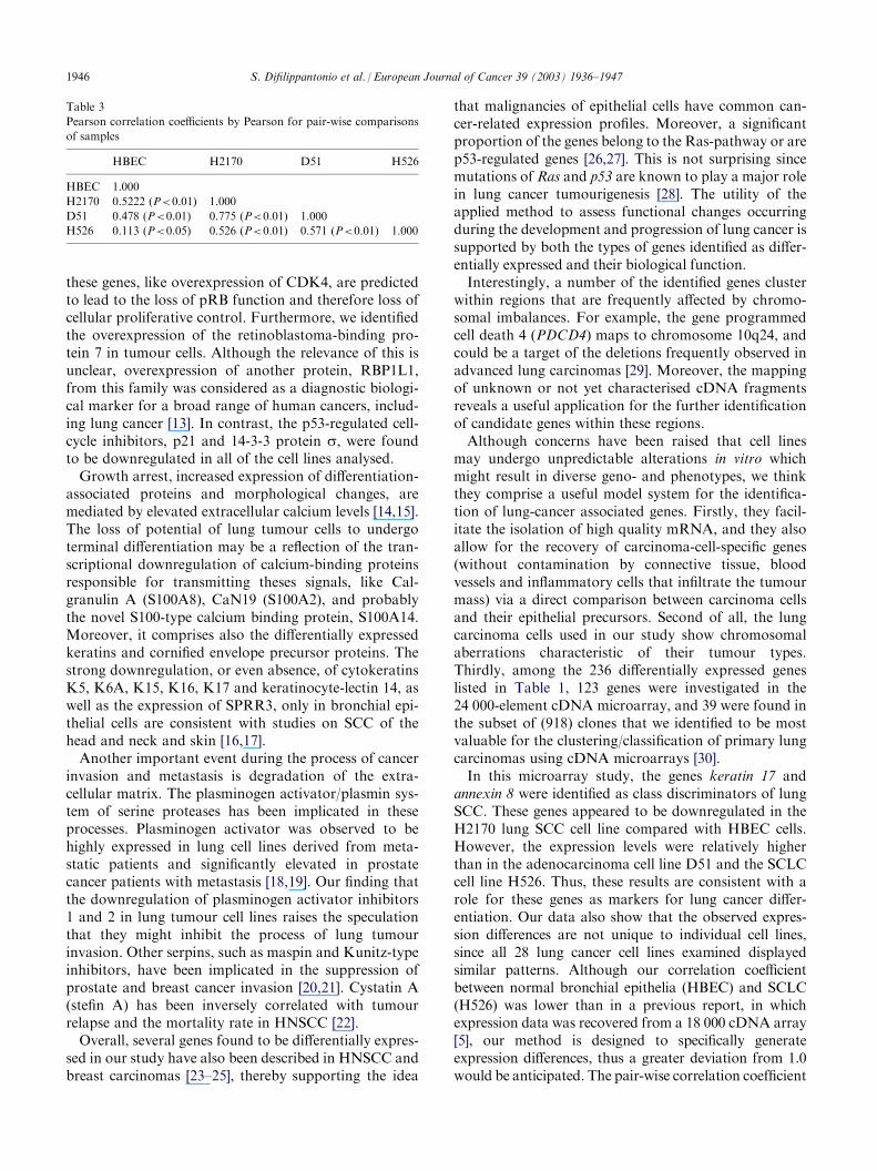
was calculated by the pair-wise correlation coefficientfrom one sample to another. The results are summarisedin Table 3.
3.2. Isolation of ESTs and full-length cDNAs
Out of the 869 partial cDNA clones, seven full-lengthcDNA clones with no known gene homologuey in thedatabase were isolated. Three of these corresponded tothe same gene.Clone DKFZp441H1211Q2 is 2110-bp long, has an
open reading frame of 125 amino acids, and was identi-fied as human microtubule-associated proteins 1A/1Blight chain 3 (accession number AF303888), a structuralprotein involved in the filamentous cross-bridgingbetween microtubules and other skeletal elements.DKFZp441K016Q2 (2602-bp) has an open reading
frame of 215 amino acids. It has been identified ashuman epithelial V-like antigen 1 (EVA1) (accession
S. Difilippantonio et al. / European Journal of Cancer 39 (2003) 1936–1947 1943

number AF304447) and may have a function in tumourdevelopment as a cell adhesion molecule.DKFZp441A199Q2 (3003 bp) corresponds to the
human GTP-binding protein SAR1 (accession numberAY008268) and has an open reading frame of 198amino acids.DKFZp404H1043Q2 (1057 bp), with an open reading
frame of 104 amino acids, is predicted to be a newmember of S100-type calcium binding protein family(S100A14) (accession number AY007220). It was map-ped to chromosome 1q21 by FISH [9].
DKFZp404O119Q2 (1014 bp), with a putative openreading frame of 73 amino acids, stands for the novelhomeobox-containing gene LAGY (accession numberAF454763). LAGY was mapped to chromosome 4 byPCR using chromosome-specific DNA together withradiation hybrid (RH) analysis. Meanwhile, we foundthat the gene is potentially involved in the developmentand progression of lung cancer [10].Additionally, six out of all differentially expressed
cDNA fragments showed no homology to any knowngenes or expressed sequence tags. They have beenentered into the Database of Expressed Sequence Tags(dbEST) with the following designations: dbEST_Id6635322 (accession number BF188817), dbEST_Id6635320 (accession number BF188815), dbEST_Id6635324 (accession number BF188819), dbEST_Id6635323 (accession number BF188818), dbEST_Id6635321 (accession number BF188816), dbEST_Id6751337 (accession number BF294062).
3.3. Mapping of selected fragments
In addition to the S100A14 gene and the LAGY gene,the following cDNA clones were mapped: cDNAFLJ13155fis (HBEC-398) to chromosome 3pter byFISH, FLJ 23001 fis (HBEC-239) to chromosome 2q23by FISH and RH analysis, and finally dbEST_Id6635323 (HBEC-71) to chromosome 4q by RH.
4. Discussion
SSH is a powerful approach for the identification ofgenes that are differentially expressed in one cell popu-lation compared with another. Although cDNA micro-array is increasingly applied for the massively parallelanalysis of gene expression, SSH is still widely usedsince it enables the recovery of abundant, as well as low-copy-number, mRNA transcripts. However, it not onlypermits an efficient identification of tumour-associatedgenes with known function, but also an unbiased isola-tion of novel sequences that are not yet available on themicrochips. Using this approach, we compared normalhuman bronchial epithelial cells with a lung squamouscarcinoma cell line and a metastatic SCLC.We found that the proto-oncogene, KIT, was expres-
sed solely in SCLC. This is in agreement with the pos-tulated involvement of KIT in an autocrine loop inSCLC [11]. We also observed numerous other genes tobe specifically expressed in SCLC including MYB, theVAV 1 oncogene and the cytosolic tyrosine kinaseEtk/Bmx (see Table 1).Another group of genes identified was involved in
cell-cycle regulation. Cyclin D1, p16, cdk4 and pRB arecomponents of a regulatory pathway that is inactivatedin most tumour cells [12]. Changes in the expression of
Fig. 1. Representative examples of northern blotting analysis using
cloned cDNA fragments. 10 mg of total RNA from HBEC and the
three lung cancer cell lines (H2170, D51 and H526) were electro-
phoresised, blotted and hybridised with radiolabelled cDNA probes
and autoradiographed.
1944 S. Difilippantonio et al. / European Journal of Cancer 39 (2003) 1936–1947
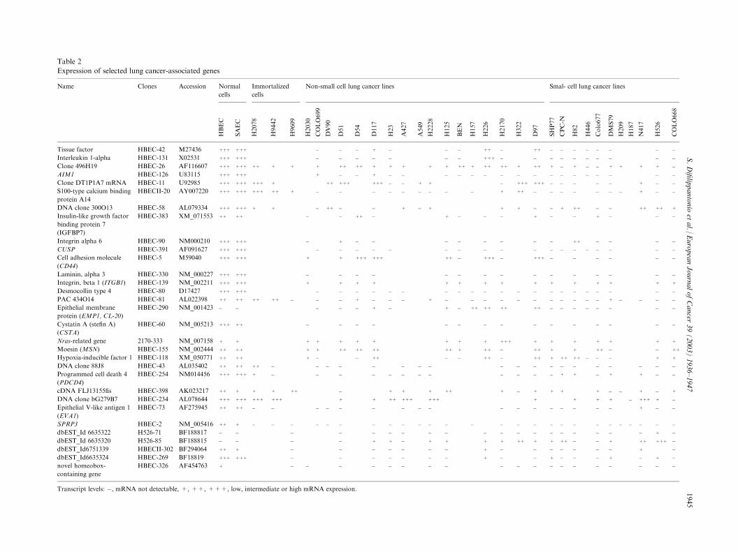
Table 2
Expression of selected lung cancer-associated genes
Name Clones Accession Normal
cells
Immortalized
cells
Non-small cell lung cancer lines Smal- cell lung cancer lines
HBEC
SAEC
H2078
H9442
H9609
H2030
COLO699
DV90
D51
D54
D117
H23
A427
A549
H2228
H125
BEN
H157
H226
H2170
D97
SHP77
CPC-N
H82
H446
Colo677
DMS79
H209
H187
N417
H526
COLO668
Tissue factor HBEC-42 M27436 +++ +++ � � � + � � � ++ � + � � � � � � � �
Interleukin 1-alpha HBEC-131 X02531 +++ +++ � � � � � � � +++ � � � � � � � � �
Clone 496H19 HBEC-26 AF116607 +++ +++ ++ + + + ++ ++ + + + + + ++ + ++ ++ + + � + � � + + + + �
AIM1 HBEC-126 U83115 +++ +++ + � � + � � � � � � � � � � � � � � �
Clone DT1P1A7 mRNA HBEC-11 U92985 +++ +++ +++ + ++ +++ +++ � � + + � ++ � � � � � + � �
S100-type calcium binding
protein A14
HBECII-20 AY007220 +++ +++ +++ ++ + � � � � � � � � � + � � � � � � + � �
DNA clone 300O13 HBEC-58 AL079334 +++ +++ + + � ++ � � + � + + � + ++ � � ++ ++ +
Insulin-like growth factor
binding protein 7
(IGFBP7)
HBEC-383 XM_071553 ++ ++ � � ++ � + � � � � � + � � �
Integrin alpha 6 HBEC-90 NM000210 +++ +++ � + � � � � � � � ++ � � � �
CUSP HBEC-391 AF091627 +++ +++ � � � � � � � � � � � � � � � � �
Cell adhesion molecule
(CD44)
HBEC-5 M59040 +++ +++ + + +++ +++ ++ � +++ � ++ � � � � � �
Laminin, alpha 3 HBEC-330 NM_000227 +++ +++ � � � � � � � � � � � � � �
Integrin, beta 1 (ITGB1) HBEC-139 NM_002211 +++ +++ + + + + + + + + + + + + + +
Desmocollin type 4 HBEC-80 D17427 +++ +++ � � � � � � � � � � � � � � � � � � �
PAC 434O14 HBEC-81 AL022398 ++ ++ ++ ++ � � � + � � � + � � � � � � � � � + � � � �
Epithelial membrane
protein (EMP1, CL-20)
HBEC-290 NM_001423 � � � � � + � + � ++ ++ ++ + � � � � � � � �
Cystatin A (stefin A)
(CSTA)
HBEC-60 NM_005213 +++ ++ � � � � � � � � � � � � � �
Nras-related gene 2170-333 NM_007158 + + + + + + + + + + +++ + + + + + +
Moesin (MSN) HBEC-155 NM_002444 ++ ++ + + ++ ++ ++ ++ + ++ � + + + ++ � � ++
Hypoxia-inducible factor 1 HBEC-118 XM_050771 ++ ++ + � � ++ � � ++ � + + ++ ++ � � � + +
DNA clone 88J8 HBEC-43 AL035402 ++ ++ ++ � � � � � � � � � � � � � � � � �
Programmed cell death 4
(PDCD4)
HBEC-254 NM014456 +++ +++ + � � � � � � � � � � + + � + + � �
cDNA FLJ13155fis HBEC-398 AK023217 ++ + + + ++ � + + + ++ + + + + � � + � +
DNA clone bG279B7 HBEC-234 AL078644 +++ +++ +++ +++ + + ++ +++ +++ + + + � +++ + �
Epithelial V-like antigen 1
(EVA1)
HBEC-73 AF275945 ++ ++ � � � � � � � � � � � � � � � + � �
SPRP3 HBEC-2 NM_005416 ++ + � � � � � � � � � � � � � � � � � � � � � � � �
dbEST_Id 6635322 H526-71 BF188817 � � � � � � � � � � � � � � � � � + �
dbEST_Id 6635320 H526-85 BF188815 � � � � + + � + + + + + ++ � � + ++ +++ �
dbEST_Id6751339 HBECII-302 BF294064 ++ + � � � � � � � + � � � � � � + �
dbEST_Id6635324 HBEC-269 BF18819 +++ +++ � � � � � � � + � + � � � + � + �
novel homeobox-
containing gene
HBEC-326 AF454763 + � � � � � � � � � � � � � � � � �
Transcript levels: �, mRNA not detectable, +, ++, +++, low, intermediate or high mRNA expression.
S.Difilippantonioetal./EuropeanJournalofCancer
39(2003)1936–1947
1945
H322
+
�
+ +
� �
+++ +
++ �
+ �
+
�
�
+
�
+
� �
� �
+
�
+
+
+
� �
� �
� +
+
� �
� �
� �
++ +
�
�
� �
these genes, like overexpression of CDK4, are predictedto lead to the loss of pRB function and therefore loss ofcellular proliferative control. Furthermore, we identifiedthe overexpression of the retinoblastoma-binding pro-tein 7 in tumour cells. Although the relevance of this isunclear, overexpression of another protein, RBP1L1,from this family was considered as a diagnostic biologi-cal marker for a broad range of human cancers, includ-ing lung cancer [13]. In contrast, the p53-regulated cell-cycle inhibitors, p21 and 14-3-3 protein s, were foundto be downregulated in all of the cell lines analysed.Growth arrest, increased expression of differentiation-
associated proteins and morphological changes, aremediated by elevated extracellular calcium levels [14,15].The loss of potential of lung tumour cells to undergoterminal differentiation may be a reflection of the tran-scriptional downregulation of calcium-binding proteinsresponsible for transmitting theses signals, like Cal-granulin A (S100A8), CaN19 (S100A2), and probablythe novel S100-type calcium binding protein, S100A14.Moreover, it comprises also the differentially expressedkeratins and cornified envelope precursor proteins. Thestrong downregulation, or even absence, of cytokeratinsK5, K6A, K15, K16, K17 and keratinocyte-lectin 14, aswell as the expression of SPRR3, only in bronchial epi-thelial cells are consistent with studies on SCC of thehead and neck and skin [16,17].Another important event during the process of cancer
invasion and metastasis is degradation of the extra-cellular matrix. The plasminogen activator/plasmin sys-tem of serine proteases has been implicated in theseprocesses. Plasminogen activator was observed to behighly expressed in lung cell lines derived from meta-static patients and significantly elevated in prostatecancer patients with metastasis [18,19]. Our finding thatthe downregulation of plasminogen activator inhibitors1 and 2 in lung tumour cell lines raises the speculationthat they might inhibit the process of lung tumourinvasion. Other serpins, such as maspin and Kunitz-typeinhibitors, have been implicated in the suppression ofprostate and breast cancer invasion [20,21]. Cystatin A(stefin A) has been inversely correlated with tumourrelapse and the mortality rate in HNSCC [22].Overall, several genes found to be differentially expres-
sed in our study have also been described in HNSCC andbreast carcinomas [23–25], thereby supporting the idea
that malignancies of epithelial cells have common can-cer-related expression profiles. Moreover, a significantproportion of the genes belong to the Ras-pathway or arep53-regulated genes [26,27]. This is not surprising sincemutations of Ras and p53 are known to play a major rolein lung cancer tumourigenesis [28]. The utility of theapplied method to assess functional changes occurringduring the development and progression of lung cancer issupported by both the types of genes identified as differ-entially expressed and their biological function.Interestingly, a number of the identified genes cluster
within regions that are frequently affected by chromo-somal imbalances. For example, the gene programmedcell death 4 (PDCD4) maps to chromosome 10q24, andcould be a target of the deletions frequently observed inadvanced lung carcinomas [29]. Moreover, the mappingof unknown or not yet characterised cDNA fragmentsreveals a useful application for the further identificationof candidate genes within these regions.Although concerns have been raised that cell lines
may undergo unpredictable alterations in vitro whichmight result in diverse geno- and phenotypes, we thinkthey comprise a useful model system for the identifica-tion of lung-cancer associated genes. Firstly, they facil-itate the isolation of high quality mRNA, and they alsoallow for the recovery of carcinoma-cell-specific genes(without contamination by connective tissue, bloodvessels and inflammatory cells that infiltrate the tumourmass) via a direct comparison between carcinoma cellsand their epithelial precursors. Second of all, the lungcarcinoma cells used in our study show chromosomalaberrations characteristic of their tumour types.Thirdly, among the 236 differentially expressed geneslisted in Table 1, 123 genes were investigated in the24 000-element cDNA microarray, and 39 were found inthe subset of (918) clones that we identified to be mostvaluable for the clustering/classification of primary lungcarcinomas using cDNA microarrays [30].In this microarray study, the genes keratin 17 and
annexin 8 were identified as class discriminators of lungSCC. These genes appeared to be downregulated in theH2170 lung SCC cell line compared with HBEC cells.However, the expression levels were relatively higherthan in the adenocarcinoma cell line D51 and the SCLCcell line H526. Thus, these results are consistent with arole for these genes as markers for lung cancer differ-entiation. Our data also show that the observed expres-sion differences are not unique to individual cell lines,since all 28 lung cancer cell lines examined displayedsimilar patterns. Although our correlation coefficientbetween normal bronchial epithelia (HBEC) and SCLC(H526) was lower than in a previous report, in whichexpression data was recovered from a 18 000 cDNA array[5], our method is designed to specifically generateexpression differences, thus a greater deviation from 1.0would be anticipated. The pair-wise correlation coefficient
Table 3
Pearson correlation coefficients by Pearson for pair-wise comparisons
of samples
HBEC
H2170 D51 H526HBEC
1.000H2170
0.5222 (P<0.01) 1.000D51
0.478 (P<0.01) 0.775 (P<0.01) 1.000H526
0.113 (P<0.05) 0.526 (P<0.01) 0.571 (P<0.01) 1.0001946 S. Difilippantonio et al. / European Journal of Cancer 39 (2003) 1936–1947

analysis between individual NSCLC and SCLC cell lineswas also significant. This fact, together with the datashowing that SCLC are distinct from pulmonary carci-noids, suggests that both NSCLC and SCLC derive froma common epithelial precursor and underlines the needfor a new classification of lung cancer that includesgenetic composition and gene expression profiles.In summary, we have provided detailed sequence
information on transcriptional changes associated withlung carcinogenesis, with many of them being describedfor the first time. The specific set of genes we recoveredby cDNA subtraction provides a basis for identifyingtranscripts with diagnostic and prognostic value, withrespect to both primary tumours and metastases. Inaddition, studying the function of these genes and theirbiological pathways may lead to the development ofnew therapeutic options.
Acknowledgements
This work was supported by the Deutsche Krebshilfe(grant 10-1300-Pe2), the Deutsche Forschungsgesellschaft(DFG Pe602/1-2) and the Monika-Kutzner-Stiftung. Thetechnical assistance of Cordula Heckert is gratefullyacknowledged. We thank Dr. Michael J. Difilippantoniofor helpful suggestions and comments during the pre-paration of the manuscript.
References
1. Pass HI, Mitchell JB, Johnson DH, Turrisi AT. Lung Cancer:
Principles and Practice. Philadelphia, PA, Lippincott-Raven, 1996.
2. Roggli VL, Vollmer RT, Greenberg SD, McGavranMH, Spjut HJ,
Yesner R. Lung cancer heterogeneity: a blinded and randomized
study of 100 consecutive cases. Hum Pathol 1985, 16, 569–579.
3. Petersen I, Langreck H, Wolf G, et al. Small-cell lung cancer is
characterized by a high incidence of deletions on chromosomes
3p, 4q, 5q, 10q, 13q and 17p. Br J Cancer 1997, 75, 79–86.
4. Petersen I, Bujard M, Petersen S, et al. Patterns of chromosomal
imbalances in adenocarcinoma and squamous cell carcinoma of
the lung. Cancer Res 1997, 57, 2331–2335.
5. Anbazhagan R, Tihan T, Bornman DM, et al. Classification of
small cell lung cancer and pulmonary carcinoid by gene expres-
sion profiles. Cancer Res 1999, 59, 5119–5122.
6. Petersen S, Aninat-Meyer M, Schluns K, Gellert K, Dietel M,
Petersen I. Chromosomal alterations in the clonal evolution to
the metastatic stage of squamous cell carcinomas of the lung. Br
J Cancer 2000, 82, 65–73.
7. Petersen S, Heckert C, Rudolf J, et al. Gene expression profiling
of advanced lung cancer. Int J Cancer 2000, 86, 512–517.
8. Schriml LM, Padilla-Nash HM, Coleman A, et al. Tyramide sig-
nal amplification (TSA)-FISH applied to mapping PCR-labeled
probes less than 1 kb in size. Biotechniques 1999, 27, 608–613.
9. Pietas A, Schluns K, Marenholz I, Schaefer BW, Heizmann CW,
Petersen I. Molecular cloning and characterization of the human
S100A14 gene encoding a novel member of the S100 family.
Genomics 2002, 79, 513–522.
10. Chen Y, Petersen S, Pacyna-Gengelbach M, Pietas A, Petersen I.
Oncology 2003, 64, 450–458.
11. Krystal GW, Hines SJ, Organ CP. Autocrine growth of small cell
lung cancer mediated by coexpression of c-kit and stem cell fac-
tor. Cancer Res 1996, 56, 370–376.
12. Sherr CJ. Cancer cell cycles. Science 1996, 274, 1672–1677.
13. Cao J, Gao T, Stanbridge EJ, Irie R. RBP1L1, a retinoblastoma-
binding protein-related gene encoding an antigenic epitope
abundantly expressed in human carcinomas and normal testis. J
Natl Cancer Inst 2001, 93, 1159–1165.
14. Hennings H, Michael D, Cheng C, Steinert P, Holbrook K,
Yuspa SH. Calcium regulation of growth and differentiation of
mouse epidermal cells in culture. Cell 1980, 19, 245–254.
15. Yuspa SH, Kilkenny AE, Steinert PM, Roop DR. Expression of
murine epidermal differentiation markers is tightly regulated by
restricted extracellular calcium concentrations in vitro. J Cell Biol
1989, 109, 1207–1217.
16. Leethanakul C, Patel V, Gillespie J, et al. Distinct pattern of
expression of differentiation and growth-related genes in squa-
mous cell carcinomas of the head and neck revealed by the use of
laser capture microdissection and cDNA arrays. Oncogene 2000,
19, 3220–3224.
17. Lohman FP, Medema JK, Gibbs S, Ponec M, van de Putte P,
Backendorf C. Expression of the SPRR cornification genes is
differentially affected by carcinogenic transformation. Exp Cell
Res 1997, 231, 141–148.
18. Chen JJW, Peck K, Hong TM, et al. Global analysis of gene
expression in invasion by a lung cancer model. Cancer Res 2001,
61, 5223–5230.
19. Miyake H, Hara I, Yamanaka K, Arakawa S, Kamidono S. Ele-
vation of urokinase-type plasminogen activator and its receptor
densities as new predictors of disease progression and prognosis
in men with prostate cancer. Int J Oncol 1999, 14, 535–541.
20. Sheng S, Carey J, Seftor EA, Dias L, Hendrix MJ. Maspin acts at
the cell membrane to inhibit invasion and motility of mammary
and prostatic cancer cells. Proc Natl Acad Sci USA 1996, 93,
11669–11674.
21. Kobayashi H, Shinohara H, Takeuchi K, et al. Inhibition of the
soluble and the tumour cell receptor-bound plasmin by urinary
trypsin inhibitor and subsequent effects on tumour cell invasion
and metastasis. Cancer Res 1994, 54, 844–849.
22. Strojan P, Budihna M, Smid L, et al. Prognostic significance of
cysteine proteinases cathepsins B and L and their endogenous
inhibitors stefins A and B in patients with squamous cell carci-
noma of the head and neck. Clin Cancer Res 2000, 6, 1052–1062.
23. Fung LF, Lo AK, Yuen PW, Liu Y, Wang XH, Tsao SW. Dif-
ferential gene expression in nasopharyngeal carcinoma cells. Life
Sci 2000, 67, 923–936.
24. Zhang M, Martin KJ, Sheng S, Sager R. Expression genetics: a
different approach to cancer diagnosis and prognosis. Trends
Biotechnol 1998, 16, 66–71.
25. Nacht M, Ferguson AT, Zhang W, et al. Combining serial ana-
lysis of gene expression and array technologies to identify genes
differentially expressed in breast cancer. Cancer Res 1999, 59,
5464–5470.
26. Zuber J, Tchernitsa OI, Hinzmann B, et al. A genome-wide sur-
vey of RAS transformation targets. Nat Genet 2000, 24, 144–152.
27. Zhao R, Gish K, Murphy M, et al. Analysis of p53-regulated
gene expression patterns using oligonucleotide arrays. Genes Dev
2000, 14, 981–993.
28. Sekido Y, Fong KM, Minna JD. Progress in understanding the
molecular pathogenesis of human lung cancer. Biochim Biophys
Acta 1998, 1378, F21–F59.
29. Petersen S, Rudolf J, Bockmuhl U, et al. Distinct regions of alle-
lic imbalance on chromosome 10q22-q26 in squamous cell carci-
nomas of the lung. Oncogene 1998, 7, 449–454.
30. Garber ME, Troyanskaya OG, Schluns K, et al. Diversity of gene
expression in adenocarcinoma of the lung. Natl Acad Sci USA
2001, 98, 13784–13789.
S. Difilippantonio et al. / European Journal of Cancer 39 (2003) 1936–1947 1947