Analytic Continuation of ζ(s) Violates the Law of Non ... - arXiv
21
Yannis Karamanos (ed.), Expression Profi ling in Neuroscience, Neuromethods, vol. 64,DOI 10.1007/978-1-61779-448-3_2, © Springer Science+Business Media, LLC 2012
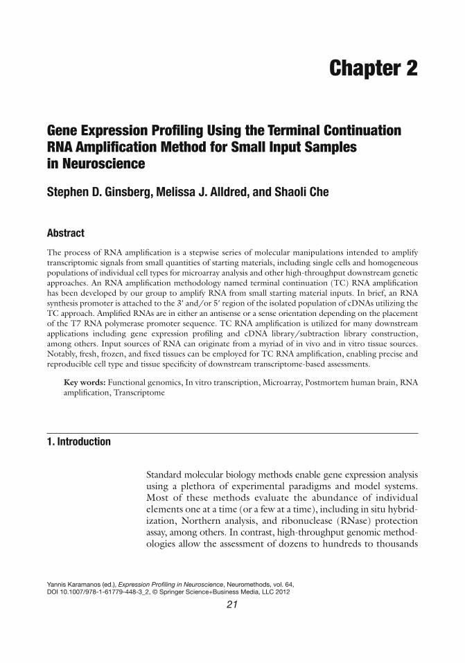
Chapter 2
Gene Expression Profi ling Using the Terminal Continuation RNA Amplifi cation Method for Small Input Samples in Neuroscience
Stephen D. Ginsberg , Melissa J. Alldred , and Shaoli Che
Abstract
The process of RNA amplifi cation is a stepwise series of molecular manipulations intended to amplify transcriptomic signals from small quantities of starting materials, including single cells and homogeneous populations of individual cell types for microarray analysis and other high-throughput downstream genetic approaches. An RNA amplifi cation methodology named terminal continuation (TC) RNA amplifi cation has been developed by our group to amplify RNA from small starting material inputs. In brief, an RNA synthesis promoter is attached to the 3 ¢ and/or 5 ¢ region of the isolated population of cDNAs utilizing the TC approach. Amplifi ed RNAs are in either an antisense or a sense orientation depending on the placement of the T7 RNA polymerase promoter sequence. TC RNA amplifi cation is utilized for many downstream applications including gene expression profi ling and cDNA library/subtraction library construction, among others. Input sources of RNA can originate from a myriad of in vivo and in vitro tissue sources. Notably, fresh, frozen, and fi xed tissues can be employed for TC RNA amplifi cation, enabling precise and reproducible cell type and tissue specifi city of downstream transcriptome-based assessments.
Key words: Functional genomics , In vitro transcription , Microarray , Postmortem human brain , RNA amplifi cation , Transcriptome
Standard molecular biology methods enable gene expression analysis using a plethora of experimental paradigms and model systems. Most of these methods evaluate the abundance of individual elements one at a time (or a few at a time), including in situ hybrid-ization, Northern analysis, and ribonuclease (RNase) protection assay, among others. In contrast, high-throughput genomic method-ologies allow the assessment of dozens to hundreds to thousands
1. Introduction

22 S.D. Ginsberg et al.
of genes simultaneously in a coordinated manner ( 1, 2 ) , but often require abundant, high-quality RNA for effective experimentation. Our research has focused upon microarray and related expression profi ling of individual populations of neurons following experimental injury and age-related neurodegeneration ( 3– 10 ) , and has required the development of RNA amplifi cation strategies that enable the use of small amounts of input RNA.
Nucleic acids for transcriptomic analyses can originate from a variety of in vivo and in vitro sources including plant and animal materials. Fresh, fl ash-frozen, and fi xed tissues are potentially useful, depending on tissue quality and the experimental design of the specifi c study. Isolation of genomic DNA and total RNA is not only rou-tinely performed using fresh and frozen tissues ( 11– 13 ) , but can also originate from paraffi n-embedded fi xed tissues ( 14– 18 ) . Use of paraffi n-embedded tissue is a viable resource, as often fresh and/or frozen tissues are not accessible, whereas archived fi xed human and animal model tissues exist in a myriad of laboratories and tissue repositories.
Although most emphasis lies in the downstream genomic output of microarrays and related high-throughput technologies, it is extremely important to note that RNA species are highly sensitive to RNase degradation. RNases are found in virtually every cell type ( 19 ) , and RNase inhibition is a key parameter to consider for the success of RNA-based experimentation. RNases are quite stable and retain their activity over a broad pH range ( 19, 20 ) . Therefore, RNase-free precautions must be employed during RNA extraction procedures and throughout RNA amplifi cation methods.
To date, an optimal method to prepare brain tissues for downstream genetic analyses has yet to be agreed upon. A consen-sus protocol has not yet been promulgated. RNAs are routinely harvested from fi xed tissues where RNA Integrity Number (RIN) values have been ascertained via bioanalysis ( 21, 22 ) . RNA from tissue samples using cross-linking fi xatives including 10% neutral buffered formalin and 4% paraformaldehyde as well as precipitating fi xatives such as 70% ethanol buffered with 150 mM sodium chloride is another resource, although RNA quantity and quality are typically less than that of fresh and frozen samples ( 14, 23– 27 ) . Another method for assessing RNA quality in tissue sections prior to performing expression profi ling studies is acridine orange (AO) histofl uorescence. AO is a fl uorescent dye that intercalates selec-tively into nucleic acids and has been used to detect RNA and DNA in brain tissues ( 10, 28– 30 ) . Importantly, individual RNA species (e.g., mRNA, rRNA, and tRNA) cannot be delineated by AO histofl uorescence. A more thorough examination of RNA qual-ity can be obtained via bioanalysis (e.g., 2100 Bioanalyzer, Agilent Technologies), which utilizes capillary gel electrophoresis to detect RNA quality and quantitate relative abundance (such as a RIN number) ( 5, 31, 32 ) .

232 TC RNA Amplifi cation
The advent of high-throughput microarray analysis has enabled signifi cant progress in expression profi ling studies in many disci-plines, and microarray analysis has emerged as a useful tool to assess transcript levels in a myriad of systems and paradigms. A disadvantage of conventional microarray technology is a requirement for signifi cant amounts of high-quality input sources of RNA for sensitivity and reproducibility. The quantity of RNA harvested from a single cell, estimated to be approximately 0.1–1.0 picograms, is not enough to employ standard RNA extraction procedures ( 11, 33 ) . Therefore, molecular biological methods have been implemented to increase the amount of input RNA for downstream genetic analyses, including exponential PCR-based amplifi cation and linear RNA amplifi cation approaches. Unfortunately, PCR-based protocols are not optimal, as exponential amplifi cation often can skew quantitative relationships between genes from the initial population ( 34, 35 ) . In contrast, linear RNA amplifi cation methods allow for the analysis of relative gene expression levels. A linear RNA amplifi cation procedure typically entails generating quantities of RNA species through in vitro transcription (IVT) ( 31, 35– 37 ) . Hybrid PCR/linear RNA amplifi cation methods ( 38, 39 ) as well as isother-mal RNA amplifi cation ( 40, 41 ) procedures have also been developed that generate faithful representation of original input RNA.
A variety of strategies have been developed to improve RNA amplifi cation effi ciency ( 38, 39, 42– 44 ) . An obstacle that hinders RNA amplifi cation protocols is the diffi culty with second-strand synthesis effi ciency and specifi city ( 35, 38, 39, 45 ) . Technical improvements include obviating second-strand cDNA synthesis and enabling fl exibility in placement of bacteriophage transcriptional promoter sequences for both antisense and sense amplifi cation.
A procedure has been developed in our laboratory termed terminal continuation (TC) RNA amplifi cation ( 31, 32, 35, 46– 48 ) which satisfi es these objectives (Fig. 1 ). TC RNA transcription can be performed using a promoter sequence (e.g., T7, T3, or SP6) attached to either the 3 ¢ or the 5 ¢ oligonucleotide primers. Therefore, transcript orientation can be in an antisense orientation when the bacteriophage promoter sequence is placed on the fi rst-strand poly d(T) primer, or in a sense orientation when the promoter sequence is attached to the TC primer, depending upon the design of the experimental paradigm (Fig. 1 ) ( 32, 47– 49 ) . TC RNA amplifi cation of genetic signals includes synthesizing fi rst-strand cDNA complementary to the mRNA template, subsequently generating second-strand cDNA complementary to the fi rst-strand cDNA (when antisense RNA is being generated; if sense RNA is being synthesized, the second-strand step is obviated) ( 47, 48 ) , and fi nally performing IVT using the double-stranded cDNA as template ( 31, 32, 35, 46 ) . First-strand cDNA synthesis complementary to the template mRNA entails the use of two oligonucleotide primers: a fi rst-strand poly d(T) primer and a TC primer. The poly d(T) primer is similar to conventional

24 S.D. Ginsberg et al.
primers that exploit the poly(A) sequence present on the majority of mRNAs. The TC primer contains a span of three cytidine triphosphates (CTPs) or guanosine triphosphates (GTPs) at the 3 ¢ terminus ( 31 ) . Adenosine triphosphates (ATPs) or thymidine triphosphates (TTPs) do not perform well as constituents of the TC primer ( 5 ) . As stated above, second-strand cDNA synthesis can be initiated for antisense RNA amplifi cation (e.g., the T7 promoter sequence is on the poly d(T) primer) by annealing a second oligo-nucleotide primer complementary to the attached oligonucleotide ( 31 ) , and is performed with robust DNA polymerases, such as Taq polymerase, or is avoided altogether when performing sense RNA amplifi cation (e.g., when the T7 promoter sequence is on the TC primer) ( 47, 48 ) . One round of amplifi cation is suffi cient for
Fig. 1. Overview and analysis of the TC RNA amplifi cation method. ( a ) Schematic representation of TC RNA amplifi cation, antisense orientation. A poly d(T) primer (containing a bacteriophage promoter sequence for antisense orientation) and TC primer are added to the mRNA population to be amplifi ed. First-strand synthesis that occurs as an mRNA–cDNA hybrid is formed after reverse transcription and terminal continuation of the oligonucleotide primers. Following RNase H digestion to remove the original mRNA template strand, second-strand synthesis is performed using Taq polymerase. The resultant double-stranded product is utilized as template for in vitro transcription, yielding high fi delity, linear RNA amplifi cation of sense orientation ( rippled lines ). ( b ) Schematic representation of TC RNA amplifi cation, sense orientation. mRNA to be amplifi ed ( green line ) and the TC primer serve as templates for the fi rst-strand synthesis, with poly d(T) acting as a primer. First-strand cDNA consists of three portions: the 5 ¢ end comprised of the poly d(T), the mRNA complementary portion in the middle ( purple line ), and the 3 ¢ end comprised of the TC primer complementary to the cDNA. The TC primer complementary sequence portion hybridizes with the TC primer present in the reaction and forms a double-stranded region without the need for further second-strand synthesis. Since the TC primer contains the T7 bacteriophage transcription promoter sequence, double-stranded TC primer regions provide a functional RNA synthesis promoter for IVT and subsequent robust RNA amplifi cation. Adapted from ( 46, 48 ).

252 TC RNA Amplifi cation
downstream genetic analyses ( 1, 31, 32, 46 ) . The TC RNA amplifi cation method is also being adjusted to enable amplifi cation of noncoding RNAs including microRNAs (miRNAs) ( 50 ) , which represents a new avenue for expression profi ling studies that is just recently being recognized as a research endeavor. Below, this protocol describes the use of the TC RNA amplifi cation procedure in detail for amplifi cation of transcripts derived from small amounts of tissue, including neurons and other cellular populations microaspi-rated from tissues via laser capture microdissection (LCM) or related extraction procedures.
The methodology described herein is a step-by-step protocol for TC RNA amplifi cation ( 47, 48 ) . In terms of experimental orienta-tion, this protocol begins at the point where cells are captured via LCM or a microaspiration strategy and follows through IVT using biotinylated, fl uorescent, or radioactive methodologies to label the TC RNA amplifi ed products. see Notes 1 and 2 .
1. Trizol reagent (Invitrogen, Carlsbad, CA) stored at 4°C. 2. Chloroform (Sigma, St. Louis, MO) stored at 22°C. 3. Isopropanol (Sigma) stored at 22°C. 4. 80% Ethanol (EtOH) stored at −20°C. 5. Linear acrylamide (5 mg/mL) (Applied Biosystems, Foster City,
CA) stored at −20°C.
1. First-strand synthesis primer {poly d(T); 100 ng/ m l (IDT, Coralville, IA)} (Table 1 ).
2. Reverse transcription (RT) master mix: fi rst-strand buffer (5×) (Invitrogen), dNTPs (10 mM) (Invitrogen), dithiothreitol (DTT) (0.1 M) (Sigma), and RNase inhibitor (Superase-In; Applied Biosystems; 20 U/ m L) stored at −20°C.
3. Superscript III (Invitrogen, 200 U/ m L) stored at −20°C.
1. 10× PCR buffer (Applied Biosystems; buffer includes 15 mM MgCl 2 ) stored at −20°C.
2. RNase H (Invitrogen, 10 U/ m L) stored at −20°C. 3. Taq polymerase (Applied Biosystems; 5 U/ m L) stored at −20°C.
1. 10,000 Molecular weight cut-off (MWCO) columns (Vivaspin 500; Sartorius Stedim, Goettingen, Germany).
2. 18.2 M W RNase-free water. see Note 1 .
2. Materials
2.1. Isolation of RNA
2.2. First-Strand Synthesis
2.3. Second-Strand Synthesis (For Antisense RNA Amplifi cation Only)
2.4. Double-Stranded cDNA and cDNA/Primer Purifi cation
26 S.D. Ginsberg et al.
1. 10× Hybridization reaction buffer (Applied Biosystems) stored at −20°C.
2. 10× Biotin-labeled ribonucleotides (Enzo Life Sciences, Farmingdale, NY) stored at −20°C. see Note 3 .
3. 10× RNase inhibitor mix (Enzo) stored at −20°C. 4. 10× DTT (Invitrogen) stored at −20°C. 5. T7 RNA polymerase (1,000 U/ m L, Epicentre, Madison, WI)
stored at −80°C.
1. 5× Transcription reaction buffer (Epicentre) stored at −20°C. 2. 0.1 M DTT (Invitrogen) stored at −20°C. 3. 3NTPs (ATP, CTP, and GTP; 2.5 mM each) (Invitrogen)
stored at −20°C. 4. UTP (100 m M) (Invitrogen) stored at −20°C. 5. RNase inhibitor (Superase-In) stored at −20°C. 6. 33 P-UTP (Perkin-Elmer, Boston, MA, 10 mCi/mL) stored at
−80°C. 7. T7 RNA polymerase (1,000 U/ m L, Epicentre).
1. Add 500 m L of Trizol reagent to 0.7-mL thin-walled PCR tubes that will receive the microdissected regions/cells and/or profi les acquired via LCM, and keep on wet ice.
2. Invert the tubes so that Trizol reagent bathes microdissected material, and keep on wet ice. The samples can also be stored at −80°C at this juncture for future use.
2.5. IVT for TC RNA Amplifi cation: Biotinylated/Fluorescent Probe Labeling
2.6. IVT for TC RNA Amplifi cation: Radioactive Probe Labeling
3. Methods
3.1. Isolation of RNA
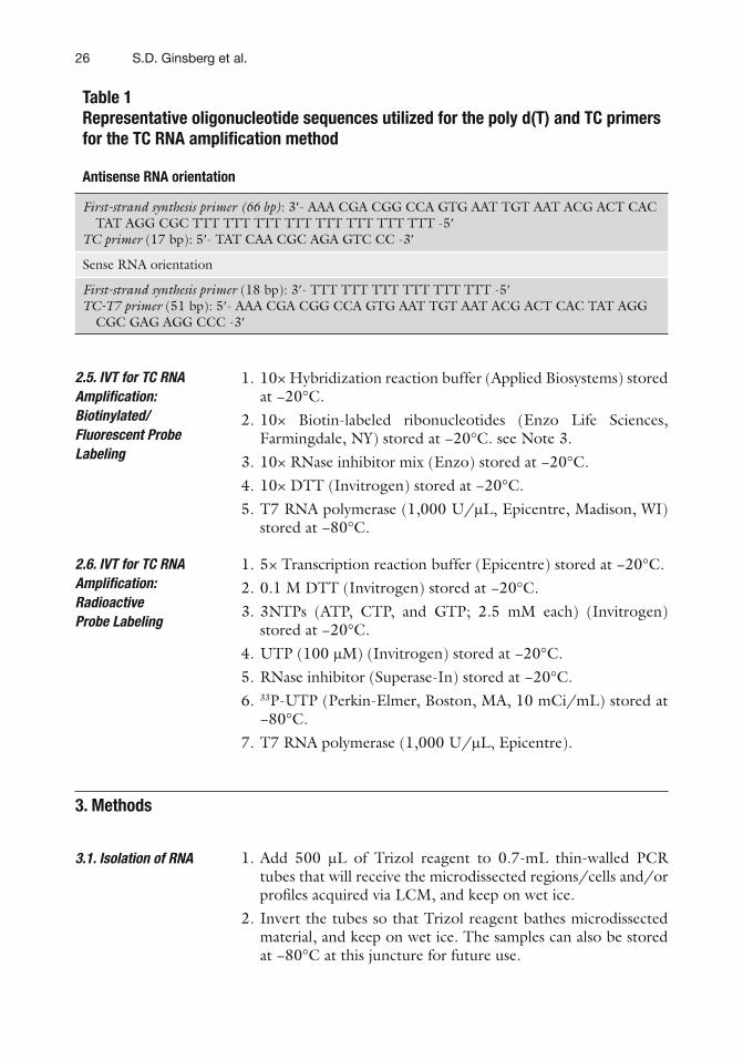
Table 1 Representative oligonucleotide sequences utilized for the poly d(T) and TC primers for the TC RNA amplifi cation method
Antisense RNA orientation
First-strand synthesis primer (66 bp) : 3 ¢ - AAA CGA CGG CCA GTG AAT TGT AAT ACG ACT CAC TAT AGG CGC TTT TTT TTT TTT TTT TTT TTT TTT -5 ¢
TC primer (17 bp): 5 ¢ - TAT CAA CGC AGA GTC CC -3 ¢
Sense RNA orientation
First-strand synthesis primer (18 bp): 3 ¢ - TTT TTT TTT TTT TTT TTT -5 ¢ TC-T7 primer (51 bp): 5 ¢ - AAA CGA CGG CCA GTG AAT TGT AAT ACG ACT CAC TAT AGG
CGC GAG AGG CCC -3 ¢

272 TC RNA Amplifi cation
3. Remove LCM cap and add 100 m L of chloroform to each sample. Vortex vigorously for 15 s and centrifuge the samples at 12,000 × g -force for 5 min at 4°C.
4. The mixture separates into an upper aqueous phase (clear) and a lower organic phase (red). Collect the aqueous phase containing RNA by aspirating with a pipette.
5. Add 250 m L of 100% isopropyl alcohol and 2 m L of linear acrylamide to precipitate the RNA from the aqueous phase. The samples can be stored at −80°C to precipitate RNAs if desired.
6. Mix vigorously and centrifuge the samples at 12,000 × g -force for 20 min at 4°C.
7. Decant the supernatant, by inverting the tube, being careful not to dislodge the pellet.
8. Add 500 m L of 80% EtOH to each sample to wash RNA pellet. Vortex vigorously for 15 s and centrifuge the samples at 12,000 × g -force for 5 min at 4°C.
9. Decant the supernatant, by inverting the tube, being careful not to dislodge the pellet.
10. Air dry the sample by inverting the tube for 5 min in the hood. see Note 4 .
11. Resuspend the pellet in 6 m L of 18.2 M W RNase-free water.
1. To each RNA sample, add 1 m L of fi rst-strand synthesis primer (Table 1 ). Centrifuge for 10 s.
2. Heat the mixture for 2 min at 65°C, then for 1 min at 45°C, and place on ice. Total volume is 7 m L.
3. Prepare reverse transcription (RT) master mix (on wet ice): see Note 2 .
5× fi rst-strand buffer 4 m L
dNTPs (10 mM) 1 m L
0.1 M DTT 1 m L
RNase inhibitor 1 m L
18.2 M W RNase-free water 4 m L
4. Take an aliquot of TC primer (100 ng/ m L) (Table 1 ), heat-denature for 2 min at 70°C, place on ice for several minutes, and then add to the RT master mix. see Note 5.
5. Aliquot Superscript III to the RT master mix. see Note 6 . The RT master mix should comprise 13 m L per reaction.
6. Add the RT master mix to the 7 m L of sample, pipette vigorously, and centrifuge briefl y. Incubate the mixture for 60 min at 50°C.
3.2. First-Strand Synthesis

28 S.D. Ginsberg et al.
7. Inactivate the RT reaction by heating the sample for an additional 15 min at 65°C.
8. Centrifuge the reaction mixture briefl y and cool immediately on wet ice. The samples can be stored at −20°C for short term or at −80°C for longer term storage.
1. Prepare second-strand master mix (on wet ice):
10× PCR buffer 10 m L
RNase H 0.5 m L
18.2 M W RNase-free water 69 m L
2. Mix well with pipette tip and centrifuge briefl y. 3. Distribute 79.5 m L of second-strand master mix to the appro-
priate number of 0.5 mL capacity thin-walled PCR tubes. 4. Add 20 m L of the sample to the second-strand master mix and
mix thoroughly with a pipette tip. 5. Place the samples in a thermal cycler. see Note 7. Degrade the
RNA for 30 min at 37°C. As soon as the block temperature reaches 95°C, pause the reaction (this will be the hot-start component). (Heat-denature RNase H for 3 min at 95°C and proceed to Sect. 3.4 if performing sense TC RNA amplifi cation). Using a dedicated PCR pipette, add into each sample the following:
Taq polymerase 0.5 m L
6. Mix thoroughly with a pipette tip. see Note 8 . 7. Continue the second-strand synthesis program by pushing the
Continue function. 8. The second-strand synthesis program consists of the following:
Hot start denaturation 95°C for 3 min.
Annealing 60°C for 3 min.
Elongation 75°C for 30 min.
9. Once the program is complete, remove the samples, centrifuge briefl y, and store at −20°C for short term or at −80°C for longer term storage.
1. To purify double-stranded cDNA, use Vivaspin 500 columns (10,000 MWCO)
cDNA sample from Sect. 3.3 100 m L
18.2 M W RNase-free water 300 m L
3.3. RNA Removal and Second-Strand Synthesis
3.4. Double-Stranded cDNA or cDNA/Primer Purifi cation

292 TC RNA Amplifi cation
2. Mix by pipetting vigorously and add to top of columns. Spin columns in their collection tubes in a microfuge at 10,000 × g -force for 7.5 min at 22°C and measure the resultant product in the column. The volume should be 13 m L. see Note 9.
3. Remove the purifi ed cDNA from the column to a fresh 1.7-mL microfuge tube and store at −20°C for short term or at −80°C for longer term storage.
1. Prepare IVT master mix (on wet ice):
10× Hybridization reaction buffer 4 m L
10× Biotin-labeled ribonucleotides 4 m L
10× DTT 4 m L
10× RNase inhibitor mix 4 m L
18.2 M W RNase-free water 9 m L
T7 RNA polymerase 2 m L
2. The IVT master mix should comprise 27 m L per reaction. 3. Add 13 m L of TC sample to the IVT master mix (fi nal volume
of the reaction is 40 m L). 4. Mix thoroughly (very important!!). Centrifuge briefl y and
incubate for 5 h at 37°C. 5. TC RNA amplifi ed products are now ready for purifi cation,
fragmentation, and application to cDNA or oligonucleotide arrays. see Note 10 .
1. Prepare IVT master mix (on wet ice):
5× RNA amplifi cation buffer 8 m L
0.1 M DTT 1 m L
3NTPs (ATP, CTP, GTP; 2.5 mM each) 2 m L
UTP (100 m M) 1 m L
RNase inhibitor (20 U) 1 m L
2. The IVT master mix should comprise 13 m L per reaction. 3. Centrifuge briefl y and add 13 m L of double-stranded TC sample
to the IVT master mix (on wet ice). 4. To each sample, add 33 P-UTP (10 mCi/mL) 12 m L. 5. Add T7 RNA polymerase 2 m L. 6. Mix thoroughly (very important!!). Centrifuge briefl y and
incubate for 4 h at 37°C. 7. TC RNA amplifi ed products are now ready to be hybridized to
membrane-based array platforms. see Note 11 .
3.5. IVT for TC RNA Amplifi cation: Biotinylated/Fluorescent Probe Labeling
3.6. IVT for TC RNA Amplifi cation: Radioactive Probe Labeling

30 S.D. Ginsberg et al.
1. All solutions throughout the entire protocol should be made with 18.2 M W RNase-free water (e.g., Nanopure Diamond, Barnstead, Dubuque, IA), and is referred to as 18.2 M W RNase-free water in the text.
2. Prepare enough master mix for each reaction step using this basic formula: tabulate the total number of samples plus two extra (i.e., calculate volumes of the mix for x # of samples + control + one to control for any volume loss and/or experi-menter error).
3. This protocol is based on using the BioArray RNA transcript labeling kit (Enzo), although any biotinylated and/or fl uores-cent labeling protocols are suitable (minor modifi cations may apply).
4. Do not air dry RNA pellets longer than 5 min, as the pellet becomes diffi cult to resuspend.
5. Each sample in the RT master mix will get 1 m l of TC RNA primer; thus if you have eight samples, the RT master mix will be equivalent to ten samples and you will add 10 m L of the TC primer to the RT master mix prior to adding to the sample.
6. When taking aliquots of Superscript III from the tube, be extremely careful to not contaminate the vial of enzyme with primers or RNA samples. We recommend that laboratory investigators performing TC RNA amplifi cation have their own aliquot of Superscript III to avoid cross-contamination of the enzyme stock.
7. For PCR cycling of the second-strand synthesis, it is a good idea to create a specifi c stepwise protocol to be programmed into the thermal cycler using fi nal volume of 100 m L.
8. A crucial mistake that is commonly made is failure to mix the hot-start second-strand synthesis reaction when the Taq poly-merase is added. Thorough mixing with a pipette tip will ensure a suitable reaction environment.
9. If more than 13 m L remains in the column after the fi rst centrifu-gation spin, perform a repeat spin at 10,000 × g -force for 1 min until a volume of approximately 13 m L is recovered. If the volume in the column is below 13 m L, remove the product left in the top of the column to a fresh 1.7-mL microfuge tube and bring the volume to 13 m L with 18.2 M W RNase-free water.
10. There are numerous Internet sites and published protocols for hybridization of labeled probes to microarray platforms, subsequent washing, and imaging protocols (e.g., www.affymetrix.com and www.enzo.com ( 46, 49, 51 ) ).
4. Notes

312 TC RNA Amplifi cation
11. Procedures for membrane microarray synthesis, pre-hybridization, hybridization, washing conditions, and imaging are described in detail in several published papers emanating from the Ginsberg laboratory ( 5, 6, 31, 32, 35, 47, 48, 52 ) .
Acknowledgments
The authors thank Irina Elarova, Shaona Fang, and Arthur Saltzman for their expert technical assistance. Support for this project comes from the NINDS (NS43939, NS48447), NIA (AG10668, AG14449, AG17617, AG09466), NICHD (HD057564), and the Alzheimer’s Association.
References
1. Ginsberg, S. D., and Mirnics, K. (2006) Functional genomic methodologies, Prog Brain Res 158, 15–40.
2. Brown, P. O., and Botstein, D. (1999) Exploring the new world of the genome with DNA microarrays, Nat Genet 21, 33–37.
3. Counts, S. E., He, B., Che, S., Ikonomovic, M. D., Dekosky, S. T., Ginsberg, S. D., and Mufson, E. J. (2007) {alpha}7 Nicotinic receptor up-regulation in cholinergic basal forebrain neurons in Alzheimer disease, Arch Neurol 64, 1771–1776.
4. Counts, S. E., He, B., Che, S., Ginsberg, S. D., and Mufson, E. J. (2009) Galanin fi ber hyper-innervation preserves neuroprotective gene expression in cholinergic Basal forebrain neurons in Alzheimer’s disease, J Alzheimers Dis 18, 885–896.
5. Ginsberg, S. D., and Che, S. (2004) Combined histochemical staining, RNA amplifi cation, regional, and single cell analysis within the hippocampus, Lab Invest 84, 952–962.
6. Ginsberg, S. D., and Che, S. (2005) Expression profi le analysis within the human hippocampus: Comparison of CA1 and CA3 pyramidal neurons, J Comp Neurol 487, 107–118.
7. Ginsberg, S. D., Che, S., Counts, S. E., and Mufson, E. J. (2006) Shift in the ratio of three-repeat tau and four-repeat tau mRNAs in indi-vidual cholinergic basal forebrain neurons in mild cognitive impairment and Alzheimer’s disease, J Neurochem 96, 1401–1408.
8. Ginsberg, S. D., Che, S., Wuu, J., Counts, S. E., and Mufson, E. J. (2006) Down regulation of trk but not p75 gene expression in single cholinergic basal forebrain neurons mark the progression of Alzheimer’s disease, J Neurochem 97, 475–487.
9. Ginsberg, S. D., Alldred, M. J., Counts, S. E., Cataldo, A. M., Neve, R. L., Jiang, Y., Wuu, J., Chao, M. V., Mufson, E. J., Nixon, R. A., and Che, S. (2010) Microarray analysis of hip-pocampal CA1 neurons implicates early endo-somal dysfunction during Alzheimer’s disease progression, Biol Psychiatry, 68, 885–893.
10. Mufson, E. J., Counts, S. E., and Ginsberg, S. D. (2002) Single cell gene expression profi les of nucleus basalis cholinergic neurons in Alzhei-mer’s disease, Neurochem Res 27, 1035–1048.
11. Sambrook, J., and Russell, D. W. (2001) Molecular cloning: a laboratory manual. Third edition, Cold Spring Harbor Laboratory Press, Cold Spring Harbor.
12. Madabusi, L. V., Latham, G. J., and Andruss, B. F. (2006) RNA extraction for arrays, Methods Enzymol 411, 1–14.
13. Van Deerlin, V. M., Gill, L. H., and Nelson, P. T. (2002) Optimizing gene expression analysis in archival brain tissue, Neurochem Res 27, 993–1003.
14. Kabbarah, O., Pinto, K., Mutch, D. G., and Goodfellow, P. J. (2003) Expression profi ling of mouse endometrial cancers microdissected from ethanol-fi xed, paraffi n-embedded tissues, Am J Pathol 162, 755–762.
15. Lehmann, U., Bock, O., Glockner, S., and Kreipe, H. (2000) Quantitative molecular analysis of laser-microdissected paraffi n-embedded human tissues, Pathobiology 68, 202–208.
16. Lewis, F., Maughan, N. J., Smith, V., Hillan, K., and Quirke, P. (2001) Unlocking the archive–gene expression in paraffi n-embedded tissue, J Pathol 195, 66–71.
17. Penland, S. K., Keku, T. O., Torrice, C., He, X., Krishnamurthy, J., Hoadley, K. A., Woosley,

32 S.D. Ginsberg et al.
J. T., Thomas, N. E., Perou, C. M., Sandler, R. S., and Sharpless, N. E. (2007) RNA expression analysis of formalin-fi xed paraffi n-embedded tumors, Lab Invest 87, 383–391.
18. Rupp, G. M., and Locker, J. (1988) Purifi cation and analysis of RNA from paraffi n-embedded tissues, Biotechniques 6, 56–60.
19. Farrell Jr, R. E. (1998) RNA Methodologies, Second Edition, Academic Press, San Diego.
20. Blumberg, D. D. (1987) Creating a ribonuclease-free environment, Methods Enzymol 152, 20–24.
21. Stan, A. D., Ghose, S., Gao, X. M., Roberts, R. C., Lewis-Amezcua, K., Hatanpaa, K. J., and Tamminga, C. A. (2006) Human postmortem tissue: what quality markers matter?, Brain Res 1123, 1–11.
22. Weis, S., Llenos, I. C., Dulay, J. R., Elashoff, M., Martinez-Murillo, F., and Miller, C. L. (2007) Quality control for microarray analysis of human brain samples: The impact of postmor-tem factors, RNA characteristics, and histopa-thology, J Neurosci Methods 165, 198–209.
23. Fend, F., Emmert-Buck, M. R., Chuaqui, R., Cole, K., Lee, J., Liotta, L. A., and Raffeld, M. (1999) Immuno-LCM: laser capture microdis-section of immunostained frozen sections for mRNA analysis, Am J Pathol 154, 61–66.
24. Goldsworthy, S. M., Stockton, P. S., Trempus, C. S., Foley, J. F., and Maronpot, R. R. (1999) Effects of fi xation on RNA extraction and amplifi cation from laser capture microdissected tissue, Mol Carcinog 25, 86–91.
25. Klimecki, W. T., Futscher, B. W., and Dalton, W. S. (1994) Effects of ethanol and paraformal-dehyde on RNA yield and quality, BioTechniques 16, 1021–1023.
26. Su, J. M., Perlaky, L., Li, X. N., Leung, H. C., Antalffy, B., Armstrong, D., and Lau, C. C. (2004) Comparison of ethanol versus formalin fi xation on preservation of histology and RNA in laser capture microdissected brain tissues, Brain Pathol 14, 175–182.
27. Qin, Y., Heine, V. M., Karst, H., Lucassen, P. J., and Joels, M. (2003) Gene expression patterns in rat dentate granule cells: comparison between fresh and fi xed tissue, J Neurosci Methods 131, 205–211.
28. Mai, J. K., Schmidt-Kastner, R., and Tefett, H.-B. (1984) Use of acridine orange for histo-logic analysis of the central nervous system, J Histochem Cytochem 32, 97–104.
29. Vincent, V. A., DeVoss, J. J., Ryan, H. S., and Murphy, G. M., Jr. (2002) Analysis of neuronal gene expression with laser capture microdissec-tion, J Neurosci Res 69, 578–586.
30. Ginsberg, S. D., Crino, P. B., Lee, V. M.-Y., Eberwine, J. H., and Trojanowski, J. Q. (1997)
Sequestration of RNA in Alzheimer’s disease neurofi brillary tangles and senile plaques, Ann Neurol 41, 200–209.
31. Che, S., and Ginsberg, S. D. (2004) Amplifi cation of transcripts using terminal con-tinuation, Lab Invest 84, 131–137.
32. Che, S., and Ginsberg, S. D. (2006) RNA amplifi cation methodologies, in Trends in RNA Research (McNamara, P. A., Ed.), pp 277–301, Nova Science Publishing, Hauppauge.
33. Kacharmina, J. E., Crino, P. B., and Eberwine, J. (1999) Preparation of cDNA from single cells and subcellular regions, Methods Enzymol 303, 3–18.
34. Eberwine, J., Kacharmina, J. E., Andrews, C., Miyashiro, K., McIntosh, T., Becker, K., Barrett, T., Hinkle, D., Dent, G., and Marciano, P. (2001) mRNA expression analysis of tissue sections and single cells, J Neurosci 21, 8310–8314.
35. Ginsberg, S. D. (2008) Transcriptional profi ling of small samples in the central nervous system, Methods Mol Biol 439, 147–158.
36. VanGelder, R., von Zastrow, M., Yool, A., Dement, W., Barchas, J., and Eberwine, J. (1990) Amplifi ed RNA (aRNA) synthesized from limited quantities of heterogeneous cDNA, Proc Natl Acad Sci U S A 87, 1663–1667.
37. Tecott, L. H., Barchas, J. D., and Eberwine, J. H. (1988) In situ transcription: specifi c synthe-sis of complementary DNA in fixed tissue sections, Science 240, 1661–1664.
38. Wang, E., Miller, L. D., Ohnmacht, G. A., Liu, E. T., and Marincola, F. M. (2000) High-fi delity mRNA amplifi cation for gene profi ling, Nat Biotechnol 18, 457–459.
39. Zhumabayeva, B., Diatchenko, L., Chenchik, A., and Siebert, P. D. (2001) Use of SMART-generated cDNA for gene expression studies in multiple human tumors, BioTechniques 30, 158–163.
40. Dafforn, A., Chen, P., Deng, G., Herrler, M., Iglehart, D., Koritala, S., Lato, S., Pillarisetty, S., Purohit, R., Wang, M., Wang, S., and Kurn, N. (2004) Linear mRNA amplifi cation from as little as 5 ng total RNA for global gene expres-sion analysis, BioTechniques 37, 854–857.
41. Kurn, N., Chen, P., Heath, J. D., Kopf-Sill, A., Stephens, K. M., and Wang, S. (2005) Novel isothermal, linear nucleic acid amplifi cation sys-tems for highly multiplexed applications, Clin Chem 51, 1973–1981.
42. Matz, M., Shagin, D., Bogdanova, E., Britanova, O., Lukyanov, S., Diatchenko, L., and Chenchik, A. (1999) Amplifi cation of cDNA ends based on template-switching effect and step-out PCR, Nucleic Acids Res 27, 1558–1560.
332 TC RNA Amplifi cation
43. Iscove, N. N., Barbara, M., Gu, M., Gibson, M., Modi, C., and Winegarden, N. (2002) Repre-sentation is faithfully preserved in global cDNA amplifi ed exponentially from sub-picogram quantities of mRNA, Nat Biotechnol 20, 940–943.
44. Xiang, C. C., Chen, M., Ma, L., Phan, Q. N., Inman, J. M., Kozhich, O. A., and Brownstein, M. J. (2003) A new strategy to amplify degraded RNA from small tissue samples for microarray studies, Nucleic Acids Res 31, E53.
45. Goff, L. A., Bowers, J., Schwalm, J., Howerton, K., Getts, R. C., and Hart, R. P. (2004) Evaluation of sense-strand mRNA amplifi cation by comparative quantitative PCR, BMC Genomics 5, 76.
46. Ginsberg, S. D. (2005) RNA amplifi cation strategies for small sample populations, Methods 37, 229–237.
47. Alldred, M. J., Che, S., and Ginsberg, S. D. (2008) Terminal continuation (TC) RNA amplifi cation enables expression profi ling using minute RNA input obtained from mouse brain, Int J Mol Sci 9, 2091–2104.
48. Alldred, M. J., Che, S., and Ginsberg, S. D. (2009) Terminal continuation (TC) RNA amplifi cation without second strand synthesis, J Neurosci Methods 177, 381–385.
49. Ginsberg, S. D., Hemby, S. E., Mufson, E. J., and Martin, L. J. (2006) Cell and tissue microdissection in combination with genomic and proteomic applications, in Neuroanatomical Tract Tracing 3: Molecules, Neurons, and Systems (Zaborszky, L., Wouterlood, F. G., and Lanciego, J. L., Eds.), pp 109–141, Springer, New York.
50. Che, S., and Ginsberg, S. D. (2008) MicroRNA (miRNA) expression profi ling using the miRNA signature sequence amplifi cation (SSAM) tech-nology in human postmortem brain tissues and in animal models of neurodegeneration, Proc. Soc. Neurosci. 34, 46.44.
51. Cheung, V. G., Morley, M., Aguilar, F., Massimi, A., Kucherlapati, R., and Childs, G. (1999) Making and reading microarrays, Nat Genet 21, 15–19.
52. Ginsberg, S. D., and Che, S. (2002) RNA ampli-fi cation in brain tissues, Neurochem Res 27, 981–992.
Copyright © 2022 FDOKUMEN