Gallium in bacteria: Metabolic and medical implications
138
G G 4 -DNA ▶ Strontium and DNA Aptamer Folding Gadolinium Takashiro Akitsu Department of Chemistry, Tokyo University of Science, Shinjuku-ku, Tokyo, Japan Definition A lanthanoid element, the seventh element of the f-elements block, with the symbol Gd, atomic number 64, and atomic weight 157.25. Electron configuration [Xe]4f 7 5d 1 6s 2 . Gadolinium is composed of stable ( 154 Gd, 2.18%; 155 Gd, 14.80%; 156 Gd, 20.47%; 157 Gd, 15.65%; 158 Gd, 24.84%) and two radioactive ( 152 Gd, 0.20%; 160 Gd, 21.86%) isotopes. Discovered by J. C. G. de Marignac in 1880. Gadolinium exhibits oxidation states III (and II); atomic radii: 180 pm, covalent radii 197 pm; redox potential (acidic solution) Gd 3+ /Gd 2.397 V; Gd 3+ /Gd 2+ 3.9 V; electronegativity (Pauling) 1.2. Ground electronic state of Gd 3+ is 8 S 7/2 with S ¼ 7/2, L ¼ 0, J ¼ 7/2 with l ¼ 0 cm 1 . Most stable technogenic radionuclide 160 Gd (half-life > 1.3 10 21 years). The most common compounds: Gd 2 O 3 , GdF 3 , and Gd(OH) 3 . Biologically, gadolinium is of high toxicity, and inhala- tional exposure to high concentrations of Gd 2 O 3 results in pneumonitis and acute inflammation in the lung (Atkins et al. 2006; Cotton et al. 1999; Huheey et al. 1997; Oki et al. 1998; Rayner-Canham and Overton 2006). Cross-References ▶ Lanthanide Ions as Luminescent Probes ▶ Lanthanide Metalloproteins ▶ Lanthanides and Cancer ▶ Lanthanides in Biological Labeling, Imaging, and Therapy ▶ Lanthanides in Nucleic Acid Analysis ▶ Lanthanides, Physical and Chemical Characteristics References Atkins P, Overton T, Rourke J, Weller M, Armstrong F (2006) Shriver and Atkins inorganic chemistry, 4th edn. Oxford University Press, Oxford/New York Cotton FA, Wilkinson G, Murillo CA, Bochmann M (1999) Advanced inorganic chemistry, 6th edn. Wiley-Interscience, New York Huheey JE, Keiter EA, Keiter RL (1997) Inorganic chemistry: principles of structure and reactivity, 4th edn. Prentice Hall, New York Oki M, Osawa T, Tanaka M, Chihara H (1998) Encyclopedic dictionary of chemistry. Tokyo Kagaku Dojin, Tokyo Rayner-Canham G, Overton T (2006) Descriptive inorganic chemistry, 4th edn. W. H. Freeman, New York Gallium ▶ Gallium(III) Complexes, Inhibition of Proteasome Activity V.N. Uversky et al. (eds.), Encyclopedia of Metalloproteins, DOI 10.1007/978-1-4614-1533-6, # Springer Science+Business Media New York 2013
-
Upload
sunnybrook -
Category
Documents
-
view
1 -
download
0
Transcript of Gallium in bacteria: Metabolic and medical implications

G
G4-DNA
▶ Strontium and DNA Aptamer Folding
Gadolinium
Takashiro Akitsu
Department of Chemistry, Tokyo University of
Science, Shinjuku-ku, Tokyo, Japan
Definition
A lanthanoid element, the seventh element of the
f-elements block, with the symbol Gd, atomic number
64, and atomic weight 157.25. Electron configuration
[Xe]4f75d16s2. Gadolinium is composed of stable
(154Gd, 2.18%; 155Gd, 14.80%; 156Gd, 20.47%; 157Gd,
15.65%; 158Gd, 24.84%) and two radioactive (152Gd,
0.20%; 160Gd, 21.86%) isotopes. Discovered by J. C. G.
deMarignacin1880.Gadoliniumexhibitsoxidationstates
III (and II); atomic radii: 180 pm, covalent radii 197 pm;
redox potential (acidic solution) Gd3+/Gd �2.397 V;
Gd3+/Gd2+ �3.9 V; electronegativity (Pauling) 1.2.
Ground electronic state of Gd3+ is 8S7/2 with S ¼ 7/2,
L¼ 0, J¼ 7/2 with l¼ 0 cm�1.Most stable technogenic
radionuclide 160Gd (half-life > 1.3 � 1021 years). The
most common compounds: Gd2O3, GdF3, and Gd(OH)3.
Biologically, gadolinium is of high toxicity, and inhala-
tional exposure to high concentrations ofGd2O3 results in
pneumonitis and acute inflammation in the lung (Atkins
V.N. Uversky et al. (eds.), Encyclopedia of Metalloproteins, DOI 1# Springer Science+Business Media New York 2013
etal.2006;Cottonetal.1999;Huheeyetal.1997;Okietal.
1998; Rayner-Canham andOverton 2006).
Cross-References
▶Lanthanide Ions as Luminescent Probes
▶Lanthanide Metalloproteins
▶Lanthanides and Cancer
▶Lanthanides in Biological Labeling, Imaging, and
Therapy
▶Lanthanides in Nucleic Acid Analysis
▶Lanthanides, Physical and Chemical Characteristics
References
Atkins P, Overton T, Rourke J, Weller M, Armstrong F (2006)
Shriver and Atkins inorganic chemistry, 4th edn. Oxford
University Press, Oxford/New York
Cotton FA, Wilkinson G, Murillo CA, Bochmann M (1999)
Advanced inorganic chemistry, 6th edn. Wiley-Interscience,
New York
Huheey JE, Keiter EA, Keiter RL (1997) Inorganic chemistry:
principles of structure and reactivity, 4th edn. Prentice Hall,
New York
Oki M, Osawa T, Tanaka M, Chihara H (1998) Encyclopedic
dictionary of chemistry. Tokyo Kagaku Dojin, Tokyo
Rayner-Canham G, Overton T (2006) Descriptive inorganic
chemistry, 4th edn. W. H. Freeman, New York
Gallium
▶Gallium(III) Complexes, Inhibition of Proteasome
Activity
0.1007/978-1-4614-1533-6,

G 800 Gallium and Apoptosis
Gallium and Apoptosis
▶Gallium, Therapeutic Effects
Gallium Complexes
▶Gallium(III) Complexes, Inhibition of Proteasome
Activity
Gallium Compounds
▶Gallium(III) Complexes, Inhibition of Proteasome
Activity
Gallium Delivery by TransferrinReceptor-1-Mediated Endocytosis
▶Gallium Uptake and Transport by Transferrin
Gallium Effect on Bacteria
▶Gallium, Therapeutic Effects
Gallium in Bacteria, Metabolic andMedical Implications
Christopher Auger, Joseph Lemire, Varun Appanna
and Vasu D. Appanna
Department of Chemistry and Biochemistry,
Laurentian University, Sudbury, ON, Canada
Synonyms
Bacterial response to gallium; The molecular effects of
gallium on bacterial physiology
Definition
Chelation therapy: medication involved in the
removal of excess metals.
Gallium: metal utilized as a semiconductor.
Pentose phosphate pathway: a metabolic network
that produces the antioxidant NADPH and ribose.
Pseudomonas fluorescens: a metabolically versatile
microbe.
Tricarboxylic acid cycle: an essential component of
aerobic respiration.
Introduction
Due to their ability to survive and grow in a wide
range of environmental conditions, bacteria present
themselves as ideal models for the study of
a variety of biological phenomena, including the
interactions of metal toxins with living organisms.
Mechanisms by which these prokaryotes dispose of
metal pollutants and circumvent their toxic influ-
ence have major implications in environmental,
medical, and biotechnological domains. This may
include the bioremediation of waste sites, chelation
therapy, and the production of unique metabolites.
Pseudomonas fluorescens, a soil microbe, is known
to thrive in media supplemented with numerous
toxic metals like Ga, aluminum (Al), lead (Pb),
zinc (Zn), and yttrium (Y). Owing to its physico-
chemical similarity to Fe, an essential nutrient in
almost all organisms, the interaction of Ga with
living organisms has become a subject of ongoing
importance.
Ga Occurrence and Toxicity
Ga is a silvery, glass-like semi-metallic element by
and large found in its salt form in bauxite and zinc
ores (Downs 1993). While normally inert, increasing
industrial activity reliant on this metal has rendered
Ga accessible to a wide spectrum of organisms. Easily
purified from the smelting process, the dominant
application of this trivalent metal is in the semicon-
ductors gallium arsenide and gallium nitride
(Chitambar 2010). Given the efficiencies of these
materials and our reliance on them for computers,
high-speed optical communication, and photovoltaic
cells, it is clear that the amount of Ga present in the
environment will only escalate in the foreseeable
future. As a result, the toxicological aspect of this
metal cannot be ignored.

Gallium in Bacteria, Metabolic and Medical Implications 801 G
G
Ga, an Fe Mimetic
Ga is not known to have any biological role
(Al-Aoukaty et al. 1992). Historically, its low bioavail-
ability has seemingly excluded it from any physiolog-
ical function. However, it is well recognized that Ga
can exert its harmful effect by interfering with the
metabolism of Fe. The functions of the latter are
quite diverse. Fe is crucial in heme-containing proteins
such as hemoglobin, myoglobin, and cytochrome
P450. This metal also plays a necessary role in
iron-sulfur (Fe-S) cluster containing proteins, which
are pervasive in nature, catalyzing a wide array of
biochemical reactions. As such, any disruption in the
function of these metalloproteins would have conse-
quences on multiple cellular operations. Ga, with an
ionic radius of 0.67 A and a trivalent charge, is
sufficiently analogous to Fe3+ to displace it from
Fe-dependent biomolecules (Downs 1993). However,
while Fe is redox active, Ga is not, and the substitution
often leads to the inactivation of the target protein.
This feature of Ga is exploited in cancer therapy.
Radiopharmaceuticals are radioactive tracers that
permit the imaging and treatment of many diseases
(Chitambar 2010). Cancer cells, due to their rapidly
proliferating nature, have an increased need for Fe.
By incorporating itself in proteins in lieu of
Fe, radiolabeled Ga67 allows for the noninvasive visu-
alization of tumors in a patient (Chitambar 2010). This
interchange of metals could also contribute minimally
to the eradication of cancerous cells, arresting growth
via disruption of vital cellular processes and the liber-
ation of Fe (Chitambar 2010).
Free Fe, a Pro-Oxidant: A Consequence of Ga
Toxicity
Once displaced from metalloproteins, free Fe further
disturbs the operation of the cell via the production of
reactive oxygen species (ROS) (Chenier et al. 2008).
Fenton chemistry is a series of reactions whereby
Fe(II) is initially oxidized by hydrogen peroxide to
form Fe(III), hydroxyl radical, and hydroxyl anion.
Hydrogen peroxide can then catalyze the reduction of
Fe(III) back to Fe(II) with the concomitant production
of peroxide radical and a proton (Beriault 2004). These
radicals are dangerous, as they can disable DNA,
proteins, and lipids. Consequently, the homeostasis of
Fe is a strongly regulated process. Proteins, such as
transferrin and ferritin, which bind Fe tightly yet
reversibly, are indispensable for its storage and
delivery (Beriault 2004). Ga therefore exerts its dele-
terious effects via a two-pronged approach. Firstly, the
displacement of Fe from its cognitive protein may
subsequently inactivate the protein. Secondly, the
release of free Fe into the microenvironment can
generate oxidative radicals which, if not properly
controlled, can induce cell death.
Physiological Responses of Pseudomonasfluorescens to Ga
Although Ga is toxic, some microbial systems have
evolved to counter its deleterious effects. Pseudomo-
nas fluorescens is an obligate aerobe known to inhabit
primarily soil, plants, and water surfaces (Paulsen
et al. 2005). This gram-negative, rod-shaped, and non-
pathogenic bacterium derived its name from its ability
to produce the fluorescent pigment pyoverdin, a
siderophore, under iron-limiting conditions (Paulsen
et al. 2005). The capability to grow on different
carbon sources under a variety of conditions renders
P. fluorescens ideal for commercial applications
(Paulsen et al. 2005). This microbe has been
implemented in bioremediation, the production of anti-
freeze agents as well as biofertilizers (Paulsen et al.
2005). The extreme metabolic versatility of this organ-
ism renders it suitable for the study of adaptation in
harsh environments. Despite the consequences associ-
ated with Ga internalization, P. fluorescens is capableof adapting to the toxic effects of this metal. When
P. fluorescens is grown in a medium containing citrate
as the sole carbon source chelated to 1 mM Ga, cells
reach their stationary phase within 68 h, compared to
the 24 h observed in the control cultures (Al-Aoukaty
et al. 1992) (Fig. 1). However, this lag phase is
reversed if 20 mM Fe is included in the medium
(Al-Aoukaty et al. 1992). The Ga-stressed cells have
a pale appearance as opposed to the light brown color-
ation of the control cells. This may be due to the
inability of P. fluorescens to access Fe in the
Ga-containing medium.
Ga Toxicity: Molecular Insights
The electron transport chain, a series of complexes that
is responsible for generating the majority of the
universal energy currency adenosine triphosphate
(ATP) during aerobic metabolism, is highly suscepti-
ble to stress arising from oxidative radicals and metal

Lag phase
CtlGa + Fe
600
500
400μg
of p
rote
in/m
l of c
ultu
re
300
200
100
00 10 20 30 40
Incubation time (h)
50 60 70 80 90
Ga alone
Gallium in Bacteria,Metabolic and MedicalImplications,Fig. 1 Biomass curve of
Pseudomonas fluorescenstreated with Ga. When Ga is
added to growth media with
citrate as the sole carbon
source, the lag phase is
increased to 50 h (■), as
opposed to 13 h in the control
cells (♦). Supplementation
with exogenous Fe reverses
the growth shift observed
under Ga stress (~)
(Ctl ¼ control. Adapted from
Al-Aoukaty et al. 1992)
G 802 Gallium in Bacteria, Metabolic and Medical Implications
pollution (Chenier et al. 2008). Because this machinery
is riddled with heme groups, iron-sulfur clusters, and
various metal cofactors, Ga is capable of reacting with
and disabling the cell’s primary source of energy
(Fig. 2). In order to assess the activity of these
complexes, total membrane protein can first be sepa-
rated by blue native polyacrylamide gel electrophore-
sis (BN-PAGE). Secondly, activity staining can be
performed in order to determine whether or not these
proteins are functional (Beriault et al. 2007). In-gel
analysis of complexes I, II, and IV show that they are
severely crippled by the addition of gallium to the
medium (Chenier et al. 2008).
Ga and Fe-Dependent Metabolism
A key reaction catalyzing the entry of citrate into the
TCA cycle is that performed by aconitase (ACN).
The latter contains a labile [Fe4-S4]2+ cluster and
allows for the isomerization of citrate to isocitrate
(Chenier et al. 2008). Under oxidative stress, the con-
ventional [Fe4-S4]2+ cluster is converted to an inactive
[Fe3-S4]+ form, thus incapacitating the enzyme
(Chenier et al. 2008). As such, ACN acts as a gate-
keeper to the TCA cycle. Under normal conditions,
citrate catabolism proceeds through the TCA cycle.
When P. fluorescens is exposed to Ga, ACN activity
is diminished (Chenier et al. 2008).
As fumarase (FUM) is also affected by ROS and Ga
toxicity, P. fluorescens elaborates multiple isoforms
of FUM. While FUM A and B rely on iron-sulfur
clusters for their activity, FUM C is iron-independent
(Chenier et al. 2008). In the presence of Ga, this
microbe evokes the synthesis of FUM C in order to
counteract the decrease in FUM A activity (Chenier
et al. 2008). By doing so, carbon flow proceeds through
the TCA cycle rather than grinding to a halt. These
metabolic manipulations enable the survival of
P. fluorescens under Ga stress (Fig. 3).
Metabolic Flux Through the TCA Cycle
As citrate is the sole source of carbon and in the
absence of any citrate lyase, P. fluorescens activates
two enzymes, namely, isocitrate lyase (ICL) and
isocitrate dehydrogenase (ICDH), downstream of
ACN. The former cleaves isocitrate into succinate
and glyoxylate, feeding various anaploretic pathways.
ICDH exists in two isoforms; a membrane-bound nic-
otinamide adenine dinucleotide (NAD)-dependent var-
iant, and a soluble NADP-dependent form (Beriault
2004). Both catalyze the decarboxylation of isocitrate
to alpha-ketoglutarate (KG) with the concomitant
reduction of their respective cofactors. InP.fluorescens
subjected to Ga stress, ICL and ICDH-NADP are
upregulated (Beriault 2004). Hence, this metabolic

Citrate
Isocitrate
ATP production
Malate
Glyoxylate
Isocitrate Iyase
MalateSynthase
Transaminase
Ga+3 sequestration
Succinate
α-ketoglutarateAspartate
Hydroxyaspartate
Oxaloacetate
Aconitase
IsocitrateDehydrogenase
Gallium in Bacteria,Metabolic and MedicalImplications,Fig. 3 Metabolic shift
induced by Ga toxicity. Citrate
is redirected to alternate
metabolic pathways in order to
ensure the survival of the
bacterium
s
s
sGa+3
Disruption
DisruptionROS
Creation
ss
s
s
s
ss
s
s
s
Fe
Fe
s
s
Fe
Fe
Fe
Fe
Fe
3Fe-4S- Succinate
Dehydrogenase
4Fe-4S- Aconitase- Fumarase
TCADysfunction
Dysfunctionof
Nitrogenmetabolism
4Fe-4S- GlutamineSynthase
Gallium in Bacteria,Metabolic and MedicalImplications, Fig. 2 The
disruption of Fe-containing
proteins by Ga. Ga disrupts Fe
homeostasis and creates ROS,
leading to the formation of
dysfunctional Fe-S clusters.
The Fe-S clusters are crucial to
the functioning of numerous
key enzymes in the TCA cycle
and nitrogen metabolism
Gallium in Bacteria, Metabolic and Medical Implications 803 G
G
flux generated by ICL and ICDH-NADP acts as
a potent vacuum and helps metabolize citrate despite
an almost defunct ACN (Fig. 3).
Elimination of NADH
The oxidation of NADH by complex I is the first step
of oxidative phosphorylation and is a major site of
superoxide formation. It is downregulated in
Ga-stressed P. fluorescens as are enzymes generating
NADH (Chenier et al. 2008). This nicotinamide deriv-
ative is also a pro-oxidant. NADH production is atten-
uated by several mechanisms. Alpha-ketoglutarate
dehydrogenase (aKGDH) is an NADH-forming
enzyme that is downregulated (Beriault 2004).

NADHOxNADK
NADP
NADPH
NADP-ICDH
G-6-PDH
Ga
ROS
Metaboic Networks
NADHNAD
Detoxification
Gallium in Bacteria, Metabolic and Medical Implications,Fig. 4 Ga promotes the reduction in NADH and production of
NADPH. Ga forces the microbe to diminish NADH production
with a concomitant increase in NADPH synthesis. Simulta-
neously, Ga promotes the activity of NADH oxidase (NADHOx)
leading to the oxidation of NADH to NAD. NAD is diverted to
NADP by NAD kinase (NADK). The NADP is pooled and
utilized by NADP-dependent isocitrate dehydrogenase (NADP-
ICDH) and glucose-6-phosphate dehydrogenase (G6PDH) to
create NADPH, the ultimate reductive capacity for ROS detox-
ification. Arrows in green are pathways, which are increased.
Those in red are those that are reduced in activity
G 804 Gallium in Bacteria, Metabolic and Medical Implications
The downregulation of aKGDH also serves to pool
KG, which can act as a scavenger of ROS. The perox-
ide-mediated decarboxylation of this alpha-keto acid
generates succinate, CO2, and H2O, thus providing the
cell with an effective means of mopping up ROS in
situ. Another stratagem that is utilized to control
NADH production and allows the functioning of the
TCA cycle, albeit ineffectively, is the expression of
NADH oxidase (NOX). P. fluorescens employs
a water-generating NOX to convert NADH into
NAD, which can be utilized to propel other metabolic
reactions (Chenier et al. 2008). This Ga-triggered
metabolic shift allows the catabolism of citrate, the
generation of KG, and the regeneration of NAD.
NADPH Combats Ga Toxicity
While NADH acts as a pro-oxidant, NADPH serves as
an antioxidant, acting as fuel for the activity of ROS
detoxifying enzymes such as glutathione peroxidase.
Hence, its production is favored during Ga stress. The
upregulation of ICDH-NADP is one method of
accomplishing this task (Beriault et al. 2007).
Glucose-6-phosphate dehydrogenase (G6PDH) is the
first enzyme catalyzing the entry of glucose-
6-phophate into the pentose phosphate pathway.
When P. fluorescens is exposed to Ga, three different
isoforms of G6PDH can be found whereas untreated
cells only have one (Beriault et al. 2007). As NADP
levels need to be kept elevated for the purpose of
NADPH production, two enzymes, NAD kinase and
NADP phosphatase are modulated. The former that
catalyzes the phosphorylation of NAD to produce
NADP is upregulated, while the latter dephosphory-
lates NADP into NAD and is downregulated (Chenier
et al. 2008) (Fig. 4).
Ga Sequestration and Metal Chelation Therapy
Despite the critical role metabolic adaptation plays in
P. fluorescens to circumvent Ga toxicity, it does not
tackle the root of the problem. If the microbe is to
resume business as usual, the source of the toxicity
must be dealt with. Siderophores are often deployed
by microorganisms in order to chelate and internal-
ize Fe. Numerous siderophores have been isolated
and can be used to treat heavy metal toxicity.
Desferrioxamine, a chelator produced by Streptomy-
ces pilosus is often utilized to treat acute iron
poisoning and iron overload (Beriault 2004).
P. fluorescens treated with Ga elaborates
a hydroxyaspartate containing metabolite involved
in the chelation of this trivalent metal (Beriault
2004). Hence, enzymes like aspartate transaminase
(AST) and malate dehydrogenase (MDH) that play
a critical role in the production of aspartate are
upregulated (Beriault 2004). Figure 5 depicts the
toxicity associated with Ga and the subsequent
detoxification evoked by P. fluorescens confronted
with this metal toxin.
Ga as an Antimicrobial
Inhibition of Biofilm Formation in Pseudomonas
aeruginosa
Bacterial resistance to commercially available
antibiotics is a growing concern. As such, the discov-
ery of alternative therapies for treatment of pathogenic
organisms has become a priority. Resistance is thought
to occur primarily via transfer of genetic material from
the commensal bacteria of a person to the pathogenic

Ga
ROS creation
NADPHmetabolism
EnergyMetabolism
Nitrogenmetabolism
A Shift in Metabolic Pathways
Hydroxyasparateproduction
Fe homeostasis Siderophore Production
Gallium in Bacteria,Metabolic and MedicalImplications, Fig. 5 A
global view of Ga toxicity and
adaptation in Pseudomonasfluorescens. The deprivationof Fe, the generation of ROS,
and the production of
siderophores evoked by Ga are
shown
Gallium in Bacteria, Metabolic and Medical Implications 805 G
G
organism once infection occurs (Rasko and Sperandio
2010). Therefore, it is plausible that drugs targeting
virulence factors of the invading bacteria and not
the human flora will have a greater lifespan (Rasko
and Sperandio 2010). One common virulence
factor employed by the gram-negative pathogen
P. aeruginosa is the establishment of a biofilm. The
latter is an aggregate of microorganisms embedded
within a protective matrix with increased resistance
toward antibiotic treatments.
Ga, because it is FDA approved for intravenous
administration and is a Fe mimetic, is a potential
candidate for the treatment of pathogenic bacteria in
humans (Chitambar 2010). One study has indicated
that gallium treatment in mice is capable of blocking
acute and chronic P. aeruginosa infections
(Chitambar 2010). These microbes release
siderophores into the environment in order to chelate
and internalize available Fe which is required for
survival. Ga acts as a “Trojan horse” and is taken
up instead. This strategy limits the ability of
P. aeruginosa to form biofilms in vivo, and the bacteria
are quickly eradicated (Chitambar 2010). Indeed, mice
subjected to gallium treatment are able to overcome
the infection and survive much longer than their
untreated counterparts (Chitambar 2010).
Interference with Fe Metabolism in Mycobacteria
Monocytes and macrophages, two subsets of white
blood cells (WBCs), form the cornerstone of the
human immune response. Their ability to uptake and
destroy foreign matter via phagocytosis is indispens-
able for the clearance of dangerous microorganisms.
While this environment is normally too hostile for the
survival of the bacteria, some are capable of prolifer-
ating within the confines of WBCs. Mycobacterium
tuberculosis and Mycobacterium avium complex
(MAC) are pathogenic bacteria that can grow and
replicate in monocytes and macrophages
(Chitambar 2010). Mycobacteria are known to produce
two siderophores, the soluble exochelins and the
membrane-bound mycobactins (Olakanmi et al.
2000). These compounds are thought to participate in
intraphagosomal Fe acquisition once extracellular
Fe is delivered to the WBCs via transferrin.
Ga, because of its similarity to Fe, is well suited to
target pathogens which replicate within the phagosome.
At sites of infection, WBCs uptake large amounts of Fe
to fuel biological processes and impede microbial inva-
siveness. Gallium nitrate (Ga(NO3)3) and Ga-transferrin
substitute for Fe and inhibit mycobacteria growth in
media and within human macrophages, an effect which
can be reversed by the addition of exogenous

G 806 Gallium in Bacteria, Metabolic and Medical Implications
Fe (Olakanmi et al. 2000). Since Ga is released from
transferrin at a higher pH (�6.5) than the release of
Fe (�5.5), it is postulated that the pH of the mycobacte-
rial phagosome (�6.0) renders it an effective environ-
ment in which Ga can compete with Fe for intracellular
targets (Olakanmi et al. 2000). Suppression ofMycobac-terium growth byGa is thought to occur by the inhibition
of Fe-dependent antioxidant enzymes and ribonucleo-
tide reductase. The latter plays a key role in mycobacte-
rial proliferation, but has negligible activity in terminally
differentiated macrophages (Olakanmi et al. 2000). Fur-
thermore, the concentrations of Ga(NO3)3 required to
impede mycobacterial growth in vitro are approved for
human use (Olakanmi et al. 2000).
Improved Cultural Selectivity in Mycological
Media
Isolating and culturing clinically relevant fungi, such
as the Aspergillus spp. and the zygomycetes, prove to
be difficult tasks owing to the presence of bacterial
contaminants that outgrow the fungus of interest.
Sabouraud Dextrose Agar (SDA) is a fungal selective
media commonly utilized to culture commensal and
pathogenic fungi in clinical microbiology laboratories
(Moore et al. 2009). Generally, this medium is
supplemented with streptomycin, penicillin, and
chloramphenicol to curb the growth of bacteria
(Moore et al. 2009). However, the emergence of
multi- and pan-drug resistant microorganisms has
compromised the effectiveness of these antibiotics.
The ability of Ga salts such as Ga(NO3)3 and
Ga-maltolate to disrupt Fe metabolism in bacteria
renders them ideal agents for the suppression of
bacterial proliferation in vitro. Supplementation of
SDA with 2 mM Ga(NO3)3 efficaciously inhibits
growth of several bacteria, including Escherichia coli,Klebsiella pneumoniae, P. aeruginosa, and Staphylo-
coccus aureus (Moore et al. 2009). At this concentra-
tion, only the fungi and Burkholderia cenocepacia are
viable (Moore et al. 2009). It is important to note that the
effective concentration of Ga for antimicrobial activity
is dependent on the local concentration of Fe, which
reverses its toxic effect (Moore et al. 2009).
Conclusion
The ability of bacteria to adapt to and overcome a wide
range of environmental pollutants renders them ideal
for the study of these agents and their noxious effects.
P. fluorescens, due to its metabolic versatility, prolif-
erates readily in environments rich in Ga. In order to
accomplish such a feat, this organism reworks various
metabolic networks, particularly the TCA cycle, in an
effort to circumvent and eliminate Ga’s adverse
influence. The upregulation of enzymes downstream
of ACN allows for continuous carbon flux through the
TCA cycle, a task also made possible via the switch to
a Fe-independent isoform of FUM. A finely tuned
metabolic-balancing act exists in order to ensure an
adequate ratio of NADPH to NADH and avoid
a buildup of oxidative stress. The rerouting of
TCA cycle intermediates towards the production of
aspartate protects the organism by producing chelating
agents to bind and expel Ga. Bacteria which cannot
adapt to the noxious effects of Ga exhibit decreased
virulence and quickly succumb to death and clearance
by the immune system. Thus, a better understanding of
bacterial physiology and biochemistry under the
influence of Ga is critical in understanding Fe
metabolism and designing effective therapeutic
approaches to combat bacterial infection.
Cross-References
▶Gallium, Physical and Chemical Properties
▶Gallium, Therapeutic Effects
▶Gallium Uptake and Transport by Transferrin
▶ Iron Homeostasis in Health and Disease
▶ Iron, Physical and Chemical Properties
References
Al-Aoukaty A, Appanna V, Falter H (1992) Gallium toxicity and
adaptation in Pseudomonas fluorescens. FEMS Microbiol
Lett 71:265–272
Beriault R (2004) The metabolic network involved in the
survival of Pseudomonas fluorescens exposed to gallium,
a pro-oxidant and an iron mimetic. M.Sc. thesis, Laurentian
University
Beriault R, Hamel R, Chenier D, Mailloux R, Joly H,
Appanna V (2007) The overexpression of NADPH-
producing enzymes counters the oxidative stress evoked by
gallium, an iron mimetic. Biometals 20:165–176
Chenier D, Beriault R, Mailloux R, Baquie M, Abramia G,
Lemire J, Appanna V (2008) Involvement of fumarase
C and NADH oxidase in metabolic adaptation of Pseudomo-nas fluorescens cells evoked by aluminum and gallium
toxicity. Appl Environ Microbiol 74:3977–3984

Gallium Nitrate, Apoptotic Effects 807 G
G
Chitambar C (2010) Medical applications and toxicities of
gallium compounds. Int J Environ Res Public Health
7:2337–2361
Downs A (ed) (1993) Chemistry of aluminum, gallium, indium
and thallium. Springer, New York
Moore J, Murphy A, Miller B, Loughrey A, Rooney P, Elborn J,
Goldsmith C (2009) Improved cultural selectivity of medi-
cally significant fungi by suppression of contaminating bac-
terial flora employing gallium (III) nitrate. J Microbiol
Methods 76:201–203
Olakanmi O, Britigan B, Schlesinger L (2000) Gallium disrupts
iron metabolism of mycobacteria residing within human
macrophages. Infect Immun 68:5619–5627
Paulsen I, Press C, Ravel J, Kobayashi D, Myers G, Mavrodi D,
Deboy R, Seshadri R, Ren Q, Madupu R, Dodson R,
Durkin A, Brinkac L, Daugherty S, Sullivan S, Rosovitz M,
Gwinn M, Zhou L, Schneider D, Cartinhour S, Nelson W,
Weldman J, Watkins K, Tran K, Khouri H, Plerson E,
Plerson L, Thomashow L, Loper J (2005) Complete genome
sequence of the plant commensal Pseudomonas fluorescensPf-5. Nat Biotechnol 23:873–878
Rasko D, Sperandio V (2010) Anti-virulence strategies to combat
bacteria-mediated disease. Nat Rev Drug Discov 9:117–128
Gallium Incorporation by theIron-Acquisition Pathway
▶Gallium Uptake and Transport by Transferrin
Gallium Nitrate, Apoptotic Effects
Christopher R. Chitambar
Division of Hematology and Oncology, Medical
College of Wisconsin, Froedtert and Medical College
of Wisconsin Clinical Cancer Center, Milwaukee, WI,
USA
Synonyms
Metallodrugs in cancer treatment; Therapeutic
targeting of iron-dependent tumor growth with
iron-mimetic metals
Definition
Apoptosis refers to a form of cell death that occurs
through a specific programmed sequence of events in
cells. It involves the activation of specific genes and
their proapoptotic protein products that act through
different pathways to eventually produce proteolytic
cleavage of cytoskeletal proteins, nuclear fragmenta-
tion, and cell death. The activity of apoptotic proteins
is counterbalanced by the activity of antiapoptotic
proteins; hence, the balance between pro- and
antiapoptotic processes determines whether a cell
will live or die. The apoptotic process is often
disrupted in cancer cells, thus empowering these cells
with a survival advantage over normal cells.
Compounds that can trigger apoptosis in malignant
cells are of considerable interest since such agents
may potentially be developed as drugs to treat cancer.
Background and Introduction
Gallium, atomic number 31, is a shiny, silvery-
white-colored group IIIa metal that has found wide-
spread use in the fields of electronics and medicine.
Gallium has a melting point of 28.7646�C making it
one of the few metals that is near-liquid and can melt
when held in the hand. In aqueous solution, Ga3+ tends
to form chelates through bonds with oxygen and nitro-
gen atoms present on ligands. In solution, gallium
hydrolyzes when the pH is near neutral, leading to the
formation of insoluble Ga(OH)3. Gallium shares prop-
erties with iron in regard to its ionic radius and trivalent
state; however, unlike iron (III), Ga (III) is not reduced
to divalent Ga(II) and therefore does not directly
participate in redox reactions. In the circulation, gallium
binds avidly to the iron transport protein transferrin,
albeit slightly more weakly than iron (III).The biochem-
istry of gallium has been reviewed (Bernstein 1998).
While there appears to be no physiologic function
for gallium in the human body, the ability of gallium
compounds to interact with certain biologic processes
has led to the development of various gallium com-
pounds as potential therapeutic and diagnostic agents.
Studies conducted in the 1960s demonstrated that
radiogallium (67Ga), when injected into tumor-bearing
animals, concentrated primarily in tumor cells. This
finding was rapidly advanced to humans leading to the
development of the 67Ga scan for imaging and detec-
tion of tumors in patients with cancers. While its
clinical application was examined in a number of
different malignancies, 67Ga scanning proved to be
most useful for the detection of viable lymphomatous

G 808 Gallium Nitrate, Apoptotic Effects
tumors in Hodgkin’s and non-Hodgkin’s lymphoma.
The reader is referred to a recent review for additional
discussion of radiogallium in tumor imaging
(Chitambar 2010).
Gallium Nitrate as a Metallodrug for CancerTreatment
The ability of 67Ga to localize in tumor cells prompted
further investigation to evaluate the potential
antitumor activity of stable gallium nitrate. Screening
studies conducted in the mid-1970s at the National
Cancer Institute in the USA compared the antitumor
activities and toxicity of the salts of the group IIIa
metals gallium, aluminum, and indium in a rodent
tumor model. These studies revealed that gallium
nitrate had the highest antineoplastic activity and low-
est toxicity in mice and rats inoculated with solid
tumors including Walker 256 carcinosarcoma, reticu-
lum cell sarcoma A-RCS, mammary carcinoma YMC,
lymphosarcoma P1798, and others. These interesting
observations led to further studies to determine the
toxicity profile of gallium nitrate in larger animals
and eventually to its testing in humans as an investi-
gational drug (NSC 15200). In initial clinical trials, to
determine its toxicity and antitumor activity in
humans, gallium nitrate was administered to patients
as either a brief intravenous infusion over 20 min or
a continuous intravenous infusion for 5–7 days. The
latter was found to be associated with improved clini-
cal outcomes and less toxic side effects; it was there-
fore adopted as the preferred method of drug
administration for gallium nitrate in subsequent clini-
cal studies. The major dose-limiting toxicity of brief
intravenous infusion of gallium nitrate was kidney
dysfunction resulting from the deposition of precipi-
tates containing gallium, calcium, and phosphate in the
renal tubules. In contrast, nausea, vomiting, and diar-
rhea were the dose-limiting toxicities associated with
continuous intravenous infusion of gallium nitrate. Of
the various cancers examined, bladder cancer and
non-Hodgkin’s lymphoma emerged as the two malig-
nancies most sensitive to the antineoplastic activity of
gallium nitrate(Chitambar 2004).
Whereas the clinical activity of gallium nitrate as an
antineoplastic agent has been convincingly established
in several clinical studies, an understanding of the
basic mechanisms by which gallium induces tumor
cell death has lagged behind its clinical development.
Subsequent discussion will focus on our current under-
standing of the steps involved in the apoptotic action of
gallium nitrate.
Mechanisms of Gallium-Induced Cell Death
Iron-Related Processes Involved in Gallium-Induced
Cell Death
Exposure of malignant cells to gallium nitrate in vitro
results in growth arrest and cell death. In general,
leukemia and lymphoma cell lines appear to be more
sensitive to the growth-inhibitory effects of gallium
nitrate than solid tumor cell lines. Our current under-
standing of the mechanisms of action of gallium is
largely derived from studies conducted in the former
cell lines.
Gallium’s chemical properties enable it to function
as an iron mimetic and, consequently, perturb cellular
iron homeostasis. Iron is an essential component of
many enzymes involved in cell function and viability.
Several studies have shown that cancer cells have
a greater requirement for iron than normal cells; cer-
tain cancer cells display increased transferrin receptors
(responsible for iron uptake), increased ferritin (iron
storage protein) levels, and decreased ferroportin
levels (iron efflux protein) (Hogemann-Savellano
et al. 2003; Arosio et al. 1990; Pinnix et al. 2010).
These changes enable malignant cells to acquire
increased amounts of iron to support their proliferation
relative to nonmalignant cells. Interference with cellu-
lar iron utilization by gallium thus has a greater impact
on the growth of malignant cells than on normal cells.
Cellular uptake of gallium: The initial step in
gallium’s cytotoxic action involves its targeting to
cancer cells and its entry into these cells. One model
of cellular gallium uptake involves the binding of
gallium to transferrin and its incorporation into cells
via transferrin receptor-mediated endocytosis of
transferrin-gallium. Transferrin receptors are present
in high numbers on lymphoma and bladder cancer
cells. Clues to the similarity between gallium and
iron transport into cells were provided by early studies
which showed that the uptake of 67Ga by cells in tissue
culture could be enhanced by the addition of exoge-
nous transferrin to the culture medium. Evidence for
the importance of transferrin in this process was pro-
vided by studies which demonstrated (1) that 67Ga in
Gallium Nitrate, Apoptotic Effects 809 G
G
the circulation was bound exclusively to transferrin
and (2) that 67Ga was initially incorporated into cells
through transferrin receptor-mediated endocytosis of
transferrin-gallium in a manner similar to that of 59Fe.
The uptake of 67Ga uptake bymelanoma cells implanted
in a mouse was blocked by a monoclonal antibody
against the transferrin receptor, thus providing
additional evidence for the role of the transferrin recep-
tor in cellular gallium uptake in vivo. In contrast to the
above data, some studies have shown that 67Ga uptake
by cells may also occur independently of the transferrin
receptor. However, this non-transferrin receptor 67Ga
uptake pathway is less well defined; it may be similar
to that used by cells for transferrin receptor-independent
iron uptake. It is likely that the cellular uptake of 67Ga
by transferrin-dependent or transferrin-independent
pathways may be dependent on cell type.
The process of cellular uptake of stable gallium
nitrate tends to follow that of 67Ga, although there
may be some additional steps dictated by the aqueous
chemistry of gallium. Under physiologic conditions,
approximately one-third of transferrin in the circula-
tion is occupied by iron (III); hence, the remaining
metal-binding sites of transferrin are available to bind
nonradioactive gallium and facilitate its (receptor-
mediated) uptake by cells. However, while high
concentrations of gallium favor its avid binding to
transferrin in vitro, low concentrations of gallium
favor its dissociation from transferrin to form
Ga(OH)4. It is probable that following its intravenous
administration as gallium nitrate, a variable fraction of
gallium exists in the circulation as Ga(OH)4 rather than
as transferrin-gallium. Whether Ga(OH)4 is taken up
by cells and whether it contributes to the cytotoxicity
of gallium nitrate is not known.
Interference of iron transport by gallium: Studies inhuman leukemic cell lines have shown that
transferrin-gallium can compete with transferrin-iron
for binding to cell surface transferrin receptors thus
inhibiting cellular iron uptake. Moreover, gallium
interferes with endosomal acidification necessary for
the dissociation of iron from transferrin and its subse-
quent trafficking from the endosomal to the cytoplas-
mic compartments. The net consequence of gallium’s
action at these steps is the development of a state of
cellular iron deprivation. The cytotoxicity of gallium
in certain malignant cell lines can be reversed in vitro
by the addition of iron salts or hemin (iron-protopor-
phyrin) (Chitambar et al. 1988), suggesting that
interference with cellular iron utilization by gallium
plays a role in its cytotoxicity. Patients treated with
gallium nitrate may develop a microcytic hypochromic
anemia and an increase in zinc protoporphyrin levels,
findings that are consistent with red cell iron depletion
in vivo.
Inhibition of the iron-dependent activity of ribonu-
cleotide reductase: In human leukemic HL60 cells,
transferrin-gallium inhibits cell proliferation leading
to cell cycle arrest in early S-phase and to a block in
DNA synthesis. Studies utilizing electron spin reso-
nance (ESR) spectroscopy and measurements of nucle-
otide pools in cells showed that blockade of cellular
iron uptake by transferrin-gallium results in inhibition
of the activity of the R2 subunit of ribonucleotide
reductase, the enzyme responsible for deoxyribonucle-
otide synthesis. This enzyme consists of two dimeric
subunits termed R1 and R2. The R1 subunit contains
substrate and effector-binding sites, while the R2
subunit contains an essential binuclear iron center
and a tyrosyl free radical that has a characteristic signal
on ESR spectroscopy. The activity of the R2 subunit
increases as cells enter S phase, but since the R2
subunit has a half-life of 3–4 h, a continuous supply
of iron is needed for its activity to support DNA
synthesis. Disruption of the flow of iron to the R2
subunit will diminish ribonucleotide reductase activ-
ity. Hence, inhibition of cellular iron uptake by
transferrin-gallium will lead to a block in ribonucleo-
tide reductase and an arrest in DNA synthesis.
Iron-containing compounds such as hemin and soluble
iron salts reverse the inhibitory effect of gallium on the
activity of R2 subunit. Cells exposed to gallium dis-
play a decrease in R2 subunit activity (as evidenced by
a decrease in the tyrosyl free radical signal of R2). This
is due to a conversion of iron-containing R2 to apoR2
(iron-poor R2) that lacks the ESR signal rather than to
a loss of R2 protein. The addition of iron salts to cell
lysates of gallium-treated cells restores the R2 ESR
signal to normal in minutes, thus indicating that active
R2 is rapidly regenerated from apoR2 with iron.
Cellular iron deprivation does not appear to be the
sole mechanism by which gallium blocks ribonucleo-
tide reductase activity. Gallium nitrate was shown to
block the enzymatic activities of CDP and ADP reduc-
tase in a cell-free assay system, suggesting that gallium
can directly inhibit the activity of ribonucleotide
reductase. Although the mechanism by which this
occurs has not been elucidated, it appears reasonable

G 810 Gallium Nitrate, Apoptotic Effects
to speculate that gallium likely disrupts the iron center
of the R2 subunit leading to loss of enzymatic activity.
Hence, gallium inhibits ribonucleotide reductase
through both an indirect (blockade of cellular iron
uptake) and a direct action.
Interaction of gallium with iron metabolism inmicrobial systems: Further evidence for the interaction
of gallium with iron metabolism has been provided by
studies in bacteria that show that gallium interferes
with iron utilization by certain microorganisms leading
to their death. Moreover, gallium promotes
a pro-oxidant state and invokes an antioxidant
response in Pseudomonas fluorescens, suggesting
a role for oxidative stress in gallium’s mechanisms of
antimicrobial action. Mycobacterium tuberculosis,Mycobacterium avium, and Pseudomonas aeruginosa
are among the important pathogens in humans that
have been reported to be susceptible to gallium com-
pounds in preclinical studies.
Iron-Independent Actions of Gallium That May
Produce Cell Death
Gallium has been shown to interfere with cellular
processes unrelated to iron. However, the extent to
which these effects of gallium contribute to cell death
is unclear. DNA polymerases and tyrosine phospha-
tases can be inhibited by gallium, but a correlation
between these effects and cell growth inhibition
could not be demonstrated. Other gallium complexes
such as gallium chloride can inhibit tubulin polymeri-
zation while ligands containing pyridine/4-6-
substituted phenolic moieties complexed to gallium
can inhibit proteasomal function and the growth of
prostate cells in vitro and in animal xenografts.
Although the action of gallium nitrate on proteasome
function has not been studied, the ability of these
newer gallium compounds to act on the proteasome
suggests that gallium may also induce apoptotic cell
death through mechanisms that are independent of its
action on iron metabolism.
Activation of Proapoptotic Proteins by Gallium
Morphologic changes and DNA fragmentation which
are typical of apoptosis occur in human lymphoma
CCRF-CEM cells incubated with transferrin-gallium.
These changes can be prevented by transferrin-iron,
again underscoring the interaction of gallium with iron
metabolism. It is known that apoptotic cell death
occurs by the activation of executioner caspases-3/-7,
which, in turn, cleave DNA, lipids, and proteins. These
caspases may be activated through two major
pathways: (1) an extrinsic pathway in which signals
from the cell surface Fas-Associated Death Domain
activate caspase-8 which then activates caspase-3 and
(2) an intrinsic pathway in which apoptosis occurs
though the loss of the mitochondrial membrane poten-
tial and the release of cytochrome c and apoptosis-
activating factor-1 (APAF-1) from the mitochondria
to the cytoplasm. These proteins combine with
caspase-9 to form an apoptosome that cleaves
procaspase-3 to yield active caspase-3 (Elmore
2007). The activities of cellular proapoptotic and
antiapoptotic proteins play a critical role in determin-
ing the fate of a cell in response to a potential
apoptosis-inducing agent. Studies in human lymphoma
CCRF-CEM and DoHH2 cells show that gallium
nitrate induces apoptosis via the intrinsic pathway
through steps that include the activation of
proapoptoticBax, loss of mitochondrial membrane
potential, and the release of cytochrome c. These
events lead to activation of caspase-3 and cell death.
The levels of the antiapoptotic proteins Bcl-2 and
Bcl-XLdo not appear to be affected by gallium nitrate.
Whereas induction of apoptosis through the initial
activation of Bax is frequently seen with a variety of
different agents, it is likely not the sole trigger for
gallium-induced cell death. With apoptosis induced
by gallium maltolate, a novel gallium compound, loss
of mitochondrial membrane potential occurs in the
absence of Bax activation, thus suggesting that this
gallium formulation has a direct action on the mito-
chondrion (Chitambar et al. 2007). In this regard, it is
important to recall that numerous enzymes of the mito-
chondrial citric acid cycle and electron transport chain
contain iron-sulfur clusters that are essential for enzy-
matic function. Given the interaction of gallium with
iron-containing proteins, it is very likely that these
iron-sulfur clusters may be disrupted by gallium
resulting in loss of mitochondrial function and cell
viability. It is anticipated that future studies will eluci-
date the action of gallium on the mitochondrion.
Cellular Adaptation to Gallium-Induced Apoptosis
Studies utilizing cell lines resistant to the
growth-inhibitory effects of gallium nitrate have pro-
vided insights into some of the biologic targets of
gallium and the adaptive changes cells may undergo
to circumvent the cytotoxicity of gallium.

Gallium Nitrate, Apoptotic Effects 811 G
G
Gallium-resistant CCRF-CEM cells display a decrease
in gallium and iron uptake, an increase in the
mRNA-binding activity of iron regulatory protein,
and a decrease in the content of ferritin. The cytotox-
icity of gallium nitrate and the uptake of 67Ga by
gallium-resistant cells can be restored by the addition
of exogenous transferrin to cells; this suggests that one
step by which cells adapt to exposure to gallium nitrate
is through decreasing the transferrin receptor-mediated
uptake of this metal. Further evidence for this notion is
provided by the observation that the growth of
CCRF-CEM cells resistant to gallium nitrate can still
be inhibited by galliummaltolate, a compound that can
be incorporated into cells independent of the
transferrin–transferrin receptor pathway.
Recently, studies using gene array technology to
identify metal metabolism genes that may be altered
by the development of gallium resistance have revealed
some unexpected findings. These investigations showed
that metallothionein-2A, a protein involved in the bind-
ing and intracellular sequestration of divalent metals
such as zinc and cadmium (but not iron), was markedly
increased in gallium-resistant CCRF-CEM cells. The
upregulation of metallothionein-2A in these cells was
associated with an increase in the binding of metal-
responsive transcription factor-1 to metal-response ele-
ments present on the metallothionein gene. An action of
gallium on proteins of zinc metabolism would not have
been anticipated since gallium is a trivalent metal.
A role for metallothionein in modulating gallium’s
cytotoxicity was further suggested by the observation
that increased levels of metallothionein, whether due to
endogenous production or by induction through prior
exposure of cells to zinc, led to a reduction in the
growth-inhibitory effects of gallium nitrate. Exposure
of CCRF-CEM cells to gallium nitrate produces an
increase in reactive oxygen species (ROS), a decrease
in glutathione within 1–4 h, and a subsequent increase in
metallothionein-2A and heme oxygenase-1 gene
expression. Gallium-induced activation of heme
oxygenase-1 involves signaling through the p38
mitogen-activated protein kinase pathway with activa-
tion of Nrf-2, a transcription factor for heme oxygenase-
1 (Yang and Chitambar 2008). The gallium-induced
increase in heme oxygenase-1 can be blocked by p38
MAP kinase inhibitors, while the expression of both
metallothionein-2A and heme oxygenase-1 can be
reduced by the antioxidant N-acetyl-L-cysteine. These
studies suggest a model in which cells exposed to
gallium initially generate a cytoprotective response by
elevating metallothionein and heme oxygenase-1 levels
in response to ROS; however, cell death eventually
ensues when this response is overwhelmed.
Conclusions and Future Directions
Gallium nitrate is a metal salt that has displayed impor-
tant clinical activity in the treatment of certain cancers
and is now being evaluated as an antimicrobial agent.
Central to the mechanism of gallium nitrate’s apopto-
tic effects is its ability to disrupt iron-dependent
processes in cells at several levels including mitochon-
drial function. Additional processes involved in
gallium-induced cell death are likely but these remain
to be elucidated. Newer gallium compounds are in
development which include gallium maltolate,
G4544, Tris(8quinolonato)Ga (III), gallium thiosemi-
carbazones, and gallium complexes with pyridine and
4-6-substitued phenolic moieties. These compounds
may display greater antitumor activity than gallium
nitrate and may also possess additional mechanisms
of cytotoxic action. Their advancement as therapeutic
agents is awaited with anticipation.
Cross-References
▶Gallium in Bacteria, Metabolic and Medical
Implications
▶Gallium, Therapeutic Effects
▶Gallium Uptake and Transport by Transferrin
▶ Iron Homeostasis in Health and Disease
References
Arosio P, Levi S, Cairo G, Cazzola M, Fargion S (1990) Ferritin
in malignant cells. In: Ponka P, Schulman HM, Woodworth
RC (eds) Iron transport and storage. CRC Press, Boca Raton
Bernstein LR (1998) Mechanisms of therapeutic activity for
gallium. Pharmacol Rev 50:665–682
Chitambar CR (2004) Gallium compounds as antineoplastic
agents. Curr Opin Oncol 16:547–552
Chitambar CR (2010) Medical applications and toxicities of
gallium compounds. Int J Environ Res Public Health
7:2337–2361
Chitambar CR, Matthaeus WG, Antholine WE, Graff K,
O’Brien WJ (1988) Inhibition of leukemic HL60 cell growth
by transferrin-gallium: effects on ribonucleotide reductase

Asp63
Tyr95 Tyr188
Carbonate
His249
Gallium Uptake and Transport by Transferrin,Scheme 1 The N-binding site of human serum-transferrin
(constructed with the RasTop shareware with the Protein Data
Bank coordinates: 1A8F)
G 812 Gallium Uptake
and demonstration of drug synergy with hydroxyurea. Blood
72:1930–1936
Chitambar CR, Purpi DP, Woodliff J, Yang M, Wereley JP
(2007) Development of gallium compounds for treatment
of lymphoma: gallium maltolate, a novel hydroxypyrone
gallium compound induces apoptosis and circumvents lym-
phoma cell resistance to gallium nitrate. J Pharmacol Exp
Ther 322:1228–1236
Elmore S (2007) Apoptosis: a review of programmed cell death.
Toxicol Pathol 35:495–516
Hogemann-Savellano D, Bos E, Blondet C, Sato F, Abe T,
Josephson L, Weissleder R, Gaudet J, Sgroi D, Peters PJ,
Basilion JP (2003) The transferrin receptor: a potential
molecular imaging marker for human cancer. Neoplasia
5:495–506
Pinnix ZK, Miller LD, Wang W, D’Agostino R Jr, Kute T,
Willingham MC, Hatcher H, Tesfay L, Sui G, Di X,
Torti SV, Torti FM (2010) Ferroportin and iron regulation
in breast cancer progression and prognosis. Sci Transl Med
2:43–56
Yang M, Chitambar CR (2008) Role of oxidative stress in the
induction of metallothionein-2A and heme oxygenase-1 gene
expression by the antineoplastic agent gallium nitrate in
human lymphoma cells. Free Radic Biol Med 45:763–772
Gallium Uptake
▶Gallium, Therapeutic Effects
Gallium Uptake and Transport byTransferrin
Jean-Michel El Hage Chahine,
Nguyet-Thanh Ha-Duong and Miryana Hemadi
ITODYS, Universite Paris-Diderot Sorbonne Paris
Cite, CNRS UMR 7086, Paris, France
Synonyms
Gallium delivery by transferrin receptor-1-mediated
endocytosis; Gallium incorporation by the iron-
acquisition pathway; Transferrin as a gallium mediator
Definitions
T, human serum transferrin or apotransferrin; TFe,
monoferric human serum transferrin; TFe2, iron-
saturated transferrin or holotransferrin; TFR, transfer-
rin receptor; R, transferrin receptor subunit; TGa,
transferrin with a gallium-loaded C-lobe; TGa2,
gallium-saturated transferrin in both N- and C-lobes;
FeL, iron-nitrilotriacetate; GaL, gallium-
nitrilotriacetate.
Transferrins are the most important iron conveyors
in vertebrates and invertebrates and also in some
microorganisms and bacteria (Aisen 1998). Human
serum transferrin (T) is a glycoprotein composed of
a single chain of about 700 amino acids organized in
two semi-equivalent lobes: the C-lobe and the N-lobe
(Aisen 1998). Both lobes consist of two domains. Each
contains one iron-binding site, where the metal is coor-
dinated to two phenolates of two tyrosines, an imidazol
of a histidine and a carboxylate of an aspartate. It is
also coordinated to a synergistic carbonate adjacent to
an arginine (Scheme 1).When in the iron-free apo state
the two domains of each lobe are in an open confor-
mation in which the protein ligands are in contact with
the biological fluid. When iron-loaded, the two
domains act as a pair of jaws that engorge Fe(III) in
a closed conformation, where iron is buried about 10 A
beneath the protein surface (Scheme 2) (Aisen 1998;
Ha-Duong et al. 2008). Human serum transferrin (T) is
a major partner in the iron-acquisition pathway. When
iron-loaded, holotransferrin (TFe2) interacts with
transferrin receptor 1 (TFR), which is anchored in the
plasma membrane. The protein/protein adduct is then
internalized in the cytoplasm in an endosome, the
acidification of which leads to iron release in the endo-
some. The protein-protein adduct, consisting of iron-
free apo-serum transferrin in interaction with the
receptor, is recycled back to the plasma membrane,
Apo-Transferrin
FeL (1 eq)
Monoferric Transferrin
C-Lobe N-Lobe
Holo-Transferrin
FeL (1 eq)
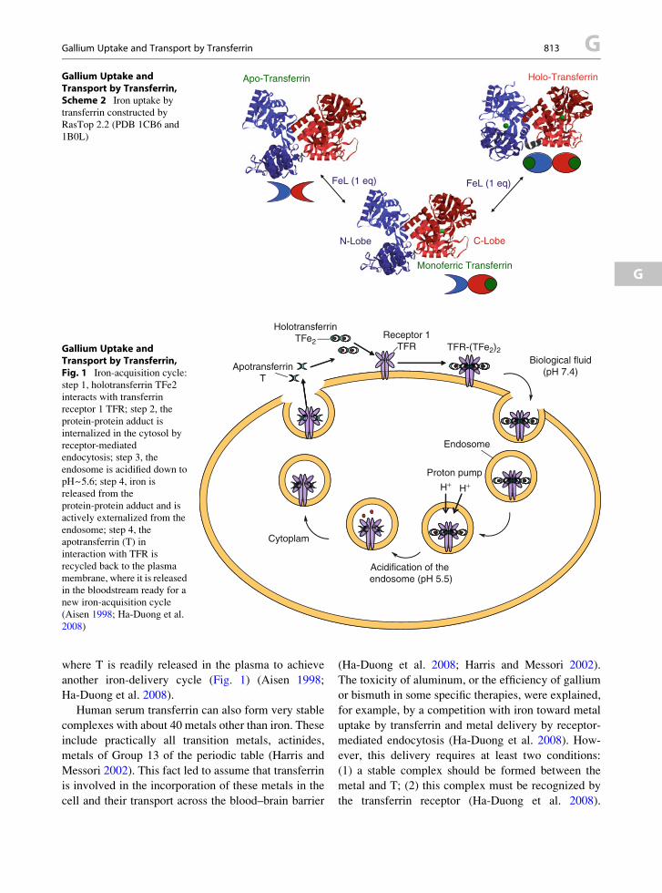
Gallium Uptake andTransport by Transferrin,Scheme 2 Iron uptake by
transferrin constructed by
RasTop 2.2 (PDB 1CB6 and
1B0L)
H+H+
HolotransferrinTFe2
Receptor 1TFR TFR-(TFe2)2
Endosome
Proton pump
Acidification of the endosome (pH 5.5)
ApotransferrinT
Cytoplam
Biological fluid(pH 7.4)
Gallium Uptake andTransport by Transferrin,Fig. 1 Iron-acquisition cycle:
step 1, holotransferrin TFe2
interacts with transferrin
receptor 1 TFR; step 2, the
protein-protein adduct is
internalized in the cytosol by
receptor-mediated
endocytosis; step 3, the
endosome is acidified down to
pH~5.6; step 4, iron is
released from the
protein-protein adduct and is
actively externalized from the
endosome; step 4, the
apotransferrin (T) in
interaction with TFR is
recycled back to the plasma
membrane, where it is released
in the bloodstream ready for a
new iron-acquisition cycle
(Aisen 1998; Ha-Duong et al.
2008)
Gallium Uptake and Transport by Transferrin 813 G
G
where T is readily released in the plasma to achieve
another iron-delivery cycle (Fig. 1) (Aisen 1998;
Ha-Duong et al. 2008).
Human serum transferrin can also form very stable
complexes with about 40 metals other than iron. These
include practically all transition metals, actinides,
metals of Group 13 of the periodic table (Harris and
Messori 2002). This fact led to assume that transferrin
is involved in the incorporation of these metals in the
cell and their transport across the blood–brain barrier
(Ha-Duong et al. 2008; Harris and Messori 2002).
The toxicity of aluminum, or the efficiency of gallium
or bismuth in some specific therapies, were explained,
for example, by a competition with iron toward metal
uptake by transferrin and metal delivery by receptor-
mediated endocytosis (Ha-Duong et al. 2008). How-
ever, this delivery requires at least two conditions:
(1) a stable complex should be formed between the
metal and T; (2) this complex must be recognized by
the transferrin receptor (Ha-Duong et al. 2008).

Receptor 1+
Helical domainApical domain
Protease-likedomain
N-lobe
Holotransferrin
C-lobe and helicaldomain interaction
N-lobe and protease-like domain interaction
50 μs
Kd = 0.5 µM
Overall dissociation constant KD = 2.3 nM
8000s
C-lobe
Gallium Uptake and Transport by Transferrin, Fig. 2 The
C-lobe of iron-saturated transferrin interacts very rapidly (50 ms)with the helical domain of transferrin receptor 1 (TFR) to yield
a first protein-adduct with a dissociation constant Kd ¼ 0.5 mM.
This interaction is followed by a very slow (�2 h) change in the
conformation of the protein-protein adduct to yield the final
product in which the N-lobe of the holotransferrin becomes in
interaction with the protease-like domain of the receptor with an
overall dissociation constant KD ¼ 2.3 nM (Chikh et al. 2007).
The structures are constructed with RASTOP 2.2 open-share
program by the use of the coordinates available at the Protein
Data bank (1CX8 and 1SUV)
G 814 Gallium Uptake and Transport by Transferrin
In addition, even when the metal is released in the
endosome after acidification, it would still require
means of delivery to its potential targets in the cell
outside this endosome.
Transferrin receptor 1 is a 190 kDa homodimeric
protein arranged in two subunits (R) linked by two
disulfide bridges. R contains a transmembrane domain,
a cytoplasmic endodomain of about 15 kDa and a solu-
ble ectodomain directed toward the biological fluid. The
receptor is arranged in four domains: the helical, the
apical, the protease-like, which forms with the plasma
membrane a pseudo-cavity of about 10 A and, finally,
the endodomain (Lawrence et al. 1999). The C-lobe of
holotransferrin (TFe2) or that of a C-lobe only iron-
loaded transferrin interacts primarily with the helical
domain of the receptor. This interaction is followed by
that of the N-lobe with the protease-like domain (Fig. 2)
(Cheng et al. 2004). The first interaction is extremely
fast and occurs in the 50 ms range whereas the second
occurs in about 2 h (Ha-Duong et al. 2008).
The mechanisms for iron, aluminum, bismuth,
cobalt, uranium, and gallium uptake by serum trans-
ferrin were established recently (Ha-Duong et al.
2008). It was also shown that, besides aluminum, the
cobalt-, bismuth-, uranium-, and gallium-loaded
transferrins interact with the transferrin receptor. This
renders the incorporation of these metals possible by
the iron-acquisition pathway (Ha-Duong et al. 2008).
Gallium is a nonphysiological metal, which belongs
to Group 13 of the periodic table. Ga(III) has practi-
cally the same ionic radius as Fe(III) and is
diamagnetic. Gallium has a very low redox potential
which implies that in neutral media, contrary to iron,
this metal exists exclusively as a complex of Ga(III)
(Harris and Messori 2002). Ga(III) is, therefore, used
to mimic Fe(III) without the redox and paramagnetic
inconveniences. Moreover, gallium can be used in
chemotherapy, medical imagery, has effects on bone
metabolism, and an antineoplastic activity (Bern-
stein 1998; Chitambar and Purpi 2010). Part of this
action is explained by the fact that Ga(III) forms
a stable complex with transferrin and can, therefore,
interfere with the metabolism of iron (Bernstein 1998).
Gallium Uptake by Transferrin: Methods andResults
The transferrin gallium complex possesses specific
absorption and emission spectra (Chikh et al. 2007).

Gallium Uptake and Transport by Transferrin 815 G
G
These spectra associated with the fast-mixing tech-
niques, such as stopped-flow (mixing time �1 ms),
permit to follow with time the formation of the
transferrin-gallium complex (Chikh et al. 2007).
When transferrin is rapidly mixed with a gallium
donor, such as nitrilotriacetatoGa(III), gallium is trans-
ferred from the chelate (GaL) to transferrin in three
differentiated kinetic steps (Fig. 3). The use of the
techniques and methods of chemical relaxation (fast
kinetics) allows attributing each step to a series of
chemical processes involved in gallium uptake by
transferrin (Chikh et al. 2007; Eigen 1967).
In the C-lobe of apotransferrin, the phenols of the
tyrosine ligands are in contact with bulk aqueous
medium and are, therefore, in the protonated form.
Furthermore, under natural conditions, only the
C-lobe of apotransferrin is in interaction with the syn-
ergistic hydrogenocarbonate, whereas the N-lobe is
not. A first gallium transfer occurs in about 50 s from
the gallium chelate to the C-lobe in interaction with
HCO3� to produce a first transferrin-gallium interme-
diate, in which the tyrosine ligands are still in the
protonated form. This intermediate undergoes a series
of proton losses from the protein ligands and confor-
mation changes to produce, in the second kinetic pro-
cess, an intermediate species, in which gallium is
coordinated to the two phenolates of the tyrosines
and a carbonate. This leads to a drop in the apparent
pKas of the phenols from about 9 in T down to 8 in the
kinetic intermediate. This latter undergoes, then,
a series of very slow conformation changes, which
allow the N-lobe of transferrin to acquire a second
gallium and to reach the thermodynamic equilibrium
(Fig. 3) (Chikh et al. 2007).
Interaction of Gallium-Loaded T with R:Methods and Results
Transferrin receptor 1 possesses a typical fluorescence
emission spectrum. Adding TGa2 to TFR results in
a increase in fluorescence intensity accompanied by
a small red shift in the emission maximum (Chikh et al.
2007). Similar spectral modifications were also
observed with iron-loaded and other metals–
loaded transferrins (Ha-Duong et al. 2008). These
spectra allowed the titration of R by TGa2 and, subse-
quently, the determination of the dissociation
constant of the TGa2/receptor 1 protein/protein adduct
(KDGa(III) ¼ 1.10 � 0.12 mM) (Chikh et al. 2007).
KDGa(III) is very high as compared to that reported for
the interactions of holotransferrin with R in the final
equilibrated state (2.3 nM) (Ha-Duong et al. 2008).
This interaction was confirmed by a T-jump kinetic
approach. The T-jump technique is based on Joule
heating by the discharge of a capacitor in the solution.
Heating times can be as low as 200 ns and heating
amplitudes can be as high as 10�C. This technique
allows to measure kinetic runs occurring in the micro-
seconds to the milliseconds range (Ha-Duong et al.
2008). In the present case the detection was based on
the variations in the emission spectra. When a solution
of R in the presence of holotransferrin is submitted to
a fast T-jump, a single kinetic process corresponding to
the interaction of the C-lobe of TGa2 with R occurs in
the 150 ms range as an exponential increase in the
fluorescence emission (Fig. 4).
Can Gallium Follow the Iron AcquisitionPathway?
Gallium, iron, aluminum, and bismuth are all consid-
ered as hard metals (Harris and Messori 2002) and are
complexed by the same protein ligands (Aisen 1998).
These include the two phenolates of the two tyrosines,
the carboxylate of an aspartate and the imidazol of
a histidine. Upon complex formation with T, shifts
occur in the apparent pKa of the ligands. These are
related to the nature of the metal. Indeed, the binding
site undergoes a chelation process and a change in its
environment when the protein transits from an open
structure, where all ligands are in contact with the bulk,
to the closed structure, where the ligands are engaged
in the complex and protected from the external
medium (Aisen 1998). The discrepancies in these
apparent pKas shifts reported for iron, bismuth, and
cobalt when compared to gallium and aluminum imply
that the local environment of aluminum and gallium
are at variance with those of Fe(III), Bi(III), and cobalt
(Ha-Duong et al. 2008). As with iron, gallium gain by
the C-site triggers a series of proton dissociation-
dependent changes in the conformation of the protein,
which lead to the activation of the N-binding site, thus
allowing the secondmetal uptake by the N-lobe (Chikh
et al. 2007).
When gallium-saturated transferrin is mixed with
FeL, iron replaces gallium. The affinity of transferrin

050
0.06
0
0.06
4
0.06
8
Differential absorption (ΔA) Differential absorption (ΔA)
time
(s)
Firs
t kin
etic
pro
cess
a
050
0
0.19
8
0.20
4
time
(s)
Firs
t kin
etic
pro
cess
Sec
ond
kine
tic p
roce
ss
b
010
000
0.16
0.18
0.20
Differential absorbance (ΔA)
time
(s)
6000
0 s
Thi
rd k
inet
ic p
roce
ss
Sec
ond
kine
ticpr
oces
s
Firs
t kin
etic
pr
oces
sT
herm
odyn
amic
prod
uct
c
500
s+
3 H
+N
C
?
6000
0 s
N
C
TG
a2T
herm
odyn
amic
pro
duct
: gal
lium
satu
rate
d tr
ansf
errin
CN
+C
N
50
sC
N
CN
GaL
(TG
a)'
LT
+
Firs
t kin
etic
inte
rmed
iate
: C-s
ite
galli
um-lo
aded
tran
sfer
rin
TG
aS
econ
d ki
netic
inte
rmed
iate
:C
-site
gal
lium
-load
ed tr
ansf
errin
(TG
a)'
TG
a
Gallium
UptakeandTransp
ort
byTransferrin,Fig.3
(a)Ga(III)
istransferred
from
gallium
nitrilotriacetateto
theC-lobeoftransferrinin
interactionwithbicarbonate
toyield
afirstkinetic
product,as
shownbythe50schangein
theabsorptionofthe
proteinwhen
mixed
withthechelate.(b)Asecondslowabsorptionchange(500s)ofthe
kinetic
product
describes
aconform
ationchangeaccompaniedbytheloss
ofthree
protonsfrom
theprotein
ligands.(c)A
ultim
ateveryslow
absorptionchange(�
17h)
showsthefinalchangeintheconform
ation,w
hichallowasecondgalliumtransfertothe
N-lobeoftheprotein
(Chikhetal.2007)
G 816 Gallium Uptake and Transport by Transferrin

150 ms
KDGa = 1.1 µM
Interaction of TGa2 with receptor 1
Receptor 1 Final protein-protein adduct:TGa2-Receptor 1
5.0×10⁻⁵ 1.0×10⁻⁴ 1.5×10⁻⁴
0.80
0.82
0.84
Nor
mal
ized
fluo
rese
cnce
em
issi
on
time (s)
NC+
TGa2
NC
Gallium Uptake andTransport by Transferrin,Fig. 4 Emission growth in
150 ms after an instantaneous
T-jump performed in
a solution of gallium-saturated
transferrin in the presence of
receptor 1. This corresponds to
the interaction of the C-lobe of
the metal-loaded transferrin
with the helical domain of
R with a dissociation constant
KDGa(III) ¼ 1.1 mM. No
interaction of the N-lobe with
the protease-like domain of
R is observed (Chikh et al.
2007)
Gallium Uptake and Transport by Transferrin, Table 1 Summary of the main results and interpretations (Chikh et al. 2007)
Affinity of
T for the
metal
Dissociation
constant
(C-Lobe)
Average time
of interaction
with the C-lobe
Overall
dissociation
constant (KD)
Average time of
interaction with the
N-lobe
Average time
for receptor
recycling
Internalization
with metal-
loaded C-lobe
Iron(III) 1021 0.5 mM 50 ms 2.3 nM 8,000 s Few minutes Yes
Gallium(III) 1019 1.1 mM 150 ms 1.1 mM No interaction Few minutes Probable
Gallium Uptake and Transport by Transferrin 817 G
G
for gallium is about 1019 M-1 (Harris and Messori
2002), whereas that for iron is in the 1021 M�1range
(Table 1) (Aisen 1998). Furthermore, the first step in
iron uptake by transferrin occurs in less than 0.1 s,
whereas the first step in gallium uptake occurs in the
50 s range. Therefore, as compared to gallium, iron
uptake by transferrin is both thermodynamically and
kinetically favored. This suggests that there cannot be
a competition between iron and gallium toward metal
uptake by transferrin. However, only 40% of the trans-
ferrin circulating in the blood stream is iron-loaded and
there is still plenty of protein available for complex
formation with other metals (Aisen 1998). Is this avail-
ability for gallium sufficient to imply that this metal
follows the iron-acquisition pathway by receptor-
mediated endocytosis? The answer is negative,
because in order to follow the iron-acquisition path-
way, the gallium-loaded transferrin should also be
recognized by R.
There are two binding sites on a receptor subunit,
the first is in the helical domain, common to the C-site
of TFe2 and HFE. The second is in the protease-like
domain and is specific to the N-lobe of TFe2 (Cheng
et al. 2004). The N-lobe interacts with the protease-like
domain where it occupies the cavity formed by the
plasma membrane and the protease-like domain
(Cheng et al. 2004; Chikh et al. 2007; Giannetti et al.
2003). The interaction of TFe2 with R occurs in two
differentiated steps (Fig. 2, Table 1). The first corre-
sponds to that of the C-lobe with the helical domain of
R. It is extremely fast and occurs with a dissociation
constant of 0.5 mM (Fig. 2, Table 1). It is followed by
the slow interaction of the N-lobe with the protease-
like domain of R. This process lasts about 2 h and
stabilizes the protein/protein adduct 300-fold to lead
to an overall dissociation constant, KDFe(III) ¼ 2.3 nM
(Fig. 2, Table 1) (Ha-Duong et al. 2008). With
TGa2, only the interaction of the C-lobe with the

G 818 Gallium Uptake and Transport by Transferrin
helical domain is detected with a dissociation constant
KDGa(III) ¼ 1.1 mM (Fig. 4, Table 1). This value is
higher than that reported for the interaction of
TFe2 with R and should, therefore, imply that there
is no possible competition between holotransferrin
and TGa2 toward the interaction with R in the final
equilibrated state. However, the dissociation constant
of the R-C lobe intermediate at the end of the first fast
step is 0.5 mM (Table 1) (Ha-Duong et al. 2008). This
value is of the same order of magnitude as that
reported for TGa2 (1.1 mM, Table 1) (Chikh et al.
2007). Therefore, at the end of the first fast process
of the interaction of C-lobe of holotransferrin with
R and before the very slow interaction of the N-lobe
with the protease-like domain, TGa2 can compete
with holotransferrin for recognition by R. Receptor-
mediated endocytosis and iron release in the endo-
some occur in a few minutes (Chikh et al. 2007). This
lapse of time is sufficient to permit the internalization
of TGa2 long before thermodynamic equilibrium
between TFe2 and R is attained. The same case was
reported for the interaction of bismuth-loaded trans-
ferrin, which also occurs in a single fast step with
a dissociation constant of 4 mM (Ha-Duong et al.
2008). Subsequently, TGa2 can compete with
holotransferrin for the interaction with the helical
domain of R leading, eventually, to gallium uptake
by the iron-acquisition pathway. This was shown for
HFE which interacts with only the helical domain of
the receptor with a lower Kd value than that of the
overall holotransferrin-R interaction. HFE competes
with holotransferrin for the recognition by
R and perturbs iron acquisition by the cell (Giannetti
et al. 2003).
Conclusion
Receptor-mediated iron-acquisition pathway is
assumed to be involved in the transport and delivery
of several metals from the blood stream to the cell
and across the blood–brain barrier. For gallium, the
affinity of R for the metal-loaded transferrin is about
two orders of magnitude lower than that for
holotransferrin at equilibrium. Despite this fact, gal-
lium can be internalized by receptor 1-mediated
endocytosis. Indeed, the main responsibility in this
transport lies in the fast interaction of the C-lobe of
metal-loaded transferrin with the helical domain of R.
This process is very fast for both iron-loaded and
gallium-loaded transferrins, and the affinities involved
are of the same order of magnitude. Both processes are
extremely fast compared to endocytosis, which lasts
a few minutes. Likewise, endocytosis is extremely
fast when compared to the interaction of the N-lobe
of holotransferrin with the protease-like domain
of R which is controlled by a change in conforma-
tion lasting several hours. This implies a dynamic
metal transport based more on kinetics than on
thermodynamics.
Cross-References
▶Bismuth in Brain, Distribution
▶Calsequestrin
▶Cobalt Transporters
▶Manganese, Interrelation with Other Metal Ions in
Health and Disease
▶Nickel Carcinogenesis
▶Vanadium Ions and Proteins, Distribution,
Metabolism, and Biological Significance
References
Aisen P (1998) Transferrin, the transferrin receptor, and the
uptake of iron by cells. Met Ions Biol Syst 35:585–631
Bernstein LR (1998) Mechanisms of therapeutic activity for
gallium. Pharmacol Rev 50(4):665–682
Cheng Y et al (2004) Structure of the human transferrin receptor-
transferrin complex. Cell 116(4):565–576
Chikh Z et al (2007) Gallium uptake by transferrin and interac-
tion with receptor 1. J Biol Inorg Chem 12(1):90–100
Chitambar CR, Purpi DP (2010) A novel gallium
compound synergistically enhances bortezomib-induced
apoptosis in mantle cell lymphoma cells. Leuk Res
34(7):950–953
Eigen M (1967) Nobel lecture
Giannetti AM et al (2003) Mechanism for multiple ligand rec-
ognition by the human transferrin receptor. PLoS Biol
1(3):E51
Ha-Duong NT et al (2008) Kinetics and thermodynamics of
metal-loaded transferrins: transferrin receptor 1 interactions.
Biochem Soc Trans 36(Pt 6):1422–1426
Harris WR, Messori L (2002) A comparative study of alumi-
num(III), Gallium(III), indium(III) and Thallium(III)
binding to human serum transferrin. Coord Chem Rev
228:237–262
Lawrence CM et al (1999) Crystal structure of the ectodomain
of human transferrin receptor. Science 286(5440):
779–782

Gallium(III) Complexes, Inhibition of Proteasome Activity 819 G
Gallium(III) Complexes, Inhibition ofProteasome Activity
Michael Frezza1, Sara Schmitt1, Dajena Tomco2,
Di Chen1, Claudio N. Verani2 and Q. Ping Dou1
1The Developmental Therapeutics Program, Barbara
Ann Karmanos Cancer Institute, and Departments of
Oncology, Pharmacology and Pathology, School of
Medicine, Wayne State University, Detroit, MI, USA2Department of Chemistry, Wayne State University,
Detroit, MI, USA
G
SynonymsGallium; Gallium(III) ion; Gallium Complexes;
Gallium Compounds
Definition
Gallium complexes were characterized by elemental,
spectroscopic, and spectrometric methods. Structure 1
and 3 were elucidated by X-ray crystallography.
Biological activity was investigated by growth
suppression, proteasome inhibition, and apoptosis
induction in vitro and in vivo.
Basic Characteristics
Chemical Properties of Gallium
Gallium is described as a gray semimetal. The most
stable cation is the trivalent Ga(III) with an electronic
configuration [Ar]3d10 formed by the loss of two 4sand one 4p electrons. The solubility and chemical
parameters of gallium have major implications on its
therapeutic utility. The trivalent form of gallium is
redox-inactive, often being used as a probe for
electroactive ligands (Frezza et al. 2007).
Biochemical Properties of Gallium
Investigation into the therapeutic applications of
gallium dates to the 1970s where gallium presented
an optimal therapeutic and toxicity profile compared to
other Group 13 (previously IIIA) elements. Gallium
was subsequently found to be active against acceler-
ated bone turnover, tumor growth, and immune
diseases. Gallium nitrate and other gallium compounds
have been investigated in clinical trials as single agents
or in combination with pre-existing chemotherapeutic
agents and have been found to be active, especially
toward lymphomas and bladder cancer (Frezza et al.
2007).
The therapeutic application of the gallium(III) ion
appears to be underscored by its chemical similarity to
the trivalent ferric ion, where comparable charge,
lability, and ionic radii (0.76 A vs. 0.78 A) are
observed. Therefore, gallium is commonly recognized
as iron(III) upon cellular uptake and interacts with iron
specific sites in enzymes and proteins. Gallium also
concentrates in regions that are highly dependent of
iron, including inflammation and malignancy (Chen
et al. 2009). Gallium, unlike iron, is redox inactive.
However, unlike iron ions, the gallium(III) ion is redox
inactive precluding its participation in biologically
relevant redox reactions such as oxygen transport.
One of the critical targets of intracellular gallium
appears to be the enzyme ribonucleotide reductase,
which catalyzes the rate-limiting step in DNA
synthesis and is upregulated in highly proliferating
cancer cells. This gallium-dependent inhibition of
ribonucleotide reductase is in part related to the inhi-
bition of cellular proliferation (Chitamber et al. 1988).
Several gallium complexes have been synthesized
and evaluated in clinical trials (Collery et al. 1996).
Such complexes have shown an increase in bioavail-
ability and superior anticancer activity compared to
gallium salts. These findings provide compelling
evidence for the development of novel gallium-based
complexes that may show alternative mechanisms of
action leading to durable anticancer activity and
decreased toxicity.
Cellular 26S Proteasome as a Target for Gallium(III)
Complexes
A series of gallium complexes described as
[GaIII(LX)2]ClO4� with asymmetric NN0O-containing
pyridine amino phenolate ligands have been
developed as potential cellular proteasome inhibitors.
The phenolate moiety groups were appended with
electron withdrawing and donating groups such as
methoxy (1), nitro (2), chloro (3), bromo (4), and
iodo (5) positioned at the 4th and 6th positions
(Fig. 1). Due to the flexibility of the ligands, facial
coordination takes place and the overall geometry of
the complexes is distorted octahedrally (Fig. 1).

H
N
I
N
H
OMe
NO2
NN
Br
Br
N
Cl
Cl
NHNHNHNHNH OHOHOHOHOH
I
N
NO
X'
X
Ga
N
NO
X'
X
HH
+
The complexes
The HLX ligands
The octahedral/facial geometry Gallium(III) Complexes,Inhibition of ProteasomeActivity, Fig. 1 Gallium
complexes. [GaIII(LX)2]ClO4
and their ligands HLX, with
X ¼ H, methoxide (1), nitro(2), chloro (3), bromo (4), andiodo (5). All compounds
exhibit facially coordinated
ligands and a perchlorate
(ClO4�) counterion
G 820 Gallium(III) Complexes, Inhibition of Proteasome Activity
These complexes were first tested against cisplatin-
resistant neuroblastoma cells. It was shown in ranking
order, methoxy (1)¼ nitro (2)< chloro(3)< bromo (4)
< iodo (5), that the species containing halogen
substituents showed preferential growth inhibition in
human neuroblastoma cells with activity superior to
cisplatin. In addition, neuroblastoma cells treated with
gallium chloride rendered cells viable, with results
similar to the less active methoxy-substituted complex.
Furthermore, this series of Ga(III) complexes was
tested against normal human fibroblasts and were
found to be nontoxic up to 25 mM treatment (Shakya
et al. 2006).
Promising results acquired in the laboratory
highlight a novel approach utilizing metal-based
complexes as tumor-specific proteasome inhibitors.
The proteasome is a multicatalytic protease responsi-
ble for the degradation of the majority of cellular
proteins involved in various cellular processes such
as cell cycle progression, angiogenesis, and apoptosis
(Dou and Li 1999). The ubiquitin-proteasome pathway
in eukaryotes led to the realization that specific molec-
ular machinery is available in the cell to eliminate
defective proteins and enzymes (Hershko et al. 1982).
This discovery led to the 2004 Nobel Prize in Chemis-
try to Hershko, Ciechanover, and Rose. An increased
proteasome activity has been identified in malignant
cells, making its inhibition a venue for cancer therapy
of unprecedented relevance (Li and Dou 2000). Addi-
tionally, inhibition of the proteasomal chymotrypsin-
like activity was found to be associated with induction
of tumor cell apoptosis (An et al. 1998). In 2003,
bortezomib became the first proteasome inhibitor to
be approved as an anticancer drug (Dou and Goldfarb
2002). However, drug resistance and toxicity coupled
with the lack of activity toward solid tumors, and
interactions with some natural products has limited
its clinical development.
A possible molecular mechanism for these gallium
(III) complexes responsible for their growth-inhibitory
effects was investigated. Whether these gallium com-
plexes could target and inhibit the proteasomal activity
of purified 20S and cellular 26S proteasome in intact
prostate cancer cells was tested. The studies revealed
that complexes 3, 4, and 5 were able to target and
inhibit proteasomal activity associated with apoptosis
induction in various prostate tumor cells including
LNCaP (androgen-dependent) and C4-2B (androgen-
independent). Specifically, complexes 3, 4, and 5
inhibited the chymotrypsin-like activity of the
proteasome by 39%, 62%, and 79% in LNCaP
cells and 38%, 58%, and 81% in C4-2B cells at
50 mM treatment for 18 h. The ability to inhibit
proteasome activity was indicated by accumulation of
ubiquitinated proteins and the proteasomal target
protein p27. Inhibition of proteasomal activity by

Gallium(III) Complexes, Inhibition of Proteasome Activity 821 G
G
these gallium(III) complexes was associated with apo-
ptosis induction as measured by poly(ADP-ribose)
polymerase (PARP) cleavage and nuclear staining.
One interesting observation was that treatment of
androgen receptor (AR)-containing prostate cancer
cells with these gallium(III) complexes causes
downregulation of AR protein, associated with tumor
cell apoptosis.
As observed with cisplatin-resistant neuroblastoma
cells, the iodo-substituted complex 5 achieved superior
therapeutic efficacy in a human prostate cancer
model. Accordingly, complex 5 exhibited potent
proteasome inhibitory activity against both intact
26S proteasome (IC50 ¼ 17 mmol) and purified 20S
proteasome (IC50¼ 16 mmol). It is notable to point out
that treatment with the iodo-substituted ligand alone
showed only minimal cytotoxicity. Furthermore, com-
plex 5 (20 mg/kg/day for 30 days) was able to exert the
same effect in vivo by inhibiting the growth of prostate
PC-3 tumors grown in mice (by 66%), associated with
proteasome inhibition and apoptosis induction.
Immunohistochemistry studies showed higher levels
of p27 and TUNEL-positive cells in tumors treated
with complex 5, when compared to tumors treated
with vehicle control or ligand alone (Chen et al.
2007). The superior biological activity of complex 5
suggests that the iodo-substituted ligand attains certain
physiochemical properties that impart significant
therapeutic efficacy. Since the active complexes from
this study consistently contain halogen-substituted
ligands, the heightened efficacy may be governed by
the strong p-donating groups (I> Br> Cl). For exam-
ple, iodine harbors weak electron withdrawing capa-
bility, but retains strong p-donating effects. Although
not conclusive, this property might play a role in
activating the ring system of the complex and thus
influencing coordination to the proteasome.
In order to gain insight into a possible mechanism of
action for these gallium complexes, the biological role
of other bivalent transition metals with the same NN0Oligand platform was then investigated. It was found
that the 2:1 metal complex is unable to coordinate to
available terminal threonine residues available in the
active centers of the 20S proteasome without the loss
of one ligand. It was therefore hypothesized that the
primary pharmacophore responsible for the biological
activity of these metal complexes is an [ML] species
with a 1:1 metal-to-ligand ratio. In support of this
hypothesis, it was shown that both a 1:1 and 1:2
complexes with copper(II) showed similar levels
of pharmacological activity both in vitro and
under cell culture conditions (Hindo et al. 2009).
To test this working hypothesis further, similar 1:2
metal-to-ligand complexes using divalent nickel(II)
and zinc(II) were designed. The nickel(II) ion has
a 3d8 configuration, which leads to nonzero ligand-
field stabilization energies (LFS) and, as a result,
fosters slower ligand exchange. On the other hand,
the zinc(II) ion exhibits a 3d10 configuration, is pre-
dominantly ionic, and displays rapid ligand dissocia-
tion and exchange. Consistent with the above
hypothesis, it was found that the zinc(II) complex,
but not the nickel(II) complex was able to inhibit the
proteasomal chymotrypsin-like activity under cell-free
conditions and in cultured human prostate cancer cells
(Frezza et al. 2009). Therefore, liberation of one ligand
from the complex should facilitate potent proteasome-
inhibitory activity, allowing the 1:1 metal-to-ligand
species to coordinate to amino acid residues in the
proteasome. Thus, gaining a better understanding of
the detailed nature of the pharmacophore and its role in
proteasome inhibition should aid in the development of
metal complexes containing NN0O tridentate ligands
with gallium and other transition metals. In conclusion,
these metal complexes could serve as platforms for
development of novel proteasome inhibitor anticancer
drugs for the treatment of prostate and other human
cancers.
References
An B, Goldfarb RH, Siman R et al (1998) Novel dipeptidyl
proteasome inhibitors overcome Bcl-2 protective function
and selectively accumulate the cyclin-dependent kinase
inhibitor p27 and induce apoptosis in transformed, but not
normal, human fibroblasts. Cell Death Differ 5:1062–1075
Chen D, Frezza M, Shakya R et al (2007) Inhibition of the
proteasome activity by gallium(III) complexes contributes
to their anti-prostate tumor effects. Cancer Res
67:9258–9265
Chen D,Milacic V, FrezzaM et al (2009)Metal complexes, their
cellular targets, and potential in cancer therapy. Curr Pharm
Des 15:777–791
Chitamber CR, Matthaeus WG, Antholine WE et al (1988) Inhi-
bition of leukemic HL60 cell growth by transferrin-gallium:
effects on ribonucleotide reductase and demonstration of drug
synergy with hydroxyurea. Blood 72:1930–1936
Collery P, Domingo JL, Keppler BK (1996) Preclinical toxicol-
ogy and tissue distribution of a novel antitumour gallium
compound: tris 8(quinolinolato)gallium(III). Anticancer
Res 16:687–691

G 822 Gallium(III) Ion
Dou QP, Goldfarb RH (2002) Bortezomib (millennium pharma-
ceuticals). IDrugs 5:828–834
Dou QP, Li B (1999) Proteasome inhibitors as potential novel
anticancer agents. Drug Resist Updat 2:215–223
Frezza M, Verani CN, Chen D et al (2007) The therapeutic
potential of gallium-based complexes in anti-tumor drug
design. Lett Drug Des Disc 4:311–317
Frezza M, Hindo SH, Tomco D et al (2009) Comparative
activitities of nickel(II) and zinc(II) Complexes of asymmet-
ric [NN’O] ligands as 26S proteasome inhibitors. Inorg
Chem 48:5928–5937
Hershko A, Ciechanover H et al (1982) Mechanisms of intracel-
lular protein breakdown. Annu Rev Biochem 51:335–364
Hindo SH, Frezza M, Tomco D et al (2009) Metals in anticancer
therapy: copper(II) complexes as inhibitors of the 20S
proteasome. Eur J Med Chem 44:4353–4361
Li B, Dou QP (2000) Bax degradation by the ubiquitin/
proteasome pathway-dependent pathway: involvement in
tumor survival and progression. Proc Natl Acad Sci USA
97:3850–3855
Shakya R, Peng F, Liu J et al (2006) Synthesis, structure, and
anticancer activity of gallium(III) complexes with asymmet-
ric tridentate ligands: growth inhibition and apoptosis
induction of cisplatin-resistant neuroblastoma cells. Inorg
Chem 45:6263–6268
Gallium, Physical and Chemical Properties,Fig. 1 Low melting of gallium
Gallium(III) Ion
▶Gallium(III) Complexes, Inhibition of Proteasome
Activity
Gallium, Physical and ChemicalProperties
Fathi Habashi
Department of Mining, Metallurgical, and Materials
Engineering, Laval University, Quebec City, Canada
Gallium is a silvery white low-melting point metal
(Fig. 1) belonging to the less typical metals group, i.e.,
when it loses its outermost electrons it will not have the
electronic structure of inert gases. There is a volume
expansion of 3.2% on solidification of the metal. Gal-
lium is as a by-product of the production of aluminum
oxide. Its concentration in bauxite is 0.003%
and 0.008%. The largest reserves of gallium are
contained in phosphate ores and in coal. When phos-
phorus is produced electrothermally from phosphate
rock, the gallium is concentrated in the flue dust.
Likewise, gallium may be obtained from the fly ash of
coal. Gallium contained in zinc ore (sphalerite) has lost
economic importance due to the change over to hydro-
metallurgical methods of zinc extraction. The main use
of gallium is as a raw material in the manufacture of
semiconductors, for example, the arsenide or the phos-
phide for the production of light-emitting diodes.
Physical Properties
Atomic number
31Atomic weight
69.72Relative abundance in the Earth’s crust,%
1.5 � 10�3Melting point, �C
29.78Boiling point, �C
2,403Density, g/cm3
Solid at 24.6�CLiquid at 29.8�C
5.904
6.095
Crystal form
Lattice constants, nm
Orthorhombic
a ¼ 0.4523
b ¼ 0.45198
c ¼ 0.76602
Thermal expansion coefficients, K�1
Along the a axis
Along b axis
Along c axis
1.65 � 10�5
1.13 � 10�5
3.1 � 10�5
Gallium, Therapeutic Effects 823 G
G
Chemical Properties
Metallic gallium dissolves slowly in dilute mineral
acids but rapidly in aqua regia and concentrated
sodium hydroxide. In its compounds, the valency
is +3. Its oxygen compounds resemble those of
aluminum in that there are high- and low-
temperature forms of Ga2O3 and two hydroxides,
Ga(OH)3 and GaO·OH. Substances which lower the
hydrogen ion concentration throw down a white
gelatinous precipitate from Ga(III) salt solutions.
This precipitate is amorphous to x-rays and has
a variable water content (gallium oxide hydrate,
Ga2O3·xH2O). It dissolves both in acids and in
strong bases and differs from aluminum oxide
hydrate in being soluble in ammonia solutions. As
the precipitate ages, its solubility in caustic alkalis
diminishes.
Gallium halides have covalent character and there-
fore have good solubility in many nonpolar solvents
in which they exist in dimeric form. Like aluminum,
gallium is precipitated as the white oxide hydrate
from solutions of its salts by reducing the hydrogen
ion concentration. Gallium sulfate, Ga2(SO4)3, crys-
tallizes from aqueous solutions as an 18-hydrate
similar to Al2(SO4)3·18H2O. It can be dehydrated
by heating and decomposes above 520�C with
loss of SO3. With ammonium sulfate, it forms the
double salt (NH4)Ga(SO4)2·12H2O, ammonium gal-
lium alum.
The crude metal contains 99–99.9% gallium.
The material for semiconductor manufacture is used
in purities from 99.9999% (6 N) to 99.999999% (8 N).
The binary compounds, GaP and GaAs, are of
great industrial importance. They may be prepared
by direct combination of the elements at high
temperature.
References
Greber JF (1997) Gallium. In: Habashi F (ed)
Handbook of extractive metallurgy. Wiley, Weinheim,
pp 1523–1530
Habashi F (2006) Gallium Update. In: Travaux du Comite
international pour l”etude de la bauxite, de l”alumine
et de l”aluminium (ICSOBA), vol 33, No. 37.
Hungarian National Committee of ICSOBA, Budapest,
pp 141–153. Hungarian translation in Kohaszat
140(3):29–34
Gallium, Therapeutic Effects
Lawrence R. Bernstein
Terrametrix, Menlo Park, CA, USA
Synonyms
Gallium and apoptosis; Gallium effect on bacteria;
Gallium uptake
Definition
Gallium: Gallium is an element (atomic number 31;
atomic weight 69.723) in Group XIII of the periodic
table (below aluminum, above indium), classed as
a semimetal or poor metal. It occurs in the Earth’s
crust at an average abundance of about 15–19 parts
per million (ppm) (similar to the abundance of nitro-
gen and about ten times that of tin or arsenic), widely
distributed in soils and rocks. The element is
extracted mainly from bauxite (in which it occurs
at an average concentration of roughly 50 ppm) as
a byproduct of aluminum refining, and to a lesser
extent from zinc ores.
Introduction
Gallium is not known to have any essential biological
function, but it exerts a variety of therapeutically use-
ful biological activities. Many of these activities derive
from gallium’s ability to act as an irreducible mimic of
ferric iron (Fe3+). Gallium’s ability to compete with
other essential metals, particularly zinc, is likely
responsible for some of its other biological activities.
The ionic radii, electronegativity, ionization
potentials, and coordination chemistry of Ga3+ are
remarkably similar to those of Fe3+. In the body,
Ga3+ can be transported in the blood serum by the
iron transport protein transferrin, and appears to follow
many of the uptake and transport pathways observed
for iron (Bernstein 1998).
It is, however, the differences between Ga3+ and
Fe3+ that allow gallium to be therapeutically useful. By
being irreducible under physiological conditions, gal-
lium cannot participate in redox reactions, such as the

G 824 Gallium, Therapeutic Effects
Fenton-type reactions that make free iron (mainly as
Fe2+ in solution) highly toxic. Gallium also is not
observed to enter Fe2+-bearing molecules such as
heme, and so does not interfere with oxygen transport
(via hemoglobin) or with cytochrome-mediated reac-
tions. Furthermore, at pH 7.4 and 25�C, Ga3+ has
a solubility of about 1 mM (98.4% as [Ga(OH)4]¯ and
1.6% as Ga(OH)3) whereas Fe3+ has a solubility of
only about 10�18 M. Thus, small amounts of nonpro-
tein-bound gallium can exist in solution at physiolog-
ical conditions, versus insignificant amounts of
nonprotein-bound Fe3+, permitting biological interac-
tions for Ga3+ that would not be possible for Fe3+.
Gallium metalloproteins: Gallium is known to sub-
stitute for iron in several human proteins. Crucial to
many of gallium’s activities in the body, it binds to the
two metal sites on transferrin (TF). Typically, serum
TF is about 33% saturated with Fe, leaving 67% avail-
able for binding by Ga (about 2.7 mg/mL). Under usual
physiologic conditions, the binding constants for gal-
lium are log K1 ¼ 20.3 and log K2 ¼ 19.3; the
corresponding binding constants for Fe3+ are log
K1 ¼ 22.8 and log K2 ¼ 21.5. However, whereas
Fe3+ remains bound to TF down to a pH of about 5.5,
Ga3+ starts to dissociate from TF at pH < 6.8; it is
>50% dissociated at pH 6 (McGregor and Brock
1992). This difference in stability may provide
a therapeutic advantage to TF-bound Ga, as it may be
released sooner than Fe in endosomes of cancer cells
and phagosomes of some pathogens, helping it to com-
pete against Fe (Bernstein et al. 2011). In addition to
binding to TF, gallium binds even more avidly to the
related protein lactoferrin as well as to the iron storage
protein ferritin.
The substitution of Ga3+ for Fe3+ is also observed in
bacterial proteins, including ferric-binding protein
(FbpA), an iron transporter in the same protein super-
family as TF (Weaver et al. 2008). The presence of
such substitutions could inhibit bacterial growth. Ga3+
can also substitute for Fe3+ in non-ribosomal peptide
microbial siderophores.
Ionic gallium is observed to dose-dependently inhibit
alkaline phosphatase (Boskey et al. 1993) and matrix
metalloproteinase activity (Panagakos et al. 2000). This
effect is hypothesized to be caused by the substitution of
gallium for zinc in these proteins. Gallium and zinc are
chemically similar, with gallium found substituting for
zinc in minerals (e.g., gallium was first discovered
replacing zinc in sphalerite, cubic ZnS).
Therapeutic gallium compounds: Numerous gal-
lium compounds have shown evidence of therapeutic
activity (Table 1). These compounds include inorganic
and organic salts, metal-organic complexes,
metalloproteins, and organometallics. As with all
metals and semimetals, the chemical form in which
gallium occurs is crucial to its absorption, distribution,
excretion, and activity. The biochemical properties of
gallium are thus dependent on the chemical form in
which it is delivered. Organometallic gallium com-
pounds (having Ga–C bonds), as well as some highly
stable complexes (including Ga porphyrins), tend to
remain fairly intact under physiological conditions, so
their molecular conformation helps determine their
activity. Other compounds, such as some orally admin-
istered gallium salts and complexes, are prodrugs in
that they dissociate before reaching the bloodstream,
with the gallium becoming bound mainly to transferrin
and other proteins. Depending on their route of admin-
istration and chemical nature, gallium compounds can
undergo various degrees of metabolic alteration in the
body, resulting in varying contributions from the gal-
lium itself versus its molecular host.
Currently, the only gallium compounds reported to
have been tested in humans are citrated gallium nitrate
for injection (Ganite®; approved for use in the USA to
treat cancer-related hypercalcemia); gallium maltolate
for oral administration (Phase I trials); gallium
8-quinolinolate for oral administration (Phase
I trials); and gallium chloride for oral administration
(in a few cancer patients).
Therapeutic activities: The known therapeutic activ-
ities in which gallium may play a major role include:
(1) Activity against pathological hyperproliferation,
particularly against some aggressive cancers; (2) Anti-
inflammatory and immunomodulating activity, as
observed in animal models of rheumatoid arthritis, mul-
tiple sclerosis, and lupus; (3) Anti-bone-resorptive and
anti-hypercalcemic activity, plus possible anabolic bone
activity; (4) Activity against some pathogenic microbes,
including Pseudomonas aeruginosa and some intracel-
lular bacteria; and (5) Possible analgesic activity,
including against neuropathic pain.
Gallium in the Treatment of Cancer
Following the discovery of cisplatin’s (cis-diammine-
dichloroplatinum(II)) anticancer activity in 1969, there

Gallium, Therapeutic Effects, Table 1 Therapeutic gallium compounds
Compound Therapeutic activities References
Inorganic salts and glasses
GaCl3 In vitro and animal studies: Anticancer, particularly in
aggressive tumors; active against Pseudomonas aeruginosaBernstein (2005); Banin
et al. (2008)
Human clinical studies: Oral bioavailability low. Possible
potentiation of cisplatin and etoposide therapy in lung cancer
patients.
Ga(NO3)3 (In many of the in vitro and
animal studies, and in all of the human
clinical studies, citrate was added to
solutions of gallium nitrate in order to
neutralize the pH and to increase stability.
The molar concentration of the citrate was
generally the same as that of the Ga)
In vitro and animal studies: Anti-bone-resorptive; possibleanabolic activity on bone; immunomodulating: effective in
animal models of rheumatoid arthritis, multiple sclerosis,
and others; anticancer; antimicrobial, including against
Mycobacteria, Pseudomonas, Staphylococcus, Francisella,and others; antibiofilm activity
Bernstein (2005)
Human clinical studies: Low oral bioavailability.
Intravenous formulation approved in the USA for cancer-
related hypercalcemia. High efficacy observed for Paget’s
disease of bone, lymphoma, multiple myeloma; low to
moderate efficacy observed for urothelial carcinoma,
ovarian cancer, metastatic prostate cancer
Ga2(SO4)3 Suppressed adjuvant arthritis in rats Bernstein (1998)
Ga2O3-doped phosphate-based glasses Activity against Staphylococcus aureus, Escherichia coli,and Clostridium difficile
Valappil et al. (2008)
Organic salts and complexes
Ga citrate Activity against Pseudomonas fluorescens Al-Aoukaty et al. (1992)
Ga tartrate Eliminated experimental syphilis in rabbits at a single dose
of 30–45 mg Ga/kg intramuscularly or 15 mg Ga/kg
intravenously. Eliminated Trypanosoma evansi infection in
mice at 225 mg/kg
Levaditi et al. (1931)
Ga maltolate (tris(3-hydroxy-2-methyl-
4 H-pyran-4-onato)gallium(III))
In vitro and animal studies: Anticancer, including against
hepatocellular carcinoma; effective in animal models of
rheumatoid arthritis; antimicrobial: effective against P.aeruginosa, Rhodococcus equi, S. aureus, Staphylococcusepidermidis and other bacteria; antibiofilm
Bernstein et al. (2000);
Bernstein (2005); Chua
et al. (2006)
Human clinical studies: Oral formulation safe in Phase
I trials; anecdotal efficacy observed in hepatocellular
carcinoma, metastatic colon cancer, lymphoma. Topical
formulation has shown anecdotal efficacy in pain, including
postherpetic neuralgia, inflammation, actinic keratosis, and
psoriasis
Ga 8-quinolinolate (tris(8-quinolonato)Ga
(III))
In vitro and animal studies: Anticancer, including against
lung cancer, melanoma; anti-bone-resorptive
Bernstein (2005)
Human clinical studies: Oral formulation safe in Phase
I trials; possible efficacy in renal cell carcinoma.
Ga complex of pyridoxal isonicotinoyl
hydrazone
Potently antiproliferative in T-lymphoblastic leukemic
CCRF-CEM cells
Bernstein (2005)
Ga protoporphyrin IX Activity against Yersinia enterocolitica, S. aureus,Mycobacterium smegmatis and others
Stojiljkovic et al.
(1999); Bozja et al.
(2004)
[GaL2]CIO4; L ¼ 2,4-diiodo-6-{[(pyridin-
2-ylmethyl)amino]methyl}phenol
Inhibited PC-3 prostate cancer xenographs in mice; inhibited
proteosome activity
Chen et al. (2007)
Ga-deferoxamine Active against P. aeruginosa in vitro and in rabbit cornea
together with gentamicin
Banin et al. (2008)
(continued)
Gallium, Therapeutic Effects 825 G
G

Gallium, Therapeutic Effects, Table 1 (continued)
Compound Therapeutic activities References
Ga complexes of thiosemicarbazones Activity against several cancer cell lines; activity against
CryptococcusKowol et al. (2009);
Bastos et al. (2010);
Mendes et al. (2009)
(LH)2[GaCl4]Cl; L ¼ 1-methyl-4,5-
diphenylimidazole
Modest activity against several cancer cell lines Zanias et al. (2010)
[Ga-3-Madd]+, [Ga-5-Madd]+, [Ga-3-
Eadd]+, [Ga-3-M-5-Quadd]+aActivity against Plasmodium falciparum Sharma et al. (1997);
Ocheskey et al. (2003,
2005); Harpstrite et al.
(2003)
Ga complexes of pyrazoles, indazoles, and
benzopyrazoles
Moderate activity against human immunodeficiency virus Kratz et al. (1992)
Ga complexes of pyrazole-imine-phenols
and pyrazole-amine-phenols
Activity against MCF-7 breast cancer and PC-3 prostate
cancer cell lines
Silva et al. (2010)
Ga-curcumin Activity against mouse lymphoma L1210 cell line Mohammadi et al.
(2005)
Diphenyl Ga chloride Moderate activity against Candida albicans, Cryptococcusneoformans, Bacillus subtilis, and S. aureus
Srivastava et al. (1973)
Tris(N-methylthioacetohydroxamato)
Ga(III)
Moderate activity against several species of bacteria and
fungi
Abu-Dari and Mahasneh
(1993)
Protein complexes
Ga transferrin Active against MCF-7 breast cancer and HeLa cervical
carcinoma cells in vitro
Jiang et al. (2002); Head
et al. (1997); Olakanmi
et al. (2010)Active against Francisella tularensis and Francisellanovicida in vitro
Ga lactoferrin Active against F. tularensis and F. novicida in vitro and
protected against F. novicida nasally introduced to mice
Olakanmi et al. (2010)
Organometallics, organometallic complexes
[((CH3)2 Ga)(5-phenyl-1,3,4-oxadiazole-2-
thio)]4
Active against several cancer cell lines Gallego et al. (2011)
Dimeric methyl-Ga complexes of some
carboxylates
Active against several cancer cell lines Kaluđerovic et al. (2010)
a-dimethylamino-cyclohexoxyl-dimethyl-
gallium
Active against P. falciparum Yan et al. (1991)
Miscellaneous
Doxorubicin-Ga-transferrin conjugate Active in vitro against MCF-7 breast cancer cells, including
those resistant to doxorubicin; anecdotal efficacy in breast
cancer patients
Wang et al. (2000);
Bernstein (2005)
Sulfonated Ga corrole bound to
a heregulin-modified protein
Active against HER-positive cancer in rats Agadjanian et al. (2009)
Liposomal Ga(NO3)3 and gentamicin Very active against P. aeruginosa biofilms Halwani et al. (2008)
Yeast-incorporated Ga Antiresorptive and anabolic to bone in rats Ma and Fu (2010)
a[Ga-3-Madd]+ ¼ [{1,12-bis(2-hydroxy-3-methoxybenzyl)-1,5,8,12-tetraazadodecane}gallium(III)]+;
[Ga-5-Madd]+ ¼ [{1,12-bis(2-hydroxy-5-methoxybenzyl)-1,5,8,12-tetraazadodecane}gallium(III)]+;
[Ga-3-Eadd]+ ¼ [{1,12-bis(2-hydroxy-3-ethyl-benzyl)-1,5,8,12-tetraazadodecane}gallium(III)] +;
[Ga-3-M-5-Quadd]+ ¼ [{1,12-bis(2-hydroxy-3-methoxy-5-(quinolin-3-yl)-benzyl)-1,5,8,12-tetraazadodecane}-gallium(III)]+
G 826 Gallium, Therapeutic Effects
was a systematic effort to determine the anticancer
efficacy of other metal compounds. By 1971, after an
initial round of in vitro and animal screening, gallium
emerged as being particularly promising. Specifically,
parenterally administered gallium nitrate showed
efficacy in several animal cancer models, including
Walker 256 ascites carcinosarcoma in rats and
implanted human medulloblastoma in mice. In vitro
activity of gallium compounds has been observed in
a number of cancer cell lines, including those for
Gallium, Therapeutic Effects 827 G
G
lymphoma (Chitambar 2010), breast cancer (Wang
et al. 2000), leukemia (Bernstein 1998), and hepato-
cellular carcinoma (Chua et al. 2006) (see Table 1 for
more examples of in vitro and in vivo anticancer
activity).
The discovery of gallium’s anticancer activity coin-
cided with the introduction of 67Ga-scans to diagnose
and locate a variety of cancers in the body. 67Ga-scans
were found to be particularly sensitive to lymphomas,
some sarcomas, and bone tumors, but much less sen-
sitive to many other types of cancer. For Ga-sensitive
cancers, 67Ga-scans were noted to detect growing can-
cer tissue rather than cancer tissue that was necrotic or
otherwise less active. Many of the 67Ga-avid cancers
soon proved to be most susceptible to therapeutic gal-
lium treatment, though this correlation was apparently
not documented or utilized.
Mechanisms of antiproliferative activity: The
sensitivity of multiplying cancer cells to gallium
appears due mainly to their high need for iron. This
need stems primarily from the requirement for iron in
the active site of ribonucleotide reductase, an enzyme
essential for the synthesis of DNA. To obtain sufficient
iron, most cancer cells highly overexpress TF-receptor
(though some cancer cells can also take up iron by
TF-receptor-independent mechanisms). If Ga is pre-
sent on the available TF, the Ga-TF will be taken into
the cell by endocytosis and compete with iron. Ga then
hinders acidification of the TF-bearing endosome, pos-
sibly inhibiting release of Fe (which occurs at
pH < 5.5). Gallium thus interferes with the uptake
and utilization of iron by cancer cells; in addition,
it has direct inhibitory activity on ribonucleotide
reductase (Bernstein 1998). Cancer cells that are
consequently unable to make DNA and multiply ulti-
mately undergo apoptosis (see “▶Gallium Nitrate,
Apoptotic Effects” for further information regarding
apoptosis).
Other antiproliferative mechanisms of action have
been proposed for gallium, including inhibition of
protein tyrosine phosphatases and DNA polymerases,
but these mechanisms have not been further
substantiated.
The natural targeting of gallium to multiplying can-
cer cells, as is observed in gallium scans, contributes
greatly to the efficacy and low toxicity of therapeuti-
cally administered gallium. Most healthy cells, even
those that are rapidly proliferating, generally do not
take up significant amounts of gallium. The inability of
Ga to enter heme likely accounts for its lack of
accumulation in healthy proliferating hematopoietic
cells of the bone marrow. The reasons for the low
Ga-avidity of other proliferating healthy cells, such
as gastrointestinal mucosal cells and the transient
cells of hair follicles, are not known, but may be due
to efficient local iron recycling.
Clinical experience: Gallium nitrate became the
first preferred gallium compound for experimental
and therapeutic use in the USA, due to its relative
ease of synthesis and handling. Because aqueous
solutions of gallium nitrate are acidic and tend to
precipitate gallium hydroxides over time, citrate was
added to the solutions; the citrate chelated the gallium,
thus neutralizing the pH and promoting stability. The
US National Cancer Institute sponsored animal toxi-
cology studies with gallium nitrate, and clinical studies
in cancer patients were launched in about 1974
(Adamson et al. 1975).
Small clinical trials found that intravenously
administered citrated gallium nitrate (CGN) is
effective in non-Hodgkin’s lymphoma (43% response)
and bladder carcinoma (40% response), with lower
response levels in urothelial carcinoma, cervical
carcinoma, ovarian carcinoma, squamous cell carci-
noma, and metastatic prostate carcinoma (Bernstein
2005). Intravenous CGN must be administered as
a slow infusion over several days to avoid renal
toxicity. Typical anticancer doses for CGN are
200–500 mg/m2/day for �5 days.
CGN administered at 30 mg/m2/day during alter-
nate 2-week periods, combined with a bimonthly 5-day
infusion at 100 mg/m2/day, together with the M-2
chemotherapy protocol, was highly effective in
a study of 13 patients with advanced multiple mye-
loma. These patients had significantly reduced pain,
increased total body calcium, stable bone density, and
reduction in vertebral fractures relative to a matched
group of 167 patients who received only the M-2
protocol. Most significantly, there was a marked
increase in survival in the CGN group (mean survival
of 87+ months with several long-term survivors) ver-
sus the M-2 only group (mean survival of 48 months
with no long-term survivors).
To avoid the inconvenience and renal toxicity of
intravenous CGN, gallium maltolate was developed as
an orally active gallium compound. Phase I clinical tri-
als have found no renal toxicity or other serious or dose-
limiting toxicity (Bernstein et al. 2000). The lack of

G 828 Gallium, Therapeutic Effects
renal toxicity is due to the differences in blood specia-
tion between parenterally administered CGN and orally
administered gallium maltolate. When CGN is intro-
duced into the blood, much of the gallium forms [Ga
(OH)4]¯ (gallate) in serum. As a small anionic group,
gallate is rapidly concentrated and excreted by the kid-
ney, where it can reach toxically high levels, sometimes
forming gallium-calcium-phosphate precipitates in the
renal tubules. Gallium from orally administered gallium
maltolate, however, becomes almost entirely TF-bound
in the blood, with very low renal excretion and no renal
toxicity. Anecdotal cases have shown responses to gal-
lium maltolate in advanced hepatocellular carcinoma
(Bernstein et al. 2011), lymphoma, metastatic breast
cancer, metastatic prostate cancer, and metastatic colo-
rectal cancer.
Gallium 8-quinolinolate is also being developed as
an orally active form of gallium. Animal testing has
shown activity against some cancers as well as anti-
bone-resorptive activity. Phase I clinical trials to date
have demonstrated safety and apparent responses in
renal cell carcinoma.
Antimicrobial Activity of Gallium
The first biological activity observed for gallium was
against pathogenic microbes. In 1931, Levaditi et al.
reported that a single dose of gallium tartrate, at
30–45 mg Ga/kg intramuscularly or 15 mg Ga/kg
intravenously, eliminated experimental syphilis in rab-
bits. They also reported that this compound eliminated
Trypanosoma evansi infection in mice at 225 mg/kg
(750 mg/kg was tolerated). Despite these encouraging
results, no further antimicrobial studies on gallium
were published for several decades.
Emery (1971) found that Ga3+ could bind to the
fungal siderophore ferrichrome and then be
transported into the organism. Subsequently, it
was found that other microbial siderophores
(low molecular weight Fe3+ transporters), including
pyoverdine (from P. aeruginosa) and deferoxamine,
could also bind Ga3+. Suggestions arose for the possi-
ble use of Ga3+-substituted siderophores as antimicro-
bials, but little progress was apparently made in this
direction until the late 2000s.
As seen in Table 2, there has been increased
research on gallium’s antimicrobial properties since
the late 1990s, with a number of gallium compounds
showing antimicrobial activity at levels that are
therapeutically promising. Stojiljkovic et al. (1999)
described Ga porphyrins that exploit the heme uptake
systems of some bacteria, resulting in highly potent
activity against them. Olakanmi et al. (2000) reported
that gallium nitrate and gallium transferrin were effec-
tive against Mycobacterium tuberculosis and Myco-
bacterium avium complex, including those organisms
living within human macrophages. Orally adminis-
tered gallium maltolate at 3–30 mg/kg/day was then
found to significantly reduce the number of colony
forming units and liver tubercles in guinea pigs
infected with M. tuberculosis (L.S. Schlesinger 2003,
unpublished data).
Gallium appears to be particularly effective against
P. aeruginosa. This Gram-negative, aerobic bacterium
is ubiquitous and highly adaptable, living in soils,
plants, and animals throughout a wide variety of
environments. It is an opportunistic infector of
humans, colonizing open wounds and burns, as well
the bladder and lungs. In the presence of sufficient iron,
it can form biofilms (commonly in conjunction with
other bacterial species) that are highly resistant to
immunological attack and to antibiotics. Such biofilms
are particularly dangerous when they form in the lungs,
as commonly occurs in patients with cystic fibrosis or
AIDS. Gallium nitrate was found to inhibit the growth
and kill the planktonic form of P. aeruginosa dose-
dependently at concentrations greater than 1 mM; at
0.5 mM biofilm growth was prevented, and at 100 mMestablished biofilms were destroyed (Kaneko et al.
2007). In addition, gallium nitrate was effective in
treating two murine models of P. aeruginosa lung
infection. Expression of the transcriptional regulator
pvdS, which regulates the synthesis of pyoverdine and
other proteins related to iron transport, was suppressed
by Ga, contributing to its antibacterial effect (Kaneko
et al. 2007).
Gallium maltolate locally administered by
subcutaneous injection at 25 mg/kg to burned mouse
skin infected with P. aeruginosa resulted in 100%
survival, versus 0% survival in untreated mice and
those treated with an equivalent dose of gallium nitrate
(DeLeon et al. 2009). Treatment with gallium
maltolate also prevented systemic spread of the bacte-
ria from already colonized wounds. The higher effi-
cacy of gallium maltolate relative to gallium nitrate
may be due to its higher lipophilicity (octanol partition
coefficient of 0.4), resulting in higher penetration of

Gallium, Therapeutic Effects, Table 2 Antimicrobial activity of gallium compounds
Microorganism Gallium compound Activity References
Bacteria
Acinetobacterbaumannii
Ga maltolate Local administration inhibited growth in burned
mouse skin
DeLeon et al. (2009)
Bacillus cereus Ga-MTAHa ZOI: 100 mg/mL: 0 mm; 800 mg/mL: 24 mm Abu-Dari and Mahasneh
(1993)
B. subtilis Diphenyl Ga chloride MIC: 12.5 mg/mL Srivastava et al. (1973)
Ga protoporphyrin IX MIC: 0.2 mg/mL Stojiljkovic et al. (1999)
Burkholderia cepaciacomplex
Ga nitrate MIC: 64 mg/mL (250 mM Ga) Peeters et al. (2008)
Citrobacter freundii Ga protoporphyrin IX MIC: 1–2 mg/mL (growth medium Fe-restricted
w/dipyridyl)
Stojiljkovic et al. (1999)
Escherichia coli Diphenyl Ga chloride MIC: >100 mg/mL Srivastava et al. (1973)
Ga2O3-doped PO4 glass Significant activity in disk diffusion assay Valappil et al. (2008)
Ga protoporphyrin IX MIC: <0.5 mg/mL (growth medium
Fe-restricted w/dipyridyl)
Stojiljkovic et al. (1999)
Ga-MTAHa ZOI: 100 mg/mL: 8 mm; 800 mg/mL: 42 mm Abu-Dari and Mahasneh
(1993)
Francisella novicida Ga lactoferrin IC50: 1 mM; protected against F. novicidanasally introduced to mice
Olakanmi et al. (2010)
Ga transferrin IC50: 10 mM Olakanmi et al. (2010)
F. tularensis Ga lactoferrin IC50: 3 mM Olakanmi et al. (2010)
Ga transferrin IC50: 3 mM Olakanmi et al. (2010)
Haemophilus ducreyi Ga protoporphyrin IX MGIC: 32 mg/mL Bozja et al. (2004)
Helicobacter pylori Ga protoporphyrin IX MIC: 0.19 mg/mL Stojiljkovic et al. (1999)
Klebsiella pneumoniae Ga protoporphyrin IX MIC: 2 mg/mL Stojiljkovic et al. (1999)
Listeria monocytogenes Ga protoporphyrin IX MIC: 0.2 mg/mL Stojiljkovic et al. (1999)
Mycobacteriumsmegmatis
Ga protoporphyrin IX MIC: 0.4 mg/mL Stojiljkovic et al. (1999)
M. tuberculosis Ga nitrate IC50: 25–100 mM for bacteria within human
macrophages
Olakanmi et al. (2000)
Ga transferrin Inhibited iron uptake by the bacteria Olakanmi et al. (2000)
Ga maltolate Orally administered drug showed efficacy in
infected guinea pigs at 3.3–30 mg/kg/day
Schlesinger et al. (2003),
unpublished data
M. bovis Ga protoporphyrin IX MIC: 0.4 mg/mL Stojiljkovic et al. (1999)
Neisseria gonorrhoeae Ga protoporphyrin IX MIC: 0.2 mg/mL Stojiljkovic et al. (1999);
Bozja et al. (2004)MGIC: 8 mg/mL; effective in murine vaginal
model
N. meningitidis Ga protoporphyrin IX MIC: 0.2 mg/mL Stojiljkovic et al. (1999)
Proteus mirabilis Ga protoporphyrin IX MIC: 2 mg/mL (growth medium Fe-restricted w/
dipyridyl)
Stojiljkovic et al. (1999)
Pseudomonasaeruginosa
Ga nitrate MIC: 2 mg/mL; effective against biofilms;
effective in mouse lung infection
Kaneko et al. (2007)
Ga chloride Planktonic phase MIC: 2 mg/mL (32 mM) Banin et al. (2008)
0.07 mg/mL (1 mM) prevented biofilm formation
Ga maltolate Oral drug showed dose-dependant efficacy
against mouse urinary tract infection
Wirtz et al. (2006)
Local administration was highly effective in
mouse burn/infection model
DeLeon et al. (2009)
(continued)
Gallium, Therapeutic Effects 829 G
G

Gallium, Therapeutic Effects, Table 2 (continued)
Microorganism Gallium compound Activity References
Ga-deferoxamine Planktonic phase MIC: 2 mg/mL (32 mM) Banin et al. (2008)
1 mM (0.07 mg/mL) prevented biofilm formation
Active together with gentamicin in rabbit
corneal infection
Ga2O3-doped PO4 glass Significant activity in disk diffusion assay Valappil et al. (2008)
Liposomal Ga(NO3)3 and
gentamicin
Formulations with 0.16 or 0.3 mM Ga more
potent than either gentamicin or Ga(NO3)3alone against drug-resistant biofilms
Halwani et al. (2008)
P. fluorescens Ga citrate Growth lag of 40 h at 1 mM in PO4-rich medium
[note: PO4 may remove Ga from solution as
insoluble Ga phosphate]
Al-Aoukaty et al. (1992)
Rhodococcus equi Ga nitrate Growth inhibition observed at 50 mM Ga Harrington et al. (2006)
Ga maltolate MIC: 0.6 mg/mL (8 mM Ga) Coleman et al. (2010)
Salmonella typhimurium Ga-MTAHa ZOI: 100 mg/mL: 0 mm; 800 mg/mL: 18 mm Abu-Dari and Mahasneh
(1993)
Staphylococcus aureus Diphenyl Ga chloride MIC: 12.5 mg/mL Srivastava et al. (1973)
Ga2O3-doped PO4 glass Significant activity in disk diffusion assay Valappil et al. (2008)
Ga protoporphyrin IX MIC: 1–2.5 mg/mL Stojiljkovic et al. (1999)
Ga maltolate MIC: 375–2,000 mg/mL (0.84–4.5 mM Ga);
active against biofilms
Baldoni et al. (2010)
Ga-MTAHa ZOI: 100 mg/mL: 4 mm; 800 mg/mL: 32 mm Abu-Dari and Mahasneh
(1993)
S. epidermidis Ga maltolate MIC: 94–200 mg/mL (210–450 mM Ga) Baldoni et al. (2010)
Streptococcus pyogenes Ga protoporphyrin IX MIC: >4 mg/mL Stojiljkovic et al. (1999)
Treponema pallidum Ga tartrate Eliminated experimental syphilis in rabbits at
single dose of 30–45 mg Ga/kg intramuscularly
or 15 mg Ga/kg intravenously
Levaditi et al. (1931)
Yersinia enterocolitica Ga protoporphyrin IX MIC: 0.4 mg/mL (growth medium Fe-restricted
w/dipyridyl)
Stojiljkovic et al. (1999)
Y. pseudotuberculosis Ga protoporphyrin IX MIC: 0.2–0.4 mg/mL Stojiljkovic et al. (1999)
Fungi
Aspergillus parasiticus Ga-MTAHa ZOI: 100 mg/mL: 0 mm; 800 mg/mL: 16 mm Abu-Dari and Mahasneh
(1993)
Candida albicans Diphenyl Ga chloride MIC: 25 mg/mL Srivastava et al. (1973)
C. tropicalis Ga-MTAHa ZOI: 100 mg/mL: 8 mm; 800 mg/mL: 24 mm Abu-Dari and Mahasneh
(1993)
C. neoformans Diphenyl Ga chloride MIC: 25 mg/mL Srivastava et al. (1973)
Fusarium moniliforme Ga-MTAHa ZOI: 100 mg/mL: 4 mm; 800 mg/mL: 20 mm Abu-Dari and Mahasneh
(1993)
F. solami Ga-MTAHa ZOI: 100 mg/mL: 8 mm; 800 mg/mL: 34 mm Abu-Dari and Mahasneh
(1993)
Protozoa
Plasmodium falciparum [Ga-3-Madd]+b IC50 (chloroquine sensitive): �20 mM Sharma et al. (1997)
IC50 (chloroquine resistant): 0.5-0.6 mM[Ga-5-Madd]+c IC50 (chloroquine sensitive): 2 mM Ocheskey et al. (2003)
IC50 (chloroquine resistant): �15 mM[Ga-3-Eadd]+d IC50 (chloroquine sensitive): 0.086 mM Harpstrite et al. (2003)
IC50 (chloroquine resistant): 0.8 mM
(continued)
G 830 Gallium, Therapeutic Effects

Gallium, Therapeutic Effects, Table 2 (continued)
Microorganism Gallium compound Activity References
[Ga-3-M-5-Quadd]+e IC50 (chloroquine sensitive): 0.6 mM Ocheskey et al. (2005)
IC50 (chloroquine resistant): 1.4 mMGa protoporphyrin IX IC50: 127 mM Begum et al. (2003)
Ga maltolate IC50 (chloroquine sensitive or resistant): 15 mM Goldberg et al. (1998),
unpublished dataGa nitrate IC50 (chloroquine sensitive or resistant): 90 mMa-dimethylamino-
cyclohexoxyl-dimethyl-Ga
Single dose of 1–3 mg/kg to mice rapidly killed
both sexual and asexual forms of the parasite in
blood
Yan et al. (1991)
Trypanosoma evansi Ga tartrate Eliminated infection in mice at 225 mg/kg Levaditi et al. (1931)
Viruses
Humanimmunodeficiency virus(HIV)
trans-dichlorotetrakis(benzimidazole)
gallium(III) chloride
IC50: 5 mM Kratz et al. (1992)
Ga nitrate IC50: 7 mM Stapleton et al. (1999)
Abbreviations: IC50 half maximal inhibitory concentration, MGIC minimal growth inhibitory concentration, MIC minimal inhib-
itory concentration, ZOI zone of inhibitionaTris(N-methylthioacetohydroxamato)gallium(III)b[{1,12-bis(2-hydroxy-3-methoxybenzyl)-1,5,8,12-tetraazadodecane}gallium(III)]+
c[{1,12-bis(2-hydroxy-5-methoxybenzyl)-1,5,8,12-tetraazadodecane}gallium(III)]+
d[{1,12-bis(2-hydroxy-3-ethyl-benzyl)-1,5,8,12-tetraazadodecane}gallium(III)] +
e[{1,12-bis(2-hydroxy-3-methoxy-5-(quinolin-3-yl)-benzyl)-1,5,8,12-tetraazadodecane}-gallium(III)]+
Gallium, Therapeutic Effects 831 G
G
tissues and bacteria. Gallium maltolate was also effec-
tive when administered orally in a murine urinary tract
infection model using P. aeruginosa (Wirtz et al.
2006). Several other Ga compounds and formulations
have also shown activity against P. aeruginosa and
biofilm formation (Table 2).
A number of Ga compounds have shown in vitro
efficacy against Plasmodium falciparum (the major
causative agent of malaria), including chloroquine-
resistant strains (see Table 2), but no in vivo studies
have been published.
Due to its inhibition of ribonucleotide reductase,
gallium may have antiviral activity. Stapleton et al.
(1999) and Kratz et al. (1992) reported in vitro activity
of Ga compounds against human immunodeficiency
virus (HIV). Because HIV is a retrovirus that is depen-
dent upon host ribonucleotide reductase to synthesize
DNA, a strategy that targets host ribonucleotide reduc-
tase may avoid the development of drug resistance.
Gallium compounds have also shown promise
against other pathogenic organisms, including Staphy-
lococcus aureus, Staphylococcus epidermidis,
Francisella sp., Acinetobacter baumannii, and
Rhodococcus equi (Table 2). Due to its novel mecha-
nism of action, low toxicity, and activity against
drug-resistant strains, there is justification for further
exploration of gallium as a potential antimicrobial.
Some gallium compounds may be particularly effec-
tive when topically applied to the skin, eyes, lungs
(by inhalation), bladder (by instillation), or elsewhere,
where they can rapidly achieve high local concentra-
tions and can prevent or treat biofilms. Gallium com-
pounds may also be useful as antimicrobial coatings on
medical devices.
Anti-inflammatory and ImmunomodulatingActivity of Gallium
Immunomodulating activity for gallium was noted at
about the same time as the early anticancer studies
were getting underway, in the early 1970s. Since
then, a number of in vitro and animal studies have
shown that gallium can act to suppress inflammation
and some pathological immunological responses with-
out being generally immunosuppressive.
Gallium appears particularly effective at inhibiting
abnormal T-cell-mediated immunological reactions.
Ga-TF greatly suppresses alloantigen-induced prolif-
eration of mixed lymphocytes, also reducing the
G 832 Gallium, Therapeutic Effects
amounts of IL-2 receptor and increasing the amount of
TF-receptor on activated T-cells. It does not, however,
inhibit IL-2 secretion or the induction of IL-2-
stimulated cytokine-activated killer T-cell activity.
Gallium nitrate inhibited both antigen-specific and
mitogenic proliferative responses in purified-protein-
derivative-specific rat T-cells. While gallium nitrate
was found to suppress T-cell activation and some
interferon-gamma (IFN-g) secretion in cell cultures,
it did not directly interfere with the normal growth
and repair response of gonadal vein endothelial cells
to IFN-g and TNF-a (which may have been enhanced
by Ga) (Bernstein 1998).
Intravenously administered gallium nitrate, gener-
ally at doses of 10–45 mg Ga/kg/day, has shown effi-
cacy in a number of animal models of T-cell-mediated
autoimmune disease. Efficacy has been observed in
adjuvant-induced arthritis in rats (reduced synovitis,
pannus, subchondral resorption, cartilage degenera-
tion, and periosteal new bone formation), experimental
autoimmune encephalomyelitis in rats (a model for
demyelinating diseases such as multiple sclerosis,
caused by exposure to myelin basic protein (MBP);
the proliferative response to MBP was suppressed in
T-cells from animals treated with Ga at certain times);
experimental autoimmune uveitis in rats (retinal and
choroidal inflammation prevented; lymphocyte prolif-
erative responses and humoral immune response
decreased); and mouse models of systemic lupus
erythematosus and Type 1 diabetes. Some activity in
mouse models of asthma and endotoxic shock has also
been seen (Bernstein 1998).
Orally administered gallium maltolate showed effi-
cacy in two models of inflammatory arthritis in
rats: adjuvant-induced arthritis and streptococcus cell
wall–induced chronic arthritis (Schwendner et al.
2005). In both models, oral gallium maltolate dose-
dependently reduced joint inflammation, bone degra-
dation, liver and spleen enlargement, and other
measures of inflammation. No toxicity was observed.
Gallium nitrate has demonstrated direct immuno-
logical effects on macrophages. The effects include
transient inhibition of major histocompatibility
complex (MHC) class II by murine macrophages;
inhibition of inflammatory cytokine and NO secretion
by activated murine macrophage-like RAW 264 cells;
and inhibition of NO secretion from activated murine
ANA-1 macrophages, without inhibition of TNF-asecretion.
In some strains of mice, gallium nitrate was effec-
tive at inhibiting acute allograft rejection and
prolonging survival. Chronic rejection, however, was
not suppressed.
Many of these immunomodulating effects are again
likely related to Ga3+ being an irreducible mimic of
Fe3+. Pro-inflammatory T-helper type 1 (Th-1) cells
are much more sensitive to inactivation by iron depri-
vation than are anti-inflammatory, pro-antibody Th-2
cells (Thorson et al. 1991). Furthermore, Fe3+ chelates
tend to be highly pro-inflammatory (as they may be
siderophores from pathogens); the irreducibility of
Ga3+ may help to suppress this reaction. The antiproli-
ferative activity of gallium, in this case on certain
lymphocytes, may also contribute to gallium’s
immunomodulating activity.
Effects of Gallium on Bone and on SerumCalcium Levels
A period of intense animal and clinical investigations
into gallium’s biological activities during the late 1940s
and early 1950s revealed the tendency of gallium to
concentrate in bone, particularly at sites of bone growth,
healing, or tumors. Later, during the early clinical trials
of CGN in cancer patients, in the 1970s and 1980s, it was
observed that serum calcium levels became normalized
in many hypercalcemic patients who were treated with
CGN. Hypercalcemia (abnormally high serum calcium,
which can be life-threatening) occurs fairly commonly
in cancer patients, particularly those with breast and lung
cancers. Further clinical and animal studies showed that
the reduction in serum calciumwas due to inhibited bone
mineral resorption rather than increased calcium
excretion.
The mechanisms for preferred uptake of Ga at sites
of bone remodeling remain poorly understood, though
it is known that the uptake is at least partially indepen-
dent of TF and TF-receptor mediation. Animals or
humans lacking TF, that have Fe-saturated TF, or that
have TF-receptor blocked by an antibody, show skel-
etal Ga uptake at the same or greater levels than those
with normal TF and TF-receptor. Ga3+ in aqueous
solution is known to adsorb strongly to calcium phos-
phates, and gallium phosphates are highly insoluble.
Limited experimental evidence (Ga and Ca absorption
edge spectroscopy of bone from Ga-treated rats) sug-
gests that about half of the Ga incorporated into bone is
Gallium, Therapeutic Effects 833 G
G
in the form of phosphates. The high phosphate concen-
trations at sites of bone remodeling may cause the
precipitation of Ga-phosphates at these sites.
The discovery of gallium’s anti-bone-resorptive
effect led to numerous preclinical and clinical studies
that explored gallium’s mechanisms of action as well
as its potential in the treatment of cancer-related hyper-
calcemia and metabolic bone diseases. These diseases
included Paget’s disease of bone (characterized by
abnormally rapid turnover of bone, often accompanied
by pain) and osteoporosis. Ga is observed to dose-
dependently inhibit the resorption of bone by
osteoclasts (bone resorbing cells of macrophage line-
age), without being toxic to the osteoclasts at
antiresorptive concentrations. This is in contrast to
bisphosphonates, whose antiresorptive activity derives
in part from their toxicity to osteoclasts. Gallium was
found to directly inhibit acid production by vacuolar-
class ATPase in osteoclasts. In patients with Paget’s
disease treated with CGN, Ga was found concentrated
almost exclusively in osteoclast nuclei, presenting the
possibility for Ga to act on DNA transcription and
expression.
Several controlled clinical studies found CGN
(administered as a continuous infusion at 200 mg/m2
for 5 days) to be effective in the treatment of cancer-
related hypercalcemia; comparative studies found
CGN to be superior to etidronate or calcitonin, and at
least as effective as pamidronate. The US Food and
Drug Administration approved this drug in 1991 for
the treatment of cancer-related hypercalcemia.
Paget’s disease has been successfully treated with
low doses of CGN (0.25 or 0.5 mg/kg/day adminis-
tered by subcutaneous injection). The patients had
significantly reduced markers of bone turnover,
including serum alkaline phosphatase, urinary
hydroxyproline, and N-telopeptide collagen crosslinks
excretion (Chitambar 2010).
Considerable data exist that suggest anabolic (bone
mineral increase) activity for gallium. Evidence
includes increased observed bone formation and bone
calcium content in Ga-treated rats, and elevated serum
alkaline phosphatase in Ga-treated postmenopausal
women. CGN dose-dependently (from 5 to 100 mM)
decreased constitutive and vitamin D3-stimulated
osteocalcin (OC) and OC mRNA levels in rat osteo-
genic sarcoma osteoblast-like 17/2.8 cells and in nor-
mal rat osteoblasts (osteocalcin being an inhibitor of
bone formation) (Bernstein 1998).
Effects on Pain
In a brief (7-day) clinical trial of gallium in patients
with metastatic prostate cancer, a rapid and signif-
icant reduction in pain was noted (Scher et al.
1987). Several years later, in a study of multiple
myeloma patients, a significant reduction of pain
was recorded for those patients who received gal-
lium nitrate plus a standard form of chemotherapy
versus those who received chemotherapy alone
(Warrell et al. 1993). In both of these cases, the
analgesic effect may have been due to the activity
of gallium against the cancer, bone resorption, and/
or associated inflammation. Neither of these obser-
vations regarding pain reduction were discussed or
followed up.
Starting in 2006, a topical cream formulation
(0.25 or 0.5 wt.% gallium maltolate in 50% water
and 50% hydrophilic petrolatum (Aquaphor®)) has
been administered cutaneously to a few individuals
suffering from various types of pain (Bernstein
2012). The first case was a 100-year-old woman
who had severe facial (trigeminal) postherpetic neu-
ralgia for 4 years and who had responded poorly
or not at all to a large variety of systemic and
locally administered narcotics, anesthetics, analge-
sics, antiepileptics, antipsychotics, and other medi-
cations. Topically applied, low-dose gallium
maltolate has provided nearly complete pain relief
that lasts about 6–8 h; the treatment has been used
everyday for more than 5 years. The topical formu-
lation has also been found effective in other individ-
uals against postherpetic neuralgia and against pain
and itching due to insect bites and stings, spider
bites, infections, allergic reactions, inflammation,
and burns. The mechanisms for the anti-pain activity
are not known; they may relate to gallium’s anti-
inflammatory activity, plus possible interference
with neuropeptides, most of which are Zn-depen-
dent. It is also likely that Ga is acting on one or
more presently unknown pain pathways.
Cross-References
▶Gallium in Bacteria, Metabolic and Medical
Implications
▶Gallium Nitrate, Apoptotic Effects
▶Gallium Uptake and Transport by Transferrin

G 834 Gallium, Therapeutic Effects
References
Abu-Dari K, Mahasneh AN (1993) Antimicrobial activity of
thiohydroxamic acids and their metal complexes: II. The
synthesis and antimicrobial activity of N-methyllthioaceto-
hydroxamic acid and its Zn, Cu, Fe and Ga complexes.
Dirasat Nat Sci 20B:7–15
Adamson RH, Canellos GP, Sieber SM (1975) Studies on the
antitumor activity of gallium nitrate (NSC-15200) and
other group IIIa metal salts. Cancer Chemother Rep 1
59:599–610
Agadjanian H, Ma J, Rentsendorj A et al (2009) Tumor detection
and elimination by a targeted gallium corrole. Proc Natl Acad
Sci USA 106:6105–6110
Al-Aoukaty A, Appanna VD, Falter H (1992) Gallium toxicity
and adaptation in Pseudomonas fluorescens. FEMS
Microbiol Lett 92:265–272
Baldoni D, Steinhuber A, Zimmerli W et al (2010) In vitro
activity of gallium maltolate against Staphylococci in
logarithmic, stationary, and biofilm growth phases: com-
parison of conventional and calorimetric susceptibility
testing methods. Antimicrob Agents Chemother 54:
157–163
Banin E, Lozinski A, Brady KM et al (2008) The potential of
desferrioxamine-gallium as an anti-Pseudomonastherapeutic agent. Proc Natl Acad Sci USA 105:
16761–16766
Bastos TO, Soares BM, Cisalpino PS et al (2010) Coordination
to gallium(III) strongly enhances the potency of
2-pyridineformamide thiosemicarbazones against Crypto-
coccus opportunistic fungi. Microbiol Res 165:573–577
Begum K, Kim HS, Kumar V et al (2003) In vitro antimalarial
activity of metalloporphyrins against Plasmodium falciparum.Parasitol Res 90:221–224
Bernstein LR (1998) Mechanisms of therapeutic activity for
gallium. Pharmacol Rev 50:665–682
Bernstein LR (2005) Therapeutic gallium compounds. In:
Gielen M, Tiekink ERT (eds) Metallotherapeutic drugs and
metal-based diagnostic agents: the use of metals in medicine.
Wiley, New York
Bernstein LR (2012) Successful treatment of refractory
postherpetic neuralgia with topical gallium maltolate:
case study. Pain Med 13:915–918
Bernstein LR, Tanner T, Godfrey C, Noll B (2000) Chemistry
and pharmacokinetics of galliummaltolate, a compound with
high oral gallium bioavailability. Metal Based Drugs
7:33–48
Bernstein LR, van der Hoeven JJM, Boer RO (2011) Hepatocel-
lular carcinoma detection by gallium scan and subsequent
treatment by gallium maltolate: rationale and case study.
Anticancer Agents Med Chem 11:585–590
Boskey AL, Ziecheck W, Guidon P et al (1993) Gallium
nitrate inhibits alkaline phosphatase activity in
a differentiating mesenchymal cell culture. Bone Miner
20:179–192
Bozja J, Yi K, Shafer WM, Stojiljkovic I (2004) Porphyrin-
based compounds exert antibacterial action against the
sexually transmitted pathogens Neisseria gonorrhoeaeand Haemophilus ducreyi. Int J Antimicrob Agents
24:578–584
Chen D, Frezza M, Shakya R et al (2007) Inhibition of the
proteasome activity by gallium(III) complexes contributes
to their anti prostate tumor effects. Cancer Res 67:
9258–9265
Chitambar CR (2010) Medical applications and toxicities of
gallium compounds. Int J Environ Res Public Health
7:2337–2361
Chua MS, Bernstein LR, Li R, So SKS (2006) Gallium
maltolate is a promising chemotherapeutic agent for the
treatment of hepatocellular carcinoma. Anticancer Res
26:1739–1744
Coleman M, Kuskie K, Liu M et al (2010) In vitro antimicrobial
activity of gallium maltolate against virulent Rhodococcusequi. Vet Microbiol 146(1–2):175–178
DeLeon K, Balldin F, Watters C et al (2009) Gallium maltolate
treatment eradicates Pseudomonas aeruginosa infection in
thermally injured mice. Antimicrob Agents Chemother
53:1331–1337
Emery T (1971) Role of ferrichrome as a ferric ionophore in
Ustilago sphaerogena. Biochemistry 10:1483–1488
Gallego B, Kaluđerovic MR, Kommera H et al (2011) Cyto-
toxicity, apoptosis and study of the DNA-binding proper-
ties of bi- and tetranuclear gallium(III) complexes with
heterocyclic thiolato ligands. Invest New Drugs
29(5):932–944
Halwani M, Yebio B, Suntres ZE et al (2008) Co-encapsulation
of gallium with gentamicin in liposomes enhances antimi-
crobial activity of gentamicin against Pseudomonasaeruginosa. J Antimicrob Chemother 62:1291–1297
Harpstrite SE, Beatty AA, Collins SD et al (2003) Metalloanti-
malarials: targeting of P. falciparum strains with novel iron
(III) and gallium(III) complexes of an amine phenol ligand.
Inorg Chem 42:2294–2300
Harrington JR,Martens RJ, CohenND et al (2006)Antimicrobial
activity of gallium against virulent Rhodococcus equi in vitro
and in vivo. J Vet Pharmacol Ther 29:121–127
Head JF, Wang F, Elliott RL (1997) Antineoplastic drugs thatinterfere with iron metabolism in cancer cells. Adv Enzyme
Regul 37:147–169
Jiang XP, Wang F, Yang DC et al (2002) Induction of apoptosis
by iron depletion in the human breast cancer MCF-7 cell line
and the 13762NF rat mammary adenocarcinoma in vivo.
Anticancer Res 22:2685–2692
Kaluđerovic MR, Gomez-Ruiz S, Gallego B et al (2010) Anti-
cancer activity of dinuclear gallium(III) carboxylate com-
plexes. Eur J Med Chem 45:519–525
Kaneko Y, Thoendel M, Olakanmi O, Britigan BE, Singh PK
(2007) The transition metal gallium disrupts Pseudomonasaeruginosa iron metabolism and has antimicrobial and
antibiofilm activity. J Clin Invest 117:877–888
Kowol CR, Trondl R, Heffeter P et al (2009) Impact of metal
coordination on cytotoxicity of 3-aminopyridine-2-
carboxaldehyde thiosemicarbazone (triapine) and novel
insights into terminal dimethylation. J Med Chem
52:5032–5043
Kratz F, Nuber B, Weiss J (1992) Synthesis and characterization
of potential antitumor and antiviral gallium(III) complexes of
N-heterocycles. Polyhedron 11:487–498
Levaditi C, Bardet J, Tchakirian A et al (1931) Le gallium,
proprietes therapeutiques dans la syphilis et les

GCAP 835 G
G
trypanosomiases experimentales. C R Acad Sci Hebd
Seances Acad Sci D Sci Nat 192:1142–1143
Ma Z, Fu Q (2010) Therapeutic effect of organic gallium on
ovariectomized osteopenic rats by decreased serum minerals
and increased bone mineral content. Biol Trace Elem Res
133:342–349
McGregor SJ, Brock JH (1992) Effect of pH and citrate on
binding of iron and gallium by transferrin in serum. Clin
Chem 38:1883–1885
Mendes IC, Soares MA, Dos Santos RG et al (2009) Gallium(III)
complexes of 2-pyridineformamide thiosemicarbazones:
cytotoxic activity against malignant glioblastoma. Eur
J Med Chem 44:1870–1877
Mohammadi K, Thompson KH, Patrick BO et al (2005) Synthe-
sis and characterization of dual function vanadyl, gallium
and indium curcumin complexes for medicinal applications.
J Inorg Biochem 99:2217–2225
Ocheskey JA, PolyakovVR,Harpstrite SE et al (2003) Synthesis,
characterization, and molecular structure of a gallium(III)
complex of an amine-phenol ligand with activity against
chloroquine-sensitive Plasmodium falciparum strains.
J Inorg Biochem 93:265–270
Ocheskey JA, Harpstrite SE, Oksman A et al (2005) Metalloan-
timalarials: synthesis and characterization of a novel agent
possessing activity against Plasmodium falciparum. ChemCommun (Camb) 2005:1622–1624
Olakanmi O, Britigan BE, Schlesinger LS (2000) Gallium dis-
rupts iron metabolism of mycobacteria residing within
human macrophages. Infect Immun 68:5619–5627
Olakanmi O, Gunn JS, Su S, Soni S, Hassett DJ, Britigan BE
(2010) Gallium disrupts iron uptake by intracellular and
extracellular Francisella strains and exhibits therapeutic effi-cacy in a murine pulmonary infection model. Antimicrob
Agents Chemother 54:244–253
Panagakos FS, Kumar E, Venescar C et al (2000) The effect of
gallium nitrate on synoviocyte MMP activity. Biochimie
82:147–151
Peeters E, Nelis HJ, Coenye T (2008) Resistance of planktonic
and biofilm-grown Burkholderia cepacia complex isolates to
the transition metal gallium. J Antimicrob Chemother
61:1062–1065
Scher HI, Curley T, Geller N et al (1987) Gallium nitrate in
prostatic cancer: evaluation of antitumor activity and effects
on bone turnover. Cancer Treat Rep 71:887–893
Schwendner SW, Allamneni KP, Bendele A (2005) Oral
gallium maltolate is efficacious in acute and chronic
models of rheumatoid arthritis. Ann Rheum Dis
64(Suppl III):168
Sharma V, Beatty A, Goldberg DE, Piwnica-Worms D (1997)
Structure of a novel antimalarial gallium(III) complex with
selective activity against chloroquine-resistant Plasmodiumfalciparum. Chem Commun 1997:2223–2224
Silva F, Marques F, Santos IC, Paulo A, Rodrigues AS, Rueff
J, Santos I (2010) Synthesis, characterization and cyto-
toxic activity of gallium(III) complexes anchored by
tridentate pyrazole-based ligands. J Inorg Biochem
104:523–532
Srivastava TN, Bajpai KK, Singh K (1973) Anti-microbial activ-
ities of diaryl gallium, indium and thallium compounds. Ind
J Agric Sci 43:88–93
Stapleton JT, Klinzman D, Olakanmi O et al (1999) Gallium
nitrate: a potent inhibitor of HIV-1 infection in vitro. Abs
Intersci Conf Antimicrob Agents Chemother 39:74
Stojiljkovic I, Kumar V, Srinivasan N (1999) Non-iron
metalloporphyrins: potent antibacterial compounds that
exploit haem/Hb uptake systems of pathogenic bacteria.
Mol Microbiol 31:429–442
Thorson JA, Smith KM, Gomez F, Naumann PW, Kemp JD
(1991) Role of iron in T cell activation: TH1 clones differ
from TH2 clones in their sensitivity to inhibition of DNA
synthesis caused by IgG Mabs against the transferrin
receptor and the iron chelator deferoxamine. Cell Immunol
134:126–137
Valappil SP, Ready D, Abou Neel EA et al (2008) Antimicrobial
gallium-doped phosphate-based glasses. Adv Funct Mater
18:732–741
Wang F, Jiang X, Yang DC et al (2000) Doxorubicin-gallium-
transferrin conjugate overcomes multidrug resistance: evi-
dence for drug accumulation in the nucleus of drug resistant
MCF-7/ADR cells. Anticancer Res 20:799–808
Warrell RP Jr, Lovett D, Dilmanian FA et al (1993) Low-dose
gallium nitrate for prevention of osteolysis in myeloma:
results of a pilot randomized study. J Clin Oncol 11:
2443–2450
Weaver KD, Heymann JJ, Mehta A, Roulhac PL, Anderson DS,
Nowalk AJ, Adhikari P, Mietzner TA, Fitzgerald MC,
Crumbliss AL (2008) Ga3+ as a mechanistic probe in Fe3+
transport: characterization of Ga3+ interaction with FbpA.
J Biol Inorg Chem 13:887–898
Wirtz UF, Kadurugamuwa J, Bucalo LR et al (2006) Efficacy of
gallium maltolate in a mouse model for Pseudomonasaeruginosa chronic urinary tract infection. In: Abstracts:
American society for microbiology, 106th general meeting
2006, Orlando (abstract A-074)
Yan G, Wang G, Li Y (1991) Effects of a-dimethylamino-
cyclohexoxyl-dimethyl gallium on ultrastructure of erythro-
cytic stage of Plasmodium berghei and P. yoelii. Acta
Pharmacol Sin 12:530–533
Zanias S, Papaefstathiou GS, Raptopoulou CP et al (2010)
Synthesis, structure, and antiproliferative activity of three
gallium(III) azole complexes. Bioinorg Chem Appl
2010:168030
Gating
▶ Potassium Channel Diversity, Regulation of Potas-
sium Flux across Pores
▶ Potassium Channels, Structure and Function
GCAP
▶Calcium, Neuronal Sensor Proteins

G 836 GCP-II, Glutamate Carboxypeptidase II
GCP-II, Glutamate Carboxypeptidase II
▶Zinc Aminopeptidases, Aminopeptidase from
Vibrio Proteolyticus (Aeromonas proteolytica) as
Prototypical Enzyme
Ge-132
▶Germanium-Containing Compounds, Current
Knowledge and Applications
Gelatinase
▶Zinc Matrix Metalloproteinases and TIMPs
Gene Name: CALB1=CAB27
▶Calbindin D28k
Genetic/Hereditary and NonhereditaryDiseases of Selenoprotein Deficiency
▶ Selenocysteinopathies
Germanium
▶Germanium-Containing Compounds, Current
Knowledge and Applications
Germanium Complex as Antioxidant
▶Germanium l-Cysteine Alpha-Tocopherol Complex
as Stimulator to Antioxidant Defense System
Germanium l-Cysteine Alpha-TocopherolComplex as Stimulator to AntioxidantDefense System
Mamdouh M. Ali
Biochemistry Department, Genetic Engineering and
Biotechnology Division, National Research Centre,
El Dokki, Cairo, Egypt
Synonyms
Germanium complex as antioxidant
Definition
Germanium L-cysteine a-tocopherol complex [germa-
nium dichloro tetrakis (L-cysteinyl-a-tocopherol amide)
dichloride] acts as a protective agent against g-irradia-tion-induced free radical’s production and liver toxicity.
Metal is not only an important raw material of
industrial construction, but is also beneficial to
human health. Germanium is a chemical element
with the symbol Ge and atomic number 32. It is
a lustrous, hard, grayish-white metalloid in the carbon
group. Germanium has five naturally occurring iso-
topes ranging in atomic mass number from 70 to 76.
It forms a large number of organometallic compounds.
Germanium normalizes many physiological func-
tions such as lowering of high blood pressure in
humans and rats. It also restores deviant blood
Germanium l-Cysteine Alpha-Tocopherol Complex as Stimulator to Antioxidant Defense System 837 G
G
characteristics to their normal range including pH,
glucose, the minerals sodium, potassium, calcium and
chlorides, triglycerides, cholesterol, uric acid, hemo-
globin, and leucocytes (white blood cells) (Kidd 1987).
Organic germanium appears to be remarkably safe.
Germanium sesquioxide (Ge-132) is highly safe, even at
dosages as high as 10 g per day. The nutritional sub-
stance has practically no toxicity and no influence on
reproductive functions. In fact, no trace of germanium
can be found in the body after 20–30 h. Therefore, it
cannot accumulate in the tissue. Dr. Rinehardt observed
that a few patients who are very immunodeficient may
experience slight afternoon fevers. Rather than being
a cause for alarm, he believes these very low-grade
temperatures indicate that the germanium is stimulating
the patient’s compromised immune system.
Organic germanium has found to be an impressive
immunostimulant. Several studies have demonstrated
its marked antitumor effects (Badawi and Hafiz 2007)
interferon-inducing activity (Xin et al. 1996) and restor-
ing the immune function in immune-depressed animals.
Organic germanium compounds demonstrate their anti-
cancer activity in laboratory animals infected with
a wide range of different cancers as immunostimulant,
which has been shown to be mediated by activation and
stimulation of T cells, natural killer cells, lymphokine,
and macrophage activity (Suzuki et al. 1986). Organic
germanium’s immune-enhancing properties were also
noted in many studies, particularly in the case of cancer
and arthritis (Fukazawa et al. 1994).
Organogermanium complexes have been found to
have impressive antitumor activity (Morsy et al. 2008).
Germanium–amino acid complexes revealed cytotoxic
activity against Ehrlich ascitic carcinoma (EAC) cell
lines in which germanium–histidine complex
exhibited very high cytotoxicity. L-cysteine adminis-
tration prevented liver fibrosis by suppressing hepatic
stellate cell proliferation and activation. L-cysteine and
its derivatives modulate lymphocyte functions and
immune responses (Badawi and Hafiz 2007).
Organic Germanium Enriches OxygenSupply
Organic germanium lowers the requirement for oxygen
consumption by organs in culture and increases the life
span of animals under oxygen stress. Investigating the
effects of organic germanium upon oxygen consumption
in the liver and diaphragm indicated a decline in oxygen
consumption. The organic germanium plays the same
role as oxygen in the body, thereby increasing the body’s
oxygen supply. There is a relationship between oxygen
supply, blood viscosity, and blood flow. When more
oxygen is available, blood viscosity decreases, thereby
increasing the blood flow to all organs. Organic germa-
nium protects against carbon monoxide asphyxiation,
stroke, and Raynaud’s disease, conditions linked with
oxygen starvation. Upon taking therapeutic doses of
organic germanium, there is often a warm, glowing,
even a tingling feeling, that has been attributed by to its
oxygenation effect. Individuals suffering from diseases
of the circulatory system, such as Raynaud’s disease,
which may lead to gangrene and limb amputation, have
shown significant improvement in their condition after
taking organic germanium (Asai 1980).
The structure of organic germanium, a crystalline lat-
tice network extensively bonded with negative oxygen
ions, is said to actually substitute for oxygen, and to
enable the attraction and elimination of acidifying hydro-
gen ions, which detoxifies the blood. In the electron
transport scheme during oxidative metabolism, electrons
are transferred along a set of electron acceptors, ending
up ultimately with the combination of hydrogen and
oxygen to formwater. However, when there is an oxygen
deficiency, the loss of electrons can result in the accumu-
lation of positive hydrogen ions, which leads to blood
acidification. Ge-132 has negatively charged oxygen
ions, which can clear away these hydrogen ions, and
thus detoxify the blood. Organic germanium’s ability to
facilitate transfer electrons permits it to act as an electron
sink during oxidative metabolism, thus enhancing the
body’s generation of energy without the intake of extra
oxygen. The electron transport system can be likened to
a fire bucket brigade. If there is a shortage of electron
acceptors, the entire process can grind to a halt, just as if
one person is not in position, the bucket of water cannot
get passed to put out the fire. Organic germanium has
been shown to be an excellent electron conductor, and
thus can significantly contribute to the efficiency of the
entire process of oxidative metabolism, which ultimately
generates energy for the body (Goodman 1988).
Germanium and Protection from Radiation
There is growing evidence that radiation injury to
living cells is, to large extent, due to oxidative

G 838 Germanium l-Cysteine Alpha-Tocopherol Complex as Stimulator to Antioxidant Defense System
stress (Wallace 1998). The interaction of ionizing radi-
ation with living cells induces a variety of reaction
products and a complex chain reaction in which many
macromolecules and their degradation products partic-
ipate. Major biomarkers of oxidative damage to living
cells are: lipid peroxidation (LPx) products, DNA
hydroxylation and protein hydroxylation products
(De Zwart et al. 1999). Efficient defense and repair
mechanisms exist in living cells to protect against
oxidant species (Deger et al. 2003). The antioxidative
defense system is composed of methods to transfer
sensitive material to compartments better protected
from the action of reactive species, complex transition
metals, a potential source of electrons, thereby render-
ing them unreactive, inhibit vulnerable processes such
as DNA replication, repair damagedmolecules, initiate
apoptosis, and, possibly the most important consider-
ing the liability to internal and external modifying
factors, activate antioxidant enzymes, and finally use
a variety of direct free radical scavengers (Riley 1994).
Enzymes involved in antioxidative defense, particu-
larly well documented, are the antioxidative properties
of the superoxide dismutase, glutathione peroxidases,
and catalase (De Zwart et al. 1999). A need exists for
nontoxic and inexpensive drugs for clinical radiation
protection. Antioxidant agents may help to protect
against chemically induced or radiation-induced toxic-
ity in rats (Sagrista et al. 2002). Previous studies have
indicated that some commonly used antioxidants of
plant origin include vitamin E, vitamin C, selenium,
phenolic compounds, carotenoids, and flavonoids
(Jagetia and Reddy 2005).
Numerous biochemical compounds such as cys-
teine (Badaloo et al. 2002), germanium (Yang and
Kim 1999), and a-tocopherol (Chow 1991) have
been used individually to target oxygen and oxy-
gen-free radicals in an attempt to reduce radiation-
induced damage. Various studies have suggested
that germanium compounds may have a protective
effect against liver injury and have a similar oxygen-
enriching properties and rigorously documented
antioxidant effects (Goodman 1988; Chow 1991;
Yang and Kim 1999). Ge-132 administered to
patients undergoing radiotherapy for cancer offers
protection against radiation-induced killing of white
and red blood cells (Goodman 1988). The precise
mechanisms describing how organic germanium can
protect cells from radiation damage have not been
elucidated. However, according to Asai (1980),
atoms of Ge-132 securely fasten to red blood cells
and shelter these cells from oncoming electrons by
diverting them around the atom. It is postulated that
organic germanium could have an affinity for blood
cells: possibly electronic attraction, given germa-
nium’s extended network shape and its ability to
conduct electrons efficiently (Goodman 1988).
a-tocopherol is the most important lipid-soluble
chain-breaking antioxidant in tissues and blood.
The vitamin might protect cellular components
against peroxidation damage via free radical–scav-
enging mechanism (Chow 1991). In addition, cyste-
ine is a potent antioxidant that has been shown to
protect from oxidative stress. In particular, cysteine
is known to increase the intracellular stores of glu-
tathione, thereby enhancing endogenous antioxidant
levels (Jayalakshmi et al. 2005).
Ali et al. (2007) examined the potency of new
prepared germanium L-cysteine a-tocopherol com-
plex [germanium dichloro tetrakis (L-cysteinyl-a-tocopherol amide) dichloride] as a protective agent
against g-irradiation-induced free radicals’ produc-
tion and liver toxicity and gave the result: The treat-
ment of rats with germanium L-cysteine a-tocopherolprior to whole body g-irradiation seems to exert
a beneficial antioxidant protective effect by
maintaining high blood levels of reduced glutathione
and by enhancing the activities of antioxidant
enzymes (catalase, superoxide dismutase, and gluta-
thione peroxidase). It also offers a considerable pro-
tection against radiation-induced liver injury by
inhibiting the chain reaction of membrane lipid per-
oxidation that follows irradiation, thus protecting the
membrane lipids from peroxidation damage.
According to foregoing results, germanium
L-cysteine a-tocopherol could be possibly used as
radioprotector against treatment side effects induced
by radiotherapy after further experiments that can be
extended to other organs and the results could be used
for planning a more effective therapy program com-
bining radiation therapy and radioprotection.
Even in substances high in germanium such as
garlic, ginseng, and aloe, the amount of Ge-132 is
extremely small. For this reason, germanium supple-
ments may be more costly than some nutritional com-
pounds, but naturally synthesized germanium
sesquioxide is your best source of this vital element.
As a preventative aid, most people like 25–100 mg
per day.

Germanium l-Cysteine Alpha-Tocopherol Complex as Stimulator to Antioxidant Defense System 839 G
G
Germanium and Heart Disease
Two of the major causes of heart disease are hyperten-
sion or high blood pressure and arteriosclerosis,
clogged arteries. Unfortunately, many people do not
realize they suffer from this “silent killer” and risk
a sudden death heart attack or stroke. Due to its ability
to normalize metabolic functions, germanium
appeared to lower blood pressure. At the Japan Exper-
imental Medical Research Institute in Tokyo, test ani-
mals with normal blood pressure showed no change
when researchers administered organic germanium.
However, lab animals with high blood pressure
returned to normal levels after 7–10 days of germa-
nium use. Arteriosclerosis also affects thousands of
people. Again, most individuals are unaware that
their arteries are gradually constricting as cholesterol
builds up on the walls. Preliminary results suggest that
germanium may be beneficial in reducing cholesterol
levels as it is in regulating high blood pressure.
In an examination of 30 surgery patients, in which
all test subjects had elevated cholesterol and triglycer-
ides prior to receiving germanium, the following
observations were made: After 4 weeks of treatment,
a definite decrease in cholesterol was observed which
was attributed to the germanium. The average drop
was from 360 to 260 mg. Many doctors are urging
their patients to reduce their cholesterol below
250 mg. Treating a patient who suffered from severe
coronary artery disease. Cholesterol-lowering
medication and a low cholesterol diet had failed to
bring his serum cholesterol level below 435 mg.
Without a change in diet or medication, but with
a daily dose of germanium, his level dropped to
234 mg.
Germanium Boosts the Immune System
Previous researches believe that germanium had the
ability to stimulate the body’s natural defense mecha-
nisms which makes it an ideal candidate for combating
viruses, fungi, and bacteria, especially Candida
albicans, a yeast infection.
Excellent results were obtained in using germanium
in a 53-year-old patient who had been suffering from
recurrent diverticulitis, an inflamed colon, for 5 years.
Despite treatment with antibiotics, the patient still
experienced fevers, night sweats, headaches, and
fatigue. Within 10 days of receiving 500 mg per day
of germanium, the patient experienced a complete
regression of his symptoms. Germanium does not
directly kill viruses and bacteria. Some researchers
believe that germanium plays an active role in
returning the body’s defenses to normal so that the
body can fight off the invading germs. This is the
prime reason why the International AIDS Treatment
Conference approved germanium’s use for clinical
testing on AIDS patients.
A virus is a cleverly disguised enemy which invades
your cells and uses the cell’s own reproductive pro-
cesses to clone itself. To destroy this enemy, your body
mounts an attack using its warrior cells. These
T and B lymphocytes, macrophages, and natural killer
cells devour foreign cells and defend the body against
viruses and cancer. However, none of these cells can
function properly if the person is deficient in inter-
feron. Germanium appears to significantly enhance
the body’s production of interferon, a protein which
blocks viruses from infecting cells.
In animal experiments, germanium has reduced the
harmful effects of influenza. At the Germanium
Research Institute, germanium substantially prolonged
the life expectancy of mice injected with influenza
virus. In the control group, all the mice who were not
given germanium died by day 14. In the groups treated
with 100mg of Ge-132, 60% of the animals survived to
day 20.
Dr. Rinehardt has found that germanium’s antiviral
powers have been particularly potent in one patient,
against the Epstein Barr virus, a very debilitating
infection related to herpes, which, like herpes, has no
known cure.
Germanium and Cancer
Not surprisingly, many cancer patients are immunode-
ficient. Preliminary research suggests that germanium
appears to exhibit an ability to help normalize the
body’s defenses in cancer patients. A report by
(Fukazawa et al. 1994) states that, studies in
immunosuppressed animals and patients with malig-
nancies (cancer) or rheumatoid arthritis suggest that
Ge-132 restores the normal function of T cells,
B lymphocytes, and antibody-forming cells.
In a report given at the International Symposium at
Osaka in July of 1981, Japanese doctors observed that

The chemical structure of Germanium L-Cysteine a-tocopherol
O
OC
O
H2C
CHH2N
SHH3C
H3C
CH3
CH3
OCH2CH2CH2CH2
O C
O
CH2
HC NH2
SHCH3
CH3
CH3
H3C2
Ge
Cl
Cl
Cl2
OCH2CH2CH2CH2
CH32CH2CH2CH2CH2
O
OC
O
H2C
CHH2N
SH
CH3
CH3
CH32CH2CH2CH2CH2
O C
O
CH2
HC NH2
SHCH3
CH3
CH3
H3C2
Germanium l-Cysteine Alpha-Tocopherol Complex as Stimulator to Antioxidant Defense System, Fig. 1
G 840 Germanium l-Cysteine Alpha-Tocopherol Complex as Stimulator to Antioxidant Defense System
Ge-132 normalized activity in immune cells such as
T lymphocytes and NK cells and increased interferon
production in seven cancer patients. The doctors con-
cluded that Ge-132 had increased the patient’s resis-
tance in both malignancies and rheumatic disorders.
According to this study, one patient’s abdomen cancer
disappeared after 16 months of Ge-132 treatment and
intermittent therapy with a chemotherapy agent.
Another patient’s postoperative condition was con-
trolled well on Ge-132 alone (Fig. 1).
Cross-References
▶Antioxidant Enzymes
▶Germanium
References
Ali MM, Eman N, Sherein K et al (2007) Role of germanium
L-cysteine a-tocopherol complex as stimulator to some anti-
oxidant defense system in gamma-irradiated rats. Acta
Pharmaceutica 57:1–12
Asai K (1980) Mechanism of action. In: Miracle cure: organic
germanium. Japan Publications Inc, Tokyo
Badaloo A, Reid M, Forrester T et al (2002) Cysteine supple-
mentation improves the erythrocyte glutathione synthesis
rate in children with severe edematous malnutrition. Am J
Clin Nutr 76:646–652
Badawi AM, Hafiz AA (2007) Synthesis and immunomodula-
tory activity of some novel amino acid germinates. J Iran
Chem Sci 4(1):107–113
Chow CK (1991) Vitamin E and oxidative stress. Free Rad Biol
Med 11:215–232
De Zwart LL, Meerman JHN, Commandeur JNM et al (1999)
Biomarkers of free radical damage applications in experi-
mental animals and in humans. Free Rad Biol Med
26:202–226
Deger Y, Dede S, Belge A et al (2003) Effects of X-ray radiation
on lipid peroxidation and antioxidant systems in rabbits
treated with antioxidant compounds. Biol Trace Elem Res
94:149–156
Fukazawa H, Ohashi Y, Sekiyama S et al (1994) Multidis-
ciplinary treatment of head and neck cancer using BCG,
OK-432, and GE-132 as biologic response modifiers. Head
Neck 16:30–38
Goodman S (1988) Therapeutic effect of organic germanium.
Med Hypotheses 26:207–215
Jagetia GC, Reddy TK (2005) Modulation of radiation-induced
alteration in the antioxidant status of mice by naringin. Life
Sci 77:780–794
Jayalakshmi K, Sairam M, Singh SB et al (2005)
Neuroprotective effect of N-acetyl cysteine on hypoxia-
induced oxidative stress in primary hippocampal culture.
Brain Res 1046:97–104
Kidd PM (1987) Germanium-I 32 (Ge-I 32): Homeostatic nor-
malizer and immunostimulant. A review of its preventive
efficacy. Intern Clin Nutr Rev 7:11–19
Morsy I, Salwa MI, Badawi AM et al (2008) Metal ions in
biology and medicine. Bastia, France
Riley PA (1994) Free radicals in biology: Oxidative stress and
the effects of ionizing radiation. Int J Radiat Biol 65:27–33
Sagrista ML, Garcia AE, Africa De Madariaga M et al (2002)
Antioxidant and pro-oxidant effect of the thiolic compounds
N-acetyl-L-cysteine and glutathione against free radical-
induced lipid peroxidation. Free Radical Res 36:329–340
Suzuki F, Brutkiewicz R, Pollard R (1986) Cooperation of lym-
phokine(s) and macrophages in expression of antitumor
activity of carboxyethylgermanium sesquioxide (Ge-132).
Anticancer Res 6:177–182
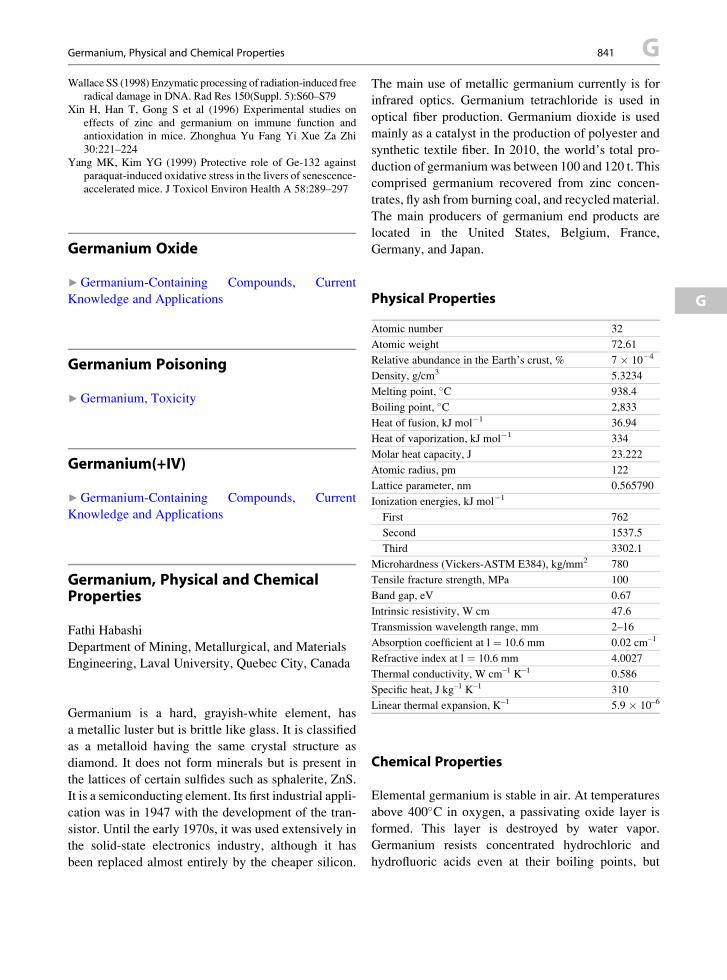
Germanium, Physical and Chemical Properties 841 G
Wallace SS (1998) Enzymatic processing of radiation-induced freeradical damage in DNA. Rad Res 150(Suppl. 5):S60–S79
Xin H, Han T, Gong S et al (1996) Experimental studies on
effects of zinc and germanium on immune function and
antioxidation in mice. Zhonghua Yu Fang Yi Xue Za Zhi
30:221–224
Yang MK, Kim YG (1999) Protective role of Ge-132 against
paraquat-induced oxidative stress in the livers of senescence-
accelerated mice. J Toxicol Environ Health A 58:289–297
G
Germanium Oxide
▶Germanium-Containing Compounds, Current
Knowledge and Applications
Germanium Poisoning
▶Germanium, Toxicity
Germanium(+IV)
▶Germanium-Containing Compounds, Current
Knowledge and Applications
Germanium, Physical and ChemicalProperties
Fathi Habashi
Department of Mining, Metallurgical, and Materials
Engineering, Laval University, Quebec City, Canada
Germanium is a hard, grayish-white element, has
a metallic luster but is brittle like glass. It is classified
as a metalloid having the same crystal structure as
diamond. It does not form minerals but is present in
the lattices of certain sulfides such as sphalerite, ZnS.
It is a semiconducting element. Its first industrial appli-
cation was in 1947 with the development of the tran-
sistor. Until the early 1970s, it was used extensively in
the solid-state electronics industry, although it has
been replaced almost entirely by the cheaper silicon.
The main use of metallic germanium currently is for
infrared optics. Germanium tetrachloride is used in
optical fiber production. Germanium dioxide is used
mainly as a catalyst in the production of polyester and
synthetic textile fiber. In 2010, the world’s total pro-
duction of germaniumwas between 100 and 120 t. This
comprised germanium recovered from zinc concen-
trates, fly ash from burning coal, and recycled material.
The main producers of germanium end products are
located in the United States, Belgium, France,
Germany, and Japan.
Physical Properties
Atomic number
32Atomic weight
72.61Relative abundance in the Earth’s crust, %
7 � 10�4Density, g/cm3
5.3234Melting point, �C
938.4Boiling point, �C
2,833Heat of fusion, kJ mol�1
36.94Heat of vaporization, kJ mol�1
334Molar heat capacity, J
23.222Atomic radius, pm
122Lattice parameter, nm
0.565790Ionization energies, kJ mol�1
First
762Second
1537.5Third
3302.1Microhardness (Vickers-ASTM E384), kg/mm2
780Tensile fracture strength, MPa
100Band gap, eV
0.67Intrinsic resistivity, W cm
47.6Transmission wavelength range, mm
2–16Absorption coefficient at l ¼ 10.6 mm
0.02 cm–1Refractive index at l ¼ 10.6 mm
4.0027Thermal conductivity, W cm–1 K–1
0.586Specific heat, J kg–1 K–1
310Linear thermal expansion, K–1
5.9 � 10–6Chemical Properties
Elemental germanium is stable in air. At temperatures
above 400�C in oxygen, a passivating oxide layer is
formed. This layer is destroyed by water vapor.
Germanium resists concentrated hydrochloric and
hydrofluoric acids even at their boiling points, but

G 842 Germanium, Toxicity
reacts slowly with hot sulfuric acid. It dissolves at
room temperature in nitric acid to form nitrate and in
alkaline media in the presence of an oxidizing agent,
e.g., hydrogen peroxide to form germanates. Germa-
nium and silicon are miscible and form a continuous
range of alloys. The system with tin has an eutectic.
Germanium can have valences of two or four. The
divalent compounds are less stable and oxidize easily
to the tetravalent compounds, which are similar to their
silicon analogues. Germanium reacts readily with hal-
ogens to form tetrahalides. Germanium tetrachloride is
obtained by reaction of chlorine gas with elemental
germanium and of hydrochloric acid with GeO2 or
germanates. Germanium tetrachloride is soluble in
carbon tetrachloride. All the tetrahalides are readily
hydrolyzed to hydrated germanium dioxide.
Germanides can be produced by melting metals
with germanium and freezing the melt, e.g., Mg2Ge.
Germanes (germanium hydrides) are evolved when
germanides react with acids: mono-germane, GeH4,
di-germane, Ge2H6, and tri-germane, Ge3H8, can be
separated by fractional distillation. Mono-germane is
similar in structure to methane is a colorless, highly
inflammable, toxic gas of low stability with
a characteristic unpleasant odor. Polygermanes are
similar to alkanes with formula GenH2n+2 containing
up to five germanium atoms are known. The germanes
are less volatile and less reactive than their
corresponding silicon analogues. The germanium
hydrohalides with one, two, and three halogen atoms
are colorless reactive liquids.
Most germanium compounds are comparatively low
in toxicity because of pharmacological inertness,
diffusibility, and rapid excretion. However, some excep-
tions exist, the most important being germanium
hydrides. Germanium tetrafluoride and tetrachloride
vapors irritate the eyes and mucous membranes of the
respiratory tract. Gaseous germanium hydride is themost
toxic of germanium compoundswhen inhaled. Like other
metal hydrides such as AsH3, it shows hemolytic action
in animals. The lethal concentration in air is 150 ppm.
References
http://en.wikipedia.org/wiki/Germanium
Scoyer J, Wolf H-U (1997) Germanium. In: Habashi F (ed)
Handbook of extractive metallurgy. Wiley, Weinheim,
pp 1505–1524
Germanium, Toxicity
Katsuhiko Yokoi
Department of Human Nutrition, Seitoku University
Graduate School, Matsudo, Chiba, Japan
Synonyms
Germanium poisoning
Definition
Germanium compounds are relatively less toxic
compared to other metalloids and metals. However,
relatively high doses of germanium dioxide and other
inorganic germanium compounds caused severe
poisoning including fatal cases via so-called germa-
nium-containing health foods. Impairments in kidney,
nerves, muscles, and bone marrows are conspicuous.
Milligram order germanium per 1-g tissue was found
from kidney, blood, brain, and other tissues of the
poisoning cases.
Introduction
Germanium is an element with an atomic number of
32, an atomic weight of 72.63, and a valence of +2 or
+4 (Lide 1998). It belongs to a chemical group IVA in
the periodic table that includes C, Si, Ge, Sn, and Pb.
Its chemical properties are similar to arsenic, silicon,
tin, and antimony. Germanium is an important metal-
loid element to make semiconductors, and its oxide is
amphoteric. Elemental germanium is a metalloid that
is slowly oxidized in the air.
Inorganic germanium compounds include germa-
nium dioxide (GeO2), germanium lactate-citrate salt,
germanium tetrachloride and other tetrahalides, and
germanium hydride. Some “dietary supplements”
contain GeO2 or germanium lactate-citrate salt, and
germanate ions (Tao and Bolger 1997). Because of the
amphoteric nature ofGeO2, onceGeO2 is hydrolyzed by
acid or alkaline and neutralized by its counterpart, the
obtained germanate ion is freely soluble in water. The
solubilized GeO2 is colorless, odorless, and tasteless
and impossible to distinguish from plain water by

Germanium, Toxicity 843 G
G
senses. Organic germanium compounds include germa-
nium sesquioxides like bis(2-carboxyethylgermanium)
sesquioxide or bis(b-carboxyethylgermanium)
sesquioxide, usually known as Ge-132. Many organic
germanium compounds were tested for possible
antitumor and immune-stimulating effects and are
sometimes contained in some “dietary supplements.”
According to the 2006 UK Total Diet Study, ger-
manium was detected from only the offal and meat
categories at the concentrations of 2 and 1 mg/kg,respectively. The population dietary exposure is
0.1–1.5 mg Ge/day (UK-FSA 2009).
As per the author’s knowledge, there is no clear
evidence that germanium is an essential element for
life, including microorganisms to higher plants and
animals. However, the consistent occurrence of ger-
manium in coal ash may indicate the absorption of
germanium by prehistoric plants (Furst 1987). Altered
bone and liver mineral composition in rats fed low ger-
manium diet was reported (Uthus and Seaborn 1996).
The question arises of whether germanium may have
a physiological role but is still unanswered.
Absorption, Distribution, Metabolism, andExcretion of Inorganic Germanium
Rosenfeld (Rosenfeld 1954) studied the absorption,
distribution, metabolism, and excretion of inorganic
germanium. When neutralized GeO2 was given to
rats via a stomach tube (50 mg Ge/kg body weight),
96.4% of germanium was absorbed without causing
diarrhea and gastrointestinal tract irritations. A 93.8%
of intramuscularly administered dose of GeO2 suspen-
sion was absorbed within an hour after administration
without any signs of muscular damage. While in the
blood, germanium distributes in plasma and blood
cells. The plasma protein binding was not found for
germanium. Inorganic germanium is considered to be
delivered as free germanate ions in blood stream
(Rosenfeld 1954). Rosenfeld found that under equilib-
rium conditions, the distribution of soluble germa-
nium, i.e., germanate ions between erythrocytes and
surrounding fluid, roughly approximates the relative
water content of the intracellular and extracellular fluid
compartments. Absorbed soluble germanium distrib-
utes throughout the entire bodies, and the highest con-
centration occurs in the kidneys. Intraperitoneally
injected germanate to rats was excreted into urine
(78.8% of the injected dose) and feces (13.0%) within
5 days (Rosenfeld 1954). Kidney is a major excretory
organ for soluble inorganic germanium or germanate
and an important target organ for inorganic germanium
toxicity.
From the above conspicuous metabolic behavior,
Rosenfeld stated that with respect to the type of
distribution, soluble inorganic germanium compounds
resemble substances like alcohol and urea, rather than
inorganic bromides and iodides, which do not pene-
trate cells, or arsenicals, which are selectively fixed by
them. From the dilute aqueous solution, at least three
germanate ions, i.e., [GeO(OH)3]�, [GeO2(OH)]
2�,and {[Ge(OH)4]8(OH)3}
3�, have been identified. One
form of germanium hydroxide GeO(OH)2 is planar and
electronically analogous to H2CO3 (Qin et al. 2006).
This chemical resemblance to carbonic acid may
explain the metabolic behavior of neutralized GeO2.
Recently, dermal absorption of inorganic germa-
nium was investigated using rats (Yokoi et al. 2008).
Hydrophilic ointment containing neutralized GeO2
(0, 0.21 and 0.42 mg/g) spread on a 2 cm � 2 cm
polyvinylidene chloride sheet was topically applied
to rat skin. When the topical dose was doubled, the
germanium concentrations in plasma and kidney were
increasedmore than double. The excessive rise of renal
germanium concentration indicates a breakdown of
excretion system of inorganic germanium.
Toxicity of Germanium Dioxide andGermanate
Acute oral toxicity of inorganic germanium is rela-
tively low. The reported median lethal doses (LD50)
of germanium compounds in rodents are summarized
in Table 1. When Rosenfeld and Wallace’s acute
toxicity data of germanium in rats (Table 1 of their
article) was read (Rosenfeld and Wallace 1953), LD50
by intraperitoneal injection of neutralized GeO2, i.e.,
sodium germanate, was estimated to be 750 mg Ge/kg
body weight. Germanic acid, like boric acid, forms
complex with polyvalent alcohol such as mannitol
and glycerin. It is evident that germanate is more
toxic than germanium–sugar alcohol complexes such
as Ge–mannitol, or CMC suspension of GeO2. Hypo-
thermia was conspicuous in inorganic germanium
intoxication of experimental animals in acute and sub-
acute studies (Rosenfeld 1954).
Germanium, Toxicity, Table 1 LD50 for germanium compounds (mg compounds/kg) in rodents
Compounds Medium Species Route Male Female
Germanium dioxide 1–10% mannitol (pH 5.4–5.6) Mouse p.o. 6,300 5,400
Rat i.p. 2,250 2,025
s.c. 2,750 2,550
p.o. 3,700 3,700
i.p. 1,800 1,620
s.c. 2,025 1,910
Germanium dioxide 0.5% CMC suspension Mouse p.o. >10,000 –
i.p. 1,550
Neutralized germanium dioxide Solubilized by sodium hydroxide and neutralized by
hydrochloric acid (pH 7.3)
Rat i.p. 750 –
Triethylgermanium acetate Arachis oil Rat p.o. 125–250 –
tri-n-Butylgermanium Arachis oil Rat p.o. > 375 –
bis-b-Carboxyethylgermanium
sesquioxide
0.5% CMC suspension Mouse p.o. 12,500 11,400
Rat i.p. 2,110 2,230
s.c. 7,550 8,050
p.o. 11,700 11,000
i.p. 3,500 3,200
bis-b-Carboxyethylgermanium
sesquioxide
PH 7 solution (neutralized by sodium bicarbonate) Mouse p.o. 11,500 11,700
Rat i.v. 5,720 5,720
s.c. 10,800 12,900
p.o. 10,600 9,500
i.v. 4,800 4,380
s.c. 16,300 17,800
b-Carboxyethylgermanium
sesquioxide
0.5% CMC suspension Mouse p.o. >10,000 –
i.p. 2,820
b-Carbamoylethyl germanium
sesquioxide
0.5% CMC suspension Mouse p.o. >10,000 –
i.p. 7,100
Propagermanium 5% Gum arabic suspension Mouse p.o. 5,600 5,800
Rat i.p. 1,250 1,300
s.c. >4,000 >4,000
p.o. 7,700 7,050
i.p. 1,750 1,670
s.c. >4,000 >4,000
Propagermanium Saline Mouse i.v. >200 >200
Rat i.v. >200 >200
Propagermanium 10% Sodium bicarbonate Mouse i.v. >3,000 >3,000
Rat i.v. >3,000 >3,000
Rosenfeld and Wallace (1953); Cremer and Aldridge (1964); Hatano et al. (1981); Kanda et al. (1990).
G 844 Germanium, Toxicity
In spite of the relatively low acute oral toxicity,
human toxicity of inorganic germanium, especially
GeO2 or sodium germanate-containing so-called
health foods, is well known because of many poisoning
cases. Germanium has been expected as an alternative
drug for cancer, immune impairments, viral infections,
and health promotion. GeO2 powder or solubilized and
neutralized GeO2 solution is added to some so-called
health foods and drinks as an elixir of health. Tao and
Bolger summarized reported cases of germanium poi-
soning and found that nine persons died of germanium
poisoning out of 31 reported cases (Tao and Bolger
1997). Most of cases of germanium poisoning were
reported from Japan. They were caused by inorganic

Germanium, Toxicity 845 G
G
germanium (unidentified forms), GeO2, or unidentified
germanium compounds.
Major signs and symptoms of GeO2 or germanate
intoxication in Japanese cases were renal damage,
anemia, myopathy, peripheral neuropathy, weight
loss, fatigue, and gastrointestinal symptoms
(nausea, vomiting, and anorexia). Renal failure was
characterized by tubuloepithelial degeneration with
swollen mitochondria containing electron-dense gran-
ules. The similar electron-dense deposits were also
found from mitochondria of hepatocytes and myocytes
of germanium poisoning patients. Urinalysis was usu-
ally normal (absence of proteinuria and hematuria).
Extrarenal organs involved were muscles (both skeletal
and cardiac), nerves (both peripheral and central), bone
marrow (characterized by hypoplasia), and liver (char-
acterized by liver dysfunction with severe steatosis)
(Schauss 1991; Tao and Bolger 1997). Milligram order
germaniumper 1-g tissuewas found fromkidney, blood,
brain, and other tissues of the poisoning cases.
Four European cases were poisoning due to germa-
nium lactate-citrate. The renal pathology of European
cases was similar to the Japanese cases of GeO2 intox-
ication. Different from the Japanese cases, hepatic
involvements were conspicuous in the European
cases. The typical form of the toxicity was lactic
acidosis that was not seen in the Japanese cases.
Whether there were any cases of genuine organic
germanium poisoning by so-called health foods is
uncertain because GeO2 was detected from the
ingested preparations that were reported to be organic
germanium once (Tao and Bolger 1997). Sometimes,
inorganic germanium was erroneously designated as
“organogermanium” or “organic germanium”
(Schauss 1991; Tao and Bolger 1997).
Human poisoning due to GeO2 was reproduced in
experimental animals (Tao and Bolger 1997). Indepen-
dent researchers demonstrated the characteristic renal
dysfunction with remarkable histological changes in
kidneys, i.e., tubuloepithelial degeneration with mito-
chondrial electron-dense deposits after subchronic and
chronic doses of GeO2. The mitochondrial electron-
dense deposits were revealed to contain germanium,
iron, and sulfur by electron energy-dispersive X-ray
microanalysis (Sanai et al. 1991). The mitochondrial
degeneration due to accumulated germanium
deposits that associates iron and sulfur could partly
explain the germanium-induced tissue damage.
Germanium-induced myopathy, associating mitochon-
drial electron-dense inclusions in degenerated
myocytes, was also reproduced in experimental rats
orally administered GeO2. Germanium-induced neu-
ropathy characterized by segmental demyelination and
axonal degeneration was also demonstrated in rats
orally given GeO2.
Tumorigenicity and carcinogenicity were not
shown by lifetime low-dose exposure to 5 mg Ge/L
in drinking water as sodium germanate (Tao and Bol-
ger 1997). Kamildzhanov et al. found decreased sper-
matozoa motility in rats exposed to GeO2 by inhalation
(Kamildzhanov et al. 1986). They also reported the
synergistic toxicity of GeO2 and arsenous anhydride
(i.e., arsenic trioxide) in an inhalation study.
Toxicity of Gaseous Germanium Compounds
Gaseous germanium compounds are synthetic and do
not occur naturally. Germanium hydride is a gas at the
ambient temperature and the boiling point of germa-
nium tetrachloride is 86.55 �C (Lide 1998). Exposure
to gaseous germanium compounds occurs in the work
place. Germanium hydride exposure causes neurolog-
ical abnormalities and lung damage, while germanium
tetrachloride can irritate skin (Furst 1987).
Toxicity of Organic Germanium Compounds
Organic germanium is defined as the compounds that
contain covalent bonds between carbon and germa-
nium. Organic acid salts of germanium such as germa-
nium lactate-citrate are not included in organic
germanium compounds. Toxicological data are limited
for organic germanium. The LD50s for selected organic
germanium compounds are also listed in Table 1.
The Japanese Ministry of Health, Labour and Wel-
fare has approved propagermanium or
proxigermanium, a hydrophilic polymer of 3-
oxygermyl propionates, as a drug for the treatment of
chronic B hepatitis in 1994. Since 1994 to the
reevaluation in 2003, several adverse effects of
propagermanium (Serocion®) were reported.
According to Hirayama, since 1994, moderate to
severe liver damage has been reported in about 4% of
32,700 recipients after propagermanium treatments.

G 846 Germanium, Toxicity
Among them, six patients died of acute liver failure,
four with severe chronic hepatitis, one with flare of
chronic hepatitis, and one with chronic hepatitis with
hepatocellular carcinoma (Hirayama et al. 2003).
Because of these severe adverse effects, the Japanese
Ministry warned possible acute exacerbation of hepa-
titis and fatal cases in the course of propagermanium
treatment of chronic B hepatitis.
The LD50s of propagermanium in rodents are shown
in Table 1. In beagle dogs, no significant changes were
observed with intravenous administration of
125–500 mg propagermanium/kg/day for 6 months.
An azaspiran-germanium compound (2-aza-8-
germanspiro[4,5]-decane-2-propamine-8,8-diethyl-N,
N-dimethyl dichloride) (NSC 192965; spirogermanium)
has cytotoxic effects and tested chemotherapeutic
effects in phase I and phase II studies that were incon-
clusive. During the clinical trials, neurotoxic adverse
effects, including dizziness, vertigo, nystagmus, ataxia,
and paresthesia and pulmonary toxicity were observed
(Schauss 1991).
In contrast to GeO2 (52 mg Ge/kg body weight/day)
that induced severe renal toxicity, the similar dose of
bis-b-carboxyethylgermanium sesquioxide as an ele-
ment (51 mg Ge/kg body weight/day) did not cause
significant abnormalities in kidney and blood biochem-
istry or tissue germanium accumulation after a 24-week
oral administration of the preparation mixed into the
powdered rat chow (Sanai et al. 1991). A French
group found moderate renal dysfunction of bis-b-carboxyethylgermanium sesquioxide by subchronic
study (6 months) in rats with a sixfold oral dose
(306 mg Ge/kg body weight/day) (Anger et al. 1992).
Cross-References
▶Arsenic
▶Cadmium and Health Risks
▶Germanium L-Cysteine Alpha-Tocopherol Complex
as Stimulator to Antioxidant Defense System
▶Germanium, Physical and Chemical Properties
▶Lead Nephrotoxicity
▶Mercury Nephrotoxicity
▶Mercury Neurotoxicity
▶Rubredoxin, Interaction with Germanium
▶ Silver, Neurotoxicity
References
Anger F, Anger JP, Guillou L, Papillon A, Janson C, Sublet Y
(1992) Subchronic oral toxicity (six months) of carboxyethyl-
germanium sesquioxide [(HOOCCH2CH2Ge)2O3]n in rats.
Appl Organomet Chem 6:267–272
Cremer JE, Aldridge WN (1964) Toxicological and biochemical
studies on some trialkylgermanium compounds. Br J Ind
Med 21(3):214–217
Furst A (1987) Biological testing of germanium. Toxicol Ind
Health 3:167–204
Hatano M, Ishimura K, Fuchigami K, Ito I, Hongo Y, Hosokawa
Y, Azuma I (1981) Toxicological studies on germanium
dioxide (GeO2) (1). Acute, subacute, chronic toxicity and
successive irritation to the eye. Oyo Yakuri 21:773–796 (in
Japanese)
Hirayama C, Suzuki H, Ito M et al (2003) Propagermanium:
a nonspecific immune modulator for chronic hepatitis B.
J Gastroenterol 38:525–532
Kamildzhanov A, Ubaidullaev RU, Akbarov AA (1986) Com-
bined effects of arsenous anhydride and germanium dioxide
after their hygienic regulation in atmospheric air. Gig Sanit
8:82–84 (in Russian)
Kanda K, Igawa E, Shinoda M, Asaeda N, Yoshiyasu T, Iwai H,
Nagai N, Tamano S, Ichikawa K, Koide M (1990) Acute
toxicity test of proxigermanium (SK-818) in mice and rats.
Kiso To Rinsho 24:5–18 (in Japanese)
Lide DR (1998) CRC handbook of chemistry and physics,
79th edn. CRC Press, New York
Qin C, Gao L, Wang E (2006) Germanium: inorganic
chemistry. In: King RB (ed) Encyclopedia of inorganic
chemistry. Wiley, Hoboken, NJ. doi:10.1002/
0470862106.ia079
Rosenfeld G (1954) Studies of metabolism of germanium. Arch
Biochem Biophys 48:84–94
Rosenfeld G,Wallace EJ (1953) Studies of the acute and chronic
toxicity of germanium. AMA Arch Ind Hyg Occup Med
8:466–479
Sanai T, Okuda S, Onoyama K et al (1991) Chronic tubuloin-
terstitial changes induced by germanium dioxide in
comparison with carboxyethylgermanium sesquioxide. Kid-
ney Int 40:882–890
Schauss AG (1991) Nephrotoxicity and neurotoxicity in humans
from organogermanium compounds and germanium dioxide.
Biol Trace Elem Res 29:267–280
Tao SH, Bolger PM (1997) Hazard assessment of germanium
supplements. Regul Toxicol Pharmacol 25:211–219
UK-FSA (2009) Measurement of the concentrations of metals
and other elements from the 2006 UK total diet study.
FSA, London. http://www.food.gov.uk/multimedia/pdfs/
fsis0109metals.pdf
Uthus EO, Seaborn CD (1996) Deliberations and evaluations of
the approaches, endpoints and paradigms for dietary recom-
mendations of the other trace elements. J Nutr
126:2452S–2459S
Yokoi K, Kawaai T, Konomi A et al (2008) Dermal absorption of
inorganic germanium in rats. Regul Toxicol Pharmacol
52:169–173
Germanium-Containing Compounds, Current Knowledge and Applications 847 G
Germanium-Containing Compounds,Current Knowledge and Applications
Erwin Rosenberg
Institute of Chemical Technologies and Analytics,
Vienna University of Technology, Vienna, Austria
G
Synonyms
Carboxyethyl germanium sesquioxide; Ge-132;
Germanium oxide; Germanium(+IV); Germanium;
Organogermanium compounds; Propagermanium;
Spirogermanium
Definition
Germanium (Ge) is the element with atomic number
32. Being a member of the group 14 of the periodic
table, it stands in many of its physical and chemical
properties between the elements silicon and tin. While
the element has important applications in the electron-
ics, semiconductor, and optics industries, the organic
compounds of germanium have been investigated as
anticancer, anti-inflammatory, and immunosti-
mulating drugs.
General Information and Historical Outline
Germanium is a main group IV (group 14) element that
has atomic number 32 and relative atomic mass 72.61.
Its specific gravity is 5.323 g/cm3, melting point
937.4�C (1210.6 K), and boiling point 2,830�C(3103.2 K). Ge has five stable isotopes
(70Ge, 20.45%; 72Ge, 27.41%; 73Ge, 7.77%; 74Ge,
36.58%; 76Ge, 7.79%), and 13 further radioactive iso-
topes of mass numbers 65–69, 71, 75, 77, and 78 whose
half-lives range between 20 ms and 287 days.
Germanium is a brittle, relatively hard element
(Mohs hardness of 6.0) and has a refractive index of
4.0. With its good transparency in the mid-infrared
range and its low dispersion, it is highly suitable for
the construction of infrared optics (windows, lenses),
which, particularly when designed for rugged field use,
benefit from its surface hardness and mechanical
strength. The principal region of transparency
extends throughout the entire mid-IR range from
around 550–5,500 cm�1, corresponding to approxi-
mately 1.8–18 mm. The short wavelength cutoff corre-
sponds to an energy gap of 0.68 eV at 300 K to 0.83 eV
at 50 K, respectively, and a transmission level of
approximately 47% (Scoyer et al. 2000).
Ge is a relatively inert semimetal and is stable in air
at ambient temperature. It is oxidized only at temper-
atures above approximately 1,000 K to form GeO2.
The element is insoluble in water, hydrochloric acid,
and dilute alkali, but can readily be dissolved in the
oxidizing media, such as nitric or sulfuric acid, and by
basic peroxides, nitrates, and carbonates. According to
the position in the periodic table of elements, Ge
occurs in addition to its elemental form in the oxidation
states + II and + IV, where the latter possesses the
higher stability and is the one in which Ge compounds
typically occur in the environment. In minerals, Ge
often appears in the form of the oxide (GeO2) or the
sulfide (GeS2), and in solution as Germanic acid,
Ge(OH)4. Divalent compounds of germanium are
mainly produced synthetically and have importance
only in the chemical industries.
The concentration of Ge related to the entire earth
mass is estimated at 7.3 ppm (w/w); in the surface layer
of the earth, it is estimated at 5.6 ppm (w/w), in the
continental earth crust 1.6 ppm (w/w), and in the oceans
5.10�5 mg/L. Related to the mass of the entire earth, it
ranks 22nd in relative abundance, while based on the
mass of the surface layer of the earth it is 46th and based
on its concentration in the continental earth crust it is
only 55th (Rosenberg 2009). Germanium is thus not
a very rare element, but it is found in nature only in
widely dispersed form. There are but fewminerals which
contain Ge at significant concentrations, the best known
being mixed hydroxides, such as Stottite, FeGe(OH)6,
or Schauerteite, Ca3Ge(SO4)2(OH)6.3H2O, and mixed
sulfides, such as Germanite, Cu3(Ge, Fe)S4, and
Argyrodite, 4Ag2S.GeS2, the latter being the form that
led to the discovery of this element by Winkler in 1885
(Winkler 1886). None of these minerals occurs in
amounts that would justify its industrial mining. Eco-
nomically more feasible is the production of Ge from
silver, zinc, and copper refineries where Ge can be
recovered as a by-product. Technical production usually
G 848 Germanium-Containing Compounds, Current Knowledge and Applications
involves a pre-concentration process in which the con-
centrate from the Zn production, containing only about
0.02% Ge is enriched to a content of 6–8%. The second
step of the production of Ge relies on the volatility of
GeCl4 which is formed from GeO2 in the presence of
HCl and Cl2. GeCl4 can thus be separated from the
concentrate solution by distillation, is then hydrolyzed
again to (pure) GeO2, and then further reduced to ele-
mental Ge (Scoyer et al. 2000).From the organic compounds of the elements of
group 14 of the periodic table of elements,
organogermanium compounds were the least investi-
gated among its homologues up to the middle of
the twentieth century. This is all the more surprising
since the first organogermanium compound,
tetraethylgermane, was synthesized as early as 1887
by Winkler by the reaction of tetrachlorogermane and
diethylzinc which was only about a quarter century
later than the first organic compounds of silicon, tin
and lead were obtained.
The synthesis of Et4Ge was an additional and
significant proof of the assumption that the germanium
discovered by Winkler belongs to Group IV of the
Periodic Table and that it was identical to Mendeleev’s
eca-silicon.
During the period between 1887 and 1925, no
new organogermanium compounds were reported.
These 40 years of apparent disinterest resulted mainly
from the very limited availability and the high prices of
germanium and its simplest inorganic derivatives. This
reflected the low natural reserves of Argyrodite, the
only mineral source of germanium known at that time.
With the discovery of new sources of germanium in
1922, this picture changed dramatically. In particular,
0.1–0.2% of Ge was found in a residue of American
zinc ore after zinc removal (Clark 1951). This was
followed by the discovery of two germanium-rich
minerals: Germanite (in 1924), a mineral from South-
western Africa which contained about 5.1% Ge, and
Rhenierite, a mineral from the Belgian Congo,
containing 6–8% of Ge, which became another source
of germanium. In the following decade, the 1930s,
processing wastes of coal ashes and sulfide ores
became the main sources of germanium (Scoyer et al.
2000). The gradually improving availability of Ge
allowed American, English, and German chemists to
start fundamental investigations of organogermanium
compounds from the mid-1920s, although germanium
was still very expensive at that time.
Organogermanium chemistry was stimulated by the
rapid development of the chemistry of other organo-
metallic compounds, in particular of the elements
silicon and tin. A further motivation was the significant
role that this element and its organic derivatives were
starting to have in the electronics industry
(Haller 2006). Also, first biological effects of
organogermanium compounds (including anticancer,
hypotensive, immunomodulating, and other kinds of
physiological action) were described (Lukevics and
Ignatovich 2002). In addition, the eventual decrease
in the prices of elemental germanium and its
derivatives expanded their production and helped
their growth.
The rapid expansion of organogermanium chemis-
try is clearly illustrated by the continuous increase in
the number of compounds described in literature: In
1951, 230 organogermanium compounds were known;
in 1961, there were 260; and in 1963, there were more
than 700 (Voronkov and Abzaeva 2002).
Current Use of Germanium
Ge has a number of technical uses which make it,
despite the relatively low annual production of approx-
imately 60 t/year, an element of strategic importance.
For this reason the United States of America’s National
Defense Stockpile held up to the late 1990s tens of tons
of this semi-metal in order to not be dependent on the
international production and availability of this tech-
nologically and strategically important element. Key
germanium applications include infrared optics, night
vision instruments, optical fibers, catalyst in
PET (polyethylene-terephthalate) production, and
semiconductor and photovoltaic cell industries
(Rosenberg 2009).
The use of Ge for the production of
organogermanium compounds is only of minor impor-
tance in comparison to its other technical uses. Most
organogermanium compounds are tetravalent. Being
an element of Main Group IV (or Group 14 according
to the new IUPAC numbering) of the periodic table of
the elements, both + II and + IV oxidation states are
possible, although the higher oxidation state is more
stable. The electronegativity of Ge is 2.01 according to
Pauling. It is thus closer to carbon (EN: 2.50) and hydro-
gen (EN: 2.20) than the electronegativity of the other
elements of the same group (silicon: 1.74, tin: 1.72,

Germanium-Containing Compounds, Current Knowledge and Applications 849 G
and lead: 1.55). Consequently, both germanium-carbonand germanium-hydrogen bonds can be considered as
almost apolar when compared to the bonds of
Si�H (DEN 0.46), Sn�H (DEN 0.48), and
Pb�H (DEN 0.65). Being practically apolar in the
GeH4 molecule, the polarity of the Ge�H bond depends
mainly on the substitution of Ge with electron providing
or withdrawing groups, such as alkyl groups R (weakly
electron-pushing) or halogens X (strongly electron-
withdrawing), resulting in positively or negatively polar-
ized Ge:
G
dþ d� d� dþR3Ge� H X3Ge� H
This fact is important as it explains the reactivity of
organogermanium compounds.
In order to form organogermanium compounds, three
generic routes exist (Voronkov and Abzaeva 2002):
(a) Direct reaction of Ge with alkyl halogenides using
a copper catalyst. This reaction proceeds with very
good yields; however, statistical mixtures of the
various alkylgermanium halides are produced,
with the dialkylated R2GeX2 typically being the
favored product:
n RXþ Ge ! RnGeX4�n (1)
(R¼ alky or aryl group; X¼ halogen; n¼ 1, . . ., 4)
(b) Reaction of germanium halide with an organome-tallic compound M � R (a form of the Wurtz
synthesis, a typical metathesis reaction):
Ge�XþM�R !Ge�RþM�X (2)
(R ¼ alky or aryl group; M ¼ Li, Na, K, Mg, Zn,
Hg, Al; X ¼ halogen)
This reaction proceeds particularly well with
Grignard reagents (compounds of the general
form RMgX, with R being an alkyl or aryl moiety,
and X a halogen) and can be used for the synthesis
of tetrasubstituted organogermanium compounds.
As an example, GeCl4 can be reacted with the
methyl-Grignard MeMgI to give Me4Ge with
93% yield.
(c) The hydrogermylation reaction. In this synthetic
route, organogermanium compounds are prepared
by forming trihalogen germanium hydride in a first
step, followed by the addition of this compound to
unsaturated organic compounds:
Geþ 3HCl ! HGeCl3 þ H2 (3)
HGeCl3 þ CH2 ¼ CH� R
! Cl3GeCH2CH2�R(4)
This latter reaction is of particular importance,
since it is the most widely employed route for the
synthesis of the pharmaceutically relevant
germanium sesquioxide (also called bis-
carboxyethylgermanium sesquioxide or Ge-132).
Organogermanium Compounds withTherapeutic Use
Germanium sesquioxide (Scheme 1), IUPAC
systematic name: 3-[(2-Carboxyethyl-oxogermyl)
oxy-oxogermyl]propanoic acid, CAS-No. 12758-40-
6, also known under the synonyms propagermanium,
proxigermanium, Ge-132, 2-carboxyethylgermases-
quioxane, SK-818, or bis-(2-carboxyethylgermanium)
sesquioxide, is the best known organogermanium drug.
Its therapeutic uses were first described by Asai and
coworkers of the Asai Germanium Research Institute
(Tokyo, Japan) in 1968 who had screened a large num-
ber of germanium compounds for their biological
effects. The drug was patented in Japan in 1971 and
in the USA in 1972, after which date Asai and
coworkers strongly promoted the use of this compound
in the treatment of various diseases. Ge sesquioxide
has an unusual planar structure in which each Ge atom
binds to the three surrounding oxygen atoms, forming
a 12-membered ring. The carboxylate chains are
arranged alternately above and below the plane defined
by the germanium and oxygen atoms. This particular
three-dimensional structure explains why Ge
sesquioxide is a crystalline substance which does not
dissolve in organic solvents (except ethylene glycol),
but is soluble inwater upon heating; and alsowhy it does
not melt nor decompose at temperatures below 320�C.Ge sesquioxide has been claimed to be a potent
antioxidant and a useful drug in anticancer and
antiviral treatments. This was based on reports that
claimed the induction of interferon g (IFN-g) and the
induction of immunological responses such as

O Ge
O
O
Ge
O
O OH
HOn
Germanium-Containing Compounds, Current Knowledgeand Applications, Scheme 1 Structure of germanium
sesquioxide (¼bis-(2-carboxyethylgermanium) sesquioxide,
Ge-132)
G 850 Germanium-Containing Compounds, Current Knowledge and Applications
increased natural killer (NK) cell, macrophage and
T-cell production after oral administration of
Ge sesquioxide (Kaplan et al. 2004).
Ge-132, as well as spirogermanium (¼ 8,8-diethyl-
2-[3-(N-dimethylamino)-propyl]-2-aza-8-germaspiro
[4.5]decane) and other related synthetic derivatives
have demonstrated significant antitumor activity
in vitro in cancer cell lines and in vivo in animal
models of some cancers.
Their antitumor activities have been established
against various experimental tumors, such as Ehrlich
ascites tumor and Lewis lung carcinoma. In clinical
studies, limited value was revealed for antitumor
germanium complexes.
The exact anticancer mechanism still remains
relatively unclear, though induction of IFN-g may be
the key. Ge-132 may also possess DNA binding
specificity like cisplatin, to inhibit cancer cell prolifer-
ation. A similar mode of action is reported for some
Ge-132 derivatives (Shangguan et al. 2005).
Several cases of Ge-related toxicity have been
reported; however, these were linked to large amounts
of Ge (15–426 g) being ingested over prolonged
periods up to 36 months. Studies have narrowed
acute and chronic Ge-related toxicity specifically to
inorganic germanium dioxide (GeO2) and metallic Ge
which can accumulate in the liver, kidney and spleen,
peripheral nerves, lungs and muscle. Since Ge-132 is
also synthesized from GeO2, it is possible that residual
GeO2 can contaminate formulations claiming to be
pure organogermanium. This most likely was the
case in the few documented cases of anticancer treat-
ment with Ge-132 with fatal consequences attributed
to Ge intake (Tao and Bolger 1997).
In contrast to inorganic germanium, organogermanium
compounds including Ge-132 have characteristic low
toxicity and are readily excreted via the kidney with
very low accumulation in major organs and tissues.
There are but few clinical trials reported in the
literature which have assessed the efficacy in the treat-
ment of the various forms of cancer. It appears that
most of these studies are either inadequately
documented, or they have been conducted under insuf-
ficiently controlled conditions, particularly as concerns
the dose and the purity of the administered drug. It is
thus hardly possible to arrive to a firm and experimen-
tally underpinned conclusion on the efficiency of
Ge-132 in anticancer treatment, and on the potential
toxicity of Ge-132 formulations (Horneber and
Wolf 2011).
Along this line, a review from 1997 reported on 31
cases of organ toxicity related to different germanium
compounds, including GeO2, germanium-lactate-
citrate, and Ge-132, which were ingested over
a period from 2 to 36 months. In the two cases of
Ge-132 intake, contamination with GeO2 was
detected. All cases showed renal impairment in terms
of chronic or acute renal failure, which caused death in
nine cases. Other clinical findings were anemia, which
occurred in all cases, gastrointestinal disturbances,
weight loss, myopathy, and liver dysfunction. If at
all, renal function improved very slowly and remained
impaired in some patients observed as long as
40 months. As a consequence, governmental institu-
tions of several countries imposed alerts because of
possible injury to health (Tao and Bolger 1997).
In most countries, Ge-132 falls under the regula-
tions of dietary supplements. An import alert on ger-
manium products was imposed by the U.S. FDA in
1988, because of possible health hazard. In Germany,
governmental institutions warned consumers of possi-
bly fatal kidney damage. In the UK, supplements
containing germanium were voluntarily withdrawn
by the industry. In Japan, Ge-132 is approved for the
treatment of chronic hepatitis B.
Spirogermanium. Spirogermaniumwas first synthe-
sized in 1974 and represents an azaspiran-germanium
compound which was tested in various phase I/II trials
to examine its anti-tumor effect (Scheme 2). Due to
a markedly negative risk-benefit profile, in particular

GeN N
.2 HCl
Germanium-Containing Compounds, Current Knowledgeand Applications, Scheme 2 Structure of spirogermanium
(¼N-(3-dimethylaminopropyl)-2-aza-8,8-diethyl-8-germaspiro
[4.5]decane)
Ge Y
O
Z
Z
Ge
Z
O
Y
CC
C
CC
C
R2 R4
R2
R4
R3R1
R1 R3
Germanium-Containing Compounds, Current Knowledgeand Applications, Scheme 3 R1–R4 ¼ H, alkyl, aryl; Z ¼ O,
S; Y¼OH, OR, OM, NH2, NRR0. Ge-132 (bis-(2-carboxyethyl-
germanium) sesquioxide) is a particular representative of this
compound class with R1–R4 ¼ H; Y ¼ OH; and Z ¼ O
Germanium-Containing Compounds, Current Knowledge and Applications 851 G
G
neurologic toxicity, spirogermanium was abandoned
for therapeutic use.
Spirogermanium inhibits DNA, RNA, and protein
synthesis in vitro and decreases cell survival after
exposure to as little as 1.0 mg/mL for 24 h. Quiescent
cells seem more resistant. At higher concentrations,
cytolysis is observed (Gerber and Leonard 1997).
Z = CH2CH2COOH
SGe
Z
S
S
Ge GeS
S SGe
Z Z
Z
Germanium-Containing Compounds, Current Knowledgeand Applications, Scheme 5 Structure of the
organogermanium sesquisulfur
R = 3,5-di-t-butylphenyl
N
N
NN Ge
CH3
CH3
R R
R R
Germanium-Containing Compounds, Current Knowledgeand Applications, Scheme 4 Structure of the porphyrine
dimethylgermanium complex
Other Organogermanium Compounds
In addition to the two best studied organogermanium
drugs, a number of other organic germanium deriva-
tives have been tested in anticancer protocols. Many of
these studies were led by the Asai Germanium
Research Institute in Japan. Starting from the
sesquioxide of 2-carboxyethylgermanium, derivatives
with similar structures have also been studied as
antitumor drugs but also as antibacterial agents (see
Scheme 3).
Another germanium compound shown to have an
anticancer activity for rats and mice is a porphyrine
dimethylgermanium complex (Scheme 4).
The biological activity of trialkyl germanium on
fungi, yeasts, and bacteria was also studied. It was
found that some organogermanium compounds present
a rather strong activity against Streptococcus lactis.Similarly, Me3GeCH2CH2NHCH2Ph has shown
a high antibacterial activity with a wide spectrum of
targets. Also the organogermanium sesquisulfur has an
excellent antibacterial activity (see Scheme 5).
Other germanium compounds that have been shown
to exhibit biological effects are the germatranes and
trithiagermatranes (showing psychotropic and
antitumor activities, Scheme 6) and germathiolactones
which are active as biological antioxidants (Scheme 7).
(Et3Ge)2S is an extremely strong fungicide, with an
efficiency that exceeds many of the best fungicides on
the market.

XGe
R
X
X N
X = O or S
Germanium-ContainingCompounds, CurrentKnowledge andApplications,Scheme 6 Structure of the
germatranes and
trithiagermatranes
R1, R2, R3 = H, alkyl group
S
R2Ge
R3
O
R2
R1
Germanium-ContainingCompounds, CurrentKnowledge andApplications,Scheme 7 Structure of the
germathiolactones
R = CH3: valium
R = Ge(CH3)3: trimethylgermanium
analogue of valium
Cl
O
R
N
N
C6H5
Germanium-Containing Compounds, Current Knowledgeand Applications, Scheme 8 Structure of valium and its
trimethylgermanium analogue
G 852 Germanium-Containing Compounds, Current Knowledge and Applications
In the valium molecule, substitution of the methyl
group connected to the nitrogen atom by
a trimethylgermyl group leads to a nontoxic
organogermylated valium whose psycholeptic activity
is higher than that for valium (Scheme 8).
Numerous organic germanium derivatives have
shown an important radioprotective activity. Many of
the compounds that were studied for this purpose were
organogermanium compounds containing sulfur, such as
germathiazolidines, germadithioacetals, germatranes,
and germanium sulfides. More than one hundred of
these derivatives have been tested and characterized by
their dose reduction factor (DRF). The DRF is the ratio
between the LD50/30 days of treated mice and the
LD50/30 days of non-treated mice. With irradiations in
the range 7.5–8.5 Gy (grays), DRFs in the range 1.2–1.7
were obtained.
These organometallic derivatives usually have
a lower toxicity and a radioprotective activity greater
than that for the corresponding organic derivatives,
namely, cysteamine, methylcysteamine, and
N-substituted cysteamine.
It should be emphasized that this strong radiopro-
tective activity was obtained in many cases with
organogermylated derivatives injected in lower doses
than typically required for the corresponding organic
compounds. The organogermylated derivatives seem
to exhibit this pronounced radioprotective activity due
to their amphiphilic properties, where the presence of
organometallic groups improves the aqueous solubil-
ity, while the presence of organic ligands increases the
lipophilic solubility, thereby favoring their transfer
through the cellular membranes (Satge 2004).
Biological Effects of Germanium Compounds
Mutagenicity of Germanium. Dibutylgermanium
dichloride exhibited an LC50 of about 600 mg/ml
(2.33 mM) in a mutation assay on hypoxanthine gua-
nine phosphoribosyl transferase (HGPRT) carried out
on Chinese hamster ovarian (CHO) cells; the incidence
of mutants increased with dose up to 400 mg/ml
(1.55 mM). It is assumed that this electrophilic com-
pound acts by binding to DNA bases, thereby forming
penta- and hexa-coordination compounds. Also the
immunosuppressive potential of this compound in
lymphocytes was evaluated on the basis of cytotoxicity
and impairment of antibody formation. No toxic action
was observed in lymphocytes at concentrations up to
64 mg/ml (0.25 mM), but the number of antibody-
producing cells was reduced to one half of the control.
Ge-132 is a potent antimutagen when added to agar
at concentrations of 5–30 mg/ml agar, markedly reduc-
ing the number of revertant mutants in B/r WP2
Trp � Escherichia coli which are induced by gamma
irradiation at a dose of 10 kRad (�100 Gy).
Germanium oxide (GeO2) was found to have
a dose-dependent antimutagenic activity for Trp-P-2
frameshift reverse mutations induced by 3-amino-1-
methyl-5H-pyrido-[3,4-b]indole in Salmonellatyphimurium strains TA 98 and TA 1538. Doses of

Germanium-Containing Compounds, Current Knowledge and Applications 853 G
G
600 mg/plate of germanium dioxide reduced the muta-
genic potential of benzo[a]pyrene in three of four
S. typhimurium strains tested.
Germanium dioxide (GeO2) was also reported to
exhibit an antimutagenic effect after administration
of cadmium chloride (CdCl2) to laboratory mice.
Administration of 0.05 mg/kg germanium oxide sig-
nificantly reduced the action of 1.35 mg cadmium
chloride in the DNA synthesis inhibition test when
the CdCl2 was injected 1 h later. However, at other
concentration ratios of the two compounds, a less pro-
nounced antagonistic effect was observed. Germanium
dioxide at doses of 0.1 or 0.5 mg/ml/kg decreased
markedly the number of micronuclei or chromosome
aberrations induced by 0.7, 1.4, or 2.7 mg/kg of CdCl2in vivo in mouse bone marrow cells. Germanium diox-
ide also protected in vivo against sperm head changes
in morphology and in vitro from the sister chromatid
exchanges (SCEs) in human peripheral blood lympho-
cytes caused by CdCl2. It should be noted that GeO2
given alone had no detectable effect on all these tests.
Carcinogenicity of Germanium. No carcinogenic
effects have been reported so far for organogermanium
compounds. Some, especially the organic compounds
of germanium, have been reported to exhibit antineo-
plastic properties in experimental animals and man. In
one of the studies performed with Long-Evans rats, the
group that received sodium germanate in drinking
water at a concentration of 5 ppm throughout their
entire lifetime showed significantly lower incidence
of tumors than in the control group.
A few inorganic and organic germanium com-
pounds have been reported to have antineoplastic
activity in man. However, in a further study, inorganic
germanium appeared not to alter the rate of dimethyl-
hydrazine-induced colon cancer in Western rats
(Gerber and Leonard 1997).
As early as 1979, a Japanese patent claimed, on the
basis of preliminary experiments, an antitumor activity
of Ge-132 on mouse tumors which might be related to
the ability of this compound to induce interferon
(Brutkiewicz and Suzuki 1987). Ge-132 (administered
at 8 mg/day) and “natural organic germanium” (at
70 mg/kg/day, obtained as an extract from the plant
Hydrangea macrophylla with an original content of
600 ppm Ge) significantly decrease the incidence of
intestinal cancer induced by 1,2-dimethylhydrazine in
male Sprague–Dawley rats; inorganic germanium
(GeO2) (at 27 mg/kg/day) did not exhibit such
anticancer effect. Similar antitumor activities of ger-
manium compounds have also been reported for other
systems, such as on Lewis lung tumor, or murine
ascites tumor.
This rather heterogeneous and uncertain informa-
tion may be summarized in that germanium (and its
organic compounds) very likely appears not to be
carcinogenic; its anticarcinogenic properties, however,
appear more doubtful and, thus, do not justify the risk
of using germanium compounds in anticancer therapy
or “bio-oxidative therapy.”
Teratogenicity of germanium. The organic com-
pound dimethyl germanium oxide, (CH3)2GeO, has
been reported to be teratogenic in chick embryos caus-
ing limb abnormalities, umbilical hernias, and
anophthalmia. Doses of 40 and 100 mg/kg of germa-
nium dioxide (in the form of sodium metagermanate;
Na2GeO3.7H2O) injected intravenously into pregnant
hamsters on day 8 of gestation had some embryopathic
effects as indicated by an increased embryonic resorp-
tion rate, but did not produce obvious malformations.
Physiological Importance of Germanium
Germanium is not considered an essential element.
There is no known biological requirement for germa-
nium, germanates, or any organogermanium com-
pound. Ge deficiency has not been demonstrated in
any animal. The estimated average dietary intake of
Ge in humans is 1.5 mg/day. Ge is widely distributed in
edible foods, all of which, with few exceptions, contain
less than 5 ppm Ge, since higher levels are toxic to
most plants.
It has been suggested that germanium can interact
with silicon in bone metabolism. It can interfere with
the action of loop diuretic drugs and inhibit the activity
of a number of enzymes including lactate and alcohol
dehydrogenase. Hexobarbital-induced sleeping times
are increased in mice treated with germanium com-
pounds suggesting that inhibition of cytochrome P450
activity may also occur. Organic germanium com-
pounds have been reported to inhibit the detoxification
enzyme glutathione-S-transferase.
Germanium compounds are readily absorbed fol-
lowing oral exposure. Germanium is distributed
throughout the body tissues, with some enrichment in
the kidney and thyroid. Organic germanium is thought
not to accumulate to the same extent as inorganic

G 854 Germanium-Containing Compounds, Current Knowledge and Applications
germanium compounds but few data on germanium
metabolism are available.
Germanium is largely excreted in the urine. Some
biliary and fecal excretion also occurs.
In plants, germanium is taken up by the same routes
as silicon, owing to its chemical similarity. Uptake of
larger amounts of Ge from the cultivation medium,
however, inhibits plant growth in comparison to
a control group grown without Ge. This inhibition is
purely competitive and is only dependent on the Ge/Si
ratio in the growing medium. In the case of boron
deficiency, Ge administration, however, may to
a certain degree substitute for the demand of this ele-
ment, thus alleviating the negative effects of boron
undersupplementation.
Analysis of Ge Compounds
The analysis of Ge in inorganic and biological matrices
is mainly performed by atomic spectrometric tech-
niques, mostly along with other relevant elements.
Both optical and mass spectrometric techniques are
used. Among the optical spectrometric detection tech-
niques, atomic absorption spectrometry (AAS) and
optical emission spectrometry (OES) are used, and
are capable of detecting the presence of this element
at the low and sub-ppb (ng/ml) level. Sensitive detec-
tion of Ge by AAS requires either electrothermal
evaporization in the graphite furnace, or the formation
of volatile germanium hydride (GeH4) by the hydride
technique.
Inductively coupled plasma-mass spectrometry
(ICP-MS) reaches even higher sensitivity than the
two former techniques. Either of the atomic spectro-
metric techniques requires a complete digestion of the
organic or inorganic matrix, achieved by the digestion
with nitric and/or hydrochloric acid in open or closed
vessels, often with the aid of microwave irradiation. It
is evident that such an analytical procedure provides
only total element concentration information.
If a more comprehensive picture on the various Ge
species present in a sample (speciation analysis) is
required, then hyphenated techniques are often used.
Hyphenated techniques combine the separation power
of high-resolution chromatographic (or, more rarely,
electrophoretic) separation techniques with the high
detection power of spectrometric detection. An instru-
mental setup that has been used for the analysis of
volatile organogermanium compounds is gas chroma-
tography with microwave-induced plasma-atomic
emission spectrometry (GC-MIP-AES). This tech-
nique allows the sensitive and selective analysis of
organogermanium compounds, with as little as few
pg (abs.) of Ge being detectable. The combination of
gas chromatography with mass spectrometry with an
inductively coupled plasma as atomization and ioniza-
tion device (GC-ICP-MS) can be used with similar
sensitivity. As hyphenated techniques with atomic
spectrometric detection do not provide molecule-
specific information, this information has to come
either from the comparison of chromatographic reten-
tion times with authentic standards, or from the com-
plementary use of GC with molecular mass
spectrometry (GC-MS). This analytical technique is
available in many laboratories, and can equally be
used for the speciation analysis of volatile germanium
compounds. The rich fragmentation of
organogermanium compounds provides increased
structural information, however, at the cost of reduced
sensitivity.
All gas chromatographic approaches require that
the germanium compounds either be volatile, or can
be volatilized by an appropriate derivatization reac-
tion. This disadvantage can be overcome by using
separation in liquid phase, particularly high-
performance liquid chromatography (HPLC) or ion
chromatography (IC). The latter has been used with
ICP-MS detection to achieve the important separation
between inorganic germanium and germanium
sesquioxide to control the purity of this drug. The
combination of ion chromatography with organic
mass spectrometry is possible, although more difficult
to achieve due to the limited compatibility of ion
chromatographic separation conditions with the
requirements of atmospheric pressure ionization
sources from molecular mass spectrometry.
Cross-References
▶Boron, Biologically Active Compounds
▶Germanium L-Cysteine Alpha-Tocopherol Complex
as Stimulator to Antioxidant Defense System
▶Germanium, Toxicity
▶ Platinum Anticancer Drugs
▶Rubredoxin, Interaction with Germanium
▶ Silicon, Biologically Active Compounds

Gold 855 G
G
References
BrutkiewiczRR,SuzukiF(1987)Biologicalactivitiesandantitumor
mechanism of an immunopotentiating organogermanium com-
pound, Ge-132 (review). In Vivo 1:189–203
Clark JW (1951) Minor metals: Germanium. In: U. S. Bureau of
Mines, Minerals Yearbook 1949, U.S. Department of the
Interior, Washington, pp 1311–1313
Gerber GB, Leonard A (1997) Mutagenicity, carcinogenicity
and teratogenicity of germanium compounds. Mutat Res
Rev Mutat Res 387:141–146
Haller EE (2006) Germanium: from its discovery to SiGe
devices. Mater Sci Semicond Process 9:408–422
Horneber M, Wolf E, CAM-Cancer Consortium (2011)
Propagermanium. Available online: http://www.cam-cancer.
org/CAM-Summaries/Dietary-approaches/Propagermanium.
Accessed 1 Aug 2012
Kaplan BJ, Parish WW, Andrus GM, Simpson JS, Field CJ
(2004) Germane facts about germanium sesquioxide: I.
Chemistry and anticancer properties. J Altern Complement
Med 10:337–344
Lukevics E, Ignatovich L (2002) Biological activity of
organogermanium compounds. In: Rappoport Z (ed) The
chemistry of organic germanium, tin and lead compounds,
Wiley, Chichester, pp 1653–1683
RosenbergE(2009)Germanium:environmentaloccurrence, impor-
tance and speciation. Rev Environ Sci Biotechnol 8:29–57
Satge J (2004) Some applications of germanium and its deriva-
tives. Main Group Met Chem 27:301–307
Scoyer J, Guislain H, Wolf HU (2000) Germanium and germa-
nium compounds. In: Ullmann’s encyclopedia of industrial
chemistry, vol 16. Wiley-VCH, Weinheim, pp 629–641.
doi:10.1002/14356007.a12_351
Shangguan G, Xing F, QuX,Mao J, Zhao D, Zhao X, Ren J (2005)
DNA binding specificity and cytotoxicity of novel antitumor
agentGe132 derivatives. BioorgMedChemLett 15:2962–2965
Tao SH, Bolger PM (1997) Hazard assessment of germanium
supplements. Regul Toxicol Pharmacol 25:211–219
Voronkov MG, Abzaeva KA (2002) Genesis and evolution in the
chemistry of organogermanium, organotin andorganolead com-
pounds. In: Rappoport Z (ed) The chemistry of organic germa-
nium,tinandleadcompounds,vol2.Wiley,NewYork,pp1–130
Winkler CA (1886) Germanium, Ge, a new nonmetallic element.
Chem Ber 19:210–211
Gla: G-Carboxyglutamic Acid
▶Blood Clotting Proteins
Glucose Tolerance Factor
▶Chromium and Diabetes
▶Chromium and Glucose Tolerance Factor
▶Chromium and Membrane Cholesterol
Glucose Transporters
▶ Sodium/Glucose Co-transporters, Structure and
Function
Glutamatic Acid: Glutamate, Glu, E
▶Magnesium Binding Sites in Proteins
Glutathione Peroxidase, GPx
▶ Selenoproteins and Thyroid Gland
Glutathione, Gamma-Glutamyl-Cysteinyl-Glycine, GSH
▶Magnesium in Health and Disease
Glycerol Channel Fps1
▶Arsenic and Yeast Aquaglyceroporin
Glycerol Facilitator Fps1
▶Arsenic and Yeast Aquaglyceroporin
Gold
▶Gold Nanomaterials as Prospective Metal-based
Delivery Systems for Cancer Treatment

G 856 Gold and Nucleic Acids
Gold and Nucleic Acids
Eugene Kryachko
Bogolyubov Institute for Theoretical Physics, Kiev,
Ukraine
Synonyms
Apoferritin, Activation by Gold, Silver, and
Platinum Nanoparticles; Biomineralization of gold
nanoparticles from gold complexes in cupriavidus
metallidurans CH34; Colloidal silver nanoparticles and
bovine serum albumin; Gold complexes as prospective
metal-based anticancer drugs; Gold nanomaterials as
prospective metal-based delivery systems for cancer
treatment; Gold nanoparticle platform for protein-protein
interactions and drug discovery; Gold nanoparticles
and proteins, interaction; Gold, ultrasmall nanoclusters
and proteins, interaction; Mercury and DNA
Definition
Gold is one of the seven “coinage” metals that the
ancients related to certain gods and to certain stellar
objects as well. For instance, gold was naturally linked
to the Sun due to its bright yellow. Alchemists
even considered gold as “condensed sunbeams” (New-
ton (1951)) and gave gold the symbol of a circle, the
hallmark of mathematical perfection.
Alchemy aimed at transmuting basicmetals into gold
with the help of the hypothetical substance called the
Philosophers’ Stone. It emerged from the encounter of
the mysticism and practical arts of Egypt (Alexandria)
with the rational spirit of the Greek philosophers (see,
e.g., Pearsall (1986) and references therein). Among the
“coinage” metals, silver was linked to the Moon, iron to
Mars, copper to Venus, tin to Jupiter, lead to Saturn, and
mercury obviously to Mercury. The 7 days of the week
were also associated in the same way with these seven
metals: Sunday to gold, Monday to silver, etc.
One of the greatest alchemists, Zosimos of
Panopolis, who, as reported, wrote 28 books on
alchemy, alludes to a tincture that transforms silver
into gold. The famous Arabian chemist Geber (Abu
Musa Jabir ibn Hayyan) thought that gold is composed
of sulfur and mercury. On the other hand, Robert Boyle
(1627–1691), who is likely to be the first to have
introduced the concept of a cluster: “There are Clusters
wherein the Particles stick not so close together”
(Boyle (1661), p. 153), believed that sulfur and mer-
cury cannot be extracted from gold: “I can easily
enough sublime Gold into the form of red Crystals of
a considerable length; and many other ways may Gold
be disguised and help to constitute Bodies of very
different Natures both from It and from one another,
Yellow, Fixt, Ponderous and Malleable Gold it was
before its commixture” (Boyle (1661), p. 429).
The word “gold” is believed to have been derived
from Sanskrit and means “to shine” (Newton (1951)).
The atom of gold, Au, has the atomic number 79 in the
Periodic Table of the Elements with an atomic mass
equal to 196.96654 a.u.. Its ground-state electronic
configuration is (Xe)4f145d106s1. Gold has always
been considered as the noblest atom. What is the origin
of its nobleness? It might primarily be a historical rea-
son: gold was found in some river sands in its native
metallic form and attracted man’s attention due to its
color and luster and its resilience to tarnish and corro-
sion (Newton (1951)). In his Natural History (Transla-
tion by Bostock and Riley, Bonn, 1857; Book 33,
Chapter 19), Pliny emphasized the latter property of
gold by writing that “Those persons are manifestly in
error who think that it is the resemblance of its colour to
the stars that is so prized in gold.” Gold was mined in
more than 95 sites (such as, e.g., Um Rus, Barramiya,
Samut, Hamash, El Sid, Atud, El Sukari) in the base-
ment rocks of the Eastern Desert of Egypt, under the
dynasty of the Roman rulers of Egypt. Gold is not
subject to rust, verdigris, or emanation. In addition,
gold steadily resists the corrosive action of salt and
vinegar. This was probably the reason why gold was
used as a medium of exchange for nearly 3,000 years.
The first gold coins appeared in Lydia in 700 B.C.
The word “gold” has been used in many different
fields, mostly to evoke perfection, like, for example,
the famous “gold section” in geometry. In chemistry,
the concept of a “gold number” is used to represent the
protective action of colloid (Partington (1964), p. 738).
This concept was introduced by R. A. Zsigmondy
(Z. Anal. Chem. XL, 697 (1901)) after the works by
J. B. Richter and M. Faraday (see, e.g., Phil. Trans.
CXLVII, 145 (1857)) who recognized the protective
action of gelatin on the precipitation of colloidal gold.
They found that colloidal gold contains finely divided
metallic gold and observed the light scattering by

Gold and Nucleic Acids 857 G
them, the so called “Tyndall effect” (Partington(1964), p. 729).
Another side of the nobleness of gold has been
discovered in the last two decades, though gold clus-
ters were apparently first studied experimentally in the
mid-seventies (Louis 2012).
G
Surprisingly High Catalytic Activity of GoldNanoclusters
Gold is the least reactive of the coinage metals. In its
bulk form, gold is essentially inert and has a yellowish
color. Gold has for a long time been considered as
a poor catalyst (see, e.g., Kryachko and Remacle
(2006)). However, in the 1980s Haruta (see Haruta
(1997) and references therein) discovered that oxide
supported by nanosized gold particles, particularly
gold, dispersed on various metal oxides as well as
nanosized islands on titania oxide, demonstrate
a surprisingly high catalytic activity that make them
very well suited for use as chemical catalysts in many
reactions like combustion of hydrocarbons, reduction
of nitrogen oxide, propylene epoxidation, and low-
temperature oxidation of carbon monoxide (for review
see Kryachko and Remacle (2006)).
Gold exhibits very strong relativistic effects
(Pyykko (1988)) and this is likely to be a key reason
for the puzzling and unique behavior or “misbehavior”
(Remacle and Kryachko (2005b)) of gold clusters
which can be summarized as:
1. Neutral andanionicgoldclusters favor2Dstructures to
unusually largesizes (RemacleandKryachko(2005b);
see also Berry and Smirnov (2011)): for Aun� the
2D-3D transition occurs for n ¼ 12, 13, 14 (see
Kryachko and Remacle (2010)); the size threshold for
the2D-3Dcoexistence is lower,n¼7, forcationic than
neutral gold clusters: the 2D-3D coexistence develops
for Au5+ and Au7
+on the cationic potential energy
surfaces while only for Au9 on the neutral.
2. Small gold clusters exhibit gas-phase and catalytic
reactivity with, for example, O2 (Mills et al. (2003))
and CO (Wallace and Whetten (2000)), which is
primarily due to the existence of a band gap.
3. Gold possesses a high activation barrier for the H2
chemisorption (Hammer and Nørskov (1995)).
4. Finite gold clusters are a very common building
“bricks” for nanostructured materials, electronic
devices, and novel nanocatalytic systems.
5. Neutral gold clusters do not follow the typical pat-
terns inherent to Lennard-Jones, Morse, or Gupta
systems. For example, the ground-state cluster Au6does not have an octahedral structure in the gas
phase, although it takes such a geometry for MgO-
supported gold nanoclusters.
Golden Fullerenes
For larger n> 12, gold clusters can possess, in addition
to the icosahedral structure, tetrahedral, cage-like, and
tubular structures. The discovery, in the 1980s, of the
buckyball C60 and larger fullerenes (Kroto et al.
(1985)) enables to form, by trapping guest atoms,
ions, and molecules into their nanosized voids or
nanovoids, so-called endohedral or @-fullerenes
which manifest a variety of remarkable features and
raised the question of existence of similar fullerene- or
cage-like structures or hollow cages for other chemical
elements. Among the latter, gold is a particular ele-
ment – definitely “noblesse oblige” – because of its
rather unique properties which are mostly dictated by
the strong relativistic effects (Pyykko (1988)) which
tend to stabilize the 6s orbital and, on the contrary,
destabilize the 5d one, and thus favor their
hybridization.
The first fullerene-type cluster of gold – a “golden
fullerene” – is the smallest hollow icosahedral cage
Au12(Ih) that was predicted in 2002 and is only stable
endohedrally, that is, as M@Au12 with a metal atom
M ¼ W, V, Nb, and Ta (see, e.g., Kryachko and
Remacle (2010, 2011)). The cage Au14 is similar: it is
bound as the endohedral golden fullerene M@Au14and unbound in the gas phase (see, e.g., Kryachko
and Remacle (2010, 2011)). Larger hollow cages
AuN¼16–18 in the anionic charge state have recently
been observed in photoelectron spectroscopy experi-
ments (see, e.g., Kryachko and Remacle (2010)). Their
voids are characterized by a diameter that determines
a spatial confinement of ca. 5.5 A, that is, these voids
are large enough to accommodate some guest atoms.
The concept of golden fullerenes has recently been
extended to N > 18 and already reached 55 (see, e.g.,
Kryachko and Remacle (2010)). Among them is,
for instance, the ground-state and highly stable icosa-
hedral golden fullerene Au32(Ih) whose HOMO-
LUMO gap D: ¼ eLUMO�eHOMO, defined as the dif-
ference of the energy eigenvalues of the HOMO
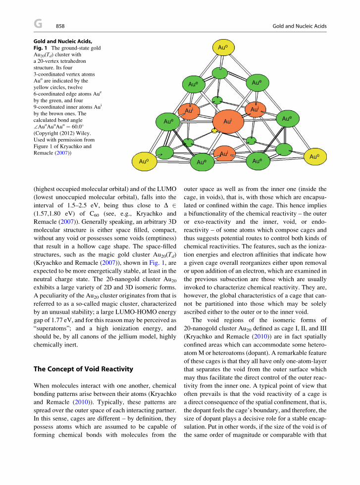
Auo
Auo
Auo
Aue
Aui
Aui
Aui
Aui
Aue
AueAue
Aue Aue
Gold and Nucleic Acids,Fig. 1 The ground-state gold
Au20(Td) cluster witha 20-vertex tetrahedron
structure. Its four
3-coordinated vertex atoms
Auo are indicated by the
yellow circles, twelve
6-coordinated edge atoms Aue
by the green, and four
9-coordinated inner atoms Aui
by the brown ones. The
calculated bond angle
∠AuoAuoAuo ¼ 60.0�
(Copyright (2012) Wiley.
Used with permission from
Figure 1 of Kryachko and
Remacle (2007))
G 858 Gold and Nucleic Acids
(highest occupied molecular orbital) and of the LUMO
(lowest unoccupied molecular orbital), falls into the
interval of 1.5–2.5 eV, being thus close to D 2(1.57,1.80 eV) of C60 (see, e.g., Kryachko and
Remacle (2007)). Generally speaking, an arbitrary 3D
molecular structure is either space filled, compact,
without any void or possesses some voids (emptiness)
that result in a hollow cage shape. The space-filled
structures, such as the magic gold cluster Au20(Td)
(Kryachko and Remacle (2007)), shown in Fig. 1, are
expected to be more energetically stable, at least in the
neutral charge state. The 20-nanogold cluster Au20exhibits a large variety of 2D and 3D isomeric forms.
A peculiarity of the Au20 cluster originates from that is
referred to as a so-called magic cluster, characterized
by an unusual stability; a large LUMO-HOMO energy
gap of 1.77 eV, and for this reason may be perceived as
“superatoms”; and a high ionization energy, and
should be, by all canons of the jellium model, highly
chemically inert.
The Concept of Void Reactivity
When molecules interact with one another, chemical
bonding patterns arise between their atoms (Kryachko
and Remacle (2010)). Typically, these patterns are
spread over the outer space of each interacting partner.
In this sense, cages are different – by definition, they
possess atoms which are assumed to be capable of
forming chemical bonds with molecules from the
outer space as well as from the inner one (inside the
cage, in voids), that is, with those which are encapsu-
lated or confined within the cage. This hence implies
a bifunctionality of the chemical reactivity – the outer
or exo-reactivity and the inner, void, or endo-
reactivity – of some atoms which compose cages and
thus suggests potential routes to control both kinds of
chemical reactivities. The features, such as the ioniza-
tion energies and electron affinities that indicate how
a given cage overall reorganizes either upon removal
or upon addition of an electron, which are examined in
the previous subsection are those which are usually
invoked to characterize chemical reactivity. They are,
however, the global characteristics of a cage that can-
not be partitioned into those which may be solely
ascribed either to the outer or to the inner void.
The void regions of the isomeric forms of
20-nanogold cluster Au20 defined as cage I, II, and III
(Kryachko and Remacle (2010)) are in fact spatially
confined areas which can accommodate some hetero-
atomM or heteroatoms (dopant). A remarkable feature
of these cages is that they all have only one-atom-layer
that separates the void from the outer surface which
may thus facilitate the direct control of the outer reac-
tivity from the inner one. A typical point of view that
often prevails is that the void reactivity of a cage is
a direct consequence of the spatial confinement, that is,
the dopant feels the cage’s boundary, and therefore, the
size of dopant plays a decisive role for a stable encap-
sulation. Put in other words, if the size of the void is of
the same order of magnitude or comparable with that

Gold and Nucleic Acids, Fig. 2 TheMEPs of cage I, II, and III
mapped from �0.016 (red) to +0.016 (blue) |e|/(4pe0a0) onto0.001 |e| A-3 isosurface of the one-electron density r(r) and from�0.01 to +0.01 |e|/(4pe0a0) onto 0.004 (the right column, corre-
spondingly, for cages I, II, and III, respectively) |e| A-3
isosurface of the one-electron density r(r). r(r) is computed at
the B3LYP computational level (Copyright (2012) Wiley. Used
with permission from Figure 2 of Kryachko and Remacle
(2010))
Gold and Nucleic Acids 859 G
G
of the dopant, one may anticipate that doping is stable.
Obviously, doping influences the outer reactivity. It is
well known that the patterns of the molecular orbitals,
particularly of the HOMO and LUMO, and the concept
of the molecular electrostatic potential (MEP) are cru-
cial for understanding of both facets of chemical reac-
tivity. For example, the buckyball fullerene C60
exhibits two different behaviors of the MEP. It is
positive in the entire void region, where C60 is capable
to encage atoms, ions, and some molecules, and
reaches the outer central regions of the pentagon
rings. On the contrary, it is negative in the outer region
and the most negative at the midpoints of the bonds
linked two hexagonal rings.
TheMEP patterns of the studied golden fullerenes I,
II, and III are plotted in Fig. 2.
Gold Nanoparticles and Nanotechnology
The “bottom-up” strategy in molecular electronics
and biosensor technology often utilizes biohybrid
complexes where biological molecules, DNA and
peptides in particular, serve as templates. It is there-
fore of current interest to assemble the DNAmolecule
directly on gold nanoparticles. In addition, the mech-
anism of interaction between the DNA and gold is by
itself an important issue, both theoretically and exper-
imentally. Recent experimental studies showed that
DNA bases, adenine (A), thymine (T), guanine (G),
and cytosine (C) interact with Au surfaces in
a specific and sequence-dependent manner. The rela-
tive binding affinities of these nucleobases for
adsorption on polycrystalline Au films obey the fol-
lowing order: A > C � G > T (see, e.g., Kryachko
(2009)). The computational models of the interaction
of DNA bases with bare Au3 and Au4 gold clusters as
simple catalytic models of Au particles are demon-
strated in Fig. 3 (Remacle and Kryachko (2005a, b);
Kryachko (2009)).
Therefore, as follows from Fig. 3, two key bonding
ingredients underlie the base-gold and base pair-gold
hybridizations: the anchoring, either of the Au-N or
Au-O type, and the nonconventional N-HAu hydro-gen bonding. The former is the leading bonding factor
and results into stronger binding and coplanar coor-
dination when the ring nitrogen atoms of the
nucleobases are involved. The anchor bond predeter-
mines the formation of the nonconventional H-
bonding via prearranging the charge distribution
within the entire interacting system and “galvaniz-
ing” an unanchored atom of the gold cluster to act as
a nonconventional proton acceptor, through its lone-
pair-like 5d�2 and 6s orbitals. Solid computational
evidence has been provided to show that the
nonconventional hydrogen bonding is of a new,
nonconventional type, and that it sustains and even
reinforces the anchoring bond by a cooperative ‘back
donation’ mechanism.

Gold and Nucleic Acids, Fig. 3 (a) The four possible planar
(N1, N3, N7) and nonplanar (N6) binding sites of the gold cluster
Au3 to adenine. Also shown is the NH2 anchored complex AAu3(N6). (b) The three possible planar (O2(N1), O2(N3), O4) binding
sites of the gold cluster Au3 to thymine. (c) The six possible
planar (N3(N2), N3(N9), O6(N1), O6(N7), N7) and nonplanar(N2)
binding sites of the gold cluster Au3 to guanine. The anchoring
in N2 is to the amino group. (d) The three possible planar
(O2(N1), N3) and nonplanar (N4) binding sites of the gold cluster
Au3 to cytosine. For the binding site N4, the anchor bond is to the
NH2 group. For each complex, the anchor bond is drawn as
a thick (red) line and the nonconventional H-bond as a dottedline. The bond lengths are given in A and bond angles in
deg (Reprinted with permission from Figures 1–4 of Remacle
and Kryachko (2005b). Copyright (2012) The American
Chemical Society)
G 860 Gold and Nucleic Acids

Gold and Nucleic Acids, Fig. 4 Left panel: The structure of
the crown compound [Rb([18]crown-6)(NH3)3]Au-NH3: carbon
atoms are shown in gray, H white, N blue, O red, Au orange, Rb
violet. Right panel: Environment of the auride anions in [Rb([18]
crown-6)(NH3)3]Au-NH3 (Copyright (2012) Wiley. Used with
permission from Figures 1 and 3 of Nuss and Jansen (2006))
Gold and Nucleic Acids 861 G
G
Gold as Nonconventional Hydrogen BondAcceptor
This feature of gold clusters was discovered in 2005
when it was demonstrated (Kryachko and Remacle
(2005)) that the gold clusters Aun are prone to play,
interacting with conventional proton donors such as
the O-H and N-H groups, a role of a proton acceptor
and hence, to participate in the formation of
nonconventional hydrogen bonds. This work showed
a strong computational evidence that a triangular gold
cluster Au3 behaves as a proton acceptor with the O-H
group of formic acid and with the N-H one of form-
amide. The bonding involves two ingredients: the
anchoring bond between the gold atom and the car-
bonyl oxygen and the N-HAu or O-HAu contacts
between Au and the amino group of formamide or the
hydroxyl group of formic acid. It was proved
(Kryachko and Remacle (2005)) that these contacts
share all the common features of the conventional
hydrogen bonds and they can therefore be treated as
their nonconventional analogs. Since this work, the
existence of the X-HAun nonconventional hydrogenbond was computationally demonstrated for a wide
variety of molecules in different charge states
(Kryachko (2009)): Z ¼ 0, �1, ranging from the Aun-
DNA bases and Aun-DNA duplexes, to Aun-(HF)m,
[AunZ-(H2O)m]
Z, and [Aun-(NH3)m]Z complexes. The
latter family includes the smallest nanosized tetrahe-
dral gold cluster Au20Z(Td). The unveiled charge-state
specificity of the bonding ingredients of the [Aun-
(NH3)m]Z complexes has recently been explored

G 862 Gold Biomineralization in Bacterium Cupriavidus Metallidurans
(see, e.g., Kryachko (2009)) to formulate the bonding
encoding approach for molecular logic. Recently, on
the experimental side, the hydrogen acceptor
propensity of the gold atom and some its clusters has
been experimentally and computationally examined in
the variety of complexes, such as [Au(H2O)]�-Arn,
[Au(H2O)n¼1,2]�, [Au(H2O)]
�, the crown compound
[Rb([18]crown-6)(NH3)3]Au-NH3 (Nuss and Jansen
(2006)) (see Fig. 4), the complexes of small gold
clusters with acetone and with amino acids, the gold
(III) antitumor complex, the amine complexes of Au
(I), the gold(I) complexes with thione ligands, and
[cytosine-Au]� and [uracil-Au]�.
References
Berry RS, Smirnov BM (2011) Charge separation in CO oxida-
tion involving supported gold clusters. JETP 140,
No.6(12):1043–1050
Boyle R (1661) The sceptical chymist: or chymico-physical
doubts & paradoxes, touching the spagyrist’s principles com-
monly call’d hypostatical, as they are wont to be propos’d
and defended by the generality of alchymists (Reprinted in
the Everyman Series), London
Hammer B, Nørskov JK (1995)Why gold is the noblest of all the
metals. Nature (London) 376:238–240
Haruta M (1997) Size- and support-dependency in the catalysis
of gold. Catal Today 36:153
Kroto HW, Heath JR, O’Brien SC, Curl RF, Smalley RE
(1985) C60: buckminsterfullerene. Nature (London) 318:162
Kryachko ES (2009) To nano-biochemistry: picture of interac-
tions of DNA with gold. In: Matta C (ed) Quantum biochem-
istry. Wiley-VCH, Weinheim, pp 245–306, Ch. 8
Kryachko ES, Remacle F (2005) Three-gold clusters form
nonconventional hydrogen bonds O-HAu and N-HAuwith formamide and formic acid. Chem Phys Lett
404(1):142–149
Kryachko ES, Remacle F (2006) Small gold clusters form
nonconventional hydrogen bonds X-HAu: gold-water clus-ters as example, Ch 11. In: Torro-Labbe A (ed) Theoretical
aspects of chemical reactivity. Vol. 16 of Theoretical and
computational chemistry, Politzer P (ed). Elsevier, Amster-
dam, pp 219–250
Kryachko ES, Remacle F (2007) The magic gold cluster Au20.
Int J Quantum Chem 107(14):2922–2934
Kryachko ES, Remacle F (2010) 20-nanogold Au20(Td) clusterand its hollow cage isomers: structural and energetic proper-
ties. J Phys: Conf Ser 248:012026–1–012026–8
Kryachko ES, Remacle F (2011) 20-Nanogold Au20(Td) and
low-energy hollow cages: void reactivity. In: Hoggan P,
Maruani J, Piecuch P, Delgado-Barrio G, Br€andas EJ (eds)Advances in the theory of quantum systems in chemistry and
physics, vol 22, Progress in theoretical chemistry and phys-
ics. Springer, Berlin, pp 573–612, Ch. 30
Louis C, Pluchery O (2012) Gold nanoparticles for physics,
chemistry and biology. World Scientific.
Mills G, Gordon MS, Metiu H (2003) Oxygen adsorption on Au
clusters and a rough Au(111) surface: the role of surface
flatness, electron confinement, excess electrons, and band
gap. J Chem Phys 118:4198
Newton J (1951) Friend, man and the chemical elements from
stone-age hearth to the cyclotron. Ch. Griffin, London
Nuss H, Jansen M (2006) Cs5([12]crown-4)2(O3)5:
a supramolecular compound containing the confined ozonide
partial structure 11{Cs8(O3)10}. Angew Chem Int Ed
45:4369
Partington JR (1964) A history of chemistry, vol 4. Macmillan,
London, 4
Pearsall R (1986) The alchemists. Weidenfeld and Nicolson,
London
Pyykko P (1988) Chem Rev 88:563
Remacle F, Kryachko ES (2005a) Complexes of DNA bases and
gold clusters Au3 and Au4 involving nonconventional N-
HAu hydrogen bonding. Nano Lett 5:735–739
Remacle F, Kryachko ES (2005b) Structure and energetics of
two- and three-dimensional neutral, cationic and anionic
gold clusters Au5 � n � 8Z (Z ¼ 0, �1). J Chem Phys
122(4):044304–1–0443041–4
Wallace WT, Whetten RL (2000) Carbon monoxide adsorption
on selected gold clusters: highly size-dependent activity and
saturation compositions. J Phys Chem B 104:10964
Gold Biomineralization in BacteriumCupriavidus Metallidurans
Carla M. Zammit and Frank Reith
School of Earth and Environmental Sciences, The
University of Adelaide, Centre of Tectonics,
Resources and Exploration (TRaX) Adelaide, Urrbrae,
South Australia, Australia
CSIRO Land and Water, Environmental
Biogeochemistry, PMB2, Glen Osmond, Urrbrae,
South Australia, Australia
Synonyms
Biomineralization of gold nanoparticles from gold
complexes in Cupriavidus metallidurans CH34;
Interactions of Cupriavidus metallidurans CH34 with
mobile, ionic gold
Definition
Cupriavidus metallidurans CH34 is a facultative
chemolithoautotrophic b-proteobacterium. It facili-
tates resistance to more than 20 mobile heavy metals

Gold Biomineralization in Bacterium CupriavidusMetallidurans, Fig. 1 Gold nugget from Western Australia
Gold Biomineralization in Bacterium Cupriavidus Metallidurans 863 G
G
via chromosome- and megaplasmid-encoded metal
resistance factors. C. metallidurans CH34 occurring
on gold nuggets mediates the (trans)formation of gold
in placer environments. This is due to the ability of
C. metallidurans CH34 to biomineralize gold via
energy-dependent reductive precipitation of highly
toxic gold complexes.
Historical Background
Cupriavidus (formerly also known as Wautersia,
Ralstonia, or Alcaligenes) metallidurans CH34 was
originally isolated from a decantation tank at a metal
processing factory in Belgium (Mergeay et al. 1985). It
serves as a model organism for the study of metal
resistance in microorganisms due to its ability to with-
stand millimolar concentrations of more than 20
different heavy metal ions. Cupriavidus metallidurans
CH34 is a facultative chemolithoautotrophic
b-proteobacterium that contains two chromosomes and
two megaplasmids (Mergeay et al. 1985). Its metal
resistance factors are located throughout the genome
on both chromosomes and the megaplasmids. These
resistance genes have been acquired, rearranged, and
recombined from numerous sources, amounting to the
many genetic alterations that give this bacterium the
ability to adapt and persist in environments high in
mobile heavy metals (Janssen et al. 2010).This ability to adapt to high metal concentrations
has made it possible for Cupriavidus species to persist
in some of the most hostile environments on Earth,
e.g., it has been isolated from a range of industrial sites
across Belgium, Germany, and the Congo. Species
from the genus Cupriavidus and its close relative
Ralstonia have also been detected in ultra-clean space-
craft assembly rooms, spacecraft atmosphere, and
spent nuclear fuel pools. Besides anthropogenic con-
taminated environments, three Cupriavidus species
have been isolated from volcanic mudflow deposits
from the Pinatubo volcano in the Philippines.
Additionally, Cupriavidus species have also been
isolated from the surface of gold grains in Australia
(Fig. 1; Reith et al. 2006).
Metal-Resistant Systems in C. metalliduransCH34
Metals make up more than 75% of all elements, and
since the industrial revolution, they have undergone
drastic redistribution in Earth surface environments.
Some metals are essential to microbial life, others are
used to gain cellular energy; however, all metals,
above threshold concentrations, are toxic to microor-
ganisms. Therefore, microorganisms have developed
biochemical systems to deal with toxic levels of
metals. Most of these mechanisms are based on chang-
ing the speciation of metals via active/passive oxida-
tion/reduction and complex formation, which affect
metal mobility in intra- and extracellular aqueous solu-
tions. Whereas passive mechanisms, e.g., passive
extracellular biomineralization, involve the interac-
tions between metals with different components of
cells, active detoxification and biomineralization are
commonly mediated by metal-binding (poly)-peptides
and proteins.
To date, C. metallidurans CH34 has been identified
as the microorganism containing the highest number of
genes involved in heavy metals resistance (Mergeay
et al. 2003; Monsieurs et al. 2011). Metal resistance in
C. metallidurans CH34 includes a complex and highly
diverse range of mechanisms conveying resistance to
Ag+, AsO�, Au+, Au3+, Bi3+, Co2+, Cd2+, CrO42�, Cs+,
Cu+, Cu2+, HAsO42�, Hg2+, Ni2+, Pb2+, SeO3
2�,SeO4
2�, Sr2+, Tl+, and Zn2+ (Janssen et al. 2010).
Many of the active mechanisms that microorganisms
in general and C. metallidurans CH34 specifically
have in place to deal with elevated levels of metals
are not specific to one particular metal, but convey
resistance to different metals. For example, in
a recent study, the copH gene in C. metallidurans
CH34 was upregulated by 13 out of 16 different
metal ions (Monsieurs et al. 2011). In this study,

Gold Biomineralization inBacterium CupriavidusMetallidurans,Fig. 2 Secondary scanning
electron micrograph of
a biofilm growing on the
surface of a gold nugget from
the Flinders Ranges, South
Australia
G 864 Gold Biomineralization in Bacterium Cupriavidus Metallidurans
more than 43% of upregulated metal-response genes
could be classified as “metal specific,” varying
between 4% metal-specific response genes for Sr2+
and 57% metal-specific response genes for Ni2+
(Monsieurs et al. 2011). Cupriavidus metalliduransCH34 was shown to have a similar transcriptomic
response to metals which clustered in the following
categories: Group I – Cd2+, Pb2+, and Zn2+; Group II –
Ag1+, Au3+, Cs1+, Hg2+, Sr2+, and Tl+; and Group III –
CrO42+, As3+, andMn2+ (Monsieurs et al. 2011). These
groupings appeared to not be the result of the physico-
chemical properties of the metals, but rather to be
a function of the location of specific metal resistance
genes on chromosomes and plasmids. Having these
types of cross-reactive transcriptional networks is
highly advantageous for C. metallidurans CH34,
because it allows the organisms to maintain metal
homeostasis when exposed to elevated levels of
a range of metals, while being able to conserve
metabolic energy required for the production of
specific proteins. For example, when C. metallidurans
CH34 was exposed to equal concentrations of Co2+,
Ni2+, Cu2+, Zn2+, and Cd2+ combined, it was shown
that the metal content within the cell was the same as if
only a single metal had been added (Kirsten et al. 2011).
Interactions of C. metallidurans CH34and Gold
The interaction between C. metallidurans CH34 and
gold has attracted much interest among researchers,
because C. metallidurans CH34 was shown to be
involved in the transformation of gold nuggets
(Fig. 2). Gold is a Group IB metal that occurs in
aqueous solutions under surface conditions as metal-
lic gold (Au0) as well as the highly toxic aurous, Au
(I), and auric, Au(III), complexes. Hence, along with
its concentration, its oxidative state and type of com-
plex, as determined by the available ligands, play an
important role in its toxicity to cells. Because of their
high affinity for organic ligands, Au(III)-complexes
are rapidly absorbed by bacterial cells (Reith et al.
2007). Once in contact with a cell membrane, Au(III)
is transformed to Au(I), which is taken up into the
cytoplasm (Reith et al. 2009). Here it may disturb the
redox equilibrium, increase cell permeability, or dis-
rupt metabolic pathways. However, microorganisms
such as C. metallidurans CH34 and Salmonella
typhimurium have developed systems to deal with
these increased levels of toxic gold (Checa et al. 2007;Reith et al. 2009).

Gold Biomineralization in Bacterium CupriavidusMetallidurans, Fig. 3 Transmission electron micrograph of
an ultrathin section of a C. metallidurans cells harboring nano-
particulate gold (arrow) in the periplasm
Gold Biomineralization in Bacterium CupriavidusMetallidurans, Fig. 4 Protein model of Rmet_4684, a gene
predicted to encode for a hypothetical protein possibly involved
in gold biomineralization by C. metallidurans
Gold Biomineralization in Bacterium Cupriavidus Metallidurans 865 G
G
Gold-Resistant Systems
Two mechanisms employed by microorganisms to
tolerate gold have been postulated, the first involves
the efflux of Au(I)-complexes through metal trans-
porters, the second involves the active precipitation
of Au(I)-complexes (Reith et al. 2007). However, as
with other metal resistance systems, deciphering the
molecular mechanisms involved in Au detoxification
is complex. Actively growing C. metallidurans CH34
cells rapidly absorb Au(III)-complexes from solution
and reduce them to Au(I)-S complexes on the surface
of the cell (Reith et al. 2009). Gold is then distributed
across the cell surface and upregulation of genes
belonging to oxidative stress and metal detoxification
occurs, followed by the appearance of metallic Au
nanoparticles in the periplasm (Figs. 3 and 4; Reith
et al. 2009). The genes that are most strongly
upregulated during this process belong to the gig
(gold-induced genes) operon, which includes genes
for the proteins: GigA and GigB, both cytoplasmic
proteins; GigP, a putative periplasmic protein; GigT,
a protein with three predicted transmembrane spans,
related to a subunit of terminal quinol oxidase found in
an archaeal membrane (Mohr 2011). These genes are
predicted to be important to the movement of Au(III)
into the cytoplasm. A MerR family protein, CupR,
specifically responds to gold stress in C. metallidurans
CH34 and could act as a reporter of Au(I) in the
cytoplasm. Next Au(I) needs to be transported into the
periplasm where it forms Au(0), this could be achieved
with PIB1-type ATPases, such as CupA or CopF.
However, the employment of these systems is yet to be
experimentally validated and with the two-fold
upregulation of 52–303 transcripts upon the addition of
Au(III), it is unlikely to be so simple (Reith et al. 2009).
Biomineralization of Gold
Organisms are able to accumulate and precipitate min-
erals under a range of conditions. This process is
termed biomineralization and a variety of different
minerals are formed via this process. These include
calcium carbonates and phosphates in vertebrates,
calcium and magnesium carbonates in invertebrates,
silicates in algae and diatoms, as well as iron-oxides
and copper and gold minerals in microorganisms.
Biomineralization contributes to the natural movement
of minerals in the environment and there is a growing
amount of evidence that certain microorganisms play

Gold Biomineralization inBacterium CupriavidusMetallidurans,Fig. 5 Secondary scanning
electron micrograph of
spherical gold microparticles
formed as a result of
biomineralization by
C. metallidurans
G 866 Gold Biomineralization in Bacterium Cupriavidus Metallidurans
an important role in the movement of gold within the
environment, despite gold being seen as an inert sub-
stance under surface conditions. C. metallidurans cells
rapidly accumulate Au(III)-complexes from solution,
and promote the biomineralization of Au nanoparticles
via energy-dependent reductive precipitation of the
highly toxic complexes. These particles can vary in
size from a few nanometers to several micrometers
(Figs. 3 and 5). The industrial applications of such
nanoparticles and microparticles is only just beginning
to be explored but includes a broad range of applica-
tions, such as bio-imaging, pollution control, water
purification, components for technical equipment
(e.g., computer components), and uses as catalysts.
Implications for Gold Cycling
It has long been accepted that microorganisms play an
important role in the biogeochemical cycling of car-
bon, oxygen, and nitrogen. The role of microorganisms
in the movement of metals within the environment was
at first unknown, then overlooked. However, presently,
scientists are gaining widespread appreciation for the
contribution that microorganisms have in metal
solubilization, dispersion, and precipitation. Microor-
ganisms both directly and indirectly influence the
movement of metals in the environment. The solubili-
zation of gold occurs via iron and/or reduced inorganic
sulfur-oxidizing microorganisms via the production of
acid, a by-product of their metabolic reactions; the
microbial production of thiosulfate; the sequestration
of Au by Au-complexing ligands; the production of
cyanide. Once gold is solubilized, it can be distributed
in the environment. Finally, the gold is deposited back
into the environment via the biomineralization mech-
anisms discussed above.
Understanding how gold is transported in the envi-
ronment has many industry-based applications. Tech-
nologies derived from fundamental knowledge of how
Au is solubilized, dispersed, and precipitated will have
a great impact on the mining industry. Mine explora-
tion techniques can be adapted to account for the
microbial movement of metals, brining greater accu-
racy. Processing technologies, based on microbial-
metal interactions could be improved (i.e.,
biooxidation and bioleaching) or further developed
(microbially mediated in situ leaching). Bioremedia-
tion techniques could be developed for the treatment of
metal contaminated sites. The use of nanoparticle

Gold Complexes as Prospective Metal-Based Anticancer Drugs 867 G
technologies is only just in its infancy, and could begreatly explored. In conclusion, although understand-
ing the molecular mechanisms involved in the biomin-
eralization of Au in C. metallidurans CH34 is
a complex endeavor, the potential applications of
such knowledge is wide spanning and of great com-
mercial value.
G
Cross-References
▶Aluminum, Physical and Chemical Properties
▶Biomineralization of Gold Nanoparticles from Gold
Complexes in Cupriavidus Metallidurans CH34
▶Gold and Nucleic Acids
▶Gold Nanomaterials as Prospective Metal-based
Delivery Systems for Cancer Treatment
▶Gold Nanoparticle Platform for Protein-Protein
Interactions and Drug Discovery
▶Gold Nanoparticles and Fluorescent Proteins,
Optically Coupled Hybrid Architectures
▶Gold Nanoparticles and Proteins, Interaction
References
Checa SK, Espariz M, Perez Audero ME et al (2007) Bacterial
sensing of and resistance to gold salts. Mol Microbiol
63(5):1307–1318
Janssen PJ, Van Houdt R, Moors H et al (2010) The complete
genome sequence ofCupriavidus metallidurans strain CH34,a master survivalist in harsh and anthropogenic
environments. PLoS One 5(5):e10433
Kirsten A, Herzberg M, Voigt A et al (2011) Contributions of
five secondary metal uptake systems to metal homeostasis of
Cupriavidus metallidurans CH34. J Bacteriol
193(18):4652–4663
Mergeay M, Nies D, Schlegel HG et al (1985) Alcaligeneseutrophus CH34 is a facultative chemolithotroph with plas-
mid-bound resistance to heavy metals. J Bacteriol
162(1):328–334
Mergeay M, Monchy S, Vallaeys T et al (2003) Ralstoniametallidurans, a bacterium specifically adapted to toxic
metals: towards a catalogue of metal-responsive genes.
FEMS Microbiol Rev 27(2–3):385–410
Mohr J (2011) Goldresistenz von Cupriavidus metallidurans,Naturwissenschaftlich Fakultaet. Universitate Halle-Witten-
berg, Wittenberg
Monsieurs P, Moors H, Van Houdt R et al (2011) Heavy metal
resistance in Cupriavidus metallidurans CH34 is governed
by an intricate transcriptional network. Biometals
24(6):1133–1151
Reith F, Rogers SL, McPhail DC et al (2006) Biomineralization
of gold: biofilms on bacterioform gold. Science 313:233–236
Reith F, Lengke MF, Falconer D et al (2007) The
geomicrobiology of gold. ISME J 1:567–584
Reith F, Etchmann B, Grosse C (2009) Mechanisms of gold
biomineralization in the bacterium Cupriavidusmetallidurans. PNAS 106(42):17757–17762
Gold Complexes and Their AnticancerProperties
▶Gold Complexes as Prospective Metal-Based Anti-
cancer Drugs
Gold Complexes as ProspectiveMetal-Based Anticancer Drugs
Anil Kumar1,3, Bhargavi M. Boruah2,3 and
Xing-Jie Liang1
1CAS Key Laboratory for Biomedical Effects of
Nanoparticles and Nanosafety, National Center for
Nanoscience and Nanotechnology, Chinese Academy
of Sciences, Beijing, China2CAS Key Laboratory of Pathogenic Microbiology
and Immunology, Institute of Microbiology, Chinese
Academy of Sciences, Beijing, China3Graduate University of Chinese Academy of Science,
Beijing, China
Synonyms
Gold complexes and their anticancer properties; Gold
for health – past and present; Gold physiochemical
properties; Mode of action; Overcome cisplatin resis-
tance; Targeted mechanism of gold complex; Toxicity
of gold complex
Definition
Gold (Au) is a metallic element with an atomic number
of 79. It is soft, shiny, dense, as well as an extremely
malleable and ductile metal. Au is yellow color when
in mass but may give black, ruby, or purple color when
finely divided. It is a good conductor of electricity and
is unaffected by heat, moisture, oxygen, and most
reagents except when comes in contact with chlorine,

AcO
AcO
OAcc
OAc
O S Au P
Et
Et
Et
HO
OH
OH
b
S
Au
O
HO
n
ONa
NaO
S Au
a
n
O
G 868 Gold Complexes as Prospective Metal-Based Anticancer Drugs
fluorine, and aqua regia. Au may be alloyed with var-
ious other metals like silver (Ag), copper (Cu), zinc
(Zn), nickel (Ni), platinum (Pt), palladium (Pd), tellu-rium (Te), and iron (Fe) to give it special properties and
various color hues ranging from silver-white to green
and orange-red. Due to its relatively uncommon chem-
ical and physical properties, Au is indispensible for
a wide variety of applications for diagnosis and treat-
ment of cancer. The most advantageous attributes of
Au is the relative inertness. Listed as one of the “noble”
metals, Au does not oxidize under normal conditions
and thus, have resistance to tarnishing. However, it
dissolves in the presence of solutions containing halo-
gens or aqua regia as well as in mixtures of
hydrochloric and nitric acids (3:1).
Gold Complexes as Prospective Metal-Based AnticancerDrugs, Fig. 1 Structure of gold(I) complex drugs used for thetreatment of rheumatoid arthritis (a) sodium aurothiomalate
(Myocrisin), (b) aurothioglucose (Solganol), and (c)tetraacetyl-b-D-thioglucose gold(I) triethyl- phosphine also
known as (auranofin) (a, Reprinted with permission from
Dreaden EC, Mwakwari SC, Sodji QH, Oyelere AK,
El-Sayed MA (2009) Tamoxifen−poly(ethylene glycol)−thiolgold nanoparticle conjugates: enhanced potency and selective
delivery for breast cancer treatment. Bioconjugate Chem.
Copyright (2009 American Chemical Society)
Gold for Health: Past and Present
Besides use as ornaments and as currency, gold (Au)
has remarkable medical and therapeutic values that
have been recognized thousands of years ago.
According to the earliest records, use of Au for medical
purposes can be traced back to the Chinese civilization
in 2500 B.C., and after that, several ancient cultures
have utilizedAu-based materials for medicinal purpose
for the treatment of a variety of diseases such as
smallpox, skin ulcers, measles, fainting, fevers, and
falling sickness. The rational use of Au in medicine
started in the nineteenth century whereby mixture of
Au chloride and sodium chloride Na[AuCl4] was used
for the treatment of syphilis. Initial research on Au
complexes could be credited to bacteriologist
Robert Koch who reported the use of K[Au(CN)2]
in the treatment of tuberculosis, later replaced by
Au(I)-thiolated complexes on account of toxicity
issues. With the advent of Au therapy and following
its success in treating rheumatoid arthritis (Fig. 1),
Au complexes found further applications in the treat-
ment of other rheumatic diseases including psoriatic
arthritis, juvenile arthritis, palindromic rheumatism,
and discoid lupus erythematosus. With rather more
success in recent decades, colloidal Au has been used
in research applications in medicine, biology,
and material sciences. Some Au-salts show the anti-
inflammatory properties and are used as therapeutics
in the treatment of inflammatory skin disorders
namely pemphigus, urticaria, and psoriasis. Treatment
using Au-based drugs is known as chrysotherapy.
Considerable research has gone into the potential anti-
cancer and antimicrobial activity of Au complexes, and
the recent decades have seen interesting advances in
the pharmacology of Au-based drugs on their mecha-
nism of action and biological chemistry related to these
findings (Milacic et al. 2008).
Current Cancer Treatment and Challenges
Cancer is one of the most serious health hazard world-
wide with about 12 million new cancer cases diag-
nosed each year which is expected to increase to 25
million per year in 2020 (Cancer Facts and
Figures 2012 Annual Publications, American Cancer
Society). Improvement in understanding, diagnostics,
treatment, and prevention has been tremendously
achieved through investments in biological and
chemical research. With the advancement in cancer
surgery, chemotherapy, and radiotherapy, half of the
total cancer cases can be cured, while the other half can
have the benefit of prolong survival or in worst cases,
no survival at all. Cancer chemotherapy was initiated
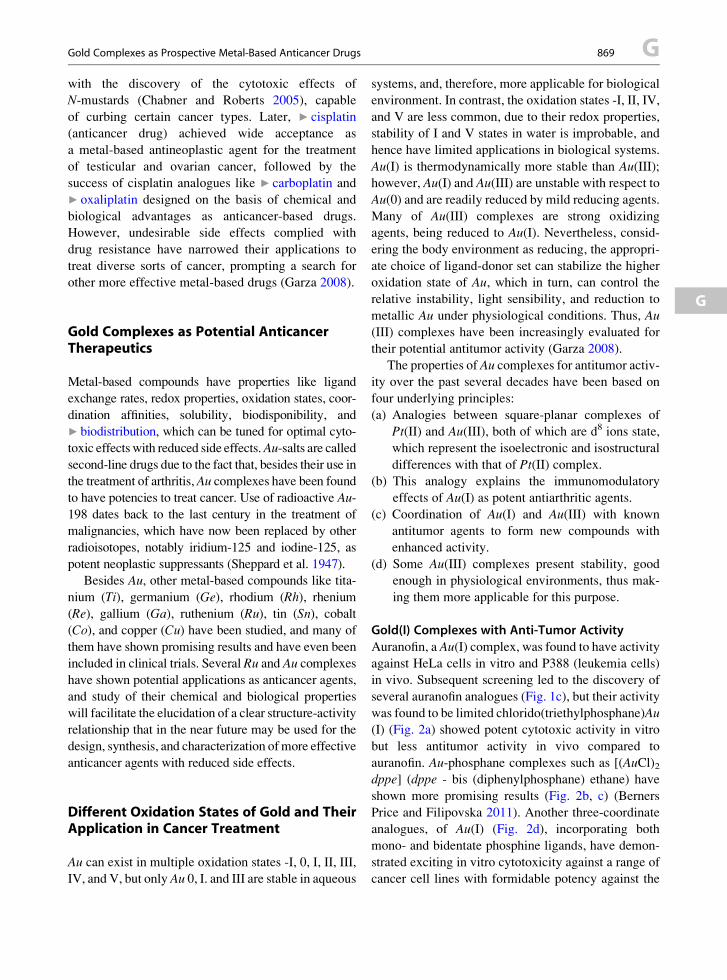
Gold Complexes as Prospective Metal-Based Anticancer Drugs 869 G
with the discovery of the cytotoxic effects ofN-mustards (Chabner and Roberts 2005), capable
of curbing certain cancer types. Later, ▶ cisplatin
(anticancer drug) achieved wide acceptance as
a metal-based antineoplastic agent for the treatment
of testicular and ovarian cancer, followed by the
success of cisplatin analogues like ▶ carboplatin and
▶ oxaliplatin designed on the basis of chemical and
biological advantages as anticancer-based drugs.
However, undesirable side effects complied with
drug resistance have narrowed their applications to
treat diverse sorts of cancer, prompting a search for
other more effective metal-based drugs (Garza 2008).
G
Gold Complexes as Potential AnticancerTherapeutics
Metal-based compounds have properties like ligand
exchange rates, redox properties, oxidation states, coor-
dination affinities, solubility, biodisponibility, and
▶ biodistribution, which can be tuned for optimal cyto-
toxic effects with reduced side effects.Au-salts are called
second-line drugs due to the fact that, besides their use in
the treatment of arthritis,Au complexes have been found
to have potencies to treat cancer. Use of radioactive Au-
198 dates back to the last century in the treatment of
malignancies, which have now been replaced by other
radioisotopes, notably iridium-125 and iodine-125, as
potent neoplastic suppressants (Sheppard et al. 1947).
Besides Au, other metal-based compounds like tita-
nium (Ti), germanium (Ge), rhodium (Rh), rhenium
(Re), gallium (Ga), ruthenium (Ru), tin (Sn), cobalt(Co), and copper (Cu) have been studied, and many of
them have shown promising results and have even been
included in clinical trials. Several Ru and Au complexes
have shown potential applications as anticancer agents,
and study of their chemical and biological properties
will facilitate the elucidation of a clear structure-activity
relationship that in the near future may be used for the
design, synthesis, and characterization ofmore effective
anticancer agents with reduced side effects.
Different Oxidation States of Gold and TheirApplication in Cancer Treatment
Au can exist in multiple oxidation states -I, 0, I, II, III,
IV, and V, but only Au 0, I. and III are stable in aqueous
systems, and, therefore, more applicable for biological
environment. In contrast, the oxidation states -I, II, IV,
and V are less common, due to their redox properties,
stability of I and V states in water is improbable, and
hence have limited applications in biological systems.
Au(I) is thermodynamically more stable than Au(III);however, Au(I) and Au(III) are unstable with respect to
Au(0) and are readily reduced by mild reducing agents.
Many of Au(III) complexes are strong oxidizing
agents, being reduced to Au(I). Nevertheless, consid-
ering the body environment as reducing, the appropri-
ate choice of ligand-donor set can stabilize the higher
oxidation state of Au, which in turn, can control the
relative instability, light sensibility, and reduction to
metallic Au under physiological conditions. Thus, Au(III) complexes have been increasingly evaluated for
their potential antitumor activity (Garza 2008).
The properties of Au complexes for antitumor activ-
ity over the past several decades have been based on
four underlying principles:
(a) Analogies between square-planar complexes of
Pt(II) and Au(III), both of which are d8 ions state,
which represent the isoelectronic and isostructural
differences with that of Pt(II) complex.
(b) This analogy explains the immunomodulatory
effects of Au(I) as potent antiarthritic agents.
(c) Coordination of Au(I) and Au(III) with known
antitumor agents to form new compounds with
enhanced activity.
(d) Some Au(III) complexes present stability, good
enough in physiological environments, thus mak-
ing them more applicable for this purpose.
Gold(I) Complexes with Anti-Tumor Activity
Auranofin, a Au(I) complex, was found to have activity
against HeLa cells in vitro and P388 (leukemia cells)
in vivo. Subsequent screening led to the discovery of
several auranofin analogues (Fig. 1c), but their activity
was found to be limited chlorido(triethylphosphane)Au(I) (Fig. 2a) showed potent cytotoxic activity in vitro
but less antitumor activity in vivo compared to
auranofin. Au-phosphane complexes such as [(AuCl)2dppe] (dppe - bis (diphenylphosphane) ethane) have
shown more promising results (Fig. 2b, c) (Berners
Price and Filipovska 2011). Another three-coordinate
analogues, of Au(I) (Fig. 2d), incorporating both
mono- and bidentate phosphine ligands, have demon-
strated exciting in vitro cytotoxicity against a range of
cancer cell lines with formidable potency against the

P Au
Ph2PPh2P
PPh2
(HOH2CH2C)3P
(HOH2CH2C)3P P(CH2CH2OH)3
P(CH2CH2OH)3
PPh2
PPh2
Ph2PPh2P
Ph2P
n
PPh3
Au
Cl
+
+
Cl
AuAu
Au
Au(1) (1)
Cl
b ca
d
e
(1)
Gold Complexes asProspective Metal-BasedAnticancer Drugs,Fig. 2 Structure of cytotoxic
gold(I) compounds (a)chlorido(triethylphosphane)
gold(I), (b and c) bis(diphenylphosphane)ethane
gold(I) analogues, (d) three-coordinate analogues, and
(e) hydrophilic tetrahedralcompound
R2P
a b
R2P
PR2R R
R
R
R
R
RP
P P
PPR2
Au Au
Cl–Cl–
++
Gold Complexes as Prospective Metal-Based AnticancerDrugs, Fig. 3 Structure of lipophilic cationic gold(I) complex
(a) [Au(dppe)2]Cl and (b)[Au(d2pypp)2]Cl anticancer com-
pounds, where R ¼ 2, 3, or 4 pyridyl
G 870 Gold Complexes as Prospective Metal-Based Anticancer Drugs
breast tumor cell line, MCF-7 (Caruso et al. 2003).
Similarly, the hydrophilic tetrahedral compound,
(Fig. 2e) having all-monodentate phosphine ligands,
showed promising activity against a range of prostrate,
colon carcinoma, and gastric carcinomas (Pillarsetty
et al. 2003).
The dppe ligand exhibits antitumor activity by
itself, and Au protects the ligand from oxidation and
aids in the delivery of the active species. Similar
experimental results have revealed the use of Au as
“prodrugs” acting as a platform for delivering antican-
cer agents, which when administered alone, have fewer
efficacies to penetrate into tumors. Such coordination
drugs have been reported to alter the normal metabolic
pathways and release mechanisms through
rearrangement (in solution and biological media) of
the diphosphane compounds to produce a rare coordi-
nation geometry of Au(I) complexes such as [Au
(dppe)2]Cl and [Au(d2pypp)2]Cl favoring efficiency
for penetration into tumors (Fig. 3).
The mechanism of cytotoxicity mediated by Au
complexes differs from that of cisplatin, another
eminent metal-based anticancer agent. In contrast
to cisplatin which targets the DNA, Au complexes
have the ability to slowdown mitochondrial function
and thus inhibit protein synthesis (Krishnamurthy
et al. 2008). The effect of structural variation in
bis(diphosphane) gold(I) complexes, in general, are
active against various types of cancer and kill cells
via damage to mitochondria. The coordination of Au
by phosphane ligands with the three different phos-
phorus-bonds substituents leads to chiral phospho-
rous coordination compounds, which has been
reported to increase potency with increasing number
of coordinated phosphorus atoms. However, higher
potency related to chirality was not observed. In
spite of their efficacy, Au(I) complexes have been
prevented from clinical use by far due to reports of
heart toxicity highlighted during preclinical studies
(Hoke et al. 1989). But, toxicities can be
circumvented by careful selection of substituents
on the phosphane moiety and by tuning the
lipophilicity of the cation.

a
P Au(1)
S P Au(1)
Au
S
S
NN
N
H
H
H
NN
N
H
NH2
NH
NH2
H
N
Me
SO3
Et3P
O
b
c
Gold Complexes asProspective Metal-BasedAnticancer Drugs,Fig. 4 Schematic structure of
cytotoxic gold(I) (a)compounds, 6-mercaptopurine
anion derivative,
(b) 6-thioguanine anionderivative, and
(c) N-heterocyclic carbeneligands
Gold Complexes as Prospective Metal-Based Anticancer Drugs 871 G
G
Coupling of phosphane Au(I) species to biologically
active thiols such as 6-mercaptopurine and
6-thioguanine (Fig. 4a, b) revealed that the presence
of phosphane Au(I) entity enhanced the potency of the
biologically active free thiols, which are already
known for anticancer activity in leukemia patients.
Although Au(I)-drugs have proved their effectiveness
in many diseases, knowledge related to the mechanism
of action has not been completely understood even
after 80 years of clinical use. This lack of knowledge
in part is a result of the wide dispersion of Au com-
pounds in the body and the absence of effective high-
affinity target sites of action.
Gold(III) Complexes as Anticancer Agents
Recent research based on Au(III) complexes (Fig. 5)
have revealed outstanding cytotoxic properties and are
presently being evaluated as potential antitumor agents
exhibiting promising in vivo activity against several
tumor models in rats and mice (Messori et al. 2003,
2005). In contrast to the Au(I) complexes defined by
“soft” (easily polarisable) sulfur or phosphorus atom
donors, Au(III) complexes are characterized by “hard”
atom donors like nitrogen, oxygen, and carbon. Four-
coordinated Au(III) resemble with that of cisplatin
having a square-planar geometry. Au(III) compounds
with imine donors have also been prepared and biolog-
ically tested. Several Au(III) (Fig. 6a–c) complexes
have been reported to show systemic toxic effects
and significant differences in the spectrum of action
have been noticed compared to cisplatin (Berners Price
and Filipovska 2011). Some of the most interesting
cytotoxic effects were reported for the complexes
containing ligand donors derived from one element or
the combination of chloride, nitrogen, oxygen, and
carbon. One approach followed was the synthesis of
complexes with the mononegative bidentate ligand,
damp, (2- [(dimethylamino) methyl] phenyl), and two
monodentate anionic ligands, chloride (Cl), or acetate
(O2CCH3). The damp ligand forms part of a five-
membered chelate ring in which the nitrogen of the
amine group and the carbon of the aryl ring bind to the
metal (Fig. 6i, j) making them readily hydrolyzed and
available for substitution.
Initial in vitro studies against a panel of
human tumor cell lines indicated that the breast carci-
noma cell line (ZR-75-1) is sensitive to the
compounds. Further tests in vivo against a xenograft
of the same tumor cells demonstrate modest
antitumor activity. Analogues of this complex have

N
a
c d
b
XX
+ +
Au
NN
N
N
Au
N
Br
Br
NN
Au
SN
CH3
CH3
SAu
Gold Complexes as Prospective Metal-Based AnticancerDrugs, Fig. 5 Structure of gold(III) compounds that have dem-
onstrated its antitumor activity in vivo (a) [AuX2(damap)],
(where, X ¼ acetate and malonate) (b) [Au(DMDT)Br2], (c)[Au(III)(TPP)]+, and (d) [Au(C^N^C (NHC)]+. The complexes
(a and b) [Au(DMDT)X2] and [Au(ESDT)X2] (where
DMDT ¼ N, N-dimethylthiocarbamate and
ESDT ¼ ethylsarcosinedithiocarbamate; X ¼ Cl, Br) are more
toxic in vitro than cisplatin and to human tumor cell lines
intrinsically resistant to cisplatin
a
N
HO
N
N
N
N
N
NH
H HH
H2
H2
NH2
NH2
Cl
Au (111)
Au (111)
Au
(111)
3+
3+
Cl
Cl
d
g h
Gold Complexes asProspective Metal-BasedAnticancer Drugs,Fig. 6 Structure of cytotoxic
gold(III) compounds and
cationic species which show
anticancer properties.
(Reprinted with permission
from Bardhan R, Lal S,
Joshi A, Halas NJ (2011)
Theranostic nanoshells: from
probe design to imaging and
treatment of cancer. Acc
Chem Res. Copyright (YEAR)
American Chemical Society)
G 872 Gold Complexes as Prospective Metal-Based Anticancer Drugs
been evaluated in the same way. Also in vitro cytotox-
icity studies showed promising activity of two Au(III)
complexes with bipyridyl-related ligands (Fig. 7),
namely [Au(bpy)(OH)2]PF6(bpy ¼ 2,20-dypyridyl)and [Au(bpyc-1H)(OH)]PF69 (bpyc ¼ 6-(1,1-
dimethylbenzyl)-2,20-bypiridine) (Marcon et al. 2002).
Gold(I) and (III) Complexes to Overcome Cisplatin
Resistance
Many of the Au(I) and (III) compounds are able to
overcome to a large extent resistance to cisplatin,
suggesting that a different mechanism of action is
taking part. Carbene ligands of Au(I) complex (a deriv-
ative of vitamin K3) (Fig. 4c) displayed significant
cytotoxicity in the cisplatin sensitive and resistant can-
cer lines, proving to be relatively more cytotoxic in the
latter (Casas et al. 2006). A complex of Au(III),
[Au(phen)Cl2]Cl (phen ¼ 1,10-phenanthroline)
(Fig. 6f), and [Au(tpy)Cl]Cl2 (tpy ¼ 2,20:60,200-terpyridine) (Fig. 6h) exhibited cytotoxicity profile
which is similar to that of cisplatin (used at
a concentration which is three times higher than the
Au complexes) on the sensitive line. Moreover,
N
N
N
ClAu
(111) N
Cl
Cl
Au (111)
N
Au
O2CCH2
O2CCH2
N
N
Cl
+
Cl
(111) Au
2+
H2N
NH2
NH2
Cl
Au (111)
N
Cl
Cl
O
Au (111) N
Cl
Cl
O
Au (111)
2+
b c
e f
i j
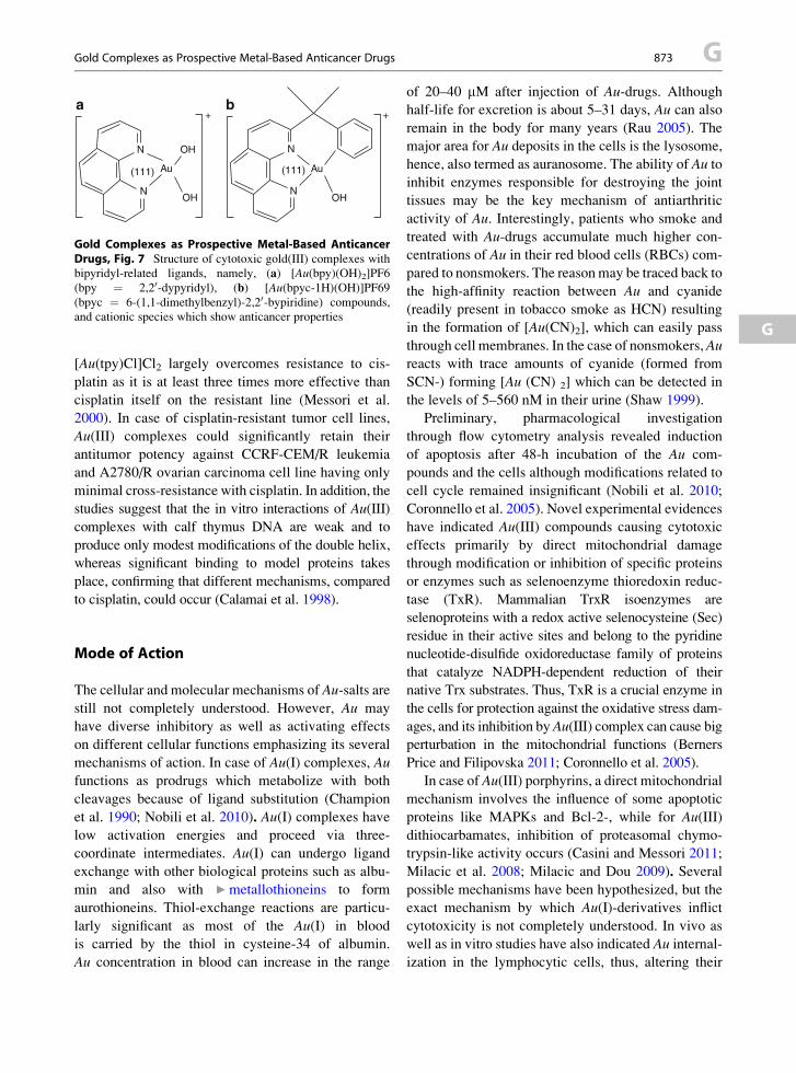
a
N
N
OH
+ +
OH
Au(111)
N
NOH
Au(111)
b
Gold Complexes as Prospective Metal-Based AnticancerDrugs, Fig. 7 Structure of cytotoxic gold(III) complexes with
bipyridyl-related ligands, namely, (a) [Au(bpy)(OH)2]PF6(bpy ¼ 2,20-dypyridyl), (b) [Au(bpyc-1H)(OH)]PF69(bpyc ¼ 6-(1,1-dimethylbenzyl)-2,20-bypiridine) compounds,
and cationic species which show anticancer properties
Gold Complexes as Prospective Metal-Based Anticancer Drugs 873 G
G
[Au(tpy)Cl]Cl2 largely overcomes resistance to cis-
platin as it is at least three times more effective than
cisplatin itself on the resistant line (Messori et al.
2000). In case of cisplatin-resistant tumor cell lines,
Au(III) complexes could significantly retain their
antitumor potency against CCRF-CEM/R leukemia
and A2780/R ovarian carcinoma cell line having only
minimal cross-resistance with cisplatin. In addition, the
studies suggest that the in vitro interactions of Au(III)
complexes with calf thymus DNA are weak and to
produce only modest modifications of the double helix,
whereas significant binding to model proteins takes
place, confirming that different mechanisms, compared
to cisplatin, could occur (Calamai et al. 1998).
Mode of Action
The cellular and molecular mechanisms of Au-salts are
still not completely understood. However, Au may
have diverse inhibitory as well as activating effects
on different cellular functions emphasizing its several
mechanisms of action. In case of Au(I) complexes, Aufunctions as prodrugs which metabolize with both
cleavages because of ligand substitution (Champion
et al. 1990; Nobili et al. 2010). Au(I) complexes have
low activation energies and proceed via three-
coordinate intermediates. Au(I) can undergo ligand
exchange with other biological proteins such as albu-
min and also with ▶metallothioneins to form
aurothioneins. Thiol-exchange reactions are particu-
larly significant as most of the Au(I) in blood
is carried by the thiol in cysteine-34 of albumin.
Au concentration in blood can increase in the range
of 20–40 mM after injection of Au-drugs. Althoughhalf-life for excretion is about 5–31 days, Au can also
remain in the body for many years (Rau 2005). The
major area for Au deposits in the cells is the lysosome,
hence, also termed as auranosome. The ability of Au to
inhibit enzymes responsible for destroying the joint
tissues may be the key mechanism of antiarthritic
activity of Au. Interestingly, patients who smoke and
treated with Au-drugs accumulate much higher con-
centrations of Au in their red blood cells (RBCs) com-
pared to nonsmokers. The reasonmay be traced back to
the high-affinity reaction between Au and cyanide
(readily present in tobacco smoke as HCN) resulting
in the formation of [Au(CN)2], which can easily pass
through cell membranes. In the case of nonsmokers, Aureacts with trace amounts of cyanide (formed from
SCN-) forming [Au (CN) 2] which can be detected in
the levels of 5–560 nM in their urine (Shaw 1999).
Preliminary, pharmacological investigation
through flow cytometry analysis revealed induction
of apoptosis after 48-h incubation of the Au com-
pounds and the cells although modifications related to
cell cycle remained insignificant (Nobili et al. 2010;
Coronnello et al. 2005). Novel experimental evidences
have indicated Au(III) compounds causing cytotoxic
effects primarily by direct mitochondrial damage
through modification or inhibition of specific proteins
or enzymes such as selenoenzyme thioredoxin reduc-
tase (TxR). Mammalian TrxR isoenzymes are
selenoproteins with a redox active selenocysteine (Sec)
residue in their active sites and belong to the pyridine
nucleotide-disulfide oxidoreductase family of proteins
that catalyze NADPH-dependent reduction of their
native Trx substrates. Thus, TxR is a crucial enzyme in
the cells for protection against the oxidative stress dam-
ages, and its inhibition byAu(III) complex can cause big
perturbation in the mitochondrial functions (Berners
Price and Filipovska 2011; Coronnello et al. 2005).
In case of Au(III) porphyrins, a direct mitochondrial
mechanism involves the influence of some apoptotic
proteins like MAPKs and Bcl-2-, while for Au(III)
dithiocarbamates, inhibition of proteasomal chymo-
trypsin-like activity occurs (Casini and Messori 2011;
Milacic et al. 2008; Milacic and Dou 2009). Several
possible mechanisms have been hypothesized, but the
exact mechanism by which Au(I)-derivatives inflict
cytotoxicity is not completely understood. In vivo as
well as in vitro studies have also indicated Au internal-ization in the lymphocytic cells, thus, altering their

G 874 Gold Complexes as Prospective Metal-Based Anticancer Drugs
normal functions. From immunological point of view,
suppression of the B lymphocyte function by Au com-
pounds could prevent the formation of blocking anti-
bodies that protect tumors and thereby facilitating
tumor destruction by T cells.
Toxicity of Gold Complexes
Underlying the potential benefits of using Au-based
drugs for treating rheumatoid arthritis, inflammatory,
and other autoimmune diseases, the risks associated
with toxicity of Au on organ systems cannot be ruled
out. However, the most adverse cases of Au toxicity is
restricted to skin and mucous membranes and reported
in case of blind clinical trials which have no possibility
of further adjustment of the dose (Rau 2005).
Analysis of cytotoxicity data of Au complex has
revealed significant insights due to their structure/
function relationship (Messori et al. 2005):
(a) The cytotoxicity of Au(III) complexes is
strictly related to the presence of the Au(III)
center. The Au complexes, namely, [Au(en)2]3+
(en ¼ ethylenediamine) and [Au(dien)Cl]2+
(dien ¼ diethylenetriamine) (Fig. 6d, e), are sig-
nificantly more cytotoxic than the corresponding
platinum compounds.
(b) The presence of hydrolysable chloride atoms in the
Au(III) center or, in general, of important leaving
groups does not essentially represent
a requirement for cytotoxicity.
(c) Excessive stabilization of the Au(III) center results
in loss of biological activity as was already
observed in case of [Au(cyclam)]3+ (cyclam ¼ 1,
4, 8, 11-tetraazacyclotetradecane) (Fig. 6g).
(d) The amount of Au(III) that enters in the cells is
proportional to the exposure time, at least during
the first hour.
The formation of Au(III) may be responsible
for some of the toxic side effects of Au-drugs.
Although most of the Au, in vivo, is present in the
form of Au(I) oxidation state, powerful oxidants such
as hypochlorous acid (HClO), which can oxidize Au(I)
to Au(III), are generated at sites of inflammation so
white blood cells from patients treated with Au-drugsbecome sensitive to Au(III).
Recent research related to Au(III)-dithiocarbamato
complexes has focused on their strong tumor
cell growth-inhibitory effects achieved through
non-cisplatin-like mechanisms of action. A similar Au(III) compound, namely, [Au(III)Br2 (ESDT)] (AUL12),
exhibited encouraging antitumor activity in vitro as well
as in vivo in three transplantable murine tumor models
following 80% inhibition of tumor growth. Moreover,
the complex showed low acute toxicity levels (lethal
dose, LD50 30 mg kg�1) and reduced nephrotoxicity
(Marzano et al. 2011). Thus, these results suggest Au
(III)-dithiocarbamate to be an attractive candidate in
clinical trials for tumor-growth inhibition. To sum up,
further understanding of the redox cycling of Au may
lead to a better understanding of the side effects resulting
due to Au(III) complexes and discover ways to over-
come them.
Acknowledgment This work was supported in part by Chinese
Natural Science Foundation project (No.30970784 and
81171455), National Key Basic Research Program of China
(2009CB930200), Chinese Academy of Sciences (CAS) “Hun-
dred Talents Program” (07165111ZX), and CAS Knowledge
Innovation Program. We sincerely thank Prof. Chunying Chen,
National Center for Nanoscience and Technology, Chinese
Academy of Sciences, for her valuable suggestions for preparing
the chapter.
Cross-References
▶Apoptosis
▶Biodistribution of Gold Nanomaterials
▶Carboplatin
▶Magnesium and Cell Cycle
▶Gold and Nucleic Acids
▶Metallothioneins
▶Oxaliplatin
References
Berners Price SJ, Filipovska A (2011) Gold compounds as ther-
apeutic agents for human diseases. Metallomics 3:863–873
Calamai P, Carotti S, Guerri A et al (1998) Cytotoxic effects of
Au(III) complexes on established human tumor cell lines
sensitive and resistant to cisplatin. Anticancer Drug Des
13:67–80
Cancer Facts & Figures (2012) Is an annual publication
of the American Cancer Society, Atlanta. http://www.can-
cer.org/acs/groups/content/@epidemiologysurveilance/docu-
ments/document/acspc-031941.pdf. Accessed January 2012
Caruso F, Rossi M, Tanski J et al (2003) Antitumor
Activity of the mixed phosphine gold specie chlorotriphenyl-
phosphine-1,3bis(diphenylphosphino)propane gold(I). Med
Chem 46:1737–1742

Gold Nanomaterials as Prospective Metal-based Delivery Systems for Cancer Treatment 875 G
G
Casas J, Castellano S, Couce EE et al (2006) A gold(I) complex
with a vitamin K3 derivative: characterization and
antitumoral activity. J Inorg Biochem 100:1858–1860
Casini A, Messori L (2011) Molecular mechanisms and pro-
posed targets for selected anticancer Au compounds. Curr
Top Med Chem 11:2647–2660
Chabner BA, Roberts TG (2005) Chemotherapy and the war on
cancer. Nat Rev Cancer 5:65–72
Champion GD, Graham GG, Ziegler JB (1990) The gold com-
plexes. Baillieres Clin Rheumatol 4:491–534
Coronnello M, Mini E, Caciagli B et al (2005) Mechanisms of
cytotoxicity of selected organogold(III) compounds. J Med
Chem 48:6761–6765
Garza OA (2008) Design synthesis characterization and, biolog-
ical studies of ruthenium and gold compounds with antican-
cer properties. Doctoral thesis, Leiden Institute of Chemistry,
Faculty of Science, Leiden University
Hoke GD, Macia RA, Meunier PC et al (1989) In vivo and
in vitro cardio-toxicity of a gold-containing antineoplastic
drug candidate in the rabbit. Toxicol Appl Pharmacol
100:293–306
Krishnamurthy D, Karver MR, Fiorillo E et al (2008) Gold(I)-
mediated inhibition of protein tyrosine phosphatases: a
detailed in vitro and cellular study. J Med Chem 51:4790–
4795
Marcon G, Carott S, Coronnello M et al (2002) Gold(III) com-
plexes with bipyridyl ligands: solution chemistry, cytotoxic-
ity, and DNA binding properties. J Med Chem 45:1672–1677
Marzano C, Ronconi L, Chiara F et al (2011) Gold(III)-
dithiocarbamato anticancer agents: activity, toxicology
and histopathological studies in rodents. Int J Cancer
129:487–496
Messori L, Abbate F, Marcon G et al (2000) Gold(III) complexes
as potential antitumor agents: solution chemistry and cyto-
toxic properties of some selected gold(III) compounds. JMed
Chem 43:3541–3548
Messori L, Marco G, Orioli P (2003) Gold(III) compounds
as new family of anticancer drugs. Bioinorg Chem Appl
1:177–187
Messori L, Marcon G, Innocenti A et al (2005) Molecular rec-
ognition of metal complexes by dna: a comparative study of
the interactions of the parent complexes [PtCl(TERPY)]Cl
and [AuCl(TERPY)]Cl2 with double stranded DNA.
Bioinorg Chem Appl 3:239–253
Milacic V, Dou QP (2009) The tumor proteasome as a novel
target for gold(III) complexes: implications for breast cancer
therapy. Coord Chem Rev 253:1649–1660
Milacic V, Fregona D, Dou QP (2008) Au complexes as pro-
spective metal-based anticancer drugs. Histol Histopathol
23:101–108
Nobili S, Mini E, Landini I et al (2010) Gold compounds as
anticancer agents: chemistry, cellular pharmacology, and
preclinical studies. Med Res Rev 30:550–580
Pillarsetty N, Katti KK, Hoffma TJ et al (2003) In vitro and
in vivo antitumor properties of tetrakis((trishydroxy- methyl)
phosphine)gold(I) chloride. J Med Chem 46:1130–1132
Rau R (2005) Have traditional DMARDs had their day? Effec-
tiveness of parenteral gold compared to biologic agents. Clin
Rheumatol 24:189–202
Shaw CF (1999) Gold-based therapeutic agents. Chem Rev
99:2589–2600
Sheppard CW, Goodell JP, Hahn PF (1947) Colloidal gold
containing the radioactive isotope Au198 in the selective
internal radiation therapy of diseases of the lymphoid system.
J Lab Clin Med 32:1437–1441
Gold for Health – Past and Present
▶Gold Complexes as Prospective Metal-Based Anti-
cancer Drugs
Gold Nanomaterials
▶Gold Nanomaterials as Prospective Metal-based
Delivery Systems for Cancer Treatment
Gold Nanomaterials as ProspectiveMetal-based Delivery Systems for CancerTreatment
Anil Kumar1,2 and Xing-Jie Liang1
1CAS Key Laboratory for Biomedical Effects of
Nanoparticles and Nanosafety, National Center for
Nanoscience and Nanotechnology, Chinese Academy
of Sciences, Beijing, China2Graduate University of Chinese Academy of
Sciences, Beijing, China
Synonyms
Biodistribution of gold nanomaterials; Biomedical
application; Cancer diagnosis and treatment;
Fictionalization of gold nanomaterials; Gold; Gold
nanomaterials; Gold-based nanomedicine; Method of
synthesis; Photothermal therapy; Targeted drug
delivery; Toxicity of gold nanomaterials
Definition
Gold nanomaterials (AuNMs) are one of the
precious nanomaterials that are important in
biomedical field because of their application used in

G 876 Gold Nanomaterials as Prospective Metal-based Delivery Systems for Cancer Treatment
our lives to cure disease. AuNMs with controlled
geometrical, optical, and surface chemical properties
are the subject of intensive studies and applications
in biology and medicine. AuNMs are used in drug
carriers, diagnosis, imaging, and for thermotherapy of
biological targets. AuNMs, such as nanorods (NRs),
nanoshells (NSs), nanocages (NCs), and nanostar
(aspherical), have the extensive potential to deliver
the drug and image the integral part of our imaging
toolbox and are useful in the fight against various
cancer. Surface of AuNMs can be functionalized by
using various ligand approaches to conjugate some
biological molecules (directly or indirectly) onto their
surface for various applications. The toxicity of
nanomaterials can be monitored by using difference
biocompatible molecules to make them more applica-
ble for biomedical applications. Due to unique optical
properties of AuNMs, they are being incorporated into
new generations of drug-delivery vehicles, contrast
agents, and diagnostic devices, some of which are
currently undergoing clinical investigation or have
been approved by the Food and Drug Administration
(FDA) for use in humans.
Gold Nanomaterials (AuNMs)
After the great success in clinical application of gold
complexes for cancer treatment, the next generation of
gold-based materials comprises of the nano-world,
where these materials can be synthesized in the
nanoscale with different shapes, and sizes meant for
different applications in the biomedical field (Dreaden
et al. 2012; Dykman and Khlebtsov 2012). The word
“nano,” derived from the Greek word “nanos,”
meaning dwarf or extremely small, is used to describe
any material or property which occurs with dimensions
on the nanometer scale (1–100 nm) (http://en.wikipedia.
org/wiki/Nanotechnology). Nanomaterials can be
metals, ceramics, polymeric materials, or composite
materials. Among these, gold-based nanomaterials
(AuNMs) are an attractive platform to design
nanomedicine for cancer treatment (Dreaden et al.
2011; Jelveh and Chithrani 2011). Colloidal gold is
a suspension (or colloid) of sub-micrometer-sized par-
ticles of gold in a fluid condition such as water. The
color of AuNMs suspension varies from size of
nanoparticles, it is usually intense red color (for parti-
cles less than 100 nm), or a dirty yellowish color
(for larger particles) (Fig. 1) (http://en.wikipedia.org/
wiki/Colloidalgold). Beyond their beauty, AuNMs
exhibit properties which are fundamentally different
from all others. Due to the unique optical and electronic
properties of gold nanoparticles, they are the subject of
substantial research, with applications in a wide variety
of areas including electron microscopy, electronics,
nanotechnology, nanomedicine, and also in material
sciences. Properties and applications of AuNMs depend
upon shape, size, surface coating, and so on. For exam-
ple, rod-like particles have both transverse and longitu-
dinal absorption peaks, and anisotropy of the shape
affects their self-assembly process. In contrast to the
bulk materials (macroscale materials), “AuNMs” in the
nanoscale (1–100 nm) exhibit unique fundamental prop-
erties which are particularly influenced by size, shape,
diameter, and surface coating of the materials (Huang
and El-Sayed. 2010). Due to their nanoscale size, these
materials preferentially accumulate at the sites of tumor
growth/inflammation and enter into the cells by mech-
anisms very different andmuchmore rapid than those of
small molecules (Huang et al. 2012; Connor et al. 2005).
Their unique optical or photophysical properties
allow for their use in bio-diagnostic assays for several
kinds of diseases (cancer, bacterial, and HIV-AIDS), it
is also used to interpret pregnancy test (such product
has already been marketed for the benefit of patients).
Due to their facile surface chemistry of AuNMs, it can
act as artificial antibodies whose binding affinity
can be precisely tuned by varying the concentration
of binding ligands on their surfaces. The efficient
conversion of light into heat by AuNRs can allow for
the highly specific thermal ablation (▶ photothermal
Therapy) of diseased or infected tissues. AuNMs
can be used to enhance cancer radiation therapy or
increase imaging contrast in diagnostic CT scans
(computed tomography) by exploiting their ability
to absorb abundant amounts of X-ray radiation.
The functional configuration of AuNMs can be
exploited to facilitating efficient delivery of
poorly soluble drug, flouroscence imaging and MRI
(contrast agents) (Dykman and Khlebtsov 2012).
Methods for the Synthesis of GoldNanomaterials (AuNMs)
AuNMs in various shapes, such as spherical, nanorods,
nanoshells, nanocages, were synthesized (Fig. 1a).

(a) Nanospheres
1–150 nm 50 nm
100 nm 100 nm 200 nm
10–200 nm 50 nm 50 nm
a
b
c
(b) Nanorods (c) Nanoshells (d) Nanocages (e) Nanostars
Gold Nanomaterials as Prospective Metal-based DeliverySystems for Cancer Treatment, Fig. 1 (a) Different types ofgold nanomaterials with different shapes and sizes dependingon
the synthesis procedure. (b) Image of different types of gold
nanomaterials in TEM (Transmission electron microscopy).
(c) Tunable optical properties of gold nanomaterials by changing
the aspect ratios different color was observed during synthesis
process (Reprinted with permission American Chemical
Society, Elsevier, Future Science Ltd, Royal Society Chemistry,
Reference from Jiang et al. 2012; Dreaden et al. 2012; Dykman
and Khlebtsov 2012; Huang and El-Sayed 2010)
Gold Nanomaterials as Prospective Metal-based Delivery Systems for Cancer Treatment 877 G
G
Among these nanostructures, spherical and gold
nanorods are more attractive for nanomedicine
application. The synthesis of colloidal gold dates
back to 1857 when Michael Faraday discovered the
“fine particles” through stabilizing a reduction reaction
of gold chloride by phosphorus, with addition of car-
bon disulfide. Present synthetic methods employ sim-
ilar strategies with the added advantage of using
surface capping ligands to prevent aggregation of the
particles by electrostatic and/or physical repulsion.
There are three main important routes to synthesize
AuNMs which include the electrochemical method,
template method, and the seed-mediated growth
method. Electrochemical method consists of electro-
chemical synthesis of transition metal clusters within
reverse micelles in organic solvent systems; through
this synthetic process, the bulk gold metal anode is
consumed to form AuBr4 and then the anions,
complexed with the cationic surfactants, migrate to
the cathode where reduction occurs. This method is
more applicable to prepare high yields of gold
nanorods. Template method consists of electrochemi-
cal deposition of gold within the nanopores of
alumina or polycarbonate as template membranes.
Seed-mediated growth method is one of the oldest
having the longest history among all the three methods,
wherein monodispersed AuNMs of various sizes
and shapes could be synthesized using simple and
straightforward synthesis procedure consisting of two
basal steps including nucleation and growth. Firstly,
gold nanoseeds are prepared by reducing metal salts
with a strong reducing agent, and then the growth
steps are followed by adding metal salts with a weak
reducing agent in the presence of a surfactant. Using this
method, monodisperse spherical AuNPs of diameter in
the range of 10–150-nm diameters could be synthesized
with citrate as a reducing agent (Jiang et al. 2012).
The mechanism following formation of these NPs is
related to spontaneous nucleation and isotropic growth
or LaMer growth (LaMer et al. 1995). In this method,

G 878 Gold Nanomaterials as Prospective Metal-based Delivery Systems for Cancer Treatment
the size of the AuNPs can be varied by changing
concentrations of sodium citrate. Likewise, particle
size can be adjusted by varying proportion of gold
ion with that of reducing agent or stabilizer ratio.
Method for the synthesis of gold nanostars
was carried by mild reduction of Gold(III) chloride
trihydrate (HAuCl4.3H2O) with ascorbic acid (AA) in
the presence of DES (deep-eutectic solvents) which is
prepared from mixture of choline and urea (1:2). The
exact method to obtain gold nanostar depends upon the
variation of the reactant molar ratio.
The key step for synthesis of AuNMs is to
control their physicochemical properties to obtain
well-defined materials, which is carried out by
controlling the different parameters like concentration,
surfactant, pH, and temperature in the growth of nano-
scale process (Dreaden et al. 2012; Jiang et al. 2012).
Optical Properties of Gold nanomaterials forBiomedical Applications
Surface Plasmon Resonance (SPR)
Surface plasmon resonance is a phenomenon occurring
at the metal surface when a beam of light is incident on
the surface of the molecules at a particular angle and
distance (typically in case of gold (Au) and silver (Ag)metallic NPs). It is well known that diameter and
thickness of materials play an important role at the
metal surface; the SPR phenomenon results in
a gradual reduction in intensity of the reflected
light when the size and shape of materials differs.
By measuring the appropriate exquisite sensitivity of
SPR to the refractive index of the surrounding medium
onto the metal surface, it is possible to measure
accurately the adsorption and scattering of molecules
on the metal surface and their targeted specific ligands.
The photophysical properties AuNMs like absor-
bance and light scattering can be tuned in accordance
with the size. It is well known that particles with size of
20 nm exhibit Surface Plasmon Resonance (SPR) and
can be used for colorimetric detection of analytes
as well as for biological analysis (Kumar et al. 2012).
On the other hand, AuNPs of large sizes (20–80 nm)
have scattering properties that tend to increase with
increase in size and diameter, making themmore appli-
cable for biomedical applications (Fig. 2).
Importantly, the SPR spectrum depends on the size,
shape, and chemical composition of AuNMs, as well as
the external properties of the nanoparticles surface.
However, near-infrared light (800–1,200 nm) is pre-
ferred in biomedical applications due to its deeper
penetration (both blood and soft tissues are highly
transparent within this range).
Surface-Enhanced Raman Scattering (SERS)
Surface-enhanced Raman scattering is also an impor-
tant property of AuNMs that enhances Raman scatter-
ing by molecules adsorbed on gold metal surfaces.
This surface-enhanced absorption or surface-enhanced
Raman scattering is most attractive, because of the
huge enhancement of the SERS signal, by a factor of
(ca. 1014–1015) which means the technique may detect
single molecules.
Due to excitation, a considerable enhancement
occurs at the AuNMs surface, this enhancement also
helps in origination of energy transfer. The electronic
coupling between adsorbed molecules and the AuNMs
surface result in charge transfer between the AuNMs
metal surface and adsorbed molecules. The elastically
scattered visible light by the AuNMs themselves it can
be used as a imagining by using a dark-field optical
microscope. Therefore, inelastic SERS effect which
adsorbed by molecules providing a Raman spectrum
that leads to the identification of biological molecules
(Fig. 2) for diagnosis and treatment of several diseases
(Kumar et al. 2012).
Chemical Modifications
Gold nanomaterials can be functionalized by a wide
variety of functionalizing agents (ligands, surfactants,
polymers, dendrimers, biomolecules, etc.) onto their
surface (Fig. 3), allowing them to be used in sensing of
biomolecules and cells, diagnosis of diseases, and
also for intracellular delivery. It is important that
nanoparticles functionalized with ligands exhibiting
differential affinity toward proteins and cell surface
molecules have been employed for their identification
to particular target.
However, the most robust method for stabilizing
is by thiolates using the strong Au–S bond
between the soft acid Au and the soft thiolate base.
The cross-linking agents contain thiol group on one
side and carboxyl or amine group on the other side
that help in the conjugation process. The thiol
group binds to the gold surface, and the terminal

• Ligand-receptor Interaction •Single molecule detection
•DNA hybridization
•Antibody characterization
•Epitopes mapping
•Label-free Immuno-assay
•Screening of lead compound
•Identification of disease biomarker
•Imaging
•Actual Biological System
Catalysis analytical assay
•In-situ Ambient analysis
•Protein and Nucleic acid
Biosensors
•Protein
•Characterization study
AuNPs
SurfacePlasmonResonance
SurfaceEnhancedRamanScattering
MagneticresonanceProperties
Fluorescenceproperties
•CT-MRI
•MRI-Contrast agent
•Diagnosis of Malignant and solid tumors
•Bimodal Contrast Agents
•Analytical genomic & proteomic•studies•Drug Screening•Immunological assay•Identification of Reactive Oxygen•species (ROS)
Gold Nanomaterials as Prospective Metal-based Delivery Systems for Cancer Treatment, Fig. 2 Optical properties of gold
nanomaterials and its various application used in biomedical field for human welfare
Gold Nanomaterials as Prospective Metal-based Delivery Systems for Cancer Treatment 879 G
G
carboxyl or amine group serves as a binding site
to conjugate functional biomolecules such as
proteins, peptides, antibodies, DNA, or RNA, using
1-ethyl-3(3-dimethylaminopropyl)-carbodiimide-HCl
(EDC), NHS (N-Hydroxysuccinimide) coupling agents
(Kanaras and Bartczak 2011).
Applications of Gold Nanomaterials (AuNMs)in Biomedical Field
Due to their comparative size related to proteins and
other biomolecules, AuNMs can be exploited to alter
subcellular processes which are normally not feasible
with proteins or small molecules. The diverse
advantages of gold nanotechnology can be compiled
for effective biomedical applications comprising of
simultaneous targeting, diagnostic, and therapeutic
functionality, according to patient’s need (Fig. 4).
Spherical Gold Nanoparticles
The fabrication of spherical AuNPs with varying
core sizes makes them an attractive platform for
drug-delivery systems (DDSs). A variety of synthetic
methods exist for the fabrication of spherical AuNPs,
core size ranging from 1 to 150 nm in diameter.
The single most popular method of Brust and
Schiffrin et al. allows for the rapid and scalable
synthesis of monolayer protected AuNPs (Brust
et al. 1994), in which salts AuCl4� are reduced
by NaBH4 in solution with the desired ligands.
By changing the ligand to gold, stoichiometry core
sizes from 1.5 to 6 nm can be fabricated which is also
known as ultra-small AuNPs. Ultrasmall AuNPs can

• AuNPs (spherical)
Delivery
BiomedicalApplication of
GoldNanomaterials
Diagnostic Therapy
Imagining
• AuNRs
• AuNCs
• AuNPs (spherical)
• AuNRs
• AuNSs
• AuNSs
• AuNStars
• AuNRs
• AuNCs
• AuNSs
• AuNStars
• AuNRs
• AuNCs
• AuNSs
• AuNStars
Gold Nanomaterials asProspective Metal-basedDelivery Systems forCancer Treatment,Fig. 4 Important role of gold
nanomaterials in biomedical
filed for diagnosis and
treatment of cancer disease
DNA/RNA/Aptamer
Peptide
Polymer
Antibody
Contrast agent
Drug conjugated with somepolymer or other molecules
Drug conjugated directly onthe surface of AuNPs
Gold Nanomaterials asProspective Metal-basedDelivery Systems forCancer Treatment,Fig. 3 Functionalization of
gold nanomaterials by using
different biological and
chemical molecules on the
surface of nanomaterials for
a wide range of clinical
application
G 880 Gold Nanomaterials as Prospective Metal-based Delivery Systems for Cancer Treatment
also be prepared by using different stabilizing agents,
e.g., Tiopronin or GSH (Glutathione) to prepare less
than 10 nm AuNPs.
The diversity of AuNPs is achieved by synthesizinga mixed monolayer by post-functionalization or
directly functionalizing with appropriate ligands onto
AuNPs surface. Awidely used method for the synthesis
of mixed monolayer protected AuNPs is using a
place-exchange reaction developed by Murray et al.
In this method, ligands with thiol functionality
exchange in equilibrium with the bound ligands
to AuNPs surface. The tunability of the ligand structure

Scattering
TPLh
HeatPlasmonresonance
electron
hole
Gold Nanomaterials as Prospective Metal-based DeliverySystems for Cancer Treatment, Fig. 5 Photophysical
properties of gold nanorods (AuNRs). Near-infrared irradiation
induces the excitation of a longitudinal plasmon resonance
mode, resulting mostly in absorption but also some resonant
light scattering. Through this process an electronic transition
from the d-band to sp-band occurs with two-photon absorption,
generating an electron–hole pair; recombination of separated
charges results in two-photon luminescence (TPL) emission.
Heat is also generated as a consequence of electron-photon
collisions and that heat is important to damage cancer cells
(Reprinted with permission (John Wiley and Sons), Reference
from Tong et al. 2009)
Gold Nanomaterials as Prospective Metal-based Delivery Systems for Cancer Treatment 881 G
G
allows for enhanced delivery applications, such
as the use of polyethylene glycol (PEG) and
oligo (ethylene glycol) moieties to create a more bio-
compatible nanoparticle for biological application.
The spherical AuNPs ranging from 2 to 10 nm in size
play an important role for drug delivery into nucleus
for the treatment of diseases like cancer (Kumar et al.
2012), It can be functionalized by using various
biological molecules (DNA, RNA Peptides, and vari-
ous hydrophobic drugs) onto the surface of AuNPs for
diagnostic and therapeutic applications. It is very well
known that AuNPs interact between cells and success-fully deliver the cargo molecules into the cell cyto-
plasm or nucleus. The size, charge, and surface
functionality affect the intracellular fate and cellular
uptake of AuNPs. Hydrophobicity and size of AuNPs
contributed to determining the cellular uptake. Size is
an important parameter to characterize the
nanomaterials in biological environment for its further
application in biological complex (Huang et al. 2012).
Gold Nanorods
Gold nanorods (AuNRs) are nanomaterials with struc-
tures like cylindroid or rod in shape which generate
heat, during excitation (Fig. 5). AuNRs can be prepared
by using CTAB, a cationic surfactant. While the exact
conditions depend upon the requirements, NRs can be
reproduced easily through these syntheses and also
can yield NRs with different surface modifications, to
produce stable NRs suspensions with longer circula-
tion lifetime and specific targeting to diseased cells.
Cytotoxicity issues can be monitored by sterically
stabilizing surfactants, particularly by using nonionic
polymers such as polyethylene glycol (PEG) chains.
There are numerous examples for conjugating
AuNRs with biomolecules for cell targeting or intra-
cellular delivery, such as folic acid, antibodies, DNA,
and therapeutic drug), introduced by various surface
functionalization methods similar as spherical AuNPsillustrated in (Fig. 4). Most AuNRs are used as
multiplex biosensors as well as detection method
based on localized SPR shifts, using 11-
mercaptoundecanoic acid as a linker for protein con-
jugation. AuNRs can be functionalized by antibodies
(Abs) such as anti-epidermal growth factor receptor
Abs (anti-EGFR) onto PSS-coated NRs, and use these
to selectively identify cancer cells using conventional
and dark-field microscopy. Anti-EGFR Ab-coated
NRs (anti-EGFR-NRs) can also be used as labels for
cancer cells based on their polarized Raman spectral
emissions (Tong et al. 2009). Recently, antibody-
conjugated NRs have also been used as photothermal
agents to selectively damage cancer cells by NIR laser
irradiation. A NIR laser pulse was used to irradiate
AuNPs; due to strong light scattering from AuNRs in
the dark field, it was easy to visualize malignant cells
and distinguish them from the nonmalignant cells by
a microscope. During the study, it was known that only
half of the power density of the laser is enough to
destroy malignant cells than that for nonmalignant
cells, which provides a good chance for killing cancer
cells efficiently and selectively. AuNRs can also
generate two-photon ▶ luminescence at sufficient
intensities for single-particle detection and in vivo
imaging. Due to all these characteristic properties,

Amine
terminated
iron oxide
Streptavidin
Maleimide
Biotinylated
Antibody
PEG
Thiolated
silane
Silica precursor
NH4OH
ICG
Gold Nanomaterials as Prospective Metal-based DeliverySystems for Cancer Treatment, Fig. 6 Schematic design and
fabrication of gold nanoshells (AuNSs) with antibodies for
theranostic application (Reprinted with permission (Journal
of American Chemical Society), Reference from Bardhan
et al. 2011)
G 882 Gold Nanomaterials as Prospective Metal-based Delivery Systems for Cancer Treatment
AuNRs have provided a chance for the real-time
imaging system as well as for photothermal therapy.
On the other hand, AuNRs can also mediate tumor cell
death by compromising membrane integrity in thermal
therapy. In the cell membrane, blabbing was observed
due to the disruption of actin filaments, resulting in
Ca2+ influx and the depolymerization of the intracellu-
lar actins network in cytoskeleton (Tong et al. 2009).
Gold Nanoshells
Gold nanoshells (AuNSs) are a novel class of optically
tunable nanoparticles that consist of a dielectric core
covered by a thin metallic gold shell. Based on the
relative dimensions of the shell thickness and core
radius, nanoshells may be designed to scatter and/or
absorb light over a broad spectral range including
the near-infrared (NIR), due to this behavior gold
nanoshell is also called nanoshell plasmon. By adjusting
the size of the nanoparticle core, relative to the thickness
of the gold shell, the optical resonance of nanoshells can
be precisely and systematically diverse over a broad
spectrum including the near-infrared (NIR) region
where it provides maximal penetration of light through
tissue. The ability to control bothwavelength-dependent
scattering and absorption of nanoshells offers the oppor-
tunity to design nanoshells (Fig. 6) for a theranostic
application (Bardhan et al. 2011).
The surface of AuNS is a chemically inert material
well known for its biocompatibility. In addition,
polymers such as PEG can be grafted to nanoshell
surfaces through self-assembly process to improve
their biocompatibility.
Theranostic probes, used for the detection and treat-
ment of cancer in a single treatment, are NPs
containing binding sites on their shell that allow them
to attach to a desired location (typically cancerous
cells) that can be imaged through dual modality
imagery (an imaging strategy that uses x-rays and
radionuclide imaging) and through near-infrared
fluorescence (http://en.wikipedia.org/wiki/Nanoshell).
Through these NPs, treatment of cancer is possible
only because of the scattering and absorption
that occurs for plasmonics. During such scattering
condition, the gold-plated NPs become visible to
imaging processes that are tuned to the correct wave-
length which is dependent upon the size and geometry
of the particles due to plasmonic properties of AuNSs
has been used in a wide variety of biomedical
application (Bardhan et al. 2011).
1. Photothermal Effects
Excitation of AuNSs in the presence of resonant
light (due to Photoexcitation of the electron gas)
results in rapid nonequilibrium state which results
in heating. The scattering electron rapidly increases

Gold Nanomaterials as Prospective Metal-based Delivery Systems for Cancer Treatment 883 G
G
the surface temperature of the metal; this initial
rapid heating is followed by cooling to equilibrium
by energy exchange between the electrons and
the lattice. Through this process very fast rates of
energy dissipation (a few picoseconds) take
place relative to lattice cooling, which result in
intense photothermal heating. When AuNSs are
in biological medium, a significant temperature
difference between the hot AuNSs surface and the
cooler surrounding biological medium occurs, this
rapid local temperature increase or decrease is
responsible for cell death.
2. Fluorescence Enhancement
When a metal surface is illuminated with resonant
light, nanoshell plasmons decay nonradiatively,
resulting in absorption, and decay radiatively,
resulting in light scattering. These characteristic
properties can modify the emission properties of
fluorophores in their proximity. This metal
fluorophore interaction occurs due to (1) modifica-
tion of the electromagnetic field and (2) the
photonic mode density near the fluorophores; the
damping of molecular oscillators by processes such
as electron tunneling between the metal and
the fluorophore and image interactions between
the two systems typically lead to strong quenching
of the molecular fluorescence.
3. Optical Imaging and Therapy
The AuNSs (60 nm core radius, 10–12 nm) shell
thickness can absorb and scatter light at 800 nm,
therefore serving as imaging contrast agents and
photothermal therapy. The light scattering proper-
ties of AuNSs scattered in the NIR physiological
“water window” can be employed as contrast agents
for dark-field scattering, photoacoustic imaging,
and optical coherence tomography (OCT). It is
well known that AuNSs bound to anti-HER2 anti-
bodies have been used for dark-field imaging and
therapy of SKBR3 breast carcinoma cells and also
the work related to AuNSs has been also examined
in vivo for enhancement of optical coherence
tomography (OCT) and for inducing photothermal
cell death.
4. Multimodal Imaging and Therapy
Multimodal imaging and therapy system based
upon AuNSs when the complexes system were
constructed for two diagnostic capabilities, such
as MRI and near-infrared fluorescence imaging,
in addition to photothermal therapy. In some current
studies, AuNS complexes were constructed by
encapsulating AuNSs in a silica epilayer doped
with�10-nm iron oxide (Fe3O4) NPs, this complex
(contrast agents) is now in clinical use for patients
(Bardhan et al. 2011).
5. Gene Therapy
The plasmonic properties of AuNSs help in the
photothermal ablation to treat solid tumors, this
can also be used to light-trigger release of short
DNA strands conjugated to the surface of AuNSs.The light-triggered release of the DNA from AuNSs
also allows precise temporal control over DNA/
RNA release to treat cancer by controlling their
genetic machinery.
6. Drug Delivery
AuNSs play an important role to increase drug
accumulation into tumors site, because during the
heating process, AuNSs generate tumor perfusion
and through that perfusion helps to increases the
drug transport into the tumor environment. Mice,
when treated with nanoshells at 24 h before laser
treatment, perfusion was monitored using MRI
(Magnetic Resonance Imaging) study, during low
heating process (0.8 W cm�2) and high laser inten-
sities (4 W cm�2) decreased contrast uptake, while
heating with an intensity of (2 W cm�2) almost
doubled the uptake was observed. Thus nanoshells
have potential to improve drug delivery for cancer
treatment.
Gold Nanocage
Gold nanocages (AuNCs) represent a novel class of
nanostructures prepared by using a remarkably simple
synthetic route that involves galvanic replacement
between Ag nanocubes and HAuCl4 in an aqueous
solution. AuNCs are cubes of gold, with sides about
50-billionths of a meter long and holes at each corner
(Fig. 1Bd), are made using silver particles as a mold,
and then coated with strands of a smart polymer. In
normal conditions, the polymer strands remain
extended and bushy, covering the holes in the cube.
But when heated, the strands collapse, leaving the
holes open and allowing the drug inside to escape.
AuNCs have hollow interiors and porous walls,
besides having a range of hidden qualities that make
them unique for theranostic applications (Xia et al. 2011)
1. AuNCs are single crystals with unique mechanical
flexibility and stability, and possess flat surfaces at
the atomic level.

Gold nanocages tumor
Imaging
Theranostic Application
Cancerdiagnosis
Photothermaltherapy
Drugdelivery
Gold Nanomaterials asProspective Metal-basedDelivery Systems forCancer Treatment,Fig. 7 Theranostic
application of gold nanocages
(AuNCs) and their significant
role in treatment of cancer
disease
G 884 Gold Nanomaterials as Prospective Metal-based Delivery Systems for Cancer Treatment
2. They can be synthesized in large scale, with wall
thicknesses tunable in the range of 2–10 nm with an
accuracy of 0.5 nm.
3. Their LSPR peaks can be tuned to any wavelength
of interest in the range of 600–1,200 nm with pre-
cision, by simply controlling the amount of HAuCl4added to the reaction.
4. Their hollow interiors can be used for encapsulation
of drugs and, thus, are suitable drug-delivery
systems through the porous walls, with the release
being controlled by various stimuli.
5. Their sizes can be readily varied from 20 to 500 nm
to optimize biodistribution, facilitate particle
permeation through epithelial tissues, or increase
drug loading.
6. Their LSPR peaks can be dominated by absorption
or scattering to adapt to different imaging
modalities.
7. Other noble metals such as▶ Pd (Palladium) and Pt
(Platinum) can be incorporated into the walls during
synthesis to maneuver their optical properties.
AuNCs have extraordinary large scattering and
absorption cross sections, which make them superb
optical tracers or contrast agents for imaging modali-
ties such as dark-field microscopy, optical coherence
tomography (OCT), photoacoustic tomography (PAT),
and multi-photon luminescence-based detection.
The potential biomedical applications of AuNCs
have been demonstrated both in vitro and in vivo
studies for theranostic application (Fig. 7). Due to
their high absorption cross section in NIR region,
most incident photons are absorbed by gold nanocages
and converted into phonons or vibrations of the lattice
when resonance frequency is applied. The subsequent
generation of heat can cause rise in temperature as well
as thermo-elastic expansion, which, in turn, can be
used in bio-imaging and photothermal therapy
(Fig. 8). Besides their use as drug-delivery system,
the surface of AuNCs could be readily functionalized
with biomolecules or polymers to enhance biocompat-
ibility without significant shift in their SPR peaks.
Gold Nanostar
The star-shaped gold nanoparticle is approximately
25-nm wide and has around 5–10 points. The points
are highly spiky (Fig.1Ae), and it is also known as
aspherical nanoparticles because of their structure.
The use of highly pointed, spiky particles has shown
a huge enhancement in electrical conduction, since the
charges accumulated at the particle protuberances gen-
erate a very large electric local field. It is believed that
when the composite material is compressed, the
electrons tunnel between the sharp tips at even higher
distances with respect to the spherical particles, which

Gold Nanomaterials as Prospective Metal-based DeliverySystems for Cancer Treatment, Fig. 8 Photograph of
a tumor-bearing mouse undergoing photothermal treatment
using gold nanocages (AuNCs). (a) PEGylated nanocages were
administrated intravenously through the tail vein as indicated by
an arrow (in tail region) After the nanocages had been cleared
from the circulation (72 h after injection), the tumor on the right
flank was irradiated by the diode laser at 0.7 W cm�2 with
a beam size indicated by the dashed circle in figure.
Figure (b–g) Thermographic images of nanocage-injected and
(f–i) saline-injected tumor-bearing mice at different time point
intervals: (b, e) 1 min; (c, f) 3 min; (d, g) 5 min; and (E, I) 10 min
(Reproduced with permission (John Wiley and Sons), Literature
from Chen et al. 2010)
Gold Nanomaterials as Prospective Metal-based Delivery Systems for Cancer Treatment 885 G
G
results in a larger reduction of the electrical resistance.
Indeed, the local charge density enhancement is well
known for these highly irregular metal spiky particles,
which are widely used for the surface-enhanced
Raman scattering (SERS) effect.
Because the plasmonic behavior of AuNPs is
largely determined by their geometry and size,
aspherical AuNPs typically exhibit plasmons in the
near-infrared (NIR) region. This region is called the
“tissue therapeutic window” because of the lower
optical attenuation from water and blood in this
spectral range (650–900 nm). At the plasmon
resonance, the absorption cross section of aspherical
AuNPs is strongly enhanced to several orders of
magnitude higher than an organic dye; aspherical
AuNPs absorb NIR photoenergy strongly, thereby effi-
ciently transducing the incident light into heat (Yuan
et al. 2012). Moreover, because of their unique
plasmonic property, aspherical AuNPs emit strong
two-photon photoluminescence (TPL), allowing direct
particle visualization under multi-photon microscopy.
Combining a preferential tumor site accumulation,
efficient NIR photothermal transduction, and particle
optical traceability, aspherical AuNPs are ideal candi-
dates for various biomedical arenas, including in vitro
and in vivo photothermal therapy (PTT), surface-
enhanced Raman scattering spectroscopy,
photoacoustic imaging, biosensing, photodynamic
therapy, and also magnetomotive imaging. Gold
nanostars are also able to release the drugs easily and
that their star shape allowed concentration at specific
points with cells. (http://www.medicalnewstoday.
com/articles/243856.php/).
Biodistribution of Gold Nanomaterials
Biodistribution study of AuNMs depends upon
the various aspects such as size, shape, charge,
functionalization methods; cells and animal types;
particle administration doses and methods; and so
on. Most of the studies revealed that AuNMs of all
sizes were mainly accumulated in various parts of
organs like liver, lung, and spleen. The exact accu-
mulation of AuNMs in various tissues was depen-
dent on particle size. 15-nm AuNPs revealed higher
accumulation in all tissues including blood, liver,
lung, spleen, kidney, brain, heart, and stomach. On
the other hand, 15- and 50-nm AuNPs were able to
pass blood–brain barrier as evident from gold con-
centration in brain and around 200-nm AuNPs
showed very minute presence in different organs
including blood, brain, stomach, and pancreas
(Sonavane et al. 2008).

Gold Nanomaterials as ProspectiveMetal-based Delivery Systems for Cancer Treatment, Table 1 Examples of gold-based
nanomaterials in therapeutics use (with FDA approval status)
Product/brand name Component/active ingredient Delivery route Target Company Current status
Verigene Gold In vitro
diagnostic
Genetics Nanosphere FDA
approved
Aurimmune Colloidal gold nanoparticles coupled
to TNF-a and PEG-Thiol(�27 nm)
Intravenous Solid
tumor
Cyt-Immune
Sciences
Phase-II
AuroShell Gold-coated silica nanoparticles
(�150 nm)
Intravenous Solid
tumor
Nanospectra Phase-I
Biosciences
G 886 Gold Nanomaterials as Prospective Metal-based Delivery Systems for Cancer Treatment
It is also known that PEGylation of AuNPsinfluences the biokinetics after IV injection into mice,
especially regarding the uptake in liver and spleen.
Therefore, PEGylation process presents an important
tool to prolong the blood circulation times. Sometimes
total translocation of the NPs in circulation and
accumulation in secondary organs is not influenced
by the PEGylation (PEG-750 or PEG-10 k) process.
Therefore, other surface modifications (e.g., carboxyl-
ation) play an important role to influence the
translocation from the lungs to the circulation.
There is a large number of scatter data published
in different journals on the levels of kinetics,
biodistribution, and on toxicity of AuNMs estimates.
Most of the data collected pertained to the effects of
the shape, structure, charge, and surface chemical
modification of AuNMs on their pharmacokinetics in
the blood stream, their kinetics of accumulation and
elimination from the organism, and their short- and
long-term toxicities (Khlebtsov and Dykman. 2011)
However, this field is quite unclear and more deep
research is needed to understand the behavior of
AuNMs in massive environment.
Toxicity of Gold Nanomaterials
Potential applications of AuNMs have been already
investigated in biomedical field including delivery,
chemical sensing, and imaging applications while bulk
AuNMs have been deemed “safe.” However, more
research needs to be done to study the biocompatibility
and environmental impact of AuNMs provided they are
to be used on a large-scale basis (in vivo) (Khlebtsov
and Dykman 2011). It is well known that AuNMs inter-
nalized by the cells depend upon various aspects such as
size, shape, surface coating, and incubation time and so
on. The potential concentrations at whichAuNMsmight
be used for treatment should be well validated. It has
been reported that spherical AuNPs are nontoxic to the
human leukemia cells in concentrations up to�100 mM.
Surface modification of nanomaterials included a wide
range of anionic, neutral, and cationic groups like cit-
rate, cysteine, glucose, biotin (aka vitamin B7 or H), and
cetyltrimethylammonium bromide (CTAB). CTAB is
the structure-directing agent that can be used to control
shape in AuNRs, and it appears to form a tightly bound
cationic bilayer on AuNPs during synthesis. Due to thiscationic trimethylammonium headgroup, AuNRs
show cytotoxicity in cells. At certain concentration,
(�10 nm) CTAB and gold salt HAuCl4 were found to
be toxic to the cells that can break the cell membrane
after exposure. Nevertheless, these materials are still
used in several biomedical applications after proper
purification and also functionalization of surface by
using polymer (e.g., PEG) to make it more relevant for
such applications. The cationic AuNPs are generally
toxic at much lower concentrations than anionic parti-
cles, which they relate to the electrostatic interaction
between the cationic nanoparticles and the negatively
charged cell membranes (Kim et al. 2010). Cytotoxicity
of the nanomaterials also depends upon the type of cells,
receptor expression, and so on because the cells have
very unique natural properties for their selection.
Gold-Based Nanomedicine
The biomedical application of gold stretches back to
almost 5,000 years. In today’s era of nano-age,
nanoparticles have opened new doors for diagnostic
and therapeutic applications and many nano-based
drugs are already in clinical trials (Table 1). Special
progress has been made in the field of tumor targeting
whereby AuNPs can specifically target and deliver
anticancer drugs directly into cancerous tumors,

Gold Nanoparticle Platform for Protein-Protein Interactions and Drug Discovery 887 G
with the added advantage of being simple andcost-effective. Currently, significant research is
going on how to improve the therapeutic effects of
pharmaceutical drug based upon designing of
these materials and at the same time how to control
the toxicity and environmental impacts to make these
materials more applicable for biomedical applications.
G
Acknowledgment This work was supported in part by Chinese
Natural Science Foundation project (No.30970784 and
81171455), National Key Basic Research Program of China
(2009CB930200), Chinese Academy of Sciences (CAS) “Hun-
dred Talents Program” (07165111ZX), and CAS Knowledge
Innovation Program. We sincerely thank Bhargavi M Boruah,
Laboratory of Pathogenic Microbiology and Immunology, Insti-
tute of Microbiology, Chinese Academy of Sciences, Beijing-
100101, China Chinese Academy of Sciences, for her help and
valuable suggestions in preparing this entry and also Mr. Zhang
Xu for his help in preparing the graphics of this manuscript.
Author would like to thank every scientific community and
people who are involved in cancer research and are making an
effort in the fight against cancer.
Cross-References
▶Gold and Nucleic Acids
▶Luminescent Tag
▶ Peptide Toxins
▶ Photothermal Therapy
References
Bardhan R, Lal S, Joshi A et al (2011) Theranostic nanoshells:
from probe design to imaging and treatment of cancer. Acc
Chem Res 44:936–946
Brust M, Walker M, Bethell D et al (1994) Synthesis of thiol-
derivatised gold nanoparticles in a two-phase liquid–liquid
system. J Chem Soc Chem Commun 1994:801–802
Connor EE, Mwamuka J, Gole A (2005) Gold nanoparticles are
taken up by human cells but do not cause acute cytotoxicity.
Small 1:325–327
Dreaden EC,MackeyMA,HuangX et al (2011) Beating cancer in
multiple ways using nanogold. Chem Soc Rev 40:3391–3404
Dreaden EC, Alkilany AM, Huang X et al (2012) The golden
age: gold nanoparticles for biomedicine. Chem Soc Rev
41:2740–2779
Dykman L, Khlebtsov N (2012) Gold nanoparticles in biomed-
ical applications: recent advances and perspectives. Chem
Soc Rev 41:2256–2282
Huang X, El-Sayed MA (2010) Gold nanoparticles: optical
properties and implementations in cancer diagnosis and
photothermal therapy. J Adv Res 1:13–28
Huang K, Ma H, Liu J et al (2012) Size-dependent localization
and penetration of ultrasmall gold nanoparticles in cancer
cells, multicellular spheroids, and tumors in vivo. ACS Nano
6:4483–4493
Jelveh S, Chithrani DB (2011) Gold nanostructures as a platform
for combinational therapy in future cancer therapeutics.
Cancer 3:1081–1110
Jiang X-M,Wang L-M,Wang J et al (2012) Gold nanomaterials:
preparation, chemical modification, biomedical applications
and potential risk assessment. Appl Biochem Biotechnol
166:1533–1551
Kanaras AG, Bartczak D (2011) Preparation of peptide-
functionalized gold nanoparticles using one pot EDC/
Sulfo-NHS coupling. Langmuir 27:10119–10123
Khlebtsov N, Dykman L (2011) Biodistribution and toxicity of
engineered gold nanoparticles: a review of in vitro and
in vivo studies. Chem Soc Rev 40:1647–1671
Kim C, Agasti SS, Zhu ZJ et al (2010) Recognition-mediated
activation of therapeutic gold nanoparticles inside living
cells. Nat Chem 2:962–966
Kumar A, Bhargavi MB, Liang XJ (2011) Gold nanoparticles:
promising nanomaterials for the diagnosis of cancer and
HIV/AIDS. J Nanomater. doi:10.1155/2011/202187
Kumar A, Ma H, Zhang X et al (2012) Gold nanoparticles
functionalized with therapeutic and targeted peptides for
cancer treatment. Biomaterials 33:1180–1189
LaMer VK, Dinegar RH (1950) Theory, production and mecha-
nism of formation of monodispersed hydrosols. J Am Chem
Soc 72:4847–4854
Sonavane G, Tomoda K, Makino K (2008) Biodistribution of
colloidal gold nanoparticles after intravenous administration:
effect of particle size. Colloids Surf B Biointerfaces
66:274–280
Tong L, Wei QS, Wei A et al (2009) Gold nanorods as contrast
agents for biological imaging: optical properties, surface
conjugation and photothermal effects. Photochem Photobiol
85:21–32
Xia Y, Li W, Cobley CM et al (2011) Gold nanocages: from
synthesis to theranosticapplications.AccChemRes44:914–924
Yuan H, Khoury CG, Wilson CM et al (2012) In vivo particle
tracking and photothermal ablation using plasmon-resonant
gold nanostars. Nanomed Nanotechnol Biol Med 8:1355–1363
Gold Nanoparticle Platform for Protein-Protein Interactions and Drug Discovery
Cynthia Bamdad
Minerva Biotechnologies Corporation, Waltham,
MA, USA
Synonyms
AuNPs; Colloids; Gold nanoparticles; Nanoparticles;
NPs; Sol

G 888 Gold Nanoparticle Platform for Protein-Protein Interactions and Drug Discovery
Definition
Nanoparticles are generally formed from metal or
semiconductor material and their diameters range
from 10 to 100 nm. Metallic particles of diameter less
than �4 nm are generally referred to as metal clusters,
comprised of just a few or up to 300 metal atoms. One
reason for the distinction between nanoparticles and
metal clusters is that metal clusters do not share the
same optical properties as the larger nanoparticles. For
example, gold clusters are not characterized by the red
color of colloidal gold and cannot produce a color
change when aggregated. The most commonly used
nanoparticles are gold nanoparticles, which appear red
when in a homogeneous suspension and turn blue/gray
when aggregated and those made of CdSe, such as
Quantum DotsTM, which self-fluoresce.
Background
Gold nanoparticles (AuNPs) have been used in biolog-
ical assays for decades in agglutination assays, radio-
active assays, and in pregnancy tests (Geoghegan et al.
1980). In these earlier colloidal gold assays, antibodies
were typically adsorbed directly onto the
nanoparticles. Upon antigen binding, agglutination or
a solution color change would occur. One problem
with these earlier approaches was that antibodies
could be nonspecifically adsorbed onto bare
nanoparticles without significant denaturation, but
most other proteins could not. Therefore, early nano-
particle assays were limited to the detection of
antibody-antigen interactions. Another problem was
that even antibody-coated colloids are unstable when
stored in solution for long periods, so the particles
either had to be embedded in an absorbent strip or
coated with antibody just prior to use. Thus, applica-
tions of these nanoparticles were very limited.
Technical advances in nanoparticle synthesis and in
nanoscale surface chemistries that were made in the
1990s greatly expanded the applicability of
nanoparticles to biomedical testing. Methods were
developed to generate nanoparticles out of semiconduc-
tor materials (Brus 1991), which were later marketed as
Quantum DotsTM. These self-fluorescing nanoparticles
enabled real-time visualization of, for example,
a ligand’s journey through a cell. Others attached
DNA oligos to gold particles to detect the presence of
target DNA (Mirkin et al. 1996). Perhaps the greatest
advance that enabled today’s nanoparticle technology
was the development of methods of molecular self-
assembly (Nuzzo and Allara 1983; Pale-Grosdemange
et al. 1991). Under certain conditions, thiols line up or
“self-assemble” to form single molecule thick layers
(monolayers) on gold substrates: self-assembled mono-
layers (SAMs). These thiols can be terminated with
a variety of headgroups, imparting several functionali-
ties to a planar gold surface (Sigal et al. 1996; Bamdad
1998a, b). Improvements to these techniques enabled
the formation of SAMs on gold nanoparticles, which
stabilized the particles and provided user-friendly sur-
faces to which proteins could be readily attached
(Bamdad 2000; Thompson et al. 2011).
Advantages of Gold Nanoparticles
Nanoparticles are on the same size scale as proteins
and about 500–1,000-times smaller than the average
cell in the human body, so do little to perturb the native
environment during a biological experiment. An
important characteristic of gold nanoparticles is that
they can self-signal when they find their target. Gold
nanoparticles have the intrinsic optical property that
when they are in a homogeneous suspension, the solu-
tion looks pink/red, but when the particles are aggre-
gated or drawn close together, the solution turns blue/
gray. Therefore, in a bioassay, protein-binding part-
ners, immobilized on separate particles and then mixed
together, signal the binding event by bringing the par-
ticles in close proximity to each other, which induces
the pink-to-blue color change. However, bare gold
nanoparticles are relatively unstable in solution, due
to electromagnetic properties of colloidal particles. In
the presence of salt and over a wide range of pHs,
changes to the surface charge of the particles cause
gold nanoparticles to irreversibly precipitate out of
solution. Nanoparticles can be somewhat stabilized
by adding detergent to the solution, or as in the early
assays, nonspecifically adsorbing proteins to the parti-
cles. However, detergents interfere with binding
assays and many proteins denature when adsorbed
directly onto nanoparticles. A better approach is to
coat or “cap” the nanoparticles with molecules that
shield surface charge or carry their own charge,
which causes the nanoparticles to repel each other.
Importantly, sulfur binds to gold, so thiol chemistry

Gold Nanoparticle Platform for Protein-ProteinInteractions and Drug Discovery, Fig. 1 Stability of bareversus NTA-SAM-coated nanoparticles. Salts mask surface
charges on gold nanoparticles (AuNPs), which causes them to
aggregate and makes the solution color change from red to blue/gray. Similarly, changes in the protonation state of surface atoms
can cause the AuNPs to precipitate out of solution. AuNPs of
25 nm were either left bare or coated with NTA-SAMs (nitrilo
triacetic acid-self-assembled monolayers) and then incubated in
varying concentrations of NaCl as indicated. Resistance to salt-
induced aggregation was determined by observing solution color
change after 1 h. Dashed boxes represent physiological NaCl
concentrations and pH. As can be seen, NTA-SAMs (a) make
AuNPs stable over a wide range of salt concentrations and over
a range of pHs. Bare AuNPs (b) precipitate out of solution at
physiological pH and salt concentrations
Gold Nanoparticle Platform for Protein-Protein Interactions and Drug Discovery 889 G
G
can be used to derivatize gold nanoparticles. Thiols are
saturated carbon chains terminated with a sulfur atom.
The simple addition of thiols to gold nanoparticles will
temporarily stabilize the particles, but thiols desorb
over time, rendering the particles unstable again.
Gold nanoparticles coated with SAMs provide the
most stable particles because the thiols are integrated
into a robust monolayer that is difficult to disrupt. The
reason for the increased stability of SAMs is that sulfur
chemisorbs onto gold in a specific tiling pattern,
wherein the distance between sulfurs is 4.97 A (Strong
and Whitesides 1988), which is approximately the van
der Waals distance between the carbons of the alkyl
chain. This means that there is a lot of energy gener-
ated between neighboring carbons, as the alkyl chains
line up into a uniform, 1-molecule thick layer that is
difficult to disrupt. Gold nanoparticles are sometimes
coated with carboxy-terminated SAMs. The negative
charge of the carboxylates makes the nanoparticles
repel each other and thus prevents nanoparticle precip-
itation. However, SAMs comprised of a single-charged
species are unstable as the thiols will rearrange to
better distribute charge. In addition, nanoparticles
that carry a net charge can repel a target protein of
the same charge even if the nanoparticle bears its
cognate ligand. Heterologous SAMs comprised of
mixed thiols and especially those that incorporate
ethylene glycol-terminated thiols, to resist nonspecific
binding, are preferred. Heterologous SAMs render
gold nanoparticles stable under a multitude of assay
conditions, including over a wide range of salt concen-
trations, pH, and temperatures. For example, 25 nm
gold nanoparticles derivatized with SAMs comprised
of thiols terminated with triethylene glycol, carboxyl-
ates, and nitrilo-tri-acetic acid (NTA-Ni for the spe-
cific capture of histidine-tagged proteins) are stable at
physiological pH 7 in sodium chloride concentrations
from 10 to 500 mM compared to bare gold
nanoparticles, which are only stable in 10 nm salt at
pH 7 (Fig. 1). Bioassays are typically performed at pH
7.4 in 150 mM NaCl.
Factors that Affect the Accuracy ofNanoparticle Bioassays
Stability
Typically, derivatized nanoparticles are used to detect
protein-protein interactions or to screen for drugs that
disrupt a targeted interaction. The color change pro-
duced by nanoparticle instability is indistinguishable
from the color change produced when two particle-
immobilized proteins interact and draw the particles
close together. Accordingly, one must be sure that

G 890 Gold Nanoparticle Platform for Protein-Protein Interactions and Drug Discovery
under particular assay conditions, the nanoparticles do
not precipitate out of solution in the absence of a real
protein-protein interaction. Additives, such as Tween-
20 or BSA, are often added to stabilize nanoparticles
and prevent spontaneous precipitation. However, it
should be noted that these additives greatly decrease
the sensitivity of the assay. In addition to salt- and pH-
induced particle aggregation, many organic solvents
cause gold nanoparticles to precipitate. DMSO, which
is a solvent that many drug candidates are dissolved
into, causes nanoparticle aggregation, so users are cau-
tioned to dissolve drug candidates in DMSO but then
dilute as much as possible in the assay buffer.
Particle Size
As discussed above, a solution of gold nanoparticles
changes in color from red/pink to blue/gray when the
particles are drawn close together. Gold nanoparticles
between 10 and 50 nm are characterized by a brilliant
red color and absorb light in the visible region
with peak wavelengths around 520 nm (van der Hulst
1957). Decreasing the distance between gold
nanoparticles causes a visible color change from red
to blue as the absorption peak broadens and shifts
toward longer wavelengths of about 620 nm (Kreibig
and Grenzel 1985; Dusemund et al. 1991; Elghanian
et al. 1997). While the effect of inter-particle distance
on color change has been well established (Rechberger
et al. 2003), the effect of nanoparticle size on detecting
the color change has been underappreciated. Color
change of gold nanoparticle solutions is also
determined by the diameter of the nanoparticle itself
(Link and El-Sayed 1999). For example, nanoparticles
with diameters less than 4 nm do not emit color
whether in a homogeneous suspension or aggregated,
so they cannot be used in a self-signaling color change
assay. The reverse is also true; the larger the nanopar-
ticle the faster and more intense the color change. In
fact, the extinction coefficient of gold nanoparticles
increases exponentially with diameter (Nath and
Chilkoti 2002), which means that a few interactions
among large particles will produce the same color
change as many interactions among small particles.
Thus, nanoparticles of large diameter are more sensi-
tive for detecting protein-protein interactions. How-
ever, particles that are too large are likely to perturb
the assay or sediment out of solution. In general, gold
nanoparticles between 20 and 40 nm in diameter are
optimal for use in bioassays.
To demonstrate the effect of particle size on assay
sensitivity, a typical antibody-antigen binding assay
was performed in which a histidine-tagged peptide
antigen was immobilized onto NTA-Ni-SAM-coated
nanoparticles and the cognate antibody was added
into the nanoparticle solution. Antibodies are biva-
lent; so one antibody can simultaneously bind to two
different peptides that are immobilized on two differ-
ent nanoparticles, which causes a color change in the
solution as the distance between nanoparticles is
reduced. The same antibody-antigen assay was
performed in parallel using five different nanoparticle
batches with diameters of 15 nm, 20 nm, 25 nm,
30 nm, or 35 nm. Antibody-induced particle aggrega-
tion was detected at concentrations as low as 6.7 nm
for 30–35-nm diameter nanoparticles, but could not
be detected using 25 nm nanoparticles until the anti-
body concentration was increased by four times.
Nanoparticles of 15 nm could not detect the antibody
even at concentrations five times greater than the
concentration at which nanoparticles of 30 nm
detected it (Fig. 2).
Detection Method
Detecting nanoparticle color change by eye is con-
venient but less sensitive than detecting by
a spectrophotometer. Using a spectrophotometer,
the characteristic red color of gold nanoparticles
can be detected as a peak at wavelength �520 nm.
Although the aggregation-induced color change
from pink to blue shifts the peak wavelength to
�620 nm, complete aggregation results in near
complete loss of color. A more reliable method
for detecting nanoparticle color change with
a spectrophotometer is detecting the decrease in
the peak at 521 nm. In this method, a known set
of interacting proteins is immobilized onto separate
nanoparticles and the interaction is allowed to pro-
ceed to completion. The absorbance at 521 nm at
t1 is recorded and used in (1) to calculate K, which
is a percentage of maximal aggregation. Compari-
son of Fig. 2a, b shows the increase in sensitivity
when color change is detected by
a spectrophotometer measuring the decrease in
absorbance at 521 nm.
K ¼ At521 � At0
521
At1521 � At0
521
� 100 (1)

Gold Nanoparticle Platform for Protein-ProteinInteractions and Drug Discovery, Fig. 2 Correlationbetween nanoparticle size and sensitivity. The size of gold
nanoparticles in solution is a determinant of nanoparticle color
change. The same binding assay was performed in parallel using
five different nanoparticle batches with diameters of 15 nm,
20 nm, 25 nm, 30 nm, or 35 nm. Nanoparticles were coated
with NTA-Ni-SAMs designed to capture and present histidine-
tagged species. A histidine-tagged MUC1* sequence peptide
was immobilized onto the nanoparticles and the cognate anti-
body was added over a range of concentrations. Antibodies are
bivalent so one antibody can simultaneously bind to two pep-
tides immobilized on two different nanoparticles, which causes
a solution color change as the distance between nanoparticles is
reduced. (a) Nanoparticle color change is clearly visible to the
naked eye and shows that the larger the nanoparticle, the more
sensitive is the detection. (b) A spectrophotometer was used to
quantify nanoparticle color change. Equation 1 was used to
calculate percentage loss of absorbance at 521 nm. In this
method, a known set of interacting proteins is immobilized on
nanoparticles and the interaction is allowed to go to completion.
The absorbance measured at 521 nm is defined as maximum
aggregation, At1 and a percentage of aggregation after 120 min
is plotted in function of the antibody concentration. As can be
seen by comparing (a) to (b), detection using
a spectrophotometer is more sensitive than inspection by eye
Gold Nanoparticle Platform for Protein-Protein Interactions and Drug Discovery 891 G
G
Method of Protein Attachment
The fourth factor that contributes to the veracity of any
nanoparticle assay is how the proteins are attached to
the nanoparticle. As discussed above, proteins can be
nonspecifically adsorbed directly onto bare gold
nanoparticles. Antibodies may be rigid enough that
they are not significantly denatured by this process,
however, this method of protein or peptide
immobilization is not recommended. In addition to
the problem of protein denaturation, nonspecifically
bound proteins or peptides will desorb over time, thus
compromising the integrity of the assay. Nanoparticle
surface coatings, ideally SAMs, that incorporate an
entity that enables protein attachment are preferred.
Surface coatings with exposed carboxylates can be
used to covalently couple proteins using standard

Bare AuNPs
Pep
tide
Load
ed
0 nM MUC1*
250 nM MUC1*
500 nM MUC1*
1 2 3 4 5 1 2 3 4 5
NTA-SAM Coated AuNPs
1 = No Antibody
4 = 134 nM α-MUC1*
5 = 134 nM α-GFP
3 = 67 nM α-GFP
2 = 67 nM α-MUC1*
Gold Nanoparticle Platform for Protein-ProteinInteractions and Drug Discovery, Fig. 3 Comparison ofpeptide loading onto bare gold nanoparticles compared toSAM-coated nanoparticles. Protein-protein interactions are eas-
ily detected when probes are loaded onto NTA-Ni-SAM-coated
gold nanoparticles (AuNPs) but undetectable when
nonspecifically adsorbed onto bare AuNPs. The color of
AuNPs changes from red to blue/gray as distance between
nanoparticles is reduced, for example, when peptides on
nanoparticles interact with each other or with a common target.
Bare AuNPs and NTA-Ni-SAM-coated AuNPs were loaded
with three concentrations of a histidine-tagged MUC1* peptide,
then exposed to either the cognate antibody (a-MUC1*) or an
irrelevant control antibody (a-GFP). There was no color changefor any of the wells in which the MUC1* peptide had been
nonspecifically adsorbed onto bare AuNPs (left). In stark con-
trast, when theMUC1* peptides immobilized onNTA-Ni-SAM-
coated AuNPs were exposed to a-MUC1*, the color of the
solution changed from red to blue/gray, indicating an interac-
tion, but not when they were exposed to the control antibody,
indicating color change is due to specific interaction between
binding pairs
G 892 Gold Nanoparticle Platform for Protein-Protein Interactions and Drug Discovery
EDC/NHS chemistry that couples a primary amine on
the protein to the activated carboxylate on the particle.
If the concentration of the protein is high relative to the
concentration of the particles, one can avoid coupling
one protein to many particles. Dynamic light scattering
analysis, which measures an effective particle diame-
ter, is informative for determining the optimal protein
concentration at which a high percentage of proteins
become coupled to a single nanoparticle, rather than
one protein coupled to two or more nanoparticles. The
latter case renders the protein inaccessible to interac-
tions with binding partners, yet the faulty immobiliza-
tion mimics real interactions. Another method of
attaching proteins to nanoparticles is via interaction
between a moiety incorporated into the nanoparticle
surface coating and an affinity tag on the protein.
Although this technique is mainly used for recombi-
nant proteins expressed with an affinity tag, such as
a histidine-tag, it is the easiest method of protein
attachment that presents the probe protein in
a consistent orientation without denaturing the protein.
For example, nanoparticles coated with SAMs that incor-
porate NTA-Ni, triethylene glycol and carboxy-
terminated thiols instantly capture any histidine-tagged
protein or peptide that is added to the nanoparticle solu-
tion. A simple experiment highlights the pitfalls of
directly adsorbing peptides onto bare nanoparticles and
the advantage of immobilizing affinity tagged peptides to
SAM-coated nanoparticles bearing NTA-Ni. The same
peptide is either nonspecifically adsorbed onto bare
nanoparticles or immobilized onto NTA-Ni-SAMs via
the affinity tag-metal chelate interaction. Peptides
nonspecifically adsorbed are unable to bind their cognate
antibody or an irrelevant antibody, yet the same peptide
immobilized via a SAM is available to bind to its cognate
antibody and does not bind the irrelevant antibody
(Fig. 3).
Applications
Detecting Protein-Protein Interactions
Generally, to see if two proteins interact, each protein
is separately immobilized on two different sets of
nanoparticles and the particles are thenmixed together.
If a color change from red/pink to blue/gray occurs,
then the proteins have interacted (Fig. 4). If proteins
are immobilized onto nanoparticles via interaction
between an affinity tag on the protein and a moiety
on the nanoparticle, then proteins need not be
immobilized on two different sets of nanoparticles,
which are then mixed together. Statistically, enough
proteins will immobilize on different nanoparticles
that the particles will become cross-linked as the bind-
ing partners interact.
As in any bioassay, proper controls must be
included in the experimental setup. However, several
perhaps unanticipated precautions must be taken when

1 2 3
1 = Syntaxin1 alone (1 μm)
2 = Syntaxin+SNAp25 (1 μmMeach)
3 = SNAP25 alone (1 μm)
Pre-Mixing
2 min
10 min
Gold Nanoparticle Platform for Protein-ProteinInteractions and Drug Discovery, Fig. 4 Detecting protein-protein interactions. Known binding partners SNAP25 and
Syntaxin1a were expressed with histidine tags and separately
immobilized onto different sets of NTA-Ni-SAM-coated
nanoparticles. The NTA-Ni-SAM-coated AuNPs were loaded
with equal concentrations of either histidine-tagged SNAP25
or histidine-tagged Syntaxin1a. The two nanoparticle solutions
were mixed together and color change indicating their interac-
tion was observed within 2 min
Gold Nanoparticle Platform for Protein-Protein Interactions and Drug Discovery 893 G
G
detecting protein-protein interactions wherein one or
both are dimeric. If the ligand that is being detected
dimerizes its peptide receptor, then the peptide should
be immobilized on the nanoparticle at low density. If
the peptide is immobilized on the nanoparticle at high
density, then the ligand(s) can dimerize two peptides
on the same particle and no color change will result.
Similarly, if the ligand is added at too high
a concentration, then one ligand dimer can bind to
each peptide receptor instead of one ligand dimerizing
two receptors.
In some cases, one may want to identify an
unknown ligand that binds to an orphan receptor.
Many growth factor receptors function by ligand-
induced dimerization. The extracellular domain of
a transmembrane receptor can be attached to
nanoparticles and used in a screen of cell lysates and
supernatants, suspected of containing the dimerizing
growth factor. If the sample added to the receptor-
bearing nanoparticles contains the dimerizing ligand,
then the characteristic color change will occur. As long
as the nanoparticles are coated with properly formed
SAMs, optimally bearing ethylene glycol species, the
whole cell lysates, supernatants, or fractions thereof
can be added to peptide-bearing nanoparticles without
being compromised by nonspecific binding. Although
detecting interactions among multimers is more com-
plicated than detecting single binding pairs, they are
biologically relevant and of great interest in the area of
drug discovery.
Drug Screening
If one is able to detect an interaction between proteins
in a nanoparticle assay, then screening for drugs that
disrupt that interaction is immediately available.
A candidate drug is added to a solution containing
the two interacting proteins and if the drug candidate
inhibits the interaction, the nanoparticle color change
will not occur. In nanoparticle assays, it is easier to
inhibit an interaction than it is to disrupt an interaction
that has already taken place. One reason for this is that
proteins on different nanoparticles interact and quickly
form a protein-nanoparticle reticulum that sterically
hinders a drug candidate’s access to the proteins of
interest. Therefore, nanoparticle drug screens are
most successful when the drug candidates are added
first to a multi-well plate, followed by the addition of
one set of protein-immobilized particles then the other.
To screen for drugs that inhibit the binding of
a particle-immobilized peptide to a dimeric ligand,
free in solution, one should separately preincubate
both the peptide-bearing nanoparticles and the ligand
in solution with the candidate drug and then mix the
two solutions together. Otherwise, the avidity of the
peptide-particle reticulum could mask the inhibitory
effect of bona fide inhibitors.
Drug screening for inhibitors of Class I growth fac-
tor receptors, which are activated by ligands that
dimerize the extracellular domain, has been success-
fully done even when the activating ligand was
unknown. The extracellular domain of the

Gold Nanoparticle Platform for Protein-ProteinInteractions and Drug Discovery, Fig. 5 Demonstration ofa high-throughput drug screen. NTA-Ni-SAM-coated gold
nanoparticles are a platform for high-throughput drug screening.
A small molecule drug library was screened for the ability to
disrupt an interaction between MUC1* peptide and its ligand,
dimeric NM23. All wells of a 96-well plate contain MUC1*
peptide loaded onto NTA-Ni-SAM-coated AuNPs. Column 1,
rows A–D are positive controls that contain MUC1*-bearing
AuNPs, while E–H are negative controls that contain AuNPs
bearing an irrelevant peptide. All other wells contain an equal
weight amount of compound of approximately the same molec-
ular weight. The endogenous MUC1* ligand that dimerizes
MUC1* (NM23) was then added to every well. The compound
in well C9 inhibited the nanoparticle color change and is iden-
tified as a potent inhibitor of the MUC1*-NM23 interaction. The
drug “hit” of well C9 is shown in the inset to the right
G 894 Gold Nanoparticle Platform for Protein-Protein Interactions and Drug Discovery
transmembrane receptor of MUC1* was synthesized
with a histidine tag to facilitate immobilization onto
NTA-Ni-SAM-coated nanoparticles. Cell lysates or
supernatants from MUC1*-positive or negative cancer
cells were separately added to the nanoparticle solu-
tions in search of a supernatant fraction that contained
a ligand that dimerized the MUC1* extracellular
domain peptides and caused the nanoparticle color
change. It was observed that only supernatants from
MUC1*-positive cancer cells secreted the dimerizing
ligand and cells that had high MUC1* expression
secreted more of the dimerizing ligand. Using the
strategy outlined above, small molecules from a drug
library were screened and several drug hits were iden-
tified that inhibited the MUC1*-ligand interaction
(Fig. 5). In cell-based assays, these drug hits greatly
inhibited the growth of MUC1*-positive cancer cells.
As long as proper controls are incorporated,
derivatized gold nanoparticles can be an ideal technol-
ogy for detecting and studying protein-protein interac-
tions and for identifying small molecule or natural
product antagonists.
Cross-References
▶Aluminum, Physical and Chemical Properties
▶Apoferritin, Activation by Gold, Silver, and
Platinum Nanoparticles
▶Biomineralization of Gold Nanoparticles from
Gold Complexes in Cupriavidus Metallidurans
CH34
▶Colloidal Silver Nanoparticles and Bovine Serum
Albumin
▶Gold and Nucleic Acids
▶Gold Nanomaterials as Prospective Metal-based
Delivery Systems for Cancer Treatment
▶Gold Nanoparticles and Fluorescent Proteins,
Optically Coupled Hybrid Architectures
▶Gold Nanoparticles and Proteins, Interaction
▶Gold, Ultrasmall Nanoclusters and Proteins,
Interaction
▶Nickel, Physical and Chemical Properties
▶ Silicon Nanowires
▶ Silver in Protein Detection Methods in Proteomics
Research

Gold Nanoparticles and Fluorescent Proteins, Optically Coupled Hybrid Architectures 895 G
G
References
Bamdad C (1998a) A DNA self-assembled monolayer for the
specific attachment of unmodified double or single stranded
DNA. Biophys J 75(4):1997–2003
Bamdad C (1998b) The use of variable density self-assembled
monolayers to probe the structure of a target molecule.
Biophys J 75(4):1989–1996
Bamdad C (2000) Interaction of colloid-immobilized species
with species on non-colloidal structures. WO 00/43783
Brus LE (1991) Quantum crystallites and nonlinear optics. Appl
Phys A 53:465–474
Dusemund B, Hoffmann A, Salzmann T, Kreibig U,
Schmid G (1991) Cluster matter – the transition of optical
elastic-scattering to regular reflection. Z Phys D 20:
305–308
Elghanian R, Storhoff JJ, Mucic RC, Letsinger RL, Mirkin CA
(1997) Selective colorimetric detection of polynucleotides
based on the distance-dependent optical properties of gold
nanoparticles. Science 277:1078–1081
Geoghegan WD, Ambegaonkar S, Calvanico NJ (1980) Passive
gold agglutination. An alternative to passive hemagglutina-
tion. J Immunol Methods 34:11–21
Kreibig U, Grenzel L (1985) Optical absorption of small metallic
nanoparticles. Surf Sci 156:678–700
Link S, El-Sayed MA (1999) Size and temperature dependence
of the plasmon absorption of colloidal gold nanoparticles.
J Phys Chem B 103:4212–4217
Mirkin CA, Letsinger RL, Mucic RC, Storhoff JJ
(1996) A DNA-based method for rationally assembling
nanoparticles into macroscopic materials. Nature
382:607–609
Nath N, Chilkoti A (2002) A colorimetric gold nanoparticle
sensor to interrogate biomolecular interactions in real time
on a surface. Anal Chem 74:504–509
Nuzzo RG, Allara DL (1983) Adsorption of bifunctional
organic disulfides on gold surfaces. J Am Chem Soc 105:
4481–4483
Pale-Grosdemange C, Simons ES, Prime KL, Whitesides GM
(1991) Formation of self-assembled monolayers by chemi-
sorption of derivatives of oligo(ethylene glycol) of structure
HS(CH2)11(OCH2CH2)mOH on gold. J Am Chem Soc
113:12–20
Rechberger W et al (2003) Optical properties of two interacting
gold nanoparticles. Opt Commun 220:137–141
Sigal GB, Bamdad C, Barberis A, Strominger J, Whitesides GM
(1996) A self-assembled monolayer for the binding and study
of histidine-tagged proteins by surface plasmon resonance.
Anal Chem 68(3):490–497
Strong L, Whitesides GM (1988) Structures of self-assembled
monolayer films of organosulfur compounds adsorbed on
gold single crystals: electron diffraction studies. Langmuir
4:546–558
Thompson AB, Calhoun AK, Bamdad C et al (2011) A gold
nanoparticle platform for protein-protein interactions and
drug discovery. ACS Appl Mater Interfaces 3:2979–2987
Van der Hulst HC (1957) Light scattering by small metal parti-
cles. Wiley, New York
Gold Nanoparticles
▶Gold Nanoparticle Platform for Protein-Protein
Interactions and Drug Discovery
Gold Nanoparticles and FluorescentProteins, Optically Coupled HybridArchitectures
Naba K. Dutta1, Ankit K. Dutta2 and
Namita Roy Choudhury1
1Ian Wark Research Institute, University of South
Australia, Mawson Lakes, South Australia, Australia2School of Molecular and Biomedical Science,
University of Adelaide, Adelaide, South Australia,
Australia
Synonyms
Binding of fluorescent proteins at gold nanoparticles;
Fluorescent protein–gold nanoparticle conjugates;
Protein–gold nanoparticle conjugation
Definition
Gold Nanoparticles (AuNPs) and Gold
Nanoclusters (AuNCs)
Gold nanoparticles (AuNPs) are individual metallic
gold entities that have at least one dimension in the
length scale of approximately 1–100 nm. On the other
hand, gold nanoclusters (AuNCs) are ultrasmall gold
particles (composed of a few to a hundred gold atoms)
with sub-nanometer sizes (<1 nm).
Fluorescent Proteins
Proteins are biopolymers that consist of linear poly-
mers chain built from a series of up to 20 different
L-a-amino acid residues in different combination and
sequence, which are bonded together by peptide bonds
between the carboxyl and amino groups of adjacent
amino acid residues. Fluorescent proteins are proteins
that exhibit fluorescent characteristics. Normally, the

E-field
e−cloud
Metalsphere
Gold Nanoparticles and Fluorescent Proteins, OpticallyCoupled Hybrid Architectures, Scheme 1 Schematic repre-
sentation of plasmon oscillation for a sphere, showing the dis-
placement of the conduction electron charge cloud relative to the
nuclei (Reproduced with permission from The Journal of Phys-
ical Chemistry B 107,668-677 Copyright # 2010 American
Chemical Society)
G 896 Gold Nanoparticles and Fluorescent Proteins, Optically Coupled Hybrid Architectures
presence of three aromatic amino acid residues, tryp-
tophan, tyrosine, phenylalanine, respectively, in the
protein structure enables them to be fluorescent.
Hybrid Architecture
Hybrid architectures are engineered nanomaterials
made from two or more constituent entities interacting
at the molecular level and exhibit new/novel charac-
teristics, which are significantly different from the two
individual constituents.
Bulk Gold, Gold Nanoparticle (AuNP), and Gold
Nanoclusters (AuNC): Size Tunable Electronic
Transitions and Optical Properties
Dazzling color, inertness, and stability of bulk gold has
given it a perpetual attraction and is the material of
desire since time immemorial. However, in recent
years, AuNPs and AuNCs have attracted great atten-
tion due to their unusual properties, high level catalytic
activities, and responsiveness. The electronic, optical,
electrochemical, magnetic, and catalytic properties
displayed by AuNPs are dramatically different from
bulkmetallic gold (size> mm) and significantly depen-
dent on the size, shape, environment, and the capping
layer used to stabilize the AuNPs. The properties of
materials change significantly as their size approaches
nanoscale, as a consequence of the dramatic increase in
the percentage of atoms at the surface of a material.
The display of unique characteristics by AuNPs is
related to the presence of localized surface plasmon
resonance (LSPR). LSPR is initiated when AuNPs are
irradiated with electromagnetic wave (e.g., light) and
the imposed oscillating electric field of the electromag-
netic wave causes the conduction electrons to oscillate
coherently, as shown schematically in Scheme 1.
AuNPs reveal LSPR by confining conduction electrons
in both ground and excited states to dimension smaller
than the electron mean free path, (�20 nm) (Kelly and
Coronado 2003).
The resulting frequency of oscillation of LSPR is
determined mainly by four factors: (1) the density of
electrons, (2) the effective electron mass, (3) the shape,
and (4) size of the charge distribution. In AuNPs,
LSPR occurs with direct optical excitement that
induces visible red color to AuNP colloids. The elec-
tric field of photons induces collective oscillation of
the free electrons of the AuNP surface only, but not the
electrons on the AuNP core. Consequently, on electro-
magnetic wave exposure, the valence bond electrons of
AuNPs and bulk gold (particle size > mm) experience
different energies and distributions, and bulk gold
exhibits no LSPR. The facile synthesis protocol
including new lithographic techniques as well as
improved classical wet chemical methods has provided
opportunities to synthesize AuNPs with a wide range
of sizes, shapes, and dielectric environments; and
LSPR is size and shape tunable (Kelly and Coronado
2003). Mie in 1908 (Mie 1908) advanced a theoretical
treatment that provides the basis to explain the LSPR
of spherical nanoparticles of arbitrary size, though
theoretical treatment of the new generation of the
complex metal nanoparticles has proved to be of
significant challenge.
This is worthy to note that confinement of free
electrons in AuNPs below a critical size, when their
sizes are comparable to or smaller than the Fermi
wavelength (�0.7 nm) of conductive electrons, they
display optical, electronic, and chemical properties of
metal clusters dramatically different from bulk gold
and AuNPs. These ultrasmall gold nanoparticles are
generally known as gold clusters – AuNC. AuNCs with
sub-nanometer sizes (<1 nm composed of a few to
a hundred gold atoms) and demonstrate molecular-
like electronic transitions between highest occupied
molecular orbital and lowest unoccupied molecular
orbital (HOMO/LUMO) energy levels (Sinkeldam
et al. 2010) (Scheme 2).
Due to this unique electronic characteristic, AuNCs
exhibit photoluminescent properties and size-
dependent fluorescence characteristics. AuNCs exhibit
discrete absorption and fluorescence that are size tun-
able from UV to near IR as illustrated in Fig. 1.
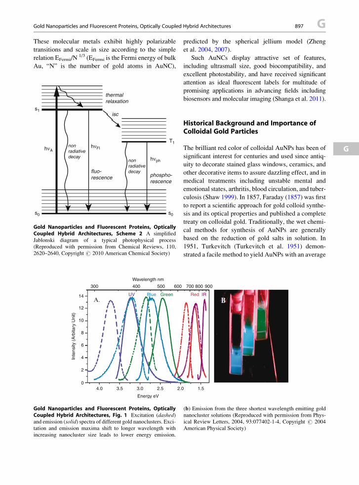
Gold Nanoparticles and Fluorescent Proteins, Optically Coupled Hybrid Architectures 897 G
These molecular metals exhibit highly polarizabletransitions and scale in size according to the simple
relation EFermi/N1/3 (EFermi is the Fermi energy of bulk
Au, “N” is the number of gold atoms in AuNC),
thermalrelaxation
nonradiativedecay
fluo-rescence
nonradiativedecay
phospho-rescence
hνph
hνFlhνA
s0 s0
s1isc
T1
Gold Nanoparticles and Fluorescent Proteins, OpticallyCoupled Hybrid Architectures, Scheme 2 A simplified
Jablonski diagram of a typical photophysical process
(Reproduced with permission from Chemical Reviews, 110,
2620–2640, Copyright # 2010 American Chemical Society)
300
A.
4.0 3.5 3.0
Energy eV
2.5 2.0
14
12
10
8
6
4
2
0
Wavelength nm
Inte
nsity
(A
rbita
ry U
nit)
400
UV Blue Green
500 600
Gold Nanoparticles and Fluorescent Proteins, OpticallyCoupled Hybrid Architectures, Fig. 1 Excitation (dashed)and emission (solid) spectra of different gold nanoclusters. Exci-tation and emission maxima shift to longer wavelength with
increasing nanocluster size leads to lower energy emission.
predicted by the spherical jellium model (Zheng
et al. 2004, 2007).
Such AuNCs display attractive set of features,
including ultrasmall size, good biocompatibility, and
excellent photostability, and have received significant
attention as ideal fluorescent labels for multitude of
promising applications in advancing fields including
biosensors and molecular imaging (Shanga et al. 2011).
G
Historical Background and Importance ofColloidal Gold Particles
The brilliant red color of colloidal AuNPs has been of
significant interest for centuries and used since antiq-
uity to decorate stained glass windows, ceramics, and
other decorative items to assure dazzling effect, and in
medical treatments including unstable mental and
emotional states, arthritis, blood circulation, and tuber-
culosis (Shaw 1999). In 1857, Faraday (1857) was first
to report a scientific approach for gold colloid synthe-
sis and its optical properties and published a complete
treaty on colloidal gold. Traditionally, the wet chemi-
cal methods for synthesis of AuNPs are generally
based on the reduction of gold salts in solution. In
1951, Turkevitch (Turkevitch et al. 1951) demon-
strated a facile method to yield AuNPs with an average
1.5
Red IR
700 800 900
(b) Emission from the three shortest wavelength emitting gold
nanocluster solutions (Reproduced with permission from Phys-
ical Review Letters, 2004, 93:077402-1-4, Copyright # 2004
American Physical Society)

G 898 Gold Nanoparticles and Fluorescent Proteins, Optically Coupled Hybrid Architectures
diameter of�20 nm, which was stabilized with citrate.
Citrates form a negatively charged electrical double
layer on the surface of AuNPs and provide them with
the required stability. However, citrate moieties do not
form very strong bonds with gold atoms, and such
AuNPs are not very stable. Brust et al. in 1994 (Brust
et al. 1994) introduced another protocol to synthesize
highly stable thiol-capped AuNPs. The covalent stabi-
lization of the AuNPs through Au–thiol interactions on
the surface leads to highly stable AuNPs. It is now been
well established that many functionalities can bind to
metallic gold, for example, sulfur-containing moieties
including thiols, thioethers, thioesters, disulfides,
isothiocyanates, phosphines, amines, citrate, and car-
boxylates to yield stable AuNPs. In all the cases,
a passivation layer on the surface of AuNPs prevents
their coalescence and stabilizes nanoparticles. Further
modification of the procedure has also been advanced
that includes the preparation of AuNPs/AuNCs using
weak ligands and consequently their exchange with
stronger ones for long-term stability and function.
Based on their capability of sequestering metal ions
from solution, dendrimers, and a variety of polymers,
biopolymers and block copolymers have also been
utilized as templates or directing agent to prepare
AuNPs and AuNCs (Daniel and Astruc 2010).
Use of Fluorescent Proteins as Templates/Directing Agent to Prepare AuNPs/AuNCs
In general synthesis of AuNPs using wet chemical
methods discussed above often involves the use of
harsh chemicals (e.g., surfactant) and sometimes
organic solvents (e.g., toluene) to control the size,
shape, and morphology of AuNPs. The presence of
such impurities and harsh chemicals in the final
product is unacceptable for many sensitive biomedi-
cal applications including biosensing, bioimaging,
drug and gene delivery, and cancer photothermal
therapy. This demand has recently prompted the use
of proteins and/or peptides as the template and reduc-
ing agent, which is inspired by the roles of such
molecules in natural biomineralization process.
Such biomaterials allow synthesis of AuNPs and
AuNCs of controlled shape, size, and organization
and function in aqueous solution at room temperature
under mild condition that also ensure biocompatibil-
ity (Tan et al. 2010).
Most proteins are biopolymers consisting of linear
polymer chain amino acid residues, and the combined
effect of all of the amino acid side chains in a protein
determines the physical, physicochemical, and chemi-
cal reactivity including three-dimensional structure.
Several independent studies have confirmed that
these functionalities are related to the presence of
certain amino acid residues in a peptide sequence.
Moreover, the large diversity of proteins and/or pep-
tides available for use through the traditional top-down
approach or the modern recombinant DNA technology
and combinatorial approach offers a unique opportu-
nity for exploration of the structure/property relation-
ship in peptides/proteins. Very recently, intrinsically
fluorescent proteins (IFPs) have also been utilized as
templates for synthesizing highly fluorescent hybrid
architectures. AuNCs that have opened the possibili-
ties of incorporation of biological fluorophores into
nanoparticle based devices with unique photophysical
properties.
Protein–Gold Nanoparticle Interactions:Exploration of the Key Chemistry
The formation of colloidal metal particles by proteins/
peptides requires two basic steps as well known for
conventional metal nanoparticle synthesis, namely,
(a) the reduction capability for metal ions and
(b) capping capability for the nanoparticles formed.
Some proteins and IFPs could be used as both the
reductant and the capping agent in metal nanoparticle
synthesis; and recently research has been focused on
developing a guiding principle through exploration of
the key chemistry of protein amino acid segments. Due
to the complexity in proteins, a variety of short peptide
sequences have been developed and used to identify
the specific functions of the 20 natural amino acid
residues in reduction of gold precursor such as
chloroaurate and binding to elemental gold particles
thus formed (Tan et al. 2010). Among the 20 standard
amino acids, tryptophan (Trp) has been identified as
the fastest reducing agent. However, amino acid
residues with heterocycles (histidine-His, Trp) and
sulfur (cysteine-Cys, methionine-Met) are strong
binders and form strong complexes with metal ions
(Lippard and Berg 1994); however, they lower the
redox potential of gold and hence the reducibility of
the metal ions. The amino acid residues with acidic and
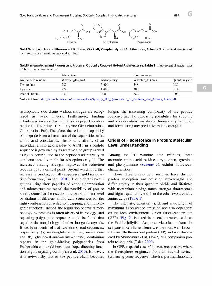
Tyrosine L-Tryptophan Phenylalanin
NH2
O
OH
NH2
OH
HO
O
NH2HN
OH
O
Gold Nanoparticles and Fluorescent Proteins, Optically Coupled Hybrid Architectures, Scheme 3 Chemical structure of
the fluorescent aromatic amino acid residues
Gold Nanoparticles and Fluorescent Proteins, Optically Coupled Hybrid Architectures, Table 1 Fluorescent characteristics
of the aromatic amino acidsa
Absorption Fluorescence
Amino acid residue Wavelength (nm) Absorptivity Wavelength (nm) Quantum yield
Tryptophan 280 5,600 348 0.20
Tyrosine 274 1,400 303 0.14
Phenylalanine 257 200 282 0.04
aAdopted from http://www.biotek.com/resources/docs/Synergy_HT_Quantitation_of_Peptides_and_Amino_Acids.pdf
Gold Nanoparticles and Fluorescent Proteins, Optically Coupled Hybrid Architectures 899 G
G
hydrophobic side chains without nitrogen are recog-
nized as weak binders. Furthermore, binding
affinity also increased with increase in peptide confor-
mational flexibility (i.e., glycine-Gly>glutamine-
Gln>proline-Pro). Therefore, the reduction capability
of a peptide is not a linear sum of the capabilities of its
amino acid constituents. The binding affinity of an
individual amino acid residue to AuNPs in a peptide
sequence is governed by its reactive side group as well
as by its contribution to the peptide’s adaptability to
conformations favorable for adsorption on gold. The
increased binding strength improves the reduction
reaction up to a critical point, beyond which a further
increase in binding actually suppresses gold nanopar-
ticle formation (Tan et al. 2010). The in-depth investi-
gations using short peptides of various composition
and microstructures reveal the possibility of precise
kinetic control at the reaction microenvironment level
by dialing in different amino acid sequences for the
right combination of reduction, capping, and morpho-
genic functions. Indeed, the regulation of crystal mor-
phology by proteins is often observed in biology, and
repeating polypeptide sequence could be found that
regulate the morphology of other inorganic crystals.
It has been identified that two amino acid sequences,
respectively, (a) serine–glutamic acid–lysine–leucine
and (b) glycine–alanine–serine–leucine, containing
repeats, in the gold-binding polypeptides from
Escherichia coli could introduce shape-directing func-
tion in gold crystal growth (Tan et al. 2010). However,
it is noteworthy that as the peptide chain becomes
longer, the increasing complexity of the peptide
sequence and the increasing possibility for structure
and conformation variations dramatically increase,
and formulating any predictive rule is complex.
Origin of Fluorescence in Protein: MolecularLevel Understanding
Among the 20 a-amino acid residues, three
aromatic amino acid residues, tryptophan, tyrosine,
and phenylalanine (Scheme 3), exhibit fluorescent
characteristics.
These three amino acid residues have distinct
photon absorption and emission wavelengths and
differ greatly in their quantum yields and lifetimes
with tryptophan having much stronger fluorescence
and higher quantum yield than the other two aromatic
amino acids (Table 1).
The intensity, quantum yield, and wavelength of
maximum fluorescence emission are also dependent
on the local environment. Green fluorescent protein
(GFP) (Fig. 2) isolated from coelenterates, such as
the Pacific jellyfish, Aequorea victoria, or from the
sea pansy, Renilla reniformis, is the most well-known
intrinsically fluorescent protein (IFP) and was discov-
ered by Shimomura et al. (1962) as a companion pro-
tein to aequorin (Tsien 2009).
In GFP, a special case of fluorescence occurs, where
the fluorophore originates from an internal serine–
tyrosine–glycine sequence, which is posttranslationally

Gold Nanoparticles and Fluorescent Proteins, OpticallyCoupled Hybrid Architectures, Fig. 2 3D structure of the
archetypical Aequorea victoria GFP.(a-Helices and b-strandsare shown as ribbons, connecting segments as tubes, and the
chromophore in ball-and-stick representation). (Reproduced
with permission from Science 1996, 273, 1392 –1395, Copyright
# 1996 American Association for the Advancement of Science)
G 900 Gold Nanoparticles and Fluorescent Proteins, Optically Coupled Hybrid Architectures
modified to a 4-(p-hydroxybenzylidene)- imidazolidin-
5-one structure The chromophore is a
p-hydroxybenzylideneimidazolinone formed from
residues 65–67, which are Ser–Tyr–Gly in the native
protein. Scheme 4 shows the currently accepted mech-
anism for chromophore formation.
First, GFP folds into a nearly native conformation,
then the imidazolinone is formed by nucleophilic
attack of the amide of Gly67 on the carbonyl group
of residue 65, followed by dehydration. Finally,
molecular oxygen (the process would stop in the
absence of O2) dehydrogenates the a-b bond of residue
66 to put its aromatic group into conjugation with the
imidazolinone, which would complete the fluorophore
(Heim et al. 1994). A variety of mutants of GFP,
creating new variants of GFP that shone more strongly
and in different colors – from blue to red and
yellow – have also been developed (Fig. 3).
These highly fluorescent IFPs have revolutionized
how the dynamics of cellular events can be imaged and
open up tremendous possibilities for continuously
monitoring gene expression, cell developmental fates,
and protein trafficking in living, minimally perturbed
cells, tissues, and organisms. Such IFPs have also been
used as reporters to monitor transcriptional regulation,
as targeted markers for organelles and subcellular
structures, and in sensors designed to show changes
in cellular environments ranging from pH to protein
kinase activity (Tsien 2009; Wang et al. 2004; Orm
et al. 1996). For the discovery and development of
GFP, the 2008 Nobel Prize in Chemistry has been
awarded jointly to O. Shimomura, M. Chalfie, and
R. Tsien (http://www.nobelprize.org/nobel_prizes/
chemistry/laureates/2008). In view of its enormous
potential, fluorescence-based detection strategies that
use quenched emission of fluorescence by IFP ligand
functionalized AuNPs have generated significant
recent interest in biotechnology for fluorescence
immunoassay and biosensors.
Fundamental of Fluorescence Process andDesign of Optically Coupled HybridArchitectures
Photo-induced excitation of a chromophore through
absorption of a photon of appropriate energy induces
the vibronic transition (simultaneous change of
vibrational and electronic quantum number) in
a fluorophore – commonly known as Franck–Condon
state – extremely rapidly (within 10�15 s) (Scheme 2).
The efficiency of this process is related to the chromo-
phore’s absorption cross section (s), which is propor-
tional to its extinction coefficient (e) (Sinkeldam et al.
2010). The Jablonski diagram for a typical fluorophore
is presented in Scheme 2, which illustrates the possible
chain of photophysical events that include the follow-
ing: (1) internal conversion or vibrational relaxation,
(2) fluorescence, intersystem crossing (from singlet
state to a triplet state), and (3) phosphorescence. The
vibrational relaxation (within 10�12–10�10 s) quickly
populates the lowest vibronic state of the chromo-
phore’s excited state (loss of energy without light
emission). This relaxation process generally accounts
for the observed Stokes shift – energy of emission
wave is lower (longer wavelength) when compared to
the excitation energy. Each of the processes occurs

NH
HO
HN
R
HN N
H
NH
NH
O
OTyr66
Gly67
Ser or Thr65(R = H or Me)
O
O
OH
OH
HO
folding (t1/2 ~ 10 min)
cyclization
dehydration
aerial oxidationO2 H2O2?
t1/2 ~ 3 min forcyclization + dehydration
t1/2 ~ 19–83 min
or other unfolded/denatured conformation
O
O
R
HNN
NH
O
OH
OH
HO
O
OHR
NN
NH
OHO
O
R
OH
NN
NH
OHO
O
R
Gold Nanoparticles andFluorescent Proteins,Optically Coupled HybridArchitectures, Scheme 4 A
tentative molecular
interpretation
posttranslational autoxidation
of the chromophore of greenfluorescent protein, which is
considered to be responsible
for the fluorescence in GFP
(Tsien 1998)
GFP-derived mRFP1-derived
EB
FP
EC
FP
EG
FP
YF
P (C
itrine)
mH
oneydew
mB
anana
mO
range
tdTomato
mTangerine
mS
trawberry
mC
herry
mG
rape1
mG
rape2
mP
lum
mR
aspberry
Evolved by SHM
Exc. 380
433/452 488 516
487/504 540 548 554 568 574 587 595 596 605 590 nmEm. 440 475/505 509 529 537/562 553 562 581 585 596 610 620 625 636 648 nm
Gold Nanoparticles and Fluorescent Proteins, OpticallyCoupled Hybrid Architectures, Fig. 3 Monomeric and tan-
dem dimeric fluorescent proteins derived from Aequorea GFP or
Discosoma RFP, expressed in bacteria, and purified. This photo
is a time exposure of fluorescences excited at different
wavelengths and viewed through different cutoff filters.
SHM ¼ somatic hypermutation (Reproduced with permission
fromAngewandte Chemie International Edition 48, 5612 –5626,Copyright # 2009, Wiley-VCH)
Gold Nanoparticles and Fluorescent Proteins, Optically Coupled Hybrid Architectures 901 G
G
with a certain probability, characterized by the decay
rate constants (k). The fluorescence lifetime of
a fluorophore is the characteristic time required by
a population of excited fluorophore to decaying back
to the ground state (t0) via the loss of energy through
fluorescence and other non-radiative processes.
Typical organic fluorophores reside in their excited
state for a very short period of �(0.5–20) � 10�9 s.
The fluorescence lifetime (t) of a fluorophore reflects
the emission quantum yield of the chromophores
(Q ¼ F ¼ t/t0). The product of the molar absorptivity
(e) and the fluorescence quantum yield (F) of
a fluorophore (eF) is recognized as the brightness
(Valeur and Nuno Berberan-Santos 2012). Fluores-
cence-based techniques have gained enormous pop-
ularity for their simplicity, versatility, and
sensitivity (up to a 1,000-fold higher than absorption
spectrophotometry). Moreover, creative probe

G 902 Gold Nanoparticles and Fluorescent Proteins, Optically Coupled Hybrid Architectures
design with selectivity to various environmental
parameters (pH, polarity, viscosity, presence of
quenchers, etc.) makes molecular fluorescence an
extremely effective tool for in vitro biophysical
and biochemical analyses, as well as in vivo cellular
imaging, capable of providing both spatial and
temporal information.
Nevertheless, the major limitations of the fluores-
cence probe include low photostability, high
background fluorescence and nonspecific background
signals. To achieve these specific goals, activatable
fluorescence probes have been designed. Such
activatable probes are designed with at least two com-
ponents, respectively, (1) the fluorophore that acts as
the donor and (2) the quencher that acts as the acceptor.
Such probes offer high specificity to the target of
interest and with superior signal to background ratio
in the imaging signal. Inorganic AuNPs show the
highest quenching efficiency (up to 99 %) and there-
fore can offer the highest sensitivity in the develop-
ment of activatable probes.
Optically Coupled Architectures Based on AuNP–
Fluorescent Protein Hybrid: Activatable Probes
The fluorescence-based assays, detection, and imaging
techniques are among the most sensitive and popular
biological tests. AuNPs are the most commonly used
optical sensing nanomaterials because of their strong
LSPR absorptions in the visible region, quenching
efficiency, facile preparation, high stability and bio-
compatibility. In addition, the surface chemistry of
AuNPs is versatile, allowing the linking of various
biofunctional groups (e.g., amphiphilic polymers,
silanols, sugars, nucleic acids, and proteins) through
strong Au–S or Au–N bonding or through physical
adsorption. AuNPs are considered as super quenchers
for fluorophores, because they have much stronger
molar absorptivity relative to the available organic
dyes. For example, the molar absorptivity of 13-nm
AuNPs at 518 nm is ca. 2� 108 cm�1 mol�l L. AuNPs
have Stern–Volmer quenching constants (KSV) that
are more than five orders of magnitude greater than
those of typical small-molecule dye quencher pairs.
The absorbance of AuNPs is dependent on the size
and geometry, for example, as their diameters increase,
their plasmon band red shifts. Hence, AuNPs are
highly tunable absorbing agents. AuNP surfaces can
be chemically conjugated with various biomolecules
and targeting agents using facile and straightforward
manner. Typically, effective quenching results
when the distance between the fluorophore and the
AuNP surface is less than 5 nm. In addition, size,
shape, and the surface characteristics of the AuNPs
are all important parameters that affect fluorescence
quenching. The past few years have witnessed the
development of many successful strategies for the
preparation of high-quality AuNPs and of numerous
AuNP-based sensing systems, and activatable probes
appear to be the most reliable molecular probe design
architecture.
The fluorescence activatable probes, also referred to
as molecular beacons, are designed from a donor–
quencher conjugates, and the efficiency of quenching
is dependent on the distance between these two com-
ponents. In the native state, these two components are
in close proximity, and quenching occurs efficiently;
however, with increase of distance between the donor
and the quencher in response to the external stimuli/
analyte, fluorescence is effectively restored (Fig. 4).
Activatable probes have been designed with various
quenchers, fluorophores, and biomolecules; however,
fluorescence-based detection strategies that use
quenched emission of fluorescence by IFP ligand
functionalized AuNPs have generated significant
recent interest in biotechnology for fluorescence
immunoassay and biosensors (Swierczewska et al.
2011). Hazarika et al. (2006) reported reversible bind-
ing of yellow fluorescent protein (EYFP) – a mutant
derivative of the naturally occurring green fluorescent
protein from the North Pacific jellyfish Aequorea
victoria at AuNP hybrids without compromising
their biological activity. Dutta and coworkers
(Mayavan et al. 2011) investigated the synthesis of
optically coupled hybrid architectures based on
a new biomimetic fluorescent protein rec1-resilin
and nanometer-scale AuNPs in one-step method
using a non-covalent mode of binding protocol. The
presence of uniformly distributed fluorophore amino
acid sequences, serine (threonine)–tyrosine–glycine,
along the molecular structure of rec1-resilin provides
significant opportunity to synthesize fluorophore-
modified AuNPs bioconjugates with unique
photophysical properties. The final properties of the
protein–AuNP hybrids are very sensitive not only to
the chemical composition of the protein and the size of
AuNPs but also on their topological organization
including the distance of the fluorophores from AuNP

GFPFluorescence ‘off’
Protein 2
Protein 2
Protein 3
Protein 3Protein 1
Protein 1
Aggregation
Low displacement
High displacement
Flu
ores
cenc
e re
spon
se
Gold Nanoparticles andFluorescent Proteins,Optically Coupled HybridArchitectures,Fig. 4 Schematic of
competitive binding of green
fluorescent protein (GFP) onto
monolayer-protected AuNPs.
The competitive binding of
GFP protein to the AuNP is
affected by specific types of
proteins, which, in turn,
affects the efficacy of
fluorescence quenching.
Different proteins can
therefore be identified based
on the shifts in fluorescence
intensity (Reproduced with
permission from Nature
Chemistry, 1: 461–465.
Copyright # 2009 Macmillan
Publishers Limited)
Gold Nanoparticles and Fluorescent Proteins, Optically Coupled Hybrid Architectures 903 G
G
surfaces as discussed before. In DNA molecular bea-
cons, AuNPs have been used as an effective proximal
quencher (Kobayashi et al. 2010).
Further advancement has also been made to synthe-
size AuNPs using engineered fluorescent protein as
reductant and/or capping agent applied to bio- and
chemosensing. Rotello and co-workers (De et al.
2009) have demonstrated the power of protein
activatable fluorescent probe from conjugates of
AuNPs and green fluorescent protein in rapid and
efficient sensing of proteins in human serum with
specificity. Figure 4 illustrates the use of competitive
binding of GFP to the AuNP, which is affected by
the presence of specific types of proteins in the
environment that in turn affects the efficacy of fluores-
cence quenching. Such specific recognition and
quantification is attractive, because there is a direct
correlation between protein levels and disease states
in human serum, but challenging (serum features more
than 20,000 proteins, with an overall protein content
greater than 1 mM). Using such probe, reproducible
fluorescence response patterns with an identification
accuracy of 100 % in buffer and 97 % in human serum
have been reported from five major serum proteins
(human serum albumin, immunoglobulin G, transfer-
rin, fibrinogen, and a-antitrypsin). The arrays have
been demonstrated to be enabled to discriminate
between different concentrations of the same protein
as well as a mixture of different proteins in human
serum. Such chemical noses can sense proteins with
over 97 % accuracy using in-depth understanding of
the fluorescence quenching characteristics of the

BSA
+
Incubate 2 min
Incubate 12 h
BSA-Au lon Complex BSA-Au NC
+ NaOH
AuCl4−
Gold Nanoparticles and Fluorescent Proteins, OpticallyCoupled Hybrid Architectures, Fig. 5 Schematic represen-
tation of the formation of AuNCs in BSA solution. Upon addition
ofAu(III) ions to the aqueous BSA solution, the proteinmolecules
sequestered Au ions and entrapped them. The reducing capability
of BSA is subsequently activated by adjusting the pH to about 12
with NaOH, so the entrapped ions underwent progressive reduc-
tion to form AuNCs in situ (Reproduced with permission from
Journal of the American Chemical Society 131, 888–889, Copy-right# 2009 American Chemical Society)
G 904 Gold Nanoparticles and Fluorescent Proteins, Optically Coupled Hybrid Architectures
molecular probe along with interplay of electrostatic
and hydrophobic interactions. So far, AuNP activatable
probe studies havemainly been fixed tomicrotiter plates
to use them as diagnostic tools. Nevertheless, AuNPs
are widely accepted to be noncytotoxic, biocompatible,
and exhibit good cellular uptake and could be extended
to in vivo study.
Optically Coupled Hybrid Architectures Based on
AuNC–Fluorescent Protein: Ratiometric
Fluorescent Probe
Optically coupled hybrid architectures based on
AuNC–fluorescent protein bioconjugates have the
unique potential to display dual fluorescence
emissions – one from the AuNC and the other from
the IFP that could be further modulated through the
control of AuNC size and morphology. This dual
fluorescence offers a distinct advantage over the most
fluorescence-AuNP sensors as discussed above, which
normally exhibit a single signal fluorescence output on
the basis of quenching or recovery. Two
well-separated emission bands with comparable inten-
sities from AuNC–IFP have two distinct advantages:
(1) the large shift (�100 nm) between excitation and
emission peak of AuNC, and (2) presence of two
well-separated emission bands with comparable inten-
sities ensures accuracy in determining their intensities
and ratios. Ratiometric fluorescencemethods can elim-
inate most or all the ambiguities by using the ratio of
two emission bands to demonstrate the quantitative
analysis ability independent of fluorophore concentra-
tion and environmental conditions.
The power of common protein bovine serum albu-
min (BSA) in both sequestration and reduction of Au
precursors to generate highly fluorescent AuNCs has
been clearly demonstrated (Xie et al. 2009). AuNC–
BSA was prepared by reduction of chloroauric acid
with bovine serum albumin (BSA) based on the capa-
bility of BSA to sequester and reduce Au precursors in
situ (Fig. 5).
The presence of 21 Tyr residues in BSA is consid-
ered to be responsible for the reduction reaction; Fig. 5
presents a schematic representation of the formation of
AuNCs in BSA solution. Compared with short pep-
tides, large and complicated proteins possess abundant
binding sites that can potentially bind and further
reduce metal ions, thus offering better scaffolds for
template-driven formation of small metal NCs. The
BSA–AuNC coupled hybrids exhibit dual emissions:
the blue at �425 nm from the oxides of BSA and the
red at �635 nm from AuNCs. Such BSA–AuNCs
colloidal solutions are highly stable both in solutions
(aqueous or buffer) and in the solid form. The power of
metal-ion modulated ratiometric fluorescence probe

(Metal ions)-(BSA-AuNCs)
a
b
Metal ions Amino acids
Gold Nanoparticles andFluorescent Proteins,Optically Coupled HybridArchitectures, Fig. 6 (a)Illustration of The
fluorescence ratiometric
response to amino acids by the
use of metal ion-modulated
AuNCs. (b) Schematic
drawings for the identification
of amino acids by ratiometric
fluorescence probe array
(Reproduced with permission
from Analyst, 137,
1618–1623, Copyright
# 2012 RSC Publishing)
Gold Nanoparticles and Fluorescent Proteins, Optically Coupled Hybrid Architectures 905 G
G
array constructed from such BSA–AuNCs hybrids for
the detection and identification of amino acids has
been recently demonstrated (Wang et al. 2012) as
illustrated in Fig. 6.
Metal ions easily bind with BSA–AuNCs and thus
greatly affect the two emissions (�425 nm and
�635 nm, respectively) in different ways by fluores-
cence enhancement or quenching. Upon the addition of
amino acids, the metal ion-modulated BSA–AuNCs
exhibit fluorescence ratiometric responses. The environ-
ment and type of interaction of the specific metal ions
with the BSA modulate the intensities of the two fluo-
rescence peaks of AuNC–BSA differently. Figure 6a
illustrates the typical fluorescence ratiometric response
to amino acids by the use of metal ion-modulated
AuNCs. For example, the presence of Ni2+ in the metal
ion-modulated BSA–AuNCs quenches both the fluores-
cence emissions, while the addition of Zn2+ only
enhances the red fluorescence. Following the addition
of amino acids into the bioconjugates, stronger interac-
tion occurs between the amino acids and the metal ions,
compared with BSA that may induce either the disasso-
ciation of metal ions from BSA–AuNCs or the aggrega-
tion of AuNCs. Consequently, the two fluorescence
emissions from BSA–AuNCs independently change
with different amino acids, and the resultant evolution
of fluorescence ratios can be employed as an efficient
indicator of amino acids (Wang et al. 2012). The dual
emissions of BSA–AuNCs modulated by a specific
metal ion offer four different possible fluorescence
responses: (1) both fluorescence quenching, (2) both
enhancements, (3) one quenching and another enhance-
ment, and (4) one enhancement and another quenching.
Therefore, a modulated BSA–AuNCs ratiometric fluo-
rescent probe array designed with integration of four
different metal ions, respectively, Ni2+, Pb2+, Zn2+, and
Cd2+ and designated as BSA–AuNC–Ni2+, BSA–
AuNCs–Pb2+, BSA–AuNCs–Zn2+, and BSA–AuNCs–
Cd2+ can essentially display 256 kinds of different sig-
nals, theoretically. Using such an intelligent probe,
a specific fluorescence pattern obtained for a specific
amino acid can be easily recognized as the characteristic

a b
c
d
255
160
64
0
Gold Nanoparticles and Fluorescent Proteins, OpticallyCoupled Hybrid Architectures, Fig. 7 In vivo fluorescence
image of 100 ml. (a) AuNCs injected subcutaneously and (b)intramuscularly into the mice. (c) Real-time in vivo abdomen
imaging of intravenously injected with 200 ml of AuNCs at
different time points post-injection. (d) Ex vivo optical imaging
of anatomized mice with injection of 200ml of AuNCs and some
dissected organs during necropsy. The organs are liver, spleen,
left kidney, right kidney, heart, lung, muscle, skin, and intestine
from left to right (Reproduced with permission from Nanoscale,
2, 2244–2249. Copyright # 2010 RSC Publishing)
G 906 Gold Nanoparticles and Fluorescent Proteins, Optically Coupled Hybrid Architectures
fingerprint. Various amino acids were identified by the
fluorescence ratiometric array composed of such four
ratiometric probes with each amino acid added into the
four probes to obtain the distinct pattern on the changes
of fluorescence ratio. Figure 6b represents the likely
mechanism for the detection of amino acids. It is well
understood that larger complex protein BSA easily binds
to various metal ions such as Ni2+, Pb2+, Zn2+, and Cd2+
due to the presence of large amount of residual amino,
carboxylic and mercapto groups, and disulfide bridges
at the peptide chains. This work is remarkable not only
for providing an alternative approach of synthesizing
highly fluorescentAuNCs but also because the proposed
strategy may be extended to other proteins and metals.
The use of BSA–AuNCs coupled hybrids has also
been explored as novel contrast imaging agents for
tumor fluorescence imaging in vivo (Wu et al. 2010)
as illustrated in Fig. 7.
It was clearly demonstrated that the light level
produced by the BSA–AuNCs hybrid is sufficient to
provide a localised signal under a few millimeters of
tissues with a subcutaneous injection. Furthermore,
using MDA-MB-45 and Hela tumor xenograft models,
in vivo and ex vivo imaging studies show that the
ultrasmall NIRAuNCs are able to be highly accumulated
in the tumor areas due to enhanced permeability and
retention (EPR) effects and yield excellent contrast from
the surrounding tissues. The results indicate that the
ultrasmall NIRAuNCs appear as very promising contrast
imaging agents for in vivo fluorescence tumor imaging.
Future Outlook
The dramatically changed characteristics of colloidal
gold particles based on the size, shape, geometry,mor-
phology and interparticle distance, and the environment
are currently most intensely investigated. We envisage
that these modulation in characteristics –particularly the
absorption, plasmonic, and fluorescence characteristics –
will enable them for many potential applications in
near future for a wide range of areas, including nano-
technology, catalysis, medicine, immunohistochemistry
biomarkers, and sensors. The gold-fluorescent protein-
based hybrid colloidal gold nanoparticles with range in
size from 2 to 100 nm will find wider applications as

Gold Nanoparticles and Fluorescent Proteins, Optically Coupled Hybrid Architectures 907 G
G
labeling, delivery, heating, and sensors. The molecular
beacons and activatable probes from AuNP–fluorescent
protein will find applications in new and exciting ways
for fluorescence-based supersensitive assays and detec-
tions. Fluorescence-based activatable probes can be used
in vitro and in vivo as cellular and clinical bioimaging
agents. The fluorescence characteristics of ultrasmall
size of AuNCs can provide extraordinary advantages
over quantum dots and conventional fluorescent dyes
owing to their excellent biocompatibility, robust resis-
tance to photobleaching, and facile synthesis. They are
able to sustain a longer blood circulation time than other
classical PEGylated nanomaterials and enjoy superior
binding efficiency towards tumor through “enhanced
permeability and retention.” Dual fluorescence emis-
sions from protein–AuNCs-based probes can be greatly
modulated in presence of other metal ions and com-
pounds, and the resultant fluorescence ratiometric
responses will be identified as a novel sensory method
for identification and quantification of different analytes
including heavy metals as pollutant, proteins in blood
serum. Such AuNC hybrid with further inclusion of
vector and drug molecules specific to cancer cells has
the potential to make significant in road in tumor diag-
nostic and therapeutic markets in the continued fight
against disease.
Acknowledgment This work has been financially supported by
the Australian Research Council.
Cross-References
▶Biomineralization of Gold Nanoparticles from Gold
Complexes in Cupriavidus Metallidurans CH34
▶Gold Nanomaterials as Prospective Metal-based
Delivery Systems for Cancer Treatment
▶Gold Nanoparticle Platform for Protein-Protein
Interactions and Drug Discovery
▶Gold Nanoparticles and Proteins, Interaction
▶Gold, Ultrasmall Nanoclusters and Proteins,
Interaction
References
Brust M, Walker M et al (1994) Synthesis of thiol-derivatized
gold nanoparticles in a two-phase liquid–liquid system.
J Chem Soc, Chem Commun 7:801–802
Daniel M-C, Astruc D (2004) Quantum-size-related properties
and applications toward biology, catalysis and nanotechnol-
ogy. Chem Rev 104:293–346
De M, Rana S et al (2009) Sensing of proteins in human serum
using conjugates of nanoparticles and green fluorescent
protein. Nat Chem 1:461–465
Faraday M (1857) Experimental relations of gold (and other
metals) to Light. Philos Trans 147:145–181
Hazarika P, Kukolka F et al (2006) Reversible binding of fluo-
rescent proteins at DNA-gold nanoparticles. Angew Chem
Int Ed 45:6827–6830
Heim R, Prashert DC et al (1994) Wavelength mutations and
posttranslational autoxidation of green fluorescent protein.
Proc Natl Acad Sci USA 91:12501–12504
Kelly KL, Coronado E (2003) The optical properties of metal
nanoparticles: the influence of size, shape, and dielectric
environment. J Phys Chem B 107:668–677
Kobayashi H, Ogawa M et al (2010) New strategies for fluores-
cent probe design in medical diagnostic imaging. Chem Rev
110:2620–2640
Lippard SJ, Berg JM (1994) Principles of bioinorganic chemis-
try. University Science Books, Mill Valley
Mayavan S, Dutta NK et al (2011) Self-organization, interfacial
interaction and photophysical properties of gold nanoparticle
complexes derived from resilin-mimetic fluorescent protein
rec1-resilin. Biomaterials 32:2786–2796
Mie G (1908) Beitr€age zur Optik tr€uber Medien, speziell
kolloidaler Metallosungen. Ann Phys 25:377–445
Orm M, Cubitt AB et al (1996) Crystal structure of the
Aequorea victoria green fluorescent protein. Science
273:1392–1395
Shanga SL, Dongb S et al (2011) Ultra-small fluorescent metal
nanoclusters: synthesis and biological applications. Nano
Today 6:401–418
Shaw CF (1999) Gold-based medicinal agents. Chem Rev
99:2589–2600
Sinkeldam RW, Greco NJ et al (2010) Fluorescent analogs of
biomolecular building blocks: design, properties, and appli-
cations. Chem Rev 110:2579–2619
Swierczewska M, Lee S et al (2011) The design and application
of fluorophore–gold nanoparticle activatable probes. Phys
Chem Chem Phys 13:9929–9941
Tan YN, Lee JY et al (2010) Uncovering the design rules for
peptide synthesis of metal nanoparticles. J Am Chem Soc
132:5677–5686
Tsien RY (1998) Annu Rev Biochem 67:509–44
Tsien RY (2009) Constructing and exploiting the fluorescent
protein paintbox (Nobel Lecture). Angew Chem Int Ed
48:5612–5626
Turkevitch J, Stevenson PC et al (1951) Nucleation and growth
process in the synthesis of colloidal gold. Discus Faraday Soc
11:55–75
Valeur B, Nuno Berberan-Santos M (2012) Molecular fluores-
cence: principles and applications, 2nd edn. Wiley-VCH,
Weinheim
Wang LWC, Jackson WC et al (2004) Evolution of new
nonantibody proteins via. iterative somatic hypermutation.
Proc Natl Acad Sci USA 101:16745–16749
Wang M, Mei Q et al (2012) Protein-gold nanoclusters for
identification of amino acids by metal ions modulated
ratiometric fluorescence. Analyst 137:1618–1623

G 908 Gold Nanoparticles and Proteins, Interaction
Wu X, He X et al (2010) Ultrasmall near-infrared gold
nanoclusters for tumor fluorescence imaging in vivo.
Nanoscale 2:2244–2249
Xie J, Zheng Y et al (2009) Protein-directed synthesis of highly
fluorescent gold nanoclusters. J Am Chem Soc 131:888–889
Zheng J, Zhang C et al (2004) Highly fluorescent, water-soluble,
size-tunable gold quantum dots. Phys Rev Lett 93:077402-
1–077402-4
Zheng J, Nicovich PR et al (2007) Highly fluorescent noble-
metal quantum dots. Annu Rev Phys Chem 58:409–431
Gold Nanoparticles and Proteins,Interaction
Ricardo Franco1 and Eulalia Pereira2
1REQUIMTE FCT/UNL, Departamento de Quımica,
Faculdade de Ciencias e Tecnologia, Universidade
Nova de Lisboa, Caparica, Portugal2REQUIMTE, Departamento de Quımica e
Bioquımica, Faculdade de Ciencias da Universidade
do Porto, Porto, Portugal
Synonyms
Gold nanoparticles interaction with proteins; Protein-
gold nanoparticle conjugation
Definition
Gold nanoparticles are metallic gold entities with
a diameter of 1–100 nm. They are surrounded by
a capping layer that imparts chemical functionality to
the nanoparticle. In the presence of proteins, gold
nanoparticles interact with these biomolecules, and
the nature of this interaction mainly depends on local
chemical and physical effects.
Historical Background
Gold nanoparticles (AuNPs) have been used as
radioactive labels in vivo since the 1950s. Since the
1980s, AuNPs have been conjugated with targeting
antibodies and used as density probes for biological
transmission electron microscopy. The interest in the
study of the interaction of AuNPs with proteins started
when research in nanotechnology found that the
unique nanoscale properties of AuNPs and their simi-
larity in size to biomolecules and organelles, make
them ideal partners for the development of sensors,
drug delivery vehicles, and in vivo imaging agents. In
fact, from the viewpoint of biological and medical
applications, the primary interest in nanoparticles
(NPs) stems from the fact that they are small enough
to interact with cellular machinery and to potentially
reach previously inaccessible targets, such as the brain
(Mahmoudi et al. 2011). For in vivo applications,
AuNPs are practically nontoxic and easy to
functionalize with appropriate capping agents. These
functionalized AuNPs are robust delivery/imaging sys-
tems, in which the gold core imparts stability to the
assembly, while the capping monolayer allows tuning
of surface properties, such as charge, hydrophobicity,
and binding affinity.
Protein-Gold Nanoparticle Interactions atthe Molecular Level
When proteins come in contact with AuNPs, they
become adsorbed at the surface of the NP due to van
derWaals forces. The initial adsorption depends on the
nature of the surfaces of both the AuNP and the
protein. In order to better understand this process it
is, thus, necessary to first have a closer look at the
AuNP surface. In a bare metal, surface atoms are less
stable than those within the core. This instability of the
surface atoms is primarily due to the lower number of
neighbors relative to core atoms. This lower number
of neighbors in surface atoms can be partially compen-
sated by adsorption of other species. This lower stabil-
ity of surface atoms is also the underlying reason for
spontaneous growth of nanostructures to macroscopic
structures, since the ratio between the number of
surface atoms and the number of total atoms decreases
as the diameter of the NP increases. For spherical
or cubic particles this fraction is inversely proportional
to the cubic root of the total number of atoms. In order
to prevent the growth of NP into macroscopic mate-
rials, it is, therefore, necessary to use an agent that can
lower the surface energy of the NP by adsorbing to
surface atoms. These agents, commonly called capping
agents, can be small molecules or polymers, and are
frequently used as co-reagents in NP synthesis. In
addition to their role as stabilizers of the surface
atoms, preventing undesired growth, the capping

Gold Nanoparticles and Proteins, Interaction 909 G
G
agent also improves dispersion of the NPs in the sol-
vent used for the synthesis (Sperling and Parak 2010).
For AuNPs in aqueous solution, the most typical cap-
ping agent is citrate that confers a strongly negative
surface charge to AuNPs. This high charge not only
hinders the close contact between AuNPs in solution,
thus avoiding aggregation, but also provides a strong
interaction with the surrounding water molecules,
promoting colloidal stability. Other capping agents
commonly used for AuNPs are small molecules
with a terminal thiolate functional group. The most
representative molecule in this group is
11-mercaptoundecanoic acid (MUA). Thiolates are
known to have a very high affinity for gold surfaces.
In addition, MUA has a 11 carbon chain that forms
a packed layer around the NP, reminiscent of the struc-
ture of a micelle, which imparts slow desorption of
MUA and hinders diffusion of other species to the
AuNP surface. Finally, the carboxylate functional
group is directed toward the surface, conveying
a highly negative charge to the surface of the NPs.
The capped AuNPs may, thus, be viewed as
a composite NP made up of a gold metal core and
a monolayer of the organic capping agent, the latter
forming the surface in direct contact with the solvent.
Other thiolate functionalized capping agents imparting
characteristic surface properties to the AuNPs are com-
monly used, namely, to obtain specific hydrophilic/
hydrophobic surfaces, surface charge, and/or binding
properties. The adsorption of the capping agent is
a dynamic process, that is, the capping agent can be
exchanged with solvent molecules, ions, and other
molecules in solution, potentially decreasing the col-
loidal stability of the AuNPs in solution. In this respect,
citrate is considered a labile capping agent, because the
exchange rate is fast, whereas MUA and other thiolate
capping agents show much slower exchange rates.
The adsorption process of the protein molecule is
controlled by van der Waals forces between the NP
surface and the protein. These can be electrostatic
attraction forces between the charged surface of the
AuNP (more commonly negatively charged) and
charged residues on the protein; hydrogen bonding
between the capping molecules and the protein;
acid-base adduct formation; dipole-dipole interac-
tions; and London (dispersion) forces. In addition,
when adsorption takes place, the protein replaces
ions, water molecules, and/or capping agent molecules
on the AuNP surface, while the AuNP replaces the
local solvent shell at the protein surface. The combined
effect of all these interactions can be strong enough to
partly overcome the intramolecular interactions
responsible for the tertiary structure of the protein,
eventually leading to conformational changes. The
extent of these conformational changes depends not
only on the chemical properties of the AuNPs surface,
mostly imparted by the capping agent, but also on the
robustness of the tertiary structure of the protein.
Moreover, it must be emphasized that the conforma-
tion stability is not usually the same for each of the
protein domains, and thus, adsorption may or may not
significantly affect the structure/function of the pro-
tein. Experimentally, both types of behavior have been
observed.
Often, the adsorption of protein molecules takes
place in a two-step process: a first fast adsorption
occurs due to the electrostatic interaction between the
charged surface and opposite charges in the protein
surface; then slow rearrangement may occur, that
may or may not lead to replacement of the capping
agent and cause direct interaction of protein molecules
with the metal surface. The latter slow reorganization
step can involve interactions with internal residues of
the protein (hydrophobic interactions; cysteine adsorp-
tion at the gold surface, etc.), leading to extensive
protein conformational changes.
Many proteins have a prolate ellipsoid shape giving
rise to two possible orientations relative to the metal
surface: “side-on” and “end-on.” Side-on adsorption is
usually stronger than end-on adsorption, mainly
because in the side-on orientation, the contact area
between the two nano-objects is larger (Fig. 1). This
may lead to irreversible adsorption, whereas end-on
adsorption is more usually reversible. It is also to be
expected that conformational changes induced by the
nanoparticle should be more severe in side-on
adsorption.
Many concepts given above are common to the
adsorption of proteins to macroscopic surfaces.
Nevertheless, other effects arise when the size of the
particles is similar to the size of the proteins adsorbed.
Figure 2 shows the relative sizes of selected proteins
and of AuNPs at the most commonly used sizes.
For instance, the conformational changes induced
by adsorption of proteins at flat macroscopic surfaces
are commonly more severe than at NPs. Moreover,
the distortion is also more intense at larger NPs
(low curvature) than at smaller NPs (high curvature).

Gold Nanoparticles and Proteins, Interaction,Fig. 1 Possible orientations of a prolate ellipsoid-shaped pro-
tein relative to the metal surface, upon conjugation with AuNPs.
The side-on adsorption orientation has a larger contact surface
area compared to the end-on adsorption orientation
Gold Nanoparticles and Proteins, Interaction,Fig. 2 Relative sizes of AuNPs (most common) and represen-
tative proteins. Proteins are IgG Monoclonal Antibody (IgG)
(PDB ID: 1IGY); Human Serum Albumin (HSA) (PDB ID:
1AO6); and Horse Heart Cytochrome c (Cyt c) (PDB ID: 1HRC)
G 910 Gold Nanoparticles and Proteins, Interaction
This curvature effects are related to the decrease in
contact area available for interaction when going from
a flat surface to a curved surface (Fig. 3). As the size of
NPs approaches the size of the proteins, the curvature
effect becomes extremely important. These curvature
effects are most commonly observed for globular pro-
teins. Fibrillar proteins, on the other hand, often show
large conformational changes in high curvature sur-
faces. This may be explained by side-on adsorption
and wrapping of the protein around the nanoparticle.
Another important size effect arises from the lateral
protein-protein interactions that often cause cooperative
or anti-cooperative effects in proteins adsorbed at flat
surfaces (crowding effect) (Fig. 3). The high curvature
of NPs decreases the lateral interactions, with obvious
advantages formaintaining biological activity by expos-
ing the protein to the solvent and thus, keeping the
protein available for biological interactions (e.g., with
substrate in the case of enzymes, or to antigen binding
for antibody-AuNP conjugates). In some cases, how-
ever, lateral protein-protein interactions are found to be
necessary for structure stabilization and in these cases,
the decrease in size, and hence the increase in the
curvature of the surface, can be detrimental to the bio-
logical functions of adsorbed proteins. The increase in
NP curvature and the resultant increased surface area
determine an increased number of protein motifs on the
“outer” surface of protein exposed to the medium. This
may in turn lead to avidity effects arising from a dense
location of actively interacting motifs on the NP surface
(Shemetov et al. 2012).
In conclusion, adsorption of protein molecules at
AuNP is a complex process that, depending on the
conformational stability of the protein and the surface
properties and morphology of the AuNPs, can lead to
more or less severe changes in the protein structure.
For a specific protein, the key factor for the adsorption
process is the chemical composition of the surface of
the NP, usually imparted by the capping agent directly
surrounding the AuNP. A current common strategy for
improving the properties of protein-AuNP conjugates
is to alter the surface chemistry of the AuNP, by
changing the capping agent, thus modulating the affin-
ity and denaturing properties of the AuNPs.
Characterization Techniques
Conjugates between AuNPs and proteins can be
formed by incubation of both components, namely,
AuNPs with an appropriate capping agent and the
protein containing solution. Alternatively, complex
protein-containing mixtures, such as plasma, can be
incubated with AuNPs and further characterized for

Gold Nanoparticles andProteins, Interaction,Fig. 3 AuNP curvature
effects on protein adsorption
modes. The contact area
between protein and AuNP is
larger in low curvature NPs (a)than in high curvature NPs (b);lateral protein-protein
interaction (“crowding
effect”) is more extensive in
low curvature NPs (c) than in
high curvature NPs (d)
Gold Nanoparticles and Proteins, Interaction 911 G
G
determination of the quality and amount of AuNP-
bound proteins (Dobrovolskaia et al. 2009; Mahmoudi
et al. 2011). Protein to AuNP binding can occur by
electrostatic-hydrophobic interaction, or covalent
strategies can be utilized. The latter are of two main
types: (a) covalent binding to the AuNP of a thiol from
the side chain of a cysteine residue present in the
protein; or (b) cross-linking chemistry by formation
of an amide bond between carboxylate surface groups,
functionalizing the AuNPs, and available amine
groups in the protein, using EDC/NHS (1-Ethyl-3-
(3-dimethylaminopropyl)-carbodiimide hydrochlo-
ride/ N-hydroxysuccinimide). Covalent binding
strategies have to be considered with great care, as
they often induce enzyme activity loss and deleterious
protein structural alterations. In the case of conjugate
formation from protein mixtures, these covalent strat-
egies should be altogether avoided due to their lack of
specificity. The cross-linking chemistry approach for
covalent binding has been used for AuNP-antibody
conjugates without appreciable loss of antigen affinity
by the antibody, and sometimes with improved results
in immunoassays.
AuNP-protein conjugate isolation is a complicated
process. A common method to achieve the goal of
obtaining pure conjugates is centrifugation followed
by washing to remove unbound or weakly bound pro-
tein(s). Other common biochemical separation
techniques that can be used to isolate AuNP-protein
conjugates are gel exclusion chromatography and
membrane ultrafiltration. In the case of protein
mixtures, it is important that separation techniques
have the capability to separate proteins with various
affinities, without any disruption of the conjugates, as
well as avoiding the creation of new binding opportu-
nities (Mahmoudi et al. 2011).
Agarose gel electrophoresis is a powerful tool
toward identification of protein-AuNP conjugates.
Protein adsorption at the surface of a capped AuNP
leads to reduced electrophoretic mobility, mainly due
to reduction of the superficial charge of the AuNPs.
This variation of electrophoretic mobility can in some
cases be directly related to the degree of protein
coverage of the AuNPs. For example, if increasing
concentrations of a protein are incubated with citrate-
capped AuNP, the resulting conjugates will present
lower electrophoretic mobility toward the anode. Elec-
trophoretic mobility data can be plotted versus protein
to AuNP concentration ratios and fitted to a Langmuir-
type behavior (Fig. 4).
The highest electrophoretic mobility values corre-
spond to full protein coverage of the AuNP that can be
attained at known protein concentrations in solution.
Agarose gel electrophoresis of AuNP-protein conju-
gates is still lacking the quantitative detail that could
already be achieved for nucleic acid-AuNP conjugates,
in which single oligonucleotide discrimination is pos-
sible (Sperling and Parak 2010). Experimental optimi-
zation can, nevertheless, lead to qualitative results in
terms of compactness of the protein corona, and quan-
titative results in terms of maximal protein coverage of
the AuNPs.
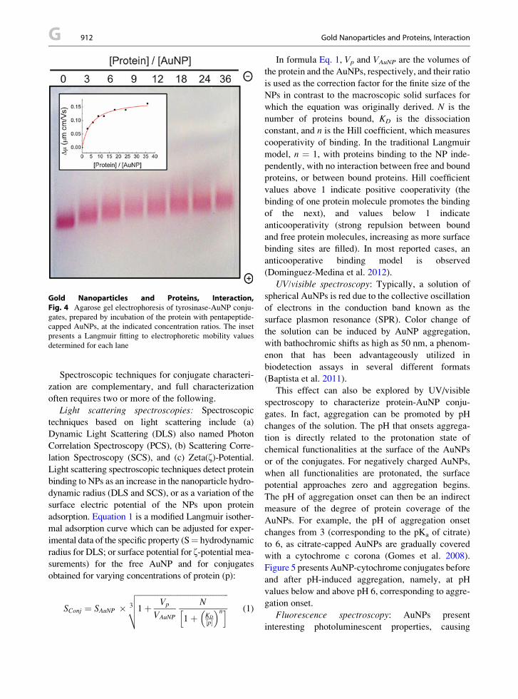
Gold Nanoparticles and Proteins, Interaction,Fig. 4 Agarose gel electrophoresis of tyrosinase-AuNP conju-
gates, prepared by incubation of the protein with pentapeptide-
capped AuNPs, at the indicated concentration ratios. The inset
presents a Langmuir fitting to electrophoretic mobility values
determined for each lane
G 912 Gold Nanoparticles and Proteins, Interaction
Spectroscopic techniques for conjugate characteri-
zation are complementary, and full characterization
often requires two or more of the following.
Light scattering spectroscopies: Spectroscopic
techniques based on light scattering include (a)
Dynamic Light Scattering (DLS) also named Photon
Correlation Spectroscopy (PCS), (b) Scattering Corre-
lation Spectroscopy (SCS), and (c) Zeta(z)-Potential.Light scattering spectroscopic techniques detect protein
binding to NPs as an increase in the nanoparticle hydro-
dynamic radius (DLS and SCS), or as a variation of the
surface electric potential of the NPs upon protein
adsorption. Equation 1 is a modified Langmuir isother-
mal adsorption curve which can be adjusted for exper-
imental data of the specific property (S¼ hydrodynamic
radius for DLS; or surface potential for z-potential mea-
surements) for the free AuNP and for conjugates
obtained for varying concentrations of protein (p):
SConj ¼ SAuNP � 3
ffiffiffiffiffiffiffiffiffiffiffiffiffiffiffiffiffiffiffiffiffiffiffiffiffiffiffiffiffiffiffiffiffiffiffiffiffiffiffiffiffiffiffiffiffiffi
1þ Vp
VAuNP
N
1þ KD
½p � �nh i
vuut (1)
In formula Eq. 1, Vp and VAuNP are the volumes of
the protein and the AuNPs, respectively, and their ratio
is used as the correction factor for the finite size of the
NPs in contrast to the macroscopic solid surfaces for
which the equation was originally derived. N is the
number of proteins bound, KD is the dissociation
constant, and n is the Hill coefficient, which measures
cooperativity of binding. In the traditional Langmuir
model, n ¼ 1, with proteins binding to the NP inde-
pendently, with no interaction between free and bound
proteins, or between bound proteins. Hill coefficient
values above 1 indicate positive cooperativity (the
binding of one protein molecule promotes the binding
of the next), and values below 1 indicate
anticooperativity (strong repulsion between bound
and free protein molecules, increasing as more surface
binding sites are filled). In most reported cases, an
anticooperative binding model is observed
(Dominguez-Medina et al. 2012).
UV/visible spectroscopy: Typically, a solution of
spherical AuNPs is red due to the collective oscillation
of electrons in the conduction band known as the
surface plasmon resonance (SPR). Color change of
the solution can be induced by AuNP aggregation,
with bathochromic shifts as high as 50 nm, a phenom-
enon that has been advantageously utilized in
biodetection assays in several different formats
(Baptista et al. 2011).
This effect can also be explored by UV/visible
spectroscopy to characterize protein-AuNP conju-
gates. In fact, aggregation can be promoted by pH
changes of the solution. The pH that onsets aggrega-
tion is directly related to the protonation state of
chemical functionalities at the surface of the AuNPs
or of the conjugates. For negatively charged AuNPs,
when all functionalities are protonated, the surface
potential approaches zero and aggregation begins.
The pH of aggregation onset can then be an indirect
measure of the degree of protein coverage of the
AuNPs. For example, the pH of aggregation onset
changes from 3 (corresponding to the pKa of citrate)
to 6, as citrate-capped AuNPs are gradually covered
with a cytochrome c corona (Gomes et al. 2008).
Figure 5 presents AuNP-cytochrome conjugates before
and after pH-induced aggregation, namely, at pH
values below and above pH 6, corresponding to aggre-
gation onset.
Fluorescence spectroscopy: AuNPs present
interesting photoluminescent properties, causing

Gold Nanoparticles and Proteins, Interaction,Fig. 5 Visible spectra for cytochrome-AuNP conjugates at pH
8 (non-aggregated conjugates; red line), and at pH 4 (aggregated
conjugates; blue line)
Gold Nanoparticles and Proteins, Interaction 913 G
G
fluorescence enhancement or fluorescence quenching
of chromophores in its vicinity, depending on the
distance between the metal core and the fluorophore.
Chromophores within ca. 5 nm of the surface of the
AuNP have their fluorescence quenched, while
fluorophores at distances of ca. 10 nm have their
fluorescence enhanced up to 100-fold. The enhance-
ment decreases again for longer distances. These
effects have been used in the development of molecu-
lar diagnostic applications (Baptista et al. 2011).
A particularly interesting example of an array-sensor
platform for human serum proteins identification is
based on specifically functionalized AuNPs-
fluorophore constructs. The constructs are made of
AuNPs capped with positively charged ligands bearing
different terminal functionalities (alkyl, cycloalkyl,
aromatic, polar), working as the recognition element,
and the anionic green fluorescent protein (GFP) as the
transducer element (Moyano and Rotello 2011). The
discerning capabilities of the method toward a protein
rely on the choice of surface functionalization of the
AuNP, responsible for direct interaction with the
bioanalyte. This approach is well suited for the detec-
tion of proteins present in high levels, but not of
biomarkers.
CD spectroscopy: Circular dichroism (CD) spec-
troscopy in the far-UV region is commonly used for
determination of protein secondary structures in
solution. Different protein secondary structures
(a-helix, b-sheet, etc.) have their own characteristic
CD spectra in the UV region. AuNPs are not usually
chiral in nature and thus, do not affect the protein CD
spectra, although, if their size is large, they can give
a scattering contribution which can affect the CD
signal. Although CD cannot be applied on complex
protein mixtures, it can provide useful information on
protein structure changes resulting from adsorption to
a NP surface (Mahmoudi et al. 2011).
FTIR and Raman: Vibrational spectroscopies,
Raman, and Fourier transform infrared (FTIR) are
used to detect conformational changes in proteins
upon AuNP attachment, by alterations in the charac-
teristic vibration bands. Also, protein attachment onto
NPs can be confirmed through the appearance of addi-
tional characteristic bands. FTIR has been used to
monitor the structure of NP-bound proteins, with
protein secondary structures estimated on the basis of
the absorption of amide bonds. Among the amide I, II,
and III bands, the amide I vibrational band
(1,700–1,600 cm�1) is the most sensitive and is fre-
quently used to determine protein conformation.
Raman spectroscopy has an important advantage over
FTIR, as spectra are free of interference from water
vibrational bands. Raman spectra of proteins consist of
bands associated with the protein backbone, and
aromatic or sulfur-containing amino acid side chains
(Mahmoudi et al. 2011).
Examples
Peptides and Amyloid conjugates: Seminal work by
Levy and collaborators (Shaw et al. 2011) established
a rational approach for AuNPs capping by peptides.
The proposed pentapeptide, Cys-Ala-Leu-X1X2, con-
tains a strong affinity for gold, provided by the thiol
side chain of the N-terminal cysteine, and the ability to
self-assemble into a dense layer that excludes water,
given by the hydrophobic side chains of alanine and
leucine. A hydrophilic terminus ensures colloidal
stability and water solubility (X1,2 ¼ Asn, Lys, etc.).
This type of capping agent rapidly forms a compact
self-assembled monolayer, which proved to be
extremely useful as a bio-friendly surface for enzyme
and antibody conjugation, with or without cross-
linking agents. This peptide functionalization concept
is rather flexible, allowing for specific recognition

G 914 Gold Nanoparticles and Proteins, Interaction
sequences to be designed based on the exposed amino
acids. The exposed peptide recognition sequences can
find use in bioassays and analytical devices, taking
advantage of the specificity imparted by the peptide
sequence; and of the avidity effect given by AuNP
functionalization, for increased detection sensitivity.
The AuNP-peptide interaction can be further devel-
oped in relation to amyloid peptides, implicated in
the pathogenesis of Alzheimer’s disease. These pep-
tides suffer secondary structure transformation to
a beta-sheet-dominated conformation, leading to pro-
tein aggregation. Here the approaches can be related to
the amyloid-peptide aggregation process, as it can be
induced or slowed down by the presence of AuNPs,
depending on their surface functionalization, with spe-
cial relevance for the influence of AuNPs curvature.
Control of enzyme activity on conjugates: Proteins,
such as a-chymotrypsin (ChT), adsorb electrostatically
to the surface of AuNPs functionalized with tetra(eth-
ylene glycol) carboxylate ligands (AuTCOOH), with
retention of protein structure and enzymatic activity.
Two major mechanisms have been identified for this
stabilization: (a) restriction of ChT unfolding through
multivalent electrostatic interactions, and (b) preferen-
tial localization of AuTCOOH at the air-water inter-
face, shielding ChT from interfacial denaturation.
Interactions of the anionic monolayer surface of the
NP with charged substrates and reaction products lead
to a dramatic alteration of the substrate selectivity for
ChT/Au-TCOOH conjugates in relation to the enzyme
alone in solution (Moyano and Rotello 2011). To
explore the effects of hydrophobicity on ChT affinity
for AuNPs and on the stability of the protein in the
resulting conjugates, AuNPs with amino acid cappings
were used. Different amino acids provided an exten-
sive range of hydrophobicities to the AuNPs, with
increasing AuNP surface hydrophobicity resulting in
increased protein binding. Smaller denaturation rate
constants were obtained with increasing hydrophobic-
ity, contradicting the general belief that hydrophobic
surfaces are detrimental to protein stability.
Cytochrome conjugates: Cytochrome c (Cyt c) has
been used as a model protein in several types of studies,
including AuNP-protein interaction. The protein’s struc-
ture and folding are extensively characterized, both
experimentally and computationally. Cyt c presents
a “front face lysine patch” in its three-dimensional struc-
tures, a protein region that is positively charged at neutral
pH and is involved in the formation of physiological
complexes of Cyt c with other proteins. This highly
charged region has been advantageously used for
studying adsorption to negatively charged surfaces in
electrochemical systems. Studies of Cyt c adsorption to
AuNPs with an anionic functionalized surface have
shown Cyt c binding over a large surface and with
a high binding constant. Conversely, AuNPs with
a hydrophobic functionalized surface bind a much
smaller surface of Cyt c, spanning from Tyr67 to Phe82,
suggesting increased selectivity of the functionalized
AuNPs for this specific protein surface. A combination
of electrostatics and hydrophobicity seems then to be
necessary for specific recognition of the different faces
of Cyt c (Moyano and Rotello 2011).
Non-covalent AuNP-Cyt c interaction studies have
relied in Cyt c from horse heart, whereas interaction
studies on covalent AuNP-Cyt c conjugates are based
on the protein from Saccharomyces cerevisiae. ThisCyt c has a unique exposed Cys102 that can be linked
covalently to AuNPs, inducing protein conformational
changes (Gomes et al. 2008). This effect can be
explored as a tool for studying protein structure and
folding at the AuNP surface, by creating site-directed
mutants in which Cys residues are replaced for amino
acid residues strategically placed in the protein three-
dimensional structure (Aubin-Tam et al. 2009).
Interaction with blood proteins: AuNPs can access
the bloodstream either by intentional (medical use) or
unintentional (pollution) delivery. It has been recog-
nized that protein adsorption is the initial event when
NPs come into contact with a bodily fluid. The
so-formed protein corona changes the biological iden-
tity of the NPs and can have profound consequences in
biodistribution, cell uptake, and other toxicological
issues. This initial protein corona is kinetically labile,
and its specific composition at a given instant after
contact strongly depends on the concentration of
blood proteins and their relative affinity for the AuNP
(Lynch and Dawson 2008). This initial layer consisting
mainly of the more abundant proteins loosely bound to
the AuNP surface (soft corona) slowly transforms
into a more densely packed layer of higher affinity
proteins (hard corona) existing in blood at lower
concentrations.
The composition of the protein corona depends
primarily on the surface properties of the AuNPs, but
size and curvature also seem to be an important factor.
Blood has a complex composition of proteins with ca.
104 proteins, most of which at very low concentrations.

Gold Nanoparticles Interaction with Proteins 915 G
Albumin is the most abundant protein in the blood,followed by fibrinogen and gamma-globulins.
Albumin, apolipoprotein, immunoglobulins, and
fibrinogen were found to be the most abundantly
AuNP-bound proteins. AuNPs were also found to
induce conformational changes in these proteins. This
can have profound implications on the biological
impact of AuNPs since conformational changes may
expose internal protein residues, leading to new
epitopes on the protein surface and disturbance of
biological mechanisms, for example, inflammatory
responses (Shemetov et al. 2012).
G
Future DirectionsProtein and AuNPs interact through complex mecha-
nisms, which are presently the subject of active
research. The number of available studies is still mod-
est taking into consideration the complexity of the
problem. The characterization of the protein corona
adsorbed to the NP is crucial, as this dynamic layer
of proteins determines how a NP interacts with living
systems and thereby can modify the cellular responses
to the NPs. Improved characterization methods should
be developed for the quantitative and qualitative anal-
ysis of the adsorbed protein layer. In this respect, great
promise lies in technological advanced methodologies,
such as Differential Centrifugal Sedimentation. On the
other hand, established methods, such as Agarose Gel
Electrophoresis, should now be explored under the
perspective of a NP system.
Another determining step toward medical and
nonmedical applications of AuNPs is related to
nanotoxicological issues that must be addressed
before implementing the technology. It is worth men-
tioning that the risk assessment procedure should
include numerous types of molecular, cell culture,
and animal model tests to identify the potential haz-
ards of nanoparticles before they could be used in
biomedical applications or released into the
environment.
Cross-References
▶Biomineralization of Gold Nanoparticles from Gold
Complexes in Cupriavidus Metallidurans CH34
▶Gold and Nucleic Acids
▶Gold Nanomaterials as Prospective Metal-based
Delivery Systems for Cancer Treatment
▶Gold Nanoparticle Platform for Protein-Protein
Interactions and Drug Discovery
▶Gold Nanoparticles and Fluorescent Proteins,
Optically Coupled Hybrid Architectures
▶Gold, Ultrasmall Nanoclusters and Proteins,
Interaction
References
Aubin-Tam M-E, Hwang W et al (2009) Site-directed nanopar-
ticle labeling of cytochrome c. Proc Natl Acad Sci
106(11):4095–4100
Baptista PV, Doria G, et al (2011) Chapter 11 – Nanoparticles in
molecular diagnostics. In Antonio V (ed) Progress in molec-
ular biology and translational science, vol 104. Amsterdam:
Academic Press, pp 427–488
Dobrovolskaia MA, Patri AK et al (2009) Interaction of colloidal
gold nanoparticles with human blood: effects on particle size
and analysis of plasma protein binding profiles. Nanomed
Nanotechnol Biol Med 5(2):106–117
Dominguez-Medina S, McDonough S et al (2012) In situ mea-
surement of bovine serum albumin interaction with gold
nanospheres. Langmuir 28(24):9131–9139
Gomes I, Santos NC et al (2008) Probing surface properties of
cytochrome c at Au bionanoconjugates. J Phys Chem
C112(42):16340–16347
Lynch I, Dawson KA (2008) Protein-nanoparticle interactions.
Nano Today 3(1–2):40–47
Mahmoudi M, Lynch I et al (2011) Protein� nanoparticle inter-
actions: opportunities and challenges. Chem Rev
111(9):5610–5637
Moyano DF, Rotello VM (2011) Nano meets biology: structure
and function at the nanoparticle interface. Langmuir
27(17):10376–10385
Shaw CP, Fernig DG et al (2011) Gold nanoparticles as
advanced building blocks for nanoscale self-assembled
systems. J Mater Chem 21(33):12181–12187
Shemetov AA, Nabiev I et al (2012) Molecular interaction of
proteins and peptides with nanoparticles. ACS Nano
6(6):4585–4602
Sperling RA, Parak WJ (2010) Surface modification, functiona-
lization and bioconjugation of colloidal inorganic
nanoparticles. Philos Trans R Soc A Math Phys Eng Sci
368(1915):1333–1383
Gold Nanoparticles Interaction withProteins
▶Gold Nanoparticles and Proteins, Interaction

G 916 Gold Nanoparticles, Biosynthesis
Gold Nanoparticles, Biosynthesis
Kannan Deepa and Tapobrata Panda
Biochemical Engineering Laboratory, Department of
Chemical Engineering, Indian Institute of Technology
Madras, Chennai, Tamil Nadu, India
Synonyms
Clusters; Colloidal gold; Monodispersity; SPR
Definition
Nanoparticles are entities that have at least one of their
dimensions less than 100 nm. The properties of
nanoparticles are markedly different from that of
their bulk counterparts. Nanomaterials are known to
have widespread applications ranging from consumer
goods such as cosmetics and textiles to highly special-
ized core research fields such as diagnostics, imaging,
catalysis, electronics, optics, etc., Though several tech-
niques exist for the synthesis of nanoparticles, they
suffer from limitations such as polydispersity and
instability of the nanoparticles and the toxic nature of
the chemicals involved. Hence, it is necessary to look
upon alternative benign routes for the synthesis of
nanoparticles. Synthesis of nanoparticles from biolog-
ical sources is not only an eco-friendly alternative, but
also confers additional advantages like water solubil-
ity, biocompatibility, and surface functionalization.
Introduction
The vital interactions between biological systems and
inorganic materials are implicated in several instances
such as the ability of organisms to produce highly
structured bioinorganic materials, removal of toxic
metals (bioremediation), and the recovery of precious
metals such as gold. Oft-cited examples of lower
organisms producing inorganic materials include the
magnetotactic bacteria synthesizing magnetite
nanoparticles that act as magnetic sensors; diatoms
and radiolarians possessing highly organized and geo-
metrically symmetric siliceous nanoarchitectures
(Sarikaya 1999). Nanoparticle research mainly focuses
on synthesis procedures since it is required to synthe-
size nanoparticles of desired size, shape, and
monodispersity. Another hot area of adequate interest
is the surface modification of the synthesized
nanoparticles (Eustis and El-Sayed 2006). The past
decade has witnessed a tremendous increase in the
number of publications in the field of “biosynthesis
of nanoparticles” of which gold and silver were the
most studied. Extensive literature available on the bio-
synthesis of nanoparticles showcases the wide variety
of biological sources (ranging from bacteria to higher
fungi and plants) used for the synthesis of
nanoparticles. Relatively little information is available
on the mode of interaction between the gold ions and
the biomolecules leading to the formation of
nanoparticles, though several reports suggest the
possible role of proteins (enzymes in particular),
sometimes with the assistance of cofactors (Duran
et al. 2011).
Process Outline
The production of metal nanoparticles by wet syn-
thesis techniques involves two significant steps –
(1) reduction of the precursor salt to form metal
clusters followed by (2) capping/stabilizing the clus-
ters to reduce their growth rate and to prevent aggre-
gation of the clusters into larger particles.
Stabilization is necessary since the preformed clus-
ters are subject to attractive forces (such as van der
Waals forces) at short interparticle distances which if
not counteracted by equivalent repulsive forces
would render the solution unstable. Stabilization is
brought about either electrostatically, sterically or by
a combination of both (electrosteric stabilization)
(Schmid 2004). Electrostatic/charge stabilization
involves the formation of an electrical double layer
of adsorbed ions and their counter ions. These elec-
trostatic Coulombic repulsive forces between the
clusters overcome the attractive forces resulting in
the formation of stable colloids. Steric stabilization is
achieved by enclosing the clusters in a protective
layer consisting of molecules of polymers, surfac-
tants, or ligands. Hence, individual clusters are not
within the range of the attractive forces of neighbor-
ing clusters, thereby preventing aggregation. Figure 1
shows the scheme for the formation of gold
nanoparticles.

Cl−
Cl−
++Cl
−
Cl−
Cl−Cl
−
Cl−
Cl−
Cl−
Cl−
Cl−
Cl−
Cl−
Cl−
Cl−
Cl−
Cl−
Cl−
Cl−
Cl−
Cl−Cl
−e
−
e e−−
e−
e− e −
e−
e−
e− e
−
e−
e−
e−
e−
e−
AuCl4−
e−
electron
Au nanoparticle
Electrostatic stabilization Steric stabilization
Reduction
Gold Nanoparticles, Biosynthesis, Fig. 1 Scheme for the
formation of gold nanoparticles. AuCl4� ions are reduced to
Au0 clusters. The clusters are then stabilized by providing an
electrical double layer (electrostatic stabilization) or coated
using polymers, surfactants, or ligands (steric stabilization)
Gold Nanoparticles, Biosynthesis 917 G
G
In the biosynthesis of gold nanoparticles, the
most commonly used precursor salt is Gold (III)
chloride (HAuCl4). Reduction of the AuCl4� ions
to metallic Au proceeds according to the following
reaction.
Cl Cl
Au(III) Au0 + 4Cl−
Cl Cl
−
+3e−
Reduction and/or stabilization are believed to be
mediated by protein molecules.
Challenges
A variety of methods are available for synthesizing
gold nanoparticles but they suffer from limitations
such as extent of size and shape control, poor degree
of monodispersity, and instability. Also, eco-friendly
methods that do not employ toxic reducing agents,
stabilizers, or solvents are mandatory. The process
should not be energy-intensive, i.e., use of high tem-
peratures, radiation, etc., are not recommended.
Sources Explored
A broad range of biological systems have been studied
for the synthesis of gold nanoparticles. Live cells or
extracts from microorganisms, plants, and even human
cells have been used to synthesize gold nanoparticles.
The most commonly used protocol for microbial syn-
thesis of gold nanoparticles requires that the washed
biomass be suspended in a solution of the gold salt.
Cell culture filtrates can also be used instead of the
biomass. However, methods for synthesis of gold
nanoparticles from plants has variations that are men-
tioned under the “plants” section. For the purpose of
brevity, it would be better to look into a few unique
examples.
Bacteria
The interaction between AuCl4� ions and cell walls of
Bacillus subtilis was reported by Beveridge and
Murray (1976) as a part of an ensemble study involving
the uptake and retention of several metals by this
bacterium. It was observed that microscopic elemental
gold (Au0) crystals were deposited within the
cell walls. The formation of nanoscale gold by

G 918 Gold Nanoparticles, Biosynthesis
Bacillus subtilis was however observed later
when gold nanoparticles in the size range of 5–50 nm
were precipitated intracellularly (Southam and
Beveridge 1994). Gold nanoparticles of two varying
size ranges, i.e., clusters (20–50 nm) and crystals (>
100 nm) were synthesized by Lactobacillus sp. com-
monly found in buttermilk (Nair and Pradeep 2002).
Other bacteria that are known to synthesize gold
nanoparticles include Escherichia coli DH5a,Plectonema boryanum UTEX 485, Shewanella algae,
Desulfovibrio desulfuricans, Rhodopseudomonas
capsulata or Rhodobacter capsulatus, Actinobactersp., Bacillus megaterium D01, Bacillus licheniformis,
and Stenotrophomonas maltophilia among several
others (Panda and Deepa 2011).
Plectonema boryanum UTEX 485 reacted differ-
ently to Au(S2O3)23� and AuCl4
� ions. With AuCl4�
ions, octahedral platelets of size lesser than 10 nmwere
observed intracellularly while extracellular particles
where in the range of 1–10 mm. With Au(S2O3)23�,
nanoparticles of size 10–25 nm were present in the
membrane vesicles along with gold sulfide both intra-
cellularly and also encrusted on the cyanobacteria. The
anaerobic bacterium Shewanella algae showed differ-
ences in the size and localization of the nanoparticles
based on varying pH conditions. At pH 7, 10–20 nm
sized nanoparticles were found in the periplasm; at pH
2.8, particles in the size range of 15–200 nm were
found, some of which were present extracellularly; at
pH 2, 20 nm sized particles were deposited on cells
while 350 nm sized particles were deposited extracel-
lularly. The extracellular reduction of gold ions was
attributed to an enzyme which got released from the
periplasmic space into the solution.
Actinomycetes and Fungi
Among the actinomycetes, the alkalothermophilic
Thermonospora and alkalotolerant Rhodococcus sp.
have been known to synthesize gold nanoparticles.
The marine yeast, Yarrowia lipolytica was able to
synthesize hexagonal and triangular gold nanoparticles
on the cell surface within 12 h, and later got dislodged
from the cells after 24 h. Fungi were considered as
useful candidates for synthesizing nanoparticles owing
to their ability to secrete large amount of enzymes
(Mandal et al. 2006).
Several fungi such as Verticillium sp., Colletotrichum
sp., Trichothecium sp., Helminthosporium solani,
Fusarium oxysporum, Fusarium semitectum, Aspergillusniger, and Penicillium sp. are known to synthesize gold
nanoparticles (Panda and Deepa 2011). The
alkalotolerant fungus, Trichothecium sp. produced gold
nanoparticles of sizes between 5 and 200 nm extracellu-
larly under stationary condition. Varied shapes of the
nanoparticles were also observed such as triangular,
hexagonal, spherical, and rod shapes. However, under
shaking condition, spherical nanoparticles in the size
range of 10–25 nm were observed on the cell wall and
cytoplasmic membrane. The effect of temperature on the
synthesis of gold nanoparticles by Penicillium sp. was
studied. Nanoparticles of size 8 (�2) nm were obtained
at 4�C, while at 28�C, nanoparticles of size 30 (�5) nm
were obtained. At fluctuating temperatures between
20�C and 30�C, even bigger particles of size 75 (�35)
nm were obtained indicating higher degree of polydis-
persity. The extract of Volvariella volvacea exhibited
variation in nanoparticle shapes at different tempera-
tures. At 40�C, hexagonal structures were formed while
dendritic or fractal structures were obtained at 80�C.
Plants
The usage of plants for the synthesis of gold
nanoparticles has several advantages such as nonpatho-
genicity, low toxicity, low cost, and the process is gen-
erally faster than when employing microorganisms.
Most importantly, the elaborate process of maintaining
cell cultures is avoided since phytosynthesis makes use
of either the extracts derived from plant tissues such as
leaves and fruits or isolated compounds such as
phyllanthin and apiin. Growing the seedlings in metal-
rich soil or agar media are other alternative methods
(Rai et al. 2008). Plants that were used for synthesizing
gold nanoparticles include Emblica officinalis (fruit
extract), Sesbania drummondii, Camellia sinensis,Cymbopogon flexuosus, Pelargonium graveolens,
Psidium guajava, Medicago sativa, Cinnamomum
camphora, Cinnamomum zeylanicum, Triticumaestivum (shoots and leaves), Azadirachta indica, Aloe
vera, Cicer arietinum L. (seeds), Magnolia kobus,
Diopyros kaki, etc.By varying the ratio of HAuCl4 to tea extract
(Camellia sinensis), nanoparticles of different sizes
were obtained. Shape control of the biosynthesized
nanoparticles was observed with Cymbopogon
flexuosus wherein predominantly hexagonal particles
were obtained at lower concentrations of the extract,

Gold Nanoparticles, Biosynthesis 919 G
while mostly spherical particles were obtained athigher concentrations. Nanoparticles of varying mor-
phologies were produced as an effect of varying tem-
perature in the case ofMagnolia kobus leaf extract. At
25�C and 60�C, triangular, pentagonal, and hexagonal
plates of dimensions 250–300 nmwere obtained. How-
ever at 95�C, mostly spherical particles were obtained.
G
Characterization
Physical, chemical, and electrochemical methods of syn-
thesizing nanoparticles clearly state the factors responsi-
ble for the formation of nanoparticles. Hence, only the
properties of the nanoparticles such as their shape, size,
size distribution, degree of polydispersity, stability, etc.,
need to be characterized. However, a biological process
is several times complex due to the innumerable compo-
nents which are produced by the cell under different
conditions. The greatest challenge lies in exploring the
actual biological factors responsible for the formation of
the nanoparticles apart from characterizing the
nanoparticles themselves. Given below are a few tech-
niques used for characterizing the nanoparticles.
Gold in its colloidal state is known to exhibit vivid
colors ranging from red, purple, violet, pink, and blue.
These colors arise due to the Surface Plasmon Reso-
nance (SPR) of metal nanoparticles. Hence, a change
in color of the reaction solution to one of the
above-mentioned colors is a primary indication for the
formation of colloidal gold. This could be followed by
a UV-visible spectrophotometric analysis. The presence
of a maximum absorption peak in the visible spectrum
around 540 nm indicates the presence of gold
nanoparticles in solution. X-ray diffraction (XRD)
gives information about the crystallinity of the
nanoparticles. Also, the Full Width at Half Maximum
(FWHM) of the maximum refraction peak in the
diffractogram can be used to calculate the average size
of the nanoparticles using the Scherrer equation. Elec-
tron microscopy is used to visualize the nanoparticles in
order to study their size, shape, alignment, and even the
localization of the nanoparticles within cells. Most elec-
tron microscopy instruments are equipped with Energy
Dispersive X-ray (EDX) analyzer which gives informa-
tion about the elemental composition of the sample.
Selected Area Electron Diffraction (SAED) gives infor-
mation about the crystal structure of the nanoparticles
which can be correlated with XRD data to establish the
crystallinity of the particles.
Mechanisms
Several possible mechanisms for the formation of gold
nanoparticles using biological systems have been
reported (Duran et al. 2011). Tyrosine residues present
in the C-terminus of the peptides and are capable of
reducing the gold ions and also stabilizing the gold
nanoparticles through interactions of the metal surface
with the free N-terminus of the peptide. Tryptophan
residues of peptides get converted to a transient
tryptophyl radical donating an electron in the process,
thereby acting as a reducing agent in the formation of
metal nanoparticles.
In the microbial synthesis of gold nanoparticles, it is
speculated that proteins are involved in reduction and
stabilization of the nanoparticles. The role of NADH-
dependent reductases is the most widely reported factor
responsible for the synthesis of metal nanoparticles by
several organisms. NADH acts as the electron carrier.
More specifically, NADH-dependent nitrate reductases
which got induced by nitrate ions were reported to medi-
ate the process of bioreduction of metal ions. Though
both Fusarium oxysporum and Fusarium moniliformewere able to secrete nitrate reductase, nanoparticles
were not obtained with the latter. Electron shuttling
compounds such as quinones were necessary in addition
to nitrate reductase activity. The absence of such an
electron shuttle mechanism in Fusarium moniliforme
could be responsible for the inability of the organism to
produce nanoparticles. The stabilization of nanoparticles
by polypeptides or proteins is supported by FTIR ana-
lyses showing the presence of amide groups and the
disappearance of carboxyl groups as in the synthesis of
gold nanoparticles by Rhizopus oryzae. The intracellular
reducing sugar content of Penicillium sp. was responsi-
ble for the formation of gold nanoparticles, while carot-
enoids present in the plasma membrane of Rhodobacter
capsulatus mediates the reduction of gold ions.
A biosurfactant, surfactin produced by Bacillus subtilis
promotes the stabilization of the gold nanoparticles.
Studies on the synthesis of gold nanoparticles using
plant extracts suggest the role of functional groups of
the cell walls of plant tissues in the reduction process.
Reducing sugars (aldoses), aldehydes, and ketones,

Gold Nanoparticles, Biosynthesis, Table 1 Applications of biosynthesized gold nanoparticles
S. No. Organism/Compound Type/Origin Application References
1. Lactobacillus sp. Bacterium In situ investigations of
biomolecules using Surface
Enhanced Raman Spectroscopy
(SERS)
Nair and Pradeep 2002
2. Escherichia coli DH5a Bacterium Study of electron transfer
mechanisms of redox-active
proteins
Du et al. 2007
3. Rhizopus oryzae Fungus Biosorption of organophosphorus
pesticides and antimicrobial
activity
Das et al. 2009
4. Helminthosporum solani Phytopathogenic
fungus
Enhancement of uptake of
anticancer drug, doxorubicin by
HEK-293 cells
Kumar et al. 2008
5. Cymbopogon flexuosus Lemongrass plant Infrared-absorbing optical coatings
on glass
Shankar et al. 2005
6. Tea leaves – Cellular probes in imaging of tumor
cells
Nune et al. 2009
7. Sesbania drummondii Leguminous shrub Catalytic role in the conversion of
toxic pollutant 4-nitrophenol to 4-
aminophenol
Sharma et al. 2007
8. Scutellaria barbata D. Don Herb -do- Wang et al. 2009
9. HEK-293 (human embryonic
kidney), HeLa (human cervical
cancer), SiHa (human cervical
cancer), and SKNSH (human
neuroblastoma) cells
Non-malignant and
malignant cell lines
Cancer diagnostics Anshup et al. 2005
G 920 Gold Nanoparticles, Biosynthesis
hydroxyl groups, amino groups, carboxylic groups,
sulfhydryl groups, methoxy groups, and proteins have
been reported as the factors mediating phytosynthesis
of nanoparticles. Polyol components and water-soluble
heterocyclic components are also implicated in the
synthesis of metal nanoparticles. Theaflavins and
thearubigins, the polyphenols present in tea leaf extract
are involved in reduction of the gold ions. Flavonoids
and terpenoids present in the leaf extract of Psidium
guajava and Pelargonium graveolens, respectively,
were found to be responsible for the formation of
gold nanoparticles.
Applications
Gold nanoparticles are known to havemaximum utility
in the fields of biology andmedicine in areas of cellular
imaging, targeting tumor cells in cancer therapy, drug
delivery, etc. In this context, it is essential to synthesize
gold nanoparticles that are free of toxic unreacted
reducing agents or surfactants that are commonly
used for stabilization in the chemical syntheses pro-
tocols. It is possible that gold nanoparticles synthe-
sized through benign routes such as biosynthesis
would be most suited for in vivo studies than their
chemically prepared counterparts. Gold nanoparticles
synthesized using biological systems have been stud-
ied for their applications in spectroscopy, electrochem-
istry, catalysis, environmental biotechnology, and
diagnostics. Table 1 lists the potential applications of
biosynthesized gold nanoparticles.
Future Directions
In the past decade, several investigations have been
carried out on the biosynthesis of gold nanoparticles
with much emphasis on the search for novel biological
sources for synthesizing them. Researchers have also

Gold(III) Complexes as Anticancer Agents 921 G
explored the biological factors and the possible mech-anisms involved in the formation of gold nanoparticles.
The scope of future studies in this area could be
directed on the analysis of extraction procedures of
the biogenic nanoparticles. Another exciting area of
study would be to genetically modify the organism to
overexpress the specific proteins responsible for the
formation of nanoparticles. As a consequence, the pro-
tein could be extracted in significant amounts and used
for large-scale synthesis of the nanoparticles. And
finally, the studies on applications of biosynthesized
nanoparticles require much more attention.
G
Cross-References▶Aluminum, Physical and Chemical Properties
▶Gold and Nucleic Acids
▶Gold Complexes as Prospective Metal-Based
Anticancer Drugs
▶Gold Nanomaterials as Prospective Metal-based
Delivery Systems for Cancer Treatment
▶Gold Nanoparticle Platform for Protein-Protein
Interactions and Drug Discovery
▶Gold Nanoparticles and Fluorescent Proteins,
Optically Coupled Hybrid Architectures
▶Gold Nanoparticles and Proteins, Interaction
▶Gold, Ultrasmall Nanoclusters and Proteins,
Interaction
▶Gold(III) Complexes, Cytotoxic Effects
▶Metals and the Periodic Table
References
Anshup A, Venkataraman JS, Subramaniam C et al
(2005) Growth of gold nanoparticles in human cells. Lang-
muir 21:11562–11567
Beveridge TJ,Murray RGE (1976) Uptake and retention ofmetals
by cell walls of Bacillus subtilis. J Bacteriol 127:1502–1518Das SK, Das AR, Guha AK (2009) Gold nanoparticles: micro-
bial synthesis and application in water hygiene management.
Langmuir 25:8192–8199
Du L, Jiang H, Liu X et al (2007) Biosynthesis of gold
nanoparticles assisted by Escherichia coli DH5a and its
application on direct electrochemistry of hemoglobin.
Electrochem Commun 9:1165–1170
Duran N, Marcato PD, Duran M et al (2011)Mechanistic aspects
in the biogenic synthesis of extracellular metal nanoparticles
by peptides, bacteria, fungi, and plants. Appl Microbiol
Biotechnol 90:1609–1624
Eustis S, El-Sayed MA (2006) Why gold nanoparticles are more
precious than pretty gold: noble metal surface plasmon res-
onance and its enhancement of the radiative and nonradiative
properties of nanocrystals of different shapes. Chem Soc Rev
35:209–217
Kumar SA, Peter Y, Nadeau JL (2008) Facile biosynthesis,
separation and conjugation of gold nanoparticles to doxoru-
bicin. Nanotechnology 19:495101–195110
Mandal D, Bolander ME, Mukhopadhyay D et al (2006) The use
of microorganisms for the formation of metal nanoparticles
and their application. Appl Microbiol Biotechnol
69:485–492
Nair B, Pradeep T (2002) Coalescence of nanoclusters and
formation of submicron crystallites assisted by Lactobacillus
strains. Cryst Growth Des 2:293–298
Nune SK, Chanda N, Shukla R et al (2009) Green nanotechnol-
ogy from tea: phytochemicals in tea as building blocks for
production of biocompatible gold nanoparticles. J Mater
Chem 19:2912–2920
Panda T, Deepa K (2011) Biosynthesis of gold nanoparticles.
J Nanosci Nanotechnol 11:10279–10294
Rai M, Yadav A, Gade A (2008) Current trends in phytosynthesis
of metal nanoparticles. Crit Rev Biotechnol 28:277–284
Sarikaya M (1999) Biomimetics: materials fabrication through
biology. Proc Natl Acad Sci 96:14183–14185
Schmid G (2004) Nanoparticles – from theory to application.
Wiley-VCH, Weinheim
Shankar SS, Rai A, Ahmad A et al (2005) Controlling the optical
properties of lemongrass extract synthesized gold
nanotriangles and potential application in infrared-absorbing
optical coatings. Chem Mater 17:566–572
Sharma NC, Sahi SV, Nath S et al (2007) Synthesis of
plant-mediated gold nanoparticles and catalytic role
of biomatrix-embedded nanomaterials. Environ Sci Technol
41:5137–5142
SouthamG, Beveridge TJ (1994) The in vitro formation of placer
gold by bacteria. Geochim Cosmochim Acta 58:4527–4530
Wang Y, He X, Wang K et al (2009) Barbated Skullcup herb
extract-mediated biosynthesis of gold nanoparticles and its
primary application in electrochemistry. Colloids Surf
B 73:75–79
Gold Physiochemical Properties
▶Gold Complexes as Prospective Metal-Based Anti-
cancer Drugs
Gold(III) Complexes as AnticancerAgents
▶Gold(III) Complexes, Cytotoxic Effects

G 922 Gold(III) Complexes, Cytotoxic Effects
Gold(III) Complexes, Cytotoxic Effects
Nebojsa Arsenijevic1, Vladislav Volarevic1,
Marija Milovanovic1 and Zivadin D. Bugarcic2
1Faculty of Medical Sciences, University of
Kragujevac, Centre for Molecular Medicine,
Kragujevac, Serbia2Faculty of Science, Department of Chemistry,
University of Kragujevac, Kragujevac, Serbia
Synonyms
Anticancer characteristics of gold(III) complexes;
Antiproliferative characteristics of gold(III)
compounds; Antitumor effects of metal-based drugs;
Cytotoxic potential of gold(III) complexes; Gold(III)
complexes as anticancer agents; Mechanisms of action
of gold(III) compounds; Toxicity of gold(III)
complexes
The Definition and Importance ofthe Subject
Gold(III) is isoelectronic with platinum(II), and
tetracoordinate gold(III) complexes have the same
square-planar geometries as cisplatin, the drug that
is currently used for the treatment of several solid
tumors. However, effectiveness of cisplatin is limited
by toxic side effects and tumor resistance that often
leads to the occurrence of secondary malignancies.
Several recently published studies that tested antican-
cer characteristics of gold(III) complexes showed
promising results: high cytotoxic effect in vitro and
in vivo was noticed against representative tumor cell
lines with reduced, or even no, systemic and renal
toxicity, suggesting some of the newly synthesized
gold(III) compounds as novel, effective anticancer
agents. The results achieved so far are summarized
here, focusing on the latest in-depth mechanistic stud-
ies that have recently provided insights into gold(III)
compound mechanism of action and their antitumor,
cytotoxic potential, thus opening up new prospects
for further pharmacological testing and, hopefully, to
enter clinical trials.
Introduction
Gold compounds have a long tradition in applica-
tions in medicine as drugs. One of the major goals of
modern bioinorganic and bio-organometallic medic-
inal chemistry research is the development of novel
metallo-drugs with a pharmacological activity dif-
ferent from platinum drugs (Casini and Messori
2011). Among the new non-platinum antitumour
drugs, gold complexes have recently gained consid-
erable attention as a class of compounds with differ-
ent pharmacodynamic and pharmacokinetic
properties than cisplatin, but with strong cell
growth–inhibiting effects. In many cases, the cell
growth–inhibiting effects could be related to anti-
mitochondrial effects that make the gold complexes
interesting drugs (Nobili et al. 2010). Some gold
complexes described as antiproliferative comprise
a broad variety of different species including phos-
phine complexes, as well as gold complexes in dif-
ferent oxidation states (Nobili et al. 2010). Also,
some gold(I) compounds used for the treatment of
rheumatoid arthritis, such as gold thiolates, were
considered for possible antitumor activity. It has
been shown that gold(I) compounds inhibit tumor
cell proliferation in vitro, but unfortunately their
in vivo effectiveness as well as significant nephro-
toxicity and poor chemical stability limited their use
(Casini and Messori 2011). However, the unique
chemistry of the gold center, a great propensity to
form strong gold–gold bonds (the so-called aurophilic
interactions), and rich redox chemistry were further
exploited and a variety of gold-based pharmacologi-
cally active compounds were synthesized so far
(Djekovic et al. 2012).
Gold(III) complexes have been synthesized and
characterized and show greater chemical stability
and a far better toxicological profile than gold(I)
complexes. During the early 1990s, there was
a revival of interest toward gold(III)-based
antitumor compounds in an attempt to obtain
pharmaceutically useful substances with an even
better stability profile. As a result, several square
planar gold(III) complexes, isoelectronic and
isostructural with Pt(II), were prepared and tested
as potentially new anti-tumor drugs (Che and
Sun 2011).

Gold(III) Complexes, Cytotoxic Effects 923 G
G
Classification of Gold(III) Complexes
All gold(III) complexes can be grouped into four
categories:
1. Classical mononuclear gold(III) complexes
2. Gold(III) porphyrins
3. Organogold(III) compounds
4. Dinuclear gold(III) complexes
Classical Mononuclear Gold(III) Complexes
These compounds are square planar gold(III) compounds
accompanied with nitrogen or halide ligands: [AuCl4]-,
[Au(dien)Cl]Cl2 (Audien), [Au(en)2]Cl3 (Auen), [Au
(cyclam)](ClO4)2Cl (Aucyclam), [Au(terpy)Cl]Cl2(Auterpy), and [Au(phen)Cl2]Cl (Auphen).
Nitrogen ligands are less labile than chloride
ligands, while chloride ligands undergo far more facile
aquation reactions.
Gold(III) Porphyrins
In these complexes, gold(III) is stabilized with the
porphyrin ligand that led to stabilization of gold(III)
center and reduces its redox and oxidizing character.
Further, reduction of gold(III) to gold(I) or elemental
gold is very rare.
Organogold(III) Compounds
These complexes have at least one direct carbon–gold
(III) bond which stabilizes oxidation state of gold.
Organogold(III) compounds are stable under physio-
logical conditions and have a limited tendency to be
reduced to gold(I). They are significantly cytotoxic to
human tumor cell lines.
Dinuclear Gold(III) Complexes
Dinuclear gold(III) compounds have been recently
synthesized. These complexes are very stable under
physiological-like conditions and have significant
antiproliferative effects toward different human
tumor cell lines. All these compounds contain
a common structural motif consisting of an Au2O2
“diamond core” linked to two bipyridine ligands in
a roughly planar arrangement. Importantly, the intro-
duction of different alkyl or aryl substituents on the 6
(and 60) position(s) of the bipyridine ligand leads to
small structural changes that greatly affect the reactiv-
ity of the metal centers.
Biological Effects of Gold(III) Complexes
A lot of recently published studies that tested antican-
cer characteristics of gold(III) complexes showed
promising results: high cytotoxic effect in vitro and
in vivo was noticed against representative tumor cell
lines, suggesting some of the newly synthesized gold
(III) compounds as novel, effective anticancer agents
(Nobili et al. 2010). Although number of specific
hypotheses has been formulated concerning their pos-
sible mode of action, the precise mechanism of their
biological effects is still largely unknown. However,
after detailed analysis, it is possible to define four
major intracellular targets for gold(III) complexes:
DNA, thioredoxin reductase (TrxR), protein kinase
C (PKC), and proteasome (Casini and Messori 2011).
DNA: Target or Not?
Although all gold compounds have a lower affinity for
DNA than platinum complexes, and their mode of
action is so-called DNA independent, gold(III) center
has high propensity to react with nucleobase nitrogens
and is able to make stronger interactions with DNA and
DNA nucleobases than gold(I) drugs which suggest
that DNA could be one of the possible targets for
gold(III) compounds (Casini and Messori 2011).
Some gold(III) complexes (bipyridyl gold(III)
complexes such as[Au(bipy)(OH)2][PF6] and [Au
(bipyc-H)(OH)][PF6]) and polyamine complexes
(such as [Au(en)2]Cl3 and [Au(dien)Cl]Cl2) show
a very weak interaction with calf thymus DNA and
induce slight modifications of the double helix. On the
contrary, chloroglycylhistidine gold(III) compounds
and [Au2(6,60-dimethyl-2,20-bipyridine)(m-O)2][PF6]2
complex are able to bind DNA and modify its
conformation.
It was recently revealed that for gold(III) complexes
bearing a different positive charge, a higher
positive charge enhances the DNA binding affinity.
The main interactions of gold(III) complexes with
DNA depend on the intercalation of the square planar
gold(III) chromophore into the DNA double helix.
Gold(III) complexes [Au(DMDT)X2] where DMDT
is N, N-dimethyldithiocarbamate and [Au(ESDT)X2]
where ESDT is ethylsarcosinedithiocarbamate; X=Cl
or Br) possess an appreciable affinity for the
DNA double helix, efficiently induce interstrand

G 924 Gold(III) Complexes, Cytotoxic Effects
cross-links, and successfully kill cancer cells. Interest-
ingly, in vitro studies showed that gold(III) porphyrins
interact strongly and directly with DNA, but in vivo
experiments revealed that these gold(III) compounds
cause DNA fragmentation rather than cross-linkage
suggesting that their interactions with DNA are
noncovalent and reversible.
According to all the above discussed results, it
can be concluded that DNA is not the primary or
the exclusive target for gold(III) complexes. This
observation led a lot of scientists to postulate the
existence of some other potential targets for gold
complexes, such as thioredoxin reductase (TrxR)
(Casini and Messori 2011).
Thioredoxin Redox System: A Principal Target for
Gold(III)-Dithiocarbamate Derivatives
The thioredoxin redox system comprises thioredoxin
reductase (TrxR), a homodimeric selenium containing
flavoprotein, and thioredoxin (Trx), a ubiquitously
expressed small protein, with a conserved Cys-Gly-
Pro-Cys redox catalytic site capable of reducing
a variety of substrates (Nobili et al. 2010). Both Trx
and TrxR in mammals are expressed in two isoforms:
cytosolic (Trx1 and TrxR1) or mitochondrial
(Trx2 and TrxR2). Trx/TrxR system plays multiple
functions in the cell as follows: provides reducing
equivalents for DNA synthesis through ribonucleotide
reductase (RR) enzyme and for reactive oxygen spe-
cies (ROS) scavenging through the peroxiredoxins
(Prxs), and activates several transcription factors
involved in the regulation of cell growth and survival,
including NF-kB, AP-1, SP-1, and p53 (Casini and
Messori 2011).
It was noticed that the expression of cytosolic
thioredoxin isoform Trx1 is increased in several
human carcinoma and correlates to tumor aggressive-
ness (Nobili et al. 2010).
Aubipy and Aubipyxil gold(III) compounds inhibit
mitochondrial TrxR2 and trigger mitochondrial swell-
ing in cancer cells due to the interaction with selenol
moiety, cysteine, and histidine residues present in the
active site of the TrxR2 carboxy terminus.
TrxR is overexpressed in prostate cancer
and is associated with the onset of resistance to
cisplatin. Gold(III)-dithiocarbamate derivatives
[AuCl2(DMDT)] and [AuBr2(ESDT)] strongly inhibit
the activity of the TrxR and were shown to be highly
effective against prostate cancer cells (Cattaruzza
et al. 2011).
Gold(III)-dithiocarbamate complexes [Au(DMDT)
X2] and [Au(ESDT)X2] have been shown to trigger
ROS generation and ultimately increase the levels of
phosphorylated ERK1/2 through the inhibition of both
cytosolic and mitochondrial TrxR2, in human uterine
cervical carcinoma HeLa cells. Continuous activation
of ERK1/2 is supposed to be responsible for gold(III)
dithiocarbamate–induced HeLa cell death (Nobili
et al. 2010).
The inhibition of TrxR activity was also noticed in
Jurkat human leukemia cells and MCF-7 human breast
cancer cells treated by gold(III) complexes with
thiosemicarbazones further confirming TrxR as
a principal target for gold(III)-dithiocarbamate
derivatives.
In summing up, as Trx expression is associated with
aggressive tumor growth and significantly increased in
human leukemia and breast cancer cells, gold(III)-
dithiocarbamate-induced inhibition of TrxR activity
could further be explored as a new therapeutic
approach for the treatment of leukemia and breast
cancer (Casini and Messori 2011).
Proteasome: Cellular Target for Gold(III)
Complexes
As the ubiquitin–proteasome pathway is essential
for cell cycle regulation, apoptosis, angiogenesis,
and differentiation, it has recently been investigated
as an intracellular target for gold(III) compound–
induced cytotoxicity. A gold(III) dithiocarbamate
compound showed chymotrypsin-like, trypsin-like,
and Peptidyl-glutamyl peptide-hydrolyzing
(PGPH)-like peptidase activity, inhibited
proteasome function, and induced apoptosis in
MDA-MB-231 breast cancer cells. Proteasome
inhibition was confirmed by decreased proteasome
activity, increased levels of ubiquitinated proteins,
and the accumulation of proteasome target protein
p27 both in intact MDA-MB-231 breast cancer
cells and in MDA-MB-231 xenografts treated
with gold(III) dithiocarbamate compound. These
data suggest proteasome as a target for gold(III)
dithiocarbamates both in vitro and in vivo
confirming that inhibition of the proteasomal

Gold(III) Complexes, Cytotoxic Effects 925 G
G
activity is strongly associated with apoptotic death
of cancer cells (Casini and Messori 2011).
Protein Kinase C Iota as a Potential Therapeutic
Target in Gold(III) Compound–Mediated
Cytotoxicity
Protein kinase C (PKC) is protein kinase involved in
cellular proliferation, cell cycle control, differentia-
tion, polarity, and survival. It was found that altered
PKC activity, localization, and/or expression corre-
lates with malignant phenotype suggesting several
PKC isozymes, particularly protein kinase C iota
(PKCi), as potential therapeutic target.
PKCi, structurally and functionally distinct from
other PKCs, is a bonafide human oncogene required
for the transformed growth of human cancer cells.
PKCi-Rac1-Pak-Mek-Erk signaling axis plays an
important role in the cell growth of nonsmall cell
lung cancer (NSCLC) both in vitro and in vivo.
Aurothioglucose blocks PKCi-dependent signaling to
Rac1 and inhibits transformed growth of NSCLC cells,
while aurothiomalate forms gold-cysteine adducts on
target cellular proteins (in particular with Cys69),
located within the active site of the PKCi. It could be
concluded that the selective targeting of PKCi
might explain the inhibition of NSCLC cancer cell
growth by aurothioglucose and aurothiomalate (Nobili
et al. 2010).
Apoptosis as a Major Mechanism for Gold(III)
Porphyrin 1a Treatment of Human
Nasopharyngeal Carcinoma
One of the most usually proposed mechanisms for
gold(III) compound–induced cytotoxicity is the induc-
tion of apoptosis by mitochondrial death pathways
related to ROS. This was clearly documented in stud-
ies that investigated cytotoxic effects of gold(III) por-
phyrin 1a in human nasopharyngeal carcinoma cell
lines. It has been shown that, in HONE1 human naso-
pharyngeal carcinoma cells, gold(III) porphyrin 1a
directly causes alteration of bcl-2 family proteins
and translocation of the apoptosis inducing factor
(AIF) to the nucleus, and induces release of cyto-
chrome c, and activation of caspase-9 and caspase-3
subsequently producing cleavage of Poly (ADP-
ribose) polymerase enzyme involved in DNA repair
(Casini and Messori 2011).
A wide series of protein alterations were detected
in SUNE1 human nasopharyngeal carcinoma
cell line after treatment with gold(III) porphyrin
1a (Nobili et al. 2010). The main targets were
stress-related chaperone proteins, proteins involved
in ROS (e.g., stomatin-like 2, peroxiredoxin 1 and 6,
and thioredoxin), enzyme proteins and translation
factors (e.g., mitochondrial single-stranded DNA
binding protein-mtSSB, splicing factor 17,
peptidylprolyl isomerase F, and cyclophilin F-CypF),
proteins that mediate cell proliferation or differentia-
tion (e.g., cyclophilin A, porin isoform 1 voltage-
dependent anion channel 1-VDAC1, calcyclin binding
protein, Siah-interacting protein-CacyBP, and Ras-
related nuclear protein), and proteins that belong to
the internal degradation system (e.g., proteasome
a type 3, proteasome b type 4, and proteasome
a type 6).
Gold(III) porphyrin 1a treatment causes cell cycle
arrest initially at G2-M phase, then at G0-G1, and
upregulation of the proapoptotic protein p53 in
SUNE1 cells. All the above discussed results strongly
suggest gold(III) porphyrin 1a as a novel therapeutic
agent in the treatment of human nasopharyngeal
carcinoma.
Biological Effects of Gold(III) ComplexesTested In Vitro and In Vivo
Cytotoxic effects of gold(III) compounds were mea-
sured on different tumor cell lines in vitro and in vivo
and the obtained results were promising.
Several new gold(III)-dithiocarbamate derivatives
in vitro have even better cytotoxic properties than
cisplatin toward a series of human tumor cell lines.
It was clearly documented that some of the gold
(III)-dithiocarbamate derivatives could be used as
a novel therapeutic agent in the treatment of prostate
cancer (Cattaruzza et al. 2011). It has been recently
shown that gold(III)-dithiocarbamate derivatives
[AuCl2(DMDT)] and [AuBr2(ESDT)] were highly
active against the androgen-resistant prostate cancer
cell lines PC3 and DU145 andmanaged to significantly
inhibit tumor growth in vivo, causing minimal
systemic toxicity (Cattaruzza et al. 2011). In addition,
these complexes were more active than the reference

G 926 Gold(III) Complexes, Cytotoxic Effects
drug cisplatin. The complex [AuCl2(DMDT)] was
cytotoxic against cisplatin-resistant R-PC3 cells ruling
out the occurrence of cross-resistance phenomena and
suggesting different mechanisms of action compared
to cisplatin. Gold(III)-dithiocarbamate derivatives
induced caspase-mediated apoptosis of prostate cancer
cells: pro-apoptotic Bax was upregulated while the
expression of anti-apoptotic Bcl-2 and phosphorylated
form of the EGFR was reduced in [AuCl2(DMDT)]
treated PC3 cells. In addition, gold(III)-
dithiocarbamate complexes altered mitochondrial
functions by inducing mitochondrial membrane
permeabilization and Cyt-c release, stimulating ROS
generation, and strongly inhibiting the activity of the
TrxR (Cattaruzza et al. 2011).
Several gold(III) complexes were active against
leukemia cells in vitro. The water-soluble gold(III)
complex [Au(III)(butyl-C^N)biguanide]Cl (butyl-
HC^N ¼ 2-(4-n-butylphenyl)pyridine, BCN) induced
endoplasmic reticulum damage and S-phase cell
cycle arrest in HeLa cells. Gold(III)-
methylsarcosinedithiocarbamate derivatives [Au
(MSDT)Cl2] and [Au(MSDT)Br2] were cytotoxic
against human acute myeloid leukemia (AML) cells.
In addition, both gold(III) complexes were signifi-
cantly more active than cisplatin in inhibiting the
clonogenic growth of AML cell lines. Gold(III)-
ethylenediamine complex [Au(en)Cl2] induced
apoptosis in human chronic lymphocytic leukemia
(CLL) cells (Milovanovic et al. 2010).
The same complex was highly active against 4T1
mouse breast cancer cells both in vitro and in vivo and
managed to significantly reduce breast cancer tumor
growth in mice (Volarevic et al. 2010). Although both
[Au(en)Cl2] and cisplatin showed similar cytotoxic
effects against tested cancer cells, [Au(en)Cl2] was
tolerated much better than cisplatin.
Although in the presence of aurothioglucose, A549
cell tumors exhibited a marked reduction in growth
kinetics compared with controls, several studies
showed that gold(III) complexes have a cytostatic
rather than cytotoxic effect on A549 tumors
(Arsenijevic et al. 2012).
Gold(III) tetraphenylporphyrin [Au(TPP)]+
downregulates the expression of genes playing roles
in angiogenesis and inhibits microvessel formation of
the epithelial cells. Also, [Au(TPP)]+ could inhibit
migration and invasion of nasopharyngeal carcinoma
cells. According to these data, [Au(TPP)]+ could be
further explored for the treatment of cancer metastasis
(Che and Sun 2011).
Gold(III) dithiocarbamate compound significantly
inhibited tumor growth after subcutaneous injection in
MDA-MB-231 tumor-bearing nude mice as
a consequence of proteasomal inhibition and apoptosis
induction. It is interesting to note that during the 29-
day treatment with this compound at 1–2 mg/kg/day,
no toxicity was observed, and mice did not display
signs of weight loss, decreased activity, or anorexia
(Nobili et al. 2010).
Conclusions and Future Directions
In the design of gold(III)-based drugs, there needs to
be a more balanced consideration on the cytotoxic
properties and aqueous solubility. Previous studies
on different types of gold-based lipophilic cations
revealed that introduction of lipophilic substitution
(s) generally enhance cellular uptake and hence cyto-
toxic activities. However, aqueous solubility of these
complexes would decrease, resulting in lowering the
bioavailability in the biological system. Accord-
ingly, in vivo data available for gold(III) compounds
as anticancer agents are limited, probably due to the
high redox potential and relatively poor kinetic sta-
bility of several of these compounds under physio-
logical conditions. Some gold(III) complexes could
be significantly stabilized, even at neutral pH, with
the appropriate choice of the inert ligands, preserving
its peculiar biological properties. The presence of at
least two nitrogen donors directly coordinated to the
gold(III) center leads to a significant decrease in the
reduction potential of the complex. It is evident that
more extensive in vivo examinations are
recommended for gold compounds which have
already demonstrated to display promising in vitro
activities. To date, gold(III) compounds have not
been formally investigated as anticancer drugs in
the clinic and no published clinical data is available.
In order to provide more clinically relevant informa-
tion for clinical use, further in vivo examinations,
such as biodistribution, biotransformation (metabo-
lite(s) formation), pharmacokinetics, as well as

Gold(III), Cyclometalated Compound, Inhibition of Human DNA Topoisomerase IB 927 G
safety pharmacological evaluation of selected gold(III) complexes, should be conducted.
G
Cross-References
▶Biomineralization of Gold Nanoparticles from Gold
Complexes in Cupriavidus Metallidurans CH34
▶Gold and Nucleic Acids
▶Gold Complexes as Prospective Metal-Based
Anticancer Drugs
▶Gold Nanomaterials as Prospective Metal-based
Delivery Systems for Cancer Treatment
▶Gold Nanoparticle Platform for Protein-Protein
Interactions and Drug Discovery
▶Gold Nanoparticles and Proteins, Interaction
▶Gold(III), Cyclometalated Compound, Inhibition of
Human DNA Topoisomerase IB
▶ Platinum-Resistant Cancer
References
Arsenijevic M, Milovanovic M, Volarevic V et al (2012) Cyto-
toxicity of gold(III) complexes on A549 human lung carci-
noma epithelial cell line. Med Chem 8:2–8
Casini A, Messori L (2011) Molecular mechanisms and pro-
posed targets for selected anticancer gold compounds. Curr
Top Med Chem 11:2647–2660
Cattaruzza L, Fregona D, Mongiat M et al (2011) Antitumor
activity of gold(III)-dithiocarbamato derivatives on prostate
cancer cells and xenografts. Int J Cancer 128:206–215
Che CM, Sun RW (2011) Therapeutic applications of gold
complexes: lipophilic gold(III) cations and gold(I) com-
plexes for anti-cancer treatment. Chem Commun
47:9554–9560
Djekovic A, Petrovic B, Bugarcic ZD et al (2012) Kinetics and
mechanism of the reactions of Au(III) complexes with some
biologically relevant molecules. Dalton Trans 41:
3633–3641
Milovanovic M, Djekovic A, Volarevic V et al (2010) Ligand
substitution reactions and cytotoxic properties of [Au(L)Cl2]
(+) and [AuCl2(DMSO)2]+ complexes (L¼ ethylenediamine
and S-methyl-l-cysteine). J Inorg Bioch 104:944–949
Nobili S, Mini E, Landini I (2010) Gold compounds as antican-
cer agents: chemistry, cellular pharmacology, and preclinical
studies. Med Res Rev 30:550–580
Volarevic V, Milovanovic M, Djekovic A et al (2010) The
cytotoxic effects of some selected gold(III) complexes on
4T1 cells and their role in the prevention of breast tumor
growth in BALB/c mice. J BUON 15:768–773
Gold(III), Cyclometalated Compound,Inhibition of Human DNATopoisomerase IB
Silvia Castelli1, Oscar Vassallo1, Prafulla Katkar1,
Chi-Ming Che3, Raymond Wai-Yin Sun3 and
Alessandro Desideri1,2
1Department of Biology, University of Rome Tor
Vergata, Rome, Italy2Interuniversity Consortium, National Institute
Biostructure and Biosystem (INBB), Rome, Italy3Department of Chemistry, State Key Laboratory of
Synthetic Chemistry and Open Laboratory of
Chemical Biology of the Institute of Molecular
Technology for Drug Discovery and Synthesis,
The University of Hong Kong, Hong Kong, China
Synonyms
[Au(C^N^C)(IMe)]CF3SO3; Anticancer drug;
Catalytic inhibitor; Inhibitory mechanism; Topoisom-
erase IB
Definition
The interaction of the gold (III) compound [Au
(C^N^C)(IMe)]CF3SO3 with topoisomerase IB con-
sists in the inhibition of the cleavage reaction, and it
is produced through a binding to the enzyme that does
not permit anymore the binding of the DNA substrate.
Effect of a Gold Compound on HumanTopoisomerase IB Activity
Square-planar d8 metal complexes exhibit promising
anticancer activities, by covalent crosslink of d8 metal
ions to DNA, by intercalation of the planar metal
complexes between the DNA base pairs, and/or by
inhibition of enzymes activity (Tiekink 2002; Che
and Siu 2010). Different stable anticancer gold (III)
complexes have been prepared, and some of these
were identified as possible new anticancer drugs
(▶Gold Complexes as Prospective Metal-Based

G 928 Gold(III), Cyclometalated Compound, Inhibition of Human DNA Topoisomerase IB
Anticancer Drugs). Gold(III) complexes containing
dianionic [C^N^C]2 and neutral auxiliary
N-heterocyclic carbene (NHC)2 ligands have been
suggested to be promising candidates since they are
more cytotoxic to cancerous cells than to normal cells
and can be easily modified (▶Gold(III) Complexes,
Cytotoxic Effects).
Topoisomerases are key enzymes that control the
topological state of DNA. These enzymes are involved
in many vital cellular processes that influence DNA
replication, transcription, recombination, integration,
and chromosomal segregation (Champoux 2001). All
the topoisomerases act introducing transient strand
breaks in a DNA double-strand molecule. There are
two classes of topoisomerases: type II enzymes which
nick both DNA strands and whose activity is dependent
on the presence of ATP and type I enzymes, which act
by transiently nicking one of the two DNA strands. In
particular, human topoisomerase IB forms a covalent
bond with the 30-phosphate end of the cleaved strand.
During this state, the broken strand can rotate around the
uncleaved strand leading to DNA relaxation. To restore
the correct DNA double-strand structure, topoisomerase
I catalyzes the religation of the 50-hydroxyl termini
(Stewart et al. 1998).
A number of antitumor agents have topoisomerases
as their target, and they act through different
mechanisms that are related to the different catalytic
steps. In particular, the drugs inhibiting topoisomerase
I can be divided into two classes: poisons and catalytic
inhibitors. Topoisomerase I poisons, such as
camptothecin (CPT) and its analogs, reversibly
stabilize the topoI-DNA complex by inhibiting DNA
religation (Liu et al. 2000). Collision of the stabilized
complexes with advancing replication forks leads to
the formation of irreversible strand breaks that are
ultimately responsible for cell death (Hsiang et al.
1989). Conversely, catalytic inhibitors exert their cyto-
toxicity by preventing topoisomerase I binding to the
DNA and/or the cleavage activity of the enzyme,
resulting in the inhibition of DNA relaxation (Castelli
et al. 2009).
The gold (III) compound [Au(C^N^C) (IMe)]
CF3SO3 (here abbreviated Gold III) belongs to the
class of the catalytic inhibitors since it inhibits the
topoisomerase I cleavage reaction by not permitting
the binding of the DNA substrate (Castelli et al. 2011).
The inhibition activity is demonstrated by a DNA
relaxation assay. In this assay, the activity of
topoisomerase IB is assayed in 20 ml of reaction vol-
ume containing 0.5 mg of negatively supercoiled
pBlue-Script KSII(+) DNA and Reaction Buffer
(20 mM Tris–HCl, 0.1 mM Na2EDTA, 10 mM
MgCl2, 50 mg/ml acetylated BSA, and 150 mM KCl,
pH 7.5). The effect of Gold III on enzyme activity is
measured by adding different concentrations of the
compound, at different times. Reactions are stopped
with a final concentration of 0.5% SDS after 30 min or
after each time-course point at 37�C. The samples are
electrophoresed in a horizontal 1% agarose gel in
50 mM Tris, 45 mM boric acid, and 1 mM EDTA.
The gel is stained with ethidium bromide (5 mg/ml),
destained with water and photographed under UV illu-
mination. Assays are usually performed at least three
times. A typical gel is represented in Fig. 1 that shows
that Gold III has an inhibitory effect on the human
topoisomerase I relaxation activity. The assay detects
the different electrophoretic mobility of the DNA
supercoiled plasmid in comparison to the relaxed
form produced by the enzyme and indicates that
Gold III inhibits the human topoisomerase I in
a dose-dependent manner (Fig. 1a, lanes 3–7), a full
inhibition being achieved at around 15 mM. The com-
pound is able to fully inhibit the enzyme relaxation
activity even at a lower concentration if it is
preincubated with the enzyme.
In detail, the enzyme fully relaxes the substrate in
the absence of the compound (Fig. 1b, lanes 2–5) and
partially relaxes the substrate in presence of 6.5 mMGold III (Fig. 1b, lanes 6–9), but it is fully inhibited
when it is preincubated with the same amount of Gold
III (Fig. 1b, lanes 14–17).
A two- or fourfold dilution of the preincubated Gold
III-enzyme mixture permits to verify if the inhibition is
reversible or irreversible. This represents a useful and
simple procedure, and in this case, it shows that a full
inhibition remains also after dilution, indicating that the
inhibitory effect of the compound is irreversible
(Fig. 1c, lanes 6–9). The procedure also requires a mea-
surement of the activity of the enzyme preincubated
with DMSO in the absence of the inhibitor to verify
that the enzymemaintains its activity at the same degree
of dilution. In the case of the Gold III compound, this is
really what is observed (Fig. 1c, lanes 2–5), confirming
that the compound is an irreversible inhibitor.
Themechanism of inhibition can be better understood
performing a so-called cleavage/religation equilibrium
experiment. In this experiment, the oligonucleotide

C DMSO Gold III (μΜ)
5 10 15 20 50
N.C.
S.C.
Relaxed
1 2 3 4 5 6 7
C 0.5
1 2 4 0.5
1 2 40.5
1 2 40.5
1 2 4 Time (min)
DMSO Gold III
a
b
c
Pre-incubation
N.C.
S.C.Relaxed
1 2 3 4 5 6 7 8 9 10 11 12 13 14 15 16 17
C
DMSO Gold III
Pre-incubation
1:1
1:2
1:4
1:8 1:1
1:2
1:4
1:8
N.C.
S.C.
Relaxed
1 2 3 4 5 6 7 8 9
Gold(III), Cyclometalated Compound, Inhibition of HumanDNA Topoisomerase IB, Fig. 1 (a) Relaxation of negative
supercoiled plasmid DNA by topoisomerase IB in the presence
of increasing concentrations of Gold III (lanes 3–7). The reaction
products were resolved in an agarose gel and visualized with
ethidium bromide. Lane 1, no protein added. Lane 2, control
reaction with DNA and enzyme in the presence of only DMSO.
NC nicked circular plasmid DNA; SC supercoiled plasmid DNA.
(b) Relaxation activity assay of topoisomerase IB on negative
supercoiled plasmid in presence of only DMSO (lanes 2–5),
6.5 mM Gold III (lanes 6–9), after 5-min 6.5 mM Gold III-
substrate preincubation (lanes 10–13), and after 5-min
preincubation of 6.5 mlM Gold III with the enzyme (lanes
14–17). Lane 1, no protein added. (c) Relaxation assay of
topoisomerase IB in DMSO diluted two–four- or eightfold
(lanes 2–5) or in the presence of Gold III (20 mM) diluted
two–four- or eightfold (lanes 6–9) for 30 min at 37�C. Lane 1,
no protein added
1 2 3 4 5 6 7 8 9 10 11
a
b
12 13
C 5 15 30
DMSO
5 15 30
Gold III + CPT
5 15 30
CPT
5 15 30
Gold III
Pre-incubation
Time (min)
5�-p-GAAAAAAGACTTAGAAAAATTTTTA-3�
3�-CTT T TT TCTGAATC T TTTTAAAAAT-5�
(CL25)
(CP25)
Gold(III), Cyclometalated Compound, Inhibition of HumanDNA Topoisomerase IB, Fig. 2 Cleavage/religation equilib-
rium of the duplex substrate. (a) DNA substrate used. The
substrate contains the 50 end-labeled CL25 and the preferred
topoisomerase IB binding site, indicated by an asterisk. (b) Gelelectrophoresis of the products coming from the incubation of
duplex substrate with the enzyme at 37�C for 5, 15, and 30min in
presence of DMSO (lanes 2–4) or in presence of 50 mM CPT
(lanes 5–7), 50 mM Gold III (lanes 8–10), and after 5-min
preincubation with 50 mM Gold III at 37�C and subsequent
addition of 50 mM CPT (lanes 11–13). Lane 1, no protein
added. The slowest migrating band corresponds to the uncleaved
oligonucleotide. The asterisk indicates the band corresponding
to the preferential cleavage site (CL1)
Gold(III), Cyclometalated Compound, Inhibition of Human DNA Topoisomerase IB 929 G
G
DNA substrate CL25 (50-GAAAAAAGACTTA-GAAAAATTTTTA- 30) is firstly radiolabelled with
[g-32P]ATP at its 50 end, while the CP25 complementary
strand (50-TAAAAATTTTTCTAAGTCTTTTTTC-30)is phosphorylated at its 50 end with unlabeled ATP. The
two strands are annealed at a twofold molar excess of
CP25 over CL25 as previously described (Chillemi et al.
2005). An excess of topoisomerase IB enzyme is then
incubated at 37�C with the CL25/CP25 duplex (final
concentration of 20 nM) in Reaction Buffer in presence
or absence of the inhibitor. Dimethylsulfoxide (DMSO)
is added in the controls not containing the drug to have
the same quantity of DMSO in the solution containing or
not containing the inhibitor. After different times, the
reaction is stopped by adding 0.5% SDS and digested
with trypsin after ethanol precipitation. Reaction prod-
ucts are resolved in 20% acrylamide-7 M urea gel
(25 mA for 30 min). Also in this case, the experiments
must be repeated at least three times.
The assay is performed as function of time for the
enzyme alone or in the presence of 50 mMGold III or
50 mM CPT or after preincubating the enzyme with
50 mM Gold III and then adding 50 mM CPT. The
reactions is stopped with SDS, the samples digested
with trypsin, and the products are analyzed by
polyacrylamide–urea gel electrophoresis (Fig. 2). In
presence of DMSO, the cleavage/religation equilib-
rium is shifted toward religation, showing only a tiny
detectable band, corresponding to the trapped

1 2 3 4 5 6 7 8 9 10 11 12 13 14 15 16 17 18 19
C 5 10 20
DMSO Gold III Gold III
Pre-incubation
Time (min)1 600.5
5 10 201 600.5
5 10 201 600.5
CL1uncleaved
5�-p-GAAAAAAGACTTAG
3�-C TTTT T TCTGAATCTTTTTAAAAAT-5�
*
Gold(III), Cyclometalated Compound, Inhibition of HumanDNA Topoisomerase IB, Fig. 3 Suicide cleavage kinetics.
The CL14/CP25 substrate was incubated with topoisomerase
IB alone (lanes 2–7), in presence of 50 mM Gold III (lanes
8–13), or after 5-min enzyme-Gold III pre-incubation (lanes
14–19). Lane 1, no protein added. CL1 represents the DNA
fragment cleaved at the preferred enzyme site. CL14/CP25 sub-
strate is shown in the upper part; the asterisk indicates the bandcorresponding to the preferential cleavage site
G 930 Gold(III), Cyclometalated Compound, Inhibition of Human DNA Topoisomerase IB
cleavable complex (Fig. 2, lanes 2–4, asterisk). In the
presence of 50 mM Gold III, no detectable trapped
cleavable complex can be detected (Fig. 2, lanes
8–10). When the enzyme is incubated with 50 mMCPT, the cleavage/religation equilibrium is shifted
toward cleavage since the band corresponding to the
substrate cleaved at the preferred CL1 site is clearly
detectable (Fig. 2, lanes 5–7). This result is in line
with the known CPT inhibition mechanism involving
the reversible binding of drug to the DNA–enzyme
covalent complex that is stabilized by CPT
(Svejstrup et al. 1991). Preincubation of Gold III
with the enzyme before the addition of the duplex
oligonucleotide and CPT does not permit the stabili-
zation of the cleavable complex (Fig. 2, lanes 11–13).
These data show that Gold III either inhibits the
cleavage or induces a faster religation so that the
cleavable complex cannot be formed or cannot be
stabilized by CPT.
To clarify whether Gold III affects the cleavage/
religation equilibrium perturbing the religation or the
cleavage reaction, the two processes are evaluated in
separate experiments. To evaluate the cleavage
process, a CL14/CP25 suicide substrate, shown in the
upper part of Fig. 3, is produced annealing the two
strands at a twofold molar excess of CP25 over CL14.
The oligonucleotide substrate CL14 (50-GAAAAAAGACTTAG-30) is radiolabelled with
[g-32P]ATP at its 50 end. The CP25 complementary
strand (50-TAAAAATTTTTCTAAGTCTTTTTTC-30)
is phosphorylated at its 50 end with unlabeled ATP.
The suicide cleavage reactions are carried out, incubat-
ing 20 nM of this partial duplex substrate with an excess
of enzyme in ReactionBuffer at 37�C and in presence of
50 mM Gold III. DMSO is added to no-drug control.
Before the addition of the protein, 5-ml sample of the
reaction mixture is removed and used as the zero time
point (C). At different time points, 5-ml aliquots are
removed and the reaction stopped with 0.5% SDS.
After, ethanol precipitation samples are resuspended
in 6 ml of 1 mg/ml trypsin and incubated at 37�Cfor 1 h. Samples are analyzed using denaturing
urea/polyacrylamide gel electrophoresis. The experi-
ment is replicated at least three times.
A time-course experiment of a suicide cleavage
substrate incubated with the enzyme alone, with the
enzyme in the presence of 50 mM Gold III, or with
the enzyme preincubated with 50 mMGold III is shown
in Fig. 3, where the band corresponding to the cleaved
DNA fragment is indicated as CL1. The cleavage
reaction is fast in absence of the inhibitor since 90%
of the cleavage product is produced within the first 30 s
(Fig. 3, lanes 2–7) and in less than 5min, the maximum
quantity of cleaved substrate is obtained. In the pres-
ence of Gold III, the cleavage reaction is clearly
inhibited since the band of the cleaved substrate cor-
responds to not more than 30% even after 1 h incuba-
tion (Fig. 3, lanes 8–13), and cleavage inhibition is
almost complete when the enzyme is preincubated
with 50 mM Gold III (Fig. 3, lanes 14–19).
The religation reaction is monitored incubating
suicide CL14/CP25 substrate (20 nM) with an excess
of topoisomerase IB enzyme for 30 min at 37�C in
Reaction Buffer. A 5-ml sample of the reaction mixture
is removed and used as the zero time point. Religation
reactions are initiated by adding a 200-fold molar
excess of R11 oligonucleotide (50-AGAAAAATTTT-30) over the duplex CL14/CP25 in the presence or
absence of 50 mM Gold III, as shown in Fig. 4.
DMSO is added to no-drug controls. At time-course
points, 5 ml aliquots are removed and the reaction
stopped with 0.5% SDS. After, ethanol precipitation
samples are resuspended in 5 ml of 1 mg/ml trypsin and
incubated at 37�C for 30 min. Samples are analyzed by
denaturing urea/polyacrylamide gel electrophoresis.
The experiment also in this case must be replicated
three times.
A 200-fold molar excess of the complementary
R11 oligonucleotide is added in the absence or
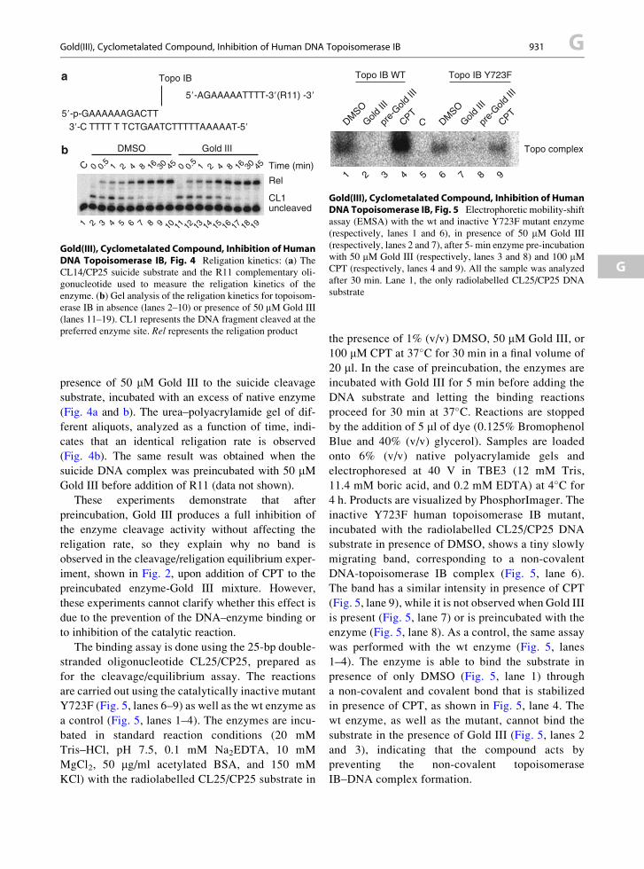
1 2 3 4 5 6 7 8 9 10 11 12 13 14 15 16 17 18 19
C 2 16 30
DMSO Gold III
Time (min)1 450.5
4 8 2 16 301 450.5
4 80 0
CL1uncleaved
5�-p-GAAAAAAGACTT3�-C TTTT T TCTGAATCTTTTTAAAAAT-5�
Topo IB
5�-AGAAAAATTTT-3�(R11) -3�
a
b
Rel
Gold(III), Cyclometalated Compound, Inhibition of HumanDNA Topoisomerase IB, Fig. 4 Religation kinetics: (a) TheCL14/CP25 suicide substrate and the R11 complementary oli-
gonucleotide used to measure the religation kinetics of the
enzyme. (b) Gel analysis of the religation kinetics for topoisom-
erase IB in absence (lanes 2–10) or presence of 50 mM Gold III
(lanes 11–19). CL1 represents the DNA fragment cleaved at the
preferred enzyme site. Rel represents the religation product
Topo IB WT Topo IB Y723F
DMSO
Gold II
I
pre-
Gold II
I
CPTDM
SO
Gold II
I
pre-
Gold II
I
CPTC
Topo complex
1 2 3 4 5 6 7 8 9
Gold(III), Cyclometalated Compound, Inhibition of HumanDNA Topoisomerase IB, Fig. 5 Electrophoretic mobility-shift
assay (EMSA) with the wt and inactive Y723F mutant enzyme
(respectively, lanes 1 and 6), in presence of 50 mM Gold III
(respectively, lanes 2 and 7), after 5- min enzyme pre-incubation
with 50 mM Gold III (respectively, lanes 3 and 8) and 100 mMCPT (respectively, lanes 4 and 9). All the sample was analyzed
after 30 min. Lane 1, the only radiolabelled CL25/CP25 DNA
substrate
Gold(III), Cyclometalated Compound, Inhibition of Human DNA Topoisomerase IB 931 G
G
presence of 50 mM Gold III to the suicide cleavage
substrate, incubated with an excess of native enzyme
(Fig. 4a and b). The urea–polyacrylamide gel of dif-
ferent aliquots, analyzed as a function of time, indi-
cates that an identical religation rate is observed
(Fig. 4b). The same result was obtained when the
suicide DNA complex was preincubated with 50 mMGold III before addition of R11 (data not shown).
These experiments demonstrate that after
preincubation, Gold III produces a full inhibition of
the enzyme cleavage activity without affecting the
religation rate, so they explain why no band is
observed in the cleavage/religation equilibrium exper-
iment, shown in Fig. 2, upon addition of CPT to the
preincubated enzyme-Gold III mixture. However,
these experiments cannot clarify whether this effect is
due to the prevention of the DNA–enzyme binding or
to inhibition of the catalytic reaction.
The binding assay is done using the 25-bp double-
stranded oligonucleotide CL25/CP25, prepared as
for the cleavage/equilibrium assay. The reactions
are carried out using the catalytically inactive mutant
Y723F (Fig. 5, lanes 6–9) as well as the wt enzyme as
a control (Fig. 5, lanes 1–4). The enzymes are incu-
bated in standard reaction conditions (20 mM
Tris–HCl, pH 7.5, 0.1 mM Na2EDTA, 10 mM
MgCl2, 50 mg/ml acetylated BSA, and 150 mM
KCl) with the radiolabelled CL25/CP25 substrate in
the presence of 1% (v/v) DMSO, 50 mM Gold III, or
100 mM CPT at 37�C for 30 min in a final volume of
20 ml. In the case of preincubation, the enzymes are
incubated with Gold III for 5 min before adding the
DNA substrate and letting the binding reactions
proceed for 30 min at 37�C. Reactions are stopped
by the addition of 5 ml of dye (0.125% Bromophenol
Blue and 40% (v/v) glycerol). Samples are loaded
onto 6% (v/v) native polyacrylamide gels and
electrophoresed at 40 V in TBE3 (12 mM Tris,
11.4 mM boric acid, and 0.2 mM EDTA) at 4�C for
4 h. Products are visualized by PhosphorImager. The
inactive Y723F human topoisomerase IB mutant,
incubated with the radiolabelled CL25/CP25 DNA
substrate in presence of DMSO, shows a tiny slowly
migrating band, corresponding to a non-covalent
DNA-topoisomerase IB complex (Fig. 5, lane 6).
The band has a similar intensity in presence of CPT
(Fig. 5, lane 9), while it is not observed when Gold III
is present (Fig. 5, lane 7) or is preincubated with the
enzyme (Fig. 5, lane 8). As a control, the same assay
was performed with the wt enzyme (Fig. 5, lanes
1–4). The enzyme is able to bind the substrate in
presence of only DMSO (Fig. 5, lane 1) through
a non-covalent and covalent bond that is stabilized
in presence of CPT, as shown in Fig. 5, lane 4. The
wt enzyme, as well as the mutant, cannot bind the
substrate in the presence of Gold III (Fig. 5, lanes 2
and 3), indicating that the compound acts by
preventing the non-covalent topoisomerase
IB–DNA complex formation.

G 932 Gold, Physical and Chemical Properties
Cross-References
▶Gold Complexes as Prospective Metal-Based
Anticancer Drugs
▶Gold(III) Complexes, Cytotoxic Effects
References
Castelli S, Campagna A, Vassallo O et al (2009) Conjugated
eicosapentaenoic acid inhibits human topoisomerase IB with
a mechanism different from camptothecin. Arch Biochem
Biophys 486:103–110
Castelli S, Vassallo O, Katkar P et al (2011) Inhibition of human
DNA topoisomerase IB by a cyclometalated gold III com-
pound: analysis on the different steps of the enzyme catalytic
cycle. Arch Biochem Biophys 516:108–112
Champoux J (2001) DNA topoisomerases: structure, function,
and mechanism. Annu Rev Biochem 70:369–413
Che CM, Siu FM (2010) Metal complexes in medicine with
a focus on enzyme inhibition. Curr Opin Chem Biol
14:255–261
Chillemi G, Fiorani P, Castelli S et al (2005) Effect on DNA
relaxation of the single Thr718Ala mutation in human topo-
isomerase I: a functional and molecular dynamics study.
Nucleic Acids Res 33:3339–3350
Hsiang YH, LihouMG, Liu LF (1989) Arrest of replication forks
by drug-stabilized topoisomerase I-DNA cleavable com-
plexes as a mechanism of cell killing by camptothecin. Can-
cer Res 49:5077–5082
Liu LF, Desai SD, Li TK, Mao Y, Sun M, Sim SP (2000) Mech-
anism of action of camptothecin. Ann N Y Acad Sci
922:1–10
Stewart L, Redinbo MR, Qiu X, Hol WG, Champoux JJ
(1998) A model for the mechanism of human topoisomerase
I. Science 279:1534–1541
Svejstrup JQ, Christiansen K, Gromovat II et al (1991) New
technique for uncoupling the cleavage and religation reac-
tions of eukaryotic topoisomerase I. the mode of action of
camptothecin at a specific recognition site. J Mol Biol
222:669–678
Tiekink ER (2002) Gold derivatives for the treatment of cancer.
Crit Rev Oncol Hematol 42:225–248
Gold, Physical and Chemical Properties
Fathi Habashi
Department of Mining, Metallurgical, and Materials
Engineering, Laval University, Quebec City, Canada
Gold is the first element recognized by man as a metal.
Ancient Egypt was the principal gold producing
country and maintained that status until ca. 1500 BC.
Gold has been valued since ancient times because of its
color, its occurrence in the native state, and its non-
tarnishing properties. It was established as the standard
for currency by Newton in 1717 when he wasMaster of
the Mint in London. Gold Rushes were responsible for
creating large cities like San Francisco, Johannesburg,
Sydney, and others.
Physical Properties
Atomic number
79Atomic weight
196.97Naturally occurring isotope
197Relative abundance in Earth’s crust, %
1 � 10�7Melting point, �C
1064.43Boiling point, �C
2,808Unit cell
Face-centered cubicLattice constant (a0), nm
0.40781Density, g/cm3
At 20�C
19.32At 900�C
18.32At 1,000�C
18.32At 1,065�C
17.32At 1,200�C
17.12At 1,300�C
17.00Vapor pressure, Pa
At 1,064�C
0.002At 1,319�C
0.1At 1,616�C
10At 1,810�C
100At 2,360�C
10,000Atomic volume at 20�C, cm3/mol
10.21Electrical resistivity at 0�C, W cm
2.06 � 10�6Thermal conductivity at 0�C,W cm�1 K�1
3.14
Specific heat, J g�1 K�1
0.138Enthalpy of fusion, kJ/mol
12.77Enthalpy of vaporization, kJ/mol
324.4Tensile strength, N/mm2
127.5Hardness on the Mohs’ scale
2.5Brinell hardness
18Standard potential, V
Au/Au3+
+1.498Au/Au+
+1.68Au+/ Au3+
+1.29Gold is one of the metals that can be prepared
readily to the highest purity 99.99+% and is the most
ductile of all metals. It can be cold drawn to give wires
of less than 10 mm diameter, and beaten into gold foil

Gold, Ultrasmall Nanoclusters and Proteins, Interaction 933 G
with a thickness of 0.2 mm. Because of its softness,gold can be highly polished. Very thin gold foil is
translucent; transmitted light appears blue-green.
G
Chemical Properties
Gold in solution exists as Au+ and Au3+. Gold does not
react with water, dry or humid air, oxygen (even at
high temperature), ozone, nitrogen, hydrogen, fluorine,
iodine, sulfur, and hydrogen sulfide under normal
conditions. Sulfuric, hydrochloric, hydrofluoric, phos-
phoric, nitric, and practically all organic acids have no
effect on gold, either in concentrated or dilute solutions
and at temperatures up to the boiling point. A mixture
of hydrochloric and nitric acids known as aqua regia or
royal water dissolves gold due to the formation of
chlorine and nitrosyl chloride:
HNO3 þ 3HCl ! Cl2 þ NOClþ 2H2O
Gold can also be dissolved in chlorine water
(the Plattner process). Aqueous solutions of alkali
metal hydroxides and alkali metal sulfides do not
attack gold. However, gold dissolves in solutions of
alkali metal cyanides in the presence of oxygen:
2Auþ 4CN� þ O2 þ 2H2O
! 2 AuðCNÞ2� �� þ H2O2 þ 2OH�
Gold can be precipitated from cyanide solution
by zinc powder:
2Na AuðCNÞ2� �þ Zn ! Na2 ZnðCNÞ4
� �þ 2Au
Gold can be recovered from solution by electrolytic
deposition or by chemical reduction. If the
tetrachloroaurate(III) complex is present, then iron
(II) salts, tin(II) salts, sulfur dioxide, hydrazine, or
oxalic acid can be used as reducing agents. Fused
caustic alkalis do not attack gold, provided air and
other oxidizing agents are excluded. Gold reacts
vigorously with alkali metal peroxides to form aurates.
It is inert to sodium borate which can therefore be used
as slagging agents for removing metallic impurities
from gold. Gold reacts readily with dry chlorine.
The maximum reactivity occurs at 250�C.Colloidal gold forms hydrosols of an intense red
or violet color. Gold alloys readily with mercury at
room temperature to form an amalgam. The mercury
can be distilled out by heating. This property is
utilized in the amalgamation process and in fire
gilding.
References
Habashi F (2009) Gold. History, metallurgy, culture. Metallurgie
Extractive Quebec, Quebec City. Distributed by Laval
University Bookstore “Zone”, www.zone.ul.ca
Renner H, Johns MW (1997) Gold. In: Habashi F (ed) Handbook
of extractive metallurgy. Weinheim, WILEY-VCH,
pp 1183–1214
Gold, Ultrasmall Nanoclusters andProteins, Interaction
Lennart Treuel1,2 and Gerd Ulrich Nienhaus1,3
1Institute of Applied Physics and Center for Functional
Nanostructures (CFN), Karlsruhe Institute of
Technology (KIT), Karlsruhe, Germany2Institute of Physical Chemistry, University of
Duisburg-Essen, Essen, Germany3Department of Physics, University of Illinois at
Urbana-Champaign, Urbana, IL, USA
Synonyms
Protein adsorption onto ultrasmall gold nanoclusters;
Protein corona formation around ultrasmall gold
nanoclusters
Definition
Ultrasmall gold nanoclusters (Au NCs) consist of
a few to roughly one hundred gold atoms. They are
promising candidates for the development of novel
fluorescence markers in biological applications
because of attractive features including small size
(diameter < 2 nm), low toxicity, and good
photophysical properties. In biological applica-
tions, Au NCs and other nanoparticles become
exposed to biological fluids and, typically,
a protein adsorption layer forms around the particle
surface.

Gold, Ultrasmall Nanoclusters and Proteins, Interaction,Fig. 1 (a) Fluorescence emission spectra (excitation: 550 nm)
of pure HSA (curve A) and Au NCs exposed to buffer solutions
with varying concentrations of HSA (curves B–G). Fluorescence
intensities of Au NCs, plotted as a function of (b) HSA and
(c) transferrin protein concentration in the solution. The grey linesrepresent fits to the data points using the adapted Hill equation
(Reproduced with kind permission from Shang et al. 2012)
G 934 Gold, Ultrasmall Nanoclusters and Proteins, Interaction
Au NCs in Biological Systems
Upon nanoparticle (NP) exposure to an organism,
proteins from body fluids bind to NP surfaces (Rocker
et al. 2009; Maffre et al. 2011). Consequently, living
systems interact with protein-coated rather than bare
NPs. This so-called protein corona largely defines the
biological identity of the NP, and the efficiency of this
interaction can be a decisive factor of the biological
response of an organism to NP exposure. Nel and coau-
thors (2009) have presented an in-depth discussion of the
basic physical interactions occurring at the nano-bio
interface. Biophysicalmechanisms associatedwith nano-
particle-protein interactions on a molecular scale have
been reviewed recently (Treuel and Nienhaus 2012).
Protein adsorption onto NP surfaces can cause
structural changes of the protein (Gebauer et al. 2012;
Treuel et al. 2010). Altered protein conformations can
result in a loss of the biological activity and exposure
of novel “cryptic” peptide epitopes that can promote
autoimmune diseases (Nel et al. 2009).
A variety of synthesis strategies have been
described for ultrasmall Au NCs, peptides, and pro-
teins have also been employed successfully as
templates for synthesizing highly fluorescent metal
NCs (Shang et al., 2011).
Quantitative Aspects of Protein Adsorption
In ultrasmall Au NCs, the particle size is comparable to
the Fermi wavelength of electrons resulting in discrete
electronic states giving rise to size-dependent
fluorescence properties (Lin et al. 2009). Furthermore,
the strong, size-dependent surface plasmon absorption
characteristic of larger Au NPs is completely absent.
Fluorescence emission intensity was used as
a readout in a quantitative study (Shang et al. 2012)
of protein adsorption onto ultrasmall Au NCs (1.4 �0.3 nm diameter) coated with dihydrolipoic acid
(DHLA). Protein binding resulted in a substantial
increase in the fluorescence intensity of the Au NCs
(Fig. 1). By using this effect as a quantitative experi-
mental readout, apparent binding affinities of four
different proteins, human serum albumin (HSA), trans-
ferrin, lysozyme, and apolipoprotein E4 (ApoE4) to
these Au NCs were determined. They were all in the
micromolar concentration regime (KD [HSA]¼0.9mM,
KD [transferrin] ¼ 0.7 mM, KD [lysozyme] ¼ 3.0 mM,
KD [ApoE4] ¼ 2.7 mM). However, the increase in
fluorescence intensity may not scale with the fraction
of proteins bound to the NCs, so the data in Fig. 1b, c
should not be viewed as proper binding isotherms.
Au NCs in Sensing Applications
The effect of protein adsorption onto Au NCs on their
fluorescence properties can be put to good use in bio-
sensor application. Selective receptor molecules (e.g.,
antibodies) have been attached to the surfaces of fluo-
rescent Au NCs for the development of fluorescence-
based protein biosensors.
As an early application, Leblanc and coworkers
(Triulzi et al. 2006) reported an immunoassay based
on electrostatic conjugation of polyclonal anti-IgG

Gray or Grey Arsenic 935 G
antibodies with dendrimer-encapsulated Au NCs. Lin-ear fluorescence quenching was observed in response
to changes in the surface electrostatic properties of the
Au NCs caused by formation of an antigen-antibody
immunocomplex. Further examples can be found in
a recent review (Shang et al. 2011).
G
How Protein Adsorption AffectsFluorescence Properties of Au NCs
To explore the underlying mechanisms of the observed
fluorescence increase upon protein adsorption, the
effect on fluorescence lifetime was studied (Shang
et al. 2012), revealing an average lifetime of 450 �5 ns for bare Au NCs. HSA adsorption onto these Au
NCs slowed the luminescence decay, resulting in an
average lifetime increase to 980 � 40 ns. A detailed
analysis of the luminescence decay kinetics revealed
that the main contribution to this change is associated
with the presence of an Au(I)-thiol complex on the
particle surface.
Moreover, this study showed a shift in the electron
binding energy of the Au core levels upon protein
adsorption. They are sensitive to the local chemi-
cal or physical environment around the surface gold
atoms. In the Au 4f XPS spectra of the Au NCs, the
binding energies of the Au 4f5/2 and Au 4f7/2 levels in
the absence of HSA, 88.3 � 0.1 and 84.6 � 0.1 eV,
respectively, shifted to 88.0 � 0.1 and 84.3 � 0.1 eV,
upon HSA adsorption onto the NC surface. Overall,
these results indicated that photophysical properties of
the Au NCs are directly altered by protein binding to
the NC surface.
Cross-References
▶Aluminum, Physical and Chemical Properties
▶Biomineralization of Gold Nanoparticles from Gold
Complexes in Cupriavidus Metallidurans CH34
▶Colloidal Silver Nanoparticles and Bovine Serum
Albumin
▶Gold and Nucleic Acids
▶Gold Nanomaterials as Prospective Metal-based
Delivery Systems for Cancer Treatment
▶Gold Nanoparticle Platform for Protein-Protein
Interactions and Drug Discovery
▶Gold Nanoparticles and Proteins, Interaction
References
Gebauer JS, Malissek M, Simon S, Knauer SK, Maskos M,
Stauber RH, Peukert W, Treuel L (2012) Impact of the
nanoparticle-protein corona on colloidal stability and protein
structure. Langmuir 28:9181–9906
Lin CAJ, Lee CH, Hsieh JT, Wang HH, Li JK, Shen JL, Chan
WH, Yeh HI, Chang WH (2009) Synthesis of fluorescent
metallic nanoclusters toward biomedical application: recent
progress and present challenges. J Med Biol Eng 29:276–283
Maffre P, Nienhaus K, Amin F, Parak WJ, Nienhaus GU
(2011) Characterization of protein adsorption onto FePt
nanoparticles using dual-focus fluorescence correlation spec-
troscopy. Beilstein J Nanotechnol 2:374–383
Nel AE,M€adler L, Velegol D, Xia T, Hoek EM, Somasundaran P,
Klaessig F, Castranova V, Thompson M (2009) Understand-
ing biophysicochemical interactions at the nano-bio interface.
Nat Mater 8:543–557
Rocker C, Potzl M, Zhang F, Parak WJ, Nienhaus GU
(2009) A quantitative fluorescence study of protein mono-
layer formation on colloidal nanoparticles. Nat Nanotechnol
4:577–580
Shang L, Dong S, Nienhaus GU (2011) Ultra-small fluorescent
metal nanoclusters: synthesis and biological applications.
Nano Today 6:401–418
Shang L, Brandholt S, Stockmar F, Trouillet V, Bruns M,
Nienhaus GU (2012) Effect of protein adsorption on the fluo-
rescence of ultrasmall gold nanoclusters. Small 8:661–665
Treuel L, NienhausGU (2012) Toward amolecular understanding
of nanoparticle–protein interactions. Biophys Rev 4:137–147
Treuel L,MalissekM,Gebauer JS, Zellner R (2010) The influence
of surface composition of nanoparticles on their interactions
with serum albumin. Chem Phys Chem 11:3093–3099
Triulzi RC, Micic M, Giordani S, Serry M, Chiou WA, Leblanc
RM (2006) Immunoasssay based on the antibody-conjugated
PAMAM-dendrimer-gold quantum dot complex. Chem
Commun 48:5068–5070
Gold-Based Nanomedicine
▶Gold Nanomaterials as Prospective Metal-based
Delivery Systems for Cancer Treatment
G-Quartet
▶ Strontium and DNA Aptamer Folding
Gray or Grey Arsenic
▶Arsenic in Pathological Conditions

G 936 Green Remediation
Green Remediation
▶Lead and Phytoremediation
G-Rich
▶ Selenoprotein K
Group 13 Elements NMR in Biology
▶NMR spectroscopy of Gallium in Biological
Systems
Growth Factor Regulated Channel (GRC)
▶Transient Receptor Potential Channels TRPV1,
TRPV2, and TRPV3
Growth Inhibitory Factor (GIF)
▶Zinc Metallothionein-3 (Neuronal Growth
Inhibitory Factor)
G-Tetrad
▶ Strontium and DNA Aptamer Folding
GTF
▶Chromium and Glucose Tolerance Factor