G Protein-coupled Receptor-promoted Trafficking of G 1 2 Leads to AKT Activation at Endosomes via a...
13
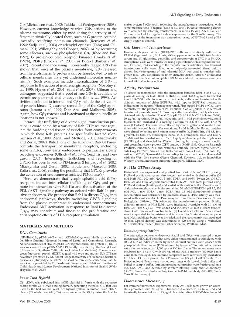
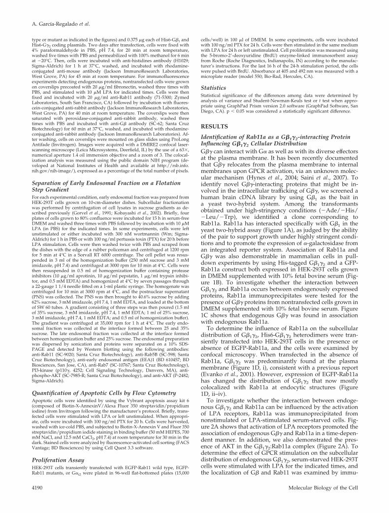
Molecular Biology of the Cell Vol. 19, 4188 – 4200, October 2008 G Protein-coupled Receptor-promoted Trafficking of G 1 2 Leads to AKT Activation at Endosomes via a Mechanism Mediated by G 1 2 -Rab11a Interaction Alejandro Garcı´a-Regalado,* Marı´a Luisa Guzma ´n-Herna ´ndez, † IlianaRamı´rez-Rangel, † Evelyn Robles-Molina, † Tamas Balla, ‡ Jose ´ Va ´zquez-Prado, † and Guadalupe Reyes-Cruz* Departments of *Cell Biology and † Pharmacology, Centro de Investigacio ´ n y de Estudios Avanzados-Instituto Polite ´cnico Nacional, 07000 Me ´xico, D.F., Me ´xico; and ‡ Section on Molecular Signal Transduction, National Institute of Child Health and Human Development, National Institutes of Health, Bethesda, MD 20892-4510 Submitted October 30, 2007; Revised July 16, 2008; Accepted August 6, 2008 Monitoring Editor: J. Silvio Gutkind G-protein coupled receptors activate heterotrimeric G proteins at the plasma membrane in which most of their effectors are intrinsically located or transiently associated as the external signal is being transduced. This paradigm has been extended to the intracellular compartments by studies in yeast showing that trafficking of G activates phosphatidylino- sitol 3-kinase (PI3K) at endosomal compartments, suggesting that vesicle trafficking regulates potential actions of G and possibly G at the level of endosomes. Here, we show that G interacts with Rab11a and that the two proteins colocalize at early and recycling endosomes in response to activation of lysophosphatidic acid (LPA) receptors. This agonist-dependent association of G to Rab11a-positive endosomes contributes to the recruitment of PI3K and phos- phorylation of AKT at this intracellular compartment. These events are sensitive to the expression of a dominant-negative Rab11a mutant or treatment with wortmannin, suggesting that Rab11a-dependent G trafficking promotes the activation of the PI3K/AKT signaling pathway associated with endosomal compartments. In addition, RNA interference-mediated Rab11a depletion, or expression of a dominant-negative Rab11a mutant attenuated LPA-dependent cell survival and proliferation, suggesting that endosomal activation of the PI3K/AKT signaling pathway in response to G trafficking, via its interaction with Rab11, is a relevant step in the mechanism controlling these fundamental events. INTRODUCTION Hundreds of different G protein-coupled receptors (GPCRs) exist in eukaryotes. They detect the presence of diverse extracellular stimuli, including hormones, neurotransmit- ters, lipids, and ions, and they activate intracellular hetero- trimeric G proteins by promoting the binding of guanosine triphosphate (GTP) to the G subunit and its dissociation from G. Both the GTP-bound G and the liberated G subunits interact with a selected set of effectors, including phospholipases, adenylyl cyclases, ion channels, phospho- inositide 3-kinases, and guanine exchange factors (Gilman, 1989; Clapham and Neer, 1997; Ford et al., 1998; Gautam et al., 1998; Albert and Robillard, 2002; Welch et al., 2002; Preininger and Hamm, 2004; Rosenfeldt et al., 2004). The signal is then propagated within the cells by diffusible sec- ond messengers that modulate protein–protein interactions and enzymatic activities. Current views place heterotrimeric G protein signaling to the plasma membrane in which acti- vated receptors promote the exchange of guanosine diphos- phate for GTP in the G subunit resulting in its dissociation from G. The cycle ends when G hydrolyzes GTP and will reassociates with G. Recent findings in yeast have extended this model by the demonstration that GTP-loaded G associates with endosomes as part of the mechanism of yeast PI3-kinase (Vps34p) activation (Slessareva et al., 2006). Whether receptor-activated mammalian G or G subunits can follow an intracellular trafficking pathway or contribute to further signaling or desensitization is not known. The initial phase of GPCR activation is frequently followed by posttranslational modifications of the receptor, leading to endocytosis and desensitization (Pierce et al., 2002). How- ever, endocytosis can also define novel signaling outputs by presenting different downstream effectors at endocytic com- partments compared with the plasma membrane (von Zas- trow and Sorkin, 2007). For example, it has been suggested that protein kinase A-phosphorylated 2 -adrenergic recep- tors associate with -arrestins and acquire novel coupling specificities to activate the extracellular signal-regulated ki- nase (ERK) signaling cascade (Daaka et al., 1997a; Lefkowitz et al., 2002). Previous studies on trafficking of G after its synthesis and assembly revealed its association with the plasma membrane via the carboxy-terminal isoprenyl as a result of covalent modification of the CAAX sequence present in G. These studies also showed that exit of G from the endoplasmic reticulum and trafficking to the plasma membrane require the expression and acylation of This article was published online ahead of print in MBC in Press (http://www.molbiolcell.org/cgi/doi/10.1091/mbc.E07–10 –1089) on August 13, 2008. Address correspondence to: Guadalupe Reyes-Cruz (guadaluper@cell. cinvestav.mx). Abbreviations used: EEA1, early endosomal antigen 1; GPCR, G protein coupled receptor; LPA, lysophosphatidic acid; PhLP1, phos- ducin-like protein 1; PI3-kinase, phosphoinositide 3-kinase; Wm, wortmannin. 4188 © 2008 by The American Society for Cell Biology http://www.molbiolcell.org/content/suppl/2008/08/13/E07-10-1089.DC1.html Supplemental Material can be found at:
-
Upload
independent -
Category
Documents
-
view
3 -
download
0
Transcript of G Protein-coupled Receptor-promoted Trafficking of G 1 2 Leads to AKT Activation at Endosomes via a...
Molecular Biology of the CellVol. 19, 4188–4200, October 2008
G Protein-coupled Receptor-promoted Trafficking of G�1�2Leads to AKT Activation at Endosomes via a MechanismMediated by G�1�2-Rab11a InteractionAlejandro Garcıa-Regalado,* Marıa Luisa Guzman-Hernandez,†Iliana Ramırez-Rangel,† Evelyn Robles-Molina,† Tamas Balla,‡Jose Vazquez-Prado,† and Guadalupe Reyes-Cruz*
Departments of *Cell Biology and †Pharmacology, Centro de Investigacion y de Estudios Avanzados-InstitutoPolitecnico Nacional, 07000 Mexico, D.F., Mexico; and ‡Section on Molecular Signal Transduction, NationalInstitute of Child Health and Human Development, National Institutes of Health, Bethesda, MD 20892-4510
Submitted October 30, 2007; Revised July 16, 2008; Accepted August 6, 2008Monitoring Editor: J. Silvio Gutkind
G-protein coupled receptors activate heterotrimeric G proteins at the plasma membrane in which most of their effectorsare intrinsically located or transiently associated as the external signal is being transduced. This paradigm has beenextended to the intracellular compartments by studies in yeast showing that trafficking of G� activates phosphatidylino-sitol 3-kinase (PI3K) at endosomal compartments, suggesting that vesicle trafficking regulates potential actions of G� andpossibly G�� at the level of endosomes. Here, we show that G�� interacts with Rab11a and that the two proteinscolocalize at early and recycling endosomes in response to activation of lysophosphatidic acid (LPA) receptors. Thisagonist-dependent association of G�� to Rab11a-positive endosomes contributes to the recruitment of PI3K and phos-phorylation of AKT at this intracellular compartment. These events are sensitive to the expression of a dominant-negativeRab11a mutant or treatment with wortmannin, suggesting that Rab11a-dependent G�� trafficking promotes the activationof the PI3K/AKT signaling pathway associated with endosomal compartments. In addition, RNA interference-mediatedRab11a depletion, or expression of a dominant-negative Rab11a mutant attenuated LPA-dependent cell survival andproliferation, suggesting that endosomal activation of the PI3K/AKT signaling pathway in response to G�� trafficking, viaits interaction with Rab11, is a relevant step in the mechanism controlling these fundamental events.
INTRODUCTION
Hundreds of different G protein-coupled receptors (GPCRs)exist in eukaryotes. They detect the presence of diverseextracellular stimuli, including hormones, neurotransmit-ters, lipids, and ions, and they activate intracellular hetero-trimeric G proteins by promoting the binding of guanosinetriphosphate (GTP) to the G� subunit and its dissociationfrom G��. Both the GTP-bound G� and the liberated G��subunits interact with a selected set of effectors, includingphospholipases, adenylyl cyclases, ion channels, phospho-inositide 3-kinases, and guanine exchange factors (Gilman,1989; Clapham and Neer, 1997; Ford et al., 1998; Gautam etal., 1998; Albert and Robillard, 2002; Welch et al., 2002;Preininger and Hamm, 2004; Rosenfeldt et al., 2004). Thesignal is then propagated within the cells by diffusible sec-ond messengers that modulate protein–protein interactionsand enzymatic activities. Current views place heterotrimeric
G protein signaling to the plasma membrane in which acti-vated receptors promote the exchange of guanosine diphos-phate for GTP in the G� subunit resulting in its dissociationfrom G��. The cycle ends when G� hydrolyzes GTP andwill reassociates with G��. Recent findings in yeast haveextended this model by the demonstration that GTP-loadedG� associates with endosomes as part of the mechanism ofyeast PI3-kinase (Vps34p) activation (Slessareva et al., 2006).Whether receptor-activated mammalian G� or G�� subunitscan follow an intracellular trafficking pathway or contributeto further signaling or desensitization is not known. Theinitial phase of GPCR activation is frequently followed byposttranslational modifications of the receptor, leading toendocytosis and desensitization (Pierce et al., 2002). How-ever, endocytosis can also define novel signaling outputs bypresenting different downstream effectors at endocytic com-partments compared with the plasma membrane (von Zas-trow and Sorkin, 2007). For example, it has been suggestedthat protein kinase A-phosphorylated �2-adrenergic recep-tors associate with �-arrestins and acquire novel couplingspecificities to activate the extracellular signal-regulated ki-nase (ERK) signaling cascade (Daaka et al., 1997a; Lefkowitzet al., 2002). Previous studies on trafficking of G�� after itssynthesis and assembly revealed its association with theplasma membrane via the carboxy-terminal isoprenyl as aresult of covalent modification of the CAAX sequencepresent in G�. These studies also showed that exit of G��from the endoplasmic reticulum and trafficking to theplasma membrane require the expression and acylation of
This article was published online ahead of print in MBC in Press(http://www.molbiolcell.org/cgi/doi/10.1091/mbc.E07–10–1089)on August 13, 2008.
Address correspondence to: Guadalupe Reyes-Cruz ([email protected]).
Abbreviations used: EEA1, early endosomal antigen 1; GPCR, Gprotein coupled receptor; LPA, lysophosphatidic acid; PhLP1, phos-ducin-like protein 1; PI3-kinase, phosphoinositide 3-kinase; Wm,wortmannin.
4188 © 2008 by The American Society for Cell Biology http://www.molbiolcell.org/content/suppl/2008/08/13/E07-10-1089.DC1.htmlSupplemental Material can be found at:
G� (Michaelson et al., 2002; Takida and Wedegaertner, 2003).However, current knowledge restricts G�� actions to theplasma membrane, either by modulating the activity of ef-fectors intrinsically located there, such as G protein-coupledinwardly rectifying potassium channels (Reuveny et al.,1994; Sadja et al., 2003) or adenylyl cyclases (Tang and Gil-man, 1991; Willoughby and Cooper, 2007), or by recruitingsome effectors, such as phospholipase C�2 (Rhee and Bae,1997), G protein-coupled receptor kinase-2 (Daaka et al.,1997b), PI3K� (Brock et al., 2003), or P-Rex1 (Barber et al.,2007). Recent evidence using fluorescently tagged G�� hasshown that, once at the plasma membrane, G�� releasedfrom heterotrimeric G proteins can be translocated to intra-cellular membranes via a yet undefined molecular mecha-nism(s). Such examples include internalization of G�� inresponse to the action of �-adrenergic receptors (Novotny etal., 1995; Hynes et al., 2004; Saini et al., 2007). Gilman andcolleagues suggested that a pool of free G�� is available topermit receptor-mediated endocytosis (Lin et al., 1998). Ac-tivities attributed to internalized G�� include the activationof protein kinase D, causing remodeling of the Golgi appa-ratus (Jamora et al., 1999; Diaz Anel and Malhotra, 2005).Again, how G�� reaches and is activated at these subcellularlocations is not known.
Intracellular trafficking of diverse signal transduction pro-teins is coordinated by a variety of Rab GTPases that regu-late the budding and fission of vesicles from compartmentsin which these Rab proteins are specifically located (Son-nichsen et al., 2000; Zerial and McBride, 2001; Miaczynskaand Zerial, 2002). Rab11, one of the 40 known Rab GTPases,controls the transport of membrane receptors, includingsome GPCRs, from early endosomes to perinuclear slowlyrecycling endosomes (Ullrich et al., 1996; Seachrist and Fer-guson, 2003). Interestingly, trafficking and recycling ofGPCRs has been linked to PI3-kinases (Hunyady et al., 2002;Miaczynska and Zerial, 2002; Houle and Marceau, 2003;Kalia et al., 2006), raising the possibility that GPCRs provokethe activation of endosome-associated PI3-kinase(s).
Here, we demonstrate that lysophosphatidic acid (LPA)receptors induce intracellular trafficking of G�� and pro-mote its interaction with Rab11a and the activation of thePI3K/AKT signaling pathway associated with Rab11a-pos-itive endosomes. We propose that Rab11a directs G�1�2 intoendosomal pathways, thereby switching GPCR signalingfrom the plasma membrane to endosomal compartments.Endosomal AKT activation in response to Rab11a-directedG�1�2 may contribute and fine-tune the proliferative andantiapoptotic effects of LPA receptor stimulation.
MATERIALS AND METHODS
DNA ConstructspEF-His6-G�1, pEF-His6-G�2, and pCDNA3-G�i2 were kindly provided byDr. Silvio Gutkind (National Institute of Dental and Craniofacial Research,National Institutes of Health). pCEFL3XFlag phosducin-like protein 1 (PhLP1)was subcloned from pCDNA3-PhLP1 kindly provided by Dr. Sheryl Craft(University of Southern California Keck School of Medicine). The enhancedgreen fluorescent protein (EGFP)-tagged wild-type and mutant Rab GTPaseshave been generated by Dr. Robert Lodge (University of Quebec) as describedpreviously (Hunyady et al., 2002). The short hairpin RNA (shRNA) for Rab11awas kindly provided by Dr. Yoshiyuki Wakabayashi (National Institute ofChild Health and Human Development, National Institutes of Health) (Wak-abayashi et al., 2005).
Yeast Two-HybridG�1 cDNA was subcloned into pGB3 plasmid in frame with the sequencecoding for the Gal4 DNA binding domain, generating the pGB3-G�1 that wasused as the bait for the yeast two-hybrid system. A human brain cDNAlibrary (Clontech, Palo Alto, CA) was screened with G�1 by using the Match-
maker system 3 (Clontech), following the manufacturer’s instructions, withsome modifications (Vazquez-Prado et al., 2004). Putative interacting cloneswere obtained by selecting transformants in media lacking Ade/His/Leu/Trp and checked for �-galactosidase expression by the X �-Gal assay. Thespecificity of the interaction was determined using pGBKT7-p53 and pGB3empty vector as control.
Cell Lines and TransfectionsHuman embryonic kidney (HEK)-293T cells were routinely cultured inDMEM (Sigma-Aldrich, St. Louis, MO) supplemented with 10% fetal bovineserum and 1% glutamine, penicillin, and streptomycin at 37°C in a 5% CO2atmosphere. Cells were transfected using Lipofectamine Plus reagent (Invitro-gen, Carlsbad, CA) according to the manufacturer’s indications. For transienttransfections, cells were plated onto poly-d-lysine–coated tissue culturedishes. Four micrograms of total plasmid DNA was used to transfect cellsgrown to 60–70% confluence in 10-cm-diameter dishes. After 5 h of initiatedthe transfection, 5 ml of complete DMEM was added, the assays were per-formed 48 h after transfection.
Affinity PrecipitationTo assess in mammalian cells the interaction between Rab11a and G�1�2,plasmids coding for EGFP-Rab11a, His6-G�1, and His6-G�2 were transfectedinto HEK-293T cells. In a set of experiments, cells were transfected withdifferent amounts of either EGFP-Rab wild type or EGFP-Rab mutants asindicated in the figures. When appropriated, Flag-tagged PhLP1 or G�i2 werecotransfected; the proportion of PhLP1/Rab11a or G�i2/Rab11a, in terms oftransfected plasmids, was 3:1. Two days after transfection, cell lysates wereobtained with lysis buffer (50 mM Tris, pH 7.5, 0.15 M NaCl, 1% Triton X-100,10 �g/ml aprotinin, 10 �g/ml leupeptin, and 1 mM phenylmethylsulfonylfluoride), and incubated in a rocking platform for 2 h at 4°C with TALONresin (Clontech). The resin was collected by centrifugation and washed fourtimes with ice-cold lysis buffer containing 5 mM imidazole. Bound proteinswere eluted by boiling for 5 min in sample buffer (62.5 mM Tris, pH 6.8, 10%glycerol, 2% SDS, 5% �-mercaptoethanol, 0.1% bromphenol blue, and EDTA5 mM) and fractionated on a 10% SDS-polyacrylamide gel electrophoresis(PAGE) gel and detected by Western blotting by using a mouse specificanti-green fluorescent protein (GFP) antibody (MMS-118R; Covance ResearchProducts, Princeton, NJ), anti-histidines antibody (H1029; Sigma-Aldrich),anti G�i2 (SC-7276; Santa Cruz Biotechnology, Santa Cruz, CA), anti-Flagantibody (F3165; Sigma-Aldrich) according to the experiment and revealedwith the West Pico system (Pierce Chemical, Rockford, IL), or ImmobilonWestern chemiluminescent substrate (Millipore, Billerica, MA).
Rab11a GTPase AssayHis6-Rab11 was expressed and purified from Escherichia coli BL21 by usingProBond purification system (Invitrogen) and eluted with elution buffer (50mM NaH2PO4, 300 mM NaCl, 5 mM EDTA, and 0.05% Tween 20, pH 8.0).His6-G�1 and His6-G�2 from transfected HEK-293T cells were purified usingProBond system (Invitrogen) and eluted with elution buffer. Proteins weredialyzed overnight against buffer containing 20 mM HEPES�KOH, pH 7.5, 150mM KCl, 1 mM EDTA, 1 mM EGTA, and 0.5 mM dithiothreitol; proteinconcentration was determined by Bradford assay. GTPase activity was mea-sured with a colorimetric GTPase assay kit from Innova Biosciences (NovusBiologicals, Littleton, CO) following the manufacturer’s protocol. Briefly,different amounts of His6-Rab11 were incubated overnight with 0.1 �M ofHis6-G�1-His6-G�2; GTP was added and incubated 30 min at room temper-ature. Gold mix or colorimetric buffer (Pi ColorLock Gold and Accelerator)was incorporated to the mixture and incubated for 5 min at room tempera-ture. Next, stabilizer buffer was included, and the reaction mix was incubated30 min. Optical density was determined at 620 nm by using a MultisKanMCC/340 plate reader (Thermo Fisher Scientific, Waltham, MA).
ImmunoprecipitationThe interaction between endogenous Rab11 and G�1�2 was assessed in non-transfected HEK-293T cells that were either nonstimulated or stimulated with10 �M LPA as indicated in the figures. Confluent cultures were washed withphosphate-buffered saline (PBS) followed by lysis at 4°C in lysis buffer; lysateswere then centrifuged at 14,000 rpm at 4°C for 10 min. The supernatants wereincubated for 12 h at 4°C with 600 ng/ml anti-Rab11 antibody (SC-9020; SantaCruz Biotechnology). The immune complexes were recovered by incubationfor 2 h at 4°C with protein A/G Plus-agarose (25 �l, SC-2003; Santa CruzBiotechnology). Beads were washed four times with ice-cold lysis buffer andboiled in sample buffer. Immunoprecipitated proteins were fractionated on a15% SDS-PAGE and detected by Western blotting using anti-G� antibody(SC-261; Santa Cruz Biotechnology) and anti-Rab11 antibody (SC-9020; SantaCruz Biotechnology).
Fluorescence MicroscopyFor immunofluorescence experiments, HEK-293T cells were grown on cover-slips precoated with 20 �g/ml fibronectin (Calbiochem, La Jolla, CA) andtransiently transfected with 0.75 �g of plasmids coding EGFP-Rab (either wild
AKT Signaling at Early Endosomes
Vol. 19, October 2008 4189
type or mutant as indicated in the figures) and 0.375 �g each of His6-G�1 andHis6-G�2 coding plasmids. Two days after transfection, cells were fixed with4% paraformaldehyde in PBS, pH 7.4, for 20 min at room temperature,washed five times with PBS and permeabilized with 100% methanol for 6 minat �20°C. Then, cells were incubated with anti-histidines antibody (H1029;Sigma-Aldrich) for 1 h at 37°C, washed, and incubated with rhodamine-conjugated anti-mouse antibody (Jackson ImmunoResearch Laboratories,West Grove, PA) for 45 min at room temperature. For immunofluorescenceexperiments detecting endogenous proteins, nontransfected cells were grownon coverslips precoated with 20 �g/ml fibronectin, washed three times withPBS, and stimulated with 10 �M LPA for indicated times. Cells were thenfixed and incubated with 20 �g/ml anti-Rab11 antibody (71-5300; ZymedLaboratories, South San Francisco, CA) followed by incubation with fluores-cein-conjugated anti-rabbit antibody (Jackson ImmunoResearch Laboratories,West Grove, PA) for 40 min at room temperature. The coverslips were thensaturated with peroxidase-conjugated anti-rabbit antibody, washed threetimes with PBS and incubated with anti-G� antibody (SC-62; Santa CruzBiotechnology) for 60 min at 37°C, washed, and incubated with rhodamine-conjugated anti-rabbit antibody (Jackson ImmunoResearch Laboratories). Af-ter washing, cells on coverslips were mounted on glass slides using ProLongAntifade (Invitrogen). Images were acquired with a DMIRE2 confocal laser-scanning microscope (Leica Microsystems, Deerfield, IL) by the use of a 63�,numerical aperture 1.4 oil immersion objective and a zoom of 3. The colocal-ization analysis was measured using the public domain NIH program (de-veloped at National Institutes of Health and available at http://rsb.info.nih.gov/nih-image/), expressed as a percentage of the total number of pixels.
Separation of Early Endosomal Fraction on a FlotationStep GradientFor each experimental condition, early endosomal fraction was prepared fromHEK-293T cells grown on 10-cm-diameter dishes. Subcellular fractionationwas performed by centrifugation of cell lysates in sucrose gradients as de-scribed previously (Gorvel et al., 1991; Kobayashi et al., 2002). Briefly, fourplates of cells grown to 80% confluence were incubated for 15 h in serum-freeDMEM and washed three times with PBS followed by incubation with 10 �MLPA (in PBS) for the indicated times. In some experiments, cells were leftunstimulated or either incubated with 300 nM wortmannin (Wm; Sigma-Aldrich) for 1 h in PBS or with 100 ng/ml pertussis toxin (PTX) for 20 h beforeLPA stimulation. Cells were then washed twice with PBS and scraped fromthe dishes with the edge of a rubber policeman and centrifuged at 1200 rpmfor 5 min at 4°C in a Sorvall RT 6000 centrifuge. The cell pellet was resus-pended in 3 ml of the homogenization buffer (250 mM sucrose and 3 mMimidazole, pH 7.4) and centrifuged at 3000 rpm for 10 min at 4°C. Cells werethen resuspended in 0.5 ml of homogenization buffer containing proteaseinhibitors (10 �g/ml aprotinin, 10 �g/ml pepstatin, 1 �g/ml trypsin inhibi-tor, and 0.5 mM EDTA) and homogenized at 4°C by seven passages througha 22-gauge 1 1/4 needle fitted on a 1-ml plastic syringe. The homogenate wascentrifuged for 10 min at 3000 rpm at 4°C, and the postnuclear supernatant(PNS) was collected. The PNS was then brought to 40.6% sucrose by adding62% sucrose, 3 mM imidazole, pH 7.4, 1 mM EDTA, and loaded at the bottomof SW 60 tubes. A gradient consisting of three steps was then poured (1.5 mlof 35% sucrose, 3 mM imidazole, pH 7.4, 1 mM EDTA; 1 ml of 25% sucrose,3 mM imidazole, pH 7.4, 1 mM EDTA; and 0.5 ml of homogenization buffer).The gradient was centrifuged at 35,000 rpm for 1 h at 4°C. The early endo-somal fraction was collected at the interface formed between 25 and 35%sucrose. The late endosomal fraction was collected at the interface formedbetween homogenization buffer and 25% sucrose. The endosomal preparationwas dispersed by sonication and proteins were separated on a 10% SDS-PAGE and detected by Western blotting using the following antibodies:anti-Rab11 (SC-9020; Santa Cruz Biotechnology), anti-Rab5B (SC-598; SantaCruz Biotechnology), anti-early endosomal antigen (EEA)1 (BD 610457; BDBiosciences, San Jose, CA), anti-Rab7 (SC-10767; Santa Cruz Biotechnology),PI3-kinase (p110�, 4252; Cell Signaling Technology, Danvers, MA), anti-phospho-AKT (SC-7985-R; Santa Cruz Biotechnology), and anti-AKT (P-2482;Sigma-Aldrich).
Quantification of Apoptotic Cells by Flow CytometryApoptotic cells were identified by using the Vybrant apoptosis assay kit 6(composed of Biotin-X-AnnexinV/Alexa Fluor 350 streptavidin/propidiumiodine) from Invitrogen following the manufacturer’s protocol. Briefly, trans-fected cells were stimulated with LPA or left unstimulated. When appropri-ate, cells were incubated with 100 ng/ml PTX for 20 h. Cells were harvested,washed with ice-cold PBS, and subjected to Biotin-X-Annexin V and Fluor 350streptavidin/propidium iodide staining in binding buffer (50 mM HEPES, 700mM NaCl, and 12.5 mM CaCl2, pH 7.4) at room temperature for 30 min in thedark. Stained cells were analyzed by fluorescence-activated cell sorting (FACSVantage; BD Biosciences) by using Cell Quest 3.3 software.
Proliferation AssayHEK-293T cells transiently transfected with EGFP-Rab11 wild type, EGFP-Rab11 mutants, or G�i2 were plated in 96-well flat-bottomed plates (15,000
cells/well) in 100 �l of DMEM. In some experiments, cells were incubatedwith 100 ng/ml PTX for 24 h. Cells were then stimulated in the same mediumwith LPA for 24 h or left unstimulated. Cell proliferation was measured usingthe 5-bromo-2�-deoxyuridine (BrdU) enzyme-linked immunosorbent assayfrom Roche (Roche Diagnostics, Indianapolis, IN) according to the manufac-turer’s instructions. For the last 16 h of the 24-h stimulation period, the cellswere pulsed with BrdU. Absorbance at 405 and 492 nm was measured with amicroplate reader (model 550; Bio-Rad, Hercules, CA).
StatisticsStatistical significance of the differences among data were determined byanalysis of variance and Student-Newman-Keuls test or t test when appro-priate using GraphPad Prism version 2.0 software (GraphPad Software, SanDiego, CA). p � 0.05 was considered a statistically significant difference.
RESULTS
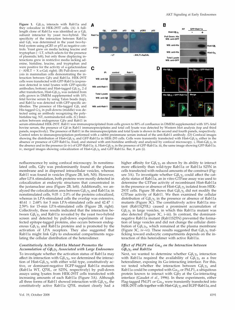
Identification of Rab11a as a G�1�2-interacting ProteinInfluencing G�1�2 Cellular DistributionG�� can interact with G� as well as with its diverse effectorsat the plasma membrane. It has been recently documentedthat G�� relocates from the plasma membrane to internalmembranes upon GPCR activation, via an unknown molec-ular mechanism (Hynes et al., 2004; Saini et al., 2007). Toidentify novel G��-interacting proteins that might be in-volved in the intracellular trafficking of G��, we screened ahuman brain cDNA library by using G�1 as the bait ina yeast two-hybrid system. Among the transformantsobtained under high-stringency conditions (�Ade/�His/�Leu/�Trp), we identified a clone corresponding toRab11a. Rab11a has interacted specifically with G�1 in theyeast two-hybrid assay (Figure 1A), as judged by the abilityof the pair to support growth under highly stringent condi-tions and to promote the expression of �-galactosidase froman integrated reporter system. Association of Rab11a andG�� was also demonstrable in mammalian cells in pull-down experiments by using His-tagged G�1�2 and a GFP-Rab11a construct both expressed in HEK-293T cells grownin DMEM supplemented with 10% fetal bovine serum (Fig-ure 1B). To investigate whether the interaction betweenG�1�2 and Rab11a occurs between endogenously expressedproteins, Rab11a immunoprecipitates were tested for thepresence of G�� proteins from nontransfected cells grown inDMEM supplemented with 10% fetal bovine serum. Figure1C shows that endogenous G�� was found in associationwith endogenous Rab11a.
To determine the influence of Rab11a on the subcellulardistribution of G�1�2, His6-G�1�2 heterodimers were tran-siently transfected into HEK-293T cells in the presence orabsence of EGFP-Rab11a, and the cells were examined byconfocal microscopy. When transfected in the absence ofRab11a, G�1�2 was predominantly found at the plasmamembrane (Figure 1D, i), consistent with a previous report(Evanko et al., 2001). However, expression of EGFP-Rab11ahas changed the distribution of G�1�2 that now mostlycolocalized with Rab11a at endocytic structures (Figure1D, ii–iv).
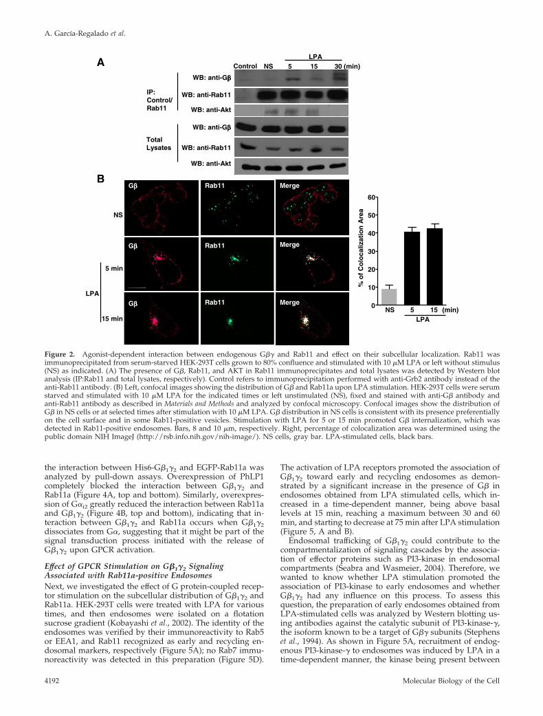
To investigate whether the interaction between endoge-nous G�1�2 and Rab11a can be influenced by the activationof LPA receptors, Rab11a was immunoprecipitated fromnonstimulated or LPA-stimulated serum-starved cells. Fig-ure 2A shows that activation of LPA receptors promoted theassociation of endogenous G�� and Rab11a in a time-depen-dent manner. In addition, we also demonstrated the pres-ence of AKT in the G�1�2:Rab11a complex (Figure 2A). Todetermine the effect of GPCR stimulation on the subcellulardistribution of endogenous G�1�2, serum-starved HEK-293Tcells were stimulated with LPA for the indicated times, andthe localization of G� and Rab11 was examined by immu-
A. Garcıa-Regalado et al.
Molecular Biology of the Cell4190
nofluorescence by using confocal microscopy. In nonstimu-lated cells, G�� was predominantly found at the plasmamembrane and in dispersed intracellular vesicles, whereasRab11 was found in vesicles (Figure 2B, left, NS). However,after LPA stimulation, both proteins were mostly detected incolocalization at endocytic structures that concentrated inthe juxtanuclear area (Figure 2B, left). Additionally, we an-alyzed the colocalization area between G�1�2 and Rab11a; innonstimulated cells, 9.05 � 2.0% of the proteins colocalized,whereas in LPA-stimulated cells the overlap was extensive,40.61 � 2.68% for 5 min LPA-stimulated cells and 42.47 �2.59% for 15-min LPA-stimulated cells (Figure 2B, right).Collectively, these results indicated that the interaction be-tween G�1�2 and Rab11a revealed by the yeast two-hybridscreen and detected by pull-down experiments of trans-fected epitope-tagged proteins, also occurs between endog-enous G�1�2 and Rab11a proteins and is promoted by theactivation of LPA receptors. They also suggested thatRab11a might link G�� to endosomal compartments regu-lating the cellular distribution of the heterodimer.
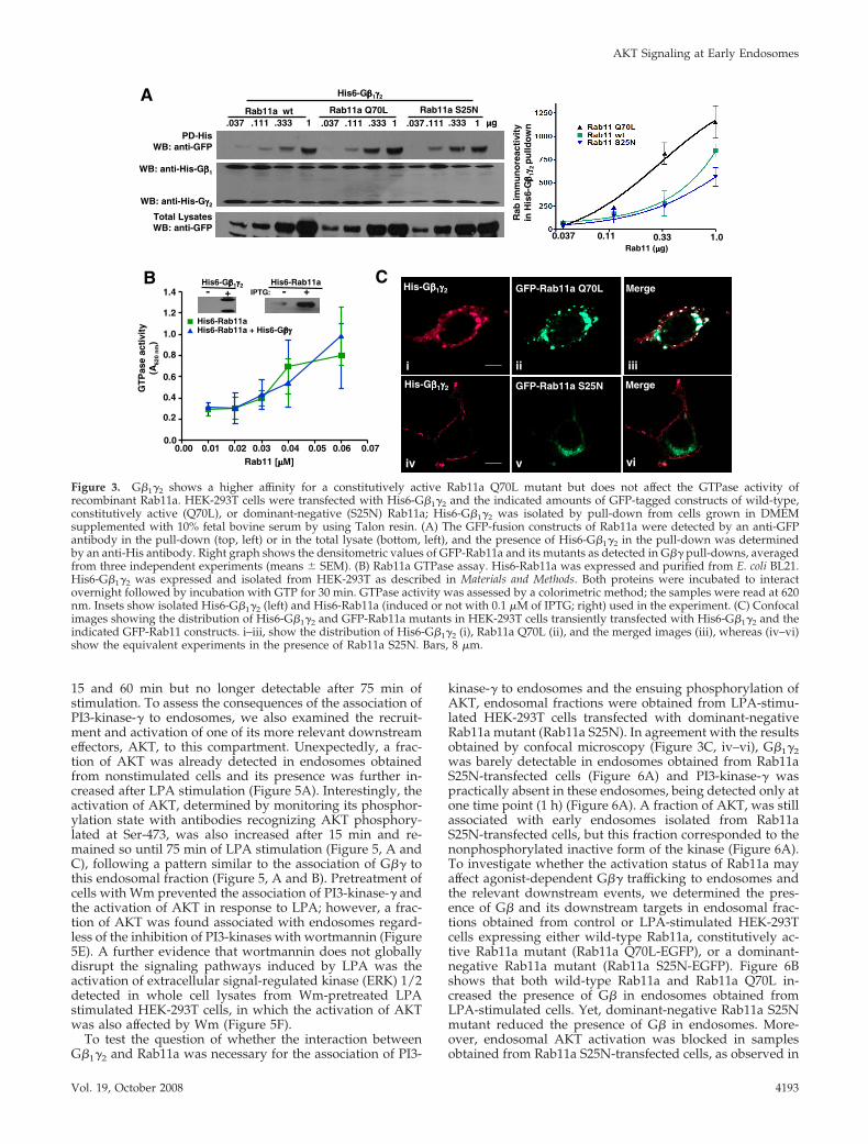
Constitutively Active Rab11a Mutant Promotes theAccumulation of G�1�2 Associated with Large EndosomesTo investigate whether the activation status of Rab11a mayaffect its interaction with G�1�2, we determined the interac-tion of His6-G�1�2 with either wild type, constitutively ac-tive, or dominant-negative EGFP-tagged Rab11a mutants(Rab11a WT, Q70L, or S25N, respectively) by pull-downassays using lysates from HEK-293T cells transfected withincreasing amounts of each Rab11a (Figure 3A). Althoughall three forms of Rab11 showed interaction with G�1�2, theconstitutively active Rab11a Q70L mutant clearly had a
higher affinity for G�1�2 as shown by its ability to interactmore efficiently than wild-type Rab11a or Rab11a S25N incells transfected with reduced amounts of the construct (Fig-ure 3A). To investigate whether G�1�2 could affect the cat-alytic status of Rab11a, an in vitro GTPase assay was used todetermine the GTPase activity of recombinant His6-Rab11ain the presence or absence of His6-G�1�2 isolated from HEK-293T cells. Figure 3B shows that G�1�2 did not modify theGTPase activity of Rab11. We then examined the cellulardistribution of G�1�2 in the presence or absence of Rab11amutants (Figure 3C). The constitutively active Rab11a mu-tant (Rab11Q70L) caused a prominent accumulation ofG�1�2 in large vesicles, in which this Rab11a mutant wasalso detected (Figure 3C, i–iii). In contrast, the dominant-negative Rab11a mutant (Rab11S25N) prevented the forma-tion of large vesicles and did not change the cellular distri-bution of G�1�2, which remained at the plasma membrane(Figure 3C, iv–vi). These results suggested that G�1�2 traf-ficking toward endocytic compartments depends on the in-teraction of this heterodimer with active Rab11a.
Effect of PhLP1 and G�i2 on the Interaction betweenG�1�2 and Rab11aNext, we wanted to determine whether G�1�2 interactionwith Rab11a required the availability of G�1�2 as a freeheterodimer, exposing its G�-interacting interface. For this,we tested whether the interaction between G�1�2 andRab11a could be competed with G�i2 or PhLP1, a ubiquitousprotein known to interact with G�� at the G�-interactinginterface (Gaudet et al., 1996). In these experiments, eitherFlag-tagged PhLP1 or G�i2 were transiently transfected intoHEK-293T cells together with His6-G�1�2 and EGFP-Rab11a, and
Pull Down: HisWB: anti-GFP
Total LysateWB: anti-GFP
- + GFP-Rab11a
His6-Gββββ1γγγγ2
NTA B
Gββββ1 pGB3 p53
- LT - AHLT/+XααααGAL
Rab11a
PTD1
Gββββ1 pGB3 p53
C
iii
Total Lysate
WB: anti-Gββββ
WB: anti-Rab11
IP: control Rab11
WB: anti-Gββββ
WB: anti-Rab11
D
WB: anti-His
WB: anti-His
His6-Gββββ1
His6-Gγγγγ2
i
His-Gββββ1γγγγ2
ii
His-Gββββ1γγγγ2 + GFP-Rab11a
iii
MergeHis-Gββββ1γγγγ2 + GFP-Rab11a
iv
Figure 1. G�1�2 interacts with Rab11a andthey colocalize in HEK-293T cells. (A) A full-length clone of Rab11a was identified as a G�1subunit interactor by yeast two-hybrid. Thespecificity of the interaction between Rab11aand G�1 was determined in the yeast two-hy-brid system using pGB3 or p53 as negative con-trols. Yeast grew on media lacking leucine andtryptophan (�LT, which selects for the presenceof plasmids; left), but only those displaying in-teractions grew in restrictive media lacking ad-enine, histidine, leucine, and tryptophan andwere positive for the activity of �-galactosidase(�AHLT � X �-Gal; right). (B) Pull-down anal-ysis in mammalian cells demonstrating the in-teraction between G�� and Rab11a. HEK-293Tcells were transfected with GFP-Rab11a (expres-sion detected in total lysates with GFP-specificantibodies; bottom) and His6-tagged G�1�2, 2 dafter transfection, His6-G�1�2 was isolated fromcells grown in DMEM supplemented with 10%fetal bovine serum by using Talon beads (top),and Rab11a was detected with GFP-specific an-tibodies. The presence of His-tagged G�1 andHis-tagged G�2 in pull-downs (middle) was de-tected using an antibody recognizing the poly-histidine tag. NT, nontransfected cells. (C) Inter-action between endogenous G�� and Rab11 inserum-stimulated HEK-293T. Rab11 was immunoprecipitated from cells grown to 80% of confluence in DMEM supplemented with 10% fetalbovine serum, the presence of G� in Rab11 immunoprecipitates and total cell lysate was detected by Western blot analysis (top and thirdpanels, respectively). The presence of Rab11 in the immunoprecipitate and total lysate is shown in the second and fourth panels, respectively.Control refers to immunoprecipitation performed with a rabbit preimmune serum instead of the anti-Rab11 antibody. (D) Confocal imagesshowing the distribution of His6-G�1�2 and GFP-Rab11a in HEK-293 cells. Cells were transiently transfected with His6-G�1�2 either in theabsence or presence of GFP-Rab11a, fixed, and stained with anti-histidine antibody and analyzed by confocal microscopy. i, His6-G�1�2 inthe absence and in the presence (ii–iv) of GFP-Rab11a. ii, His6-G�1�2 in the presence of GFP-Rab11a. iii, the same image showing GFP-Rab11a.iv, merged images showing colocalization of His6-G�1�2 and GFP-Rab11a. Bar, 8 �m (i).
AKT Signaling at Early Endosomes
Vol. 19, October 2008 4191
the interaction between His6-G�1�2 and EGFP-Rab11a wasanalyzed by pull-down assays. Overexpression of PhLP1completely blocked the interaction between G�1�2 andRab11a (Figure 4A, top and bottom). Similarly, overexpres-sion of G�i2 greatly reduced the interaction between Rab11aand G�1�2 (Figure 4B, top and bottom), indicating that in-teraction between G�1�2 and Rab11a occurs when G�1�2dissociates from G�, suggesting that it might be part of thesignal transduction process initiated with the release ofG�1�2 upon GPCR activation.
Effect of GPCR Stimulation on G�1�2 SignalingAssociated with Rab11a-positive EndosomesNext, we investigated the effect of G protein-coupled recep-tor stimulation on the subcellular distribution of G�1�2 andRab11a. HEK-293T cells were treated with LPA for varioustimes, and then endosomes were isolated on a flotationsucrose gradient (Kobayashi et al., 2002). The identity of theendosomes was verified by their immunoreactivity to Rab5or EEA1, and Rab11 recognized as early and recycling en-dosomal markers, respectively (Figure 5A); no Rab7 immu-noreactivity was detected in this preparation (Figure 5D).
The activation of LPA receptors promoted the association ofG�1�2 toward early and recycling endosomes as demon-strated by a significant increase in the presence of G� inendosomes obtained from LPA stimulated cells, which in-creased in a time-dependent manner, being above basallevels at 15 min, reaching a maximum between 30 and 60min, and starting to decrease at 75 min after LPA stimulation(Figure 5, A and B).
Endosomal trafficking of G�1�2 could contribute to thecompartmentalization of signaling cascades by the associa-tion of effector proteins such as PI3-kinase in endosomalcompartments (Seabra and Wasmeier, 2004). Therefore, wewanted to know whether LPA stimulation promoted theassociation of PI3-kinase to early endosomes and whetherG�1�2 had any influence on this process. To assess thisquestion, the preparation of early endosomes obtained fromLPA-stimulated cells was analyzed by Western blotting us-ing antibodies against the catalytic subunit of PI3-kinase-�,the isoform known to be a target of G�� subunits (Stephenset al., 1994). As shown in Figure 5A, recruitment of endog-enous PI3-kinase-� to endosomes was induced by LPA in atime-dependent manner, the kinase being present between
WB: anti-Gββββ
WB: anti-Rab11
WB: anti-Rab11
NS 5 15 30LPA
WB: anti-Gββββ
(min)A
B
IP:Control/Rab11
Total Lysates
WB: anti-Akt
WB: anti-Akt
Control
NS
5 min
15 min
LPA
Gββββ
Gββββ
Rab11
Rab11
Rab11
Merge
Merge
Merge
Gββββ 0
10
20
30
40
50
60
NS 5 15 (min)LPA
% o
f C
olo
caliz
atio
n A
rea
Figure 2. Agonist-dependent interaction between endogenous G�� and Rab11 and effect on their subcellular localization. Rab11 wasimmunoprecipitated from serum-starved HEK-293T cells grown to 80% confluence and stimulated with 10 �M LPA or left without stimulus(NS) as indicated. (A) The presence of G�, Rab11, and AKT in Rab11 immunoprecipitates and total lysates was detected by Western blotanalysis (IP:Rab11 and total lysates, respectively). Control refers to immunoprecipitation performed with anti-Grb2 antibody instead of theanti-Rab11 antibody. (B) Left, confocal images showing the distribution of G� and Rab11a upon LPA stimulation. HEK-293T cells were serumstarved and stimulated with 10 �M LPA for the indicated times or left unstimulated (NS), fixed and stained with anti-G� antibody andanti-Rab11 antibody as described in Materials and Methods and analyzed by confocal microscopy. Confocal images show the distribution ofG� in NS cells or at selected times after stimulation with 10 �M LPA. G� distribution in NS cells is consistent with its presence preferentiallyon the cell surface and in some Rab11-positive vesicles. Stimulation with LPA for 5 or 15 min promoted G� internalization, which wasdetected in Rab11-positive endosomes. Bars, 8 and 10 �m, respectively. Right, percentage of colocalization area was determined using thepublic domain NIH ImageJ (http://rsb.info.nih.gov/nih-image/). NS cells, gray bar. LPA-stimulated cells, black bars.
A. Garcıa-Regalado et al.
Molecular Biology of the Cell4192
15 and 60 min but no longer detectable after 75 min ofstimulation. To assess the consequences of the association ofPI3-kinase-� to endosomes, we also examined the recruit-ment and activation of one of its more relevant downstreameffectors, AKT, to this compartment. Unexpectedly, a frac-tion of AKT was already detected in endosomes obtainedfrom nonstimulated cells and its presence was further in-creased after LPA stimulation (Figure 5A). Interestingly, theactivation of AKT, determined by monitoring its phosphor-ylation state with antibodies recognizing AKT phosphory-lated at Ser-473, was also increased after 15 min and re-mained so until 75 min of LPA stimulation (Figure 5, A andC), following a pattern similar to the association of G�� tothis endosomal fraction (Figure 5, A and B). Pretreatment ofcells with Wm prevented the association of PI3-kinase-� andthe activation of AKT in response to LPA; however, a frac-tion of AKT was found associated with endosomes regard-less of the inhibition of PI3-kinases with wortmannin (Figure5E). A further evidence that wortmannin does not globallydisrupt the signaling pathways induced by LPA was theactivation of extracellular signal-regulated kinase (ERK) 1/2detected in whole cell lysates from Wm-pretreated LPAstimulated HEK-293T cells, in which the activation of AKTwas also affected by Wm (Figure 5F).
To test the question of whether the interaction betweenG�1�2 and Rab11a was necessary for the association of PI3-
kinase-� to endosomes and the ensuing phosphorylation ofAKT, endosomal fractions were obtained from LPA-stimu-lated HEK-293T cells transfected with dominant-negativeRab11a mutant (Rab11a S25N). In agreement with the resultsobtained by confocal microscopy (Figure 3C, iv–vi), G�1�2was barely detectable in endosomes obtained from Rab11aS25N-transfected cells (Figure 6A) and PI3-kinase-� waspractically absent in these endosomes, being detected only atone time point (1 h) (Figure 6A). A fraction of AKT, was stillassociated with early endosomes isolated from Rab11aS25N-transfected cells, but this fraction corresponded to thenonphosphorylated inactive form of the kinase (Figure 6A).To investigate whether the activation status of Rab11a mayaffect agonist-dependent G�� trafficking to endosomes andthe relevant downstream events, we determined the pres-ence of G� and its downstream targets in endosomal frac-tions obtained from control or LPA-stimulated HEK-293Tcells expressing either wild-type Rab11a, constitutively ac-tive Rab11a mutant (Rab11a Q70L-EGFP), or a dominant-negative Rab11a mutant (Rab11a S25N-EGFP). Figure 6Bshows that both wild-type Rab11a and Rab11a Q70L in-creased the presence of G� in endosomes obtained fromLPA-stimulated cells. Yet, dominant-negative Rab11a S25Nmutant reduced the presence of G� in endosomes. More-over, endosomal AKT activation was blocked in samplesobtained from Rab11a S25N-transfected cells, as observed in
CB
His6-Rab11aHis6-Rab11a + His6-Gβγβγβγβγ
0.01 0.02 0.03 0.04 0.060.00 0.05 0.070.0
0.2
0.4
0.6
0.8
1.0
1.2
1.4
Rab11 [µµµµM]
GT
Pas
e ac
tivi
ty(A
620
nm
)
A
PD-HisWB: anti-GFP
Total LysatesWB: anti-GFP
Rab11a wt Rab11a Q70L Rab11a S25N.037 1.333.111.037 1.333.111 .037 1.333.111 µµµµg
WB: anti-His-Gγγγγ2
WB: anti-His-Gββββ1
His6-Rab11aIPTG:
His6-Gββββ1γγγγ2+
His-Gββββ1γγγγ2 MergeGFP-Rab11a Q70L
i ii iii
His-Gββββ1γγγγ2 GFP-Rab11a S25N Merge
iv v vi
Rab
imm
un
ore
acti
vity
in H
is6-
Gββ ββ 1γγ γγ 2
pu
lldo
wn
Rab11 (µµµµg)0.037 0.11 0.33 1.0
His6-Gββββ1γγγγ2
+- -
Figure 3. G�1�2 shows a higher affinity for a constitutively active Rab11a Q70L mutant but does not affect the GTPase activity ofrecombinant Rab11a. HEK-293T cells were transfected with His6-G�1�2 and the indicated amounts of GFP-tagged constructs of wild-type,constitutively active (Q70L), or dominant-negative (S25N) Rab11a; His6-G�1�2 was isolated by pull-down from cells grown in DMEMsupplemented with 10% fetal bovine serum by using Talon resin. (A) The GFP-fusion constructs of Rab11a were detected by an anti-GFPantibody in the pull-down (top, left) or in the total lysate (bottom, left), and the presence of His6-G�1�2 in the pull-down was determinedby an anti-His antibody. Right graph shows the densitometric values of GFP-Rab11a and its mutants as detected in G�� pull-downs, averagedfrom three independent experiments (means � SEM). (B) Rab11a GTPase assay. His6-Rab11a was expressed and purified from E. coli BL21.His6-G�1�2 was expressed and isolated from HEK-293T as described in Materials and Methods. Both proteins were incubated to interactovernight followed by incubation with GTP for 30 min. GTPase activity was assessed by a colorimetric method; the samples were read at 620nm. Insets show isolated His6-G�1�2 (left) and His6-Rab11a (induced or not with 0.1 �M of IPTG; right) used in the experiment. (C) Confocalimages showing the distribution of His6-G�1�2 and GFP-Rab11a mutants in HEK-293T cells transiently transfected with His6-G�1�2 and theindicated GFP-Rab11 constructs. i–iii, show the distribution of His6-G�1�2 (i), Rab11a Q70L (ii), and the merged images (iii), whereas (iv–vi)show the equivalent experiments in the presence of Rab11a S25N. Bars, 8 �m.
AKT Signaling at Early Endosomes
Vol. 19, October 2008 4193
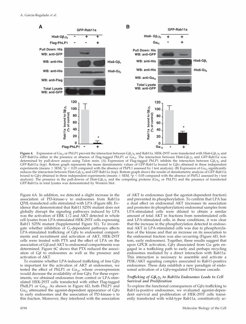
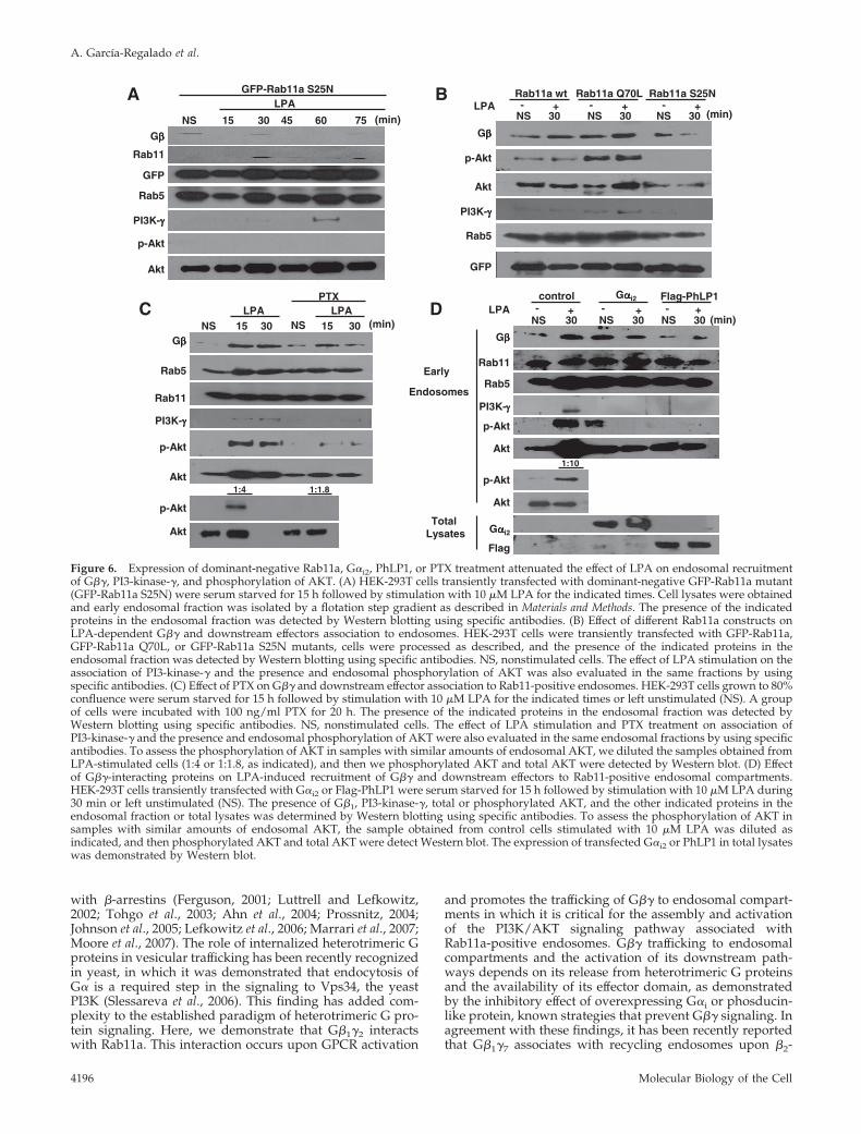
Figure 6A. In addition, we detected a slight increase in theassociation of PI3-kinase-� to endosomes from Rab11aQ70L-transfected cells stimulated with LPA (Figure 6B). Ev-idence that demonstrated that Rab11 S25N mutant does notglobally disrupt the signaling pathways induced by LPAwas the activation of ERK 1/2 and AKT detected in wholecell lysates from LPA-stimulated HEK-293T cells expressingRab11 S25N mutant (Supplemental Figure S1). To investi-gate whether inhibition of Gi-dependent pathways affectsLPA-stimulated trafficking of G�� to endosomal compart-ments and recruitment and activation of AKT, HEK-293Tcells were treated with PTX and the effect of LPA on theassociation of G� and AKT to endosomal compartments wasdetermined. Figure 6C shows that PTX reduced the associ-ation of G� to endosomes as well as the presence andactivation of AKT.
To examine whether LPA-induced trafficking of free G��is important for the activation of AKT at endosomes, wetested the effect of PhLP1 or G�i2, whose overexpressionwould decrease the availability of free G��. For these exper-iments, we obtained endosomes from control or LPA-stim-ulated HEK-293T cells transfected with either Flag-taggedPhdLP1 or G�i2. As shown in Figure 6D, both PhLP1 andG�i2 attenuated the agonist-dependent appearance of G��in early endosomes and the association of PI3-kinase-� tothis fraction. Moreover, they interfered with the association
of AKT to endosomes (just the agonist-dependent fraction)and prevented its phosphorylation. To confirm that LPA hasa dual effect on endosomal AKT (increases its associationand promotes its phosphorylation) endosomal samples fromLPA-stimulated cells were diluted to obtain a similaramount of total AKT in fractions from nonstimulated cellsand LPA-stimulated cells, in these conditions, it was clearthat the increase in the phosphorylation detected in endoso-mal AKT in LPA-stimulated cells was due to phosphoryla-tion of the kinase and that an increase on its association tothe endosomal fraction was also occurring (Figure 6D, bot-tom, early endosomes). Together, these results suggest thatupon GPCR activation, G�� dissociated from G� gets en-gaged in a trafficking path to early and perhaps recyclingendosomes mediated by a direct interaction with Rab11a.This interaction is necessary to assemble and activate aPI3K/AKT signaling complex associated to Rab11-positiveendosomes. These data establish a new paradigm of endo-somal activation of a G��-regulated PI3-kinase cascade.
Trafficking of G�1�2 to Rab11a Endosomes Leads to CellSurvival and ProliferationTo explore the functional consequences of G�� trafficking toRab11a-positive endosomes, we evaluated agonist-depen-dent survival and proliferation of HEK-293T cells transi-ently transfected with wild-type Rab11a, constitutively ac-
A B
Pull Down: HisWB: anti-GFP
Total LysateWB: anti-GFP
WB: anti-His
WB: anti-Flag
GFP-Rab11a
His6-Gββββ1γγγγ2
Flag-PhLP1
- + +- - +
GFP-Rab11a
His6- Gββββ1γγγγ2
Gααααi2 - +
+ +
WB: anti-Gααααi2
Pull Down: HisWB: anti-GFP
Total LysateWB: anti-GFP
WB: anti-His
Flag-PhLP1
*
*
0
20
40
60
80
100
120
Rab
11a
bo
un
dto
Gββ ββ 1γγ γγ 2
(% o
f co
ntr
ol )
0
20
40
60
80
100
120
Gαααα i2 - +- +R
ab11
a b
ou
nd
toGββ ββ 1γγ γγ 2
(%
of
con
tro
l)
WB: anti-His
WB: anti-His
His6-Gββββ1
His6-Gγγγγ2
His6-Gββββ1
His6-Gγγγγ2
Figure 4. Expression of G�i2 or PhLP1 prevent the interaction between G�1�2 and Rab11a. HEK-293T were transfected with His6-G�1�2 andGFP-Rab11a either in the presence or absence of Flag-tagged PhLP1 or G�i2. The interaction between His6-G�1�2 and GFP-Rab11a wasdetermined by pull-down assays using Talon resin. (A) Expression of Flag-tagged PhLP1 inhibits the interaction between G�1�2 andGFP-Rab11a (top). Bottom graph represents the mean densitometric values of GFP-Rab11a bound to G�� obtained in three independentexperiments (means � SEM, *p � 0.05 compared with the absence of PhPL1 assessed by t test analysis). (B) Expression of G�i2 significantlyreduces the interaction between His6-G�1�2 and GFP-Rab11a (top). Bottom graph shows the results of densitometric analysis of GFP-Rab11abound to G�� obtained in three independent experiments (means � SEM, *p � 0.05 compared with the absence of PhPL1 assessed by t testanalysis). The presence in the pull-downs of His6-G�1�2 and the competing proteins (G�i2 or PhLP1) and the presence of transfectedGFP-Rab11a in total lysates was demonstrated by Western blot.
A. Garcıa-Regalado et al.
Molecular Biology of the Cell4194

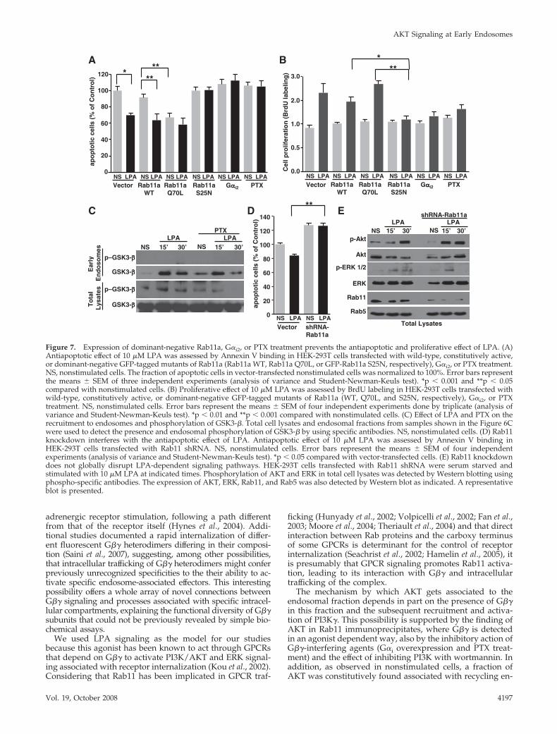
tive Rab11a mutant (EGFP-Rab11a Q70L) or a dominant-negative Rab11a mutant (EGFP-Rab11a S25N). As shown inFigure 7A, LPA protected against apoptosis in cells express-ing either wild-type Rab11a or Rab11a Q70L; in the Rab11aQ70L-expressing cells an antiapoptotic effect was detectedevent in the absence of LPA stimulation. However, overex-pression of dominant-negative Rab11a S25N mutant blockedthe LPA-dependent survival effect, resulting in apoptosis.Additionally, overexpression of G�i2 or treatment with PTXalso interfered with the survival effect promoted by LPA, asshown in Figure 7A.
To examine the effect of Rab11a on LPA-dependent cellproliferation, we transiently transfected HEK-293T cellswith wild-type Rab11a, constitutively active Rab11a mutant(EGFP-Rab11a Q70L) or a dominant-negative Rab11a mu-tant (EGFP-Rab11a S25N). As shown in Figure 7B, the pro-liferative effect of LPA was not affected by wild-type orQ70L mutant Rab11a. However, dominant-negative Rab11aS25N mutant blocked the proliferative effect of LPA. Simi-larly, the proliferative effect of LPA was blocked by overex-pression of G�i2 or PTX treatment, two strategies known tointerfere with G�� signaling (Figure 7B). Together, our re-sults indicate that agonist-dependent trafficking of G�� toRab11a endosomes is linked to cell survival and prolifera-tion.
To evaluate the possibility that AKT effectors can be re-cruited to endosomes and activated as a consequence of G��trafficking and AKT stimulation, we explored the presenceof GSK3-�, FKHR-1, and Bad proteins, known substrates ofAKT, on endosomes from LPA- and PTX-pretreated cells. Asshown in Figure 7C, we detected that LPA promoted theassociation of GSK3-� to the endosomal fraction; however,we could not reveal its phosphorylation and the treatmentwith PTX did not show a significant effect on LPA-stimu-lated association of GSK3-� to endosomes. The lack of phos-
phorylation of GSK3-� associated to the endosomal fractionmakes it difficult to connect its recruitment to the activationof AKT, which could be detected in total cell lysates andwhere the treatment with PTX attenuated the phosphoryla-tion of this AKT substrate in LPA-stimulated cells (Figure7C). In contrast, we could not detect the presence of FKHR-1or Bad proteins at the endosomal fraction (data not shown).
To further examine the functional role of endogenousRab11 in LPA-dependent cell survival, we assessed the effectof short hairpin RNA-induced Rab11 knockdown on theantiapoptotic effect of LPA. As shown in Figure 7D, Rab11shRNA blocked the LPA-dependent survival effect, result-ing in apoptosis. Additionally, we demonstrated that Rab11shRNA does not globally disrupt LPA-dependent signalingpathways as evidenced by the ability of LPA to promote theactivation of ERK 1/2 and AKT as detected in total lysatesfrom cells transiently transfected with shRNA specific forRab11 (Figure 7E). Together, our results indicate that G��trafficking to endosomes, in association to Rab11, is linked tocell survival.
DISCUSSION
Heterotrimeric G protein signaling is based on G proteinactivation/inactivation cycles occurring at the plasma mem-brane, where GPCRs are stimulated by their cognate ligandspresent in the extracellular milieu (Gilman, 1987; Dessauer etal., 1996; Offermanns and Simon, 1996; Bourne, 1997; Offer-manns, 2003; Bourne, 2006). Phosphorylation and traffickingof GPCRs is commonly recognized as part of a mechanism ofdesensitization (Ghanouni et al., 2001; Whistler et al., 2002;Prossnitz, 2004; Drake et al., 2006; Reiter and Lefkowitz,2006; Violin et al., 2006). However, internalization of GPCRshas also been proposed as a means to acquire novel signal-ing properties via association of the internalized receptors
Figure 5. LPA stimulation increases G�1�2 as-sociation to Rab11a-positive endosomes. HEK-293T cells grown to 80% confluence were serumstarved for 15 h followed by stimulation with10 �M LPA for the indicated times. Cells werefractionated to isolate early and late endosomesas described in Materials and Methods. (A and D)LPA stimulation induces a time-dependent as-sociation of G�1�2 heterodimer with Rab11-positive endosomal fractions as detected byWestern blotting using an anti-G�1 antibody.Distribution of Rab11, Rab5, early endosomalmarker EEA1, and late endosomal marker Rab7was assessed by Western blot as indicated. NS,nonstimulated cells; TL, total cell lysates. Theeffect of LPA stimulation on the association ofPI3-kinase-� and the presence and endosomalphosphorylation of AKT was also evaluated inthe same endosomal fractions by using specificantibodies. (B) Graph represents the mean den-sitometric values of G�1 present in the endoso-mal fractions determined in three independentexperiments. Means � SEM, *p � 0.001 com-pared with nonstimulated cells (analysis ofvariance and Student-Newman-Keuls test). (C)Results of similar densitometric analysis ofAKT phosphorylation (means � SEM, *p �0.001 compared with nonstimulated cells. (E)Wortmannin prevented LPA-induced endosomal recruitment of PI3-kinase-� and phosphorylation of AKT. HEK-293T cells were incubatedwith 300 nM wortmannin for 1 h before LPA stimulation, and early endosomes were isolated as described, and proteins were detected byWestern blot using anti-phospho-Ser473-AKT (top), total AKT (middle), and PI3-kinase-� catalytic subunit (bottom). (F) Wortmannin doesnot affect LPA-dependent ERK phosphorylation. Total cell lysates were obtained from HEK-293T cells pretreated with wortmannin beforeLPA stimulation, and the presence of the indicated proteins was detected by Western blotting using specific antibodies.
AKT Signaling at Early Endosomes
Vol. 19, October 2008 4195
with �-arrestins (Ferguson, 2001; Luttrell and Lefkowitz,2002; Tohgo et al., 2003; Ahn et al., 2004; Prossnitz, 2004;Johnson et al., 2005; Lefkowitz et al., 2006; Marrari et al., 2007;Moore et al., 2007). The role of internalized heterotrimeric Gproteins in vesicular trafficking has been recently recognizedin yeast, in which it was demonstrated that endocytosis ofG� is a required step in the signaling to Vps34, the yeastPI3K (Slessareva et al., 2006). This finding has added com-plexity to the established paradigm of heterotrimeric G pro-tein signaling. Here, we demonstrate that G�1�2 interactswith Rab11a. This interaction occurs upon GPCR activation
and promotes the trafficking of G�� to endosomal compart-ments in which it is critical for the assembly and activationof the PI3K/AKT signaling pathway associated withRab11a-positive endosomes. G�� trafficking to endosomalcompartments and the activation of its downstream path-ways depends on its release from heterotrimeric G proteinsand the availability of its effector domain, as demonstratedby the inhibitory effect of overexpressing G�i or phosducin-like protein, known strategies that prevent G�� signaling. Inagreement with these findings, it has been recently reportedthat G�1�7 associates with recycling endosomes upon �2-
ANS 15 30 6045 75
LPAGFP-Rab11a S25N
p-Akt
Akt
Gββββ
Rab5
Rab11
GFP
PI3K-γγγγ
(min)
Early
Endosomes
TotalLysates
LPA
Akt
p-Akt
Rab5
Gββββ
Rab11
PI3K-γγγγ
NS 30 NS NS30 30
Flag-PhLP1Gααααi2
+ +control
--(min)
+-
Gααααi2
Flag
D
Akt
p-Akt
1:10
C LPA
GββββNS
LPA
Rab5
p-Akt
15 NS30 15 30
PTX
Akt
Rab11
(min)
PI3K-γγγγ
Gββββ
Rab5
p-Akt
GFP
NS 30 NS 30 NS 30
Rab11a wt Rab11a Q70L Rab11a S25N
Akt
+-(min)
+- +-LPA
PI3K-γγγγ
Akt
p-Akt
1:4 1:1.8
B
Figure 6. Expression of dominant-negative Rab11a, G�i2, PhLP1, or PTX treatment attenuated the effect of LPA on endosomal recruitmentof G��, PI3-kinase-�, and phosphorylation of AKT. (A) HEK-293T cells transiently transfected with dominant-negative GFP-Rab11a mutant(GFP-Rab11a S25N) were serum starved for 15 h followed by stimulation with 10 �M LPA for the indicated times. Cell lysates were obtainedand early endosomal fraction was isolated by a flotation step gradient as described in Materials and Methods. The presence of the indicatedproteins in the endosomal fraction was detected by Western blotting using specific antibodies. (B) Effect of different Rab11a constructs onLPA-dependent G�� and downstream effectors association to endosomes. HEK-293T cells were transiently transfected with GFP-Rab11a,GFP-Rab11a Q70L, or GFP-Rab11a S25N mutants, cells were processed as described, and the presence of the indicated proteins in theendosomal fraction was detected by Western blotting using specific antibodies. NS, nonstimulated cells. The effect of LPA stimulation on theassociation of PI3-kinase-� and the presence and endosomal phosphorylation of AKT was also evaluated in the same fractions by usingspecific antibodies. (C) Effect of PTX on G�� and downstream effector association to Rab11-positive endosomes. HEK-293T cells grown to 80%confluence were serum starved for 15 h followed by stimulation with 10 �M LPA for the indicated times or left unstimulated (NS). A groupof cells were incubated with 100 ng/ml PTX for 20 h. The presence of the indicated proteins in the endosomal fraction was detected byWestern blotting using specific antibodies. NS, nonstimulated cells. The effect of LPA stimulation and PTX treatment on association ofPI3-kinase-� and the presence and endosomal phosphorylation of AKT were also evaluated in the same endosomal fractions by using specificantibodies. To assess the phosphorylation of AKT in samples with similar amounts of endosomal AKT, we diluted the samples obtained fromLPA-stimulated cells (1:4 or 1:1.8, as indicated), and then we phosphorylated AKT and total AKT were detected by Western blot. (D) Effectof G��-interacting proteins on LPA-induced recruitment of G�� and downstream effectors to Rab11-positive endosomal compartments.HEK-293T cells transiently transfected with G�i2 or Flag-PhLP1 were serum starved for 15 h followed by stimulation with 10 �M LPA during30 min or left unstimulated (NS). The presence of G�1, PI3-kinase-�, total or phosphorylated AKT, and the other indicated proteins in theendosomal fraction or total lysates was determined by Western blotting using specific antibodies. To assess the phosphorylation of AKT insamples with similar amounts of endosomal AKT, the sample obtained from control cells stimulated with 10 �M LPA was diluted asindicated, and then phosphorylated AKT and total AKT were detect Western blot. The expression of transfected G�i2 or PhLP1 in total lysateswas demonstrated by Western blot.
A. Garcıa-Regalado et al.
Molecular Biology of the Cell4196
adrenergic receptor stimulation, following a path differentfrom that of the receptor itself (Hynes et al., 2004). Addi-tional studies documented a rapid internalization of differ-ent fluorescent G�� heterodimers differing in their composi-tion (Saini et al., 2007), suggesting, among other possibilities,that intracellular trafficking of G�� heterodimers might conferpreviously unrecognized specificities to the their ability to ac-tivate specific endosome-associated effectors. This interestingpossibility offers a whole array of novel connections betweenG�� signaling and processes associated with specific intracel-lular compartments, explaining the functional diversity of G��subunits that could not be previously revealed by simple bio-chemical assays.
We used LPA signaling as the model for our studiesbecause this agonist has been known to act through GPCRsthat depend on G�� to activate PI3K/AKT and ERK signal-ing associated with receptor internalization (Kou et al., 2002).Considering that Rab11 has been implicated in GPCR traf-
ficking (Hunyady et al., 2002; Volpicelli et al., 2002; Fan et al.,2003; Moore et al., 2004; Theriault et al., 2004) and that directinteraction between Rab proteins and the carboxy terminusof some GPCRs is determinant for the control of receptorinternalization (Seachrist et al., 2002; Hamelin et al., 2005), itis presumably that GPCR signaling promotes Rab11 activa-tion, leading to its interaction with G�� and intracellulartrafficking of the complex.
The mechanism by which AKT gets associated to theendosomal fraction depends in part on the presence of G��in this fraction and the subsequent recruitment and activa-tion of PI3K�. This possibility is supported by the finding ofAKT in Rab11 immunoprecipitates, where G�� is detectedin an agonist dependent way, also by the inhibitory action ofG��-interfering agents (G�i overexpression and PTX treat-ment) and the effect of inhibiting PI3K with wortmannin. Inaddition, as observed in nonstimulated cells, a fraction ofAKT was constitutively found associated with recycling en-
C
A
NS LPA NS LPA NS LPA NS LPA NS LPA NS LPAVector Rab11a
WTRab11aQ70L
Rab11aS25N
Gααααi2 PTX
120
40
100
20
60
0
80
apo
pto
tic
cells
(%
of
Co
ntr
ol)
p–GSK3-ββββ
GSK3-ββββ
p–GSK3-ββββ
GSK3-ββββ
LPANS
LPA15’ NS30’ 15’ 30’
PTX
NS LPARab11a
WTRab11aQ70L
Rab11aS25N
B
NS LPA NS LPA NS LPA NS LPA NS LPA
Vector Gααααi2 PTX
Cel
l pro
lifer
atio
n (
Brd
Ula
bel
ing
)
1.0
0.5
2.0
0.0
3.0
Ear
lyE
nd
oso
mes
To
tal
Lys
ates
D
NS LPA NS LPA
Vector shRNA-Rab11a
LPANS
LPA15’ NS30’ 15’ 30’
shRNA-Rab11a
p-Akt
Akt
Rab5
Rab11
ERK
p-ERK 1/2
E
***
***
**
Total Lysates
**
0
20
40
60
80
100
120
140
apo
pto
tic
cells
(%
of
Co
ntr
ol)
Figure 7. Expression of dominant-negative Rab11a, G�i2, or PTX treatment prevents the antiapoptotic and proliferative effect of LPA. (A)Antiapoptotic effect of 10 �M LPA was assessed by Annexin V binding in HEK-293T cells transfected with wild-type, constitutively active,or dominant-negative GFP-tagged mutants of Rab11a (Rab11a WT, Rab11a Q70L, or GFP-Rab11a S25N, respectively), G�i2, or PTX treatment.NS, nonstimulated cells. The fraction of apoptotic cells in vector-transfected nonstimulated cells was normalized to 100%. Error bars representthe means � SEM of three independent experiments (analysis of variance and Student-Newman-Keuls test). *p � 0.001 and **p � 0.05compared with nonstimulated cells. (B) Proliferative effect of 10 �M LPA was assessed by BrdU labeling in HEK-293T cells transfected withwild-type, constitutively active, or dominant-negative GFP-tagged mutants of Rab11a (WT, Q70L, and S25N, respectively), G�i2, or PTXtreatment. NS, nonstimulated cells. Error bars represent the means � SEM of four independent experiments done by triplicate (analysis ofvariance and Student-Newman-Keuls test). *p � 0.01 and **p � 0.001 compared with nonstimulated cells. (C) Effect of LPA and PTX on therecruitment to endosomes and phosphorylation of GSK3-�. Total cell lysates and endosomal fractions from samples shown in the Figure 6Cwere used to detect the presence and endosomal phosphorylation of GSK3-� by using specific antibodies. NS, nonstimulated cells. (D) Rab11knockdown interferes with the antiapoptotic effect of LPA. Antiapoptotic effect of 10 �M LPA was assessed by Annexin V binding inHEK-293T cells transfected with Rab11 shRNA. NS, nonstimulated cells. Error bars represent the means � SEM of four independentexperiments (analysis of variance and Student-Newman-Keuls test). *p � 0.05 compared with vector-transfected cells. (E) Rab11 knockdowndoes not globally disrupt LPA-dependent signaling pathways. HEK-293T cells transfected with Rab11 shRNA were serum starved andstimulated with 10 �M LPA at indicated times. Phosphorylation of AKT and ERK in total cell lysates was detected by Western blotting usingphospho-specific antibodies. The expression of AKT, ERK, Rab11, and Rab5 was also detected by Western blot as indicated. A representativeblot is presented.
AKT Signaling at Early Endosomes
Vol. 19, October 2008 4197
dosomes, we speculate that this fraction of AKT associates toendosomes due to the presence there of AKT-binding pro-teins such as ProF or APPL1 that has been recently charac-terized as an endosomal AKT-recruiting protein (Mitsuuchiet al., 1999; Miaczynska et al., 2004; Fritzius et al., 2006). Inagreement with this possibility, Zerial and colleagues justdemonstrated that APPL1 mediates AKT substrate specific-ity at endosomal compartments (Schenck et al., 2008).
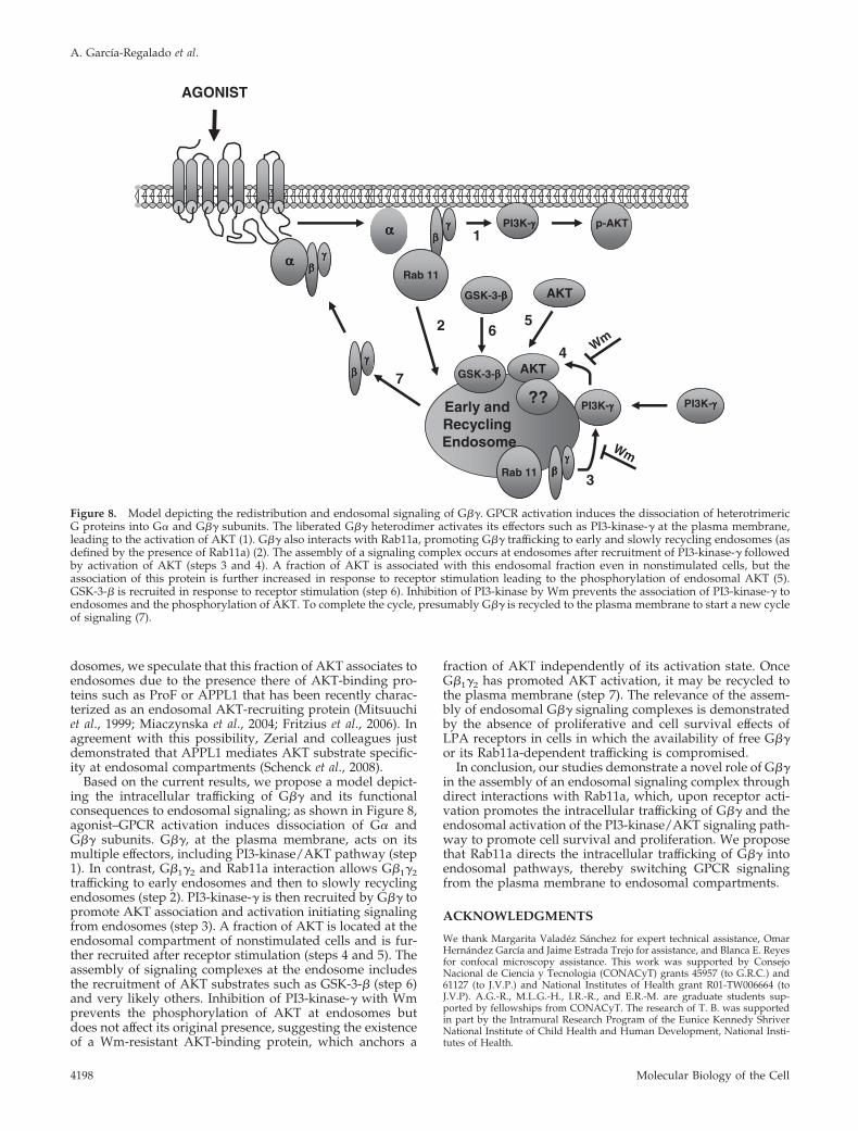
Based on the current results, we propose a model depict-ing the intracellular trafficking of G�� and its functionalconsequences to endosomal signaling; as shown in Figure 8,agonist–GPCR activation induces dissociation of G� andG�� subunits. G��, at the plasma membrane, acts on itsmultiple effectors, including PI3-kinase/AKT pathway (step1). In contrast, G�1�2 and Rab11a interaction allows G�1�2trafficking to early endosomes and then to slowly recyclingendosomes (step 2). PI3-kinase-� is then recruited by G�� topromote AKT association and activation initiating signalingfrom endosomes (step 3). A fraction of AKT is located at theendosomal compartment of nonstimulated cells and is fur-ther recruited after receptor stimulation (steps 4 and 5). Theassembly of signaling complexes at the endosome includesthe recruitment of AKT substrates such as GSK-3-� (step 6)and very likely others. Inhibition of PI3-kinase-� with Wmprevents the phosphorylation of AKT at endosomes butdoes not affect its original presence, suggesting the existenceof a Wm-resistant AKT-binding protein, which anchors a
fraction of AKT independently of its activation state. OnceG�1�2 has promoted AKT activation, it may be recycled tothe plasma membrane (step 7). The relevance of the assem-bly of endosomal G�� signaling complexes is demonstratedby the absence of proliferative and cell survival effects ofLPA receptors in cells in which the availability of free G��or its Rab11a-dependent trafficking is compromised.
In conclusion, our studies demonstrate a novel role of G��in the assembly of an endosomal signaling complex throughdirect interactions with Rab11a, which, upon receptor acti-vation promotes the intracellular trafficking of G�� and theendosomal activation of the PI3-kinase/AKT signaling path-way to promote cell survival and proliferation. We proposethat Rab11a directs the intracellular trafficking of G�� intoendosomal pathways, thereby switching GPCR signalingfrom the plasma membrane to endosomal compartments.
ACKNOWLEDGMENTS
We thank Margarita Valadez Sanchez for expert technical assistance, OmarHernandez Garcıa and Jaime Estrada Trejo for assistance, and Blanca E. Reyesfor confocal microscopy assistance. This work was supported by ConsejoNacional de Ciencia y Tecnologia (CONACyT) grants 45957 (to G.R.C.) and61127 (to J.V.P.) and National Institutes of Health grant R01-TW006664 (toJ.V.P). A.G.-R., M.L.G.-H., I.R.-R., and E.R.-M. are graduate students sup-ported by fellowships from CONACyT. The research of T. B. was supportedin part by the Intramural Research Program of the Eunice Kennedy ShriverNational Institute of Child Health and Human Development, National Insti-tutes of Health.
PI3K-γγγγ
AGONIST
ββββγγγγ
ββββγγγγαααα
Rab 11
ββββγγγγ
Rab 11
Early andRecyclingEndosome
PI3K-γγγγ p-AKT
AKT
??
AKT
αααα
ββββγγγγ
1
2
3
4
5
7
Wm
Wm
PI3K-γγγγ
6
GSK-3-ββββ
GSK-3-ββββ
Figure 8. Model depicting the redistribution and endosomal signaling of G��. GPCR activation induces the dissociation of heterotrimericG proteins into G� and G�� subunits. The liberated G�� heterodimer activates its effectors such as PI3-kinase-� at the plasma membrane,leading to the activation of AKT (1). G�� also interacts with Rab11a, promoting G�� trafficking to early and slowly recycling endosomes (asdefined by the presence of Rab11a) (2). The assembly of a signaling complex occurs at endosomes after recruitment of PI3-kinase-� followedby activation of AKT (steps 3 and 4). A fraction of AKT is associated with this endosomal fraction even in nonstimulated cells, but theassociation of this protein is further increased in response to receptor stimulation leading to the phosphorylation of endosomal AKT (5).GSK-3-� is recruited in response to receptor stimulation (step 6). Inhibition of PI3-kinase by Wm prevents the association of PI3-kinase-� toendosomes and the phosphorylation of AKT. To complete the cycle, presumably G�� is recycled to the plasma membrane to start a new cycleof signaling (7).
A. Garcıa-Regalado et al.
Molecular Biology of the Cell4198
REFERENCES
Ahn, S., Shenoy, S. K., Wei, H., and Lefkowitz, R. J. (2004). Differential kineticand spatial patterns of beta-arrestin and G protein-mediated ERK activationby the angiotensin II receptor. J. Biol. Chem. 279, 35518–35525.
Albert, P. R., and Robillard, L. (2002). G protein specificity: traffic directionrequired. Cell Signal. 14, 407–418.
Barber, M. A., Donald, S., Thelen, S., Anderson, K. E., Thelen, M., and Welch,H. C. (2007). Membrane translocation of P-Rex1 is mediated by G protein betagamma subunits and phosphoinositide 3-kinase. J. Biol. Chem. 282, 29967–29976.
Bourne, H. R. (1997). How receptors talk to trimeric G proteins. Curr. Opin.Cell Biol. 9, 134–142.
Bourne, H. R. (2006). G-proteins and GPCrs: from the beginning. Ernst Scher-ing Found. Symp. Proc. 1–21.
Brock, C., Schaefer, M., Reusch, H. P., Czupalla, C., Michalke, M., Spicher, K.,Schultz, G., and Nurnberg, B. (2003). Roles of G beta gamma in membranerecruitment and activation of p110 gamma/p101 phosphoinositide 3-kinasegamma. J. Cell Biol. 160, 89–99.
Clapham, D. E., and Neer, E. J. (1997). G protein beta gamma subunits. Annu.Rev. Pharmacol. Toxicol. 37, 167–203.
Daaka, Y., Luttrell, L. M., and Lefkowitz, R. J. (1997a). Switching of thecoupling of the beta2-adrenergic receptor to different G proteins by proteinkinase A. Nature 390, 88–91.
Daaka, Y., Pitcher, J. A., Richardson, M., Stoffel, R. H., Robishaw, J. D., andLefkowitz, R. J. (1997b). Receptor and G betagamma isoform-specific interac-tions with G protein-coupled receptor kinases. Proc. Natl. Acad. Sci. USA 94,2180–2185.
Dessauer, C. W., Posner, B. A., and Gilman, A. G. (1996). Visualizing signaltransduction: receptors, G-proteins, and adenylate cyclases. Clin. Sci. 91,527–537.
Diaz Anel, A. M., and Malhotra, V. (2005). PKCeta is required forbeta1gamma2/beta3gamma2- and PKD-mediated transport to the cell surfaceand the organization of the Golgi apparatus. J. Cell Biol. 169, 83–91.
Drake, M. T., Shenoy, S. K., and Lefkowitz, R. J. (2006). Trafficking of Gprotein-coupled receptors. Circ. Res. 99, 570–582.
Evanko, D. S., Thiyagarajan, M. M., Siderovski, D. P., and Wedegaertner, P. B.(2001). G�� isoforms selectively rescue plasma membrane localization andpalmitoylation of mutant G�s and G�q. J. Biol. Chem. 276, 23945–23953.
Fan, G. H., Lapierre, L. A., Goldenring, J. R., and Richmond, A. (2003).Differential regulation of CXCR2 trafficking by Rab GTPases. Blood 101,2115–2124.
Ferguson, S. S. (2001). Evolving concepts in G protein-coupled receptor en-docytosis: the role in receptor desensitization and signaling. Pharmacol. Rev.53, 1–24.
Ford, C. E. et al. (1998). Molecular basis for interactions of G protein beta-gamma subunits with effectors. Science 280, 1271–1274.
Fritzius, T. et al. (2006). A WD-FYVE protein binds to the kinases Akt andPKCzeta/lambda. Biochem. J. 399, 9–20.
Gaudet, R., Bohm, A., and Sigler, P. B. (1996). Crystal structure at 2.4 ang-stroms resolution of the complex of transducin betagamma and its regulator,phosducin. Cell 87, 577–588.
Gautam, N., Downes, G. B., Yan, K., and Kisselev, O. (1998). The G-proteinbetagamma complex. Cell Signal. 10, 447–455.
Ghanouni, P., Steenhuis, J. J., Farrens, D. L., and Kobilka, B. K. (2001).Agonist-induced conformational changes in the G-protein-coupling domainof the beta 2 adrenergic receptor. Proc. Natl. Acad. Sci. USA 98, 5997–6002.
Gilman, A. G. (1987). G proteins: transducers of receptor-generated signals.Annu. Rev. Biochem. 56, 615–649.
Gilman, A. G. (1989). Transmembrane signaling, G proteins, and adenylylcyclase. Harvey Lect. 85, 153–172.
Gorvel, J. P., Chavrier, P., Zerial, M., and Gruenberg, J. (1991). rab5 controlsearly endosome fusion in vitro. Cell 64, 915–925.
Hamelin, E., Theriault, C., Laroche, G., and Parent, J. L. (2005). The intracel-lular trafficking of the G protein-coupled receptor TP� depends on a directinteraction with Rab11. J. Biol. Chem. 280, 36195–36205.
Houle, S., and Marceau, F. (2003). Wortmannin alters the intracellular traf-ficking of the bradykinin B2 receptor: role of phosphoinositide 3-kinase andRab5. Biochem. J. 375, 151–158.
Hunyady, L., Baukal, A. J., Gaborik, Z., Olivares-Reyes, J. A., Bor, M., Szaszak,M., Lodge, R., Catt, K. J., and Balla, T. (2002). Differential PI 3-kinase depen-
dence of early and late phases of recycling of the internalized AT1 angiotensinreceptor. J. Cell Biol. 157, 1211–1222.
Hynes, T. R., Mervine, S. M., Yost, E. A., Sabo, J. L., and Berlot, C. H. (2004).Live cell imaging of Gs and the beta2-adrenergic receptor demonstrates thatboth alphas and beta1gamma7 internalize upon stimulation and exhibit sim-ilar trafficking patterns that differ from that of the beta2-adrenergic receptor.J. Biol. Chem. 279, 44101–44112.
Jamora, C., Yamanouye, N., Van Lint, J., Laudenslager, J., Vandenheede, J. R.,Faulkner, D. J., and Malhotra, V. (1999). Gbetagamma-mediated regulation ofGolgi organization is through the direct activation of protein kinase D. Cell 98,59–68.
Johnson, E. E., Christie, M. J., and Connor, M. (2005). The role of opioidreceptor phosphorylation and trafficking in adaptations to persistent opioidtreatment. Neurosignals 14, 290–302.
Kalia, M., Kumari, S., Chadda, R., Hill, M. M., Parton, R. G., and Mayor, S.(2006). Arf6-independent GPI-anchored protein-enriched early endosomalcompartments fuse with sorting endosomes via a Rab5/phosphatidylinositol-3�-kinase-dependent machinery. Mol. Biol. Cell 17, 3689–3704.
Kobayashi, T., Beuchat, M. H., Chevallier, J., Makino, A., Mayran, N., Escola,J. M., Lebrand, C., Cosson, P., and Gruenberg, J. (2002). Separation andcharacterization of late endosomal membrane domains. J. Biol. Chem. 277,32157–32164.
Kou, R., Igarashi, J., and Michel, T. (2002). Lysophosphatidic acid and recep-tor-mediated activation of endothelial nitric-oxide synthase. Biochemistry 41,4982–4988.
Lefkowitz, R. J., Pierce, K. L., and Luttrell, L. M. (2002). Dancing with differentpartners: protein kinase a phosphorylation of seven membrane-spanningreceptors regulates their G protein-coupling specificity. Mol. Pharmacol. 62,971–974.
Lefkowitz, R. J., Rajagopal, K., and Whalen, E. J. (2006). New roles forbeta-arrestins in cell signaling: not just for seven-transmembrane receptors.Mol. Cell 24, 643–652.
Lin, H. C., Duncan, J. A., Kozasa, T., and Gilman, A. G. (1998). Sequestrationof the G protein beta gamma subunit complex inhibits receptor-mediatedendocytosis. Proc. Natl. Acad. Sci. USA 95, 5057–5060.
Luttrell, L. M., and Lefkowitz, R. J. (2002). The role of beta-arrestins in thetermination and transduction of G-protein-coupled receptor signals. J. CellSci. 115, 455–465.
Marrari, Y., Crouthamel, M., Irannejad, R., and Wedegaertner, P. B. (2007).Assembly and trafficking of heterotrimeric G proteins. Biochemistry 46, 7665–7677.
Miaczynska, M., Christoforidis, S., Giner, A., Shevchenko, A., Uttenweiler-Joseph, S., Habermann, B., Wilm, M., Parton, R. G., and Zerial, M. (2004).APPL proteins link Rab5 to nuclear signal transduction via an endosomalcompartment. Cell 116, 445–456.
Miaczynska, M., and Zerial, M. (2002). Mosaic organization of the endocyticpathway. Exp. Cell Res. 272, 8–14.
Michaelson, D., Ahearn, I., Bergo, M., Young, S., and Philips, M. (2002).Membrane trafficking of heterotrimeric G proteins via the endoplasmic retic-ulum and Golgi. Mol. Biol. Cell 13, 3294–3302.
Mitsuuchi, Y., Johnson, S. W., Sonoda, G., Tanno, S., Golemis, E. A., and Testa,J. R. (1999). Identification of a chromosome 3p14.3-21.1 gene, APPL, encodingan adaptor molecule that interacts with the oncoprotein-serine/threoninekinase AKT2. Oncogene 18, 4891–4898.
Moore, C. A., Milano, S. K., and Benovic, J. L. (2007). Regulation of receptortrafficking by GRKs and arrestins. Annu. Rev. Physiol. 69, 451–482.
Moore, R. H., Millman, E. E., Alpizar-Foster, E., Dai, W., and Knoll, B. J.(2004). Rab11 regulates the recycling and lysosome targeting of beta2-adren-ergic receptors. J. Cell Sci. 117, 3107–3117.
Novotny, J., Kvapil, P., Bokoch, G. M., and Ransnas, L. A. (1995). Isoproter-enol-induced subcellular redistribution of G-protein beta subunits in S49lymphoma cells demonstrated by a novel competitive ELISA. Arch. Physiol.Biochem. 103, 202–210.
Offermanns, S. (2003). G-proteins as transducers in transmembrane signalling.Prog. Biophys. Mol. Biol. 83, 101–130.
Offermanns, S., and Simon, M. I. (1996). Organization of transmembranesignalling by heterotrimeric G proteins. Cancer Surv. 27, 177–198.
Pierce, K. L., Premont, R. T., and Lefkowitz, R. J. (2002). Seven-transmem-brane receptors. Nat. Rev. Mol. Cell Biol. 3, 639–650.
Preininger, A. M., and Hamm, H. E. (2004). G protein signaling: insights fromnew structures. Sci. STKE 2004, re3.
AKT Signaling at Early Endosomes
Vol. 19, October 2008 4199
Prossnitz, E. R. (2004). Novel roles for arrestins in the post-endocytic traffick-ing of G protein-coupled receptors. Life Sci. 75, 893–899.
Reiter, E., and Lefkowitz, R. J. (2006). GRKs and beta-arrestins: roles inreceptor silencing, trafficking and signaling. Trends Endocrinol. Metab. 17,159–165.
Reuveny, E., Slesinger, P. A., Inglese, J., Morales, J. M., Iniguez-Lluhi, J. A.,Lefkowitz, R. J., Bourne, H. R., Jan, Y. N., and Jan, L. Y. (1994). Activation ofthe cloned muscarinic potassium channel by G protein beta gamma subunits.Nature 370, 143–146.
Rhee, S. G., and Bae, Y. S. (1997). Regulation of phosphoinositide-specificphospholipase C isozymes. J. Biol. Chem. 272, 15045–15048.
Rosenfeldt, H., Vazquez-Prado, J., and Gutkind, J. S. (2004). P-REX2, a novelPI-3-kinase sensitive Rac exchange factor. FEBS Lett. 572, 167–171.
Sadja, R., Alagem, N., and Reuveny, E. (2003). Gating of GIRK channels:details of an intricate, membrane-delimited signaling complex. Neuron 39,9–12.
Saini, D. K., Kalyanaraman, V., Chisari, M., and Gautam, N. (2007). A familyof G protein betagamma subunits translocate reversibly from the plasmamembrane to endomembranes on receptor activation. J. Biol. Chem. 282,24099–24108.
Schenck, A., Goto-Silva, L., Collinet, C., Rhinn, M., Giner, A., Habermann, B.,Brand, M., and Zerial, M. (2008). The endosomal protein Appl1 mediates Aktsubstrate specificity and cell survival in vertebrate development. Cell 133,486–497.
Seabra, M. C., and Wasmeier, C. (2004). Controlling the location and activa-tion of Rab GTPases. Curr. Opin. Cell Biol. 16, 451–457.
Seachrist, J. L., and Ferguson, S. S. (2003). Regulation of G protein-coupledreceptor endocytosis and trafficking by Rab GTPases. Life Sci. 74, 225–235.
Seachrist, J. L., Laporte, S. A., Dale, L. B., Babwah, A. V., Caron, M. G.,Anborgh, P. H., and Ferguson, S. S. (2002). Rab5 association with the angio-tensin II type 1A receptor promotes Rab5 GTP binding and vesicular fusion.J. Biol. Chem. 277, 679–685.
Slessareva, J. E., Routt, S. M., Temple, B., Bankaitis, V. A., and Dohlman, H. G.(2006). Activation of the phosphatidylinositol 3-kinase Vps34 by a G proteinalpha subunit at the endosome. Cell 126, 191–203.
Sonnichsen, B., De Renzis, S., Nielsen, E., Rietdorf, J., and Zerial, M. (2000).Distinct membrane domains on endosomes in the recycling pathway visual-ized by multicolor imaging of Rab4, Rab5, and Rab11. J. Cell Biol. 149,901–914.
Stephens, L., Smrcka, A., Cooke, F. T., Jackson, T. R., Sternweis, P. C., andHawkins, P. T. (1994). A novel phosphoinositide 3 kinase activity in myeloid-derived cells is activated by G protein beta gamma subunits. Cell 77, 83–93.
Takida, S., and Wedegaertner, P. B. (2003). Heterotrimer formation, togetherwith isoprenylation, is required for plasma membrane targeting of Gbeta-gamma. J. Biol. Chem. 278, 17284–17290.
Tang, W. J., and Gilman, A. G. (1991). Type-specific regulation of adenylylcyclase by G protein beta gamma subunits. Science 254, 1500–1503.
Theriault, C., Rochdi, M. D., and Parent, J. L. (2004). Role of the Rab11-associated intracellular pool of receptors formed by constitutive endocytosisof the beta isoform of the thromboxane A2 receptor (TP beta). Biochemistry43, 5600–5607.
Tohgo, A., Choy, E. W., Gesty-Palmer, D., Pierce, K. L., Laporte, S., Oakley,R. H., Caron, M. G., Lefkowitz, R. J., and Luttrell, L. M. (2003). The stabilityof the G protein-coupled receptor-beta-arrestin interaction determines themechanism and functional consequence of ERK activation. J. Biol. Chem. 278,6258–6267.
Ullrich, O., Reinsch, S., Urbe, S., Zerial, M., and Parton, R. G. (1996). Rab11regulates recycling through the pericentriolar recycling endosome. J. Cell Biol.135, 913–924.
Vazquez-Prado, J., Basile, J., and Gutkind, J. S. (2004). Modular architectureand novel protein-protein interactions regulating the RGS-containing Rhoguanine nucleotide exchange factors. Methods Enzymol. 390, 259–285.
Violin, J. D., Dewire, S. M., Barnes, W. G., and Lefkowitz, R. J. (2006).G protein-coupled receptor kinase and beta-arrestin-mediated desensitizationof the angiotensin II type 1A receptor elucidated by diacylglycerol dynamics.J. Biol. Chem. 281, 36411–36419.
Volpicelli, L. A., Lah, J. J., Fang, G., Goldenring, J. R., and Levey, A. I. (2002).Rab11a and myosin Vb regulate recycling of the M4 muscarinic acetylcholinereceptor. J. Neurosci. 22, 9776–9784.
von Zastrow, M., and Sorkin, A. (2007). Signaling on the endocytic pathway.Curr. Opin. Cell Biol. 19, 436–445.
Wakabayashi, Y., Dutt, P., Lippincott-Schwartz, J., and Arias, I. M. (2005).Rab11a and myosin Vb are required for bile canalicular formation in WIF-B9cells. Proc. Natl. Acad. Sci. USA 102, 15087–15092.
Welch, H. C., Coadwell, W. J., Ellson, C. D., Ferguson, G. J., Andrews, S. R.,Erdjument-Bromage, H., Tempst, P., Hawkins, P. T., and Stephens, L. R.(2002). P-Rex1, a PtdIns(3,4,5)P3- and Gbetagamma-regulated guanine-nucle-otide exchange factor for Rac. Cell 108, 809–821.
Whistler, J. L., Enquist, J., Marley, A., Fong, J., Gladher, F., Tsuruda, P.,Murray, S. R., and Von Zastrow, M. (2002). Modulation of postendocyticsorting of G protein-coupled receptors. Science 297, 615–620.
Willoughby, D., and Cooper, D. M. (2007). Organization and Ca2� regulationof adenylyl cyclases in cAMP microdomains. Physiol. Rev. 87, 965–1010.
Zerial, M., and McBride, H. (2001). Rab proteins as membrane organizers.Nat. Rev. Mol. Cell Biol. 2, 107–117.
A. Garcıa-Regalado et al.
Molecular Biology of the Cell4200