further observations on small green flagellates with special ...
33
J. mar. bio!. Ass. U.K. (1960) 39, 275-298 Printed in Great Britain 275 FURTHER OBSERVATIONS ON SMALL GREEN FLAGELLATES WITH SPECIAL REFERENCE TO POSSIBLE RELATIVES OF CHROMU- LINA PUSILLA BUTCHER By 1. MANTON Botany Department, Leeds University AND M. PARKE The Plymouth Laboratory (With total of 59 Figures on Plates I-IX and in text) CONTENTS PAGE Introduction 275 Observations with the electron microscope on Micromonas squamata sp.nov. 277 Observations with the light microscope on Micromonas squamata sp.nov.. 283 Observations with the electron microscope on Pedinomonas tuberculata (Vischer) Gams 285 Observations with the light microscope on Pedinomonas tuberculata (Vischer) Gams 287 Further observations with the light microscope on Micromonas (Chromulina) pusilla (Butcher) comb.nov. 288 Summary of the taxonomic conclusions with diagnoses 289 General discussion 293 Summary 295 References 296 Appendix Data on the distribution of Micromonas pusilla (Butcher) Manton & Parke 297 Formal diagnoses in Latin 298 INTRODUCTION The observations to be recorded here have been carried out primarily for the purpose of clarifying the taxonomic position and naming of the small organism previously known as Chromulina pusilla Butcher. This had been shown (Manton, 1959a) to possess the pigments and fine structure appropriate to a position in or near the Chlorophyceae but quite inappropriate to a position within the Chrysophyceae to which the genus Chromulina properly belongs. Before selecting a new generic name, it was highly desirable to make some comparative electron microscopical observations on authentically named material of appropriate kinds since the fallibility of the light micro- scope for study of organisms of this order of size (1-3 fL) had been all too clearly exposed. A search was therefore made for a representative or representatives of the only genus of green flagellates known to us from
-
Upload
khangminh22 -
Category
Documents
-
view
1 -
download
0
Transcript of further observations on small green flagellates with special ...
J. mar. bio!. Ass. U.K. (1960) 39, 275-298Printed in Great Britain
275
FURTHER OBSERVATIONS ON SMALL GREENFLAGELLATES WITH SPECIAL REFERENCE
TO POSSIBLE RELATIVES OF CHROMULINA PUSILLA BUTCHER
By 1. MANTON
Botany Department, Leeds University
AND M. PARKE
The Plymouth Laboratory
(With total of 59 Figures on Plates I-IX and in text)
CONTENTSPAGE
Introduction 275Observations with the electron microscope on Micromonas squamata sp.nov. 277Observations with the light microscope on Micromonas squamata sp.nov.. 283Observations with the electron microscopeon Pedinomonas tuberculata (Vischer)Gams 285Observations with the light microscope on Pedinomonas tuberculata (Vischer) Gams 287Further observations with the light microscope on Micromonas (Chromulina) pusilla
(Butcher) comb.nov. 288Summary of the taxonomic conclusions with diagnoses 289General discussion 293Summary 295References 296Appendix
Data on the distribution of Micromonas pusilla (Butcher) Manton & Parke 297Formal diagnoses in Latin 298
INTRODUCTION
The observations to be recorded here have been carried out primarily forthe purpose of clarifying the taxonomic position and naming of the smallorganism previously known as Chromulina pusilla Butcher. This had beenshown (Manton, 1959a) to possess the pigments and fine structure appropriateto a position in or near the Chlorophyceae but quite inappropriate to aposition within the Chrysophyceae to which the genus Chromulina properlybelongs. Before selecting a new generic name, it was highly desirable to makesome comparative electron microscopical observations on authenticallynamed material of appropriate kinds since the fallibility of the light microscope for study of organisms of this order of size (1-3 fL) had been all tooclearly exposed. A search was therefore made for a representative orrepresentatives of the only genus of green flagellates known to us from

1. MANTON AND M. PARKE
the literature to possess a single posteriorly directed flagellum, namelyPedinomonas.
No species of Pedinomonas has so far been encountered among flagellatesisolated from the sea at Plymouth, and most of those described in theliterature have been from fresh water. The Culture Collection at Cambridge,however, contains two species referred to this genus, one a recent isolationfrom brackish water made by E. A. George who had provisionally identifiedit as Pedinomonas minor Korsch., and the other a freshwater species, P. tuberculata (Vischer) Gams.
When these two species were investigated electron microscopically it atonce became apparent that they were so unlike each other that they could notpossibly be regarded as representatives of one genus. This rather disconcertingdiscovery necessitated close attention to the taxonomic credentials of the twocultures since one showed many more points of resemblance to Chromulinapusilla Butcher than the other and therefore the correct allocation of the namePedinomonas was a matter of critical importance.
There proved to be a substantial difference between the two cultures inthe authenticity of the specific names attributed to them. The original cultureof P. tuberculata had been presented to the Cambridge collection by thedescriber of the species and it still agrees with the published description(Vischer, 1945, 1949) in all details ascertainable with the light microscope;its specific identity cannot therefore be called in question. The other culturewas, however, a recent isolate from British estuarine waters which had beennamed provisionally from the literature. Close comparison with Korschikov'soriginal description (1923) of Pedinomonas minor (a freshwater species fromPoland) soon convinced us that though superficially similar our materialcould not be this species. Moreover, the differences detectable with thelight microscope, notably those concerned with the character of the starchyfood store, were also differences from the situation found in the culture ofP. tuberculata. Since P. minor Korschikov itself is generally assumed to bethe type species of Pedinomonas we are forced to conclude that the materialsupplied to us under this provisional name is not only not this species but isa new and undescribed species of an, at present, untypified genus.
The fine structure of our new species is, however, similar in many respectsto that of Chromulina pusilla Butcher, allowance being made for a differencein cell size. There are also substantial morphological differences, notablyin the long hair point terminating the flagellum in the latter and the veryunusual covering of scales on the flagellar surface in the former; but as longas we have, as at present, only one species of each type on record, andsince some of the characters in which the two species differ most from eachother are completely invisible with the light microscope, it seems not unreasonable in the present state of knowledge to regard these differences asspecific and not as generic criteria. It is then possible to construct a new
OBSERVATIONS ON GREEN FLAGELLATES 277
generic diagnosis which will separate both these species from Pedinomonas
minor on light microscope characters and from P. tuberculata on light microscope and electron microscopical characters.
We therefore propose to describe our new species as Micromonas squamatasp.nov., referring it to Micromonas gen.nov. of which M. pusilla (Butcher)comb.nov. will be the type. In designating M. pusilla and not M. squamata
as the type species of Micromonas we are hoping to safeguard M. pusilla fromfurther name changes should it become necessary in the future to subdividethis genus.
Our observations contribute only indirectly to an understanding of Pedinomonas itself since we still lack electron microscopical information about thetype species. We therefore propose to limit the account of P. tuberculata tothe minimum necessary to substantiate our view that generic separationfrom Micromonas is essential. It will be possible to deal briefly with M. pusillasince all the electron microscopical facts have already been placed on recordin Manton (1959a). Our new species, M. squamata, will however be describedas fully as is normally necessary in dealing with a new species, and since thefine structure has in this case been of critical importance in indicating theprobable affinities we propose, in the account which follows, to present theelectron microscopical evidence first. To meet the needs of the light microscopists, however, and to facilitate cross-reference we have supplied a table(p. 292) listing all the more important specific characters of all three speciesand have assembled together (PI. IX and P.291) all the photographs anddrawings made with the light microscope instead of following our usualpractice of grouping these beside the electron micrographs of the species towhich they refer.
Weare able to dispense with a special section on material and methodssince the methods used are exactly the same as in our previous studies onmarine flagellates (Parke, Manton & Clarke, 1955, 1956, 1958, 1959; Manton,1959a) and the few additional details about our sources of material will beinserted with the specific descriptions.
OBSERVATIONS WITH THE ELECTRON MICROSCOPE ONMICROMONAS SQUAMATA SP.NOV.
External morphology
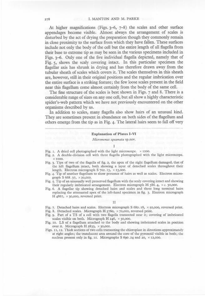
Low-power views of intact cells are uninformative except as evidence of size(Fig. I). The single posteriorly directed flagellum is about 12}.L long and thoughit is sometimes accompanied by a second, or even a third (Fig. 2) or fourthflagellum, such supernumeraries being of any length from very short to equal,there is no doubt that these all relate to growth stages of cells in division orto giant cells-representing double-divisions. In normal undivided cells, whichmake up the majority in any culture, only one flagellum is present.
1. MANTON AND M. PARKE
At higher magnifications (Figs. 3-6, 7-8) the scales and other surfaceappendages become visible. Almost always the arrangement of scales isdisturbed by the act of drying the preparation though they commonly remainin close proximity to the surface from which they have fallen. These surfacesinclude not only the body of the cell but the entire length of all flagella fromtheir base to extreme tip as may be seen in the various specimens included inFigs. 3-6. Only one of the five individual flagella depicted, namely that ofFig. 5, shows the scaly covering intact. In this particular specimen theflagellar axis has shrunk in drying and has therefore drawn away from thetubular sheath of scales which covers it. The scales themselves in this sheath
are, however, still in their original positions and the regular imbrication overthe entire surface is a striking feature; the few loose scales present in the fieldnear this flagellum come almost certainly from the body of the same cell.
The fine structure of the scales is best shown in Figs. 7 and 8. There is aconsiderable range of sizes on anyone cell, but all show a highly characteristicspider's-web pattern which we have not previously encountered on the otherorganisms described by us.
In addition to scales, many flagella also show hairs of an unusual kind.They are sometimes present in abundance on both sides of the flagellum andothers emerge from the tip as in Fig. 4. The lateral hairs seem to fall off very
Explanation of Plates I-VIMicromonas squamata sp.nov.
IFig. I. A dried cell photographed with the light microscope. x 1000.Fig. 2. A double-division cell with three flagella photographed with the light microscope.
x 1000.Fig. 3. Tips of two of the flagella of fig. 2, the apex of the right flagellum damaged, that of
the left flagellum intact, both showing a layer of detached scales throughout theirlength. Electron micrograph S 7°2.15, x 15,000.
Fig. 4. Tip of another flagellum to show presence of hairs as well as scales. Electron micrograph S 668.37, x 20,000.
Fig. 5. Tip of an unusually well preserved flagellum with the scaly covering intact and showingtheir regularly imbricated arrangement. Electron micrograph M 586.4, xc. 30,000.
Fig. 6. A flagellar tip showing detached hairs and scales and three long terminal hairsreplacing the attenuated apex of the left-hand specimen in fig. 3. Electron micrographH 4867, x 30,000, reversed print.
IIFig. 7. Detached hairs and scales. Electron micrograph S 680.18, x 50,000, reversed print.Fig. 8. Detached scales. Micrograph H 5760, x 70,000, reversed print.Fig. 9. Part of a TS of a cell with two flagella transected near it; covering of imbricated
scales visible on both. Micrograph H 246, x 30,000.Fig. 10. LS of a flagellum attached to the body and showing imbricated scales in position
over it. Micrograph H 2833, x 30,000.Figs. 11, 12. Thick sections of two cells transecting the chloroplast in directions approximately
at right angles; the translucent area around the core of the pyrenoid visible in both; thenucleus present only in fig. II. Micrographs S 690.24 and 20, x 12,000.
1
(inset)
J. MAR. BIOL. Ass. U.K., 39 (2)
5
MANTON & PARKE. PLATE I
(Facing p. 278)

J. MAR. RIOL. Ass. U.K., 39 (2) MANTON & PARKE. PLATE II
7
9
10
11
8
12

J. MAR. BIOL. Ass. U.K., 39 (2) MANTON & PARKE. PLATE III
J. MAR. BIOL. Ass. U.K., 39 (2) MANTON & PARKE. PLATE IV
21

23a
24
J. MAR. BIOL. Ass. U.K., 39 (2)
l 1fL
MANTON & PARKE. PLATE V
27

J. MAR. BIOL. Ass. U.K., 39 (2) MANTON & PARKE. PLATE VI
28
29b
OBSERVATIONS ON GREEN FLAGELLATES
Explanation of Plates I-VI continued
Micromonas squamata sp.nov.
279
III
Fig. 13. Two cells, the left-most passing thtough the nucleus (N), the mitochondrion (m),the plastid (P) cut near but not through the pyrenoid and showing the stacks of pairedplastid lamellae near the surface with fenestrations at intervals; the right-hand cell adivision stage with two lobes of a bent U-shaped mitochondrion (m and m') associatedwith the two halves of the plastid (another section of this cell, not reproduced, showedthe centre part of the mitochondrion connecting the two lobes). Micrograph H 247,x 20,000.
Fig. 14. Tangential section near the surface of a plastid showing the fenestrations in face view.Micrograph H 5777, x 20,000.
Fig. 15. Part of a dividing cell showing golgi (d) and a long arm of a mitochondrion (m)between two plastids. Micrograph H 5467, x 20,000.
Fig. 16. A section passing thtough the plastid to show a structure resembling an eyespot(bottom centre) but without sign of pigment. Micrograph H 2728, x 15,000. Forfurther details of eyespot region see figs. 17 and 18.
Fig. 17. Tangential section near the surface of a plastid in the region of the putative eyespotshowing the close-packing of objects resembling pigment chambers, an obliquely cutflagellum near. Micrograph H 5777, x 20,000.
Fig. 18. More highly magnified view of plastid surface with putative pigment chamberscovered by plastid double-membrane and cell body membrane. Micrograph H 2635,x 50,000.
IV
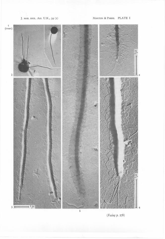
Figs. 19, 20. Two successive stages of a division showing different degrees of separation of thetwo flagella for the daughter-cells; two nuclei side by side visible in fig. 20. MicrographsH 2995 and H 3010, x 20,000.
Fig.21. Stage in a cell-division showing a very long U-shaped mitochondrion (m) betweentwo plastids or plastid lobes. Micrograph H 242, x 20,000.
Fig. 22. Another specimen at a stage similar to that of fig. 21 but cut in a slightly differentplane and showing two plastids (or plastid lobes), a nucleus (N), a U-shaped mitochondrion em, m') with the arms in contact with the two plastids, some fat bodies and vesicleswith other contents in the trough of the U. Micrograph H 698, x 20,000.
V
Fig. 23 a, b. Two successive sections through a flagellar insertion cut near the surface of a celland showing disposition of vesicles and other details represented more highly magnifiedin figs. 24-26. Micrographs H 2647 and H 2639, x 20,000.
Fig. 24. Part of the section of fig. 23 a near the flagellar base showing a vesicle containing ascale in full face view immediately above the flagellar insertion. Micrograph H 2647,x 50,000.
Fig. 25. A scale in face view outside the body for comparison with that of fig. 24, from theregion of the arrow on the right of fig. 23 a. Micrograph H 2647, x 50,000.
Fig. 26. Part of the cell surface showing imbrication of body scales, from the field near thearrow in fig. 23b. Micrograph H 2639, x 50,000.
Fig. 27. Imbricated scales on the surface of part of another cell. Micrograph H 2639,x 50,000.
VI
Fig. 28. LS of a cell passing thtough the pyrenoid P, the starch shell S, the nucleus N, themitochondrion m and showing the fibrous connection (r) between the base of the flagellumand the nuclear surface. Micrograph H 2986, x 40,000.
Fig. 29a, b. Two adjacent sections thtough another specimen showing the fibrous connection(r) from the flagellar base (f) ending on the nuclear surface (N). Micrographs H 5459 andH 5461, x 30,000.
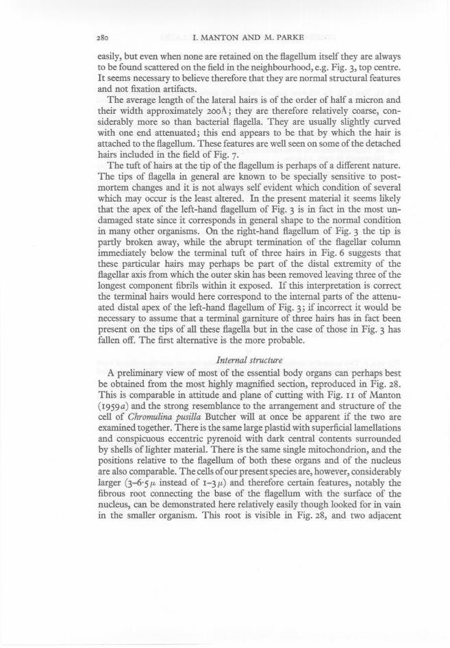
280 1. MANTON AND M. PARKE
easily, but even when none are retained on the flagellum itself they are alwaysto be found scattered on the field in the neighbourhood, e.g. Fig. 3, top centre.It seems necessary to believe therefore that they are normal structural featuresand not fixation artifacts.
The average length of the lateral hairs is of the order of half a micron andtheir width approximately 2ooA; they are therefore relatively coarse, considerably more so than bacterial flagella. They are usually slightly curvedwith one end attenuated; this end appears to be that by which the hair isattached to the flagellum. These features are well seen on some of the detachedhairs included in the field of Fig. 7.
The tuft of hairs at the tip of the flagellum is perhaps of a different nature.The tips of flagella in general are known to be specially sensitive to postmortem changes and it is not always self evident which condition of severalwhich may occur is the least altered. In the present material it seems likelythat the apex of the left-hand flagellum of Fig. 3 is in fact in the most undamaged state since it corresponds in general shape to the normal conditionin many other organisms. On the right-hand flagellum of Fig. 3 the tip ispartly broken away, while the abrupt termination of the flagellar columnimmediately below the terminal tuft of three hairs in Fig. 6 suggests thatthese particular hairs may perhaps be part of the distal extremity of. theflagellar axis from which the outer skin has been removed leaving three of thelongest component fibrils within it exposed. If this interpretation is correctthe terminal hairs would here correspond to the internal parts of the attenuated distal apex of the left-hand flagellum of Fig. 3; if incorrect it would benecessary to assume that a terminal garniture of three hairs has in fact beenpresent on the tips of all these flagella but in the case of those in Fig. 3 hasfallen off. The first alternative is the more probable.
Internal structure
A preliminary view of most of the essential body organs can perhaps bestbe obtained from the most highly magnified section, reproduced in Fig. 28.This is comparable in attitude and plane of cutting with Fig. 11 of Manton(1959a) and the strong resemblance to the arrangement and structure of thecell of Chromulina pusilla Butcher will at once be apparent if the two areexamined together. There is the same large plastid with superficiallamellationsand conspicuous eccentric pyrenoid with dark central contents surroundedby shells of lighter material. There is the same single mitochondrion, and thepositions relative to the flagellum of both these organs and of the nucleusare also comparable. The cells of our present species are, however, considerablylarger (3-6·5,u instead of 1-3,u) and therefore certain features, notably thefibrous root connecting the base of the flagellum with the surface of thenucleus, can be demonstrated here relatively easily though looked for in vainin the smaller organism. This root is visible in Fig. 28, and two adjacent
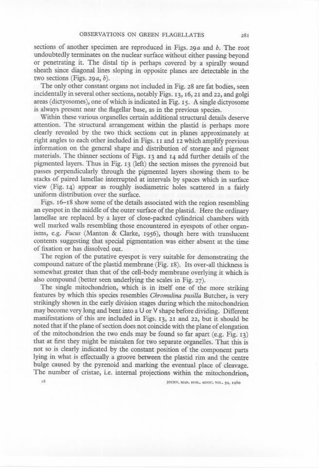
OBSERVATIONS ON GREEN FLAGELLATES 281
sections of another specimen are reproduced in Figs. 29a and b. The rootundoubtedly terminates on the nuclear surface without either passing beyondor penetrating it. The distal tip is perhaps covered by a spirally woundsheath since diagonal lines sloping in opposite planes are detectable in thetwo sections (Figs. 29a, b).
The only other constant organs not included in Fig. 28 are fat bodies, seenincidentally in several other sections, notably Figs. 13, 16,21 and 22, and golgiareas (dictyosomes), one of which is indicated in Fig. 15. A single dictyosomeis always present near the flagellar base, as in the previous species.
Within these various organelles certain additional structural details deserveattention. The structural arrangement within the plastid is perhaps moreclearly revealed by the two thick sections cut in planes approximately atright angles to each other included in Figs. I I and 12 which amplify previousinformation on the general shape and distribution of storage and pigmentmaterials. The thinner sections of Figs. 13 and 14 add further details of thepigmented layers. Thus in Fig. 13 (left) the section misses the pyrenoid butpasses perpendicularly through the pigmented layers showing them to bestacks of paired lamellae interrupted at intervals by spaces which in surfaceview (Fig. 14) appear as roughly isodiametric holes scattered in a fairlyuniform distribution over the surface.
Figs. 16-18 show some of the -details associated with the region resemblingan eyespot in the middle of the outer surface of the plastid. Here the ordinarylamellae are replaced by a layer of close-packed cylindrical chambers withwell marked walls resembling those encountered in eyespots of other organisms, e.g. Fucus (Manton & Clarke, 1956), though here with translucentcontents suggesting that special pigmentation was either absent at the timeof fixation or has dissolved out.
The region of the putative eyespot is very suitable for demonstrating thecompound nature of the plastid membrane (Fig. 18). Its over-all thickness issomewhat greater than that of the cell-body membrane overlying it which isalso compound (better seen underlying the scales in Fig. 27).
The single mitochondrion, which is in itself one of the more strikingfeatures by which this species resembles Chromulina pusilla Butcher, is verystrikingly shown in the early division stages during which the mitochondrionmay become very long and bent into a U or V shape before dividing. Differentmanifestations of this are included in Figs. 13, 21 and 22, but it should benoted that if the plane of section does not coincide with the plane of elongationof the mitochondrion the two ends may be found so far apart (e.g. Fig. 13)that at first they might be mistaken for two separate organelles. That this isnot so is clearly indicated by the constant position of the component partslying in what is effectually a groove between the plastid rim and the centrebulge caused by the pyrenoid and marking the eventual place of cleavage.The number of cristae, i.e. internal projections within the mitochondrion,
18JOURN. MAR. BIOL. ASSOC. VOL. 39. 1960
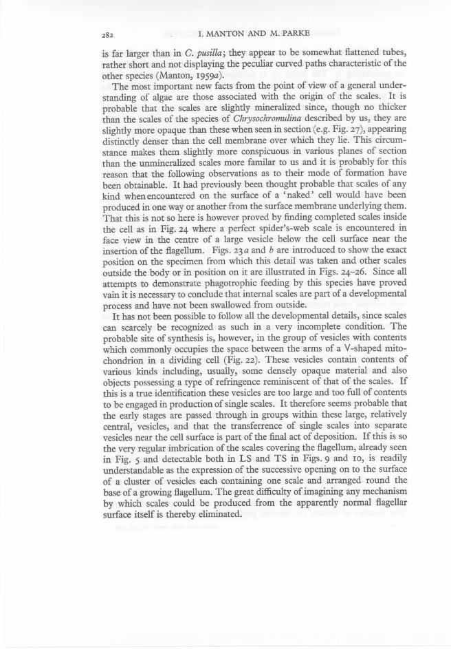
282 1. MANTON AND M. PARKE
is far larger than in C. pusilla; they appear to be somewhat flattened tubes,rather short and not displaying the peculiar curved paths characteristic of theother species (Manton, 1959a).
The most important new facts from the point of view of a general understanding of algae are those associated with the origin of the scales. It isprobable that the scales are slightly mineralized since, though no thickerthan the scales of the species of Chrysochromulina described by us, they areslightly more opaque than these when seen in section (e.g. Fig. 27), appearingdistinctly denser than the cell membrane over which they lie. This circumstance makes them slightly more conspicuous in various planes of sectionthan the unmineralized scales more familar to us and it is probably for thisreason that the following observations as to their mode of formation havebeen obtainable. It had previously been thought probable that scales of anykind when encountered on the surface of a 'naked' cell would have been
produced in one way or another from the surface membrane underlying them.That this is not so here is however proved by finding completed scales insidethe cell as in Fig. 24 where a perfect spider's-web scale is encountered inface view in the centre of a large vesicle below the cell surface near theinsertion of the flagellum. Figs. 23 a and b are introduced to show the exactposition on the specimen from which this detail was taken and other scalesoutside the body or in position on it are illustrated in Figs. 24-26. Since allattempts to demonstrate phagotrophic feeding by this species have provedvain it is necessary to conclude that internal scales are part of a developmentalprocess and have not been swallowed from outside.
It has not been possible to follow all the developmental details, since scalescan scarcely be recognized as such in a very incomplete condition. Theprobable site of synthesis is, however, in the group of vesicles with contentswhich commonly occupies the space between the arms of a V-shaped mitochondrion in a dividing cell (Fig. 22). These vesicles contain contents ofvarious kinds including, usually, some densely opaque material and alsoobjects possessing a type of refringence reminiscent of that of the scales. Ifthis is a true identification these vesicles are too large and too full of contentsto be engaged in production of single scales. It therefore seems probable thatthe early stages are passed through in groups within these large, relativelycentral, vesicles, and that the transferrence of single scales into separatevesicles near the cell surface is part of the final act of deposition. If this is sothe very regular imbrication of the scales covering the flagellum, already seenin Fig. 5 and detectable both in LS and TS in Figs. 9 and 10, is readilyunderstandable as the expression of the successive opening on to the surfaceof a cluster of vesicles each containing one scale and arranged round thebase of a growing flagellum. The great difficulty of imagining any mechanismby which scales could be produced from the apparently normal flagellarsurface itself is thereby eliminated.

OBSERVATIONS ON GREEN FLAGELLATES
OBSERVATIONS WITH THE LIGHT MICROSCOPE ONMICROMONAS SQUAMATA SP.NOV.
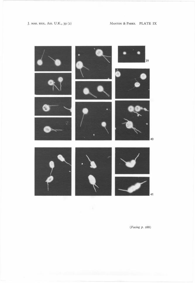
Examples of cells photographed with the light microscope, using dark-groundillumination, will be found grouped under one number as Fig. 40, PI. IX(facing p. 288); some are in the uniflagellate condition and the remainder aredivision stages with two or four flagella, unequal to equal in length. Fig. 39,PI. IX, illustrates two cells of Chromulina pusilla Butcher at the samemagnification for comparison; their much smaller size has made it impossibleto obtain effective photographs of the flagella by this method.
Cells of the organism that we propose to name Micromonas squamata sp.nov.can swim fairly quickly, rotating rapidly with only slight gyration and movingin straight lines for short distances, but more usually they move fairly slowlywith slow rotation of the body and more pronounced gyration. They can alsoglide along for short distances without rotation, or they can show periods ofjerking about very actively by the lashing ofthe flagellum. During movement,the region of the body at which the flagellum arises is always foremost.The proximal 1-2 fl- of the flagellum becomes pulled out into what appears,when the cell is moving, to be a short stiff spine-like organ which lies infront of the body (Figs. 44-45); the remainder of the flagellum is curvedsharply backwards down the side of the body to lie behind the cell. Whencells are moving rapidly the flagellum appears to vibrate but when movingmore slowly definite undulations can be observed; the undulations are usuallysmall but can on occasion be large. A cell stops swimming suddenly bybringing the flagellum up with a sharp jerk of the body into a curved positionround and under the body (Fig. 42), sometimes coiled twice round the body(Fig. 43), after which the cell will commonly move off in a different direction.In fission stages when a second flagellum has developed, the two behavehomodynamically, both twisting round the body simultaneously when a cellstops swimming to change direction.
The region of the body close to the point of origin of the flagellum can showconsiderable metabolyand,in addition, fine pseudopodium-like filaments haveseveral times been seen produced; they appear to attach to the surface of theslide but so far only cells in the incipient fission stage have been seen producingthese filaments (Fig. 46).
In an actively growing culture 15% of the cells are 3 fl- in size, 20 % 3'5 fl-,
40 % 4'0 fl-, 13% 4'5 fl-, while the remaining 12% are incipient fission stagesranging from 5 to 6,5 fl- in size. The smallest cells (newly divided daughter-cells)may show some slight flattening of the body but the larger cells and thefission stages are more globose. The flagellum length in relation to the bodysize can vary from 2! to 4 times the length of the body but 80 % of the cellshave a flagellum 3-3! times the length of the body ..
This organism shows a distinct phototactic reaction but there is no evidencer8-z

1. MANTON AND M. PARKE
of phagotrophy. In culture it is yellow-green, i.e. is similar in colour toChromulina pusilla Butcher, and it contains, as does the latter, chlorophyll a,chlorophyll b, f3-carotene and <x-xanthophyll (Dr G. Y. Kennedy, personalcommunication).
In the living cell the most obvious of the cell contents are the chloroplastwith its embedded pyrenoid and, when present, the refractive lipid body orbodies. The chloroplast is situated on the side of the body opposite to thatfrom which the flagellum arises; under the high power it appears striatedwith the pigmented outer region crescentic in optical section. The pyrenoid,immersed in the inner side of the chloroplast, is surrounded by a starch shellstaining violet with iodine. The starch shell shows considerable variation inshape and size in different cells and it may be studied either in intact stainedcells or loose among the debris taken from broken cells at the bottom of theflask. Examples of such isolated but intact starch shells are drawn in Fig. 47.When least developed the starch shell is a hollow sphere a little over 1j-t indiameter surrounding the pyrenoid on all sides except that nearest to theunpigmented face; an isolated starch shell is therefore a hollow sphere with ahole at one pole. As the shell thickens its outline becomes more ovoid andirregular, the starch appearing to be laid down unevenly, more being depositedon the sides than in the centre but the hole on the inner plastid face neverbecoming covered. The largest starch shells are about 3 j-t in diameter.
There is usually one lipid body, about 0'5 j-t in diameter, situated in thecell on the side opposite to the chloroplast, but occasionally none are present.Mter a culture has been grown in strong light for some time additional lipidglobules can be seen in the cells; there may be from I to 4 and they lie againstthe inner face of the chloroplast. The stigma, orange-red, and oval to oblongin the surface view, measuring up to 1x 0'5 j-t, lies centrally in the outer faceof the chloroplast and is not always readily detected. A single elongatedmitochondrion, I x 0'75 j-t in size when stained with Janus Green, lies on theinner face of the chloroplast while the ovoid nucleus, 1'5 XI j-t in size, liesoutside it towards the side of the cell from which the flagellum arises. Thegolgi area, situated close to the nucleus, stains with Janus Green to give abluer colour than the mitochondrion. Neither the body scales nor the flagellarscales stain with cresyl blue, but the flagellar basal body can be detected afterfixation with osmium tetroxide.
Asexual reproduction can occur in either the motile or non-motile phase.In the motile phase fission is usually into two daughter-cells but it is notuncommon for double-fission to occur producing three or four daughter-cellsat the same time from the parent cell (Figs. 40, 48, 49). The first indications offission are the simultaneous elongation of the stigma and pyrenoid accompanying the first appearance of the second flagellum; occasionally the stigmadivides and the daughter-flagellum appears before the elongation of thepyrenoid. By the time the daughter-flagellum has nearly reached its full

OBSERVATIONS ON GREEN FLAGELLATES
length the nucleus, mitochondrion, golgi, chloroplast, pyrenoid and stigmahave divided. The daughter-flagellum is fully developed and the chloroplastshave separated before actual fission of the body commences at the flagellarpole. In the non-motile (palmelloid) phase, the cells become surrounded bywhat appears to be a very thin membrane lying close to the body; ~uch cellsdivide into four or occasionally eight daughter-cells (Figs. 50, 51). Theapparent membrane shows up a little more clearly after treatment withSchultz's solution but its precise nature is uncertain.
If this description is compared with that of Korschikov (1923) for Pedinomonas minor the following differences from P. squamata should be noted:habitat in standing fresh water of inland areas in Poland and west Russia(more recently reported from similar habitats from Hungary by Fott & Ettl(1959)), in contrast to the brackish habitat of M. squamata; presence of acontractile vacuole reported in P. minor, not detectable in M. squamata; theflagellum in P. minor considerably shorter (only 1t times body length) thanin M. squamata (where it is 2t-4 times body length); starch grains presentround the pyrenoid as opposed to the continuous starch shell of M. squamata.
OBSERVATIONS WITH THE ELECTRON MICROSCOPE ONPEDINOMONAS TUBERCULATA (VISCHER) GAMS
External morphology
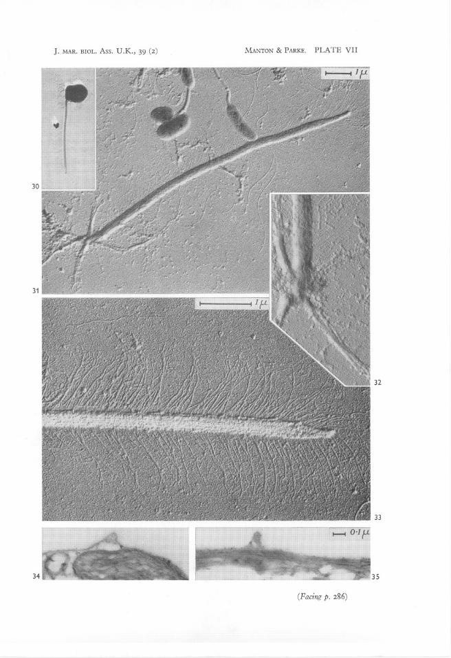
The cells of this species make very elegant preparations when dried down andthey also possess an unusual capacity for informative dismemberment. Oneintact cell is illustrated in Fig. 30 with the tip of its plume-like flagellum shownmore highly magnified in Fig. 33. The profuse garniture of very delicatehairs spread on the field on both sides of the flagellar axis are highly charac::'teristic. They are individually so slender, however, that the bacterial flagellalying loose in various parts of the field of Fig. 31 appear coarse in comparison.
Where cells have dismembered as in Figs. 31 and 32, four stout roots'arranged in a cruciform manner are seen to be attached to the lower end ofthe flagellar base. These roots have a fibrous core and a membranous coveringwhich in some if not all exhibit conspicuous diagonal striations as of a spiralwinding of alternating bands of different electron opacity. This diagonalcross-banding is clearly visible in the lowermost root of Fig. 32. '
Internal structure
One morphological detail, namely, the superficial tubercles from which thisspecies gets its name, is better seen in sections than in our whole moUnts.They occur as spine-like excrescences on various parts of the body as may be,seen in Figs. 34-35. These tubercles are covered by the normal body m~m-'brane and they therefore represent local deposits of material beneath the

286 1. MANTON AND M. PARKE
membrane. This material is unidentifiable by the electron microscope alonebut the presence of cellulose, calcite and quartz (Si02 in micro-crystallineform) has been indicated by X-ray analysis by Brandenburger & FreyWyssling (1947) and one or other of these components could be representedhere. In addition, the surface membrane, which bears no resemblance insection to a normal cell wall, is covered by small spicules and traces of amorphous material among which fine particles of metallic osmium commonlybecome lodged as a fixation artifact. It therefore seems probable that thoughentirely devoid of either scales or a cell wall of algal type the membrane of thisspecies is not completely naked on its outer side.
When viewed in section (PI. VIII) the cells of P. tuberculata appear stronglyflattened. Internally the cell is dominated by the large starch-filled chloroplast which is here curved (see especially Fig. 37), surrounding the othercytoplasmic organs which lie in the hollow. In a section cut in anotherdirection (Fig. 36, approximately at right angles to Fig. 37) the nucleus isseen to be strongly flattened against the outer surface of the body on oneside. There are several rather small mitochondria in the hollow of the curved
plastid, one conspicuous golgi body (dictyosome) near the flagellar insertionand an array of small vesicles and granular cytoplasm. A few larger vesiclescontaining traces of opaque fat-like material are often conspicuous, though
Explanation of Plates VII and VIIIPedinomonas tuberculata (Vischer) Gams
VII
Fig. 30. A dried cell. Micrograph S 672.26, x 3000.Fig.31. Field containing a dismembered cell represented by an intact flagellum with four
roots arranged in a cruciform manner; bacterial cells and bacterial flagella loose upon thefield and appearing relatively coarse in comparison with the very fine lateral hairs on theflagellum of Pedinomonas. Micrograph S 669.3°, x ro,ooo.
Fig. 32. Base of the flagellum and roots from another specimen showing fibrous structurein the roots and traces of cross-banding probably carried by a covering membrane onthe longest of the roots shown. Micrograph S 669.29, x 20,000.
Fig. 33. Tip of the flagellum of the cell of fig. 30 more highly magnified to show the detailsof the hairs. Micrograph S 672.28, x 20,000, reversed print.
Fig. 34. Section through a tubercle. Micrograph H 4957, x 30,000.Fig. 35. Section through a tubercle, another specimen. Micrograph H 4985, x 40,000.
VIII
Fig. 36. Section of a cell showing parts of all the main organelles; nucleus (N), pyrenoid (P)starch grains (S), mitochondria (m), golgi (d). Micrograph H 4993, x 40,000.
Fig. 37. Section cut in another plane showing the curved plastid surrounding the otherorganelles in the hollow, the flagellar base cut transversely at the top of the section .
. Micrograph H 5047, x 30,000.Fig. 38a, b. Two successive sections through a flagellar base (I) in the region of a root (r),
to show its superficial position and close relation with the edge of the plastid; a golgiarea (d) also included in Fig. 38b. Micrographs H 498r and H 4978, x 50,000.
J. MAR. BIOL. Ass. U.K., 39 (2) MANTON & PARKE. PLATE VII
(Pacing p. 286)

J. MAR. BIOL. Ass. u.K., 39 (2) MANTON & PARKE. PLA TE VIII
36
OBSERVATIONS ON GREEN FLAGELLATES
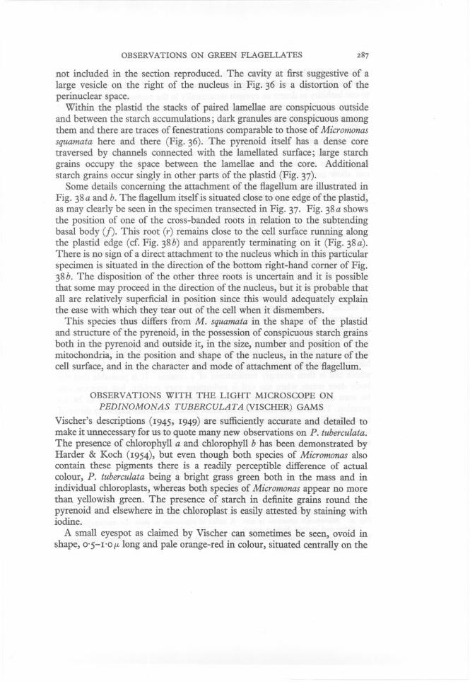
not included in the section reproduced. The cavity at first suggestive of alarge vesicle on the right of the nucleus in Fig. 36 is a distortion of theperinuclear space.
Within the plastid the stacks of paired lamellae are conspicuous outsideand between the starch accumulations; dark granules are conspicuous amongthem and there are traces of fenestrations comparable to those of Micromonassquamata here and there (Fig. 36). The pyrenoid itself has a dense coretraversed by channels connected with the lamellated surface; large starchgrains occupy the space between the lamellae and the core. Additionalstarch grains occur singly in other parts of the plastid (Fig. 37).
Some details concerning the attachment of the flagellum are illustrated inFig. 38a and b. The flagellum itself is situated close to one edge of the plastid,as may clearly be seen in the specimen transected in Fig. 37. Fig. 38a showsthe position of one of the cross-banded roots in relation to the subtendingbasal body (f). This root (r) remains close to the cell surface running alongthe plastid edge (cf. Fig. 38b) and apparently terminating on it (Fig. 38a).
There is no sign of a direct attachment to the nucleus which in this particularspecimen is situated in the direction of the bottom right-hand corner of Fig.38b. The disposition of the other three roots is uncertain and it is possiblethat some may proceed in the direction of the nucleus, but it is probable thatall are relatively superficial in position since this would adequately explainthe ease with which they tear out of the cell when it dismembers.
This species thus differs from M. squamata in the shape of the plastidand structure of the pyrenoid, in the possession of conspicuous starch grainsboth in the pyrenoid and outside it, in the size, number and position of themitochondria, in the position and shape of the nucleus, in the nature of thecell surface, and in the character and mode of attachment of the flagellum.
OBSERVATIONS WITH THE LIGHT MICROSCOPE ONPEDINOMONAS TUBERCULATA (VISCHER) GAMS
Vischer's descriptions (1945, 1949) are sufficiently accurate and detailed tomake it unnecessary for us to quote many new observations on P. tuberculata.
The presence of cWorophyll a and cWorophyll b has been demonstrated byHarder & Koch (1954), but even though both species of Micromonas alsocontain these pigments there is a readily perceptible difference of actualcolour, P. tuberculata being a bright grass green both in the mass and inindividual cWoroplasts, whereas both species of Micromonas appear no morethan yellowish green. The presence of starch in definite grains round thepyrenoid and elsewhere in the cWoroplast is easily attested by staining withiodine.
A small eyespot as claimed by Vischer can sometimes be seen, ovoid inshape, o·5-1·ofL long and pale orange-red in colour, situated centrally on the
288 I. MANTON AND M. PARKE
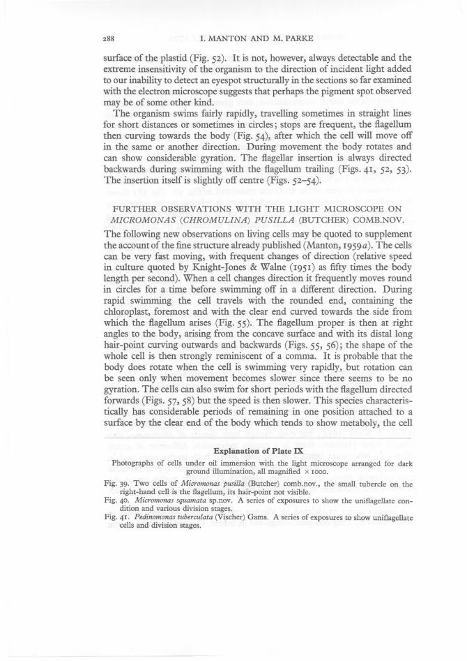
surface of the plastid (Fig. 52). It is not, however, always detectable and theextreme insensitivity of the organism to the direction of incident light addedto our inability to detect an eyespot structurally in the sections so far examinedwith the electron microscope suggests that perhaps the pigment spot observedmay be of some other kind.
The organism swims fairly rapidly, travelling sometimes in straight linesfor short distances or sometimes in circles; stops are frequent, the flagellumthen curving towards the body (Fig. 54), after which the cell will move offin the same or another direction. During movement the body rotates andcan show considerable gyration. The flagellar insertion is always directedbackwards during swimming with the flagellum trailing (Figs. 41, 52, 53).The insertion itself is slightly off centre (Figs. 52-54).
FURTHER OBSERVATIONS WITH THE LIGHT MICROSCOPE ONMICROMONAS (CHROMULINA) PUSILLA (BUTCHER) COMB.NOV.
The following new observations on living cells may be quoted to supplementthe account of the fine structure already published (Manton, 1959a). The cellscan be very fast moving, with frequent changes of direction (relative speedin culture quoted by Knight-Jones & Walne (1951) as fifty times the bodylength per second). When a cell changes direction it frequently moves roundin circles for a time before swimming off in a different direction. Duringrapid swimming the cell travels with the rounded end, containing thechloroplast, foremost and with the clear end curved towards the side fromwhich the flagellum arises (Fig. 55). The flagellum proper is then at rightangles to the body, arising from the concave surface and with its distal longhair-point curving outwards and backwards (Figs. 55, 56); the shape of thewhole cell is then strongly reminiscent of a comma. It is probable that thebody does rotate when the cell is swimming very rapidly, but rotation canbe seen only when movement becomes slower since there seems to be nogyration. The cells can also swim for short periods with the flagellum directedforwards (Figs. 57, 58) but the speed is then slower. This species characteristically has considerable periods of remaining in one position attached to asurface by the clear end of the body which tends to show metaboly, the cell
Explanation of Plate IX
Photographs of cells under oil immersion with the light microscope arranged for dark.. ground illumination, all magnified x 1000.
Fig. 39. Two cells of Micromonas pusilla (Butcher) comb.nov., the small tubercle on theright-hand cell is the flagellum, its hair-point not visible.
Fig. 40. Micromonas squamata sp.nov. A series of exposures to show the uniflagellate condition and various division stages.
Fig. 41. Pedinomonas tuberculata (Vischer) Gams. A series of exposures to show uniflagellatecells and division stages.
J. MAR. BIOL. Ass. U.K., 39 (2) MANTON & PARKE. PLATE IX
(Pacing p. 288)

OBSERVATIONS ON GREEN FLAGELLATES
meanwhile either swinging round in circles from the point of attachment(Fig. 59) or exhibiting a dithering movement which is very characteristic forthis organism.
The species is strongly phototactic and is not toxic to fish (tested by Mrs B.Hepper). Motile cells in culture measure 1'0-3'0 fL long x 0'75-1 fL wide.Fission occurs in both motile and non-motile condition. A non-motilepalmelloid phase with cells measuring 2'5-5 fL and showing a translucent arearound each also occurs, the cells dividing into 2 or 4. No trace of celluloseis detectable in either motile or non-motile phases and no positive signs ofstarch when tested with iodine. Signs of a sexual process have been lookedfor by mixing five different strains, without success.
In occurrence this marine flagellate is very widespread; it has been recorded from surface samples taken from estuaries, creeks and the open seaaround the British Isles (Knight-Jones, 1951, 1952; Knight-Jones & Walne,1951). According to Knight-Jones it is the most generally abundant organismin British coastal waters and the North Sea, the greatest density he recordedfor it being 3500 cells per ml in October 1946 from the Helford River,Cornwall.
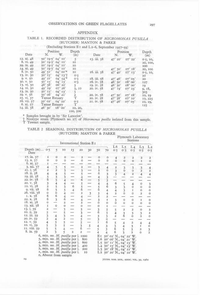
Our records, covering a period of several years (Tables 1 & 2, Appendix)amplify those of Knight-Jones and Walne and show that this species occurscommonly in the English Channel throughout the year from the surfacedown to 70 m (Table 2). It occurs also in oceanic water and has been recordedfrom oceanic stations from the surface down to a depth of 500 m (Table 1).In Table 1 of the Appendix (p. 297) its occurrences are listed other than forInternational Station Eland Plymouth Laboratory Stations L 2-6 from17 September 1957 to 8 September 1959, the records for which are includedin Table 2 (p. 297). This table includes densities at different depths sampledon one day at International Station E 1.
SUMMARY OF TAXONOMIC CONCLUSIONS AND DIAGNOSES
We can now summarize in tabular form the more important facts ascertainedor verified by us for the three organisms under discussion. From this table(p. 292) it will be seen that the organism formerly known as Chromulina pusillaButcher agrees with Micromonas squamata in all salient features of internalorganization, including plastid structure and form, the possession of onemitochondrion and in the position, relative to other cell organs, of the singleflagellum. It differs in the absence of a scaly covering, in the absence of anytrace of lateral hairs on the flagellum and in the very unusual relative proportions of the flagellum and its hair-point.
In contrast, Pedinomonas tuberculata differs from both the other speciesin the shape of nucleus and chloroplast, in the position and number of themitochondria, in the presence of starch in definite grains, in the presence of
1. MANTON AND M. PARKE
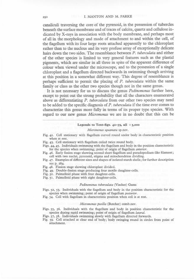
canaliculi traversing the core of the pyrenoid, in the possession of tuberclesbeneath the surface membrane and of traces of calcite, quartz and cellulose indicated by X-rays in association with the body membrane, and perhaps mostof all in the morphology and mode of attachment to and within the cell, ofthe flagellum with its four large roots attached apparently to the chloroplastrather than to the nucleus and its very profuse array of exceptionally delicatehairs down the two sides. The resemblance between P. tuberculata and eitherof the other species is limited to very general features such as the plastidpigments, which are similar in all three in spite of the apparent difference ofcolour when viewed under the microscope, and to the possession of a singlechloroplast and a flagellum directed backwards in swimming though arrivingat this position in a somewhat different way. This degree of resemblance isperhaps sufficient to permit the placing of P. tuberculata within the samefamily or class as the other two species though not in the same genus.
It is not necessary for us to discuss the genus Pedinomonas further here,except to point out the strong probability that all the characters enumerat~dabove as differentiating P. tuberculata from our other two species may needto be added to the specific diagnosis of P. tuberculata if the time ever comes tocharacterize this genus more fully in terms of its proper type species. Withregard to our new genus Micromonas we are in no doubt that this can be
Legends to Text-figs. 42-59, all x 5,000
Micromonas squamata sp.nov.
Fig. 42. Cell stationary with flagellum curved round under body in characteristic positionwhen at rest.
Fig. 43. Cell stationary with flagellum coiled twice round body.Figs. 44,45. Individuals swimming with the flagellum and body in the position characteristic
for the species when swimming; point of origin of flagellum anterior.Fig. 46. Early fission stage showing second short flagellum and pseudopodium-like filament;
cell with two nuclei, pyrenoid, stigma and mitochondrion dividing.Fig. 47. Examples of different sizes and shapes of isolated starch shells; for further description
see p. 284.Fig. 48. Fission stage showing chloroplast divided.Fig. 49. Double-fission stage producing four motile daughter-cells.Fig. 50. Palmelloid phase with four daughter-cells.Fig. 51. Palmelloid phase with eight daughter-cells.
Pedinomonas tuberculata (Vischer) Gams
Figs. 52, 53. Individuals with the flagellum and body in the position characteristic for thespecies when swimming; point of origin of flagellum posterior.
Fig. 54. Cell with flagellum in characteristic position when cell is at rest.
Micromonas pusilla (Butcher) comb.nov.
Figs. 55, 56. Individuals with the flagellum and body in position characteristic for thespecies during rapid swimming; point of origin of flagellum lateral.
Figs. 57, 58. Individuals swimming slowly with flagellum directed forwards.Fig. 59. Cell attached at clear end of body; body swinging round in circles from point of
attachment.

OBSERVATIONS ON GREEN FLAGELLATES
differentiated clearly from the type species of Pedinomonas (P. minor), as described by Korschikov, on the light microscopy alone. We recognize, however,that while this applies with equal force to both our species of Micromonas, thedecision to group these together as one new genus and not as two is to someextent arbitrary, depending on our decision, on grounds of convenience, toregard the differences between these two species as less significant than theresemblances and to treat such differences as specific and not as generic criteria,
t58
l'
l'
52
Text-figs 42-59

I. MANTON AND M. PARKE
as explained in the introduction. The way is then clear to summarize theseconclusions still further by constructing formal diagnoses for the new genusMicromonas and for the two species which it at present contains.
TABLE OF MORPHOLOGICAL AND ANATOMICAL CHARACTERS AVAILABLEFOR DIFFERENTIATING THE THREE SPECIES UNDER DISCUSSION FROMEACH OTHER
Yellowish green Yellowish greenHemispherical with pigment lamellae near
outer curved surfaceLarge uniform core of electron opaquematerial
Distinct single starch Starch shell minuteshell (only visible e.m.)
Single SingleLying on surface of chloroplast
Subspherical near flagellar base
Body covering
Flagellum:AttachedRootsJoined toAppendages
Chloroplast:ColourShape
Pyrenoid
Starch
Mitochondria
Nucleus
Micromonassquamata
Mineralized scalesover membrane
Anterio-lateralSingleNucleusScales and hairs
Micromonaspusilla
Membrane
Lateral
Terminal hair-point
Pedinomonastuberculata
Membrane, plus tracesof calcite, cellulose,Si02
Posterior4 cruciformChloroplast2 rows fine lateral hairs
Bright greenCurved round otherprotoplasmic organelles
Core traversed bycanals
Starch grains in plastidand round pyrenoid
Several small in hollowof curved chloroplast
Much flattened againstcell surface
Diagnosis of Micromonas gen.nov.
Motile cells ellipsoid to pyriform, slightly compressed, small or very small,naked or covered with minute scales invisible with the light microscope;with one flagellum originating laterally or anteriorly but directed backwardsduring swimming; chloroplast single, appearing crescentic in side view witha large pyrenoid filling the concavity, starch grains absent but sometimes astarch shell round the pyrenoid staining violet (not black) with iodine;stigma present or absent; one mitochondrion lying on inner face of chloroplast;no contractile vacuole; nucleus subspherical, situated near the flagellar baseto which it is sometimes attached by a delicate fibrous root. Fission in motileor palmelloid phase. Sexual reproduction not known.
Type species Micromonas pusilla (Butcher) comb.nov.
Diagnosis of Micromonas pusilla (Butcher) comb.nov., emend. (Chromulina
pusilla Butcher (1952). J. mar. bioi. Ass. U.K., Vol. 31, p. 182.)
Motile cell pyriform, naked, 1-3 f.L long x 0'7- 1 f.L broad. Flagellum attachedlaterally, less than 1f.L long, with a slender hair-point c. 3 f.L long. Starch notdetectable with the light microscope but_ a thin layer round the pyrenoid

OBSERVATIONS ON GREEN FLAGELLATES 293
thought to be a narrow starch shell visible with the electron microscope.Stigma absent.
Habitat in estuaries and the open sea round the British coasts. Neo-typeculture Plymouth no. 27, isolated by M. Parke from the sea at positionSoo IS' N., 04° I3' W. (I3 April I9S0) at surface.
Diagnosis of Micromonas squamata sp.nov.
Motile cell ovoid with anterior pole depressed, 3-S fL diameter; the flagellumattached anterio-Iaterally, 3-3'S times cell diameter in length. Surface of celland flagellum completely covered with slightly mineralized plate-like scalesof a spider's web pattern, o'I5 to o'4fL in diameter; a few short stout curvedhairs borne laterally on the flagellum but easily detached. Single starchshell demonstrable with iodine round the pyrenoid; a small stigma sometimespresent on outer face of chloroplast close to the pyrenoid.
Habitat in brackish water. Type culture Cambridge no. I965/I, isolatedby E. A. George from Brancaster Salt Marsh (salinity c. I5%0) in AugustI9SI.
GENERAL DISCUSSION
Apart from the taxonomic conclusions only three general points need to besingled out for discussion. The observations on scale formation in Micromonas
squamata constitute perhaps the most generally interesting contribution thatthis organism has made to botanical knowledge. Detachable surface scalesare now known in great variety in pigmented micro-organisms of manygroups but it is usually tacitly supposed that their place of origin is the cellsurface itself, either on, in or immediately below the body membrane.Satisfactory direct evidence of mode of origin has hitherto eluded us, evenin those species, notably of Chrysochromulina for which we have detailedelectron microscopical information on the appearance and pattern of thefinished scale. The observation in Micromonas squamata that the scales arehere elaborated in the interior of the cell before being deposited on the surfaceindividually from separate vesicles does not necessarily apply to any otherflagellate. It is, nevertheless, strongly suggestive of a primitive and moreplausible mechanism than any other yet suggested. It is therefore greatlyto be hoped that precise observations on this particular matter may be obtainable from other genera.
Secondly, the various manifestations of hairs on the flagella on the twospecies described here offer many points of unusual interest. The presence ofhairs as lateral appendages on certain flagella is well known in and characteristic of all the major heterokont groups (Xanthophyceae, PhaeophyceaeChrysophyceae, Saprolegniaceae, etc.) though not of the green algae as such.Nevertheless, reports claiming the presence of hairy flagella in individual

294 1. MANTON AND M. PARKE
genera of green flagellates have appeared in the literature more than once(e.g. Benesova, 1949; Butcher, 1959) and though these records vary very muchin the degree of clarity with which the facts have been ascertained a fewcomparisons and contrasts can usefully be made. Thus the peculiar curvedhairs encountered by us in Micromonas squamata bear a suggestive resemblanceto hairs illustrated by one micrograph of Chilomonas paramecium (a colourlessCryptomonad) published by Pitelka & Schooley (1955). C. paramecium isdescribed by these authors as also possessing, on the same flagellum, anothertype of hair more nearly comparable in morphology to those commonly knownas Flimmer in some of the heterokont groups. It is therefore possible thatboth in this Chilomonas and in our Micromonas squamata the peculiar curvedhairs represent some entirely different category of appendage from ordinaryFlimmer. A comparison based on only one micrograph reproduced at a verylow magnification (17,000 in Pitelka & Schooley's PI. 22C) must necessarily besubstantiated by fuller information before it can become more than a tentativesuggestion. It would, nevertheless, be a matter of considerable interest if areal resemblance in this particular feature were to be found between twosuch apparently different organisms.
On the other hand, Pedinomonas tuberculata with its profuse garniture ofvery fine hairs is peculiar in several respects. The hairs here are individuallyfar more delicate than those hitherto encountered in the heterokont groups,though in this particular character they may prove to be comparable with theEuglenoids, in some of which (e.g. Phacus, Manton, 1952) the hairs are alsoindividually more delicate than average bacterial flagella. A marked differencefrom the Euglenoids is nevertheless provided by the presence in Pedinomonastuberculata of hairs on both sides of the flagellum and also on the conspicuouslyposterior position of the flagellum both in point of attachment and in directionof movement. The usual position of the 'Flimmergeisel', in the heterokonttypes, as in the euglenoids, is forwards, even when, as in Dictyota, the cellmay have become uniflagellate by suppression of one (the smooth) memberof the pair (Manton, 1959b). The particular situation in P. tuberculata is thusat present without exact parallel.
A conclusion of some importance which these observations suggest is thatoutside the major heterokont groups the mere presence or absence of flagellarhairs cannot yet be used with the same precision as a phyletic guide aswithin the heterokont groups. Where hairy flagella are encountered amongforms with chlorophycean pigments, notably chlorophyll b, there is reasonto think that parallel evolution, possibly of more than one kind, may havetaken place. If this were so, by no means all, and perhaps none, of the hairsencountered among green pigmented flagellates could be treated as homologous with those of the Phaeophyceae, Chrysophyceae, Xanthophyceae andSaprolegniaceae.
This may perhaps give point to the final comment that in very small

OBSERVATIONS ON GREEN FLAGELLATES 295
organisms of the size range of those immediately under investigation in thiscommunication, far greater care is necessary in formulating descriptions andin postulating phyletic affinities than has sometimes been used. The electronmicroscope as it can now be applied to the study of both morphology andinternal structure has a uniquely important part to play in elucidating thefacts regarding a region of the plant world about which very little has hithertobeen known. It is of the greatest importance to clarity of thinking that hastygeneralizations should not be made on insufficient evidence. If restraint ispractised, however, it is to be hoped and indeed expected that when a greaternumber of individual taxa have been studied by comparable means someclearer general principles about how to interpret them may become availablethan we have at present.
Grateful thanks are due to Mr E. A. George of Cambridge for supplyingthe two cultures and for co-operating with information. We have to thankDr G. Y. Kennedy of Sheffield for the pigment analyses, Miss I. Adams forassistance in the routine examination of samples and Mrs B. Hepper fortesting Micromonas pusilla for its possible toxicity to fish. For help with theLatin diagnoses we have to thank Dr T. Christensen. We have also to thankDr L. H. N. Cooper, Mr E. I. Butler, Mr D. Vaux and Mr A. Burd (' SirLancelot') for the collection of sea-water samples. Mention should also bemade of the technical staffs of both Leeds and Plymouth for help in maintaining cultures, making preparations and completing the photography forpublication.
SUMMARY
Pedinomonas tuberculata (Vischer) Gams has been investigated electron microscopically to the extent necessary to show that the generic name Pedinomonascannot be used to include the flagellate formerly known as Chromulina pusillaButcher.
A new genus, Micromonas, has been defined with M. pusilla (Butcher)comb.nov. emend. as the type species.
A second species of Micromonas, M. squamata sp.nov., has been describedelectron microscopically and with the light microscope. Special attention isdrawn to one specific character of unusual interest, namely, the presence allover the flagellar and cell surface of an external covering of detachable platescales of characteristic pattern which have been shown to originate withinvesicles in the body of the cell.
Attention is drawn to some of the comparative problems raised by theexistence of lateral hairs of very different types on the flagellum in Pedinomonas tuberculata and Micromonas squamata respectively.

I. MANTON AND M. PARKE
REFERENCES
BENEsovA VL., 1949. Fouets pleuronemates et disposition amphiconte chez l'Haematococcus pluvialis Flotow. C.R. Acad. Sci., Paris, T. 228, pp. 1883-5.
BRANDE'lBURGER,E. & FREY-WYSSLING, A., 1947. Uber die Membransubstanzen vonChlorochytridion tuberculatum Vischer. Experientia, Bd. 3, pp. 492-3.
BUTCHER R W., 1952. Contributions to our knowledge of the smaller marine algae.J. mar. bioI. Ass. U.K., Vol. 31, pp. 175-91.
-- 1959. An introductory account of the smaller algae of the British coastal waters.Part I. Introduction and Chlorophyceae. Fish. Invest. Lond., Ser. 4, 74 pp.
FOTT, B. & ETTL, H., 1959. Das Phytoplankton der Talsperre bei Sedlice. Preslia,Bd. 31, pp. 213-46.
HARDER, R. & KOCH., W., 1954. Uber die Plastidenfarbstoffe von Pedinomonas(Protochloridales). Arch. Mikrobiol., Bd. 21, pp. 1-3.
KNIGHT-JONES, E. W., 1951. Preliminary studies of nanoplankton and ultraplanktonsystematics and abundance by a quantitative culture method. J. Cons. intoExplor. Mer, Vol. 17, pp. 140-55.
-- 1952. Reproduction of oysters in the River Crouch and Roach, Essex, during1947, 1948 and 1949. Fish. Invest. Lond., Ser. 2, Vol. 18, no. 2, 48 pp.
KNIGHT-JONES, E. W. & WALNE, P. R., 1951. Chromulinapusilla Butcher, a dominantmember of the ultraplankton. Nature, Lond., Vol. 167, p. 445.
KORSCHIKOV, A., 1923. Protochlorinae, eine neue Gruppe der griinen Flagellata.Arch. russes Protist., T. 2, pp. 148-69.
MANTON, I., 1952. The fine structure of plant cilia. Symp. Soc. expo BioI., Vol. 6,306-19.
.-- I959a. Electron microscopical observations on a very small flagellate: theproblem of Chromulina pusilla Butcher. J. mar. bioI. Ass. U.K., Vol. 38, pp. 319-33·
.-- I959b. Observations on the internal structure of the spermatozoid of Dictyota.J. expo Bot., Vol. 10, pp. 448-61.
MANTON, I. & CLARKE, B., 1956. Observations with the electron microscope on theinternal structure of the spermatozoid of Fucus. J. expo Bot., Vol. 7, pp. 416-432.
PARKE, M., MANTON, I. & CLARKE,B., 1955. Studies on marine flagellates. II. Threenew species of Chrysochromulina. J. mar. bioI. Ass. U.K., Vol. 34, pp. 579-609.1956. III. Three further species of Chrysochromulina. Vol. 35, pp. 387-414.1958. IV. Morphology and micro-anatomy of a new species of Chrysochromulina.Vol. 37, PP.209-28. 1959. V. Morphology and microanatomy of Chrysochromulina strobilus sp.nov. Vol. 38, pp. 169-88.
PITELKA, D. R. & SCHOOLEY,C. N., 1955. Comparative morphology of some protistanflagella. Univ. Calif. Publ. Zool., Vol. 61, no. 2, pp. 79-128.
VISCHER, W., 1945. Uber einen pilzahnlichen autotrophen Mikroorganismus, Chlorochytridion, einige neue Protoccales und die systematische Bedeutung der Chloroplasten. Verh. naturf. Ges. Basel, Bd. 56, pp. 41-59.
-- 1949. Pedinomonas Korchikov und eine neue Flagellatenklasse, Opisthokontae.Verh. into Ver. Limnol., Bd. IO, pp. 504-IO.
OBSERVATIONS ON GREEN FLAGELLATES297
APPENDIXTABLE 1.
RECORDED DISTRIBUTION OF MICROMONAS PUSILLA(BUTCHER) MANTON & PARKE, (Excluding Stations E I and L2-6, September I957-59)Position
Depth PositionDepthDate
N.W.(m) DateN.W.(m)I2. xi. 48
50° I9'5' 04° IO'5I5. iii. 5847° 20'07° 22'0'5, IO,8. vi. 49 50° I9'5' 04° IO'IO 20, IOO,29· ix. 49
50° I9'5' 04° IO'IO 200I4· xi. 49 50° I9'5' 04° IO'IO 47° 30'07° I8'20, IOO8. iv. 50 49° 5I'04° 00'*IOI6. iii. 5847° 40'07° I3'0'5, IO,
I3. iv. 5050° I5'04° I3't0'5 20
9. v. 5049° 2I'04° 54'*0'5I8. iii. 5847° 46'07° 05'20
30. v. 5050° I5'04° I3'0'5I6. iv. 5846° 30'08° 00'I27
I3. vi. 50 48° 38'06° 20'5I9. iv. 5846° 30'08° 00'I9I4. vi. 50 49° I9'07° 26'5, IO20. iv. 5847° I9'07° 23'9, I8,I3. ix. 50
50° I5'04° I3'5 30329. v. 56
50° 06'04° 2I'220. iv. 5847° 30'07° I8'IO, I925· vi. 57
Tamar EstuaryT20. iv. 5847° 38'07° IO'IO,225I6. vii. 57 50° 02'04° 22'0'52I. iv. 5847° 46'07° 05'IO, I9,6. xi. 57 Tamar EstuaryT I25I4. iii. 58 46° 30'08° 00'IO,20,Ioo,500* Samples brought in by 'Sir Lance1ot'.t Neotype strain (Plymouth no. 27) of Micromonas pusilla isolated from this sample.T Townet sample.
TABLE 2
SEASONAL DISTRIBUTION OF MICROMONAS PUSILLA(BUTCHER) MANTON & PARKEPlymouth LaboratoryStationsInternational Station E I
,A,L6 L5L4L3L2
Depth (m) ... 0'55IOI5203050700'50'50'50'50'5Date
I7· ix. 57I00- 2-0042222
I5· x. 57000- 0- 00000I0
6. xi. 57I------------
3· xii. 57 455- 2- 34233342I. i. 58
223- 3-2030022I8. ii. 58
445- 5-6354244I9. iii. 58
533-4- 4322. iv. 58
654- 6- 55
20. v. 58344- 5-4465403
ro. vi. 5825365
- 56330009. vii. 58
65546- 6443I0026. viii. 58
004-235420020I. x. 58
664-4- 22
22. x. 58636-4- 3I300I0
I8. xi. 580- 2- 2- 0000000
I3. xii. 58I00- 0- 0I
I3· i. 592- 3- 5- 3552202
IO. ii. 59 5- 5- 3-6
543332I2. iii. 59 343-4- 352602020. iv. 59
242-5-30I2. v. 59
435- 5- I654244IO. vi. 59 535- 3543343I0II. viii. 59 554- 6- 55655238. ix. 59
23522- 44632026, min. no. M. pusilla per 1. IOOO
E I 50° 02' N., 04° 22' W.5, min. no. M. pusilla per 1.
800L6 50° 06' N., 04° 2I' W.4, min. no. M. pusilla per 1.
600L5 50° II' N., 04° I3' W.3, min. no. M. pusilla per 1.
400L4 50° I5' N., 04° I3' W.2, min. no. M. pusilla per 1.
200L3 50° I8' N., 04° II' W.I, min. no. M. pusilla per 1.
IOL2 50° 20' N., 04° IO' W.0, Absent from sample 19
JOURN. MAR. BIOL. ASSOC. VOL. 39. 1960

I. MANTON AND M. PARKE
FORMAL DIAGNOSES IN LATIN
Micromonas gen.nov.*
Cellula erratica ellipsoides vel pyriformis, leniter compressa, parva vel minuta,nuda vel squamis minimis per microscopium luminarium non conspicuis induta;flagellum unicum e latere vel apice oriens, inter nandum retro deflexum; chromatophorum unicum a latere lunulare visum, pyrenoide magno in sinu sito, amyli granulisnullis, sed testa interdum pyrenoidi circumdata iodo violascente (non nigrescente);stigma praesens vel deficiens; mitochondrium unicum faciei cavae chromatophoriappositum; vacuolum contractile nullum; nucleus subsphaericus prope basim flagellisitus, fibra subtili interdum illi conjunctus. Fissio in statu erratico vel in statupalmelloide effecta; propagatio sexualis ignota.
Species typica Micromonas pusilla (Butcher) comb.nov.
Micromonas pusilla (Butcher) comb.nov., emend. (Chromulina pusilla Butcher (1952),J. mar. bioi. Ass. U.K., Vol. 31, p. 182.)
Cellula erratica pyriformis, nuda, I-31"1onga, 0'7-1 I" lata. Flagellum e latere oriens,vix I I" longum, seta tenui terminali ca. 3 I" longa auctum. Amylum per rnicroscopiumluminarium non conspicuum, sed stratum tenue pyrenoidi circumdatum per rnicroscopium electronicum apparens pro testa amylea exili putatum. Stigma nullum.
Habitat in aestuariis ut in ipso mari oras Britannicas alluente. Neotypus die13 Aprilis 1950 in summo mari lat. bor. 50° 15', long. occ. 04° 13' lectus, in PlymouthAngliae sub numero 27 cultus, postea in vivario Cantabrigiensi depositus.
Micromonas squamata sp.nov.
Cellula erratica ovata, apice depressa, 3-5 I" diam.; flagellum ex parte anteriorelateris oriens, cellulae diametro 3-3'5plo longius. Tota cellulae ut flagelli superficiessquamis araneacei:s, parce petrificatis vestita. Ciliola lateralia pauca brevia, sat crassa,curva, flagello facile adempta. Testa amylea continua pyrenoidi circumdata iodomanifesta. Stigma parvum interdum praesens, prope pyrenoides faciei exteriorichromatophori appositum.
Habitat in aqua subsalsa. Typus mense Augusto 1951 prope Brancaster Angliaeorientalis ab E. A. George lectus, ab eodem sub numero 1965/1 cultus, in vivarioCantabrigiensi depositus.
* The name Micromonas was suggested to one of us (M.P.) by Dr R. W. Butcher.