Functionalisation of gold surfaces via topological templates
10
Pergamon TETRAHEDRON Tetrahedron 54 (1998) 3725-3734 FUNCTIONALISATION OF GOLD SURFACES VIA TOPOLOGICAL TEMPLATES Lukas Scheibler”, Pascal Dumy”‘, Dimitris Starnot?, Claus Duschl’, Horst Voge? and Manfred Mutter’ ‘ Institute of Organic Chemistry, University of Lausanne, BCH-Dorigny, CH- 1015 Lausanne ‘Swiss Federal Institute of Technology, EPFL, CH-1015 Lausanne, Switzerland Received 2 1 November 1997; accepted 28 January I998 Abstract : Regioselectivrly addressable functionalised templates (RAFT) represent topological cyclic peptides containing orthogonally protected attachment sites pointing to opposite faces of the template backbone. Functionalisation of the template with coordinating groups and alkane chains result in metal-binding and self-association properties in solution without major disruption of the preferred template conformation as inferred by NMR, CD and su$ace plasmon resonance (SPR) spectroscopy studies. This suggested that the template disposes the appended groups in spatial orientations suitable for their designed functions. Accordingly, a RAFT molecule exhibiting metal-binding sites and thioalkane chains has been incorporated into self-assembled monolayers on gold surfaces atrd is used to detect external ligand-binding on the surface by SPR spectroscopy. The results demonstrate that RAFT molecules are versatile molecular devices for functionalizing gold surfaces open@ a wide range of applications in biosensor techno1og.v.0 1998 Elsevier Science Ltd. All rights reserved. INTRODUCTION Great efforts have been devoted to the de nova design of peptides and proteins with novel structural and functional properties’.‘. In particular, the use of peptide frameworks with defined topology and limited size represents a powerful strategy to embody and direct functional groups such as metal chelating or fluorescent probes in a well defined spatial orientatior?. Moreover, an increasing interest in using peptides for the construction of analytical devices to monitor specific recognition reactions between receptors and ligands is observed. Up to date, extensive design is needed for the construction of peptidic scaffolds exhibiting well defined three-dimensional packing topologies4. In order to overcome these limitations, we have recently introduced a new type of scaffolds termed Regioselectively addressable functionahsed templates (RAFT) containing orthogonally protected attachment sites pointing to opposite faces of the cycle” (Fig. 1). NMR studies supported that these RAFT molecules adopt a well-defined conformation in solution characterised by an antiparallel P-sheet encompassed by two p-turns centred at the Pro-Gly locations6. Such RAFT molecules represent a key element in the de ntxw design of proteins using the TASP (Template Assembled Synthetic Proteins) approach’ for directing covalently attached secondary structure units into predetermined folding topologies exhibiting tailor-made functional properties*. 0040-4020/98/$19.00 0 1998 Elsevier Science Ltd. All rights reserved. P II: SOO40-4020(98)00 100-8
-
Upload
independent -
Category
Documents
-
view
4 -
download
0
Transcript of Functionalisation of gold surfaces via topological templates

Pergamon
TETRAHEDRON Tetrahedron 54 (1998) 3725-3734
FUNCTIONALISATION OF GOLD SURFACES VIA TOPOLOGICAL TEMPLATES
Lukas Scheibler”, Pascal Dumy”‘, Dimitris Starnot?, Claus Duschl’, Horst Voge? and Manfred Mutter’
‘Institute of Organic Chemistry, University of Lausanne, BCH-Dorigny, CH- 1015 Lausanne
‘Swiss Federal Institute of Technology, EPFL, CH-1015 Lausanne, Switzerland
Received 2 1 November 1997; accepted 28 January I998
Abstract : Regioselectivrly addressable functionalised templates (RAFT) represent topological cyclic peptides containing orthogonally protected attachment sites pointing to opposite faces of the template backbone. Functionalisation of the template with coordinating groups and alkane chains result in metal-binding and self-association properties in solution without major disruption of the preferred template conformation as inferred by NMR, CD and su$ace plasmon resonance (SPR) spectroscopy studies. This suggested that the template disposes the appended groups in spatial orientations suitable for their designed functions. Accordingly, a RAFT molecule exhibiting metal-binding sites and thioalkane chains has been incorporated into self-assembled monolayers on gold surfaces atrd is used to detect external ligand-binding on the surface by SPR spectroscopy. The results demonstrate that RAFT molecules are versatile molecular devices for functionalizing gold surfaces open@ a wide range of applications in biosensor techno1og.v. 0 1998 Elsevier Science Ltd. All rights reserved.
INTRODUCTION
Great efforts have been devoted to the de nova design of peptides and proteins with novel structural and
functional properties’.‘. In particular, the use of peptide frameworks with defined topology and limited size
represents a powerful strategy to embody and direct functional groups such as metal chelating or fluorescent
probes in a well defined spatial orientatior?. Moreover, an increasing interest in using peptides for the
construction of analytical devices to monitor specific recognition reactions between receptors and ligands is
observed. Up to date, extensive design is needed for the construction of peptidic scaffolds exhibiting well
defined three-dimensional packing topologies4. In order to overcome these limitations, we have recently
introduced a new type of scaffolds termed Regioselectively addressable functionahsed templates (RAFT)
containing orthogonally protected attachment sites pointing to opposite faces of the cycle” (Fig. 1). NMR
studies supported that these RAFT molecules adopt a well-defined conformation in solution characterised by an
antiparallel P-sheet encompassed by two p-turns centred at the Pro-Gly locations6. Such RAFT molecules
represent a key element in the de ntxw design of proteins using the TASP (Template Assembled Synthetic
Proteins) approach’ for directing covalently attached secondary structure units into predetermined folding
topologies exhibiting tailor-made functional properties*.
0040-4020/98/$19.00 0 1998 Elsevier Science Ltd. All rights reserved. P II: SOO40-4020(98)00 100-8

L. Scheibler et al. /Tetrahedron 54 (1998) 37253734
1 X=Boc Y = Allot
Fig. 1. RAPT molecule (1: X=Bcc, Y=Alcc) exhibiting two spatially well separated faces. The attachment sites (NH2 of lysines) are orthogonally protected and thus regioselectively addressable (see Table 1; G = Glycine; P = Proline; K = Lysine; X, Y = E-amino functionalisation of lysine).
More recently, the attachment of various organic molecules and peptides to opposite faces of the
template provided a convenient way to modulate the physicochemical properties of the resulting TASP
molecules’. In this article, we report the synthesis and spectroscopic characterisation of template molecules
functionalised with coordinating groups and alkane chains. The resulting molecular systems l-9 (Table 1) serve
as sensing elements on gold surfaces for monitoring the binding of metal ions and external ligands.
FWXJLTS AND DISCUSSION
Synthesis. The protected cyclic RAFT molecule 1 (Table 1) was prepared by a combined solid-phase and
solution strategy as described previousl$. A orthogonal strategy based on a combination of Boc and Alec
protecting groups at the lysine side-chains of 1 was used to address selectively the opposite faces (or domains)
of the cyclic template.
Table 1: Derivatives of RAFT molecule 1 (Fig. 1)
RAFT X Y Yield (%)’ compounds
1 Boc Alec 2 H Alec 92 3 Fen Alec 64 4 His Alec 57 5 H His 74 6 des-amino-His Alec 70 7 H OC-(CH2)g-CH3 66 8 H OC-(CH2) l@SH 42 9a His OC-(CH2) to-S-Acm 86 9b His OC-(CH2) lo-SH 37
Fen = IJO-B line-2arboxylic acid: His = Hi&line. Boc = I-Butyloxywbx~yl: Alec = Allyloxycarbonyl;
Acm = Acetamidomahyl. a Overall yields aRa depmtslion. coupling and HPLC purification stating from 1.

L. Scheibler et al. /Tetrahedron 54 (1998) 3725-3734 3727
Removal of Alec (Scheme 1, step 1) or Boc (step 3) protecting groups and subsequent acylation with
ligands or aliphatic alkane chains afforded derivatives 2-8 (Table 1). For instance, peptides 4 and 5 (Table 1)
were obtained by the selective attachment of four respectively two histidines residues after Boc or Alec
removal. Similarly, the synthesis of molecules 9a and 9b exhibiting two functional domains is achieved by
sequential deprotection-acylation cycles as depicted in Scheme 1.
1. (Phh,P),PdCl, Bu,SnH 2. Acm-S_(CH&,,COJ’fP 3. TFA/CH$I, 1: 1
4. Boais(Boc)osu .
I 5. TPA/qc1,1: 1 6. a)Hg(OAc)2
1 Allot b)HSCH$I$OH
SH
His
Scheme 1. Synthesis of m molecule 9b (see Table 1).
Metal binding properties. Derivatives 2-6 disposing His and Fen groups have been designed to assess and
character& metal binding properties in solution by NMR, CD and UV spectroscopy. The Zinc(I1) titration of
Fen-containing peptide 3 was monitored by absorption spectroscopy (UV-Vis) as depicted in Figure 2. The
XIC* transition of the phenanthroline moiety displayed a distinct red shift upon addition of Zn(LI) ions (Fig. 2-
a). A clean isobestic point was also observed supporting a two-state equilibrium. Therefore, the binding
isotherm obtained Tom variation of absorption at 277 nm (Fig. 2-b) was used to calculate the dissociation
constants and ascertain the anticipated templatemetal stoichiometry of 1:2 lo. The method of continuous
variations” (JOBsmethod, Fig. 2, insert ) further supported this stoichiometric ratio.
A -
2 -
0 - I I 300 400 nm
1.40
1.30
0 2 4 [Zn*+] *104(M)
Fig. 2. (a) Absorption spectra of 3 upon addition of ZnC12 at pH 7.00. Insert: JOB-Plot (x represents the total molar fraction of the template). For a tem@ate:metal stoichiomztry of 1:2 a theoretical value x,, of 0.33 is calculated (found 0.38). (b) Binding isotherm calculated from the change of absorption at 277nm
An apparent dissociation constant of Kd ?? 10 @I was obtained for both metal binding sites. Addition of
Zn(II) to the His-containing molecules 4 and 5 at neutral pH resulted in a significant increase of peptide
3728 L. Scheibler et al. /Tetrahedron 54 (1998) 3725-3734
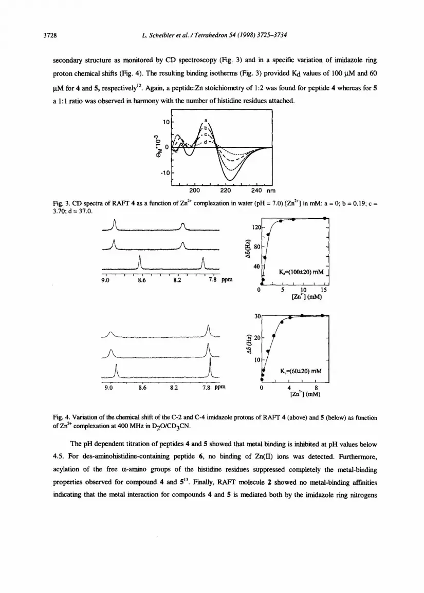
secondary structure as monitored by CD spectroscopy (Fig. 3) and in a specific variation of imidazole ring
proton chemical shifts (Fig. 4). The resulting biding isotherms (Fig. 3) provided Kd values of 100 PM and 60
PM for 4 and 5, respectively”. Again, a peptide:Zn stoichiometry of 1:2 was found for peptide 4 whereas for 5
a 1: 1 ratio was observed in harmony with the number of histidiie residues attached.
-10 -
I. I * I. I. I. I. 200 220 240 nm
Fig. 3. CD spectra of RAFT 4 as a function of Zn2+ complexation in water (pH = 7.0) [Zn*‘] in ml& a = 0; b = 0.19; c = 3.io; d = 37:o.
I I I I I , I I I I I
9.0 8.6 8.2 7.8 ppm
9.0 8.6 8.2 7.8 PPm
0 5 [Zn’~ (ml@’ 5
Fig. 4. Variation of the chemical shift of the C-2 and C-4 imidazole protons of RAFT 4 (above) and 5 (below) as function of Zn*+ complexation at 400 MHz in D2O/CD3CN.
The pH dependent titration of peptides 4 and 5 showed that metal binding is inhibited at pH values below
4.5. For des-aminohistidine-containing peptide 6, no binding of Zn(I1) ions was detected. Furthermore,
acylation of the free cl-amino groups of the histidine residues suppressed completely the metal-binding
properties observed for compound 4 and 513. Finally, RAFT molecule 2 showed no metal-biding affiities
indicating that the metal interaction for compounds 4 and 5 is mediated both by the imidazole ring nitrogens

L. Scheibler et al. /Tetrahedron 54 (1998) 372S-3734 3729
and the cx-amino groups of the histidine residues as coordination ligands as supported by the observed
peptidemetal stoichiometry.
Self-assembly properties. Molecule 7-9 bearing hydrophobic aliphatic chains have been prepared for studying
micellation and self-assembly in solution. A critical micellar concentration (cmc) of about 0.2 mgml’ was
measured for compound 7 by surface pressure measurements in aqueous solution (Fig. 5, insert). Therefore,
CD spectra have been recorded above and below this cmc value to assess the effect of concentration on the
peptide conformation. Above the cmc, a characteristic n-n* minimum at 220 nm and a strong positive x-x* at
192-197 nm (Fig. 5, c) close to the theoretically derived class B patternI is obtained. Such CD spectra are
consistent with the presence of type II p-turns and P-sheet structures normally observed for RAFT molecules in
organic solvents only*. Below the cmc, the CD spectrum does not point to the adoption of a particular defined
secondary structure (Fig& a). A similar loss of secondary structure was observed in water for the EN-
deprotected FUFT 2 and rational&d by an increase in the motional flexibility of the cycle’.“. However, upon
addition of undecanoic acid, a CD spectrum resembling the one obtained above the cmc is observed (Fig. 5, b).
4
2
0
-2
-4 ITI I’ 8 ‘0 ‘8 I I I I,
200 220 240 nm
Fig. 5. CD spectra of RAFT 7 in water at concentrations below the cnx (a), after addition of undecanoic acid (b) and above the cmc (c). The insert shows the surface pressure variation as function of the concentration of 7, representing the critical micellar concentration (cmc) at the line break. a: 0.17 mgknl; b: 0.17 mg/ml + undecanoic acid; c: 1 m&l.
These results indicate that hydrophobic interactions promote in water a significant stabilisation of the peptide
conformation, presumably upon micelle formation. It is interesting to note that a similar secondary structure
stabilising effect can be induced by the addition of Zn(I1) to compounds 4 and 5. Consequently, the chimeric
molecule 9a mnctionalised with His and hydrocarbon chains, displays a pronounced secondary structure
stabilisation upon metal binding as well as upon self-assembly in solution.
Functionalisation of gold surfaces. Terminal thiol groups have been used to address gold surfaces with self-
assembled monolayers (SAM) of molecule 8 or 9b (Fig. 6).

3730 L. Scheibler et al. /Tetrahedron 54 (I 998) 3725-3734
/’ 6, SH $H
SH /
1. NiSO,
2.w
3. EDTA
Fig. 6. Schematic representation of RAPT derivative 9b as self-assembled monolayer (SAM) immobilised on a gold surface showing the reversible binding of NTA labelled fluoresceine ligands (box above). After leading with Ni(II) (step 1). the ligand is bound to the remaining free cemlination sites of the coordinated Ni (II) (step 2). Removal of Ni(II) and the ligand is achieved by washing the surface with a EDTA solution (step 3). SPR spectroscopy is used to monitor the interactions of the ligand with the surface by following the distinct change of the resonance angle 0 (Fig. 7).
The self-assembly process on gold was monitored in real time by surface plasmon resonance (SPR)
spectroscopy and the remainiug accessible surface was subsequently blocked by immobilisation of 1 l-
metcaptoundecan-l-01. Pure SAM of 8 showed a mass coverage corresponding to a mean areal of 150 A2 per
molecule whereas a slightly higher value of 180 A2 was determined for 9b. These values are in good agreement
with the NMR derived template geomett$. SAM of peptide 8, disposing free amiuo groups, provide also a
means for further chemical modifications of gold surfaces via RAFI molecules’7. SAM of 9b disposing
histidine residues as metal binding sites, can be considered as an inversion of immobii metal afIlnity
chromatography’* (IMAC). After loading of the histidiie residues with Ni(I1) ions, specific binding of a
nitrilotriacetic acid (NTA) derivativeI to the surface is observed (Fig. 7b). Desorption of this derivative upon
addition of EDTA demonstrates the full reversibility of the process (Fig. 6 and 7a). The change of the
resonance angle 0 allowed for the estimation of the NTA stoichiometry per template (Fig. 7a). Again, a 1:2
ratio (template : NTA) is found consistent with the results obtained in solution.

L. Scheibler et al. /Tetrahedron 54 (1998) 3725-3734 3731
0.00 4 I I I I %4uk(, ,
\ a 0.12 e
b -
IIVI ’ ‘-me; ’ ’ ’ ’ ’ ’ r ’ ’ ’ I I
:.* am:, IIVl ’ I I I
I’.’
0.15 - :‘A
3
Ft.0 0.18 - -
d . AR f.= i f: _
- NTA incubating
:=. r:
0’16 -
0.10 - / 2, e: F.*
? c 0.14 - . :*
r.* Lw -
0.05 - . c .
i- c
.=
I I 0.10 - I I 1 I I I 52 53 54 55 56 57 58 Q[degl 0 20 40 60
t [m in]
Fig. 7. SPR measurements of surface assembled RAFT 9b (see Fig. 6). (a) Reflectivity (I) vs. angle (Q) curves for the mixed SAM of 9b and 1 I-mercaptoundecan-l-01 (A), after the complexation of the NTA derivative A&0.25” (0) and after metal removal with EDTA A0=0.06” 0. (b) Time course of the reflected light intensity during the specific binding of the NTA derivative at 0 = 53.75”.
CONCLUSIONS
The regioselective functionalisation of RAFT molecules by hydrocarbon chains, metal chelating groups or
combinations thereof is studied to obtain new molecular devices for surface modifications. The flexibility of the
synthetic methodology, based on orthogonally protected attachment sites, allows a wide range of combinations
of functionalities to be assembled on the bifacial template molecule. The resulting molecular systems exhibit the
designed properties such as metal complexation and self-association without disruption of the preferred
template conformation. This suggests that the RAFT topology disposes the appended groups in well detined
spatial orientations avoiding the extensive design of structurally complex scaffolds. As demonstrated above self-
assembly of the bifunctional molecule 9b on gold surfaces provides a supramolecular array of oriented metal-
binding sites. This surface recognises and reversibly binds external hgands. Consequently, the template concept
allows for the design of novel molecular systems exhibiting tailored physical and chemical functions, thus
offering interesting perspectives in the area of biosensor- and nano-technologies.
EXPERIMENTAL
Materials and Methods. Solid phase peptide synthesis was done on a semi-automatic Advanced ChemTech ACT200 Peptide Synthesiser. HPLC was performed on Waters equipment using columns packed with Vydac Nucleosil3OOA 5 pm Cl8 particles unless otherwise stated. Solvent A consisted of 0.09% TFA and solvent B of 0.09% TFA in 90% acetonitrile unless. Amino-acid analyses were performed on a Perkin-Elmer HPLC. Mass spectra were obtained by electron spray ionisation @SI-MS) on a Finnigan MAT SSQ 710C. Circular dichroism (CD) spectra were recorded at room temperature on a Jobin Yvon CD Mark V spectropolarimeter using quartz cells of 0.1 mm path length. The
instrument was calibrated with D-lO-camphorsulphonic acid. lH NMR spectra were obtained on a Bruker DPX-400. WMS spectra where obtained on a computerised Perkin Elmer Lambda 7 using 1 cm cells.
c[HN-K(Boc)-P-G-K(Boc)-K(Aloc)-K(Boc)-P-G-K(Boc)-K(Aloc)~ (I) = T(Bo~)~(Aloc)~ .Z was prepared following published procedures. 780 mg, (76%), tR = 24.6 min (gradient 25% B to lOO%B in 30 min), Rf = 0.35 (CHC13 / MeGH = 9 I l), ESI-MS: 823.8 ((M+2H)I2), mp. > 25O”C, ASA: G 1.74 (2), K 6.0 (6). P 1.83 (2)
T(NH2)4(Aloc)2 (2). The. protected template 1 (50 mg, 30 pmol) was dissolved in 10 ml of 50% TFA solution in DCM for 45 min. After removal of tbe solvent under reduced pressure, the residue was precipitated with diethyl ether and

3732 L. Scheibler et al. /Tetrahedron 54 (J998) 3725-3734
lyophilised. Yield: 47 mg (92%). tR = 18.6 min (gradient 25% B to 100% B in 30 min), ESI-MS: 416.8 ((M+3H)/3), 454.0 ((M+TFA+2H)/3), 623.8 ((M+2H)12), 680.6 ((M+TFA+H)/‘L), 737.8 ((M+2TFA)/2)
T(Fen)4(Aloc)2 (3). The l , lO-phenanthroline-2-carboxylic acid (Fen) was synthesised according the protocol of Corey et aim. A solution of T(NH2)4(Aloc)2 (2) (47 mg, 37 pmol), Fen (50 mg, 230 pmol), HATU” (90 mg, 230 pmo1) in of dry DMF (10 ml) was treated with DIEA (70 pl, 720 pmol) for 3h. The solvent was removed and the crude residue precipitated with diethyl ether and then purified by RP-HPLC. Yield: 50 mg ( 64%). tR = 19.25 (gradient 5% B to 100% B in 38 min) ESI-MS: 518.8 ((M+4H)/4), 691.5 ((M+3H)13), 1026.4 ((M+2H)/2).
T(HisJ4(Aloc)2 (4). A solution of T(NH2)4(Aloc)2 (2) (20 mg, 12 pmol), Na-Boc-His(Boc)-OSu (22 mg, 48 pmol) in dry DMF (2 ml) was treated with DIEA (35 pl, 96 pmol) at room temperature. The reaction was complete after 3 h and after removal of the volatile the residue was treated with a 50% solution of TFA in DCM during 45 min. The solvent was evaporated and the remaining residue was purified by preparative RP-HPLC to yield 12 mg (57%) of pure product. tR = 18.59 min (gradient 0% B to 100% B in 40 min), ESI-MS: 898.1 ((M+2H)/2), ASA: G 1.82 (2), H 4.83 (4), K 6.12 (6), P 2.02 (2)
T(NH2)4(His)2 (5). A solution of the protected template 1 (100 mg, 61 pmol), 1.6 ml glacial acetic acid, PdC12(PPh3)2 (l-3 mg) in 40 ml DCM was treated with Bu3SnI-I (100 pl, 150 mmol) added in two portion. When the reaction was complete (1 h, HPLC), the orange solution was concentrated and the crude brown residue was dissolved in MeOH and precipitated with diethyl ether. The solid was dissolved in water, filtered through Celite and then lyophilised to afford 81 mg (83%) of the product. tR = 18.3 min (gradient 25% B to 100% B in 30 min), ESI-MS: 739 ((M+2H)/2). A mixture of
the Alec deprotected template (10 mg, 6.7 pmol), Na-Boc-His(Boc).OSu (6.5 mg, 41.2 pmol) in dry DMF (2 ml) was treated with DIEA (10 pl, 55 pmol). After 2 h the reaction was complete as inferred by HPLC, the solvent was removed and the residue was deprotected with 2 ml of a 50% solution of TFA in DCM for 30 min. The solvent was removed and the residue was purified on a Waters Sep-Pak Vat 35cc (2g) Cartridge using 10% B in water as eluent. Lyophilisation afforded 6.8 mg (74%) of the desired product. tR = 10.35 min (gradient 5% B to 100% B in 38 min) ES&MS: 451.3 ((M+3H)/3), 489.3 ((M+TFA+2H)/3), 676.3 ((M+2H)/2), 733.6 ((M+TFA+H)/2)
T(des-aminoHis)q(Aloc)2 (6). A solution of T(NH2)4(Aloc)2 (2) (10 mg, 5.8 pmol), des-aminohistidine (Bachem, CH) ( 5 mg, 32 pmol), PyAOP (17 mg, 32 pool) in dry DMF (2 ml) was treated with DIEA (20 pl, 128 pmol) for 4 h. The solvent was removed and the residue purified by RP-HPLC to yield 7 mg (70%) of 6. tR = 19.53 min (gradient 0% B to 100% B in 40 min), ESI-MS: 434.9 ((M+4H)/4), 579.1 ((M+3H)/3), 868.0 ((M+2H)/2), 1735.0 (M+H).
T(NH2)4( OC-(CH2)9-CH3)2 (7). A solution of undecanoic acid (2.00 g, 10.7 mmol), pentafluorophenol ( 2.20 g 12 mmol) in dry DCM (5 ml) was treated with dicyclohexylcarbodiimide @CC) (2.44g 12 mmol). After lh, the solution was filtered, the volatile removed under vacuum and the residue purified by chromatography column with petrolether I diethyl ether (9:l) to afford an 3.553 of an oil (94%). Alec groups were removed of 1 following the protocol described for 5. A solution of this deprotected peptide (30 mg, 20 mmol), undecanoic-pentafluorophenyl eater (7.2 mg, 40 mtrrol) and 4- dimethylaminopyridine (DMAP) in DMF (2 ml) was treated with DIEA (15 ml, 80 mmol) for 12h. After removal of the solvent and precipitation with diethyl ether, the residue was deprotected with a solution of SO%TFA in DCM during 45 min. Evaporation and RP-HPLC purification afforded 19 mg (66%) of 7. tR = 20.43 min (gradient 25% B to 100% B in 30 min), ESI-MS: 472.6 ((M+3H)/3), 510.8 ((M+2H+TFA)/3), 707.7 ((M+2H)/2), 765.0 ((M+H+TFA)/2), 114.6 (M+H).
T(NH2)4(OC-(CH2) JO_%&? (8). Acm-S-l I-mercaptoundecanoic acid, I I-mercapto-undecanoic acid (1.00 g, 4.58 mmol) and acetamidomethanol(409 mg, 4.58 mmol) were dissolved in 50 ml trifluoroacetic acid and stirred for 30 min at room temperature. After solvent removal the residue was dissolved in CHCl3 and extracted with 2N NaOH. The aqueous phases were acidified and extracted with CHCl3. The organic layer was dried over MgS04 and the solvent removed under pressure. The product was crystallized with DCM ! petrol ether to afford 950 mg (72%) of pure 8. Rf = 0.26 (diethyl ether I acetic acid = 98 : 2), m.p. 81°C tR = 17.19 (gradient 25% B to 100% B in 30 min), ESI-MS: 290.5 (M+l), 312.4
(M+Na), IH-NMR: 1.15 (m, 12H, H alkane chain), 1.27 (m, 6H, H alkanechain), 1.95 (s, 3H, CH3CO), 2.30 (td, 2H, S- CH2- alkane chain), 4.30 (d, 2H, NCH2-S), 4.6 (s, large, lH, acid), 5.75( s, large, lH, NH).
Acm-S-l I-mercaptoundecanoic-pentafluorophenyi ester: Acm-S- 1 I-mercapto-undecanoic acid (100 mg, 346 mmol), pentafluorophenol(70 mg, 380 mmol) and DCC (206 mg, 380 mmol) were reacted in CHC13 (4 ml). The solution was filtered after 12 h, extracted with a solution of 5% KHSO,, 10% NAHCO, and Nat&, and the organic phase dried over MgSO,. After removal of the solvent 99 mg (63%) of the desired active eater was obtained. Rf = 0.7 (diethyl ether/acetic

L. Scheibler et al. /Tetrahedron 54 (I 998) 3725-3734 3733
acid = 98 : 2), m.p- 67-68°C. tR = 17.59 (gradient 25% B to 100% B in 30 min), ESI-MS: 456.7 (M+ I), lH-NMR: ] ] 5 (m, 12H, H alkane chain), 1.22 (m, 6H, H alkane chain), 1.95 (s, 3H, CH3CO). 2.30 (td, 2H, S-CH2- alkane chain), 4.30 (d, 2H, N-CH2-S), 5.75( s, large, IH, NH).
T(Boc)4(0C-(CH2)10-S-Acm)2: The Alec groups were removed according the procedure described for 5. A solution of the deprotected template (40 mg, 27 pmol), Acm-S- 11 -mercaptoundecanoic-acidpentafluorophenolester (25 mg, 55 pmol), DMAP in dry DMF was treated with DIEA (100 pl, 580 l.tmol). The solvent was removed after 9 h and the residue precipitated with diethyl ether to afford 50 mg (91%) of the product. tR = 24.82 min (gradient 25% B to 100% B in 30 min), ES&MS: 1010.6 ((M+2H)/2).
T(NH2)4(0C(CH2)loSAcm)2: T(Boc)4(OC(CH2)loSAcm):! (5Omg, 25 mmol) was treated according the protocol used for 2. Yield: 41.8 mg (81%), tR = 13.99 min. ESI-MS: 810.7 ((M+2H)12)
T(NH2)4(0C-(CH2)10-SH)2 (8): T(NH2)4(OC(CH2)lOSAcm)2 (50 mg, 24 l.tmol) and Hg(OAc)2 (20 mg, 50 pmol) were stirred for 2h in 5 ml 30% acetic acid under N2 atmosphere. P-Mercaptoethanol (430 ~1, 4.8 mmol) was added and the solution stirred overnight. The solution was filtered and purified by preparative RP-HPLC to afford 29 mg (62%) of pure 8. tR = 18.61 min (gradient 25% B to 100% B in 30 min), ESI-MS: 493.3 ((M+3H)/3), 531.2 ((M+3H+TFA)/3), 739.8 ((M+2H)/2), 796.8 ((M+2H+TFA)/2), 797.2 ((M+2TFA)/2), 1478 (M+l).
T(His)q(OC-(CH2)10-S-A&2 (9~). T(NH2)4(0C(CH2)lOSAcm)2 (73 mg, 49 pmol) was prepared according the protocol for 8 and then treated following the protocol for 4 to afford 100 mg (86%) of product. tR = 12.55 min (gradient 0% B to 100% B in 40 min), ESI-MS: 542.9 ((M+4H)/4), 724.2 ((M+3H)13), 1084 ((M+2H)/2).
T(His)4(OC-(CH2)10-SH)2 (9b). T(His)4(OC(CH2)lOSAcm)2 (20 mg, 9.2 pmol) was treated according to the protocol for 8 affording 12 mg (64%) of product. tR = 16.65 min (gradient 0% B to 100% B in 40 min), ESI-MS: 507.0 (M/4+1),. 676.6 ((M+3H)/3), 1014.2 ((M+2H)/2).
Suflace plasmon resonance (SPR). The surface plasmon resonance measurements were performed, using the Kretschmann coupling scheme*’ on a home-made computerised reflection apparatus as described in details elsewhere*“. At the resonance angle the incident laser beam (He-Ne laser, 632.8 nm, p-polarised) couples via an equilateral, high index prism (SF 10, n= 1.723) to the surface plasmon mode in a thin gold film (40 nm).
Layerformation. For the self-assembly of peptide and 11 mercaptoundecanol, a gold film (40 nm), vacuum evaporated on a cleaned glass slide, was incubated in peptide or thiol water/methanol mixtures. After 1 h self-assembly, the surface was extensively washed with water/methanol. Finally a solution of thioalcohols in methanol/water was added to block remaining defects in the monolayer. The binding was monitored in situ using surface plasmon resonance. For Ni2+ binding the surface was incubated with 100 mM NiS04 solution for IO min. After washing, the cell was calibrated with buffer and the fluoresceine-labelled NTA was added. The surface was washed with buffer and the final thickness measured. The His-exposing surface was regenerated by washing with 0.5 M EDTA in buffer.
(/V/W Measurements of (Fen)4-Temp-(Aloc)2 (3). A 2.665. 10m5 M solution in IO mM Tris/HCl buffer pH = 7.0 of the
decapeptide 3 was titrated in a 1 cm cell with a 5.3 1.1 Om3 M solution of Zn(BF4)* at room temperature. The reference cell contained the buffer and the equivalent amount of metal. Job-Plot measurements were obtained by mixing a peptide and a
metal solution (both 5.31. 10s5 M in buffer) in various ratios affording always the same amount of final solution (1 ml). The reference cell contained the same concentration of peptide in buffer. In this manner a difference spectra between ligated and free peptide was obtained. Job-Plot data points were recorded at )c = 277 nm.
CD Titrations. CD spectra were obtained in a 1 mm cell with a peptide solution of about 0.5 mg.ml-1 in a Tris / HCI buffer pH 7.0 and titrated with a 100 times more concentrated ZnC12 solution in the same buffer. Exact peptide concentration was determined by quantitative ASA. For each spectra 2 scans were accumulated, the resolution was set to 0.2 nm and the integration time to 2s per step.
NMR Titrations. NMR spectra were measured in acetonitrile CDFN I D20 (I : I), pH being adjusted with NaOD solution to a value of pH = 7. Peptide concentration was 20 mg.ml-1 and the titration was performed with addition of concentrated ZnCl2 solution.

3134 L. Scheibler et al. /Tetrahedron 54 (1998) 3725-3734
Surface pressure. Surface pressure was measured a Langmuir balance. Peptide 7 (6.2 mg) was dissolved in pure water (2 ml) in a Teflon cell. The sample was then successively diluted and the corresponding surface pressure was determined after the sample reached equilibrium (constant pressure over more than 5 min). This took normally about 20 min.
Acknowledgements: This work was supported by the Priority Program Biotechnology of the Swiss National Science Foundation.
1.
2.
3.
4. 5. 6. 7. 8.
9.
10.
Il.
12. 13. 14. 15.
16.
17. 18. 19. 20. 21. 22. 23.
RJWERJENCES AND NOTES
* To whom the correspondance should be addressed, e-mail [email protected]. Tuchscherer, G.; Mutter, M. Chem. & Ind. 1997,597-601. Regan, L. Trends Biochem. Sci. 1995,20,280-285 and references cited therein. Ackerfeldt, K. S.; Lear, J. D.; Wasserman, Z.R.; Chung, L.A.; De&ado, W. F. Act. Chem. Res. 1993, 26, l91- 197. Rabanal, F.; DeGrado, W. F.; Dutton, P. L. J. Am. Chem. Sot. 1996, 118,473-474. Voyer, N.; Lamothe, J. Tetrahedron, 1995.51, 9241-9244. Cheng, R.P.; Fisher, L.S.; Imperiali. B. J. Am. Chem. Sot. 1996, 118, 11349-11356. Struthers, M.D.; Chkng, R.P.; Imperiali, B. J. Am. Gem. Sot.. 1996. 118, 3073- 3081. Mihara, H.; Tanaka, Y.; Fujimoto, T.; Nishino, N. J. Chem. Sot. Perkin Trans. 2 1995, 1915-1921. Mihara, H.; Tanaka, Y.; Fujimoto, T.; Nishino, N. J. Chem. Sot. Perkin Trans. 2 1995, I 133-l 140. Schneider, J. P.; Kelly, J. W. Chem. Rev., 1995, 95,2169-2187. And references cited therein. Dumy, P.; Egg&on I.; Cervigni, S.; Sila, U, Sun, X.; Mutter, M. Tetrahedron Lett. 1995,36, 1255-1258 Dumy, P.; Eggleston I.-M., Esposito G.; S. Nicula, Mutter, M. Biopolymers. 1996, 39, 297-308. Mutter, M.; Vuilleumier, S. Angew. Chem. Int. Ed. Engl., 1989,28,535-554. Tuchscherer, G.; Servis. C.; Corradin, G.; Blum, U.; Mutter, M. Proteins Sci. 1992, 1, 1377-1386. Pawlak, M.; Meseth, U.; Dhanapal, B.; Mutter, M.; Vogel, H. Protein Sci. 1994, 3, 1788- 1805. Mutter, M.; Dumy. P.; Garrouste, P.; Lehmann, C.; Mathieu, M.; Peggion, C.; Peluso, S.; Razaname, A.; Tuchscherer, G. Angew. Chem. Int. Ed. Engl. 1996.35, 1482-1485. Dumy, P; Scheibler, L.; Cervigni, S.; Vogel, H.; Mutter, M. feptides 1996; Chemistry and Biology 24th Proc. Eur. Pept. Symp. 1997, Eds Ramage, H., ESCOM, Leiden, in press. The & values were calculated as follows (shown for NMR-titrations):
K, =v:[C]=[M,,]-[Ml [T]=[T,]-[ ] [ ]= C wrrh T template, [M] = metal, [c] = compkx
For [~]=-a~~~(Ifz);~~=chemica~shiftinHz, oneobtains [M,]=-~.M(HZ)+@~]- K,)+ f$ .[T,] a. @(Hz)
This formula allows the fitting of AF against m] and the determination of Kd and [T,]. A [T,,] concentration twice the actual weight in concentration was obtained, indicating that two metal ions were bound independently per RAFT molecule 4. The maximal change in absorption was found to be at a total molar fraction of the template x=0.38. The theoretical value of x=0.33 for a template to metal complex of 1:2 is within the error limits. For the calculation of & values, only the histidine C, proton chemical shift was considered. Acetylation of the m 4 was performed with 0-carboxymethylhydroxylamine. Woody, R. W. In The Peptides 7,1985, Hruby, V.J., Ed., Academic Press, New York, pp. 15-l 14. Dumy, P.; Eggleston I.-M.; Esposito G.; Nicula, S.; Mutter, M. Electronic Conference on Trends in Organic Chemistry, (ECTOC-I), Rzepa, H., Goodman, .I. M. (Eds), (CDROM), Royal Society of Chemistry Publications, 1995, See also http:Nwww.ch.ic.ac.uk/ectoc/. For determination of the thickness and the mass loading, a refractive index n=1.45, a refractive index increment Mdc = 0. 18cm"g" and a typical peptide density of p = I .37 gem? was used. The chemical modification with peptides will be reported in due time. Porath, J.; Carlsson, J.; Olsson, I.; Belfrage, G. Nature 1975, 258, 598. We thank A. Heusler (EPFL) for the synthesis of the NTA derivative. Corey, E.J.; Borror A.L.; Foglia, T. J. Org. Chem., 1965, 30,288-290. Carpino, L.A.; El-Faham, A.; Minor, C.A.; Albericio, F. J. Chem. Sot., Chem. Corn. 1994.201-203. Kretschmann, E. Optics Communications 1972; 6, 185-186. Terrettaz, S.; Stora, T.; Duschl, C.; Vogel, H. Lungmuir 1993.9, 1361.1369.