![Substituted dibenzo[ c,h]cinnolines: topoisomerase I-targeting anticancer agents](https://static.fdokumen.com/doc/165x107/631871c065e4a6af370f5e52/substituted-dibenzo-chcinnolines-topoisomerase-i-targeting-anticancer-agents.jpg)
Substituted dibenzo[ c,h]cinnolines: topoisomerase I-targeting anticancer agents
Functional dissection of the C-terminal domain oftype II DNA topoisomerase from the kinetoplastidhemo¯agellate Leishmania donovaniTanushri Sengupta, Mandira Mukherjee, Chhabinath Mandal1, Aditi Das2 and
Hemanta K. Majumder*
Department of Molecular Parasitology, 1Department of Drug Design, Development and Molecular Modeling,Indian Institute of Chemical Biology, Kolkata 700032, India and 2Sealy Center for Molecular Sciences,University of Texas Medical Branch at Galveston, Galveston, TX 77555, USA
Received June 19, 2003; Revised and Accepted July 21, 2003
ABSTRACT
The amino acid sequences of the C-terminal domain(CTD) of the type II DNA topoisomerases are diver-gent and species speci®c as compared with thehighly conserved N-terminal and central domains. Aset of C-terminal deletion mutants of Leishmaniadonovani topoisomerase II was constructed.Removal of more than 178 amino acids out of 1236amino acid residues from the C-terminus inactivatesthe enzyme, whereas removal of 118 amino acids orless has no apparent effect on the ability of the para-site enzyme to complement a temperature-sensitivemutation of the Saccharomyces cerevisiae topo-isomerase II gene. Deletion analysis revealed apotent nuclear localization signal (NLS) within theamino acid residues 998±1058. Immunomicroscopyresults suggest that the removal of an NLS in theCTD is likely to contribute to the physiological dys-function of these proteins. Modeling of the LdTOP2based on the crystal structure of the yeast type IIDNA topoisomerase showed that the parasiteprotein assumes a structure similar to its yeastcounterpart harboring all the conserved residues ina structurally similar position. However, a markeddifference in electrostatic potential was found in aspan of 60 amino acid residues (998±1058), whichalso do not have any homology with topoisomeraseII sequences. Such signi®cant differences can beexploited by the structure-based design of selectiveinhibitors using the structure of the Leishmaniaenzyme as a template.
INTRODUCTION
Topoisomerases are enzymes that can modify the tertiarystructure of DNA without altering the primary structure (1).
DNA topoisomerases accomplish their function by eitherpassing one strand of DNA duplex through a transient break inthe other strand (type I topoisomerase) resulting in changes inthe linking number in steps of one (2) or by a passing a duplexDNA from the same or another molecule through a transientdouble-stranded break generated in the DNA in an ATP-dependent manner (type II topoisomerase), resulting inchanges in linking number in steps of two (3). These enzymesparticipate in nearly all events related to DNA metabolismwhich includes replication, transcription and recombination(4). Eukaryotic type II topoisomerases (TOP2) are alsoimportant because of their essential role in chromosomesegregation (5) and maintenance of the chromosome structure(6). In addition to these functions, topoisomerase II is acomponent of the nuclear scaffold where it is involved inchromosome condensation and decondensation (7,8). Theseenzymes have been found in all cell types from viruses (9) tobacteria (10) to mammals (11) and are essential for cellviability. Recent biochemical and structural studies provide aninsight into the mechanistic detail of the eukaryotic topo-isomerase II (12); however, little information has emergedabout the functional organization of the enzyme.
Comparison of the primary sequence suggests thateukaryotic topoisomerase II has evolved by the fusion of thegyr A and gyr B genes of DNA gyrase, the eubacterialcounterpart of topoisomerase II. Based on homology to DNAgyrase, topoisomerase II can be divided into three distinctdomains. The N-terminal domain is homologous to the gyraseB subunit that contains the ATP binding and hydrolysisactivity, the central part is similar to the gyrase A subunit andcontains the active site tyrosine which is implicated in theDNA breakage and rejoining reaction. The N-terminal and thecentral part of topoisomerase II are highly conserved amongdifferent eukaryotic species. The conservation decreasesmarkedly at the C-terminus, which is not homologous toeither subunit of gyrase and the one-third of the C-terminaldomain (CTD) is quite divergent in sequence. The C-terminusis of interest for a number of reasons. First, it is highlyhydrophilic and contains the regulatory sequences such as the
*To whom correspondence should be addressed. Tel: +91 33 2412 3207; Fax: +91 33 2473 5197; Email: [email protected]
The authors wish it to be known that, in their opinion, the ®rst two authors should be regarded as joint First Authors
Nucleic Acids Research, 2003, Vol. 31, No. 18 5305±5316DOI: 10.1093/nar/gkg727
Nucleic Acids Research, Vol. 31 No. 18 ã Oxford University Press 2003; all rights reserved
by guest on March 14, 2014
http://nar.oxfordjournals.org/D
ownloaded from
nuclear localization signal (NLS) (13), phosphorylation sitesand a dimerization interface (14±16). Secondly, while partialproteolysis and truncation analysis of topoisomerase II fromhuman (17), Drosophila (18), Schizosaccharomyces pombe(19) and Saccharomyces cerevisiae (20) reveal that theC-terminus is dispensable for the biochemical activities (21),this domain is thought to enhance the stability of the complexformed between DNA and topoisomerase II (22).
The kinetoplastid protozoan parasite Leishmania causes aspectrum of disease, termed leishmaniasis, manifestingpathologies from mild to dis®guring to fatal. This protozoandiverged early in the eukaryotic evolution, near the base of theevolutionary tree before the emergence of several protozoanlineages and well before the separation of the crown metazoanlineages that comprise plants, animals and fungi (23). Work onDNA topoisomerases from the kinetoplastid protozoanparasite has been a major focus of interest, since theseenzymes are believed to play an important role in thereplication of the unusual kinetoplast DNA (kDNA) harboredin the mitochondrion of these parasites. Type II DNAtopoisomerases have been isolated from several kinetoplastidprotozoans and the genes encoding these enzymes have beencloned and sequenced in Trypanosoma brucei (24),Trypanosoma cruzi (25) and Crithidia fasciculata (26).Wehave previously reported topoisomerase activity from the cellextracts of Leishmania donovani (27,28) and the geneencoding the type II DNA topoisomerase was isolated (29).DNA sequence analysis revealed an ORF of 3711 bp with nointrons and encoding a protein of 1236 amino acids. This wasshown to be an ATP-dependent enzyme whose activity wasinhibited by etoposide, an eukaryotic topoisomerase IIinhibitor.
The L.donovani topoisomerase II has the highest degree ofhomology with the TOP2 of other kinetoplastid parasites.With its human counterpart the LdTOP2 shares a much loweridentity of 32% and a similarity of 47%. The degree ofhomology is much higher towards the N-terminus andgradually falls off towards the C-terminus. Although thisdomain contains none of the functions known to be necessaryfor enzyme catalysis, it has been retained in phylogeneticallydivergent organisms and thus presumably performs someimportant functions for the enzyme.
In this study, a series of deletion mutants of the TOP2 geneof L.donovani were constructed and introduced into atemperature-sensitive S.cerevisiae strain to investigate thefunctions of the C-terminus of the enzyme. The mutantproteins were characterized with respect to their complement-ation ability, stability inside yeast cells and in vitro catalyticactivity. Using a molecular modeling approach we examinedthe plausible structure for the LdTOP2 based on the availablecrystal structure of yeast type II DNA topoisomerase (12). Wedetected signi®cant differences in the charge distribution ofthe two enzymes in their C-termini. Taken together, our resultsprovide the ®rst insight into the structure and biochemicalproperties of the CTD of type II DNA topoisomerase from thekinetoplastid protozoan parasite L.donovani. In the CTD, aregion has been identi®ed between amino acid residues 998and 1058, which plays a crucial role in modulating thecatalytic activity of the enzyme in vitro and in substituting forthe yeast topoisomerase II in vivo.
MATERIALS AND METHODS
Strains, media and growth conditions
The Escherichia coli strains used were DH5a and BL21(DE3) pLysS. If required, ampicillin and chloramphenicolwere used at 100 and 34 mg/ml concentrations, respectively.Wild-type and mutant topoisomerase II of L.donovani wereexpressed in the yeast strain RS192 that has a genotype ofMATa top2-1 top1-8 [top1:: LEU2] ade2 ura3-1 his3-11trp1-1 leu 2-3 leu2-112 (a gift from Dr Rolf Sternglanz, StateUniversity of New York, Stony Brook). The yeast cells weregrown at 25°C on YEPD medium containing 1% peptone, 2%yeast extract, 2% dextrose and 1.5% agar or synthetic minimalmedia containing 6.7 g/l yeast nitrogen base without aminoacid, 5 g/l casamino acids, 20 g/l glucose, 20 mg/l adeninesulfate, 20 mg/l tryptophan and 15 g/l Bacto agar (30).
Construction of mutants
The full-length LdTOP2 gene previously cloned in theHindIII/XbaI site of pBluescript (SK+) (29) was subclonedas a HindIII/XbaI fragment into the yeast shuttle vectorpVT100U, a gift from Dr Rolf Sternglanz (31). For construc-tion of all truncation mutants, the regions corresponding toamino acids 1±1195, 1±1118, 1±1058, 1±998 and 1±785 wereampli®ed by polymerase chain reaction (PCR). In all cases the5¢ primer was 5A (5¢-CCCAAGCTTATGACAGACGCTTC-CAAG-3¢) and the different 3¢ primers were 3A (5¢-GCT-CTAGAGGAACCGACCAACCGCTT-3¢), 3B (5¢-GCTCTA-GACGAGTTGATGAGCACGCG-3¢), 3C (5¢-GCTCTAGA-GAAGCTCTCGTCCACGCG-3¢), 3D (5¢-GCTCTAGACT-TGTACAAGTCAAGGCG-3¢) and 3E (5¢-GCTCTAGAG-GCAAAACGGCTGAGCTT-3¢). The ampli®ed productswere cloned in the HindIII/XbaI site of pVT100U, resultingin constructs LdDC1195, LdDC1118, LdDC1058, LdDC998and LdDC785, respectively. The mutants LdDC998 andLdDC785 were also cloned in the NdeI/BamHI site of pET16b (Novagen) and expressed in E.coli BL21 (DE3) pLysS.The different sets of primers used were 5B (5¢-GGGAATTC-CATATGACGCTTCCAAG-3¢) and 3F (5¢-CGGGATCCC-TTGTACAAGTCAAGGCG-3¢) and 3G (5¢-CGGGATCCG-GCAAAACGGCTGAGCTT-3¢), resulting in constructsLdDC998 and LdDC785, respectively. The PCR was per-formed using 30 cycles of denaturation at 94°C for 45 s,annealing at 54°C for 30 s and extension at 68°C for 1 min/kbwith 2.5 U of high-®delity Taq DNA polymerase (RocheApplied Science) and 200 mM of each dNTP. In all cases, theLdTOP2 truncation mutants were under ADH1 promoter inthe shuttle vector and under T7 RNA polymerase promotor inpET16b, respectively.
Complementation assay
The yeast strain RS192 was used for transformation withL.donovani topoisomerase II truncation mutants by the lithiumacetate and polyethylene glycol method (32). The trans-formants were cultured on solid synthetic minimal medium at25°C for 2 days. The replicated colonies were grown at 37°Cfor 7 days. Colonies were picked and cultured in tubes with2 ml of synthetic minimal media at 25°C overnight. The cellswere collected and resuspended in a lysis buffer containing2% Triton X-100, 1% SDS, 100 mM NaCl, 10 mM Tris±HCl
5306 Nucleic Acids Research, 2003, Vol. 31, No. 18
by guest on March 14, 2014
http://nar.oxfordjournals.org/D
ownloaded from

and 1 mM EDTA and plasmids were prepared as described(33).
Reverse transcription (RT±PCR)
Yeast spheroplast was prepared by the method described (30).Total RNA was isolated using the RNA Isolation Kit (RocheApplied Science) according to the manufacturer's protocol.RT±PCR was performed using a sense primer, 5¢-GGG-AATTCCATATGACAGACGCTTCCAAG-3¢, and an anti-sense primer, 5¢-CGGGATCCCGCCTCCAGAAACGGCAT-3¢, designed to amplify the N-terminal 1.1 kb of theL.donovani topoisomerase II gene. The PCR program con-sisted of an initial RT at 42°C for 1 h, followed by theampli®cation of the cDNA product for 30 cycles at 94°C for45 s, 52°C for 30 s and 68°C for 1 min using the Titan one tubeRT±PCR kit from Roche Applied Science. The resultantRT±PCR products were electrophoresed in 1% agarose gel,stained with ethidium bromide and photographed under UVillumination.
Decatenation assay
Bacterial cells were lysed as described (29). Brie¯y, the cellswere induced at 0.6 OD600 with 0.5 mM IPTG at 37°C for 3 h.Bacteria were harvested by centrifugation and the cells wereresuspended in lysis buffer containing 20 mM Tris±Cl pH 7.8,100 mM NaCl, 1 mM EDTA, 100 mg of lysozyme (Sigma-Aldrich), 5 mM DTT, 0.1% (v/v) Triton X-100 and proteaseinhibitor. Final lysis was achieved by sonication on ice and thelysate was cleared by centrifugation at 12 000 r.p.m. in a SS34rotor for 15 min. Yeast cells were lysed in a buffer containing150 mM NaCl, 10 mM Tris±HCl pH 7.9, 1 mM EDTA, 0.1%Triton X-100 and 15 mM b-mercaptoethanol and 300 mg ofacid-washed glass beads (300 mm; Sigma-Aldrich) by rapidvortexing (34). Decatenation assays were performed in totalvolumes of 25 ml containing 25 mM Tris±HCl pH 7.9, 10 mMMgCl2, 0.1 mM EDTA, 1 mM DTT, 50 mM NaCl, 10%glycerol, 1 mg of kDNA from L.donovani strain UR6 andcrude yeast or bacterial extracts. The assays were carried at30°C for 30 min and the reaction products were analyzed byelectrophoresis on a 1% agarose gel as above.
Antibody production and western blotting analysis
A 1.1 kb fragment was ampli®ed from the 4.1 kb genomicclone of L.donovani, using a sense primer, 5¢-GGGAATTC-CATATGACAGACGCTTCCAAG-3¢, and an anti-sense pri-mer, 5¢-CGGGATCCCGCCTCCAGAAACGGCAT-3¢, thatcoded the N-terminal 385 amino acids of the LdTOP2 gene.This fragment was cloned in the NdeI/BamHI site of bacterialexpression vector pET 16b and transformed into BL21 (DE3)pLysS. Expression from the construct pET16b/1.1 yielded a43 kDa protein as viewed by SDS±PAGE (35). The over-expressed protein band was excised from the gel and theprotein was electro-eluted in a buffer containing 200 mM Tris-acetate pH 7.4, 1% SDS and 100 mM DTT per 0.1 g of wet gelslice (36). It was dialyzed against a buffer containing 50 mMTris-acetate pH 7.4, 0.1% SDS at 100 mA for 3 h. The electro-eluted product (100 mg) was subcutaneously injected (36) inrabbit using Freund's complete adjuvant, followed by twoinjections at 2 week intervals with incomplete adjuvant toproduce the polyclonal antibody against the 43 kDa protein.
This serum was then used for western blot analysis andimmuno¯uorescence experiments.
For immunoblot analysis, yeast cells harboring the full-length and truncated LdTOP2 constructs were lysed aspreviously mentioned. The protein content of cell lysateswas estimated by the Bio-Rad Protein Estimation Kit accord-ing to the manufacturer's protocol. Twenty micrograms ofprotein from each cell lysate was loaded in separate lanes andelectrophoresed in 8% SDS±polyacrylamide gels. Proteinswere transferred to 0.2 mm nitrocellulose membranes using aBio-Rad wet blotter following the manufacturer's instructions.Antibody probing of membrane blots was carried out asfollows. The membrane was blocked with 3% BSA in PBS for1 h, then incubated for 2 h with 100 times diluted primaryantibody (LdTOP2-speci®c polyclonal antibody) in PBScontaining 0.1% Tween-20 (PBST). The membrane waswashed four times in PBST and incubated with 1000 timesdiluted alkaline phosphatase-conjugated goat (anti-rabbit)antisera in PBST for 45 min at room temperature. Themembrane was washed as above prior to color generation andthen incubated in a buffer (100 mM Tris±HCl pH 8.8)containing 330 mg/ml nitroblue tetrazolium and 160 mg/ml5-bromo-4-chloro-3-indolyl phosphate. Color developmentwas stopped by the addition of a buffer containing 20 mMTris±HCl, pH 8.0 and 5 mM EDTA.
Immuno¯uorescence microscopy
Yeast strain RS192 containing wild-type and mutant forms ofLdTOP2 was prepared for immunostaining as describedpreviously (15). Brie¯y, the cells were grown at 25°C untilan OD600 of 0.5, and then shifted to 37°C for 4 h. The cellswere harvested at 4000 r.p.m. using an SS34 rotor in a SorvallRC35 centrifuge for 4 min and washed twice with PBS andtreated with 1:10 formaldehyde (37%) in PBS for 45 min atroom temperature. Subsequently, the cells were washed andresuspended in a buffer containing sorbitol, sodium citrate,EDTA, 100 mg/ml zymolyase, glusulase and b-mercapto-ethanol, and incubated at 30°C for 1 h. Cells were thenadhered to poly-L-lysine-coated slides and ®xed with chilledmethanol and acetone. Antibody against the N-terminaldomain of the LdTOP2 (1:25 dilution) was used along with3% BSA to detect the expressed proteins. Following the ®rstantibody reaction, slides were washed with PBS and reactedwith ¯uorescein isothiocyanate (FITC)-conjugated goat-derived anti-rabbit IgG (1:100 dilution) for 45 min. After thelast wash with PBS, the cells were mounted in 10% glycerol inPBS containing 0.1% p-phenylenediamine to prevent fading.To locate the yeast nucleus the ®xed cells were stained withethidium bromide (0.25 mg/ml in PBS containing 10%glycerol). Cells were viewed with a TCS-SP Leica confocalmicroscope system equipped with a krypton±argon mixed laser.
Molecular modeling
A three-dimensional model of L.donovani topoisomerase IIprotein was obtained from Swiss Prot (37). Energy minimiza-tions were done on this model with a convergence criterion of0.001 kcal/mol using a combination of steepest descent andconjugate gradient methods of 100 steps each. Secondarystructures were analyzed using the programs Ribbons andModelyn (38) and electrostatic potential surface of the modelwas determined by using MOL MOL (39).
Nucleic Acids Research, 2003, Vol. 31, No. 18 5307
by guest on March 14, 2014
http://nar.oxfordjournals.org/D
ownloaded from

RESULTS
The CTD of L.donovani topoisomerase II
In order to study the CTD of L.donovani topoisoimerase II, theamino acid sequence of the enzyme was compared with that ofS.cerevisiae and Homo sapiens using CLUSTAL W (40).Figure 1 shows regions of alignment of amino acids ofL.donovani topoisomerase II with that of the above twospecies. Sequence comparison shows that L.donovani topo-isomerase II shares much lower identity and similarity of 23and 31% with its yeast counterpart and an identity andsimilarity of 32 and 47% with its counterpart from human. Thedegree of conservation is generally greater in the N-terminaltwo-thirds of the coding sequence and falls off markedlytowards the C-terminus. The poor homology between thesetwo proteins suggests a weak evolutionary parsimony con-sistent with the evolutionary distance between Leishmania andother organisms. In L.donovani topoisomerase II, the variableC-terminal region covers approximately 400 amino acidsextending from an approximate 850 amino acid residue, asobserved from the multiple alignment analysis.
Effects of deletion on complementation ability
To investigate the role of the CTD of L.donovani topoisomer-ase II we have used a functional complementation assay ofLdTOP2 deletion mutants to rescue a S.cerevisiae topoisomer-ase II temperature-sensitive mutant strain. The rationale forthe use of S.cerevisiae was based on a number of factors. LikeL.donovani, S.cerevisiae is a unicellular eukaryote andprovides the most ef®cient eukaryotic system to test largenumbers of mutations in an essential gene for effects onfunctional complementation. We constructed mutants ofLdTOP2 that were serially deleted at the 3¢ end of the gene(Fig. 2A). The C-terminus was subdivided into ®ve regions bysix truncation mutants. We have used a plasmid-shuf¯ingtechnique to study the ability of wild-type and mutant forms ofthe LdTOP2 gene to complement the temperature-sensitiveS.cerevisiae strain RS192 by in vivo complementation assay.
Plasmids expressing wild-type or mutant forms of theLdTOP2 gene were constructed with amino acid residuesranging from 1236 to 785 in a shuttle vector pVT100U andtransformed into the temperature-sensitive URA± yeast strainRS192 and URA+ colonies were selected. RS192 and the cellstransformed with pVT100U served as controls, respectively,in the complementation assays. At the non-permissivetemperature (37°C), there was complementation with thefull-length LdTOP2 gene containing 1236 amino acids. When40 and 118 amino acids from the C-terminus were removedby truncation to residues 1195 (LdDC1195) and 1118(LdDC1118), no detectable effects on the ability of the mutantenzymes to complement S.cerevisiae were found. Truncationto residue 1058 (LdDC1058) causes only slightly impairedgrowth of the host strain. However, any truncation beyondresidue 1058 towards the N-terminus (LdDC998 andLdDC785) completely abolished growth of the yeast strain atthe non-permissive temperature (Fig. 2B).
Our observation adds to the general conclusion thattopoisomerase II enzyme from a wide variety of eukaryoticorigins are functionally interchangeable (41±45) and that onlya part of the divergent CTD of the enzyme is dispensable forcatalytic activity (15).
Stability of transcripts, protein expression and in vitroactivity
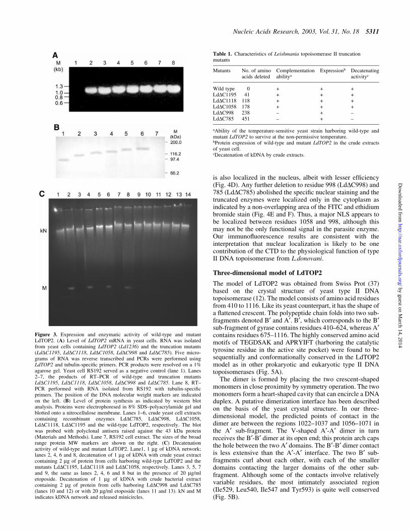
It is not known whether the inability to complement by someof the constructs is due to reduced gene expression or due toenhanced proteolysis of the gene products or due to synthesisof inactive proteins. RT±PCR analysis was performed usingspeci®c primers for the N-terminal 1.1 kb region of theLdTOP2 gene. Total RNA was isolated from the yeast cellsharboring the full-length and the truncated LdTOP2 con-structs. The yeast cell RS192 was taken as a negative control.RT±PCR performed with the primers for the housekeepingtubulin gene of S.cerevisiae served as a positive control. Asshown in Figure 3A, the transcripts from the yeast cellsharboring the wild-type and the mutant LdTOP2 wereexpressed at similar levels comparable with the housekeepingtubulin of S.cerevisiae. Western blot analysis using theantibody raised against the N-terminal 1.1 kb region ofLdTOP2 (43 kDa protein) shows that the wild-type anddifferent topoisomerase II mutant proteins were also expressedat similar levels (Fig. 3B). Our observations thus suggest thatthe level of transcripts does not correlate with the lack ofcomplementation.
The ability of an enzyme to complement a geneticde®ciency usually re¯ects the ability of the enzyme to performcatalysis. The recombinant enzymes LdTOP2, LdDC1195,LdDC1118 and LdDC1058 from crude yeast extracts were ableto perform decatenation of the kDNA in vitro. That theobserved decatenation of the kDNA was not due to nucleaseactivity was substantiated by the inhibition of decatenationactivity by etoposide, an eukaryotic topoisomerase II inhibitor(Fig. 3C). As the yeast cell harboring the constructs LdDC998and LdDC785 failed to survive at the non-permissivetemperature, the truncated proteins from LdDC998 andLdDC785 were expressed in E.coli and induced as describedin Materials and Methods. The crude bacterial extracts failedto decatenate the kDNA networks into minicircles. As thetruncated transcripts are expressed at similar levels, theinability to complement correlates well with the loss ofdecatenation activity of the parasite enzyme. The results ofcomplementation, protein expression and decatenatingactivity of the wild-type and mutant enzymes in yeast aresummarized in Table 1.
Distribution of wild-type and mutant L.donovanitopoisomerase II in yeast
Since topoisomerase II activity is required within the nucleusand it is known that the C-terminus of several eukaryotictopoisomerase II enzymes contains nuclear import signal, weexamined the ability of the wild-type and the mutant enzymesto be translocated to the nucleus. Indirect immuno¯uorescencewas carried out to show the distribution of the mutant proteinsand of the full-length LdTOP2 enzymes in yeast cells. Toeliminate the cross-reactivity of the antibody with the yeastTOP2, the yeast strain RS192 was taken as negative control.There was no detectable signal from control cells harboringpVT100U (data not shown). The only signal detected in cellsexpressing full-length, LdDC1195 and LdDC1118 enzymeswas from the nucleus (Fig. 4A±C), which is identi®ed byethidium bromide staining and the overlapping area of theFITC and the ethidium bromide stain. The enzyme LdDC1058
5308 Nucleic Acids Research, 2003, Vol. 31, No. 18
by guest on March 14, 2014
http://nar.oxfordjournals.org/D
ownloaded from

Figure 1. Alignment of LdTOP2 with yeast and human topoisomerases. Multiple alignment of topoisomerase II sequences from L.donovani (LdTOP2,GenBank accession no. AF150876), H.sapiens (HsTOP2, accession no. NM_001067) and S.cerevisiae (ScTOP2, accession no. NP_014311.1) usingCLUSTAL W. The amino acids are numbered on the top of sequences. Red indicates identity; blue indicates strong similarity and black indicatesdissimilarity. Hyphens represent the gap used to optimize alignment.
Nucleic Acids Research, 2003, Vol. 31, No. 18 5309
by guest on March 14, 2014
http://nar.oxfordjournals.org/D
ownloaded from

Figure 2. Schematic diagram of wild-type and truncated Leishmania TOP2 and functional complementation of a TOP2ts yeast strain. (A) Schematic diagram.A coordinate of the Leishmania TOP2 peptide (A) and the truncation mutants LdDC1195 (B), LdDC1118 (C), LdDC1058 (D), LdDC998 (E) and LdDC785 (F)are shown. The boxed region refers to the highly conserved sequences homologous to gyr B and gyr A subunits of bacterial gyrase. The non-conservedC-terminal region is represented by a thick line. Y indicates the location of the active site tyrosine. (B) Complementation assay. Saccharomyces cerevisiaeRS192 (TOP2-1) was transformed with a vector pVT100U and the vector carrying wild-type Leishmania TOP2 (LdDC1236) and truncation mutantsLdDC1195, LdDC1118, LdDC1058, LdDC998 and LdDC785, respectively. Transformed cells were streaked on solid synthetic minimal media and incubated at22 and 37°C. Between the panels, the names of the clones are indicated. Open arrowhead indicates the wild-type (1236) culture.
5310 Nucleic Acids Research, 2003, Vol. 31, No. 18
by guest on March 14, 2014
http://nar.oxfordjournals.org/D
ownloaded from
is also localized in the nucleus, albeit with lesser ef®ciency(Fig. 4D). Any further deletion to residue 998 (LdDC998) and785 (LdDC785) abolished the speci®c nuclear staining and thetruncated enzymes were localized only in the cytoplasm asindicated by a non-overlapping area of the FITC and ethidiumbromide stain (Fig. 4E and F). Thus, a major NLS appears tobe localized between residues 1058 and 998, although thismay not be the only functional signal in the parasite enzyme.Our immuno¯uorescence results are consistent with theinterpretation that nuclear localization is likely to be onecontribution of the CTD to the physiological function of typeII DNA topoisomerase from L.donovani.
Three-dimensional model of LdTOP2
The model of LdTOP2 was obtained from Swiss Prot (37)based on the crystal structure of yeast type II DNAtopoisomerase (12). The model consists of amino acid residuesfrom 410 to 1116. Like its yeast counterpart, it has the shape ofa ¯attened crescent. The polypeptide chain folds into two sub-fragments denoted B¢ and A¢. B¢, which corresponds to the B¢sub-fragment of gyrase contains residues 410±624, whereas A¢contains residues 675±1116. The highly conserved amino acidmotifs of TEGDSAK and APRYIFT (harboring the catalytictyrosine residue in the active site pocket) were found to besequentially and conformationally conserved in the LdTOP2model as in other prokaryotic and eukaryotic type II DNAtopoisomerases (Fig. 5A).
The dimer is formed by placing the two crescent-shapedmonomers in close proximity by symmetry operation. The twomonomers form a heart-shaped cavity that can encircle a DNAduplex. A putative dimerization interface has been describedon the basis of the yeast crystal structure. In our three-dimensional model, the predicted points of contact in thedimer are between the regions 1022±1037 and 1056±1071 inthe A¢ sub-fragment. The V-shaped A¢-A¢ dimer in turnreceives the B¢-B¢ dimer at its open end; this protein arch capsthe hole between the two A¢ domains. The B¢-B¢ dimer contactis less extensive than the A¢-A¢ interface. The two B¢ sub-fragments curl about each other, with each of the smallerdomains contacting the larger domains of the other sub-fragment. Although some of the contacts involve relativelyvariable residues, the most intimately associated region(Ile529, Leu540, Ile547 and Tyr593) is quite well conserved(Fig. 5B).
Figure 3. Expression and enzymatic activity of wild-type and mutantLdTOP2. (A) Level of LdTOP2 mRNA in yeast cells. RNA was isolatedfrom yeast cells containing LdTOP2 (Ld1236) and the truncation mutants(LdDC1195, LdDC1118, LdDC1058, LdDC998 and LdDC785). Five micro-grams of RNA was reverse transcribed and PCRs were performed usingLdTOP2 and tubulin-speci®c primers. PCR products were resolved on a 1%agarose gel. Yeast cell RS192 served as a negative control (lane 1). Lanes2±7, the products of RT±PCR of wild-type and truncation mutantsLdDC1195, LdDC1118, LdDC1058, LdDC998 and LdDC785. Lane 8, RT±PCR performed with RNA isolated from RS192 with tubulin-speci®cprimers. The position of the DNA molecular weight markers are indicatedon the left. (B) Level of protein synthesis as indicated by western blotanalysis. Proteins were electrophoresed in 8% SDS±polyacrylamide gel andblotted onto a nitrocellulose membrane. Lanes 1±6, crude yeast cell extractscontaining recombinant enzymes LdDC785, LdDC998, LdDC1058,LdDC1118, LdDC1195 and the wild-type LdTOP2, respectively. The blotwas probed with polyclonal antisera raised against the 43 kDa protein(Materials and Methods). Lane 7, RS192 cell extract. The sizes of the broadrange protein MW markers are shown on the right. (C) Decatenationactivity of wild-type and mutant LdTOP2. Lane1, 1 mg of kDNA network;lanes 2, 4, 6 and 8, decatenation of 1 mg of kDNA with crude yeast extractcontaining 2 mg of protein from cells harboring wild-type LdTOP2 and themutants LdDC1195, LdDC1118 and LdDC1058, respectively. Lanes 3, 5, 7and 9, the same as lanes 2, 4, 6 and 8 but in the presence of 20 mg/mletoposide. Decatenation of 1 mg of kDNA with crude bacterial extractcontaining 2 mg of protein from cells harboring LdDC998 and LdDC785(lanes 10 and 12) or with 20 mg/ml etoposide (lanes 11 and 13). kN and Mindicates kDNA network and released minicircles.
Table 1. Characteristics of Leishmania topoisomerase II truncationmutants
Mutants No. of aminoacids deleted
Complementationabilitya
Expressionb Decatenatingactivityc
Wild type 0 + + +LdDC1195 41 + + +LdDC1118 118 + + +LdDC1058 178 + + +LdDC998 238 ± + ±LdDC785 451 ± + ±
aAbility of the temperature-sensitive yeast strain harboring wild-type andmutant LdTOP2 to survive at the non-permissive temperature.bProtein expression of wild-type and mutant LdTOP2 in the crude extractsof yeast cell.cDecatenation of kDNA by crude extracts.
Nucleic Acids Research, 2003, Vol. 31, No. 18 5311
by guest on March 14, 2014
http://nar.oxfordjournals.org/D
ownloaded from

Comparison of the electrostatic potential of the entiremonomer of the two enzymes shows that the two proteins havenearly similar charge distribution (Fig. 6A and B). The modelreveals a span of 60 amino acids ranging from 998 to 1058(marked in red), at the C-terminus of the A¢ sub-fragment andshows a marked difference in the distribution of charge(Fig. 6C). This region of the parasite protein has a strong
positive potential as compared with its yeast counterpart. Theimportant feature highlighted in the model is the putativedimer interface of the Leishmania enzyme and a span of 60amino acids (998±1058) distinctly different from its yeastcounterpart.
Figure 4. Immuno¯uorescent localization of wild-type and C-terminallytruncated mutant enzymes of LdTOP2 expressed in RS192. Yeast cellstransformed with LdTOP2 wild-type and truncation constructs were pre-pared as spheroplasts, ®xed and probed with anti-LdTOP2 antiserum.Visualization of the bound primary antibody was done with FITC-conjugated anti-rabbit IgG. Ethidium bromide staining was subsequentlyperformed to highlight the nuclei and the area of the overlapping FITC andethidium bromide stain are shown in the merged pictures. Cells were viewedat an original magni®cation of 1003 under a Leica DM IRB inverted micro-scope. Images of cells expressing full-length L.donovani topoisomerase II(A), and mutants LdDC1195 (B), LdDC1118 (C), LdDC1058 (D), LdDC998(E) and LdDC785 (F) were captured and the ¯uorescent signal wasphotographed.
Figure 5. Three-dimensional structure of L.donovani topoisomerase II.(A) Space-®lling representation of LdTOP2 monomer. The conserved aminoacid motifs of TEGDSAK (blue) and APRYIFT (yellow) containing theactive site tyrosine have been depicted. The amino acids involved in nucleartranslocation are shown in pink. (B) The dimer formed by placing twoLdTOP2 monomers in close proximity by symmetry operation. OneLdTOP2 monomer is colored red and the other is colored yellow. The sub-fragment homologous to gyrase B and gyrase A is marked as B¢ and A¢,respectively. The primary A¢ putative dimer interface is indicated.
5312 Nucleic Acids Research, 2003, Vol. 31, No. 18
by guest on March 14, 2014
http://nar.oxfordjournals.org/D
ownloaded from

DISCUSSION
The nature of the multi-domain structure for eukaryotictopoisomerase II is revealed by many lines of evidence,including sequence comparison, mutagenesis and X-raycrystal structures (18,20). Although the topoisomerase IIgenes from the kinetoplastid parasites have been cloned andsequenced (24±26,29) very little is known about the charac-teristics of the protein. At ®rst sight, protozoan topoisomerasesappear to share many characteristics of their human homologs,but closer observation reveals that differences do exist.Alignment of the sequences of the type II DNA topoisomeraseof yeast, human and L.donovani has revealed conservation atthe N-terminal region of the enzyme and the conservationdecreases towards the CTD (Fig. 1). The CTDs are onlysimilar in sequences for closely related species and that too forthe ®rst half of the tail (29,46). A number of studies havefocused on identifying domains in eukaryotic TOP2 interact-ing speci®cally with anti-TOP2 agents (47). So, in this studywe have tried to assess the importance of the CTD of LdTOP2,which is the largest unconserved domain of topoisomerase II.
Deletion of conserved domains in human TOPIIa andS.cerevisiae TOP2 have led to inactive, non-complementingproteins indicating that all conserved domains are importantfor TOP2 function (15). Only a part of the CTD of humanTOPIIa, S.cerevisiae and Drosophila melanogaster is dis-pensable for complementation in S.cerevisiae (15,48). So, anumber of C-terminal truncation mutants of LdTOP2 deletedto amino acid residue 785 were constructed. Survival of atemperature-sensitive topoisomerase II mutant yeast strain inthe presence of LdTOP2 at the non-permissive temperature
should be all-or-nothing. None of the mutants beyond 1058amino acid residues were able to complement for thetemperature sensitivity of the mutant yeast strain (Fig. 2B).
The C-terminus of eukaryotic TOP2 contains manyregulatory elements like the phosphorylation sites (14,49),dimerization domain (15,16) and the NLSs (13). It is knownfor many genes that the deletion of sequences leads to unstablemessenger RNAs and may also affect protein expression orstability. To rule out the possibility that the observation madeon complementation ability was due to low expression orinstability of certain truncated transcripts, RT±PCR wasperformed (Fig. 3A). Transcripts of all the constructs wereobtained at a similar level showing that the full-length CTD ofLdTOP2 is not required for the stability of the transcript.Immunoblot experiments show the presence of only a singleband in the expected regions in all the crude extracts isolatedfrom recombinant yeast cells carrying wild-type and truncatedLeishmania topoisomerase II genes (Fig. 3B). Our dataindicate that all the wild-type and truncated LdTOP2 proteinsare expressed at similar levels showing that C-terminaldeletion does not alter the stability of the protein nor does itrender the protein susceptible to proteolytic degradation(Fig. 3B). Previous studies with human TOPIIa andS.cerevisiae TOP2 indicate that lack of complementationdoes not correlate with loss of catalytic activity (15).Our studies reveal that the LdTOP2 mutants which fail tocomplement the mutant yeast strain in vivo also failto decatenate kDNA in vitro. It is likely that the lack ofcatalytic activity of the C-terminal truncation mutantsLdDC998 and LdDC785 is due to the inability of the subunitto form dimers.
Figure 6. Comparison of electrostatic potential of type II DNA topoisomerase from Leishmania and yeast. The structure of residues 410±1116 of LdTOP2(A) and 410±1202 of yeast topoisomerase II (B) were selected for calculating the electrostatic potential. The orientation of both surface plots is the same.Acidic and basic amino acid residues are shown in red and blue, respectively, and the neutral amino acids are shown in white. The ®gures show a markeddifference in the charge distribution in the C-terminus of LdTOP2 and yeast TOP2. The amino acid residues between 998 and 1058 of LdTOP2 are highlybasic compared with its yeast counterpart, which is neutral. A space-®lling model of LdTOP2 monomer in the same orientation as (A) is shown in (C) andthe amino acid residues having different charge distributions are shown in red. The overall atom color depictions are as follows: carbon, green; nitrogen, blue;oxygen, red.
Nucleic Acids Research, 2003, Vol. 31, No. 18 5313
by guest on March 14, 2014
http://nar.oxfordjournals.org/D
ownloaded from
The C-terminus of the topoisomerase II contains NLSs.Alternatively, the C-terminus may be a regulatory domain or itmay provide a site for the interaction with DNA and RNA(50). The localization study was performed with the truncatedproteins to seek a potential function of the CTD. It was foundthat the full-length enzyme and the enzyme truncated to 1118residues are localized within the nucleus in the yeast cell,whereas the enzyme truncated to residue 1058 was onlypartially impaired with respect to nuclear translocation(Fig. 4A±D). However, the non-complementing enzymetruncated to residue 998 and 785, appeared to localize onlyin the cytoplasm, indicating its failure to be translocated to thenucleus in yeast (Fig. 4E and F). This observation suggests theexistence of a major NLS KRRRTRKIGL between amino acidresidues 998 and 1058. In human, a suggested nuclear signal(KKQTTLAFKPIKKGKKR) is located between residues1274 and 1290 (13). Identi®cation of a region containing theputative NLS in the highly variable C-terminal part ofLdTOP2 correlates well with the previous studies on type IIDNA topoisomerases from H.sapiens (13,15), mouse (42),S.pombe (19, 51), D.melanogaster (48) and S.cereviseae (52).
The model of LdTOP2 based on the available crystalstructure of yeast TOP2 (PDB accession no. 1BJT) shows thatthe parasite protein adopts an overall conformation similar toyeast TOP2 and is organized into two sub-fragments A¢ andB¢. All the secondary structural elements are conservedbetween the two proteins and are present in structurallyrelative positions (Fig. 5A). Placing the two monomers inclose proximity reveals a putative dimer interface in astructurally equivalent position to that of the crystallizedyeast TOP2 (Fig. 5B). The crystal structure reveals twodimerization regions where the primary dimer interface ispresent in the C-terminal part of the fragment involvingresidues 1031±1046 and 1114±1130. A biochemical approachidenti®es two regions spanning amino acid residues 1053±1069 and 1124±1143 essential for the dimerization of humanTOPIIa. Our model reveals a putative dimer interface, whichis formed between the amino acid residues 1022±1037 and1056±1071.
Although the mutant LdDC1058 contains one primarydimerization region and three amino acid residues of thesecond dimerization region, it can still complement a mutantyeast cell albeit with lesser ef®ciency and can decatenatekDNA in vitro. Since this region tolerates linker insertion inboth yeast and human (15), it indicates that this area mightconstitute a ¯exible structure. Taken together, our resultssuggest that the enzyme deleted to residue 998 cannotfunctionally complement the mutant yeast strain, as it cannotbe translocated to the nuclear compartment where the enzymeneeds to carry out its function.
The current drugs for leishmaniasis infection are inadequatedue to low ef®cacy or high toxicity and the problem is furthercompounded by the emergence of increasingly pentavalentantimonial drug-resistant parasites (53). The search for anef®cacious, less toxic, yet inexpensive drug, is the requirementof the day to ®ght against leishmaniasis. DNA topoisomeraseshave been established as important chemotherapeutic targetsfor various anticancer (54), antibacterial (55), antiviral (56)and antiparasitic agents (57). If a protein or a pathway isconserved between the mammalian host and the pathogen,some discriminating features that distinguish between the host
and the pathogen system must be identi®ed. Recently, fromour laboratory, various antileishmanial agents have beenreported which target the parasite topoisomerases (57,58). Forparasitic topoisomerases to be targeted selectively by speci®cagents, suf®cient differences should exist between them andtheir human homolog to enable them to be pharmacologicallydistinguishable. Our analysis of mutants truncated at theC-terminus of L.donovani indicates that the amino acidresidues between 998 and 1058 are very essential for theprotein in vivo. While deletion of more than 1058 amino acidresidues from the C-terminus results in complete loss oftopoisomerase function, mutants with shorter deletions such asLdDC1195 and LdDC1118 can functionally complement yeasttopoisomerase II in in vivo assays. The residues between 998and 1058, encompassing a span of 60 amino acids, reveal acritical point in the sequence of LdTOP2. A Blast searchshows that these 60 amino acid residues share no similaritywith the host TOP2 or any other eukaryotic protein. The levelof sequence and structure conservation between the host andthe parasite enzyme is high. However, there is a noteworthydifference in this region of the CTD.
Topoisomerase II is a highly potential drug target because ithas an indispensable function in cell biology and it lacksbiological redundancy. A future aspect of this work lies inexploiting the differences between the CTD of the host andparasite enzyme by integrating structural information withbiochemical experimentation, so as to delineate the commonand distinguishing feature of the host and parasite enzyme.This information will provide an insight for development ofnewer therapeutic agents with speci®c selectivity.
ACKNOWLEDGEMENTS
This paper is dedicated to the memory of Professor Amar NathBhaduri, former Director and Coordinator of the LeishmaniaProgram of this institute. We thank Professor S. Bhattacharya,the Director of our institute, for his interest in this work. Weare grateful to Professor Pratima Sinha of Bose Institute,Kolkata, India, for her valuable suggestions and expertise. Wealso acknowledge Dr Gayatri Tripathi for her help withconfocal microscopy. This work was supported by a grant(BT/PR2643/BRB/10/250/2001) from the Department ofBiotechnology, Government of India.
REFERENCES
1. Osheroff,N. (1989) Biochemical basis for interaction of type I and type IItopoisomerases with DNA. Pharmacol. Ther., 41, 223±241.
2. Wang,J.C. (1996) DNA topoisomerases. Annu. Rev. Biochem., 65,635±692.
3. Wang,J.C. (1998) Moving one DNA double helix through another by atype II DNA topoisomerase: the story of a simple molecular machine.Q. Rev. Biophys., 31, 107±144.
4. Wang,J.C (2002) Cellular roles of DNA topoisomerases: a molecularperspective. Nature Rev. Mol. Cell. Biol., 3, 430±40.
5. Rose,D., Thomas,W. and Holm,C. (1990) Segregation of recombinedchromosomes in meiosis I requires DNA topoisomerase II. Cell, 60,1009±1017.
6. Gasser,S.M., Laroche,T., Falzaet,J., Tour,E.B.D. and Laemmli,U.K.(1986) Metaphase chromosome structure involvement of topoisomeraseII. J. Mol. Biol., 188, 613±624.
7. Adachi,Y., Luke,M. and Laemmli,U.K. (1989) Preferential, cooperativebinding of DNA topoisomerase II to scaffold-associated regions.EMBO J., 8, 3997±4006.
5314 Nucleic Acids Research, 2003, Vol. 31, No. 18
by guest on March 14, 2014
http://nar.oxfordjournals.org/D
ownloaded from
8. Adachi,Y., Luke,M. and Laemmli,U.K. (1991) Chromosome assemblyin vitro: topoisomerase II is required for condensation. Cell, 64, 137±148.
9. Liu,L.F., Liu,C.C. and Alberts,B.M. (1979) T4 DNA topoisomerase: anew ATP-dependent enzyme essential for initiation of T4 bacteriophageDNA replication. Nature, 281, 456±461.
10. Gellert,M., Mizuuchi,K., O'Dea,M.H. and Nash,H.A. (1976) DNAgyrase: an enzyme that introduces superhelical turn into DNA. Proc. NatlAcad. Sci. USA, 73, 3872±3876.
11. Jenkins,J.R., Ayton,P., Jones,T., Davies,S.L., Simmons,D.L.,Harris,A.L., Sherr,D. and Hixon,I.D. (1992) Isolation of cDNA clonesencoding the beta isozyme of human DNA topoisomerase II andlocalisation of the gene to chromosome 3p24. Nucleic Acids Res., 20,5587±5592.
12. Berger,J.M., Gramblin,S.J., Harrison,S.C. and Wang,J.C. (1996)Structure and mechanism of DNA topoisomerase II. Nature, 379,225±232.
13. Dingwall,C. and Laskey,R.A. (1991) Nuclear targeting sequencesÐaconsensus? Trends Biochem. Sci., 16, 478±481.
14. Wells,N.J., Addison,C.N., Fry,A.M., Ganapathi,R. and Hixon,I.D. (1994)Serine 1524 is a major site of phosphorylation on human topoisomeraseII alpha protein in vivo and is a substrate for casein kinase II in vitro.J. Biol. Chem., 269, 29746±29751.
15. Jenson,S., Andersen,A.H., Kjeldsen,E., Biersack,H., Olsen,E.H.N.,Andersen,T.B., Westergaard,O. and Jakobsen,B.K. (1996) Analysis offunctional domain organisation in DNA topoisomerase II from humansand Saccharomyces cerevisiae. Mol. Cell. Biol., 16, 3866±3877.
16. Bjergbaek,L., Jensen,S., Westergaard,O. and Andersen,A.H. (1999)Using a biochemical approach to identify the primary dimerisationregions in human DNA topoisomerase II alpha. J. Biol. Chem., 274,26529±26536.
17. Austin,C.A., Marsh,K.L. Wasserman,R.A., Willmore,E., Sayer,P.J.,Wang,J.C. and Fisher,M.L. (1995). Expression, domain structure andenzymatic properties of an active recombinant human DNA topoisomerseII b. J. Biol. Chem., 270, 15739±15746.
18. Lee,M.P. and Hsieh,T.S. (1994) Linker insertion mutagenesis ofDrosophila topoisomerase II. Probing the structure of eukaryotictopoisomerase II. J. Mol. Biol., 235, 436±447.
19. Shiozaki,K. and Yanagida,M. (1991) A functional 125-kDa corepolypeptide of ®ssion yeast DNA topoisomerase II. Mol. Cell. Biol., 11,6093±6102.
20. Lindsley,J.E. and Wang,J.C. (1991) Proteolysis patterns of epitopicallylabeled yeast topoisomerase II suggest an allosteric transition in theenzyme induced by ATP binding. Proc. Natl Acad. Sci. USA, 88,10485±10489.
21. Reece,R.J. and Maxwell,A. (1991) The C-terminal domain of theEscherichia coli DNA gyrase A subunit is a DNA-binding protein.Nucleic Acids Res., 19, 1399±1405.
22. Roca,J. and Wang,J.C. (1992) The capture of a DNA double helix by anATP-dependent protein clamp: a key step in DNA transport by type IIDNA topoisomerases. Cell, 71, 833±840.
23. Beverly,S.M. (2003) Protozomics: trypanosomatid parasite geneticscomes of age. Nature Rev. Gen., 4, 11±19.
24. Strauss,P.R. and Wang,J.C. (1990) The TOP2 gene of Trypanosomabrucei: a single copy gene that shares extensive homology with otherTOP2 genes encoding eukaryotic DNA topoisomerase II. Mol. Biochem.Parasitol., 38, 141±150.
25. Fragosso,S.P. and Goldenberg,S. (1992) Cloning and characterisation ofthe gene encoding Trypanosoma cruzi DNA topoisomerase II. Mol.Biochem. Parasitol., 55, 127±135.
26. Pasion,S.G., Hines,J.C., Aebersold,R. and Ray,D.S. (1992) Molecularcloning and expression of the gene encoding the kinetoplast-associatedtype II DNA topoisomerase of Crithidia fasciculata. Mol. Biochem.Parasitol., 50, 57±68.
27. Chakraborty,A.K. and Majumder,H.K. (1987) Decatenation ofkinetoplast DNA by an ATP-dependent DNA topoisomerase from thekinetoplast hemo¯agellate Leishmania donovani. Mol. Biochem.Parasitol., 26, 215±224.
28. Chakraborty,A.K. and Majumder,H.K. (1991) An ATP independentcatenating enzyme from the kinetoplast hemo¯agellate Leishmaniadonovani. Biochem. Biophys. Res. Commun., 180, 279±285.
29. Das,A., Dasgupta,A., Sharma,S., Ghosh,M., Sengupta,T.,Bandopadhyay,S. and Majumder,H.K. (2001) Characterisation of thegene encoding type II DNA topoisomerase from Leishmania donovani:
a key molecular target in anti leishmanial therapy. Nucleic Acids Res.,29, 1844±1851.
30. Sherman,F. (1991) Getting started with yeast. Methods Enzymol., 194,3±21.
31. Vernet,T., Dignard,D. and Thomas,D.Y. (1987) A family of yeastexpression vectors containing the phage f1 intergenic region. Gene, 52,225±233.
32. Chen,D.C., Yang,B.C. and Kuo,T.T. (1992) One-step transformation ofyeast in stationary phase. Curr. Genet., 21, 83±84.
33. Okada,Y., Ito,Y., Kikuchi,A., Nimura,Y., Yoshida,S. and Suzuki,M.(2000) Assignment of functional amino acids around the active site ofhuman DNA topoisomerase II alpha. J. Biol. Chem., 275, 24630±24638.
34. Villa,H., Marcos,A., Reguera,R.M., Fouce,R.B., Estrada,C.G.,Pertejo,Y.P., Tekwani,B.L., Myler,P.J., Stuart,K.D., Bjornsti,M.A. andOrdonez,D. (2003) A novel active DNA topoisomerase I in Leishmaniadonovani. J. Biol. Chem., 278, 3521±3526.
35. Laemmli,U.K. (1970) Cleavage of structural proteins during theassembly of the head of bacteriophage T4. Nature, 227, 680±685.
36. Harlow,E. and Lane,D. (1988) Antibodies: A Laboratory Manual.Cold Spring Harbor Laboratory Press, Cold Spring Harbor, NY.
37. Guex,N. and Peitsch,M.C. (1997) SWISS-MODEL and Swiss PdbViewer: an environment for comparitive protein modeling.Electrophoresis, 18, 2714±2723.
38. Mandal,C. (1998) MODELYNÐA Molecular Modeling Programme.Version PC-1.0, Indian Copyright No. 9/98.
39. Koradi,R., Billeter,M. and Wuthrich,K. (1996) MOLMOL: a program fordisplay and analysis of macromolecular structures. J. Mol. Graph., 14,51±55, 29±32.
40. Thompson,J.D., Higgins,D.G. and Gibson,T.J. (1994) CLUSTAL W:improving the sensitivity of progressive multiple sequence alignmentthrough sequence weighting, position-speci®c gap penalties and weightmatrix choice. Nucleic Acids Res., 22, 4673±46809.
41. Adachi,N., Miyaike,M., Ikeda,H. and Kikuchi,A. (1992) Characterisationof cDNA encoding the mouse DNA topoisomerase II that cancomplement the budding yeast top2 mutation. Nucleic Acids Res., 20,5297±5303.
42. Adachi,N., Miyaike,M., Kato,S., Kanamaru,R., Koyama,H. andKikuchi,A. (1997) Cellular distribution of mammalian DNAtopoisomerase II is determined by its catalytically dispensable C-terminaldomain. Nucleic Acids Res., 25, 3135±3142.
43. Caron,P.R. and Wang,J.C. (1994) Appendix II: alignment of primarysequences of DNA topoisomerases. Adv. Pharmacol., 29B, 271±297.
44. Wasserman,R.A., Austin,C.A., Fisher,L.M. and Wang,J.C. (1993) Use ofyeast in the study of anticancer drugs targeting DNA topoisomerases:expression of a functional recombinant human DNA topoisomerase IIalpha in yeast. Cancer Res., 53, 3591±3596.
45. Wyckoff,E. and Hsieh,T.S. (1988) Functional expression of a Drosophilagene in yeast: genetic complementation of DNA topoisomerase II. Proc.Natl Acad. Sci. USA, 85, 6272±6276.
46. Champoux,J.J. (2001) DNA topoisomerases: structure, function andmechanism. Annu. Rev. Biochem., 70, 369±413.
47. Vassetzky,Y.S., Alghisi,G.C. and Gasser,S.M. (1995) DNAtopoisomerase II mutations and resistance to anti-tumor drugs. Bioessays,17, 767±774.
48. Crenshaw,D.G. and Hsieh,T.S. (1993) Function of the hydrophiliccarboxyl terminus of type II DNA topoisomerase from Drosophilamelanogaster. I. In vitro studies. J. Biol. Chem., 268, 21335±21343.
49. Cardenas,M.E., Dang,Q., Glover,C.V.C. and Gasser,S.M. (1992) Caseinkinase II phosphorylates the eukaryote-speci®c C-terminal domain oftopoisomerase II in vivo. EMBO J., 11, 1785±1796.
50. Rzepecki,R. and Fisher,P.A. (2000) During both interface and mitosis,DNA topoisomerase II interacts with DNA as well as RNA through theprotein's C-terminal domain. J. Cell Sci., 113, 1635±1647.
51. Shiozaki,K. and Yanagida,M. (1991) A functional 125-kDa polypeptideof ®ssion yeast DNA topoisomerase II. Mol. Cell. Biol., 11, 6093±6102.
52. Caron,P.R., Watt,P. and Wang,J.C. (1994) The C-terminal domain ofSaccharomyces cerevisiae DNA topoisomerase II. Mol. Cell. Biol., 14,3197±3207.
53. Sunder,S. (1998) Trial of oral miltefosine for visceral leishmaniasis.Lancet, 352, 1821±1823.
54. Kellner,U., Sehested,M., Jensen,P.B., Gieseler,F. and Rudolph,P. (2002)Culprit and victim-DNA topoisomerase II. Lancet Oncol., 3, 235±243.
55. Shen,L.L., Kuhlbrenner,W.E., Weigl,D. and Baranowski,J. (1989)Mechanism of quinolone inhibition of DNA gyrase. Appearance of
Nucleic Acids Research, 2003, Vol. 31, No. 18 5315
by guest on March 14, 2014
http://nar.oxfordjournals.org/D
ownloaded from
unique nor¯oxacin binding sites in enzyme±DNA complexes. J. Biol.Chem., 264, 2973±2978.
56. Hwang,Y., Rowley,D., Rhodes,D., Gertsch,J., Fenical,W. andBushman,F. (1999) Mechanism of inhibition of a pox virustopoisomerase by the marine natural product Sansalvamide. Mol.Pharmacol., 55, 1049±1053.
57. Mittra,B., RoyChowdhury,A. and Majumder,H.K (2000) Luteolin, anabundant dietary component is a potent antileishmanial agent that acts by
inducing topoisomerase II-mediated kinetoplast DNA cleavage leading toapoptosis. Mol. Med., 6, 527±541.
58. RoyChowdhury,A., Mandal,S., Goswami,A., Ghosh,M., Mandal,L.,Chakraborty,D., Ganguly,A., Tripathi,G., Mukhopadhyay,S.,Bandyopadhyay,S. and Majumder,H.K. (2003) Dihydrobetulinic acidinduces apoptosis in Leishmania donovani by targeting DNAtopoisomerase I and II: implications in antileishmanial therapy.Mol. Med., 9, 26±36.
5316 Nucleic Acids Research, 2003, Vol. 31, No. 18
by guest on March 14, 2014
http://nar.oxfordjournals.org/D
ownloaded from
Copyright © 2022 FDOKUMEN