Functional assessment of the mouse heart by MRI with a 1-min acquisition
22
Functional assessment of the mouse heart by MRI with a one-minute acquisition Guido Buonincontri 1* , MSc; Carmen Methner 2 , PhD; Thomas Krieg 2 , MD; T. Adrian Carpenter 1 , PhD; Stephen J. Sawiak 1,3 , PhD 1. Wolfson Brain Imaging Centre, Department of Clinical Neurosciences, University of Cambridge, Cambridge, UK 2. Department of Medicine, University of Cambridge, Cambridge, UK 3. Behavioural and Clinical Neuroscience Institute, University of Cambridge, Cambridge, UK * Corresponding author, address for correspondence: Wolfson Brain Imaging Centre Department of Clinical Neurosciences University of Cambridge School of Clinical Medicine Box 65 Cambridge Biomedical Campus Cambridge CB2 0SP Email: [email protected] Tel +44 1223 746463 Fax +44 1223 331826 Short title: Fast cine MRI of the mouse heart. Sponsor: Funding from the UK Medical Research Council (G.B. and S.J.S.), and the British Heart Foundation (PG/12/42/29655 to T.K.). Word count: 3620
Transcript of Functional assessment of the mouse heart by MRI with a 1-min acquisition

Functional assessment of the mouse heart by MRI with a one-minute
acquisition
Guido Buonincontri1*, MSc; Carmen Methner2, PhD; Thomas Krieg2, MD; T. Adrian
Carpenter1, PhD; Stephen J. Sawiak1,3, PhD
1. Wolfson Brain Imaging Centre, Department of Clinical Neurosciences,
University of Cambridge, Cambridge, UK
2. Department of Medicine, University of Cambridge, Cambridge, UK
3. Behavioural and Clinical Neuroscience Institute, University of Cambridge,
Cambridge, UK
* Corresponding author, address for correspondence:
Wolfson Brain Imaging Centre
Department of Clinical Neurosciences
University of Cambridge School of Clinical Medicine
Box 65 Cambridge Biomedical Campus
Cambridge CB2 0SP
Email: [email protected]
Tel +44 1223 746463
Fax +44 1223 331826
Short title: Fast cine MRI of the mouse heart.
Sponsor: Funding from the UK Medical Research Council (G.B. and S.J.S.), and the
British Heart Foundation (PG/12/42/29655 to T.K.).
Word count: 3620

2
ABSTRACT
Background: In vivo assessment of heart function in mice is important for basic and
translational research in cardiology. Magnetic resonance imaging (MRI) is an accurate
tool for investigating anatomy and function in the preclinical setting, however the long
scan duration limits its usage. We aimed to reduce the acquisition time of cine-MRI to
one minute.
Methods: We employed spatiotemporal compressed sensing and parallel imaging to
accelerate retrospectively-gated cine MRI. We compared the functional parameters
derived from full and undersampled data in Cartesian and radial MRI by means of
Bland-Altman plots.
Results: We found that the scan time for the whole heart could be reduced to two
minutes with Cartesian sampling and to one minute with radial sampling. Despite a
reduction in signal to noise ratio, the accuracy in the estimation of left and right
ventricular volumes was preserved for all tested subjects.
Conclusion: This method can be used to perform accurate functional MRI exams in
mice for high-throughput phenotyping or translational studies.
Keywords: heart, mouse, cine, MRI, compressed sensing, parallel imaging, function,
right ventricle

3
LIST OF ABBREVIATIONS ECG
FLASH FISP
LV LVM
LVCO LVEDV
LVEF LVESV
LVSV nuFFT
PCA POCS
RV RVCO
RVEDV RVEF
RVESV RVSV
SPIRiT
Electrocardiogram
Fast low angle shot Fourier imaging with steady state free precession
Left ventricle Left ventricular mass
Left ventricular cardiac output Left ventricular end diastolic volume
Left ventricular ejection fraction Left ventricular end systolic volume
Left ventricular stroke volume Non uniform fast Fourier transform
Principal components analysis Projection over convex sets
Right ventricle Right ventricular cardiac output
Right ventricular end diastolic volume Right ventricular ejection fraction
Right ventricular end systolic volume Right ventricular stroke volume
Iterative self-consistent parallel iterative reconstruction

4
INTRODUCTION
Mouse models are widely used in cardiovascular research. Genetically modified strains
and interventional models are useful for answering key questions in both basic and
translational studies (1). In this context, in vivo measures are important for accurate
assessment of heart morphology and function non-invasively and longitudinally. Today,
these are mostly derived with echocardiography (echo), micro computed tomography
(CT) or magnetic resonance imaging (MRI).
The most commonly used technique to measure heart function is two-dimensional echo.
This technique can be used for real-time acquisition in rodents for fast high throughput
screening, but is operator dependent and relies on geometrical assumptions of heart
geometry. Micro CT, on the other hand, is less user-dependent, and can still achieve
high throughput acquisition for heart function in three dimensions in less than 80
seconds (2). However, despite its promise, CT has not seen widespread adoption in
cardiac studies in rodents, perhaps due to concerns over the radiation dose involved.
Cine MRI is considered the gold standard measurement of systolic function (3),
producing images with anatomical detail superior to any other in vivo technology. MRI
can be used for assessment of the left as well as the right heart, and does not require
geometrical assumptions on heart shape and contraction (4). In addition, using contrast
agents for late gadolinium enhancement (5) and displacement encoding (6), MRI can
obtain tissue viability and muscle performance including stress and strains within the
same imaging session (7).
Despite its high accuracy, the application of cardiac MRI in mice has been limited in the
past by several challenges. Fast heart rates and small dimensions, together with the
difficulties in gating meant that this technology has lagged behind clinical MRI (8).

5
Most of these problems have now been resolved with the diffusion of the latest
generation of small-bore MRI systems and techniques such as retrospective gating (9).
Nevertheless, the procedure to assess function is still time consuming when compared
to echo or CT, imposing lengthy anaesthesia and limiting the number of animals that
can be done in each session. Long scan times are prohibitive to techniques such as
pharmacological stress testing, which can be used to boost sensitivity and specificity
during functional exams, and are important clinically (10). Speeding up the MRI
experiment for mice would therefore make preclinical experiments more similar to
human cardiac experiments and thus render the findings more translatable to the clinic,
allowing a full battery of testing to be performed.
In recent years, acceleration techniques such as parallel imaging and compressed
sensing have been successfully applied to cardiac MRI in clinical settings. Parallel
imaging uses multiple radiofrequency receivers to reduce the number of samples
required (11). Compressed sensing achieves the same goal exploiting redundancy of the
image in space or time by means of regularized iterative reconstruction (12). Although
these techniques have been used in clinical research studies, only a few reports so far
have attempted to accelerate cine MRI in rodents. Recent studies have used either
parallel imaging (13,14) or temporal compressed sensing (15), achieving 2-4 fold
acceleration. Spatiotemporal compressed sensing has previously been implemented in
the mouse heart using retrospective gating, in order to increase temporal resolution (16)
or accelerate the acquisition in the rat obtaining scan times of 20-30 seconds per slice
reconstructing a limited number of heart phases (8-16 per heart cycle) (17).

6
Here compressed sensing and parallel imaging are used in concert to accelerate cine-
MRI the mouse heart, comparing rectilinear and radial acquisition protocols. The effects
of acceleration are evaluated by comparing measured parameters of function.
MATERIALS AND METHODS
All components of this study were carried out in accordance with the UK Animals
(Scientific Procedures) Act, 1986, and with the approval of the University of Cambridge
Ethical Review Panel.
Five male C57/BL6 wildtype mice (20 weeks) were used. Animals were anaesthetised
with gaseous isoflurane both for induction (3% in 1l/min O2) and maintenance (1-1.5%
in 1l/min O2). A pressure sensor for respiration was used to monitor anaesthesia depth
with breathing rates maintained in the range 30-60 breaths per minute. ECG signals
were monitored with graphite electrodes. Body temperature was monitored using a
rectal thermometer and a flowing-water heating blanket was used to maintain animal
temperature at 37°C throughout the experiment.
Imaging was performed at 4.7T with a Bruker BioSpec 47/40 horizontal system
equipped with 400 mT/m gradients (Bruker Inc., Ettlingen, Germany). A birdcage coil
of 12cm was used for signal excitation and a four-channel mouse cardiac array for
signal reception (Bruker Inc., Ettlingen, Germany). Animals were positioned prone.
After the initial localizers, standard prospectively-gated cine MRI was acquired (18)
(for sequence details see Table 1).
Rectilinear and radial retrospectively gated FLASH cine-MRI were acquired on 1 mm
short-axis slices covering both ventricles using 8 slices with no gap. For the rectilinear
scheme, the sequence was modified to include a navigator after slice excitation (9). The

7
sequence was acquired with standard linear filling of k-space. In the radial scheme,
spokes were acquired spanning 360°, and the forward and reverse spokes were used to
estimate and correct gradient inaccuracies (19). Phase correction was applied
retrospectively (20), the k-space centre signal was used for navigation. Full sequence
parameters in each case are given in Table 1.
Retrospective gating was achieved by performing principal component analysis (PCA)
on the navigator signals, using all of the receivers. For the rectilinear case, data in the
navigator signals was concatenated across coils. For the radial case, the whole k-line
was concatenated across coils. The first component of the PCA was low-pass filtered to
extract cardiac gates. Prediction error on a band-pass Fourier filter was used to identify
and exclude respiration events. Cine-MRI images were reconstructed with 20 cardiac
phases.
Compressed sensing reconstruction was used for both radially- and rectilinearly-
acquired data, with a projection over convex sets (POCS) algorithm (21), implemented
in Matlab (Mathworks, USA). Each iteration of the algorithm included two data
consistency steps (acquired k-space consistency and SPIRiT consistency across
receivers in the image domain (22)) and two soft thresholding steps in sparse domains
(wavelet transform to take advantage of spatial compressibility; temporal Fourier
transform to exploit the periodicity of the heart cycle). In the radial case, the algorithm
employed implicit gridding with non-uniform fast Fourier transform (NuFFT (23)) to
avoid feedback of numerical errors. In each case, a reconstruction without applying
gating (grouping all data into a single time-frame) was used to estimate the SPIRiT
weights. All of our algorithms will be made available on request. Each of the library
functions used code from publicly-available open-source software.

8
First, reference data were acquired with 90 seconds acquisitions per each slice.
Undersampled datasets were obtained taking only the first part of the acquisition and
discarding the rest. For each acquisition strategy we reconstructed 6-fold accelerated
images (15 seconds per slice) and 12-fold accelerated images (7.5 seconds per slice), to
compare with the reference (90 seconds per slice). In order to obtain a general metric,
undersampling factors were measured for each frame: the number of lines acquired at
least once was divided by the number of lines required by the Nyquist limit. To obtain a
single value for the whole heart, these were averaged across phases and slices.
Standard functional parameters for the LV and the RV were derived using Segment v1.9
(24). Data were analysed by a single assessor blind to mouse identity. Reproducibility
of segmentation for fully-sampled data was measured as described in the supplement.
Agreement in the volume estimates between acquisition schemes and acceleration
factors was assessed by means of Bland-Altman statistics. To validate the self-gating
strategy, parameters from fully sampled Cartesian and radial acquisitions were
compared with standard prospectively gated scans using a t-test.
RESULTS
The heart rate was 454 ± 30 bpm (mean ± s.d.) during the examination. Using PCA, our
self-gating routine extracted the relevant information automatically for both radial and
rectilinear MRI (representative gating signals are shown in Supplementary Figure 1).
The gating procedure required minimal manual interaction to define the threshold for
rejection of respiratory events. No significant differences were found in measurements
using prospective gating and the two retrospectively-gated schemes (Table 2). Intra-
observer variability of standard prospective data is reported in Supplementary Table 1.

9
Undersampling factors achieved by each accelerated scheme are reported in Table 3.
The compressed sensing algorithm successfully anti-aliased the cine images, which
could be used to produce accelerated whole-heart stacks. Image quality of 6-fold
accelerated rectilinear MRI was sufficient for segmentation of the ventricles, while
residual aliasing corrupted the measurement on the 12-fold rectilinear acceleration
(Figure 1). Residual aliasing was mostly present in the slices with higher diastolic
inflow at the base of the heart, while mid-ventricular and apical slices were still artifact-
free. On the other hand, 12-fold accelerated radial images were alias-free throughout the
heart (Figure 2). Despite a reduction in signal to noise ratio due to the fewer datapoints
acquired it was possible to segment all the images.
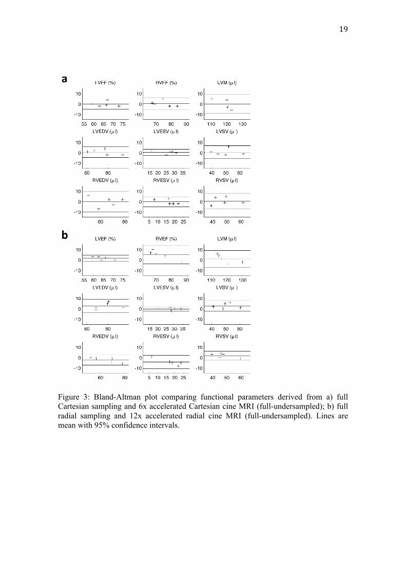
Bland-Altman plots for functional parameters are shown in Figure 3.
DISCUSSION
We have shown that imaging times for complete functional assessment of heart function
in mice can be accelerated to obtain an acquisition time of one minute. This work builds
on existing accounts accelerating cine imaging of the mouse heart by combining
compressed sensing and parallel imaging. Using a formulation permitting arbitrary k-
space trajectories, we acquired radial trajectories of k-space obtaining substantially
improved acquisition rates.
Importantly we have considered not only the visual quality of the images, but also the
quantification of the functional parameters. Functional parameters were derived from
the undersampled data with high accuracy, as agreement of accelerated data with
reference was similar to intra-observer variability. To assess an optimal regime to
achieve the required acceleration, we have compared the performance of rectilinear and

10
radial acquisition of k-space. Unlike the radial case, rectilinear sampling led to a bias in
measures caused by coherent aliasing. This is unsurprising as the radial data achieve a
less coherent sampling of k-space, producing noise-like aliasing, which is vital for the
application of compressed sensing.
Compared to prospective ECG gating, using navigators for retrospective gating is more
efficient and results in higher resistance to arrhythmias (9). Furthermore, the gating
procedure can be performed in post-processing and is less prone to human error.
Through PCA, the method described here made efficient use of all physiological
information at once, minimising operator interaction. Although the procedure for
obtaining the cine images is relatively straightforward, preparatory steps such as
positioning and imaging scouts are required for careful planning of the short-axis scan.
Nevertheless, landmarks such as the apex, the aorta and the LV wall can be easily and
quickly identified to guide the prescription of oblique short axis slices.
Parallel imaging and compressed sensing have been used to reconstruct images from
partial data. Importantly, the undersampled data used here were not synthetised, but
obtained from the first part of the acquisition, therefore demonstrating directly the
acceleration capabilities of this method. In order to utilise partial data, iterative
reconstruction must be employed. As this comes with a high computational burden,
especially when re-gridding steps are needed, at present reconstruction is performed off-
line. However, with multi-core computers and graphical processing units being
employed in scientific programming, strategies to perform these algorithms faster have
been proposed that may permit online reconstruction in the future (25). Although in our
case 1,440 spokes were used, any number of spokes exceeding the Nyquist limit could
have been used in the implementation.

11
Mouse models are widely used to explore heart function, remodelling, cell death and
proliferation from the single protein to the whole heart level (1). Fast in-vivo techniques
for assessing function of both ventricles are required, for longitudinal phenotyping and
meaningful translation of new treatments. In echocardiography, volumes are derived
from two-dimensional datasets making assumptions on ventricular shape based on a
geometrical model. This can potentially produce discrepancy in diseased hearts with
abnormal anatomy and function (26). Furthermore, it is difficult to apply such models
accurately to the right ventricle, due to its complex shape and contraction pattern. MRI
can derive those volumes directly from three-dimensional data. MRI is considered the
non-invasive reference standard (26) when accurate assessment of left or right
ventricular volumes is needed, and the method is particularly suited to longitudinal
studies as smaller sample sizes are needed compared to other techniques (27).
CONCLUSION
We have demonstrated a protocol to accelerate self-gated retrospective cine MRI in
mice using spatiotemporal compressed sensing and parallel imaging. This can be
performed with either Cartesian or radial MRI, utilising an optimum self-gating
strategy. We found that MRI assessment of left and right ventricular systolic function in
mice can be significantly accelerated maintaining high accuracy with an acquisition of
just one minute. Fast protocols such as these will enable high throughput phenotyping
or translational studies, including pharmacological stress experiments, favouring
refinement of preclinical studies and reduction of animal numbers.

12
REFERENCES
1. Yutzey KE, Robbins J. Principles of genetic murine models for cardiac disease.
Circulation 2007;115(6):792-799.

13
2. Dinkel J, Bartling SH, Kuntz J, Grasruck M, Kopp-Schneider A, Iwasaki M,
Dimmeler S, Gupta R, Semmler W, Kauczor HU, Kiessling F. Intrinsic gating
for small-animal computed tomography: a robust ECG-less paradigm for
deriving cardiac phase information and functional imaging. Circulation
Cardiovascular imaging 2008;1(3):235-243.
3. Stuckey DJ, Carr CA, Tyler DJ, Clarke K. Cine-MRI versus two-dimensional
echocardiography to measure in vivo left ventricular function in rat heart. Nmr
in Biomedicine 2008;21(7):765-772.
4. Wiesmann F, Frydrychowicz A, Rautenberg J, Illinger R, Rommel E, Haase A,
Neubauer S. Analysis of right ventricular function in healthy mice and a murine
model of heart failure by in vivo MRI. American journal of physiology Heart
and circulatory physiology 2002;283(3):H1065-1071.
5. Buonincontri G, Methner C, Krieg T, Carpenter TA, Sawiak SJ. A fast protocol
for infarct quantification in mice. Journal of magnetic resonance imaging : JMRI
2013; 38(2):468-73
6. Gilson WD, Yang Z, French BA, Epstein FH. Complementary displacement-
encoded MRI for contrast-enhanced infarct detection and quantification of
myocardial function in mice. Magnetic resonance in medicine : official journal
of the Society of Magnetic Resonance in Medicine / Society of Magnetic
Resonance in Medicine 2004;51(4):744-752.
7. Buonincontri G, Methner C, Krieg T, Hawkes RC, Adrian Carpenter T, Sawiak
SJ. PET/MRI assessment of the infarcted mouse heart. Nuclear Instruments and
Methods in Physics Research Section A: Accelerators, Spectrometers, Detectors
and Associated Equipment 2014;734(0):152-155.

14
8. Price AN, Cheung KK, Cleary JO, Campbell AE, Riegler J, Lythgoe MF.
Cardiovascular magnetic resonance imaging in experimental models. The open
cardiovascular medicine journal 2010;4:278-292.
9. Heijman E, de Graaf W, Niessen P, Nauerth A, van Eys G, de Graaf L, Nicolay
K, Strijkers GJ. Comparison between prospective and retrospective triggering
for mouse cardiac MRI. Nmr in Biomedicine 2007;20(4):439-447.
10. van Rugge FP, van der Wall EE, de Roos A, Bruschke AV. Dobutamine stress
magnetic resonance imaging for detection of coronary artery disease. Journal of
the American College of Cardiology 1993;22(2):431-439.
11. Deshmane A, Gulani V, Griswold MA, Seiberlich N. Parallel MR imaging.
Journal of magnetic resonance imaging : JMRI 2012;36(1):55-72.
12. Tsao J, Kozerke S. MRI temporal acceleration techniques. Journal of magnetic
resonance imaging : JMRI 2012;36(3):543-560.
13. Ratering D, Baltes C, Dorries C, Rudin M. Accelerated cardiovascular magnetic
resonance of the mouse heart using self-gated parallel imaging strategies does
not compromise accuracy of structural and functional measures. Journal of
cardiovascular magnetic resonance : official journal of the Society for
Cardiovascular Magnetic Resonance 2010;12:43.
14. Schneider JE, Lanz T, Barnes H, Medway D, Stork LA, Lygate CA, Smart S,
Griswold MA, Neubauer S. Ultra-fast and accurate assessment of cardiac
function in rats using accelerated MRI at 9.4 Tesla. Magnetic resonance in
medicine : official journal of the Society of Magnetic Resonance in Medicine /
Society of Magnetic Resonance in Medicine 2008;59(3):636-641.

15
15. Wech T, Lemke A, Medway D, Stork LA, Lygate CA, Neubauer S, Kostler H,
Schneider JE. Accelerating cine-MR imaging in mouse hearts using compressed
sensing. J Magn Reson Imaging 2011;34(5):1072-1079.
16. Motaal AG, Coolen BF, Abdurrachim D, Castro RM, Prompers JJ, Florack LM,
Nicolay K, Strijkers GJ. Accelerated high-frame-rate mouse heart cine-MRI
using compressed sensing reconstruction. NMR Biomed 2012. doi:
10.1002/nbm.2883
17. Montesinos P, Abascal JF, Cusso L, Vaquero JJ, Desco M. Application of the
compressed sensing technique to self-gated cardiac cine sequences in small
animals. Magnetic resonance in medicine : official journal of the Society of
Magnetic Resonance in Medicine / Society of Magnetic Resonance in Medicine
2013. doi: 10.1002/mrm.24936
18. Buonincontri G, Methner C, Carpenter TA, Hawkes RC, Sawiak SJ, Krieg T.
MRI and PET in mouse models of myocardial infarction. Journal of visualized
experiments : JoVE 2013;in press(82):e50806.
19. Block KT, Uecker M. Simple Method for Adaptive Gradient-Delay
Compensation in Radial MRI. Proceedings of the 19th ISMRM (2011).
20. Moussavi A, Untenberger M, Uecker M, Frahm J. Correction of gradient-
induced phase errors in radial MRI. Magnetic resonance in medicine : official
journal of the Society of Magnetic Resonance in Medicine / Society of Magnetic
Resonance in Medicine 2014;71(1):308-312.
21. Bauschke HH, Borwein JM. Projection algorithms for solving convex feasibility
problems. Siam Rev 1996;38(3):367-426.

16
22. Lustig M, Pauly JM. SPIRiT: Iterative self-consistent parallel imaging
reconstruction from arbitrary k-space. Magnetic resonance in medicine : official
journal of the Society of Magnetic Resonance in Medicine / Society of Magnetic
Resonance in Medicine 2010;64(2):457-471.
23. Fessler JA, Sutton BP. Nonuniform fast Fourier transforms using min-max
interpolation. Ieee T Signal Proces 2003;51(2):560-574.
24. Heiberg E, Sjogren J, Ugander M, Carlsson M, Engblom H, Arheden H. Design
and validation of Segment--freely available software for cardiovascular image
analysis. BMC Med Imaging 2010;10:1. doi: 10.1186/1471-2342-10-1
25. Nam S, Akcakaya M, Basha T, Stehning C, Manning WJ, Tarokh V, Nezafat R.
Compressed sensing reconstruction for whole-heart imaging with 3D radial
trajectories: a graphics processing unit implementation. Magnetic resonance in
medicine : official journal of the Society of Magnetic Resonance in Medicine /
Society of Magnetic Resonance in Medicine 2013;69(1):91-102.
26. Constantine G, Shan K, Flamm SD, Sivananthan MU. Role of MRI in clinical
cardiology. Lancet 2004;363(9427):2162-2171.
27. Bellenger NG, Davies LC, Francis JM, Coats AJ, Pennell DJ. Reduction in
sample size for studies of remodeling in heart failure by the use of
cardiovascular magnetic resonance. Journal of cardiovascular magnetic
resonance : official journal of the Society for Cardiovascular Magnetic
Resonance 2000;2(4):271-278.

17
Figures
Figure 1: Mid-ventricular short-axis slice comparing full sampling, 6x and 12x acceleration for Cartesian and radial sampling. Each image is reconstructed both by applying straightforward transformation to zero-filled data and iterative reconstruction including compressed sensing and parallel imaging. Scale bar is 1 mm.

18
Figure 2 Comparison between fully sampled and 12x accelerated radial MRI at different levels in the LV. Although the signal to noise ratio is lower in the 12x images due to fewer datapoints acquired, the anatomical detail is preserved for functional examination as aliasing has been successfully removed. Scale bar is 1 mm.
19
Figure 3: Bland-Altman plot comparing functional parameters derived from a) full Cartesian sampling and 6x accelerated Cartesian cine MRI (full-undersampled); b) full radial sampling and 12x accelerated radial cine MRI (full-undersampled). Lines are mean with 95% confidence intervals.

20
Tables
Sequence FISP FLASH FLASH
Scheme Cartesian Cartesian radial
Gating prospective retrospective retrospective
TR/TE 6.0 / 2.1 ms 4.8 / 1.7 ms 5.6 / 1.1 ms
FOV 3.5 × 3.5 cm2 3.5 × 3.5 cm2 3.84 × 3.84 cm2
Echo position 50 % 15 % 5 %
Matrix size 256 × 256 192 × 192 256 × 1440
Rec BW 78 kHz 100 kHz 100 kHz
Pulse flip angle 20°
BW 5.4 kHz
flip angle 20°
BW 18 kHz
flip angle 20°
BW 14 kHz
Repetitions 15-20 frames,
1 NEX
100 repetitions 12 repetitions
Table 1 Sequence parameters.

21
FISP
FISP – Rectilinear
FLASH
FISP – Radial
FLASH
LVEDV (µl) 79 ± 7 -0.8 ± 4.9 3.0 ± 5.3
LVESV (µl) 27 ± 3 -0.4 ± 3.0 1.6 ± 2.5
LVSV (µl) 51 ± 6 -0.4 ± 3.3 1.4 ± 3.8
LVEF (%) 65 ± 4 -0.2 ± 2.8 -0.4 ± 2.3
Table 2: Differences between functional parameters obtained from prospectively-gated Cartesian FISP (reported in the first column) and retrospectively gated scans: rectilinear and radial FLASH (mean ± s.d.). No significant differences were found between prospective and retrospective gating.

22
K-space acquisition
Acceleration factor
Undersampling at 20 frames (% Nyquist)
Scan time per slice / s
Cartesian 1 98 ± 2 90
Cartesian 6 48 ± 9 15
Cartesian 12 27 ± 6 7.5
Radial 1 138 ± 16 90
Radial 6 28 ± 3 15
Radial 12 14 ± 2 7.5
Table 3 Measured undersampling (mean ± standard deviation) and scan time for each acceleration factor used.