Controle de qualidade dos dados de radiossondagem da campanha WET-AMC/LBA
Upload
independentCategory
view
1download
0
CH A P T E R 11Functional and morphological comparison of three primary liver cell types cultured in the AMC bioartificial liver
Paul P.C. Poyck1 Ruurdtje Hoekstra1,2
Albert C.W.A. van Wijk1
Chiara Attanasio3,4
Fulvio Calise3,4
Robert A.F.M. Chamuleau5
Thomas M. van Gulik1
1 Dept. of Surgery (Surgical Laboratory), Academic Medical Center, Amsterdam, the Netherlands;2 AMC Liver Center, Academic Medical Center, Amsterdam, the Netherlands;3 Liver Transplantation Unit, A. Cardarelli Hospital, Naples, Italy4 Centro Biotechnologie, A. Cardarelli Hospital, Naples, Italy5 Dept. of Gastroenterology and Hepatology, Academic Medical Center, Amsterdam, the Netherlands.
Liver Transplantation 2007;13(4):589-598
Chapter 11
218
Abstract
The selection of a cell type for bioartificial liver (BAL) systems for the treatment of patients with acute liver failure is in part determined by issues concerning patient safety and cell availability. Consequently, mature porcine hepatocytes (MPHs) have been widely applied in BAL systems. The success of clinical BAL application systems is, however, largely dependent on the functionality and stability of hepatocytes. Therefore, we compared herein the general metabolic and functional activities of MPHs with mature human hepatocytes (MHHs) in the AMC-BAL during a 7 day culture period. We also tested fetal human hepatocytes (FHHs), since their proliferation capacity is higher than MHHs as well as their function increased compared to human liver cell lines. The results showed large differences between the three cell types. MHHs eliminated 2-fold more ammonia and produced 3-fold more urea than MPHs, whereas FHHs produced ammonia. Lidocaine elimination of FHHs was 3.5-fold higher than MPHs and 6.6-fold higher than of MHHs. Albumin production was not different between the three cell types. MPHs and FHHs became increasingly glycolytic, whereas MHHs remained metabolically stable during the whole culture period. MHHs and MPHs formed tissue-like structures inside the AMC-BAL. We propose that FHHs can be considered as a suitable cell type for pharmacological studies inside a bioreactor. However, we conclude that MHHs are the preferred cell source for loading a BAL device for clinical use, because of their high ammonia eliminating capacity and metabolic stability. MPHs should be considered as the best alternative cell source for BAL application, although their phenotypic instability urges application within one or two days after loading.
Three primary liver cell types cultured in the AMC-BAL
219
Introduction
Chronic liver disease is responsible for over 1.4 million disability adjusted life years annually1 and ranks in the USA among the top seven disease related causes of death between the age of 25 and 64 years.2 For end-stage liver failure, orthotopic liver transplantation remains the current treatment of choice. However, patients suffering from acute or acute-on-chronic liver failure (ALF; AoCLF) may benefit from temporary extracorporeal artificial liver support used to tide them over till transplantation or to allow regeneration of their own liver to occur. Bioartificial liver (BAL) support systems are promising, as these systems rely on detoxification and synthetic function of liver cells.3 Over the last two decades, many BAL systems have been devised, of which only eight systems have found application in a clinical setting.4
Many liver cell types have been used for pre-clinical and clinical application of BAL devices. The choice for the optimal cell source for BAL support devices is, however, still a matter of debate as the ideal set of criteria cannot be combined in one single cell type.4-10 Particularly, the availability, degree of liver specific function and safety aspects are ongoing issues.
Primary hepatocytes, either allogeneic or xenogeneic, have an excellent function and are therefore favorite cell sources for BAL application. However, mature porcine hepatocytes (MPHs) are not attractive for clinical application because of risks related to xenotransplantation.11 Mature human hepatocytes (MHHs), on the other hand, are scarce, as their sources are limited to either discarded donor livers or small parts obtained during liver resections.12,13 Human hepatic cell lines (HHCLs) have the advantage of an infinite proliferation capacity and can potentially serve as a stable cell source. However, the lack of sufficient liver-specific functions limits their use for clinical BAL application. Alternatively, fetal human hepatocytes (FHHs) can be considered as an attractive human liver cell source. These cells have two advantages, i.e. 1. a much higher proliferation capacity than MHHs and 2. increased liver-specific functions, in some respects higher than human liver cell lines14, showing even further increase with aging of the fetus.15
So far, the two most frequently used cell sources for BAL application, i.e. MHHs and MPHs, have not been analyzed in a clinically applied BAL system in one single study. Furthermore, FHHs have not been investigated in the setting of BAL using a three-dimensional perfused culture system and subsequently compared with MHHs and MPHs. The aim of this study therefore, was to compare hepatic functionality and metabolism of three types of primary hepatocytes in a for laboratory testing, down-scaled version of the AMC-BAL.16-19 The three cell types used were 1. mature porcine hepatocytes (MPHs), as this cell type has been widely used for pre-clinical and clinical BAL application; 2. mature human hepatocytes (MHHs) isolated from liver resection material, as this cell type displays highest physiological function and therefore is considered as gold standard;
Chapter 11
220
and 3. human fetal hepatocytes (FHHs) isolated from fetuses at the highest, yet acceptable age (23-24 weeks). Function of these three cell types was assessed for up to 7 days and the general tissue architecture in full transverse sections of the laboratory-scale AMC-BAL bioreactor was examined after termination of the culture period.
Materials and Methods
All procedures were conducted in accordance with the institutional guidelines, either by the Animal Ethical Committee or Medical Ethical Committee of the Academic Medical Center and the Cardarelli Hospital. Human material was collected after written consent and used when serological tests were negative for human immunodeficiency virus (HIV), hepatitis B virus (HBV) and hepatitis C virus (HCV).
Isolation procedures of primary hepatocytes
Mature porcine hepatocyte (MPH) isolation. MPHs were isolated from livers of female pigs (20-24 kg) by a two-step collagenase perfusion technique according to a modified protocol of Seglen20 as previously described.21 MPHs were suspended in ice-cold WE culture medium, consisting of William’s E medium supplemented with 10% (v/v) heat-inactivated fetal bovine serum (BioWhittaker, Verviers, Belgium), 2mM glutamine, 1 μM dexamethason, 20m U/mL insulin, 2 mM ornithine, 100 μg/mL streptomycin, 100 U/mL penicillin and 0.25 μg/mL fungizone. Total yield of isolated hepatocytes was estimated by determination of the cell pellet volume after 3-times of centrifugation at 50xg for 3 min.21 Viability was determined by trypan blue exclusion test using a Bürker Bright line cytometer (Optik Labor) (Table 1).
Mature human hepatocyte (MHH) isolation. Human liver tissue was collected from patients who underwent partial hepatectomy for various reasons (Table 1). Hepatocytes were isolated by a modified two-step collagenase perfusion technique as described by Hoekstra et al.22 The cells were suspended in ice-cold William’s E medium (as MPH isolation), and their quantity and viability were determined by trypan blue exclusion test, using a Bürker Bright line cytometer (Optik Labor) (Table 1).
Fetal human hepatocyte (FHH) isolation. Human fetal livers were obtained from abortions of 23-24 weeks old fetuses suffering from congenital disorders irreconcilable with life (Table 1). One fetus died in utero 30 min before the partus, whereas two fetuses died shortly after parturition. After hepatectomy, fetal livers were preserved on Celsior at 4oC and transported under sterile conditions from Naples (Italy) to Amsterdam (The Netherlands). Fetal livers were washed with Hank’s Balanced Salt Solution (HBSS; BioWhittaker) to remove Celsior, and weighed. All extra-hepatic tissue was dissected. Fetal livers were then cut into small pieces and subsequently digested in 50 mL 0.03%
Three primary liver cell types cultured in the AMC-BAL
221
collagenase-P (Roche) HBSS solution for 15 min at 37°C. The first cell suspension was sieved through a surgical gauze, whereas the residual fetal tissue was subjected to a second digestion by collagenase for 15 min. After sieving a second time, both cell suspensions were combined and centrifuged at 50x g for 3 min at 4oC and collected in 25 mL ice-cold DMEM culture medium (Dulbecco’s modified Eagle’s medium, BioWhittaker) containing 10% heat-inactivated fetal bovine serum (HI-FBS, BioWhittaker), 2 mM L-glutamine (BioWhittaker), 1 μM dexamethason (Sigma), 10 μg/mL insulin, 5.5 μg/mL transferrin, 6.7 ng/mL selenium-X (ITS mix, Life Technology), 100 U/mL penicillin, 100 μg/mL streptomycin (penicillin/streptomycin mix, BioWhittaker). Quantity of cells and viability was determined by trypan blue exclusion test using a Bürker Bright line cytometer (Optik Labor) (Table 1).
Bioreactor
We used the laboratory-scale AMC-BAL bioreactor, which is a 10x down-scaled bioreactor of the second generation AMC-BAL with an internal volume of 55 mL.21 The general configuration of the bioreactor, as described in detail by Flendrig et al.16,17, consists of a polycarbonate housing containing a three dimensional non-woven hydrophilic polyester matrix circularly wound around a polycarbonate core. Between the matrix layers, hydrophobic polypropylene gas capillaries are situated in a parallel fashion of which the ends are embedded in polyurethane resin and fitted with gas inlet and outlet caps.
Hepatocyte culture
Hepatocytes were injected under sterile conditions into the bioreactor through three different loading ports using a 60 mL syringe containing a final volume of 50 mL culture medium for all cell types. Bioreactors were then placed in an incubator at 37oC and oxygenated with sterile 95% air and 5% CO2 at a flow rate of 150 mL/min during the whole experiment. To ensure optimal cell attachment and an even cell distribution, bioreactors charged with MHHs and MPHs were were rotated 340o (back and forth) according the longitudinal plane and through the gravitational field at 1 revolution/min for two hours, whereas bioreactors with FHHs were rotated for four hours. After this attachment period, dead and unattached cells were removed by flushing 100 mL of fresh WE culture medium through the bioreactor at 15 mL/min. The bioreactors were then continuously perfused overnight with 150 mL recirculating WE culture medium at 15 mL/min.
Hepatocyte function tests
Each test sequence consisted of an oxygen consumption test followed by a function test, as described previously.21 Briefly, oxygen consumption was determined by measuring the decrease in oxygen tension during the first 15 min after closure of the oxygen
Chapter 11
222
supply to the bioreactor. A function test was performed by flushing the bioreactor with 100 mL of test medium composed of WE culture medium supplemented with 500 μg/mL lidocaine, 2 mM L-lactate with either 5 mM NH4Cl for MPH and MHH bioreactors or 0.5 mM NH4Cl for FHH bioreactors, followed by recirculation of 100 mL test medium for two hours at 37oC. Samples were taken at 30, 60, 90 and 120 min, respectively, and subsequently analyzed for concentrations of ammonia, urea, lidocaine, porcine albumin, glucose and lactate as well as activities of aspartate aminotransferase (AST) and lactate dehydrogenase (LDH), as described previously.21 Human albumin was determined by ELISA using goat-anti-human serum albumin antibody (1:100, Abcam, ab8940;) and HRP conjugated rabbit-anti-human serum albumin (1:5000, Abcam, ab7394). Ammonia and lidocaine elimination, urea and albumin production capacity, AST and LDH release, glucose and lactate consumption and/or production rates were determined by calculating the changes in concentration in test medium per hour per billion cells loaded in the bioreactor.
MPH bioreactors (n=15) were tested at day 1 (n=15), 3 (n=11), 5 (n=10) and 7 (n=3). MHH (n=5) and FHH (n=3) bioreactors were all tested at day 1, 3, 5 and 7, respectively.
Bioreactor tissue preparation for histological analysis
Bioreactors were fixed in 10% formalin after the final function test and stored at 4oC. Complete transverse 8 μm sections of the laboratory-scale bioreactor (ØID 22 mm) were obtained after embedding the whole bioreactor in paraffin as follows. Fixed bioreactors were manually flushed with an increasing ethanol series, i.e. 100 mL per flush at 37oC; 1x 50% (v/v), 1x 70% (v/v), and 4x with 100% ethanol (v/v) (Merck). After dehydration, bioreactors were flushed three times with 100 mL xylene (Merck) at 37oC and followed by a single flush of 100 mL paraffin (Variwax, Klinipath) at 62oC. After paraffin embedding, bioreactor housings were removed by milling, leaving an intact roll of the paraffinized matrix and capillaries. This roll was then transversely sliced into 1 cm discs, of which each disc was inserted in stainless steel cylinders (ØID 22 mm, 1 cm wide) with openings at both sides. Each cylinder was then placed in a 25 mL beaker containing pre-warmed paraffin and incubated for 12 hours under vacuum at 62oC to remove all air. Finally, and after cooling, each bioreactor disc was embedded in a mold and made ready for sectioning.
All sections were stained with hematoxylin and eosin (HE) to evaluate tissue architecture and organization.
Statistical analysis
Statistical analysis was performed by using SPSS 12.0.1 for Windows software (SPSS Inc., Chicago, IL, USA). Results are reported as means + standard error (SE). Repeated
Three primary liver cell types cultured in the AMC-BAL
223
measurement ANOVA rank tests were used to compare the three cell types over the 7-day culture period, and to compare differences per day within one cell type. Significance was reached if p < 0.05. Prism version 4.0 (GraphPad Prism Inc, San Diego, CA, USA) was used for graphical presentation of the data.
Results
Cell sources
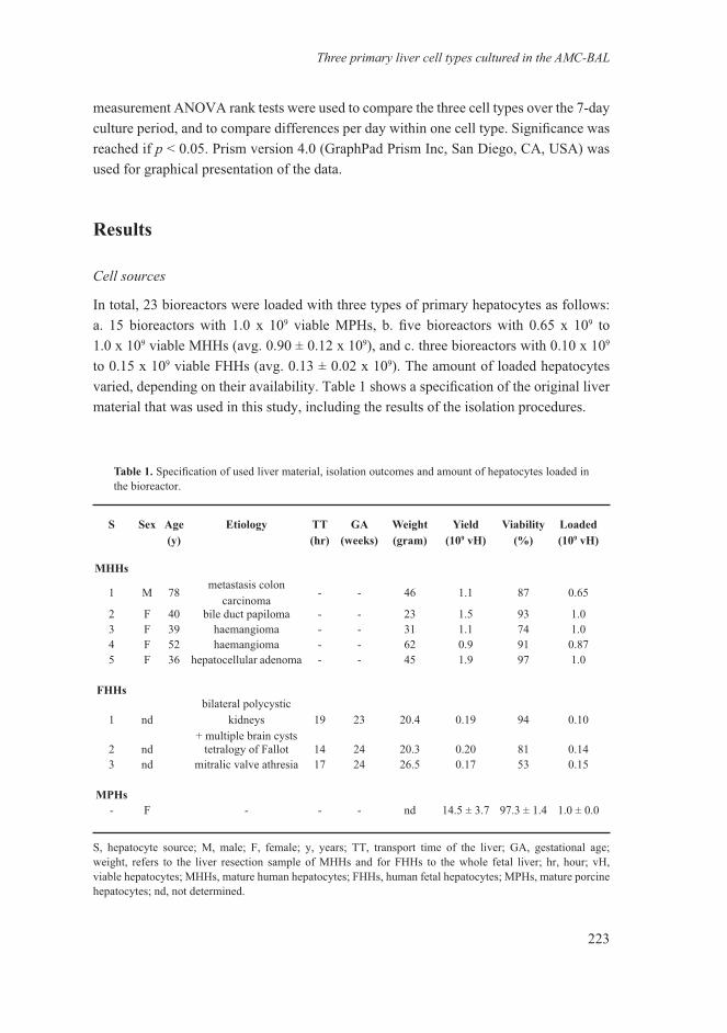
In total, 23 bioreactors were loaded with three types of primary hepatocytes as follows: a. 15 bioreactors with 1.0 x 10⁹ viable MPHs, b. five bioreactors with 0.65 x 10⁹ to 1.0 x 10⁹ viable MHHs (avg. 0.90 ± 0.12 x 10⁹), and c. three bioreactors with 0.10 x 10⁹ to 0.15 x 10⁹ viable FHHs (avg. 0.13 ± 0.02 x 10⁹). The amount of loaded hepatocytes varied, depending on their availability. Table 1 shows a specification of the original liver material that was used in this study, including the results of the isolation procedures.
S Sex Age(y)
Etiology TT(hr)
GA(weeks)
Weight(gram)
Yield(109 vH)
Viability(%)
Loaded(109 vH)
MHHs
1 M 78metastasis colon
carcinoma- - 46 1.1 87 0.65
2 F 40 bile duct papiloma - - 23 1.5 93 1.03 F 39 haemangioma - - 31 1.1 74 1.04 F 52 haemangioma - - 62 0.9 91 0.875 F 36 hepatocellular adenoma - - 45 1.9 97 1.0
FHHs
1 ndbilateral polycystic
kidneys+ multiple brain cysts
19 23 20.4 0.19 94 0.10
2 nd tetralogy of Fallot 14 24 20.3 0.20 81 0.143 nd mitralic valve athresia 17 24 26.5 0.17 53 0.15
MPHs- F - - - nd 14.5 ± 3.7 97.3 ± 1.4 1.0 ± 0.0
S, hepatocyte source; M, male; F, female; y, years; TT, transport time of the liver; GA, gestational age; weight, refers to the liver resection sample of MHHs and for FHHs to the whole fetal liver; hr, hour; vH, viable hepatocytes; MHHs, mature human hepatocytes; FHHs, human fetal hepatocytes; MPHs, mature porcine hepatocytes; nd, not determined.
Table 1. Specification of used liver material, isolation outcomes and amount of hepatocytes loaded in the bioreactor.
Chapter 11
224
Hepatocyte-specific functions
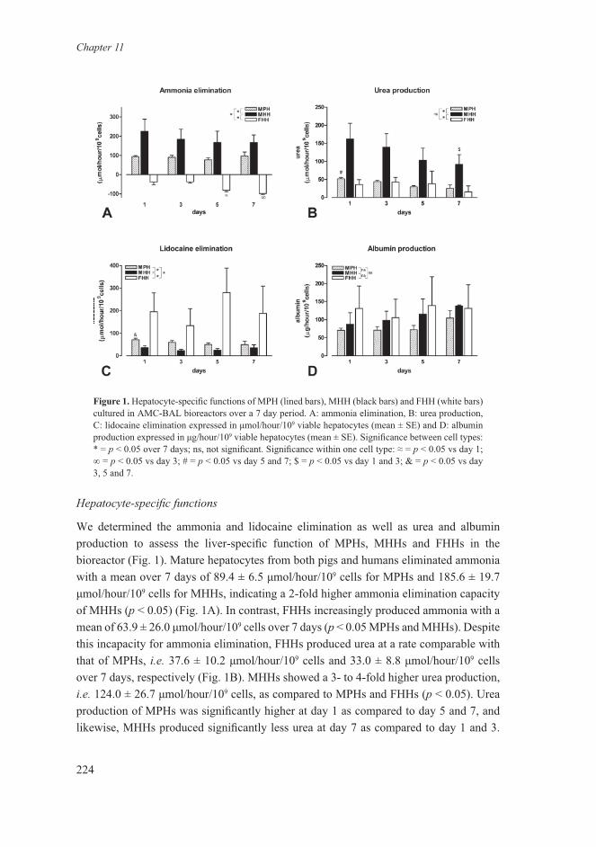
We determined the ammonia and lidocaine elimination as well as urea and albumin production to assess the liver-specific function of MPHs, MHHs and FHHs in the bioreactor (Fig. 1). Mature hepatocytes from both pigs and humans eliminated ammonia with a mean over 7 days of 89.4 ± 6.5 μmol/hour/109 cells for MPHs and 185.6 ± 19.7 μmol/hour/109 cells for MHHs, indicating a 2-fold higher ammonia elimination capacity of MHHs (p < 0.05) (Fig. 1A). In contrast, FHHs increasingly produced ammonia with a mean of 63.9 ± 26.0 μmol/hour/109 cells over 7 days (p < 0.05 MPHs and MHHs). Despite this incapacity for ammonia elimination, FHHs produced urea at a rate comparable with that of MPHs, i.e. 37.6 ± 10.2 μmol/hour/109 cells and 33.0 ± 8.8 μmol/hour/109 cells over 7 days, respectively (Fig. 1B). MHHs showed a 3- to 4-fold higher urea production, i.e. 124.0 ± 26.7 μmol/hour/109 cells, as compared to MPHs and FHHs (p < 0.05). Urea production of MPHs was significantly higher at day 1 as compared to day 5 and 7, and likewise, MHHs produced significantly less urea at day 7 as compared to day 1 and 3.
Figure 1. Hepatocyte-specific functions of MPH (lined bars), MHH (black bars) and FHH (white bars) cultured in AMC-BAL bioreactors over a 7 day period. A: ammonia elimination, B: urea production, C: lidocaine elimination expressed in μmol/hour/109 viable hepatocytes (mean ± SE) and D: albumin production expressed in μg/hour/109 viable hepatocytes (mean ± SE). Significance between cell types: * = p < 0.05 over 7 days; ns, not significant. Significance within one cell type: ≈ = p < 0.05 vs day 1; ∞ = p < 0.05 vs day 3; # = p < 0.05 vs day 5 and 7; $ = p < 0.05 vs day 1 and 3; & = p < 0.05 vs day 3, 5 and 7.
Three primary liver cell types cultured in the AMC-BAL
225
If ammonia elimination is only determined by urea synthesis, 2 moles of ammonia are converted into 1 mole of urea. However, the ammonia-urea ratio was rather variable: it increased in time for MPHs as well as MHHs. This tendency, although not significant, was stronger for MPHs as compared to MHHs with a mean ratio of 1.8 ± 0.5 at day 1 and 5.5 ± 2.7 at day 7 for MPHs and for MHHs, a mean ratio of 1.6 ± 0.4 at day 1 and 2.7 ± 1.4 at day 7.
Lidocaine elimination, as a parameter for cytochrome p450 3A4/1A2 (human) and 3A29 (pig) activity, was significantly different for all three cell types over the 7 day culture period (Fig 1-C). Lidocaine elimination of MPHs was 1.9 fold higher than of MHHs. Lidocaine elimination of FHHs was 3.5-fold higher than of MPHs and 6.6-fold higher than of MHHs, with an average over 7 days of 199.3 ± 40.4 μmol/hour/109 cells for FHHs, 57.5 ± 7.8 μmol/hour/109 cells for MPHs and 30.1 ± 5.7 μmol/hour/109 cells for MHHs. Lidocaine elimination of MPHs at day 1 was significantly higher than day 3, 5 and 7, whereas no daily differences were observed for MHHs and FHHs. No changes in lidocaine concentration were observed during a function test in an empty bioreactor.
Albumin production was not significantly different for all three cell types over the 7 day culture period (Fig 1-D). A tendency towards a higher albumin production for FHH > MHH > MPH was observed, with an average over 7 days of 126.8 ± 80.5 μg/hour/109 cells, 109.9 ± 36.6 μg/hour/109 cells and 79.5 ± 26.9 μg/hour/109 cells, respectively. MHH and MPH tended to increase in time, whereas FHH remained stable during the culture period.
These results show that most liver-specific functions differ considerably between MPHs, MHHs and FHHs when cultured inside the laboratory-scale AMC-BAL.
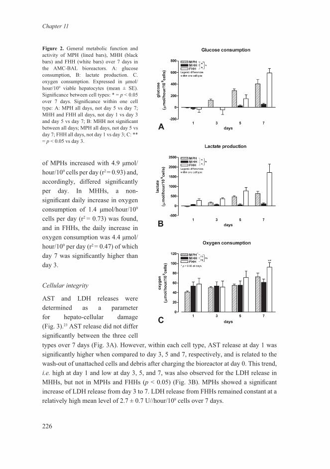
General metabolic function and activity
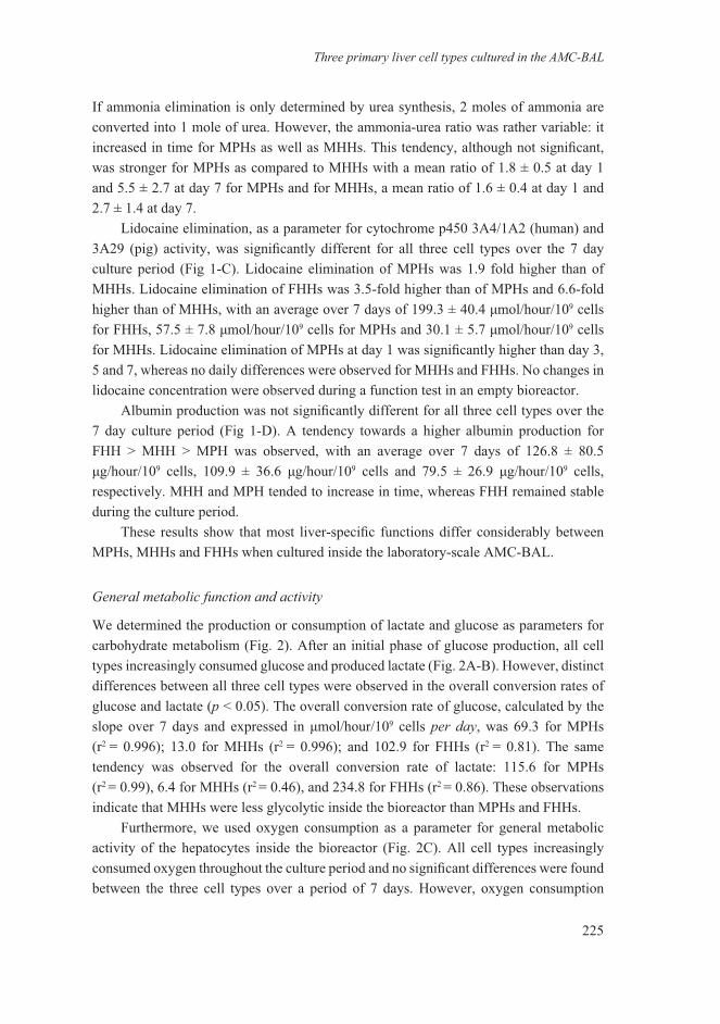
We determined the production or consumption of lactate and glucose as parameters for carbohydrate metabolism (Fig. 2). After an initial phase of glucose production, all cell types increasingly consumed glucose and produced lactate (Fig. 2A-B). However, distinct differences between all three cell types were observed in the overall conversion rates of glucose and lactate (p < 0.05). The overall conversion rate of glucose, calculated by the slope over 7 days and expressed in μmol/hour/109 cells per day, was 69.3 for MPHs (r2 = 0.996); 13.0 for MHHs (r2 = 0.996); and 102.9 for FHHs (r2 = 0.81). The same tendency was observed for the overall conversion rate of lactate: 115.6 for MPHs (r2 = 0.99), 6.4 for MHHs (r2 = 0.46), and 234.8 for FHHs (r2 = 0.86). These observations indicate that MHHs were less glycolytic inside the bioreactor than MPHs and FHHs.
Furthermore, we used oxygen consumption as a parameter for general metabolic activity of the hepatocytes inside the bioreactor (Fig. 2C). All cell types increasingly consumed oxygen throughout the culture period and no significant differences were found between the three cell types over a period of 7 days. However, oxygen consumption
Chapter 11
226
of MPHs increased with 4.9 μmol/hour/109 cells per day (r2 = 0.93) and, accordingly, differed significantly per day. In MHHs, a non-significant daily increase in oxygen consumption of 1.4 μmol/hour/109 cells per day (r2 = 0.73) was found, and in FHHs, the daily increase in oxygen consumption was 4.4 μmol/hour/109 per day (r2 = 0.47) of which day 7 was significantly higher than day 3.
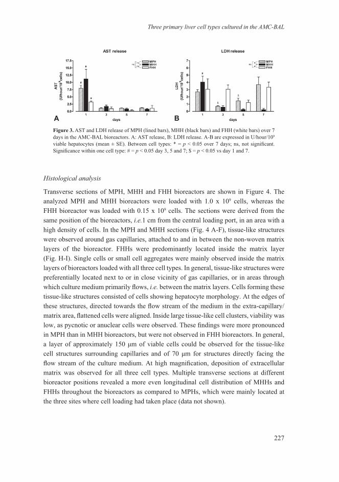
Cellular integrity
AST and LDH releases were determined as a parameter for hepato-cellular damage (Fig. 3).23 AST release did not differ significantly between the three cell types over 7 days (Fig. 3A). However, within each cell type, AST release at day 1 was significantly higher when compared to day 3, 5 and 7, respectively, and is related to the wash-out of unattached cells and debris after charging the bioreactor at day 0. This trend, i.e. high at day 1 and low at day 3, 5, and 7, was also observed for the LDH release in MHHs, but not in MPHs and FHHs (p < 0.05) (Fig. 3B). MPHs showed a significant increase of LDH release from day 3 to 7. LDH release from FHHs remained constant at a relatively high mean level of 2.7 ± 0.7 U//hour/109 cells over 7 days.
Figure 2. General metabolic function and activity of MPH (lined bars), MHH (black bars) and FHH (white bars) over 7 days in the AMC-BAL bioreactors. A: glucose consumption, B: lactate production. C. oxygen consumption. Expressed in μmol/hour/109 viable hepatocytes (mean ± SE). Significance between cell types: * = p < 0.05 over 7 days. Significance within one cell type: A: MPH all days, not day 5 vs day 7; MHH and FHH all days, not day 1 vs day 3 and day 5 vs day 7; B: MHH not significant between all days; MPH all days, not day 5 vs day 7; FHH all days, not day 1 vs day 3; C: ** = p < 0.05 vs day 3.
Three primary liver cell types cultured in the AMC-BAL
227
Figure 3. AST and LDH release of MPH (lined bars), MHH (black bars) and FHH (white bars) over 7 days in the AMC-BAL bioreactors. A: AST release, B: LDH release. A-B are expressed in U/hour/109 viable hepatocytes (mean ± SE). Between cell types: * = p < 0.05 over 7 days; ns, not significant. Significance within one cell type: # = p < 0.05 day 3, 5 and 7; $ = p < 0.05 vs day 1 and 7.
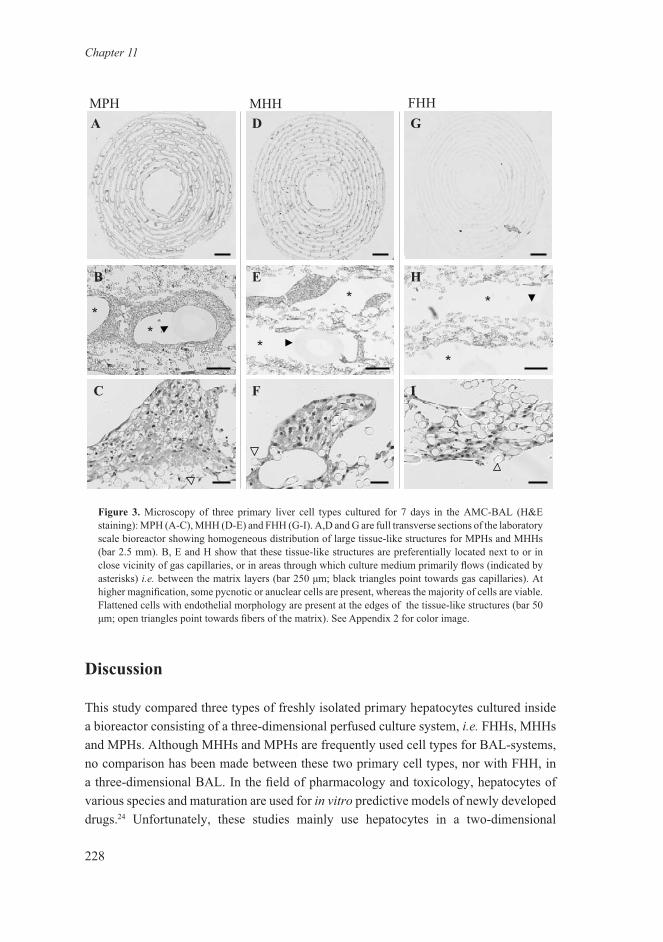
Histological analysis
Transverse sections of MPH, MHH and FHH bioreactors are shown in Figure 4. The analyzed MPH and MHH bioreactors were loaded with 1.0 x 10⁹ cells, whereas the FHH bioreactor was loaded with 0.15 x 10⁹ cells. The sections were derived from the same position of the bioreactors, i.e.1 cm from the central loading port, in an area with a high density of cells. In the MPH and MHH sections (Fig. 4 A-F), tissue-like structures were observed around gas capillaries, attached to and in between the non-woven matrix layers of the bioreactor. FHHs were predominantly located inside the matrix layer (Fig. H-I). Single cells or small cell aggregates were mainly observed inside the matrix layers of bioreactors loaded with all three cell types. In general, tissue-like structures were preferentially located next to or in close vicinity of gas capillaries, or in areas through which culture medium primarily flows, i.e. between the matrix layers. Cells forming these tissue-like structures consisted of cells showing hepatocyte morphology. At the edges of these structures, directed towards the flow stream of the medium in the extra-capillary/matrix area, flattened cells were aligned. Inside large tissue-like cell clusters, viability was low, as pycnotic or anuclear cells were observed. These findings were more pronounced in MPH than in MHH bioreactors, but were not observed in FHH bioreactors. In general, a layer of approximately 150 μm of viable cells could be observed for the tissue-like cell structures surrounding capillaries and of 70 μm for structures directly facing the flow stream of the culture medium. At high magnification, deposition of extracellular matrix was observed for all three cell types. Multiple transverse sections at different bioreactor positions revealed a more even longitudinal cell distribution of MHHs and FHHs throughout the bioreactors as compared to MPHs, which were mainly located at the three sites where cell loading had taken place (data not shown).
Chapter 11
228
Discussion
This study compared three types of freshly isolated primary hepatocytes cultured inside a bioreactor consisting of a three-dimensional perfused culture system, i.e. FHHs, MHHs and MPHs. Although MHHs and MPHs are frequently used cell types for BAL-systems, no comparison has been made between these two primary cell types, nor with FHH, in a three-dimensional BAL. In the field of pharmacology and toxicology, hepatocytes of various species and maturation are used for in vitro predictive models of newly developed drugs.24 Unfortunately, these studies mainly use hepatocytes in a two-dimensional
Figure 3. Microscopy of three primary liver cell types cultured for 7 days in the AMC-BAL (H&E staining): MPH (A-C), MHH (D-E) and FHH (G-I). A,D and G are full transverse sections of the laboratory scale bioreactor showing homogeneous distribution of large tissue-like structures for MPHs and MHHs (bar 2.5 mm). B, E and H show that these tissue-like structures are preferentially located next to or in close vicinity of gas capillaries, or in areas through which culture medium primarily flows (indicated by asterisks) i.e. between the matrix layers (bar 250 μm; black triangles point towards gas capillaries). At higher magnification, some pycnotic or anuclear cells are present, whereas the majority of cells are viable. Flattened cells with endothelial morphology are present at the edges of the tissue-like structures (bar 50 μm; open triangles point towards fibers of the matrix). See Appendix 2 for color image.
MPH MHH FHHA
IF
HE
GD
B
C
**
*
**
*
Three primary liver cell types cultured in the AMC-BAL
229
configuration without medium flow. Direct extrapolation to BAL devices is therefore difficult.
The results in the current study show large differences in liver-specific and metabolic functions between MPH, MHH and FHH. However, oxygen consumption, as a measure of cell volume and general metabolic activity, did not differ between the three cell types during the whole culture period (Fig. 1C).25 This suggests that the measured differences in liver-specific and metabolic functions are not attributable to the initial state of the cells (either fresh or Celsior preserved), the isolation method used (central or manual perfusion) or inadequate calculation of the number of cells charged in the bioreactors. Moreover, the high reproducibility of functional results within the groups shows that the variation in viability of the isolates, particularly of the MHHs and FHHs, is acceptable. The observed differences may be explained by species differences, although this cannot hold true for the comparison between FHHs and MHHs.
The values for ammonia elimination and urea production of MPH are in accordance with previous studies from our laboratory (Fig. 1A-B).19,21,26 The higher values observed for MHH should be attributed to a higher and more efficient urea cycle and glutamine synthetase (GS) activity. We point out, however, that urea can be produced by conversion of arginine into ornithine by arginase I although the urea cycle may not be intact, and that ammonia can be eliminated exclusively by glutamine synthetase. As a result, the elimination of ammonia into urea does not necessarily follow the molar ratio of 2, as was observed in this study for MPH and MHH. FHH, however, produced ammonia. Production of ammonia is a phenomenon of hepatocytes observed in different culture systems of human hepatic cell lines (HepZ, HepG2, C3A, HuH-7 and PCL/PRF/527-29) and for mature rat, human and porcine hepatocytes under different culture conditions.28,30 Recently we also observed ammonia production in primary FHHs cultured in monolayer.14 In this study, ureagenesis was observed in FHHs at a level comparable to that of MPHs, as has been shown previously.31 The different relations between ammonia and urea metabolism, i.e. production of ammonia and urea by FHHs and ammonia elimination and urea production by MHHs and MPHs, may be explained by the ontogeneic activity of the kidney-type and liver-type glutaminases in relation to the urea cycle.32,33 The kidney-type glutaminase is expressed most in the fetal liver, and not in the postnatal liver. As a consequence, fetal liver releases free ammonia from glutamine catabolism by the kidney-type glutaminase, independent of the production of urea from ammonia via the urea cycle. In mature liver, however, only liver-type glutaminase is expressed, which produces ammonia that is directly used for the synthesis of urea, since glutaminase-derived ammonia is directly channeled to carbamoyl-phosphate synthetase and does not escape the mitochondria.34,35 This explanation for the production of ammonia by FHH should be considered as speculative, since we did not investigate the kidney- as well as liver-type glutaminase at the protein level in this study.
Chapter 11
230
Porcine hepatocytes are considered a relatively good experimental human model for cytochrome p450 (CYP) 3A drug metabolism. Caution however, is required for direct extrapolation of the activity of CYP isoforms from pigs to humans, because the activity differs per substrate.36-39 In this study, lidocaine elimination of MPHs, mediated by the CYP3A29 isoform, was 1.9 fold higher than of MHHs (Fig. 1C). Lidocaine elimination of FHHs, on the other hand, was 6.6 times higher than MHH. The fetal isoform of human CYP3A4 is CYP3A7 and this accounts for up to 50% of total fetal hepatic cytochrome p450 and nearly all CYP3A enzymes.40 Metabolic activity of CYP3A7 is significantly lower than CYP3A4, depending on the concentration and metabolite used.41 Interestingly, however, total CYP3A7 content of FHH of 94-168 days gestational age is app.310 pmol/mg microsomal protein, whereas total CYP3A4 content in MHH is app. 60 pmol/mg microsomal protein.40 This can explain the higher lidocaine elimination of FHH observed in this study. We emphasize at this point, however, that no data are available on the effect of a 3-dimensional high cell density perfused culture systems on CYP3A4/1A2, 3A29 and 3A7 activity of MPH, MHH and FHH, respectively, using lidocaine.
In contrast to the urea production, ammonia and lidocaine elimination, albumin production was not significantly different between all three types of hepatocytes. This was also observed by Jasmund et al. in a 2-dimensional sandwich culture system with MPH and MHH under different culture conditions.42 A stable or an increased albumin production of MPH and MHH during the first 7 days of culture has been shown in monolayer42-44 as well as several bioreactor systems.21,45-48 FHH of 23-24 weeks old were able to produce albumin at a rate of MPH and MHH. This was also observed for FHH cultured in monolayer, secreting albumin at 4-21 ug/ml/1.8-2.5 x 106 FFH49-51, which resembles the albumin production by MPH and MHH in monolayer.43
The increase in glucose consumption, lactate production, oxygen consumption (Fig. 2) and LDH release (Fig. 3B) of MPH are in accordance with previous studies.19,21,26 These observations may imply an increase in anaerobic glycolysis in combination with mitochondrial uncoupling of the oxidative phosphorylation.52,53 An increase in lactate production, LDH release and oxygen consumption can also, at least in part, be a reflection of cell growth.25 In parallel, non-parenchymal liver cells produce more lactate and grow faster than parenchymal liver cells as observed in monolayer culture.54 Fetal livers contain more non-parenchymal cells than mature livers because of their haematopoietic function. Therefore, the higher glucose consumption, lactate production, oxygen consumption rate and LDH release of FHHs can either be explained by a higher content of non-parenchymal cells or a higher in vitro proliferation capacity of FHH itself. Dedifferentiation does not seem to play a major role, since the hepatic functions remain relatively stable. MHHs, on the other hand, are more stable in the general metabolic functions during the 7 day culture than MPHs and FHHs. This can be explained by the composition of human and
Three primary liver cell types cultured in the AMC-BAL
231
porcine liver cell isolates. Since MPH isolates contain more cellular aggregates and form spheroids easier than MHH isolates47,55,56, high dense cellular but also anoxic areas can be formed, as was observed by histological examination. In contrast, MHH isolates exist of mainly single cells which were distributed evenly throughout the bioreactor.
In summary, at present MPH and MHH are generally considered as the most acceptable cell sources for BAL devices. FHH have to date, not been demonstrated to be a relevant cell source for BAL application. The higher proliferation capacity of FHHs as compared to MHHs and MPHs, and the higher liver-specific function compared to human hepatic cell lines have encouraged us to test this cell type in the AMC-BAL. We observed large differences between the three cell types when investigated in the three-dimensional perfused AMC-BAL system. Our results indicate that FHHs, despite their higher proliferative capacity and cytochrome p450 3A activity, are not an ideal cell type for BAL application, since FHHs produce ammonia and display high metabolic instability. FHH in a bioreactor may, on the other hand, be considered as a favorable cell type for pharmacological and toxicological studies.57,58 MHHs outperformed MPHs and FHHs in their ammonia detoxification, urea production and metabolic stability. From this study, we conclude that MHHs are the preferred cell source for clinical use in a BAL. On the other hand, MPHs can be regarded as the second best cell type because of their unrestricted availability. Since bioreactors loaded with MPH are less stable than MHH bioreactors, these bioreactors should be preferably used within one or two days after loading in the clinical setting.
Acknowledgements
The authors thank G. Huyzer and A. Maas of the Surgical Laboratory for their assistance in porcine liver harvest
surgery. The authors express their gratitude to dr. Fabio Sirimarco (dept. Gynaecology and Obstetrics) and dr.
Giuseppe Nazzaro (dept. Obstetric Pathology) for their help in collecting the fetal livers, and to dr. Antonio
Faiella and Daniele Morelli for their help in the logistics concerning the fetal livers.
Chapter 11
232
References
1. World Health Organization. The World Health Report 2004 - Changing History. Geneva: 2004.2. National Center for Health Statistics. Health, United States, 2005 - With Chartbook on Trends in the
Health of Americans. Hyattsville, Maryland: 2005.3. Tzanakakis ES, Hess DJ, Sielaff TD, Hu WS. Extracorporeal tissue engineered liver-assist devices.
Annu Rev Biomed Eng 2000;2: 607-632.4. van de Kerkhove MP, Hoekstra R, Chamuleau RA, van Gulik TM. Clinical application of bioartificial
liver support systems. Ann Surg 2004;240: 216-230.5. Tsiaoussis J, Newsome PN, Nelson LJ, Hayes PC, Plevris JN. Which hepatocyte will it be? Hepatocyte
choice for bioartificial liver support systems. Liver Transpl 2001;7: 2-10.6. Hoekstra R, Chamuleau RA. Recent developments on human cell lines for the bioartificial liver. Int J
Artif Organs 2002;25: 182-191.7. Gerlach JC, Zeilinger K, Sauer IM, Mieder T, Naumann G, Grunwald A, Pless G, Holland G, Mas A,
Vienken J, Neuhaus P. Extracorporeal liver support: porcine or human cell based systems? Int J Artif Organs 2002;25: 1013-1018.
8. Morsiani E, Brogli M, Galavotti D, Pazzi P, Puviani AC, Azzena GF. Biologic liver support: optimal cell source and mass. Int J Artif Organs 2002;25: 985-993.
9. Chan C, Berthiaume F, Nath BD, Tilles AW, Toner M, Yarmush ML. Hepatic tissue engineering for adjunct and temporary liver support: critical technologies. Liver Transpl 2004;10: 1331-1342.
10. Chamuleau RA, Deurholt T, Hoekstra R. Which are the right cells to be used in a bioartificial liver? Metab Brain Dis 2005;20: 327-335.
11. Patience C, Takeuchi Y, Weiss RA. Infection of human cells by an endogenous retrovirus of pigs. Nat Med 1997;3: 282-286.
12. Baccarani U, Sanna A, Cariani A, Sainz-Barriga M, Adani GL, Zambito AM, Piccolo G, Risaliti A, Nanni-Costa A, Ridolfi L, Scalamogna M, Bresadola F, Donini A. Isolation of human hepatocytes from livers rejected for liver transplantation on a national basis: results of a 2-year experience. Liver Transpl 2003;9: 506-512.
13. Sauer IM, Zeilinger K, Pless G, Kardassis D, Theruvath T, Pascher A, Goetz M, Neuhaus P, Gerlach JC. Extracorporeal liver support based on primary human liver cells and albumin dialysis--treatment of a patient with primary graft non-function. J Hepatol 2003;39: 649-653.
14. Deurholt T, Ten Bloemendaal L, Chatta AA, van Wijk AC, Weijer K, Seppen J, Oude-Elferink R.P.J., Chamuleau RAFM, Hoekstra R. In vitro Functionality of Human Fetal Liver Cells and Clonal Derivatives under Proliferative Conditions. Cell Transplan t2006; 15: 811-22.
15. Hamamoto R, Kamihira M, Iijima S. Growth and differentiation of cultured fetal hepatocytes isolated various developmental stages. Biosci Biotechnol Biochem 1999;63: 395-401.
16. Flendrig LM, la Soe J, Jorning GG, Steenbeek A, Karlsen OT, Bovee, WM, Ladiges NC, te Velde A, Chamuleau RA. In vitro evaluation of a novel bioreactor based on an integral oxygenator and a spirally wound nonwoven polyester matrix for hepatocyte culture as small aggregates. J Hepatol 1997;26: 1379-1392.
17. Flendrig LM, Chamuleau RA, Maas MA, Daalhuisen J, Hasset B, Kilty CG, Doyle S, Ladiges NC, Jorning GG, la Soe J, Sommeijer D, te Velde A. Evaluation of a novel bioartificial liver in rats with complete liver ischemia: treatment efficacy and species-specific alpha-GST detection to monitor hepatocyte viability. J Hepatol 1999;30: 311-320.
18. van de Kerkhove MP, Di Florio E, Scuderi V, Mancini A, Belli A, Bracco A, Dauri M, Tisone G, Di Nicuolo G, Amoroso P, Spadari A, Lombardi G, Hoekstra R, Calise F, Chamuleau RA. Phase I clinical trial with the AMC-bioartificial liver. Academic Medical Center. Int J Artif Organs 2002;25: 950-959.
Three primary liver cell types cultured in the AMC-BAL
233
19. Poyck PP, Hoekstra R, van Wijk AC, Ten Bloemendaal L, Chamuleau RA, van Gulik TM. Mild Hypothermic Preservation for Transport Purposes of the AMC Bioartificial Liver Charged with Porcine Hepatocytes. Transplantation 2005;80: 1153-1160.
20. Seglen PO. Preparation of rat liver cells. 3. Enzymatic requirements for tissue dispersion. Exp Cell Res 1973;82: 391-398.
21. van de Kerkhove MP, Poyck PPC, van Wijk ACWA, Galavotti D, Hoekstra R, van Gulik TM, Chamuleau RA. Assessment and improvement of liver specific function of the AMC-bioartificial liver. Int J Artif Organs 2005;28: 617-630.
22. Hoekstra R, Deurholt T, Ten Bloemendaal L, Desille M, van Wijk ACWA, Clement B, Oude Elferink RP, van Gulik TM and Chamuleau RA. Assessment of in vitro, applicability of reversible immortalized NKNT-3 cells and clonal derivatives. Cell Transplant 15: 423-433, 2006.
23. Jauregui HO, Hayner NT, Driscoll JL, Williams-Holland R, Lipsky MH, Galletti PM. Trypan blue dye uptake and lactate dehydrogenase in adult rat hepatocytes--freshly isolated cells, cell suspensions, and primary monolayer cultures. In vitro 1981;17: 1100-1110.
24. O’Brien PJ, Chan K, Silber PM. Human and animal hepatocytes in vitro with extrapolation in vivo. Chem Biol Interact 2004;150: 97-114.
25. Jorjani P, Ozturk SS. Effects of cell density and temperature on oxygen consumption rate for different mammalian cell lines. Biotechnol Bioeng 1999;64: 349-356.
26. Poyck PP, Pless G, Hoekstra R, Roth S, van Wijk ACWA, Schwartlander R, van Gulik TM, Sauer IM, Chamuleau RAFM. In vitro Comparison of Two Bioartificial Liver Support Systems: MELS CellModule and AMC-BAL. Int J Artif Organs 2008 (January);in press.
27. Werner A, Duvar S, Muthing J, Buntemeyer H, Lunsdorf H, Strauss M, Lehmann J. Cultivation of immortalized human hepatocytes HepZ on macroporous CultiSpher G microcarriers. Biotechnol Bioeng 2000;68: 59-70.
28. Wang L, Sun J, Li L, Mears D, Horvat M, Sheil AG. Comparison of porcine hepatocytes with human hepatoma (C3A) cells for use in a bioartificial liver support system. Cell Transplant 1998;7: 459-468.
29. Nagaki M, Miki K, Kim YI, Ishiyama H, Hirahara I, Takahashi H, Sugiyama A, Muto Y, Moriwaki H. Development and characterization of a hybrid bioartificial liver using primary hepatocytes entrapped in a basement membrane matrix. Dig Dis Sci 2001;46: 1046-1056.
30. Yamashita Y, Shimada M, Tsujita E, Shirabe K, Ijima H, Nakazawa K, Sakiyama R, Fukuda J, Funatsu K, Sugimachi K. High metabolic function of primary human and porcine hepatocytes in a polyurethane foam/spheroid culture system in plasma from patients with fulminant hepatic failure. Cell Transplant 2002;11: 379-384.
31. Vijayalakshmi V, Naseem B, Khan AA, Capoor AK, Habibullah CM. Comparison of biochemical and cytotoxic functions of hepatocytes from goat, pig and human fetuses. J Gastroenterol Hepatol 2004;19: 1029-1035.
32. Watford M. Hepatic glutaminase expression: relationship to kidney-type glutaminase and to the urea cycle. FASEB J 1993;7: 1468-1474.
33. Curthoys NP, Watford M. Regulation of glutaminase activity and glutamine metabolism. Annu Rev Nutr 1995;15: 133-159.
34. Meijer AJ. Channeling of ammonia from glutaminase to carbamoyl-phosphate synthetase in liver mitochondria. FEBS Lett 1985;191: 249-251.
35. Meijer AJ, Lamers WH, Chamuleau RA. Nitrogen metabolism and ornithine cycle function. Physiol Rev 1990;70: 701-748.
36. Bogaards JJ, Bertrand M, Jackson P, Oudshoorn MJ, Weaver RJ, van Bladeren PJ, Walther B. Determining the best animal model for human cytochrome P450 activities: a comparison of mouse, rat, rabbit, dog, micropig, monkey and man. Xenobiotica 2000;30: 1131-1152.
37. Zuber R, Anzenbacherova E, Anzenbacher P. Cytochromes P450 and experimental models of drug metabolism. J Cell Mol Med 2002;6: 189-198.
Chapter 11
234
38. Maurel P. The use of adult human hepatocytes in primary culture and other in vitro systems to investigate drug metabolism in man. Advanced Drug Delivery Reviews 1996;22: 105-132.
39. Guengerich FP. Comparisons of catalytic selectivity of cytochrome P450 subfamily enzymes from different species. Chem Biol Interact 1997;106: 161-182.
40. Stevens JC, Hines RN, Gu C, Koukouritaki SB, Manro JR, Tandler PJ, Zaya MJ. Developmental expression of the major human hepatic CYP3A enzymes. J Pharmacol Exp Ther 2003;307: 573-582.
41. Williams JA, Ring BJ, Cantrell VE, Jones DR, Eckstein J, Ruterbories K, Hamman MA, Hall SD, Wrighton SA. Comparative metabolic capabilities of CYP3A4, CYP3A5, and CYP3A7. Drug Metab Dispos 2002;30: 883-891.
42. Jasmund I, Schwientek S, Acikgoz A, Langsch A, Machens HG, Bader A. The influence of medium composition and matrix on long-term cultivation of primary porcine and human hepatocytes. Biomol Eng 2006.
43. Hino H, Tateno C, Sato H, Yamasaki C, Katayama S, Kohashi T, Aratani A, Asahara T, Dohi K, Yoshizato K. A long-term culture of human hepatocytes which show a high growth potential and express their differentiated phenotypes. Biochem Biophys Res Commun 1999;256: 184-191.
44. Pahernik SA, Thasler WE, Mueller-Hoecker J, Schildberg FW, Koebe HG. Hypothernic storage of pig hepatocytes: influence of different storage solutions and cell density. Cryobiology 1996;33: 552-566.
45. Nyberg SL, Remmel RP, Mann HJ, Peshwa MV, Hu WS, Cerra FB. Primary hepatocytes outperform Hep G2 cells as the source of biotransformation functions in a bioartificial liver. Ann Surg 1994;220: 59-67.
46. De Bartolo L, Jarosch-Von Schweder G, Haverich A, Bader A. A novel full-scale flat membrane bioreactor utilizing porcine hepatocytes: cell viability and tissue-specific functions. Biotechnol Prog 2000;16: 102-108.
47. Nyberg SL, Hardin J, Amiot B, Argikar UA, Remmel RP, Rinaldo P. Rapid, large-scale formation of porcine hepatocyte spheroids in a novel spheroid reservoir bioartificial liver. Liver Transpl 2005;11: 901-910.
48. Shito M, Kim NH, Baskaran H, Tilles AW, Tompkins RG, Yarmush ML, Toner M. In vitro and in vivo evaluation of albumin synthesis rate of porcine hepatocytes in a flat-plate bioreactor. Artif Organs 2001;25: 571-578.
49. Lescoat G, Theze N, Fraslin JM, Pasdeloup N, Kneip B, Guguen-Guillouzo C. Influence of ornithine on albumin synthesis by fetal and neonatal hepatocytes maintained in culture. Cell Differ 1987;21: 21-29.
50. Guguen-Guillouzo C, Clement B, Lescoat G, Glaise D, Guillouzo A. Modulation of human fetal hepatocyte survival and differentiation by interactions with a rat liver epithelial cell line. Dev Biol 1984;105: 211-220.
51. Lazaro CA, Croager EJ, Mitchell C, Campbell JS, Yu C, Foraker J, Rhim JA, Yeoh GC, Fausto N. Establishment, characterization, and long-term maintenance of cultures of human fetal hepatocytes. Hepatology 2003;38: 1095-1106.
52. Kimura K, Jung BD, Kanehira K, Irie Y, Canas X, Saito M. Induction of uncoupling protein (UCP) 2 in primary cultured hepatocytes. FEBS Lett 1999;457: 75-79.
53. Cheng G, Polito CC, Haines JK, Shafizadeh SF, Fiorini RN, Zhou X, Schmidt MG, Chavin KD. Decrease of intracellular ATP content downregulated UCP2 expression in mouse hepatocytes. Biochem Biophys Res Commun 2003;308: 573-580.
54. Agius L. Metabolic interactions of parenchymal hepatocytes and dividing epithelial cells in co-culture. Biochem J 1988;252: 23-28.
55. Lazar A, Peshwa MV, Wu FJ, Chi CM, Cerra FB, Hu WS. Formation of porcine hepatocyte spheroids for use in a bioartificial liver. Cell Transplant 1995;4: 259-268.
56. Sakai Y, Naruse K, Nagashima I, Muto T, Suzuki M. A new bioartificial liver using porcine hepatocyte spheroids in high-cell-density suspension perfusion culture: in vitro performance in synthesized culture medium and in 100% human plasma. Cell Transplant 1999;8: 531-541.

Three primary liver cell types cultured in the AMC-BAL
235
57. Iacovacci S, Manzin A, Barca S, Sargiacomo M, Serafino A, Valli MB, Macioce G, Hassan HJ, Ponzetto A, Clementi M, Peschle C, Carloni G. Molecular characterization and dynamics of hepatitis C virus replication in human fetal hepatocytes infected in vitro. Hepatology 1997;26: 1328-1337.
58. Ochiya T, Tsurimoto T, Ueda K, Okubo K, Shiozawa M, Matsubara K. An in vitro system for infection with hepatitis B virus that uses primary human fetal hepatocytes. Proc Natl Acad Sci U S A 1989;86: 1875-1879.
Copyright © 2022 FDOKUMEN