Free Energy Perturbation Hamiltonian Replica-Exchange Molecular Dynamics (FEP:H-REMD) for Absolute...
7
Free Energy Perturbation Hamiltonian Replica-Exchange Molecular Dynamics (FEP/H-REMD) for Absolute Ligand Binding Free Energy Calculations Wei Jiang † and Benoı ˆt Roux* ,†,‡ Biosciences DiVision, Argonne National Laboratory, 9700 South Cass AVenue, Building 240, Argonne, Illinois 60439, and Department of Biochemistry and Molecular Biology, Gordon Center for IntegratiVe Science, UniVersity of Chicago, 929 57th Street, Chicago, Illinois 60637 Received April 1, 2010 Abstract: Free Energy Perturbation with Replica Ex- change Molecular Dynamics (FEP/REMD) offers a power- ful strategy to improve the convergence of free energy computations. In particular, it has been shown previously that a FEP/REMD scheme allowing random moves within an extended replica ensemble of thermodynamic coupling parameters “λ” can improve the statistical convergence in calculations of absolute binding free energy of ligands to proteins [J. Chem. Theory Comput. 2009, 5, 2583]. In the present study, FEP/REMD is extended and combined with an accelerated MD simulations method based on Hamil- tonian replica-exchange MD (H-REMD) to overcome the additional problems arising from the existence of kinetically trapped conformations within the protein receptor. In the combined strategy, each system with a given thermody- namic coupling factor λ in the extended ensemble is further coupled with a set of replicas evolving on a biased energy surface with boosting potentials used to accelerate the interconversion among different rotameric states of the side chains in the neighborhood of the binding site. Exchanges are allowed to occur alternatively along the axes correspond- ing to the thermodynamic coupling parameter λ and the boosting potential, in an extended dual array of coupled λ- and H-REMD simulations. The method is implemented on the basis of new extensions to the REPDSTR module of the biomolecular simulation program CHARMM. As an illustrative example, the absolute binding free energy of p-xylene to the nonpolar cavity of the L99A mutant of the T4 lysozyme was calculated. The tests demonstrate that the dual λ-REMD and H-REMD simulation scheme greatly accelerates the configu- rational sampling of the rotameric states of the side chains around the binding pocket, thereby improving the conver- gence of the FEP computations. Introduction Free energy perturbation molecular dynamics (FEP/MD) simu- lations with explicit solvent molecules provide one of the most fundamental routes for computing the binding affinities of small compounds to proteins. 1,2 In practice, a critical issue with FEP/ MD simulations is to achieve a sufficient sampling of all the relevant degrees of freedom. Problems can arise with large structural reorganizations either in the ligand or in the protein upon formation of the bound complex because sampling those is typically beyond the reach of straight brute-force FEP/MD simulations. More specifically, when there are large energy barriers separating the relevant conformational states, the ligand or the protein may remain kinetically trapped in the starting configuration for a very long time during FEP/MD simulations, and alternate conformations are never visited. The incomplete configurational sampling results in computed binding free energies that are dependent on the starting protein or ligand configuration, which are of limited significance and practical use. The structural changes observed upon the binding of aromatic molecules to a nonpolar cavity engineered in the L99A mutant of the T4 lysozyme (T4L) provide a good illustration of the type of problems that can arise from insufficient sampling (Figure 1). For the bound complexes involving small and medium-sized ligands (e.g., benzene, toluene, benzofurane, and indole), the protein structure is essentially identical to the ligand- free (apo) conformation. For those ligands, the calculated absolute binding free energies are well converged, regardless of whether the FEP/MD simulations are started from the holo or the apo state. 1,3,4 Difficulties arise in the case of larger ligands (e.g., indene, n-butylbenzene, isobutylbenzene, o-xylene, and p-xylene). In this case, the side chain of Val111, which is in direct contact with the bound ligand, changes its rotameric states from a trans conformation ( 1 ) 180°) for the ligand-free apo to a gauche conformation ( 1 )-60°) for the bound state with large ligands. The intrinsic energy barrier around the 1 torsion of the valine (∼5 kcal/mol) is sufficient to prevent the side chain from reorienting on the time scale of typical FEP/MD simula- * Corresponding author e-mail: [email protected]. † Argonne National Laboratory. ‡ University of Chicago. J. Chem. Theory Comput. 2010, 6, 2559–2565 2559 10.1021/ct1001768 2010 American Chemical Society Published on Web 07/30/2010
Transcript of Free Energy Perturbation Hamiltonian Replica-Exchange Molecular Dynamics (FEP:H-REMD) for Absolute...
Free Energy Perturbation HamiltonianReplica-Exchange MolecularDynamics (FEP/H-REMD) for AbsoluteLigand Binding Free EnergyCalculations
Wei Jiang† and Benoı̂t Roux*,†,‡
Biosciences DiVision, Argonne National Laboratory,9700 South Cass AVenue, Building 240, Argonne,Illinois 60439, and Department of Biochemistry andMolecular Biology, Gordon Center for IntegratiVe Science,UniVersity of Chicago, 929 57th Street, Chicago, Illinois 60637
Received April 1, 2010
Abstract: Free Energy Perturbation with Replica Ex-change Molecular Dynamics (FEP/REMD) offers a power-ful strategy to improve the convergence of free energycomputations. In particular, it has been shown previouslythat a FEP/REMD scheme allowing random moves withinan extended replica ensemble of thermodynamic couplingparameters “λ” can improve the statistical convergence incalculations of absolute binding free energy of ligands toproteins [J. Chem. Theory Comput. 2009, 5, 2583]. In thepresent study, FEP/REMD is extended and combined withan accelerated MD simulations method based on Hamil-tonian replica-exchange MD (H-REMD) to overcome theadditional problems arising from the existence of kineticallytrapped conformations within the protein receptor. In thecombined strategy, each system with a given thermody-namic coupling factor λ in the extended ensemble is furthercoupled with a set of replicas evolving on a biased energysurface with boosting potentials used to accelerate theinterconversion among different rotameric states of the sidechains in the neighborhood of the binding site. Exchangesare allowed to occur alternatively along the axes correspond-ing to the thermodynamic coupling parameter λ and theboosting potential, in an extended dual array of coupled λ-and H-REMD simulations. The method is implemented onthe basis of new extensions to the REPDSTR module of thebiomolecular simulation program CHARMM. As an illustrativeexample, the absolute binding free energy of p-xylene to thenonpolar cavity of the L99A mutant of the T4 lysozyme was
calculated. The tests demonstrate that the dual λ-REMD andH-REMD simulation scheme greatly accelerates the configu-rational sampling of the rotameric states of the side chainsaround the binding pocket, thereby improving the conver-gence of the FEP computations.
IntroductionFree energy perturbation molecular dynamics (FEP/MD) simu-lations with explicit solvent molecules provide one of the mostfundamental routes for computing the binding affinities of smallcompounds to proteins.1,2 In practice, a critical issue with FEP/MD simulations is to achieve a sufficient sampling of all therelevant degrees of freedom. Problems can arise with largestructural reorganizations either in the ligand or in the proteinupon formation of the bound complex because sampling thoseis typically beyond the reach of straight brute-force FEP/MDsimulations. More specifically, when there are large energybarriers separating the relevant conformational states, the ligandor the protein may remain kinetically trapped in the startingconfiguration for a very long time during FEP/MD simulations,and alternate conformations are never visited. The incompleteconfigurational sampling results in computed binding freeenergies that are dependent on the starting protein or ligandconfiguration, which are of limited significance and practicaluse.
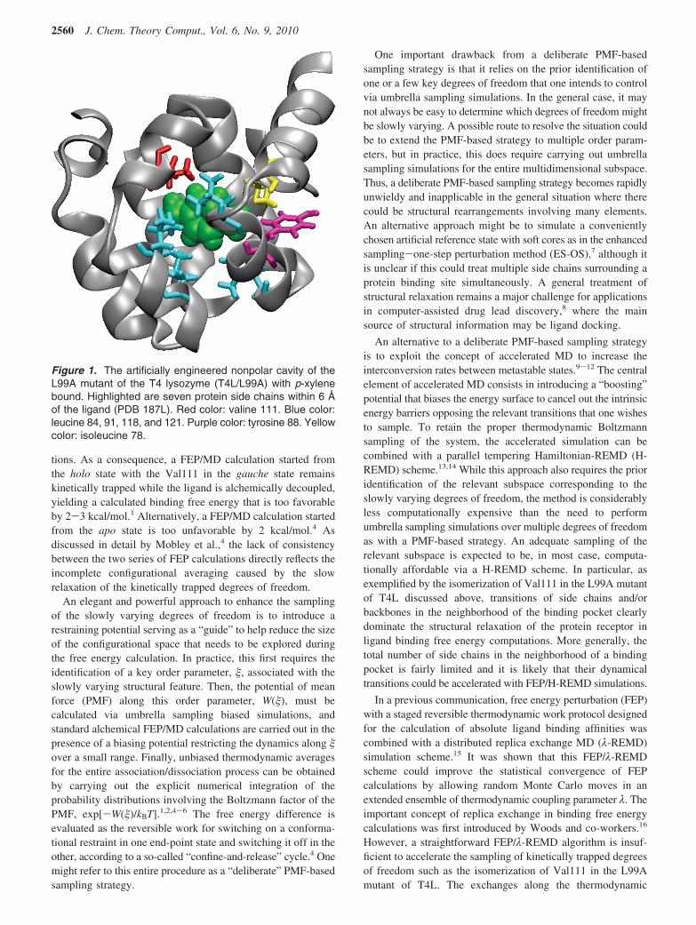
The structural changes observed upon the binding of aromaticmolecules to a nonpolar cavity engineered in the L99A mutantof the T4 lysozyme (T4L) provide a good illustration of thetype of problems that can arise from insufficient sampling(Figure 1). For the bound complexes involving small andmedium-sized ligands (e.g., benzene, toluene, benzofurane, andindole), the protein structure is essentially identical to the ligand-free (apo) conformation. For those ligands, the calculatedabsolute binding free energies are well converged, regardlessof whether the FEP/MD simulations are started from the holoor the apo state.1,3,4 Difficulties arise in the case of larger ligands(e.g., indene, n-butylbenzene, isobutylbenzene, o-xylene, andp-xylene). In this case, the side chain of Val111, which is indirect contact with the bound ligand, changes its rotameric statesfrom a trans conformation (�1 ) 180°) for the ligand-free apoto a gauche conformation (�1 ) -60°) for the bound state withlarge ligands. The intrinsic energy barrier around the �1 torsionof the valine (∼5 kcal/mol) is sufficient to prevent the side chainfrom reorienting on the time scale of typical FEP/MD simula-
* Corresponding author e-mail: [email protected].† Argonne National Laboratory.‡ University of Chicago.
J. Chem. Theory Comput. 2010, 6, 2559–2565 2559
10.1021/ct1001768 2010 American Chemical SocietyPublished on Web 07/30/2010
tions. As a consequence, a FEP/MD calculation started fromthe holo state with the Val111 in the gauche state remainskinetically trapped while the ligand is alchemically decoupled,yielding a calculated binding free energy that is too favorableby 2-3 kcal/mol.1 Alternatively, a FEP/MD calculation startedfrom the apo state is too unfavorable by 2 kcal/mol.4 Asdiscussed in detail by Mobley et al.,4 the lack of consistencybetween the two series of FEP calculations directly reflects theincomplete configurational averaging caused by the slowrelaxation of the kinetically trapped degrees of freedom.
An elegant and powerful approach to enhance the samplingof the slowly varying degrees of freedom is to introduce arestraining potential serving as a “guide” to help reduce the sizeof the configurational space that needs to be explored duringthe free energy calculation. In practice, this first requires theidentification of a key order parameter, �, associated with theslowly varying structural feature. Then, the potential of meanforce (PMF) along this order parameter, W(�), must becalculated via umbrella sampling biased simulations, andstandard alchemical FEP/MD calculations are carried out in thepresence of a biasing potential restricting the dynamics along �over a small range. Finally, unbiased thermodynamic averagesfor the entire association/dissociation process can be obtainedby carrying out the explicit numerical integration of theprobability distributions involving the Boltzmann factor of thePMF, exp[-W(�)/kBT].1,2,4-6 The free energy difference isevaluated as the reversible work for switching on a conforma-tional restraint in one end-point state and switching it off in theother, according to a so-called “confine-and-release” cycle.4 Onemight refer to this entire procedure as a “deliberate” PMF-basedsampling strategy.
One important drawback from a deliberate PMF-basedsampling strategy is that it relies on the prior identification ofone or a few key degrees of freedom that one intends to controlvia umbrella sampling simulations. In the general case, it maynot always be easy to determine which degrees of freedom mightbe slowly varying. A possible route to resolve the situation couldbe to extend the PMF-based strategy to multiple order param-eters, but in practice, this does require carrying out umbrellasampling simulations for the entire multidimensional subspace.Thus, a deliberate PMF-based sampling strategy becomes rapidlyunwieldy and inapplicable in the general situation where therecould be structural rearrangements involving many elements.An alternative approach might be to simulate a convenientlychosen artificial reference state with soft cores as in the enhancedsampling-one-step perturbation method (ES-OS),7 although itis unclear if this could treat multiple side chains surrounding aprotein binding site simultaneously. A general treatment ofstructural relaxation remains a major challenge for applicationsin computer-assisted drug lead discovery,8 where the mainsource of structural information may be ligand docking.
An alternative to a deliberate PMF-based sampling strategyis to exploit the concept of accelerated MD to increase theinterconversion rates between metastable states.9-12 The centralelement of accelerated MD consists in introducing a “boosting”potential that biases the energy surface to cancel out the intrinsicenergy barriers opposing the relevant transitions that one wishesto sample. To retain the proper thermodynamic Boltzmannsampling of the system, the accelerated simulation can becombined with a parallel tempering Hamiltonian-REMD (H-REMD) scheme.13,14 While this approach also requires the prioridentification of the relevant subspace corresponding to theslowly varying degrees of freedom, the method is considerablyless computationally expensive than the need to performumbrella sampling simulations over multiple degrees of freedomas with a PMF-based strategy. An adequate sampling of therelevant subspace is expected to be, in most case, computa-tionally affordable via a H-REMD scheme. In particular, asexemplified by the isomerization of Val111 in the L99A mutantof T4L discussed above, transitions of side chains and/orbackbones in the neighborhood of the binding pocket clearlydominate the structural relaxation of the protein receptor inligand binding free energy computations. More generally, thetotal number of side chains in the neighborhood of a bindingpocket is fairly limited and it is likely that their dynamicaltransitions could be accelerated with FEP/H-REMD simulations.
In a previous communication, free energy perturbation (FEP)with a staged reversible thermodynamic work protocol designedfor the calculation of absolute ligand binding affinities wascombined with a distributed replica exchange MD (λ-REMD)simulation scheme.15 It was shown that this FEP/λ-REMDscheme could improve the statistical convergence of FEPcalculations by allowing random Monte Carlo moves in anextended ensemble of thermodynamic coupling parameter λ. Theimportant concept of replica exchange in binding free energycalculations was first introduced by Woods and co-workers.16
However, a straightforward FEP/λ-REMD algorithm is insuf-ficient to accelerate the sampling of kinetically trapped degreesof freedom such as the isomerization of Val111 in the L99Amutant of T4L. The exchanges along the thermodynamic
Figure 1. The artificially engineered nonpolar cavity of theL99A mutant of the T4 lysozyme (T4L/L99A) with p-xylenebound. Highlighted are seven protein side chains within 6 Åof the ligand (PDB 187L). Red color: valine 111. Blue color:leucine 84, 91, 118, and 121. Purple color: tyrosine 88. Yellowcolor: isoleucine 78.
2560 J. Chem. Theory Comput., Vol. 6, No. 9, 2010
coupling λ can help to mix the side chain rotamers of the proteinin the apo and holo states, but transitions occur rarely due tothe intrinsic dihedral energy barriers. In the present work, weextend those ideas to propose a rapid and robust framework forfree energy computations combing the concept of λ-REMDsimulations within the staged FEP, and the concept of acceler-ated MD with boosting potentials via H-REMD. To achievethe proper sampling enhancement in the relevant subspace, wecombine λ-REMD with H-REMD. Random moves are allowedwithin an extended set of replicas biased by different values ofthe boosting factor “b” controlling the amplitude of a biasingpotential according to a H-REMD scheme. The implementationis based on the MPI level parallel/parallel mode made possibleby the Distributed Replica (REPDSTR) technique17,18 of theprogram CHARMM,19 in which each λ window of FEP istreated as an independent replica with its private I/O. WithREPDSTR, it is straightforward to introduce a set of auxiliaryboosting replicas for each λ window. This yields a dual REMDprotocol for FEP calculations, with replica-exchange along twoaxes (2D) corresponding to the thermodynamic couplingparameter λ and a second axis corresponding to the boostingfactor b. The entire array of REMD simulations can be executedas a single job via REPDSTR. It is shown that the dual FEPsimulation scheme combining λ-REMD and H-REMD signifi-cantly accelerates the configurational sampling of the proteinin FEP calculations. The method is illustrated with the calcula-tion of the absolute binding free energy of p-xylene to thenonpolar cavity of T4L/L99A.
Computational Details
A. REPDSTR Implementation of Staging SimulationProtocol. In the FEP staging simulation protocol, the potentialenergy is expressed in terms of four coupling (window)parameters1,2,20
where U0 is the potential of the system with the noninteracting(decoupled) ligand; λrep, λdis, λelec, and λrstr ∈ [0,1] are thethermodynamic coupling parameters; Urep and Udis are theshifted Weeks-Chandler-Anderson21 (WCA) repulsive anddispersive components of the Lennard-Jones potential; Uelec
is the electrostatic contribution; and Urstr is the restrainingpotential.
With the updated REPDSTR module of CHARMM,19 thefour-stage FEP simulation protocol can be implemented in asingle parallel/parallel MPI job. Figure 2a shows the REP-DSTR implementation of the updated FEP/REMD scheme,which is able to support the complete insertion process ofthe ligand into the binding pocket. The free energy corre-sponding to the process of inserting the ligand into thebinding site is
To achieve a significant sampling enhancement, M additionalreplicas with “boosting” biasing potentials are introduced (b )0, 1/M, 2/M, ..., 1) for each λ value of the FEP/REMD
calculation. The boosting parameter b scales the biasing potential(the system is not biased when b ) 0, and the biasing potentialcancels out the intrinsic dihedral PMF of the side chain whenb ) 1). Figure 2b shows the FEP/REMD scheme with a biasedHamiltonian. Replica exchanges are attempted alternatively inλ space and b space, forming a two-dimensional (2D) REMDframework.
The replica-exchange algorithm follows the conventionalMetropolis Monte Carlo exchange criterion:
where U denotes the potential energy of the underlying replica,λi and λj denote the staging parameters, and bk and bl denotethe boosting parameters.
B. Biasing Potentials for Residue �1 Dihedral Angle.Effective biasing boosting potentials can be obtained byfitting the potential of mean force (PMF) of individualdihedral degrees of freedom calculated on small peptides, inthe spirit of the work of Kannan and Zacharis.10 In the presentapplication, biasing potentials for the side chain dihedralangles �1 were constructed by calculating a PMF in the gasphase. The biasing potentials were determined for valine,leucine, isoleucine, and tyrosine residues using umbrellasampling simulations of one-residue peptides. In the ligandbinding calculation, these types of residues are distributedwithin a distance of 6 Å away from the center of mass ofthe ligand. A series of quadratic umbrella potentials with aforce constant of 100 kcal mol-1 rad-2 and distributed every5° was used. The angle �1 is defined as the C-CA-CB-CGdihedral, consistent with the CHARMM force field.22 Duringthe umbrella sampling simulations, the motions of thebackbones were restrained by harmonic potentials around theconformation observed in the protein. The umbrella samplingsimulations were unbiased with the weighted histogramanalysis method (WHAM).23 The resulting PMF along thedihedral angles was then fitted to a cosine Fourier series ofthe form
The fitting parameters Kn are given in Table 1. Figure 3 showsthe PMF and the result of the fit (the black curve is the PMF,and the red curve is the fitted cosine Fourier series). Anappropriate boosting potential can easily be constructed byinverting the sign of the PMF V(φ) in eq 4 via the CONS DIHEcommand of CHARMM,19 thereby canceling the potentialbarrier between the rotamers of a side chain for any selectedresidues.
C. MD Simulations. All the FEP/REMD simulations forthe binding site were carried out on the IBM Blue Gene/Pcluster Intrepid of the Argonne Leadership ComputingFacility (ALCF) at Argonne National Laboratory (ANL). Thesimulations were carried out in a high performance modeusing version c36a2 of the CHARMM program,19 which was
U(λrep,λdis,λelec,λrstr) ) U0 + Urep(λrep) + λdisUdis +λelecUelec + λrstrUrstr (1)
U(λrep ) 0, λdis ) 0, λelec ) 0, λrstr ) 1) f U(λrep )1, λdis ) 1, λelec ) 1, λrstr ) 0) (1a)
P(λi f λj) )
min{1, e(-[U(λi,b0,ri,0)+U(λj,b0,rj,0)-U(λi,b0,rj,0)-U(λj,b0,ri,0)])/kBT} (2)
P(bk f bl) )
min{1, e-[U(λi,bk,ri,k)+U(λj,bl,rj,l)-U(λi,bk,ri,l)-U(λj,bl,rj,k)]/kBT} (3)
V(φ) ) ∑n)1
3
Kn(1 + cos(n(φ - φmin))) (4)
Letter J. Chem. Theory Comput., Vol. 6, No. 9, 2010 2561
modified and extended for the present study. The hydrationcomputations were performed on the IBM quads computingcluster KBT at ANL. The binding site free energy simulationswere carried out on a reduced model of a solvated T4L/L99Asystem with the generalized solvent boundary potential(GSBP).24 The initial T4L/L99A system was constructed fromthe crystallographic structure (PDB 187L) as describedpreviously.1 The hydration free energy computations of theisolated ligands were carried out under PBC conditions atconstant pressure. The systems were propagated with a 2 fstime step using Langevin dynamics at a temperature of 298.15K. For the binding free energy calculation of p-xylene, 100
ps production runs were performed for the binding site witha replica-exchange frequency of 1/100 steps. M ) 7 boostingreplicas were used with H-REMD. The force field parametersand initial structure of p-xylene were taken from our previousstudy for the sake of consistency.1
In all calculations, the energies were collected during theproduction run and postprocessed using WHAM.23 For thebinding site simulations, the WHAM postprocessing is onlyapplied to replicas with zero biasing potential. To monitor theconvergence of the binding site calculation, 20 independent FEPcalculations (20 × 100 ps) were performed consecutively foreach system, each starting from the configuration saved at the
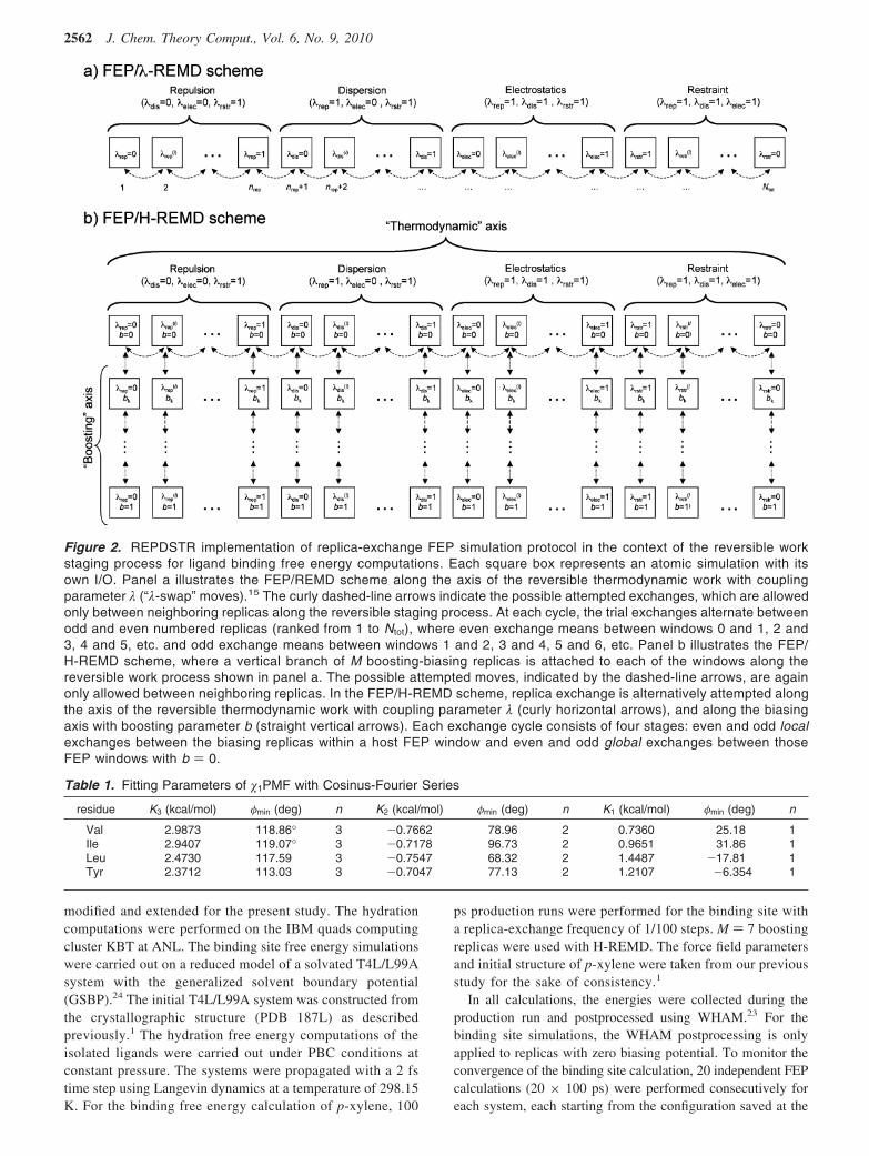
Figure 2. REPDSTR implementation of replica-exchange FEP simulation protocol in the context of the reversible workstaging process for ligand binding free energy computations. Each square box represents an atomic simulation with itsown I/O. Panel a illustrates the FEP/REMD scheme along the axis of the reversible thermodynamic work with couplingparameter λ (“λ-swap” moves).15 The curly dashed-line arrows indicate the possible attempted exchanges, which are allowedonly between neighboring replicas along the reversible staging process. At each cycle, the trial exchanges alternate betweenodd and even numbered replicas (ranked from 1 to Ntot), where even exchange means between windows 0 and 1, 2 and3, 4 and 5, etc. and odd exchange means between windows 1 and 2, 3 and 4, 5 and 6, etc. Panel b illustrates the FEP/H-REMD scheme, where a vertical branch of M boosting-biasing replicas is attached to each of the windows along thereversible work process shown in panel a. The possible attempted moves, indicated by the dashed-line arrows, are againonly allowed between neighboring replicas. In the FEP/H-REMD scheme, replica exchange is alternatively attempted alongthe axis of the reversible thermodynamic work with coupling parameter λ (curly horizontal arrows), and along the biasingaxis with boosting parameter b (straight vertical arrows). Each exchange cycle consists of four stages: even and odd localexchanges between the biasing replicas within a host FEP window and even and odd global exchanges between thoseFEP windows with b ) 0.
Table 1. Fitting Parameters of �1PMF with Cosinus-Fourier Series
residue K3 (kcal/mol) φmin (deg) n K2 (kcal/mol) φmin (deg) n K1 (kcal/mol) φmin (deg) n
Val 2.9873 118.86° 3 -0.7662 78.96 2 0.7360 25.18 1Ile 2.9407 119.07° 3 -0.7178 96.73 2 0.9651 31.86 1Leu 2.4730 117.59 3 -0.7547 68.32 2 1.4487 -17.81 1Tyr 2.3712 113.03 3 -0.7047 77.13 2 1.2107 -6.354 1
2562 J. Chem. Theory Comput., Vol. 6, No. 9, 2010
end of the previous run. The last 10 runs were used to producethe final averaged result and calculate the standard deviations.
Results and DiscussionBoth crystallographic studies and computations indicate that theside chain of Val111 of T4L/L99A changes its rotameric stateswhen moderately large ligands bind to the nonpolar cavity.1,25
In the case of p-xylene, the side chain of Val111 rotates byapproximately 140°, to a �1 value of -35°, to avoid a stericclash with the ligand (Val111 is depicted in red in Figure 1). Itis often convenient to utilize the holo configuration as thestarting set of coordinates in absolute binding free energycalculations since this is provided either by the X-ray crystal-lographic structure of the bound complex or by the output of insilico docking. However, one must ensure that FEP/MDsimulations do reversibly cover the relevant set of thermody-namic states. In principle, an ideal sampling should be able toreflect any conformational changes within the protein betweenthe two end-point holo and apo states along the thermodynamicdecoupling simulations. However, as shown in Figure 4, nospontaneous dihedral transition is observed in simple FEP/REMD simulations. The side chain remains kinetically “trapped”in its holo rotameric state, with �1 around -60°. This isconsistent with the observation reported by Dill and co-workers;the dihedral of the Val111 is unable to cross spontaneously theenergy barrier during the FEP simulations.4 In previous calcula-tion performed by Deng and Roux with FEP/MD simulations,a 300 ps production run started from the holo state resulted ina binding free energy of -9.06 kcal/mol, which is considerablyoverestimated when compared to the experimental value of -4.7kcal/mol. A straight FEP/REMD scheme, by itself, improvesthe value to -6.4 kcal/mol (Table 2). However, the resultremains too favorable compared to the experiment.
To address the issue of a kinetically trapped degree offreedom and to enhance the sampling of rotameric states, theextended FEP/H-REMD framework is introduced. As a pre-liminary test, the boosting potential in FEP/H-REMD wasapplied exclusively to the �1 degree of freedom of Val111, whichis the most problematic residue. Eight biasing replicas were usedto guarantee a high acceptance ratio (>80%) for exchange
attempts between the replicas with adjacent values of theboosting parameter b. A binding free energy of -5.1 kcal/molis achieved using this FEP/H-REMD scheme (Table 2), closerto the experimental value, and also in good agreement with thevalue of -5.06 kcal/mol obtained by Dill and co-workers usinga PMF-based confine-and-release strategy.4 The largest changeoccurs for the repulsive free energy contribution ∆Grep, whichincreases by about 1.5 kcal/mol. The changes for the dispersiveand electrostatic free energy contributions are smaller and donot suffer from convergence problems, most likely because theyare switched-on after the repulsive core of the ligand has beeninserted into the binding cavity. The enhancement of theconformational sampling for the calculation of the repulsive freeenergy contribution suggests that the dihedral energy barrier isassociated with a steric contact with the ligand.
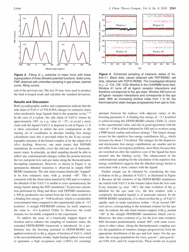
Further insight can be obtained by considering the timeevolution of the �1 dihedral of Val111, as illustrated in Figure4. Because all the windows of the FEP/H-REMD simulationsare started with Val111 in the rotamer taken from the holo stateX-ray structure (�1 near -60°), the time evolution of the �1
dihedral for the apo state (i.e., the first window with acompletely decoupled ligand) is of particular interest. In theFEP/H-REMD simulations, it is observed that the �1 of Val111rapidly starts to make transitions within ∼10 ps toward 180°(red curve), corresponding to the dominant rotamer for the apostate. In contrast, the side chain remains trapped, with �1 around-60° in the straight FEP/REMD simulations (black curve).Moreover, the time evolution of �1 for the holo state (window#40) fluctuates predominantly around 180°, with some excur-sions to other values. Along the thermodynamic coupling axis(λ), the population of rotamers changes progressively from theappropriate distribution of the apo and holo states. For the apostate, the average populations for trans, gauche+, and gauche-
are 0.99, 0.01, and 0.0, respectively. These results are in good
Figure 3. Fitting of �1 potential of mean force with linearsuperposition of three dihedral potential functions. Solid curve,PMF obtained with umbrellas sampling in gas phase; dashedcurve, fitting curves.
Figure 4. Enhanced sampling of rotameric states of Va-line111. Black dots, values obtained with FEP/REMD; reddots, obtained with FEP/H-REMD. The investigated dihedralis �1, C-CA-CB-CG2 dihedral in the CHARMM force field.Window #1 turns off all ligand-receptor interactions andtherefore corresponds to the apo state. Window #40 turns onall ligand-receptor interactions and corresponds to the apostate. With an increasing window index from 1 to 40, thethermodynamic state changes progressively from apo to holo.
Letter J. Chem. Theory Comput., Vol. 6, No. 9, 2010 2563
accord with the values estimated from the PMF of Dill and co-workers (0.99, 0.01, and 0.0008).4 For the holo state, the averagepopulations are 0.16, 0.11, and 0.73, again in good accord withthe values estimated from the PMF reported by Dill and co-workers (0.23, 0.002, and 0.76). For both the apo and holo states,the three possible rotamers are ranked correctly, and theprobability of the dominant state is reproduced within a fewpercent. The enhanced sampling provided by FEP/H-REMD iskey to producing an accurate binding free energy started fromthe holo configuration.
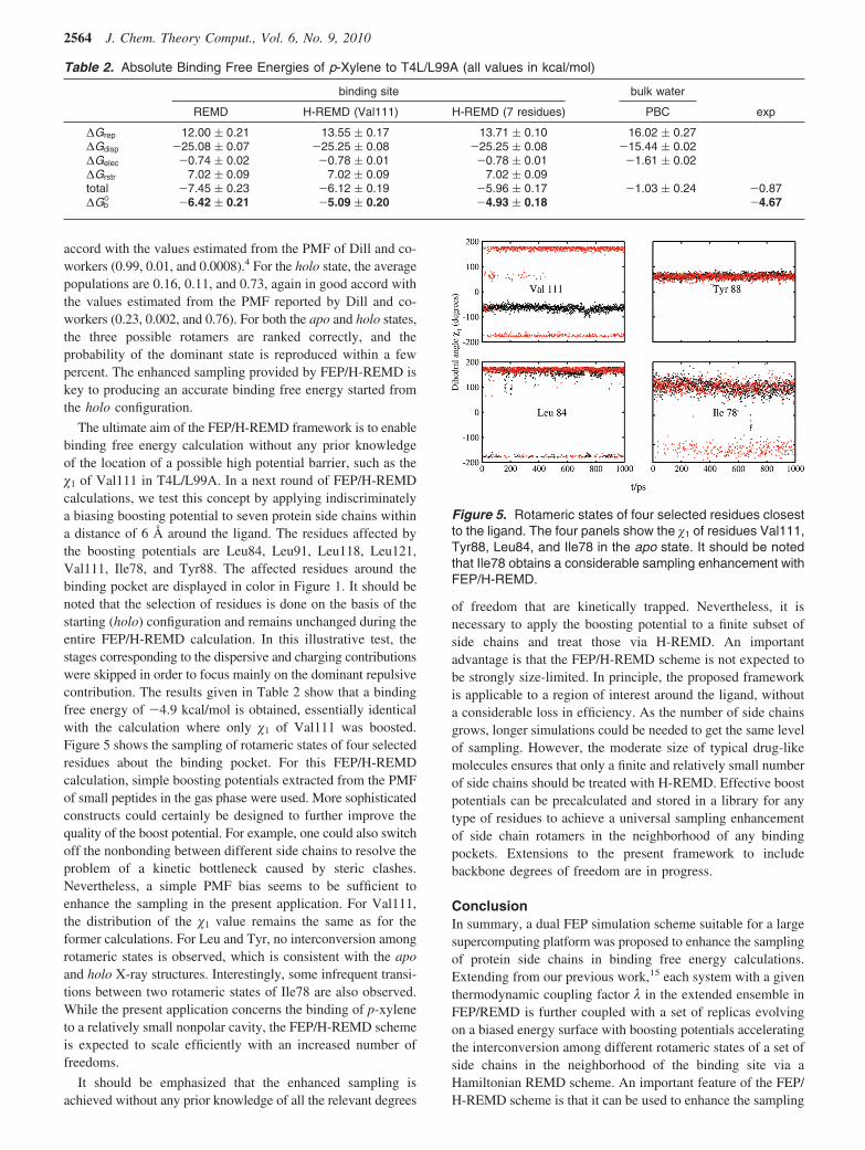
The ultimate aim of the FEP/H-REMD framework is to enablebinding free energy calculation without any prior knowledgeof the location of a possible high potential barrier, such as the�1 of Val111 in T4L/L99A. In a next round of FEP/H-REMDcalculations, we test this concept by applying indiscriminatelya biasing boosting potential to seven protein side chains withina distance of 6 Å around the ligand. The residues affected bythe boosting potentials are Leu84, Leu91, Leu118, Leu121,Val111, Ile78, and Tyr88. The affected residues around thebinding pocket are displayed in color in Figure 1. It should benoted that the selection of residues is done on the basis of thestarting (holo) configuration and remains unchanged during theentire FEP/H-REMD calculation. In this illustrative test, thestages corresponding to the dispersive and charging contributionswere skipped in order to focus mainly on the dominant repulsivecontribution. The results given in Table 2 show that a bindingfree energy of -4.9 kcal/mol is obtained, essentially identicalwith the calculation where only �1 of Val111 was boosted.Figure 5 shows the sampling of rotameric states of four selectedresidues about the binding pocket. For this FEP/H-REMDcalculation, simple boosting potentials extracted from the PMFof small peptides in the gas phase were used. More sophisticatedconstructs could certainly be designed to further improve thequality of the boost potential. For example, one could also switchoff the nonbonding between different side chains to resolve theproblem of a kinetic bottleneck caused by steric clashes.Nevertheless, a simple PMF bias seems to be sufficient toenhance the sampling in the present application. For Val111,the distribution of the �1 value remains the same as for theformer calculations. For Leu and Tyr, no interconversion amongrotameric states is observed, which is consistent with the apoand holo X-ray structures. Interestingly, some infrequent transi-tions between two rotameric states of Ile78 are also observed.While the present application concerns the binding of p-xyleneto a relatively small nonpolar cavity, the FEP/H-REMD schemeis expected to scale efficiently with an increased number offreedoms.
It should be emphasized that the enhanced sampling isachieved without any prior knowledge of all the relevant degrees
of freedom that are kinetically trapped. Nevertheless, it isnecessary to apply the boosting potential to a finite subset ofside chains and treat those via H-REMD. An importantadvantage is that the FEP/H-REMD scheme is not expected tobe strongly size-limited. In principle, the proposed frameworkis applicable to a region of interest around the ligand, withouta considerable loss in efficiency. As the number of side chainsgrows, longer simulations could be needed to get the same levelof sampling. However, the moderate size of typical drug-likemolecules ensures that only a finite and relatively small numberof side chains should be treated with H-REMD. Effective boostpotentials can be precalculated and stored in a library for anytype of residues to achieve a universal sampling enhancementof side chain rotamers in the neighborhood of any bindingpockets. Extensions to the present framework to includebackbone degrees of freedom are in progress.
ConclusionIn summary, a dual FEP simulation scheme suitable for a largesupercomputing platform was proposed to enhance the samplingof protein side chains in binding free energy calculations.Extending from our previous work,15 each system with a giventhermodynamic coupling factor λ in the extended ensemble inFEP/REMD is further coupled with a set of replicas evolvingon a biased energy surface with boosting potentials acceleratingthe interconversion among different rotameric states of a set ofside chains in the neighborhood of the binding site via aHamiltonian REMD scheme. An important feature of the FEP/H-REMD scheme is that it can be used to enhance the sampling
Table 2. Absolute Binding Free Energies of p-Xylene to T4L/L99A (all values in kcal/mol)
binding site bulk water
REMD H-REMD (Val111) H-REMD (7 residues) PBC exp
∆Grep 12.00 ( 0.21 13.55 ( 0.17 13.71 ( 0.10 16.02 ( 0.27∆Gdisp -25.08 ( 0.07 -25.25 ( 0.08 -25.25 ( 0.08 -15.44 ( 0.02∆Gelec -0.74 ( 0.02 -0.78 ( 0.01 -0.78 ( 0.01 -1.61 ( 0.02∆Grstr 7.02 ( 0.09 7.02 ( 0.09 7.02 ( 0.09total -7.45 ( 0.23 -6.12 ( 0.19 -5.96 ( 0.17 -1.03 ( 0.24 -0.87∆Gb° -6.42 ( 0.21 -5.09 ( 0.20 -4.93 ( 0.18 -4.67
Figure 5. Rotameric states of four selected residues closestto the ligand. The four panels show the �1 of residues Val111,Tyr88, Leu84, and Ile78 in the apo state. It should be notedthat Ile78 obtains a considerable sampling enhancement withFEP/H-REMD.
2564 J. Chem. Theory Comput., Vol. 6, No. 9, 2010

of a fairly large number of putative slowly varying degrees offreedom without a considerable loss in efficiency. Sampling ofany residue lining the binding pocket can benefit by the boostingH-REMD from a set of precalculated biasing potentials storedin a library. Application of FEP/H-REMD shows that thesampling of rotamers of the side chains surrounding the nonpolarcavity of T4L/L99A is significantly enhanced and that thebinding free energy for a large ligand such as p-xylene can becalculated accurately by starting from the holo protein config-uration. Further developments of the present method to includethe treatment of backbone reorganization are currently inprogress.
Acknowledgment. We are grateful to Dr. Andrew Binkows-ki for his support. We would like to acknowledge Dr. MilanHodoscek for collaboration with the programming work for theCHARMM REPDSTR module, and Dr. Albert Lau for valuablediscussions about free energy calculations and the replica-exchange scheme. This research is funded by grant MCB-0920261 from the National Science Foundation. Access to thecomputational resources of ALCF at ANL, supported by theOffice of Science of the U.S. Department of Energy (DOE)under contract DE-AC02-06CH11357, was made possible byan INCITE grant from the DOE. The submitted manuscript hasbeen created by UChicago Argonne, LLC, Operator of ANL.ANL, a U.S. DOE Office of Science laboratory, is operatedunder Contract No. DE-AC02-06CH11357.
References
(1) Deng, Y.; Roux, B. J. Chem. Theory Comput. 2006, 2, 1255–1273.
(2) Wang, J.; Deng, Y.; Roux, B. Biophys. J. 2006, 91, 2798–2814.
(3) Mobley, D. L.; Graves, A. P.; Chodera, J. D.; Mcreynolds, A. C.;Shoichet, B. K.; Dill, K. A. J. Mol. Biol. 2007, 371, 1118–1134.
(4) Mobley, D. L.; Chodera, J. D.; Dill, K. A. J. Chem. TheoryComput. 2007, 3, 1231–1235.
(5) Woo, H. J.; Roux, B. Proc. Natl. Acad. Sci. U.S.A. 2005, 102,6825–6830.
(6) Gan, W.; Roux, B. Proteins 2009, 74, 996–1007.
(7) Hritz, J.; Oostenbrink, C. J. Phys. Chem. B 2009, 113, 12711–20.
(8) Jorgensen, W. Acc. Chem. Res. 2009, 42, 724.
(9) Hamelberg, D.; Mongan, J.; McCammon, J. A. J. Chem. Phys.2004, 120, 11919–11929.
(10) Kannan, S.; Zacharis, M. Proteins: Struct., Funct., Bioinf.2007, 66, 697–706.
(11) Chao, X.; Wang, J.; Liu, H. J. Chem. Theory Comput. 2008,4, 1348–1359.
(12) Straatsma, T. P.; McCammon, J., A. J. Chem. Phys. 1994, 101,5032–5039.
(13) Fajer, M.; Hamelberg, D.; McCammon, J. A. J. Chem. TheoryComput. 2008, 4, 1565–1569.
(14) Hritz, J.; Oostenbrink, C. J. Chem. Phys. 2008, 128, 144121.
(15) Jiang, W.; Hodoscek, M.; Roux, B. J. Chem. Theory Comput.2009, 5, 2583–2588.
(16) Woods, C. J.; Essex, J. W.; King, M. A. J. Phys. Chem. B 2003,107, 13711–13718.
(17) Woodcock, H. L., III; Hodoscek, M.; Sherwood, P.; Lee, Y.;Schaefer, H.; Brooks, B. Theor. Chem. Acc. 2003, 109, 140–148.
(18) Woodcock, H. L., III; Hodoscek, M.; Gilbert, A. T. B.; Gill,P. M. W.; Schaefer, H. F., III; R., B. B. J. Comput. Chem. 2007,28, 1485–1502.
(19) Brooks, B. R.; Brooks, C. L., III; Mackerell, A. D., Jr.; Nilsson,L.; Petrella, R. J.; Roux, B.; Won, Y.; Archontis, G.; Bartels, C.;Boresch, S.; Caflisch, A.; Caves, L.; Cui, Q.; Dinner, A. R.; Feig,M.; Fischer, S.; Gao, J.; Hodoscek, M.; Im, W.; Kuczera, K.;Lazaridis, T.; Ma, J.; Ovchinnikov, V.; Paci, E.; Pastor, R. W.;Post, C. B.; Pu, J. Z.; Schaefer, M.; Tidor, B.; Venable, R. M.;Woodcock, H. L.; Wu, X.; Yang, W.; York, D. M.; Karplus, M.J. Comput. Chem. 2009, 30, 1545–1614.
(20) Deng, Y.; Roux, B. J. Phys. Chem. 2004, 108, 16567–16576.
(21) Weeks, J. D.; Chandler, D.; Anderson, H. C. J. Chem. Phys.1971, 54, 5237–5247.
(22) MacKerell, A. D.; Bashford, D.; Bellott, M.; Dunbrack, R. L.;Evanseck, J. D.; Field, M. J.; Fischer, S.; Gao, J.; Guo, H.;Ha, S.; Joseph-McCarthy, D.; Kuchnir, L.; Kuczera, K.; Lau,F. T. K.; Mattos, C.; Michnick, S.; Ngo, T.; Nguyen, D. T.;Prodhom, B.; Reiher, W. E.; Roux, B.; Schlenkrich, M.; Smith,J. C.; Stote, R.; Straub, J.; Wantanabe, M.; Wiorkiewicz-Kuczera, J.; Yin, D.; Karplus, M. J. Phys. Chem. B 1998,102, 3586–3616.
(23) Kumar, S.; Bouzida, D.; Swendsen, R. H.; Kollman, P. A.;Rosenberg, J. M. J. Comput. Chem. 1992, 13, 1011–1021.
(24) Im, W.; Berneche, S.; Roux, B. J. Chem. Phys. 2000, 114, 2924–2937.
(25) Morton, A.; Matthews, B. W. Biochemistry 1995, 34, 8576-8588.
CT1001768
Letter J. Chem. Theory Comput., Vol. 6, No. 9, 2010 2565