A demanding industry requires a versatile solution portfolio ...
Upload
independentCategory
view
2download
0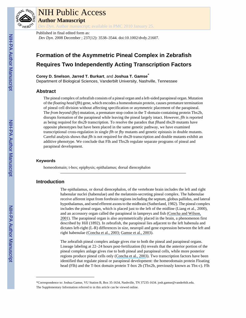
Formation of the Asymmetric Pineal Complex in ZebrafishRequires Two Independently Acting Transcription Factors
Corey D. Snelson, Jarred T. Burkart, and Joshua T. Gamse*Department of Biological Sciences, Vanderbilt University, Nashville, Tennessee
AbstractThe pineal complex of zebrafish consists of a pineal organ and a left-sided parapineal organ. Mutationof the floating head (flh) gene, which encodes a homeodomain protein, causes premature terminationof pineal cell division without affecting specification or asymmetric placement of the parapineal.The from beyond (fby) mutation, a premature stop codon in the T-domain-containing protein Tbx2b,disrupts formation of the parapineal while leaving the pineal largely intact. However, flh is reportedas being required for tbx2b transcription. To resolve the paradox that flhand tbx2b mutants haveopposite phenotypes but have been placed in the same genetic pathway, we have examinedtranscriptional cross-regulation in single flh or fby mutants and genetic epistasis in double mutants.Careful analysis shows that flh is not required for tbx2b transcription and double mutants exhibit anadditive phenotype. We conclude that Flh and Tbx2b regulate separate programs of pineal andparapineal development.
Keywordshomeodomain; t-box; epiphysis; epithalamus; dorsal diencephalon
IntroductionThe epithalamus, or dorsal diencephalon, of the vertebrate brain includes the left and righthabenular nuclei (habenulae) and the melatonin-secreting pineal complex. The habenulaereceive afferent input from forebrain regions including the septum, globus pallidus, and lateralhypothalamus, and send efferent axons to the midbrain (Sutherland, 1982). The pineal complexincludes the pineal organ, which is placed just to the left of the midline (Liang et al., 2000),and an accessory organ called the parapineal in lampreys and fish (Concha and Wilson,2001). The parapineal organ is also asymmetrically placed in the brain, a phenomenon firstdescribed by Hill (1892). In zebrafish, the parapineal lies adjacent to the left habenula anddictates left-right (L-R) differences in size, neuropil and gene expression between the left andright habenulae (Concha et al., 2003; Gamse et al., 2003).
The zebrafish pineal complex anlage gives rise to both the pineal and parapineal organs.Lineage labeling at 22–24 hours post-fertilization (h) reveals that the anterior portion of thepineal complex anlage gives rise to both pineal and parapineal cells, while more posteriorregions produce pineal cells only (Concha et al., 2003). Two transcription factors have beenidentified that regulate pineal or parapineal development: the homeodomain protein Floatinghead (Flh) and the T-box domain protein T-box 2b (Tbx2b, previously known as Tbx-c). Flh
*Correspondence to: Joshua Gamse, VU Station B, Box 35-1634, Nashville, TN 37235-1634. [email protected] Supplementary Information referred to in this article can be viewed online.
NIH Public AccessAuthor ManuscriptDev Dyn. Author manuscript; available in PMC 2010 January 25.
Published in final edited form as:Dev Dyn. 2008 December ; 237(12): 3538–3544. doi:10.1002/dvdy.21607.
NIH
-PA Author Manuscript
NIH
-PA Author Manuscript
NIH
-PA Author Manuscript
activity promotes the generation of pineal cells; in flhn1 mutants, neurogenesis initiates in thepineal complex but ends prematurely at 18 somites (s) stage (Masai et al., 1997). By contrast,Tbx2b is required for the specification and migration of parapineal cells; in fbyc144 mutants (amutation in tbx2b), few or no parapineal cells develop and those that do fail to migrate to theleft side of the brain (Snelson et al., 2008).
The regulatory relationship between flh and tbx2b is unclear. In the pineal complex anlage,flh expression begins at 80% epiboly (Masai et al., 1997), about 4 hr prior to tbx2b expression(Snelson et al., 2008). Expression of tbx2b and flh overlaps extensively within the pinealcomplex anlage at 24 h (Snelson et al., 2008). A previous publication (Cau and Wilson,2003) suggested that Flh is required for expression of tbx2b. However, the phenotypes of flhand fby mutants are very different, arguing against the two genes acting in the same pathway.Asymmetrically-placed parapineal cells form in flh mutant larvae (Gamse et al., 2002, 2003),but not fby mutant larvae (Snelson et al., 2008). This observation prompted a re-evaluation oftbx2b and flh cross-regulation.
Examination of flh and tbx2b mRNA in fby and flh mutants reveals that they do not regulateone another's expression, and flh;fby double mutants exhibit an additive phenotype.Additionally, in flh mutants, specification and asymmetric migration of parapineal cells areindistinguishable from wild-type (WT) embryos. These results indicate that flh and tbx2b actindependently of one another during pineal versus parapineal development.
ResultsParapineal Cell Division Ends Prior to Pineal Cell Division
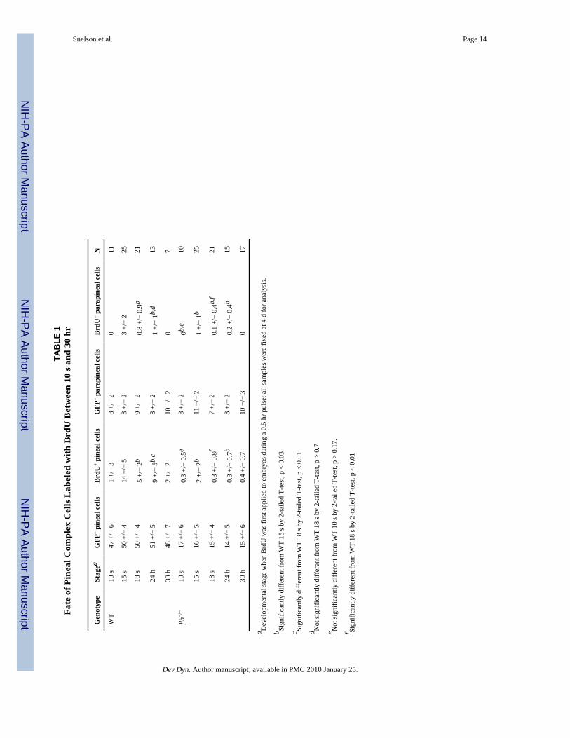
To identify the period of development when pineal and parapineal cells are dividing, weperformed BrdU pulse labeling at stages between 10 s and 30 hr and examined the fate oflabeled cells at 4 days post-fertilization (d). To identify pineal and parapineal cells, embryoscarrying the transgene Tg-(foxd3:gfp)fkg3 were used (Gilmour et al., 2002). Peak labeling ofparapineal cells occurred when embryos received a half-hour pulse of BrdU at the 15 s stage(Fig. 1A,E,F and Table 1); BrdU pulses before (10 s) or after (18 s or later) labeled few or noparapineal cells (Fig. 1E,F and Table 1). Pineal cells also have maximal incorporation of BrdUat 15 s; interestingly, a second smaller peak of labeling occurs at 24 h (Fig. 1C,E,G and Table1). In flh mutant larvae, pineal and parapineal BrdU labeling at the 10 s stage is similar to WTand increases at 15 s; however, almost no BrdU labeling of the pineal or parapineal occurs ator after 18 s (Fig. 1B,D,F,G and Table 1). The number of foxd3:gfp-positive cells in the pinealorgan is reduced (Table 1), consistent with a premature end to pineal neurogenesis in flhmutants (Masai et al., 1997).
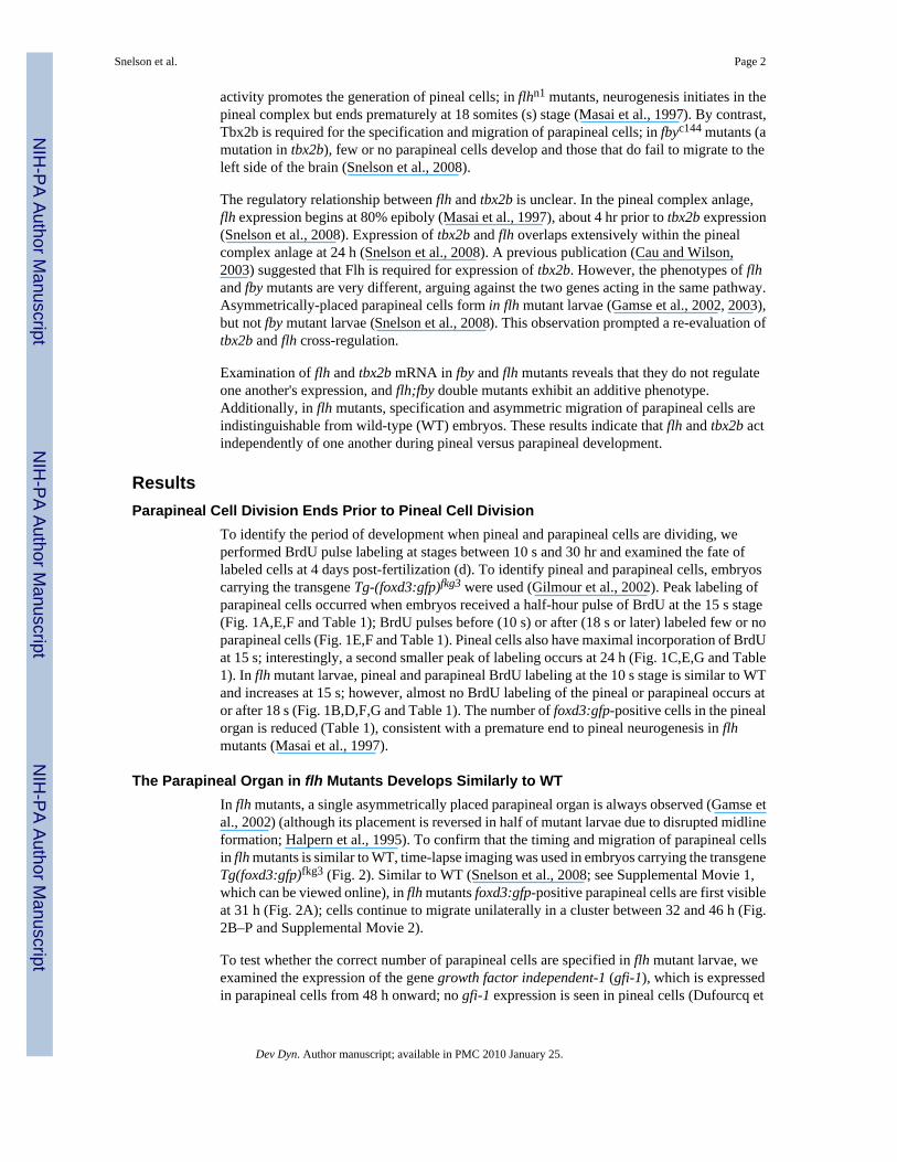
The Parapineal Organ in flh Mutants Develops Similarly to WTIn flh mutants, a single asymmetrically placed parapineal organ is always observed (Gamse etal., 2002) (although its placement is reversed in half of mutant larvae due to disrupted midlineformation; Halpern et al., 1995). To confirm that the timing and migration of parapineal cellsin flh mutants is similar to WT, time-lapse imaging was used in embryos carrying the transgeneTg(foxd3:gfp)fkg3 (Fig. 2). Similar to WT (Snelson et al., 2008; see Supplemental Movie 1,which can be viewed online), in flh mutants foxd3:gfp-positive parapineal cells are first visibleat 31 h (Fig. 2A); cells continue to migrate unilaterally in a cluster between 32 and 46 h (Fig.2B–P and Supplemental Movie 2).
To test whether the correct number of parapineal cells are specified in flh mutant larvae, weexamined the expression of the gene growth factor independent-1 (gfi-1), which is expressedin parapineal cells from 48 h onward; no gfi-1 expression is seen in pineal cells (Dufourcq et
Snelson et al. Page 2
Dev Dyn. Author manuscript; available in PMC 2010 January 25.
NIH
-PA Author Manuscript
NIH
-PA Author Manuscript
NIH
-PA Author Manuscript
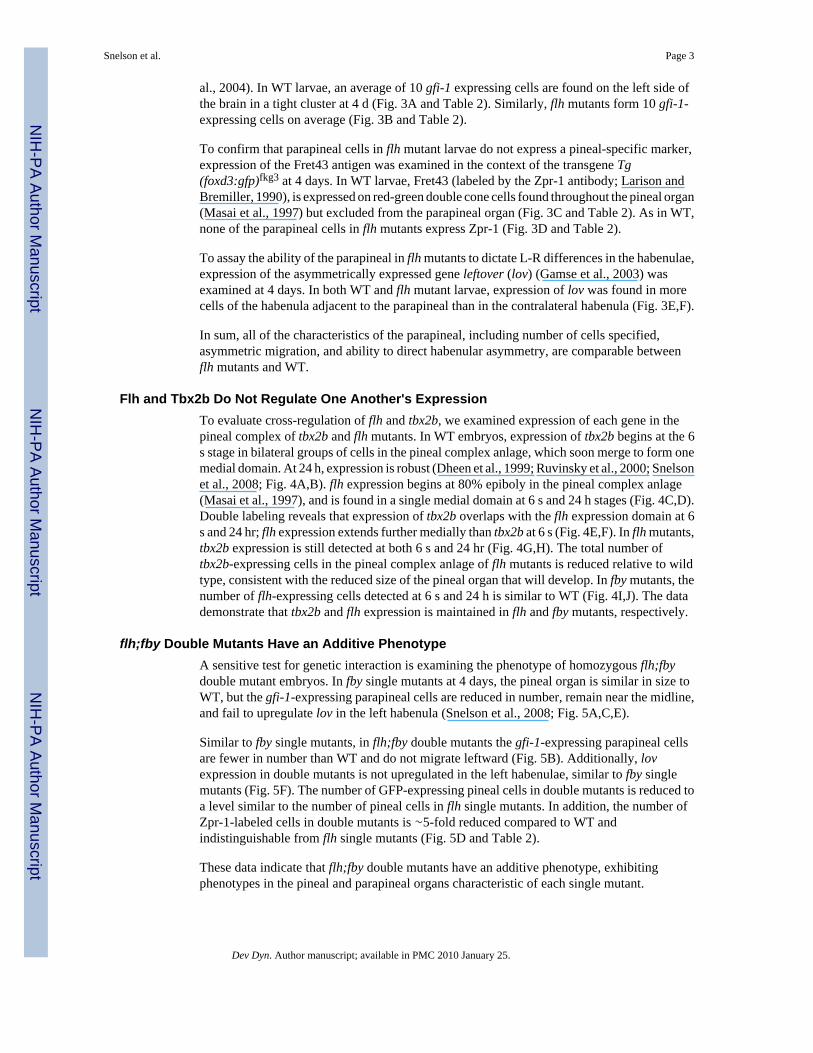
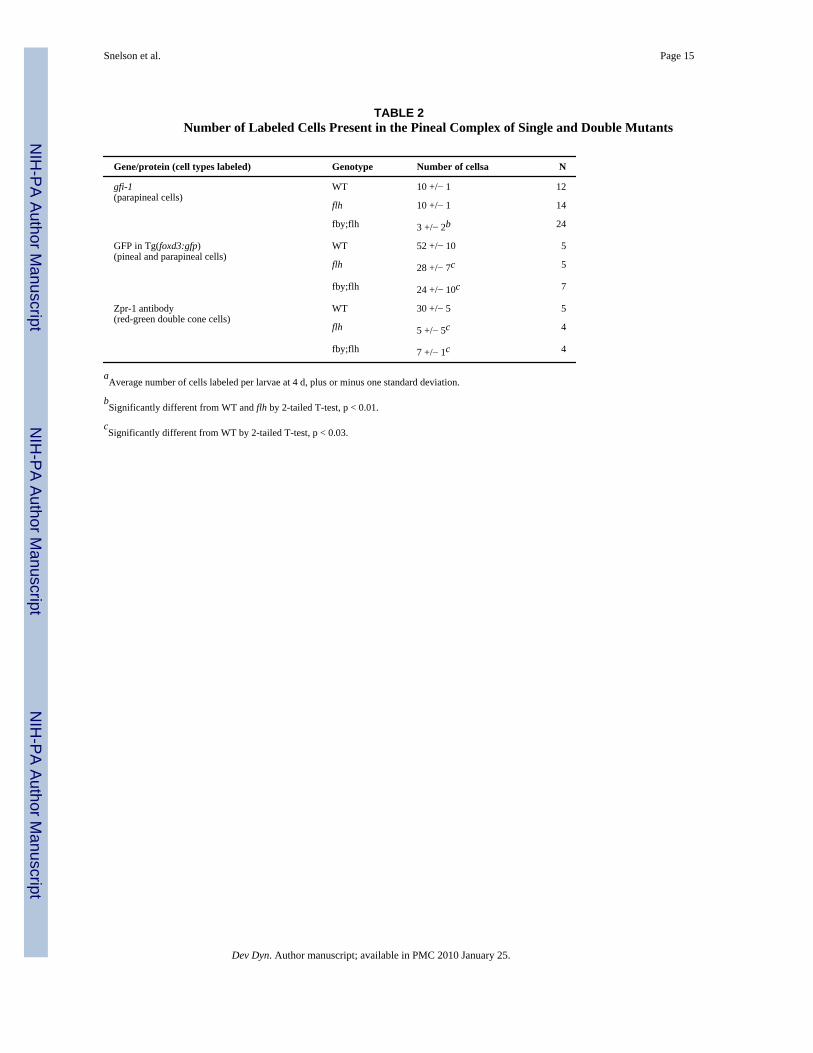
al., 2004). In WT larvae, an average of 10 gfi-1 expressing cells are found on the left side ofthe brain in a tight cluster at 4 d (Fig. 3A and Table 2). Similarly, flh mutants form 10 gfi-1-expressing cells on average (Fig. 3B and Table 2).
To confirm that parapineal cells in flh mutant larvae do not express a pineal-specific marker,expression of the Fret43 antigen was examined in the context of the transgene Tg(foxd3:gfp)fkg3 at 4 days. In WT larvae, Fret43 (labeled by the Zpr-1 antibody; Larison andBremiller, 1990), is expressed on red-green double cone cells found throughout the pineal organ(Masai et al., 1997) but excluded from the parapineal organ (Fig. 3C and Table 2). As in WT,none of the parapineal cells in flh mutants express Zpr-1 (Fig. 3D and Table 2).
To assay the ability of the parapineal in flh mutants to dictate L-R differences in the habenulae,expression of the asymmetrically expressed gene leftover (lov) (Gamse et al., 2003) wasexamined at 4 days. In both WT and flh mutant larvae, expression of lov was found in morecells of the habenula adjacent to the parapineal than in the contralateral habenula (Fig. 3E,F).
In sum, all of the characteristics of the parapineal, including number of cells specified,asymmetric migration, and ability to direct habenular asymmetry, are comparable betweenflh mutants and WT.
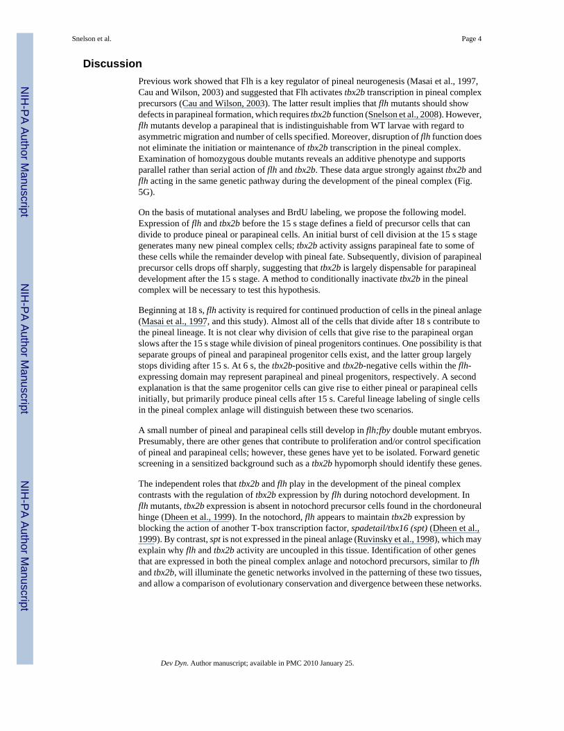
Flh and Tbx2b Do Not Regulate One Another's ExpressionTo evaluate cross-regulation of flh and tbx2b, we examined expression of each gene in thepineal complex of tbx2b and flh mutants. In WT embryos, expression of tbx2b begins at the 6s stage in bilateral groups of cells in the pineal complex anlage, which soon merge to form onemedial domain. At 24 h, expression is robust (Dheen et al., 1999; Ruvinsky et al., 2000; Snelsonet al., 2008; Fig. 4A,B). flh expression begins at 80% epiboly in the pineal complex anlage(Masai et al., 1997), and is found in a single medial domain at 6 s and 24 h stages (Fig. 4C,D).Double labeling reveals that expression of tbx2b overlaps with the flh expression domain at 6s and 24 hr; flh expression extends further medially than tbx2b at 6 s (Fig. 4E,F). In flh mutants,tbx2b expression is still detected at both 6 s and 24 hr (Fig. 4G,H). The total number oftbx2b-expressing cells in the pineal complex anlage of flh mutants is reduced relative to wildtype, consistent with the reduced size of the pineal organ that will develop. In fby mutants, thenumber of flh-expressing cells detected at 6 s and 24 h is similar to WT (Fig. 4I,J). The datademonstrate that tbx2b and flh expression is maintained in flh and fby mutants, respectively.
flh;fby Double Mutants Have an Additive PhenotypeA sensitive test for genetic interaction is examining the phenotype of homozygous flh;fbydouble mutant embryos. In fby single mutants at 4 days, the pineal organ is similar in size toWT, but the gfi-1-expressing parapineal cells are reduced in number, remain near the midline,and fail to upregulate lov in the left habenula (Snelson et al., 2008; Fig. 5A,C,E).
Similar to fby single mutants, in flh;fby double mutants the gfi-1-expressing parapineal cellsare fewer in number than WT and do not migrate leftward (Fig. 5B). Additionally, lovexpression in double mutants is not upregulated in the left habenulae, similar to fby singlemutants (Fig. 5F). The number of GFP-expressing pineal cells in double mutants is reduced toa level similar to the number of pineal cells in flh single mutants. In addition, the number ofZpr-1-labeled cells in double mutants is ∼5-fold reduced compared to WT andindistinguishable from flh single mutants (Fig. 5D and Table 2).
These data indicate that flh;fby double mutants have an additive phenotype, exhibitingphenotypes in the pineal and parapineal organs characteristic of each single mutant.
Snelson et al. Page 3
Dev Dyn. Author manuscript; available in PMC 2010 January 25.
NIH
-PA Author Manuscript
NIH
-PA Author Manuscript
NIH
-PA Author Manuscript
DiscussionPrevious work showed that Flh is a key regulator of pineal neurogenesis (Masai et al., 1997,Cau and Wilson, 2003) and suggested that Flh activates tbx2b transcription in pineal complexprecursors (Cau and Wilson, 2003). The latter result implies that flh mutants should showdefects in parapineal formation, which requires tbx2b function (Snelson et al., 2008). However,flh mutants develop a parapineal that is indistinguishable from WT larvae with regard toasymmetric migration and number of cells specified. Moreover, disruption of flh function doesnot eliminate the initiation or maintenance of tbx2b transcription in the pineal complex.Examination of homozygous double mutants reveals an additive phenotype and supportsparallel rather than serial action of flh and tbx2b. These data argue strongly against tbx2b andflh acting in the same genetic pathway during the development of the pineal complex (Fig.5G).
On the basis of mutational analyses and BrdU labeling, we propose the following model.Expression of flh and tbx2b before the 15 s stage defines a field of precursor cells that candivide to produce pineal or parapineal cells. An initial burst of cell division at the 15 s stagegenerates many new pineal complex cells; tbx2b activity assigns parapineal fate to some ofthese cells while the remainder develop with pineal fate. Subsequently, division of parapinealprecursor cells drops off sharply, suggesting that tbx2b is largely dispensable for parapinealdevelopment after the 15 s stage. A method to conditionally inactivate tbx2b in the pinealcomplex will be necessary to test this hypothesis.
Beginning at 18 s, flh activity is required for continued production of cells in the pineal anlage(Masai et al., 1997, and this study). Almost all of the cells that divide after 18 s contribute tothe pineal lineage. It is not clear why division of cells that give rise to the parapineal organslows after the 15 s stage while division of pineal progenitors continues. One possibility is thatseparate groups of pineal and parapineal progenitor cells exist, and the latter group largelystops dividing after 15 s. At 6 s, the tbx2b-positive and tbx2b-negative cells within the flh-expressing domain may represent parapineal and pineal progenitors, respectively. A secondexplanation is that the same progenitor cells can give rise to either pineal or parapineal cellsinitially, but primarily produce pineal cells after 15 s. Careful lineage labeling of single cellsin the pineal complex anlage will distinguish between these two scenarios.
A small number of pineal and parapineal cells still develop in flh;fby double mutant embryos.Presumably, there are other genes that contribute to proliferation and/or control specificationof pineal and parapineal cells; however, these genes have yet to be isolated. Forward geneticscreening in a sensitized background such as a tbx2b hypomorph should identify these genes.
The independent roles that tbx2b and flh play in the development of the pineal complexcontrasts with the regulation of tbx2b expression by flh during notochord development. Inflh mutants, tbx2b expression is absent in notochord precursor cells found in the chordoneuralhinge (Dheen et al., 1999). In the notochord, flh appears to maintain tbx2b expression byblocking the action of another T-box transcription factor, spadetail/tbx16 (spt) (Dheen et al.,1999). By contrast, spt is not expressed in the pineal anlage (Ruvinsky et al., 1998), which mayexplain why flh and tbx2b activity are uncoupled in this tissue. Identification of other genesthat are expressed in both the pineal complex anlage and notochord precursors, similar to flhand tbx2b, will illuminate the genetic networks involved in the patterning of these two tissues,and allow a comparison of evolutionary conservation and divergence between these networks.
Snelson et al. Page 4
Dev Dyn. Author manuscript; available in PMC 2010 January 25.
NIH
-PA Author Manuscript
NIH
-PA Author Manuscript
NIH
-PA Author Manuscript
Experimental ProceduresZebrafish
Zebrafish were raised at 28.5°C on a 14/10 hr light/dark cycle and staged according to hours(h) or days (d) post-fertilization. The wild-type AB strain (Walker, 1999); the transgenic linesTg(foxd3:GFP)fkg17 and Tg(foxd3:GF-P)fkg3 (Gilmour et al., 2002), and mutants carrying thespontaneous allele flhn1 (Halpern et al., 1995) and the ethylnitrosourea-induced null allelefbyc144 (Snelson et al., 2008) were used.
RNA In Situ HybridizationWhole-mount RNA in situ hybridization was performed as described previously (Gamse et al.,2003), using reagents from Roche Applied Bioscience. RNA probes were labeled usingfluorescein-UTP or digoxygenin UTP. To synthesize antisense RNA probes, znot (flh) (Talbotet al., 1995) and pBK-CMV-leftover (Gamse et al., 2003) were linearized with EcoRI andtranscribed with T7 RNA polymerase; pCRII-tbx2b (Snelson et al., 2008) with BamHI and T7RNA polymerase; and pBS-gfi1 (Dufourcq et al., 2004) with SacII and T3 RNA polymerase.Embryos were incubated at 70°C with probe and hybridization solution containing 50%formamide (or 65% for cyc). Hybridized probes were detected using alkaline phosphatase-conjugated antibodies and visualized by 4-nitro blue tetrazolium (NBT) and 5-bromo-4-chloro-3-indolyl-phosphate (BCIP) staining. All in situ data was collected on a LeicaDM6000B microscope with a 20× or 40× objective.
BrdU LabelingFor pulse labeling with bromodeoxyuridine (BrdU, Roche), embryos were incubated with ice-cold 10 mM BrdU dissolved in embryo medium plus DMSO at 2% for 10–15 s embryos, 5%for 18-s-stage embryos, or 10% for 24-30-h embryos, over a period of 30 min. Embryos werethen washed extensively with embryo medium and raised to 4 d, when they were fixed in 4%paraformaldehyde for 2 hours at room temperature.
Time-Lapse ImagingFor time-lapse imaging, WT or flh mutant embryos carrying the transgene (foxd3:GFP)fkg3
were mounted in a 0.8% solution of low melt agarose containing 0.003% PTU and anesthetizedusing 0.04% Tricaine. Images were collected on a Zeiss/Perkin Elmer spinning disk confocalmicroscope with a 40× oil-immersion objective every 15 min from 31 to 46 hr, and analyzedusing Volocity software (Improvision).
ImmunofluorescenceFor whole-mount immunohistochemistry, 4 d larvae were fixed in 4% paraformaldehydeovernight (for anti-Zpr-1/anti-GFP) or 2 hr (for anti-BrdU/anti-GFP). Samples werepermeabilized by treatment with 10 μg/ml Proteinase K (Roche Applied Bioscience) andrefixed in 4% paraformaldehyde. For BrdU labeling, samples were additionally washed indeionized water following refixation, and incubated in 2 N HCl for 1 hr. All samples wereblocked in PBS with 0.1% Triton X-100, 2% sheep serum, 1% DMSO, and 10% BSA (PB-STrS). For antibody labeling, rabbit anti-GFP (1:1,000, Torrey Pines Biolabs), mouse anti-Zpr-1 (1:250, Zebrafish International Resource Center, Eugene, OR), and mouse anti-BrdUantibody (1: 200, developed by Stephen Kaufman, obtained from the Developmental StudiesHybridoma Bank, University of Iowa) were used. Larvae were incubated overnight in primaryantibody diluted in PBSTrS. Primary antibody was detected using goat-anti-rabbit or goat-anti-mouse secondary antibodies conjugated to the Alexa 568 or Alexa 488 fluorophores (1:350,Molecular Probes). Immunofluorescence data were collected on a Zeiss/Perkin Elmer spinning
Snelson et al. Page 5
Dev Dyn. Author manuscript; available in PMC 2010 January 25.
NIH
-PA Author Manuscript
NIH
-PA Author Manuscript
NIH
-PA Author Manuscript
disk confocal microscope or a Zeiss LSM 510 Meta with a 40× oil-immersion objective andanalyzed with Volocity software (Improvision).
Supplementary MaterialRefer to Web version on PubMed Central for supplementary material.
AcknowledgmentsWe thank Erin Booton and Heidi Beck for expert fish care. We acknowledge the Developmental Studies HybridomaBank at the University of Iowa (developed under the auspices of the NICHD) for the anti-BrdU antibody, and theVanderbilt Cell Imaging Shared Resource for microscopy support.
Grant sponsor: Whitehall Foundation; Grant number: 2005-08-79-APL.
ReferencesCau E, Wilson SW. Ash1a and Neurogenin1 function downstream of Floating head to regulate epiphysial
neurogenesis. Development 2003;130:2455–2466. [PubMed: 12702659]Concha ML, Wilson SW. Asymmetry in the epithalamus of vertebrates. J Anat 2001;199:63–84.
[PubMed: 11523830]Concha ML, Russell C, Regan JC, Tawk M, Sidi S, Gilmour DT, Kapsimali M, Sumoy L, Goldstone K,
Amaya E, Kimelman D, Nicolson T, Grunder S, Gomperts M, Clarke JD, Wilson SW. Local tissueinteractions across the dorsal midline of the forebrain establish CNS laterality. Neuron 2003;39:423–438. [PubMed: 12895418]
Dheen T, Sleptsova-Friedrich I, Xu Y, Clark M, Lehrach H, Gong Z, Korzh V. Zebrafish tbx-c functionsduring formation of midline structures. Development 1999;126:2703–2713. [PubMed: 10331981]
Dufourcq P, Rastegar S, Strahle U, Blader P. Parapineal specific expression of gfi1 in the zebrafishepithalamus. Gene Expr Patterns 2004;4:53–57. [PubMed: 14678828]
Gamse JT, Shen YC, Thisse C, Thisse B, Raymond PA, Halpern ME, Liang JO. Otx5 regulates genesthat show circadian expression in the zebrafish pineal complex. Nat Genet 2002;30:117–121.[PubMed: 11753388]
Gamse JT, Thisse C, Thisse B, Halpern ME. The parapineal mediates left-right asymmetry in the zebrafishdiencephalon. Development 2003;130:1059–1068. [PubMed: 12571098]
Gilmour DT, Maischein HM, Nusslein-Volhard C. Migration and function of a glial subtype in thevertebrate peripheral nervous system. Neuron 2002;34:577–588. [PubMed: 12062041]
Halpern ME, Thisse C, Ho RK, Thisse B, Riggleman B, Trevarrow B, Weinberg ES, Postlethwait JH,Kimmel CB. Cell-autonomous shift from axial to paraxial mesodermal development in zebrafishfloating head mutants. Development 1995;121:4257–4264. [PubMed: 8575325]
Hill C. Development of the epiphysis in Coregonus Albus. J Morphol 1892;5:503–510.Larison KD, Bremiller R. Early onset of phenotype and cell patterning in the embryonic zebrafish retina.
Development 1990;109:567–576. [PubMed: 2401210]Liang JO, Etheridge A, Hantsoo L, Rubinstein AL, Nowak SJ, Izpisua Belmonte JC, Halpern ME.
Asymmetric nodal signaling in the zebrafish diencephalon positions the pineal organ. Development2000;127:5101–5112. [PubMed: 11060236]
Masai I, Heisenberg CP, Barth KA, Macdonald R, Adamek S, Wilson SW. floating head and masterblindregulate neuronal patterning in the roof of the forebrain. Neuron 1997;18:43–57. [PubMed: 9010204]
Ruvinsky I, Silver LM, Ho RK. Characterization of the zebrafish tbx16 gene and evolution of thevertebrate T-box family. Dev Genes Evol 1998;208:94–99. [PubMed: 9569350]
Ruvinsky I, Oates AC, Silver LM, Ho RK. The evolution of paired appendages in vertebrates: T-boxgenes in the zebrafish. Dev Genes Evol 2000;210:82–91. [PubMed: 10664151]
Snelson CD, Santhakumar K, Halpern ME, Gamse JT. Tbx2b is required for the development of theparapineal organ. Development 2008;135:1693–1702. [PubMed: 18385257]
Snelson et al. Page 6
Dev Dyn. Author manuscript; available in PMC 2010 January 25.
NIH
-PA Author Manuscript
NIH
-PA Author Manuscript
NIH
-PA Author Manuscript
Sutherland RJ. The dorsal diencephalic conduction system: a review of the anatomy and functions of thehabenular complex. Neurosci Biobehav Rev 1982;6:1–13. [PubMed: 7041014]
Talbot WS, Trevarrow B, Halpern ME, Melby AE, Farr G, Postlethwait JH, Jowett T, Kimmel CB,Kimelman D. A homeobox gene essential for zebrafish notochord development. Nature1995;378:150–157. [PubMed: 7477317]
Walker, C. Methods in Cell Biology. London: Elsevier; 1999. Haploid screens and gamma-raymutagenesis; p. 43-70.
Snelson et al. Page 7
Dev Dyn. Author manuscript; available in PMC 2010 January 25.
NIH
-PA Author Manuscript
NIH
-PA Author Manuscript
NIH
-PA Author Manuscript
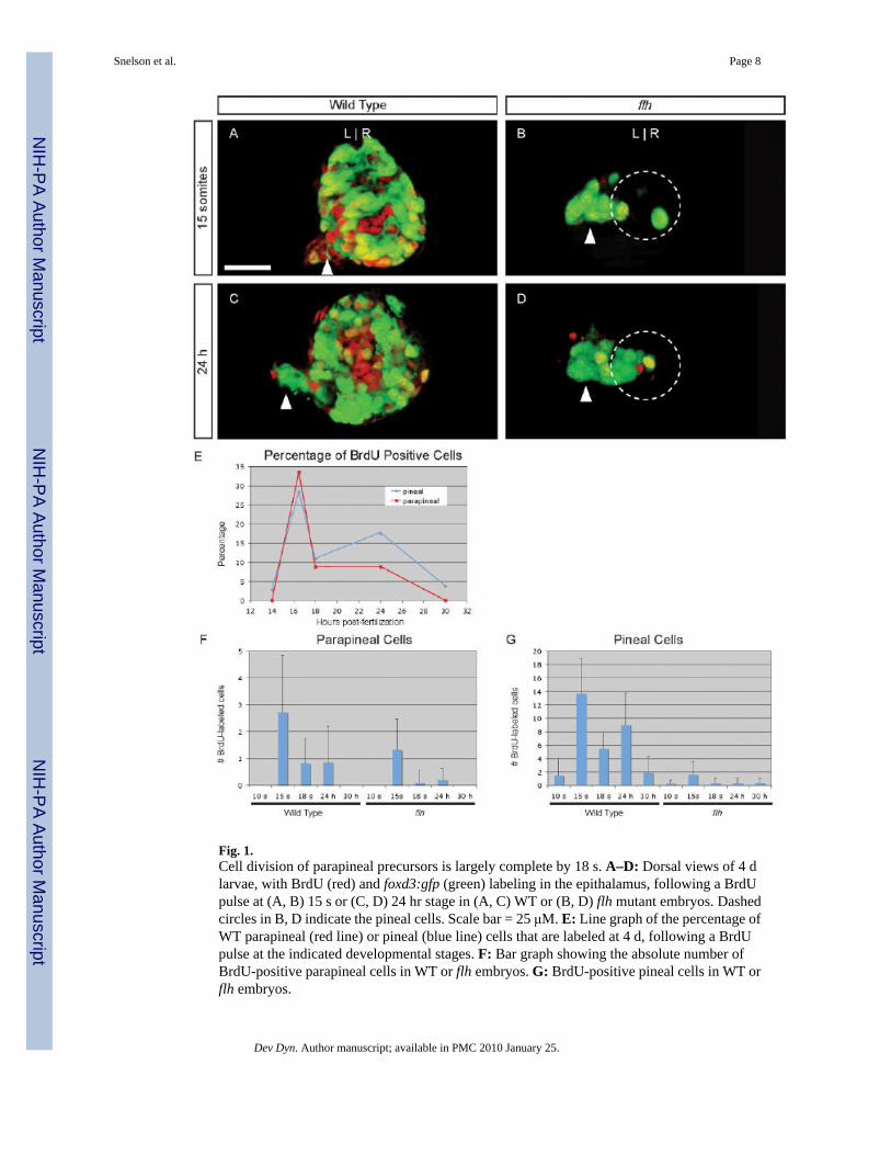
Fig. 1.Cell division of parapineal precursors is largely complete by 18 s. A–D: Dorsal views of 4 dlarvae, with BrdU (red) and foxd3:gfp (green) labeling in the epithalamus, following a BrdUpulse at (A, B) 15 s or (C, D) 24 hr stage in (A, C) WT or (B, D) flh mutant embryos. Dashedcircles in B, D indicate the pineal cells. Scale bar = 25 μM. E: Line graph of the percentage ofWT parapineal (red line) or pineal (blue line) cells that are labeled at 4 d, following a BrdUpulse at the indicated developmental stages. F: Bar graph showing the absolute number ofBrdU-positive parapineal cells in WT or flh embryos. G: BrdU-positive pineal cells in WT orflh embryos.
Snelson et al. Page 8
Dev Dyn. Author manuscript; available in PMC 2010 January 25.
NIH
-PA Author Manuscript
NIH
-PA Author Manuscript
NIH
-PA Author Manuscript
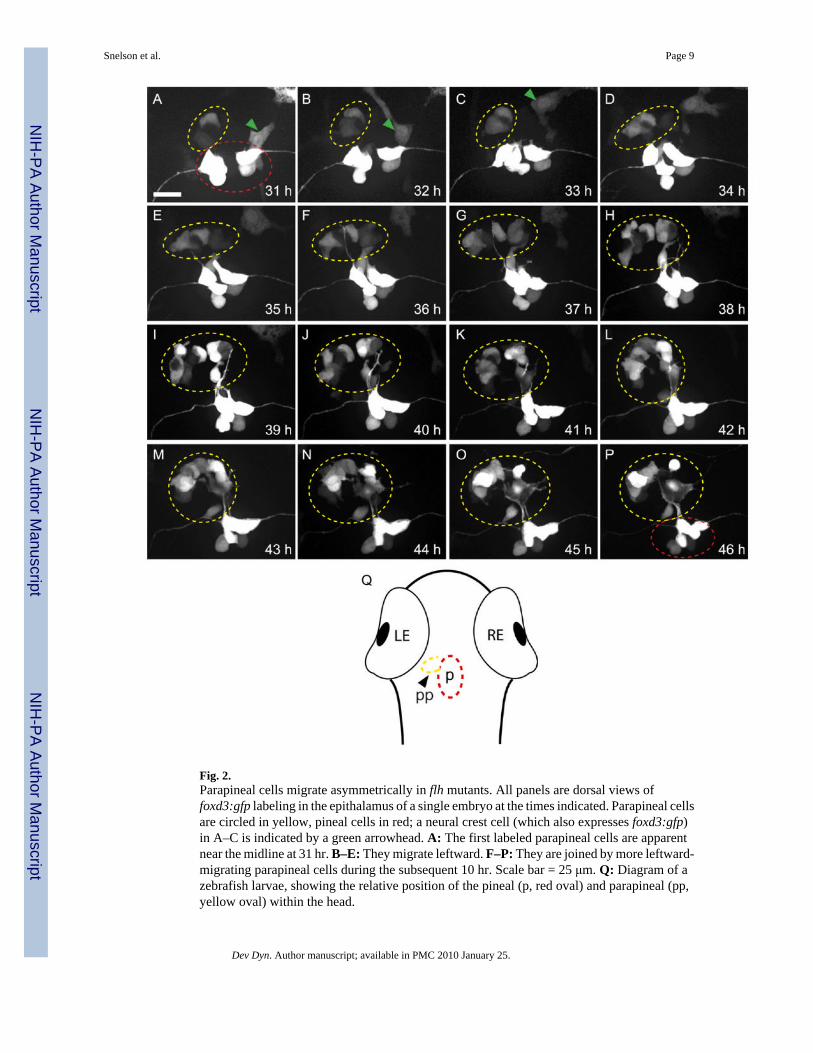
Fig. 2.Parapineal cells migrate asymmetrically in flh mutants. All panels are dorsal views offoxd3:gfp labeling in the epithalamus of a single embryo at the times indicated. Parapineal cellsare circled in yellow, pineal cells in red; a neural crest cell (which also expresses foxd3:gfp)in A–C is indicated by a green arrowhead. A: The first labeled parapineal cells are apparentnear the midline at 31 hr. B–E: They migrate leftward. F–P: They are joined by more leftward-migrating parapineal cells during the subsequent 10 hr. Scale bar = 25 μm. Q: Diagram of azebrafish larvae, showing the relative position of the pineal (p, red oval) and parapineal (pp,yellow oval) within the head.
Snelson et al. Page 9
Dev Dyn. Author manuscript; available in PMC 2010 January 25.
NIH
-PA Author Manuscript
NIH
-PA Author Manuscript
NIH
-PA Author Manuscript
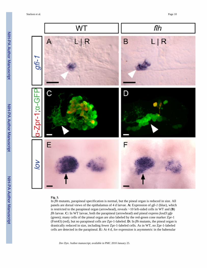
Fig. 3.In flh mutants, parapineal specification is normal, but the pineal organ is reduced in size. Allpanels are dorsal views of the epithalamus of 4 d larvae. A: Expression of gfi-1 (blue), whichis restricted to the parapineal organ (arrowhead), reveals ∼10 left-sided cells in WT and (B)flh larvae. C: In WT larvae, both the parapineal (arrowhead) and pineal express foxd3:gfp(green); many cells of the pineal organ are also labeled by the red-green cone marker Zpr-1(Fret43) (red), but no parapineal cells are Zpr-1-labeled. D: In flh mutants, the pineal organ isdrastically reduced in size, including fewer Zpr-1-labeled cells. As in WT, no Zpr-1-labeledcells are detected in the parapineal. E: At 4 d, lov expression is asymmetric in the habenular
Snelson et al. Page 10
Dev Dyn. Author manuscript; available in PMC 2010 January 25.
NIH
-PA Author Manuscript
NIH
-PA Author Manuscript
NIH
-PA Author Manuscript

nuclei; the habenula adjacent to the parapineal (arrow) expresses lov extensively in WT and(F) flh mutants. Scale bars = 25 μm.
Snelson et al. Page 11
Dev Dyn. Author manuscript; available in PMC 2010 January 25.
NIH
-PA Author Manuscript
NIH
-PA Author Manuscript
NIH
-PA Author Manuscript
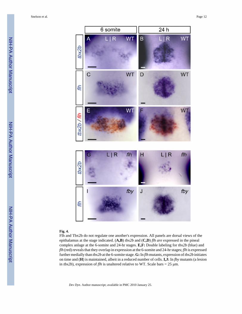
Fig. 4.Flh and Tbx2b do not regulate one another's expression. All panels are dorsal views of theepithalamus at the stage indicated. (A,B) tbx2b and (C,D) flh are expressed in the pinealcomplex anlage at the 6-somite and 24-hr stages. E,F: Double labeling for tbx2b (blue) andflh (red) reveals that they overlap in expression at the 6-somite and 24-hr stages; flh is expressedfurther medially than tbx2b at the 6-somite stage. G: In flh mutants, expression of tbx2b initiateson time and (H) is maintained, albeit in a reduced number of cells. I,J: In fby mutants (a lesionin tbx2b), expression of flh is unaltered relative to WT. Scale bars = 25 μm.
Snelson et al. Page 12
Dev Dyn. Author manuscript; available in PMC 2010 January 25.
NIH
-PA Author Manuscript
NIH
-PA Author Manuscript
NIH
-PA Author Manuscript
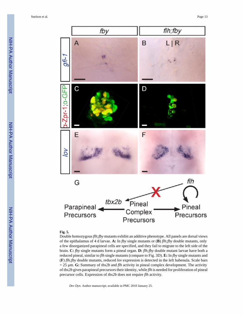
Fig. 5.Double homozygous flh;fby mutants exhibit an additive phenotype. All panels are dorsal viewsof the epithalamus of 4 d larvae. A: In fby single mutants or (B) flh;fby double mutants, onlya few disorganized parapineal cells are specified, and they fail to migrate to the left side of thebrain. C: fby single mutants form a pineal organ. D: flh;fby double mutant larvae have both areduced pineal, similar to flh single mutants (compare to Fig. 3D). E: In fby single mutants and(F) flh;fby double mutants, reduced lov expression is detected in the left habenula. Scale bars= 25 μm. G: Summary of tbx2b and flh activity in pineal complex development. The activityof tbx2b gives parapineal precursors their identity, while flh is needed for proliferation of pinealprecursor cells. Expression of tbx2b does not require flh activity.
Snelson et al. Page 13
Dev Dyn. Author manuscript; available in PMC 2010 January 25.
NIH
-PA Author Manuscript
NIH
-PA Author Manuscript
NIH
-PA Author Manuscript
NIH
-PA Author Manuscript
NIH
-PA Author Manuscript
NIH
-PA Author Manuscript
Snelson et al. Page 14
TAB
LE 1
Fate
of P
inea
l Com
plex
Cel
ls L
abel
ed w
ith B
rdU
Bet
wee
n 10
s an
d 30
hr
Gen
otyp
eSt
agea
GFP
+ pi
neal
cel
lsB
rdU
+ pi
neal
cel
lsG
FP+
para
pine
al c
ells
Brd
U+
para
pine
al c
ells
N
WT
10 s
47 +
/− 6
1 +/−
38
+/−
20
11
15 s
50 +
/− 4
14 +
/− 5
8 +/−
23
+/−
225
18 s
50 +
/− 4
5 +/−
2b9
+/−
20.
8 +/−
0.9b
21
24 h
51 +
/− 5
9 +/−
5b,c
8 +/−
21
+/−
1b,d
13
30 h
48 +
/− 7
2 +/−
210
+/−
20
7
flh−/−
10 s
17 +
/− 6
0.3
+/−
0.5e
8 +/−
20b
,e10
15 s
16 +
/− 5
2 +/−
2b11
+/−
21
+/−
1b25
18 s
15 +
/− 4
0.3
+/−
0.8f
7 +/−
20.
1 +/−
0.4b
,f21
24 h
14 +
/− 5
0.3
+/−
0.7b
8 +/−
20.
2 +/−
0.4b
15
30 h
15 +
/− 6
0.4
+/−
0.7
10 +
/− 3
017
a Dev
elop
men
tal s
tage
whe
n B
rdU
was
firs
t app
lied
to e
mbr
yos d
urin
g a
0.5
hr p
ulse
; all
sam
ples
wer
e fix
ed a
t 4 d
for a
naly
sis.
b Sign
ifica
ntly
diff
eren
t fro
m W
T 15
s by
2-ta
iled
T-te
st, p
< 0
.03
c Sign
ifica
ntly
diff
eren
t fro
m W
T 18
s by
2-ta
iled
T-te
st, p
< 0
.01
d Not
sign
ifica
ntly
diff
eren
t fro
m W
T 18
s by
2-ta
iled
T-te
st, p
> 0
.7
e Not
sign
ifica
ntly
diff
eren
t fro
m W
T 10
s by
2-ta
iled
T-te
st, p
> 0
.17.
f Sign
ifica
ntly
diff
eren
t fro
m W
T 18
s by
2-ta
iled
T-te
st, p
< 0
.01
Dev Dyn. Author manuscript; available in PMC 2010 January 25.
NIH
-PA Author Manuscript
NIH
-PA Author Manuscript
NIH
-PA Author Manuscript
Snelson et al. Page 15
TABLE 2Number of Labeled Cells Present in the Pineal Complex of Single and Double Mutants
Gene/protein (cell types labeled) Genotype Number of cellsa N
gfi-1(parapineal cells)
WT 10 +/− 1 12
flh 10 +/− 1 14
fby;flh 3 +/− 2b 24
GFP in Tg(foxd3:gfp)(pineal and parapineal cells)
WT 52 +/− 10 5
flh 28 +/− 7c 5
fby;flh 24 +/− 10c 7
Zpr-1 antibody(red-green double cone cells)
WT 30 +/− 5 5
flh 5 +/− 5c 4
fby;flh 7 +/− 1c 4
aAverage number of cells labeled per larvae at 4 d, plus or minus one standard deviation.
bSignificantly different from WT and flh by 2-tailed T-test, p < 0.01.
cSignificantly different from WT by 2-tailed T-test, p < 0.03.
Dev Dyn. Author manuscript; available in PMC 2010 January 25.
Copyright © 2022 FDOKUMEN