Urban Regeneration between Cultural Heritage Preservation ...
Upload
khangminh22Category
view
1download
0
1
Development and Regeneration of the Pineal Region of
the Diencephalon
Thesis presented for the degree of MPhil
by Jiahui Liu
Supervisor Prof. Claudio Stern
Department of Cell and Developmental Biology
University College London
September 2013

2
I, Jiahui Liu, confirm that the work presented in this thesis is my own. Where
information has been derived from other sources, I confirm that this has been
indicated in the thesis.

3
Abstract
Organizer is a group of cells that induces and patterns surrounding tissues
during embryo development. Previous studies of organizers were mainly
based on transplantation of various pieces of tissues. This project first aimed
to find out putative organizers using a novel method, which was to
characterize organizers based on patterns of syn-expression genes. The
differential microarray assays selected a list of gene that are enriched or
depleted in three known organizers (Hensen’s node, notochord and floor
plate and zone of polarizing activity). Whole-mount in-situ hybridisation,
histological sections and optical projection tomography were used to further
analyse the expression patterns of these syn-expressed genes. The roof of
dorsal thalamus including the pineal gland is revealed as one of a potential
organizer, with expression of cNOT1, TSPAN6 and PKIγ.
Further studies of gene expression showed that cNOT1 is expressed broadly
in early diencephalon and is restricted to pineal gland and its posterior
territory till HH25. This raises questions that whether cNOT1 is marking the
pineal progenitors and whether there are movements of cNOT1-expressing
cells. Fate mapping analysis demonstrates that not all cNOT1-expressing
cells are pineal progenitors and they are not moving over the development of
pineal gland.
Considering the change of gene expression over diencephalon development,
it could be that either cNOT1-expressing cells or the pineal gland is a
potential organizer. To assess their organizing abilities, ablation experiments
were performed on both area. Ablation of pineal progenitors alone led to
regeneration of the whole pineal gland and with normal expression patterns
of diencephalic genes. On the other hand, removal of all cNOT1-expressing
cells inhibited the regeneration of pineal gland.
In conclusion, these data suggest that the pineal gland can be regenerated
from surrounding tissues. Whether the roof of the dorsal thalamus or the
cNOT1-expressing cells is an organizer requires more experiments, including
transplantation, to assess.

4
Table of Contents 1 Introduction ....................................................................................................... 7
1.1 Hensen’s node ............................................................................................ 7
1.2 Notochord and floor plate ....................................................................... 13
1.3 Organizers involved in brain development ........................................... 16
1.3.1 Anterior neural ridge ....................................................................... 16
1.3.2 Isthmus .............................................................................................. 19
1.3.3 Zone limitants intrathal ................................................................... 20
1.4 Zone of polarizing activity ...................................................................... 21
1.5 Looking for putative organizers by studies of syn-expression groups 23
1.5.1 Microarray screen to identify a “synexpression” group of genes
from three known organizers ......................................................................... 25
1.5.2 Distilled list genes ............................................................................. 31
1.6 Thesis plan ................................................................................................ 50
2 Methods and material ..................................................................................... 53
2.1 Harvesting chick embryos ...................................................................... 53
2.2 Whole-mount in-situ hybridisation ............................................................. 53
2.2.1 Transformation of DNA ........................................................................ 53
2.2.2 Preparation of DNA ............................................................................... 54
2.2.3 DNA digestion......................................................................................... 54
2.2.4 DNA transcription ................................................................................. 54
2.2.5 Probe making for whole-mount in-situ hybridisation ........................ 55
2.2.5 Whole-mount in-situ hybridisation (Gall and Pardue, 1969) ............ 57
2.3 Histological section .................................................................................. 58
2.4 Optical Projection Tomography (Sharpe, 2003) ....................................... 58
2.4.1 Preparation of embryo .......................................................................... 58
2.4.2 Embedding embryo ................................................................................ 59
2.4.3 Trimming the embryo............................................................................ 59
2.4.4 Dehydration and clearing of specimen ................................................. 59
2.4.5 Scanning embryos .................................................................................. 60
2.5 Fate mapping ................................................................................................ 60
2.5.1 DiI and DiO preparation ....................................................................... 60
2.5.2 in vivo label ............................................................................................. 60
2.5.3 Harvest labelled embryo........................................................................ 60
2.6 Imaging .......................................................................................................... 61

5
2.7 Ablation of diencephalon ............................................................................. 61
3 Looking for putative organizers by studies of syn-expression groups ........... 63
3.1 Introduction .................................................................................................. 63
3.2 Results ............................................................................................................ 63
3.2.1. Expression of distilled list genes in known organizers ...................... 63
3.2.2 Summary of regions that syn-expressed genes express in common .. 69
3.2.3 Pineal region is one of the potential organizer regions ....................... 77
3.3 Discussion ...................................................................................................... 78
4 Development of early diencephalon in chick embryo ...................................... 83
4.1 Introduction .................................................................................................. 83
4.2 Results ............................................................................................................ 84
4.2.1 cNOT1 expression during diencephalon development ....................... 84
4.2.2 DKK1 is a pineal gland marker ............................................................ 87
4.2.3 Fate mapping of the developing diencephalon .................................... 88
4.3 Discussion ...................................................................................................... 94
5 Pineal Gland is Regenerated after Ablation of Progenitor Cells .................... 98
5.1 Introduction .................................................................................................. 98
5.2 Results ............................................................................................................ 99
5.2.1 Pineal gland is regenerated after ablation of pineal progenitors. ..... 99
5.2.2 Gene expression patterns are normal after regeneration of pineal . 101
5.2.3 Ablation of all cNOT1 expressing cells prevents the regeneration of
the pineal gland and DKK1 expression ....................................................... 102
5.4 Discussion .................................................................................................... 106
6 General Discussion ............................................................................................ 109
Reference ............................................................................................................... 112
Appendix ............................................................................................................... 140
1. Whole-mount in-situ hybridisation data ................................................. 140
2. Fate Mapping Data.................................................................................... 180
3. Preliminary Fate Mapping Data .............................................................. 199

6
Chapter 1
Introduction

7
1 Introduction
Organizer is a group of cells that induces and patterns surrounding tissues during
the development of embryos. It was first demonstrated by Hans Spemann and Hilde
Mangold in 1924 and is an important landmark in the history of developmental
biology (Spemann and Mangold, 2001; Spemann, 1924b). Organizers equivalent to
the Spemann’s organizer were also discovered in other animals including mouse,
Xenopus, zebrafish and chick (De Robertis et al., 2000; Niehrs, 2004; Shih and
Fraser, 1996; Waddington, 1933), which shows that it is conserved between
species.
Different to signalling centres, organizers can not only provide signals to
surrounding cells, but also drive cellular rearrangements nearby. Transplantation of
organizers to ectopic sites can alter cell fates of surrounding tissues, while ablation
of organizer will cause abnormal development. Based on these criteria, there are
only five organizers found in chick embryos, which are Hensen’s node (equivalent
to the Spemann’s organizer), notochord and floor plate, zone of polarizing activity
(ZPA), isthmus and apical ectodermal ridge (AER). How these organizers were
discovered and function in embryos will be discussed in the chapter of introduction
in detail, following the order of development.
1.1 Hensen’s node
The first organizer that appears during chick embryo development is Hensen’s
node. At the end of gastrulation, Hensen’s node is formed at the anterior end of the
primitive streak, which is an organizer in chick embryo. Hensen’s node is
equivalent to Spemann’s organizer (also called “the organizer”), which is the first
organizer discovered by Spemann and Mangold. In 1907, Warren Lewis
transplanted the dorsal lip of a blastopore at gastrula stage to the ventral ectopic
side of a similar stage embryo and observed the formation of a secondary axis
(Lewis, 1907). But he concluded it to be the result of self-differentiation of the
graft. Until 1924, when Spemann and Mangold figured out a way to distinguish

8
host and donor tissues and repeated the experiment successfully to show that the
donor dorsal lip could induce and pattern the host tissues to form a secondary axis
(Spemann, 1924b). They exchanged pieces of tissues between Triton embryos of
different species and analysed the cell fates of those transplanted embryos. Triton
Cristatus cells are unpigmented and can be easily distinguished from pigmented
taeniatus or alpestris cells over a long time. They have observed that most tissues
were influenced by surrounding tissues and differentiated according to their new
positions. However, there is one exception that acted differently, which is the upper
lip of the blastopore. It induced a formation of secondary axis containing neural
tube and somites in the region of epidermis progenitors (Spemann, 1918; Spemann,
1924b). Importantly, the neural tube was derived almost entirely from host tissue,
whereas the graft gave rise mainly to the notochord. Therefore Spemann defined an
organizer as a piece of tissue that can induce a secondary axis when it is
transplanted to an undifferentiated region of another embryo (Spemann, 1924a;
Spemann, 1924b). The primary (gastrula) organizer is conserved across different
species including Fundulus, zebrafish, Xenopus, chick, quail, duck, rabbit and
mouse, which were also proven by transplantation experiments (Beddington, 1994;
Oppenheimer, 1936b; Shih and Fraser, 1996; Spemann, 1924a; Waddington, 1932a;
Waddington, 1933). In Fundulus and zebrafish, microsurgical grafting of the
embryonic shield to an ectopic side can induce a secondary axis containing both
graft and host cells, which demonstrates that the embryonic shield is equivalent to
the Spemann’s organizer (Ho and Kimmel, 1993; Oppenheimer, 1936a;
Oppenheimer, 1936b). On the other hand, Waddington performed transplantation
experiments between rabbit, chick and quail embryos and thus identified Hensen’s
node, which is the tip of primitive streak, as the equivalent of “Spemann’s
organizer” (Hensen, 1875; Storey et al., 1995; Waddington, 1932b; Waddington,
1933; Waddington, 1936). Similarly, the anterior part of the primitive streak in the
mouse resembles the chick Hensen’s node. Transplantation of the mouse node can
induce additional digit in chick limb buds and anterior structures in Xenopus (Blum
et al., 1992b; Hogan et al., 1992; Kintner and Dodd, 1991). Later on, Beddington
showed that grafts of mouse node posterolateral location induced a secondary axis
and the formation of ectopic somites, which confirmed that the mouse node is the
equivalent of Spemann’s organizer (Beddington, 1994). In addition, cross-species
transplantations between chick, zebrafish, rabbit, amphibians and mouse proved

9
that the properties and functions of organizers are conserved (Blum et al., 1992a;
Hatta and Takahashi, 1996; Kintner and Dodd, 1991; Oppenheimer, 1936a;
Waddington, 1934; Waddington, 1936; Waddington, 1937)
Although Waddington showed that transplantation of Hensen’s node to an ectopic
lateral site in duck, rabbit or chick embryo could all induce a secondary axis as
Spemann’s organizer (Waddington, 1932a; Waddington, 1933), this ectopic site
was later shown to develop into neural plate and thus it was not possible to
conclude that the host cells change their fate (Stern, 2005). The organizing ability
of Hensen’s node was later proved by the transplantation in ectopic site at area
opaca (Figure1.1), which only develops into extraembryonic structures (Dias and
Schoenwolf, 1990; Guttikar et al., 1993; Storey et al., 1992). The competence of
this region will no longer exist after HH5 (Dias and Schoenwolf, 1990; Gallera,
1970; Gallera and Ivanov, 1964; Storey et al., 1992; Streit and Stern, 1997). After
ablation of the organizer (under certain conditions), embryos can still develop
normally, which shows that the organizer may be regulated by the surrounding
tissues as well (Abercrombie and Bellairs, 1954; Butros, 1967; Waddington,
1932a). More recent investigations suggest that the organizer is a dynamic state
rather than a pre-established population of cells, and that this state is determined by
signals emanating from a “node inducing centre” located in the middle of the
primitive streak and expressing Vg1 and Wnt8c, which induce adjacent cells to
become organizer (Joubin & Stern 1999). This explains the finding that as cells
move in and out of Hensen’s node they acquire and lose node markers appropriate
to their current position, demonstrated by DiI labelling experiments coupled with
hybridisation of the labelled embryos (Joubin and Stern, 1999). The role of Vg1 and
Wnt8C in defining the organizer state in this experiment has a parallel in the events
of much earlier development, prior to primitive streak formation, where Vg1 and
Wnt8C cooperate to induce Nodal, and through this a primitive streak containing
the future organizer at its tip (see above and Skromne & Stern 2001; 2002;
Bachvarova et al., 1998). Bachvarova et al. found that the posterior marginal zone
(PMZ) (where Vg1 and Wnt8C expression overlaps) can induce primitive streak and
the Hensen’s node without contributing cells to these structures (Bachvarova et al.,
1998). In this respect (the ability to induce an organizer without making a cellular

10
contribution to it), the PMZ fulfils the requirements to be equivalent to the
Nieuwkoop centre of Amphibians. Nieuwkoop centre induces the neighbouring
Spemann’s organizer by dorsalizing Nieuwkoop signals (Nieuwkoop, 1992). In
addition, mesoderm induction assays in Xenopus and mouse models confirm the
induction of the organizer by Vg1 and Wnt signalling pathway is conserved across
species (Watabe et al., 1995).
Figure 1.1 Transplantation of Hensen’s node. Hensen’s node (red) is transplanted
from donor quail embryo at HH4 to area opaca (yellow area) at host chick at HH4.
After incubation, a secondary axis will be developed in the host ectopic side.
Following the discovery of the organizer, many studies have been concentrated on
its functions and the molecular mechanism of its action. The first gene claimed to
be specific for the organizer was goosecoid isolated in 1991 (Cho et al., 1991).
Apart from its expression in the dorsal lip of the blastopore, microinjection of
goosecoid mRNA at an ectopic site leads to the formation of secondary axis in
Xenopus (Cho et al., 1991). Goosecoid expression is induced by activin (which
belongs to the same class of TGFβ-related molecules as Vg1 and Nodal) (Cho et al.,

11
1991). Gene expression and fate mapping studies in chick embryo also show that
goosecoid is a marker for organizer cells (Izpisua-Belmonte et al., 1993). However
it is also expressed in non-organizer tissues including Koller's sickle, prechordal
mesoderm and head endoderm (Izpisua-Belmonte et al., 1993). In the mouse, initial
studies using gsc-null mutants showed that goosecoid is not required for formation
of the primary axis (Rivera-Perez et al., 1995). However, an effect on neural
inducing properties was revealed by a more sensitive assay based on cross-species
transplantation of the mouse node to chick embryos, which showed that goosecoid
affects the neural inducing strength of the node (Zhu et al., 1999a).
Later, mainly by using differential screens of cDNA libraries, many more genes
expressed in the amphibian organizer were discovered including frzb-1, cerberus,
chordin, noggin, follistatin and dickkopf -1 (DKK-1) (Bouwmeester et al., 1996;
Fainsod et al., 1997; Glinka et al., 1998; Hemmatibrivanlou and Melton, 1994;
Leyns et al., 1997; Piccolo et al., 1996; Sasai et al., 1996). Among these molecules,
chordin, noggin and follistatin can induce neural tissues in a Xenopus animal cap
assay (Hemmatibrivanlou et al., 1994; Lamb et al., 1993; Piccolo et al., 1996; Sasai
et al., 1996). Microinjection of mRNA encoding any of these three proteins can also
mimic the effect of the organizer and cause the formation of secondary neural tube,
dorsal mesoderm and additional gut (De Robertis et al., 2001; Hemmatibrivanlou
and Melton, 1994; Holley et al., 1995; Lamb et al., 1993; Schmidt et al., 1995).
Further studies revealed that both chordin, noggin, follistatin, and other neural
inducing molecules are BMP inhibitors (Hansen et al., 1997; Piccolo et al., 1999;
Piccolo et al., 1996; Zimmerman et al., 1996). This, along with other findings
(Hemmati-Brivanlou and Melton, 1997; Stern, 2005), led to the establishment of
the “default model” of neural induction, which proposes that ectodermal cells will
develop into neural tissues if they do not receive BMP signals, otherwise they are
instructed to develop into epidermis (Wilson and Hemmatibrivanlou, 1995).
According to this model, the organizer would act by lowering the BMP
concentration around ectoderm cells and thus preventing them from being induced
to epidermis by BMP. BMP signalling is also particularly important for patterning
mesoderm (Dosch et al., 1997; Tonegawa et al., 1997; Winnier et al., 1995).

12
However, it was first noticed in chick embryos that the expression patterns of
BMPs and their antagonists did not fit the “default model” (Streit et al., 1998).
Moreover, misexpression of chordin in ectopic regions does not induce the
expression of early neural markers (Linker and Stern, 2004; Streit et al., 2000;
Streit et al., 1998). Experiments in Xenopus also show that neural induction of
chordin or noggin is inhibited by blockage of FGF signalling pathway (Launay et
al., 1996; Linker et al., 2009; Linker and Stern, 2004; Sasai et al., 1996). Therefore,
inhibition of BMP is not sufficient or requires cooperation of other signalling
molecules to induce neural development in chick.
Apart from dorsalization of the mesoderm and neural induction, the organizer also
plays a role in orchestrating left-right asymmetry at the late primitive streak stage in
avian and mammalian embryos. The first insights into this process came from the
observations that mRNA encoding the “activin” (actually Nodal) receptor cActRIIA
is expressed on the right side of the chick primitive streak at stage HH4 (Levin et
al., 1995; Stern et al., 1995), while the transcription factor HNF3β (Foxa2) and the
secreted protein Sonic hedgehog are expressed on the left side of the node (from
HH4+ to HH6) (Levin et al., 1995). Soon afterwards (HH6-7), nodal expression
starts as a small spot next to the left node and a larger domain in the left lateral
plate. Misexpression and knockdown of these components affect these expression
patterns and reveal a pathway that initiates left-right asymmetry (Levin, 1997;
Levin et al., 1995; Levin et al., 1997; Tabin, 2006). Expression of nodal was shown
to be induced by Shh (Levin et al., 1995), while misexpression of activin can
repress SHH expression on the left hand side and induce the expression of right-
hand side markers (Levin, 1997; Levin et al., 1995). Also, misexpression of nodal,
SHH or activin can affect heart situs (Levin et al., 1997; Zhu et al., 1999b). The
cascade of signals converges on the transcription factor Pitx2, which shows left-
sided expression in all organ systems that develop asymmetrically (Campione et al.,
1999; Capdevila et al., 2000; Yokouchi et al., 1999; Yoshioka et al., 1998; Zhu et
al., 1999b). Surprisingly, of these various asymmetry markers, only two (Nodal and
Pitx2) are conserved across all vertebrates (Collignon et al., 1996; Levin et al.,
1995; Long et al., 2003). The mechanism that position these two seems quite
variable in different species, and has been shown to involve diverse strategies such

13
as ion fluxes (Levin, 2003), ciliary beating (Babu and Roy, 2013; Nonaka et al.,
1998; Okada et al., 1999), dynein (Supp et al., 1997), calcium signalling
(Langenbacher and Chen, 2008) and other signals. For example, nodal expression
in mouse and zebrafish is not as essential as that in chick and Xenopus. In mouse
chimera embryos, no defects have been observed in left-right asymmetry
(Beddington and Robertson, 1999). Similarly, point mutations in nodal homologous
cyclops (cyc) in zebrafish does not affect heart looping as well (Chen et al., 1997).
In fact, the breakthrough of symmetry in mouse is affected by the nodal flow
generated, which is a leftward flow of extracellular fluid generated by monocillia
around the node and requires the axonemal dynein, left-right dynein (lrd) (McGrath
et al., 2003). Similar nodal flow mechanism is also observed in zebrafish, and
knockdown of lrd causes randomise gene expression and cardiac laterality (Essner
et al., 2005; Kawakami et al., 2005).
In conclusion, the Spemann’s organizer and its equivalent in other species
are responsible for the development of primary anterior-posterior and left-
right axis. Genes like SHH and chordin are involved within the process and
conserved across different species. But variations in mechanisms of
organizing activities are also present.
1.2 Notochord and floor plate
Following Spemann’s organizer, the second organizer involved in embryo
development is the notochord and floor plate, which is responsible for the dorsal-
ventral patterning of neural tube.
Following the regression of the primitive streak and the formation of neural plate,
the neural tube starts to form by bending of the neural plate into a hollow tubular
structure. DiI labelling reveals that the anterior-median quadrant of the epiblast of
Hensen’s node contributes to cells in both the notochord and the ventral-most
wedge of the neural tube, which is the prospective floor plate (Selleck and Stern,
1991). This can be seen even when a single cell in this node region is labelled with
a lineage tracer: descendants can be found in both the notochord (mesoderm) and
14
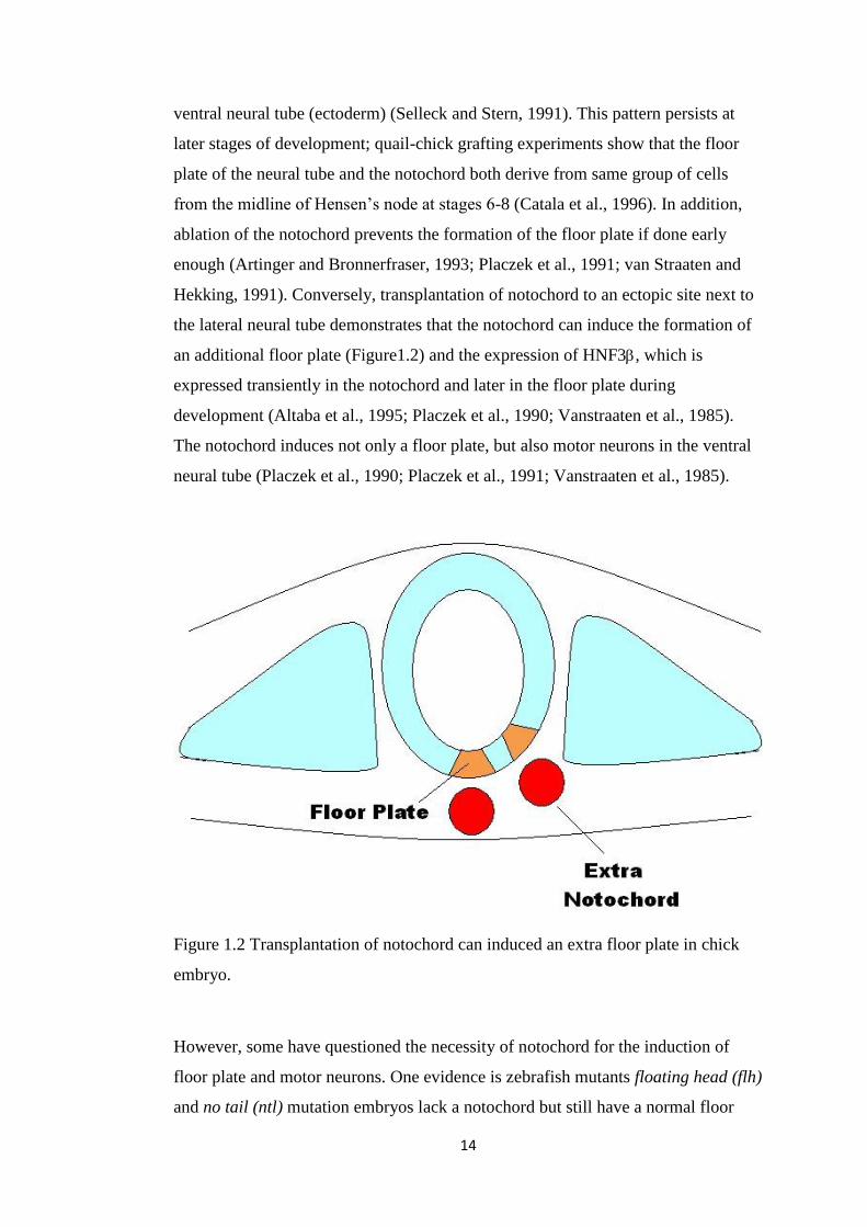
ventral neural tube (ectoderm) (Selleck and Stern, 1991). This pattern persists at
later stages of development; quail-chick grafting experiments show that the floor
plate of the neural tube and the notochord both derive from same group of cells
from the midline of Hensen’s node at stages 6-8 (Catala et al., 1996). In addition,
ablation of the notochord prevents the formation of the floor plate if done early
enough (Artinger and Bronnerfraser, 1993; Placzek et al., 1991; van Straaten and
Hekking, 1991). Conversely, transplantation of notochord to an ectopic site next to
the lateral neural tube demonstrates that the notochord can induce the formation of
an additional floor plate (Figure1.2) and the expression of HNF3, which is
expressed transiently in the notochord and later in the floor plate during
development (Altaba et al., 1995; Placzek et al., 1990; Vanstraaten et al., 1985).
The notochord induces not only a floor plate, but also motor neurons in the ventral
neural tube (Placzek et al., 1990; Placzek et al., 1991; Vanstraaten et al., 1985).
Figure 1.2 Transplantation of notochord can induced an extra floor plate in chick
embryo.
However, some have questioned the necessity of notochord for the induction of
floor plate and motor neurons. One evidence is zebrafish mutants floating head (flh)
and no tail (ntl) mutation embryos lack a notochord but still have a normal floor
15
plate (Halpern et al., 1993; Halpern et al., 1995). It was also argued that the ablation
experiments in chick may have included the removal of predetermined floor plate
precursors (Catala et al., 1996).
Despite this dissent, the inductive ability of the notochord has been reproduced
many times and is now understood to be based on its secretion of Shh, which is first
expressed in the notochord and then in the floor plate (Echelard et al., 1993; Krauss
et al., 1993; Roelink et al., 1994). Misxpression of SHH in mouse, frog or zebrafish
embryos mimics the effect of notochord transplantation and induces the formation
of extra floor plate, motor neurons and ventral interneurons (Echelard et al., 1993;
Krauss et al., 1993; Roelink et al., 1994). In support of this idea, further analysis in
chick embryos provides more evidence for the inducing ability of the notochord and
Shh: first, time-lapse microscopy revealed that many floor plate cells derive from
epiblasts anterior to Hensen’s node (named “area a”) and therefore do not share a
lineage with the notochord cells. Second, co-expression of nodal and SHH induces
the formation of floor plate from “area a” cells in vitro (Patten et al., 2003). In
conclusion, the notochord induces the formation of floor plate and establishes
ventral identities in the neighbouring neural tube. Apart from the notochord, the
floor plate itself also plays a role in patterning the neural tube through secretion of
diffusible signals including Shh, Wnt and BMP (Dale et al., 1999; Furuta et al.,
1997; Liu et al., 2000; Mcgrew et al., 1992; Placzek et al., 1993; Placzek et al.,
1991; Yamada et al., 1993). This implicates a relay system: the notochord provides
the initial Shh signal, which induces the floor plate, and the floor plate itself
secretes Shh responsible for inducing motor neurons and ventral interneurons in the
adjacent ventral neural tube. It is now widely believed that Shh acts as a gradient,
highest ventrally and diminishing dorsally, where it is also antagonised by BMP
and other TGFβ molecules expressed by the roof of the neural tube (Liem et al.,
2000; Liem et al., 1997; Liem et al., 1995). Based on the concentration of Shh,
different transcription factors and downstream genes are activated or repressed. As
a result, different types of neurons are generated across the neural tube (Dessaud et
al., 2008; Liem et al., 2000). For example, Pax6 is repressed by Shh and is
expressed more dorsally in the neural tube (Liem et al., 2000). On the other hand,
Nkx2.2 is activated by Shh and is expressed in the ventral part of the neural tube
(Liem et al., 2000). This mechanism generates the diversity of neuronal territories

16
along the dorso-ventral axis of the spinal cord (Briscoe et al., 2001; Briscoe and
Ericson, 2001; Briscoe et al., 2000; Jessell, 2000).
1.3 Organizers involved in brain development
As a complex organ, there are two organizers found in brain development, which
are the anterior neural ridge (ANR) and isthmus. These two organizers are
responsible for patterning different compartments in brain vesicles. The ANR
directs the development of the forebrain, while the isthmus is responsible for the
development of the mesencephalon and the metencephalon. The following chapter
will discuss their discovery and functions in brain development separately. I will
also introduce ZLI, which is considered to be an organizer in diencephalon in many
research paper.
Figure 1.3 Structures of brain vesicles in chick embryo. A) primary brain vesicles at
HH 10; B) brain compartments at HH22. Red area shows the expression of SHH.
1.3.1 Anterior neural ridge
During the development of the notochord in chick embryo, the head fold starts to
form at the anterior neural plate. First, all three layers (ectoderm, mesoderm and
endoderm) fold ventrally, forming an arc that progress caudally; this is called the
head fold in chick and mammalian embryos. The endodermal lining of this head
fold first defines the foregut which gradually becomes a tube (Bellairs, 1954;

17
Kimura et al., 2006). As the neural plate folds to form a tube, this process continues
in the head, involving the medial neural plate in the dorsal part of the head fold.
From stages HH9-HH10 (Figure1.3 A), bulges appear in this region, which will
give rise to the so-called primary brain vesicles: prosencephalon (forebrain),
mesencephalon (midbrain) and rhombencephalon (hindbrain, containing
metencephalon). The prosencephalon is further subdivided into telencephalon and
diencephalon (Hamburger and Hamilton, 1951; Hamburger and Hamilton, 1992).
At stage HH8, the forming anterior neural plate of the chick displays a pair of
prominent protrusions, which will eventually form the olfactory placode. The
anterior edge of the neural plate, which is also called the anterior neural ridge
(ANR) (Figure1.3 A), is suggested to act as an organizer that regulates the
development of the telencephalon. In the mouse, a mutation of BF1 (now called
FOXG1) causes a reduction in size of the cerebral hemispheres in the telencephalon
due to the reduction of proliferation of neuroepithelial cells (Xuan et al., 1995).
FGF8 is strongly expressed in the ANR and induces FOXG1 expression in the
neighbouring telencephalon, suggesting that the ANR may pattern the
telencephalon (Shimamura and Rubenstein, 1997b). Overexpression of FGF8 in
Xenopus proves that it promotes the expression of anterior telencephalic marker
FOXG1 (Eagleson and Dempewolf, 2002). Increased expression of FOXG1 is
induced by the transplantation of ANR, which demonstrates the induction ability of
ANR (Eagleson and Dempewolf, 2002). However, transplantation of medial ANR
inhibits the telencephalic development, while lateral ANR increases it (Eagleson
and Dempewolf, 2002). It means that there are inhibitory mechanisms to ensure the
development of bilateral telencephali (Eagleson and Dempewolf, 2002; Hongo et
al., 1999). Transplantation and ablation of anterior ectodermal cells in the central
nervous system in zebrafish also point to an important signalling function of the
anterior region of the forebrain (Houart et al., 1998a). This was suggested to be
due to the production of a Wnt antagonist called TLC (Houart et al., 1998a), but no
homologue has yet been found in other vertebrates although there is considerable
evidence from many model systems that inhibition of Wnt signalling is important
for specifying anterior identity in the neural plate (Belo et al., 2009; Niehrs, 2006).
The FGF8 mutant Ace (“acerebellar”) in zebrafish also shows that Fgf8 is required
for normal development of both the cerebellum (derived from the anterior hindbrain
adjacent to the isthmus/MHB, where FGF8 is also expressed; see below) and of the

18
telencephalon (Shanmugalingam et al., 2000). In chick embryo, induction or
inhibition of Fgf8 does not appear to affect telencephalic development, which may
due to several Fgf signalling molecules present together in the developing forebrain
(Gunhaga et al., 2003). Requirement of other Fgfs like Fgf3 for telencephalon
development is also shown in zebrafish using antisense morpholino inhibition
experiments (Shinya et al., 2001).
In addition, the Wnt signalling pathway is also involved in telencephalon
patterning. One evidence is that targeted deletion of -catenin causes defects in
medial ganglionic eminence which results from disruption of the canonical Wnt
pathway (Gulacsi and Anderson, 2008). Chordin and Noggin, which are BMP
antagonists, are also shown to be involved in forebrain development in mutated
mice (Bachiller et al., 2000). Similar effects of BMP are also observed in zebrafish
(Barth et al., 1999; Nguyen et al., 1998).
Apart from Fgf and Wnt signalling, other molecules are involved in telencephalon
development. Given the strong effect of Shh in spinal cord development, and the
fact that it is also expressed within the brain (in structures that continue anteriorly
from the floor plate: the basal plate/hypothalamus, infundibulum and zona limitans
intrathalamica, ZLI) (Scholpp et al., 2007; Scholpp and Lumsden, 2010), it would
not be surprising to find that Shh plays an important role in forebrain development.
Explantation of Shh shows that it induces the differentiation of ventral neuronal
cells that are normally in diencephalon and telencephalon in chick embryo (Ericson
et al., 1995). Targeted mutation of SHH in mice causes early defects in midline
structures including the notochord and floor plate (Chiang et al., 1996). Explant
experiments using rat and mouse tissues also suggest that SHH can induce the
development of ventral neurons in the telencephalon (Kohtz et al., 1998). However
the dorsal telencephalon seems to develop in a largely Shh-independent way; mice
lacking SHH develop the dorsal telencephalon normally (Chiang et al., 1996). On
the other hand, defects in dorsal telencephalon is observed in Gli3 mutant mice
(Rash and Grove, 2007); the source of Shh responsible for this, if any, is unknown.

19
1.3.2 Isthmus
Apart from ANR, FGF8 is also expressed in the isthmus, which is the
mesencephalon-metencephalon boundary (MHB) (Figure 1.3 A). There is evidence
suggesting that the isthmus may function as an organizer for the mesencephalon
and metencephalon. Transplantation of the isthmus into the hindbrain induces
ectopic cerebellum in chick embryo (Martinez et al., 1995), whereas transplantation
into the diencephalon (posterior to the ZLI) induces graded expression of En2,
which is a marker for mesencephalon (Ballycuif et al., 1992; Martinez et al., 1991;
Sgaier et al., 2007). Conversely, transplantation of a diencephalic explant to the
vicinity of the isthmus causes the transplanted cells to change their fate to tectum,
which does not occur if the transplant is placed in a more anterior part of the
mesencephalon (Nakamura and Itasaki, 1992; Nakamura et al., 1991; Nakamura et
al., 1986).
As the isthmus is located between the mesencephalon and metencephalon, it is
interesting to know how this boundary is defined. Previous studies have shown that
it is set up by repression between Otx2 and Ggx2, which are expressed in
mesencephalon and metencephalon respectively (Broccoli et al., 1999; Hidalgo-
Sanchez et al., 1999; Millett et al., 1999). Misexpression of Otx2 (anterior) or Ggx2
(posterior) domain or vice-versa in the mouse both cause the isthmus to become re-
positioned (Broccoli et al., 1999; Katahira et al., 2000; Millett et al., 1999) and it is
generally thought that Otx2 and Gbx2 mutually repress each other, with the
isthmus/MHB arising at the boundary between their expression domains (Grapin-
Botton et al., 1999; Sato and Joyner, 2009).
As mentioned before, Fgf8 is expressed in the isthmus as well as in the ANR. Two
splice variants of Fgf8 are produced in the isthmus, which are Fgf8a and Fgf8b
(Sato et al., 2001). Misexpression of Fgf8a does not affect the expressions of most
of the isthmus specific genes except En1 and En2. Overexpression causes
expansion of En1 and En2 expression anteriorly into the diencephalon and changes
the fate of diencephalic cells to mesencephalic (Muramatsu et al., 1997; Nakamura

20
et al., 2004; Nakamura et al., 2000). Fgf8b exerts stronger effects on brain
development. Misexpression of FgF8b in chick embryo inhibits Otx2 expression
but induces Gbx2 and Irx2 to extend to the diencephalon (Sato et al., 2001). A
similar effect is also observed in transgenic mice (Liu et al., 1999). Fgf8 acts
through activation of the ERK (MAP-kinase) pathway (Sato and Nakamura, 2004;
Suzuki-Hirano et al., 2010). In addition to Fgf8, Wnt signalling is also involved in
tectum and cerebellum development. Wnt1 knockout mice show repressions in En1
and En2 expression and disruptions in midbrain and metencephalon structures
(Mcmahon et al., 1992). It may be required for maintenance of Fgf8 expression
(Panhuysen et al., 2004). Implants of beads and misexpression experiments in chick
and mice both show that Wnt1 interacts with Lmx1b to maintain Fgf8 expression
(Adams et al., 2000; Matsunaga et al., 2002).
1.3.3 Zone limitants intrathal
Because of its expression of SHH, the zona limitans intrathal (ZLI), located at the
border between ventral thalamus (pre-thalamus) and dorsal thalamus (thalamus)
(Fog. 1.3 B), has been suggested to be a secondary organizer for the forebrain
(Kiecker and Lumsden, 2004; Scholpp et al., 2006). In turn, establishment of the
ZLI requires Shh shown in both fate mapping and ectopic expression of SHH in
chick embryo (Zeltser, 2005). Expression of SHH is maintained in the ZLI and
regulates the development of the diencephalon. In zebrafish, increasing expression
of SHH by injection of mRNA leads to an expansion of prethalamic and thalamic
gene expressions including DLX2A, LHX5, DBX1A, EMX2 and NEUROG1, while
reducing SHH expression by Hh signalling inhibitor or mutated Hh co-receptor
smoothened, causes a reduction of expression in these genes (Scholpp et al., 2006).
OEP (EGF-CFC nodal co-receptor one-eyed pinhead) mutant embryos lacking the
basal plate do not appear to have defects in diencephalon (Scholpp et al., 2006).
However, using microbarrier to separate the basal plate from the ZLI in chick
embryos, Vieria and Martinez (2006) observed that the inhibition of SHH
expression by microbarriers causes structural alterations, and suggested that signals
from the ZLI are important for molecular regionalization of the diencephalon
(Vieira and Martinez, 2006). That Shh does play a role in diencephalon

21
development is suggested by gain- and loss- of function experiments in chick as
well (Kiecker and Lumsden, 2004). Also, in SHH-/- mice, a reduction in size of the
diencephalon is observed (Ishibashi and McMahon, 2002).
In addition to Shh, Wnt signalling may also be involved in diencephalic patterning
by the ZLI. Members of the Wnt family are expressed in various regions of the
diencephalon, including Wnt1,Wnt2b,Wnt3a, Wnt5a and Wnt8b. Inhibition of Wnt
expression by Dkk-1 bead-implantation blocks the dorsal progression of the
expression of SHH in the ZLI, but Wnt expression is not required for maintenance
of SHH expression (Martinez-Ferre et al., 2013). In addition, that the Wnt pathway
regulates the expression of Gli3 and L-fng in the ZLI, is also demonstrated by
implantation of Dkk-soaked beads experiments in chick (Martinez-Ferre et al.,
2013). The expression of some Wnt molecules are restricted by the ZLI in chick;
for example, Wnt3 which is expressed posterior to the ZLI in the dorsal thalamus
(Mattes et al., 2012). Wnt3a or Wnt3 induces the expression of dorsal thalamus
marker Gbx2 in explants of prospective dorsal thalamus, whereas inhibition of Wnt
induces the expression of ventral thalamic marker Dlx2 in similar explants (Braun
et al., 2003).
However, despite its expression of SHH, there is currently no evidence showing
that the ZLI can drive the rearrangement of surrounding cells. To establish this,
transplantation experiments to ectopic regions, accompanied by analysis of regional
markers, are essential. Therefore, in this project, ZLI is not considered to be an
organizer based on the criteria mentioned at the beginning.
1.4 Zone of polarizing activity
The final organizer that I would like to introduce is zone of polarizing activity
(ZPA), which is found to be involved in limb development. ZPA is a well-
characterized organizer, located in the posterior limb bud mesenchyme and is
responsible for patterning the anterior-posterior (radio-ulnar) axis of the limb

22
(Capdevila and Izpisua Belmonte, 2001). Mirror duplication of digits is induced by
transplantation of ZPA cells to an ectopic anterior location (Saunders, 1948)
(Figure 1.4). Several decades later, Tickle and colleagues reported that implantation
of a bead soaked in retinoic acid can mimic the effect of ZPA transplantation in
chick (Tickle et al., 1982), which together with the finding that posterior limb has
higher retinoic levels than the anterior limb (Thaller and Eichele, 1987),
immediately raised the hope that retinoic acid may be the ZPA morphogen (Slack,
1987). Inactivation of RALDH2 by mutation, an enzyme involved in retinoic acid
synthesis, can inhibit the development of the mouse limb (Niederreither et al., 1999;
Niederreither et al., 2002). However later studies uncovered that SHH is expressed
very specifically in the ZPA (Kiecker and Lumsden, 2004). Moreover, implantation
of Shh beads induces similar limb duplication as ZPA transplantation (Riddle et al.,
1993b). Shh seems to act in a concentration dependent manner: using alkaline
phosphatise tagged Shh, Shh protein has been found a long distance from the ZPA
in the chick limb bud (Yang et al., 1997). Recently, it was reported that limb bud
mesenchyme cells sample their environment and transport Shh protein by means of
very long, dynamic filopodia that can span a large proportion of the limb width
(Sanders et al., 2013). Cholesterol modification of Shh peptide shows a disruption
of formation of anterior digits, which also proves the long-range effect of Shh
(Lewis et al., 2001). One of the mechanisms is by negative feedback loop involving
HIP protein (Chuang and McMahon, 1999).
Figure 1.4 Transplantation of
ZPA. ZPA located at posterior
limb bud is transplanted to
ectopic anterior limb bud and
will induce an extra digit in
the host.

23
Apart from the ZPA, another signalling centre has been found to act in the limb
bud, the apical ectodermal ridge (AER) at the distal margin of the limb bud
ectoderm; this region functions to sustain limb growth (Lu et al., 2008b). Fgf family
members are found to be expressed in the AER including Fgf8, Fgf4, Fgf9 and
Fgf17 (Lewandoski et al., 2000; Mariani et al., 2008; Niswander et al., 1993).
However only Fgf8 has been shown to be necessary for limb bud development
(Lewandoski et al., 2000). Inactivation of Fgf8 in AER causes a reduction of limb-
bud size and delays expression of SHH in the ZPA, while mutations in other Fgf
members do not affect limb development (Lewandoski et al., 2000). Implantation of
Fgf4 beads can rescue limb development (including maintenance of SHH
expression in the ZPA) after AER ablation (Laufer et al., 1994; Niswander et al.,
1994). This reveals a positive feedback loop between the AER and ZPA to regulate
limb development (Zeller et al., 2009).
1.5 Looking for putative organizers by studies of syn-expression
groups
As discussed before, so far, only five organizers have been discovered that fulfil the
essential criteria of “a region that has both inducing and patterning activity on
surrounding cells”: Hensen’s node, the notochord and floor plate, the isthmus
(midbrain-hindbrain boundary), the Zone of Polarizing Activity (ZPA) and the
anterior neural ridge (ANR). All of these organizers were discovered based on
transplantation experiments showing their inducing ability and ablation experiments
that shows their requirement for normal embryological patterning. For example,
Hensen’s node in chick is identified as the equivalent of “Spemann’s organizer” by
transplantation experiments between chick and quail (Waddington, 1932a;
Waddington, 1933) and ablation experiments at different developmental stages
(Psychoyos and Stern, 1996).
Considering the complexity of embryo development, one would expect that there
should be many more organizers remaining to be undiscovered. However the
demonstration of organizer activity requires transplantation of the appropriate
signalling cell to an ectopic region competent to respond to those signals, as well as

24
having the appropriate assay to detect the response. This project started from the
initial idea that it may be possible to identify organizers prospectively, based on
their expression of a set of common genes. Synexpressed genes are a group of
genes that are co-ordinately expressed at one site (Gawantka et al., 1998; Niehrs
and Pollet, 1999; Peter and Davidson, 2009; Tomancak et al., 2002). If there is such
a common set of genes that characterises known organizers, their pattern of
expression could be used as a diagnostic for discovering new organising centres.
Specifically, genes enriched in known organizers should be enriched in candidate
regions, whereas genes that are not expressed in organizers but expressed on other
similar (non-inducing/patterning) tissues should be absent from the candidate
regions. This is not easy to assess by simple observation partly because of the sheer
number of genes involved and the complexity of development, and especially
because it is difficult to “see” regions that do not express a particular gene.
To progress in this direction we (in collaboration with Prof. David Burt, Dr. Megan
Davey and Dr. Frances Wong from Roslin Institute, Prof. Richard Baldock, Dr.
Duncan Davidson, Dr.Jeff Chrisitansen and Mike Wicks from HGU, Prof. Cheryll
Tickle and Dr.Monique Welten from Bath university, Dr. Paula Murphy from
Trinity College Dublin) decided to construct a detailed, 3-dimensional atlas of gene
expression which would then be analysed by image comparison software to define
regions with similar expression characteristics for many genes at the same time. To
help identify regions that have conserved patterns of synexpression of these genes
between chick and mouse, the consortium (see above) decided to host the chick
atlas of gene expression on the same server as an atlas of gene expression for the
mouse (EMAGE; http://www.emouseatlas.org/emage/) . The EMAGE project
started several years ago to act as a comprehensive repository of gene expression
patterns during development, along with a detailed anatomical atlas and an
anatomical ontology (Baldock et al., 2003; Ringwald et al., 1994). These are fully
searchable by gene name, structure name, or location/pattern (drawn interactively
by the user), and importantly, allowing complex Boolean queries (for example:
“find genes expressed in structure A that are also expressed in structure B but not
expressed in structure C at stage D of development”). During the course of this

25
project, a prototype chick atlas (eChickAtlas;
http://www.echickatlas.org/ecap/home.html) was generated, providing some of the
above functionality (Wong et al., 2013). To start to populate the atlas with data, the
consortium decided to prioritise genes expressed during limb development
(collected by the groups of Prof. Cheryll Tickle and David Burt) and the
“synexpressed” set of genes identified from three of the known organizers:
Hensen’s node, the notochord and associated ventral neural tube and the ZPA in the
posterior limb bud. This “synexpressed” set would include both genes that are
enriched in and those that are depleted from these organizers, as compared to
similar tissues at equivalent stages of development.
This following chapter describes the design of the screen and the identification of a
set of genes expressed similarly in the three chosen organisers.
1.5.1 Microarray screen to identify a “synexpression” group of
genes from three known organizers
(experments in this section were performed by Prof. David Burt group)
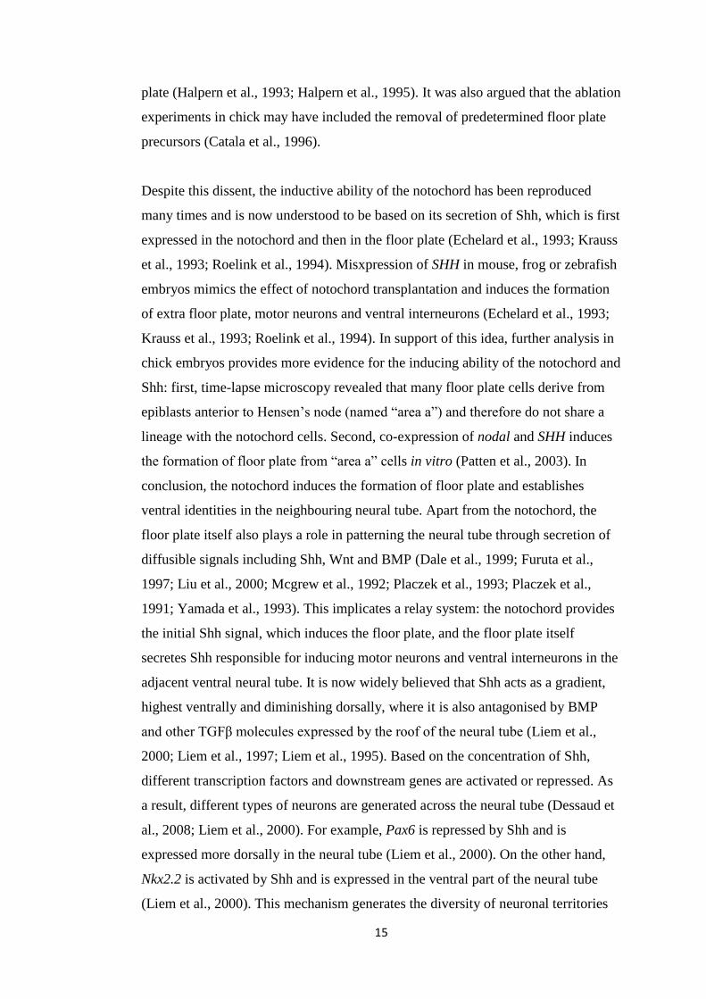
Figure 1.5 shows the tissues collected for microarray assays analysis. The gene
expressions in organizers were compared to their closely-related, non-organizing
regions. The following comparisons were performed. First, tissues were collected
from HH3+ Hensen’s node and the gene expression levels were compared to that in
the posterior primitive streak at the same stage (Figure 1.5 A). The early (HH3+)
node expressions were also compared to that in late node at HH6, which is not
organizer anymore but retains some signalling activities like left-right patterning
(Levin et al., 1995). The expressions in late node HH6 were also compared to that
in early posterior primitive streak at HH3+ to further select genes expressed for
signalling activities. Moreover, the expressions in the ventral neural tube and
notochord were compared to that in the dorsal neural tube to select genes involved
in notochord development (Figure 1.5 B, (Tanabe and Jessell, 1996). Finally, the
expressions in posterior limb bud including ZPA were compared to that in the
anterior limb bud both at early and late limb (HH20 and HH24 respectively), to

26
identify genes involved in limb development (Figure 1.5 C, (Zeller et al., 2009).
The initial comparisons between organizer and their related non organizing regions
select a large number of genes that were enriched or depleted in each organizer as it
is shown in Table 1.1. To further narrow down the genes of interest and select
synexpression groups, cross comparisons between each group in Table 1.1 were
performed. Detail comparisons are shown in Table 1.2. At the end, genes were
selected from comparison group 15, 16, 17 and 18, which represents genes that are
enriched or depleted in early node and/or late node and the ventral neural tube and
notochord and ZPA.
The final set of synexpressed genes selected is shown in Table 1.3. It includes
transcription factors and intracellular molecules, which are expressed or depressed
in cells within known organizers. It also includes membrane and secreted
molecules, which are responsible for signalling activities to surrounding tissues. In
total, 31 genes selected are enriched in organizers tested and 16 are depleted
instead. In summary, we selected a set of genes, which are up- or down-regulated in
current known organizers (Hensen’s node, notochord and floor plate, ZPA), by
comparing the gene expression level between organizers and their closely-related,
non-organizer regions. These genes, therefore, may be characteristic of organizers
and the region where they are commonly up- or down-regulated may be a potential
organizer. However, microarray assays do not give spatial and temporal gene
expression patterns and the expression levels of these synexpressed genes are
needed to be further analyzed by other methods such as whole-mount in-situ
hybridisation.
27
Figure 1.5 Tissues collected to perform microarray assays. The expressions of SHH
are shown in all three pictures. A) Tissues were collected from HH3+ Hensen’s
node and posterior primitive streak at the same stage. Tissues were also collected
from HH6 Hensen’s node which is not shown here. B) Tissues were collected from
the ventral neural tube and notochord together. Tissues from the dorsal neural tube
at the same stage were collected as well. C) Tissues were collected from the
posterior limb bud including the ZPA and anterior limb bud. Microarray assays
were performed by Prof. David Burt’s group at Roslin Institute to test the gene
expression levels in each tissue.
A B C

28
Group
Comparison Statistical test
UP_
probes
DOWN_
probes
1 S3HN_S3PS FC >1.5; FDR <=0.05 1678 1415
2 S6HN_S3PS FC >1.5; FDR <=0.05 3285 2075
3 S3HN_S6HN FC >1.5; FDR <=0.05 1701 2819
4 VNT_DNT FC >1.5; FDR <=0.05 811 283
5 S20LP_S20LA FC >1.5; FDR <=0.05 227 361
6 S24LP_S24LA FC >1.5; FDR <=0.05 760 1823
Table 1.1 The table shows the comparison of gene expression levels in organizers
and their closely related, non-organizing regions. The column of “comparison”
shows the two pieces of tissues collected to compare their gene expression levels by
microarray assay. S3HN: early node collected between HH3+ and HH4. S3PS:
posterior primitive streak collected between HH3+ and HH4. S6HN: late node
collected between HH5 and HH6. VNT: ventral neural tube and notochord. DNT:
dorsal neural tube. S20LP: posterior limb bud (ZPA) collected from HH20. S20LA:
anterior limb bud collected from HH20. S24LP: posterior limb bud (ZPA) collected
from HH24. S24LA: anterior limb bud collected from HH24. All genes are selected
to be enriched or depleted in each group under the criteria of fold change (FC)
bigger than 1.5 and false discovery rate (FDR) equal or smaller than 0.05.
UP_probes column shows the number of genes enriched in the organizers or
signalling centres. On the other hand, DOWN_probes column shows the number of
genes depleted in the organizers or signalling centres.

29
Group Comparison Statistical test UP_probes DOWN_probes
1 S3HN_S3PS FC >1.5; FDR <=0.05 1678 1415
2 S6HN_S3PS FC >1.5; FDR <=0.05 3285 2075
3 S3HN_S6HN FC >1.5; FDR <=0.05 1701 2819
4 VNT_DNT FC >1.5; FDR <=0.05 811 283
5 L20P_L20A FC >1.5; FDR <=0.05 227 361
6 L24P_L24A FC >1.5; FDR <=0.05 760 1823
7 1 AND 3 FC >1.5; FDR <=0.05 614 532
8 1 AND 2 FC >1.5; FDR <=0.05 872 639
9 7 AND 4 FC >1.5; FDR <=0.05 66 17
10 7 AND 5 FC >1.5; FDR <=0.05 21 16
11 7 AND 6 FC >1.5; FDR <=0.05 28 36
12 8 AND 4 FC >1.5; FDR <=0.05 127 61
13 8 AND 5 FC >1.5; FDR <=0.05 25 21
14 8 AND 6 FC >1.5; FDR <=0.05 54 51
15 7 AND 4 AND 5 FC >1.2; FDR <=0.05 6 1
16 7 AND 4 AND 6 FC >1.2; FDR <=0.05 13 5
17 8 AND 4 AND 5 FC >1.2; FDR <=0.05 10 3
18 8 AND 4 AND 6 FC >1.2; FDR <=0.05 19 15
19 4 AND 5 FC >1.2; FDR <=0.05 48 29
20 4 AND 6 FC >1.2; FDR <=0.05 105 53
21 10 OR 11 FC >1.2; FDR <=0.05 78 88
22 13 OR 14 FC >1.2; FDR <=0.05 147 102
23 15 OR 16 FC >1.2; FDR <=0.05 14 7
24 17 OR 18 FC >1.2; FDR <=0.05 22 15
25 19 OR 20 FC >1.2; FDR <=0.05 124 61
Table 1.2 Cross comparisons of gene expressions between organizers. The first 6
groups were the original data set described in Table.3.1. The number in
“Comparison” column from group 7 to 25 shows the comparison between which
groups. For example, group 7 shows the data of comparison between group 1 and
group 3. Genes selected at the end were under the criteria of fold change (FC)
bigger than 1.2 and faults discovery rate equal or smaller than 0.05 (Groups 15, 16,
17 and 18). UP_probes and Down_probes columns show the genes enriched or
depleted in organizer or signaling centers respectively after each comparison.

30
Up-
regulated
genes
(31)
Transcription
Factors
cNOT1, LMO7
Intracellular
molecules
GNPDA1, KCNMA1, KIRREL3, MCF2L, PKIγ,
PLK1S1, PIK3CD, PIK3R5, PPP1R14C, SLC39A11
Membrane
or secreted
molecules
ADRA2A, CHGA, ENPP4, FLBN7 LIKE, NXPH1,
NRP1, NRSN1, PCSK6, PRKG1, PROM1,
RAB11FIP4, SMOC1 (ChEST21m16), SHH,
TMEM63C, TMTC2, TSPAN6, VTN
unannotated ChEST125i5, ChEST851k1
Down-
regulated
genes
(16)
Transcription
Factors
DLX5, DLX6, ID2, LDB2, MSX1, MSX2
Intracellular
molecules
BTG2, DSP, MOXD1, SOCS2
Membrane
or secreted
molecules
CBLN2, CXCL12, EFNA5, PLXNC1, TNFRSF19
unannotated ChEST378m14
Table 1.3 this table shows synexpressed genes selected based on the comparisons of
gene expression level between organizers and their close-related, non-organizing
regions. Up-regulated genes are those enriched in organizers and down-regulated
genes are those depleted in organizers. In total, 31 up-regulated genes and 16 down-
regulated genes were selected. Both of them include transcription factors and
intracellular molecules that are express during organizing activities and function in
the cells within organizer regions. On the other hand, membrane and secreted
molecules were also selected, which interact with or diffuse to surrounding non-
organizer tissues. Some genes are un-annotated and are not studied before.

31
1.5.2 Distilled list genes
As described above, we have selected 47 genes that may be involved in organizing
activities based on our microarray assays. Some of these genes like SHH are well
known organizer genes. Some of them are known to be involved in many signalling
activities such as CXCL12. But some are not studied in the developmental process
and some even do not have an official gene name. The following chapter will
generally introduce current research work for each gene involving known
expression patterns and functions in the order as in Table 1.3.
- cNOT1
cNOT1 (Stein and Kessel, 1995), which is also called gNOT1(Ranson et al., 1995),
belongs to a noto homeobox gene family. Its homolog is also found in xenopus
(Xnot1 and Xnot2) (Gont et al., 1993; von Dassow et al., 1993), zebrafish (floating
head flh) (Talbot et al., 1995) and mouse (Abdelkhalek et al., 2004; Plouhinec et al.,
2004). There is also another gene called cNOT2 that is located right next to cNOT1
and has a similar expression pattern in early embryo that has been reported before
(Stein et al., 1996). Based on mutations and gain-of-function experiments,
homologs of NOT gene in other animals have been shown to be involved in
notochord development (Abdelkhalek et al., 2004; Gont et al., 1993; Halpern et al.,
1995; Talbot et al., 1995). Also, in zebrafish, it is shown to be involved in the
development of the pineal gland as its mutation affects the maturation of pineal
cells (Snelson et al., 2008).
- LMO7
Lim domain only protein 7 (Lmo7) is a transcription factor and is a LIM/PDZ
domain containing protein (Semenova et al., 2003). It was previously reported to be
involved in myogenic differentiation through regulation of Emerin in mouse
(Dedeic et al., 2011; Lindvall et al., 2005). Down-regulation of LMO7 in HeLa

32
Cells reduced emerin mRNA expression as well as other muscle-relevant genes,
suggesting Lmo7 is required for the regulation of transcription of those genes
(Holaska et al., 2006). Also, knockdown of LMO7 with morpholinos in zebrafish
caused a heart defect (Ott et al., 2008).
- GNPDA1
Glucosamine-6-phosphate deaminase 1 (GNPDA1) was first identified as a soluble
hamster sperm protein that correlated with the calcium oscillations for the
activation of egg and embryo development and it was later found in human as well
(Parrington et al., 1996; Shevchenko et al., 1998). In mouse, GNPDA1 mRNA was
detected widely, including testis, heart, liver, kidney and lung (Amireault and Dube,
2000). The expression of GNPDA1 has not been studied in chick embryo yet.
- KCNMA1
Potassium large conductance calcium-activated channel, subfamily M, alpha
member 1 (KCNMA1) is also known as slo and slo1 (Cui et al., 2009b; Magleby,
2003; Salkoff et al., 2006). Its activation depends on the concentration of
intracellular Ca2+ and mediates export of potassium (Brenner et al., 2000; Meera et
al., 1996). Therefore it is expressed widely and has diverse physiological functions
including regulations of blood flow, immunity and neurotransmission (Horii et al.,
1994; Lu et al., 2006; Morton et al., 2004). In chick embryo, it is found to be
expressed in retina and cochlea (Rosenblatt et al., 1997).
- KIRREL3
Kin of IRRE like 3 is also known as Neph2 and is involved in a transmembrane
family of Kirrel that contains immunoglobulin domain (Volker et al., 2012). It is
widely expressed in the nervous system in mouse and chick and is shown to be

33
responsible for the CNS patterning (Gerke et al., 2006; Volker et al., 2012). For
instance, KIRREL3 mutant mice shows an abnormal structure of accessory olfactory
bulb (Prince et al., 2013).
- MCF2L
MCF.2 cell line derived transforming sequence-like (MCF2L) is also known as
DBS or OST (Horii et al., 1994; Yamauchi et al., 2002). It is found as an oncogene
that interacts with Rac1, RhoA and Cdc42 and is expressed high in brain and
neurons in rats (Horii et al., 1994). Although its role in development is not clear yet.
- PKIγ
Protein kinase (cAMP-dependent, catalytic) inhibitor gamma belongs to the family
of inhibitors of cAMP-dependent protein kinases (PKAs) (Collins and Uhler, 1997).
The other two members in this family are PKI and PKI (Doskeland et al., 1993;
Francis and Corbin, 1994) . The expression patterns of these isoforms are
significantly different. PKI is found to be expressed in heart, skeletal muscle,
cerebral cortex and cerebellum in mouse (Olsen and Uhler, 1991; Van Patten et al.,
1997). It is also found to be expressed asymmetrically in the node of chick embryo,
which suggests its role in left-right asymmetry (Kawakami and Nakanishi, 2001).
Interestingly, PKI mRNA is detected in mouse testis (Van Patten et al., 1997).
PKIγ is also found to be predominantly expressed in mouse heart (Collins and
Uhler, 1997). Expressions studies in quail shows that these isoforms of PKI are
overlap expressed in heart and skeletal muscle (Wakamatsu, 2009).
- PLK1S1
Polo-like kinase 1 substrate 1 is a homolog to Gm411 in mouse and Kizuna in
human (Oshimori et al., 2006; Tang et al., 2008). Polo-like kinases (PLKs) are
conserved proteins that are responsible for the activation of cyclin-dependent kinase

34
1-cyclin B and thus for regulation of cell cycle progression (Nigg, 1998; van de
Weerdt and Medema, 2006). Plk1-/- mice is embryonic lethal due to failure to
progress through the mitotic phase, confirming the role of Plk1 in cell cycle
regulation (Lu et al., 2008a). Plk1s1 is a centrosomal substrate of Plk1 and
depletion of Plk1s1 by siRNA results multipolar spindles in transfected HeLa cells
(Oshimori et al., 2006). Also, Plk1s1 is homologues to Drosophila bam, the
activation of which causes germ cells differentiation. Similarly, Plk1s1 is highly
expressed in differentiating germ cells and in differentiated cells of adult testis in
mouse (Tang et al., 2008).
- PIK3CD
Phosphoinositide-3-kinase, catalytic, delta polypeptide (PIK3CD) is also known as
p110δ (Kok et al., 2009). It is a member of a lipid kinase family that is involved in
signal transduction through tyrosine kinase- and heterotrimeric G-protein-coupled
receptors (Herman and Johnson, 2012). PIK3CD is regulated to express in
leukocytes, while p110α and β are widely expressed (Bi et al., 2002; Geering et al.,
2007; Vanhaesebroeck et al., 1997). P110δ is associated with the p85α and β
adaptor subunits as p110α (Vanhaesebroeck et al., 1997). In p110δ-/- mice, there is a
reduction of peripheral B cells and a B1 B-cell deficiency (Jou et al., 2002).
- PIK3R5
Phosphoinositide-3-kinase, regulatory subunit 5 (PIK3R5) is also known as p101. It
is involved in the phosphorylation of membrane lipids that leads to the activation of
the AKT pathway (Al Tassan et al., 2012). The role of PIK3R5 in early embryo
development is not clear yet and its expression pattern has not been studied in any
animal yet.
35
- PPP1R14C
Protein phosphatase 1, regulatory (inhibitor) subunit 14C (PPP1R14C) is also
known as KEPI. It is an inhibitor for type 1 Ser/Thr protein phosphatises and
regulates ERK signalling pathway (Wenzel et al., 2007). PPP1R14A, which is other
subunit of the same regulatory pathway, is expressed in the gut endoderm at E8.5
mouse embryo (Tamplin et al., 2008). However the detail of the role of PPP1R14C
in the development of the embryo is not still clear.
- SLC39A11
Solute carrier family 39 member 11 is also known as ZIP11, which is involved in a
large ZIP family of metal ion transporters (Kambe et al., 2004). It is located at the
plasma membrane and differs from other members in ZIP family as it lacks the
zinc-binding site His-rich loop (Kambe et al., 2006). Its role in the development of
the embryo is not clear.
- ADRA2A
Adrenergic alpha-2A-receptor (ADRA2A) is a conserved G protein-coupled
receptor in the CNS found in chick and rat brain (Diez-Alarcia et al., 2009;
Fernandez-Lopez et al., 1990). The mRNA of ADRA2A is detected in the
interdigital mesenchyme during digit separation in mouse and transfection of
ADRA2A into mesenchymal cell lines displaces accelerated apoptosis (Wang and
Limbird, 1997). ADRA2A is also expressed widely in craniofacial regions and in the
central nervous system both in mouse and chick embryos (Diez-Alarcia et al., 2009;
Wang and Limbird, 1997) Double knockout of ADRA2A/2C in mouse embryos
causes alteration of distribution of cortical interneurons suggesting its role in the
development of cortical circuits (Riccio et al., 2012).

36
- CHGA
Chromogranin A (parathyroid secretary protein 1) are polypeptides found in
secretory granules in neurons and endocrine cells (Iacangelo and Eiden, 1995). It is
a precursor of several active smaller peptides including pancreastatin, vasostatin-I,
catestatin and parastatin (Aardal and Helle, 1992; Fasciotto et al., 1993; Koeslag et
al., 1999; Tatemoto et al., 1986). It was detected by immunochemistry in chicken
pars tuberalis of the pituitary gland early at 6 days of incubation and in the
intestinal tract from day 10 (Kameda et al., 1998; Salvi et al., 1996). In zebrafish, it
starts to be expressed 16 hours post fertilization and is highly expressed.
- ENPP4
Ectonucleotide pyrophosphatase/phosphodiesterase (ENPP) is a conserved
ectonucleotidase that can catalytically interact with nucleotides and their derivatives
(Zimmermann, 1999). ENPP4 is detected in many tissues as the other ENPP
members (Bollen et al. 2000), but little is known about its catalytic function. In
Xenopus, the expression of ENPP4 is detected in blastomeres of the animal pole,
cement gland, pronephric tubles, somites and kidney. Its expression is aslo
upregulated during neurulation (Masse et al., 2010).
- FBLN7L
Fibulin 7 like (FBLN7L) is homologous to fibulin 7 (FBLN) in mouse and human
(data in our lab). It is also known as TM14 (de Vega et al., 2007). It was first
identified as an extracellular protein expressed in teeth, cartilage, hair follicles and
extraembryonic tissues of the placenta in mouse embryos (de Vega et al., 2007). It is
shown to interact with molecules including heparin, fibronectin, fibulin-1 and dentin
sialophosphoprotein in the extracelluar matrix and it also binds to dental
mesecenchyme cells and odontoblasts (de Vega et al., 2007).

37
- NXPH1
Neurexophilin 1 (NXPH1) belongs to the neurexophilin family and is a secreted
glycoprotein that binds to α-neurexins (Missler et al., 1998). The neurexophilin
gene was expressed highly in adult rat and mouse brain testing by in-situ
hybridisation (Petrenko et al., 1996). In chick, it was detected in paravertebral
sympathetic ganglia by RNA in-situ hybridisation at E18 embryos (Apostolova et
al., 2007).
- NRP1
Neuropilin 1 (NRP1), which is also known as membrane protein A5, is a vertebrate
specific transmembrane protein found in blood vessels and nerves (Hirata et al.,
1993; Takagi et al., 1995). It was first identified in the visual and somatic sensory
neurons in Xenopus and was shown to promote neurite outgrowth by culture of
retinal explants or ganglion neurons with A5-expressing cells (Hirata et al., 1993).
On the other hand, the chick homologue of neuropilin is restricted to express in
cells receiving retinal input in the optic tectum and amacrine cells of the retina as
well as growing neurites of cultured dorsal root ganglia, suggesting that chick
neuropilin is involved in the development of certain neuronal circuits in vivo
(Takagi et al., 1995). Furthermore, cell aggregation and functional knock down
assays suggest that chick neuropilin functions as a adhesion molecule for neural
crest cell migration (McLennan and Kulesa, 2007). In addition, neuropilin-1 is
shown to be involved in blood vessel development. Amino acid sequence shows
that it is a receptor for vascular endothelial growth factor VEGF-A (also called
VEGF165 or VEGF164I in mice) and increases the affinity of VEGF-A for
VEGFR2 for cell migration (He and Tessier-Lavigne, 1997; Kolodkin et al., 1997;
Migdal et al., 1998; Soker et al., 1998). Alternatively, SEMA3A competing with
VEGF-A binds to NRP1 for vascular growth (Appleton et al., 2007; Miao et al.,
1999). Therefore, overexpression of NRP1 in mice leads to overgrown of capillaries
and blood vessels (Kitsukawa et al., 1995), while targeted inactivation of NRP-1
leads to vascular defects (Kawasaki et al., 1999).

38
- NRSN1
Neurensin 1 is a membrane protein that is expressed in neurons and in the developing
retina of mouse, especially in the growth cones of actively extending neurites (Kadota
et al., 1997; Nagata et al., 2006). The neurensin protein is abundant in the growing
distal end of neurites and transfection of neurensin causes epithelial cells to become
neuron-like, which suggests neurensin may play a role in neurite extension during
development (Araki et al., 2002; Ida et al., 2004). Also it is up-regulated in the early
regeneration of motor nerves after crush injury in mouse and thus suggests its roles
in regeneration (Suzuki et al., 2007).
- PCSK6
Proprotein convertase subtilisin/kexin type 6 (PCSK6) is also known as PACE4 or
SPC4, which is a proprotein convertase for proteolytic maturation of many proteins
(Creemers et al., 1993; Mains et al., 1997; Seidah et al., 2008). Constam and
Robertson shows that SPC4-dificient mouse develops situs ambiguus and altered
expression patterns of Nodal, Lefty and/or Pitx2, which demonstrates that SPC4 is
involved in left-right axis formation. They also demonstrates that SPC4 is involved
in anterior-posterior axis formation using chimeric embryos and immunochemistry
(Constam and Robertson, 2000).
- PRKG1
Protein kinase, cGMP-dependent, type I (PRKG1) is an intracellular receptors for
second-messenger cGMP (Hofmann et al., 2009). It contains two isoform, type Iα
and type Iβ recognized in human tissues (Orstavik et al., 1997). The antibody
against both human PRKG Iα and Iβ labelled the smooth muscle layer of the aorta
and pericyctes encircling capillaries in the lateral wall of guinea pig cochlea (Tian
et al., 1999). In mouse embryo, PRKG1 expression is observed in dorsal root
ganglion neurons, the spinal floor and roof plates (Qian et al., 1996). PRKG1
expressed with Pde5 in cochlear hair cells and deletion of PRKG1 in mice results in

39
less recovery from noise-induced hearing loss, suggesting PRKG1 is involved in
hearing function (Jaumann et al., 2012). In addition, induction of cGMP analogs to
chick neural plate explants can promote its response to Shh (Robertson et al., 2001),
while inhibition of cGMP by inhibitor DT-2 reduces the Shh response in mouse
embryonic stem cells (Christensen et al., 2006), suggesting there is a link between
Shh signalling and PRKG1.
- PROM1
Prominin 1 (Prom 1, also known as CD133) is a cholesterol-interacting
transmembrane glycoproteins that locates to the apical microvilli and primary cilia
of neuroepithelial and glandular cells and is involved in neuroepithelial cell
differentiation (Corbeil et al., 2010; Weigmann et al., 1997). Loss of Prom1 in mice
causes a deficiency in retinal development and blindness (Zacchigna et al., 2009).
- RAB11-FIP4
RAB11 family interacting protein 4 (RAB11-FIP4) is a member of the Rab11-FIP
family that interacts with Rab11 in a GTP-dependent manner (Zerial and McBride,
2001). In zebrafish embryos, it is expressed in retina progenitors and later in the
ganglion cell layer and ciliary marginal zone, while Rab11-FIP4A knockdown
embryos and rescue experiments by ectopic expression of either p57Kip2 or
dominant-negative PKA show that RAB11-FIP4A is involved in retinal
development by regulating Shh signalling (Muto et al., 2006). Similarly, mouse
Rab11-FIP4 is also expressed in neural tissues and gain- and loss-of function
experiments show that it plays a role in cell cycle control of retinal cells with Shh
signalling (Muto et al., 2007).
- SMOC1
Secreted modular calcium binding 1 (SMOC1) is a secreted glycoprotein belonging
to the matricellular protein SPARC family, which is a group of extracellular

40
proteins that regulates various cellular functions without contributing to physical
structures (Bornstein, 1995; Bornstein and Sage, 2002; Vannahme et al., 2002).
SMOC1 is expressed widely in organ basement membranes and in node, mesoderm,
somite, otic vesicle and limbs in embryonic stages of mouse (Okada et al., 2011;
Tamplin et al., 2008; Vannahme et al., 2002) Gain of function of SMOC1 in
Xenopus shows a consistent result as a BMP antagonist, while complete loss of
function of SMOC1 results in arrest of development before neurulation (Thomas et
al., 2009). SMOC1 is acting downstream of BMP signalling, one evidence is that
SMOC1 cannot induce neural markers if inhibition of BMP signalling through
phosphorylation of Smad is prohibited (Thomas et al., 2009). In addition, in
SMOC1 null mice, expression of genes within BMP signalling is disturbed and
phenotypes of microphthalamia with limb anomalies (MLA) are present, suggesting
SMOC1 is involved in ocular and limb development (Okada et al., 2011). The
equivalent mutation in human SMOC1 causes ophthalmo-acromelic syndrome
(OAS) which is a combination of eye and limb malformations (Rainger et al.,
2011).
- SHH
Shh is from the vertebrate hedgehog family, which includes other two members
Dersert hedgehog (Dhh) and Indian hedgehog (Ihh). The hedgehog gene is first
identified in Drosophila and was later found in different animals including sea
urchin, mouse, zebrafish, rat, chicken and human (Varjosalo and Taipale, 2008).
Shh binds to the transmembrane protein receptor Patched (Goodrich et al., 1996;
Marigo et al., 1996) and leads to the activation of target genes including
transcription factor HNF3β (Sasaki et al., 1997) through zinc finger transcription
factors Gli (Alexandre et al., 1996; Ding et al., 1998; Ruiz i Altaba, 1997).It can act
both as contact-dependent or as a diffusible morphogen (Chuang and McMahon,
1999; Johnson and Tabin, 1995). This gene was selected as a positive control in the
microarray assay. SHH is expressed asymmetrically in the left side of the late node
and is responsible for the left-right axis patterning (Cui et al., 2009a). It is also
expressed in the notochord and floor plate to specify motoneurons in the ventral
neural tube (Ericson et al., 1996; Marti et al., 1995; Placzek et al., 1991; Ruiz i

41
Altaba et al., 1995). SHH is also expressed in the ZPA for anterior and posterior
patterning in limb bud (Riddle et al., 1993b).
- TMEM63C
Transmembrane protein 63C (TMEM63C) is a protein that has not been studied
before.
- TMTC2
Transmembrane and tetratricopeptide repeat containing 2 (TMTC2) is a
transmembrane protein with unknown function. It is downregulated by both HIF-1α
and HIF-2α siRNA (Mole et al., 2009), suggesting it is regulated by transcription
factor hypoxia-inducible factor HIF-1α and HIF-2α (Pouyssegur et al., 2006;
Wenger, 2002; Wiesener et al., 1998). And it is upregulated when neurogenin-1is
overexpressed in human neural stem cells (Satoh et al., 2010).
- TSPAN6
Tetraspanin 6 is also called TM4SF6, belonging to the tetramembrane 4
superfamily (TM4) (Hemler, 2005; Maeda et al., 1998). Other members of
tetraspnin family have been shown to interact with various types of proteins
including integrins and membrane receptors and are involved in different cellular
process like cellular signalling, adhesion and fusion (Hemler, 2005; Maecker et al.,
1997; Yunta and Lazo, 2003). TSPAN6 mRNA is expressed widely in adult human
tissues (Maeda et al., 1998; Todd et al., 1998). Although functions of TSPAN6 in
early embryo development are not clear, studies have been done on other members
of the TSPAN family. TSPAN1 in Xenopus is found to be expressed in the dorsal
ectoderm and neural plate and knockdown experiment revealed that it is involved in
gastrulation movements and primary neurogenesis (Yamamoto et al., 2007).
TSPAN5 mRNA is highly expressed in the cortical areas and Purkinje cells in
mouse embryo during development (Garcia-Frigola et al., 2001). Finally, some

42
members of tetraspanin are highly expressed in the embryonic chick spinal cord
including NAG-2 (homologous to human TSPAN4), cd9 (homologous to human
TSPAN29) and TSPAN18 (Perron and Bixby, 1999).
- VTN
Vitronectin (VTN) a glycoprotein that presents in the extracellular matrix both in
adult and embryonic tissues (Preissner, 1991). It is induced by Shh during the
differentiation of motor neurons in the notochord and floor plate and also the
development of retina in chick (Martinez-Morales et al., 1995; Pons and Marti,
2000). Its involvement in CNS development is also observed in the mouse embryo
(Seiffert et al., 1995).
- ChEST125i5
ChEST125i5 is unannotated gene that has not been studied yet.
- ChEST851k1
ChEST851k1 is another unannotated gene that has not been studied yet.
- DLX5 and DLX6
DLX homeobox genes are homologous to the Drosophila Distal-less (Dll) gene, and
are transcriptional activators (Panganiban and Rubenstein, 2002). There are 6 DLX
genes in mammals and eight in the zebrafish (Ekker et al., 1992; Ellies et al., 1997;
Panganiban and Rubenstein, 2002; Stock et al., 1996). DLX5 and DLX6 are paired to
convergently transcribe, hence their expression patterns are similar to each other
(Stock et al., 1996; Zerucha et al., 2000). Robledo and his colleagues show that
DLX5/6-/- mice have severe craniofacial, axial and appendicular skeletal

43
abnormalities and overexpression of DLX5 can rescue limb development in DLX5/6
null mice (Robledo et al., 2002). In addition, the Msx1/2:DLX5:DLX6 triple knock-
out mice show defects in hindlimb, while single mutation of any of them dos not lead
to limb defects, suggesting interactions between Msx and Dlx molecules (Vieux-
Rochas et al., 2013). Also, Bmp7, Bmp4 and Bmp antagonist Follistatin are
misexpressed in the palate of DLX5-/- embryos, suggesting Dlx5 is involved in
regulation of BMP signallings (Levi et al., 2006). Finally, in chick embryo, ectopic
expression of DLX5 inhibits lens-specific gene expression (Bhattacharyya et al.,
2004), while otic progenitors expresses DLX5 based on fate mapping and gene
expression analysis (Streit, 2002).
- ID2
Inhibitor of DNA binding 2 (ID2) is a dominant negative helix-loop-helix (HLH)
transcriptional regulator that is involved in many developmental processes
(Martinsen and Bronner-Fraser, 1998). There are four members in ID family (ID1
to ID4). Apart from ID1, they all have a conserved sequence SPVR for
phosphorylation of cyclin E-cdk2 and cyclin A-cdik2 kinases. Hence Id proteins
play roles in the regulations of proliferation, differentiation and apoptosis (Butler et
al., 2009).Brad J. Marinsen and Marianne Bronner-Fraser showed that ID2 is
expressed in cranial and migrating cranial neural crest cells in chick embryo.
Ectopic expression of ID2 converted ectodermal cells to neural crest fate and
overexpression of ID2 resulted in overgrowth and premature neurogenesis of the
dorsal neural tube. Hence ID2 is involved in regulation of neural crest cells to
become neural rather than epidermal lineages (Martinsen and Bronner-Fraser,
1998). Genes of Id are also targets of BMP signals, for example, inhibition of BMP
receptors blocks the expression of ID2 in mouse myoblast cells and CGCC
sequence is identified as a Smad target by a reporter assay (Nakahiro et al., 2010).
- LDB2
LIM domain binding 2 (Ldb2) is also known as Clim1. It is a nuclear cofactor for
LIM homeodomain transcription factors (LIM-HDs) and LIM-only proteins

44
(LMOs), which can potentiate gene activation or inactivation (Bach, 2000;
Sanchez-Garcia and Rabbitts, 1994; Tran et al., 2006) This LIM-domain binding
protein family includes another member which is Ldb1 (Clim2). Expressions of
LDB1 and LDB2 mRNA are similar in mouse embryo and are detected in
telencephalon, mesencephalon, metencephalon, neural tube, dorsal root ganglia and
limbs (Bach et al., 1997). Targeted deletion of the LDB1 gene in mouse embryo
causes defects in heart, head and extraembryonic strucutures, which may be due to
misexpression of Wnt antagonists (Mukhopadhyay et al., 2003) Also, Ldb1
interacts with Lhx3 and is involved in motor neuron and interneuron differentiation
(Thaler et al., 2002) as well as development and function of the pituitary gland
(Susa et al., 2006).
- Msx1 and Msx 2
Msh homeobox gene is a transcriptional repressor that was initially identified as
homologous to the Drosophila muscle segment homeobox gene (msh) (Hill et al.,
1989; Robert et al., 1989). This family contains 3 unlinked members including
Msx1, Msx2 and Msx3. Apart from MSX3 that is expressed in the dorsal neural
tube only (Shimeld et al., 1996), MSX1 (also called Hox7) and MSX2 (also called
Hox8) are widely expressed in embryonic development. Their expressions are
overlapping with each other, especially in the developing craniofacial regions
(Alappat et al., 2003). Also, MSX2 is expressed in the limb bud mesoderm at early
stages in a way that consistent with specification of anterior-posterior axis and is
also expressed in the interdigital mesenchyme in later stages suggesting its
involvement in apoptosis (Coelho et al., 1991). Conditional mutation of MSX1 and
MSX2 in the limb bud mesoderm of mouse embryo shows anterior expression of
SHH and misexpression of BMP molecules, suggesting it is required for proper Shh
and BMP signalling in developing limbs (Bensoussan-Trigano et al., 2011). Further
studies prove that MSX genes are downstream effectors of BMP signalling, for
example, inhibition of BMP antagonist Noggin results in a reduction of MSX2
expression (Brugger et al., 2004; Khokha et al., 2003; Marazzi et al., 1997). BMP

45
signalling is generally down-regulated in known organizers as discussed in Chapter
1.
- BTG2
B-cell translocation gene 2 (BTG2) is also known as Tis21, Pc3 or APRO1. BTG2
is an anti-proliferative gene involved in regulation of the cell cycle and response to
DNA damage. It is homolog to rodent Pc3/Tis21, which is induced by p53. BTG2
shares similar sequence with BTG1, which is another member in the same family
(Rouault et al., 1996). BTG2 is shown to be a coactivitor-corepressor and/or and
adaptor molecule that interacts with protein arginine methyltransferase 1, Hoxb9
and Caf1 (Lin et al., 1996; Prevot et al., 2001; Prevot et al., 2000). BTG2 is
expressed in mouse presomitic mesoderm, tail bud and somites and is involved in
vertebral patterning as BTG2 null mice display posterior homeotic transformations
caused by impaired BMP/Smad signalling (Park et al., 2004). BTG2 is also
expressed in the neural plate border, presomitic mesoderm, trigeminal placode and
mesonephros in early chick embryo (Kamaid and Giraldez, 2008).
- DSP
Desmoplakin (DSP) is a cytolinker protein and is a key component of desmosomes,
which are intercellular junctions of epithelial tissues and cardiac muscles (Al-Jassar
et al., 2011; Kowalczyk et al., 1999). DSP-/- mice embryo cannot survive over E6.5
with major defects in heart muscle, neuroepithelium and skin epithelium due to loss
or instability of desmosomes (Gallicano et al., 1998). Rescuing DSP function by
supplementing with wild-type extra-embryonic tissues enables DSP-/- embryos to
express desmosome-like structures and survive longer, showing the importance of
DSP for normal function of demosome (Gallicano et al., 2001; Gallicano et al.,
1998). Plakoglobin, which is another desmosome protein and negative regulator of
canonical Wng signalling, is localized to the nucleus in DSP-deficient cells (by
siRNA), while normally it is localized in the cytoplasm, leading to suppression of
Wnt signalling and promotion of adipogenic and fibrogenic genes (Garcia-Gras et
al., 2006) . The cardiac-restricted deletion of DSP shows heart dysfunction and

46
embryonic lethality that is consistent with previous studies by Gallicano (Gallicano
et al., 1998; Garcia-Gras et al., 2006). Also, loss of function of DSP in combination
with fluorescently labelled microtubules shows DSP is required for relocalization of
the microtubule-anchoring protein ninein to desmosomes (Lechler and Fuchs,
2007). Furthermore, whole-mount in-situ hybridisation in mouse demonstrates that
DSP is expressed in the visceral endoderm and the endoderm near the node at E7.5,
and later on in the otic vesicle, eye, midgut and hindgut, pharyngeal region, somites
and heart (Moore-Scott et al., 2007).
- MOXD1
Monooxygenase, DBH-like 1 (MOXD1) is also known as DBHR. Dopamin -
hydroxylase (DBH, also known as DBM dopamine -monooxygenase) is included
in copper monooxygenase family (Ikeno et al., 1977; Stewart and Klinman, 1988).
DBHR (DBH-related) is 30% genetically similar to DBH (Knecht and Bronner-
Fraser, 2001). DBH catalyzes the conversion of dopamine into neurotransmitter
and norepinephrine and it is detectable from HH18 at chick embryo during the
development of sympathetic neurons (Ernsberger et al., 2000; Stewart and Klinman,
1988). In comparison, DBHR is expressed in prospective neural crest cells at HH8.
It is also expressed in the myotome, dorsal neural tube, rhombomeres and limbs
(Knecht and Bronner-Fraser, 2001).
- SOCS2
Suppressor of cytokine signalling family (SOCS) regulates cytokine signal
induction by inhibiting both phosphorylation of interleukin-6-induced receptor and
activation of JAK/STAT pathways (Hilton et al., 1998; Starr et al., 1997). SOCS2
belonging to the SOCS family interacts with insulin-like growth factor-I receptor
(IGF-IR) both in vitro and in vivo and it is also widely expressed in human fetal
and adult tissues (Dey et al., 1998). SOCS2-/- mice have enlargement of most organs
which due to deregulation of growth hormone and IGF-I (Dey et al., 1998; Metcalf

47
et al., 2000). SOCS2 is also highly expressed in the developing nervous system
from embryonic day 14 in mice (Polizzotto et al., 2000). SOCS2-/- mice has less
neurons, while transfection of SOCS2 in SOCS2-/- mice or overexpression SOCS2
in transgenic mice both promotes neurogenesis, suggesting that SOCS2 promotes
neuronal differentiation (Scott et al., 2006; Turnley et al., 2002).
- CBLN2
Cerebellin 2 precursor (Cbln2) is a secreted glycoprotein belonging to the cerebellin
family and is detected in adult and developing brain (Kavety et al., 1994; Miura et
al., 2006; Wada and Ohtani, 1991). In the mouse brain, CBLN2 mRNA is expressed
in the olfactory bulb, thalamus, inferior colliculus, trigeminal spinal tract nucleus,
neocortex and cerebral cortex (Miura et al., 2006). Cbln1, which is another protein
in the cerebellin family, has a well-known function for bridging granule neurons to
Purkinje cell in the cerebellum (Hirai et al., 2005; Matsuda et al., 2010). Expression
of CBLN2 in CBLN1-null mice can rescue the cerebellar defects, but CBLN2-null
mice do not appear to have the same alteration in synaptic spine densities as in
CBLN1-null mice, suggesting THAT Cbln2 has functional redundancy with Cbln1
in the cerebellum but not in the thalamus(Rong et al., 2012). In addition, CBLN2
mRNA is detected in the distal forelimb and hindlimb in mouse embryo, and its
expression is altered in LMX1B mutant embryo, which is a transcription factor for
dorsoventral limb patterning (Gu and Kania, 2010). In chick embryo, CBLN2 is
expressed in synpatically connected neurons in the dorsal root ganglia and spinal
cord (Reiner et al., 2011; Yang et al., 2010).
- CXCL12
Chemokine (C-X-C motif) ligand 12 (CXCL12) is also known as stromal cell-
derived factor 1 (SDF1) (Read et al., 2005). Cxcl12 is a secreted ligand for the G
protein-coupled Cxcr4 chemokine receptor and an orphan receptor, Cxcr7, and is
involved in cell migration (Braun et al., 2002; Nishikawa et al., 1988; Nishimura et
al., 1998; Shirozu et al., 1995; Tiveron and Cremer, 2008). During mouse

48
gastrulation, CXCL12 is expressed in ectoderm and CXCR4 is expressed in
mesoderm and endoderm (McGrath et al., 1999). Various types of defects are
shown in CXCL12 or CXCR4 mutant mice including defects in the ventricular
septum, cerebellum, retina, and gastrointestinal tract (Ma et al., 1998; Nagasawa et
al., 1996; Tachibana et al., 1998; Zou et al., 1998). In chick embryo, CXCL12 is
expressed in ectoderm, sclerotome, intersomitic spaces and developing limbs
(Rehimi et al., 2008).
- EFNA5
Ephrin A5 (EFNA5) encodes one of the nine ephrin ligands that bind to Eph
receptor EPHA4. Both ephrin receptors and ligands are membrane proteins so they
are involved in cell-cell contact and the signal is bidirectional (Pitulescu and
Adams, 2010). Eph receptors and ligands are shown to be involved in cell
positioning, migration, adhesion and differentiation in various tissues during
development (Andersson et al., 2011; Andres and Ziemiecki, 2003; Feldheim and
O'Leary, 2010; Pitulescu and Adams, 2010; Weiler et al., 2009). For example,
EFNA5 enhances N-cadherin/β-catenin interactions and EFNA5-/- shows high
percentage of cataracts due to disruption of lens fiber cell packing (Cooper et al.,
2008). Gradient expression of EFNA5 is also observed in organization of visual
connections in chick embryo (Marin et al., 2001). Detailed analysis of EFNA5
expression in early chick development shows that it is expressed from HH7 in the
neural fold and in the head mesenchyme between HH9-12 (Baker and Antin, 2003).
Also, EFNA5/EPHA4 ligand and receptor are both detected in lateral motor column
of the HH21-28 spinal cord and dorsal root ganglia in chick embryo and the overlap
of their mRNA expressions suggest a possible role in motor axon projections and
pathfinding to the hindlimb (Eberhart et al., 2000).

49
- PLXNC1
Plexin C1 (PLXNC1) codes for a large single-pass transmembrane protein that
interacts with semaphorin7A (Sema7A) (Tamagnone et al., 1999). The other
members of the plexin family are shown to be involved in many different
developmental processes, including development of the nervous system, regulation
of axon guidance, cell motility and migration, and the immune response (Basile et
al., 2005; Negishi et al., 2005; Renaud et al., 2008; Roney et al., 2013; Winberg et
al., 1998). PLXNC1 is shown to be involved in neuronal migration and
development in thed rat (Pasterkamp et al., 2007). In mice, PLXNC1 is expressed in
the optic recess of the diencephalon, meninges and olfactory lobe. It is also
expressed in the lung mesenchyme, cargilarge primordium and endocardium and
vascular endothelia. Later on, PLXNC1 is expressed in the developing sex chords in
the testis (Perala et al., 2005).
- TNFRSF19
TNFRSF19 is a member of the tumor necrosis factor receptor superfamily
(TNFRSF) (Hu et al., 1999). It encodes a membrane receptor for the cytokine,
lymphotoxin-α and the resultant signalling activates NFkB mediated transcription
(Hashimoto et al., 2008). TNFRSF19 mRNA is expressed mainly in skin epithelia
and hair follicles in mouse (Hu et al., 1999; Kojima et al., 2000). But it is also
expressed in the mouse embryonic limb bud, tooth, mammary glands and shiskers
(Pispa et al., 2003). In chick embryo, it is expressed in feather placodes at E8 (Drew
et al., 2007).
- ChEST378m14
ChEST378m14 is an annotated gene of unknown function.

50
1.6 Thesis plan
The above paragraphs briefly summarize what is known about five organizers that
has both inducing and patterning activities on surrounding cells in early
development: the “primary” organizer (Spemann organizer, Hensen’s node), the
notochord and floor plate, anterior neural ridge, isthmus and the ZPA. Considering
the complexity of development, it seems very likely that other organizers must
exist, to pattern the large diversity of embryonic territories that form. All known
organizers have been discovered by transplantation experiments, which also require
very good markers for the patterns affected by the organizer being tested in the
neighbouring tissue, access to competent cells at the appropriate time in
development and a certain amount of randomness in testing the correct signalling
regions. Could there be another way, such as a strategy to predict which regions
might act as organizers and might be worth testing by transplantation? That
common properties (at least common signals) may characterise different organisers
is suggested by the finding that transplantation of Hensen’s node to the anterior
aspect of the limb bud generates digit duplications as if it were a ZPA (Hornbruch
and Wolpert, 1986)– of course this could be to Shh produced by notochord cells
derived from the grafted node, but it also raises the possibility that other genetic
components are conserved among organizers.
The underlying hypothesis behind the present project is that there may exist a group
of genes common to all signalling regions, and which specify an “organizer state”.
By comparing the transcriptome of three of the known organizers (Hensen’s node,
notochord/floor plate and ZPA) with their closest non-organizer tissues at the same
stage and with each other, we identified a set of about 50 genes that are either
enriched or depleted in organizers (Chapter 1.5). This was used to explore the
embryo for other sites sharing similar expression patterns, which could reveal novel
organizers.
Therefore, the first goal of my project is to find out common sites of expressions of
those distilled list genes, which may be potential organizers. The rest of the work in

51
the thesis provides some preliminary experiments required to test the putative
organizing functions of one region sharing expression of some of the genes of the
“synexpression group”, the pineal region at the roof of the diencephalon.

52
Chapter 2
Method and material

53
2 Methods and material
2.1 Harvesting chick embryos
Gallus gallus chick was used in all experiments and eggs were incubated at 37◦C to
required stages (Hamburger and Hamilton, 1992). Embryos were fixed in 4%
paraformaldehyde in poly (butylene succinate) (PBS), pH 7.5. Embryos were fixed
either 2 hours at room temperature or 4◦C overnight. Embryos were then transferred
to 100% methanol and stored at -20◦C for no longer than 1 week. For embryos older
than HH11 (Hamburger and Hamilton, 1951), heads was perforated with a pin to
assist better wash and prevent trapping of colour in whole-mount in-situ
hybridisation.
2.2 Whole-mount in-situ hybridisation
2.2.1 Transformation of DNA
DH5- competent cells were defrosted on ice (from storage at -80◦C). ~1 µg of
DNA was added to 50µl cells and mixed. Cells were incubate on ice for 30min and
gently mixed constantly. Cells were then incubated at 37◦C for 5min then on ice for
2min. After that, 500µl SOC (Super Optimal broth with Catabolite repression) was
added and cells were incubated at 37◦C for 1hour. Cells were then plated on LB
(Lysogeny broth) plate and were incubated at 37◦C overnight. One single colony
was picked and grown in 5ml LB at 37◦C shaker for about 6 hours. Transfer the LB
with competent cells to 200ml LB medium with 1ml ampicillin. Grow the medium
overnight at 37◦C shaker.

54
2.2.2 Preparation of DNA
DNA was prepared based on the protocol from Qiagen kit. The LB medium from
2.2 was centrifuged at 4◦C 4000rpm for 15min. Supernatant was poured off and the
pellet was resuspended. The bacterial pellet was then lysed at room temperature for
5min and was neutralised in ice for 15min. After that, the solution was spun at 4◦C
at 4000rpm for 40min. At the same time, a Qiagen-tip 500 column was equilibrated.
After centrifuge, the supernatant was applied to the Qiagen column. The column
with DNA binding was washed for two times. After that, DNA was eluted to a
clean tube. 11ml of isopropanol was added to precipitate DNA. The solution was
spun at 4◦C at 4000rpm for 75min. The supernatant was poured off and the DNA
washed by 500µl 70% ethanol for two times. The supernatant was poured off
without disturbing the pellet. The DNA pellet was air-dried at room temperature
and was resuspended in TE in 1µg/µl concentration. DNA was stored -20◦C.
2.2.3 DNA digestion
DNA was cut in 20µl total volume with 5µg plasmid, 2µl Bovine serum albumin
(BSA), 2µl required buffer and 2µl restriction enzyme in distilled water. The
solution was incubated at 37◦C for about 4-6 hours. After cutting, the DNA was
checked in 1% agarose gel to see whether it was cut successfully. 80µl of
Phenol:Chloroform (Sigma) was then added to remove protein contaminants. The
solution was vortexed vigorously and was spun in a centrifuge for 15mins. Then
the top layer was taken using an Eppendorf pipette. 10µl 3M Na Acetate was added
for every 100µl and 2.5x its volume of 100% ethanol was added as well. The
solution was vortexed briefly and the digested DNA was precipitated at -20◦C
overnight.
2.2.4 DNA transcription
Precipitated DNA was harvested by centrifugation at 4000rpm for 30min. The
supernatant was removed and the DNA pellet was washed with 150µl 70% ethanol.

55
The solution was vortexed well and was spin for another 15min. Alcohol was taken
off and the pellet was dried at room temperature completely. After that, the pellet
was dissolved in 15 µl distilled water at room temperature. 3 µg of DNA was
transcribed in a total volume of 30 µl and the following solutions was added in
order: 6 µl 5x transcription buffer (Promega), 2 µl DIG-nucleotide mix (Promega),
3 µl 10X DTT (Promega), 1 µl RNAsin (Promega), and 2µl polymeras. The
solution was mixed well and DNA was transcribed at 37◦C for at least 3 hours.
After incubation, 1µl DNase (Promega) was added and the solution was incubated
at 37◦C for 30min to remove template DNA. At the same time, the probe length was
checked in 1% agarose gel. Then 8 µl EDTA was added to stop the DNAase and
was diluted by 50 µl of distilled water. Then 10µl 4M LiCl and 250µl absolute
ethanol were added. The solution was vortexed well and DNA was precipitated at -
20◦C overnight.
2.2.5 Probe making for whole-mount in-situ hybridisation
The transcribed RNA was spun at 4◦C at 4000rpm for 30min and alcohol was taken
off carefully. The pellet was washed by 300µl 70% ethanol and vortexed very well.
The pellet was spun for another 15min and alcohol was taken off. The pellet was
dried completely at room temperature. Then it was dissolved in distilled water at
1mg/ml and was incubated at 37◦C for 15mins, then 65◦C for another 15mins. The
solution was vortexed occasionally. The probe was denatured at 95◦C for more than
3min and was immediately cooled on ice for at least 5min. Finally the probe was
transferred to 10x volume of hybridization buffer to store at -20◦C.

56
Table 2.1 shows all ISH probes used in this project.
Gene name Provider/
Est humber
Size Gene name Provider/
Est number
Size
cNOT1 M. Kessel ~2000 PLK1S1 ChEST347j23 671
LMO7 ChEST860d14 1908 PIK3CD ChEST886j23 516
GNPDA1 ChEST950h2 1241 PIK3R5 ChEST553J10 1083
KCNMA1 Paul Fuchs / Bernd
Sokolowski
437 PPP1R14C ChEST219i17 815
KIRREL3 ChEST883d8 593 SLC39A11 ChEST622b5 673
MCF2L ChEST103i7 2802 ADRA2A ChEST742a6 1113
PKIγ ChEST318i8 1022 CHGA ChEST139e12 742
ENPP4 ChEST686p14 627 NXPH1 G. Dechant 734
NRP1 G. Neufeld ~1000 FLBN7L ChEST761l3 1212
NRSN1 ChEST129b19 1081 PCSK6 ChEST722i4 1361
PRKG1 ChEST206l15 674 PROM1 ChEST671b22 686
RAB11-
FIP4
ChEST803n22 1579 SMOC1 ChEST66m21 831
SHH In-house ~1000 TMEM63C ChEST440l4 591
TMTC2 ChEST968a24 827 TSPAN6 ChEST407m3 469
VTN E.Marti ~752 DLX5 In-house 315
DLX6 Andrea Streit 350 ID2 Marianne
Bronner-Fraser
1239
LDB2 ChEST729m24 724 MSX1 In-house 332
MSX2 Monique Welten 331 BTG2 ChEST32k18 1998
DSP ChEST293i19 756 MOXD1 Marianne
Bronner-Fraser
~2300
SOCS2 ChEST586m16 870 CBLN2 ChEST718g2 689
CXCL12 Bearte Brand-
Saberi
425 EFNA5 ChEST290o16 1404
PLXNC1
ChEST579j22 592 TNFRSF19 ChEST175o17 2060

57
2.2.5 Whole-mount in-situ hybridisation (Gall and Pardue, 1969)
Embryos in absolute methanol were rehydrated through 75%, 50% and 25%
methanol in PTW (PBS with 0.1% Tween-20, Sigma). Then they was washed in
PTW for 2 x10min. Proteinase K was then added for proteolytic digestion (1µg/ ml
in PTW) and vials were carefully rotated to let the proteinase K rinse the whole
bottle. EGK (Eyal-Giladi and Kochav) stages embryos (Eyal-Giladi and Kochav,
1976), were left in proteinase K for 5min. From stages up to HH12, embryos were
left in proteinase K for 15min. For stages older than HH14, they were left in
proteinase K for 30min at least. For stages older than 25, embryos were left in
proteinase K for 1hour. Then proteinase K was taken off and embryos were rinsed
carefully by PTW. Vials must be carefully rotated during this process to let PTW
rinse the whole vial. Embryos were then post-fix to stabilize Glutaraldehyde was
then added (0.1% in 4% paraformaldehyde) for 30min to post-fix and stabilize
embryos. Embryos were then rinsed 2 times in PTW and 2 times in hybridization
solution. Embryos were then incubated in hybridization solution in 70◦C water bath
for at least 2 hours. At this step, embryos can be stored in -20◦C freezer in
hybridization solution until required. Otherwise, they were heated in a 70◦C water
bath for at least another 2 hours before the required probe was added. Embryos
were incubated with required probe in 70◦C water bath overnight. On the second
day, embryos were rinsed 3 times by hybridisation solution and then were incubated
in hybridisation solution for 30min at 70◦C for two times. Solution was replaced by
1:1 hybridisation solution: TBST and embryos were incubated in 70◦C water bath
for another 20min. Then embryos were washed 3 times by TBST and further 3
times for 30min in TBST at room temperature on the rocker. The blocking buffer
was made as follow: 5% heat inactivated goat serum and 1mg/ml BSA in TBST.
Embryos were blocked at room temperature for at least 2 hours. Blocking buffer
was replaced by anti-dig antibody (1:5000 in block solution) and embryos were
incubated at 4◦C rocker overnight. On the third day, embryos were rinsed in TBST
3 times and were washed in TBST 3 times for 1 hour each at room temperature on
the rocker. Embryos were washed at 4◦C on rocker in TBST overnight. For old
embryos (HH>20), more changes of TBST for washing were required. To develop
colour of in-situ, incubated embryos in fresh-made NTMT (0.1M NaCl, 0.1M Tris
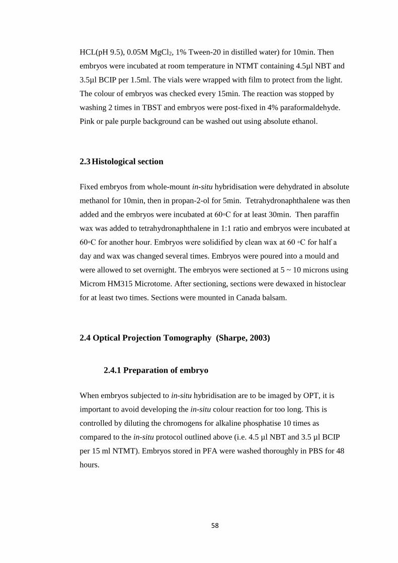
58
HCL(pH 9.5), 0.05M MgCl2, 1% Tween-20 in distilled water) for 10min. Then
embryos were incubated at room temperature in NTMT containing 4.5µl NBT and
3.5µl BCIP per 1.5ml. The vials were wrapped with film to protect from the light.
The colour of embryos was checked every 15min. The reaction was stopped by
washing 2 times in TBST and embryos were post-fixed in 4% paraformaldehyde.
Pink or pale purple background can be washed out using absolute ethanol.
2.3 Histological section
Fixed embryos from whole-mount in-situ hybridisation were dehydrated in absolute
methanol for 10min, then in propan-2-ol for 5min. Tetrahydronaphthalene was then
added and the embryos were incubated at 60◦C for at least 30min. Then paraffin
wax was added to tetrahydronaphthalene in 1:1 ratio and embryos were incubated at
60◦C for another hour. Embryos were solidified by clean wax at 60 ◦C for half a
day and wax was changed several times. Embryos were poured into a mould and
were allowed to set overnight. The embryos were sectioned at 5 ~ 10 microns using
Microm HM315 Microtome. After sectioning, sections were dewaxed in histoclear
for at least two times. Sections were mounted in Canada balsam.
2.4 Optical Projection Tomography (Sharpe, 2003)
2.4.1 Preparation of embryo
When embryos subjected to in-situ hybridisation are to be imaged by OPT, it is
important to avoid developing the in-situ colour reaction for too long. This is
controlled by diluting the chromogens for alkaline phosphatise 10 times as
compared to the in-situ protocol outlined above (i.e. 4.5 µl NBT and 3.5 µl BCIP
per 15 ml NTMT). Embryos stored in PFA were washed thoroughly in PBS for 48
hours.

59
2.4.2 Embedding embryo
1% solution of Low Melting Point Agarose (UltraPure LMP Agarose, invitrogen)
was made up in distilled water. It was cooled to 60◦C and was filtered through
Whatman filter paper 113V. The agarose was cooled down to 37◦C in incubator. A
50mm x 20.3mm (diameter x height) petri dish was prepared on top of an ice-cool
platform. Embryos were transferred from PBS to the petri dish and PBS was
removed as much as possible. The agarose was poured into the dish gently to avoid
generation of bubbles. A glass pipette was used to gently move the embryo to the
centre of the petri dish. Make sure when the gel sets, the embryo is still in the centre
of the dish. After that, the embedding embryo was incubated in 4◦C fridge to
further set for about 10 min.
2.4.3 Trimming the embryo
The agarose was trimmed to a rectangular block with the embryo in the centre of
the block, leaving at least 5mm between the embryo and end of the block. Make
sure the end of the block is completely straight. The surface of the block was dried
and the end of the block was glued to the mount. Press gently to remove air
bubbles and ensure complete contact between the block and the mount. The block
was allowed to set for 5-10min. Use a blade to cut the four corners at the same
angle. When cutting, start from the top of the block and cut outwards. So at the end,
the block will have eight angled sides and an octagonal shape.
2.4.4 Dehydration and clearing of specimen
The mount was placed into absolute methanol and the lid had to be completely
closed. Methanol was changed until no water remained in agarose block. Then
methanol was replaced with BABB (2:1 Benzyl Benzoate: Benzyl Alcohol). The
bottle was left with lids off to allow the remaining methanol to evaporate at room
temperature and was protected from light. Embryos were scanned by OPT when the
agarose block was transparent.

60
2.4.5 Scanning embryos
Before scanning embryos by OPT, alignment should be carried out. Alignment pin
was used to check the rotation and maximise to largest magnification. Pin
alignment was carried out in OPT scanning software. Once it was done, the mount
was attached to the rotation stage and scanning could be started. Scanned images
were reconstructed by NRecon Software.
2.5 Fate mapping
2.5.1 DiI and DiO preparation
DiI and DiO (Invitrogen) stock were dissolved (in dimethylformamide (DMF) at
2mM). Before labelling, 5µl stock DiI or 3µl stock DiO was added to 3µl 1:1000
Tween-20 in sucrose with 45 µl 0.39 M sucrose at 37◦C.
2.5.2 in vivo label
Eggs were placed horizontally and were incubated to required stages. A small
window was opened on top of the eggs. Tyrodes was used to lift the embryos up
above the egg shell. To visualise the embryos, ink (100 times diluted in tyrodes;
winsor & newton black Indian ink) was injected underneath the embryos.
Membranes on top of the embryos were peeled off. For stages older than HH14, the
epidermis was carefully peeled off at the position that would be labelled. Dye was
injected onto the cells at the desired location using a fine sharp calibrated pipette.
The position of labelling was measured using 26mm crossed graticules (Pyser-SGI).
Then about 2ml of egg albumen was removed to let the embryos sink down and 1
ml egg albumen was put back on top of the embryos. The window in the shell was
then closed with PVC tape. Eggs were put back to 37◦C to incubate to the required
stages.

61
2.5.3 Harvest labelled embryo
Dye labelled embryos were harvested as in 2.1. Labelled embryos were
photographed before fixing in 4% paraformaldehyde.
2.6 Imaging
Fixed embryos were first transferred to PBS before imaging. Dyes labelled embryos
were kept in tyrodes when imaging. The embryos were photographed using
Olympus SZH10 Zoom Stereo Microscope and QImaging camera (Retigar 2000R
with QCapture Pro Software). After imaging, embryos were stored back in 4%
paraformaldehyde fix.
2.7 Ablation of diencephalon
Embryos were incubated and prepared as in 2.5.2. The targeted tissues were
removed using a microsurgical knife (5.0mm depth, Rincon). The embryos was
covered with albumin as in 2.5.2 and the egg was taped back the window before
incubating to the required stages. The embryos were harvested and fixed as in 2.1.

62
Chapter 3
Looking for putative
organizers by studies of syn-
expression groups

63
3 Looking for putative organizers by
studies of syn-expression groups
3.1 Introduction
As discussed in chapter 1.5., 31 up-regulated and 16 down-regulated genes were
selected based on the microarray assays and their expression patterns were used to
find our putative organizers. As the microarray assays did not give spatial and
temporal information, their expressions need to be mapped over the developmental
period of interest. Whole-mount in-situ hybridisation was used. Embryos from pre-
primitive streak EGK X-XIV (Eyal-Giladi and Kochav, 1976) to Hamburger and
Hamilton (Hamburger and Hamilton, 1951) stage 14 were surveyed. The following
chapter shows and discussed the expression patterns of syn-expressed genes in
Table 1.3. The work of these whole-mount in-situ hybridisation was done in co-
operation with Dr. Anderson and the full expression patterns for each gene is shown
in Appendix 1.
3.2 Results
3.2.1. Expression of distilled list genes in known organizers
The microarray assays described in chapter 1.5 selected genes that are expressed in
early and/or late Hensen’s node. As it is shown in Figure 3.1, there are 25 out of 31
distilled list genes expressed in Hensen’s node in whole-mount in-situ bridisation
compared the microarray assays. In comparison, expressions of PIK3CD, NXPH1,
NRSN1, PROM1, TMTC2 and ChEST125i5 were not seen (data in Appendix 1). It
may be the hybridisation experiment was not as sensitive as the microarray assay.
But most of the organizer-enriched genes in the distilled list were detected, which
confirms that the microarray assays had selected genes of interest correctly. In
addition, all of the organizer-depleted genes selected from the microarray assays
were not detected in Hensen’s node from HH3+ to HH6 (data in Appendix 1).

64
Figure 3.2 shows the expression patterns of these organizer-depleted genes at HH4.
MSX1 is the best example showing that the organizer-depleted gene is expressed in
the posterior, non-organizer part of the embryo, but not in the node.
Figure 3.1 Expressions of organizer-enriched genes in the Hensen’s node. All these
genes are expressed at HH4, except PPP1R14C and ADRA2A which were
expressed lately at HH6. These whole-mount in-situ hybridisations were done in
co-operation with Dr. Anderson.

65
Figure 3.2 Expressions of organizer-depleted genes in the Hensen’s node. All these
genes were not expressed in Hensen’s node at HH4. These whole-mount in-situ
hybridisations were done in co-operation with Dr. Anderson.
Apart from Hensen’s node, another organizer that was analysed in the microarray
assays was the notochord and floor plate. Figure 3.3 shows the expression patterns
of 30 organizer-enriched genes between HH11 and HH12. Their expressions in
notochord at these stages were commonly strong, although the length of

66
expressions during development was different for each gene (data in Appendix 1).
There was only one exception is PROM1, which had poor expression in the
notochord and might be due to a probe that is difficult to detect. On the other hand,
all 16 organizer-depleted genes were not detected in the notochord between HH11
and HH12. MSX1 was again the best example showing that it was expressed in the
non-organizer, dorsal neural tube but not in the ventral part.
The final organizer that was analysed in the microarray assays was the ZPA and the
whole-mount in-situ hybridisation for embryos at HH22 or later was done by Prof.
Burt group. Combining their data and the expression patterns discussed above, it
could be confirmed that the microarray assays were successful in choosing genes
enriched or depleted in current known organizers. To find out potential new
organizers, the expression patterns of all these genes were analysed by whole-
mount in-situ hybridisation first and our group was mainly focused to study the
development from EGK stages to HH14. In the next chapter, I am going to discuss
the expression patterns of regions that these syn-expressed genes expressed in
common.

67
Figure 3.3 Expressions of organizer-enriched genes in the notochord. All these
genes were expressed in the notochord between HH11 and HH12. These whole-
mount in-situ hybridisations were done in co-operation with Dr. Anderson.

68
Figure 3.4. Expressions of organizer-depleted genes in the notochord. All these
genes were not expressed in the notochord between HH11 and HH12. These whole-
mount in-situ hybridisations were done in co-operation with Dr. Anderson.

69
3.2.2 Summary of regions that syn-expressed genes express in
common
Region
Organizer-
enriched genes
expressed
Organizer-depleted genes not
expressed
Otic vesicle
GNPDA1, KIRREL3,
MCF2L,PLK1S1,
PIK3R5, ADRA2A,
ENPP4, SMOC1,
TSPAN6,
ChEST125i5
LDB2, MSX1, MSX2, BTG2, SOCS2,
CBLN2, CXCL12, EFNA5,
PLXNC1,TNFRSF19, ChEST378m14
Pharyngeal arches
LMO7, GNPDA1,
PLK1S1, PIK3R5
LDB2, BTG2, DSP, SOCS2, CXCL12,
EFNA5, TNFRSF19, ChEST378m14
Somites
KIRREL3, MCF2L,
PKIγ, ENPP4, NRP1,
PCSK6, PRKG1,
RAB11-FIP4,
SMOC1, TMTC2
DLX5, DLX6, MSX1, MSX2,
DPS1, SOCS2, CXCL12, EFNA5,
PLXNC1, TNFRSF19, ChEST378m14
Anterior intestinal
portal endoderm
FBLN7L, NRP1,
NRSN1, SHH,
TMTC2, VTN
DLX5, DLX6, LDB2, MSX1, MSX2,
MOXD1, SOCS2, CXCL12, EFNA5,
PLXNC1, ChEST378m14
Lateral plate
mesoderm
KIRREL3, PKIγ,
NRP1, SMOC1
DLX5, DLX6, LDB2, MSX1, MSX2,
BTG2,SOCS2, CBLN2, EFNA5, PLXNC1,
TNFRSF19, ChEST378m14
Pineal gland
cNOT1, TSPAN6,
PKIγ
DLX5,DLX6,MSX1,MSX2,
LDB2,SOCS2,DSP,BTG2,
PLXNC1,CXCL12,CBLN2,ChEST378m14
Table 3.1 Summary of gene expression patterns in the regions of potential
organizers, having organizer-enriched genes commonly expressed, and organizer-
depleted genes that are not expressed.

70
As it is shown in Table 3.1, there were several regions that had organizer-enriched
genes expressing, including the otic vesicles (Figure 3.5), pharyngeal arches (Figure
3.6), somites (Figure 3.7 and 3.8), the endoderm of anterior intestinal portal (AIP)
(Figure 3.9), lateral plate mesoderm (Figure 3.10) and pineal gland (Figure 3.11).
Whether these regions have organizer abilities has not been studied before. In all of
these regions, most of the organizer-depleted genes were not expressed. The region
with the least expression was the pharyngeal arches, which only had half of the
organizer-depleted genes down-regulated in its region. On the other hand, the
organizer-enriched genes were expressed differently in these potential organizing
regions. Compared to known organizers described in chapter 3.2.1, all of these
regions have fewer than half of the organizer-enriched genes expressed and most of
these genes are not transcription factors. The pharyngeal arches and pineal gland
were the only two that had organizer-enriched transcription factors expressed:
LMO7 was expressed in the pharyngeal arches, while cNOT1 was expressed in the
pineal gland. In addition, the otic vesicles and somites had nearly 10 organizer-
enriched genes expressed, but the area of expression was different for each gene. In
the otic vesicles, GNPDA1 and TSPAN6 were expressed more anteriorly, while
MCF2L and PIK3R5 were expressed posteriorly. On the other hand, genes
expressed in the somites could be divided into three different types. KIRREL3,
PCSK6, PRKG1 and SMOC1 were expressed in posterior newly formed somites.
MCF2L, ENPP4 and TMTC2 were expressed in anterior somites. PKIγ, RAB11-
FIP4 and NRP1 could be seen in all somites along the embryo. Lateral plate
mesoderm had only 4 organizer-enriched genes expressed, and these genes were
also expressed in differently. SMOC1 was expressed posteriorly compared to the
rest that were expressed in anterior lateral plate mesoderm. Finally, the endoderm
of anterior intestinal portal (AIP) had 6 organizer-enriched genes expressed and
their expression patterns were similar, as shown in Figure 3.9. Dr. Anderson chose
to further study the organizing ability of AIP.
For the rest of my project, I selected the pineal gland as a region of interest. The
first reason was it is one of the common sites that express organizer-enriched
transcription factor. Unlike secreted molecules which can be seen in broader area
around the organizer tissue, transcription factor is easier to detect in local

71
organizing region. Also, compared to the pharyngeal arches, which also expressed
transcription factor, the pineal gland had more organizer-depleted genes that were
not expressed within it. In the following chapter, I will further discuss the
expression patterns in pineal gland.
Figure 3.5. Expressions of distilled list genes in otic vesicles. The first two rows
show the expressions of organizer-enriched genes expressed in the otic vesicles,
while the bottom two rows show the organizer-depleted genes that were not
expressed in the otic vesicles. Embryos selected were between HH10 and HH12.
These whole-mount in-situ hybridisations were done in co-operation with Dr.
Anderson.

72
Figure 3.6. Expressions of distilled list genes in pharyngeal arches. The first row
shows the expressions of organizer-enriched genes expressed in the pharyngeal
arches, while the bottom two rows show the organizer-depleted genes that were not
expressed in the pharyngeal arches. Embryos selected were at HH14. These whole-
mount in-situ hybridisations were done in co-operation with Dr. Anderson.
73
Figure 3.7 Expressions of organizer-enriched genes in somites. Embryos selected
are at HH14. The first row of genes were expressed in the newly formed somites,
while the second row of genes were expressed in the somites of anteiror side. The
bottome row shows genes that are expressed in all somites. These whole-mount in-
situ hybridisations were done in co-operation with Dr. Anderson.

74
Figure 3.8 Expressions of organizer-depleted genes in somites. Embryos selected
were at HH14. These whole-mount in-situ hybridisations were done in co-operation
with Dr. Anderson.

75
Figure 3.9 Expressions of distilled list genes in the endoderm of anteiror intestinal
portal (AIP). Embryos selected were between HH10 and HH12. The first row
shows the expression patterns of organizer-enriched genes in AIP, while the bottom
two rows show the expression patterns of organizer-depleted genes. These whole-
mount in-situ hybridisations were done in co-operation with Dr. Anderson.

76
Figure 3.10 Expressions
of distilled list genes in
the lateral plate
mesoderm. Embryos
selected were at HH14.
The first row shows the
expression patterns of
organizer-enriched
genes in the lateral plate
mesoderm, while the
bottom three rows show
the expression patterns
of organizer-depleted
genes. These whole-
mount in-situ
hybridisations were
done in co-operation
with Dr. Anderson.

77
Figure 3.11 Expressions of distilled list genes in the pineal gland. Embryos selected
are at HH14. The first row shows the expression patterns of organizer-enriched
genes in the pineal gland. FBLN7 LIKE was not expressed in the pineal gland, but
around the pineal gland. On the other hand, the bottom three rows show the
expression patterns of organizer-depleted genes. These whole-mount in-situ
hybridisations were done in co-operation with Dr. Anderson.
3.2.3 Pineal region is one of the potential organizer regions
The region that I wanted to further study was the roof of dorsal thalamus, which
develops into the pineal gland in later stages. As it is shown in figure 3.11, there
was one out of the two transcription factors enriched in known organisers that was
also expressed in the pineal region, cNOT1. An intracellular protein, PKIγ and a
transmembrane protein, TSPAN6 were detected in the pineal region. If the roof of
the diencephalon is a potential organizer, transcription factors, intracellular
molecules and transmembrane proteins are expressed within organizing regions. In

78
comparison, secreted molecules may be detected in surrounding tissues adjacent to
organizers. In Figure 3.11, FBLN7L mRNA was excluded from the pineal region
and was observed in the tissues surrounding the roof of the diencephalon. As it is an
extracellular matrix protein, it cannot be excluded that it may be involved in
potential organizing activities as a matricellular proteins that interacts with other
signalling molecules and is involved in cell-cell interaction. On the other hand,
Figure 3.11 shows expressions of all syn-expressed genes that are depleted in
organizers and their expression in the diencephalon at HH14. Five out of six down-
regulated transcription factors were not expressed in the pineal region, ID2 is the
exception. Also, three out of four intracellular molecules were not expressed in
pineal region except MOXD1. Four out of five membrane-bound or secreted
molecules were not expressed in pineal region except TNFRSF19 and EFNA5. And
finally, the unannotated gene, ChEST378m14 was not detected in the pineal region.
In conclusion, there was a small proportion of organizer enriched syn-expressed
genes present in pineal region. But one of the two transcription factor was
expressed, which is important to define organizing regions. On the other hand, only
a few organizer depleted genes were expressed in pineal region. Therefore, pineal
region is one of the potential organizer region to study further.
3.3 Discussion
The above chapter discussed those putative organizer regions. Traditional
transplantation experiments required tests of large amount of pieces of tissues and
an ectopic responding region. In comparison, this project aims to find out
organizer-specific genes, which are either enriched or depleted in known organizers
including Hensen’s node, the notochord and floor plate and the ZPA. Microarray
assays were performed to compare the gene expression levels between organizer
and its closely related non-organizing regions (Figure1.5). There are many genes up
or down-regulated in an organizer at particular stage. However, if cross
comparisons are made between different groups as in Table 1.2, the number of
genes enriched or depleted in early and/or late node, and ventral neural tube and

79
posterior limb bud (ZPA) remarkably few genes are found to be expressed in
common; a total of 31 up-regulated genes and 16 down-regulated genes (Table 1.3).
These putative syn-expressed genes include transcription factors, intracellular
molecules membrane and secreted molecules.
Some of them are known to be involved in developmental process. For example,
cNOT1 is a transcription factor expressed in Hensen’s node and notochord. In
zebrafish, mutation in cNOT1 homolog floating head (flh) causes an absence of ntl
(mouse Brachyury homolog) expression in notochord precursors and leads to the
lack of notochord in embryos. Hence, cNOT1 is considered to be involved in
notochord development in zebrafish (Talbot et al., 1995). Similar function in
notochord formation of flh homolog Xnot in Xenopus, where overexpression of
Xnot can mimic the effect of BMP inhibition and promote the formation of ectopic
notochord (Yasuo and Lemaire, 2001). In addition, mouse Noto gene is shown to be
involved in the regulation of node morphogenesis and left-right asymmetry as
mutant embryos have varied size and shape of posterior notochord, shortened cilia
and randomised left-right internal organs (Beckers et al., 2007). In comparison to
transcription factors which regulate the expression of down-stream molecules in
organizing activity, secreted molecules are released in organizing region and
instruct neighbouring cells. One example is SHH, which was also selected in our
microarray assay. In limb development, it is released by the ZPA in vertebrate and
mediates the polarizing activity. As a morphogen, it diffuses across the posterior-
anterior axis of the limb bud (Chang et al., 1994; Lopez-Martinez et al., 1995;
Riddle et al., 1993a). In addition, signalling pathways that have been mentioned in
Chapter 1 like Wnt signalling involve interactions with intracellular molecules. β-
catenin is one sample intracellular molecule that interferes Wnt pathway and gene
ablation of β-catenin leads to inactivation of Wnt signalling and thus abnormal
structures in posterior midbrain and cerebellum (Brault et al., 2001; Chilov et al.,
2010). The selection of genes involved transcription factors, membrane,
intracellular and secreted molecules will help us to understand more about the
characteristic of organizers and the mechanisms of organizing activities.
Furthermore, many of the genes selected from our microarray assay have unknown
functions in chick embryo development. However, some of them have been proved
to be involved in development in other animal model, for example, ADRA2A, which

80
affects the development of cortical circuits in the mouse (Riccio et al., 2012). Also,
some molecules are involved in important signalling pathway, for example,
PPP1R14C, which interacts with ERK signalling (Wenzel et al., 2007).
As long as syn-expressed genes were selected, common sites of expression of these
genes were going to be analysed, which require mapping of gene expression along
the development of embryo. Whole-mount in-situ hybridisation was performed for
chick embryos from EGK stages and up to HH14. This detail analysis of gene
expression gives us more temporal information of expression of each gene. Also,
SHH and MSX1 were selected as controls once microarray assays had been done as
they are known to be involved in organizing activities as discussed before. The
expressions of SHH and MSX1 correlated to the microarray data, demonstrating that
the microarray analysis was successful. Although some of the genes were expressed
weakly during the development, it may be due to the high sensitivity of the
microarray. At the end, syn-expressed genes including cNOT1, TSPAN6 and PKIγ
were found to be expressed in the centre of the roof of the dorsal thalamus.
FBLN7L which is a secreted molecule was expressed around the centre of the roof
of the dorsal thalamus. Although, there were only 3 of 31 organizer-specific
enriched genes expressed in the dorsal thalamus, it was still an interesting region to
study. To compare, not all organizer-enriched genes from the distilled list is
expressed in the ANR, which is another known organizer. For example, PLK1S1 is
not expressed in ANR up to HH14 (data in Appendix 1). This may due to the
differences between each organizer. In addition, cNOT1, which is one of the two
organizer-enriched transcription factors, was expressed in the roof of the dorsal
thalamus. As discuss before, transcription factor is more likely to be expressed
within the organizing region, and thus more likely to characterize the organizer. As
it is shown in Figure 3.2 and in Kessel Mackem papers, cNOT1 is expressed in very
few regions including Hensen’s node, the notochord, posterior digital limb and the
roof of dorsal thalamus (Stein et al., 1996). Two of them are known organizers.
Furthermore, in the zebrafish model, mutation in flh causes a premature termination
of pineal cells differentiation but does not affect the development of parapineal,
which means flh is involved in development of pineal (Snelson et al., 2008).
Therefore, cNOT1 may be involved in the development of the pineal gland in chick

81
embryo as well due to the possibility of evolutionary conservation. Secondly, there
is a large proportion of organizer-depleted genes that were not expressed in the
dorsal thalamus. Finally, although FBLN7L was not expressed in the centre of the
roof of the dorsal thalamus, it was expressed in the surrounding regions. As
FBLN7L is a secreted molecule, the possibility that FBLN7L may be involved in the
organizing activities through interactions with extracellular proteins and
surrounding cells cannot be excluded.
Furthermore, genes that require further analysis have been processed to histological
section or optical projection tomograph. All this data is uploaded to
www.echickatlas.org, which contribute to a new atlas of gene expression of chick
embryo.
In conclusion, comparison of gene expressions between organizers and closely
related non-organizing regions identified a set of syn-expressed genes that are
enriched or depleted in known organizers. This set of genes involves different types
of molecules including transcription factors and secreted molecules which play
different roles in organizing activities. By studying their gene expression patterns
over early chick embryo development, the centre of the roof of the dorsal thalamus
was identified to have some common expressions of syn-expressed organizer
enriched genes and lack of expressions of majority of organizer depleted genes.
Therefore, it was considered to be a potential organizer.

82
Chapter 4
Development of early
diencephalon in chick embryo

83
4 Development of early diencephalon
in chick embryo
4.1 Introduction
In the development of the embryonic brain, there are two secondary organizers
found so far, the anterior neural ridge (ANR) and the isthmus (chapter 1.3). The
ANR is located at the most anterior part of neural plate and patterns the anterior-
posterior axis of the telencephalon (Crossley et al., 2001; Houart et al., 1998b;
Shimamura and Rubenstein, 1997a; Wilson and Rubenstein, 2000). In comparison,
the isthmus functions as an organizer between mesencephalon and metencephalon
and patterns the anterior hindbrain and midbrain (Alvarado-Mallart, 1993; Joyner,
1996; Marin and Puelles, 1994; Wilson and Rubenstein, 2000; Wurst and Bally-
Cuif, 2001). Diencephalon is derived from the prosencephalon and locates posterior
to the telencephalon. Many studies consider that the zone limitans intrathalamica
(ZLI) as an organizer patterning the development of the diencephalon. ZLI is a
transverse ridge of neuralepithelium located in the middle of diencephalon. It is also
a boundary separate the dorsal and ventral thalamus. (Nieuwenhuys, 1999; Puelles
et al., 1987; Rubenstein et al., 1994) SHH expresses in ZLI and is considered to be
involved in patterning the ventral-dorsal axis of the diencephalon (Hashimoto-Torii
et al., 2003; Kiecker and Lumsden, 2004; Vieira et al., 2005) . It is shown by quail-
chick ectopic transplantation that SHH-expressing cells induced the expression of
Gbx2, which is a marker for the dorsal thalamus (Echevarria et al., 2003). Similarly,
the increased expression of SHH in thalamic progenitors induces rosto-ventral
markers in mice, while reduction of SHH leads to expansion of caudal-dorsal
markers (Vue et al., 2009). Bead experiments in zebrafish confirm the conserved
role of SHH in patterning the thalamus (Scholpp et al., 2006; Zeltser, 2005). Hence,
expression of SHH in the ZLI is required for normal development of the
prethalamus and thalamus. However, there are no transplantation experiments
showing that the ZLI alters the fate of cells in other regions, which argues if it is a
true organizer (based on criteria in chapter 1) or a signalling centre.
As the diencephalon develops, it is subdivided into the dorsal and ventral thalamus,
epithalamus and pretectum. Each compartment is separated by ridges and furrows

84
in the diencephalon (Figdor and Stern, 1993; Larsen et al., 2001). The epithalamus
comprises of the habenula, and the pineal gland. The habenula locates in the
bilateral thalamus and connects to the ventral midbrain and hindbrain through the
stria medullaris (Sutherland, 1982; Villalon et al., 2012). It exhibits a left-right
asymmetry in many animals including the fish, frog and chick (Concha and Wilson,
2001). On the other hand, the pineal gland locates at the centre of the dorsal
epithalamus. It connects to the epithlamus by a stalk structure called pineal recess
(Calvo and Boya, 1978; Calvo and Boya, 1979).It regulates the circadian cycle
through the secretion of melatonin (Csernus et al., 2007). In fish, reptiles and
amphibians, there is an extra pineal body, usually on the left hand side, called
parapineal. The parapineal is involved in the asymmetric development of the
diencephalon (Gamse et al., 2003).
In zebrafish, a homeobox gene floating head expresses in the pineal gland and is
required for its normal development (Snelson et al., 2008). It is also found to be
expressed in the dorsal telencephalon, the roof of prosomere 3 and the epithalamus
(Staudt and Houart, 2007). flh is also involved in the regulation of development of
the notochord in zebrafish (Talbot et al., 1995). From chapter 3, it is shown that
cNOT1, which is the homolog of zebrafish flh, expresses in dorsal thalamus in
HH14. At that stage, the epithelium of the dorsal thalamus is undifferentiated
(Calvo and Boya, 1978). We first questioned how cNOT1 expression relates to the
development of the pineal gland. Also, we questioned whether the development of
the pineal gland involves the movement or the regulation of the cNOT1-expressing
cells.
4.2 Results
4.2.1 cNOT1 expression during diencephalon development
To study the role of cNOT1 in the development of pineal gland in chick embryo and
to compare that to the zebrafish, the expression of cNOT1 was analysed in the
developing diencephalon from HH8 to HH24. Whole-mount in-situ hybridisation

85
was done as it is described in Chapter 2 and cNOT1 cDNA used was the same as in
Chapter 3.
The expression of cNOT1 was first observed in the dorsal neural tube at HH8 (Fig
4.1.A). At HH10, cNOT1 expresses both in the telencephalon and diencephalon
regions (Fig 4.1.B). At HH14, expression of cNOT1 became restricted to the centre
of the roof of the dorsal thalamus (Figure 4.1 C). The lateral borders of cNOT1
expression reaches the lateral ridges seen on the roof of the dorsal thalamus (Figure
4.1 C’). The expression of cNOT1 at HH14 embryo was also confirmed by the OPT
scan, which gives more information in 3D (Figure 4.1 H). This broad expression in
the dorsal thalamus was observed at HH18 as well (Figure 4.1 D and D’). The
pineal gland starts to become morphologically visible as an obvious protrusion
from about HH22. Expression of cNOT1 at this stage became restricted to the
pineal gland and to a small region in the surrounding epithelium (Figure 4.1 E).
Based on other literature, the epithelium of the pineal gland was observed to be
thicker than the surrounding tissues at HH25, although no pinealocytes can be
distinguished yet (Calvo and Boya, 1978). cNOT1 expression at HH25 was
restricted to the pineal gland and the posterior midline of dorsal thalamus but not in
anterior tissues (Figure 4.1 F, F’ & G).

86
Figure 4.1 cNOT1 expression in the developing
diencephalon. Whole-mount in-situ hybridisation
of cNOT1 expressions in chick embryos are
shown from A-F. cNOT1 started to be expressed
in the dorsal neural tube from HH8 (A, Data
kindly provided by Dr. Claire Anderson). At
HH10 cNOT1 was expressed in the
prosencephalon including the future diencephalon
(B). It was still expressed broadly in the early
diencephalon (C at HH14 and D at HH18. C’ and
D’ are the corresponding views of the same
embryos from the dorsal side). The expression of
cNOT1 in the diencephalon at HH14 was confirmed by the OPT scan (H). At
HH22, cNOT1 expression became restricted to the visible protrusion of the pineal
gland and a small region of the dorsal thalamic roof (E at HH22). cNOT1 was
expressed in the pineal gland and the posterior dorsal thalamic roof at HH25 (F, F’
is the corresponding dorsal view of F, G is a sagittal section of F) A: anterior; P:
posterior. The red arrow points to the region which is adjacent to pineal gland
protrution and expresses cNOT1.
P A

87
4.2.2 DKK1 is a pineal gland marker
In zebrafish, cNOT1 homolog flh is a marker for pineal gland and is expressed in
pineal precursors (Aquilina-Beck et al., 2007; Masai et al., 1997). However, from
Figure 4.1, it shows that cNOT1 was not only expressed in the pineal gland in chick
embryo. Therefore, is there any pineal gland markers present in the chick embryo?
In the developing diencephalon, expressions of Wnt family members were
observed. For example, Wnt4 is expressed broadly in the thalamus and epithalamus.
But it was not expressed in the pineal gland. Wnt8b was expressed in the midline of
the telencephalon and epithalamus. However, it is not expressed in the pineal body
as well. (Quinlan et al., 2009) Therefore, it will be interesting to test the expression
of Wnt antagonist in the pineal gland. One of the Wnt antagonists is Dickkopf-
related protein1 (Dkk1) (Kawano and Kypta, 2003; Niehrs, 2006).
Expression of DKK1 in chick embryos was analysed using whole-mount in-situ
hybridisation at different stages of development. DKK1 expression was observed
from HH10 at the posterior procensephalon (future diencephalon) (Figure 4.2.A).
At HH14, DKK1 was expressed in a small region in the centre of the roof of the
dorsal thalamus (Figure 4.2.B and B’). At later stages, DKK1 expression was
observed only within the pineal gland protrusion (Figure 4.2.C, D and D’).
Therefore, DKK1 is expressed only within the pineal gland, when the pineal body
was morphologically observed.

88
Figure 4.2 DKK1 expression in the developing diencephalon studied by whole-
mount in-situ hybridisation. DKK1 started to be expressed in the centre of the future
diencephalon from HH10 (A). This region becomes more compact at HH14 (B)
and at HH18 (C). B’ is the corresponding dorsal view of the embryo of B. DKK1
was restricted to the pineal gland protrusion from HH22 (D; D’ is the dorsal view of
the embryo of D). All pictures were taken from the dorsal view of the embryo.
4.2.3 Fate mapping of the developing diencephalon
From the results in chapter 4.2 and 4.3, it was shown that cNOT1 was not only
expressed in the pineal gland but also in the posterior roof of the dorsal thalamus. In
comparison, DKK1 was expressed in the pineal gland specifically in chick embryo.
To further confirm whether those cNOT1-expressing cells are pineal precursors or
cNOT1 expression is regulated during the development of the pineal gland, fate
mapping of the roof of the dorsal thalamus was required. DiI staining was
performed as described in chapter 2.5. To focus on the study of the development of
the dorsal thalamus, fate mapping was performed at HH14, before the pineal gland
can be morphologically observed. In addition, labelling the diencephalon at earlier
stage (HH10-12) ended up with larger spread of DiI at later stage and it was more
difficult to label the neuroepithelium (data in Appendix 3).

89
4.2.3.1 Standardisation of the coordinates of DiI labelled cells
Due to the 3D structure of brain vesicles, it was difficult to determine the
coordinates of the DiI labelled cells. Figure 4.3 shows how the position of DiI
labelled cells varies in different angles of viewing. The white line shows the
distance of DiI labelled cells to the midline of the dorsal thalamus from ventral and
lateral views, and from any random angle. This distance changes depending on the
angle of observation. Therefore, to minimise this variation and measure the
coordinates of the labelled cells as accurate as possible, the distances of the labelled
cells to morphological landmarks in brain vesicles were measured. These included
the telencephalon/diencephalon boundary, D2/D3 boundary (future thalamus and
pretectum) (Figdor and Stern, 1993), mesencephalon/diencephalon boundary, and
the margins of the brain vesicle. Figure 4.4 shows the landmarks seen from the
dorsal view of the dorsal thalamus at HH14 and black lines show all of the
distances to the morphological margins measured after DiI was labelled at HH14.
These distances were measured again after incubation to HH22. Due to the
difficulty of taking pictures of the in vivo embryo from the top view, images of the
embryos before incubation were taken from lateral view only, to confirm the spread
of the DiI label. Figure 4.5 shows the picture of DiI labelled embryo before
incubation at HH14 (A-C) and after incubation to HH22 both from a lateral view
(D-F) and from a dorsal view (G-H). These pictures show how the DiI labelled cells
dispersed before and after incubation and helped to trace the cell fates. Using these
methods in combination, coordinates of the DiI labelled cells were marked down
for later fate mapping analysis.

90
Figure 4.3 Coordinates of DiI labelled cells to the dorsal midline vary in different
angles of viewings. An embryo at HH14 was labelled by DiI and images were taken
from different angles. A) Dorsal view. B) Lateral view. C) Oblique view. White
arrows indicate the distance between DiI labelled cells to the dorsal midline. Hence,
the coordinates of DiI labelled cells vary based on the way of viewing and it is
important to standardise the method of measurement.
Figure 4.4 Standard method of measurement of DiI labelled cells at HH14 embryo.
Distances between target cells (blue spot) and landmarks in the diencephalon were
measured as shown in black lines. Land marks in HH14 brain included the dorsal
midline (Red line), pretectum (D3)/mesencephalon (Mes), and thalamus
(D2)/telencephalon (Tel). The anterior and posterior edges of the embryo were also
measured as a reference.
Mes D3 D2 Tel

91
Figure 4.5 The dispersion of DiI label is analysed by microscope imaging. A-C:
lateral views of the embryo taken immediately after labelling (HH14). D-F: lateral
views of the embryo after incubation (HH22). G-I: dorsal views after incubation
(HH22). All pictures are from the same embryo. A,D and G: incident epi-
illumination. B, E and H: fluorescence (green excitation, red emission). C,F and I:
merged bright field and fluorescence images. DiI labelled cells are traceable and the
coordinates of labelled cells can be measured out. The successful DiI label should
not disperse through the whole brain vesicle.
4.2.3.2 Correlation between cell fate and cNOT1 expression
248 embryos were labelled with DiI using the method described in Chapter 2.5.
Those embryos that were 1) dead or showing abnormal structures after incubation,
2) DiI dispersed through the whole brain vesicle, 3) not at HH14 when labelled
were discarded. A total of 74 embryos were retained for construction of the fate
map. The raw data of these 74 embryos are included in appendix 2. For each
embryo, the coordinates of DiI label before and after incubation were measured as
described in Chapter 4.2.3.2. This data was combined with the picture taken to draw
the fate map for each embryo. The final data is shown in Figure 4.6 (A-C). Each dot

92
represents one label from one embryo. The drawings show the position of DiI label
and the number of each label correlates to the raw picture in appendix. The colour
represents the cell fate of each label, which correlates with the colour key showing
in Figure 4.6 D. During the labelling process, the epidermis was easily labelled as
well and causes a spread of colour after incubation at the surface of the embryo. As
it was difficult to distinguish the label in the epidermis and the neuroepithelium
under the microscope, if any epidermis was labelled in addition to the
neuroepithelium, this DiI labelled epidermis was not excluded from the map. Also,
although a boundary is drawn for the epithalamus in Figure 4.4 and 4.6.D, no
morphological landmark exists for this region at this stage. The position of this line
was based on the published expression of CYP1B1, which was suggested to be an
epithalamus marker at HH15 (Guinazu et al., 2007) and Fz2 that is expressed in the
epithalamus at HH19 (Quinlan et al., 2009) .
Considering all of the limitations mentioned above and with careful labels, I can
confirm that there was only a small proportion of cells located within the centre of
the dorsal thalamus which contributed to the pineal gland tissues. It is shown as red
spots in Figure 4.6 (A-C). And it was also clear that DiI labelled cells at the
epithalamus surrounding the pineal (purple spots; also future habenula) were
expressing cNOT1. Although the actual boundary of cNOT1 expression within the
epithalamus was difficult to define, due to: firstly, the strength of gene expression
varied in each embryo and secondly, there was shrinkage of the embryo after the
whole-mount in-situ hybridisation process.

93

94
Figure 4.6 Fate mapping of diencephalic cells. The fate maps constructed from all
74 embryos at HH14 are summarised in pictures A to C (25 labels in A and B, and
24 labels in C). Each DiI dot represents the position of a labelled group of cells, and
the colour of the dot symbolises the structures (D, for a stage 22 brain) to which the
labelled cells contributed when analysed at stage 22. The small pictures next to
each diagram show a corresponding enlargement of the dorsal thalamus, which was
the cNOT1 expression region at HH14. The multiple colours in one spot means
cells in this area contributed to both regions. All heads are drawn in the same
direction with posterior on the left and anterior on the right hand side.
Eth:epithalamus; Tel:telencephalon; Mes:mesencephalon; Pret:pretectum;
Th:thalamus; EpiD:epidermis; P:pineal gland.

95
4.3 Discussion
At HH14, the roof of the dorsal thalamus is not differentiated yet. Although
CYP1B1 is considered to be an epithalamus marker at HH15 (Chambers et al.,
2007; Guinazu et al., 2007), there are no morphological boundaries between the
roof and the lateral side of the dorsal thalamus at this stage. As the epithalamus
develops, it subdivided into the habenula and the pineal gland which are conserved
structures in vertebrates. It is known to be involved in the central nervous system. It
receives signals from the cortex, limbic system and basal ganglia through the stria
medullaris and sends signals out to the midbrain and hindbrain through the
fasciculus retroflexus (Hikosaka et al., 2008; Sutherland, 1982). In many lower
vertebrates, the habenula exhibits a left-right asymmetry and markers of genes are
identified both for symmetric and asymmetric structures. For example, in zebrafish,
cpd2 is a symmetric marker and lov is an asymmetric marker for the habenula
(Gamse et al., 2003). However, no specific marker is found for the habenula in
higher vertebrate yet. The gene expressions in the epithalamus are mainly shared
with the pretectum and thalamus (Quinlan et al., 2009). To compare, genes have
been identified in the development of habenula neuronal traits and Pou4f1 is one of
the sample which marks postmitotic habenular neurons (Quina et al., 2009).
As little is known about the development of the epithalamus and there is a lack of a
specific marker for the epithalamus in chick embryo, it is important to understand
the specification of epithalamus structures both in terms of gene expressions and
cell fate before study of the functions in development. cNOT1 was shown to be
expressed broadly in the early diencephalon (Figure 4.1), which raises a possibility
that it may be a marker for the pineal precursor. In zebrafish, cNOT1 homolog flh is
shown to express specifically in the pineal gland and is involved in the regulation of
the development of the pineal (Snelson et al., 2008; Talbot et al., 1995). However,
whole-mount in-situ analysis of cNOT1 shows that it was not only expressed in the
pineal gland but also the posterior epithalamus to the pineal (Figure 4.1). Also, the
fate map of the thalamus shows that only a small proportion of cNOT1 was
contributed to the pineal gland (Figure 4.7). Therefore, cNOT1 expression is down-
regulated during the development of thalamus. On the other hand, DKK1, which is
96
a Wnt antagonist, was shown to be a possible pineal gland marker. Whole-mount
in-situ hybridisation of DKK1 shows that it was expressed only in the pineal
protrusion (Figure 4.2). However, as the DKK1 expressing region was very small at
the centre of dorsal thalamus at HH14, it was difficult to identify whether they are
all pineal gland progenitor by comparing to the fate map in Figure 4.6.
So far, fate maps for chick diencephalon have been done at HH8 (Couly and Le
Douarin, 1985; Couly and Le Douarin, 1987) and HH10 (Garcia-Lopez et al., 2004)
respectively by quail-chick transplantation. These studies reveal the progenitors for
compartments in the diencephalon, including the epithalamus, thalamus and
pretectum. However, the progenitors for both the pineal gland and habenular are not
defined in chick embryo yet. Fate maps have also been done for mouse and
Xenopus to identify precursors for each brain vesicles, but pineal gland and
habenular origins are not defined yet (Eagleson and Harris, 1990; Inoue et al.,
2000). In comparison, fate mapping in zebrafish have revealed the pineal precursor
which expresses flh specifically (Kimmel et al., 1990; Kozlowski et al., 1997; Woo
et al., 1995). Therefore, the fate map of HH14 diencephalon in this chapter provides
information about pineal gland precursors and the development of the epithalamus
in chick embryos.

97
Chapter 5
Pineal Gland is Regenerated
after Ablation of Progenitor
Cells

98
5 Pineal Gland is Regenerated after
Ablation of Progenitor Cells
5.1 Introduction
To test whether the pineal region is an organizer, one of the methods is to remove
the potential organizer and study the changes of surrounding tissues both in terms
of morphology and gene expressions. In the mouse embryo, ablation of the
organizer node at late gastrulation causes a deformation of somites, absence of
notochord and floor plate and foreshorten of the body axis, which demonstrates that
the mouse node is crucial for patterning of the ventral neural tube and lateral body
axis (Davidson et al., 1999). The node-ablated embryo also expresses Pitx2
symmetrically, which is a left-right asymmetry marker (Davidson et al., 1999).
However, the embryonic stage when ablation is performed is important as
regeneration may occur. For example, the node is regenerated when it is ablated
between HH3+ and HH4- in chick embryo (Grabowski, 1956; Psychoyos and Stern,
1996). The regenerated node is identical to that in normal embryos and the embryo
develops normally after ablation. Truncated notochord is observed when the node is
ablated at HH4+ or later (Grabowski, 1956; Joubin and Stern, 1999; Psychoyos and
Stern, 1996; Yuan et al., 1995). Hence, before studying whether the potential
organizer can influence the patterning of surrounding tissues, the following
questions have to be answered: 1) whether all organizer cells have been removed;
2) which embryonic stage is most appropriate to perform ablation experiments; 3)
whether organizer is regenerated after ablation.
Based on the gene expression analysis in Chapter 3, the roof of the dorsal thalamus
is a potential organizer with the expression of syn-expressed genes
cNOT1,TSPAN6, and PKIγ. The expression of cNOT1 starts in a broad area and
later on restricts to the pineal gland, which raised the possibility that cNOT1-
expressing cells may be the pineal gland progenitors. However, fate mapping
studies in Chapter 4 shows that not all cNOT1 cells develop to the pineal gland.
Therefore, there is a question: whether the pineal gland or the cNOT1-expressing
territory is a potential organizer. To address this question, ablation experiments
were performed for these two regions respectively.
99
This Chapter presents the results of ablation of the pineal region and how they are
related to the organizing activities and the regeneration potential of this region.
5.2 Results
5.2.1 Pineal gland is regenerated after ablation of pineal
progenitors.
Ablation experiments were performed as described in Chapter 2.6. Ablated
embryos were processed to whole-mount in-situ hybridisation as described in
Chapter 2.2.6.
To test the roles of pineal gland and the cNOT1-expressing cells in brain
development, ablation of the roof of the diencephalon at HH14 was performed. The
roof of the diencephalon was divided into three parts as it is shown in Figure 5.1.
“Anterior” (A) mainly includes cells that are located in the anterior of the roof of
the dorsal thalamus. “Centre” (C) is the centre region of the roof of the dorsal
thalamus. “Posterior” (P) is the group of cells locating at the posterior part of the
roof of the dorsal thalamus. Based on the data from whole-mount in-situ in Chapter
3 and the fate map in Chapter 4, the anterior cells contain those that express cNOT1
and not pineal progenitors. The centre region of the roof of the dorsal thalamus
expresses cNOT1 and contains pineal progenitors as well. On the other hand, the
posterior tissues of the roof of the diencephalon express cNOT1 but do not
contribute to the pineal gland.

100
Figure 5.1 Design of ablation experiment. The picture shows the landmark of a
HH14 embryo used for ablation experiments. The division is based on the
measurement using graticules. P stands for the posterior tissues in the roof of dorsal
thalamus. C is the centre of the roof of dorsal thalamus. A is anterior tissues in the
roof of dorsal thalamus. The white arrows point to the morphological land mark
between telencephalon and thalamus, and between thalamus and pretectum.
Based on the fate map and gene expression analysis in Chapter 4, it showed that
pineal gland progenitors contain a small group of cells located in the centre of the
roof of the dorsal thalamus at HH14 (“center” region in Figure 5.1). cNOT1 was
expressed in a large region at HH14 in the diencephalon including the pineal
progenitors (“center” and “posterior” region in Figure 5.1). To examine whether the
pineal region is required for patterning surrounding tissue, the “center” region
containing pineal progenitors was removed at HH14 embryos and the ablated
embryos were incubated for two days. Ablation of pineal progenitors (center) was
also combined with removal of either posterior or anterior tissue, to see whether
cNOT1-expressing cells are required for development. Before processing embryos
for further analysis, regeneration of pineal gland was observed. As it is shown in

101
Table 5.1, removal of the “centre” region containing pineal progenitors alone led to
regeneration of the pineal gland protrusion in all cases. Removal of pineal
progenitors with the neighbouring anterior region led to regeneration of the pineal
gland protrusion in half of the cases. On the other hand, removal of pineal
progenitors together with the posterior neighbouring tissue gives no regeneration of
the pineal gland protrusion in all cases. As the “centre” and “posterior” parts of the
roof of the dorsal thalamus contain nearly all the cNOT1 expressing cells, it raised
the possibility that the pineal gland can regenerate from cNOT1 expressing cells
that are not pineal progenitors.
However, this preliminary work was quick ablation without picturing and was only
based on the observation of the morphological protrusion of pineal gland. It was not
precise enough and do not show whether the gene expression in the roof of
diencephalon was still the same after ablation. Therefore, further analysis was
carried out and will be discussed in the next chapter.
Regions of Ablation
number of Regeneration
Centre (C)
5/5
Center+Anterior (C+A)
3/6
Center+Posterior (C+P)
0/6
Table 5.1 preliminary data of ablation experiments in dorsal thalamus. The first row
shows the number of regeneration, when the centre of the roof of dorsal thalamus
was ablated. Five out of five samples got regeneration. The second row shows the
number of regeneration, when the centre and the anterior tissues of the roof of
dorsal thalamus was ablated. Half of the samples had pineal regeneration. The third
row shows the number of regenerations, when the centre and the posterior of the
roof of dorsal thalamus was ablated. None of the samples had pineal regeneration.

102
5.2.2 Gene expression patterns are normal after regeneration of
pineal
Apart from the morphology of the diencephalon, the gene expression patterns
needed to be examined after regeneration of the pineal gland. As it shows in Figure
5.2, removal of pineal progenitors (region “C”) alone at HH14 led to the
regeneration of the pineal gland protrusion by 48hours incubation, which correlates
to Table 5.1. Whole-mount in-situ hybridisation was performed to further test the
expression of pineal region markers after ablation. cNOT1 was expressed both in
the pineal gland protrusion and in the posterior tissues, as in their usual pattern, 48
hours post-surgery (Figure 5.2c). Wnt8b and Pax6 are genes expressed in
diencephalon and telencephalon from about HH14 (Figure 5.2 d,e,g & h). Ablation
of the pineal gland at HH14 removed part of the Wnt8b and Pax6 expressing
tissues, however their expressions were still detected around pineal two days after
surgery (Figure 5.2 f & i). The expression area of Pax6 was slightly smaller in
ablated embryo, which may be due to the unfinished regeneration process.
Therefore, the pineal region (both cNOT1 expressing cells and the pineal gland
protrusion) can regenerate after removal of pineal progenitors in the “centre” of the
roof of dorsal thalamus at HH14 and as a consequence, patterning of the
diencephalon occurs normally.
5.2.3 Ablation of all cNOT1 expressing cells prevents the
regeneration of the pineal gland and DKK1 expression
To examine whether the pineal gland regenerates from surrounding cNOT1 -
expressing cells, ablation experiments were performed to remove the entire cNOT1
expressing region. To see whether cNOT1-expressing cells were removed
completely, embryos from which “Centre” and “Posterior” tissues had been ablated
were fixed immediately after ablation. No cNOT1-expressing cells were observed
(Figure 5.3b). The ablated embryos were also tested for the expression of DKK1,
which is expressed only in the pineal gland (Chapter 4). No DKK1 expression was
observed (Figure 5.3d). Another group of ablated embryos was incubated for two
days to see whether the pineal gland would be regenerated. As shown in Figure

103
5.3h, no DKK1 expression was observed. Also, there was no pineal gland
protrusion. Therefore, the pineal gland did not regenerate. The expression of
cNOT1 was also tested, and it showed that cNOT1 was re-expressed in a small
region of the roof of diencephalon (Figure 5.3f). However, no morphological
protrusion was observed. Therefore, removal of all cNOT1 expressing cells in the
“Centre” and “Posterior” tissues of the diencephalon prevents regeneration of the
pineal gland protrusion that normally expresses DKK1. However, cNOT1
expression itself regenerates after ablation.

104
Figure 5.2 Ablation of pineal progenitors (“centre” region) was performed at HH14
and embryos were incubated two days (~H22) before process to whole-mount in-
situ hybridisation. The first row shows the expression of cNOT1 in normal HH14
embryo (a), normal HH22 embryo (b) and HH22 ablated embryo (c). Smaller pineal
protrusion was observed in (c) with cNOT1 expression, which means that pineal
was regenerated. The second row shows the expression of Wnt8b in normal HH14
embryo (d), normal HH22 embryo (e), and HH22 ablated embryo (f). Wnt8b was
not expressed in pineal gland at HH22 but in surrounding tissues. There was no
difference in Wnt8b expression between the control embryo and ablated embryo.
The third row shows the expression of Pax6 normal HH14 embryo (g), normal
HH22 embryo (h) and HH22 ablated embryo (i). Pax6 was not expressed in pineal
gland at HH22 but in surrounding tissues. The ablated embryo had a smaller area of
Pax6 expressed around the pineal, which may due to incomplete regeneration.
Black arrows are pointing at the transparent pineal.

105
Figure 5.3 Ablation of cNOT1 expressing cells (“centre” and “posterior” regions). Pictures a, c, e and g are controls for cNOT1 and
DKK1 at either HH14 or HH25 respectively. Embryos were ablated with centre and posterior tissues of the roof of dorsal thalamus
based on Figure 5.1 scale. Ablated embryos were either fixed directly to check gene expression (b & d) or processed to incubation for
regeneration (f & h). It shows that the ablation removed all cNOT1 and DKK1 expressing cells (b & d). The number accounts for the
embryos that had no cNOT1 or DKK1 cells left, i.e. successful ablation. The ablated embryos were harvested two days later and were
processed to whole-mount in-situ. None of the embryos had pineal gland regeneration. DKK1 expression was not detected in all
samples (h, number stands for no DKK1 expression, i.e. successful ablation). In comparison, as shown in picture f, cNOT1 was
expressed again without regeneration of the pineal (no protrusions). The number stands for the embryos that had cNOT1 expression but
no regeneration. Therefore, re-expression of cNOT1 was a consistent result.

106
5.4 Discussion
As discussed in Chapter 3, the roof of the dorsal thalamus with cNOT1 expression
was revealed as a possible organizer region. Fate mapping experiments (Chapter 4),
showed that this region contains both pineal progenitors and cells with non-pineal,
epithalamic fate, i.e. cNOT1 is not only expressed in pineal progenitors. This region
expresses cNOT1, PKIγ and TSPAN6 and lacked expression of some of the genes
generally absent from the tested organizers (MSX1&2, DLX5&6,BTG2, etc.) at
HH14 (Chapter 3) and was therefore a possible organizer. To test for organizing
properties of this region, various territories of the roof of the dorsal thalamus were
ablated. First, an attempt was made to remove just the pineal progenitors, located at
the centre of the roof of the dorsal thalamus. However, ablation of pineal precursors
(centre, with DKK1 expression) showed regeneration of the pineal gland with
normal morphology and diencephalic gene expression patterns (Table 5.1 and
Figure 5.2). Therefore, the pineal gland can be regenerated from surrounding
tissues. To confirm this, different regions from the surrounding epithalamic area
were removed along with pineal progenitors. Removal of the central and anterior
regions still led to regeneration of the pineal gland protrusion (Table 5.1). The
ablation removed all pineal progenitors but left tissues (posterior) that expressed
cNOT1. In contrast, removal of all pineal progenitors together with posterior tissues
containing virtually all cNOT1 expressing cells reduced the incidence of
morphological regeneration (Table 5.1and Figure 5.3). Although the pineal marker
DKK1 was not expressed and a pineal gland protrusion was not observed after
ablation of the centre and posterior regions of the dorsal diencephalon , expression
of cNOT1 was restored (Figure 5.3). Therefore, the pineal gland can be regenerated
from surrounding tissues that have cNOT1 expression and it raises a question that
whether cNOT1 plays a role in development and regeneration of the pineal gland. In
zebrafish, flh (which is homologue to NOT) mutation can affect pineal development
without disruption of the neighbouring parapineal (Snelson et al., 2008), suggesting
that Not function is important for pineal development. However, there are another
NOT gene in chick, which is cNOT2. Comparing to cNOT1, cNOT2 is more colsely
related to zebrafish flh (Stein et al., 1996). Therefore, studies of both cNOT1 and
cNOT2 are required to address the role of NOT gene in pineal regeneration. Further
more, pienal gland is not differentiated at HH14, i.e. neuroepithelial cells are

107
identical within the dorsal diencephalon (Calvo and Boya, 1978; Calvo and Boya,
1979). Ablation of differentiated pineal cells is required to examine whether
cNOT1-expressing cells can regenerate the pineal gland at later stages. Ablations of
the pineal area at later stages (>HH18) were tried. However these embryos did not
survive, probably due to haemorrhage caused by damage to the surrounding blood
vessels that have developed by this stage. Embryos without a pineal gland appeared
to develop normally to HH22 and have apparently normal morphology of the head,
but a more detailed study of axon tracts and gene expression is required to ascertain
this rigorously. Finally, there is a question of whether the posterior non-pienal
progenitor, cNOT1-expressing cells are required for normal developmen of the
pineal. Ablation of the posterior dorsal thalamus is required to address this
question. Therefore further experiments are needed to be done in this case.
In conclusion, the ablation experiements showed that the pineal gland can
regenerate after ablation at stage 14 and that patterning of the diencephalon occurs
normally, provided that posterior cNOT1-expressing cells are spared at the time of
ablation. Removal of all NOT1-expressing cells prevents regeneration of the pineal
but cNOT1 expression returns. Therefore, to test the organizing ability of the
cNOT1-expressing region of the roof of the dorsal thalamus, further experiments
are required. The following questions can be studied in the future.
1) Whether regeneration will appear if ablation of the pineal gland is
performed at later stages, i.e. after HH18 when the pineal gland is more
differentiated?
2) Can transplantation of the pineal gland or cNOT1-expressing cells to
ectopic site in brain region cause misexpression of brain markers, like
Wnt8b, Wnt4, Pax6 and Otx2?
3) Whether gain- or loss-of-function of cNOT1 affects the development of
the pineal gland and/or diencephalon?

108
Chapter 6
General Discussion

109
6 General Discussion
The expression of genes that are enriched or depleted in 3 known organizers
(Chapter 3) pointed to the region of the roof of the dorsal thalamus including and
adjacent to the pineal gland as a potential organizer. This region expresses 3/32 of
the enriched genes and lacks expression of 12/16 depleted genes which, although
not representing the majority of the genes within this synexpression group, includes
a key transcription factor, cNOT1, which is only expressed in a few regions of the
embryo, most of which are known to have signalling activity: the node at stage 3+-
4, the notochord from stage 5 onwards and the posterior limb bud. This raised the
possibility that the peri-pineal region of the thalamic roof could be an organizer of
surrounding regions of the brain.
To explore this further, we started by constructing a fate map of this regions
(Chapter 4) at stage 14. cNOT1 is expressed from stage 8 at the prospective
diencephalon, at stage 10 as a broad region in the diencephalic roof, and expression
gradually becomes confined to the emerging pineal protrusion (visible from stage
18) and a territory just posterior to it on the dorsal thalamic roof. DiI-labelling
reveals that not all cells within the cNOT1-expressing domain are pineal gland
progenitors. In comparison, flh which is cNOT homologue, define pineal gland
progenitors, which is confirmed by mutation of flh that cause the pineal complex to
premature at 18 somite stage (Masai et al., 1997). The expression pattern of cNOT1
and the contribution of cNOT1-expressing cells to the diencephalon I found
corresponds more closely to the observation from Staudt and Houart in zebrafish,
where cNOT1 is expressed in a broad area in the epithalamus and telencephalon and
only a small proportion of cNOT1-expressing cells participate in the pineal gland
(Staudt and Houart, 2007). However, DKK1 is expressed exclusively in the pineal
gland from stage 14 in the chick embryo, prior to morphological changes that reveal
the appearance of the pineal gland (Calvo and Boya, 1978; Calvo and Boya, 1979).
Comparing the expression patterns and fate maps, the majority of DKK1-expressing
cells develop into the pineal gland, while only a small proportion of cNOT1-

110
expressing cells contribute to the pineal gland and most of them develop into the
surrounding epithelium of thalamus.
To determine whether it is the pineal gland or the cNOT1-expressing territory that
acts as an organizer, one approach is to establish whether it is required for
patterning the surrounding diencephalon. However, ablation could lead to
regeneration, which would undermine this experiment. The broad expression of
cNOT1 in the early diencephalon and the findings (Chapter 4) that not all cNOT1-
expressing cells contribute to the pineal also raises the question of whether non-
pineal precursor cNOT1-expressing cells can restore the pineal gland when this is
ablated. We performed ablations of the prospective pineal gland territory at stage 14
either alone or with the neighbouring anterior or posterior diencephalon roof (the
latter contains most of the non-pineal cNOT1-expressing cells). It was found that
regeneration of pineal gland was observed when only pineal precursors were
removed, and was inhibited when both the precursors and the posterior
diencephalon roof were removed. Therefore, the pineal gland can regenerate from
surrounding diencephalic roof, provided that cells from the cNOT1-expressing
territory are spared from ablation. Also, to further investigate the organizing ability
of the pineal gland, regeneration can be inhibited by either removal of surrounding
tissues or ablation at later embryonic stage. On the other hand, to test whether
cNOT1-expressing territory is an organizer, ablation may be performed at later
stage, though cNOT1 is expressed broadly in the early diencephalon and removal of
all cNOT1-expressing cells would require ablation of almost whole diencephalon
roof.
Based on our ablation experiment, it is still clear that regeneration of the pineal
gland requires cNOT1 expression, as removal of all cNOT1-expressing cells
prohibits the pineal gland regeneration. It is also interesting that cNOT1 expression
re-appeared without the regeneration of pineal, which means it is induced by the
surrounding tissues. Comparing to the pineal, removal of the progenitors alone did
not interrupt the normal development of the dorsal diencephalon and the cNOT-
expressing region seems to be more important to the development. Therefore, if

111
there is an organizer within diencephalon, it is more likely to be the cNOT-
expressing region. To further examine this possibility, transplantation of cNOT-
expressing region to other brain vesicles, for instance, telencephalon or
mesencephalon, is needed. If it is a true organizer, the transplantation of cNOT-
expressing tissues to ectopic side should induce the expression of diencephalic
markers. And this transplantation should be compared to that of pineal gland
transplantation alone, to confirm the requirement of cNOT1 expression for brain
development. Also, the ablation experiments in Chapter 5 lacked the removal of
posterior part of the dorsal thalamus alone. This posterior tissues contains cNOT-
expressing cells but no pineal gland progenitors. If the cNOT1 expression is needed
for normal development, reduction of the expression region may affect the normal
development of the pineal gland and diencephalon as well, which in turn, would
prove the induction activity of cNOT1-expressing cells rather than regeneration.
In conclusion, the experiments reported in this thesis pointed to a region where
gene expression patterns partly resemble those of three known organizers: the
pineal and part of the adjacent thalamic roof. Ablation of the pineal precursors leads
to regeneration as long as peri-pineal cNOT1 cells are spared. To determine whether
the pineal or the entire cNOT1-expressing region acts as an organizer it will be
necessary to complete the ablation experiments, as well as transplantation
experiments to more lateral, anterior and posterior sites, analysing markers for
features in the neighbouring diencephalon (eg. Wnt8b, Pax6, Lfng, the pattern of
axon tracts). If the pineal or peri-pineal region acts as an organizer, ablation should
lead to loss of some aspects of the pattern and transplantation should lead to some
features being duplicated. It will be important to perform these experiments at
different stages.

112
Reference
Aardal, S. and Helle, K.B., 1992. The vasoinhibitory activity of bovine chromogranin A fragment (vasostatin) and its independence of extracellular calcium in isolated segments of human blood vessels. Regul Pept. 41, 9-18.
Abdelkhalek, H.B., Beckers, A., Schuster-Gossler, K., Pavlova, M.N., Burkhardt, H., Lickert, H., Rossant, J., Reinhardt, R., Schalkwyk, L.C., Muller, I., Herrmann, B.G., Ceolin, M., Rivera-Pomar, R. and Gossler, A., 2004. The mouse homeobox gene Not is required for caudal notochord development and affected by the truncate mutation. Genes Dev. 18, 1725-36.
Abercrombie, M. and Bellairs, R., 1954. The Effects in Chick Blastoderms of Replacing the Primitive Node by a Graft of Posterior Primitive Streak. J Embryol Exp Morphol. 2, 55-&.
Adams, K.A., Maida, J.M., Golden, J.A. and Riddle, R.D., 2000. The transcription factor Lmx1b maintains Wnt1 expression within the isthmic organizer. Development. 127, 1857-1867.
Al-Jassar, C., Knowles, T., Jeeves, M., Kami, K., Behr, E., Bikker, H., Overduin, M. and Chidgey, M., 2011. The nonlinear structure of the desmoplakin plakin domain and the effects of cardiomyopathy-linked mutations. J Mol Biol. 411, 1049-61.
Al Tassan, N., Khalil, D., Shinwari, J., Al Sharif, L., Bavi, P., Abduljaleel, Z., Abu Dhaim, N., Magrashi, A., Bobis, S., Ahmed, H., Alahmed, S. and Bohlega, S., 2012. A missense mutation in PIK3R5 gene in a family with ataxia and oculomotor apraxia. Hum Mutat. 33, 351-4.
Alappat, S., Zhang, Z.Y. and Chen, Y.P., 2003. Msx homeobox gene family and craniofacial development. Cell Res. 13, 429-42.
Alexandre, C., Jacinto, A. and Ingham, P.W., 1996. Transcriptional activation of hedgehog target genes in Drosophila is mediated directly by the cubitus interruptus protein, a member of the GLI family of zinc finger DNA-binding proteins. Genes Dev. 10, 2003-13.
Altaba, A.R., Placzek, M., Baldassare, M., Dodd, J. and Jessell, T.M., 1995. Early Stages of Notochord and Floor Plate Development in the Chick-Embryo Defined by Normal and Induced Expression of Hnf-3-Beta. Dev Biol. 170, 299-313.
Alvarado-Mallart, R.M., 1993. Fate and potentialities of the avian mesencephalic/metencephalic neuroepithelium. J Neurobiol. 24, 1341-55.
Amireault, P. and Dube, F., 2000. Cloning, sequencing, and expression analysis of mouse glucosamine-6-phosphate deaminase (GNPDA/oscillin). Mol Reprod Dev. 56, 424-35.
Andersson, L., Westerlund, J., Liang, S., Carlsson, T., Amendola, E., Fagman, H. and Nilsson, M., 2011. Role of EphA4 receptor signaling in thyroid development: regulation of folliculogenesis and propagation of the C-cell lineage. Endocrinology. 152, 1154-64.
Andres, A.C. and Ziemiecki, A., 2003. Eph and ephrin signaling in mammary gland morphogenesis and cancer. J Mammary Gland Biol Neoplasia. 8, 475-85.
Apostolova, G., Dorn, R., Ka, S., Hallbook, F., Lundeberg, J., Liser, K., Hakim, V., Brodski, C., Michaelidis, T.M. and Dechant, G., 2007. Neurotransmitter phenotype-specific expression changes in developing sympathetic neurons. Mol Cell Neurosci. 35, 397-408.

113
Appleton, B.A., Wu, P., Maloney, J., Yin, J., Liang, W.C., Stawicki, S., Mortara, K., Bowman, K.K., Elliott, J.M., Desmarais, W., Bazan, J.F., Bagri, A., Tessier-Lavigne, M., Koch, A.W., Wu, Y., Watts, R.J. and Wiesmann, C., 2007. Structural studies of neuropilin/antibody complexes provide insights into semaphorin and VEGF binding. EMBO J. 26, 4902-12.
Aquilina-Beck, A., Ilagan, K., Liu, Q. and Liang, J.O., 2007. Nodal signaling is required for closure of the anterior neural tube in zebrafish. BMC Dev Biol. 7, 126.
Araki, M., Nagata, K., Satoh, Y., Kadota, Y., Hisha, H., Adachi, Y. and Taketani, S., 2002. Developmentally regulated expression of Neuro-p24 and its possible function in neurite extension. Neurosci Res. 44, 379-89.
Artinger, K.B. and Bronnerfraser, M., 1993. Delayed Formation of the Floor Plate after Ablation of the Avian Notochord. Neuron. 11, 1147-1161.
Babu, D. and Roy, S., 2013. Left-right asymmetry: cilia stir up new surprises in the node. Open Biol. 3, 130052.
Bach, I., 2000. The LIM domain: regulation by association. Mech Dev. 91, 5-17. Bach, I., Carriere, C., Ostendorff, H.P., Andersen, B. and Rosenfeld, M.G., 1997. A family of
LIM domain-associated cofactors confer transcriptional synergism between LIM and Otx homeodomain proteins. Genes Dev. 11, 1370-80.
Bachiller, D., Klingensmith, J., Kemp, C., Belo, J.A., Anderson, R.M., May, S.R., McMahon, J.A., McMahon, A.P., Harland, R.M., Rossant, J. and De Robertis, E.M., 2000. The organizer factors Chordin and Noggin are required for mouse forebrain development. Nature. 403, 658-661.
Bachvarova, R.F., Skromne, I. and Stern, C.D., 1998. Induction of primitive streak and Hensen's node by the posterior marginal zone in the early chick embryo. Development. 125, 3521-34.
Baker, R.K. and Antin, P.B., 2003. Ephs and ephrins during early stages of chick embryogenesis. Dev Dyn. 228, 128-42.
Baldock, R.A., Bard, J.B., Burger, A., Burton, N., Christiansen, J., Feng, G., Hill, B., Houghton, D., Kaufman, M., Rao, J., Sharpe, J., Ross, A., Stevenson, P., Venkataraman, S., Waterhouse, A., Yang, Y. and Davidson, D.R., 2003. EMAP and EMAGE: a framework for understanding spatially organized data. Neuroinformatics. 1, 309-25.
Ballycuif, L., Alvaradomallart, R.M., Darnell, D.K. and Wassef, M., 1992. Relationship between Wnt-1 and En-2 Expression Domains during Early Development of Normal and Ectopic Met-Mesencephalon. Development. 115, 999-1009.
Barth, K.A., Kishimoto, Y., Rohr, K.B., Seydler, C., Schulte-Merker, S. and Wilson, S.W., 1999. Bmp activity establishes a gradient of positional information throughout the entire neural plate. Development. 126, 4977-4987.
Basile, J.R., Afkhami, T. and Gutkind, J.S., 2005. Semaphorin 4D/plexin-B1 induces endothelial cell migration through the activation of PYK2, Src, and the phosphatidylinositol 3-kinase-Akt pathway. Mol Cell Biol. 25, 6889-98.
Beckers, A., Alten, L., Viebahn, C., Andre, P. and Gossler, A., 2007. The mouse homeobox gene Noto regulates node morphogenesis, notochordal ciliogenesis, and left right patterning. Proc Natl Acad Sci U S A. 104, 15765-70.
Beddington, R.S., 1994. Induction of a second neural axis by the mouse node. Development. 120, 613-20.
Beddington, R.S.P. and Robertson, E.J., 1999. Axis development and early asymmetry in mammals. Cell. 96, 195-209.
Bellairs, R., 1954. The effects of tetra-sodium 2-methyl 1:4-naphthohydroquinone diphosphate on early chick and amphibian embryos. Br J Cancer. 8, 685-92.

114
Belo, J.A., Silva, A.C., Borges, A.C., Filipe, M., Bento, M., Goncalves, L., Vitorino, M., Salgueiro, A.M., Texeira, V., Tavares, A.T. and Marques, S., 2009. Generating asymmetries in the early vertebrate embryo: the role of the Cerberus-like family. Int J Dev Biol. 53, 1399-407.
Bensoussan-Trigano, V., Lallemand, Y., Saint Cloment, C. and Robert, B., 2011. Msx1 and Msx2 in limb mesenchyme modulate digit number and identity. Dev Dyn. 240, 1190-202.
Bhattacharyya, S., Bailey, A.P., Bronner-Fraser, M. and Streit, A., 2004. Segregation of lens and olfactory precursors from a common territory: cell sorting and reciprocity of Dlx5 and Pax6 expression. Dev Biol. 271, 403-14.
Bi, L., Okabe, I., Bernard, D.J. and Nussbaum, R.L., 2002. Early embryonic lethality in mice deficient in the p110beta catalytic subunit of PI 3-kinase. Mamm Genome. 13, 169-72.
Blum, M., Gaunt, S.J., Cho, K.W.Y., Steinbeisser, H., Blumberg, B., Bittner, D. and Derobertis, E.M., 1992a. Gastrulation in the Mouse - the Role of the Homeobox Gene Goosecoid. Cell. 69, 1097-1106.
Blum, M., Gaunt, S.J. and Derobertis, E.M., 1992b. Gastrulation in the Mouse - the Role of the Homeobox Gene Goosecoid. Molecular Biology of the Cell. 3, A98-A98.
Bornstein, P., 1995. Diversity of function is inherent in matricellular proteins: an appraisal of thrombospondin 1. J Cell Biol. 130, 503-6.
Bornstein, P. and Sage, E.H., 2002. Matricellular proteins: extracellular modulators of cell function. Curr Opin Cell Biol. 14, 608-16.
Bouwmeester, T., Kim, S.H., Sasai, Y., Lu, B. and DeRobertis, E.M., 1996. Cerberus is a head-inducing secreted factor expressed in the anterior endoderm of Spemann's organizer. Nature. 382, 595-601.
Brault, V., Moore, R., Kutsch, S., Ishibashi, M., Rowitch, D.H., McMahon, A.P., Sommer, L., Boussadia, O. and Kemler, R., 2001. Inactivation of the beta-catenin gene by Wnt1-Cre-mediated deletion results in dramatic brain malformation and failure of craniofacial development. Development. 128, 1253-64.
Braun, M., Wunderlin, M., Spieth, K., Knochel, W., Gierschik, P. and Moepps, B., 2002. Xenopus laevis Stromal cell-derived factor 1: conservation of structure and function during vertebrate development. J Immunol. 168, 2340-7.
Braun, M.M., Etheridge, A., Bernard, A., Robertson, C.P. and Roelink, H., 2003. Wnt signaling is required at distinct stages of development for the induction of the posterior forebrain. Development. 130, 5579-5587.
Brenner, R., Perez, G.J., Bonev, A.D., Eckman, D.M., Kosek, J.C., Wiler, S.W., Patterson, A.J., Nelson, M.T. and Aldrich, R.W., 2000. Vasoregulation by the beta1 subunit of the calcium-activated potassium channel. Nature. 407, 870-6.
Briscoe, J., Chen, Y., Jessell, T.M. and Struhl, G., 2001. A hedgehog-insensitive form of patched provides evidence for direct long-range morphogen activity of Sonic hedgehog in the neural tube. Mol Cell. 7, 1279-1291.
Briscoe, J. and Ericson, J., 2001. Specification of neuronal fates in the ventral neural tube. Current Opinion in Neurobiology. 11, 43-49.
Briscoe, J., Pierani, A., Jessell, T.M. and Ericson, J., 2000. A homeodomain protein code specifies progenitor cell identity and neuronal fate in the ventral neural tube. Cell. 101, 435-445.
Broccoli, V., Boncinelli, E. and Wurst, W., 1999. The caudal limit of Otx2 expression positions the isthmic organizer. Nature. 401, 164-168.
Brugger, S.M., Merrill, A.E., Torres-Vazquez, J., Wu, N., Ting, M.C., Cho, J.Y., Dobias, S.L., Yi, S.E., Lyons, K., Bell, J.R., Arora, K., Warrior, R. and Maxson, R., 2004. A phylogenetically conserved cis-regulatory module in the Msx2 promoter is

115
sufficient for BMP-dependent transcription in murine and Drosophila embryos. Development. 131, 5153-65.
Butler, D.C., Haramizu, S., Williamson, D.L. and Alway, S.E., 2009. Phospho-ablated Id2 is growth suppressive and pro-apoptotic in proliferating myoblasts. PLoS One. 4, e6302.
Butros, J., 1967. Limited Axial Structures in Nodeless Chick Blastoderms. J Embryol Exp Morphol. 17, 119-&.
Calvo, J. and Boya, J., 1978. Embryonic development of the pineal gland of the chicken (Gallus gallus). Acta Anat (Basel). 101, 289-303.
Calvo, J. and Boya, J., 1979. Ultrastructural study of the embryonic development of the pineal gland of the chicken (Gallus gallus). Acta Anat (Basel). 103, 39-73.
Campione, M., Steinbeisser, H., Schweickert, A., Deissler, K., van Bebber, F., Lowe, L.A., Nowotschin, S., Viebahn, C., Haffter, P., Kuehn, M.R. and Blum, M., 1999. The homeobox gene Pitx2: mediator of asymmetric left-right signaling in vertebrate heart and gut looping. Development. 126, 1225-34.
Capdevila, J. and Izpisua Belmonte, J.C., 2001. Patterning mechanisms controlling vertebrate limb development. Annu Rev Cell Dev Biol. 17, 87-132.
Capdevila, J., Vogan, K.J., Tabin, C.J. and Izpisua Belmonte, J.C., 2000. Mechanisms of left-right determination in vertebrates. Cell. 101, 9-21.
Catala, M., Teillet, M.A., DeRobertis, E.M. and LeDouarin, N.M., 1996. A spinal cord fate map in the avian embryo: While regressing, Hensen's node lays down the notochord and floor plate thus joining the spinal cord lateral walls. Development. 122, 2599-2610.
Chambers, D., Wilson, L., Maden, M. and Lumsden, A., 2007. RALDH-independent generation of retinoic acid during vertebrate embryogenesis by CYP1B1. Development. 134, 1369-83.
Chang, D.T., Lopez, A., von Kessler, D.P., Chiang, C., Simandl, B.K., Zhao, R., Seldin, M.F., Fallon, J.F. and Beachy, P.A., 1994. Products, genetic linkage and limb patterning activity of a murine hedgehog gene. Development. 120, 3339-53.
Chen, J.N., vanEeden, F.J.M., Warren, K.S., Chin, A., NussleinVolhard, C., Haffter, P. and Fishman, M.C., 1997. Left-right pattern of cardiac BMP4 may drive asymmetry of the heart in zebrafish. Development. 124, 4373-4382.
Chiang, C., Litingtung, Y., Lee, E., Young, K.E., Corden, J.L., Westphal, H. and Beachy, P.A., 1996. Cyclopia and defective axial patterning in mice lacking Sonic hedgehog gene function. Nature. 383, 407-13.
Chilov, D., Sinjushina, N., Saarimaki-Vire, J., Taketo, M.M. and Partanen, J., 2010. beta-Catenin regulates intercellular signalling networks and cell-type specific transcription in the developing mouse midbrain-rhombomere 1 region. PLoS One. 5, e10881.
Cho, K.W., Blumberg, B., Steinbeisser, H. and De Robertis, E.M., 1991. Molecular nature of Spemann's organizer: the role of the Xenopus homeobox gene goosecoid. Cell. 67, 1111-20.
Christensen, C., Zhang, S. and Roelink, H., 2006. Inhibition of cGMP-dependent protein kinase reduces the response to sonic hedgehog in neuralized embryoid bodies. Stem Cells Dev. 15, 647-54.
Chuang, P.T. and McMahon, A.P., 1999. Vertebrate Hedgehog signalling modulated by induction of a Hedgehog-binding protein. Nature. 397, 617-621.
Coelho, C.N., Sumoy, L., Rodgers, B.J., Davidson, D.R., Hill, R.E., Upholt, W.B. and Kosher, R.A., 1991. Expression of the chicken homeobox-containing gene GHox-8 during embryonic chick limb development. Mech Dev. 34, 143-54.

116
Collignon, J., Varlet, I. and Robertson, E.J., 1996. Relationship between asymmetric nodal expression and the direction of embryonic turning. Nature. 381, 155-8.
Collins, S.P. and Uhler, M.D., 1997. Characterization of PKIgamma, a novel isoform of the protein kinase inhibitor of cAMP-dependent protein kinase. J Biol Chem. 272, 18169-78.
Concha, M.L. and Wilson, S.W., 2001. Asymmetry in the epithalamus of vertebrates. J Anat. 199, 63-84.
Constam, D.B. and Robertson, E.J., 2000. SPC4/PACE4 regulates a TGFbeta signaling network during axis formation. Genes Dev. 14, 1146-55.
Cooper, M.A., Son, A.I., Komlos, D., Sun, Y., Kleiman, N.J. and Zhou, R., 2008. Loss of ephrin-A5 function disrupts lens fiber cell packing and leads to cataract. Proc Natl Acad Sci U S A. 105, 16620-5.
Corbeil, D., Marzesco, A.M., Wilsch-Brauninger, M. and Huttner, W.B., 2010. The intriguing links between prominin-1 (CD133), cholesterol-based membrane microdomains, remodeling of apical plasma membrane protrusions, extracellular membrane particles, and (neuro)epithelial cell differentiation. FEBS Lett. 584, 1659-64.
Couly, G.F. and Le Douarin, N.M., 1985. Mapping of the early neural primordium in quail-chick chimeras. I. Developmental relationships between placodes, facial ectoderm, and prosencephalon. Dev Biol. 110, 422-39.
Couly, G.F. and Le Douarin, N.M., 1987. Mapping of the early neural primordium in quail-chick chimeras. II. The prosencephalic neural plate and neural folds: implications for the genesis of cephalic human congenital abnormalities. Dev Biol. 120, 198-214.
Creemers, J.W., Groot Kormelink, P.J., Roebroek, A.J., Nakayama, K. and Van de Ven, W.J., 1993. Proprotein processing activity and cleavage site selectivity of the Kex2-like endoprotease PACE4. FEBS Lett. 336, 65-9.
Crossley, P.H., Martinez, S., Ohkubo, Y. and Rubenstein, J.L., 2001. Coordinate expression of Fgf8, Otx2, Bmp4, and Shh in the rostral prosencephalon during development of the telencephalic and optic vesicles. Neuroscience. 108, 183-206.
Csernus, V.J., Nagy, A.D. and Faluhelyi, N., 2007. Development of the rhythmic melatonin secretion in the embryonic chicken pineal gland. Gen Comp Endocrinol. 152, 148-53.
Cui, C., Little, C.D. and Rongish, B.J., 2009a. Rotation of organizer tissue contributes to left-right asymmetry. Anat Rec (Hoboken). 292, 557-61.
Cui, J., Yang, H. and Lee, U.S., 2009b. Molecular mechanisms of BK channel activation. Cell Mol Life Sci. 66, 852-75.
Dale, K., Sattar, N., Heemskerk, J., Clarke, J.D.W., Placzek, M. and Dodd, J., 1999. Differential patterning of ventral midline cells by axial mesoderm is regulated by BMP7 and chordin. Development. 126, 397-408.
Davidson, B.P., Kinder, S.J., Steiner, K., Schoenwolf, G.C. and Tam, P.P., 1999. Impact of node ablation on the morphogenesis of the body axis and the lateral asymmetry of the mouse embryo during early organogenesis. Dev Biol. 211, 11-26.
De Robertis, E.M., Larrain, J., Oelgeschlager, M. and Wessely, O., 2000. The establishment of Spemann's organizer and patterning of the vertebrate embryo. Nat Rev Genet. 1, 171-81.
De Robertis, E.M., Wessely, O., Oelgeschlager, M., Brizuela, B., Pera, E., Larrain, J., Abreu, J. and Bachiller, D., 2001. Molecular mechanisms of cell-cell signaling by the Spemann-Mangold organizer. Int J Dev Biol. 45, 189-97.
de Vega, S., Iwamoto, T., Nakamura, T., Hozumi, K., McKnight, D.A., Fisher, L.W., Fukumoto, S. and Yamada, Y., 2007. TM14 is a new member of the fibulin family

117
(fibulin-7) that interacts with extracellular matrix molecules and is active for cell binding. J Biol Chem. 282, 30878-88.
Dedeic, Z., Cetera, M., Cohen, T.V. and Holaska, J.M., 2011. Emerin inhibits Lmo7 binding to the Pax3 and MyoD promoters and expression of myoblast proliferation genes. J Cell Sci. 124, 1691-702.
Dessaud, E., McMahon, A.P. and Briscoe, J., 2008. Pattern formation in the vertebrate neural tube: a sonic hedgehog morphogen-regulated transcriptional network. Development. 135, 2489-503.
Dey, B.R., Spence, S.L., Nissley, P. and Furlanetto, R.W., 1998. Interaction of human suppressor of cytokine signaling (SOCS)-2 with the insulin-like growth factor-I receptor. J Biol Chem. 273, 24095-101.
Dias, M.S. and Schoenwolf, G.C., 1990. Formation of ectopic neurepithelium in chick blastoderms: age-related capacities for induction and self-differentiation following transplantation of quail Hensen's nodes. Anat Rec. 228, 437-48.
Diez-Alarcia, R., Mostany, R., Dos-Anjos, S. and Fernandez-Lopez, A., 2009. Functional autoradiography and gene expression analysis applied to the characterization of the alpha2-adrenergic system in the chicken brain. J Chem Neuroanat. 38, 282-91.
Ding, Q., Motoyama, J., Gasca, S., Mo, R., Sasaki, H., Rossant, J. and Hui, C.C., 1998. Diminished Sonic hedgehog signaling and lack of floor plate differentiation in Gli2 mutant mice. Development. 125, 2533-43.
Dosch, R., Gawantka, V., Delius, H., Blumenstock, C. and Niehrs, C., 1997. Bmp-4 acts as a morphogen in dorsoventral mesoderm patterning in Xenopus. Development. 124, 2325-34.
Doskeland, S.O., Maronde, E. and Gjertsen, B.T., 1993. The genetic subtypes of cAMP-dependent protein kinase--functionally different or redundant? Biochim Biophys Acta. 1178, 249-58.
Drew, C.F., Lin, C.M., Jiang, T.X., Blunt, G., Mou, C., Chuong, C.M. and Headon, D.J., 2007. The Edar subfamily in feather placode formation. Dev Biol. 305, 232-45.
Eagleson, G.W. and Dempewolf, R.D., 2002. The role of the anterior neural ridge and Fgf-8 in early forebrain patterning and regionalization in Xenopus laevis. Comparative Biochemistry and Physiology B-Biochemistry & Molecular Biology. 132, 179-189.
Eagleson, G.W. and Harris, W.A., 1990. Mapping of the presumptive brain regions in the neural plate of Xenopus laevis. J Neurobiol. 21, 427-40.
Eberhart, J., Swartz, M., Koblar, S.A., Pasquale, E.B., Tanaka, H. and Krull, C.E., 2000. Expression of EphA4, ephrin-A2 and ephrin-A5 during axon outgrowth to the hindlimb indicates potential roles in pathfinding. Dev Neurosci. 22, 237-50.
Echelard, Y., Epstein, D.J., Stjacques, B., Shen, L., Mohler, J., Mcmahon, J.A. and Mcmahon, A.P., 1993. Sonic-Hedgehog, a Member of a Family of Putative Signaling Molecules, Is Implicated in the Regulation of Cns Polarity. Cell. 75, 1417-1430.
Echevarria, D., Vieira, C., Gimeno, L. and Martinez, S., 2003. Neuroepithelial secondary organizers and cell fate specification in the developing brain. Brain Res Brain Res Rev. 43, 179-91.
Ekker, M., Akimenko, M.A., Bremiller, R. and Westerfield, M., 1992. Regional expression of three homeobox transcripts in the inner ear of zebrafish embryos. Neuron. 9, 27-35.
Ellies, D.L., Stock, D.W., Hatch, G., Giroux, G., Weiss, K.M. and Ekker, M., 1997. Relationship between the genomic organization and the overlapping embryonic expression patterns of the zebrafish dlx genes. Genomics. 45, 580-90.
Ericson, J., Morton, S., Kawakami, A., Roelink, H. and Jessell, T.M., 1996. Two critical periods of Sonic Hedgehog signaling required for the specification of motor neuron identity. Cell. 87, 661-73.
118
Ericson, J., Muhr, J., Placzek, M., Lints, T., Jessell, T.M. and Edlund, T., 1995. Sonic Hedgehog Induces the Differentiation of Ventral Forebrain Neurons - a Common Signal for Ventral Patterning within the Neural-Tube. Cell. 81, 747-756.
Ernsberger, U., Reissmann, E., Mason, I. and Rohrer, H., 2000. The expression of dopamine beta-hydroxylase, tyrosine hydroxylase, and Phox2 transcription factors in sympathetic neurons: evidence for common regulation during noradrenergic induction and diverging regulation later in development. Mech Dev. 92, 169-77.
Essner, J.J., Amack, J.D., Nyholm, M.K., Harris, E.B. and Yost, J., 2005. Kupffer's vesicle is a ciliated organ of asymmetry in the zebrafish embryo that initiates left-right development of the brain, heart and gut. Development. 132, 1247-1260.
Eyal-Giladi, H. and Kochav, S., 1976. From cleavage to primitive streak formation: a complementary normal table and a new look at the first stages of the development of the chick. I. General morphology. Dev Biol. 49, 321-37.
Fainsod, A., Deissler, K., Yelin, R., Marom, K., Epstein, M., Pillemer, G., Steinbeisser, H. and Blum, M., 1997. The dorsalizing and neural inducing gene follistatin is an antagonist of BMP-4. Mech Dev. 63, 39-50.
Fasciotto, B.H., Trauss, C.A., Greeley, G.H. and Cohn, D.V., 1993. Parastatin (porcine chromogranin A347-419), a novel chromogranin A-derived peptide, inhibits parathyroid cell secretion. Endocrinology. 133, 461-6.
Feldheim, D.A. and O'Leary, D.D., 2010. Visual map development: bidirectional signaling, bifunctional guidance molecules, and competition. Cold Spring Harb Perspect Biol. 2, a001768.
Fernandez-Lopez, A., del Arco, C., Gonzalez, A.M., Gomez, T., Calvo, P. and Pazos, A., 1990. Autoradiographic localization of alpha 2-adrenoceptors in chick brain. Neurosci Lett. 120, 97-100.
Figdor, M.C. and Stern, C.D., 1993. Segmental organization of embryonic diencephalon. Nature. 363, 630-4.
Francis, S.H. and Corbin, J.D., 1994. Structure and function of cyclic nucleotide-dependent protein kinases. Annu Rev Physiol. 56, 237-72.
Furuta, Y., Piston, D.W. and Hogan, B.L.M., 1997. Bone morphogenetic proteins (BMPs) as regulators of dorsal forebrain development. Development. 124, 2203-2212.
Gall, J.G. and Pardue, M.L., 1969. Formation and detection of RNA-DNA hybrid molecules in cytological preparations. Proc Natl Acad Sci U S A. 63, 378-83.
Gallera, J., 1970. [The action of actinomycin D on the inductive capacity of Hensen's node and the neuralization of chick ectoderm]. J Embryol Exp Morphol. 23, 473-89.
Gallera, J. and Ivanov, I., 1964. [Neurogenic Competence of the External Layer of the Chick Blastoderm as a Function of the 'Time' Factor]. J Embryol Exp Morphol. 12, 693-711.
Gallicano, G.I., Bauer, C. and Fuchs, E., 2001. Rescuing desmoplakin function in extra-embryonic ectoderm reveals the importance of this protein in embryonic heart, neuroepithelium, skin and vasculature. Development. 128, 929-41.
Gallicano, G.I., Kouklis, P., Bauer, C., Yin, M., Vasioukhin, V., Degenstein, L. and Fuchs, E., 1998. Desmoplakin is required early in development for assembly of desmosomes and cytoskeletal linkage. J Cell Biol. 143, 2009-22.
Gamse, J.T., Thisse, C., Thisse, B. and Halpern, M.E., 2003. The parapineal mediates left-right asymmetry in the zebrafish diencephalon. Development. 130, 1059-68.
Garcia-Frigola, C., Burgaya, F., de Lecea, L. and Soriano, E., 2001. Pattern of expression of the tetraspanin Tspan-5 during brain development in the mouse. Mech Dev. 106, 207-12.
Garcia-Gras, E., Lombardi, R., Giocondo, M.J., Willerson, J.T., Schneider, M.D., Khoury, D.S. and Marian, A.J., 2006. Suppression of canonical Wnt/beta-catenin signaling by
119
nuclear plakoglobin recapitulates phenotype of arrhythmogenic right ventricular cardiomyopathy. J Clin Invest. 116, 2012-21.
Garcia-Lopez, R., Vieira, C., Echevarria, D. and Martinez, S., 2004. Fate map of the diencephalon and the zona limitans at the 10-somites stage in chick embryos. Dev Biol. 268, 514-30.
Gawantka, V., Pollet, N., Delius, H., Vingron, M., Pfister, R., Nitsch, R., Blumenstock, C. and Niehrs, C., 1998. Gene expression screening in Xenopus identifies molecular pathways, predicts gene function and provides a global view of embryonic patterning. Mech Dev. 77, 95-141.
Geering, B., Cutillas, P.R., Nock, G., Gharbi, S.I. and Vanhaesebroeck, B., 2007. Class IA phosphoinositide 3-kinases are obligate p85-p110 heterodimers. Proc Natl Acad Sci U S A. 104, 7809-14.
Gerke, P., Benzing, T., Hohne, M., Kispert, A., Frotscher, M., Walz, G. and Kretz, O., 2006. Neuronal expression and interaction with the synaptic protein CASK suggest a role for Neph1 and Neph2 in synaptogenesis. J Comp Neurol. 498, 466-75.
Glinka, A., Wu, W., Delius, H., Monaghan, A.P., Blumenstock, C. and Niehrs, C., 1998. Dickkopf-1 is a member of a new family of secreted proteins and functions in head induction. Nature. 391, 357-362.
Gont, L.K., Steinbeisser, H., Blumberg, B. and de Robertis, E.M., 1993. Tail formation as a continuation of gastrulation: the multiple cell populations of the Xenopus tailbud derive from the late blastopore lip. Development. 119, 991-1004.
Goodrich, L.V., Johnson, R.L., Milenkovic, L., McMahon, J.A. and Scott, M.P., 1996. Conservation of the hedgehog/patched signaling pathway from flies to mice: induction of a mouse patched gene by Hedgehog. Genes Dev. 10, 301-12.
Grabowski, C.T., 1956. The effects of the excision of Hansen's node on the early development of the chick embryo. J. Exp. Zool. 133, 301-344.
Grapin-Botton, A., Cambronero, F., Weiner, H.L., Bonnin, M.A., Puelles, L. and Le Douarin, N.M., 1999. Patterning signals acting in the spinal cord override the organizing activity of the isthmus. Mech Dev. 84, 41-53.
Gu, W.X. and Kania, A., 2010. Identification of genes controlled by LMX1B in E13.5 mouse limbs. Dev Dyn. 239, 2246-55.
Guinazu, M.F., Chambers, D., Lumsden, A. and Kiecker, C., 2007. Tissue interactions in the developing chick diencephalon. Neural Dev. 2, 25.
Gulacsi, A.A. and Anderson, S.A., 2008. beta-catenin-mediated Wnt signaling regulates neurogenesis in the ventral telencephalon. Nat Neurosci. 11, 1383-1391.
Gunhaga, L., Marklund, M., Sjodal, M., Hsieh, J.C., Jessell, T.M. and Edlund, T., 2003. Specification of dorsal telencephalic character by sequential Wnt and FGF signaling. Nat Neurosci. 6, 701-707.
Guttikar, G., Patil, S., Goel, C., Rane, R.K., Ghatpande, S.K., Joshi, M.V., Mulherkar, L. and Modak, S.P., 1993. Multiple neural inductions in area opaca by grafts of the Hensen's node do not retard host chick embryo development. Indian J Exp Biol. 31, 803-7.
Halpern, M.E., Ho, R.K., Walker, C. and Kimmel, C.B., 1993. Induction of Muscle Pioneers and Floor Plate Is Distinguished by the Zebrafish No Tail Mutation. Cell. 75, 99-111.
Halpern, M.E., Thisse, C., Ho, R.K., Thisse, B., Riggleman, B., Trevarrow, B., Weinberg, E.S., Postlethwait, J.H. and Kimmel, C.B., 1995. Cell-autonomous shift from axial to paraxial mesodermal development in zebrafish floating head mutants. Development. 121, 4257-4264.
Hamburger, V. and Hamilton, H.L., 1951. A series of normal stages in the development of the chick embryo. Journal of Morphology. 88, 49-92.

120
Hamburger, V. and Hamilton, H.L., 1992. A series of normal stages in the development of the chick embryo. 1951. Dev Dyn. 195, 231-72.
Hansen, C.S., Marion, C.D., Steele, K., George, S. and Smith, W.C., 1997. Direct neural induction and selective inhibition of mesoderm and epidermis inducers by Xnr3. Development. 124, 483-492.
Hashimoto-Torii, K., Motoyama, J., Hui, C.C., Kuroiwa, A., Nakafuku, M. and Shimamura, K., 2003. Differential activities of Sonic hedgehog mediated by Gli transcription factors define distinct neuronal subtypes in the dorsal thalamus. Mech Dev. 120, 1097-111.
Hashimoto, T., Schlessinger, D. and Cui, C.Y., 2008. Troy binding to lymphotoxin-alpha activates NF kappa B mediated transcription. Cell Cycle. 7, 106-11.
Hatta, K. and Takahashi, Y., 1996. Secondary axis induction by heterospecific organizers in zebrafish. Developmental Dynamics. 205, 183-195.
He, Z. and Tessier-Lavigne, M., 1997. Neuropilin is a receptor for the axonal chemorepellent Semaphorin III. Cell. 90, 739-51.
Hemler, M.E., 2005. Tetraspanin functions and associated microdomains. Nat Rev Mol Cell Biol. 6, 801-11.
Hemmati-Brivanlou, A. and Melton, D., 1997. Vertebrate embryonic cells will become nerve cells unless told otherwise. Cell. 88, 13-7.
Hemmatibrivanlou, A., Kelly, O.G. and Melton, D.A., 1994. Follistatin, an Antagonist of Activin, Is Expressed in the Spemann Organizer and Displays Direct Neuralizing Activity. Cell. 77, 283-295.
Hemmatibrivanlou, A. and Melton, D.A., 1994. Inhibition of Activin Receptor Signaling Promotes Neuralization in Xenopus. Cell. 77, 273-281.
Hensen, V., 1875. Uber ein neues Strukturverhaltnis der quergestreiften muskelfase. Arb Physiol. Inst. Kiel. 1868, 1-26.
Herman, S.E. and Johnson, A.J., 2012. Molecular pathways: targeting phosphoinositide 3-kinase p110-delta in chronic lymphocytic leukemia. Clin Cancer Res. 18, 4013-8.
Hidalgo-Sanchez, M., Millet, S., Simeone, A. and Alvarado-Mallart, R.M., 1999. Comparative analysis of Otx2, Gbx2, Pax2, Fgf8 and Wnt1 gene expressions during the formation of the chick midbrain/hindbrain domain. Mech Dev. 81, 175-8.
Hikosaka, O., Sesack, S.R., Lecourtier, L. and Shepard, P.D., 2008. Habenula: crossroad between the basal ganglia and the limbic system. J Neurosci. 28, 11825-9.
Hill, R.E., Jones, P.F., Rees, A.R., Sime, C.M., Justice, M.J., Copeland, N.G., Jenkins, N.A., Graham, E. and Davidson, D.R., 1989. A new family of mouse homeo box-containing genes: molecular structure, chromosomal location, and developmental expression of Hox-7.1. Genes Dev. 3, 26-37.
Hilton, D.J., Richardson, R.T., Alexander, W.S., Viney, E.M., Willson, T.A., Sprigg, N.S., Starr, R., Nicholson, S.E., Metcalf, D. and Nicola, N.A., 1998. Twenty proteins containing a C-terminal SOCS box form five structural classes. Proc Natl Acad Sci U S A. 95, 114-9.
Hirai, H., Pang, Z., Bao, D., Miyazaki, T., Li, L., Miura, E., Parris, J., Rong, Y., Watanabe, M., Yuzaki, M. and Morgan, J.I., 2005. Cbln1 is essential for synaptic integrity and plasticity in the cerebellum. Nat Neurosci. 8, 1534-41.
Hirata, T., Takagi, S. and Fujisawa, H., 1993. The membrane protein A5, a putative neuronal recognition molecule, promotes neurite outgrowth. Neurosci Res. 17, 159-69.
Ho, R.K. and Kimmel, C.B., 1993. Commitment of cell fate in the early zebrafish embryo. Science. 261, 109-11.
Hofmann, F., Bernhard, D., Lukowski, R. and Weinmeister, P., 2009. cGMP regulated protein kinases (cGK). Handb Exp Pharmacol. 137-62.

121
Hogan, B.L., Thaller, C. and Eichele, G., 1992. Evidence that Hensen's node is a site of retinoic acid synthesis. Nature. 359, 237-41.
Holaska, J.M., Rais-Bahrami, S. and Wilson, K.L., 2006. Lmo7 is an emerin-binding protein that regulates the transcription of emerin and many other muscle-relevant genes. Hum Mol Genet. 15, 3459-72.
Holley, S.A., Jackson, P.D., Sasai, Y., Lu, B., De Robertis, E.M., Hoffmann, F.M. and Ferguson, E.L., 1995. A conserved system for dorsal-ventral patterning in insects and vertebrates involving sog and chordin. Nature. 376, 249-53.
Hongo, I., Kengaku, M. and Okamoto, H., 1999. FGF signaling and the anterior neural induction in Xenopus. Dev Biol. 216, 561-581.
Horii, Y., Beeler, J.F., Sakaguchi, K., Tachibana, M. and Miki, T., 1994. A novel oncogene, ost, encodes a guanine nucleotide exchange factor that potentially links Rho and Rac signaling pathways. EMBO J. 13, 4776-86.
Hornbruch, A. and Wolpert, L., 1986. Positional signalling by Hensen's node when grafted to the chick limb bud. J Embryol Exp Morphol. 94, 257-65.
Houart, C., Westerfield, M. and Wilson, S.W., 1998a. A small population of anterior cells patterns for forebrain during zebrafish gastrulation. Nature. 391, 788-792.
Houart, C., Westerfield, M. and Wilson, S.W., 1998b. A small population of anterior cells patterns the forebrain during zebrafish gastrulation. Nature. 391, 788-92.
Hu, S., Tamada, K., Ni, J., Vincenz, C. and Chen, L., 1999. Characterization of TNFRSF19, a novel member of the tumor necrosis factor receptor superfamily. Genomics. 62, 103-7.
Iacangelo, A.L. and Eiden, L.E., 1995. Chromogranin A: current status as a precursor for bioactive peptides and a granulogenic/sorting factor in the regulated secretory pathway. Regul Pept. 58, 65-88.
Ida, M., Suzuki, H., Mori, N., Taketani, S. and Araki, M., 2004. Neuro-p24 plays an essential role in neurite extension: antisense oligonucleotide inhibition of neurite extension in cultured DRG neurons and neuroblastoma cells. Neurosci Res. 50, 199-208.
Ikeno, T., Hashimoto, S., Kuzuya, H. and Nagatsu, T., 1977. Purification and properties of human serum dopamine-beta-hydroxylase. Mol Cell Biochem. 18, 117-23.
Inoue, T., Nakamura, S. and Osumi, N., 2000. Fate mapping of the mouse prosencephalic neural plate. Dev Biol. 219, 373-83.
Ishibashi, M. and McMahon, A.P., 2002. A sonic hedgehog-dependent signaling relay regulates growth of diencephalic and mesencephalic primordia in the early mouse embryo. Development. 129, 4807-4819.
Izpisua-Belmonte, J.C., De Robertis, E.M., Storey, K.G. and Stern, C.D., 1993. The homeobox gene goosecoid and the origin of organizer cells in the early chick blastoderm. Cell. 74, 645-59.
Jaumann, M., Dettling, J., Gubelt, M., Zimmermann, U., Gerling, A., Paquet-Durand, F., Feil, S., Wolpert, S., Franz, C., Varakina, K., Xiong, H., Brandt, N., Kuhn, S., Geisler, H.S., Rohbock, K., Ruth, P., Schlossmann, J., Hutter, J., Sandner, P., Feil, R., Engel, J., Knipper, M. and Ruttiger, L., 2012. cGMP-Prkg1 signaling and Pde5 inhibition shelter cochlear hair cells and hearing function. Nat Med. 18, 252-9.
Jessell, T.M., 2000. Neuronal specification in the spinal cord: Inductive signals and transcriptional codes. Nature Reviews Genetics. 1, 20-29.
Johnson, R.L. and Tabin, C., 1995. The long and short of hedgehog signaling. Cell. 81, 313-6.
Jou, S.T., Carpino, N., Takahashi, Y., Piekorz, R., Chao, J.R., Carpino, N., Wang, D. and Ihle, J.N., 2002. Essential, nonredundant role for the phosphoinositide 3-kinase p110delta in signaling by the B-cell receptor complex. Mol Cell Biol. 22, 8580-91.

122
Joubin, K. and Stern, C.D., 1999. Molecular interactions continuously define the organizer during the cell movements of gastrulation. Cell. 98, 559-571.
Joyner, A.L., 1996. Engrailed, Wnt and Pax genes regulate midbrain--hindbrain development. Trends Genet. 12, 15-20.
Kadota, Y., Niiya, A., Masaki, R., Yamamoto, A., Araki, M. and Taketani, S., 1997. A newly identified membrane protein localized exclusively in intracellular organelles of neurons. Brain Res Mol Brain Res. 46, 265-73.
Kamaid, A. and Giraldez, F., 2008. Btg1 and Btg2 gene expression during early chick development. Dev Dyn. 237, 2158-69.
Kambe, T., Suzuki, T., Nagao, M. and Yamaguchi-Iwai, Y., 2006. Sequence similarity and functional relationship among eukaryotic ZIP and CDF transporters. Genomics Proteomics Bioinformatics. 4, 1-9.
Kambe, T., Yamaguchi-Iwai, Y., Sasaki, R. and Nagao, M., 2004. Overview of mammalian zinc transporters. Cell Mol Life Sci. 61, 49-68.
Kameda, Y., Miura, M. and Ohno, S., 1998. Localization and development of chromogranin A and luteinizing hormone immunoreactivities in the secretory-specific cells of the hypophyseal pars tuberalis of the chicken. Histochem Cell Biol. 109, 211-22.
Katahira, T., Sato, T., Sugiyama, S., Okafuji, T., Araki, I., Funahashi, J. and Nakamura, H., 2000. Interaction between Otx2 and Gbx2 defines the organizing center for the optic tectum. Mech Dev. 91, 43-52.
Kavety, B., Jenkins, N.A., Fletcher, C.F., Copeland, N.G. and Morgan, J.I., 1994. Genomic structure and mapping of precerebellin and a precerebellin-related gene. Brain Res Mol Brain Res. 27, 152-6.
Kawakami, M. and Nakanishi, N., 2001. The role of an endogenous PKA inhibitor, PKIalpha, in organizing left-right axis formation. Development. 128, 2509-15.
Kawakami, Y., Raya, A., Raya, R.M., Rodriguez-Esteban, C. and Belmonte, J.C.I., 2005. Retinoic acid signalling links left-right asymmetric patterning and bilaterally symmetric somitogenesis in the zebrafish embryo. Nature. 435, 165-171.
Kawano, Y. and Kypta, R., 2003. Secreted antagonists of the Wnt signalling pathway. J Cell Sci. 116, 2627-34.
Kawasaki, T., Kitsukawa, T., Bekku, Y., Matsuda, Y., Sanbo, M., Yagi, T. and Fujisawa, H., 1999. A requirement for neuropilin-1 in embryonic vessel formation. Development. 126, 4895-902.
Khokha, M.K., Hsu, D., Brunet, L.J., Dionne, M.S. and Harland, R.M., 2003. Gremlin is the BMP antagonist required for maintenance of Shh and Fgf signals during limb patterning. Nat Genet. 34, 303-7.
Kiecker, C. and Lumsden, A., 2004. Hedgehog signaling from the ZLI regulates diencephalic regional identity. Nat Neurosci. 7, 1242-9.
Kimmel, C.B., Warga, R.M. and Schilling, T.F., 1990. Origin and organization of the zebrafish fate map. Development. 108, 581-94.
Kimura, I., Konishi, M., Miyake, A., Fujimoto, M. and Itoh, N., 2006. Neudesin, a secreted factor, promotes neural cell proliferation and neuronal differentiation in mouse neural precursor cells. J Neurosci Res. 83, 1415-24.
Kintner, C.R. and Dodd, J., 1991. Hensen Node Induces Neural Tissue in Xenopus Ectoderm - Implications for the Action of the Organizer in Neural Induction. Development. 113, 1495-1505.
Kitsukawa, T., Shimono, A., Kawakami, A., Kondoh, H. and Fujisawa, H., 1995. Overexpression of a membrane protein, neuropilin, in chimeric mice causes anomalies in the cardiovascular system, nervous system and limbs. Development. 121, 4309-18.

123
Knecht, A.K. and Bronner-Fraser, M., 2001. DBHR, a gene with homology to dopamine beta-hydroxylase, is expressed in the neural crest throughout early development. Dev Biol. 234, 365-75.
Koeslag, J.H., Saunders, P.T. and Wessels, J.A., 1999. The chromogranins and the counter-regulatory hormones: do they make homeostatic sense? J Physiol. 517 ( Pt 3), 643-9.
Kohtz, J.D., Baker, D.P., Corte, G. and Fishell, G., 1998. Regionalization within the mammalian telencephalon is mediated by changes in responsiveness to Sonic Hedgehog. Development. 125, 5079-89.
Kojima, T., Morikawa, Y., Copeland, N.G., Gilbert, D.J., Jenkins, N.A., Senba, E. and Kitamura, T., 2000. TROY, a newly identified member of the tumor necrosis factor receptor superfamily, exhibits a homology with Edar and is expressed in embryonic skin and hair follicles. J Biol Chem. 275, 20742-7.
Kok, K., Nock, G.E., Verrall, E.A., Mitchell, M.P., Hommes, D.W., Peppelenbosch, M.P. and Vanhaesebroeck, B., 2009. Regulation of p110delta PI 3-kinase gene expression. PLoS One. 4, e5145.
Kolodkin, A.L., Levengood, D.V., Rowe, E.G., Tai, Y.T., Giger, R.J. and Ginty, D.D., 1997. Neuropilin is a semaphorin III receptor. Cell. 90, 753-62.
Kowalczyk, A.P., Bornslaeger, E.A., Norvell, S.M., Palka, H.L. and Green, K.J., 1999. Desmosomes: intercellular adhesive junctions specialized for attachment of intermediate filaments. Int Rev Cytol. 185, 237-302.
Kozlowski, D.J., Murakami, T., Ho, R.K. and Weinberg, E.S., 1997. Regional cell movement and tissue patterning in the zebrafish embryo revealed by fate mapping with caged fluorescein. Biochem Cell Biol. 75, 551-62.
Krauss, S., Concordet, J.P. and Ingham, P.W., 1993. A functionally conserved homolog of the Drosophila segment polarity gene hh is expressed in tissues with polarizing activity in zebrafish embryos. Cell. 75, 1431-44.
Lamb, T.M., Knecht, A.K., Smith, W.C., Stachel, S.E., Economides, A.N., Stahl, N., Yancopolous, G.D. and Harland, R.M., 1993. Neural induction by the secreted polypeptide noggin. Science. 262, 713-8.
Langenbacher, A. and Chen, J.N., 2008. Calcium signaling: a common thread in vertebrate left-right axis development. Dev Dyn. 237, 3491-6.
Larsen, C.W., Zeltser, L.M. and Lumsden, A., 2001. Boundary formation and compartition in the avian diencephalon. J Neurosci. 21, 4699-711.
Laufer, E., Nelson, C.E., Johnson, R.L., Morgan, B.A. and Tabin, C., 1994. Sonic Hedgehog and Fgf-4 Act through a Signaling Cascade and Feedback Loop to Integrate Growth and Patterning of the Developing Limb Bud. Cell. 79, 993-1003.
Launay, C., Fromentoux, V., Shi, D.L. and Boucaut, J.C., 1996. A truncated FGF receptor blocks neural induction by endogenous Xenopus inducers. Development. 122, 869-880.
Lechler, T. and Fuchs, E., 2007. Desmoplakin: an unexpected regulator of microtubule organization in the epidermis. J Cell Biol. 176, 147-54.
Levi, G., Mantero, S., Barbieri, O., Cantatore, D., Paleari, L., Beverdam, A., Genova, F., Robert, B. and Merlo, G.R., 2006. Msx1 and Dlx5 act independently in development of craniofacial skeleton, but converge on the regulation of Bmp signaling in palate formation. Mech Dev. 123, 3-16.
Levin, M., 1997. Left-right asymmetry in vertebrate embryogenesis. Bioessays. 19, 287-296.
Levin, M., 2003. Motor protein control of ion flux is an early step in embryonic left-right asymmetry. Bioessays. 25, 1002-10.

124
Levin, M., Johnson, R.L., Stern, C.D., Kuehn, M. and Tabin, C., 1995. A Molecular Pathway Determining Left-Right Asymmetry in Chick Embryogenesis. Cell. 82, 803-814.
Levin, M., Pagan, S., Roberts, D.J., Cooke, J., Kuehn, M.R. and Tabin, C.J., 1997. Left/right patterning signals and the independent regulation of different aspects of situs in the chick embryo. Dev Biol. 189, 57-67.
Lewandoski, M., Sun, X. and Martin, G.R., 2000. Fgf8 signalling from the AER is essential for normal limb development. Nat Genet. 26, 460-463.
Lewis, P.M., Dunn, M.P., McMahon, J.A., Logan, M., Martin, J.F., St-Jacques, B. and McMahon, A.P., 2001. Cholesterol modification of sonic hedgehog is required for long-range signaling activity and effective modulation of signaling by Ptc1. Cell. 105, 599-612.
Lewis, W.H., 1907. Transplantation of the lips of the blastopore in rana palustris. American Journal of Anatomy. 7, 137-143.
Leyns, L., Bouwmeester, T., Kim, S.H., Piccolo, S. and DeRobertis, E.M., 1997. Frzb-1 is a secreted antagonist of Wnt signaling expressed in the Spemann organizer. Cell. 88, 747-756.
Liem, K.F., Jr., Jessell, T.M. and Briscoe, J., 2000. Regulation of the neural patterning activity of sonic hedgehog by secreted BMP inhibitors expressed by notochord and somites. Development. 127, 4855-66.
Liem, K.F., Jr., Tremml, G. and Jessell, T.M., 1997. A role for the roof plate and its resident TGFbeta-related proteins in neuronal patterning in the dorsal spinal cord. Cell. 91, 127-38.
Liem, K.F., Jr., Tremml, G., Roelink, H. and Jessell, T.M., 1995. Dorsal differentiation of neural plate cells induced by BMP-mediated signals from epidermal ectoderm. Cell. 82, 969-79.
Lin, W.J., Gary, J.D., Yang, M.C., Clarke, S. and Herschman, H.R., 1996. The mammalian immediate-early TIS21 protein and the leukemia-associated BTG1 protein interact with a protein-arginine N-methyltransferase. J Biol Chem. 271, 15034-44.
Lindvall, J.M., Blomberg, K.E., Wennborg, A. and Smith, C.I., 2005. Differential expression and molecular characterisation of Lmo7, Myo1e, Sash1, and Mcoln2 genes in Btk-defective B-cells. Cell Immunol. 235, 46-55.
Linker, C., De Almeida, I., Papanayotou, C., Stower, M., Sabado, V., Ghorani, E., Streit, A., Mayor, R. and Stern, C.D., 2009. Cell communication with the neural plate is required for induction of neural markers by BMP inhibition: evidence for homeogenetic induction and implications for Xenopus animal cap and chick explant assays. Dev Biol. 327, 478-86.
Linker, C. and Stern, C.D., 2004. Neural induction requires BMP inhibition only as a late step, and involves signals other than FGF and Wnt antagonists. Development. 131, 5671-81.
Liu, A.M., Losos, K. and Joyner, A.L., 1999. FGF8 can activate Gbx2 and transform regions of the rostral mouse brain into a hindbrain. Development. 126, 4827-4838.
Liu, A.P., Majumdar, A., Schauerte, H.E., Haffter, P. and Drummond, I.A., 2000. Zebrafish wnt4b expression in the floor plate is altered in sonic hedgehog and gli-2 mutants. Mech Dev. 91, 409-413.
Long, S., Ahmad, N. and Rebagliati, M., 2003. The zebrafish nodal-related gene southpaw is required for visceral and diencephalic left-right asymmetry. Development. 130, 2303-2316.
Lopez-Martinez, A., Chang, D.T., Chiang, C., Porter, J.A., Ros, M.A., Simandl, B.K., Beachy, P.A. and Fallon, J.F., 1995. Limb-patterning activity and restricted posterior localization of the amino-terminal product of Sonic hedgehog cleavage. Curr Biol. 5, 791-6.

125
Lu, L.Y., Wood, J.L., Minter-Dykhouse, K., Ye, L., Saunders, T.L., Yu, X. and Chen, J., 2008a. Polo-like kinase 1 is essential for early embryonic development and tumor suppression. Mol Cell Biol. 28, 6870-6.
Lu, P., Yu, Y., Perdue, Y. and Werb, Z., 2008b. The apical ectodermal ridge is a timer for generating distal limb progenitors. Development. 135, 1395-405.
Lu, R., Alioua, A., Kumar, Y., Eghbali, M., Stefani, E. and Toro, L., 2006. MaxiK channel partners: physiological impact. J Physiol. 570, 65-72.
Ma, Q., Jones, D., Borghesani, P.R., Segal, R.A., Nagasawa, T., Kishimoto, T., Bronson, R.T. and Springer, T.A., 1998. Impaired B-lymphopoiesis, myelopoiesis, and derailed cerebellar neuron migration in CXCR4- and SDF-1-deficient mice. Proc Natl Acad Sci U S A. 95, 9448-53.
Maecker, H.T., Todd, S.C. and Levy, S., 1997. The tetraspanin superfamily: molecular facilitators. FASEB J. 11, 428-42.
Maeda, K., Matsuhashi, S., Hori, K., Xin, Z., Mukai, T., Tabuchi, K., Egashira, M. and Niikawa, N., 1998. Cloning and characterization of a novel human gene, TM4SF6, encoding a protein belonging to the transmembrane 4 superfamily, and mapped to Xq22. Genomics. 52, 240-2.
Magleby, K.L., 2003. Gating mechanism of BK (Slo1) channels: so near, yet so far. J Gen Physiol. 121, 81-96.
Mains, R.E., Berard, C.A., Denault, J.B., Zhou, A., Johnson, R.C. and Leduc, R., 1997. PACE4: a subtilisin-like endoprotease with unique properties. Biochem J. 321 ( Pt 3), 587-93.
Marazzi, G., Wang, Y. and Sassoon, D., 1997. Msx2 is a transcriptional regulator in the BMP4-mediated programmed cell death pathway. Dev Biol. 186, 127-38.
Mariani, F.V., Ahn, C.P. and Martin, G.R., 2008. Genetic evidence that FGFs have an instructive role in limb proximal-distal patterning. Nature. 453, 401-U56.
Marigo, V., Davey, R.A., Zuo, Y., Cunningham, J.M. and Tabin, C.J., 1996. Biochemical evidence that patched is the Hedgehog receptor. Nature. 384, 176-9.
Marin, F. and Puelles, L., 1994. Patterning of the embryonic avian midbrain after experimental inversions: a polarizing activity from the isthmus. Dev Biol. 163, 19-37.
Marin, O., Blanco, M.J. and Nieto, M.A., 2001. Differential expression of Eph receptors and ephrins correlates with the formation of topographic projections in primary and secondary visual circuits of the embryonic chick forebrain. Dev Biol. 234, 289-303.
Marti, E., Bumcrot, D.A., Takada, R. and McMahon, A.P., 1995. Requirement of 19K form of Sonic hedgehog for induction of distinct ventral cell types in CNS explants. Nature. 375, 322-5.
Martinez-Ferre, A., Navarro-Garberi, M., Bueno, C. and Martinez, S., 2013. Wnt Signal Specifies the Intrathalamic Limit and Its Organizer Properties by Regulating Shh Induction in the Alar Plate. Journal of Neuroscience. 33, 3967-3980.
Martinez-Morales, J.R., Marti, E., Frade, J.M. and Rodriguez-Tebar, A., 1995. Developmentally regulated vitronectin influences cell differentiation, neuron survival and process outgrowth in the developing chicken retina. Neuroscience. 68, 245-53.
Martinez, S., Marin, F., Nieto, M.A. and Puelles, L., 1995. Induction of Ectopic Engrailed Expression and Fate Change in Avian Rhombomeres - Intersegmental Boundaries as Barriers. Mech Dev. 51, 289-303.
Martinez, S., Wassef, M. and Alvarado-Mallart, R.M., 1991. Induction of a mesencephalic phenotype in the 2-day-old chick prosencephalon is preceded by the early expression of the homeobox gene en. Neuron. 6, 971-81.

126
Martinsen, B.J. and Bronner-Fraser, M., 1998. Neural crest specification regulated by the helix-loop-helix repressor Id2. Science. 281, 988-91.
Masai, I., Heisenberg, C.P., Barth, K.A., Macdonald, R., Adamek, S. and Wilson, S.W., 1997. floating head and masterblind regulate neuronal patterning in the roof of the forebrain. Neuron. 18, 43-57.
Masse, K., Bhamra, S., Allsop, G., Dale, N. and Jones, E.A., 2010. Ectophosphodiesterase/nucleotide phosphohydrolase (Enpp) nucleotidases: cloning, conservation and developmental restriction. Int J Dev Biol. 54, 181-93.
Matsuda, K., Miura, E., Miyazaki, T., Kakegawa, W., Emi, K., Narumi, S., Fukazawa, Y., Ito-Ishida, A., Kondo, T., Shigemoto, R., Watanabe, M. and Yuzaki, M., 2010. Cbln1 is a ligand for an orphan glutamate receptor delta2, a bidirectional synapse organizer. Science. 328, 363-8.
Matsunaga, E., Katahira, T. and Nakamura, H., 2002. Role of Lmx1b and Wnt1 in mesencephalon and metencephalon development. Development. 129, 5269-5277.
Mattes, B., Weber, S., Peres, J., Chen, Q., Davidson, G., Houart, C. and Scholpp, S., 2012. Wnt3 and Wnt3a are required for induction of the mid-diencephalic organizer in the caudal forebrain. Neural Dev. 7, 12.
McGrath, J., Somlo, S., Makova, S., Tian, X. and Brueckner, M., 2003. Two populations of node monocilia initiate left-right asymmetry in the mouse. Cell. 114, 61-73.
McGrath, K.E., Koniski, A.D., Maltby, K.M., McGann, J.K. and Palis, J., 1999. Embryonic expression and function of the chemokine SDF-1 and its receptor, CXCR4. Dev Biol. 213, 442-56.
Mcgrew, L.L., Otte, A.P. and Moon, R.T., 1992. Analysis of Xwnt-4 in Embryos of Xenopus-Laevis - a Wnt Family Member Expressed in the Brain and Floor Plate. Development. 115, 463-473.
McLennan, R. and Kulesa, P.M., 2007. In vivo analysis reveals a critical role for neuropilin-1 in cranial neural crest cell migration in chick. Dev Biol. 301, 227-39.
Mcmahon, A.P., Joyner, A.L., Bradley, A. and Mcmahon, J.A., 1992. The Midbrain Hindbrain Phenotype of Wnt-1--Wnt-1- Mice Results from Stepwise Deletion of Engrailed-Expressing Cells by 9.5 Days Postcoitum. Cell. 69, 581-595.
Meera, P., Wallner, M., Jiang, Z. and Toro, L., 1996. A calcium switch for the functional coupling between alpha (hslo) and beta subunits (KV,Ca beta) of maxi K channels. FEBS Lett. 382, 84-8.
Metcalf, D., Greenhalgh, C.J., Viney, E., Willson, T.A., Starr, R., Nicola, N.A., Hilton, D.J. and Alexander, W.S., 2000. Gigantism in mice lacking suppressor of cytokine signalling-2. Nature. 405, 1069-73.
Miao, H.Q., Soker, S., Feiner, L., Alonso, J.L., Raper, J.A. and Klagsbrun, M., 1999. Neuropilin-1 mediates collapsin-1/semaphorin III inhibition of endothelial cell motility: functional competition of collapsin-1 and vascular endothelial growth factor-165. J Cell Biol. 146, 233-42.
Migdal, M., Huppertz, B., Tessler, S., Comforti, A., Shibuya, M., Reich, R., Baumann, H. and Neufeld, G., 1998. Neuropilin-1 is a placenta growth factor-2 receptor. J Biol Chem. 273, 22272-8.
Millett, S., Campbell, K., Epstein, D.J., Losos, K., Harris, E. and Joyner, A.L., 1999. A role for Gbx2 in repression of Otx2 and positioning the mid/hindbrain organizer. Nature. 401, 161-164.
Missler, M., Hammer, R.E. and Sudhof, T.C., 1998. Neurexophilin binding to alpha-neurexins. A single LNS domain functions as an independently folding ligand-binding unit. J Biol Chem. 273, 34716-23.

127
Miura, E., Iijima, T., Yuzaki, M. and Watanabe, M., 2006. Distinct expression of Cbln family mRNAs in developing and adult mouse brains. Eur J Neurosci. 24, 750-60.
Mole, D.R., Blancher, C., Copley, R.R., Pollard, P.J., Gleadle, J.M., Ragoussis, J. and Ratcliffe, P.J., 2009. Genome-wide association of hypoxia-inducible factor (HIF)-1alpha and HIF-2alpha DNA binding with expression profiling of hypoxia-inducible transcripts. J Biol Chem. 284, 16767-75.
Moore-Scott, B.A., Opoka, R., Lin, S.C., Kordich, J.J. and Wells, J.M., 2007. Identification of molecular markers that are expressed in discrete anterior-posterior domains of the endoderm from the gastrula stage to mid-gestation. Dev Dyn. 236, 1997-2003.
Morton, M.J., Hutchinson, K., Mathieson, P.W., Witherden, I.R., Saleem, M.A. and Hunter, M., 2004. Human podocytes possess a stretch-sensitive, Ca2+-activated K+ channel: potential implications for the control of glomerular filtration. J Am Soc Nephrol. 15, 2981-7.
Mukhopadhyay, M., Teufel, A., Yamashita, T., Agulnick, A.D., Chen, L., Downs, K.M., Schindler, A., Grinberg, A., Huang, S.P., Dorward, D. and Westphal, H., 2003. Functional ablation of the mouse Ldb1 gene results in severe patterning defects during gastrulation. Development. 130, 495-505.
Muramatsu, T., Mizutani, Y., Ohmori, Y. and Okumura, J., 1997. Comparison of three nonviral transfection methods for foreign gene expression in early chicken embryos in ovo. Biochemical and Biophysical Research Communications. 230, 376-380.
Muto, A., Aoki, Y. and Watanabe, S., 2007. Mouse Rab11-FIP4 regulates proliferation and differentiation of retinal progenitors in a Rab11-independent manner. Dev Dyn. 236, 214-25.
Muto, A., Arai, K. and Watanabe, S., 2006. Rab11-FIP4 is predominantly expressed in neural tissues and involved in proliferation as well as in differentiation during zebrafish retinal development. Dev Biol. 292, 90-102.
Nagasawa, T., Hirota, S., Tachibana, K., Takakura, N., Nishikawa, S., Kitamura, Y., Yoshida, N., Kikutani, H. and Kishimoto, T., 1996. Defects of B-cell lymphopoiesis and bone-marrow myelopoiesis in mice lacking the CXC chemokine PBSF/SDF-1. Nature. 382, 635-8.
Nagata, K., Suzuki, H., Niiya-Kato, A., Kinoshita, S., Taketani, S. and Araki, M., 2006. Neurensin-1 expression in the mouse retina during postnatal development and in the cultured retinal neurons. Brain Res. 1081, 65-71.
Nakahiro, T., Kurooka, H., Mori, K., Sano, K. and Yokota, Y., 2010. Identification of BMP-responsive elements in the mouse Id2 gene. Biochem Biophys Res Commun. 399, 416-21.
Nakamura, H. and Itasaki, N., 1992. Expression of En in the Prosencephalon Heterotopically Transplanted into the Mesencephalon. Development Growth & Differentiation. 34, 387-391.
Nakamura, H., Katahira, T., Sato, T., Watanabe, Y. and Funahashi, J., 2004. Gain- and loss-of-function in chick embryos by electroporation. Mech Dev. 121, 1137-1143.
Nakamura, H., Matsui, K.A., Takagi, S. and Fujisawa, H., 1991. Projection of the retinal ganglion cells to the tectum differentiated from the prosencephalon. Neurosci Res. 11, 189-97.
Nakamura, H., Nakano, K.E., Igawa, H.H., Takagi, S. and Fujisawa, H., 1986. Plasticity and rigidity of differentiation of brain vesicles studied in quail-chick chimeras. Cell Differ. 19, 187-93.
Nakamura, H., Watanabe, Y. and Funahashi, J., 2000. Misexpression of genes in brain vesicles by in ovo electroporation. Development Growth & Differentiation. 42, 199-201.

128
Negishi, M., Oinuma, I. and Katoh, H., 2005. Plexins: axon guidance and signal transduction. Cell Mol Life Sci. 62, 1363-71.
Nguyen, V.H., Schmid, B., Trout, J., Connors, S.A., Ekker, M. and Mullins, M.C., 1998. Ventral and lateral regions of the zebrafish gastrula, including the neural crest progenitors, are established by a bmp2b/swirl pathway of genes. Dev Biol. 199, 93-110.
Niederreither, K., Subbarayan, V., Dolle, P. and Chambon, P., 1999. Embryonic retinoic acid synthesis is essential for early mouse post-implantation development. Nat Genet. 21, 444-448.
Niederreither, K., Vermot, J., Schuhbaur, B., Chambon, P. and Dolle, P., 2002. Embryonic retinoic acid synthesis is required for forelimb growth and anteroposterior patterning in the mouse. Development. 129, 3563-3574.
Niehrs, C., 2004. Regionally specific induction by the Spemann-Mangold organizer. Nat Rev Genet. 5, 425-34.
Niehrs, C., 2006. Function and biological roles of the Dickkopf family of Wnt modulators. Oncogene. 25, 7469-81.
Niehrs, C. and Pollet, N., 1999. Synexpression groups in eukaryotes. Nature. 402, 483-7. Nieuwenhuys, R., 1999. The morphological pattern of the vertebrate brain. Eur J Morphol.
37, 81-4. Nieuwkoop, P.D., 1992. The Formation of the Mesoderm in Urodelean Amphibians .6. The
Self-Organizing Capacity of the Induced Meso-Endoderm. Rouxs Archives of Developmental Biology. 201, 18-29.
Nigg, E.A., 1998. Polo-like kinases: positive regulators of cell division from start to finish. Curr Opin Cell Biol. 10, 776-83.
Nishikawa, S., Ogawa, M., Nishikawa, S., Kunisada, T. and Kodama, H., 1988. B lymphopoiesis on stromal cell clone: stromal cell clones acting on different stages of B cell differentiation. Eur J Immunol. 18, 1767-71.
Nishimura, Y., Miyazawa, T., Ikeda, Y., Izumiya, Y., Nakamura, K., Cai, J.S., Sato, E., Kohmoto, M. and Mikami, T., 1998. Molecular cloning and sequencing of feline stromal cell-derived factor-1 alpha and beta. Eur J Immunogenet. 25, 303-5.
Niswander, L., Jeffrey, S., Martin, G.R. and Tickle, C., 1994. A positive feedback loop coordinates growth and patterning in the vertebrate limb. Nature. 371, 609-12.
Niswander, L., Tickle, C., Vogel, A., Booth, I. and Martin, G.R., 1993. Fgf-4 Replaces the Apical Ectodermal Ridge and Directs Outgrowth and Patterning of the Limb. Cell. 75, 579-587.
Nonaka, S., Tanaka, Y., Okada, Y., Takeda, S., Harada, A., Kanai, Y., Kido, M. and Hirokawa, N., 1998. Randomization of left-right asymmetry due to loss of nodal cilia generating leftward flow of extraembryonic fluid in mice lacking KIF3B motor protein. Cell. 95, 829-37.
Okada, I., Hamanoue, H., Terada, K., Tohma, T., Megarbane, A., Chouery, E., Abou-Ghoch, J., Jalkh, N., Cogulu, O., Ozkinay, F., Horie, K., Takeda, J., Furuichi, T., Ikegawa, S., Nishiyama, K., Miyatake, S., Nishimura, A., Mizuguchi, T., Niikawa, N., Hirahara, F., Kaname, T., Yoshiura, K., Tsurusaki, Y., Doi, H., Miyake, N., Furukawa, T., Matsumoto, N. and Saitsu, H., 2011. SMOC1 is essential for ocular and limb development in humans and mice. Am J Hum Genet. 88, 30-41.
Okada, Y., Nonaka, S., Tanaka, Y., Saijoh, Y., Hamada, H. and Hirokawa, N., 1999. Abnormal nodal flow precedes situs inversus in iv and inv mice. Mol Cell. 4, 459-68.
Olsen, S.R. and Uhler, M.D., 1991. Isolation and characterization of cDNA clones for an inhibitor protein of cAMP-dependent protein kinase. J Biol Chem. 266, 11158-62.

129
Oppenheimer, J.M., 1936a. Structures developed in amphibians by implantation of living fish organizer. Proceedings of the Society for Experimental Biology and Medicine. 34, 461-463.
Oppenheimer, J.M., 1936b. Transplantation experiments on developing teleosts (Fundulus and perca). Journal of Experimental Zoology. 72, 409-437.
Orstavik, S., Natarajan, V., Tasken, K., Jahnsen, T. and Sandberg, M., 1997. Characterization of the human gene encoding the type I alpha and type I beta cGMP-dependent protein kinase (PRKG1). Genomics. 42, 311-8.
Oshimori, N., Ohsugi, M. and Yamamoto, T., 2006. The Plk1 target Kizuna stabilizes mitotic centrosomes to ensure spindle bipolarity. Nat Cell Biol. 8, 1095-101.
Ott, E.B., van den Akker, N.M., Sakalis, P.A., Gittenberger-de Groot, A.C., Te Velthuis, A.J. and Bagowski, C.P., 2008. The lim domain only protein 7 is important in zebrafish heart development. Dev Dyn. 237, 3940-52.
Panganiban, G. and Rubenstein, J.L., 2002. Developmental functions of the Distal-less/Dlx homeobox genes. Development. 129, 4371-86.
Panhuysen, M., Weisenhorn, D.M.V., Blanquet, V., Brodski, C., Heinzmann, U., Beisker, W. and Wurst, W., 2004. Effects of Wnt1 signaling on proliferation in the developing mid-/hindbrain region. Molecular and Cellular Neuroscience. 26, 101-111.
Park, S., Lee, Y.J., Lee, H.J., Seki, T., Hong, K.H., Park, J., Beppu, H., Lim, I.K., Yoon, J.W., Li, E., Kim, S.J. and Oh, S.P., 2004. B-cell translocation gene 2 (Btg2) regulates vertebral patterning by modulating bone morphogenetic protein/smad signaling. Mol Cell Biol. 24, 10256-62.
Parrington, J., Swann, K., Shevchenko, V.I., Sesay, A.K. and Lai, F.A., 1996. Calcium oscillations in mammalian eggs triggered by a soluble sperm protein. Nature. 379, 364-8.
Pasterkamp, R.J., Kolk, S.M., Hellemons, A.J. and Kolodkin, A.L., 2007. Expression patterns of semaphorin7A and plexinC1 during rat neural development suggest roles in axon guidance and neuronal migration. BMC Dev Biol. 7, 98.
Patten, I., Kulesa, P., Shen, M.M., Fraser, S. and Placzek, M., 2003. Distinct modes of floor plate induction in the chick embryo. Development. 130, 4809-4821.
Perala, N.M., Immonen, T. and Sariola, H., 2005. The expression of plexins during mouse embryogenesis. Gene Expr Patterns. 5, 355-62.
Perron, J.C. and Bixby, J.L., 1999. Tetraspanins expressed in the embryonic chick nervous system. FEBS Lett. 461, 86-90.
Peter, I.S. and Davidson, E.H., 2009. Genomic control of patterning. Int J Dev Biol. 53, 707-16.
Petrenko, A.G., Ullrich, B., Missler, M., Krasnoperov, V., Rosahl, T.W. and Sudhof, T.C., 1996. Structure and evolution of neurexophilin. J Neurosci. 16, 4360-9.
Piccolo, S., Agius, E., Leyns, L., Bhattacharyya, S., Grunz, H., Bouwmeester, T. and De Robertis, E.M., 1999. The head inducer Cerberus is a multifunctional antagonist of Nodal, BMP and Wnt signals. Nature. 397, 707-710.
Piccolo, S., Sasai, Y., Lu, B. and DeRobertis, E.M., 1996. Dorsoventral patterning in xenopus: Inhibition of ventral signals by direct binding of Chordin to BMP-4. Cell. 86, 589-598.
Pispa, J., Mikkola, M.L., Mustonen, T. and Thesleff, I., 2003. Ectodysplasin, Edar and TNFRSF19 are expressed in complementary and overlapping patterns during mouse embryogenesis. Gene Expr Patterns. 3, 675-9.
Pitulescu, M.E. and Adams, R.H., 2010. Eph/ephrin molecules--a hub for signaling and endocytosis. Genes Dev. 24, 2480-92.
Placzek, M., Jessell, T.M. and Dodd, J., 1993. Induction of floor plate differentiation by contact-dependent, homeogenetic signals. Development. 117, 205-18.

130
Placzek, M., Tessierlavigne, M., Yamada, T., Jessell, T. and Dodd, J., 1990. Mesodermal Control of Neural Cell Identity - Floor Plate Induction by the Notochord. Science. 250, 985-988.
Placzek, M., Yamada, T., Tessier-Lavigne, M., Jessell, T. and Dodd, J., 1991. Control of dorsoventral pattern in vertebrate neural development: induction and polarizing properties of the floor plate. Development. Suppl 2, 105-22.
Plouhinec, J.L., Granier, C., Le Mentec, C., Lawson, K.A., Saberan-Djoneidi, D., Aghion, J., Shi, D.L., Collignon, J. and Mazan, S., 2004. Identification of the mammalian Not gene via a phylogenomic approach. Gene Expr Patterns. 5, 11-22.
Polizzotto, M.N., Bartlett, P.F. and Turnley, A.M., 2000. Expression of "suppressor of cytokine signalling" (SOCS) genes in the developing and adult mouse nervous system. J Comp Neurol. 423, 348-58.
Pons, S. and Marti, E., 2000. Sonic hedgehog synergizes with the extracellular matrix protein vitronectin to induce spinal motor neuron differentiation. Development. 127, 333-42.
Pouyssegur, J., Dayan, F. and Mazure, N.M., 2006. Hypoxia signalling in cancer and approaches to enforce tumour regression. Nature. 441, 437-43.
Preissner, K.T., 1991. Structure and biological role of vitronectin. Annu Rev Cell Biol. 7, 275-310.
Prevot, D., Morel, A.P., Voeltzel, T., Rostan, M.C., Rimokh, R., Magaud, J.P. and Corbo, L., 2001. Relationships of the antiproliferative proteins BTG1 and BTG2 with CAF1, the human homolog of a component of the yeast CCR4 transcriptional complex: involvement in estrogen receptor alpha signaling pathway. J Biol Chem. 276, 9640-8.
Prevot, D., Voeltzel, T., Birot, A.M., Morel, A.P., Rostan, M.C., Magaud, J.P. and Corbo, L., 2000. The leukemia-associated protein Btg1 and the p53-regulated protein Btg2 interact with the homeoprotein Hoxb9 and enhance its transcriptional activation. J Biol Chem. 275, 147-53.
Prince, J.E., Brignall, A.C., Cutforth, T., Shen, K. and Cloutier, J.F., 2013. Kirrel3 is required for the coalescence of vomeronasal sensory neuron axons into glomeruli and for male-male aggression. Development. 140, 2398-408.
Psychoyos, D. and Stern, C.D., 1996. Restoration of the organizer after radical ablation of Hensen's node and the anterior primitive streak in the chick embryo. Development. 122, 3263-73.
Puelles, L., Amat, J.A. and Martinez-de-la-Torre, M., 1987. Segment-related, mosaic neurogenetic pattern in the forebrain and mesencephalon of early chick embryos: I. Topography of AChE-positive neuroblasts up to stage HH18. J Comp Neurol. 266, 247-68.
Qian, Y., Chao, D.S., Santillano, D.R., Cornwell, T.L., Nairn, A.C., Greengard, P., Lincoln, T.M. and Bredt, D.S., 1996. cGMP-dependent protein kinase in dorsal root ganglion: relationship with nitric oxide synthase and nociceptive neurons. J Neurosci. 16, 3130-8.
Quina, L.A., Wang, S., Ng, L. and Turner, E.E., 2009. Brn3a and Nurr1 mediate a gene regulatory pathway for habenula development. J Neurosci. 29, 14309-22.
Quinlan, R., Graf, M., Mason, I., Lumsden, A. and Kiecker, C., 2009. Complex and dynamic patterns of Wnt pathway gene expression in the developing chick forebrain. Neural Dev. 4, 35.
Rainger, J., van Beusekom, E., Ramsay, J.K., McKie, L., Al-Gazali, L., Pallotta, R., Saponari, A., Branney, P., Fisher, M., Morrison, H., Bicknell, L., Gautier, P., Perry, P., Sokhi, K., Sexton, D., Bardakjian, T.M., Schneider, A.S., Elcioglu, N., Ozkinay, F., Koenig, R., Megarbane, A., Semerci, C.N., Khan, A., Zafar, S., Hennekam, R., Sousa, S.B.,

131
Ramos, L., Garavelli, L., Furga, A.S., Wischmeijer, A., Jackson, I.J., Gillessen-Kaesbach, G., Brunner, H.G., Wieczorek, D., van Bokhoven, H. and Fitzpatrick, D.R., 2011. Loss of the BMP antagonist, SMOC-1, causes Ophthalmo-acromelic (Waardenburg Anophthalmia) syndrome in humans and mice. PLoS Genet. 7, e1002114.
Ranson, M., Tickle, C., Mahon, K.A. and Mackem, S., 1995. Gnot1, a member of a new homeobox gene subfamily, is expressed in a dynamic, region-specific domain along the proximodistal axis of the developing limb. Mech Dev. 51, 17-30.
Rash, B.G. and Grove, E.A., 2007. Patterning the dorsal telencephalon: A role for sonic hedgehog? Journal of Neuroscience. 27, 11595-11603.
Read, L.R., Cumberbatch, J.A., Buhr, M.M., Bendall, A.J. and Sharif, S., 2005. Cloning and characterization of chicken stromal cell derived factor-1. Dev Comp Immunol. 29, 143-52.
Rehimi, R., Khalida, N., Yusuf, F., Dai, F., Morosan-Puopolo, G. and Brand-Saberi, B., 2008. Stromal-derived factor-1 (SDF-1) expression during early chick development. Int J Dev Biol. 52, 87-92.
Reiner, A., Yang, M., Cagle, M.C. and Honig, M.G., 2011. Localization of cerebellin-2 in late embryonic chicken brain: implications for a role in synapse formation and for brain evolution. J Comp Neurol. 519, 2225-51.
Renaud, J., Kerjan, G., Sumita, I., Zagar, Y., Georget, V., Kim, D., Fouquet, C., Suda, K., Sanbo, M., Suto, F., Ackerman, S.L., Mitchell, K.J., Fujisawa, H. and Chedotal, A., 2008. Plexin-A2 and its ligand, Sema6A, control nucleus-centrosome coupling in migrating granule cells. Nat Neurosci. 11, 440-9.
Riccio, O., Hurni, N., Murthy, S., Vutskits, L., Hein, L. and Dayer, A., 2012. Alpha2-adrenergic receptor activation regulates cortical interneuron migration. Eur J Neurosci. 36, 2879-87.
Riddle, R.D., Johnson, R.L., Laufer, E. and Tabin, C., 1993a. Sonic-Hedgehog Mediates the Polarizing Activity of the Zpa. Cell. 75, 1401-1416.
Riddle, R.D., Johnson, R.L., Laufer, E. and Tabin, C., 1993b. Sonic hedgehog mediates the polarizing activity of the ZPA. Cell. 75, 1401-16.
Ringwald, M., Baldock, R., Bard, J., Kaufman, M., Eppig, J.T., Richardson, J.E., Nadeau, J.H. and Davidson, D., 1994. A database for mouse development. Science. 265, 2033-4.
Rivera-Perez, J.A., Mallo, M., Gendron-Maguire, M., Gridley, T. and Behringer, R.R., 1995. Goosecoid is not an essential component of the mouse gastrula organizer but is required for craniofacial and rib development. Development. 121, 3005-12.
Robert, B., Sassoon, D., Jacq, B., Gehring, W. and Buckingham, M., 1989. Hox-7, a mouse homeobox gene with a novel pattern of expression during embryogenesis. EMBO J. 8, 91-100.
Robertson, C.P., Gibbs, S.M. and Roelink, H., 2001. cGMP enhances the sonic hedgehog response in neural plate cells. Dev Biol. 238, 157-67.
Robledo, R.F., Rajan, L., Li, X. and Lufkin, T., 2002. The Dlx5 and Dlx6 homeobox genes are essential for craniofacial, axial, and appendicular skeletal development. Genes Dev. 16, 1089-101.
Roelink, H., Augsburger, A., Heemskerk, J., Korzh, V., Norlin, S., Altaba, A.R.I., Tanabe, Y., Placzek, M., Edlund, T., Jessell, T.M. and Dodd, J., 1994. Floor Plate and Motor-Neuron Induction by Vhh-1, a Vertebrate Homolog of Hedgehog Expressed by the Notochord. Cell. 76, 761-775.
Roney, K., Holl, E. and Ting, J., 2013. Immune plexins and semaphorins: old proteins, new immune functions. Protein Cell. 4, 17-26.

132
Rong, Y., Wei, P., Parris, J., Guo, H., Pattarini, R., Correia, K., Li, L., Kusnoor, S.V., Deutch, A.Y. and Morgan, J.I., 2012. Comparison of Cbln1 and Cbln2 functions using transgenic and knockout mice. J Neurochem. 120, 528-40.
Rosenblatt, K.P., Sun, Z.P., Heller, S. and Hudspeth, A.J., 1997. Distribution of Ca2+-activated K+ channel isoforms along the tonotopic gradient of the chicken's cochlea. Neuron. 19, 1061-75.
Rouault, J.P., Falette, N., Guehenneux, F., Guillot, C., Rimokh, R., Wang, Q., Berthet, C., Moyret-Lalle, C., Savatier, P., Pain, B., Shaw, P., Berger, R., Samarut, J., Magaud, J.P., Ozturk, M., Samarut, C. and Puisieux, A., 1996. Identification of BTG2, an antiproliferative p53-dependent component of the DNA damage cellular response pathway. Nat Genet. 14, 482-6.
Rubenstein, J.L., Martinez, S., Shimamura, K. and Puelles, L., 1994. The embryonic vertebrate forebrain: the prosomeric model. Science. 266, 578-80.
Ruiz i Altaba, A., 1997. Catching a Gli-mpse of Hedgehog. Cell. 90, 193-6. Ruiz i Altaba, A., Placzek, M., Baldassare, M., Dodd, J. and Jessell, T.M., 1995. Early stages
of notochord and floor plate development in the chick embryo defined by normal and induced expression of HNF-3 beta. Dev Biol. 170, 299-313.
Salkoff, L., Butler, A., Ferreira, G., Santi, C. and Wei, A., 2006. High-conductance potassium channels of the SLO family. Nat Rev Neurosci. 7, 921-31.
Salvi, E., Buffa, R. and Renda, T.G., 1996. Ontogeny, distribution and amine/peptide content of chromogranin A- and B-immunoreactive endocrine cells in the small and large intestine of the chicken. Anat Embryol (Berl). 194, 89-98.
Sanchez-Garcia, I. and Rabbitts, T.H., 1994. The LIM domain: a new structural motif found in zinc-finger-like proteins. Trends Genet. 10, 315-20.
Sanders, T.A., Llagostera, E. and Barna, M., 2013. Specialized filopodia direct long-range transport of SHH during vertebrate tissue patterning. Nature. 497, 628-32.
Sasai, Y., Lu, B., Piccolo, S. and DeRobertis, E.M., 1996. Endoderm induction by the organizer-secreted factors chordin and noggin in Xenopus animal caps. Embo Journal. 15, 4547-4555.
Sasaki, H., Hui, C., Nakafuku, M. and Kondoh, H., 1997. A binding site for Gli proteins is essential for HNF-3beta floor plate enhancer activity in transgenics and can respond to Shh in vitro. Development. 124, 1313-22.
Sato, T., Araki, I. and Nakamura, H., 2001. Inductive signal and tissue responsiveness defining the tectum and the cerebellum. Development. 128, 2461-2469.
Sato, T. and Joyner, A.L., 2009. The duration of Fgf8 isthmic organizer expression is key to patterning different tectal-isthmo-cerebellum structures. Development. 136, 3617-26.
Sato, T. and Nakamura, H., 2004. The Fgf8 signal causes cerebellar differentiation by activating the Ras-ERK signaling pathway. Development. 131, 4275-4285.
Satoh, J., Obayashi, S., Tabunoki, H., Wakana, T. and Kim, S.U., 2010. Stable expression of neurogenin 1 induces LGR5, a novel stem cell marker, in an immortalized human neural stem cell line HB1.F3. Cell Mol Neurobiol. 30, 415-26.
Saunders, J.W., Jr., 1948. The proximo-distal sequence of origin of the parts of the chick wing and the role of the ectoderm. J Exp Zool. 108, 363-403.
Schmidt, J., Francois, V., Bier, E. and Kimelman, D., 1995. Drosophila short gastrulation induces an ectopic axis in Xenopus: evidence for conserved mechanisms of dorsal-ventral patterning. Development. 121, 4319-28.
Scholpp, S., Foucher, I., Staudt, N., Peukert, D., Lumsden, A. and Houart, C., 2007. Otx1l, Otx2 and Irx1b establish and position the ZLI in the diencephalon. Development. 134, 3167-76.

133
Scholpp, S. and Lumsden, A., 2010. Building a bridal chamber: development of the thalamus. Trends Neurosci. 33, 373-80.
Scholpp, S., Wolf, O., Brand, M. and Lumsden, A., 2006. Hedgehog signalling from the zona limitans intrathalamica orchestrates patterning of the zebrafish diencephalon. Development. 133, 855-864.
Scott, H.J., Stebbing, M.J., Walters, C.E., McLenachan, S., Ransome, M.I., Nichols, N.R. and Turnley, A.M., 2006. Differential effects of SOCS2 on neuronal differentiation and morphology. Brain Res. 1067, 138-45.
Seidah, N.G., Mayer, G., Zaid, A., Rousselet, E., Nassoury, N., Poirier, S., Essalmani, R. and Prat, A., 2008. The activation and physiological functions of the proprotein convertases. Int J Biochem Cell Biol. 40, 1111-25.
Seiffert, D., Iruela-Arispe, M.L., Sage, E.H. and Loskutoff, D.J., 1995. Distribution of vitronectin mRNA during murine development. Dev Dyn. 203, 71-9.
Selleck, M.A. and Stern, C.D., 1991. Fate mapping and cell lineage analysis of Hensen's node in the chick embryo. Development. 112, 615-26.
Semenova, E., Wang, X., Jablonski, M.M., Levorse, J. and Tilghman, S.M., 2003. An engineered 800 kilobase deletion of Uchl3 and Lmo7 on mouse chromosome 14 causes defects in viability, postnatal growth and degeneration of muscle and retina. Hum Mol Genet. 12, 1301-12.
Sgaier, S.K., Lao, Z.M., Villanueva, M.P., Berenshteyn, F., Stephen, D., Turnbull, R.K. and Joyner, A.L., 2007. Genetic subdivision of the tectum and cerebellum into functionally related regions based on differential sensitivity to engrailed proteins. Development. 134, 2325-2335.
Shanmugalingam, S., Houart, C., Picker, A., Reifers, F., Macdonald, R., Barth, A., Griffin, K., Brand, M. and Wilson, S.W., 2000. Ace/Fgf8 is required for forebrain commissure formation and patterning of the telencephalon. Development. 127, 2549-2561.
Sharpe, J., 2003. Optical projection tomography as a new tool for studying embryo anatomy. J Anat. 202, 175-81.
Shevchenko, V., Hogben, M., Ekong, R., Parrington, J. and Lai, F.A., 1998. The human glucosamine-6-phosphate deaminase gene: cDNA cloning and expression, genomic organization and chromosomal localization. Gene. 216, 31-8.
Shih, J. and Fraser, S.E., 1996. Characterizing the zebrafish organizer: Microsurgical analysis at the early-shield stage. Development. 122, 1313-1322.
Shimamura, K. and Rubenstein, J.L., 1997a. Inductive interactions direct early regionalization of the mouse forebrain. Development. 124, 2709-18.
Shimamura, K. and Rubenstein, J.L.R., 1997b. Inductive interactions direct early regionalization of the mouse forebrain. Development. 124, 2709-2718.
Shimeld, S.M., McKay, I.J. and Sharpe, P.T., 1996. The murine homeobox gene Msx-3 shows highly restricted expression in the developing neural tube. Mech Dev. 55, 201-10.
Shinya, M., Koshida, S., Sawada, A., Kuroiwa, A. and Takeda, H., 2001. Fgf signalling through MAPK cascade is required for development of the subpallial telencephalon in zebrafish embryos. Development. 128, 4153-4164.
Shirozu, M., Nakano, T., Inazawa, J., Tashiro, K., Tada, H., Shinohara, T. and Honjo, T., 1995. Structure and chromosomal localization of the human stromal cell-derived factor 1 (SDF1) gene. Genomics. 28, 495-500.
Slack, J.M., 1987. Embryology: we have a morphogen! Nature. 327, 553-4. Snelson, C.D., Burkart, J.T. and Gamse, J.T., 2008. Formation of the asymmetric pineal
complex in zebrafish requires two independently acting transcription factors. Dev Dyn. 237, 3538-44.

134
Soker, S., Takashima, S., Miao, H.Q., Neufeld, G. and Klagsbrun, M., 1998. Neuropilin-1 is expressed by endothelial and tumor cells as an isoform-specific receptor for vascular endothelial growth factor. Cell. 92, 735-45.
Spemann, H., 1918. Ü ber die Determination der ersten Organanlagen des Amphibienembryo I–VI. Archiv für Entwicklungsmechanik der Organismen 43, 448-555.
Spemann, H., 1924a. Vererbung und Entwicklungsmechanik. Naturwissenschaften 12, 65-79.
Spemann, H. and Mangold, H., 2001. Induction of embryonic primordia by implantation of organizers from a different species. 1923. Int J Dev Biol. 45, 13-38.
Spemann, H.M., H. Ü ber 1924b. induktion von Embryonalagen durch Implantation Artfremder Organisatoren. . Roux' Arch. Entw. Mech. 100, 599–638.
Starr, R., Willson, T.A., Viney, E.M., Murray, L.J., Rayner, J.R., Jenkins, B.J., Gonda, T.J., Alexander, W.S., Metcalf, D., Nicola, N.A. and Hilton, D.J., 1997. A family of cytokine-inducible inhibitors of signalling. Nature. 387, 917-21.
Staudt, N. and Houart, C., 2007. The prethalamus is established during gastrulation and influences diencephalic regionalization. PLoS Biol. 5, e69.
Stein, S. and Kessel, M., 1995. A homeobox gene involved in node, notochord and neural plate formation of chick embryos. Mech Dev. 49, 37-48.
Stein, S., Niss, K. and Kessel, M., 1996. Differential activation of the clustered homeobox genes CNOT2 and CNOT1 during notogenesis in the chick. Dev Biol. 180, 519-33.
Stern, C.D., 2005. Neural induction: old problem, new findings, yet more questions. Development. 132, 2007-21.
Stern, C.D., Yu, R.T., Kakizuka, A., Kintner, C.R., Mathews, L.S., Vale, W.W., Evans, R.M. and Umesono, K., 1995. Activin and its receptors during gastrulation and the later phases of mesoderm development in the chick embryo. Dev Biol. 172, 192-205.
Stewart, L.C. and Klinman, J.P., 1988. Dopamine beta-hydroxylase of adrenal chromaffin granules: structure and function. Annu Rev Biochem. 57, 551-92.
Stock, D.W., Ellies, D.L., Zhao, Z., Ekker, M., Ruddle, F.H. and Weiss, K.M., 1996. The evolution of the vertebrate Dlx gene family. Proc Natl Acad Sci U S A. 93, 10858-63.
Storey, K.G., Crossley, J.M., De Robertis, E.M., Norris, W.E. and Stern, C.D., 1992. Neural induction and regionalisation in the chick embryo. Development. 114, 729-41.
Storey, K.G., Selleck, M.A. and Stern, C.D., 1995. Neural induction and regionalisation by different subpopulations of cells in Hensen's node. Development. 121, 417-28.
Streit, A., 2002. Extensive cell movements accompany formation of the otic placode. Dev Biol. 249, 237-54.
Streit, A., Berliner, A.J., Papanayotou, C., Sirulnik, A. and Stern, C.D., 2000. Initiation of neural induction by FGF signalling before gastrulation. Nature. 406, 74-8.
Streit, A., Lee, K.J., Wool, I., Roberts, C., Jessell, T.M. and Stern, C.D., 1998. Chordin regulates primitive streak development and the stability of induced neural cells, but is not sufficient for neural induction in the chick embryo. Development. 125, 507-519.
Streit, A.C. and Stern, C.D., 1997. Competence for neural induction: HGF/SF, HGF1/MSP and the c-Met receptor. Ciba Found Symp. 212, 155-65; discussion 165-8.
Supp, D.M., Witte, D.P., Potter, S.S. and Brueckner, M., 1997. Mutation of an axonemal dynein affects left-right asymmetry in inversus viscerum mice. Nature. 389, 963-6.
Susa, T., Sato, T., Ono, T., Kato, T. and Kato, Y., 2006. Cofactor CLIM2 promotes the repressive action of LIM homeodomain transcription factor Lhx2 in the expression of porcine pituitary glycoprotein hormone alpha subunit gene. Biochim Biophys Acta. 1759, 403-9.

135
Sutherland, R.J., 1982. The dorsal diencephalic conduction system: a review of the anatomy and functions of the habenular complex. Neurosci Biobehav Rev. 6, 1-13.
Suzuki-Hirano, A., Harada, H., Sato, T. and Nakamura, H., 2010. Activation of Ras-ERK pathway by Fgf8 and its downregulation by Sprouty2 for the isthmus organizing activity. Dev Biol. 337, 284-93.
Suzuki, H., Tohyama, K., Nagata, K., Taketani, S. and Araki, M., 2007. Regulatory expression of Neurensin-1 in the spinal motor neurons after mouse sciatic nerve injury. Neurosci Lett. 421, 152-7.
Tabin, C.J., 2006. The key to left-right asymmetry. Cell. 127, 27-32. Tachibana, K., Hirota, S., Iizasa, H., Yoshida, H., Kawabata, K., Kataoka, Y., Kitamura, Y.,
Matsushima, K., Yoshida, N., Nishikawa, S., Kishimoto, T. and Nagasawa, T., 1998. The chemokine receptor CXCR4 is essential for vascularization of the gastrointestinal tract. Nature. 393, 591-4.
Takagi, S., Kasuya, Y., Shimizu, M., Matsuura, T., Tsuboi, M., Kawakami, A. and Fujisawa, H., 1995. Expression of a cell adhesion molecule, neuropilin, in the developing chick nervous system. Dev Biol. 170, 207-22.
Talbot, W.S., Trevarrow, B., Halpern, M.E., Melby, A.E., Farr, G., Postlethwait, J.H., Jowett, T., Kimmel, C.B. and Kimelman, D., 1995. A homeobox gene essential for zebrafish notochord development. Nature. 378, 150-7.
Tamagnone, L., Artigiani, S., Chen, H., He, Z., Ming, G.I., Song, H., Chedotal, A., Winberg, M.L., Goodman, C.S., Poo, M., Tessier-Lavigne, M. and Comoglio, P.M., 1999. Plexins are a large family of receptors for transmembrane, secreted, and GPI-anchored semaphorins in vertebrates. Cell. 99, 71-80.
Tamplin, O.J., Kinzel, D., Cox, B.J., Bell, C.E., Rossant, J. and Lickert, H., 2008. Microarray analysis of Foxa2 mutant mouse embryos reveals novel gene expression and inductive roles for the gastrula organizer and its derivatives. BMC Genomics. 9, 511.
Tanabe, Y. and Jessell, T.M., 1996. Diversity and pattern in the developing spinal cord. Science. 274, 1115-23.
Tang, H., Ross, A. and Capel, B., 2008. Expression and functional analysis of Gm114, a putative mammalian ortholog of Drosophila bam. Dev Biol. 318, 73-81.
Tatemoto, K., Efendic, S., Mutt, V., Makk, G., Feistner, G.J. and Barchas, J.D., 1986. Pancreastatin, a novel pancreatic peptide that inhibits insulin secretion. Nature. 324, 476-8.
Thaler, J.P., Lee, S.K., Jurata, L.W., Gill, G.N. and Pfaff, S.L., 2002. LIM factor Lhx3 contributes to the specification of motor neuron and interneuron identity through cell-type-specific protein-protein interactions. Cell. 110, 237-49.
Thaller, C. and Eichele, G., 1987. Identification and spatial distribution of retinoids in the developing chick limb bud. Nature. 327, 625-8.
Thomas, J.T., Canelos, P., Luyten, F.P. and Moos, M., Jr., 2009. Xenopus SMOC-1 Inhibits bone morphogenetic protein signaling downstream of receptor binding and is essential for postgastrulation development in Xenopus. J Biol Chem. 284, 18994-9005.
Tian, F., Fessenden, J.D. and Schacht, J., 1999. Cyclic GMP-dependent protein kinase-I in the guinea pig cochlea. Hear Res. 131, 63-70.
Tickle, C., Alberts, B., Wolpert, L. and Lee, J., 1982. Local application of retinoic acid to the limb bond mimics the action of the polarizing region. Nature. 296, 564-6.
Tiveron, M.C. and Cremer, H., 2008. CXCL12/CXCR4 signalling in neuronal cell migration. Curr Opin Neurobiol. 18, 237-44.
Todd, S.C., Doctor, V.S. and Levy, S., 1998. Sequences and expression of six new members of the tetraspanin/TM4SF family. Biochim Biophys Acta. 1399, 101-4.

136
Tomancak, P., Beaton, A., Weiszmann, R., Kwan, E., Shu, S., Lewis, S.E., Richards, S., Ashburner, M., Hartenstein, V., Celniker, S.E. and Rubin, G.M., 2002. Systematic determination of patterns of gene expression during Drosophila embryogenesis. Genome Biol. 3, RESEARCH0088.
Tonegawa, A., Funayama, N., Ueno, N. and Takahashi, Y., 1997. Mesodermal subdivision along the mediolateral axis in chicken controlled by different concentrations of BMP-4. Development. 124, 1975-84.
Tran, Y.H., Xu, Z., Kato, A., Mistry, A.C., Goya, Y., Taira, M., Brandt, S.J. and Hirose, S., 2006. Spliced isoforms of LIM-domain-binding protein (CLIM/NLI/Ldb) lacking the LIM-interaction domain. J Biochem. 140, 105-19.
Turnley, A.M., Faux, C.H., Rietze, R.L., Coonan, J.R. and Bartlett, P.F., 2002. Suppressor of cytokine signaling 2 regulates neuronal differentiation by inhibiting growth hormone signaling. Nat Neurosci. 5, 1155-62.
van de Weerdt, B.C. and Medema, R.H., 2006. Polo-like kinases: a team in control of the division. Cell Cycle. 5, 853-64.
Van Patten, S.M., Donaldson, L.F., McGuinness, M.P., Kumar, P., Alizadeh, A., Griswold, M.D. and Walsh, D.A., 1997. Specific testicular cellular localization and hormonal regulation of the PKIalpha and PKIbeta isoforms of the inhibitor protein of the cAMP-dependent protein kinase. J Biol Chem. 272, 20021-9.
van Straaten, H.W. and Hekking, J.W., 1991. Development of floor plate, neurons and axonal outgrowth pattern in the early spinal cord of the notochord-deficient chick embryo. Anat Embryol (Berl). 184, 55-63.
Vanhaesebroeck, B., Welham, M.J., Kotani, K., Stein, R., Warne, P.H., Zvelebil, M.J., Higashi, K., Volinia, S., Downward, J. and Waterfield, M.D., 1997. P110delta, a novel phosphoinositide 3-kinase in leukocytes. Proc Natl Acad Sci U S A. 94, 4330-5.
Vannahme, C., Smyth, N., Miosge, N., Gosling, S., Frie, C., Paulsson, M., Maurer, P. and Hartmann, U., 2002. Characterization of SMOC-1, a novel modular calcium-binding protein in basement membranes. J Biol Chem. 277, 37977-86.
Vanstraaten, H.W.M., Hekking, J.W.M., Thors, F., Wiertzhoessels, E.L.M.J. and Drukker, J., 1985. Induction of an Additional Floor Plate in the Neural-Tube. Acta Morphologica Neerlando-Scandinavica. 23, 91-97.
Varjosalo, M. and Taipale, J., 2008. Hedgehog: functions and mechanisms. Genes Dev. 22, 2454-72.
Vieira, C., Garda, A.L., Shimamura, K. and Martinez, S., 2005. Thalamic development induced by Shh in the chick embryo. Dev Biol. 284, 351-63.
Vieira, C. and Martinez, S., 2006. Sonic hedgehog from the basal plate and the zona limitans intrathalamica exhibits differential activity on diencephalic molecular regionalization and nuclear structure. Neuroscience. 143, 129-140.
Vieux-Rochas, M., Bouhali, K., Mantero, S., Garaffo, G., Provero, P., Astigiano, S., Barbieri, O., Caratozzolo, M.F., Tullo, A., Guerrini, L., Lallemand, Y., Robert, B., Levi, G. and Merlo, G.R., 2013. BMP-mediated functional cooperation between Dlx5;Dlx6 and Msx1;Msx2 during mammalian limb development. PLoS One. 8, e51700.
Villalon, A., Sepulveda, M., Guerrero, N., Meynard, M.M., Palma, K. and Concha, M.L., 2012. Evolutionary plasticity of habenular asymmetry with a conserved efferent connectivity pattern. PLoS One. 7, e35329.
Volker, L.A., Petry, M., Abdelsabour-Khalaf, M., Schweizer, H., Yusuf, F., Busch, T., Schermer, B., Benzing, T., Brand-Saberi, B., Kretz, O., Hohne, M. and Kispert, A., 2012. Comparative analysis of Neph gene expression in mouse and chicken development. Histochem Cell Biol. 137, 355-66.

137
von Dassow, G., Schmidt, J.E. and Kimelman, D., 1993. Induction of the Xenopus organizer: expression and regulation of Xnot, a novel FGF and activin-regulated homeo box gene. Genes Dev. 7, 355-66.
Vue, T.Y., Bluske, K., Alishahi, A., Yang, L.L., Koyano-Nakagawa, N., Novitch, B. and Nakagawa, Y., 2009. Sonic hedgehog signaling controls thalamic progenitor identity and nuclei specification in mice. J Neurosci. 29, 4484-97.
Wada, C. and Ohtani, H., 1991. Molecular cloning of rat cerebellin-like protein cDNA which encodes a novel membrane-associated glycoprotein. Brain Res Mol Brain Res. 9, 71-7.
Waddington, C.H., 1932a. Experiments on the development of chick and duck embryos, cultivated in vitro. Philosophical Transactions of the Royal Society of London Series B-Containing Papers of a Biological Character. 221, 179-230.
Waddington, C.H., 1932b. Experiments on the Development of Chick and Duck Embryos, Cultivated in vitro. Philosophical Transactions of the Royal Society of London. Series B, Containing Papers of a Biological Character 221, 179–230.
Waddington, C.H., 1933. Induction by the primitive streak and its derivatives in the chick. Journal of Experimental Biology. 10, 38-U4.
Waddington, C.H., 1934. Experiments on embryonic induction Part I. The competence of the extra-embryonic ectoderm in the chick Part II. Experiments on coagulated organisers in the chick Part III. A note on inductions by chick primitive streak transplanted to the rabbit embryo. Journal of Experimental Biology. 11, 211-227.
Waddington, C.H., 1936. Organizers in mammalian development. Nature. 138, 125-125. Waddington, C.H., 1937. Experiments on determination in the rabbit embryo. Arch. Biol.
48, 273-290. Wakamatsu, Y., 2009. Overlapped and differential expression of cAMP-dependent kinase-
inhibitor isoforms during avian organogenesis period. Dev Growth Differ. 51, 707-14.
Wang, R.X. and Limbird, L.E., 1997. Distribution of mRNA encoding three alpha 2-adrenergic receptor subtypes in the developing mouse embryo suggests a role for the alpha 2A subtype in apoptosis. Mol Pharmacol. 52, 1071-80.
Watabe, T., Kim, S., Candia, A., Rothbacher, U., Hashimoto, C., Inoue, K. and Cho, K.W.Y., 1995. Molecular mechanisms of Spemann's organizer formation: Conserved growth factor synergy between Xenopus and mouse. Genes Dev. 9, 3038-3050.
Weigmann, A., Corbeil, D., Hellwig, A. and Huttner, W.B., 1997. Prominin, a novel microvilli-specific polytopic membrane protein of the apical surface of epithelial cells, is targeted to plasmalemmal protrusions of non-epithelial cells. Proc Natl Acad Sci U S A. 94, 12425-30.
Weiler, S., Rohrbach, V., Pulvirenti, T., Adams, R., Ziemiecki, A. and Andres, A.C., 2009. Mammary epithelial-specific knockout of the ephrin-B2 gene leads to precocious epithelial cell death at lactation. Dev Growth Differ. 51, 809-19.
Wenger, R.H., 2002. Cellular adaptation to hypoxia: O2-sensing protein hydroxylases, hypoxia-inducible transcription factors, and O2-regulated gene expression. FASEB J. 16, 1151-62.
Wenzel, K., Daskalow, K., Herse, F., Seitz, S., Zacharias, U., Schenk, J.A., Schulz, H., Hubner, N., Micheel, B., Schlag, P.M., Osterziel, K.J., Ozcelik, C., Scherneck, S. and Jandrig, B., 2007. Expression of the protein phosphatase 1 inhibitor KEPI is downregulated in breast cancer cell lines and tissues and involved in the regulation of the tumor suppressor EGR1 via the MEK-ERK pathway. Biol Chem. 388, 489-95.
Wiesener, M.S., Turley, H., Allen, W.E., Willam, C., Eckardt, K.U., Talks, K.L., Wood, S.M., Gatter, K.C., Harris, A.L., Pugh, C.W., Ratcliffe, P.J. and Maxwell, P.H., 1998.
138
Induction of endothelial PAS domain protein-1 by hypoxia: characterization and comparison with hypoxia-inducible factor-1alpha. Blood. 92, 2260-8.
Wilson, P.A. and Hemmatibrivanlou, A., 1995. Induction of Epidermis and Inhibition of Neural Fate by Bmp-4. Nature. 376, 331-333.
Wilson, S.W. and Rubenstein, J.L., 2000. Induction and dorsoventral patterning of the telencephalon. Neuron. 28, 641-51.
Winberg, M.L., Noordermeer, J.N., Tamagnone, L., Comoglio, P.M., Spriggs, M.K., Tessier-Lavigne, M. and Goodman, C.S., 1998. Plexin A is a neuronal semaphorin receptor that controls axon guidance. Cell. 95, 903-16.
Winnier, G., Blessing, M., Labosky, P.A. and Hogan, B.L., 1995. Bone morphogenetic protein-4 is required for mesoderm formation and patterning in the mouse. Genes Dev. 9, 2105-16.
Wong, F., Welten, M.C., Anderson, C., Bain, A.A., Liu, J., Wicks, M.N., Pavlovska, G., Davey, M.G., Murphy, P., Davidson, D., Tickle, C.A., Stern, C.D., Baldock, R.A. and Burt, D.W., 2013. eChickAtlas: an introduction to the database. Genesis. 51, 365-71.
Woo, K., Shih, J. and Fraser, S.E., 1995. Fate maps of the zebrafish embryo. Curr Opin Genet Dev. 5, 439-43.
Wurst, W. and Bally-Cuif, L., 2001. Neural plate patterning: upstream and downstream of the isthmic organizer. Nat Rev Neurosci. 2, 99-108.
Xuan, S.H., Baptista, C.A., Balas, G., Tao, W.F., Soares, V.C. and Lai, E., 1995. Winged Helix Transcription Factor Bf-1 Is Essential for the Development of the Cerebral Hemispheres. Neuron. 14, 1141-1152.
Yamada, T., Pfaff, S.L., Edlund, T. and Jessell, T.M., 1993. Control of cell pattern in the neural tube: motor neuron induction by diffusible factors from notochord and floor plate. Cell. 73, 673-86.
Yamamoto, Y., Grubisic, K. and Oelgeschlager, M., 2007. Xenopus Tetraspanin-1 regulates gastrulation movements and neural differentiation in the early Xenopus embryo. Differentiation. 75, 235-45.
Yamauchi, J., Hirasawa, A., Miyamoto, Y., Kokubu, H., Nishii, H., Okamoto, M., Sugawara, Y., Tsujimoto, G. and Itoh, H., 2002. Role of Dbl's big sister in the anti-mitogenic pathway from alpha1B-adrenergic receptor to c-Jun N-terminal kinase. Biochem Biophys Res Commun. 296, 85-92.
Yang, M., Cagle, M.C. and Honig, M.G., 2010. Identification of cerebellin2 in chick and its preferential expression by subsets of developing sensory neurons and their targets in the dorsal horn. J Comp Neurol. 518, 2818-40.
Yang, Y., Drossopoulou, G., Chuang, P.T., Duprez, D., Marti, E., Bumcrot, D., Vargesson, N., Clarke, J., Niswander, L., McMahon, A. and Tickle, C., 1997. Relationship between dose, distance and time in Sonic Hedgehog-mediated regulation of anteroposterior polarity in the chick limb. Development. 124, 4393-4404.
Yasuo, H. and Lemaire, P., 2001. Role of Goosecoid, Xnot and Wnt antagonists in the maintenance of the notochord genetic programme in Xenopus gastrulae. Development. 128, 3783-93.
Yokouchi, Y., Vogan, K.J., Pearse, R.V., 2nd and Tabin, C.J., 1999. Antagonistic signaling by Caronte, a novel Cerberus-related gene, establishes left-right asymmetric gene expression. Cell. 98, 573-83.
Yoshioka, H., Meno, C., Koshiba, K., Sugihara, M., Itoh, H., Ishimaru, Y., Inoue, T., Ohuchi, H., Semina, E.V., Murray, J.C., Hamada, H. and Noji, S., 1998. Pitx2, a bicoid-type homeobox gene, is involved in a lefty-signaling pathway in determination of left-right asymmetry. Cell. 94, 299-305.

139
Yuan, S., Darnell, D.K. and Schoenwolf, G.C., 1995. Mesodermal patterning during avian gastrulation and neurulation: experimental induction of notochord from non-notochordal precursor cells. Dev Genet. 17, 38-54.
Yunta, M. and Lazo, P.A., 2003. Tetraspanin proteins as organisers of membrane microdomains and signalling complexes. Cell Signal. 15, 559-64.
Zacchigna, S., Oh, H., Wilsch-Brauninger, M., Missol-Kolka, E., Jaszai, J., Jansen, S., Tanimoto, N., Tonagel, F., Seeliger, M., Huttner, W.B., Corbeil, D., Dewerchin, M., Vinckier, S., Moons, L. and Carmeliet, P., 2009. Loss of the cholesterol-binding protein prominin-1/CD133 causes disk dysmorphogenesis and photoreceptor degeneration. J Neurosci. 29, 2297-308.
Zeller, R., Lopez-Rios, J. and Zuniga, A., 2009. Vertebrate limb bud development: moving towards integrative analysis of organogenesis. Nat Rev Genet. 10, 845-58.
Zeltser, L.M., 2005. Shh-dependent formation of the ZLI is opposed by signals from the dorsal diencephalon. Development. 132, 2023-2033.
Zerial, M. and McBride, H., 2001. Rab proteins as membrane organizers. Nat Rev Mol Cell Biol. 2, 107-17.
Zerucha, T., Stuhmer, T., Hatch, G., Park, B.K., Long, Q., Yu, G., Gambarotta, A., Schultz, J.R., Rubenstein, J.L. and Ekker, M., 2000. A highly conserved enhancer in the Dlx5/Dlx6 intergenic region is the site of cross-regulatory interactions between Dlx genes in the embryonic forebrain. J Neurosci. 20, 709-21.
Zhu, L., Belo, J.A., De Robertis, E.M. and Stern, C.D., 1999a. Goosecoid regulates the neural inducing strength of the mouse node. Dev Biol. 216, 276-81.
Zhu, L., Marvin, M.J., Gardiner, A., Lassar, A.B., Mercola, M., Stern, C.D. and Levin, M., 1999b. Cerberus regulates left-right asymmetry of the embryonic head and heart. Curr Biol. 9, 931-8.
Zimmerman, L.B., DeJesusEscobar, J.M. and Harland, R.M., 1996. The Spemann organizer signal noggin binds and inactivates bone morphogenetic protein 4. Cell. 86, 599-606.
Zimmermann, H., 1999. Two novel families of ectonucleotidases: molecular structures, catalytic properties and a search for function. Trends Pharmacol Sci. 20, 231-6.
Zou, Y.R., Kottmann, A.H., Kuroda, M., Taniuchi, I. and Littman, D.R., 1998. Function of the chemokine receptor CXCR4 in haematopoiesis and in cerebellar development. Nature. 393, 595-9.

140
Appendix
1. Whole-mount in-situ hybridisation data

141
Figure.1.1 cNOT1 expression was studied by whole-mount in-situ hybridisation. All pictures were taken from dorsal view.

142
Figure 1.2 LMO7 expression was studied by whole-mount in-situ hybridisation.
All pictures were taken from dorsal view. Data kindly provided by Dr. Claire
Anderson.

143
Figure 1.3 GNPDA1 expression was studied by whole-mount in-situ
hybridisation. All pictures were taken from the dorsal view. Data kindly
provided by Dr. Claire Anderson.

144
Figure 1.4 KCNMA1 expression was studied by whole-mount in-situ
hybridisation. All pictures were taken from the dorsal view. Data kindly
provided by Dr. Claire Anderson.

145
Figure 1.5 KIRREL3 expression was studied by whole-mount in-situ
hybridisation. All pictures were taken from the dorsal view. Data kindly
provided by Dr. Claire Anderson.

146
Figure 1.6 MCF2L expression was studied by whole-mount in-situ
hybridisation. All pictures were taken from the dorsal view.
Figure 1.7 PKIγ expression was studied by whole-mount in-situ hybridisation.
All pictures were taken from the ventral view except HH15 which is from the
dorsal view. Data kindly provided by Dr. Claire Anderson.

147
Figure 1.8 PLK1S1 expression was studied by whole-mount in-situ
hybridisation. All pictures were taken from the dorsal view. Data kindly
provided by Dr. Claire Anderson.

148
Figure 1.9 PIK3CD expression was studied by whole-mount in-situ
hybridisation. All pictures were taken from the dorsal view. Data kindly
provided by Dr. Claire Anderson.
Figure 1.10 PIK3R5 expression was studied by whole-mount in-situ
hybridisation. All pictures were taken from the dorsal view. Data kindly
provided by Dr. Claire Anderson.

149
Figure 1.11 PPP1R14C expression was studied by whole-mount in-situ
hybridisation. All pictures were taken from the dorsal view. Data kindly
provided by Dr. Claire Anderson.
Figure 1.12 SLC39A11 expression was studied by whole-mount in-situ
hybridisation. All pictures were taken from the dorsal view. Data kindly
provided by Dr. Claire Anderson.

150
Figure 1.13 ADRA2A expression was studied by whole-mount in-situ
hybridisation. All pictures were taken from the dorsal view. Data kindly
provided by Dr. Claire Anderson.

151
Figure 1.14 CHGA expression was studied by whole-mount in-situ
hybridisation. All pictures were taken from the dorsal view. Data kindly
provided by Dr. Claire Anderson.
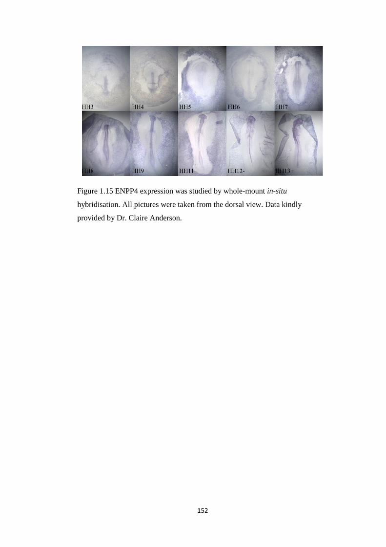
152
Figure 1.15 ENPP4 expression was studied by whole-mount in-situ
hybridisation. All pictures were taken from the dorsal view. Data kindly
provided by Dr. Claire Anderson.

153
Figure 1.16 FBLN7L expression was studied by whole-mount in-situ
hybridisation. All pictures were taken from the dorsal view. Data kindly
provided by Dr. Claire Anderson.

154
Figure 1.17 NXPH1 expression was studied by whole-mount in-situ
hybridisation. All pictures were taken from the dorsal view. Data of HH4+, 13
and 14- kindly provided by Dr. Claire Anderson.
Figure 1.18 NRP1 expression was studied by whole-mount in-situ hybridisation.
All pictures were taken from the ventral view. Data kindly provided by Dr.
Claire Anderson.

155
Figure 1.19 NRSN1 expression was studied by whole-mount in-situ
hybridisation. All pictures were taken from the ventral view. Data kindly
provided by Dr. Claire Anderson.
Figure 1.20 PCSK6 expression was studied by whole-mount in-situ
hybridisation. All pictures were taken from the dorsal view. Data kindly
provided by Dr. Claire Anderson.

156
Figure 1.21 PRKG1 expression was studied by whole-mount in-situ
hybridisation. All pictures were taken from the dorsal view. Data kindly
provided by Dr. Claire Anderson.

157
Figure 1.22 PROM1 expression was studied by whole-mount in-situ
hybridisation. All pictures were taken from the dorsal view. Data kindly
provided by Dr. Claire Anderson.

158
Figure 1.23 RAB11-FIP4 expression was studied by whole-mount in-situ
hybridisation. All pictures were taken from the dorsal view. Data kindly
provided by Dr. Claire Anderson.
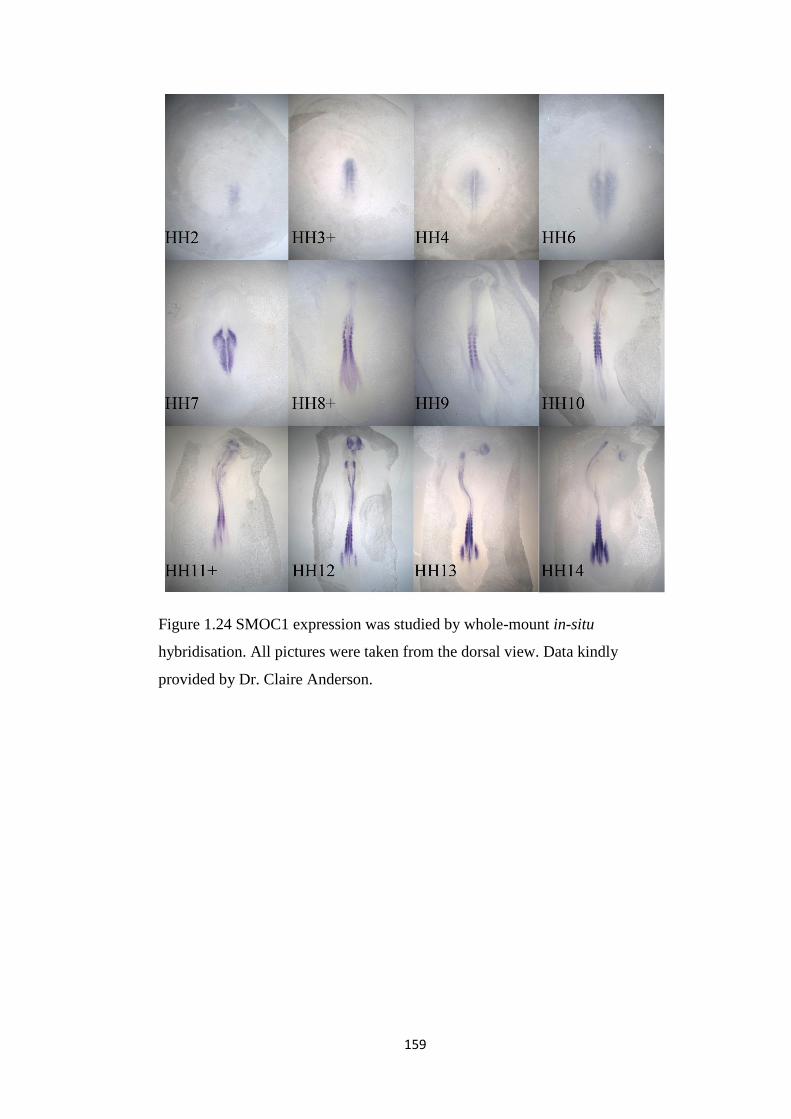
159
Figure 1.24 SMOC1 expression was studied by whole-mount in-situ
hybridisation. All pictures were taken from the dorsal view. Data kindly
provided by Dr. Claire Anderson.

160
Figure 1.25 ChEST21m16 expression was studied by whole-mount in-situ
hybridisation. This is another probe staining for SMOC1 as well. All pictures
were taken from the dorsal view. Data kindly provided by Dr. Claire Anderson.

161
Figure 1.26 SHH expression was studied by whole-mount in-situ hybridisation.
Pictures from HH4 to HH7 were taken from dorsal view.
Figure 1.27 TMEM63C expression was studied by whole-mount in-situ
hybridisation. All pictures were taken from the dorsal view. Data kindly
provided by Dr. Claire Anderson.

162
Figure 1.28 TMTC2 expression was studied by whole-mount in-situ
hybridisation. All pictures were taken from the ventral view.

163
Figure 1.29 TSPAN6 expression was studied by whole-mount in-situ
hybridisation. All pictures were taken from the dorsal view. Data kindly
provided by Dr. Claire Anderson.
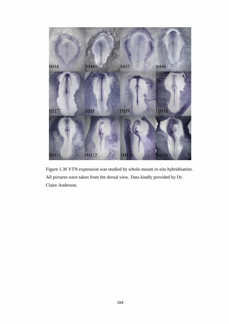
164
Figure 1.30 VTN expression was studied by whole-mount in-situ hybridisation.
All pictures were taken from the dorsal view. Data kindly provided by Dr.
Claire Anderson.

165
Figure 1.31 ChEST125i5 expression was studied by whole-mount in-situ
hybridisation. All pictures were taken from the dorsal view. Data kindly
provided by Dr. Claire Anderson.
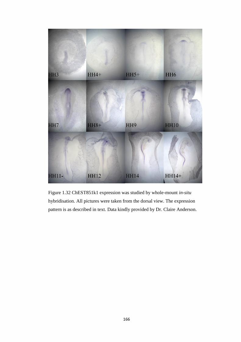
166
Figure 1.32 ChEST851k1 expression was studied by whole-mount in-situ
hybridisation. All pictures were taken from the dorsal view. The expression
pattern is as described in text. Data kindly provided by Dr. Claire Anderson.

167
Figure 1.33 DLX5 expression was studied by whole-mount in-situ
hybridisation. All pictures were taken from the dorsal view. Data kindly
provided by Dr. Claire Anderson.

168
Figure 1.34 DLX6 expression was studied by whole-mount in-situ
hybridisation. All pictures were taken from the dorsal view.

169
Figure 1.35 ID2 expression was studied by whole-mount in-situ hybridisation.
All pictures were taken from the dorsal view. Data kindly provided by Dr.
Claire Anderson.

170
Figure 1.36 LDB2 expression was studied by whole-mount in-situ
hybridisation. All pictures were taken from the dorsal view. Data kindly
provided by Dr. Claire Anderson.
Figure 1.37 Msx1 expression was studied by whole-mount in-situ hybridisation.
All pictures were taken from the dorsal view. Data kindly provided by Dr.
Claire Anderson.

171
Figure 1.38 Msx2 expression was studied by whole-mount in-situ hybridisation.
All pictures were taken from the dorsal view. Data kindly provided by Dr.
Claire Anderson.
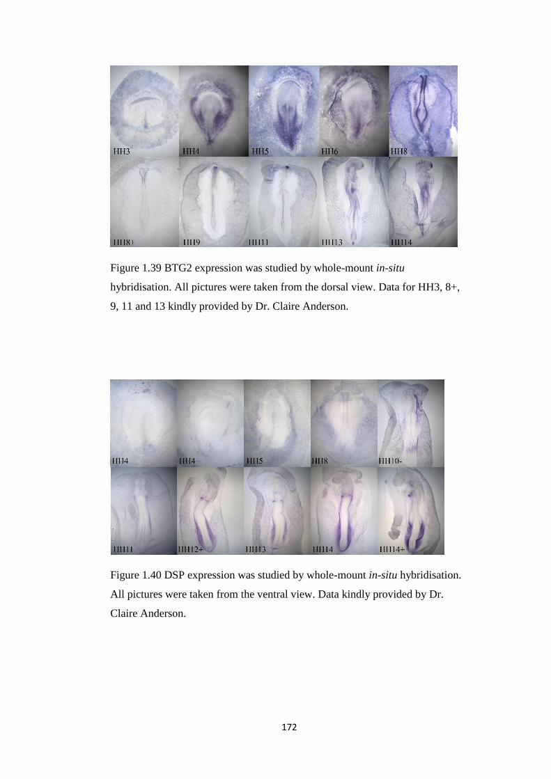
172
Figure 1.39 BTG2 expression was studied by whole-mount in-situ
hybridisation. All pictures were taken from the dorsal view. Data for HH3, 8+,
9, 11 and 13 kindly provided by Dr. Claire Anderson.
Figure 1.40 DSP expression was studied by whole-mount in-situ hybridisation.
All pictures were taken from the ventral view. Data kindly provided by Dr.
Claire Anderson.

173
Figure 1.41 MOXD1 expression was studied by whole-mount in-situ
hybridisation. All pictures were taken from the dorsal view. Data kindly
provided by Dr. Claire Anderson.
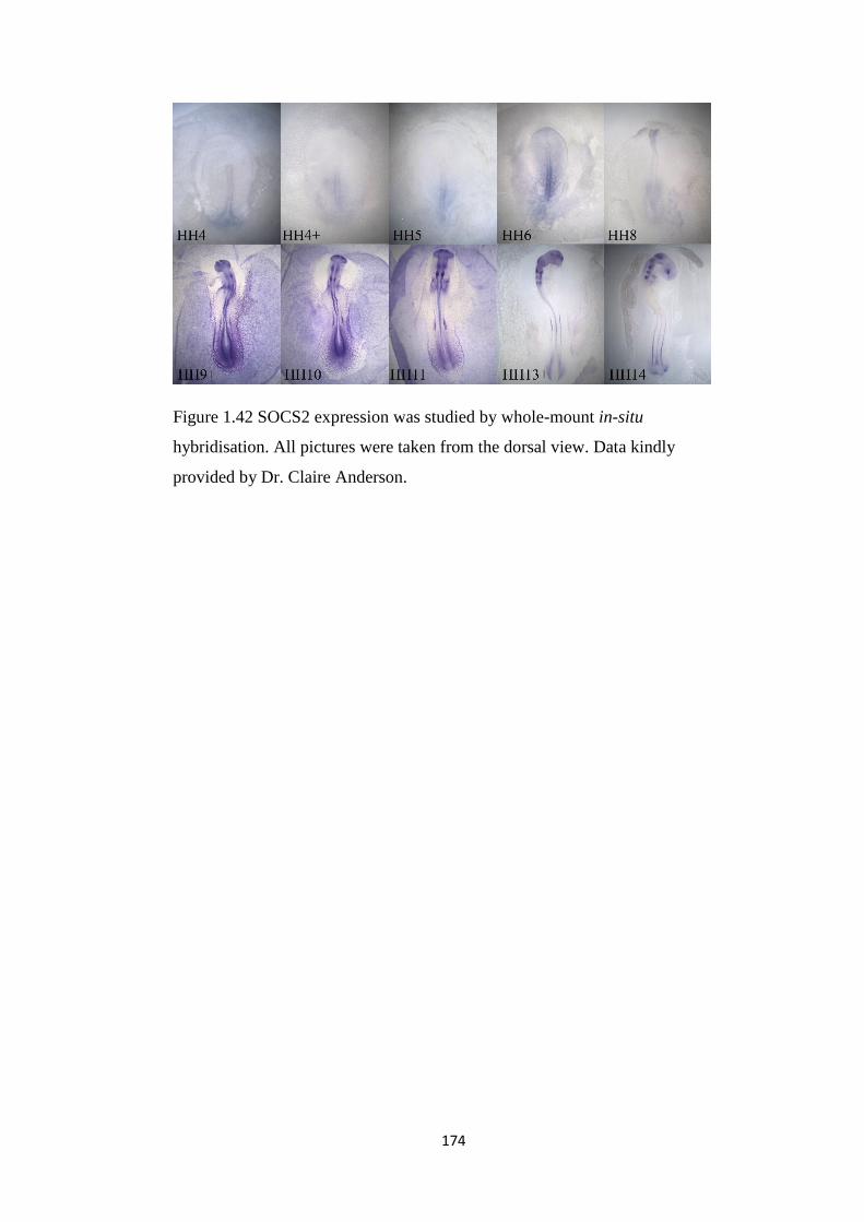
174
Figure 1.42 SOCS2 expression was studied by whole-mount in-situ
hybridisation. All pictures were taken from the dorsal view. Data kindly
provided by Dr. Claire Anderson.
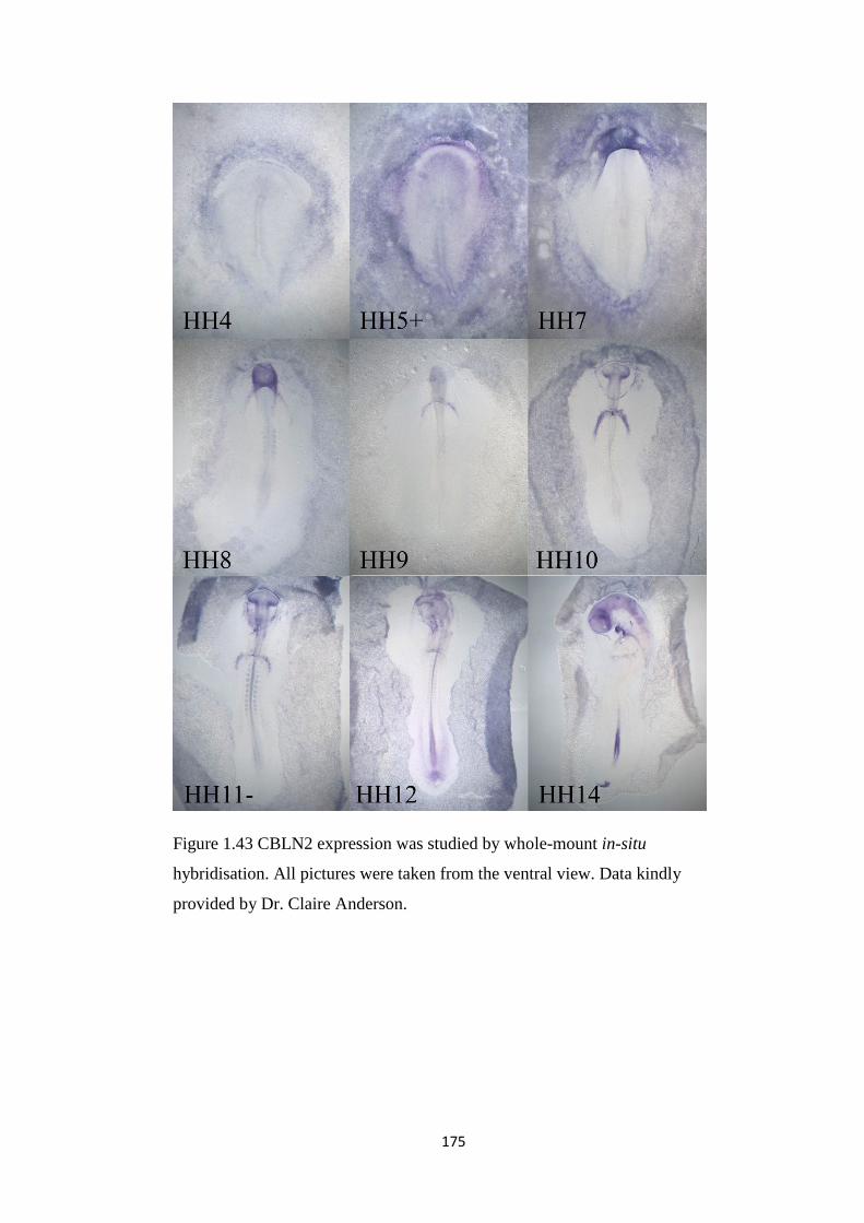
175
Figure 1.43 CBLN2 expression was studied by whole-mount in-situ
hybridisation. All pictures were taken from the ventral view. Data kindly
provided by Dr. Claire Anderson.

176
Figure 1.44 CXCL12 expression was studied by whole-mount in-situ
hybridisation. Pictures were taken from the dorsal view except HH14 which
was taken from ventral view.

177
Figure 1.45 EFNA5 expression was studied by whole-mount in-situ
hybridisation. All pictures were taken from the dorsal view. Data kindly
provided by Dr. Claire Anderson.
Figure 1.46 PLXNC1 expression was studied by whole-mount in-situ
hybridisation. All pictures were taken from the dorsal view. Data kindly
provided by Dr. Claire Anderson.

178
Figure 1.47 TNFRSF19 expression was studied by whole-mount in-situ
hybridisation. All pictures were taken from the ventral view. Data kindly
provided by Dr. Claire Anderson.

179
Figure 1.48 ChEST378m14 expression was studied by whole-mount in-situ
hybridisation. All pictures were taken from the dorsal view. Data kindly
provided by Dr. Claire Anderson.
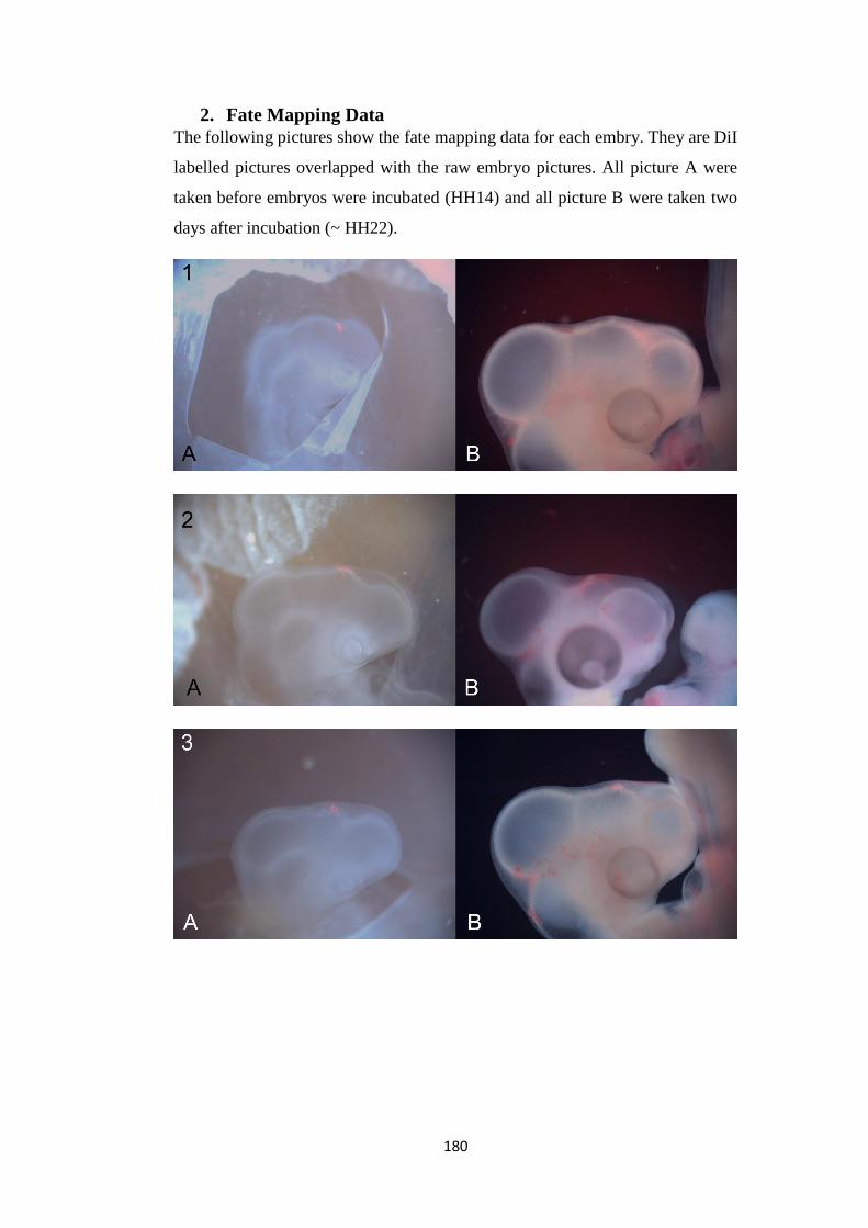
180
2. Fate Mapping Data
The following pictures show the fate mapping data for each embry. They are DiI
labelled pictures overlapped with the raw embryo pictures. All picture A were
taken before embryos were incubated (HH14) and all picture B were taken two
days after incubation (~ HH22).

181

182

183

184

185

186

187

188
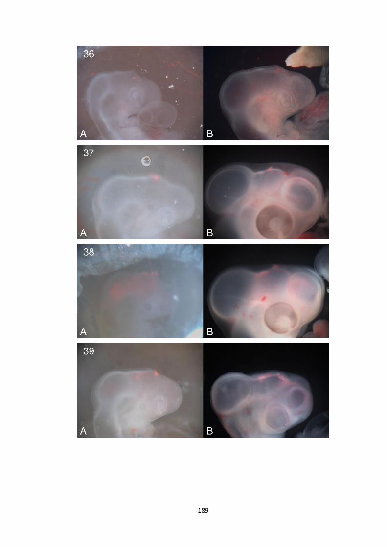
189
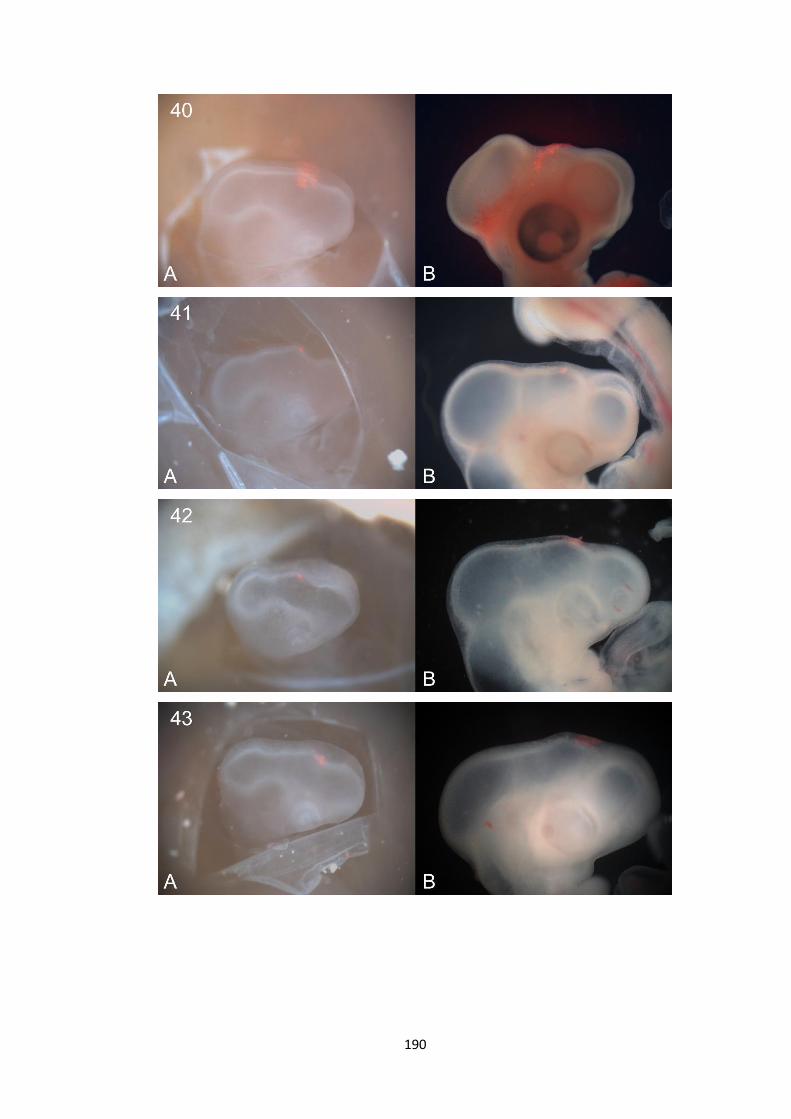
190

191

192
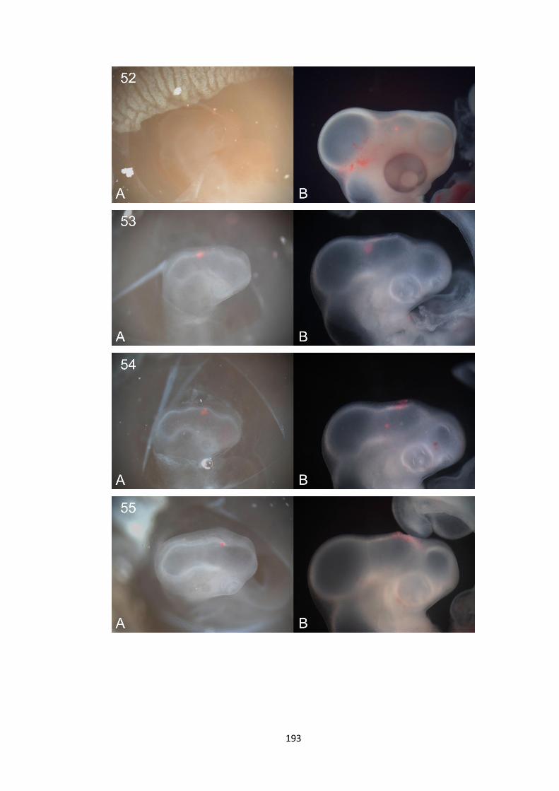
193

194

195

196

197

198

199
3. Preliminary Fate Mapping Data
The following pictures show DiI labels between HH10 and HH11. They are DiI
labelled pictures overlapped with the raw embryo pictures. All picture A were
taken before embryos were incubated (HH10-11) and all picture B were taken
two days after incubation (~ HH14).

200

201
Acknowledgement
I would like to deeply thank Professor Claudio Stern for his supervision
of my study. I would also like to thank all members in Stern lab for their
help along my project, especially Dr. Claire Anderson!
Copyright © 2022 FDOKUMEN