Broca's Region
436
Broca’s Region Yosef Grodzinsky Katrin Amunts, Editors OXFORD UNIVERSITY PRESS
-
Upload
khangminh22 -
Category
Documents
-
view
0 -
download
0
Transcript of Broca's Region

Broca’s Region
Yosef Grodzinsky Katrin Amunts,
Editors
OXFORD UNIVERSITY PRESS

BROCA’S REGION

This page intentionally left blank

BROCA’S REGION
EDITED BY
Yosef Grodzinsky
Katrin Amunts
12006
Oxford University Press
Oxford University Press, Inc., publishes works that furtherOxford University’s objective of excellence
in research, scholarship, and education.
Oxford New YorkAuckland Cape Town Dar es Salaam Hong Kong KarachiKuala Lumpur Madrid Melbourne Mexico City Nairobi
New Delhi Shanghai Taipei Toronto
With offices inArgentina Austria Brazil Chile Czech Republic France Greece
Guatemala Hungary Italy Japan Poland Portugal SingaporeSouth Korea Switzerland Thailand Turkey Ukraine Vietnam
Copyright © 2006 by Yosef Grodzinsky and Katrin Amunts
Published by Oxford University Press, Inc.198 Madison Avenue, New York, New York 10016
www.oup.com
Oxford is a registered trademark of Oxford University Press
All rights reserved. No part of this publication may be reproduced,stored in a retrieval system, or transmitted, in any form or by any means,
electronic, mechanical, photocopying, recording, or otherwise,without the prior permission of Oxford University Press.
Library of Congress Cataloging-in-Publication Data
Broca’s region / edited by Yosef Grodzinsky, Katrin Amunts.p. cm.
Includes bibliographical references and index.ISBN-13 978-0-19-517764-0
ISBN 0-19-517764-91. Broca, Paul, 1824–1880. 2. Neurolinguistics. 3. Psycholinguistics. 4. Frontallobes. 5. Sign language. 6. Aphasia. I. Grodzinsky, Yosef. II. Amunts, Katrin.
QP399.B76 2005612.8�2336—dc22 2004023816
1 3 5 7 9 8 6 4 2
Printed in the United States of Americaon acid-free paper

Contributors ixIntroduction xiii
I. Matters Anatomical
1. The Origin of Broca’s Area and ItsConnections from an Ancestral Working Memory Network 3
FRANCISCO ABOITIZRICARDO GARCÍAENZO BRUNETTICONRADO BOSMAN
2. A Multimodal Analysis of Structure and Function in Broca’s Region 17
KATRIN AMUNTSKARL ZILLES
3. Broca’s Area in the Human and theNonhuman Primate Brain 31
MICHAEL PETRIDES
II. Matters Linguistic
4. Weak Syntax 49SERGEY AVRUTIN
5. Speech Production in Broca’s Agrammatic Aphasia: Syntactic Tree Pruning 63
NAAMA FRIEDMANN
6. A Blueprint for a Brain Map of Syntax 83YOSEF GRODZINSKY
7. Evaluating Deficit Patterns of Broca’sAphasics in the Presence of HighIntersubject Variability 108
DAN DRAI
8. Treating Language Deficits in Broca’sAphasia 119
LEWIS P. SHAPIROCYNTHIA K. THOMPSON
v
Contents
III. Motor Aspects and Sign Language
9. Broca’s Region: A Speech Area? 137LUCIANO FADIGALAILA CRAIGHEROALICE ROY
10. Broca’s Area in System Perspective:Language in the Context of Action-Oriented Perception 153
MICHAEL ARBIB
11. The Role of Broca’s Area in SignLanguage 169
KAREN EMMOREY
IV. Psycholinguistic Investigations
12. Broca’s Area and Lexical-SemanticProcessing 187
STEFANO F. CAPPADANIELA PERANI
13. The Neural Basis of Sentence Processing:Inferior Frontal and TemporalContributions 196
ANGELA D. FRIEDERICI
14. Involvement of the Left and Right FrontalOperculum in Speech and NonspeechPerception and Production 218
MARTIN E. MEYERLUTZ JÄNCKE
15. On Broca, Brain, and Binding 242PETER HAGOORT
16. A Role for Broca’s Area Beyond LanguageProcessing: Evidence fromNeuropsychology and fMRI 254
GEREON R. FINKZINA M. MANJALYKLAAS E. STEPHANJENNIFER M. GURDKARL ZILLESKATRIN AMUNTSJOHN C. MARSHALL
V. Discussion
17. Jülich Workshop Excerpts 271
VI. Historical Articles
Choices We Made: An Introduction to theHistorical Section 287
KATRIN AMUNTSYOSEF GRODZINSKY
18. Comments Regarding the Seat of theFaculty of Spoken Language, Followed by an Observation of Aphemia (Loss of Speech) (1861) 291
PAUL BROCA (1824–1880)
19. On Affections of Speech from Disease of the Brain (1878–1879) 305
JOHN HUGHLINGS-JACKSON (1835–1911)
20. On Aphasia (1885) 318LUDWIG LICHTHEIM (1845–1928)
21. Contributions to a HistologicalLocalization of the Cerebral Cortex—VI. Communication: The Division of the Human Cortex (1908) 334
KORBINIAN BRODMANN (1868–1918)
22. The Agrammatical Language Disturbance: Studies on a Psychological Basis for the Teaching on Aphasia (1913) 337
ARNOLD PICK (1854–1924)
23. The Cytoarchitectonics of the FieldsConstituting Broca’s Area (1931) 348
LUDWIG RIEGELE
24. The Phonological Development of Child Language and Aphasia as aLinguistic Problem (1956) 355
ROMAN JAKOBSON (1896–1982)
vi CONTENTS

25. Grammatical Complexity and AphasicSpeech (1958) 369
HAROLD GOODGLASS (1920–2002)J. HUNT
26. The Organization of Language and the Brain (1970) 376
NORMAN GESCHWIND (1926–1984)
27. Broca’s Area and Broca’s Aphasia (1976) 384
JAY P. MOHR (1937–)
Author Index 395
Subject Index 407
CONTENTS vii

This page intentionally left blank
FRANCISCO ABOITIZ
Departamento de PsiquiatríaEscuela de MedicinaPontificia Universidad Católica de [email protected]
KATRIN AMUNTS
Brain MappingInstitut für MedizinForschungszentrum JülichJü[email protected]
MICHAEL ARBIB
Computer Science DepartmentNeuroscience Program and USC Brain ProjectUniversity of Southern CaliforniaLos Angeles, [email protected]
SERGEY AVRUTIN
UiL OTSUtrecht UniversityUtrechtThe [email protected]
CONRADO BOSMAN
Departamento de PsiquiatríaEscuela de MedicinaPontificia Universidad Católica de [email protected]
ENZO BRUNETTI
Departamento de PsiquiatríaEscuela de MedicinaPontificia Universidad Católica de [email protected]
ix
Contributors

STEFANO F. CAPPA
Università Vita Salute and Istituto Scientifico San Raffaele
LAILA CRAIGHERO
Department of Biomedical SciencesSection of Human PhysiologyUniversity of [email protected]
DAN DRAI
Weizmann Institute of [email protected]
KAREN EMMOREY
DirectorLaboratory for Language and Cognitive
NeuroscienceSan Diego State UniversitySan Diego, [email protected]
LUCIANO FADIGA
Department of Biomedical SciencesSection of Human PhysiologyUniversity of [email protected]
GEREON R. FINK
Kognitive NeurologieInstitut für MedizinForschungzentrum JülichJü[email protected]
ANGELA D. FRIEDERICI
Max Planck Institute for Human Cognitive andBrain Sciences
NAAMA FRIEDMANN
School of EducationTel Aviv UniversityTel [email protected]
RICARDO GARCÍA
Departamento de PsiquiatríaEscuela de MedicinaPontificia Universidad Católica de [email protected]
YOSEF GRODZINSKY
Department of LinguisticsMcGill UniversityMontreal, [email protected]
JENNIFER M. GURD
Department of PsychologyUniversity of HertfordshireHatfieldUnited KingdomandNeuropsychology UnitUniversity Department of Clinical NeurologyRadcliffe InfirmaryOxfordUnited [email protected]
PETER HAGOORT
F.C. Donders Centre for Cognitive NeuroimagingNijmegenThe [email protected]
LUTZ JÄNCKE
Department of NeuropsychologyInstitute for PsychologyUniversity of ZürichZü[email protected]
KYLE JOHNSON
Department of LinguisticsUniversity of MassachusettsAmherst, [email protected]
ZINA M. MANJALY
Kognitive NeurologieInstitut für MedizinForschungzentrum JülichJü[email protected]
x CONTRIBUTORS

JOHN C. MARSHALL
Neuropsychology UnitUniversity Department of Clinical NeurologyRadcliffe [email protected]
MARTIN E. MEYER
Department of NeuropsychologyInstitute for PsychologyUniversity of ZürichZü[email protected]
DANIELA PERANI
Università Vita Salute and Istituto Scientifico San Raffaele
MICHAEL PETRIDES
Montreal Neurological InstituteMcGill UniversityMontreal, [email protected]
ALICE ROY
Department of Biomedical SciencesSection of Human PhysiologyUniversity of [email protected]
LEWIS P. SHAPIRO
San Diego State UniversitySchool of Speech, Language, and Hearing
SciencesSan Diego, [email protected]
KLAAS E. STEPHAN, MDWellcome Department of Imaging NeuroscienceInstitute of NeurologyUniversity College [email protected]
CYNTHIA K. THOMPSON
Northwestern UniversityCommunication Sciences and DisordersEvanston, [email protected]
KARL ZILLES
Institut für MedizinForschungzentrum JülichJülichGermanyandC. and O.Vogt für Hirnforschungs InstitutHeinrich-Heine-UniversitätDüsseldorf, [email protected]
CONTRIBUTORS xi

This page intentionally left blank

Broca’s region has been in the news ever since sci-entists first hit upon the idea that particular cognitivefunctions can be localized to parts of the cerebral cor-tex. Its discoverer, Paul Broca, was one of the first sci-entists to argue that there is a direct connection be-tween a concrete piece of behavior—in this case, theuse of language, or what Broca called “articulatedspeech”—and a specific cortical region. Today,Broca’s region is probably the most famous part of thehuman brain: it is featured in virtually every intro-ductory psychology course and textbook and in many,if not all, introductory anatomy and linguisticscourses, and for over a century, it has persisted as thefocus of intense research and much debate. The namehas even penetrated popular culture, serving as the ti-tle for a best-selling popular science book by CarlSagan.
This region is not famous for nothing: as languageis one of the most distinctive human traits, it wouldseem to follow that the cognitive mechanisms thatsupport it are quite complex, and the tissues in which
these mechanisms are housed have interesting char-acteristics and important implications for how brainfunction relates to behavior. Thus, Broca’s region fea-tures prominently in the study of brain–behavior relations.
The first studies of Broca’s region focused on lan-guage pathologies caused by brain lesions. The resultsof this early work by Broca, Wernicke, and their con-temporaries captivated the public’s imagination.Among the language pathologies, Broca’s aphasia at-tracted attention as no other disorder ever had. It evenmade it to Broadway, through the protagonist ofArthur Kopit’s play Wings—stroke victim Emily Stil-son, an aphasic patient whose compelling plight, asdescribed in the play’s synopsis, is that she is “an ex-aviatrix who loses speech . . . and slowly regains powerof language and life.”
Broca’s discoveries were also an important, drivingforce behind the more general effort to relate com-plex behavior to particular parts of the cerebral cor-tex, which, significantly, produced the first brain
xiii
Introduction

maps. Incorrect paths such as phrenology notwith-standing, during the heyday of Broca’s work, researchaimed at identifying the cerebral localization of cog-nitive function was flourishing and the study of apha-sic syndromes was the flagship of neuropsychology.During these early years, the investigation into brainand behavior was based almost solely upon lesionstudies. Although researchers were able to analyze le-sions at the level of brain macroscopy and functionaldeficit, this is, unfortunately, where their analysesended. On an anatomical level, researchers were notaware of the underlying principles of cellular dys-function and disconnectivity, and of the huge amountof variability among different subjects. On a behav-ioral level, their distinctions were crude because theywere not aware of the structural properties of languageand cross-linguistic variation.
Although much has been discovered since Brocaclaimed that the region contains “the faculty of artic-ulated language,” even today, these problems have notbeen completely solved. Technological advance-ments have made more detailed studies possible, butthere is still no general agreement among anatomistson what constitutes the underlying microstructure ofBroca’s region or even on the name of the region it-self. In the early twentieth century, anatomists distin-guished a strict sense of Broca’s region from the moregeneral sense of it. Just to make matters perfectly con-fusing, later researchers also referred to this particu-lar piece of neural tissue as Broca’s area and Broca’scomplex. The term “Broca’s area” seems to be mis-leading because it suggests that it is a single entity,characterized by a certain homogeneity in structure,connectivity, and function (almost as if it were a Brod-mann area), whereas it has become increasingly clearthat it actually includes cortical areas with differentfunctional specializations and connectivity.
In an analogous situation, there is also no generalagreement on how the cognitive functions related toBroca’s region should be characterized. It is, however,quite clear that modality-based divisions of the typefound in standard neurology texts—speaking, under-standing, repeating, naming, and so on—are not goodenough units of measure and that creating divisionsbased on either psychological mechanisms or lin-guistically motivated concepts also fails to tell thewhole story.
If it is so difficult to come to an understanding ofeach of these things alone, then how can we so eas-
ily claim that the anatomy and functional role of thispart of the brain are related to each other? Initially es-tablished as the exclusive domain of clinical neurol-ogists, the study of brain/language relations increas-ingly required deeper expertise in specializeddomains. The field was transformed when psycholo-gists entered with experimental methods and linguistsentered with analytic categories and theoretical con-ceptions. Later, when new methods has been devel-oped, neuroscientists, biomedical engineers, andcomputer scientists became interested. Today, partic-ularly as a result of important advances made in neu-roimaging during the past two decades, Broca’s regionand, for that matter, all language areas are being in-vestigated from every angle from which we can man-age to approach them.
The careers of the editors of this volume illustratethe phenomenon: Our long-term research programhas always focused on Broca’s region, and we cur-rently use fMRI as a method of investigation. How-ever, while one of us studies the structure of this re-gion primarily by looking at cells, cellulararchitecture, cortical layers, and borders, the other ed-itor is primarily concerned with words, phrases, sen-tences, and grammatical rules and the way in whichthey are instantiated in neural tissue.
Indeed, as the volume of research into the rela-tions between brain and language has created severalcommunities, each importing its own concepts, meth-ods, and considerations, we thought that it was timeto stop and reflect together, and that is the purpose ofthis book. In it, we tried to do what is generally con-sidered to be nearly impossible: to mix intellectual tra-ditions and cultures and to juxtapose rather disparatebodies of knowledge, styles of reasoning, and forms ofargumentation. At the same time, we made every ef-fort to produce a book that is both relevant and ac-cessible to a broad audience.
To this end, we invited scientists with diversebackgrounds to contribute their particular take atthe Broca’s Region Workshop, hoping that a coher-ent and perhaps even a novel picture would emerge.Although the participants share a special interest inBroca’s region, their backgrounds and approaches—neuroanatomy, physiology, evolutionary biology,cognitive psychology, clinical neurology, functionalimaging, speech and language research, computa-tional biology, and psychological, neurological, andtheoretical linguistics—represent all the myriad
xiv INTRODUCTION

angles from which we currently approach Broca’sregion.
We met at the Forschungszentrum in Juelich,Germany, on June 4 through 6, 2004. As one wouldexpect, the meeting was unusual. At times it becametense, but it was never dull. We are happy to reportthat it reflected a genuine effort on everyone’s part tolisten, accommodate, and, most of all, understand.
We offer the readers of this volume the product ofour Broca’s Region Workshop—contributions madeby the participants and their research teams, amendedafter they were presented to the workshop’s diverse au-dience. To underscore the richness of viewpoints con-tained herein and to give readers an idea of the levelof interaction that took place, we have included partsof the discussion that we recorded during the work-shop. Because Broca’s region is such an historicallysignificant concept and rich area, we added a collec-tion of classic and recent-yet-classic papers. Alongwith cutting-edge science, we wish to remind readersof the celebrated past from which much may belearned. These historical chapters include the first twopapers written by Paul Broca, as well as some work bytwo of the most important neurologists of the nine-teenth century, Carl Wernicke and John Hughlings-Jackson. We also included parts of twentieth-century
papers by Korbinian Brodmann, Norman Geschwind,Harold Goodglass, Roman Jakobson, Jay Mohr, andArnold Pick.
The resulting volume, we believe, reflects the stateof the art. We hope that it will stimulate more inter-disciplinary work, as the project called “Broca’s re-gion,” which encompasses the study of brain/languagerelations, is far from being finished.
We are grateful to the speakers/contributors, whohelped bring the workshop and book to life. We weresupported by Oxford University Press, with CatharineCarlin at the wheel; by the ForschungszentrumJuelich GmbH, the chairman of its board of directors,Joachim Treusch, and the director of its Institute ofMedicine, Karl Zilles; and by the Centre for Researchon Language, Brain, and Mind at McGill University,and its director, Shari Baum. We would also like tothank Katie Clark and Andrea Santi for their help.Without these individuals and organizations, neitherthe workshop nor the book would ever have hap-pened. Their help and generosity were invaluable increating and carrying out this project.
Katrin AmuntsYosef Grodzinsky
INTRODUCTION xv

This page intentionally left blank

I
MATTERS ANATOMICAL

This page intentionally left blank

1
The Origin of Broca’s Area and ItsConnections from an Ancestral
Working Memory Network
Francisco Aboitiz
Ricardo García
Enzo Brunetti
Conrado Bosman
Language has been historically considered the hall-mark of human uniqueness. Darwin (1871) proposeda species-specific, instinctive tendency to acquire lan-guage (Pinker, 1995, p. 20) but never ventured to pro-pose a detailed account of evolutionary language ori-gins. Not long before Darwin’s publication, PaulBroca (1861) had presented the brain of his patientTan to the Société d’ Anthropologie in Paris, whichfirmly established the role of the left inferior frontallobe in language production. Relatively soon afterBroca, Carl Wernicke (1874) proposed the firstanatomical model for language perception and pro-duction, which despite many posterior modificationsremains roughly valid today. Thus, even if the con-cepts of human evolution by natural selection and ofthe localization of the language faculty were quitecontemporaries, very little was proposed in the way tounderstand the evolutionary bases of language origins.
A significant attempt to explain the evolutionaryorigins of language occurred a century later, whenNorman Geschwind (1964) proposed that the ability
to give names to objects relied on the capacity to establish cross-modal, corticocortical associations,something that nonhuman primates were apparentlyunable to do. Subsequent studies showed that apesare able to name objects and actions if trained in signlanguage (Premack, 1983). Furthermore, recent evi-dence indicates that word learning—even after singleexposure—can occur in the domestic dog (Kaminskiet al., 2004). However, the fact remains that apes (andprobably dogs) are much slower than humans inlearning cross-modal associations and in the ability tolearn sign words (Aggleton, 1993; Nahm et al., 1993).Thus, it may seem that the ability to give names, al-though greatly facilitated in the human species, isbased on a neural substrate whose precursor exists inother animals. This is entirely compatible with a grad-ualistic darwinian interpretation of language origins.
Nevertheless, apes trained in sign language werenever able to go beyond a two- or three-word organi-zation in their utterances and definitely lacked anyhint of grammatical organization. This evidence
3

strengthened the contemporary theory of universalgrammar, prescribing that all languages share com-mon combinatorial principles that are genetically de-termined and unique with regard to other cognitiveprocesses found in humans and other animals (Chom-sky, 1978). One important observation regarding theinnateness of language is that while learning lan-guage, children may produce complex grammaticalutterances that they apparently never heard before.This supports a genetic tendency to learn language inour species. Furthermore, Chomsky has argued thatbecause no hints of syntax precursors are found innonhuman animals, syntax most likely arose in evo-lution as the result of a single biological macromuta-tion. Nevertheless, gradualistic interpretations for theorigins of syntactic rules have been proposed in thelatest years (Dennett, 1995; Pinker, 1995; Pinker andBloom, 1990), partly based on observations on howchildren acquire grammatical rules.
Many authors have located syntactical processingin Broca’s area (corresponding to Brodmann’s areas44 and 45) (Embick et al., 2000). However, studiesindicate that Broca’s area participates in several otherneurocognitive processes, including working mem-ory, gesture recognition, mirror drawing, and aspectsof musical analysis (Bookheimer 2002; Dronkers etal., 1992; Gruber, 2002; Maess et al., 2001; Patel,2003; Rizzolatti et al., 1992). Of particular interest isthe finding of “mirror neurons” in the inferior frontallobe, which participate in the recognition of one’sown actions and of actions performed by others in thehuman and monkey ventral premotor cortex (Galleseet al., 1996; Iacoboni et al., 1999; Rizzolatti et al.,1996, 2002); these have been proposed to be the phy-logenetic precursors of Broca’s language area (Arbiband Bota, 2003; Rizzolatti and Arbib, 1998). An ad-ditional issue is that damage to Broca’s area alone usu-ally does not produce long-lasting severe aphasia; thesurrounding areas and underlying white matter mustbe damaged as well (Dronkers et al., 1992; Pinker,1995). In this context, we (Aboitiz, 1995; Aboitiz andGarcía, 1997) originally proposed that the neural de-vice involved in language comprehension and pro-duction is not isolated from the rest of the brain.Rather, this system belongs to a large-scale cortico-cortical network reciprocally connecting higher-orderareas in the temporoparietal lobes with prefrontal ar-eas (other components such as the basal ganglia andcertain thalamic nuclei may also participate). Amongthe functions of this overall network are several
processes that require sustained neural activity for in-tervals of a few seconds or longer, such as attention,language, imitation, and especially working memory.
In this chapter, we provide an updated version ofthis theory. Since our original proposal, much neu-roanatomical, neuroimaging, and neurolinguistic ev-idence has accumulated that calls for a revision ofsome of the issues presented in those times. In par-ticular, neuroanatomical findings on animals andimaging studies in humans, especially on linguisticworking memory, have revealed a much finer-grainedpicture of the anatomy and connectivity of the lan-guage areas and its nonhuman homologues and oftheir function in the human brain. We begin with abrief overview of our original proposals and then dis-cuss new evidence from comparative neuroanatomy,brain imaging, and cognitive neuroscience, to endwith a discussion on the possible role of neuropsy-chological processes like working memory in seman-tic and syntactic processing.
OVERVIEW OF THE ORIGINAL HYPOTHESIS
In our original articles (Aboitiz, 1995; Aboitiz andGarcía, 1997a, b), we placed special emphasis onworking memory, a specific form of short-term memory that maintains perceptual information andlong-term memory online while executing a certaincognitive task (Baddeley, 1992; Baddeley and Hitch, 1974). Like the language network, the neuralsystem for working memory relies on extensive temporoparietal–prefrontal connections. Workingmemory has been proposed to be subdivided into ageneral, all-purpose executive system and “slave” sys-tems involved in sensorimotor rehearsal: one of suchsystems is the visuospatial sketchpad, which maintainsonline visuospatial information, and the other is thephonological loop, allowing internal rehearsal ofphonological utterances. However, sensorimotorworking memory networks may be more complexthan just a visuospatial sketchpad and a phonologicalloop; there are probably subdivisions in these com-ponents, as well as additional components involvingother perceptual and processing domains (Levy andGoldman-Rakic, 2000; Romanski et al., 1999a). Wesuggested that the phonological loop in particular mayhave been essential to early language origins, in thesense that it participated in learning complex utter-ances that were acquired by imitation. A circuit in-
4 MATTERS ANATOMICAL

volving inferior parietal, intraparietal, and inferiorfrontal areas (including Broca’s area) differentiatedinto a working memory device involved in the imita-tion and internal rehearsal of complex vocalizations.This is not to exclude gestural communication, whichmay have co-occurred with these early vocalizations.However, in early hominids, there were limits to thecomplexity of utterances that could be learned. Theincrease in brain size was related to the developmentof more complex neuronal networks, allowing thegeneration of elaborate vocal utterances with primi-tive syntactic rules (Aboitiz, 1988, 1996). Initially, thelanguage areas were able to coordinate phonemes andmorphemes into primitive words, and eventuallysome sequences of words. Shortly, our claim is thatthe language areas originated from a primitive work-ing memory device involved in the imitation of complex utterances, which eventually served as a tem-plate from which brain organization for modern lan-guage evolved. Language processing requires a veryefficient working memory system, in terms of phonol-ogy, syntax, and meaning. In this sense, additionalworking memory circuits were subsequently recruitedin the evolution of human communication, produc-ing the structural and semantic complexities of mod-ern language.
CONNECTIVITY AND HOMOLOGY ISSUES
As mentioned earlier, a major issue in our proposalconcerns the structural similarity of language-relatednetworks and the networks involved in working mem-ory. Because connectivity studies cannot be per-formed in humans, neuroscientists have had to relyin nonhuman primates to study these neural projec-tions. However, cortical areas in the monkey and hu-man are not easily comparable, and a major problemhas been to establish specific nonhuman homologiesfor the language areas. In our original article (Aboitizand García, 1997a), we tentatively proposed a frame-work for homology between the human language ar-eas and their primate counterparts based on the avail-able evidence at that time (Barbas and Pandya, 1989;Preuss and Goldman-Rakic, 1991a). Shortly, we fol-lowed Preuss and Golman-Rakic (1991a) in their sug-gestion of an area 45 inside the macaque’s inferior ar-cuate sulcus, between subareas 6V and 8Ar, andproposed that Broca’s region could be conceived as adifferentiation of the ventral premotor region (ventral
area 6 of the monkey). In the human, we suggestedthe existence of a network connecting Broca’s area (ar-eas 44 and 45) and frontal granular cortex (areas 9and 46) with regions in the superior temporal lobe(specifically, area Tpt in the posterior superior tem-poral gyrus and, to a lesser extent, area TE), and weemphasized connections between the supramarginalgyrus (more specifically, area 40) and Broca’s area.The latter proposal was mainly based on previous find-ings in the monkey, indicating connections from in-traparietal (7ip) and inferior parietal (7b) areas to theanterior and the posterior banks of the inferior arcu-ate sulcus, respectively (Cavada and Goldman-Rakic,1989; Petrides and Pandya, 1984; Preuss and Gold-man-Rakic, 1991a, b, c). Although area Tpt was foundto project strongly to the dorsal moiety of the arcuatesulcus (Petrides and Pandya, 1988), it was also ob-served sending projections to the inferior arcuate sul-cus (Deacon, 1992).
In the past years, much evidence has appeared re-garding the cytoarchitecture and connectivity of theseareas, which, although implying modifications to ourproposed scheme, confirms its main points of aBroca’s area homolog in the inferior bank of the ar-cuate sulcus with strong connections with the supe-rior temporal, intraparietal, and inferior parietal re-gions (see Fig. 3 in Aboitiz and García, 1997a).Connections of intraparietal and inferoparietal re-gions with area 45 and the inferior arcuate sulcus havebeen confirmed (Lewis and Van Essen, 2000; Preussand Goldman-Rakic, 1991a). Nevertheless, a recon-sideration of this scheme, claiming a parallel segre-gation of the prefrontal–temporoparietal projectionsto Broca’s area, has come from additional data asso-ciated with physiological and anatomical refinementsof the primate auditory cortical system (Kaas andHackett, 2000; Romanski and Goldman-Rakic, 2001;Romanski et al, 1999a, b). This evidence is reviewedlater.
The current understanding of the anatomical or-ganization of the auditory cortex specifies a concen-tric organization, with a core region containing theprimary auditory area and other rostral areas, a beltregion surrounding the core region, and a more lat-eral, parabelt region (Hackett et al., 1998). Very in-terestingly, and in analogy with the visual system, twodifferent processing streams have been identified inthe primate auditory cortex, one projecting caudallyand more involved with spatial auditory information(the “where” stream), and the other projecting ros-
THE ORIGIN OF BROCA’S AREA 5

trally and processing the intrinsic features of auditorystimuli including speech (the “what” stream) (Kaasand Hackett, 1999; Tian et al., 2001). Supporting thisevidence, the injection of retrograde and anterogradetracers into several areas of prefrontal cortex hasshown a rostrocaudal gradient of reciprocal corticalconnectivity between prefrontal regions and belt andparabelt auditory association areas (Romanski et al.,1999a). Thus, rostral and orbital regions of prefrontalcortex are connected with rostral belt and parabelt ar-eas, whereas caudal regions in the sulcus principalisand the inferior convexity of the prefrontal cortex arereciprocally connected with caudal belt and parabeltauditory association areas. More specifically, the“where” pathway projects mainly to prearcuate areas8a and 46, and the “what” pathway projects to thefrontal pole (area 10), the rostral principal sulcus (area46), and the ventral prefrontal cortex (areas 12 and45) (Hackett et al., 1999; Rauschecker and Tian,2000; Romanski et al., 1999a, 1999b). Furthermore,Romanski and Goldman-Rakic (2002) identified anauditory domain in the monkey prefrontal cortex, lo-cated in areas 12lat, 12orb, and 45 (which receive pro-jections from the “what” pathway), in which mostneurons preferred vocalizations than other acousticstimuli, whereas some neurons were also responsiveto visual stimuli. In general, the caudal parabelt(“where”) provides auditory projections to the regionrelated to directing eye movements and to the dorsalprefrontal cortex, and the rostral parabelt (“what”) pro-jects to the ventral/orbital prefrontal cortex and thegranular frontal cortex, perhaps more related to thehuman Broca’s area.
Area Tpt, located in the posterior superior tempo-ral gyrus, deserves some special consideration. Thisarea was originally described by Pandya and Sanides(1973) and by Galaburda and Sanides (1980) in themacaque and the human, respectively. Consideringits relation to the human planum temporale, this areawas considered to be an integral element of Wer-nicke’s posterior language area (Galaburda et al.,1978). On the other hand, according to the concen-tric belt–parabelt array proposed by Hackett et al.(1998), area Tpt is located near the posterolateral para-belt region and topographically close to the “where”stream. We see later that although this area projectsto dorsal and ventral aspects of the prefrontal cortex,its emphasis is to more dorsal areas, consistent withthe projections of the caudal parabelt. Furthermore,
recent analyses indicate that more than being a language-specific area, the planum temporale (andperhaps, consequently, area Tpt) is related to theanalysis of many types of spectrally complex sounds,working as a node (a “hub”) that distributes projec-tions to several higher-order areas (Binder et al., 1996;Griffiths and Warren, 2002).
Another topographic and connectional scheme isthat provided by Petrides and Pandya (1994, 1999,2001). These authors considered that the macaquearea 45 (comparable to the human area 45 because itdisplays large pyramidal neurons in layer IIIc, a well-defined layer IV, and medium-size neurons in layerV) could be subdivided into areas 45A and 45B, whichare different from the more dorsally located frontaleye fields. Importantly, they describe a dysgranulararea 44 in the caudal bank of the lower limb of thearcuate sulcus, adjacent to area 45B. In addition, ar-eas 47 and 12 were considered to correspond to hu-man area 47, sharing with area 45 a complex, multi-modal input. Nevertheless, area 47/12 receives moreheavy projections from rostral inferotemporal visualareas and temporal limbic areas, whereas area 45 re-ceives input from the superior temporal gyrus (in-cluding area Tpt) and the upper bank of the superiortemporal sulcus. It will be important to determine towhat extent Petrides and Pandya’s (2001) areas 45 and47/12 relate to Romanski and Goldman-Rakic’s(2002) areas 45 and 12lat/12orb, respectively. AreaTpt and other caudal stream auditory areas have animportant projection to the more dorsal area 8Ad,which is linked to the parieto-occipital region but isalso connected to area 45. Finally, area 46 was foundto be smaller than previously thought, being restrictedto an anterior part and to the cortex inside the sulcusprincipalis. The posterior part of area 46 correspondedinstead to a part of area 9 and was called area 9/46(ventral and dorsal).
In our view, this new cumulus of neuroanatomi-cal evidence allows us to refine our early connectionalmodel subserving human language origins (Aboitizand García, 1997a, b) (Fig. 1–1, see color insert). Infact, the recent data regarding the architecture andprefrontal–temporoparietal connectivity confirm ourprevious proposal suggesting several pathways runningin parallel from inferoparietal and inferotemporal re-gions to the inferior frontal cortex: first, (1) the infero-parietal and intraparietal zones corresponding to ar-eas 7ip and 7b of the monkey (perhaps partly related
6 MATTERS ANATOMICAL

to the gyrus supramarginalis in humans, especiallyarea 7b) connect with the inferior periarcuate region,including area 45. As we have mentioned before, in humans, the inferior parietal region could be connected to the caudal belt region configuring a temporoparietal–prefrontal pathway subserving aphonological–rehearsal loop. However, this connec-tion has not been demonstrated in the monkey. In ad-dition, a dual stream of auditory projections targetsdistinct prefrontal domains: (2) a spatial “where” path-way connects the caudal belt regions with the caudaldorsolateral prefrontal cortex (areas 8 and 46) in-cluding a posterior parietal relay through areas 7ip and7a, and (3) a “what” stream connects the rostral beltand parabelt regions to the inferior periarcuate con-vexity where area 45 is located. Finally, there is an in-ferotemporal projection connecting area TE with area45 whose function is probably to transmit object rel-evant information to Broca’s area. Note that Petrides
and Pandya (1988, 1994, 1999, 2001) describe a pro-jection from area Tpt, the superior temporal gyrus andthe superior temporal sulcus to dorsal frontal areas (re-lated to eye movements) but also to area 45, whichreceives projections from the “what” pathway (Hack-ett et al., 1999; Rauschecker and Tian, 2000; Ro-manski et al., 1999a, b). In this sense, the inferior ar-cuate sulcus (including area 45) may partly serve asan interface between both the “where” and the “what”streams in the inferior frontal lobe.
A quite recent proposal for macaque homologieswith the human language areas was outlined by Ar-bib and Bota (2003) and was based on Rizzolatti andArbib’s (1998) concept of a mirror system for grasp-ing as the neuronal precursor of Broca’s area. Theseauthors propose that a network involving the anteriorintraparietal sulcus and ventral premotor area F5 ofthe macaque anchors a cortical circuit for hand move-ments and grasping. F5 contains so-called mirror neu-rons whose activity matches the execution and obser-vation of hand movements. Furthermore, F5 can besubdivided into F5ab, containing “canonical” motorneurons, and F5c, which contains mirror neurons. Ac-cording to Arbib and Bota, F5 may be compared withhuman areas 44 and 45, and F5c corresponds to atleast part of area 44. Area 45 may contain two subar-eas: a dorsal one related to the frontal eye fields anda ventral one distinct from the frontal eye fields. Fur-ther studies are needed to determine exactly and towhat extent areas F5ab and F5c correspond to Petridesand Pandya’s (1994) areas 45A, 45B, and 44 and withRomanski and Goldman-Rakic’s (2002) areas 45 and12lat/12orb, respectively. Although it may be prema-ture to propose detailed homologies in these as yet ill-defined regions, this view is in general terms consis-tent with our proposals. In general, we agree that a region in or surrounding the inferior arcuate sulcus,receiving projections from superior temporal, inferiorparietal, and intraparietal areas, may be the best can-didate for homology with Broca’s area. Because in thehuman frontal lobe the language-related region ex-tends beyond the boundaries of areas 44 and 45(Dronkers et al., 1992), it is perhaps advisable to con-sider the connections of a relatively wide region sur-rounding the primate inferior arcuate sulcus insteadof focusing on the strict homologues of areas 44 and45. In this chapter, we emphasized these two areasmainly because at this point they are the best knownin terms of language processing.
THE ORIGIN OF BROCA’S AREA 7
FIGURE 1–1. Diagram indicating some connectionsbetween the parietotemporal and the inferior pre-frontal areas in the monkey. The auditory region inthe superior temporal lobe is subdivided into core,belt, and parabelt regions, and has two main process-ing streams: the rostral belt and parabelt (the “what”pathway, RTL and RP), which projects to the inferiorconvexity of the prefrontal lobe; and the caudal beltand parabelt (the “where” pathway, CM, CL, andCP), which projects to more dorsolateral areas. Theintraparietal and inferior parietal regions (7ip, 7b) pro-ject to the inferior convexity. Numbers indicate Brod-mann’s areas. Data from Hackett et al. (1998), Ro-manski et al. (1999a, 1999b), and Petrides and Pandya(2001, 1999). as, arcuate sulcus; cs, central sulcus; ips,intraparietal sulcus; las, lateral sulcus; ls, lunate sul-cus; ps, principal sulcus; sts, superior temporal sulcus.

WORKING MEMORY
Phonological working memory has been shown to beimportant for language learning. Patients with work-ing memory deficits show impairments in long-termphonological learning, and a link has been observedbetween performance in the phonological loop andvocabulary level in children (Baddeley et al., 1988;Gathercole and Baddeley, 1990). According to Bad-deley (2000), this suggests that the loop might haveevolved to enhance language acquisition. In our orig-inal proposal (Aboitiz and García, 1997a) and fol-lowing earlier evidence (Awh et al., 1995; Frackowiak,1994; Habib, 1996; Paulesu et al., 1993; Salmon etal., 1996), we proposed that the “slave” component ofverbal working memory contained a storage compo-nent, or phonological buffer, which included the lefthemispheric postero parietal cortex (the supramar-ginal gyrus, Brodmann’s area 40), and a rehearsalcomponent including the left hemisphere frontalspeech areas. We proposed that phonological repre-sentations were initially processed in the posteriorlanguage areas, stored transiently in inferior parietalareas, and then transferred to Broca’s area for re-hearsal. On the other hand, we suggested that granu-lar frontal areas (areas 9 and 46) might participate inmore complex working memory functions related tohigher-level syntactic and semantic processing and indiscourse planning (Aboitiz and García, 1997a, b).
Shortly after our publication, Smith and Jonides(1998) reviewed much of the neuroimaging studiesfor human working memory, providing an essentiallysimilar picture in which verbal working memory was dissociable into a transient storage component inthe left posterior parietal cortex and a rehearsal com-ponent in Broca’s area and its vicinities. In this context, Kirchhoff et al. (2000) showed that a left prefrontal–temporal circuit including Broca’s areaand lateral temporal cortex mediates novel verbalepisodic encoding and that the magnitude of activityin that region predicted whether an event (word)would be subsequently remembered. In addition, inarticulatory suppression experiments that interferewith the phonological rehearsal mechanism, Gruber(2001, 2002) observed a specific activation pattern in-volving inferior parietal areas and anterior prefrontalcortex, while in conditions of subvocal rehearsal therewas activation of intraparietal areas and Broca’s area.Note that the pattern of activation in conditions of ar-
ticulatory suppression resembles the “mirror system”circuit for movements and grasping proposed by Ar-bib and Bota (2003). Thus, the “slave” verbal work-ing memory circuit may involve two main circuits,one involving rehearsal and perhaps overlapping witha mirror system circuit, and the other, related to tran-sient phonological storage (Gruber, 2001, 2002).
In our view, one paradigmatic example of the roleof working memory in language processing is the caseof conduction aphasia. Usually, this type of aphasiaresults from lesions in the supramarginal gyrus, andeven lesions of the insula that perhaps impair fibersin the underlying white matter (Greenfield, 1992).Like in Wernicke’s aphasia, there are paraphasias andrelatively fluent speech production, and like Broca’saphasia, there is preserved comprehension (Damasio,1992). A diagnostic characteristic of conduction apha-sia is the inability to repeat sentences presented to thepatient. We proposed that because the capacity to re-peat sentences requires a functional short-term mem-ory (Trortais, 1974), the deficit in conduction apha-sia may result from interruption of a verbal workingmemory circuit, either by damage to the subcorticalwhite matter or, more likely, by direct damage to in-ferior parietal (intraparietal?) areas that transientlystore phonological information (Aboitiz and García,1997a). Smith and Jonides (1998) also called atten-tion to a possible deficit in the storage component ofverbal working memory in conduction aphasia. Alongthis line, it has been proposed that subjects with con-duction aphasia comprehend and are able to accesslexical–semantic information about words butdemonstrate a deficit in retreiving the stored phono-logical representations of these items (Anderson et al.,1999). A related interpretation is that the supramar-ginal gyrus (area 40) and Broca’s area are involved inspeech perception only indirectly through their rolein phonological working memory: the left supramar-ginal gyrus alone might represent an interface be-tween speech-related auditory representations andfrontal, motor systems, whereas the left supramarginalgyrus together with Broca’s area mediates explicit ac-cess to speech segments (Hickok, 2000a; Hickok andPoeppel, 2000).
Summarizing, there is substantial evidence indi-cating that temporoparietal–prefrontal working mem-ory systems are strongly implicated in language ac-quisition and in language processing, which suppportsour main proposal that a specialization of working
8 MATTERS ANATOMICAL

memory–related networks may have been at the basisof language origins. Perhaps the phonological re-hearsal system including an incipient Broca’s area andintraparietal and inferior parietal regions was espe-cially relevant in the very early stages of language evo-lution. This may have been fundamental for learningcomplex utterances by imitation. In later stages, a tran-sient phonological store located in the inferior pari-etal lobe evolved together with working memory sys-tems for complex syntax, lexicon, and semantics(Aboitiz, 1995; Aboitiz and García, 1997a).
As mentioned previously (page 7), Arbib and Bota(2003) proposed a sequence of events for languageorigins starting from a complex imitation system forgrasping, followed by a manual-based communicationsystem in which imitation of others is an importantelement; this “mirror system” eventually participatesin the generation of protospeech, which gives the vo-cal apparatus sufficient flexibility, and eventually lan-guage originates. In other words, the hand-based pari-etoprefrontal imitation system provided the behavioralflexibility necessary to generate a diversity of vocal-izations that could evolve into language. (Note thatthis series of steps, if they occurred, might have takenplace prior to the establishment of a phonologicalloop, which in our view served as the basis for prim-itive language networks.) We agree that the hand-based and orofacial mirror neuron system probablyhad a role in gestural communication and that imi-tation was possibly an important element in earlyprelinguistic evolution. However, we are not so sureyet about the stronger claim that gestural languagewas ancestral to vocal communication. Phylogeneticevidence indicates that external meaning is transmit-ted mostly by vocal communication in nonhuman primates (Acardi, 2003; Seyfarth and Cheney, 2003).In addition, in wild chimpanzees gestural communi-cation is more limited than vocal communication(Acardi, 2003). Perhaps a more parsimonious hy-pothesis is that gestural and vocal communication co-evolved to a large extent. Furthermore, we have sug-gested that the frontal auditory domain described byRomanski and Goldman-Rakic (2002) may belong to,or be the precursor of, a vocalization mirror systemsimilar to the mirror system for grasping described byRizzolatti and Arbib (1998). This system may haveparticipated in vocal imitative behavior, permitting tocompare heard vocalizations with own productions,and therefore may have been fundamental in early
language evolution (Bosman et al., 2004; see also Jürgens, 2003).
The discovery of a family with members bearing amutation in gene FOXP2 and showing severe lan-guage disorders (Lai et al., 2001) has prompted an in-tensive search for the functions of this gene. Imaginganalyses indicate underactivation of Broca’s area insubjects carrying the mutation (Liégeois et al., 2003).FOXP2 has been hypothesized to be linked to the mir-ror system for grasping (Liégeois et al., 2003; Corbal-lis, 2004), while there is evidence indicating that thedeficit in FOXP2 function produces an impairmentin nonword repetition, a condition related to a deficitin the storage of phonological information in workingmemory (Watkins et al., 2002). Recently, we proposedan integrative approach to the study of this gene, in-cluding its participation in mirror system circuits andin working memory networks (Bosman et al., 2004).
SEMANTICS AND SYNTAX
Two main characteristics of language are semanticsand syntax. Semantics concerns meaning, or the ref-erence to the external world or the internal state ofthe individual, while syntax has to do with rules spec-ifying an internal order of utterances. Concerning se-mantics, Geschwind (1964) already proposed that itwas based on associative interactions between the lan-guage areas and sensory or limbic areas, a line thatPulvermuller (1999) has pursued with a more mod-ern associative approach. In our view, which is simi-lar to those just presented, semantics was achieved byintegration of the language/working memory networkswith other (working memory) networks related to on-line maintenance of sensory and mnemonic percepts.This integration was perhaps initially with limbic ar-eas, to signal emotional state, and to motor areas, tosignal specific behaviors. For example, associationsbetween mirror neurons and auditory neurons in thefrontal cortex may have provided the basis for the firstutterances signaling specific behaviors (Hauk et al.,2004). In subsequent stages, integration with other,more widespread brain systems possibly allowed thegeneration of content words (Pulvermuller, 1999).Next, we discuss the possibility that higher levels ofsyntax involve the integration of Broca’s area withneighboring cognitive systems of the frontal lobe andother cortical areas (this may include processing of
THE ORIGIN OF BROCA’S AREA 9

abstract words, which serve a grammatical function;Pulvermuller, 1999).
Many researchers now consider that syntactic pro-cessing takes place in Broca’s area and neighboringregions (operculum, insula, and subjacent white mat-ter in which connections with other brain regions occur). Beside the well-known lesion studies, someelectroencephalographic and imaging reports haveconfirmed this view. Electrophysiologically, two kinds of evoked potentials, the P600 (Osterhout andHolcomb, 1992) and the left anterior negativity(Friederici et al., 1993; Münte et al., 1993), have beenrelated to errors in syntactic processing. Although boththe source and the syntactic specificity of the P600have been challenged (Patel et al., 1998), the left an-terior negativity has been found to be more specificand its topographic arrangement suggests a source re-lated to Broca’s area (Patel, 2003). Interestingly, it wasalso found that harmonic musical processing may beprocessed in Broca’s area; an anterior negativity com-ponent (ERAN [early right anterior negativity]) waselicited by harmonically inappropriate chords, local-ized in Broca’s area and its right hemisphere homo-logue (Maess et al., 2001), suggesting similar mecha-nisms in syntactic and harmonic processing (Patel,2003).
Several imaging reports have shown activation ofBroca’s area and surrounding regions related to syn-tactic processing (Caplan et al., 1998; Dapretto andBookheimer, 1999; Friederici et al., 2000; Indefrey etal., 2001; Meyer et al., 2000; Moro et al., 2001; Ni etal., 2000; Stromswold et al., 1996; Tettamanti et al.,2001). For example, a study detected that Broca’s areais specifically activated with ungrammatical sentences(Embick et al., 2000), and another study detected anincrease in signal in the left infero frontal gyrus (es-pecially area 45) with online performance of sen-tences with “real” syntactic rules compared with sen-tences with “unreal” rules (Musso et al., 2003). Theinterpretation of the latter result was that Broca’s areadisengages with the learning of unreal grammars,which possibly are learned with the participation ofother, nonlinguistic systems.
It has been argued that the inferior frontal gyrusactivity associated with syntactic processing may partlyreflect working memory demands rather than syntac-tic processing per se (Bookheimer, 2002). Neverthe-less, the two possibilities may not be exclusive. Be-sides its involvement in phonological processing,working memory seems to participate in syntactic pro-
cessing (see also Chapter 13). Caplan and Waters(1999) claim that syntax processing requires a spe-cialized working memory system that is separatedfrom the working memory system underlying sen-tence meaning and other functions. They propose asystem for “interpretive processing” that deals withcomplex syntax and is more related to Broca’s areaand a system for “postinterpretive processing” that an-alyzes complex meaning, which may be more widelydistributed. Furthermore, Martin and Saffran (1997)emphasize that there are several parallel but interact-ing working memory circuits in language: the se-mantic, the lexical, and the phonological systems. Inaddition, Fiebach et al. (2001) argue that there existsa separate cognitive or neural resource that supportssyntactic working memory processes necessary for thetemporary maintenance of syntactic information forthe parser, whereas in an fMRI study, Cooke et al.(2002) report that the left inferior frontal cortex is re-cruited to support the cognitive resources required tomaintain long-distance syntactic dependencies duringthe comprehension of complex grammatical sen-tences. Although there may still be some disagree-ment on the details of how many working memorylevels operate in language processing, it is fair to saythat working memory participates at the phonologi-cal, the lexical, the syntactic, and the semantic levels.However, these components may be relatively inde-pendent of each other: impairment to one particularworking memory domain (say, phonological) mayleave relatively undamaged other working memorycomponents (lexical, syntactic, semantic) and viceversa. This is consistent with the neurobiological con-cept of several parallel working memory networks subserving distinct cognitive domains (Levy and Goldman-Rakic, 2000; Romanski et al., 1999a). Alsoalong this line, it is important to recall Fuster’s asser-tion that rather than one memory system in the cere-bral cortex, each processing system has its own mem-ory device (Fuster, 1995).
In a comprehensive study of the linguistic char-acteristics of Broca’s aphasics, Grodzinsky (2000)claimed that Broca’s area and neighboring regions donot participate in all syntax but only in some syntac-tic rules: in language comprehension, Broca’s areakeeps track of the transformationally moved phrasalconstituents. For example, when transforming acanonical sentence into a passive form (“the boykissed the girl” � “the girl was kissed by the boy”),the components are moved in order in relation to the
10 MATTERS ANATOMICAL

verb. In verbal transformations, moved constituentsleave a “trace” that is normally tracked by the listenerin order to appropiately understand it. According toGrodzinsky, Broca’s aphasics have special problemswith verbal transformations because for some reason,traces are “deleted” and therefore roles are impossi-ble or more difficult to track (the trace deletion hy-pothesis [TDH]). Interestingly, the more complex thetransformation, or the longer the distance between atrace and its antecedent, the more difficult it is to trackthe moved constituents for Broca’s aphasics. A rela-tively common interpretation of Grodzinsky’s pro-posal is that trace deletion reflects a deficit in work-ing memory (for example, Hickok, 2000b; Müller,2000, Stowe, 2000, Szelag and Pöppel, 2000). Simi-larly, Pinker (1995) had already proposed a role ofshort-term memory for tracing constituents in phrasaltransformations. Grodzinsky argues that it remains tobe shown that there is a direct connection betweenworking memory resources and formal elements ofsyntactic processing. Another point is that whenphrasal constituents are moved, there must exist neu-ronal mechanisms involved in the detection of tracesand their extraction sites. This may be detected at thelexical level, there being key words that permit theconnection with other elements located distantly inthe phrase. Finally, Grodzinsky (2000) also claimsthat in speech production, Broca’s aphasics have prob-lems in constructing appropriate syntactic trees in amanner similar to children’s grammar. Specifically,they cannot construct trees below the tense node.
What is so special about syntax? A fundamentalcharacteristic of syntax is that it does not merely con-sist of linear order within a sequence of words; rather,it relies on the hierarchical or recursive relations be-tween words in such a way that not just any order ispossible and that there are constraints with regard tothe types of grammar that can be learned (Chomsky,1978; Marcus et al., 2003). How is this performed inthe brain? At least Pinker (1995) claimed that short-term memory plays an important role in syntactic pro-cessing. More recently, Patel (2003) compared twodifferent theories of syntactic and harmonic process-ing: the dependency locality theory (DLT) (Gibson,1998, 2000) and the tonal pitch space theory (TPS)(Lerdhal, 2001), respectively. In DLT, distances be-tween related words are computed and stored as thesentence is perceived in time, whereas in TPS, dis-tances between chords and some predefined “anchor”chords are being computed in a similar way. When
explaining the similarity between syntactic and har-monic processing, Patel explicitly claims not to be invoking a special memory system or a symbol-manipulating system but rather considers the cogni-tive theories of processing in the two domains. How-ever, Patel finally claims that “in DLT, integrationcan be understood as activating the representation ofan incoming word while also reactivating a prior de-pendent word whose activation has decayed in pro-portion to the distance between words. In TPS, inte-gration can be understood as activating an incomingchord while maintaining activation of another chordwhich provides the context for the incoming chord’sinterpretation” (Patel, 2003, p. 678). This interpreta-tion is strongly reminiscent of managing online in-formation during working memory tasks.
It may be argued that most syntactical processingis performed implicitly, without conscious involve-ment, whereas working memory operates largely inexplicit form. We consider that the short-term mem-ory networks required for syntactic processing may bequite similar in organization (although not necessar-ily in location) to the working memory networks de-scribed for other linguistic tasks. The implicit/explicitdistinction perhaps has to do with other parametersthat are not fundamental for the implementation ofthese networks.
Summarizing, our main claim has been that thelanguage circuit arose as a specialization of a circuitinvolved in phonological working memory, whicheventually incorporated other working memory sys-tems involved in syntax, the lexicon, and semantics.This is reminiscent of Martin and Saffran’s (1997) pro-posal of different but interacting working memorydomains involved in language processing: the se-mantic, the lexical, and the phonological systems, andCaplan and Waters’ (1999) two working memory sys-tems, an “interpretive” component analyzing syntaxand a “postinterpretive” component analyzing the se-mantic contents.
Anatomically, these networks may correspond to arelatively restricted system involving the language ar-eas and neighboring regions for phonological and syn-tactic processing and, more widespread, bilateral net-works related to meaning and semantics. In order tobe efficient, working memory must create a frame inwhich all the online items are ordered and have somedefinite relations between them. In this sense, thereis some sort of “syntax” intrinsic to all working mem-ory systems. We consider that a primordium of the
THE ORIGIN OF BROCA’S AREA 11

formal elements that appear in modern syntax mayhave occurred in the organization of the primitive lin-guistic working memory systems but, perhaps moreimportant, as rules that permitted the “traduction”from one working memory system to another. For ex-ample, a “presyntax” may have emerged as a strategyto superpose the temporal (phonological and syntac-tical) working memory frames in which the differentcomponents of an utterance are retained, with a visuospatial/episodic frame that generates a mental“image” of an action or an event (this need not bepurely visual) associated with that utterance. Thus,the hierarchical organization of a syntactically or-dered sentence may be partly related to the require-ments to transform this temporally ordered online in-formation (in which Broca’s area may play a specialrole, in both phonological and syntactic processing)into a perhaps more distributed, visuospatial sketch-pad describing the meaning of a given sentence, andvice versa. In this sense, perhaps one main innovationin language origins and in human neural evolutionwas the ability to “translate” complex working mem-ory codes from one functional domain (i.e., sequen-tial) into another (i.e., visuospatial working memoryand episodic memory) and vice versa. Modern lan-guage, then, may be generated by an imbricated andcoordinated macroscopic working memory system,which involves several reciprocally interacting sub-systems (lexical, semantic, syntactic, phonological).The uniqueness of syntax, and of the human brain, isperhaps partly related to the translating mechanismsrequired for the reciprocal interaction between thesedifferent subsystems. This view is consistent with re-cent fMRI research identifying three separate regionsof functional specialization in the inferior frontal gyrus:phonology, syntax, and semantics (Bookheimer, 2002).
FINAL COMMENTS
We have proposed a revised version of our originalhypothesis that language networks emerged as a spe-cialization of temporoparietal–prefrontal networks in-volved in cognitive processes that require sustainedactivity, like working memory, attention, and move-ment imitation. In this context, Arbib and Bota (2003)made a contrast between our theory being “retro-spective” (looking at what is in the human brain—working memory—and tracking it back to the mon-key brain) and theirs being “prospective” (finding
what is in the monkey—hand coordination—whichmay have served as a substrate for human language),which we believe may be misleading. We have fol-lowed standard phylogenetic methodology: first, iden-tified in the monkey the networks that can be ho-mologous to the language-related neural networks,and second, asked for the functions of these networksin the monkey, one of which is working memory. Be-cause working memory is seen to operate at many lev-els of language processing (phonological, lexical, syn-tactic, and semantic), we conclude that corticocorticallanguage networks originated from preexisting work-ing memory networks in the primate brain (of course,this is far from saying that working memory mecha-nisms explain all aspects of language processing). Agood analogy for our strategy comes from the evolu-tion of the eye: although image formation is a highlyderived characteristic, there are other, more basicfunctions, like photoreception, which are central tovision but insufficient for producing an image, thatare shared by other species whose visual organs lackimage-forming properties; these functions permit usto track the phylogenetic ancestry of the eyes. Simplyput, our hypothesis points to a function (workingmemory) that is present in the monkey and partici-pates in language processing. On the other hand, al-though in humans and monkeys the inferior frontalregion participates in hand movements and otherfunctions, at this point there is no evidence that man-ual coordination is involved in language processing.Nevertheless, a “mirror system” initially involved indecoding orofacial gestures and body posture mayhave also participated in communication. We are notsure which came first, oral or gestural communica-tion, but we believe that these two may have reliedon related, interacting temporoparietal–prefrontalnetworks in the cerebral cortex. Furthermore, a mir-ror system for vocalizations may have been funda-mental for early learning of complex vocalizations andvocal communication (Bosman et al., 2004; Jürgens,2003). We consider that a crucial element that allowed the processing of increasingly complex utter-ances was an expanded phonological working mem-ory capacity, possibly based on supratemporal/inferoparietal–inferior prefrontal networks. Utterancesbegun to acquire relatively complex meanings byvirtue of associative interactions of the language re-gions with other brain regions; this required a notableexpansion of the working memory capacity and of theneural networks underlying it. Finally, we propose
12 MATTERS ANATOMICAL

that syntax emerged as a neuronal strategy to effi-ciently “translate” a phonological, sequential workingmemory code into an episodic, visuospatial memorycode that represents the meaning of the respective ut-terances. In a way, the rules for syntax may perhapsbe partly understood as a strategy that permits thetransmission of information between the differentworking memory codes that make up the elements oflanguage; that is, it relates to a binding mechanismthat permits the different networks to interact duringhuman communication.
ACKNOWLEDGMENTS Juan Montiel prepared Figure1–1. Part of the work presented here was financed by theMillenium Center for Integrative Neuroscience.
References
Aboitiz, F. (1988). Epigenesis and the evolution of thehuman brain. Medical Hypotheses, 25, 55–59.
Aboitiz, F. (1995). Working memory networks and theorigin of language areas in the human brain. Med-ical Hypotheses, 44, 504–506.
Aboitiz, F. (1996). Does bigger mean better? Evolution-ary determinants of brain size and structure. Brain,Behavior and Evolution, 47, 225–245.
Aboitiz, F., & García, R. (1997a). The evolutionary ori-gin of the language areas in the human brain. Aneuroanatomical perspective. Brain Research. BrainResearch Reviews, 25, 381–396.
Aboitiz, F., & García, R. (1997b). The anatomy of lan-guage revisited. Biol. Research, 30, 171–183.
Acardi, A.C. (2003). Is gestural communication more so-phisticated than vocal communication in wildchimpanzees? The Behavioral and Brain Sciences,26, 210–211.
Aggleton, P. (1993). The contribution of the amygdalato normal and abnormal emotional states. Trends inNeuroscience, 16, 328–334.
Anderson, J.M., Gilmore, R., Roper, S., Crosson, B.,Bauer, R.M., Nadeau, S., Beversdorf, D.Q., et al.(1999). Conduction aphasia and the arcuate fac-siculus: A reexamination of the Wernicke-Geschwind model. Brain and Language, 70, 1–12.
Arbib, M., & Bota, M. (2003). Language evolution:Neural homologies and neuroinformatics. NeuralNetworks: The Official Journal of the InternationalNeural Network Society, 16, 1237–1260.
Awh, E., Smith, E.E., & Jonides, J. (1995). Human re-hearsal processes and the frontal lobes: PET evi-dence. Annals of New York Academy of Science, 769,97–118.
Baddeley, A. (2000). The episodic buffer: A new com-ponent of working memory? Trends in CognitiveSciences, 4, 417–423.
Baddeley, A. (1992). Working memory. Science, 255,556–559.
Baddeley, A.D., & Hitch, G.J. (1974). Working memory.In G.A. Bower (Ed.), The psychology of learning and motivation (pp. 47–89). New York: AcademicPress.
Baddeley, A.D., Papagno, C., & Vallar, G. (1988). Whenlong-term learning depends on short-term memory.Journal of Memory and Language, 27, 586–595.
Barbas, H., & Pandya, D.N. (1989). Architecture and in-trinsic connections of the prefrontal cortex in therhesus monkey. The Journal of Comparative Neu-rology, 286, 353–375.
Binder, J.R., Frost, J.A., Hammeke, T.A., Rao, S.M., &Cox, R.W. (1996). Function of the left planum tem-porale in auditory and linguistic processing. Brain,119, 1239–1247.
Bookheimer, S. (2002). Functional fMRI of language:New approaches to understanding the cortical or-ganization of semantic processing. Annual Reviewof Neuroscience, 25, 151–188.
Bosman, C., García, R., & Aboitiz, F. (2004). FOXP2and the language working memory system. Trendsin Cognitive Sciences, 6, 251–252.
Broca, P. (1861). Remarques sur le siege de la faculté du langage articulé, suivies d’une observationd’aphémie (perte de la parole). Bulletin of Societyof Anatomy, 36, 330–357.
Cavada, C., & Goldman-Rakic, P. (1989). Posterior pari-etal cortex in rhesus monkey: II. Evidence for seg-regated cortico–cortical networks linking sensoryand limbic areas with the frontal lobe. The Journalof Comparative Neurology, 287, 422–445.
Caplan, D., & Waters, G.S. (1999). Verbal workingmemory and sentence comprehension. Behavioraland Brain Sciences, 22, 77–126.
Caplan, D., Alpert, N., & Waters, G. (1998). Effects ofsyntactic structure and propositional number onpatterns of regional cerebral blood flow. Journal ofCognitive Neurosciences, 10, 541–552.
Chomsky, N. (1978). Rules and representations. NewYork: Columbia University Press.
Cooke, A., Zurif, E.B., DeVita, C., Alsop, D., Koenig,P., Detre, J., et al. (2002). Neural basis for sentencecomprehension: grammatical and short-term mem-ory components. Human Brain Mapping, 15,80–94.
Corballis, M.C. (2004). FOXP2 and the mirror system.Trends in Cognitive Science, 8, 95–96.
Damasio, A.R. (1992). Aphasia. New England Journal ofMedicine, 326, 531–539.
THE ORIGIN OF BROCA’S AREA 13

Dapretto, M., & Bookheimer, S.Y. (1999). Form andcontent: Dissociating syntax and semantics in sen-tence comprehension. Neuron, 24, 427–432.
Darwin, C. (1871). The descent of man and selections inrelation to sex. London: John Murray.
Deacon, T.W. (1992). Cortical connections of the infe-rior arcuate sulcus cortex in the macaque brain.Brain Research, 573, 8–26.
Dennett, D.C. (1995). Darwin’s dangerous idea. Evolu-tion and the meanings of life. New York: Simon andSchuster.
Dronkers, N.F., Shapiro, J.K., Redfern, B., & Knight,R.T. (1992). The role of Broca’s area in Broca’saphasia. Journal of Clinical and Experimental Neu-ropsychology, 14, 52–53.
Embick, D., Marantz, A., Miyashita, Y., O’Neil, W., &Sakai, K.L. (2000). A syntactic specialization forBroca’s area. Proceedings of National Academy ofScience USA, 97, 6150–6154.
Fiebach, C.J., Schlesewsky, M., & Friederici, A.D.(2001). Syntactic working memory and the estab-lishment of filler-gap dependencies: Insights fromERPs and fMRI. Journal of Psycholinguistic Re-search, 30, 321–338.
Frackowiak, R.S. (1994). Functional mapping of verbalmemory and language. Trends in Neuroscience, 17,109–115.
Friederici, A.D., Opitz, B., & Von Cramon, D.Y. (2000).Segregating semantic and syntactic aspects of pro-cessing in the human brain: An fMRI investigationof different word types. Cerebral Cortex, 10,698–705.
Friederici, A.D., Pfeifer, E., & Hahne, A. (1993). Event-related brain potentials during natural speech pro-cessing: Effects of semantic, morphological and syn-tactic violations. Cognitive Brain Research, 1, 183–192.
Fuster, J. (1995). Memory in the cerebral cortex. Cam-bridge, MA: MIT Press.
Galaburda, A.M., & Sanides, F. (1980). Cytoarchitec-tonic organization of the human auditory cortex.Journal of Comparative Neurology, 190, 597–610.
Galaburda, A.M., & Pandya, D.N. (1983). The intrinsicarchitectonic and connectional organization of thesuperior temporal region of the rhesus monkey.Journal of Comparative Neurology, 221, 169–184.
Galaburda, A.M., Sanides, F., & Geschwind, N. (1978).Human brain: Cytoarchitectonic left-right asym-metries in the temporal speech region. Archives ofNeurology, 35, 812–817.
Gallese, V., Fadiga, L., Fogassi, L., & Rizzolatti, G.(1996). Action recognition in the premotor cortex.Brain, 119, 593–609.
Gathercole, S.E., & Baddeley, A.D. (1990). The role ofphonological memory in vocabulary acquisition: A
study of young children learning new names BritishJournal of Psychology, 81, 439–454.
Geschwind, N. (1964). The development of the brainand the evolution of language. Mon. Ser. Lang.Ling., 1, 155–169.
Gibson, E. (1998). Linguistic complexity: Locality of syn-tactic dependencies. Cognition, 68, 1–76.
Gibson, E. (2000). The dependency locality theory: Adistance-based theory of linguistic complexity. In Y.Miyashita, A. Marantaz, & W. O’Neill (Eds.), Im-age, language, brain (pp. 95–126), Cambridge, MA:MIT Press.
Greenfield, P.M. (1992). Language, tools and brain: Theontogeny and phylogeny of hierarchically organizedsequential behavior. Behavioral and Brain Sciences,14, 531–595.
Griffiths, T.D., & Warren, J.D. (2002). The planum tem-porale as a computational hub. Trends in Neuro-science, 25, 348–353.
Grodzinsky, Y. (2000). The neurology of syntax: Lan-guage use without Broca’s area. Behavioral andBrain Science, 23, 1–71.
Gruber, O. (2001). Effects of domain-specific interfer-ence on brain activation associated with verbalworking memory task performance. Cerebral Cor-tex, 11, 1047–1055.
Gruber, O. (2002). The coevolution of language andworking memory capacity in the human brain. InV. Gallese and M. Stamenov (Eds.), Mirror neuronsand the evolution of brain and language (pp. 77–86).Advances in Consciousness Research Series. Ams-terdam: John Benjamins.
Habib, M., Demonet, J.F., & Frackowiak, R. (1996).Cognitive neuroanatomy of language: Contributionof functional cerebral imaging. Review of Neurol-ogy, (Paris), 152, 249–260.
Hackett, T.A., Stepniewsk, I., & Kaas, J.H. (1999). Pre-frontal connections of the parabelt auditory cortexin macaque monkeys. Brain Research, 817, 45–58.
Hackett, T.A., Stepniewska, I., & Kaas, J.H. (1998). Sub-divisions of auditory cortex and ipsilateral corticalconnections of the parabelt auditory cortex inmacaque monkeys. Journal of Comparative Neurol-ogy, 394, 475–495.
Hauk, O., Johnsrude, I., & Pulvermuller, F. (2004). So-matotopic representation of action words in humanmotor and premotor cortex. Neuron, 41, 301–307.
Hickok, G. (2000a). Speech perception, conductionaphasia and the functional neuroanatomy of lan-guage. In J. Grodzinsky, L. Shapiro, & D. Swinney(Eds.), Language and the brain (pp. 87–104). NewYork: Academic Press.
Hickok, G. (2000b). The left frontal convolution playsno special role in syntactic comprehension. Behav-ioral and Brain Sciences, 23, 35–36.
14 MATTERS ANATOMICAL

Hickock, G., & Poeppel, D. (2000). Towards a functionalneuroanatomy of speech perception Trends in Cog-nitive Science, 4, 131–138.
Iacoboni, M., Woods, R.P., Brass, M., Bekkering, H.,Mazziota, J.C., & Rizzolatti, G. (1999). Corticalmechanisms of human imitation. Science, 286,2526–2528.
Jürgens, U. (2003). From mouth to mouth and hand tohand: on language evolution. Behavioral and BrainSciences, 26, 229–230.
Kaas, J.H., & Hackett, T.A. (2000). Subdivisions of au-ditory cortex and processing streams in primates.Proceedings of the National Academy of SciencesUSA, 97, 11793–11799.
Kaas, J.H., & Hackett, T.A. (1999). What’ and ‘where’processing in auditory cortex. Nature Neuroscience,2, 1045–1047.
Kaminski, J., Call, J., & Fischer, J. (2004). Word learn-ing in a domestic dog: Evidence for “fast mapping.”Science, 304, 1682–1683.
Indefrey, P., Hagoort, P., Herzog, H., Seitz, R., & Brown,C. (2001). Syntactic processing in left prefrontalcortex is independent of lexical meaning. Neu-roimage, 14, 546–555.
Kirchhoff, B.A., Wagner, A.D., Maril, A., & Stern, C.E.(2000). Prefrontal-temporal circuitry for episodicencoding and subsequent memory. Journal of Neu-roscience, 20, 6173–6180.
Lai, C.S., Fisher, S.E., Hurst, J.A., Vargha-Khadem, F.,& Monaco, A.P. (2001). A forkhead-domain geneis mutated in a severe speech and language disor-der. Nature, 413, 519–523.
Lerdhal, F. (2001). Tonal pitch space. Oxford: OxfordUniversity Press.
Levy, R., & Goldman-Rakic, P.S. (2000). Segregation ofworking memory functions within the dorsolateralprefrontal cortex. Experimental Brain Research,133, 23–32.
Lewis, J.W., & Van Essen, D.C. (2000). Corticocorticalconnections of visual, sensorimotor, and multi-modal processing areas in the parietal lobe of themacaque monkey. Journal of Comparative Neurol-ogy, 428, 112–137.
Liegeois, F., Baldeweg, T., Connelly, A., Gadian, D.G.,Mishkin, M., & Vargha-Khadem, F. (2003). Lan-guage fMRI abnormalities associated with FOXP2gene mutation. Nature and Neuroscience, 6, 1230–1237.
Maess, B., Koelsch, S., Gunter, T.C., & Friederici, A.D.(2001). Musical syntax is processed in Broca’s area:An MEG study. Nature and Neuroscience, 4,540–545.
Marcus, G.F., Vouloumanos, A., & Sag, I.A. (2003).Does Broca’s area play by the rules? Nature andNeuroscience, 6, 651–652.
Martin, N., & Saffran, E.M. (1997). Language and auditory-verbal short-term memory impairments:Evidence for common underlying processes. Cog-nitive Neuropsychology, 14, 641–682.
Meyer, M., Friederici, A., & Von Cramon, D. (2000).Neurocognition of auditory sentence comprehen-sion: Event related fMRI reveals sensitivity to syn-tactic violations and task demands. Cognitive BrainResearch, 9, 19–33.
Moro, A., Tettamanti, M., Perani, D., Donati, C., Cappa,S., & Fazio, F. (2001). Syntax and the brain: Dis-entangling grammar by selective anomalies. Neu-roimage, 13, 100–118.
Müller, R.A. (2000). A big “housing” problem and a traceof neuroimaging: Broca’s area is more than a trans-formation center. Behavioral Brain Sciences, 23, 42.
Munte, T.F., Heinze, H.J., & Mangun, G.R. (1993). Dis-sociation of brain activity related to syntactic and se-mantic aspects of language. Journal of CognitiveNeuroscience, 5, 335–344.
Musso, M., Moro, A., Glauche, V., Rijntjes, M., Rei-chenbach, J., Büchel, C., et al. (2003). Broca’s areaand the language instinct. Nature and Neuroscience,6, 774–781.
Nahm, F.K.D., Tranel, D., Damasio, H., & Damasio,A.R. (1993). Cross-modal associations and the hu-man amygdala. Neuropsychologia, 31, 727–744.
Ni, W., Constable, R., Mencl, W., Pugh, K., Fulbright,R., Shaywitz, S., Shaywitz, B., et al. (2000). Anevent-related neuroimaging study distinguishingform and content in sentence processing. Journal ofCognitive Neuroscience, 12, 120–133.
Osterhout, L., & Holcomb, P.J. (1992). Event-relatedbrain potentials elicited by syntactic anomaly. Jour-nal of Memory and Language, 31, 785–806.
Pandya, D.N., & Sanides, F. (1973). Architectonic par-cellation of the temporal operculum in rhesusmonkey and its projection pattern. Z. Nature Entwickl. Gesch, 139, 127–161.
Patel, A.D. (2003). Language, music, syntax and thebrain. Nature and Neuroscience, 7, 674–681.
Patel, A.D., Gibson, E., Ratner, J., Besson, M., & Hol-comb, P.J. (1998). Processing syntactic relations inlanguage and music: An event-related potential study.Journal of Cognitive Neuroscience, 10, 717–733.
Paulesu, E., Frith, C.D., & Frackowiak, R.S. (1993). Theneural correlates of the verbal component of work-ing memory. Nature, 362, 342–345.
Petrides, M., & Pandya, D.N. (2001). Comparative cy-toarchitectonic analysis of the human and themacaque ventrolateral prefrontal cortex and cortico-cortical connection patterns in the monkey. Euro-pean Journal of Neuroscience, 16, 291–310.
Petrides, M., & Pandya, D.N. (1999). Dorsolateral pre-frontal cortex: Comparative cytoarchitectonic
THE ORIGIN OF BROCA’S AREA 15

analysis in the human and the macaque brain andcorticocortical connection patterns. European Jour-nal of Neuroscience, 11, 1011–1036.
Petrides, M., & Pandya, D. (1994). Comparative archi-tectonic analysis of the human and the macaquefrontal cortex. In F. Boller & J. Grafman (Eds.),Handbook of neuropsychology (pp. 17–58). Amster-dam: Elsevier.
Petrides, M., & Pandya, D.N. (1988). Association fiberpathways to the frontal cortex from the superiortemporal region in the rhesus monkey. Journal ofComparative Neurology, 273, 52–66.
Petrides, M., & Pandya, D.N. (1984). Projections to thefrontal cortex from the posterior parietal region inthe rhesus monkey. Journal of Comparative Neu-rology, 228, 105–116.
Pinker, S. (1995). The language instinct: How the mindcreates language. New York: Harper Perennial.
Pinker, S., & Bloom, P. (1990). Natural language andnatural selection. Behavioral and Brain Sciences, 13,707–784.
Premack, D. (1983). The codes of man and beasts. Be-havioral and Brain Sciences, 6, 125–167.
Preuss, T., & Goldman-Rakic, P.S. (1991a). Myelo- andcytoarchitecture of the granular frontal cortex andsurrounding regions in the strepsirhine primateGalago and the anthropoid primate Macaca. Jour-nal of Comparative Neurology, 310, 429–474.
Preuss, T., & Goldman-Rakic, P.S. (1991b). Architec-tonics of the parietal and temporal association cor-tex in the strepsirhine primate Galago compared tothe anthropoid primate Macaca. Journal of Com-parative Neurology, 310, 475–506.
Preuss, T., & Goldman-Rakic, P.S. (1991c). Ipsilateralcortical connections of granular frontal cortex in thestrepsirhine primate Galago, with comparative com-ments on anthropoid primates. Journal of Compar-ative Neurology, 310, 507–549.
Pulvermüller, F. (1999). Words in the brain’s language.Behavioral and Brain Sciences, 22, 253–336
Rauschecker, J.P., & Tian, B. (2000). Mechanisms andstreams for processing of “what” and “where” in au-ditory cortex. Proceedings of the National Academyof Science USA, 97, 11800.
Rizzolatti, G., & Arbib, M.A. (1998). Language withinour grasp. Trends in Neuroscience, 21, 188–194.
Rizzolatti, G., Fogassi, L., & Gallese, V. (2002). Motorand cognitive functions of the ventral premotor cor-tex. Current Opinions in Neurobiology, 12, 149–154.
Rizzolatti, G., Fadiga, L., Gallese, V., & Fogassi, L.(1996). Premotor cortex and the recognition of mo-tor actions. Cognitive Brain Research, 3, 131–141.
Romanski, L.M., & Goldman-Rakic, P.S. (2002). An au-ditory domain in primate prefrontal cortex. Natureand Neuroscience, 5, 15–16.
Romanski, R.L., Tian, B., Mishkin, M., Goldman-Rakic,P.S., & Raushecker, J.P. (1999a). Dual streams ofauditory afferents target multiple domains in the pri-mate prefrontal cortex. Nature and Neuroscience, 2,1131–1136.
Romanski, R.L., Bates, J.F., & Goldman-Rakic, P.S.(1999b). Auditory belt and parabelt projections tothe prefrontal cortex in the rhesus monkey. Journalof Comparative Neurology, 403, 141–157.
Salmon, E., Van der Linden, M., Collette, F., Delfiore,G., Maquet, P., Degueldre, C., et al. (1996). Re-gional brain activity during working memory tasks.Brain, 119, 1617–1625.
Seyfarth, R.M., & Cheney, D.L. (2003). Meaning andemotion in animal vocalizations. Annals of NewYork Academy of Science, 1000, 32–55.
Smith, E.E., & Jonides, J. (1998). Neuroimaging analy-ses of human working memory. Proceedings of National Academy of Science USA, 95, 12061–12068.
Stowe, L.A. (2000). Sentence comprehension and the leftinferior frontal gyrus: Storage, not comprehension.Behavioral and Brain Sciences, 23, 51.
Stromswold, K., Caplan, D., Alpert, N., & Rauch, S.(1996). Localization of syntactic comprehension bypositron emission tomography. Brain and Lan-guage, 52, 452–473.
Szelag, E., & Pöppel, E. (2000). Temporal perception:A key to understand language. Behavioral and BrainSciences, 23, 52.
Tettamanti, M., Alkadhi, H., Moro, A., Perani, D., Kol-lias, S., & Weniger, D. (2002). Neural correlates forthe acquisition of natural language syntax. Neu-roImage, 17, 700–709.
Tian, B., Reser, D., Durham, A., Kustov, A., &Rauschecker, J.P. (2001). Functional specializationin rhesus monkey auditory cortex. Science, 292,290–293.
Trortais, A. (1974). Impairment of memory for sequencesin conduction aphasia, Neuropsychologia, 12, 355–366.
Watkins, K.E., Vargha-Khadem, F., Ashburner, J., Pass-ingham, R.E., Connelly, A., Friston, K.J., et al.(2002). Behavioural analysis of an inherited speechand language disorder: Comparison with acquiredaphasia. Brain, 125, 452–464.
Wernicke, C. (1874). Der aphasische Syntomemkomplex:Eine psychologische Studie auf anatomischer Basis.Breslau: Cohn and Weigert.
16 MATTERS ANATOMICAL

2
A Multimodal Analysis of Structureand Function in Broca’s Region
Katrin Amunts
Karl Zilles
A recent postmortem study of histological sections ofa brain of a language genius, E.K., who spoke morethan 60 languages, showed that the cytoarchitectureof cortical areas 44 and 45 as the putative correlatesof Broca’s region differed significantly from that ofcontrol brains (Amunts et al., 2004). The reported dif-ferences encompassed the cell architecture as well asinterhemispheric cytoarchitectonic asymmetry. It wasfound that right area 45 differed with respect to thelaminar pattern in supragranular and infragranularlayers in the brain of E.K. compared with controls.Differences in left and right areas 44 were mainly as-sociated with the heterogeneity of the volume fractionof cell bodies across cortical layers. Finally, the cy-toarchitecture of areas 44 was more symmetrical inthe brain of E.K. than in any control brain, whereasthe architecture of area 45 was more asymmetrical.This combination was unique for the E.K. brain. Itwas concluded that the exceptional language compe-tence of E.K. may be related to distinct cytoarchitec-tonic features in Broca’s region (Amunts et al., 2004).
Only a few studies, however, correlated languagewith the underlying (micro)anatomy, althoughBroca’s region was originally conceptualized as aspeech center based on anatomical clinical observa-tions performed by Pierre Paul Broca (1824–1880) inthe middle of the nineteenth century (see the histor-ical chapters in this volume). Broca and his colleagueAubertin examined a patient, Leborgne, who had lostarticulated speech (“aphemia”) after brain lesion inthe third convolution of the frontal lobe of the lefthemisphere (Broca, 1861a, b, 1863, 1865) (Fig. 2–1,see color insert). Leborgne could no longer pro-nounce more than a single syllable—“tan”—for thatreason, he became famous as “Monsieur Tan.” In ad-dition to his inability to speak, he had paralysis of theright side. He seemed to be normal in all other as-pects (Stookey, 1954).
“Broca’s region” was the first brain region to whicha circumscribed function, overt speech, had been re-lated. A few years after Broca’s first studies, Wernicke(1848–1904) proposed the first theory of language,
17

which postulated an anterior, motor speech center(Broca’s region); a posterior, semantic language cen-ter (Wernicke’s region); and a fiber tract, the arcuatefascicle, connecting the two regions (Lichtheim,1885; Wernicke, 1874; see the historical part of thisvolume). These discoveries can be interpreted as thebeginning of the scientific theory of the localizationof cortical functions.
It has to be noticed, however, that the lesion ofLeborgne’s brain was huge (Fig. 2–1, see color insert).In addition to the involvement of the left inferiorfrontal lobe, the brain of Leborgne showed more pos-terior lesions. Broca interpreted these lesions as oc-curring after the onset of Leborgne’s aphasia and asnot being relevant for his speech disturbances (Kolband Whishaw, 1996). As shown by recent magneticresonance imaging (MRI) of Leborgne’s brain, largeparts of the subcortical white matter and parts of themiddle frontal convolution, the insula, and the tem-poral pole of the left hemisphere were also lesioned.The lesion even reached the parietal lobe of the lefthemisphere (Signoret et al., 1984).
Broca did not analyze the underlying microstruc-ture of the lesion. Such an analysis was not feasibleat this time, because Brodmann’s famous map of thecytoarchitecture of cortical areas was published morethan 40 years later (1909). According to Brodmann,the cytoarchitecture should be more or less constant
within a cortical area (BA) but changes at its border.Although for the vast majority of cortical areas suchan association was not rigorously tested, Brodmannwas also convinced that every cytoarchitectonicallydefined cortical area subserves a certain function con-sidering the existing electrophysiological and com-parative anatomical data of that time, which camemainly from the analysis of movements and the mo-tor system (Brodmann, 1912; Leyton and Sherrington,1917; Vogt and Vogt, 1919a, 1926). He was carefulenough, however, not to interpret his cytoarchitec-tonic map with respect to the Broca region as a cen-ter for articulated speech. This may be seen in thecontext that later lesion data of patients with aphasiawere more difficult to interpret and that the questionof the localization of function was still controversiallydiscussed, such as by Hughling Jackson (see histori-cal part of the book). The purely macroscopic de-scription of Paul Broca and the huge extent of the le-sion made it difficult to associate the lesion site in thefollowing years with a Brodmann area (or a group ofareas) as defined on a microstructural level.
Old (Vogt and Vogt, 1919b) and more recent elec-trophysiological studies in nonhuman primates havedemonstrated that Brodmann’s basic idea was true:neurons with similar receptive fields and responseproperties lie within the same cytoarchitectonic area.This has been shown by combined electrophysiolog-
18 MATTERS ANATOMICAL
FIGURE 2–1. (A) Lateral view of the brain ofLeborgne with lesion in the left frontal lobe. It wasthe first brain region to which a circumscribed func-tion, overt speech, had been related. This speech re-gion was called Broca’s area (or region) and the syn-drome that results from its damage was called Broca’saphasia. The lesion included the posterior portion ofthe left inferior frontal gyrus as seen on the photo-
graph but also parts of subcortical white matter, tem-poral lobe, and inferior parietal lobe (Signoret et al.,1984). (B) Lateral view of Brodmann’ map (1909). Ar-eas 44 and 45 (marked by a bold line) occupy the pos-terior inferior frontal gyrus and, thus, have been as-sumed to constitute the microstructural correlates ofBroca’s region.

ical–neuroanatomical studies, such as of the motorsystem, in which monkey brains were sectioned andcell stained following electrophysiological stimulationand in which penetration sites were correlated withthe cytoarchitectonic pattern. The experimentsshowed that response properties of neurons changeacross cytoarchitectonic borders (e.g., Luppino et al.,1991; Matelli et al., 1991; Tanji and Kurata, 1989).
Relationships between architecture, connectivity,and function have been less well analyzed for highercognitive functions, such as, language, which are sup-ported by neocortical association areas. The search foranatomical correlates of language requires alternativetools of analysis compared with, for example, the motor system, where many questions of structure–function relationship could be investigated in ani-mals. It also requires a clear concept of what is lan-guage—not an easy task. Anatomoclinical observa-tions of patients with brain lesion and aphasiarepresent such an alternative. However, “We are byno means justified to infer directly from a correlationbetween a localized defect and a defect in perfor-mance a relationship between the concerned area and
a definite performance. The facts allow only—local-ization of defects, but not a localization of perfor-mance. The latter remains a theoretical interpretationwhich can be tried only after a careful analysis of thefunctions corresponding to the performance and thedefect” (Goldstein, 1946). An obvious but necessaryprerequisite of anatomoclinical studies is a detailedanalysis of the underlying “normal” anatomy and themicrostructure, in particular (e.g., cyto-, receptor-,myeloarchitecture), as well as the connectivity of therelevant cortical areas.
The cortical areas corresponding to Broca’s region(or Broca’s area) comprise, according to many au-thors, BA 44 and 45 (Aboitiz and Garcia, 1997;Amunts et al., 1999; Kononova, 1949; Lieberman,2002). Other authors used the term Broca’s region ei-ther for Brodmann’s area 45 (Hayes and Lewis, 1992)or for area 44 only (Galaburda, 1980), or they attrib-uted BA 47 as well to Broca’s region (Harasty et al.,1996). For an overview of the different meanings ofthe term “Broca’s region,” see Uylings et al. (1999).Finally, a Broca region in the “strict sense” was dis-tinguished from a region in a “broader sense.” The
STRUCTURE AND FUNCTION IN BROCA’S REGION 19
FIGURE 2–2. (A) Adopted cytoarchitectonic maps ofan individual brain (right hemisphere) of Riegele(1931; see the historical part of this volume) and (B)von Economo and Koskinas (left hemisphere; 1925).Riegele defined a Broca region comprising areas 56to 66 as well as 41 (nomenclature of Vogt), corre-sponding roughly to Brodmann’s areas 44, 45, and 47
(1909). Areas 57, 58, and 59 constitute the Pars tri-angularis (area FD� of von Economo and Koskinas),whereas 56 constitutes the Pars opercularis (FCBm ofvon Economo and Koskinas). Note the different par-cellation schemes of the different authors with respectto Broca’s region.

latter also included orbitofrontal and deep opercularareas (Hervé, 1888; Riegele, 1931; Strasburger, 1938)(Fig. 2–2, see color insert).
Thus, the term “Broca region” is not used consis-tently in the literature with respect to the underlyingmicrostructure. It is often applied as a clinical and/orhistorical concept of a center of language processingwithout keeping in mind a certain microstructurallydefined cortical area. It seems to be not yet decided,whether the concept of “Broca region” is more thanan historical one or an imprecise descriptor of a lan-guage center, that is, whether a functionally and/oranatomically defined unit is beyond the term. It seemsto be reasonable, at least, to investigate this questionusing a combination of anatomical approaches (mul-timodal analysis of microstructure) with functionalanalysis of language (psycholinguistics) and, finally,clinical studies. Here we focus on the microstructureof the putative Broca region and how this anatomicalinformation can be used for the interpretation of data from functional imaging studies (fMRI, positon-emission tomography [PET]) of language-related tasks.
THE ANATOMY OF BROCA’S REGION
Macrostructure
Two areas, Brodmann’s areas 44 and 45, occupy theposterior portion of the inferior frontal gyrus (oper-cular and triangular parts, respectively), which makesthem good candidates for microstructural correlatesof Broca’s region. It is not clear from brain macro-scopic studies whether the most ventral part of BA 6(inferior precentral gyrus) is also part of Broca’s re-gion, nor can it be excluded that parts of the cortexhidden in the depths of the Sylvian fissure or the or-bital part of the inferior frontal gyrus (e.g., BA 47) alsobelong to Broca’s region. The inferior posterior frontalcortex is a region, where many cytoarchitectonic ar-eas are located in close neighborhood. As a possibleconsequence, the localization of the borders of theseareas as well as their number differed between themaps of Brodmann (1909), von Economo and Koski-nas (1925), the Russian school (Sarkisov et al., 1949),and Riegele (1931).
Furthermore, previous anatomical studies havedemonstrated a considerable variability in the sulcalpattern of the infero frontal lobe (Amunts et al., 1999,2004; Duvernoy, 1991; Ono et al., 1990; Tomaiuolo
et al., 1999). See also Petrides, this volume, Chapter3. This concerns the shape of the sulci, the numberof their segments, and their presence. Previous stud-ies by our group have shown that the diagonal sulcus,which may subdivide the opercular part of the infero-frontal gyrus into an anterior and a posterior part, waspresent in only the half of the 20 observed hemi-spheres (Amunts et al., 1999). This sulcus has beeninterpreted as an indicator for a subdivision of BA 44into an anterior and a posterior portion in the map ofvon Economo and Koskinas (1925).
Because of the considerable intersubject variabil-ity in the sulcal pattern, the question arises of how farthis variability is also reflected on a microstructurallevel. To answer this question, we have applied quantitative tools of analysis of histological, cellbody–stained sections of the human brain and havedefined the borders of BA 44 and 45 in these sectionsusing an algorithm-based approach (Schleicher et al.,1999).
Cytoarchitecture
In contrast to classic cytoarchitectonic studies, whichwere mainly based on purely visual inspection of his-tological sections and rather subjective cytoarchitec-tonic criteria such as the presence of large pyramidalcells and the distinctiveness of the laminar and colum-nar organization of the cortex, recent methodologicalprogress enabled an observer-independent definitionof the borders of BA 44 and 45 (Amunts et al., 1999;Schleicher et al., 1999; Zilles et al., 2002) based onthe statistical evaluation of laminar changes in thegrey level index (GLI), (Schleicher and Zilles, 1990).Even though these modern methods are observer in-dependent, they search for borders along the samelines that Brodmann proposed, namely looking forabrupt changes in the cytoarchitecture. Observer-independent mapping includes (1) the measurementof the GLI as an estimate of the volume fraction ofnerve cell bodies (Wree et al., 1982), (2) the calcula-tion of profiles that quantified the laminar changes inthe volume fraction from the cortical surface to thewhite matter, that is, the cytoarchitecture, and thatcovered in small equidistant intervals the putative bor-der regions of the cytoarchitectonic areas, (3) the ex-traction of features from the profiles as numerical de-scriptors of the shape of the profiles, that is,cytoarchitecture, (4) the calculation of the Maha-lanobis distances as measures of differences in shape
20 MATTERS ANATOMICAL

between profiles coming from different areas, and (5)the definition of areal borders at those locations atwhich the Mahalanobis distances showed significantvalues, thus indicating that the shape of the profile,that is, the cytoarchitecture, has changed abruptly(Fig. 2–3, see color insert). The Mahalanobis distanceis a multivariate distance measure that quantifiesshape differences between groups of profiles, that is,differences in the laminar pattern or cytoarchitecture.The larger the Mahalanobis distance, the larger arecytoarchitectonic differences, and vice versa—thesmaller the distance, the more similar are the lami-nar patterns (Schleicher et al., 1999).
Locations of significant changes in the Maha-lanobis distance function have been accepted as arealborders if they could be verified in serial histologicalsections. In contrast, if a border has been defined onlyon a single histological section, it was rejected.
Changes in the laminar patterns as detected by theobserver-independent approach coincide well withchanges in cytoarchitecture between BA 44 and 45and the surrounding areas. For example, both BA 44and 45 show conspicuous pyramidal cells in the deeppart of layer III, whereas such cells are absent in neigh-boring BA 8, 9, and 46. In contrast to BA 6, which isinterpreted as premotor cortex and is agranular (i.e.,without an internal granular layer IV), BA 44 is dys-granular; that is, layer IV is visible, but pyramidal cellsfrom the lower part of layer III and from the upper
part of layer V may intermingle with granular cells oflayer IV. This blurs the appearance of layer IV as aseparate layer containing granular cells exclusively.
When taking the layer IV criterion into account,dysgranular BA 44 seems to occupy an intermediateposition between the more posterior agranular BA 6and the more anterior granular BA 45. These trans-formations from the agranular (BA 6) via the dys-granular (BA 44) to the granular cortex (BA 45) may
STRUCTURE AND FUNCTION IN BROCA’S REGION 21
FIGURE 2–3. Algorithm-based definition of areal bor-ders in the inferior frontal cortex of a left hemsiphere.(A) Regions of interest are defined in cell body stainedhistological sections. (B) The GLI (Schleicher andZilles, 1990) as a measure of the volume fraction ofcell bodies is defined, and GLI (grey level index) im-ages are obtained in which each pixel corresponds toa certain value grey value, that is, the volume fraction.The cortex is covered by consecutive traverses, reach-ing from the layer I–layer II border to the cortex–whitematter border. Profiles are obtained along these tra-verses that quantify laminar changes in the GLI. (C)Multivariate statistical analysis is applied in order todefine the positions of those profiles (here at positions111 and 33) at which the shape of the profiles, thatis, the cytoarchitecture, changes abruptly. (D) Thesepositions indicate areal borders—in this section, theborders of BA 44. sprc/sfi, junction of the precentraland the inferior frontal sulcus; sd/sfi, junction of thediagonal and the inferior frontal sulcus.

be interpreted as “gradations,” or stepwise, architec-tonic differentiations of the frontal lobe in coronal di-rections that have been described by Sanides (1966)and Vogt and Vogt (1919b). In this context, it seemsto be interesting to know to which of them (BA 45 orBA 6) BA 44 is more similar. Greater similarity of BA44 to BA 6 would be an argument that BA 44 belongsto the “motor” family of cortical areas. If BA 44 ismore similar to BA 45, this would be an argument ofcombining BA 44 and 45 into one family such asBroca’s region.
Analyses of this kind, however, can be performedonly on the basis of measurements and statisticalanalysis, not on pure visually guided descriptions.Dissimilarities of Brodmann’s areas 44, 45, and 6 aswell as the visual areas V1 and V2 (for internal com-
parison) therefore have been analyzed via GLI pro-files as measures of cortical cytoarchitecture. Eu-clidean distances were calculated as multivariatemeasures of dissimilarity between profiles of differ-ent cortical areas (�interareal differences). Multidi-mensional scaling was applied for the visualizationof interareal differences and for data reduction to atwo-dimensional plane defined by dimension 1 anddimension 2 (Schleicher et al., 2000): the larger thedissimilarity in cytoarchitecture between two areas,the larger was the distance between them in thegraph (Fig. 2–4, see color insert). The distances onthe graph showed high similarities between Brod-mann’s areas 44 and 45. Both areas differed consid-erably from area 6 as well as from visual areas V1and V2. Thus, BA 44 and 45 seem to belong to a
22 MATTERS ANATOMICAL
FIGURE 2–4. Cytoarchitectonic dissimilarities be-tween Brodmann’s areas (BA) 44, 45, and 6 and ar-eas V1 and V2 for left and right hemispheres. Dataare based on the analysis of 10 human postmortembrains. Euclidean distances were calculated as mul-tivariate measures of dissimilarity between profiles ofdifferent cortical areas (�interareal differences).They are calculated using features that characterizethe shape of the profiles of an area. Multidimensionalscaling was applied for visualization of interareal dif-ferences and for data reduction to a two-dimensionalplane defined by dimension 1 and dimension 2(Schleicher et al., 2000). The greater the dissimilar-ity in cytoarchitecture between two areas, the larger
is the distance between them in the graph. Althoughleft hemispheric areas 44 and 45 were found to bevery similar in cytoarchitecture, both areas differedfrom area 6 and even more from visual areas V1 andV2. Differences in cytoarchitecture between areas 6on the one hand and areas 44 and 45 on the otherwere less pronounced in the right hemisphere. Area44 seems to take an intermediate position betweenareas 45 and 6 with respect to its cytoarchitecture,the location in rostrocaudal direction, and in recep-torarchitecture (see Fig. 2–5). The two visual areas(V1 and V2) can clearly be distinguished on the ba-sis of their cytoarchitecture from the three frontalones (6, 44, and 45).

family of areas, and V1 and V2 belong to anotherfamily, whereas BA 6 does not belong to any of thesetwo families but instead seems to belong to a thirdgroup. This would make sense with respect to thefunctional involvements as language—related, vi-sual, and motor, respectively.
Receptorarchitecture
Further arguments come from the analysis of the dis-tribution of different receptor binding sites of the classic neurotransmitters. Considering that receptorborders are even more likely to reflect functional dis-tinctions than are cytoarchitectonic borders and thatreceptorarchitectonic borders coincide with cytoar-chitectonic borders, as we show later, Brodmann’s hy-pothesis regarding a correspondence between struc-ture and function is further supported.
Unfixed frozen hemispheres (n � 4) were seriallysectioned in the frontal plane by means of a cryostatmicrotome for large sections. Quantitative in vitro re-ceptor autoradiography of various receptor subtypes ofneurotransmitter systems was performed using triti-ated ligands (Zilles et al., 2002). The laminar and re-gional distribution of glutamatergic (AMPA, kainate,NMDA), GABAergic (GABAA, GABAB), muscariniccholinergic (M1, M2), noradrenergic (�1, �2),adenosinergic (A1), and serotonergic (5-HT1A, 5-HT2)receptors were examined in the inferofrontal lobe.Cell body staining was performed on adjacent sectionsfor cytoarchitectonic analysis. The distribution of dif-ferent receptor bindings sites revealed both interlam-inar and regional differences. For example, the AMPAreceptor binding site for glutamate shows a higherconcentration in BA 6 than in BA 44 and in BA 45.There is a caudal-to-rostral gradient in the binding siteconcentrations from BA 4 over BA 6, BA 44 to BA 45(Fig. 2–5, see color insert). [3H]Prazosin labels thenoradrenergic �1 receptor and also shows a caudal-to-rostral gradient with higher concentrations of bindingsites in BA 4 and BA 6 compared with BA 45.
In addition to differences in receptor densities be-tween cortical areas, there are heterogeneities withina cortical area. For example, binding to the AMPA re-ceptor subdivided BA 45 into a rostral part with higherconcentrations in the supragranular layers and a cau-dal part with lower concentrations in these layers. Theinfragranular layers of both subdivisions show ap-proximately similar values. The receptorarchitectonicfindings agree with data from comparative cytoarchi-
tectonic analysis of the human and macaque brain(Petrides and Pandya, 2001).
Another subdivision, in dorsoventral direction, canbe found in coronal sections showing Brodmann’s
STRUCTURE AND FUNCTION IN BROCA’S REGION 23
FIGURE 2–5. Regional distributions of glutamatergicAMPA and noradrenergic �1 receptors (demonstratedwith [3H]AMPA and [3H]prazosin) in horizontal sec-tions through the human frontal lobe (Amunts,Palomero-Gallagher, and Zilles; unpublished obser-vation). Grey value images were scaled, enhanced incontrast, and color coded (Zilles, 2003). The colorscales indicate the total binding in fmol/mg protein.The receptors are heterogeneously distributed and,therefore, allow mapping of cortical areas including4, 6, 44, and 45 (arrowheads). In addition to these cor-tical areas, subdivisions have been identified on thebasis of receptorarchitectonic mapping (open arrow-heads). Borders obtained by receptor architectonicmapping were compared with borders revealed by cy-toarchitectonic mapping. Note the decrease in re-ceptor density from caudal to rostral positions. Thedifferent architectonic techniques supplement eachother and are the basis of multimodal architectonicmapping. sprc, precentral sulcus; sd, diagonal sulcus;prf, prefrontal cortex (arrows).

area 44. The 5-HT2 receptor for serotonin subdividesa more dorsal part with lower binding site density froma more ventral part with higher density (Fig. 2–6, seecolor insert).
Evidence for a heterogeneity of BA 44 has beenalready provided in cytoarchitectonic studies, but thesubdivision of BA 44 have not yet been mapped sys-tematically (Amunts et al., 1999). Functional imag-ing studies revealed an activation pattern that seem tooccupy only parts of BA 44, not the entire area. Forexample, the most ventral (and posterior) part of BA44 had been activated in a syntactic task (Indefrey etal., 2001). Imagery of movement (a task that was notdirectly related to language) has shown to activate themost caudoventral part of BA 44 (Binkofski et al.,2000). On the other hand, phonological processing
seems to involve only the superior portion of the leftpars opercularis of the inferofrontal gyrus, not the in-ferior one (Heim and Friederici, 2003). It has to beunderlined, however, that these tasks have not specif-ically been conducted to evaluate the substructure ofBroca’s region. There is a considerable variabilityacross studies (usually due to the use of different typesof materials, different reference systems, statisticaltools of analysis, smoothing, etc.), which makes itproblematic to compare them directly (see, e.g., Poep-pel et al., 1996).
The existing data on cytoarchitecture and recep-torarchitecture as well as functional imaging datastrongly suggest a further subdivision of both BA 44and 45. As a consequence, the Brodmann’s parcella-tion scheme seems to be not necessarily the adequateanatomical reference for functional activations ob-tained during fMRI and PET experiments. For otherbrain regions, such as the visual cortex, this discrep-ancy between Brodmann’s map and functional, ar-chitectonic, and connectivity data has already beendemonstrated (Zilles and Clarke, 1997). Retinotopicmapping (Sereno et al., 1995; Tootell et al., 1997) re-vealed a much more detailed map than the proposedtripartion of the visual cortex as proposed by Brod-mann (1909).
Is it possible, as an alternative, to interpret func-tional imaging data with respect to gyri and sulci only,without relating them to cortical areas or subareas?There are, at least, two problems with this approach.The first concerns the intersubject variability in theshape, presence, and size of gyri and sulci (see ear-lier). The second is related to the variability in the lo-calization of cortical borders with respect to the sulci.
Localization of BA 44 and 45 With Respect to
Surrounding Sulci and Gyri
The definition of borders using an observer-independentapproach has shown that the location of the bordersdoes not coincide with sulcal markers, such as thecrown of a gyrus or the depth of a sulcus. As an ex-ample, the dorsal border of BA 44 has been found inthe ventral bank of the inferior frontal sulcus in somesections, but also in its dorsal bank in other sections.It never, however, reached the free surface of the mid-dle frontal gyrus. The inferior frontal sulcus was some-times interrupted and consisted of two, three, or evenfour independent segments. This was the case in 44%of the right hemispheres and 60% of the left hemi-
24 MATTERS ANATOMICAL
FIGURE 2–6. Regional distributions of serotonergic 5-HT2 receptors (demonstrated with [3H]ketanserin) ina coronal section through the human frontal lobe(Amunts, Palomero-Gallagher, and Zilles; unpub-lished observation). Designation of areas as in Figure2–5. lf/ar, ascending branch of the lateral fissure. sfi,inferior frontal sulcus.

spheres as reported by Ono et al. (1990). The dorsalborder of BA 44 and 45 cannot continuously be ap-proximated by the inferior frontal sulcus in half of thebrains.
The same situation is true for the caudal border ofBA 44 with respect to the inferior segment of the pre-central sulcus and for the border between BA 44 and45. The latter may be located either close to the as-cending branch of the lateral fissure or in the regionof the diagonal sulcus, or somewhere between thesesulci. Thus, although it is true that BA 45 usually oc-cupies the triangular part of the inferior frontal gyrus,and BA 44, the opercular part, the location of the bor-ders of both areas to the surrounding sulci is less welldefined by sulci. The more one moves from the freesurfaces of the opercular and triangular parts into thedepths of the surrounding sulci, the more uncertainis the localization of the borders of BA 44 and 45, re-spectively. This variability is additive to the variabil-ity of the opercular and triangular parts (Tomaiuoloet al., 1999).
Finally, two thirds of the cortex are hidden in thedepths of the sulci, and not on the outer surface ofthe brain (Zilles et al., 1988). As a consequence, thesize of the free surface of the opercular and triangu-lar parts is not an indicator of the sizes, that is, the
volumes, of BA 44 and 45. A relatively small extent ofan area on the free surface may be associated withhigh volume values due to a large portion of the areathat is hidden in the sulci, such as the precentral andthe diagonal sulci (Fig. 2–7, see color insert).
Interhemispheric Differences
The volumes of both areas also varied considerably(by a factor of 10) between the postmortem brains.This variability was considerably larger than that inother brain regions, such as the hippocampus, wherethe volume varied by only a factor of 2 (Amunts et al., 2005). Volumes of left BA 44, however, were al-ways larger than those of the right hemisphere(Amunts et al., 1999). This was in correspondence todata of Galaburda (1980). In contrast to BA 44, thevolumes of BA 45 did not differ significantly betweenthe hemispheres. Both areas, however, differed be-tween the two hemispheres in cytoarchitecture asshown on quantitative cytoarchitectonic analysis(Amunts and Zilles, 2001). Thus, interhemisphericdifferences in the volume of BA 44 and cytoarchitec-tonic differences may be interpreted as anatomicalcorrelates of language dominance and supplement
STRUCTURE AND FUNCTION IN BROCA’S REGION 25
FIGURE 2–7. Surface rendering of Brodmann’s area(BA) 44 (red) and BA 45 (yellow) in a postmortembrain of the left and the right hemispheres based oncell body stained histological sections and observer-independent definition of cytoarchitectonic bordersin serial histological sections (Amunts et al., 1999).The volumes (in mm3) of the areas have been calcu-lated using Cavalieri’s principle. Note that the differ-ent extents of the areas on the free surface do not re-
flect their volumes. For example, BA 45 occupies aconsiderably larger portion of the free surface than BA44, but the volumes show very similar values. In ad-dition, BA 44 seems to be slightly larger on the rightside as judged by its extent on the free surface, but thevolume is greater on the left side by a factor of 1.5. arlf,ascending branch of the lateral fissure; ds, diagonalsulcus; hrlf, horizontal branch of the lateral fissure;ifs, inferior frontal sulcus; prcs, precentral sulcus.

earlier data on interhemispheric asymmetry on a cel-lular level (Hayes and Lewis, 1995, 1996; Jacobs etal., 1993).
Interestingly, interhemispheric differences in cy-toarchitecture (i.e., interhemispheric asymmetry) asestimated by multivariate Euclidean distances werealready found in 1-year-old infants (Amunts et al.,2003). Asymmetry tended to increase with age, whichwas significant in BA 45 but not in BA 44. An adult-like, left-larger-than-right asymmetry in the GLI wasreached approximately at an age of 5 years in BA 45and 11 years in BA 44. These time points indicate de-layed development of the cytoarchitectonic asymme-try in Broca’s region in comparison to that of the pri-mary motor cortex (BA 4) (Amunts et al., 1997). Itmay be hypothesized that the delayed maturation isthe microstructural basis of the development of lan-guage abilities and the influence of language practiceon cytoarchitecture during childhood. Interhemi-spheric asymmetry in cytoarchitecture of BA 44 and45 continues changing throughout life. We concludethat the cytoarchitectonic asymmetry of Brodmann’sareas 44 and 45 is a result of microstructural plastic-ity that endures throughout almost the entire life span(Amunts et al., 1997). Considering functional imag-ing data suggesting that BA 44 is more involved insyntactic processes and BA 45 is more involved in se-mantic processes, the data on the developmental as-pects of interhemispheric asymmetry fit nicely withbehavioral findings that adult-like syntactic processesare observable only around the age of 10 years(Friederici and Kotz, 2003), whereas adult-like se-mantic processes are established much earlier(Friederici, 1983).
STRUCTURAL–FUNCTIONAL ANALYSIS OF
BROCA’S REGION
BA 44 and 45 in Stereotaxic Space
Cytoarchitectonic borders of both areas on histologi-cal sections were reconstructed in three dimensionsand warped to a common reference space, such as theT1-weighted MR single-subject brain of the MontrealNeurological Institute (MNI) (Collins et al., 1994;Evans et al., 1993; Holmes et al., 1998) and the ref-erence brain of the ECHBD (Roland and Zilles,1994). For warping, we have developed and applied
a combination of an affine preregistration and an elas-tic, nonlinear registration (Henn et al., 1997; Hömkeet al., 2004; Mohlberg et al., 2003). The elastic trans-formation was computed by minimizing a nonlinearfunctional composed of the sum of squared differ-ences between the two data sets and the elastic en-ergy of the transformation field. This leads to a sys-tem of Navier-Lamé equations. The equations weresolved with a full multigrid approach, that is, a mul-tiscale approach with a multigrid as a solver at eachscale. As a result of this step, the MR data of the orig-inal data sets of the brains coincided both in shape(sulcal pattern, external shape) and in size with thedata set of the reference brain.
The superimposition of the 10 brain data sets instandard reference space enabled the calculation ofprobabilistic maps of each area. These maps show, foreach voxel of the reference space, the frequency withwitch a certain cortical areas was present in the sam-ple of postmortem brains (Fig. 2–8, see color insert).The probabilistic maps of BA 44 and 45 and corre-sponding maps of other cortical areas and subcorticalstructures are available at http://www.fz-juelich.de/imeand http://www.bic.mni.mcgill.ca/cytoarchitectonics/.Both brains are applied as reference systems for func-tional imaging studies.
The application of a common reference brain forpostmortem data of cytoarchitectonically defined ar-eas (e.g., BA 44 and 45) and data obtained during par-adigms testing for a certain component of languageprocessing in an fMRI or a PET experiment enablesthe interpretation of functional activations with re-spect to the probabilistic maps. As an example, weshow a combined fMRI and cytoarchitectonic study(Amunts et al., 2004), analyzing the involvement ofBA 44 and 45 in a verbal fluency task. To do so, wecombined data from a previous fMRI (Gurd et al.,2002) and from our cytoarchitectonic study (Amuntset al., 1999). In the fMRI experiment, verbal fluencywas investigated in 11 healthy volunteers, whocovertly produced words from predefined categories.A factorial design was used with factors verbal class(semantic versus overlearned fluency) and switchingbetween categories (no versus yes). Cytoarchitectonicmaps of BA 44 and 45 were derived from histologicalsections of 10 postmortem brains. Both the in vivofMRI and postmortem MR data were warped to thecommon reference brain of the ECHBD (Roland andZilles, 1994) using the above-mentioned elastic warp-
26 MATTERS ANATOMICAL

ing tool. The cytoarchitectonic probability mapsshowed the involvement of left hemisphere areas withverbal fluency relative to the baseline. Semantic rel-ative to overlearned fluency showed greater involve-ment of left BA 45 than of 44. Thus, although bothareas participate in verbal fluency, they do so differ-entially. Left BA 45 was more involved in semanticaspects of language processing, whereas BA 44 wasprobably involved in high-level aspects of program-ming speech production per se. The combination offunctional data analysis with a new elastic warpingtool and cytoarchitectonic maps opens new perspec-tives for analyzing the cortical networks involved inlanguage (Horwitz et al., 1998; Stephan et al., 2003).
RESUME
The frontal cortex consists of numerous areas, eachwith a special architecture (cyto-, myelo-, receptor-,etc.), connectivity, and function. Quantitative tools ofthe analysis may assist in defining these cortical areasand their position in a hierarchy of cortical regionsand subregions. They enable a reliable definition ofareal borders and the consideration of intersubjectvariability.
In our particular case, receptorarchitecture sug-gests a further subdivision of BA 44 and 45. The com-
bination of microstructurally defined maps and dataobtained in fMRI studies investigating different as-pects of language may open further perspectives foranalyzing language and underlying brain structure.
References
Aboitiz, F., & Garcia, G.L. (1997). The evolutionary ori-gin of language areas in the human brain. A neu-roanatomical perspective. Brain Research Reviews,25, 381–396.
Amunts, K., Kedo, O., Kindler, M., Pieperhoff, P.,Hömke, L., Schneider, F., Mohlberg, H., Shah,N.J., Habel, U., & Zilles, K. (2005). Cytoarchitec-tonic mapping of the human amygdala, hippocam-pal region, and entorhinal cortex. Anatomy and Embryology, in press.
Amunts, K., Schleicher, A., & Zilles, K. (2004). Out-standing language competence and cytoarchitec-ture in Broca’s speech region. Brain and Language,89, 346–353.
Amunts, K., Schleicher, A., Ditterich, A., & Zilles, K.(2003). Broca’s region: Cytoarchitectonic asymme-try and developmental changes. Journal of Com-parative Neurology, 465, 72–89.
Amunts, K., Schleicher, A., Bürgel, U., Mohlberg, H.,Uylings, H.B.M., & Zilles, K. (1999). Broca’s regionrevisited: Cytoarchitecture and intersubject vari-ability. Journal of Comparative Neurology, 412,319–341.
STRUCTURE AND FUNCTION IN BROCA’S REGION 27
FIGURE 2–8. Probabilistic map of Brodmann’s area45 in standard stereotaxic space (T1-weighted single-subject brain of the MNI; Evans et al., 1993). (A)Complete probabilistic map showing the frequencywith which Brodmann’s area 45 was present in a par-ticular voxel of the standard reference space. Fre-
quencies are color-coded, whereby red means maxi-mal overlap of all 10 brains, dark orange correspondsto an overlap of 9 brains, etc. (B) 50% map of the area.All of the voxels are part of the 50% map that over-lapped in at least 5 of 10 brains.

Amunts, K., Schmidt-Passos, F., Schleicher, A., & Zilles,K. (1997). Postnatal development of interhemi-spheric asymmetry in the cytoarchitecture of humanarea 4. Anatomy and Embryology, 196, 393–402.
Amunts, K., Weiss, P.H., Mohlberg, H., Pieperhoff, P.,Gurd, J., Shah, J.N., et al. (2004). Analysis of theneural mechanisms underlying verbal fluency in cy-toarchitectonically defined stereotaxic space—Therole of Brodmann’s areas 44 and 45. Neuroimage,22, 42–56.
Amunts, K., & Zilles, K. (2001). Advances in cytoarchi-tectonic mapping of the human cerebral cortex. InT.P. Naidich, T.A. Yousry, & V.P. Mathews (Eds.),Neuroimaging clinics of North America on func-tional MR imaging (pp. 151–169). Philadelphia:Harcourt.
Binkofski, F., Amunts, K., Stephan, K.M., Posse, S.,Schormann, T., Freund, H.-J., et al. (2000). Broca’sregion subserves imagery of motion: A combinedcytoarchitectonic and fMRI study. Human BrainMapping, 11, 273–285.
Broca, M.P. (1861a). Perte de la parole, ramollissementchronique et desstruction partielle du lob antérieurgauche de cerveau. Bulletins de la Sociétéd’Antrhopologie, 62, 235–238.
Broca, M.P. (1861b). Remarques sur le siége de la fac-ulté du langage articulé, suivies d’une observationd’aphemie (Perte de la Parole). Bulletins et Mem-oires de la Societe Anatomique de Paris, 36, 330–357.
Broca, M.P. (1863). Localisation des fonctionscérébrales. Siege du langage articulé. Bulletins dela Société d’Anthropologie, Séance du 2 Avril,200–204.
Broca, M.P. (1865). Sur la siege de la faculté langage ar-ticulé. Bulletin of the Society of Anthropology, 6,377–396.
Brodmann, K. (1912). Neue Ergebnisse über die verglei-chende histologische Lokalisation der Großhirn-rinde mit besonderer Berücksichtigung des Stirn-hirns. Anatomischer Anzeiger, Ergänzungsheft zum41. Band, 157–216.
Collins, D.L., Neelin, P., Peters, T.M., & Evans, A.C.(1994). Automatic 3D intersubject registration ofMR volumetric data in standardized Talairachspace. Journal of Computer Assisted Tomography,18, 192–205.
Duvernoy H. (1991). The human brain. Surface, three-dimensional sectional anatomy and MRI. New York:Springer.
Evans, A.C., Collins, D.L., Mills, S.R., Brown, E.D.,Kelly, R.L., and Peters, T.M. (1993). 3D statisticalneuroanatomical models from 305 MRI volumes.1813–1817. IEEE-NSS-MI Symposium.
Friederici, A.D. (1983). Children’s sensitivity to functionwords during sentence comprehension. Linguistics,21, 717–739.
Friederici, A.D., & Kotz, S.A. (2003). The brain basis ofsyntactic processes: functional imaging and lesionsstudies. Neuroimage, 20, S8–S17.
Galaburda, A.M. (1980). La region de Broca: Observa-tions anatomiques faites un siecle apres la mort deson decoveur. Revue Neurology (Paris), 136, 609–616.
Goldstein, K. (1946). Remarks on localisation. ConfidiaNeurologica, 7, 25–34.
Gurd, J., Amunts, K., Weiss, P.H., Zafiris, O., Zilles, K.,Marshall, J.C., et al. (2002). Posterior parietal cor-tex is implicated in continous switching betweenverbal fluency tasks: An fMRI study with clinicalimplications. Brain, 125, 1024–1038.
Harasty, J., Halliday, G.M., & Kril, J.J. (1996). Repro-ducible sampling regimen for specific cortical re-gions: Application to speech-related areas. Journalof Neuroscience Methods, 67, 43–51.
Hayes, T.L., & Lewis, D.A. (1992). Nonphosphorylatedneurofilament protein and calbindin immunoreac-tivity in layer III pyramidal neurons of human neo-cortex. Cerebral Cortex, 2, 56–67.
Hayes, T.L., & Lewis, D.A. (1995). Anatomical special-ization of the anterior motor speech area: Hemi-spheric differences in magnopyramidal neurons.Brain and Language, 49, 289–308.
Hayes, T.L., & Lewis, D.A. (1996). Magnopyramidalneurons in the anterior motor speech region.Archives of Neurology, 53, 1277–1283.
Heim, S., & Friederici, A.F. (2003). Phonological pro-cessing in language production. Neuroreport, 14,2031–2033.
Henn, S., Schormann, T., Engler, K., Zilles, K., &Witsch, K. (1997). Elastische Anpassung in der dig-italen Bildverarbeitung auf mehreren Auflö-sungsstufen mit Hilfe von Mehrgitterverfahren. InE. Paulus & F. M. Wahl (Eds.), Mustererkennung(pp. 392–399). Informatik aktuell. Berlin: Springer.
Hervé, G. (1888). La circonvolution de Broca. Etude demorphologie cerebrale. Paris: A. Davy.
Holmes, C.J., Hoge, R., Collins, L., Woods, R., Toga,A.W., & Evans, A.C. (1998). Enhancement of MRimages using registration for signal averaging. Jour-nal of Computer Assisted Tomography, 22, 324–333.
Hömke, L., Weder, B., Binkofski, F., & Amunts, K.(2004). Lesion mapping in MRI data—an applica-tion of cytoarchitectonic probabilistic maps. SecondVogt-Brodmann Symposium: The convergence ofstructure and function, P21. Jülich; Forschungszen-trum Jülich GmbH.
28 MATTERS ANATOMICAL

Horwitz, B., Rumsey, J.M., & Donohue, B.C. (1998).Functional connectivity of the angular gyrus in nor-mal reading and dyslexia. Proceedings of the Na-tional Academy of Science USA, 95, 8939–8944.
Indefrey, P., Brown, C.M., Hellwig, F., Amunts, K., Her-zog, H., Seitz, R.J., et al. (2001). A neural correlateof syntactic encoding during speech production.Proceedings of the National Academy of ScienceUSA, 98, 5933–5936.
Jacobs, B., Batal, H.A., Lynch, B., Ojemann, G., Oje-mann, L.M., & Scheibel, A.B. (1993). Quantitativedendritic and spine analysis of speech cortices: Acase study. Brain and Language, 44, 239–253.
Kolb, B., & Whishaw, I.Q. (1996). Human neuropsy-chology. New York: W.H. Freeman and Company.
Kononova, E.P. (1949). The frontal lobe (in Russian). In:S.A. Sarkisov, I.N. Filimonoff, & N.S. Preo-brashenskaya (Eds.), The cytoarchitecture of the hu-man cortex cerebri (pp. 309–343). Moscow: Medgiz.
Leyton, A.S.F., & Sherrington, C.S. (1917). Observationson the excitable cortex of the chimpanzee, orang-utan, and gorilla. Quarterly Journal of Experimen-tal Physiology, XI, 135–222.
Lichtheim, L. (1885). On aphasia. Brain, 7, 433–484.Lieberman, P. (2002). On the nature and evolution of
the neural bases of human language. Yearbook ofPhysical Anthropology, 45, 36–62.
Luppino, G., Matelli, M., Camarda, R.M., Gallese, V.,& Rizzolatti, G. (1991). Multiple representations ofbody movements in mesial area 6 and the adjacentcingulate cortex: An intracortical microstimulationstudy in the macaque monkey. Journal of Compar-ative Neurology, 311, 463–482.
Matelli, M., Luppino, G., & Rizzolatti, G. (1991). Ar-chitecture of superior and mesial area 6 and the ad-jacent cingulate cortex in the macaque monkey.Journal of Comparative Neurology, 311, 445–462.
Mohlberg, H., Lerch, J., Amunts, K., Evans, A.C., &Zilles, K. (2003). Probabilistic cytoarchitectonicmaps transformed into MNI space. Presented at the9th International Conference on Functional Map-ping of the Human Brain, June 19–22, 2003, NewYork. Available on CD-Rom in Neuroimage, 19(2).
Ono, M., Kubik, S., & Abernathey, C.D. (1990). Atlasof the cerebral sulci. Stuttgart/New York: Thieme.
Petrides, M., & Pandya, D.N. (2001). Comparative cy-toarchitectonic analysis of the human and themacaque ventrolateral prefrontal cortex and cortico-cortical connection patterns in the monkey. Euro-pean Journal of Neuroscience, 16, 291–310.
Poeppel, D. (1996). A critical review of PET studies ofphonological processing. Brain and Language, 55,317–351.
Riegele, L. (1931). Die Cytoarchitektonik der Felder derBroca’schen Region. Journal für Psychologie undNeurologie, 42, 496–514.
Roland, P.E., & Zilles, K. (1994). Brain atlases—A newresearch tool. Trends in Neuroscience, 17, 458–467.
Sanides, F. (1966). The architecture of the humanfrontal lobe and the relation to its functional dif-ferentiation. International Journal of Anatomy, 5,247–261.
Sarkisov, S.A., Filimonoff, I.N., & Preobrashenskaya,N.S. (1949). Cytoarchitecture of the human cortexcerebri (in Russian). Moscow: Medgiz.
Schleicher, A., Amunts, K., Geyer, S., Kowalski, T.,Schormann, T., Palomero-Gallagher, N., et al.(2000). A stereological approach to human corticalarchitecture: Identification and delineation of cor-tical areas. Journal of Chemcial Neuroanatomy, 20,31–47.
Schleicher, A., Amunts, K., Geyer, S., Morosan, P., &Zilles, K. (1999). Observer-independent method formicrostructural parcellation of cerebral cortex: Aquantitative approach to cytoarchitectonics. Neu-roimage, 9, 165–177.
Schleicher, A., & Zilles, K. (1990). A quantitative ap-proach to cytoarchitectonics: analysis of structuralinhomogeneities in nervous tissue using an imageanalyser. Journal of Microscopy, 157, 367–381.
Sereno, M.I., Dale, A.M., Reppas, J.B., Kwong, K.K., Bel-liveau, J.W., Brady, T.I., et al. (1995). Borders ofmultiple visual areas in humans revealed by func-tional magnetic resonance imaging. Science, 268,889–893.
Signoret, J.-L., Castaigne, P., Lhermitte, F., Abelanet, R.,& Lavorel, P. (1984). Rediscovery of Leborgne’sbrain: Anatomical description wit CT scan. Brainand Language, 22, 303–319.
Stephan, K.E., Marshall, J.C., Friston, K.J., Rowe, J.B.,Ritzl, A., Zilles, K., et al. (2003). Lateralized cog-nitive processes and lateralized task control in thehuman brain. Science, 301, 384–386.
Stookey, B. (1954). A note on the early history of cere-bral localization. Bulletin of the New York Academyof Medicine, 30, 559–578.
Strasburger, E.H. (1938). Vergleichende myeloarchitek-tonische Studien an der erweiterten Brocaschen Re-gion des Menschen. Journal für Psychologie undNeurologie, 48, 477–511.
Tanji, J., & Kurata, K. (1989). Changing concepts of mo-tor areas of the cerebral cortex. Brain Development,11, 374–377.
Tomaiuolo, F., MacDonald, B., Caramanos, Z., Posner,G., Chiavaras, M., Evans, A.C., et al. (1999). Mor-phology, morphometry and probability mapping of
STRUCTURE AND FUNCTION IN BROCA’S REGION 29

the pars opercularis of the inferior frontal gyrus: Anin vivo MRI analysis. European Journal of Neuro-science, 11, 3033–3046.
Tootell, R.B.H., Mendola, J.D., Hadjikhani, N.K., Led-den, P.J., Liu, A.K., Reppas, J.B., et al. (1997). Func-tional analysis of V3A and related areas in human vi-sual cortex. The Journal of Neuroscience, 17, 7060–7078.
Uylings, H.B.M., Malofeeva, L.I., Bogolepova, I.N.,Amunts, K., & Zilles, K. (1999). Broca’s languagearea from a neuroanatomical and developmentalperspective. In P. Hagoort, & C. Brown (Eds.), Neu-rocognition of language processing (pp. 319–336).Oxford: Oxford University Press.
von Economo C., & Koskinas, G.N. (1925). Die Cy-toarchitektonik der Hirnrinde des erwachsenen Men-schen. Berlin: Springer.
Vogt, C., & Vogt, O. (1919a). Allgemeinere Ergebnisseunserer Hirnforschung. Vierte Mitteilung: Diephysiologische Bedeutung unserer Rindenfelder-ung auf Grund neuer Rindenreizung. Journal fürPsychologie und Neurologie, 25, 399–462.
Vogt, C., & Vogt, O. (1919b). Allgemeinere Ergebnisseunserer Hirnforschung. Journal für Psychologie undNeurologie, 25, 292–398.
Vogt, C., & Vogt, O. (1926). Die vergleichend-architek-tonische und die vergleichend-reizphysiologischeFelderung der Großhirnrinde unter besonderer
Berücksichtigung der menschlichen. Die Natur-wissenschaften, 14, 1192–1195.
Wernicke, C. (1874). Der aphasische Symptomencom-plex. Eine psychologische Studie auf anatomischerBasis. Berlin: Springer Verlag.
Wree, A., Schleicher, A., & Zilles, K. (1982). Estimationof volume fractions in nervous tissue with an imageanalyzer. Journal of Neuroscience Methods, 6,29–43.
Zilles, K. (2003). Architecture of the human cerebral cor-tex: Regional and laminar organization. In G. Pax-inos, & J.K. Mai (Eds.), Human nervous system (pp.997–1055). San Diego: Elsevier.
Zilles, K., Schleicher, A., Palomero-Gallagher, N., &Amunts, K. (2002). Quantitative analysis of cyto-and receptor architecture of the human brain. InJ.C. Mazziotta and A. Toga (Eds.), Brain map-ping: The methods (pp. 573–602). New York: Elsevier.
Zilles, K., Armstrong, E., Schleicher, A., & Kretschmann,H.J. (1988). The human pattern of gyrification inthe cerebral cortex. Anatomy and Embryology, 179,173–179.
Zilles, K., & Clarke, S. (1997). Architecture, connectiv-ity and transmitter receptors of human extrastriatevisual cortex. Comparison with non-human pri-mates. In Rockland, et al. (Ed.), Cerebral cortex.Vol. 12. (pp. 673–742). New York: Plenum Press.
30 MATTERS ANATOMICAL

3
Broca’s Area in the Human and theNonhuman Primate Brain
Michael Petrides
The posterior part of the inferior frontal gyrus in theleft hemisphere is traditionally considered to consti-tute the classic Broca’s area (Dejerine, 1914), that is,the anterofrontal cortical area that is thought to playa critical role in certain aspects of language produc-tion (Geschwind, 1970; Grodzinsky, 2000). The pos-terior part of the inferior frontal gyrus exhibits twogross morphological subdivisions: the pars opercularis(Op) and the pars triangularis (Tr) (Fig. 3–1, see colorinsert). Although several attempts have been made tospecify more precisely the critical zone for languagewithin this general region on the basis of clinical–anatomical correlation studies (see, e.g., Alexander etal., 1989; Mohr et al., 1978), such endeavors havebeen of limited success because of the difficulty inobtaining lesions that are restricted to particular sub-divisions of the inferior frontal region. The best evi-dence thus far has been obtained from electrical stim-ulation of the cerebral cortex carried out under localanesthesia during brain surgery to establish, in a par-ticular patient, the precise part of the left hemisphere
that is critical for speech. This approach is motivatedby the need to spare cortex critical for language. Thecritical region is considered to be the part of the cor-tex from which dysphasic speech arrest can be evokedby the application of electrical stimulation. Such stud-ies have established that, in the lateral frontal lobe ofthe left hemisphere, dysphasic speech arrest occursmost reliably from stimulation of that part of the in-ferior frontal gyrus that lies immediately anterior tothe lower end of the precentral gyrus, that is, the parsopercularis (Fig. 3–2, see color insert) (Duffau et al.,2003; Ojemann et al., 1989; Penfield and Roberts,1959; Rasmussen and Milner, 1975). Stimulation ofthe ventral precentral region, which represents theorofacial motor region, also interferes with speech, butthis interference is primarily due to dysarthria andevoked vocalization responses caused by disruption ofnormal activity in the motor circuits necessary forspeech.
The term “Broca’s area” is used inconsistently bydifferent authors. Some investigators restrict the use
31

of this term to the pars opercularis that is the focus ofthe zone yielding dysphasic speech arrest upon elec-trical stimulation (see, e.g., Mohr et al., 1978; Ras-mussen and Milner, 1975), whereas others use theterm more broadly to include the pars opercularis andthe pars triangularis (see, e.g., Amunts et al., 1999).Language disturbance following electrical stimulationcan also occasionally be evoked by stimulation of thecaudal part of the pars triangularis. This chapter firstexamines the gross morphology of the inferior frontalregion of the human brain within which Broca’s arealies and then examines the architectonic areas that areoccupying this region and compares them with simi-lar areas in the macaque monkey brain.
GROSS MORPHOLOGY OF THE INFERIOR
FRONTAL REGION IN THE HUMAN BRAIN
The ventrolateral surface of the human frontal cortexis dominated by the inferior frontal gyrus. It is de-limited superiorly by the horizontally directed infe-rior frontal sulcus and inferiorly by the anterior partof the Sylvian (lateral) fissure (see Fig. 3–1). The in-ferior frontal gyrus can be divided into three parts: thepars opercularis, the pars triangularis, and the pars or-bitalis. The pars opercularis, which is the most pos-terior part of the inferior frontal gyrus, lies immedi-ately in front of the lower part of the precentral gyrus,where the orofacial motor/premotor region is repre-
32 MATTERS ANATOMICAL
FIGURE 3–1. Three-dimensional reconstruction ofthe lateral frontal lobe in the left hemisphere basedon a magnetic resonance image of a single humanbrain. The shaded area shows the pars opercularis ofthe inferior frontal gyrus. as, ascending sulcus (i.e.,vertical ramus of the Sylvian fissure); ascs, anteriorsubcentral sulcus; cs, central sulcus; ds, diagonal sul-cus; ifs, inferior frontal sulcus; infs-h, intermediatefrontal sulcus (horizontal part); infs-v, intermediatefrontal sulcus (vertical part); iprs, inferior precentralsulcus; Op, pars opercularis of the inferior frontalgyrus; Or, pars orbitalis of the inferior frontal gyrus;pscs, posterior subcentral sulcus; Sf, Sylvian fissure;sfs, superior frontal sulcus; sprs, superior precentralsulcus; Tr, pars triangularis of the inferior frontalgyrus; ts, triangular sulcus; hs, horizontal sulcus (i.e.,horizontal ramus of the Sylvian fissure); VPr, ventralprecentral gyrus.
FIGURE 3–2. (Top) Cytoarchitectonic map of Brod-mann (1909) with the pars opercularis (area 44)marked in yellow. (Bottom) Summary of electricalstimulation studies causing aphasic arrest of speech(adapted from Rasmussen and Milner, 1975). In thelateral frontal cortex, aphasic arrest of speech is ob-tained from stimulation of the pars opercularis(marked in yellow). Cs, central sulcus; Iprs, inferiorprecentral sulcus; Op, pars opercularis of the inferiorfrontal gyrus; VPr, ventral precentral gyrus.

sented. The inferior precentral sulcus separates thepars opercularis from the precentral region, and an-teriorly, the vertical (ascending) ramus of the Sylvianfissure separates the pars opercularis from the pars tri-angularis. The pars triangularis lies between the as-cending sulcus and the horizontal sulcus, that is, thehorizontal ramus of the Sylvian fissure. Within the pars triangularis, there is usually a small sulcus,the triangular sulcus, also known as the incisura capi-tis. The pars orbitalis lies inferior to the horizontal sulcus and extends as far as the lateral orbital sulcus.Posteriorly, the inferior frontal sulcus originates closeto the inferior precentral sulcus, and in many brains,there is a clear separation between these two sulci. In other cases, the narrow bridge of cortex that sepa-rates them may be submerged and thus the inferiorfrontal sulcus and the inferior precentral sulcus mergesuperficially. Eberstaller (1890) pointed out that theinferior frontal sulcus extends anteriorly until aboutthe mid-portion of the dorsal edge of the pars trian-gularis of the inferior frontal gyrus. Our recent inves-tigation of the frontal sulci of the human brain hasconfirmed Eberstaller’s observations (Petrides andPandya, 2004).
We examined in detail the morphology of the parsopercularis of the inferior frontal gyrus from the MRimages of 54 normal adult human brains (Tomaiuoloet al., 1999). This region, which is defined as the partof the inferior frontal gyrus that lies between the in-ferior precentral sulcus and the vertical ramus of theSylvian fissure, exhibits considerable morphologicalvariability. The pars opercularis appears usually as avertically oriented gyrus that may be subdivided intoan anterior and a posterior part by the diagonal sul-cus (Fig. 3–3, see color insert). In several brains, avariable extent of the posterior part of the pars oper-cularis may be submerged into the inferior precentralsulcus (see Tomaiuolo et al., 1999). An example ofsuch a brain is shown in Figure 3–1, where the pos-terior part of the pars opercularis (i.e., the part behindthe diagonal sulcus) is seen to be receding into theinferior precentral sulcus. Depending on the curva-ture of the pars opercularis and how much of it re-mains on the surface of the brain, the diagonal sul-cus may appear to lie in the middle of the parsopercularis clearly separating it into two parts (i.e., twoopercular convolutions) (Fig. 3–3), or it may appearto join any one of the surrounding sulci. This poste-
BROCA’S AREA IN HUMAN AND NONHUMAN PRIMATE BRAIN 33
FIGURE 3–3. Three-dimensional reconstruction ofthe lateral surface of the left hemisphere based on amagnetic resonance image of a single human brain(right). A horizontal section through the pars opercu-laris is shown (left). The pars opercularis of the infe-rior frontal gyrus (Pop) is marked in yellow. Note that
it is formed by two vertically oriented convolutionsseparated by the diagonal sulcus. IP, inferior precen-tral sulcus; Pop, pars opercularis of the inferior frontalgyrus; Vr, vertical ramus (ascending sulcus) of the Syl-vian fissure.

rior part of the pars opercularis may be completely orpartially submerged in the inferior precentral sulcus.In some cases, we noted that the pars opercularis wasformed by only one continuous convolution (i.e., itwas not divided into anterior or posterior parts by adiagonal sulcus) and the gyrus was almost completelysubmerged (Fig. 3–4, see color insert). In such cases,the vertical (ascending) sulcus and the inferior pre-central sulcus could not be distinguished on the lat-eral surface of the brain (red arrow, Fig. 3–4), but theycould easily be identified on horizontal sections of thebrain (white arrows, Fig. 3–4, left).
These findings have relevance to the identificationof the anterior speech area (i.e., Broca’s area) bymeans of electrical stimulation in patients undergo-ing brain surgery. Rasmussen and Milner (1975, p.241) pointed out that speech arrest or interferencewith speech can occur as a result of electrical stimu-lation “from one or the other, or both of the two frontalopercular convolutions immediately anterior to thelower end of the precentral gyrus” (see Fig. 3–2B).Ojemann (1989) also emphasized that this region ofthe inferior frontal gyrus is the part from which speecharrest with brain stimulation can most reliably be
elicited, but he went on to stress the variability of theprecise location within this region from which speecharrest can occur. Although some of this variability maybe due to the stimulation parameters and the natureof the behavioral response required, the possibilitythat this variability also reflects morphological differ-ences must be seriously entertained. For instance, inbrains in which all or a part of the pars opercularis issubmerged, electrical stimulation of the lower pre-central gyrus may evoke orofacial motor responses, butno or only minor dysphasic speech interference maybe evoked from stimulation immediately in front ofthe precentral gyrus because the critical speech areamay be lying largely hidden within the inferior pre-central sulcus.
ARCHITECTONIC ORGANIZATION OF THE
VENTROLATERAL FRONTAL CORTEX IN THE
HUMAN AND THE MACAQUE MONKEY BRAIN
Cytoarchitectonic analyses have indicated that thepars opercularis is occupied by a distinct type of cor-tex that was labeled as area 44 by Brodmann (1908,
34 MATTERS ANATOMICAL
FIGURE 3–4. Three-dimensional reconstruction ofthe lateral surface of the left hemisphere based on amagnetic resonance image of a single human brain(right). A horizontal section taken at the level markedby the blue line is shown (left). The pars opercularis
of the inferior frontal gyrus is marked in yellow. Notethat it is mostly hidden within the inferior precentralsulcus. IP, inferior precentral sulcus; vr, vertical ra-mus (ascending sulcus) of the Sylvian fissure.

1909) (Fig. 3–2) and as area FCBm by Economo andKoskinas (1925). The pars triangularis is occupied byanother architectonic area that was labeled as area 45by Brodmann (1908, 1909) (Fig. 3–2) and as areaFD� by Economo and Koskinas (1925). Area 44,which is thought to constitute the main part of Broca’sspeech area (see Fig. 3–2), can be distinguished fromthe posteriorly adjacent cortical area 6, which lies onthe precentral gyrus, by the presence of a layer IV thatis lacking in the agranular area 6. Although layer IVis present in area 44, it is not well developed and,therefore, area 44 can be described as dysgranular(Amunts et al., 1999; Petrides and Pandya, 1994).Layer III contains mostly small and medium pyrami-dal cells in its upper part, and the lower part is occu-pied by deeply stained large and conspicuous pyra-midal cells. Layer V also contains a number of ratherlarge pyramidal cells (Fig. 3–5). Anteriorly, area 44 isreplaced by Brodmann’s area 45, which occupies thepars triangularis of the inferior frontal gyrus (Amuntset al., 1999; Brodmann, 1908, 1909; Petrides andPandya, 1994; Sarkissov et al., 1955). In area 45, layerIII contains small to medium pyramidal cells in itsupper part, and in its lower part, it has clusters ofdeeply stained and densely packed large pyramidalneurons. Layer IV is well developed (Fig. 3–5). Thedefining feature of area 45 that distinguishes it clearlyfrom all adjacent areas is that it has clusters of largeand deeply stained pyramidal neurons in the deeperpart of layer III in combination with the well-developedlayer IV. Layer Va contains medium pyramidal cellsand layer Vb is cell sparse; therefore, layer VI is clearly
separated from layer Va. Area 45 can be subdividedinto an anterior and a posterior part, areas 45A and45B, respectively (Fig. 3–6, see color insert). The sub-division of area 45 into areas 45A and 45B is basedprimarily on the relative development of layer IV, thislayer being better developed in area 45A that in area45B.
Thus, as one proceeds anteriorly within the ven-trolateral frontal region of the human brain, startingfrom the central sulcus, one observes a series of ar-chitectonic areas arranged in an anterior-to-posteriordirection (Figs. 3–2 and 3–6). At the most posteriorextremity lies the lower part of area 4, representingthe primary motor orofacial representation. Area 4 atthis inferior level is almost entirely hidden in the ros-tral bank of the central sulcus. Area 4 is succeeded byanother agranular area that occupies the crown of theprecentral gyrus, namely ventral area 6, which can bedivided into a caudal (area 6VC) and a rostral (area6VR) part. Areas 4 and 6 are agranular frontal corti-cal areas, typical of the precentral motor region. Theagranular areas are characterized by the absence ofthe granular layer IV. Area 6 is succeeded anteriorlyby area 44 that occupies the pars opercularis of theinferior frontal gyrus. Area 44 exhibits a rudimentarylayer IV above which are to be found the large andconspicuous pyramids of the deeper part of layer III(Fig. 3–5). Area 44 is replaced by area 45 that has awell-developed layer IV, a typical feature of prefrontalcortex, and a layer of large and deeply stained con-spicuous pyramids in the deeper part of layer III (Fig. 3–5).
BROCA’S AREA IN HUMAN AND NONHUMAN PRIMATE BRAIN 35
FIGURE 3–5. Photomicrographs ofcortical areas 44 and 45 in the humanfrontal lobe. The calibration bar in layerI of area 45 indicates 1 mm.

Although the presence of the agranular areas 4 and6 in the ventrolateral frontal cortex of the monkeybrain has not been the subject of debate, the identi-fication of areas 44 and 45 in the monkey brain hasbeen problematic. Most modern architectonic stud-ies of the prefrontal cortex in the monkey have beeninfluenced and were largely based on the map of theprefrontal cortex by Walker (1940). As can be seen inFigure 3–7, Walker identified a part of the monkeyventrolateral prefrontal cortex as area 45, but he onlytentatively suggested that it might correspond to area45 of the human brain, because he had not studiedmonkey and human architecture in a comparativemanner (Walker, 1940, p. 67).
Given the confusion in identifying, in the ven-trolateral frontal cortex of the monkey brain, areascomparable to areas 44 and 45 of the human brain,we carried out an architectonic investigation inwhich we compared the ventrolateral prefrontal cor-tex of the human brain with that of the macaquemonkey (Petrides and Pandya, 1994, 2002; Petrideset al., 2005). The aim was to examine the questionof whether areas 44 and 45 can be identified in themacaque monkey brain by applying the same archi-tectonic criteria used to define these areas in the hu-man brain. As is the case with the human brain, thelower part of the precentral gyrus of the monkey
brain, where the orofacial musculature is repre-sented, is occupied by agranular areas 4 and 6 (Fig.3–6). Area 4 lies very close to and extends into theanterior bank of central sulcus. Immediately anterior
36 MATTERS ANATOMICAL
FIGURE 3–6. Comparative cytoarchitectonic maps of the lateral frontal lobe of the human and themacaque monkey brain by Petrides and Pandya (1994). Ai, inferior ramus of the arcuate sulcus;CS, central sulcus; SF, Sylvian fissure.
FIGURE 3–7. Architectonic map of the lateral surfaceof the macaque monkey prefrontal cortex accordingto Walker (1940). Ai, inferior ramus of the arcuate sul-cus; As, superior ramus of the arcuate sulcus.

to area 4, on the crown of the precentral gyrus, area6 replaces area 4. Two separate subdivisions of lowerarea 6 can be identified: a ventrocaudal area 6 (area6VC) and a ventrorostral area 6 (area 6VR), whichhave been referred to as areas F4 and F5, respec-tively, by Matelli et al. (1985). The ventrorostral area6 (area 6VR or F5) exhibits a better lamination thanthe caudoventral area 6 (area 6VC or F4) (Matelliet al., 1985; Petrides et al., 2000). Our architectonicstudies showed that anterior to ventral area 6 and
buried mostly within the posterior bank and the fun-dus of the arcuate sulcus, a dysgranular area can beidentified that exhibits a rudimentary layer IV andconspicuous deeply stained large pyramidal neuronsin the deeper part of layer III. Because this area ex-hibits the same architectonic characteristics as area44 in the human brain and occupies a comparablelocation (i.e., immediately anterior to the ventralagranular area 6), we consider this area to be com-parable to human area 44 (Fig. 3–8).
BROCA’S AREA IN HUMAN AND NONHUMAN PRIMATE BRAIN 37
FIGURE 3–8 (see insert for enlarged Figure 3–8).Photomicrographs of cortical areas 6VR, 44, and 45of the macaque monkey frontal lobe. The axial sec-tion perpendicular to the direction of the inferior ra-mus of the arcuate sulcus shows the location of thephotomicrographs. Area 6VR is the anterior part ofthe ventral agranular premotor cortex and lacks layerIV. This area lies along the inferior ramus of the ar-cuate sulcus and continues for a variable distancewithin the upper part of the posterior bank of the sul-cus. The dysgranular area 44 succeeds area 6VRwithin the caudal bank of the inferior ramus of thearcuate sulcus and continues within the fundus of thesulcus. Granular area 45 replaces area 44 at the lower
part of the anterior bank of the inferior ramus of thearcuate sulcus. The Roman numerals indicate the cor-tical layers. Note the absence of layer IV in area 6VR(agranular), the emergence of a layer IV in area 44(dysgranular), and the well-developed layer IV in area45 (granular). Note also that in area 45 the granularlayer IV happens to be inclined downward at this par-ticular part of the section. Ai, inferior ramus of the ar-cuate sulcus; Ipd, infraprincipalis dimple. Directions:C, caudal; L, lateral; M, medial; R, rostral. The hor-izontal calibration bar placed in layer I in the pho-tomicrograph of area 45 is 200 �m, and the calibra-tion bar in the lower left side of the axial sectionindicates 1 mm.

In the human brain, area 44 is replaced, anteri-orly, by area 45, which exhibits a well-developed layerIV; that is, it is a fully developed prefrontal cortex andhas deeply stained and large neurons that are con-spicuous in the deeper part of layer III. Indeed, thiscombination of architectonic features sets area 45apart from all nearby architectonic areas. We there-fore asked the question of whether, in the macaquemonkey brain, there is an area that is comparable incytoarchitecture to area 45 of the human ventrolat-eral prefrontal cortex. As pointed out, Walker (1940)designated as “area 45” a part of the cortex that liesalong the anterior bank of the inferior ramus of thearcuate sulcus (see Fig. 3–7) and tentatively suggestedthat it may correspond to area 45 of the human brain.He was, however, uncertain about this correspon-dence because he had not explicitly compared mon-key with human architecture (see Walker, 1940, p.67). In addition, he was uncertain about its limits inthe arcuate sulcus, pointing out that “the upper partof it merges with area 8 without definite line of de-marcation” (Walker, 1940, p. 68, l. 3-5). Furthermore,Walker characterized the region he labeled as “area 45”as having “large pyramidal cells in the third and fifthlayers” (p. 68, l. 1-2), whereas area 45 in the humanbrain is characterized by large neurons in layer IIIcombined with pyramids of medium size in layer V.
Even more confusing, however, is the fact that theterm “area 45” is sometimes used, in the field of ocu-lomotor neurophysiology, to refer to the ventral partof the frontal eye field, from which small-amplitudesaccades can be evoked with electrical microstimula-tion, whereas the part of the frontal eye field wherelarge-amplitude saccades can be evoked is referred toas being in caudal area 8A (Schall et al., 1995). Thereare two problems with this use of the term “area 45”to define a part of the frontal eye field. First, there isno evidence that, in the human brain, area 45 is in-volved in any way with eye movement control. For in-stance, in the left hemisphere of the human brain,area 45 is involved in language processes and not eyemovements. Second, in the monkey, the ventral partof the frontal eye field from which small-amplitudesaccades can be evoked and which has been thoughtto lie in “area 45” is restricted to the anterior bank ofthe arcuate sulcus at a level just ventral to the caudaltip of the sulcus principalis. Thus, the ventral part ofthe frontal eye field would have to lie at the border ofarea 8 with the most dorsal part of area 45 as definedby Walker (1940), because oculomotor responses
from microstimulation have never been observed inthe lower part of the inferior ramus of the arcuate sul-cus where Walker’s “area 45” extends (Bruce et al.,1985; Schall et al., 1995; Stanton et al., 1989).
These considerations gave rise to the followingquestion. Is all or part of the strip of cortex that Walker(1940) labeled as “area 45” in the monkey arcuate cor-tex comparable in architectonic characteristics to area45 in the human brain? Given that no part of area 45in the human brain has ever been viewed as being in-volved in oculomotor control (i.e., as part of the“frontal eye field”), it is highly unlikely that, in themonkey, the part of the arcuate sulcus from whicheye movements can be evoked is comparable to area45 of the human brain. In our architectonic studies,we searched for an area that has architectonic char-acteristics similar to those of area 45 in the humanbrain. The definition of area 45 in the human brainis not controversial and is agreed on by all investiga-tors who have examined this region (Amunts et al.,1999; Economo and Koskinas, 1925; Petrides andPandya, 1994; Sarkissov et al, 1955). The striking char-acteristic of area 45 in the human brain that differ-entiates it very easily from surrounding areas is thepresence of clusters of large deeply stained pyramidalneurons in the deeper part of layer III (i.e., layer IIIc)combined with a well-developed layer IV andmedium-size neurons in layer V. The well-developedlayer IV distinguishes area 45 from the caudally ad-jacent dysgranular area 44, where layer IV is rudi-mentary. We observed that only the inferiormost partof the anterior bank of the lower limb of the arcuatesulcus has architectonic characteristics comparable tothose of area 45 of the human brain and that this cor-tex extends onto the adjacent ventrolateral prefrontalconvexity for a considerable distance (i.e., as far as theinfraprincipal dimple) (Figs. 3–6 and 3–8). As in thehuman brain, area 45 of the monkey can be distin-guished, posteriorly, from the dysgranular area 44,which lies in the fundus and the posterior bank of theinferior ramus of the arcuate sulcus.
We were particularly concerned with the superiorborder of area 45 within the anterior bank of the in-ferior limb of the arcuate sulcus, that is, the part wherethe frontal eye field starts. As pointed out earlier, thefrontal eye field region, as defined by low-thresholdmicrostimulation, lies within the anterior bank of thearcuate sulcus in the region that curves just caudal tothe sulcus principalis. In this microstimulation-definedfrontal eye field region, the cortex exhibits large and
38 MATTERS ANATOMICAL

dense pyramidal neurons in layer V (Stanton et al.,1989). These large layer V neurons diminish sharplyas one proceeds into the lower part of the anteriorbank of the inferior limb of the arcuate sulcus, thatis, as one moves away from the region where eyemovements can be evoked (Stanton et al., 1989). Inthe lower part of the inferior ramus of the arcuate sul-cus that we consider to be comparable to area 45 ofthe human brain, we rarely encounter the very largepyramidal neurons in layer V that are typical in thedorsal part where the frontal eye field is located. Wehave included the upper part of the inferior limb ofthe arcuate cortex that exhibits large neurons in layerV as part of caudal area 8, as other investigators hadpreviously done (e.g., Brodmann, 1905; Barbas andPandya, 1989). Thus, the lower part of the anteriorbank of the inferior ramus of the arcuate sulcus thatwe defined as area 45 (using the criteria of area 45 inthe human brain) does not extend dorsally to the re-gion where short-amplitude saccades are generated.In our architectonic scheme, both the large- and short-amplitude parts of the frontal eye field would liewithin subdivisions of caudal area 8 and not in area45. Furthermore, note that the area of the monkeybrain that we defined as 45 (using the criteria of area45 in the human brain) is not coincidental with thestrip of cortex that Walker (1940) defined as area 45and extends for a considerable distance anteriorlywithin the ventrolateral frontal cortex (see Fig. 3–6).
In conclusion, area 45 in the monkey ventrolateralprefrontal cortex when defined by criteria compara-ble to those of human area 45 is not coincidental withWalker’s area 45 and does not include any part of thefrontal eye field. When we injected retrograde fluo-rescent tracers into the part of the monkey prefrontalcortex that we identified as comparable to area 45 ofthe human cortex, we observed that its cortical inputswere from the supero temporal gyrus (i.e., the audi-tory system) and the multimodal areas of the superiortemporal sulcus and not from areas that are known tobe connected with the frontal eye field (Petrides andPandya, 2002).
CORTICOCORTICAL CONNECTION PATTERNS
OF AREAS 44 AND 45
The unique connectivity patterns of areas 44 and 45in the human brain are not known. The traditionalgross dissection methods of the pathways in the hu-
man brain identify the major fasciculi but cannot es-tablish the precise origins and terminations of long association fibers. Modern methods of tracing con-nections in the human brain with diffusion tensorimaging are capable of demonstrating the fasciculiknown from classic gross dissection methods, but theycannot yet reveal the precise origins and terminationsof long association fibers that enter these fasciculi(e.g., Pajevik and Pierpaoli, 1999; Poupon et al.,2000). These methods, however, hold promise ofeventually revealing the connections of the corticalarchitectonic areas of the human brain.
By contrast, experimental neuroanatomical meth-ods used on macaque monkeys, such as the injectionof radioactively labeled amino acids in particular cor-tical areas to show the precise trajectories and termi-nations of cortical association pathways or retrogradetracing of the cells projecting to an area with horse-radish peroxidase or fluorescent tracers, can demon-strate the precise connections of cortical areas. Giventhe relatively conservative nature of connectivity pat-terns in brain evolution, one can assume that the con-nections of these areas in nonhuman primates reflectreasonably well the connectivity patterns in the hu-man brain.
Figure 3–9 summarizes the corticocortical con-nections of area 44 in the macaque monkey brain. Ascan be seen, area 44, which is the transitional area be-tween the lower premotor and the lower prefrontalcortex, is linked locally with ventral area 6, area ProM,area 9/46v, and area 47/12. This area is also connectedwith areas 11 and 13 of the orbital frontal cortex andareas 24 and 23 of the cingulate gyrus, as well as thesupplementary motor area (MII). Its distant connec-tions are with the Sylvian opercular cortex, that is, somatosensory areas 1 and 2 and the second so-matosensory area (SII). There are also some connec-tions with the dysgranular insula. Strong connectionsexist with area PFG of the anterior part of the intra-parietal gyrus and anterior intraparietal sulcus (areaAIT).
Area 45 is connected with many areas of the lat-eral prefrontal cortex: areas 8Av, 8Ad, 9/46v, 46, 10,9, 8B, and 6DR (Fig. 3–10). Some connections existbetween area 45 and medial frontal areas 10, 9, 8B,and 24. This area is also connected with nearby ven-trolateral area 47/12 and areas 13 and 11 on the or-bital frontal cortex. Long connections exist with thedysgranular and the granular insula, as well as areasPaI and ProK of the supratemporal plane. There are
BROCA’S AREA IN HUMAN AND NONHUMAN PRIMATE BRAIN 39

major connections between the superior temporal re-gion and area 45. There are also connections with themultimodal areas TPO and PGa in the superior tem-poral sulcus. Finally, there are connections with areaSII in the parietal operculum and the mid-portion ofthe inferior parietal lobule (area PG).
IMPLICATIONS FOR FUNCTIONAL STUDIES
OF BROCA’S AREA
The existence of areas 44 and 45 in the monkey ven-trolateral frontal lobe implies that their fundamentalfunctional contribution must not be limited to lan-guage production. These areas may play a more gen-eral role in cognition that, in the left hemisphere ofthe human brain, was adapted to serve linguistic pro-cessing as well. In other words, the monkey studiescan provide important clues with regard to the fun-damental aspects of information processing in areas
44 and 45, which, at the human level, came to con-tribute and be used for certain specific aspects of lan-guage processing.
As pointed out, the architectonic organization (seeFig. 3–8) and the connections of prefrontal areas 44and 45 with other brain regions (see Figs. 3–9 and3–10) are quite distinct. These differences in con-nectivity, as well as recordings of the properties of neu-rons in areas 44 and 45 of the monkey and in the pos-terior cortical areas with which they are connected,can provide important clues with regard to the corti-cal networks within which they are embedded and,therefore, their fundamental function. Area 44 isstrongly connected with the anterior part of the infe-rior parietal lobule where the information processedis multimodal but nevertheless centered on the body.For instance, neurons in the anterior inferior parietallobule exhibit complex body-centred responses (Hy-varinen and Shelepin, 1979; Leinonen et al., 1979;Robinson and Burton, 1980; Taira et al., 1990).
40 MATTERS ANATOMICAL
CC24 23
MII
CS
9/46v
45
11 13
44
Sii47/12
ProM
DG
6 VR1-2
IPS
PFG
AIP
FIGURE 3–9. Diagrammaticrepresentation of the medial, lateral, andorbital surfaces of the left cerebralhemisphere of the macaque monkey toillustrate the connections of area 44.The ventral bank of the intraparietalsulcus (shaded area) has been openedup to show connections with area AIP.The Sylvian fissure (shaded area) hasbeen opened up to show connectionswith the second somatosensory cortex(SII) and dysgranular insula (DG). cc,corpus callosum; cs, central sulcus; ips,intraparietal sulcus.

Within the frontal lobe, area 44 is connected with therostral part of the ventral premotor cortex (area 6VRor F5) that has both orofacial (most ventrally) andhand/arm representations (more dorsally) and is, inturn, connected with the parts of the primary motorcortex (area 4) that control the orofacial and thearm/hand musculature. Area 44 is also connectedwith motor areas on the medial part of the hemi-sphere, such as the supplementary motor cortex. Fur-thermore, area 44, which lies at the interface betweenprefrontal and premotor cortex, is also bidirectionallyconnected with prefrontal areas 45 and 47/12 and ven-tral area 9/46. This connectivity pattern indicates thatarea 44 is embedded in a primarily somatomotor cor-tical network. Given its connectivity and its privilegedposition at the interface between prefrontal and pre-motor cortex, area 44 can exercise control over theorofacial and hand/arm musculature and therefore
can provide high-level control of certain aspects of theexpression of communicative behavior. This contri-bution to the expressive aspects of communicative be-havior may have preceded the evolution of linguisticability in the human brain and may reflect a generalfunction of area 44 in human and nonhuman primatebehavior. With the evolution of linguistic ability inthe human brain, area 44 may also have come to servecertain aspects of speech production. Indeed, there isstrong evidence that communicative function in thehuman left hemisphere is closely related to its capac-ity for complex motor control (see Kimura, 1993, forreview).
Clues regarding the special functional contribu-tion of area 44 can be provided by examining what isknown of the immediately adjacent anterior part ofthe ventral area 6 with which area 44 is closely inter-acting and, perhaps, controlling. Recordings of single-
BROCA’S AREA IN HUMAN AND NONHUMAN PRIMATE BRAIN 41
CC24
10
10
46
9
9
8B
8B
6
6DR
TPOPaA
lt
PGa
CS
9/46v
45
11 13
47/12
10
ProM
PG
6v
Pal
ProK
8Ay
8Ad
-TS3
IPS
GISIIDG
FIGURE 3–10. Diagrammaticrepresentation of the medial, lateral,and orbital surfaces of the left cerebralhemisphere of the macaque monkey toillustrate the connections of area 45.The Sylvian fissure (shaded area) hasbeen opened up to show connectionswith the second somatosensory cortex(SII), dysgranular insula (DG), andgranular insula (GI). The banks of thesuperior temporal sulcus (shaded area)have been opened to show connectionswith multimodal areas TPO and PGa.cc, corpus callosum; cs, central sulcus;ips, intraparietal sulcus.

neuron activity in the anterior part of the premotorcortex (area 6VR, also referred to as F5) have demon-strated coding of action in abstract terms, rather thanin terms of specific movements (Murata et al., 1997;Rizzolatti et al., 1988, 1996). Even more interestingis the demonstration of the class of neurons referredto as “mirror neurons” in the anterior part of the ven-tral premotor area 6. Mirror neurons are neurons thatbecome active both when the monkey performs a par-ticular motor action and when the monkey observesa similar action being made by another individual.Mirror neurons were originally observed for hand/armmovements (Rizzolatti et al., 1996) but have also beenshown for orofacial movements (Ferrari et al., 2003).Thus, mirror neurons can be thought of as being partof a neural circuit that participates in the interpreta-tion of another individual’s action, perhaps by covertlyreplaying the observed action. These neurons haveprovoked considerable interest with regard to the evo-lutionary origins of language (Arbib and Rizzolatti,1999) and have led to the suggestion that area F5,where they were originally recorded, may be the pre-cursor of Broca’s area. Our cytoarchitectonic analy-ses, however, have shown that the part of F5 wherethe mirror neurons have been recorded (i.e., the pre-central gyrus caudal to the inferior ramus of the ar-cuate sulcus) is an agranular area that corresponds tothe rostral part of ventral area 6 in the human brainand not to area 44. Area 44 in the monkey lies im-mediately anterior to the agranular part of area F5where the mirror neurons have thus far been recorded.It remains to be determined whether mirror neuronactivity can also be recorded in the lower part of theposterior bank and the fundus of the inferior ramus ofthe arcuate sulcus where dysgranular area 44 is lo-cated. If mirror neurons were to be discovered there,it would be of considerable interest to examine theirsimilarities and differences with those recorded in theagranular premotor cortex (area F5/6VR).
What might be the role of area 44 in the higherorder control of communicative behavior? Several in-vestigators have suggested that speech evolved fromgestural communication (Armstrong et al., 1995; Cor-ballis, 2002; Kimura, 1993; MacNeilage, 1998). Com-municative behavior may have been based, at least inpart, on the mirror-system circuitry available in mon-keys for action recognition, but this system had toadapt to serve intentional communication throughgesture. The mirror neuron system in the premotorcortex of the monkey codes, primarily, object-directedactions, such as grasping, reaching, and manipulating
objects. However, sign articulations in gestural com-munication and in sign language are largely arbitrary.For instance, while the shape of the hand duringgrasping of a particular object is determined by thesize and shape of that object, the hand shape for aparticular gesture or a particular word in a sign lan-guage differs from the hand shape for another wordin that sign language and also across different sign lan-guages. Thus, a system that can control the arbitrarylinkage between symbols and specific actions andtheir selection based on cultural (e.g., gestural com-munication) and lexical specification (e.g., sign lan-guage) would be necessary during the gradual evolu-tion of the mirror system into language. Area 44 mighthave been critical in this respect. In monkeys, lesionstudies of the periarcuate region that lies just dorsalto area 44 and, which is comparable in terms of levelof anatomical organization, have shown that it is crit-ically involved in the selection between competingactions based on conditional operations (see Petrides,2004). Periarcuate lesions impair performance onconditional associative tasks in which the animalslearn to select action X if cue A is presented and toselect action Y if cue B is presented, although per-formance of these actions is completely intact. Theseconditional operations that depend on the periarcu-ate region provide enormous flexibility in the selec-tion between different competing acts within a givencontext. Based on our earlier work with more dorsalperiarcuate lesions, area 44 may therefore be thoughtof as exercising high-level control over orofacial andperhaps hand/arm actions, including those related tocommunicative acts, based on conditional and othertypes of rules, and that, in the human brain, area 44came also to control certain aspects of the speech acton a similar basis. For instance, area 44 may be in aposition to regulate the selection of alternative speechoutput based on conditional/contextual operations.
In sharp contrast to the primarily somatomotorconnectivity of area 44 and its architectonic status asa transitional area between agranular premotor cortexand granular prefrontal cortex, area 45 is a clearly pre-frontal granular area (see Fig. 3–8) that is connectedwith the superior temporal region and the adjacentmultimodal areas of the superior temporal sulcus (seeFig. 3–10). Petrides (1994, 1996) has suggested thatthe mid-portion of the ventrolateral prefrontal cortex(areas 45 and 47/12) is critical for the active (i.e.,strategic) regulation of information in posterior corti-cal association areas where information is perceivedand coded in short-term and long-term form. This role
42 MATTERS ANATOMICAL

of the mid-ventrolateral prefrontal cortex (areas 45 and 47/12) can be contrasted with that of the mid-dorsolateral prefrontal cortex (areas 9, 9/46, and 46),which is involved in the monitoring of mnemonic per-formance on the basis of the subject’s current plans(Petrides, 1994, 1996). These functional contribu-tions of the mid-dorsolateral prefrontal region (areas9, 46, and 9/46) and the mid-ventrolateral prefrontalregion (areas 45 and 47/12) are fundamental and trueof the nonhuman, as well as the human, primate brain(see Petrides, 1994, 1996, for details). In the humanleft hemisphere, the specific role of area 45 for lan-guage processing may simply be an adaptation of themore general role of this mid-ventrolateral prefrontalregion in primate brains for active retrieval based onthe top-down regulation of information in posteriorcortical association areas (Petrides, 1994, 1996).
Are these arguments consistent with the results offunctional neuroimaging studies of the human brain?Paulesu et al. (1993), in a PET study, demonstratedthe involvement of area 44 in the processing of artic-ulatory/phonological information and, furthermore,its co-activation with the supramarginal gyrus duringsuch processing. Note that this finding is consistentwith the anatomical studies of the monkey area 44that have shown that the strongest posterior corticalconnections of area 44 are with the supramarginalgyrus and not with the posterior part of the superiortemporal gyrus, as is often assumed. There is also otherevidence suggesting that area 44 may be involved inarticulatory/phonological processing (Fiez et al.,1996; Poldrack et al., 1999; Price et al., 1997), andthese would be consistent with an area that is em-bedded in a primarily somatomotor network.
Consistent with the hypothesis regarding the func-tional role of mid-ventrolateral areas 45 and 47/12,based on nonhuman primate research (Petrides, 1994,1996), we have provided functional neuroimaging ev-idence that, in the human brain, the mid-ventrolateralprefrontal cortex (areas 45 and 47/12) is involved inthe active retrieval of information from memory. Ac-tive, controlled retrieval is required in situations whereretrieval cannot be the result of automatic recognitionor strong contextual and associative links (see Petrides,1994, 1996). We have observed activation related toactive retrieval primarily in the right hemisphere whenthe active retrieval involves nonverbal visual or visual-spatial information (Cadoret et al., 2001; Kostopoulosand Petrides, 2003; Petrides et al., 2002) and in theleft hemisphere when it is verbal (Petrides et al., 1995).In the PET study in which we tested the prediction
that the mid-ventrolateral prefrontal cortex, in the lefthemisphere, is involved in the active, strategic retrievalof verbal information from long-term memory, themain experimental condition involved the free recallof a list of arbitrary words that had been studied be-fore scanning (Petrides et al., 1995). Free recall underthese conditions is the result of active strategic retrievalprocesses, because the subject is asked to recall a spe-cific set of arbitrary words that were presented on aparticular occasion, namely the words studied beforescanning. Because any recall task will require somedegree of monitoring within working memory of theoutput from long-term memory, it was expected that during the performance of the above free recalltask there would be significant activity in the mid-dorsolateral region of the frontal cortex, in addition toany ventrolateral activity that might be observed. Notethat in our earlier functional neuroimaging work (e.g.,Petrides et al., 1993), the mid-dorsolateral prefrontalcortex, but not the mid-ventrolateral, was shown to bespecifically activated in relation to the monitoring ofinformation within working memory. Two controlconditions were therefore used to reveal the specificcontribution of the left mid-ventrolateral prefrontalcortex to the active retrieval of verbal information. Oneof these scanning conditions required the simple rep-etition of auditorily presented words and was designedto control for processes involved in the listening, un-derstanding, and production of words. The other con-trol condition was designed to involve verbal retrievalthat would be significantly easier than that of the freerecall task but would also require monitoring, withinworking memory, of the retrieved verbal output. Forthis purpose, a verbal paired-associate task was used inwhich the pairs were well learned before scanning andwere therefore easy to retrieve in comparison with thefree recall task.
In relation to the repetition control task, the freerecall task resulted in greater activation within both themid-ventrolateral and the mid-dorsolateral prefrontalcortex, because both active retrieval and monitoringof the retrieved output within working memory weregreater in the free recall task. The comparison be-tween the free recall (i.e., difficult retrieval) and thehighly learned paired-associate (i.e., easy retrieval)tasks revealed significantly greater activity in the leftmid-ventrolateral prefrontal cortex in the free recalltask but no difference between the two tasks in themid-dorsolateral prefrontal cortex (Petrides et al.,1995). Thus, if postretrieval monitoring is controlled(i.e., the monitoring requirements are matched across
BROCA’S AREA IN HUMAN AND NONHUMAN PRIMATE BRAIN 43

two retrieval conditions that are being compared), nodifferences in activity will be observed in mid-dorso-lateral prefrontal cortex. Retrieval per se will engagethe mid-ventrolateral prefrontal cortex but primarilyunder circumstances that require disambiguation ofinformation from short-term and long-term memory(Petrides, 1996).
A well-known and reliable finding from functionalneuroimaging studies is the frequently observed in-crease in activity within the left ventrolateral pre-frontal cortex during the performance of tasks that re-quire semantic retrieval (e.g., Buckner et al., 1995;Klein et al., 1995; Petersen et al., 1988). Furthermore,the evidence suggests that the mid-ventrolateral pre-frontal cortex (areas 47/12 and 45) may be more in-volved in the controlled retrieval of semantic infor-mation, whereas the posterior ventrolateral region(areas 44 and 6) may be more related to the controlof phonological processing (Fiez et al., 1996; Poldracket al., 1999; Price et al., 1997). One example of sucha semantic retrieval task is the verbal fluency task inwhich the subject is required to produce words froma certain semantic category (e.g., animals). In func-tional neuroimaging studies, when activation in thesemantic retrieval task (e.g., listing animals) has beencompared with activation of highly automatic retrieval(e.g., listing the days of the week), greater activity isoften observed within the part of the left ventrolateralprefrontal cortex involving area 45. This finding wasrecently confirmed in an fMRI study in which regionsof interest within areas 44 and 45 were set up usingcytoarchitectonic probabilistic maps (Amunts et al.,2004). The increases in activity during semantic re-trieval were clearly shown to be focused within area45, rather than area 44.
How can one reconcile the facts that area 45 inthe human brain is involved in semantic retrieval andthat a comparable area can be identified in a nonhu-man primate brain, that of the macaque monkey? Weargue that the role of area 45 in semantic retrieval isno different from that of its general role in the activeretrieval of information stored in posterior cortical ar-eas (Petrides, 1994, 1996). Obviously, in the nonhu-man primate brain such active retrieval processingcan operate only on nonverbal information stored inposterior cortical areas. In the human brain, however,and in particular in the left hemisphere, this sametype of active retrieval processing can also operateupon verbal episodic memory, such as in listing wordsencountered in a particular study context (Petrides etal., 1995), as well as on semantic memory, such as in
listing items from a particular semantic category (e.g.,Amunts et al., 2004; Poldrack et al., 1999) or findingsynonyms to presented words or accessing semanticrepresentations in other languages as in translation(Klein et al., 1995). The important claim here is thata fundamental functional contribution of the mid-ventrolateral prefrontal cortex across primate brains(nonhuman and human) is the active retrieval of in-formation from short-term and long-term memory andthat its role in semantic memory is simply the appli-cation of the same type of processing on semanticknowledge. The precise neural computations under-lying active retrieval within the mid-ventrolateral pre-frontal cortex and the posterior cortical areas withwhich it is interacting can be studied in the monkeybrain using nonverbal stimuli.
ACKNOWLEDGMENTS I would like to thank ScottMackey for preparing the microphotographs in Figure3–8. This work was supported by a grant from the Cana-dian Institutes of Health Research.
References
Alexander, M.P., Benson, D.F., & Stuss, D.T. (1989).Frontal lobes and language. Brain and Language,37, 656–691.
Amunts, K., Schleicher, A., Burgel, U., Mohlberg, H.,Uylings, H.B.M., & Zilles, K. (1999). Broca’s regionrevisited: Cytoarchitecture and intersubject vari-ability. Journal of Comparative Neurology, 412,319–341.
Amunts, K., Weiss, P.H., Mohlberg, H., Pieperhoff, P.,Eickhoff, S., Gurd, J.M., et al. (2004). Analysis ofneural mechanisms underlying verbal fluency in cytoarchitectonically defined stereotaxic space- Theroles of Brodmann areas 44 and 45. NeuroImage,22, 42–56.
Arbib, M.A., & Rizzolatti, G. (1999). Neural expecta-tions: A possible evolutionary path from manualskills to language. In P. Van Loocke (Ed.), The na-ture of concepts: Evolution, structure and represen-tation (pp. 128–154). London: Routledge.
Armstrong, A.C., Stokoe, W.C., & Wilcox, S.E. (1995).Gesture and the nature of language. Cambridge:Cambridge University Press.
Barbas, H., & Pandya, D.N. (1987). Architecture andfrontal cortical connections of the premotor cortex(area 6) in the rhesus monkey. Journal of Compar-ative Neurology, 286, 353–375.
Barbas, H., & Pandya, D.N. (1989). Architecture and in-trinsic connections of the prefrontal cortex in therhesus monkey. Journal of Comparative Neurology,286, 353–375.
44 MATTERS ANATOMICAL

Brodmann, K. (1905). Beitraege zur histologischenLokalisation der Grosshirnrinde. III. Mitteilung:Die Rindenfelder der niederen Affen. J. Psychol.Neurol. (Lzp) 4, 177–226.
Brodmann, K. (1908). Beitraege zur histologischenLokalisation der Grosshirnrinde. VI. Mitteilung:Die Cortexgliederung des Menschen. J. Psychol.Neurol. (Lzp.), 10, 231–246.
Brodmann, K. (1909). Vergleichende Lokalisationslehreder Grosshirnrinde in ihren Prinzipien dargestellt aufGrund des Zellenbaues. Leipzig: Barth.
Bruce, C.J., Goldberg, M.E., Fushnell, M.C., & Stan-ton, G.B. (1985). Primate frontal eye fields: II. Phys-iological and anatomical correlates of electricallyevoked eye movements. Journal of Neurophysiology,54, 714–734.
Buckner, R.L., Raichle, M.E., & Petersen, S.E. (1995).Dissociation of human prefrontal cortical areasacross different speech production tasks and gendergroups. Journal of Neurophysiology, 74, 2163–2173.
Cadoret, G., Pike, G.B., & Petrides, M. (2001). Selectiveactivation of the ventrolateral prefrontal cortex in thehuman brain during active retrieval processing. Eu-ropean Journal of Neuroscience, 14, 1164–1170.
Corballis, M.C. (2002). From hand to mouth. The originsof language. Princeton: Princeton University Press.
Déjerine, J.J. (1914). Sémiologie des Affections du Sys-tème Nerveux. Paris: Masson et Cie.
Duffau, H., Capelle, L., Denvil, D., Gatignol, P., Sichez,N., Lopes, M., et al. (2003). The role of dominantpremotor cortex in language: a study using intraop-erative mapping in awake patients. Neuroimage, 20,1903–1914.
Eberstaller, O. (1890). Das Stirnhirn. Wien: Urban &Schwarzenberg.
Economo, C., & Koskinas, G.N. (1925). Die Cytoar-chitektonik der Hirnrinde des erwachsenen Men-schen. Wien: Springer.
Ferrari, P.F., Gallese, V., Rizzolatti, G., & Fogassi, L.(2003). Mirror neurons responding to the observa-tion of ingestive and communicative mouth actionsin the monkey ventral premotor cortex. EuropeanJournal of Neuroscience, 17, 1703–1714.
Fiez, J.A., Raife, E.A., Balota, D.A., Schwarz, J.P., Raichle,M.E., & Petersen, S.E. (1996). A positron emissiontomography study of the short-term maintenance ofverbal information. J. Neurosci., 16, 808–822.
Geschwind, N. (1970). The organization of the languageand the brain. Science, 170, 940–944.
Grodzinsky, Y. (2000). The neurology of syntax: Lan-guage use without Broca’s area. Behavioral andBrain Sciences, 23, 1–71.
Hyvarinen, J., & Shelepin, Y. (1979). Distribution of vi-sual and somatic functions in the parietal associativeareas 7 of the monkey. Brain Research, 169, 561–564.
Klein, D., Milner, B., Zatorre, R.J., Meyer, E., & Evans,A.C. (1995). The neural substrates underlying wordgeneration: A bilingual functional-imaging study.Proceedings of the Natural Academy of Science USA,92, 2899–2903.
Kimura, D. (1993). Neuromotor mechanisms in humancommunication. New York: Oxford University Press.
Kostopoulos, P., & Petrides, M. (2003). The mid-ventrolateral prefrontal cortex: insights into its rolein memory retrieval. European Journal of Neuro-science, 17, 1489–1497.
Leinonen, L., Hyvarinen, J., Nyman, G., & Linnankoski,I. (1979). Functional properties of neurons in lat-eral part of associative area 7 in awake monkeys. Ex-perimental Brain Research, 34, 299–320.
Matelli, M., Luppino, G., & Rizzolatti, G. (1985). Pat-terns of cytochrome oxidase activity in the frontalagranular cortex of the macaque monkey. Behav-ioral and Brain Research, 18, 125–136.
MacNeilage, P.F. (1998). The frame/content theory ofevolution of speech production. Behavioral andBrain Science, 21, 499–511.
Mohr, J.P., Pessin, M.S., Finkelstein, S., Funkenstein,H.H., Duncan, G.W., & Davis, K.R. (1978). Brocaaphasia: Pathologic and clinical. Neurology, 28,311–324.
Murata, A., Fadiga, L., & Fogassi, L. (1997). Object rep-resentation in the ventral premotor cortex (area F5)of the monkey. Journal of Neurophysiology, 78,2226–2230.
Ojemann, G.A., Ojemann, J.G., Lettich, E., & Berger,M.S. (1989). Cortical language localization in left,dominant hemisphere. An electrical stimulationmapping investigation in 117 patients. Journal ofNeurosurgery, 71, 316–326.
Pajevik, S., & Pierpaoli, C. (1999). Color schemes to rep-resent the orientation of anisotropic tissues from dif-fusion tensor data: Application to white matter tractmapping in the human brain. Magnetic ResonanceMedicine, 42, 526–540.
Paulesu, E., Frith, C.D., & Frackowiak, R.S.J. (1993).The neural correlates of the verbal component ofworking memory. Nature, 362, 342–345.
Penfield, W., & Roberts, L. (1959). Speech and brainmechanisms. Princeton: Princeton University Press.
Petersen, S.E., Fox, P.T., Posner, M.I., Mintun, M., &Raichle, M.E. (1988). Positron emission tomogra-phy studies of the cortical anatomy of single wordprocessing. Nature, 331, 585–589.
Petrides, M. (1994). Frontal lobes and working mem-ory: Evidence from investigations of the effects of cortical excisions in nonhuman primates. In F. Boller and J. Grafman (Eds.), Handbook of neu-ropsychology, Vol. 9 (pp. 59–82). Amsterdam: Elsevier.
BROCA’S AREA IN HUMAN AND NONHUMAN PRIMATE BRAIN 45
Petrides, M. (1996). Specialized systems for the process-ing of mnemonic information within the primatefrontal cortex. Phil. Trans. Royal Society, London B,351, 1455–1462.
Petrides, M. (2005). The rostral-caudal axis of cognitivecontrol within the lateral frontal cortex. In S. De-haene, G.R. Duhamel, M. Hauser, & G. Rizzolatti(Eds.), From monkey brain to human brain (pp.293–314). Cambridge, MA: MIT Press.
Petrides, M., & Pandya, D.N. (1994). Comparative cy-toarchitectonic analysis of the human and themacaque frontal cortex. In Boller, F., & Grafman,J. (Eds), Handbook of Neuropsychology, Vol. 9 (pp.17–58). Amsterdam: Elsevier.
Petrides, M., & Pandya, D.N. (2002). Comparative cy-toarchitectonic analysis of the human and themacaque ventrolateral prefrontal cortex and cortico-cortical connection patterns in the monkey. Euro-pean Journal of Neuroscience, 16, 291–310.
Petrides, M., & Pandya, D.N. (2004). The frontal cortex.In G. Paxinos & J.K. Mai (Eds.), The human ner-vous system, 2nd edition (pp. 950–972). San Diego:Elsevier Academic Press.
Petrides, M., Alivisatos, B., Meyer, E., & Evans, A.C.(1993). Functional activation of the human frontalcortex during the performance of verbal workingmemory tasks. Proceedings of National Academy ofScience USA, 90, 878–882.
Petrides, M., Alivisatos, B., & Evans, A.C. (1995). Func-tional activation of the human ventrolateral frontalcortex during mnemonic retrieval of verbal infor-mation. Proceedings of National Academy of ScienceUSA, 92, 5803–5807.
Petrides, M., Alivisatos, B., & Frey, S. (2002). Differ-ential activation of the human orbital, mid-ven-trolateral and mid-dorsolateral prefrontal cortexduring the processing of visual stimuli. Proceedingsof National Academy of Science USA, 99, 5649–5654.
Petrides, M., Paxinos, G., Huang, X.-F., & Pandya, D.N.(2000). Delineation of the monkey cortex on thebasis of the distribution of a neurofilament protein.In G. Paxinos, X.-F. Huang, & A.W. Toga (Eds.),The rhesus monkey brain in stereotaxic coordinates(pp. 155–165). San Diego: Academic Press.
Petrides, M., Cadoret, G., & Mackey, S. (2005). Orofa-cial somatomotor responses in the macaque monkeyhomologue of Broca’s area. Nature, 435, 1235–1238.
Poldrack, R.A., Wagner, A.D., Prull, M.W., Desmond,J.E., Glover, G.H., & Gabrieli, J.D.E. (1999). Func-tional specialization for semantic and phonologicalprocessing in the left inferior prefrontal cortex. Neu-roImage, 10, 15–35.
Poupon, C., Clark, C.A., Frouin, V., Regis, J., Bloch, I.,LeBihan, D., et al. (2000). Regularization of diffu-
sion-based direction maps for the tracking of brainwhite matter fasciculi. NeuroImage, 12, 184–195.
Price, C.J., Moore, C.J., Humphreys, G.W., & WiseR.S.J. (1997). Segregating semantic from phono-logical processes during reading. Journal of Cogni-tive Neuroscience, 9, 727–733.
Rasmussen, T., & Milner B. (1975). Clinical and surgi-cal studies of the cerebral speech areas in man. InK.J. Zulch, O. Creutzfeldt, & G.C. Galbraith (Eds),Cerebral localization (pp. 238–257). Berlin:Springer-Verlag.
Rizzolatti, G., Camarda, R., & Fogassi, L. (1988). Func-tional organization of inferior area 6 in the macaquemonkey: II. Area F5 and the control of distal move-ments. Experimental Brain Research, 71, 491–507.
Rizzolatti, G., Fadiga, L., & Gallese, V. (1996). Premo-tor cortex and the recognition of motor actions. Cog-nitive Brain Research, 3, 131–141.
Robinson, C.J., & Burton, H. (1980). Organization of so-matosensory receptive fields in cortical areas 7b,retroinsula, postauditory, granular insula of M. fas-cicularis. Journal of Comparative Neurology, 192,69–92.
Sarkissov, S.A., Filimonoff, I.N., Kononowa, E.P., Preo-braschenskaja, I.S., & Kukuew, L.A. (1955). Atlasof the cytoarchitectonics of the human cerebral cor-tex. Moscow: Medgiz.
Schall, J.D., Morel, A., King, D.J., & Bullier, J. (1995).Topography of visual cortex connections withfrontal eye field in macaque: Convergence and seg-regation of processing streams. Journal of Neuro-science, 15, 4464–4487.
Stanton, G.B., Deng, S.-Y., Goldberg, M.E., & Mc-Mullen, N.T. (1989). Cytoarchitectural charac-teristic of the frontal eye fields in macaque mon-keys. Journal of Comparative Neurology, 282,415–427.
Taira, M., Mine, S., Georgopoulos, A.P., Murata, A., &Tanaka, Y. (1990). Parietal cortex neurons of themonkey related to the visual guidance of hand move-ment. Experimental Brain Research, 83, 29–36.
Thompson-Schill, S., D’Esposito, M., Aguirre, G.K., &Farah, M.J. (1997). Role of left inferior prefrontalcortex in retrieval of semantic knowledge: A reeval-uation. Proceedings of the National Academy of Sci-ence USA, 94, 14792–14797.
Tomaiuolo, F., McDonald, J.D., Caramanos, Z., Posner,G., Chiavaras, M., Evans, A.C., et al. (1999). Mor-phology, morphometry and probability mapping ofthe pars opercularis of the inferior frontal gyrus: Anin vivo MRI analysis. European Journal of Neuro-science, 11, 3033–3046.
Walker, A.E. (1940) A cytoarchitectonic study of the pre-frontal area of the macaque monkey. Journal ofComparative Neurology, 73, 59–86.
46 MATTERS ANATOMICAL

II
MATTERS LINGUISTIC

This page intentionally left blank

4
Weak Syntax
Sergey Avrutin
Why should linguists be interested in the study ofBroca’s region? There are, in my view, several reasonswhy such investigation is mutually beneficial to lin-guists and aphasiologists alike. Not only does linguis-tic theory provide a tool for the proper characteriza-tion of the function of Broca’s area’s, but also studiesof this area, especially research with aphasic speakers,may contribute to a proper formulation of a linguis-tic theory. Indeed, the computational theory of mind,an approach widely accepted in today’s cognitive sci-ence, views the relationship between language andthe brain as analogous to the relationship betweensoftware and hardware. For any computer to functionproperly—and the human brain is, in some sense, anatural biological computer—there must be well-functioning hardware capable of running particular(domain-specific) software. Theoretical linguisticscan thus be characterized as a study of the natural,language-related software, a study that aims at givinga precise characterization of the rules the language ar-eas of the brain should be able to support. Aphasiol-ogy, on the other hand, can contribute to the proper
formulation of a linguistic theory in at least the fol-lowing way. Assume two competing linguistic theories,one of which lumps together two linguistic observa-tions (e.g., two different types of linguistic construc-tions), while the other suggests that the observed factsare to be explained by different principles. The twotheories would make different predictions about thelinguistic performance of brain-damaged patients: thefirst one would predict a similar performance on thetwo constructions (both good or bad), while the otherwould predict a possible differentiation.1
Another reason involves the division of labor be-tween various components of language system. Cor-rect comprehension (and production) involves inter-play of various domains, such as morphology, syntax,and discourse. To properly characterize the functionof Broca’s region, researchers need to have a clear pic-ture of what kind of linguistic processes are involvedin interpreting a particular structure under investiga-tion. In this sense, developments in linguistic theorymay have significant consequences for our under-standing of the function of Broca’s region. A rather
49

characteristic example in this sense is the pattern oferrors observed in experiments with aphasic patientsin their offline comprehension of pronominals (e.g.,Grodzinsky et al., 1993) or in aphasics’ errors in on-line studies (e.g., Love et al., 1998). While investi-gating somewhat different constructions, both of thesestudies observed an abnormal pattern of comprehen-sion or activation in these patients. The explanationsproposed in these studies, however, were based mostlyon the theory of anaphora available at that time, thatis, Chomsky’s binding theory (Chomsky, 1986). Spe-cific conclusions about the function of Broca’s regionwere drawn on the basis of that theory, but it appearsclear now that the theory was inadequate and con-ceptually problematic (for review, see Reuland, 2003).More specifically, it has been argued that syntax (orthe “narrow syntax,” in current terms) is unrelated tothe constraints on pronominals of the type investi-gated in the above-mentioned studies. Thus, the pat-tern of errors observed experimentally calls for a non-syntactic explanation, and, consequently, the functionof Broca’s region (at least in this regard) needs to bereconsidered as including operations beyond narrowsyntax.
The next problem follows immediately. If Broca’saphasics demonstrate some kind of comprehensiondeficiency, such as interpretation of passive construc-tions or object relative clauses, is it reasonable to at-tribute this deficit to the disruption of their syntacticmachinery? This question arises because the functionof Broca’s region in this case becomes rather undif-ferentiated: it would have to support processes that,according to theoretical linguistics, belong to differ-ent parts of language architecture.
All else being equal, a characterization of the func-tion of Broca’s region should be able to account for avariety of dysfunctions in a more or less unified way.It seems to me that researchers often provide a de-tailed linguistic characterization of some particularcomprehension errors in aphasia without attemptingto connect their explanation with the well-known dif-ficulties in speech production in these patients. Thetwo modalities—production and comprehension—are treated as if they had nothing to do with each otherand as if it were a pure coincidence that one and thesame patient (with a particular brain damage inBroca’s region) has an effortful, telegraphic speech,with multiple omissions (or substitutions), while incomprehension he or she demonstrates a chance per-formance on passive constructions.
Finally, a proper formulation of a linguistic theorycan explain to what extent “agrammatism” often at-tributed to Broca’s aphasics represents a case of pro-ducing truly “ungrammatical” utterances. In otherwords, there is a distinction between an unacceptableutterance and an ungrammatical one. An utterancecan be unacceptable because of variety of reasons,only some of which are syntactic in nature. As isdemonstrated soon, what we often judge as ungram-matical represents, in fact, a case where certain (re-quired) contextual conditions are satisfied. The sen-tence is, indeed, unacceptable in a particular case,but it has nothing to do with grammar: it is gram-matically well formed. This is perhaps a question ofterminology, but if we take the notion of “ungram-matical” to mean something that violates therules/principles of the narrow syntax (as I believe re-searchers often do), we cannot ignore the fact thatsome “ungrammatical” expressions typical for Broca’saphasics are often observed in unimpaired popula-tions as well. To give a brief example (a more detaileddiscussion follows), frequent omission of subjects inthe speech of English-speaking aphasics is a typicalcharacteristic of the diary style register in unimpairedEnglish (for discussion and examples see Haegeman,1990). Nonfinite main clauses, frequently observed inthe speech of Dutch and German Broca’s aphasics(e.g., Dutch hij lachen! ‘he to-laugh!’) are fully pro-ductive (and fully acceptable) in some special regis-ters in these languages (e.g., Kolk and Heeschen,1992; Blom, 2003; Tesak and Ditman, 1991; Avrutin,2004; among others). Indeed, the range of contextualcircumstances when such utterances are used by nor-mal and aphasic speakers differs significantly, but thepoint is that by itself such omissions do not go beyondwhat is allowed, under certain conditions, in unim-paired language. If so, a proper formulation of anaphasic syndrome, and hence a better understandingof Broca’s area’s functions, has to be provided in suchterms that could explain why allegedly ungrammati-cal utterances are allowed in unimpaired speech aswell, provided the contextual conditions are satisfied.
The Model
What Does the Model Seek to Account for?
As a first step, the proposed model seeks to provide anexplanation for the following observations:
50 MATTERS LINGUISTIC

1. Omission of some functional categories, suchas determiners and tense, is allowed in unim-paired speech in many languages that are tra-ditionally assumed to require these elements.Importantly, however, such omissions are al-lowed only when specific contextual conditionsare satisfied. Thus, the model needs to showwhy such omissions are in principle possible,and why they are restricted to special (context-related) registers.
2. Optional omission of functional categories is acharacteristic feature of Broca’s aphasics’ speechin languages such as Dutch, German, English,and Swedish. The model seeks to provide an ex-planation for why these elements are omitted inaphasic speech and why such omission is op-tional (i.e., the same patient sometimes omitsand sometimes produces a determiner). In ad-dition, the model needs to explain what role thecontext plays in the omission pattern in aphasicspeech, given that such omissions are allowedin special registers (see earlier).
3. Aphasic speakers demonstrate significantlymore errors with tense than with agreement.The model needs to explain why there is suchdifference.
4. As discussed later, there is a correlation betweenomission of determiners and tense in a givenutterance of an aphasic speaker. This correla-tion needs to be accounted for within the pro-posed model.
5. The above observations have to do with speechproduction. Well-known phenomena of com-prehension (e.g., poor comprehension of pas-sive constructions and object relative clauses)need to be explained within the same model.
6. The proposed model seeks to connect theabove-mentioned linguistic observations withmore low-level facts about brain damage,specifically with lower-than-normal brain acti-vation. In other words, the goal is to show howdiminished available brain resources result inwhat may appear to be a structural deficit.
7. As an illustration for the last point, the modelseeks to account for recent results on compre-hension of pronouns by Dutch Broca’s apha-sics. As discussed later in Economy in Broca’sAphasia, these patients demonstrate a signifi-cantly worse performance precisely in thosecases where the processing economy hierarchyis at stake.
It has to be acknowledged, of course, that at thispoint it is perhaps impossible to come up with a truly
unifying theory that would connect all experimentaland theoretical dots in a scientifically harmoniousway. Clearly, this is not my intention here. Never-theless, I will attempt to outline a new approach tothe investigation of aphasics’ linguistic errors. Thismodel represents a further development of ideas out-lined in Avrutin (1999); its application to the childdata is provided in Avrutin (2004).
Special Registers in Unimpaired Speech
As mentioned, omission of functional categories is al-lowed in unimpaired language, although only in somerestricted contexts. Consider, for instance, example 1.Taken out of context, this sentence is unacceptablein English:
(1) *John dance.
However, as noticed, for example, by Akmaijan(1984), Tesak and Dittmann (1991), Schütze (1997),among others,2 this construction becomes acceptablein the so-called Mad Magazine register:
(2) John dance???!!! Never!
Similar examples exist in Russian and Dutch. A tense-less clause example (3) is unacceptable if produced“out of the blue,” but it becomes fully “normal” whenit follows, in a given discourse, a completed event [asin (4)]:
(3) *Deti prygat’ ot radosti. [Russian]Children to-jump[INF] of joy‘children started jumping out of joy’
(4) Ded Moroz prinjos podarki. Deti prygat’ ot radosti!Santa Clause has brought gifts. Children to-jump[INF] of joy!
(5) Maria vertelde Peter een mop. Hij lachen.[Dutch]
Mary told Peter a joke he laugh-inf
Omission of determiners in a language that normallyrequires them is also possible in a specific context, asillustrated by the following Dutch examples (fromBaauw et al., 2002; see also Tesak and Dittmann,1991).
(6a) Q: Wie heeft jou gisteren gebeld?‘Who called you yesterday?’
WEAK SYNTAX 51

A: Oh, meisje van schoolOh, girl from school
(6b) Leuk huisje heb je.nice house have you
Such expressions are fully acceptable and productive,both in Russian and Dutch; however, they do requirespecific contextual conditions. For example, Russiannonfinite clauses are possible only if they follow acompleted event [see earlier example (4)]. Dutch de-terminerless NPs are acceptable only in specific con-texts where there is a sufficient presupposition withregard to the referent of the NP. Thus, what these ex-amples suggest is that the function of a functional cat-egory can be sometimes taken over by the context.When such conditions are not satisfied, functional el-ements must be provided in order to make an utter-ance interpretable. In the next section, I outline amodel to capture the interaction among the narrowsyntax, information structure, and context. I beginwith how the proposed model explains the omissionpattern in special registers in unimpaired speech andwhat role the context plays in this case. I extend theapplication of the model to aphasia in Economy inBroca’s Aphasia.
From the Narrow Syntax to Information
Structure and Beyond
Following the basic tenets of the Minimalist Program(Chomsky, 1995), I assume that narrow syntax is acomputational system that is isolated and encapsu-lated with respect to meaning; that is, that such sys-tem conducts symbolic operations on lexical items,putting them together in some specific order that isallowed in a given language. The output of this sys-tem must be eventually interpretable. The meaningof lexical items by themselves is clearly not always suf-ficient; for example, the interpretation of a pronomi-nal element depends on the information in the lin-guistic discourse. Thus, the output of the narrow syntaxis submitted to what Chomsky calls conceptual–intentional interface (C-I). In my view, this is preciselythe same level as linguistic discourse, or informationstructure (as in Vallduví, 1992), as it is precisely herethat the information about topic, focus, specificity,and pronominal anaphora is encoded.
As shown shortly, I distinguish between the notionsof linguistic discourse and the context. The linguisticdiscourse for me is a level of representation responsi-
ble for resolving (at least some) anaphoric dependen-cies, identifying topic and focus, determining an ap-propriate antecedent for a logophoric element,3 andperforming other operations usually referred to as “dis-course operations.” This system is constructed dy-namically in the course of a given conversation andoperates by rules that go beyond a sentence level.What I mean by context, on the other hand, is a non-linguistic system of thought that can be modified bydifferent means, including, but not limited to, lin-guistic ones. Thus, the way the term “discourse” issometimes used in the literature is, in my view, some-what confusing as it encompasses both purely lin-guistic operations (“linguistic discourse” in my ter-minology) and the context. To avoid confusion, I usethe terms “information structure” and “context”throughout this article.
The information structure is part of the compu-tational system involved in language. Like the nar-row syntax that operates on syntactic symbols ac-cording to its specific principles, the informationstructure operates on its own symbols and in accor-dance with its own rules. Depending on a particu-lar theory, these symbols can be represented as a dis-course representation structure (DRS) (Kamp andReyle, 1993) or a file card (Heim, 1982). The pointis that there is, at this level, a basic unit, a chunk ofinformation, that must be made completely inter-pretable. This, in fact, may be a nonlinguistic re-quirement: After all, any communication system,and language is such a system, should be designedin such a way that it transmits interpretable chunksof information.
In Avrutin (2004), I discuss some constraints (anda possible reason for their existence) that ensure thewell-formedness of the information structure. Thecentral idea is that the units of this system consist oftwo parts: a frame and a heading. They are introduced,respectively, by functional and lexical projectionsfrom the narrow syntax. The units of informationstructure must contain both parts in order to be fullyinterpretable. A frame ensures that information unitsare separated from each other, and a heading providesthe information necessary for interpretation. The di-agram below illustrates relationship between narrowsyntax, information structure, and other, nonlinguis-tic cognitive faculties, such as context.4 The frame ofthe unit is supplied by determiner ‘a’, and the head-ing is supplied by noun ‘dog’. This entity representswhat we may call an individual unit of information
52 MATTERS LINGUISTIC

(or an individual file card, adapting Heim’s 1982 ter-minology) (Fig. 4–1).
Similarly, a TP in example (7) contains a func-tional element T and its lexical complement VP.
(7)
As a result of the translation from the narrow syntaxinto the information structure, we obtain an event unitof information (or an event file card) represented inFigure 4–2 (T supplies the event heading, and V sup-plies the heading).
Let us look again at the special register exempli-fied in examples 1 through 6. Characteristic of theseexamples is that they represent a case where func-
tional categories are missing. Thus, at the level of in-formation structure, such expressions are unable to in-troduce frames. At the same time, as mentioned, theyare fully acceptable, provided certain contextual con-ditions are satisfied. That means that certain contex-tual conditions can take over the function of func-tional categories, specifically to introduce a frame. Inprinciple, this should not be surprising, as the infor-mation structure is an intermediate level between thenarrow syntax and our general cognitive capacity,which is involved in constructing the context repre-sentation. Thus, it is reasonable that this interfacelevel may be subject to impact from either side. Thiswould make information structure qualitatively dif-ferent from narrow syntax, which is fully isolated, au-tonomous, and encapsulated. As such, external con-ditions cannot influence the well-formedness of astructure in narrow syntax; however, such influence
WEAK SYNTAX 53
Narrow syntax Information structure
(“linguistic discourse,”
“C-I interface”)
Heading: dog
D: a
dog
NP: information
Context
FRAME ?
FIGURE 4–1. Normal way of introducing an individual information unit for DP “a dog”.
Narrow syntax Information structure
(“linguistic discourse,”
“C-I interface”)
Heading: run
T:
ran
past tense features
VP: information
Context
FRAME ?
FIGURE 4–2. Normal way of introducing an event unit of information.
TP
T VP�fin run
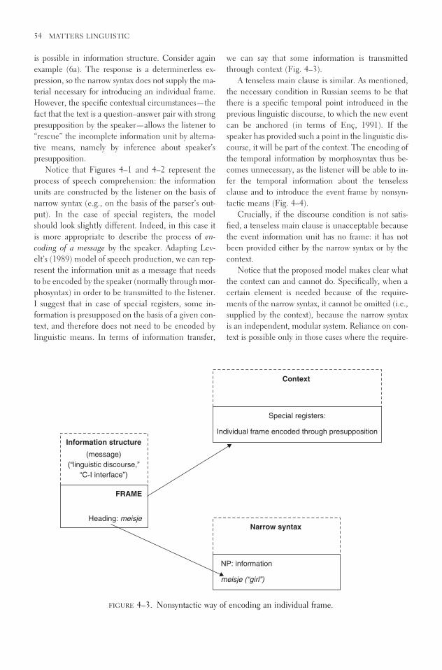
is possible in information structure. Consider againexample (6a). The response is a determinerless ex-pression, so the narrow syntax does not supply the ma-terial necessary for introducing an individual frame.However, the specific contextual circumstances—thefact that the text is a question–answer pair with strongpresupposition by the speaker—allows the listener to“rescue” the incomplete information unit by alterna-tive means, namely by inference about speaker’s presupposition.
Notice that Figures 4–1 and 4–2 represent theprocess of speech comprehension: the informationunits are constructed by the listener on the basis ofnarrow syntax (e.g., on the basis of the parser’s out-put). In the case of special registers, the modelshould look slightly different. Indeed, in this case itis more appropriate to describe the process of en-coding of a message by the speaker. Adapting Lev-elt’s (1989) model of speech production, we can rep-resent the information unit as a message that needsto be encoded by the speaker (normally through mor-phosyntax) in order to be transmitted to the listener.I suggest that in case of special registers, some in-formation is presupposed on the basis of a given con-text, and therefore does not need to be encoded bylinguistic means. In terms of information transfer,
we can say that some information is transmittedthrough context (Fig. 4–3).
A tenseless main clause is similar. As mentioned,the necessary condition in Russian seems to be thatthere is a specific temporal point introduced in theprevious linguistic discourse, to which the new eventcan be anchored (in terms of Enç, 1991). If thespeaker has provided such a point in the linguistic dis-course, it will be part of the context. The encoding ofthe temporal information by morphosyntax thus be-comes unnecessary, as the listener will be able to in-fer the temporal information about the tenselessclause and to introduce the event frame by nonsyn-tactic means (Fig. 4–4).
Crucially, if the discourse condition is not satis-fied, a tenseless main clause is unacceptable becausethe event information unit has no frame: it has notbeen provided either by the narrow syntax or by thecontext.
Notice that the proposed model makes clear whatthe context can and cannot do. Specifically, when acertain element is needed because of the require-ments of the narrow syntax, it cannot be omitted (i.e.,supplied by the context), because the narrow syntaxis an independent, modular system. Reliance on con-text is possible only in those cases where the require-
54 MATTERS LINGUISTIC
Information structure
(message)
(“linguistic discourse,”
“C-I interface”)
Heading: meisje
FRAME
Context
Special registers:
Individual frame encoded through presupposition
Narrow syntax
NP: information
meisje (“girl”)
FIGURE 4–3. Nonsyntactic way of encoding an individual frame.

ments of the information structure are at stake. A clearexample of this distinction is the respective roles ofagreement and tense. As a morphosyntactic charac-teristic, agreement must be present because of the nar-row syntax requirements. Tense, on the other hand,is required to make a connection (anchoring) of an utterance to the linguistic discourse (e.g., Enç,1991). Thus, the model predicts that there will be noregister that would allow for omission of agreement.Indeed, this seems to be the case: while many lan-guages allow a nonsyntactic encoding of the eventframe (e.g., nontensed main clauses), they do not allow omission of agreement (e.g., no register in Russian would allow a sentence like *deti prygnul‘children[PL] jumped[SING]’). This feature of themodel is important because it makes further predic-tions about errors in aphasic speech, discussed later.Briefly, it predicts that problems with tense should beobserved significantly more often than problems withagreement in the speech of these patients.
A question that arises at this point is why “special”conditions would be necessary for “special registers.”In other words, if the possibility of encoding an eventor an individual frame by nonsyntactic means is inprinciple available in natural languages, why dospeakers not always omit functional categories and
rely on (often quite reliable) context in order to buildthe information units? I would like to propose that theanswer is related to economy considerations. I hy-pothesize that in normal adult speakers, narrow syn-tax is the cheapest, most economical way of encodingindividual and event frames and that this is the mosteconomical way of building information structure.5
This is in line with the theory formulated in Reuland(2001) for unimpaired language. He argues that op-erations that take place at different levels (syntax, se-mantics, discourse) form an economy hierarchy, thesyntactic ones being the cheapest and the discourseones being the most expensive. Thus, the use of nar-row syntax in unimpaired language for encoding mes-sages follows from its position in the hierarchy.
Economy in Broca’s Aphasia
The cheapest, most economical option in unimpairedlanguage does not necessarily enjoy the same statusin the language of brain-damaged patients. If Broca’sregion is involved with syntactic computations, it isonly natural that damage to this area would makethese operations more resource consuming. Theeconomy hierarchy will then be different: what usedto be the easiest way of building information struc-
WEAK SYNTAX 55
Information structure
(message)
(“linguistic discourse,”
“C-I interface”)
Heading: prygat' (to jump)
FRAME
Context
Special registers:
Event frame encoded through presupposition
Narrow syntax
VP: information
prygat' (to-jump)
FIGURE 4–4. Nonsyntactic way of encoding an event frame.

ture, or encoding messages, will now become moreexpensive than (or at least equally expensive as) otheroptions in principle available to the language system.The straightforward reflection of such change in theeconomy hierarchy will be noticeable precisely inthose cases where, in unimpaired language, alterna-tive options are avoided because of their relativelyhigher cost.
What I propose therefore is a possible way of whata theory of complexity may look like if it is to explaincertain omissions in production and some specific er-rors in comprehension. The main idea is that the re-sources necessary for conducting syntactic operationsare diminished in Broca’s aphasia, with consequencesreflected both in the production pattern and in com-prehension errors of these speakers.6
Production In production, the use of narrow syntax(“the syntactic channel”) becomes less efficient be-cause it is no longer the most economical option avail-able. Rather, an alternative option is now used—specifically, reliance on the context. Importantly, thisoption is also available for unimpaired speakers in spe-cial registers. In other words, damage to Broca’s re-gion does not create a new information-processing sys-tem. Depending on the degree of severity of damage,or perhaps on other factors still to be investigated, thetwo means—syntactic and nonsyntactic—may be-come comparable with each other in the amount ofinformation they can encode and transmit. We wouldthen expect to see occasional use of the syntactic chan-nel and occasional use of the nonsyntactic one. Theresult is the well-known phenomenon of variability inaphasic speech.
The proposed model also makes an interesting pre-diction about the correlation between omission of de-terminers and tense. Because such omissions repre-sent the use of nonsyntactic means of transmittinginformation, the prediction is that once this alterna-tive is selected by Broca’s aphasics, it is likely to beselected for both functional categories. This predic-tion is borne out. Baauw et al. (2002) presented evi-dence that there is a correlation between omissions ofthese two functional elements. For the utterances ofa group of Dutch-speaking Broca’s aphasics, it wasshown that the more likely a sentence was to containtense, the more likely it was that a determiner wouldalso be produced. And the other way around: tense-less clauses are more likely to contain determinerlessNPs. The authors argue that this correlation is due to
the optional reliance on nonsyntactic means for in-troducing information units: if a nonsyntactic root ischosen (as the easiest in the sense discussed earlier),it is more likely to fulfill all functions with regard toencoding a message, both for individual and for eventinformation units.7
As discussed earlier, the ability of the context tocompensate for what has not been encoded mor-phosyntactically is limited. If a certain element of thestructure is required because of the requirements ofthe narrow syntax (a modular, independent compu-tational system), this element cannot be omitted.Again, for example, there is no register such that al-lows mismatch in agreement. The prediction aboutBroca’s aphasics is then that these patients wouldmake significantly fewer errors with agreement thanwith tense, because there is no alternative way to en-code agreement rather than by means of morphosyn-tax. Indeed, as many authors have argued, there is adifference in aphasics’ performance with respect tothese two elements. Friedmann (1999), Benedet et al.(1998), Kolk (2000), and Wenzlaff and Clahsen(2005), among others, show that Hebrew-, Dutch-,and German-speaking Broca’s aphasics are more im-paired with tense than with agreement. Given that inmany cases these patients produce a nonfinite verb,the results support the proposed model.
To continue this line of argument about what thecontext can and cannot do, it is important to re-member that the speech of Broca’s aphasics is notoverall impaired. It has been demonstrated that thesepatients are sensitive to some subtle syntactic con-straints. Bastiaanse and Zonneveld (1998), for exam-ple, show that the use of nonfinite forms in aphasicsis restricted to the main clause (the authors’ claim be-ing that agrammatics have problems with verb move-ment that takes place in Dutch only in the mainclause; see however, Lonzi and Luzzatti, 1993). Whenasked to complete a sentence with a missing verb,agrammatics produced finite forms only 49% of thetime; in embedded clauses, however, their perfor-mance increased to 86%. Avrutin (1999) suggests thatthese findings demonstrate patients’ sensitivity to whatGueron and Hoekstra (1995) label “the tense chain,”the requirement on the coindexation of Comp and T.Thus, they preserve the subtle syntactic knowledgethat in the presence of overt Comp, only finite T canparticipate in the tense chain. Further support for thisview comes from the observation that these speakersalways produce tensed auxiliary verbs and modals
56 MATTERS LINGUISTIC

(even in the main clause), the elements that head TPand therefore occur in the tense chain (e.g., Bastia-anse and Jonkers, 1998; De Roo, 1999; Kolk, 1998).8
Broca’s aphasics also demonstrate subtle syntacticknowledge of the relationship between verb move-ment and finiteness. Kolk and Heeschen (1992) showthat if a nonfinite verb is produced by a Dutch or Ger-man (V2 languages) aphasic speaker, this verb, in thevast majority of cases, is in the clause final position;a finite verb is always correctly placed in the secondposition. Lonzi and Luzzatti (1993) report similar re-sults for Italian Broca’s aphasics. The authors observethat when the verb is nonfinite, it either precedes orfollows the adverb (both positions are correct in Ital-ian), but when the verb is finite, the adverb always follows it.
As for comprehension, which I discuss shortly,Broca’s aphasics do not differ from normal speakersin their sensitivity to a verb’s representational com-plexity. Thus, as Shapiro et al. (1987) demonstrate, ittakes longer for normal speakers to process verbs thathave more possible argument structures (e.g., alter-nating datives such as ‘send’ take more resources thana transitive verb such as ‘fix’). Following up on thisstudy, Shapiro and Levine (1990) showed that Broca’saphasics exhibit a similar pattern of performance:while their reaction, overall, is significantly slowerthan normal, the same distinction between verbs withregard to the argument structure complexity can bedetected in these patients.
Interestingly, these findings represent preciselythose cases where the context cannot serve as an al-ternative. Verb movement, for example, is triggeredby the principles of narrow syntax and has no directconsequences for building of information structure.Moreover, there is no register in unimpaired Dutchor German that would allow a nonfinite verb in thesecond position. These observations further supportthe claim that the “agrammatic” performance exhib-ited by Broca’s aphasics does not constitute impair-ment of grammar (taken here to mean principles ofnarrow syntax). Their errors are restricted to thosecases that can be characterized as reliance on alter-native means of building information structure (incomprehension) or encoding a message (in produc-tion). If this alternative is not available, these patientswill produce, perhaps slower than in unimpairedspeech, syntactically well-formed utterances.
A natural question is why functional categories aremore vulnerable than lexical ones. First, notice that
omission of functional categories is compatible withsome registers in unimpaired language, while there isno register that would allow optional omission of alexical element (e.g., even if we know from a givencontext that the conversation is about a specific boywho read some book, it is impossible to say “the . . .read a . . .”). Simply speaking, lexical elements aretoo informative to be omitted. In terms of Shannon’sinformation theory (Shannon, 1948), these elementsare selected from a larger set compared with the func-tional categories, which makes them, in the techni-cal sense, “more informative.” Avrutin (in prepara-tion) provides more discussion of how a formalinformation theory can be integrated into current psy-cholinguistic research. Suffice it to say that theamount of information of an element is proportionalto the degree of activation it receives and therefore isless likely to be omitted. The same kind of reasoningexplains why in some languages aphasic patientsdemonstrate substitution patterns rather than omissions.These are usually morphologically rich languages; thus,each form is to be selected from a larger set of relatedelements (a somewhat similar, processing-based ac-count is offered in Lapointe, 1985). This fact makesthem more informative, with direct consequences forprocessing cost and activation (for more discussion ofsuch relationship in unimpaired speech, see, for ex-ample, Kostic et al. [in press]).
Comprehension In order to illustrate how the pro-posed model explains comprehension deficiency inBroca’s aphasia, I now discuss some experimentalfindings that can be straightforwardly explained interms of a competition between two systems. It is cer-tainly beyond the scope of this article to address allfindings in aphasia; the ones presented later simplyserve as an illustration of the application of the model.
A typical case where economy considerations de-termine a possible interpretation is the so-called ex-ceptional case marking (ECM) constructions illus-trated in Dutch example (8) (SE stands for ‘simplexexpression’: a monomorphemic element that is alwaysreferentially dependent on an antecedent in the samesentence).
(8) Jani zag [zichi / *hem i dansen]John saw [SE/him dance]
Reuland (2001), in his Primitives of Binding frame-work, argues that various types of anaphoric depen-
WEAK SYNTAX 57

dencies form an economy hierarchy in such a waythat a more costly dependency is disallowed, provideda cheaper alternative is available. For example, Dutchanaphor ‘zich’ can enter a syntactic dependency withits antecedent as a result of feature checking. Pro-nouns, on the other hand, are unable to participatein this type of dependency because, unlike simplexexpression ‘zich’, they possess a number feature thatcannot be deleted. Thus, no syntactic dependency canbe formed for a pronoun. There are, of course, otherdependencies in natural language, such as discoursedependency where ‘Jan’ and ‘hem’ would receive thesame value from discourse storage9:
(9) Jan zag hem dansen ˆˆl zag (x, (dansen (y)); x � yJohn saw him dance
However, according to Reuland’s hierarchy, a dis-course dependency is more expensive than a syntac-tic one. Thus, it is disallowed on the basis of econ-omy considerations.
If the narrow syntax is “weakened” and is no longerthe most economical option, the hierarchy proposedby Reuland changes. If so, there should be no prohi-bition against establishing a nonsyntactic dependencybetween the antecedent and a pronoun in example(9). Ruigendijk et al. (2003) demonstrated that indeedDutch-speaking Broca’s aphasics show an impairedpattern of responses. In a picture selection task, sub-jects were presented with sentences such as in exam-ple 10 and three pictures corresponding to the cor-rect choice of an antecedent, an incorrect one, and afiller. Each sentence began with an introductoryclause of the form ‘First the boy and the man dranksomething and . . .’ (in order to make two possible antecedents available) and concluded with the fol-lowing:
(10) . . . daarna zag de man hem voetballen [Dutch]. . . then the man saw him playing soccer
The correct picture showed a boy playing soccer infront of the mirror and a man looking at the boy. Theincorrect picture showed a man playing soccer in frontof the mirror looking at himself. Thus, if a listenerdisallows coreference between ‘him’ and ‘the man’,he or she should choose the correct picture. If he orshe allows ‘him’ to refer to the matrix subject, the in-correct picture could be chosen.
Adult controls correctly chose the picture wherethe man sees the boy in the mirror. Aphasic speakers,however, incorrectly pointed to the picture where theboy sees himself around 50% of the time, which is notdifferent from chance.10 In other words, they quite of-ten allowed a local dependency relation between thepronoun and the matrix subject. The authors arguethat because the syntactic dependency is the less eco-nomical one for this population, they sometimes al-low a semantic or discourse dependency between thematrix subject and the pronoun. Notice that such al-lowance is once again “optional”—it is not the casethat subjects always select this picture. This is not surprising, however, because, as in the case of pro-duction, the errors result from the competition be-tween two systems—the narrow syntax and an alter-native that is the context. For some speakers, onesystem can win on some occasions, and the other willtake over in other cases, which results in an overallchance performance.11
These results show that Broca’s aphasics some-times fail to establish a dependency relation betweenan antecedent and a pronoun correctly. Other resultsconcerning dependency between two positions in-volve passive constructions and object relatives. Theabnormal, chance-level comprehension of agram-matic Broca’s aphasics on passive constructions is wellknown. As discussed extensively in Grodzinsky(1990),12 a typical observation is that Broca’s aphasicschoose at a more or less chance level between ‘thecat’ and ‘the dog’ as the agent of the chasing event inthe following sentence.
(11) The cat was chased by the dog.
Grodzinsky’s trace-deletion hypothesis suggests thataphasic patients are unable to represent traces, so atrace in the object position of the verb ‘chase’ is deleted.As a result, the subject DP does not receive the agenttheta-role (as it should in an unimpaired linguisticstructure), and because each DP must have a theta rolein order to be interpreted, the subject receives an in-terpretation by means of some kind of strategy that as-signs, at least in English, the agenthood to the first DPin the sentence. Alternative explanations for this phe-nomenon were offered, among others, by Piñango(1999), who argues that the impairment is restricted tomapping between syntactic and semantic structures.
Notice, however, that the phonologically empty el-ement in the object position (a trace or a copy) nor-
58 MATTERS LINGUISTIC

mally enters a syntactic dependency with the subject.This dependency may be represented as a syntacticchain or as some other mechanism that would linkthe two positions in narrow syntax. I suggest that suchdependency must be established in a timely fashion,that is very fast, in a normal case, in order to functionas a reliable source of interpretation. Syntactic oper-ations, however, are slowed in aphasia, as demon-strated convincingly by Zurif et al. (1993) and Swin-ney (2003).13 These researchers showed that Broca’saphasics show priming for an antecedent significantlylater than normal controls. The main point thus isthat the syntactic dependency is, in principle, intactin aphasia; however, the necessary syntactic connec-tion is significantly slowed.14 In terms of the proposedmodel, we can interpret these findings as evidence forthe lack of necessary resources for conducting syn-tactic operations in time because, as in any other phys-ical system, speed is a function of available energy; inother words, it is proportional to the available brainactivation. As the syntactic system is no longer themost economical (and hence the cheapest one) forthese patients, they may sometimes avoid reliance onnarrow syntax when interpreting passive construc-tions. As the first DP in a passive construction is usu-ally the most prominent element, a topic, and as thereis a general correlation between topichood and agency(as also pointed out by Grodzinsky in his formulationof aphasics strategy), the information unit introducedby the first DP may end up marked as the agent. Onceagain, there is a competition between the two systems.Depending on which system wins, patients will eitherchoose a correct interpretation (subject � patient), oran incorrect one (subject � agent.) Overall, we ob-serve a chance performance.15
Finally, the proposed model makes the predictionthat the chance performance in comprehension willbe observed in Broca’s aphasics only in cases wherecompetition between the syntactic and discourse-related operations may take place. As discussed ear-lier for production, when no alternative is possible andthe application of a syntactic operation is required,these patients are predicted to show an above-chanceperformance. A clear example of such a situation incomprehension is the case of wh- questions. Hickokand Avrutin (1995) and Tait et al. (1995) show thatBroca’s aphasics performed above chance on objectWHO-questions (as in example 12) but aroundchance on object WHICH-questions (as in example13).16
(12) Who did the cat chase?(13) Which dog did the cat chase?
WHO-questions differ from WHICH-questions inthat only the latter is discourse linked. Specifically,the wh- phrase ‘who’ functions as a pure operator andis not represented by a discourse referent. The wh-phrase ‘which dog’, on the other hand, does have sucha representation, because it presupposes the existenceof a known set of dogs. Importantly, thus, only in example (13) is a discourse link between the object position and the wh-phrase possible; for WHO-questions the only option is to establish the connec-tion in syntax. Broca’s aphasics demonstrated a goodperformance on the “pure syntactic” question, whileshowing a chance performance on the conditionwhere a competition between two systems (syntacticand nonsyntactic) was in principle possible. Giventhat their syntactic system was weakened, they wouldoccasionally rely on a nonsyntactic one, which re-sulted in a an overall chance performance in the sameway as in the case of interpreting ECM constructionsor passive sentences.
SUMMARY
As a result of damage to Broca’s region, the amountof resources necessary for conducting operations in-volving narrow syntax is diminished. This causes aslowdown in the process of speech production. More-over, reduced power of this system (a direct conse-quence of diminished resources) may result in the sit-uation where alternative systems become morepowerful and therefore are used for the purposes ofbuilding information structure in comprehension orencoding a message (in production: an option avail-able, in principle, in unimpaired language17).
Impairment in Broca’s aphasia is not limited tostructures involving constituent movement (e.g., pas-sive constructions, object relative clauses). Compre-hension of pronouns and other determiners causes dif-ficulties as well. Slow, effortful, telegraphic speech ischaracteristic of the same patients who demonstrateproblems with comprehension of certain elements.The unified explanation of these phenomena pre-sented here is based on the claim that the power ofthe damaged Broca’s region is diminished, with directconsequences for both production speed and economy-based errors in comprehension.
WEAK SYNTAX 59

In order to produce speech in normal time and torely on narrow syntax for the purposes of conveyingand processing information, speakers need to havestrong, powerful syntactic apparatus capable of per-forming required operations in time. As a result ofbrain damage, the power available to this system is re-duced, and the syntax becomes weak.
ACKNOWLEDGMENTS The preparation of this publi-cation was supported by the Dutch National ScienceFoundation (NWO) as part of the Comparative Psy-cholinguistics research program. I thank Sergio Baauw,Yosef Grodzinsky, Joke de Lange, Esther Ruigendijk,Nada Vasic, and Shalom Zuckerman for their comments.Special thanks are due to Jocelyn Ballantyne for proof-reading the manuscript.
Notes
1. For example, Grodzinsky et al. (1991) used evi-dence from Broca’s aphasics to distinguish between twotheories of passive constructions. They demonstrated thatthese patients are selectively impaired in those construc-tions that, according to one of the competing theories,involve syntactic transformations.
2. Haegeman (1990) also shows that omission ofsubjects in a non–pro drop language such as English ispossible in some specific “diary-style” registers.
3. Recent work by Cole et al. (2001) is particularlyinteresting in this regard as it shows that the choice of anantecedent for a logophor can be parameterized withvalue of parameters differing in different dialects. Thisevidence demonstrates that the level of “discourse” is, in-deed, linguistic in nature and that the term “linguisticdiscourse” is rather appropriate.
4. I put a question mark under “context” as I re-main agnostic with regard to the type of symbols and thestructure of context as a cognitive system.
5. In this sense, the notion of economy that hasplayed a major role in recent theoretical work (e.g.,Chomsky, 1995; Reuland, 2001) is more than a formalnotion. In my view, it should reflect, at some level, theamount of resources used by the brain when performinga specific computation. In Avrutin (2000), I discuss ex-perimental evidence supporting the claim that the nar-row syntax operations are the cheapest. For further evi-dence, see also Piñango et al. (2001).
6. The reduction of resources, and a consequentslowdown of the syntactic machinery, is most likely dueto problems with lexical access in Broca’s aphasia, as ar-gued by many researchers (e.g., Shapiro and Levin, 1990;Swinney et al., 1989; Zurif, 2003, among others.)
7. For a somewhat different explanation of thesedata, see Ruigendijk (2002).
8. The same approach explains the omission ofcomplementizers in aphasics’ speech. Because Compand T form a chain, omission of one element necessi-tates omission of the other in order to avoid an invalidchain formation.
9. Once again, there is a discrepancy in terminol-ogy. What Reuland (2001) refers to as “discourse de-pendency” is, in my terminology, a dependency at thelevel of information structure (“linguistic discourse”) es-tablished by reliance on contextual information. Reu-land’s notion of “discourse storage” should be viewed aspart of the context in the present terminology.
10. When in the same construction the pronoun isreplaced with ‘zichzelf’, the patients demonstrated analmost perfect performance. Thus, their problem is notrelated to ECM constructions per se but rather to the in-terpretation of a pronoun in such constructions.
11. Another reason is that there is always a choicebetween two pictures; that is, even if subjects allow for adiscourse dependency between the matrix subject andthe pronoun, they do not have to make this choice: it isalways possible to interpret the pronoun as referring toan external antecedent.
12. Grodzinsky (1999) provides a more general pic-ture of the role of Broca’s area in comprehension of struc-tures involving transformations. For expository purposes,I focus here on passive constructions, although, I believe,the account can be extended to relative clauses and clefts.
13. Other results that are relevant are reported inSwinney et al. (1989), who showed that Broca’s aphasicsexhibit abnormal pattern of activation of word meaning.In a study with ambiguous words, these researchers showthat Broca’s aphasics are capable of accessing only themost frequent meaning of the word within the initial,short period of time. Later on, however, both meaningsbecome available.
14. Further evidence that Broca’s aphasics main-tain a normal ability to connect two positions in a syn-tactic tree but in a slower-than-normal way is presentedin Piñango and Burkhardt (2001).
15. The same explanation also holds for the chanceperformance observed in object relative clauses, as in thepioneering work of Caramazza and Zurif (1976).
16. Performance on both types of subject wh- ques-tions was above chance, as predicted because in this casethe information about the thematic role of the unmovedobject is sufficient to obtain a full interpretation.
17. Another population that exhibits performancesimilar to that of Broca’s aphasics is normally developingchildren. See Avrutin (2004) for more discussion and theapplication of the proposed model to child speech.
60 MATTERS LINGUISTIC

References
Akmaijan, A. (1984). Sentence type and form-function fit.Natural Language and Linguistic Theory, 2, 1–23.
Avrutin, S. (1999). Development of the syntax-discourseinterface. Dordrecht: Kluwer Academic Publishers.
Avrutin, S. (2000). Comprehension of D-linked and non-D-linked wh-questions by children and Broca’saphasics. In Y. Grodzinsky, L. Shapiro, & D. Swin-ney (Eds.), Language and the brain (pp. 295–313).San Diego: Academic Press.
Avrutin, S. (2004). Optionality in child and aphasicspeech. Lingue e Linguaggio, 1, 67–89.
Baauw, S., de Roo, E., & Avrutin, S. (2002). Determineromission in language acquisition and language im-pairment: Syntactic and discourse factors. Proceed-ings of the Boston University Conference on Lan-guage Development (pp. 23–35). Boston: CascadillaPress.
Bastiaanse, R., & Jonkers, R. (1998). Verb retrieval in ac-tion naming and spontaneous speech in agrammaticand anomic speech. Aphasiology, 12, 951–969.
Bastiaanse, R., & van Zonneveld, R. (1998). On the re-lation between verb inflection and verb position inDutch agrammatic aphasics. Brain and Language,64, 165–181.
Benedet, M.J., Christiansen, J.A., & Goodglass, H.(1998). A cross-linguistic study of grammatical mor-phology in Spanish- and English-speaking agram-matic patients. Cortex, 34, 309–336.
Blom, E. (2003). From root infinitive to finite sentences.Doctoral dissertation. the Netherlands: UtrechtUniversity.
Caramazza, A., & Zurif, E. (1976). Dissociation of algo-rithmic and heuristic processes in language com-prehension: Evidence from aphasia. Brain and Lan-guage, 3, 572–582.
Chomsky, N. (1981). Lectures on government and bind-ing, Dordrecht: Foris.
Chomsky, N. (1986). Knowledge of language. New York:Praeger.
Chomsky, N. (1995). The minimalist program. Cam-bridge, MA: MIT Press.
Cole, P., Hermon, G., & Huang C.-T.J. (2001). Longdistance reflexives. In P. Cole, G. Hermon, & C.-T.J. Huang (Eds.), Syntax and Semantics Series33. New York: Academic Press.
De Roo, E. (1999). Agrammatic grammar. Doctoral dis-sertation, The Netherlands: Leiden University.
Enç, M. (1991). Anchoring conditions for tense. Lin-guistic Inquiry, 18, 633–657.
Friedmann, N. (1999). Functional categories in agram-matic production: A cross-linguistic study. Doctoraldissertation. Tel Aviv: Tel Aviv University.
Grodzinsky, Y. (1990). Theoretical perspectives on lan-guage deficit. Cambridge, MA: MIT Press.
Grodzinsky, Y., Pierce, A., and Marakowitz, S. (1991).Neuropsychological reasons for a transformationalderivation of syntactic passive. Natural Languageand Linguistic Theory, 6, 431–453.
Grodzinsky, Y. (1999). The neurology of syntax: Lan-guage use without Broca’s area. Brain and Behav-ioural Science, 23, 47–117.
Grodzinsky, Y., Wexler, K., Chien, Y.-C., Marakovitz, S.,& Solomon, J. (1993). The breakdown of bindingrelations. Brain and Language, 45, 371–395.
Gueron, J., & Hoekstra, T. (1995). The temporal inter-pretation of predication. In A. Cardinalletti & T.Guasti (Eds.), Syntax and semantic. San Diego: Aca-demic Press.
Haegeman, L. (1990). Understood subjects in English di-aries: On the relevance of theoretical syntax for thestudy of register variation. Multilingua, 9, 157–199.
Heim, I. (1982). The semantics of definite and indefinitenoun phrases. Doctoral dissertation. Amherst: Uni-versity of Massachusetts.
Hickok, G., & Avrutin, S. (1995). Representation, referen-tiality, and processing in agrammatic comprehension:Two case studies. Brain and Language, 50, 10–26.
Hofstede, B. (1992). Agrammatic speech in Broca’s apha-sia; Strategic choice for the elliptical register. Doc-toral dissertation. Nijmegen Institute for Cognitionand Information, Holland.
Kamp, H., & Reyle, U. (1993). From discourse to logic.Dordrecht: Kluwer Academic.
Kolk, H. (2000). Canonicity and inflection in agram-matic sentence production. Brain and Language,74, 558–560.
Kolk, H.H.J., & Heeschen, C. (1992). Agrammatism,paragrammatism and the management of language.Language and Cognitive Processes, 7, 89–129.
Kolk, H.H.J. (1998). Disorders of syntax in aphasia. In B.Stemmer & H. Whitaker (Eds.), Handbook of neu-rolinguistics (pp. 249–260). San Diego: AcademicPress.
Kolk, H.H.J. (1995). A time-based approach to agrammaticproduction, Brain and Language, 50, 282–303.
Kostic, A. (in press). The effects of the amount of infor-mation on processing of inflected morphology. Bel-grade: University of Belgrade.
Levelt, W.J.M. (1989). Speaking: From intention to ar-ticulation. Cambridge, MA: MIT Press.
Lapointe, S. (1985). A theory of verb form use in thespeech of agrammatic aphasics. Brain and Lan-guage, 24, 100–185.
Lonzi, L., & Luzzatti, C. (1995). Omission of preposi-tions in agrammatism and universal constraint of re-coverability, Brain and Language, 51, 129–132.
WEAK SYNTAX 61

Love, T., Nicol, J., Swinney, D., Hickok, G., & Zurif, E.(1998). The nature of aberrant understanding andprocessing of pro-forms by brain-damaged popula-tions. Brain and Language, 65, 59–62.
Piñango, M. (1999). Syntactic displacement in Broca’saphasia comprehension. In Y. Grodzinsky & R. Bas-tiaanse (Eds.), Grammatical disorders in aphasia: aneurolinguistic perspective (pp. 75–87). London:Whurr.
Piñango, M.M. (2000). Neurological underpinnings ofbinding relations. Paper presented at the linguisticsociety of Germany, Marburg, Germany, March 2.
Piñango, M., Burkhart, P., Brun, D., & Avrutin, S.(2001). The architecture of the sentence processingsystem: The case of pronominal interpretation. Paperpresented at SEMPRO meeting, Edinburgh, UK.
Piñango, M., & Burkhardt, P. (2001). Pronominals inBroca’s aphasia comprehension: the consequences ofsyntactic delay. Brain and Language, 79, 1, 167–168.
Reuland, E. (2003). A window into the architecture ofthe language system. GLOT International, 7, No.1/2.
Reuland, E. (2001). Primitives of binding. Linguistic In-quiry, 32, 439–492.
Ruigendijk, E. (2002). Case assignment in agramma-tism. Doctoral dissertation. Groningen: GroningenUniversity.
Ruigendijk, E., Baauw, S., Zuckerman, S., Vasic, N., deLange, J., and Avrutin, S. (2003). A cross-linguisticstudy on the interpretation of pronouns by childrenand agrammatic speakers: Evidence from Dutch,Spanish, and Italian, Paper presented at CUNY sen-tence processing conference, Boston.
Schutze, C. (1997). The empirical base of linguistics:Grammaticality judgments and linguistic methodol-ogy. Chicago: University of Chicago Press.
Shannon, C.E. (1948). A mathematical theory of com-munication. Bell System Technical Journal, 27,379–423, 623–656.
Shapiro, L., & Levine, B. (1990). Verb processing duringsentence comprehension in aphasia. Brain and Lan-guage, 38, 21–47.
Shapiro, L., Zurif, E., & Grimshaw, J. (1987). Sentenceprocessing and the mental representation of verbs.Cognition, 27, 219–246.
Swinney, D., Nicol, J., & Zurif, E. (1989). The effectsof focal brain damage on sentence processing: Anexamination of the neurological organization of amental module. Journal of Cognitive Neuroscience,1, 25–37.
Swinney, D. (2003). Psycholinguistic approaches. Paperpresented at the Fourth Science of Aphasia Con-ference, Trieste.
Tait, M.E., Thompson, C.K., & Ballard, K.J. (1995). Sub-ject-object asymmetries in agrammatic compre-hension of four types of wh-questions. Brain andLanguage, 51, 77–79.
Tesak, J., & Dittmann, J. (1991). Telegraphic style innormals and aphasics. Linguistics, 29, 1111–1137.
Vallduví, E. (1992). The informational component. NewYork: Garland.
Zurif, E.B. (2003). The neuroanatomical organization ofsome features of sentence processing: Studies ofreal-time syntactic and semantic processing. Psy-chologia, 32, 13–24.
Zurif, E.B., Swinney, D., Prather, P., Solomon, J., &Bushell, C. (1993). An on-line analysis of syntacticprocessing in Broca’s and Wernicke’s aphasia. Brainand Language, 45, 448–464.
Wenzlaff, M., & Clahsen, H. (2005). Finiteness and verb-second in German agrammatism. Brain and Lan-guage, 92, 33–34.
62 MATTERS LINGUISTIC

5
Speech Production in Broca’sAgrammatic Aphasia:
Syntactic Tree Pruning
Naama Friedmann
Agrammatic aphasia, a deficit that usually occurs fol-lowing brain lesion in Broca’s area and its vicinity inthe left hemisphere, causes individuals to lose theirability to produce syntactically well-formed sentences.They can no longer inflect verbs correctly for tense,use subject pronouns, form relative sentences, pro-duce subordination conjunctions, or construct a well-formed Wh question. Even more striking is the factthat at the same time they retain their ability to in-flect verbs for subject agreement, use object pronouns,form reduced relatives, and produce coordinationconjunctions, and they are still able to form yes/noquestions in some languages. This pattern is also opento cross-linguistic variability: for example, individualswith agrammatic aphasia who speak Arabic and He-brew can produce well-formed yes/no questions, butspeakers of Dutch, English, and German with thesame impairment cannot.
The study of this intricate pattern of dissociationsis promising for both the exploration of the functionalcharacterization of various brain areas and for the in-
quiry of the psychological reality of linguistic con-structs. In the realm of brain and function, it suggestsa window through which the role of brain areas thatare involved in speech production, and specificallythe syntactic aspects of production, can be viewed.Once a selective deficit in a specific syntactic func-tion can be described in fine functional details, it canthen be related to the brain area that was damaged,with the possible result of the description of brain ar-eas that subserve this specific function.
The selective nature of impairment also makes theexploration of agrammatic aphasia valuable for syn-tactic theories, as the selectivity of the impairment im-poses constraints on the theory of normal functioningof the relevant cognitive ability: linguistic theory. Forexample, a selective deficit in one type of verb in-flection, such as tense (the inflection of the verb forpast, present, or future tense), which can occur with-out a deficit in other types of inflection, such as agree-ment (the inflection of the verb for person, genderand number), suggests that syntactic theory should
63

treat tense and agreement inflections as distinct con-structs and as functions that are represented orprocessed by different modules. Therefore, such afinding can be useful when two different linguisticmodels are suggested for verb inflection: one in whichtense and agreement inflections form a single naturalclass, and one in which tense and agreement belongto two different classes. A finding from aphasia thatshows a dissociation between tense and agreementcan support the latter model and bring support for itfrom a neuropsychological angle. Such a dissociationis presented in the current study.
For many years, the standard view concerningagrammatic speech production has been that thedeficit is very broad. Many researchers in the fieldclaimed that syntactic ability is completely lost inthese individuals and that they merely lean on non-linguistic strategies to concatenate words into a sen-tence (cf., for instance, Berndt and Caramazza, 1980;Caplan, 1985; Goodglass, 1976; Goodglass and Berko1960; Saffran et al., 1980). Later, the impairment hasbeen presented as a more circumscribed deficit, andattempts have been made to account for it using lin-guistic terms. In this framework, Kean (1977) sug-gested ascribing the deficit to phonological factorssuch as the fact that grammatical morphology is gen-erally unstressed. Grodzinsky (1984, 1990) convinc-ingly argued that the underlying deficit is syntactic,rather than phonological. He used the distinction be-tween languages that allow “zero morphology,”namely, languages in which omission of morphemesis licit, and languages in which it is illicit, to accountfor the finding that morphology is omitted in somelanguages and substituted in others. Still, even theseaccounts that used grammatical terms to explain thepattern of agrammatic impairment in production as-sumed a very vast deficit in grammatical elements.Grodzinsky (1984, 1990; see also Ouhalla, 1993), forexample, suggested that all functional elements areimpaired in agrammatic speech production.
However, empirical evidence regarding agram-matic aphasia has accumulated in recent years, sug-gesting that the deficit is actually finer-grained andthat not all functional elements are impaired inagrammatic production. Some syntactic abilities werefound to be intact, and some other structures werefound to be differentially impaired in different lan-guages following the same lesion. In sentence pro-duction, the empirical investigation of different syn-tactic structures through constrained tests has shown
that the impairment in agrammatic production doesnot involve all grammatical structures and functionwords. To give just a few examples of elements thathave been shown to be intact in agrammatic produc-tion, case was shown to be spared in Finnish and Pol-ish (Menn and Obler, 1990), and in Dutch and Hebrew (Ruigendijk and Friedmann, 2002), coordi-nation conjunctions were shown to be spared andeven overused (Menn and Obler, 1990), and negationmarkers and their position relative to adverbs werestudied in Italian and French by Lonzi and Luzzatti(1993) and proved to be intact. Even in the domainof verb inflection, some intact abilities were found:De Bleser and Luzzatti (1994) examined past partici-ple agreement in a structured production task andfound a considerable preservation of this inflection.Their participants performed at around 90% correcton most of the tasks in nonembedded sentences.These results called for a systematic exploration of theimpaired and unimpaired syntactic abilities inagrammatic production, as well as for an account thatwill explain why some functions are impaired andwhat separates them from the unimpaired functions.But before such systematic explorations of various syn-tactic domains and the syntactic generalization arepresented, let us start with a short presentation of thesyntactic terms that will be required.
According to syntactic theories within the genera-tive tradition (e.g., Chomsky, 1995; Pollock, 1989),when we produce and understand sentences, they arerepresented as phrase markers or syntactic trees. Inthese syntactic trees, content and function words arerepresented in different nodes (Fig. 5–1). Functionalnodes include inflectional nodes: an agreementphrase (AgrP), which represents agreement betweenthe subject and the verb in person, gender and num-ber, and a tense phrase (TP), representing tense in-flection of the verb. Finite verbs move from V, theiroriginal position within the verb phrase (VP), to Agrand then to T in order to check (or collect) their in-flection. Thus, the ability to correctly inflect verbs foragreement and tense crucially depends on the inflec-tional nodes, AgrP and TP. The highest phrasal nodein the tree is the complementizer phrase (CP), whichhosts complementizers, which are embedding ele-ments like “that,” and Wh morphemes such as“where” and “what.” Other elements that move to CPare verbs and auxiliaries in Germanic languages thatrequire the verb to be in second position in the sen-tence and in yes/no questions in languages like Eng-
64 MATTERS LINGUISTIC

lish, German, and Dutch, which require movementof the auxiliary or the verb to the beginning. Thus,the construction of embedded sentences, Wh ques-tions, and the position of the verb in some languagesdepend on the CP node being intact and accessible.The nodes are hierarchically ordered in the syntactictree—the lowest node is the VP, the nodes above itare the AgrP and the TP (in this order according toPollock, 1989), and the CP is placed in the highestpoint of the syntactic tree. This hierarchical order wassuggested on the basis of purely linguistic groundssuch as arguments from the relative order of elementsof various types (subjects and verbs, inflected verbsand negation, infinitive verbs and adverbs, etc.).
The following sections present a set of experimentsthat systematically explored the status of syntacticstructures that relate to these functional nodes inagrammatic production, proceeding from the bottomto the top nodes: AgrP and TP and then various struc-tures that relate to CP.
The study encompassed a variety of tasks that wereadministered to 18 individuals with agrammatism fol-lowing brain lesion in the left hemisphere, 16 speak-ers of Hebrew and 2 Palestinian Arabic-speakingagrammatic aphasics. Following these studies, a por-trait of the selective syntactic impairment in agram-matic production with respect to these functionalnodes and a syntactic characterization are presented.The spared and impaired abilities are shown to form
two natural classes when looking at them from thepoint of view of syntactic trees. It will be suggestedthat what underlies the syntactic deficit in agrammaticproduction is the inability to project syntactic trees upto their highest nodes (the Tree Pruning Hypothesis,Friedmann, 1998, 1999; Friedmann and Grodzinsky,1997, 2000). This has implications for syntactic the-ory, and at a later stage it would also allow conclu-sions regarding the brain areas that subserve varioussyntactic functions.
VERB INFLECTIONS
A Study in Hebrew and Arabic
Hebrew and Palestinian Arabic serve as excellent test-ing ground for verb inflection ability, because theirinflection is rich: For every finite verb produced, thespeaker has to choose between three tenses—past, pres-ent, and future—and 12 agreement forms—agreeingin person, gender, and number with the subject.
All of these inflection forms were examined usingtwo simple tasks—verb completion and sentence rep-etition. In the completion task, two simple sentenceswere presented. The first included a verb inflected fortense and agreement. In the second sentence, the par-ticipant had to supply the correctly inflected verb. Inthe tense condition, the temporal adverb was changed,
SPEECH PRODUCTION IN BROCA’S AGRAMMATIC APHASIA 65
CP
C'(Wh-question)
TP C
(complementizer)
NegPT
(tense)
Agr
(agreement)
AgrP
Agr'
VP
V'NP
NPV
T'
FIGURE 5–1. Syntactic tree (fromPollock, 1989).

and the missing verb differed from the supplied verbin tense only (example 1); in the agreement condi-tion the subject was changed, so that the missing verbdiffered from the existing verb in one agreement fea-ture only—person, gender, or number (example 2).Another type of sentence completion test requiredcompletion of a verb inflected for both tense andagreement, without a temporal adverb (example 3).
(1) Tense:axshav ha-yeled holex. gam etmol ha-yeled ______.(halax)now the boy walks. Yesterday too the boy ______.(walked)(2) Agreement:axshav ha-yeled holex. berega ze gam ha-yeladim______. (holxim)now the boy walks. Right now the boys also ______.(walk -plural)(3) Tense and Agreement:ha-yalda racta likfoc, az hi amda al ha-makpeca ve______. (kafca)The girl wanted to-jump, so she stood on the diving-board and ______. (jump-past, 3rd, fem, sg)
In the repetition task, participants repeated shortsimple sentences of three or four words that includeda verb inflected to one of the 30 inflection forms (fordetailed description of participants, method, and re-sults, see Friedmann, 1998).
The results showed remarkable dissociation be-tween the production of tense and agreement inflec-tions. While tense was severely impaired, agreement
was relatively intact both in Hebrew and in Arabic(see Fig. 5–2 for the results of the completion task;the Arabic-speaking participants were HH and SSH).Participants made tense substitution errors, but almostno agreement errors. This pattern was consistent forall participants, and each one of them showed signif-icantly better performance on the agreement teststhan on the tense tests. Even in the simple repetitiontask (which was only administered in Hebrew), par-ticipants made tense errors, repeating the sentencewith the correct verb in a wrong tense inflection. Theymade no agreement errors (Table 5–1).1
Tense was significantly poorer than agreement inboth the completion and the repetition tasks, usingWilcoxon signed rank test, W � 6, P � 0.0001, forboth comparisons in Hebrew. In Arabic, we used �2
because there were only two participants, and the dif-ference was significant, too, �2 � 34.82, P � 0.0001.
In contrast to Germanic languages, in which theinfinitives are the preferred substituting forms (Kolkand Heeschen, 1992), in Hebrew the infinitive wasnot used instead of the finite verb. In the repetitiontask, 0% of the errors were toward the infinitival form.
66 MATTERS LINGUISTIC
100%
90%
80%
KA
70%
60%
50%
40%
30%
20%
10%
0%RS AL RA AD TA
Tense errors
RN GR HY AE ER IE PK MA ML AG SSH HH
Agreement errors
FIGURE 5–2. Performance of individual participants in the agreement and tense completion task.Shown is percent errors.
TABLE 5–1. Sentence Repetition Task Showing Per-cent Errors (Number of Inflection Errors/Total Num-ber of Inflected Verbs)
Hebrew (n � 12) Tense Errors Agreement Errors
Repetition 16% (143/912) 0% (4/912)

An additional completion task with half infinitives andhalf finite verbs as target forms was administered to12 of the Hebrew-speaking patients. In this test, only2% of the substitutions were from finite to infinitiveverbs, and the large majority of errors was within thefinite paradigm. (See Friedmann, 2000, for a possibleaccount for this cross-linguistic difference.)
Inflection in Other Languages
Studies in other languages point in the same direc-tion. In Spanish, too, verb agreement was found tobe much better preserved than tense inflection: us-ing a sentence completion procedure, Benedet et al.(1998) reported that the six Spanish-speaking agram-matics they tested produced only 5.5% correct verbaltense but produced 63.8% correct subject-verb agree-ment. They found a similar pattern of results in Eng-lish for the seven agrammatics they examined butwith a smaller difference: the English-speakingagrammatics produced 42% correct agreement andabout 15% correct tense. The same was found inFrench: the agrammatic patient Mr. Clermont re-ported in Nespoulous et al. (1988, 1990) had onlytense errors in spontaneous speech but no verb agree-ment errors. Ferreiro (2003) tested 14 agrammaticaphasics in Catalan and Spanish, with the same com-pletion and repetition tasks described above, and re-ported a similar pattern of dissociations: tense wasmore impaired than agreement in Catalan and Span-ish. Wenzlaff and Clahsen (2004) tested seven Ger-man-speaking agrammatics using forced-choicecompletion of inflection and also showed a dissocia-tion between completion of agreement, which wassimilar to that of the control subjects (greater than90% correct) and completion of tense, which was atchance level (68% correct).
Based on these results, two main conclusions canbe drawn. First, not all functional categories are im-paired in agrammatic production. Agrammatism isneither a complete loss of syntax nor a complete lossof grammatical morphemes or functional categories,as has been claimed in different versions by many re-searchers of agrammatism over the years (Caplan,1985; Caplan and Futter, 1986; Grodzinsky, 1984,1990; Kean, 1977; Ouhalla, 1993). Second, a cleardissociation has been demonstrated between two typesof verb inflection: tense and subject agreement. Tenseis severely impaired, whereas agreement is relativelyintact.
What Can Account for the Dissociation
Between Tense and Agreement?
The split inflection syntactic tree suggested by Pol-lock (1989) (see Fig. 5–1) seems to offer a natural syn-tactic way to capture the dissociation found betweengood agreement and poor tense. In this tree, tense andagreement are represented in two different functionalphrases, TP and AgrP. This allows for a selective im-pairment of one but not the other. The finding thatagreement is always better than tense, and never viceversa, can also be explained by this syntactic repre-sentation. As TP is situated higher than AgrP on thesyntactic tree, we might suggest that higher nodes areharder for agrammatic aphasics to access or project.Thus, while agreement node is accessed appropri-ately, and therefore subject-verb agreement is intact,tense is higher and therefore less accessible, and tenseerrors follow. Thus, two properties of Pollock’s tree al-low for an explanation of the dissociation found inagrammatic speech. The split of the inflectionalphrase into two inflectional nodes allows for the se-lectivity in impairment, and the hierarchical order ac-counts for the asymmetry of this impairment.2
Based on these results, Friedmann (1994, 1998,2000, 2001) and Friedmann and Grodzinsky (1997)suggested the tree pruning hypothesis (TPH), ac-cording to which the syntactic tree of agrammaticaphasics is pruned and higher nodes are inaccessiblein agrammatism.3 The dissociation in verb inflectionfollows from this: Agrammatic aphasics can projectAgrP; therefore, agreement is intact, but they fre-quently fail to project (or access) TP, which causestense errors.
If indeed individuals with agrammatism are im-paired in getting as high up as the tense node, thiswould have rather radical empirical consequences, asit would mean that they would fail to access nodesabove TP as well. This implies that all structures thatdepend on the highest node of the tree, the CP, arepredicted to be impaired in agrammatism.
Therefore, the next step was to examine structuresthat require the CP. Recall that the CP hosts Wh el-ements of Wh questions, complementizers of em-bedded clauses, and verbs and auxiliaries that moveto second position of the sentence. The study thusproceeded with examining the production of ques-tions, embedding, and verb position in Hebrew andPalestinian Arabic as well as with perusal of the cross-linguistic literature. If agrammatic aphasics are un-
SPEECH PRODUCTION IN BROCA’S AGRAMMATIC APHASIA 67

able to project the syntactic tree up to its highestnodes, the prediction is twofold. First, Wh questions,embedding structures and verbs in second position,are expected to be impaired. Furthermore, whilequestions and embeddings that require the high nodewould be impaired, questions and embedded clausesthat do not require CP should be unimpaired (in theabsence of additional impairments).
PRODUCTION OF QUESTIONS
The first step toward evaluating the status of the CPnode in agrammatic production was to assess patients’ability to produce questions. A Wh question such as(4) below is formed from (5), by means of movementof the Wh morpheme to the beginning of the sen-tence (to spec CP). Because the production of Whquestions involves CP, the highest node of the tree,an impairment in CP should entail a deficit in Whquestions. Studies by Shapiro and Thompson andtheir group (see Chapter 8) focused on treatment ofthe production of Wh questions in English and re-ported very poor baseline abilities to produce ques-tions (Thompson et al., 1993, 1996, 1997; Thompsonand Shapiro, 1995). Thompson and Shapiro (1995)reported that all of the 17 Broca’s aphasics who par-ticipated in five different studies were unable to pro-duce Wh questions before the onset of treatment.
(4) Mai Miri mecayeret ______?what Miri paints?What does Miri paint?
(5) Miri mecayeret ma?Miri paints what?
Wh Questions in Hebrew and Arabic
To assess patients’ ability to form questions, we usedanalysis of spontaneous speech as well as two con-strained tasks: question repetition and question elici-tation. Spontaneous speech was collected from 12 Hebrew-speaking agrammatic patients and from 1
Arabic-speaking agrammatic patient, from free con-versation between the subjects and the experimenter.The structured tests were administered to 10 Hebrew-and 1 Arabic-speaking agrammatic patients and tomatched control subjects without neurological deficit.
In the question repetition task, participants wereasked to repeat simple four- or five-word Wh ques-tions. In the elicitation task, participants heard a de-clarative sentence with a missing detail, signified bynonspecific words like “someone” or “something,”and were required to ask a question about the miss-ing detail (see example 6). (For more details on thetests, the individual results, and discussion see Fried-mann, 2002.)
(6) Experimenter: Danny ate something. You want toask me about this thing. So you ask. . . . Target: ma dani axal?Target: what Danny ate?Target: ‘What did Danny eat?’
The results showed a severe impairment in Wh ques-tion production across all types of assessment. In spon-taneous speech, most Wh questions that were pro-duced were ill formed. Of 2272 utterances in Hebrewand Arabic, attempts for 100 Wh questions weremade. Of these 100 Wh questions, only 13 weregrammatical. In the elicitation task, all agrammaticparticipants showed a clear deficit in Wh questionproduction (see Fig. 5–3). The matched control par-ticipants were 100% correct in this task.
Even in the relatively easy repetition task, all par-ticipants showed a clear deficit in Wh question pro-duction. Table 5–2 presents the performance on therepetition task. In this analysis, inflectional errors andlexical substitutions were disregarded, and are in-cluded in the correct responses if the syntactic struc-ture of the question was well preserved.
An analysis of the errors the participants producedwhile trying to produce a Wh question shows that themost common error types were production of yes/noquestions instead of Wh questions (example 7), pro-duction of only the Wh morpheme without the restof the question (example 8), various ungrammaticalquestions, and Wh in situ (example 9).
68 MATTERS LINGUISTIC
Example 7
(7) Yes/no questions instead of Wh questions (in the elicitation task):Experimenter: The sun rose today at a certain hour. You want to know about the hour. So you ask . . .Patient: beshesh . . . hashemesh zarxa . . . hashemesh hayom . . . lo yodaat. Hashemesh zarxa hayom?
at-six . . . the-sun rose . . . the-sun today . . .(I) don’t know. The-sun rose today?

(8) Wh morpheme sequence (in spontaneous speech)ma . . . lama? ma, lama?what . . . why? what, why?
(9) Wh in situ (in the question repetition task)Target : eifo dani sam et hamafteax?
where Dani put ACC the-key?Patient: dani sam et hamaftex eifo?
Dani put ACC the-key where?
These errors offer two important hints regardingthe nature of the agrammatic deficit in question pro-duction. The production of Wh morpheme sequencesand of Wh in situ shows that it is not a lexical prob-lem of Wh morpheme retrieval that underlies the dif-ficulty in question production. The Wh in situ errorssuggest that the deficit is structural, and that the move-ment of the Wh element to the beginning of the sen-tence cannot take place.
Wh versus Yes/No Questions in Hebrew
and Arabic
In contrast to Wh questions, yes/no questions in He-brew and Palestinian Arabic do not require the high-est node as they do not require any overt morpheme
in CP (in the beginning of the sentence). In He-brew, for example, unlike in English, a yes/no ques-tion like “Do you like hummus?” can be asked with-out any morpheme in the beginning of the sentence(see example 10).
(10) at ohevet xummus?(10) you like hummus?
So if the deficit that underlies the impairment inWh question production is indeed the inaccessibilityof the high syntactic nodes and not a general prob-lem with questions, then the production of yes/noquestions in Hebrew and Arabic should show a com-pletely different pattern from that of Wh questions. Infact, they are expected to be unimpaired. A first hintthat this was indeed the case could be seen in that pa-tients often provided yes/no instead of Wh questions.In order to examine this prediction empirically, wecompared the production of yes/no questions with thatof Wh questions in spontaneous speech, in the samecorpus of 2272 utterances produced by 13 of the par-ticipants, and in the question elicitation test.
The performance in spontaneous speech and inthe elicitation task confirmed this prediction. Yes/noquestion production was much better than Wh ques-tion production. Both in spontaneous speech and inelicitation, a much higher percentage of yes/no ques-tions was produced correctly (Table 5–3). UsingWilcoxon signed-rank test, Wh questions were elicitedsignificantly worse than yes/no questions, W � 45,P � 0.002).
Thus, the deficit cannot be ascribed to a generalproblem with the speech act of asking questions as
SPEECH PRODUCTION IN BROCA’S AGRAMMATIC APHASIA 69
100%
90%
80%
KA
70%
60%
50%
40%
30%
20%
10%
0%RS AL RA AD TA RN HY IE PK MA ML AG HH
FIGURE 5–3. Performance of individual participants in the Wh questions elicitation task. Shownis percent correct.
TABLE 5–2. Wh Question Repetition Task ShowingPercent Correct (Correct/Total)
Repetition (n � 10) Wh Questions
Agrammatic 57% (188/327)
Control 100% (200/200)

some have claimed, but it is rather a structural deficit.A dissociation is found between Wh questions, whichrequire the highest nodes, and therefore are impaired,and yes/no questions, which can be produced with-out the high nodes in Hebrew and Arabic, and thusare spared.
Question Production in English Dutch and
German—The Dissociation Disappears
A completely different pattern is expected in lan-guages in which yes/no questions do require the highnodes. In English, for example, yes/no questions startwith an auxiliary (example 11), and the auxiliary re-sides in the high node C. If the C node is impaired,yes/no questions are expected to be impaired in sucha language.
(11) Do you like pasta?
In fact, several studies indicate that yes/no ques-tions are impaired in the speech of English-speakingagrammatic aphasics. Goodglass, Gleason, Bernholtz,and Hyde (1972) tested the production of various sen-tence structures in English—among them, yes/noquestions. Their patient made errors on all his yes/noquestions trials (0 of 14 correct). Thompson et al.(1993) observed that the English-speaking agrammaticaphasics they examined produced mainly questionsthat did not include movement of any kind—neitherWh movement nor subject/auxiliary inversion. Theirpatients only used rising inflection to express a ques-tion. These English-speaking patients were impairedalso in yes/no questions and produced them withoutthe initial “do” (e.g., “You like eggplant?”). The sametendency was reported by Myerson and Goodglass(1972). Although they did not refer specifically toyes/no questions, they remarked that their three English-speaking agrammatics used intonation aloneto indicate a question in their spontaneous speech.
We also administered the same tests we used inHebrew and Arabic for the elicitation of Wh and
yes/no questions to one English-speaking individualwith agrammatism. He failed to produce both Wh andyes/no questions (Friedmann, 2002). Similarly, datafrom Dutch and German indicate that individualswith agrammatic aphasia fail to produce both Wh-and yes/no questions (De Bleser and Friedmann,2003; Ruigendijk et al., 2004). To conclude, the dataon question production in languages in which yes/noquestions require the CP node indicate that in Eng-lish, Dutch, and German, unlike in Hebrew and Ara-bic, yes/no questions are also impaired.
We see that whether a structure involves highnodes is the critical factor for its status in agrammaticspeech production. Wh questions in Hebrew, Arabic,Dutch German, and English and yes/no questions inEnglish, Dutch, and German require high nodes andthus are impaired, whereas yes/no questions in He-brew and Arabic do not require high nodes and there-fore are spared.
EMBEDDING
The next function of CP we studied was embedding—the ability to embed one sentence to the other, usu-ally using “that.” We tested the ability of the agram-matic aphasics to produce embedded structures, whilecomparing embeddings that require the CP with em-beddings that do not. Again, two types of analysis wereused. First, we analyzed spontaneous speech in He-brew and in Palestinian Arabic and searched for em-beddings in order to obtain general information re-garding embedding ability. Later, in order to receivea quantitative and accurate measure in which the tar-get sentence can be controlled, structured tests weredevised—embedded sentence repetition and relativeclause elicitation.
Spontaneous Speech Analysis
The spontaneous speech of 11 Hebrew-speaking andone Palestinian Arabic–speaking agrammatic aphasics
70 MATTERS LINGUISTIC
TABLE 5–3. Question Production in Spontaneous Speech and inQuestion Elicitation Task Showing Percent Correct (Correct/Total)
Wh Questions Yes/No Questions
Spontaneous speech (n � 13) 13% (13/100) 96% (81/84)
Elicitation (n � 11) 22% (64/285) 87% (148/170)

was analyzed for embedded sentences of two types:embeddings that require full CP such as sententialcomplements and full relative clauses and embed-dings that involve lower structures such as untensedembeddings like infinitival complements and reducedrelatives. For each type of embedding, the number ofgrammatical versus ungrammatical sentences wasreckoned.
This analysis of spontaneous speech showed thatembedding was impaired whenever CP was involved.Compared with speakers without language impair-ment, very few CP-embedded structures were present,and the embedded structures that did appear were illformed (Thompson et al. [1994] report a 1.1:1 rate ofcomplex/simple sentences for normal speakers of Eng-lish; our counting in normal Hebrew yielded an evenhigher rate of 1.8:1, whereas the spontaneous speechof our patients included an extremely low rate of 1:18).On the other hand, untensed embeddings that do notrequire any morpheme in CP were almost alwaysgrammatical (Table 5–4).
Structured Tasks: Embedded Sentence Repetition
and Elicitation
Repetition and elicitation tasks were used to quanti-tatively assess subordination production in agram-matic speech. The repetition task included repetitionof relative clauses (example 12) and sentential com-plements of nouns and verbs (example 13).
(12) Yoxanan ra’aa et ha-ish she-hit’atesh.John saw the-man that-sneezed.
(13) Yoxanan ra’aa she-ha-ish hit’atesh.John saw that-the-man sneezed.
In the subject-relative clause elicitation task, patientswere shown two drawings of a person involved in someaction and were asked to depict each picture in onesentence in a specific way. Example 14 shows the(translated) experimenter presentation of the question,and example 15 is the target relative clause response.The control condition for this test was elicitation of ad-jectival modification, using the same type of elicitationmethod (“This is the red fish, this is the blue fish”).
(14) Here are two men. One man is playing tennis,another man is rowing a boat. Which man is this?Start with “This is the man. . .”
(15) Target: ze ha-‘ish she-xoter besira.this the-man that-rows in-boat
‘This is the man who rows a boat.’
The results of the two tests again indicated a severedeficit in embedding production. In repetition, bothsentential complements and relative clauses were im-paired, with a mere 33% correct in the simple repe-tition task (Table 5–5).
This lack of difference between relative clausesand sentential complements indicates that the agram-matic deficit in production is a structural deficit thatinvolves the CP node rather than a movement deficit.Given that only relative clauses include a movement,a movement deficit entails that only relative clauses,not sentential complements, be impaired. Structuralimpairment of the CP node, on the other hand, ac-counts for the impairment of both structures, whichinvolve CP.
In the elicitation test, full subject relative clauseswere very poorly produced, both in Hebrew and inArabic (see Fig. 5–4 for the performance of individ-
SPEECH PRODUCTION IN BROCA’S AGRAMMATIC APHASIA 71
TABLE 5–4. Subordination Production in Spontaneous Speech—CPEmbedding Versus Untensed Embedding Showing Percent Correct(Correct/Total Embeddings Produced)
Spontaneous Speech (n � 12) CP Embedding Untensed Embedding
1,950 utterances 12% (13/110) 99% (93/94)
TABLE 5–5. Embedded Sentence Repetition Showing Percent Correct (Correct/Total)
Relative Clauses Sentential ComplementsRepetition (n � 10) (Mary knew the man that sneezed) (Mary knew that the man sneezed)
Agrammatic 33% (50/152) 33% (29/87)
Control 100% (100/100) 100% (100/100)

ual participants). Adjectival modifications were sig-nificantly better than the relative clauses, with an av-erage 98% correct compared with 22% for the rela-tive clauses. The good performance in the adjectivalmodification control items shows that the failure inrelative clauses was not due to lack of comprehensionof the task or a general deficit of predication.
In order to further compare the production of em-bedded structures that involve CP to embedded struc-tures that do not involve CP, we included these twotypes of embedding in an additional repetition test. Inthis test, patients were asked to repeat comparable sen-tences with tensed (example 16) or untensed (exam-ple 17) sentential complements of verbs.
The results, presented in Table 5–6, again show aclear dissociation between tensed and untensed sen-tences. This dissociation cannot be related to sentencelength, because sentence length was identical in thetwo conditions, nor can it be ascribed to meaning.Rather, it relates to the different syntactic properties
of the two types of subordination—untensed sen-tences are analyzed syntactically as structures that donot involve CP or TP nodes of the syntactic tree; thetensed sentences in the study require CP.
The most frequent error types in the spontaneousspeech and the structured tests were use of direct in-stead of indirect speech; no embedded sentence afterthe complementizer (in sentential complements); un-grammatical CP: filled trace, unrelated embeddedetc.; complementizer omission; “and” instead of acomplementizer—all indicating inability to use CPto embed one sentence to the other.
Another interesting type of response that appearedin the repetition and elicitation tasks in Hebrew wasthe use of participial relative (example 18) or semi-relative instead of a full relative. According to Siloni(1994, 1997) and Friedemann and Siloni (1997), re-duced relatives do not contain either CP, AgrsP, orTP. This probably allows the agrammatic aphasics toproduce subordination even when CP is inaccessible.
72 MATTERS LINGUISTIC
100%
90%
80%
70%
60%
50%
40%
30%
20%
10%
0%AL RA RN GR HY AE ER IE PK ML SSH HH Control
FIGURE 5–4. Performance of individual participants in the relative clause elicitation task. Shownis percent correct.
TABLE 5–6. Repetition of Tensed and Untensed Embedding inHebrew Showing Percent Correct (Correct/Total)
Repetition (n � 6) Tensed Embedding Untensed Embedding
Agrammatic 31% (50/162) 92% (130/141)
Control 100% (120/120) 100% (120/120)
Example 16
(16) Tensed: yoxanan xashav she-Sharon rakdaJohn thought that Sharon danced
Example 17
(17) Untensed: yoxanan ra’a et Sharon rokedetUntensed: John saw ACC Sharon dance-participle

(18) zo ha-yalda ha-roxevet al ofanayimThis the-girl the-riding-participle on bicycle.
A similar preference for “low” relatives can also befound in the data of Ni et al. (1997) in English. In arelative clause elicitation task, their patients did notproduce a single correct full relative clause, but in-stead they produced 12/32 reduced relatives. A studywe conducted with an English-speaking agrammaticaphasic showed similar results: K.C.L. was unable torepeat sentences with CP embeddings. He repeatedonly 3 of 56 relative clauses and sentential comple-ments (5% correct). In marked contrast, he could re-peat sentences with small clauses much better: he re-peated correctly 29 of 41 comparable sentences withsmall clauses (71% correct) (Friedmann and Taranto,2000). The spontaneous speech of agrammatic apha-sics in French and Japanese tells a similar story: whiletheir CP embeddings are scarce and ill formed, theyproduce untensed embeddings correctly (Nespoulouset al., 1988, 1990; Sasanuma et al., 1990).
The deficit in embedding is a very robust phe-nomenon that occurs across all languages in whichagrammatic production was studied. Data from spon-taneous speech in various languages show the samepicture: agrammatics have severe difficulties in theproduction of embeddings, which manifest in theiravoidance of complex sentence and in errors whenthey do try to produce them. This was found in Eng-lish (Thompson et al., 1994, 1996, 1997; Bates et al.,1988), in Italian and German (Bates et al., 1988), inFrench, (Nespoulous et al., 1988, 1990), in Japan-ese (Hagiwara, 1995), in Hindi (Bhatnagar, 1990;Bhatnagar and Whitaker, 1984), and in Dutch,Swedish, Polish, and Finnish (Menn and Obler’s[1990] corpora).
To summarize, embedding is severely impaired inagrammatic production, but, just like in the produc-tion of questions, the impairment is selective. In He-brew and Arabic, embeddings that do not involve CPare better preserved than CP embeddings. Indicationsfor the same dissociation can be found also in Eng-lish, French, and Japanese. The impaired structuresare those that involve the highest nodes of the syn-tactic tree (TP, CP), whereas embedded structuresthat do not include these nodes are intact. Thus, fullrelatives and tensed sentential embeddings that re-quire a complementizer in C and a finite verb are im-paired. Semirelatives, reduced relatives, and untensedembeddings do not include TP and CP and are there-fore spared.
VERB INFLECTION AND POSITION IN
GERMANIC LANGUAGES
Another structure that requires access to CP is the in-flected verb that moves in some languages to the sec-ond position of the sentence, which, on the tree, is amovement of the verb to C. This happens obligato-rily in Germanic languages such as Dutch, German,and the Scandinavian languages, and it is optional inHebrew. In Germanic languages, the finite verb inevery main clause moves to the second position of theclause, following the first constituent—the subject orany other constituent (see examples 19 and 20 inDutch). Nonfinite verbs (participles and infinitives),do not move, and in Dutch and German, they remainin sentence-final position (example 21). This phe-nomenon of finite verb in second position (V2) is an-alyzed syntactically (at least in the case of nonsubjectfirst constituent) as a movement of the verb from itsbase-generated position at the end of the VP to C(Koster, 1975).
(19) Vfin 2nd: De boer melkt de koethe farmer milks the cow
(20) Vfin 2nd: Langzaam melkt de boer de koeslowly milks the farmer the cow
(21) Vinf final: De boer wil de koe melkenthe farmer wants the cow milk-inf
Consider how a deficit in the high nodes of thesyntactic tree might affect verb production in such V2sentences. If TP and CP are not accessible, the verbwill not be able to move to C (through T). Thus, theprediction is that in V2 languages, the verb will notappear in second position but rather in final position.Given the close relation between verb inflection andverb movement (Pollock, 1997), whenever a verb can-not move to the high nodes it will also be uninflected.Therefore, although main verbs in sentences withoutauxiliaries should always be inflected, many matrixverbs can be expected to appear uninflected in sen-tence final position, instead of inflected in second po-sition. In sentences in which the speaker succeeds inmoving the verb, the verb will be inflected and in sec-ond position.
Data from structured tests and spontaneous speechverify this prediction: Many matrix verbs in Germanand Dutch appear in an infinitival form in sentencefinal position (when they are supposed to be finite,and in second position), and when finite verbs are pro-
SPEECH PRODUCTION IN BROCA’S AGRAMMATIC APHASIA 73

duced, they appear in second position. This has beenfound for Dutch and German (Bastiaanse and vanZonneveld, 1998; Kolk and Heeschen, 1992), andsome indications for verb position implication werealso found for Swedish and Icelandic (see Friedmann,2000, for a review).
A study of verb movement to C in Hebrew yieldedsimilar results. In modern Hebrew, the basic word or-der is SVO (subject, verb, object) (example 22).
(22) etmol ha-yalda axla xumusyesterday the-girl ate hummus‘The girl ate hummus yesterday.’
However, in Hebrew it is also possible to move theverb to the second position of the sentence, immedi-ately after a nonsubject phrasal constituent. Thismovement creates an XVSO structure such as (23)(Shlonsky 1987, 1997; Shlonsky and Doron, 1992).
(23) etmol axla ha-yalda xumus.yesterday ate the-girl hummus‘The girl ate hummus yesterday.’
According to Shlonsky and Doron (1992) andShlonsky (1997), the XVSO structure in Hebrew iscreated by a nonsubject constituent at spec CP, whichtriggers the movement of the verb to C. Thus, XSVOand XVSO structures in Hebrew form a minimal pairwith respect to movement to CP. The comparison ofthe two structures might suggest an additional indi-
cation about whether movement to CP is possible inagrammatism. If indeed agrammatic aphasics are un-able to access the high nodes of the syntactic tree, theyare expected to fail on the XVSO structure that in-volves movement to these nodes.
The production of verb movement to CP was as-sessed using a sentence repetition task that included40 sentences. Half were XVSO, that is, structures withverb movement to CP, and the other half were XSVO.The XSVO and XVSO sentences were matched forlength (Friedmann and Gil, 2001).
The results were that the repetition of sentenceswith verb movement to CP was profoundly impairedin all agrammatic participants, as seen in Figure 5–5.Repetition of the XVSO structures was significantlyworse than of the XSVO structures (for the group,paired t test, one-tailed, t(4) � 12.17, P � 0.0001,M � 24% and 83%, respectively; and for each individ-ual participant, using the Fisher exact test, P � 0.002).
The two most common error types in repeatingXVSO sentences were inversion of verb-subject orderto subject-verb order, and verb omission. Inversion er-rors were far more frequent in XVSO (56 inversions)than in XSVO (only two inversions) [t(4) � 4.43, P �
0.005]. Verb omissions occurred more frequently inXVSO sentences than in XSVO sentences: Therewere 17 verb omissions in the sentences containingverb movement, compared with 6 verb omissions inthe sentences without verb movement. These resultsalso have an implication for the nature of verb omis-sion in agrammatism. Many studies have reported thatagrammatic aphasics have difficulties in verb pro-duction (Luzzatti et al., 2002). There are different ex-
74 MATTERS LINGUISTIC
100
90
80
AL
70
60
50
40
30
20
10
0
XSVO XVSO (V movement)
RA RN GR HY
FIGURE 5–5. Percent correct repetition of sentences with and without verb movement to second position.

planations of this deficit: Some researchers hold thatagrammatic aphasics have a selective deficit in the lex-ical retrieval of verbs (Kim and Thompson, 2000;Zingeser and Berndt, 1990). Others have suggestedthat the deficit is syntactic rather than lexical (Fried-mann, 2000). The results of the current study furtherconfirm that the syntactic deficit is involved in verbomissions, as sentences that were similar in all but themovement of a verb yielded a different rate of verbomission—verbs were omitted three times more oftenfrom sentences with verb movement to C than fromsentences without verb movement. These results aresimilar to those of Bastiaanse and van Zonneveld(1998) and Zuckerman, Bastiaanse, and van Zonn-eveld (2001) in Dutch, who used a verb completiontest and found a clear difference between verb re-trieval in verb-second position and sentence-final po-sition. Their patients retrieved significantly fewerverbs in second position (when the verb was to be po-sitioned in C) than when they had to complete a sen-tence-final verb. In addition, data from treatment stud-ies indicate an improvement in verb retrievalfollowing treatment of syntactic domains such asmovement to CP (Friedmann et al., 2000) and tenseinflection (Weinrich et al., 1997). These results con-firm that verb retrieval failure in agrammatic aphasiahas syntactic underpinnings. When, due to syntactictree pruning, agrammatics fail to raise their verbs toinaccessible nodes in the tree, they either drop themor leave them unraised in a low node. When the ac-cessibility of high nodes is improved following treat-ment, verb retrieval improves as well.
To conclude, data from V2 in Hebrew and in Ger-manic languages support the claim that the highestnode of the syntactic tree, the CP, is impaired inagrammatic production, and this impairment causesthe agrammatic speakers not to be able to move theirverbs to second position of the sentence, before thesubject. This also causes the agrammatic aphasics tofrequently produce uninflected verbs at their base-generated position at the end of the sentence, and toomit verbs.
INTERIM SUMMARY—IMPAIRED AND
INTACT SYNTACTIC STRUCTURES
Where do we stand now? It definitely seems that notall functional elements are impaired in agrammaticproduction; furthermore, it seems that the dissocia-tions, both within and between languages, behave very
regularly along syntactic lines that are drawn by thesyntactic tree. In the realm of inflections, a dissocia-tion was found between tense and agreement inflec-tion: tense is impaired, but agreement seems to be in-tact. This can be accounted for by the hierarchy ofthe syntactic tree: the node in which agreement ischecked (or collected) is located lower than the nodethat is responsible for tense inflection. Therefore, ifthe syntax of an individual with agrammatism allowsonly access to lower node, agreement might be fine,but tense is impaired. Another cluster of findings re-lates to all structures that require the highest node,CP. These structures, embedding, Wh questions,yes/no questions in Germanic languages, and verbmovement to second sentential position, are impairedfor all the participants. Importantly, when a questionor an embedded structure does not involve the high-est node (such as yes/no questions in Hebrew and Ara-bic, or reduced relative clauses), it is produced cor-rectly, pointing to the CP node as the source ofdifficulty in the production of these structures.
DEGREES OF AGRAMMATIC SEVERITY
AND THE TREE
Do all individuals with agrammatism show the samepattern of impairment? The next step was to look atthe performance of each of the individual participantson structures that relate to the three syntactic levels.Several researchers in the field have pointed to thevariability that exists between individuals with agram-matism, to the point that there were calls to dispensewith the concept of a neuropsychological syndromeof agrammatic aphasia altogether. This makes the in-trasyndrome variability another important target tostudy.
For this aim, the performance of the 18 partici-pants on agreement completion, on tense completion,and an average score of Wh question and relativeclause elicitation was pitted together in Figure 5–6.When looking at the performance of each individualpatient, two very clear patterns emerge. One pattern,manifested by the more severe patients, is that of intact agreement, impaired tense, and impaired Whquestions and embedded sentences. The milder pa-tients (shown on the right hand side of the chart) showa different pattern. In their production, both tense andagreement are relatively intact (with agreement at 100%and tense at around 90% correct), but Wh-questionsand embedded sentences are impaired.
SPEECH PRODUCTION IN BROCA’S AGRAMMATIC APHASIA 75

This difference in severity is easily captured by theheight of the deficit on the tree. The severe patientsare impaired both in TP and in CP; the milder im-paired patients are only impaired higher up, in CP(Fig. 5–7). Crucially, none of the patients showed adeficit in TP without a deficit in CP, or a deficit inAgrP without a deficit in TP and CP. So a deficit ata specific level on the tree entails a deficit in all lev-els above it. The higher a patient can access (thehigher the pruning site), the milder is the impairment,because more functional nodes can be accessed. The
lower the impaired node is on the syntactic tree, themore severe is the clinical manifestation of agram-matism, because less functional nodes are available.
In every other respect, except the functional cate-gories C, T, and Agr, these patients share all the stan-dard clinical sign of agrammatism. The speech of allof them is nonfluent and impaired in aspects of theirgrammar—they have short phrase length, they pro-duce ungrammatical sentences, and, in particular,they cannot embed or ask questions. So it seems thatindeed the impairment of all of these participants be-
76 MATTERS LINGUISTIC
100%
90%
80%
KA
70%
60%
50%
40%
30%
20%
10%
0%RS AL RA AD TA RN
Severe agrammatism, TP impairment Milder agrammatism, CP impairment
HYGR AE ER SSH HH
Agreement Tense Wh and embedding
IE PK MA ML AG
FIGURE 5–6. Individual performance in agreement, tense, Wh questions, and embedding tests.
CP
C'(Wh-question)
TP C
(complementizer)
NegPT
(tense)
Agr
(agreement)
AgrP
severe
milder
Agr'
VP
V'NP
NPV
T'
FIGURE 5–7. Degrees of agrammaticseverity: pruning at CP describes amilder impairment than pruning atTP.

longs to the same general clinical generalization butin different degrees of severity. The crucial point hereis that there is a single principle that distinguishesthem from one another—the level in the syntactic treeat which the pruning occurs. This is how the treepruning hypothesis provides a flexible conception ofa neuropsychological syndrome that may have morethan a single manifestation. Yet this generalization ishighly constrained, as it has strong predictions withrespect to what is not expected as a pattern of agram-matic production. Specifically, given that inaccessi-bility to a certain node prevents access to highernodes, we do not expect to find individuals with im-paired functions that relate to TP (subject case, tenseinflection, subject pronouns) but with intact CP func-tions such as Wh question production and productionof CP-embedded sentences. Similarly, if agreement-related structures are impaired, we expect both TPand CP to be impaired.4
RECOVERY ON THE SYNTACTIC TREE
So far, the syntactic performance of the participantswas assessed in one point in time, after spontaneousrecovery is completed. But what is the path that spon-taneous recovery follows? Can it also be captured bythe hierarchical order on the syntactic tree? A studywe recently conducted asked this exact question(Friedmann, 2005). We continuously tested the pro-duction of functional elements and syntactic struc-tures of S.B., a young woman with agrammatic aphasiafollowing traumatic brain injury, and left craniotomythat resulted (according to a computed tomographyscan obtained 4 months later) in a vast hypodense areain the left hemisphere, including temporal, parietal,and frontal lobes, enlarged lateral ventricle, and righthemiplegia. S.B. was 20 years old and a native speakerof Hebrew who had 12 years of education.
Her syntax was systematically tested since shestarted speaking again, 4.5 months after her injury,and then regularly until 18 months post-injury. Herabilities that are related to Agr, T, and C were tested:she was tested in agreement inflection, tense inflec-tion, and relative clause and question production. Theresults, as can be seen in Figure 5–8, are that duringthe first stage, 4.5 months postinjury, all of the testedfunctions were impaired. She had both agreementand tense inflection substitutions in production tasks,and she was completely unable to construct either Wh
questions or embedded relative clauses (neither sub-ject nor object relative clauses). Two months later, 6.6months post-injury, she was already able to inflectverbs for agreement but still had many errors of tenseinflections and could not construct embedding andWh questions. Tense inflection recovered around 15months post-injury, at which point relative clauseswere still not produced. At 18 months post-injury, rel-ative clauses started to emerge in elicitation tasks, in-dicating the occasional access to the highest node ofthe tree, CP. At this time she could also produce someWh questions and some sentential complements ofverbs.
Thus, it seems that the recovery of S.B. can be de-scribed as gradual climbing on the syntactic tree, ateach stage obtaining access to a higher node of thetree (Fig. 5–9). First, AgrP, TP, and CP are not ac-cessible. Then, around 6 months after injury, AgrPbecomes available, and TP and CP are still not ac-cessible. Then, 15 months after the injury, TP be-comes available, but CP is still out of reach. The fi-nal stage is characterized by access to AgrP and TP,and partial access to CP.
Apart from indicating that syntactic recovery mightbe described by the hierarchy on the syntactic tree,these results also have another important contribu-tion. Recall that in the last section all participants hadeither TP and CP impairment or only CP impair-ment. S.B. shows an additional pattern that goes very
SPEECH PRODUCTION IN BROCA’S AGRAMMATIC APHASIA 77
100%
90%
80%
4.5 9.4 13.9 15.0
Agreement
Tense
Relative clause
17.96.6
Perf
orm
ance
Months
70%
60%
50%
40%
30%
20%
10%
0%
FIGURE 5–8. The spontaneous recovery of SB inagreement, tense, and relative clause production.

well with the severity generalization. At 4.5 monthspost-onset, she had a deficit that included also AgrP(as well as TP and CP). This suggests that the fact thatonly individuals with a tree pruned at TP or prunedat CP were reported in the initial study of 18 partici-pants (see section on degrees of severity), may haveresulted from the fact that we included in the initialstudy only individuals at a stable stage who had sen-tences of two words and more. It might be that theindividuals who are impaired in agreement too are ei-ther not yet in a stable condition, or produce very short sentences. These results also show the three dif-ferent degrees of severity, characterized by three dif-ferent pruning sites, AgrP, TP, and CP in one and thesame patient, during recovery from very severe to mildagrammatism.
SUMMARY
The study of speech production in Broca’s agram-matic aphasia showed that not all functional elementsand not all syntactic structures are impaired and thatthe deficit is highly selective. This selective patterncan be captured using theoretical syntactic notionssuch as the hierarchical structure of various functionalnodes on the syntactic tree. The tree pruning hy-pothesis suggests that individuals with agrammatismfail to project their syntactic tree all the way up to thetreetop. This leads to the dissociations between theproduction of structures that depend on high parts of
the tree, which are impaired, and lower structures,which are preserved. This approach also enables anaccount for the way the same deficit manifests differ-ently in different languages.
Thus, within a language, dissociations are foundbetween structures, depending on their position onthe syntactic tree. For example, a deficit in TP im-pairs the production of structures that require TP orthe node above it, CP, but leaves the structures thatrelate to AgrP intact. Thus, tense inflection, full rel-atives and embeddings, Wh questions, yes/no ques-tions in English and German, and verbs in secondposition are impaired. On the other hand, agreementinflection, reduced relatives, infinitival sententialcomplements, yes/no questions in Hebrew and Ara-bic, and nonfinite verbs can make do with the lowerpart of the syntactic tree and therefore are producedcorrectly. Thus, these seemingly unrelated deficitsare all part and parcel of the same underlyingdeficit—the inability to project the syntactic tree upto its highest nodes, and the site of pruning deter-mines which structures can be produced and whichstructures are impaired. Differences between lan-guages for individuals with the same structural deficitare accounted for by the difference in the syntacticstructure similar sentence types have between lan-guages. For example, when one language requires theuse of an impaired node and the other language suf-fices with a lower node for a certain structure, onlyspeakers of the first language would be impaired inthis structure, as is the case with yes/no questions.
78 MATTERS LINGUISTIC
CP
C'(Wh-question)
TP C
(complementizer)
T
(tense)
Agr
(agreement)
AgrP
6.5 months
4.5 months
15 months
Agr'
VP
V'NP
NPV
T'
FIGURE 5–9. SB—gradualrecovery on the syntactic tree.

The hierarchical nature of the syntactic tree alsoaccommodates findings regarding differences in per-formance between individuals. While some patientsare impaired both in TP-related abilities (like tenseinflection) and in CP-related abilities (such as Whquestions and embedding), others are impaired onlyin CP-related abilities. If, with linguistic theory, weassume that CP is higher than TP, this pattern fol-lows: milder patients can access higher parts of thetree, and thus can access TP but not CP, but moresevere patients cannot even reach TP and thus are im-paired both in TP and in CP. The most severely im-paired patient, at the first stage of her agrammatism,was unable to access either AgrP, TP, or CP. Cru-cially, the hierarchy of the functional nodes on thetree forms a perfect Guttman scale (Guttman, 1944);namely, when a lower node is impaired, all nodesabove it are always impaired, too. When a node is in-tact, it means that all nodes below it are intact.
This hierarchy has been shown as a possible de-scription for spontaneous recovery of syntactic abili-ties after brain damage as well. A longitudinal studyof the syntactic abilities of a woman with agramma-tism has shown that she started without being able toaccess any functional node, failing in agreement andtense inflection as well as in the production of em-bedded sentences. The next step brought her to ac-cess AgrP but not TP or CP; then she could accessboth AgrP and TP but not CP, and the final step in-volved partial recovery of CP.
Apart from allowing for an accurate description ofthe agrammatic deficit, these findings offer supportfor the psychological reality of syntactic trees from aneuropsychological angle. That is to say, the findingthat tense and agreement can be selectively impairedindicates that tense and agreement are indeedchecked (or affixed) in distinct nodes, as was origi-nally suggested by Pollock (1989). Furthermore, thefinding that some agrammatic individuals are im-paired in Wh questions and subordinations but not intense inflection suggests that tense resides in a differ-ent functional node than Wh questions and embed-dings. Thus, we have support for three phrasal nodes,parallel to AgrP, TP, and CP, that have been assumedin the linguistic literature for independent reasons.The pattern of asymmetric dissociations also supportsa specific hierarchical order of these nodes.
The possibility to characterize the selective patternof agrammatic impairment by terms of functionalnodes and their hierarchical order on the syntactic
tree argues for the psychological reality of the syntac-tic tree. But the syntactic tree is not only psychologi-cally real: it is also neurologically real. The findingsand accounts presented in this chapter suggest thatthe brain areas implicated in agrammatic aphasia,which are typically Broca’s area and its vicinity, areinvolved in the structuring and projection of the syn-tactic tree. It might be that a lesion in these area re-stricts the number of phrasal nodes that can be pro-jected (represented or processed) in the syntactic tree.
ACKNOWLEDGMENT The research was supported bythe National Institute for Psychobiology in Israel (Fried-mann 2004-5-2b).
Notes
1. The repetition task yielded better performancethan the completion task. This means that repetition iseasier than completion and that completion is a moresensitive task for verb inflection assessment. Thesemethodological considerations are not at the focus of thispaper, but although this difference between differenttasks prevents us from directly comparing performancein different tasks, it allows for a comparison within a task:for example, in both completion and repetition tasks,tense was significantly more impaired than agreement.This should also be borne in mind in the next sectionswith respect to the comparisons between different tasks:the exact rate of success in question elicitation task can-not be compared with the rate in the relative clause elic-itation task, which was different in nature, but conclu-sions can be drawn on the basis of general success orfailure in the tasks.
2. Tense above Agr is the order proposed by Pol-lock (1989, 1993) for English and French and by Demir-dache (1988) and Ouhalla (1994) for Arabic. It is, nev-ertheless, the opposite of the relative order suggested byBelletti (1990) for Romance and by Chomsky (1995),who later gave up the representation of Agr on the treealtogether. At this point, it seems that there is no defini-tive linguistic argument for either order (especially giventhat checking and inflection collection yield opposite re-sults), and it might be that results from neuropsycholog-ical studies, of the sort suggested here, can be taken asargument for the tense-above-agreement ordering.
3. See section on degrees of agrammatic severity fortwo such degrees: patients whose syntactic tree is prunedat TP, who are impaired in structures related to TP andCP, and milder patients whose syntactic tree is prunedat a higher point, at CP, who are impaired only in struc-tures related to CP.
4. Note that morphophonological impairmentsthat also induce verb inflection errors (such as the one
SPEECH PRODUCTION IN BROCA’S AGRAMMATIC APHASIA 79
witnessed in reproduction conduction aphasia) are notexpected to be characterized by the syntactic tree andtherefore can appear with unimpaired CP.
References
Bastiaanse, R., & van Zonneveld, R. (1998). On the re-lation between verb inflection and verb position inDutch agrammatic aphasics. Brain and Language,64, 165–181.
Bates, E.A., Friederici, A.D., Wulfeck, B.B., & Juarez,L.A. (1988). On the preservation of word order inaphasia: Cross-linguistic evidence. Brain and Lan-guage, 33, 323–364.
Belletti, A. (1990). Generalized verb movement: Aspectsof verb syntax. Torino, Italy: Rosenberg & Sellier.
Benedet, M.J., Christiansen, J.A., & Goodglass, H.(1998). A cross-linguistic study of grammatical mor-phology in Spanish- and English-speaking agram-matic patients. Cortex, 34, 309–336.
Berndt, R.S., & Caramazza, A. (1980). A redefinition ofBroca’s aphasia: Implications for a neuropsycho-logical model of language. Applied Psycholinguis-tics, 1, 225–278.
Bhatnagar, S. (1990). Agrammatism in Hindi: A casestudy. In L. Menn & L. Obler (Eds.), Agrammaticaphasia: A cross-language narrative sourcebook.Philadelphia: John Benjamin.
Bhatnagar, S., & Whitaker, H.A. (1984). Agrammatismon inflectional bound morphemes: A case study ofa Hindi-speaking aphasic patient. Cortex, 20,295–301.
Caplan, D. (1985). Syntactic and semantic structures inagrammatism. In M.-L. Kean (Ed.), Agrammatism.New York: Academic Press.
Caplan, D., & Futter, C. (1986). Assignment of thematicroles by an agrammatic aphasic patient. Brain andLanguage, 27, 117–134.
Chomsky, N. (1995). The minimalist program. Cambridge,MA: MIT Press.
De Bleser, R., & Friedmann, N. (December 2003). Across-linguistic study of syntactic disorders in apha-sia: From theory to therapy. Presented at BMBF-MOS Cooperation in Medical Research 2nd StatusSeminar in Neurosciences, Eilat, Israel.
De Bleser, R., & Luzzatti, C. (1994). Morphological pro-cessing in Italian agrammatic speakers syntactic im-plementation of inflectional morphology. Brain andLanguage, 46, 21–40.
Demirdache, H. (1988). Nominative NPs in modern stan-dard Arabic. Unpublished.
Ferreiro, S.M. (2003). Verbal inflectional morphology inBroca’s aphasia. Thesis. Barcelona: Universitat Au-tonoma de Barcelona.
Friedemann, M.A., & Siloni, T. (1997). AGRobject isnot AGRparticiple. The Linguistic Review, 14, 69.
Friedmann, N. (1994). Morphology in agrammatism: Adissociation between tense and agreement. Thesis.Tel Aviv: Tel Aviv University.
Friedmann, N. (1998). Functional categories in agram-matic production: A cross-linguistic study. Doctoraldissertation. Tel Aviv: Tel Aviv University.
Friedmann, N. (1999). ‘That’ and ‘what’ in agrammaticproduction. Brain and Language, 69, 365–367.
Friedmann, N. (2000). Moving verbs in agrammatic pro-duction. In R. Bastiaanse & Y. Grodzinsky (Eds.),Grammatical disorders in aphasia: A neurolinguisticperspective (pp. 152–170). London: Whurr.
Friedmann, N. (2001). Agrammatism and the psycho-logical reality of the syntactic tree. Journal of Psy-cholinguistic Research, 30, 71–90.
Friedmann, N. (2002) Question production in agram-matism: The Tree Pruning Hypothesis, Brain andLanguage, 80, 160–187.
Friedmann, N. (2005). Degrees of severity and recoveryin agrammatism: Climbing up the syntactic tree.Aphasiology, 19, 1037–1051.
Friedmann, N., & Gil, M. (2001). Verb movement inagrammatic comprehension and production. A paperpresented at the Second “Science of Aphasia” con-ference, Giens, France.
Friedmann, N., & Grodzinsky, Y. (1997). Tense andagreement in agrammatic production: Pruning thesyntactic tree. Brain and Language, 56, 397–425.
Friedmann, N., & Grodzinsky, Y. (2000). Split inflectionin neurolinguistics. In M.-A. Friedemann & L.Rizzi (Eds.), The acquisition of syntax: Studies incomparative developmental linguistics (pp. 84–104).Geneva, Switzerland: Longman Linguistics LibrarySeries..
Friedmann, N., & Taranto, G. (2000). Small clauses arebetter than big ones: The production of embeddingin an English-speaking individual with agramma-tism. Ms. University of California, San Diego.
Friedmann, N., Wenkert-Olenik, D., & Gil, M. (2000).From theory to practice: Treatment of agrammaticproduction in Hebrew based on the Tree PruningHypothesis. Journal of Neurolinguistics, 13, 250–254.
Goodglass, H. (1976). Agrammatism. In H. Whitaker &H.A. Whitaker (Eds.), Studies in Neurolinguistics,Vol. 1. (pp. 237–260). New York: Academic Press.
Goodglass, H., & Berko, J. (1960). Agrammatism and in-flectional morphology in English. Journal of Speechand Hearing Research, 3, 257–267.
Goodglass, H., Gleason, J.B., Bernholtz, N.A., & Hyde,M.R. (1972). Some linguistic structures in thespeech of Broca’s aphasic. Cortex, 8, 191–212.
80 MATTERS LINGUISTIC

Grodzinsky, Y. (1984). The syntactic characterization ofagrammatism. Cognition, 16, 99–120.
Grodzinsky, Y. (1990). Theoretical perspectives on lan-guage deficits. Cambridge, MA: MIT Press.
Guttman, L. (1944). A basis for scaling qualitative data.American Sociological Review, 9, 139–150.
Hagiwara, H. (1995). The breakdown of functional cat-egories and the economy of derivation. Brain andLanguage, 50, 92–116.
Kean, M.L. (1977). The linguistic interpretation of apha-sic syndromes. Cognition, 5, 9–46.
Kim, M., & Thompson, C.K. (2000). Patterns of com-prehension and production of nouns and verbs inagrammatism: Implications for lexical organization.Brain and Language, 74, 1–25.
Kolk, H.H.J., & Heeschen, C. (1992). Agrammatism,paragrammatism and the management of language.Language and Cognitive Processes, 7, 89–129.
Koster, J. (1975). Dutch as an SOV language. LinguisticAnalysis, 1, 111–136.
Lonzi, L., & Luzzatti, C. (1993). Relevance of adverbdistribution for the analysis of sentence representa-tion in agrammatic patients. Brain and Language,45, 306–317.
Luzzatti, C., Raggi, R., Zonca, G., Pistarini, C., Con-tardi, A., & Pinna, G.–D. (2002). Verb–noun dou-ble dissociation in aphasic lexical impairments: Therole of word frequency and imageability. Brain andLanguage, 81, 432–444.
Menn, L., & Obler, L. (Eds.). (1990). Agrammatic apha-sia: A cross-language narrative sourcebook. Philadel-phia: John Benjamin.
Myerson, R., & Goodglass, H. (1972). Transformationalgrammars of three agrammatic patients. Languageand Speech, 15, 40–50.
Nespoulous, J.-L., Dordain, M., Perron, C., Ska, B., Bub,D., Caplan, D., et al. (1988). Agrammatism in sen-tence production without comprehension deficits:Reduced availability of syntactic structures or ofgrammatical morphemes? A case study. Brain andLanguage, 33, 273–295.
Nespoulous, J.-L., Dordain, M., Perron, C., Jarema, G.,& Chazal, M. (1990). Agrammatism in French:Two case studies. In L. Menn & L. Obler (Eds.),Agrammatic aphasia: A cross-language narrativesourcebook. Philadelphia: John Benjamin.
Ni, W., Shankweiler, D., Harris, K.S., & Fulbright, R.K.(October 1997). Production and comprehension ofrelative clause syntax in nonfluent aphasia: A coor-dinated study. Presented at the Academy of Apha-sia Conference, Philadelphia.
Ouhalla, J. (1993). Functional categories, agrammatismand language acquisition. Linguistische Berichte,143, 3–36.
Pollock, J.Y. (1989). Verb movement, universal grammarand the structure of IP. Linguistic Inquiry, 20,365–424.
Pollock, J.Y. (1993). Notes on clause structure. Amiens:Universite de Picardie.
Ouhalla, J. (1994). Verb movement and word order inArabic. In N. Hornstein & D. Lightfoot (Eds.), Verbmovement. Cambridge: Cambridge University Press.
Pollock, J.Y. (1997). Notes on clause structure. In L.Haegeman (Ed.), Elements of grammar (pp. 237–280). Dordrecht: Kluwer.
Ruigendijk, E., & Friedmann, N. (2002). Not all gram-matical morphemes are impaired in agrammatism:Evidence from the production of case in Hebrewand Dutch. Israeli Journal of Language, Speech andHearing Disorders, 24, 41–54 (in Hebrew).
Ruigendijk, E., Kouwenberg, M., & Friedmann, N.(2004). Question production in Dutch agramma-tism. Brain and Language, 91, 116–117.
Saffran, E., Schwartz, M.E., & Marin, O. (1980). Theword-order problem in agrammatism: II. Produc-tion. Brain and Language, 10, 263–280.
Sasanuma, S., Kamio, A., & Kubota, M. (1990). Agram-matism in Japanese: Two case studies. In L. Mennand L. Obler (Eds.), Agrammatic aphasia: A cross-language narrative sourcebook. Philadelphia: JohnBenjamin’s.
Shlonsky, U. (1987). Null and displaced subjects. Thesis.Cambridge, MA: MIT.
Shlonsky, U. (1997). Clause structure and word order inHebrew and Arabic. New York: Oxford UniversityPress.
Shlonsky, U., & Doron, E. (1992). Verb second in He-brew. In Proceedings of the West Coast Conferenceon Formal Linguistics 10 (pp. 431–446). Stanford,CA: Stanford Linguistics Association, Stanford University.
Siloni, T. (1994). Noun phrases and nominalizations.Thesis. University of Geneva.
Siloni, T. (1997). Noun phrases and nominalizations:The syntax of DPs. In Studies in natural languageand linguistic theory, 40. Dordrecht: Kluwer.
Thompson, C.K., & Shapiro, L.P. (1995). Training sen-tence production in agrammatism: Implications fornormal and disordered language. Brain and Lan-guage, 50, 201–224.
Thompson. C.K., Shapiro, L.P., Ballard, K.J., Jacobs,B.J., Schneider, S.S., & Tait, M.E. (1997). Train-ing and generalized production of Wh- and NP-movement structures in agrammatic aphasia. Jour-nal of Speech, Language, and Hearing Research, 40,228–244.
Thompson, C.K., Shapiro, L.P., & Roberts, M. (1993).Treatment of sentence production deficits in apha-
SPEECH PRODUCTION IN BROCA’S AGRAMMATIC APHASIA 81

sia: A linguistic-specific approach to Wh-interroga-tive training and generalization. Aphasiology, 7,111–133.
Thompson, C.K., Shapiro, L.P., Schneider, S.S., & Tait,M.E. (1994). Morpho-syntactic and lexical analysisof normal and agrammatic aphasic language pro-duction. Paper presented at TENNET V, Montreal,Canada.
Thompson, C.K., Shapiro, L.P., Tait, M.E., Jacobs, B.J.,& Schneider, S.L. (1996). Training Whquestionproduction in agrammatic aphasia: Analysis of ar-gument and adjunct movement. Brain and Lan-guage, 52, 175–228.
Weinrich, M., Shelton, J.R., Cox, D.M., & McCall, D.(1997). Remediating production of tense morphol-ogy improves verb retrieval in chronic aphasia.Brain and Language, 58, 23–45.
Wenzlaff, M., & Clahsen, H. (2004). Tense and agree-ment in German agrammatism. Brain and Lan-guage, 89, 57–68.
Zingeser, L.B., & Berndt, R.S. (1990). Retrieval of nounsand verbs in agrammatism and anomia. Brain andLanguage, 39, 14–32.
Zuckerman, S., Bastiaanse, R., & van Zonneveld, R.(2001). Verb movement in acquisition and aphasia:Same problem, different solutions—evidence fromDutch. Brain and Language, 77, 449–458.
82 MATTERS LINGUISTIC

6
A Blueprint for a Brain Map of Syntax
Yosef Grodzinsky
THE MULTIFUNCTIONALITY OF
BROCA’S REGION
Broca’s region on the left in humans can do manythings. While this multifunctionality has long beenrecognized, distinctions are becoming finer with time.Broca viewed it as the locus (siége) of the faculté dulangage articulé (which he aptly distinguished fromother aspects of linguistic capacity, see [this volume,Chapter 18]). The distinctness of the language facultydid not gain universal acceptance (this volume, Chap-ter 19), but Broca nonetheless had influential succes-sors who placed language production in the area theynamed after him, and proceeded to localize other lin-guistic activities (i.e., comprehension, repetition, read-ing, writing and naming) elsewhere in additional “lan-guage” regions (see Wernicke, 1874, Lichtheim, 1885,Geschwind, 1979 [this volume, Chapters 20 and 26].See Basso [2003] for a recent historical review). Whenthis clinical scene was later invaded by psychologists andlinguists, the focus of investigation into brain/language
relations shifted: the borders of Broca’s region—nowknown as left inferofrontal gyrus (LIFG), or areas 44and 45, in Brodmann’s nomenclature—were nowaligned not only with activities/modalities but also withlinguistic concepts such as phonology, lexicon, andsyntax (e.g., Blumstein, 1973; Goodglas and Hunt,1958 [Chapter 25]; Zurif, 1980). Some of these func-tions, gleaned almost exclusively through analyses ofaberrant linguistic behavior in Broca’s aphasia, wereat times imputed neither to deficiencies in activities,nor to loss of linguistic knowledge but rather to im-paired “psychological mechanisms,” such as fluency(Goodglass et al., 1967), general sensory motor failures(e.g., Schuell and Sefer, 1973), or memory (Paulesuet al., 1993). The bag of descriptive tools used in ac-counts of brain–language relations was growing, con-taining now a mixed vocabulary of activities/modalities,sensory/motor and cognitive concepts, and, finally, lin-guistic terminology. Matters were getting complicated.
With time, the amount of relevant data and analy-ses grew. More experimentation and enhanced meth-
83

ods led to refined perspectives on the role of Broca’sregion in linguistic behavior. The advent of functionalneuroimaging technologies made the picture evenricher. That is where we now stand. This chapter isabout some of these intriguing complexities and theway they might bear on our understanding of the na-ture of language and its relation to neural tissue.
Current literature underscores the multifunction-ality of Broca’s region: It is implicated in phonology(Blumstein, 1998) and in the way words are handled(see Cappa & Perani, this volume, Chapter 12); it isalso said to contain resources that are recruited inworking memory tasks (Smith and Jonides, 1999);there is even some evidence linking it to mental im-agery (Binkofski et al., 2002). Many things seem tohappen, then, in this relatively small portion of theleft cerebral hemisphere. Finally, Broca’s regionseems to be crucial for syntactic analysis (see Chap-ters by Avrutin, Friederici, Friedmann, and Shapiro,this volume, Chapters 4, 5, 8, and 13). This chapteris about the role this brain area plays in receptive syn-tax and its place in the broader context—within abrain map for syntax.
More specifically, as it is becoming increasinglyclear that Broca’s region plays a limited role in re-ceptive syntax (see Grodzinsky [2000a] for a recent re-view), an attempt to draw a full-blown syntax brainmap must go beyond this area. Based on new findingsthat seem to localize pieces of syntax in other parts ofthe brain (particularly in the right hemisphere), I tryto provide a rough sketch (based on the sparse avail-able evidence) and consider its potential significanceto neuroscience and linguistics.
A syntax map locates syntactic operations in brainspace. However, a map merely points to the anatom-ical addresses of distinct operations, remaining a chap-ter in phrenology (albeit new and refined). I aim formore intricate properties that a syntax map mighthave, from which clues can be obtained regarding thecharacter of principles of syntax. The idea is to har-ness the spatial geometry of the cerebral representa-tion of syntactic operations for theoretical purposes.Drawing on results obtained in vision and in so-matosensory physiology, I consider the theoretical sig-nificance that a syntax map might have. I entertainthe following idea:
Syntacto-Topic Conjecture (STC)a. Major syntactic operations are neurologically
individuated.
b. The organization of these operations in brainspace is linguistically significant.
Part a of the STC conjectures that formal proper-ties of the linguistic signal are neurologically signifi-cant, that is, they reside in distinct brain loci and alignwith anatomically defined borders; the study of thefunctional neuroanatomy of syntax thus must makeuse of linguistic tools. Part b supposes that the spatialproperties of this organization in neural tissue are lin-guistically significant. If supported, this conjecturewould add an anatomical, perhaps even a quantita-tive, dimension to the theory of syntax.
The STC is a very general framework and is for-mulated against the background of current ap-proaches to the visual, auditory and sensory motor sys-tems. To give it life (i.e., empirical content) requiresa long journey. Currently, there seems to be morequestions than answers, yet a first step is to examinethe current experimental record and to see whetherrelevant information can be gleaned for a syntacticbrain map. I begin with a short review of two currentmethods for the study of brain language relations (nextsection) and move on to syntactic deficits in Broca’saphasia, which I argue are restricted to syntactic move-ment (a k a grammatical transformations). Next, I re-view the current experimental record in neuroimag-ing of the healthy brain in Broca’s region and seekconvergence with the aphasia results.
The next section looks beyond this region. It re-views two rather surprising recent findings that havelocated certain intrasentential dependency relationsin different portions of the right hemisphere. Theseresults drive the conclusion that a rough brain mapfor syntax may be within reach. Finally, I propose di-mensions along which the STC may be explored byexamining how visual maps are currently investigated.
NEW PHRENOLOGICAL TOOLS: ERRORS
IN APHASIA, FMRI IN HEALTH
Of the plethora of experimental techniques currentlyin use, two seem to have contributed the most towardan understanding of brain–language relations: (1) thestudy of linguistic behavior in aphasic patients whohave focal lesions in Broca’s region and (2) functionalimaging investigations of language in neurologicallyintact adults.1 In aphasia, various types of linguisticstimuli and tasks are used, the typical dependent mea-
84 MATTERS LINGUISTIC

sure being error level. Erroneous performances arethen correlated with lesion location. In neuroimagingof healthy language users, normal behavior is corre-lated with both anatomical locus and relative inten-sity of activation per a stimulus contrast (in a giventask). As results obtained with these methods consti-tute the empirical backbone of this chapter, I now re-view some of their properties, in an attempt to un-derstand the nature of the inference from data totheory that can later be made.
Componential analysis of behavior. In health,functional imaging measures brain correlates of nor-mal behavior. Units of behavior can be identified onthe basis of loci and relative intensity of blood oxy-gen level–dependent (BOLD) response. In aphasiol-ogy, behavioral abnormalities (errors) help discoverneurologically natural classes of behavior—those af-fected and those spared by focal brain damage. Thisis done through the construction of deductive ac-counts that map the theory of the normal onto thepathological.
The nature of the inference. We map health ontopathology by removing components that seem to un-derlie the observed aberrant behavior subsequent tofocal brain lesion. A success in deducing the absolutelevel of errors points to the crucial role of the removedcomponent in the processing of the relevant stimulusin health. Such deficit analyses identify the role thatthe missing neural tissue plays in health. Such ac-counts are difficult to construct on the basis of fMRIdata, because these data typically come in the formof contrasts in relative, rather than absolute, activationlevel. Activation of a brain region by some stimuluscontrast is thus at best indicative of that region’s par-ticipation in processing, but not necessarily of a crit-ical role it plays (see Jezzard et al., 2001).
Analysis of brain activity. Remote and poorly un-derstood as the index of brain activation that imagingcurrently provide may be, it does provide a measureof neural activity. No such measure is made in lesionstudies discussed later.
Anatomical accuracy. Neuroimaging technologiesare hailed as a technological breakthrough that en-ables unprecedented anatomical accuracy. This maybe true, yet in the context of language it should beconsidered against three facts: (1) Interindividual vari-ation in the language regions is great, a finding thatrepeats with every known anatomical mappingmethod (see Chapters 2 and 3). (2) It appears that, asBrodmann (1909 [this volume]) proposed, theanatomical method that produces borders that alignbest with functional distinctions is the cytoarchitec-tonic mapping method (Mattelli et al., 1991). Yet, cy-toarchitectonic borders (not visible on fMRI) do notalign well with topographic borders (visible on fMRI).As a result, our ability to localize linguistic processesprecisely is constrained by the biology (see Amunts etal., 1999). (3) Lesion size and lesion variation in apha-sic patients are thought to be on average larger thanthe corresponding measures in fMRI in health. Thismay be true, yet it is important to note that no studythat compares lesion volume with volume of activa-tions in health has ever been conducted.
Anatomical constraints. Neuroimaging methodsare not limited to a specific brain area, because, un-like aphasia, they are not lesion dependent. As a con-sequence, a broader view of the brain is possible.Here, we see how significant is this feature. In at leastone case, aphasia results exclude the involvement ofBroca’s area, yet only fMRI investigations localizethem elsewhere. Table 6–1 summarizes the mainpoints of comparison.
BLUEPRINT FOR BRAIN MAP OF SYNTAX 85
TABLE 6–1.
Method
Lesion StudiesDimension (Aphasia) fMRI in Health
A. Type of measured behavior Errors Normal performance
B. Possibility for a deductive account Yes No
C. Measured brain activity None Blood flow
D. Degree of anatomical precision Up to lesion size and inter individual Up to resolution of functionallesion overlap image and individual variation
E. Possibility of a broad view of the brain No Yes

FOCAL INSULT TO BROCA’S AREA
RESULTS IN A SYNTACTIC MOVEMENT
FAILURE—TRACE-DELETION
HYPOTHESIS
Common wisdom is that Broca’s area on the left hemi-sphere is entrusted with syntactic responsibilities.However, it is becoming increasingly clear that theseare limited and do not encompass all of syntax. Re-move Broca’s area from a person, and she or he willbe left with quite a lot of syntax; create a functionalimage of this area during syntactic analysis, and youwill find that it remains silent on many syntactic tasks.Thus, important parts of syntax must be elsewhere inthe brain, if they are to have neurological existence.
Focusing on receptive abilities in Broca’s aphasia,I first present a view of the role Broca’s region playsin supporting syntactic computations. I assume noprior knowledge in neuroimaging or in linguistics, al-though occasional [bracketed] hints and commentsfor imagers and linguists are included.
Focal insult to the vicinity of the left inferiorfrontal gyrus (i.e., the area that “encompass[es] mostof the operculum, insula, and subjacent white mat-ter,” Mohr, 1978, p. 202) impairs linguistic ability inhighly specific ways. The etiology of this conditionmay be stroke, hemorrhage, protrusion wound, tumor,or excision of tissue. As we look into syntax, only stud-ies that use minimal pairs can be of use. That is, al-though many studies incorporate varieties of syntac-tic considerations into their design, I discuss onlystudies that contrast syntactic types with other syntac-tic types. This restricted domain has been a focus ofintense study in recent years, and a rich body of datais currently available. Work carried out in many lab-oratories, through varied experimental methods andon several languages, has indicated that the receptiveabilities of Broca’s aphasics at the sentence level areselectively compromised. When tested in compre-hension, grammaticality judgment as well as recep-tive timed tasks, they yield mixed results, success orfailure (or aberrant performance) depending on sen-tence type. The goal of this section is to uncover apattern in their performance.
Let me get to the bottom line right away: Whencore results are scrutinized, the deficit seems to en-compass all and only sentences that contain syntacticmovement. This deficit may have different faces whentapped by the various task types, but overall, syntacticmovement operations (a k a grammatical transforma-
tions) are the heart of the receptive deficit and henceconstitute the central syntactic function of Broca’sarea. A brief syntax tutorial follows.
Some Basics of Syntactic Movement
Simply put, syntactic movement is an operation thatchanges the relative sequential order of elements ina sentence. It is thus an abstract relation between twopositions—an element’s original position in a sen-tence and its “landing site.” This operation may affectthe visible (or audible) nature of a sentence, but it canalso be invisible/covert, with empirical consequencesthat are sometimes detectable only through subtletests. Overt movement of an element in the sentence(our current focus) implies that it has a split existence:as a phonetic entity, it is located in one position inthe sentence (the landing site), yet its semantic inter-pretation is elsewhere (its original position, now pho-netically empty but thematically active). Movementis the relation between the two positions. Considerthe distinction between a declarative sentence and acorresponding question:
(1) �1 �2
a. The horse kicked the rider
�1 �2
b. Which rider did the horse kick t
In the declarative sentence (example 1a), the predi-cate kick assigns thematic (�)-roles to the argumentimmediately preceding it, horse, and to the one im-mediately following it, rider. Verb semantics deter-mine which role (drawn out of a universal inventoryof labels that specify possible argument denotations,such as agent, patient, experiencer, goal, source, andinstrument) is assigned to each argument (�1 �
agent, �2 � patient, in example 1a). In the corre-sponding question (example 1b), however, the ele-ments �kick, rider� are nonadjacent, and their se-quential order is reversed. Still, as the verb kick hasnot changed, only its surroundings, the manner bywhich it assigns its �-roles, must remain fixed–�1 tothe left and �2 to the right. Yet rider remains recipi-ent-of-action or patient under this major change. Tomaintain �-constancy despite the sequential change,a transmission mechanism is posited (which will be
86 MATTERS LINGUISTIC

then shown to have additional functions): ‘the rider’not only becomes ‘which rider’ but also is copied tothe front of the sentence, and its token in its previousposition is deleted and replaced by a symbol ‘t’ fortrace of movement. In the question, ‘kick’ assigns a patient �-roles rightward to the position marked by�t�. This means that phonetically, ‘which rider’ is sentence-initial, but its �-role is downstream in �t�.The two positions �’which rider’, �t�� are related by alink that ensures that the �-role is transmitted from tto ‘which rider’, so that interpretation will be carriedout properly.
Movement is a generalized, yet highly constrained,relation between positions in a sentence. From a �-perspective, example 1 and the English passive con-struction (example 2) are somewhat similar, in thatthey contain one indirect assignment that a trace mediates:
(2) �2 �1
The rider was kicked t by the horse
This presentation (in which accuracy is sacrificedfor clarity and accessibility to a broader audience) hasthus far adopted a thematic perspective to movement.This choice is made because the relevant compre-hension studies on Broca’s aphasics are mostly aboutthe manner by which they interpret �-roles. Yet tracesof movement have syntactic functions that go beyondthe mediation of �-roles. They are also crucial for thedetermination of the grammatical status of strings.Movement operations are highly constrained: Allow-ing constituents to move around freely would resultin a very large number of ungrammatical strings (cf.the pair I believe that it is likely that John is a fool and*John is believed that it is likely to be a fool). We mustset conditions to prevent such eventuality. Many ofthese restrictive conditions are predicated over traces.For example, while movement from subject positionto create a multiple question is possible in English(example 3a), such a question is impossible to for-mulate if the object is fronted (example 3b).
(3) a. I don’t know who [t saw what]
(3) b. *I don’t know what who [t saw t]
Ungrammaticality seems to occur when the linkconnecting a moved question word to its traces crossesanother question word (example 3b). If true, this ob-servation suggests that traces are involved in the de-termination of grammaticality of sentences, and thushave purely syntactic functions. On this view, tracesof movement have a dual role: They are involved ininterpretation through their function in transmitting�-roles to moved arguments, and they are embeddedin constraints on movement. [Notice that as pre-sented, movement may be “vacuous,” as it may occurwithout overt changes (example 3a). I return to thisissue later.]
With these basic tools at hand, we can now ex-amine the mixed performance of Broca’s aphasics inboth comprehension and grammaticality judgmenttests, to start searching for a pattern in their behavior.
Some Core Data on the Syntactic
Comprehension Deficit in Broca’s Aphasia
When asked to match the sentences (in example 4)to depicted scenarios in binary choice experiments(i.e., on ‘who did X to whom’ tasks that require cor-rect matching of two arguments in a sentence to twoactors in a scenario, which amount to �-role assign-ment), Broca’s aphasic patients perform well abovechance (as measured by tests that typically consist of10 to 30 trials per sentence type). In example 5, how-ever, their comprehension performance drops dra-matically to a level that is around chance (see Draiand Grodzinsky, 2006, and Drai, this volume, Chap-ter 7 for discussion of this measure). These form thebasic data array from which we start, which is pre-sented next in an annotated form [traces of subjectmovement from VP-internal position are ignored;more on that later]:
(4) Above-chance comprehensiona. The woman is chasing the manb. The woman who t is chasing the man is tallc. Show me the woman who t is chasing the mand. It is the woman that t is chasing the mane. Which man t touched Mary?
(5) Chance comprehensiona. The man that the woman is chasing t is tallb. Show me the man who the woman is chasing tc. It is the man that the woman is chasing td. The man is chased t by the womane. Which man did Mary touch t?
BLUEPRINT FOR BRAIN MAP OF SYNTAX 87

This pattern of performance is intricate, and itsconnection to syntactic movement as described ear-lier is not immediately apparent. That is, traces fea-ture in many (if not all) sentence representations inexamples 4 and 5, and thus their presence or absencedoes not place the cases in the correct performancegroupings. Still, syntactic movement and traces dofunction as critical building blocks in various incar-nations of a deficit analysis known as the trace-deletion hypothesis (TDH) (Grodzinsky, 1984, 1986,1995, 2000a), which attempts to account for thesedata. The idea behind the TDH is that the core re-ceptive deficit in Broca’s aphasia inheres in an in-ability to represent traces of movement in syntacticrepresentations. If true, this theory would mean thatthe central role of Broca’s area in sentence percep-tion is to support syntactic movement. Next, I presentthe logic behind the TDH and show it at work.
Mapping Deficient Representations
Onto Performance
Suppose that traces of movement are deleted from syn-tactic representations in Broca’s aphasia, as the TDHwould have it. On minimal expectations, it is not clearhow any behavior can be derived from such a suppo-sition. On the one hand, if every trace deletion is toaffect performance, more comprehension failures thanobserved are expected, because most cases not only inexample 5, but also in example 4, contain traces, andtheir deletion is supposed to cause comprehensionproblems. On the other hand, it is not clear why thedeletion of traces would impact patients’ success ratein comprehension tasks in the first place. This account,then, seems to be both too strong and too weak.
An example will help elucidate the problem. Con-sider �-assignment in subject (example 6a) and object
(example 6b) relative clauses in English, measured inaphasia through the typical sentence-to-picture match-ing task, in which correct �-assignment is critical forerrorless performance. In both examples 6a and 6b, a[bracketed] relative clause modifies the subject of anitalicized main clause. From here, the cases diverge.In example 6a, the woman is the subject of the rela-tive clause (i.e., of the verb chase), hence linked to atrace in subject position; in example 6b, the man is theobject of the relative and is linked to an object trace.
The transitive verb chase assigns two roles, ��1 �
agent; �2 � patient�, to the subject on its left and tothe object on its right, respectively. The verb in bothsentences is one and the same, and hence its �-assigning properties are unaffected by sentential con-text (i.e., position of the trace) and remain fixed. Inboth instances, one �-role is mediated by a trace. Iftrace deletion diminishes performance, then Broca’saphasics’ success rates should be low for both exam-ples 6a and 6b. Yet, their performance is split: they areabove chance on example 6a and at chance levels onexample 6b. But even if we could derive this split, wewould still have to say why the deletion of the tracewould bring about the particular performance level ob-served for example 6b. Mere deletion of traces thusneither singles out object relatives for impairment noraccounts for the particulars of this divergent pattern.
These observations may help us formulate pre-liminary requirements that a deficit analysis must sat-isfy: It must have a descriptive device that would setthe impaired behaviors apart from the preserved ones,and it must offer an account from which the aberrantbehavioral pattern can be deduced—an explicit map-ping from normal to pathological behavior. An un-derstanding of the deficit behind the behavior pre-supposes an explicit mapping from structuraldeficiency to measured behavior (� error rate).
88 MATTERS LINGUISTIC
Example 6
(6) Normal �-representation Aphasic performance
�1 �2
a. I saw the woman who [t was chasing the man]Above chance
�1 �2
b. I saw the man who [the woman was chasing t]Chance

Next, the quest for a deductive account leads us toponder the quantitative nature of aberrant behavior.Earlier, no concrete numerical value was given to theaphasics’ performance level, only its relation to chance.This stems from the recognition that a syntax-based approach does not have quantitative scales. The un-availability of an implementation (performance model)with such scales, and the binary nature of the experi-ments at issue, leave us with three performance typesonly: A patient performing a binary-choice compre-hension (�-assignment) task that contains multiple to-kens can either get it right (� above chance level) orwrong (� below chance), or guess (� chance).
Finally, comprehension tasks require that referen-tial elements have an interpretation. The task thusforces the deficient system to label each referential el-ement with a �-role. Our desiderata from an accountcan be summarized in the following premises:
(7) Premises for deducing error data in error-measuring experimentsP1. Transparency: Error rates must be derived de-ductively.(explicit mapping from representation to error-rateon each sentence type)
P2. Restricted Outcomes: Experimental paradigmdetermines the range of discernible error types.(In a binary-choice, �-assignment paradigm, un-less quantitative parameters are introduced to theinterpretive framework, outcomes can only be re-lated to chance.)
P3. Full Interpretation Under Duress (FIUD): In-terpretive forced-choice tasks require every refer-ential element to have a semantic role.(When grammatical �-assignment fails, a �-lessreferential element acquires a semantic role via ex-tra-grammatical means.)2,3
With these premises in our pocket, we can now tryto account for the experimental results in example 6through the TDH. If all traces in example 6 aredeleted, example 8a becomes the deficient represen-tation of example 6a, whereas example 8b representsexample 6b.
Premises P1 and P2 enter first into play: An ac-count of Broca’s aphasia must deduce the patients’performance levels from deficient representations(P1), and error rates are given in terms of their rela-tion to chance (P2). Moving to the results, we beginwith the virtually normal comprehension perfor-mance in example 8a. The relative object ‘the man’is assigned �2 (� patient) directly. The story is dif-ferent for the moved subject the woman: Assignmentof �1 is normally mediated by a trace, now deleted(annotated by “*”); the subsequent disruption of thelink between �1 and the moved constituent the woman
(annotated by a perforated line) leads to a �1-assign-ment failure, in violation of premise P3 (FIUD).
How can P3 be satisfied? Notice that the directionof �1-assignment is toward the woman; moreover, noreferential element intervenes between the woman
and “*”. As example 8a depicts it, this peculiar con-figuration allows the thematic gap caused by trace-deletion to be bridged, as the assignment of �1 maystretch leftward. Call this operation �-bridging. Nowboth arguments of the verb have �-roles, as �2 is as-signed normally, and �1� by �-bridging (hencebolded); this thematic representation is correct, andnormal performance follows. A somewhat similar ap-proach to �-roles in subjects in the absence of a trace-antecedent link is independently considered in thecontext of developing children (see Fox and Grodzin-sky, 1998) [Note that the �-bridging mechanism alsoprovides a solution to a potential problem that arisesregarding gaps in �-transmission that are created due
BLUEPRINT FOR BRAIN MAP OF SYNTAX 89
Example 8
(8) Broca’s aphasics’ �-representation Performance level
�1 �2
a. I saw the woman who [* is chasing the man]Above chance
�1 �1 �2
b. I saw the man who [the woman is chasing *]Chance

to the deletion of traces of movement from VP-internalposition and their subject antecedents. That is, despitetrace deletion, the �-role assigned to the subject VP-internally can reach the subject in its IP position by�-bridging in a manner similar to what we have seen.We ignore this issue for ease of exposition.]
Now consider example 8b. Here, �1 is assignedcorrectly, having no interaction with the TDH (again,traces of movement from VP-internal subject positionnotwithstanding). The problem assignment is that of�2: The mediating trace is deleted by the TDH, whichcreates a gap between �2 and the man. Like before,P3 requires each referential element to have a �-role;unlike before, �2 cannot reach its assignee through�-bridging, because its bridging operates on linear se-quences, and here the direction of assignment is nottoward the man, but away from it. �2 is stranded(hence struck through), the man remains �-less in therelative clause, and P3 is again violated.
The failure of grammatical devices forces the sys-tem to resort to knowledge acquired through experi-ence, in an attempt to satisfy P3. Bever (1970) ob-served that in most of the world’s languages, the mostfrequent linear order of semantic roles in transitivesentences is ��1 � agent, �2 � patient�. Suppose thatin order to satisfy P3, a link—driven by an extra-grammatical default strategy—is established betweenthe �-less NP and the role that is most frequently as-sociated with its linear position in the string. A clause-initial NP that has not obtained its �-role grammati-cally would thus become agent.
Consider now the thematic representation that aBroca’s aphasic patient has for a sentence with move-ment under the TDH. It is based on two knowledgesources: an incomplete grammar, and the strategy. Inexample 8b, the resulting representation contains twoarguments, both associated with �1 � agent. In a taskthat requires �-assignment, a �-conflict arises, andchance performance is forced. The grammar-basedstrategic �-assignment pulls in opposite directions(e.g., given two arguments and an �agent, patient� �-representation of the predicate, grammar dictates thatargument1 � agent, and strategy dictates that argu-ment2 � agent). Note that chance performance fol-lows only if the thematic output of the deficient gram-mar and the thematic dictum of the strategy haveequal weights. Whether this is true remains to be em-pirically investigated (see Chapter 7).
Many of these ideas have been around for a while.What is new here is the explicit formulation of inter-
pretive principles (example 7) that link syntactic rep-resentations to numerical results of experiments. P1posits a general requirement, that an account deriveaberrant performance deductively. P2 sets up a rangeof possible experimental outcomes for the specific taskunder consideration, and P3 connects deficient rep-resentations to compensatory mechanisms that maybe involved when deficient comprehenders performthis task. Hopefully, these premises will help shed newlight not only on the structure of neurolinguistic ex-planation but also on the interpretation of similar ex-periments with language-deficient populations in gen-eral (e.g., developing children).
Next, it is important to distinguish the TDH froman apparently simpler, canonicity-based account ofdeficient performance (e.g., Frazier and Friederici,1992; Zurif, 1995). This approach suggest that canon-ical sentences, in which the linear order of overt ar-guments corresponds to their order in the lexicon,yield normal comprehension in Broca’s aphasia,whereas deviation from canonicity leads to compre-hension difficulties. As the TDH relies on direction-ality of �-role assignment—itself an expression of thecanonical arrangement of arguments around a predi-cate (agent to its left, patient to its right, etc.)—it isimportant to compare the two accounts.
To begin with, the canonicity account is incom-mensurate with interpretive premise P1, transparency.While it may partition the cases correctly into thosethat induce error (example 5) and those that do not(example 4), it says nothing about why errors are ob-served and why performance is at these levels. TheTDH, by contrast, is transparent.
Second, the two approaches are empirically dis-tinguishable. Consider multiclausal sentences withmovement (example 9a) and their array of �-roles andpredicates (example 9b).
When linearized, the �-arrays of the two verbs areinterwoven: the agent argument of ‘say’, ‘the girl’, in-tervenes between ‘pushed’ and ‘which boy’. However,the precedence relations between push and its argu-ments—which boy ��pushed�� the old man—docorrespond to the order of �-roles in the lexicon. Thiscorrespondence seems to be the rationale behind acanonicity-based account of the comprehensiondeficit in Broca’s aphasia, which contends that pa-tients who succeed use lexical knowledge to com-pensate for a syntactic deficit (of an unspecified na-ture). In (example 9a), however, similarity inprecedence relations does not mean congruence with
90 MATTERS LINGUISTIC

order in the lexicon. Reliance on lexical informa-tion—as a canonicity-based account would have it—may not be sufficient, and a comprehension failurewould follow.
By contrast, a straightforward construal of theTDH predicts normal comprehension in Broca’saphasia: which boy is dissociated from its agent �-roledue to trace-deletion, but because it is clause-initial,this deficiency is expected to be correctly compen-sated for by the default strategy.
The TDH Cross-Linguistically
We move to languages whose structural properties dif-fer from English in ways that interact with the deficitin Broca’s aphasia. Results from a variety of languagetypes seem to bear on the TDH. In Chinese, an SVOlanguage, heads (bold) follow their relative clause (ex-amples 10a and 11a), unlike English, in which theyprecede it (examples 10b and 11b).
This structural difference leads to a remarkableprediction for Broca’s aphasia: Opposite English/
Chinese performance patterns are expected. In Eng-lish subject relatives (example 10b), trace-deletion deprives the relative head ‘cat’ of its agent �-role; cor-rect assignment—hence, above chance performance—is obtained through �-bridging. In the Chinese sub-ject relative (example 10a), the link between the rel-ative head mau is to the right of the verb but �-assignment is to the left. The trace is deleted, yet un-like its English counterpart, �-bridging cannot workbecause �1-assignment is in a direction contralateralto its argument. Premise P3 forces default assignment,and given the clause-final position of the �-deficientargument, it now receives the patient role. The re-sulting representation has 2 � �2 � patient (‘dog’ andcat), and guessing follows. Similar considerationshold in object relatives (examples 11a and 11b) andare left to the reader who will notice the mirror �-images. This prediction is confirmed: the results inChinese form a remarkable mirror image of the Eng-lish ones (Grodzinsky, 1989; Law, 2000; Su, 2000),and correlate with the contrasting position of the rel-ative head in the two languages. The phenomenon of
BLUEPRINT FOR BRAIN MAP OF SYNTAX 91
Example 9
(9) �1 �2
a. Which boy did the girl say [* pushed the old man]b. <�1-push> <�1-say> say <�2-say> � [push <�2-push>]>
Example 10
(10) �1 �2 �2
a. [*zhuei gou] de mau hen da Chancet chased dog that cat very big
�1 �2
b. The cat that [*chased the dog] was very big Above chance
Example 11
(11) �1 �2
a. [mau zhuei *] de gou hen xiao Above chancecat chased t that dog very small
�1 �1 �2
b. The dog that [the cat chased *] was very big Chance

�-conflict is thus generalized, manifesting as agentversus agent in English and as patient versus patientin Chinese.
Further intriguing cross-linguistic contrasts arealso found. Take passive: In many languages, its com-prehension by Broca’s aphasics is at chance level, yetthis is not universally true. Performance levels ofDutch and German Broca’s aphasics are abovechance (e.g., Burchert et al., 2002; Friederici andGraetz, 1987; Kolk and van Grunsven, 1985; see Draiand Grodzinsky, 2006, for quantitative analyses con-trasting English and Dutch/German). Consider how�-assignment in Broca’s aphasia works for passive inthese languages.
How the by-phrase is assigned a �-role by the pred-icate (thin line) is orthogonal to the current discus-sion. For simplicity, we assume that �1 � agent is tothe assigned object of the by/door/vom-phrase (seeBaker et al., 1989; Fox and Grodzinsky, 1998, forsome discussion of the problems in this assignment).
We now focus on the assignment of �2 � patient,the locus of a critical difference between English andDutch/German: Only the latter are verb final lan-guages (i.e., their basic word order is SOV). That is,objects in English are canonically to the right of theverb, whereas in Dutch/German they are to the left.Given that �-roles (such as agent and patient) are as-signed by a predicate (verb) to its arguments, thiscross-linguistic contrast has two relevant implicationsto the thematic identity of the moved argument: (a)in English, �-assignment to the internal argument
(the one canonically in object position but nowfronted) is to the right (example 12a); in Dutch/German, it is to the left (examples 12b and 12c); (b)in English passive, movement of the object to sub-ject position crosses the verb; in Dutch/German pas-sive, the object only crosses the auxiliary when itmoves. It stays on the same side of the main verb asin active.
As a consequence, normal �-assignment to objectposition in English passive (perforated arrow in ex-ample 12a) is in a direction contralateral to the posi-tion of the moved argument the woman. The apha-sia result follows, in a manner similar to the objectrelatives: the �-role fails to reach its target, and astrategically assigned agent role is forced on themoved subject by the default strategy, which leads toa �-conflict, hence chance performance; in Dutch/German, by contrast, �-bridging is possible: the movedconstituent De jongen or Der Gaul is ipsilateral tothe �-role is should receive, and no intervener standsbetween the �-role and its target. A uniform cross-linguistic view of the deficit in Broca’s aphasia follows,which may actually provide critical hints regardingthe correct analysis of passive in Dutch and German.
�-Bridging has been repeatedly invoked here, andit needs some elaboration. A �-role fails to reach itstarget argument only if the direction of assignment iscontralateral to the direction of movement. Other-wise, �-bridging works both in the subject-versus-object trace contrast in English and the cross-linguis-tic contrast in passive we have just seen. While this
92 MATTERS LINGUISTIC
Example 12
(12) a. English�1 �2 �1
The woman was pushed * by the man chance
b. Dutch�2 �1
De jongen wordt * door het meisje gekust above chanceThe boy was t by the girl kissed
c. German�2 �1
Der Gaul wird * vom Esel getreten above chanceThe horse is-being t by the donkey kicked

might make intuitive sense, there are puzzles that re-main. Specifically, there are cases in which move-ment leaves an NP ipsilateral to its �-role and, still,aphasic performance is at chance. This is the case inscrambling constructions in SOV languages. Theymight shed light on �-bridging.
Many languages have a scrambling rule that movesthe object across the subject in simple declarative sen-tences. In Japanese, a subject-object-verb language,this rule transforms a sentence with an SOV word or-der (example 14a) into OSV (example 14b). Thismovement operation yields a representation with atrace. The two sentence configurations are otherwiseon a par, yet performance in Broca’s aphasia splits (Fu-jita, 1977; Hagiwara and Caplan, 1990):
(13) �1 �2
a. Taro-ga Hanako-o nagutta Above chancea. Taro hit Hanakoa. Subject Object Verb
�1 �1 �2
b. Hanako-o Taro-ga * nagutta Chanceb. Object Subject t Verb
A remarkable result was obtained in German. Thatis, patients who perform successfully on passive (ex-ample 12) fail on the German analogue of scramblingin a manner identical to the Japanese one (Burchertet al., 2001). Analogous results have also been ob-tained in Hebrew, Spanish, and Korean (Beretta etal., 2001; Friedmann and Shapiro, 2003). As thegraphics make clear, movement and �-assignment in(example 13b) go in the same direction—leftward—and still, patients are at chance. Recall that in exam-ple 12, the explanation of the performance contrastbetween English and Dutch/German on passive ex-ploited the fact that although in English the displacedargument moved leftward and �-assignment wentrightward, in Dutch/German they both go in the samedirection. Yet, in Japanese they go in the same direc-tion, and patients are still at chance. Why cannot theTDH-induced gap between �2 and Hanako-o be �-bridged? How can these seemingly contradictorycross-linguistic results be reconciled?
Observe that in Dutch/German passive, no the-matically active element (i.e., predicate, argument)intervenes between the position of the trace and the
argument to which a �-role must reach. That is, thegap that trace-deletion creates between �-role and ar-gument can be bridged effortlessly. In Japanese scram-bling constructions, by contrast, the subject intervenesbetween the trace and the moved object and blocksthe �-role from bridging the gap that trace deletioncreated.4
Informally speaking, the consequence is that inBroca’s aphasia, �-assignment to a moved elementmay succeed despite trace-deletion if two conditionsare met: (a) movement is in the same direction as �-assignment and (b) there is no thematically relevantintervener between the trace and the antecedent.These conclusions are important: they indicate thatscrambling and cases of XP-movement form a neuro-logical natural class.
Finally, a recent important study by Luzzatti andcolleagues (2001) is a test of the default strategy fromanother angle. Italian-speaking Broca’s aphasics, whogive the typical comprehension pattern in active/passive, exhibit a complex pattern when clitic move-ment is at issue. In active sentences with transitiveverbs, they perform above chance not only on simplesentences like example 14a but also, on their cliticizedanalogues (example 14b), even though the objectclitic le/la moves across the verb (see, e.g., Sportiche,1995) and is dissociated from its �-role. Furthermore,while the patients’ comprehension is similarly near-normal on ditransitives (example 14c), their perfor-mance drops significantly, to chance level, when theyare faced with cliticized versions of these sentences(example 14d)5:
(14) a. Mario cerca Flora Above chanceMario seeks Flora
b. Mario la cerca t Above chanceMario her seeks t‘Mario seeks her’
c. Mario dà un regalo a Flora Above chanceMario gives a present to Flora
d. Mario le dà un regalo t ChanceMario her gives a present‘Mario gives her a present’6
How can we cut these results correctly? Performancedoes not seem to split the pie along familiar lines. Thegood performance on (examples 14a and 14c) followsfrom the TDH straightforwardly, as they contain nomovement. Next, the paradigm contains two sentencetypes with movement (examples 14b and 14d), andyet patients have trouble only with example 14d. Con-
BLUEPRINT FOR BRAIN MAP OF SYNTAX 93

sider example 14b first: Cliticization moves the objectacross the verb, producing an SO(clitic)V sequence.The verb normally assigns �agent, theme� left andright, respectively. In Broca’s aphasia, S is assignedagent grammatically, whereas the object clitic—located to the left of the verb—is dissociated from its�-role due to the deletion of its trace that is to theverb’s right. Yet, being second in the linear sequence,this clitic is assigned the theme role by the defaultstrategy, and correct compensation, hence normalperformance, follows.
By the same logic, the strategy assigns the themerole to the clitic in example 14d. Yet here, matters aredifferent: The verb is ditransitive, and its �-grid is �agent, theme, goal�. The output of the cliticizationprocess is SO1
(clitic)VO2. The moved element shouldnormally be goal, being the result of cliticizing the re-cipient of the gift Flora in example 14c. In Broca’saphasia the trace is deleted, and the string-secondO1
(clitic), now dissociated from its goal �-role, receivesthe theme role from the strategy. But this role cannotbe assigned to animate arguments; moreover, thereare now two themes, as one is already assigned to O2
grammatically. A conflict ensues, resulting in the ob-served performance drop.
The logic underlying the TDH, as well as its im-plementation in a range of fairly complex cases, is thusdemonstrated. It can be summarized thus:
(15) TDHa. Trace-deletion: Delete all traces of phrasal
movementb. Interpret referential elements: A �-argument
satisfies P3 either by �-bridging, or by a lineardefault strategy that assigns it a role
Statement 15a specifies the syntactic deficit; state-ment 15b proposes a mechanism to satisfy P3 (FIUD).Limiting myself to an informal account, I offer no pre-cise definitions, and only note that the linear defaultstrategy is defined relative to the sequence of �-rolesaround the predicate and that �-bridging enables anargument that is adjacent to a deleted trace to acquirethe latter’s �-role in the absence of an intervener(whose precise nature awaits definition). These prin-ciples are obviously related, perhaps reducible to asingle statement. As they are predicated over the lin-ear order of elements, one can perhaps imagine a re-formulation of (at least parts of) the TDH, as a patho-logical modification of conditions on the linearization
of syntactic representations in normal speakers (cf.,Fox and Pesetsky, in press, for one framework that maybe a candidate platform for this idea).
If this type of account is on the right track, we canconclude that the core syntactic deficit in Broca’saphasia—hence the central role of Broca’s region insentence reception—is the computation of syntacticmovement. Next, I present additional converging ev-idence from aphasic performance in experimentaltasks that do not require �-assignment and show thatthis conclusion is not task dependent.
Pathological Performance Across
Experimental Methods
Grammaticality Judgment
When patients are requested to indicate whether thesentences in example 16 are grammatical (a task forwhich training is necessary), these patients are quiteagile, performing at near-normal levels. By contrast,when asked about the sentences in example 17, theyvacillate, failing to distinguish the grammatical (i)cases from the ungrammatical ones (ii) (Grodzinskyand Finkel, 1998; see Lima and Novaes, 2000, for areplication in Brazilian Portuguese).
(16) Successful Determination of Grammatical Statusa. (i)i The children sang1. (ii) *The children sang the ball over the fenceb. (i)i The children threwb. (ii) *The children threw the ball over the fencec. (i)i Could they have left town?c. (ii) *Have they could leave town?d. (i)i John did not sitd. (ii) *John sat not
(17) Failed Determination of Grammatical Statusa. (i)ii It seems that John is likely to wina. (ii)i John seems likely t to wina. (iii) *John seems that it is likely to winb. (i)ii Which woman did David think t saw John?b. (ii)i *Which woman did David think that t saw
John?c. (i)ii I don’t know who t saw whatc. (ii)i *I don’t know what who t saw t
All of the well-judged sentences do not containmovement (i.e., if we restrict our attention to XP-movement), whereas the poorly judged ones do (ex-cept example 18a, i). Deleted traces preclude the
94 MATTERS LINGUISTIC

aphasics from determining the grammatical status ofthese sentences—whether grammatical or not—andthe observed failure follows. Similarly, when patientsare required to make judgments of semantic plausi-bility, and these crucially depend on traces, errors fol-low. Such results have been obtained through differ-ent experimental methods and in different languages(cf., Dickey and Thompson, 2004; Mikelic et al.,1995; Schwartz et al., 1986; Wilson and Saygın, 2004).
Real-time Processing
It has long been known that neurologically intact sub-jects access the antecedents of traces at the gap posi-tion in real-time. This is demonstrated by Cross-Modal-Lexical-Priming (CMLP) tests, in whichsubjects listen to sentences such as example 18a andwatch a screen, onto which a visual probe of the typesin i through iii may be projected at points 1, 2, or 3in the sentence. Their task is to make a lexical deci-sion on the visually presented item:
(18) a. The passenger smiled at the baby1 that thewoman2 in the pink jacket fed3 t at the trainstation
b. The passenger smiled at the baby1 in the bluepajamas2 who t3 drank milk at the train station
i.ii Diaper (related)ii.i Horse (unrelated)iii. Strile (non-word)
In both sentences, access to (i), the related target,at position (1) – immediately after the prime – is fa-cilitated, and reaction times are shorter when com-pared to (ii); at position (2), a decay of this effect ismonitored, as the distance from the prime increases;surprisingly, at (3), there appears to be facilitation –after the decay, the prime reawakens, becomes reac-tivated at the gap position, and reaction time to (i) isdecreased again, relative to (ii) (Love and Swinney,1996).
If Broca’s aphasics suffer from a deficit that theTDH describes, they should be unable to reactivatetraces properly. Indeed, when confronted with thistask, they do not show normal priming at the gap (Zurif et al., 1993).
The CMLP technique provides an importantcross-task angle on subject traces. In comprehension,the deletion of subject traces is circumvented by �-bridging. In the CMLP task, trace deletion should pre-
clude antecedent reactivation. CMLP tests of Broca’saphasics with subject relative clauses (Zurif et al.,1993) confirmed this view (example 18b). The pat-tern of performance observed was indistinguishablefrom that found for object relatives (example 18a)—the patients failed to prime the relevant target at point3, indicating that they did not reactivate the an-tecedent at trace position. Thus, Broca’s aphasics ev-idence split performance in subject relative clauses:this sentence type yields near-normal comprehensionbut pathological performance in CMLP. Once again,a curious contingency is revealed, where the samesentence type may yield different results, dependingon how the particular experimental task interacts withthe patient’s deficit.
MOVEMENT IN HEALTHY BROCA’S
AREA: AN FMRI PERSPECTIVE
Imaging Syntactic Analysis
The study of syntax in the healthy brain through func-tional brain imaging seeks to tease apart syntactic op-erations by monitoring an index of regional brain ac-tivity (usually done through the measurement ofchanges in regional cerebral blood flow) during syn-tax tasks. To this end, stimuli made of minimal pairsof sentences are used, to make possible the isolationof the relevant syntactic operations. This reduces thenumber of currently available studies that can be dis-cussed, because many, if not most, imaging investi-gations at the sentence level are designed with moregeneral goals in mind (e.g., whether syntactic and se-mantic processes are distinct [Dapretto andBookheimer, 1999; Vandenbergh et al., 2002; seeGrodzinsky, 2002, for a critical review]). Still, the fewrelevant results that are available provide preliminaryhints regarding the relevance of this endeavor to theSTC. Before these are presented, a short digression toexperimental issues is necessary.
Effects in fMRI experiments are always difficult toobtain, due to a relatively poor signal-to-noise ratio(see Jezzard et al., 2001, for tutorials). In experimentsthat feature syntactic contrasts, matters are consider-ably harder: Sentence stimuli are typically of exceed-ingly long duration, about 4 seconds per stimulus,compared with about 300 to 400 milliseconds in vi-sion experiments. To isolate a syntactic operation, adifference in brain response between members of a
BLUEPRINT FOR BRAIN MAP OF SYNTAX 95

minimal syntactic pair needs to be detected. Like invision experiments, we compare the difference in thestrength of the BOLD response between experimen-tal conditions. Yet, unlike vision, the effect that suchcontrasts produce is weak relative to the overall BOLDresponse generated by the cognitively taxing analysisof long sentence stimuli.
The most compelling evidence for regional activ-ity in the brain that correlates with a stimulus contrastis provided through statistical maps (e.g., Worsley etal., 2001). These maps are constructed through an ex-haustive search across the whole brain for a signifi-cant difference in BOLD response between experi-mental conditions. Signal intensity that each contrastinduces is compared on every brain volume (voxel)that the technology defines. This method thus in-volves multiple statistical comparisons. The nature ofstatistical comparisons opens way to spurious effectsin such cases, for which we must control (i.e., correctfor accidental effects obtained simply by the multi-plicity of statistical tests). As a consequence, thethreshold for each comparison is elevated, requiringmore power to be significant. Syntactic effects, whosesize is typically small, rarely pass the required thresh-old, making the direct construction of syntax maps difficult.
An alternative approach is the regions of interest(ROI) approach, which seeks to increase signal in-tensity and at the same time reduce the number ofstatistical comparisons. It focuses on those parts of thebrain that interest us and ignores the rest; at the sametime, it pulls together all voxels within this region andtreats them as one unit. The result is higher signal in-tensity, with less correction. Weaker effects may sur-face to the forefront. These gains do not come for free:anatomical resolution is sacrificed for statisticalpower. Moreover, as an a priori anatomical choice ismade, ROI analyses need motivation, as well as amethod for the delineation of the chosen brain areas.Most (although not all) of the data I present comefrom ROI analyses.
Currently, three findings can be reported. (1) A se-ries of studies in English, German, and Hebrew inmultiple contrasts and tasks correlate a movement ef-fect with increased BOLD response in Broca’s regionof the left cerebral hemisphere. These results con-verge on the findings for Broca’s aphasia; most, butnot all, of these contrasts also activate left (and per-haps right) Wernicke’s region. (2) Reflexive bindinguniquely activates the Superior frontal gyrus of the
right hemisphere. (3) Dative shift uniquely activatesthe anterior insula and the ventral portion of the pre-central sulcus, both in the right hemisphere, and doesnot activate Broca’s region. The remainder of this sec-tion reviews the movement studies; the rest are de-scribed in the subsequent one.
Relative versus Complement Clauses
Initial imaging studies of syntax contended thatBroca’s region is entrusted with the task of processingcomplex sentences (see Caplan, 2001, for a review).Among the contrasts tested were many that involvedsyntactic movement. For a variety of reasons, itseemed worthwhile to try and tease apart the two no-tions—movement and complexity.7
The first study to do so (Ben-Shachar et al., 2003)used Hebrew auditory stimuli that were identical ona large number of commonly used complexity mea-sures (length, number of words, propositions, em-beddings, verbs, ratio of functional to lexical cate-gories, and more) and differed only with respect tosyntactic movement. Object relative clauses (example19a) with an embedded transitive verb were pittedagainst sentences with sentential (CP) complementsthat contained an intransitive verb (example 19b). Thissetup produced a minimal movement contrast.
Each sentence had an ungrammatical counterpart,created by switching the embedded verbs—meet forsleep and vice versa. Subjects were asked to makegrammaticality judgments.
Weak effects forced an ROI approach. Four ROIson which the analysis was focused were defined oneach hemisphere anatomically (based on past apha-sia results) and functionally (through a “filler-sentences minus silence” localizer and a minimumof 100 contiguous activated voxels). Each definitionled to an independent analysis. Anatomical ROIs were(1) inferior frontal gyrus (BA 44,45); (2) posterior su-perior temporal sulcus, the posterior third of STS; (3)anterior insula—the anterior third of the insula, bor-dering the IFG; and (4) Heschl’s complex, Heschl’sgyrus and sulcus (BA 22).
Analysis revealed a movement effect in Broca’s re-gion (LIFG). On the functional definition of ROIs,effects in both left and right IFG and pSTS werefound, but none in the anterior insula or HC. Yet,only in IFG was the effect lateralized to the left hemi-sphere. All effects were in the “movement” direction(i.e., �Movement � �Movement). Over and above
96 MATTERS LINGUISTIC

any potential “complexity” effects, a movement effectis thus recorded in Broca’s region, thereby providingdirect imaging evidence that converges on the lesiondata.
Topicalization and Questions
Encouraged by this finding, as well as by results ob-tained for the contrast between subject and objectcleft sentences in English (Caplan et al., 2000), Ben-Shachar and colleagues proceeded to lead the fMRIinvestigation into other movement contrasts, to testthe TDH-based claim that Broca’s area is the centrallocus for movement in general (Ben-Shachar et al.,2004). The next experiment featured two newMovement contrasts in Hebrew: declarative versustopicalized sentences (example 20) and embeddedquestions with and without movement (example 21).
Syntactically, the contrast falls under the same syn-tactic generalization as the first study (i.e., syntacticmovement), although sentence materials are differ-ent. The task was modified, to see whether the move-ment effect holds not only across constructions, butalso across tasks (as was done in the lesion studies).Rather than judge grammaticality, subjects now lis-tened for comprehension (and answered interleavedyes/no questions to ensure that they were attending).
ROIs were defined anatomically and functionally(roughly, on the basis of an “all sentences—silence”localizer, and a volume criterion [�300 contiguousvoxels]). At the groups level, a movement effect forboth contrasts in examples 20 and 21 was found inLIFG. The functional definition also revealed activa-tions in (roughly) Wernicke’s regions in both hemi-spheres. Another region (the ventral portion of the pre-central sulcus) was activated as well (see Meyer et al.,
BLUEPRINT FOR BRAIN MAP OF SYNTAX 97
Example 19
(19) a. Relative (�movement)a. ’azarti la-yalda [Se-Rina pagSa t ba-gina]a. helped-I to-the-girl that-Rina met t in-the-gardena. “I helped the girl that Rina met in the garden”b. Embedded complement (–movement)b. ’amarti le-Rina [Se-ha-yalda yaSna ba-gina]b. told-I to-Rina that-the-girl slept in-the-gardenb. “I told Rina that the girl slept in the garden”
Example 20
(20) a. Declarative (�movement)a. Dani natan ’et ha-sefer ha-’adom la-professor me-Oxforda. Dani gave ACC. the-book the-red to-the-professor from-Oxforda. “Danny gave the red book to the professor from Oxford”b. Topicalized (�movement)b. ’et ha-sefer ha-’adom Dani natan t la-professor me-Oxfordb. ACC. the-book the-red Dani gave t to-the-professor from-Oxfordb. “To the professor from Oxford, Danny gave the red book”
Example 21
(21) a. Wh-questions (�movement)a. (i)i ha-meltzar sha’al ‘eyze tayar t hizmin avocado ba-boker (subject question)
The waiter asked which tourist t ordered avocado in the morninga. (ii) ha-meltzar sha’al ‘eyze salat ha-tayar ha-shamen hizmin t ba-boker (object question)
The waiter asked which salad the fat tourist ordered t in the morningb. Yes/no questions (�movement)b. ha-meltzar sha’al ‘im ha-tayar hizmin salat avocado ba-bokera. The waiter asked if the tourist ordered avocado salad in the morning

2000, for discussion). All effects were in the “move-ment” direction (i.e., �Movement �� �Movement).
When the results of individual subjects for the twocontrasts are examined, they reveal a similar activa-tion pattern for examples 20 and 21. While no nu-merical analysis of the degree of overlap between thecontrasts has thus far been carried out (although it iscurrently in preparation by Drai and colleagues), vi-sual inspection is rather suggestive, which indicatesthat movement is a syntactic operation whose stronglink to Broca’s region had wide reflections not onlyin disease but also in health.
German Scrambling Contrasts
To the picture that has emerged, one might add an-other intriguing finding, also related to the lesion-based results: fMRI results from German scramblingcontrasts converge on the above data. While this con-trast produces chance performance in Broca’s apha-sia in many languages, an fMRI experiment in healthyadults (Röder et al., 2001) yielded convergent results.It tested several types of sentences containing double-object verbs. The idea was to see whether contrastsbetween different orderings of the subject, direct ob-ject, and indirect object would activate the languageareas. Röder and colleagues used the well-known verb-second property of German to create uninterruptedsequences of these three NPs in different orders (ex-ample 22).
In German, when there is a combination betweenan auxiliary (wird) and a main verb (beschreiben), theformer is inflected and occupies the second position(here, second to the adverbial jetzt), while the latterremains uninflected in sentence-final position. Thisallows for a sequence of three NPs (der Astronaut, demForscher, den Mond), which can be reordered by ascrambling rule that can scramble one or two objectsout of the VP and over the subject. Thus, example22b is derived from example 22a by scrambling an ob-ject (dem Forscher) across the subject, while meaning
remains unchanged. The contrasts reported by Röderand colleagues provide a coherent (if partial) picture:When �scrambling (example 22a) sentences are sub-tracted from �scrambling (example 22b), activationis detected in the same areas for which we found ac-tivations in the Hebrew fMRI experiments, that is,mostly in LIFG, with some bilateral temporal activa-tion (similar results are reported by Fiebach et al.,2001).
These findings provide an important imaging an-gle on the receptive role of LIFG but also on the na-ture of scrambling. Convergent on the lesion data,Broca’s area is activated by contrasts between scram-bled and nonscrambled simple active declarative sen-tences (for an indication that well-formed Germansentences without movement are not computed inBroca’s area see Friederici et al., 2003). Neurologi-cally, the centrality of Broca’s region in movement isthus repeatedly demonstrated for a variety of sentencetypes in different languages. Linguistically, these datashow that scrambling is a species of syntactic move-ment, as the two align in both diseased and healthybrains.
SYNTAX IN THE RIGHT HEMISPHERE
Reflexive Binding in the Left IFG and the
Right SFG
A movement impairment in Broca’s aphasia andmovement-related activity in the corresponding brainregion in health are only a first hint. It is still possi-ble that this brain region handles not just movementbut in fact all intrasentential dependency relations.There is evidence that generic working memory re-sources are housed in the same locus (Smith andJonides, 1999). Thus our findings could result fromthe fact that movement is a dependency relation thatholds among two potentially nonadjacent positions ina sequence. Linking such positions requires some
98 MATTERS LINGUISTIC
Example 22
(22) a. �ScramblingJetzt wird der Astronaut dem Forscher den Mond beschreibenNow will the astronaut [to] the scientist the moon describe“The astronaut will now describe the moon to the scientist”b. �ScramblingJetzt wird dem Forscher der Astronaut t den Mond beschreiben

kind of a temporary store, which makes this workingmemory a prime candidate explanation to the obser-vations made in both health and disease.
To examine this possibility, one might test in-trasentential dependency relations that hold amongnonadjacent constituents (and hence need workingmemory support) but are not governed by syntacticmovement. The first test that comes to mind involvesthe binding theory, governing the relationship be-tween referentially dependent elements (pronounsand reflexives) and the expressions on which they de-pend (antecedents). Preliminary clues suggested thatBroca’s aphasics are capable of handling intrasenten-tial dependency relations when these pertain to thebinding relation between reflexives, pronouns, and theirantecedents (Blumstein et al., 1983; Grodzinsky et al.,1993).8 Contrasting the two types of dependency re-lations directly—in both disease and health—mighthelp us distinguish the two theoretical possibilities.
To this end, we conducted a lesion-based gram-maticality judgment experiment that tested move-ment and reflexive binding (Grodzinsky, 2000c;Grodzinsky and Santi, 2004). Patients’ were asked todetect agreement violations in sentences that pittedreflexive binding against movement (example 23).Every case involved a dependency relation between areflexive and an antecedent in sentences with two NPsas potential antecedents that differed in gender. Eachsentence contained a sequence of the form �NP1,NP2, reflexive�, where genderNP1 genderNP2. Inthe grammatical sentences, the antecedent was eitherlocal (examples 23 and 24a) or moved (examples 23and 24c); in the ungrammatical sentences, the gen-der of the reflexive was changed, so that it did notmatch the proper antecedent and matched an NP thatcould not function as its antecedent (examples 23,24b, and 24d). The necessary knowledge is (a) con-dition A of the binding theory that essentially says thata reflexive must have a local antecedent and (b) thatmovement leaves a trace that may serve as a local an-tecedent. After some training with the task, subjectswere asked to decide whether the reflexive matchedNP1 or NP2. The sentences divide along the Move-ment, Grammatical dimensions.
(23) a. The woman believes �M,�Gthe man likes himself
b. *The woman believes �M, �Gthe man likes herself
c. Which man does The woman �M, �Gbelieve t likes himself
d. *Which man does The woman �M, �Gbelieve t likes herself
(24) a. It seems to Sally that �M,�Gthe father rewards himself
b. *It seems to Sally that �M, �Gthe father rewards herself
c. The father seems to Sally t �M, �Gto reward himself
d. *The father seems to Sally t �M, �Gto reward herself
We tested Broca’s-, Wernicke’s-, and right hemi-sphere–lesioned patients and carried out between-and within-group comparisons. The performance ofthe right hemisphere patients was near ceiling on allconditions. The performances of Broca’s and Wer-nicke’s patients were not distinguishable on the basisof this test. For both groups, the performance onmovement conditions was lower than on the reflexiveconditions (which were all above chance level).Whilethese results are suggestive, they are somewhat weak.
The quest for converging evidence from the intactbrain led us to adapt this test for fMRI in healthyadults who were asked to judge the grammaticality ofsequences as in example 25, in order to tease apartneural responses to movement and to binding(Grodzinsky and Santi, 2004). The basic logic was thesame as above.
As before, special care was taken to ensure that thesubjects who participated in the experiment under-stood the fine nature of the task (all subjects wereright-handed monolingual English-speaking linguistsor students of linguistics). Only data from the gram-matical sentences were analyzed. The effects we gotwere stronger than most: We were able to construct aGLM-based statistical map (that is, one that makes noa priori selection of brain ROIs), which revealed a sig-nificant reflexive effect (P � 0.005, uncorrected formultiple comparisons). That is, when conditions con-
BLUEPRINT FOR BRAIN MAP OF SYNTAX 99
Example 25
(25) a. [NP1Which man] does [NP2The woman] believe t likes himself/*herselfb. [NP1The woman] believes [NP2the man] likes himself/*herselfc. [NP1Which man] does [NP2The woman] believe t likes/*slept Maryd. [NP1The woman] believes [NP2the man] likes/*slept Mary

taining reflexives were compared with the rest, acti-vations were detected in the right superofrontal gyrus(a frontal region above Broca’s and in front of the ven-tral portion of the precentral sulcus). No movementeffect was detected in this region. In the left hemi-sphere, three effects were detected (also at P � 0.005,uncorrected): In LIFG, we detected both a movementand a reflexive effect; a movement effect was also de-tected in the left inferior posterior central sulcus.
An ROI approach delineated an ROI functionally,through the use of a fairly standard “all sentences—silence” localizer, and captured a contiguous area thatspanned over the posterior part of Broca’s region (BA44), as well as the ventral portion of the precentralsulcus. An analysis of variance revealed a significantdifference between the movement and reflexive con-ditions (movement � reflexive).
We thus have two results: First, reflexives, but notmovement, activated the right SFG. Second, an ROIanalysis revealed a mail effect of movement.
The results obtained thus far are not clear cut andare open to a variety of criticisms. An improved ver-sion of this test, designed by Andrea Santi, is currentlyunder way. It parameterizes the distance between thereflexive, the trace, and their antecedents, to controlfor potential distance effects. The overall picture sug-gests a limited role for Broca’s region in the process-ing of dependencies other than movement.
Dative Shift in the Left Anterior Insula
and the Right Ventral Portion of the
Precentral Sulcus
Preliminary evidence is now available about an addi-tional syntactic dependency relation—dative shift.
This is a regular relation (studied extensively; see, e.g.,Aoun and Li, 1989; Beck & Johnson, 2004; Larson,1988) that holds among two orderings of objects insentences containing triadic predicates, hence two ob-jects (O1, O2), such as give, send, mail (example 26).
The change in the relative order suggests a move-ment relation (note that the annotation of objects asO1, O2 and the exclusion of the preposition to arejust for expository purposes). Indeed, two recurrentquestions have concerned linguists:
(I)I What type of movement (if any) is involved?(II) If there is movement, which complement order
is base generated and which is derived? Is it O1O2 or vice versa?
A neuroimaging perspective on these questionswas recently attempted by Ben-Shachar and Grodzin-sky (2002). We tried to see whether it is possible touse the location and intensity of the fMRI signal as atool for the examination of these questions. To thisend, we conducted a comprehension test of Hebrewdouble objects, aimed to get an imaging perspectiveon the linguistic analysis of this construction. WhileHebrew is somewhat different from English, linguis-tic tests suggest that its dative (example 27a) is like itsEnglish counterpart (example 26a); likewise, Hebrewexample 27b is comparable to example 26b.
We embedded these materials in the above topi-calization experiment: Sentences like examples 26aand 26b were mixed with their topicalized and un-topicalized counterparts (examples 27a and 27b).
Question (I) can be resolved when the activationpattern of the dative contrast is compared to that ofthe topicalization contrast. That is, an activation-by-
100 MATTERS LINGUISTIC
Example 26
(26) a. Dativea. Danny gave/sent/mailed [O1 a book] to [O2 Donna]b. Double Objectb. Danny gave/sent/mailed [O2 Donna] [O1 a book]
Example 27
(27) Dative Shifta. Dative: Dani natan ‘et [O1 ha-sefer ha-’adom] la-[O2 professor me-Oxford]a. Dative: Dani gave ACC. the-book the-red to-the-professor from-Oxfordb. Double Object: Dani natan [O2 la-professor me-Oxford] ‘et [O1 ha-sefer ha-’adom]b. Double Object: Dani gave to-the-prof. from-Ox. ACC the-book the-red

region interaction between the dative-shift contrast(examples 27a and 27b) and the topicalization con-trast (examples 28a and 28b) would imply that the tworelations are computed in different regions and thusare neurologically distinct. Question (II) can be re-solved when the relative intensity of the signal in ex-amples 27a and 27b is compared. That is, the derivedsentence should produce a stronger signal than thebase configuration. Our study used two types of em-pirical argument: the anatomical locus of the fMRIsignal as reflecting uniformity or distinctness of oper-ations (topicalization versus dative shift), and the rel-ative intensity of the fMRI signal within an anatomi-cal region as reflecting more mental computation(double object versus dative).
We obtained two results: First, regarding questionI, our results indicated a spatial pattern quite differentfor dative shift compared with the topicalization con-trast. Specifically, the comparison between examples27a and 27b activated two frontal regions in the rightcerebral hemisphere and none of the topicalization-related regions 28a versus 28b, which like other move-ment contrasts activated Broca’s area on the left andWernicke’s area bilaterally. This difference suggeststhat a different type of operation is involved.
Second, regarding question II, the intensity of theBOLD signal was measured in the two right frontalregions that are sensitive to the dative-shift contrast.The result for double objects (example 27b) was sig-nificantly higher than for datives (example 27a), sug-gesting that Hebrew double objects are more de-manding than datives and providing an indication oftheir derived nature. Dative shift, then, is a distinctdependency relation that appears to be computed out-side Broca’s area. Finally, it is surprising, perhaps thatthe dative shift contrast activates anterior regions ofthe right hemisphere.
Dative shift and reflexive experiments activated dif-ferent regions in the right hemisphere – the formerin Superior Frontal Gyrus, the latter in vPCS and an-terior Insula. This provides preliminary hints that syn-tax is not exclusively on the left side of the brain.
Wernicke’s Area in Brief
It has been recurrently observed that Wernicke’s areais engaged in similar activity—similar, but not iden-tical. In aphasia studies of syntax, initial striking dif-ferences (e.g., Zurif, 1980) in functional role gave wayto findings that indicated overlap. Some studies con-ducted over the years have found the performance ofWernicke’s aphasics to hold great individual variationand present a blurry picture that is not quantitativelydiscernible from that of the Broca’s aphasics (e.g.,Grodzinsky and Finkel, 1998; Grodzinsky and Santi,2004). At the same time, important syntactic differ-ences between Broca’s and Wernicke’s aphasics havebeen demonstrated (Shapiro et al., 1993; Zurif, 2003;Zurif et al., 1993). While it is fairly clear that the roleof these areas in syntax is not identical, providing aprecise account that would tease apart their functionshas turned out to be quite difficult.
In this respect, the empirical record obtained byfMRI is not that clear either: Some, but not all, ex-periments have detected activation not only in the leftWernicke’s area but also on the right (Ben-Shachar etal., 2003, 2004; Friederici et al., 2003; Röder et al.,2001). The paucity of data and analyses at this point—from both lesion and imaging studies—precludes amore thorough assessment of the role of Wernicke’sarea, yet this clearly is a question that warrants care-ful attention and should be investigated.
A BLUEPRINT FOR A SYNTAX MAP
I hope the reader has become convinced that the be-ginnings of a brain map for syntax are starting to sur-face, as three seemingly distinct syntactic operationsare supported by mechanisms in distinct brain areas—movement in Broca’s (perhaps to an extent in Wer-nicke’s) areas; dative shift in posterior portions of theright frontal lobe (vPCS, aINS); and reflexive-antecedent binding in an anterior part of the rightfrontal lobe (SFG). It is time now to come full circleand return to the STC:
BLUEPRINT FOR BRAIN MAP OF SYNTAX 101
Example 28
(28) Topicalizationa. ‘et ha-sefer ha-’adom Dani natan la-professor me-Oxforda. ACC. the-book the-red Dani gave to-the-professor from-Oxfordb. la-professor me-Oxford Dani natan ‘et ha-sefer ha-’adomb. To-the-prof. from-Ox. Dani gave ACC. the-book the-red

Syntacto-Topic Conjecture (STC)a. Major syntactic operations are neurologically in-
dividuated.b. The organization of these operations in brain
space is linguistically significant.
We have seen that the use of descriptive tools fromlinguistics is beneficial for brain mapping; we havealso seen some evidence that suggests the brain hon-ors fine syntactic distinctions in both aphasia andfMRI. In aphasia research, this is not new: corre-spondence between neurological and syntactic nat-ural classes has been found several times in the past60 years, since Jakobson (1941) first hinted at the pos-sibility (Avrutin, 2001; Friedmann and Grodzinsky,1997; Goodglass and Hunt, 1958; Grodzinsky et al.,1991, 1993).
The STC seeks to go beyond these results and todetermine whether the manner by which syntax is or-ganized in the brain is theoretically meaningful.There is a large set of related questions: Is the arrange-ment of syntactic components in the brain haphazardor orderly? Is the representation of language similarfor all humans, whose experiences vis-à-vis languageare so diverse? Why would the cerebral arrangementof grammatical abilities be in different places in thebrain? Is there, in other words, any significance to thespatial arrangement of the neural patches that supportcertain central syntactic computations? And if so, whatare the relevant parameters, and how can they be studied?
Looking at adjacent domains might actually help.Since the sensorimotor homunculi discovered by Pen-field and Rasmussen (1950), much neurophysiologi-cal work—in vision, audition, and the sensorimotordomain—has charted the spatial arrangement of func-tional systems in the brain. Thus, somatotopy in sen-sory and motor cortex has long been investigated (e.g.,Beisteiner et al., 2004), as has retinotopy in visual cor-tex (e.g., Tootel et al., 1997) and tonotopy in auditorycortex (e.g., Pantev et al., 1989; Zhang et al., 2001).Diesch et al. (1996) even attempted (not very suc-cessfully) to detect for the spatial organization of vowels.
Moreover, some have gone beyond mere func-tional localization and tried to harness the anatomyto the quest of constitutive principles. To this end, ef-forts have been made to see whether the topologicalarrangement of neural systems that support behavior
is theoretically meaningful or arbitrary. One salientexample comes from vision, where there have beenrecent attempts to identify overarching principles thatgovern the organization of cortical maps for vision.Take visual maps, for example. For a while it has beenknown that these are composed of receptive fields,each having separate maps for orientation, spatial fre-quency, direction of motion, color, ocular domi-nance, and perhaps more (see Swindale, 1996, for areview). Yet is the topographic organization of thesemaps within a receptive field accidental, or does ithave some meaning? Moreover, is there any signifi-cance to the manner by which the various functionsare laid on the receptive fields and superimposed onone another?
Until very recently, no definitive answer could begiven to this type of question, although it has long oc-cupied the minds of vision scientists. The advent ofoptical and magnetic resonance imaging methods hasled to several discoveries regarding the spatial geom-etry of visual functions. Thus, it was found that ori-entation columns, that mark orientation preference ofcells, are organized in cycles around orientation cen-ters, known as singularities, or “pinwheels” (Bonhoef-fer and Grinvald, 1991); it was also found that someof these systems are superimposed on one another ingeometrically regular ways (Huebener et al., 1997).While the reason for this type of arrangement is notimmediately clear, its regularity is quite striking andcalls for an explanation. Indeed, there have been at-tempts to discover general organizing principles forthe visual system through the study of the neuralgeometry of its subsystems (see Swindale et al., 2000,for a formalization and an empirical test of generalprinciples proposed by Hubel and Wiesel, 1977).
Given the findings just presented, it seems rea-sonable to explore whether such an arrangement ex-ists in the language domain. Why would syntactic abil-ities be localized in the first place, and if so, whywould certain parts be in the left hemisphere and oth-ers in the right? We are coming to a point when suchquestions can actually be formulated. In particular, Ithink that the following questions can now begin tobe asked:
1. Can specific rule systems be characterized bythe volume of tissue involved in their imple-mentation? More concretely, when we com-pare the volume of tissue in Broca’s region ac-
102 MATTERS LINGUISTIC

tivated during the computation of movement tothat activated on the right SFG during binding,can the result have implications to the relationbetween movement and binding?
2. Does spatial arrangement matter? Do relativeproximity and hemisphericity of specific gram-matical components have theoretical signifi-cance? What are we to make of the fact that da-tive shift and binding are anatomically adjacent?
3. Does signal intensity matter? Why do certainsyntactic contrasts produce stronger signals thanothers?
4. How much individual variation is there for eachprinciple, and is there a cerebral reflection ofparametric variation observed in grammaticalsystems?
5. Are there more abstract spatial relationscouched within the anatomy that are linguisti-cally relevant?
It is, of course, possible that the emerging topo-logical arrangement of syntactic principles is arbitraryand that nothing can be learned from a syntax map,which would be meaningless, its geometry being ofno consequence.
Yet if the preliminary clues I presented above area lead, then the biology is actually telling us some-thing important: That an attempt to make sense ofthose vague patches we see in those images is a keyto a future understanding of the language faculty. Andwhile it is difficult to predict how far we can get, tomy mind this is an opportunity that should not bemissed.
ACKNOWLEDGMENTS This work was supported byCanada Research Chairs, Canada Foundation for Inno-vation, and a McGill VP-research grant. This authorthanks Dan Drai, Lew Shapiro, and especially Na’amaFriedmann and Andrea Santi for their helpful comments.
Notes
1. Space limitations led me to focus on core datafrom aphasia and fMRI. Many important results were un-fortunately omitted (e.g., Hickok and Avrutin, 1995,Thompson et al., 1999); also absent is discussion of otherexperimental techniques (e.g., ERP—Friederici et al.,1996; Hahne and Friederici, 1999; Neville et al., 1991).Future work will hopefully incorporate these into the dis-cussion.
2. P3 refers to a semantic role, rather than a �-rolebecause this requirement may be met outside the lan-guage faculty. That is, it may be that the forced assign-ment (unconscious as it is) takes place once the themat-ically deficient representation is matched against the task.As �-role is a designated term for a syntactically sensitivesemantic role, I worded P3 thus. Yet, the reader will seebelow that I label the semantic roles assigned by extra-grammar as �i. This is done for simplicity.
3. Premise (P3) is formulated over referential ele-ments because it is formulated in the context of forced-choice interpretive experiments, where every NP corre-sponds to character, and where the subject’s task is toeither establish or to check the match between charac-ters and NPs. In the context of experiments that do notnecessarily require interpretation, i.e., certain judgmenttests (see below), P3 does not apply.
4. One important piece of data that remains a mys-tery is the performance of Japanese- and Korean-speak-ing Broca’s aphasics on passive. These are SOV lan-guages, and unless a reason is given, their passiveconstruction is analyzed as the German and Dutch ones.And yet, contrary to Dutch/German, Korean and Japan-ese patients perform at chance. While properties of pas-sive may differ cross-linguistically, I am currently un-aware of a way to capture this fact. One possible directionto explore may capitalize on the fact that in Korean/Japanese, passive sentences contain no auxiliary verb,only a passive morpheme, while Dutch/German requirean auxiliary verb, just like English.
5. The characterization of these results here differsfrom Luzzatti et al.’s. The reason is a somewhat differ-ent approach I took to their data. Three of their 11Broca’s aphasics were at ceiling on all conditions, as canbe seen from the raw individual subject results that thepaper provides. As we are looking for pathological pat-terns, the inclusion of these normally performing pa-tients in the sample mitigates the results. The results asdescribed in the text reflect the statistical analysis onLuzatti et al.’s data, carried out through an ANOVA anda series of t-tests after these three patients were excludedfrom the sample.
6. Luzzatti et al.’s experiment had another level, us-ing subject pronouns instead of name. Our ANOVA de-tected no subject pronoun effect, hence this aspect of thestudy—that is orthogonal to present purposes—is ignoredhere.
7. This might be true to an extent, yet as com-plexity—even if made precise—is at best a property ofconstructions, it is not clear how to formulate a mecha-nism that handles it. More importantly, it is not clearhow this putative role of Broca’s region is related to itsrole in syntactic movement, as evidenced in aphasia.
BLUEPRINT FOR BRAIN MAP OF SYNTAX 103

8. I am ignoring for the moment certain deficien-cies in the way the patients treat pronouns, as these maynot follow from sentence level. See Grodzinsky et al.,1993; Avrutin, this volume, Chapter 4.
References
Amunts, K., Schleicher, A., Bürgel, U., Mohlberg, H.,Uylings, H., & Zilles, K. (1999). Broca’s region re-visited: Cytoarchitecture and intersubject variabil-ity. Journal of Comparative Neuroogy, 412, 319–341.
Aoun, J., & Li, A. (1989). Scope and constituency. Lin-guistic Inquiry 20, 141–172.
Hickok, G., & Avrutin, S. (1995). Representation, refer-entiality, and processing in agrammatic compre-hension: Two case studies. Brain and Language, 50,10–26.
Avrutin, S. (2000). Comprehension of Wh-questions bychildren and Broca’s aphasics. In Y. Grodzinsky,L.P. Shapiro, & D.A. Swinney (Eds.), Languageand the brain: Representation and processing (pp.295–312). Academic Press, San Diego.
Avrutin, S. (2001). Linguistics and agrammatism. GlotInternational, 5.3, 1–5.
Baker, M., Johnson, K., & Roberts, I. (1989). Passive ar-guments raised. Linguistic Inquiry 20, 219–251.
Basso, A. (2003). Aphasia and its therapy. New York: Ox-ford University Press.
Beck, S., & Johnson, K. (2004). Double objects again.Linguistic Inquiry, 35, 97–123.
Beisteiner, R., Gartus, A., Erdler, M., Mayer, D., Lanzen-berger, R., & Deecke, L. (2004). Magnetoen-cephalography indicates finger motor somatotopy.European Journal of Neuroscience 19, 465–472.
Ben-Shachar, M., Hendler, T., Kahn, I., Ben-Bashat, D.,& Grodzinsky, Y. (2003). The neural reality of syn-tactic transformations: Evidence from fMRI. Psy-chological Science, 14.5, 433–440.
Ben Shachar, M., & Grodzinsky, Y. (2002). On the de-rivation of Hebrew double objects—a functionalimaging investigation. Paper presented at NELS 33,MIT.
Ben-Shachar, M., Palti, D., & Grodzinsky, Y. (2004).Neural correlates of syntactic movement: Converg-ing evidence from two fMRI experiments. Neu-roImage, 21, 1320–1336.
Beretta, A., Schmitt, C., Halliwell, J., Munn, A., Cuetos,F., and Kim, S. (2001). The effects of scramblingon Spanish and Korean agrammatic interpretation:why linear models fail and structural models sur-vive. Brain and Language, 79, 407–425.
Bever, T. (1970). The cognitive basis of linguistic struc-tures. In J.R. Hayes (Ed.), Cognition and the devel-opment of language. New York: Wiley.
Binkofski, F., Amunts, K., Stephan, K.M., Posse, S.,Schormann, T., Freund, H.-J., Zilles, K., & Seitz,R. (2000). Broca’s region subserves imagery of mo-tion: A combined cytoarchitectonic and fMRIstudy. Human Brain Mapping, 11, 273–285.
Blumstein, S. (1973). Phonological investigations ofaphasia. The Hague: Mouton.
Blumstein, S. (1998). Phonological aspects of aphasia. InM. Sarno (Ed.), Acquired aphasia (pp. 157–185).New York: Academic Press.
Blumstein, S., Goodglass, H., Statlender, S., & Biber, C.(1983). Comprehension strategies determining ref-erence in aphasia: A study of reflexivization. Brain& Language, 18, 115–127.
Bobaljik. J. (2002). A-chains at the PF-interface: Copiesand ‘covert’ movement. Natural Language and Lin-guistic Theory, 20, 197–267.
Bonhoeffer, T., & Grinvald, A. (1991). Iso-orientation do-mains in cat visual cortex are arranged in pinwheel-like patterns. Nature 353, 429–431.
Burchert, F., de Bleser, R., & Sonntag, K. (2001). Doescase make the difference? Cortex 37, 700–703.
Caplan, D. (2001). Functional neuroimaging studies ofsyntactic processing. Journal of Psycholinguistic Re-search 30, 297–320.
Caplan, D., Vijayan, S., Kuperberg, G., West, C., Wa-ters, G., Greve, D., & Anders, D. (2002). Vascularresponses to syntactic processing: Event-relatedfMRI study of relative clauses. Human Brain Map-ping 15, 26–38.
Caplan, D., & Waters, G. (1999). Verbal working mem-ory and sentence comprehension. Behavioral andBrain Sciences, 22, 77–126.
Chomsky, N. (1995). The minimalist program. Cam-bridge, MA: MIT Press.
Dapretto, M., & Bookheimer, S. (1999). Form and con-tent: Dissociating syntax and semantics in sentencecomprehension. Neuron, 24, 427–432.
Dickey, M.W., & Thompson, C. (2004). The resolutionand recovery of filler-gap dependencies in aphasia:Evidence from on-line anomaly detection. Brainand Language, 88, 108–127.
Diesch, E., Eulitz, C., Hampson, S., & Ross, B. (1996).The neurotopography of vowels as mirrored byevoked magnetic field measurements. Brain & Lan-guage, 53, 143–168.
Drai, D., & Grodzinsky, Y. (2006). Stability of functionalrole in Broca’s region: Quantitative neurosyntacticanalysis of a large data set from Aphasia. Brain &Language, 76.
Fiebach, C., Schlesewsky, M., & Friederici, A. (2001).An ERP investigation of syntactic working memoryduring the processing of German wh-questions.Journal of Memory and Language.
104 MATTERS LINGUISTIC

Fox, D. (2002). Antecedent contained deletion and thecopy theory of movement. Linguistic Inquiry, 33,63–96.
Fox, D., & Pesetsky, D. (in press). Cyclic Linearizationof Syntactic Structure. Theoretical Linguistics, spe-cial issue on Object Shift in Scandinavian; K.É. Kiss(Ed).
Friederici, A., & Frazier, L. (1992). Thematic analysis inagrammatic comprehension: Syntactic structuresand task demands. Brain and Language, 42, 1–29.
Friederici, A., & Graetz, P. (1987). Processing passivesentences in aphasia: Deficits and strategies. Brainand Language, 30, 93–105.
Friederici, A., Hahne, A., & Mecklinger, A. (1996). Thetemporal structure of syntactic parsing: Early andlate event-related brain potential effects elicited bysyntactic anomalies. Journal of Experimental Psy-chology: Learning, Memory, and Cognition, 22,1219–1248.
Friederici, A., Rueschemeyer, S.-A., Hahne, A., &Fiebach, C. (2003). The role of left inferior frontaland superior temporal cortex in sentence compre-hension: Localizing syntactic and semanticprocesses. Cerebral Cortex, 13, 170–177.
Friedmann, N., & Grodzinsky, Y. (1997). Tense andagreement in agrammatic production: Pruning thesyntactic tree. Brain & language, 56, 397–425.
Friedmann, N., & Gvion, A. (2003). Sentence compre-hension and working memory limitation in apha-sia: A dissociation between semantic-syntactic andphonological reactivation. Brain & language, 86,23–39.
Friedmann, N., & Shapiro, L. (2003). Agrammatic com-prehension of simple active sentences with movedconstituents: Hebrew OSV and OVS structures.Journal of Speech and Hearing Research, 46,288–297.
Fujita, I., Miyake, T., Takahashi, Y., Sakai, K., & Aki-take, M. (1977). Shi tsugoshyoosha no koobunn norikai [syntactic recognition in aphasics]. OnseGengo Igaku [The Japan Journal of Logopedics andPhoniatrics], 18, 6–13.
Gabrieli, J., Poldrack, R., & Desmond, J. (1998). Therole of left prefrontal cortex in langauge and mem-ory. Proceedings of the National Academy of SciencesUSA, 95, 906–913.
Geschwind, N. (1979). Specializations of the humanbrain. Scientific American, September.
Goodglass, H., Fodor, I., & Schulhoff, C. (1967).Prosodic factors in grammar—evidence from apha-sia. Journal of Speech and Hearing Research, 10,5–20.
Goodglass, H., and Hunt, J. (1958). Grammatical com-plexity and aphasic speech. Word, 14, 197–207.
Grodzinsky, Y. (1984). Language deficits and linguistictheory. Doctoral dissertation, Brandeis University.
Grodzinsky, Y. (1986). Language deficits and the theoryof syntax. Brain & Language, 27, 135–159.
Grodzinsky, Y. (1990). Theoretical perspectives on lan-guage deficits. Cambridge, MA: MIT Press.
Grodzinsky, Y. (2000a). The neurology of syntax: Lan-guage use without Broca’s area. Behavioral andBrain Sciences, 23.1, 1–71.
Grodzinsky, Y. (2000b). Syntactic dependencies as mem-orized sequences in the brain. A paper presented atthe TENNET XI annual conference on theoreticaland experimental neuropsychology, Montreal,Canada. Available at http://freud.tau.ac.il/�yosef1.
Grodzinsky, Y. (2002). Neurolinguistics and neuroimag-ing: Forward to the future, or is it back? Psycholog-ical Science, 13, 189–193.
Grodzinsky, Y. (2004). Variation in Broca’s region: Pre-liminary cross-methodological comparisons. In LyleJenkins (Ed.), Variation and universals in biolin-guistics. Oxford: Elsevier.
Grodzinsky, Y., & Finkel, L. (1998). The neurology ofempty categories. Journal of Cognitive Neuroscience,10.2, 281–292.
Grodzinsky, Y., Pierce, A., & Marakovitz, S. (1991). Neu-ropsychological reasons for a transformationalanalysis of verbal passive. Natural Language & Lin-guistic Theory, 9, 431–453.
Grodzinsky, Y., & Santi, A. (2004). An fMRI investiga-tion into reflexive binding vs. movement. CognitiveNeuroscience Society, April, San Francisco.
Grodzinsky, Y., Wexler, K., Chien, Y.-C., Marakovitz, S.,& Solomon, J. (1993). The breakdown of bindingrelations. Brain & Language, 45, 396–422.
Hagiwara, H., & Caplan, D. (1990). Syntactic compre-hension in Japanese aphasics: Effects of category andthematic role order. Brain & language, 38, 159–170.
Hahne, A., & Friederici, A. (1999). Electrophysiologicalevidence for two steps in syntactic analysis: Earlyautomatic and late controlled processes. Journal ofCognitive Neuroscience, 11, 194–205.
Hickok, G., & Avrutin, S. (1995). Comprehension ofWh-questions by two agrammatic Broca’s aphasics.Brain & Language, 51, 10–26.
Hubel, D., & Wiesel, T. (1977). Functional architectureof macaque monkey visual cortex. Proceedings of theRoyal Society, London, B 198, 1–59.
Huebener, M., Shoham, D., Grinvald, A., and Bonho-effer, T. (1997). Spatial relationships among threecolumnar systems in cat area 17. Journal of Neuro-science, 17, 270–9284.
Hughlings-Jackson, J. (1878/1932). Selected writings ofJohn Hughlings Jackson (2 Vols.). Edited by J. Tay-lor. London: Hodder and Stoughton.
BLUEPRINT FOR BRAIN MAP OF SYNTAX 105

Jezzard, P., Matthews, P.M., and Smith, S.M. (Eds.)(2001). Functional MRI: An introduction to method.Oxford: Oxford University Press.
Kluender, R., & Kutas, M. (1993). Bridging the gap: Ev-idence from ERPs on the processing of unboundeddependencies. Journal of Cognitive Neuroscience, 5,196–214.
Kolk, H., and van Grunsven, M. (1985). Agrammatismas a variable phenomenon. Cognitive Neuropsy-chology, 2, 347–384.
Larson, R. (1988). On the double object construction.Linguistic Inquiry, 19, 335–391.
Law, S.-P. (2000). Structural prominence hypothesis andChinese aphasic sentence comprehension. Brainand Language, 74, 260–268.
Lichtheim, L. (1885). Über Aphasie. Deutsches Archivfür klinische Medicin, Leipzig, 36, 204–268. [Engl.:On Aphasia, Brain, 7, 433–484].
Lima, R., & Novaes, C. (2000). Grammaticality judg-ments by agrammatic aphasics: Data from Brazil-ian-Portuguese. Brain and Language, 74, 515–551.
Love, T., & Swinney, D. (1996). Coreference processingand levels of analysis in object relative construc-tions; Demonstration of antecedent reactivationwith the cross modal paradigm. Journal of Psy-cholinguistic Research, 25, 5–24.
Luzzatti, C., Toraldo, A., Guasti, M.T., Ghirardi, G.,Lorenzi, L., & Guarnaschelli, C. (2001). Compre-hension of reversible active and passive sentencesin agrammatism. Aphasiology, 15, 419–441.
Matelli, M., Luppino, G., & Rizzolatti, G. (1991). Architecture of superior and mesial area 6 and the adjacent cingulate cortex in the macaque mon-key. Journal of Comparative Neurology, 311, 445–462.
Meyer, M., Friederici, A.D., von Cramon, D.Y. (2000).Neurocognition of auditory sentence comprehen-sion: Event related fMRI reveals sensitivity to syn-tactic violations and task demands. Cognitive BrainResearch, 9, 19–33.
Mikelic, S., Boskovic, Z., Crain, S., & Shankweiler, D.(1995). Comprehension of nonlexical categories inagrammatism. Journal of Psycholinguistic Research,24, 299–311.
Mohr, J.P. (1978). Broca’s area and Broca’s aphasia. InH. Whitaker (Ed.) Studies in neurolinguistics (VolII, pp. 201–235). San Diego: Academic Press.
Neville, H., Nicol, J., Barss, A., Forster, K., & Garrett,M. (1991). Syntactically based sentence processingclasses: Evidence from event-related potentials.Journal of Cognitive Neuroscience, 3, 151–165.
Pantev, C., Hoke, M., Lütkenhöner, B., & Lehnertz, K.(1989). Tonotopic organization of the auditory cor-
tex: Pitch versus frequency representation. Science,246, 486–488.
Paulesu, E., Frith, C., & Frackowiak, R. (1993). Theneural correlates of the verbal component of work-ing memory. Nature, 362, 342–345.
Penfield, W., & Rasmussen, T. (1950). The cerebral cor-tex of man. A clinical study of localization of func-tion. New York: Macmillan.
Röder, B., Stock, O., Neville, H., Bien, S., & Rösler, F.(2001). Brain activation modulated by the compre-hension of normal and pseudo-word sentences ofdifferent processing demands: A functional mag-netic resonance imaging study. NeuroImage, 15,1003–1014.
Schuell, H., & Sefer, J.W. (1973). Differential diagnosisof aphasia with the Minnesota test. Minneapolis:University of Minnesota Press.
Schwartz, M., Linebarger, M., Saffran, E., & Pate, D.(1987). Syntactic transparency and sentence inter-pretation in aphasia. Language and CognitiveProcesses, 2, 85–113.
Shapiro, L.P., Gordon, B., Hack, N., & Killackey, J.(1993). Verb-argument structure processing incomplex sentences in Broca’s and Wernicke’s apha-sia. Brain & Language 45.3, 423–447.
Su, Y.-C. (2000). Asyntactic thematic role assignment:Implications from Chinese aphasics. Paper pre-sented at the LSA Meeting, Chicago.
Smith, E., & Jonides, J. (1999). Storage and executiveprocesses in the frontal lobes. Science, 283, 1657–1661.
Sportiche, D. (1995). Clitic constructions. In R. Johan& L. Zaring (Eds.), Phrase structure and the lexicon.Bloomington, IN: IUCL Press.
Swindale, N. (1998). Modules, polymaps and mosaics.Current Biology, 8, R270–R273.
Swindale, N., Shoham, D., Grinvald, A., Bonhoeffer, T.,& Hübener, M. (2000). Visual cortex maps are op-timized for uniform coverage. Nature Neuroscience3, 822–826.
Thompson, C.K., Tait, M.E., Ballard, K.J., & Fix, S.C.(1999). Agrammatic aphasic subjects’ comprehen-sion of subject and object extracted Wh-questions.Brain and Language, 67, 169–187.
Tomaiuolo, F., MacDonald, J.D., Caramanos, Z., Pos-ner, G., Chiavaras, M., Evans, A.C., & Petrides, M.(1999). Morphology, morphometry and probabilitymapping of the pars opercularis of the inferiorfrontal gyrus: an in vivo MRI analysis. EuropeanJournal of Neuroscience, 11, 3033–3046.
Tootel, R., Mendola, J.D., Hadjikhani, N.K., Ledden,P.J., Liu, A.K., Reppas, J.B., et al. (1997). Func-tional analysis of V3A and related areas in human
106 MATTERS LINGUISTIC

visual cortex. The Journal of Neuroscience, 17,7060–7078.
Vandenberghe, R., Nobre, A.C., & Price, C.J. (2002).The response of left temporal cortex to sentences.Journal of Cognitive Neuroscience 14, 550–560.
Wernicke, C. (1874). Der Aphasische Symptomenkom-pleks. Breslau: M. Crohn und Weigert.
Wilson, S., & Saygın, A.P. (2004). Grammaticality judg-ment in aphasia: Deficits are not specific to syntac-tic structures, aphasic syndromes, or lesion sites.Journal of Cognitive Neuroscience, 16, 238–252.
Worsley, K., Liao, C., Aston, J., Petre, V., Duncan, G.H.,Morales, F., et al. (2002). A general statistical analy-sis for fMRI data. NeuroImage, 15, 1–15.
Zhang, L., Bao, S., and Merzenich, M.M. (2001). Per-sistent and specific influences of early acoustic en-vironments on primary auditory cortex. Nature Neu-roscience, 4, 1123–1130.
Zurif, E.B. (1980). Language mechanisms: A neuropsy-chological perspective. American Scientist, May.
Zurif, E.B. (1995). Brain regions of relevance to syntac-tic processing. In L. Gleitman & M. Liberman(Eds.), An invitation to cognitive science (2nd ed.,Vol. I). Cambridge, MA: MIT Press.
Zurif, E.B. (2003). The neuroanatomical organization ofsome features of sentence comprehension: Studiesof real-time syntactic and semantic composition.Psychologica, 32, 13–24.
Zurif, E.B., & Caramazza, A. (1976). Linguistic struc-tures in aphasia: Studies in syntax and semantics. InH. Whitaker and H.H. Whitaker (Eds), Studies inneurolinguistics (Vol. 2). New York: Academic Press.
Zurif, E.B., Swinney, D., Prather, P., Solomon, J., &Bushell, C. (1993). An on-line analysis of syntacticprocessing in Broca’s and Wernicke’s aphasia. Brainand Language, 45, 448–464.
BLUEPRINT FOR BRAIN MAP OF SYNTAX 107

108
7
Evaluating Deficit Patterns of BrocaAphasics in the Presence of High
Intersubject Variability
Dan Drai
A sensible strategy for studying the functional role ofa brain area is to analyze the performance deficits ofpeople affected by a lesion of that area. The hope, ofcourse, is that the performances of the affected sub-jects will show a differential pattern. Ideally, somefunction will be isolated such that the performanceof a task requiring it is abnormal for affected subjects,while the performance of those tasks not requiring itis normal. Broca’s area is not an exception in this re-spect, and there is a rather large body of data on thelinguistic performances of subjects with a lesion inBroca’s area. A series of studies (all referenced in Draiand Grodzinsky, 2006) focus on a single aspect ofaphasic performance: relative pattern of success andfailure on comprehension of sentences of differentstructural types. The relevant data were gathered insentence–to–picture matching tests in which sen-tences were constructed that could not be understoodsolely on the basis of the mapping between the mean-ings of single words and what is normally the case inthe real world. That is, the comprehension tests used
semantically reversible sentences in which either of(at least) two nouns could logically fill the role ofagent of the described action. A data point in such astudy is simply a couple (number of trial sentences,number of successes in the comprehension task), andeach study typically involves a few patients (i.e., 5 to10 data points). Summarizing the data from thesesstudies, for each putative contrast between sentences,we are given two sets of performances of a relativelylarge number of subjects (30 to 60 data points, de-pending on the contrast) on the two different types ofsentences, and we are to determine whether the per-formances on the two sets of sentences show a differ-ential pattern.
An hypothesis about the existence of a computa-tion for which Broca’s area is indispensable wouldthus take the following form: identify two classes ofsentences such that one involves the relevant com-putation and the other does not, then form a moti-vated prediction on the way a deficit in this compu-tation should affect the performance of the Broca

aphasics. The concluding step should be, of course,to evaluate the prediction in light of the empiricalrecord of performances of Broca aphasics. This chap-ter is exclusively concerned with this last step. Indeedwith respect to a specific hypothesis of the form justdescribed, the trace-deletion hypothesis (TDH)(Grodzinsky 1986, 1990, 2000), there has been a long-standing controversy over the meaning of the empir-ical record, some claiming the record confirms thehypothesis (Grodzinsky et al., 1999; Grodzinsky,2000) and others arguing that the variability of per-formances across patients precludes the drawing ofany stable conclusion or even refutes the TDH(Berndt et al., 1996; Caramazza et al., 2000). The de-bate has been to some extent about the choice of pa-tients to be included in the empirical database to beevaluated, but (and we believe crucially) the debatealso stems from the lack of a precise quantitativemethodology for analyzing group data of perfor-mances in the presence of high interindividual vari-ability. Our aim in this chapter is to propose such amethodology. In particular, we shall look for a way ofmodeling the data that leaves room for sharp statisti-cal claims, such as: “The performances of Broca apha-sics on sentences of type A are significantly differentfrom their performances on sentences of type B” whiletaking into account the high variability of the data.Accordingly, the plan of this chapter is as follows:
1. Data collection. We report on the principlesthat have guided the compilation of perfor-mance data to which we shall apply ourmethodology.
2. TDH. We briefly explain the TDH and its pre-diction concerning patterns of performance ofBroca aphasics.
3. Binomial view and its intuitive meaning. Wepresent a very simple stochastic model for theperformances of a given subject and deduce away of summarizing one individual’s perfor-mance on a set of sentences.
4. Variability. We illustrate the high individualvariability of performance data viewed throughthe above statistics.
5. Framework for group analysis. We present amodel for evaluating group performances (thebeta binomial).
6. Results of group analysis. We apply the de-scribed framework to the evaluation of theTDH, showing a contrast in accordance withthe prediction of TDH.
7. Was this tautological? We present the results ofour analysis on an alternative division of thestimulus sentences and show that it does notyield a contrast of performances.
8. Conclusion. TDH rephrased.
THE DATA COLLECTION
Scores were selected from the published literature bya sieve of five highly restrictive criteria. A patient’sscore was admitted into the database only if all of thefollowing requirements were met1:
1. Positive diagnosis of Broca’s aphasia is madewith a well-established test2 and sound neuro-logical considerations (i.e., imaging when avail-able, clinical workup otherwise).
2. Tests investigate comprehension at the sen-tence level, through a forced binary-choice task(‘who did what to whom’) with multiple ‘se-mantically reversible’ sentences, and with sce-nario pairs that depict thematic reversals <“adoes X to b”, “b does X to a”>.
3. Detailed descriptions of experimental condi-tions and procedures are available.
4. Raw individual patient scores are available.5. Identifying information is available (to avoid
multiple counting of patients who participatedin more than one study).
TRACE DELETION HYPOTHESIS (TDH)
The TDH is discussed in detail in (Grodzinsky, thisvolume, Chapter 6), and we shall keep the presentdiscussion fairly abstract, referring the reader toGrodzinsky’s chapter in this volume for theoreticalflesh while we stick to the statistical bones.
From the present point of view, all that matters isthat TDH entails the claim that there exists a certainfeature of syntactical structure of sentences (calledMovement) such that Broca aphasics have to guessthematic roles when processing sentences that possessthis feature, whereas they can compute these roles ina more or less normal way for sentences that do notpossess the feature (other things being equal of course,see Chapter 6 for experimental details). Movement is an abstract feature, and it can exist within many linguistic constructs; in Drai and Grodzinsky, 2006the approach described in the present chapter was ap-
DEFICIT PATTERNS IN HIGH INTERSUBJECT VARIABILITY 109

plied to several linguistic constructs (relative clauses,monoclausal passive/actives) that involve a contrastbetween sentences involving movement and sen-tences not involving movement. Here, we confineourselves to the relative clauses construct, becausethe emphasis of the present chapter is on the quan-titative methodology. The syntactic motivation forthe contrast between Movement relative clauses and non-Movement relative clauses is summarizedin Figure 7–1, as well as an alternative dichotomybetween relative clauses sentences. Both dichoto-mies are analyzed with the tools presented in thischapter.
The original formulation of the TDH (e.g.,Grodzinsky, 1990) is that Broca aphasics will performat chance level on Movement sentences and abovechance level on non-Movement sentences (otherthings being equal). The obvious interpretation is abinomial one, in which one expects that all patients(albeit with variations in performances) will display aperformance consistent with a binomial with proba-bility parameter of 0.5 for Movement sentences andnot so for non-Movement sentences.
A BINOMIAL VIEW AND ITS
INTUITIVE MEANING
We model the forced binary choice task of a subjectas a binomial experiment; that is, we assume there issuch a thing as the probability that this subject un-derstands a given sentence of a given type. Given sucha (subject-specific) probability parameter P, we canwrite an explicit expression for the probability that thesubject understands correctly m sentences among ntrials:
Binom[{n,m}; P]: � � � Pm � (1 � P)n-m
This formulation simply means that we consider thesuccessive choices of a given patient as independentbinary choices driven by an underlying individualprobability of success P on any given sentence. Eachvalue of P entails a certain probability of observing msuccesses among n trials, given by the usual binomialformula. But we can also reverse the way we look atthis and ask for the probability of a certain value of P
nm
110 MATTERS LINGUISTIC
a. The woman who is tall[ is pushing the man] b. The man who is tall[the woman is pushing ]
c. Joe knows the woman who [ is pushing the man] d. Joe knows the man who [the woman is pushing ]
MovementC
om
ple
xit
y
–M (Subject)H
igh C
ente
r
Em
beddin
g
Low
Rig
ht
Bra
nchin
g
+M (Object)
FIGURE 7–1. Two levels of contrast in English rela-tive clauses. Each sentence contains a main clause(bold), and a relative clause (bracketed, set in Couriersubscript). Rows constitute the complexity level: Therelative can either modify the subject of the mainclause (resulting in the more complex center em-bedding a, b) or the object (in the less complex rightbranching c, d). Columns constitute the movementlevel: In the bracketed relative clause, the grammati-cal function of the head (the woman, the man) canbe either subject (�) (a, c) or object (�) (b, d). Insubject relative constructions (a, c), the blue relativehead connects to an empty proximal position (�), lo-
cated on the left edge of the relative clause. The link(the woman–�) crosses no part of the relative and isanalyzed as �movement.* In object-relative con-structions (b, d) by contrast, the red relative head con-nects to �, a distal, non–left-edge, empty position.The �movement link (the man–�), depicted by thearrow, crosses both the verb and the subject of the rel-ative clause. (*The relative pronoun who is viewedhere as being in-between the clauses, belonging toneither; we assume irrelevance of “vacuous move-ment” for the perspective developed here [or a “vac-uous movement” contrast]).

given an observed couple {n,m}. In other words wecan give an �-level confidence interval [PL, PU] forthe value of the underlying probability parametergiven an observation {n,m}; this amounts to solvingthe following equations:
�m
k � 0� � pU
k(1 � pU)n � k � �/2
�m � 1
k � 0� � pL
k(1 � pL)n � k � (1 � �)/2
So for a given type of sentences X, and a given patientA, we get a couple [PLXA, PUXA] that describes an �-level confidence interval for the value of the proba-bility parameter of A on sentences of type X. In brief,we view the performance as an indication of the valueof the underlying probability parameter and evaluatea lower bound (PLXA) and an upper bound (PUXA) onthe “reasonably plausible” values of the probability pa-rameter given the observed performance. We can nowsummarize the performances of each patient on agiven type of sentence. Notice that simply summa-rizing a performance by a percentage of success wouldnot be proper in view of the fact that data are collatedfrom experiences in which the numbers of trials arenot necessarily equal.
THE VARIABILITY
We chose to summarize the performances at the 0.01confidence level, and this enables us to graphicallydisplay the intersubject variability inherent in the per-formance data. Let us consider, for instance, the per-formance data for relative clauses constructions with
nk
nk
movement (object relative clauses, see the previoussection 3), and display the confidence intervals for theprobability parameter as explained above (Fig. 7–2).
This graphic display calls for a few comments, asit does not seem to comply with the prediction of theTDH. Recall that object relative clauses are those rel-ative clauses constructions that involve movement, sounder a strict interpretation, the prediction of theTDH is that patients should perform at chance levelon such sentences. Indeed, notice the horizontal linethat signals the P � 0.5 value; one can readily see thatthere are four patients for whom the hypothesis thatthey perform at chance level can be rejected at the0.01 confidence level, and quite a few others forwhich PX,A � 0.5 is in the lower range of plausibleoptions for the interpretation of the performances.Moreover, it can be seen that the intersection betweenthe ranges of the more extreme vertical lines is empty,so no single value of P (even different from 0.5) cansimultaneously accommodate all of the data as a setof binomial trials.
FRAMEWORK FOR GROUP ANALYSIS
As we have just seen, there is a wide variability in theperformances of subjects on a given type of sentences.This variability precludes summarizing the group per-formance on a type of sentence as a single number.For example, as we have remarked, a typical P � 0.5value for the binomial model will not account for thevariety of performances (neither will any other valueof P). Does this mean that we should renounce theidea of showing a difference in patterns of perfor-mances between two types of sentences? The vari-ability of the data certainly shows that the naïve in-
DEFICIT PATTERNS IN HIGH INTERSUBJECT VARIABILITY 111
0
0.2Object: 22 data points
0.4
0.6
0.8
1
FIGURE 7–2. Display of 99%confidence intervals for the probabilityparameter of a population of subjects.Each vertical line represents theperformance of a patient on objectrelative clause sentences, on the y-axisare possible values for the parameterPX,S. The vertical segment for eachpatient X is the interval [PLX,S, PUX,S].The horizontal line signals theprobability � 0.5 value.

terpretation of the quantitative implication of theTDH (binomial performance with P � 0.5 for�Movement sentences and P �� 0.5 for �Move-ment sentences) is not tenable. But the naïve quanti-tative interpretation is not the only possible one. If wefollow the overall logic of the argumentation, we seethat what is essential to assess, at the group level, iswhether there is a significant difference between thegroup performances on �Movement sentences andon �Movement sentences, the former being pooreron the whole than the latter. In other words, what isimportant is the existence of a contrast, not the uni-formity of the binomial probability parameter. An im-mediate illustration of this point is given in Figure7–3, top.
In Figure 7–3, we see that although the confidenceintervals on �Movement and on �Movement sen-tences are both highly variable, there is a clear over-all tendency. What we need then is a way of rigor-ously assessing whether this tendency is within thebounds of chance fluctuations or indicative of a truecontrast.
To carry such an analysis, we need a frameworkthat suits the binomial nature of the data, while leav-ing room for the possibility that the probability coef-ficient in the corresponding binomial distributionvaries among subjects.
In other words, we need a way of describing theprobability distribution of the probability parameterPx,S for a population X and a set of sentences S. Letus elucidate this last phrase a little bit, as it is not com-mon procedure in psychological studies. What we aredoing is the following: We assume there is some well-
defined entity called “the probability that a patient xwill understand correctly a sentence from S”; we ad-mit that this entity has a stochastic character, and weseek a way of describing the way in which the valueof this entity is distributed in the population. That is,for each possible value p0 of PX,S (i.e., each numberbetween 0 and 1), we would like to say what is theprobability, when x is picked randomly in the popu-lation X, of obtaining Px,S � p0.
Actually the situation is slightly more complicated:because the possible values for Px,S span the wholecontinuum of values from 0 to 1, the probability ofobtaining any particular value is 0 and we have to usewhat is called a probability density function (pdf). Aprobability distribution of the possible values for Px,S
is of a function f such that:
�1
0f(x)dx � 1 (The probability of obtaining
a value in the whole 0–1 range is 1) (1)
Pr{p0 � h � PX,s � p0 � h} � �p0 � h
p0 � hf(x)dlx (2)
That is, for any h bigger than 0, the probability of ob-taining a value in the range [p0 � h, p0 � h] is givenby the area under the curve f between the boundsp0 � h, p0 � h.
A pdf for Px,S thus specifies the way in which theprobability of obtaining a particular value varies. Onecan think about it as a generalization of the idea ofassigning a probability to each possible outcome of anexperiment (e.g., the roll of a dice), when the set ofpossible outcomes is continuous.
112 MATTERS LINGUISTIC
0
0.2
Object: 32 data points
Subject: 32 data points
A
0.4
0.6
0.8
1
FIGURE 7–3. Binomialconfidence interval analysissubject by subject, as in Figure7–2, for both sets ofperformances. Dashed verticallines correspond to performanceson subject relative clauses, andsolid black vertical lines, toperformances on object relativeclauses.
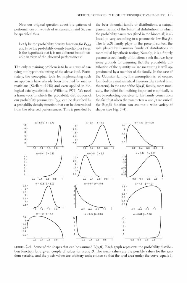
Now our original question about the patterns ofperformances on two sets of sentences, S1 and S2, canbe specified thus:
Let f1 be the probability density function for PX,S1and f2 be the probability density function for PX,S2.Is the hypothesis that f1 is not different from f2 ten-able in view of the observed performances?
The only remaining problem is to have a way of car-rying out hypothesis testing of the above kind. Fortu-nately, the conceptual tools for implementing suchan approach have already been invented by mathe-maticians (Skellam, 1948) and even applied to bio-logical data by statisticians (Williams, 1975). We needa framework in which the probability distribution ofour probability parameters, PX,S, can be described bya probability density function that can be determinedfrom the observed performances. This is provided by
the beta binomial family of distributions, a naturalgeneralization of the binomial distribution, in whichthe probability parameter (fixed in the binomial) is al-lowed to vary according to a parametric law B(�,�).The B(�,�) family plays in the present context therole played by Gaussian family of distributions inmore usual hypothesis testing. Namely, it is a finitelyparameterized family of functions such that we havesome grounds for assuming that the probability dis-tribution of the quantity we are measuring is well ap-proximated by a member of the family. In the case ofthe Gaussian family, this assumption is, of course,founded on a mathematical theorem (the central limittheorem). In the case of the B(�,�) family, more mod-estly, the belief that nothing important empirically islost by restricting ourselves to this family comes fromthe fact that when the parameters � and � are varied,the B(�,�) function can assume a wide variety ofshapes (see Fig. 7–4).
DEFICIT PATTERNS IN HIGH INTERSUBJECT VARIABILITY 113
3.5
3
2.5
2
1.5
0.5
0.2 0.4 0.6 0.8 1
1
7
6
5
4
3
1
0.2 0.4 0.6 0.8 1
2
2
4
6
8
10
12
0.2 0.4 0.6 0.8 1
0.2
0.4
0.6
0.8
1
1.2
0.2 0.4
α = 1.2 β = 1.5
α = 10.8 β = 8.03
α = 5.4 β = 0.83
α = 64.6 β = 6.79
0.6 0.8 1
0.2 0.4 0.6 0.8 1
1
2
3
4
5
3
2.5
2
1.5
0.5
1
0.2 0.4 0.6 0.8 1
0.5
1
1.5
2
2.5
3
0.2 0.4 0.6 0.8 1
2
4
6
8
10
0.2 0.4
α = 0.17 β = 6.64
α = 0.87 β = 3.88
α = 3.5 β = 9.7
α = 9.1 β = 4.5
0.6 0.8 1
0.2 0.4 0.6 0.8 1
0.2
0.4
0.6
0.8
1
1.2
0.25
0.5
0.75
1
1.25
1.75
1.5
0.2 0.4 0.6 0.8 1
0.5
1
1.5
2
2.5
0.2 0.4 0.6 0.8 1
2
4
6
8
10
0.2 0.4
α = 6.64 β = 0.18
α = 1.5 β = 1.2
α = 3.17 β = 1.85
α = 1.49 β = 4.24
0.6 0.8 1
FIGURE 7–4. Some of the shapes that can be assumed B(�,�). Each graph represents the probability distribu-tion function for a given couple of values for � and �. The x-axis values are the possible values for the ran-dom variable, and the y-axis values are arbitrary units chosen so that the total area under the curve equals 1.

In the context of the beta binomial model, we can,for each set of subject’s performance, determine thebest-fitting value for the parameters �,�. Furthermore,for the performances of a set of subjects on two typesof sentences, we can test the null hypothesis that thewhole data set comes from one distribution against thehypothesis that they come from two distinct distribu-tion. The analogy to the hypothesis-testing procedurewith which our reader is probably familiar runs thus:
1. Given the empirical data (the performances ontwo sets of sentences), one determines, for eachdata set, the best-fitting (most likely) beta func-tion, which amounts to finding two couples ofnumbers (�1, �1), (�2, �2).
This corresponds to the way one would eval-uate the (most likely) means and standard de-viations from two data sets assumed to followGaussian distributions.
2. One computes a quantity that depends on thetwo couples (�1, �1), (�2, �2).
This corresponds to the computation of thet statistic.
3. Finally, the hypothesis that the two distribu-tions are in fact equal is tested by comparingthe obtained value with the known distributionof the previously computed statistic in case thehypothesis is true.
This corresponds to the determination of aP value for the hypothesis that the means of thetwo populations are equal. This P value is ob-tained from the known distribution of the t sta-tistic when the two populations indeed comefrom the same underlying Gaussian process.
Note that a Gaussian approach is not appropriatehere, for several reasons:
• Summarizing the performances of each patientas a percentage of success gives an empirical dis-tribution of a number in the [0,1] range, whichdoes not square with the range of the normaldistribution assumption underlying the t test.
• This move is unwarranted also because thenumber of trials for different patients/studies(and even for different contrasts for a given pa-tient) is not fixed. This means that the percent-age values cannot be pooled safely.
• More conceptually, we are trying to determinethe distribution over the population of Px for agiven type of sentence X, and there is no reasonto assume a priori that this distribution is bellshaped (in fact, some of the empirical analyzes
in Drai and Grodzinsky 2006 show a best-fitting distribution that is J-shaped).
In order to specify the analytical details of thescheme, we now have to go into specific formulas,which the reader who is adverse to equations can skipto go directly to the next section, in which the methodis applied to empirical results.
Formally, under the B(�,�) model, the probabil-ity of m successes in n trials is
P(x � m�n) � � � B(� � m, n � � � m)/B(�, �);
with
B(�, �) � �(�) * �(�)/� (� � �).
The function � being defined by the integral
�(z) � �
0tz � 1 e�t dt
In order to compute the maximum likelihood parameters given the data, it is convenient to re-parameterize the distribution, setting
� � �/(� � �)� � 1/(� � �)
Then � is the expectation of the distribution, while �is a parameter that determines the shape of the curve.Under these notations the variance �2 is given by �2 �
�(1 � �)�/(1 � �) (Williams, 1975).Ignoring constants involving only the observations,
the log-likelihood for a set of results {(n1, m1) . . . (nk,mk)} is given by
(*)L(�, ��{(n1, m1) . . . (nk, mk)}) �
�k
j � 1� �
mj � 1
r � 0log[� � (r * �)]
� �nj � mj � 1
r � 0log[1 � � � (r * �)]
� �mj � 1
r � 0log[1 � (r * �)]�
Note that this likelihood depends on the full infor-mation concerning the number of trials and of suc-cesses of each subject so that no concern over undulypooling incomparable percentages arises. If {(n1 , m1). . . (nk, mk)}, represents the performances of subjects
nm
114 MATTERS LINGUISTIC

on one type of sentence and {(nk � 1, mk � 1) . . . (nk �
q, mk � q)} represents their performances on an othertype, we can proceed to test the hypothesis that theperformances are similar using the following:
If L1 is the maximal value of
L(�, ��{(n1, m1) . . . (nk, mk),(nk � 1, mk � q) . . . (nk � 1, mk � 1 q)})
and L2 is the maximal value of
L(�, ��{(n1, m1) . . . (nk, mk)})� L(�, ��{(nk � 1, mk � 1) . . . (nk � q, mk � q)})
Then an asymptotically valid test of the similarity of per-formance for the two types of tasks is given by compar-ing 2(L2 � L1) with upper percentage points of the �2
distribution with 2 degrees of freedom (Williams, 1975).We are now in position to assess whether the pat-
tern of performance of Broca aphasics on one set of sen-tences S1 is distinct from that on set S2. Given the setsof performances we can compute the � and � parame-
ters for each set of sentences and the values of L1 andL2 by maximizing the expression (*) for each set of re-sults. Our hypothesis testing now consists of computing2(L2 � L1) and asking where it stands on the �2 distri-bution with 2 degrees of freedom: if the probability ofobtaining a value as high as the one we actually obtainis very small, we must reject the hypothesis that the twosets of performances correspond to the same underly-ing distribution of values for the probability parameters.
RESULTS OF GROUP ANALYSIS
Let us now apply the contrastive scheme we describedin the preceding section and check whether despitethe variability of the performances, and hence of thebinomial probability parameter, a contrast can still beestablished. To check this, we apply the hypothesis-testing procedure of the previous section to a com-parison of performances on object relative clauses(�Movement) with subject relative clauses (�Move-ment). Figure 7–5 summarizes the situation.
DEFICIT PATTERNS IN HIGH INTERSUBJECT VARIABILITY 115
0
0.2
Object: 32 data points
0 0.2 0.4 0.6 0.8 1
Solid = Object (movement)
Dashed = Subject (no movement)
Subject: 32 data points
0.4
0.6
0.8
1
0
1
2
3
4
FIGURE 7–5. (Top) binomialconfidence interval analysis subjectby subject, as in Figure 7–3, forboth sets of performances. Dashedvertical lines correspond toperformances on subject relative clauses, and solid vertical lines, to performances on object relativeclauses. (Bottom) Correspondingbeta models probability distributedfunctions (pdf’s) for both sets ofperformances. The x-axis representsthe possible values for theprobability parameter, and the unitson the y-axis are arbitrary numbersensuring that the total area undereach curve equals 1.

Applying the computations detailed at the end ofthe last section, we obtain the following values for theparameters:
�Movement �Movement� 0.573 0.835
� 0.119 0.09
P value 2.4 � 10�8
The intuitive impression that the two sets of perfor-mances are distinct is reflected in the fact that the twocurves corresponding to the two sets of parameters ob-tained from the two sets of performances look differ-ent. Moreover, this impression can be quantified bycomputing the relevant statistic and here we obtain aP value of 2.4 � 10�8, thereby enabling us to rejectwith very high confidence the hypothesis that the per-formances for �Movement and �Movement on sen-tences with relative clauses come from the same un-derlying distribution of probabilities of success.
WAS THIS TAUTOLOGICAL?
If we allow taking the intrinsic variability of perfor-mances into account, are we not bound to be able toaccommodate any counterexample? Or, in otherwords, are we not turning the TDH (albeit under amodified quantitative form) into a tautology? Theshort answer is no. Like any other statistical discrim-ination claim, the alleged contrast between �Move-ment and �Movement sentences is open to refuta-tion in at least two ways: one could bring massivegroup evidence that points in another direction, sothat the discrimination (here between �Movementand �Movement performances) disappears after all.Alternatively, one may come up with a new, more in-teresting distinction, one that has better theoreticaljustifications and that separates the data in an evenbetter way. In any case, we do not have to remain atthis philosophical level; the TDH as tested here is notcircular, at least not in the sense of tautologicallyyielding a distinction in performances. Indeed, we canreanalyze the same data set by dividing the sentencesin an alternative manner, not informed by the con-cept of movement but current in psycholinguisticmethodology. We did, and the result is a lack of con-trast in the performances (i.e., performing the sameanalysis as before one could not reject the relevant
null hypothesis). Recall from Figure 7–1 two possibleways of categorizing the relative clause sentences usedin the experiments:
1. Through the Movement/non-Movement dis-tinction
2. Through a complexity distinction in which cen-ter embedding is contrasted with branch em-bedding
The contrasts in both cases are presented in Figure 7–6.
The results for the two contrasts are summarizedas follows:
High Low(CE) (RB) +Movement �Movement
� 0.716 0.687 0.573 0.835
� 0.16 0.17 0.119 0.09
P value 0.84 2.4 � 10�8
In brief, the �Movement-versus-�Movement con-trast yields a very significant contrast, whereas the highcomplexity–versus–low complexity contrast yields nocontrast.
CONCLUSION: TDH REPHRASED
Grodzinsky’s analysis of the mechanism that gener-ates the faulty performances of Broca aphasics in thecase of Movement sentences posits a competition be-tween two computational strategies for the assignmentof thematic roles (see Grodzinsky, this volume, Chap-ter 6 for details). Following a kind of principle of par-simony, he therefore predicted that performances forMovement sentences should be at chance level (thetwo strategies have equal influences, and all one cando is randomly guess). This prediction, in such a word-ing, is not confirmed by the data because the overallstructure of the data for all patients taken collectivelycannot be accounted by a P � 0.5 binomial. Ouranalysis shows that the TDH is confirmed by the em-pirical record, albeit in a slightly modified form: Thegroup performances on �Movement sentences is sig-nificantly poorer than that on �Movement sentences,in the sense that the distribution of probability para-meters that summarizes �Movement group perfor-
116 MATTERS LINGUISTIC
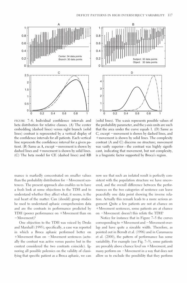
mance is markedly concentrated on smaller valuesthan the probability distribution for �Movement sen-tences. The present approach also enables us to havea fresh look at some objections to the TDH and tounderstand whether they affect what, it seems, is thereal heart of the matter: Can (should) group studiesbe used to understand aphasic comprehension dataand are the contrasts in performance predicted byTDH (poorer performance on �Movement than on�Movement)?
One objection to the TDH was voiced by Druksand Marshall (1995); specifically, a case was reportedin which a Broca aphasic performed better on�Movement than on �Movement sentences (actu-ally the contrast was active versus passive but in thecontext considered the two contrasts coincide). Ig-noring all possible polemics on the validity of classi-fying that specific patient as a Broca aphasic, we can
now see that such an isolated result is perfectly con-sistent with the population structure we have uncov-ered, and the overall difference between the perfor-mances on the two categories of sentence can leavepeacefully one data point showing the inverse rela-tion. Actually this remark leads to a more serious ar-gument: Quite a few patients are not at chance on�Movement sentences, some patients are at chanceon �Movement: doesn’t this refute the TDH?
Notice for instance that in Figure 7–5 the curvescorresponding to �Movement and �Movement over-lap and have quite a sizeable width. Therefore, aspointed out in Berndt et al. (1996) and in Caramazzaet al. (2000), the pattern of performance has somevariability. For example (see Fig. 7–3), some patientsare provably above chance level on �Movement, andmany perform on �Movement in a way that does notallow us to exclude the possibility that they perform
DEFICIT PATTERNS IN HIGH INTERSUBJECT VARIABILITY 117
0
0.2 Branch: 30 data points
Center: 34 data points
0.4
0.6
0.8
A1
00 0.2 0.4 0.6 0.8 1
0.5
1
1.5
2
C2.5
0
0.2Object: 32 data points
Subject: 32 data points
0.4
0.6
0.8
B1
00 0.2 0.4 0.6 0.8 1
1
2
3
4
D
FIGURE 7–6. Individual confidence intervals andbeta distribution for relative clauses. (A) The centerembedding (dashed lines) versus right branch (solidlines) contrast is represented by a vertical display ofthe confidence intervals for all patients. Each verticalline represents the confidence interval for a given pa-tient. (B) Same as A, except �movement is shown bydashed lines and �movement is shown by solid lines.(C) The beta model for CE (dashed lines) and RB
(solid lines). The x-axis represents possible values ofthe probability parameter, and the y-axis units are suchthat the area under the curve equals 1. (D) Same asC, except �movement is shown by dashed lines, and�movement is shown by solid lines. The complexitycontrast (A and C) discerns no structure; movementwas vastly superior—the contrast was highly signifi-cant, indicating that movement, but not complexity,is a linguistic factor supported by Broca’s region.

at chance level. So indeed, if one hoped to summa-rize all performances by a single binomial parameter,or through the dichotomy at chance/above chance, thesituation looks hopeless. This fact has led some to con-test the value of group studies and to argue that thevariability is so great that the results are meaningless.In the present context, this is seen to be a much tooextreme conclusion. If we try to quantitatively char-acterize the variability in performance—as we didhere—we are led to an empirical assessment that notonly gives meaning to group study data but can ONLYbe performed in the context of group study data. Ifthere is some intrinsic variability in the performancesof patients, the only way to still assess whether thereis a pattern in the performances is to compare thecurves that describe the varying performances and toassess whether it is reasonable to assume that theycome from the same underlying process.
The logical conclusion of our analysis is that thedichotomy between �Movement and �Movementcaptures an aspect of linguistic processing that cru-cially involves Broca’s area. The assumptions under-lying the stochastic model that enables the hypothesistesting procedure do, however, leave open a questionabout the source of the variability of performance.
Here we must confess to a relative ignorance ofthe precise mechanisms that create the variability. Ob-viously the size and severity of the lesions are variable,and the brains within which they occur are variableto start with. Moreover, we do not know if there is asystematic structure to the variability itself. For in-stance, is the variance dependent (in an interestingway) on the kind of linguistic construct in whichMovement is studied? The kind of explanation pro-posed originally in the TDH, that is, the existence ofa computational strategy orthogonal to syntax, whichtakes over when syntax fails, could be modified to ac-commodate a model with variability, for instance bypostulating that given the conflicting informationfrom syntax and from the general strategy, each indi-vidual brain weights the sources of information dif-ferently. But this is purely programmatic. The theoryat its present stage is not entirely mute about imple-mentation, though; it implies that whatever the im-plementation of the processing of sentences turns outto be, it must have the property of degrading in a waythat yields the observed stochastic behavior when de-prived of one of its components (the one correspond-ing to Broca’s area).
ACKNOWLEDGMENTS Dan Drai’s research at theWeizmann Institute of Science is supported by the Goldaand Dr. Yehiel Shwartzman and by a Sara and HaimMedvedi Families Postdoctoral Fellowship.
Notes
1. The references to the study used in compilingthe complete data matrix appear as an appendix in Draiand Grodzinsky (2006).
2. For example, Boston Diagnostic Aphasia Exam-ination, Western Aphasia Battery, Aachen Aphasia Test,and others.
References
Berndt, R.S., Mitchum, C.C., & Haedinges, A.N. (1996).Comprehension of reversible sentences in “agram-matism”: A meta-analysis. Cognition, 58, 289–308.
Caramazza, A. (1986). On drawing inferences about thestructure of normal cognitive processes from pat-terns of impaired performance: The case for single-patient studies. Brain and Cognition, 5, 41–66.
Caramazza, A., Capitani, E., Rey, A., & Berndt, R.(2000). Agrammatic Broca’s aphasia is not associ-ated with a single pattern of comprehension per-formance. Brain and Language, 76, 158–184.
Drai, D., & Grodzinsky, Y. (2006). Stability of functionalrole in Broca’s region: Quantitative neurosyntacticanalysis of a large data set from Aphasia Brain andLanguage, 96, 1.
Druks, J., & Marshall, J.C. (1995). When passives areeasier than actives: Two case studies of aphasic com-prehension. Cognition, 55, 311–331.
Grodzinsky, Y. (1986). Language deficits and the theoryof syntax. Brain and Language, 27, 135–159.
Grodzinsky, Y. (1990). Theoretical perspectives on lan-guage deficits. Cambridge, MA: MIT Press.
Grodzinsky, Y. (2000). The neurology of syntax: Lan-guage use without Broca’s area. Behavioural BrainSciences, 23, 1–21.
Grodzinsky, Y., Pinango, M.M., Zurif, E., & Drai, D.(1999). The critical role of group studies in Neu-ropsychology: Comprehension deficits in Broca’saphasia. Brain and Language, 67, 134–147.
Skellam, J.G. (1948). A probability distribution derivedfrom the binomial distribution by regarding theprobability of success as variable between the setsof trials. Journal of the Royal Statistical Society. Se-ries B (Methodological), 10.2, 257–261.
Williams, D.A. (1975). The analysis of binary responsesfrom toxicological experiments involving reproduc-tion and teratogenicity. Biometrics, 31.4, 949–952.
118 MATTERS LINGUISTIC

8
Treating Language Deficits inBroca’s Aphasia
Lewis P. Shapiro
Cynthia K. Thompson
This chapter describes a research program investigat-ing treatment of sentence production and compre-hension deficits found with individuals who haveBroca’s aphasia. The purpose of this program is to in-vestigate the efficacy of a treatment approach that isbased on evidence from a variety of sources, includ-ing linguistic, psycholinguistic, and neurolinguisticinvestigations. Our intent also is to use treatment andrecovery data to reveal brain–language relations. Toget to the approach and its fruits, we need to take youthrough its empirical origins. We then describe, inrelative detail, the treatment approach and its experi-mental products.
Our interest in how linguistic theory and psy-cholinguistic experimentation can inform learning,and therefore treatment, began empirically on twoseparate fronts. One of us (Shapiro and colleagues)ran a small experiment that examined how argumentstructure might serve as a point of connection betweenlinguistic and nonlinguistic conceptual information(Canseco-Gonzalez et al., 1990, 1991). Our singleparticipant had incurred a stroke that yielded a left
hemisphere lesion involving most of Broca’s area, theinsular region, and putaminal area with superior ex-tension into premotor, motor, and sensory cortical ar-eas.1 He evinced severely nonfluent speech as well assentence comprehension difficulties on noncanonicalstructures, that is, on sentences where the direct ob-ject noun phrase had been displaced from its typicalpost-verb position (e.g., passives, object relatives). Wetrained our participant on c-VIC, a visually based,computerized artificial language (see, for example,Kleczewska et al., 1987).2 We taught our subject toassociate either one or two visual symbols that corre-sponded to the natural language equivalent of “sen-tences” containing either obligatory two-place or op-tional three-place predicates.
We found that the ease with which c-VIC symbolswere learned was predicted by the number of argu-ment structure entries for natural language verbs thatequivalently referred to the depicted actions. So, forexample, our subject performed better when learningtwo symbols for the possibilities inherent with naturallanguage dative verbs (those that allowed either a two-
119

place or three-place possibility) than for learning thesymbols that represented the natural language equiv-alent of pure two-place verbs (with or without ad-juncts). Additionally, we found that “verbs” that al-lowed an optional third argument yielded betterperformance when they were inserted in more com-plex, three-argument “sentences” than when insertedin two-place equivalents. Our interpretation of thesefindings suggested that knowledge of argument struc-ture might be spared by the damage underlying Broca’saphasia because it serves a translation function be-tween language and conceptual structure, the com-putation of the latter which is considered to be intact.The work also suggested that the sparing of argumentstructure might be exploited in developing treatmentprograms for sentence-level deficits in aphasia.
At approximately the same time that we were in-vestigating the processing of argument structure inaphasia (see Sentence Processing, for more details),Thompson and Wambaugh (1989) were directly ini-tiating investigations of treatment for the sentence pro-duction difficulties observed in Broca’s aphasia. In theinitial report, a treatment experiment was describedwhereby Broca’s aphasic individuals were trained onwh-question production, a problematic structure forthese individuals (baseline levels of performance ap-proached 0% correct). It was found that training a setof what-questions generalized to untrained what-questions but did not generalize to untrained when-questions. It became apparent to both of us that thislack of cross-generalization could be explained by theargument structure properties of the verbs used toform the two question types. The verbs used for whatquestions, for example, were two-argument verbs likeeat, whereas those used to generate where questionswere one-argument verbs like sleep. Simply put, be-cause of the verb’s argument structure properties,movement in who questions involves displacement ofan argument of the verb (e.g., a direct object); that in-volved in where questions involves movement of anadjunctive phrase (e.g., a locative prepositionalphrase). This result led us to surmise that generaliza-tion should be forthcoming if the questions trainedand those tested for generalization were structurallysimilar. Later, we flesh out what is meant by “struc-tural similarity.” More generally, we predicted thatcontrolling for those lexical and syntactic propertiesthat form the basis for language processing in neuro-logically intact as well as individuals with brain dam-age could result in efficacious treatment for the lan-guage disorders inherent in Broca’s aphasia. Keeping
these initial findings in mind, we now fill in some ofthe details of the background to our treatment work.
LINGUISTIC BACKGROUND
In most theories of grammar, there is an intimate re-lation between lexical representations and syntax. Thisrelation as well has been the focus of much work inpsycholinguistics and investigations of brain language.
Argument Structure
The information that seems most clearly relevant tothe interaction between lexical properties and syntaxis argument structure. Borrowing from logic, a sen-tence can be considered analogous to a proposition,which can be organized into a set of arguments that“revolve around” an event described by the predicate.In its essence, argument structure simply describes thenumber and type of participants entailed by, typically,a verb. For example, consider the English verb defeat:
1. Kerry will defeat Bush in November.In example 1, there are two NPs—subject
and complement (direct object)—that con-tribute to the event described by the verb. Theremainder of the material—the prepositionalphrase—although adding to the syntax and in-terpretation of the sentence, does not form partof the verb’s contribution and is therefore con-sidered an adjunct. Thus, the verb defeat is saidto be a two-place predicate.
There is a limited range of argument structures al-lowed in English as well as in other languages. Con-sider the following examples:
2. Joelle slept easily last night. Sleep is a one-place(intransitive) predicate, where the subject NPis the sole argument selected by the verb.
3. Zack discovered that the ball went over the fence.Discover, in this case, is a two-place predicatewith the second argument syntactically realizedas a complement phrase.
4. Dillon threw/sent/lent the ball to the umpire.These verbs allow three arguments, with thethird argument contained within the preposi-tional phrase.
Arguments can be required or optional. For example,in Dillon threw the ball, there are only two arguments
120 MATTERS LINGUISTIC

specified by the verb. In this case, it could be arguedthat the verb throw is an optional three-place verb, andin its present realization only two of the argumentsare apparent. On the other hand, verbs like put andgive require three arguments for a sentence to be wellformed, as in Joelle put the makeup on the shelf (*Joelleput the makeup) and Dillon gave the ball to the um-pire (*Dillon gave the ball).
Further consideration of the argument structuresfor sentences 1 through 4 reveals that the argumentsplay different semantic roles in relation to their verbs.So, in example 1, the subject argument lexically filledby the NP Kerry is playing the role of “agent of theaction” and the direct object argument Bush is play-ing the role of “patient” or “theme.” In example 2,the subject argument Joelle is the “experiencer” of theaction described by the verb. In example 3, the clausalargument that the ball went over the fence is a “propo-sition.” In example 4, the NP argument the umpirethat is embedded in the prepositional phrase to theumpire plays the role of the “goal” of the action.
The array of these roles, idiosyncratically selectedby each verb, forms the verb’s thematic grid. The verbdefeat bears an (agent theme) grid; the verb give an(agent theme goal), the verb discover an (agent propo-sition), and so on. Within the principles and param-eters framework (see Chomsky and Lasnik, 1993;Haegeman, 1994; Radford, 1997), thematic informa-tion interacts with syntax via theta theory. Briefly, alexical head (e.g., verb) assigns its thematic roles toargument positions in the sentence in its underlyingrepresentation. So, for example, considering the verbgive and its thematic grid (agent theme goal), agentis assigned to the subject argument position, theme isassigned to the direct object position, and goal is as-signed to the oblique object position. In this way, the-matic information forms a lexically based constrainton well-formedness. That is, if all of the thematic rolesfor a particular verb are discharged to their appropri-ate argument positions, the sentence that is generatedis grammatical; if they are not, or if there is an argu-ment position where a thematic role is not assigned,the sentence that is generated is ill formed.
In more recent accounts based on minimalist con-siderations (Chomsky, 1995; 1998, see also Adger,2003), there is no role for modules or subtheories ofany sort; the architecture of the grammar is said to bedriven by methodological economy (i.e., fewer prim-itives, relations, and assumptions). A basic syntacticoperation is Merge, which selects an item from thelexicon that has a rich set of features (a lexical array)
and combines it with another such selected item,yielding a higher-order category. Simplifying, a seriesof such merger operations builds the syntactic struc-ture of the sentence.
Still, the facts that arguments can play any one ofa number of thematic roles, and that verbs idiosyn-cratically select a set of such roles, should be under-stood within the grammar. Thus, we might claim thattheta-assignment occurs as part of merge (Radford,2004). Consider, then, how thematic roles are as-signed in a simple SVO structure, exemplified as Kerrywill defeat Bush:
5. VP
DPAgent
V�
KerryV DP
Theme
Defeat Bush
Here, as V is merged with DP to yield V�, the thetarole of theme is assigned to the direct object argumentposition; as V� is merged with DP to form a VP, thetheta role of agent is assigned to the subject (note thatthe subject is internal to the VP and raises to the spec-ifier position of TP, which we discuss later). The VPin example 5 is merged with a T� constituent, and soon up the tree.3 In some extended accounts of the lex-icon, thematic information is considered an abbrevi-ation for a more richly specified conceptual or eventstructure in which the verb plays a central role (e.g.,Jackendoff, 1990; Pustejovsky, 1995).
Movement
Within the theory we are considering here, sentencesare derived from two operations: one, described above,is merge: two categories merge to yield a higher-ordercategory, and a series of such merge operations buildsconstituent structure or the syntactic tree (see Fried-mann, this volume, for details of the tree, particularthose that have relevance for accounts of productionin agrammatic Broca’s aphasia). It was suggestedabove that thematic role assignment is part of themerge operation, allowing lexical features to perco-late to syntax. The second operation is move. It has
TREATING LANGUAGE DEFICITS IN BROCA’S APHASIA 121

long been recognized that there are three subtypes ofmovement. One is head movement (seen in Englishin “subject-Aux inversion”; e.g., “Dillon is playing soc-cer” � “Is Dillon playing soccer”?); the others are wh-movement (or A-bar movement, written as A�; in Eng-lish object-extracted wh-questions, clefts, and objectrelatives) and NP- or A-movement (seen in Englishpassives, unaccusatives, and subject raising). In thefollowing we briefly describe the latter two movementoperations, most relevant to the empirical work de-scribed later in this paper.
wh-Movement (A� Movement)
Consider the following sentences:
6. The soldier pushed the woman7. Which woman did the soldier push?
It is apparent that examples 6 and 7 are related. Eachsentence contains the same two-place predicate withthe same lexical items filling the argument slots (ig-noring the wh-word in example 7). The NP playingthe agent in the event in example 6 plays the samerole in example 7; the NP playing the role of patientplays that same role in both sentences as well. Giventhe relation between these constructions, it is likelythat the question form in example 7 is somehow de-rived from the underlying event stated in example 6.One way of describing this relation is to suggest that,in the question form, the direct object argument istransferred from its base position occurring after theverb to a position higher in the syntactic tree:
8. CP
8. DP C�
C TP
DP T�
T VP
DP V�
V DP
Which did the did the push whichwoman soldier soldier woman
The structure in example 8 shows the derivationof the wh-question. The complement of push—whichwoman—is represented in its base, direct object, po-sition. It moves to Spec-CP, and as a result it leavesa clone or copy of itself, also known as a trace in theprinciples and parameters framework. The thematicrole of theme is assigned to the complement positionvia merge of V and DP. The subject DP—thesoldier—raises out of VP into the Spec-TP position(for reasons not germane to the current discussion);agent is assigned to the subject constituent via mergeof DP with V�. The copies of the moved constituentsare deleted (or rather, given a null status phonetically)before they are sent off to the level of representationconcerned with phonetic form.4
NP-Movement (A Movement)
The other type of move operation relevant to the workdescribed here derives passives as well as raising con-structions. Consider the passive:
9. TP
DP T�
T VP
V� PP
V DP
The woman was pushed the woman by the soldier
Here, the complement of the verb push—the woman—is moved to Spec-TP from its canonical complementposition, leaving a copy that is subsequently deleted.Thus, an argument is moved to another argument po-sition, unlike the case of wh-movement described inexample 8, where the landing site for movement is anonargument position (Spec-CP).
Interim Summary
In this section we have described some basic linguis-tic notions (admittedly skipping many details) thatturn out to have important implications for the treat-ment work that we soon discuss. Argument structure
122 MATTERS LINGUISTIC

is a type of lexical property (or set of features; e.g., the-matic roles) that characterizes the number and typesof participants in an event described by the verb. Thisproperty interacts with the syntax, particularly the syn-tactic tree, and thus places constraints on the well-formedness of sentences. Sentences are derived fromthe output of two operations: merge, which takes twosyntactic objects selected from the lexicon, combinesthem and yields a higher-order category, and move,which displaces a category to another position in thesyntactic tree. In the following, we briefly describe theprocessing consequences of these linguistic operations.
SENTENCE PROCESSING
In a series of online experiments, we have found thatnormal listeners—when encountering a verb duringthe temporal unfolding of a sentence—exhaustivelyactivate their argument-taking properties (e.g., Shapiroet al., 1987, 1989, 1993). Once active, these proper-ties are then used by the processing system—perhapsduring a secondary routine—to establish thematic re-lations among the arguments in the sentence, allow-ing for final interpretation. Indeed, regardless of theoretical orientation, most sentence processing ac-counts claim a privileged role for the constraints es-tablished by argument structure in both sentence pro-duction and comprehension (e.g., Bock, 1995; Bockand Levelt, 1994; Levelt 1999; Shapiro et al., 1991,1993; Trueswell and Kim, 1998).
Wernicke’s aphasic individuals, those who havetypically incurred damage to posterior cortical re-gions, do not show normal online sensitivity to theseargument structure properties, suggesting that theconceptual-semantic aspects of these lexical prop-erties influence the types of “semantic-like” deficitsobserved in these individuals. Yet, Broca’s aphasicindividuals perform just like the neurologically in-tact population; that is, when they encounter a verbin a sentence, they activate its argument structureproperties.
There is also a large body of evidence suggestingthat normal listeners/readers respect movement oper-ations (e.g., Garnsey et al., 1989; Hickok et al., 1993;Nicol and Swinney, 1989; Shapiro and Hestvik,1995). In a series of online experiments, Swinney andcolleagues (1987) have shown that a moved comple-ment is reactivated at its base position. In sentenceslike “The policeman saw the boy who the crowd at
the party accused of the crime”, where the comple-ment of accused (i.e., the boy) has been moved up thetree to Spec-CP and leaves a copy in the direct objectposition, listeners show activation for the moved ar-gument at the copy position (and not at a position be-fore the copy). We have recently found such “gap-filling” effects in wh-questions (Shapiro and Thomp-son, in preparation), VP-ellipsis constructions (Shapiroand Hestvik, 1995; Shapiro et al., 2003), and NP-movement constructions (Friedmann et al., submitted).
Importantly, in constructions derived by move-ment, individuals with Broca’s and Wernicke’s apha-sia show the reverse pattern observed with argumentstructure. Broca’s aphasic individuals do not show nor-mal “gap-filling” effects, yet those individuals withWernicke’s aphasia do (e.g., Zurif et al., 19935). Thisdouble-dissociation—that individuals with Broca’saphasia show normal lexical processing routines in-volving argument structure but aberrant structuralroutines involving movement, and those with Wer-nicke’s aphasia show disordered lexical processing butrelatively normal online structural routines—stronglysuggests that lexical and structural processing are in-dependent. And, indeed, there is a recent neuro-imaging evidence to support this possibility. Ben-Shachar et al. (2003) found that Broca’s region (leftinferior frontal gyrus) is activated during processing ofmovement structures, yet argument structure com-plexity revealed activation only in left posterior su-perior temporal sulcus. Thompson et al. (submitted) reported a similar finding, comparing activation pat-terns for verbs by argument structure. Verbs with agreater number of arguments, such as three-argumentversus two- or one-argument verbs, yielded bilateralposterior, superior temporal gyri/sulci activation.These findings support the deficit patterns seen inaphasic patients.
It is now a well-established fact that individualswith Broca’s aphasia have difficulty understandingand producing sentences that are “noncanonical.” Amore precise definition of what that means inevitablybrings us to detailed aspects of movement (see, for example, Grodzinsky, Friedmann, this volume).Grodzinsky has described a detailed account (thetrace-deletion hypothesis [TDH]) that explains a hostof sentence comprehension patterns evinced byBroca’s aphasic individuals involving constructionsderived by movement. Remediation of sentence-leveldeficits requires a treatment program that takes theselinguistic notions seriously. Before we describe such
TREATING LANGUAGE DEFICITS IN BROCA’S APHASIA 123

a program, we briefly ground our work in the extanttreatment literature.
TREATMENT OF LANGUAGE DEFICITS
IN APHASIA: A BRIEF REVIEW
Our intention, then, was to develop a treatment pro-gram based on linguistic theory, psycholinguistic find-ings, and what we knew about the deficit patterns inaphasia, and critically, we also required that this pro-gram be testable using the right kinds of experimen-tal designs (Shapiro and Thompson, 1994; Thomp-son and Shapiro, 1994). The literature on recoveryand treatment of sentence deficits resulting from braindamage prior to the initial Wambaugh and Thomp-son (1989) effort consisted primarily of case studiesand uncontrolled group studies (and to some extent,this continues to be the case). Very few studies con-sidered the relevance of linguistic or psycholinguisticvariables, even broadly defined. One exception is anapproach developed by Schwartz et al. (1994; see alsoByng, 1988; Jones, 1986), based on the “mapping hy-pothesis,” which holds that agrammatic performancereflects an impaired mapping between grammaticalconstituents (subject, object) and thematic roles(agent, theme) (Schwartz et al., 1987). Using color-coded materials corresponding to sentence con-stituents, participants were trained to recognize thethematic roles of noun phrases set around the verbsin sentences in both canonical (active NP-V-NP sen-tences) and noncanonical sentences by responding toquestions of the form “Who did what to whom.” Treat-ment outcomes were mixed, with generalizationwithin sentence types claimed for some participants,and sparse evidence for generalization across sentencetypes.
Although the intent of these studies was laudable,the experimental designs have fallen short of those re-quired in a controlled treatment study. For example,simple pretest, posttest designs are often implemented(e.g., Schwartz et al., 1994; for a more recent exam-ple, see Beveridge and Crerar, 2002), or the case studyapproach is taken; neither can unambiguously at-tribute any potential gains to the treatment itself. Byng(1988), for example, presented case studies for twosubjects, reporting only pretreatment and posttreat-ment data, without control for potentially extraneousfactors. Further, in the Schwartz et al. program, a pre-training phase was instituted whereby patients were
trained to understand wh-questions necessary for thesubsequent training phase. But as noted by the in-vestigators, wh-questions are problematic for these pa-tients. Indeed, according to the “mapping” hypothe-sis, the same deficit contributes to the problems thesepatients have in understanding wh-questions and thesentences that were targets for treatment; that is, bothsentence types are derived from noncanonical the-matic role assignment. Yet to reap the benefits of the treatment, the patients had to understand the wh-questions in the pretraining phase.
Another treatment program that has consideredlinguistic variables was “Cueing verb treatment”(CVT) (e.g., Loverso et al., 1986). This program usedcase grammar (Fillmore, 1968) as a basis for treatmenttargeting production of simple sentence structures.Case grammar, whose constructs found their way intomore current approaches to lexical representation,considers the verb as the “motor” of the sentence’spropositional structure. Loverso et al. designed a treat-ment program that first trained the verb and then, ina series of graduated steps, attempted to expand theverb to include both its subject and, depending onthe type of verb, either its object or a location, adver-bial of time, instrument, etc. The sentence expansionwas produced by clinician-controlled use of wh-words(e.g., “who run” written on index cards yields patient-initiated “I run,” “who run when” yields “I run yes-terday,” “who hit” yields “I hit,” etc.). The investiga-tors claimed generalization because of increases onformal aphasia test measures (although, so far as weare aware, generalization to untrained verb and verb-argument combinations was not assessed in their ex-perimental design. Whether these subjects wouldshow such generalization remains unanswered).
The generalization issue aside, from our presentperspective Loverso and colleagues (1986) should becommended for devising perhaps the first treatmentprogram that considered the influence of lexical prop-erties on sentence generation. Their approach, how-ever, was limited to the production of simple sen-tences, and, not unlike the Schwarz et al. program,the use of wh-words to elicit the simple sentences wasquite likely to be problematic for some participants.And although their materials were controlled for fre-quency of occurrence, imagery, and concreteness, theverbs themselves were not controlled for the numberand types of arguments they entailed. Again, we knowfrom the psycholinguistic literature that the lexicalproperties of verbs have direct consequences for syn-
124 MATTERS LINGUISTIC

tax, and for sentence comprehension and production,and thus it may be necessary to control for these prop-erties when devising treatment programs.
So, to the best of our knowledge, the treatment lit-erature contains few instances of studies that have se-riously considered the linguistic and psycholinguisticunderpinnings of sentence targets, and those that havehave used research designs that limit the generaliz-ability of any positive results. As you will soon see,there is some underlying similarities between our ap-proach and the mapping/cueing verb treatment ap-proaches described earlier. One important similarityis the reliance on verb-argument structure. However,one critical difference is the acknowledgment ofmovement operations involved in the derivation ofnoncanonical structures, as Grodzinsky and col-leagues have so clearly shown may be at the heart ofthe sentence interpretation patterns evinced byBroca’s aphasic individuals. With these rather lengthypreliminaries out of the way, we now describe ourtreatment program.
TREATMENT OF UNDERLYING FORMS
We have labeled our treatment program “Treatmentof Underlying Forms” for good reason: We take ad-vantage of the underlying, abstract, properties of lan-guage. We assume that training such properties willallow for more effective generalization. The proper-ties we exploit are lexical—argument structure—thatappears to be spared in some individuals with agram-matic Broca’s aphasia, and syntactic—movement—that is compromised in these same individuals. Ourtreatment investigations use small numbers of subjectsbut with controlled experimental designs; we assess,and find, generalization to untrained structures thatshare similar underlying properties to those structuresthat are trained, and in some cases we find general-ization to overall language performance as well.
Our first collaborative empirical effort (Thompsonet al., 1993) sought to accomplish two things: (1) es-tablish pilot data using the treatment of underlyingforms approach and (2) examine if training one typeof question would generalize to another type of ques-tion if we controlled for verb argument structure andthe type of movement involved. We began with twoparticipants; each suffered a single left-hemispherecerebrovascular accident. Both participants were di-agnosed with nonfluent aphasia with agrammatism.
Briefly, sentence production was characterized byshort phrases and simple sentences consisting pri-marily of major lexical categories, with inflectionalmorphemes compromised. Using elicited production,both participants could produce simple SVO sen-tences relatively accurately (again, with inflectionalmorphology compromised) but were unable to pro-duce grammatically correct wh-questions or passiveconstructions. Auditory comprehension of active SVOconstructions was above-chance levels, and compre-hension of reversible passives was at chance.
We examined whether training who-questionswould generalize to untrained who- and untrainedwhat-questions (and vice versa), and whether trainingwh-questions based on more complex underlyingforms (NP-V-NP-PP; e.g., “What is the man giving tothe boy?”) would generalize to wh-questions based onless complex forms (NP-V-NP; e.g., “What is the manfixing?”). A single-subject multiple-baseline across be-haviors and subjects was used to examine acquisitionand generalization effects of treatment. Subject 1 wastrained to first form what-questions, and subject 2 wastrained to first form who-questions.6
Baselines of one subject were extended into thetreatment phases of the other subject, and, for eachsubject, one question type was trained at a time to al-low us to examine generalization to the other ques-tion form. This type of design—a multiple baselineacross subjects (and behaviors)—allows one to estab-lish effects that can be unambiguously assigned totreatment variables rather than to extraneous, uncon-trolled variables. That is, in the multiple baselineacross subjects approach, treatment is initiated foreach participant after baselines varying in length. Ifstable baselines are observed for participant 2 duringthe time participant 1 is undergoing treatment, andthen treatment is instituted for participant 2 and in-creased performance is observed, a good argumentcan be made that it is the treatment itself that is re-sponsible for the change in behavior. The multiplebaseline across behaviors portion of the design in-volves training one question form at a time for eachparticipant. In all cases, probes designed to assess gen-eralization to untrained versions of the same behav-ior (e.g., complex what-questions for subject 1; com-plex who-questions for subject 2), and untrainedversions of the other behaviors (e.g., simple and com-plex what-questions, and simple and complex who-questions), were assessed throughout treatment. Inthis way, we could scrutinize individual subject data,
TREATING LANGUAGE DEFICITS IN BROCA’S APHASIA 125

carefully inspecting the relation between trained anduntrained question types.
We predicted that training what-questions wouldgeneralize to untrained who-questions and vice versabecause both structures are derived from wh-move-ment, and their only difference is the wh-morphemethat heads the question form. In this initial effort, wefound that some of our predictions were borne out.First, we observed that both participants evinced in-creased performance on wh-questions after treatment.At baseline, neither subject could produce well-formed wh-questions. Participant 1 showed 2% cor-rect production of wh-questions at baseline with 68%of the sentences produced consisting of rising into-nation without the appropriate wh-morpheme, 22%consisting of utterances with no wh-morpheme andno evidence of movement, and 9% including the cor-rect wh-morpheme but no movement. At posttreat-ment, this participant produced 82% correct. Partici-pant 2 showed similar performance, with only 2%correct at baseline and 96% at posttreatment. Second,and more specifically, when treatment was applied towhat-questions for participant 1, generalization to un-trained exemplars of what-questions was observed.However, acquisition of who-questions was not ob-served until treatment was applied to those structures,not supporting our prediction. For participant 2, how-ever, training of who-questions resulted in general-ization to untrained what-questions, supporting ourprediction. In addition, this participant showed gen-eralization from complex to simple exemplars of bothwho- and what-questions. This latter finding encour-aged us to move forward in our treatment work.
Training Movement of Arguments Versus
Movement of Adjuncts
Following up on the initial study just described(Thompson et al., 1993), we further examined the relation between those wh-questions that requiredmovement of an argument and those that requiredmovement of an adjunctive phrase (Thompson et al.,1996). Recall that who- and what-questions refer to,typically, a moved direct object argument of the verb(as in “The soldier pushed the woman into the street.Who did the soldier push ______ into the street?”),while, for example, where-questions refer to move-ment of an adjunct (as in “The soldier pushed thewoman into the street. Where did the soldier push thewoman ______?”). Seven individuals with aphasia
participated in the experiment. All incurred a singleleft-hemisphere infarct; five became aphasic sec-ondary to a cerebrovascular accident in the distribu-tion of the left middle cerebral artery, one suffered aleft middle cerebral artery from a ruptured aneurysmin the temporoparietal region, and one had a mass ex-cised from the left frontal lobe. The results of a seriesof formal and informal tests revealed that productionwas more compromised than comprehension and thatspeech was nonfluent, consisting of primarily shortphrases and sentences, most of them simple structuresthat were, nevertheless, ungrammatical. In an analy-sis of verb-argument structures produced in discoursesituations, significantly fewer verbs and less complexargument structures relative to normal controls wereobserved. Sentence comprehension was compro-mised as well. For example, all the participants per-formed from 70% to 100% correct on active SVO sen-tences as well as on subject relatives; on passives andobject relatives, they performed from 50% to 70% correct.
A single-subject multiple-baseline design acrossbehaviors was used to examine generalization patternsacross wh-questions. As one structure was treated, gen-eralization to the other structures was examined. Foreach participant, once the initial structure reachedcriterion, treatment ensured for a second structure.The order of wh-questions entered into treatment wascounterbalanced across the participants. For example,participant 1 was first trained on when-questions thatrely on movement of an adjunct, and generalizationwas tested to untrained where-questions that also re-lied on movement of the adjunct and to untrainedwho- and what-questions, both of which rely onmovement of the direct object argument. In a secondphase, what-questions were entered into treatment,and generalization to untrained who-questions was as-sessed. For participant 2, who-questions were firsttrained and generalization to untrained what-, where-,and when-questions was assessed. In the second phasefor participant 2, when-questions were entered intotreatment and generalization to untrained where-questions was assessed, and so on.
We predicted that training sentences derived frommovement of an argument (e.g., who-questions)would only generalize to untrained material using adifferent set of wh-questions (e.g., what-questions) thatalso relied on movement of an argument, and not tosentences derived from movement of an adjunctphrase (e.g., when- and where-questions). Relatedly,
126 MATTERS LINGUISTIC

we predicted that training sentences derived frommovement of an adjunctive phrase would not gener-alize to those derived from movement of an argument.
Overall results indicated that for every participant,treatment yielded improved production significantlyabove baseline levels for all wh-questions entered intotreatment. Further observation revealed two patternsof results. First, three participants evinced patterns inline with our initial predictions. When these individ-uals were trained on wh-questions derived from move-ment of an argument, generalization to untrainedforms was restricted to those materials that also relyon movement of an argument. A similar pattern wasobserved within questions derived from movement ofadjunctive phrases. Importantly, generalization acrossstructures derived from movement of an argumentand movement of an adjunct did not occur. Interest-ingly, these participants’ error patterns revealed a cor-rect wh-question pattern but with what has beencalled a “filled gap” (e.g., when required to respondwith “Who is the soldier pushing into the street?” aparticipant would attempt “Who is the soldier push-ing the woman”). Referring back to the LinguisticBackground section, it seems that the participants may have failed to delete the copy of the moved constituent.
A second pattern was revealed for four other par-ticipants. Training of a particular question (e.g.,when-questions) yielded generalization to untrainedmaterials of the same question type but did not gen-eralize to untrained materials of other question types,whether they were derived by the same type of move-ment. An examination of the error patterns was quiterevealing; these participants primarily evinced errorsof wh-morpheme selection (e.g., instead of producing“who is the soldier pushing?” they would produce“where is the soldier pushing?”), while at the sametime reducing errors of co-reference that were ob-served during baseline testing. We then entered theseindividuals into supplemental wh-morpheme dis-crimination and production training. Once initiated,these individuals evinced generalization to all wh-question forms, and their wh-morpheme selection er-rors significantly decreased there.
The story does not stop there. For three of thesefour individuals, if we ignored errors of wh-morphemeselection, the patterns revealed that training wh-questions relying on movement of an argument didindeed increase performance on other untrained wh-questions relying on argument movement, and later
adjunct movement improved (and vice versa). Thatis, the within-question type generalization observedfor the first three participants was also revealed inthese other three participants, but the latter’s perfor-mance was masked by the errors of wh-morpheme selection. These two patterns suggest two putativelydistinct operations involved in the production of wh-questions. One has to do with processing routines en-abling co-reference; the other has to do with selectionof the correct lexical item that fills the functional cat-egory. Regarding the latter, it is interesting to note thatmore than 10 years ago, Grodzinsky (1990) suggestedthat sentence production deficits observed in agram-matic Broca’s aphasia might be partially explained bya deficit to the features inherent in functional cate-gories and that such individuals randomly select itemsto fill these categories.
Finally, in this study (and in others as well), weexamined pretreatment and posttreatment discoursepatterns. We found significantly improved sentenceproduction across the board. Our participants pro-duced significantly more verbs post-relative to pre-treatment, more correct argument structures, moreadjuncts, a great number of wh-questions, a greaterproportion of complex sentences, and more embed-dings per utterance. One interpretation of these im-pressive gains is that because our training program tar-gets lexical and structural properties that are involvedin many aspects of sentence production, significantand pervasive changes may occur to the more broadlyconceived language system.
Wh- (A� Movement) Versus NP-Movement
(A Movement)
As described previously, a distinction can be made between two types of movement. One type is Wh-movement, which displaces a complement from itsunderlying position after the verb (in English) to Spec-CP, a nonargument (A�) position. Object extractedwh-questions, as well as cleft structures and object rel-atives, involve this type of movement. The other isNP-movement, which moves elements into the sub-ject argument (A) position. Two types of sentences in-dicative of this operation are passives and raising con-structions. Given this theoretical distinction—and ourpremise and earlier findings that generalization shouldoccur only among constructions that have like-structural properties—we addressed the following ques-tion: Will training sentences that rely on one type of
TREATING LANGUAGE DEFICITS IN BROCA’S APHASIA 127

movement generalize to only those constructions alsorelying on that type of movement, or, alternatively,will any type of movement generalize to any othertype of movement? This is a much stronger test of ourunderlying forms premise than the argument/adjunctdistinction, because the spell-out forms of sentencesgenerated from Wh-movement (e.g., wh-questions;object clefts and relatives) cannot be said to be simi-lar in any analogical way to each other or to sentencesgenerated by NP-movement (e.g., passives, raising).
In our first effort addressing this issue (Thompsonet al., 1997), we tested two participants with languageproduction and comprehension patterns consistentwith agrammatic Broca’s aphasia. Both participantshad aphasia resulting from a single left hemispherestroke; both had damage to regions involving the parstriangularis and opercular parts of the inferofrontallobe. Both revealed nonfluent production, with shortand primarily ungrammatical sentences. In terms ofauditory comprehension, both participants performedrelatively well on active SVO and subject relative con-structions and poorer on passives and object relatives.
We again used a single-subject multiple-baselinedesign across subjects and behaviours. Followingbaseline testing, participant 1 was trained to produceobject cleft sentences that relied on wh-movement.During training, generalization to untrained who-questions, also relying on wh-movement, was assessed.Constructions derived by A movement (passives, sub-ject raising) were also tested. Next for this participant,subject-raising sentences were entered into treatmentwhile generalization to untrained passives was tested.The order of training was counterbalanced for par-ticipant 2 who was first trained on passives while gen-eralization to untrained subject-raising was tested, aswere object clefts and wh-questions. Finally, for par-ticipant 2, object clefts were trained. Again, we pre-dicted that training sentences derived from wh-movement (e.g., object clefts) would only generalizeto untrained object clefts and wh-questions, and train-ing sentences derived from A movement (e.g., pas-sives) would only generalize to other A movementconstructions (e.g., subject raising).
The data revealed the following patterns: For bothparticipants, the sentences entered into treatmentwere acquired very quickly once treatment began andremained significantly above baseline performancelevels throughout the study. Observed generalizationpatterns aligned with our predictions: For participant1, training on object cleft constructions yielded sig-
nificantly increased performance on untrained who-questions (from 0% at baseline to 48% during objectcleft training). Yet, this training did not influence pro-duction of passives or subject-raising constructions.For participant 2, training on passives yielded signifi-cantly improved performance on subject-raising con-structions (0% at baseline; 80% during training of pas-sives) but no generalization to untrained object cleftconstructions was noted. Thus, these patterns corrob-orated and extended the results from our studies ex-amining argument and adjunct distinctions in wh-movement; that is, generalization occurs only toconstructions that have similar underlying propertiesas those trained. In follow-up studies, we have foundsimilar patterns (Ballard and Thompson, 1999; Jacobsand Thompson, 2000).
We also assessed sentence production through nar-rative discourse samples, collected pretreatment, im-mediately after treatment, and 4 weeks posttreatment.We put these samples through our sentence produc-tion analysis coding system (Thompson et al., 1995).The results showed that both participants significantlyincreased the proportion of sentences that were gram-matical. Participant 1, for example, evinced 26%grammatical sentences in the sample taken pretreat-ment. At immediate posttreatment, grammatical sen-tences rose to 36%, and to 39% at 4 weeks posttreat-ment. Participant 2 evinced only 19% grammaticalsentences at pretreatment; at immediate posttreat-ment grammatical sentences increased to 31% and re-mained stable (31%) 4 weeks posttreatment. We be-lieve that these are impressive gains, and we attributethem to the nature of the treatment and the possibil-ity that the entire language system may be facilitatedif the right kinds of linguistic variables are targeted.
The reader may detect that we have been primar-ily describing work that examines sentence productiontreatment. However, our production treatment re-quires aspects of sentence interpretation as well. Aspointed out previously, the details of our treatmenttake the participant through an underlying structurewhere arguments are in their canonical positions and then trains the participant on thematic relationsand, importantly, on how each sentence (e.g., a wh-question) is derived through movement. These are thecritical features of this treatment program, and theseare the operations shared by the computation of pro-ducing or understanding sentences. We note, how-ever, that in a study directly examining the relationbetween production and comprehension, we (Jacobs
128 MATTERS LINGUISTIC

and Thompson, 2000) found little improvement incomprehension of complex sentences even thoughthe patients showed improved ability to produce com-plex sentences when provided with treatment of un-derlying forms. For this reason, we added more ex-plicit comprehension components to our treatment.Indeed, the bulk of the participants who are includedin our studies show both impoverished productionand sentence comprehension patterns indicative ofBroca’s aphasia. Therefore, we surmised that focusingmore on comprehension would help to ameliorateboth deficits. Indeed, this is what we have found. Thisadditional comprehension component has resultingin improvements in both production and compre-hension in these patients (see, for example, Thomp-son et al., 2003, described later).
Complexity Effects
The final set of treatment studies to discuss here de-scribes our proposal that initially training complexstructures yields more efficacious and wide-rangingtreatment effects than using simpler structures as astarting point for treatment. Note the following pat-terns: (1) in our attempt to train an aphasic individ-ual on an artificial visual symbol system designed tomimic natural language predicates, we discoveredthat learning was facilitated when the symbols wereacquired in the context of more complex structures(Canseco-Gonzalez et al., 1990); (2) in our initial col-laborative treatment effort (Thompson and Shapiro,1993), we found that training wh-questions that reliedon “denser” underlying phrase structure configura-tions generalized to wh-questions based on less-densestructures; and (3) in a closer analysis of response pat-terns from our previous studies that examined wh-questions and object clefts (Thompson and Shapiro,1994; Thompson et al., 1997), we found that severalof our participants evinced better generalization whenfirst trained on object clefts relative to when firsttrained on wh-questions. As the reader will soon ob-serve, these patterns tie together quite nicely whenviewed from a complexity perspective. A more detailedtest of the complexity proposal was thus required.
The syntax of object cleft constructions involvesmovement of a direct object NP from an underlyingcomplement position to Spec-CP. Movement takesplace within an embedded relative clause; the maxi-mal projection (CP) of this clause is dominated by an-other TP in the matrix clause. In simple wh-questions,
the wh-phrase moves to Spec-CP as well, but this CPdominates all other nodes in the construction. Thus,the syntax of a simple wh-question forms a subset ofthe entire phrasal configuration of an object-cleft construction (as an important aside, we recognize thatit is critical to define, in detail, what is meant by complexity).
Given this complexity metric, and assuming thatsuch a notion can be transferred to the processing rou-tines underlying sentence production and compre-hension, we conducted some formal tests of this com-plexity hypothesis. In one of our efforts (Thompsonet al., 1998), we tested three agrammatic Broca’s apha-sic individuals who were trained on object clefts (e.g.,It was the artist who the thief chased) and wh-ques-tions (e.g., Who did the thief chase?). Using, again, asingle-subject experimental design, participants weretrained to produce either object-clefts or simple wh-questions in counterbalanced order, while general-ization to untrained structures was assessed.
The results of this study showed that for partici-pant 1, performance on wh-questions increased dra-matically during baseline testing, that is, without treat-ment, likely related to repeated opportunity topractice target items during the baseline period. How-ever, simultaneous improvement of object-clefts didnot occur during this period when wh-questions wereemerging. Rather, object clefts required direct train-ing. For participant 2, who received object cleft training initially, object cleft production increased sig-nificantly above baseline levels, and so, too, did wh-question production (indeed, the learning curves forboth constructions looked remarkably similar). Fi-nally, for participant 3, who received training initiallyon wh-questions, these question forms emergedquickly, but no increase on untrained object clefts wasobserved until object cleft training was initiated. Forall three participants, the production of passives, gen-erated from the application of A movement, did notincrease above initial baseline levels. These patternsalign with our complexity predictions: treatment ef-fects are more pronounced when treatment is initi-ated on complex structures; in such a case simplerstructures appear to come along for the ride. The re-verse—that it is best to begin treatment with simplerstructures and progressively increase complexity—appears to be falsified by this, and indeed, other sim-ilar treatment experiments. Our results, therefore,turn conventional clinical wisdom on its head. In ourmost recent effort (Thompson et al., 2003), we repli-
TREATING LANGUAGE DEFICITS IN BROCA’S APHASIA 129

cated and extended this work. Briefly, our participantsrevealed both sentence production and comprehen-sion deficits, particularly on noncanonical construc-tions (wh-questions, passives, object relatives) com-pared with canonical structures (actives, subjectrelatives). In this experiment we added an additionalstructure—object relative constructions. Note that ob-ject relatives (e.g., The man saw the artist who the thiefchased) and object clefts (e.g., It was the artist who thethief chased) are similar in that one cannot be said tobe a subset of the other, unlike matrix wh-questions(e.g., Who did the thief chase?), which are in a subsetrelationship with both object clefts and relatives. Yet,object relatives and clefts are (obviously) differentstructures. In the matrix clause of object relatives (e.g.,The man saw the artist . . .), the subject is base-gen-erated in Spec-VP (refer to example 8) and moves toSpec-TP. Furthermore, the subject acquires the agentrole when the V� (saw the artist) merges with the DP(the man) (refer to example 5). In the matrix clauseof the object cleft (e.g., It was the artist . . .), however,on a standard analysis the subject is represented by apronoun (It) that lacks semantic content, and is basegenerated in its surface position, that is, in Spec of TP.
We were thus in a position to examine whetherthe syntactic subset relation is the only complexitymetric responsible for the patterns we observed in ourprevious work or if we should extend our notion ofcomplexity to other factors that influence structureand interpretation. First, as pointed out earlier, in thisexperiment we tracked both sentence comprehensionand production; we found improvements in both. Theresults showed that training object relatives resultedin significant generalization to untrained object cleftsand wh-questions, while training wh-questions did notshow generalization to untrained object relatives andclefts. Furthermore, when object clefts were enteredinto treatment, generalization was not observed foruntrained object relatives. These patterns suggest,then, that the subset relationship cannot be the solecontributor to generalization effects and that, in ob-ject relatives, the addition of the subject that movesfrom its base position in the VP (requiring a theta-role) contributes as well.7
The complexity hypothesis is buttressed by mount-ing evidence from multiple sources. For example,Eckman and colleagues (1988) have found that teach-ing relative clauses to L2 learners of English general-izes to untrained canonical structures (actives and
subject relatives), much like what we have found withour Broca’s aphasic participants. Gierut and col-leagues, in numerous studies, have shown that un-marked phonological structures replace marked struc-tures in the error patterns observed in children withphonological disorders. Furthermore, trainingmarked structures (e.g., defined in terms of sonorityor cluster formation) results in greater systemwidechanges than training unmarked structures (see, forexample, Gierut, 1998; Gierut and Champion, 2001;see also Archibald, 1998; Barlow, 2001; Eckman andIverson, 1993, for evidence from L2 phonological ac-quisition). This, again, is similar to the systemwidechanges we observed when analyzing speech in dis-course for our aphasic individuals after treatment oncomplex syntactic forms. We have also observed suchcomplexity training effects with adult individuals withapraxia of speech (Maas et al., 2002) and those withfluent aphasia and naming deficits (Kiran andThompson, 2003). Finally, evidence for the com-plexity hypothesis also comes from domains outsideof language (e.g., in math learning, Yao, 1989; in mo-tor learning, Schmidt and Lee, 1999).
SUMMARY AND CONCLUSIONS
To summarize, we have found that the sentence pro-duction and comprehension deficits observed subse-quent to Broca’s aphasia lend themselves to remedia-tion and that if the materials to be trained are of theright kind, pervasive changes can be observed. Thesechanges occur when underlying lexical and syntacticproperties are considered. Furthermore, similaritybreeds generalization. Structures that share funda-mental properties generalize; those that do not sharesuch properties do not generalize. And this general-ization effect occurs in like-structures with spell-out forms that are dissimilar (as in the case of A�
movement-derived structures or A movement struc-tures). Importantly, there is a variable superimposedon these findings: training more complex forms gen-eralizes to less complex forms, but the reverse doesnot work as well. Thus, consideration of both the lin-guistic similarity of forms and their complexity (basedon linguistic principles as well as sentence-processingevidence) are important for efficacious treatment oflanguage disorders. These patterns follow from thethesis that linguistic theory can and should be used
130 MATTERS LINGUISTIC

to deduce what goes awry in the aphasias and thenshould form the basis for the design of treatment programs.
Our work suggests, indeed, requires, that the brainsof our treatment patients have undergone change.Thompson and colleagues (submitted) have recentlyembarked on a program to image brains of individu-als with aphasia that have or have not undergone“treatment of underlying forms” (through functionalMRI). In their first effort, three participants receivedtreatment and three acted as controls. For the threetreated participants, changes were noted in activationpatterns in the right hemisphere homologue of Wer-nicke’s and surrounding areas and, for two of thosethree, the right hemisphere homologue of Broca’sarea. Posttreatment recruitment of spared left hemi-sphere areas was also observed in two of the partici-pants. This is just a small step; there is much more todo and it would not be surprising if activation pat-terns, initially, are idiosyncratic and difficult to inter-pret. Nevertheless, the foundation of the research pro-gram—an efficacious treatment based on linguisticand psycholinguistic evidence—appears to be inplace.
We end, now, with some brief comments regard-ing how our work reflects on the functional role ofBroca’s region. Our participants were chosen becausethey had particular kinds of deficits that we believedmight be amendable to a linguistically focused treat-ment program. A large majority of these individualsrevealed classic signs of Broca’s aphasia: in produc-tion, halting, and effortful speech, with a paucity offunctional categories, short utterance lengths, and alimited repertoire of sentence “types”; in compre-hension, a clear distinction between SVO construc-tions and those derived by movement.
Nevertheless, the lesion data were not as consis-tent. Many of our participants evinced classic Broca’sregion damage. But there were considerable excep-tions. Should we be concerned, therefore, that thefunctions of language are not so easily localized inour work? We do not think so. It is likely that the im-pairments our participants exhibit cannot be ex-plained by a minimalistic syntactic deficit. That is,they likely did not exhibit deficits that can be de-scribed solely in terms of trace deletion, of tree prun-ing, of resource limiting binding principles, and thelike. We did not chose our participants for any one ofthese deficits. Rather, it is likely that a (limited) range
of functional deficits were typical of our patients, andtherefore it is quite likely that the normal operationsthat were impaired recruit brain tissue from sourcesbeyond Broca’s region.
Relatedly, although our treatment might appear tobe narrowly focused, it is not, to our minds, too nar-rowly focused, nor is it too broadly conceived. We tar-geted argument structure properties of verbs, move-ment operations, and wh-morpheme selection. Thereis good evidence that each of these has independentprocessing reflections (e.g., lexical operations target-ing argument structure properties are functionally andperhaps even neurologically independent of move-ment operations). It is therefore perhaps not surpris-ing that our approach has thus far been so successful,given that we targeted a reasonable range of process-ing operations in our treatment program. Likewise, itis not surprising that we find generalization effects inlanguage beyond what we targeted in treatment. It re-mains to be seen whether we can successfully modifyour approach to encompass an even wider range ofdeficits found in aphasia; if we do, it will likely be be-cause the modifications respect the linguistic and psy-cholinguistic operations underlying the deficits.
ACKNOWLEDGMENTS We would like to thank theparticipants and their families for their support; we hopethat in some small way our work will contribute to ahigher quality of life for them. We also acknowledge thesupport of the NIH, from grants R01DC00494 andR01DC01948.
Notes
1. Our reliance on Broca’s aphasia in this and otherstudies can serve two independent roles. The first is tomine the syndrome of Broca’s aphasia to help inform lan-guage studies, and in turn to reflect back on the syndrometo help further define its behavioral range and to gener-alize findings to various theoretical claims (see Avrutin,Grodzinsky, and Friedmann, this volume). The secondis to use Broca’s aphasia for its lesion localizing value(see, again, Chapter 6). It is apparent from the literaturethat Broca’s aphasia encompasses more cortical (and sub-cortical) regions than suggested by classical Broca’s area.Yet, exploiting Broca’s aphasia for its lesion localizingvalue has still borne considerable empirical fruit.
2. c-VIC, developed by Richard Steele and col-leagues (Steele, Weinrich, Wertz, Kleczewska, & Carl-son, 1989), was a computerized version of a low-tech
TREATING LANGUAGE DEFICITS IN BROCA’S APHASIA 131

VIC, a visual communication system developed at theBoston Aphasia Research Center (Gardner, Zurif, Berry,and Baker, 1976) and designed for individuals with globalaphasia. The electronic version used on-screen decks ofcards with pictures or symbols that could be manipulatedby the patient via a mouse. The version we used con-tained a “lexicon” for noun-like objects and a lexicon for“actions” (i.e., verbs), which could be combined to formrudimentary sentence-like communication.
3. The assumption that it is the V� that assigns athematic role to its subject rests on the fact that the typeof thematic role assigned to the subject is determined bythe composition of the verb and its object, and not bythe verb alone. For example, in the sentence “Bush brokehis promise,” the subject of the verb plays the role ofAgent, yet in “Bush broke his toe” the subject Bush playsthe role of the Experiencer (on the interpretation that histoe refers to Bush and that the event was accidental).
4. Note also that there is another movement oper-ation in (8), where the head of TP (e.g., did) moves tothe head of CP; this is known as head movement.
5. The aphasic participants used in the Zurif et al.studies overlapped with those participants used in theShapiro et al. studies. Arguments that there is too muchvariability inherent in the syndrome approach for it to beuseful for uncovering brain-language relations won’t holdhere.
6. The treatment procedures for most of these stud-ies involved several steps. These included presenting pic-ture pairs depicting reversed actions (e.g., a man push-ing a woman and a woman pushing a man) and askingthe participant to point to the correct picture, and cor-recting an error while pointing out the Agent and Pa-tient; asking the participant to produce a sentence by be-ing primed with one of the pictures (e.g., “In this pictureyou want to know who the man is pushing so you ask . . .“?); using anagram cards, each with a written word fromthe sentence. The examiner moves the direct object tothe front of the sentence, replaces it with a “wh-card,”inverts the subject-aux if necessary. In essence, the pro-cedures involve “meta-linguistic” knowledge of verbproperties and movement.
7. Sentence length cannot be a serious contenderfor contributing to our complexity effects given that ourobject relatives and clefts were controlled for length. Fur-thermore, the number of propositions expressed in thesentences cannot explain the observed patterns, sinceboth matrix wh-questions and object clefts could be ar-gued to entail only one proposition, yet object clefts andwh-questions did not pattern together. Nevertheless, wetake it as an open question as to the set of factors thatmight contribute to a complexity metric.
In this regard, currently one of the most serious at-tempts to connect detailed aspects of linguistic theory to
sentence production patterns in Broca’s aphasia comesfrom the work of Friedmann (e.g., Friedmann, 2002; seeChapter 5). Friedmann suggests that the underlyingdeficit in agrammatism is best explained by reference tothe syntactic tree. Friedmann, Wenkert-Olenik, and Gil(2000) trained an agrammatic speaker on structures re-lying on higher levels of the tree and found significantimprovement on related structures that weren’t trainedbut that relied on lower levels. Relatedly, Friedmann(Chapter 5) describes the spontaneous recovery of a sin-gle aphasic patient; the types of sentences the patient pro-duced over time could be explained by reference tohigher and higher levels of the tree. If these patterns canbe replicated with additional participants and structures,it supports the case that the syntactic tree can also beused as a complexity metric for treatment.
References
Adger, D. (2003). Core syntax: A minimalist approach.New York: Oxford University Press.
Archibald, J. (1998). Second language phonology, pho-netics, and typology. Studies in Second LanguageAcquisition, 20, 189–212.
Ballard, K.J., & Thompson, C.K. (1999). Treatment andgeneralization of complex sentence structures inagrammatism. Journal of Speech, Language, andHearing Sciences, 42, 690–707.
Barlow, J.A. (2001). Individual differences in the pro-duction of initial consonant sequences in pig Latin.Lingua, 111, 667–696.
Ben-Shachar, M., Hendler, T., Kahn, I., Ben-Bashat, D.,& Grodzinsky, Y. (2003). The neural reality of syn-tactic transformations: Evidence from fMRI. Psy-chological Science, 14, 433–440
Beveridge, M.A., & Crerar, M.A. (2002). Remediation ofasyntactic sentence comprehension using a multi-media microworld. Brain and Language, 22,243–295.
Bock, J.K., & Levelt, W.J.M. (1994). Language produc-tion: Grammatical encoding. In M. Gernsbacher(Ed.), Handbook of psycholinguistics (pp. 945–984).New York: Academic Press.
Bock, J.K. (1995). Sentence production: From mind tomouth. In J. Miller & P. Eimas (Eds.), Handbookof Perception and Cognition: Speech, Language, andCommunication (pp. 181–216). New York: Acade-mic Press.
Byng, S. (1988). Sentence processing deficits: Theoryand therapy. Cognitive Neuropsychology, 5, 629–676.
Canseco-Gonzalez, E., Shapiro, L.P., Zurif, E.B., &Baker, E. (1990). Predicate-argument structure as a
132 MATTERS LINGUISTIC

link between linguistic and nonlinguistic represen-tations. Brain and Language, 39, 391–404.
Canseco-Gonzalez, E., Shapiro, L.P., Zurif, E.B., &Baker, E. (1991). Lexical argument structure rep-resentations and their role in translation across cog-nitive domains. Brain and Language, 40, 384–392.
Chomsky, N. (1995). The minimalist program. Cam-bridge, MA: MIT Press.
Chomsky, N. (1998). Some observations of economy ingenerative grammar. In P. Barbosa, D. Fox, P.Hagstrom, M. McGinnis, & D. Pesetsky (Eds.), Isthe best good enough? Optimality and competitionin syntax (pp. 115–127). Cambridge, MA: MITPress.
Chomsky, N. & Lasnik, H. (1993). The theory of prin-ciples and parameters. In J. Jacobs, A. von Stechow,W. Sternefeld, & T. Vennemann (Eds.), Syntax: Aninternational handbook of contemporary research(pp. 506–569). Berlin: de Gruyter.
Eckman, F.R., & Iverson, G.K. (1993). Sonority andmarkedness among onset clusters in the interlan-guage of ESL learners. Second Language Research,9, 234–252.
Eckman, F.R., Bell, L., & Nelson, D. (1988). On thegeneralization of relative clause instruction in theacquisition of English as a second language. AppliedLinguistics, 9, 1–20.
Fillmore, C. (1968). The case for case. In E. Bach & R.Harms (Eds.), Universals in linguistic theory (pp.1–88). New York: Holt, Reinhart, and Winston.
Friedmann, N. (2002). Question production in agram-matism: The tree pruning hypothesis. Brain andLanguage, 80, 160–187.
Friedmann, N., Taranto, G., Shapiro, L.P., & Swinney,D. (submitted). The leaf fell (the leaf): The on-lineprocessing of unaccusatives. Language.
Friedmann, N., Wenkert-Olenik, D., & Gil, M. (2000).From theory to practice: Treatment of agrammaticproduction in Hebrew based on the tree pruninghypothesis. Journal of Neurolinguistics, 13, 250–254.
Gardner, H., Zurif, E., Berry, T., & Baker, E. (1976). Vi-sual communication in aphasia. Neuropsychologia,14, 275–292.
Garnsey, S., Tanenhaus, M., & Chapman, R. (1989).Evoked potentials and the study of sentence com-prehension. Journal of Psycholinguistic Research,18, 51–60.
Gierut, J.A., & Champion, A.H. (2001). Syllable onsets.II: Three-element clusters in phonological treat-ment. Journal of Speech, Language, and HearingResearch, 44, 886–904.
Gierut, J.A. (1998). Treatment efficacy: Functionalphonological disorders in children. Journal of
Speech, Language, and Hearing Research, 41, S85–S100.
Grodzinsky, Y. (1990). Theoretical perspectives on lan-guage deficits. Cambridge, MA: MIT Press.
Haegeman, L. (1994). Introduction to government andbinding theory. Cambridge, MA: Basil Blackwell.
Hickok, G., Canseco-Gonzalez, E., Zurif, E., &Grimshaw, J. (1993). Modularity in locating wh-gaps. Journal of Psycholinguistic Research, 21, 545–561.
Jackendoff, R. (1990). Semantic structures. Cambridge,MA: MIT Press.
Jacobs, B., & Thompson, C.K. (2000). Cross-modal gen-eralization effects of training noncanonical sen-tence comprehension and production in agram-matic aphasia. Journal of Speech, Language, andHearing Sciences, 43, 5–20.
Jones, E.V. (1986). Building the foundation for sentenceproduction in a nonfluent aphasic. British Journalof Disorders of Communication, 21, 63–82.
Kleczewska, M.K., Shapiro, L.P., Steele, R.D., & Wein-rich, M. (1987). Design considerations for a computer-based visual communication system foraphasic patients. Seminar presented to the Ameri-can Speech-Language-Hearing Association, NewOrleans.
Levelt, W.J.M. (1999). Producing spoken language: Ablueprint of the speaker. In C. Brown & P. Hagoort(Eds.), The neurocognition of language (pp.83–122). New York: Oxford. University Press.
Loverso, F.L., Prescott, T.E., & Selinger, M. (1986).Cueing verbs: A treatment strategy for aphasicadults. Journal of Rehabilitation Research, 25, 47–60.
Maas, E., Barlow, J., Robin, D., & Shapiro, L.P. (2002).Treatment of phonological errors in aphasia andapraxia of speech: Effects of phonological com-plexity. Aphasiology, 16, 609–622.
Nicol, J., & Swinney, D. (1989). The role of structureand coreference assignment during sentence com-prehension. Journal of Psycholinguistic Research,18, 5–19.
Pustejovsky, J. (1995). The generative lexicon. Cam-bridge, MA: MIT Press.
Radford, A. (1997). Syntax: A minimalist introduction.New York: Cambridge University Press.
Radford, A. (2004). Minimalist syntax: Exploring thestructure of English. New York: Cambridge Uni-versity Press.
Schmidt, R.A., & Lee, T.D. (1999). Motor control andlearning: A behavioural emphasis. Champaign, IL:Human Kinetics.
Schwartz, M.F., Linebarger, M., Saffron, E.M., & Pate,D. (1987). Syntactic transparency and sentence in-
TREATING LANGUAGE DEFICITS IN BROCA’S APHASIA 133

terpretation in aphasia. Language and CognitiveProcesses, 2, 85–113.
Schwartz, M.F., Saffron, E.M., Fink, R.B., Myers, J.L., &Martin, N. (1994). Mapping therapy: A treatmentprogramme for agrammatism. Aphasiology, 8, 19–54.
Shapiro, L.P., & Hestvik, A. (1995). On-line compre-hension of VP-ellipsis: Syntactic reconstruction andsemantic influence. Journal of Psycholinguistic Re-search, 24, 517–532.
Shapiro, L.P., & Thompson, C.K. (1994). The use of lin-guistic theory as a framework for treatment studiesin aphasia. Clinical Aphasiology, 22, 291–305.
Shapiro, L.P., & Thompson, C.K. (2005). On-line com-prehension of wh-questions in discourse. Unpub-lished manuscript, San Diego State University, SanDiego, CA.
Shapiro, L.P., Nagel, N., & Levine, B.A. (1993). Prefer-ences for a verb’s complements and their use in sen-tence processing. Journal of Memory and Language,32, 96–114.
Shapiro, L.P., Zurif, E., & Grimshaw, J. (1987). Sen-tence processing and the mental representation ofverbs. Cognition, 27, 219–246.
Shapiro, L.P., Zurif, E., & Grimshaw, J. (1989). Verbrepresentation and sentence processing: Contextualimpenetrability. Journal of Psycholinguistic Re-search, 18, 223–243.
Shapiro, L.P., Brookins, B., Gordon, B., & Nagel, N.(1991). Verb effects during sentence processing.Journal of Experimental Psychology: Learning,Memory, and Cognition, 17, 983–996.
Shapiro, L.P., Gordon, B., Hack, N., & Killackey, J.(1993). Verb-argument structure processing in com-plex sentences in Broca’s and Wernicke’s aphasia.Brain and Language, 45, 423–447.
Shapiro, L.P., Hestvik, A., Lesan, L., & Garcia, A.R.(2003). Charting the time-course of sentence pro-cessing: Evidence for an initial and independentstructural analysis. Journal of Memory and Lan-guage, 49, 1–19.
Steele, R.D., Weinrich, M., Wertz, R.T., Kleczewska,M.K., & Carlson, G.S. (1989). Computer-based vi-sual communication in aphasia. Neuropsychologia,21, 409–426.
Swinney, D., Nicol, J., Ford, M., Frauenfelder, U., &Bresnan, J. (1987). The time course of co-indexa-tion during sentence comprehension. Presented atPsychonomic Society Meeting, Seattle.
Thompson, C.K., Fix, S., Gitelman, D.R., Parish, T.B.,& Mesulam, M. (submitted). The neurobiology of
recovered sentence comprehension in aphasia:Treatment-induced fMRI activation patterns.
Thompson, C.K., Ballard, K.J., & Shapiro, L.P. (1998).The role of complexity in training Wh-movementstructures in agrammatic aphasia: Optimal order forpromoting generalization. Journal of the Interna-tional Neuropsychological Society, 4, 661–674.
Thompson, C.K., Shapiro, L.P., & Roberts, M. (1993).Treatment of sentence production deficits in apha-sia: A linguistic-specific approach to wh-interroga-tive training and generalization. Aphasiology, 7,111–133.
Thompson, C.K., Shapiro, L.P., Ballard, K.J., Jacobs,B.J., Schneider, S.L., & Tait, M. (1997). Trainingand generalized production of wh- and NP-move-ment structures in agrammatic aphasia. Journal ofSpeech, Language, and Hearing Research, 40,228–244.
Thompson, C.K., Shapiro, L.P., Kiran, S., & Sobecks, J.(2003). The role of syntactic complexity in treat-ment of sentence deficits in agrammatic aphasia:The complexity account of treatment efficacy(CATE). Journal of Speech, Language, and Hear-ing Research, 46, 591–607.
Thompson, C. K., Shapiro, L. P., Li, L., & Schendel, L.(1995). Analysis of verbs and verb-argument struc-ture: A method for quantification of aphasic lan-guage production. Clinical Aphasiology, 121–140.
Thompson, C.K., Shapiro, L.P., Tait, M., Jacobs, B.J., &Schneider, S.L. (1996). Training Wh-question pro-ductions in agrammatic aphasia: Analysis of argu-ment and adjunct movement. Brain and Language,52, 175–228.
Trueswell, J., & Kim, A. (1998). How to prune a garden-path by nipping it in the bud: Fast priming of verbargument structure. Journal of Memory and Lan-guage, 39, 102–123.
Wambaugh, J.L., & Thompson, C.K. (1989). Trainingand generalization of agrammatic aphasic adults’wh-interrogative productions. Journal of Speech andHearing Disorders, 54, 509–525.
Yao, K. (1989). Acquisition of mathematical skills in alearning hierarchy by high and low ability studentswhen instruction is omitted on coordinate and sub-ordinate skills. Unpublished doctoral dissertation,Indiana University.
Zurif, E., Swinney, D., Prather, P., Solomon, J., &Bushell, C. (1993). On-line analysis of syntactic pro-cessing in Broca’s and Wernicke’s aphasia. Brainand Language, 45, 448–464.
134 MATTERS LINGUISTIC

III
MOTOR ASPECTS AND SIGN LANGUAGE

This page intentionally left blank

9
Broca’s Region: A Speech Area?
Luciano Fadiga
Laila Craighero
Alice Roy
Since its first description in the nineteenth century,Broca’s area, largely coincident with Brodmann areas44/45 (pars opercularis and pars triangularis of the in-ferior frontal gyrus (IFG), see Amunts et al. (1999)has represented one of the most challenging areas ofthe human brain. Its discoverer, the French neurolo-gist Paul Broca (1861), strongly claimed that a nor-mal function of this area is fundamental for the cor-rect functioning of verbal communication. In his viewthis area, which he considered a motor area, containsa “memory” of the movements necessary to articulatewords. Several of Broca’s colleagues, however, arguedagainst his interpretation (e.g., Henry C. Bastian, whoconsidered Broca’s area a sensory area deputed totongue proprioception), and perhaps the most famousamong them, Paul Flechsig, postulated for Broca’sarea a role neither motor nor sensory (see Mingazzini,1913). According to his schema, drawn to explain theparaphasic expression of sensory aphasia, Broca’s re-gion is driven by Wernicke’s area and, due to its strongconnections to the inferior part of the precentral gyrus(gyrus frontalis ascendens), it recruits and coordinates
the motor elements necessary to produce words ar-ticulation. It is important to stress here that the wholeset of knowledge on the function of Broca’s area pos-sessed by the neurologists of the nineteenth century,derived from the study of the correlation betweenfunctional impairment and brain lesions, as assessedpost-mortem by neuropathological investigations.
The neurosurgeon Wilder Penfield was the firstwho experimentally demonstrated the involvement ofBroca’s region in speech production. By electricallystimulating the frontal lobe in awake patients under-going brain surgery for intractable epilepsy, he col-lected dozens of cases and first reported that the stim-ulation of the inferior frontal gyrus evoked the arrestof ongoing speech, although with some individualvariability. The coincidence between the focus of thePenfield effect and the location of the Broca’s areawas a strongly convincing argument in favor of themotor role of this region (Penfield and Roberts, 1959).
Apart from some pioneeristic investigation ofspeech-related evoked potentials (Ertl and Schafer,1967; Lelord et al., 1973), the true scientific revolution
137

in the study of verbal communication was representedby the discovery of brain imaging techniques, such aspositron emission tomography (PET), functional mag-netic resonance imaging (fMRI), and magnetoen-cephalography (MEG). This mainly because evokedpotential technique, although very fast in detectingneuronal responses, was definitely unable to localizebrain activation with enough spatial resolution (thesituation has changed now with high-resolution elec-troencephalography). As soon as PET became avail-able, a series of independent studies on the brain cor-relates of the verbal function demonstrated theinvolvement of Broca’s region during generation ofspeech (for a review of early studies, see Liotti et al.,1994). At the same time, however, the finding thatBroca’s region was activated also during speech per-ception became increasingly accepted (see Papathan-assiou et al., 2000, for review). This finding representsin our view the second revolution in the neuroscienceof speech. Data coming from cortical stimulation ofcollaborating patients undergoing neurosurgery con-firmed these observations. According to Schaffler etal. (1993), the electrical stimulation of the Broca’sarea in, addition to the speech production interfer-ence originally shown by Penfield, also produced alsocomprehension deficits, particularly evident in thecase of “complex auditory verbal instructions and vi-sual semantic material.” According to the same group,while Broca’s region is specifically involved in speechproduction, Wernicke’s and Broca’s areas both par-ticipate to speech comprehension (Schaffler et al.,1993). This double-faced role of Broca’s area is nowwidely accepted, although with different functionalinterpretations. The discussion in deep of this debateis, however, outside the scope of the present chapter,but readers will find details on it in other contribu-tions to the present book.
The perceptual involvement of Broca’s area seemsnot to be restricted to speech perception. Since theearly 1970s several groups have shown a strict corre-lation between frontal aphasia and impairment in gestures/pantomimes recognition (Bell, 1994; Daniloffet al., 1982; Duffy and Duffy, 1975, 1981; Gainottiand Lemmo, 1976; Glosser et al., 1986). It is oftenunclear, however, whether this relationship betweenaphasia and gestures recognition deficits is due toBroca’s area lesion only or if it depends on the in-volvement of other, possibly parietal, areas. In fact, itis a common observation that aphasic patients are frequently affected by ideomotor apraxia too (seeGoldenberg, 1996).
This chapter will present data and theoreticalframework supporting a new interpretation of the roleplayed by Broca’s area. In a first part, we briefly re-view a series of recent brain imaging studies that re-port, among others, the activation of areas 44/45. Inconsideration of the large variety of experimental par-adigms inducing such activation, we will make an in-terpretative effort by presenting neurophysiologicaldata from the monkey homologue of BA44/BA45. Fi-nally we will report electrophysiological data on hu-mans which connect speech perception to the moregeneral framework of other’s action understanding.
DOES ONLY SPEECH ACTIVATE
BROCA’S AREA?
In this section we present recent brain imaging ex-periments reporting Broca’s area activation. Experi-ments will be grouped according to the general cog-nitive function they explore.
Memory and Attention
Brain imaging studies aiming at identifying the neu-ronal substrate of the working memory have repeat-edly observed activations of Broca’s area. However,these results should be taken cautiously as most of theexperimental tasks used verbal stimuli and do not al-lowed to clearly disambiguate the role of BA44 in thememory processes from the well-known one in lan-guage processes. Considered as the neuronal substrateof the phonological loop, a component of the work-ing memory system, few memory studies have high-lighted the possible contribution of Broca’s area in“pure” memory processes. Mecklinger and colleagues(2002) recently reported the activation of BA44 dur-ing a delay match-to-sample task in which subjectswere required to match the orientation of non-manipulable objects. When tested with pictures ofmanipulable objects the activation shifted caudally tothe left ventral premotor cortex. While in this studyBA44 was mainly associated with the encoding delay,Ranganath and co-workers (2003) failed to demon-strate any preferential activation of Broca’s area for theencoding or, conversely, the retrieval phases. Fur-thermore, the authors found the activation of Broca’sarea during both a long-term memory task and a work-ing memory task, questioning thus a specific involve-ment in the working memory system. Interestingly,
138 MOTOR ASPECTS AND SIGN LANGUAGE

the stimuli used in this experiment were pictures offace and, as we review later, Broca’s area seems par-ticularly responsive to facial stimuli. Moreover, thereis a series of papers by Ricarda Schubotz and Yves vonCramon in which they investigate nonmotor and non-language functions of the premotor cortex (for review,see Schubotz and von Cramon, 2003). According tothis work, the premotor cortex is also involved inprospective attention to sensory events and in pro-cessing serial prediction tasks.
Arithmetics and Calculation
If the role of Broca’s area in nonverbal memory re-mains to be confirmed, its potential participation incalculation tasks is equally confusing. Arithmetic stud-ies face with two problems that could both accountfor the activation of BA44, the working memory sub-processes and the presence of covert speech (Delazeret al., 2003; Menon et al., 2000; Rickard et al., 2000).To this respect, the study of Gruber et al. (2001) isinteresting as they carefully controlled for the covert-speech but still found an activation of Broca’s area.Moreover, they compared a simple calculation taskwith a compound one and once again observed an ac-tivation of Broca’s area. If the hypothesis of the ver-bal working memory cannot be ruled out, these authors nevertheless strengthened the potential in-volvement of Broca’s area in processing symbolicmeaningful operations and applying calculation rules.
Music
Playing with rules seems to be part of the cognitiveattributions of BA44, as repeatedly observed duringtasks manipulating syntactic rules. In an elegant study,Maess and colleagues (2001) further extended theseobservations to musical syntax. Indeed, the pre-dictability of harmonics and the rules underlying mu-sic organization has been compared to language syn-tax (Bharucha and Krumhansl, 1983; Patel et al.,1998). By inserting unexpected harmonics, Maess andco-workers (2001) created a sort of musical syntacticviolation. Using MEG, they studied the neuronalcounterpart of hearing harmonic incongruity. As ex-pected, based on a previous experiment, they foundan early right anterior negativity, a parameter that hasalready been associated with harmonics violation(Koelsch et al., 2000). The source of the activitypointed out BA44, bilaterally. However, the story is
not that simple, and in addition to a participation inhigh-order processes, Broca’s area takes part in lower-order processes such as tonal frequency discrimina-tion (Muller et al., 2001) or binocular disparity (Ne-gawa et al., 2002).
Motor-Related Functions
Excluding the linguistic field, another important con-tribution of BA44 is certainly found in the motor do-main and motor-related processes. Gerlach et al.(2002) asked subjects to perform a categorization taskbetween natural and man-made objects and found anactivation of BA44 extending caudally to BA6 for arte-facts only. The authors proposed that categorizationmight rely on motor-based knowledge, artefacts beingmore closely linked with hand actions than naturalobjects. Distinguishing between manipulable andnonmanipulable objects, Kellenbach and colleagues(2003) found a stronger activation of BA44 when sub-jects were required to answer a question concerningthe action evoked by manipulable objects. However,performing a categorization task or answering a ques-tion even by the way of a button press is likely toevoked covert speech. Against this criticism are sev-eral studies reporting a significant activation of BA44during execution of distal movements such as grasp-ing (Binkofski et al., 1999ab; Gerardin et al., 2000;Grezes et al., 2003; Hamzei et al., 2003; Lacquanitiet al., 1997; Matsumura et al., 1996; Nishitani andHari, 2000). Moreover, the activation of BA44 is notrestricted to motor execution but spreads over motorimagery (Binkofski et al., 2000; Geradin et al., 2000;Grezes and Decety, 2002).
HOW CAN WE SOLVE THE PUZZLE?
In order to understand how the human brain works,usually neuroscientists try to define which human ar-eas are morphologically closer to electrophysiologi-cally characterized monkey ones. In our case, to nav-igate through this impressive amount of experimentaldata, we perform the reversal operation by steppingdown the evolutionary scale in order to examine thefunctional properties of the homologue of BA44 inour “progenitors.” From a cytoarchitectonical point ofview (Petrides and Pandya, 1997), the monkey’sfrontal area that closely resembles human Broca’s re-gion is an agranular/disgranular premotor area (area
BROCA’S REGION: A SPEECH AREA? 139

F5 as defined by Matelli et al., 1985) (see Rizzolattiet al., 2002). We therefore examine the functionalproperties of this area by reporting the results of ex-periments aiming to find a behavioral correlate to single-neuron responses (Fig. 9–1).
Motor Properties of the Monkey
Homologue of Human Broca’s Area
Area F5 forms the rostral part of inferior area 6.Microstimulation (Hepp-Reymond et al., 1994)
and single-neuron studies (see Rizzolatti et al., 1988)show that area F5 represents hand and mouth move-ments. The two representations tend to be spatiallysegregated: while hand movements are mostly repre-sented in the dorsal part of area F5, mouth movementsare mostly located in its ventral part. Although notmuch is known about the functional properties of“mouth” neurons, the properties of “hand” neuronshave been extensively investigated. Rizzolatti et al.(1988) recorded single-neuron activity in monkeystrained to grasp objects of different sizes and shapes.The specificity of the goal seems to be an essentialprerequisite in activating these neurons. The sameneurons that discharge during grasping, holding, tear-ing, and manipulating are silent when the monkeyperforms actions that involve a similar muscular pat-
tern but with a different goal (e.g., grasping to putaway, scratching, grooming, etc.). Further evidencein favor of such a goal representation is given by F5neurons that discharge when the monkey grasps anobject with its right hand or with its left hand (Fig.9–2). This observation suggests that some F5 premo-tor neurons are capable to generalize the goal, inde-pendent of the acting effector. Using the action effec-tive in triggering neuron’s discharge as classificationcriterion, F5 neurons can be subdivided into severalclasses. Among them, the most common are “grasp-ing,” “holding,” “tearing,” and “manipulating” neu-rons. Grasping neurons form the most representedclass in area F5. Many of them are selective for a par-ticular type of prehension such as precision grip, fin-ger prehension, or whole hand prehension. In addi-tion, some neurons show specificity for differentfingers configuration, even within the same grip type.Thus, the prehension of a large spherical object(whole hand prehension, requiring the opposition ofall fingers) is coded by neurons different from thosecoding the prehension of a cylinder (still whole handprehension but performed with the opposition of thefour last fingers and the palm of the hand). The tem-poral relation between grasping movement and neu-ron discharge varies from neuron to neuron. Someneurons become active during the initial phase of the
140 MOTOR ASPECTS AND SIGN LANGUAGE
FIGURE 9–1. Lateral view of monkeyleft hemisphere. Area F5 is buriedinside the arcuate sulcus (posteriorbank) and emerges onto the convexityimmediately posterior to it. Area F5 isbidirectionally connected with theinferior parietal lobule (areas AIP[anterior intraparietal], PF, and PFG)and represents the monkey homologueof human Broca’s area (Petrides andPandya, 1997). Area F5 sends somedirect connections also to hand/mouthrepresentations of primary motor cortex(area F1) and to the cervicalenlargement of the spinal cord. Thislast evidence definitely demonstrates itsmotor nature.

movement (opening of the hand), some dischargeduring hand closure, and others discharge during theentire grasping movement from the beginning of fin-gers opening until their contact with the object.
Taken together, the functional properties of F5neurons suggest that this area stores a set of motorschemata (Arbib, 1997) or, as it was previously pro-posed (Rizzolatti and Gentilucci, 1988), contains a“vocabulary” of motor acts. The “words” composingthis vocabulary are constituted by populations of neu-rons. Some of them indicate the general category ofan action (hold, grasp, tear, manipulate). Others spec-ify the effectors that are appropriate for that action.Finally, a third group is concerned with the temporalsegmentation of the actions. What differentiates F5from the primary motor cortex (M1, BA4) is that whileF5 motor schemata code for goal-directed actions (orfragments of specific actions), the primary motor cor-
tex represents movements, which are independent ofthe action context in which they are used. In com-parison with F5, M1 could therefore be defined as a“vocabulary of movements.”
Visuomotor Properties of the Monkey
Homologue of Human Broca’s Area
All F5 neurons share similar motor properties. In ad-dition to their motor discharge, however, several F5neurons also discharge to the presentation of visualstimuli (visuomotor neurons). Two radically differentcategories of visuomotor neurons are present in areaF5: Neurons of the first category discharge when themonkey observes graspable objects (“canonical” F5neurons; Rizzolatti et al., 1988; Rizzolatti and Fadiga,1998). Neurons of the second category dischargewhen the monkey observes another individual mak-
BROCA’S REGION: A SPEECH AREA? 141
A B
15
10
0
0 560
15
10
0
0 560
C D
15
10
0
0
X
560
15
10
0
0 5601 sec
FIGURE 9–2. Example of F5 motor neuron. In eachpanel, eight trials are represented (rasters in the top)together with the sum histogram (bottom). Trials arealigned with the moment at which the monkey
touches the target (vertical line across rasters and his-tograms). Ordinates given in spikes per second; ab-scissae, 20 � millisecond bins. (Modified from Rizzolatti et al., 1988.)

ing an action in front of it (di Pellegrino et al., 1992,Gallese et al., 1996; Rizzolatti et al., 1996a). For thispeculiar “resonant” properties, neurons belonging tothe second category have been named “mirror” neu-rons (Gallese et al., 1996). The two categories of F5neurons are located in two different subregions of areaF5: canonical neurons are mainly found in that sec-tor of area F5 buried inside the arcuate sulcus,whereas mirror neurons are almost exclusively locatedin the cortical convexity of F5.
Recently, the visual responses of canonical neu-rons were reexamined using a formal behavioral par-adigm, which allowed separate testing of the responserelated to object observation, during the waitingphase between object presentation and movementsonset, and during movement execution (Murata etal., 1997). The results showed that among the canon-ical neurons recorded in area F5, two thirds were se-lective to one or few specific objects. When visualand motor properties of F5 object observation neu-rons are compared, it becomes clear that there is astrict congruence between the two types of responses.Neurons that become active when the monkey ob-serves small objects also discharge during precisiongrip. On the contrary, neurons selectively activewhen the monkey looks at a large object, dischargealso during actions directed towards large objects(e.g., whole hand prehension). The most likely in-terpretation for visual discharge in these visuomotorneurons is that, at least in adult individuals, there isa close link between the most common three-dimensional stimuli and the actions necessary to in-teract with them. Thus, every time a graspable objectis visually presented, the related F5 neurons are ad-dressed and the action is “automatically” evoked.Under certain circumstances, it guides the executionof the movement; under others, it remains an unex-ecuted representation of it, which might be used alsofor semantic knowledge.
Mirror neurons, that become active when themonkey acts on an object and when it observes an-other monkey or the experimenter making a similargoal-directed action, appear to be identical to canon-ical neurons in terms of motor properties, but theyradically differ from them as far as visual propertiesare concerned (Rizzolatti and Fadiga, 1998). In orderto be triggered by visual stimuli, mirror neurons re-quire an interaction between a biological effector(hand or mouth) and an object. The sights of an ob-ject alone, of an agent mimicking an action, or of an
individual making intransitive (non–object directed)gestures are all ineffective. The object significance forthe monkey has no obvious influence on mirror neu-ron response. Grasping a piece of food or a geomet-ric solid produces responses of the same intensity.
Mirror neurons show a large degree of generaliza-tion. Largely different visual stimuli, but representingthe same action, are equally effective. For example,the same grasping mirror neuron that responds to ahuman hand grasping an object also responds whenthe grasping hand is that of a monkey. Similarly, theresponse is, typically, not affected if the action is donenear or far from the monkey, despite of the fact thatthe size of the observed hand is obviously different inthe two conditions. It is also of little importance forneuron activation if the observed action is eventuallyrewarded. The discharge is of the same intensity if theexperimenter grasps the food and gives it to therecorded monkey or to another monkey, introducedin the experimental room. The observed actionswhich most commonly activate mirror neurons aregrasping, placing, manipulating, and holding.
Most mirror neurons respond selectively to onlyone type of action (e.g., grasping). Some are highlyspecific, coding not only the type of action but alsohow that action is executed. They fire, for example,during observation of grasping movements but onlywhen the object is grasped with the index finger andthe thumb. Typically, mirror neurons show congru-ence between the observed and executed action. According to the type of congruence they exhibit,mirror neurons have been subdivided into “strictlycongruent” and “broadly congruent” neurons(Gallese et al., 1996). Mirror neurons in which theeffective observed and effective executed actions cor-respond in terms of goal (e.g., grasping) and meansfor reaching the goal (e.g., precision grip) have beenclassified as “strictly congruent.” They representabout one third of F5 mirror neurons. Mirror neu-rons that, in order to be triggered, do not require theobservation of exactly the same action that they codemotorically have been classified as “broadly congru-ent.” They represent about two thirds of F5 mirrorneurons.
The early studies of mirror neurons concerned es-sentially the upper sector of F5 where hand actionsare mostly represented. Recently, a study was carriedon the properties of neurons located in the lateral partof F5 (Ferrari et al., 2003), where, in contrast, mostneurons are related to mouth actions. The results
142 MOTOR ASPECTS AND SIGN LANGUAGE

showed that about 25% of studied neurons have mir-ror properties. According to the visual stimuli effec-tive in triggering the neurons, two classes of mouthmirror neurons were distinguished: ingestive andcommunicative mirror neurons. Ingestive mirror neu-rons respond to the observation of actions related toingestive functions, such as grasping food with themouth, breaking it, or sucking. Neurons of this classform about 80% of the total amount of the recordedmouth mirror neurons. Virtually all ingestive mirrorneurons show a good correspondence between the ef-fective observed and the effective executed action. Inabout one third of them, the effective observed andexecuted actions are virtually identical (strictly con-gruent neurons); in the remaining, the effective ob-served and executed actions are similar or function-ally related (broadly congruent neurons). Moreintriguing are the properties of the communicativemirror neurons. The most effective observed action isfor them a communicative gesture such as lip smack-ing. However, as the ingestive mirror neurons, theystrongly discharge when the monkey actively performsan ingestive action.
It seems plausible that the visual response of bothcanonical and mirror neurons addresses the same mo-tor vocabulary, the words of which constitute themonkey motor repertoire. What is different is the wayin which “motor words” are selected: in the case ofcanonical neurons, they are selected by object obser-vation; in the case of mirror neurons, by the sight ofan action. Thus, in the case of canonical neurons, vi-sion of graspable objects activates the motor repre-sentations more appropriate to interact with those ob-jects. In the case of mirror neurons, objects alone areno more sufficient to evoke a premotor discharge:what is necessary is a visual stimulus describing a goal-directed hand action in which both an acting handand a target must be present.
Summarizing the evidence presented here, themonkey precursor of human Broca’s area is a premo-tor area, representing hand and mouth goal-directedactions, provided with strong visual inputs comingfrom the inferior parietal lobule. These visual inputsoriginate in distinct parietal areas and convey distinctvisual information: (1) object-related information,used by canonical neurons to motorically categorizeobjects and to organize the hand-object interaction,and (2) action-related information driving the re-sponse of mirror neurons during observation of actionmade by others.
WHICH ARE THE HUMAN AREAS
ACTIVATED BY ACTION OBSERVATION?
The existence of a mirror-neuron system in humanshas been first demonstrated by electrophysiological ex-periments. The first pioneer demonstration of a “vi-suomotor resonance” has been reported by Gastautand Bert (1954) and Cohen-Seat et al. (1954). Theseauthors showed that the observation of actions madeby humans exerts a desynchronizing effect on theelectroencephalogram recorded over motor areas,similar to that exerted by actual movements. Recently,more specific evidence in favor of the existence of ahuman mirror system arose from transcranial mag-netic stimulation (TMS) studies of cortical excitabil-ity. Fadiga et al. (1995) stimulated the left motor cor-tex of normal subjects using TMS while they wereobserving meaningless intransitive arm movements aswell as hand grasping movements performed by an ex-perimenter. Motor evoked potentials (MEPs) wererecorded from various arm and hand muscles. The ra-tionale of the experiment was the following: If themere observation of the hand and arm movements fa-cilitates the motor system, this facilitation should de-termine an increase of MEPs recorded from hand andarm muscles. The results confirmed the hypothesis.A selective increase of MEPs was found in those mus-cles that the subjects would have used for producingthe observed movements. Additionally, this experi-ment demonstrated that both goal-directed and in-transitive arm movements were capable to evoke themotor facilitation. More recently, Strafella and Paus(2000) supported these findings and demonstrated thecortical nature of the facilitation.
The electrophysiological experiments just de-scribed, while fundamental in showing that action ob-servation elicits a specific, coherent activation of mo-tor system, do not allow the localization of the areasinvolved in the phenomenon. The first data on theanatomical localization of the human mirror-neuronsystem have been therefore obtained using brain-imaging techniques. PET and fMRI experiments, car-ried out by various groups, demonstrated that whenthe participants observed actions made by humanarms or hands, activations were present in the ventralpremotor/inferior frontal cortex (Decety and Chami-nade, 2003; Decety et al., 1997; Grafton et al., 1996;Grèzes et al., 1998, 2003; Iacoboni et al., 1999; Riz-zolatti et al., 1996b). As already mentioned for TMSexperiments by Fadiga et al. (1995), both transitive
BROCA’S REGION: A SPEECH AREA? 143

(goal-directed) and intransitive meaningless gesturesactivate the mirror-neuron system in humans. Grèzeset al. (1998) investigated whether the same areas be-came active in the two conditions. Normal humanvolunteers were instructed to observe meaningful ormeaningless actions. The results confirmed that theobservation of meaningful hand actions activates theleft inferior frontal gyrus (Broca’s region), the left in-ferior parietal lobule plus various occipital and infer-otemporal areas. An activation of the left precentralgyrus was also found. During meaningless gesture ob-servation there was no Broca’s region activation. Fur-thermore, in comparison with meaningful action ob-servations, an increase was found in activation of theright posterior parietal lobule. More recently, two fur-ther studies have shown that a meaningful hand-object interaction more than pure movement obser-vation is effective in triggering Broca’s area activation(Hamzei et al., 2003; Johnson-Frey et al., 2003). Sim-ilar conclusions have been reached for mouth move-ment observation (Campbell et al., 2001).
In all early brain imaging experiments, the partic-ipants observed actions made by hands or arms. Re-cently, experiments were carried out to learn whethermirror system coded actions made by other effectors.Buccino et al. (2001) instructed participants to ob-serve actions made by mouth, foot, and hand. The ob-served actions were biting an apple, reaching andgrasping a ball or a small cup, and kicking a ball orpushing a brake (object-related actions). Similar ac-tions but non–object-related (such as chewing) werealso tested. The results showed that (1) During ob-servation of non–object-related mouth actions (chew-ing), activation was present in areas 6 and in Broca’sarea on both sides, with a more anterior activation(BA45) in the right hemisphere. During object-relatedaction (biting), the pattern of premotor activation wassimilar to that found during non–object-related ac-tion. In addition, two activation foci were found inthe parietal lobe. (2) During the observation ofnon–object-related hand/arm actions there was a bi-lateral activation of area 6 that was located dorsally tothat found during mouth movement observations.During the observation of object-related arm/hand ac-tions (reaching-to-grasp-movements), there was a bi-lateral activation of premotor cortex plus an activationsite in Broca’s area. As in the case of observation ofmouth movements, two activation foci were presentin the parietal lobe. (3) During the observation ofnon–object-related foot actions there was an activa-tion of a dorsal sector of area 6. During the observa-
tion of object-related actions, there was, as in the con-dition without object, an activation of a dorsal sectorof area 6. In addition, there was an activation of theposterior part of the parietal lobe. Two are the mainconclusions that can be drawn from these data. First,the mirror system is not limited to hand movements,Second, actions performed with different effectors arerepresented in different regions of the premotor cor-tex (somatotopy). Third, in agreement with previousdata by Grèzes et al. (1998) and Iacoboni et al. (1999),the parietal lobe is part of the human mirror systemsand it is strongly involved when an individual observesobject-directed actions.
The experiments reviewed in this section testedsubjects during action observation only. Therefore,the conclusion that frontal activated areas such asBroca’s region have mirror properties was an indirectconclusion based on their premotor nature and, in thecase of Broca’s area, by its homology with monkey’spremotor area F5. However, if one looks at the resultsof the brain imaging experiments reviewed in the sec-ond section of this chapter (motor-related functions ofBroca’s area), it appears clearly that Broca’s area is anarea becoming active not only during speech genera-tion but also during real and imagined hand move-ments. If one combines these two observations, it ap-pears that Broca’s area might be the highest-ordermotor region where an observation/execution match-ing occurs. Direct evidence for an observation/exe-cution matching system was recently provided by twoexperiments, one using fMRI technique (Iacoboni etal., 1999) and the other using event-related MEG(Nishitani and Hari, 2000).
Iacoboni et al. (1999) instructed normal humanvolunteers to observe and imitate a finger movementand to perform the same movement after a spatial ora symbolic cue (observation/execution tasks). In an-other series of trials, the same participants were askedto observe the same stimuli presented in the observa-tion/execution tasks but without giving any responseto them (observation tasks). The results showed thatactivation during imitation was significantly strongerthan in the other two observation/execution tasks inthree cortical areas: left inferior frontal cortex, rightanterior parietal region, and right parietal operculum.The first two areas were active also during observationtasks, while the parietal operculum became activeduring observation/execution conditions only.
Nishitani and Hari (2000) addressed the same is-sue using event-related neuromagnetic recordings. Intheir experiments, normal human participants were
144 MOTOR ASPECTS AND SIGN LANGUAGE

requested, under different conditions, to grasp a ma-nipulandum, to observe the same movement per-formed by an experimenter, and, finally, to observeand simultaneously replicate the observed action.Their results showed that during execution, there wasan early activation in the left inferior frontal cortex(Broca’s area) with a response peak appearing ap-proximately 250 ms before the touch of the target.This activation was followed within 100 to 200 ms byactivation of the left precentral motor area and 150 to250 ms later by activation of the right one. During ob-servation and during imitation, pattern and sequenceof frontal activations were similar to those found dur-ing execution, but the frontal activations were pre-ceded by an occipital activation due to visual stimu-lation occurring in the former conditions.
More recently, two studies of Koski and colleagues(2002, 2003) have made further steps toward the com-prehension of imitation mechanism and its relationto Brodmann’s area 44. First (Koski et al., 2002), theycompared the activity evoked by imitation and obser-vation of finger movements in the presence or con-versely the absence of an explicit goal. They foundthat the presence of goal increases the activity ob-served in BA44 for the imitation task only. They con-cluded that imitation may represent a behavior tunedto replicate the goal of an action. Later, the same au-thors (Koski et al., 2003) investigated the potential dif-ference between anatomical (actor and imitator bothmove the right hand) and specular imitation (the ac-tor moves the left hand, the imitator moves the righthand as in a mirror). They demonstrated that the ac-tivation of Broca’s area was present only during spec-ular imitation. In sum, a growing amount of studiesestablished the determinant role of Broca’s area in dis-tal and facial motor functions such as movement ex-ecution, observation, simulation, and imitation, em-phasizing further the fundamental aspect of the goalof the action.
WHAT LINKS HAND ACTIONS
WITH SPEECH?
Others’ actions do not generate only visually perceiv-able signals. Action-generated sounds and noises arealso very common in nature. One could expect, there-fore, that also this sensory information, related to aparticular action, could determine a motor activationspecific for that same action. A very recent neuro-physiological experiment addressed this point. Kohler
and colleagues (2002) investigated whether there areneurons in area F5 that discharge when the monkeymakes a specific hand action and also when it hearsthe corresponding action-related sounds. The experi-mental hypothesis started from the remark that a largenumber of object-related actions (e.g., breaking apeanut) can be recognized by a particular sound. Theauthors found that 13% of the investigated neuronsdischarge both when the monkey performed a handaction and when it heard the action-related sound.Moreover, most of these neurons discharge also whenthe monkey observed the same action demonstratingthat these “audiovisual mirror neurons” represent ac-tions independent of whether them are performed,heard, or seen.
The presence of an audio-motor resonance in a re-gion that, in humans, is classically considered aspeech-related area evokes the Liberman’s hypothesison the mechanism at the basis of speech perception(motor theory of speech perception, Liberman et al.,1967; Liberman and Mattingly, 1985; Liberman andWahlen, 2000). The motor theory of speech percep-tion maintains that the ultimate constituents of speechare not sounds but articulatory gestures that haveevolved exclusively at the service of language. A cog-nitive translation into phonology is not necessary be-cause the articulatory gestures are phonological in na-ture. Furthermore, speech perception and speechproduction processes use a common repertoire of mo-tor primitives that, during speech production, are atthe basis of articulatory gesture generation, while dur-ing speech perception, are activated in the listener asthe result of an acoustically evoked motor “reso-nance.” Thus, sounds conveying verbal communica-tion are the vehicle of motor representations (articu-latory gestures) shared by both the speaker and thelistener, on which speech perception could be basedupon. In other terms, the listener understands thespeaker when his or her articulatory gestures repre-sentations are activated by verbal sounds.
Fadiga et al. (2002), in a TMS experiment basedon the paradigm used in 1995 (Fadiga et al., 1995),tested for the presence in humans of a system that motorically “resonates” when the individuals listen toverbal stimuli. Normal subjects were requested to at-tend to an acoustically presented randomized se-quence of disyllabic words, disyllabic pseudo-wordsand bitonal sounds of equivalent intensity and dura-tion. Words and pseudo-words were selected accord-ing to a consonant-vowel-consonant-consonant-vowel(CVCCV) scheme. The embedded consonants in the
BROCA’S REGION: A SPEECH AREA? 145

middle of words and of pseudo-words were either adouble ‘f’ (labiodental fricative consonant that, whenpronounced, requires slight tongue tip mobilization)or a double ‘r’ (lingua-palatal fricative consonant that,when pronounced, requires strong tongue tip mobi-lization). Bitonal sounds, lasting about the same timeas verbal stimuli and replicating their intonation pat-tern, were used as a control. The excitability of mo-tor cortex in correspondence of tongue movementsrepresentation was assessed by using single-pulse TMSand by recording MEPs from the anterior tongue mus-cles. The TMS stimuli were applied synchronouslywith the double consonant of presented verbal stim-uli (words and pseudo-words) and in the middle ofthe bitonal sounds. Results (see Fig. 9–3) showed thatduring speech listening there is an increase of motorevoked potentials recorded from the listeners’ tonguemuscles when the listened word strongly involvestongue movements, indicating that when an individ-ual listens to verbal stimuli his/her speech related mo-tor centers are specifically activated. Moreover, words-related facilitation was significantly larger thanpseudo-words–related facilitation.
These results indicate that the passive listening towords that would involve tongue mobilization (whenpronounced) induces an automatic facilitation of thelistener’s motor cortex. Furthermore, the effect isstronger in the case of words than in the case of
pseudo-words, suggesting a possible unspecific facili-tation of the motor speech center due to recognitionthat the presented material belongs to an extant word.
The presence of “audio-visual” mirror neurons inthe monkey and the presence of “speech-relatedacoustic motor resonance” in humans indicate thatindependent of the sensory nature of the perceivedstimulus, the mirror-resonant system retrieves from action vocabulary (stored in the frontal cortex) thestimulus-related motor representations. It is, however,unclear if the activation of the motor system duringspeech listening could be interpreted in terms of aninvolvement of motor representations in speech pro-cessing and, perhaps, perception. Studies of corticalstimulation during neurosurgery and clinical datafrom frontal aphasics suggest that this is the case (seeearlier). However, all of these studies report that com-prehension deficits become evident only in the caseof complex sentences processing or complex com-mands accomplishment. Single words (particularly ifnouns) are almost always correctly understood. To ver-ify this observation, we applied repetitive TMS(rTMS, that functionally blocks for hundreds of mil-liseconds the stimulated area) on speech-related pre-motor centers during single word listening (Fadiga etal., unpublished observation). TMS was delivered ona site 2 cm anterior to the hot spot of the hand mo-tor representation, as assessed during mapping ses-sions performed on individual subjects. At the end ofeach trial, participants were required to identify thelistened word in a list presented on a computer screen.Data analysis showed that rTMS was ineffective inperturbing subject’s performance. As expected, sub-jects were perfectly able to report the listened wordindependent of the presence or the absence of thestimulation, from the duration of stimulation itselfand from the moment at which the stimulus was de-livered with respect to the beginning of the presentedword. If one accepts that Broca’s region is not con-cerned with single-word perception but at the sametime considers that this area has been classically con-sidered the brain center more involved in phonolog-ical processing (at least in production), a possible con-tradiction emerges. In order to investigate morerigorously the perceptual role of Broca’s area, we de-cided therefore to use an experimental paradigm verysensitive in detecting a possible phonological impair-ment following Broca’s area inactivation. It should benoted, however, that although phonology, among var-ious speech attributes is strictly related to the more
146 MOTOR ASPECTS AND SIGN LANGUAGE
−1.4
−1.0
−0.6
−0.2
0.2
0.6
'rr'
1.0
1.4
z-s
co
re o
f M
EP
s’ a
rea
'ff '
Words
'rr' 'ff'
Pseudo-words Bitonal sounds
FIGURE 9–3. Average value (�SEM) of intrasubjectnormalized MEP total areas for each condition. Datafrom all subjects; ‘rr’ and ‘ff’ refer to verbal stimulicontaining a double linguapalatal fricative consonant‘r’ and containing a double labiodental fricative con-sonant ‘f’, respectively. (Modified from Iadija et al.,2002.)

motor aspects of speech (phonoarticulatory), this doesnot mean that other speech attributes, such as lexiconand syntax, are totally unrelated to motor representa-tions. It is well known, in fact, that several Broca’saphasic patients present differential deficits in under-standing nouns (less impaired) and verbs (more im-paired, particularly in the case of action verbs) (seeBak et al., 2001).
With this aim we applied rTMS on Broca’s regionduring a phoneme discrimination task in order to seeif rTMS-induced inhibition was able to produce a spe-cific “deafness” for the phonologic characteristics ofthe presented stimuli. Subjects were instructed tocarefully listen to a sequence of acoustically presentedpseudo-words and to categorize the stimuli accordingto their phonological characteristics by pressing oneamong four different switches (Craighero, Fadiga, andHaggard, unpublished data). Stimuli consisted of 80pseudo-words subdivided into four different cate-gories. Categories were formed according to the pho-netic sequence of the middle part of the stimulus thatcould be “dada,” “data,” “tada,” or “tata” (e.g.,pipedadali, pelemodatalu, mamipotadame, pimatat-ape). Subjects had to press as soon as possible the but-ton corresponding to stimulus category. Participants’left hemisphere was magnetically stimulated in threedifferent regions by using rTMS: (a) the focus of thetongue motor representation, (b) a region 2 cm moreanterior (ventral premotor/inferior frontal cortex), (c)a region 2 cm more posterior (somatosensory cortex).Repetitive transcranial magnetic stimulation was de-livered at a frequency of 20 Hz in correspondence ofthe second critical formant, in correspondence of thefirst and of the second critical formants, and also dur-ing the whole critical portion of the presented word.The hypothesis was that rTMS delivered in corre-spondence of the speech-related premotor cortex, bydetermining the temporary inhibition of the reso-nance system, should induce slower reaction timesand a significant higher amount of errors in the dis-crimination task with respect to the sessions in whichother cortical regions were stimulated. Results, how-ever, showed no difference between the performancesobtained during the different experimental conditions(which also included a control without rTMS).
A possible interpretation of the absence of any ef-fect of interference on phonologic perception couldbe that the discrimination task we used does not, in-deed, involve a phonological perception. The taskcould be considered a mere discrimination task of the
serial order of two different (not necessarily phono-logical) elements: the sound “TA” and the sound“DA.” It is possible that the way in which the subjectssolve the task is the same he or she would use in thecase of two different tones and, possibly, involvingstructures different from the Broca’s area. Anotherpossible interpretation is that the used stimuli(pseudo-words) are treated by the brain as nonspeechstimuli, because they are semantically meaningless.
In order to be sure to investigate subjects with atask necessarily involving phonological perception,we decided to use a paradigm classically considereda phonological one: the “phonological priming” task.Phonological priming effect refers to the fact that atarget word is recognized faster when it is precededby a prime word sharing with it the last syllable(rhyming effect; Emmorey, 1989).
In a single-pulse TMS experiment, we thereforestimulated participants’ inferior frontal cortex whilethey were performing a phonological priming task.Subjects were instructed to carefully listen to a se-quence of acoustically presented pairs of verbal stim-uli (dysillabic “CVCV” or “CVCCV” words andpseudo-words) in which final phonological overlapwas present (rhyme prime) or, conversely, not present.Subjects were requested to make a lexical decision on the second stimulus (target) by pressing with in-dex finger or middle finger one button if the targetwas a word and another button if the target was apseudo-word.
The pairs of verbal stimuli could pertain to fourcategories which differed for presence of lexical con-tent in the prime and in the target:
Prime-word/target-word (W-W)
Prime-word/target-pseudo-word (W-PW)
Prime-pseudo-word/target-word (PW-W)
Prime-pseudo-word/target-pseudo-word (PW-PW).
Each category contained both rhyming and non-rhyming pairs. In some randomly selected trials, weadministered single-pulse TMS in correspondence ofleft BA44 (Broca’s region, localized by using “Neuro-compass,” a frameless stereotactic system built in ourlaboratory) during the interval (20 ms) between primeand target stimuli.
In trials without TMS, these are the main results:(1) strong and statistically significant facilitation(phonological priming effect) when W-W, W-PW,
BROCA’S REGION: A SPEECH AREA? 147

PW-W pairs are presented; (2) no phonological prim-ing effect when the PW-PW pair is presented; and (3)faster responses when the target is a word rather thana pseudo-word (in both W-W and PW-W) (Fig. 9–4).
An interesting finding emerges from the analysisof these results: the presence or absence of lexical con-tent modulates the presence of the phonological prim-ing effect. When neither the target nor the prime hasthe access to the lexicon (PW-PW pair), the presenceof the rhyme does not facilitate the recognition of thetarget. In other words, in order to have a phonologi-cal effect it is necessary to have the access to the lexicon.
In trials during which TMS was delivered, only W-PW pairs were affected by brain stimulation: the W-PW pair behaving exactly as the PW-PW one. Thisfinding suggests that the stimulation of the Broca’s re-gion might have affected the lexical property of theprime. As consequence, the impossibility to have ac-cess to the lexicon determines the absence of thephonological effect. According to our interpretation,the TMS-related effect is absent in the W-W and PW-W pairs because of the presence of a meaningful (W)target.
Being aware that a possible criticism to our data isthat the task was implying a lexical decision, we repli-cated the experiment by asking subjects to detect ifthe final vowel of the target stimulus was A or O. De-spite the absence of any lexicon-directed attention, the
results were exactly the same as in the case of the lex-ical decision paradigm. Further experiments are nowcarried out in order to reinforce this observation. Inparticular, brain regions other that Broca’s area willbe stimulated in order to test the specificity of the ob-served effect.
CONCLUSIONS
The present chapter reviews some literature data andpresents some experimental results showing that, inaddition to speech-related tasks, Broca’s area is alsosignificantly involved during tasks devoid of verbalcontent. If in some cases one could advance the crit-icism that internal speech might have been at the ori-gin of this activation, in other cases such possibility isruled out by appropriate controls. As frequently hap-pens in biology, when too much interpretations areproposed for an anatomic-functional correlate, oneshould strongly doubt each of them. We started fromthis skeptical point of view by making an attempt tocorrelate what was found with brain imaging in hu-mans with what is known in monkeys at single-neuron level. The behavioral conditions triggering theresponse of neurons recorded in the monkey area thatis more closely related to human Broca’s (ventral pre-motor area F5) are (1) grasping with the hand andgrasping with the mouth actions, (2) observation of
148 MOTOR ASPECTS AND SIGN LANGUAGE
950
1000
1050
non-priming
priming
1100
1200
1150
w-w w-pw pw-w pw-pw
RTs
FIGURE 9–4. Reaction time (RTs) in milliseconds(�SEM) relative to the lexical decision during aphonological priming task. Dotted line, phonologicaloverlap absent; solid line, phonological overlap pre-
sent. w-w, prime-word/target-word; w-pw, prime-word/target-pseudo-word; pw-w, prime-pseudo-word/target-word; pw-pw, prime-pseudo-word/target-pseudo-word.

graspable objects, (3) observation of hand/mouth ac-tions performed by other individuals, and (4) hearingsounds produced during manipulative actions. Theexperimental evidence suggests that, in order to acti-vate F5 neurons, executed/observed/heard actionsmust be all goal directed. Does the cytoarchitectoni-cal homology, linking monkey area F5 with Broca’sarea, correspond to some functional homology? Doeshuman Broca’s area discharge during hand/mouth ac-tion execution/observation/hearing, too? Does makedifference, in terms of Broca’s activation, if observedactions are meaningful (goal directed) or meaning-less? A positive answer to these questions comes fromfMRI experiments in which human subjects are re-quested to execute goal-directed actions and are pre-sented with the same visual stimuli effective in trig-gering F5 neurons’ response (graspable objects ortools and actions performed by other individuals). Re-sults show that in both cases, Broca’s area become sig-nificantly active. Finally, it is interesting to note thatobservation of meaningless movements, whilestrongly activates human area 6 (bilaterally) is defi-nitely less effective in activating Broca’s region.
It has been suggested that a motor resonant sys-tem, such as that formed by mirror-neurons, mighthave given a neural substrate to interindividual com-munication (see Rizzolatti and Arbib, 1998). Accord-ing to this view, speech may have evolved from ahand/mouth gestural communicative system. A com-plete theory on the origin of speech is, however, wellbeyond the scope of this chapter. Our aim in writingwas to suggest to the readers some stimulating start-ing points and to make an attempt to conciliate twostreams of research, which start from very different po-sitions: the study of speech representation in humansand the study of hand action representation in mon-keys. These two approaches reach a common target:the premotor region of the inferior frontal gyrus wherePaul Broca first localized his “frontal speech area.”The data presented in the last section of this chaptergo in this direction and although they represent onlyan experimental starting point, we think that they al-ready could allow some preliminary considerationsand speculations.
The first important point is that the temporary in-activation of Broca’s region during phonological tasksis ineffective in perturbing subjects’ performance.This result is definitely against a “pure” phonologicalrole of Broca’s region. The interpretation we favor isthat it is impossible to dissociate phonology from lex-
icon at Broca’s level because there “exist” only words.In other terms, phonologically relevant stimuli arematched on a repertoire of words and not on indi-vidually meaningless, “phonemes assembly.” Conse-quently, the motor resonance of tongue representa-tion revealed by TMS during speech listening (Fadigaet al., 2002) is probably a mixed phenomenon thatshould involve cortical regions others than Broca’sarea (possibly BA6) being this “acoustically evokedmirror effect” independent of the meaning of the pre-sented stimuli. The motor representation of hand/mouth actions present in Broca’s area derives from anancient execution/observation (hearing) matching sys-tem, already present in monkeys. As a consequence,forms of communication other than the verbal one,although expressions of a residual ancient mecha-nism, should exert a significant effect on Broca’s areabenefiting of its twofold involvement with motorgoals: during execution of own actions and during per-ception of others’ ones. We will investigate this topicin the near future by using brain-imaging techniques.
The intimate motor nature of Broca’s region can-not be neglected when interpreting the results of ex-periments testing hypotheses apparently far from puremotor tasks. The hypothesis we suggest here (beingaware of its purely speculative nature) is that the orig-inal role played by this region in generating/extractingaction meanings (by organizing/interpreting motor se-quences of individually meaningless movements)might have been generalized during evolution givingto this area a new capability. The capability to dealwith meanings (and rules) that share with the motorsystem similar hierarchical and sequential structuresharmonized by a general, supramodal “syntax.”
ACKNOWLEDGMENTS The research presented in thischapter is supported in the framework of the EuropeanScience Foundation EUROCORES program “The Ori-gin of Man, Language and Languages”, by EC “MIR-ROR” Contract and by Italian Ministry of EducationGrants. The authors deeply thank G. Spidalieri for hisfruitful comments on early versions of the manuscript.
This chapter is dedicated to the memory of Massimo Matelli.
References
Amunts, K., Schleicher, A., Burgel, U., Mohlberg, H.,Uylings, H.B., & Zilles, K. (1999). Broca’s regionrevisited: Cytoarchitecture and intersubject vari-
BROCA’S REGION: A SPEECH AREA? 149

ability. Journal of Comparative Neurology, 412,319–341.
Arbib, M.A. (1997). From visual affordances in monkeyparietal cortex to hippocampo-parietal interactionsunderlying rat navigation. Philosophical Transac-tions of the Royal Society of London B Biology, 352,1429–1436.
Bak, T.H., O’Donovan, D.G., Xuereb, J.H., Boniface, S.,& Hodges, J.R. (2001). Selective impairment of verbprocessing associated with pathological changes inBrodmann areas 44 and 45 in the motor neuronedisease-dementia-aphasia syndrome. Brain, 124,103–120.
Bell, B.D. (1994). Pantomime recognition impairmentin aphasia: An analysis of error types. Brain and Lan-guage, 47, 269–278.
Bharucha, J., & Krumhansl, C. (1983). The representa-tion of harmonic structure in music: Hierarchies ofstability as a function of context. Cognition, 13,63–102.
Binkofski, F., Amunts, K., Stephan, K.M., Posse, S.,Schormann, T., Freund, H.J., et al. (2000). Broca’sregion subserves imagery of motion: A combinedcytoarchitectonic and fMRI study. Human BrainMapping, 11, 273–285.
Binkofski, F., Buccino, G., Posse, S., Seitz, R.J., Rizzo-latti, G., & Freund, H.-J. (1999a). A fronto-parietalcircuit for object manipulation in man: evidencefrom an fMRI study. European Journal of Neuro-science, 11, 3276–3286.
Binkofski, F., Buccino, G., Stephan, K.M., Rizzolatti, G.,Seitz, R.J., & Freund, H.-J. (1999b). A parieto-premotor network for object manipulation: evi-dence from neuroimaging. Experimental Brain Re-search, 128, 21–31.
Broca, P.P. (1861). Loss of speech, chronic softening andpartial destruction of the anterior left lobe of thebrain. Bulletin de la Société Anthropologique, 2,235–238.
Buccino, G., Binkofski, F., Fink, G.R., Fadiga, L., Fo-gassi, L., Gallese, V., et al. (2001). Action observa-tion activates premotor and parietal areas in a so-matotopic manner: An fMRI study. EuropeanJournal of Neuroscience, 13, 400–404.
Campbell, R., MacSweeney, M., Surguladze, S., Calvert,G., McGuire, P., Suckling, J., et al. (2001). Corti-cal substrates for the perception of face actions: AnfMRI study of the specificity of activation for seenspeech and for meaningless lower-face acts (gurn-ing). Cognitive Brain Research, 12, 233–243.
Cohen-Seat, G., Gastaut, H., Faure, J., & Heuyer, G.(1954). Etudes expérimentales de l’activité nerveusependant la projection cinématographique. RevueInternationale de Filmologie, 5, 7–64.
Daniloff, J.K., Noll, J.D., Fristoe, M., & Lloyd, L.L. (1982).Gesture recognition in patients with aphasia. Journalof Speech and Hearing Disorders, 47, 43–49.
Decety, J., & Chaminade, T. (2003). Neural correlatesof feeling sympathy. Neuropsychologia, 41, 127–138.
Decety, J., Grezes, J., Costes, N., Perani, D., Jeannerod,M., Procyk, E., et al. (1997). Brain activity duringobservation of actions: Influence of action contentand subject’s strategy. Brain, 120, 1763–1777.
Delazer, M., Dohmas, F., Bartha, L., Brenneis, A.,Lochy, A., Trieb, T., et al. (2003). Learning com-plex arithmetic: An fMRI study. Cognitive Brain Re-search, 18, 76–88.
Di Pellegrino, G., Fadiga, L., Fogassi, L., Gallese, V., &Rizzolatti, G. (1992). Understanding motor events:A neurophysiological study. Experimental Brain Re-search, 91, 176–180.
Duffy, R.J., & Duffy, J.R. (1975). Pantomime recogni-tion in aphasics. Journal of Speech and Hearing Re-search, 18, 115–132.
Duffy, R.J., & Duffy, J.R. (1981). Three studies of deficitsin pantomimic expression and pantomimic recog-nition in aphasia. Journal of Speech and HearingResearch, 24, 70–84.
Emmorey, K.D. (1989). Auditory morphological primingin the lexicon. Language and Cognitive Processes,4, 73–92.
Ertl, J., & Schafer, E.W. (1967). Cortical activity pre-ceding speech. Life Sciences, 6, 473–479.
Fadiga, L., Craighero, L., Buccino, G., & Rizzolatti, G.(2002). Speech listening specifically modulates theexcitability of tongue muscles: A TMS study. Euro-pean Journal of Neuroscience, 15, 399–402.
Fadiga, L., Fogassi, L., Pavesi, G., & Rizzolatti, G.(1995). Motor facilitation during action observa-tion: A magnetic stimulation study. Journal of Neu-rophysiology, 73, 2608–2611.
Ferrari, P.F., Gallese, V., Rizzolatti, G., & Fogassi, L.(2003). Mirror neurons responding to the observa-tion of ingestive and communicative mouth actionsin the monkey ventral premotor cortex. EuropeanJournal of Neuroscience, 17, 1703–1714.
Gainotti, G., & Lemmo, M.S. (1976). Comprehensionof symbolic gestures in aphasia. Brain and Lan-guage, 3, 451–460.
Gallese, V., Fadiga, L., Fogassi, L., & Rizzolatti, G.(1996). Action recognition in the premotor cortex.Brain, 119, 593–609.
Gastaut, H.J., & Bert, J. (1954). EEG changes duringcinematographic presentation. Electroencephalog-raphy and Clinical Neurophysiology, 6, 433–444.
Gerardin, E., Sirigu, A., Lehéricy, S., Poline, J.-B., Gay-mard, B., Marsault, C., et al. (2000). Partially over-
150 MOTOR ASPECTS AND SIGN LANGUAGE
lapping neural networks for real and imagined handmovements. Cerebral Cortex, 10, 1093–1104.
Gerlach, C., Law, I., Gade, A., & Paulson, O.B. (2002).The role of action knowledge in the comprehen-sion of artefacts: A PET study. Neuroimage, 15,143–152.
Glosser, G., Wiener, M., & Kaplan, E. (1986). Com-municative gestures in aphasia. Brain and Lan-guage, 27, 345–359.
Goldenberg, G. (1996). Defective imitation of gesturesin patients with damage in the left or right hemi-spheres. Journal of Neurology, Neurosurgery andPsychiatry, 61, 176–180.
Grafton, S.T., Arbib, M.A., Fadiga, L., & Rizzolatti, G.(1996). Localization of grasp representations in hu-mans by PET: 2. Observation compared with imag-ination. Experimental Brain Research, 112, 103–111.
Grezes, J., Armony, J.L., Rowe, J., & Passingham, R.E.(2003). Activations related to “mirror” and “canon-ical” neurones in the human brain: An fMRI study.Neuroimage, 18, 928–937.
Grezes, J., Costes, N., & Decety, J. (1998). Top-down ef-fect of strategy on the perception of human bioog-ical motion: A PET investigation. Cognitive Neu-ropsychology, 15, 553–582.
Grezes, J., & Decety, J. (2002). Does visual perceptionof object afford action? Evidence from a neu-roimaging study. Neuropsychologia, 40, 212–222.
Gruber, O., Inderfey, P., Steinmeiz, H., & Kleinschmidt,A. (2001). Dissociating neural correlates of cogni-tive components in mental calculation. CerebralCortex, 11, 350–359.
Hamzei, F., Rijntjes, M., Dettmers, C., Glauche, V.,Weiller, C., & Buchel, C. (2003). The human ac-tion recognition system and its relationship toBroca’s area: An fMRI study. Neuroimage, 19,637–644.
Hepp-Reymond, M.C., Husler, E.J., Maier, M.A., & Qi,H.X. (1994). Force-related neuronal activity in tworegions of the primate ventral premotor cortex.Canadian Journal of Physiology and Pharmacology,72, 571–579.
Iacoboni, M., Woods, R., Brass, M., Bekkering, H., Maz-ziotta, J.C., & Rizzolatti, G. (1999). Cortical mech-anisms of human imitation. Science, 286, 2526–2528.
Johnson-Frey, S.H., Maloof, F.R., Newman-Norlund, R.,Farrer, C., Inati, S., & Grafton, S.T. (2003). Actionsor hand-object interactions? Human inferior frontalcortex and action observation. Neuron, 39, 1053–1058.
Kellenbach, M.L., Brett, M., & Patterson, K. (2003). Ac-tions speak louder than functions: the importance
of manipulability and action in tool representation.Journal of Cognitive Neuroscience, 15, 30–45.
Koelsch, S., Gunter, T., Friederici, A.D., & Schroger, E.(2000). Brain indices of music processing: “Non-musicians” are musical. Journal of Cognitive Neu-roscience, 12, 520–541.
Kohler, E., Keysers, C.M., Umiltà, A., Fogassi, L.,Gallese, V., & Rizzolatti, G. (2002). Hearingsounds, understanding actions: Action representa-tion in mirror neurons. Science, 297, 846–848.
Koski, L., Iacoboni, M., Dubeau, M.-C., Woods, R.P., &Mazziotta, J.C. (2003). Modulation of cortical ac-tivity during different imitative behaviors. Journal ofNeurophysiology, 89, 460–471.
Koski, L., Wohlschlager, A., Bekkering, H., Woods, R.P.,Dubeau, M.-C., Mazziotta, J.C., & Iacoboni, M.(2002). Modulation of motor and premotor activityduring imitation of target-directed actions. CerebralCortex, 12, 847–855.
Lacquaniti, F., Perani, D., Guignon, E., Bettinardi, V.,Carrozzo, M., Grassi, F., et al. (1997). Visuomotortransformations for reaching to memorized targets:A PET study. Neuroimage, 5, 129–146.
Lelord, G., Laffont, F., Sauvage, D., & Jusseaume, P.(1973). Evoked slow activities in man following vol-untary movement and articulated speech. Elec-troencephalography Clinical Neurophysiology, 35,113–124.
Liberman, A.M., & Mattingly, I.G. (1985). The motortheory of speech perception revised. Cognition, 21,1–36.
Liberman, A.M., & Wahlen, D.H. (2000). On the rela-tion of speech to language. Trends in Cognitive Neu-roscience, 4, 187–196.
Liberman, A.M., Cooper, F.S., Shankweiler, D.P., &Studdert-Kennedy, M. (1967). Perception of thespeech code. Psychological Review, 74, 431–461.
Liotti, M., Gay, C.T., & Fox, P.T. (1994). Functionalimaging and language: Evidence from positronemission tomography. Journal of Clinical Neuro-physiology, 11, 175–190.
Maess, B., Koelsch, S., Gunter, T.C., & Friederici, A.D.(2001). Musical syntax is processed in Broca’s area:An MEG study. Nature Neuroscience, 4, 540–545.
Matelli, M., Luppino, G., & Rizzolatti, G. (1985). Pat-terns of cytochrome oxidase activity in the frontalagranular cortex of macaque monkey. BehavioralBrain Research, 18, 125–137.
Matsumura, M., Kawashima, R., Naito, E., Satoh, K.,Takahashi, T., Yanagisawa, T., et al. (1996).Changes in rCBF during grasping in humans ex-amined by PET. NeuroReport, 7, 749–752.
Mecklinger, A., Gruenewald, C., Besson, M., Magnié,M.-N., & Von Cramon, Y. (2002). Separable neu-
BROCA’S REGION: A SPEECH AREA? 151

ronal circuitries for manipulable and non-manipu-lable objects in working memory. Cerebral Cortex,12, 1115–1123.
Menon, V., Rivera, S.M., White, C.D., Glover, G.H., &Reiss, A.L. (2000). Dissociating prefrontal and pari-etal cortex activation during arithmetic processing.Neuroimage, 12, 357–365.
Mingazzini, G. (1908). Lezioni di anatomia clinica deicentri nervosi. Torino: UTET.
Muller, R.-A., Kleinhans, N., & Courchesne, E. (2001).Broca’s area and the discrimination of frequencytransitions: A functional MRI study. Brain and Lan-guage, 76, 70–76.
Murata, A., Fadiga, L., Fogassi, L., Gallese, V., Raos, V.,& Rizzolatti, G. (1997). Object representation inthe ventral premotor cortex (area F5) of the mon-key. Journal of Neurophysiology, 78, 2226–2230.
Negawa, T., Mizuno, S., Hahashi, T., Kuwata, H., To-mida, M., Hoshi, H., et al. (2002). M pathway andareas 44 and 45 are involved in stereoscopic recog-nition based on binocular disparity. Japanese Jour-nal of Physiology, 52, 191–198.
Nishitani, N., & Hari, R. (2000). Temporal dynamics ofcortical representation for action. Proceedings of theNational Academy of Sciences USA, 97, 913–918.
Papathanassiou, D., Etard, O., Mellet, E., Zago, L., Ma-zoyer, B., & Tzourio-Mazoyer, N. (2000). A com-mon language network for comprehension and pro-duction: A contribution to the definition of languageepicenters with PET. Neuroimage, 11, 347–357.
Patel, A.D., Gibson, E., Ratner, J., Besson, M., & Hol-comg, P. (1998). Processing syntactic relations in lan-guage and music: An event-related potential study.Journal of Cognitive Neuroscience, 10, 717–733.
Penfield, W., & Roberts, L. (1959). Speech and brainmechanisms. Princeton, NJ: Princeton UniversityPress.
Petrides, M., & Pandya, D.N. (1997). Comparative ar-chitectonic analysis of the human and the macaquefrontal cortex. In Handbook of neuropsychology (pp.17–58), edited by F. Boller & J. Grafman. NewYork: Elsevier.
Ranganath, C., Johnson, M., & D’esposito, M. (2003).Prefrontal activity associated with working memoryand episodic long-term memory. Neuropsychologia,41, 378–389.
Rickard, T.C., Romero, S.G., Basso, G., Wharton, C.,Flitman, S., & Grafman, J. (2000). The calculatingbrain: An fMRI study. Neuropsychologia, 38,325–335.
Rizzolatti, G., & Arbib, M.A. (1998). Language withinour grasp. Trends in Neuroscience, 21, 188–194.
Rizzolatti, G., & Fadiga, L. (1998). Grasping objects andgrasping action meanings: The dual role of monkeyrostroventral premotor cortex (area F5). In G.R.Bock & J.A. Goode (Eds.), Sensory guidance ofmovement, Novartis Foundation Symposium (pp.81–103). Chichester: John Wiley and Sons.
Rizzolatti, G., & Gentilucci, M. (1988). Motor and vi-sual-motor functions of the premotor cortex. In P.Rakic & W. Singer (Eds.), Neurobiology of Neocor-tex (pp. 269–284). Chichester: John Wiley andSons.
Rizzolatti, G., Camarda, R., Fogassi, L., Gentilucci, M.,Luppino, G., & Matelli, M. (1988). Functional or-ganization of inferior area 6 in the macaque mon-key: II. Area F5 and the control of distal movements.Experimental Brain Research, 71, 491–507.
Rizzolatti, G., Fadiga, L., Gallese, V., & Fogassi, L.(1996a). Premotor cortex and the recognition of mo-tor actions. Cognitive Brain Research, 3, 131–141.
Rizzolatti, G., Fadiga, L., Matelli, M., Bettinardi, V.,Paulesu, E., Perani, D., et al. (1996b). Localizationof grasp representation in humans by PET: 1. Ob-servation versus execution. Experimental Brain Re-search, 111, 246–252.
Rizzolatti, G., Fogassi, L., & Gallese, V. (2003). Motorand cognitive functions of the ventral premotor cor-tex. Current Opinion in Neurobiology, 12, 149–154.
Schaffler, L., Luders, H.O., Dinner, D.S., Lesser, R.P.,& Chelune, G.J. (1993). Comprehension deficitselicited by electrical stimulation of Broca’s area.Brain, 116, 695–715.
Schubotz, R.I., & von Cramon, D.Y. (2003). Functional-anatomical concepts of human premotor cortex: Ev-idence from fMRI and PET studies. Neuroimage,20, Suppl 1, 120–131.
Strafella, A.P., & Paus, T. (2000). Modulation of corti-cal excitability during action observation: A trans-cranial magnetic stimulation study. NeuroReport,11, 2289–2292.
152 MOTOR ASPECTS AND SIGN LANGUAGE

10
Broca’s Area in System Perspective:Language in the Context of Action-Oriented Perception
Michael Arbib
The mirror system hypothesis of Rizzolatti and Arbib(1998) gave an evolutionary account of the readinessof the human brain for language, informed by ho-mologies between areas of the human (Fig. 10–1A)and macaque (Fig. 10–1B) brains.1 The generally ac-cepted view of human cortical areas involved in lan-guage gives special prominence to Broca’s area andWernicke’s area (although many other areas are alsoimplicated), both lateralized in the left hemispherefor most humans. Broca’s area is located on the infe-rior frontal gyrus (pars triangularis and opercularis),and comprises BA (Brodmann area) 44 and BA 45.Some publications use the term Broca’s area for BAs44, 45, and 47; others use it for BA 44 only. Wernicke’sarea is located in the posterior part of the superiortemporal gyrus and in the floor of the Sylvian sulcus.It corresponds to the posterior part of BA 22, or areaTpt (temporoparietal) as defined by Galaburda andSanides (1980). Lesion-based views of Wernicke’s areamay include not only the posterior part of BA 22
but also (in whole or in part) areas 42, 39, 40, andperhaps 37.
Where Rizzolatti and Arbib (1998) focused on thehomology between Broca’s area and macaque pre-motor area F5, the present paper (following Arbib andBota, 2003) treats other areas as well, placing Broca’sarea in a systems perspective as part of a network ofbrain regions whose interactions collectively supportlanguage. The account is incomplete, but is informedby lessons learned from computational models of(more or less) homologous areas of the macaquebrain.
In my view, a neurolinguistic approach to lan-guage is part of a performance approach that explic-itly analyzes both perception and production (Fig.10–2). For production, we have much that we couldpossibly talk about which might be represented as cog-nitive structures (cognitive form; schema assemblages[see Schema Theory for more on schemas]) fromwhich some aspects are selected for possible expres-
153

sion. Further selection and transformation yield se-mantic structures (hierarchical constituents express-ing objects, actions, and relationships) that constitutea semantic form. Finally, the ideas in the semanticform must be expressed in phonological form, that is,as words whose markings and ordering reflect the re-lationships within semantic form—where, here, I ex-tend phonological form to embrace a wide range ofordered expressive gestures that may include speech,sign, and orofacial expressions. For example, percep-tion of a visual scene may interpret the visual inputthrough an assemblage of perceptual schema in-stances to reveal “Who is doing what and to whom/which” as part of a nonlinguistic action-object framein cognitive form. By contrast, the verb-argument struc-ture is an overt linguistic representation in semanticform—in modern human languages, generally the ac-
tion is named by a verb and the objects are named bynouns or noun phrases (see Arbib, 2005b, Section 7).For perception, the received sentence must be inter-preted semantically with the result updating thehearer’s cognitive structures.
In many approaches to linguistics, a grammar isviewed as a storehouse of syntactic and other knowl-edge that can be invoked by mechanisms for both theproduction and understanding of sentences. However,paralleling the notion of direct and inverse model inmotor control, I would argue that this “knowledge” isembedded in two different subsystems. A productiongrammar for a language is then a specific mechanism(whether explicit or implicit) for converting semanticstructures into phonological structures—e.g., verb-argument structures into strings of words (and hierar-chical compounds of verb-argument structures into
154 MOTOR ASPECTS AND SIGN LANGUAGE
Intraparietalsulcus
Intraparietalsulcus
Superiortemporal sulcus
Centralsulcus
Arcuatesulcus
Precentral sulcus
Sylvianfissure
B
A
STS41,42
224445
46
6
8
9
10
4
40 39
Tpt
Angulargyrus
Supramarginalgyrus
F5
F1F2F7
46
TE
STSa
SI
5
7b
7a
(SII)
F4AIP
VIP
MIP
LIP
SH
FIGURE 10–1. (A) Lateral view of the left hemisphereof the human brain (after Aboitiz and García, 1997)showing various sulci as well as various cortical areas(numbered according to Brodmann). Tpt, tem-poroparietal area. (B) A side view of the left hemi-sphere of the macaque brain (adapted from Jeannerod
et al., 1995). Area 7b is also known as area PF. MIP,LIP, VIP, AIP, medial, lateral, ventral, and anteriorregions of the intraparietal sulcus; SII, secondary so-matosensory cortex; STSa, part of the superior tem-poral sulcus; TE, the temporal pole.

complex sentences)—and vice versa for a perceptiongrammar. However, the study of grammars is beyondthe scope of this chapter.
SCHEMA THEORY
The workshop on which this volume is based exhib-ited a tension between a concern with structures (suchas Broca’s area) and functions (such as those impairedin Broca’s aphasia). As such analyses reveal, there isno one-to-one correlation between areas and func-tions. Arbib (1981; Arbib et al., 1998, Chapter 3) of-fers a version of schema theory to complement neu-roscience’s terminology for levels of structural analysiswith a framework for analysis of function with no nec-essary commitment to hypotheses on the localizationof each schema (unit of functional analysis).
A schema expresses a function that need not beco-extensive with the activity of any single neuronalcircuit. Schema-based modeling becomes part of neu-roscience when hypotheses on the distribution ofschemas across a network of brain regions and neuralcircuitry is constrained by data provided by, for ex-ample, human brain mapping, studies of the effectsof brain lesions, or neurophysiology.
Schemas for Perceptual Structures and
Distributed Motor Control
A schema is what is learned about some aspect of theworld, combining knowledge with the processes for
applying it; a schema instance is an active deploymentof these processes. Central to my approach is action-oriented perception, as the “active organism” seeksfrom the world the information it needs to pursue itschosen course of action. A perceptual schema not onlydetermines whether a given “domain of interaction”(an action-oriented generalization of the notion of ob-ject) is present in the environment but can also pro-vide parameters concerning the current relationshipof the organism with that domain. Motor schemas pro-vide the control systems which can be coordinated toeffect the wide variety of movement.
Schema instances may be combined (possibly withthose of more abstract schemas, including coordinat-ing schemas) to form schema assemblages. For exam-ple, an assemblage of perceptual schema instancesmay combine an estimate of environmental state witha representation of goals and needs. A coordinated con-trol program is a schema assemblage which processesinput via perceptual schemas and delivers its outputvia motor schemas, interweaving the activations ofthese schemas in accordance with the current task and sensory environment to mediate more complexbehaviors.
Figure 10–3 shows the original coordinated con-trol program (Arbib, 1981, analyzing data of Jean-nerod and Biguer, 1982). As the hand moves to graspan object, it is preshaped so that when it has almostreached the ball, it is of the right shape and orienta-tion to enclose some part of the object prior to grip-ping it firmly. Moreover (to a first approximation), themovement can be broken into a fast phase and a slow
BROCA’S AREA IN SYSTEM PERSPECTIVE 155
Cognitive structures
Schema assemblages
“Phonological” structures
Ordered expressive gestures
Patterns of
motor control
Image features,
salient objects,
salient motions
Semantic structures
Hierarchical constituents
expressing objects,
actions, and relationships
Production:
Praxis and
language
Perception:
Praxis and
language
FIGURE 10–2. A performance view of the production and perception of language.

phase. The output of three perceptual schemas isavailable for the control of the hand movement byconcurrent activation of two motor schemas, one con-trolling the arm to transport the hand toward the ob-ject and the other preshaping the hand. Once thehand is preshaped, it is only the completion of the fastphase of hand transport that “wakes up” the final stageof the grasping schema to shape the fingers under con-trol of tactile feedback. (This model anticipates themuch later discovery of perceptual schemas for grasp-ing in a localized area [AIP] of parietal cortex and mo-tor schemas for grasping in a localized area [F5] ofpremotor cortex; see below.) Jeannerod (1997) surveysthe role of schemas and other constructs in the cog-nitive neuroscience of action; schemas have also playedan important role in the development of behavior-based robots (Arkin, 1998).
VISIONS: Schemas for Visual
Scene Understanding
An early example of schema-based interpretation forvisual scene analysis in the VISIONS system (Arbib,1989, Section 5.3; Draper et al., 1989). Low-level
processes take an image of an outdoor visual sceneand extract an intermediate representation—includingcontours and surfaces tagged with features such ascolor, texture, shape, size, and location. Perceptualschemas process different features of the intermediaterepresentation to form confidence values for the pres-ence of objects like houses, walls, and trees. Theknowledge required for interpretation is stored inLTM (long-term memory) as a network of schemas,while the state of interpretation of the particular sceneunfolds in STM (short-term or working memory) as anetwork of schema instances (Fig. 10–4). Note thatthis STM is not defined in terms of recency (as in veryshort term memory) but rather in terms of continu-ing relevance.
Interpretation of a novel scene starts with the data-driven instantiation of several schemas (e.g., a certainrange of color and texture might cue an instance ofthe foliage schema for a certain region of the image).When a schema instance is activated, it is linked withan associated area of the image and an associated setof local variables. Each schema instance in STM hasan associated confidence level that changes on the ba-sis of interactions with other units in STM. The STM
156 MOTOR ASPECTS AND SIGN LANGUAGE
Fast Phase
Movement
Hand
Preshape
Hand
Rotation
Size
Recognition
Orientation
Recognition
Slow Phase
Movement
Reaching
Actual
Grasp
Visual
Location
Recognition
criteria
Visual
input
Target
location
Size Orientation Visual,
kinesthetic, and
tactile input
Activation of
visual search
Activation
of reaching
Visual and
kinesthetic input
Visual
input
Visual
input
Grasping
FIGURE 10–3. Hypothetical coordinated control pro-gram for reaching and grasping. Note that differentperceptual schemas are required for the control of“reaching” (arm transport hand reaching) and“grasping” (controlling the hand to conform to the ob-
ject). Note too the timing relations posited here withinthe “Hand Reaching” motor schema and between themotor schema for “Hand Reaching” and “Grasping.”Dashed lines, activation signals; solid lines, transfer ofdata. (Adapted from Arbib, 1981.)

network makes context explicit: each object representsa context for further processing. Thus, once severalschema instances are active, they may instantiate oth-ers in a “hypothesis-driven” way (e.g., recognizingwhat appears to be a roof will activate an instance ofthe house schema to seek confirming evidence in theregion below that of the putative roof). Ensuing com-putation is based on the competition and cooperationof concurrently active schema instances. Once a num-ber of schema instances have been activated, theschema network is invoked to formulate hypotheses,set goals, and then iterate the process of adjusting theactivity level of schemas linked to the image until acoherent scene interpretation of (part of) the scene isobtained. Cooperation yields a pattern of “strength-ened alliances” between mutually consistent schemainstances that allows them to achieve high activity lev-els to constitute the overall solution of a problem. Asa result of competition, instances that do not meet theevolving consensus lose activity and thus are not partof this solution (though their continuing subthresh-old activity may well affect later behavior). Success-ful instances of perceptual schemas become part ofthe current short-term model of the environment.
From Vision to Action and
Working Memory
As exemplified in the VISIONS system, schema-basedmodeling of action-oriented perception views theSTM of an organism as an assemblage combining anestimate of environmental state via instances of per-ceptual schemas. LTM is provided by the stock ofschemas from which STM may be assembled. Newsensory input as well as internal processes can updateSTM. Moving beyond visual STM for objects inretinotopic location, note that in action-oriented per-ception current sensory stimulation is always inter-preted within the ongoing state of the organism. Thus,in general, STM is dynamic and task oriented andmust include a representation of goals and needs, link-ing instances of perceptual schemas to motorschemas, providing parameters and changing confi-dence levels. As their activity levels reach threshold,certain motor schemas create patterns of overt be-havior. To see this, consider a driver instructed to“Turn right at the red barn.” At first the person drivesalong looking for something large and red, after whichthe perceptual schema for barns is brought to bear.
BROCA’S AREA IN SYSTEM PERSPECTIVE 157
STM LTM
Requests tolow-levelprocesses
Goallist
3D geometry
Objects
• pointers to interpretation strategies
• important event list
• object description
• initial parameter values
Image featuregroupings
Image labels
Schema instancesScene
schema
Objectschema
(details ofobject schema)
(interpretation networkin STM)
(schema networkin LTM)
Data fromsegmentation,line extraction,and featuremeasurementroutines
FIGURE 10–4. The VISIONS paradigm for coopera-tive computation in visual scene analysis. Interpreta-tion strategies are stored in schemas that are linked ina schema network in long-term memory (LTM). Un-der the guidance of these schemas, the intermediate
representation is modified and interpreted by a net-work of schema instances that label regions of the im-age and link them to a three-dimensional geometryin short-term memory (STM). (From Arbib, 1989, af-ter Weymouth, 1986.)

Once a barn is identified, the emphasis shifts to recog-nition of spatial relations appropriate to executing aright turn “at” the barn but determined rather by theplacement of the roadway, etc.
All this is “planning” in a flexible representationstrongly conditioned by current goals. Arbib and Liaw(1995) thus suggested extending VISIONS by the in-clusion of motor as well as perceptual schemas andthe dynamic interaction of working memory withchanging sensory input. Activity in inferotemporalcortex accentuates perceptual schemas for the currentfocus of attention. In this case, only a few intermedi-ate schemas may be active, with STM being updatedas new results come in from this focal processing. Thisleads us to reverse the view of activity/passivity ofschemas and instances in the VISIONS system. There,the schema in LTM is the passive code for processes(the program for deciding if a region is a roof, for ex-ample), while the schema instance is an active copyof that process (the execution of that program to testa particular region for “roofness”). By contrast, it maybe that in the brain, the active circuitry is the schema,so that only one or a few instances can apply data-driven updating at a time. Rolls and Arbib (2003) out-line how the general schema architecture integratesthe “what” system (inferotemporal cortex) and the var-ious “how” systems (posterior parietal cortex), whilethe MNS model (Oztop and Arbib, 2002) exemplifiesthis for the special case of grasping an object.
Such considerations offer a different perspective onthe neuropsychological view of working memory of-fered by Baddeley (2003). The initial three-componentmodel of working memory proposed by Baddeley andHitch (1974) posits a central executive (an attentionalcontroller) coordinating two subsidiary systems: thephonological loop, capable of holding speech-basedinformation, and the visuospatial sketchpad. However,the latter is passive, because the emphasis of Badde-ley’s work has been on the role of working memoryin sentence processing. Baddeley (2003) added anepisodic LTM to the Baddeley-Hitch model, with theability to hold language information complementingthe phonological loop and (the idea is less well de-veloped) an LTM for visual semantics complement-ing the visuospatial sketchpad. He further adds anepisodic buffer, controlled by the central executive,which is assumed to provide a temporary interface be-tween the phonological loop and the visuospatialsketchpad and LTM. The Arbib-Liaw scheme seemsfar more general, because it integrates dynamic visual
analysis with the ongoing control of action.2 As such,it seems better suited to encompass Emmorey’s no-tion (this volume) of a visuospatial phonological short-term store. With this, let us see how the above ideasplay out in the domain of speech understanding.
HEARSAY: Schemas for Speech
Understanding
Jackendoff (2002) makes much use of the AI notionof blackboard in presenting his architecture for lan-guage, but does not cite HEARSAY-II (Lesser et al.,1975), perhaps the first AI system to develop a black-board architecture.3 While obviously not the state ofthe art, it is of interest because it foreshadows featuresof Jackendoff’s architecture. Digitized speech dataprovide input at the parameter level; the output at thephrasal level interprets the speech signal as a sequenceof words with associated syntactic and semantic struc-ture. Because of ambiguities in the spoken input, avariety of hypotheses must be considered. To keeptrack of all these hypotheses, HEARSAY uses a dy-namic global data structure, called the blackboard,partitioned into various levels; processes called knowl-edge sources act upon hypotheses at one level to gen-erate hypotheses at another. First, a knowledge sourcetakes data from the parameter level to hypothesize aphoneme at the surface-phonemic level. Many differ-ent phonemes may be posted as possible interpreta-tions of the same speech segment. A lexical knowl-edge source takes phoneme hypotheses and findswords in its dictionary that are consistent with thephoneme data—thus posting hypotheses at the lexicallevel and allowing certain phoneme hypotheses to bediscarded. These hypotheses are akin to the schemainstances of the VISIONS system (Fig. 10–4).
To obtain hypotheses at the phrasal level, knowl-edge sources embodying syntax and semantics arebrought to bear. Each hypothesis is annotated with anumber expressing the current confidence level as-signed to it. Each hypothesis is explicitly linked tothose it supports at another level. Knowledge sourcescooperate and compete to limit ambiguities. In addi-tion to data-driven processing which works upward,HEARSAY also uses hypothesis-driven processing sothat when a hypothesis is formed on the basis of par-tial data, a search may be initiated to find supportingdata at lower levels. A hypothesis activated with suffi-cient confidence will provide context for determina-tion of other hypotheses. However, such an island of
158 MOTOR ASPECTS AND SIGN LANGUAGE

reliability need not survive into the final interpreta-tion of the sentence. All we can ask is that it forwardsthe process, which eventually yields this interpretation.
Arbib and Caplan (1979) discussed how theknowledge sources of HEARSAY, which were sched-uled serially, might be replaced by schemas distrib-uted across the brain to capture the spirit of “distrib-uted localization” of Luria (e.g., 1973). Today,advances in the understanding of distributed compu-tation and the flood of brain imaging data make thetime ripe for a new push at a neurolinguistics informedby the understanding of cooperative computation.
THE MIRROR SYSTEM HYPOTHESIS
Elsewhere in this volume, Fadiga has summarized theneurobiology of macaque canonical and mirror neu-rons (see Rizzolatti et al., 2002, and Rizzolatti andCraighero, 2004, for further details and references).Here, we briefly present two computational models.
FARS: Modeling the Macaque Canonical
System for Grasping
Parietal area AIP, the anterior region of the intrapari-etal sulcus, and ventral premotor area F5 (Fig. 10–1B)anchor the cortical circuit in macaque that transformsvisual information on intrinsic properties of an objectinto hand movements for grasping it. Discharge inmost grasp-related F5 neurons correlates with an ac-tion rather than with the individual movements thatform it so that one may relate F5 neurons to variousmotor schemas corresponding to the action associatedwith their discharge:
Fagg and Arbib (1998) developed the FARS (Fagg-Arbib-Rizzolatti-Sakata) model for the control of thecanonical neurons of F5 (Fig. 10–5). Area cIPS pro-vides visual input to parietal area AIP concerning theposition and orientation of the object’s surfaces. AIPthen extracts the affordances the object offers for grasping (i.e., the visually grounded encoding of “motor opportunities” for grasping the object, rather
BROCA’S AREA IN SYSTEM PERSPECTIVE 159
IT
cIPS AIP
Motor F1Motor
Visual Input
Schema
Hand control
Modulating choice
of affordances
Affordances
Premotor F5
(Canonical)
“It’s a mug”
“Go
signal”
PFC
Working memory (46)
Instruction stimuli (F2)
Pre-SMA (F6)
When to move
Sequence organization
Ventral stream:
Recognition
Dorsal stream:
Affordances
FIGURE 10–5. A reconceptualization of the FARSmodel in which the primary influence of PFC (pre-frontal cortex) on the selection of affordances is on
parietal cortex (AIP, anterior intraparietal sulcus)rather than premotor cortex (the hand area F5). cIPS,the caudal area of the intraparietal sulcus.
than its classification). The basic pathway AIP �
F5canonical � F1 (primary motor cortex, also knownas M1) of the FARS model then transforms the (neuralcode for) the affordance to the appropriate motorschema (F5) and thence to the appropriate detaileddescending motor control signals (F1).
Going beyond the empirical data then available,Fagg and Arbib (1998) stressed that there may be sev-eral ways to grasp an object and thus hypothesized (1)that object recognition (mediated by inferotemporalcortex) can bias the computation of working memoryand task constraints and the effect of instruction stim-uli in various areas of prefrontal cortex and (2) thatstrong connections between prefrontal cortex and F5provide the data for F5 to choose one affordance fromthe possibilities offered by AIP.
However, following suggestions of Rizzolatti andLuppino (2003), Figure 10–5 shows “FARS Modi-ficato” in which information on object semanticsand the goals of the individual directly influenceAIP rather than F5. AIP still describes several affor-dances initially, but only one of these is selected toinfluence F5. This affordance then establishes inthe F5 neurons a command which reaches thresh-old for the appropriate grip once it receive a “go sig-nal” from F6 (pre-SMA), which (in concert with thebasal ganglia) will determine whether externaland/or internal contingencies allow the action exe-cution. It is worth noting that this account associ-ates three working memory systems with the canon-ical grasping system:
WM1: Interactions between AIP and F5 keep trackof current affordances in the environment.
WM2: Area 46 or other prefrontal cortex regionshold the location and related parameters of unat-tended or absent objects within the currently rel-evant environment (see Rolls and Arbib, 2003, forsome of the relevant issues in scene perception).
WM3: The basal ganglia works with F6 to keeptrack of the place of the current action within someoverall coordinated control program.
Although it is not part of the FARS model, weshould also note the importance of yet another work-ing memory system:
WM4: A working memory that holds informationabout aspects of the recently executed trajectory.This working memory decays rapidly over time.
MNS: Modeling the Macaque Mirror
System for Grasping
As explained at length in Fadiga’s chapter, there is asubset of the F5 neurons related to grasping, the mir-ror neurons, which are active not only when the mon-key executes a specific hand action but also when itobserves a human or other monkey carrying out a sim-ilar action. These neurons constitute the “mirror sys-tem for grasping” in the monkey and we say that theseneurons provide the neural code for matching exe-cution and observation of hand movements. (By con-trast, the canonical neurons are active for executionbut not observation.)
What is the mirror system “for”? It has often beensuggested that the mirror system for grasping firstevolved to mediate action recognition as a basis forsocial interaction and imitation. However, my coun-terhypothesis subdivides the emergence of the mirrorsystem into two stages: (1) The mirror system for grasp-ing evolved originally to provide visual feedback fordexterous hand movements requiring attention to ob-ject detail. (2) Exaptation then exploited this “self-ability” to map one’s own actions onto internal motorrepresentations to recognize other individual’s actionsand encode them in such a way as to link with simi-lar actions of one’s own. Moreover, monkeys have amirror system for grasping but little or no capacity forimitation. Imitation requires more than action recog-nition—it requires the ability to recognize that a novelaction A achieves goal B in context W, and use thisas the basis for mastering the skill of performing A,and then using it in future if B is one’s goal in a con-text related to W.
The populations of canonical and mirror neuronsappear to be spatially segregated in F5 (Rizzolatti andLuppino, 2001). The region of F5 buried in the dor-sal bank of the arcuate sulcus, F5ab, contains thecanonical neurons, while the convexity located cau-dal to the arcuate sulcus, F5c, includes the mirrorneurons. Both sectors receive a strong input from thesecondary somatosensory area (SII; buried within theSylvian fissure, Fig. 10–1B) and parietal area PF(shown as 7b in Fig. 10–1B). F5ab is the target of areaAIP.
STSa, in the rostral part of STS, has neurons thatdischarge when the monkey observes such biologicalactions as walking, turning the head, bending the torsoand moving the arms. Of most relevance to us is thata few of these neurons discharged when the monkey
160 MOTOR ASPECTS AND SIGN LANGUAGE
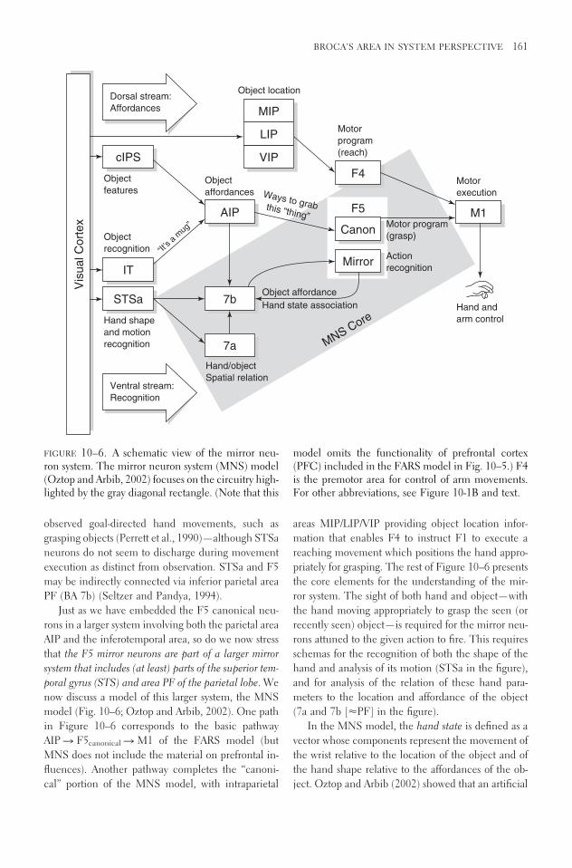
observed goal-directed hand movements, such asgrasping objects (Perrett et al., 1990)—although STSaneurons do not seem to discharge during movementexecution as distinct from observation. STSa and F5may be indirectly connected via inferior parietal areaPF (BA 7b) (Seltzer and Pandya, 1994).
Just as we have embedded the F5 canonical neu-rons in a larger system involving both the parietal areaAIP and the inferotemporal area, so do we now stressthat the F5 mirror neurons are part of a larger mirrorsystem that includes (at least) parts of the superior tem-poral gyrus (STS) and area PF of the parietal lobe. Wenow discuss a model of this larger system, the MNSmodel (Fig. 10–6; Oztop and Arbib, 2002). One pathin Figure 10–6 corresponds to the basic pathwayAIP � F5canonical � M1 of the FARS model (butMNS does not include the material on prefrontal in-fluences). Another pathway completes the “canoni-cal” portion of the MNS model, with intraparietal
areas MIP/LIP/VIP providing object location infor-mation that enables F4 to instruct F1 to execute areaching movement which positions the hand appro-priately for grasping. The rest of Figure 10–6 presentsthe core elements for the understanding of the mir-ror system. The sight of both hand and object—withthe hand moving appropriately to grasp the seen (orrecently seen) object—is required for the mirror neu-rons attuned to the given action to fire. This requiresschemas for the recognition of both the shape of thehand and analysis of its motion (STSa in the figure),and for analysis of the relation of these hand para-meters to the location and affordance of the object(7a and 7b [PF] in the figure).
In the MNS model, the hand state is defined as avector whose components represent the movement ofthe wrist relative to the location of the object and ofthe hand shape relative to the affordances of the ob-ject. Oztop and Arbib (2002) showed that an artificial
BROCA’S AREA IN SYSTEM PERSPECTIVE 161
cIPS
STSa 7b
AIP
Canon
F4
F5 M1
Mirror
MNS Core
IT
Vis
ual C
ort
ex
Ventral stream:
Recognition
Hand shape
and motion
recognition
Object
recognition
Object
features Ways to grabthis “thing”
Object
affordances
“It’s a
mug
” Motor program
(grasp)
Action
recognition
Motor
execution
Hand and
arm control
Motor
program
(reach)
Hand/object
Spatial relation
Object affordance
Hand state association
Object locationDorsal stream:
Affordances
VIP
LIP
MIP
7a
FIGURE 10–6. A schematic view of the mirror neu-ron system. The mirror neuron system (MNS) model(Oztop and Arbib, 2002) focuses on the circuitry high-lighted by the gray diagonal rectangle. (Note that this
model omits the functionality of prefrontal cortex(PFC) included in the FARS model in Fig. 10–5.) F4is the premotor area for control of arm movements.For other abbreviations, see Figure 10-1B and text.

neural network corresponding to PF and F5mirror
could be trained to recognize the grasp type from thehand state trajectory, with correct classification oftenbeing achieved well before the hand reached the object.
During training, the output of the F5 canonicalneurons, acting as a code for the grasp being executedby the monkey at that time, was used as the trainingsignal for the F5 mirror neurons to enable them tolearn which hand-object trajectories corresponded tothe canonically encoded grasps. As a result of thistraining, the appropriate mirror neurons come to firein response to the appropriate trajectories even whenthe trajectory is not accompanied by F5 canonical fir-ing. What makes the modeling worthwhile is that thetrained network not only responded to hand state tra-jectories from the training set but also exhibited in-teresting responses to novel hand-object relationships.
This training prepares the F5 mirror neurons torespond to hand-object relational trajectories evenwhen the hand is of the “other” rather than the “self”because the hand state is based on the movement ofa hand relative to the object, and thus only indirectlyon the retinal input of seeing hand and object—thelatter can differ greatly between observation of self andother. However, the model only accepts input relatedto one hand and one object at a time, and so saysnothing about the “binding” of the action to the agentof that action.
The MNS model provides an explicit account ofhow the mirror system may learn to recognize thehand-object relations associated with grasps already inits repertoire. Complementing this, Oztop and col-leagues (2004) discuss how the infant may acquire itsinitial repertoire of grasps. Such learning models, andthe data they address, make clear that neither canon-ical nor mirror neurons are restricted to recognition ofan innate set of actions but can be recruited to recog-nize and encode an expanding repertoire of novel ac-tions. Given the debate over innateness with regard tolanguage acquisition, it is worth noting that in theMNS model the focus on hand-object relationshipsas providing relevant input is “built in”—but not theexact nature of the grasps that will be learned. Simi-larly, the ILGM model (Oztop et al., 2004) has as itsbasis that the child reaches for a bright or moving ob-ject, executes the grasp reflex if contact is made onthe palmar surface, and builds a repertoire of graspsbased on those which prove to be stable, which weposit supplies a reinforcement signal. The system thushas certain “innate” hand-related biases but is then
able to acquire a range of power grasps and precisionpinches through experience without these having tobe built in.
Homologies
The notion that a mirror system might exist in hu-mans was tested by PET experiments which showedthat grasp observation significantly activated the STS,the inferior parietal lobule, and the inferior frontalgyrus (area 45, part of Broca’s area). For further re-view of related studies using TMS (transcranial mag-netic stimulation) as well as PET and fMRI brainimaging, see Fadiga (this volume). F5 in macaque isgenerally considered to be the homologue of Broca’sarea in humans.
The relative positions of Broca’s area on the infe-rior part of the frontal cortex and Wernicke’s area onthe superior part of the temporal lobe suggest that can-didates for homologous structures of these language-related areas may be situated in corresponding loca-tions in the macaque cortex. Accordingly, thehomologous structures of the human Broca’s area maybe found on the inferior part of the macaque agran-ular frontal cortex, in the vicinity of the arcuate sul-cus (considered to be the macaque homologue of thehuman precentral and prefrontal sulci), and themacaque homologues of Wernicke’s area may be lo-cated at the junction between the temporal and pari-etal cortices. If the nomenclature proposed by Brod-mann is applied to the macaque cortex, one shouldfind corresponding structures of the human areas 44and 45. Both of these human areas are considered tohave their counterparts in the macaque cortex, eventhough there is no consensus over their exact loca-tions and extensions. The macaque homologue ofWernicke’s area appears to include the macaque areaTpt, located on the posterior part of the superior tem-poral gyrus. For full discussion of this and other rele-vant homologies see Arbib and Bota (2003) and thechapters by Petrides and Aboitiz et al. in this volume.
Arbib and Bota (2003) hypothesize that themacaque homologues of Broca’s area are areas 44 and45 (as defined in their paper). They also hypothesizethat the macaque areas 44 and 45 are convergencepoints of the action recognition information conveyedby the connections with posterior parietal corticeswith auditory information from the superior temporalgyrus. Results of Kohler et al. (2002) and Romanskiand Goldman-Rakic (2002) indicate a possible func-tional dissociation between areas 44 and 45, with area
162 MOTOR ASPECTS AND SIGN LANGUAGE

44 involved in processing spatially related auditory in-formation in the context of action recognition, andarea 45 possibly involved in nonspatial processing ofauditory inputs. In any case, more structural, neuro-physiological and functional studies have to be per-formed in humans, macaques and great apes to morefully explore homologies to human language-relatedstructures across different primate species.
Evolving the Language-Ready Brain
We would all agree that the human genome providesthe human child with a brain able to acquire and uselanguage if raised in a community which already haslanguage. Controversy arises over what constitutes this“language readiness”. Some would claim that thegenome provides a Principles and Parameters style ofUniversal Grammar4 (e.g., Chomsky 1981), but Ar-bib (2002, 2005b) argues that biological evolutiononly brought hominids to the stage of having pro-tolanguage (an open system of symbolic communi-cation with little or no syntax), whereas the path fromprotolanguage to language involved a series of histor-ical changes such as those that led to the developmentof farming, or city dwelling or literacy—changes thatdefine what most of us today would see as part of theessentials of the way we are human, yet which weknow to be in the main part “postbiological.”
What turns a movement into an action is that it isassociated with a goal, so that initiation of the move-ment is accompanied by the creation of an expecta-tion that the goal will be met. We distinguish “praxicaction” in which the hands are used to interact phys-ically with objects or other creatures, from “commu-nicative action” (both manual and vocal). Our as-sumption is that macaques use hand movements mainlyfor praxic actions. The mirror system allows othermacaques to understand these actions and act on thebasis of this understanding. Similarly, the macaque’sorofacial gestures register emotional state, and primatevocalizations can also communicate something of thecurrent situation of the macaque. However, seekingto understand why the mirror system for grasping inmacaque is the homologue of Broca’s area in humans,Rizzolatti and Arbib (1998) developed the following:
The Mirror-System Hypothesis
Language evolved from a basic mechanism not orig-inally related to communication: the mirror system forgrasping with its capacity to generate and recognize
a set of actions. More specifically, human Broca’s areacontains a mirror system for grasping which is ho-mologous to the F5 mirror system of macaque, andthis provides the evolutionary basis for languageparity—i.e., an utterance means roughly the same forboth speaker and hearer.
This provides a neurobiological “missing link” forthe hypothesis that communication based on manualgesture preceded speech in language evolution (e.g.,Hewes, 1973; Stokoe, 2001).5 I view the “openness”or “generativity” that some see as the hallmark of lan-guage (i.e., its openness to new constructions, as dis-tinct from having a fixed repertoire like that of mon-key vocalizations) as present in manual behaviorwhich can thus supply part of the evolutionary sub-strate for its appearance in language.
Arbib (2002, 2005b) has amplified the original ac-count of Rizzolatti and Arbib to hypothesize sevenstages in the evolution of language, with imitationgrounding two of the stages. The first three stages areprehominid:
S1: Grasping
S2: A mirror system for grasping shared with thecommon ancestor of human and monkey. Themirror neurons can be recruited to recognize andencode an expanding set of novel actions. StageS2 is subdivided into S2a, providing feedback fordexterous manual control, and S2b, acting withother brain regions to make useful informationavailable for interacting with others.
S3: A simple imitation system for grasping sharedwith the common ancestor of human and chim-panzee.
The next three stages then distinguish the hominidline from that of the great apes:
S4: A complex imitation system for grasping: Thispresupposes a capacity for complex action analysis,the ability to analyze another’s performance as acombination of actions (or approximated by vari-ants of actions) already in the repertoire. This canprovide the basis for adding new, complex actionsto one’s repertoire. The notion is that complex im-itation was an evolutionary step of great advantageindependent of its implications for communica-tion. However, in modern humans it undergirdsthe child’s ability to acquire language, while com-plex action analysis is essential for the adult’s abil-ity to comprehend the novel compounds of “ar-ticulatory gestures” that constitute language.
BROCA’S AREA IN SYSTEM PERSPECTIVE 163

The next two stages introduces communication in-tended by the utterer to have a particular effect on therecipient, rather than being involuntary or a side ef-fect of praxis:
S5: Protosign, a manual-based communication sys-tem, breaking through the fixed repertoire of pri-mate vocalizations to yield an open repertoire.This decomposes into S5a: the ability to engage inpantomime; and S5b: the ability to make conven-tional gestures to disambiguate pantomime.
S6: Proto-speech, resulting from the ability of con-trol mechanisms evolved for protosign to also con-trol the vocal apparatus with increasing flexibility.
It is argued that early protosign provided the scaf-folding for early protospeech after which they devel-oped together in an expanding spiral till protospeechbecame dominant. Recent data on mirror neurons arerelevant here. Kohler et al. (2002) show that 15% ofmirror neurons in the hand area of macaque F5 canrespond to the distinctive sound of an action (break-ing peanuts, ripping paper, etc.) as well as viewing theaction. Ferrari et al. (2003) find that in the orofacialarea of F5, there are “mirror” neurons tuned to com-municative gestures such as lip-smacking, althoughthese are not strictly congruent—e.g., one “observed”lip protrusion (communicative) but “executed” sy-ringe sucking (ingestive). Some might argue that theseresults let us apply the mirror system hypothesis to vo-calization directly, “cutting out the middleman” ofmanual gesture. The three papers by Arbib (2005a),MacNeilage and Davis (2005b), and Fogassi and Fer-rari (2004) exemplify the current state of the debate.
The final stage is claimed to involve little if anybiological evolution, but instead to result from cul-tural evolution (historical change) in Homo sapiens:
S7: Language: the change from action-objectframes to verb-argument structures to syntax andsemantics; the co-evolution of cognitive and lin-guistic complexity.
Arbib (2005b) offers examples of the discoverieswhich might have accumulated across tens of mil-lennia to take Homo sapiens from protolanguage tolanguage, arguing that the features of languageemerged by adding many features as “patches” to aprotolanguage, with general “rules” emerging bothconsciously and unconsciously only as generalizationscould be imposed on, or discerned in, a population
of ad hoc mechanisms. Needless to say, this assertionis highly controversial.
Aboitiz and García (1997) offer a somewhat dif-ferent theory of human brain evolution, also rootedin the study of human–macaque homologies. Theypropose three stages in the evolution of the capacityof the human brain to support language:
1. The capacity to give names yielded the lexiconthat underlies the ability to refer to objects orevents in the external world. They associate thiswith the elaboration of a precursor of Wer-nicke’s area in the superior temporal lobe as azone for cross-modal associations which in-clude a phonological correlate.
2. The need to combine more and more words toexpress complex meanings yielded syntax, theexpression of regularities in the ways in whichdifferent elements are combined to form lin-guistic utterances. They associate the emer-gence of syntax with the differentiation of aninferoparietal-frontal (Broca’s) area with its con-nections to the incipient Wernicke’s region de-veloping as a phonological rehearsal device thateventually differentiated into the language ar-eas This system they see as yielding a phono-logical-rehearsal loop (recall the discussion ofBaddeley, 2003) that could provide some basicsyntactic rules at the levels of phonemes andmorphemes.
3. Coordinated operation of networks involvinggranular frontal cortex and the semantic systemrepresented in the temporoparietal lobes, to-gether with the phonological-rehearsal loop,generated higher levels of syntax and discourse.
Aboitiz et al. (this volume, Chapter 1) provide afuller exposition of these hypotheses and notes certainmodifications they have made in the years since 1997.Arbib and Bota (2003) contrast the two evolutionarytheories as follows:
Aboitiz and García assume that the human brainevolved (in part) to support language. They offer anessentially retrospective theory working back from a lex-icon of spoken words and a syntax that binds them intosentences to suggest how areas of the macaque brain,and their connections, changed (some areas enlarged,some connections are strengthened) to yield a humanbrain that could support these features of language. Bycontrast, Rizzolatti and Arbib offer more of a prospec-tive theory. They start from an analysis of the monkey’scapabilities, especially the fact that F5, containing amirror system for hand movements, is homologous to
164 MOTOR ASPECTS AND SIGN LANGUAGE

Broca’s area. They then offer hypotheses on how in-termediate stages from the mirror system for graspingled via simple and complex imitation and protosign toprotospeech. Here I want to place special emphasis oncomplex imitation and in particular complex actionanalysis, the ability to analyze another’s performancein terms of actions already in the repertoire. However,Rizzolatti and Arbib are relatively silent on the phono-logical loop and other working memory systems whoseemphasis is an important feature of the Aboitiz andGarcía analysis. Thus, the relevance of the schema-
theoretic view of memory provided at the end of theSchema Theory section and the range of workingmemory systems within the FARS model of Figure10–5.
TOWARD AN ACTION-ORIENTED
NEUROLINGUISTICS
Figure 10–7 provides a view of the language-readybrain (Arbib & Bota 2003), informed both by the mir-
BROCA’S AREA IN SYSTEM PERSPECTIVE 165
Working memory foractions, objects, and
relationships
Auditoryinput
A
B
Visualinput
Working memory forperception and production of
utterances
MultimodalBroca’s area
(vocal,manual,
oro-facial)
From imitation todescribing actions, objects, relationships
Recognizingactions, objects,and relationships
IT
IT
PF
F1
PF
AIP
Tpt
Tpt
F5 mirror
Protospeech (basis for speech)
Protosign (basis for sign language)
Recognizing an action
Choosing an action
PF extended
Wernicke
F5 canonical
DLPL
Broca
STSa
AIP
DPLF
FIGURE 10–7. (A) A recasting and extension (in part)of FARS Modificato (Fig. 10–5) in register with theview of the human brain in Figure 1A, designed formaximal congruence with the schema provided byAboitiz and García (1997). (B) A high-level view of the
cumulative emergence of three frontoparietal systems:choosing an action � recognizing an action � de-scribing an action (in multiple modalities). DLPL, dor-solateral prefrontal cortex. For other abbreviations seeFigure 10–1. (Both figures from Arbib and Bota, 2003.)

ror system hypothesis and by the emphasis on work-ing memory of Aboitiz and García. It is a hybrid, mix-ing macaque and human regions and gives the im-pression that three frontoparietal systems end in threedistinct brain structures. In fact, current research hasnot really settled the question. In any case, the possi-bility that one monkey area may be homologous todifferent human regions implicated in one or moreof praxic hand movements, protosign, protospeech,signed language, and speech offers challenge to overlybinary views of homology. Much needs to be done todelineate subareas of Broca’s area that can be distin-guished on this basis and their connections—whilenoting that differences that are found (and the varia-tions in the pattern of such differences from individ-ual to individual) may reflect the self-organization ofthe brain as the child grows within a language com-munity rather than any innate “fate map” for thesedifferences. DeRenzi et al. (1966) found that the ma-jority of patients with apraxia of speech had oralapraxia and a high coexistence of oral and limbapraxia while Marquardt and Sussman (1984) foundthat 12 of their 15 patients with Broca’s aphasia hadapraxia of speech while 5 had limb apraxia. Doubledissociations do occur in some cases. Thus, either sep-arate networks of neurons are engaged in the genera-tion of speech and nonspeech movement of the samemuscles, or the same general network underliesspeech and nonspeech movements but these have sep-arate control mechanisms which can be differentiallydamaged (Code, 1998).
Notes
1. Analysis of the macaque brain, and thus of ho-mologies with the human brain, is bedeviled by the factthat different authors use different parcellations to ad-dress data acquired since the time of Brodmann. Arbiband Bota (2003) have addressed the issue by developingneuroinformatics tools to create an online knowledgemanagement system, the NeuroHomology Database.
2. See Goldman-Rakic, Ó Scalaidhe, and Chafee,1999, for the variety of working memories implementedin prefrontal cortex and with links to specific parietal circuitry.
3. The architecture of the VISIONS computer vi-sion system was based on the HEARSAY architecture aswell as on neurally inspired schema theory.
4. The concept of universal grammar is protean,and there may be variants for which I would accept a ge-netic basis. What I do reject is the view that the major-ity of syntactic rules are preencoded, so that the child’s
experience simply “sets switches” to select and parame-terize those rules appropriate to the language commu-nity in which the child is raised. Perhaps the most com-pelling hypothesis for the evolution of such a universalgrammar was advanced by Pinker and Bloom (1990)—but when I read it carefully, I find that the notion of uni-versal grammar is not unequivocally defined, and thatmany of the stages they posited could have occurred bycultural evolution as easily as by biological evolution.
5. Below, I soften this assertion to the claim thatprotosign provided scaffolding essential to the evolutionof protospeech.
References
Aboitiz, F., & García V.R. (1997). The evolutionary ori-gin of the language areas in the human brain. Aneuroanatomical perspective. Brain Research Re-views, 25, 381–396.
Arbib, M.A. (1981). Perceptual structures and distributedmotor control. In V.B. Brooks (Ed.), Handbook ofPhysiology – The Nervous System II. Motor Control(pp. 1449–1480). Bethesda, MD: American Physi-ological Society.
Arbib, M.A. (1989). The metaphorical brain 2: Neuralnetworks and beyond. New York: Wiley-Inter-science.
Arbib, M.A. (2002). The mirror system, imitation, andthe evolution of language. In C. Nehaniv & K.Dautenhahn (Eds.), Imitation in animals and arti-facts (pp. 229–280). Boston: MIT Press.
Arbib, M.A. (2005a). Interweaving protosign and proto-speech: Further developments beyond the mirror.Interaction Studies: Social Behavior and Communi-cation in Biological and Artificial Systems, 6,145–171.
Arbib, M.A. (2005). From monkey-like action recogni-tion to human language: An evolutionary frame-work for neurolinguistics. Behavioral and Brain Sci-ences (in press).
Arbib, M.A., & Bota, M. (2003). Language evolution:Neural homologies and neuroinformatics. NeuralNetworks, 16, 1237–1260.
Arbib, M.A., & Caplan, D. (1979). Neurolinguistics mustbe computational. Behavioral and Brain Sciences,2, 449–483.
Arbib, M.A., & Liaw, J.-S. (1995). Sensorimotor trans-formations in the worlds of frogs and robots. Artifi-cial Intelligence, 72, 53–79.
Arbib, M.A., Érdi, P., & Szentágothai, J. (1998). Neuralorganization: Structure, function, and dynamics.Cambridge, MA: MIT Press.
Arkin, R.C. (1998). Behavior-based robotics. Cambridge,MA: MIT Press.
166 MOTOR ASPECTS AND SIGN LANGUAGE

Baddeley, A. (2003). Working memory: Looking backand looking forward. Nature Reviews Neuroscience,4, 829–839.
Baddeley, A.D., & Hitch, G.J. (1974). Working memory.In G.A. Bower (Ed.), The psychology of learning andmotivation (pp. 47–89). NY: Academic Press.
Chomsky, N. (1981). Lectures on government and bind-ing: The Pisa lectures. Dordrecht: Foris.
Code, C. (1998). Models, theories and heuristics inapraxia of speech. Clinical Linguistics & Phonetics,12, 47–65.
DeRenzi, E., Pieczuro, A., & Vignolo, L.A. (1966). Oralapraxia and aphasia. Cortex, 2, 50–73.
Draper, B.A., Collins, R.T., Brolio, J., Hanson, A.R., &Riseman, E.M. (1989). The schema system. Inter-national Journal of Computer Vision, 2, 209–250.
Fagg, A.H., & Arbib, M.A. (1998). Modeling parietal-premotor interactions in primate control of grasp-ing. Neural Networks, 11, 1277–1303.
Ferrari, P.F., Gallese, V., Rizzolatti, G., & Fogassi, L.(2003). Mirror neurons responding to the observa-tion of ingestive and communicative mouth actionsin the monkey ventral premotor cortex. EuropeanJournal of Neuroscience, 17, 1703–1714.
Fogassi, L., & Ferrari, P.F. (2004). Mirror neurons, ges-tures and language evolution. Interaction Studies:Social Behavior and Communication in Biologicaland Artificial Systems, 5, 345–363.
Galaburda, A.M., & Sanides, F. (1980). Cytoarchitec-tonic organization of the human auditory cortex.Journal of Comparative Neurology, 190, 597–610.
Geschwind, N. (1964). The development of the brainand the evolution of language. Montreal Series inLanguage & Linguistics, 1, 155–169.
Goldman-Rakic, P.S., Ó Scalaidhe, S.P., & Chafee,M.V. (1999). Domain specificity in cognitive sys-tems. In M.S. Gazzaniga (Ed.), The new cognitiveneurosciences (Chapter 50). Second Edition. Cam-bridge, MA: MIT Press.
Hewes, G. (1973). Primate communication and the ges-tural origin of language. Current Anthropology, 14,5–24.
Jackendoff, R. (2002). Foundations of language: Brain,meaning, grammar, evolution. Oxford and NewYork: Oxford University Press.
Jeannerod, M., & Biguer, B. (1982). Visuomotor mech-anisms in reaching within extra-personal space. InD.J. Ingle, R.J.W. Mansfield, & M.A. Goodale(Eds.), Advances in the analysis of visual behavior(pp. 387–409), Boston: MIT Press.
Jeannerod, M. (1997). The cognitive neuroscience of ac-tion. Oxford: Blackwell Publishers.
Jeannerod, M., Arbib, M.A., Rizzolatti, G., & Sakata, H.(1995). Grasping objects: The cortical mechanisms
of visuomotor transformation. Trends in Neuro-sciences, 18, 314–320.
Kohler, E., Keysers, C., Umilta, M.A., Fogassi, L.,Gallese, V., & Rizzolatti, G. (2002). Hearingsounds, understanding actions: Action representa-tion in mirror neurons. Science, 297, 846–848.
Lesser, V.R., Fennel, R.D., Erman, L.D., & Reddy, D.R.(1975). Organization of the HEARSAY-II speechunderstanding system. IEEE Transactions onAcoustics, Speech, and Signal Processing, 23, 11–23.
Luria, A.R. (1973). The working brain. Harmondsworth:Penguin Books.
MacNeilage, P.F., & Davis, B. (2005). The frame/con-tent theory of evolution of speech: A comparisonwith a gestural origins alternative. Interaction Stud-ies: Social Behavior and Communication in Biolog-ical and Artificial Systems, 6, 173–199.
Marquardt, T.P., & Sussman, H. (1984). The elusive le-sion—Apraxia of speech link in Broca’s aphasia. InJ.C. Rosenbek, M.R. McNeil, & A.E. Aronson(Eds.), Apraxia of speech: Physiology, acoustics, lin-guistics, management, San Diego: College-HillPress.
Oztop, E., & Arbib, M.A. (2002). Schema design andimplementation of the grasp-related mirror neuronsystem. Biological Cybernetics, 87, 116–140.
Oztop, E., Bradley, N., & Arbib, M.A. (2004). Infantgrasp learning: A computational model. Experi-mental Brain Research, 158, 480–503.
Perrett, D.I., Mistlin, A.J., Harries, M.H., & Chitty, A.J.(1990). Understanding the visual appearance andconsequence of hand actions. In M.A. Goodale(Ed.), Vision and action: The control of grasping (pp.163–342). Norwood, NJ: Ablex.
Pinker, S., & Bloom, A. (1990). Natural language andnatural selection. Behavioural Brain Sciences, 13,707–784.
Rizzolatti, G., & Arbib, M.A. (1998). Language withinour grasp. Trends in Neurosciences, 21, 188–194.
Rizzolatti, G., & Craighero, L. (2004). The mirror-neu-ron system. Annual Review of Neuroscience, 27,169–192.
Rizzolatti, G., & Luppino, G. (2003). Grasping move-ments: Visuomotor transformations. In M.A. Arbib(Ed.), The handbook of brain theory and neural net-works (2nd ed., pp. 501–504). Cambridge, MA:MIT Press.
Rizzolatti, G., Fogassi, L., & Gallese, V. (2002). Motorand cognitive functions of the ventral premotor cor-tex. Current Opinions in Neurobiology, 12, 149–154.
Rolls, E.T., & Arbib, M.A. (2003). Visual scene percep-tion. In M.A. Arbib (Ed.), The handbook of brain
BROCA’S AREA IN SYSTEM PERSPECTIVE 167

theory and neural networks (2nd ed., pp. 1210–1251). Cambridge, MA: MIT Press.
Romanski, L.M., & Goldman-Rakic, P.S. (2002). An au-ditory domain in primate prefrontal cortex. NatureNeuroscience, 5, 15–6.
Seltzer, B., & Pandya, D.N. (1994). Parietal, temporal,and occipital projections to cortex of the superiortemporal sulcus in the rhesus monkey: A retrogradetracer study. Journal of Comparative Neurology, 15,445–463.
Stokoe, W.C. (2001). Language in hand: Why sign camebefore speech. Washington, DC: Gallaudet Univer-sity Press.
Weymouth, T.E. (1986). Using object descriptions in aschema network for machine vision, Ph.D. Thesisand COINS Technical Report 86–24, Departmentof Computer and Information Science, Universityof Massachusetts at Amherst.
Wilkins, W.K., & Wakefield, J. (1995). Brain evolutionand neurolinguistic preconditions. Behavioral andBrain Sciences, 18, 161–226.
168 MOTOR ASPECTS AND SIGN LANGUAGE

11
The Role of Broca’s Area in Sign Language
Karen Emmorey
Sign languages provide a powerful tool for investi-gating the neurobiology and cognitive architecture ofhuman language. Linguistic research has revealedsubstantial similarities between signed and spokenlanguages, but this is only a starting point (for reviews,see Emmorey, 2002; Sandler and Lillo-Martin, 2006).The similarities provide a strong basis for compari-son and serve to highlight universal properties of hu-man language. In addition, however, sign-speechmodality distinctions can be exploited to discoverhow the input-output systems of language impact theneurocognitive underpinnings of language produc-tion and comprehension. In this chapter, we explorewhether Broca’s area subserves the same behavioralfunctions for signed and spoken languages and dis-cuss the nature of these functions. First, however,some background on sign language structure needsto be presented.
The discovery that sign languages have a phonol-ogy (traditionally defined as the sound patterns of lan-guage) was ground-breaking and crucial to our un-
derstanding of the nature of human language (for reviews, see Brentari, 1998; Corina and Sandler,1993). In spoken languages, words are constructed outof sounds that in and of themselves have no meaning(duality of patterning). The words “bat” and “pat” dif-fer only in the initial sounds that convey no inherentmeanings of their own. Similarly, signs are con-structed out of components that are themselves mean-ingless and are combined to create morphemes andwords. Signs can be minimally distinguished by handconfiguration, place of articulation, movement, andorientation with respect to the body. Figure 11–1 pro-vides an illustration of four minimal pairs: signs thatare identical except for one component, and if yousubstitute one component for another, it changes themeaning of the sign. Some signs are produced withone hand and some with two hands, but handednessis not distinctive in ASL (American Sign Language)or perhaps in any sign language. There are no ASLsigns that differ only on the basis of whether they aremade with the right hand or with the left hand. One-
169

handed signs are produced with the signer’s dominanthand. Thus, left-handers and right-handers differ inwhich hand is dominant.
Not all hand configurations nor all places of ar-ticulation are distinctive in any given sign language.For example, the T handshape (thumb inserted be-tween index and middle fingers) does not occur inEuropean signed languages. Chinese Sign Languagecontains a handshape formed with an open hand with
all fingers extended expect for the ring finger, whichis bent; this hand configuration does not occur inASL. Although the upper cheek and chin are dis-tinctive places of articulation in ASL (see Fig. 11–1B),the back jaw is not a phonologically contrastive loca-tion (e.g., there are no minimal pairs involving thisplace of articulation).
Spoken languages represent sequential structure interms of the linear ordering of consonant and vowelsegments. Similarly, the linear structure of signs canbe represented in terms of sequences of phonologicalcategories akin to consonants and vowels. These areLocation and Movement segments within Sandler’sphonological model (Sandler, 1989). Other phono-logical models also propose linear structure, but char-acterize segments and features somewhat differently(Brentari, 1998; van der Hulst, 1993). Segments insigned languages are characterized by manual features(e.g., selected fingers; body contact) rather than byoral features (e.g., voicing; labial).
There is fairly strong evidence for the existence ofsyllables in sign languages—the syllable is a unit ofstructure that is below the level of the word but abovethe level of the segment. However, there is little evi-dence for onset-rhyme distinctions within a sign syllable or resyllabification processes. Finally, signlanguages exhibit phonological rules such as assimi-lation, constraints on well-formedness that refer to seg-mental structure, and form patterns can be accountedfor using feature geometry. Thus, both signed and spo-ken languages exhibit a linguistically significant, yetmeaningless, level of structure that can be analyzedas phonology for both language types.
Signed and spoken languages are also subject tothe same syntactic constraints on form and movement(see Sandler and Lillo-Martin, in press, for review).For example, the basic word order for ASL is SVO(subject-verb-object). The ASL sentence GIRL PUSHBOY1 unambiguously means “The girl pushes theboy” and BOY PUSH GIRL means “The boy pushesthe girl.” Topicalization can result in the movementof the object noun phrase to a sentence initial posi-tion, as illustrated in example 1 and Figure 11–2.
(1) ____t(1) BOY, GIRL PUSH ______.“The boy, the girl pushes him.”
This ASL topicalization marker is a combinationof a backward head tilt and raised eyebrows, timed to
170 MOTOR ASPECTS AND SIGN LANGUAGE
FIGURE 11–1. Examples of minimal pairs in Ameri-can Sign Language: (A) signs that contrast in handconfiguration, (B) signs that contrast in place of ar-ticulation, (C) signs that contrast in movement, and(D) signs that contrast in orientation.
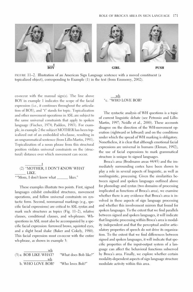
co-occur with the manual sign(s). The line aboveBOY in example 1 indicates the scope of the facialexpression (i.e., it continues throughout the articula-tion of BOY), and “t” stands for topic. Topicalizationand other movement operations in ASL are subject tothe same universal constraints that apply to spokenlanguage (Fischer, 1974; Padden, 1983). For exam-ple, in example 2 the subject MOTHER has been top-icalized out of an embedded wh-clause, resulting inan ungrammatical sentence (from Lillo-Martin, 1991).Topicalization of a noun phrase from this structuralposition violates universal constraints on the (struc-tural) distance over which movement can occur.
(1) __________t(2) *MOTHER, I DON’T-KNOW WHAT
______ LIKE.*“Mom, I don’t know what ______ likes.”
These examples illustrate two points. First, signedlanguages exhibit embedded structures, movementoperations, and follow universal constraints on syn-tactic form. Second, nonmanual markings (e.g., spe-cific facial expressions) are critical to ASL syntax andmark such structures as topics (Fig. 11–2), relativeclauses, conditional clauses, and wh-phrases. Wh-questions in ASL must also be accompanied by a spe-cific facial expression: furrowed brows, squinted eyes,and a slight head shake (Baker and Cokely, 1980).This facial expression must co-occur with the entirewh-phrase, as shown in example 3:
(1)________________wh(3) a. BOB LIKE WHAT? “What does Bob like?”
(1 ________________whb. WHO LOVE BOB? “Who loves Bob?”
(1) a______wh*c. *WHO LOVE BOB?
The syntactic analysis of WH questions is a topicof current linguistic debate (see Petronio and Lillo-Martin, 1997; Neidle et al., 2000). These accountsdisagree on the direction of the WH-movement op-eration (rightward or leftward) and on the conditionsunder which the spread of WH marking is obligatory.Nonetheless, it is clear that although emotional facialexpressions are universal in humans (Ekman, 1992),the use of facial expressions to mark grammaticalstructure is unique to signed languages.
Broca’s area (Brodmann areas 44/45) and the im-mediately surrounding cortex have been shown toplay a role in several aspects of linguistic, as well asnonlinguistic, processing. Given the similarities be-tween signed and spoken languages outlined abovefor phonology and syntax (two domains of processingimplicated as functions of Broca’s area), we examinewhether there is any evidence that Broca’s area is in-volved in these aspects of sign language processingand whether this involvement mirrors that found forspoken languages. To the extent that we find parallelsbetween signed and spoken languages, it will indicatethat linguistic processing within Broca’s area is modal-ity independent and that the perceptual and/or artic-ulatory properties of speech do not drive its organiza-tion. To the extent that we find differences betweensigned and spoken languages, it will indicate that spe-cific properties of the input-output system of a lan-guage can affect the behavioral functions subservedby Broca’s area. Finally, we explore whether certainmodality-dependent aspects of sign language structuremodulate activity within this area.
ROLE OF BROCA’S AREA IN SIGN LANGUAGE 171
FIGURE 11–2. Illustration of an American Sign Language sentence with a moved constituent (atopicalized object), corresponding to Example (1) in the text (from Emmorey, 2002).

BROCA’S AREA AND SIGN
LANGUAGE PRODUCTION
Broca’s area area has long been thought to play a rolein speech production. Given that Broca’s area is an-terior to primary motor cortex controlling mouth andlip movements, it is reasonable that an area involvedin speech production would be anatomically locatednear regions involved in control of the speech articu-lators. Is the same area involved in sign language pro-duction? Or is the functional equivalent of Broca’sarea shifted superiorly so that it is next to the motorrepresentation for the hand and arm? Using corticalstimulation mapping, Corina and colleagues (1999)were able to identify the areas involved in mouth andlip movements in a deaf ASL signer. While the sub-ject produced manual signs in a picture-naming task,they stimulated the posterior aspect of Broca’s area(Brodmann’s area 44). This site was just anterior tothe sites that evoked facial motor responses. Stimula-tion resulted in signing errors best characterized asphonetic or motor execution errors. Signs were artic-ulated with nonspecific movements and laxed hand-shape configurations. Semantic naming errors did notoccur with stimulation to BA44, nor did phonologi-cal errors (e.g., producing an erroneous, but clearlyarticulated, handshape substitution). In contrast, bothphonological substitution errors and semantic errorsoccurred during sign production when stimulationwas applied to the left supramarginal gyrus in the samedeaf signer.
The cortical stimulation mapping results with thisdeaf ASL signer suggest that BA 44 is involved in pho-netic aspects of linguistic expression, regardless of theanatomy of the language articulators. This hypothesisis supported by the recent results of Horwitz, Amunts,Bhattacharyya, Patkin, Jeffries, Zilles, and Braun(2003), who used a combination of cytoarchitectonicmapping and PET data to investigate activation ofBroca’s area during the production of spoken andsigned language by hearing ASL-English bilinguals.Subjects were asked to produce autobiographical nar-ratives in either English or ASL, and the baselinecomparison tasks were laryngeal and orofacial articu-latory movements or bilateral non-routinized handand arm movements (from Braun et al., 1997, 2001).When contrasted with the baseline articulation con-ditions, there was little or no activation within BA44for sign production or for speech production. How-ever, when the articulatory baseline tasks were con-
trasted with a rest condition, significant activation wasobserved in BA44 for both language types (more fororofacial articulation than for manual articulation).Interestingly, activation in BA 44 within the righthemisphere was observed for orofacial articulationcompared to rest, but very little activation in right BA44 was observed for (bimanual) limb articulation.
In contrast to BA 44, Horwitz et al. (2003) foundonly left hemisphere activation in BA 45 for both sign-ing and speaking. Activation in left BA 45 was onlyobserved when the linguistic tasks (producing an Eng-lish or an ASL narrative) were contrasted with the ar-ticulatory baseline tasks. The contrast between the ar-ticulation tasks and rest did not produce activation inBA 45 for either orofacial or manual-limb articula-tion. Horowitz et al. (2003) concluded that BA45 isthe portion of Broca’s area that is fundamental tomodality-independent aspects of language production.
PET data from prelingually deaf ASL signers nam-ing objects and/or actions also indicate a role for leftBA 45 in single sign production, as well as for narra-tive production (Emmorey et al., 2002, 2003, 2004).Activation maxima were observed in left BA 45 withnearly identical Talairach coordinates when signersnamed animals (�41, �28, �17), tools (�42, �28,�18), tool-based actions (�46, �26, �13), and ac-tions performed without a tool (�47, �21, �15). Thebaseline task required subjects to judge the orienta-tion of unknown faces (overtly responding “yes”/“no”for upright/inverted). A between-subjects (N � 58)random effects analysis revealed that BA 45 (peak co-ordinates: �48, �27, �20) was equally involved inboth speech and sign production (Emmorey &Grabowki, 2004). However, two regions within theparietal lobe were significantly more active duringsign production than during word production: the leftsupramarginal gyrus (peak coordinates: �63, �34,�25) and the left superior parietal lobule (two peakcoordinates: �29, �46, �54; �10, �62, �59). Wespeculate that activation in these regions is linked tomodality-specific output parameters of sign language.Specifically, activation within left supramarginalgyrus may reflect aspects of phonological processingin ASL (e.g., selection of hand configuration andplace of location features), whereas activation withinthe superior parietal lobule may reflect proprioceptivemonitoring of motoric output.
The neuroimaging data indicate strong left later-alized activation within Broca’s area for both covert(McGuire et al., 1997) and overt signing (Braun et
172 MOTOR ASPECTS AND SIGN LANGUAGE

al., 2001; Emmorey et al., 2002, 2003, 2004; Petittoet al., 2000). However, in all of these studies, subjectsproduced signs with their dominant right hand, whichis innervated almost exclusively by left primary motorand sensory cortices. In contrast, the speech articula-tors lie on the midline of the body and are innervatedby both hemispheres. Does the fact that the primaryarticulator for sign language is innervated by left-lateralized sensorimotor cortices influence the later-ality of language production systems? Corina and col-leagues (2003) recently investigated this question byasking ASL signers to generate verbs with either theirleft (non-dominate) or right (dominant) hand, whileundergoing PET scanning. The verb-generation task(produce a verb that is semantically related to a visu-ally presented ASL noun) was contrasted with a nounrepetition task. The data showed no activation in rightBroca’s area when signers produced verbs with theirleft hand. In addition, a conjunction analysis withright- and left-hand sign production revealed activa-tion maxima within BA 44/45 (�54, �18, �16) andalso in BA 46 (�48, �30, �20) and BA 47 (�38,�29, -8). These findings indicate that lexical-seman-tic processing in language production relies uponBroca’s area (and surrounding cortex) in the left hemi-sphere, even when the primary language articulatoris innervated by the right hemisphere.
In general, the lesion data also support a role forBroca’s area in sign language production because left,but not right, frontal damage results in nonfluent, ef-fortful signing (Corina, 1998; Poizner et al., 1987).However, there is only one case study reported thusfar in which the location of the cortical damage wasclearly restricted to Broca’s region (BA 44/45 and im-mediately surrounding cortex). Hickok and colleagues(1996) report the case of R.S., a congenitally deaf na-tive ASL signer, who suffered an ischemic infarct in-volving the posterior aspect of the left frontal opercu-lum, inferior motor cortex, and white matter deep toboth regions. R.S. exhibited relatively good compre-hension of ASL (but see later) and significant deficitsin ASL production. Although R.S. had good articula-tion, she produced frequent phonemic paraphasiasand occasional semantic paraphasias. An unusual as-pect of R.S.’s phonemic paraphasias was that the er-rors all occurred with two-handed signs. Specifically,the errors involved bimanual coordination of the twohands. For example, she might incorrectly fail tomove one of her hands or incorrectly mirror the move-ment of one hand with the other when the movement
should be symmetrical. She also tended to shadowone-handed signs with her nondominant (left) hand.Deficits in bimanual coordination were not observedwhen R.S. produced nonlinguistic bimanual handand arm movements (e.g., producing alternatingwindmill movements, finger tapping, tying her shoes,knitting, etc.). R.S.’s deficits in coordinating the twohands may be analogous to the phonetic deficits ob-served for speakers with nonfluent aphasia who havedifficulty coordinating independent speech articula-tors, e.g., the larynx, tongue, and lips (see Blumstein,1998).
Another clue to the function of Broca’s area(specifically BA 45) in sign language productioncomes from a recent PET study by Emmorey and col-leagues (2005). We contrasted noun production withthe production of spatial language (prepositions orlocative classifier constructions) in English and ASLby hearing ASL-English bilinguals. Briefly, in ASL themost common way to express the spatial relationshipbetween two objects is to use a classifier constructionin which the position of the hands in signing spaceschematically represents the spatial relation betweenobjects. An illustrative example is provided in Figure11–3. Within this type of classifier construction, eachhandshape specifies an object type, such as “long andthin,” “cylindrical,” or “flat surface.” The set of clas-sifier handshape morphemes is closed.
Of interest is the finding that the production ofclassifier constructions (like the one shown in Fig.11–3) did not significantly engage Broca’s area. In thisstudy, ASL-English bilinguals were asked to describethe spatial relation between two objects in English(with a preposition) or in ASL (with a classifier con-struction), and on a separate presentation, they namedthe figure object in either ASL or English (the figureobject was colored red). When producing ASL lexi-cal signs (nouns) was contrasted with producing loca-tive classifier constructions, significant activation wasobserved in left BA 45 for ASL nouns (peak activationcoordinates: �38, �26, �3). The contrast betweenspoken English prepositions and English nouns re-vealed no difference in activation for Broca’s area.Furthermore, the interaction analysis indicated thatEnglish prepositions engaged left BA 45 (�41, �29,�9) to a significantly greater extent than ASL classi-fier constructions. The PET data from deaf ASL sign-ers also indicated that production of ASL classifierconstructions did not engage Broca’s area. When theproduction of classifier constructions was contrasted
ROLE OF BROCA’S AREA IN SIGN LANGUAGE 173

with our standard baseline task, no activation was ob-served within Broca’s area; however, when deaf sign-ers viewed the same picture stimuli and named thefigure object with an ASL sign, significant activationwas observed in Broca’s area (BA 45: �49, �24, �15)using the same baseline contrast. Finally, the deafASL signers were also asked to name spatial relation-ships with lexicalized ASL “prepositions” (signsglossed as IN, ON, UNDER, etc.). An example isshown in Figure 11–3. Syntactically, these forms donot function like prepositions in English (McIntire,1980; Shepard-Kegl, 1985), are not preferred foreveryday spatial descriptions, and are usually used foremphasis (“It’s under the table”). In addition, thehandshape of these signs is lexically specified and doesnot change with the nature of the figure or groundobjects, unlike the handshape morphemes withinclassifier constructions. Also unlike classifier con-structions, production of ASL prepositions engagedBroca’s area when contrasted with the baseline task(BA 45: �50, �19, �20).
In sum, the production of ASL verbs, nouns, andlexical prepositions activated Broca’s area (specificallyBA 45), but the production of locative classifier con-structions did not. One possible explanation for thissomewhat surprising result is that because classifierconstructions do not constitute a simple lexical cate-gory, their production does not engage the same lex-ical retrieval processes that have been associated withBroca’s area (e.g., Petersen et al., 1988; Thompson-Schill et al., 1997). In our PET studies, signers andspeakers were asked to provide a single label for eachpicture. That is, subjects were required to retrieve adistinct lexical item for each picture stimulus. How-ever, for locative classifier constructions, subjects did
not retrieve a specific lexical sign that denoted an ob-ject, action, or spatial relationship; rather, they re-trieved a handshape morpheme that denoted an ob-ject type (e.g., long and thin). The same handshapemorpheme was appropriate for a number of differentobjects. For example, the 1 handshape shown in Fig-ure 11–3 could be used for a pencil, a toothbrush, ora spoon. To indicate the spatial relationship betweentwo objects, signers did not retrieve a lexical sign ormorpheme. Rather, as illustrated in Figure 11–3, theyplaced their right hand (representing the figure ob-ject) in signing space with respect to their left hand(representing the ground object, which was the sameobject type for a given stimulus set—either a cylin-drical object or a flat surface). In these constructions,the position of the hands in space is not a morphemic,categorical representation (Emmorey and Herzig,2003). Signing space is schematically mapped in ananalogue manner to the physical space shown in thepicture stimuli. The finding that the production ofthese constructions does not significantly engageBroca’s area, in contrast to the production of lexicalsigns, suggests that Broca’s area plays a specific rolein the selection and/or retrieval of specific lexicalitems, i.e., names of entities, actions, and relationships.
In conclusion, the function of Broca’s area is notstrongly tied to oral-acoustic phonological features ofspoken language. Despite the anatomical proximity ofBroca’s area to the sensorimotor representation of theorofacial articulators and the strong connection be-tween Broca’s area and auditory cortices via the ar-cuate fasciculus, this neural area is nonetheless inti-mately involved in the production of visual-manuallanguages. It has been hypothesized that Broca’s areais lateralized to the left hemisphere because of the
174 MOTOR ASPECTS AND SIGN LANGUAGE
FIGURE 11–3. (A) Illustration of an American SignLanguage locative classifier construction expression.The signer’s right hand (a 1 handshape) represents along, thin object (the paintbrush), whereas her lefthand (a C handshape) represents a cylindrical object
(the glass). The spatial relationship between her twohands denotes the spatial relationship between the twoobjects. (B) Illustration of the lexicalized locativepreposition glossed as IN.
A B

need for unilateral motor control over the bilaterallyinnervated midline speech articulators (e.g., Cook,2003). However, the primary articulators for sign lan-guage, the hand and arm, are unilaterally innervated,and Broca’s area is nonetheless engaged during lan-guage production, even when the left hand and armare the language articulators. Finally, the finding thatthe production of locative classifier constructions doesnot engage Broca’s area in a picture-naming task (incontrast to nouns, verbs, and lexical prepositions) sug-gests that Broca’s area is engaged in the retrievaland/or selection of stored lexical items that refer tospecific entities, actions, or relationships.
BROCA’S AREA AND SIGN
LANGUAGE COMPREHENSION
For spoken languages, lesion data indicate that dam-age to Broca’s area results in comprehension deficitsthat are typically less debilitating than damage to tem-poral lobe structures, such as Wernicke’s area (Good-glass, 1993). Given that Wernicke’s area is adjacentto auditory cortex, deficits in comprehension follow-ing temporal lobe damage might be expected for spo-ken language but perhaps not for signed language,which does not depend upon auditory input for per-ception. However, Hickok, Love-Geffen, and Klima(2002) reported that deaf signers with damage to theleft temporal lobe performed significantly worse ontests of ASL comprehension than signers with leftfrontal and/or parietal damage and worse than signerswith right temporal lobe damage. The comprehen-sion deficit associated with left temporal lobe damagewas most severe for complex sentences (e.g., multi-clausal commands) but was also observed for simplesentences (single clause commands) and single signcomprehension. Hickok et al. (2002) concluded thatlanguage comprehension for either spoken or signedlanguage depends upon the integrity of the left tem-poral lobe.
Functional imaging data support this conclusion.Several studies using a variety of techniques have re-ported activation within superior temporal cortex dur-ing sign language comprehension (Levänen et al.,2001; MacSweeney et al., 2002; Neville et al., 1998;Newman et al, 2004; Petitto et al., 2000; Nishimuraet al., 1999; Söderfeldt et al., 1994, 1997). In addi-tion, some studies also reported activation in Broca’sarea during sign comprehension (Levänen et al.,
2001; MacSweeney et al., 2002; Neville et al., 1998;Newman et al., 2004). Unlike sign production, how-ever, activation in Broca’s area was bilateral for signcomprehension in all studies (as was activation in su-perior temporal cortex). Note, however, that when audio-visual speech is presented to hearing subjects,similar bilateral activation of Broca’s area is observed(Capek et al., 2004; MacSweeney et al., 2002). “Audio-visual speech” refers to viewing and listeningto a talking person. Sign language comprehensiondoes not appear to recruit more right hemisphere re-gions than comprehension of audio-visual speech, themost natural way to process spoken language (but seeCapek et al., 2004). For both sign and speech, acti-vation within Broca’s area is more left-lateralized forlanguage production than for comprehension.
To date, no studies (that I am aware of) have specif-ically examined the role of Broca’s area in the syn-tactic processing of sign language. However, Hickok,Say, Bellugi, and Klima (1996) investigated the ASLcomprehension abilities of the aphasic signer R.S.,who had damage restricted primarily to Broca’s area.Although her comprehension of single ASL signs andsimple sentences was relatively good compared tosigners with left temporal lobe damage, R.S. per-formed poorly on a test assessing the comprehensionof signing space to express subject-object distinctions.In this test, the examiner produced a signed descrip-tion in which two nouns (e.g., CAT, DOG) were as-sociated with two locations in signing space usingpointing signs. The examiner then produced a verb(e.g., BITE) with movement between the two spatiallocations indicating subject (the first location) and ob-ject (the second location). The sentences were re-versible, and only the direction of the verb indicatedwhich noun should be understood as the subject orobject. R.S. was asked to chose which of two picturesmatched the signed sentence (e.g., “The dog bites thecat” or “The cat bites the dog”). R.S. performed sig-nificantly worse than control subjects, and her per-formance was not reliably different from chance.
However, it is not clear whether the source ofR.S.’s impairment on this task can be traced to a spe-cific deficit in syntactic processing. For example,Poizner, Klima, and Bellugi (1987) reported that bothleft- and right-hemisphere damaged signers performedpoorly on the comprehension test used by Hickok etal. (1996). Furthermore, errors on this test could bedue to a number of factors: failures of spatial mem-ory (e.g., forgetting which noun was associated with
ROLE OF BROCA’S AREA IN SIGN LANGUAGE 175

which location), impairment in the ability to deter-mine verb argument structure, or difficulty with non-canonical structures (in natural discourse, it is rare tofind the type of construction tested by Hickok et al.(1996), i.e., two nouns associated with two spatial lo-cations via pointing signs, followed immediately by adirectional verb).
Grodzinsky (2000) proposed that Broca’s area playsa limited and very specific role in syntactic process-ing. He hypothesized that Broca’s area houses the “re-ceptive mechanisms involved in the computation ofthe relation between transformationally moved phrasalconstituents and their extraction sites” (Grodzinsky,2000, p. 1). Interestingly, unpublished data fromHickok (1994) indicate that R.S. had particular diffi-culty comprehending sentences with topicalized ob-jects that involve transformational movement. On apicture-matching task, R.S. scored at chance (7/14)comprehending topicalized object sentences (e.g.,BOY, GIRL PUSH; see Fig. 11–2), but performedquite well (13/14) on sentences without movement(e.g., GIRL PUSH BOY). I know of no other lesionor imaging data (published or unpublished) that speakto the neural substrate underlying the comprehensionof syntactic movement in sign language.
However, without further investigation, the syn-tactic specificity of R.S.’s deficit is somewhat suspect.For example, the comprehension deficits observed forR.S. could be due to impairment of working mem-ory. Both the topicalized sentences and the sentencestested by Hickok et al. (1996) require that two po-tential verb arguments be maintained in workingmemory until the verb is encountered. Therefore, wenext turn to the possible role of Broca’s area in work-ing memory for sign language.
BROCA’S AREA AND WORKING MEMORY
FOR SIGN LANGUAGE
Models of working memory typically contain two ma-jor components, one used for verbal material, theother used for visual spatial material (e.g., Baddeley,1986; Logie, 1995). Gathercole and Baddeley (1993)characterize these two subcomponents as follows:“The phonological loop maintains verbally coded in-formation, whereas the visuo-spatial sketchpad is in-volved in the short-term processing and maintenanceof material which has a strong visual or spatial com-ponent” (p. 4; italics in the original). Given that sign
languages are both “verbal” (i.e., linguistic) and visual-spatial, they pose a challenge for this charac-terization of working memory. In a series of studies,Margaret Wilson and I took up this challenge by in-vestigating how language modality might shape thearchitecture of working memory (Wilson and Em-morey, 1997a, 1997b, 1998, 2000, 2001, 2003).
For hearing speakers, the phonological loop hasbeen argued to consist of a phonological store with anarticulatory process that refreshes information withinthe store. Wilson and Emmorey (1997a) conductedtwo experiments to discover whether working mem-ory can support an articulatory rehearsal loop in thevisuospatial domain (our model is presented in Fig.11–4). Deaf ASL signers were tested on immediate se-rial recall. One experiment, using ASL stimuli, pro-vided evidence for manual motoric coding: signershad worse recall under articulatory suppression—repetitive motion of the hands—than under no sup-pression (hands at rest). This experiment also repli-cated earlier findings of ASL-based phonologicalcoding (Bellugi et al., 1975; Hanson, 1982). Specifi-cally, signers exhibited worse recall for phonologicallysimilar lists (all signs in a list had the same handshapeand the same location, e.g., BROOM, PIE, BOOK)than for phonologically dissimilar lists (signs in a listdiffered in handshape and movement, e.g., EGG,KEY, SOCKS). The phonological similarity effectand the articulatory suppression effect did not inter-act, suggesting separate components which both con-tribute to memory performance. Stimuli in a secondexperiment were nameable pictures, which must berecoded for ASL-based rehearsal to occur. Underthese conditions, articulatory suppression eliminatedthe phonological similarity effect. Thus, an articula-tory process seems to be used in translating pictures
176 MOTOR ASPECTS AND SIGN LANGUAGE
FIGURE 11–4. Illustration of the phonological loopmodel of sign-based working memory (from Em-morey, 2002).

into a phonological code for memory maintenance(see Fig. 11–4).
In addition, Wilson and Emmorey (1998) found asign length effect for ASL, analogous to the wordlength effect for speech. Lists containing long signs(signs which traverse relatively long distances) pro-duced poorer memory performance than lists of shortsigns (signs that do not change in location). Further,this length effect was eliminated by articulatory sup-pression, and articulatory suppression produced anoverall drop in performance. This pattern of resultsindicates a configuration of components similar to thephonological loop for speech, suggesting that workingmemory can develop a language-based rehearsal loopin the visuospatial modality.
However, there is also evidence for modality-dependent processes in working memory. Wilson andEmmorey (2003) found that working memory for ASLis sensitive to irrelevant signed input (and other struc-tured visual input) in a manner similar to the effectsof irrelevant auditory input on working memory forspeech. Deaf signers were disrupted on serial recall oflists of ASL signs when either pseudosigns or movingshapes were presented during a retention interval.Hearing subjects asked to recall lists of printed Eng-lish words did not show disruption under the same in-terference conditions. The results favor models ofworking memory which hypothesize modality-specificrepresentations, as opposed to amodal representations(e.g., Jones et al., 1996). The results also indicate thatworking memory for sign language involves visual orquasi-visual representations, suggesting parallels to visuo-spatial working memory. Furthermore, Wilsonand Emmorey (2001) found that working memory forASL may involve a type of spatial coding that is un-available for spoken languages. This study showed thatdeaf signers can use signing space as a tool for main-taining serial order information (better performancefor lists of signs with a moveable place of articulation(neutral space signs) than lists of signs with a fixedplace of articulation on the body). Association ofwords with locations in space does not improve im-mediate recall performance for hearing speakers (al-though a method of loci strategy does enhance longterm recall). These findings suggest that workingmemory for sign and for speech are each at least par-tially shaped by the sensorimotor modalities in whichthey are instantiated.
Thus, universal structural properties of languageand sensori-motoric processing constraints interact to
determine the cognitive architecture of working mem-ory for sign and for speech (for review, see Emmorey,2002). One question that arises is whether this cog-nitive architecture is instantiated in the brain in thesame way for spoken and signed languages. Doesworking memory for sign language engage brain re-gions known to be involved in working memory forspeech? Or does language modality influence theneural circuitry recruited during verbal working mem-ory? For spoken language, working memory has beenargued to involve inferior parietal regions subservingstorage processes and left prefrontal regions subserv-ing rehearsal and executive control processes (e.g.,Smith and Jonides, 1997; Wagner, 2002). Specifically,Broca’s area has been implicated as a potential sub-strate for rehearsal processes within the phonologicalloop of verbal working memory (Paulesu et al., 1993;Smith et al., 1998; Wagner et al., 2001).
Unfortunately, there has been little research investi-gating the neural circuitry for sign-based working mem-ory. A recent fMRI study by Buchsbaum and col-leagues (2005) suggests that both modality-independentand modality-dependent regions are engaged duringworking memory for sign language. In that study, deafsigners viewed ASL nonsense signs (phonologicallypossible, but nonexistent signs) and then silently re-hearsed them. Buchsbaum et al. (2005) reported thatBroca’s area was active both while viewing the ASLstimuli and during silent (submanual) rehearsal. In aparallel study with hearing English speakers, Buchs-baum et al. (2001) found similar activation in Broca’sarea when subjects listened to nonsense words andwhen they rehearsed subvocally. Signers and speakersdiffered, however, with respect to the site of posteriorparietal activation during rehearsal and encoding oflinguistic stimuli. Signers showed activation in a sitealong the intraparietal sulcus, whereas the most dom-inant response for speakers was more ventral, alongthe posterior superior temporal gyrus at the parietaljunction. Similarly, in a working memory study usingPET, Rönnberg et al. (2004) found more parietal ac-tivation during an overt serial recall task with SwedishSign Language compared to (spoken) Swedish, butno difference in activation within Broca’s area for thetwo languages.
Buchsbaum et al. (2005), following Hickok andPoeppel (2000), argue that parietal activation duringworking memory tasks does not reflect the activationof the phonological storage buffer per se, rather thisactivation is due to a sensorimotor integration system
ROLE OF BROCA’S AREA IN SIGN LANGUAGE 177

that mediates between frontal articulatory systems andposterior sensory systems in which phonological in-formation is represented. Therefore, they argue thatthe more dorsal parietal activation observed duringsign-based working memory reflects visual-motor in-tegration required for sign language and the more ven-tral and temporal activation during speech-basedworking memory reflects auditory-motor integration.
In sum, behavioral data indicate the cognitive ar-chitecture for verbal working memory is parallel forsign and for speech (see Fig. 11–4). However, effectsof the different perceptual-motor systems of sign andspeech can be found in the nature of phonologicalrepresentations that must be stored and rehearsed(e.g., unlike speech-based working memory, sign-based working memory is affected by irrelevant visualinput and can take advantage of spatial coding). Theleft frontal-parietal system that subserves the rehearsaland storage components of working memory forspeech may also subserve these same components forsign-based working memory. However, there is someevidence that the specific neural sites within pari-etal (storage-relevant) cortex may be influenced bymodality-specific properties of sign and speech.
BROCA’S AREA AND MANUAL ACTIONS
Recently, primate studies have identified mirror neu-rons within area F5 (inferior ventral premotor cortex)that respond both when a monkey manipulates an ob-ject and when the monkey observes another individ-ual grasping or manipulating an object (Gallese et al.,1996; Rizzolatti et al., 1988, 1996). It has been hy-pothesized that the human homologue of area F5 inmonkey is Broca’s area (Iacoboni et al., 1999; Rizzo-lati and Arbib, 1998). Given that the articulators re-quired for signing are the same as those involved innon-linguistic reaching and grasping, one might ex-pect Broca’s area to play an important role in bothsigning and in nonlinguistic action production.
However, unlike reaching and grasping, sign ar-ticulations are structured within a phonological sys-tem of contrasts. For grasping tasks, hand configura-tion is determined by the nature of the object to beheld or manipulated. For sign production, hand con-figuration is determined by the phonological specifi-cation stored in the lexicon. For example, as shownin Figure 11–1, the hand configuration for the ASL
sign PLEASE is a B handshape, and this hand con-figuration contrasts with that for SORRY (an A hand-shape). As noted in the introduction, there is a lim-ited inventory of contrasting hand configurations inASL, and this inventory differs from other signed lan-guages. The hand configuration used to grasp objectsis generally functionally determined, but the handconfiguration for signs is dependent on the lexiconand phonology of a particular sign language (e.g., thesign for PLEASE in British Sign Language is forma-tionally distinct from ASL).
However, ASL contains some signs that appear tomimic grasping motions and the hand configurationin these signs represents how the human hand holdsor manipulates an instrument or the hand representsthe instrument itself. Specifically, ASL handling clas-sifier verbs denote actions performed with an imple-ment. For example, the sign BRUSH-HAIR is pro-duced with a grasping handshape and a brushingmotion at the head (see Fig. 11–5). Such verbs are re-ferred to as classifier verbs because the handshape ismorphemic and refers to a property of the referent ob-ject (e.g., the handle of a brush); see papers in Em-morey (2003) for a discussion of classifier construc-tions in signed languages). As can be seen in Figure11–5A, the form of handling classifier verbs is quiteiconic, depicting the hand configuration used to graspand manipulate an object and the movement that istypically associated with the object’s manipulation.
In addition, ASL nouns denoting tools or manip-ulable objects are often derived from instrument clas-sifier verbs. For instrument classifier verbs, the objectitself is represented by the articulator, and the move-ment of the sign reflects the stylized movement of thetool or implement. For example, the sign SCREW-DRIVER shown in Figure 11–5B is made with a twist-ing a motion, and the H handshape (fist with indexand middle fingers extended) depicts the screwdriveritself, rather than how the hand would hold a screw-driver. In general, the movement of a noun in ASLreduplicates and shortens the movement of the relatedverb (Supalla and Newport, 1978). Thus, the twistingmotion of the sign SCREWDRIVER is repeated andrelatively short.
Given the motoric iconicity of handling classifierverbs and of many ASL nouns referring to manipula-ble objects, Emmorey et al. (2004) investigatedwhether such iconicity impacts the neural systemsthat underlie tool and action naming for deaf ASL
178 MOTOR ASPECTS AND SIGN LANGUAGE

signers. In this PET study, signers were asked to nameactions performed with an implement (producinghandling classifier verbs), actions performed withouta tool (e.g., YELL in Figure 11–5C), and manipula-ble objects (nouns referring to tools or utensils). Theresults showed that the sensory-motoric iconicity ofASL signs denoting tools (e.g., SCREWDRIVER) andof handling classifier verbs denoting actions per-formed with a tool (e.g., BRUSH-HAIR) does not al-ter the neural systems that underlie lexical retrieval orsign production.
Specifically, naming tools or tool-based actions en-gages a left premotor-parietal cortical network for bothsigners and speakers. The neural activation maximumobserved within left premotor cortex for ASL handlingclassifier verbs (�39, �1, �41) was similar to the pre-motor activation observed when English speakersnamed the function or use of tools (�39, �6, �51;Grafton et al., 1997). Naming tools with iconic ASLsigns also engaged left premotor cortex (�40, 0, �35),and this activation maximum was similar to that foundwhen English speakers named tools: �52, �11, �29(Grabowski et al., 1998); �50, �3, �25 (Chao andMartin, 2000). These premotor sites are generally su-perior to Broca’s area. Activation maxima within theleft inferoparietal cortex when naming tools or tool-based actions were also similar for signers and speak-ers. When signers or speakers named actions that didnot involve tools (e.g., yelling, sleeping) or nonma-nipulable objects (e.g., animals that are not pets) ac-tivation within this left premotor-parietal network wasnot observed (Damasio et al., 1996, 2001; Emmorey
et al., 2004, 2003). Thus, activation within this net-work may represent retrieval of knowledge about thesensory- and motor-based attributes of grasping move-ments associated with tools and commonly manipu-lated objects.
In addition, Broca’s area was activated when sign-ers named tools (�42, �28, �18), tool-based actions(�46, �26, �13), and actions without tools (�47,�21, �15) (Emmorey et al., 2004). As suggested ear-lier, this activation may reflect lexical retrieval and/orselection processes during naming. Activation inBroca’s area is not reported when subjects are askedto pantomime tool use (Choi et al., 2001; Moll et al.,2000). The fact that the production of ASL verbs re-sembling pantomime (e.g., BRUSH-HAIR) and non-pantomimic verbs (e.g., YELL) both engaged Broca’sarea suggests that handling classifier verbs are lexicalforms, rather than nonlinguistic gestures. This resultis complemented by two case studies of aphasic sign-ers who exhibit a dissociation between the ability tosign and to pantomime (Corina et al., 1992; Marshallet al., 2004). Corina et al. (1992) describe the case ofWL who had a large frontotemporoparietal lesion inthe left hemisphere. The lesion included Broca’s area,the arcuate fasciculus, a small portion of inferoparietallobule (Brodmann’s area 40) and considerable damageto the white matter deep to the inferoparietal lobule.WL exhibited poor sign comprehension, and his sign-ing was characterized by phonological and semanticerrors with reduced grammatical structure. An exam-ple of a phonological error by WL was his productionof the sign SCREWDRIVER. He substituted an A-bar
ROLE OF BROCA’S AREA IN SIGN LANGUAGE 179
BRUSH-HAIR SCREWDRIVER YELL
FIGURE 11–5. Illustration of an American Sign Language verb and noun that resemble pantomime(A and B) and a verb that does not exhibit such sensory-motor iconicity (C). (A) BRUSH-HAIR;(B) SCREWDRIVER; (C) YELL.

handshape (fist with thumb extended, touching thepalm of the non-dominant hand) for the required Hhandshape (see Fig. 11–1B). In contrast to his signproduction, WL was unimpaired in his ability to pro-duce pantomime. For example, instead of signingDRINK (a C handshape (fingers extended and curvedas if holding a cup) moves toward the mouth, withwrist rotation—as if drinking), WL cupped his handstogether to form a small bowl. WL was able to pro-duce stretches of pantomime and tended to substitutepantomimes for signs, even when pantomime re-quired more complex movements. Such pantomimeswere not evident before his brain injury.
Marshall et al. (2004) report a second case of adeaf aphasic signer who also demonstrated a strikingdissociation between gesture and sign (in this case,British Sign Language). “Charles” had a left tem-poroparietal lesion and exhibited sign anomia that wasparallel to speech anomia. For example, his sign find-ing difficulties were sensitive to sign frequency and tocueing, and he produced both semantic and phono-logical errors. However, his gesture production was in-tact and superior to his sign production even whenthe forms of the signs and gestures were similar. Fur-thermore, this dissociation was impervious to theiconicity of signs. His production of iconic signs wasas impaired as his production of non-iconic signs.Thus, the lesion data support the neuroimaging re-sults. The neural systems supporting sign languageproduction and pantomimic expression are non-identical. Specifically, Broca’s area is engaged duringthe lexical retrieval of signs denoting tools and tool-based actions, but not during tool-use pantomime(Choi et al., 2001; Moll et al., 2000).
Although Broca’s area (specifically, BA 44) is acti-vated when subjects imitate non-linguistic fingermovements (Iacoboni et al., 1999; Tanaka and Inui,2002), activation in Broca’s area has not been foundwhen signers imitate (repeat) visually presented signs,in contrast to visual fixation (Petitto et al., 2000; sup-plemental material). Activation in the posterior infe-rior frontal gyrus (including Broca’s area) is rarely re-ported when speakers repeat (i.e., imitate) spokenwords (Indefrey and Levelt, 2000). Repetition of manual signs (or oral words) does not require lexicalretrieval/selection and is cognitively quite distinctfrom imitating novel manual movements becausemental representations specifying form exist only forlexical items, not for finger/hand movements. Thus,
although Broca’s area may be engaged during the im-itation of manual actions, it appears to play a differ-ent role in sign language production.
CONCLUSIONS
Overall, the functions associated with Broca’s area forspoken language parallel those for signed language.With respect to language production, the imagingdata indicate a critical role for Broca’s area (particu-larly BA 45) in lexical retrieval and/or selection. Moreposterior cortex (BA 44) appears to be involved in thecoordination of linguistic articulation, regardless ofwhether the articulators are manual-brachial or oro-facial (Horowitz et al., 2003). The precise role ofBroca’s area in sign language comprehension is lessclear. Imaging studies that presented signed sentencesfor comprehension reported activation in Broca’s area(MacSweeney et al., 2001; Neville et al., 1998; New-man et al., 2004), but Petitto et al. (2000; supportingmaterial) reported no such activation for viewing sin-gle signs compared to fixation (but see Levänen et al.,2001). Data from a deaf aphasic signer with specificdamage to Broca’s area suggested impairment in com-prehension of sentences with moved constituents (ob-ject topicalization), but much more detailed studiesof the effects of anterior and posterior damage on thesyntactic abilities of aphasic signers are needed.
Broca’s area has also been identified as critical torehearsal processes for spoken language materials. Be-havioral data from signers indicates that sign-basedworking memory exhibits a cognitive architecture thatis similar to speech-based working memory (see Fig.11–4). Much more research is needed to determinewhether the neural systems that instantiate workingmemory are parallel for signed language. The few ex-isting imaging studies suggest that Broca’s area is alsoengaged during rehearsal of signs (Buchsbaum et al.,2005; Rönnberg et al., 2004).
Finally, Broca’s area (specifically BA 44) has beenhypothesized to house mirror neurons in humans. Al-though Broca’s area has been found to be engagedduring human action perception (e.g., observing aperson drink from a cup) and during verb generation,the peak activation sites for action observation andverb production were distinct for individual subjects(Hamzei et al., 2003). Data from signed languagessuggest that action production (e.g., pantomime) and
180 MOTOR ASPECTS AND SIGN LANGUAGE

sign production are distinct and subserved by non-identical neural systems. However, imaging studiesthat directly compare sign and gesture production orcompare action observation and sign/gesture com-prehension have not yet been conducted. Such stud-ies will help determine whether mirror neurons (orthe human functional equivalent) are specific to non-linguistic action production/observation or respondduring the production/observation of linguistic ac-tions as well.
ACKNOWLEDGMENTS Preparation of this chapter wassupported by NIH grants RO1 HD13249 and RO1DC00146.
Note
1. By convention, ASL signs are written in capitalletters representing English glosses (the nearest equiva-lent translation).
References
Baddeley, A. (1986). Working memory. Oxford: Claren-don Press.
Baker, C., & Cokely, D. (1980). ASL: A teacher’s resourcetext on grammar and culture. Silver Spring, MD: TJPublishers.
Bellugi, U., Klima, E. S., & Siple, P. (1974). Remem-bering in signs. Cognition, 3, 93–125.
Blumstein, S. (1998). Phonological aspects of aphasia. InM.T. Sarno (Ed.), Acquired aphasia (pp. 157–185),third edition. San Diego: Academic Press.
Braun, A.R., Varga, M., Stager, S., Shulz, G., Selbie, S.,Maisog, J.M., et al. (1997). Altered patterns of cere-bral activity during speech and language produc-tion in developmental stuttering. An H2O-15positron emission tomography study. Brain, 120,761–784.
Braun, A.R., Guillemin, A., Hosey, L., & Varga, V.(2001). The neural organization of discourse: AnH2
15O PET study of narrative production in Eng-lish and American Sign Language. Brain, 124,2028–2044.
Brentari, D. (1998). A prosodic model of sign languagephonology. Cambridge, MA: MIT Press.
Buchsbaum, B., Hickok, G., & Humphries, C. (2001).Role of left posterior superior temporal gyrus inphonological processing for speech perception andproduction. Cognitive Science, 15, 663–678.
Buchsbaum, B., Pickell, P., Love, T., Hatrak, M., Bel-lugi, U., & Hickok, G. (2005). Neural substrates for
verbal working memory in deaf signers: fMRI studyand lesion case report. Brain and Language, 95,265–272.
Capek, C.M., Bavelier, D., Corina, D., Newman, A.J.,Jezzard, P., & Neville, H.J. (2004). The cortical or-ganization of audio-visual sentence comprehension:An fMRI study at 4 Tesla. Cognitive Brain Research,20, 111–119.
Chao, L., & Martin, A. (2000). Representation of ma-nipulable man-made objects in the dorsal stream.NeuroImage, 12, 478–484.
Choi, S.H., Na, D.L., Kang, E., Lee, K.M., Lee, S.W.,& Na, D.G. (2001). Functional magnetic resonanceimaging during pantomiming tool-use gestures. Ex-perimental Brain Research, 139, 311–317.
Cook, N.D. (2003). Hemispheric dominance has its ori-gins in the control of the midline organs of speech.Behavioral and Brain Sciences, 26, 216–217.
Corina, D.P., & Sandler, W. (1993). On the nature ofphonological structure in sign language. Phonology,10, 165–207.
Corina, D.P. (1998). Aphasia in users of signed lan-guages. In P. Coppens, Y. Lebrun, & A. Basso(Eds.), Aphasia in atypical populations (pp. 261–310). Mahwah, NJ: Lawrence Erlbaum Associates.
Corina, D.P., McBurney, S.L., Dodrill, C., Hinshaw, K.,Brinkley, J., & Ojemann, G. (1999). Functionalroles of Broca’s area and supramarginal gyrus: Evi-dence from cortical stimulation mapping in a deafsigner. NeuroImage, 10, 570–581.
Corina, D.P., Poizner, H., Bellugi, U., Feinberg, T.,Dowd, D., & O’Grady-Batch, L. (1992). Dissocia-tion between linguistic and non-linguistic gesturalsystems: A case for compositionality. Brain and Lan-guage, 43, 414–447.
Corina, D.P., San Jose-Robertson, L., Guillemin, A.,High, J., & Braun, A. (2003). Language lateraliza-tion in a bimanual language. Journal of CognitiveNeuroscience, 15, 718–730.
Damasio, H., Grabowski, T.J., Tranel, D., Hichwa, R.,& Damasio, A.R. (1996). A neural basis for lexicalretrieval. Nature, 380, 499–505.
Damasio, H., Grabowski, T.J., Tranel, D., Ponto, L.L.B.,Hichwa, R.D., & Damasio, A.R. (2001). Neural cor-relates of naming actions and of naming spatial re-lations. NeuroImage, 13, 1053–1064.
Ekman, P. (1992). Facial expression of emotion: An oldcontroversy and new findings. In V. Bruce (Ed.),Processing the facial image (pp. 63–69). Oxford:Clarendon Press.
Emmorey, K., & Grabowki, T. (2004). Neural organiza-tion for sign versus speech production. Journal ofCognitive Neuroscience Supplement, F69, 205.
ROLE OF BROCA’S AREA IN SIGN LANGUAGE 181

Emmorey, K., & Herzig, M. (2003). Categorical versusgradient properties of classifier constructions inASL. In K. Emmorey (Ed)., Perspectives on classi-fier constructions in signed languages (pp. 222–246).Mahwah, NJ: Lawrence Erlbaum Associates.
Emmorey, K. (Ed.) (2003). Perspectives on classifier con-structions in signed languages. Mahwah, NJ:Lawrence Erlbaum and Associates.
Emmorey, K. (2002). Language, cognition, and the brain:Insights from sign language research. Mahwah, NJ:Lawrence Erlbaum and Associates.
Emmorey, K., Damasio, H., McCullough, S., Grabowski,T., Ponto, L., Hichwa, R., et al. (2002). Neural sys-tems underlying spatial language in American SignLanguage. NeuroImage, 17, 812–824.
Emmorey, K., Grabowski, T., McCullough, S., Dama-sio, H., Ponto, L., Hichwa, R., et al. (2003). Neuralsystems underlying lexical retrieval for sign lan-guage. Neuropsychologia, 41, 85–95.
Emmorey, K., Grabowski, T., McCullough, S., Dama-sio, H., Ponto, L., Hichwa, R., et al. (2004). Motor-iconicity of sign language does not alter the neuralsystems underlying tool and action naming. Brainand Language, 89, 27–37.
Emmorey, K., Grabowski, T., McCullough, S., Ponto,L., Hichwa, R., & Damasio, H. (submitted). Theneural correlates of spatial language in English andAmerican Sign Language: A PET study with hear-ing bilinguals.
Fischer, S. (1974). Sign language and linguistic univer-sals. In C. Rohrer & N. Ruwet (Eds)., Actes du Col-loque Franco-Allemand de Grammaire Transforma-tionelle, Band II: Etudes de Sémantique et Autres(pp. 187–204). Tübingen: Max Neimery Verlag.
Gallese, V., Fadiga, L., Fogassi, L., & Rizzolatti, G.(1996). Action recognition in the premotor cortex.Brain, 119, 593–609.
Gathercole, S., & Baddeley, A. (1993). Working memoryand language. Hove, UK: Lawrence Erlbaum As-sociates.
Goodglass, H. (1993). Understanding aphasia. SanDiego, CA: Academic Press.
Grabowski, T.J., Damasio, H., & Damasio, A. (1998).Premotor and prefrontal correlates of category-related lexical retrieval. NeuroImage, 7, 232–243.
Grafton, S., Fadiga, L., Arbib, M., & Rizzolatti, G.(1997). Premotor cortex activation during observa-tion and naming of familiar tools. NeuroImage, 6,231–236.
Grodzinsky, Y. (2000). The neurology of syntax: Lan-guage use without Broca’s area. Brain and Behav-ior Sciences, 23, 1–71.
Hamzei, F., Rijntjes, M., Dettmers, C., Glauche, V.,Weiller, C., & Büchel, C., (2003). The human ation
recognition system and its relationship to Broca’sarea: An fMRI study. NeuroImage, 19, 637–644.
Hanson, V.L. (1982). Short-term recall by deaf signers ofAmerican Sign Language: Implications of encodingstrategy for order recall. Journal of Experimental Psy-chology, 8, 572–583.
Hickok, G., & Poeppel, D. (2000). Towards a functionalneuroanatomy of speech perception. Trends in Cog-nitive Sciences, 4, 131–138.
Hickok, G., Kritchevsky, M., Bellugi, U., & Klima, E.S.(1996). The role of the left frontal operculum insign language aphasia. Neurocase, 2, 373–380.
Hickok, G., Love-Geffen, T., & Klima, E.S. (2002). Roleof the left hemisphere in sign language compre-hension. Brain and Language, 82, 167–178.
Hickok, G., Say, K., Bellugi, U., & Klma, E.S. (1996). Thebasis of hemispheric asymmetries for language andspatial cognition: Clues from focal brain damage intwo deaf native signers. Aphasiology, 10, 577–591.
Horwitz, B., Amunts, K., Bhattacharyya, R., Patkin, D.,Jeffries, K., Zilles, K., et al. (2003). Activation ofBroca’s area during the production of spoken andsigned language: A combined cytoarchitectonicmapping and PET analysis. Neuropsychologia, 41,1868–1876.
Iacoboni, M., Woods, R.P., Brass, M., Bekkering, H.,Mazziotta, J.C., & Rizzolatti, G. (1999). Corticalmechanisms of human imitation. Science 286,2526–2528.
Indefrey, P., & Levelt, W. (2000) The neural correlatesof language production. In M. Gazzaniga (Ed.), Thenew cognitive neurosciences (pp. 845–865). Cam-bridge, MA: MIT Press.
Jones, D.M., Beaman, P., & Macken, W.J. (1996). Theobject-oriented episodic record model. In S. Gath-ercole (Ed.), Models of short-term memory (pp.209–237). Hove, UK: Psychology Press.
Levänen, S., Uutela, K., Salenius, S., & Hari, R. (2001).Cortical representation of sign language: Compari-son of deaf signers and hearing non-signers. Cere-bral Cortex, 11, 506–512.
Lillo-Martin, D. (1991). Universal grammar and Ameri-can sign language: Setting the null argument para-meters. Dordrecht, the Netherlands: Kluwer Acad-emic Publishers.
Logie, R.H. (1995). Visuo-spatial working memory. Hills-dale, NJ: Lawrence Erlbaum and Associates.
MacSweeney, M., Woll, B., Campbell, R., McGuire,P.K., David, A.S., Williams, S.C.R., et al. (2002).Neural systems underlying British Sign Languageand audio-visual English processing in native users.Brain, 125, 1583–1593.
Marshall, J., Atkinson, J., Smulovitch, E., Thacker, A., &Woll, B. (2004). Aphasia in a user of British Sign
182 MOTOR ASPECTS AND SIGN LANGUAGE

Language: Dissociation between sign and gesture.Cognitive Neuropsychology, 21(5), 537–554.
McGuire, P., Robertson, D., Thacker, A., David, A.S.,Kitson, N., Frackovwiak, R.S.J., & Frith, C.D.(1997). Neural correlates of thinking in sign lan-guage. NeuroReport, 8, 695–697.
McIntire, M. 1980. Locatives in American Sign Lan-guage. Unpublished Ph.D. dissertation, Universityof California, Los Angeles.
Moll, J., de Oliveira-Souza, R., Passman, L.J., CiminiCunha, F., Souza-Lima, F., & Andreiuolo, P.A. (2000).Functional MRI correlates of real and imaginedtool-use pantomimes. Neurology, 54, 1331–1336.
Neidle, C., Kegl, J., MacLaughlin, D., Bahan, B., & Lee,R.G. (2000). The syntax of American Sign Lan-guage: Functional categories and hierarchical struc-ture. Cambridge, MA: MIT Press.
Neville, H., Bavelier, D., Corina, D., Rauschecker, J.,Karni, A., Lalwani, A., et al. (1998). Cerebral orga-nization for language in deaf and hearing subjects:Biological constraints and effects of experience. Pro-ceedings of the National Academy of Science USA,95, 922–929.
Newman, A., Supalla, T., Hauser, P., Newport, E., &Bavelier, D. (2004). Brain organization for mor-phosyntactic and narrative structure in AmericanSign Language: An fMRI study. Journal of Cogni-tive Neuroscience Supplement, E91, 175.
Nishimura, H., Hashikawa, K., Doi, D., Iwaki, T., Watan-abe, Y., Kusuoka, H., et al. (1999). Sign language‘heard’ in the auditory cortex. Nature, 397, 116.
Padden, C. (1983). Interaction of morphology and syntaxin American Sign Language. Doctoral dissertation,published in 1988 in Outstanding Dissertations inLinguistics, Series IV, New York: Garland.
Paulesu, E., Frith, C.D., & Frackowiak, R.S. (1993). Theneural correlates of the verbal component of work-ing memory. Nature, 362, 342–345.
Petersen, S.E., Fox, P.T., Posner, M.I., Mintun, M.A., &Raichle, M.E. (1988). Positron emission tomo-graphic studies of the cortical anatomy of single-word processing. Nature, 331, 585–589.
Petitto, L.A., Zatorre, R.J., Gauna, K., Nikelski, E.J.,Dostie, D., & Evans, A. (2000). Speech-like cere-bral activity in profoundly deaf people processingsigned languages: Implications for the neural basisof human language. Proceedings of the NationalAcademy of Sciences USA, 97, 13961–13966.
Petronio, K., & Lillo-Martin, D. (1997). WH-movementand the position of spec-CP: Evidence from Amer-ican Sign Language. Language, 73, 18–57.
Poizner, H., Klima, E.S., & Bellugi, U. (1987). What thehands reveal about the brain. Cambridge, MA: MITPress.
Rizzolati, G., & Arbib, M. (1998). Language within ourgrasp. Trends in Neuroscience, 21, 188–194.
Rizzolatti, G., Carmarda, R., Fogassi, L., Gentilucci, M.,Luppino, G., & Matelli, M. (1988). Functional or-ganization of inferior area 6 in the macaque mon-key. II. Area F5 and the control of distal movements.Experimental Brain Research, 71, 491–507.
Rizzolatti, G., Fadiga, L, Gallese, V., & Fogassi, L.(1996). Premotor cortex and the recognition of mo-tor actions. Cognitive Brain Research, 3, 131–141.
Rönnberg, J., Rudner, M., & Ingvar, M. (2004). Neuralcorrelates of working memory for sign language.Cognitive Brain Research, 20, 165–182.
Sandler, W., & Lillo-Martin, D. (2006). Sign languageand linguistic universals. Cambridge UniversityPress.
Sandler, W. (1989). Phonological representation of thesign: Linearity and nonlinearity in American SignLanguage. Dordrecht, Holland: Foris Publications.
Shepard-Kegl, J. (1985). Locative relations in AmericanSign Language word formation, syntax, and dis-course. Ph.D. Dissertation, MIT, Cambridge, MA.
Smith, E.E., & Jonides, J. (1997). Working memory: Aview from neuroimaging. Cognitive Psychology, 33,5–42.
Smith, E.E., Jonides, J., Marshuetz, C., & Koeppe, R.A.(1998). Components of verbal working memory:Evidence from neuroimaging. Proceedings of theNational Academy of Sciences USA, 95, 876–882.
Söderfeldt, B., Ingvar, M., Rönnberg, J., Eriksson, L., Ser-rander, M., & Stone-Elander, S. (1997). Signed andspoken language perception studied by positronemission tomography. Neurology, 49, 82–87.
Söderfeldt, B., Rönnberg, J., & Risberg, J. (1994). Re-gional cerebral blood flow in sign language users.Brain and Language, 46, 59–68.
Supalla, T., & Newport, E. (1978). How many seats ina chair? The derivation of nouns and verbs in Amer-ican Sign Language. In P. Siple (Ed)., Under-standing language through sign language research.New York: Academic Press.
Tanaka, S., & Inui, T. (2002). Cortical involvement foraction imitation of hand/arm versus finger configu-rations: An fMRI study. NeuroReport, 13, 1599–1602.
Thompson-Schill, S.L., D’Esposito, M., Aguirre, G.K.,& Farah, M.J. (1997). Role of left inferior prefrontalcortex in retrieval of semantic knowledge: A reeval-uation. Proceedings of the National Academy of Sci-ences USA, 94, 1472–14797.
Van der Hulst, H. (1993). Units in the analysis of signs.Phonology, 10, 209–241.
Wagner, A.D. (2002). Cognitive control and episodicmemory: Contributions from prefrontal cortex. In
ROLE OF BROCA’S AREA IN SIGN LANGUAGE 183

L. Squire & D. Schachter (Eds)., Neuropychologyof memory (pp. 174–192). New York: The GuilfordPress.
Wagner, A.D., Maril, A., Bjork, R., & Schachter, D.(2001). Prefrontal contributions to executive con-trol: fMRI evidence for functional distinctionswithin lateral prefrontal cortex. NeuroImage, 14,1337–1347.
Wilson, M., & Emmorey, K. (1997a). A visual-spatial“phonological loop” in working memory: Evidencefrom American Sign Language. Memory and Cog-nition, 25, 313–320.
Wilson, M., & Emmorey, K. (1997b). Working memoryfor sign language: A window into the architectureof working memory. Journal of Deaf Studies andDeaf Education, 2, 123–132.
Wilson, M., & Emmorey, K. (1998). A “word length ef-fect” for sign language: Further evidence on the role
of language in structuring working memory. Mem-ory and Cognition, 26, 584–590.
Wilson, M., & Emmorey, K. (2000). When does modal-ity matter? Evidence from ASL on the nature ofworking memory. In K. Emmorey & H. Lane (Eds.),The signs of language revisited: An anthology to honorUrsula Bellugi and Edward Klima (pp. 135–142).Mahwah, NJ: Lawrence Erlbaum and Associates.
Wilson, M., & Emmorey, K. (2001). Functional conse-quences of modality: Spatial coding in workingmemory for signs. In V. Dively, M. Metzger, S.Taub, & A.M. Baer (Eds.), Sign languages: Discov-eries from international research (pp. 91–99). Wash-ington, DC: Gallaudet University Press.
Wilson, M., & Emmorey, K. (2003). The effect of irrel-evant visual input on working memory for sign lan-guage. Journal of Deaf Studies and Deaf Education,8, 97–103.
184 MOTOR ASPECTS AND SIGN LANGUAGE

IV
PSYCHOLINGUISTIC INVESTIGATIONS

This page intentionally left blank

12
Broca’s Area and Lexical-Semantic Processing
Stefano F. Cappa
Daniela Perani
Every neurologist gets acquainted early in her train-ing with the syndrome of Broca’s aphasia. It is a fairlycommon consequence of an acute lesion, usually aleft-sided infarction in the territory of the anteriorbranches of the middle cerebral artery, and the clin-ical presentation is relatively straightforward. The pa-tients’ speech is impaired, which is what would be ex-pected in a condition bearing the rather pessimisticname of aphasia. The articulation is affected, result-ing in speech production, which is often hard to un-derstand; what the listener often grasps are a few iso-lated words, which do not appear to be integrated inproper sentences. On the other hand, the patient ap-pears to understand what is said, at least in the lim-ited context of a neurological examination: simplecommands, such as close your eyes, raise your arms,etc. During our neurology training, which took placein sophisticated aphasiological environments, we soonbecame aware that the idea of “articulated language”sitting in a small chunk of brain tissue, namely the
foot of the left third frontal convolution, was fairly sim-plistic. As a possible consequence of this, we werehooked up to the relationship between language andthe brain for the rest of our life. It was not a bad timeto get into this area. In the late seventies, the avail-ability of computerized axial tomography (CAT scan)was revamping the rather stale field of clinicoanatom-ical correlations in neurology. Aphasia, the traditionalbattlefield of localizer and antilocalizer, was of courseimmediately invested by the new developments.
In the present chapter our arbitrary choice, due toreasons of space, is to focus the review on the rela-tionship between Broca’s area and lexical-semanticprocessing starting from that period. In the first sec-tion, we will briefly review the aphasiological evi-dence for lexical and/or semantic impairment in thesyndrome of Broca’s aphasia. In the second, we willconsider the relationship between damage to Broca’sarea and neighboring structures and specific aspectsof linguistic impairment in the syndrome, including
187

disorders of lexical/semantic processing. Finally, wewill review the contribution of functional imaging tothe issue of the role of Broca’s area in lexical/semanticprocessing.
LEXICAL DISORDERS IN
BROCA’S APHASIA
The classic description of the syndrome of Broca’saphasia is centered on a cluster of symptoms of speechand language impairment. Goodglass and Kaplan(1972) provide a concise description: “its essentialcharacteristics are awkward articulation, restricted vo-cabulary, restriction of grammar to the simplest, moreover-learned forms, and relative preservation of audi-tory comprehension.” A lexical disorder was thus con-sidered to be an integral part of the syndrome ofBroca’s aphasia. Actually, Goodglass and Kaplan ob-serve that, in the case of good recovery, Broca’s apha-sia loses its distinctive features, leaving a residual pic-ture of word-finding difficulties, which makes it“barely distinguishable” from anomic aphasia. A moreprecise description of naming difficulties in Broca’saphasia can be found in an earlier, classic paper about“semantic word categories in aphasia” (Goodglass etal., 1966). Here, Broca’s aphasics are shown to be de-fective in naming all categories of stimuli (objects, ac-tion words, colors, numbers and letters), with an over-all performance level which is not different fromfluent aphasics. As for error types, Kohn and Good-glass (1985) found that the production of semanticand phonological errors in picture naming was com-parable for all diagnostic categories. In other words,Broca’s aphasics were as likely to produce semanticand phonemic paraphasias as Wernicke’s. However,Goodglass and coworkers clearly formulated the con-cept that a similar level of performance can be asso-ciated to different mechanisms of impairment. Thefinding that Broca’s aphasics, but not Wernicke’s, arefacilitated by phonemic cueing, and show the TOTphenomenon was taken to indicate that the namingdisorder in BA reflects a “difficulty in the program-ming of phonological information for articulation.”
The clinical observation that the production of pa-tients with Broca’s aphasia might be characterized bya difficulty with verbs, both at the single word and atthe sentence level, has opened an active field of psy-cholinguistic investigation. The representation of lex-ical items includes syntactic and semantic informa-tion. In the case of verbs, crucial aspects are
subcategorization frame, argument structure and the-matic roles (see Shapiro and Nagel, 1995). Sinceagrammatism is one of the crucial aspects of Broca’saphasia, it has been hypothesized that defective verbretrieval may reflect an impairment in dealing withlexical items associated with complex argument struc-ture (Thompson et al., 1997; see also Collina et al.,2001). The situation however appears to be morecomplex. Shapiro and Levine (1990) found that, dur-ing sentence comprehension, agrammatic Broca’saphasics activate complex argument structures at thesame level as normal subjects, suggesting a preserva-tion of this information in real-time verb processing.Further, there is evidence from production indicatingthe role of morphological impairment. The verbs,which are produced by agrammatic subjects, are of-ten lacking in inflection (Miceli et al., 1989).Grodzinsky (1984) observed that in the case of lan-guages in which the production of inflectional mor-phology is required for lexical or lexical/phonologicalwell formedness (such as Italian and Hebrew), themost frequent errors are morphological substitutions,usually towards a default form (typically the infinitive).
It is noteworthy that most of the case reports of pa-tients with selective, or relatively selective, disordersin the processing of nouns and verbs have capitalizedon the results of tasks at the single word level (typi-cally, picture naming and word-picture matching;Miceli et al., 1988). In general, these case studies werenot aimed at the definition of the anatomical corre-lates of the observed dissociation. However, an analy-sis of the reported lesion sites, assessed with comput-erized brain tomography, indicated that, whilepatients with selective disorders of noun processinghad lesions centered on the left temporal lobe, verbimpairment was associated to damage involving, orlimited, to the left prefrontal cortex. The first carefulanatomical study of a patient with selective actionnaming impairment was reported by Damasio andTranel in 1993. The MRI lesion involved the left pre-motor frontal cortex. In a recent study, Tranel et al.(2001) have shown that damage to a region involvingthe left frontal operculum, the inferior sector of thepre-central and post-central gyri and the anterior partof the insula is associated with severe action-namingimpairment. However, patients with lesions involvingthis area were often impaired also in object naming,and damage to other posterior areas (mesial occipitalcortex, white matter underlying the posterior tem-poroparietal region) was also associated with actionnaming impairment. A striking disorder of action
188 PSYCHOLINGUISTIC INVESTIGATIONS

naming and comprehension has also been reportedin motor neuron disease patients with pathologicallyverified involvement of Ba 44 and 45 (Bak et al.,2001). More recently, several case reports have indi-cated the presence of a severe, selective disorder ofaction naming in patients with nonfluent progressiveaphasia, the variant of fronto-temporal dementia as-sociated with prevalent atrophy in the Broca’s region(Gorno-Tempini et al., 2004; Hillis et al., 2004).
Other case reports further indicate that actionnaming impairment can be associated with lesionssparing Broca’s area. Lesions centered in the left pari-etal lobe were observed in several patients with a dis-proportionate deficit in verb processing (see, for ex-ample, Silveri and Di Betta, 1997).
These findings indicate that patients with a selec-tive difficulty in action naming and, maybe, also ofother aspects of language processing involving thegrammatical category of verbs are as a rule affectedby lesions which involve the frontal and parietal partof the left perisylvian language areas. The temporallobe, and in particular its anterior part, are usuallyspared in these patients. It is noteworthy that, withinthe category of nouns, an interesting pattern ofanatomical correlation has been derived from the in-vestigation of patients with category-specific semanticdisorders. Patients with defective naming of animalsand persons usually have lesions affecting the tempo-ral lobe, in particular its anterior part, while tool naming impairments are associated with perisylviandamage in the temporo-parietal and frontal areas(Damasio et al., 1996; Saffran and Schwartz, 1994).
Taken together, these findings support the hy-pothesis of a crucial role of areas in the left hemi-spheric convexity, including Broca’s area, in the re-trieval of lexical items related to action. These includeverbs, but also nouns referring to tools. This issue willbe discussed in more detail in the section on func-tional imaging studies.
BROCA’S APHASIA AND BROCA’S AREA
As indicated in the introduction, we will neglect manyyears of active research and hot debate about the lan-guage function of Broca’s area, leaving out of the storyimportant contributors such as Marie, Moutier, andNiessl von Mayendorf. A landmark of “modern” in-vestigation of the role of Broca’s area in language isthe study of surgical lesions of “F3” by Hecaen andConsoli (1973). The sample of 19 patients was very
heterogeneous from the point of view of the etiology(mostly tumor ablations) and of time post surgery(from 12 days to 17 years!). However, lesion localiza-tion appears to have been exceptionally careful, anda relatively detailed neurolinguistic examination wasavailable for all patients. All the patients showed someaspect of language impairment, with the exception ofthe two angioma cases (the latter finding suggests thatsome reorganization of the language areas can be ex-pected to take place in the case of congenital lesions).It is noteworthy that disorders of lexical retrieval inspontaneous production were observed in some of thepatients. Impaired picture naming was found in fivepatients, in which it was associated with defective au-ditory comprehension. None of the patients with right-sided lesions had a similar pattern of impairment.
Only a few years later, several investigators startedto apply the new technology of CT brain scanning tothe investigation of the lesion correlates of aphasia syn-dromes. These were defined according to traditionalclassification schemes, based on the patient’s perfor-mance on a cluster of language task. For example, inthe Milan study (Mazzocchi and Vignolo, 1979),Broca’s aphasia was defined on the basis of the resultsof a standardized examination. The Boston series wasbased on the results of the BDAE (Naeser and Hay-ward, 1978). The main result of these early investiga-tions was that the syndrome of Broca’s aphasia is as-sociated with lesions, which extend beyond Broca’sarea proper, extending to other regions of the lefthemisphere (motor cortex, insula, subcortical whitematter, basal ganglia). No attempt was made at thistime to fractionate the components of Broca’s apha-sia, with a remarkable exception. Jay P. Mohr and hiscolleagues (1978) published a remarkable paper,which was based on a personal series of 22 cases ofBroca’s aphasia, in which lesion information wasavailable on the basis of “autopsy, CT scan, radionu-clide brain scan or angiography” and included a re-view of cases published “from 1820 onward.” Themain result of this study, which expanded earlier re-ports by the same author, was that lesion limited toBroca’s area proper were only associated with “mild,transient anomia.” The authors’ conclusion is that themultiple aspects of language function that are affectedin the full-fledged syndrome reflect the synergisticfunction of the entire opercular and insular region.Within this area, there is no strict localization of func-tion, but a sort of “team action,” with ample possibil-ity for vicariation due to neural plasticity. The authorsunderline that this rejection of a specific role for
BROCA’S AREA AND LEXICAL-SEMANTIC PROCESSING 189

Broca’s area does not imply a complete denial of lan-guage localization. The deficit in Broca’s aphasia isnot the same as in Wernicke’s aphasia, and it “involvesgrammar in particular.”
In the following years, the Boston VA group, inparticular, attempted an anatomical fractionation ofthe components of the syndrome. Tonkonogy andGoodglass (1981) provided important evidence, basedon two pathologically verified cases, for the fraction-ation of articulatory impairment, due to damage tothe rolandic operculum, and lexical impairment, as-sociated with lesion of Broca’s area proper. Naeser etal. (1989) correlated severe nonfluency to damage tospecific white matter tracts. A crucial role of insulardamage in the production of apraxia of speech was in-dicated by the lesion study of Dronkers (1996).
BROCA’S AREA AND LEXICAL
PROCESSING: FUNCTIONAL IMAGING
Given the association of Broca’s areas with multipleaspects of language processing indicated by clinicalstudies, the engagement of the same regions in nor-mal subjects in tasks going beyond “speech produc-tion” should not have came as much surprise. An earlylanguage activation study with positron emission to-mography clearly established, for example, that apurely input task, such as phoneme discrimination,resulted in activation of Broca’s area (Zatorre et al.,1992). Many different tasks have been applied to theinvestigation of the brain correlates of lexical selec-tion and retrieval. A very widely used task has beensingle word production according to a cue. The firstpublished PET study of language activation (Petersenet al., 1988) showed that, in comparison to repetition,generating a “use” for a presented word resulted in anactivation of several areas in left dorsolateral prefrontalcortex, as well as in anterior cingulate. The latter ac-tivation was attributed to the non-language-specificrequirement for response selection, while the pre-frontal activity was suggested to reflect, “some com-putation related to semantic processing or associationbetween words” (Petersen and Fiez, 1993).1 It is note-worthy that the foci of activation included areas 45and 47, but not area 44. This finding opened a livelydiscussion about the convergence of imaging and le-sion data. The reaction from the aphasiological com-munity was that semantic dysfunction is not typicallyobserved in Broca’s aphasia. On the other hand, pre-
frontal lesions are associated with defective perfor-mance in “fluency” tasks, in which the patient is askedto generate as many items as possible belonging to agiven category. Frith et al. (1991a) reported that gen-eration of words (following semantic—names ofjobs—or orthographic—beginning with a—cues) re-sulted in activation in area 46, i.e., in the same regionactivated by random movements generation (Frith etal., 1991b). This was interpreted as reflecting a non-linguistic (“intrinsic generation”) rather than a se-mantic role of the prefrontal activity. No activation inBroca’s area and surrounding region was observedduring lexical decision, which extensively activatedsuperior temporal areas (including BA 22) bilaterally.The conclusion was compatible with traditional viewsof language representation in the brain: the superiortemporal regions were suggested to be “the store ofword representations” (Wortschatz). We have sum-marized this debate, which now may seem outdated(even if it was only 12 years ago!), in some detail, be-cause it includes all the crucial issues presently un-der investigation. First, is there a specific role ofBroca’s area in lexical-semantic processing, or are weobserving in activation data a reflection of nonspecificaspects, such as selection among competitors, retrievaleffort, working memory, etc.? Second, the classicalquestion: what are we talking about, in terms ofanatomy of Broca’s area? Let us now consider analyt-ically other imaging studies, subdivided according tothe task involved.
Word Generation
Phonological vs. Semantic Cues
There is considerable evidence that retrieving a wordfollowing a phonological or semantic cue results in adifferential pattern of activity within Broca’s area.Mummery et al. (1996) found a selective activationof BA 44/6 during phonological fluency. Using fMRI,we found an engagement of area 44 only in the caseof phonological cues, while both engaged Ba 45(Paulesu et al., 1997).
Semantic Category Effects
Martin et al. (1995) were the first to show that the se-mantic content of the word to be retrieved mattered.In particular, generating action words (action associ-
190 PSYCHOLINGUISTIC INVESTIGATIONS

ated with an achromatic object picture) activatedBroca’s area (BA 44) more extensively than generat-ing the color of the object. A similarly located acti-vation was found by Warburton et al. (1996) in thecomparison between generating multiple actions ap-propriate to a heard concrete noun, and generatingnouns belonging to a heard superordinate noun. Thecontrast between generating natural kinds versus ob-jects, and vice versa, did not result in prefrontal dif-ferences in the PET study by Mummery et al. (1996).Recently, we have performed a functional magneticresonance study of semantic fluency for animal andtool names (Vitali et al., 2005). A similar network ofleft prefrontal and premotor, parietal, and occipital re-gions was active for both conditions, but the directcomparisons revealed areas selectively engaged by theretrieval of tool names. An effective connectivityanalysis indicated the existence of two partially segre-gated systems of functional integration. During thetool condition, there was an enhancement of the ef-fective connectivity between left hemispheric regionsincluding the inferior prefrontal, and premotor cor-tex, the inferoparietal lobule and the lateral fusiformgyrus/inferotemporal cortex; during the animal con-dition, there was an enhanced pattern of connectivityin the left visual associative regions. Noteworthy, theleft inferofrontal gyrus (Brodmann’s area 45) was moreconnected with the lateral part of the fusiform gyrusduring tool production and with its medial part dur-ing animal production. Taken together, these studiesindicate a relationship between retrieval of names ofaction and tools, and Broca’s area activation.
Picture Naming
The brain activations observed when subjects retrievethe name of a visually presented stimulus reflects complex cognitive performance involving visual per-ceptual processes, semantic identification, lexical re-trieval, and speech production. A careful considera-tion of the experimental conditions is thus necessaryin order to interpret the possible contribution ofBroca’s area. A particularly relevant investigation inthis respect is the PET study of (Murtha et al., 1999),which clearly indicated the presence of activation inBA 44, 45, and 46 during semantic judgment and pic-ture naming.
A number of studies suggest that, as in the case ofword generation, semantic category, and/or gram-matical class play an important role in recruiting pre-
frontal regions during picture naming. Martin et al.(1996) found an extensive activation of BA 44/6 whencomparing tool naming with animal naming. It isnoteworthy that a close area of activation (BA 45/46)had been reported a few months before by our groupin a picture-matching task, contrasting animals withman-made tools (Perani et al., 1995). Martin et al.(1995) found that the comparison between color nam-ing and action naming indicated selective activationsrelated to action naming in the left frontoparietal cor-tex, the middle temporal gyrus, and the cerebellum.Another investigation of the cerebral correlates of ac-tion naming has been recently reported by Damasioet al. (2001). Naming actions, compared to a percep-tual baseline (verbal judgment of the orientation ofunknown faces), resulted in left frontal, temporal, andparietal activations. A comparison of naming actionsperformed with an implement, with naming tools andimplements resulted in bilateral activations in areaMT in the temporal lobe, a region associated with mo-tion processing. We have recently compared the cere-bral activations associated with naming pictures of ma-nipulation and nonmanipulation actions, with thoseassociated with naming tools and objects which can-not be manipulated (Saccuman et al., submitted).The items related to manipulation, independent fromtheir noun-verb status, selectively activated Broca’sarea, suggesting that semantic reference, independentof grammatical class, plays a crucial role in the re-cruitment of this area.
Lexical Decision
In a PET experiment with lexical decision, Perani etal. (1999) compared respectively, nouns referring totools and psychological states, and manipulation andpsychological verbs. The results indicated the exis-tence of incompletely overlapping neurological sub-strates for verb and noun processing. There was nodouble dissociation between frontal and temporal cor-tex, but only the presence of “verb-specific” areas(Broca’s, left middle temporal gyrus). Noun and verbprocessing equally activated the other areas, associ-ated with the lexical task. No significant interactionsbetween grammatical class and semantic content wereobserved, suggesting that the observed difference isverb specific. A similar study has been recently re-ported by Tyler et al. (2001), with negative results. Nodifferences were found between closely matchednouns and verbs, both in a lexical decision and in a
BROCA’S AREA AND LEXICAL-SEMANTIC PROCESSING 191

semantic judgment task. The reason for this discrep-ancy is unclear, and deserves further investigation.
Passive Listening to Single Words
Mazoyer et al. (1993) reported activation in Broca’sarea while subjects were listening to words in theirmother language, in comparison to stories in a lan-guage they could not understand. This is evidence fora role of lexical processing independent from speechproduction.
“Semantic Encoding”
This heading includes a number of tasks, character-ized by the presentation (visual or auditory) of wordlists, on which the subjects are asked to perform se-mantic decisions. Monitoring a list of words for thenames of dangerous animals resulted in a left pre-frontal activation (Petersen et al., 1989). Similar tasksare often used as the “encoding” phase of an episodicmemory test. In comparison to “shallow” encoding ofvisual words (to decide if there was a letter “a”), a living-nonliving decision resulted in extensive activa-tion in BA 45, 46, 47, and 10 (Kapur et al., 1994).Demb et al. (1995) showed that this activation couldnot be explained by “task difficulty,” as it was observedin the comparison with both an easy and a difficultshallow encoding task. A contrast between form-basedand semantic judgment resulted in a fractionation ofactivity within LIPC similar to that observed for wordgeneration (Mummery et al., 1996; Paulesu et al.,1997), indicating a prevalent role of the posterior partof Broca’s area in phonological processing, and of BA45/47 for semantic processing (Poldrack et al., 1999).
We can conclude from this that, while the frac-tionation of semantic and grammatical factor has notbeen conclusively shown, there is strong evidence forseparate activations in Broca’s area associated withphonological and semantic aspects of lexical retrieval.
Selection Demands and Other
Task Manipulations
Other important evidence for the involvement ofBroca’s area in semantic processing comes for studies,which have attempted to manipulate task require-ments, using one of the paradigms described above.Murtha et al. (1999) contrasted word generation, se-mantic classification and comparison in different con-
ditions. In the first task, the subjects had to generate averb when confronted with a noun. The baseline wasword reading. In the high selection (HS) condition,the noun had many possible associates, without a dom-inant response. In low selection (LS), the associateswere few, or there was a clearly dominant response.For classification of pictures, the baseline was an iden-tity judgment. HS involved pictures had to be classi-fied according to a single feature, LS required ananalysis of the entire representation. Comparison in-volved visual words: the HS required a judgment ona single feature (for example, color), LS, a global analy-sis. The HS-LS comparison yielded a significant acti-vation in left BA 44, suggesting that the prefrontal ac-tivation is driven by the selection requirements of thetask, rather than by semantic processing per se. Thisconclusion was supported by a lesion study in whichpatients with lesions affecting BAs 44/45 were impairedin producing actions associated to objects only in theHS condition (Thompson-Schill et al., 1998).
Task repetition can be expected to reduce retrievaldemands (i.e., make the task “easier”). Raichle et al.(1994) found a decrease in activation of a network ofareas (anterior cingulate-left prefrontal-left temporaland right cerebellum) during repetition of the gener-ation of verbs to the same list of nouns. A decreasewas also observed with repetition of a semantic en-coding task (Demb et al., 1995). Thompson-Schill etal. (1999) attempted to contrast in the same experi-ment the effects of repetition and selection demands.The reasoning is as follows: if a subject is asked togenerate an action word associated with an objectname, and the task is repeated, the repetition prim-ing effect is expected to reduce both retrieval demandsand selection demands. If the repetition however re-quires the generation of a different word (a color) tothe same stimulus, the reduction of retrieval demandis associated with an increase in selection demands.The fMRI findings indicated a decrease of LIFG ac-tivity with repetition, but an increase in the “differ-ent” task. It is noteworthy that behavioral priming (re-action time decrease) was present for both therepetition “same” and “different” conditions.
Sentence Processing
There is ample evidence for the contribution ofBroca’s area to morphological and syntactic level pro-cessing, thus confirming aphasiological evidence. Ageneral conclusion we can draw from our own studies
192 PSYCHOLINGUISTIC INVESTIGATIONS

of Broca’s area activation during “lexical-semantic”versus morphosyntactic tasks is that the activationswhich we consider to be related to the processing ofaction-related language do not seem to coincide withthose observed in the case of tasks which emphasizemorphological and syntactic processing (see Table12–1). The evidence on which this finding is basedis too limited in its anatomical precision to allow fur-ther speculation about the precise correlates of eachaspect of language processing. The presently availableimprovements in the anatomical localization of func-tional activations, however, will probably allow a moreprecise answer in the near future.
CONCLUSIONS
The convergence of findings derived from both lesionand functional imaging studies provides firm indica-tion that Broca’s area plays a necessary role in lexicalretrieval (Friston and Price, 2003). The semantic di-mension of action (in particular manipulative action),interacting with the grammatical distinction betweennoun and verb, appears to be a crucial determinantof Broca’s area involvement. Whether these two as-pects are independent in the anatomical sense, i.e. re-flected in an anatomical segregation, or interactive isa question that cannot be answered with confidenceat the present time. There is however some evidencesupporting the hypothesis that the activations associ-ated with “action words” involve the same component
(pars opercularis, BA 44) of Broca’s area that are ac-tivated by motor imagery (Binkofski et al., 1999). Ithas been proposed that this area represents the hu-man homologue of monkey area F5, containing mir-ror neurons, i.e. an observation/execution matchingsystem (Rizzolatti et al., 2001).
What are the implications of these findings for lex-ical theories? While the brain system underlying so-matosensory and motor functions is well known, verylittle is known about the format assumed by the con-ceptual level representations accessed by language.Two main theories have been proposed. The firstclaims that the meaning of an action, when verballypresented, is accessed using abstract and amodal units(Fodor, 2001; Pylyshyn, 1984). An alternative hy-pothesis suggests that understanding words semanti-cally related to actions depends upon the motor struc-tures involved in the execution of the very sameactions (Lakoff and Johnson, 1999; Pulvermueller,2002). The findings reviewed in this chapter appearto support the latter theory. A possible criticism is thatthe activation shown by functional imaging may rep-resent a form of mental imagery (visual or motor),which is associated with tasks such as naming, con-trolled associations, etc., but is not necessary to per-form the task. The data from patient studies, however,appear to support the hypothesis of a functional roleof dorsal prefrontal (as well as of parietal) areas in theprocessing of action-related language.
While Broca’s area appears to be part of this net-work, there is ample evidence that its role is not lim-ited to it. The evidence for its participation in a vari-ety of tasks involving word form has also been brieflydiscussed here. Its involvement in morphological andsyntactic processing has been mentioned in this chapter only in relationship with the lexicon, and isdealt with extensively in other chapters. This multi-functional role leads to the issue of the anatomicalfractionation of the Broca’s region (including neigh-boring structures, such as the rolandic operculum, theinsula, the basal ganglia) according to the linguisticdistinctions between phonology, lexical-semantics,and morpho-syntax. Lesion data and functional imag-ing results support the idea of subcomponents, but theprecise assignment of specific roles to definite areasmay be premature. A more advanced understandingof the cortical anatomy of functional activations andof anatomical and functional connectivity is requiredto allow a more precise definition of the networks in-volved in specific aspects of linguistic processing.
BROCA’S AREA AND LEXICAL-SEMANTIC PROCESSING 193
TABLE 12–1. Talairach Coordinates of Broca’s AreaActivation in Four Different Experiments
Lexical-Semantic
Lexical decision on verbs (Perani et al., 1999)
BA 46/9 �28 28 28
BA 45/46 �36 30 20
Listening to action-related sentences (Tettamanti et al., 2005)
BA 44 �52 10 16
Morpho-Syntactic
Detection of morphosyntactic anomalies (Moro et al., 2001)
BA 45 �28 34 8
Learning of possible rules (Musso et al., 2003)
BA 45 �45 21 6

Note
1. The same authors later modified this interpreta-tion. In a subsequent study with the same task (in whichthe focus of prefrontal activation was actually in area 46),they conclude for a role in using internalized knowledgeto guide behavior, when the response is not based onstrong association with the presented stimuli (Raichle etal., 1994)
References
Bak, T.H., O’Donovan, D.G., Xuereb, J.H., Boniface, S.,& Hodges, J.R. (2001). Selective impairment of verbprocessing associated with pathological changes inBrodmann areas 44 and 45 in the motor neuronedisease-dementia-aphasia syndrome. Brain, 124,103–120.
Binkofski, F., Buccino, G., Posse, S., Seitz, R.J., Rizzo-latti, G., & Freund, H. (1999). A fronto-parietal cir-cuit for object manipulation in man: evidence froman fMRI-study. European Journal of Neuroscience,11, 3276–3286.
Collina, S., Marangolo, P., & Tabosssi, P. (2001). Therole of argument structure in the production ofnouns and verbs. Neuropsychologia, 39, 1125–1137.
Damasio, A.R., & Tranel, D. (1993). Nouns and verbsare retrieved with differently distributed neural sys-tems. Proceedings of the National Academy of Sci-ence USA, 90, 4957–4960.
Damasio, H., Grabowski, T.J., Tranel, D., Hichwa, R.D.,& Damasio, A.R. (1996). A neural basis for lexicalretrieval. Nature, 380, 499–505.
Damasio, H., Grabowski, T.J., Tranel, D., Ponto, L.L.B.,Hichwa, R.D., & Damasio, A.R. (2001). Neural cor-relates of naming actions and of naming spatial re-lations. Neuroimage, 13, 1053–1064.
Demb, J.B., Desmond, J.E., Wagner, A.D., Vaidya, C.J.,Glover, G.H., & Gabrieli, J.D.E. (1995). Semanticencoding and retrieval in the left inferior prefrontalcortex: A functional MRI study of task difficulty andprocess specificity. Journal of Neuroscience, 15,5870–5878.
Dronkers, N.F. (1996). A new brain region for coordi-nating speech articulation. Nature, 384, 159–161.
Frith, C.D., Friston, K.J., Liddle, P.F., & Frackowiak,R.S.J. (1991a). A PET study of word finding. Neu-ropsychologia, 29, 1137–1148.
Frith, C.D., Friston, K.J., Liddle, P.F., & Frackowiak,R.S.J. (1991b). Willed action and the prefrontal cor-tex in man: A study with PET. Proceedings of theRoyal Society of London B, 244, 241–246.
Gorno-Tempini, M.L., Dronkers, N.F., Rankin, K.P.,Ogar, J.M., Phengrasamy, L., Rosen, H.J., et al.(2004). Cognition and anatomy in three variants of
primary progressive aphasia. Annals of Neurology,55, 335–346.
Grodzinsky, Y. (1984). The syntactic characterization ofagrammatism. Cognition, 16, 99–120.
Hecaen, H., & Consoli, S. (1973). Analyse des troublesdu langage au cours des lésions de l’aire de Broca.Neuropsychologia, 11, 377–388.
Hillis, A., Oh, S., & Ken, L. (2004). Deterioration ofnaming nouns vs. verbs in primary progressive apha-sia. Annals of Neurology, 55, 268–275.
Kapur, S., Craik, F.I.M., Tulving, E., Wilson, A.A.,Houle, S., & Brown, G.M. (1994). Neuroanatomi-cal correlates of encoding in episodic memory: Lev-els of processing effect. Proceedings of the NationalAcademy of Science USA, 91, 2008–2011.
Kohn, S.E., & Goodglass, H. (1985). Picture-naming inaphasia. Brain Language, 24, 266–283.
Lakoff, G., & Johnson, M. (1999). The flesh: The em-bodied mind and its challenge to western thought.New York: Basic Books.
Martin, A., Haxby, J.V., Lalonde, F.M., Wiggs, C.L., &Ungerleider, L.G. (1995). Discrete cortical regionsassociated with knowledge of color and knowledgeof action. Science, 270, 102–105.
Martin, A., Wiggs, C.L., Ungerleider, L.G., & Haxby,J.V. (1996). Neural correlates of category-specificknowledge. Nature, 379, 649–652.
Mazoyer, B.M., Tzourio, N., Frank, V., Syrota, A., Mu-rayama, N., Levrier, O., et al. 1993. The corticalrepresentation of speech. Journal of Cognitive Neu-roscience, 5, 467–479.
Mazzocchi, F., & Vignolo, L.A. (1979). Localisation oflesions in aphasia: Clinical-CT scan correlations instroke patients. Cortex, 15, 627–654.
Miceli, G., Silveri, M.C., Nocentini, U., & Caramazza,A. (1988). Patterns of dissociation in comprehen-sion and production of nouns and verbs. Aphasiol-ogy, 2, 251–258.
Miceli, G., Silveri, M.C., Romani, C., & Caramazza, A.(1989). Variations in the pattern of omissions andsubstitutions of grammatical morphemes in thespontaneous speech of so-called agrammatic pa-tients. Brain and Language, 36, 447–492.
Mohr, J.P., Pessin, M.S., Finkelstein, S., Funkestein,H.H., Duncan, G.W., & Davis, K.R. (1978). Brocaaphasia. Pathologic and clinical. Neurology, 28,311–324.
Moro, A., Tettamanti, M., Perani, D., Donati, C., Cappa,S.F., & Fazio, F. (2001). Syntax and the brain: Dis-entangling grammar by selective anomalies. Neu-roimage, 13, 110–118.
Mummery, C.J., Patterson, K., Hodges, J.R., & Wise,R.S.J. (1996). Generating “tiger” as an animal nameor a word beginning with T: Differences in brain
194 PSYCHOLINGUISTIC INVESTIGATIONS

activation. Proceedings of the Royal Society of Lon-don B, 263, 989–995.
Murtha, S., Chertkow, H., Beauregard, M., & Evans, A.(1999). The neural substrate of picture naming.Journal of Cognitive Neuroscience, 11, 399–423.
Musso, M., Moro, A., Glauche, V., Rijntjes, M., Rei-chenbach, J., Buchel, C., et al. (2003). Broca’s areaand the language instinct. Nature Neuroscience, 6,774–781.
Naeser, M.A., & Hayward, R.W. (1978). Lesion local-ization in aphasia with cranial computed tomogra-phy and the Boston Diagnostic Aphasia exam. Neu-rology, 28, 545–551.
Naeser, M.A., Palumbo, C.L., Helm-Eastabrooks, N., Stiassny-Eder, D., & Albert, M.L. (1989). Severenonfluency in aphasia. Role of the medial subcal-losal fasciculus and other white matter pathways inthe recovery of sponataneous speech. Brain, 112,1–38.
Paulesu, E., Goldacre, B., Scifo, P., Perani, D., Cappa,S.F., Gilardi, M.C., et al. (1997). Differential acti-vation of left frontal cortex during phonemic andsemantic word fluency. An EPI-fMRI activationstudy. Neuroreport, 8, 2011–2016.
Perani, D., Cappa, S.F., Bettinardi, V., Bressi, S., GornoTempini, M.L., Matarrese, M., et al. (1995). Dif-ferent neural networks for the recognition of bio-logical and man-made entities. Neuroreport, 6,1637–1641.
Perani, D., Cappa, S.F., Schnur, T., Tettamanti, M.,Collina, S., Rosa, M.M., et al. (1999). The neuralcorrelates of verb and noun processing: A PETstudy. Brain, 122, 2337–2344.
Petersen, S.E., & Fiez, J.A. (1993). The processing of sin-gle words studied with positron emission tomogra-phy. Annual Review of Neuoscience, 160, 509–530.
Petersen, S.E., Fox, P.T., Posner, M.I., Mintun, M., &Raichle, M.E. (1988). Positron emission tomogra-phy studies of the cortical anatomy of single-wordprocessing. Nature, 331, 585–589.
Petersen, S.E., Fox, P.T., Posner, M.I., Mintun, M., &Raichle, M.E. (1989). Positron emission tomo-graphic studies of the processing of single words.Journal of Cognitive Neuroscience, 1, 153–170.
Pylyshyn Z.W. (1984). Computation and cognition: To-wards a foundation for cognitive science. Cam-bridge, MA: MIT Press.
Raichle, M.E., Fiez, J.A., Videen, T.O., MacLeod,A.M.K., Pardo, J.V., Fox, P.T., et al. (1994). Practice-related changes in human brain functional anatomyduring non-motor learning. Cerebral Cortex, 4, 8–26.
Saffran, E.M., & Schwartz, M.F. (1994). Of cabbagesand things: Semantic memory from a neuropsy-chological perspective—a tutorial review. In C.Umiltà & M. Moscovitch (Eds.), Attention and per-formance. XV: conscious and unconscious processes.Cambridge, MA: MIT Press.
Shapiro, L., & Levine, B. (1990). Verb processing dur-ing sentence comprehension in aphasia. Brain andLanguage, 38, 21–47.
Shapiro, L.P., & Nagel, H.N. (1995). Lexical properties,prosody, and syntax: Implications for normal anddisordered language. Brain and Language, 50,240–257.
Silveri, M.C., & Di Betta, A.M. (1997). Noun-verb dis-sociations in brain-damaged patients: Further evi-dence. Neurocase, 3, 477–488.
Tettamanti, M., Danna, M., Scifo, P., Fazio, F., Sac-cuman, C., Buccino, G., et al. (2005). Under-standing action-related sentences activates fronto-parietal motor circuits. Journal of CognitiveNeuroscience, 17, 273–281.
Thompson, C.K., Lange, K.L., Schneider, S.L., &Shapiro, L.P. (1997). Agrammatic and non-brain-damaged subjects’ verb and verb argument struc-ture production. Aphasiology, 11, 473–490.
Thompson-Schill, S.L., D’Esposito, M., & Kan, I.P.(1999). Effects of repetition and competititon on ac-tivity in left prefrontal cortex during word genera-tion. Neuron, 23, 513–522.
Tonkonogy, J., & Goodglass, H. (1981). Language func-tion, foot of the third frontal gyrus and rolandic operculum. Archives of Neurology, 38, 486–490.
Tranel, D., Adolphs, R., Damasio, H., & Damasio, A.R.(2001). A neural basis for the retrieval of words foractions. Cognitive Neuropsychology, 18, 655–670.
Tyler, L.K., Russell, R., Fadili, J., & Moss, H.E. (2001).The neural representation of nouns and verbs: PETstudies. Brain, 124, 1619–1634.
Vitali, P., Abutalebi, J., Tettamanti, M., Rowe, J., Scifo,P., Fazio, F., Cappa, S.F., & Perani, D. (2005).Generating animal and tool names: An FMRI studyof effective connectivity. Brain and Language, 93,32–45.
Warburton, E., Wise, R.S.J., Price, C.J., Weiller, C.,Hadar, U., Ramsay, S., et al. (1996). Noun and verbretrieval by normal subjects. Studies with PET.Brain, 119, 159–179.
Zatorre, R.J., Evans, A.C., Meyer, E., & Gjedde, A.(1992). Lateralization of phonetic and pitch dis-crimination in spech processing. Science, 256,846–849.
BROCA’S AREA AND LEXICAL-SEMANTIC PROCESSING 195

196
13
The Neural Basis of SentenceProcessing: Inferior Frontal and
Temporal Contributions
Angela D. Friederici
Sentence comprehension consists of a number of sub-processes bridging the way from auditory (or visual)input to the ultimate representation of the sentence’smeaning.
Psycholinguistic models of sentence comprehen-sion agree that different subprocesses underlie thecomprehension process. They disagree, however,with respect to the issue, whether there are differentcomponents responsible for the different sub-processes, and if so, to what extent these subprocessesinteract in time (see Frauenfelder and Tyler, 1987for a review). Two extreme positions can be identi-fied with respect to the current models. On the onehand there are the so-called serial or syntax-first mod-els (e.g., De Vincenzi, 1991; Frazier, 1978, 1987a,1987b; Gorrell, 1995), which claim that the lan-guage processing system, called the parser, initiallybuilds up a syntactic structure independent of lexical-semantic or sentential-semantic information and that semantic aspects only come into play during asecond processing stage. These models assume that
syntactic and semantic processes are supported bydifferent components in the language comprehen-sion system and that these may be implemented atthe neuronal level by distinct brain regions. The so-called interactive models (e.g., Marslen-Wilson andTyler, 1980) and models within the constraint-satis-faction framework (MacDonald, Pearlmutter andSeidenberg, 1994a, 1994b; McClelland et al., 1989;Trueswell and Tanenhaus, 1994) on the other handassume that the parser uses multiple sources of in-formation, including semantic and world knowledgeat the same time. Interactive approaches are eitherneutral with respect to the assumption of differentcomponents in the processing systems, as for exam-ple proposed by MacDonald et al. (1992) who in ver-sions of constraint based models assume such sub-components but allow them to interact directly, orthey deny separate components for syntactic and se-mantic processes (Marslen-Wilsen and Tyler, 1980).The latter approach can be taken as an example ofa model of non-component processes.

Overviews concerning the behavioral evidence foreach of these views are given in Mitchell (1994),Tanenhaus and Trueswell (1995), Frazier and Clifton(1996), and different articles published in the readersedited by Garfield (1987), Carlson and Tanenhaus(1989), Altmann (1990), Balota et al. (1990), Simp-son (1991), Clifton et al. (1994), and Hemforth andKonieczny (2000). Each of the different type of mod-els is supported by empirical data from behavioral ex-periments, thus disallowing to draw a final conclusionwith respect to the ultimate cognitive architecture ofsentence processing.
The present paper will not reiterate the empiricalbehavioral data that have been collected over the lasttwo decades providing support for each of these mod-els of sentence processing, but it will rather provideneurocognitive evidence critical to this discussion.Given the fact that the difference between the mod-els crucially depends on the separability of different
linguistic subdomains, such as syntax and semanticsas well as the temporal relation between these, an ad-equate description of the neural basis of sentence pro-cessing will have to specify not only the brain areasinvolved, but also the temporal aspects of language-related brain activation.
NEUROCOGNITIVE MODEL OF
SENTENCE PROCESSING
The neurocognitive model proposed here emergesfrom the temporal and neurotopological parametersas gathered by electrophysiological (EEG), magneto-physiological (MEG), and functional magnetic reso-nance imaging (fMRI) or position emission tomogra-phy (PET) studies presented in the following sections(see Fig. 13–1).
NEURAL BASIS OF SENTENCE PROCESSING 197
Phase 2LAN/N400 (300–500 ms)
Phase 1ELAN (150–200 ms)
Phase 0N100 (100 ms)
Phase 3P600 (± 600 ms)
Function Function
Processing PhasesERP ComponentsBrain Basis
Identificationof word form
Identification ofword category
Integration
Semantic relationsThematicreanalysis
Processes ofreanalysis and
repair
TemporalLeft HemisphereFrontal
Identification oflemma and morpho-
logic information
Integration ofsemantic and morpho-syntactic information
1000 m
s
0
TIM
E
Syntacticstructurebuilding
Fhonologicalsegmentation
and sequencing
Identificationof phonemes
Thematic roleassignment
Basal gangliaSTG
(posterior portion)
STG(posterior region)
IFGBA 44/45IFG
BA 45/47
STG/MTG(posterior region)
STG(anterior region)
Auditory cortex
BA 44(superior-posterior
portion)
STG(posterior region)
frontal operculumBA 44/6
STG(middle portion)
MTG/STG(posterior region
and MTL)
Primaryacoustic analysis
FIGURE 13–1. Schematic neurocognitive model ofsentence processing. BA, Brodmann areas; STG, su-perior temporal gyri; MTG, middle temporal gyri;MTL, middle temporal lobe; IFG, inferior frontalgyri. For further explanations of their functional sig-
nificance, see the text. The different components ofthe event-related brain potential, that is, N100, ELAN(early left anterior negativity), LAN (left anterior neg-ativity), N400, and P600, are described with respectto the shape and function in the text.

The temporal aspects of the diagram (Phase0–Phase 3) are modeled on the basis of EEG andMEG data, indicated by the relevant event-relatedbrain potential (ERP) components: the ELAN, anearly anterior negativity with a maximum over the lefthemisphere occuring between 150 and 200 ms, theLAN, a left anterior negativity occuring between 300and 500 ms, the N400, a negative centroparietally dis-tributed component occuring around 400 ms, and theP600, a positive component with a centroparietal dis-tribution occuring after 600 ms. The spatial aspectsof the diagram are modeled on the basis of eitherfMRI and PET data (indicated by Brodmann areasand descriptions of the relevant brain regions), or onthe basis of MEG dipole localization or EEG/MEGdistributional effects (indicated by distributional de-scriptions). The former is true for semantic on-lineprocesses whereas the latter holds for the descriptionof the syntactic processes. Note, that the diagram re-flects a schematic representation of the state of the art.It must be considered to be preliminary, as not all as-pects have been investigated so far. It is meant to pro-vide a structured view on our present knowledge con-cerning the processing of sentences during languagecomprehension.
The present model primarily concentrates on lan-guage processes located in the left hemisphere. But,note that there are a number of behavioral studies, le-sion studies, and recent imaging studies suggestingthat the right hemisphere is also involved in severalstages of language processing, in particular in the pro-cessing of prosodic information (for a recent reviewon this issue, see Friederici and Alter, 2004).
In the following the model graphed in Figure 13–1will be sketched briefly before the available evidenceis discussed in more detail.1
On-line processes during language comprehen-sion, depicted in Figure 13–1, are modeled to occurin an incremental serial order. Phonological processesprecede three phases of sentence processing. Phase 1represents structure building, which precedes Phase2 during which semantic processes and processes ofthematic role assignment take place and which in turnprecede Phase 3 reflecting processes of integrationand reanalysis. The temporal course of theses Phasesis indicated by the vertical line at the outer left of Fig-ure 13–1. Phases 1 and 2 are assumed to involve tem-poral as well as inferior frontal brain structures. Phase3 seems to be supported by the temporal regions and
the basal ganglia (BG). The spatial dimensions are in-dicated by the lower horizontal line in Figure 13–1.Temporal brain structures of the left hemisphere aretaken to support the identification of a given elementand whereas frontal structures rather seem to be re-cruited by phonological sequencing, structure build-ing processes, and the build up and judgment of se-mantic relations.
The available brain imaging data discussed in de-tail in Neuroimaging suggest that identification of aparticular type of information in the input is supportedby the superotemporal region (superior temporal gyrus[STG]/superior temporal sulcus [STS]) and the mid-dle temporal gyrus (MTG) with different subparts be-ing responsible for different information types. Withinthe temporal lobe, the identification of phonemes(speech sounds) appears to be supported by regions ad-jacent to the primary auditory cortex. In an anterolat-eral regions (possibly comprising belt and parabelt re-gions in primates) the spectrotemporal structure ofsounds are processed, whereas the region lateral andanterior to this is involved in the identification of phonetic features relevant for the recognition ofphonemes. The identification of word form is sup-ported by the posterior portion of the STG. The iden-tification of word category (e.g., noun versus verb)seems to involve the anterior portion of the STG, thatis the planum polare, which together with the inferiorportion of BA 44 is responsible for on-line local phrasestructure building. The identification of the informa-tion encoded in the lemma (part of the lexical entryin the lexicon), that is, meaning information, verb ar-gument information, syntactic gender information, isassumed to be subserved by the posterior region of theMTG/STG. Phonological sequencing, as well as thebuild up of syntactic and semantic relations are takento be supported by the inferior frontal gyrus (IFG).Within this gyrus, phonological segmentation and se-quencing appears to involve the superior-posterior por-tion of BA 44. Local syntactic structure building seemto involve the frontal operculum, whereas processingof complex structures rather involve BA 44/45. Exec-utive processes in the semantic domain rather recruitBA 45/47.
Finally, processes of reanalysis, which becomenecessary when the different types of representationscannot be integrated successfully may be differenti-ated into processes of thematic reanalysis (revision ofthematic role assignment) and those of structural re-
198 PSYCHOLINGUISTIC INVESTIGATIONS

analysis (recomputation of the syntactic structure).Neurophysiologically, the two aspects are reflected inan early (345 ms) and a late (600 ms) centroparietallydistributed ERP effect. As a supporting system of thelatter processes, the BG has been discussed as a pos-sible neural correlate. However, the neural anatomyof these processes as well as those of thematic revisionhas not yet been specified.
The data justifying this model will be presented asfollows. We first discuss the electrophysiological datarelevant to the temporal structure of sentence pro-cessing. We then turn to a description of the neu-roanatomic parameters based on results from PETstudies and from fMRI studies. Given the limited tem-poral resolution of these techniques we will use re-sults from dipole modeling studies to map the differ-ent processing phases onto its neural substrateswhenever possible.
NEUROPHYSIOLOGICAL DATA:
THE TEMPORAL ASPECTS OF
SENTENCE PROCESSING
The data informing the on-line processes Phase 1 toPhase 3 in the present neurocognitive model will bepresented following the proposed time course of thelanguage comprehension process.2
Phase 1
Function
Phase 1 is the stage that could be termed first-passparsing. During this phase the parser is assumed tobuild up the initial syntactic structure on the basis ofword category information (Friederici, 1995; Frazier,1987a, 1987b; Gorrell, 1995). Each incoming ele-ment is integrated into the current structure follow-ing the specific principles of the grammar under in-vestigation and following the general principle ofsimplicity (Gorrell, 1995). If an incoming element’sword category does not fit the rules of the grammar,it cannot be integrated into the current structure.
Temporal Parameters
The brain reacts immediately whenever the word cat-egory of the incoming element does not match theexpectations based on the rules of the grammar. Anumber of ERP studies have observed an early left an-terior negativity between 150 and 200 ms, when thebrain is confronted with such a situation (Friedericiet al., 1993; Hahne and Friederici, 1999; Hahne andJescheniak, 2001; Hahne, 2001; Kubota et al., 2003;Neville et al., 1991). This component has been la-beled ELAN (Friederici, 1995). Other studies inves-
NEURAL BASIS OF SENTENCE PROCESSING 199
1 23
1 2
468
9
10
46
4713
38
22
22
40
84
57a
7b
39
19
1817
52
21
20
37
4341
45
44 42
3
FIGURE 13–2. Specification of Brodmann areas in the left hemisphere.

tigating word category violation reported a left ante-rior negativity peaked somewhat later (Friederici etal., 1996; Münte et al., 1993). The latency differenceappears to be a function of when the relevant wordcategory information is available to the parser and,moreover, as a function of the mode of presentation.The ELAN’s latency is early when word category in-formation is available early. This could be due to thefact that the word category information can be ac-cessed rapidly, as in example 1, in a short functionword (Kubota et al., 2003; Neville et al., 1991) or asin example 2, in a prefix of the main verb (Hahne andFriederici, 1999) (critical elements are underlined).3
(1a) *Max’s of proof the theorem.(1b) Max’s proof of the theorem.(2a) *Das Bild wurde im gemalt.
The picture was in the painted.(2b) Das Bild wurde im Atelier gemalt.
The picture was in the studio painted.
The latency of the ELAN is prolonged when the ERPis time-locked to the word onset, although the rele-vant word category information only becomes avail-able late in the critical word, e.g., in the suffix of themain verb as in example 3 (Friederici et al., 1996)4
or only after accessing the lexical entry fully as in ex-ample 4 (Münte et al., 1993).
(3a) *Das Metall wurde zur veredelt . . . The metal was by the refined . . .
(3b) Das Metall wurde zur Veredelung . . . The metal was by the refinement . . .
(4a) *your write(4b) your letter
Moreover, it has been shown that the latency ofthe left anterior negativity depends on optimal inputconditions: the left anterior negativity is early whenthe sentence is presented as normal connected speechor rapid, high contrast reading conditions (Friedericiet al., 1993; Hahne and Friederici, 1999; Neville etal., 1991). This component appears somewhat laterwhen presented visually either in a slow word-by-wordfashion or with a poor visual contrast (Gunter et al.,1999).
Thus, it seems that all studies investigating wordcategory violations discussed thus far found a left an-terior negativity either between 150 and 200 ms or be-tween 200 and 400 ms. The latency difference was
explained by two factors: first, by differences in theword category decision point and second, by the in-put conditions. Reading a sentence word-by-word witha slow presentation time (pauses up to 300 ms or morebetween each word) or poor visual contrast conditionsappear to slow down the fast and high automatic first-pass parsing processes. The notion that local phrasestructure building is indeed highly automatic is sup-ported by a recent ERP experiment varying the pro-portion of grammatical and ungrammatical sentences(Hahne and Friederici, 1999). The ELAN componentwas unaffected by this variation indicating this com-ponent reflects highly automatic processes.5
Temporospatial Parameters
The spatial parameters for the early syntactic processesas reflected in the (E)LAN component are hard tospecify as this component is usually part of a bipha-sic ERP pattern with a P600 as its second part. Theinterpretation of the P600 as a component reflectinglate processes of syntactic reanalysis and integrationclearly separates it form the ELAN functionally. Theparticular question of which brain areas support theearly phrase structure building processes reflected bythe ELAN was approached by using different dipolemodeling techniques applied to MEG data (Gross etal., 1998; Friederici et al., 2000; Knösche et al., 1999).The study by Friederici et al. (2000) applied fMRIconstraints for the dipole location and found twodipoles in the left hemisphere, a larger dipole in theanterior left temporal brain region and a smaller inthe left inferior frontal brain region in the time win-dow reflecting the ELAN. This suggests an involve-ment of the anterior portion of the left temporal cor-tex as well as in the inferior frontal cortex during earlystructure building processes. This localization nicelyconicides with data from a functional imaging studyusing the same material (Friederici et al., 2003c). Syn-tactic processes were shown to involve the frontal op-erculum and the anterior portion of the STG (for adetailed discussion of these data see Neuroimaging).
At this point, the question arises what the functionof these two brain areas may be. One hypothesis isthat the temporal area provides information about thedifferent words’ categories, whereas the inferior frontalarea builds the structure of the incoming elements onthe basis of this information. Further research mustshow whether this hypothesis holds true.
200 PSYCHOLINGUISTIC INVESTIGATIONS
Summary Phase 1
Local phrase structure processes are early as indicatedby the ELAN component.6 The available data suggestthat local structure building proceeds incrementallyas soon as word category information is available. Thisprocess is fast and automatic.
Phase 2
Function
During Phase 2 of sentence processing lexically basedinformation becomes available and thematic and re-lational properties are determined. After the word cat-egory information is accessed and used for initial lo-cal structure building, the full information encodedin the lexical entry is retrieved. Beside word categoryinformation the lexical entry contains at least two dif-ferent types of information: meaning information ofcontent words as well as idiosyncratic syntactic infor-mation, i.e., syntactic gender of nouns and verb ar-gument structure information of verbs. During thisphase the available morpho-syntactic information de-termining the relation between different words is alsoprocessed. Here it is proposed that these semantic andsyntactic processing domains work in parallel (but independent of each other) to achieve a meaning representation.
Temporal Parameters
Processes attributed to Phase 2 are correlated with twocomponents in the ERP the N400 and the LAN.
N400 as a Marker of Semantic Processes TheN400 is a negativity with a centro-parietal maximumgenerally observed between 300 and 500 ms post-stimulus onset. The N400 has been shown to occurin response to words that cannot be integrated se-mantically into the preceding sentential context (Ku-tas and Hillyard, 1980, 1983). Furthermore, it is alsoelicited by words that are possible, but semanticallyunexpected in the context in which they appear (Ku-tas and Hillyard, 1984). I will not review the rich lit-erature on the N400 here, the reader is referred tocomprehensive overviews by Kutas and Van Petten(1994) and Kutas and Federmeier (2000). There has
been some discussion as to whether the N400 reflectsprocesses of lexical access and/or lexical integration.On the psycholinguistic assumption that the formerprocess is automatic whereas the latter is controlled,Chwilla et al. (1995) argued that the N400 reflectsprocesses of lexical integration. They had demon-strated that the N400 to a target word varied as a func-tion of the task demands (semantic judgment versusphysical task).
LAN as a Marker of Morphosyntactic Processes
The LAN is a left anterior negativity observed be-tween 300 and 500 ms poststimulus outset. The LANwas found in response to agreement violations in anumber of different languages: for subject-verb-agreement violations in English (Kutas and Hillyard,1983; Osterhout and Mobley, 1995; but see Oster-hout and Nicol, 1999), in Dutch (Gunter et al.,1997), in German (Penke et al., 1997; but see Münteet al., 1997) and in Italian (Angrilli et al., 2002) forarticle-noun gender agreement in German (Gunteret al., 2000) as well as for noun-verb gender agree-ment in Hebrew (Deutsch and Bentin, 2001). As alarge number of studies across different languagesconsistently show an LAN in response to subject-verbagreement violations.
N400/LAN in Verb-Argument Processsing Thereis a long-standing discussion to what extend verb-argument structure must be considered to fall into thedomain of syntax or semantics. Interestingly, viola-tions of verb-argument-structure elicit a LAN undersome conditions and an N400 under others. An LAN,for example, was observed in English for a nounphrase (we) whose case marking indicated an argu-ment position (i.e., subject), which was already oc-cupied by another noun phrase (the plan) (Coulsonet al., 1998) as in example 8.
(8a) *The plane took we to paradise.(8b) The plane took us to paradise.
A study in German focusing explicitly on two dif-ferent aspects of verb-argument structure informationusing the fact that in German subordinate clauses theverb is in clause final position (Friederici and Frisch,2000). A LAN was found in German when the sentence-final critical element violating the verb-
NEURAL BASIS OF SENTENCE PROCESSING 201

argument-structure with respect to the type of argu-ments (marked by case) was the verb as in example 9.
(9a) *Anna weiß, dass der Kommissar den Banker bei-stand.Anna knows, that the commissar the (ACC) bankerhelped.
(9b) Anna weiß, dass der Kommissar dem Banker beis-tand.Anna knows, that the commissar the (DAT) bankerhelped.
In contrast, an N400 was observed for the sentencefinal verb when of thematic role assignment was im-plausible as in example 10 in which the number ofarguments associated with the verb was incorrect thus,disallowing an interpretation (Friederici and Frisch,2000).
(10) *Anna weiß, dass der Kommissar den Bankerabreiste.Anna knows that the inspector (NOM) the banker(ACC) left town.
The two violation types tap different aspects ofverb-argument structure information. While the verbin example 9 is a two-place-argument verb with a pre-ceding incorrect morpho-syntactically marked NP,the verb in example 10 a one-place-argument verbwith two preceding arguments both asking for the-matic implementation. These two different aspectsmorpho-syntactic versus thematic are reflected in twoERP component, the LAN and the N400, respectively.
N400 as a Marker of Thematic Processes In morerecent studies an N400 component was observed sys-tematically in German sentences with case markingerrors that signal incorrect thematic role assignment(Bornkessel et al., 2002; Frisch and Schlesewsky,2001). Note, that in languages that mark case thematicroles can be derived directly from the case itself.Bornkessel (2002) proposed that the German parsermust implement two parallel processing routes: oneroute that considers case when marked unambigu-ously and maps thematic roles directly and one routewhich is used for ambiguously marked noun phraseand which considers other morphosyntactic informa-tion (e.g., subject-verb agreement) similar to parsersof languages without case marking.
Temporospatial Parameters
The issue of which brain areas support semanticprocesses and thematic processes has only been in-vestigated partially. So far, this issue has been ap-proached mainly for semantic processes by using in-tracranial ERP recording and by dipole modelingbased on MEG data for the N400 component.
Most recently, however, the processing of subject-verb-agreement was investigated in studies using astandard oddball mismatch paradigm (Pulvermüllerand Shtyrov, 2003; Shtyrov et al., 2003). In the EEGand MEG experiments, pronouns were combinedwith verbs which matched or did not match the pro-noun in their inflectional form. For syntactic viola-tions, a left lateralized effect was found about 100 msafter the divergence point at the end of the verb (e.g.,we come vs *we comes). While EEG findings revealeda left anterior negativity (Pulvermüller and Shtyrov,2004), an MEG study reported a significant temporalsource (Shtyrov et al., 2003). This apparent differencein localization may be due to the different method-ologies and their differential sensitivity to the spatialorientation of the source.
With respect to N400, it has been proposed thatthis component arises from a number of functionallyand spatially distinct generators (Nobre and Mc-Carthy, 1995). This suggestion is largely based on datafrom intracranial depth recordings during word read-ing. These data specify medial temporal structuresclose to the hippocampus and the amygdala as possi-ble locations for the N400 generators. Data from astudy with intracranial recordings from less deep struc-tures, however, suggest that the superior temporal sul-cus is also involved in the generation of the N400(Halgren et al., 1994).
Helenius et al. (1998) used the approach of whole-head MEG recording to identify the generators of theN400 during sentence reading. They reported struc-tures in the “immediate vicinity of the left auditorycortex” (p. 1133) to be implicated most consistentlywith the N400 in reading comprehension.
Summary Phase 2
The combined findings show that during the Phase 2lexical-semantic, morpho-syntactic and argumentstructure information is retrieved and thematic roleassignment takes place. Different subsystems seem tobe responsible for lexical-semantic and thematic
202 PSYCHOLINGUISTIC INVESTIGATIONS

processes on the one hand (N400) and morpho-syntactic processes (LAN) on the other. The process-ing of verb argument structure information appears tofall in one of the two processing domains dependenton whether the critical feature has direct conse-quences for thematic assignment or whether it hasconsequences for the syntactic structure. Future re-search must show whether the N400 observed in re-sponse to semantic anomalies and the N400 elicitedby thematic processing difficulties are of the samekind.7
Functional Relation of Phase 1 and
Phase 2
The temporal parameters of Phase 1 and Phase 2 sug-gest a relative seriality between the two phases. A num-ber of studies have been devoted to the issue of thetemporal and functional relation of the phrase struc-ture building processes of Phase 1 and the processesof Phase 2.
A crucial test with respect to the functional rela-tion between the processes of Phase 1 and Phase 2 aresentences in which the crucial violations are realizedwithin the same critical element. If phrase structurebuilding processes of Phase 1 indeed functionally pre-cede processes of Phase 2 the following predictionshould hold: processes of Phase 1 may influenceprocesses of Phase 2, but not vice versa. A number ofexperiments conducted in German demonstrate thatthis prediction holds true.
In these experiments phrase structure violationswere crossed either with semantic violations (Fried-erici et al., 1999; Hahne and Friederici, 2002) or withverb argument structure violations (Frisch et al., 2004)on the critical target element. The general pattern ofresults from these experiments was as follows. All sin-gle violation conditions showed the expected compo-nents: namely an ELAN for the syntactic violationcondition, and an N400 for the semantic violationcondition as well as for the verb argument violationcondition. Critically for all double violation condi-tions only an ELAN, but no N400 effect was observed.This ERP pattern indicates that once the word cate-gory of an item does not fit the preceding syntacticstructure, the parser abandons further lexical pro-cessing, leading to an absence of the N400. This find-ing, which was replicated in three studies, is in directagreement with models assuming a primacy of syn-tactic processes.
A relevant question, however, is whether this rela-tion between Phase 1 and Phase 2 is due to the rela-tive timing of the availability of the relevant informa-tion to a parser or due to some functional aspects ofparsing itself. In the experiment by Hahne andFriederici (2002), stimulus material was used inwhich the violating word category information wasavailable from the verb prefix ge- of the critical ele-ment gefüttert/fed as in example 13a, whereas the vi-olating semantic information was available from theverb stem -füttert (Hahne and Friederici, 2002). Fora semantically and syntactically correct sentence seeexample 13b.
(13a) *Die Burg wurde im gefüttert.The castle was in the fed.
(13b) Die Gans wurde gefüttert.The goose was fed.
An additional experiment has investigated a dou-ble violation of phrase structure and semantics in sen-tences with target words that contained word categoryinformation in the suffix, i.e., after the semantic in-formation encoded in the verb stem was processed asin example 14 (Friederici et al., 2004). The word stemverpflanz-/repott in the verb verpflanz-t/repotted isidentical to the word stem in the noun Verpflanz-ung/repottment. Thus, word category identification isonly possible after the stem, i.e., in the suffix. In ex-ample 14a, an example of a double violation is given,whereas example 14b provides a correct example.
(14a) *Das Buch wurde trotz verpflanzt . . . The book was despite repotted . . .
(14b) Der Busch wurde trotz Verpflanzung . . . The bush was despite repottment . . .
Even in this situation we observed an ELAN butno N400. This suggests that the target word was notintegrated semantically in the double violation con-dition even when the word category information wasavailable after the semantic information.
This finding is clear evidence for the functionalseriality of phrase structure building processes and se-mantic processes, thus, for the functional seriality ofPhase 1 and Phase 2 as predicted by syntax-first mod-els. Moreover, with respect to the temporal orderingwithin Phase 2, De Vincenzi et al. (2003) most re-cently argued on the basis of ERP results in Italianfor an inherent serialty of morpho-syntactic and se-mantic processes.
NEURAL BASIS OF SENTENCE PROCESSING 203

Phase 3
Function
During Phase 3 the structural representation, the se-mantic representation and thematic representationare mapped onto each other. When these differentrepresentation can be mapped successfully integra-tion, i.e., comprehension, has taken place. Whenthese representations cannot be mapped the parser di-agnoses this mismatch and reanalyzes the initial struc-ture that was built. When no structural or thematicreanalysis8 is possible based on the input, the parsingsystem has to consider processes of repair.
From this functional description the questionarises as to whether there are certain neurophysiolog-ical parameters that reflect processes of structural re-analysis and thematic reanalysis, and if so, whetherthese can provide evidence for different subprocessesduring the reanalysis phase.
Temporal Parameters
Reanalysis There is a particular ERP componentthat is taken to reflect processes of syntactic recom-putation: The so-called P600. This component is acentro-parietally distributed positivity between 600and 900 ms that is elicited by the element that dis-ambiguates a structurally ambiguous sentence, i.e., agarden-path sentence, first reported by Osterhout andHolcomb (1992, 1993). The positivity around 600 mselicited by the processing of nonpreferred sentencestructures was shown to vary in amplitude as a func-tion of the “cost of reprocessing” (Osterhout et al.,1994). The P600 observed in correlation with garden-path sentences may be considered to reflect processesof structural reanalysis.
Friederici and Mecklinger (1996) formulated theassumption that the latency of the positivity may varyas a function of difficulty to recover from a garden-path. They found a very early positivity (P345) in cor-relation with the processing of nonpreferred objectrelatives compared to subject relatives in German, buta later positivity (P600) for difficult-to-revise object-first complement compared to subject-first comple-ment sentences. The revision of a subject relativeclause into an object relative clause interpretation isa relatively easy process, achieved simply by recoin-dexing subject and object NP and its thematic roles,
but requiring no alteration of the underlying struc-ture. The revision of a subject-first complement clausetowards an object-first complement clause, in con-trast, requires an alteration of the underlying struc-tural hierarchy and could be predicted to requiremore effort (Mecklinger et al., 1995).
An alternative interpretation for the early positiv-ity (P345) and the late positivity (P600) on the basisof more recent data, however, is given by Bornkesselet al. (2002). As they found a P345 to be present insentences, which call for a thematic reanalysis, theyinterpret the P345 to reflect thematic reanalysis andthe P600 to reflect structural reanalysis. This latter in-terpretation of the P345 also explains the Mecklingeret al. (1995) data under the assumption that in caseambiguous sentences thematic roles of the NPs areassigned on the basis of word order, and that the un-derlying process of reanalysis is similar to case markedsentences in which thematic roles are assigned on thebasis of case information.
Repair There are a number of ERP studies in dif-ferent languages investigating the processing of syn-tactic violations and thus, processes of syntactic repair.Most of these report a biphasic pattern with a P600following a preceding left anterior negativity. TheP600 has been found to covary with a variety of syn-tactic anomalies such as garden-path sentences andother syntactically nonpreferred structures (Friedericiet al., 1996, 1993; Hagoort et al., 1993; Mecklingeret al., 1995; Osterhout et al., 1994; Osterhout and Hol-comb, 1992;), outright phrase structure violations(Friederici et al., 1996; Neville et al., 1991; Osterhoutet al., 1992, 1993), subjacency violations (McKinnonand Osterhout, 1996; Neville et al., 1991), and agree-ment violations (Coulson et al., 1998; Friederici etal., 1993; Gunter et al., 1997; Hagoort et al., 1993;Osterhout and Mobley, 1995).
Integration There is one study that investigated theissue of whether the P600 reflects the brain’s responseto a syntactic violation or a preference violation. Kaanet al. (2000) constructed correct sentences, whichvaried in the difficulty of integration while keepingall other aspects constant. They found a P600 for thedifficult-to-integrate element and argued that theP600 is a marker for syntactic integration difficulty.This notion may describe the P600 in most generalterms.
204 PSYCHOLINGUISTIC INVESTIGATIONS

However, the finding that the syntax related posi-tivity varies systematically in amplitude, latency anddistribution across a number of studies may indicatethat different aspects of integration problems find theirdifferent signatures in the “late positivity.”
Temporospatial Parameters of P600
Our current knowledge concerning the neural gen-erators, syntactic integration, reanalysis, and repairprocesses is limited. This is due to a number of facts.First, so far no study focusing on syntactic integrationhas systematically varied the difficulty of integrationin accord with any type of parsing theory. Second, di-pole modeling for EEG or MEG so far has not suc-cessfully been applied to late components such as theP600. Third, due to the low temporal resolution ofPET and fMRI imaging, studies investigating the pro-cessing of syntactic violations cannot distinguish be-tween early processes (detection of error as reflectedin the (E)LAN) and the late processes (as reflected inthe P600). Therefore, the only indication with respectto spatial parameters of the different processing as-pects during Phase 3 come from scalp recorded ERPsand from ERP studies in brain-lesioned patients.
Given that scalp distributions are difficult to in-terpret relative to the underlying neural generatorsonly some topographic differences of the P600 will bementioned. Reviewing the literature, Hagoort, Brownand Osterhout (1999) proposed that the topographyof the P600 may vary as a function of whether it re-flects processes of reanalysis and repair. Friederici,Hahne and Saddy (2002) directly compared processescorrelated with syntactic complexity and processes ofrepair and observed a distributional difference. Theyfound the P600 elicited by syntactic violations had acentro-parietal focus, whereas the P600 observed withthe processing of syntactic complexity had a centro-frontal focus. This difference in the distributional pat-tern suggests that the two P600s partly involve differ-ent brain systems.
Two recent ERP studies with patients with defi-cient functioning of the BG suggest an involvementof this structure in the generation of the P600 (Kotzet al., 2003). In one study investigating patients withlesions in the BG, the P600 was found to be atten-uated (Friederici et al., 1999), in another study test-ing patients which suffered from Parkinson’s diseaseaffecting the BG, the P600 was found to be absent(Friederici et al., 2003).
In order to evaluate whether the effects of BG le-sions on the P600 were specific to the ERP compo-nent, a study was conducted in which patients withlesions in the BG were tested with sentence materialusually eliciting a P600 and stimulus material knownto elicit a domain unspecific P300 (Frisch et al.,2003). This study indicated that the BG are specifi-cally involved in syntactic repair processes as no P600was observed, although a P300 was found.
Summary Phase 3
Phase 3 can be best-described neurocognitively on thebasis of the available ERP data. Difficulty of syntac-tic integration induced by complexity and syntactic re-pair induced by syntactic violation can be distin-guished on the basis of neurotopographic parameterswith the latter showing a centro-parietal distributedP600 and the former demonstrating a centro-frontaldistributed P600. Processes of different domains of re-analysis may be distinguishable on the basis of the la-tency of the positivity, namely an early positivity re-flecting thematic reanalysis (P345) and a late positivityreflecting syntactic reanalysis/repair and integration(P600). The data at hand suggest that at least processesof syntactic repair are partly supported by the BG. Fur-ther studies, however, are necessary before a more de-tailed description of the neural basis of those processesreflected by the P600 and the P345 can be given.
The question as to what extent the differentprocesses reflected by the P600 are dependent onmemory resources can be approached by consideringthose ERP studies that specifically examine partici-pants with different general working memory capaci-ties. This literature indicates an interesting differencebetween the P600 and the P345: the P600 varies sys-tematically as a function of memory capacity, whereasthe P345 does not (Vos et al., 2001). This suggests thatthe P600 reflects processes, which are not part of theparsing system per se, but may lay outside the lin-guistic system and are, therefore, subject to generalworking memory resources.9 The P345, on the otherhand, may reflect processes that are part of the lin-guistic system proper.
Implication for Psycholinguistic Models
The empirical neurophysiological data lead to amodel which assumes three processing phases(Friederici, 1995, 2002): a first phase during which
NEURAL BASIS OF SENTENCE PROCESSING 205

local phrase structure is build on the basis of word cat-egory information, a second phase (which may in-crementally have a temporal overlap with Phase 1)during which lexical-semantic, morpho-syntactic andthematic information is considered in parallel and athird phase during which integration and if necessarysyntactic reanalysis takes place. Interestingly, theELAN component is present whenever there is a wordcategory violation, its presence is not affected eitherby variation in semantic or thematic information, norby strategic manipulations. Semantic processes as re-flected by the N400 can be blocked by processes ofPhase 1, e.g., by syntactic incongruency at the wordcategory level. When processing morpho-syntactic in-formation (Phase 2) and semantic or thematic infor-mation (Phase 2) these two information types do notseem to interact. These two types of information, how-ever, seem to interact during a later processing stage.
Gunter et al. (2000) used sentences in which gen-der agreement violation between the noun and its de-terminer was crossed with a variation in cloze proba-bility. The results revealed the LAN effect to bepresent independent of the cloze probability manip-ulation and the N400 to be present independent ofthe agreement violation, again suggesting the LANand the N400 to be independent. Interestingly, thefactor of gender agreement and semantic cloze prob-ability were found to interact, however, in the P600time window: The P600 varied as a function of bothinformation types. For a similar result see Gunter etal. (1997).
Thus, the neurophysiological findings availablethus far, suggest that different subprocesses do not in-teract unconstraintly during the first two processingphases. It appears that a failure to build up a correctstructure can block lexical integration processes. Morpho-syntactic and semantic systems appear towork in parallel and independently during Phase 2.The different types of information, however, do in-teract during Phase 3. It appears that the fine-grainedtemporal resolution of the ERP, which allows moni-toring the brain’s activity from the onset of the stim-ulus presentation millisecond-by-millisecond can pro-vide data relevant for the discussion of differentpsycholinguistic models. The data partly support syntax-first models, but they clearly indicate an inter-action between different information types during alate processing phase thus also supporting interactivemodels, but only those that do not assume immedi-ate interaction of semantic and syntactic information.
NEUROIMAGING DATA:
THE NEUROANATOMY OF
SENTENCE PROCESSING
A consideration of the neural structures underlyingthe different subprocesses involved in language com-prehension is clearly limited given by the relativelypoor temporal resolution of the PET and fMRImethod, which are tied to the hemodynamic responseof the brain. Thus, a description of the early versuslate syntactic processes can only be achieved indirectly.
Syntactic Processes
Neurophysiological studies over the last 30 years sug-gest that Broca’s area plays a crucial role in the pro-cessing of syntactic information (for a recent review,see Grodzinsky, 2000). The actual role of this brainregion during sentence processing may be describablein more detail in reference to those functional neu-roimaging studies that investigated sentence process-ing, with a special focus on syntactic processes. Ear-lier studies compared the brain activation during thecomprehension of sentences or short passages to rest-ing baselines or non-language control conditions (e.g.,Bavelier et al., 1997; Bottini et al., 1994; Lechevalieret al., 1989; Mazoyer et al., 1993; Müller et al., 1997)and found a broadly distributed network of areas inthe peri-Sylvian region active. One of these studies,however, compared sentence processing and the pro-cessing of random word lists (including functionwords and content words) and found the left IFG andthe left STG to be active in the word list conditionand the left temporal pole together with the left STGto be active in the sentence condition (Mazoyer et al.,1993). This finding was taken to suggest a special in-volvement of the temporal pole in syntactic process-ing. More recent studies focusing on syntacticprocesses do not support this notion. Moreover, how-ever, these studies do not provide a unified picture ei-ther. This may partly be due to the different types oflanguage material used and/ or to the task required inthe different experimental settings.
In the following the various studies will be dis-cussed with respect to different aspects of syntacticprocessing that is (1) the processing of syntactic vio-lations and the processing of syntactic structure in theabsence of lexical-semantics and (2) the processing ofsyntactically complex sentences.
206 PSYCHOLINGUISTIC INVESTIGATIONS

Syntactic Violations/Syntax in Focus
Syntactic violations were investigated in a number offMRI and PET experiments. Meyer, Friederici, and vonCramon (2000) presented syntactically correct and syn-tactically incorrect sentences auditorily and observedstronger activation in the left temporal regions (i.e.,planum polare, Heschl’s gyrus and planum temporale)for incorrect than for correct sentences compared tobaseline, the activation in the IFG was present thoughweak, but not influenced by the presence of syntacticviolations (i.e., word category or agreement).
In a different approach, Friederici, Meyer, and vonCramon (2000) studied syntactic processes in an au-ditory fMRI study by systematically varying the pres-ence of semantic and syntactic information in the in-put. Participants listened to normal sentences inactive and passive voice (i.e., syntax and semantics),to lists of semantically unrelated words (no syntax, butsemantics), to lists of pseudowords (no syntax, no se-mantics) and, most critically, to so-called syntacticprose sentences in which all content words were re-placed by pseudowords but in which all functionwords and inflectional morphology were present (syn-tax, but no semantics). They were requested to judgewhether the stimulus contained a syntactic structureand/or content words. Normal and syntactic prose sen-tences, i.e., the two conditions in which syntax waspresent, activated the anterior portion of the STG(planum polare) selectively (in addition to activationin the planum temporale and the Heschl’s gyrus). Ac-tivation in the deep frontal operculum, maximal inthe left hemisphere, was observed only in the syntac-tic prose condition, i.e., the condition in which onlysyntactic information was available. Thus no clear ac-tivation of the Broca’s area (BA 44/45) proper, butrather an activation of the deep frontal operculum wasfound as a function of stimulus types.
In a more recent study, syntactic violations in theauditory domain were investigated in an event-relatedfMRI experiment in German (Friederici et al.,2003c). The study used sentences identical to thoseused in the ERP experiments Hahne and Friederici(2002) and similar to the previous ERP studies a gram-maticality judgment task was used. Syntactic phrasestructure violations during auditory sentence com-prehension revealed an increased involvement of theanterior portion of the left STG and the frontal oper-culum as well as the putamen in the BG, when con-sidering the activation of the syntactically incorrect
sentences (versus baseline) compared to those of thecorrect sentences (versus baseline). Thus from thesestudies it appears that a syntactic violation in a sen-tence does not selectively increase activation in BA44/45, the classic Broca’s region.
Quite a number of studies have investigated syn-tactic processes during reading. Moro et al. (2001) re-port a PET study on processing syntactic violationsduring visual sentence comprehension. They foundactivation in the anterior portion of the left insula andthe right homologue of Broca’s area (BA 44/45),which was selectively increased for the violation con-ditions (i.e., word order violation and subject-verbagreement violation) compared to the correct condi-tion. Ni et al. (2000) investigated syntactic and se-mantic violations during visual sentence processingand observed a number of brain areas similarly activein both conditions. These included bilateral activa-tion of the inferior frontal gyri (BA 44, 45, and 47),the middle frontal gyri (BA 46/49) and the superiorand middle temporal gyri. No region specific for theprocessing of syntactic violations could be identified.Kuperberg et al. (2000) contrasting syntactic, seman-tic, and pragmatic violations during reading also foundno area specifically activated by syntactic violations.
A clear involvement of Broca’s area (BA 44/45),however, is reported in studies as a function of task.Dapretto and Bookheimer (1999) report an fMRIstudy in which the activation of different brain areaswas investigated as a function of whether participantsdirected their attention towards syntactic or semanticaspects of a sentence. This study uses a paradigm inwhich two sentences were presented visually one af-ter the other and participants were required to makea same/different decision. The processing of syntacticinformation,, was correlated with a bilateral activationin the pars opercularis (BA 44), but with maximal ac-tivations in the left hemisphere.
Embick et al. (2000) used an error detection para-digm in which subjects had to detect misspelled wordsor a syntactic error in visually presented sentence.They reported a larger activation in the grammar thanin the spelling task for Broca’s area as compared toWernicke’s area or the angular and supramarginalgyrus. They claim that this result suggests a selectiveinvolvement of Broca’s area in syntactic processing.
In a PET study Indefrey and colleagues (Indefreyet al., 2001) also investigated the processing of syn-tactic errors in meaningless sentences using differentsyntactic and nonsyntactic tasks. Stimuli were pre-
NEURAL BASIS OF SENTENCE PROCESSING 207

sented visually in a whole sentence format. Their find-ings reveal that an area of the left dorso-lateral pre-frontal cortex, “dorsally adjacent to BA 45 or Broca’sarea” is specifically involved in the detection of syn-tactic errors. More anterior prefrontal areas were ac-tivated bilaterally for all tasks including those requir-ing non-syntactic error detection.
Suzuki and Sakai (2003) varied the task demandsin a sentence processing study in Japanese and foundthe left IFG (BA 44/45) to be specifically increasedduring explicit syntactic processing compared to im-plicit processing.
These studies, in which syntax was put into focusby particular task demands, Broca’s area (BA 44/45)appears to be activated.
Thus, from the combined studies we may con-clude that syntactic violations during on-line process-ing do not automatically give rise to a specific activa-tion pattern in Broca’s area, but rather in the left deepfrontal operculum. When syntactic processes are infocus due to task demands the left pars opercularisand the pars triangularis are selectively activated.
Syntactic Complexity/Syntactic Memory
A number of PET and fMRI studies which have at-tempted to specify those brain areas that vary system-atically as a function of syntactic complexity. By com-paring syntactically complex to syntactically lesscomplex sentences, and keeping the lexical elementsidentical, these studies aimed to substract out lexical-semantic processes and to look at pure syntacticprocesses. The majority of these studies were carriedout in English, used language in which the variationof syntactic complexity was directly confounded withworking memory demands. Caplan and collaborators(Caplan et al., 1998, 2000; Stromswold et al., 1996)for example, compared the processing of visually pre-sented right-branching subject relative clauses (11a)and center-embedded object relative clauses (11b).The “i” in sentences (11a/11b) indexes the filler andthe ______ indexes the gap or trace the filler leavesbehind when moved out of its original position.
(11a) The child spilled the juice thati ______i stainedthe rug.(subject-first)
(11b) The juice thati the child spilled ______i stainedthe rug.(object-first, center embedded)
Sentences of this structural type were presentedwhich were either semantically plausible or semanti-cally implausible. Participants had to perform a plau-sibility judgment task. In a first PET study the authorsreported a focal activation in the left pars opercularis(BA 44) for object relative clauses compared to sub-ject relative clauses for male subjects (Stromswold etal., 1996). Similar studies using cleft subjects and cleftobject sentences with female participants and in theauditory domain (Caplan et al., 1998) also reportedactivation in “Broca’s area.” However, the precise lo-cation of the activation varies considerably betweenthese studies (including BA 44, 45, and 47 and re-gions beyond these areas).
Just et al. (1996) conducted an fMRI study usingthree types of sentences with different syntactic com-plexity: sentences in which two simple clauses wereconjoined by “and,” sentences with center-embeddedsubject relative clauses and sentences with center-embedded object relative clauses. They reported in-creased activation as a function of syntactic complex-ity in left “Broca’s area” and left “Wernicke’s area”and to a lesser degree their right hemisphere homo-logues. As, however, no coordinates of the activationare provided in their report, results from this studycannot be directly compared to those of others. Theauthors attributed the increased activation to theamount of cognitive resources necessary to performthe task, without, however, specifying what these“cognitive resources” are.
In an investigation of the processing of simple andcenter-embedded sentences in Japanese, Inui et al.(1998), using fMRI, found left inferior frontal regions(BA 44 and 45) as well as the premotor area to be ac-tive. In a Dutch study, Stowe et al. (1998) investigatedthe processing of simple, complex and ambiguoussentences as well as word lists using PET. When com-paring all sentence conditions to the word list condi-tion they found increased activation in the left ante-rior STG. The combined findings suggest thatsyntactic processes are supported by the anterior por-tion of the left STG (i.e., the planum polare), in ad-dition to the left IFG. A German study also compar-ing activations to simple and those to more complexsentences reported the left IFG and the left STG andMTG to show an increase as a function of complex-ity (Roeder et al., 2002).
Fiebach, Schlesewsky, and Friederici (2001) alsoused German as the test language as this language al-
208 PSYCHOLINGUISTIC INVESTIGATIONS

lows varying aspects of syntactic complexity (subject-initial versus object-initial structures) independent ofworking memory required to bridge the distance be-tween filler and gap (short distance vs long distance).Applying an event-related fMRI technique activationin the left IFG was found to increase as a function ofthe filler-gap distance. Interestingly, two foci in theIFG could be separated by cluster analysis: inferiorportion of BA 44 and the superior portion of BA 44extending to BA 45. Functionally, the latter regionmay be correlated with syntactic working memoryprocesses whereas the inferior portion of BA 44 maysupport structure-building processes.
A very similar finding was reported for Englishwhen comparing filler-gap dependencies of differentlength in subject and object relative clause sentences(Cooke et al., 2001). Significant activation in BA44/45 was observed when the distance of the filler andits gap was long. Thus, the combined data suggest thatactivation in the superior portion of BA 44/45 is dueto processes of syntactic working memory rather thanprocesses of structure building perse.
Using Hebrew, Ben-Shachar et al. (2003) investi-gated syntactic transformations and found the left IFG(Broca’s area) and the posterior STS bilaterally to sup-port syntactic transformations, which they consider tobe the core of syntax. As the left posterior STS wasalso activated as a function of verb complexity, the leftIFG activation is taken to be selectively involved inthe processing of syntactic transformations.
Recently, Fiebach, Schlesewsky, and Friederici(2001) conducted a study in which the processing oftransformations was directly compared to the pro-cessing of syntactic violations. The results indicatethat the processing of transformations is supported byleft BA 44/45, whereas the processing of syntactic vi-olations does not recruit this area, but rather a regionin the left ventral premotor cortex (inferior BA 44/6)in the direct vicinity to the frontal operculum.
On the basis of the combined studies, the notionwas put forward that local syntactic violations are cor-related with an increase of the frontal operculum (andthe adjacent premotor cortex, i.e., inferior BA 44/6),whereas processing of sentences involving syntacticmovement and long distance dependencies are cor-related with activation in Broca’s areas, i.e., BA 44/45(Friederici, 2004). It is argued that the proposed func-tional neuroanatomical segregation may be related tothe phylogenetic differentiation between older and
younger brain structures. Under such a view, the leftfrontal operculum which according to Sanides (1962)is most likely older evolutionary territory derived frominsular proisocortex may be differentiated from adja-cent cortex structures which are phylogeneticallyyounger, that is Broca’s area.
Summary Syntactic Processes
The reviewed findings suggest that the left IFG aswell as the anterior portion of the left STG (planumpolare) constitute a network that is specifically in-volved in syntactic processes. The left planum polareappears to support on-line structure-buildingprocesses. Within the left IFG two brain areas maybe functionally separable: the frontal operculum ap-pears to come into play when local syntactic struc-ture building is in focus and the superior-anterior por-tion of BA 44/45 seems to support more demandingsyntactic processes, e.g., those involving syntacticmovement and non-local dependencies.
Semantic Processes
Semantics has been in the focus of PET and fMRI re-search from the beginning. It is, however, only veryrecent that sentence level studies have been. Earlierstudies have focused on word level processes and onsemantic memory.
There are quite a number of imaging studies,which tried to specify the neural correlate of seman-tic processes at the word level (e.g., Demonet et al.,1992; Fiez, 1997; Gabrieli et al., 1998; Paulesu et al.,1993; Price et al., 1997; Shaywitz et al., 1995; Thompson-Schill et al., 1997; Vandenberghe et al.,1996; Wise et al., 1991). Overviews of the relevant lit-erature on word level semantic processes suggest a sys-tematical involvement of the posterior portion of theSTG and the MTG in semantic processes as indicatedacross different tasks in reading or listening. Inferiorfrontal activation (BA 45/47), however, is observedonly under particular task demands (Fiez, 1997;Thompson-Schill et al., 1997). This has lead to thenotion that the IFG only comes into play when strate-gic or executive aspects of semantic processes are atorder.
Only those few studies that examined semanticprocesses at the sentence level will be reviewed in
NEURAL BASIS OF SENTENCE PROCESSING 209

somewhat for detail here. In general fMRI sentencestudies reported a variety of loci of activation in cor-relation with semantic processes including the left inferior frontal gyrus (BA 47) (Dapretto and Book-heimer, 1999), the right superior and left middle tem-poral gyrus (Kuperberg et al., 2000) and the posteriortemporal region (Ni et al., 2000). While the latter twostudies used a task in which subjects were required tojudge after each sentence whether it “made sense,”the first study presented two sentences visually at thesame time and subjects had to judge whether they“mean the same.”
Friederici et al. (2003c) used a grammaticalityjudgment task in a study investigating the processingof semantic and syntactic violations. For semantic vi-olations the event-related fMRI revealed activation inthe mid portion of the STG and the insular cortex bi-laterally. For both types of violation, semantic and syn-tactic, additional activation was observed in the su-peroposterior portion of the STG. This latteractivation may thus, be correlated with generalprocesses of sentential integration, rather than withsemantic processes in particular.
Summary Semantic Processes
Overall, the combined findings from these studies to-gether with results from semantic studies at the wordlevel suggest that semantic processes are supported bythe left MTG, whereas the left IFG is recruited when-ever strategic processes (Thompson-Schill et al., 1997)or, more generally, executive processes (Poldrack etal., 1999) come into play.
The neural substrate can currently only be speci-fied with respect to lexical-semantic but not with re-spect to thematic aspects of processing. Lexical-semantic access and semantic integration processes atthe sentence level are supported by temporal struc-tures, in particular, the MTG and possibly the poste-rior temporal region. Strategic processes in the se-mantic domain as well as processes of semanticmemory are located in the IFG, in particular, BA 45and 47.
In addition to what has been sketched so far, it isassumed that the system must represent the outputfrom the phase of thematic role assignment in someform, possibly in a representation specifying the the-matic structure hierarchy of the sentence (Schle-sewsky, 2001). The locus of such a representation inmemory is not yet identified, as there is no single study
investigating this issue. It is not unlikely that frontalmemory systems are involved when it comes to holdsuch a thematic representation in memory.
CONCLUSION
It is not easy to draw a final conclusion from thesestudies as they used different languages, sentencetypes, tasks and different functional imaging method-ologies. Nonetheless, the empirical data reviewedhere lead to the formulation of the model presentedat the outset of the chapter. As a general principle offunctional organization the model proposes a temporo-frontal network for sentence processing. The modelsuggests that temporal areas analyze and identify theelements at each processing phase with respect totheir specific characteristics while the inferofrontalareas are responsible for aspects of sequencing andbuilding of dependency relations.
The semantic network, more specifically, is as-sumed to consist of the posterior portion of the STGand the MTG together with the more ventral portionof the IFG (BA 45/47). The network subserving on-line syntactic processes, in contrast, seems to consistof the anterior portion of the STG and the Broca’sarea (BA 44/45), i.e., pars triangularis/pars opercularisand the frontal operculum. Processes of late integra-tion and reanalyis may partly be supported by the pos-terior portion of the STG and the BG, respectively.
It appears that in the language evidence (pro-vided here) as well as in a nonlanguage domain(Binkofski et al., 2000; Rizzolatti et al., 1988;Schubotz and von Cramon, 2002) temporal andparieto-temporal areas, including Broca’s area, inconcert with frontal areas are active during percep-tion. A crucial question that arises, given these find-ings, clearly is: Due to what extent one can claim afunctional specificity to Broca’s area? The notionpromoted here is that Broca’s area receives its speci-ficity as part of a specialized network (which shouldvary between domains). Within the syntax-specificnetwork, Broca’s area’s special function is to supportthe processing of sentences involving syntacticmovements and of nonlocal syntactic dependencies.
ACKNOWLEDGMENTS The author would like tothank Kai Alter, Christian Fiebach, Anja Hahne,Christoph Herrmann, Martin Meyer, David Swinney,
210 PSYCHOLINGUISTIC INVESTIGATIONS

and three anonymous reviewers for helpful comments onan earlier version of the manuscript.
This work was supported by the Leibniz-Prize awardedto Angela Friederici by the Deutsche Forschungsgem-einschaft (German Research Foundation).
Notes
1. Note that the model only displays the initial pro-cessing step for the auditory domain. Reading sentencesadditionally involves occipitial and parietal brain struc-tures paving the way from primary visual processes, to let-ter recognition, to word form recognition and to lexicalaccess (for a review and recent findings on word readingsee Cohen et al., 2000; Fiebach et al., 2002). We willnot consider these processes or those involved ingrapheme-to-phoneme conversion in this paper, butrather describe the processes of sentence comprehensionbeyond those specific to reading.
2. Phase 0 will not be discussed here. For recentreviews on the neural basis of processes from auditory in-put to speech recognition see Hickok and Poeppel(2000), Griffiths (2002), and Scott and Johnsrude (2003).
3. Note that in (2) the sentence final position of theverb is correct as German is a verb final language, butthat (2a) is incorrect as the preposition ‘im’ requires anoun or an adjective-noun combination to follow; thusthe main verb following the preposition is syntacticallyincorrect.
4. A more valuable time locking in cases (3) wouldrequire to the ERP component to be time-locked not tothe word onset, but to the word category decision pointwhen the ERP component is time-locked to the wordcategory decision point which is the third syllable of thecritical word ‘veredelt’ in (3a). The time-locked to thispoint, the latency of the left anterior negativity is againearly.
5. It should be noted at this point that there aresome studies, which report no left anterior negativity incorrelation with presumed word category violations. Inthese studies stimulus material are such that their targetsdo not actually violate local syntactic word category re-lated rules (e.g., Ainsworth-Darnell et al., 1998, Exp. 1).A similar argument holds for the study by Hagoort et al.(1993) in Dutch for the condition they labelled their‘phrase structure violation’ condition, but which ratherrepresents, as they admit, a “less frequent, more complex,but syntactically legal structure in Dutch.”
6. There is some recent discussion as to whetherthe ELAN component that reflects the detection of aphrase structure violation can be separate from the so-called mismatch negativity (MMN) observed for auditorystimuli that mismatch prior context with respect to phys-ical parameters such as frequency, duration and location.
This issue was investigated in a recent study in which thetarget item in a sentence like (2) was not only violatingthe preceding context in the syntactic domain, but alsoin location of the auditory stimulus (left vs. right) (Hahneet al., 2002). The observed additivity of these factors sug-gest’s a relative independence of the ELAN form thephysical MMN.
7. The question whether the N400 is a componentspecific for language processing has been discussed inthe literature. Recent studies investigating the domain-specificity of the N400 with respect to language reportan N400 for the processing of pictures in sentence con-text (for a review see Kutas and Federmeier, 2000). AnN400 was also observed in the domain of gesture com-prehension for a violation of meaningful gestures in theplay “Paper, Scissors, Rock” (Gunter et al., 2002). Thesestudies suggest that an N400 can be elicited by a pro-cessing difficulty of an anomaly of meaning even whennot encoded in words.
8. Recently the idea has been put forward that casemarking languages in which the grammatical, andthereby the thematic, role of an NP can be assigned solelyon the basis of case information may not enter processesof structural reanalysis, but may rather enter directly intoprocesses of thematic reanalysis as soon as the relevantdisambiguating case information is available (Bornkessel,2002; Schlesewsky, 2001).
9. As with the other language related ERP compo-nents the issue about the domain-generality versus do-main specificity of the P600 has been raised. Within thelanguage domain several studies have investigated theP600’s modulation as a function of proportion variationsince the domain general P300 context-up-dating com-ponent varies its amplitude systematically as a functionof proportion (Donchin and Coles, 1988; Osterhout etal., 1996) used a proportion of 40%:60% correct versusincorrect sentences and found no variation of the P600and suggested its domain specificity. Hahne andFriederici (1999) had a 20%:80% proportion and ob-served a variation of the P600 that was present when 80%of the sentences were incorrect but not when 20% wereincorrect. Studies focusing on nonlanguage domain re-port a P600 for the processing of anomalies in musicalsequences (Patel et al., 1998), and a P600 for the pro-cessing of anomalies in gestural sequences (Gunter et al.,2000). These findings suggest that the P600 may be lessdomain specific than the early syntactic component(E/LAN) (for a similar argument, see Münte et al., 1997).It may be interesting to note the sequential violation inthe music domain elicited an early negativity with a righthemispheric maximum (Koelsch et al., 2000; Patel et al.,1998), and the sequential violation the gestural domainan early negativity over the occipital sites know to covervisual processing areas (Gunter et al., 2002).
NEURAL BASIS OF SENTENCE PROCESSING 211

References
Ainsworth-Darnell, K., Shulman, H.G., & Boland, J.E.(1998). Dissociating brain responses to syntactic andsemantic anomalies: Evidence from event-relatedbrain potentials. Journal of Memory and Language,38, 112–130.
Altmann, G.T.M. (Ed.) (1990). Cognitive models ofspeech processing. Cambridge, MA: MIT Press.
Angrilli, A., Penolazzi, B., Vespignani, F., De Vin-cenzi, M., Job, R., Ciccarelli, L., et al. (2002).Cortical brain responses to semantic incongruityand syntactic violation in Italien language: Anevent-related potential study. Neuroscience Let-ters, 322, 5–8.
Balota, D.A., Flores d’Arcais, G.B., & Rayner, K. (Eds.)(1990). Comprehension processes in reading. Hills-dale, NJ: Lawrence Erlbaum.
Bavelier, D., Corina, D., Jezzard, P., Padmanabhan, S.,Clark, V.P., Karni, A., et al. (1997). Sentence read-ing: A functional MRI study at 4 Tesla. Journal ofCognitive Neuroscience, 9, 664–686.
Ben-Shachar, M., Hendler, T., Kahn, I., Ben-Bashat, D.,& Grodzinsky, Y. (2003). The neural reality of syn-tactic transformations: Evidence from fMRI. Psy-chological Science, 14, 433–440.
Binkofsi, F., Amunts, K., Stephan, K.M., Posse, S., Schor-mann, T., Freund, H.-J., et al. (2000). Broca’s re-gion subserves imagery of motion: A combined cy-toarchitectonic and fMRI study. Human BrainMapping, 11, 273–285.
Bornkessel, I. (2002). The argument dependency model:A neurocognitive approach to incremental interpre-tation. In Max Planck Institute of Cognitive Neu-roscience (Ed.), MPI Series in Cognitive Neuro-science, vol. 28, Leipzig.
Bornkessel, I., Schlesewsky, M., & Friederici, A.D.(2002). Language-related positivities reflect the re-vision of hierarchies. Neuroreport, 13, 361–364.
Bottini, G., Corcoran, R., Sterzi, R., Paulesu, E.,Schenone, P., Scarpa, P., et al. (1994). The role ofright hemisphere in interpretation of figurative as-pects of language. A positron emission tomographyactivation study. Brain, 117, 1241–1253.
Caplan, D., Alpert, N., & Waters, G. (1998). Effects ofsyntactic structure and propositional number onpatterns of regional cerebral blood flow. Journal ofCognitive Neuroscience, 10, 541–552.
Caplan, D., Alpert, N., Waters, G., & Olivieri, A. (2000).Activation of Broca’s area by syntactic processingunder conditions of concurrent articulation. Hu-man Brain Mapping, 9, 65–71.
Carlson, G.N., & Tanenhaus, M.K. (Eds.) (1989). Lin-guistic structure in language processing. Dordrecht:Kluwer Academic Publishers.
Caplan, D., & Waters, G.S. (1999). Verbal workingmemory and sentence comprehension. BehavioralBrain Sciences, 22, 114–126.
Chwilla, D.J., Brown, C., & Hagoort, P. (1995). TheN400 as a function of the level of processing. Psy-chophysiology, 32, 274–285.
Clifton, C., Frazier, L., & Rayner, K. (Eds.) (1994). Per-spectives on sentence processing. Hillsdale, NJ:Lawrence Erlbaum.
Cohen, L., Dehaene, S., Naccache, L., Lehéricy, S., Dehaene-Lambertz, G., Hénaff, M.-A., et al.(2000). Spatial and temporal characterization of aninitial stage of reading in normal subjects and pos-terior split-brain patients. Brain, 123, 291–307.
Cooke, A., Zurif, E.B., DeVita, C., Alsop, D., Koenig,P., Detre, J., et al. (2001). Neural basis for sentencecomprehension: Grammatical and short-term mem-ory components. Human Brain Mapping, 15, 80–94.
Coulson, S., King, J., & Kutas, M. (1998). Expect theunexpected: Event-related brain responses of morpho-syntactic violations. Language and Cogni-tive Processes, 13, 21–58.
Dapretto, M., & Bookheimer, S. Y. (1999). Form andcontent: Dissociating syntax and semantics in sen-tence comprehension. Neuron, 24, 427–432.
Demonet, J.F., Chollet, F., Ramsay, S., Cardebat, D.,Nespoulous, J.L., Wise, R., et al. (1992). Theanatomy of phonological and semantic processingin normal subjects. Brain, 115, 1753–1768.
De Vincenzi, M. (1991). Syntactic parsing strategies inItalian: The minimal chain principle (Studies in the-oretical psycholinguistics, Vol. 12). Dordrecht:Kluwer Academic Publishers.
De Vincenzi, M., Job, R., Di Matteo, R., Angrilli, A.,Penolazzi, B., Ciccarelli, L., et al. (2003). Differ-ences in the perception and time course of syntac-tic and semantic violations. Brain and Language,85, 280–296.
Deutsch, A., & Bentin S. (2001). Syntactic and seman-tic factors in processing gender agreement in He-brew: Evidence form ERPs and eye movements.Journal of Memory and Language, 45, 200–224.
Donchin, E., & Coles, M. (1988). Is the P300 compo-nent a manifestation of context updating? Behav-ioral and Brain Sciences, 11, 357–374.
Embick, D., Marantz, A., Miyashita, Y., O’Neil, W., &Sakai, K.L. (2000). A syntactic specialization forBroca’s area. Proceedings of the National Academyof Sciences of the USA, 97, 6150–6154.
Fiebach, C.J., Friederici, A.D., Müller, K., & von Cra-mon, D.Y. (2002). fMRI evidence for dual routes tothe mental lexicon in visual word recognition. Jour-nal of Cognitive Neuroscience, 14, 11–23.
212 PSYCHOLINGUISTIC INVESTIGATIONS

Fiebach, C.J., Schlesewsky, M., & Friederici, A.D.(2001). Syntactic working memory and the estab-lishment of filler-gap dependencies: Insights fromERPs and fMRI. Journal of Psycholinguistic Re-search, 30, 321–338.
Fiez, J.A. (1997). Phonology, semantics, and the role ofthe left inferior prefrontal cortex. Human BrainMapping, 5, 79–83.
Fodor, J.D., & Inoue, A. (1994). The diagnosis and cureof garden path. Journal of Psycholinguistic Research,23, 407–434.
Frauenfelder, U.H., & Tyler, L.K. (1987). The processof spoken word recognition: An introduction. Cog-nition, 25, 1–20.
Frazier, L. (1978). On comprehension sentences: Syntac-tic parsing strategies. Doctoral Dissertation, Uni-versity of Connecticut.
Frazier, L. (1987a). Sentence processing: A tutorial re-view. In M. Coltheart (Ed.), Attention and perfor-mance XII: The psychology of reading. London:Lawrence Erlbaum.
Frazier, L. (1987b). Theories of sentence processing. InJ. Garfield (Ed.), Modularity in knowledge repre-sentation and natural-language processing. Cam-bridge, MA: MIT Press.
Frazier, L., & Clifton, C., Jr. (1996). Construal. Cam-bridge, MA: MIT Press.
Frazier, L., & Flores d’Arcais, G.B. (1989). Filler drivenparsing: A study of gap filling in Dutch. Journal ofMemory and Language, 28, 331–344.
Frazier, L., & Rayner, K. (1982). Making and correctingerrors during sentence comprehension: Eye move-ments in the analysis of structurally ambiguous sen-tences. Cognitive Psychology, 14, 178–210.
Friederici, A.D. (1995). The time course of syntactic ac-tivation during language processing: A model basedon neuropsychological and neurophysiologicaldata. Brain and Language, 50, 259–281.
Friederici, A.D. (2002). Towards a neural basis of audi-tory sentence processing. Trends in Cognitive Sci-ences, 6, 78–84.
Friederici, A.D. (2004). Processing local transitions ver-sus long-distance syntactic hierarchies. Trends inCognitive Sciences, 8, 245–247.
Friederici, A.D., & Alter, K. (2004). Lateralization of au-ditory language functions: A dynamic dual pathwayview. Brain and Language, 89, 267–276.
Friederici, A.D., & Frisch, S. (2000). Verb-argumentstructure processing: The role of verb-specific andargument-specific information. Journal of Memoryand Language, 43, 476–507.
Friederici, A.D., Gunter, T.C., Hahne, A., & Mauth, K.(2004). The relative timing of syntactic and se-
mantic processes in sentence comprehension. Neu-roReport, 15, 165–169.
Friederici, A.D., Hahne, A., & Mecklinger, A. (1996).The temporal structure of syntactic parsing: Earlyand late event-related brain potential effects elicitedby syntactic anomalies. Journal of Experimental Psy-chology: Learning, Memory, and Cognition, 22,1219–1248.
Friederici, A.D., Hahne, A., & Saddy, D. (2002). Dis-tinct neurophysiological patterns for aspects of syn-tactic complexity and syntactic repair. Journal ofPsycholinguistic Research, 31, 45–63.
Friederici, A.D., Kotz, S.A., Steinhauer, K., & von Cra-mon, D.Y. (2003a). The neural basis of the prosody-syntax interplay: The role of the corpus callosum.Brain and Language, 87, 133–134.
Friederici, A.D., Kotz, S.A., Werheid, K., Hein, G., &von Cramon, D.Y. (2003b). Syntactic comprehen-sion in Parkinson’s desease: Investigating early au-tomatic and late integrational processes using event-related brain potentials. Neuropsychology, 17,133–142.
Friederici, A.D., & Mecklinger, A. (1996). Syntactic pars-ing as revealed by brain responses: First-pass andsecond-pass parsing processes. Journal of Psycholin-guistic Research, 25, 157–176.
Friederici, A.D., Meyer, M., & von Cramon, D.Y.(2000). Auditory language comprehension: Anevent-related fMRI study on the processing of syn-tactic and lexical information. Brain and Language,74, 289–300.
Friederici, A.D., Pfeifer, E., & Hahne, A. (1993). Event-related brain potentials during natural speech pro-cessing: Effects of semantic, morphological and syn-tactic violations. Cognitive Brain Research, 1,183–192.
Friederici, A.D., Rüschemeyer, S.-A., Hahne, A., &Fiebach, C.J. (2003c). The role of left inferiorfrontal and superior temporal cortex in sentencecomprehension: Localizing syntactic and semanticprocesses. Cerebral Cortex, 13, 170–177.
Friederici, A.D., Steinhauer, K., & Frisch, S. (1999). Lex-ical integration: Sequential effects of syntactic andsemantic information. Memory and Cognition, 27,438–453.
Friederici, A.D., von Cramon, D.Y., & Kotz, S.A. (1999).Language-related brain potentials in patients withcortical and subcortical left hemisphere lesions.Brain, 122, 1033–1047.
Friederici, A.D., Wang, Y., Herrmann, C.S., Maess, B.,& Oertel, U. (2000). Localization of early syntacticprocesses in frontal and temporal cortical areas: Amagnetoencephalographic study. Human BrainMapping, 11, 1–11.
NEURAL BASIS OF SENTENCE PROCESSING 213

Frisch, S., Hahne, A., & Friederici, A.D. (2004). Wordcategory and verb-argument structure informationin the dynamics of parsing. Cognition, 91, 191–219.
Frisch, S., Kotz, S.A., von Cramon, D.Y., & Friederici,A.D. (2003). Why the P600 is not just a P300: Therole of the basal ganglia. Clinical Neurophysiology,114, 336–340.
Frisch, S., & Schlesewsky, M. (2001). The N400 reflectsproblems of thematic hierarchizing. Neuroreport,12, 3391–3394.
Gabrieli, J.D.E., Poldrack, R.A., & Desmond, J.E.(1998). The role of left prefrontal cortex in langaugeand memory. Proceedings of the National Academyof Sciences USA, 95, 906–913.
Garfield, J.L. (Ed.) (1987). Modularity in knowledge rep-resentation and natural-language understanding.Cambridge, MA: MIT Press.
Gibson, E. (1991). A computational theory of human lin-guistic processing: Memory limitations and process-ing breakdown. Unpublished Ph.D. dissertation.Carnegie Mellon University.
Gorrell, P. (1995). Syntax and parsing. Cambridge, UK:Cambridge University Press.
Gorrell, P. (1996). Parsing theory and phrase-order vari-ation in german V2 clauses. Journal of Psycholin-guistic Research, 25, 135–156.
Griffiths, T.D. (2002). The planum temporale as a com-putational hub. Trends in Neurosciences, 25, 348–353.
Grodzinsky, Y. (2000). The neurology of syntax: Lan-guage use without Broca’s area. Behavioral andBrain Sciences, 23, 1–21.
Gross, J., Ioannides, A.A., Dammers, J., Maess, B.,Friederici, A.D., & Müller-Gärtner, H.-W. (1998).Magnetic field tomography analysis of continiousspeech. Brain Topography, 10, 273–281.
Gunter, T.C., Friederici, A.D., & Hahne, A. (1999).Brain responses during sentence reading: Visual in-put affects central processes. NeuroReport, 10,3175–3178.
Gunter, T.C., Friederici, A.D., & Schriefers, H. (2000).Syntactic gender and semantic expectancy: ERPsreveal early autonomy and late interaction. Journalof Cognitive Neuroscience, 12, 556–568.
Gunter, T.C., Knoblich, G, Bach, P., Prinz, W., &Friederici, A.D. (2002). Meaning and structure inaction comprehension: electrophysiological evi-dence. Journal of Cognitive Neuroscience, Supple-ment, S80.
Gunter, T.C., Stowe, L.A., & Mulder, G. (1997). Whensyntax meets semantics. Psychophysiology, 34, 660–676.
Hagoort, P., Brown, C., & Groothusen, J. (1993). Thesyntactic positive shift as an ERP measure of syntac-tic processing. Language and Cognitive Processes, 8,439–483.
Hagoort, P., Brown, C.M., & Osterhout, L. (1999). Theneurocognition of syntactic processing. In C.M.Brown & P. Hagoort (Eds.). The neurocognition oflanguage (pp. 273–316). Oxford/New York: OxfordUniversity Press.
Hahne, A. (2001). What’s different in second languageprocessing? Evidence from event-related brain po-tentials. Journal of Psycholinguistic Research, 30,251–266.
Hahne, A., & Friederici, A.D. (1999). Electrophysiolog-ical evidence for two steps in syntactic analysis:Early automatic and late controlled processes. Jour-nal of Cognitive Neuroscience, 11, 194–205.
Hahne, A., & Friederici, A.D. (2002). Differential taskeffects on semantic and syntactic processes as re-vealed by ERPs. Cognitive Brain Research, 13,339–356.
Hahne, A., & Jescheniak, J.D. (2001). What’s left if theJabberwocky gets the semantics? An ERP investiga-tion into semantic and syntactic processes duringauditory sentence comprehension. Cognitive BrainResearch, 11, 199–212.
Hahne, A., Schröger, E., & Friederici, A.D. (2002). Seg-regating early physical and syntactic processes in au-ditory sentence comprehension. Neuroreport, 13,305–309.
Halgren, E., Baudena, P., Heit, P., Clarke, J.M.,Marinkovic, K., & Clarke, M. (1994). Spatio-temporal stages in face and word processing. 1.Depth-recorded potentials in the human occipital,temporal and parietal lobes [corrected] [publishederratum appears in Journal de Physiologie Paris,1994, 88, following ISI]. Journal de Physiologie, 88,1–50.
Helenius, P., Salmelin, R., Service, E., & Connolly, J.F.(1998). Distinct time courses of word and contextcomprehension in the left temporal cortex. Brain,121, 1133–1142.
Hemforth, B., & Konieczny, L. (Eds.) (2000). Germansentence processing. Dordrecht/Boston/London:Kluwer Academic Publishers.
Hickok, G. (1993). Parallel parsing: Evidence from re-activation in garden-path sentences. Journal of Psy-cholinguistic Research, 22, 239–250.
Hickok, G., & Poeppel, D. (2000). Towards a functionalneuroanatomy of speech perception Trends in Cog-nitive Sciences, 4, 131–138.
Indefrey, P., Hagoort, P., Herzog, H., Seitz, R.J., &Brown, C.M. (2001). Syntactic processing in leftprefrontal cortex is independent of lexical meaning.NeuroImage, 14, 546–555.
Inoue, A., & Fodor, J.D. (1995). Information-paced pars-ing of Japanese. In R. Mazuka & N. Nagai (Eds.),Japanese sentence processing (pp. 9–63). Hillsdale,NJ: Lawrence Erlbaum.
214 PSYCHOLINGUISTIC INVESTIGATIONS

Inui, T., Otsu, Y., Tanaka, S., Okada, T., Nishizawa, S.,& Konishi, J. (1998). A functional MRI analysis ofcomprehension processes of Japanese sentences.NeuroReport, 9, 3325–3328.
Just, M.A., & Carpenter, P. (1992). A capacity theory ofcomprehension: Individual differences in workingmemory. Psychological Review, 99, 122–149.
Just, M.A., Carpenter, P.A., Keller, T.A., Eddy, W.F., &Thulborn, K.R. (1996). Brain activation modulatedby sentence comprehension. Science, 274, 114–116.
Kaan, E., Harris, A., Gibson, E., & Holcomb, P.J. (2000).The P600 as an index of syntactic integration diffi-culty. Language and Cognitive Processes, 15,159–201.
Knösche, T., Maess, B., & Friederici, A.D. (1999). Pro-cessing of syntactic information monitored by brainsurface current density mapping based on MEG.Brain Topography, 12, 75–87.
Koelsch, S., Gunter, T.C., Friederici, A.D., & Schröger,E. (2000). Brain indices of music processing: Non-musicians are musical. Journal of Cognitive Neuro-science, 12, 520–541.
Kotz, S.A., Frisch, S., von Cramon D.Y., & Friederici,A.D. (2003). Syntactic language processing: ERP le-sion data on the role of the basal ganglia. Journal ofthe International Neuropsychological Society, 9,1053–1060.
Kubota, M., Ferrari, P., & Roberts, T.P.L. (2003). Mag-netoencephalography detection of early syntacticprocessing in humans: Comparison between L1speakers and L2 learners of English. NeuroscienceLetters, 353, 107–110.
Kuperberg, G.R., McGuire, P.K., Bullmore, E.T., Bram-mer, M.J., Rabe-Hesketh, S., Wright, I.C., et al.(2000). Common and distinct neural substrates forpragmatic, semantic, and syntactic processing ofspoken sentences: An fMRI study. Journal of Cog-nitive Neuroscience, 12, 321–341.
Kutas, M., & Federmeier, K.D. (2000). Electrophysiol-ogy reveals semantic memory use in language com-prehension. Trends in Cognitive Sciences, 4, 463–470.
Kutas, M., & Hillyard, S.A. (1980). Reading senselesssentences: Brain potentials reflect semantic incon-gruity. Science, 207, 203–205.
Kutas, M., & Hillyard, S.A. (1983). Event-related poten-tials to grammatical errors and semantic anomalies.Memory and Cognition, 11, 539–550.
Kutas, M., & Hillyard, S. A. (1984). Brain potentials dur-ing reading reflect word expectancy and semanticassociation, Nature, 307, 161–163.
Kutas, M., & Van Petten, C. (1994). Psycholinguisticselectrified: Event-related brain potential investiga-tions. In M.A. Gernsbacher (Ed.). Handbook of psy-cholinguistics (pp. 83–143). San Diego: AcademicPress.
Lechevalier, B., Petit, M., Eustache, F., Lambert, J.,Chapon, F., & Viader, F. (1989). Regional cerebralblood flow during comprehension and speech (incerebrally healthy subjects). Brain and Language,37, 1–11.
MacDonald, M.C., Just, M.A., & Carpenter, P.A. (1992).Working memory constraints on the processing ofsyntactic ambiguity. Cognitive Psychology, 24,56–98.
MacDonald, M.C., Pearlmutter, N.J., & Seidenberg,M.S. (1994a). The lexical nature of syntactic am-biguity resolution. Psychological Review, 101,676–703.
MacDonald, M.C., Pearlmutter, N.J., & Seidenberg,M.S. (1994b). Syntactic ambiguity resolution as lex-ical ambiguity resolution. In C. Clifton, Jr., L. Fra-zier and K. Rayner (Eds.), Perspectives on sentenceprocessing (pp. 123–154). Hillsdale, NJ: LawrenceErlbaum.
Marslen-Wilson, W.D., & Tyler, L.K. (1980). The tem-poral structure of spoken language understanding.Cognition, 8, 1–71.
Mazoyer, B.M., Tzourio, N., Frak, V., Syrota, A., Mu-rayama, N., Levrier, O., et al. (1993). The corticalrepresentation of speech. Journal of Cognitive Neu-roscience, 5, 467–479.
McClelland, J.L., St.John, M., & Taraban, R. (1989). Aninteraction model of context effects in letter per-ception: Part I: An account of basic findings. Lan-guage and Cognitive Processes, 4, 287–336.
McKinnon, R., & Osterhout, L. (1996). Constraints onmovement phenomena in sentence processing: Ev-idence from event-related brain potentials. Lan-guage and Cognitive Processes, 11, 495–523.
Mecklinger, A., Schriefers, H., Steinhauer, K., &Friederici, A.D. (1995). Processing relative clausesvarying on syntactic and semantic dimensions: Ananalysis with event-related potentials. Memory andCognition, 23, 477–494.
Meyer, M., Alter, K., Friederici, A.D., Lohmann, G., &von Cramon, D.Y. (2002). FMRI reveals brain re-gions mediating slow prosodic modulations in spo-ken sentences. Human Brain Mapping, 17, 73–88.
Meyer, M., Friederici, A.D., & von Cramon, D.Y.(2000). Neurocognition of auditory sentence com-prehension: Event-related fMRI reveals sensitivityto syntactic violations and task demands. CognitiveBrain Research, 9, 19–33.
Mitchell, D.C. (1994). Sentence parsing. In M.A. Gerns-bacher (Ed.), Handbook of Psycholinguistics. SanDiego: Academic Press.
Moro, A., Tettamanti, M., Perani, D., Donati, C., Cappa,S.F., & Fazio, F. (2001). Syntax and the brain: Dis-entangling grammar by selective anomalies. Neu-roImage, 13, 110–118.
NEURAL BASIS OF SENTENCE PROCESSING 215

Müller, R.-A., Rothermel, R.D., Behen, M.E., Muzik,O., Mangner, T.J., & Chugani, H.T. (1997). Re-ceptive and expressive language activations for sen-tences: A PET study. NeuroReport, 8, 3767–3770.
Münte, T.F., Heinze, H.J., & Mangun, G.R. (1993). Dis-sociation of brain activity related to syntactic and se-mantic aspects of language. Journal of CognitiveNeuroscience, 5, 335–344.
Münte, T.F., Szentkui, A., Wieringa, B.M., Matzke, M.,& Johannes, S. (1997). Human brain potentials toreading syntactic errors in sentences of differentcomplexity. Neuroscience Letters, 235, 105–108.
Neville, H.J., Nicol, J., Barss, A., Forster, K.I., & Garrett,M.F. (1991). Syntactically based sentence process-ing classes: Evidence from event-related brain po-tentials. Journal of Cognitive Neuroscience, 3, 151–165.
Ni, W., Constable, R.T., Mencl, W.E., Pugh, K.R., Ful-bright, R.K., Shaywitz, S.E., et al. (2000). An event-related neuroimaging study distinguishing form andcontent in sentence processing. Journal of Cogni-tive Neuroscience, 12, 120–133.
Nobre, A.C., & McCarthy, G. (1995). Language-relatedfield potentials in the anterior-medial temporallobe: II. Effects of the word type and semantic prim-ing. The Journal of Neuroscience, 15, 1090–1098.
Osterhout, L., & Holcomb, P.J. (1992). Event-related po-tentials and syntactic anomaly. Journal of Memoryand Language, 31, 785–804.
Osterhout, L., & Holcomb, P.J. (1993). Event-related po-tentials and syntactic anomaly: Evidence of anom-aly detection during the perception of continuousspeech. Language and Cognitive Processes, 8, 413–437.
Osterhout, L., Holcomb, P.J., & Swinney, D. (1994).Brain potentials elicited by garden-path sentences:Evidence of the application of verb information dur-ing parsing. Journal of Experimental Psychology:Learning, Memory, and Cognition, 20, 786–803.
Osterhout, L., McKinnon, R., Bersick, M., & Corey, V.(1996). On the language-specificity of the brain re-sponse to syntactic anomalies: Is the syntactic posi-tive shift a member of the P300 family? Journal ofCognitive Neuroscience, 8, 507–526.
Osterhout, L., & Mobley, L.A. (1995). Event-relatedbrain potentials elicited by failure to agree. Journalof Memory and Language, 34, 739–773.
Osterhout, L., & Nicol, J. (1999). On the distinctiveness,independence, and time course of the brain re-sponses to syntactic and semantic anomalies. Lan-guage and Cognitive Processes, 14, 283–317.
Patel, A., Gibson, E., Ratner, J., Besson, M., & Holcomb,P. (1998). Processing syntactic relations in languageand music: An event-related potential study. Jour-nal of Cognitive Neuroscience, 10, 717–733.
Paulesu, E., Frith, C.D., & Frackowiak, R.S.J. (1993).The neural correlates of the verbal component ofworking memory. Nature, 362, 342–345.
Penke, M., Weyerts, H., Gross, M., Zander, E., Münte,T.F., & Clahsen, H. (1997). How the brain processescomplex words: An ERP-study of German verb in-flections. Cognitive Brain Research, 6, 37–52.
Poldrack, R.A., Wagner, A.D., Prull, M.W., Desmond,J.E., Glover, G.H., & Gabrieli, J.D.E. (1999). Func-tional specialization for semantic and phonologicalprocessing in the left inferior prefrontal cortex. Neu-roImage, 10, 15–35.
Price, C.J., Moore, C.J., Humphreys, G.W., & Wise,R.J.S. (1997). Segregating semantic from phono-logical processes during reading. Journal of Cogni-tive Neuroscience, 9, 727–733.
Pulvermüller, F., & Shtyrov, Y. (2003). Automatic pro-cessing of grammar in the human brain as revealedby the mismatch negativity. NeuroImage, 20, 159–172.
Rizzolatti, G., Camarda, R., Fogassi, L., & Gentillucci,M. (1988). Functional organization of inferior area6 in the macaque monkey. II. Area F5 and the con-trol of distal movements. Experimental Brain Re-search, 71, 491–507.
Roeder, B., Stock, O., Neville, H., Bien, S., & Roesler, F.(2002). Brain activation modulated by the compre-hension of normal and pseudo-word sentences of dif-ferent processing demands: A functional magnetic res-onance imaging study. NeuroImage, 15, 1003–1014.
Sanides, F. (1962). Die Architektonik des menschlichenStirnhirns. In M. Müller, H. Spatz, & P. Vogel(Eds.), Monographien der Gesamtgebieten der Neu-rologie und Psychiatrie (Vol. 98). Berlin: Springer.
Schlesewsky, M. (2001). Die thematische Natur mor-phologischer Kasus: Beobachtungen und Konse-quenzen. Habilitation thesis, University of Potsdam.
Schubotz, R.I., & von Cramon, D.Y. (2002). A blueprintfor target motion: fMRI reveals perceived sequen-tial complexity to modulate premotor cortex. Neu-roImage, 16, 920–935.
Scott, S.K., & Johnsrude, I.S. (2003). The neuro-anatomical and functional organization of speechperception. Trends in Neurosciences, 26, 100–107.
Shaywitz, B.A., Pugh, K.R., Constable, R.T., Shaywitz,S.E., Bronen, R.A., Fulbright, R.K., et al. (1995).Localization of semantic processing using func-tional resonance imaging. Human Brain Mapping,2, 149–158.
Shtyrov, Y., Pulvermüller, F., Näätänen, R., & Il-moniemi, R.J. (2003). Grammar processing outsidethe focus of attention: An MEG study. Journal ofCognitive Neuroscience, 15, 1195–1206.
Simpson, G.B. (Ed.) (1991). Understanding word andsentence. Amsterdam: North-Holland.
216 PSYCHOLINGUISTIC INVESTIGATIONS

Steinhauer, K., Alter, K., & Friederici, A.D. (1999). Brainpotentials indicate immediate use of prosodic cuesin natural speech processing. Nature Neuroscience,2, 191–196.
Stowe, L.A., Broere, C.A.J., Paans, A.M.J., Wijers, A.A.,Mulder, G., Vaalburg, W., et al. (1998). Localizationcomponents of a complex task: Sentence processingand working memory. NeuroReport, 9, 2995–2999.
Stromswold, K., Caplan, D., Alpert, N., & Rauch, S.(1996). Localization of syntactic comprehension bypositron emission tomography. Brain and Lan-guage, 10, 132–144.
Suzuki, K., & Sakai, K.L. (2003). An event-related fMRIstudy of explicit syntactic processing of normal/anomalous sentences in contrast to implicit syntac-tic processing. Cerebral Cortex, 13, 517–526.
Tanenhaus, M.K., & Trueswell, J.C. (1995). Sentencecomprehension. In J.L. Miller & P.D. Eimas (Eds.),Speech, language and communication. San Diego:Academic Press.
Thompson-Schill, S.L., D’Esposito, M., Aguirre, G.K.,& Farah, M.J. (1997). Role of left inferior prefrontal
cortex in retrieval of semantic knowledge: A reeval-uation. Proceedings of the National Academy of Sci-ences USA, 94, 14792–14797.
Trueswell, J.C., & Tanenhaus, M.K. (1994). Toward alexicalist framework of constraint-based syntacticambiguity resolution. In C. Clifton Jr., L. Frazier,& K. Rayner (Eds.), Perspectives on sentence pro-cessing (pp. 155–180). Hillsdale, NJ: Lawrence Erl-baum.
Vandenberghe, R., Price, C., Wise, R., Josephs, O., &Frackowiak, R.S. (1996). Functional anatomy of acommon semantic system for words and pictures.Nature, 383, 254–256.
Vos, S.H., Gunter, T.C., Schriefers, H., & Friederici,A.D. (2001). Syntactic parsing and working mem-ory: The effects of syntactic complexity, readingspan, and concurrent load. Language and Cogni-tive Processes, 16, 65–103.
Wise, R., Chollet, F., Hadar, U., Friston, K., Hoffner, E.,& Frackowiak, R. (1991). Distribution of corticalneural networks involved in word comprehensionand word retrieval. Brain, 114, 1803–1817.
NEURAL BASIS OF SENTENCE PROCESSING 217

218
14
Involvement of the Left and RightFrontal Operculum in Speech
and Nonspeech Perception and Production
Martin E. Meyer
Lutz Jäncke
For a future understanding of the neural underpin-nings of auditory functions it is essential to elucidatehow the brain processes distinctive acoustic cues avail-able in spoken utterances, nonspeech sounds, andmusic. Recent review articles agree in emphasizingthe role the superior temporal lobes play in speechand nonspeech processing (Friederici, this volume,Chapter 13; Indefrey and Cutler, 2004; Scott andWise, 2004). In particular, the auditory cortices thatstretch along the entire supratemporal plane includ-ing the planum polare and the planum temporale sup-port a multitude of auditory functions even whencomplex linguistic stimuli have to be processed. Withrespect to the latter the majority of brain imaging stud-ies support the notion of left hemisphere dominancein speech function while the precise contribution ofthe right superior temporal cortex still remains some-what clouded. Notably, recent studies provided someevidence for the importance of the superior temporalsulcus during speech and nonspeech comprehension
(Belin and Zatorre, 2003; Boemio et al., 2005; vonKriegstein and Giraud, 2004).
Aside from the temporal lobes it has been knownfor a long time that the left inferior frontal gyrus isalso indispensibly involved in speech and articulatoryfunctions. According to the seminal reports by PaulBroca a disturbance in articulation was associatedwith a lesion within the posterior part of the thirdfrontal convolution in the left hemisphere and wascharacterized by deficient speech production but notby difficulty in language comprehension (Broca,1861, 1863). From this time on the foot of the thirdleft frontal convolution was called Broca’s area. Inthe early 1960s the notion of a distinct brain regionin the left hemisphere accounting for expressivespeech functions had been replaced by a revised view.Due to clinical observations it has been demonstratedthat patients suffering from damage to Broca’s areaindeed display particular deficits in producing func-tion words and appropriate inflections, as well as a

limited range of syntactic structures (Goodglass,1976). Additionally, careful tests also evidenced thatBroca’s aphasia has frequently been accompanied bydeficient sentence comprehension. These observa-tions made an increasing number of aphasiologistsand neurolinguists believe that Broca’s area can beconsidered the locus of syntactic functions until acase study by Mohr et al. (1978) substantiated thatthe relation between Broca’s aphasia and lesion inBroca’s area was not as consistent as once believed.It became highly apparent that damage which is notexclusively confined to Broca’s area but also affectsthe surrounding frontal operculum and may even ex-tend to the anterior insula accounts for the majorityof observed serious disturbances in speech functions.With the advent of brain imaging a decade ago a pileof evidence has been collected which corroboratesthe fundamental role of the frontal operculum in gen-eral auditory processing. The frontal operculum cov-ers the lateral convexity of the posterior inferiorfrontal gyrus (IFG) encompassing Broca’s area1 andits immediate environment, which is the deeplyburied portion of the frontal operculum and the ad-jacent anterior insula.
In this review, we concentrate on sublexical andprelexical modes of speech processing, all types ofnonspeech and music processing and speech andnonspeech production. We have explicitely refrainedfrom incorporating and discussing higher levels lan-guage comprehension, i.e., semantics and syntax.With respect to semantics a review of the current lit-erature clearly demonstrates that the orbital part of theIFG (corresponding to BA 47) (Bookheimer, 2002)mediate complex semantic executive processes whilesimple processing of lexical information is subservedby the temporal lobes (Giraud et al., 2001). A reviewby Cappa and Perani (this volume, Chapter 12) con-cludes that there is considerable evidence supportinga necessary role of Broca’s area in aspects of lexicalretrieval. According to Cappa and Perani both gram-matical and semantic factors, as well as task require-ments, appear to modulate the engagement of Broca’sarea during lexical-semantic processing. Even thoughthe orbital part seems to differ in cytoarchitecturefrom the triangular and the opercular part of the IFGit is considered part of Broca’s area by some authors(Amunts and Zilles, this volume, Chapter 2). With re-spect to sentence-level syntactic processing we referto recently published overview article that conclu-sively discusses the potential functional-structural re-
lationship between differential aspects of syntacticprocessing and distinct subportions of the frontal operculum (Friederici, 2004). Several other reviewpapers comprehensively discuss and delineate the potential role the temporal lobes play during auditorysentence perception (Friederici, 2002; Friederici andAlter, 2004; Friederici, this volume, Chapter 13; In-defrey and Cutler, 2004; Hickok and Pöppel, 2004;Scott and Wise, 2004). In the context of this synopsiswe completely ignore studies which researched theinvolvement of the frontal operculum in written lan-guage processing. Readers with potential interest inthis matter may address the review papers by Burton(2001), Gernsbacher and Kaschak (2003), Martin(2003), and the eloquent position paper by Stowe etal. (2005). A comprehensive review on the role Broca’sarea plays in sign language production and compre-hension has been composed by Emmorey (this vol-ume, Chapter 11).
The multitude of linguistic and paralinguisticfunctions that could be associated with Broca’s areaand its contralateral analogue imply that this regioncannot be considered a unifunctional area. It is plau-sible to assume that Broca’s area supports nonspecificprocessing devices which are modality independent.The traditional notion of Broca’s area as a core cen-tre for language in the brain has been discarded sincea plethora of neuroimaging studies has revealed anengagement of this area in a multitude of cognitiveand perceptuomotor functions, i.e., visually prompteddigit sequence learning (Müller et al., 2002), per-ception of the rhythm of motion (Schubotz and vonCramon, 2001), imagery of motion (Binkofski et al.,2000; Binkofski and Buccino, 2004; Fadigo et al., thisvolume, Chapter 9; Jäncke et al., 1999) subvocalrhythm encoding and maintenance (Gruber et al.,2000), mapping of nonlinguistic structural informa-tion (Hoen et al., in press), visual pattern matching(Fink, this volume, Chapter 16), etc. Evidence in fa-vor of an essential involvement of Broca’s area in the“articulatory loop,” the subvocal rehearsal system,stems from a positron emission tomography study byPaulesu et al. (1993). Furthermore, a review articleby Pöppel (1996) provided additional evidence for theclose relation between Broca’s area and verbal work-ing memory. Aboitiz and colleagues provide a com-plementary hypothesis proposing that language networks emerged as a specialization of frontoparietal-prefrontal circuits involved in cognitive processessuch as working memory (Aboitiz et al., this volume,
FRONTAL OPERCULUM IN PERCEPTION AND PRODUCTION 219

Chapter 1). Of particular interest in this context is thenotion of “mirror neurons” in the frontal lobe, whichhave been reported to activate during observation,recognition, and imitation of nonverbal actions (Buc-cino et al., 2004; Rizzolatti and Luppino, 2001). Todate there is still an ongoing debate on the potentialneuroanatomical correspondence of monkey area F5and Broca’s area in humans. Notably the proponentsof this correspondence advance the idea that the prin-ciple of mirror neurons may have shaped the basis forthe emergence of spoken communication (Rizzolattiand Arbib, 1999).
Due to the heterogenity and variety of cognitiveand perceptuomotor functions outlined earlier, it isalmost impossible to draw a unifying picture that plau-sibly delineates the functional determination of thefrontal operculum. One recently published innova-tive account of research provides an attempt to bridgeat least the gap between nonlinguistic processes sub-served by Broca’s area and its preference to supportlinguistic functions (Müller and Basho, 2004). Adopt-ing an ontogenetic perspective, the authors concludethat elementary perceptuomotor functions, i.e., au-diomotor and visuomotor imitation can be consideredindispensable principles of language acquisition.Once the crucial steps of language acquisition havebeen completed, Broca’s area may serve general non-linguistic processes of sequencing or segmenting vi-sual and auditory language input for which preciselinguistic relevance has to be determined in the future.
This review subsumes various relevant auditoryfunctions that have been attributed to multipleanatomical locations and attempts to illustrate thecommonalities between them. For this (admittedly ar-bitrary approach), we used classic terms and expres-sions that are associated with auditory perception inneuroscience studies of auditory functions (tone per-ception, speech perception, perception of speechcomplexity, perception of degraded speech, musicperception, perception of sound, perception ofphonology, perception of affective auditory informa-tion, speech and music production, dichotic listen-ing, and processing of auditory mismatch informa-tion). These auditory functions have been explored inthe reviewed neuroimaging studies by statistically con-trasting an operationalized function of interest againstan appropriate control condition. Based on a com-prehensive synopsis of these contrasts, we are able toaddress some important and hotly debated issues in
current neuroscience of speech and audition as out-lined below.
Primarily, the current review aims at publishing acomprehensive synopsis of recent brain imaging stud-ies that observed a recruitment of the frontal opercu-lum including Broca’s area in auditory functions. Thevast majority of studies investigated perceptive issueswhile merely a few investigations examined expressiveaspects. A systematic comparison between the meanMNI (Montreal Neurological Institute; http://www.mni.mcgill.ca) coordinates would allow speculationupon potential differences or overlaps of auditory do-mains based on precise localization. It is conceivablethat domains primarily deal with linguistic informa-tion (phonology, lexical tone) cover an area of thefrontal operculum that is clearly distinct from do-mains which are primarily related to melodic modu-lations (music perception, affective prosody). Bymeans of a hierarchical cluster analysis based on theEuclidian distance between MNI coordinates, weidentified clusters including up to four auditory do-mains in at least three distinct subregions of the leftfrontal operculum.
Besides the cardinal relevance the left frontal operculum has in the context of cognitive and per-ceptuomotor task, it has become more and more ob-vious that the contralateral region is also involved ina variety of functions, even though its precise role ispoorly understood. Activity in the right frontal oper-culum has been observed in studies investigating cog-nitive and perceptuomotor processing in the visualand auditory domain. In particular, in the realm ofauditory studies on speech, intonation and music pro-duction the right frontal operculum seems to play animportant role (Meyer et al., 2004; Perry et al., 1999;Riecker et al., 2000). Interestingly, it has been ob-served that the right frontal operculum preferentiallycoactivates with the left frontal operculum (Friedericiet al., 2000; Meyer et al., 2004; Plante et al., 2002;Zatorre et al., 1994) that has opened the court for spec-ulations on neurofunctional connectivity between leftand right frontoopercular areas. However, this poten-tial connectivity has not yet been proved. In the con-text of the current review we take a careful look onthe present evidence which may help elucidate thefunctional role the right frontal operculum plays inauditory and speech processing.
A few recent fMRI studies observed functional re-sponses in the deep frontal operculum extending into
220 PSYCHOLINGUISTIC INVESTIGATIONS

the anterior insula while participants listened tospeech signals that had been physically manipulatedin that normal speech signals had been turned intoacoustically degraded percepts (Davis and Johnsrude,2003; Giraud et al., 2004; Meyer et al., 2002). We gointo this issue by reviewing brain imaging studies thatinvestigated the processing of distorted speech signaland compare the location of hemodynamic responses.
PROCEDURES
This review includes 56 brain imaging studies usingeither the positron emission tomography (n � 21) orthe fMRI technique (n � 33). All studies were pub-lished in peer-reviewed journals in the years 1992through 2004. All studies investigated right-handedparticipants, which prohibits a discussion on variablelateralization of speech functions in the brain of left-handed persons. All studies report activity either inthe left or right frontal operculum, which encom-passes the lateral convexity of the posterior IFG(Broca’s region and Broca’s analog in the right hemi-sphere) and the deeply buried opercular part adjacentto the anterior insula.2 The studies assembled here in-vestigated production or perception of speech andnonspeech at different levels. We structured this het-erogeneous bulk of studies by nominating 12 pro-cessing domains under which we classified the 56brain imaging studies. There are a few among this ar-ray of studies that provided contrasts for not only onebut two domains. All studies assigned to a particularmain are listed in one table. We should mention thatthe studies included in this review considerably differed in terms of the tasks that the volunteers performed.
Tables 14–1 through 14–12 list the particular con-trasts that entered the review in that each table rep-resents one particular expressive or perceptive mode.Activation foci reported in Talairach coordinates wereconverted into the MNI space using the nonlinear al-gorithm of Brett (2002; http://www.mrc-cbu.cam.ac.uk/Imaging/Common/mnispace.shtml). Each tablelists coordinates of fronto-opercular foci for the leftand right hemisphere separately. For reasons of in-termode comparability we calculated mean valuesand standard deviations for x, y, and z coordinates.
Figures 14–1 through 14–12 (see color insert) showthe averaged spherical distribution of mode-dependent
coordinates superimposed on the normalized singleT1-weighted anatomical scan that is implemented inSPM99 (http://www.fil.ion.ucl.ac.uk/spm). Horizon-tal brain scans are plotted in natural convention,which means the left side of the scan corresponds tothe left side of the brain. The center of the sphere cor-responds to the mean x, y, and z coordinates (cf. Re-sults section). The radius of the sphere corresponds tothe standard deviation of x coordinates. Spheres en-compassing left hemisphere coordinates are plotted inwhite. Spheres encompassing right hemisphere coor-dinates are plotted in green. We assigned neuro-anatomical labels to center of spheres by using thedigital version of macroscopic anatomical parcellationof the MNI magnetic resonance imaging single-subject brain (Tzourio-Mazoyer et al., 2002) as im-plemented in the MRICRO software (http://www.sph.sc.edu/comd/rorden/mricro.html).
We performed a hierarchical cluster analysis foreach hemisphere separately based on the Euclidiandistance between MNI coordinates to elucidate towhat extent single auditory domains might be groupedtogether (Fig. 14–13).
RESULTS
Phonology
Table 14–1 and Figure 14–1 show an overview ofimaging studies that examined “phonological pro-cessing.” As recently pointed out by Indefrey and Cut-ler (2004) neuroimaging studies have failed to providea substantial basis for a dissociation between phoneticand phonological processing. Therefore, we followtheir suggestion and use the notion “phonology” as anumbrella term in a loose sense in that we includedstudies that researched the processing of discretespeech categories (syllables, phonemes, rhymes, con-sonants, etc.). The current analysis includes 10 neu-roimaging studies (6 positron emission tomography, 4fMRI) that incorporate 17 conditional contrasts yield-ing 28 activational clusters associated with phonolog-ical processing. Of 28 activational clusters, 25 (89%)are localized in the left hemisphere, while 3 of 28clusters (11%) are situated in the right hemisphere,which speaks of a clear prevalence of the left frontalregions in phonological processing. The mean lefthemisphere coordinate “�46 17 20” corresponds to
FRONTAL OPERCULUM IN PERCEPTION AND PRODUCTION 221

TABLE 14–1. MNI Coordinates in Neuroimaging Studies of Phonological Speech Processing
Phonological Left RightProcessing Hemisphere Hemisphere
Study Contrast x y z x y z
Zatorre et al. (1992) Syllable perception—noise �51 22 13 — — —Phonetic perception—syllable perception �48 2 26 — — —
Zatorre et al. (1996) Phonetic discrimination—passive words �35 20 24 — — —Phonetic monitoring—passive words �44 7 30 — — —Phonetic discrimination—pitch discrimination �57 5 32 — — —Phonetic discrimination—noise �43 4 30 — — —
Poldrack et al. (2001) Rhyme judgment �48 34 12 — — —�52 16 0 — — —
Burton et al. (2000) Segmentation in phonological processing �47 16 27 — — —
Demonet et al. (1992) Sequential phoneme monitoring—tones �51 18 23 — — —
Demonet et al. (1994) Sequential phoneme monitoring—tones �40 3 31 — — —Sequential phoneme monitoring—simple �42 5 31 — — —
phoneme monitoring
Fiez et al. (1995) Phon. target detection �40 16 10 41 22 6Phon. discrimination �37 16 10 41 22 6
Hsieh et al. (2001) Consonant discrimination versus passive �54 13 20 — — —listening �52 9 28 — — —
�42 38 6 — — —Vowel discrimination versus passive listening �44 4 30 — — —
�35 30 17 — — —�39 38 4 — — —
Noesselt et al. (2003) Indirect phonological processing �60 20 28 — — —�48 28 16 — — —�56 32 24 — — —
Golestani and Zatorre (2004) Phonemes versus noise subtraction �34 18 5a 31 21 5a
�45 19 25a — — —
Mean �46 17 20 38 22 6
Standard deviation �47 11 10 6 1 1
aAveraged data.
TABLE 14–2. MNI Coordinates in Neuroimaging Studies of Processing Lexical Tones and Sublexical Intonation
Lexical Tones/Sublexical Left RightIntonation Hemisphere Hemisphere
Study Contrast x y z x y z
Gandour et al. (2000) Lexical tone—pitch �39 13 20 — — —
Gandour et al.(2002) Lexical tone—pitch �46 15 41 — — —Duration nonspeech—passive nonspeech �44 13 28 49 16 26
Gandour et al. (2003) Intonation perception �41 4 39 43 21 35Lexical tone perception �41 4 36 — — —Intonation versus lexical tone — — — 46 16 36
Hsieh et al. (2001) Lexical tone versus passive listening �48 9 28 — — —
Mean �43 10 32 46 18 32
Standard deviation � 3 5 8 3 3 6
222
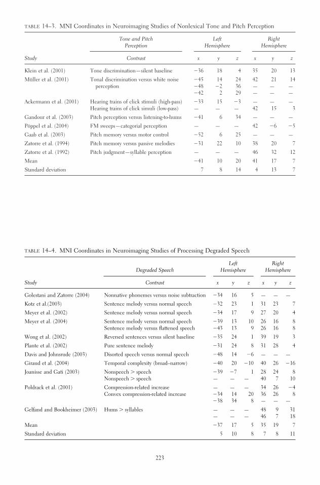
TABLE 14–3. MNI Coordinates in Neuroimaging Studies of Nonlexical Tone and Pitch Perception
Tone and Pitch Left RightPerception Hemisphere Hemisphere
Study Contrast x y z x y z
Klein et al. (2001) Tone discrimination—silent baseline �36 18 4 35 20 13
Müller et al. (2001) Tonal discrimination versus white noise �45 14 24 42 21 14perception �48 �2 36 — — —
�42 2 29 — — —
Ackermann et al. (2001) Hearing trains of click stimuli (high-pass) �33 15 �3 — — —Hearing trains of click simuli (low-pass) — — — 42 15 3
Gandour et al. (2003) Pitch perception versus listening-to-hums �41 6 34 — — —
Pöppel et al. (2004) FM sweeps—categorial perception — — — 42 �6 �5
Gaab et al. (2003) Pitch memory versus motor control �52 6 25 — — —
Zatorre et al. (1994) Pitch memory versus passive melodies �31 22 10 38 20 7
Zatorre et al. (1992) Pitch judgment—syllable perception — — — 46 32 12
Mean �41 10 20 41 17 7
Standard deviation 7 8 14 4 13 7
TABLE 14–4. MNI Coordinates in Neuroimaging Studies of Processing Degraded Speech
Left RightDegraded Speech Hemisphere Hemisphere
Study Contrast x y z x y z
Golestani and Zatorre (2004) Nonnative phonemes versus noise subtraction �34 16 5 — — —
Kotz et al.(2003) Sentence melody versus normal speech �32 23 1 31 23 7
Meyer et al. (2002) Sentence melody versus normal speech �34 17 9 27 20 4
Meyer et al. (2004) Sentence melody versus normal speech �39 13 10 26 16 8Sentence melody versus flattened speech �43 13 9 26 16 8
Wong et al. (2002) Reversed sentences versus silent baseline �35 24 1 39 19 3
Plante et al. (2002) Pure sentence melody �31 24 8 31 28 4
Davis and Johnsrude (2003) Disorted speech versus normal speech �48 14 �6 — — —
Giraud et al. (2004) Temporal complexity (broad–narrow) �40 20 �10 40 26 �16
Joanisse and Gati (2003) Nonspeech � speech �39 �7 1 28 24 8Nonspeech � speech — — — 40 7 10
Poldrack et al. (2001) Compression-related increase — — — 34 26 �4Convex compression-related increase �34 14 20 36 26 8
�38 34 8 — — —
Gelfand and Bookheimer (2003) Hums � syllables — — — 48 9 31— — — 46 7 18
Mean �37 17 5 35 19 7
Standard deviation 5 10 8 7 8 11
223

TABLE 14–5. MNI Coordinates in Neuroimaging Studies of Processing Speech Complexity
Left RightSpeech Complexity Hemisphere Hemisphere
Study Contrast x y z x y z
Vouloumanos et al. (2001) Speech versus complex nonspeech — — — 40 24 19Speech versus simple tones — — — 48 15 27
— — — 32 24 14
Benson et al. (2001) Speech complexity — — — 23 19 �5Speech � complexity interaction — — — 27 21 1
Mean — — — 34 21 11
Standard deviation — — — 10 4 13
TABLE 14–6. MNI Coordinates in Neuroimaging Studies of Covert and Overt Speech Production
Left RightSpeech Production Hemisphere Hemisphere
Study Contrast x y z x y z
Müller et al. (1997) Generating sentences versus repetition �35 �21 � 5 — — —
Riecker et al. (2000) Overt speech versus covert speech �69 �9 �17 66 �4 18�35 �19 ��4 — — —
Covert speech versus overt speech �66 � 5 �20 — — —
Riecker et al. (2002) Isochronous syllable repetition �27 �18 � 3 24 18 3Rhythmic syllable repetition �30 �18 � 3 33 20 3
Blank et al. (2002) Overt speech versus nonspeech perception �60 �10 � 8 — — —�36 �10 � 8 — — —
Fox et al. (2000) Syllable production �43 �13 �21 — — —
Ingham et al. (2004) Stuttering — — — 44 11 3— — — 36 8 �2
Mean �45 � 9 � 9 41 11 5
Standard deviation �16 �12 � 9 16 10 8
TABLE 14–7. MNI Coordinates in Neuroimaging Studies of Covert and Overt Music Production
Left RightMusic Production Hemisphere Hemisphere
Study Contrast x y z x y z
Perry et al. (1999) Singing versus complex pitch perception — — — 44 1 11
Riecker et al. (2000) Overt singing versus covert singing �69 �9 19 66 �4 20
— — — 32 17 �6
Covert singing versus overt singing — — — 69 �6 18
Brown et al. (2004) Monotonic vocalization �42 25 6 44 8 11
Melody repetition �48 6 11 40 14 12
Harmonization �42 24 8 40 16 10
— — — 44 10 12
Mean �50 12 11 47 7 11
Standard deviation 13 17 6 13 9 8
224

TABLE 14–8. MNI Coordinates in Neuroimaging Studies of Musical Perception
Left RightMusic Perception Hemisphere Hemisphere
Study Contrast x y z x y z
Janata et al. (2002) Music perception — — — 56 19 5— — — 60 22 20
Tillmann et al. (2003b) Dissonant—consonant — — — 45 �4 15Unrelated consonant—related consonant �52 8 0 49 15 0
�38 19 5 34 22 0
Mean �45 14 3 49 15 8
Standard deviation 10 8 4 10 11 9
TABLE 14–9. MNI Coordinates in Neuroimaging Studies of Sound Perception Production
Left RightSounds Hemisphere Hemisphere
Study Contrast x y z x y z
Engelien et al. (1995) Environmental sound perception versus rest — — — 34 26 14
Environmental sound categorization versus rest — — — 24 19 �8�42 20 18 — — —
Maeder et al. (2001) Sound recognition � sound localization — — — 36 �14 21
Sound localization � sound recognition �28 17 �6 — — —
Alain et al. (2001) Sound recognition � sound localization — — — 45 25 12— — — 52 18 14
Mean �35 19 6 39 15 11
Standard deviation 10 2 17 11 16 11
TABLE 14–10. MNI Coordinates in Neuroimaging Studies of Processing Affective Prosody
Left RightAffective Prosody Hemisphere Hemisphere
Study Contrast x y z x y z
Buchanan et al. (2000) Emotion versus verbal — — — 44 20 16
Kotz et al. (2003) Positive � neutral �51 13 2 53 13 5�54 19 5 44 21 15
Negative � neutral �44 21 15 — — —�53 21 4 — — —
Mean �51 19 7 47 18 12
Standard deviation 5 4 6 5 4 6
225

the rostrodorsal part of the lateral pars opercularis.The mean right hemisphere coordinate “38 22 6”matches the anterior insula.
Lexical Tones/Sublexical Intonation
Table 14–2 and Figure 14–2 depict an overview ofimaging studies that examined two related processing
domains that play a cardinal role in “prosodic pro-cessing.” As comprehensively described by Gandourand colleagues (2000, 2002, 2003), native speakers oftonal languages (like Chinese and Thai) are moreamenable to pick up pitch contours that span shortertemporal integration windows since these fine-grainedacoustic cues help decode the lexical meaning of aspoken word. In contrast, speakers of intonational lan-
226 PSYCHOLINGUISTIC INVESTIGATIONS
TABLE 14–11. MNI Coordinates in Neuroimaging Studies of Mismatch Responses Obtained from AuditoryOddball Paradigms
Left RightMismatch Response Hemisphere Hemisphere
Study Contrast x y z x y z
Opitz et al. (2002) Mismatch response (medium deviant versus standard) — — — 46 20 8
Doeller et al. (2003) Mismatch response (small deviant versus medium deviant) — — — 54 24 6Mismatch response (large deviant versus medium deviant) �54 26 8 50 24 6
Mean �54 26 8 50 23 7
Standard deviation — — — 4 2 1
TABLE 14–12. MNI Coordinates in Neuroimaging Studies of Dichotic Listening
Left RightDichotic Listening Hemisphere Hemisphere
Study Contrast x y z x y z
Jäncke et al. (2003) Dichotic tone listening � rest — — — 48 12 20— — — 44 16 �8
Divided � focussed attention �36 24 �4 — — —
Jäncke and Shah (2002) Divided � binaural �60 12 8 56 24 �16�44 24 �8 — — —
FL � binaural — — — 48 28 8FR � binaural �48 28 8 — — —
Jäncke et al. (2001) Dichotic versus rest �56 16 12 51 17 16
Pollmann et al. (2004) CV-syllable (left ear) versus null event �43 13 20 — — —
Hugdahl et al. (1999) CV versus tones (dichotic) �38 24 4 32 22 2
Hugdahl et al. (2000) CV versus music (dichotic divided) �53 19 3a — — —FL versus FR — — — 62 16 4
Thomsen et al. (2004b) FL �32 22 �5 32 23 �5— — — 48 13 21
�40 32 9 — — —
Thomsen et al. (2004a) Dichotic versus binaural �30 25 �5 32 25 �8Divided versus forced �34 19 �9 36 23 �13FR versus FR binaural �30 25 �5 32 25 �6FL versus FL binaural �30 25 �6 34 25 �6
Mean �41 22 2 43 21 1
Standard deviation 10 6 9 10 5 12
aAveraged data.

FIGURE 14–1. Sublexical phonological perception of speech.
FIGURE 14–2. Perception of lexical tones and sublexical intonation.
FIGURE 14–3. Perception of tonal and pitch information.
227

FIGURE 14–4. Perception of degraded speech.
FIGURE 14–5. Perception of speech complexity.
FIGURE 14–6. Covert and overt speech production.
228

FIGURE 14–7. Covert and overt music production.
FIGURE 14–8. Perception of music.
FIGURE 14–9. Perception of sounds.
229

FIGURE 14–10. Perception of affective prosody.
FIGURE 14–11. Perception of auditory deviants in a train of tonal stimuli.
FIGURE 14–12. Dichotic listening.
230

guages (like English or German) are more sensitiveto slow prosodic modulations that may convey lin-guistically relevant information, i.e. sentence modus.The current analysis includes four neuroimaging stud-ies (two positron emission tomography, two fMRI)that incorporate seven conditional contrasts yieldingnine activational clusters associated with either pro-cessing of lexical tones or sublexical intonation. Ofnine activational clusters, six (67%) are localized inthe left hemisphere, while three out of nine clusters
(33%) are situated in the right hemisphere. The meanleft hemisphere coordinate “�43 10 32” correspondsto the ventral border of the precentral gyrus adjoin-ing the dorsal part of the pars opercularis. The meanright hemisphere coordinate “46 18 32” can be pre-cisely localized in the pars opercularis. Four of sevencontrasts were derived from analyses testing the pro-cessing of lexical tones and exclusively revealed acti-vational increases in the left hemisphere. The re-maining three contrasts focused on perception of
FRONTAL OPERCULUM IN PERCEPTION AND PRODUCTION 231
affepros
Label
Label
Dendogram Left Hemisphere
Dendogram Right Hemisphere
0 5 10 15
Rescaled distance cluster combine
20 25
musicper
musicpro
speechpro
sounds
degraded
dichlist
mismatch
tonepitch
phonology
lextone
[10]
[8]
[7]
[6]
[9]
[4]
[12]
[11]
[3]
[1]
[2]
affepros
0 5 10 15
Rescaled distance cluster combine
20 25
musicper
mismatch
phonology
dichlist
sounds
tonepitch
speechcomp
degraded
musicpro
speechpro
[10]
[8]
[11]
[1]
[12]
[9]
[3]
[5]
[4]
[7]
[6]
lextone [2]
FIGURE 14–13. Results of hierarchical cluster analy-sis based on Euclidian distance. (Top) The extent towhich auditory domains cluster in the left hemisphere(LH). (Bottom) The extent to which auditory domainscluster in the right hemisphere (RH). Short code la-bels correspond to single auditory domains listed in
Tables 1 through 12. Numbers in parentheses refer tothe numbering of Tables 1 through 12. There is noLH cluster for speech complexity which results in un-even number of domains for LH (n � 11) and RH(n � 12).

intonational cues and exposed bilateral and rightwardactivity patterns.
Tone and Pitch Perception
Table 14–3 and Figure 14–3 sketch an overview ofimaging studies focusing primarily on basic auditoryfunctions which are supposed to be essential in speechand musical processing (Zatorre et al., 2002). Thecurrent analysis comprises eight neuroimaging stud-ies (four positron emission tomography, four fMRI)that incorporate nine conditional contrasts yielding 14activational clusters associated with either processingof tonal and pitch information. Of 14 activational clus-ters, 8 (57%) are localized in the left hemisphere while6 of 14 clusters (43%) are situated in the right hemi-sphere. Both the mean left hemisphere coordinate“�41 10 20” and the mean right hemisphere coordi-nate “41 17 7” correspond to the dorsomedial part ofthe pars opercularis with the latter being close to theinsular cortex. Based on this analysis, it appears thatneither the left nor the right frontal operculum dis-plays a dominating preference for tone and pitch per-ception in a nonlinguistic context.
Degraded Speech
Table 14–4 and Figure 14–4 compile a number ofimaging studies that triggered frontal operculum ac-tivity by exposing participants to speech that had beenartificially or naturally manipulated. More specifi-cally, intelligibility was reduced or even canceled outby applying filtering techniques that (partly) removedsegmental information or recorded human hummingwas presented which also lacked any segmentable lin-guistic information. The current analysis is made upof 11 neuroimaging studies (1 positron emission to-mography, 10 fMRI) that comprise 14 conditionalcontrasts resulting in 25 activational clusters that sup-ported the processing of unintelligible speech. Pre-cisely, 12 of 25 activational clusters (48%) are local-ized in the left hemisphere and 13 of 25 clusters (52%)are identified in the right hemisphere. Both the meanleft hemisphere coordinate “�37 17 5” and the meanright hemisphere coordinate “35 19 7” correspond tothe anterior portion of the insular cortex bordering themedial wall of the Rolandic operculum. Based on thisanalysis, it seems plausible that processing a degradedacousticc percept like unintelligible speech necessi-
tates both the left and right intra-Sylvian regions. No-tably, the mean left and right hemisphere coordinatesare outside the frontal operculum and refer to a locusin the phylogenetically distinct anterior insula.
Speech Complexity
Table 14–5 and Figure 14–5 list two fMRI studies thatidentified the neural substrates underlying speechcomplexity. Benson and co-workers (2001) used mul-tivariate multiple regression of fMRI responses to stimuli that systematically contrasted along speech/nonspeech, acoustic, or phonetic complexity and natural/synthetic dimensions. In an oddball detectiontask, Vouloumanos and colleagues (2001) contrastedspeech with nonspeech analogues that were matchedalong key temporal and spectral dimensions. The twostudies provide four conditional contrasts yielding fiveactivational clusters corresponding to the processingof speech complexity. Notably, all clusters were con-sistently localized in the right hemisphere. The meanright hemisphere coordinate “34 21 11” refers to themost anterior tip of the right insular cortex directlyadjacent to the deeply buried compartment of thefrontal operculum. The analysis suggests that the pro-cessing of speech complexity preferentially drives theright hemisphere.
Speech Production
Table 14–6 and Figure 14–6 summarize imagingstudies that examined overt and covert speech pro-duction and stuttering. The current analysis com-prises six neuroimaging studies (four positron emis-sion tomography, two fMRI) that include eightconditional contrasts yielding 14 activational clustersgenerated by a variety of speech production tasks. Of14 activational clusters, 9 (65%) are localized in theleft hemisphere, while 5 of 14 clusters (35%) are sit-uated in the right hemisphere. The mean left hemi-sphere coordinate “�45 9 9” is medially situated nearthe border between the caudal part of the pars oper-cularis and the rostral part of the Rolandic opercu-lum. The mean right hemisphere coordinate “41 115” refers to a voxel in the insular cortex in the vicin-ity of the medial wall of the pars opercularis and theRolandic operculum. However, standard deviationsfor these samples are relatively large, so that the pic-ture drawn by this analysis may be inconclusive. Large
232 PSYCHOLINGUISTIC INVESTIGATIONS

differences between expressive modes, i.e., overt ver-sus covert production, simple repetition versus sentencegeneration, may account for these inconsistencies.Even though two thirds of the contrasts point to aninvolvement of the left hemisphere fronto-opercularcortex in expressive functions, at least one third of contrasts indicate an engagement of the right intra-Sylvian cortex, in particular, when the neural under-pinnings of stuttering are imaged.
Music Production
Table 14–7 and Figure 14–7 depict an overview ofimaging studies that addressed the role the frontal operculum plays in music production. The presentanalysis includes three neuroimaging studies (twopositron emission tomography, one fMRI) that com-prise six conditional contrasts resulting in 12 activa-tional clusters associated with either overt or covertsinging. Of 12 activational clusters, 4 (33%) are lo-calized in the left hemisphere, while 8 of 12 clusters(67%) are situated in the right hemisphere. The meanleft hemisphere coordinate “�50 12 11” correspondsto the ventral part of the pars opercularis. The meanright hemisphere coordinate “47 7 11” could be lo-calized at the junction of the Rolandic operculum,the pars opercularis, and the insular cortex. Interest-ingly, the right mean coordinate observed for musicproduction is almost identical to the right mean co-ordinate observed for speech production. The twomodes additionally share relatively large standard de-viation scores, which belies the considerable variancebetween contrasts entered in this analysis. In sum, theanalysis implies that music production accompaniesan activational rightward asymmetry in the intra-Syl-vian cortex. Furthermore, it is reasoned that this par-ticular area can be considered to play a fundamentalrole in both speech and music production.
Music Perception
Table 14–8 and Figure 14–8 present two fMRI stud-ies that image the frontal operculum while partici-pants heard musical melodies. The review on musicperception comprises three conditional contrasts re-sulting in seven activational clusters that accompanythe perception of music. Of seven activational clus-ters, two (29%) are localized in the left hemisphereand five of seven clusters (61%) are identified in the
right hemisphere. Both the mean left hemisphere co-ordinate “�45 14 3” and the mean right hemispherecoordinate “49 15 8” correspond to the ventromedialstrip of the pars opercularis, with the first borderingthe insular cortex. Even though Tillmann and col-leagues (2003a) observed clusters in the left frontalcortex, this analysis implicates a general right frontalsuperiority in music perception. The specific de-mands imposed by the task in the study by Tillmannand colleagues apparently addressed aspects of se-quence expectancies that may account for the re-cruitment of Broca’s region.
Sounds
Table 14–9 and Figure 14–9 show an overview ofthree imaging studies (one positron emission tomog-raphy, two fMRI) that examined sound processing andincorporates five conditional contrasts yielding sevenactivational clusters associated with sound processing.Of seven activational clusters, two (29%) are localizedin the left hemisphere and five of seven clusters (61%)are identified in the right hemisphere, which displaysa clear prevalence of the right frontal regions in soundprocessing. Both the mean left hemisphere coordinate“�35 19 6” and the mean right hemisphere coordi-nate “39 15 11” correspond to the anterior insula withthe latter adjoining to the medial wall of the pars opercularis. Notably, tasks that explicitly focused onsound recognition activated the right frontal cortex,while sound localization and categorization excitedthe left frontal cortex.
Affective Prosody
Table 14–10 and Figure 14–10 depict an overview oftwo fMRI studies that addressed the role the frontaloperculum plays in affective prosody. This analysiscomprises three conditional contrasts resulting inseven activational clusters associated with processingaffective spoken utterances. Of seven activational clus-ters, four (57%) are localized in the left hemispherewhile three of seven clusters (43%) are situated in theright hemisphere. The left hemisphere coordinate“�51 19 7” corresponds to the most posterior part ofthe pars triangularis. The mean right hemisphere co-ordinate “47 18 12” could also be localized in the lat-eral pars opercularis close to the small ascendingbranch of the anterior segment of the Sylvian fissure.
FRONTAL OPERCULUM IN PERCEPTION AND PRODUCTION 233

Mismatch Response
Table 14–11 and Figure 14–11 compile two fMRI stud-ies that elicited frontal operculum activity during par-ticipant’s performance of a preattentive auditory oddballtask. More specifically, volunteers heard continuoustrains of identical tones that comprised a small propor-tion of deviant tones differing in acoustic parameters(e.g., intensity, frequency). The current analysis con-tains three conditional contrasts resulting in four acti-vational clusters more strongly activated by deviant rel-ative to standard tones. Of four activational clusters, one(25%) is localized in the left hemisphere and three offour clusters (75%) are identified in the right hemi-sphere. Both the mean left hemisphere coordinate“�54 26 8” and the mean right hemisphere coordinate“50 23 7” correspond to the triangular part of the IFG.Based on this analysis, it seems plausible that the rightIFG more prevalently subserves the neurofunctionalmechanism underlying deviancy detection.
Dichotic Listening
Table 14–12 and Figure 14–12 show evidence fromeight studies (two positron emission tomography, sixfMRI) that induces activity within the frontal oper-culum while participants performed a dichotic lis-tening test. This analysis comprises 15 conditionalcontrasts resulting in 27 activational clusters. Of 27activational clusters, 14 (52%) are localized in the lefthemisphere and 13 of 27 clusters (48%) are identifiedin the right hemisphere. Both the mean left hemi-sphere coordinate “�41 22 2” and the mean righthemisphere coordinate “43 21 1” correspond to theventromedial strip of the pars triangularis borderingthe anterior insular cortex. In the context of thisoverview, it is evident that lateralization of activationalcluster varies as a function of monaural and binauralpresentation.
Cluster Analysis
Figure 14–13 presents the results of hierarchical clus-ter analysis based on the Euclidian distance betweenMNI coordinates, implicating a differential pattern inthe left relative to the right hemisphere. In the lefthemisphere, we identify three clusters that include upto four functional domains. The first cluster (mean“�48 14 8”, ventral part of pars opercularis) subsumes“affective prosody,” “music perception,” “music pro-
duction,” and “speech production.” The second clus-ter (mean “�38 19 4”, anterior insula) groups“sounds,” “degraded speech,” and “dichotic listen-ing.” The third cluster (mean “�43 12 24”, dorsal partof pars opercularis) includes “tone and pitch percep-tion,” “phonology,” and “lexical tones/sublexical in-tonation.” The domain “mismatch response” revealsa large distance to the other clusters. A different pic-ture can be drawn for the right hemisphere. Theanalysis yields four clusters showing “close” distances.Analogous to the left hemisphere the first cluster(mean “49 19 9”, anterior ventral part of pars oper-cularis) subsumes “affective prosody,” “music per-ception,” and in moderate distance “mismatch re-sponse.” Also similar to the left hemisphere, a secondcluster (mean “44 9 8”, border zone between pars opercularis, insula, and Rolandic operculum) config-ures “music production” and “speech production.” Athird cluster (mean “40 16 9”, junction at pars oper-cularis and anterior insula) groups “sounds” and “toneand pitch perception.” A fourth cluster (mean “35 209”, anterior insula) includes “speech complexity” and“degraded speech.” According to the analysis, the lat-ter cluster is in a close distance to the third cluster,while both the third and the fourth cluster are re-motely distant from the first and the second cluster.“Phonology,” “dichotic listening,” and “lexical tones/sublexical intonation” reveal a large distance to theother clusters.
DISCUSSION
A comprehensive discussion of all studies or contrastswe have included is beyond the potentialities ofthisreview. We therefore concentrate on the discussion ofclustered domains and attempt to illustrate the com-monalities between them. Generally, the results ob-tained from this review demonstrate that a multitudeof heterogeneous tasks and processes illuminate therole of the frontal operculum in both the left and theright hemisphere with the first primarily involved inphonological processing and the latter preferentiallydriven by paralinguistic and nonlinguistic domains,i.e., speech complexity, music perception and pro-duction, and sound perception. We discuss results forleft and right hemisphere separately in turn.
Interestingly, the mean coordinates for “phonol-ogy” are located in the most dorsal part of the pars opercularis, while all other spots we subsumed are sit-
234 PSYCHOLINGUISTIC INVESTIGATIONS

uated in more ventral parts of the pars opercularis. Inthe left hemisphere, “phonology” clusters with “toneand pitch perception” and “lexical tones.” It holds inparticular for the latter domain that linguistically rel-evant stimuli were tested. The majority of phonolog-ical contrasts reviewed here and the perception ofpitch contours associated with lexical tones have incommon that rapidly changing acoustic cues play adecisive role. Therefore, it is possible that the dorsalpart of the left pars opercularis belongs to the neuralcircuit that contributes to the processing of rapidlychanging acoustic information. The second cluster in-tegrates domains that particularly deal with musicaland intonational information. One might note thatboth music and affective prosody comprise emotionalinformation. However, this does not hold for the kindof speech investigated in the speech production par-adigms reviewed here. Therefore, it is more likely toassume that more general aspects involved in expres-sive and receptive functions related to (speech)melody account for this observation. Both perceptionof sounds and degraded speech activated the anteriorinsula. Like sounds, degraded speech does not com-prise any lexical information but only speech melody.In the studies we reviewed, volunteers had to eitherrecognize, categorize, or localize sounds, which is ademanding task. This assertion also holds for the per-ception of degraded speech and addresses our intro-ductory reasoning on the potential functional role ofthis area as a provider of extracomputational re-sources. We will get back to this issue later. Eventhough the cluster analysis grouped “dichotic listen-ing” together with sounds and degraded speech, webelieve that we should consider the latter as not be-ing part of former duo as the spot corresponding to dichotic listening seems to be located in the parstriangularis.
Traditionally, the right hemisphere has been con-sidered to be more amenable to musical processing(Zatorre et al., 2002) even though more recent viewsindicate that particular acoustic features, that is,slowly changing acoustic cues, account for the righthemisphere preference for music and melodic con-tours rather than music per se. Our review cannotfully corroborate this view as music perception wasfound to activate both left and right hemisphere sites.Analogous to the left hemisphere, “affective prosody”and “music perception” clustered closely together inthe ventral pars opercularis in the vicinity of the “mis-match response.” Based on this finding we are not
able to detect any commonality between these func-tions. At least this finding speaks to a general respon-siveness of the right pars opercularis to auditory stim-uli. Also similar to the left hemisphere is the findingthat mean coordinates for “speech production” and“music production” are nearby. However, in the righthemisphere, this duo is situated in the Rolandic operculum, which corresponds to the ventral premo-tor cortex. Since this brain region represents suprala-ryngeal articulators (lips, tongue), which play a rolein (sub)articulation (Kolb and Whishaw, 1995), its engagement in speech and music production is com-pelling. Obviously both speaking and singing consis-tently recruit the right Rolandic operculum. Interest-ingly, two recent fMRI studies that investigated theperception of intonation contour in sentences also ob-served an engagement of the right Rolandic opercu-lum, which makes this region a candidate for func-tions of auditory-motor integration (Meyer et al.,2002, 2004). There are two additional clusters in theright hemisphere. The first spot is situated moredeeply in the medial part of the pars opercularis closeto the anterior insula (recognition and perception ofsounds as well as perception of simple tones andpitch). The second spot can be directly localized inthe anterior insula (“degraded speech” and “speechcomplexity”). Considering the variance betweenmethods, experiments, subjects, etc. across all re-viewed studies, we do not think it is beneficial to dis-cuss a difference between these two clusters. Werather state that the right (and partly left) deep frontaloperculum and the anterior insula are part of theneural network processing multiple elementary as-pects provided by auditory stimuli. Neuroanatomi-cally the anterior insula cannot be considered part ofthe frontal operculum. The anterior insula has beenattributed to a variety of vascular, vestibular, olfactory,gustatory, visual, somatosensory, motor, and even au-ditory modulation (Bamiou et al., 2003; Flynn et al.,1999; Türe et al., 1999; Wise et al., 1999). Due to thepoor spatial resolution of positron emission tomogra-phy and fMRI studies, it is hard to decide to what ex-tent an activational cluster covering both the anteriorinsula and the deep frontal operculum originates fromthe first or the latter (or even from both the first andthe latter). Little is known about the cytoarchitectonicstructure of the deep portion of the frontal operculumso far. Thus, the precise border between the most an-terior tip of the anterior insula and the deep frontal operculum cannot be exactly determined, which
FRONTAL OPERCULUM IN PERCEPTION AND PRODUCTION 235

leaves researchers insecure about the origin and po-tential functional implication of activational clusterssituated in this brain region. Notably, activity in the“borderland” between (left and right) anterior insulaand the deep frontal operculum has primarily beenreported by brain imaging studies that examined theperception of degraded auditory percepts (Davis andJohnsrude, 2003; Giraud et al., 2004; Meyer et al.,2004; Plante et al., 2002; Wong et al., 2002). One re-cent statement that attempts to provide an interpreta-tion proposes that bilateral activity in this region dur-ing the perception of degraded speech might reflectthe effort to achieve a meaningful segmentation of theinflowing auditory input (Meyer et al., 2003). Thisview is affirmed by results obtained from a positronemission tomography study that demonstrated a bi-lateral activational increase in the deeply buried fron-topercular cortex reflecting an increase in processingeffort while participants performed a demanding pitchmemory task (Zatorre et al., 1994). Thus, we think itis plausible to assume the existence of two distinct pro-cessing systems in the frontal operculum. While thedorsolateral part of the pars opercularis and triangu-laris are preferentially adept at processing sequentialinformation as required by phonological and workingmemory tasks, the deep frontal operculum (and even-tually the anterior insula) activates when the percep-tion of auditory information interacts with the appli-cation of additional effort required to extract allrelevant auditory information from the acoustic sig-nal. With respect to functional lateralization, wewould like to emphasize that our review reveals a con-siderable proportion of right hemisphere clusters in-volved in a variety of auditory functions. Based on ourreview, it is not possible to determine differential func-tional roles for the left and the right frontal opercu-lum. It rather seems that the proper performance ofreceptive and expressive auditory tasks necessitates ei-ther a consistent or even a cooperative functioning ofboth the left and the right frontal opercula with a clearleftward asymmetry only observed for phonologicalprocessing. In sum, we conclude that neither the leftnor the right frontal operculum is selective for a particular function during auditory perception andproduction. The review rather provides compellingevidence that multiple heterogeneous tasks and ex-perimental contrasts recruit a relatively small territoryin the human inferior frontal cortex.
The purpose of this review was to summarize thedifferent auditory functions processed by the fronto-opercular region including the so-called Broca’s area
and the right-sided homotopic area. The 56 studieswe reviewed here have used a variety of experimentalauditory tasks tapping multiple auditory functions that(partly) engaged Broca’s area. We arbitrarily have triedto find a plausible way to classify and distinguish thesefunctions on the basis of current psycholinguisticand/or perception theories of auditory functions. Infact, we revealed several subregions either completelyor partly within the “classic” Broca’s area that are as-sociated with different auditory functions. Thus, onemight conclude we may have identified several dis-tinct cortical sites that accommodate neural assem-blies responsible for very specific auditory functions.However, that is not the aim of this review. We ratherwould like to emphasize the importance of being verycareful in interpreting the collected data for severalreasons: First, we have to be aware that the majorityof the studies included here used fMRI to localize he-modynamic responses. However, the relationship be-tween neural excitement and the BOLD responsemeasured by fMRI has not been fully clarified so far.Although most current authorities speculate thatBOLD responses are mainly triggered by synaptic ac-tivity (Logothetis, 2003; Logothetis et al., 2001), morerecent papers discuss the possible influence of elec-tric activity of the neurons on the measured BOLDresponse (Xiong et al., 2003). In addition, it is an openquestion to what extent peak activation or the spatialextension of activation is based on the same neuralunderpinnings (Marcar and Loenneker, 2004). Fur-thermore, fMRI and, to a greater degree, positronemission tomograph are neuroimaging methods witha relatively crude temporal resolution ranging from 4seconds to 2 minutes, which adds to the list of method-ological limitations associated with neuroimagingtechniques. Taken together, the hemodynamic re-sponse is a composite signal modulated by severalneurophysiological processes that are hardly to disso-ciate and it is subject to a relatively sparse temporalresolution. Obviously, these constraints constrict thesuitability of these methods to research auditory func-tions that occur in a range of milliseconds.
A second concern that is even more important inour view addresses an objection that is often raisedagainst the basic idea that the neuroimaging approachrelies on. Present neuroimagers (and even the authorsof this review) are often (at least implicitly) misguidedto interpret the circumscribed activation spots in amore or less phrenological way. For example, the con-fined sphere including MNI coordinates we report forphonological processing must not be considered to
236 PSYCHOLINGUISTIC INVESTIGATIONS

cover the particular region solely processing phono-logical functions. We rather need to reconcile theseneuroimaging findings with insights obtained fromclassic lesion and human neurophysiological studiesusing MEG or EEG methods. From the latter, weknow that psychologically relevant information iscoded in terms of the neural firing rate (firing overtime) and the correlated activity of neural oscillation(periodical neural activation patterns). Thus, as someresearchers pointed out, “time is the essence” whenwe are attempting to understand what the brain is really doing (Ringo et al., 1994). Principally, due tothe temporal constraints, neuroimaging researchpresently is not able to implement a closer analysis ofthe temporal aspects of the brain function. Currentlyand in the face of the outstanding popularity of neu-roimaging, more and more studies using traditionalneurophysiological methods (EEG, MEG) havenicely demonstrated that a particular sensory or cog-nitive process corresponds to the coupled firing of afocal or even largely distributed neural network. Thus,as a general rule, a particular and circumscribed brainregion does not control a psychological function; it ismore plausible to assume that the particular mannerof neural coupling within a neurofunctional networkand not the intensity of its activation is the main pa-rameter determining the quality of psychologicalfunctions (Engel et al., 2001). Recently developedelectrical imaging methods, such as LORETA (Pascual-Marqui et al., 1994; Pascual-Marqui, 1999),even allow for decent estimation of the neural sourcesof EEG signals recorded from scalp electrodes andcan therefore compete with conventional brain imag-ing. We believe this development has two implica-tions for the neuroimaging community. First, it be-comes more and more imperative to carefullycross-check hemodynamic data obtained from fMRIand positron emission tomography with neurophysio-logical data derived from EEG and MEG to optimizeour knowledge on both temporal and spatial aspectsof sensory, motor, and cognitive processes. Second,there is a compelling need to strictly pursue model-driven neuroimaging research rather than using neu-roimaging in a more explorative manner, which canlead to neuroimaging suffering from the detrimentaland undeserved reputation of “neophrenology.”
ACKNOWLEDGMENTS The authors wish to thank Ka-trin Amunts, Yosef Grodzinsky, Adam McNamara, andNatalia Estévez for helpful comments on the manuscript.During the preparation of this manuscript, Martin Meyer
was supported by Schweizer National Fonds (Swiss Na-tional Foundation) SNF 46234101, “Short-term andlong-term plasticity in the auditory system.”
Notes
1. Since the exact size, borders, subportions, andlocation of Broca’s area are still matters of debate(Amunts et al., 1999, 2004; Amunts and Zilles, Chapter2; Foundas et al., 1998; Petrides, Chapter 3; Uylings etal., 1999; Tomaiuolo et al., 1999), we do not aim to func-tionally and anatomically dissociate between Brodmannareas 44 and 45. In the context of the present review, thecommonly used notions “Broca’s region” and “Broca’sarea” are used exchangeably.
2. Besides activity in the frontal operculum, themajority of studies reviewed here also illuminated therole of temporal and even extra-Sylvian areas. However,the latter are not subject to the present paper and aretherefore not considered.
3. The latter studies tested the perception of puresentence melody, which can also be considered a de-graded percept as proper sentence constitutes sentencemelody as well as lexical and syntactic in formation.
References
Ackermann, H., Riecker, A., Mathiak, K., Erb, M.,Grodd, W., & Wildgruber, D. (2001). Rate-depen-dent activation of a prefrontal-insular-cerebellar net-work during passive listening to trains of click stim-uli: an fMRI study. NeuroReport, 12, 4087–4092.
Alain, C., Arnott, S.R., Hevenor, S., Graham, S., &Grady, C.L. (2001). ‘What’ and ‘where’ in the hu-man auditory system. Proceedings of the NationalAcademy of Science USA, 98, 12301–12306.
Amunts, K., Schleicher, A., Bürgel, U., Mohlberg, H.,Uylings, H.B.M., & Zilles, K. (1999). Broca’s regionrevisited: cytoarchitecture and intersubject variabil-ity. Journal of Comparative Neurology, 412, 319–341.
Amunts, K., Schleicher, A., & Zilles, K. (2004). Out-standing language competence and cytoarchitec-ture in broca’s speech region. Brain and Language,89, 346–353.
Bamiou, D.-E., Musiek, F.E., & Luxon, L.M. (2003).The insula (Island of Reil) and its role in auditoryprocessing. Behavioural Brain Research, 42, 143–154.
Belin, P., & Zatorre, R.J. (2003). Adaptation to speaker’svoice in right anterior temporal lobe. NeuroReport,14, 2105–2109.
Benson, R.R., Whalen, D.H., Richardson, M., Swainson,B., Clark, V.P., Lai, S., et al. (2001). Parametricallydissociating speech and nonspeech perception inthe brain using fMRI. Brain and Language, 78,364–396.
FRONTAL OPERCULUM IN PERCEPTION AND PRODUCTION 237

Binkofski, F., Amunts, K., Stephan, K.M., Posse, S.,Schormann, T., Freund, H.J., et al. (2000). Broca’sregion subserves imagery of motion: A combinedcytoarchitectonic and fMRI study. Human BrainMapping, 11, 273–285.
Binkofski, F., & Buccino, G. (2004). Motor functions ofBroca’s region. Brain and Language, 89, 362–369.
Blank, S.C., Scott, S.K., Murphy, K., Warburton, E., &Wise, R.J.S. (2002). Speech production: Wernicke,Broca and beyond. Brain, 125, 1829–1838.
Boemio, A., Fromm, S., Braun, A., & Pöppel, D. (2005).Hierarchical and asymmetric temporal sensitivity inhuman auditory cortices. Nature Neuroscience, 8,389–395.
Bookheimer, S. (2002). Functional MRI of language:New approaches to understanding the cortical or-ganization of semantic processing. Annual Reviewsof Neuroscience, 25, 151–188.
Brett, M., Johnsrude, I.S., & Owen, A.M. (2002). Theproblem of functional localization in the humanbrain. Nature Neuroscience Review, 3, 243–249.
Broca, P. (1861). Remarques sur la siège de la faculté delangage suivies d’une observation d’aphemie. Bul-letin de la Société d’Anatomie, 6, 330–357.
Broca, P. (1863). Localisation des fonctions cérébrales:Siége de langage articulé. Bulletin de la Sociétéd’Anthropologie de Paris, 4, 200–208.
Brown, S., Martinez, M.J., Hodges, D.A., Fox, P.T., &Parsons, L.M. (2004). The song system of the hu-man brain. Brain Research Cognitive Brain Re-search, 20, 363–375.
Buccino, G., Binkofski, F., & Riggio, L. (2004). The mir-ror neuron system and action recognition. Brainand Language, 89, 370–376.
Buchanan, T.W., Lutz, K., Mirzazade, S., Specht, K.,Shah, N.J., Zilles, K., et al. (2000). Recognition ofemotional prosody and verbal components of spo-ken language: An fMRI study. Brain Research &Cognitive Brain Research, 9, 227–238.
Burton, M.W. (2001). The role of inferior frontal cortexin phonological processing. Cognitive Science, 25,695–709.
Burton, M.W., Small, S.L., & Blumstein, S.E. (2000).The role of segmentation in phonological process-ing: an fMRI investigation. Journal of CognitiveNeuroscience, 12, 679–690.
Davis, M.H., & Johnsrude, I.S. (2003). Hierarchical pro-cessing in spoken language comprehension. Journalof Neuroscience, 23, 3423–3431.
Demonet, J.-F., Chollet, F., Ramsay, S., Cardebat, D.,Nespoulous, J., Wise, R., et al. (1992). The anatomyof phonological and semantic processing in normalsubjects. Brain, 115, 1753–1768.
Demonet, J.-F., Price, C.J., Wise, R., & Frackowiak,R.S.J. (1994). A PET study of cognitive strategies innormal subjects during language tasks: Influence ofphonetic ambiguity and sequence processing onphoneme monitoring. Brain, 117, 671–682.
Doeller, C.F., Opitz, B., Mecklinger, A., Krick, C., Reith, W., & Schröger, E. (2003). Prefrontal cortexinvolvement in preattentive auditory deviance de-tection: Neuroimaging and electrophysiological ev-idence. NeuroImage, 20, 1270–1282.
Engel, A.K., Fries, P., & Singer, W. (2001). Dynamic pre-dictions: Oscillations and synchrony in top-downprocessing. Nature Neuroscience Review, 2, 704–716.
Engelien, A., Silbersweig, D., Stern, E., Huber, W.,Döring, W., Frith, C., et al. (1995). The functionalanatomy of recovery from auditory agnosia. A PETstudy of sound categorization in a neurological pa-tient and normal controls. Brain, 118, 1395–1409.
Fiez, J.A., Raichle, M.E., Miezin, F.M., Petersen, S.E.,Tallal, P., & Katz, W.F. (1995). PET studies of au-ditory and phonological processing: Effects of stim-ulus characteristics and task demands. Journal ofCognitive Neuroscience, 7, 357–375.
Flynn, F.G., Benson, D.F., & Ardila, A. (1999). Anatomyof the insula—functional and clinical correlates.Aphasiology, 13, 55–78.
Foundas, A.L., Eure, K.F., Luevano, L.F., & Wein-berger, D.R. (1998). MRI asymmetries of Broca’sarea: The pars triangularis and pars opercularis.Brain and Language, 64, 282–296.
Fox, P.T., Ingham, R.J., Ingham, J.C., Zamarripa, F.,Xiong, J.H., & Lancaster, J.L. (2000). Brain corre-lates of stuttering and syllable production. A PETperformance-correlation analysis. Brain, 123, 1985–2004.
Friederici, A.D. (2002). Towards a neural basis of audi-tory sentence processing. Trends in Cognitive Sci-ences, 6, 78–84.
Friederici, A.D. (2004). Processing local transitions ver-sus long-distance syntactic hierarchies. Trends inCognitive Sciences, 8, 245–247.
Friederici, A.D., & Alter, K. (2004). Lateralization of au-ditory language functions: A dynamic dual pathwaymodel. Brain and Language, 89, 267–276.
Friederici, A.D., Meyer, M., & von Cramon, D.Y.(2000). Auditory language comprehension: Anevent-related fMRI study on the processing of syn-tactic and lexical information. Brain and Language,75, 465–477.
Gaab, N., Gaser, C., Zaehle, T., Jäncke, L., & Schlaug,G. (2003). Functional anatomy of pitch memory—An fMRI study with sparse temporal sampling. Neu-roImage, 19, 1417–1426.
238 PSYCHOLINGUISTIC INVESTIGATIONS

Gandour, J., Dzemidzic, M., Wong, D., Lowe, M., Tong,Y., Hsieh, L., et al. (2003). Temporal integration ofspeech prosody is shaped by language experience:An fMRI study. Brain and Language, 84, 318–336.
Gandour, J., Wong, D., Hsieh, L., Weinzapfel, B., VanLancker, D., & Hutchins, G.D. (2000). A crosslin-guistic PET study of tone perception. Journal ofCognitive Neuroscience, 12, 207–222.
Gandour, J., Wong, D., Lowe, M., Dzemidzic, M.,Satthamnuwong, N., Tong, Y., et al. (2002). Acrosslinguistic fMRI study of spectral and temporalcues underlying phonological processing. Journal ofCognitive Neuroscience, 14, 1076–1087.
Gelfand, J.R., & Bookheimer, S.Y. (2003). Dissociatingneural mechanisms of temporal sequencing andprocessing phonemes. Neuron, 38, 831–842.
Gernsbacher, M.A., & Kaschak, M.P. (2003). Neuroimag-ing studies on language production and comprehen-sion. Annual Review of Psychology, 54, 91–114.
Giraud, A.L., Kell, C., Thierfelder, C., Sterzer, P., Russ,M.O., Preibisch, C., et al. (2004). Contributions ofsensory input, auditory search and verbal compre-hension to cortical activity during speech process-ing. Cerebral Cortex, 14, 247–255.
Giraud, A.L., Price, C.J., Graham, J.M., Truy, E., &Frackowiak, R.S.J. (2001). Cross-modal plasticityunderpins language recovery after cochlear im-plantation. Neuron, 30, 657–663.
Golestani, N., & Zatorre, R.J. (2004). Learning newsounds of speech: Reallocation of neural substrates.NeuroImage, 21, 494–506.
Goodglass, H. (1976). Agrammatism. In H. Whitacker &H.A. Whitacker (Eds.), Studies in neurolinguistics.Vol. 1 New York: Academic Press.
Gruber, O., Kleinschmidt, A., Binkofski, F., Steinmetz,H., & von Cramon, D.Y. (2000). Cerebral corre-lates of working memory for temporal information.NeuroReport, 11, 1689–1693.
Hickok, G., & Pöppel, D. (2004). Dorsal and ventralstreams: A framework for understanding aspects ofthe functional anatomy of language. Cognition, 92,67–99.
Hoen, M., Pachot-Clouard, M., Segebarth, C., &Dominey, P.F. (in press). When Broca experiencesthe Janus syndrome. An er-fMRI study comparingsentence comprehension and cognitive sequenceprocessing. Cortex.
Hsieh, L., Gandour, J., Wong, D., & Hutchins, G.D.(2001). Functional heterogeneity of inferior frontalgyrus is shaped by linguistic experience. Brain andLanguage, 76, 227–252.
Hugdahl, K., Bronnick, K., Kyllingsbaek, S., Law, I.,Gade, A., & Paulson, O.B. (1999). Brain activationduring dichotic presentations of consonant-vowel
and musical instrument stimuli: A 15O-PETstudy.Neuropsychologia, 37, 431–440.
Hugdahl, K., Law, I., Kyllingsbaek, S., Bronnick, K.,Gade, A., & Paulson, O.B. (2000). Effects of atten-tion on dichotic listening: A 15O-PET study. Human Brain Mapping, 10, 87–97.
Indefrey, P., & Cutler, A. (2004). Prelexical and lexicalprocessing in listening. In M.A. Gazzaniga (Ed.),The new cognitive neuroscience (pp. 759–774). Cam-bridge, MA: MIT Press.
Ingham, R.J., Fox, P.T., Ingham, J.C., Xiong, J., Zamar-ripa, F., Hardies, L.J., et al. (2004). Brain correlatesof stuttering and syllable production: Gender com-parison and replication. Journal of Speech, Lan-guage, and Hearing Research, 14, 321–341.
Janata, P., Birk, J.L., Van Horn, J.D., Leman, M., Till-mann, B., & Bharucha, J.J. (2002). The corticaltopography of tonal structures underlying Westernmusic. Science, 298, 2167–2170.
Jäncke, L., Buchanan, T., Lutz, K., & Shah, N.J.S.(2001). Focused and non-focused attention in ver-bal and emotional dichotic listening: an fMRIstudy. Brain and Language, 78, 349–363.
Jäncke, L., Kleinschmidt, A., Mirzazade, S., & Freund,H.-J. (1999). The sensorimotor role of parietal cortexin linking the perception and creation of objectshapes: An fMRI study. Cerebral Cortex, 11, 114–121.
Jäncke, L., & Shah, N.J. (2002). Does dichotic listeningprobe temporal lobe functions? Neurology, 58,736–743.
Jäncke, L., Specht, K., Shah, N.J., & Hugdahl, K. (2003).Focused attention in a simple dichotic listeningtask: An fMRI experiment. Brain Research Cogni-tive Brain Research, 16, 257–266.
Joanisse, M.F., & Gati, J.S. (2003). Overlapping neuralregions for processing rapid temporal cues in speechand nonspeech signals. NeuroImage, 19, 64–79.
Klein, D., Zatorre, R.J., Milner, B., & Zhao. V. (2001).A cross-linguistic PET study of tone perception inMandarin Chinese and English speakers. Neu-roImage, 13, 646–653.
Kolb, B., & Whishaw, I.Q. (1995). Fundamentals of hu-man neuropsychology. New York: Freeman.
Kotz, S.A., Meyer, M., Alter, K., Besson, M., von Cra-mon, D.Y., & Friederici, A.D. (2003). On the lat-eralization of emotional prosody: An event-relatedfunctional MR investigation. Brain and Language,86, 366–376.
Logothetis, N.K. (2003). The underpinnings of theBOLD functional magnetic resonance imaging sig-nal. Journal of Neuroscience, 23, 3963–3971.
Logothetis, N.K., Pauls, J., Augath, M., Trinath, T., &Oeltermann, A. (2001). Neurophysiological inves-
FRONTAL OPERCULUM IN PERCEPTION AND PRODUCTION 239

tigation of the basis of the fMRI signal. Nature, 412,150–157.
Maeder, P.P., Meuli, R.A., Adriani, M., Bellmann, A.,Fornari, E., Thiran, J.-P., et al. (2001). Distinctpathways involved in sound recognition and local-ization: A human fMRI study. NeuroImage, 14,802–816.
Marcar, V.L., & Loenneker, T. (2004). The BOLD re-sponse: A new look at an old riddle. NeuroReport,15, 1997–2000.
Martin, R.C. (2003). Language processing: Functionalorganization and neuroanatomical basis. AnnualReview of Psychology, 54, 55–89.
Meyer, M., Alter, K., & Friederici, A.D. (2003). Func-tional MR imaging exposes differential brain re-sponses to syntax and prosody during auditory sen-tence comprehension. Journal of Neurolinguistics,16, 277–300.
Meyer, M., Alter, K., Friederici, A.D., Lohmann, G., &von Cramon, D.Y. (2002). Functional MRI revealsbrain regions mediating slow prosodic modulationsin spoken sentences. Human Brain Mapping, 17,73–88.
Meyer, M., Steinhauer, K., Alter, K., Friederici, A.D., &von Cramon, D.Y. (2004). Brain activity varies withmodulation of dynamic pitch variance in sentencemelody. Brain and Language, 89, 277–289.
Mohr, J.P., Pessin, M.S., Finkelstein, S., Funkenstein,H.H., Duncan, G.W., & Davis, M.D. (1978). Brocaaphasia: Pathologic and clinical. Neurology, 28,311–324.
Müller, R.-A., & Basho, S. (2004). Are nonlinguisticfunctions in “Broca’s area” prerequisites for lan-guage acquisition? fMRI findings from an ontoge-netic viewpoint. Brain and Language, 89, 329–336.
Müller, R.-A., Kleinhans, N., & Courchesne, E. (2001).Broca’s area and the discrimination of frequencytransitions: A functional MRI study. Brain and Lan-guage, 76, 70–76.
Müller, R.-A., Kleinhans, N., Pierce, K., Kemmotsu, N.,& Courchesne, E. (2002). Functional MRI of mo-tor sequence acquisition: Effects of learning stageand performance. Brain Research Cognitive BrainResearch, 14, 277–293.
Müller, R.-A., Rothermel, R.D., Behen, M.E., Muzik,O., Mangner, T.J., & Chugani, H.T. (1997). Re-ceptive and expressive language activations for sen-tences: A PET study. NeuroReport, 8, 3767–3770.
Noesselt, T., Shah, N.J., & Jäncke, L. (2003). Top-downand bottom-up modulation of language related areas—An fMRI study. BMC Neuroscience, 4.
Opitz, B., Rinne, T., Mecklinger, A., von Cramon, D.Y.,& Schröger, E. (2002). Differential contribution of
frontal and temporal cortices to auditory change de-tection: fMRI and ERP results. NeuroImage, 15,167–174.
Pascual-Marqui, R.D. (1999). Reviews of methods forsolving the EEG inverse problem. InternationalJournal of Bioelectromagnetism, 1, 75–86.
Pascual-Marqui, R.D., Michel, C.M., & Lehmann, D.(1994). Low resolution electromagnetic tomogra-phy: A new method for localizing electrical activi-tiy in the brain. International Journal of Psy-chophysiology, 18, 49–65.
Paulesu, E., Frith, C.D., & Frackowiak, R.S.J. (1993).The neural correlates of the verbal components ofworking memory. Nature, 362, 342–345.
Perry, D.W., Zatorre, R.J., Petrides, M., Alivisatos, B.,Meyer, E., & Evans, A.C. (1999). Localization ofcerebral activity during simple singing. Neuro-Report, 10, 3979–3984.
Plante, E., Creusere, M., & Sabin, C. (2002). Dissociat-ing sentential prosody from sentence processing: ac-tivation interacts with task demands. NeuroImage,17, 401–410.
Pöppel, D. (1996). A critical review of PET studies ofphonological processing. Brain and Language, 55,317–351.
Pöppel, D., Guillemin, A., Thompson, J., Fritz, J., Bave-lier, D., & Braun, A.R. (2004). Auditory lexical de-cision, categorical perception, and FM directiondiscrimination differentially engage left and rightauditory cortex. Neuropsychologia, 42, 183–200.
Poldrack, R.A., Temple, E., Protopapas, A., Nagarajan,S., Tallal, P., Merzenich, M., et al. (2001). Rela-tions between the neural bases of dynamic auditoryprocessing and phonological processing: Evidencefrom fMRI. Journal of Cognitive Neuroscience, 13,687–697.
Pollmann, S., Lepsien, J., Hugdahl, K., & von Cramon,D.Y. (2004). Auditory target detection in dichoticlistening involves the orbitofrontal and hippocam-pal paralimbic belts. Cerebral Cortex, 14, 903–913.
Riecker, A., Ackermann, H., Wildgruber, D., Dogil, G.,& Grodd, W. (2000). Opposite hemispheric later-alization effects during speaking and singing at mo-tor cortex, insula and cerebellum. NeuroReport, 11,1997–2000.
Riecker, A., Wildgruber, D., Dogil, G., Grodd, W., &Ackermann, H. (2002). Hemispheric lateralizationeffects of rhythm implementation during syllablerepetitions: An fMRI study. NeuroImage, 16, 169–176.
Ringo, J.L., Doty, R.W., Demeter, S., & Simard, P.Y.(1994). Time is of the essence: A conjecture thathemispheric specialization arises from interhemi-
240 PSYCHOLINGUISTIC INVESTIGATIONS

spheric conduction delay. Cerebral Cortex, 4,331–343.
Rizzolatti, G., & Arbib, M.A. (1999). Language withinour grasp. Trends in Neurosciences, 21, 188–196.
Rizzolatti, G., & Luppino, G. (2001). The cortical mo-tor system. Neuron, 31, 889–901.
Schubotz, R.I., & von Cramon, D.Y. (2001). Interval andordinal properties of sequences are associated with dis-tinct premotor areas. Cerebral Cortex, 11, 210–222.
Scott, S.K., & Wise, R.J.S. (2004). The functional neu-roanatomy of prelexical processing in speech per-ception. Cognition, 92, 13–45.
Stowe, L.A., Haverkort, M., & Zwarts, F. (2005). Re-thinking the neurological basis of language. Lingua,115, 997–1045.
Thomsen, T., Rimol, L.M., Ersland, L., & Hugdahl, K.(2004a). Dichotic listening reveals functional speci-ficity in prefrontal cortex: An fMRI study. Neu-roImage, 21, 211–218.
Thomsen, T., Specht, K., Hammar, A., Nyttingnes, J.,Ersland, L., & Hugdahl, K. (2004b). Brain local-ization of attentional control in different age groupsby combining functional and structural MRI. Neu-roImage, 22, 912–919.
Tillmann, B., Janata, P., & Bharucha, J.J. (2003a). Acti-vation of the inferior frontal cortex in musical prim-ing. Annals of New York Academy of Science, 999,209–211.
Tillmann, B., Janata, P., & Bharucha, J.J. (2003b). Acti-vation of the inferior frontal cortex in musical prim-ing. Brain Research Cognitive Brain Research, 16,145–161.
Tomaiuolo, F., McDonald, J., Caramanos, Z., Posner,G., Chiavaras, M., Evans, A.C., et al. (1999). Mor-phology, morphometry and probability mapping ofthe pars opercularis of the inferior fontal gyrus: Anin vivo MRI analysis. European Journal of Neuro-science, 11, 3033–3046.
Tzourio-Mazoyer, N., Landeau, B., Papathanassiou, D.,Crivello, F., Etard, O., Delcroix, N., et al. (2002).Automated anatomical labeling of activations inSPM using a macroscopic anatomical parcellationof the MNI MRI single-subject brain. NeuroImage,15, 273–289.
Türe, U., Yasargil, D.C.H., Al-Mefty, O., & Yasargil,M.G. (1999). Topographic anatomy of the insularregion. Journal of Neurosurgery, 90, 720–733.
Uylings, H.B.M., Malofeeva, L.I., Bogolepova, I.N.,Amunts, K., & Zilles, K. (1999). Broca’s languagearea from a neuroanatomical and developmentalperspective. In P. Hagoort & C. Brown (Eds.), Theneurocognition of language (pp. 319–336) NewYork: Oxford University Press.
Von Kriegstein, K., & Giraud, A.L. (2004). Distinct func-tional substrates along the right superior temporalsulcus for the processing of voices. NeuroImage, 22,948–955.
Vouloumanos, A., Kiehl, K., Werker, J.F., & Liddle, P.F.(2001). Detection of sounds in the auditory stream:Event-related fMRI evidence for differential activa-tion to speech and nonspeech. Journal of CognitiveNeuroscience, 13, 994–1005.
Wise, R.J.S., Greene, J., Buchel, C., & Scott, S.K. (1999).Brain regions involved in articulation. Lancet, 353,1057–1061.
Wong, D., Pisoni, D.B., Learn, J., Gandour, J.T.,Miyamoto, R.T., & Hutchins, G.D. (2002). PETimaging of differential cortical activation bymonoaural speech and nonspeech stimuli. HearingResearch, 166, 9–23.
Xiong, J., Fox, P.T., & Gao, J. (2003). Directly mappingof magnetic field effects of neuronal activity by mag-netic resonance imaging. Human Brain Mapping,20, 41–49.
Zatorre, R.J., Belin, P., & Penhune, V.B. (2002). Struc-ture and function of auditory cortex: Music andspeech. Trends in Cognitive Sciences, 6, 37–46.
Zatorre, R.J., Evans, A.C., & Meyer, E. (1994). Neuralmechanisms underlying melodic perception andmemory for pitch. Journal of Neuroscience, 14,1908–1919.
Zatorre, R.J., Evans, A.C., Meyer, E., & Gjedde, A. (1992).Lateralization of phonetic and pitch discriminationin speech processing. Science, 256, 846–849.
Zatorre, R.J., Meyer, E., Gjedde, A., & Evans, A.C.(1996). PET studies of phonetic processing ofspeech: Review, replication, and reanalysis. Cere-bral Cortex, 6, 21–30.
FRONTAL OPERCULUM IN PERCEPTION AND PRODUCTION 241

242
15
On Broca, Brain, and Binding
Peter Hagoort
Not all brain areas are equal. Some have triggered theattention and fascination of mankind more than oth-ers. Few have even seen books devoted to them or en-tered the domain of general awareness. Broca’s areais one of these areas. No doubt, one of the main rea-sons is that this area is often seen as distinctly human.After all, isn’t it Broca’s area that is connected to thefaculty that makes us uniquely human, the faculty oflanguage? However, sometimes fascination breedsconfusion. In this case, our fascination makes us be-lieve that “Broca’s area” is a coherent notion. Closerinspection reveals that it stands for a family of con-cepts that are loosely connected at best. We thus needto begin by deconstructing the concept of Broca’sarea. Only then we can see it more clearly.
Broca’s area has different interpretations across dif-ferent domains of research. We should at least dis-tinguish between Broca’s area in neuroanatomical,neuropsychological, and functional terms. At all theselevels one can ask the question: Is Broca’s area a nat-ural kind? That is, does it carve brain and mind at its
joints? We discuss these issues in the first part of thischapter. The second part presents a proposal aboutthe role of the left inferior frontal cortex, which con-tains Broca’s area as classically defined.
DECONSTRUCTING BROCA’S AREA
The Neuroanatomical Perspective
Despite some disagreement in the literature (seeUylings et al., 1999), most authors agree that Broca’sarea comprises Brodmann’s areas 44 and 45 of the lefthemisphere. In the classic textbooks, these areas co-incide at the macroscopic level with the pars opercu-laris (BA 44) and the pars triangularis (BA 45) of thethird frontal convolution. However, given anatomicalvariability, in many brains these two parts are not easyto identify (Uylings et al., 1999), and clear mi-croanatomical differences (see Amunts and Zilles, thisvolume, Chapter 2) have been missed when macro-

anatomical landmarks are used (Tomaiuolo et al.,1999). Furthermore, cytoarchitectonic analysis (Amuntset al., 2003) shows that areas 44 and 45 do not neatlycoincide with the sulci that have been assumed toform their boundaries in gross anatomical terms.More fundamentally, one has to question the justifi-cation for subsuming these two cytoarchitectonic ar-eas under the overarching heading of Broca, ratherthan, say, areas 45 and 47. Areas 44 and 45 show anumber of clear cytoarchitectonic differences, one ofwhich is that 45 has a granular layer IV, whereas 44is dysgranular. In contrast, like area 45, area 47 is partof the heteromodal component of the frontal lobe,known as the granular cortex (see Fig. 15–1) (Mesu-lam, 2002). In addition, areas 44 and 45 have clearlydistinct postnatal developmental trajectories and showa difference in their patterns of lateral asymmetry(Uylings et al., 1999). Using an observer-independentmethod for delineating cortical areas, Amunts et al.(1999) analyzed histological sections of 10 humanbrains. They found a significant left-over-right asym-metry in cell density for area 44, whereas no signifi-cant left-right differences were observed for area 45.However, areas 44 and 45 are cytoarchitectonicallymore similar to each other than 44 and 6 or than 45and 6 (Amunts and Zilles, 2001).
Studies on corresponding regions in the macaquebrain (Petrides and Pandya, 2002) have shown thatarea 44 receives projections from mainly somatosen-sory and motor-related regions like SII, the rostral in-ferior parietal lobule, supplementary, and cingulatemotor areas. There is input from portions of the ven-
tral prefrontal cortex but only sparse projections frominferotemporal cortex (Pandya and Yeterian, 1996).Conversely, area 45 receives massive projections frommost parts of prefrontal cortex, from auditory areas ofthe superotemporal gyrus, and visually related areasin the posterior superior temporal sulcus. In otherwords, the connectivity patterns of macaque BA 44and 45 suggest clear functional differences betweenthese areas.
Finally, studies on the receptorarchitecture of leftinferofrontal areas indicate that functionally relevantsubdivisions within BA 44 and 45 might be necessary(for more details, see Amunts and Zilles, this volume,Chapter 2). For instance, there is a difference withinBA 44 of the receptor densities, for example, of the 5-HT2 receptor for serotonin, with relatively low den-sity in dorsal BA 44 and relatively high density in ven-tral BA 44.
In short, from a cytoarchitectonic and receptor-architectonic point of view, Broca’s area, comprisingBA 44 and BA 45, is a heterogeneous patch of cortexand not a uniform cortical entity. However, the de-gree of uniformity required for an inference of func-tional unity is not known.
With respect to language areas in prefrontal cor-tex, it has become clear that, in addition to BA 44 and45, at least BA 47 and the ventral part of BA 6 shouldbe included in the left frontal language network. Recent neuroimaging studies indicate that the pars or-bitalis of the third frontal convolution (roughly cor-responding to BA47) is involved in language process-ing (e.g., Devlin et al., 2003; Hagoort et al., 2004).
ON BROCA, BRAIN, AND BINDING 243
44 6
6
4
4
4
SF
68
10
1147
45
46
8
9
FIGURE 15–1. Lateral view of thefrontal lobes. The numbers refer toBrodmann areas. Dark grey markings,motor-premotor cortex; dotted markings,heteromodal association cortex. SF,Sylvian fissure. (After Mesulam, 2002.Used by permission of Oxford UniversityPress.)

From a functional anatomical perspective, it thusmakes sense to use the term Broca’s complex for thisset of areas. Broca’s complex is used here to distin-guish it from Broca’s area as classically defined. Thelatter definition of Broca’s area is both too broad, be-cause it comprises anatomically and functionally dis-tinct areas, and too narrow, because it leaves out ad-jacent areas that are shown to be crucial for languageprocessing. Broca’s complex as here defined is the setof anatomical areas in left inferior frontal cortex thatare known to play a crucial, but by no means exclu-sive, role in language processing.
The Neuropsychological Perspective
The name and fame of Broca’s area can be tracedback to 1861, when Paul Broca presented a detailedcase history of a patient with a restricted brain lesionand a language disorder, which he referred to asaphemia (see Schiller, 1992). In fact, the autopsy ofthis patient (Leborgne, better known as Tan, since thesyllable “tan” was the only utterance the patient couldproduce) demonstrated an extensive lesion in the lefthemisphere encompassing the frontal, parietal, andtemporal cortex. Broca’s conclusion that the third in-ferior frontal part of the lesion caused the aphemiawas inferred from the degree of necrosis and an analy-sis of the patient’s medical history (Whitaker, 1998).What Broca referred to as aphemia is now known asaphasia, a term introduced by Trousseau in 1864 torefer to brain-related language disorders (Ryalls,1984). In the decades following Broca’s influential pa-pers, other types of aphasia were described as well,pointing out that disorders might involve not onlyspeech production but also comprehension. The pub-lication by Carl Wernicke of “Der Aphasische Symp-tomencomplex” (1874) was a further hallmark in thisinitial era of aphasia research.
The history of neuropsychological research sinceBroca has resulted in the description of a series ofaphasic syndromes, including Broca’s aphasia andWernicke’s aphasia. Although these aphasias were de-scribed in terms of their symptoms, they were also as-sociated with particular lesions, with their focus inwhat we now refer to as Broca’s area and Wernicke’sarea, respectively. Although it is certainly true that thefield of aphasiology has contributed enormously to ourcurrent understanding of the relation between brainand language, certainly the implicit link between
functional symptom complexes of Broca’s and Wer-nicke’s aphasia and particular brain areas has also re-sulted in some confusion. I focus here particularly onthe problematic relationship between Broca’s aphasiaand Broca’s area. To do so, I first have to clarify whatis commonly referred to as Broca’s aphasia (e.g., Caplan, 1992).
Despite substantial individual variation in severity,characteristic features of Broca’s aphasia are the non-fluent speech and the reduced syntactic complexityof the utterances, sometimes resulting in telegraphicspeech in which function words and grammaticalmorphemes are omitted (agrammatism). In contrastto what is often used as a defining characteristic ofBroca’s aphasia, this type of aphasia is not restrictedto language production but also comprises syntacticand other deficits in comprehension (Caramazza andZurif, 1976; Kolk and Friederici, 1985; Zurif et al.,1972). Only in very rare cases does one find an im-pairment of language production with an intact lan-guage comprehension (Kolk and Friederici, 1985;Miceli et al., 1983; Nespoulous et al., 1988). On thebasis of the neurolinguistic studies in the 1970s,Broca’s area came to be seen as crucially involved inboth grammatical encoding and parsing operations.Modality-independent grammatical knowledge wasalso thought to be represented in this area (Zurif,1998). However, since then the pivotal role of Broca’sarea in syntactic processing has faced a number of se-rious challenges. Studies that correlated aphasic syn-dromes with site of lesion led to the conclusion thatthe relation between Broca’s area and Broca’s apha-sia is not as straightforward as once believed, for anumber of reasons.
First, lesions restricted to Broca’s area often do notseem to result in lasting aphasic (including agram-matic) symptoms (Mohr et al., 1978). In other words,a lesion in Broca’s area is not a sufficient conditionfor a Broca’s aphasia.
Second, large-scale correlational studies havefound a substantial number of exceptions to the gen-eral rule that left frontal lesions go together withBroca’s aphasia (Basso et al., 1985; Willmes andPoeck, 1993). Basso et al. (1985) correlated corticallesions as revealed by computed tomography scanswith aphasiological symptomatology for a group of207 patients. They reported a substantial number ofexceptions (17%) to the classic associations betweenlesion site and aphasia syndromes. Among these ex-
244 PSYCHOLINGUISTIC INVESTIGATIONS

ceptions were patients with lesions restricted to leftanterior areas, but with a fluent aphasia of the Wer-nicke type (seven cases), as well as nonfluent Broca’saphasics with posterior lesions and sparing of Broca’sarea (six cases). Willmes and Poeck (1993) investi-gated the computed tomography lesion localizationfor a group of 221 aphasic patients with a vascular le-sion in the territory of the middle cerebral artery.Their results were even more dramatic. The condi-tional probability of an anterior lesion given a Broca’saphasia was no higher than 59%, whereas the proba-bility that an anterior lesion resulted in a Broca’s apha-sia was only 35%.
In addition, later studies indicate that the syntac-tic deficit in Broca’s aphasics is probably more lim-ited than was believed in the 1970s. Many agrammaticpatients with Broca’s aphasia show a relatively highsensitivity to syntactic structure in tasks such as judg-ing the grammaticality of sentences (Linebarger et al.,1983). With respect to language output, other analy-ses indicate that the telegraphic style of agrammaticaphasics follows the syntactic regularities of elliptic ut-terances, and therefore these patients show syntacticcompetence at least to some degree (Kolk andHeeschen, 1992).
In summary, the view that a central syntacticdeficit is the distinguishing feature of Broca’s aphasia,and therefore that Broca’s area is crucial for gram-matical encoding and parsing, is difficult to maintainin the light of more recent neurolinguistic studies andlesion studies correlating Broca’s aphasia with the con-comitant lesion sites.
However, there are good reasons to consider allthis evidence as not really decisive with respect to therole of Broca’s area in syntactic processing. One ma-jor reason is that the characterization of the languagedisorder in lesion studies usually is based on clinicalimpressions (Mohr et al., 1978) or clinical aphasia testbatteries (Basso et al., 1985; Willmes and Poeck,1993), which are often insufficient to determine thedegree and specificity of the syntactic impairment.The classification of aphasic patients in terms of a lim-ited set of syndromes does not guarantee that core lan-guage operations are singled out according to articu-lated cognitive architectures for speaking, listening, orreading (cf. Shallice, 1988). Willmes and Poeck(1993) therefore rightly conclude that “localizationstudies along the traditional lines will not yield resultsthat lend themselves to a meaningful interpretation of
impaired psychological processes such as aphasia.Small-scale in-depth studies lend themselves better to characterizing the functional impairment in an information-processing model” (pp. 1538–1539).
In one of these in-depth studies, Caplan et al.(1996) tested patients on a series of sentence types thatrequired them to process a range of syntactic struc-tures. These studies showed that the task performancefor the different sentence types did not differ betweenpatients with anterior (Broca’s area) lesions and thosewith posterior lesions. The size of the lesion withinthe peri-Sylvian area also did not correlate with thesyntactic task performance. Caplan et al. (1996) gavetwo possible explanations for these results. One is thatsyntactic processing is fairly strictly localized, but theexact site can vary quite substantially between indi-viduals within the borders of the left peri-Sylvian areaincluding the insula (Caplan, 1987; Vanier and Ca-plan, 1990). The other possibility is that the syntacticmachinery is organized as a distributed neural net-work in which several regions of the left peri-Sylviancortex are critically involved.
Grodzinsky (2000) acknowledges the problems ofthe classic view of the relation between the syntacticdeficit in Broca’s aphasia and the role of Broca’s area.He proposes a much more restricted role of Broca’sarea. In his view, “Broca’s area and its vicinity (oper-culum, insula, and adjacent white matter) support re-ceptive language mechanisms that implement some,but not all aspects of syntax, namely those pertainingto syntactic movement rules in comprehension (aswell as limited aspects of tree building in speech pro-duction)” (Grodzinsky, 2000, p. 7). However, one hasto realize that BA 44 and 45, operculum, and insulatogether constitute a substantial amount of cortex. Itwould be surprising if no further functional subdivi-sions were to be found within this large area.
Finally, a more general problem of the lesion ap-proach is that for some cognitive functions, alterna-tive brain systems might be available. This is referredto as degeneracy (Price and Friston, 2002). In addi-tion, one area within association cortex might be anode in different functional networks (Mesulam,1998). This implies, on the one hand, that the ab-sence of a cognitive deficit after a lesion to a specificsite does not necessarily imply that the lesioned areais not involved in the spared function and, on theother hand, that a lesion to one particular area willoften not result in a deficit that conforms to the idea
ON BROCA, BRAIN, AND BINDING 245

that deficits can be easily parcellated into differentcognitive domains.
In conclusion, our view on Broca’s area from aneuropsychological perspective has suffered from theassumption that the symptoms of a Broca’s aphasia arerelated in a straightforward way to a lesion of Broca’sarea. In fact, the contribution of Broca’s area cannotbe easily inferred from the symptoms of patients witha Broca’s aphasia. The functional lesion of patientswith Broca’s aphasia thus cannot be directly mappedonto Broca’s area.
Broca’s Area From a Cognitive
Neuroscience Perspective
In this chapter, I will not be able to review the rapidlyincreasing number of neuroimaging studies on dif-ferent aspect of language processing and on the roleof the left inferior frontal cortex in this context. How-ever, what I will do is highlight several lessons to belearnt from this recent body of evidence that cogni-tive neuroscience has provided.
A first important lesson is that it would be a seri-ous mistake to assume that Broca’s area is a language-specific area and that within the language domain itonly subserves one very specific function. As Mesu-lam has argued in a series of classic papers (1990,1998), “Many cortical nodes are likely to participatein the function of more than one network. Conceiv-ably, top-down connections from transmodal areascould differentially recruit such a cortical node intothe service of one network or another” (1998, p. 1040).In this conception, a particular cognitive function ismost likely served by a distributed network of areas,rather than by one local area alone. In addition, a lo-cal area participates in more than one function. A one-to-one mapping between Broca’s area and a specificfunctional component of the language system wouldthus be a highly unlikely outcome. Even for the vi-sual system, it is claimed that the representations of,for example, objects and faces in ventral temporal cor-tex are widely distributed and overlapping (Haxby etal., 2001). It would indeed be highly surprising if thedifferent representational domains in the languagenetwork would behave according to a principle of lo-calization that is less distributed than for the visualsystem. Moreover, Broca’s area has been found acti-vated in imaging studies on nonlanguage functions.For instance, Fink et al. (Chapter 16) found activa-tion in Broca’s area when subjects had to search for
a target hidden within a complex geometric pattern.Broca’s area is also activated in action recognition(Decety et al., 1997; Hamzei et al., 2003) and move-ment preparation (Thoenissen et al., 2002). Of course,all of this does not mean that cognitive functions arenot localized and that the brain shows equipotential-ity. It only means that the one-area–one-function prin-ciple is in many cases not an adequate account of howcognitive functions are neuronally instantiated.
The second lesson to be learnt is that, withinBroca’s complex, there might be functionally definedsubregions. By now, there is some indication that thiscomplex shows an anterior-to-posterior gradient(Bookheimer, 2002). Roughly speaking, BA 47 andBA 45 are involved in semantic processing, while BA45, 44, and 46 contribute to syntactic processing. Fi-nally BA 44 and BA 6 have a role in phonological pro-cessing. Broca’s complex is thus involved in at leastthree different domains of language processing (se-mantic, syntactic, phonological), with a certain levelof relative specialization within different subregionsof Broca’s complex. However, the overlap of activa-tions for these three different types of processing issubstantial. For this reason, subregional specificitywithin Broca’s complex cannot (yet) be concluded.
Based on the neuroanatomical, neuropsychologi-cal, and cognitive neuroscience perspectives, it is ev-ident that Broca’s area is not a natural kind at the levelof either brain structure or cognitive function. Instead,within the left inferior prefrontal cortex, it refers to agrouping of related but cytoarchitectonically distinctareas with a responsivity to distinct information typeswithin the domains of language comprehension andproduction. Most likely, the conglomerate contributesto other cognitive functions as well. In the remainderof this chapter, I propose a role for Broca’s complexin what I refer to as binding or unification of infor-mation retrieved from the mental lexicon.
BROCA’S COMPLEX AS PART OF THE
UNIFICATION SPACE
The proposed role for Broca’s complex is based on (1)an embedding of this complex in the overall func-tional architecture of prefrontal cortex and (2) a gen-eral distinction between memory retrieval of linguis-tic information and combinatorial operations oninformation retrieved from the mental lexicon. These
246 PSYCHOLINGUISTIC INVESTIGATIONS

operations are referred to as unification or binding.The notion of binding is inspired by the visual neu-rosciences, where one of the fundamental questionsis: How do we get from the processing of different vi-sual features (color, form, motion) by neurons that arefar apart in brain space to a unified visual percept?This is known as the binding problem. In the contextof the language system, the binding problem refers toan analogous situation but now transferred to the timedomain: How is information that is incrementally re-trieved from the mental lexicon unified into a coher-ent overall interpretation of a multiword utterance?
Most likely, unification needs to take place at the con-ceptual, syntactic, and phonological levels, as well asbetween these levels (see Fig. 15–2) (Jackendoff,2002). Binding in this context refers to a problem thatthe brain has to solve, not to a concept from a par-ticular linguistic theory.
Broca’s Complex as Part
of Prefrontal Cortex
Integration is an important part of the function of theprefrontal cortex. This holds especially for integration
ON BROCA, BRAIN, AND BINDING 247
R R
x x x xx x
x
xx
σ
N
R R
σ σ σ σ σ σ σσ
N
Phonological structure
Syntactic structure
Semantic/conceptual structure
Spatial structure
N
R R
N
R
N
Syllabicstructure
Segmentalstructure
Morpho-phonology
Prosodicstructure
N
R
N
R
NC CO O C C CO O O OO NO
e e e
l s t a r z b s a yd d b i g s t a rO il
cCl
cDct3
NP2
dA4
PRES7 BE6
V6 Infl
VP
AP N3 singcount
[TYPR: STAR]5DEF3
BESIDE9
[PropLITTLE]4
[TYPR: STAR]13INDEF11[PropBIG]4
aS1
gP9
fVbPP8
hDet11
iA12 13j
AP N
NP10
210 8
5e
Situation State 2 ’ 10PlaceThing Thing
rCl hCl
a b
dWd eWd gWd iWd jWd
Wd Wd Wd
8 1
pres73 sing
3 singcount
FIGURE 15–2. The phonological, syntactic, and semantic/conceptual structures for the sentence “Thelittle star’s beside the big star.” (Jackendoff, 2002.
Used by permission of Oxford University Press.) Theunification operations involved are suggested to re-quire the contribution of Broca’s complex.

of information in the time domain (Fuster, 1995). Tofulfill this role, prefrontal cortex needs to be able tohold information on-line (Mesulam, 2002) and to se-lect among competing alternatives (Thompson-Schillet al., 1999). Electrophysiological recordings in themacaque monkey have shown that this area is im-portant for sustaining information triggered by a tran-sient event for many seconds (Miller, 2000). This al-lows prefrontal cortex to establish unificationsbetween pieces of information that are perceived orretrieved from memory at different moments in timeFuster (1995).
Recent neuroimaging studies indicate that Broca’scomplex contributes to the unification operations re-quired for binding single-word information into largerstructures. In psycholinguistics, integration and unifi-cation refer to what is usually called postlexical pro-cessing. These are the operations on information thatis retrieved from the mental lexicon. It seems that pre-frontal cortex is especially well suited to contribute topostlexical processing, because this includes selectionamong competing unification possibilities, so that oneunified representation spanning the whole utteranceremains.
In short, the properties of neurons in the prefrontalcortex of macaques suggest that this part of the brainis suitable for integrating pieces of information thatare made available sequentially, that is spread out overtime, regardless of the nature of the material to behandled (Owen et al., 1998). Clearly, there are inter-species differences in terms of the complexity of theinformation binding operations (Fitch and Hauser,2004), possibly supported by a corresponding increasein the amount of frontal neural tissue from monkeyto humans (Passingham, 2002). With respect to lan-guage processing in humans, different complex bind-ing operations take place. Hereafter, I will propose thatsubregions in the Broca complex contribute to the dif-ferent unification operations that are required for bind-ing single-word information into larger structures.
Broca’s Complex as the Unification
Space for Language
Accounts of the human language system (Jackendoff,1999, 2002; Levelt, 1999) generally assume a cogni-tive architecture, which consists of separate process-ing levels for conceptual/semantic information, or-thographic/phonological information, and syntactic
information. Based on this architecture, most currentmodels of language processing agree that, in on-linesentence processing, different types of constraints arevery quickly taken into consideration during speakingand listening/reading. Constraints on how words canbe structurally combined operate alongside qualita-tively distinct constraints on the combination of wordmeanings, on the grouping of words into phonologi-cal phrases, and on their referential binding into a dis-course model (see Fig. 15–2).
Moreover, in recent linguistic theories, the dis-tinction between lexical items and traditional rules ofgrammar is vanishing. For instance, Jackendoff (2002)proposes that the only remaining rule of grammar isUNIFY PIECES, “and all the pieces are stored in acommon format that permits unification” (p. 180).The unification operation clips together lexicalizedpatterns with one or more variables in it. The opera-tion MERGE in Chomsky’s Minimalist Program(Chomsky, 1995) has a similar flavor. Thus, phono-logical, syntactic, and semantic/pragmatic constraintsdetermine how lexically available structures are gluedtogether. In Jackendoff’s (2002) recent account, for allthree levels of representation (phonological, syntactic,semantic/conceptual), information retrieved from themental lexicon has to be unified into larger structures.In addition, interface operation link these three lev-els of analysis. The gradient observed in Broca’s com-plex can be specified in terms of the unification op-erations at these three levels. In short, the left inferiorfrontal cortex recruits lexical information, mainlystored in temporal lobe structures, and unifies theminto overall representations that span multiword ut-terances. Hereafter I will show in more detail how thiscould work for the syntactic level of analysis (see Hagoort, 2003). The challenge for the future is tospecify computational models with similar detail for the unification of conceptual and phonological information.
According to the Unification Model for parsing(see Vosse and Kempen, 2000), each word form inthe mental lexicon is associated with a structural frame.This structural frame consists of a three-tiered un-ordered tree, specifying the possible structural envi-ronment of the particular lexical item (see Fig. 15–3).
The top layer of the frame consists of a singlephrasal node (e.g., NP). This so-called root node isconnected to one or more functional nodes (e.g., sub-ject, head, direct object) in the second layer of the
248 PSYCHOLINGUISTIC INVESTIGATIONS

frame. The third layer contains, again, phrasal nodesto which lexical items or other frames can be attached.
This parsing account is “lexicalist” in the sense thatall syntactic nodes (e.g., S, NP, VP, N, V, etc.) are re-trieved from the mental lexicon. In other words,chunks of syntactic structure are stored in memory.There are no syntactic rules that introduce additionalnodes. In the on-line comprehension process, struc-tural frames associated with the individual word formsincrementally enter the unification workspace. In thisworkspace, constituent structures spanning the wholeutterance are formed by a unification operation (seeFig. 15–4). This operation consists of linking up lex-ical frames with identical root and foot nodes, andchecking agreement features (number, gender, per-son, etc.). It specifies what Jackendoff (2002) refers toas the only remaining “grammatical rule”: UNIFYPIECES.
The resulting unification links between lexicalframes are formed dynamically, which implies thatthe strength of the unification links varies over timeuntil a state of equilibrium is reached. Due to the in-herent ambiguity in natural language, alternativebinding candidates will usually be available at anypoint in the parsing process. That is, a particular rootnode (e.g., PP) often finds more than one matching
ON BROCA, BRAIN, AND BINDING 249
DP
hd
art
the
mod
PP
det
DP
NPRoot node
hd
N
woman
mod
PP
det
DP
NP
hd
N
man
mod
PP
det
DP
NP
hd
N
binoculars
dobj
NP
subj
NP
S
hd
V
mod
PP
sees
obj
NP
PP
hd
prep
with
Foot node
FIGURE 15–3. Syntactic frames in memory. These frames are retrieved on the basis of incomingword form information. DP, determiner phrase; NP, noun phrase; S, sentence; PP, prepositionalphrase; art, article; hd, head; det, determiner; mod, modifier; subj, subject; dobj, direct object.
mod
PP
det
DP
NPRoot node
hd
N
woman
dobj
NP
subj
NP
S
hd
V
mod
PP
sees
Foot node
2
FIGURE 15–4. The unification operation of two lexi-cally specified syntactic frames. The unification takesplace by linking the root node NP to an available footnode of the same category. The number 2 indicatesthat this is the second link that is formed during on-line processing of the sentence “The woman sees theman with the binoculars.” (See Fig. 15–3 legend forabbreviations.)

foot node (i.c. PP) with which it can form a unifica-tion link (for examples, see Hagoort, 2003).
Ultimately, one phrasal configuration results. Thisrequires that among the alternative binding candi-dates only one remains active. The required state ofequilibrium is reached through a process of lateral in-hibition between two or more alternative unificationlinks. In general, due to gradual decay of activation,more recent foot nodes will have a higher level of ac-tivation than the ones that entered the unificationspace earlier. In addition, strength levels of the unifi-cation links can vary as a function of plausibility (se-mantic) effects. For instance, if instrumental modi-fiers under S-nodes have a slightly higher defaultactivation than instrumental modifiers under an NP-node, lateral inhibition can result in overriding a re-cency effect.
The unification model accounts for sentencecomplexity effects known from behavioral measures,such as reading times. In general, sentences are harderto analyze syntactically when more potential unifica-tion links of similar strength enter into competitionwith each other. Sentences are easy when the num-ber of U-links is small and of unequal strength. In ad-dition, the model accounts for a number of other ex-perimental findings in psycholinguistic research onsentence processing, including syntactic ambiguity(attachment preferences; frequency differences be-tween attachment alternatives), and lexical ambiguityeffects. Moreover, it accounts for breakdown patternsin agrammatic sentence analysis (see, for details,Vosse and Kempen, 2000).
The advantage of the unification model is that it(1) is computationally explicit, (2) accounts for a largeseries of empirical findings in the parsing literatureand in the neuropsychological literature on aphasia,and (3) belongs to the class of lexicalist parsing mod-els that have found increasing support in recent years(Bresnan, 2001; Jackendoff, 2002; Joshi and Schabes,1997; MacDonald et al., 1994).
This model also nicely accounts for the two classesof syntax-related ERP effects that have been consis-tently reported over recent years. One type of ERP ef-fect related to syntactic processing is the P600/SPS(Hagoort et al., 1993; Osterhout and Holcomb, 1992).The P600/SPS is reported in relation to syntactic vi-olations, syntactic ambiguities, and syntactic com-plexity. Another syntax-related ERP is a left anteriornegativity, referred to as LAN or, if earlier in latencythan 400 ms, as ELAN (Friederici et al., 1996). In
contrast to the P600/SPS, the (E)LAN has so far onlybeen observed to syntactic violations.
In the unification model, binding (unification) isprevented in two cases. One case is when the rootnode of a syntactic building block (e.g., NP) does notfind another syntactic building block with an identi-cal foot node (i.c. NP) to bind to. The other case iswhen the agreement check finds a serious mismatchin the grammatical feature specifications of the rootand foot nodes. The claim is that the (E)LAN resultsfrom a failure to bind, as a result of a negative out-come of the agreement check or a failure to find amatching category node. For instance, the sentence“The woman sees the man because with the binocu-lars” does not result in a completed parse, because thesyntactic frame associated with “because” does notfind unoccupied (embedded) S-root nodes that it canbind to. As a result, unification fails.
In the context of the unification model, I proposethat the P600/SPS is related to the time it takes to es-tablish unification links of sufficient strength. Thetime it takes to build up the unification links until therequired strength is reached is affected by ongoingcompetition between alternative unification options(syntactic ambiguity), by syntactic complexity, and bysemantic influences. The amplitude of the P600/SPSis modulated by the amount of competition. Compe-tition is reduced when the number of alternative bind-ing options is smaller or when lexical, semantic, ordiscourse context biases the strengths of the unifica-tion links in a particular direction, thereby shorteningthe duration of the competition. Violations result ina P600/SPS as long as unification attempts are made.For instance, a mismatch in gender or agreement fea-tures might still result in weaker binding in the ab-sence of alternative options. However, in such casesthe strength and build-up of U-links will be affectedby the partial mismatch in syntactic feature specifi-cation. Compared with less complex or syntacticallyunambiguous sentences, in more complex and syn-tactically ambiguous sentences it takes longer to buildup U-links of sufficient strength. The latter sentences,therefore, result in a P600/SPS in comparison to theformer ones.
In summary, it seems that the unification modelprovides an acceptable account for the collective bodyof ERP data on syntactic processing. It is the com-putationally most explicit account of the (E)LAN and P600/SPS effects that is currently available (Fig. 15–5).
250 PSYCHOLINGUISTIC INVESTIGATIONS

In a recent meta-analysis of 28 neuroimaging stud-ies, Indefrey (2003) found two areas that were criticalfor syntactic processing, independent of the inputmodality (visual in reading, auditory in speech).These two supramodal areas for syntactic processingwere the left posterior superior temporal gyrus and theleft prefrontal cortex. The left posterior temporal cor-tex is known to be involved in lexical processing (In-defrey and Cutler, 2004). In connection to the unifi-cation model, this part of the brain might beimportant for the retrieval of the syntactic frames thatare stored in the lexicon. The unification space, whereindividual frames are connected into a phrasal con-figuration for the whole utterance, might be localizedin Broca’s complex. Presumably, this holds for bothlanguage comprehension and language production(Indefrey et al., 2001).
However, unification operations take place notonly at the level of syntactic processing. Combinato-riality is a hallmark of language across representa-tional domains. That is, it holds equally for syntactic,semantic, and phonological levels of analyses. In allof these cases, lexical bits and pieces have to be com-bined and integrated into larger structures. The needfor combining independent bits and pieces into a sin-
gle coherent percept is not unique for syntax. Mod-els for semantic/conceptual unification and phono-logical unification could be worked out along similarlines as the unification model for syntax, with BA 47and 45 involved in semantic binding, BA 45 and 44in syntactic binding, and BA 44 and 6 in phonologi-cal binding (see Fig. 15–5).
BROCA’S AREA REVISITED
As I have tried to make clear, despite the large appealof Broca’s area, it is not a very well-defined concept.Instead of “Broca’s area,” I have therefore proposed touse the term “Broca’s complex,” to refer to a series ofrelated but distinct areas in the left prefrontal cortex,at least encompassing BAs 47, 45, and 44 and ventralBA 6. This set of areas subserves more than one func-tion in the language domain and almost certainlyother nonlanguage functions as well. In the contextof language processing, the common denominator ofBroca’s complex is its role in selection and unifica-tion operations by which individual pieces of lexicalinformation are bound together into representationalstructures spanning multiword utterances. One canthus conclude that Broca’s complex has a pivotal rolein solving the binding problem for language.
ACKNOWLEDGMENTS The author is grateful to Ka-trin Amunts, Michael Coles, Ivan Toni, Harry Uylings,Francisco Aboitiz, Ricardo García, Enzo Brunetti, andYosef Grodzinsky for their comments on an earlier ver-sion of this chapter.
References
Amunts, K., Schleicher, A., Burgel, U., Mohlberg, H.,Uylings, H.B., & Zilles, K. (1999). Broca’s regionrevisited: cytoarchitecture and intersubject variabil-ity. Journal of Comparative Neurology, 412,319–341.
Amunts, K., Schleicher, A., Ditterich, A., & Zilles, K.(2003). Broca’s region: Cytoarchitectonic asymme-try and developmental changes. Journal of Com-parative Neurology,465, 72–90.
Amunts, K., & Zilles, K (2001). Advances in cytoarchi-tectonic mapping of the human cerebral cortex.Neuroimaging Clinics of North America, 11, 151–170.
Amunts, K., & Zilles, K. (2005). Broca’s region re-visited:A multimodal analysis of its structure and function.
ON BROCA, BRAIN, AND BINDING 251
FIGURE 15–5. The gradient in left inferior frontal cor-tex for activations and their distribution, related to se-mantic, syntactic, and phonological processing, basedon the meta-analysis in Bookheimer (2002). The cen-ters represent the mean coordinates of the local max-ima, and the radii represent the standard deviationsof the distance between the local maxima and theirmeans (courtesy of Karl Magnus Petersson). The ac-tivation shown is from artificial grammar violations inPetersson et al. (2004).

In Y. Grodzinsky & K. Amunts (Eds.), Broca’s re-gion. Oxford: Oxford University Press.
Basso, A., Lecours, A.R., Moraschini, S., & Vanier, M.(1985). Anatomoclinical correlations of the aphasiasas defined through computerized tomography: Ex-ceptions. Brain and Language, 26, 201–229.
Bookheimer, S. (2002). Functional MRI of language:New approaches to understanding the cortical or-ganization of semantic processing. Annual Reviewof Neuroscience, 25, 151–188.
Bresnan, J.W. (2001). Lexical-functional syntax. Oxford:Blackwell.
Caplan, D. (1987). Neurolinguistics and linguistic apha-siology. Cambridge: Cambridge University Press.
Caplan, D. (1992). Language: Structure, processing, anddisorders. Cambridge, MA: MIT Press.
Caplan, D., Hildebrandt, N., & Makris, N. (1996). Lo-cation of lesions in stroke patients with deficits insyntactic processing in sentence comprehension.Brain, 119, 933–949.
Caramazza, A., & Zurif, E.B. (1976). Dissociation of al-gorithmic and heuristic processes in language com-prehension: evidence from aphasia. Brain and Lan-guage, 3, 572–582.
Chomsky, N. (1995). The minimalist program. Cam-bridge, MA: MIT Press.
Decety, J., GrÇzes, J., Costes, N., Perani, D., Jeannerod,M., Procyk, E., et al. (1997). Brain activity duringobservation of actions. Influence of action contentand subject’s strategy. Brain, 120, 1763–1777.
D’Esposito, M., Aguirre, G.K., Zarahn, E., Ballard, D.,Shin, R.K., & Lease, J. (1998). Functional MRIstudies of spatial and nonspatial working memory.Cognitive Brain Research, 7, 1–13.
Devlin, J.T., Matthews, P.M., & Rushworth, M.F.S.(2003). Semantic processing in the left prefrontalcortex: A combined functional magnetic resonanceimaging and transcranial magnetic stimulationstudy. Journal of Cognitive Neuroscience, 15, 71–84.
Fitch, W.T., & Hauser, M.D. (2004). Computationalconstraints on syntactic processing in a nonhumanprimate. Science, 303, 377–380.
Friederici, A.D., Hahne, A., & Mecklinger, A. (1996).Temporal structure of syntactic parsing: Early andlate event-related brain potential effects. Journal ofExperimental Psychology: Learning, Memory, andCognition, 22, 1219–1248.
Fuster, J.M. (1995). Temporal processing. Annals of NewYork Academy of Sciences, 769, 173–181.
Grodzinsky, Y. (2000). The neurology of syntax: Lan-guage use without Broca’s area. Behavioral andBrain Sciences, 23, 1–21; discussion 21–71.
Hagoort, P. (2003). How the brain solves the bindingproblem for language: A neurocomputational
model of syntactic processing. Neuroimage, 20,S18–S29.
Hagoort, P., Brown, C.M., & Groothusen, J. (1993). TheSyntactic Positive Shift (SPS) as an ERP measureof syntactic processing. Language and CognitiveProcesses, 8, 439–483.
Hagoort, P., Hald, L., Bastiaansen, M., & Petersson, K.M.(2004). Integration of word meaning and worldknowledge in language comprehension. Science,304, 438–441.
Hamzei, F., Rijntjes, M., Dettmers, C., Glauche, V.,Weiller, C., & Büchel, C. (2003). The human ac-tion recognition system and its relationship toBroca’s area: An fMRI study. Neuroimage, 19,637–644.
Haxby, J.V., Gobbini, M.I., Furey, M.L., Ishai, A.,Schouten, J.L., & Pietrini, P. (2001). Distributedand overlapping representations of faces and objectsin ventral temporal cortex. Science, 293, 2425–2430.
Indefrey, P., & Cutler, A. (2004). Prelexical and lexicalprocessing in listening. In M. Gazzaniga (Ed.), Thecognitive neurosciences, 3rd edition (pp. 759–774).Cambridge, MA: MIT Press.
Indefrey, P., Brown, C.M., Hellwig, F., Amunts, K., Her-zog, H., Seitz, R.J., et al. (2001). A neural correlateof syntactic encoding during speech production.Proceedings of the National Academy of SciencesUSA, 98, 5933–5936.
Jackendoff, R. (1999). The representational structures ofthe language faculty and their interactions. In C.M.Brown & P. Hagoort (Eds.), The neurocognition oflanguage (pp. 37–79). Oxford, UK: Oxford Univer-sity Press.
Jackendoff, R. (2002). Foundations of language: Brain,meaning, grammar, evolution. Oxford, UK: OxfordUniversity Press.
Joshi, A.K., & Schabes, Y. (1997). Tree-adjoining gram-mars. In G. Rozenberg & A. Salomaa (Eds.), Hand-book of formal languages. Vol. 3: Beyond words (pp.69–123). Berlin: Springer Verlag,
Kolk, H.H., & Friederici, A.D. (1985). Strategy and im-pairment in sentence understanding by Broca’s andWernicke’s aphasics. Cortex, 21, 47–67.
Kolk, H.H., & Heeschen, C. (1992). Agrammatism, para-grammatism and the management of language.Language and Cognitive Processes, 7, 89–129.
Levelt, W.J.M. (1999). Producing spoken language: Ablueprint of the speaker. In C.M. Brown & P. Ha-goort (Eds.), The neurocognition of language (pp.83–122). Oxford: Oxford University Press.
Linebarger, M.C., Schwartz, M.F., & Saffran, E.M.(1983). Sensitivity to grammatical structure in so-called agrammatic aphasics. Cognition, 13, 361–392.
252 PSYCHOLINGUISTIC INVESTIGATIONS
MacDonald, M.C., Pearlmutter, N.J., & Seidenberg,M.S. (1994). Lexical nature of syntactic ambiguityresolution. Psychological Review, 101, 676–703.
Mesulam, M.-M. (1990). Large-scale neurocognitive net-works and distributed processing for attention, lan-guage, and memory. Annals of Neurology, 28, 597–613.
Mesulam, M.-M. (1998). From sensation to cognition.Brain, 121, 1013–1052.
Mesulam, M.-M. (2002). The human frontal lobes: Tran-scending the default mode through contingent en-coding. In D.T. Stuss & R.T. Knight (Eds.), Prin-ciples of frontal lobe function (pp. 8–31). Oxford:Oxford University Press.
Miceli, G., Mazzucchi, A., Menn, L., & Goodglass, H.(1983). Contrasting cases of Italian agrammaticaphasia without comprehension disorder. Brain andLanguage, 19, 65–97.
Miller, E.K. (2000). The prefrontal cortex and cognitivecontrol. Nature Review Neuroscience, 1, 59–65.
Mohr, J.P., Pessin, M.S., Finkelstein, S., Funkenstein,H.H., Duncan, G.W., & Davis, K.R. (1978). Brocaaphasia: Pathologic and clinical. Neurology, 28,311–324.
Nespoulous, J.L., Dordain, M., Perron, C., Ska, B., Bub,D., Caplan, D., et al. (1988). Agrammatism in sen-tence production without comprehension deficits:Reduced availability of syntactic structures and/orof grammatical morphemes? A case study. Brainand Language, 33, 273–295.
Osterhout, L., & Holcomb, P.J. (1992). Event-relatedbrain potentials elicited by syntactic anomaly. Jour-nal of Memory and Language, 31, 785–806.
Owen, A.M., Stern, C.E., Look, R.B., Tracey, I., Rosen,B.R., & Petrides, M. (1998). Functional organiza-tion of spatial and nonspatial working memory pro-cessing within the human lateral frontal cortex. Pro-ceedings of the National Academy of Sciences USA,95, 7721–7726.
Pandya, D.N., & Yeterian, E.H. (1996). Comparison ofprefrontal architecture and connections. Philosoph-ical Transactions of the Royal Society London, 351,1433–1444.
Passingham, R.E. (2002). The frontal cortex: Does sizematter? Nature Neuroscience, 5, 190–192.
Petersson, K.M., Forkstam, C., & Ingvar, M. (2004). Ar-tificial syntactic violations activate Broca’s region.Cognitive Science, 28, 383–407.
Petrides, M. (2000). Dissociable roles of mid-dorsolateralprefrontal and anterior inferotemporal cortex in vi-sual working memory. The Journal of Neuroscience,20, 7496–7503.
Petrides, M., & Pandya, D.N. (2002). Association path-ways of the prefrontal cortex and functional obser-
vations. In D.T. Stuss & R.T. Knight (Eds.), Prin-ciples of frontal lobe function (pp. 31–51). Oxford:Oxford University Press.
Price, C.J., & Friston, K.J. (2002). Degeneracy and cog-nitive anatomy. Trends in Cognitive Sciences, 6,416–421.
Ryalls, J. (1984). Where does the term “aphasia” comefrom? Brain and Language, 21, 358–363.
Schiller, F. (1992). Paul Broca: Explorer of the brain.New York: Oxford University Press.
Shallice, T. (1988). From neuropsychology to mentalstructure. Cambridge: Cambridge University Press.
Thoenissen, D., Zilles, K., & Toni, I. (2002). Differen-tial involvement of parietal and precentral regionsin movement preparation and motor intention.Journal of Neuroscience, 22, 9024–9034.
Thompson-Schill, S.L., D’Esposito, M., & Kan, I.P.(1999). Effects of repetition and competition on ac-tivity in left prefrontal cortex during word genera-tion. Neuron, 23, 513–522.
Tomaiuolo, F., MacDonald, J.D., Caramanos, Z., Pos-ner, G., Chiavaras, M., Evans, A.C., et al. (1999).Morphology, morphometry and probability map-ping of the pars opercularis of the inferior frontalgyrus: An in vivo MRI analysis. European Journalof Neuroscience, 11, 3033–3046.
Uylings, H.B.M., Malofeeva, L.I., Bogolepova, I.N.,Amunts, K., & Zilles, K. (1999). Broca’s languagearea from a neuroanatomical and developmentslperspective. In C.M. Brown & P. Hagoort (Eds.),Neurocognition of Language (pp. 319–336). Oxford:Oxford University Press.
Vanier, M., & Caplan, D. (1990). CT-scan correlates ofagrammatism. In L. Menn & L.K. Obler (Eds.),Agrammatic aphasia: A cross-language narrativesource book (pp. 37–114). Amsterdam: John Ben-jamins.
Vosse, T., & Kempen, G.A.M. (2000). Syntactic struc-ture assembly in human parsing: A computationalmodel based on competitive inhibition and lexical-ist grammar. Cognition, 75, 105–143.
Whitaker, H.A. (1998). Neurolinguistics from the Mid-dle Ages to the pre-modern era: historical vignettes.In B. Stemmer & H.A. Whitaker (Eds.), Handbookof Neurolinguistics (pp. 27–55). San Diego: Aca-demic Press.
Willmes, K., & Poeck, K. (1993). To what extent can apha-sic syndromes be localized? Brain, 116, 1527–1540.
Zurif, E.B. (1998). The neurological organization ofsome aspects of sentence comprehension. Journalof Psycholinguistic Research, 27, 181–190.
Zurif, E.B., Caramazza, A., & Myerson, R. (1972). Gram-matical judgments of agrammatic aphasics. Neu-ropsychologia, 10, 405–417.
ON BROCA, BRAIN, AND BINDING 253

254
16
A Role for Broca’s Area BeyondLanguage Processing: Evidence
from Neuropsychology and fMRI
Gereon R. Fink
Zina M. Manjaly
Klaas E. Stephan
Jennifer M. Gurd
Karl Zilles
Katrin Amunts
John C. Marshall
A NEUROPSYCHOLOGICAL AND
FUNCTIONAL IMAGING PERSPECTIVE
ON THE ROLES OF BROCA’S AREA
Although his paper was only published in 1865 (nearly30 years after his death), it was Dr. Marc Dax whofirst associated language functions with the left hemi-sphere. In 1800, Dax had seen a patient, a captain inthe cavalry, who had been wounded in the head by asaber blow and who subsequently suffered from diffi-culties in remembering words. Dax then read the workof the “organologist” Gall who had made two relevantclaims: Gall’s first claim was that all mental facultieshave their own distinct material substrate in punctateregions of the brain, and his second claim was thateach mental faculty is characterized by the content-domain with which it is concerned (Gall, 1822). Ac-cording to Gall, the cortex is a mosaic of cerebral or-gans specialized to deal with, for example, language,arithmetic, and music. That is, Gall’s organs were de-fined by the type of material that they analyse and pro-
duce rather than by the processes they carry out, ir-respective of what was being processed (Marshall,1984; Marshall and Fink, 2003). For example, “mem-ory of words” and “sense of language and speech,” or-gans 14 and 15 in Gall (1822), were placed side byside in the posterior region of the orbital area of theinferior surface of the frontal lobes. Gall mistakenlythought that the relative sizes (and hence the differ-ential efficiency) of the cortical organs could be in-ferred from the size of the overlying area of the skull.But more importantly, his craniological (phrenologi-cal) localizations could be confirmed (or refuted) bythe patterns of impaired and preserved cognitive per-formance that followed relatively discrete brain le-sions: Dax, accordingly, asked where in the head thecavalry officer had been wounded and was told theleft parietal region.
Gall, however, had never suggested that language(or any other mental function) could be located inonly one side of the brain (frontal or parietal), andhence Dax’s observation was puzzling irrespective of

its intrahemispheric location: How could a unilaterallesion give rise to such a profound impairment if allmental organs in Gall’s scheme were bilaterally rep-resented? Gall’s own solution was to conjecture thata sudden insult to one hemisphere could “upset thebalance between the two hemispheres, thus affectingthe faculties on both sides” (Finger, 2000). Never-theless, over the following years Dax collected obser-vations on more aphasic patients and eventually con-cluded that loss of language was indeed preferentiallyassociated with damage to the left half of the brain.Dax presented his paper [“Damage to the left half ofthe brain associated with forgetting the signs ofthought (that is, a loss of words)”] orally at a confer-ence in Montpellier, Le Congrès Méridional in 1836,but died shortly thereafter (McManus, 2002).
Dax’s work was only rediscovered some 25 yearslater, when in 1861 Paul Broca saw two neurologicalpatients with severe language problems. At that time,physicians had already extended the anatomoclinicalmethod (Marshall and Fink, 2003; Marshall andGurd, 2004) to the study of higher mental functions:brain autopsy at death revealed the pathologicalchanges responsible for the patient’s acquired cogni-tive impairment in life. Paul Broca was an early en-thusiast of the method and believed that “if there wereever a phrenological science, it would be the phrenol-ogy of convolutions [in the cortex], and not thephrenology of bumps [on the head]” (Broca, 1861).Between Broca (1861) and Liepmann (1900), manycognitive functions were discovered to be impaired af-ter unilateral lesions of the brain. From a physiologi-cal point of view these findings were a serious matter:“ . . . if it were shown that one particular and perfectlywell determined faculty . . . can be affected only bya lesion in the left hemisphere, it would necessarilyfollow that the two halves of the brain do not have thesame attributes—quite a revolution in the physiologyof the nervous system. I must say that I could not eas-ily resign myself to accept such a subversive conse-quence” (Broca, 1861).
Language was the first mental faculty to be stud-ied systematically in terms of its anatomical substrate.From 1825 onward, Jean-Baptiste Bouillaud pub-lished many papers on impaired speech after frontallobe damage in explicit support of Gall’s localizationof “l’organe du langage articulé” (Bouillaud, 1825)but he never recognized the significance of the later-ality of the lesion. Despite his earlier misgivings,Broca himself (1865) eventually became convinced
that unilateral lesions of the left third frontal convo-lution typically gave rise to loss of “the memory of theprocedure that is employed to articulate language,”while equivalent lesions of the right hemisphere didnot. Shortly thereafter, Carl Wernicke (1874) ob-served the association between injury to left temporalcortex and fluent, paraphasic speech combined withimpairment of language comprehension. Yet despitethese cases many contemporary scientists remainedsceptical: “On looking back on brief notes, keptthrough many years, I find frequent evidences of theconjunction of some form of aphasia with right hemi-plegia . . . (nevertheless) . . . I cannot accept—I putno faith in—the theory upheld by M. Broca.” As JohnHughlings Jackson, the great neurologist, has put it:‘The faculty of language resides nowhere in the brain,because it resides everywhere’” (Watson, 1871).
Nonetheless, many subsequent studies have con-firmed the relationship between localized left hemi-sphere lesions and impairments to core languagefunctions. Over 150 years of research into the neuralorganization of language based on this anatomoclin-ical method has stressed the central importance of tworegions and their interconnections: Broca’s area in in-ferior frontal cortex and Wernicke’s area in the pos-terior superior temporal region. As a result of this re-search, the cortical organization of language has longbeen considered to be largely modular (Bookheimer,2002). Furthermore, the anatomical substrate of eachmodule was typically large (e.g., Brodmann areas44/45 or Brodmann’s area 22). The story is, however,more complex. For example, small lesions restrictedto Broca’s area produce few permanent deficits. Thewide range of language deficits subsumed under thelabel of Broca’s aphasia usually implicates a larger le-sion extending back along the Sylvian fissure. Thesedeficits include problems with articulation, naming,fluent sentence production, morphology, syntax, andthe comprehension of some complex syntactic struc-tures. But the range of these impairments makes it dif-ficult to assume that all the underlying functions areindiscriminately computed in one however large area(Bookheimer, 2002). Rather, Alexander et al. (1990)demonstrated with structural magnetic resonanceimaging that different lesions around the inferiorfrontal gyrus correlate with different aphasic symp-toms within the overall syndrome of Broca’s aphasia.
Finally, there is also neuropsychological evidencethat is difficult to reconcile with the claim that lefthemisphere language areas are only responsible for
BROCA’S AREA BEYOND LANGUAGE PROCESSING 255

language-related functions: Teuber and Weinstein(1956) showed that impairment on the embedded (orhidden) figures task (EFT) could follow injury to anylobe of the brain (frontal, parietal, temporal, and oc-cipital) in either cerebral hemisphere. But, more in-terestingly, they found that patients with aphasia weresignificantly more impaired than nonaphasic brain-injured patients, who were in turn more impairedthan control subjects. The EFT was originally devisedby Gottschaldt (1926; 1929) to study the “influenceof past experience upon the perception of visualforms.” His findings, however, indicated that the fre-quency and recency of seeing a target shape had lit-tle effect upon the ease of finding that shape whenembedded in a more complex figure. Accordingly,subsequent clinical studies have deployed the EFT toinvestigate the processes of local perception and vi-sual search.
The somewhat unexpected association of a localvisual search task with aphasia was replicated by Russoand Vignolo (1967) and Orgass et al. (1972) but noconvincing explanation for the association betweenaphasia and impaired performance on the EFT wasput forward. Teuber and Weinstein (1956) suggestedthat the result may be an artefact of differential sever-ity of brain damage, although ratings of this latter vari-able in the study of Orgass et al. (1972) were not con-sistent with this speculation. Furthermore, all therelevant studies took care that the task instructionswere understood by the aphasic patients.
Nonetheless, it is not in dispute that Broca’s areahas language-related functions. Many functionalimaging studies support this claim. For example, stud-ies have shown the involvement of inferior frontal cor-tex in language production (Kim et al., 1997; Petersenet al., 1988), and language comprehension, and insyntactic as well as phonological processing (Chee etal., 1999; Friederici et al., 2003; Zatorre et al., 1996).But recent studies also suggest that Broca’s area orparts thereof (Brodmann’s area 44) subserve other mo-tor functions in addition to speech and hence may bepart of human inferior premotor cortex (Binkofski andBuccino, 2004). Broca’s region might thus not onlybe critical for speech but may also play a more gen-eral role in motor control by interfacing external in-formation with the internal motor representations ofhand/arm and mouth actions. This claim is based onexperiments carried out with brain imaging thatshowed activation of the inferofrontal gyrus duringboth the overt and the covert production of actions
(Bonda et al., 1995; Decety et al., 1994; Parsons et al.,1995), mental imagery of grasping movements (Decety et al., 1994; Grafton et al., 1996b), prepara-tion of finger movements to copy a target movement(Toni et al., 1998), and during imagery and executionof visually guided movements (Binkofski et al., 2000;Decety et al., 1994; Stephan et al., 1995).
Common to all these tasks was the preparation ofa complex motor act. Furthermore, the involvementof ventral premotor cortex and the pars opercularis ofthe inferofrontal gyrus in the observation and recog-nition of actions performed by others (Buccino et al.,2001; Grafton et al., 1996a; Rizzolatti et al., 1996) orin the observation and subsequent imitation of actions(Buccino et al., 2004) has led to the suggestion thatthe ventral premotor cortex and the pars opercularisof the inferofrontal gyrus in humans might be part ofthe “mirror neuron system” (Binkofski and Buccino,2004; Buccino et al., 2004). Thus, functional imag-ing studies challenge the specificity of the role ofBroca’s region for language processing. Given the pro-posal that an observation/execution matching systemmight provide a necessary bridge from “doing” to“communicating,” a role for the mirror neuron sys-tem in language evolution has been suggested (Riz-zolatti and Arbib, 1998). Such a role would help toreconcile the different results concerning the contri-bution of Broca’s area to language and motor tasks.Yet an involvement of Broca’s area in a local visualsearch task, such as the EFT, would be a mystifyingpuzzle as no complex motor act is involved and nolanguage-related task component springs to mind.
We accordingly investigated which brain regionsare implicated in the EFT. A previous study of thecerebral correlates of the EFT using fMRI (Ring etal., 1999) had contrasted the EFT with a low-levelcontrol condition in which subjects fixated on a blankscreen. Unsurprisingly, this study design yielded manysignificant activations that covered all lobes in bothhemispheres (Ring et al., 1999). We therefore de-signed an fMRI experiment (Manjaly et al., 2003) thataimed to isolate the brain regions specifically sup-porting the EFT compared with both a closely relatedbut easier visual search task and to a high-level base-line (a straightforward shape recognition task). Basedon the neuropsychological evidence referred to above,we hypothesized that the EFT would draw specificallyupon left hemisphere brain regions that were also in-volved in language processing. In a second step (Man-jaly et al., in press), we analyzed whether any such re-
256 PSYCHOLINGUISTIC INVESTIGATIONS

lationship of the EFT with language-related areas re-sulted from a yet-to-be-discovered language-relatedcomponent of the EFT (“hidden language compo-nent hypothesis”) or rather from a cognitive functionunrelated to language but also supported by areasmore typically associated with language functions.The latter possibility would strongly suggest that thereis no general one-to-one mapping between cognitivefunctions and the activations of an individual area andthus challenge the notion that language-related areassubserve language-related functions only. It wouldrather support the importance of “neural context,”that is, the hypothesis that the context-dependentbinding of a given area into different networks deter-mines the function of that area (McIntosh, 2000).McIntosh (2000) suggests that activation of “a partic-ular region in isolation may not act as a reliable in-dex for a particular cognitive function. Instead, theneural context in which an area is active may definethe cognitive function. Neural context emphasizesthat the particular spatiotemporal pattern of neural in-teractions may hold the key to bridge between brainand mind.” From this perspective, understanding thepotential range of functions of a given area requiresus to understand its context-dependent interactionswith other areas. This view is a challenge to the in-vestigation of functional specialization, as it has beenconceived in the large majority of neuropsychologi-cal and functional imaging studies to date. It empha-sizes the importance of characterizing how functionalintegration between different regions in the brain isachieved (Friston, 1998).
THE ROLE OF BROCA’S AREA IN LOCAL
VISUAL SEARCH
Details of our experimental design, data acquisition,and data analysis have been published previously(Manjaly et al., 2003). Here, we briefly summarizethe methods, then detail a new analysis of the local-ization of the activations observed using cytoarchi-tectonic probability maps, and finally focus on theanalysis of effective connectivity, using psychophysio-logical interactions (PPIs) (see Friston et al., 1997) asreported in Manjaly et al. (in press). Sixteen healthy,right-handed male volunteers (aged 20 to 36 years)with no history of medical illness were studied.
To localize the brain activations that are specificto the local search aspects of the EFT, we compared
the EFT with another closely related but easier visualsearch task (referred to as the “control task”) that op-erated on similar visual stimuli and also engaged vi-sual search, shape analysis, visual matching, decisionsabout geometrical configurations, and motor re-sponses but that minimized local visual searchprocesses by highlighting the target figure within thecomplex figure. To investigate whether the spatialarrangement of the stimuli had an influence of howthe tasks were performed, we used horizontally andvertically arranged pairs of stimuli equally often. Theexperimental setup thus constitutes a 2 � 2 factorialdesign with task (EFT versus control) and stimulusorientation (horizontal versus vertical) as experimen-tal factors.
In all conditions, each complex figure was ac-companied by a simple target figure. These target fig-ures were either embedded into the simultaneouslypresented complex figures (50% of trials) or were notpart of any of the complex figures shown on any trial(50% of trials). To minimize priming effects, each cor-rect target figure was only shown once throughout theentire experiment. Since the incorrect target figureswere not embedded in any of the complex figures usedin the experiment, they could safely be used twice.Figure 16–1 shows examples of the figures used asstimuli during the EFT and the control task.
For the EFT, subjects were instructed to indicatevia right hand button press whether or not the targetwas embedded in the complex figure. During the con-trol task, subjects were presented with the same stim-uli, which were, however, disambiguated: that is, asimple figure within the complex figure was high-lighted. For this task, subjects were instructed to in-dicate via right hand button presses whether or notthe target figure matched the simple figure high-lighted within the complex figure. The conditionswere blocked. Between conditions, a “baseline” wasimplemented, in which subjects viewed squares or tri-angles presented centrally and had to indicate via righthand button presses whether or not the figure pre-sented was a square. Before the beginning of eachblock, instructions for the subsequent task were given,informing the subject of which task to perform andwhether the stimuli were arranged horizontally or ver-tically. The condition sequence was pseudorandom-ized and counterbalanced across subjects.
Reaction times and error rates were recorded asmeasures of task difficulty and task performance. Ad-ditionally, as the tasks were undertaken in free vision,
BROCA’S AREA BEYOND LANGUAGE PROCESSING 257

we recorded eye movements during the magnetic res-onance measurements to assess whether differentialeye movements occurred in the EFT and control con-ditions. Details of the analysis of the behavioral datacan be found in our previous article (Manjaly et al.,2003).
Magnetic resonance imaging was performed on aMagnetom Vision 1.5-Tesla scanner (Siemens Med-ical Systems GmbH, Erlangen, Germany) using astandard head coil. For each subject, a high-resolution,T1-weighted anatomical image was obtained using aMP-RAGE sequence. Functional images were obtainedby means of an echo planar imaging (EPI) sequence(for sequence details, see Manjaly et al., 2003)).
Following spatial preprocessing, statistical analysiswas performed using a General Linear Model inSPM99 (Friston et al., 1995b). In all statistical analy-ses, areas of activation were identified as significantonly if they passed a threshold of Pc � 0.05, correctedfor multiple comparisons at the cluster level (Friston,1997; Friston et al., 1995b). For the fMRI data groupanalysis, the contrast images from the analyses of the individual subjects were analyzed by one samplet tests. This constituted a random-effects model thatallows inference to the population from which the 16 subjects were drawn (Friston et al., 1999). For fur-ther details, see Manjaly et al. (2003).
The stereotactic coordinates of the pixels of the lo-cal maximum significant activation were determinedwithin areas of significant relative activity change as-sociated with the tasks. The anatomical localizationof these local maxima was first assessed with referenceto the standard stereotactic atlas (Talairach andTournoux, 1988). Validation of this method of local-ization was obtained by superimposition of the SPM
maps on the group mean magnetic resonance imagecalculated after each individual’s magnetic resonanceimage had been stereotactically transformed into thesame standard stereotactic space (Friston et al.,1995a).
We then superimposed group activations onto thecytoarchitectonical probabilistic maps of Brodmann’sareas 44 and 45, which were brought into stereotac-tic space (Amunts et al., 2004) and which have beenimplemented in a toolbox for SPM2, based on in-house software development. The cytoarchitectonicprobabilistic maps show for each voxel of the refer-ence brain, the probability with which that voxel canbe assigned to Brodmann’s areas 44 or 45 (e.g., a prob-ability of 70% for area 44 for a given voxel means thatin 7 of 10 postmortem brains this area was found tolie within area 44). We compared the coordinates ofthe activation maxima found in our functional imag-ing study with these probabilistic maps.
Finally, for analysis of effective connectivity, weused a variant of psychophysiological interactions(Friston et al., 1997) to determine which brain areasreceived a context-dependent contribution from theposterior part of the left inferior frontal gyrus (pIFG)cortex and from the intraparietal sulcus (IPS). Thisprocedure is widely used in analyses of effective con-nectivity (compare, for example, Buchel et al., 1998,and Stephan et al., 2003). The psychophysiologicalpIFG � task interaction term (referred to as the “PPIregressor”) was computed as the element-by-elementproduct of the mean-corrected left pIFG time series(or the mean-corrected left IPS time series, respec-tively) and a mean-corrected task vector coding forthe main effect of task (1 for each scan during theEFT, �1 for each scan during the control task, and
258 PSYCHOLINGUISTIC INVESTIGATIONS
C1 C3
Embedded Figures Control
FIGURE 16–1. Stimuli. Examples of the stimuli usedfor the embedded figures task (EFT) and the controltask. Stimuli were displayed either horizontally or ver-tically. In the EFT, subjects judged whether the tar-
get figure was embedded in the more complex figure.In the control task, subjects judged whether the tar-get figure matched the highlighted figure (indicatedin gray) within the complex figure.

0 elsewhere). In addition to the PPI regressor and thepIFG time series per se, the model of effective con-nectivity included the main effects of task and stim-ulus orientation, the highlevel baseline condition,and effects of no interest (instruction periods, move-ment regressors). Altogether, our analysis of effectiveconnectivity was thus specific for context-dependentpIFG (or IPS) influences that occurred over andabove any task effects and context-independent pIFG(or IPS) influences (compare Macaluso et al., 2000,and Stephan et al., 2003). Brain sites receiving con-textual pIFG (or IPS) influences that were strongerduring EFT than during the control task were deter-mined by testing for positive slopes of the PPI re-gressor, i.e., by applying a t contrast that was 1 for thePPI regressor and 0 elsewhere. Conversely, areas re-ceiving contextual pIFG (or IPS) influences that wereweaker during the EFT than during the control taskwere found by testing for negative slopes of the PPIregressor, i.e., by applying a t contrast that was �1for the PPI regressor and 0 elsewhere. The signifi-cance of the results was again assessed by correctingfor multiple comparisons across the whole brain, us-ing a corrected threshold of P � 0.05 at the clusterlevel (with P � 0.001 at the voxel level). For furtherdetails, see Manjaly et al. (in press).
Behavioral Results
Analysis of the behavioral data obtained during scan-ning revealed that subjects performed the EFT sig-nificantly more slowly and made significantly moreerrors than during the control task. Subjects per-formed the EFT equally well irrespective of displayorientation (horizontal/vertical). There was no inter-action of the factors task and display orientation. Eyemovement data revealed no significant differences be-tween the EFT and the control task. Any differencesin neural activity between the two tasks (see later) thuscannot be ascribed to differential eye movements. De-tails of all behavioral results can be found in Manjalyet al. (2003).
Neural Activations
As reported in more detail in Manjaly et al. (2003),testing for an overall effect of visual search in geo-metric figures relative to geometric shape recognition(i.e., the high-level baseline) revealed a mostly sym-
metrical occipitoparietofrontal pattern of activations(Table 16–1A). Bilateral effects (Pc � 0.05, cor-rected) were found in the vermis and lateral cere-bellum, lateral occipital cortex, posterior parietal cor-tex, ventral premotor cortex, and anterior cingulatecortex. Activations specific to the left hemispherewere found in the fusiform and lingual gyri, whereasan activation in the anterior insula was confined tothe right hemisphere.
When contrasting the EFT with the control task(i.e., EFT � control task), masked inclusively by con-trol task � baseline (to ensure that activations associ-ated with the EFT were revealed rather than deacti-vations associated with the control task), the resultsshowed increased neural activity along the left intra-parietal sulcus (superior and inferior parietal cortex)and in the left inferior frontal gyrus (Table 16–1B,Fig. 16–2, see color insert).
Comparing the topology of the area of activationin the posterior part of the pIFG (local maximum atx � �42, y � 10, z � 24) with the anatomical prob-ability maps of Brodmann’s areas 44 and 45 (Amuntset al., 1999) revealed that 47% of the activated voxelswithin that area of activation lay in BA 44 (see Fig.16–3, see color insert). By contrast, only 8% of the ac-tivated voxels lay within BA 45.
The reverse comparison, also masked inclusively(i.e., control � EFT masked by EFT � baseline), didnot reveal any significant activations associated withthe control task relative to the EFT. With regard tofactor 2 (display orientation), no significant activationswere found. Likewise, there was no significant inter-action between the factors task (EFT, control task)and display orientation (horizontal, vertical).
Analysis of Effective Connectivity
The local maximum of the activation in the left pIFGduring performance of the EFT served as the startingpoint for the PPI analysis. For each subject, we de-termined the local pIFG maximum in the “effects ofinterest” F contrast that was closest to the group max-imum of the EFT � control task contrast and ex-tracted a characteristic regional time series as the ba-sis for the subject-specific PPI analysis. The contrastimages resulting from testing for positive and negativeregression slopes of the PPI regressor (i.e., the pIFG �
task interaction term), respectively, were then enteredinto standard random-effects group analyses of all six-
BROCA’S AREA BEYOND LANGUAGE PROCESSING 259
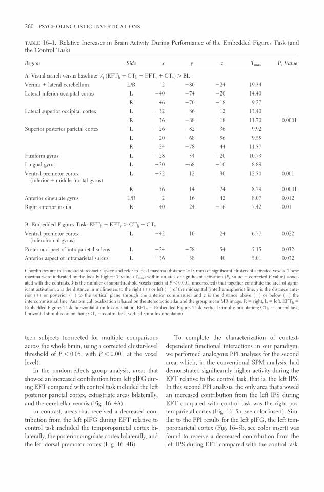
teen subjects (corrected for multiple comparisonsacross the whole brain, using a corrected cluster-levelthreshold of P � 0.05, with P � 0.001 at the voxellevel).
In the random-effects group analysis, areas thatshowed an increased contribution from left pIFG dur-ing EFT compared with control task included the leftposterior parietal cortex, extrastriate areas bilaterally,and the cerebellar vermis (Fig. 16–4A).
In contrast, areas that received a decreased con-tribution from the left pIFG during EFT relative tocontrol task included the temporoparietal cortex bi-laterally, the posterior cingulate cortex bilaterally, andthe left dorsal premotor cortex (Fig. 16–4B).
To complete the characterization of context-dependent functional interactions in our paradigm,we performed analogous PPI analyses for the secondarea, which, in the conventional SPM analysis, haddemonstrated significantly higher activity during theEFT relative to the control task, that is, the left IPS.In this second PPI analysis, the only area that showedan increased contribution from the left IPS duringEFT compared with control task was the right pos-teroparietal cortex (Fig. 16–5a, see color insert). Sim-ilar to the PPI results for the left pIFG, the left tem-poroparietal cortex (Fig. 16–5b, see color insert) wasfound to receive a decreased contribution from theleft IPS during EFT compared with the control task.
260 PSYCHOLINGUISTIC INVESTIGATIONS
TABLE 16–1. Relative Increases in Brain Activity During Performance of the Embedded Figures Task (andthe Control Task)
Region Side x y z Tmax Pc Value
A. Visual search versus baseline: 1⁄4 (EFTh � CTh � EFTv � CTv) � BL
Vermis � lateral cerebellum L/R 2 �80 �24 19.34
Lateral inferior occipital cortex L �40 �74 �20 14.40
R 46 �70 �18 9.27
Lateral superior occipital cortex L �32 �86 12 13.40
R 36 �88 18 11.70 0.0001
Superior posterior parietal cortex L �26 �82 36 9.92
L �20 �68 56 9.55
R 24 �78 44 11.57
Fusiform gyrus L �28 �54 �20 10.73
Lingual gyrus L �20 �68 �10 8.89
Ventral premotor cortex L �52 12 30 12.50 0.001(inferior � middle frontal gyrus)
R 56 14 24 8.79 0.0001
Anterior cingulate gyrus L/R �2 16 42 8.07 0.012
Right anterior insula R 40 24 �16 7.42 0.01
B. Embedded Figures Task: EFTh � EFTv � CTh � CTv
Ventral premotor cortex L �42 10 24 6.77 0.022(inferofrontal gyrus)
Posterior aspect of intraparietal sulcus L �24 �58 54 5.15 0.032
Anterior aspect of intraparietal sulcus L �36 �38 40 5.01 0.032
Coordinates are in standard stereotactic space and refer to local maxima (distance �15 mm) of significant clusters of activated voxels. Thesemaxima were indicated by the locally highest T value (Tmax) within an area of significant activation (Pc value � corrected P value) associ-ated with the contrasts. k is the number of suprathreshold voxels (each at P � 0.001, uncorrected) that together constitute the area of signif-icant activation. x is the distance in millimeters to the right (�) or left (�) of the midsagittal (interhemispheric) line; y is the distance ante-rior (�) or posterior (�) to the vertical plane through the anterior commissure; and z is the distance above (�) or below (�) theintercommissural line. Anatomical localization is based on the stereotactic atlas and the group mean MR image. R � right, L � left. EFTh �
Embedded Figures Task, horizontal stimulus orientation; EFTv � Embedded Figures Task, vertical stimulus orientation; CTh � control task,horizontal stimulus orientation; CTv � control task, vertical stimulus orientation.

NETWORKS OF VISUAL SEARCH AND
SPATIAL ATTENTION
Previous work has associated visual search (spatial at-tentional shifts) with activation of superior parietalcortex (Corbetta et al., 1995; Corbetta and Shulman,2002). Activation of left inferoparietal cortex has beenassociated with object-based visual attention (Fink etal., 1997a) and with locally directed visual attentionin tasks involving hierarchically organized visual stim-uli: that is, complex stimuli with both a local and aglobal organization (Fink et al., 1996, 1997b; Weberet al., 2000). This overall pattern of results is in goodaccord with our claim that activation of left superiorand inferior parietal cortex during performance of theEFT is specifically related to visual search for localtargets. More importantly, however, with regard to theassociation between aphasia and impaired perfor-mance on the EFT (Teuber and Weinstein, 1956),
BROCA’S AREA BEYOND LANGUAGE PROCESSING 261
FIGURE 16–2. Main effect of task (EFT versus CT)as reported by Manjaly et al. (2003), random effectsanalysis (16 subjects). All activations from the EFTversus CT contrast (masked inclusively by the CT ver-sus baseline contrast), significant at a cluster-level cor-
rected threshold of P � 0.05, are shown. Top row:Maximum intensity projections (glass brains). A, an-terior; P, posterior; L, left; R, right. Bottom row: Acti-vations overlaid on a rendered template brain.
FIGURE 16–3. Overlay of the functional activation ofpIFG during EFT (red) with a cytoarchitectonicallydefined probability map of Brodmann area 44 (lightgray).

FIGURE 16–4. Results of the PPI analysis, random ef-fects analysis (16 subjects). All areas are shown thatreceive an increased (a) or a decreased (b) influencefrom left pIFG during EFT relative to CT. Cluster-level threshold of P � 0.05, corrected for multiple
comparisons across the whole brain (voxel-levelthreshold of P � 0.001). Maximum intensity projec-tions (glass brains) of the results: sagittal (left), hori-zontal (middle), and coronal (right) views. Abbrevia-tions as in Figure 16–2.
FIGURE 16–5. Results of the PPI analysis for left IPS,random effects analysis (16 subjects). As in Figures16–2 through 16–4, results are shown at a cluster-levelthreshold of P � 0.05, corrected for multiple com-parisons across the whole brain (voxel-level thresholdof P � 0.001). (a) The only area to receive an in-creased contribution from left IPS during EFT com-
pared with the control task was the right posterior pari-etal cortex (18, �62, 42; tmax � 6.69; P � 0.001). (b)Similar to the PPI results for the left pIFG (comparewith Fig. 16–4), the left temporoparietal cortex (�58,�50, 16; tmax � 4.57; P � 0.001) was found to receivea decreased contribution from the left IPS duringEFT relative to the control task.
262

the region that we found activated in left inferior pari-etal cortex is relatively close to the posterior languageareas of the left hemisphere (Benson, 1979). Fur-thermore, central disorders of reading and writing maybe provoked by lesions of the left supramarginal gyrus(Heilman and Valenstein, 1993).
Likewise, the region of the left inferofrontal gyrusthat was activated by performing the EFT is part ofthe anterior language area as demonstrated by over-lying the area of increased neural activity during theEFT with cytoarchitectonically derived probabilitymaps of Brodmann’s areas 44 and 45 (Amunts et al.,1999, 2004). Although, in principle, large left hemi-sphere lesions may result in both aphasia and im-pairment on the EFT by reason of infarcting two func-tionally distinct regions (Corkin, 1979), it seemsunlikely that the association is simply dependent onthe extent of the cerebral pathology, contrary to theclaims of Teuber and Weinstein (1956) and Cobrinik(1959). Our results rather imply that two compara-tively small regions are associated specifically with lo-cal search on the EFT, a finding that is consistentwith the observation by Russo and Vignolo (1967) andOrgass et al. (1972) that both large and small lesionscan give rise to impairments on the EFT.
The analysis of effective connectivity (i.e., context-dependent functional interactions between differentbrain regions) of the left pIFG helps to explain the re-peatedly observed yet puzzling association betweenaphasia (due to left hemispheric lesions) and impairedperformance on the EFT (Orgass et al., 1972; Russoand Vignolo, 1967; Teuber and Weinstein, 1956).The analysis allows us to distinguish between two pos-sible explanations for why left pIFG, which is usuallyassociated with language processing, might be in-volved in the execution of the EFT. One hypothesisis that pIFG is activated during the EFT because per-forming the EFT might involve a “hidden” language-related component that had gone unnoticed by bothourselves and previous researchers. If this “hidden lan-guage component hypothesis” was correct and leftpIFG function during the EFT was linked to languageprocesses, we would expect to find significant changesin functional interactions with language-related areas,such as the left temporal lobe, during the EFT com-pared with the control task. The alternative hypothe-sis is based on the notion of “neural context” (McIn-tosh, 2000; McIntosh et al., 2003) according to whichany given cortical area can contribute to different cog-
nitive processes by participating in multiple networksthat are established through context-dependent changesin functional coupling. If this hypothesis is correct andleft pIFG can also become involved in local visualsearch processes, we expected that comparing thefunctional interactions of left pIFG during the twodifferent visuospatial tasks of our experiment (EFTand the control task) should demonstrate significantchanges in coupling with other areas typically in-volved in visuospatial processing.
In order to assess context-dependent functional in-teractions and decide between the two hypothesesabove, we computed psychophysiological interactionsof the left pIFG. Importantly, this method does notdetermine absolute degrees of coupling, but rathercomputes the difference in the functional contribu-tions of a source area to voxels elsewhere between twodifferent contexts (Friston et al., 1997). Since we hadno strong hypotheses on the type of coupling changesinduced by the two tasks, we determined the entireset of areas that showed a significant difference in cou-pling with left IFG between the EFT and the controltask, regardless of the direction of this difference.
The results of our PPI analyses suggest that pIFGfunction is not exclusively linked to language but cancooperate with areas that typically subserve visuospa-tial functions: during the EFT (relative to the controltask), left pIFG increased its contribution to left pos-terior parietal cortex, bilateral extrastriate cortex, andthe cerebellar vermis, and decreased its contributionto left and right temporoparietal cortex, posterior cin-gulate cortex, and left dorsal premotor cortex (see Figs.16–4 and 16–5). The involvement of all these areasin various aspects of visuospatial processing has beendemonstrated in previous imaging studies (e.g., Cor-betta et al., 2000; Corbetta and Shulman, 1998; Finket al., 1996, 1997a). In contrast, to our knowledge,only two of these areas have also been implicated inlanguage processing: left temporoparietal cortex acti-vation has previously been observed during the pro-cessing of spoken language (Binder et al., 1997), andthe left dorsal premotor cortex has been linked to ver-bal working memory (Herwig et al., 2003). Thesecognitive processes, however, are unlikely to be partof any strategy that could be used to perform eitherthe EFT or the control task: first, spoken words werenot part of the stimulus material, and second, the useof verbal working memory was not required to copewith our complex visuospatial displays that remained
BROCA’S AREA BEYOND LANGUAGE PROCESSING 263

in full view during task performance (for a more de-tailed discussion, see Manjaly et al., 2003, in press).Moreover, if language-related components had in-deed played a role in our paradigm, one would nothave expected the analysis to show higher left pIFGcoupling with left temporoparietal and left dorsal pre-motor cortex during the control task than during theEFT. Put the other way around, why should the con-trol task, which was a conventional visual matchingtask (with minimal visual search), use these putativelanguage-related processes to a greater degree than themore complex EFT?
We thus conclude that the changes in couplingwith left pIFG that we have observed in the areas de-scribed earlier reflect functional changes in a moregeneral visuospatial network and are due to the dif-ferent demands that the two tasks place on specific vi-suospatial subprocesses (e.g., direct matching of thetarget to a “pop-out” element in the control task com-pared to iterative matching during local search in theEFT). This interpretation is supported by additionalresults from our analysis concerning the context-dependent interactions of the second region that wasfound to be activated during the EFT relative to thecontrol task, that is, the left IPS (Manjaly et al., 2003,in press). In contrast to left pIFG, there is much pre-vious evidence that left IPS subserves visuospatialfunctions (e.g., Corbetta et al., 2000; Corbetta andShulman, 2002; Hopfinger et al., 2000). In our analy-sis, the left IPS showed an increased contribution tothe right posterior parietal cortex, another “classic” vi-suospatial area, during EFT compared with the con-trol task (see Fig. 16–5a). Importantly, in analogy tothe findings for left pIFG, the reverse comparisondemonstrated higher left IPS coupling with the sameleft temporoparietal region during the control task rel-ative to EFT (Fig. 16–5b).
At a more general level, our findings demonstratethat, depending on the neural context of the task de-mands, left pIFG can contribute to cognitive opera-tions other than language processing. To some extent,this view had previously been suggested by functionalimaging experiments of the human “mirror neuronsystem.” These studies have demonstrated that the leftpIFG is crucially involved in action understandingand action imitation (Binkofski and Buccino, 2004;Buccino et al., 2001, 2004). Although action under-standing and imitation are not language functions ina strict sense, some proposals have tried to bridge these
two domains by suggesting that the acquisition of lan-guage skills may be linked to the imitation of mouthmovements (e.g., Rizzolatti and Arbib, 1998). But ourcurrent findings challenge much more radically thetraditional view of pIFG as an area exclusively dedi-cated to language than do the results from the abovestudies of the “mirror neuron system.”
CONCLUSIONS
Broca’s area (or, more generally, the left inferiorfrontal region) is undoubtedly implicated in many lan-guage and language-related tasks. The question at is-sue, however, is whether it is legitimate to move fromthis assertion (supported by very large numbers of le-sion studies and functional neuroimaging experi-ments) to the theoretical claim that the exclusive (oreven the core) specialization of Broca’s area is the me-diation of language functions. Alternative hypothesesinclude the possibilities that (1) Broca’s area is a mo-saique of many different functional regions, only someof which are directly related to language; (2) Broca’sarea computes a highly abstract function, such as se-lection from a pool of competing alternative repre-sentations, which subsumes aspects of language pro-cessing and many other cognitive systems, includinglocal visual search (e.g., as in the EFT); and (3) par-ticular neuroanatomical regions, including Broca’sarea, change their functions consequent upon the si-multaneous activation of other regions that are effec-tively connected to a given region. Our results pro-vide further support for this third conceptualframework in cognitive neuroscience that stresses theimportance of context and interactivity for under-standing the functional architecture of the brain (Fris-ton, 1998, 2002; McIntosh, 2000). Although these ap-proaches do not question the existence of specializedcomputational modules in the brain, they posit thatthere is no one-to-one mapping between a singleanatomical region and a particular cognitive process.Rather, context-dependent interactions within net-works of areas determine the functions of the con-stituent areas. This notion of a distributed architec-ture of brain function implies that a particular areamay be part of different networks and hence have dif-ferent functions depending on a given task context.This view is corroborated by neuroanatomical andneurophysiological studies that, collectively, demon-
264 PSYCHOLINGUISTIC INVESTIGATIONS

strate that the computational role of any given corti-cal area cannot be derived from its behavior in a sin-gle task or task comparison but is characterized bet-ter through its multivariate response profile across awide range of tasks (Passingham et al., 2002). Such a“functional fingerprint” (Passingham et al., 2002) ofa particular area critically depends on its unique pat-tern of anatomical connections with other areas: Thatis, the “connectional fingerprint” of an area neces-sarily constrains its functional interactions. Where anarea receives information from and where theprocessed information can be sent to determine thenetworks in which the area can participate (Kötter andStephan, 2003; Passingham et al., 2002).
The notion that a particular area may contributeto more than one cognitive function by participatingin more than one functional network represents a one-to-many structure-function relationship. It should benoted that the opposite principle, that is, a many-to-one structure-function relationship, is also realized inthe human brain: there are some cognitive functionswhich can be mediated by means of different (par-tially overlapping or totally disjoint) networks (Fristonand Price, 2003; Price and Friston, 2002). This bio-logical “degeneracy” (Tononi et al., 1999) is found atmany different levels, ranging from the genetic codeto the systems level of the brain. The degeneracy (inthis sense) of the brain, which is believed to be dueto sparse structural connectivity patterns (“semicon-nectivity”) (McIntosh 2000), massively complicatesthe drawing of reliable causal inferences about struc-ture-function relationships from the study of behav-ioral deficits consequent upon brain lesions (Priceand Friston, 2002; Young et al., 2000). Both types ofmapping, that a particular area can participate in dif-ferent cognitive functions and that a particular cog-nitive function can be mediated by different networksof areas, demonstrate the highly distributed architec-ture of the human brain. To disentangle these com-plex structure-function relationships will be a majorchallenge for the neurosciences in the twenty-firstcentury (Friston et al., 2003).
ACKNOWLEDGMENTS J.C.M. and J.M.G. are sup-ported by the Medical Research Council (MRC), UnitedKingdom. K.Z. and G.R.F. are supported by theDeutsche Forschungsgemeinschaft (DFG-KFO 112),Germany. K.E.S. is funded by the Wellcome Trust,
United Kingdom. Additional support from the Volkswa-genStiftung to J.C.M., K.Z., and G.R.F. is gratefully acknowledged.
References
Alexander, M.P., Naeser, M.A., & Palumbo, C. (1990).Broca’s area aphasias: Aphasia after lesions includ-ing the frontal operculum. Neurology, 40, 353–362.
Amunts, K., Schleicher, A., Burgel, U., Mohlberg, H.,Uylings, H.B., & Zilles, K. (1999). Broca’s regionrevisited: cytoarchitecture and intersubject variabil-ity. Journal of Comparative Neurology, 412,319–341.
Amunts, K., Weiss, P.H., Mohlberg, H., Pieperhoff, P.,Eickhoff, S., Gurd, J.M., et al. (2004). Analysis ofneural mechanisms underlying verbal fluency in cy-toarchitectonically defined stereotaxic space—Theroles of Brodmann areas 44 and 45. NeuroImage,22, 42–56.
Benson, D.F. (1979). Aphasia, alexia, and agraphia,.New York: Churchill Livingstone.
Binder, J.R., Frost, J.A., Hammeke, T.A., Cox, R.W.,Rao, S.M., & Prieto, T. (1997). Human brain lan-guage areas identified by functional magnetic reso-nance imaging. Journal of Neuroscience, 17,353–363.
Binkofski, F., Amunts, K., Stephan, K.M., Posse, S.,Schormann, T., Freund, H.J., et al. (2000). Broca’sregion subserves imagery of motion: A combinedcytoarchitectonic and fMRI study. Human BrainMapping, 11, 273–285.
Binkofski, F., & Buccino, G. (2004). Motor functions ofthe Broca’s region. Brain and Language, 89,362–369.
Bonda, E., Petrides, M., Frey, S., & Evans, A.C. (1995).Neural correlates of mental transformations of thebody-in-space. Proceedings of the National Academyof Science USA, 92, 11180–11184.
Bookheimer, S. (2002). Functional MRI of language:New approaches to understanding the cortical or-ganization of semantic processing. Annual ReviewNeuroscience, 25, 151–188.
Bouillaud, J.B. (1825). Recherches cliniques propres àdemontrer que la perte de la parole correpond à lalésion des lobuls antérieurs du cerveau et à con-firmer l’opinion de M Gall, sur le siège de l’organedu langage articulé. Archives of General Medicine,6, 25–45.
Broca, P. (1861). Nouvelle observation d’aphemic pro-duite per une lesion de la moité postérieur des deux-ième et troisième conconvolutions frontales. Bul-letin of the Society of Anatomy of Paris, 36, 398–407.
BROCA’S AREA BEYOND LANGUAGE PROCESSING 265

Broca, P. (1865). Sur le siège de la faculté du langagearticulé. Bulletin of the Society of Anthropology ofParis, 6, 377–393.
Buccino, G., Binkofski, F., Fink, G.R., Fadiga, L., Fo-gassi, L., Gallese, V., et al. (2001). Action observa-tion activates premotor and parietal areas in a so-matotopic manner: An fMRI study. EuropeanJournal of Neuroscience, 13, 400–405.
Buccino, G., Vogt, S., Ritzl, A., Fink, G.R., Zilles, K.,Freund, H.-J., et al. (2004). Neural circuits under-lying imitation learning of hand actions: An event-related fMRI study. Neuron, 42, 323–334.
Buchel, C., Josephs, O., Rees, G., Turner, R., Frith,C.D., & Friston, K.J. (1998). The functionalanatomy of attention to visual motion. A functionalMRI study. Brain, 121, 1281–1294.
Chee, M.W.L., O’Kraven, K.M., Bergida, R., Rosen,B.R., & Savoy, R.L. (1999). Auditory and visualword processing studied with fMRI. Human BrainMapping, 7, 15–28.
Cobrinik, L. (1959). The performance of brain-injuredchildren on hidden-figures tasks. American Journalof Psychology, 72, 566–571.
Corbetta, M., Kincade, J.M., Ollinger, J.M., McAvoy,M.P., & Shulman, G.L. (2000). Voluntary orient-ing is dissociated from target detection in humanposterior parietal cortex. Nature Neuroscience, 3,292–297.
Corbetta, M., Shulman, G.L., Miezin, F.M., & Petersen,S.E. (1995). Superior parietal cortex activation dur-ing spatial attention shifts and visual feature con-junction. Science, 270, 802–805.
Corbetta, M., & Shulman, G.L. (1998). Human corticalmechanisms of visual attention during orientingand search. Philosophical Transactions of the RoyalSociety of London B, 353, 1353–1362.
Corbetta, M., & Shulman, G.L. (2002). Control of goal-directed and stimulus-driven attention in the brain.Nature Review of Neuroscience, 3, 201–215.
Corkin, S. (1979). Hidden-figures-test performance: Last-ing effects of unilateral penetrating head injury andtransient effects of bilateral cingulotomy. Neu-ropsychologia, 17, 585–605.
Decety, J., Perani, D., Jeannerod, M., Bettinard, V.,Tadardy, B., Woods, R., et al. (1994). Mapping mo-tor representations with positron emission tomogra-phy. Nature, 371, 600–602.
Finger, S. (2000). Minds behind the brain: A history ofthe pioneers and their discoveries. Oxford: OxfordUniversity Press.
Fink, G.R., Dolan., R.J., Halligan, P.W., Marshall, J.C.,& Frith, C.D. (1997a). Space-based and object-based visual attention: shared and specific neuraldomains. Brain, 120, 2013–2028.
Fink, G.R., Halligan, P.W., Marshall, J.C., Frith, C.D.,Frackowiak, R.S.J., & Dolan, R.J. (1996). Where inthe brain does visual attention select the forest andthe trees? Nature, 382: 626–628.
Fink, G.R., Halligan, P.W., Marshall, J.C., Frith, C.D.,Frackowiak, R.S.J., & Dolan, R.J. (1997b). Neuralmechanisms involved in the processing of globaland local aspects of hierarchically organized visualstimuli. Brain, 120, 1779–1791.
Friederici, A.D., Ruschemeyer, S.A., Hahne, A., &Fiebach, C.J. (2003). The role of left inferior frontaland superior temporal cortex in sentence compre-hension: Localizing syntactic and semanticprocesses. Cerebral Cortex, 13, 170–177.
Friston, K.J. (1997). Testing for anatomically specifiedregional effects. Human Brain Mapping, 5,133–166.
Friston, K.J. (1998). Imaging neuroscience: Principles ormaps? Proceedings of National Academy of ScienceUSA, 95, 796–802.
Friston, K.J. (2002). Beyond phrenology: What can neu-roimaging tell us about distributed circuitry? An-nual Review of Neuroscience, 25, 221–250.
Friston, K.J., Ashburner, J., Frith, C.D., Poline, J.-B.,Heather, J.D., & Frackowiak, R.S.J. (1995a). Spa-tial registration and normalization of images. Hu-man Brain Mapping, 3, 165–189.
Friston, K.J., Büchel, C., Fink, G.R., Morris, J., Rolls,E.T., & Dolan, R.J. (1997). Psychophysiologicaland modulatory interactions in neuroimaging. Neu-roImage, 6, 218–229.
Friston, K.J., Harrison, L., & Penny, W. (2003). Dynamiccausal modeling. NeuroImage, 19, 1273–1302.
Friston, K.J., Holmes, A., Worsley, K.J., Poline, J.-B.,Frith, C.D., & Frackowiak, R.S.J. (1995b). Statisti-cal parametric maps in functional imaging: A gen-eral linear approach. Human Brain Mapping, 2,189–210.
Friston, K.J., Holmes, A.P., & Worsley, K.J. (1999). Howmany subjects constitute a study? NeuroImage, 10,1–5.
Friston, K.J., & Price, C.J. (2003). Degeneracy and re-dundancy in cognitive anatomy. Trends in Cogni-tive Sciences, 7, 151–152.
Gall, F.J. (1822). Sur les functions du cerbeau et sur cellde chacune de ses parties., Paris: Baillière.
Gottschaldt, K. (1926). Über den Einfluss der Erfahrungauf die Wahrnehmung von Figuren, I. PsycholForsch, 8, 261–317.
Gottschaldt, K. (1929). Über den Einfluss der Erfahrungauf die Wahrnehmung von Figuren, II. PsycholForsch, 12, 1–87.
Grafton, S.T., Arbib, M.A., Fadiga, L., & Rizzolatti, G.(1996a). Localization of grasp representation in hu-
266 PSYCHOLINGUISTIC INVESTIGATIONS

mans by positron emission tomography: II. Obser-vation compared with imagination. Exp Brain Res,112, 103–111.
Grafton, S.T., Fagg, A.H., Woods, R.P., & Arbib, M.A.(1996b). Functional anatomy of pointing and grasp-ing in humans. Cerebral Cortex, 6, 226–237.
Heilman, K.M., & Valenstein, E. (1993). Clinical neu-ropsychology., Oxford: Oxford University Press.
Herwig, U., Abler, B., Schonfeldt-Lecuona, C., Wun-derlich, A., Grothe, J., Spitzer, M., et al. (2003).Verbal storage in a premotor-parietal network: Evi-dence from fMRI-guided magnetic stimulation.NeuroImage, 20, 1032–1041.
Hopfinger, J.B., Buonocore, M.H., & Mangun, G.R.(2000). The neural mechanisms of top-down atten-tional control. Nature Neurosciences, 3, 284–291.
Kim, K.H., Relkin, N.R., Lee, K.M., & Hirsch, J. (1997).Distinct cortical areas associated with native andsecond languages. Nature, 388, 171–174.
Kötter, R., & Stephan, K.E. (2003). Network participa-tion indices: Characterizing component roles for in-formation processing in neural networks. NeuralNetworks, 16, 1261–1275.
Liepmann, H. (1900). Das Krankheitsbild der Apraxie(“motorischen Asymbolie”) aufgrund eines Fallesvon einseitiger Apraxie. Monatsschrift für Psychia-trie und Neurologie, 8, 15–44.
Macaluso, E., Frith, C.D., & Driver, J. (2000). Mom-dulation of human visual cortex by crossmodal spa-tial attention. Science, 289, 1206–1208.
Manjaly, Z.M., Marshall, J.C., Stephan, K.E., Gurd,J.M., Zilles, K., & Fink, G.R. (2003). In search ofthe hidden: An fMRI study with implications for thestudy of patients with autism and with acquiredbrain injury. NeuroImage, 19, 674–683.
Manjaly, Z.M., Marshall, J.C., Stephan, K.E., Gurd,J.M., Zilles, K., & Fink, G.R. (2004). Context-dependent interactions of left posterior inferiorfrontal gyrus in a local visual search task unrelatedto language. Cognitive Neuropsychology, (in press),
Marshall, J.C. (1984). Multiple perspectives on modu-larity. Cognition, 17, 209–242.
Marshall, J.C., & Fink, G.R. (2003). Cerebral localiza-tion, then and now. NeuroImage, 20, S2–S7.
Marshall, J.C., & Gurd, J.M. (2004). On the anatomo-clinical method. Cortex, 40, 230–231.
McIntosh, A.R. (2000). Towards a network theory of cog-nition. Neural Networks, 13, 861–870.
McIntosh, A.R., Rajah, M.N., & Lobaugh, N.J. (2003).Functional connectivity of the medial temporallobe relates to learning and awareness. Journal ofNeuroscience, 23, 6520–6528.
McManus, C. (2002). Right hand, left hand., Cam-bridge, MA: Harvard University Press.
Orgass, B., Poeck, K., Kirchensteiner, M., & Hartje, W.(1972). Visuo-cognitive performances in patientswith unilateral hemispheric lesions. An investiga-tion with three factorial reference tests. Z Neurol,202, 177–195.
Parsons, L.M., Fox, P.T., Hunter Downs, J., Glass, T.,Hirsch, T.B., Martin, C.C., et al. (1995). Use of im-plicit motor imagery for visual shape discriminationas revealed by PET. Nature, 375, 54–58.
Passingham, R.E., Stephan, K.E., & Kötter, R. (2002).The anatomical basis of functional localization inthe cortex. Nature Review Neuroscience, 3, 606–616.
Petersen, S.E., Fox, P.T., Snyder, A.Z., & Raichle, M.E.(1988). Positron emission tomographic studies ofthe cortical anatomy of single-word processing. Na-ture, 331, 585–589.
Price, C.J., & Friston, K.J. (2002). Degeneracy and cog-nitive anatomy. Trends in Cognitive Sciences, 6,416–421.
Ring, H.A., Baron-Cohen, S., Wheelwright, S., Williams,S.C.R., Brammer, M., Andrew, C., et al. (1999).Cerebral correlates of perserved cognitive skills inautism. A functional MRI study of embedded fig-ures task performance. Brain, 122, 1305–1315.
Rizzolatti, G., & Arbib, M.A. (1998). Language withinour grasp. Trends in Neuroscience, 21, 188–194.
Rizzolatti, G., Fadiga, L., Matelli, M., Bettinardi, V.,Paulesu, E., Perani, D., et al. (1996). Location ofgrasp representations in humans by PET. 1. Obser-vation versus execution. Experimental Brain Re-search, 111, 246–252.
Russo, M., & Vignolo, L.A. (1967). Visual figure-grounddiscrimination in patients with unilateral cerebraldisease. Cortex, 3, 113–127.
Stephan, K.E., Marshall, J.C., Friston, K.J., Rowe, J.B.,Ritzl, A., Zilles, K., et al. (2003). Lateralized cog-nitive processes and lateralized task control in thehuman brain. Science, 301, 384–386.
Stephan, K.M., Fink, G.R., Passingham, R.E., Silber-sweig, D.A., Ceballos-Baumann, A.O., Frith,C.D., et al. (1995). Imagining the execution ofmovements. Functional anatomy of the mentalrepresentation of upper extremity movements inhealthy subjects. Journal of Neurophysiology, 73,373–386.
Talairach, J., & Tournoux, P. (1988). Co-planar stereo-tactic atlas of the human brain,. Stuttgart: Thieme.
Teuber, H.-L., & Weinstein, S. (1956). Ability to discoverhidden figures after cerebral lesions. Archives ofNeurology and Psychiatry, 76, 369–379.
Toni, I., Krams, M., Turner, R., & Passingham, R.E.(1998). The time course of changes during motorlearning: A whole brain fMRI study. NeuroImage,8, 50–61.
BROCA’S AREA BEYOND LANGUAGE PROCESSING 267

Tononi, G., Sporns, O., & Edelman, G.M. (1999). Mea-sures of degeneracy and redundancy in biologicalnetworks. Proceedings of National Academy of Sci-ence USA, 96, 257–326.
Watson, T. (1871). Lectures on the principles and prac-tice of physic. London: Jon W Parker.
Weber, B., Schwarz, U., Kneifel, S., Treyer, V., & Buck,A. (2000). Hierarchical visual processing is depen-dent on the oculomotor system. Neuroreport, 11,241–247.
Wernicke, C. (1874). Der aphasische Symptomenkom-plex., Breslau: Cohn & Weigart.
Young, M.P., Hilgetag, C.C., & Scannell, J.W. (2000).On imputing function to structure from the behav-ioural effects of brain lesions. Philosophical Trans-actions of Royal Society of London B, 355, 147–161.
Zatorre, R.J., Meyer, E., Gjedde, A., & Evans, A.C.(1996). PET studies of phonetic processing ofspeech: Review, replication, and reanalysis. Cere-bral Cortex, 6, 21–30.
268 PSYCHOLINGUISTIC INVESTIGATIONS

V
DISCUSSION
The Jülich workshop featured long hours of discussion, inside the conference room as well asoutside. The excerpts that follow represent the main issues that arise when one attempts to rec-oncile the multiple perspectives that currently exist on Broca’s region.

This page intentionally left blank

ANATOMY AND LOCALIZATION
Karl Zilles:
What interested me during this conference is the con-fusion which comes out when different approacheslike the linguistic, the clinical and anatomical areused in the different talks. For example, everybodyrefers to Broca’s region, to Broca’s complex, to area44, to area 45, or to area 47. It is my impression, thatthis notion is not well defined, more or less chaotic.It is not just a question of nomenclature, but a prob-lem which goes into the interpretation of the data.
For instance, when somebody says that “we foundin a clinical study something which is related to a dys-function of Broca’s area 47” and later discusses datafrom areas 45, this cannot be done, because the “area47” is not defined. It’s rather like “anterior and veryventral” relative to the inferior frontal gyrus, whereasthe meaning in the second statement is, let’s say cor-tical area 45, or 44, if it’s a bit more posterior. In some
cases this notion refers to the mean of a group study,whereas in other cases it is a single brain. The refer-ence system, as well as the way the data came into thissystem, also differ. Sometimes, it’s done by a mathe-matically controlled elastic warping procedure to acommon reference space, whereas in other talks it’sjust a reference to the Talairach atlas. So we use thedefinitions of cortical areas in different ways, mixingand matching, and then we end up with theories andstatements, which become accepted in the community.But, these statements don’t really tell us very much.
So, I would really like to hear your opinion aboutthis because some people tell me “I’m absolutely notinterested in these anatomical issues, it’s not neces-sary. I can do my functional studies. I don’t need anyanatomical atlas, any anatomical basis.” This is oneviewpoint, but there may be other participants whoare interested in knowing whether the blob they seein images is in a structurally homogeneous brain area,or is distributed over different areas. So, what do youthink about this situation?
271
17
Jülich Workshop Excerpts

Luciano Fadiga:
I am a neurophysiologist. Usually we believe thatanatomical differences correspond to functional dif-ferences. For example, if there is an area in which thefourth cortical layer is developed, this differential de-velopment is due to a functional reason. It might be,for example, that this particular region of cortex re-ceives a lot of thalamic input, while another regioncannot receive this input from the thalamus, hencethe differentiation is missing.
So, I think we should really keep in mind that theanatomical subdivision is not simply a classificationtrick used by neuroanatomists to subdivide the brain.We must remember that there is much confirmationto the claim that for every anatomically classified brainlesion, there is a corresponding function. I think thatwe can start from this common point of view.
Sergey Avrutin:
I’d like to give an opposite perspective, coming fromwork with aphasic patients. With my linguistic back-ground, I think that the studies with aphasic patientscan actually contribute to the linguistic theory inde-pendently of the exact knowledge where the damageis. Specifically, suppose that there are two linguistictheories, theory I and theory II. Theory I claims thatcertain constructions, X and Y, are grouped together.Theory II claims that these are two distinct linguisticconstructions. Data from aphasic patients can actu-ally tell us which theory is true. Here is how. Supposethat the patients perform equally well on both con-structions. Then it is evidence for theory I, but not II.In this case, we can circumvent the problem of com-plete localization. We simply don’t care. We wouldcare if we were to focus on localization, but then itbecomes a question of how one focuses one’s researchprogram. If it is on localization, then localization isimportant; but if your research aims to contribute toa general understanding of language, then it can beignored.
Francisco Aboitiz:
Sergey’s preferred question would be a general un-derstanding of language, and in this way there wouldbe no need to know about localization. But what doyou mean by that precisely? I mean, that a generalunderstanding of the organization of language in-
cludes the knowledge of the involved neuralprocesses. If you want to know that you will need toknow about localization, anyway.
Sergey Avrutin:
Well, the statement that we don’t care is too strong ofcourse. Neurologists tell me that the issue of local-ization is a mess, and so you can abstract away andapproximate. There are Broca’s aphasics which areusually classified according to, say, the Boston test, orwhatever. Eventually you would like to have of coursea very clear connection with localization, to the ex-tent that it exists. What I’m saying is that at the mo-ment, when a typology does not exist, this should notprevent us from working with aphasic patients, andmake inferences about function.
Karl Zilles:
I think that there is already a problem. When you lookat the classic case of Broca, a lot of people say “Aha,this is Broca’s region, where the hole is.” But whenyou have a look to the MR scan of this brain, you willsee that—we heard it repeatedly during this meeting—there is much more destroyed tissue in this brain.Then, a question arises whether it is a white matterdisease, or a problem of the destroyed connectivity, ora problem of this particular cortical region which in-cludes areas 44, 45, or whether it is a question of sub-cortical structures. Immediately, you get in troublewith the anatomists, because you have to really definethe question. Therefore, I think that the question forstructure and functional correlates comes up veryearly.
Stefano Cappa:
I think that Sergey’s point is perfectly legitimate. It de-pends from what angle you are doing research.Twenty years of cognitive neuropsychology have beenbasically ignoring the brain completely. I think this isa perfectly legitimate enterprise. It does not detractfrom the fact that nowadays, especially with functionalimaging, many people are asking different kinds ofquestions, e.g., how language is represented in thebrain. At that point, the precision in the anatomy can-not be avoided. To be fair to the field, 15 or 16 yearsago it was impossible to be very precise with respect
272 DISCUSSION

to anatomy, because of positron emission tomography,group studies averaging, a lack of good atlas, etc.Given that, it is appropriate to discuss the data in termsof “slightly more anterior” or “slightly more posterior.”I did it, for example, in my talk as well because ifyou’re not very confident about localization, it’s onlyfair to talk in vague terms like anterior, posterior andso on.
Kyle Johnson:
I’d like to talk against Sergey Avrutin. You’ve got twonatural classes, given to you by virtue of the diag-nostic criteria for Broca’s aphasia, and you knownothing about the physiological basis of these naturalclasses. But it is still OK, because we’ve got these twonatural classes, by virtue of which we can determinewhether the cuts made by linguistic theory or psy-chological theory are correct. But then there is stilla question remaining: What are the diagnostic crite-ria for distinguishing Broca’s aphasics from non-Broca’s aphasics?
Sergey Avrutin:
Actually what I meant is that we can just use onegroup who are diagnosed as Broca’s aphasics, and ap-ply the data to two different linguistic theories, with-out even comparing their performance to a differentgroup. There are tests to classify patients. For somepatients, we look at the scans. However, after you workwith patients for a while, you just see that a patient ismore or less “typical” than another. So there is abunch of factors to classify patients. There is a differ-ence between patients and I don’t even try to say any-thing about anatomy with these patients.
Stefano Cappa:
There are several points of investigation where youdon’t need to classify the patients. You don’t need clas-sification if you’re just testing a psycholinguistic hy-pothesis on a patient with language disorder.
Gereon Fink:
I think the story is even more complicated. You can’tclassify many acute stroke patients with lesions of theleft hemisphere. They don’t fall under Broca’s or Wer-
nicke’s aphasia or whatever. They are nonclassifiable.After a while, they evolve, e.g., into Broca’s aphasics.Thus, when we talk about Broca’s aphasia or Wer-nicke’s aphasia, we are talking about something thatdepends on reconstitution of brain function, onneural plasticity. We often talk about an endpoint, af-ter a lot of changes have happened in the brain, wherewe have no clue what they are. Such changes occurin the perilesioned areas of the same hemisphere, orin the homologue areas of the other hemisphere. I re-ally think we have to take that into account when wetalk about localization of a brain function based onlesions.
Karl Zilles:
With respect to localization and brain maps itseems to me that the probability maps are a funda-mental different concept of cytoarchitecture whencompared with the maps in the past. The latter sug-gest that there is a more or less well-defined border.The probability maps tell a different story. You canonly choose a probability and then you will see theborder here or there. Then you choose a differentprobability, and you will see the border at anotherplace. We can only make probability statements aboutthe precise location of the border of a cortical area.This is the only—let’s say—the only possible way tospeak about cytoarchitecture, and how does probabil-ity and structure and function fit together. Well, thereare experimental studies in animals with extremelyhigh spatial resolution, which show that there is achange in function within 100 micrometers. Thesestudies show that, precisely at the same site, there isa change in the anatomical and cytoarchitectonicstructure, in particular. In the somatosensory cortexof rats, for instance, where you have representation ofsingle whiskers, you see abrupt changes at the borderof the barrels. For an example in the human brain Irefer to the human visual cortex where you haveabrupt changes in cytoarchitecture and correspond-ing changes in function at the border of the striate tothe extrastriate cortex. Moreover, you even can iden-tify a small region in between where the meridian islocalized. You can see it in the anatomy of the visualcortex. When you look for such areas in different in-dividuals, however, you will see a tremendous vari-ability and therefore when we want to make a generalstatement about the human cortical brain map, thenwe can only do it as a probability map.
JÜLICH WORKSHOP EXCERPTS 273

DIVERSITY OF FUNCTIONAL ANALYSES
OF LANGUAGE
Yosef Grodzinsky:
I would like to shift attention to behavior, if you don’tmind. I’ve been following Karl Zilles’ comments verycarefully because he tries to keep everybody preciseabout anatomical statements. I think it’s fascinating tosee how difficult it is to make a precise anatomicalstatement as an anatomist, and how it becomes moredifficult once you get into lesion data, and on to func-tional imaging. Anatomical problems keep recurring.The biology is imposing incredibly hard problems on us.
But I want to shift focus because I and many ofmy colleagues find ourselves in exactly the same sit-uation on the behavioral front. Each and every oneof the issues you raise has a parallel in the analysis ofbehavior. Consider what Zilles said about problemsin scoping lesions. Well, look at diagnostic tests ofaphasia and you will see practical, conceptual andother problems that similarly arise. While the prob-lem is not limited to language, there are special prob-lems in the case of language. First, what is the frame-work within which you carve up behavior. And hereyou discover that if you try to put studies together andinterpret them, you quickly discover that people meanvery different things when they talk about the lexicon,or phonology, or for that matter the real, the true, thegeneral, diagnostic test. Likewise, when you look atwhat people do in functional imaging, not only dothey use different tasks and different analytic softwareand different slices and different number of slices, anddifferent blurring filters and so on, but they also usevery, very different language tasks. Thus what is fas-cinating to me about Karl Zilles’ presentation is thefact that when you turn to look at the behavioral front,you find an exact parallel to the problems in anatomyyou pointed out.
Michael Arbib:
A diversity of linguistic theories came up in the pre-sentations here and in the related papers. I’m firstlystruck by the fact that Chomsky for many years wasvery insistent on the separation between competenceand performance and that his theory was a compe-tence theory. I see many people here using straight
Chomskian competence grammar as part of a perfor-mance theory without any methodological discussionof how it is appropriate to go from what was explic-itly declared by the master to be only appropriate forcompetence.
Yosef Grodzinsky:
Can you give a concrete example of what you mean?
Michael Arbib:
Your trace-deletion hypothesis is an example. There’sno processing model there. You simply say, let’s con-sider trees and imagine deleting the traces, which isan absurd notion from a processing point of view be-cause it assumes that the traces are there to be deletedrather than offering a processing account of what anaphasic brain does. I cannot believe that what it doesis to construct the traces and delete them. So, it is nota coherent processing account.
Naama Friedmann has a beautiful analysis of pro-duction data. What is the thing that connects thisbeautiful competence account to a processing ac-count for production and perception? What are theways those trees are constructed? What would it meanto say to “add higher nodes”?
In the same way I’m seeing what maybe coherentlinguistic accounts, what may be divergent linguisticaccounts, but I see no consistent attempt in the liter-ature to map them to each other. I find each studyfascinatingly compelling. Then I go to the next studyand there’s a completely different linguistic frame-work and no account of whether those two frameworksreally are incompatible, they could be reconciled.
Kyle Johnson:
I wanted to respond to one thing because it’s a frus-tration of anyone coming to the linguistic literaturefrom outside. It’s just a living hell for those of us in-side, of course. I mean the divergent terminologies andmechanisms offered by different frameworks. But theresolution that you need for the kinds of studies youhave here, the sorts of distinctions you need to make,are distinctions that never divide linguistic theory sofar as I know or frameworks. So for instance, in eachof the papers that I’ve seen today, different frameworkshave been employed. You know in particular the sort
274 DISCUSSION

of syntax-lexical relationship that Peter Hagoort gaveus is quite different from that which would be usedby Yosef Grodzinsky’s trace-deletion hypothesis. Butthe distinctions between these two models are for themost part immaterial, as far as I can see. Immaterialto the sorts of questions you’re trying to find answersto. I think that for the questions we look at in the neu-roscience of language you could really use baby ver-sions of linguistic theory and still get very far.
Look at the principles that grab the phenomenaand sort of strip away the terminology that expressesa particular point of view. Take all that extra stuffaway, and just look at the phenomena that ground theterminology and that framework, and you’ll find thatthe grounding is exactly the same. So, the same set ofphenomena, the same sets of constraints, the modelsare slightly different, but what’s relevant here is therelationship between where you speak this term andsome facet of its interpretation. So, I could take thetrace-deletion hypothesis, express it in an LFG frame-work, find the homologue of traces in LFG and Iwould have the same thesis, empirically, the same the-sis that the trace deletion has.
Peter Hagoort:
You have to accept different meanings of cognitiveneuroscience. It’s very hard to convince people in cog-nitive neuroscience that, compared to memory andvision, language is a relevant and interesting topic.They say well, we get these linguists, they come andthrow their tree structures on the blackboard, makesome hand-waving claims about the fact that this allhas to be instantiated in some way in the brain, andyou neuroscientists go off and tell us exactly that thistheory by Mr. Chomsky or whatever version of it isthe correct one. That’s the perceived attitude in thefield of neuroscience.
As a result, people in neuroscience are not willingto go and test a very specific linguistic theory, becausethey need the vocabulary that allows them to ask theright questions with regard to the brain, and if they’reat all interested in whether the address of the accountof Mr. Chomsky or Mr. Such and Such, they want anaccount that would convince not only one particularkind of linguist, but also others.
In addition, you have to specify the nature of therepresentations being computed. That’s part of yourprocessing model, and you need to be explicit about
it. Many domains of neuroscience fall in the trap thatwe don’t have an explicit account of what the natureof the representation is being computed.
Yosef Grodzinsky:
I think there are several issues here. First, any theorymust accommodate the types of linguistic phenom-ena we are familiar with. Second, there’s an observedaphasic deficit that any theory of language must ac-count for. It doesn’t matter whether it’s traces or a con-struct that other theories might use. In this respect,aphasia does not distinguish between linguistic theories.
Moreover, remember that we use this concept(trace) as a descriptive device that carves up behavior.It doesn’t mean that we have a neuronal theory of tracesand syntactic movement. You use it as a descriptive de-vice and then you go along to see what the next thingthat you can discover. Now, there are rare cases whichinterest Sergey, and interest me, which are those rarecases that do distinguish between linguistic theories.These are extremely interesting cases. But for the mostpart, these are cases that we don’t present to an audi-ence that is interested in brain-language relations. Herewe look at core issues which any rational linguistic the-ory must account for. And it doesn’t matter what theycall it. In other words, the problem of diversity amonglinguistic theories for the core data here is immaterialand what you have to know is sort of to brush off theterminology and stay with the core conceptions. Fur-thermore, as long as your claim is used in this theoryas a descriptive device, you’re not committed to a pro-cessing theory because it’s not as if you want to derivethe behavior from some neuronal theory. You just useit as a descriptive behavior that correlates brain areaswith some kind of behavioral distinction. In this respectyou simply do the best you can.
Lewis Shapiro:
I absolutely agree that there is a knee jerk reaction toparticular kinds of linguistic theories and there areknee jerk reactions from students to neuroscientists.It may simply be because the vocabulary that we useis just outside what you have learned. Same goes forphysics: maybe I don’t understand the physical vo-cabulary, but I wouldn’t ever say physics isn’t impor-tant, or physics is the wrong thing to do, or that par-ticular physical theory.
JÜLICH WORKSHOP EXCERPTS 275
REPRESENTATION AND PROCESSING
Kyle Johnson:
One of the things I found interesting in Naama Fried-mann’s presentation is that it presented a map of whatlinguistic theory divides. Linguistic theories, all ofthem, you know, what they call natural classes. And,her account is based on a syntactic distinction be-tween a high part of a tree and a low part of a tree,given the tree for some sentence. And the character-ization of the deficit makes a distinction between the“high part” and “low part” and is tied to a very par-ticular data set. Only a few frameworks model this dataset and how she’s modeled it would go with any ofthose frameworks as far as I can see.
But, I do not know of a linguistic theory that makesa divide between high and low. So, the interestinganalysis you have uses a method of syntactic repre-sentation, but what makes the class—what partitionsthe data—is something that linguistic theory doesn’tmake, at least so far as I know.
So, I look at her characterization of the deficit shewas studying and I say, that’s not a linguistic deficit.It just does not align with what I know to be the gen-eral way of carving up nature that linguistic theoriesgive us. But, this is interesting: you have a very spe-cific linguistic theory in which you’re describing data,but actually the conclusion should be, I would say,that what’s modeled is not only a linguistic deficit. Weneed some bridge between what’s found there and sheoffered some hypotheses about what that bridge mightbe, but without the linguistic theories it doesn’t mat-ter their particular nature, you wouldn’t be able to seethat. So, that’s what I would claim is the role that lin-guists can bring here, we have some map of wherethe natural classes are, how the phenomenon shouldbe divided up, we have some idea about what the phe-nomena are. And then there is a very big and openquestion. Is that a correct characterization?
Naama Friedmann:
I think it is, in a way. It is true that I don’t see any-thing in syntactic theory that predicts that high nodeswill be more impaired than low nodes, but I can imag-ine several reasons for that. Based on my data, I haveno support for any specific reason for why high nodesare more impaired than low nodes. However, when youlook at individual differences and individual patterns,
when you look at recovery patterns, and when youlook at treatment results, the data definitely show it.
Why this is so I can only speculate. I can think ofseveral reasons why high nodes would be more im-paired. One would be to give it a processing flavor,and to say well, maybe there is a limit to the numberof layers we can accumulate and the number of lay-ers are counted from down to up. So, this is one pos-sible way to account for it.
Another way of accounting for it that SergeyAvrutin and I discussed, is to bank on the fact that thehigher nodes may be related to discourse. This would,in a way, put Sergey’s account and mine together. Itwould amount to saying that the high nodes are im-paired because they are related to discourse, not be-cause they are higher. There are predictions here: Torefute this account, we have to find discourse relatedelements that are lower in the tree, or elements thatare not discourse related that are higher in the tree.So that’s another way to think about it. One more waymight involve syntactic constraints on projection. So,these are three ways to think about it, but what I pro-vided was the data and the generalization, and thenext step will be to think why high nodes are moreimpaired.
Karen Emmorey:
Naama, going back to this “processing issue,” what doyou think is actually going on when patients are try-ing to produce these sentences in terms of a process-ing model of language production?
Naama Friedmann:
I would like to emphasize again that the deficit maynot be linguistic—it might be one of the ideas I pro-posed, or it might be something about processing orabout syntactic working memory. But what I think isvery important here is that you need a very specificsyntactic theory in order to account for the deficit. Soif you don’t have a well articulated syntactic theory,you are not likely to understand why the patients failon wh-questions but not in Yes-No questions, and soon. In order to know where to look, in order to knowwhat to test, you need a theory. So eventually youmight end up with a deficit that is not syntactic, butyou definitely need a syntactic theory to know whatto do.
276 DISCUSSION

The problem with a processing account is thatthere is no explicit processing theory. What I am thenforced to do is take the representational theory of lay-ers within syntactic trees, and try to use it as a pro-cessing model. What I get from this is that, you moreor less build the syntactic structure from the bottomup, and you get stuck when you have too many nodesto merge.
Karen Emmorey:
Is there any evidence from psycholinguistics, for ex-ample, that normal speakers do that – they build fromthe bottom up? I’m not sure how you’d do this kindof psycholinguistic experiment but that’s what youwould have to show that in fact that this is the wayyou produce these kinds of sentences, right?
Naama Friedmann:
I’m not aware of evidence from psycholinguistics.
Peter Hagoort:
This is a very instrumentalist philosophy of scienceposition. You just have a description and as long asthis description is adequate, that’s fine. I want to knowwhat the psychological reality of this description is.
Lewis Shapiro:
You’re missing the point. If the questions you’re ask-ing are about the endpoints of behavior—what apha-sic patients show – then it’s perfectly reasonable tohave a linguistic theory that cuts the pie in a particu-lar way. If your question has to do with the process-ing components that implement this kind of endpointof behavior, then you would indeed need a full-blownprocessing theory.
This brings me back to Michael Arbib’s comment,about how some of us use linguistic theory terminol-ogy in our processing accounts. That’s absolutely true.I’m certainly someone who does that. My view is thatyou take those linguistic objects, and use them as dis-covery procedures, to suggest what a processing ac-count might look like. In the work that I do, I pickand choose. I pick some vocabulary that seems to becomputed in lexical access, I pick some vocabularythat seems to be computed in syntactic parsing. Do
we have a full-blown processing account? No, I don’tthink so. And would we like to have one? Absolutely.Would it be better to have a really nice processingtheory that maps onto linguistic theory? Of course itwould.
Yosef Grodzinsky:
I think that what Peter Hagoort is saying is perfectlylegitimate: that when you make a descriptive claim,you want to find some kind of coherent construal ofit. You have a description which works to some ex-tent—and in Naama Friedmann’s tree pruning caseit does to an impressive extent, namely it is well-supported across structures and cross-linguistically.
But as a next step, you want to understand whatexactly are the neurological underpinnings or the pro-cessing underpinnings. Is it possible, given our cur-rent understanding? That’s where I take issue with theobjections that Peter Hagoort, and to an extentMichael Arbib, were voicing. They say that until youcome up with a full-fledged processing account, noth-ing you do is worthwhile (I’m putting it in more ex-treme terms than you would, of course). But I thinkthat the rational way to go about it is to use the besttools you have at any given moment, and then to tryto come up with other, better ones. To my knowledge,parsing theories today perform rather poorly once youtry to connect them to the actual empirical reality inneuropsychology. Thus, for the moment, I’d ratherstick to an agnostic point of view, and at the same timeI am happy to admit that we must make progress andstrive to interpret our data in some kind of processingtheory.
LANGUAGE DEVELOPMENT
Norbert Herschkovitz:
Last year, Katrin Amunts published a very importantpaper in the Journal of Comparative Neurology, aboutcytoarchitectonic changes in the asymmetry of area45 and 44 in the human brain, between ages 3 monthsto 5 years. It is one of the few developmental studiesof anatomical structures. There, an important findingwas that area 45 develops asymmetry in a significantway, around 5 years of age, and area 44 later, around11 years. Now, if you have a theory then you will pre-dict that, there should be some important changes.
JÜLICH WORKSHOP EXCERPTS 277

Francisco Aboitiz:
And there are changes in child language around thisage. One related to more syntactic development, oneto more pragmatic development.
Naama Friedmann:
I’m quite excited about it because at the age of 5 wesee the relative clauses appear all of a sudden. So ataround 5, 5 and a half, you start to see children un-derstanding object relatives. At around 11 they start tounderstand center embedded object relatives. So itcorrelates very nicely with the notion of the compre-hension of movement at around the age when 45 de-velops and computation of center embedding aroundthat age.
Peter Hagoort:
I’m still not convinced that this is the ultimate theorywe are looking for, because basically it’s saying, well,we have something at the level of behavior and yes,we find something at the level of developmental neu-roanatomy with which we can correlate the behavior.But it doesn’t change anything about our account oflinguistic behavior. It also doesn’t change anything in our understanding of the neural architecture. It just says: well, these pieces come together, but I’m not sure that essentially we can be thinking that’s theultimate.
Yosef Grodzinsky:
But this is something new. We can learn somethingfrom this.
Kyle Johnson:
But it’s true, that if you ask for what is the theory, youknow, if we had a theory that allowed us, given somerange of behaviors, say, to predict where to lookanatomically, or visa versa, it doesn’t help us developsuch a theory. It helps us to develop such a theory inso far as it allows us to see a correspondence. That’simportant, and we have nothing more than thatpresently. But maybe Sergey Avrutin’s question is, sowhat else should we have? What other sorts of thingsshould we have?
Yosef Grodzinsky:
I would like to take issue with Kyle Johnson. If yousee, if this collusion is true, namely, there are devel-opmental changes that you can tap cytoarchitectoni-cally that correspond to behavioral changes in devel-opment, it gives you a new direction for research,that’s exactly what it gives you. Because what you haveto do now is to start looking for these kinds of corre-lations and yes, correlations is all we have to go by inscience. There’s nothing else that I’ve ever seen orheard about. These things don’t exist, there are cor-relations. If this is true, it gives you a fantastic direc-tion to go by. Well, maybe it’s going to happen in 50years, but it’s a new direction.
Sergey Avrutin:
I think it’s extremely interesting and extremely im-portant and that it adds something, but I wonder howfar you can go. In this example, Naama gave a dis-tinction between types of relative clauses. Well, thereis so much in linguistic theory you are not expectingto detect at the neural level at the particular age of5:2. At the same age, that is around age 4:5, you alsofind problems with pronouns. It is very interesting, butI don’t think we will be able to find neural develop-ment that directly correlates with linguistic change.
Michael Petrides:
But you don’t know exactly the interaction betweenbrain and language, at some point, you are sayingthere might be some characteristic of language thatare determined by the development and the matura-tion of certain brain structures.
Sergey Avrutin:
I think everything is determined by brain structures.
Michael Petrides:
That’s fine, but you find out, otherwise we wouldn’tbe in this room, talking about Broca’s area.
Katrin Amunts:
You may often find out that the development of lan-guage may also influence the brain structure itself. It’sa very complex process.
278 DISCUSSION

FUNCTIONAL CONNECTIVITY
Karl Zilles:
An important point was mentioned by MichaelPetrides with respect to the arcuate fascicle. You canread, in the book by Dejerine from the end of thenineteenth century, a very nice and precise descrip-tion of the arcuate fascicle connecting the superiortemporal region around with the prefrontal and pre-motor and even the orbital region. I highly recom-mend these old reports by Dejerine to anyone wholooks for connectivity data in the human brain, be-cause these data are so sparse and so rare that we havereally to go back to the end of the nineteenth centuryfor the original sources.
The DTI method was mentioned as a hope. I hopeyou understand that DTI is not the solution to the con-nectivity in the human brain. DTI shows a preferreddirection of water diffusion. “Connectivity” meanssynaptic connectivity. Fiber tracts do not necessarilymean connectivity. In clinical data, where a fiber tractis disrupted and you have functional deficits, it maybe connectivity. Even when an axon has a very intensecontact with a cell, it is not necessarily a synapse, be-cause an axon can just pass by and through the area.The strict definition of connectivity requires that youdemonstrate the synaptic contact using electron mi-croscopy. This is my personal view point about con-nectivity, DTI and the hope that we can get more in-formation about connectivity in the human brain inthe near future. I think that effective connectivity andother approaches, are more likely to work than thehope that fiber tracking solves all the problems.
Peter Hagoort:
In functional imaging studies, we have the BOLD re-sponse which is also no direct measure of the neu-ronal activity. Nevertheless, we know enough to seewhether there is a high correlation between these twocomponents, whatever the mechanism is.
Karl Zilles:
I think this is a comparison of apples and oranges.There is detailed research on the correlation betweenaction potentials and the BOLD effect. This is com-pletely different from the demonstration of some hugefiber, which seems to “connect” areas of the right and
the left hemisphere. Whether this leads to a connec-tion in the strict anatomical sense cannot be inferredfrom DTI, but from animal experiments, where weknow that the callosal fibers do synaptic connectionsbetween the two hemispheres. In the case of the ar-cuate fascicle, we don’t know how often these fasci-cles have synaptic connectivity. The arcuate fasciclecontains fibers from completely different areas. InDTI you see this fascicle as a continuum, but it’s notat all a continuum. This is the problem.
Michael Petrides:
If you read the Dejerine descriptions carefully—andI’ve read them several times—you realize, for exam-ple, that he is not sure as to whether the arcuate fas-cicle is or isn’t the same as the superior longitudinalfascicle. In fact, he is talking about the arcuate fasci-cle as maybe the superior longitudinal fascicle, witha little branching into the temporal lobe. The pointof this story is that we have a general idea about thehuman pathways, but we don’t know where the exactorigin is. Are the fibers coming from the posterior partof the supero temporal gyrus, or are they coming fromthe superotemporal sulcus as well, or are they alsocoming from the posterior part of the middle tempo-ral gyrus? And Zilles rightly said, if you read Dejer-ine, you get the impression that they go as far as theorbital frontal cortex. Pandya, trying to make the bestpicture he could make in the late 1960s based on hisreading of the classic human data, and based on whatever was known at the time from monkey work triedto make this diagram including the arcuate fascicle.The arcuate fascicle linking the posterior part of thesuperotemporal gyrus to the posterior part of the in-ferofrontal gyrus may not in fact be the way that thelanguage zones of the inferofrontal gyrus and the lan-guage zones of the temporal-parietal area are linked.
FUNCTIONAL IMAGING
Michael Petrides:
When you read the work in many areas of cognitiveneuroscience, there’s lots of functional imaging. Youthen look at so-called meta-analyses, and you findpeaks all over the place. The message that you get outof that is trivial. You read a review of language that ba-sically says that functional neural imaging taught us
JÜLICH WORKSHOP EXCERPTS 279

that besides Broca’s area, lots of other areas of the brainare important. This is, at one level, a trivial statement.
We start from the assumption that we have “acti-vated an area.” I think that there is something funda-mentally wrong with the word “activation.” It assumesthat the brain was in a baseline state, and somehowwhen we do a task, we’ve “activated” that area. In fact,except when we’re dead, the brain is never in a base-line state. Even when I’m sitting here and I’m sup-posed to do nothing, I’m thinking of my girlfriend,my wife, I’m hungry, I want to get out of this place,this place has too much noise. So there are all sortsof activity in the brain, even in the so-called completebase-line state.
Ultimately, what we see is a difference betweentwo states: State A, the experimental state, and StateB, the control state. I can take any experimental state,and when I change the baseline I will see completelydifferent sets of activation patterns. I don’t think weoften think about the implications of that.
For example, I may have seen some parietal acti-vation because in my experimental task, attentionalspatial requirements were greater than in my controltask. I saw a peak in Broca’s area, and in the parietallobe, and in the cerebellum. To say that this is thenetwork involving this particular task or operation, isat the very least a very naïve way of thinking, becauseyou don’t know if those peaks are differences for manydifferent aspects of the task.
If suddenly you saw a cerebellar peak in a workingmemory task, you may believe that this is not due tosome accidental factor. Of course, we always pretendthat we can control things, but the truth is that we can-not. I think that we have to be aware of all these kindof problems, the traps, the pitfalls, and I would just liketo hear people’s views, how we should deal with them.
Yosef Grodzinsky:
When old sages like me who have been around longenough to see the field transform from lesion studiesinto imaging, I think that it’s one of those rare caseswhere technology has taken science backwards, atleast in the beginning. What happened was, and thisis completely sociological, that there was this new ex-citement, new types of researchers came in with a lotof computational muscle and physics muscle, andthey thought that behavior was trivial. And what hap-pened was that the same kind of mistakes that weremade in aphasiology in the sixties and seventies are
now being repeated. That’s kind of unfortunate, andI hope it’s temporary.
You were saying two kinds of things. First of allyou said that you need to do tightly controlled exper-iments, and secondly you said in order for you to beconvinced of the serious involvement of an area, youneed generalization across different tasks, and youneed replicability. But in this respect, there’s nothingspecial about imaging. The need for generalizationand replicability is true of any science.
So why, you might ask yourself, does this issue riseso acutely in the context of imaging? I think that theexplanation is purely sociological: There was toomuch excitement. It’s big technology, it’s fantastictechnology. Not only that, it is another advantage, itis expensive technology, so you get a lot of overhead.And that’s exciting. But in the long run, it’s very clearto me that things will be straightened out.
Stefano Cappa:
I agree completely with Michael Petrides. But I mustsay that I was quite impressed by the many functionalimaging work presented at this conference, and mostof the results which have been important to us havebeen of the type we call “complex comparison.” Thatis, there was no presentation of data with simple base-line or making some assumption of pure insertion.
At this point, there is no sense in just putting some-one in the scanner, give him a task, and wait to seewhat happens. You must have very specific predic-tions about the effects you expect from subtle ma-nipulations. In this respect it is important to have con-straints such as a specific anatomical partition. I thinkthat a major problem with much of the linguistic workin this area, with some remarkable exceptions, is thatyou start with a good linguistic theory, but have noprediction about the possible neurological substrate.The trace-deletion hypothesis is one possible excep-tion to this, but there are some other areas of lin-guistics in which you have a very nice theoreticalmodel but no anatomical prediction. It’s totally dif-ferent from other areas in which you have very strongassumptions from experimental neurophysiology, etc.Language is really special from this point of view.
Luciano Fadiga:
I think that Stefano Cappa’s point is a good one—prediction on the activation. This is what we call an
280 DISCUSSION

experimental hypothesis in science. We cannot runexperiments without experimental hypotheses. Oth-erwise, we make observations; we can also measurephenomena. But in that case, we need very precisemethods, and it seems to me that brain imaging is notenough precise for that, at least now.
I would like to make two points: First, we proba-bly have to simplify as much as we can the experi-mental condition which activates a certain area. So,after a lesion of the pyramidal tract, a violin player isno longer able to play violin. But that doesn’t meanthat the primary motor cortex serves to play the violin.
Second, we almost never look at the intensity ofthe response. In the activation, we always look at thestatistical maps, which are very useful to give a firstlook, but after that we extract the region of interest,we measure the intensity, and we lose all informa-tion about the intensity of the response in other ar-eas. It seems there is no software that can show themap of the activation in terms of intensity. It’s veryrare to see papers in which you see the intensity ofthe response.
Peter Hagoort:
Approaches have been developed, that are a littlemore informed than before. First, meta-analysis caneffectively be used to sum up areas of activation thathave been found; it may also be used to distinguishbetween the accidental and what seems to be the moreprofound. Second, parametric modulations are cru-cial, in that they help avoid exactly the problem thatMichael Petrides referred to, regarding experimentaland control states. I therefore take issue with minimalpair approach that Yosef Grodzinsky was defending,which is based on the pure insertion methodology thatcompares a kind of type of construction with move-ment and one without movement, and then claimsthe difference has to do with movement. In the imag-ing field, we do have to do parametrical modulations.In this case, you do not only have two conditions, butincrease the local parameter in notable steps and seewhether the area is sensitive in this behavior to themodulation of that parameter.
Michael Petrides:
There’s a problem with minimal pairs if you are go-ing to assume that parametric manipulations is the so-
lution to our problems. For instance, I have two stepsto pay attention to I have four steps to pay attentionto, or I have ten steps to pay attention to, or whatever.The reaction time increases linearly, perhaps. Theproblem is, how do you know that the BOLD signalis going to be reflecting your parametric manipula-tion? For example, suppose that Yosef was able to para-metrically these sentences. How do you know thatonce Broca’s area, or whatever region, is activated bythat syntactic manipulation, how do you know thatthe fact that twice as much of that syntactic manipu-lation should be reflected in twice as much using theBOLD system. Why? When you are increasing thesemeasures in types of complexity, how do you knowyou are not also increasing the attention of complex-ity? And also working memory performance? And alsox and y which are also going to be reflected as para-metric manipulations. My point is that I don’t thinkparametric manipulations solve the problem.
Yosef Grodzinsky:
While I do use parameterization in experiments, it isimportant to realize that parameterization doesn’tsolve problems of principle. That is, even if you pa-rameterize conditions in your experiment—which isthe right way to go in many instances—you still haveto maintain the logical structure of your experiment—the set of basic conditions. So you do need minimalcontrasts within which you embed parameters.
Michael Petrides:
Ultimately, I’m not excited about parameterization. Itis the catch word in the field now, but I don’t thinkit solves the problems we had before when we onlydid a comparison between two conditions. Whetheryou parameterize or you compare two conditions,there will be some activity difference in the brain thatwere due to your manipulation according to your hy-pothesis, there are some which are totally irrelevant.There will always be, even when you parameterize,differences in attentional requirements, differences inmotor response requirements, differences in conflictresolution, etc. All of these will be reflected in the ac-tivity in these other areas. It doesn’t necessarily allowyou to say that these areas are a network. They couldbe due to five totally dependent factors that uninten-tionally were manipulated in your experiment.
JÜLICH WORKSHOP EXCERPTS 281

Karen Emmorey:
This might make a good transition: One of the thingsyou need in order to try to understand these blobs ofactivation is a model of the behavior. But the modelhas to be connected to the neuroscience data thatyou’re looking at. I think that one of the reasons thatvision research has been more successful in somesense than language research is because that gap be-tween the cognitive model and neuroscience issmaller for vision than it is for language. If you lookat linguistic models, it’s not at all obvious in any wayhow to map that on the brain, as Stefano Cappa com-mented earlier. There are no predictions about lo-calization. One way is to try to make linguistic theorymore compatible with processing models, which inturn can be more compatible with the underlyingneural circuitry.
Stefano Cappa:
I noticed that this very interesting development in thefield of linguistics. Originally, the typical idea of thelinguist who was planning an imaging experiment is,“Let’s take this distinction and the view under theimaging experiment. If there is a difference, for meit’s enough, wherever it is, who cares, it could be inthe toe, wherever it’s perfect.” This is a logic whichis similar to the discussion we were having yesterdayabout patients. Because, you could take the same com-putation, just the fact that the patient shows dissocia-tion, it’s interesting, and the lesion is developed.There’s a whole body of cognitive neuropsychologybased on this. Why now I think that there is an in-teresting development, which is the fact that not onlythe finding of a difference is relevant, but where youfind the difference and whether you have any form ofneurological model which connects the two. This isour way of trying to do now. My feeling is that up tonow this has not been an extraordinary success, witha few exceptions, this is a meeting on Broca’s area andthere are some interesting developments here, andthere are interesting developments in the area of Wer-nicke’s area. But in the case of Wernicke’s area, thereare interesting developments because of the interfacewith the phonology and peripheral processes. In thiscase you have constraints that is, what you know fromaudition and recent developments in all the differentprocessing, etc. We seem to have a nice developmentin two areas which are the interface with, essentially
motor and phonological processes. In between, thereseems to be much more complicated. And it’s not surprising.
Sergey Avrutin:
I want to follow up with what Stefano Cappa said, thatthe reason why linguists designed the imaging exper-iment, the reason they first designed the experimentand they said, “Well, let’s see what happens,” is basi-cally not because people still treat the brain as a blackbox. Suppose I design a study with central embeddedrelative clauses, I expect a difference. Should I makesuch a stupid claim and say that, “Okay, this is thearea of the brain that is responsible for all the em-bedded clauses”? That would be very naïve and prim-itive. We don’t really know how to connect the brainanatomy and behavioral data. Should the theory beformulated in terms of this, we are looking for some-thing that at least encodes a trace or what’s left fornow? What should the theory look like?
Karen Emmorey:
I was just going to come back to this notion of task,that anatomists and neurophysiologists think aboutlanguage in terms of tasks. But part of that is becausewhen you try to tap language performance, you haveto tap it through some type of task. So, when you lookat the early language studies, why did they pick verbgeneration? It makes no sense from a linguistic pointof view, but you can say similar things about gram-maticality judgments. The point is in terms of tryingto look at the neural systems that underlie languageprocessing, you’re looking at processing and you haveto tap it with some kind of task. And now the taskcould be very simple, well, maybe not so simple, somemeasure of comprehension, type of “did you under-stand this sentence,” etc. In some sense that is reallywhat you want to get at, not whether you decided itwas grammatical or not grammatical.
Yosef Grodzinsky:
You’re absolutely right, you always have a task. Butthen, what you have to do is never to forget that thetask implements structured behavior. There are prin-ciples that govern behavior, and these get imple-mented in a task. Therefore, you shouldn’t think onlyin terms of task, but remember that the main thing
282 DISCUSSION

you are after is not the task that you happen to bechoosing because you need to be doing an experi-ment, but rather structured behavior.
In this respect, it is important to go back to DavidMarr’s book on vision. To my knowledge, it is the mostexplicit statement of the relationship that there shouldbe between structured behavior and cortical tissue. It’svery interesting for me to listen to anatomists and phys-iologists here, because they know a lot about the brain,and their approach to neural tissue is rigorous. But in-terestingly, for the most part when they talk about be-havior, they talk about tasks. That’s usually the kindof conception. It seems to me what you have to betalking about is not about tasks, but rather about struc-tured behavior. That’s the important thing and whatlinguistic theory and computation theories of visiongive you is exactly a characterization of the kinds ofstructures that you have to then relate to neural tissue.
So, there’s a theory that structures behavior, andeverything that there is to know about neural tissue,and the relationship between the two is complex, andyou shouldn’t expect to try and derive all behaviorstraight out of neural vocabulary.
Sergey Avrutin:
I agree with you completely. There are these differ-ent levels—the algorithm, computational, imple-mentation levels—but the issue with Marr’s approachand actually with Chomsky’s unification approach isone and the same. When you talk about different lev-els, there has to be a translation procedure betweenthem. How you translate the description at one levelto a description at another level? I do not think DavidMarr actually answered this question.
Katrin Amunts:
My interest is related to the function of a cortical area.If we want to map complex behavior to the brain, afirst prerequisite is to have good brain maps. Whenwe started to map the brain anatomically, we saw a huge variability in the extent of cortical areas. These areas can be defined using different criteria—receptorarchitecture, cytoarchitecture, whatever. Youhave to be aware, when you map some behavior tosome brain area, that there is this variability. Five mil-limeters or one centimeter difference in the brain canmean a completely different area. And when you havean activation in a different area, perhaps, you also have
a different idea or different behavioral concept whichyou would like to. Here is an example: If you have anactivation, which is a little bit above area 44—maybeit’s in the region where the inferior frontal sulcusmeets the precentral sulcus. Of course, there is a cer-tain overlap with cytoarchitectonic area 44, it is quitevariable. Okay, it might be there, but it also might bethe case that this is a completely different area. Andwe have good arguments for this. Our colleagues fromthe Max Planck Institute in Leipzig, von Cramon andBrass, assume, that there is a little area, in this par-ticular region that is involved in switching of task rep-resentations. This is something completely different,not related to language, at least not to overt language.These behavioral differences go in parallel with cy-toarchitectonic differences between this little area andthe classical Broca’s area, or area 44. The question isreally, what is a function of an area? I have ananatomy-centered view, of course, but I really wouldlike to prove, or to know, what is the function of a cer-tain cortical area. Such a function cannot be a com-plex behavior, as language, or movement, or vision.But, maybe these functions are quite more abstract inthat sense, like task representation, switching, or what-ever. Maybe we should look not only what is languageand where it is distributed over the cortex but alsofrom the other angle of view—namely, what means acortical area? These cortical areas have a very certaincytoarchitecture, receptor architecture, connectivity,transmitters. That is, they have a specific function. Iwould like to know what’s the function of a real, sin-gular anatomical unit in terms of cognitive functionor language function.
Luciano Fadiga:
The border is critical. It’s not a conceptual problem.If you have area 4, it means that you found the char-acteristic response of area 4, you will never find theresponse of an another area.
Peter Hagoort:
So now let’s take a more complicated example be-cause we’re here for the language system. Is it the casethat the movement operations that Yosef is presup-posing to be in this particular set of areas, is that some-thing which is essentially different between one indi-vidual and the others given certain differences inindividual make up of the brain or not? Because that,
JÜLICH WORKSHOP EXCERPTS 283

of course, we abstract away from that and it might notbe necessary, but is it also legitimate.
Katrin Amunts:
But there are individual differences in function. Per-haps Yosef’s differences in transformations may bevery tiny differences. However, we know that they aredifferent capacities of language. Recently, we have an-alyzed the cytoarchitecture of a language genius, whoknew more than 60 languages. Of course, there wasnot only one single language function which was su-perior in this human subject than in others, but many.But, at least, this was an extreme point of a distribu-tion of people knowing language. What we found wasthat the cytoarchitecture is so different from the av-erage population of Broca’s regions that we were ableto clearly distinguish and to classify this particularbrain as compared to all the other brains.
Yosef Grodzinsky:
In what way was it different?
Katrin Amunts:
Remarkable statistically significant cytoarchitecture,not in terms of absolute cell-packing density but ratherin terms of connectivity. We at the very beginning tosee a correlation between a cognitive ability, e.g., lan-guage, and the cytoarchitecture. But, there are suchcorrelations and I could provide additional examplesfrom the developmental perspective. Unfortunately,large intersubject variability both in function and instructure makes these type of analysis difficult. Some-times, it’s not possible to analyze such relationships.But the relationship exists.
Luciano Fadiga:
So you raised the critical point when you told us be-fore the example. You are perfectly right when yousay that it was a serendipity discovery. The same wasdone with single-neuron recordings in the monkey.So the technique allowed the serendipity in otherterms, with brain imaging it would never been possi-ble to discover the same effect. But, if you can findthe same cell that responds during observation andexecution, you can try to formulate a hypothesis; oth-erwise, it’s very difficult to.
LESION STUDIES
Kyle Johnson:
Let’s come to the next part of the discussion. The tar-get here is the units of analysis at the anatomical level,the physical level. What are those units? Does it makesense to talk about them, how do we demarcate them,and so on.
Luciano Fadiga:
I’ve learned something from patients studies con-cerning these issues. The study of patients was reallyvery important in the last century, considering, for in-stance, technical limitations. There were no other pos-sible approaches to the problem. As neurophysiologydeveloped, lesion studies were gradually abandoned,because we know that they are quite unspecific. Iwould like to ask those who study patients, what intheir view they can say to neuroscience, neurophysi-ology, and neuroanatomy. What they can tell us aboutBroca’s area?
Stefano Cappa:
I think it’s still extremely important. For example, go-ing to Yosef Grodzinsky’s sentence data, about differ-ent kinds of movement, and finding right hemisphereactivation. Of course, it would the best thing at thispoint to go back to patient with right hemispheric le-sion and see what happens with this kind of structure.I don’t think so far anybody has been doing this. Ofcourse, there are other complications that have beenmentioned several times. You have a distance effect,the stage of the deficit, etc. However, I think this re-mains a very important tool to test the necessity of agiven structure for a function. Another interestingtechnology is TMS, but, as you know TMS has alsoproblems. The effects might complicate the data, forexample for repetition and match on the rate of stim-ulation. And there, too, you have distance effect. Theonly difference is that you don’t expect to see muchorganization because it’s such a time-locked procedure.
Michael Petrides:
I’ll defend the lesion studies. A problem with lan-guage, of course, is that unfortunately you cannot getbeautiful lesions and precise lesions. But, for exam-
284 DISCUSSION

ple in monkeys, in the prefrontal cortex where I startedin neurophysiology, I could stick a microelectrode inthere, do a complex task, and see all sorts of crazy ac-tivity, which I could never be able to interpret becauseI had no constraints. So, by going there and makinga tiny lesion in the monkey brain bilaterally, and Iemphasize the precision, the bilateral character, andthe smallness, that are targeting architectonic areas.For all intensive purposes, the monkey’s behavior in99.9% of the cognitive tasks was normal. And then,there comes a task, BOOM, and it cannot do it. Thenyou can manipulate the requirements and get somefundamental ideas about what kind of area might bedoing. I don’t think you would ever get such data fromneurophysiology, particularly recording studies. Youcan only get from the lesion data. Now, we get intorecording areas, medial lateral-prefrontal cortex. Le-sion studies have enabled me to constrain the hy-pothesis, and now we have very precise hypothesis totest. I think lesion studies are wonderful, in the lan-guage area they are problematic not because themethodology is the problem, it’s just often difficult tohave precise lesions. If you could have precise lesionsof, let’s say area 44 in humans, bilaterally, you wouldhave done wonderful studies.
Lewis Shapiro:
When I do lesion studies, I look for particular kindsof behavioral profiles of patients. Let’s say, they areBroca’s aphasia patients. Now what I want to do is findout the online processing antecedents to off-line com-prehension behavior. I want to know how the opera-tions work in real time, rather than just finding theendpoint. So, I select these patients by a priori selec-tion criteria, and then I test them in reaction time ex-periments, and find differences between them andhealthy subjects. Now the critical question regards theclaim am I going to make. One possibility is thatBroca’s area is responsible for the normal functioningof the process I isolated. It’s a grain-size thing, that is,it is located in the left anterior frontal lobe somewherearound Broca’s area. Then you find that other patientsthat you have selected don’t exhibit the same effect—they show some other pattern, and they have damageelsewhere.
The next step might be, “Okay, now we imagethese patients.” The problem is that there are tasksthat you can’t use in the magnet. Simple online tasks,reaction time tasks, are sometimes very difficult to
translate into imaging tasks or in evoked potential ex-periments, due to all sorts of constraints on themethod. So, that’s what you get.
Sergey Avrutin:
One thing to do, with for example, Broca’s aphasics,is to see not only what they cannot do, but what theycan do. When we design an experiment with aphasicpatients, we always have a minimal pair, a linguisticminimal pair, that will tell you, that the two con-structions are different in this specific way. Then youlook and these patients with sometimes very severedamage perform very well, above chance, at least,compared to other conditions. So, one interestingconclusion for neurophysiology, maybe, is that for thisparticular linguistic operation, your Broca’s area orwhatever is not really that important. Because, evenwith this part of the brain completely gone, they canstill do it at an above chance level, almost like nor-mal. This seems to be important, too.
Peter Hagoort:
I represent the villains. Take a precise lesion in areaMT in the monkey, for which we know it’s involvedin motion processing. You create a precise lesion inthe monkey, and find that there is no behavioral im-pairment visible in the monkey in terms of perceivedmotion. And that is what Edelman and others call theissue of degeneracy, the brain might have multipleways to solve a particular problem. Thus, informationall based on lesions does not allow us to say that thisarea, therefore, is not involved in motion processing.
Michael Petrides:
I agree with all you say. If I was to do this experiment,and I tested the monkey in motion perception, andthe monkey was not impaired, then I would go backand ask myself the question that there is some fun-damental contribution that that MT area is doing tomotion, that none of my simple motion detection ex-periments are capturing. So, what essentially you areshowing, is that subcortical and other cortical areasare capable of resolving and allowing the animal tosay yes, I see these things moving, no, I don’t see thosemoving. And, in fact, the motion properties at the celllevel in the MT, are not just to detect motion, as weperhaps simple mindedly, originally thought. In 10
JÜLICH WORKSHOP EXCERPTS 285

years’ time, when more sophisticated experiments aredone in that area, perhaps the detection of form frommotion, or some higher level use of motion in per-ception, either of space or whatever, then people willsuddenly, BOOM, detect different a fantastic andclear deficit in there. These deficits then it will bevery useful to go back to the argument just previouslymade, when you contrast it with all the motion tasksthat the monkey is capable of doing. And actually allcognition is done is multiple areas, but there must besome fundamental computations that are going there,otherwise God or nature would not have created astructure there. But, when you start searching sud-denly you realized that a certain aspects of workingmemory that have been impaired, which now enablesyou to go back and record and ask new questions ofthose neurons and instead of interpreting them as sim-ply neurons that hold information online, which in-cidentally is happening in many other parts of the cor-tex, you are able to say there are certain decisionmaking requirements down there. So, that’s my wayof thinking. I would make a strong claim that if you,at some point, if you do the right experiments that re-ally tap the specific contribution of that area to mo-tion, you’ll find it.
Peter Hagoort:
So there’s no redundancy in the brain?
Michael Petrides:
There is redundancy in the sense, that all cognitivetasks can be solved in alternative ways, to a lesser orgreater degree. In that case, we do rely on other struc-tures. But I think if you really shape the right kind ofmanipulation, you are going to see a massive impair-ment from a given lesion in a certain area.
Stefano Cappa:
I think that it’s clear there are limitations in lesionmethods, but why does one wish to continue to relyon very lousy anatomy. There are a lot of very inter-esting developments in the field, and if you think thatstudying patients will still bring some contribution,which I think is the case, it’s quite important at thispoint to have precise lesion localization taking intoaccount the developments in the mapping methods,etc. I think it’s very important to go back from the re-sults of imaging to lesion studies. But in order to do
that in any reasonable way, we should be able, at theminimal, to map activation and lesion in the samespace. Otherwise, this is a topic open to wide array ofdifferent possible interpretations. I think it’s quite im-portant to try to take advantage now of what is avail-able and to have, for example, something we don’thave except in a few places in which they’ve beenworking very systematically and use a large databaseof lesion cases with very precise localization, this isvery helpful.
Luciano Fadiga:
I would like to ask Michael Petrides a question. Youraised the point of the bilateral of the lesion, and thisis very rare in a physician office. But we know that al-most all of the frontal lobe is connected with homol-ogous contralateral part, apart from the hand area ofthe primary motor cortex, so I couldn’t comment onyour view why so it’s so necessary, this bilateral lesion.Do you think that there is a compensatory effect ofthe contralateral area every time you remove onlyone?
Michael Petrides:
The striking example is the mediotemporal lobe. Youremove the amgydala-hippocampal area, and let’s notget into the argument whether or not it’s the hip-pocampus, entorhinal, or perirhinal, cortex bilater-ally, you have massive amnesia that lasts a lifetime,H.M. is a classic example. You then remove the leftmediotemporal lobe areas, the amygdala, hippocam-pus, entorhinal, perirhinal cortex etc., you get a sig-nificant verbal memory, but not as severe as that ofH.M. This implies that to some extent, both the lefthippocampus of capable of processing non verbalstimuli and the right one is capable of, to some ex-tent, of verbal stimuli. The maximal effect is whenyou have bilateral lesions.
Karl Zilles:
We should stop here. For me, the major points of thediscussion are that we cannot draw any conclusionsfrom patient studies without a careful and critical ap-proach. This is so because in our study we knock-outneither a specific anatomical area nor a well-definedfunctional unit. It’s a mixture: lesions are rather largeand don’t care about borders of anatomical areas, ordefinitions of functional units.
286 DISCUSSION

VI
HISTORICAL ARTICLES
Choices We Made: An Introductionto the Historical Section
Katrin Amunts
Yosef Grodzinsky
287
Broca’s region was among the first pieces of brain tis-sue to which a circumscribed function—articulatedlanguage—was specifically related. Modern neuro-psychology is believed to have begun when Paul Brocadelivered a short presentation at the AnthropologicalSociety of Paris in 1861. This famous paper, and themore detailed ones that followed, are often seen asthe beginning of modern research on the localizationof language in the brain and of investigations into cog-nitive functioning in general.
This section seeks to give the reader a flavor of thehistory of the investigation into the neurological un-derpinnings of language, so that current-day researchachievements can be situated in a broader context. Tothis end, we collected papers we deem to be of ma-jor historical importance, which we present inchronological order (some as a whole, others just inexcerpts). These papers—whose detailed descriptionsof neurological symptoms and anatomical features arestunningly accurate even by current standards—covermainly two topics: language (studied through apha-
sia) and anatomy. Some papers are famous and fre-quently cited; others are less well known. Many arekept in only a few libraries, and some are originallywritten in languages other than English. Our goalhere was to make all these accessible. Below we tellthe reader what we chose and why.
Broca’s first paper was published one day after thedeath of his famous patient “Tan” and the removal ofTan’s brain at an autopsy. The paper contains Broca’sideas about localization of language, which he thencontinued to develop. While his first paper is famous,less well known is the fact that Broca actually pub-lished analyses of patients other than Leborgne (somewith smaller and more circumscribed lesions thanTan’s) and that during the 1860s, he wrote a series ofpapers about the localization of function, on the clin-ical description of the effects of brain damage, and onthe neuroanatomy of lesion sites. Equally importantis the fact that Broca did not start a field from scratch.Interest in the relation between brain and cognitivecapacity began earlier. Broca was preceded by Franz

Josef Gall and his pupil Johann Casper Spurzheimand by Gustav Dax, Louis P. Gratiolet, Jean BaptisteBouillaud, Ernest Aubertin, and several others whohad published earlier observations of patients withaphasia after brain injury. Later studies supplementedthese early findings but also provided contradictoryevidence.
Indeed, Broca’s modular view of language and itscerebral representation (more clearly articulated byCarl Wernicke and his disciples) did not pass withoutreaction. The localizationist approach was criticizedalmost at once. First and foremost among the oppo-nents of the localizationist group was John Hughlings-Jackson, the great British neurologist who is probablybest known for his description of epileptic convul-sions. Hughlings-Jackson wrote a series of papers inwhich he argued that language is not localizable inany specific area of the cerebral cortex. His writingson aphasia, published from the mid-1870s on, repre-sent the first attack on the localizationist school froman antimodular point of view—one that views cogni-tive deficits as a domain-general “asymbolia.” What isspecial about Hughlings-Jackson is not just his ap-proach but also his attempt—the first ever to ourknowledge—to make contact with some principledway to think about language as an entity by itself. Ascan be seen from the excerpts we selected, he was verymuch influenced by the ideas of philosopher HerbertSpencer about language. And while modern linguis-tics was not born yet, Hughlings-Jackson’s attempt isthe first to connect language structure to brain disease.
We did not select a paper by Broca’s successor,Carl Wernicke, simply because his views are well rep-resented by a famous article on brain/language rela-tions by Ludwig Lichtheim, which we excerpted. Yet,it was Wernicke who in 1874 described temporal lobeaphasia, which he related to a posterior language cen-ter and juxtaposed it to the anterior aphasia—and lan-guage center—that Broca had discovered. Wernickeproposed the first detailed model for how language isorganized in the left hemisphere, consisting of the an-terior (Broca) and the posterior (Wernicke) connectedvia the arcuate fascicle. This model is the basis of aschema drawn by Ludwig Lichtheim, who in 1885published a comprehensive review paper on aphasiathat contained diagrams of the cerebral representationof language processing. All of these schema and diagrams—gathered by Wernicke’s disciples, who be-came known as Connectionists or diagram makers—were inspired by Wernicke’s ideas and hypotheses
about how the brain has nodes and connections andabout how these can be injured to produce the vari-ous types of aphasia. These diagrams are still widelyused in behavioral neurology and aphasia research.
The historical section then shifts focus to studiesof the brain’s cytoarchitecture, which intensifiedaround the turn of the twentieth century, subsequentto Golgi’s discovery of methods that stain selectivelyneural tissue. We present Korbinian Brodmann’s1908 cytoarchitectonic paper, which contains the firstcomplete description of a parcellation of the wholecortical surface, one that is still widely used today. Thepaper is a shorter version of Brodmann’s famous 1909book. We chose another important cytoarchitectonicstudy with respect to Broca’s region, by Brodmann’scontemporary, L. Riegele, who was, like some yearsearlier Brodmann, a co-worker of the Vogts at theKaiser Wilhelm Institute for Brain Research in Berlin.At the time, this institute was one of the most ad-vanced centers for neuroscience, featuring a modernmultidisciplinary approach to the brain that consistedof neuropathology, psychiatry, electrophysiology,comparative anatomy, and genetics. Many of Riegele’scriteria for cytoarchitectonic borders can still be usedin mapping efforts today.
The next paper is by Carl Wernicke’s contempo-rary, Arnold Pick, who studied medicine in Viennaand also worked in Berlin for some time. Pick, whostudied agrammatism (although it was Kußmaul whocoined the term), was the first to put the study of lan-guage disturbances in the context of modern psy-cholinguistic research. Agrammatism is now recog-nized as a central feature of Broca’s aphasia. Pick’srecognition of the centrality of the grammatical aspectin the deficit in Broca’s aphasia paved the way to laterlinguistic analyses of aphasic syndromes, and thusmade linguistic investigations of brain–language rela-tions possible.
For linguistic concepts to enter the arena, it wouldstill take time. Indeed, linguists first entered the fieldlate, perhaps too late. A first step was made almost 30years after Pick’s observations. At this point, clinicallybased notions as to how one should analyze languagewere already deeply entrenched, and exceedingly dif-ficult to change. The first to try was structural linguistRoman Jakobson. His 1941 seminal monographKindersprache, Aphasie und allgemeine Lautgesetze[Child Language, Aphasia, and Phonological Univer-sals] was an attempt to identify universal dimensionsto phonological analysis and to show that these un-
288 HISTORICAL ARTICLES

fold at a fixed order in language development andbreak down at a reverse order in aphasia (the regres-sion hypothesis). We present some of Jakobson’s laterideas in an excerpt from Fundamentals of Language(co-authored with Morris Halle).
The notion of a controlled experiment also enteredthe scene at a delay. Harold Goodglass was among thefirst psychologists to investigate aphasia, and he shouldmost likely be credited with the introduction of con-trolled experimental methods into the field. In one ofhis first studies, he operationalized an idea conveyedto him by Massachusetts Institute of Technology lin-guist Morris Halle and, with the help of J. Hunt, in-corporated the notion of grammatical transformationinto an experiment of language production in apha-sia. Readers can thus observe the results of the firststudy of transformational grammar (perhaps a pre-cursor of much work that followed and is representedin this book).
Finally, we present two more recent papers writ-ten by neurologists. While interest in brain–languagerelations was low in the 1940s–1950s, the charisma ofNorman Geschwind helped revive it. His work on thecerebral representation of language resulted in an en-riched Connectionist model, which he explored inthe context of other disorders such as apraxia and ag-nosia. Later, in 1978, Jay P. Mohr published a paper,which quickly became a classic, in which he corre-lated Broca’s aphasia with infarction in Broca’s region.He showed that speech disturbances resulting frominfarction limited to the Broca region differ from theclinical characteristics in Broca aphasia and con-cluded that the latter is observed subsequent to dam-age that extends beyond Broca’s region.
The papers of this chapter cover single aspects ofthe history of language and do not mention many oth-ers. We hope, however, that they illustrate the spec-trum of studies from different disciplines, articulate
different points of views with respect to the localiza-tion of a complex function, and reflect the changesin the understanding of language and in neuronal cor-relates during more than 140 years. And perhaps theyraise the wish to go back to the original, completesources and to read more about the beginnings of thescience of language.
Space limitations precluded a comprehensive historical survey, so we should just mention a fewtwentieth-century scientists and clinicians who in-fluenced our thinking about brain and language, andwho were unfortunately left out: Joseph Jules Dejerine, Henry Head, Theodore Weisenburg andKatharine McBride, Kurt Goldstein, Alexander R.Luria, and Hildred Schuell on the neuropsychologi-cal side, as well as Friedrich Sanides, Constantin vonEconomo, Oskar and Cecile Vogt, Ottfried Foerster,and E. Kononova on the anatomical side.
It should be clear from this short selection thatthe study of brain and language initially revolvedaround several geographical locations: in the earlyphase, the second half of the nineteenth century, Eu-rope was at center stage for both clinical neurologyand neuroanatomy. The influence of the early heroespersists, as can be seen from the current papers pub-lished earlier in this book. Later, the study of apha-sia gravitated to the United States, where, starting inthe early 1960s, it was primarily conducted in theAphasia Unit, later the Aphasia Research Center, atthe Boston VA Hospital, co-directed by NormanGeschwind and Harold Goodglass. Many currentleaders in cognitive neuroscience passed through theVA Hospital and its various wards and programs,where they encountered scientists such as SheilaBlumstein and Edgar Zurif, who pioneered the useof psycholinguistic methods in the study of aphasia.We thus see ourselves as part of a long line that wehope will stretch far into the future.
CHOICES WE MADE: AN INTRODUCTION 289

This page intentionally left blank

18
Comments Regarding the Seat ofthe Faculty of Spoken Language,Followed by an Observation of
Aphemia (Loss of Speech)
Paul Broca
The case and observation that I present to the Anatom-ical Society come in support of the ideas propagatedby Mr. Bouillaud on the seat of the faculty of lan-guage. The question at hand, partly a physiologicaland partly a pathological one, deserves more attentionthan most doctors have accorded it to date, and thematter is delicate enough, the subject obscure andcomplicated enough, that it seems useful to me to startwith a few comments on that which I have observed.
I.
We know that the school of phrenology placed theseat of the faculty of language in the anterior part ofthe brain, on one of the convolutionsthat rests in the
orbital arch. This opinion, which was offered, like somany others, without sufficient proof and which, bythe way, was based only on a very imperfect analysisof phenomena occurring in language, would no doubthave disappeared with the rest of the system, had Mr.Bouillaud not modified and surrounded it by a cortegeof proof, borrowed especially from pathology, therebysaving it. Without considering language to be a sim-ple faculty, dependent on only one cerebral organ,and without looking to circumscribe the location ofthis organ in a space of a few millimeters, as the schoolof Gall did, this professor was led by an analysis of agreat number of clinical facts, followed by autopsies,to acknowledge that certain lesions on the hemi-spheres abolish the ability of speech, without de-stroying intelligence, and that these lesions are alwayslocated in the anterior lobes of the brain. He con-cluded that there are somewhere on these lobes oneor several convolutionsthat hold one of the essentialelements of the complex phenomenon of speech, andthis is why he therefore, less exclusively than the
291
Translated by Simran Karir. From “Remarques sur le Siégede la Faculté du Langage Articulé, Suivies d’une Observa-tion d’aphémie (Perte de la Parole),” in Bulletin de la So-ciété Anatomique de Paris 6:330–357, 1861. The author wasSurgeon at the Bicêtre Hospital.

school of Gall, placed in the anterior lobes, withoutfurther specifying, the seat of the faculty of spoken lan-guage, which must not be confounded with the gen-eral faculty of language.
There are, in effect, several types of languages. Allsystems of signsthat permit one to express ideas in amore or less intelligible, more or less complete, moreor less rapid manner are a language in the most gen-eral sense of the word: speech, mimicry, dactylology,pictorial writing, phonetic writing, etc., are all typesof languages.
There is a general faculty of language that presidesover all these ways of expressing thought, and whichcan be defined as: the faculty for establishing a con-stant relationship between an idea and a sign, be itsound, a movement, a picture, or whatever sort ofsketch. In addition, every type of language necessitatesthe play between certain organsthat are responsiblefor emitting and receiving information. Organs [sic]receiving information are sometimes the ear, some-times sight and sometimes even touch. As to the or-gans emitting information, these are set in play bymuscles, which can be controlled by will, like the onesin the larynx, the tongue, the soft palate, the face, up-per extremities, etc. All regular language therefore re-quires the soundness of: (1) a certain number of mus-cles, motor nerves leading up to these, and part of thecentral nervous system, where these nerves stem from;(2) a certain number of external sensorial devices, thesensory nerve leading from this device, and the partof the central nervous system, where this nerve willbe directed to; (3) and finally the part of the brain,which holds the general faculty of language, as wasdefined above.
The absence or abolition of this faculty renders anytype of language an impossibility. Hereditary or acci-dental lesions on organs of reception and emissioncan prevent a certain type of language for which thecontribution of these organs is necessary; but if thegeneral faculty of language still functions with a suf-ficient degree of intelligence, then we can supple-ment this lost one with another type of language.
The pathological causes, which deprive us of ourmeans of communication, usually only have us losehalf of these, because it is very rare that both organsof emission and reception are afflicted at the sametime. For example, the adult who becomes deaf con-tinues to express himself by speaking, but to transmitan idea to him, we use a different kind of language,like a gesture or writing. The opposite takes place,
when the muscles involved in speech are paralyzed;the patient we address by spoken language will answerus using a different kind of language. This is how thedifferent types of languages can mutually supplementeach other.
This is but elementary physiology, pathology per-mits pushing even further the analysis of spoken lan-guage, which is the most important and probably themost complex of all languages.
There are cases where the general faculty of lan-guage continues to exist in an unaltered state, wherethe auditory apparatus, where the muscles, even thosehaving to do with the voice and with articulation, canbe moved at will, and yet where a cerebral lesion abol-ishes spoken language. This abolition of speech in in-dividuals who are neither paralyzed nor idiots consti-tutes such a unique symptom that I deem it useful todesignate it by a special term. I will therefore give itthe name aphemia (� deprive; ��, I speak, I pro-nounce), as all these patients are missing is the fac-ulty to articulate words. They hear and understandeverything one tells them; they are fully intelligent;they easily emit vocal sounds; they move their tonguesand lips, producing movementsthat are much moreexpansive and energetic than it would take to articu-late sounds, and yet the intended answer they wouldlike to give is reduced to a very small number of ar-ticulated sounds that are always the same and alwaysproduced in the same manner; their vocabulary, if onecan call it that, is made up of a short series of sylla-bles, sometimes of monosyllables, that express every-thing or rather express nothing, as this distinct wordis most often not to be found in any vocabulary. Somepatients do not even have this trace of spoken lan-guage; they make efforts, which are in vain, withoutpronouncing one single syllable. Others have, in someways, two degrees of articulation. Under ordinary cir-cumstances, they invariably pronounce their word ofchoice; but when they experience a fit of anger, theybecome able to articulate a second word, more oftena vulgar swear word, which they were probably fa-miliar with before their sickness; they then stop afterthis last effort. Mr. Auburtin observed a patient whois still alive and who needs no other stimulus in or-der to pronounce this common swear word. All hisanswers started with a strange word comprised of sixsyllables and invariably ended with this supreme in-vocation: Sacred name of G . . .
Those who have studied these strange occurrencefor the first time, might have thought, due to insuffi-
292 HISTORICAL ARTICLES

cient analysis, that the faculty of language, in similarcases, had been abolished; but it evidently exists, sincethe patients perfectly understand spoken and writtenlanguage; since those, who do not know or cannotwrite are intelligent enough (and one needs a lot inboth cases) to find a way to communicate theirthoughts, and finally since those people, who are lit-erate and who are able to use their hands, clearly puttheir ideas to paper. They therefore know the senseand meaning of words, in an oral as well as in agraphic form. The language they spoke not long agois still familiar to them, but they cannot execute theseries of methodical and coordinated movements,which correspond to the syllables they search for.That, which has perished in them, is therefore not thefaculty of language, it is not the memory of words, itis not the actions of the nerves, the muscles respon-sible for phonation and articulation either, it is some-thing else, it is a particular faculty considered by Mr.Bouillaud as the faculty of coordinated movements,responsible for spoken language [faculté de coordon-ner les mouvements propres au langage articulé] or sim-ply the faculty of spoken language [faculté du langagearticulé], since there is no articulation possible with-out it.
The nature of this faculty and the place it must beassigned in the cerebral hierarchy may be cause forsome hesitation. Is it but a kind of memory, and thoseindividuals, who have lost it, have they simply lost,not the memory of words, but the memory of theprocess, which has to take place to be able to articu-late words? Have they regressed to a condition com-parable to that of a young child, who already under-stands the language spoken by those close to him, whois sensitive to blame and praise, who can point out allobjects that are named, who has gathered a bunch ofsimple ideas, and who, to express them, only knowsto babble but one syllable? Little by little, after count-less efforts, he is able to articulate a few new syllables.But it still happens, that he often makes mistakes andsays papa, for example, while he meant to say mama,because at the moment of pronouncing this last word,he could not remember the position his tongue andlips had to be in. Soon he knows the mechanism ofsome simple and easy syllables quite well, so muchso, that he can instantly pronounce them withoutmaking a mistake and without hesitation; but he stillhesitates and makes mistakes with syllables, that aremore complicated and difficult, and when he has fi-nally mastered several monosyllables, he needs to ac-
quire a new experience in order to all of a suddenmove from one syllable to another and to pronounce,instead of repeating monosyllables, which constitutedhis vocabulary, words composed of two or three dif-ferent syllables. This gradual perfecting of spoken lan-guage in children is due to developments of a type ofmemory, which is not the memory of words, but oneconcerning the necessary movements in order to ar-ticulate words. And this particular memory has norapport with other memories, nor with the remainingintelligence. I knew a child of three, who was intelli-gent and willful beyond his age, who had a well-developed language, and who did not yet know howto speak. I know another very intelligent child who,at twenty-one months, understands two languages per-fectly, who consequently possesses a very good mem-ory of words, and who, to date, has not been able toprogress past the stage of pronouncing monosyllables.
If those adults, who lose their ability of speech,have simply forgotten the art of articulation, if theysimply reverted back to a state they were at, beforehaving learned to pronounce words, then one has toclassify the faculty, which they have been deprived ofdue to sickness, as an intellectual faculty. This hy-pothesis seems quite plausible to me. It could be pos-sible however, that something else occurred and thatthe aphasia was the result of a locomotor ataxia, whichis limited to that part in the central nervous system,which presides over the movements with whichsounds are articulated One objects, it is true, that thesepatients can freely carry out with their tongue and lipsall movements except those of articulation; that theyimmediately, when asked, can point their tongue up,down, to the right and the left, etc.; but these move-ments, no matter how precise they might seem, arefar less so than the very delicate movements necessaryfor speech. When extremities are inflicted with a lo-comotor ataxia, we observe the patient executing atwill all the big movements: when we ask them to lifttheir hand, to open it, to close it, they will almost al-ways do so without hesitation; but when they want tocarry out a more precise movement, for example, grab-bing something very small in a certain manner, theyreach over or below it; they do not know how to co-ordinate their muscle contractions in a manner as toobtain the desired results, and they err a lot less onthe direction their movement should take than on theamount of force it would take and the successive or-der of partial movements that constitute the action ofgrabbing an object. We can thus ask ourselves if
COMMENTS BY PAUL BROCA 293

aphemia is not a kind of locomotor ataxia limited tothe muscles responsible for sound articulation, and ifthis is the case, the faculty the patients lost is not anintellectual one, that is a faculty belonging to the‘thinking’ part of the brain, rather it would simply bea case, particular to the general faculty governing thecoordination of muscle actions, a faculty, which de-pends on the motor part of nervous centers.
We can thus set up at least two hypotheses on thenature of the special faculty of spoken language. Inthe first hypothesis, it would be a higher faculty, andaphemia would be an intellectual disorder; in the sec-ond hypothesis, it would be a lesser faculty, andaphemia would be no more than a disorder of loco-motion. Although the latter interpretation seems tome a lot less likely than the other, I would nonethe-less not dare categorically appoint one over the other,if all was based only on clinical observations.
Whatever the case may be, based on the functionalanalysis, the existence of the special faculty of spokenlanguage, as I defined it, cannot be denied, since afaculty, which can perish in isolation, without affect-ing neighboring ones, is evidently a faculty, indepen-dent of all others, that is, a special faculty.
If all cerebral faculties were this distinct, as neatlycircumscribable as this one, we would finally have apositive starting point for answering the controversialquestion concerning cerebral localization. This is un-fortunately not the case, and the biggest obstacle hin-dering progress in this part of physiology results fromthe insufficiency and uncertainty of the functionalanalysis, which must precede the search for identify-ing organs in relation to each function.
Science is so behind in this respect, that it has noteven found its foundation yet, and that, which is be-ing contested today, is not this or that phrenologicalsystem, but the principle of localization itself, i.e. thepreliminary question of knowing if all the parts of thebrain, which are affected during thought have identi-cal attributes or different attributes.
A report by Mr. Gratiolet concerning cerebral andintellectual similarities among the human races has,some time ago, spurred the Anthropological Societyof Paris to examine this important question, and Mr.Auburtin, partisan of the localization principle,thought, and rightly so, that the localization of onefaculty would suffice to prove this principle to be true;he therefore sought to demonstrate, conforming to thedoctrine of his teacher Mr. Bouillaud, that the faculty
of spoken language is located in the anterior lobes ofthe brain.
For this, he first examined a series of cases, wherea spontaneous cerebral affliction had destroyed thefaculty of spoken language without destroying theother cerebral faculties, and where, during the au-topsy, one found a deep lesion on anterior convolu-tions in the brain. The special nature of the symptomof aphemia did not depend on the nature of the ail-ment, but rather on its location, since the lesion wasin one case a softening, in another an apoplexy, in an-other an abscess or a tumor. In conclusion, Mr.Auburtin referred to another series of cases, whereaphemia was the result of a lesion on the anterior lobesin the brain caused by trauma; these facts, accordingto him, were equivalent to a vivisection, and he con-cluded by saying that to his knowledge one has neverfound anterior lobes of the brain in a state of com-plete intactness, not even in a state of being somewhatintact, in autopsies of those individuals, who had lostthe faculty of spoken language without losing the restof their intelligence.
He was confronted by several remarkable factsconcerning individuals who had spoken up to theirdying day, but whose anterior lobes were the seat fordeep spontaneous or traumatic lesions; but he re-sponded, that this did not prove anything, that a le-sion on the anterior lobes, even if widespread, didnot necessarily have to reach the part of these lobeswhere the seat of the faculty of spoken language issituated; that the objection would only be valid if allthe frontal convolutions had been destroyed on bothsides and all along their expansion, i.e. all the wayto the fissure of Rolando, and that in the cases hewas being confronted with, the destruction of theseconvolutions was only partial. He thus recognizedthat a lesion on the anterior lobes does not neces-sarily lead to a loss of speech, but he maintained thatthis was a sure clue for allowing it to be diagnosed;that this diagnosis had been made many times dur-ing life, and has never been denied at the autopsy;at last, after having presented the case of a still liv-ing individual, who for several years showed very dis-tinct symptoms of aphemia and who is actually atthe Hospice des Incurables, he declared that hewould completely renounce Mr. Bouillaud’s doc-trines if the autopsy of this patient did not confirmthe diagnosis of a cerebral lesion that was exclusivelyor mainly located on the anterior lobes. (Cf. Bulletin
294 HISTORICAL ARTICLES

de la Société d’anthropologie, 1, II, session of April4th, 1861.)
I thought it necessary to briefly repeat this discus-sion in order to underline the interest and relevanceof the observation I am presenting to the AnatomicalSociety today. No doubt the value of the facts is notbound by the circumstances in which we observethem, but the impression they make on us dependsgreatly on them, and when, a few days after havingheard Mr. Auburtin’s arguments, I found one morn-ing in my care a deadly sick man, who twenty-oneyears ago had lost the faculty of spoken language, Itreated with the greatest care this case that seemed tocome especially to serve as a touchstone for the the-ory upheld by my colleague.
Up until now, without refuting this theory andwithout underestimating in any way the importanceof the facts that favor it, I was quite hesitant in lightof the many contradictions that exist in science. Al-though a partisan of localization, I asked myself, andstill ask myself, to what extent is this principle ap-plicable? There is a point, which seems to me, to bealmost established by comparative anatomy, byanatomical and physiological similarities in humanraces, and lastly by the comparison of individualgroups of normal, abnormal of pathological humansbelonging to the same race, that is we know, that thehighest cerebral faculties, those which, very plainlyput, constitute reasoning, like judgement, thought,and the faculties responsible for comparing and ab-straction have their seat on the frontal convolutions,whereas the convolutions located on the temporal,parietal and occipital lobes are responsible for emo-tions, for likes and for passion. In other words, thereare groups of faculties in the mind [esprit] and in thebrain there are groups of convolutions; and the factsgathered in science up until now permit one to ad-mit, that, as I have stated elsewhere, the largest re-gions of the mind [esprit] correspond to the largest regions of the brain. It is in this sense, that I see theprinciple of localization, even if it has not been rig-orously proven, to be at least extremely probable. Butto know if every particular faculty is located on a par-ticular convolution, is a question, which seems quiteunanswerable to me in the current state science is in.
The study of the facts concerning the loss of spo-ken language is one of those, which have the best pos-sibility of guiding us to a positive or negative solution.The independence of this faculty is proven by patho-
logical observations, and although we may have somedoubts as to its nature, although we may ask ourselves,as we saw further up, if it is part of the intellectualfunctions or part of the cerebral functions, which areconnected to the muscles, it is permitted to side withthe first hypothesis, at least temporarily, which alreadyat first glance seems to be the most probable and infavor of which the pathological anatomy of aphemiaplaces its strongest suppositions. In fact in all cases todate, where an autopsy could be performed, the sub-stance of the entire convolutions were found to bestrongly altered; in some patients the lesions were lo-cated exclusively on the convolutions: which leads usto believe, that the faculty of spoken language is oneof the functions located on the convolutions. We gen-erally admit, that all faculties we consider being in-tellectual, have their seat in this region of the brain,it therefore seems very probable, that all faculties, lo-cated on cerebral convolutions, are faculties of the in-tellectual order.
In choosing this point of view, we will easily rec-ognize, that the pathological anatomy of aphemiarenders more, than simply the answer to a particularquestion and that it can shed a lot of light on the gen-eral question of cerebral localization, by offering thephysiology of the brain a starting point or rather avery precious point of comparison. If it is proven, forexample, that aphemia might result from lesions,which randomly affect any convolution in any lobe,we would have the right to conclude, that not onlyis the faculty of language not localized, but that it isvery likely, that the other faculties of the same orderare not localized either. If it were proven, on the otherhand though, that the lesions, which abolish speechconstantly occupy a determined convolution, wecould not do otherwise but to admit, that that con-volution is the seat of the faculty of spoken languageand having once admitted the existence of a first lo-calization, the principle of localization by convolu-tions will have been established. Lastly, betweenthese two extreme alternatives, there is a third one,which could lead to a mixed doctrine. Let’s suppose,in effect, that the aphemic lesions constantly occupythe same cerebral lobe, but that in this lobe, they didnot constantly occupy the same convolution; the re-sult would be, that the faculty of spoken languagewould have its seat in a certain region, on a certaingroup of convolutions but not on a particular con-volution, and it would thus become very probable,
COMMENTS BY PAUL BROCA 295

that the cerebral faculties are localized by region andnot by convolution.
Consequently, it is important to examine, with thegreatest care, one particular question, which may havevery general as well as very important doctrinal con-sequences. It is not only about searching for that re-gion of the brain, where the aphemic lesion is located;in addition, the affected convolutions must be desig-nated by their name and row and to what degree eachone was affected. This is not how we have proceededup until now. We have limited ourselves, even in themost complete observations, to saying that the lesionstarted and ended at so many centimeters from theanterior extremity of the hemisphere, at so many cen-timeters from the big median fissure or from the fis-sure of Sylvius. But this is quite insufficient, becausewith these indications, no matter how detailed theyare, the reader cannot guess what affected convolu-tion this is. Thus, there are cases, where the afflictionis situated in the anterior most region of the hemi-sphere; others, where it is situated 5 or even 8 cen-timeters behind this point and it seems, according tothis, that the seat of the lesion is very different; but ifwe imagine, that the three anterio-posterior convolu-tions of the arch of the frontal lobe start at the levelof the superciliary arch and progress side by side, fromthe front to the back, and all three merge into thefrontal transversal convolution, which forms the an-terior border of the fissure of Rolando; if one imagines,that this fissure is situated 4 centimeters behind thecoronal suture1 and that the three antero-posteriorfrontal convolutions occupy more than two-fifths ofthe length of the total brain, — we will understand,that the same convolution may be affected by lesionsin places, situated at very different points and very dis-tant from one another. It is thus much less importantto indicate the level of the affliction, than to state whatconvolution is affected.
This type of description is no doubt much less con-venient than the other, since the classical works inanatomy have, up till now, not popularized the studyof the cerebral convolutions, which the phrenologiststhemselves have unfortunately neglected as well. Wehave let ourselves be dominated by this old prejudice,that cerebral convolutions have nothing fixed aboutthem, that they are simple folds, randomly produced,comparable to the disordered twists and turns in theloops of the intestine, and what has sustained this ideais, that the secondary folds, which depend on the de-gree of development of the fundamental convolutions,
vary not only from individual to individual but oftenalso, in the same individual, from one hemisphere tothe other. It is no less true, that the primary [fonda-mentales] convolutions are fixed and constant in ani-mals of the same species, and that in the animal hi-erarchy, they are considered to behave like any otherperfectly distinct organ. The description and inven-tory of all these primary convolutions, of their con-nections and of their combinations will not take placehere. They can be found in the special works of MM.Gratiolet and Rodolphe Wagner.2
And since I expressed regret about how imprecisethe descriptions of lesions on the cerebral hemi-spheres were, I will bring to your attention a vexingmistake, which has led many observers astray. Manypeople, who are used to specifically examining thebrain from its inferior side, imagine that the anteriorlobes comprise only that part of the hemispheres,which is situated in front of the chiasma of the opticnerves and of the anterior extremity of the temporo-sphenoidal lobe. This, in fact, is where the inferiorside of the anterior lobes end; but, on the side of thehemispherical convexity, these lobes have a length atleast double that of the preceding one and extend be-yond the fissure of Sylvius, of which they form the up-per border up to the fissure of Rolando, which sepa-rates them from the parietal lobes. When one readsin certain observations, that patients, whose two an-terior lobes were completely destroyed, continued tospeak up until their death, it is permitted to think, inthe absence of other indications, that the author hadspecifically wanted to talk about that part of the lobes,which covers the orbital arch. It is stated, for exam-ple, in the most famous of these observations, that aman, hit on the forehead by a mine explosion, hadboth anterior lobes completely crushed and reduced toa pulp. But is clear, that no trauma can immediately,completely and in one single shot reduce both entireanterior lobes to a pulp, without at the same timecrushing all the anterior half of the brain, includingthe insula, the striate body, the corpus callosum, thefornix cerebri, etc and such a lesion is not admissiblein a man, who was able to walk to his bed, who hadretained all his intelligence, and who had survived fortwenty-four hours without showing signs of contrac-ture or of paralysis. Likewise, when I, in a meeting ofdoctors, for the first time showed the brain of the man,who is the subject of the case I am presenting today,several people protested, that this specimen contra-dicted Mr. Bouillaud’s ideas, that the anterior lobes
296 HISTORICAL ARTICLES

were quite healthy, that the lesion was almost com-pletely situated behind these lobes. We will seethough, that frontal (or anterior) convolutions weredestroyed on a considerable part of their extension.
But I am to apologize for having gotten ahead ofmyself in these preliminary comments. It is time toturn to the description of my observation of aphemia.
II.
Aphemia Dating Back Twenty-one Years
Produced by a Chronic and Progressive
Softening of the Second and Third
Convolution of the Upper Part of the
Left Frontal Lobe
On 11 April 1861, a man of fifty-one years of age andnamed Leborgne was transported to the general in-firmary at Bicêtre, into the care of surgery, afflictedwith a gangrenous diffuse phlegmon of all his lowerright extremity from the instep up to the buttocks. Inresponse to the questions I asked him the followingday, concerning the origin of his pain, he replied onlyusing the monosyllable tan, which he repeated twotimes in a row, and accompanied by a movement ofhis left hand. I gathered all the information in the his-tory of this man, who had been at Bicêtre for twenty-one years. We questioned those people looking afterhim, his friends in the ward and those of his parents,who came to see him, and this is the result of this in-vestigation.
Since his youth he had been subjected to attacksof epilepsy; but he was able to take up the trade ofmilner, which he carried out until the age of thirty.At that time he lost the ability to speak, and it is forthis reason, that he was admitted as an invalid to theBicêtre hospital. We could not find out if the loss ofspeech came about slowly or gradually, nor if anyother symptoms had accompanied the beginning ofthis affliction.
When he came to Bicêtre, he had not been ableto speak for two or three months already. He was atthis time perfectly healthy and intelligent and differedfrom a healthy person only by the loss of spoken lan-guage. He went about the hospital, where he wasknown as Tan. He understood everything one toldhim; he even had a very sensitive sense of hearing;but no matter what question we asked him, he alwaysanswered: tan, tan, and accompanied this with vary-
ing movements with which he was able to expressmost of his ideas. When his partners in conversationdid not understand hispantomime, he would quicklybecome angry and then added to his vocabulary a veryvulgar swear word, only one, and precisely the sameone, which I have indicated above, when discussinga patient observed by Mr. Auburtin. Tan was thoughtto be egotistical, vengeful, mean, and his fellow pa-tients, who hated him, even accused him of being athief. These faults could, for the most part, be due tothe cerebral lesion; nevertheless they were not pro-nounced enough, to appear to be pathological, andalthough the patient was at Bicêtre, the thought neveroccurred to us to place him in the ward for patientswith mental deficiencies. On the contrary, we con-sidered him to be a man, who took absolute respon-sibility for his actions.
It had been ten years since he had lost his speech,when a new symptom manifested itself: the musclesin his right arm gradually weakened, and ended upbeing completely paralyzed. Tan continued to walkwithout difficulty but his paralysis spread little by lit-tle to the inferior right extremity, and after havingdragged his leg around for some time, the patient hadto resign himself to remain constantly in bed. Aboutfour years had passed since the start of the paralysis ofthe arm up to that of the abdominal extremity had ad-vanced to the point, where the situation was intoler-able. It was thus about seven years, that Tan had beenbed-ridden, when he was brought to the infirmary.This last period of his life is the one, we have the leastinformation about. As he became incapable of beinga nuisance, his fellow patients did not bother with himany more, and if so, then only to sometimes amusethemselves at his expense (which pushed him into fitsof rage), and he lost the little notoriety that the sin-gularity of his sickness accorded him in the hospitalbefore. We noticed that his sight had weakened con-siderably since about two years. That was the only ag-gravation we noticed, since he had been confined tohis bed. As to the rest, he was never picky; his sheetswere only changed once a week, so that the diffusephlegmon for which he was brought to the infirmaryon April 11, 1861, was only discovered by the nursewhen it had spread considerably and had completelytaken over the right abdominal extremity, from thefoot up to the buttocks.
The examination of this poor fellow, who couldnot speak and who, being paralyzed in the right hand,could not write, proved to be difficult. He was further
COMMENTS BY PAUL BROCA 297

more in such a perilous general state, that it wouldhave been cruel to have tortured him with long examinations.
I did, however, ascertain that his general sensationwas still intact everywhere, even if not equally every-where. The right side of the body was less sensitivethan the other, and this had undoubtedly contributedto attenuating the pain of the diffuse phlegmon. Thepatient did not suffer much from it as long as we didnot touch it, but the palpation was painful and someincisions, which I had to undertake, provoked agita-tion and screams.
The two right extremities were completely para-lyzed; the two other extremities could be moved atwill and although weak, could without any hesitation,execute all the movements. The emission of urine andfecal matter was natural, but swallowing posed somedifficulty; mastication, on the contrary, went well. Theface was not disfigured; nonetheless, while blowing,the left cheek seemed to bulge out more than the rightone, which indicated that the muscles on this side ofthe face were a little weaker. There was no tendencytowards being cross-eyed. The tongue was absolutelyfree; it was not deformed in any way; the patient couldmove it in every direction and stick it out of his mouth.The two halves of this organ were of the same thick-ness. The difficulty in swallowing I mentioned, wasdue to a paralysis, which was beginning at the phar-ynx, and not due to a paralysis of the tongue, as it washard to swallow the third time. The muscles of thelarynx did not seen to be altered, the timbre of thevoice was natural, and the sound the patient pro-duced, to pronounce his monosyllables, was com-pletely pure.
The sense of hearing remained very good: Tanheard the noise of the watch very well, but his sightwas very weak; when he wanted to see the time, hehad to take the watch himself into his left hand andhold it about 20 centimeters from his right eye, whichseemed to be better than the left.
The state of his intelligence could not exactly bedetermined. It is certain that Tan understood almosteverything that was said to him; but as he could onlyexpress himself by moving his left hand, our dyingman could not make himself understood as well as hecould understand others. The numerical answers werethe ones he did the best with, by opening or closinghis fingers. I asked him many times, for how long hadhe been sick? He would answer sometimes five days,sometimes six days. How many years had he been at
Bicêtre? He opened his hands four times in a row andadded the rest with a single finger; this came out totwenty-one years, and as we have seen above, this in-formation is absolutely exact. The next day, I repeatedthe same question, and got the same answer; but whenI had wanted to do it a third time, Tan understoodthat I was making him do an exercise; he became an-gry and uttered the swear word already mentioned,which I heard come out of his mouth only once. Ishowed him my watch two days in a row. The secondhand did not work; consequently, he could distinguishthe three hands only by their form or their length;nonetheless, after having examined the watch for awhile, he could indicate the exact time every time. Itis indisputable that this man was intelligent, that hecould think, and that he had maintained, to a certainextent, the memory of old things. He could even un-derstand quite complicated ideas: thus I asked him inwhat order his paralyses succeeded each other; he firstmade a little horizontal movement with the index ofhis left hand, which meant: understood! Then he suc-cessively showed me his tongue, his right arm and hisright leg. This was absolutely correct; that he attrib-uted the loss of speech to a paralysis of the tongue,was very natural.
Nonetheless, diverse questions that a man of ordi-nary intelligence would have found a way to answerusing a gesture, even with one hand, were left unan-swered. Other times, we were unable to grasp themeaning of some answers, which seemed to make thepatient very impatient; and other times, the answerwas clear but wrong: for example, although he neverhad children, he pretended to have children. Thus,without a doubt the intelligence of this man had beenaffected quite deeply, be it due to his cerebral afflic-tion, be it due to the fever, which devoured him; buthe was evidently more intelligent than it is necessaryto be in order to speak.
From the information gathered and the presentstate of the patient it was clear, that there existed aprogressive cerebral lesion, which at the beginningand during the last six years of the sickness had stayedlimited to a region, which was quite bounded andwhich in this first period had not attained the organsof motility, nor the sensory organs; that after ten years,the lesion had spread to one or several organs of motil-ity, still without touching the sensory organs; and thatfinally more recently, the general sensation hadnumbed at the same time as the vision, especially thevision in the left eye. Seeing as the complete paraly-
298 HISTORICAL ARTICLES

sis of movement was located in the two extremities onthe right side, and the sensation of both these ex-tremities being somewhat weakened, the main cere-bral lesion had to occupy the left hemisphere, andthat which confirmed this opinion, was the completeparalysis of the muscles of the left cheek and of theretina of the same side, since it is superfluous to reit-erate, that paralyses caused by the brain manifestthemselves cross wise in the torso and the extremitiesand directly in the face.
The cause at hand now was to determine more ex-actly, if possible, the seat of the first lesion and al-though the last discussion of the Anthropological So-ciety left some doubt as to Mr. Bouillaud’s doctrines,I wanted to, while waiting for the next autopsy, rea-son as if the doctrine were true; this was the best wayto put it to the test. I invited Mr. Auburtin, who hav-ing declared some days before, that he would re-nounce it if someone showed him a single case of welldiscernable aphemia, without lesions on the anteriorlobes, to come see my patient in order to see, whathis diagnosis would be, and if this case would be oneto offer a concluding result. Despite the complica-tions, which had ensued for the past eleven years, mycolleague found the actual state and the precedingones clear enough to affirm, without hesitation, thatthe lesion had to have started on one of the anteriorlobes.
Reasoning from this fact in order to complete thediagnosis, I considered the striate body to be the mo-tor organ closest to the anterior lobes; it was no doubtby gradually spreading to this organ, that the first le-sion had caused the hemiphlegia. The probable di-agnosis was thus: original legion on the left anteriorlobe, then spreading to the striate body on the sameside. As to the nature of this lesion, everything indi-cated a chronic softening, which was progressing, butvery slowly, since the absence of all phenomenon ofpressure excluded the possibility of an intracranial tumor.
The patient died on April 17, at eleven o’clock inthe morning. The autopsy was performed as soon aspossible, i.e. after twenty-four hours. The temperaturewas cool. The cadaver showed no signs of putrefac-tion. The brain was shown a few hours later to theAnthropological Society, then immediately put intoalcohol. This organ was so altered that it took verygreat precautions to conserve it. It was only after twomonths, and after having changed the liquid manytimes, that the specimen started to become firm. To-
day it is in perfect condition and is deposited in theDupuytren museum under the number 55a, of thenervous system.
I will not go into the details concerning the dif-fuse phlegmon. The muscles of both extremities onthe right hand side were completely fatty and reducedin size. All the internal organs were healthy exceptthe brain.
The skull was sawed open with great care. All thesutures were joined; the bone was a little thicker; thediploë was replaced by compact tissue. The inside sur-face of the cranial arch had indications of small worm-holes all along its surface, a sure indication of achronic osteitis (no. 55b).
The outside surface of the dura mater was red andvery vascularized; this membrane was very thick, veryvascular, almost fleshy and on the inside covered bya pseudo-membranous layer, permeated by serousfluid and greasy looking. The dura mater and the falsemembrane together had a thickness of about 5 mil-limeters (min. 3 millimeters; max. 8), which indicatesthat the brain must have lost a considerable amountof its original size.
The dura mater removed, the pia mater seemed tobe very pierced in certain spots, thick everywhere, andopaque in certain places, infiltrated by an unnaturallyyellow material, the color of pus, but which is solidand which, when examined under the microscope,did not hold any purulent globules.
On the lateral side of the left hemisphere, at thelevel of the fissure of Sylvius, the pia mater is raisedby a pool of transparent serous fluid, which is lodgedin a large and deep depression in the cerebral sub-stance. Having removed this liquid by puncturing it,the pia mater sinks very deeply, resulting in a cavitywith a capacity for holding a chicken’s egg, located atthe level of the fissure of Sylvius and consequentlyseparating the frontal lobe from the temporal lobe.The cavity extends in the back up to the fissure ofRolando, which separates, as we know, the anterior orfrontal convolutions from the parietal convolutions.The lesion is thus completely situated in front of thisfissure, and the parietal lobe is healthy, relatively atleast, since no part of the hemisphere is in a state ofcomplete health.
By cutting into and pushing aside the pia mater atthe level of the cavity, I just mentioned, one recog-nizes at first glance, that this is not a depression, buta loss of substance of the cerebral mater; the liquid,which filled it, was consecutively drawn into it, to fill
COMMENTS BY PAUL BROCA 299

the void as it was being formed during the chronicsoftening of the upper layers of the brain or of thecerebellum. The examination of the convolutions,which bound the cavity, show, that they are the seatof one of this chronic softening, whose progress is slowenough for the cerebral molecules, in some ways dis-sociated from each other, to be reabsorbed and re-placed by an emission of serous fluid.3 A large part ofthe left hemisphere had gradually been destroyed likethis; but the softening extends much further than thecavity; the softening is bound nowhere and can in noway be compared to a cyst. Its inner surfaces, irregu-lar almost everywhere, full of holes, are made up ofthe cerebral substance itself, extremely soft at this leveland whose inner-most layer in contact with the emit-ted serous fluid, was on the path of a slow and grad-ual dissolution, at the time the patient succumbed tohis sickness. Only the inferior inner surface is smoothand is of a quite firm consistence.
It is consequently clear that the original locationof the softening was located there, where today sub-stance is lost; that the waste then spread from tissueto neighboring tissue; and that the starting point ofthis waste must be searched for not among the organs,which are actually soft or in the process of softening,but among those that are more or less completely de-stroyed. We will thus, according to the examinationof the parts that neighbor the lost substance, set upthe list of those that have disappeared.
The cavity, which we will describe, is situated, aswe have already seen, at the level of the fissure ofSylvius; it is consequently situated between the frontallobe and the temporo-sphenoidal lobe and if the or-gans, which surround it, were only pushed back, with-out being destroyed, we should find on its upper ortemporal border the marginal inferior convolution, onits upper or frontal border the third frontal convolu-tion,4 and lastly, on its deep inner surface, the lobe ofthe insula. The following is not unimportant either.(1) The inferior border of the cavity is bound by thesecond temporo-sphenoide convolution, which, bythe way, is complete and which has a quite firm con-sistence. The inferior marginal convolution has thusbeen destroyed in all its thickness, that is up to theparallel fissure. (2) The deep inner surface of the cav-ity does not show any trace of the lobe of the insulaanymore; this lobe is entirely destroyed, as well as theinner half of the extra-ventricle core of the striatebody; lastly, the loss of substance reaches into the an-
terior part of the ventricle core of the striate body onthis side, in such a manner, that our cavity connects,by way of a long opening, half a centimeter long andwith irregular borders, with the lateral ventricle of thebrain. (3) Lastly, the upper border or rather the up-per inner surface of the cavity considerably infringesinto the frontal lobe, which, at this level, is a largeand deep depression. The posterior half of the thirdfrontal convolution is completely destroyed in all itsthickness; the second frontal convolution is affecteda little less. At least two thirds of its external part hasdisappeared, and the remaining external third is ex-tremely soft. In back, the inferior third of the frontaltransversal convolution is destroyed in all its thicknessup to the fissure of Rolando.
To sum up, the destroyed organs are the following:The small inferior marginal convolution (tem-
poro-sphenoidal); the small convolutions on the lobeof the insula and the subjacent part of the striate body;lastly, on the frontal lobe, the inferior part of the trans-versal convolution, and the posterior half of the twobig convolutions known as the second and thirdfrontal convolutions. Of the four convolutions thatform the upper level of the frontal lobe, only one, thefirst and inner most one, is not intact, since it is softand small, but has kept on existing; and if we recallall the parts, which have disappeared, we find that atleast three thirds of the cavity has been dug up at theexpense of the frontal lobe.
We must now determine the place where, the le-sion must have started. We know, that by examiningthe cavity left behind by the loss of substance first ofall shows, that the center of the place of origin cor-responds to the frontal lobe. Consequently, if the soft-ening had spread uniformly in all directions, this lobewould have been the starting point for the destruc-tion. We should not only be guided by studying thecavity, but also must take into account the state of thesurrounding parts. These parts are very unevenly soft-ened, and to very different extents. As such, the sec-ond temporal convolution, which borders below theplace of origin, has a soft surface and a quite firm con-sistency; it is no doubt softened, but not very muchand only on its upper layers. On the other side, onthe frontal lobe, the softening has, on the other hand,almost taken on a liquid form near the place of ori-gin; moving away, the cerebral substance graduallybecomes firmer, but, in reality, the softening extendsa considerable distance, and almost reaches the
300 HISTORICAL ARTICLES

frontal lobe. So it is especially into this lobe, that thesoftening has spread and it is quite certain, that theother parts have only been invaded consecutively.
If we wanted to go into it in greater detail, wewould see that the third frontal convolution has lostthe most substance, that it is not only cut in half atthe level of the anterior extremity of the fissure ofSylvius, but also that its posterior half is entirely de-stroyed, such that it alone witnessed a loss of substanceequaling half of the entire loss of substance; that thesecond convolution or middle convolution, althoughdeeply damaged, still upholds its continuity in its mostinternal part, and that by consequence, according toall probability, it is in the third frontal convolution,that the destruction started.
The other parts of the hemisphere are relativelyintact; they are, it is true, a little less firm than usual,and we can say, that the exterior parts of the brainhave undergone a notable shrinkage, but they haveconserved their shape, their continuity and their nor-mal characteristics. As to the parts located deeper in-wards, I have given up examining these, in order notto destroy the specimen, which I consider to be veryimportant to donate to the museum. Nevertheless, theopening, which connected the anterior part of the leftlateral ventricle with the exterior, having widened, de-spite my efforts?, during the dissection of the piamater, allowed me to half examine the internal sur-face of the this ventricle, and I saw that the striatebody was more or less softened, but that the thalamushad retained its color, its normal size and its normalconsistence.
The entire brain weighed with the pia mater, afterthe liquid, which filled the cavity, was removed, nomore than 987 grams. It thus weighed about 400grams less than the average weight of a brain of a maleof fifty years. This considerable loss is almost entirelycarried by the cerebral hemispheres. We know, in fact,that the rest of the brain in a normal state never weighs200 grams, and remains almost constantly under 180.We know, that the cerebellum, the protuberance andthe bulb, although not very big in our subject, are cer-tainly not very much below the average, and suppos-ing the impossible, that they had lost a quarter of theirweight, that would only account for a very small partof the total loss.
The destruction of the organs surrounding the fis-sure of Sylvius in the left hemisphere no doubt con-tributes a lot to diminishing the weight of the brain;
but I extracted the same mass of substance from ahealthy brain and the amount I took did not quiteweigh 50 grams. It is thus very probable, that the cere-bral hemispheres shrunk a great deal and this proba-bility changes to certainty, if one thinks about howthick the meninges and false arachnoid membraneare, in certain points reaching up to 5 or 6 millimeters.
After having described the lesion and searched todetermine the nature, seat and anatomical path ittook, it is important to compare these results with theclinical observation, in order to establish, if possible,a rapport between the symptoms and the material disturbance.
The anatomical examination shows that the lesionwas still spreading when the patient died. This lesionhas thus been progressive, but very slow, since it tooktwenty-one years to destroy a rather limited part of thecerebral mass. Consequently, it is permissible to thinkthat there was a long period in which the lesion hadnot reached beyond the limits of the organ it origi-nated in. We know that the destruction started in thefrontal lobe, and very probably in the third frontalconvolution. This leads us to admit that in point ofview of anatomical pathology, there were two periods:one in which only one frontal convolution (probablythe third) was altered; the other in which the de-struction propagated itself more and more onto otherconvolutions of the lobe of insula or of the core of theextra-ventricle of the striate body.
Now if we examine the succession of symptoms,we equally find two periods: a first one, which lastedten years, in which the faculty of spoken language wasabolished and where all other functions of the brainwere intact; and a second period of eleven years, inwhich a paralysis at first partially, then completely setin, invading the upper extremity and then the lowerextremity on the right side.
This stated, it is impossible to overlook that therewas a connection between the anatomical periods andthe two symptomatological periods. No one is igno-rant of the fact, that the cerebral convolutions are notmotor organs. The striate body in the left hemisphereis thus, from all organs touched by the lesion, wherewe can look for the cause of the paralysis of both rightextremities and the second clinical period, the one inwhich the motility was altered, therefore correspondto the second anatomical period, i.e. the one, in whichthe softening, reaching beyond the limits of the frontallobe, spread into the insula and the striate body.
COMMENTS BY PAUL BROCA 301

Therefore, the first period of ten years, clinicallycharacterized by the unique symptom of aphemia,must correspond to the period when the lesion wasstill limited to the frontal lobe.
Until now, in this parallel of lesions and symptoms,I have not spoken of the problems concerning intel-ligence, nor of their anatomical causes. We saw, thatour patient’s intelligence was perfectly maintained fora long time, and that it had strongly deteriorated,when we saw him for the first time. We have in theautopsy alterations, which more than suffice to ex-plain this state. Three out of four frontal convolutionswere deeply lesioned to a considerable extent, almostall of the frontal lobe was more or less softened; fi-nally, all the mass of the convolutions of both hemi-spheres was atrophied, collapsed and softer than inthe normal state. It is difficult to believe that the pa-tient was able to still maintain some intelligence, andit does not seem probable that one can live for verylong with such a brain. For my part, I think, that thegeneral softening of the left frontal lobe, the generalatrophy of both hemispheres and the general chronicmeningitis do not go too far back; I am disposed tobelieve, that these lesions came about a long time af-ter the softening of the striate body, so that one couldsubdivide the second period into two secondary peri-ods, and resume the patient’s history thus.
Facts, which, like this one, are attached to granddoctrinal questions, cannot be described with muchdetail, nor discussed with much care. I need this ex-cuse in order to pardon the dryness of the descriptionsand the length of the discussion. I now only have afew words to add, with which the conclusions, I havedrawn from this case, may be stated.
1. Aphemia, i.e. the loss of speech, before all otherintellectual disturbances and before any paral-ysis, was the result of a lesion in the anteriorlobes of the brain.
2. Thus, our case just confirmed Mr. Bouillaud’sopinion, which places the seat of the faculty ofspoken language in these lobes.
3. The observations gathered up until now, thosewhich at least are accompanied by a clear andprecise anatomical description, are too few inorder that we may consider this localization ofa particular faculty in a determined lobe as def-initely proven, but we can at least consider it ashighly likely.
4. It is a much harder question to ask whether thefaculty of spoken language depends on the
whole anterior lobe or only specifically on oneof the convolutions in this lobe; to know, inother words, if the localization of cerebral fac-ulties takes place by faculty and by convolution,or only by group of faculties and group of con-volutions. The future observations should begathered in light of answering this question. Forthis, one needs to indicate the exact name andthe row of afflicted convolutions, and if the le-sion is very extensive, try to determine as muchas possible, by way of anatomical examination,the point or rather the convolution, where theaffliction seems to have started.
5. In our patient, the original seat of the lesionwas in the second or third frontal convolution,most probably in the latter. It is thus probable,that the faculty of spoken language is locatedin one or the other of these two convolutions;but we cannot know it yet, as previous obser-vations are silent about the particular state ofeach convolution, and we cannot even theorizeon it, since the principle of localization by con-volution does not rest on a certain foundationyet.
6. In any case, it suffices to compare our case withothers, which have preceded it, to push asidethe idea today, that the faculty of spoken lan-guage lies in a point that is fixed, circumscribedand situated under any bump on the skull; thelesions of aphemia were most often found inthe most anterior part of the frontal lobe, notfar from the eyebrows and above the orbitalarch; whereas with my patient, they existedmuch further back and a lot closer to the coro-nal suture of the superciliary arch. This differ-ence in seat is incompatible with the system ofbumps: on the other hand, it would be perfectlyreconcilable with the system of localization byconvolutions, since every one of the three bigconvolutions on the upper level of the frontallobe successively run through, in its anterio-pos-terior path, all regions, where one, up untilhere, found the lesions causing aphemia.
Notes
1. It is generally thought that the fissure of Rolandois situated directly under the coronal suture, and Mr. Gra-tiolet therefore grants the study of this suture much im-portance, which would permit to establish a very preciserelationship between the frontal region of the skull andthe anterior lobes of the brain. This would be very im-portant data in the comparison of human races. Unfor-tunately, the data is arrived at in a very inexact way: the
302 HISTORICAL ARTICLES

brain, removed from the skull and placed onto a table,spreads and stretches out, and if one then measures thelength of the anterior lobe of the hemisphere, one findsthat it is about the same as the frontal bone. But in ex-amining the organ in place, I came to a completely dif-ferent result. This is how I proceeded. After having re-moved the integument and the pericranium, I drill bitsinto different points along the coronal suture and pushlittle wooden pins into these holes and into the cerebralsubstance. The skull is then sawed open; the brain is re-moved and cleaned of its membranes, and I examine thelocation of the wooden pins in relation to the fissure ofRolando. I have done this with eleven subjects of themale gender who had attained or passed the age of adult-hood, and I constantly found that the fissure of Rolandostarts, on the median line, at least 4 centimeters behindthe coronal suture (minimum 40 millimeters; maximum63 millimeters). On its external surface, this fissure,which is oblique and not transverse, nears the coronal su-ture; at 4 centimeters of the median line, it is situatedonly about 2 centimeters, max. 3, behind this suture. Thesame procedure allowed me to find that, on the otherhand, there is a constant enough relationship betweenthe lambdoid suture and the transverse occipital fissurethat separates the parietal lobe from the occipital lobe of the hemisphere. The pins thrust into the lambdoid suture ordinarily penetrate the occipital fissure or veryclose to there. I never found it more than 15 millimetersfrom this fissure, and the difference is rarely more than5 millimeters.
2. Gratiolet and Leuret, Anatomie comparée du sys-tème nerveux, 1. II, p. 110. Paris, 1857, in –8. This sec-ond volume is the exclusive work of Mr. Gratiolet.—Gra-tiolet, Mémoire sur les plis cérébraux de l’homme et desprimates, Paris, 1854, in-4, with atlas in-folio.—RodolpheWagner, Abhandlung über die typischen Verschieden-heiten der Windungen der Hemisphaeren, etc. Göttingen,1860, in-4, with atlas, pp. 13 to 25. We will find furtheron, in another note, an abridged description of the an-terior or frontal convolutions.
3. This is not how it happens with the softening,which starts in the medullary layer of the convolutions:its only when the lesion starts under the pia mater, i.e.the cortical layer of convolutions, that the substance, soft-ened and slowly reabsorbed, is replaced by serous fluid.I observed the diverse phases of this procedure on thecerebellum as well as on the brain. The first specimen Itook (and which I presented in January 1861 to theAnatomical Society) had first baffled me; but many oth-ers have since laid my doubts to rest.
4. It had seemed necessary to me, for the sake ofunderstanding that which will follow, to briefly remindhere the nature of and rapport amongst the cerebral or-gans, which I will mention.
The anterior lobe of the brain makes up all that partof the hemisphere, situated above the fissure of Sylvius,that separates it from the temporo-sphenoidal lobe, andin front of the fissure of Rolando, that separates it fromthe parietal lobe. The location of this latter fissure hasbeen specified in a previous note (p. 340). Its path is al-most transversal; starting from the median line, it followsan almost straight line, hardly deviating in slight turns,ending at the bottom and beyond the fissure of Sylvius,which it meets almost at a right angle behind the poste-rior border of the lobe of the insula.
The anterior lobe of the brain is comprised of two lev-els, one inferior or orbital, formed by many said orbitalconvolutions, which lie on the orbital arch and which Iwill not discuss; the other, superior one, is situated un-der the shell of the frontal bone and under the most an-terior part of the parietal.
This superior level is comprised of four main con-volutions simply called frontal convolutions: one is lo-cated in the posterior, the others in the anterior. Theposterior one, not very winding, forms the anterior bor-der of the fissure of Rolando; it thus almost lies trans-versely and comes up from outside going inwards fromthe fissure of Sylvius to the great median fissure, whichreceives the falx of the brain: that is why it is indiffer-ently known under different terms as the frontal poste-rior, transversal or ascending convolution. The other con-volutions on the superior level are very winding, verycomplicated and it takes getting used to, in order to dis-tinguish them in all their length, without confusing themain fissures that separate them with the secondary fis-sures that separate the folds of the second order andwhich changes, following the individual, according tothe degree of complication, i.e. according to how de-veloped the frontal convolutions are. These three ante-rior convolutions are anterio-posterior, and running sideby side, follow a path going from the front to the backall along the length of the frontal lobe. They start at thelevel of the sourciliary arcade, where they bend back, inorder to continue with the convolutions on the inferiorlayer, and they end, in back, at the frontal transversalconvolution, in which all three merge into. They aretermed the first, second, and third frontal convolution.They can also be called inner, middle, and external; butthe ordinal names have prevailed.
The first follows the grand fissure of the brain; in hu-mans, it constantly presents an antero-posterior fissurethat is more or less complete and divides into two foldsof the second order. It has thus been subdivided into twoconvolutions; but comparing the anatomy shows thatthese two folds only form one main convolution.
The second frontal convolution is not very distin-guishable; it is different from the third one, which liesthe most externally. This one forms an upper or inner
COMMENTS BY PAUL BROCA 303

contiguous border with the winding border of the mid-dle convolution, and an inferior or external border, ofwhich the relationships differentiate according to wherethey are examined in front or in back. In its anterior half,the border is in contact with the external border of themost external orbital convolution. In its posterior half, onthe other hand, it is free and separated from the temporo-sphenoidal lobe by the fissure of Sylvius, for which itforms the superior border. It is due to this last relation-ship that the third frontal convolution is sometimes re-ferred to as the superior marginal convolution.
Let’s add that the inferior border of the fissure ofSylvius is formed by the superior convolution of the tem-poro-sphenoidal lobe, which is therefore called the infe-rior marginal convolution. It is a thin and almost straightantero-posterior fold that is separated by the second tem-poro-sphenoidal convolution by a fissure parallel to thefissure of Sylvius. This fissure is designated under the
term parallel fissure (as implied in relation to the fissureof Sylvius).
Finally, when we push aside the two marginal convo-lutions, the superior and inferior one, from the fissure ofSylvius, we recognize a large but not very protruding bumpwhose summit gives birth to five simple little convolutions,or rather to five straight lined folds, spread out in the shapeof a fan: this is the lobe of the insula, which covers the ex-traventricle core of the striate body, and which, emergingfrom the bottom of the fissure of Sylvius, is connected tothe most sunken part of both marginal convolutions by itscortical layer and to the extra-ventricle core of the striatebody by its medullary layer. The result of this connectionis that a propagating lesion has to pass by the insula lobe,and that from there, it has a big opportunity to spread tothe extra-ventricle core of the striate body, that is if thesubstance of the insula, which separates this core from thesurface of the brain, is but a very thin layer.
304 HISTORICAL ARTICLES
19
On Affections of Speech fromDisease of the Brain
John Hughlings-Jackson
It is very difficult for many reasons to write on Affec-tions of Speech. So much, since the memorable re-searches of Sax and Broca, has been done in the in-vestigation of these cases of disease of the brain, thatthere is an embarras des richesses in material. To re-fer only to what has been done in this country, wehave the names of Gairdner, Moxon, Broadbent,William Ogle, Bastian, John W. Ogle, Thomas Wat-son, Alexander Robertson, Ireland, Wilks, Bristowe,Ferrier, Bateman, and others. To Wilks, Gairdner,Moxon, Broadbent and Ferrier, I feel under greatobligations. Besides recognising the value of Broad-bent’s work on this subject, I have to acknowledge aparticular indebtedness to him. Broadbent’s hypothe-sis—a verified hypothesis—is, I think, essential to themethodical investigation of affections of speech. Letme give at once an illustration of its value. It disposesof the difficulty there otherwise would be in holding
(1) that loss of speech is, on the physical side, loss ofnervous arrangements for highly special and complexarticulatory movements, and (2) that in cases of loss ofspeech the articulatory muscles are not paralysed, orbut slightly paralysed. I shall assume that the readeris well acquainted with Broadbent’s researches on therepresentation of certain movements of the two sidesof the body in each side of the brain; the reader mustnot assume that Broadbent endorses the applicationsI make of his hypothesis. The recent encyclopaedicarticle on Affections of Speech, by Kussmaul, inZiemssen’s Practice of Medicine, is very complete andhighly original. It is worthy of most careful study.
The subject has so many sides—psychological,anatomical, physiological and pathological—that it isvery difficult to fix on an order of exposition. It willnot do to consider affections of speech on but one ofthese sides. To show how they mutually bear, we mustsee each distinctly. For example: we must not con-found the physiology of a case with its pathology, byusing for either the vague term “disease.” Again, we
305
From Brain, vol. i, 1878–79, pp. 304–330. (Reprinted inBrain, 1915, vol. xxxviii.)

must not ignore anatomy when speaking of the phys-ical basis of words, being content with morphology,as in saying that words “reside” in this or that part ofthe brain. Supposing we could be certain that this orthat grouping of cells and nerve-fibres was concernedin speech, from its being always destroyed whenspeech is lost, we should still have to find out theanatomy of the centre. Even supposing we were surethat the physical states called words, and the nervousstates in the “centre for words,” were the same things,we should still have the anatomy of that centre to con-sider. The morphology of a centre deals with its shape,with its “geographical” position, with the sizes andshapes of its constituent elements. A knowledge of theanatomy of a centre is a knowledge of the parts of thebody represented in it, and of the ways in which theseparts are therein represented. Whilst so much hasbeen learned as to the morphology of the cerebrum—cerebral topography—it is chiefly to the recent re-searches of Hitzig and Ferrier that we are indebtedfor our knowledge of the anatomy of many of the con-volutions, that is, a knowledge of the parts of the bodythese convolutions represent. It is supposed that theanatomy of the parts of the brain concerned withwords is that they are cerebral nervous arrangementsrepresenting the articulatory muscles in very specialand complex movements. Similarly, a knowledge ofthe anatomy of the centres concerned during visualideation is a knowledge of those regions of the brainwhere certain parts of the organism (retina and ocu-lar muscles) are represented in particular and com-plex combinations. A merely materialistic or mor-phological explanation of speech or mind, supposingone could be given, is not an anatomical explanation.Morphologically, the substratum of a word or of a syl-lable is made up of nerve-cells and fibres: anatomi-cally speaking, we say it is made up of nerve-cells and fibres representing some particular articulatorymovement.
Unless we most carefully distinguish betwixt psy-chology and the anatomy and physiology of the ner-vous system in this inquiry, we shall not see the fun-damental similarity there is betwixt the defect oftendescribed in psychological phraseology as “loss ofmemory for words,” and the defect called ataxy of ar-ticulation. A method which is founded on classifica-tions which are partly anatomical and physiological,and partly psychological, confuses the real issues.These mixed classifications lead to the use of such ex-pressions as that an idea of a word produces an artic-
ulatory movement; whereas a psychical state, an “ideaof a word” (or simply “a word”) cannot produce anarticulatory movement, a physical state. On any viewwhatever as to the relation of mental states and ner-vous states such expressions are not warrantable in amedical inquiry. We could only say that discharge ofthe cells and fibres of the anatomical substratum of aword produces the articulatory movement. In all ourstudies of diseases of the nervous system we must beon our guard against the fallacy that what are physi-cal states in lower centres fine away into psychicalstates in higher centres; that, for example, vibrationsof sensory nerves become sensations, or that somehowor another an idea produces a movement.
Keeping them distinct, we must consider now oneand now another of the several sides of our subject:sometimes, for example, we consider the psychicalside—speech—and at other times the anatomical ba-sis of speech. We cannot go right on with the psy-chology, nor with the anatomy, nor with the pathol-ogy of our subject. We must consider now one andnow the other, endeavouring to trace a correspon-dence betwixt them.
I do not believe it to be possible for anyone to writemethodically on these cases of disease of the nervoussystem without considering them in relation to otherkinds of nervous disease; nor to be desirable in a med-ical writer if it were possible. Broadbent’s hypothesisis exemplified in cases of epilepsy and hemiplegia, aswell as in cases of affections of speech, and can onlybe vividly realised when these several diseases havebeen carefully studied. Speech and perception(“words” and “images”) co-operate so intimately inmentation (to use Metcalfe-Johnson’s term) that thelatter process must be considered. We must speakbriefly of imperception—loss of images—as well as ofloss of speech—loss of symbols. The same generalprinciple is, I think, displayed in each. Both in delir-ium (partial imperception) and in affections of speechthe patient is reduced to a more automatic condition;respectively reduced to the more organised relationsof images and words. Again, we have temporary lossor defect of speech after certain epileptiform seizures:temporary affections of speech after these seizures areof great value in elucidating some difficult parts of oursubject, and cannot be understood without a goodknowledge of various other kinds of epileptic andepileptiform paroxysms, and post-paroxysmal states.After a convulsion beginning in the (right) side of theface or tongue, or in both these parts, there often re-
306 HISTORICAL ARTICLES

mains temporary speechlessness, although the articu-latory muscles move well. Surely we ought to considercases of discharge of the centres for words as well ascases in which these centres are destroyed, just as weconsider not only hemiplegia but hemispasm. Beforetrying to analyse that very difficult symptom calledataxy of articulation, we should try to understand themore easily studied disorder of co-ordination, locomo-tor ataxy; and before that, the least difficult disorderof co-ordination of movements resulting from ocularparalysis. Unless we do, we shall not successfully com-bat the notion that there are centres for co-ordinationof words which are something over and above centresfor special and complex movements of the articularlymuscles, and that a patient can, from lesion of sucha centre, have a loss of co-ordination, without verita-ble loss of some of the movements represented in it.
It might seem that we could consider cases of apha-sia, as a set of symptoms at least, without regard to thepathology of different cases of nervous disease. We re-ally could not. It so happens that different morbidprocesses have what, for brevity, we may metaphori-cally call different seats of election; thus, that defectof speech with which there are frequent mistakes inwords is nearly always produced by local cerebral soft-ening; that defect which is called ataxy of articulation,is, I think, most often produced by haemorrhage.Hence we must consider hemiplegia in relation to af-fections of speech; for it so happens that the first kindof defect mostly occurs, as Hammond has pointed out,without hemiplegia, or without persistent hemiplegia,a state of things producible by embolism and throm-bosis, and the latter mostly with hemiplegia and per-sistent hemiplegia, a state of things usually producedby haemorrhage. From ignoring such considerations,the two kinds of defects are by some considered to beabsolutely different, whereas on the anatomicophysi-ological side they are but very different degrees of onekind of defect.
There are certain most general principles whichapply, not only to affections of speech, but also to thecommonest variety of paralysis, to the simplest of con-vulsive seizures, and to cases of insanity.
The facts that the speechless patient is frequentlyreduced to the use only of the most general proposi-tions “yes” or “no,” or both; that he may be unable tosay “no” when told, although he says it readily in re-ply to questions requiring dissent; that he may be ableordinarily to put out his tongue well, as for exampleto catch a stray crumb, and yet unable to put it out
when he tries, after being asked to do so; that he losesintellectual language and not emotional language;that although he does not speak, he understands whatwe say to him; and many other facts of the same or-der, illustrate exactly the same principle as do suchfacts from other cases of disease of the nervous systemas—that in hemiplegia the arm suffers more than theleg; that most convulsions beginning unilaterally be-gin in the index finger and thumb; that in cases ofpost-epileptic insanity there are degrees of temporaryreduction from the least towards the most “organisedactions,” degrees proportional to the severity of thedischarge in the paroxysm, or rather to the amount ofexhaustion of the highest centres produced by the dis-charge causing the paroxysm. In all these cases—except in the instance of convulsion, which, however,illustrates the principle in another way—there are,negatively, degrees of loss of the most voluntaryprocesses with, positively, conservation of the nextmost voluntary or next more automatic; otherwise put,there are degrees of lost of the latest acquirementswith conservation of the earlier, especially of the in-herited, acquirements; speaking of the physical side,there are degrees of loss of function of the least or-ganised nervous arrangements with conservation offunction of the more organised. There is in each re-duction to a more automatic condition: in each thereis Dissolution, using this term as Spencer does, as theopposite of Evolution.1
In defects of speech we may find that the patientutters instead of the word intended a word of the same class in meaning as “worm-powder” for “cough-medicine”; or, in sound, as “parasol” for “castor oil.”The presumption is that the patient uses what is tohim a more “organised” or “earlier” word, and if so,Dissolution is again seen. But often there is no obvi-ous relation of any sort betwixt the word said and theone appropriate, and thus the mistake does not appearto come under Dissolution. If, however, we apply thebroad principles which we can, I think, establish from other cases of Dissolution, viz. from degrees ofinsanity—especially the slight degrees of the post-epileptic insanity just spoken of—we shall be able toshow that many of the apparently random mistakes inwords are not real exceptions to the principle of Dissolution.
For the above reasons I shall make frequent refer-ences to other classes of nervous disease. The subjectis already complex without these excursions, but wemust face the complexity. Dr. Curnow has well said
SPEECH AFFECTIONS FROM BRAIN DISEASE 307

(Medical Times and Gazette, November 29, p. 616),“The tendency to appear exact by disregarding thecomplexity of the factors is the old failing in our med-ical history.”
Certain provisional divisions of our subject mustbe made. The reader is asked to bear in mind thatthese are admittedly arbitrary; they are not put forwardas scientific distinctions. Divisions2 and arrangementsare easy, distinctions and classifications are difficult.But in the study of a very complex matter, we mustfirst divide, and then distinguish. This is not contra-dictory to what was said before on the necessity of en-countering the full complexity of our subject. Harmcomes, not from dividing and arranging, but fromstopping in this stage, from taking provisional divisionsto be real distinctions, and putting forward elaboratearrangements, with divisions and subdivisions, as be-ing classifications. In other words, we shall, to startwith, consider our subject empirically, and afterwardsscientifically; we first arbitrarily divide and arrange forconvenience of obtaining the main facts which par-ticular cases supply, and then try to classify the facts,in order to show their true relations one to another,and consider them on the psychical side as defects ofmind, and on the physical side as defects of the ner-vous system. Empirically we consider the cases of af-fection of speech we meet with, as they approach cer-tain nosological types (most frequently occurringcases), scientifically we classify the facts thus obtained,to show how affections of speech are departures fromwhat we know of healthy states of mind and body. Thelatter study is of the cases as they show different de-grees of nervous dissolution.
Let us first of all make a very rough popular divi-sion. When a person “Talks” there are three thingsgoing on—speech, articulation and voice. Disease canseparate them. Thus from disease of the larynx, orfrom paralysis of its nerves, we have loss of voice, butarticulation and speech remain good. Again, in com-plete paralysis of the tongue, lips and palate, articu-lation is lost, but speech is not even impaired; the pa-tient remains able to express himself in writing, whichshows that he retains speech—internal speech—thathe propositionises well. Lastly, in extensive disease ina certain region in one half of the brain (left half usu-ally) there is loss of speech, internal and external, butthe articulatory muscles move well.
Let us make a wider division. Using the term lan-guage, we make two divisions of it, intellectual andemotional. The patient, whom we call speechless (he
is also defective in pantomime), has lost intellectuallanguage and has not lost emotional language.
The kind of case we shall consider first is that ofa man who has lost speech, and whose pantomime isimpaired, but whose articulatory muscles move well,whose vocal organs are sound, and whose emotionalmanifestations are unaffected. This is the kind of caseto be spoken of as No. 2 (p. 161).
The term Aphasia has been given to affections ofspeech by Trousseau; it is used for defects as well asfor loss of speech. I think the expression Affections ofSpeech (including defects and loss) is preferable. Nei-ther term is very good, for there is, at least in manycases, more than loss of speech; pantomime is im-paired; there is often a loss or defect in symbolisingrelations of things in any way. Dr. Hamilton proposesthe term Asemasia, which seems a good one. He de-rives it “from �’ and �����, an inability to indicateby signs or language.” It is too late, I fear, to displacethe word aphasia. Aphasia will be sometimes used assynonymous with affections of speech in this article.
We must at once say briefly what we mean byspeech, in addition to what has been said by impli-cation when excluding articulation, as this is popu-larly understood, and voice. To speak is not simply toutter words, it is to propositionise. A proposition issuch a relation of words that it makes one new mean-ing; not by a mere addition of what we call the sepa-rate meanings of the several words; the terms in aproposition are modified3 by each other. Single wordsare meaningless, and so is any unrelated successionof words. The unit of speech is a proposition. A sin-gle word is, or is in effect, a proposition, if other wordsin relation are implied. The English tourist at aFrench table d’hôte was understood by the waiter tobe asking for water when his neighbours thought hewas crying “oh!” from distress. It is from the use of aword that we judge of its propositional value. Thewords “yes” and “no” are propositions, but only whenused for assent and dissent; they are used by healthypeople interjectionally as well as propositionally. Aspeechless patient may retain the word “no,” and yethave only the interjectional or emotional, not thepropositional, use of it; he utters it in various tones assigns of feeling only. He may have a propositional useof it, but yet a use of it short of that healthy peoplehave, being able to reply “no,” but not to say “no”when told; a speechless patient may have the full useof it. On the other hand, elaborate oaths, in spite oftheir propositional structure, are not propositions, for
308 HISTORICAL ARTICLES

they have not, either in the mind of the utterer or inthat of the person to whom they are uttered, any mean-ing at all; they may be called “dead propositions.” Thespeechless patient may occasionally swear. Indeed hemay have a recurring utterance, e.g. “Come on tome,” which is propositional in structure but not, tohim, propositional in use; he utters it on any occa-sion, or rather on no occasion, but every time he triesto speak.
Loss of speech is therefore the loss of power topropositionise. It is not only loss of power to propo-sitionise aloud (to talk), but to propositionise eitherinternally or externally, and it may exist when thepatient remains able to utter some few words. Wedo not mean, by using the popular term power, thatthe speechless man has lost any “faculty” of speechor propositionising; he has lost those words whichserve in speech, the nervous arrangements for thembeing destroyed. There is no “faculty” or “power” ofspeech apart from words revived or revivable inpropositions, any more than there is a “faculty” ofco-ordination of movements apart from movementsrepresented in particular ways. We must here saytoo that besides the use of words in speech there isa service of words which is not speech; hence we donot use the expression that the speechless man haslost words, but that he has lost those words whichserve in speech. In brief, Speechlessness does notmean entire Wordlessness.
It is well to insist again that speech and words arepsychical terms; words have of course anatomical sub-strata or bases as all other psychical states have. Wemust as carefully distinguish betwixt words and theirphysical bases, as we do betwixt colour and its physi-cal basis; a psychical state is always accompanied bya physical state, but nevertheless the two things havedistinct natures. Hence we must not say that the“memory of words” is a function of any part of the ner-vous system, for function is a physiological term (videinfra). Memory or any other psychical state arises dur-ing not from—if “from” implies continuity of a psy-chical state with a physical state—functioning of ner-vous arrangements, which functioning is a purelyphysical thing—a discharge of nervous elements rep-resenting some impressions and movements. Henceit is not to be inferred from the rough division we justmade of the elements of “talking,” and from what wassaid of their “separation” by disease, that there is any-thing in common even for reasonable contrast, muchless for comparison, betwixt loss of speech (a psychi-
cal loss) and immobility of the articulatory musclesfrom, say disease of the medulla oblongata, as in “bul-bar paralysis” (a physical loss). As before said, we mustnot classify on a mixed method of anatomy, physiol-ogy and psychology, any more than we should clas-sify plants on a mixed natural and empirical method,as exogens, kitchen-herbs, graminaceae and shrubs.The things comparable and contrastable in the roughdivision are (1) the two physical losses: (a) loss of func-tion of certain nervous arrangements in the cerebrum,which are not speech (words used in speech), but theanatomical substrata of speech; and (b) loss of func-tion of nervous arrangements in the medulla oblon-gata. (2) The comparison, on the psychical side, fails.There is no psychical loss in disease of the medullaoblongata to compare with loss of words, as this partof the nervous system, at least as most suppose,4 hasno psychical side; there is nothing psychical to be lostwhen nervous arrangements in the medulla oblongataare destroyed.
The affections of speech met with are very differ-ent in degree and kind, for the simple reason that theexact position of disease in the brain and its gravitydiffer in different cases; different amounts of nervousarrangements in different positions are destroyed withdifferent rapidity in different persons. There is, then,no single well-defined “entity”—loss of speech oraphasia and thus, to state the matter for a particularpractical purpose, such a question as, “Can an apha-sic make a will?” cannot be answered any more thanthe question, “Will a piece of string reach across thisroom?” can be answered. The question should be,“Can this or that aphasic person make a will?” Indeed,we have to consider degrees of affection of Language,of which speech is but a part. Admitting the occur-rence of numerous degrees of affection of Language,we must make arbitrary divisions for the first part ofour inquiry, which is an empirical one.
Let us divide roughly into three degrees: (1) De-fect of Speech.—The patient has a full vocabulary, butmakes mistakes in words, as saying “orange” for“onion,” “chair” for “table”; or he uses approximativeor quasi-metaphorical expressions, as “Light the fireup there,” for “Light the gas.” “When the warm watercomes the weather will go away,” for “When the suncomes out the fog will go away.” (2) Loss of Speech.—The patient is practically speechless and his pan-tomime is impaired. (3) Loss of Language.—Besidesbeing speechless, he has altogether lost pantomime,and emotional language is deeply involved.
SPEECH AFFECTIONS FROM BRAIN DISEASE 309

To start with, we take the simplest case, one of lossof speech, No. 2 (“complete aphasia”). Cases of defectof speech (1) are far too difficult to begin with, andso, too, are those cases (3) in which there is not onlyloss of speech, but also deep involvement of that leastspecial part of language which we call emotional lan-guage. Moreover, we shall deal with a case of per-manent speechlessness. I admit that making but threedegrees of affection of language, and taking for con-sideration one kind of frequently occurring case, is anentirely arbitrary proceeding, since there actually oc-cur very numerous degrees of affection of language,many slighter than, and some severer than, that de-gree (No. 2) we here call one of loss of speech. But,as aforesaid, we must study subjects so complex as thisempirically before we study them scientifically; andfor the former kind of study we must have what arecalled “definitions” by type, and state exceptions. Thisis the plan adopted in every work on the practice ofmedicine with regard to all diseases. Let us give anexample of the two-fold study. Empirically or clini-cally, that is for the art of medicine, we should con-sider particular cases of epilepsy as each approachesthis or that nosological type (le petit-mal, le grand-mal,etc.). For the science of medicine we should, so faras is possible, consider cases of epilepsy as each is de-pendent on a “discharging lesion” of this or that partof the cortex cerebri, and thus as it is a departure fromhealthy states of this or that part of the organism. Wecannot do the latter fully yet, but the anatomico-physiological researches of Hitzig and Ferrier havemarvellously helped us in this way of studying epilep-sies, as also have the clinical researches of Broadbent,Charcot, Duret, Carville, and others.5
The following are brief and dogmatic statementsabout a condition which is a common one—the kindof one we call loss of speech, our second degree (No.2) of Affection of Language. The statements are abouttwo equally important things: (1) of what the patienthas lost in Language—his negative condition, and (2)of what he retains of Language—his positive condi-tion. Here, again, is an illustration of a general prin-ciple which is exemplified in many if not in all casesof nervous disease, and one of extreme importancewhen they are scientifically considered as instances ofnervous Dissolution. We have already stated the du-ality of many symptomatic conditions in the remarkson p. 157. Without recognising the two elements inall cases of affections of speech, we shall not be ableto classify affections of speech methodically. If we do
not recognise the duplex (negative and positive) con-dition, we cannot possibly trace a relation betwixt Nos.1, 2 and 3 (p. 161). There can be no basis for com-parison betwixt the wrong utterances in No. 1 and thenon-utterances in Nos. 2 and 3—betwixt a positiveand a negative condition—betwixt speech, howeverbad, and no speech. There is a negative and a posi-tive condition in each degree; the comparison is ofthe three degrees of the negative element and thethree degrees of the positive element; the negative andpositive elements vary inversely. The condition of thepatient No. 1, who made such mistakes as saying“chair” for “table” was duplex; (a) negatively in notsaying “table,” and (b) positively, in saying, “chair” in-stead; there is in such a case loss of some speech, withretention of the rest of speech. Hence the term defectof speech applied to such a case is equivocal; it is of-ten used as if the actual utterance was the direct re-sult of the disease. The utterance is wrong in that thewords of it do not fit the things intended to be indi-cated; but it is the best speech under the circum-stances, and is owing to activity of healthy (except per-haps slightly unstable) nervous elements. The real, theprimary, fault is in the nervous elements which do notact, which are destroyed, or are for the time hors decombat. If then we compare No. 1 with No. 2, wecompare the two negative conditions, the inability tosay “table,” etc. (the loss of some speech) in No. 1,with the loss of nearly all speech in No. 2, saying thelatter is a greater degree of the former, and we com-pare the two positive conditions, the retention of in-ferior speech (the wrong utterances) in No. 1, with inNo. 2 the retention of certain recurring utterances andwith the retention of emotional language, saying thelatter is a minor or lower degree of language than theformer. Unless we take note of the duplex conditionin imperception (delirium and ordinary insanity), weshall not be able to trace a correspondence betwixt itand other nervous diseases. There are necessarily thetwo opposite conditions in all degrees of mental af-fections, from the slightest “confusion of thought” todementia, unless the dementia be total.
THE PATIENT’S NEGATIVE CONDITION
1. He does not speak.—He can, the rule is, uttersome jargon, or some word, or some phrase.With rare exceptions, the utterance continuesthe same in the same patient: we call these Re-
310 HISTORICAL ARTICLES

curring Utterances. The exceptions to the state-ment that he is speechless are two. (a) The re-curring utterance may be “yes,” or “no,” orboth. These words are propositions when usedfor assent or dissent, and they are so used bysome patients who are for the rest entirelyspeechless. (b) There are Occasional Utter-ances. Under excitement the patient may swear:this is not speech, and is not exceptional; theoath means nothing; the patient cannot repeatit, he cannot “say” what he has just “uttered.”Sometimes, however, a patient, ordinarilyspeechless, may get out a phrase appropriate tosome simple circumstance, such as “good-bye”when a friend is leaving. This is an exception,but yet only a partial exception; the utteranceis not of high speech value;6 he cannot “say” itagain, cannot repeat it when entreated; it is in-ferior speech, little higher in value than swear-ing. However, sometimes a patient, ordinarilyspeechless, may get out an utterance of highspeech value; this is very rare indeed.
2. He cannot write; that is to say, he cannot ex-press himself in writing. This is called Agraphia(William Ogle). It is, I think, only evidence ofthe loss of speech, and might have been men-tioned in the last paragraph. Written words aresymbols of symbols. Since he cannot write, wesee that the patient is speechless, not only inthe popular sense of being unable to talk, butaltogether so; he cannot speak internally. Thereis no fundamental difference betwixt externaland internal speech; each is propositionising. IfI say “gold is yellow” to myself, or think it, theproposition is the same; the same symbols re-ferring to the same images in the same relationas when I say it aloud. There is a difference,but it is one of degree; psychically “faint” and“vivid,” physically “slight” and “strong” nervousdischarges. The speechless patient does notwrite because he has no propositions to write.The speechless man may write in the sense ofpenmanship; in most cases he can copy writ-ing, and can usually copy print into writing, andvery frequently he can sign his name withoutcopy. Moreover he may write in a fashion with-out copy, making, or we may say drawing, ameaningless succession of letters, very oftensignificantly the simplest letters, pothooks. Hishandwriting may be a very bad scrawl, for hemay have to write with his left hand. His in-ability to write, in the sense of expressing him-self, is loss of speech; his ability to make (“todraw”) letters, as in copying, etc., shows that his
“image series” (the materials of his perception)is not damaged.
Theoretically there is no reason why heshould not write music without copy, suppos-ing of course that he could have done that whenwell; the marks (artificial images) used in not-ing music have no relation to words any wayused. On this matter I have no observations.Trousseau writes in his “Lecture on Aphasia”(Sydenham Society’s Transactions, vol. i, p.270), “Dr. Lasegue knew a musician who wascompletely aphasia, and who could neitherread nor write, and yet could note down a mu-sical phrase sung in his presence.”
3. In most cases the speechless patient cannot readat all, obviously not aloud, but not to himselfeither, including what he has himself copied.We suppose our patient cannot read. This is notfrom lack of sight, nor is it from want of per-ception; his perception is not itself in fault, aswe shall see shortly.
4. His power of making signs is impaired (pan-tomimic propositionising). We must most care-fully distinguish pantomime from gesticulation.Throwing up the arms to signify “higher up,”pantomime, differs from throwing up he armswhen surprised, gesticulation, as a propositiondoes from an oath.
So far we have, I think only got two things, loss ofspeech (by simple direct evidence, and by the indi-rect evidence of non-writing and non-reading) and de-fect of pantomime. There are in some cases of loss ofspeech other inabilities; the most significant are thata patient cannot put out his tongue when he tries, orexecute other movements he is told, when he canmove the parts concerned in other ways quite well.
THE PATIENT’S POSITIVE CONDITION
1. He can understand what we say or read to him;he remembers tales read to him. This is im-portant, for it proves that, although Speechless,the patient is not Wordless. The hypothesis isthat words are in duplicate; and that the ner-vous arrangements for words used in speech liechiefly in the left half of the brain; that the ner-vous arrangements for words used in under-standing speech (and in other ways) lie in theright also. Hence our reason for having usedsuch expressions as “words serving in speech”;for there is, we now see, another way in which
SPEECH AFFECTIONS FROM BRAIN DISEASE 311

they serve. When from disease in the left halfof the brain speech is lost altogether, the pa-tient understands all we say to him, at least onmatters simple to him. Further it is supposedthat another use of the words which remain isthe chief part of that service of words which inhealth precedes speech; there being an uncon-scious or subconscious revival of words in rela-tion before that second revival which is speech.Coining a word, we may say that the process ofVerbalising is duel; the second “half” of it be-ing speech. It is supposed also that there is anunconscious or subconscious revival of rela-tions of images, before that revival of images inrelation which is Perception.
2. His articulatory organs move apparently well ineating, drinking, swallowing, and also in suchutterances as remain always possible to him (re-curring utterances), or in those which come outoccasionally. Hence his speechlessness is notowing to disease of those centres in the medullaoblongata for immediately moving the articu-latory muscles; for in other cases of nervous dis-ease, when these centres are so damaged thatthe articulatory muscles are so much paralysedthat talking is impossible, the patient remainsable to speak (to propositionise) as well as ever;he has internal speech, and can write what hespeaks.
The following dicta may be of use to be-ginners. Using the popular expression ‘talk,” wemay say that if a patient does not talk becausehis brain is diseased, he cannot write (expresshimself in writing), and can swallow well; if hecannot talk because his tongue, lips and palateare immovable, he can write well and cannotswallow well.
3. His vocal organs act apparently well; he may beable to sing.
4. His emotional language is apparently unaf-fected. He smiles, laughs, frowns and varies hisvoice properly. His recurring utterance comesout now in one tone and now in another, ac-cording as he is vexed, glad, etc.; strictly weshould say he sings his recurring utterance;variations of voice being rudimentary song(Spencer); he may be unable to sing in the or-dinary meaning of that term. As stated already,he may swear when excited, or get out more in-nocent interjections, simple or compound (ac-quired parts of emotional language). Althoughhe may be unable to make any but the simplestsigns, he gesticulates apparently as well as ever,and probably he does so more frequently and
more copiously than he used to do. His gestic-ulation draws attention to his needing some-thing, and his friends guess what it is. Hisfriends often erroneously report their guessingwhat he wants when his emotional manifesta-tions show that he is needing something, as hisexpressing what thing it is that he wants.
So far for the negative and positive conditions oflanguage in our type case of Loss of Speech—No. 2in Defect of Language.
Words are in themselves meaningless, they are onlysymbols of things or of “images” of things; they maybe said to have meaning “behind them.” A proposi-tion symbolises a particular relation of some images.7
We must then briefly consider the patient’s con-dition in regard to the images symbolised by words.For although we artificially separate speech and per-ception, words and images co-operate intimately inmost mentation. Moreover, there is a morbid condi-tion in the image series (Imperception), which cor-responds to aphasia in the word series. The two shouldbe studied in relation.
The speechless patient’s perception (or “recogni-tion,” or “thinking” of things) (propositions of images)is unaffected, at any rate as regards simple matters. Togive examples: he will point to any object he knewbefore his illness which we name; he recognises draw-ings of all objects which he knew before his illness.He continues able to play at cards or dominoes; herecognises handwriting, although he cannot read thewords written; he knows poetry from prose, by the dif-ferent endings of the lines on the right side of thepage. One of my patients found out the continuationof a series of papers in a magazine volume, and hadthe right page ready for her husband when he re-turned from his work; yet she, since her illness, couldnot read a word herself, nor point to a letter, nor couldshe point to a figure on the clock. There is better andsimpler evidence than that just adduced that the im-age series is unaffected; the foregoing is intended toshow that the inability to read is not due to loss of per-ception nor to non-recognition of letters, etc., as par-ticular marks or drawings, but to loss of speech. Writ-ten or printed words cease to be symbols of words usedin speech for the simple reason that those words nolonger exist to be symbolised; the written or printedwords are left as symbols of nothing, as mere odd draw-ings. The simplest example showing the image seriesto be undamaged is that the patient finds his way
312 HISTORICAL ARTICLES

about; this requires preconception, that is, “proposi-tions of images” of streets, etc. Moreover, the patientcan, if he retains the propositional use of “yes” and“no,” or if he has the equivalent pantomimic symbols,intelligently assent or dissent to simple statements, asthat “Racehorses are the swiftest horses,” showing thathe retains organised nervous arrangements for the im-ages of the things “swiftness” and “horse”; this has al-ready been implied when it was asserted that he un-derstands what we say to him, a process requiring notsome of his words only, but also some of his “images”of things, of which the words are but symbols.
Such facts as the above are sometimes adduced asshowing that the patient’s “memory” is unaffected.That expression is misleading, if it implies that thereis a general faculty of memory. There is no faculty ofmemory apart from things being remembered; apartfrom having, that is, now and again, these or thosewords, or images, or actions (faintly or vividly). Wemay say he has not lost the memory of images, or, bet-ter, that he has the images actually or potentially; thenervous arrangements being intact and capable of ex-citation did stimuli come to them; we may say thathe has lost the memory of those words which serve inspeech. It is better, however, to use the simple ex-pression that he has not lost images, and that he haslost the words used in speech.
These facts as to retention of images are importantas regards the writing of speechless patients. Theprinted or written letters and words are images, butthey differ from the images of objects, in being artifi-cial and arbitrary, in being acquired later; they are ac-quired after speech and have their meaning onlythrough speech; written words are symbols of symbolsof images. The aphasic patient cannot express him-self in writing because he cannot speak; but the ner-vous arrangements for those arbitrary images whichare named letters are intact, and thus he can repro-duce them as mere drawings, as he can other images,although with more difficulty, they, besides lackingtheir accustomed stimulus, being less organised. Hecan copy writing, and he can copy print into writing.When he copies print into writing, obviously he de-rives the images of letters from his own mind (physi-cally his own organisation). He does not write in thesense of expressing himself, because there are nowords reproduced in speech to express. That series ofartificial images which makes up the signature of one’sname has become almost as fully organised as manyordinary images; hence in many cases the speechless
man who can write nothing else without copy can signhis name.
For the perception (or recognition or thinking) ofthings, at least in simple relations, speech is not nec-essary, for such thought remains to the speechlessman. Words are required for thinking, for most of ourthinking at least, but the speechless man is not word-less; there is an automatic and unconscious8 or sub-conscious service of words.
It is not of course said that speech is not requiredfor thinking on novel and complex subjects, for or-dering images in new and complex relations (i.e. tothe person concerned), and thus the process of per-ception in the speechless but not wordless man maybe defective in the sense of being inferior from lackof co-operation of speech: it is not itself in fault, it isleft unaided.
To understand anything novel and complex saidto him, the healthy man speaks it to himself, e.g. re-peats, often aloud, complex directions of route givento him.
The word “thing” has not been used as merely syn-onymous with “substance”; nor is it meant that any-body has nervous arrangements for the images of“swiftness” and “horse,” but only for images of someswiftly-moving thing or things, and for images of someparticular horse or horses.
It may be well here to give a brief recapitulationof some parts of our subject and, also very briefly, ananticipation of what is to come; the latter is givenpartly as an excuse for having dwelt in the foregoingon some points not commonly considered in such aninquiry as this, and partly to render clearer some mat-ters which were only incidentally referred to.
The division into internal and external speech (seep. 163) is not that just made into the dual service ofwords. Internal and external speech differ in degreeonly: such a difference is insignificant in comparisonwith that betwixt the prior unconscious, or subcon-scious, and automatic reproduction of words and thesequent conscious and voluntary reproduction ofwords; the latter alone is speech, either internal or ex-ternal. Whether I can show that there is this kind ofduality or not, it remains certain that our patient re-tains a service of words, and yet ordinarily uses nonein speech. The retention of that service of wordswhich is not a speech use of words, is sometimes spo-ken of as a retention of “memory of” words, or of “ideasof” words. But as there is no memory or idea of wordsapart from having words, actually or potentially, it is
SPEECH AFFECTIONS FROM BRAIN DISEASE 313

better to say that the patient retains words serving inother ways than in speech; we should say of hisspeechlessness not that he has lost the memory ofwords, but simply that he has lost those words whichserve in speech.
When we consider more fully the duality of theVerbalising process, of which the second “half” isspeech, we shall try to show that there is a duality alsoin the revival of the images symbolised; that percep-tion is the termination of a stage beginning by the un-conscious or subconscious revival of images which arein effect “image symbols”; that we think not only byaid of those symbols, ordinarily so-called (words), butby aid of symbol-images. It is, I think, because speechand perception are preceded by an unconscious orsubconscious reproduction of words and images, thatwe seem to have “faculties” of speech and of percep-tion, as it were, above and independent of the rest ofourselves. We seem to have a memory or ideas9 ofwords and words; having really the two kinds of ser-vice of words. The evidence of disease shows, it is sup-posed, that the highest mentation arises out of ourwhole organised states, out of ourselves—that will,memory, etc., “come from below,” and do not standautocratically “above,” governing the mind; they aresimply the now highest, or latest, state of our wholeselves. In simple cases of delirium (partial impercep-tion with inferior perception) as when a patient takeshis nurse to be his wife, we find, I think, a going downto and a revelation of what would have been when hewas sane, the lower and earlier step towards his truerecognition or perception of the nurse.
The first step towards his recognition of her whenhe was sane would be the unconscious, or subcon-scious, and automatic reproduction of his, or of oneof his, well-organised symbol-images of woman; theone most or much organised in him would be his wife.To say what a thing is is to say what it is like; he wouldnot have known the nurse even as a woman, unlesshe had already an organised image of at least onewoman. The popular notion is, that by a sort of fac-ulty of perception, he would recognise her without aprior stage in which, he being passive, an organisedimage was roused in him by the mere presence of thenurse; the popular notion almost seems to imply thecontradiction that he first sees her, in the sense ofrecognising her, and then sees her as like his alreadyacquired or organised image of some woman. Weseem to ourselves to Perceive, as also to Will and to
Remember, without prior stages, because these priorstages are unconscious or subconscious. It seems tome that in delirium the patient is reduced to condi-tions which are revelations of, or of parts of, the lowerearlier and prior stages; the lower or earlier stages arethen conscious. They are the then highest or latestconscious states. When the patient becomes delirious,he takes the nurse to be his wife. More or fewer of thehighest nervous arrangements being then exhausted,the final stage is not possible; there is only the firststage; the reproduction of his well-organised symbol-image is all there is, and that is all the nurse can beto him; she is, to him, his wife. The symbol-image isthen vividly reproduced because the centres nextlower than those exhausted are in abnormally greatactivity (note, that there are two conditions, one neg-ative and the other positive). There is a deepening ofconsciousness in the sense of going down to lower ear-lier and more organised states, which in health aremostly unconscious or subconscious, and precedehigher conscious states; in other words with loss or de-fect of object-consciousness, even in sleep withdreaming, there is increasing subject consciousness;on the physical side, increasing energising of thoselower centres which are in the day time more slightlyenergising during that unbroken subconscious“dreaming” from which the serial states, constitutingour latest or highest object-consciousness, are the con-tinual “awakenings.”
It is supposed that the well-organised images spo-ken of—in effect arbitrary images, symbol-images,those which become vivid and are “uppermost” indelirium, and then cease to be mere symbols—con-stitute what seems to be a “general notion” or “ab-stract idea” of such things as “horse,” “swiftness,” etc.;their particularity (that they are only images of somehorse or horses, of some swift moving thing or things)not appearing, because they are unconscious or sub-conscious; they served once as images of particularthings, and at length as symbol-images of a class ofimages of things, as well as images of the particularthings.
At page 164 we spoke of the right half of the brainas being the part during the activity of which the mostnearly unconscious and most automatic service ofwords begins, and of the left as the half during activ-ity of which there is that sequent verbal action whichis speech. The division is too abrupt; some speech—voluntary use of words—is, as we have seen, when al-
314 HISTORICAL ARTICLES

luding to occasional utterances, possible to the manwho is rendered practically speechless by disease inthe left half. Again, from disease of the right half, thereis not loss of that most automatic service of wordswhich enables us to understand speech. The thingwhich it is important to show is, that mentation isdual, and that physically the unit of function of thenervous system is double the unit of composition; notthat one half of the brain is “automatic” and the other“voluntary.”
Having now spoken of the kind of case we shallconsider, and having added remarks, with the en-deavour to show how the several symptoms—negativeand positive—are related one to another, we shall beable to give reasons for excluding other kinds of casesof speechlessness.
We are not concerned with cases of all personswho do not speak. We shall not, for example, dealwith those untrained deaf-mutes who never hadspeech, but with the cases of those persons only whohave had it, and lost it by disease. The condition ofan untrained deaf-mute is in very little comparablewith that of our arbitrarily-taken case of loss of speech.The deaf-mute’s brain is not diseased, but, because heis deaf, it is uneducated (or in anatomical and physi-ological phraseology undeveloped) so as to serve inspeech. Our speechless patient is not deaf. Part of ourspeechless patient’s brain is destroyed, he has lost ner-vous arrangements which had been trained in speech.Moreover, our speechless man retains a service ofwords which is not speech; untrained deaf-mutes haveno words at all. Further, the untrained deaf-mute hashis natural system of signs, which to him is of speechvalue so far as it goes. He will think by aid of thesesymbols as we do by aid of words.10 Our speechlesspatient is defective even in such slight pantomime aswe may reasonably suppose to have been easy to himbefore his illness. The deaf-mute may have acquiredfor talking and thinking the common arbitrary systemof deaf-mute signs (finger-talk), or he may have beentaught by the new method to speak as we do, and thushave ceased to be mute. But when not taught to speak,he is not in a condition even roughly comparable withthat of a man who has lost speech. No doubt by dis-ease of some part of his brain the deaf-mute mightlose his natural system of signs, which are of somespeech value to him, but he could not lose speech,having never had it. Much more like our speechlesspatient’s condition is that of the little child which has
been taught to understand speech, and has not yetspoken.
There is another set of cases of so-called loss ofspeech, which we shall not consider as real loss ofspeech. I prefer to say that these patients do not speak:cases of some persons are meant, who do not talk andyet write perfectly. This may seem to be an arbitraryexclusion. There is in most of these cases an associa-tion of symptoms, which never arises from any localdisease of any part of the nervous system; the so-calledassociation is a mere jumble of symptoms. Let us statethe facts. The patients are nearly always boys or un-married women. The bearing of this is obvious. Theso-called loss of speech is a total non-utterance,whereas it is an excessively rare thing for a patient whodoes not speak, because his brain is locally diseased,to have no utterance whatever; I do not rememberseeing one such case in which there was not some ut-terance (recurring utterance) a few days or a few weeksafter the onset of the illness; the absolute pseudo-speechlessness may remain for months. They cannotbe mute from paralysis of the articulatory muscle, be-cause they swallow well. Frequently there is loss ofvoice also—they get out no sounds except, perhaps,grunts, etc.—and yet they cough ringingly andbreathe without hoarseness or stridor; there is no ev-idence of laryngeal disease. Now loss of voice neveroccurs with loss of speech from local disease of oneside of the brain. No disease of the larynx would causeloss of speech or loss of articulation. The patients of-ten “lose” their speech after calamity or worry. In thesecases there is no hemiplegia and no other one-sidedcondition from first to last. They often, after monthsof not-speaking, recover absolutely and immediatelyafter some treatment which can have no therapeuti-cal effect, e.g. a liniment rubbed on the back, a sin-gle faradaic stimulation of the vocal cords or of theneck. Dr. Wilks has reported a case of “cure” of a girlwho had not spoken for months; she had also “lost”the use of her legs. Knowing well what was the gen-eral nature of the case, Dr. Wilks, by speaking kindlyto her, and giving her an excuse for recovery in theapplication of faradisation, got her well in a fortnight.Sometimes the so-called speechless patient speaks in-advertently when suddenly asked a question, and thengoes on talking; is well again. Sometimes speech issurprised out of her. Thus a woman, whose case isrecorded by Durham, when told to cry “ah!” whenthe spatula was holding down her tongue, pushed his
SPEECH AFFECTIONS FROM BRAIN DISEASE 315

hand away, saying, “How can I, with that thing in mymouth?” She then said, “Oh! I have spoken.” She was“cured.” I believe that patients, “speechless” as de-scribed, might be “cured” by faradisation of the vocalcords, or by a thunderstorm, or by quack medicines,or appliances, or by mesmerism, or by wearing acharm, or—not speaking flippantly—by being“prayed over.”
Sometimes these cases are spoken of as cases of“emotional aphasia”—the speechlessness is said to be“caused by” emotional excitement, because it oftencomes on after emotional disturbance.
I submit that the facts that the patients do not talk,and do write and do swallow are enough to show thatthere is no disease at all, in any sense except that thepatients are hysterical (which is saying nothing ex-planatory), or that they are pretending. There can beno local disease, at any rate.
These cases are spoken of at length, although theyare excluded, because they are sometimes adduced asinstances of aphasia, or loss of speech proper, withability to write remaining. I confess that were Ibrought face to face with a man whom I believed tohave local disease of his brain, who did not talk, andyet wrote well, I should conclude that he did speakinternally although he could not talk. To say that hecannot speak and yet can express himself in writingis equivalent, I think, to saying he cannot speak andyet he can speak.
Notes
1. Here I must acknowledge my great indebtednessto Spencer. The facts stated in the text seem to me to beillustrations from actual cases of disease, of conclusionshe has arrived at deductively in his Psychology. It is notaffirmed that we have the exact opposite of Evolutionfrom the apparently brutal doings of disease; the properopposite is seen in healthy senescence, as Spencer hasshown. But from disease there is, in general, the corre-sponding opposite of Evolution.
2. “How often would controversies be sweetenedwere people to remember that ‘Distinctions and Divi-sions are very different things,’ and that ‘one of them isthe most necessary and conducive to true knowledge thatcan be; the other, when made too much of, serves only topuzzle and confuse the understanding.’ Locke’s wordsare the germ of that wise aphorism of Coleridge: ‘It is adull or obtuse mind that must divide in order to distin-guish; but it is a still worse that distinguishes in order todivide.’ And if we cast our eyes back over time, it is the
same spirit as that which led Anaxagoras to say, ‘Thingsin this one connected world are not cut off from one an-other as if with a hatchet.’”—Westminster Review (art.Locke), January 1877 (no italics in original).
3. On this matter see an able article in the Corn-hill Magazine, May 1866. See also Waitz, Anthropology(Collingwood’s Translation), pp. 241 et seq.
4. I, however, believe, as Lewes does, that in so faras we are physically alive, we are psychically alive; thatsome psychical state attends every condition of activity ofevery part of the organism. This is, at any rate, a conve-nient hypothesis in the study of diseases of the nervoussystem.
5. See Moxon, “On the Necessity for a ClinicalNomenclature of Disease,” Guy’s Hospital Reports, vol.xv. In this paper Moxon shows conclusively the necessityof keeping the clinical or what is above called empiri-cal—not using that term in its popular bad significa-tion—and scientific studies of disease distinct. After read-ing this paper, my eyes were opened to the confusionwhich results from mixing the two kinds of study. It isparticularly important to have both an empiricalarrangement and a scientific classification of cases of in-sanity.—An example of the former is the much-criticisedarrangement of Skae; the scientific classification of casesof insanity, like that of affections of speech, would be re-garding them as instances of Dissolution; the Dissolutionin insanity begins in the highest and most complex of allcerebral nervous arrangements, the Dissolution causingaffections of speech in a lower series. The one kind ofclassification is for diagnosis (for direct “practical pur-poses”), the other is for increase of knowledge, and isworthless for immediate practical purposes. The fault ofsome classifications of insanity is that they are mixed,partly empirical and partly scientific.
6. What is meant by an utterance of high speechvalue and by inferior speech will later on be stated morefully than has been just now stated by implication. Whenwe cease dealing with our subject empirically and treatit scientifically, we hope to show that these so-called ex-ceptions come in place under the principle of dissolu-tion. We may now say that speech of high value, or su-perior speech, is new speech, not necessarily new wordsand possibly not new combinations of words; propositionssymbolising relations of images new to the speaker, as incarefully describing something novel; by inferior speechis meant utterances like, “Very well,” “I don’t think so,”ready fitted to very simple and common circumstances,the nervous arrangements for them being well organised.
7. The term “image” is used in a psychical sense,as the term “word” is. It does not mean “visual” imagesonly, but covers all mental states which represent things.Thus we speak of auditory images. I believe this is theway in which Taine uses the term image. What is here
316 HISTORICAL ARTICLES

called “an image” is sometimes spoken of as “a percep-tion.” In this article the term perception is used for aprocess, for a “proposition of images,” as speech is usedfor propositions, i.e. particular inter-relations of words.The expression “organised image” is used briefly for “im-age, the nervous arrangements for which are organised,”correspondingly for “organised word,” etc.
8. The expression “unconscious reproduction ofwords” involves the same contradiction as does the ex-pression “unconscious sensation.” Such expressions maybe taken to mean that energising of lower, more organ-ised, nervous arrangements, although unattended by anysort of conscious state, is essential for, and leads to, par-ticular energisings of the highest and least organised—the now-organising—nervous arrangements, which last-mentioned energising is attended by consciousness. I,however, think (as Lewes does) that some consciousnessor “sensibility” attends energising of all nervous arrange-ments (I use the term subconscious for slight conscious-
ness). In cases where from disease the highest nervousarrangements are suddenly placed hors de combat, as insudden delirium, the next lower spring into greater ac-tivity; and then, what in health was a subordinate sub-consciousness, becomes a vivid consciousness, and is alsothe highest consciousness there then can be.
9. The so-called idea of a word, in contradistinc-tion to the word, is itself a word subconsciously revived,or revivable, before the conscious revival, or revivabilityof the same word, which latter, in contradistinction tothe so-called idea of a word, is the so-called word itself—the word.
10. We must not confound the finger-talk with the“natural” system of signs. They are essentially different.No one supposes that words are essential for thought, butonly that some symbols are essential for conceptualthought, although it may be that people with “natural”symbols do not reach that higher degree of abstract think-ing which people do who have words.
SPEECH AFFECTIONS FROM BRAIN DISEASE 317

318
20
On Aphasia
Ludwig Lichtheim
When we read the recent controversies concerningthe symptoms of Aphasia and their explanation, wemight readily come to the conclusion, that there ex-ist differences between the methods of investigationadopted by the chief authorities, and that it would be,therefore, desirable first to reach unanimity upon thisfundamental question of method.
Happily such an opinion would be erroneous, andwe can have no doubt as to the way to follow in ourinvestigations, as there is no such divergence. Themethod which has hitherto yielded the results does
not differ from those used in the natural sciences.Starting from the observation of facts, it culminates inthe explanation of these facts. The correctness of ourexplanations must be subjected to the control of fur-ther observations. Precisely the same course is fol-lowed in experimental research, with the exceptionthat, in our present subject, the experiments are notinstituted at the will of the investigator, but are sup-plied to him by nature, and that he thus depends forthem upon a happy chance. The erection of the build-ing can therefore proceed at a slow pace only, andmust rise by degrees as the result of many toilers’ work.
Nor do we meet with any divergence of opinionas to the end to be attained. Our task is to determinethe connections and localisation of the paths of in-nervation subservient to language and its correlatedfunctions. On the supposition of our having reachedthis end, we should then be able to determine the ex-act place of any solution of continuity in these paths,and account for its symptomatic manifestations withthe same precision as we do for those of a motor or
Excerpts from Brain: A Journal of Neurology (January 1985).The author was Professor of Medicine at the University ofBerne.This paper has been translated by me from the German man-uscript; but our limits required that the original should becondensed. The whole task was by no means an easy one;the careful revision bestowed by Professor Lichtheim on theproofs, and for which I beg to return him my best thanks,has, however, materially relieved me from the responsibilityI had assumed.—A. DE W.

sensory paralysis depending on a lesion of the pe-ripheral nerves.
Now, although all are agreed that we have by nomeans as yet reached this point, opinions differ as tohow near we have arrived. We may, however, con-gratulate ourselves upon the simple fact of there be-ing some agreement as to the fundamental meaningof disturbances of speech from cerebral causes. Theamount of superstructure which will be raised on sucha foundation must depend, in individual instances,upon the personal temperament of the architect.There is room both for the enthusiast and the scep-tic, who both have their function to fulfil in the racefor truth. The only necessary condition for the suc-cessful building up of the edifice, is that the oneshould not deny, the other not distort, acquired facts.
It follows from what I have already said, that everystep which brings us nearer the fulfilment of the taskbefore us must enable us to differentiate more accu-rately the clinical forms of aphasic disturbance. Whatto-day appears to us as a curiosity, as a case aberrantfrom the ordinary type, will tomorrow be classified asan instance of conformity to the law. This we shallfind illustrated in the history of the previous researchesinto the nature of Aphasia. Broca was led, after manymistakes had been made, to bring into sharp reliefaphasia in its narrower sense. Wernicke1 was the first,to my knowledge, who distinguished between the lat-ter symptom and those due to an interruption in thecentripetal afferent paths. Besides these two chiefforms, which he calls motor and sensorial aphasia re-spectively, Wernicke describes a third, and designatesit as commissural aphasia (Leitungsaphasie). The fol-lowing discussion, the object of which is to establisha further differentiation, will bear upon this triple di-vision of Wernicke’s, and give me the opportunity ofmentioning the other categories of aphasia. I abstainfrom any further reference to the historical aspect ofthe question, which has been treated by Kussmaul2
in a way which leaves nothing to be desired.The morbid types which I intend to discuss in the
following remarks have been determined, in as far asthey are new, deductively: it was the task of subse-quent clinical observations to test the validity of theinferences. The necessity of differentiating still furtherthe types of aphasia struck me on attempting toschematize the forms hitherto known, for the purposesof instruction. But I did not consider that my schemashould be published until cases had been observedwhich coincided with the new types postulated
therein. The important element of my task lies in theobservations themselves and in their interpretation.Still I thought it advisable, in presenting my results,to follow the same path which I had myself trodden,giving in the first place the schematic representationfrom which I started in elaborating my views of apha-sia. It will be seen that my conception is intimatelyconnected with previous ones, especially with that ofWernicke: the points of difference will appear in thecourse of the argument.
The schema is founded upon the phenomena ofthe acquisition of language by imitation, as observedin the child, and upon the reflex arc which thisprocess presupposes. The child becomes possessed, bythis means, of auditory memories of words (auditoryword-representations3) as well as of motor memoriesof co-ordinated movements (motor word-representa-tions).4 We may call “centre of auditory images” and“centre of motor images,” respectively,5 the parts ofthe brain where these memories are fixed. They aredesignated in the schema by the letters A and M. Thereflex are consists in an afferent branch a A, whichtransmits the acoustic impressions to A; and an effer-ent branch M m, which conducts the impulses fromM to the organs of speech; and is completed by thecommissure binding together A and M.
When intelligence of the imitated sounds is su-perimposed, a connection is established between the
ON APHASIA 319
FIGURE 20–1.

auditory centre A, and the part where concepts6 areelaborated, B. All the diagrammatic representations ofthese phenomena agree so far, at least those given byGerman authors; beyond this, controverted points arefound. The next step in the formation of volitional,or intelligent, speech involves a centrifugal connec-tion between B and M. Our schema introduces, in con-sonance with most others, a direct commissure, whilstKussmaul makes it pass from B through A to M. Weshall soon return to this point. Leaving aside the pointB for the present, we see from the diagram that sevendifferent interruptions may occur in the paths thereinassumed. It is manifest at first sight how variously thefunction of language may be disturbed by some ofthem; yet it is necessary, in order to gain a clear con-ception of the various types so produced, that weshould include in our survey the disturbances of thefunctions of language involved in the acts of readingand writing. These are acquired in connection withthe exercise of speech, and are hence intimately con-nected with it; the same nervous paths are, to someextent, brought into play. Reading postulates the ex-istence of visual memories of letters and of groups ofletters. We may learn to understand writing throughthe connection between such visual representations(centre O) and auditory representations: by spellingaloud we bring the auditory centre into action, andthus establish a connection, through the path O A, be-tween O and B; in reading aloud, the tract o A M m isthrown into activity.
The problem with reference to writing is morecomplicated. The necessary movements have to belearnt, and associated with the visual representations;this is done through the commissure O E, designatingby E the centre from which the organs of writing areinnervated.
It is more difficult to determine the path throughwhich volitional, or intelligent writing is executed.This tract must united B with E, and clinical facts leaveno doubt that it passes through M. There may be somedoubt as to whether it leads directly hence to E, orpasses round through A on its way thither. I shall re-turn to this question presently; and adopt provision-ally the former view, according to which Diagram 2is constructed.
This figure makes it easy to derive the symptomatictype characteristic of each of the several possible in-terruptions in the reflex arc. I have been in the habitof using it for several years past in my lectures, andhave found that it greatly facilitates to beginners the
mastering of an otherwise very complicated subject.But if the schema is to have any value beyond that ofan aid to teaching, it must be shown that the sevenderivable types do really correspond to existing formsof aphasia, and that clinical observations are fairly re-ducible to them. I shall begin my discussion with anexposé of the several types.
1. Interruptions in M—the centre of motor repre-sentations of words, or motor centre ofspeech—give rise to the following associationof symptoms:
Loss of (a) volitional speech;(b) repetition of words;(c) reading aloud;(d) volitional writing;(e) writing to dictation.
There still exist:—(f) understanding of spoken words;(g) understanding of written words;(h) faculty of copying.
This constitutes the true “aphasia” of Broca, as wellas the “motor aphasia” of Wernicke, and the “ataxicaphasia” of Kussmaul. The interpretation usuallygiven of this trouble of speech is the same as that in-volved in my plan. It is this aphasia which rests uponthe firmest basis, and I need not adduce examplesfrom the large number of those on record, such as,for instance, the two celebrated cases of Broca’s. I be-lieve I have seen some pure cases of this description,but I have no notes of them, and their investigation
320 HISTORICAL ARTICLES
FIGURE 20–2.

was not conducted with the fulness of detail neces-sary to clear up the only point among the symptomswhich still remains to be elucidated. I am referring tothe question, whether there is understanding of writ-ten language in such cases. According to the diagram,the power of reading aloud should be lost, but that ofsilent reading preserved intact. I am sure that in un-complicated instances this must be so, but I much re-gret that I have not had such a case to observe duringthe last few years; for the study of the literature of thesubject has not afforded me any positive proof. In themajority of published cases no sufficient attention hasbeen bestowed upon this question. It has indeed of-ten been specified that the faculty of reading was in-tact: on the other hand, Trousseau7 has shown thatmany ‘aphasics’ appear eager in their reading and yetdo not understand what they read. He makes this ob-servation precisely with reference to patients in whomthe symptom of deficient understanding of writing wasassociated with the typical signs of Broca’s aphasia. Ishall return to this point, and show by personal ob-servations how I think this contradiction may be explained.
2. If the continuity be broken at the point A, inthe acoustic word-centre we find loss of:—(a) understanding of spoken language;(b) understanding of written language;(c) faculty of repeating words;(d) faculty of writing to dictation;(e) faculty of reading aloud.
There is preservation of:(f) faculty of writing;(g) faculty of copying words;(h) faculty of volitional speech.
This type corresponds with the “sensorial aphasia”of Wernicke, who has himself shown that though thefaculty of volitional language is not lost, yet there areconsiderable disturbances in it. The latter is incorrect,inasmuch as wrong words are used, the words them-selves are altered by the introduction of wrong sylla-bles, occasionally to such an extent that the languagebecomes wholly unintelligible. This form has beencalled also “paraphasia.” The explanation given byWernicke of the fact, that in sensorial aphasia suchdisturbances occur in spite of the preservation of thetract for voluntary speech, appears at first sight ratherforced. He assumes that the nervous influx descend-ing along the path B M m sends a branch current toA, and that this subconscious innervation of the audi-
tory memories of words secures the correct choice andexpression of them; and that irregularities occur assoon as the co-operation of these elements ceases totake place.
I accept this interpretation, but with a modifica-tion, namely, that the mere excitation of the auditoryrepresentation is not sufficient to secure correctspeech, but that this representation must enter intorelationship with the concept; that therefore the com-missure A B must necessarily be intact for the samepurpose. Paraphasia will be observed when in the arcB M A B, an interruption has occurred in such a waythat language is not altogether arrested. The causes ofsuch a modification will be pointed out presently.
It is by no means difficult to ascertain through self-observation, that such an innervation of the auditoryword-centre does really take place. When speakingaloud, we cannot control the fact, because the wordsare actually heard, and the innervation of A from abeing much more powerful, conceals that of A fromM. But if we perform only the movement of the mouthnecessary to the emission of a monosyllable, withoutissuing it, we shall be most distinctly conscious of thecorresponding auditory representation. This observa-tion has perhaps given rise to the assumption of Kussmaul, that the path from the concept- to themovement-centre passes through the auditory centre.This is one of the points on which his schematic rep-resentation differs from the one given above. His di-agram corresponds to the one given here (Fig. 20–3),and his path for voluntary language would be B A M m.
But it seems to me that this view does not corre-spond to the facts; the disagreement begins already inthe case of Broca’s aphasia, of which Kussmaul refersthe seat, as we do, to the motor centre M. He assumesthat the auditory representations are intact, and maybe innervated from the concept-sphere; but this is, Ibelieve, not the case. Trousseau, intending to provethat in aphasia words were forgotten, showed that innumerous cases the “inner speech” had disappeared.I have obtained evidence of this in a patient sufferingfrom a form of the disease allied to Broca’s type, andwhich will be adduced further on. It will generally befound a difficult task to elucidate this point satisfac-torily; this is the method I use: I ask the patient topress my hand as often as there are syllables in theword to which an object corresponds. Those who havenot lost the auditory representations can do this, evenif their intelligence be limited, as I have been able tosatisfy myself even under the least favourable cir-
ON APHASIA 321

cumstances. For instance, a patient who, besides a fo-cal lesion of the right hemisphere, had had a hemor-rhage in the left half of the pons, and suffered, amongother pseudo-bulbar symptoms, from completespeechlessness, preserved the faculty of fulfilling thetest to the very last.
I have unfortunately, as was just said, not observedrecently any pure cases of Broca’s aphasia, but I havehad a series of mixed cases in which this type pre-dominated at least, and of pure cases of allied forms.I always found that the patients had lost the innerva-tion of the auditory word-representations. These ob-servations correspond so accurately to those ofTrousseau, that I feel convinced that in ordinary casesof ataxic aphasia, the path from concept- to sound-centre must be interrupted.8 Hence the diagram ofKussmaul cannot be correct, nor can a lesion in thepath B A (Fig. 20–3) explain what occurs, for then thearc a A M m, and the power of repeating words wouldremain intact; whereas the latter, in Broca’s aphasia,suffers as much as that of volitional language.
These considerations show that our schematic di-agram may be considered as accurate, and that theco-operation of the auditory representations in speechis calculated to support the interpretation given byWernicke of the paraphasic phenomena in his senso-
rial aphasia. I cannot refer to any typical instance ofsensorial aphasia from my own experience; but Wer-nicke has given one,9 carefully and minutely de-scribed, and in which the symptoms coincided10 withthe deductions from our schema.
3. The third form of aphasia, caused by the inter-ruption of the commissure M A, has also beendescribed by Wernicke, and corresponds withwhat we find in the diagram.
There are preserved:(a) Understanding of spoken language;(b) understanding of written language;(c) copying words.
Paraphasia and paragraphia are present;showing, in accordance with what has been saidpreviously, the existence of:(d) volitional language; and(e) volitional writing.
Similar disturbances are observed in the(f) repetition of words;(g) reading aloud;(h) writing to dictation.
The path generally used in these acts (from A toM) is interrupted; still they are not completely inabeyance, because the tract A B M may be substitutedfor it. These actions, therefore, are not those of re-peating, reading, and writing to dictation, properly socalled, but are the result of impulses from the concept-centre; hence they manifest the same disturbances asvolitional speech.
Cases of this sort do not appear to be very rare,though I am not acquainted with any observation ofthe kind as complete as could be wished. This is themore to be regretted, that it is precisely here that Ihave some doubts as to whether the construction ofthe path of volitional writing is correct. I have men-tioned previously that I hold it as doubtful whetherthe path B M E be really used in volitional writing, orwhether the channel of innervation be not from B
through M and A to E (Fig. 20–4). The circumstance,that in volitional writing we are conscious of the ex-citation of auditory representations, does not provethat the direct innervation current really passesthrough the centre for the latter. The conditions arethe same here as for the relation existing betweenthese word-representations and volitional speech;whilst the chief current flows along B M directly to E,a secondary current may diverge from M through A toB, in order to secure the correctness of writing, as it
322 HISTORICAL ARTICLES
FIGURE 20–3.

does in the ease of speaking. Clinical observations arenecessary to decide on this point also; and the solu-tion will be found in cases (described under 2) ofWernicke’s sensorial aphasia, and of aphasia due toan interruption in the path M A. If localising the le-sion of sensorial aphasia in A, we consider Diagram4, the faculty of writing must have been lost too;whereas paragraphia only will be present, on the as-sumption that the relation of parts is as in Figure 2.
Though the question, when framed in these terms,appears a simple one, yet it is far from being easy tofind an answer to it, because the published materialat our disposal scarcely contains any sufficiently pre-cise data to give us certainty on this point. The onlycase which we may adduce is the one already quotedof Wernicke’s;11 the patient could not write: “Whensitting at the table to write, she takes the pencil pre-sented to her point upwards, looks at it, turns it round,holds it properly, but makes only up and downwardsstrokes. When the pen is given to her in the same way,she turns it round, dips it properly into the inkstand,holds it properly, but with no better result.” Theagraphia persisted throughout, whilst the disturbancein the expression and intelligence of speech had dis-appeared. This case points to the correctness of Dia-gram 4 (Fig. 20–4); but it is not safe to draw a definiteconclusion from a single instance, and unfortunatelyall other published cases of word-deafness fail us here,as will be seen on reading the thesis of Skwortzoff.12
Precisely in the same way the form of aphasia aris-ing from an interruption in the path M A, ought, ac-
cording to Diagram 4, to be accompanied with a lossof volitional writing; whilst, according to Diagram 2,paragraphia ought to be present: but we cannot de-cide with absolute certainty from the observations atour disposal. In both of Wernicke’s cases agraphia waspresent; whilst others, scarcely reported with sufficientaccuracy however, leave room for doubts. In a case ofmy own the clinical aspects clearly corresponded tothis type, whilst the post-mortem appearances coin-cided with Wernicke’s description. I relate it here, be-cause such autopsies are rare; but, owing to the in-sufficiency of the notes, it cannot serve in decidingon the point we are discussing.
Case I.—Right Hemiplegia; Aphasic
Disturbances.—Obliteration of the Sylvian
Artery; Brown Softening of the Left Insula
and Neighbouring Portions of Frontal and
Temporal Convolutions.—Pachymeningitis
Interna.—Valvular Heart-Disease, &c.
J. S. B., aged 46, a labourer in Thun, wasbrought into hospital at Berne on the 3rd of April,1883. We failed to obtain any clue to the begin-ning and course of the disease.
Sensorium normal. Speech much altered.When asked to relate his history, he strings to-gether in a fluent manner numerous words, ofwhich scarcely one now and then can be madeout. The following were noted: “Evening, five andtwenty, and.” Patient is aware of the incorrectnessof his diction, and tries to assist himself by gestures.He succeeds better with single short words and an-swers; thus, in answer to the question, “What wasthere for supper?” he answered, “Bread, meat,potatoes,” with only two mistakes. His own namehe mutilates.
Repeating.—When he repeats connected sen-tences he manifests the same defects as in voli-tional speech; single short words are pretty cor-rectly rendered. No note taken of the way thepatient names objects.
Understanding of Speech entire.—Understand-ing of Writing, whether printed or hand-written, ispreserved. On reading aloud he makes the samemistakes as in talking.
Writing very imperfect. He mixes up the orderof the letters in a word; and usually stops after anattempt of short duration. He can copy what is setbefore him with absolute correctness.
Motility.—Slight paralysis of the right arm andleg; apparent on simultaneous action of both sidesof the body. Paresis in buccal region of right facial.
ON APHASIA 323
FIGURE 20–4.

Sensation normal. The tongue deviates to theright, and is the seat of tremors. We pass over thesymptoms in the chest and other organs. Symp-toms remained unaltered until the beginning ofMay. Then consciousness became obscured dur-ing the next few days, and he died in the night ofthe 8th to 9th of May.
The autopsy was made, and a full report drawnup, by Professor Langhans. The brain presentedthe following appearances:
Inflammatory membranous deposits on the in-ner surface of the dura-mater on the left side. Pia-mater somewhat opaque and whitish, especiallyover ascending, and apex of temporal convolu-tions, where it is adherent.
A good deal of serum at the base of the brain.There is a considerable depression of the convo-lutions bordering the upper and posterior part ofthe left Sylvian fissure, as well as of the ascendingfrontal and parietal convolutions. The Island ofReil is sunken, forming a depression into whichthe second frontal convolution falls suddenly to adepth of 11⁄2 ctm.; the third (lowest) frontal con-volution terminates into it likewise. Here the piais inflamed with yellow discoloration. The de-pression is bounded posteriorly by the fissure ofSylvius. The middle portion of the first temporalconvolution is somewhat sunken opposite the de-pression of the Insula, in which the consistence ofthe cerebral matter is soft, with harder patchesaround.
It is difficult to follow the Sylvian artery beyondthe point where it reaches the Insula, and presentson the length of 11⁄2 ctm. a tough whitish appear-ance; it is occluded just before the division and ashort way up the posterior branch.
The softened patch occupies the bottom of theSylvian fissure and extends to about 11⁄2 ctm. ofthe posterior part of the inferior frontal convolu-tion, and to the neighbouring portions of the as-cending frontal convolution. A fragment of the cor-tex of the ascending temporal is also wanting, butthere is no yellow discoloration here.
The case just described is undoubtedly one of thethird kind of aphasia; the symptoms tally with the the-oretical postulates established previously. But thepoint which to us is most interesting, the capacity ofthe patient for volitional writing, is not established inthe notes, because it is not sufficiently distinguishedfrom writing to dictation.
In the absence of more definite data than those al-ready adduced, whereby to decide which course the
impulses follow in volitional writing, it appears to memore probable that Diagram 4 gives the more correctconstruction. There is one difficulty, however, and itis that in Broca’s aphasia, writing to dictation is dis-turbed, a fact not deducible from Diagram 4, but withwhich Diagram 2 appears more consonant. On theother hand, as we shall see, in some cases where vo-litional speech is lost, but not the faculty of repeatingwords, the first syllables only are repeated correctly;when the patient is to repeat a sentence it is neces-sary to say it word by word. The explanation of thisphenomenon is easy. If four or five words are giventogether, the patient must retain them in his mem-ory, and thus what he says ceases to be a repetition,properly so called, but is an effect of volition. This isstill more the case with reference to writing, an actwhich takes a much longer time; but few letters arewritten really to dictation, and indeed one finds inmany cases of Broca’s aphasia that the first letter of aword can be written down, the rest of the word beingunreadable.
Further observations cannot fail to throw light onthe question. This controverted point does not applyto the remaining forms of aphasia.
4. A variety of motor aphasia is created by inter-ruption of the path B M, of which we have manyexamples. From the diagram we should expectthe loss of(a) volitional speech;(b) volitional writing;whilst there are preserved—(c) understanding of spoken language;(d) understanding of written language;(e) the faculty of copying.
So far the symptoms coincide with those ofBroca’s aphasia. They differ inasmuch as thereis preservation of(f) faculty of repeating words:(g) writing to dictation;(h) reading aloud.
As an example of this kind of aphasia, charac-terised by loss of volitional speech and preservationof power of repeating words, I may mention a case ofHammond.13
Still more striking is the faculty of reading aloud.Most recorded cases are incomplete in this respect. Itherefore give a case of my own which beautifully il-lustrates this point.
324 HISTORICAL ARTICLES

Case II.—Traumatic Aphasia.
Dr. C. K., a busy medical practitioner, had acarriage accident, and was carried home uncon-scious. Three hours afterwards he was bled, whenconsciousness returned. There were severe bruiseson the right side of the body, and of the head,which was ecchymosed. The movements of theright arm and leg were never absent, but weak-ened, whether through paralysis or injury is notcertain. Immediately after venesection the righthand could be stretched firmly, only lifting it wasdifficult. Sensation normal. Difficulty of swallow-ing during the first two days; fluids readily got intothe air-passages; mouth not easily opened; tonguedifficult to pull out. Patient got up after about aweek, when it was noticed that he dragged the rightleg and swayed a little.
Speech was much affected: the first day heonly said “yes” or “no,” but quite appositely.Gradually, more and more words returned, atfirst imperfect. Whilst his vocabulary was stillvery meagre, it was observed that he could repeateverything perfectly. Soon after the accident hebegan to read with perfect understanding. It wasestablished beyond doubt that he could readaloud perfectly at a time when he could scarcelyspeak at all. The statements of his wife are mostpositive and trustworthy on this point, though hehimself does not remember what took place justafter the accident. She states that after much dif-ficulty in making himself understood by gestures,he obtained a newspaper, and to the great as-tonishment of all present he began to read flu-ently. She herself thought it most strange and inexplicable.
He could not write voluntarily at all; but thisfaculty returned slowly and imperfectly, as didspeech. On the other hand, he could, soon afterhe had left his bed, copy and write to dictation. Hespoke German and French fluently before the in-jury; but German rather the better of the two. Asthe aphasia diminished, German words returnedbefore French.
Immediately after the accident the right pupilwas dilated and immovable; no troubles of vision.I saw the patient six weeks afterwards, at a timewhen the loss of speech had to a great extent dis-appeared. One could observe a slight paresis in theright lower facial region; some weakness of theright leg, but nothing noticeable in the right arm.Sensation was normal, except that he did not soreadily recognise objects with his right as with hisleft hand. The right pupil sluggish; no disturbanceof vision nor hemiopia.
In every other respect he is in a normal condi-tion, with the exception of speech. His vocabularyis copious, but he does not talk much, and speaksin a drawling manner. From time to time he missesa word or construction; he then tries to expresshimself with gestures. Speech much more defec-tive when he must name objects shown to him;then many names escape him, and he also makesmistakes (e.g. for “Bild,” he says “Milbe,” then cor-rects himself and says “Portrait”; for “Stahlfeder”he says first “Bleifeder,” then “Tintenfeder”).Many words are missed in French also; he findsthe French equivalents of the words he can say inGerman. Patient says himself that the auditory rep-resentations of the words he cannot find are miss-ing; he cannot tell the number of syllables in them.If these words are spoken to him or written beforehim he says them at once, but forgets them im-mediately. He repeats correctly whole sentences,if not too long.
He understands spoken and written languageperfectly. He can read aloud with the greatest flu-ency, and with scarcely any stoppage. Writing veryimperfect. Asked to write the history of his illness,he puts down mutilated, meaningless words, inwhich it is impossible to discover any sense. Writ-ing single words, names of objects shown, gives asomewhat better result. There is a parallelism herebetween writing and speech; such names as hecannot say, neither can he write.
On dictation, he writes fluently and withoutfaults. The strokes are somewhat clumsy. Hecopies equally well. Intelligence normal. His wifeassures me that he manages business perfectly,thinks of things in time, &c.
I saw the patient again a month later. Great im-provement in his speech, which is fluent, and withalmost no hesitation; he names objects with muchless difficulty; words are rarely mutilated. Writingis still deficient, though much improved. What hewrites is perfectly legible.
5. Still better known than the preceding is the typeof aphasia arising in an interruption of the pathM m. The diagram indicates loss of:—(a) Volitional speech;(b) repetition of words;(c) reading aloud.
Preservation of:—(d) Understanding of speech;(e) understanding of writing;(f) faculty of copying.
Again, and as a distinguishing feature be-tween this and Broca’s type, there remain:—
ON APHASIA 325

(g) Faculty of volitional writing;(h) of writing to dictation.
There are numerous examples of patients who hadlost the faculty of speaking, and could yet make them-selves understood by writing. I have seen several casesof this description, and possess the notes of one inwhich the patient was under my care several years ago,and wrote his history himself. But it would be uselessto relate it here, in presence of the material alreadyat hand.
According to Kussmaul, such cases present thetrue uncomplicated type of ataxic aphasia, that is, ofthat form of speechlessness, “in which patients, withfreedom of intellect and of movement of the tongue,have the memory of words as acoustic signs, but areunable to emit them. The proof that they really pos-sess these signs is found in the fact, that they can em-body them in writing. When asked to articulate themas sounds or words, they cannot, even if they areshown how to shape their tongue and lips.”
It is obvious that this description includes the typenow before us, and Kussmaul gives a series of mostpregnant instances in this chapter on ataxic aphasia.But the aphasia he so designates extends over a widerange of cases, such as those of Broca’s aphasia, whichwe have discussed under our first heading. In thesecases there is ataxic agraphia also, owing to the im-plication of the centre of co-ordination for writtensigns, which is distinct from, though connected with,that for spoken signs. The close relationship betweenthese centres explains the fact, that the two symptomsso often co-exist.
Kussmaul’s views rest on the assumption that, asthe motor tract of spoken language is innervated bythe left hemisphere only, so is the tract of written lan-guage; for it is only under such circumstances that alesion of the left hemisphere could cause agraphia byan injury to the writing centre. I cannot accept thisinterpretation, but hold it more probable that the in-nervation of the movements of writing originates inboth cerebral hemispheres.
When we learn how to write, we apparently em-ploy the left hemisphere only; but we must assumethat the right hemisphere is the seat of slight nervousaction as well, for it can be shown that a certainamount of facility is also imparted for the same move-ments with the left hand. When the left hand exe-cutes “mirror-writing,” it performs the same move-ments as the right hand in ordinary writing, and it iseasy to show that the left hand writes mirror-wise bet-
ter than it does the usual type. In my own case, even,the strokes are more correct if I write in English char-acters. Now it could be objected that this innervationof the left hand flows from the left hemisphere; butthis is improbable, for in some lesions of the left hemi-sphere it happens that this tendency of the left handto execute mirror-writing becomes manifest. More-over, the fact, that in complete right hemiplegia thepower to write with the left hand is not diminished,is opposed to the unilateral innervation of the move-ments of writing. Therefore the point E, which in ourdiagram represents the centre of writing movements,must not be looked upon as single; in addition to thepoint E in the left hemisphere, there is a correspond-ing spot, E in the right, to which commissures go, andwhich can come into action when there is some le-sion of E. There are other proofs that in agraphia themotor innervation of the movements of writing is notinvolved. In pure cases of Broca’s aphasia the facultyof copying is preserved,14 whilst that of reading aloud,and of every kind of repetition, is lost. On the otherhand, these patients are not only unable to write, butcannot even compose words with letters placed beforethem. The real cause of the agraphia does not dependon any lesion of the motor apparatus, but on the im-possibility of reproducing voluntarily the auditory rep-resentations.15 I have already shown how one can con-vince oneself that such patients are wanting in thefaculty of volitional recall of the auditory representa-tions, and have stated how far I had reached certaintyon this point.
Since the above has been written, a case of Pitres16
has been published, which gives direct evidence of
326 HISTORICAL ARTICLES
BO
AM
m a
E1
E
FIGURE 20–5.
the correctness of my views on agraphia. The patientwas suffering from agraphia, the residual sequel of anapoplectic seizure; speech, understanding of languageand writing were intact, whilst volitional writing andwriting to dictation were impossible, the faculty ofcopying being preserved. The lesion would be re-ferred by me in the path A E (Fig. 4). The interestingpoint about the case is, however, that this agraphia af-fected solely the right hand; the patient could writecorrectly with the left hand, and copy with his rightwhat he had written with his left hand. This showsthe correctness of the diagram in which the centre E
is represented as existing in both hemispheres, andthe path A E as being double.
It is obvious that this agraphia has nothing in com-mon with that occurring in Broca’s aphasia. As couldbe expected, the patient could make up words fromtheir component letters, a feat impossible to those suf-fering from Broca’s aphasia.
Pitres adduces two more cases of isolated agraphia.The first, quotes from Ogle, is apparently much morecomplex. The patient could not even copy, nor makeup his name with the letters, and had serious embar-rassment of speech. The lesion was probably a moreextensive one, and took its origin in an injury receivedeight years previously. In the second case (from Char-cot) the conditions were likewise less simple. We arenot told whether the agraphia was bilateral. I may add,that the paths A E and A C may be interrupted at thesame time, as they appear (see Fig. 5) to run togetherin the brain for some distance before they separate togo into the two hemispheres. Isolated agraphia willtherefore, under certain circumstances, appear as bilateral.
The theoretical types with which we have hithertobeen engaged corresponded closely with well-knownvarieties of aphasia. There remain two, due to lesionsof the commissures A B and A a, which must presentthemselves as cases of sensorial aphasia. Owing to therarity of examples of the latter, it would have been butnatural had I had no actual examples of it to adduce,though it would obviously have weakened my wholeposition, the full confirmation of which requires a fill-ing up of every gap. I have, however, found the twomissing types, and their entire correspondence withthe theory of aphasia here developed is a warrant forthe accuracy of the hypothesis.
6. Lesions of the path A B, would give rise to thefollowing symptoms. Loss of:(a) understanding of spoken language;
(b) understanding of written language.There are preserved:
(c) volitional speech.For the reasons stated above, there will be
paraphasia. So far, the symptoms coincide withthose of Wernicke’s sensorial aphasia. Here,however, there subsist,(d) volitional writing,which will also have the characteristics of para-graphia;(e) faculty of repeating words;(f) of reading aloud;(g) of writing to dictation.
Owing to the interruption of communica-tion between A and B, there must be a completeloss of intelligence for what is repeated, readaloud, written to dictation by the patient.
There also subsists the(h) power of copying words.
I shall now relate a case of my own which presentsall these characteristics.
Case III.—Aortic Insufficiency. Cerebral
Softening. Sensorial Aphasia.
J. U. Schwarz was admitted into the Inselspitalon the 19th of May, 1884. Sixty years of age—nothing noteworthy in antecedents.
He was well up to the 15th of May. The nextnight his wife noticed that he became restless,spoke in his sleep, rose and returned to bed, &c.At 4 in the morning it became apparent that some-thing had happened; what he said had no sense.He did not answer to questions; he tried very hardto make himself understood, and the same muti-lated expressions were constantly repeated. Hiswife thought he had lost his reason. His apparentintention was to complain of severe pains in theforehead and nape of neck. This state of thingswent on, and as he was losing strength, he wastaken to the hospital.
Patient is a rather fat, pale, man. Featuresflabby; expression dull. On admission, he gave theimpression of a subject with psychical deficiencies.He talked a good deal, but repeated the samephrases. He points to his head and says, “Oh howstupid I am, I cannot,” &c. It is difficult to makeanything out of him, for he understands little thatis said to him; but it is easy to ascertain that he isin possession of his intelligence, and that the ap-parent psychical deficiency consists only in his dif-ficulty in understanding others, and in makinghimself understood. His actions leave no doubt onthis point.
ON APHASIA 327

Spoken language.—The disturbance in hispower to understand it is very evident (verbal deaf-ness). If one stands behind him and talks to him, heturns round and asks: “Do you speak to me?” Thesimplest requests, to show his tongue, shut his eyes,are answered by: “I don’t know what one wants.”
Speech.—There is no deficiency in his vocab-ulary. He talks a good deal in a flowing manner:he seldom is short for a word; he occasionally usesa wrong or a mutilated one. But this is unfrequent,and in this respect he has greatly improved on whathis wife described he was. He is in great difficultywhen he has to name objects shown to him; hefinds the names with the greatest difficulty, and as-sists himself with descriptions. Instead of “wine,”he says “that is strong;” for “water,” “that is weak,”&c.
Repetition.—He obviously could repeat cor-rectly all that was spoken before him; but he ap-parently did not understand what he did repeat, asfor instance the words, “My name is PeterSchwarz, and I am already 4 years old,” which didnot draw any signs of denegation on his part.
Written Language.—He understands nothingprinted, or handwritten. The simplest thingsplaced before him he cannot read nor decipher.He knows the name of most letters, and gives itcorrectly; he only confounds the capital I.
Reading aloud.—He can make up letters intowords, and he can read aloud by spelling; but it isevident that the sense of the word remains closedto him. Out of the sentence, “Will you have a glassof wine?” he goes correctly as far as “you” but hasmanifestly no idea of the meaning.
Writing.—His volitional writing is worse thanhis speaking—but he does write a few words cor-rectly. In “Meine Frau” (my wife), he manages thefirst word, not the second.
Writing to dictation was not tested at the verybeginning. Later it was found that he could do itwell if each word was given singly; whole sentencesare rendered inaccurately, words being missed.
Copying.—He copies perfectly, changing Ger-man into English characters, but does not under-stand anything of what he so writes.
Motility and Sensibility.—No difficulty inwalking or standing. Movements of arms and legsquite normal; sensation likewise.
Reflexes.—Skin reflexes normal. Patellar andAchillis-tendon jerk absent.
Cerebral nerves.—1. Smell normal.—2. Noth-ing noteworthy.—3, 4, 6. Movements of the eyenormal. Pupils small, react to light.—5. Normal.—7. No weakness of fascial muscles.—8. Hearing notvery acute; but present on both sides; he hears the
tick of the watch only when close to the ear.—9.Taste appears to be normal. He can recognise andname acid; seems to recognise sweet, but cannotname it. He calls bitter “strong.” Deglutition nor-mal. 10. Voice natural.—11. Movements of thetongue free.
In the further evolution of the case, the worddeafness receded without disappearing altogether;on the 21st of May, the improvement was alreadyvery marked. On the 22nd the following conversa-tion was held. “Shut your eyes.”—He does it. “Takethis glass of wine from the table.”—He turns roundand says: “Are you saying anything to me? I don’tunderstand it.” “Do you like red or white winebest?” “Here is some red wine.” “Put your right legout of the bed.” “What shall I take?”—and he putsout the left. “Put your right forefinger to yournose.”—“Some thing right? What must I do?” Heputs the right leg out of bed. “Drink the Professor’shealth with this wine.”—“As you wish . . .” The re-quest is repeated. “Shall I take red wine for me orfor you?” “Show your hand.”—He does it. “Do youcough much?”—“Only to-day, as the weather is sobad.” “What costs the cheapest wine you buy?”—“I don’t know the words any more,” &c.
His writing improves also. On the 21st he couldwrite the alphabet easily. German and Englishcharacters are used promiscuously. On the 29thhe made the first attempt, on being asked, to writea letter.
“Ich soll17 Herrn Professor etwas wissen, aberwas soll ich etwas gheides wissen. Ich weiss gar-nichts davon.”
He still fails to make out written language; butthis does not hold for numbers. He reads the mul-tiplication table, and tells the results, though witha little uncertainty.
At the beginning of June he understands whatis said to him much better; his speech is to a slightdegree paraphasic. But one notices now that con-versation is easier, that he has difficulty in findingthe names of objects shown to him, and that heimmediately forgets them when they are told.Thus, he was shown a knife and asked “What isthis?”—“I have seen something like it, but I can’tremember the name.” “Is it a knife?”—“Yes, knife,knife!” “What is this?” showing him a cap. He mut-ters, but cannot find the word. “Is it a coat?”—“No,” pointing to his own coat. “Is it a cap?”—“Yes, a cap.” “Tell us what you have beeneating.”—“Eaten something, this morning at 8 o’-clock drank something, and again at at 12 o’clock,but I can’t tell more.” “Take your pocket-hand-kerchief, and wipe your spectacles.” Patient doesit. “Take my purse, take five francs out of it, and
328 HISTORICAL ARTICLES

give them to So-and-so.” He takes the purse, andfive francs out of it, but does not understand therest.
His understanding of written language is verybad still, though when he reads aloud he makesout the meaning better, a fact which correspondswith his improvement as to spoken language.When he is not allowed to read aloud, he appar-ently spells inwardly.18 Even what he copies to dic-tation is obscure to him. I dictated to him an I. O.U. for 20,000 francs, which he wrote down, andallowed me to put in my pocket without giving theleast sign of emotion. Volitional writing is still ac-companied with well-marked paragraphia, thoughthere is improvement in this respect. He wrote thefollowing letter to me on the 4th of June:—
“Juni Mittwoch d. 4ten.—Herr Professor Dr.Lichtheim. Ich möchte heute etwas schreiben. Ichweiss noch sehr wenig. Ich bin Arbergergasse No.52, mit meiner meiner Lieben um 1 und 1⁄2Stunde sehr gern gesehen. Ach wie tausend Malso gern für seine liebes Herz. Ach gott wie gernsollte ich viel mehr wissen und mein liebes Herzmüsste mal allte Tag, oft die weiss täglich hundertmal und ich weiss fasst noch nichts mehr was ichzu wissen sollte. O wie viel zu wenig daheim. Ichwill am morgen Mittags daheim Arbergergasse zumeine Liebenfrau. Fünf mal heute noch. Ich willauch noch etwas schreiben.—Schwarz-Beer.”
Patient left the hospital on the 6th of June, butcame to see us once every week. Every time heshowed further improvement. On the 23rd the fol-lowing notes were taken:
1. Volitional speech.—Paraphasia is very slight;word-amnesia, on the other hand, is con-siderable, though not so apparent in currentconversation. He remembers the names ofobjects a little better; still he is very deficientin this respect.
2. Volitional writing is improved. There is lessparagraphia.
3. Understanding of spoken language is muchbetter. Patient understands almost every-thing he is told, even the longer sentences.Complicated tasks are executed correctly.Still, beyond a certain point, he still mani-fests deficiency in this respect; and it is im-possible to appeal to his self-observation.
4. He repeats words correctly, and in longersentences. He does it more quickly and intelligibly.
5. Reading aloud.—More ready; but he stillmainly does it by spelling. He understandsbetter.
6. Understanding of writing more deficientthan that of speech, though unquestionablyimproved. He understands short words atonce. Longer ones take him an interval, dur-ing which he seems to put them together;he used to repeat them, but this being for-bidden he appears to spell them inwardly.
7. Copying and writing to dictation are carriedout properly. He understands much of whathe writes. The experiment with the I. O. U.does not succeed any longer.
The last time the tests were applied was on the25th of July. He still talks somewhat slowly, thearrangement of words is often peculiar, but it takestime to discover that words are wanting in his vo-cabulary. He is easily stopped by proper names,and finds them with difficulty. The word amnesia,however, is much less marked.
He seems to understand all that is said; one isoccasionally obliged to repeat a question; and hehimself repeats, as if with astonishment, propernames. Much paragraphia still present when hewrites of his own accord, though a letter he wroteto me on that day shows a marked improvement.Progress is also noticed in the other respects.
On the 30th of July I was called to see him.About midnight his wife noticed that he suddenlybecame restless. He tried to get up, and fell. Theleft arm appeared to be paralysed; but he can giveno account of the state of the sensorium. He hasbeen restless since, and talked nonsense.
I found his consciousness slightly affected; heis continually babbling as a delirious fever-patient.He reacts a little when called, but does not recog-nise people about him.
The eyes are chiefly turned to the right; it isdifficult to make him fix them; when called he an-swers “yes,” but turns them more to the right, anddoes not follow the finger. Pupils rather con-tracted.
He appears to have headache on the right side,points to it with his hand, and applies a sponge toit. Left arm flaccid and paralysed; left leg appar-ently sound, yet on rising he bends towards the leftside.
Left naso-labial fold somewhat effaced. Sensa-tion and reflexes on the left apparently diminished.No paraphasia noticeable in what he talks. Therewas constant vomiting; and the patient was takento another hospital, owing to my removal intoother wards, and is still living.
A few remarks on this case, in which the symp-toms agree with those postulated by an interruption
ON APHASIA 329

in the commissure A B, may not be out of place. Itmay be asked why he spelt the words, though the pathO A M m was intact. I think this is explained by thefact, that for fluent reading it is necessary to under-stand the words; we guess the words before we haveactually read them through, and gain much timethereby. When we have to say long words in a foreignlanguage, we also must spell to a certain extent. Ourpatient shows also a phenomenon which has been ob-served in other cases of the kind; he repeated the ques-tion of which he did not understand the meaning.This condition is closely allied to the Echolalia ofRomberg; and Spamer,19 who also deduced the pos-sibility of this symptom from his schema, used thisname. In our case it did not appear as if the repeti-tion necessitated the understanding of the wordsheard, by means of a centripetal path20 from M to B.The circumstance that the patient supplied his lostunderstanding of written language by reading aloud,must be explained differently. The case shows, withmany others, that disturbances in the use of writtenlanguage are much slower to disappear than thoseconnected with speech. Paragraphia here persistedmuch longer than paraphasia, and the patient under-stood writing but very imperfectly, whilst verbal deaf-ness had practically passed away.
This may be explained by assuming, that the re-covery of the understanding of words is not effectedby a new connection between A and B, but that thesound-representations are deposited at another pointA which is connected with M, B, and a. We shouldthus obtain a state of things as represented in Dia-
gram 6, where the destroyed path A B is not shown,but where the vicarious connections are depicted bydotted lines (Fig. 20–6). With the assistance of thefigure, one easily sees that the patient can easily un-derstand by the ear what he reads aloud; whilst silentreading remains senseless. Silent spelling is alreadyof assistance under these circumstances, becausealong with the innervation of the movement-repre-sentations the words are revived in A� and make un-derstanding possible; The commissure O A� and A� E,require to be used for a much longer time, before thedisturbance in the writing can be overcome by theirsubstitution.
The case I have just related is by no means an iso-lated one, but appears to be of the same category asthat of Schmidt,22 and that of Broadbent23 mentionedby Skwortzoff as one of word-blindness. In other in-stances, the observations are not sufficiently full to al-low us to decide whether the cases really belong tothe same group.
7. The last interruption to be considered, that inthe path A a does not really belong to the sub-ject of aphasia, for the faculty of speech remainsperfect. Still it is necessary to consider it here,because its symptoms cannot be properly un-derstood except in connection with the presentsubject.
From the diagram we should conclude thatthere is a loss of:(a) understanding of language;(b) faculty of repeating;(c) faculty of writing to dictation.
The following are preserved intact:(d) volitional speech;(e) volitional writing;(f) understanding of writing;(g) reading aloud;(h) copying words.
There is no paraphasia nor paragraphia, becausethe path B M A B is whole; we could therefore desig-nate such cases as “Isolated speech-deafness,” as theincapacity for repetition and writing to dictation maybe included under the term “speech deafness.” Butsince there is a form of speech-deafness, in which, aswe have seen, the faculty of repeating and writing todictation is preserved, it is advisable to distinguishsharply these functions.
I have a personal observation to record of such a casewhich has already been published by Burckhardt.24
330 HISTORICAL ARTICLES
FIGURE 20–6.

Case IV.—Chronic Bronchitis.—Arterio-
sclerosis.—Two Apoplectic Seizures; After
the First, Paraphasia and Paragraphia
Gradually Receding; After the Second,
Persistent Word-deafness.—Incapacity of
Repeating and of Writing to Dictation.—
No Disturbance of Speech.
Mr. L., aged 55, formerly a teacher and jour-nalist, remained healthy till 1877. He then had anapoplectic fit, which was followed by troubles oflanguage only, with the exception of a slight weak-ness in the muscles of the mouth on the left sideof the face. These are fully described in Burck-hardt’s observation, to which the reader is referredfor particulars. They consisted in paraphasia dur-ing volitional speech, repetition of words, and read-ing; paragraphia in writing to dictation. Volitionalwriting was very faulty, but not abolished. Noth-ing is said of the intelligence of language; but fromthe context and information given by his wife, itappears to have been intact. Evidence as to his un-derstanding written language is wanting.
This association of symptoms may be identifiedwith the one described under 3, as due to a breakin M A, and the case is one I had in view when Isaid that the conditions of voluntary writing agreebest with Diagram 2.
The state just described gradually improved,and a slight defect of speech only remained, con-sisting in the occasional misuse of a word.
In June 1882 he had a sudden attack; the symp-toms seem to have been slight. A medical exami-nation made immediately after showed an increasein the facial paralysis; and his wife said the defectof speech was more accentuated; he could not readnor write. We have here a discrepancy between theinformation given by the wife and that furnishedby Burckhardt, who does not mention any distur-bance of speech, and says he could read and write.I have mentioned the former statements becausethey do not agree so well with my views; but as theywere made long after the event they do not deservethe same confidence as the notes taken at the timeby Burckhardt. He was completely word-deaf.
The disturbances of language rapidly im-proved, even beyond the state in which he was be-fore the attack. He recovered reading and writingcompletely; but the word-deafness remained thesame. Intelligence free: patient felt weak, and didnot like to leave his bed; he suffers from chroniccatarrh and asthma.
I saw him first in July 1883; and again in June1884; on both occasions his condition was the same.
[Passing over the general examination of thebody, which presented no features of importance,we come to the functions of language.]
Understanding of speech.—Patient gives theimpression of an absolutely deaf man, and differsin this respect from other cases of word-deafness.One cannot have the least communication withhim except through writing; even his wife cannotmake herself understood otherwise, as he canread from the lips but very few words, and theseimperfectly.
I should probably have taken him for really deafhad I not been assured that he was acute of hear-ing, and could perceive all noises, which he saidhurt him; and had I not tested the fact myself. Hav-ing written for him to raise his hand as soon as heheard the sound of a bell, he did so correctly, evenwhen it was rung most gently behind him. It is thedeficient attention he pays to sounds which givesone the impression that he is deaf. Then a fewminutes later I rang again, but he did not react tothe noise by raising his arm a little, until it wasvery loud. The same want of attention is manifestwhen one talks to him. He does not make the ef-forts to understand observed in patients with word-deafness, but remains indifferent; one has to pushor shake him in order to make him attend. Hehears when one whistles or sings, but does not rec-ognize melodies. When his children sing in hisroom quartettes, of which he formerly was fond,he tells them to stop, and that they make too muchnoise. When I played the national anthem beforehim, he said, “Once more, I shall perhaps recog-nize it.” But he cannot do so.
Speech.—He speaks with absolute accuracy,but with a slight drawl; very rarely, not oftener thanwould a healthy man, he stops for a word. He findssubstantives, even complex ones, and proper names.I showed him a picture, and he said without theleast hesitation, “Winterthur.” On neither occa-sion could I detect the least trace of paraphasia.
Writing.—He writes fluently and correctly, andcomposes long articles for journals, of which I haveseen several, and possess one.
Repeating is impossible to him, even when heis told by writing to do so. Thus when I said, “Ichheisse,” he fixed his eyes upon my lips and finallybrings out an “Ich,” but nothing more.
Writing to dictation.—Asked to do so, he said:“But I can’t hear.” I nevertheless proceeded to dic-tate, upon which he remarked: “What a farce todictate when one does not hear!”
Copying and reading aloud he carries out cor-rectly and fluently.
ON APHASIA 331

Intelligence of written language is intact, asshown by what has already been said. He copiesan I O U, written by myself, gives it to his wife, re-marking, “You see you have got money.”
Intelligence perfectly normal. The articles hewrites are up to his usual standard and are oftenpublished in his newspaper. Advice from his med-ical attendant informs me that he has remained instatus quo up to the present date.
There is in this case only one point which needsdiscussion here. It is whether the word-deafness pro-duced by the second attack really caused no distur-bance of speech. The patient had preserved from hisfirst attack a difficulty with reference to language andwriting. If we follow the description of Burckhardt,the second attack brought no change into this condi-tion, and the slight alteration of speech which re-mained receded completely in the subsequent evolu-tion of the case before I saw the patient. This is incontradiction with the wife’s account, who, in answerto my questions, said that the speech had been tem-porarily worse after the second attack, when writingand reading disappeared for awhile. I have already saidthat these statements were not trustworthy, havingbeen made three years after the event, whilst the ob-servations of Burckhardt were noted at the time; butI deemed it necessary to mention them precisely, be-cause they seemed to militate against my view. How-ever, even if they were correct, I would adhere to myinterpretation, and assume that, in the second attack,the centre A of the diagram had been transitorily af-fected also. Thus Wernicke’s type of aphasia, whichincludes, besides word-deafness, paraphasia, agraphiaand alexia, might have some to be observed for a shorttime. This state of things would have passed awayquickly, and word-deafness only persisted, compli-cated with a gradually receding trace of the distur-bance of speech left after the first attack. This inter-pretation is obviously more probable than theassumption, that the disturbance of speech was a di-rect symptom of the focal lesion of the second attack,and was transitory only, whilst the verbal deafness re-mained. The opposite course is indeed the usual one,verbal deafness disappearing very much more quicklythan paraphasia.
In other respects also the case offers peculiarities.I have already adverted to the possibility of lookingupon the patient as slightly deaf. The other cases of
word-deafness I have seen were different in this re-spect; one was apt in them to overlook the symptom,the patients answering all questions, but not appo-sitely, and the danger being then to diagnose confu-sion of ideas. Our patient, on the other hand, paid noattention to the questions, never returned any answer,and thus gave one the impression of being deaf.
Finally, the persistence of this symptom differen-tiates the present case from the others. Yet these pe-culiarities can lend only a high degree of probabilityto this differentiation; a single case does not warrantcertainty, and further instances are required before wecan decide. The case seems to be an isolated one; Ihave suspected from the description of others that theybelonged to the same category, but the want of accu-racy in the observations has not allowed me to reacha definite conclusion.
I think I have so far shown, that each of the sevenforms of aphasia postulated by the diagram are foundto exist; but there remains the question, as to whetherall cases hitherto observed are reducible to theseforms. If we examine a large number of the cases onrecord, it will be found that the majority present de-ficiencies of observation which allow the test to be ap-plied to them only up to a certain point. In a largenumber of instances the probability is that they do re-ally belong to one of the forms. But one readily ob-tains examples in which this is not the case; they seemto differ in one point or another from these morbidtypes. Do they constitute a serious objection to mytheory? I do not think so; most of them can be shownto be reducible to the schema. I must first advert tothe fact, that the seven forms hitherto discussed havetheir origin in simple interruptions; whilst there is nodoubt that more than one of the paths may be affectedsimultaneously. A decisive proof of the reality of suchcombination types is to be found in “total aphasia,”in which there is complete incapacity to speak (the“logoplegia” of the French), with word-deafness. Herethere must be a break in the centripetal as well as thecentrifugal portion of the arc. We possess several ob-servations to this effect, in which the autopsies haveconfirmed this assumption. Under the definition oftotal aphasia just given, this type will include all caseswhere there is loss both of speech and of under-standing of speech; a combination which will arisenot only when M and A are injured, but also whenother combined breaks in the two branches of the arcdo occur: it will be evident from the diagram that six
332 HISTORICAL ARTICLES

such combinations are possible, each giving rise to thesymptom. It is very doubtful, however, if these theo-retical possibilities are all embodied in actual cases;their respective degree of probability can be arrivedat only when we know more clearly the anatomicaldisposition of the nervous paths. We must assume,however, that some other combined lesions occur be-sides that of A and M; certain observations point to thisconclusion. Thus, for instance, the celebrated case ofLordat, which, though I know only through the frag-mentary account of Kussmaul, may be explained onthe assumption of a simultaneous break in M B and A
B. It appears that, like our patient Schwarz, he couldread spelling-wise, though without understandingwhat he did read. Aphasia due to lesion in M B and B
A, would be differentiated from that due to lesion ofM and A by the preservation of the faculty of readingaloud, of repeating words, and of writing to dictation;but there is no mention made of these functions inthe account just referred to.
[ . . . ]
Notes
1. ‘Der Aphasische Symptomencomplex;’ Breslau,1874.
2. ‘Die Störungen der Sprache;’ Ziemssen’s ‘Cy-clopedia,’ vol. xiii. Leipzig, 1877.
3. [‘Wortklangsbilder;’ or auditory word-impres-sions.]
4. [‘Wortbewegungsbilder;’ or kinaesthetic word-impressions.]
5. [‘Klangbildercentrum;’ and ‘Bewegungsbilder-centrum.’]
6. [‘Begriffe.’]7. ‘Clinique Médicale,’ vol. ii.8. A rare species of aphasia, included by Kussmaul
under the ataxic, and to which we shall allude presently,differs in this particular.
9. Loc. cit. p. 39.10. Excepting a difference in the power of voli-
tional writing. This point will be considered in detail fur-ther on.
11. Loc. cit. p. 39.12. ‘De la cécité et de la surdité des mots dans
l’Aphasie,’ Paris, 1881. On page 84 a Case of Broadbentis mentioned which could be opposed to that of Wer-
nicke. It is one apparently of sensorial aphasia, for be-sides imperfect understanding of language, the speechconsisted in a kind of unarticulated jargon. It is said thatthe writing was not involved. But apart from the fact, thatit is doubtful whether this was really volitional writing,and not only that to dictation or to a copy, the whole de-scription is given in such a fragmentary manner, that wecannot tell whether we have to do with true sensorialaphasia, or some variety of it. The case, therefore, can-not be brought as evidence on the subject before us.—Since this paper was in the press, I read a typical case ofWernicke’s sensorial aphasia described by Grasset (Con-tribution clinique à Pétude des Aphasies. ‘Montpelliermédical,’ Jan. 1884) and in which also there was com-plete agraphia. This case too, therefore, points to the cor-rectness of Fig. 4.
13. ‘Diseases of the Nervous System,’ 7th ed., chap.vii.
14. See example given by Wernicke, loc. cit., p. 56.15. We learn to write with the co-operation of au-
ditory representations, hence, we cannot write without it.Every lesion of the path B M A (Fig.) 4 destroys the in-nervation of the auditory representation from the con-cept-centre, and must necessarily result in agraphia.
16. “Considérations sur l’agraphie à propos d’uneobservation nouvelle d’agraphie.”—‘Revue de Médecine,’1884, 11.
17. [The words in italics were written in Englishcharacters, the rest in German. The word “wissen” is ap-parently used for “schreiben.” “Gheides” is meaningless.]
18. When questions were asked, he, at the begin-ning of his stay in the hospital, used to repeat them, itseemed to facilitate his comprehension; but occasionallydid not understand them even after repeating them.
19. ‘Ueber Aphasic und Asymbolie;’ ‘Archiv fürPsychiatrie,’ vi. 523.
20. I should not have mentioned this possibility,had not a case of Westphal’s (cf. Spamer, p. 541) madesuch an interpretation probable.
21. The rapidity with which this occurs may seeman objection to the explanation. But the sensorial apha-sia of Wernicke through lesions of A goes on diminishinglikewise, a fact not susceptible of another interpretation.
22. ‘Allg. Zeitschrift für Psychiatrie,’ 1871, vol. 27,p. 304; quoted by Kussmaul, p. 176.
23. “Cerebral Mechanism of Thought andSpeech” ‘Med. Chir. Transactions, 1872,’ vol. lv.; quotedafter Skwortzoff, loc. cit., p. 38.
24. “Ein Fall von Worttaubheit.”—‘Correspon-denzblatt für Schweizer Aerzte,’ 1882, No. 20.
ON APHASIA 333

334
21
Contributions to a HistologicalLocalization of the Cerebral Cortex—
VI. Communication: The Division of the Human Cortex
Korbinian Brodmann
The early beginnings of an attempt to reach a topo-graphic parcellation of the cerebral cortex by using ananatomical-histological method, thereby gaining newguidelines for clinical and physiological localization,reach back only a few years. These first attempts start—when one disregards the, for the purpose of localiza-tion, inadequate myelinisation method—with the factalready recognized by Meynart (1868 and 1872) andBetz (1874), that regional differences exist in cell andfiber structures, i.e. in the laminar pattern (Schich-tungstektonik) of the cross section of the cerebral cor-tex, and therefore base themselves on those histolog-ical structural relations existent in the cortex cerebrirecently described on numerous occasions as cytoar-chitectonical and myeloarchitectonical differences.
It was to those individual cortex segments, markedby their particular clinical significance, to which theanatomical localizational research first turned its interest.
The first cortical region, which due to its specificlaminar pattern has undergone an exact topical lo-calization, is the region of the calcarine fissure, whichis associated with vision. Bolton (1900) and afterhim—originally without knowledge of the precedinginvestigations by Bolton—I (1903) gave an exact spa-tial demarcation of the cortex type marked by the StriaGennari of Vicq d’Azyr or of the “calcarinatype”(“Calcarinatypus”) in the Lobus occipitalis inhumans and discovered that the region in question—Bolton’s Visuo sensory area or Area striata, as it waslater more practically named by E. Smith—is limitedto the actual cortex of the Fissura calcarina and itsimmediate vicinity.1
Elliot Smith and myself, localizational in com-parison, dealt with this type of cortex. E. Smith (1904)described, based only on macroscopic sections how-
Excerpts from Contributions to a histological localization ofthe cerebral cortex, translated by Simran Karir and KatrinAmunts from Beiträge zur histologischen Lokalisation derGroßhirnrinde. VI: Die Cortexgliederung des Menschen, inJournal für Psychologie und Neurologie X (6):231–246(1908). (Neurobiological Laboratory of the University ofBerlin)

ever, the extent of the Stria Gennari—Area striata—in a large number of lower species of monkeys (andbrains of Egyptians) and searched in particular toprove solid and legitimate relationships of the bordersof the Area striata to particular sulci on the occipitallobe. I myself have in the same year proven the pres-ence of the homologous field using series of micro-scopic serial sections at first on lower monkeys as well(1904) and shortly thereafter (1905) going through thewhole row of mammals and in detail determined lo-calizationally in each order except in the cetaceansand monotremes, of which material was missing, thelocation and size of the cortical area; I thereby cameto findings that differed from those by E. Smith inthat I discovered a vast variability in the behavior ofthe Area striata to individual sulci. Hermanides and
Köppen in 1903 sought to determine the location of“visual region” in lissencephalic brains (insectivoresand Rodentia), but they (as by the way Watson as well)arrived at wrong homologues and, in addition, did notgive an exact spatial boundary of their so-called “gran-ular cortex.” Köppen and Löwenstein describe in1905 the location of the visual cortex in carnivoresand ungulates. In the following year, Mott, obviouslywithout prior knowledge of my older works on thissubject, published findings on the development of the“Visual Cortex” in mammals, but he does not reallygive a topical localization of the field either.
The second gyrus, which was dealt with in regardsto cortical topography, is marked by its close rela-tionships to motility and is called the Regio Rolandica.We have Schlapp (1898), Cajal (1900), and Farrar
HISTOLOGICAL LOCALIZATION OF THE CEREBRAL CORTEX 335
FIGURE 21–1. Lateral (top) and medial(bottom) views of the hemisphere of ahuman brain with cytoarchitectoniccortical areas.

(1903) to thank for the evidence that in the Sulcuscentralis, a borderline is located between two sectionsof the cortex, which are cell—resp. fiberanatomicallydifferent; hence the important observation for clini-cians and physiologists as well, that the frontal andback central gyri are constructed differently on theirentire expansion. At the same time one should notforget, however, that already in 1874 Betz assumedthat the lower 3/4 of both gyri had a different struc-ture and only recognized a homogenous constructionfor the upper 1/6.
Localizational in a narrower sense, i.e., I dealt withthe central gyri in humans (1903) for the purpose ofspatially defining the different structure types on allsides. In the first of my contributions on histologicallocalization, one finds that the topical demarcation ofthe “giant pyramidal type” (“Riesenpyramidenty-pus”)—the Area gigantopyramidalis—is in the Gyruscentralis anterior, and at the same time it is alreadydescribed there that in the Gyrus centralis posteriorthree areas, namely occupying the frontal bank, thecrown of the gyrus and the ventral bank, may be sep-arated and are different from one another as well asfrom the aforementioned one. The location of the ho-mologous (“motor” (“motorischen”)) type or the Areagigantopyramidalis has been defined in carnivoresand ungulates by Köppen and Löwenstein (1905);Watson has done the same in insectivores (1907), asdid I for all main orders of mammals (1905/06). O.Vogt in 1906 determined the relationship of the lo-cation of zones in the Area gigantopyramidalis, whichcan be electrically stimulated, using a number of dif-ferent species of monkeys. The older work by Kolmer(1901), which also proceeds from the idea of a histo-topography of the “motor” region of the cerebral cor-tex in humans, comes to findings that completely de-viate from the ones mentioned above. He based hisstudies exclusively on the existence of one lone his-tological element, of the “motor cell type” (mo-torischen Zellart) in the same understanding as Nissl,and as such did not recognize the coarse regional dif-ferences in the tectonic structure of the entire cross
section of the cortex in the area in question. Hewrongly concluded that the biggest part of the pari-etal lobe together with both central gyri represent oneuniform histological zone.
In the following years, localizational parcellationsof the entire surface of the cortex were added to theabove-mentioned specific studies, which focused onindividual gyri marked by certain physiological fea-tures. To begin with, there exist two studies both stem-ming from 1905. One by Campbell, which by favor-ing myeloarchitectonics but also by considering thecellular structure provides a new histological parcel-lation of the cerebral cortex in humans (as an ap-pendix in anthropoids, carnivores, and ungulates aswell) and a second work by myself, which based oncytoarchitectonics offers a complete parcellation ofthe cortex in lower monkeys. Watson has recently(1907) published a complete parcellation of the cor-tex in insectivores. I myself could in the meantimefinish the histotopography of the Cortex cerebri inprosimians, and I of late, after finishing this essentialpreliminary comparative anatomical work, was alsoable to finish the cytoarchitectonical localization ofthe human cerebral cortex, which I had begun yearsago (Fig. 21–1, top and bottom).
[ . . . ]
Notes
1. It should be noted here that this anatomicallydefined demarcation is in complete agreement with thelocalization long since taught by Henschen, which isbased on clinical observations of the human “visualsphere” (“Sehsphäre”). It remains an everlasting merit ofHenschen’s to have, by way of a clinical pathologicalmethod, more closely defined and spatially exactly de-termined a relatively undefined sensory region in the “oc-cipital lobes” or in the “cuneus” or in the “vicinity of thecalcarina.” The histotopographical method seems to cor-roborate with his opinion.—Cf. Henschen, Clinical andanatomical contributions to the pathology of the brain.Vol. I-IV. 1890–1903.
336 HISTORICAL ARTICLES
22
The Agrammatical LanguageDisturbance: Studies on a
Psychological Basis for the Teachingon Aphasia
Arnold Pick
I. TERM, HISTORY, DEFINITION AND
CLASSIFICATION OF AGRAMMATISM
Kußmaul chooses the term agrammatism (Störungder Sprache, in Handbuch der Pathologie und Thera-pie, ed. by Ziemsen 1877, vol. 12, appendix, p. 193)to denote the various syntactical diction disturbances,which Steinthal (Einleitung in die Psychologie undSprachwissenschaft. 1871. 2nd ed. 1881, cited here,p. 478) had earlier on already separated from the dis-turbance of the diction of words.
The beginning of the history of agrammatism datesback before Steinthal, since he too bases his gram-matical disturbances, which he so painstakingly paredout of aphasia in general, on many Berlin disserta-tions, which he does not mention any further butwhich are partly considered here in casuistics.
Steinthal himself suggested, calling upon Aristotle’sOn Interpretation, the term “acataphasia” to designatethe disturbance, but it did not prevail. Moreover, Kuß-maul’s term was most probably taken up in all lan-guages because non-philologists easily understand itas well.
Linguistically [sprachlich] speaking, the term“asyntactism,” chosen by individual authors (for ex-ample, in Bianchi’s Lehrbuch der Psychiatrie, Engl.version, p. 341), may seem more appropriate than theothers, if we define syntax, according to O. Dittrich(Anz. F. indog. Sprach-und Altertk. XIX, p. 13) “as acommon teaching on inflection, as long as all the re-maining means of inflection in words are treated aswell and not only the case endings.”1
Taking into consideration that the term syn- orrather asyntactism designates something completelydifferent in linguistics, one hesitates to completelyagree with Bianchi’s term, which at first glance seemsappropriate (cf. also V. Henry, Et. sur l’analogie engénérale etc. 1883, p. 27); a further objection to thisterm will arise later on.
337
Excerpts here are translated by Simran Karir and KatrinAmunts from Arnold Pick, Die agrammatischen Sprach-störungen. Studien zur psychologischen Grundlegung derAphasielehre (1913), Reprint by Springer-Verlag, Berlin,Heidelberg, New York, Tokyo with an Introduction byDorothea Weniger (1986), pp. 113–129.

It is worth taking a plunge into the historical depthsof this teaching for the following reason as well,namely because the same lack of clarity existed there,which resulted from the then exclusive theory that avery close relationship existed between thought andspeech [Sprache] to the point where these two corre-sponded, and which we will still encounter in the lat-ter phase of the teaching on aphasia. This may read-ily be seen in the statement by Steinthal, which isstrongly influenced by the combination of both: “be-cause the inability to form a sentence perhaps slightlytouches the ability of logic” (p. 478). A suggestion,though, that Steinthal does in this case separatespeech [Sprechen] in the third person, as a result of alack of intelligence and agrammatism, which is de-termined somewhat differently, may be found therein,that he says in the description of a girl, who spoke ofherself in the third person, and which Steinthal takesas sign of “low intelligence”: “But she also spoke with-out verba finita and without conjunctions, exactly likea child, that is with the inability to really and fullyformulate sentences though; she said, for example,‘Toni made, everything already made’ [‘Toni gemacht,alles schon gemacht’], or ‘Toni flowers taken, keepercome, Toni hit’ [‘Toni Blumen genommen, Wärteringekommen, Toni gehaut’].”2 But the same vacillationasserts itself in the basic account of that, which is hin-dered in acataphasia, when he describes the distur-bance in the following way: “But the composition ofsound [Lautbild] is associated with an idea, and it isa severer disturbance in language ability [Sprach-fähigkeit] when the sick person is unable to repro-duce, not the sound [Lautbild], but rather the idea.The real process of speaking, the function of trans-forming an opinion [Anschauung] into a concept[Vorstellung], i.e. the ability to formulate a sentence,is hindered.”
A more precise, piece-by-piece account is neededto uncover all contradictions of the presently domi-nating opinions that arise in this interpretation; theseshall only be touched upon here, as a further presen-tation of these opinions in the chapter about the pathfrom thought to speech will bring about a self-ex-planatory clarity.
Still, in a later comment by Steinthal (l. c. p. 485),there clearly emerges a blurring of borders betweenspeech [Sprechen] and thought, resulting in the defi-nition of acataphasia becoming unclear, when he de-fines this latter as a “lack of strength to apperceive orto connect ideas according to grammatical rules.”
It will be the task of further chapters to show theerror in Steinthal’s theory, presented here at the be-ginning, of a close parallelism between language andthought; but it has to be noted here, that modern au-thors as well, like Ziehen, who even in his latest arti-cle on aphasia in the Eulenburgische Realenzyk-lopädie, hold on to the opinion: “The arrangement ofwords into a sentence is not a feat of coordination oflanguage, but rather depends on the associative con-nection of ideas.”
Somewhat clearer are comments made by Kuß-maul, who specifies the disturbance as one in the“ability to present one’s train of thought”; the opinionof the connection between thought and speech,which he seems to not uphold, does lie hidden in thefollowing though (l. c.): “To speech, the idea has to,as it moves logically through consciousness, be ex-pressed in all its detail and twists and turns.” (The em-phasis not in Kußmaul.)
The error in the idea of such a parallelism betweenthought and linguistical presentation [sprachlicheDarstellung] has already been pointed out in the in-troduction; one holds up in contrast the followingstatement by a philologist, taken from a small, recentlyrediscovered book from 1844: “As rich as a languagemight be in syntactical idioms, it is impossible thatthis language may offer similar idioms for every oneof the innumerable changes that occur in a train ofthought.” (Weil, De l’ordre des mots, p. 38.)
This shall suffice for the moment, as we will takea closer look at this question later on; but it is sug-gested that the reader take note of how, as here withKußmaul, a characteristic of logic clearly emerges inpathology and follows through to the psychology prac-ticed in recent times.
A further fundamental deficit in Kußmaul’s defi-nition of agrammatism lies in the complete disregardof an important part of syntactically effective meansof language [syntaktisch wirksamen Sprachmittel],3
which is expressed in the sentence “The train ofthought possesses two linguistical means [sprachlicheMittel] by which it presents itself: inflections and wordorder [Wortstellung], or, in a narrower sense, gram-mar and syntax. Syntax, in a broader sense, encom-passes both.”
That Kußmaul left out the musical elements oflanguage, whose importance as a means of expressionin a sentence we will soon briefly become prelimi-narily acquainted with, from the discussions onagrammatism, should be of no surprise; since he stood
338 HISTORICAL ARTICLES

under the spell of contemporary opinions such as theones voiced by H. Lotze (Mikrokosmus. Vol. 2, p. 229),he simply did not consider that a sentence is formedby an act of thought, which is cognitive opinionated,as well as emotional. This deficit goes back lastly tonot having considered spoken language [Sprech-sprache].
Broadbent dealt with the question in a more pre-cise manner than Steinthal, especially in terms ofpathology. Broadbent has to also be mentioned byname here, because he is the first person to have triedto determine the localization of agrammatism. He lo-calizes those words that represent intellectual symbolsin the “superadded convolution,” which correspondspretty exactly to what, many decades later, Flechsigcalls centers of association [Assoziationszentren] incomparison to the centers of projection [Projektion-szentren]; he localizes words, as motor processes, inthe left third frontal convolution, which, as he sup-poses, serves in the choice of words that bring an ideato expression (“which is supposed to select the wordsfor the expression of an idea”). Where thought andsentence formulation occurs cannot be precisely pin-pointed, but the above-mentioned “superadded” con-volutions of both hemispheres do play a part; the paththat sentences, for the purpose of being expressed, taketo the third left frontal convolution is not known.4
One should compare the latter belief by Broad-bent with the newer opinion by v. Monakow, whichis basically identical to the former’s, in order to ap-preciate the historical significance of Broadbent’sopinion; but with all the praise for the accomplish-ment, one cannot, on the other hand, overlook, as al-ready Hughlings Jackson criticized at the time and asis still subject to frequent criticism now, in what grossfashion the procedures related to the mechanics ofthe brain and those that are psychological areblended. The opinion that Broadbent offers here onthe formulation of thought is, besides the historicalsignificance of this first attempt at the localization ofagrammatism, of interest above all because the con-nection between the formulation of a sentence andchoice of words is presented in such a manner thatthe former goes first; and it is this priority given to oneprocess over the other that plays an important role inthe following discussions dedicated to questions con-cerning language formulation [Sprachformulierun-gen]; the following will show that we, also now, cometo the same results formulated here by Broadbent; thisis therefore of such fundamental importance to the
question concerning localization, since it is now gen-erally accepted that we normally speak by way of thetemporal lobe, i.e. it helps us speak, because this isalso where word selection takes place and thereforethe first phase of formulating a sentence cannot bethe function of the frontal lobe, which for speechcomes into effect only later, nor of Broca’s area.
Let’s add to the historical overview just presentedthe fact that Broadbent, in the 1879 January issue ofBrain, emerges correctly with the localization ofagrammatism in the frontal brain and this then ex-hausts everything that can now still be considered his-torical in regards to this question; to continue this historical overview would only be a point for recapit-ulation of all those authors and their theories, who,because they are still current, will in the followingpresentation find enough opportunity to have theirsay.—
Hughlings Jackson is of the same narrow opinion,characterized above, concerning the extent of lin-guistic means [Sprachmittel] (Brain. I 1879, p.311),in that he separates the intellectual language [in-tellektuelle Sprache] from the emotional one, but whatsets him apart is that one may clearly deduce from hisstatements on one-word sentences, which he differ-entiates according to an interjectional or propositionaluse of the word, that he, at least like this, judged, inan extraordinarily fine-tuned manner, musical ele-ments as being the primary means by which emotionswere expressed in language.
But what danger a sharp separation of “both theselanguages” poses—and not considering the emotionalelements in the “intellectual” ones—may already beseen in the work of H. Jackson’s successor, Roß (OnAphasia 1877, p. 2), who in a straightforward mannerdescribes tone, melody, and rhythm as not belongingto intellectual language. When we later learn that inthe teaching on word sequence [Wortfolge], one com-pares a grammatical one to an emotional one, thenwe clearly see therein the error of such a separation,as far as we, in this wide field, get to see how “both”languages come into effect in the same means of expression.
To further show that this theory has been ne-glected in contemporary language pathology willprove to be unnecessary here; it shall have to suffice,for the time being, to have pointed out that pathologyhas, up to the present time, been infected by thispurely intellectual definition of linguistical means[Sprachmittel], which linguistics has long since
THE AGRAMMATICAL LANGUAGE DISTURBANCE 339

mainly overcome, although Wundt as well, whosemonograph serves as a main guide for pathologists,recognizes musical elements as a form of expression,and even in “pure logic”(cf. Husserl, Logische Unter-suchungen II, p.14) the significance of these elementsin comparison to the grammatical categories does findcomplete acknowledgement.
A radical quote by a modern linguist may showhow this is perceived in current linguistics: “One fallsunder the illusion too quickly that language consti-tutes nothing more than that which one finds in blackand white on paper. No, everything that happenswhile speaking and with the help of language tools[Sprachwerkezeugen] belongs to language: rhythmand intonation, singing, monotony, breadth or sharp-ness of that which is presented, but also the position[Haltung] of the mouth, if the lips are pulled wide orpointed, a protruding lower jaw, a limp, flu-riddenvelum, etc.” (From the Gabelentz Die Sprachwis-senschaft. Vol. 2, p. 35).
How someone with a sense for the arts thinks maybe inferred from the remark by C. Fiedler (Schriftenüber Kunst, ed. von Morbach 1896, p. 200), whichstates: “That all our sensual, emotional [sinnlichseel-ischen] abilities, our whole ability to sense, to feel, toperceive, to imagine participates in judging the worthof language, that it is the whole being that enters intothe form of language.” It may be pointed out here aswell that the full weight of the meaning of the theo-ries stressed here will emerge in the next chapter onsentence definition.
Of course, if even amongst language psychologistsstill too little attention has been paid to the fact thatthere also exist parts of sentences that are not words(e.g. sentence melody, which defines the questioncharacteristic of a sentence), then it can be of no sur-prise if the correct point of view has not caught onamongst language pathologists yet. But slowly achange is coming about in the views of pathologistsregarding the meaning of emotions also in the, to date,exclusively intellectually defined area of imagination[Vorstellungsleben]; in terms of the subject matter be-ing discussed here, the since published work by Serog(Zeitschr. f. d. ges. Neur. u. Psych., 1911, p. 107)should be pointed out, in which he agrees that emo-tions have a stronger influence on how words are or-ganized [Zusammenordnen] than it merely being anarrangement of ideas by association, and which is aprerequisite for organized, goal-oriented thought[Denken]. We see here as well how pathology, which
especially in this area could itself promote crucial,guiding facts, lags behind psychology.
Since the reasoning of Kußmaul’s definition, a sec-ond issue has arisen, concerning the treatment of thisproblem, which poses some difficulty. Kußmaul’sleanings towards the then-defining linguistical theoryinfluenced him to give written language, as repre-sented in the literature [Dokumenten], precedence inhis observations, and nothing has changed in pathol-ogy since then, especially since Wundt does not usespoken language, colloquial language, as a basis forhis language psychology either; if one can now, as wasstressed in the introduction, infer the one-sidednessof Wundt’s theory from this, then language pathologymust look to lean more heavily on the living[lebendige] word; it will be shown that this psychol-ogy of colloquial language in its different, also socialand dialect modifications, normally shows importantrelationships especially to agrammatism; an analysisof those means of expression, emerging in colloquiallanguage, results in findings to be taken especial no-tice of here, as some of these means of expression,which are not at all detectable in written language oronly become so in written language through the analy-sis of colloquial language, play an even greater role astheir importance in how the sick person expresseshimself increases through the demise of the rest,caused by the aphasic destructive process.
Kleist has very recently, to designate the processescoming into play here, coined the term “Ausdrucks-findung” [the search to express oneself], which interms of the wider, encompassing meaning of theword “Ausdruck” [expression] is quite appropriate; buthe also, on closer observation (as far as may be in-ferred from reports on his lecture), adheres to the ar-ticulated elements of language.
The theories that were taken into consideration,up until this point, regarding the definition of thosedisturbances collectively known as agrammatismidentified a considerably richer amount of actual typesof disturbances and not simply types whose charac-teristics varied by a few degrees, as do the older defi-nitions, and they do so without incurring differencesin regards to the means of expression taken into con-sideration. Contrary to this, though, a fact is now tobe heeded by which such a difference is uncovered,and by further examining this fact (one of the desider-ata of future pathological research), new insight intoanother area of psychological phenomena may begranted.
340 HISTORICAL ARTICLES

It was Rieß, the philologist, who in his 1894 epoch-marking work “Was ist Syntax?” pointed out that formsof inflection do not simply serve to express relation-ships among words, as one thought up until that pointand as is still the case to date, in regards to the defi-nition of agrammatism; since this—if correct—wouldbe as important to the question concerning thebreadth of the definition and psychological signifi-cance of the different kinds of agrammatisms as to lin-guistics and also as to the question concerning the lo-calization of agrammatism and other relateddisturbances, then the essence of Rieß’s proof shall begiven here.
“The significance of forms of inflection is . . . var-ied. They serve to express relationships amongst words(e.g. most case forms, in their most frequent use), aswell as to signal a further qualification that adds tothe actual definition of the word (e.g. gender andnumber of the noun, the majority of tenses) and alsoto signal a modification in the meaning of a word (e.g.forms of comparison). The wrong theory, which sim-ply ascribes a syntactical interest [synktatisches Inter-esse] to all forms of inflection and to all of their mean-ings, has led to a veiling and blurring of the essentialvaried nature of the inflection significances, whichmostly remains unnoticed and is often completelyoverlooked.5
“In the same way as ‘the father comes’ [‘der Vaterkommt’] is syntactically the same as ‘the brothercomes’ [‘der Bruder kommt’], it is the same in regardsto syntax if I say, ‘the father comes’ [‘der Vaterkommt’] or ‘the fathers come’ [‘die Väter kommen’];if I say, ‘the father comes’ [‘der Vater kommt’] or ‘thefather came’ [‘der Vater kam’]. . . . And as Syntax doesnot take exception to ‘2 � 2 � 5,’ which it does notat all know to differentiate from ‘2 � 2 � 4,’ it, in thesame way, does not take exception to ‘my heads hurt’[‘Meine Köpfe tun mir weh’], which similarly cannotbe differentiated syntactically from ‘my head hurts’[‘Mein Kopf tut mir weh’]. What was changed in thesestructures, which remained the same, is only the ma-terial content. This alone changes by changing comes[kommt] and came [kam], father [Vater] and fathers[Väter], head [Kopf] and heads [Köpfe], as it wouldchange by replacing brother [Bruder] with father[Vater], 5 with 4. . . . That certain forms, similar forall words of the same kind [Art], serve to express cer-tain terms or categories of thought, does not changethe fact that we are exclusively dealing with the mean-ing of the individual word, by which a general term
joins the specific word-term [Wortbegriff]. All this hasnothing to do with syntax.”
F. N. Finck later elaborates on this theory by Rieß,in that he contrasts one part of inflection suffixes, likethe dual and plural endings, the designations signal-ing gender and elements of mood, as elements of qual-ification, with elements signaling relationships. TheLatinist P. Morris (On Princ. and Meth. in Latin Syn-tax, 1902, p. 38) expresses Rieß’s principle idea verywell when he says that a clear separation of semantics(theory of meaning) and syntax, which for theoreticalreasons is seldom done, could easily bring about thenotion where the syntactical form could only repre-sent the shell, which could be filled with any kind ofcontent without changing its characteristic.
The validity of Rieß’s argument leaves no doubteven for the layperson; it first of all has the followinginfluence in the wake of the pathological [Patholo-gische im Gefolge], namely that those disturbancesthat Rieß specifies cannot exclusively be categorizedas disturbances of syntax anymore, but rather fall intothe category of semantics. (Cf. that which was said inthe introduction following a program [Programm] byMeillet on an individual linguistics, regarding the re-jection of a sharp separation of linguistical materialto be used in teachings on aphasia.)
That a consideration of agrammatical disturbancesin light of these theories will be an important task forfuture research; that important, at first purely theo-retical results may come about for pathology, but alsofor the regular psychology of language, is reason tocontinue linguistic discussions that seem to be so far-reaching at the moment.
The next task for the study of agrammatical dis-turbances will be a separation of these according toRieß’s theories; if it turns out that those parts of syn-tax [Formbildung] that belong to semantics are, inthose cases, not part of the disturbance, then thiswould mean for pathological agrammatism that weare essentially really dealing with a disturbance in theold sense. Realistically, this will not be the casethough, since the grammatization of speech [Rede]may be seen as a uniform process. But such a con-clusion then gains, in terms of questions concerninglocalization, more theoretical significance than thatalready at hand. Because if in such a case, withenough proof according to the whole clinical-anatom-ical position, localizations of lesions, postulated by theauthor, should confirm agrammatism, be they in thetemporal lobe or elsewhere, then two theories of em-
THE AGRAMMATICAL LANGUAGE DISTURBANCE 341
inent importance may be abstracted from this. Firstof all, the main theory of a possible localization of afunction, which lies closer to the intellectual func-tions than to those dealing with syntax, and whose ac-quisition in itself represents a valuable step in this di-rection. A second gain may be found therein, that,aside from the specific localization, to be further madeuse of, the close proximity of the brain processes, cor-responding to both kinds of syntaxations as accordingto Rieß in the form of parallel processes, is given.
Following a suggestion by the French linguistMeillet, one designates, in contrast to the phoneticalelements, called “phonämes,” everything that in aword signals the grammatical form as “morphämes.”The teachings on agrammatism would thereby in thebroadest sense be a pathology of morphemes, and thisentails more than simply the term agrammatism, asMeillet also includes syntax, which together with mor-phology represents both subdivisions of grammar. (Cf.the different works by Meillet and lastly his article onlinguistics in “De la Méthode d. l. sciences”. 2e série.1911, p. 271 and 276 and especially his comments onp. 277, where he shows that the separation betweenmorphology and syntax is artificial and cannot be ac-tually performed.)
A.Dauzat (Essai de Méthodol. Linguist. 1906, p. 19) categorizes phonetics as the science of sound,next to semantics, the teaching of ideas in relation tosound. Semantics breaks down into three parts: mor-phology, lexicology, and syntax; morphology studieswords in relation to their different definitions, syntaxdeals with words and the relationships amongst these.Here again is to be seen how the teachings on agram-matism reach out over the borders of syntax.
That such considerations, as presented above, arenot unimportant to the question concerning pathol-ogy has been proven. An author who of late deals withagrammatism bases his definition of agrammatism, aterm that cannot be too narrowly defined, on syntac-tical disturbances. Pelz (Zur Lehre von d. transkort.Aphasien. Zeitschr. f. d. ges. Neur. u. Psychol. XI,p. 129) speaks of a disturbance in the speech order orof a loosening of the organic compound sentence asbeing a form of agrammatism. If one disregards theterm asyntactism, which would be most appropriateto describe the disturbance of speech order, then onecannot overlook, also if one were to exclude consid-erations influenced by Rieß, that next to this distur-bance others are joined together under the commonterm agrammatism as well. That such a combination
is also clinically at least justified is proven by Pelz’scase, in which he not only shows speech order, i.e.asyntactism, but also the other form, that is, speechusing infinitives [Sprechen in Infinitiven] (“come,Hanchen come” [“Kommen, Hanchen kommen”],“today mama comes” [“heute Mama kommen”]).
Some words are to be assigned to another, recentlyestablished, area of agrammatism. Kleist (ÜberStörungen der Rede. Autorref. regarding the last-men-tioned lecture) describes a more or less far-reachingsimplification in expression [Ausdrucksvereinfachung]as being a kind of disturbance in expression [Aus-drucksfindung] that reaches its highest degree inagrammatism. One has to take note in regards to this,though, that this is not the only form of agrammatismthat occurs in persons suffering from actual aphasianor in those who are mentally ill.
It should be of no surprise that other general ar-guments made in previous linguistical studies mightstill be relevant as well. When Heilbronner states inhis study on aphasia (in the Handbuch der Neurol. I,p. 987), that the separation of the two main types ofagrammatism, the ‘telegram style’ [Telegrammstil] andthe so-called parler nägre, is clinically hard to do, thefollowing question, which expresses the stance justi-fied by the preceding, is then to be pondered: Theseparation of both comes about either as a conse-quence of a different psychological or linguistic basisthat makes the separation a clinical postulate then, orsuch a difference does not exist and neither does theclinical separation; but the clinical simultaneous oc-currence of these types remains in both cases of im-portance in terms of localization and shall be assessedaccording to the answer in terms of the chosen alternative.
It should be noted, in addition to the precedingdiscussion on the definition of agrammatism, thatsome syntactical forms as well as some words serve tolend to expression that which later in the chapter willbe said of the sentence, namely that it represents thespeaker’s point of view. We shall later come back towords expressing emotions and phrases with a similareffect; but only the following statement by a linguistshall be added here: “One understands that theseforms of expression appear at any moment duringconversation; a host of adverbs, adjectives, parts of sen-tences that we simply inject into sentences and thatare the speaker’s reflections or opinions. I will first ofall present those forms of expression that relate astrong or weak feeling of security or trust, as for ex-
342 HISTORICAL ARTICLES

ample sans doute, peut-êàtre, probablement, surement,etc. Every language has a group of such adverbs.” (M.Bréal, Essai de Sémantique 1897, p. 255.)
Nothing would be simpler than to add to these lin-guistic facts, as far as they touch upon the question ofagrammatism, similar discussions concerning pathol-ogy, as was done before with Rieß’s conclusions. See-ing as the state of pathology offers even fewer actualpoints to latch onto than in Rieß’s conclusion, it issimply noted here that the opinion derived from Meil-let’s comments is certainly worthy of the languagepathologists’ [Sprachpathologen] attention.
In this context, another fact is to be considered,which Marty (Unt. z. Grundleg. 1908, I, p. 532) bringsup. He demonstrates how narrow the definition of syntax—that is, the teaching on sentences and sen-tence structure—is by showing that there exist wordconstructions that do have a complete meaning, yetare not sentences in the regular sense, but rather onlynames, and yet do show syntactical characteristics, ase.g. a father of five naughty children. Another objec-tion in regards to defining the term too narrowly alsoarises from the fact that there exist expressions that donot consist of many parts [Redegliedern] (lego, wehe!),but are, according to the definition, real sentences. Itremains to be seen if the facts that support the firstobjection are significant to pathology; in regards tothe second, one should take a look at discussions onsentences, especially on one-word sentences.
Even if, in light of the linguistical facts just pre-sented, the term agrammatism proves to be techni-cally more adequate than the term “asyntactism” andis thereby further used without regarding such kindsof fundamental differences, one cannot, on the otherhand, ignore that this broader agrammatism does alsoincorporate things that will have to be separated ac-cording to their pathological differences. In whichway a separation will occur for pathology can hardlybe foretold; one can at best think of a buffer fromother disturbances also located in the temporal lobe(amnestical aphasia), that is, symptoms that for themoment are related to paraphasia; what significancethis question concerning localization has for the au-thor’s localization of agrammatism, which he placesin the temporal lobe, will have to be discussed in achapter belonging to the section on pathology.
Only the following shall briefly be added: In ac-cordance with Rieß’s conclusions, just presented here,the effectiveness of grammatical elements affects therelations of words [Wortbeziehungen] as well as their
meaning; these latter functions are most probably con-nected to the process of finding words [Wortfindung]and their localization may at least be near the tem-poral lobe, as the localization of the process of find-ing words is definitely in the temporal lobe.6 Evennow that we know everything on the general local-ization, the task at hand is not to separate the two si-multaneously learned and carried out grammaticalfunctions Rieß distinguished, but a further argumentarises from the functions examined here, which sup-ports the point of view that the localization of “gram-matism” is in the temporal lobe.
In pathology, little attention has been paid tothose questions, as were discussed above, and yet onecannot tell what advantages, and not only for pathol-ogy, would result from contemplating these, or evenfrom simply heeding relevant facts that were notdeemed important until now. At the time these lineswere being written, Vix (Arch. f. Psych. 48, 3, p. 5 ofthe Sept. issue) reports on the recovery phase in acase of motor aphasia in addition to agrammaticaldisturbances: “Occasionally a word was, according tothe proper language usage, not applied properly.”One cannot very well, in light of these aphoristic de-scriptions, make any assumptions as to the peculiarsymptoms described here, but this case, which stillallows for the possibility that it may have somethingremotely in common with the subject matter at hand,shows how important it can be to not limit oneselfonly to theories readily accepted in pathology whenit comes to the description as well as to assessing fac-tual material.
What significance such cases, as just described,gain, how far-reaching this particular case could havebeen with a properly detailed report carried out in ac-cordance to the opinions portrayed here, has beenproven by the preceding statements. Nevertheless, toavoid any misunderstandings, the following commentis added. Earlier comments dedicated to progress inthe teaching on localization and the author’s positionon the problem of localization of psychological func-tions, which have been stated elsewhere as well, can-not leave one doubting whether he thinks to have ac-tually located the concerning function; it would be aregression back to Gall if one were now to localizethe “grammatical function” or the “function con-cerning meaning” [Bedeutungfunktion]; but it may beconsidered to be an important development that wewere able to find areas whose lesions regularly resultin the disturbances of this function.
THE AGRAMMATICAL LANGUAGE DISTURBANCE 343

More facts will be presented in the following thathelp support the localization in the temporal lobe. Itwill be shown in the chapter on abstract thought thaton a first, lowest level of abstract thought, the mean-ing of a word [Obektwort] is determined by how theobject at hand uses it. Now cases of lesions in the tem-poral lobe (cf. the ones mentioned above by the au-thor) prove through the simultaneous occurrence ofamnestical aphasia (objects’ lack of ability to desig-nate [Fehlen der Bezeichnung]) and sensory apraxia(the object does not use [Nichtauftauchen des Ge-brauches des betreffenden Objektes]) that both thesephenomena are close to one another localizationallyas well; from this follows that which further supportsthe aforementioned facts and explanations. The au-thor will not go into the Vix case any further, whichstarted off the last discussion, even though it wouldbe very appropriate, considering the fact that itdemonstrates apractical occurrences that support ar-guments made here; not only would the whole con-troversy around the localization of agrammatism haveto be revisited, for which no occasion presents itselfat the moment, but the rendering of the case wouldalso have to be critically discussed, which would haveto exceed the subject matter discussed in this chapter.
If our speech normally presents itself as an amal-gamation, as a syntax of many signs, then the teach-ing on this amalgamation forms the basis for the ex-planation of the disturbances combined under theterm agrammatism; and, accordingly, one has definedagrammatism in a wider sense as the pathology of syn-tax; but we will very soon see in great detail that meansof expression in language far exceed this frame as well,which one has up until now considered tools of syn-taxation and grammatization; one will nevertheless beable to accept the older definition, in this broadersense, if one keeps this in mind. One cannot furtheroverlook that in forming sentences, certain languagetools [Sprachmittel] are used that belong to the fieldof phonetics, where they are combined as its syntac-tical part. These theories are done justice, if one de-fines as follows: agrammatism is the form of patho-logically changed speech in which processes relatedto the grammatical and syntactical structure of lan-guage are disturbed in different ways or occur onlypartly. This latter addition is important in order to de-termine those types of agrammatisms caused by defi-cient or lacking language development as well.
By defining the term in processes, the author showsthat he leans towards functional psychology in con-trast to structural psychology and expresses the rejec-
tion of pictures of memory [Erinnerungsbilder]. Whenthe author recognizes specific functional mechanismsin these processes, which are coordinated, then hisown opinion concerning the standing of the distur-bance as well as its localizationability is thereby given;the definition should go beyond the realm of de-scription and underline the author’s opinion as to thekind of disturbance and its approximate location,which lie at the heart of the matter.
Mills (Journ. of Amer. Med. Assoc. 1904, II, p.1945) characterizes the disturbance of agrammatismas a “difficulty in regaining those parts of speechwhich are concerned with qualifying and correlating.The grammar of language no longer exists for them.”To further have to discuss the narrowness of this definition is, after the statements in this chapter, unnecessary.
Hacker (Arch. f. d. ges. Psychol. 21, p. 48) andlately, Köhler after him (ibid. Vol. 23, p. 427) sepa-rate acataphasia from agrammatism by determiningthe former disturbance as one where a whole or manysentences are wrongly used, so that they do not cor-respond to the content of the thought behind them,while agrammatism represents an incorrect linguistic[sprachliche] arrangement of the sentence structure.It’s made obvious by what is outlined here—that ashift takes place regarding acataphasia compared to itsoriginal scope—but will not be further discussed atthis place.
In addition to the definition given here, althoughnot directly related to the subject matter at hand, butstill of importance for specifying where agrammatismstands in terms of the relationships between speech[Sprechen] and thought, is an area touched upon inthe introduction that deserves to be looked at. Thefact brought up there is that, over time, a highly cul-tivated language, like the English language for exam-ple, will in the course of its evolution come to re-semble Chinese, a language without inflections; manylinguists came to a similar conclusion, namely thatinflections are not unchanging characteristics cling-ing to Indo-Germanic languages, and one can ratherprove that these languages were once without; thistheory will, at least in evaluating the dissolution oflanguages into agrammatism, be an instructive test ofHughlings Jackson’s belief of a regression to olderstages of development. Also functional psychology,which the author supports in contrast to phenomenonpsychology [Erscheinungspsychologie], will find sup-port therein. A direct consequence of this, though,will be the afore-mentioned principle rejection of that
344 HISTORICAL ARTICLES

theory, which states that agrammatism results fromamnesia of words and thereby represents a specialform of amnestical aphasia.
A subsequent consequence has to do with placingagrammatism in the context of paraphasia. v. Niessl,the last author to examine this question (Die apha-sische Symptome. 1911, p. 137), categorized, in suc-cession to Bonhöffer and Heilbronner, agrammatismas the fourth type of paraphasia, “when individualwords are properly formed, but form grammaticallyincorrect sentences”.7 Even if we have already in theintroduction rejected combining agrammatism andparaphasia as disturbances separated only by a smalldegree of difference, we must now further delve intothis question, as Pelz lately describes (Zeitschr. f. d.ges. Neur. u. Psych. XI, p. 129) the severest degree ofagrammatism as the “loosening of the speech order”[“Lockerung der Ordnung der Rede”], which results“in a complete confusion of words, void of any senseor order.” He cites Pick’s case of a transcortical sen-sorial aphasia as an example, in which “the individ-ual, otherwise correct, words are senselessly lined up.”We are of the opinion that by itself the senseless lin-ing up of words does not at all have anything to dowith agrammatism, and can state, based on W. James,that a senseless sequence of words can leave the im-pression of “making sense” if these simply possess thecharacteristic of being grammatical.8
We are further of the opinion that even the high-est degree of paraphasia, that is the one in which notonly are correct words senselessly lined up, but arespoken in complete jargon, does not necessarily con-stitute agrammatism, but that rather in those cases,where one actually speaks of agrammatism in the nar-rower sense of the definition, another disturbance isat play here, namely that described as agrammatism.This is proven in those cases that we do not considerto be agrammatical, especially by the whole tone andaccent in which the sick person speaks; this is a dis-turbance of the executive in the advanced stages,while those higher processes, which we attribute togrammatisation, are completely undisturbed or maybe so. This is also true of those statements by Pelz(l.c.) concerning these processes; this cannot be pre-sented here in greater detail, but the reader is referredto discussions assigned to a later chapter (“The pathfrom thought to speech”).
The main task of this work will be to get to knowthose disturbances that present themselves as agram-matical as well as possible; one will very quickly seein the course of this presentation, but already with the
teaching on means of expression occurring in sen-tences, and later in the discussion on those stages ofthe language process, in which the disturbance ofgrammatization takes place, that we are dealing withprocesses that precede word selection [Wortwahl];that is, we are mainly concerned with psychologicalprocesses in no way connected to motor processes,which follow word selection; the immediate conse-quence to be drawn from this is that the processes re-sulting from paraphasia have nothing in commonwith those resulting from agrammatism, except thatthey are connected due to the sequence of linguisticprocesses [Sprachvorgänge]. This latter allows for thealready mentioned combination of agrammatism andparaphasia, which in turn supports the localization ofboth in the temporal lobe.
It may of course not be overlooked that individualoffences against grammar and syntax also occur, be-sides other disturbances of aphasic nature, and onecan therefore only speak of agrammatism there, wherethese similar symptoms widely occur and may be ob-served more or less continuously. In the other cases,it remains to be seen what significance may be at-tributed to these disturbances; if they are not to bejudged as disturbances that also occur in general, orif we are dealing with the manifestation of a distur-bance where the sequence of individual processes thatconstitute the linguistic process are similar to thegrammatical and syntactical ones, and thereby, as aweaker type of the latter disturbance, may be used tostudy it genetically and localizationally; it is to be par-ticularly stressed that one cannot proceed any differ-ently than when evaluating other phenomena, thatthereby the point of views concerning the separationof direct and indirect signs, neighboring and distanteffects, the diaschisis (v. Monakows) will be kept inmind.
In a later chapter we shall see that the disturbancescollectively known as agrammatism partly manifestthemselves in processes that the linguistic philoso-phers [Sprachphilosophen] since W. v. Humboldt des-ignated as the “inner language form” [“innere Sprach-form”], and that this term, or more precisely, thenewly coined term by Marty, the so-called “construc-tive inner language form” [“konstruktive innereSprachform”], describes these processes more pre-cisely than do the old terms syntax and grammar. Onthe other hand, some disturbances will again seem tobe subsumed under Marty’s term that have nothingto do with agrammatism, the question here remain-ing quite controversial, despite its advanced age, and
THE AGRAMMATICAL LANGUAGE DISTURBANCE 345

it shall thus have to suffice for the moment to havementioned this area of research having proved to bea rich territory for pathology and by that alone there-fore of significance to the definition. Differences ofopinion, as the author cultivated with Ziehen for ex-ample, concerning if agrammatism may be seen as apsychological or a linguistic disturbance, can be nowlaid to rest; we are increasingly convinced that clearboundaries cannot be drawn between both, and that aswith the process/occurrence of language comprehen-sion, one cannot really tell where the psychologicalbegins and, similarly here, cannot tell where the psy-chological ends. But if such boundaries are now stillset up, then these are only practical points of view andconsiderations of the presentation, which are decisivefor it. It has to be stated, though, that this controversy,seen from a higher standpoint, falls apart when theprinciple of parallelism is strongly taken into consid-eration; already H. Jackson, as we have already citedhere, has rebuked as reprehensible the methodicalcombining of the psychological and physiological.
In conclusion to this introductory outline, let usmention a few words on the direction the followingwill take. Psychological and linguistic debates willtake up a large part of our exposition; the whole hastherefore, to bring an order into this wealth of mate-rial, been separated into a psychological and patho-logical part; the former will contain facts and inter-pretations taken also from the linguistic and theremaining medical sciences [Hilfswissenschaften], asthese already serve as aids in linguistic psychology.The second, pathological part will not only compriseeverything that has to do with the pathology of agram-matism, but will bring up for discussion all those gen-eral opinions in the teaching on aphasia, when theoccasion arises for the author to take a stance. As faras the attempt will be made here to bring up all thematerial concerning linguistic psychology, this stancewill then of course be more detailed.
In the first part, the psychological one, theprocesses on the path from thought to speech and theknowledge of the means of expression that serve thispurpose, presented in the following chapters, will bedecisive for systemizing the material; that is why a verydetailed presentation will be included of all the as-pects as well as all other moments that occur in thoseprocesses or influence these and will therefore be ofimportance for the understanding of the pathologicalaspect; it should of course already be implied that theauthor does not presume to be writing a work on lin-
guistic psychology. One cannot of course do other-wise but to honor these facts, if but briefly, at the ap-propriate place, in their meaning to pathology, andthereby the psychological aspects, which at first do notseem to have a relation to the pathological ones, willclose in on the pathologists’ sphere of interest. Thediscussion will follow of those psychological occur-rences that are important for the understanding of thepathology of these occurrences and whose psychologyis to be made clear (psychology of grammar).
Next, children’s language will form the basis forthe understanding of hereditary [angeboren] agram-matism and be treated as the guide for the pathologyof dissolution and re-evolution (of reformation of theoccurrences); next will follow relevant facts from signlanguage and the remaining forms of expression rel-evant to the understanding of agrammatism. Suchfacts from philology and comparative linguistics[Sprachvergleichung] that show relationships or analo-gies to those deficient languages, just discussed, roundup the end.
The pathological part shall first of all incorporatethe symptomology and localization of agrammatismand a detailed discussion of all phases taken into con-sideration will follow this, in short all that shall be in-corporated that from those conclusions drawn hereseem of importance to the teaching on aphasia in gen-eral and especially for the relationship of agramma-tism to the individual types of disturbances.
Notes
1. It should be noted here, with regards to the par-ticipation of what the author calls musical elements oflanguage in the syntaxation of language, which, as wewill soon see, is especially significant for agrammatism,that Dittrich also acknowledges, based on proof support-ing comments made on the meaning of the word “Oh!”,which follow the citation, the significance of these kindsof means of expression for the sentence’s meaning. If theauthor keeps on using here, as in different places, theterm musical [musisch], coined by him, even if we aredealing with a phenomenon that in linguistics is classi-fied as being, to an extent, part of phonetics, it is to par-ticularly underline the genetic as well as the localiza-tional point of view by emphasizing the connection withthe amusical [amusische] phenomenon, which is morefamiliar to the pathologist.
2. Let it be permitted to comment on this age-oldinterpretation. The fact that speaking of oneself in thethird person exists other than as an agrammatical phe-
346 HISTORICAL ARTICLES

nomena, due to imbecility, suggests that one should, alsofor that phenomenon, consider the lack of intelligenceas an explanation. But one may ask if this holds true inall cases. The author made an observation not long agothat cast doubt upon this. This regards a sick person withmoderate ‘word-deafness’ [Worttaubheit], severe para-phasia, and paragraphia, word-amnesia affecting nounsand alexia; it was repeatedly observed that the 67-year-old man, who did not seem to be intellectually chal-lenged, when speaking of himself constructed the wholesentence in the third person; the supposition arises, ashe, besides this, occasionally also showed signs of agram-matism (weak conjugation, instead of hard ones), thatspeaking in the third person, based on the rarity of its oc-currence, could simply be the result of a language dis-turbance. This phenomenon disappeared after a coupleof weeks.
3. The historian will not overlook the welcomecontrast that already the ideologues (S. Destut-Tracy:Élém. d’Idélogie 2. Part. Grammaire 2. Ed. 1817, p. 247)directly refer to the musical elements as “means of syntax.”
4. “Where exactly the process of reasoning andpropositioning or forming for the expression the productof intellectual action takes place cannot be stated; prob-ably the whole area of superadded convolutions of bothhemispheres is engaged in it; nor is the route known bywhich propositions pass to the third frontal gyrus for ex-pression”. (Med. Chir. Transact. 1872, Vol. 55, p. 191.)
5. That Rieß had a predecessor in the American an-thropologist and linguist Powell (Evolut. of lang. in I. An.Rep. Bureau of Ethnolog. Smithson. Inst. 1881, p. 7), isproven by Örtel (Lect. on the study of lang. 1901, p. 275).
Powell says (l.c.), introducing his work, which shall notbe expanded upon here: “It should be noted that para-digmatic inflections are used for two distinct purposes[:]qualifications and relation.”
6. The author has access to two cases of lesions inthe temporal lobe that simultaneously show a worseningof an already existing amnestical aphasia and a distur-bance in word significance [Wortbedeutung].
7. When we put the weight of the counter-argu-ment on the fact that it is not about having the group ofwords, known as sentence, resemble the assumed col-lection of letters, known as words, it then remains up tothe proponents of the latter point of view to debate thosepeople who argue against this as well (E. B. Huey, ThePsychol. and Paedag. of Reading, 1910, p. 125, “we shallfind that the word is not a mere collection of syllable andletters”). It should simply be mentioned here that it is bynot taking into consideration the spoken language thatone comes to the conclusion that a word is always madeup of the same exact letters. It was argued elsewhere howsuch theories, which are described as misguided in lightof modern linguistics, rest on a synthetic interpretationof the language process [Sprachvorgang] that would bet-ter be replaced by an analytical observation of the same.
8. “If the words stem from the same vocabulary andthe grammatical structure is correct, then absolutelysenseless sentences may be uttered . . . without it beingobvious”. (Prince. I, p. 263). Cf. ibid. the citation froma 784-page book full of the most unbelievable grammat-ically correct nonsense, as well as similar pieces of writ-ing of cases on Dementia praecox.
[ . . . ]
THE AGRAMMATICAL LANGUAGE DISTURBANCE 347

348
23
The Cytoarchitectonics of the FieldsConstituting Broca’s Area
Ludwig Riegele
INTRODUCTION
The segment of the third frontal convolution in hu-mans, named after its discoverer Broca, was deter-mined in the last century using approximate mor-phological reference points provided by the gyraldivision. Broca let this region reach from the Sulcussubcentralis, located at the foot of the frontal centralconvolution, to about the Ramus horizontalis FossaeSylvii, which, orbitalwards, it even exceeds by a little.Hervé later concluded, based on anthropological stud-ies, that this convolution feature stretches much fur-ther onto the orbital surface of the front brain (Stin-hirn) than Broca and the other authors believed. Thegyrus lies in the caudal segment of the H-fissure andends, according to Hervé, without a distinctively
marked border, close to the Sulcus olfactorius. Thisdemarcates a region that comprises the whole backpart of the third frontal convolution. We will, in thefollowing, call this region, as did Hervé, the Brocaconvolution or Broca’s area, as opposed to Broca’s spot(Brocasche Stelle). The demarcation of this cortex re-gion, up until now, has only been possible using ap-proximate morphological reference points, furrows,but architectonics, at the beginning of this centuryand based on inner structural characteristics, openedup the possibility of demarcating this region in thesame exact way in every case. The myeloarchitecturaldivision of the front brain by O. Vogt then showedthat the cortex segment in question, which is differ-ent from neighboring regions because of its marrowfiber structure, being of the unitostriary (unitostriären)type (�the joining of both Baillarger’s bands throughthick individual fibers), encompasses the Regio uni-tostriata, the largest part of the lateral as well as theentire caudal orbital segment of the third frontal con-volution. However, it does not, in most cases, reachup to the caudal border of the front central convolu-
Excerpts here are translated by Simran Karir and KatrinAmunts from Die Cytoarchitektonik der Felder der Bro-ca’schen Region, in Journal für Psychologie und Neurologie,42(5), 496–514 (1931). The author was at the Kaiser Wil-helm Institute in Berlin.

tion, the Sulcus subcentralis, as Broca indicated, sothat Vogt’s fields 56 and 41 have to be counted as be-longing to Broca’s area.
Besides this, the Regio unitostriata in most casesexceeds the Sulcus transversus, which connects bothOrbital-sulci towards the front and which, accordingto Hervé, makes up the front orbital border of Broca’sarea. The back orbital border of the Regio unitostri-ata is only partly created by the anatomically approx-imate caudal border of the orbital part of the Brocaconvolution, formed by the Sulcus marginalis supe-rior, as by the Sulcus marginalis anterior. Accordingto O.Vogt, the border (which at the same time rep-resents the front brain’s back boundary) runs throughthe Retzius’s pars anterior Gyri olfactorii lateralis here.
Brodmann demarcated the Regio subfrontalis us-ing a cytoarchitectural method. The borders of thisregion closely correspond, as the surface schemashows, with the borders O. Vogt defined in the Regiounitostriata. Campbell did not architecturally separateBroca’s area from the third frontal convolution. Ac-cording to v. Economo (who essentially follows theBrodmann division of the region into 3 fields), Broca’sarea basically has the same borders as Brodmann in-dicates, except on the orbital surface, where they onlyreach the Sulcus olfactorius.
O. Vogt, in 1910, divided his Regio unitostriatainto 10 somewhat exact myeloarchitectural fields (57-66), while Brodmann could only divide the same re-gion into 3 fields (44, 45 and 47), by way of an ar-chitectural method. v. Economo, who also divides thisregion into 3 fields, does however, using a cytoarchi-tectural method, discover a row of modifications inthese fields; e.g. in the area orbitalis: FFF, FF, FFa.These however, according to v. Economo, do not con-stitute sharply bordered fields, but continuouschanges from one spot to another in the orbital field.
According to O. Vogt, the 10 myeloarchitecturalfields of Broca’s area, as the remaining fields on thecortex, do not continuously flow into each other, butrepresent sharply divided cortex regions, whichKnauer could also confirm myeloarchitecturally in afew brains. C. and O. Vogt, based on many years ofresearch, have concluded that every architectural fieldis characterizednot only by definite myeloarchitec-tonics but also by special cytoarchitectonics.
O. Vogt described in 1910 the myeloarchitecton-ics of the 10 fields of his Regio unitostriata. The de-scription of the cytoarchitectonics of these fields wasas yet to be undertaken.
I have now, in a series of brains, examined thesequestions:
1. if and to what extent do cyto- and myeloarchi-tectural fields in Broca’s area correspond toeach other,
2. if individual fields show differences in all lay-ers or only in single layers, and
3. if the discovered cytoarchitectural fields possesssharply defined borders, as do the myeloarchi-tectural fields, or continuously flow into eachother.
Only gapless series were studied, as only these al-low for an exact reconstruction of the fields. Usinggapless series is especially essential for the region’s or-bital part, as the borders in most cases are located in-trasulcally, and too much cortex substance would belost, by cutting out small blocks, to be able to deter-mine the size of the fields and the shape of theirboundaries. The myeloarchitectonics of the fields ofBroca’s area were first examined in four series ofmyelin sheaths [Markscheiden], which were dyed us-ing the Kultschitzky-Pal method. The cytoarchitec-tonics of these fields were determined by using gap-less series of paraffin-incisions in many brains. Theparaffin method has the advantage, besides being ableto exactly reconstruct the fields, of being able to con-trol the cytoarchitectural fields as well as their bordersthrough the marrow fiber structure in the neighbor-ing incision. If this latter is not as complete (voll-ständig) as with the Kultschitzky-Pal method, it nev-ertheless suffices for identifying the cyto- andmyeloarchitectural fields.
By using these series, I was able to convince my-self that every one of O. Vogt’s myeloarchitecturalfields corresponds to a cytoarchitectural one, and fur-thermore that not only do myeloarchitectural bordersexist in Broca’s area, but also cytoarchitectural ones.Fields, therefore, do not continuously flow into eachother. The borders are easier to recognize in the mar-row fiber structure than in the cell structure, as thefield types are more clearly distinguishable here. If theborders, as is mostly the case in Broca’s area, are lo-cated intrasulcally, they are more often less noticeablethan in those parts where they lie extrasulcally. Theoft-occurring different inclinations of the Sulcus’ labiaagainst the cutting plain on the frontal incisions makeit hard to recognize these intrasulcally located bor-ders. Further more, the architectonics of each field
CYTOARCHITECTONICS OF FIELDS OF BROCA’S AREA 349
gradually changes going towards the Sulcus. Thischange is especially noticeable in the cell structure ofthe fields in Broca’s area, where relatively weak lam-inary differences exist. Despite these circumstances,which are unfavorable for the recognition of borders,one discovers that borders, located in the fundus ofsteep furrows, are clearly recognizable, even if theyare less noticeable in the cell structure than in themarrow fiber structure. The more obvious laminarydifferences in the cell structure in the fields of Broca’sarea are limited to a few layers, while the differencesin the remaining fields are significantly slighter andtherefore harder to recognize. The latter is mostly thecase for the deeper located layers V and VI.
If my research, as well as Knauer’s, determined thesame number of fields as O. Vogt discovered, thenthese corresponding results, which many researchershave come to, mean that we are not dealing with ac-cidental constructions, but rather with real and, in thisregion, constant differences. I was able, also in chim-panzees, to myeloarchitecturally locate on the orbitalsurface fields 60 to 66.
The fields have been photographically reproducedand enlarged 100 (or 50) times. In addition, only in-cisions from an A 43 r hemisphere were used. Themajority of the sites chosen are from the peaks[Kuppe] in these fields, as these are easier to compare.Field 60 is the exception here, because it lies, for themost part, intrasulcally and is the most clearly markedin the cell—as in the marrow fiber structure inside ofthe Sulcus.
The following description of the cytoarchitecturalfields is in large part based on direct observations ofthe incisions and less so of the photos, as some find-ings, which may be seen in the incisions, cannot beseen in the photos and, furthermore, a part of the finerdifferences, which can still be recognized in the orig-inal photo, is faded or entirely lost due to the print-ing. A description must therefore, in certain points,complement the graphic reproduction. I have not in-cluded a representation of the borders, as this wouldhave considerably increased the parameters of thiswork.
[ . . . ]
The preceding description of the cytoarchitecturalfields of the Regio unitostriata shows that all fields dif-fer from one another through their structural differ-ences. These do not refer to individual layers, but alllayers of the cortex cross section. The differences do,
however, appear in varying degrees in the individuallayers. The laminary differences are most pronouncedin the outer layers. But they are sometimes also verynoticeable in the inner layers, as for example in theV and VI of fields 64 and 65, as well as 58 and 59.
The individual layers vary in the different fieldsnot only in their density, but also in the compositionof elements, which change in form and size. So forexample the II, which in the Regio unitostriata ismade up of small and big corpuscles, as well as smallpyramids, is, in the individual fields, a changing com-bination of these three elements. Namely, the pyra-mids dominate in the caudal lateral fields of the re-gion, and the bigger corpuscles in the caudal orbitalfields. The II, in the fields between these, has rela-tively few bigger corpuscles and small pyramids.
Cells of varying sizes appear in layers V and III3
in the fields of the Regio unitostriata.A varying ratio of big to small cells exists in these
layers in different fields.The boundaries of certain layers, as for example
the II and IV with layers following inwards, are notvery defined in the Regio unitostriata. The reason forthis is the small difference in the cell density and cellsize of neighboring layers [II and III] or a relativelysimilar composition of elements of differing sizes inneighboring layers [IV and V].
The IV in the Regio unitostriata is narrow and poorin granula. Smaller and bigger pyramids are embed-ded here.
The Va is distinguished by a larger number ofmostly smaller pyramids, which are mixed in withsome bigger ones. The number of pyramids increasesgoing inwards, towards the Vb. In the Vb, where theelements are somewhat bigger than those in Va, tran-sitions (Übergänge) from the bigger to the smaller el-ements can be found, as in Va. The big pyramids inV, in the caudal fields, adjacent to the Regio unitos-triata, are significantly larger than the cells in VI. TheVb is embedded uniformly here. A three way divisionis thereby suggested in the V of the fields of the Re-gio unitostriata, in that a strip surfaces between Vband VI, which has less and smaller cells, i.e. the big-ger pyramids in Vb in this edge zone diminish or areabsent. This zone on the edge is called Vc in thosefields, where it is clearly distinguishable (58, 59, 62).It also changes its width in these fields.
The VI also shows, besides its varyingly sharp sep-aration from V and the varyingly sharp division intoVIa and VIb, differences in the size of its cells. Brod-mann added the VII to the myelin [Mark], which is
350 HISTORICAL ARTICLES

characterized ontogenetically by its very low amountof cells. O. Vogt realized that this was a special layer.It stands out in the Regio unitostriata, in that its cellsare not spool-shaped but possess a that is mostly tri-angular to pyramid-form. Besides an irregular transi-tion into the marrow, the cell size and –cell densityof the individual fields in this layer do not vary greatly.The cells of VII are only somewhat smaller in thefields of the Regio unitostriata than the cells in VI�.They therefore contribute to the difference in cell sizein the VI of the individual fields.
I already mentioned the discovery of distinct bor-ders between the individual fields in the introduction.
From the description of the cytoarchitectonics ofthe fields of the Regio unitostriata at hand, it can beseen that cytoarchitectural structural differences donot only exist in individual layers, but in all layers, aswell as in some sub-layers. And further more, the the-ory by C. and O. Vogt—that every field is not onlycharacterized by distinct myeloarchitectonics, but alsoby distinct cytoarchitectonics—is proven true. If onesees a strong interdependency between the cytoarchi-tectonics and myeloarchitectonics of the fields of theRegio unitostriata, the question then arises whether thecytoarchitectural as well as the myeloarchitecturalfields can, due to certain characteristics, be combinedinto subregions and divisions. O. Vogt divided thefields of his Regio unitostriata into two bigger sub-groups, the Subregio subunitostriata and the Subregiounitostriata, which are each comprised of a certainnumber of fields, as can be seen below (Fig. 23–1).
The fact that cytoarchitectural differences are lessnoticeable than myeloarchitectural ones leads one toexpect that the cytoarchitectural characteristics com-mon to the subregions and divisions stand out lessthan the myeloarchitectural ones.
The Subregio subunitostriata differs in its cellstructure from the Subregio unitostriata through thefollowing distinguishable characteristics:
1. the vague separation of II from III1, as well asthe vague separation of IV from Va,
2. the pyramids of III2 are relatively smaller thanthose in III3,
3. IV is relatively broader and possesses a highercontent of bigger pyramids.
Of the divisions, it is especially the Divisio mul-tostriata (field 60) as well as the Divisio propebizon-alis (field 66) that are distinctly noticeable due to theircell structure, while the Divisio propebistriata (field57), also categorized as a field, is less distinguishablein its cell structure from its neighboring Divisio sub-unitostriata.
The Divisio multostriata (field 60) differentiates it-self cytoarchitecturally mainly through the III; it isnamely especially the big pyramids in III3 as well asthe width and the relatively small size of the remain-ing cells of this sublayer, and also the small cells inIII2, which separates the field in its cell structure fromthe neighboring divisions.
The Divisio propebizonalis (field 66) is set apartby a rudimentary, very narrow IV, which has few cells,as well as by a relatively increased number of biggerelements in II.
The Divisio propebistriata (field 57) has, com-pared to field 56, a wider IV, which contains fewermid-size pyramids and more granula, and also a V,which, in comparison with fields 56 and 58, hassmaller cells.
The Divisio subunitostriata, which comprisesfields 58 and 59, is characterized by a greater quan-tity of pyramids in IV than 57 and 60, as well as a sig-nificant Vc, characterized by a lack of cells and smallcells.
The Divisio propeunistriata of the Subregio sub-unitostriata, which comprises fields 61 and 62, dif-ferentiates itself from the neighboring multostriatathrough V, which has large cells and also, compared
CYTOARCHITECTONICS OF FIELDS OF BROCA’S AREA 351
Regio unitostriata
Subregio subunitostriatadivisio
propebistr.
57A. propebistr.
58A. dives
59A. pauper
61A. dives
62A. pauper
63A. paup.
64A. intermed.
65A. dives
66A. propebizonalis
60A. multostr.
Subregio unitostriatadivisio
subunitostr. Multostr. propeunistr. trizonalis propebizonalis
FIGURE 23–1.

with the Divisio multostriata and subunitostriata, bysmaller cells in III3 and III2.
The Divisio trizonalis of the Subregio unitostriata,which comprises fields 63, 64 and 65, distinguishesitself from neighboring divisions through its clearerseparation of layers II and IV from the layers going in-wards and also through a V, which has a high num-ber of cells and large cells.
THE SYNTOPICAL VARIATION OF THE
FIELDS CONSTITUTING BROCA’S AREA
As mentioned above, Broca’s area divides into thesame number of fields both cytoarchitecturally andmyeloarchitecturally. These 11 fields were discoveredin 16 hemispheres by three different researchers. Theywere reconstructed in every object; i.e. the bordersdiscovered in the series were transcribed onto pho-tographs of the brain’s surface in the correspondingspots. The resulting surface projections provide a clearview of the mutual topical relationships of the fieldsin every object. The individual findings deviate fromobject to object, disregarding the intercerebral archi-tectural differences, mainly in the varying size, in thechanging large proportion of individual fields on thesurface, as well as in the mutually changing positionof the fields.
The sequence in which the individual fields ap-pear, one after the other, is the same in all examinedobjects.
Fields 56-59 follow each other, going caudal tofrontal, according to the numerical sequence estab-lished by O. Vogt. In contrast, the fields of the Regiounitostriata 60-66, in the orbital part of Broca’s area,do not succeed each other in a straight line, one af-ter the other; rather, they are grouped around field60, forming a kind of circular arch, and these fieldsare located here in the same sequence in every hemi-sphere as well. A connecting line starts from field 60
and reaches, going medially to the Sulcus olfactorius,field 61, then runs caudalwards to field 62, then runsbehind field 60 over fields 63 and 64 lateralwards tofield 65. Field 66 is located outside of this circulararch, at the medio-caudal end of the region.
Field 60, which is cyto- and myeloarchitecturallydifferent from the surrounding fields and comparedto the remaining fields (61-65 or rather 66), has a cen-tral location, lying inside of the orbital surface of thefront brain, always in a certain region called the Sul-cus transversus.
It belongs to the so-called furrow-bound fields andcan be located almost completely in the Sulcus trans-versus, but it can also stretch out quite a bit behindthe Sulcus. This central field deviates relatively littlein terms of its location—i.e. it often occupies thespace between the Sulcus orbitalis internus and ex-
352 HISTORICAL ARTICLES
FIGURE 23–2. A 43. Right hemisphere. (A 43 refersto the case number.)
FIGURE 23–3. A 21. Left hemisphere according toKnauer (Die Myeloarchitektonik der Broca’schen Re-gion. Neurol. Zentralblatt 28, 1909.) (A 21 refers tothe case number.)

ternus—but does, however, sometimes reach over toone or the other branch of both Sulci. In the contextof the reoccuring same sequence, the relationships be-tween the six fields surrounding field 60 change theirlocation relative to field 60 and relative to each other,and these changes appear to depend both on howmuch surface area each actually occupies and howmuch surface area each proportionally occupies. Thedifferent extensions of the neighboring fields in theregion in certain spots also influence the syntopy ofthe fields. These variations in the syntopy of the fieldscannot be meaningless if closer relationships exist be-tween some fields than in others. That such close re-lationships exist is made probable through the obser-vation that the fibers of the Lamina tangentialis arelocated in the same direction in certain fields, whilethey lie differently in others. Also, some thick indi-vidual tangential fibers in some fields cross into neigh-boring fields. In contrast, this is not the case in oth-ers; that is, they abruptly end at the border.
A close connection between certain fields canmanifest itself topically, in that these fields are locatednext to each other or rather share a particular border.Certain fields of the Pars. Orbitalis of the Regio – asfar as may be judged at the moment – never lose con-
tact with each other this way. Field 64 is located atthe caudal border of field 60, and they thereby sharethe border that is almost longest, and in the same wayfield 62 stays in contact with field 63.
Field 63 has more contact with field 60 than field62 does. Fields 62 and 63, because they are locatedmore occipitalwards, may lose contact with field 60.
It seems that those fields that border on each other(e.g. 60 and 64 as well as 61), also share the longestboundary.
The fields of the lateral part of Broca’s area alsoremain connected in the same way as the numericalsequence dictates. This is also the case when (as forexample with A 22) a field that normally belongs tothe orbital surface, such as field 65, is pushed in be-tween fields 58 and 59.
Besides these relationships, which can be con-stantly observed in the field schema, one may also ob-
CYTOARCHITECTONICS OF FIELDS OF BROCA’S AREA 353
FIGURE 23–4. A18. Parcellation scheme by O. Vogt(Die myeloarchitektonische Felderung des men-schlichen Stirnhirns. Journal für Psychologie und Neu-rologie 15, 221, 1910.) (A18 refers to the case number.)
FIGURE 23–5. Plate 5, Figure 2, area 56 (shown inthe right corner). Roman numbers indicate corticallayers. Lower left corner, identifier of the brain, sec-tion, region, hemisphere, and magnification. Thisarea corresponds to area 44 of Brodmann (1909) andto FCBm of von Economo and Koskinas (1925). (Fig-ure scanned from original article.)

serve a shift in the fields, due to a field being pushedin between two fields that normally share a border.
In this way, field 62 for example directly adjoins,occipitalwards, field 61. But it can also be separatedfrom field 61 by field 5. Field 63 lies mostly betweenfield 62 and 66. It usually borders field 64 frontallyand laterally. However, field 66 may push in betweenfield 63 and 64.
Field 65 may, on the one hand, be separated fromfield 60, on which it usually borders, by field 59; onthe other hand, field 59 may be divided into two seg-ments by field 65 (A 22 l and A 21 l, cf. Ill. 2). If field65 is more strongly developed on the surface, then it may (as with A 22 r) border on fields 57 and 58
and 59.If field 65 is separated from field 60 by field 59,
then field 64 may reach frontalwards around field 60.Field 66, which is normally located on the occip-
ital side around fields 62 and 63 or between these,can also be inserted between field 63 and 64 in a
wedge shape (A 24 l) and can finally also reach field60 (in doing so, field 62 and 63 are shifted frontal-wards (A 43 r, cf. ill. 1). It frequently extends on theoccipital side around fields 64, 63 and 62 (A 27 l andr, A 22 l and r).
Having been able to determine certain shifts in thelocation of fields by comparing data on fields(Felderungsmaterial) to date, it remains to be studiedif, and how far, these shifts are connected with thefurrowmolding (Furchenausprägung).
The question concerning the relationships offields, or rather, their borders to furrows, has beenposed by individual researchers since the existence ofarchitectonics and has been answered in the samemanner. O. Vogt, as well as a line of other authors,discovered that, on the one hand, fields did frequentlyend at furrows; however, on the other hand, there ex-isted a whole row of borders that only partly had re-lationships to fields or not at all. There are furrowsthat, in every brain, stand in specific relation to cer-tain fields. These, like the Sulcus transversus, containthe so-called furrow-bound fields. The position ofsome fields in regards to specific furrows is also almostconstant. Field 61, for example, lies almost in frontand somewhat medially from the Sulcus transversus,or field 62 lies medially from the Sulcus orbitalis in-ternus or between both its branches. The same holdstrue for the medial segment of field 61.
Furthermore, some furrows exist that are frequentlylocated at borders of certain fields, such as, for exam-ple, the Sulcus radiatus with the border of fields 58
and 59. With the help of the surface projections, onemay further discover that at every point where certainfields have, more than on average, strongly shifted incontrast to the neighboring fields, the furrowmoldingdeviates more strongly than the average. The furrowscan finally, as C. and O. Vogt have shown with theopercular fields 56-58, have shifted, when close simi-larity in the syntopy of the fields exist, by a whole fur-row feature (Zug) in comparison to the fields.
From this it results that we are not in the positiontoday, on the basis of observations made on the brainsurface, to only specify the extension and boundariesof one single field. For furrow-bound fields, the pos-sibility exists to specify an area that is the same in everybrain, however not the exact location and boundary.
The latter might be possible for a region only af-ter having mapped out the location for a very largenumber of fields.
[ . . . ]
354 HISTORICAL ARTICLES
FIGURE 23–6. Plate 6, Figure 2, area 58. Areas (57-59) correspond to area 45 of Brodmann (1909) and toFDG of von Economo and Koskinas (1925). (Figurescanned from original article.)

24
The Phonological Development ofChild Language and Aphasia as a
Linguistic Problem
Roman Jakobson
I [. . .]
DISSOLUTION OF THE
PHONOLOGICAL SYSTEM
The same considerations are valid mutatis mutandisfor aphasic speech disturbances.
There are unfortunately only very few linguisti-cally useful descriptions of aphasia, and these arebased on only a small number of languages. The ob-servations of linguists on infancy are for the most partall too superficial, but at least infants have been moreoften available to linguists than aphasics. We are in-debted to psychiatrists and neurologists for a numberof stimulating and fruitful ideas on particular linguis-tic functions, and especially on inner speech; but inthe description and analysis of concrete linguistic, andespecially phonological, facts they exhibit with few ex-
ceptions an astonishing perplexity and lack of atten-tiveness. It is not to be denied that the detailed lin-guistic form of problematical disturbances is indis-pensable in determining their causes, just as a rigorouslinguistic analysis is indispensable to their sympto-matology. With complete justification Arnold Pickcriticizes the poverty of the results that pathology hasso far obtained in this respect, as well as the scanti-ness of the records and the usual “lack of word-by-word transcription of pathological speech forms” (�50). This noted scholar, in fact, who endeavored tomake extensive use of a linguistic point of view in as-phasic theory, found it necessary, in a study that dealsprecisely with the phonological alterations of aphasicspeech, to make the following apology: “Of course wewere not able to indicate in the written reproductionof what was spoken those facts which would have re-quired a phonetic transcription with which we are notfamiliar . . .” ( 230). Karl Kleist, in the newest andmost thorough survey of aphasia, also points out theseriousness of these deficiencies, concerning which
355
Excerpts from Fundamentals of Language (with MorrisHalle), Mouton Hague, 1956.

he feels obliged to state: “The observations set forthin the literature regarding real linguistic deafness un-fortunately very often do not provide any exact de-scription of how much the patient was not able tograsp from linguistic impressions, or of how much hewas still able to understand—in general, how he heardlanguage.” Most of the observations about “real lin-guistic dumbness” (sound-dumbness) are open to crit-icism for the same reasons.
The pathology of language makes a fundamentaldistinction between aphasic sound disturbances andthe so-called arthritic lesions of the bulbar senso-motor apparatus (designated as dysarthria or anarthriaaccording to their degree) and the direct mutilationsof the articulatory organs (dyslalia, mechanical alalia),as well as the peripheral hearing defects. In aphasicsound disturbances neither the articulatory nor the au-ditory organs are themselves injured, nor is the bul-bar apparatus, “on which sound formation depends”;rather, “something which we have learned—a pos-session of the memory—is lost.”1 But what then is thecontrolling factor of this mnemic possession? As waspointed out above, the important factor for childrenwho are learning their language is not the ability toproduce or to perceive a particular sound, but the dis-tinctive linguistic value of the sounds in question. Inthe same way, the reduction in the ability to pro-nounce or to perceive sounds, therefore, is not essen-tial to the unlearning of the aphasic; only the abilityto distinguish functionally significant sounds is im-portant. It is here that one finds the essential originof aphasic disturbances of sound production and com-prehension. In some cases the aphasic is occasionallyable to produce the sounds in question and very of-ten preserves them in sound gestures; but their dis-tinctive (phonemic) value is lost in the “arbitrary lin-guistic signs.” There develops in patients, then, on theone hand, sound coalescences and confusions, andon the other, an absolute non-recollection of thesesounds—that is, the sound disappears without beingreplaced. In both cases a distinction ceases. For ex-ample, in the latter case, with the loss of the Czechphoneme r, the distinction between r and � is lost. Inthe former case, the distinction between r and l is lost,so that either r falls together with l (hrad “city” �
hlad), or r and l function as free variants (hrad “city”as well as hlad ‘hunger’ can be pronounced by pa-tients with either r or l).
The notorious difficulties which commonly arisein the comprehensive description of the so-called“true aphasias” (aphasies pures ou extrinsèques), or in
the establishment and classification of their differentforms and stages, as well as in the examination andtesting of individual patients, disappear automaticallywhen one attempts, first of all, to answer the follow-ing question: what changes does the phonemic sys-tem—the system of sound values that distinguishmeanings—undergo in aphasics?
If this question is considered, then indeed the suc-cession of changes turns out to be absolutely fixed,whereas arthritic disturbances can be limited to anyparticular speech muscle or any sequence of soundswhatever (cf. § 8). Individual components of thephonemic system are eliminated in sound aphasia ina determined order of precedence.
As long as a part of the phonemic system contin-ues to exist, it forms in its turn a system which is stillordered, although impoverished. And just as the childnot only reduces his linguistic model but often at-tributes new values to the simplified system (cf. § 2),there is in the linguistic system of an aphasic as wellnot only a reduction of the former richer system, butsometimes also a remodeling. The curious changeshown by the Czech aphasic examined by Pick ( ),who replaced the initial stress of the mother tonguewith accentuation of the penultima, is to be con-nected with the simultaneous tendency toward the re-duction of Czech vocalic quantitative oppositions. In the history of the West Slavic languages both phenomena—the loss of quantity and the transitionfrom initial to penultimate stress—are more than oncefound linked together (Polish, some Czech and Slo-vak dialects). This connection has been explained bythe fact that with the loss of quantity, the stress is nat-urally felt as more intense and that it is precisely thepenultima in contrast to the initial syllable that givesgreater prominence to the stress. The distinctions ofstress are more emphatic in the rising-falling patternof the word than in the simple falling, and more strik-ing in the alternating stress of the next to the last andsingle syllable of the word than in the uniform initialstress.2 A constructive adjustment is even manifested,therefore, in the destruction of the aphasic’s phono-logical system, and it recalls the “reparatory substitute-function” which was repeatedly observed in new for-mations of agrammatism.
SOUND AND MEANING DISTURBANCES
The newest developments in aphasic theory suggestthat an inquiry into the phonemic character of sound
356 HISTORICAL ARTICLES

disturbances may be necessary. The effort of modernpsychiatry, which goes back to Broca’s clearsightedformulations (see esp. 91), to consider all phenomenaof linguistic activity from the point of view of “theirsymbolic character, or sign nature” (cf. Thiele 949),is more and more consistently being applied to im-pairments of the “inner means of language,” and itmust naturally be extended to the “external means,”that is, to the phonological form of the language, andaccordingly to phonological disturbances. This pro-grammatic reminder has, moreover, already been in-corporated into the comprehensive interpretations oflanguage pathology, and it has been explicitly pointedout that the study of aphasia cannot altogether dis-pense with phonology inasmuch as functions that area part of phonology are also involved in establishingmeaning (Pick ε 1419). A progression from the sphereof sound to that of meaning ought to be apparent inevery theory. Yet even some of the newest investiga-tions of aphasia “obscure this clear and unequivocalposition,” and therefore fail to analyze the compre-hension of speech sounds in more detail. Isserlin hascharacterized this, with justified harshness, as “a graveand fatal error” (208).
“An abstracting and referential comprehension ofthe permanent or relatively permanent reality ofthings” (ibid., 220), which fundamentally distin-guishes our speech activity from the nonreferentialacoustic-articulatory sensations of the babbling child,must be learned by the child in the acquisition of lan-guage. In true aphasia, on the other hand, this “im-printed fixation” (to use the term of the pathologists)of the mnemic phonological possession is lost.3 Everyattempt to restrict the speech sound to an external em-piricism is unsuccessful, and another prominentscholar in modern speech pathology, K. Goldstein,teaches in agreement with contemporary linguisticsthat there is no distinction between sounds and wordsin this regard: “a sound is the same as a word . . . ei-ther a motor act or a linguistic entity” (� 765).
Phonemes and words are related in different waysto the sign function of language. While every word—i.e., every grammatical form—has its own particularand constant meaning, the phoneme performs onlythe function of distinguishing meanings without pos-sessing any positive meaning of its own. It distin-guishes every word in which it occurs from all otherwords which ceteris paribus contain some otherphoneme.4 Accordingly, the disturbances of meaningand of phoneme comprehension are certainly to bedistinguished; but at the same time one must not for-
get that they are two easily associated although inde-pendent aspects of an essentially uniform aphasia, andthe sign value of language possession is impaired anddiminished in both cases. The fundamental partici-pation of the phoneme in meaning, namely its dis-tinguishing function, is clearly demonstrated in thedisturbance of sound comprehension as well as in thedisturbance of meaning comprehension. A patientwhose meaning comprehension is preserved butwhose phoneme comprehension is damaged, andwho has lost, e.g., the distinction of liquids, surelyknows the meanings of Rippe and Lippe. Both wordsare homonyms for him, however, and he cannot iden-tify the meaning of either word in any given case solong as the context or the situation does not supplyhim with any more specific information.
On the other hand, patients with impaired mean-ing comprehension but uninjured sound compre-hension can distinguish two words whose meaningsthey cannot understand but which are similar insound by means of their non-significant phonologicaldifferences, and thereby perceive them as two differ-ent, although enigmatic, meaningful units (see e.g.,Isserlin 209).5 Insofar as the aphasic loses the abilityto separate closely related meanings in his use of lan-guage, words related in meaning are stripped of everyfunctional distinction, and consequently the justifi-cation for preserving these distinctions is also lost.Word amnesia occurs, and one of the words relatedin meaning replaces the others and takes over theirmeanings. Thus a patient with meaning mutenesscharacterized, e.g., every useful activity with the verb“to build.” Kleist refers to the similar word poverty ofchildren learning to speak and to the equally largerange of meaning of the small number of words whichare available to them (� 850).
Both meaning and sound disturbances result,therefore, in an expansion of homonymy. In the for-mer, a phonological unit corresponds to a multiplic-ity of interlinked meanings, while in the latter suchan interlinkage is never present, and a simple ho-mophony occurs. Indeed, in both kinds of distur-bances (just as in their exact correspondences in childlanguage) an expanded ambiguity (polysemy) of thelinguistic sign necessarily arises, and “the active useof the word as interpreter of the concept” is impaired.The more extensive the sound disturbance, the morethe distinguishing of words, or of meanings, is hin-dered. With the decreasing number and frequency ofphonemes and phoneme combinations, the numberof homonyms (phonologically identical words), and
PHONOLOGICAL DEVELOPMENT OF CHILD LANGUAGE AND APHASIA 357

especially the number of paronyms (phonologicallysimilar words), which likewise obstruct the distin-guishing of words, naturally increases. In homonymythe marks of distinction disappear, in paronymy theydecrease in number. With whatever level of languageaphasia is concerned, it is always the sign function ofthe linguistic units in question that is injured:6 inphonemes, their distinctive value; in vocabulary, lex-ical meanings; and in morphological and syntacticalforms, grammatical meanings. Often the lesions of theindividual levels are connected with each other. If thedistinction between two homogeneous units loses itslinguistic value, one of these units is then supplantedby the other. We then speak of sound amnesia wheresound disturbances are concerned, of word amnesiawhere disturbances of word meanings are concerned,and of agrammatism where disturbances of grammat-ical meanings are concerned. Or if both units, in spiteof the loss of their different functions, are retained ina weaker stage of these disturbances but are confusedand indiscriminately used for one another, then wespeak of sound paraphasia,7 of verbal paraphasia andof paragrammatism.
Jackson’s warning of the year 1878 is still relevant:“We must not classify on a mixed method of anatomy,physiology and psychology, any more than we shouldclassify plants on a mixed natural and empiricalmethod, as exogens, kitchen-herbs, graminaceae, andshrubs” (115). A purely linguistic classification ofaphasic disturbances is necessary, since it satisfies thiscall for a uniform criterion, and at the same time canbe easily realized, since every aphasic disturbance ischaracterized by the loss of some linguistic value. Thenewest pathological works comply to an even greaterextent with this formulation. In linguistics there aretwo aspects of the linguistic act that are distin-guished—emissive and receptive—and correspond-ingly, in the study of aphasia, different kinds of lin-guistic muteness and deafness. On the other hand,linguistics distinguishes semantic and phonologicalunits, i.e., primary signs, which are related to concreteentities, and secondary signs, which are related tosigns. Similarly, aphasic disturbances, according towhether they impair semantic or phonological units(therefore signs for concrete entities or signs for signs),are classified in the newest pathological literature asmeaning disturbances and sound disturbances. Thefundamental linguistic classification of meanings aslexical and grammatical (or, according to Fortunatov’sterminology, as real and formal) is also important for
the study of aphasia and underlies the delimitation ofagrammatism. Every linguistic unit functions in a se-quence. Depending upon whether one treats (1A) thecharacteristics of the units in question, (1B) their char-acteristics in relation to the sequence, or finally (2)the characteristics of the sequence as such, onespeaks, with regard to phonemes, (1A) of their quali-ties, (1B) of their prosodic features, (2) of combina-tions. Similarly the linguistic distinguishes, with re-gard to the word as a grammatical unit (1)morphology, namely (A) word formation and (B) wordinflection, and (2) syntax, i.e., the form of word com-binations. The consistent use of these distinctionscould dispose of a number of misunderstandings inthe study of aphasia.
LINGUISTIC CHARACTER OF APHASIC
SOUND-DEAFNESS AND
SOUND-MUTENESS
Complete or partial sound-deafness was often not onlycalled “sensory aphasia,” but was also interpreted assuch. Indeed, the nature of the disturbance, as espe-cially Pierre Marie convincingly showed, does nothave its roots directly in the concrete acoustic, butrather in the conceptual, “semiotic” sphere (“an in-tellectual deficiency relating specifically to language,”according to the formulation of this scholar). Not theperception as such, but rather its linguistic value isimpaired. A patient who is unable to grasp certain el-ements of his language, but whose perception of allthe remaining auditory sensations is normal (and,therefore, does not suffer from any auditory agnosia)cannot have any sensory defect.
It is impossible to explain the perception of speechsounds by a kind of elementary auditory perceptionindependent of pitch differences and of noises. Theperception of sounds is dependent on uniquely thoselaws which convert the acoustic-motor raw materialinto elements with semiotic (sign-functioning) value,and therefore on the structural laws of the phonemicsystem, and not on the acoustic characteristics of thesound in relation to pitch and noise. Every speechsound represents a complex of distinctive features, andeach of these features functions as the member of abinary opposition which necessarily implies the op-posite member. What the pathologist is confrontedwith is not the existence of three special delimitedclasses of sound perceptions, but primarily the essen-
358 HISTORICAL ARTICLES

tial distinction between three separate types of valuesthat are performed by sound phenomena. Indeed, thesame physical facts, e.g., distinctions of pitch, can oc-cur on the one hand as musical values, and on theother hand as a means of differentiating meaning. Inthe first case the absolute value of the pitch differ-ences, or intervals and their scales, is important,whereas in language the contrast of a neutral (un-marked) and raised or lowered pitch is significant (cf.in the Yoruba language tu, in normal pitch “spear,”in high register “untie,” and in low register “rest”).8
Let us use the well-known and clear comparison oflanguage with a board game. It is easily possible touse chessmen for checkers. “What constitutes themphenomenally and physically is completely inconse-quential and can change arbitrarily. They become to-kens of the game in question rather through gamerules, which give them their fixed game meaning.”9
The interpretation and classification of sound phe-nomena is entirely different according to their func-tion, and accordingly the following are distinguished:1) differences of pitch as musically utilized sound phe-nomena, 2) sounds as linguistically utilized soundphenomena, and 3) sound phenomena which haveneither musical nor linguistic value, but act rather asmere marks of different sounds (“noises” in normalusage).10 In the every day life of the average person,language (or sound) plays a much more importantrole than music (or pitch), and in this connection itis completely understandable that in some people, es-pecially those who are unmusical, simple tones cancome to acquire linguistic values, and that therebytheir similarities to vowel sounds easily emerge.11
It is customary to compare the agrammatic “tele-graphic style” and the similar speech form of childrenin particular stages of development with imperfectlycontrolled foreign speech (see Isserlin 1022), but theanalogy is also valid for phonemic disturbances. In theordinary comprehension of the phonemes of a foreignlanguage, there are striking correspondences whichhelp to explain linguistically the pathological facts ofsound-deafness. A speaker of Chinese who in listen-ing to a European language does not perceive the dif-ference between r and l, a Scandinavian who oftendoes not distinguish the Russian or German z from s,a Russian or Bulgarian who commonly remains deafnot only to the quantitative but also to the pitch op-positions of the Serbocroatian syllabic phonemes areall thoroughly normal from a sensory point of viewand suffer neither from impaired hearing nor from
any reduction in the ability to perceive. Since thereis no distinctive difference in Chinese between thetwo liquids, in Russian or Bulgarian between long orshort vowels, or vowels with rising and falling pitch,in the Scandinavian languages between voiced andvoiceless sibilants, the non-native speaker is thereforenot at all accustomed in the language in question toconsider these, to him, irrelevant fine points, and hisattention and memory must be thoroughly exercisedand newly trained so that the otherwise equivalentwords of the foreign language can be recognized andwill no longer count as homonyms (e.g., for a Swede,Russian zlóva slóva “of the bad word,” or for a Bul-garian, Serbian ‘sela “of the village” with rising pitchon e, ‘sela “the villages” with falling pitch on e, and‘sela “of the villages,” a form which is distinguishedonly by means of the long vowels). The aphasic whohas forgotten the common distinctive value betweenr and l or between z and s, or between rising andfalling word pitch,12 takes on the aspect of a foreignerin relation to his native language and therefore canno longer discriminate between such phonologicaldifferences.
But what is most difficult for a foreigner is not tograsp but to use, and especially to use correctly, a for-eign phonemic distinction which is lacking in his ownlanguage. It is not just a question of the difficulty ofthe unfamiliar articulation. Even if a speaker of Chi-nese is successful in imitating the r-sound, which isnot found in his language, he must still make a spe-cial effort to remember that it must actually be usedin speech, and to be able to choose correctly betweenthe two liquids in particular cases. Either he does notuse the foreign phoneme, or r and l are confused(Paris can become Palis and at the same time Lon-don, Rondon and reflector, lefrectol). It is not difficultfor a Bulgarian or Pole to lengthen a vowel at will,but it is a much more strenuous task for him actuallyto preserve these long vowels when he speaks Ser-bocroatian or Czech, and, what is more, in the cor-rect places, since a phonemic opposition of long andshort vowels does not occur in his native language.
The so-called aphemic disorders (sound-dumbnessor, as it is sometimes called, partial motor aphasia)and in like manner the beginning stages of child lan-guage present a similar picture. Articulations whoseautonomous phonemic value has been lost in patientsand is not yet incorporated by the child are lackingor are used in place of each other. There are border-line cases: if the sound distinction in question or its
PHONOLOGICAL DEVELOPMENT OF CHILD LANGUAGE AND APHASIA 359

use in words is indeed known but is felt as foreign orstrange, it overstrains the attention; and while it is pre-served in special situations, it is avoided in unaffectedspeech. Many Russians living in Czechoslovakia havelearned the quantitative distribution of Czech vowelsand can, if necessary, use them accurately, but in flu-ent speech they easily let this distinction go now andthen. “A child,” says Sully, “can often articulate bet-ter than he himself wants to.” A one-and-a-half-year-old English girl, e.g., who was teased because she con-stantly said mudder, laughingly quite accuratelypronounced mother, with the interdental fricative, al-though she afterwards returned to her former pro-nunciation of this word (133). There are also less se-rious cases of sound-dumbness where the patient,under coercion, speaks more correctly for a while,only to slip back again into his usual sound poverty.
This aphasic mutilation of sounds resembles ad-mittedly the so-called arthritic disturbances (i.e., dis-turbances of the senso-motor apparatus), and this sim-ilarity has often misled observers. But as Liepmann(�) has clearly pointed out, a sharp fundamental dis-tinction is necessary here. The lack of both masticat-ing and swallowing disturbances and of other pseudo-bulbar symptoms, as well as very often the undisturbedstate of the expressive elements of speech, proves thatin aphemic disturbances “the conceptual design”rather than the articulation of speech or the kinaes-thetic memory is impaired; or in other words, theknowledge of particular linguistic operations, ratherthan the innate instrument which is required for theirperformance, is lacking.
It has rightly been stressed that not only the mne-mic connection of purely kinaesthetic elements, butalso the mnemic connection of limb-kinetic com-plexes with the remaining possession of memory is aprerequisite for the completion of every action (cf. Is-serlin 188). It must be added that as far as the speechsound is concerned there is a mnemic connection notonly of the kinetic and superimposed acoustic com-ponents, but also, and more important, with that com-ponent of the speech sound concerned with con-tent—i.e., the sign-functioning element—to whichthe first two components are subordinated. Liepmanncontrasts sound-deafness (and receptive aphasia ingeneral) as “an agnosia for conventional signs, for lin-guistic symbols,” with agnosis in the narrower sense,i.e., “disturbances of sensory impressions which arenot symbols” (� 484). Accordingly, one can separatesound-dumbness (and emissive aphasia in general) as
an apraxia for conventional linguistic signs, from trueapraxis, i.e., the inability to perform movements asso-ciated with objects, rather than symbolic move-ments.13 With the failure of the sign-functioningvalue, the former can sometimes cease, while the lat-ter can sometimes remain preserved.
The production of sounds is a goal-directed activ-ity whose primary purposes lies in the distinctive valueof the sound. To the extent that this activity is divestedof its purpose, the inability arises, well-known to thepathologists, to accomplish a separate movement (inour case a self-sufficient articulation). Within theframework of another movement combined with it,however, (in our case an articulation which distin-guishes words, i.e., which is motivated by meaning)such a separate movement can be accomplished (seePick ε 1441). Similarly, in apraxia, habitual move-ments toward an object succeed considerably betterthan the same movements without an object.
The autonomy of aphasic disturbances, in contrastto apraxia, finds its explanation in the sign functionof speech elements, so that the mutually relative autonomy of aphasic phonemic and meaning distur-bances corresponds to the entirely different sign func-tion of the linguistic unit affected. Every morpholog-ical unit, from the smallest to the largest—i.e., fromthe morpheme to the word—has a constant meaning,every phoneme a constant distinctive value. The mor-phological unit is related, as we have said, to some-thing concrete, and this relation is for one and thesame unit a manifold one: In the expressions Bu-cephalus is a horse, and this cart-horse is a horse, e.g.,the concrete relation of the expression a horse, but notits meaning, has changed, as Husserl specifies (op cit.,46ff.). The phoneme is related to a morphological en-tity, and this relation is also, for one and the samephoneme, a manifold one. Thus the French nasalphoneme ã, e.g., expresses simply that the differentwords and types of words in which it occurs (as an,allant, enlever, vente, sang, lent, etc.) contrast in theirmeaning with words containing a different phonemein the same position (e.g., sang with saint, son, ça, sotor lent with lin, long, las, laid, loup). The phoneme,therefore, has no direct relation to anything concreteand participates in the distinguishing of meaningswithout having any meaning itself. The relative fre-quency of cases of sound-dumbness in relation to theremaining forms of language-dumbness appears to beconnected with this poverty of content, which opposesthe phoneme to the meaningful units of language (see
360 HISTORICAL ARTICLES

e.g., Kleist 804). For the same reason, sentence into-nation (and the phonological elements characteristicof the sentence in general) often are preserved inaphasic sound disturbances. In contrast to thephoneme, they possess their own constant meaning,as, e.g., the specific intonation at the end of the sen-tence, which marks the end of a meaningful unit.
Oddly enough, the same Pierre Marie who un-derstood clearly the conceptual aspect of the speechsound, or of the loss of speech sounds in his analysisof sound-deafness, failed to appreciate the similarcharacteristics of sound-dumbness, although it is sim-ply a question of recognizing the emissive counter-part to the corresponding receptive disturbance. In-deed, both types of disturbances, “in their nature andappearance,” are connected very closely with eachother. This same scholar even showed himself in-clined to group sound-dumbness, not with the otherforms of aphasia, in spite of its frequent connectionwith them, but with the arthritic disturbances, sincethe essential aphasic characteristic, the damage to“linguistic intelligence” (or in other words, the im-pairment of the intellectual functions of speech) is os-tensibly absent in sound-dumbness.
According to Niessl von Mayendorf as well, thereexists only a gradual distinction between aphasicsound-dumbness and dysarthria. It would be more cor-rect, however, to speak of a gradual distinction be-tween sound-deafness and sound-dumbness and, withKleist and Fröschels (� 78), to consider the former,as opposed to the latter, as a lighter form, or retro-gressive stage, of phonemic disturbance and to cite asanalogous either the “hearing-dumbness” of the child(see §§ 5, 8) or the common inability to actively ma-nipulate many foreign linguistic sound distinctions,when these are nevertheless controlled passively.Niessl believes that the boundary between aphasic andarthritic disturbances is “artificially and misleadinglydrawn.” An aphasic suffering from sound-dumbnessas well as a child learning to talk or an adult at-tempting to speak a foreign language, speaksdysarthritically, and consequently suffers from an“ataxia of the speech muscles” (32f.). Nevertheless,the loss of babbling sounds by children is not to beexplained by a suddenly appearing awkwardness of thespeech muscles. Indeed, the nondistinction of risingand falling pitch or of long and short vowels in theBulgarian pronunciation of Serbian words has noth-ing whatsoever to do with dysarthria, ataxia of thespeech muscles or with the infrequent “tongue-lip
mechanism” (see Henschen VII, 129). These samesound features (distinctions of pitch and duration) areused by speakers of Bulgarian, although they have adifferent linguistic function, that is, they are used asfeatures characteristic of the sentence (sentence into-nation, tempo). The fact that, in polyglots sufferingfrom sound-dumbness, one language can be affectedwhile another remains undamaged proves that theycannot be suffering from an “ataxia of the speech mus-cles.” And from the frequent cases of “single soundparaphasia,” which one could interpret as an incom-plete or partially restored sound aphasia (cf. § 10), itfollows that phonemic distinctions can be lost with-out the loss of any articulation. Thus there are, e.g.,patients who produce both liquids, but neverthelessindiscriminately replace r with l and conversely l withr (cf. Stein � 104).
Ombredane, the most distinguished observer ofpathological sound change, understood well that par-tial sound-dumbness is fundamentally the reductionin the ability to differentiate (� 409). The system ofdistinguishable articulatory gestures (“gesticulationpneumo-laryngeal-buccal”) is reduced, and sincethese are gestures which establish meanings (“gestic-ulation significative,” see 363 ff.), the sign function iscrucial for distinguishing them. Neither in the staticdescription of such a gesture system nor in the analy-sis of its dissolution can one refrain from taking thispoint of view and get by on “purely physiologicalgrounds.”
II. STRATIFICATION OF THE
PHONOLOGICAL SYSTEM
RELATIVE AND ABSOLUTE
CHRONOLOGY OF PHONOLOGICAL
DEVELOPMENT
Whether it is a question of French or Scandinavianchildren, of English or Slavic, of Indian or German,or of Estonian, Dutch or Japanese children,14 everydescription based on careful observation repeatedlyconfirms the striking fact that the relative chronolog-ical order of phonological acquisitions remains every-where and at all times the same.
While the succession of phonological acquisitionsin child language appears to be stable in its funda-mental characteristics, the speed of this succession, is,in contrast, exceedingly variable and individual, and
PHONOLOGICAL DEVELOPMENT OF CHILD LANGUAGE AND APHASIA 361

two “newly added phenomena” which directly suc-ceed each other in one child can in another child beseparated by many months, and even by years. Thereare children who acquire the sound system of theirnative language especially quickly and who are in fullpossession of it by about the middle of their secondyear, while others still have not completely masteredtheir phonemic system at school age, as Gutzmann,e.g., established for German, or Helga Eng (58) forNorwegian, school children.15
At a particular stage of development, as Axel Kockhas already observed, the Swedish child says tata for“kaka,” the German child topf for “kopf,” the Englishchild tut for “cut,” and the Japanese child alsochanges k to t (389).16 The absolute chronology, incontrast to the relative chronology, however, is ex-tremely vacillating. In many children, velar stops areacquired shortly after dentals at the beginning of thesecond year or sometimes even during the first year;in others, only about the third year. Often childrenreplace all velars with corresponding dentals untiltheir sixth year.17 The lack of velars is not infrequentlyfound even in school children of eight to nine years.18
It is apparently a universal fact, then, that palatovelarsounds develop in child language only after dentalsounds.
These cases of retarded language development,which are like a slow motion film, are especially in-structive. Equally instructive, on the other hand, isthe life-long preservation of one of the beginningstages of the language of imbeciles. In this case theinfant sound system is preserved, and the fricativescontinue to be replaced by stops (cf. Galant 430, Mau-paté and the further bibliography in Nadoleczny �
149).
[ . . . ]
IDENTICAL LAWS OF SOLIDARITY IN
THE PHONOLOGICAL DEVELOPMENT
OF CHILD LANGUAGE AND IN THE
SYNCHRONY OF THE LANGUAGES
OF THE WORLD
If we consider now those acquisitions of the child’sconsonantal or vocalic system which exceed the min-imum already discussed, a fact of great importancecomes to light—the amazingly exact agreement be-tween the chronological succession of these acquisi-
tions and the general laws of irreversible solidarity (sol-idarité irréversible, cf. Jakobson 28) which govern thesynchrony of all the languages of the world. Logicallythe solidarity, i.e. the necessary connexion of two el-ements, can, as is known, be “bilateral or unilateral,depending on whether the regularity in question is re-versible or not” (cf. Husserl op. cit., 265).
The acquisition of fricatives presupposes the ac-quisition of stops in child language; and in the lin-guistic systems of the world the former cannot existunless the latter exist as well. Hence, there are no lan-guages without stops, whereas P. Schmidt cites a num-ber of Australian, Tasmanian, Melonesian, Polyne-sian, African and South American languages in whichfricatives are completely unknown.19 In Kara-Kalpak20 and in Tamil,21 to cite additional examplesfrom another continent, there is no autonomous cat-egory of fricatives; stops and fricatives appear as com-binatory variants of the same phoneme—the first asbasic variants, the others as variants conditioned bythe environment. In Tamil e.g., stops become frica-tives after a vowel.
The child first changes fricatives to the corre-sponding stops—f to p, s to t, and insofar as the pala-tovelar series is established before the appearance offricatives, x and to k. For the change of � k (orȢ � g) cf., e.g., the formation kam, with which Edm.Grégoire at seventeen months repeatedly reproducedthe name “Charles” (� 135),22 or sound sequencessuch as r’águ “rezu,” mag’i “muzik,” which were fre-quently uttered by a two-year-old Russian boy(Blagovescenskij 83). In the grouping of consonantsinto basic classes according to place of articulation,the traditional classification separates the alveo-palatals , Ȣ from the palatals and classifies these con-sonant with the dentals. But this division is absolutelysuperficial and conventional. It is not based on anyproductive criterion and does not take into accountthe abundant linguistic evidence for the fundamen-tal connection between the alveo-palatals and the truepalatals. What characterizes the -sounds in contrastto the s-sounds is the greater retraction of the tongueand thereby the creation of a resonance chamber be-tween the teeth on the one hand and the narrowing(or the closure) on the other. , Ȣ and the corre-sponding affricates are thus distinguished from s, zand the corresponding affricates by the same charac-teristic feature as the palatal stops c, j are from thedental stops t, d. In the former the place of articula-tion is located behind, and in the latter, before the
362 HISTORICAL ARTICLES

dominant resonance chamber of the oral cavity.23
This opposition corresponds to that of the velars andlabials: the place of articulation is located, in the for-mer, behind, and in the latter before, the undividedoral resonator. Thus, the velars and palatals (includ-ing the alveo-palatal consonants) can be opposed, asback or palato-velar sounds, to the labials and dentals,as front sounds.24
The acquisition of back consonants presupposes inthe linguistic development of the child the acquisi-tion of front consonants, i.e., labials and dentals; and,in particular, the acquisition of back oral and nasalstops presupposes the acquisition of front oral andnasal consonants. Similarly, the acquisition of backfricatives presupposes the acquisition of front frica-tives, and, on the other hand, that of back consonants.The existence of back consonants in the languages ofthe world presupposes accordingly the existence offront consonants. That is, k as well as c (and ŋ as wellas n) require solidarity with p and t (or m and n), andx as well as require solidarity with p and t (or m and�), and x as well as require solidarity with f or s,25
and in addition with k or c.The solidarity is not reversible: the presence of
front consonants (or individual classes of them) in noway requires the presence of back consonants (or in-dividual classes of them). In other words, no languagehas back consonants without containing front conso-nants. On the other hand, there are some languageswith labials and dentals, but without back consonants,as, e.g., the language of Tahiti in which both velars—k and ŋ—have changed to ’26 and Kasimov-Tatar, inwhich all velars—both stops (voiceless and voiced,oral and nasal) and fricatives—were also replaced bythe glottal stop.27 In some languages the lack of palato-velar sounds is limited to oral consonants (e.g. in thelanguage of Samoa, where k became the glottal stop,but where ŋ was preserved),28 and in many languagesthe system of nasal consonants is represented solelyby m and n, whereas we know of no language whichpossesses back but not front nasal consonants. Ac-cordingly, the velar nasal consonant appears in Eng-lish, Scandinavian and German children, and, simi-larly, the palatal consonant in French, Czech andSerbocroatian children, only after m and n.
At first both of the back nasal consonants are re-placed in the child by n, and, generally, the back con-sonants are replaced by the corresponding dentals.Thus, t is substituted both for k (cf. § 12) and for thepalatal stop c (e.g., in Czech and, according to J. Lotz,
in Hungarian). When k finally appears, mistakes inthe use of both phonemes (k, t) arise at first, especiallythose caused by a hypercorrect repression of the ex-pected t in favor of k,29 which is sometimes inaccu-rately interpreted as a sound change of t � k (seeFröschels � 97). Consonant assimilations cannot beoffered as evidence for a sound change of this kindeither, as, e.g., in a Swedish child, gak “gata,” geka,“Greta,” gakk “god natt,” guk “duk,” gakka “docka”(Bolin 209). In the development of child language, ktherefore merges with t, and only later does k emergeas a separate phoneme.30 Occasionally, an interme-diate stage is introduced between these two stages inwhich, although the velar series is not yet established,the two phonemes are already distinguished. In thiscase, a glottal stop corresponds to k (or velars, in gen-eral) of the mother tongue (cf. e.g., Ronjat 54). Ex-actly the same mutation occurs in the languages ofthe world, as we have mentioned above (cf. also § 18).
Just as the child (e.g., a Czech, Serbocroatian orHungarian child) for a prolonged period possessesonly one of the two series of back stops of his nativelanguage, a large number of the languages of theworld contain only a single series of back stops, in con-trast to the obligatory two series of front stops. With afew isolated and doubtful exceptions, the back oralstops are replaced only by velars, whereas the backnasal consonants are more frequently replaced bypalatals. Both back as well as front fricatives can bereplaced by a single series—the latter generally bydentals and the former often by palatals. If, in the lan-guages of the world or in child language, the fricativeconsonants are limited to a single phoneme, thisphoneme is as a rule represented by s.31 Only the fric-tion, and not the place of articulation, is relevant tothis phoneme, when it is not opposed to any otherfricative. One can accordingly characterize it as an“indefinite fricative phoneme.” The same kind ofphoneme possibly has back combinatory or expressivevariants. Such was the case originally, e.g., with theProto-Slavic s-phoneme, which was replaced by x af-ter certain phonemes and, in addition, in some ex-pressive formations. In cases where there is no oppo-sition of front and back fricatives, however, thiss-sound is not sharply differentiated from an -articu-lation either in the languages of the world or in childlanguage.32
A so-called half-stop consonant (or affricate) whichfunctions as an opposition to the corresponding stopconsonant in phonemic systems, is acquired by the
PHONOLOGICAL DEVELOPMENT OF CHILD LANGUAGE AND APHASIA 363

child only after the fricative of the same series. Theson of Ronjat (54), e.g., acquired the German initialpf only in his twenty-first month, whereas he had ac-quired initial f three months earlier. In the samechronological order, although later, the samephonemes appeared medially: f in his twenty-thirdmonth and pf in his thirtieth month. Similarly, theopposition of a stop and an affricate in the languagesof the world implies the presence of a fricative of thesame series (the pair t-ts implies the co-existence ofthe phoneme s, etc.). The number of such affricatesin a phonemic system is therefore never greater, andis generally less, than the number of fricatives. Beforethe child acquires affricates, he substitutes either cor-responding stops or fricatives for them, e.g., t or s forts, and p or f for pf.
An opposition of two vowels of the same degree ofaperture is not acquired by the child as long as a cor-responding vocalic opposition of a narrower degree ofaperture is lacking. Only if y is opposed to u can �
arise, etc., as e.g., in French, Scandinavian or Ger-man children. Correspondingly, in the vowel systemsof child language the wider degrees of aperture arenever represented by more phonemes than are thenarrower degrees of aperture (cf. Trubetzkoy op. cit.,88, 103). The phoneme æ, to which a as the palatalopposition of the same degree of aperture and e as thenarrow opposition of the same series are opposed, ap-pears relatively later in children, and is explained bythe laws of solidarity already mentioned. Sully re-marks that English æ “appears to be learned only af-ter considerable practice” (126); similarly Saareste re-ports the difficulties which the same phoneme affordsEstonian children, who usually replace it with e (20),and this is also the case with æ in Slovakian children.
A differentiation of rounded vowels according todegree of aperture cannot arise in child language aslong as the same opposition is lacking for the un-rounded vowels. The pair u � o cannot, therefore,precede the pair i � e, and there are no children whohave an o-phoneme without having acquired an e-phoneme. On the contrary o is very often acquiredconsiderably later than e.33 Accordingly, a number oflanguages have an e-phoneme without any o-phoneme (cf. Trubetzkoy op. cit., 98 on the Lezghianvowel system), but there is hardly any language witho and not e.
Rounded palatal vowels, which Rousselot charac-terized appropriately as “secondary,” arise in child lan-guage only after the corresponding primary vowels,
i.e., after the rounded velar vowels and after the un-rounded palatal vowels of the same degree of aper-ture. This is the case, e.g., with Dutch and Frenchchildren, and both sons of Grégoire “acquired themcompletely only after much practice that continuedbeyond the second year” (� 245). The existence of asecondary vowel in the languages of the world is de-pendent on the co-existence of the two correspondingprimary vowels. Thus, e.g., the vowel � does not oc-cur in a linguistic system as long as the vowels o ande are not present in the same system (cf. Trubetzkoyop. cit., 102ff.).
LATE OR RARE PHONOLOGICAL
ACQUISITIONS
Oppositions which occur in the languages of theworld comparatively rarely are among the latestphonological acquisitions of the child. Thus, the ge-ographical distribution of nasal vowels is relativelylimited,34 and, accordingly, these phonemes appear,in French and Polish children, e.g., only after all ofthe remaining vowels have been acquired, generallynot until about the third year.35 On the other hand,nasal consonants, as we have stated, exist in all lan-guages and are among the earliest linguistic acquisi-tions of the child.
The number of languages with a single liquid(whether l or r) is extraordinarily large, and in thisconnection Benveniste justly points out that the childhas only a single liquid for a long time and acquiresthe other liquid only as one of his last speech sounds.36
The Czech r, a sibilant opposition to r,37 is one ofthe rarest phonemes that occur in language, andhardly any other phoneme of their native languagepresents such major and persistent difficulties toCzech children. It is also characteristic that Czechsettlers in Russia easily loose this sound, as Prof. O.Hujer observed (the voiced combinatory variant be-comes Ȣ, the voiceless ).
RELATIVE DEGREE OF SOUND
UTILIZATION
The laws of irreversible solidarity determine the in-ventory of phonemic systems. In addition, the relativedegree of utilization of particular phonemes in lan-guage (i.e., the relative frequency of their occurrence
364 HISTORICAL ARTICLES

as well as their combinatorial capacity)38 is also af-fected by these laws, provided that their validity is notrestricted by specific structural principles. When,therefore, both phonemes—the implying as well asthe implied—are introduced into child language, theimplying element generally appears in speech morefrequently than the other, takes part in a greater num-ber of phoneme combinations, and possesses a moreactive assimilating force. Thus, in the sons of Gré-goire, the predominance of the phomene a is still ob-servable even after the development of the vowel sys-tem, “a predominance which still lasts and againstwhich the other vowels must struggle” (� 171). As hasbeen observed in Russian children, fricatives, even af-ter they have become a part of the phonemic system,are still used less frequently than stops. In consonantclusters, the former are omitted more easily and for alonger period than the latter, and non-contiguous as-similation changes fricatives into stops rather than thereverse (see esp. Gvozdov, Rybnikov). Similarly, stud-ies of regressive assimilation in the language of Ger-man children show that velars are commonly replacedby dentals (Meumann), and that “the labial qualitystands out as the most important” (Röttger).
PANCHRONY OF THE LAWS
OF SOLIDARITY
One could easily increase the number of parallels be-tween the phonological development of child lan-guage and the general laws which are brought to lightby the synchrony of all of the languages of the world,and more extensive agreements will surely be uncov-ered as soon as more accurate information on childlanguage from a great many different linguistic areasis obtained. Nevertheless, that such laws of irreversiblesolidarity exist in language can be considered as al-ready established. Indeed, the domain of these laws iseven considerably wider.
As we have said, the analysis of the most variedlanguages reveals general synchronic laws of solidar-ity. According to these laws, a secondary value cannotexist in a linguistic system without the correspondingprimary value. From this fact two consequences nec-essarily emerge for the evolution of any given lin-guistic system as well: without the primary value, thecorresponding secondary value cannot arise in a lin-guistic system, and without the secondary value, thecorresponding primary value cannot be eliminated.
Thus, the laws of solidarity turn out to be panchronic.They retain their validity at every stage and in thecourse of every change of all of the languages of theworld.
LAWS OF SOLIDARITY AND
SPEECH PATHOLOGY
The same laws determine, as we have seen (§ 14f.),the development of child language (i.e., the buildingup of every individual linguistic competence): the ac-quisition of the secondary value presupposes the ac-quisition of the primary value. In addition, the disso-lution of the individual linguistic competence isgoverned by the same regularity: the loss of the pri-mary value presupposes the loss of the secondary-value. “Close analogies between immature child lan-guage and aphasia” (Fröschels � 49), or, more exactly,the infantilism or puelerism of aphasic speech, haverepeatedly been pointed out.39 The question of theparallels between the two areas, especially with refer-ence to sound correspondences, requires a systematicsurvey.
The speech of dysarthritics suffers only to the ex-tent that their speech apparatus suffers, and it doesnot reveal any constant sequence of mutilations: “ifthe lips are damaged the labials are affected, etc.”(Liepmann � 489). Similarly, there is no permanentand uniform sequence of babbling sounds in thespeech of the infant (cf. § 8). On the other hand, apha-sic sound disturbances exhibit a strictly regular se-quence of stages, and are therefore similar to thosefound in the actual linguistic progress of the child.Every attempt to make use of the principle of least ef-fort already discussed (cf. § 5), or of any other mech-anistic explanations, fails in this area.
The dissolution of the linguistic sound system inaphasics provides an exact mirror-image of the phono-logical development in child language. Thus, e.g., thedistinction of the liquids r and l is a very late acqui-sition of child language, and, as Fröschels observes, itis one of the earliest and most common losses in apha-sic sound disturbances.40 Also, in the restitution oflanguage the “r-l symptom” often remains as the lastdistinct sign of an aphasic. Similarly, in those apha-sics whose speech contained a uvular r, the confusionsof the two liquids is characteristically almost a stan-dard phenomenon (� 97f.), which once again con-firms the insignificance of the place of articulation as
PHONOLOGICAL DEVELOPMENT OF CHILD LANGUAGE AND APHASIA 365

far as the liquids are concerned.41 The very late emer-gence of the sibilant r in Czech child language is oneof the most typical and well-known phenomena ofCzech speech pathology (cf. Hlavácek, Kutvirtová).The nasal sounds, which in French children appearonly after all of the other vowels, usually disappearearliest in French aphasics, as Ombredane states (�955, � 468). English children acquire the interdentalfricatives only after the corresponding s-sounds (cf.e.g., Lewis 178), and, according to Head’s statements,English aphasics loose the interdentals earlier thanthe s-sounds (e.g., I 175, II 199f.). In the interveningperiod both children and aphasics replace the inter-dentals with s-sounds (zis “this,” etc.).
In aphasics, secondary vowels are lost earlier thanprimary vowels; affricates are given up “in a childlikefashion”;42 fricatives then fall together, as in children,with the corresponding stops. Thus Bouman andGrünbaum report, concerning Dutch aphasics, that“an explosive sound is uttered in place of a spirant.The reverse confusion does not occur” (328).43 Ac-cording to the observations of Ombredane (� 947, �
408), f in French aphasics becomes p (pu “fou”), s be-comes t, and becomes k (ka “chat,” cf. § 14), if thislatter change does not precede that of to s (seebelow).
Forward articulated consonants are more resistantthan palatovelar sounds, and the latter become den-tals for the most part, for which phenomenon thereare again exact correspondences in child language.Nasal palato-velar sounds generally merge with n (ve-lar ŋ in English as well as French and Czech �),44
and a parallel change occurs in fricatives and af-fricates, to the extent that these sounds are not yeteliminated. To this category belong the change of ,Ȣ, t to s, z, ts in Czech aphasics, which was charac-terized by Haskovic as “infantile.” And, finally, theback oral stops, as is well known, become t and d, orelse the difference between k, g and t, d is preserved,but k, g are changed to the glottal stop, which, fromthe point of view of the phonemic system, subsumesonly the distinctive feature of closure (or explosion)and consequently functions as an “indeterminate stopphoneme.”45
A further impoverishment of the consonantal sys-tem results in the so-called “Paradeltazismus,” i.e., themerger of dentals and labials into a single series,which is represented for the most part by labialsounds. Labial consonants and the vowel a appear to
be the last sounds to resist the process of dissolution(cf. e.g., Gutzmann ε 232), and this stage correspondsto the beginning stages of child language. Indeed, theagreement goes even further. For, even after the com-plete loss of the inventory of speech sounds, the in-terjectional language (“emotional language”) of theaphasic can be spared, as Hughlings Jackson under-stood and stressed (cf. e.g., Kussmaul 59ff.). In short,the higher strata are always abolished before the lower.
The order in which speech sounds are restored inthe aphasic during the process of recovery correspondsdirectly to the development of child language. Prof.B. I. Jacobowsky, Director of the University Psychi-atric Clinic in Uppsala, has drawn my attention to therapid (approximately half an hour) course of devel-opment from speechlessness through aphasia to thecomplete recovery of language in the awakeningprocess of mentally diseased patients who have beentreated with insulin.46 Thanks to the kind cooperationof Prof. Jacobowsky, I was able to observe that thereare processes, similar to an accelerated film (cf. § 12),which are extraordinarily valuable for the study of theacquisition of speech sounds and which must be sys-tematically observed and examined. A schizophrenicin the process of awakening at first omitted the liq-uids in the pronunciation of his name “Karlson,” andfor a while initial k could not be restored and was re-placed by the glottal stop. For a considerable periodthe rounded palatal vowels, and in particular r, wereomitted by Swedish insulin patients; the lack of aspi-ration in the unvoiced stops was also striking (cf. § 2),as was the strong palatalization of t (cf. § 25).
Notes
1. See Liepmann � 465, Kleist � 928f.2. Cf. my O cesskom stiche (Berlin, 1923), 51 and
41.3. The traditional concept of the “sound of the let-
ter” in speech pathology shows, despite the naiveté of theterm and of its motivation, that it is absolutely necessaryto separate in the study of aphasia the linguistically rel-evant properties of the phoneme (“phonetic unity,” inKleist’s terminology) from the simple combinatorial andoptional variants. Froment (358) accordingly distin-guishes between “fundamental sounds or phonemes” and“all the differences in pronunciation of the same sound.”
4. If two words are distinguished by severalphonemes (or by a sequence of them), the distinctive roleis distributed among them.
366 HISTORICAL ARTICLES

5. The meaningfulness of the words is grasped, butthere is no reaction to their individual meanings. Onecould cite similar examples of children who grasp cer-tain phonological differences between words at thethreshold of their language learning without having com-prehended the meanings of these words.
6. Head’s assertion, that every type of aphasia af-fects in some manner the “symbolic formulation,” is thuscorrect. Moreover, de Saussure has penetratinglysketched this position: “In all cases of aphasia or agrapha,what is affected is less the faculty of producing a givensound or of writing a given sign than the ability to evokeby means of an instrument, regardless of what it is, thesigns of a regular system of speech. . . . Beyond the func-tioning of the various organs, there exists a more generalfaculty which governs signs, and which would be the lin-guistic faculty proper” (op. cit., 27).
7. More exactly, “single sound paraphasia,” cf.Kleist � 691.
8. See D. Westerman and J. Ward, Practical pho-netics for students of African languages (Oxford, 1933),169; cf. O. Gjerdman, “Critical remarks of intonation re-search,” Bull. of the School of Orient. Studies, III, 495ff.,and N. Trubetzkoy, Grundzüge der Phonologie (� Trav.du Cercle Ling. de Prague, VII), 182ff.
9. E. Husser, Logische Untersuchungen, II (Halle,19132), 69.
10. Cf. W. Köhler in Zeitschr. f. Psychol., LXXII,80ff.
11. Cf. esp. C. Stumpf, Die Sprachlaute (Berlin,1926), 326ff.
12. Dr. Hjalmar Torp (Oslo) has drawn my atten-tion to the frequent loss of the Norwegian pitch opposi-tion in aphasics, and the pedologist Rutti Bjerknes (Pres-terod), to the lack of the same distinction in backwardchildren. The falling “double pitch” (accent II) is thusreplaced by the rising “single pitch” (accent I): �lyse “toshine” is pronounced like �lyse “the light”; on the Nor-wegian pitch opposition, see O. Broch in Mélanges Ped-ersen, 308ff., and C. Borgström in Norsk Tidsskrift Sprogv-idenskap, IX, 260ff.
13. Pötzl correctly objects to the ranking of agnosiaand apraxia above aphasic disturbances and advocatesputting agnosia, language deafness, apraxia and languagemuteness together under a higher concept: “One maycharacterize these as disturbances of the selective com-prehension, i.e. of a single integrated ability to perform”(45).
14. Data were available to us only from these lan-guages (cf. below the bibliography employed). We wereable to consider Swedish, Norwegian, and Danish of theScandinavian languages; Russian, Polish, Czech, Ser-bocroatian and Bulgarian of the Slavic languages, and
the Zuñi language in New Mexico of the American In-dian languages (see Kroeber).
15. According to Gutzmann’s Berlin experience“almost half of the children just starting school, who wereabout six years old, did not have normal pronunciation”(� 19f.), whereas more recent statistical data indicate thatonly 1.21% of the school children of working and lowermiddle classes in Vienna still do not have normal pro-nunciation.
16. Cf. in French children tã “carte,” tata “caca”(Bloch � 38), in Serbian children tata “kaka” (Pavlovic),in Estonian children taal “kukal” (Saareste 17)—in short,“children in all countries tend to substitute t for k” (Jes-persen � 85).
17. See e.g., Bloch � 42, Ronjat 58.18. Cf. Gutzmann: “I have observed very intelli-
gent children of eight and nine years who cannot pro-nounce k, although no reason whatever existed for it” (ε 111).
19. Die Sprachfamilien und Sprachenkreise derErde (Heidelberg, 1926), 287. Cf. A: Sommerfelt: “Thesephonemes are unknown to all of the Australians and wereunknown also to the Tasmanians. The s is found only atthe northeast point of Cape York. In certain Melanesianlanguages as well, the s does not occur, as is also the casewith the langauges of the Andaman Islands, where, inaddition, spirants do not occur” (La langue et la société,Oslo, 1938, 51).
20. See E. Polivanov, Nekotorye osobennosti kara-kalpakskogo jazyka (Taschkent, 1933).
21. See J. R. Firth, A short outline of Tamil pro-nunciation (appendix to Arden’s Grammar of CommonTamil, 19342), and Trubetzkoy, op. cit., 134f.
22. A backward girl (of four years) says koko“chaud” (Decroly).
23. Cf. A. Thomson, “Bemerkungen uber s-Laute,”Zeitschr. f. slav. Philol., XI (1934), 345ff., esp. 354f., alsoRousselot, Principes de phonétique expérimentale, II(1925), 916f., J. Chlumsky[agu], Les consonnes anglaises(Prague, 1924), 23, and G. Panconelli-Calzia, Die ex-perimentelle Phonetik in ihrer Anwendung auf die Sprach-weissenschaft (Berlin, 1924), 79.
24. See my article in Proceedings of the Third Int.Congress of Phonet. Sciences (Ghent, 1939), 36. Brucke’sassertion, that s was combined from both s and x, is cor-rect from the standpoint of the systematic patterning ofconsonants; and Jan Hus, before the fifth century, showedhis penetrating understanding of the problem when heseparated the Czech c, f, n, , Ȣ, from t, d, n, s, z bymeans of the same diacritic mark. Also, retroflex sounds,insofar as there are no palatals of the same manner of ar-ticulation which are opposed to them, rank with thepalatal or back class of consonants. Characteristic in this
PHONOLOGICAL DEVELOPMENT OF CHILD LANGUAGE AND APHASIA 367

connection is the change of t � k, n � ŋ in Norwegianchildren: ontli “properly” � oŋkli (G. Morgenstierne,Indo-Iranian frontier languages, II (1938), 49).
25. For stops (both nasal and oral, but not frica-tives) the presence of the opposition dental � labial isobligatory, insofar as there are no external obstacles (cf.§§ 13, 29).
26. See O. Dempwolff, Vergleichende Lautlehredes austronesischen Wortschatzes, I (1934), II (1937) �
Zeitschr. f. Eingeb. Spr., Supplements XV, XVII.27. See E. Polivanov, Vvedenie v jazsykoznanie
(Leningrad, 1928), 85f.28. See Dempwolff, o.c., II, 167ff.29. Thus a child said Duten Ta Herr Dotta, but
then for a while Guken Gag Herr Goka (Nadoleczny �
61).30. The sounds k and t can appear at first only as
two combinatorial variants. Thus the records of Gram-mont show that a child used the dental stop only inter-vocalically, but the velar initially and finally (e.g., cateau“gâteau,” cütine “cuisine,” caté “cassé,” pati “partir,” peu-teu “monsieur,” pèti “merci,” quépic “qui pique”); theinitial dental therefore becomes velar (còtüc “du sucre,”coupé “souper”), the medial back sibilant becomes den-tal (caté “caché,” boudie “bougie”), and the back stop isshifted by metathesis to initial position (capet “paquet,”cópou “beaucoup,” coupé “bouquet”).
31. E.g., in Tungus (cf. A. Gorcevskij in Sov. Sever,I (1938),105ff.
32. Grégoire observes for children who are almosttwo years old: “The s’s were often lisped. These defectives’s cannot be considered as attempts at imitating of Ȣ,since they were found in words where they replaced nei-ther nor Ȣ. The s must have sufficed to express boththe hissing and hushing fricatives, since the incorrect ar-ticulation of s was close to that of . It was necessary towait until the articulation would be decided in favor ofthe two normal types” (� 205). Cf. the lisped characterof the Danish s, to which no back sibilant phoneme isopposed.
33. See e.g., Aleksandrov 92f., Pavlovic 48, Bren-stiern 291, Ronjat 54.
34. See A. Isacenko, “A propos des voyellesnasales,” Bull. Soc. Ling., XXXVIII (1937).
35. See e.g. for French, Grégoire 246f., Ronjat 54and for Polish, Oltuszewski 23ff., Brenstiern 292.
36. Trav. du Cercle Ling. de Prague, VIII (1939),34f. Cf. similar observations in Egger 71 and Fröschels105. Most descriptions report the very late adoption ofthe second liquid in child language. See e.g., the char-acteristic example of a five-year-old in Barbelenet: “thischild neither understands nor pronounces r; he alwayssubstitutes l for it” (34ff.).
37. Cf. J. Chlumsky, “Une variété peu connue del’R Linguale,” Rev. de Phonét, I (1911).
38. Cf. the pioneering programmatic paper of V.Mathesius, “Zum Problem der Belastungs-und Kombi-nationsfähigkeit der Phoneme,” Trav. du Cercle Ling. dePrague, IV (1931).
39. In addition to Fröschels �, �, see e.g., Feyeux163, Head I, 221ff., Ombredane 409f., Pick �, Torp 45f.
40. For examples, cf. Ombredane (� 947) forFrench, Torp (37) for Norwegian, V. Bogorodickij(Fonetika russkogo jazyka v svete èksperimental’nyx dan-nyx, Kazan, 1930, 337) for Russian, Pick ( 237) forCzech.
41. Cf. esp. the paper of M. Dluska, “Quelquesproblèmes de phonétique en polonais etudiés expéri-mentalement,” Archivum Neophilologicum, I (1934).
42. See e.g., Kleist � 805, 809, Ombredane 948.43. Similarly Bogorodickij, ibid., on Russian pa-
tients.44. See Head I, 200, Ombredane � 948, Haskovec
595.45. On the last change see Fröschels � 77; for the
change of velar stops to dentals, Gutzmann gives Ger-man examples ε 170, 260, Head gives English examplesII, 199f., and Pick gives Czech examples 337.
46. Cf. M. Sakel, Neue Behandlungsmethode derSchizophrenie (Vienna, 1935).
368 HISTORICAL ARTICLES

25
Grammatical Complexity andAphasic Speech
Harold Goodglass
J. Hunt
Linguistics challenges the student of aphasia to de-termine whether various levels of language are dis-turbed in brain-injured persons differentially accord-ing to their inherent “complexity” in the language. Inthe domain of phonology R. Jakobson (1942) has sug-gested in quite specific terms that in aphasia,phonemes are dissolved in an order of greater to lessercomplexity. We would not expect a French aphasic,for example, to maintain the complex distinction be-tween /i/ and /ü/ or /e/ and /e/ if he had lost the morefundamental distinction of /a/ and /i/. This theory ofJakobson’s has unfortunately not been followed by therequired experimental investigation.
The present paper1 deals with the related problemof differentially disturbed levels, not in phonology, butin grammar. The problem of grammatical disturbance
in aphasia is significant not only for linguistics, butfor neurophysiology and clinical diagnosis as well.Differential impairment of the grammatical structureof spoken language has been regularly described incases of aphasia and most authorities have distin-guished between an “agrammatic” form, marked bysimplification and loss of grammatical detail, and a“paragrammatic” form, marked by confused and in-complete, but not necessarily simplified construc-tions. The similarities and differences between thesetwo speech patterns have not been adequately ac-counted for theoretically, nor has an objective em-pirical means of distinguishing between them beendeveloped.
For the purpose of the present experiment, wehave chosen two English inflectional morphemes: thenoun plural and the possessive. Apart from minor al-lomorphs of the plural these two morphemes have thesame allomorphs, /-s, -z, and -əz/, in the same distri-bution. Thus, any differential treatment of the mor-phemes on the part of an aphasic cannot be due to a
369
From Word, 14, 197–207 (1958). The authors are indebtedto Professor Morris Halle of the Massachusetts Institute ofTechnology for suggesting the general design of the experi-ment described here. The assistance of Uriel Weinreich informulating the linguistic issues is gratefully acknowledged.

difference in phonemic form, but must be due to theirdifferent status in the grammatical system.
Our theory is that the possessive is more complexthan the plural, and leads to the following specific hypotheses:
1. When they are given stimuli intended to elicitresponses with plural nouns and responses withnouns in the possessive form, some aphasicsshould have more difficulty with the possessiveform than with the plural, but none should havesignificantly more difficulty with the pluralthan with the possessive.
2. When required to discriminate incorrect usageof plural and of possessive word forms, someaphasics should make more errors with posses-sives, but none should make significantly moreerrors with plurals.
This prediction can be derived in several alterna-tive ways and, for economy, the discussion of the the-ory has been combined with the discussion of the re-sults in the final section of this paper.
The following secondary problems were also con-sidered:
1. The final s indicating the third personal singu-lar in verbs was included in the test for audi-tory discrimination of incorrect forms. It waspredicted that this form, like the possessive -’s,should be more vulnerable than the plural -s todisturbance in aphasia.
2. It is important to determine whether the dis-turbance of grammatical discrimination inaphasia affects expressive and receptive lan-guage similarly in most patients, or whether itmay appear in one modality without the other.
3. It is significant to determine whether difficul-ties with grammatical forms are only a reflec-tion of the severity of aphasia, or whether thereis evidence for specificity of this type of defect.
PROCEDURE
Two experimental tests were used in this study: an Ex-pressive Final S test and a Receptive Final S test.(These terms relate, respectively, to the expressive andreceptive use of language, i.e. to speaking and writ-ing, on the one hand, and to understanding on theother.) In each item of the Expressive test, a simpledeclarative sentence of from five to seven words was
read to the subject twice. The reading was followedimmediately by two questions, the first of which re-quired a one-word answer with a pluralized noun andthe second an answer with a possessive form endingin -’s. Both responses were part of the original sen-tence. To illustrate:
Examiner reads: My sister lost her gloves.(Repeated.)
Question 1. What did she lose?
Question 2. Whose gloves were they?
Examiner reads: The baby dropped his toys.(Repeated.)
Question 1. What did he drop?
Question 2. Whose toys were they?
For the grading of an answer as “correct,” it wasnecessary for the final s to be clearly heard, althougha distorted pronunciation of the stem of the word wastolerated, and help with the initial sound of the wordwas given to patients with severe word-finding diffi-culty. The test consisted of twelve of these sets.
In the Receptive Final S test, 30 sentences of sixto ten words each were divided into six categories, asfollows:
A. Six sentences in which the final s is omittedfrom a possessive. E.g., The ship anchor was lostin the storm.
B. Six sentences in which the final s is omittedfrom a plural noun. E.g., There were three bookon the table.
C. Six sentences in which the final s is omittedfrom a verb. E.g., The soldier write home everyweek.
X. Four correct sentences similar in structure tosentences of Type A.
Y. Four correct sentences similar in structure tosentences of Type B.
Z. Four correct sentences similar in structure tosentences of Type C.
These sentences were arranged in a random se-quence and recorded on tape with each sentence re-peated once, and a 15 second interval between therepetition and the first reading of the next sentence.The subject was instructed simply to listen carefullyand to indicate whether each sentence sounded likecorrect English or not.
370 HISTORICAL ARTICLES

SELECTION OF SUBJECTS
Twenty of the 24 subjects used in this study were pa-tients residing in the Aphasia Unit of the Boston Vet-erans Administration Hospital and four were patientsat the Lemuel Shattuck Hospital.1 They ranged in agefrom 25 to 70 years and included all types of aphasia,ranging in severity from mild to severe. Ten patientswere subjects in both the expressive and the receptiveexperiments. Five subjects were retained only for thereceptive experiment because they made no errors onthe Expressive Final S test and had virtually normalspeech patterns. It was felt that inclusion of their
scores would distort the results of the study with spu-riously high correlations. Nine patients who partici-pated in the expressive experiment were no longeravailable for the receptive experiment. The charac-teristics of the subjects are summarized in Table 25–1.
RESULTS
Expressive Final S. Examination of the first specifichypothesis requires comparison of the number of er-rors in the use of plural as compared to possessive fi-nal s in the Expressive Final S test. The patients were
GRAMMATICAL COMPLEXITY AND APHASIC SPEECH 371
TABLE 25–1. Characteristics of Subjects Used in These Studies
Subjects Common to Both Experiments
Subject Age Type of Aphasia SeveritySar. 46 Mixed, predom. Express. Severe
Hig. 27 Mixed, predom. Express. Severe
Ray. 38 Predominantely Expressive Severe
Bar. 27 Expressive Severe
Ton. 38 Expressive Moderate
Hic. 70 Receptive Mild
Mur. 62 Receptive Mild-moderate
Chu. 26 Mixed, predom. Express. Moderate
Cou. 38 Mixed, predom. Express. Moderate
Cru. 32 Expressive Moderate
Subjects Used in Receptive Experiment OnlyCom. 36 Mixed Mild residual
Fie. 32 Expressive Mild
Phi. 58 Expressive Mild
Tay. 34 Expressive Mild residual
Pei. 59 Expressive Mild residual
Subjects Used in Expressive Experiment OnlyMcC. 41 Mixed Moderate-Severe
Pen. 38 Expressive Moderate
Rol. 54 Expressive Moderate
Del. 48 Mixed Severe
Sla. Mixed Moderate-Severe
Murr. 62 Mixed Moderate
Ren. 40 Expressive Severe
Fei. 51 Expressive Moderate-mild
Rya. 55 Expressive Moderate

easily dichotomized into those with high total errors(more than 4) and low total errors (less than 4). Table25–2, summarizing the results on this test, is there-fore presented with sub-totals for high- and low-errorsubjects. The difference in number of plural errors(37) and possessive errors (66), as tested by theWilcoxon Signed Ranks method, is significant, withp less than .025.
By inspection, Table 25–2 reveals that the high to-tal error group was consistently higher in possessiveerrors, while the low error group was equally dividedin incidence of the two types of errors. The numberof cases involved, however, is too small to rule out theeffects of pure chance.
Receptive Final S. Examination of the second spe-cific hypothesis requires comparison of the number
of errors of types A, B, C, and XYZ (rejection of cor-rect sentences). The data are summarized in Table25–3. Application of the Wilcoxon Signed Ranks testto the comparison of errors yields the following:
A—B Significant beyond .05 levelA—C Not significantC—B Significant beyond .01 level.A—XYZ/2 Just short of .05 level of significance.B—XYZ/2 Not significantC—XYZ/2 Significant beyond .01 level.
No useful dichotomy of high- and low-error sub-jects could be made for the Receptive Final S Test.In these data, the error score XYZ/2 (rejection of cor-rect sentences) represents a measure of the subjects’tendency to make random errors due to confusabilityor to a fluctuating level of comprehension. It is ap-parent that Type B (plural) errors are no more fre-quent than the random error level, and that errors indetecting the omitted final s for possessive and verbforms occur significantly more often. Thus, the twospecific hypotheses concerning the order of difficultyof grammatical forms for aphasics are supported.
�XYZ represents the number of rejections amongthe 12 grammatically correct sentences. This figure isdivided by two to make it comparable to the errorscores for types A, B, and C which are based on setsof six sentences.
Intercorrelations Among the Tests. Computation ofrank-order intercorrelations among the several sub-scores of the tests permits examination of the secondaryproblems raised in connection with this study. Thesecorrelations are summarized in Table 25–4. In thistable, the Functional Speech score, taken from theclinical aphasia examination, is used as a measure ofthe general severity of the aphasic speech handicap.
It is apparent, from the low correlations in the lastsection of the table, that difficulties with the expressiveuse of the -s are unrelated, in our subjects, to difficul-ties in perceiving auditorily the omission of the corre-sponding -s in the context of a sentence. Severity ofaphasia is significantly predictive of difficulty with thepossessive -’s on the expressive side, but only doubt-fully related to difficulty with the plural -s (correlationsof .53 and .38, respectively). On the receptive side,there is a sharp division in the clustering of the sub-scores. The XYZ, or random receptive error rank, ishighly predictable from knowledge of the severity of
372 HISTORICAL ARTICLES
TABLE 25–2. Number of Errors in Expressive FinalS Experiment
Type of ErrorSubjects Plural Possessive Total
High Error GroupTon. 0 8 8
Ray. 3 5 8
Bar. 7 9 16
McR. 5 8 13
Chu. 1 5 6
Hig. 1 4 5
Pen. 2 6 8
Rol. 8 11 19
Subtotal 27 56 83
Low Error GroupCou. 0 0 0
Hic. 1 2 3
Cru. 2 1 3
Mur. 1 0 1
Del. 1 2 3
Sla. 1 2 3
Murr. 0 1 1
Ren. 1 0 1
Fei. 2 1 3
Rya. 1 1 2
Subtotal 10 10 20
Total 37 66 103

aphasia (rho � .82). Errors in the perception of theplural and third person singular -s are also strongly re-lated to the severity and to each other. On the otherhand, the non-perception of errors with possessive -’sis insignificantly correlated either with the severity ofaphasia or with the other receptive subscores.
DISCUSSION
Jakobson’s (1956) reformulation of agrammatism as“contiguity disorder” does not attempt a detailed pre-diction of the order of dissolution of grammaticalprocesses in aphasia. However, his position does leadto the hypothesis that contiguity disorder first inter-feres with those obligatory grammatical forms whichexpress syntactic relations and are not a matter of se-mantically determined choice. In the present experi-ment, the possessive and the third person verb suffixesare of this type, being highly determined by syntax,whereas the plural ending has a predominantly se-
mantic function and is to a large degree independentof syntactic relationships. Thus, while this study is nota strong test of Jakobson’s position, the results are inaccord with his general formulation. In order to pro-vide stronger support for an interpretation of the datain these terms, it would be necessary to produce par-allel findings with other pairs of endings which mayhave either a semantic or an automatized, syntacticfunction in different settings. In English, these in-clude the noun ending /iŋ/ (as in clipping, beginning)vs. the /iŋ/ of the verb in the progressive present; the/ər/ of agency (seller, buyer) vs. the comparative /ər/.The results should also discriminate patients whohave the clinical speech pattern of agrammatism fromthe non-agrammatic.
Chomsky’s theory of syntax (1957) provides specificpredictions about the order of complexity of gram-matical structures. According to this theory, some sen-tences in a language belong to a basic level (“kernelsentences”), whereas others are formed from kernelsentences by transformations. Elaborations or substi-
GRAMMATICAL COMPLEXITY AND APHASIC SPEECH 373
TABLE 25–3. Number of Errors in Receptive Final S Experiment
Type of Error
Type A Type B Type CSubjects (Possessive) (Plural) (3rd Pers. Sing.) �XYZ
McG. 2 4 4 2
Sar. 0 2 2 4
Hig. 2 0 1 .5
Ray. 0 0 1 1.5
Bar. 3 1 3 2
Com. 0 0 1 0
Tay. 1 0 1 0
Pei. 1 0 1 .5
Ton. 3 0 2 1.5
Fie. 1 0 3 0
Phi. 0 0 1 0
Hic. 3 0 1 0
Mur. 2 0 1 0
Chu. 1 1 4 .5
Cou. 5 1 1 .5
Cru. 2 1 3 1.5
Total 26 10 30 14.5
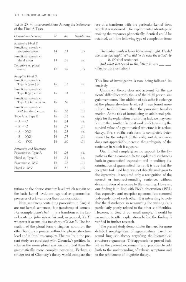
tutions on the phrase structure level, which remain onthe basic kernel level, are regarded as grammaticalprocesses of a lower order than transformations.
Now, sentences containing possessives in Englishare not kernel sentences, but transforms of kernels.For example, John’s hat . . . is a transform of the ker-nel sentence John has a hat and, in general, X’s Y,wherever it occurs, is a transform of X has Y. The for-mation of the plural form a singular noun, on theother hand, is a process within the phrase structurelevel and is thus less complex. The results in the pre-sent study are consistent with Chomsky’s position in-sofar as the noun plural was less disturbed than thegrammatically more complex possessive. Perhaps astricter test of Chomsky’s theory would compare the
use of a transform with the particular kernel fromwhich it was derived. The experimental advantage ofmaking the responses phonetically identical could beretained, as in the following type of completion item:
The soldier mails a letter home every night. He didthe same last night. What did he do with the letter? He___ ___ it. (Kernel sentence)
And what happened to the letter? It was ___ ___.(Passive transformation)
This line of investigation is now being followed in-tensively.
Chomsky’s theory does not account for the pa-tients’ difficulties with the -s of the third person sin-gular verb form. The addition of this suffix is a changeat the phrase structure level, yet it was found moresubject to disturbance than the possessive transfor-mation. At the risk of introducing an additional prin-ciple for the explanation of a further fact, we may con-jecture that another factor at work in determining thesurvival value of a grammatical structure is its redun-dancy. The -s of the verb form is completely deter-mined by the subject of the verb, and its omissiondoes not appreciably increase the ambiguity of thesentence in which it appears.
Our limited sample gives no support to the hy-pothesis that a common factor explains disturbancesboth in grammatical expression and in auditory dis-crimination of grammatical forms. It is true that thereceptive task used here was not directly analogous tothe expressive: it required only a recognition of thecorrect or incorrect-sounding sentence, withoutdemonstration of response to the meaning. However,our finding is in line with Pick’s observation (1931)that expressive and receptive agrammatism occurredindependently of each other. It is interesting to notethat the disturbance in recognizing the missing -’s isparticularly poorly related to the other -s difficulties.However, in view of our small sample, it would bepremature to offer explanations before the finding isverified in further research.
The present study demonstrates the need for moredetailed investigations of agrammatism based onsound linguistic theory regarding the hierarchicalstructure of grammar. This approach has proved fruit-ful in the present experiment and promises to addboth to the understanding of aphasic symptoms andto the refinement of linguistic theory.
374 HISTORICAL ARTICLES
TABLE 25–4. Intercorrelations Among the Subscoresof the Final S Tests
Correlations between: N rho Significance
Expressive Final SFunctional speech vs.
possessive errors 14 .53 .05
Functional speech vs.plural errors 14 .38 n.s.
Possessive vs. pluralerrors 17 .48 .05
Receptive Final SFunctional speech vs.
Type A (poss.) err. 16 .32 n.s.
Functional speech vs.Type B (pl.) errors 16 .73 .01
Functional speech vs.Type C (3rd pers) err. 16 .68 .01
Functional speech vs.XYZ (random) errors 16 .82 .01
Type A vs. Type B 16 .32 n.s.
— A — C 16 .24 n.s.
— B — C 16 .75 .01
— A — XYZ 16 .25 n.s.
— B — XYZ 16 .75 .01
— C — YXZ 16 .60 .01
Expressive and ReceptivePossessive vs. Type A. 10 .08 n.s.
Plural vs. Type B 10 .32 n.s.
Possessive vs. XYZ 10 .78 .01
Plural vs. XYZ 10 .57 .05

Note
1. The authors are indebted to Miss Mary Hyde,Speech Therapist at the Lemuel Shattuck Hospital, forher assistance in testing patients.
References
Chomsky, N., Syntactic Structures. The Hague: Mouton& Co., 1957.
Jakobson, R., “Kindersprache, Aphasie, und allgemeineLautgesetze,” Uppsale Universiteis Årsskrift, 1942:9.
Jakobson, R., “Two Aspects of Language and Two Typesof Aphasic Disturbances,” in R. Jakobson and M.Halle, Fundamentals of Language, The Hague,Mouton & Co., 1956.
Pick, A. (edited by R. Thiele), “Aphasie,” in A. Bethe,(ed.), Handbuch der normalen und pathologischenPhysiologie, Berlin, J. Springer, 1931.
GRAMMATICAL COMPLEXITY AND APHASIC SPEECH 375

376
26
The Organization of Language and the Brain
Norman Geschwind
Many problems relating to the functions of the ner-vous system can effectively be studied by investigationin animals, which permits controlled and repeatableexperiments on large groups of subjects. When wecome, however, to consider the relationship of thebrain to language, we must recognize that our knowl-edge is based entirely on findings in man. Some au-thors would even argue that language is exclusively ahuman attribute, so that no experiments on animalscould ever be relevant. Although I believe that fore-runners of language do exist in lower forms (1), the
direct contributions to this area of experimentation onthe brains of animals still lie in the future.
BRAIN LESIONS IN MAN
Information in this area has come from severalsources. Cases of brain tumor are of limited value,since tumors distort the brain and produce effects ata distance. Cases of penetrating brain wounds (2) havebeen of considerable use but are not the best sourceof anatomical data, since postmortem information isusually lacking. Analysis of the sites at which the skullwas penetrated is of use statistically, but, because ofvariations in the paths taken by missiles, cannot pro-vide precise data concerning the location of lesionsproducing language disorders. Stimulation duringsurgery (3) has been another most important source
Language disorders after brain damage help in elucidating the neural basis of verbal behavior.
Reprinted with permission from Science 170:940–944. Copy-right 1970 American Association for the Advancement of Science.The author was James Jackson Putnam Professor of Neurol-ogy at the Harvard Medical School and director of the Neu-rological Unit, Boston City Hospital, Boston, Massachusetts02118. This article is based on a paper presented 28 De-cember 1969 at the Boston meeting of the AAAS.

of information but, because of limitation of time atoperation and the accessibility of only certain struc-tures, has not covered the full range of phenomenaobserved clinically.
The elegant studies of Milner and her co-workerson patients undergoing excision of cortical regions forepilepsy represent the largest corpus of truly experi-mental studies of the higher brain functions in man(4). They are limited, however, with respect to therange of phenomena observed. Furthermore, sincemost of these patients were undergoing removal of ar-eas of brain which had been the site of epileptic dis-charges since childhood, there is reason to believe thatthe effects seen after surgery may not represent thefull range of phenomena seen after damage to theadult brain. The Wada test (5), in which sodium amy-tal is injected into one carotid artery, has been a ma-jor source of knowledge concerning the lateralizationof language functions in the brain.
Although important information has been ob-tained by the above methods, it is still true that thebulk of our knowledge concerning the relationship ofthe brain to language has been derived from the studyof adults in whom delimited areas of brain have beendamaged as the result of occlusion of blood vessels,who have been studied carefully over long periods,and whose brains have been subjected to careful post-mortem examination. Although fully suitable cases ofthis type are not common, the experience of nearly100 years of study has built up a large body of reliableknowledge.
APHASIC DISORDERS
The generic term aphasia is used to describe the dis-orders of language resulting from damage to the brain.Early in the history of the study of aphasia the dis-tinction between language and speech was stressed.In disorders of speech the verbal output was impairedbecause of weakness or incoordination of the musclesof articulation. The criterion of a disorder of languagewas that the verbal output be linguistically incorrect.The muscles of articulation might be used normallyin nonlinguistic activities. Similarly, in aphasic disor-ders of comprehension the patient might lose the abil-ity to comprehend spoken or written language and yetshow normal hearing or vision when tested nonver-bally. Furthermore, these disorders could occur with-out impairment of other intellectual abilities. The
aphasias were thus the first demonstrations of the factthat selective damage to the brain could affect oneclass of learned behavior while sparing other classes,and thus gave origin to the field of study of brain-behavior relationships. The discovery of these phe-nomena was one of the greatest achievements of thelast half of the 19th century.
Some cases of aphasia had been described beforethe mid-1800’s, but it was Paul Broca who in 1861began the study of the relationship of aphasia to thebrain, with two major contributions (6). He was thefirst to prove that aphasia was linked to specific le-sions, and to show that these lesions were predomi-nantly in the left half of the brain. The man who was,however, most responsible for initiating the modernstudy of this field was Carl Wernicke (Fig. 26–1), whoin 1874, at the age of 26, published his classic work,The Symptom Complex of Aphasia, which carried theappropriate subtitle, “A Psychological Study on anAnatomical Basis” (7). Wernicke established clearlythe fact that there were linguistic differences betweenthe aphasias produced by damage in the left tempo-ral lobe, in what is now called Wernicke’s area, andthose produced by lesions in the frontal lobe inBroca’s area (Fig. 26–2) (8).
ORGANIZATION OF LANGUAGE AND THE BRAIN 377
FIGURE 26–1. Carl Wernicke (1848–1904), who, atthe age of 26, published the monograph Der apha-sische Symptomencomplex, which was to be the ma-jor influence on the anatomical study of aphasia inthe period preceding World War I. During his tenureas professor at Breslau, his assistants and students in-cluded many of the later leaders of German neurol-ogy, such as Otfrid Foerster, Hugo Liepmann, KarlBonhoeffer, and Kurt Goldstein. (Figure scannedfrom original article.)

LINGUISTIC CHANGES IN APHASIA
The aphasic of the Broca’s type characteristically pro-duces little speech, which is emitted slowly, with greateffort, and with poor articulation. It is not, however,only at the phonemic level that the speech of thesepatients is abnormal, since the patient clearly fails toproduce correct English sentences. Characteristicallythe small grammatical words and endings are omit-ted. This failure persists despite urging by the exam-iner, and even when the patient attempts to repeat thecorrect sentence as produced by the examiner. Thesepatients may show a surprising capacity to find singlewords. Thus, asked about the weather, the patientmight say, “Overcast.” Urged to produce a sentencehe may say, “Weather . . . overcast.” These patientsinvariably show a comparable disorder in their writ-ten output, but they may comprehend spoken andwritten language normally. In striking contrast to theseperformances, the patient may retain his musical ca-pacities. It is a common but most dramatic finding toobserve a patient who produces single substantivewords with great effort and poor articulation and yetsings a melody correctly and even elegantly. BecauseBroca’s area lies so close to the motor cortex (Fig.26–2), this latter region is often damaged simultane-ously, so that these patients frequently suffer fromparalysis of the right side of the body.
The Wernicke’s aphasic contrasts sharply with theBroca’s type. The patient usually has no paralysis ofthe opposite side, a fact which reflects the differencein the anatomical localization of his lesion. Thespeech output can be rapid and effortless, and in manycases the rate of production of words exceeds the nor-
mal. The output has the rhythm and melody of nor-mal speech, but it is remarkably empty and conveyslittle or no information. The patient uses many fillerwords, and the speech is filled with circumlocutions.There may be many errors in word usage, which arecalled paraphasias. These may take the form of thewell-articulated replacement of single sounds (so-called literal or phonemic paraphasias), such as“spoot” for “spoon,” or the replacement of one wordfor another (verbal paraphasias), such as “fork” for“spoon.” A typical production might be, “I was overin the other one, and then after they had been in thedepartment, I was in this one.” The grammaticalskeleton appears to be preserved, but there is a re-markable lack of words with specific denotation.
The Wernicke’s aphasic may, in writing, producewell-formed letters, but the output exhibits the samelinguistic defects which are observed in the patient’sspeech. He shows a profound failure to understandboth spoken and written language, although he suf-fers from no elementary impairment of hearing orsight.
The localization of these forms of aphasia has beenconfirmed repeatedly. It is important to stress thispoint, since there is a common misconception thatthe classical localizations were rejected because pow-erful arguments were raised against their validity. Thetwo authors whose names are most frequently quotedas critics are Kurt Goldstein and Henry Head. As Ihave pointed out in greater detail elsewhere (9), Gold-stein, who had been a student under Wernicke at theUniversity of Breslau, despite the holistic views whichhe expressed in his philosophical discussions, actuallyexplicitly stated his support of the classical localiza-tions throughout his career. Head did indeed violentlyattack these views early in the first volume of his fa-mous work on aphasia (10). His argument was, how-ever, vitiated by the fact that, later in the same vol-ume, the localizations which he himself supportedturned out to be essentially identical to the ones hehad previously dismissed as invalid.
WERNICKE’S THEORY
Wernicke’s contribution lay not only in establishingthe syndrome patterns and their localizations but alsoin providing a theoretical analysis of the mechanismsof aphasia (Fig. 26–2). He pointed out that Broca’s areawas located just in front of the cortical region in whichlay the motor representation for the face, tongue, lips,
378 HISTORICAL ARTICLES
B
A
WW
FIGURE 26–2. Lateral surface of the left hemisphereof the human brain. B, Broca’s area, which lies ante-rior to the lower end of the motor cortex; W (open cir-cles), arcuate fasciculus, which connects Wernicke’sto Broca’s area. (See text.)

palate, and vocal cords—that is, the organs of speech.It seemed reasonable to assume that Broca’s area con-tained the rules by which heard language could becoded into articulatory form. This formulation still ap-pears reasonable. There is no need to assume that thiscoding need be a simple one. By contrast, Wernicke’sarea lies next to the cortical representation of hearing,and it was reasonable to assume that this area wassomehow involved in the recognition of the patternsof spoken language. There is also no need to assumethat this coding is a simple one.
Wernicke then added the natural assumption thatthese two areas must be connected. The general pat-tern was now clear. Destruction of Wernicke’s areawould lead to failure to comprehend spoken lan-guage. Wernicke pointed out that, for most people,written language was learned by reference to the spo-ken form and that therefore a lesion of this regionwould abolish comprehension of printed and writtenlanguage. The act of speaking would consist in arous-ing in some way the auditory form of words, whichwould then be relayed forward to Broca’s area to betransduced into the complex programming of thespeech organs, and therefore, with damage to Wer-nicke’s area, language output would also be disordered.
The model could readily be complicated further.Wernicke himself and those who followed him filledin further details. The comprehension of written lan-guage would require connections from the visual tothe speech regions, and destruction of these connec-tions should be able to cause isolated difficulties inreading comprehension. Since the language abilitieswere localized in the left hemisphere, language per-formances by the right hemisphere would depend oninformation transmission over the corpus callosum.
Clearly the validation of a theory is not a functionof its surface plausibility but is dependent on otherfactors. It is important to remember that Wernicke’stheory has been the only one in the history of apha-sia which could in a real sense be put to experimen-tal test. It was possible, on the basis of the theory, topredict that certain lesions should produce symptomsnot previously described. Furthermore, it was possi-ble, on being confronted with previously undescribedsyndromes, to predict the site of the anatomical le-sion. The most dramatic examples of this appear inthe writings of Hugo Liepmann (11) on the syndromesof the corpus callosum. On the basis of his clinicalexamination he predicted the presence of callosal le-sions, which were later confirmed at postmortem examination.
Several remarkable disorders of language havebeen described which fit readily into the Wernicketheory. In pure word deafness, the patient, with intacthearing as measured by ordinary nonverbal tests, failsto comprehend spoken language although he has es-sentially normal ability to express himself verbally andin writing and to comprehend written language. Inthis syndrome the area of damage generally lies deepin the left temporal lobe, sparing Wernicke’s area butdestroying both the direct auditory pathway to the lefthemisphere and the callosal connections from the op-posite auditory region. Although elementary hearingis intact because the right auditory region is spared,there is no means for auditory stimulation to reachWernicke’s area, and therefore the patient does notunderstand spoken language, although his ability toexpress himself in spoken and written language andhis comprehension of the written language are es-sentially intact (12).
In conduction aphasia, there is fluent paraphasicspeech, and writing, while comprehension of spokenand written language remains intact. Despite the goodcomprehension of spoken language there is a gross de-fect in repetition. The lesion for this disorder typicallylies in the lower parietal lobe (Fig. 26–2), and is soplaced as to disconnect Wernicke’s area from Broca’sarea. Because Broca’s area is preserved, speech is flu-ent, but abnormal. The preservation of Wernicke’sarea insures normal comprehension, but the gross de-fect in repetition is the result of disruption of the con-nection between this region and Broca’s area. The dis-order in repetition exhibits some remarkable linguisticfeatures which are not yet explained. The disorder isgreatest for the small grammatical words such as the,if, and is; thus, a patient who may successfully repeat“big dog” or even “presidential succession” may failtotally on “He is here.” The most difficult phrase forthese patients to repeat is “No ifs, ands, or buts.” Inmany of these patients the ability to repeat numbersmay be preserved best of all, so that, given a phrasesuch as “seventy-five percent,” the patient may repeatthe “seventy-five” rapidly and effortlessly but may failon “percent” (12).
PURE ALEXIA WITHOUT AGRAPHIA
Many examples of pure alexia without agraphia weredescribed in the 1880’s, but the first postmortem studyof this syndrome was described in 1892, by Dejerine(13). His patient suddenly developed a right visual
ORGANIZATION OF LANGUAGE AND THE BRAIN 379

field defect and lost the ability to read. He could, how-ever, copy the words that he could not understand.He was able, moreover, to write spontaneously, al-though he could not read later the sentences he hadwritten. All other aspects of his use and comprehen-sion of language were normal. At postmortem Dejer-ine found that the left visual cortex had been de-stroyed. In addition, the posterior portion of thecorpus callosum was destroyed, the part of this struc-ture which connects the visual regions of the twohemispheres (Fig. 26–3). Dejerine advanced a simpleexplanation. Because of the destruction of the left vi-sual cortex, written language could reach only theright hemisphere. In order to be dealt with as lan-guage it had to be transmitted to the speech regionsin the left hemisphere, but the portion of the corpuscallosum necessary for this was destroyed. Thus, writ-ten language, although seen clearly, was withoutmeaning. This was the first demonstration of the ef-fects of a lesion of the corpus callosum in preventingtransfer of information between the hemispheres.
Dejerine’s thesis has received striking confirma-tion. In 1925 Foix and Hillemand (14) showed thatdestruction of the left visual cortex in the absence ofa callosal lesion does not produce this syndrome. In1937 Trescher and Ford (15) described the first casein which a surgical lesion of the corpus callosum wasshown to have a definite effect. Their patient had sus-tained section of the posterior end of the corpus cal-losum for removal of a tumor from the third ventri-cle. The patient could not read in the left visual field,but could read normally on the right side. This resultis implied by the Dejerine theory and was confirmedby Maspes in 1948 (16) and more recently by Gaz-zaniga, Bogen, and Sperry (17). Many authors haveconfirmed Dejerine’s anatomical findings. MichaelFusillo and I studied a patient with alexia withoutagraphia who demonstrated another intriguing disor-der (18, 19). For approximately 3 months after hisstroke he suffered from a disorder of verbal memory,which then cleared, leaving him with the reading dif-ficulty, which remained unchanged until his deathseveral months later. At postmortem, in addition tothe anatomical findings of destruction of the left vi-sual cortex and of the posterior end of the corpus cal-losum, the brain showed destruction of the left hip-pocampal region. It is now generally accepted thatbilateral destruction of the hippocampal region leadsto a permanent memory disorder. The transient mem-ory disorder in our patient appeared to be the resultof the destruction of the left hippocampal region—that is, the one located in the same hemisphere as thespeech areas. Presumably it is the left hippocampalregion which is necessary for the memory functionsof speech cortex. After a period, the brain manages tocompensate, presumably by making use of the oppo-site hippocampal region. Since publication of our pa-per (18), I have seen several other cases of this syn-drome in which memory disorder was present at theonset. It is well known that the posterior cerebralartery supplies not only the visual cortex and the pos-terior end of the corpus callosum but also the hip-pocampal region. In a certain number of cases of oc-clusion of the left posterior cerebral artery, all of thesestructures are damaged. In other cases, however, thehippocampal region is spared. Meyer and Yates (20)and Milner (4) have demonstrated that, after removalof the left anterior temporal region for epilepsy, a ver-bal memory disorder is observed, which is, however,generally much milder than that found in the caseFusillo and I reported, and which is not present afterright anterior temporal ablation. The mildness of the
380 HISTORICAL ARTICLES
S
V
FIGURE 26–3. Horizontal section of the humanbrain, illustrating the mechanism of pure alexia with-out agraphia. V, visual region. The visual cortex onthe left is destroyed (thick black line). As a result, thepatient can perceive written material only in the in-tact right visual region. For this material to be appre-ciated as language, it must be related to the speechareas on the left side through the splenium, which isthe posterior portion of the corpus callosum. As a re-sult of damage to the splenium (S), this transfer can-not take place, and therefore the patient cannot com-prehend the written words whose form he perceivesclearly.

disorder after left temporal ablation is probably the re-sult of the fact that these patients had suffered fromleft temporal epilepsy for years and had therefore al-ready begun to use the right hippocampal region to aconsiderable degree.
ISOLATION OF THE SPEECH AREA
Another syndrome, called “isolation of the speecharea,” is explained readily by the Wernicke theory.This syndrome was described first by Kurt Goldstein(21) and has been described more recently byGeschwind, Quadfasel, and Segarra (22). We studiedour patient for nearly 9 years after an episode of car-bon monoxide poisoning. During this period sheshowed no evidence of language comprehension inthe ordinary sense, and never uttered a sentence ofpropositional speech. She was totally helpless and re-quired complete nursing care. In striking contrast tothis state were her language performances in certainspecial areas. She would repeat perfectly, with normalarticulation, sentences said to her by the examiner.She would, however, go beyond mere repetition, sinceshe would complete phrases spoken by the examiner.For example, if he said, “Roses are red,” she wouldsay, “violets are blue, sugar is sweet, and so are you.”Even more surprising, it was found that she was stillcapable of verbal learning. Songs which did not existbefore her illness were played to her several times.Eventually, when the record player was started shewould begin to sing. If the record player was thenturned off she would continue singing the words andmusic correctly to the end, despite the lack of a model.Postmortem examination by Segarra showed a re-markable lesion, which was essentially symmetrical.The classical speech area, including Wernicke’s area,Broca’s area, and the connections between them, wasintact, as were the auditory inflow pathways and themotor outflow pathways for the speech organs. In theregions surrounding the speech area either the cortexor the underlying white matter was destroyed. Thespeech area was indeed isolated. The patient’s failureto comprehend presumably resulted from the fact thatthe language inputs could arouse no associationselsewhere in the brain, and since information fromother portions of the brain could not reach the speechareas, there was no propositional speech. On the otherhand the intactness of the speech region and its in-ternal connections insured correct repetition. Thepreservation of verbal learning is particularly interest-
ing. In addition to the speech area, the hippocampalregion, which is involved in learning, was also pre-served, and this probably accounts for her remarkableability to carry on the memorizing of verbal material.
CALLOSAL SYNDROMES
Although pure alexia without agraphia (13) was thefirst syndrome in which damage to the corpus callo-sum was shown to play a role by interrupting transferof information between the hemispheres, it was agroup of Wernicke’s students, including Hugo Liep-mann, Kurt Goldstein, and Karl Bonhoeffer, who elu-cidated the full syndrome of callosal disconnection incases in which eventually there was careful post-mortem confirmation of the predicted sites of the le-sions (11, 12). While the callosal syndromes contin-ued to be recognized by German authors (23), theirexistence was either forgotten or indeed totally deniedin the English-language literature. In November1961, Edith Kaplan and I presented a patient to theBoston Society of Psychiatry and Neurology who was,we believed, suffering from a callosal disconnectionsyndrome—a diagnosis which was later confirmed atpostmortem examination by Segarra. [Since that timeseveral cases of confirmed callosal disconnection havebeen described (24).] I will mention here briefly onlya few of the aspects of our patient’s condition whichfit into the Wernicke theory. When writing with theright hand the patient produced linguistically correctwords and sentences and carried out calculations cor-rectly. When writing with the left hand he producedincorrect words (for example, “run” for “go”) and per-formed calculations incorrectly. The theory outlinedabove implies that, for writing to be carried out cor-rectly with the left hand, the information must betransmitted from the speech areas across the corpuscallosum, whose interruption in our patient explainedhis failures. Similarly, the patient could correctlyname objects (concealed from vision) which he pal-pated with the right hand. On the other hand hewould misname objects palpated with the left hand,although it could be shown by nonverbal means thathis right hemisphere recognized the object. Thus, ifa pencil was placed in his left hand the patient coulddraw the object previously held in that hand. Again,the Wernicke theory implies that, for an individual tocorrectly name an object held in the left hand, theinformation must be transmitted from the sensory re-gions in the right hemisphere to the speech regions
ORGANIZATION OF LANGUAGE AND THE BRAIN 381

via the corpus callosum, which had been destroyedin this patient. On the other hand, the patient couldread in the left as well as the right visual field. Thisled us to conclude that the destruction of the corpuscallosum had spared the posterior end, a predictionalso confirmed at postmortem.
CEREBRAL DOMINANCE
Let me turn to another bit of knowledge which fitsvery well into the scheme presented above. One ofthe most remarkable features of man is cerebral dom-inance—that is, the fact that in the adult the capaci-ties for speech are overwhelmingly controlled by theleft hemisphere. Out of 100 adult aphasics, at least 96percent have damage to the left side of the brain (25).We do not know of any example in any other mam-mal of a class of learning which is predominantly con-trolled by one half of the brain (26). What underlieshuman speech dominance? It is widely stated in theliterature that the human brain is symmetrical, andthis had led either to the assumption that speech dom-inance must reflect some subtle physiological differ-ence between the hemispheres, or indeed even to theassumption that speech dominance is somehow ac-quired as the result of postnatal experience. My col-league Walter Levitsky and I (27) decided to reinves-tigate this problem, particularly since we found thatsome earlier authors had claimed that there were infact anatomical differences between the hemispheres.We demonstrated that such differences exist and areindeed readily visible to the naked eye. The area thatlies behind the primary auditory cortex in the uppersurface of the temporal lobe is larger on the left sidein 65 percent of brains, and larger on the right in only11 percent. This region on the left side is, on the av-erage, nearly a centimeter longer than its fellow onthe opposite side—that is, larger by one-third than thecorresponding area on the right. More recently Wada(28) has confirmed our results. He has, in addition,studied this region in the brains of infants and hasfound that these differences are present at birth. Thisregion which is larger in the left hemisphere is, infact, a portion of Wernicke’s area, whose major im-portance for speech was first shown nearly 100 yearsago. It is reasonable to assume that there are otheranatomical asymmetries in the hemispheres of the hu-man brain, reflecting other aspects of dominance.
The study of the organization of the brain for lan-guage has been based of necessity on investigations in
man. The bulk of our information in this area hascome from careful studies of patients suffering fromisolated damage as a result of vascular disease, whosebrains have, after death, been subjected to carefulanatomical examination. Disorders of language re-sulting from brain damage, almost always on the leftside, are called aphasias. Carl Wernicke, nearly 100years ago, described the linguistic differences betweenaphasias resulting from damage in different anatomi-cal locations and outlined a theory of the organiza-tion of language in the brain. Not only have Wer-nicke’s localizations stood up under repeatedexamination but his theory has been the only onewhich as permitted the prediction of new phenom-ena, or has been able to account for new observations.Several remarkable disorders, such as isolated distur-bances of reading and the symptomatology of the cor-pus callosum, are examples of the explanatory powerof this theory.
The phenomenon of cerebral dominance—that is,the predominant importance of one side of the brainfor a class of learned behavior—occurs, as far as weknow, in no mammal other than man. The domi-nance of the left side of the brain for speech is themost striking example of this phenomenon. Contraryto generally accepted views, there is a striking anatom-ical asymmetry between the temporal speech regionon the left side and the corresponding region of theright hemisphere.
References and Notes
1. I have argued elsewhere that language is basedon the striking development of the angular gyrus regionin man, a region which receives inputs from all corticalsensory areas [see N. Geschwind, in Monograph Serieson Languages and Linguistics, No. 17 (Georgetown Univ.Press, Washington, D.C., 1964), pp. 155–169; Brain 88,
237 (1965); ibid., p. 585]. D. Pandya and H. Kuypers[Brain Res. 13, 13 (1969)] have shown that a forerunnerof this region exists in the macaque. R. A. Gardner andB. T. Gardner [Science 165, 664 (1969)] and D. Premack(in a paper presented at the Symposium on CognitiveProcesses of Nonhuman Primates, Pittsburgh, March1970) have described what appears to be a definite de-gree of linguistic behavior in chimpanzees.
2. A. R. Luria, Traumatic Aphasia (Mouton, TheHague, 1969).
3. O. Foerster, in Handbuch der Neurologie, O.Bumke and O. Foerster, Eds. (Springer, Berlin, 1936),vol. 6, pp. 1–448; W. Penfield and L. Roberts, Speechand Brain-Mechanisms (Princeton Univ. Press, Prince-ton, N.J., 1959).
382 HISTORICAL ARTICLES

4. See for example, B. Milner, in InterhemisphericRelations and Cerebral Dominance, V. B. Mountcastle,Ed. (Johns Hopkins Press, Baltimore, 1962), pp.177–195.
5. J. Wada and T. Rasmussen, J. Neurosurg. 17, 266(1960); C. Branch, B. Milner, T. Rasmussen, ibid. 21,
399 (1964).6. A. L. Benton [Cortex 1, 314 (1964)] summarizes
the earlier literature; R. J. Joynt (ibid., p. 206) gives anaccount of Broca’s contributions.
7. C. Wernicke, Der aphasische Symptomen-com-plex (Franck and Weigert, Breslau, 1874). An Englishtranslation has recently appeared in Boston Studies in thePhilosophy of Science, R. S. Cohen and M. W. Wartof-sky, Eds. (Reidel, Dordrecht, 1969), vol. 4, pp. 34–97.For a more complete evaluation of Wernicke’s work, seeN. Geschwind, ibid., pp. 1–33.
8. R. Jakobson [in Brain Function, E. C. Carterette,Ed. (Univ. of California Press, Berkeley, 1966), vol. 3,pp. 67–92] has given a vivid description of these linguisticdifferences.
9. N. Geschwind, Cortex 1, 214 (1964).10. H. Head, Aphasia and Kindred Disorders of
Speech (Cambridge Univ. Press, London, 1926).11. H. Liepmann, Drei Aufsatze aus dem Aprax-
iegebiet (Karger, Berlin, 1908).12. N. Geschwind, Brain 88, 237 (1965); ibid., p.
585. There is another, less readily understood, lesion insome cases of pure word deafness which is discussed inthese two communications.
13. J. Dejerine, Mem. Soc. Biol. 4, 61 (1892).14. C. Foix and P. Hillemand, Bull. Mem. Soc.
Med. Hop. Paris 49, 393 (1925).15. J. H. Trescher and F. R. Ford, Arch. Neurol.
Psychiat. 37, 959 (1937).16. P. E. Maspes, Rev. Neurol. 80, 100 (1948).
17. M. S. Gazzaniga, J. E. Bogen, R. W. Sperry,Brain 88, 221 (1965).
18. N. Geschwind and M. Fusillo, Arch. Neurol.15, 137 (1966).
19. For a review of the different varieties of alexia,see D. F. Benson and N. Geschwind, in Handbook ofClinical Neurology, P. J. Vinken and G. W. Bruyn, Eds.(North-Holland, Amsterdam, 1969), vol. 4, pp. 112–140.
20. V. Meyer and H. J. Yates, J. Neurol. Neurosurg.Psychiat. 18, 44 (1955).
21. K. Goldstein, Die transkortikalen Aphasien (Fis-cher, Jena, 1917).
22. N. Geschwind, F. A. Quadfasel, J. M. Segarra,Neuropsychologia 4, 327 (1968).
23. J. Lange, in Handbuch der Neurologie, O.Bumke and O. Foerster, Eds. (Springer, Berlin, 1936),vol. 6, pp. 885–960; O. Sittig, Über Apraxie (Karger,Berlin, 1931).
24. N. Geschwind and E. Kaplan, Neurology 12,
675 (1962); M. S. Gazzaniga, J. E. Bogen, R. W. Sperry,Proc. Nat. Acad. Sci. U.S. 48, 1765 (1962).
25. For a review, see O. Zangwill, Cerebral Domi-nance and Its Relation to Psychological Function(Thomas, Springfield, Ill., 1960).
26. In submammalian forms there are examples ofbehaviors whose neural control appears to be predomi-nantly unilateral—for example, bird song [see F. Notte-bohm, Science 167, 950 (1970)]. these may represent,not an earlier stage of dominance, but rather a separatedevelopment.
27. N. Geschwind and W. Levitsky, Science 161,
186 (1968).28. J. Wada, paper presented at the 9th Interna-
tional Congress of Neurology, New York, 1969.29. The work discussed has been supported in part
by grant NS 06209 from the National Institutes of Healthto the Boston University School of Medicine.
ORGANIZATION OF LANGUAGE AND THE BRAIN 383

384
27
Broca’s Area and Broca’s Aphasia
Jay P. Mohr
[ . . . ]
THE CLINICAL SYNDROME OF
BROCA’S APHASIA AND ITS
ANATOMIC FOUNDATION
The literature contains a long list of articles con-cerning a syndrome referred to as Broca’s aphasia. Inmost of these articles, primary emphasis has beenplaced on the description of the clinical features, withmuch less effort devoted to autopsy correlation of theclinical syndrome with the lesion topography.
The clinical features of the syndrome that haveevolved in the literature over the past 110 years aremore complex, involve more evidence of deficit in
language usage, including easily documented deficitsin “comprehension,” appear more severe, and aremuch more thoroughly described than those met inthe older literature, which presents autopsy data show-ing infarction limited to LF3 and its immediate sur-roundings. However, the uncomfortable possibilitysuggested by the more complex clinical syndrome isthat the underlying lesion far exceeds that of LF3 andadjacent structures.
Despite the impressive list of authors who havewritten on Broca’s aphasia, many have not cited per-sonal cases that document their views, and few haveprovided autopsy or other laboratory evidence cor-roborating the lesion topography. In some, even theetiology of the case material has been unclear. Vas-cular material appears to have been the source mate-rial for Broca, Wernicke, Déjerine, Marie, Liepmann,Alajouanine, Nielsen, Kreindler and Fradis, andGoodglass, Quadfasel, and Timberlake. War materialwas used by Head, Kleist, Goldstein, Conrad, Russelland Espir, and Luria. Weisenburg and McBridge, anddeAjuriaguerra and Hécaen, used a wide etiologic
Excerpts reprinted from Studies in Neurolinguistics, HaiganooshWhitaker & Harry A. Whitaker, eds.; J. P. Mohr, “Broca’sArea and Broca’s Aphasia,” 1979, with permission from Elsevier. This paper was supported by grants HL 14888-02,NS 10828-01A1, and HS 00188.The author was at the Massachusetts General Hospital andHarvard Medical School, Boston, Massachusetts.

spectrum of cases. Brown presented an abscess case.Textbook authors usually have not specified their ma-terial. Included in this group are Brain, Critchley,Lhermitte and Gautier, Benson and Geschwind, andAdams and Mohr.
Perhaps as troublesome has been the frequentlyunstated time course over which the syndromechanged from or to the state described as Broca’s apha-sia. In the few cases reported, the late description ofa deficit referred to as Broca’s aphasia is better corre-lated with a lesion far larger than Broca’s area, as il-lustrated in Tables 27–1 and 27–2. The two notableexceptions were Broca’s own cases, further describedat the end of this review.
Traditional Formulations of
Broca’s Aphasia
Wernicke (1874) might be credited with the first text-book characterization of Broca’s aphasia. In 1874, heconfined his remarks to a few simple features withoutciting actual cases: following “destruction of thespeech movement images . . . the patient understandseverything but has either suddenly become mute orhas at most a few simple words at his disposal” (p. 57).
By 1908 (Wernicke, 1908, pp. 272–273) his de-scription had enlarged considerably under the termmotor aphasia, which he equated with Broca’saphemia, due to involvement of Broca’s convolution.Still without cited cases, Wernicke described threemain features:
1. “The power of articulate speech is wanting. Thepatients have forgotten the process, the mech-anism, which they formerly called into actionto produce its sounds.” He considered the pa-tients mute or at best minimally able to speak,using “senseless syllables,” short words orphrases, profane expressions. Dyspraxia of bul-bar musculature for other movements, rightlower facial and tongue weakness, but no ele-mentary weakness of the bulbar musculaturewere also described.
2. “In the main, the power of understandingspeech is retained at least this appears to be thecase on ordinary tests. . . . There is almost in-variably a certain inability to understand com-plicated constructions and the finer differenti-ations of speech. . . . I no longer am of theopinion that in pure motor aphasia the abilityto understand speech always remains unim-
paired.” Here Wernicke considerably modifiedhis earlier views, but did not indicate whetherhe considered this wider deficit to reflect a le-sion larger than Broca’s area.
3. “Written language . . . is lost simultaneouslywith articulate speech.” This statement is thefirst of along series of opinions concerning theintimate relationship between written and spo-ken speech stemming from Broca’s originalcases.
Wernicke considered the prognosis “generally un-favorable” for restoration of speech function.
This complex, severe, and persisting syndrome, to-gether with the inference that Broca’s area is the siteof the responsible lesion, has persisted largely un-changed over the years under the label motor orBroca’s aphasia (Adams & Mohr, 1974; Bastian, 1897;Benson & Geschwind, 1971; Brain, 1962; Brown,1972; Déjerine & Mirallié, 1896; Hécaen, 1972; Heil-bronner, 1910; Kreindler & Fradis, 1968; Lhermitte& Gautier, 1969; Lichtheim, 1885; Liepmann, 1915;Nielsen, 1962; Weisenburg & McBride, 1964; Wyl-lie, 1894). This description has also found applicationin war-injury studies where efforts have been made tocorrelate the clinical deficit with the skull defect oroperative findings (Conrad, 1954; Goldstein, 1948;Head, 1926; Russell & Espir, 1961).
Several earlier authors took the trouble to presentexemplary personal case reports and/or excerptedcases from the literature (Lichtheim, Wyllie, Bastian,Weisenburg and McBride, Nielsen), but only Dejer-ine appears to have published autopsied case reports.
Remarkably, despite the paucity of autopsy mate-rial for this larger syndrome, most of the differencesof opinion revolve around individual features of thesyndrome, in particular the explanations for deficitsin behavior apart from disordered speaking. Distur-bance in silent reading “comprehension” has beennoted since the earliest reviews (Bastian, 1897; Wyl-lie, 1894). The explanations of dyslexia are typified byBastian (1897):
In reading, a proper comprehension of the mean-ing of the text requires a conjoint revival of thewords in the visual and the auditory word-centres,but that for this mere comprehension it is not nec-essary for the stimulus to pass on also to the glosso-kinesthetic (Broca’s) centre, as it must do in read-ing aloud. It may, however, be freely admitted thatif the way is open, and this latter centre is in a
BROCA’S AREA AND BROCA’S APHASIA 385

TABLE 27–1. Autopsy Documented Cases of Broca’s or Total Aphasia Found in the Literaturea
*F3 B 51 Broca (case Leborgne)
SYL T 23 Dejerine (Moutier 70)
SYL T . . Moutier (case Maillard)
SYL B . . Bastian (Moutier 98)
OI T . . Mills (Henschen 1069)
FOI B 45 Bernard (Moutier 84)
SYL B 59 Moutier (case Chissadon)
FOI B 55 Preston (Moutier 96)
FOI B . . Broadbent (Moutier 51)
SYL T . . Vulpian & Mongie (Moutier 40)
SYL T 80 Bernard (Moutier 86)
SYL T 54 Bernheim (Moutier 90)
SYL T . . Comte
*F3 B . . Broca (case Lelong)
SYL T 39 Giraud (Moutier 64)
SYL B . . Archambault (Moutier 31)
FOI T 43 Pitres (Moutier 74)
FOI B 25 Lange (Moutier 52)
SYL T . . Bleuler (Moutier 92)
EN B . . Ballet (Moutier 132)
FOI T 80 Moutier (case Fauchier)
SYL B 56 Skwotzoff (Moutier 78)
F3 B 25 Baldisseri (Henschen 719)
SYL T 39 Rosenthal (Moutier 78)
F3 B 25 Böe (Henschen 779)
F3 B 43 Ballet & Boix (Moutier 130)
F3 B . . Rosenstein (Wyllie 3)
F3 B 45 Atkins
F3 B . . Magnan
F3 B 61 Chauffard & Rathery (Henschen 849)
F3 B . . Malicherg (Henschen 1052)
FOI T 69 Demange (Moutier 80)
F3 B . . Ogle
F3 B . . Sheinker & Kuhr
F3 B . . Magnan
F3 B 55 Nielsen (case Lulu)
F3 B 50 Hervey (Moutier 127)
F3 B . . Banti (Bastian 7)
F3 B 68 Nielsen (case Ingols)
10 20 30 10 20 30 10 20 30 10 20 30
Days Weeks Months Years
(Time after onset when the last or only examination was described in the Case Report)
aKey: F3 Broca’s area (3rd Frontal), FOI frontal operculum and anterior insula, OI operculum and insula, SYL Sylvian region, EN en-cephalitis, B Broca’s aphasia, T total aphasia, . . age not stated, PH putaminal hemorrhage.
386

healthy condition, it does commonly receive inreading to one’s self a slight stimulus from the au-ditory word-centre, a fact which is often enoughshown by the occurrence of involuntary half-whis-pered mutterings when reading. It may also be ad-mitted that the rousing of all three centres doesgive assistance in the comprehension of anythingdifficult, as is shown by the common practice ofreading aloud any passage the meaning of whichmay be at all obscure [p. 1009].
This principle has remained the explanation offeredinto modern times (Luria, 1966, p. 190).
Auditory comprehension and other disturbancesin language formulations have been less easily ex-plained. Opinion has been divided as to whether ornot such disturbances even occur. Lichtheim (1885,p. 471), Wyllie (1894, pp. 318–319), Bastian (1897,
p. 1005), and Liepmann (1915, pp. 526–527) con-sidered motor aphasia to be free of such deficits. Oth-ers concurred with Wernicke’s views (Déjerine & Mi-rallié, 1896, pp. 102–105; Heilbronner, 1910, pp.1021–1028). Marie (1906b) took the extreme positionthat Broca’s aphasia represents Wernicke’s aphasiaplus anarthria. Modern authors’ views on this pointare less clearly stated, partially because the current,revised syndrome lays stress on different features ofthe deficit (as will be explained further).
Writing disturbances have been explained by cit-ing two separate mechanisms. Bastian (1897), almostalone in his opinion, maintained that any dysgraphiais secondary to involvement outside Broca’s area, usu-ally affecting the second frontal gyrus, so-called“Exner’s writing center.” A few subsequent authors(Brain, 1962; Henschen, 1925) have entertained theseviews, while others (Mohr et al., 1973) have been con-
BROCA’S AREA AND BROCA’S APHASIA 387
TABLE 27–2. Autosopy Documental Cases of Broca’s Aphasia Considerd Ameliorated Found in Literature
OI B 32 Monakow (Henschen 1113)
OI T 57 Improved Monakow (Henschen 1114)
OI T 45 Improved Dejerine (Henschen 866)
F3 B 20 Disappeared Luys (Henschen 1031)
F3 B . . Recovered Wadham
F3 B 20 Recovered Dejerine (Henschen 865)
FOI B 61 Ameliorated Leva (Moutier 95)
OI T 25 Recovered Lange (Henschen 986)
F3 B 25 Improved DeFont (Henschen 905)
F3 B . . None Bourneville
PH B 61 Improved Nielsen
OI B 62 Improved . . . Cured DuFour (Henschen 885)
F3 B 20 Rapid Foulis (Henschen 908)
OI B 38 Later DuFour (Henschen 883)
F3 B 32 Rapidly Dejerine (Henschen 871)
F3 B 70 Cleared Bramwell
F3 B Transient Tuke
F3 B 65 Improving . . . . Normal Simon (Henschen 1240)
F3 B . . 10 Days Barlow
F3 B 49 Cleared Monokow (Henschen 1116)
10 20 30 10 20 30 12 15 25 35 5 10 15 20
Days Weeks Months Years
(Time after onset when initial examination was described, followed by repeat examination orcomment regarding outcome)
aKey: F3 Broca’s area (3rd Frontal), FOI frontal operculum and anterior insula, OI operculum and insula, SYL Sylvian region, EN en-cephalitis, B Broca’s aphasia, T total aphasia, . . age not stated, PH putaminal hemorrhage.

tent to indicate a certain degree of independence be-tween writing and speaking performances withoutmaking anatomic inferences. The overwhelmingweight of opinion has favored a coexisting deficit inwriting and speaking. Where anatomic comments areincluded, the lesion is considered to involve Broca’sarea and need be no larger. The opinions of Jackson(1932) typify most authors: “speaking is proposition-izing . . . that the speechless patient cannot proposi-tionize aloud is obvious—he never does. But this isonly the superficial part of the truth. He cannot propo-sitionize internally . . . the proof that he does not speakinternally is that he cannot express himself in writing.. . . He can say nothing to himself, and therefore hasnothing to write.” Given such reasoning, little addi-tional explanation was required to account for the eas-ily observed deficits in auditory comprehension notedby all authors, including Liepmann (1915), Pick(1973), Isserlin (1936), Lhermitte and Gautier (1969),Brown (1972), and others. None—as best can be de-termined by the present reviewer—based their viewson personal cases with pathoanatomic correlation ofthe clinical deficits. Yet none expressed disagreementthat the lesion lay in Broca’s area.
Of the many features of this larger syndrome re-ferred to as Broca’s aphasia, the deficit in spokenspeech has received the greatest attention. Broca(1861) has described the recurrent utterances and par-tial syllables that characterized the limited speakingbehavior of his two patients. Jackson (1932), Wernicke(1908), Liepmann (1915), Pick (1973), Kleist (1934),Alajouanine, Ombrédane, and Durand (1939), Good-glass et al. (1964), and Brown (1972) have written andrewritten the basic features of verbal stereotypes, re-current utterances, and condensed grammatical sen-tence structure that characterize speaking in cases ofBroca’s aphasia. Save for Kleist (1934), none have de-scribed clinical cases with individual pathologic cor-relation, although all authors have stated or impliedthat the lesion lies in Broca’s area or immediately sur-rounding regions or needs to involve no additional ar-eas for this characteristic speaking performance. Inmodern times, the work of Goodglass et al. (1964)provides an exemplary documentation of the clinicaland linguistic features in clinical cases referred to asBroca’s aphasia. Twenty-two of 53 patients studiedwith the Boston Veterans Administration DiagnosticAphasia Test were characterized as cases of Broca’saphasia. They showed poor scores on melodic line,length of uninterrupted word groups, verbal agility in
articulation, and correct grammatical form. Virtuallyno jargon in connected speech, impaired auditorycomprehension, or problems in naming objects werefound. The cases studied were all allowed at least twomonths after stroke onset before they were studied. Ofparticular interest to the present reviewer was the re-mark that “It appeared that most Broca’s aphasics ei-ther remained grossly impaired or quickly attained alevel of residual aphasia in which the classical featuresof agrammatism, telegraphic speech, and laborious ar-ticulation were no longer apparent.” (Goodglass et al.,1964). These observations corroborate those of thepresent review (Mohr et al., 1975) that rapid amelio-ration of speech deficit occurs (in the local Broca’s-area infarct), or when persisting, severe deficit is foundin which disturbed language function is easily demon-strated (in the larger syndrome of Sylvian operculuminfarction). Like most other authors, Goodglass et al.(1964) had no autopsy correlation in their clinicalcases, but they appeared to consider that the presumedinfarction involved Broca’s area, since they speculatedthat “Broca’s area contains critical structures whichare so concentrated that a direct injury is likely to bepermanently and severely damaging to speech.” Thisspeculation, based as it was on the litany, and not onthe personal autopsy experience of the authors, is rep-resentative of the degrees to which this larger syn-drome has been considered to reflect Broca’s-area in-farction by most authors.
Several authors have been impressed with thedeficits in language behavior other than speaking toa degree that serious doubts were raised as to whetherthe term motor aphasia is appropriate for such cases.The painstaking work of Weisenburg and McBride(1964) represents some of the most detailed docu-mentation of this larger deficit. Their criticisms of theconcept of motor aphasia were based on a thoroughliterature review and 42 personal cases of “expressive”or “expressive–receptive” aphasia. Their efforts clearlydocumented the larger deficit and their reasons forobjecting to motor aphasia as an inappropriate con-cept, but their case material could scarcely be con-sidered an appropriate source for criticisms of theanatomic basis of the syndrome: Their 42 cases en-compassed a broad etiologic spectrum including is-chemic stroke, hemorrhage, tumor, cyst, gunshotwound, and subdural hematoma. None of the caseswere autopsied, and in the early 1930s, save for therare operated case, the topography of the lesion ineach case was inferred on clinical criteria alone. The
388 HISTORICAL ARTICLES

opportunity to relate the larger syndrome to a brainlesion far exceeding the confines of Broca’s area andimmediately adjacent brain tissue was not taken upby Weisenburg and McBride nor by a large numberof other authors who also pointed out that the syn-drome involved a deficit in language functions apartfrom that evident in speaking aloud. With few ex-ceptions, Broca’s-area lesion has been considered suf-ficient to cause this large deficit.
Kleist (1934, p. 930) considered the persisting andsevere deficit to require infarction deep into the hemi-sphere, so as to disrupt the white matter fibers thatserved as projection and association pathways forBroca’s area. By inference, these deeper lesions wouldmean a larger infarct, although Kleist did not specifythe upper division of the left middle-cerebral artery assuch. Foix (1928) earlier had made a similar infer-ence by referring to deeper branches of the middle-cerebral artery. Goldstein (1948, pp. 204–205) madesuggestions similar to those of Kleist, but also failedto specify the vascular territory involved in the largerlesion. Remarkably few cases of this larger syndromehave been autopsied and described in the literature(see Tables 27–1 and 27–2); those reported haveproven difficult to classify simply as Broca’s aphasiabecause they were examined only in the late stages,raising the possibility that they represented improvedtotal aphasia.
The theoretic problems raised by the notions of to-tal aphasia reflect sufficiently on those of Broca’s apha-sia to warrant further consideration of total aphasia.The term emerged in the late 19th century in theworks of most textbook authors (Déjerine & Mirallié,1896; Heilbronner, 1910; Liepmann, 1915; Wer-nicke, 1908). Liepmann’s (1915) account is typical:
More frequently than a lesion confined to thefrontal or temporal speech region, we find, as a re-sult of the arterial distribution bringing all theblood in the whole region of speech, through theArt. foss. sylvii, lesions, that affect both regions, andtherefore causing total or almost total (motor andsensory) aphasia. Following the retrogression of theword-deafness, one sees, years later, a clinical pic-ture, in which the symptoms of motor aphasia pre-dominate. Word-dumbness conceals the parapha-sia; the disturbance of speech understanding is nolonger very serious. Writing and reading . . . arevery poor. Hence it happens, that old cases, inwhich the lesions occur in both speech regions,are often classed clinically only as motor aphasia.
In these cases, disturbances in speaking, writing andreading, are particularly stable [pp. 529–530]. (em-phasis Liepmann)
This description by Liepmann bears a close resem-blance to that currently referred to as Broca’s aphasiain the later stages.
The existence of this syndrome prompts a detailedexamination of the timecourse data for publishedcases labeled as Broca’s aphasia. Protocols for most ofthe wholly clinically described cases of Broca’s apha-sia usually show that the authors dealt with chroniccases, at least two months (Goodglass et al., 1964), aslong as six years (Alajouanine et al., 1939), and tenyears (Goldstein, 1948, case 6, pp. 208–245) after on-set. The autopsy material (Tables 27–1 and 27–2) fur-ther reveals that the later a case labeled as Broca’saphasia is documented clinically, the greater the like-lihood that a large lesion is found at autopsy. This ma-terial also documents the frequency with which casesquite similar in detailed clinical features are given thelabel total aphasia (Tables 27–1 and 27–2).
Personal Observations
Mohr et al. (1975) have succeeded in acquiring au-topsy or other laboratory corroboration of lesion topog-raphy for ten cases suffering cerebral infarction, all ofwhom broadly satisfied the criteria currently used forBroca’s aphasia months after their stroke. Seven werestudied by computerized axial tomogram (CT scan),two by arteriogram, and one by autopsy. The lesiontopography in each case far exceeded Broca’s area. Inmost, the lesion was best explained by total or near-total infarction of the area of supply of the upper di-vision of the left middle-cerebral artery, encompass-ing the operculum from anterior frontal throughBroca’s area (LF3) to anterior parietal regions, the in-sula, both banks of the central (Rolandic) fissure, theentire infarct usually extending deep into the hemi-sphere (Fig. 27–1).
The initial deficit in these cases was uniformly se-vere, closely approximating Liepmann’s (1915) de-scriptions of total aphasia. They later evolved, as de-scribed by Liepmann (1915), toward a state more orless conforming to current descriptions of Broca’saphasia. In all cases the later syndrome emerged fromone more severe. Weeks, but usually months and oc-casionally years were required before this later statewas fully established.
BROCA’S AREA AND BROCA’S APHASIA 389

All cases experienced hemiplegia at onset. Nonewere able to stand for over a week; all walked with aheavy circumducted gait, the arm essentially useless,the right lower face densely paretic. The six seen theday of onset experienced head and conjugate eye de-viation to the side of the lesion, all six improving tofull head and eye control within a week. Hemianopiawas present for several days. After several months, asyndrome of spastic hemiparesis still disabled all pa-tients although they achieved independence with acane after prolonged physiotherapy.
Initially, all cases were mute and unable to writelegibly with either hand; most graphic efforts werehopeless loops or marks without communicativevalue. Evidence of disturbed comprehension, formany complete, was present in auditory and visuallanguage tasks. Two cases followed for years (Mohr etal., 1973) underwent steady evolution toward muchimproved auditory comprehension, but began tospeak only after ten months in one case, three years
in the other. The most successful improvements oc-curred in two other cases, who performed Marie’sthree-paper test correctly at three months in the caseof a 25-year-old left-handed man, and seven monthsin a 44-year-old right-handed man. Save for these twocases, deficits in language tasks were easily demon-strated months and even years after onset, if testingwas carried beyond ordinary social conversation intomore formal years, particularly in spelling. All patientsemerged from mutism into stereotype utterances,which soon became agrammatic laconic efforts atspontaneous speech; this difficulty was similarly re-flected in misspelled dysgrammatic efforts at writing.Considerable variation in dyspraxia of the upper ex-tremities, oral, and respiratory apparatus was observed.
Corroborative cases were culled from the retro-spective autopsy series from the Massachusetts Gen-eral Hospital for a 10-year period (mentioned pre-viously). Fifteen of the 39 cases of left-cerebralinfarction affecting the territory of supply of the up-per division of the left middle-cerebral artery were de-scribed as having deficits in speaking and in languageuse persisting months and years, conforming (wheredata was available) to features broadly classifiable asBroca’s aphasia. In all such cases, the lesion far ex-ceeded Broca’s area, usually involving the bulk of theterritory of supply of the upper division.
Formulation of the Syndrome of Upper
Division Infarction
These personal and retrospective autopsy cases, com-pared with the few described and the fewer autopsiedcases in the literature, prompt a formulation of thissyndrome that differs from the traditional and currentnotions of Broca’s aphasia.
Broca’s aphasia, as currently defined, is not a re-sult of infarction of Broca’s area and immediate sur-rounds. It reflects a major infarction involving mostof the territory of supply of the upper division of theleft middle-cerebral artery. As a clinical entity, the ev-idence to date suggests that the deficit profile knownas Broca’s aphasia is observed only later after the in-farction. The initial syndrome is more severe, de-scribed traditionally as total aphasia. After weeks ormonths, the gradual emergence of stereotypes, agram-matism, and protracted dyspraxia in speaking evolvesslowly toward the long-standing deficit profile ofBroca’s aphasia, which remains little changed aftermonths and years.
390 HISTORICAL ARTICLES
FIGURE 27–1. (Top) Syndrome of infarction in theupper division of the middle cerebral artery, projectedon a lateral view of the left hemisphere. (Bottom) Anexample of a computed tomography scan with thelarge lesion evident. The plane of section for the scanis indicated by the line drawn through the top diagram.(Figure scanned from original article.)

Subsequent studies may reveal that the complexsyndrome of Broca’s aphasia can occur acutely, with-out evolving from the more obvious syndrome of to-tal aphasia. But in such case, the prediction is madethat a large lesion will be found, far exceeding Broca’sarea.
This formulation of the upper-division syndromemakes the terms Broca’s aphasia and total aphasiaseem inexact, even inappropriate in principle. Nei-ther convey the anatomic or functional implicationsimplicit in this new formulation. The upper-divisionsyndrome is a complex, wide-ranging deficit profile,involving formulation and production of spoken andwritten communication, including “central” languagefunctions as well as the more elemental organizationof sensory, motor, and praxic skills for execution ofsuch spoken and written efforts. The major disorga-nization of these systems is more easily understoodwhen the great extent and depth of the lesion are ap-preciated. Less fanciful explanations are required toaccount for the deficit, and the long periods of timerequired for improvement are more easily accountedfor than by traditional implications that all this com-plex behavior depended upon the herculean ho-muncular functions of Broca’s area. Although muchof the mystique of aphasiology might be lost by theseformulations of the upper-division syndrome, a closealignment with views of cerebral functions based onother physiologic studies might be the result.
BROCA’S CASES
In retrospect, had Broca emphasized the extent of thelesion topography in his two cases, he might have pre-vented over a century of controversy. When accountis taken of lesion topography, Broca’s cases appear rel-atively straightforward examples of the upper-divisionsyndrome now referred to as Broca’s aphasia: twochronic cases, seen 18 months and 10 years, respec-tively, after infarction, having severe speaking deficitconfined to limited verbal stereotypes, conversation-ally satisfactory “comprehension,” and, in the one case with autopsy findings, major infarction affectingthe territory of supply of the upper division of the leftmiddle-cerebral artery (Fig. 27–2).
Broca saw neither case acutely. He used only con-versational and not more formal grammatical taskssuch as spelling, which might have shown a deficientperformance sharply contrasting with the conversa-
tional evidence of good comprehension. In accor-dance with then-current ideas that stroke disease af-fected the brain in a slowly expanding fashion (Marie,1906b), Broca understandably sought the site of “ori-gin” of the stroke; he deliberately ignored the largerzone of infarction (which he considered the laterspreading effect of the stroke) in favor of the portioninvolving the inferior frontal region, where he envis-aged the stroke “began.” By faithfully adhering tothen-current notions of the pathophysiology of stroke,Broca missed the opportunity to formulate for all timethe clinical, temporal, and pathologic features of theupper-division middle cerebral artery syndrome thatshould justifiably bear his name. Instead, his namecame to be associated with a syndrome improperlycorrelated with brain infarct topography and with anarea of the brain whose infarction produces only amodest long-term clinical deficit.
To avoid future ambiguities, the terms Broca’saphasia, total aphasia, and even Broca’s area mightpreferably fall into disuse, in favor of other terms, suchas the opercular syndromes, that better describe thespectrum observed, including that end of the spec-trum that Broca observed but did not interpret as such.Considering that Boulliaud gave Broca his original in-spirations, the eclipse of Broca’s epomyn seems a lessthan historic tragedy, since Boulliaud’s own effortshave already largely passed into oblivion.
References
Adams, R. D., Mohr, J. P. 1974. Affections of speech. InM. M. Wintrobe et al. (Eds.), Harrison’s principlesof internal medicine. (7th ed.) New York: McGraw-Hill, Pp. 137–148.
Alajouanine, T., Ombrédane, A., & Durand, M. 1939.Le syndrome de désintégration phonétique dansl’aphasie. Paris: Masson.
BROCA’S AREA AND BROCA’S APHASIA 391
FIGURE 27–2. Topography of the infarct found inBroca’s case Lebornge (drawing traced from Moutier).

Atkins, R. 1876. Case of sudden and complete aphasiaand partial right hemiplegia, lesion of Broca’s con-volution, with a small haemorrhage in substance ofcorpus callosum &c. Journal of Mental Science, 22,406–416.
Bailey, P., & von Bonin, G. 1951. The isocortex of man.Urbana: University of Illinois.
Bastian, H. C. 1897. Some problems in connexion withaphasia and other speech defects. Lancet, 1,1005–1017, 1132–1137, 1187–1194.
Benson, D. F., & Geschwind, N. 1971. The aphasias andrelated disturbances. In A. B. Baker (Ed.), Clinicalneurology. Vol. 1. New York: Harper. Pp. 1–25.
Bourneville. 1874b. Athérome généralisé: oblitérationsmultiples (aphasie; sphacèle du pied, etc.). ProgrèsMédical, 2(20), 278–280.
Bourneville. 1874b. Athérome généralisé: oblitérationsmultiples (aphasie; sphacèle du pied, etc.). ProgrèsMédical, 2(21), 296–298.
Brain, W. R. 1962. Speech disorders. London: Butterworths.Bramwell, B. 1898. A remarkable case of aphasia. Brain:
A Journal of Neurology, 21, 343–373.Broca, P. 1861. Remarques sur le siége de la faculté
du langage articulé, suivies d’une observationd’aphémie (perte de la parole). Bulletins de la So-ciété Anatomique de Paris, 6, 330–357.
Brodmann, K. 1909. Vergleichende Lokalisationslehre derGrosshirnrinde in ihren Prinzipien dargestellt aufGrund des Zellenbaues. Leipzig: Barth.
Brown, J. W. 1972. Aphasia, apraxia and agnosia. Spring-field, Ill.: Charles C Thomas.
Bruandet (Interne des hopitaux). 1900. Un cas d’hemis-pasme facial. Revue Neurologique, 8, 658–660.
Bruyn, G. W., and Gathier, J-C. 1970. The operculumsyndrome. In P. J. Vinken & G. W. Bruyn (Eds.),Handbook of clinical neurology. Vol. 2. Amsterdam:North-Holland Publ. Pp. 776–783.
Chouppe. 1870. Ramollissement superficiel du cerveauintéressant surtout la troisième circonvolutionfrontale gauche, sans aphasie. Bulletins de la SociétéAnatomique de Paris, 45, 365–366.
Cole, M. F., & Cole, M. 1971. Pierre Marie’s papers onspeech disorders. New York: Hafner.
Comte, A. 1900. Des paralysies pseudo-bulbaires. (The-sis) 8°, No. 436. Paris: Steinheil.
Conrad, K. 1954. New problems of aphasia. Brain: AJournal of Neurology, 77(4), 491–509.
Critchley, MacD. 1970. Aphasiology and other aspects oflanguage. London: Arnold.
DeAjuriaguerra, J., and Hécaen, H. 1964. Le cortexcérébral. Paris: Masson.
deBoyer, H. C. 1879. Études topographiques sur les lé-sions corticales des hémisphères cérébraux. (Thesis)Paris: Delahaye.
Déjerine, J. 1885. Étude sur l’aphasie dans lésions de l’in-sula de Reil. Revue de Médecine (Paris), 5, 174–191.
Déjerine, J. 1891. Contribution à l’étude de l’aphasiemotrice sous corticale et de la localisation cérébraledes centres laryngés (muscles phonateures). ComptesRendus Hebdomadaires des Séances et Mémoires dela Socièté de Biologie, 3, 155–162.
Déjerine, J. 1906. L’aphasie motrice. Sa localisation etsa physiologie pathologique. Presse Medicale,14(57), 453–457.
Déjerine, J., & Mirallié, C. 1896. L’aphasie sensorielle.Paris: Steinheil.
Flechsig, P. 1901. Developmental (myelogenetic) Lo-calisation of the cerebral cortex in the human sub-ject. Lancet, ii, 1027–1029.
Foix, C. 1928. Aphasies. In G. H. Roger, F. Widal, & P.J. Teissier (Eds.), Nouveau traité de médecine. Vol.18. P. 135. Paris: Masson et Cie.
Foulis, D. 1879. A case in which there was destructionof the third left frontal convolution without apha-sia. British Medical Journal, 1, 383–384.
Friedlich, A. L., Castleman, B., & Mohr, J. P. 1968. Sud-den stroke in a woman with dyspnea and anemia.New England Journal of Medicine, 278, 1109–1118.
Geschwind, N. 1965a. Disconnexion syndromes in ani-mals and man: I. Brain: A Journal of Neurology, 88,237–294.
Geschwind, N. 1965b. Disconnexion syndromes in ani-mals and man: II. Brain: A Journal of Neurology,88, 585–644.
Goldstein, K. 1948. Language and language distur-bances. New York: Grune & Stratton.
Goodglass, H., & Kaplan, E. 1972. The assessment ofaphasia and related disorders. Philadelphia: Lea &Febiger.
Goodglass, H., Quadfasel, F. A., & Timberlake, W. T.1964. Phase length and type and severity of apha-sia. Cortex, 1, 133–153.
Head, H. 1926. Aphasia and kindred disorders of speech.New York: Macmillan.
Hécaen, H. 1972. Introduction à la neuropsychologie.Paris: Larousse (Librairie).
Heilbronner, K. 1910. Die aphasischen, apraktischenund agnostischen Störungen. In M. Lewandowsky(Ed.), Handbuch der Neurologie. Vol. 1. Berlin:Springer-Verlag. Pp. 982–1093.
Henschen, S. E. 1922. Klinische und pathologischeBeiträge zur Pathologie des Gehirns. Vol. VII. Stock-holm: Nordiske Bokhandeln.
Henschen, S. E. 1925. Clinical and anatomical contri-butions on brain pathology; (Abstracts and com-ments by Walter F. Schaller); Fifth Part: Aphasia,amusia and akalkulia. Archives of Neurology andPsychiatry, 13, 226–249.
392 HISTORICAL ARTICLES

Isserlin, M. 1936. Aphasie. In O. Bumke and O. Foer-ster (Eds.), Handbuch der Neurologie. Vol. 6. Berlin:Springer-Verlag. Pp. 627–806.
Jackson, J. H. 1932. Selected writings. London: Hodder& Stoughton.
Jefferson, G. 1950. Localization of function in the cere-bral cortex. British Medical Bulletin, 6, 333–340.
Kleist, K. 1934. Gehirnpathologie. Leipzig: Barth.Kreindler, A., & Fradis, A. 1968. Performances in apha-
sia. Paris: Gauthier-Villars.Kugler, J. 1964. Electroencephalography in hospital and
general consulting practice. Amsterdam: Elsevier.Lhermitte, F., & Gautier, J.-C. 1969. Aphasia. In R. J.
Vinken & G. W. Bruyn (Eds.), Handbook of clini-cal neurology. Vol. 4. Amsterdam: North-HollandPubl. Pp. 84–104.
Lichtheim, L. 1885. On aphasia. Brain: A Journal of Neu-rology, 7, 433–484.
Liepmann, H. 1915. Diseases of the brain. In C. W. Barr(Ed.), Curschmann’s textbook on nervous diseases.Vol. 1. Philadelphia: Blakiston. Pp. 467–551.
Luria, A. R. 1966. Higher cortical functions in man. NewYork: Basic Books.
McAdam, D. W., & Whitaker, H. A. 1971. Language pro-duction: Electroencephalographic localization inthe normal human brain. Science, 172, 499–502.
Magnan. 1879. On simple aphasia, and aphasia with in-coherence. Brain: A Journal of Neurology, 2, 112–123.
Marie, P. 1906a. Revision de la question de l’aphasie:L’aphasie de 1861 à 1866; essai de critique his-torique sur la genèse de la doctrine de Broca. Se-maine Médicale, 26, 565–571.
Marie, P. 1906b. Revision de la question de l’aphasie: Latroislème circonvolution frontale gauche ne joueaucun rôle spécial dans la fonction du langage. Se-maine Médicale, 26, 241–247.
Marie, P. 1906c. Revision de la question de l’aphasie:Que faut-il penser des aphasies sous-corticales(aphasies pures)? Semaine Médicale, 26, 493–500.
Marie, P. 1907. Rectifications à propos de la question deL’aphasie. Presse Medicale, 15(4), 25–26.
Marie, P., & Moutier, F. 1906a. Examen du cerveau d’uncas d’aphasie de Broca. Bulletins et Mémoires de laSociété Médicale des Hopitaux de Paris, 23,743–744.
Marie, P., & Moutier, F. 1906b. Nouveau cas d’aphasiede Broca sans lésion de la troisième frontale gauche.Bulletins et Mémoires de la Société Médicales desHopitaux de Paris, 23, 1180–1183.
Marie, P., & Moutier, F. 1906c. Nouveau cas de lésioncorticale du pied de la 3e frontale gauche chez undroitier sans trouble du langage. Bulletins et Mé-moires de la Société Médicale des Hopitaux de Paris,23, 1295–1298.
Marie, P., & Moutier, F. 1906d. Sur un cas de ramol-lissement du pied de la 3e circonvolution frontalegauche chez un droitier, sans aphasie de Broca. Bul-letins et Mémoires de la Société Médicale des Hopi-taux de Paris, 23, 1152–1155.
Mettler, F. A. 1949. Selective partial ablation of thefrontal cortex: A correlative study of the effects on hu-man psychotic subjects. New York: Haeber.
Mohr, J. P. 1968. Cerebral control of speech. Letters tothe Editor. New England Journal of Medicine, 279,107.
Mohr, J. P. 1973. Rapid amelioration of motor aphasia.Archives of Neurology, 28, 77–82.
Mohr, J. P., Funkenstein, H., Finkelstein, S., Pessin, M.,Duncan, G. W., & Davis, K. 1975. Broca’s area in-farction versus Broca’s aphasia. Neurology, 25, 349.
Mohr, J. P., Leicester, J., Stoddard, L. T., & Sidman, M.1971. Right hemianopia with memory and colordeficits in circumscribed left posterior cerebral arteryterritory infarction. Neurology, 21, 1104–1113.
Mohr, J. P., Sidman, M., Stoddard, L. T., Leicester, J.,& Rosenberger, P. B. 1973. Evolution of the deficitin total aphasia. Neurology, 23, 1302–1312.
Moutier, F. 1908. L’aphasia de Broca. (Thesis) Paris:Steinheil.
New, P. F. J., Scott, W. R., Schnur, J. A., Davis, K. R., &Taveras, J. M. 1974. Computerized axial tomogra-phy with the EMI scanner. Radiology, 110, 109–123.
Nielsen, J. M. 1962. Agnosia, apraxia, aphasia. NewYork: Hafner.
Ogle, W. 1867. Aphasia and agraphia. St. George’s Hos-pital Reports, 2, 83–122.
Ojemann, R. G., Aronow, S., & Sweet, W. H. 1966. Scan-ning with positron-emitting isotopes in cerebrovas-cular diseases. Acta Radiologica, 5, 894–905.
Penfield, W. 1958. The excitable cortex in conscious man.Springfield, Ill.: Charles S Thomas.
Penfield, W., & Rasmussen, T. 1968. The cerebral cortexof man. New York: Hafner.
Penfield, W., & Roberts, L. 1959. Speech and brain mech-anisms. Princeton, N.J.: Princeton University Press.
Pick, A. 1973. Aphasia. (Translated and edited by J. W.Brown) Springfield, Ill.: Charles C Thomas.
Robb, J. P. 1948. Effects of cortical excision and stimu-lation of the frontal lobe on speech. Research Pub-lications, Association for Research in Nervous andMental Disease, 27, 587–609.
Russell, W. R., & Espir, M. L. E. 1961. Traumatic apha-sia. London: Oxford University Press.
Scheinker, I., & Kuhr, B. M. 1948. Motor aphasia andagraphia caused by small vascular lesion confinedto third and second convolutions of left frontal lobe.Research Publications, Association for Research inNervous and Mental Disease, 27, 582–586.
BROCA’S AREA AND BROCA’S APHASIA 393

Simpson, J. H. 1867. On a case of extensive lesion of theleft posterior frontal convolution of the cerebrum,without aphasia. Medical Times and Gazette, 2, 670.
Singer, M., & Yakovlev, P. I. 1954. The human brain insagittal section. Springfield, Ill.: Charles C Thomas.
Taveras, J. M., & Wood, E. H. 1964. Diagnostic neuro-radiology. Baltimore: Williams & Wilkins. Pp.1.691–1.718.
Tuke, J. B., & Fraser, J. 1872. Case with a lesion in-volving Broca’s convolution without Broca’s apha-sia. Journal of Mental Science, 18, 46–56.
Wadham, W. 1869. On aphasia. St. George’s HospitalReports, 4, 245–250.
Weisenburg, T., & McBride, K. E. 1964. Aphasia. NewYork: Hafner.
Wernicke, C. 1874. Der Aphasische Symptomencomplex.Breslau: Cohn & Weigert.
Wernicke, C. 1908. The symptomcomplex of aphasia. InA. Church (Ed.), Modern clinical medicine. NewYork: Appleton. Pp. 265–324.
Wyllie, J. 1894. The disorders of speech. Edinburgh:Oliver.
Yakovlev, P. I. 1970. Whole brain serial histological sec-tions. In C. G. Tedeschi (Ed). Neuropathology:Methods and diagnosis. Boston: Little, Brown. Pp.371–378.
394 HISTORICAL ARTICLES

Author Index
Abelanet, R., 29Abernathey, C.D., 29Abler, B., 267Aboitiz, F., 3, 4, 5, 6, 8, 9, 13, 19, 27,
154f, 164, 165f, 166, 219, 272, 278Abutalebi, J., 105, 195Acardi, A.C., 9, 13Ackermann, H., 223t, 237, 240Adams, R.D., 385, 391Adger, D., 121, 132Adolphs, R., 195Adriani, M., 240Aggleton, P., 3, 13Aguirre, G.K., 46, 183, 217, 252Ainsworth-Darnell, K., 211n5, 212Akitake, M., 105Akmaijan, A., 51, 61Alain, C., 225t, 237Alajouanine, T., 388, 389, 391Albert, M.L., 195Albert, N., 13Alexander, M.P., 31, 44, 255, 265Alivisatos, B., 46, 240Alkadhi, H., 16
Alpert, N., 16, 212, 217Alsop, D., 13, 212Alter, K., 198, 213, 215, 217, 219,
238, 239, 240Altmann, G.T.M., 197, 212Amunts, K., 17, 19, 20, 24, 25, 26,
27, 28, 29, 30, 32, 35, 38, 44, 85,104, 137, 149, 150, 172, 182, 212,219, 237, 237n1, 241, 242, 243,251, 252, 253, 254, 258, 263, 278,283, 284, 287, xv
Anders, D., 104Anderson, J.M., 8, 13Andreiuolo, P.A., 183Angrilli, A., 201, 212Aoun, J., 100, 104Arbib, M., 4, 7, 8, 9, 12, 13, 16, 42,
44, 141, 149, 150, 151, 152, 153,154, 155, 156, 156f, 157f, 158, 159,160, 161, 161f, 162, 163, 164, 165,165f, 166, 166n1, 167, 178, 182,183, 220, 241, 256, 264, 266, 267,274
Archibald, J., 130, 132
Ardila, A., 238Arkin, R.C., 156, 166Armony, J.L., 151Armstrong, A.C., 42, 44Armstrong, E., 30Arnott, S.R., 237Aronow, S., 393Ashburner, J., 16, 266Aston, J., 107Atkins, R., 392Atkinson, J., 182Auburtin, S., 292, 294–95Augath, M., 239Avrutin, S., 50, 51, 52, 56, 57, 60n4,
60n17, 61, 62, 84, 102, 103n1, 104,104n8, 105, 272, 273, 278, 282,283, 285
Awh, E., 8, 13
Baauw, S., 51, 56, 61, 62Bach, P., 214Baddeley, A., 4, 8, 13, 14, 158, 164,
167, 176, 181, 182Bahan, B., 183
395
Note: Page numbers followed by f and t refer to figures and tables, respectively.

Bailey, P., 392Bak, T.H., 147, 150, 189, 194Baker, C., 171, 181Baker, E., 132, 132n2, 133Baker, M., 92, 104Baldeweg, T., 9, 15Ballard, D., 252Ballard, K.J., 62, 81, 106, 128, 132,
134Balota, D.A., 45, 197, 212Bamiou, D.E., 237Bao, S., 107Barbas, H., 5, 13, 39, 44Barlow, J., 130, 132, 133Baron-Cohen, S., 267Barss, A., 106, 216Bartha, L., 150Basho, S., 220, 240Basso, A., 83, 104, 244, 245, 252Basso, G., 152Bastiaanse, R., 56, 57, 61, 74, 75, 80,
82, 252Bastian, H.C., 137, 385, 387, 392Batal, H.A., 29Bates, E.A., 73, 80Bates, J.F., 16Baudena, P., 214Bauer, R.., 13Bavelier, D., 181, 183, 206, 212, 240Beaman, P., 182Beauregard, M., 195Beck, S., 100, 104Behen, M.E., 216, 240Beisteiner, R., 102, 104Bekkering, H., 15, 151, 182Belin, P., 218, 237, 241Bell, B.D., 138, 150Bell, L., 133Belletti, A., 79, 80Belliveau, J.W., 29Bellmann, A., 240Bellugi, U., 175, 176, 181, 182, 183Ben-Bashat, D., 104, 132, 212Ben-Shachar, M., 96, 97, 100, 101,
104, 123, 132, 209, 212Benedet, M.J., 56, 61, 67, 80Benson, D.F., 31, 44, 238, 263, 265,
383n19, 385, 392Benson, R.R., 224t, 232, 237Bentin, S., 201, 212Benton, A.L., 383n6Beretta, A., 93, 104Berger, M.S., 45Bergida, R., 266Berko, J., 64, 80Berndt, R.S., 64, 75, 80, 82, 109, 117,
118Bernholtz, N.A., 70, 80Berry, T., 132n2, 133Bersick, M., 216
Bert, J., 143, 150Besson, M., 15, 151, 152, 216, 239Bettinardi, V., 151, 195, 266, 267Bever, T., 90, 104Beveridge, M.A., 124, 132Beversdorf, D.Q., 13Bharucha, J., 139, 150, 239, 241Bhatnagar, S., 73, 80Bhattacharyya, R., 172, 182Biber, C., 104Bien, S., 106, 216Biguer, B., 155, 167Binder, J.R., 6, 13, 263, 265Binkofski, F., 28, 84, 104, 139, 150,
193, 194, 210, 212, 219, 238, 239,256, 264, 265, 266
Birk, J.L., 239Bjork, R., 184Blank, S.C., 224t, 238Bleser, R. de. See de Bleser, R.Bloch, I., 46Blom, E., 50, 61Bloom, A., 166n4, 167Bloom, P., 4, 16Blumstein, S., 83, 84, 99, 104, 173,
181, 238, 289Bobaljik, J., 104Bock, J.K., 123, 132Boemio, A., 218, 238Bogen, J.E., 380, 383n17Bogolepova, I.N., 30, 241, 253Boland, J.E., 212Bonda, E., 256, 265Bonhoeffer, T., 102, 104, 105, 106Boniface, S., 150, 194Bonin, G. von. See von Bonin, G.Bookheimer, S., 4, 10, 12, 13, 14, 95,
104, 207, 210, 212, 219, 223t, 238,239, 246, 252, 255, 265
Bornkessel, I., 202, 204, 211n8, 212Boskovic, Z., 106Bosman, C., 3, 9, 12, 13Bota, M., 4, 7, 8, 9, 12, 13, 153, 162,
164, 165, 165f, 166, 166n1Bottini, G., 206, 212Bouillard, J.B., 255, 265, 291Bourneville, 392Bradley, N., 167Brady, T.I., 29Brain, W.R., 385, 387, 392Brammer, M.J., 215Bramwell, B., 392Brass, M., 15, 151, 182Braun, A., 172, 181, 238, 240Bréal, M., 343Brenneis, A., 150Brentari, D., 169, 170, 181Bresnan, J., 134, 250, 252Bressi, S., 195Brett, M., 151, 221, 238
Brinkley, J., 181Broadbent, W.H., 305, 310, 339Broca, P.P., 3, 13, 17, 28, 83, 137,
150, 218, 238, 255, 265, 266,287–88, 291–304, 377, 388, 392
Brodmann, K., 18, 20, 24, 28, 34, 35,39, 45, 85, 288, 334–36, 392
Broere, C.A.J., 217Brolio, J., 167Bronen, R.A., 216Bronnick, K., 239Brookins, B., 134Brown, C., 10, 15, 29, 205, 212, 214,
252Brown, E.D., 28Brown, G.M., 194Brown, J.W., 385, 388, 392Brown, S., 224t, 238Bruandet, 392Bruce, C.J., 38, 45Brun, D., 62Brunetti, E., 3Bruyn, G.W., 392Bub, D., 81, 253Buccino, G., 144, 150, 194, 195, 219,
220, 238, 256, 264, 265, 266Buchanan, T., 225t, 238, 239Büchel, C., 15, 182, 195, 241, 252,
258, 266Buchsbaum, B., 177, 180, 181Buck, A., 268Buckner, R.L., 44, 45Bullier, J., 46Bullmore, E.T., 215Buonocore, M.H., 267Burchert, F., 92, 93, 104Bürgel, U., 27, 44, 104, 149, 237,
251, 265Burkhart, P., 60n14, 62Burton, H., 40, 46Burton, M.W., 222t, 238Bushell, C., 62, 107, 134Byng, S., 124, 132
Cadoret, G., 43, 45Call, J., 15Calvert, G., 150Camarda, R., 29, 46, 152, 216Campbell, R., 144, 150, 182Canseco-Gonzalez, E., 119, 129, 132,
133Capek, C.M., 175, 181Capelle, L., 45Capitani, E., 118Caplan, D., 10, 11, 13, 16, 64, 67,
80, 81, 93, 96, 97, 104, 105, 159,166, 208, 212, 217, 244, 245, 252,253
Cappa, S., 15, 84, 187, 194, 195, 215,219, 272–73, 280, 282, 284, 286
396 AUTHOR INDEX

Caramazza, A., 60n15, 61, 64, 80,107, 109, 117, 118, 194, 244, 252,253
Caramonas, Z., 29, 46, 106, 241, 253Cardebat, D., 212, 238Carlson, G.N., 197, 212Carmarda, R., 183Carpenter, P., 215Castaigne, P., 29Castleman, B., 392Cavada, C., 5, 13Ceballos-Baumann, A.., 267Chafee, M.V., 166n2, 167Chaminade, T., 143, 150Champion, A.H., 130, 133Chao, L., 179, 181Chapman, R., 133Chapon, F., 215Chazal, M., 81Chee, M.W.L., 256, 266Chelune, G.J., 152Cheney, D.L., 9, 16Chertkow, H., 195Chiavaras, M., 29, 46, 106, 241, 253Chien, Y.C., 61, 105Chitty, A.J., 167Choi, S.H., 179, 180, 181Chollet, F., 212, 217, 238Chomsky, N., 4, 11, 13, 50, 52, 60n4,
61, 64, 79, 80, 104, 121, 133, 163,167, 248, 252, 373–74, 375
Chouppe, 392Christiansen, J.A., 61, 80Chugani, H.T., 216, 240Chwilla, D.J., 201, 212Ciccarelli, L., 212Cimini-Cunha, F., 183Clahsen, H., 56, 62, 67, 82, 216Clark, C.A., 46Clark, V.P., 212, 237Clarke, J.M., 214Clarke, M., 214Clarke, S., 24, 30Clifton, C., 197, 212, 213Cobrinik, L., 263, 266Code, C., 166, 167Cohen, L., 211n1, 212Cohen-Seat, G., 143, 150Cokely, D., 171, 181Cole, M., 392Cole, M.F., 392Cole, P., 60n3, 61Coles, M., 211n9, 212Collette, F., 16Collina, S., 188, 194Collins, D.L., 26, 28Collins, L., 28Collins, R.T., 167Comte, A., 392Connelly, A., 9, 15, 16
Connolly, J.F., 214Conrad, K., 385, 392Consoli, S., 189, 194Constable, R., 15, 216Contardi, A., 81Cook, N.D., 175, 181Cooke, A., 10, 13, 209, 212Cooper, F.S., 151Corballis, M.C., 9, 13, 42, 45Corbetta, M., 261, 263, 264, 266Corcoran, R., 212Corey, V., 216Corina, D., 169, 172, 173, 179, 181,
183, 212Corkin, S., 263, 266Costes, N., 150, 151, 252Coulson, S., 201, 204, 212Courchesne, E., 152, 240Cox, D.M., 82Cox, R.W., 13, 265Craighero, L., 137, 147, 150, 159,
167, 238Craik, F.I.M., 194Crain, S., 106Cramon, D.Y. von. See von Cramon,
D.Y.Crerar, M.A., 124, 132Creusere, M., 240Critchley, MacD, 392Crivello, F., 241Crosson, B., 13Cuetos, F., 104Cutler, A., 218, 219, 221, 239, 251,
252
Dale, A.M., 29Damasio, A.R., 8, 13, 15, 181, 182,
188, 189, 191, 194, 195Damasio, H., 15, 179, 181, 182,
195Dammers, J., 214Daniloff, J.K., 138, 150Danna, M., 195Dapretto, M., 10, 14, 95, 104, 207,
210, 212Darwin, C., 3, 14Dauzat, A., 342David, A.S., 182, 183Davis, B., 164, 167Davis, K.R., 45, 194, 253, 393Davis, M.D., 240Davis, M.H., 221, 223t, 236, 238de Bleser, R., 64, 70, 80, 104de Lange, J., 62de Oliveira-Sousa, R., 183de Roo, E., 57, 61de Vincenzi, M., 196, 203, 212Deacon, T.W., 5, 14DeAjuriaguerra, J., 392DeBoyer, H.C., 392
Decety, J., 139, 143, 150, 151, 246,252, 256, 266
Deecke, L., 104Degueldre, C., 16Dehaene, S., 212Déjerine, J., 31, 45, 279, 289,
379–80, 383n13, 385, 387, 389,392
Delazer, M., 139, 150Delcroix, N., 241Delfiore, G., 16Demb, J.B., 192, 194Demeter, S., 240Demirdache, H., 79, 80Demonet, J.F., 14, 209, 212, 222t,
238Deng, S.Y., 46Dennett, D.C., 4, 14Denvil, D., 45DeRenzi, E., 166, 167Desmond, J., 46, 194, 214, 216d’Esposito, M., 46, 152, 183, 195,
217, 252, 253Detre, J., 13, 212Dettmers, C., 151, 182, 252Deutsch, A., 201, 212DeVita, C., 13, 212Devlin, J.T., 243, 252Di Betta, A.M., 189, 195Di Matteo, R., 212Di Pellegrino, G., 142, 150Dickey, M.W., 95, 104Diesch, E., 102, 104Dinner, D.S., 152Ditterich, A., 27, 251Dittman, J., 50, 51, 62Dodrill, C., 181Doeller, C.F., 226t, 238Dogil, G., 240Dohmas, F., 150Doi, D., 183Dolan, R.J., 266Dominey, P.F., 239Donati, C., 15, 194, 215Donchin, E., 211n9, 212Donohue, B.C., 29Dordain, M., 81, 253Döring, W., 238Doron, E., 74, 81Dostie, D., 183Doty, R.W., 240Dowd, D., 181Drai, D., 87, 92, 98, 104, 108, 109,
114, 118, 118n1Draper, B.A., 156, 167Driver, J., 267Dronkers, N.F., 4, 7, 14, 190, 194Druks, J., 117, 118Dubeau, M.C., 151Duffau, H., 31, 45
AUTHOR INDEX 397

Duffy J.R., 138, 150Duffy R.J., 138, 150Duncan, G.H., 107, 253, 393Duncan, G.W., 45, 194, 240Durand, M., 388, 391Durham, A., 16Duvernoy, H., 20, 28Dzemidzic, M., 239
Eberstaller, O., 33, 45Eckman, F.R., 130, 133Economo, C. von. See von
Economo, C.Eddy, W.F., 215Edelman, G.M., 268Eickhoff, S., 265Ekman, P., 171, 181Embick, D., 4, 10, 14, 207, 212Emmorey, K., 147, 150, 169, 172,
173, 174, 176, 176f, 177, 178, 179,181, 182, 184, 219, 276, 277, 282
Enç, M., 54, 55, 61Engel, A.K., 237, 238Engelien, A., 225t, 238Engler, K., 28Erb, M., 237Érdi, P., 166Erdler, M., 104Erman, L.D., 167Ersland, L., 241Ertl, J., 137, 150Espir, M.L.E., 385, 393Esposito, M. d’. See d’Esposito, M.Essen, D.C. van. See van Essen,
D.C.Etard, O., 152, 241Eulitz, C., 104Eure, K.F., 238Eustache, F., 215Evans, A., 28, 29, 45, 46, 106, 183,
195, 240, 241, 253, 265, 268
Fadiga, L., 14, 16, 45, 46, 137, 141,142, 143, 145, 146, 147, 149, 150,151, 152, 182, 183, 238, 266, 267,272, 280–81, 283, 284, 286
Fadili, J., 195Fagg, A.H., 159, 160, 167, 267Farah, M.J., 46, 183, 217Farrer, C., 151Faure, J., 150Fazio, F., 15, 194, 195, 215Federmeier, K.D., 201, 211n7, 215Feinberg, T., 181Fennel, R.D., 167Ferrari, P.F., 42, 45, 142, 150, 164,
167, 215Ferreiro, S.M., 67, 80Fiebach, C.J., 10, 14, 105, 208, 209,
211n1, 212, 213, 266
Fiedler, C., 340Fiez, J.A., 43, 44, 45, 190, 195, 209,
213, 222tFilimonoff, I.N., 29, 46Fillmore, C., 124, 133Finck, F.N., 341Finger, S., 255, 266Fink, G.R., 150, 219, 246, 254, 255,
261, 263, 266, 267, 273Fink, R.B., 134Finkel, L., 94, 101, 105Finkelstein, S., 45, 194, 240, 253, 393Fischer, J., 15Fischer, S., 171, 182Fisher, S.E., 15Fitch, W.T., 248, 252Fix, S.C., 106Flechsig, P., 137, 392Flores d’Arcais, G.B., 212, 213Flynn, F.G., 235, 238Fodor, I., 105, 193Fodor, J.D., 213, 214Foerster, O., 289, 382n3Fogassi, L., 14, 16, 45, 46, 150, 151,
152, 164, 167, 182, 183, 216, 266Foix, C., 380, 383n14, 389, 392Ford, F.R., 380, 383n15Ford, M., 134Forkstamm, C., 253Fornari, E., 240Forster, K., 106, 216Foulis, D., 392Foundas, A.L., 237n1, 238Fox, D., 89, 92, 94, 105Fox, P.T., 151, 183, 195, 224t, 238,
239, 241, 267Fox, T., 45Frackowiak, R.S., 8, 14, 15, 45, 106,
183, 194, 216, 217, 238, 239, 240,266
Fradis, A., 385, 393Frank, V., 194, 215Fraser, J., 394Frauenfelder, U., 134, 196, 213Frazier, L., 90, 105, 196, 197, 199,
212, 213Freund, H.J., 104, 150, 212, 238,
239, 265, 266Frey, S., 46, 265Friedemann, M.A., 72, 80Friederici, A.D., 10, 14, 15, 24, 26,
28, 80, 84, 90, 92, 98, 101, 103n1,104, 105, 106, 151, 196, 198, 199,200, 201, 202, 203, 204, 205, 207,208, 209, 210, 211n9, 212, 213,214, 215, 217, 218, 219, 220, 238,240, 244, 250, 252, 256, 266
Friedlich, A.L., 392Friedmann, N., 56, 61, 64, 65, 66,
67, 68, 70, 73, 74, 75, 77, 80, 81,
84, 93, 102, 105, 121, 123, 132n7,133, 276–77, 278
Fries, P., 238Frisch, S., 201, 202, 203, 205, 213,
214, 215Fristoe, M., 150Friston, K.J., 16, 29, 193, 194, 217,
245, 253, 257, 258, 263, 264, 265,266, 267
Frith, C., 15, 45, 106, 183, 190, 194,216, 238, 240, 266, 267
Fritz, J., 240Fromm, S., 238Frost, J.A., 13, 265Frouin, V., 46Fujita, I., 32, 105Fulbright, R., 15, 81, 216Funkenstein, H.H., 45, 194, 240, 253,
393Furey, M.L., 252Fushnell, M.C., 45Fusillo, M., 380, 383n18Fuster, J., 10, 14, 248, 252Futter, C., 67, 80
Gaab, N., 223t, 238Gabrieli, J.D.E., 46, 105, 194, 209,
214, 216Gade, A., 151, 239Gadian, D.G., 9, 15Gainotti, G., 138, 150Galaburda, A.M., 6, 14, 19, 25, 28,
167Gall, F.J., 254, 266Gallese, V., 4, 14, 16, 29, 45, 46, 142,
150, 151, 152, 167, 178, 182, 183,266
Gandour, J., 222t, 223t, 226, 239, 241Gao, J., 241Garcia, A.R., 134García, R., 3, 4, 5, 6, 8, 9, 13, 19, 27García, V.R., 154f, 164, 165f, 166Gardner, B.T., 382n1Gardner, H., 132n2, 133Gardner, R.A., 382n1Garfield, J.L., 197, 214Garnsey, S., 123, 133Garrett, M., 106, 216Gartus, A., 104Gaser, C., 238Gastaut, H., 143, 150Gathercole, S.E., 8, 14, 176, 182Gathier, J.C., 392Gati, J.S., 223t, 239Gatignol, P., 45Gauna, K., 183Gautier, J.C., 385, 388, 393Gay, C.T., 151Gaymard, B., 150Gazzaniga, M.S., 380, 383n17
398 AUTHOR INDEX

Gelfand, J.R., 223t, 239Gentilucci, M., 141, 152, 183, 216Georgopoulos, A.P., 46Gerardin, E., 139, 150Gerlach, C., 139, 151Gernsbacher, M.A., 219, 239Geschwind, N., 2, 3, 9, 14, 18, 19,
22, 24, 27, 31, 45, 83, 105, 167,289, 376–83, 385, 392
Geyer, S., 29Ghirardi, G., 106Gibson, E., 11, 14, 15, 152, 214, 215,
216Gierut, J.A., 130, 133Gil, M., 74, 80, 132n7, 133Gilardi, M.C., 195Gilmore, R., 13Giraud, A.L., 218, 219, 221, 223t,
236, 239, 241Gitelman, D.R., 134Gjedde, A., 195, 241, 268Glass, T., 267Glauche, V., 15, 151, 182, 195, 252Gleason, J.B., 70, 80Glosser, G., 138, 151Glover, G.H., 46, 152, 194, 216Gobbini, M.I., 252Goldacre, B., 195Goldberg, M.E., 45, 46Goldenberg, G., 138, 151Goldman-Rakic, P.S., 4, 5, 6, 7, 9,
10, 13, 15, 16, 162, 166n2, 167,168
Goldstein, K., 19, 28, 289, 357, 378,381, 383n21, 385, 389, 392
Golestani, N., 222t, 223t, 239Goodglass, H., 61, 64, 70, 80, 81, 83,
102, 104, 105, 175, 182, 188, 190,194, 195, 219, 239, 289, 388, 389,392
Gordon, B., 106, 134Gorno-Tempini, M.L., 189, 194, 195Gorrell, P., 196, 199, 214Gottschaldt, K., 256, 266Grabowski, T., 172, 179, 181, 182,
194Grady, C.L., 237Graetz, P., 92, 105Grafton, S., 143, 151, 179, 182, 256,
266, 267Graham, J.M., 239Graham, S., 237Gratiolet, M.M., 296, 303n2GrÇzes, J., 252Greene, J., 241Greenfield, P.M., 8, 14Greve, D., 104Grezes, J., 139, 143, 144, 150, 151Griffiths, T.D., 6, 14, 211n2, 214Grimshaw, J., 62, 134
Grinvald, A., 102, 104, 105, 106Grodd, W., 237, 240Grodzinsky, Y., 10, 11, 14, 31, 45, 50,
58, 60n1, 60n12, 61, 64, 65, 67,80, 81, 84, 87, 88, 89, 91, 92, 94,95, 99, 100, 101, 102, 104, 104n8,105, 108, 109, 114, 116, 118,118n1, 123, 127, 132, 133, 176,182, 188, 194, 206, 214, 245, 252,274, 275, 277, 278, 280, 281,282–83, 284, 287, xv
Groothusen, J., 214, 252Gross, J., 200, 214Gross, M., 216Grothe, J., 267Gruber, O., 4, 8, 14, 139, 151, 219,
239Gruenewald, C., 151Grunsven, M. van. See van
Grunsven, M.Guarnaschelli, C., 106Guasti, M.T., 106Gueron, J., 56, 61Guignon, E., 151Guillemin, A., 181, 240Gunter, T., 15, 151, 200, 201, 204,
206, 211n7, 211n9, 213, 214, 215,217
Gurd, J., 26, 28, 44, 254, 255, 265, 267Guttman, L., 79, 81Gvion, A., 105
Habel, U., 27Habib, M., 8, 14Hack, N., 106, 134Hackett, T.A., 5, 6, 7, 14, 15Hadar, U., 195, 217Hadjikhani, N.K., 30, 106Haedinges, A.N., 118Haegeman, L., 50, 60n2, 61, 121, 133Hagiwara, H., 32, 73, 81, 105Hagoort, P., 10, 15, 204, 205, 211n5,
212, 214, 242, 243, 248, 250, 252,275, 277, 278, 279, 281, 283–84,285, 286
Hahashi, T., 152Hahne, A., 14, 103n1, 105, 199, 200,
203, 205, 207, 211n6, 211n9, 213,214, 252, 266
Hald, L., 252Halgren, E., 202, 214Halliday, G.M., 28Halligan, P.W., 266Halliwell, J., 104Hammar, A., 241Hammeke, T.A., 13, 265Hampson, S., 104Hamzei, F., 139, 144, 151, 180, 182,
246, 252Hanson, A.R., 167
Hanson, V.L., 176, 182Harasty, J., 19, 28Hardies, L.J., 239Hari, R., 139, 144, 152, 182Harries, M.H., 167Harris, A., 215Harris, K.S., 81Harrison, L., 266Hartje, W., 267Hashikawa, K., 183Hatrak, M., 181Hauk, O., 9, 14Hauser, M.D., 248, 252Hauser, P., 183Havekort, M., 241Haxby, J.V., 194, 246, 252Hayes, T.L., 19, 26, 28Hayward, R.W., 189, 195Head, H., 289, 378, 383n10, 385, 392Heather, J.D., 266Hécaen, H., 189, 194, 385, 392Heeschen, C., 50, 57, 61, 66, 74, 81,
245, 252Heilbronner, K., 342, 385, 387, 389,
392Heilman, K.M., 263, 267Heim, I., 52, 53, 61Heim, S., 24, 28Hein, G., 213Heinze, H.J., 15, 216Heit, P., 214Helenius, P., 202, 214Hellwig, F., 29, 252Helm-Eastabrooks, N., 195Hemforth, B., 197, 214Hendler, T., 104, 132, 212Henn, S., 28Henschen, S.E., 387, 392Hepp-Reymond, M.C., 140, 151Hermann, C.S., 213Hermon, G., 61Herschkovitz, N., 277Hervé, G., 20, 28, 348Herwig, U., 263, 267Herzig, M., 174, 182Herzog, H., 10, 15, 29, 214, 252Hestvik, A., 123, 134Heuyer, G., 150Hevenor, S., 237Hewes, G., 163, 167Hichwa, R.D., 181, 182, 194Hickok, G., 8, 11, 14, 15, 59, 61, 62,
103n1, 104, 105, 123, 133, 173,175, 176, 177, 181, 182, 211n2,214, 219, 239
High, J., 181Hildebrandt, N., 252Hilgetag, C.C., 268Hillemand, P., 380, 383n14Hillis, A., 189, 194
AUTHOR INDEX 399

Hillyard, S.A., 201, 215Hinshaw, K., 181Hirsch, J., 267Hirsch, T.B., 267Hitch, G.J., 4, 13, 158, 167Hodges, D.A., 238Hodges, J.R., 150, 194Hoekstra, T., 56, 61Hoen, M., 219, 239Hoffner, E., 217Hofstede, B., 61Hoge, R., 28Hoke, M., 106Holcomb, P.J., 10, 15, 152, 204, 215,
216, 250, 253Holmes, A., 266Holmes, C.J., 26, 28Hömke, L., 27, 28Hopfinger, J.B., 264, 267Horn, J.D. van. See van Horn, J.D.Horwitz, B., 27, 29, 172, 180, 182Hosey, L., 181Hoshi, H., 152Houle, S., 194Hsieh, L., 222t, 239Huang, C.T.J., 61Huang, X.F., 46Hubel, D., 102, 105Huber, W., 238Huebener, M.D., 102, 105Hugdahl, K., 226t, 239, 240, 241Hughlings-Jackson, J., 105, 288,
305–17, 339, 346Hulst, H. van der. See van der Hulst, H.Humboldt, W. v., 345Humphreys, G.W., 46, 216Humphries, C., 181Hunt, J., 83, 102, 105Hunter Downs, J., 267Hurst, J.A., 15Husler, E.J., 151Hutchins, G.D., 239, 241Hyde, M.R., 70, 80Hyvarinen, J., 40, 45
Iacoboni, M., 4, 15, 143, 144, 151,178, 180, 182
Ilmoniemi, R.J., 216Inati, S., 151Indefrey, P., 10, 15, 24, 29, 151, 180,
182, 207, 214, 218, 221, 239, 251,252
Ingham, J.C., 238, 239Ingham, R.J., 224t, 238, 239Ingvar, M., 183, 253Inoue, A., 213, 214Inui, T., 180, 183, 208, 215Ionnides, A.A., 214Iserlin, M., 393Ishai, A., 252
Isserlin, M., 388, 393Iverson, G., 130, 133Iwaki, T., 183
Jackendoff, R., 121, 133, 158, 167,247, 247f, 248, 249, 250, 252
Jackson, H., 18Jackson, J.H., 388, 393Jacobowsky, B.I., 366Jacobs, B., 26, 29, 81, 82, 128, 133,
134Jakobson, R., 102, 288–89, 355–58,
369, 373, 375, 383n8Janata, P., 225t, 239, 241Jäncke, L., 218, 219, 226t, 238, 239,
240Jarema, G., 81Jeannerod, M., 150, 154f, 155, 156,
167, 252, 266Jefferson, G., 393Jeffries, K., 172, 182Jescheniak, J.D., 199, 214Jezzard, P., 85, 95, 106, 181, 212Joanisse, M.F., 223t, 239Job, R., 212Johannes, S., 216Johnson, K., 100, 104, 273, 274–75,
276, 278, 284Johnson, M., 152, 193, 194Johnson-Frey, S.H., 144, 151Johnsrude, I., 14, 211n2, 216, 221,
223t, 236, 238Jones, D.M., 177, 182Jones, E.V., 124, 133Jonides, J., 8, 13, 16, 84, 98, 106,
177, 183Jonkers, R., 57, 61Josephs, O., 217, 266Joshi, A.K., 250, 252Joynt, R.J., 383n6Juarez, L.A., 80Jürgens, U., 9, 12, 15Jusseaune, P., 151Just, M.A., 208, 215
Kaan, E., 204, 215Kaas, J.H., 5, 6, 14, 15Kahn, I., 104, 132, 212Kaminski, J., 3, 15Kamio, A., 81Kamp, H., 52, 61Kan, I.P., 195, 253Kang, E., 181Kaplan, E., 151, 188, 381, 383n24,
392Kapur, S., 192, 194Karni, A., 183, 212Kaschak, M.., 239Kawashima, R., 151Kean, M.L., 64, 67, 81
Kedo, O., 27Kegl, J., 183Kell, C., 239Kellenbach, M.L., 139, 151Keller, T.A., 215Kelly, R.L., 28Kemmotsu, N., 240Kempen, G.A.M., 248, 250, 253Ken, L., 194Keysers, C., 151, 167Kiehl, K., 241Killackey, J., 106, 134Kim, A., 123, 134Kim, K.H., 256, 267Kim, M., 75, 81Kim, S., 104Kimura, D., 41, 42, 45Kincade, J.., 266Kindler, M., 27King, D.J., 46King, J., 212Kiran, S., 130, 134Kirchensteiner, M., 267Kirchhoff, B.A., 8, 15Kitson, N., 183Kleczewska, M.K., 119, 131n2, 133,
134Klein, D., 44, 45, 223t, 239Kleinhans, N., 152, 240Kleinschmidt, A., 151, 239Kleist, K., 340, 342, 355–56, 388,
389, 393Klima, E.S., 175, 181, 182, 183Kluender, R., 106Kneifel, S., 268Knight, R.T., 14Knoblich, G., 214Knösche, T., 200, 215Koelsch, S., 15, 139, 151, 211n9, 215Koenig, P., 13, 212Koeppe, R.A., 183Kohler, E., 145, 162, 164, 167Kohn, S.E., 188, 194Kolb, B., 18, 29, 235, 239Kolias, S., 16Kolk, H., 50, 56, 57, 61, 66, 74, 81,
92, 105, 244, 245, 252Konieczny, L., 197, 214Kononova, E.P., 19, 29, 46, 289Koski, L., 145, 151Koskinas, G.N., 20, 30, 35, 38, 45Koster, J., 73, 81Kostic, A., 57, 61Kostopoulos, P., 43, 45Kötter, R., 265, 267Kotz, S.A., 26, 28, 213, 214, 215,
223t, 225t, 239Kouwenberg, M., 81Kowalski, T., 29Krams, M., 267
400 AUTHOR INDEX

Kreindler, A., 385, 393Kretschmann, H.J., 30Krick, C., 238Kriegstein, K. von. See von
Kriegstein, K.Kril, J.J., 28Kritchevsky, M., 182Krumhansl, C., 139, 150Kubik, S., 29Kubota, M., 81, 199, 200, 215Kugler, J., 393Kuhr, B.M., 393Kukuew, L.A., 46Kuperberg, G., 104, 207, 210, 215Kurata, K., 19, 29Kussmaul, A., 319, 320–21, 320f, 326,
337, 338, 340Kustov, A., 16Kusuoka, H., 183Kutas, M., 106, 201, 211n7, 212, 215Kuwata, H., 152Kuypers, H., 382n1Kwong, K.K., 29Kyllingsbaek, S., 239
Lacquaniti, F., 139, 151Laffont, F., 151Lai, C.S., 9, 15Lai, S., 237Lakoff, G., 193, 194Lalonde, F.M., 194Lalwani, A., 183Lambert, J., 215Lancaster, J.L., 238Lancker, D. van. See van Lancker, D.Landeau, B., 241Lange, J. de. See de Lange, J.Lange, K.L., 195, 383n23Lanzenberger, R., 104Lapointe, S., 57, 61Larson, R., 100, 106Lasnik, H., 121, 133Lavorel, P., 29Law, I., 151, 239Law, S.P., 91, 106Learn, J., 241Lease, J., 252LeBihan, D., 46Lechevalier, B., 206, 215Lecours, A.R., 252Ledden, P.J., 30, 106Lee, K.M., 181, 267Lee, R.G., 183Lee, S.W., 181Lee, T.D., 130, 133Lehéricy, S., 150, 212Lehmann, D., 240Lehnertz, K., 106Leicester, J., 393Leinonen , L., 40, 45
Lelord, G., 137, 151Leman, M., 239Lemmo, M.S., 138, 150Lepsien, J., 240Lerch, J., 29Lerdhal, F., 11, 15Lesan, L., 134Lesser, R.P., 152Lesser, V.R., 158, 167Lettich, E., 45Levänen, S., 175, 180, 182Levelt, W.J.M., 54, 61, 123, 132, 133,
180, 182, 248, 252Levine, B., 57, 60n6, 62, 134, 188, 195Levitsky, W., 382, 383n27Levrier, O., 194, 215Levy, R., 4, 10, 15Lewis, D.A., 19, 26, 28Lewis, J.S., 5, 15Leyton, A.S.F., 18, 29Lhermitte, F., 29, 385, 388, 393Li, A., 100, 104Li, L., 134Liao, C., 107Liaw, J.S., 158, 166Liberman, A.M., 145, 151Lichtheim, L., 18, 29, 83, 106, 288,
318–33, 385, 387, 393Liddle, P.F., 194, 241Lieberman, P., 19, 29Liégeois, F., 9, 15Liepmann, H., 255, 267, 360, 379,
383n11, 385, 387, 388, 389, 393Lillo-Martin, D., 169, 170, 171, 182,
183Lima, R., 94, 106Linden, M. van der. See van der
Linden, M.Linebarger, M., 106, 133, 245, 252Linnankoski, I., 45Liotti, M., 138, 151Liu, A.K., 30, 106Lloyd, L.L., 150Lobaugh, N.J., 267Lochy, A., 150Loenneker, T., 236, 240Logie, R.H., 176, 182Logothetis, N.K., 236, 239Lohmann, G., 215, 240Lonzi, L., 56, 57, 61, 64, 81Look, R.B., 253Lopes, M., 45Lorenzi, L., 106Love, T., 50, 62, 95, 106, 181Love-Geffen, T., 175, 182Loverso, F.L., 124, 133Lowe, M., 239Luders, H.O., 152Luevano, L.F., 238Luppino, G., 19, 29, 45, 106, 151,
152, 160, 167, 183, 220, 241Luria, A.R., 159, 167, 289, 382n2,
387, 393Lütkenhöner, B., 106Lutz, K., 238, 239Luxon, L.M., 237Luzzatti, C., 56, 57, 61, 64, 74, 80,
81, 93, 103nn5–6, 106Lynch, B., 29
Maas, E., 130, 133Macaluso, E., 259, 267MacDonald, B., 29, 46MacDonald, J.D., 106, 253MacDonald, M.C., 196, 215, 250, 253Macken, W.J., 182MacLaughlin, D., 183MacLeod, A.M.K., 195MacNeilage, P.F., 42, 45, 164, 167MacSweeney, M., 150, 175, 180, 182Maeder, P.P., 225t, 240Maess, B., 4, 10, 15, 139, 151, 213,
214, 215Magnan, 393Magnié, M.N., 151Maier, M.A., 151Maisog, J.M., 181Makris, N., 252Malofeeva, L.I., 30, 241, 253Maloof, F.R., 151Mangner, T.J., 216, 240Mangun, G.R., 15, 216, 267Manjaly, Z.M., 254, 256, 257, 258,
259, 264, 267Maquet, P., 16Marakowitz, S., 61, 105Marangolo, P., 194Marantz, A., 14, 212Marcar, V.L., 236, 240Marcus, G.F., 11, 15Marie, P., 361, 387, 393Maril, A., 8, 15, 184Marin, O., 81Marinkovic, K., 214Marquardt, T.P., 166, 167Marr, D., 283Marsault, C., 150Marshall, J., 28, 29, 117, 118, 179,
180, 182, 254, 255, 266, 267Marshuetz, C., 183Marslen-Wilson, W.D., 196, 215Martin, A., 179, 181, 190, 191, 194Martin, C.C., 267Martin, N., 10, 11, 15Martin, R.C., 219, 240Martinez, M.J., 238Maspes, P.E., 380, 383n16Matarrese, M., 195Matelli, M., 19, 29, 37, 45, 85, 106,
140, 151, 152, 183, 267
AUTHOR INDEX 401

Mathiak, K., 237Matsumura, M., 139, 151Matthews, P.M., 106, 252Mattingly, I.G., 145, 151Matzke, M., 216Mauth, K., 213, 214Mayendorf, N. von. See von
Mayendorf, N.Mayer, D., 104Mazoyer, B., 152, 192, 194, 206, 215Mazziota, J.C., 15, 151, 182Mazzocchi, F., 189, 194Mazzucchi, A., 253McAdam, D.W., 393McAvoy, M.., 266McBride, K.E., 289, 385, 388, 394McBurney, S.L., 181McCall, D., 82McCarthy, G., 202, 216McClelland, J.L., 196, 215McCullough, S., 182McDonald, J., 241McGuire, P., 150, 172, 182, 183, 215McIntire, M., 174, 183McIntosh, A.R., 257, 263, 264, 265, 267McKinnon, R., 204, 215, 216McManus, C., 255, 267McMullen, N.T., 46Mecklinger, A., 105, 138, 151, 204,
213, 215, 238, 240, 252al-Mefty, O., 241Meillet, A., 342Mellet, E., 152Mencl, W., 15, 216Mendola, J.D., 30, 106Menn, L., 64, 73, 81, 253Menon, V., 139, 152Merzenich, M.M., 107, 240Mesulum, M., 134, 243, 245, 246,
248, 253Mettler, F.A., 393Meuli, R.A., 240Meyer, E., 45, 46, 195, 241, 268Meyer, M., 98, 106, 207, 213, 215,
218, 220, 221, 223t, 235, 236, 238,239, 240
Meyer, V., 380, 383n20Miceli, G., 188, 194, 244, 253Michel, C.M., 240Miezin, F.M., 266Mikeliç, S., 95, 106Miller, E.K., 248, 253Mills, C.K., 344Mills, S.R., 28Milner, B., 31, 32, 34, 45, 46, 239,
377, 380, 383n4Mine, S., 46Mingazzini, G., 137, 152Mintun, M., 45, 183, 195Mirallié, C., 385, 387, 389, 392
Mirzazade, S., 238, 239Mishkin, M., 9, 15, 16Mistlin, A.J., 167Mitchell, D.C., 197, 215Mitchum, C.C., 118Miyake, T., 105Miyamoto, R.T., 241Miyashita, Y., 14, 212Mizuno, S., 152Mobley, L.A., 201, 204, 216Mohlberg, H., 27, 28, 29, 44, 104,
149, 237, 251, 265Mohr, J.P., 31, 32, 45, 86, 106, 189,
194, 219, 240, 244, 245, 253, 289,384–91, 392, 393
Moll, J., 179, 180, 183Monaco, A.P., 15Moore, C.J., 46, 216Morales, F., 107Moraschini, S., 252Morel, A., 46Moro, A., 10, 15, 16, 194, 195, 207,
215Morosan, P., 29Morris, J., 266Morris, P., 341Moss, H.E., 195Moutier, F., 393Mulder, G., 214, 217Müller, K., 212Müller, R.A., 11, 15, 139, 152, 206,
216, 219, 220, 223t, 224t, 240Müller-Gärtner, H.W., 214Mummery, C.J., 190, 191, 192, 194Munn, A., 104Münte, T.F., 10, 15, 200, 201,
211n9, 216Murata, A., 42, 45, 46, 142, 152Murayama, N., 194, 215Murphy, K., 238Murtha, S., 191, 192, 195Musiek, F.E., 237Musso, M., 10, 15, 195Muzik, O., 216, 240Myers, J.L., 134Myerson, R., 70, 81, 253
Na, D.L., 181Näätänen, R., 216Naccache, L., 212Nadeau, S., 13Naeser, M.A., 189, 190, 195, 265Nagarajan, S., 240Nagel, H.N., 188, 195Nagel, N., 134Nahm, F.K.D., 3, 15Naito, E., 151Neelin, P., 28Negawa, T., 139, 152Neidle, C., 171, 183
Nelson, D., 133Nespoulous, J.L., 67, 73, 81, 212,
238, 244, 253Neville, H., 103n1, 106, 175, 180,
181, 183, 199, 200, 204, 216New, P.F.J., 393Newman, A., 175, 180, 181, 183Newman-Norlund, R., 151Newport, E., 178, 183Ni, W., 10, 15, 73, 81, 207, 210, 216Nicol, J., 62, 106, 123, 133, 134, 201,
216Nielsen, J.M., 385, 393Nikelski, E.J., 183Nishimura, H., 175, 183Nishitani, N., 139, 144, 152Nishizawa, Ss., 215Nobre, A.C., 107, 202, 216Nocentini, U., 194Noesselt, T., 222t, 240Noll, J.D., 150Nottebohm, F., 383n26Novaes, C., 94, 106Nyman, G., 45Nyttingnes, J., 241
Ó Scalaidhe, S.P., 166n2, 167Obler, L., 64, 73, 81O’Donovan, D.G., 150, 194Oeltermann, A., 239Oertel, U., 213Ogar, J.M., 194Ogle, W., 393O’Grady-Batch, L., 181Oh, S., 194Ojemann, G.A., 29, 31, 34, 45, 181Ojemann, J.G., 45Ojemann, R.G., 393Okada, T., 215O’Kraven, K.M., 266Oliveira-Sousa, R. de. See de
Oliveira-Sousa, R.Olivieri, A., 212Ollinger, J.M., 266Ombrédane, A., 388, 391O’Neill, W., 14, 212Ono, M., 20, 29Opitz, B., 14, 226t, 238, 240Orgass, B., 256, 267Osterhout, L., 10, 15, 201, 204, 205,
211n9, 214, 215, 216, 250, 253Otsu, Y., 215Ouhalla, J., 64, 67, 79, 81Owen, A.M., 238, 248, 253Oztop, E., 158, 161, 161f, 162, 167
Paans, A.M.J., 217Pachot-Clouard, M., 239Padden, C., 171, 183Padmanabhan, S., 212
402 AUTHOR INDEX

Pajevik, S., 39, 45Palomero-Gallagher, N., 30Palti, D., 104Palumbo, C., 195, 265Pandya, D.N., 5, 6, 7, 13, 14, 15, 16,
23, 29, 33, 35, 36, 38, 39, 44, 46,139, 152, 161, 168, 243, 253, 382n1
Pantev, C., 102, 106Papagno, C., 13Papathanassiou, D., 138, 152, 241Pardo, J.V., 195Parish, TB., 134Parsons, L.M., 238, 256, 267Pascual-Marqui, R.D., 237, 240Passingham, R.E., 16, 151, 248, 253,
265, 267Passman, L.J., 183Pate, D., 106, 133Patel, A., 4, 10, 11, 15, 139, 152,
211n9, 216Patkin, D., 172, 182Patterson, K., 151, 194Paulesu, E., 8, 15, 45, 83, 106, 177,
183, 190, 192, 195, 209, 212, 216,219, 240, 267
Pauls, J., 239Paulson, O.B., 151, 239Paus, T., 143, 152Pavesi, G., 150Paxinos, G., 46Pearlmutter, N.J., 196, 215, 253Penfield, W., 31, 45, 102, 106, 137,
152, 382n3, 393Penhune, V.B., 241Penke, M., 201, 216Penny, W., 266Penolazzi, B., 212Perani, D., 15, 16, 84, 150, 151, 187,
191, 194, 195, 215, 219, 252, 266,267
Perrett, D.I., 161, 167Perron, C., 81, 253Perry, D.W., 220, 224t, 240Pesetsky, D., 94, 105Pessin, M.S., 45, 194, 240, 253, 393Peters, T.M., 28Petersen, S.E., 44, 45, 174, 183, 190,
192, 195, 256, 266Petersson, K.M., 252, 253Petit, M., 215Petitto, L.A., 173, 175, 180, 183Petre, V., 107Petrides, M., 5, 6, 7, 15, 16, 20, 23,
29, 31, 33, 35, 36, 38, 39, 42, 43,44, 45, 46, 106, 139, 152, 237n1,240, 253, 265, 278, 279–80, 281,284–85, 286
Petronio, K., 171, 183Pfeifer, E., 14, 213Phengrasamy, L., 194
Pick, A., 288, 337–47, 355, 374, 375,388, 393
Pickell, P., 181Pieczuro, A., 167Pieperhoff, P., 28, 44, 265Pierce, A., 61, 105Pierce, K., 240Pierpaoli, C., 39, 45Pietrini, P., 252Pike, G.B., 45Pinango, M., 58, 60n4, 60n14, 62,
118Pinker, S., 3, 4, 11, 16, 166n4, 167Pinna, G.D., 81Pisoni, D.B., 241Pistarini, C., 81Plante, E., 220, 223t, 236, 240Poeck, K., 244, 245, 253, 267Poeppel, D., 8, 11, 15, 16, 24, 29,
177, 182, 211n2, 214Poizner, H., 173, 175, 181, 183Poldrack, R.A., 43, 44, 46, 105, 192,
210, 214, 216, 222t, 223t, 240Poline, J.B., 150, 266Pollmann, S., 226t, 240Pollock, J.Y., 64, 65, 67, 73, 79, 81Ponto, L.L.B., 181, 182, 194Pöppel, D., 219, 223t, 238, 239, 240Posner, G., 29, 46, 106, 241, 253Posner, M.I., 45, 183, 195Posse, S., 104, 150, 194, 212, 238, 265Poupon, C., 39, 46Prather, P., 62, 107, 134Preibisch, C., 239Premack, D., 3, 16, 382n1Preobrashenskaya, N.S., 29, 46Prescott, T.E., 133Preuss, T., 5, 16Price, C.J., 43, 44, 46, 106, 193, 195,
209, 216, 217, 238, 239, 245, 253,265, 266, 267
Prieto, t., 265Prinz, Ww., 214Procyk, E., 150, 252Protopapas, A., 240Prull, M.W., 46, 216Pugh, K., 15, 216Pulvermüller, F., 9, 10, 14, 16, 193,
202, 216Pustejovsky, J., 121, 133Pylyshyn, Z.W., 193, 195
Qi, H.X., 151Quadfasel, F.A., 381, 383n22, 392
Rabe-Hesketh, S., 215Radford, A., 121, 133Raggi, R., 81Raichle, M.E., 45, 183, 192, 194n1,
195, 267
Raife, E.A., 45Rajah, M.N., 267Ramsay, S., 195, 212, 238Ranganath, C., 138, 152Rankin, K.P., 194Rao, S.M., 13, 265Raos, V., 152Rasmussen, T., 31, 32, 34, 46, 102,
106, 383n5, 393Ratner, J., 15, 152, 216Rauch, S., 16, 217Rauschecker, J.P., 6, 7, 16, 183Rayner, K., 212, 213Reddy, D.R., 167Redfern, B., 14Rees, G., 266Regis, J., 46Reichenbach, J., 15, 195Reis, A.L., 152Reith, W., 238Relkin, N.R., 267Reppas, J.B., 30, 106Reser, D., 16Reuland, E., 50, 55, 57–58, 60n4,
60n8, 62Rey, A., 118Reyle, U., 52, 61Richardson, M., 237Rickard, T.C., 139, 152Riecker, A., 220, 224t, 237, 240Riegele, L., 20, 29, 288, 348–54Riggio, L., 238Rijntjes, M., 15, 151, 182, 195, 252Rimol, L.M., 241Ring, H.A., 256, 267Ringo, J.L., 237, 240Rinne, T., 240Risberg, J., 183Riseman, E.M., 167Ritzl, A., 29, 266, 267Rivera, S.M., 152Rizzolatti, G., 4, 7, 9, 14, 15, 16, 29,
42, 44, 45, 46, 106, 140, 141, 142,143, 149, 150, 151, 152, 153, 159,160, 163, 164, 167, 178, 182, 183,193, 194, 210, 216, 220, 241, 256,264, 266, 267
Robb, J., 393Roberts, I., 104Roberts, L., 31, 45, 137, 152, 382n3,
393Roberts, M., 81, 134Roberts, T.P.L., 215Robertson, D., 183Robin, D., 133Robinson, C.J., 40, 46Röder, B., 98, 101, 106Roeder, B., 208, 216Roesler, F., 216Roland, P.E., 26, 29
AUTHOR INDEX 403

Rolls, E.T., 158, 167, 266Romani, C., 194Romanski, L.M., 162, 168Romanski, R.L., 4, 5, 6, 7, 9, 10, 16Romero, S.G., 152Rönnberg, J., 177, 180, 183Roo, E. de. See de Roo, E.Roper, S., 13Rosen, B.R., 253, 266Rosen, H.J., 194Rosenberger, P.B., 393Rösler, F., 106Ross, B., 104Rothermel, R.D., 216, 240Rowe, J., 29, 151, 195, 267Roy, A., 137, 238Rudner, M., 183Ruigendijk, E., 58, 60n7, 62, 64, 70,
81Rumsey, J.M., 29Rüschemeyer, S.A., 105, 213, 266Rushworth, M.F.S., 252Russ, M.O., 239Russell, R., 195Russell, W.R., 385, 393Russo, M., 256, 263, 267Ryalls, J., 244, 253
Sabin, C., 240Saccuman, C., 191, 195Saddy, D., 205, 213Saffran, E., 10, 11, 15, 64, 81, 106,
133, 134, 189, 195, 252Sag, I.A., 15St. John, M., 215Sakai, K.L., 14, 105, 208, 212, 217Sakata, H., 167Salenius, S., 182Salmelin, R., 214Salmon, E., 8, 16San Jose-Robertson, L., 181Sandler, W., 169, 170, 181, 183Sanides, F., 6, 14, 15, 22, 29, 167,
209, 216, 289Santi, A., 99, 100, 101, 105Sarkissov, S.A., 20, 29, 35, 38, 46Sasanuma, S., 73, 81Satoh, K., 151Sattamnuwong, N., 239Sauvage, D., 151Savoy, R.L., 266Say, K., 175, 182Saygm, A.P., 95, 107Scannell, J.W., 268Scarpa, P., 212Schabes, Y., 250, 252Schacter, D., 184Schafer, E.W., 137, 150Schaffler, L., 138, 152Schall, J.D., 38, 46
Scheinker, I., 393Schendel, L., 134Schenone, P., 212Schiller, F., 244, 253Schlaug, G., 238Schleicher, A., 20, 21, 22, 27, 28, 29,
30, 44, 104, 149, 237, 251, 265Schlesewsky, M., 104, 202, 208, 209,
210, 211n8, 212, 213, 214, 216Schmidt, R.A., 130, 133Schmidt-Passos, F., 28Schmitt, C., 104Schneider, F., 27Schneider, S.L., 134, 195Schneider, S.S., 81, 82Schnur, J.A., 393Schnur, T., 195Schonfeldt-Lecuona, C., 267Schormann, T., 28, 29, 104, 150,
212, 238, 265Schouten, J.L., 252Schriefers, H., 214, 215, 217Schröger, E., 151, 214, 215, 238, 240Schubotz, R.I., 139, 152, 210, 216,
219, 241Schuell, H., 83, 106, 289Schulhoff, C., 105Schutze, C., 51, 62Schwartz, M., 95, 106, 189, 195Schwartz, M.E., 81Schwartz, M.F., 124, 133, 134, 252Schwarz, J.P., 45Schwarz, U., 268Scifo, P., 195Scott, S.K., 211n2, 216, 218, 238, 241Scott, W.R., 393Sefer, J.W., 83, 106Segarra, J.M., 381, 383n22Segebarth, C., 239Seidenberg, M.S., 196, 215Seitz, R., 10, 15, 29, 104, 150, 194,
214, 252Selbie, S., 181Selinger, M., 133Seltzer, B., 161, 168Sereno, M.I., 24, 29Service, E., 214Seyfarth, R.M., 9, 16Shah, N.J., 28, 44, 226t, 238, 239,
240Shallice, T., 245, 253Shankweiler, D., 81, 106, 151Shannon, C.E., 57, 62Shapiro, J.K., 14, 57, 60n5, 68, 82Shapiro, L.P., 62, 81, 84, 93, 101,
105, 106, 119, 123, 124, 129, 132,132n5, 133, 134, 188, 195, 275,277, 285
Shaywitz, B., 15, 209, 216Shaywitz, S., 15, 216
Shelepin, Y., 40, 45Shelton, J.R., 82Shepard-Kegl, J., 174, 183Sherrington, C.S., 18, 29Shin, R.K., 252Shlonsky, U., 74, 81Shoham, D., 105, 106Shtyrov, Y., 202, 216Shulman, G.L., 261, 263, 264, 266Shulman, H.G., 212Shulz, G., 181Sichez, N., 45Sidman, M., 393Signoret, J.L., 18, 29Silbersweig, D., 238, 267Siloni, T., 72, 80, 81Silveri, M.C., 189, 194, 195Simard, P.Y., 240Simpson, G.B., 197, 216Simpson, J.H., 394Singer, M., 394Singer, W., 238Siple, P., 181Sirigu, A., 150Ska, B., 81, 253Skellam, J.G., 113, 118Small, S.L., 238Smith, E., 8, 13, 16, 84, 98, 106, 177,
183, 334–35Smith, S.M., 106Smulovitch, E., 182Snyder, A.Z., 267Sobecks, J., 134Söderfeldt, B., 175, 183Solomon, J., 62, 105, 107, 134Sonntag, K., 104Souza-Lima, F., 183Specht, K., 238, 239, 241Spencer, H., 307, 316n1Sperry, R.W., 380, 383n17Spitzere, M., 267Sporns, O., 268Sportiche, D., 93, 106Stager, S., 181Stanton, G.B., 38, 45, 46Statlender, S., 104Steele, R.D., 131n2, 133, 134Steinhauer, K., 213, 215, 217, 240Steinmeiz, H., 151, 239Stephan, K.E., 27, 29, 238, 254, 256,
258, 259, 265, 267Stephan, K.M., 104, 150, 212, 265Stepniewsk, I., 14Stern, C.E., 8, 15, 253Stern, E., 238Sterzer, P., 239Sterzi, R., 212Stiassny-Eder, D., 195Stock, O., 106, 216Stoddard, L.T., 393
404 AUTHOR INDEX

Stokoe, W.C., 44, 163, 168Stookey, B., 17, 29Stowe, L.A., 11, 16, 208, 214, 217,
219, 241Strafella, A.P., 143, 152Strasburger, E.H., 20, 29Stromswold, K., 10, 16, 208, 217Studdert-Kennedy, M., 151Stuss, D.T., 31, 44Su, Y.C., 91, 106Suckling, J., 150Supalla, T., 178, 183Surguladze, S., 150Sussman, H., 166, 167Suzuki, K., 208, 217Swainson, B., 237Sweet, W.H., 393Swindale, N., 102, 106Swinney, D., 59, 60n6, 60n13, 62,
95, 106, 107, 123, 133, 134, 216Syrota, A., 194, 215Szelag, E., 11, 16Szentágothai, J., 166Szentkui, A., 216
Tabossi, P., 194Tadardy, B., 266Taira, M., 40, 46Tait, M., 59, 62, 81, 82, 106, 134Takahashi, Y., 105, 151Talairach, J., 258, 267Tallal, P., 240Tanaka, Y., 46, 180, 183, 215Tanenhaus, M., 133, 196, 197, 212,
217Tanji, J., 19, 29Taraban, R., 215Taranto, G., 73, 80, 133Taveras, J.M., 393, 394Temple, E., 240Tesak, J., 50, 51, 62Tettamanti, M., 10, 15, 16, 194, 195,
215Teuber, H.L., 256, 261, 263, 267Thacker, A., 182, 183Thierfelder, C., 239Thiran, J.P., 240Thoenissen, D., 246, 253Thompson, C., 95, 103n1, 104, 106Thompson, C.K., 12, 62, 68, 70, 71,
73, 75, 81, 82, 119, 120, 123, 124,125, 126, 128, 130, 131, 132, 133,134, 188, 195
Thompson, J., 240Thompson-Schill, S., 46, 174, 183,
192, 195, 209, 210, 217, 248, 253Thomsen, T., 226t, 241Thulborn, K.R., 215Tian, B., 6, 7, 16Tillmann, B., 225t, 233, 239, 241
Timberlake, W.T., 392Toga, A.W., 28Tomaiuolo, F., 20, 29, 33, 46, 237n1,
241, 243, 253Tomida, M., 152Tong, Y., 239Toni, I., 253, 256, 267Tonkonogy, J., 190, 195Tononi, G., 265, 268Tootel, R., 24, 30, 102, 106Toraldo, A., 106Tournoux, P., 258, 267Tracey, I., 253Tranel, D., 15, 181, 188, 194, 195Trescher, J.H., 380, 383n15Treyer, V., 268Trieb, T., 150Trinath, T., 239Trortais, A., 8, 16Trueswell, J., 123, 134, 196, 197, 217Truy, E., 239Tuke, J.B., 394Tulving, E., 194Türe, U., 235, 241Turner, R., 266, 267Tyler, L.K., 191, 195, 196, 213, 215Tzourio, N., 194, 215Tzourio-Mazoyer, N., 152, 221, 241
Umiltà, A., 151, 167Ungerleider, L.G., 194Uutela, K., 182Uylings, H., 19, 27, 30, 44, 104, 149,
237, 237n1, 241, 242, 243, 251,253, 265
Vaalburg, 217Vaidya, C.J., 194Valenstein, E., 263, 267Vallar, G., 13Vallduví, E., 52, 62van der Hulst, H., 170, 183van der Linden, M., 16van Essen, D.C., 5, 15van Grunsven, M., 92, 105van Horn, J.D., 239van Lancker, D., 239van Petten, C., 201, 215van Zonneveld, R., 56, 61, 74, 75, 80,
82Vandenberghe, R., 95, 107, 209, 217Vanier, M., 245, 252, 253Varga, M., 181Vargha-khadem, F., 9, 15, 16Vasiç, N., 62Vespignani, F., 212Viader, F., 215Videen, T.O., 195Vignolo, L.A., 167, 194, 256, 263, 267Vijayan, S., 104
Vincenzi, M. de. See de Vincenzi, M.Vitali, P., 191, 195Vogt, C., 18, 22, 30, 289Vogt, O., 18, 22, 30, 289, 348, 349,
351, 352, 354Vogt, S., 266von Bonin, G., 392von Cramon, D.Y., 14, 15, 106, 139,
152, 207, 212, 213, 214, 215, 216,238, 239, 241
von Economo, C., 20, 30, 35, 38, 45,289
von Kriegstein, K., 218, 241von Mayendorf, N., 361Vos, S.H., 205, 217Vosse, T., 248, 250, 253Vouloumanos, A., 15, 224t, 232, 241
Wada, J., 382, 383n5, 383n28Wadham, W., 394Wagner, A.D., 8, 15, 46, 177, 183,
184, 194, 216Wagner, R., 296, 303n2Wahlen, D.H., 145, 151Wakefield, J., 168Walker, A.E., 36, 38, 39, 46Wambaugh, J.L., 120, 124, 134Wang, Y., 213Warburton, E., 191, 195, 238Warren, J.D., 6, 14Watanabe, Y., 183Waters, G., 10, 11, 13, 104, 212Watkins, K.E., 9, 16Watson, T., 255, 268Weber, B., 261, 268Weder, B., 28Weiller, C., 182, 195, 252Weinberger, D.R., 238Weinrich, M., 75, 82, 131n2, 133, 134Weinstein, S., 256, 261, 263, 267Weinzapfel, B., 239Weisenburg, T., 289, 385, 388, 394Weiss, P.H., 28, 44, 265Weniger, D., 16Wenkert-Olenik, D., 80, 132n7, 133Wenzlaff, M., 56, 62, 67, 82Werheid, K., 213Werker, J.F., 241Wernicke, C., 3, 16, 17, 18, 30, 83,
107, 255, 268, 288, 377, 377f,378–79, 382, 383n7, 385, 388, 389,394
Wertz, R.T., 131n2, 134West, C., 104Wexler, K., 61, 105Weyerts, H., 216Weymouth, T.E., 157f, 168Whalen, D.H., 237Wharton, C., 152Wheelwright, S., 267
AUTHOR INDEX 405

Whishaw, I.Q., 18, 29, 235, 239Whitaker, H.A., 73, 80, 244, 253, 393White, C.D., 152Wiener, M., 151Wieringa, B.M., 216Wiesel, T., 102, 105Wigers, A.A., 217Wiggs, C.L., 194Wilcox, S.E., 44Wildgruber, D., 237, 240Wilkins, W., 168Williams, D.A., 113, 114, 115, 118Williams, S.C.R., 182, 267Willmes, K., 244, 245, 253Wilson, A.A., 194Wilson, M., 176, 177, 184Wilson, S., 95, 107Wise, R., 209, 217Wise, R.J.S., 216, 218, 235, 238, 241Wise, R.S.J., 46, 194, 195Wohlschlager, A., 151Woll, B., 182Wong, D., 223t, 236, 239, 241Wood, E.H., 394
Woods, R.P., 15, 28, 151, 182, 266, 267Worsley, K., 96, 107, 266Wree, A., 20, 30Wright, I.C., 215Wulfeck, B.B., 80Wunderlich, A., 267Wundt, W., 340Wyllie, J., 385, 387, 394
Xiong, J., 236, 238, 239, 241Xuereb, J.H., 150, 194
Yakovlev, P., 394Yanagisawa, T., 151Yao, K., 130, 134Yasargil, D.C.H., 241Yasargil, M.G., 241Yates, H.J., 380, 383n20Yeterian, E.H., 243, 253Young, M.P., 265, 268
Zaehle, T., 238Zafiris, O., 28Zago, L., 152
Zamarripa, F., 238, 239Zander, E., 216Zangwill, O., 383n25Zarahn, E., 252Zatorre, R.J., 45, 183, 190, 195, 218,
220, 222t, 223t, 232, 235, 237, 239,240, 241, 256, 268
Zhang, L., 102, 107Zhao, V., 239Zilles, K., 17, 20, 23, 24, 25, 26, 27,
28, 29, 30, 44, 104, 149, 172, 182,219, 237, 237n1, 238, 241, 242,243, 251, 253, 254, 265, 266, 267,271, 272, 273, 279, 286
Zingeser, L.B., 75, 82Zonca, G., 81Zonneveld, R., van. See van
Zonneveld, R.Zuckerman, S., 62, 75, 82Zurif, E., 13, 59, 60n6, 60n15, 61,
62, 83, 95, 101, 107, 118, 123, 132,132n2, 132n5, 133, 134, 212, 244,252, 253, 289
Zwarts, F., 241
406 AUTHOR INDEX

Subject Index
A movement, 122A’ movement, versus NP-movement, 127–29Acataphasia, 337, 338Action(s), hand
observation, brain imaging studies, 143–45speech and, linkage between, 145–48, 146f, 148f
Action namingbrain imaging studies, 190–91deficit, in Broca’s aphasia, 188–89in sign language, 178–80, 179f
Action observation, brain imaging studiesin human, 143–45in macaque, 141–43
Action-oriented neurolinguistics, 165–66, 165fAction-oriented perception, 153–66
mirror system hypothesis, 159–65schema theory, 155–59
Affective prosody, 225t, 230f, 233Affordances, for grasping, 159–60, 159fAgrammatism, 63–79
embedded sentences, 70–73, 71t, 72f, 72texpressive versus receptive, 369–74, 371t–374thistorical article, 337–47impaired and intact syntactic structures, 64, 75
question production, 68–70, 69f, 69t, 70trecovery on syntactic tree, 77–78, 77f, 78fseverity of deficit
standard views on, 64syntactic tree and, 75–77, 76f, 79–80n4
treatment, 119–32. See also Treatment of languagedeficits
tree pruning hypothesis, 67–68, 75–77, 76f, 79–80n4,79nn2–3
verb inflection, 66–68, 66f, 66t, 73–75, 74f, 79nn1–2Agraphia, 311, 316n6, 326–27, 387–88Agreement
in Hebrew and Arabic agrammatics, 66–67, 66f, 66t,79n1
omission, 55, 56on synaptic tree, 64, 65f, 67tense and, dissociation between, 66–67, 66f, 66t
Agreement phrase (AgrP), 64, 65fAlexia, without agraphia, 379–81, 380fAmerican Sign Language (ASL). See Sign languageAnatomy of Broca’s region, 20–26, 153, 218–19, 237n1,
242–44, 243f, 378–79, 378f. See also Frontaloperculum; Inferior frontal gyrus; specific area,e.g., Pars opercularis (BA 44)
407
Note: Page numbers followed by f and t refer to figures and tables, respectively.

Anatomy of Broca’s region (continued)cytoarchitecture, 20–23, 21f, 22f, 348–54, 351f–354finterhemispheric differences, 25–26localization related to surrounding sulci and gyri,
24–25, 25fmacrostructure, 20receptoarchitecture, 23fsyntopical variation, 352–54workshop discussion, 271–73
Angular gyrus region, in evolution of language, 382n1Animal model. See MacaqueAnterior insula
in auditory perception and production, 226, 232, 233,235–36
dative shift in, 100–101Anterior intraparietal sulcus, in FARS model, 159–60, 159fAnterior lobe of brain, Broca’s description, 303–4n4Aphasia. See also Broca’s aphasia; Wernicke’s aphasia
agrammatic. See Agrammatismconduction, 8, 379embedded figures task impairment, 256, 263gestural communication and, 138historical articles
Geschwind, 376–83Goodglass and Hunt, 369–74Jakobson, 355–68Lichtheim, 318–33Mohr, 384–91Pick, 337–47
lesion studies. See Lesion studieslinguistic changes, 378phonological development, 355–68sentence processing, 123–24speech affectations versus, 308total, 310, 385, 386t, 387t, 389Wernicke’s theory, 320–33, 322f–324f, 326f, 330f,
378–79, 385Aphasiology
anatomy and localization issues, 244–46, 272, 273history, 377
Aphemia, speech production and, Broca’s comments,291–304
Arabic and Hebrew agrammaticsembedding, 68–70, 69f, 69t, 70tquestion production, 68–70, 69f, 69t, 70tverb inflection, 66–67, 66f, 66t, 79n1
Arcuate fascicle, 279Arcuate sulcus, inferior, 5, 6, 7Argument movement, training on, 126–27Argument structure
overview, 120–21, 132n3processing, N400/LAN in, 201–2sensitivity to, in aphasia, 123
Arithmetic, Broca’s region activation, 139Artificial language, cVIC, 119, 131–32n2Asyntactism, 337Ataxic aphasia, of Kussmaul, 320–21, 320f, 326Attention
Broca’s region activation, 138–39spatial. See Visual search
Audiovisual mirror neurons, 145–48, 146f, 148fAuditory comprehension disturbances, in Broca’s aphasia,
387Auditory cortex
anatomical organization, 5–7, 7f“what” pathway, 5–6, 7, 7f“where” spatial pathway, 5, 7, 7f
Auditory perception and production, brain imagingstudies, 221–37
cluster analysis, 231f, 234implications, 234–37procedures, 221, 231f, 237n2results, 221–34, 222t–226t, 227f–230f
Auditory processing, brain imaging studies, 225t, 229f, 233
BA 44. See Pars opercularis (BA 44)BA 45. See Pars triangularis (BA 45)BA 47. See Pars orbitalis (BA 47)BA 47/12, 6, 42–43Baddeley-Hitch model of working memory, 158Basal ganglia, P600 and, 205Belt auditory association region, 5, 6, 7, 7fBeta binomial family of distributions, 113–14, 113fBinding
Chomsky’s theory, 50problem for language, 247. See also Unification space
for languagereflexive, syntactic dependency and, 98–100
Binomial distribution, 110–11Blackboard concept, in HEARSAY system, 158, 166n3Blindness, word, 330Blood oxygen level–dependent (BOLD) response, 85, 236,
279Brain
activation studies. See Brain imaging studieselectrical stimulation. See also Transcranial magnetic
stimulationeffects on speech, 138
language-ready, 165–66, 165f. See also Brain-languagerelationships
Brain imaging studies. See also Magnetic resonanceimaging; Positron emission tomography; specificregion, e.g., Broca’s region, activation
action observation, 141–45affective prosody, 225t, 230f, 233arithmetic and calculation, 139auditory perception and production, 221–37. See also
Auditory perception and productiondegraded speech, 223t, 228f, 232dichotic listening, 226t, 230f, 234interpretation issues, 280lexical-semantic processing, 190–93. See also Lexical-
semantic processinglexical tones/sublexical intonation, 222t, 226, 227f,
231–32limitations, 236–37memory and attention, 138–39mismatch response, 226t, 230f, 234motor-related functions, 139music perception and production, 224t, 225t, 229f, 233
408 SUBJECT INDEX

phonology, 169–70, 190, 221, 222t, 226, 227f, 234–35sentence processing, 192–93, 193t, 198, 206–10sign language production, 172–73, 174sound processing, 225t, 229f, 233speech complexity, 224t, 228f, 232speech listening, 145–46, 146fspeech processing, 138speech production, 224t, 228f, 232–33syntactic movement, 95–98, 99–100, 101, 209syntactic processing, 206–9, 251, 251ftone and pitch perception, 223t, 227f, 232visual search, 256–60, 258f, 260t, 261f, 262fworking memory, 8, 179, 208–9workshop discussion, 279–84
Brain-language relationshipsbenefits of studying, 49–50, 60n1Geschwind’s views, 376–83Lichtheim’s views, 318–33methods for studying, 84–85, 85t, 103n1Mohr’s views, 384–91
Brain mapsyntax, 83–104visual, 102
Broca’s aphasia, 320–21, 320fagrammatic, 63–79. See also Agrammatismbenefits of studying, 49–50, 60n1Broca’s cases, 391, 391fdeficit patterns, 244–45, 255
quantitative methodology in high intersubjectvariability, 108–18
economy, 55–59, 60nn6–16historical perspective, 384–91lesion localization, 189–90. See also Lesion studieslexical-semantic processing, 188–89linguistic changes, 378Mohr’s personal observations, 389–90, 389frelation between Broca’s area and, 219, 244–46sentence processing, 123–24slowing of syntactic operations, 59, 60nn13–14speech comprehension errors, 57–59, 60nn9–16speech production deficits, 56–57, 60nn7–8, 63–79, 388versus syndrome of upper division infarction, 390–91syntactic comprehension deficits, 87–88
canonicity-based account, 90–91cross-linguistic contrasts, 91–94, 103nn4–6mapping deficient representations onto performance,
88–91syntactic movement failure, 86–95, 123, 245. See also
Trace deletion hypothesissyntactic processing, 10–11, 58–60, 245, 274time course, 385, 386t, 387t, 389trace deletion hypothesis. See Trace deletion hypothesistraditional formulations, 385, 387–89treatment of language deficits, 119–32. See also
Treatment of language deficitsBroca’s complex
definition, 244domains of language processing, 246in unification operations, 246–51
as part of prefrontal cortex, 247–48
as unification space for language, 247f, 248–51, 249f,251f
Broca’s region. See also Frontal operculum; Inferior frontal gyrus; specific area, e.g., Pars opercularis(BA 44)
activationaction naming in sign language, 179arithmetic and calculation, 139hand actions, 144, 145language-related functions, 256lexical-semantic processing, 191–93, 193tmemory and attention, 138–39motor-related functions, 139, 256music, 139sign language production, 172–73, 174syntactic movement, 95–98, 99–100, 101, 209syntactic working memory, 209syntax in focus, 207–8visual search, 256–65, 258f, 260t, 261f, 262f
context-dependent interactions, 257, 263, 264–65deconstructing, 242–46definitions, 19–20, 31–32, 83–84, 218–19, 237n1. See
also Anatomy of Broca’s regiondevelopment, 277–78electrical stimulation, effects on speech, 138in harmonic processing, 10inactivation, phonological impairment after, 146–48,
148flesions restricted to, 244in lexical-semantic processing, 187–93, 219macaque homologue, 139–43, 140f, 141f, 162–63multifunctionality, 137–39, 219–21, 264–65multimodal analysis of structure and function, 17–27neuroanatomical perspective, 242–44, 243fneurocognitive perspective, 246neuropsychological perspective, 244–46, 254–65origin, 3–13in phonological working memory, 8in sign language, 169–81. See also Sign languagein syntactic processing, 10system perspective, 153–66
Broca’s Region Workshop, xiv–xvdiscussion, 271–86
anatomy and localization, 271–73brain imaging studies, 279–84diversity of functional analyses of language, 274–75functional connectivity, 279language development, 277–78lesion studies, 284–86representation and processing, 276–77
Broca’s theory, 287–88, 291–304
Calculation, Broca’s region activation, 139Callosal syndromes, 379, 381–82Canonical neurons, 141–42, 143Canonical system for grasping, 159–60, 159fCanonicity-based account of Broca’s aphasia, 90–91Central executive, 158Cerebral dominance, 382Children, weak syntax model in, 59, 60n17
SUBJECT INDEX 409

Chomsky’s theoryon binding, 50on separation between competence and performance, 274of syntax, 373–74
Classifier construction, in sign language, 173–74, 174f,178–79, 179f
Cleft construction, training on, 127–29Cliticization, cross-linguistic contrasts, 93–94, 103nn5–6Cognitive neuroscience. See Neurocognitive entriesCommissural aphasia, of Wernicke, 322–23, 323fCommunicative action, 163Communicative mirror neurons, 143Complement clause, syntactic movement and, 96–97Complementizer
omission, in Broca’s aphasia, 60n8structures requiring, 65
Complementizer phrase (CP), 64–65, 65fin agrammatism
embedded sentences, 70–73, 71t, 72f, 72tquestion production, 68–70, 69f, 69t, 70tverb inflection and position in Germanic languages,
73–75, 74fComplex action analysis, 163, 165Complexity
aphasic speech and, 369–74, 371t–374tbrain imaging studies, 208–9, 224t, 228f, 232effects
in treatment of underlying forms approach, 129–30,132n7
in unification model, 250syntactic movement versus, 96, 103n7, 116, 117f
Computational theory of mind, 49Conduction aphasia, 8, 379Connectionism, Lichtheim’s article, 318–33Connectivity issues
language areas, 5–7, 7f, 40–44, 153, 154f, 166n1workshop discussion, 279
Constraint-satisfaction model of sentence processing, 196Context
relationship to narrow syntax and information structure,52–55, 53f–55f, 60n4
reliance on, in Broca’s aphasia, 56–58Context-dependent interactions, Broca’s region, 257, 263,
264–65Coordinated control program, for grasping, 155–56, 156fCP. See Complementizer phrase (CP)Cross-Modal-Lexical-Priming (CMLP) test, 95Cytoarchitectonic cortical areas, neuronal response
properties, 18–19Cytoarchitecture
Broca’s region, 20–23, 21f, 22f, 348–54, 351f–354fcerebral cortex, historical article, 334–36, 335finferior frontal gyrus, 34–39, 35f–37fof language genius, 284probability maps and, 273ventrolateral prefrontal cortex in macaque, 36–39, 36f,
37f
Data shift, syntactic dependency and, 100–101Deafness
aphasic, linguistic character, 358–61
speech, isolated, 330–32word, 379
Degeneracy issue, 244, 265, 285Dejerine theory, of alexia without agraphia, 380Dependency, syntactic. See Syntactic dependencyDependency locality theory, 11Determiners, omission
in Broca’s aphasia, 56in unimpaired speech, 51–52
Diagonal sulcus, 33, 33fDiary style register, 50, 60n2Dichotic listening, 226t, 230f, 234Discourse
dependency, in Broca’s aphasia, 58, 60nn9–11higher nodes related to, 276information structure versus, 52, 60n3
Discourse representation structure (DRS), 52Dissolution, 307Distinction, division versus, 308, 316n2Dutch agrammatics, question production, 70Dysgraphia, in Broca’s aphasia, 387–88Dyslexia, in Broca’s aphasia, 385, 387
Economyin Broca’s aphasia, 55–59, 60nn6–16of narrow syntax, 55, 60n5
E.K., brain of, 17ELAN (early left anterior negativity)
in sentence processing, 198, 199–201, 203, 206, 211n6
in syntactic processing, 250Electrical brain stimulation. See also Transcranial
magnetic stimulationeffects on speech, 138
Electroencephalography, 237. See alsoMagnetoencephalography
Electrophysiological studiessentence processing, 199–206, 211nn1–9syntactic processing, 10, 250, 251f
Embedded figures taskbrain imaging during, 256–65
analysis of effective connectivity, 259–60, 262fbehavioral results, 259neural activations, 259, 260t, 261fprocedures, 257–59, 258frationale, 256–57
impairment on, in aphasia, 256, 263Embedding, in agrammatism, 70–73
spontaneous speech, 70–71, 71tstructured tasks, 71–73, 71t, 72f, 72t
English agrammatics, question production, 70Episodic buffer, 158Error measuring experiments, premises, 89–90Event-related potentials (ERPs)
in sentence processing, 199–206, 211nn1–9in syntactic processing, 10, 250, 251f
Evolution of language, 3–13angular gyrus region in, 382n1connectivity and homology issues, 5–7, 7f, 40–44, 153,
154f, 166n1mirror-system hypothesis, 163–65, 166n5, 256
410 SUBJECT INDEX

overview of original hypothesis, 4–5semantics/syntax and, 9–12working memory networks in, 5, 8–12, 219–20
Exceptional case marking (ECM) construction, 57–58
Facial expression, in sign language, 171FARS (Fagg-Arbib-Rizzolatti-Sakata) model, 159–60, 159f,
165–66, 165fFile card, 52First-pass parsing, 199Foreign languages, phonemic distinctions, 359FOXP2 gene, 9Frontal convolutions, Broca’s description, 303–4n4Frontal gyrus
inferior. See Inferior frontal gyrussuperior, reflexive binding and, 98–100
Frontal lobe, 243f. See also Prefrontal cortexFrontal operculum
activationaffective prosody, 225t, 230f, 233degraded speech, 223t, 228f, 232dichotic listening, 226t, 230f, 234lexical tones/sublexical intonation, 222t, 226, 227f,
231–32mismatch response, 226t, 230f, 234music perception and production, 224t, 225t, 229f,
233phonology, 221, 222t, 226, 227f, 234–35sound processing, 225t, 229f, 233speech complexity, 224t, 228f, 232speech production, 224t, 228f, 232–33tone and pitch perception, 223t, 227f, 232
anatomy, 219dual processing systems, 236imaging studies, 221–37
cluster analysis, 231f, 234implications, 234–37procedures, 221, 231f, 237n2results, 221–34, 222t–226t, 227f–230f
left, 220, 236orbital, in semantic processing, 219right, 220, 236in sentence processing, 198, 207, 209, 210, 219in written language processing, 219
Frontal sulcus, inferior, 20Functional brain imaging. See Brain imaging studiesFunctional categories, omission
in Broca’s aphasia, 56–57in weak syntax model, 51–52, 60n2
Functional connectivity, workshop discussion, 279
Gaussian family of distributions, 113, 114German agrammatics
question production, 70verb inflection, 73–75, 74f
German scrambling contrasts, 98Gestural communication
aphasia and, 138in evolution of language, 9, 41, 42sign language and, 180
Glutamatergic AMPA receptors, in Broca’s region, 23, 23f
Grammarsubsystems, 154–55UNIFY PIECES rule, 248, 249universal, 4, 163, 166n4
Grammatical complexity. See ComplexityGrammatical transformation. See MovementGrammaticality judgments
in Broca’s aphasia, 94–95reflexive versus movement effects, 98–100
Graspingin evolution of language, 7, 9, 163macaque canonical system for, 159–60, 159fmacaque mirror system for, 160–62, 161fschema for, 155–56, 156fin sign language, 178
Grasping neurons, macaque, 140–41, 141fGrey level index (GLI), 20, 21fGroup analysis, trace deletion hypothesis
framework, 111–15, 112f, 113fresults, 115–16, 115f
Hand actionsobservation, brain imaging studies, 143–45speech and, linkage between, 145–48, 146f, 148f
“Hand” neurons, macaque, 140–41, 141fHand state, in mirror neuron system model, 161–62Harmonic processing, 10, 11Head movement, subtypes, 122–23, 132n4HEARSAY system, 158–59, 166n3Hebrew and Arabic agrammatics
embedding, 68–70, 69f, 69t, 70tquestion production, 68–70, 69f, 69t, 70tverb inflection, 66–67, 66f, 66t, 79n1
High speech value utterance, 311, 316n6Historical articles
on affectations of speech from disease of the brain(Hughlings-Jackson), 305–17
on agrammatism and teaching on aphasia (Pick),337–47
on aphasia (Lichtheim), 318–33on Broca’s area and Broca’s aphasia (Mohr), 384–91on cytoarchitecture
Broca’s area (Riegele), 348–54, 351f–354fcerebral cortex (Brodmann), 334–36, 335f
on faculty of spoken language and observation ofaphemia (Broca), 291–304
on grammatical complexity and aphasic speech(Goodglass and Hunt), 369–74
introduction, 287–89on organization of language and the brain (Geschwind),
376–83on phonological development and aphasia as linguistic
problem (Jakobson), 355–68Homologue, Broca’s region, 5–7, 7f, 139–43, 140f, 141f,
162–63. See also MacaqueHomonymy, 357–58
Imaging. See Brain imaging studiesImitation. See also Mirror neuron system
of grasping, in evolution of language, 7, 9, 163language acquisition by, 220, 319–20, 319f, 320f
SUBJECT INDEX 411

Incisura capitis, 33Inferior arcuate sulcus
Broca’s region homolog in, 5, 6, 7as interface between “where” and “what” pathways, 7
Inferior frontal gyrus, 83. See also Pars opercularis (BA44); Pars orbitalis (BA 47); Pars triangularis (BA45)
activationtool naming, 191visual search, 259, 263–64
in auditory perception and production, 234cytoarchitecture, 34–39, 35f–37ffunctional specialization, 12gross morphology, 32–34, 32f–34fin lexical processing, 248reflexive binding and, 98–100in sentence processing, 198, 208, 209, 210subdivisions, 31, 32fin syntactic processing, 10, 200
Inferior frontal sulcus, 20, 33Inferior marginal convolution, Broca’s description, 304n4Inferior parietal cortex, activation, visual search, 259, 261Inflection, verb. See Verb inflectionInflectional nodes, on syntactic tree, 64, 65fInformation structure
discourse versus, 52, 60n3translation from narrow syntax to, 52–55, 53f–55f,
60nn4–5Ingestive mirror neurons, 143Insula
anteriorin auditory perception and production, 226, 232, 233,
235–36dative shift in, 100–101
Broca’s description, 304n4Interactive model of sentence processing, 196, 206Intermediate representation, in short-term memory, 155,
157fIntonation, sublexical, 222t, 226, 227f, 231–32Intraparietal sulcus
activation, visual search, 259, 260, 262f, 264anterior, in FARS model, 159–60, 159f
“Isolation of the speech area” syndrome, 381
LAN (left anterior negativity)in sentence processing, 201–3, 206in syntactic processing, 10, 250
Languageanatomoclinical observations, 19artificial, cVIC, 119, 131–32n2development
phonological. See Phonological developmentWernicke’s theory, 17–18, 319–20, 319f, 320fworkshop discussion, 277–78
evolution, 3–13angular gyrus region in, 382n1connectivity and homology issues, 5–7, 7f, 40–44,
153, 154f, 166n1mirror-system hypothesis, 163–65, 166n5, 256overview of original hypothesis, 4–5
semantics/syntax and, 9–12working memory networks in, 5, 8–12, 219–20
foreign, phonemic distinctions, 359functional analyses, workshop discussion, 274–75innateness, 4sign. See Sign languagespoken. See Speech entriestypes, 292unification space for, Broca’s complex as, 247f, 248–51,
249f, 251fworldwide, laws of solidarity, 362–64written. See Written language
Language areasconnectivity and homology issues, 5–7, 7f, 40–44, 153,
154f, 166n1evolutionary origin, 3–13laterality, 255in prefrontal cortex, 243–44
Language-brain relationships. See Brain-languagerelationships
Language deficits, treatment, 119–32cueing verb approach, 124–25linguistic background, 120–23mapping approach, 124sentence processing patterns related to, 123–24underlying forms approach, 125–30, 132n6
complexity effects, 129–30, 132n7implications, 130–31movement of arguments versus adjuncts, 126–27Wh- versus NP-movement, 127–29
Leborgne, 287brain of, 17, 18, 18fBroca’s description, 297–302infarct topography, 391, 391f
Left anterior negativity. See ELAN (early left anteriornegativity); LAN (left anterior negativity)
Left frontal language network, 243–44Left hemisphere, human versus macaque, 153, 154fLeft middle cerebral artery infarction syndrome, 389–91,
390fLesion studies
aphasia syndromes, 189, 244–46properties, 84–85, 85ttypes, 376–77workshop discussion, 284–86
Lexical access, in Broca’s aphasia, 60n6Lexical elements, omission, 57Lexical-semantic processing, 187–93
brain imaging studies, 190–93, 209lexical decision, 191–92lexical tones/sublexical intonation, 222t, 226, 227f,
231–32passive listening to single words, 192picture naming, 191selection demands and other task manipulations, 192“semantic encoding,” 192sentence processing, 192–93, 193tword generation, 190–91
in Broca’s aphasia, 188–89electrophysiological studies, 201–3
412 SUBJECT INDEX

Lexicalist parsing models, 250Linguistic discourse. See DiscourseLinguistic theories
anatomy and localization issues, 272, 273diversity, 274–75
Linguisticsaction-oriented, 165–66, 165fbenefits of studying Broca’s region, 49–50, 60n1and treatment of language deficits, 120–23
Listeningdichotic, brain imaging studies, 226t, 230f, 234passive, to single words, brain imaging studies, 192speech, brain imaging studies, 145–46, 146f
Localization of brain function. See also Language areas;Lesion studies
in aphasia, 244–46, 272, 273in Broca’s aphasia, 189–90criticisms, 288historical articles
Broca, 291–304Brodmann, 334–36, 335fRiegele, 348–54, 351f–354f
workshop discussion, 271–73
Macaquearea 44
corticocortical connection patterns, 39, 40fcytoarchitecture, 37, 37fimplications for functional studies of Broca’s area,
40–42area 45
corticocortical connection patterns, 39–40, 41fcytoarchitecture, 5, 6, 7, 36f, 37f, 38–39implications for functional studies of Broca’s area,
42–44area 6VC (F4), 37, 37farea 6VR (F5), 37, 37f
canonical neurons, 159–60, 159ffunctional properties, 139–43, 140f, 141fmirror neurons, 145, 160–62, 161f
Broca’s region homologue, 5–7, 7f, 139–43, 140f, 141f,162–63
left hemisphere, 153, 154fMagnetic resonance imaging, functional
BOLD response, 85, 236, 279in health, properties of studies, 84–85, 85tlexical processing, 190limitations, 236–37observation/execution matching system, 144regions of interest approach, 96sentence processing, 207, 208–9, 210statistical mapping approach, 96syntactic movement, 95–98, 99–100, 101, 209in treatment of underlying forms approach, 131visual search, 256–60, 258f, 260t, 261f, 262fworking memory for sign language, 179
Magnetoencephalography, 237music, 139observation/execution matching system, 144–45
Mahalanobis distance, 21
Map, brainsyntax, 83–104visual, 102
Mapping approach to treatment of language deficits, 124Memory
Broca’s region activation, 138–39long-term, as network of schemas, 156, 157fshort-term, schema instance in, 155–56, 157fworking
in canonical grasping system, 159f, 160in language processing, 10models, 158networks
in evolution of language, 5, 8–12, 219–20“syntax” intrinsic to, 11–12
P600 and, 205phonological, 8for sign language, 176–78, 176fsubdivisions, 4–5syntactic, 208–9from vision to action and, 157–58
Merge, as linguistic operation, 121, 132n3, 248Middle temporal gyrus, in sentence processing, 198, 209,
210Minimal pairs, 285
problems with, 281in sign language, 169–70, 170f, 171f
Mirror neuron system, 4, 7, 42, 220audiovisual, 145–48, 146f, 148fcontext-dependent interactions and, 264evolution, 160in evolution of language, 163–65, 166n5, 256in human, 143–45model, 160–62, 161fproperties, 141–43sign language and, 178–80, 179f
Mismatch negativity, in sentence processing, 211n6Mismatch response, brain imaging studies, 226t, 230f, 234Monkey. See MacaqueMorphosyntactic processing, LAN as marker, 201Motor aphasia, 385. See also Broca’s aphasia
criticisms of concept, 388–89of Wernicke, 320–21, 320f, 324–25
Motor cortex, activation, speech listening, 145–46, 146fMotor properties, of macaque homologue of Broca’s
region, 140–41, 141fMotor-related functions, Broca’s region, 139, 256Motor schema, 155Motor theory, of speech perception, 145Movement
arguments versus adjuncts, 126–27brain imaging studies, 95–98, 99–100, 101, 209complexity versus, 96, 103n7, 116, 117ffailure, in Broca’s aphasia, 86–95, 123, 245. See also
Trace deletion hypothesishead, subtypes, 122–23, 132n4as linguistic operation, 121–23, 132n4overview, 86–87relative clause
brain imaging studies, 96–97
SUBJECT INDEX 413

Movement (continued)in Broca’s aphasia, 112, 112f, 115–16, 115f, 117fversus nonmovement, 110, 110f
sensitivity to, in aphasia, 123–24in sign language, 170–71, 171fverb, in Broca’s aphasia, 56, 73–75, 74fWh- versus NP-, 127–29
MRI. See Magnetic resonance imagingMusic perception and production, 139, 225t, 229f, 233Muteness, aphasic, linguistic character, 358–61
N400domain specificity, 211n7generators, 202in sentence processing, 198, 201–3, 206
Namingbrain imaging studies, 190–91deficit, in Broca’s aphasia, 188–89evolution, 3, 164in sign language, 178–80, 179f
Narrow syntaxeconomy, 55, 60n5nonsyntactic encoding versus. See Nonsyntactic
encodingtranslation to information structure and beyond, 52–55,
53f–55f, 60nn4–5Network(s)
schema, long-term memory as, 156, 157fvisual search
context-dependent interactions, 257, 263, 264–65neural activations, 261, 263–64
working memoryin evolution of language, 5, 8–12, 219–20“syntax” intrinsic to, 11–12
Neurocognitive model, of sentence processing, 197–210,197f, 211n1
Neurocognitive perspective, on Broca’s region, 246NeuroHomology Database, 166n1Neuroimaging studies. See Brain imaging studiesNeurolinguistics, action-oriented, 165–66, 165fNeuropsychological perspective, on Broca’s region,
244–46, 254–65Nonsyntactic encoding
in Broca’s aphasia, 56–57, 60nn7–8in special registers, 54–55, 54f, 55f
Noradrenergic receptors, in Broca’s region, 23, 23fNoun
plural versus possessive, in aphasic speech, 369–74,371t–374t
processing, in Broca’s aphasia, 188NP-movement, 122
Object cleft construction, 127–29Object relatives construction, 130Observation, action, brain imaging studies, 141–45Opercular syndrome, 391Operculum
frontal. See Frontal operculumRolandic, in speech and music production, 232, 233,
235
P345, in sentence processing, 204, 205P600, 198
domain specificity, 211n9generators, 205in sentence processing, 204–5, 206in syntactic processing, 10, 250
Pantomimingdefect, 311sign language and, 179–80
Parabelt auditory association region, 5, 6, 7, 7fParameterization, 281Paraphasia, in Wernicke’s aphasia, 378Parietal lobe
activationaction naming in sign language, 179object-directed actions, 144sign language production, 172visual search, 259, 260, 261, 262f, 264working memory for sign language, 177–78
in FARS model, 159–60, 159fin mirror neuron system for grasping, 161, 161f
Parkinson’s disease, P600 in, 205Paronymy, 358Pars opercularis (BA 44), 19, 31, 32f
anatomical comparison to pars triangularis (BA 45),242–43
in auditory perception and production, 226, 231, 232,233, 234–35
cytoarchitecture, 20–23, 21f, 22f, 34–35, 35f, 36, 36f,37, 37f
in E.K.’s brain, 17gross morphology, 32, 32f, 33–34, 33f, 34fimaging studies, 24, 43interhemispheric differences, 25–26localization related to surrounding sulci and gyri,
24–25, 25fmacaque area 44 and, 243
corticocortical connection patterns, 39, 40fimplications, 40–42
macrostructure, 20in mirror neuron system, 256nonhuman homolog, 37, 37f, 162–63receptoarchitecture, 23–24, 23f, 24fin sentence processing, 198in stereotaxic space, 26–27, 27fstimulation, dysphasic speech arrest from, 31, 32f, 34
Pars orbitalis (BA 47), 19, 243gross morphology, 32f, 33in language processing, 243–44in sentence processing, 198
Pars triangularis (BA 45), 19, 31, 32fanatomical comparison to pars opercularis (BA 44),
242–43cytoarchitecture, 20–23, 21f, 22f, 35, 35f, 36, 36f, 37f,
38–39defining features, 35, 38in dichotic listening, 234in E.K.’s brain, 17gross morphology, 32f, 33imaging studies, 43
414 SUBJECT INDEX
interhemispheric differences, 25–26localization related to surrounding sulci and gyri,
24–25, 25fmacaque area 44 and, 243macaque area 45 and
corticocortical connection patterns, 39–40, 41fimplications, 42–44
macrostructure, 20nonhuman homolog, 5, 6, 7, 36f, 37f, 38–39, 162–63receptoarchitecture, 23–24, 23f, 24fin sentence processing, 198in stereotaxic space, 26–27, 27fsubdivisions, 35, 36f
Parsingfirst-pass, 199unification model, 248–50, 249f
Passive constructionin Broca’s aphasia, 58–59, 60n1, 60n12cross-linguistic contrasts, 92–93training on, 127–29
Passive listening, to single words, brain imaging studies, 192Penfield effect, 137Perception
action-oriented, 153–66auditory. See Auditory perception and productionmusic, 225t, 229f, 233phonological, 147–48, 148fpitch, 223t, 227f, 232speech
motor theory, 145performance view, 153–54, 155f
tone, 223t, 227f, 232Perception grammar, 155Perceptual schema, 155PET. See Positron emission tomographyPhoneme, relationship to word, 357Phonological cues, brain imaging studies, 190Phonological development
late or rare acquisitions, 364laws of solidarity, 362–64relative and absolute chronology, 361–62relative degree of sound utilization, 364–65speech pathology and, 365–66
Phonological impairment, after Broca’s region inactivation,146–48, 148f
Phonological loop, 158in evolution of language, 4–5, 8in sign language, 176–77, 176f
Phonological perception, 147–48, 148fPhonological priming task, 147–48, 148fPhonological processing, unification model, 251, 251fPhonological system
dissolution, 355–56sound and meaning disturbances, 356–58stratification, 361–66
Phonological working memory, 8Phonology
brain imaging studies, 169–70, 190, 221, 222t, 226,227f, 234–35
in sign language, 169–70
Picture naming, 191Pitch perception, 223t, 227f, 232Planum temporale, 6Positron emission tomography
lexical processing, 190, 191–92limitations, 236–37sentence processing, 207–8sign language
production, 172–73, 174working memory for, 179
Postlexical processing, 248Praxic action, 163Prefrontal cortex
language areas, 243–44in retrieval of verbal information, 43–44unification operations in, Broca’s complex as part of,
247–48ventrolateral
architectonic organization, 34–39, 35f–37fcorticocortical connection patterns, 39–40, 40f, 41fimplications for functional studies of Broca’s area, 40–44
Prefrontal-temporoparietal pathways, in evolution oflanguage, 6–7, 7f, 8–9
Premotor cortex, ventral, in mirror neuron system, 256Premotor-parietal cortical network, activation, action
naming in sign language, 179“Presyntax,” 12Probability, binomial, 110–11Probability density function, 112Processing. See also Lexical-semantic processing; Semantic
processing; Sentence processing; Syntacticprocessing
auditory, 225t, 229f, 233harmonic, 10phonological, unification model, 251, 251fpostlexical, 248prosodic, 222t, 226, 227f, 231–32
Productionauditory. See Auditory perception and productionmusic, 139, 225t, 229f, 233question. See Question productionsign language, 172–75, 174fspeech. See Speech production
Production grammar, 154–55Pronominal comprehension, in aphasia, 50“Prosodic processing,” 222t, 226, 227f, 231–32Prosody, affective, 225t, 230f, 233Proto-speech, 164Protosign, in evolution of language, 164, 166n5Psycholinguistic models, of sentence processing, 196–97Psychophysiological interactions, during embedded figures
task, 258, 259–60, 262f
Question production, in agrammatism, 68–70, 69f, 69t, 70tin English, Dutch, and German, 70in Hebrew and Arabic, 68–70, 69f, 69t, 70t
Readingdevelopment, Wernicke’s theory, 320, 320fsyntactic violations during, brain imaging studies, 207
SUBJECT INDEX 415

Reanalysis, in sentence processing, 198–99, 204–5Receptoarchitecture, Broca’s region, 23–24, 23f, 24fReflexive binding, syntactic dependency and, 98–100Rehearsal. See also Phonological loop
verbal working memory circuit, 8Relative clause movement
brain imaging studies, 96–97in Broca’s aphasia, 112, 112f, 115–16, 115f, 117fversus nonmovement, 110, 110f
Right hemispherein speech and music production, 235syntax in, 98–101
Right superior frontal gyrus, reflexive binding, 98–100Right ventral precentral sulcus, dative shift in, 100–101Rolandic operculum, in speech and music production,
232, 233, 235Rolando, fissure of, 198, 200–201n1
Schema(s)network of, long-term memory as, 156, 157ffor perceptual structures and distributed motor control,
155–56, 156ffor speech understanding, 158–59, 166n3for visual scene understanding, 156–57, 157f
Schema assemblage, 155Schema instance, 155–56, 157fScrambling, cross-linguistic contrasts, 93, 98, 103n4Selection demands, brain imaging studies, 192Semantic category effects, brain imaging studies, 190Semantic cues, brain imaging studies, 190Semantic encoding, brain imaging studies, 192Semantic processing
development, 26N400 as marker, 201sentence level, brain imaging studies, 209–10unification model, 251, 251fword level. See Lexical-semantic processing
Semantic retrieval, ventrolateral prefrontal cortex in,43–44
Semantics, evolution of language and, 9–12Sensorial aphasia, of Wernicke, 321–22, 322f, 327–30Sentence processing, 196–210
in aphasia, 123–24brain imaging studies, 192–93, 193t, 198, 206–10electrophysiological studies, 199–206, 211nn1–9frontal operculum in, 219functional relation of phase 1 and phase 2, 203neuroanatomy, 206–10neurocognitive model, 197–210, 197f, 211n1phase 0, 211n2phase 1, 199–201, 203, 211nn3–6phase 2, 201–3, 211n7phase 3, 204–5psycholinguistic models, 196–97reanalysis processes, 198–99, 204–5spatial aspects, 198, 200, 202, 205syntactic trees, 64–65, 65ftemporal aspects, 198, 199–206, 211nn2–9unification model, 247f, 248–51, 249f, 251f
Sentence repetition, embedded, in agrammatism, 71–73,71t, 72f, 72t
Serial model of sentence processing, 196Serotonergic receptors, in Broca’s region, 24, 24fSign language, 169–81
background on structure, 169–71, 170f, 171fBroca’s area and
comprehension, 175–76manual actions, 178–80, 179fproduction, 172–75, 174fworking memory, 176–78, 176f
gestural communication and, 180pantomiming and, 179–80
Solidarity, laws ofpanchrony of, 365in phonological development and synchrony of
languages, 362–64speech pathology and, 365–66
Sound-deafness and sound-muteness, aphasic, linguisticcharacter, 358–61
Sound disturbances, phonemic character, 356–58Sound utilization, in phonological development, 364–65Spatial aspects, sentence processing, 198, 200, 202, 205Spatial attention shifts. See Visual searchSpatial coding, in sign language, 177Spatial pathway, auditory cortex, 5, 7, 7fSpatial sketchpad. See Visuospatial sketchpadSpecial registers in unimpaired speech
nonsyntactic encoding, 54–55, 54f, 55fin weak syntax model, 50, 51–52, 60n2
Speechaffectations, historical article, 305–17complexity. See Complexitydegraded, brain imaging studies, 223t, 228f, 232hand actions and, linkage between, 145–48, 146f, 148finternal versus external, 313–14spontaneous, in agrammatism, 70–71, 71tunimpaired, special registers
nonsyntactic encoding, 54–55, 54f, 55fin weak syntax model, 50, 51–52, 60n2
Speech comprehension deficit, in Broca’s aphasia, 57–59,60nn9–16. See also Syntactic comprehensiondeficit
Speech deafness, isolated, 330–32Speech listening, motor cortex activation, 145–46, 146fSpeech pathology, laws of solidarity and, 365–66Speech perception
motor theory, 145performance view, 153–54, 155f
Speech productionagrammatic. See Agrammatismaphemia and, Broca’s comments, 291–304brain imaging studies, 224t, 228f, 232–33deficits
in Broca’s aphasia, 56–57, 60nn7–8, 63–79, 388treatment, 119–32. See also Treatment of language
deficitsperformance view, 153–54, 155f
Speech understanding, schema for, 158–59, 166n3
416 SUBJECT INDEX

Storage, verbal working memory circuit, 8Structural reanalysis, P600 in, 204Subject omission, 50, 60n2Subject raising construction, training on, 127–29Subject relatives, cross-linguistic contrasts, 91–92,
103nn4–6Subject-verb agreement, 64, 65fSublexical intonation, brain imaging studies, 222t, 226,
227f, 231–32Superior frontal gyrus, reflexive binding and, 98–100Superior marginal convolution, Broca’s description,
303–4n4Superior parietal cortex, activation, visual search, 259, 261Superior temporal cortex, activation
lexical processing, 190sign language comprehension, 175speech and nonspeech processing, 218
Superior temporal gyrusin mirror neuron system for grasping, 161, 161fin sentence processing, 198, 207, 208, 209, 210
Superior temporal sulcusin sentence processing, 198in speech and nonspeech processing, 218
Supramarginal gyrusleft, activation, sign language production, 172in phonological working memory, 8
Syllables, in sign language, 170Syntactic comprehension deficit
in Broca’s aphasia, 87–88canonicity-based account, 90–91cross-linguistic contrasts, 91–94, 103nn4–6high intersubject variability and, 108–18mapping deficient representations onto performance,
88–91treatment, 119–32. See also Treatment of language
deficitsSyntactic dependency
data shift and, 100–101discourse dependency versus, in Broca’s aphasia, 58,
60nn9–11reflexive binding and, 98–100
Syntactic integration, P600 in, 204–5Syntactic movement. See MovementSyntactic processing
brain imaging studies, 95–96, 206–9, 251, 251fin Broca’s aphasia, 10–11, 245development, 26ELAN in, 199–201electrophysiological studies, 10, 250, 251fevolution of language and, 9–12of sign language, 175–76theories, 11, 64–65, 65f, 276workshop discussion, 276–77
Syntactic repair, P600 in, 204Syntactic tree, 64–65, 65f. See also Tree pruning
hypothesisas complexity metric for treatment, 132n7description, 64–65, 65frecovery on, 77–78, 77f, 78f
severity of agrammatism and, 75–77, 76f, 79–80n4split between tense and agreement, 67, 79n2in unification model, 248–50, 249f
Syntactic violations, brain imaging studies, 207–8Syntacto-topic conjecture (STC), 84, 102Syntax
evolution, 4, 164focused by task demands, brain imaging studies, 207–8narrow
economy, 55, 60n5nonsyntactic encoding versus. See Nonsyntactic
encodingtranslation to information structure and beyond,
52–55, 53f–55f, 60nn4–5in right hemisphere, 98–101in sign language, 170–71weak. See Weak syntax model
Syntax brain map, 83–104Syntax-first models of sentence processing, 196, 203, 206
Tan. See LeborgneTemporal gyrus
in mirror neuron system for grasping, 161, 161fin sentence processing, 198, 207, 208, 209, 210
Temporal lobein lexical processing, 190in sentence processing, 198, 200, 210in sign language comprehension, 175in speech and nonspeech processing, 218
Temporal sulcusin sentence processing, 198in speech and nonspeech processing, 218
Temporoparietal-prefrontal pathways, in evolution oflanguage, 6–7, 7f, 8–9
Tenseagreement and, dissociation between, 66–67, 66f, 66tomission, 55
in Broca’s aphasia, 56–57in weak syntax model, 51
verb inflection for, 63–64, 66–67, 66f, 66t, 79n1Tense phrase (TP), 64, 65fTensed embedding, in sentence repetition task, 72, 72tThematic processing, N400 as marker, 202Thematic reanalysis, 204, 211n8Theta theory, 121Tonal pitch space theory, 11Tone perception, 223t, 227f, 232Tool naming, 191Topicalization, in sign language, 170–71, 171fTotal aphasia, 310, 385, 386t, 387t, 389Trace deletion hypothesis, 11, 58–60, 86–95, 123, 274
versus canonicity-based account of deficientperformance, 90–91
cross-linguistic contrasts, 91–94, 103nn4–6description, 109–10logic underlying, 94mapping deficient representations onto performance,
88–91objections to, 117
SUBJECT INDEX 417

Trace deletion hypothesis (continued)pathological performance across experimental methods,
94–95quantitative methodology in high intersubject
variability, 108–18beta binomial model, 113–14, 113fbinomial view, 110–11conclusion, 116–18confidence intervals, 111, 111fdata collection, 109, 118nn1–2framework for group analysis, 111–15, 112f, 113fresults of group analysis, 115–16, 115f
as tautology, 116, 117fTranscranial magnetic stimulation (TMS)
action observation, 143phonological perception, 147–48, 148fspeech listening, 145–46, 146f
Traumatic aphasia, 325Treatment of language deficits, 119–32
cueing verb approach, 124–25linguistic background, 120–23mapping approach, 124sentence processing patterns related to, 123–24underlying forms approach, 125–30, 132n6
complexity effects, 129–30, 132n7implications, 130–31movement of arguments versus adjuncts, 126–27Wh- versus NP-movement, 127–29
Tree pruning hypothesis, 67–68, 75–77, 76f, 79–80nn2–4.See also Syntactic tree
recovery and, 77–78, 77f, 78fverb inflection in agrammatism, 67–68, 79nn2–3
Triangular sulcus, 33
Unification space for language, Broca’s complex as, 247f,248–51, 249f, 251f
UNIFY PIECES rule of grammar, 248, 249Universal grammar, 4, 163, 166n4Upper division infarction syndrome, 389–91, 390fUtterance
high speech value, 311, 316n6unacceptable versus ungrammatical, 50. See also
Special registers in unimpaired speech
Ventral precentral sulcushealthy, fMRI syntactic movement effects, 97right, dative shift in, 100–101
Ventral premotor cortex, in mirror neuron system, 256Ventrolateral prefrontal cortex
architectonic organization, 34–39, 35f–37fcorticocortical connection patterns, 39–40, 40f, 41fimplications for functional studies of Broca’s area,
40–44Verb argument structure. See Argument structureVerb cueing, in treatment of language deficits, 124–25Verb inflection
in agrammatismin Germanic languages, 73–75, 74fin Hebrew and Arabic, 66–67, 66f, 66t, 79n1
in other languages, 67tree pruning hypothesis, 67–68, 79nn2–3
dissociation between tense and agreement, 63–64on syntactic tree, 64, 65f
Verb movement, in Broca’s aphasia, 56, 73–75, 74fVerb omission, in Broca’s aphasia, 74–75Verb processing, in Broca’s aphasia, 188Verb retrieval failure, in tree pruning hypothesis, 75Verb-subject agreement, 64, 65fVerbal fluency task, BA 44 and 45 involvement in, 26–27,
27f, 44VISIONS system
extended, 157–58for visual scene analysis, 156–57, 157f
Visual brain maps, 102Visual communication system, computerized (cVIC), 119,
131–32n2Visual search
Broca’s region in, 256–65, 258f, 260t, 261f, 262fnetworks
context-dependent interactions, 257, 263, 264–65neural activations, 261, 263–64
Visuomotor properties, of macaque homologue of Broca’sregion, 141–43
Visuospatial sketchpad, 158in evolution of language, 4in sign language, 176, 176f, 177
VP node, 65, 65f
Wada test, 376, 383n5Weak syntax model, 49–60
in children, 59, 60n17economy in Broca’s aphasia, 50nn6–16, 55–59explanations provided, 50–51special registers in unimpaired speech, 50, 51–52,
60n2summary, 59–60translation from narrow syntax to information structure
and beyond, 52–55, 53f–55f, 60nn4–5Wernicke’s aphasia
linguistic changes, 378sentence processing, 123syntactic effects, 101
Wernicke’s area, 378f, 379anatomy, 153electrical stimulation, effects on speech, 138healthy, fMRI syntactic effects, 97, 101macaque homologue, 162
Wernicke’s theoryof aphasia, 320–33, 322f–324f, 326f, 330f, 378–79, 385of language, 17–18, 319–20, 319f, 320f
Wh- questionsin Broca’s aphasia, 59, 60nn16in English, Dutch, and German agrammatics, 70in Hebrew and Arabic agrammatics, 68–70, 69f, 69t, 70tin sign language, 171training on
A’ movement versus NP-movement, 127–29movement of arguments versus adjuncts, 126–27
418 SUBJECT INDEX

“What” pathway, 5–6, 7, 7f“Where” pathway, 5, 7, 7fWord blindness, 330Word deafness, 379Words
generation, brain imaging studies, 190–91learning, origins, 3meaning, in Broca’s aphasia, 60n13relationship to phonemes, 357
Working memory. See Memory, workingWritten language
development, Wernicke’s theory, 320, 320fdisturbances, 311, 316n6, 326–27, 387–88processing, frontal operculum in, 219
Yes/No questionsin English, Dutch, and German agrammatics, 70in Hebrew and Arabic agrammatics, 68–70, 69f, 69t, 70t
SUBJECT INDEX 419