Follow-up of zirconia crystallization on a surface modified alumina powder
11
Available online at www.sciencedirect.com Journal of the European Ceramic Society 30 (2010) 3377–3387 Follow-up of zirconia crystallization on a surface modified alumina powder V. Naglieri a,b , L. Joly-Pottuz b , J. Chevalier b,∗ , M. Lombardi a , L. Montanaro a a Dept. Mater. Sci. and Chem. Eng., INSTM LINCE Lab., Politecnico di Torino, Corso Duca degli Abruzzi 24, 10129 Torino, Italy b Université de Lyon, INSA de Lyon, MATEIS UMR CNRS 5510, Bât. Blaise Pascal, 7 Av. Jean Capelle, 69621 Villeurbanne, France Received 18 February 2010; received in revised form 4 July 2010; accepted 16 July 2010 Available online 19 August 2010 Abstract Here we report how thermal treatments of a surface modified -alumina powder on which a zirconium oxide precursor was grafted can be tailored so as to obtain an alumina–zirconia composite powder with well-controlled phase distribution and size. The transformation of the zirconia precursor from a starting amorphous phase to zirconia nano-grains bonded to the alumina particles surface was followed by X-ray diffraction (performed at room temperature on different thermally treated powders, as well as in situ at high-temperature) and by both conventional and high resolution transmission electron microscopy (TEM and HRTEM). Phase and nanostructure evolutions were followed on a large temperature span. The crystallization kinetics were analysed in terms of nucleation-growth mechanisms by exploiting Avrami–Johnson–Mehl–Kolmogorov formalism. Based on the above investigations, a tentative description of nucleation and growth features is proposed. This knowledge-based step represents a first approach towards nanopowders engineering. © 2010 Elsevier Ltd. All rights reserved. Keywords: Calcination; Sintering; Grain growth; Nanocomposites; Microstructure-final; Microstructure-prefiring 1. Introduction Alumina–zirconia composites are used as grinding media and cutting-tools, wear parts 1 and biomedical implants, 2,3 since they exhibit high hardness, strength and high fracture toughness due to transformation toughening mechanisms. 4–7 Especially, they are raising interest in orthopaedics, since they exhibit a larger crack resistance than alumina and a lower sensitivity to aging than zirconia. However, the content and the distribution of zir- conia grains inside the alumina matrix have a strong impact on their performances. 2 In particular, micro-nano-composites in which nano-zirconia particles are embedded in micron sized alu- mina matrix are expected to exhibit improved slow crack growth resistance and stability. 8 To achieve the control of microstruc- tural features at different scales, each step of the process, from powder synthesis to sintering, must be carefully set up. Alumina–zirconia powders can be prepared by various strate- gies, from the simple powder-mixing 2 to chemically based routes, like sol–gel, 4,9,10 co-precipitation, 11 and mixed-salts thermal decomposition. 12,13 Torrecillas and co-workers recently ∗ Corresponding author. E-mail address: [email protected] (J. Chevalier). proposed a sophisticated and effective approach based on the powder–alkoxide mixture, 14 in which a zirconium alkoxide is grafted to the surface of alumina grains. After thermal treat- ments, zirconia nanocrystals, strongly bonded to the alumina surface, are developed. Such promising route has been applied and simplified by working in aqueous medium with inor- ganic zirconium salts. 15 These powders offer the opportunity to develop dense composite materials with very fine and homo- geneous microstructures, which represent a robust approach towards real nanostructured ceramic materials with promising properties. However, to our knowledge, there is still a lack of investigation concerning the intermediate steps between the starting amorphous phase on the alumina surface and the final developed microstructure, whereas a strict control of the nucle- ation and growth of zirconia crystals is needed to really achieve fully tailored microstructures. Here, this aim was pursued, by a precise follow-up of zirco- nia crystallization at the surface of alpha alumina grains during thermal treatments via X-ray diffraction (XRD) analysis and TEM observations. The final goal of the present work was to propose a possible nucleation-growth scenario able to support the choice of a suitable powder treatment to tailor phase distri- bution and size in the alumina–zirconia composite powder and consequently on sintered bodies. 0955-2219/$ – see front matter © 2010 Elsevier Ltd. All rights reserved. doi:10.1016/j.jeurceramsoc.2010.07.029
Transcript of Follow-up of zirconia crystallization on a surface modified alumina powder
A
HsfatcBfi©
K
1
cetactcowmrtp
grt
0d
Available online at www.sciencedirect.com
Journal of the European Ceramic Society 30 (2010) 3377–3387
Follow-up of zirconia crystallization on a surface modified alumina powder
V. Naglieri a,b, L. Joly-Pottuz b, J. Chevalier b,∗, M. Lombardi a, L. Montanaro a
a Dept. Mater. Sci. and Chem. Eng., INSTM LINCE Lab., Politecnico di Torino, Corso Duca degli Abruzzi 24, 10129 Torino, Italyb Université de Lyon, INSA de Lyon, MATEIS UMR CNRS 5510, Bât. Blaise Pascal, 7 Av. Jean Capelle, 69621 Villeurbanne, France
Received 18 February 2010; received in revised form 4 July 2010; accepted 16 July 2010Available online 19 August 2010
bstract
ere we report how thermal treatments of a surface modified �-alumina powder on which a zirconium oxide precursor was grafted can be tailoredo as to obtain an alumina–zirconia composite powder with well-controlled phase distribution and size. The transformation of the zirconia precursorrom a starting amorphous phase to zirconia nano-grains bonded to the alumina particles surface was followed by X-ray diffraction (performedt room temperature on different thermally treated powders, as well as in situ at high-temperature) and by both conventional and high resolutionransmission electron microscopy (TEM and HRTEM). Phase and nanostructure evolutions were followed on a large temperature span. The
rystallization kinetics were analysed in terms of nucleation-growth mechanisms by exploiting Avrami–Johnson–Mehl–Kolmogorov formalism.ased on the above investigations, a tentative description of nucleation and growth features is proposed. This knowledge-based step represents arst approach towards nanopowders engineering.2010 Elsevier Ltd. All rights reserved.ure-fi
ppgmsagdgtposdaf
eywords: Calcination; Sintering; Grain growth; Nanocomposites; Microstruct
. Introduction
Alumina–zirconia composites are used as grinding media andutting-tools, wear parts1 and biomedical implants,2,3 since theyxhibit high hardness, strength and high fracture toughness dueo transformation toughening mechanisms.4–7 Especially, theyre raising interest in orthopaedics, since they exhibit a largerrack resistance than alumina and a lower sensitivity to aginghan zirconia. However, the content and the distribution of zir-onia grains inside the alumina matrix have a strong impactn their performances.2 In particular, micro-nano-composites inhich nano-zirconia particles are embedded in micron sized alu-ina matrix are expected to exhibit improved slow crack growth
esistance and stability.8 To achieve the control of microstruc-ural features at different scales, each step of the process, fromowder synthesis to sintering, must be carefully set up.
Alumina–zirconia powders can be prepared by various strate-
ies, from the simple powder-mixing2 to chemically basedoutes, like sol–gel,4,9,10 co-precipitation,11 and mixed-saltshermal decomposition.12,13 Torrecillas and co-workers recently∗ Corresponding author.E-mail address: [email protected] (J. Chevalier).
ntTptbc
955-2219/$ – see front matter © 2010 Elsevier Ltd. All rights reserved.oi:10.1016/j.jeurceramsoc.2010.07.029
nal; Microstructure-prefiring
roposed a sophisticated and effective approach based on theowder–alkoxide mixture,14 in which a zirconium alkoxide israfted to the surface of alumina grains. After thermal treat-ents, zirconia nanocrystals, strongly bonded to the alumina
urface, are developed. Such promising route has been appliednd simplified by working in aqueous medium with inor-anic zirconium salts.15 These powders offer the opportunity toevelop dense composite materials with very fine and homo-eneous microstructures, which represent a robust approachowards real nanostructured ceramic materials with promisingroperties. However, to our knowledge, there is still a lackf investigation concerning the intermediate steps between thetarting amorphous phase on the alumina surface and the finaleveloped microstructure, whereas a strict control of the nucle-tion and growth of zirconia crystals is needed to really achieveully tailored microstructures.
Here, this aim was pursued, by a precise follow-up of zirco-ia crystallization at the surface of alpha alumina grains duringhermal treatments via X-ray diffraction (XRD) analysis andEM observations. The final goal of the present work was to
ropose a possible nucleation-growth scenario able to supporthe choice of a suitable powder treatment to tailor phase distri-ution and size in the alumina–zirconia composite powder andonsequently on sintered bodies.
3 ean C
2
2
(ccefivaBts
2
2
m
-
-
2
tffria1r
–
–
–
–
378 V. Naglieri et al. / Journal of the Europ
. Materials and methods
.1. Powder preparation
Surface modification of a commercial �-alumina powderTM-DAR TAIMICRON, Taimei Chemical Co., Japan.) wasarried out to finally obtain a 95 vol% alumina–5 vol% zirconiaomposite powder. The procedure has been already describedlsewhere.15 Briefly, a zirconium chloride aqueous solution isrst prepared, adding tribasic ammonium citrate to get a pHalue of about 4, and added under stirring to a well dispersedqueous alumina suspension. Spray drying (Mini Spray Drieruchi B-290) is finally performed (inlet temperature of 140 ◦C)
o produce a starting surface modified powder, avoiding any saltegregation.
.2. Crystallization process
.2.1. In situ follow-upThe behavior of the powder was followed in situ by two
ethods:
Simultaneous thermo gravimetric analysis – differential ther-mal analysis (TGA–DTA, SETARAM TG-DTA 92, Caluire,France) were carried out on 115 mg of powder, heated in 20%N2–80% O2 atmosphere at 5 ◦C min−1 up to 1000 ◦C.High-temperature-XRD analyses were performed in situ, ona Bruker diffractometer (Cu anticathode, λcu 1.54060 A, 2θ
range 24.2–38.3◦) equipped with a furnace allowing the sam-ples to be heated up to 1200 ◦C. Two kinds of experimentswere carried out:(i) Isochronal treatments: Heating the sample from room
temperature up to 1000 ◦C (temperature ramps of1.5 ◦C min−1, 4.2 ◦C min−1, 5.6 ◦C min−1, 30 ◦C min−1
respectively);(ii) Isothermal treatments: Heating the sample (temperature
ramp of 30 ◦C min−1, i.e. the maximum heating rateallowed by the apparatus) up to 500 ◦C, 600 ◦C, 800 ◦C,1000 ◦C or 1200 ◦C, respectively, followed by an isothermat the maximum temperature for 12 h.
.2.2. Characterization of thermally treated powdersThe phase evolution was investigated after several thermal
reatments (heating and cooling rate = 10 ◦C min−1) up to theollowing temperatures: 500 ◦C, 600 ◦C, 800 ◦C and 1000 ◦C,or a soaking time at the maximum temperature of 1 h and 10 h,espectively. Small samples of powder were also submitted tosothermal treatments, by quickly plunging them into a furnacelready at a steady temperature (i.e. 500 ◦C, 600 ◦C, 800 ◦C,000 ◦C, respectively), holding them in temperature for durationanging from 1 min to 24 h and then quenching in ambient air.
The following characterization tests were then performed:
Conventional XRD analyses (XRD, Philips PW 1710, Cuanticathode, λcu 1.54060 A, 2θ range 24–33◦) were carriedout on isothermally treated samples. The average crystallitesize was calculated by the line-broadening method, using the
2
t
eramic Society 30 (2010) 3377–3387
Scherrer’s equation:
D = k · λ
β · cos θ(1)
where D is the crystallite size, λ is the wavelength of the CuK�line (1.54060 A), k is the Scherrer constant equal to 0.9, andβ is the full width at half maximum of the main zirconiapeak (2θ ∼= 30.25◦), corrected by the experimental width ofthe apparatus and assuming a Gaussian profile.
The area of the main zirconia peak (1 0 1)t was measuredand used to calculate a crystalline degree index: the crystallinefraction f of each sample was calculated as the ratio betweenthe area of its main zirconia peak and that presented by asample in which zirconia was fully crystallized.TEM (LEO 912 instrument, operating at 120 kV accelerat-ing voltage) on powders after thermal treatments was usedto observe the appearance and growth of zirconia crystal-lites on alumina grains surface. Specimens were preparedby slow evaporation of a drop of the powder suspension inethanol, deposited onto a perforated carbon-covered coppergrid. Images were collected both in bright field (BF, Fig. 1a)and dark field (DF, Fig. 1b) mode. In the latter case, imageswere obtained with the objective aperture positioned on thefirst zirconia ring of the diffraction pattern (Fig. 1c), at whichthe objective aperture only selects the electrons which are inBragg conditions for zirconia crystallites, so that only zirco-nia crystallites are illuminated as white dots. Furthermore,we used a specific feature available on LEO microscope thatallows performing annular dark field (ADF) images in theTEM mode. This is made possible by rotating the diffractionpattern through the small objective aperture so that the wholeselected ring is used to give the DF image. Thus all the zir-conia crystallites in Bragg conditions can be imaged in thesample, allowing a more precise determination of their sizedistribution through numerical analyses performed on DF lowmagnification images. For each thermal treatment, more than600 zirconia crystallites were measured, in order to moni-tor the evolution of the mean zirconia crystallites size as afunction of treatment temperature and time.High resolution transmission electron microscopy (HRTEM)was also performed on selected thermally treated powders onJEOL 2010 F Microscope under 200 kV Voltage.The phase evolution was also followed by Raman spec-troscopy (Labram HR 800 microspectrometer manufacturedby Jobin Yvon), performed on the samples in situ heat-treatedin the HT-XR Diffractometer. A 50× microscope objectivelens was used for focusing the laser beam and collecting thescattered light. An excitation wavelength of 514.5 nm wasproduced by an argon laser source. The instrumental resolu-tion was 1 cm−1 for the 1800 g/mm grating. The calibrationwas performed with silicon semiconductor at 520.7 cm−1. Allpeaks were deconvoluted by using a Lorentzian fitting.16
.3. Final microstructure in sintered samples
In order to investigate the influence of the powder thermalreatment on the final microstructure, two composite powders,

V. Naglieri et al. / Journal of the European Ceramic Society 30 (2010) 3377–3387 3379
Fig. 1. TEM images of the 95 vol% alumina–5 vol% zirconia powder after treat-ment at 500 ◦C for 1 h: (a) bright field image, (b) dark field image obtained withthe objective aperture on one ring of zirconia structure as shown by the circleon the respective diffraction pattern (c).
Fig. 2. (a) DTA (solid line) and TGA (dashed line) curves of the as-dried 95 vol%a5a
tpiwiod1sSp
3
3c
2rab
lumina–5 vol% zirconia powder, and (b) XRD pattern of the powder treated at00 ◦C for 1 h (heating rate of 10 ◦C min−1, T = tetragonal zirconia, � = alphalumina).
reated at 600 ◦C for 1 h and 10 h, respectively, were used toroduce samples by slip casting. For this purpose, the compos-te powders were dispersed by ball milling in water at pH 4.5ith a solid content of 60 wt.% and the suspensions were poured
nto porous moulds. These conditions have been proven previ-usly to give satisfactory dispersion.2 The green bodies wereried in a humidity-controlled chamber, and finally sintered at500 ◦C for 3 h, which represents a standard heat treatment foruch composites.2 The fired microstructures were submitted toEM characterization (ESEM XL 30, FEI, The Netherlands),erformed on polished and thermally etched surfaces.
. Results
.1. Organic by-products decomposition and zirconiarystallization start
From TGA–DTA curve (Fig. 2a), the total mass loss of about
4% was completely recorded in the 100–600 ◦C temperatureange, imputable to by-products (such as ammonium citratend chloride) thermal decomposition. The exothermic peaks,etween 300 ◦C and 600 ◦C, are given by several overlapped phe-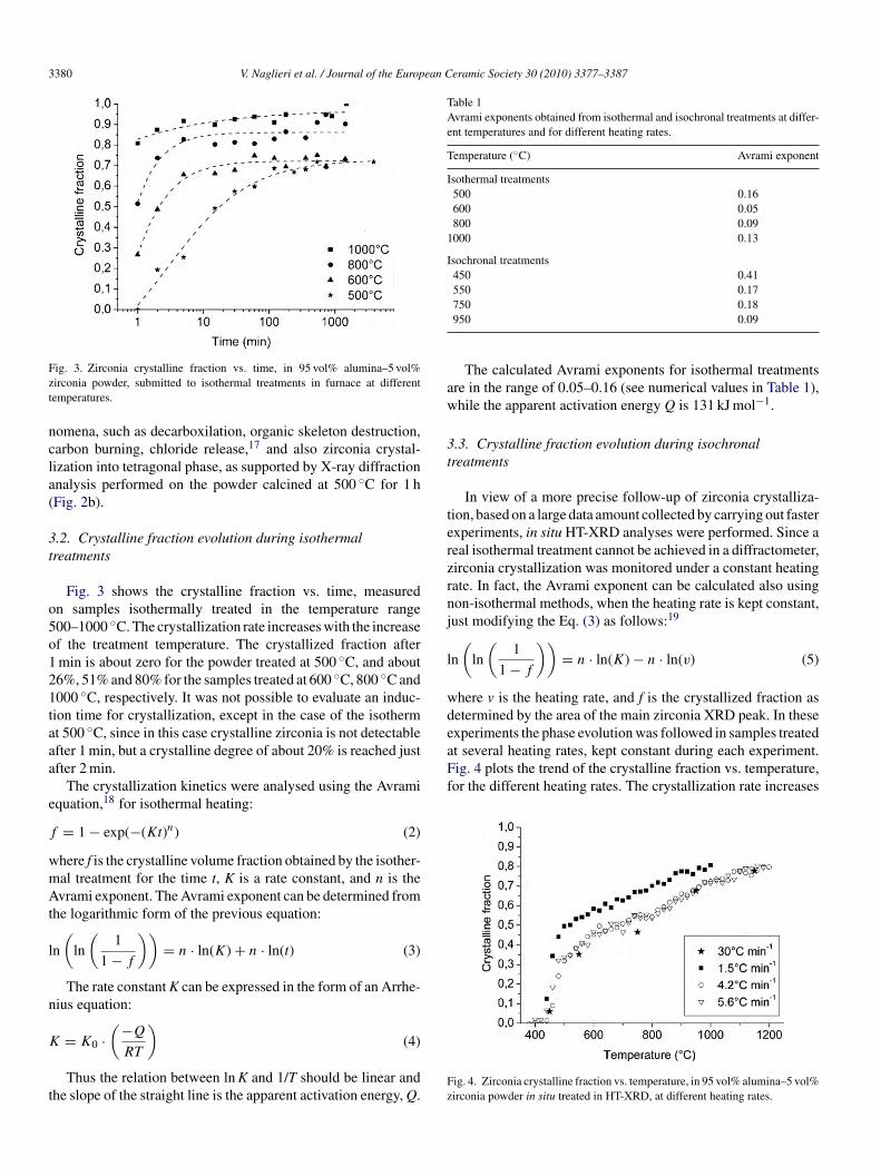
3380 V. Naglieri et al. / Journal of the European Ceramic Society 30 (2010) 3377–3387
Fzt
ncla(
3t
o5o121taaa
e
f
wmAt
l
n
K
t
Table 1Avrami exponents obtained from isothermal and isochronal treatments at differ-ent temperatures and for different heating rates.
Temperature (◦C) Avrami exponent
Isothermal treatments500 0.16600 0.05800 0.09
1000 0.13
Isochronal treatments450 0.41550 0.17
aw
3t
terzrnj
l
wdexperiments the phase evolution was followed in samples treatedat several heating rates, kept constant during each experiment.Fig. 4 plots the trend of the crystalline fraction vs. temperature,for the different heating rates. The crystallization rate increases
ig. 3. Zirconia crystalline fraction vs. time, in 95 vol% alumina–5 vol%irconia powder, submitted to isothermal treatments in furnace at differentemperatures.
omena, such as decarboxilation, organic skeleton destruction,arbon burning, chloride release,17 and also zirconia crystal-ization into tetragonal phase, as supported by X-ray diffractionnalysis performed on the powder calcined at 500 ◦C for 1 hFig. 2b).
.2. Crystalline fraction evolution during isothermalreatments
Fig. 3 shows the crystalline fraction vs. time, measuredn samples isothermally treated in the temperature range00–1000 ◦C. The crystallization rate increases with the increasef the treatment temperature. The crystallized fraction aftermin is about zero for the powder treated at 500 ◦C, and about6%, 51% and 80% for the samples treated at 600 ◦C, 800 ◦C and000 ◦C, respectively. It was not possible to evaluate an induc-ion time for crystallization, except in the case of the isothermt 500 ◦C, since in this case crystalline zirconia is not detectablefter 1 min, but a crystalline degree of about 20% is reached justfter 2 min.
The crystallization kinetics were analysed using the Avramiquation,18 for isothermal heating:
= 1 − exp(−(Kt)n) (2)
here f is the crystalline volume fraction obtained by the isother-al treatment for the time t, K is a rate constant, and n is thevrami exponent. The Avrami exponent can be determined from
he logarithmic form of the previous equation:
n
(ln
(1
1 − f
))= n · ln(K) + n · ln(t) (3)
The rate constant K can be expressed in the form of an Arrhe-ius equation:( )
= K0 · −QRT(4)
Thus the relation between ln K and 1/T should be linear andhe slope of the straight line is the apparent activation energy, Q.
Fz
750 0.18950 0.09
The calculated Avrami exponents for isothermal treatmentsre in the range of 0.05–0.16 (see numerical values in Table 1),hile the apparent activation energy Q is 131 kJ mol−1.
.3. Crystalline fraction evolution during isochronalreatments
In view of a more precise follow-up of zirconia crystalliza-ion, based on a large data amount collected by carrying out fasterxperiments, in situ HT-XRD analyses were performed. Since aeal isothermal treatment cannot be achieved in a diffractometer,irconia crystallization was monitored under a constant heatingate. In fact, the Avrami exponent can be calculated also usingon-isothermal methods, when the heating rate is kept constant,ust modifying the Eq. (3) as follows:19
n
(ln
(1
1 − f
))= n · ln(K) − n · ln(v) (5)
here v is the heating rate, and f is the crystallized fraction asetermined by the area of the main zirconia XRD peak. In these
ig. 4. Zirconia crystalline fraction vs. temperature, in 95 vol% alumina–5 vol%irconia powder in situ treated in HT-XRD, at different heating rates.
V. Naglieri et al. / Journal of the European Ceramic Society 30 (2010) 3377–3387 3381
Fp
ana
ialZ
3
fTitcmtdit
ttta
ntfncnmoebc
Fa
3s
otaSpAao(5wmgpit
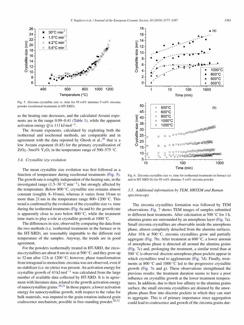
ig. 5. Zirconia crystallite size vs. time for 95 vol% alumina–5 vol% zirconiaowder (isochronal treatments in HT-XRD).
s the heating rate decreases, and the calculated Avrami expo-ents are in the range 0.09–0.41 (Table 1), while the apparentctivation energy Q is 111 kJ mol−1.
The Avrami exponents, calculated by exploiting both thesothermal and isochronal methods, are comparable and ingreement with the data reported by Ghosh et al.,20 that is aow Avrami exponent (0.45) for the primary crystallization ofrO2–3mol% Y2O3 in the temperature range of 500–575 ◦C.
.4. Crystallite size evolution
The mean crystallite size evolution was first followed as aunction of temperature during isochronal treatments (Fig. 5).he growth rate is roughly independent of the heating rate, in the
nvestigated range (1.5–30 ◦C min−1), but strongly affected byhe temperature. Below 800 ◦C, crystallite size remains almostonstant (roughly 8–10 nm), whereas it varies from 10 nm toore than 21 nm in the temperature range 800–1200 ◦C. This
rend is confirmed by the evolution of the crystallite size vs. timeuring the isothermal treatments (Fig. 6a and b): the growth rates apparently close to zero below 800 ◦C, while the treatmentime starts to play a role in crystallite growth at 1000 ◦C.
The differences in size, observed by comparing the data fromhe two methods (i.e. isothermal treatments in the furnace or inhe HT-XRD), are reasonably imputable to the different realemperature of the samples. Anyway, the trends are in goodgreement.
For the powders isothermally treated in HT-XRD, the zirco-ia crystallites are about 8 nm in size at 500 ◦C, and they grow upo 32 nm after 12 h at 1200 ◦C; however, phase transformationrom tetragonal to monoclinic zirconia was not observed, even ifo stabilizer (i.e. no yttria) was present. An activation energy forrystallite growth of 43 kJ mol−1 was calculated from the largeumber of available data collected by HT-XRD. It is in agree-ent with literature data, related to the growth activation energy
f nanocrystalline grains.20,21 In those papers, a lower activationnergy for nanocrystalline growth, with respect to the value forulk materials, was imputed to the grain-rotation-induced grainoalescence mechanism, possible in free-standing powder.20,21
sptc
ig. 6. Zirconia crystallite size vs. time for isothermal treatments in furnace (a)nd in HT-XRD (b) for 95 vol% alumina–5 vol% zirconia powder.
.5. Additional information by TEM, HRTEM and Ramanpectroscopy
The zirconia crystallites formation was followed by TEMbservations. Fig. 7 shows TEM images of samples submittedo different heat treatments. After calcination at 500 ◦C for 1 h,lumina grains are surrounded by an amorphous layer (Fig. 7a).mall zirconia crystallites are observable inside the amorphoushase, almost completely detached from the alumina surfaces.fter 10 h at 500 ◦C, zirconia crystallites grow and partially
ggregate (Fig. 7b). After treatment at 600 ◦C, a lower amountf amorphous phase is detected all around the alumina grainsFig. 7c) and, prolonging the treatment, a similar trend than at00 ◦C is observed: discrete amorphous phase pockets appear inhich crystallites tend to agglomerate (Fig. 7d). Finally, treat-ents at 800 ◦C and 1000 ◦C led to the progressive crystallite
rowth (Fig. 7e and g). These observations strengthened therevious results: the treatment duration seems to have a poornfluence on crystallite growth at the lower treatment tempera-ures. In addition, due to their low affinity to the alumina grains
urface, the small zirconia crystallites are drained by the amor-hous phase flow into discrete pockets in which they can starto aggregate. This is of primary importance since aggregationould lead to coalescence and growth of the zirconia grains dur-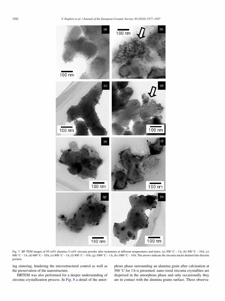
3382 V. Naglieri et al. / Journal of the European Ceramic Society 30 (2010) 3377–3387
F atmen6 1 h, (p
it
z
ig. 7. BF TEM images of 95 vol% alumina–5 vol% zirconia powder after tre00 ◦C – 1 h, (d) 600 ◦C – 10 h, (e) 800 ◦C – 1 h, (f) 800 ◦C – 10 h, (g) 1000 ◦C –ockets.
ng sintering, hindering the microstructural control as well ashe preservation of the nanostructure.
HRTEM was also performed for a deeper understanding ofirconia crystallization process. In Fig. 8 a detail of the amor-
p5da
ts at different temperatures and times: (a) 500 ◦C – 1 h, (b) 500 ◦C – 10 h, (c)h) 1000 ◦C – 10 h. The arrows indicate the zirconia nuclei drained into discrete
hous phase surrounding an alumina grain after calcination at00 ◦C for 1 h is presented: nano-sized zirconia crystallites areispersed in the amorphous phase and only occasionally theyre in contact with the alumina grains surface. These observa-

V. Naglieri et al. / Journal of the European C
Fig. 8. HRTEM images of the 95 vol% alumina–5 vol% zirconia powder cal-cined at (a) 500 ◦C for 1 h (black circles point out zirconia crystallites), (b)600 ◦C for 1 h, (c) 1000 ◦C for 1 h.
tcdtactIwso
cthmT21
scttlaars5te5scIsilh
4p
nb
tuneGpfto
eramic Society 30 (2010) 3377–3387 3383
ions clearly supported the homogeneous nucleation of zirconiarystallites into the amorphous phase, yielded by the precursorecomposition. Fig. 8b and c gives an overview of the evolu-ion of the contact angle between zirconia nano-crystallites andlumina grains as a function of temperature, precisely after cal-ination at 600 ◦C (Fig. 8b) and 1000 ◦C (Fig. 8c) for 1 h, and ofhe related, progressive disappearance of the amorphous layer.t can be qualitatively observed that the contact angle decreasesith the increase of the calcination temperature, and finally the
urface adhesion of zirconia crystals to the alumina grains isbserved at the higher treatment temperature.
To improve the information on the evolution of zirconiarystallites, the size distribution of zirconia crystallites afterhe different heat treatments (temperature range: 500–1000 ◦C,eating rate of 10 ◦C min−1, soaking time of 1 h) were deter-ined from DF and ADF TEM images (see examples in Fig. 9).he mean zirconia crystallite sizes are 9 nm, 11 nm, 17 nm and5 nm for the samples treated at 500 ◦C, 600 ◦C, 800 ◦C and000 ◦C respectively, in agreement with the above reported data.
Phase identification was carried out by Raman spectroscopy,ince this technique is preferable to investigate the primaryrystallization and possible phase transformation in our sys-em, being more sensitive to intermediate-range order, whilehe phase detectable by XRD must have a periodicity over aength of 5 nm.22 The analyses were performed on both purelumina samples and modified powders after in situ treatmentst 500 ◦C, 600 ◦C, 800 ◦C, 1000 ◦C and 1200 ◦C in XRD appa-atus (Fig. 10). Raman spectrum of the alumina powder presentseveral peaks at 378 cm−1, 416 cm−1, 429 cm−1, 451 cm−1,76 cm−1, 644 cm−1 and 750 cm−1, in good agreement withhe literature.23 In the spectra of the composite powder oth-rs peaks at 144 cm−1, 268 cm−1, 314 cm−1, 458 cm−1 and99 cm−1 appear, which are imputable to zirconia tetragonaltructure.24 No characteristic peaks of the monoclinic25 or theubic structure26 are observed even after treatment at 1200 ◦C.n both XRD pattern and Raman spectrum of the compositeample calcined at 1200 ◦C for 12 h only tetragonal zirconias detectable, confirming that, thanks to its very small crystal-ite size, tetragonal structure is preserved even after prolongedigh-temperature treatments.
. Discussion: a possible scenario of crystallizationrocess
From the above results, several considerations on the mecha-ism of zirconia crystallization onto alumina grains surface cane pointed out.
As often mentioned in the literature, the first phase to crys-allise from an amorphous zirconium oxide precursor22,27–30 issually tetragonal zirconia. Here we show that tetragonal zirco-ia is present at room temperature after the thermal treatments,ven if monoclinic phase is the thermodynamically stable one.arvie28 suggested that, for very small crystals, the tetragonal
hase could be stable thanks to the smaller surface energy, and heound that when zirconium oxide samples, obtained by precipita-ion, were heated, the tetragonal crystallites grew to a maximumf 30 nm, before a complete transformation to the monoclinic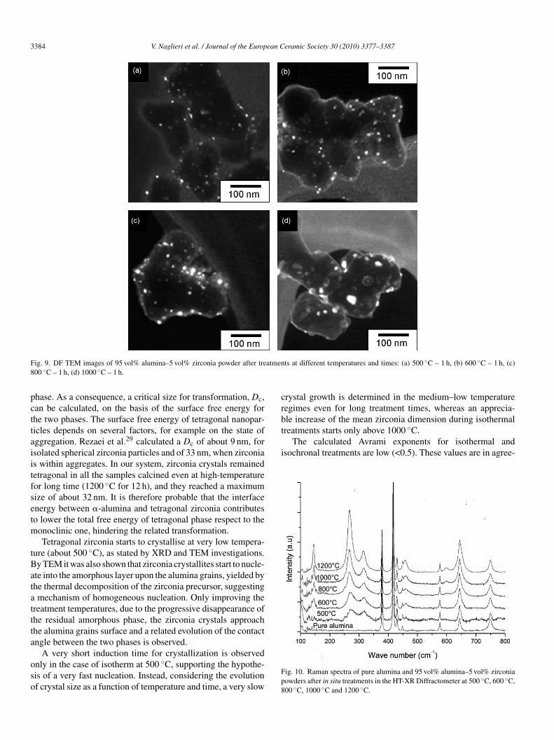
3384 V. Naglieri et al. / Journal of the European Ceramic Society 30 (2010) 3377–3387
F atmen8
pcttaiitfsetm
tBatattta
oso
crbtreatments starts only above 1000 ◦C.
The calculated Avrami exponents for isothermal andisochronal treatments are low (<0.5). These values are in agree-
ig. 9. DF TEM images of 95 vol% alumina–5 vol% zirconia powder after tre00 ◦C – 1 h, (d) 1000 ◦C – 1 h.
hase. As a consequence, a critical size for transformation, Dc,an be calculated, on the basis of the surface free energy forhe two phases. The surface free energy of tetragonal nanopar-icles depends on several factors, for example on the state ofggregation. Rezaei et al.29 calculated a Dc of about 9 nm, forsolated spherical zirconia particles and of 33 nm, when zirconias within aggregates. In our system, zirconia crystals remainedetragonal in all the samples calcined even at high-temperatureor long time (1200 ◦C for 12 h), and they reached a maximumize of about 32 nm. It is therefore probable that the interfacenergy between �-alumina and tetragonal zirconia contributeso lower the total free energy of tetragonal phase respect to the
onoclinic one, hindering the related transformation.Tetragonal zirconia starts to crystallise at very low tempera-
ure (about 500 ◦C), as stated by XRD and TEM investigations.y TEM it was also shown that zirconia crystallites start to nucle-te into the amorphous layer upon the alumina grains, yielded byhe thermal decomposition of the zirconia precursor, suggestingmechanism of homogeneous nucleation. Only improving the
reatment temperatures, due to the progressive disappearance ofhe residual amorphous phase, the zirconia crystals approachhe alumina grains surface and a related evolution of the contactngle between the two phases is observed.
A very short induction time for crystallization is observednly in the case of isotherm at 500 ◦C, supporting the hypothe-is of a very fast nucleation. Instead, considering the evolutionf crystal size as a function of temperature and time, a very slow
Fp8
ts at different temperatures and times: (a) 500 ◦C – 1 h, (b) 600 ◦C – 1 h, (c)
rystal growth is determined in the medium–low temperatureegimes even for long treatment times, whereas an apprecia-le increase of the mean zirconia dimension during isothermal
ig. 10. Raman spectra of pure alumina and 95 vol% alumina–5 vol% zirconiaowders after in situ treatments in the HT-XR Diffractometer at 500 ◦C, 600 ◦C,00 ◦C, 1000 ◦C and 1200 ◦C.

V. Naglieri et al. / Journal of the European Ceramic Society 30 (2010) 3377–3387 3385
ia cry
mbdthitzancontat
egit
tsp
pmlitiovouaim
Fig. 11. Possible zircon
ent with the work of Ghosh et al.,20 who explained their resultsy the dominance of interfacial control mechanism over the bulkiffusion in the growth behavior of zirconia. The crystalliza-ion from amorphous zirconia as a diffusionless transformationas been already reported in previous studies,27,28 based on thenvestigation of the structure of amorphous hydrous ZrO2. Inhe above studies, they described the structure of the amorphousirconia as made of alternated layers of zirconium and oxygentoms, with interatomic distances similar to that of tetrago-al zirconia. Moreover, by electron diffraction patterns, theylaimed that amorphous zirconia seemed to be a bulk samplef crystals nuclei with a structure very close to that of tetrago-al phase, but with very short periodicity.27 The hypothesis ofhese authors27,28 can therefore account for a very fast nucle-tion, as also proposed in the present study, in agreement withhe homogeneous nucleation here observed by TEM.
Therefore, the low value of the Avrami exponent could be
xplained, by analysing the contribution of both nucleation androwth. The nucleation rate cannot be supposed constant: in fact,t is very high at the beginning of the transformation, at tempera-ures lower than 600 ◦C, and it becomes moderate by increasingCttt
stallization mechanism.
he treatment temperature. On the contrary, the growth rate isupposed to be low at the temperatures below 1000 ◦C, due to aoor diffusion rate.
A possible scenario for the zirconia crystallization can beroposed starting from the collected results. After drying theodified powders are made of alumina grains coated with a
ayer of amorphous zirconia precursor (Fig. 11a). Crystallizations then promoted by thermal treatments; by calcination at lowemperature, a homogeneous nucleation of zirconia nanocrystalsn the amorphous layer is already observed in the initial stagef the thermal treatment (Fig. 11b), while their growth rate isery slow, so that, at the intermediate stage, a slightly decreasef amorphous phase is observed, mainly imputable to contin-ous nucleation (Fig. 11c). However, prolonging the treatmentt low temperature, the amorphous phase, even if still present,s preferentially drained into discrete pockets among the alu-
ina particles, to reduce the related contact surface (Fig. 11d).
onsequently, we hypothesize that the calcination time at lowemperature could have a dramatic effect on the final microstruc-ure after sintering. In a first case, the powder calcined for shortime contains uniformly distributed zirconia nuclei (Fig. 11c),
3386 V. Naglieri et al. / Journal of the European Ceramic Society 30 (2010) 3377–3387
Fc
srOtepnssgtoc
ttccos(ftif
smewbt
Fd
moFtitm
5
icpgpbpa
•
•
dtpfipiare needed to achieve a fully crystallization. At the same time,
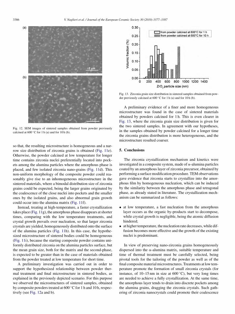
ig. 12. SEM images of sintered samples obtained from powder previouslyalcined at 600 ◦C for 1 h (a) and for 10 h (b).
o that, the resulting microstructure is homogeneous and a nar-ow size distribution of zirconia grains is obtained (Fig. 11e).therwise, the powder calcined at low temperature for longer
ime contains zirconia nuclei preferentially located into pock-ts among the alumina particles where the amorphous phase islaced, and few isolated zirconia nano-grains (Fig. 11d). Thison-uniform morphology of the composite powder could rea-onably give rise to an inhomogeneous microstructure in theintered materials, where a bimodal distribution size of zirconiarains could be expected, being the larger grains originated byhe coalescence of the close nuclei into pockets and the smallernes by the isolated grains, and also abnormal grain growthould occur into the alumina matrix (Fig. 11f).
Instead, treating at high-temperature, a faster crystallizationakes place (Fig. 11g), the amorphous phase disappears at shorterimes, comparing with the low temperature treatments, andrystal growth prevails over nucleation, so that larger zirconiarystals are yielded, homogeneously distributed onto the surfacef the alumina particles (Fig. 11h). In this case, the hypothe-ized microstructure of sintered bodies could be homogeneousFig. 11i), because the starting composite powder contains uni-ormly distributed zirconia on the alumina particles surface, buthe mean grain size, both for the matrix and the second-phase,s expected to be greater than in the case of materials obtainedrom the powder treated at low temperature for short time.
A preliminary investigation was carried out in order toupport the hypothesized relationship between powder ther-al treatment and final microstructure in sintered bodies, as
xplained in the previously depicted scenario. For this purpose
e observed the microstructures of sintered samples, obtainedy composite powders treated at 600 ◦C for 1 h and 10 h, respec-ively (see Fig. 12a and b).tte
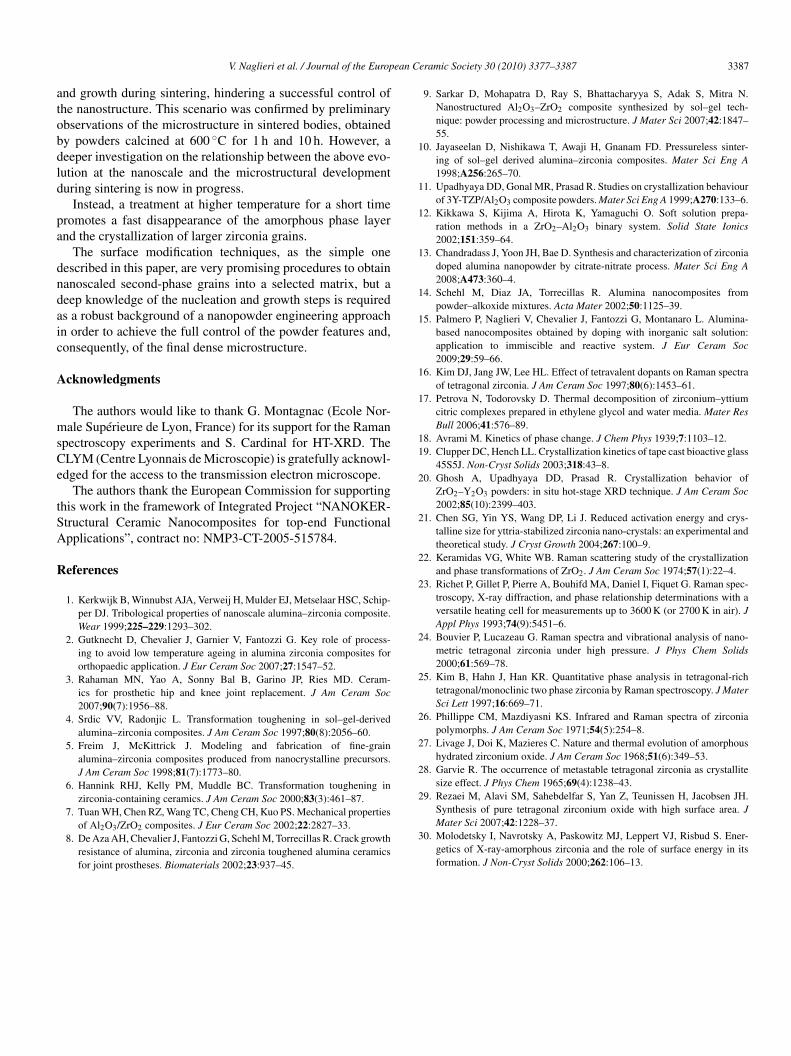
ig. 13. Zirconia grain size distribution in sintered samples obtained from pow-er previously calcined at 600 ◦C for 1 h (a) and for 10 h (b).
A preliminary evidence of a finer and more homogeneousicrostructure was found in the case of sintered materials
btained by powders calcined for 1 h. This is even clearer inig. 13, where the zirconia grain size distribution is given for
he two sintered samples. In agreement with our hypotheses,n the samples obtained by powder calcined for a longer timehe zirconia grains distribution is more heterogeneous, and the
icrostructure resulted coarser.
. Conclusions
The zirconia crystallization mechanism and kinetics werenvestigated in a composite system, made of �-alumina particlesoated by an amorphous layer of zirconia precursor, obtained byerforming a surface modification procedure. TEM observationsave evidence that zirconia starts to crystallize into the amor-hous film by homogeneous nucleation, which can be inducedy the similarity between the amorphous phase and tetragonalhase, as already stated in literature. The crystallization mech-nism can be summarized as follows:
at low temperature, a fast nucleation from the amorphouslayer occurs as the organic by-products start to decompose,while crystal growth is negligible, being the atomic diffusionhindered;at higher temperature, the nucleation rate decreases, while dif-fusion becomes more effective and the growth of the existingnuclei is predominant.
In view of preserving nano-zirconia grains homogeneouslyispersed into the �-alumina matrix, suitable temperature andime of thermal treatment must be carefully selected, beingivotal tools for the tailoring of the powder as well as of thenal composite material microstructures. Treatments at low tem-erature promote the formation of small zirconia crystals (fornstance, of 10–15 nm in size at 600 ◦C), but very long times
he amorphous layer tends to drain into discrete pockets amonghe alumina grains, dragging the zirconia crystals. Such gath-ring of zirconia nanocrystals could promote their coalescence
ean C
atobdld
pa
dndaic
A
msCe
tSA
R
V. Naglieri et al. / Journal of the Europ
nd growth during sintering, hindering a successful control ofhe nanostructure. This scenario was confirmed by preliminarybservations of the microstructure in sintered bodies, obtainedy powders calcined at 600 ◦C for 1 h and 10 h. However, aeeper investigation on the relationship between the above evo-ution at the nanoscale and the microstructural developmenturing sintering is now in progress.
Instead, a treatment at higher temperature for a short timeromotes a fast disappearance of the amorphous phase layernd the crystallization of larger zirconia grains.
The surface modification techniques, as the simple oneescribed in this paper, are very promising procedures to obtainanoscaled second-phase grains into a selected matrix, but aeep knowledge of the nucleation and growth steps is requireds a robust background of a nanopowder engineering approachn order to achieve the full control of the powder features and,onsequently, of the final dense microstructure.
cknowledgments
The authors would like to thank G. Montagnac (Ecole Nor-ale Supérieure de Lyon, France) for its support for the Raman
pectroscopy experiments and S. Cardinal for HT-XRD. TheLYM (Centre Lyonnais de Microscopie) is gratefully acknowl-dged for the access to the transmission electron microscope.
The authors thank the European Commission for supportinghis work in the framework of Integrated Project “NANOKER-tructural Ceramic Nanocomposites for top-end Functionalpplications”, contract no: NMP3-CT-2005-515784.
eferences
1. Kerkwijk B, Winnubst AJA, Verweij H, Mulder EJ, Metselaar HSC, Schip-per DJ. Tribological properties of nanoscale alumina–zirconia composite.Wear 1999;225–229:1293–302.
2. Gutknecht D, Chevalier J, Garnier V, Fantozzi G. Key role of process-ing to avoid low temperature ageing in alumina zirconia composites fororthopaedic application. J Eur Ceram Soc 2007;27:1547–52.
3. Rahaman MN, Yao A, Sonny Bal B, Garino JP, Ries MD. Ceram-ics for prosthetic hip and knee joint replacement. J Am Ceram Soc2007;90(7):1956–88.
4. Srdic VV, Radonjic L. Transformation toughening in sol–gel-derivedalumina–zirconia composites. J Am Ceram Soc 1997;80(8):2056–60.
5. Freim J, McKittrick J. Modeling and fabrication of fine-grainalumina–zirconia composites produced from nanocrystalline precursors.J Am Ceram Soc 1998;81(7):1773–80.
6. Hannink RHJ, Kelly PM, Muddle BC. Transformation toughening inzirconia-containing ceramics. J Am Ceram Soc 2000;83(3):461–87.
7. Tuan WH, Chen RZ, Wang TC, Cheng CH, Kuo PS. Mechanical propertiesof Al2O3/ZrO2 composites. J Eur Ceram Soc 2002;22:2827–33.
8. De Aza AH, Chevalier J, Fantozzi G, Schehl M, Torrecillas R. Crack growthresistance of alumina, zirconia and zirconia toughened alumina ceramicsfor joint prostheses. Biomaterials 2002;23:937–45.
eramic Society 30 (2010) 3377–3387 3387
9. Sarkar D, Mohapatra D, Ray S, Bhattacharyya S, Adak S, Mitra N.Nanostructured Al2O3–ZrO2 composite synthesized by sol–gel tech-nique: powder processing and microstructure. J Mater Sci 2007;42:1847–55.
10. Jayaseelan D, Nishikawa T, Awaji H, Gnanam FD. Pressureless sinter-ing of sol–gel derived alumina–zirconia composites. Mater Sci Eng A1998;A256:265–70.
11. Upadhyaya DD, Gonal MR, Prasad R. Studies on crystallization behaviourof 3Y-TZP/Al2O3 composite powders. Mater Sci Eng A 1999;A270:133–6.
12. Kikkawa S, Kijima A, Hirota K, Yamaguchi O. Soft solution prepa-ration methods in a ZrO2–Al2O3 binary system. Solid State Ionics2002;151:359–64.
13. Chandradass J, Yoon JH, Bae D. Synthesis and characterization of zirconiadoped alumina nanopowder by citrate-nitrate process. Mater Sci Eng A2008;A473:360–4.
14. Schehl M, Diaz JA, Torrecillas R. Alumina nanocomposites frompowder–alkoxide mixtures. Acta Mater 2002;50:1125–39.
15. Palmero P, Naglieri V, Chevalier J, Fantozzi G, Montanaro L. Alumina-based nanocomposites obtained by doping with inorganic salt solution:application to immiscible and reactive system. J Eur Ceram Soc2009;29:59–66.
16. Kim DJ, Jang JW, Lee HL. Effect of tetravalent dopants on Raman spectraof tetragonal zirconia. J Am Ceram Soc 1997;80(6):1453–61.
17. Petrova N, Todorovsky D. Thermal decomposition of zirconium–yttiumcitric complexes prepared in ethylene glycol and water media. Mater ResBull 2006;41:576–89.
18. Avrami M. Kinetics of phase change. J Chem Phys 1939;7:1103–12.19. Clupper DC, Hench LL. Crystallization kinetics of tape cast bioactive glass
45S5J. Non-Cryst Solids 2003;318:43–8.20. Ghosh A, Upadhyaya DD, Prasad R. Crystallization behavior of
ZrO2–Y2O3 powders: in situ hot-stage XRD technique. J Am Ceram Soc2002;85(10):2399–403.
21. Chen SG, Yin YS, Wang DP, Li J. Reduced activation energy and crys-talline size for yttria-stabilized zirconia nano-crystals: an experimental andtheoretical study. J Cryst Growth 2004;267:100–9.
22. Keramidas VG, White WB. Raman scattering study of the crystallizationand phase transformations of ZrO2. J Am Ceram Soc 1974;57(1):22–4.
23. Richet P, Gillet P, Pierre A, Bouhifd MA, Daniel I, Fiquet G. Raman spec-troscopy, X-ray diffraction, and phase relationship determinations with aversatile heating cell for measurements up to 3600 K (or 2700 K in air). JAppl Phys 1993;74(9):5451–6.
24. Bouvier P, Lucazeau G. Raman spectra and vibrational analysis of nano-metric tetragonal zirconia under high pressure. J Phys Chem Solids2000;61:569–78.
25. Kim B, Hahn J, Han KR. Quantitative phase analysis in tetragonal-richtetragonal/monoclinic two phase zirconia by Raman spectroscopy. J MaterSci Lett 1997;16:669–71.
26. Phillippe CM, Mazdiyasni KS. Infrared and Raman spectra of zirconiapolymorphs. J Am Ceram Soc 1971;54(5):254–8.
27. Livage J, Doi K, Mazieres C. Nature and thermal evolution of amorphoushydrated zirconium oxide. J Am Ceram Soc 1968;51(6):349–53.
28. Garvie R. The occurrence of metastable tetragonal zirconia as crystallitesize effect. J Phys Chem 1965;69(4):1238–43.
29. Rezaei M, Alavi SM, Sahebdelfar S, Yan Z, Teunissen H, Jacobsen JH.
Synthesis of pure tetragonal zirconium oxide with high surface area. JMater Sci 2007;42:1228–37.30. Molodetsky I, Navrotsky A, Paskowitz MJ, Leppert VJ, Risbud S. Ener-getics of X-ray-amorphous zirconia and the role of surface energy in itsformation. J Non-Cryst Solids 2000;262:106–13.