Focal cortical dysplasias in autism spectrum disorders
11
RESEARCH Open Access Focal cortical dysplasias in autism spectrum disorders Manuel F Casanova 1* , Ayman S El-Baz 2 , Shweta S Kamat 3 , Brynn A Dombroski 3 , Fahmi Khalifa 2 , Ahmed Elnakib 2 , Ahmed Soliman 2 , Anita Allison-McNutt 4 and Andrew E Switala 1 Abstract Background: Previous reports indicate the presence of histological abnormalities in the brains of individuals with autism spectrum disorders (ASD) suggestive of a dysplastic process. In this study we identified areas of abnormal cortical thinning within the cerebral cortex of ASD individuals and examined the same for neuronal morphometric abnormalities by using computerized image analysis. Results: The study analyzed celloidin-embedded and Nissl-stained serial full coronal brain sections of 7 autistic (ADI-R diagnosed) and 7 age/sex-matched neurotypicals. Sections were scanned and manually segmented before implementing an algorithm using Laplace’s equation to measure cortical width. Identified areas were then subjected to analysis for neuronal morphometry. Results of our study indicate the presence within our ASD population of circumscribed foci of diminished cortical width that varied among affected individuals both in terms of location and overall size with the frontal lobes being particularly involved. Spatial statistic indicated a reduction in size of neurons within affected areas. Granulometry confirmed the presence of smaller pyramidal cells and suggested a concomitant reduction in the total number of interneurons. Conclusions: The neuropathology is consistent with a diagnosis of focal cortical dysplasia (FCD). Results from the medical literature (e.g., heterotopias) and our own study suggest that the genesis of this cortical malformation seemingly resides in the heterochronic divisions of periventricular germinal cells. The end result is that during corticogenesis radially migrating neuroblasts (future pyramidal cells) are desynchronized in their development from those that follow a tangential route (interneurons). The possible presence of a pathological mechanism in common among different conditions expressing an autism-like phenotype argue in favor of considering ASD a “sequence” rather than a syndrome. Focal cortical dysplasias in ASD may serve to explain the high prevalence of seizures and sensory abnormalities in this patient population. Keywords: Autistic spectrum disorder, Frontal lobe, Malformations of cortical development, Cortical width Background Neurologists habitually regard autism as a disease affecting the cerebral cortex [1]. The presence of seizures, abnormalities of higher cognitive functions, and absence of either spasticity or vision loss, supports this contention. It is therefore unsurprising that neuropathological findings of a dysplastic nature have been described in the cerebral cortex of autistic individuals. The brunt of dysplastic changes appears to be within the neocortex wherein abnormalities of gyrification and laminar architecture are often found. These findings include effacement of the normal lamination pattern, minicolumnar abnormalities, and variations in neuronal density [1-4]. In addition to the previously mentioned microscopic changes, at gross examination, brains of autistic individuals have shown malformations in the form of hyperconvolution [5], misdirected gyri [6], and polymicrogyria [7]. Among the earliest reports of cortical abnormalities in autism was the claim made by Bauman and Kemper of indistinct lamination of the anterior cingulate gyrus in 5 out of their 6 cases [8]. However, it was Bailey and colleagues who first emphasized the significance of abnormalities of the cerebral cortex in autism [5]. In their series, 4 out of 6 patients had both abnormal * Correspondence: [email protected] 1 Department of Psychiatry & Behavioral Sciences, University of Louisville, Louisville, KY 40202, USA Full list of author information is available at the end of the article © 2013 Casanova et al.; licensee BioMed Central Ltd. This is an open access article distributed under the terms of the Creative Commons Attribution License (http://creativecommons.org/licenses/by/2.0), which permits unrestricted use, distribution, and reproduction in any medium, provided the original work is properly cited. Casanova et al. Acta Neuropathologica Communications 2013, 1:67 http://www.actaneurocomms.org/content/1/1/67
-
Upload
greenvillemed-sc -
Category
Documents
-
view
1 -
download
0
Transcript of Focal cortical dysplasias in autism spectrum disorders

Casanova et al. Acta Neuropathologica Communications 2013, 1:67http://www.actaneurocomms.org/content/1/1/67
RESEARCH Open Access
Focal cortical dysplasias in autism spectrumdisordersManuel F Casanova1*, Ayman S El-Baz2, Shweta S Kamat3, Brynn A Dombroski3, Fahmi Khalifa2, Ahmed Elnakib2,Ahmed Soliman2, Anita Allison-McNutt4 and Andrew E Switala1
Abstract
Background: Previous reports indicate the presence of histological abnormalities in the brains of individuals withautism spectrum disorders (ASD) suggestive of a dysplastic process. In this study we identified areas of abnormalcortical thinning within the cerebral cortex of ASD individuals and examined the same for neuronal morphometricabnormalities by using computerized image analysis.
Results: The study analyzed celloidin-embedded and Nissl-stained serial full coronal brain sections of 7 autistic(ADI-R diagnosed) and 7 age/sex-matched neurotypicals. Sections were scanned and manually segmented beforeimplementing an algorithm using Laplace’s equation to measure cortical width. Identified areas were thensubjected to analysis for neuronal morphometry. Results of our study indicate the presence within our ASDpopulation of circumscribed foci of diminished cortical width that varied among affected individuals both in termsof location and overall size with the frontal lobes being particularly involved. Spatial statistic indicated a reductionin size of neurons within affected areas. Granulometry confirmed the presence of smaller pyramidal cells andsuggested a concomitant reduction in the total number of interneurons.
Conclusions: The neuropathology is consistent with a diagnosis of focal cortical dysplasia (FCD). Results from themedical literature (e.g., heterotopias) and our own study suggest that the genesis of this cortical malformationseemingly resides in the heterochronic divisions of periventricular germinal cells. The end result is that duringcorticogenesis radially migrating neuroblasts (future pyramidal cells) are desynchronized in their development fromthose that follow a tangential route (interneurons). The possible presence of a pathological mechanism in commonamong different conditions expressing an autism-like phenotype argue in favor of considering ASD a “sequence”rather than a syndrome. Focal cortical dysplasias in ASD may serve to explain the high prevalence of seizures andsensory abnormalities in this patient population.
Keywords: Autistic spectrum disorder, Frontal lobe, Malformations of cortical development, Cortical width
BackgroundNeurologists habitually regard autism as a diseaseaffecting the cerebral cortex [1]. The presence of seizures,abnormalities of higher cognitive functions, and absenceof either spasticity or vision loss, supports this contention.It is therefore unsurprising that neuropathological findingsof a dysplastic nature have been described in the cerebralcortex of autistic individuals. The brunt of dysplasticchanges appears to be within the neocortex whereinabnormalities of gyrification and laminar architecture
* Correspondence: [email protected] of Psychiatry & Behavioral Sciences, University of Louisville,Louisville, KY 40202, USAFull list of author information is available at the end of the article
© 2013 Casanova et al.; licensee BioMed CentCommons Attribution License (http://creativecreproduction in any medium, provided the or
are often found. These findings include effacement of thenormal lamination pattern, minicolumnar abnormalities,and variations in neuronal density [1-4]. In addition tothe previously mentioned microscopic changes, atgross examination, brains of autistic individuals haveshown malformations in the form of hyperconvolution[5], misdirected gyri [6], and polymicrogyria [7].Among the earliest reports of cortical abnormalities in
autism was the claim made by Bauman and Kemper ofindistinct lamination of the anterior cingulate gyrus in 5out of their 6 cases [8]. However, it was Bailey andcolleagues who first emphasized the significance ofabnormalities of the cerebral cortex in autism [5]. Intheir series, 4 out of 6 patients had both abnormal
ral Ltd. This is an open access article distributed under the terms of the Creativeommons.org/licenses/by/2.0), which permits unrestricted use, distribution, andiginal work is properly cited.

Casanova et al. Acta Neuropathologica Communications 2013, 1:67 Page 2 of 11http://www.actaneurocomms.org/content/1/1/67
neuronal densities and lamination of the frontal cortex, and1 patient showed an increased number of neurons withinthe molecular layer. Several years later, Mukaetova-Landinska and colleagues examined sections of MAP2immunocytochemistry from BA9 and BA10 for dendriticcomplexity [9]. The results of the study showed ill-definedcortical layers and a reduced level of MAP2 expression inthe neuronal soma and dendrites of autistic individuals.More recently, Hutsler and colleagues used thickness andlamination as proxy measurements for cortical organizationwhen studying histological sections of eulaminate cortex(BA7, BA9, and BA21) in 8 autistic spectrum disorderindividuals and an equal number of controls [10].Between-group differences in cortical thickness werejudged non-significant. Qualitative examination revealedcell clustering and an increased number of cells in laminaI and in the subplate region. A later study using the samepatient population evaluated the transition zone betweenthe cortical gray and white matter by overlaying a sigmoidfunction in binary images [11]. Their results indicated anindistinct boundary accounted, possibly, by the presenceof supernumerary neurons beneath the cortical plate. Theauthors speculated that the presence of supernumeraryneurons was the result of either a migrational defect orfailed apoptosis within the subplate region [12]. Studies onwhole brain serial sections by Wegiel and colleaguessuggest that defects of neurogenesis and neuronal migrationaccount for described dysplastic changes [13].In this study we examined for dysplastic abnormalities
in postmortem brains of autistic individuals by measuringthe cortical thickness of full hemispheric coronal sections.We decided on pursuing a postmortem study, rather thanneuroimaging, in order to optimize the resolution of thegray-white matter transition zone. Previous studiessuggest that using neuroimaging to measure corticalthickness may provide for inaccurate results due toblurring of the transition zone by an increased number ofneurons within the subcortical white matter [10,11,14].Embedding in celloidin was selected so as to minimizetissue shrinkage. Artifacts accrued to handling largesections for mounting on slides were avoided, in part,by manually segmenting the sections.
MethodsClinical datasetThe diagnosis for each autistic patient was establishedpostmortem by the Autism Tissue Program (ATP). Acertified rater and trainer arranged for a postmortemvisit with the family to obtain, with written consent, medicaland clinical information via a questionnaire that includedthe Autism Diagnostic Interview-Revised (ADI-R).The Harvard Brain Tissue Resource Center (HBTRC)
questionnaire was modified to include autism-specificquestions for ATP use. The information obtained included:
donor and respondent identifying information; ethnicity,handedness and known exposure to hazardous materials;diagnostic information including dates and physician;genetic tests; pre- and postnatal medical history;immunization, medication, and hospitalization information;family history and additional information about donorparticipation in any training or research studies suchas imaging, medication trials, and/or genetic studies.The supporting documents such as autopsy reports,death certificates, medical, clinical, and/or educationalrecords were obtained at the time of the home visitor by sending written requests, signed by the legalnext-of-kin, to named providers.Whole, formalin fixed, cerebral hemispheres from the
ATP had already been processed at the Institute forBasic Research in Developmental Disabilities, StatenIsland, New York. Seven were obtained from individualsdiagnosed with autism spectrum disorder, and sevendonations from neurotypical individuals were selected tomatch the ASD cases by age and sex where possible(Table 1). Tissue preparation was according to themethod of Heinsen and Heinsen [15]. Serial, coronalsections 200 μm thick, spaced 1.2 mm apart andspanning the entire hemisphere, were stained withgallocyanine and mounted on glass slides. These slideswere later shipped to the authors at the University ofLouisville for the present work.
Image processingImaging was done at 800 dpi (1 pixel = 317.5 μm) usinga ScanMaker i900 flatbed scanner for transparent media(Microtek, Santa Fe Springs, California). The outer andinner cortical boundaries were manually traced. Fiveslides, randomly selected, were traced by three differentindividuals in order to determine inter-rater reliability.Cortical thickness was estimated using an algorithm
based on electrostatics [16]. The inner and outersurfaces were treated as equipotential surfaces at +1 Vand −1 V, respectively. The Laplace equation was solvedto find the potential φ within the grey matter betweenthese two boundaries. The thickness t of the cortex at apoint is then given by the length of the field line throughthat point, where field lines are defined as curvesextending from the inner surface to the outer, everywhereparallel to ∇φ. Thus is t unambiguously definedeverywhere inside the grey matter, since field lines donot cross one another. Once computed, t was sampled onthe medial surface, i.e. the equipotential surface at φ = 0.The local curvature κ of the brain surface was calculatedon the same equipotential (Figure 1).Identification of possible dysplastic regions began with
matching individual slides between ASD-neurotypicalpairs according to stereotaxic coordinate y. Coronal sliceswere positioned in MNI152 space so that y = −106 mm at

Table 1 ATP donors with autism spectrum disorder and age-matched controls used for this experiment
ATP case # Age Sex Seizures Medication history Cause of death
Autism spectrum disorder
1. UMB-1627 5.0 F N no medication Multiple injuries/Motor vehicle accident
2. AN13961 7.5 M Y Phenobarbital Drowning
Tegretol
Albuterol
3. AN00754 13.– M Y Trileptol Epilepsy
Trazadone
4. IBR-425-02 4.2 M N Asthma medication NOS Drowning, Hypothermia
5. AN19511 8.– M N Peridex Rhabdomyosarcoma
Nystatin
G-CSF
Benadryl
Phenergan
Dexamethasone
Morphine
6. AN02338 17.2 F N Paxil Cardiac arrest/Dilated cardiomyopathy
7. AN09730 22.9 M Y Neurontin Choking/Epilepsy
Thioridazine
Zonegran
Fish oil
Flaxseed oil
CoEnzyme Q10
Neurotypical
1. UMB-1499 4.5 F N N/A Myocarditis
2. UMB-4898 7.7 M N N/A Drowning
3. BTB-3638 14.3 M N N/A Electrocution
4. AN02456 4.– F N N/A Bronchopneumonia / Post-surgical procedure
5. UMB-1708 8.1 F N N/A Multiple injuries
6. UMB-1843 15.9 F N N/A Multiple injuries / Motor vehicle accident
7. UMB-1646 23.2 M N N/A Rupture of spleen
Casanova et al. Acta Neuropathologica Communications 2013, 1:67 Page 3 of 11http://www.actaneurocomms.org/content/1/1/67
the occipital pole, y = 73 mm corresponds with the frontalpole, and the slide containing the anterior commissurehas position y = 0. The sampling distribution of t in eachslide from the neurotypical case was used to estimate“fences” beyond which statistical outliers would fall, usingthe method of Hubert and Van der Veeken [17]. Regionsof the corresponding slide in the matched ASD casewhere t fell below the lower fence were identified aspossible dysplasias.We undertook detailed preliminary study of the
purported dysplasia in twelve identified regions fromtwo ASD donor’s brains. These were contrasted withtwelve homologous regions from their respective controldonors. High-resolution images (1 pixel = 0.74 μm) wereobtained within these regions of interest, spanning the full
width of the cortex. Segmentation of pyramidal cellsfollowed a three step process. First, we employed apixel-wise relaxation based on a maximum a posteriori(MAP) estimate of a pairwise energy function of ageneralized, two-dimensional Gauss-Markov randomfield (GGMRF) probabilistic model [18,19] in order toreduce the effect of image noise. Secondly, we used amarker-based watershed approach [20] to get an initialsegmentation of the neurons from the MGRF-relaxedimage, obtained in the first step. Finally, for each initiallysegmented neuron, we refined its segmentation basedon using a local iterative thresholding approach. Theoptimum threshold is obtained in a way that maximizesthe distance between the average signal intensities of theinitially segmented neuron region and its background.
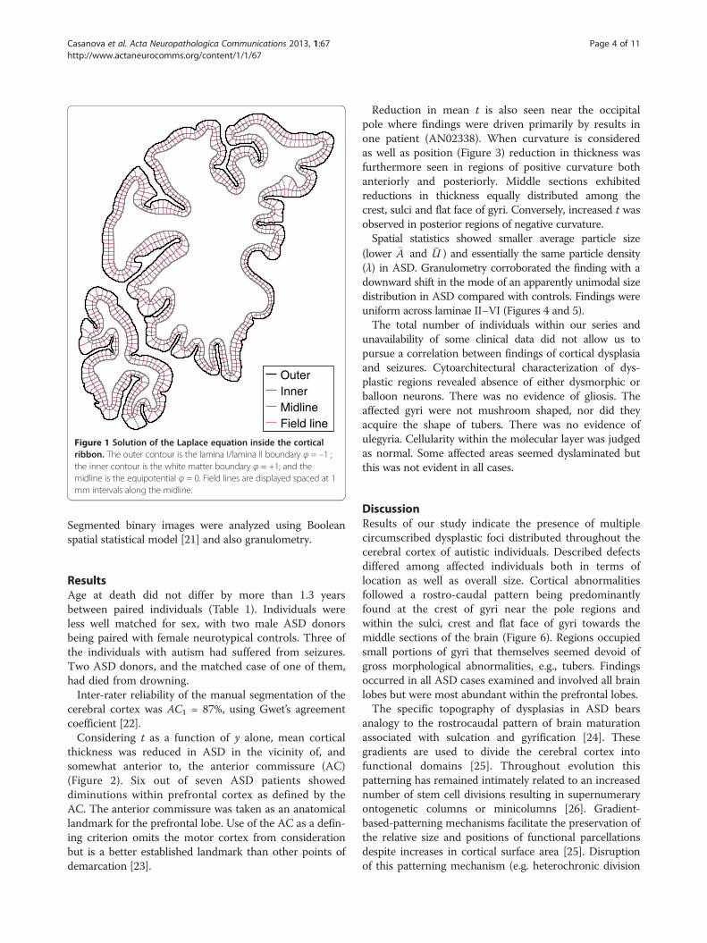
OuterInnerMidlineField line
Figure 1 Solution of the Laplace equation inside the corticalribbon. The outer contour is the lamina I/lamina II boundary φ = –1 ;the inner contour is the white matter boundary φ = +1; and themidline is the equipotential φ = 0. Field lines are displayed spaced at 1mm intervals along the midline.
Casanova et al. Acta Neuropathologica Communications 2013, 1:67 Page 4 of 11http://www.actaneurocomms.org/content/1/1/67
Segmented binary images were analyzed using Booleanspatial statistical model [21] and also granulometry.
ResultsAge at death did not differ by more than 1.3 yearsbetween paired individuals (Table 1). Individuals wereless well matched for sex, with two male ASD donorsbeing paired with female neurotypical controls. Three ofthe individuals with autism had suffered from seizures.Two ASD donors, and the matched case of one of them,had died from drowning.Inter-rater reliability of the manual segmentation of the
cerebral cortex was AC1 = 87%, using Gwet’s agreementcoefficient [22].Considering t as a function of y alone, mean cortical
thickness was reduced in ASD in the vicinity of, andsomewhat anterior to, the anterior commissure (AC)(Figure 2). Six out of seven ASD patients showeddiminutions within prefrontal cortex as defined by theAC. The anterior commissure was taken as an anatomicallandmark for the prefrontal lobe. Use of the AC as a defin-ing criterion omits the motor cortex from considerationbut is a better established landmark than other points ofdemarcation [23].
Reduction in mean t is also seen near the occipitalpole where findings were driven primarily by results inone patient (AN02338). When curvature is consideredas well as position (Figure 3) reduction in thickness wasfurthermore seen in regions of positive curvature bothanteriorly and posteriorly. Middle sections exhibitedreductions in thickness equally distributed among thecrest, sulci and flat face of gyri. Conversely, increased t wasobserved in posterior regions of negative curvature.Spatial statistics showed smaller average particle size
(lower �A and �U ) and essentially the same particle density(λ) in ASD. Granulometry corroborated the finding with adownward shift in the mode of an apparently unimodal sizedistribution in ASD compared with controls. Findings wereuniform across laminae II–VI (Figures 4 and 5).The total number of individuals within our series and
unavailability of some clinical data did not allow us topursue a correlation between findings of cortical dysplasiaand seizures. Cytoarchitectural characterization of dys-plastic regions revealed absence of either dysmorphic orballoon neurons. There was no evidence of gliosis. Theaffected gyri were not mushroom shaped, nor did theyacquire the shape of tubers. There was no evidence ofulegyria. Cellularity within the molecular layer was judgedas normal. Some affected areas seemed dyslaminated butthis was not evident in all cases.
DiscussionResults of our study indicate the presence of multiplecircumscribed dysplastic foci distributed throughout thecerebral cortex of autistic individuals. Described defectsdiffered among affected individuals both in terms oflocation as well as overall size. Cortical abnormalitiesfollowed a rostro-caudal pattern being predominantlyfound at the crest of gyri near the pole regions andwithin the sulci, crest and flat face of gyri towards themiddle sections of the brain (Figure 6). Regions occupiedsmall portions of gyri that themselves seemed devoid ofgross morphological abnormalities, e.g., tubers. Findingsoccurred in all ASD cases examined and involved all brainlobes but were most abundant within the prefrontal lobes.The specific topography of dysplasias in ASD bears
analogy to the rostrocaudal pattern of brain maturationassociated with sulcation and gyrification [24]. Thesegradients are used to divide the cerebral cortex intofunctional domains [25]. Throughout evolution thispatterning has remained intimately related to an increasednumber of stem cell divisions resulting in supernumeraryontogenetic columns or minicolumns [26]. Gradient-based-patterning mechanisms facilitate the preservation ofthe relative size and positions of functional parcellationsdespite increases in cortical surface area [25]. Disruptionof this patterning mechanism (e.g. heterochronic division

1.5
2.0
2.5
3.0
UMB−1499
UMB−1627
1.5
2.0
2.5
3.0
UMB−4898
AN13961
1.5
2.0
2.5
3.0
BTB−3638
AN00754
1.5
2.0
2.5
3.0
(mm
)
AN02456
IBR−425−02
1.5
2.0
2.5
3.0
UMB−1708
AN19511
−90−60−3003060
1.5
2.0
2.5
3.0
UMB−1843
AN02338
−90−60−3003060
1.5
2.0
2.5
3.0
Position (mm)
UMB−1646
AN09730
Figure 2 Mean t in each brain section. The seven pairs are plotted individually, with measurements from the ASD donor indicated by triangles,and circles for the matched neurotypical donor. Note that the horizontal axis scale is not uniform. Average thickness for each slide is plotted withrespect to the slide’s corresponding position in stereotaxic space. Six out of seven ASD patients showed diminutions within prefrontal cortex asdefined by the AC.
Casanova et al. Acta Neuropathologica Communications 2013, 1:67 Page 5 of 11http://www.actaneurocomms.org/content/1/1/67
of periventricular germinal cells) may help explain the pres-ence of both gyral and cerebral dominance abnormalitiespreviously reported in the brains of autistic individuals[27,28] and the focal cortical dysplasias described in thepresent study.In ASD, morphometry of neurons within affected areas
revealed smaller neurons. Findings were present through-out layers II-VI of the cortex suggesting changes ofanatomical elements in common throughout most of thecortical width. A histogram of sizes by granulometricanalysis indicated a reduction of larger cells and anoverall increase in size of the smaller ones. A possibleinterpretation to the reported findings is the diminutionin size of larger pyramidal cells and a reduction in totalnumbers of smaller neuronal elements (interneurons).This supposition is strengthened by its ability to explainseveral findings from the literature:
1) In a previous study neuronal density in this patientpopulation was found to be increased whenthresholding by size in order to select for pyramidalcells [29]. Neuronal density in the present study,although normal, took into account a range in size
inclusive of interneurons. When averaging the totalnumber of both pyramidal cells and interneurons areduction in the latter would tend to balance anincrease in the former.
2) The total reduction of smaller neurons(interneurons) would be in keeping with reductionsin the peripheral neuropil space of minicolumnsreported in autism [30] and with symptomssuggestive of cortical hyperexcitability commonlyreported in this patient population [31].
3) The size of a neuron’s cell body parallels the lengthof its axon. A long axon requires increased organellemachinery to sustain its higher metabolism. Thepresence of smaller pyramidal cells biasescorticocortical connectivity towards shorterprojections [29].
The dysplastic defects described in autism may be con-sidered as exemplars of disturbed morphogenesis them-selves stemming from an interference with corticogenesis.The dysplastic changes of the cerebral cortex coexist withheterotopias thus suggesting as a plausible pathogenicmechanism the heterochronic division of periventricular

Position (mm)
(mm
−1 )
70 50 30 10 −10 −30 −50 −70 −90
1.5
1.0
0.5
0
−0.5
−1.0
−1.5
Δ(m
m)
−0.5
−0.3
−0.1
0.1
0.3
0.5
Figure 3 Difference in t between the cerebral cortices of ASD and neurotypical donors with respect to position and curvature. Thehorizontal axis is the same as in Figure 2 (73 mm = frontal pole; 0 = anterior commissure; −106 mm = occipital pole). Regions where the cortexis narrower in ASD are colored blue, while regions where the cortex is wider are colored orange. Data were averaged across all seven pairs.
Casanova et al. Acta Neuropathologica Communications 2013, 1:67 Page 6 of 11http://www.actaneurocomms.org/content/1/1/67
germinal cells and abnormalities of the ensuing mi-gration of daughter cells to the cortex [13]. Disturbedminicolumnarity with defects within their peripheralneuropil space (i.e., the compartment holding most ofthe inhibitory cells) across the different cortical layersfurther suggests that once radially migrating neuroblastsreach the cortical plate they are desynchronized in theirmaturation from the tangentially migrating interneurons[4,30]. Evidence of periventricular germinal cell abnormal-ities is derived from neuropathological studies of idiopathicand syndromic autism. Abnormalities in the asymmetricdivision of periventricular germinal cells can account forthe epigenetic heterotopias and minicolumnar abnormal-ities observed in idiopathic cases [13,29,32,33]. It is there-fore not surprising that germinal matrix pathology issimilarly evident in syndromic cases bearing an autism-likephenotype, such as congenital cytomegalovirus infection,antenatal cocaine exposure, extreme prematurity, tuberoussclerosis, and the Ehlers-Danlos syndrome [4].
Focal cortical dysplasiasMalformations of the cortex are congenital morphologicaldefects stemming from an intrinsically abnormal develop-mental process. Propitiating agents for malformationsinclude medical illnesses, drugs, environmental agents, andgenetic abnormalities. These abnormalities range inseverity from minute defects invisible to the humaneye to gross deficits involving large territories of thecerebral hemispheres (e.g., schizencephaly). Variabilityin severity depends on the timing, intensity, durationand nature of the inciting agent. Classification schemesfor these malformations are based upon the model of
cortical development proposed, to a large extent, bySidman and Rakic [34]. This descriptive classificationsubtypes cortical malformations according to the timingof the defect during brain development. Defects are thusclassified and ordered according to whether they involveaberrant cellular proliferation, neuronal migration, orcortical organization. As many cases fall outside thegroupings of this classification, a fourth type comprised ofmiscellaneous entities is usually added [35]. In clinicalpractice, however, the different types of malformationsoften coexist in various combinations whose overlappingfeatures make it difficult to tease each other apart [36].Malformations are considered dysplastic if they evidence
disorganized development. This definition differentiatesdysplasias from other malformations due to incompletemorphogenesis, e.g., cortical malformations whereinthe morphological features are characterized by under-development due to a decrease in the number of cells(hypoplasia) and those where the tissue or organ fails todevelop (aplasia) [37]. In the case of the cerebellum, forexample, hypoplasia may be manifested as a smaller organwith fissures of normal size in comparison with thefolia. Dysplasias of the cerebellum, on the other hand,display an abnormal pattern of folia and/or presenceof heterotopic nodules [38].Traditionally, focal cortical dysplasias were believed to
be confined to one cerebral hemisphere or even to aspecific brain region like the inferior frontal lobe [12,39].More recently, the term has been expanded to includedisparate entities varying in severity and histologicalfindings [40,41]. Among the changes observed inFCD subtypes are variations in cortical thickness,

2 3 4 5 6
55
60
65
70
75
80
85
90
95
100
Autism
Lamina
Mea
n ar
ea (
μm2 )
2 3 4 5 6
55
60
65
70
75
80
85
90
95
100
Control
2 3 4 5 626
28
30
32
34
36
38
40
Autism
Lamina
Mea
n pe
rim
eter
(μm
)
2 3 4 5 626
28
30
32
34
36
38
40
Control
2 3 4 5 61500
2000
2500
3000
3500
4000Autism
Lamina
Num
ber
dens
ity (
mm
−1 )
2 3 4 5 61500
2000
2500
3000
3500
4000Control
Figure 4 Box plots of estimated Boolean model parameters by lamina. The Boolean germ-grain model is completely characterized by threequantities: mean area and perimeter of the grain distribution, and intensity of the Poisson germ process.
Casanova et al. Acta Neuropathologica Communications 2013, 1:67 Page 7 of 11http://www.actaneurocomms.org/content/1/1/67

0 5 10 150
0.5
1
1.5
2Lamina 2
0 5 10 150
0.5
1
1.5
2Lamina 3
0 5 10 150
0.5
1
1.5
2Lamina 4
0 5 10 150
0.5
1
1.5
2Lamina 5
Radius (μm)0 5 10 15
0
0.5
1
1.5
2Lamina 6
ControlAutism
Figure 5 Pattern spectrum by lamina, obtained by granulometry on segmented (binary) images. Bar height (y axis) is proportional to thetotal area of stained objects with sizes falling within each bin. The spectra were averaged bin-wise over the twelve narrow cortical regions ofinterest from two ASD cases and the twelve corresponding regions from matched control cases.
Casanova et al. Acta Neuropathologica Communications 2013, 1:67 Page 8 of 11http://www.actaneurocomms.org/content/1/1/67
compromised vertical organization with increased/persistingminicolumnar arrangements and aggregates of smallerneurons [42]. The findings are of interest as previous neuro-pathological studies in autism have similarly reportedincreased numbers of minicolumns with smaller neurons[29,32]. In essence, the present findings of smaller cells inthinned regions of the cerebral cortex and those of priorresearchers (i.e., ectopic neurons, poorly defined gray-white
Figure 6 Coronal sections through the brain of an autisticpatient. The overlay denotes those cortical areas showingabnormalities of cortical thickness. Towards both pole regionscortical thickness abnormalities preferentially affected the crest ofgyri. Defects in cortical width tended to affect both crest and sulciof gyri in middle sections. Defects affecting crest of gyri were usuallylarger than those seen at the sulci of gyri. The y-coordinates are thenormalized positions as in Figures 3 and 4.
matter boundary, increased neuronal density in layerI, and subcortical white matter, increased number ofminicolumns) indicate the presence of focal dysplasticchanges within the cerebral cortex of autistic individ-uals. These malformations describe disruptions of cel-lular proliferation, migration and cortical organizationthat start long before a person is born.
Significance of findingsFrontal lobe symptomsIn the present study a large number of dysplastic defectswere observed in the frontal lobes of autistic individuals.This is not unexpected as the prefrontal lobe alone or itsanalogues account for approximately 29% of the totalcortex of humans [43]. Besides the large area occupiedby this brain region, the frontal lobes are characterizedby their protracted development, a fact made mostapparent by the late myelination of its axonal connections[44]. Modern neuroimaging studies suggest that the vol-ume of the prefrontal white matter increases throughoutchildhood and continues beyond adolescence intoyoung adulthood [45,46]. The protracted develop-ment of the frontal lobes makes it a better target forenvironmental exigencies to disturb its organization.Once affected, the role of the frontal lobe as a con-nectivity hub explains why damage to this area mayresult in widespread brain dysfunction. Abnormalitiesmay result from both the failure to transfer incoming

Casanova et al. Acta Neuropathologica Communications 2013, 1:67 Page 9 of 11http://www.actaneurocomms.org/content/1/1/67
information and to the possibility of propagatingpotential errors.The frontal lobe abnormalities described in this study
help to explain why autism has been linked to a dysfunc-tion of the executive system in managing multiple cogni-tive processes. In effect, one cognitive account of autismis the executive dysfunction theory that assumes howthe presence of frontal lobe pathology leads to, amongother symptoms, perseveration and an inability to shiftattention [47]. The degree to which such deficits are as-sociated to intellectual disability is unclear and remainsthe object of considerable discussion within the medicalliterature [48]. However, the absence of demonstrableexecutive deficits in a subgroup of autistic patientswould argue against executive dysfunction being a coredeficit of the condition [49]. The variable involvement ofdifferent areas of the brain, including the frontal lobes,by focal cortical dysplastic processes as shown in thepresent study helps explain the heterogeneity of clinicalsymptomatology in ASD.
SeizuresMalformations of cortical development frequently resultin varying combinations of intractable seizures, mentalretardation, cerebral palsy, and focal neurological deficits[50]. Although microdysgenesis could be important inthe pathophysiology of epilepsy, the basic mechanismsinvolved remain unknown [41]. Previous studies in autismhave suggested a deficit in the inhibitory surround ofminicolumns reflective of cellular networks prone tomonotonically increasing avalanches of activity, i.e., a“rich-gets-richer” mechanism [30,51]. Furthermore, oncean error enters such a system it will be propagated andamplified through downstream connections. It is thusunsurprising that cortical dysplasias are intrinsic epilepto-genic lesions that commonly provide for intractable sei-zures [52] of multiple types [53] with remissions occurringin only a small proportion of patients [54]. Focal corticaldysplasia may thus serve as a causative factor or, insome instances, a propitiating factor for seizures. Thiscortical malformation may thus confer a susceptibilityrendering the affected brain vulnerable to seizuresafter subsequent injuries such as febrile convulsions orhead trauma [55,56].
Motor and sensory abnormalitiesInhibition is crucial to the synchronization of neural net-works wherein patterned connections give rise to organizedbehaviors. Specific cell assemblies that have interneurons(e.g., Wilson-Cowan Oscillator) are thought to participateas pattern generators within neural networks, e.g. spontan-eous rhythmical activity patterns (as in the movement oftentacles) or as biological clocks [57]. These cell assemblies
are poised within a hierarchical organization that includescentral pattern generators, initiators, and controllers. Someof the functions sustained by these pattern generatorsinclude locomotor activities (e.g., posture, walking,reaching, grasping, manipulating) and fixed action pat-terns (i.e., stereotyped behaviors elicited by fixed stimuli).An inhibitory deficit could help explain abnormalities inlocomotor skills, sleep disorders, extended periods ofrestlessness, and stereotypic movements observed inASD [58,59].Both pyramidal cells and interneurons display spon-
taneous activity so that synaptic inputs modulate theirfiring rate around a baseline rate. A lack or deficiency ofinhibition (e.g., a diminished number of interneurons)may help depolarize existing neurons below the triggerpoint of their action potential. In this scenario otherwiseweak signals add up thus providing spikes of activity.This phenomenon, called stochastic resonance, makesreference to the transmission of a weak signal in thepresence of noise [60]. Stochastic resonance may providean explanation to the high prevalence of sensoryproblems and their resultant behaviors that handicapmany autistic patients.
ConclusionsIt is the opinion of the authors that the constellation ofneuropathological abnormalities described in autism, alongwith its formes fruste, e.g., PDD-NOS (DSM-IV-TR), doesnot comprise a syndrome. The abnormalities in corticalwidth found in our study along with many previously de-scribed neuropathological findings in the literature, althoughheterogenous (i.e., varying from case to case), are in manycases linked through a cascading chain of events propitiatedby disordered periventricular and rhombic lip germinal celldivisions. This is in contrast to a syndrome where thepathogenic relationships between manifested abnormalitiesare frequently not understood. The authors thereforepropose that associated conditions expressing an autism-like phenotype may be related to a common risk factor.In autism the presence of periventricular nodular and
white matter heterotopias, and that of a minicolumnopathy,offer many pathophysiological commonalities [13,29,32].The principle of parsimony suggests that neuropathologicalfindings in autism are best seen as a pattern ofmorphologies stemming from a single original event:the heterochronic divisions of periventricular germinalcells. In this regard periventricular germinal cells offera locus minoris resistentiae to the pathology of autism[4]. Findings are therefore more accurately describedas a “sequence” or chain of variable initiating eventsall manifesting the clinical phenotype of autism.In summary, this study describes the presence of dys-
plastic processes throughout the cerebral cortex of indi-viduals with ASD as reflected in measurements of cortical

Casanova et al. Acta Neuropathologica Communications 2013, 1:67 Page 10 of 11http://www.actaneurocomms.org/content/1/1/67
thickness. Previous studies of indistinct cortical laminationand supernumerary neurons in both the molecularlayer and white matter clearly indicate the presenceof mild malformations stemming from a defect duringcorticogenesis. The occasional presence of subcorticalheterotopias and nodular clusters near the ventriclessuggest a mitotic abnormality within periventriculargerminal cells and the ensuing migration of daughtercells to the cortex. The resultant cortical abnormalities areof significance in helping explain some clinical aspects ofautism, e.g., seizures, sensory hypersensitivity. It should benoted that although this study has focused on corticalabnormalities, disordered divisions of germinal cellswithin the rhombic lip may similarly account for brainstemmalformations.
Competing interestsThe authors declare that they have no competing interests.
Authors’ contributionsMFC designed the experiment, oversaw its implementation, and wrote thefinal manuscript. ASE-B, FK, AE, and AS, developed the algorithm used inmeasuring cortical width and the segmentation of the photomicrographmontages. SSK assisted in demarcating the gray white matter outlines andacquired the photomicrograph montages. BAD and AA-McN assisted indemarcating the gray white matter outlines of the coronal slides. AESimplemented the algorithms to perform both the Boolean operation and thegranulometry analysis, and performed the statistical analysis. All authors readand approved the final manuscript.
AcknowledgementsThe authors wish to acknowledge the generous bequest of tissue todifferent brain banks and honor the memory of the donors. Without theprecious gift from these individuals no research into the neuropathologicalunderpinnings of autism would have been possible. We also acknowledgethe Harvard Brain Tissue Resource Center, the Institute for Basic Research inDevelopmental Disabilities, the NICHD Brain and Tissue Bank, and the AutismTissue Program of Autism Speaks for providing brain tissues for this study.Financial support for this work came from NIH grant R01 HD-65279, aWallace H. Coulter Foundation Translational Research Partnership Award, andthe Autism Research Institute (ARI).
Author details1Department of Psychiatry & Behavioral Sciences, University of Louisville,Louisville, KY 40202, USA. 2Department of Bio-engineering, University ofLouisville, Louisville, KY 40292, USA. 3Department of Anatomical Sciences &Neurobiology, University of Louisville, LearningRx Louisville-Springhurst,Louisville, KY 40202, USA. 4School of Medicine, University of Louisville,Louisville, KY 40202, USA.
Received: 19 August 2013 Accepted: 22 September 2013Published: 11 October 2013
References1. Casanova MF: The neuropathology of autism. Brain Pathol 2007,
17:422–433.2. Schmitz C, Rezaie P: The neuropathology of autism: where do we stand?
Neuropathol Appl Neurobiol 2008, 34:4–11.3. Courchesne E, Mouton PR, Calhoun ME, Semendeferi K, Ahrens-Barbeau C,
Hallet MJ, Carter Barnes C, Pierce K: Neuron number and size in prefrontalcortex of children with autism. JAMA 2011, 306:2001–2010.
4. Casanova MF: The minicolumnopathy of autism. In Neuroscience of autismspectrum disorders. Edited by Hof PR, Buxbaum J. Amsterdam: AcademicPress; 2012:327–334.
5. Bailey A, Luthert PJ, Dean AF, Harding B, Janota I, Montgomery M, Rutter M,Lantos PL: A clinicopathological study of autism. Brain 1998, 121:889–905.
6. Weidenheim KM, Goodman L, Dickson DW, Gillberg C, Rästam M, Rapin I:Etiology and pathophysiology of autistic behavior: clues from two caseswith an unusual variant of neuroaxonal dystrophy. J Child Neurol 2001,16:809–819.
7. Kemper TL, Bauman ML: Neuropathology of infantile autism. J NeuropatholExp Neurol 1998, 57:645–652.
8. Bauman ML, Kemper TL: Neuroanatomic observations of the brain inautism. In The neurobiology of autism. Edited by Bauman ML, Kemper TL.Baltimore: Johns Hopkins University Press; 1994:119–145.
9. Mukaetova-Ladinska EB, Arnold H, Jaros E, Perry RH, Perry EK: Depletion ofMAP2 expression and laminar cytoarchitectonic changes in dorsolateralprefrontal cortex in adult autistic individuals. Neuropathol Appl Neurobiol2004, 30:615–623.
10. Hutsler JJ, Love T, Zhang H: Histological and magnetic resonance imagingassessment of cortical layering and thickness in autism spectrumdisorders. Biol Psychiatry 2007, 61:449–457.
11. Avino TA, Hutsler JJ: Abnormal cell patterning at the cortical gray-whitematter boundary in autism spectrum disorders. Brain Res 2010,1360:138–146.
12. Thom M, Sisodiya S, Harkness W, Scaravilli F: Microdysgenesis in temporallobe epilepsy: a quantitative and immunohistochemical study of whitematter neurones. Brain 2001, 124:2299–2309.
13. Wegiel J, Kuchna I, Nowicki K, Imaki H, Wegiel J, Marchi E, Ma SY, ChauhanA, Chauhan V, Wierzba Bobrowicz T, et al: The neuropathology of autism:defects of neurogenesis and neuronal migration, and dysplasticchanges. Acta Neuropathol 2010, 119:755–770.
14. Hutsler JJ, Avino TA: Sigmoid fits to locate and characterize corticalboundaries in human cerebral cortex. J Neurosci Methods 2012, 212:242–246.
15. Heinsen H, Heinsen YL: Serial thick, frozen, gallocyanin stained sections ofhuman central nervous system. J Histotechnol 1991, 14:167–173.
16. Jones SE, Buchbinder BR, Aharon I: Three-dimensional mapping of corticalthickness using Laplace’s equation. Hum Brain Mapp 2000, 11:12–32.
17. Hubert M, Van der Veeken S: Outlier detection for skewed data. J Chemom2008, 22:235–246.
18. Bouman C, Sauer K: A generalized Gaussian image model for edge-preservingMAP estimation. IEEE Trans Image Process 1993, 2:296–310.
19. Casanova MF, El-Baz AS, Vanbogaert E, Narahari P, Trippe J: Minicolumnarwidth: comparison between supragranular and infragranular layers. JNeurosci Methods 2009, 184:19–24.
20. Gao H, Lin W, Xue P, Siu W-C: Marker-based image segmentation relyingon disjoint set union. Signal Process Image Commun 2006, 21:100–112.
21. Stoyan D, Kendall WS, Mecke J: Stochastic geometry and its applications.2nd edition. Chichester: Wiley; 1995.
22. Gwet KL: Computing inter-rater reliability and its variance in thepresence of high agreement. Br J Math Stat Psychol 2008, 61:29–48.
23. Sheline YI, Black KJ, Lin DY, Christensen GE, Gado MH, Brunsden BS, VannierMW: Stereological MRI volumetry of the frontal lobe. Psychiatry Res 1996,67:203–214.
24. Sawada K, Watanabe M: Development of cerebral sulci and gyri in ferrets(Mustela putorius). Congenit Anom 2012, 52:168–175.
25. Sansom SN, Livesey FJ: Gradients in the brain: the control of thedevelopment of form and function in the cerebral cortex. Cold SpringHarbor Perspect Biol 2009, 1:a002519.
26. Mountcastle VB: Perceptual neuroscience: the cerebral cortex. Cambridge,Mass: Harvard University Press; 1998.
27. Levitt JG, Blanton RE, Smalley S, Thompson PM, Guthrie D, McCracken JT,Sadoun T, Heinichen L, Toga AW: Cortical sulcal maps in autism. CerebCortex 2003, 13:728–735.
28. Rojas DC, Camou SL, Reite ML, Rogers SJ: Planum temporale volume inchildren and adolescents with autism. J Autism Dev Disord 2005, 35:479–486.
29. Casanova MF, Van Kooten IAJ, Switala AE, Van Engeland H, Heinsen H,Steinbusch HWM, Hof PR, Trippe J, Stone J, Schmitz C: Minicolumnarabnormalities in autism. Acta Neuropathol 2006, 112:287–303.
30. Casanova MF, El-Baz AS, Vanbogaert E, Narahari P, Switala A: A topographicstudy of minicolumnar core width by lamina comparison betweenautistic subjects and controls: possible minicolumnar disruption due toan anatomical element in-common to multiple laminae. Brain Pathol2010, 20:451–458.
31. Casanova MF, Buxhoeveden DP, Gomez J: Disruption in the inhibitoryarchitecture of the cell minicolumn: implications for autism.Neuroscientist 2003, 9:496–507.

Casanova et al. Acta Neuropathologica Communications 2013, 1:67 Page 11 of 11http://www.actaneurocomms.org/content/1/1/67
32. Casanova MF, Buxhoeveden DP, Switala AE, Roy E: Minicolumnarpathology in autism. Neurology 2002, 58:428–432.
33. Golden JA, Harding BN: Developmental neuropathology. Lawrence:Allen Press; 2004.
34. Sidman RL, Rakic P: Neuronal migration with special reference todeveloping human brain: a review. Brain Res 1973, 62:1–35.
35. Barkovich AJ, Kuzniecky RI, Jackson GD, Guerrini R, Dobyns WB:Classification system for malformations of cortical development.Neurology 2001, 57:2168–2178.
36. Golden JA, Harding BN: Cortical malformations: unfolding polymicrogyria.Nat Rev Neurol 2010, 6:471–472.
37. Barkovich AJ: Congenital malformations of the brain and skull. In Pediatricneuroimaging. 3rd edition. Edited by Barkovich AJ. Philadelphia:Lippincott Williams & Wilkins; 2000:252–381.
38. Patel S, Barkovich AJ: Analysis and classification of cerebellarmalformations. AJNR 2002, 23:1074–1087.
39. Kaufmann WE, Galaburda AM: Cerebrocortical microdysgenesis inneurologically normal subjects: a histopathologic study. Neurology 1989,39:238–244.
40. Palmini A, Najm I, Avanzini G, Babb TL, Guerrini R, Foldvary-Schaefer N,Jackson G, Lüders HO, Prayson RA, Spreafico R, Vinters HV: Terminologyand classification of the cortical dysplasias. Neurology 2004,62(6 Suppl 3):S2–S8.
41. Eriksson SH, Malmgren K, Nordborg C: Microdysgenesis in epilepsy. ActaNeurol Scand 2005, 111:279–290.
42. Blümcke I, Vinters HV, Armstrong D, Aronica E, Thom M, Spreafico R:Malformations of cortical development and epilepsies. Epileptic Disord2009, 11:181–193.
43. Goldberg E: The new executive brain: frontal lobes in a complex world. NewYork: Oxford University Press; 2009.
44. Fuster JM: Frontal lobe and cognitive development. J Neurocytol 2002,31:373–385.
45. Sowell ER, Thompson PM, Holmes CJ, Jernigan TL, Toga AW: In vivoevidence for post-adolescent brain maturation in frontal and striatalregions. Nat Neurosci 1999, 2:859–861.
46. Sowell ER, Thompson PM, Tessner KD, Toga AW: Mapping continued braingrowth and gray matter density reduction in dorsal frontal cortex:inverse relationships during postadolescent brain maturation. J Neurosci2001, 21:8819–8829.
47. Russell J: How executive disorders can bring about an inadequate“theory of mind”. In Autism as an executive disorder. Edited by Russell J.Oxford: Oxford University Press; 1997:256–304.
48. Robinson S, Goddard L, Dritschel B, Wisley M, Howlin P: Executive functionsin children with autism spectrum disorders. Brain Cogn 2009, 71:362–368.
49. Baron-Cohen S: The cognitive neuroscience of autism. J Neurol NeurosurgPsychiatry 2004, 75:945–948.
50. Guerrini R, Carrozzo R: Epilepsy and genetic malformations of the cerebralcortex. Am J Med Genet 2001, 106:160–173.
51. Tannan V, Holden JK, Zhang Z, Baranek GT, Tommerdahl M: Perceptualmetrics of individuals with autism provide evidence for disinhibition.Autism Res 2008, 1:223–230.
52. Najm I, Ying Z, Babb TL, Crino PB, Macdonald R, Mathern GW, Spreafico R:Mechanisms of epileptogenicity in cortical dysplasias. Neurology 2004,2004:S9–S13.
53. Foldvary-Schaefer N, Bautista J, Andermann F, Cascino G, Spencer S:Neurology 2004, 62(6 Suppl 3):S14–S19.
54. Hara H: Autism and epilepsy: a retrospective follow-up study. Brain Dev2007, 29:486–490.
55. Kasper BS, Chang BS, Kasper EM: Microdysgenesis: historical roots of animportant concept in epilepsy. Epilepsy Behav 2009, 15:146–153.
56. Zhang Y, Xu Q, Liu J, Li S-c, Xu X: Risk factors for autistic regression:results of an ambispective cohort study. J Child Neurol 2012, 27:975–981.
57. Deco G, Buehlmann A, Masquelier T, Huges E: The role of rhythmic neuralsynchronization in rest and task conditions. Front Hum Neurosci 2011, 5:4.
58. Nobile M, Perego P, Piccinini L, Mani E, Rossi A, Bellina M, Molteni M:Further evidence of complex motor dysfunction in drug naïve childrenwith autism using automatic motion analysis of gait. Autism 2011,15:263–283.
59. Kotagal S, Broomall E: Sleep in children with autism spectrum disorder.Pediatr Neurol 2012, 47:242–251.
60. Gluckman BJ, Netoff TI, Neel EJ, Ditto WL, Spano ML, Schiff SJ: Stochasticresonance in a neuronal network from mammalian brain. Phys Rev Lett1996, 77:4098–4101.
doi:10.1186/2051-5960-1-67Cite this article as: Casanova et al.: Focal cortical dysplasias in autismspectrum disorders. Acta Neuropathologica Communications 2013 1:67.
Submit your next manuscript to BioMed Centraland take full advantage of:
• Convenient online submission
• Thorough peer review
• No space constraints or color figure charges
• Immediate publication on acceptance
• Inclusion in PubMed, CAS, Scopus and Google Scholar
• Research which is freely available for redistribution
Submit your manuscript at www.biomedcentral.com/submit

![[Posterior cortical atrophy]](https://static.fdokumen.com/doc/165x107/6331b9d14e01430403005392/posterior-cortical-atrophy.jpg)