fMRI reveals alteration of spatial working memory networks across adolescence
21
fMRI reveals alteration of spatial working memory networks across adolescence ALECIA D. SCHWEINSBURG 1 , BONNIE J. NAGEL 2,4 , and SUSAN F. TAPERT 2,3,4 1 Department of Psychology, University of California, San Diego, La Jolla, California 2 Department of Psychiatry, University of California, San Diego, La Jolla, California 3 VA San Diego Healthcare System, San Diego, California 4 Veterans Medical Research Foundation, San Diego, California Abstract Recent studies have described neuromaturation and cognitive development across the lifespan, yet few neuroimaging studies have investigated task-related alterations in brain activity during adolescence. We used functional magnetic resonance imaging (fMRI) to examine brain response to a spatial working memory (SWM) task in 49 typically developing adolescents (25 females and 24 males; ages 12−17). No gender or age differences were found for task performance during SWM. However, age was positively associated with SWM brain response in left prefrontal and bilateral inferior posterior parietal regions. Age was negatively associated with SWM activation in bilateral superior parietal cortex. Gender was significantly associated with SWM response; females demonstrated diminished anterior cingulate activation and males demonstrated greater response in frontopolar cortex than females. Our findings indicate that the frontal and parietal neural networks involved in spatial working memory change over the adolescent age range and are further influenced by gender. These changes may represent evolving mnemonic strategies subserved by ongoing adolescent brain development. Keywords Adolescent; Functional MRI; Cognition; Neuropsychology; Gender; Development INTRODUCTION Modern neuroimaging techniques have provided a wealth of information about human brain development. Whereas it was once believed that the human brain was largely developed by the onset of puberty, it has now been established that the brain continues to develop throughout adolescence and well into adulthood (Durston et al., 2001; Giedd, 2004; Sowell et al., 2003). A recent longitudinal investigation demonstrated that higher order association cortices, such as superior temporal, posterior parietal, and prefrontal cortex, develop later than primary sensorimotor cortices, with the dorsolateral prefrontal cortex developing last (Gogtay et al., 2004). This later occurring development is predominantly a function of the progressive and regressive processes of myelination and synaptic pruning that result in increasing white matter volumes and cortical thinning (Huttenlocher, 1990; Paus et al., 1999) and a more efficient central nervous system. Reprint requests to: Susan F. Tapert, Ph.D., VA San Diego Healthcare System (151B), 3350 La Jolla Village Drive, San Diego, CA 92161. E-mail: [email protected]. NIH Public Access Author Manuscript J Int Neuropsychol Soc. Author manuscript; available in PMC 2008 March 20. Published in final edited form as: J Int Neuropsychol Soc. 2005 September ; 11(5): 631–644. NIH-PA Author Manuscript NIH-PA Author Manuscript NIH-PA Author Manuscript
-
Upload
independent -
Category
Documents
-
view
0 -
download
0
Transcript of fMRI reveals alteration of spatial working memory networks across adolescence
fMRI reveals alteration of spatial working memory networksacross adolescence
ALECIA D. SCHWEINSBURG1, BONNIE J. NAGEL2,4, and SUSAN F. TAPERT2,3,4
1 Department of Psychology, University of California, San Diego, La Jolla, California
2 Department of Psychiatry, University of California, San Diego, La Jolla, California
3 VA San Diego Healthcare System, San Diego, California
4 Veterans Medical Research Foundation, San Diego, California
AbstractRecent studies have described neuromaturation and cognitive development across the lifespan, yetfew neuroimaging studies have investigated task-related alterations in brain activity duringadolescence. We used functional magnetic resonance imaging (fMRI) to examine brain response toa spatial working memory (SWM) task in 49 typically developing adolescents (25 females and 24males; ages 12−17). No gender or age differences were found for task performance during SWM.However, age was positively associated with SWM brain response in left prefrontal and bilateralinferior posterior parietal regions. Age was negatively associated with SWM activation in bilateralsuperior parietal cortex. Gender was significantly associated with SWM response; femalesdemonstrated diminished anterior cingulate activation and males demonstrated greater response infrontopolar cortex than females. Our findings indicate that the frontal and parietal neural networksinvolved in spatial working memory change over the adolescent age range and are further influencedby gender. These changes may represent evolving mnemonic strategies subserved by ongoingadolescent brain development.
KeywordsAdolescent; Functional MRI; Cognition; Neuropsychology; Gender; Development
INTRODUCTIONModern neuroimaging techniques have provided a wealth of information about human braindevelopment. Whereas it was once believed that the human brain was largely developed bythe onset of puberty, it has now been established that the brain continues to develop throughoutadolescence and well into adulthood (Durston et al., 2001; Giedd, 2004; Sowell et al., 2003).A recent longitudinal investigation demonstrated that higher order association cortices, suchas superior temporal, posterior parietal, and prefrontal cortex, develop later than primarysensorimotor cortices, with the dorsolateral prefrontal cortex developing last (Gogtay et al.,2004). This later occurring development is predominantly a function of the progressive andregressive processes of myelination and synaptic pruning that result in increasing white mattervolumes and cortical thinning (Huttenlocher, 1990; Paus et al., 1999) and a more efficientcentral nervous system.
Reprint requests to: Susan F. Tapert, Ph.D., VA San Diego Healthcare System (151B), 3350 La Jolla Village Drive, San Diego, CA92161. E-mail: [email protected].
NIH Public AccessAuthor ManuscriptJ Int Neuropsychol Soc. Author manuscript; available in PMC 2008 March 20.
Published in final edited form as:J Int Neuropsychol Soc. 2005 September ; 11(5): 631–644.
NIH
-PA Author Manuscript
NIH
-PA Author Manuscript
NIH
-PA Author Manuscript
During adolescence and this time of active neural maturation, many cognitive processes arealso developing. One such process is working memory. Working memory refers to the abilityto actively store and manipulate information online over brief periods of time (Baddeley,1986). This ability is fundamental to intact performance in a variety of other cognitive domains,including language comprehension, abstract reasoning, and learning and memory (Baddeley,1992; Gathercole, 1999). Verbal and spatial working memory abilities improve throughoutchildhood and adolescence (Gathercole et al., 2004; Luna et al., 2004), with accuracy andreaction times increasing and decreasing respectively during spatial n-back (Kwon et al.,2002; Vuontela et al., 2003) and spatial delayed response tasks (Zald & Iacono, 1998). It islikely that these behavioral improvements in working memory are the result of the describedneuromaturational processes that are occurring during the child and adolescent years.
With the advent of functional magnetic resonance imaging (fMRI), the neural substrates ofworking memory functioning have begun to be identified. Adult studies of working memoryhave consistently revealed prefrontal and posterior parietal cortical activation in response tointact performance during working memory tasks (for review, see Wager & Smith, 2003). Incontrast to the large number of fMRI studies in adult populations, very few studies haveexamined fMRI response to working memory tasks in typically developing adolescents, andmost have focused on the development of spatial (as opposed to verbal) working memory. Thefew studies examining fMRI response during verbal and spatial working memory in childrenand adolescents suggest that, overall, children and adolescents demonstrate similar frontal andparietal patterns of response as adults (Casey et al., 1995; Thomas et al., 1999), but show greater(Klingberg et al., 2002; Kwon et al., 2002) and more widespread (Kwon et al., 2002) activationin these regions with increasing age. To our knowledge, only two of these studies haveexamined fMRI response to working memory across a sample of typically developingadolescents. One study of spatial working memory (SWM) among 34 7- to 22-year-oldssuggested age-related increases in both the intensity and spatial extent of SWM activation inbilateral dorsolateral prefrontal cortex, left ventrolateral prefrontal cortex, left premotor cortex,and bilateral superior and inferior posterior parietal cortices (Kwon et al., 2002). However,although age was the best predictor of activation in these brain regions, there were significantimprovements in SWM performance across the study age range that may have contributed toage-related activation patterns. Another study examined SWM in 13 9- to 18-year-olds anddemonstrated increased neural response in bilateral superior frontal and intraparietal cortexand left middle occipital gyrus, and decreased intensity of response with age in right inferiorfrontal cortex (Klingberg et al., 2002), but no significant relationship between age and thespatial extent of brain response was demonstrated. Thus, although we have some understandingof the developmental changes in the neural systems involved in adolescent working memory,these studies are preliminary and are based on small sample sizes across relatively broad ageranges.
Likely due to limited statistical power, to date, no studies have examined gender differencesin fMRI response to cognitive tasks across normal adolescent development. Despite this,previous neuroanatomical and cognitive research suggests that developmental genderdifferences may be present in SWM activation. Specifically, there are established genderdifferences in the rate of neural development, with females developing earlier than males infrontal and parietal brain regions (Giedd et al., 1999), which have been consistently implicatedin working memory (Wager & Smith, 2003). In addition, gender differences in workingmemory ability have been identified, specifically for SWM skills. Although adult studies havedemonstrated a general spatial information processing advantage for males over females thatemerges with increasing age (Voyer et al., 1995), this is primarily a result of differences inactive spatial processing (e.g., spatial rotation or manipulation) (Vecchi & Girelli, 1998), whichis often not required in traditional n-back or delayed matching working memory tasks. Studiesof SWM abilities suggest gender differences for accuracy and reaction time in children
SCHWEINSBURG et al. Page 2
J Int Neuropsychol Soc. Author manuscript; available in PMC 2008 March 20.
NIH
-PA Author Manuscript
NIH
-PA Author Manuscript
NIH
-PA Author Manuscript
(Vuontela et al., 2003) and adults (Barn-field, 1999; Duff & Hampson, 2001; Loring-Meier &Halpern, 1999). Overall, these studies indicate that adult females demonstrate more accurateSWM performance than adult males (Barnfield, 1999; Duff & Hampson, 2001), but males tendto show faster reaction times (Loring-Meier & Halpern, 1999). One investigation suggested asimilar profile of gender differences for SWM performance in children that diminishes towardsadolescence (Vuontela et al., 2003), and another SWM study in adolescents found noperformance differences between the genders (Barnfield, 1999). Although the pattern offindings is somewhat difficult to interpret based on the different tasks and samples used acrossstudies, it does suggest that gender discrepancies in SWM performance may vary based onvisuospatial processing demands and stage of development.
Given that the majority of developmental working memory research using fMRI has focusedon SWM, we chose to further contribute to this literature by utilizing a relatively large sampleof normally developing teens to carefully investigate the neural substrates involved in SWMacross adolescent development and between the genders using fMRI. Based on the findingsfrom the limited previous research in the area, we predicted that working memory brainactivation would increase in frontal and parietal regions as a function of age. In addition, basedon known differences in rates of neuromaturation and a potential female advantage in SWMaccuracy, we hypothesized that females would demonstrate a more mature pattern of fMRIresponse than males.
METHODSResearch Participants
Adolescent participants were recruited from local junior high and high schools as part of anongoing adolescent brain imaging project (Tapert et al., 2003, 2004). This study was approvedby the University of California San Diego Institutional Review Board, and written consent andassent were obtained from teens and their guardians. Adolescents were administered a 90-minute telephone screening interview to ascertain eligibility, and a guardian (usually a parent),separately provided corroborative reports. Exclusion criteria for the study were: use ofpsychotropic medications; head injury with loss of consciousness >2 minutes; neurological ormedical illness; learning disabilities; DSM-IV (American Psychiatric Association, 1994)psychiatric disorder including attention deficit hyperactivity disorder and substance usedisorders; significant maternal drinking during pregnancy (≥4 drinks/day or ≥7 drinks/week);parental history of bipolar I, psychotic disorders or substance use disorders; left handedness;and MRI contraindications. Eligible participants were 49 youth ages 12 to 17, including 24males and 25 females. Males and females were similar on demographics such as age, ethnicity,and socioeconomic status (Table 1).
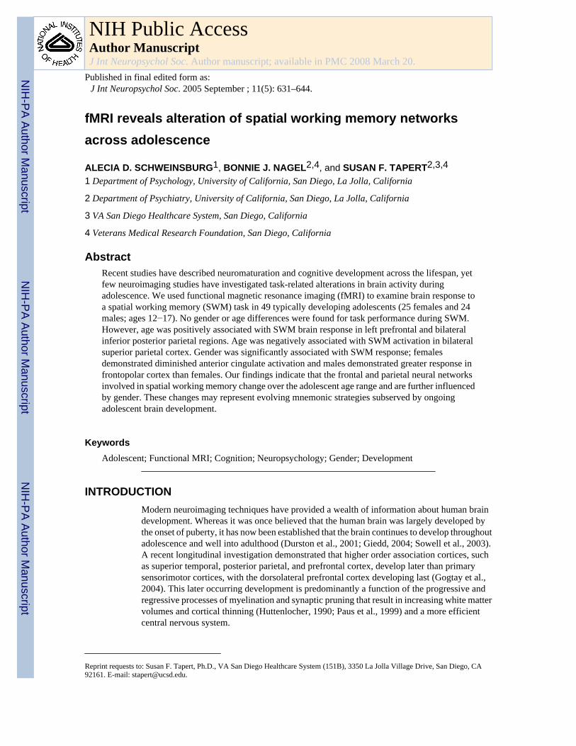
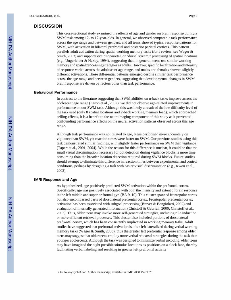
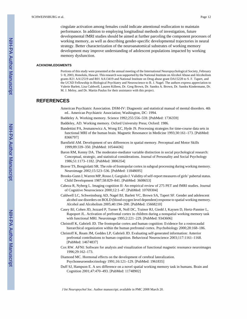
MeasuresSpatial working memory task—The spatial working memory (SWM) task (Kindermannet al., 2004; Tapert et al., 2001) consisted of 18 21-second blocks that alternated betweenexperimental and baseline conditions, and three blocks of rest (two 21-second blocks and one42-second block). The task also included six seconds of blank screen at the beginning (notanalyzed), allowing the scanner to reach steady state. Total task time was 7 minutes and 48seconds (see Figure 1). Each block started with a one-second word cue at the center of thescreen to inform the participant of the upcoming block type. Stimuli were presented for 1000ms, and each interstimulus interval was 1000 ms. During rest blocks, the word “LOOK”appeared at the center of the screen, then a centered fixation cross appeared for 20 seconds.The experimental (spatial working memory) condition was a memory for locations task inwhich abstract line drawings (Kimura figures) were projected one at a time in one of eightlocations in a circular array. Locations were chosen to minimize verbal labeling (e.g., not in
SCHWEINSBURG et al. Page 3
J Int Neuropsychol Soc. Author manuscript; available in PMC 2008 March 20.
NIH
-PA Author Manuscript
NIH
-PA Author Manuscript
NIH
-PA Author Manuscript
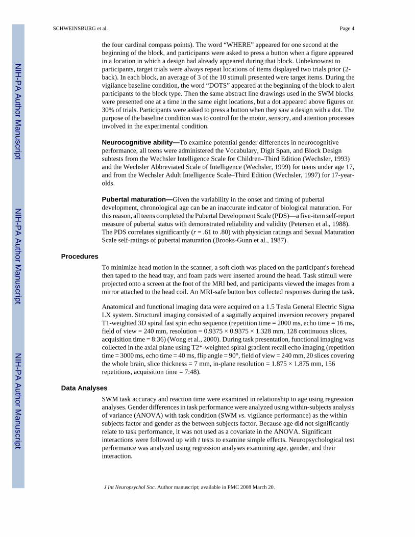
the four cardinal compass points). The word “WHERE” appeared for one second at thebeginning of the block, and participants were asked to press a button when a figure appearedin a location in which a design had already appeared during that block. Unbeknownst toparticipants, target trials were always repeat locations of items displayed two trials prior (2-back). In each block, an average of 3 of the 10 stimuli presented were target items. During thevigilance baseline condition, the word “DOTS” appeared at the beginning of the block to alertparticipants to the block type. Then the same abstract line drawings used in the SWM blockswere presented one at a time in the same eight locations, but a dot appeared above figures on30% of trials. Participants were asked to press a button when they saw a design with a dot. Thepurpose of the baseline condition was to control for the motor, sensory, and attention processesinvolved in the experimental condition.
Neurocognitive ability—To examine potential gender differences in neurocognitiveperformance, all teens were administered the Vocabulary, Digit Span, and Block Designsubtests from the Wechsler Intelligence Scale for Children–Third Edition (Wechsler, 1993)and the Wechsler Abbreviated Scale of Intelligence (Wechsler, 1999) for teens under age 17,and from the Wechsler Adult Intelligence Scale–Third Edition (Wechsler, 1997) for 17-year-olds.
Pubertal maturation—Given the variability in the onset and timing of pubertaldevelopment, chronological age can be an inaccurate indicator of biological maturation. Forthis reason, all teens completed the Pubertal Development Scale (PDS)—a five-item self-reportmeasure of pubertal status with demonstrated reliability and validity (Petersen et al., 1988).The PDS correlates significantly (r = .61 to .80) with physician ratings and Sexual MaturationScale self-ratings of pubertal maturation (Brooks-Gunn et al., 1987).
ProceduresTo minimize head motion in the scanner, a soft cloth was placed on the participant's foreheadthen taped to the head tray, and foam pads were inserted around the head. Task stimuli wereprojected onto a screen at the foot of the MRI bed, and participants viewed the images from amirror attached to the head coil. An MRI-safe button box collected responses during the task.
Anatomical and functional imaging data were acquired on a 1.5 Tesla General Electric SignaLX system. Structural imaging consisted of a sagittally acquired inversion recovery preparedT1-weighted 3D spiral fast spin echo sequence (repetition time = 2000 ms, echo time = 16 ms,field of view = 240 mm, resolution = 0.9375 × 0.9375 × 1.328 mm, 128 continuous slices,acquisition time = 8:36) (Wong et al., 2000). During task presentation, functional imaging wascollected in the axial plane using T2*-weighted spiral gradient recall echo imaging (repetitiontime = 3000 ms, echo time = 40 ms, flip angle = 90°, field of view = 240 mm, 20 slices coveringthe whole brain, slice thickness = 7 mm, in-plane resolution = 1.875 × 1.875 mm, 156repetitions, acquisition time = 7:48).
Data AnalysesSWM task accuracy and reaction time were examined in relationship to age using regressionanalyses. Gender differences in task performance were analyzed using within-subjects analysisof variance (ANOVA) with task condition (SWM vs. vigilance performance) as the withinsubjects factor and gender as the between subjects factor. Because age did not significantlyrelate to task performance, it was not used as a covariate in the ANOVA. Significantinteractions were followed up with t tests to examine simple effects. Neuropsychological testperformance was analyzed using regression analyses examining age, gender, and theirinteraction.
SCHWEINSBURG et al. Page 4
J Int Neuropsychol Soc. Author manuscript; available in PMC 2008 March 20.
NIH
-PA Author Manuscript
NIH
-PA Author Manuscript
NIH
-PA Author Manuscript
Imaging data were processed and analyzed using Analysis of Functional NeuroImages (AFNI)(Cox, 1996). First, we applied a motion-correction algorithm to align each volume in the timeseries with a base volume, yielding three rotational and three displacement parameters acrossthe time series for each participant. Two independent raters inspected time series data to removeany repetitions on which the algorithm did not adequately adjust for motion; all participantsretained at least 80% of repetitions. Using a deconvolution process (Ward, 2002), the timeseries data were correlated with a vector representing the design of the task (see Figure 1) thatmodeled 1- and 2-repetition delays in hemodynamic response (Bandettini et al., 1993), andcovaried for estimated degree of motion and linear trends. This process yielded fit coefficientsrepresenting the blood oxygen level dependent (BOLD) response contrast between SWM andvigilance, SWM and fixation, and vigilance and fixation in each voxel for every subject.Imaging datasets were transformed into standard Talairach coordinates for structurelocalization and comparisons among subjects (Lancaster et al., 2000; Talairach & Tournoux,1988), and functional data were resampled into 3.5-mm3 isotropic voxels. We applied a spatialsmoothing Gaussian filter (full-width half maximum = 3.5 mm) to functional data to accountfor anatomic variability.
Group-level analyses conducted regressions in each voxel of the brain to predict the fitcoefficient representing the contrast between SWM and vigilance from gender, age, and theirinteraction. To control for Type I error when determining clusters that showed significanteffects, we used a combination of t-statistic magnitude and cluster volume thresholding(Forman et al., 1995; Ward, 1997) by only interpreting clusters exceeding 943 microliters,equal to 22 contiguous significant (α < .05) 3.5-mm3 voxels, yielding a clusterwise α < .05. Tounderstand the nature of these group level clusters, we performed exploratory follow-upanalyses (uncorrected) examining contrasts between SWM and fixation and vigilance andfixation in each significant cluster. We utilized the Talairach Daemon (Lancaster et al., 2000;Ward, 1997) and AFNI (Cox, 1996) to confirm gyral labels for significant clusters.
To examine the role of pubertal maturation on SWM BOLD response, mean fit coefficientsfor each participant were computed for each significant activation cluster, and hierarchicalregression analyses determined whether PDS scores explained significant variance in brainresponse above and beyond that explained by chronological age.
Planned follow-up analyses examined whether age, gender, and their interaction were relatedto spatial extent of neural response in brain regions showing significant age-related activationduring SWM relative to vigilance. Because we were particularly interested in age-relatedchanges in the spatial extent of activation during SWM (and not in regions showing less SWMresponse than vigilance response), we only examined clusters in which there was greateractivation to SWM relative to vigilance, and not regions that were deactivated by the task.Therefore, we created posterior parietal and left prefrontal regions of interest (ROI), andcounted the number of voxels exhibiting significantly greater activation to SWM relative tovigilance for each participant. In order to best represent functionally important frontal andparietal regions within this sample of adolescents, our ROIs were determined by identifyingsignificant clusters activated by the task, rather than anatomically defined based on specificgyri or Brodmann's Areas (BA).
Because regression analyses indicated a change in location of parietal activation acrossadolescence (see Results), an ROI based on the average activation map for the whole groupwould not accurately represent regions used for the task in both young and old teens (Passarottiet al., 2003). Therefore, we divided teens on age with median (14.93 years) split, anddetermined significant clusters activated by the task in young and old teens separately usingsingle sample t tests (cluster volume ≥ 943 microliters, p < .05). This yielded separate posteriorparietal clusters for young and old teens. We created a posterior parietal ROI for examining
SCHWEINSBURG et al. Page 5
J Int Neuropsychol Soc. Author manuscript; available in PMC 2008 March 20.
NIH
-PA Author Manuscript
NIH
-PA Author Manuscript
NIH
-PA Author Manuscript
spatial extent of activation by including all voxels that occupied the posterior parietal clustersfor young teens and old teens, and all voxels within clusters showing a significant positive ornegative relationship to age. A similar procedure determined an appropriate left prefrontal ROI.Because young teens did not demonstrate significant clusters of left prefrontal activation,significant clusters activated by the task in older teens were added to the average left prefrontalcluster showing a significant relationship with age. To assess volume of activation withinposterior parietal and left prefrontal ROIs, we calculated the number of voxels showingsignificantly greater (p < .025) activation during SWM relative to vigilance for each participantwithin each ROI. We then performed regression analyses to predict volume of activation fromage, gender, and their interaction.
RESULTSBehavioral Performance
SWM task performance data were available for 47 participants (button box failed during twoexaminations). Teens performed at 95.61 ± 2.54% accuracy on the vigilance condition and88.87 ± 8.35% accuracy on SWM [F(1,45) = 30.44, p < .001]. There was no significant genderdifference nor was there a gender × task condition interaction for task accuracy. Participantsresponded slower to vigilance (636.63 ± 73.12 ms) than to SWM (605.00 ± 70.01 ms; F(1,45)= 10.54, p < .005). Males' overall reaction time (601.11 ± 59.72 ms) was faster thanfemales' (641.38 ± 59.71 ms; F(1,45) = 5.34, p < .025). A gender × condition interaction wasfound for reaction time (p < .05); while both males and females performed faster on SWM thanvigilance, the difference was greater for females (607.14 ± 67.21 ms on vigilance and 595.08± 70.02 ms on SWM for males; 667.41 ± 67.21 ms on vigilance and 615.36 ± 70.02 ms onSWM for females), and males responded faster than females during vigilance [t(45) = 3.07,p < .005]. Age was negatively associated with vigilance reaction time (r = 2.292, p < .05).There were no significant gender differences on the Wechsler Vocabulary, Digit Span, or BlockDesign subtest scores (see Table 1).
MovementTo determine whether movement during fMRI scanning might affect results, we examinedrelationships between age and bulk motion in two ways. Both total number of removedrepetitions and average movement in each direction throughout the task (i.e., roll, pitch, yaw,superior, left, posterior) were examined in relation to age and gender using correlationalanalyses. The number of repetitions removed for excessive motion during the task declinedwith age (r = −.44, p < .01). However, in brain regions demonstrating a relationship betweenSWM response and age, number of removed repetitions did not significantly relate to brainresponse (all p's > .025), and the relationship between age and brain response in each clusterremained significant after controlling for number of removed repetitions. Mean rotational andtranslational motion were not significantly related to age. The average rotational movementthroughout the task was 0.07, 0.22, and 0.09 degrees for roll, pitch, and yaw, respectively; theaverage translational movement was 0.16, 0.06, and 0.09 mm for superior, left, and posterior,respectively. There were no significant gender differences for number of repetitions removedfor movement or on any directional movement parameter, with the exception of malesdemonstrating significantly greater rotational motion than females in the pitch direction [t(1,47) = −2.08, p < .05].
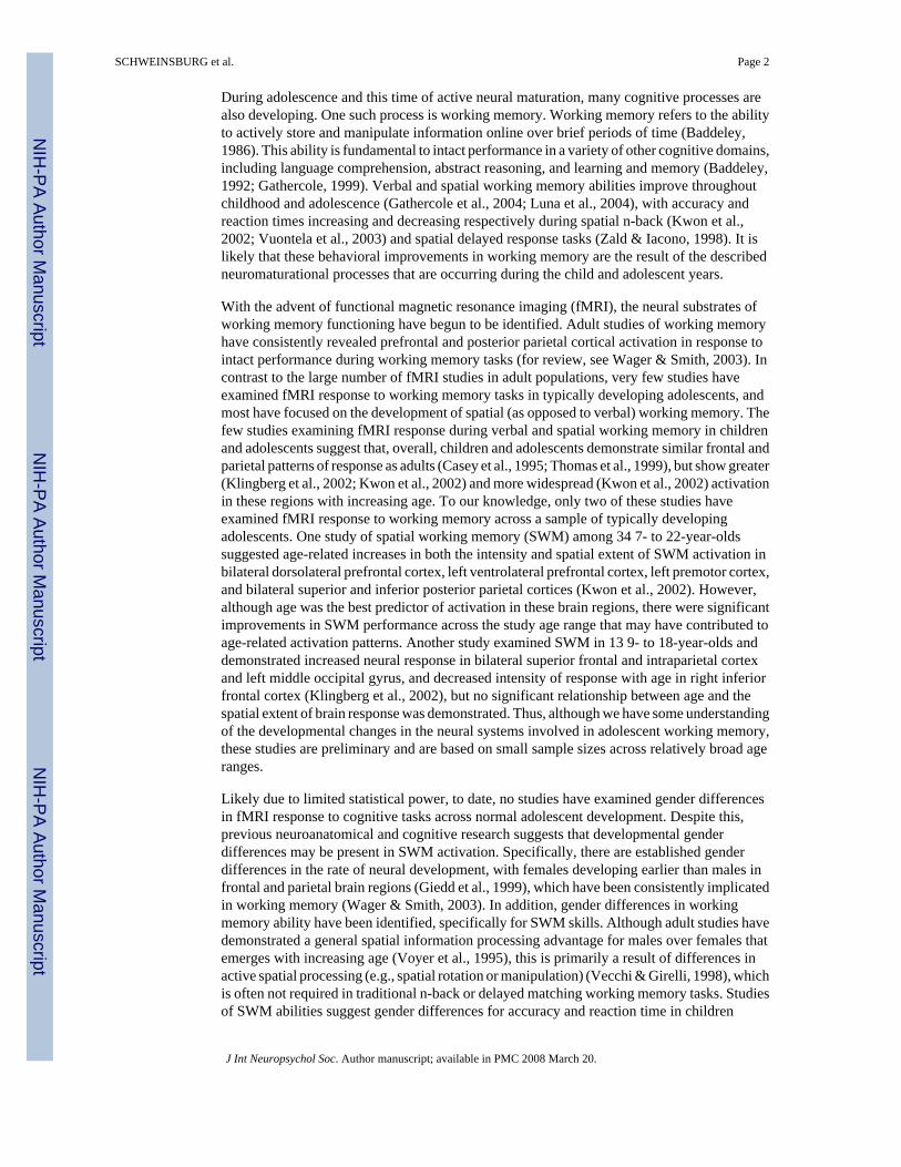
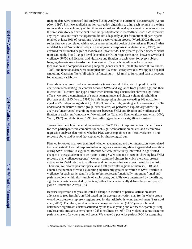
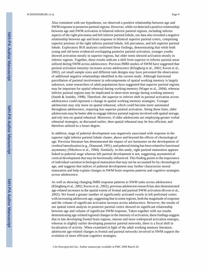
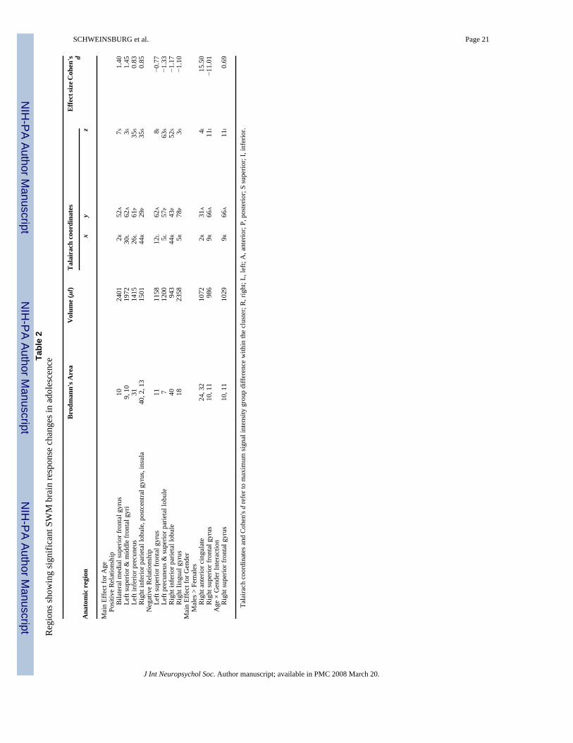
fMRI ResponseMain effect for age—Age positively predicted SWM brain response in bilateral medialportions of superior frontal gyrus (BA 10); left superior and middle frontal gyri (BA 9, 10);inferior aspects of the left precuneus and angular gyrus (BA 31); and a cluster encompassingthe right inferior parietal lobule, postcentral gyrus, and insula (BA 40, 2, 13; p's < .05; see
SCHWEINSBURG et al. Page 6
J Int Neuropsychol Soc. Author manuscript; available in PMC 2008 March 20.
NIH
-PA Author Manuscript
NIH
-PA Author Manuscript
NIH
-PA Author Manuscript
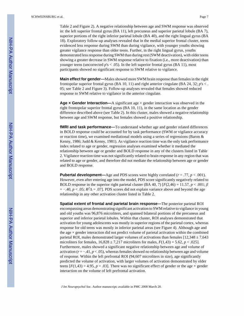
Table 2 and Figure 2). A negative relationship between age and SWM response was observedin: the left superior frontal gyrus (BA 11), left precuneus and superior parietal lobule (BA 7),superior portions of the right inferior parietal lobule (BA 40), and the right lingual gyrus (BA18). Exploratory follow-up analyses revealed that in the medial superior frontal cluster, teensevidenced less response during SWM than during vigilance, with younger youths showinggreater vigilance response than older teens. Further, in the right lingual gyrus, youthsdemonstrated less response during SWM than during rest (SWM deactivation), with older teensshowing a greater decrease in SWM response relative to fixation (i.e., more deactivation) thanyounger teens (uncorrected p's < .05). In the left superior frontal gyrus (BA 11), mostparticipants showed no significant response to SWM relative to vigilance.
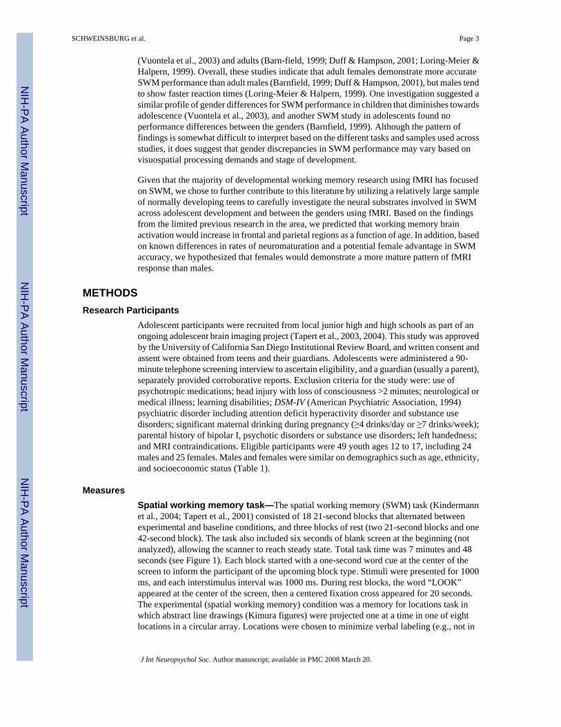
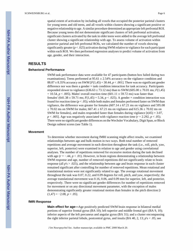
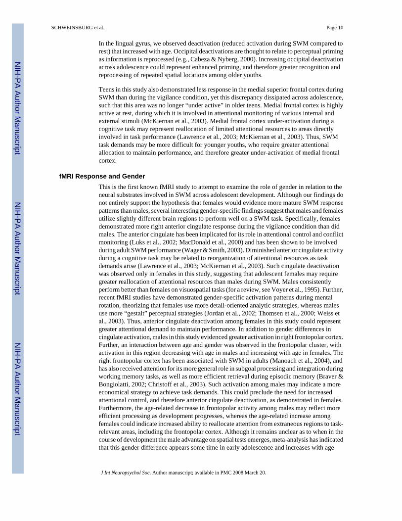
Main effect for gender—Males showed more SWM brain response than females in the rightfrontopolar superior frontal gyrus (BA 10, 11) and right anterior cingulate (BA 24, 32; p's < .05; see Table 2 and Figure 3). Follow-up analyses revealed that females showed reducedresponse to SWM relative to vigilance in the anterior cingulate.
Age × Gender Interaction—A significant age × gender interaction was observed in theright frontopolar superior frontal gyrus (BA 10, 11), in the same location as the genderdifference described above (see Table 2). In this cluster, males showed a negative relationshipbetween age and SWM response, but females showed a positive relationship.
fMRI and task performance—To understand whether age and gender related differencesin BOLD response could be accounted for by task performance (SWM or vigilance accuracyor reaction time), we examined mediational models using a series of regressions (Baron &Kenny, 1986; Judd & Kenny, 1981). As vigilance reaction time was the only task performanceindex related to age or gender, regression analyses examined whether it mediated therelationship between age or gender and BOLD response in any of the clusters listed in Table2. Vigilance reaction time was not significantly related to brain response in any region that wasrelated to age or gender, and therefore did not mediate the relationship between age or genderand BOLD response.
Pubertal development—Age and PDS scores were highly correlated (r = .77, p < .001).However, even after entering age into the model, PDS score significantly negatively related toBOLD response in the superior right parietal cluster (BA 40, 7) [F(2,46) = 11.57, p < .001; β= −.40, p < .05; R2Δ = .07]. PDS scores did not explain variance above and beyond the agerelationship in any other activation cluster listed in Table 2.
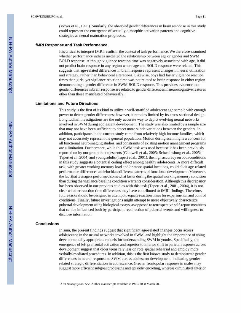
Spatial extent of frontal and parietal brain response—The posterior parietal ROIencompassing areas demonstrating significant activation to SWM relative to vigilance in youngand old youths was 96,876 microliters, and spanned bilateral portions of the precuneus andsuperior and inferior parietal lobules. Within that cluster, ROI analyses demonstrated thatactivation for young adolescents was mostly in superior regions of the parietal cortex, whereasresponse for old teens was mostly in inferior parietal areas (see Figure 4). Although age andthe age × gender interaction did not predict volume of parietal activation within the combinedparietal ROI, males demonstrated larger volumes of activations than females [12,348 ± 7,643microliters for females, 16,828 ± 7,217 microliters for males, F(1,43) = 5.62, p < .025].Furthermore, males showed a significant negative relationship between age and volume ofactivation (r = −.41, p < .05), whereas females showed no relationship between age and volumeof response. Within the left prefrontal ROI (94,607 microliters in size), age significantlypredicted the volume of activation, with larger volumes of activation demonstrated by olderteens [F(1,43) = 4.95, p = .03]. There was no significant effect of gender or the age × genderinteraction on the volume of left prefrontal activation.
SCHWEINSBURG et al. Page 7
J Int Neuropsychol Soc. Author manuscript; available in PMC 2008 March 20.
NIH
-PA Author Manuscript
NIH
-PA Author Manuscript
NIH
-PA Author Manuscript
DISCUSSIONThis cross-sectional study examined the effects of age and gender on brain response during aSWM task among 12- to 17-year-olds. In general, we observed comparable task performanceacross the age range and between genders, and all teens showed typical response patterns forSWM, with activation in bilateral prefrontal and posterior parietal cortices. This patternparallels adult activation during spatial working memory tasks (for a review, see Wager &Smith, 2003) and supports occipitoparietal, or “dorsal stream,” processing of spatial locations(e.g., Ungerleider & Haxby, 1994), suggesting that, in general, teens use similar workingmemory and spatial processing strategies as adults. However, specific localization and intensityof response varied across the adolescent age range, and males and females showed slightlydifferent activations. These differential patterns emerged despite similar task performanceacross the age range and between genders, suggesting that developmental changes in SWMbrain response are driven by factors other than task performance.
Behavioral PerformanceIn contrast to the literature suggesting that SWM abilities on n-back tasks improve across theadolescent age range (Kwon et al., 2002), we did not observe age-related improvements inperformance on our SWM task. Although this was likely a result of the low difficulty level ofthe task used (only 8 spatial locations and 2-back working memory load), which approachedceiling effects, it is a benefit to the neuroimaging component of this study as it preventedconfounding performance effects on the neural activation patterns observed across this agerange.
Although task performance was not related to age, teens performed more accurately onvigilance than SWM, yet reaction times were faster on SWM. Our previous studies using thistask demonstrated similar findings, with slightly faster performance on SWM than vigilance(Tapert et al., 2001, 2004). While the reason for this difference is unclear, it could be that thesmall visual discrimination necessary for dot detection during vigilance blocks is more timeconsuming than the broader location detection required during SWM blocks. Future studiesshould attempt to eliminate this difference in reaction times between experimental and controlconditions, perhaps by designing a task with easier visual discrimination (e.g., Kwon et al.,2002).
fMRI Response and AgeAs hypothesized, age positively predicted SWM activation within the prefrontal cortex.Specifically, age was positively associated with both the intensity and extent of brain responsein the left middle and superior frontal gyri (BA 9, 10). This cluster spanned frontopolar cortexbut also encompassed parts of dorsolateral prefrontal cortex. Frontopolar prefrontal cortexactivation has been associated with subgoal processing (Braver & Bongiolatti, 2002) andevaluation of internally generated information (Christoff & Gabrieli, 2000; Christoff et al.,2003). Thus, older teens may invoke more self-generated strategies, including rule inductionor more efficient retrieval processes. This cluster also included portions of dorsolateralprefrontal cortex, which has been consistently implicated in working memory tasks. Adultstudies have suggested that prefrontal activation is often left-lateralized during verbal workingmemory tasks (Wager & Smith, 2003); thus the greater left prefrontal response among olderteens may suggest that older teens employ more verbal rehearsal strategies during the task thanyounger adolescents. Although the task was designed to minimize verbal encoding, older teensmay have imagined the eight possible stimulus locations as positions on a clock face, therebyfacilitating verbal labeling and resulting in greater left prefrontal activity.
SCHWEINSBURG et al. Page 8
J Int Neuropsychol Soc. Author manuscript; available in PMC 2008 March 20.
NIH
-PA Author Manuscript
NIH
-PA Author Manuscript
NIH
-PA Author Manuscript
Also consistent with our hypotheses, we observed a positive relationship between age andSWM response in posterior parietal regions. However, while we detected a positive relationshipbetween age and SWM activation in bilateral inferior parietal regions, including inferioraspects of the right precuneus and left inferior parietal lobule, our data also revealed a negativerelationship between age and brain response in bilateral superior parietal cortex, comprisingsuperior portions of the right inferior parietal lobule, left precuneus, and left superior parietallobule. Exploratory ROI analyses confirmed these findings, demonstrating that while bothyoung and old teens evidenced overlapping posterior parietal activation, younger youthsshowed activation mostly in superior regions, but older teens showed activation mostly ininferior regions. Together, these results indicate a shift from superior to inferior parietal areasutilized during SWM across adolescence. Previous fMRI studies of SWM have suggested thatparietal activation intensity increases across adolescence (Klingberg et al., 2002; Kwon et al.,2002), yet small sample sizes and different task designs may have prevented the observationof additional negative relationships identified in the current study. Although functionalparcellation of parietal involvement in subcomponents of spatial working memory is largelyunknown, some researchers of adult populations have suggested that superior parietal regionsmay be important for spatial rehearsal during working memory (Wager et al., 2004), whereasinferior parietal regions may be implicated in short-term storage during working memory(Smith & Jonides, 1998). Therefore, the superior to inferior shift in parietal activation acrossadolescence could represent a change in spatial working memory strategies. Youngeradolescents may rely more on spatial rehearsal, which could become more automatedthroughout adolescence, requiring less superior parietal activation. Along those lines, olderadolescents may be better able to engage inferior parietal regions involved with spatial storage,and rely less on spatial rehearsal. Moreover, if older adolescents are employing greater verbalrehearsal strategies, as discussed earlier, then spatial rehearsal may be less efficient, andtherefore utilized to a lesser degree.
In addition, stage of pubertal development was negatively associated with response in thesuperior right inferior parietal lobule cluster, above and beyond the effects of chronologicalage. Previous literature has demonstrated the impact of sex hormones on the development ofcerebral lateralization (e.g., Diamond, 1991), and pubertal timing has been related to functionalasymmetry (Nikolova et al., 1994). Similarly, in this study, right parietal maturation appearslinked to pubertal stage whereas left parietal development is not, suggesting asymmetricalcortical development that may be hormonally influenced. This finding points to the importanceof individual variation in biological maturation that may not be accounted for by chronologicalage, and suggests that indices of pubertal development may further characterize neuralmaturation and help explain changes in SWM brain response patterns and cognitive strategiesacross adolescence.
As well as showing changing fMRI response patterns to SWM tasks across adolescence(Klingberg et al., 2002; Kwon et al., 2002), previous adolescent research has also demonstratedage-related increases in the spatial extent of frontal and parietal SWM activation (Kwon et al.,2002). We found a greater number of significantly activated voxels in left prefrontal cortexwith increasing adolescent age, suggesting that in some regions, both the magnitude of responseand the volume of significant activation increase across adolescence. However, the results ofour spatial extent analysis in posterior parietal cortex showed no significant relationshipbetween age and volume of significant SWM response. Taken together with our resultsdemonstrating age-related regional changes in the intensity of activation, these findings suggestthat in late developing frontal brain regions, intense and more widespread activation emerges,whereas in slightly earlier developing posterior parietal networks, there is a focal shift inlocalization of activity. When examined in light of the adult working memory literature,adolescent age-related changes in frontal and parietal networks involved in SWM support theevolution of more efficient cognitive strategies.
SCHWEINSBURG et al. Page 9
J Int Neuropsychol Soc. Author manuscript; available in PMC 2008 March 20.
NIH
-PA Author Manuscript
NIH
-PA Author Manuscript
NIH
-PA Author Manuscript
In the lingual gyrus, we observed deactivation (reduced activation during SWM compared torest) that increased with age. Occipital deactivations are thought to relate to perceptual primingas information is reprocessed (e.g., Cabeza & Nyberg, 2000). Increasing occipital deactivationacross adolescence could represent enhanced priming, and therefore greater recognition andreprocessing of repeated spatial locations among older youths.
Teens in this study also demonstrated less response in the medial superior frontal cortex duringSWM than during the vigilance condition, yet this discrepancy dissipated across adolescence,such that this area was no longer “under active” in older teens. Medial frontal cortex is highlyactive at rest, during which it is involved in attentional monitoring of various internal andexternal stimuli (McKiernan et al., 2003). Medial frontal cortex under-activation during acognitive task may represent reallocation of limited attentional resources to areas directlyinvolved in task performance (Lawrence et al., 2003; McKiernan et al., 2003). Thus, SWMtask demands may be more difficult for younger youths, who require greater attentionalallocation to maintain performance, and therefore greater under-activation of medial frontalcortex.
fMRI Response and GenderThis is the first known fMRI study to attempt to examine the role of gender in relation to theneural substrates involved in SWM across adolescent development. Although our findings donot entirely support the hypothesis that females would evidence more mature SWM responsepatterns than males, several interesting gender-specific findings suggest that males and femalesutilize slightly different brain regions to perform well on a SWM task. Specifically, femalesdemonstrated more right anterior cingulate response during the vigilance condition than didmales. The anterior cingulate has been implicated for its role in attentional control and conflictmonitoring (Luks et al., 2002; MacDonald et al., 2000) and has been shown to be involvedduring adult SWM performance (Wager & Smith, 2003). Diminished anterior cingulate activityduring a cognitive task may be related to reorganization of attentional resources as taskdemands arise (Lawrence et al., 2003; McKiernan et al., 2003). Such cingulate deactivationwas observed only in females in this study, suggesting that adolescent females may requiregreater reallocation of attentional resources than males during SWM. Males consistentlyperform better than females on visuospatial tasks (for a review, see Voyer et al., 1995). Further,recent fMRI studies have demonstrated gender-specific activation patterns during mentalrotation, theorizing that females use more detail-oriented analytic strategies, whereas malesuse more “gestalt” perceptual strategies (Jordan et al., 2002; Thomsen et al., 2000; Weiss etal., 2003). Thus, anterior cingulate deactivation among females in this study could representgreater attentional demand to maintain performance. In addition to gender differences incingulate activation, males in this study evidenced greater activation in right frontopolar cortex.Further, an interaction between age and gender was observed in the frontopolar cluster, withactivation in this region decreasing with age in males and increasing with age in females. Theright frontopolar cortex has been associated with SWM in adults (Manoach et al., 2004), andhas also received attention for its more general role in subgoal processing and integration duringworking memory tasks, as well as more efficient retrieval during episodic memory (Braver &Bongiolatti, 2002; Christoff et al., 2003). Such activation among males may indicate a moreeconomical strategy to achieve task demands. This could preclude the need for increasedattentional control, and therefore anterior cingulate deactivation, as demonstrated in females.Furthermore, the age-related decrease in frontopolar activity among males may reflect moreefficient processing as development progresses, whereas the age-related increase amongfemales could indicate increased ability to reallocate attention from extraneous regions to task-relevant areas, including the frontopolar cortex. Although it remains unclear as to when in thecourse of development the male advantage on spatial tests emerges, meta-analysis has indicatedthat this gender difference appears some time in early adolescence and increases with age
SCHWEINSBURG et al. Page 10
J Int Neuropsychol Soc. Author manuscript; available in PMC 2008 March 20.
NIH
-PA Author Manuscript
NIH
-PA Author Manuscript
NIH
-PA Author Manuscript
(Voyer et al., 1995). Similarly, the observed gender differences in brain response in this studycould represent the emergence of sexually dimorphic activation patterns and cognitivestrategies as neural maturation progresses.
fMRI Response and Task PerformanceIt is critical to interpret fMRI results in the context of task performance. We therefore examinedwhether performance indices mediated the relationship between age or gender and SWMBOLD response. Although vigilance reaction time was negatively associated with age, it didnot predict brain response in any region where age and BOLD response were related. Thissuggests that age-related differences in brain response represent changes in neural utilizationand strategy, rather than behavioral alterations. Likewise, boys had faster vigilance reactiontimes than girls, yet vigilance reaction time was not related to brain response in either regiondemonstrating a gender difference in SWM BOLD response. This provides evidence thatgender differences in brain response are related to gender differences in neurocognitive featuresother than those manifested behaviorally.
Limitations and Future DirectionsThis study is the first of its kind to utilize a well-stratified adolescent age sample with enoughpower to detect gender differences; however, it remains limited by its cross-sectional design.Longitudinal investigations are the only accurate way to depict evolving neural networksinvolved in SWM during adolescent development. The study was also limited by a sample sizethat may not have been sufficient to detect more subtle variations between the genders. Inaddition, participants in the current study came from relatively high-income families, whichmay not accurately represent the general population. Motion during scanning is a concern forall functional neuroimaging studies, and constraints of existing motion management programsare a limitation. Furthermore, while this SWM task was used because it has been previouslyreported on by our group in adolescents (Caldwell et al., 2005; Schweinsburg et al., 2005;Tapert et al., 2004) and young adults (Tapert et al., 2001), the high accuracy on both conditionsin this study suggests a potential ceiling effect among healthy adolescents. A more difficulttask, with greater working memory load and/or more spatial locations, could elicit age-relatedperformance differences and elucidate different patterns of functional development. Moreover,the fact that teenagers performed somewhat faster during the spatial working memory conditionthan during the vigilance baseline condition warrants consideration. Although this discrepancyhas been observed in our previous studies with this task (Tapert et al., 2001, 2004), it is notclear whether reaction time differences may have contributed to fMRI findings. Therefore,future tasks should be designed in attempt to equate reaction times for experimental and controlconditions. Finally, future investigations might attempt to more objectively characterizepubertal development using biological assays, as opposed to retrospective self-report measuresthat can be influenced both by participant recollection of pubertal events and willingness todisclose information.
ConclusionsIn sum, the present findings suggest that significant age-related changes occur acrossadolescence in the neural networks involved in SWM, and highlight the importance of usingdevelopmentally appropriate models for understanding SWM in youths. Specifically, theemergence of left prefrontal activation and superior to inferior shift in parietal response acrossdevelopment suggest that older teens rely less on rote spatial rehearsal and employ moreverbally-mediated procedures. In addition, this is the first known study to demonstrate genderdifferences in neural response to SWM across adolescent development, indicating gender-related strategic differentiation in adolescence. Greater frontopolar response in males maysuggest more efficient subgoal processing and episodic encoding, whereas diminished anterior
SCHWEINSBURG et al. Page 11
J Int Neuropsychol Soc. Author manuscript; available in PMC 2008 March 20.
NIH
-PA Author Manuscript
NIH
-PA Author Manuscript
NIH
-PA Author Manuscript
cingulate activation among females could indicate attentional reallocation to maintainperformance. In addition to employing longitudinal methods of investigation, futuredevelopmental fMRI studies should be aimed at further parceling the component processes ofworking memory, as well as describing gender-specific developmental trajectories in neuralstrategy. Better characterization of the neuroanatomical substrates of working memorydevelopment may improve understanding of adolescent populations impacted by workingmemory dysfunction.
ACKNOWLEDGMENTS
Portions of this study were presented at the annual meeting of the International Neuropsychological Society, February5−8, 2003, Honolulu, Hawaii. This research was supported by the National Institute on Alcohol Abuse and Alcoholismgrants R21 AA12519 and R01 AA13419 and National Institute on Drug abuse grant DA15228 to S. F. Tapert, andthe UCSD Fellowship in Biological Psychiatry and Neuroscience to B. J. Nagel. The authors express appreciation toValerie Barlett, Lisa Caldwell, Lauren Killeen, Dr. Greg Brown, Dr. Sandra A. Brown, Dr. Sandra Kindermann, Dr.M. J. Meloy, and Dr. Martin Paulus for their assistance with this project.
REFERENCESAmerican Psychiatric Association. DSM-IV: Diagnostic and statistical manual of mental disorders. 4th
ed.. American Psychiatric Association; Washington, DC: 1994.Baddeley A. Working memory. Science 1992;255:556–559. [PubMed: 1736359]Baddeley, AD. Working memory. Oxford University Press; Oxford: 1986.Bandettini PA, Jesmanowicz A, Wong EC, Hyde JS. Processing strategies for time-course data sets in
functional MRI of the human brain. Magnetic Resonance in Medicine 1993;30:161–173. [PubMed:8366797]
Barnfield AM. Development of sex differences in spatial memory. Perceptual and Motor Skills1999;89:339–350. [PubMed: 10544436]
Baron RM, Kenny DA. The moderator-mediator variable distinction in social psychological research:Conceptual, strategic, and statistical considerations. Journal of Personality and Social Psychology1986;51:1173–1182. [PubMed: 3806354]
Braver TS, Bongiolatti SR. The role of frontopolar cortex in subgoal processing during working memory.Neuroimage 2002;15:523–536. [PubMed: 11848695]
Brooks-Gunn J, Warren MP, Rosso J, Gargiulo J. Validity of self-report measures of girls’ pubertal status.Child Development 1987;58:829–841. [PubMed: 3608653]
Cabeza R, Nyberg L. Imaging cognition II: An empirical review of 275 PET and fMRI studies. Journalof Cognitive Neuroscience 2000;12:1–47. [PubMed: 10769304]
Caldwell LC, Schweinsburg AD, Nagel BJ, Barlett VC, Brown SA, Tapert SF. Gender and adolescentalcohol use disorders on BOLD (blood oxygen level dependent) response to spatial working memory.Alcohol and Alcoholism 2005;40:194–200. [PubMed: 15668210]
Casey BJ, Cohen JD, Jezzard P, Turner R, Noll DC, Trainor RJ, Giedd J, Kaysen D, Hertz-Pannier L,Rapoport JL. Activation of prefrontal cortex in children during a nonspatial working memory taskwith functional MRI. Neuroimage 1995;2:221–229. [PubMed: 9343606]
Christoff K, Gabrieli JD. The frontopolar cortex and human cognition: Evidence for a rostrocaudalhierarchical organization within the human prefrontal cortex. Psychobiology 2000;28:168–186.
Christoff K, Ream JM, Geddes LP, Gabrieli JD. Evaluating self-generated information: Anteriorprefrontal contributions to human cognition. Behavioral Neuroscience 2003;117:1161–1168.[PubMed: 14674837]
Cox RW. AFNI: Software for analysis and visualization of functional magnetic resonance neuroimages1996;29:162–173.
Diamond MC. Hormonal effects on the development of cerebral lateralization.Psychoneuroendocrinology 1991;16:121–129. [PubMed: 1961835]
Duff SJ, Hampson E. A sex difference on a novel spatial working memory task in humans. Brain andCognition 2001;47:470–493. [PubMed: 11748902]
SCHWEINSBURG et al. Page 12
J Int Neuropsychol Soc. Author manuscript; available in PMC 2008 March 20.
NIH
-PA Author Manuscript
NIH
-PA Author Manuscript
NIH
-PA Author Manuscript
Durston S, Hulshoff Pol HE, Casey BJ, Giedd JN, Buitelaar JK, van Engeland H. Anatomical MRI ofthe developing human brain: What have we learned? Journal of the American Academy of Child andAdolescent Psychiatry 2001;40:1012–1020. [PubMed: 11556624]
Forman SD, Cohen JD, Fitzgerald M, Eddy WF, Mintun MA, Noll DC. Improved assessment ofsignificant activation in functional magnetic resonance imaging (fMRI): Use of a cluster-sizethreshold. Magnetic Resonance in Medicine 1995:636–647. [PubMed: 7596267]
Gathercole SE. Cognitive approaches to the development of short-term memory. Trends in CognitiveSciences 1999;3:410–419. [PubMed: 10529796]
Gathercole SE, Pickering SJ, Ambridge B, Wearing H. The structure of working memory from 4 to 15years of age. Developmental Psychology 2004;40:177–190. [PubMed: 14979759]
Giedd JN. Structural magnetic resonance imaging of the adolescent brain. Annals of the New YorkAcademy of Sciences 2004;1021:77–85. [PubMed: 15251877]
Giedd JN, Blumenthal J, Jeffries NO, Castellanos FX, Liu H, Zijdenbos A, Giedd JN, Blumenthal J,Jeffries NO, Castellanos FX, Liu H, Zijdenbos A, Paus T, Evans AC, Rapoport JL. Brain developmentduring childhood and adolescence: A longitudinal MRI study. Nature Neuroscience 1999;2:861–863.
Gogtay N, Giedd JN, Lusk L, Hayashi KM, Greenstein D, Vaituzis AC, Nugent TF 3rd, Herman DH,Clasen LS, Toga AW, Rapoport JL, Thompson PM. Dynamic mapping of human corticaldevelopment during childhood through early adulthood. Proceedings of the National Academy ofSciences, U.S.A 2004;17:17.
Huttenlocher PR. Morphometric study of human cerebral cortex development. Neuropsychologia1990;28:517–527. [PubMed: 2203993]
Jordan K, Wustenberg T, Heinze HJ, Peters M, Jancke L. Women and men exhibit different corticalactivation patterns during mental rotation tasks. Neuropsychologia 2002;40:2397–2408. [PubMed:12417468]
Judd CM, Kenny DA. Process analysis: Estimating mediation in evaluation research. Evaluation Review1981;5:602–619.
Kindermann SS, Brown GG, Zorrilla LE, Olsen RK, Jeste DV. Spatial working memory among middle-aged and older patients with schizophrenia and volunteers using fMRI. Schizophrenia Research2004;68:203–216. [PubMed: 15099603]
Klingberg T, Forssberg H, Westerberg H. Increased brain activity in frontal and parietal cortex underliesthe development of visuospatial working memory capacity during childhood. Journal of CognitiveNeuroscience 2002;14:1–10. [PubMed: 11798382]
Kwon H, Reiss AL, Menon V. Neural basis of protracted developmental changes in visuo-spatial workingmemory. Proceedings of the National Academy of Sciences, U.S.A 2002;99:13336–13341.
Lancaster JL, Woldorff MG, Parsons LM, Liotti M, Freitas CS, Rainey L, Kochunov PV, Nickerson D,Mikiten SA, Fox PT. Automated Talairach atlas labels for functional brain mapping. Human BrainMapping 2000;10:120–131. [PubMed: 10912591]
Lawrence NS, Ross TJ, Hoffmann R, Garavan H, Stein EA. Multiple neuronal networks mediate sustainedattention. Journal of Cognitive Neuroscience 2003;15:1028–1038. [PubMed: 14614813]
Loring-Meier S, Halpern DF. Sex differences in visuo-spatial working memory: Components of cognitiveprocessing. Psychonomic Bulletin and Review 1999;6:464–471. [PubMed: 12198785]
Luks TL, Simpson GV, Feiwell RJ, Miller WL. Evidence for anterior cingulate cortex involvement inmonitoring preparatory attentional set. Neuroimage 2002;17:792–802. [PubMed: 12377154]
Luna B, Garver KE, Urban TA, Lazar NA, Sweeney JA. Maturation of cognitive processes from latechildhood to adulthood. Child Development 2004;75:1357–1372. [PubMed: 15369519]
MacDonald AW 3rd, Cohen JD, Stenger VA, Carter CS. Dissociating the role of the dorsolateralprefrontal and anterior cingulate cortex in cognitive control. Science 2000;288:1835–1838. [PubMed:10846167]
Manoach DS, White NS, Lindgren KA, Heckers S, Coleman MJ, Dubal S, Holzman PS. Hemisphericspecialization of the lateral prefrontal cortex for strategic processing during spatial and shape workingmemory. Neuroimage 2004;21:894–903. [PubMed: 15006656]
McKiernan KA, Kaufman JN, Kucera-Thompson J, Binder JR. A parametric manipulation of factorsaffecting task-induced deactivation in functional neuroimaging. Journal of Cognitive Neuroscience2003;15:394–408. [PubMed: 12729491]
SCHWEINSBURG et al. Page 13
J Int Neuropsychol Soc. Author manuscript; available in PMC 2008 March 20.
NIH
-PA Author Manuscript
NIH
-PA Author Manuscript
NIH
-PA Author Manuscript
Nikolova P, Stoyanov Z, Negrev N. Functional brain asymmetry, handedness and menarcheal age.International Journal of Psychophysiology 1994;18:213–215. [PubMed: 7775218]
Passarotti AM, Paul BM, Bussiere JR, Buxton RB, Wong EC, Stiles J. The development of face andlocation processing: An fMRI study. Developmental Science 2003;6:100–117.
Paus T, Zijdenbos A, Worsley K, Collins DL, Blumenthal J, Giedd JN, Rapoport JL, Evans AC. Structuralmaturation of neural pathways in children and adolescents: In vivo study. Science 1999;283:1908–1911. [PubMed: 10082463]
Petersen A, Crockett L, Richards M, Boxer A. A self-report measure of pubertal status: Reliability,validity, and initial norms. Journal of Youth and Adolescence 1988;17:117–133.
Schweinsburg AD, Schweinsburg BC, Cheung EH, Brown GG, Brown SA, Tapert SF. fMRI responseto spatial working memory in adolescents with comorbid marijuana and alcohol use disorders. Drugand Alcohol Dependence 2005;79:201–210. [PubMed: 16002029]
Smith EE, Jonides J. Neuroimaging analyses of human working memory. Proceedings of the NationalAcademy of Sciences, U.S.A 1998;95:12061–12068.
Sowell ER, Peterson BS, Thompson PM, Welcome SE, Henkenius AL, Toga AW. Mapping corticalchange across the human life span. Nature Neuroscience 2003;6:309–315.
Talairach, J.; Tournoux, P. Three-dimensional proportional system: An approach to cerebral imaging.Thieme; New York: 1988. Coplanar stereotaxic atlas of the human brain..
Tapert SF, Brown GG, Kindermann SS, Cheung EH, Frank LR, Brown SA. fMRI measurement of braindysfunction in alcohol-dependent young women. Alcoholism: Clinical and Experimental Research2001;25:236–245.
Tapert SF, Cheung EH, Brown GG, Frank LR, Paulus MP, Schweinsburg AD, Meloy MJ, Brown SA.Neural response to alcohol stimuli in adolescents with alcohol use disorder. Archives of GeneralPsychiatry 2003;60:727–735. [PubMed: 12860777]
Tapert SF, Schweinsburg AD, Barlett VC, Brown GG, Brown SA, Frank LR, Brown GG, Meloy MJ.Blood oxygen level dependent response and spatial working memory in adolescents with alcohol usedisorders. Alcoholism: Clinical and Experimental Research 2004;28:1577–1586.
Thomas KM, King SW, Franzen PL, Welsh TF, Berkowitz AL, Noll DC, Birmaher V, Casey BJ. Adevelopmental functional MRI study of spatial working memory. Neuroimage 1999;10:327–338.[PubMed: 10458945]
Thomsen T, Hugdahl K, Ersland L, Barndon R, Lundervold A, Smievoll AI, Roscher BE, Sundberg H.Functional magnetic resonance imaging (fMRI) study of sex differences in a mental rotation task.Medical Science Monitor 2000;6:1186–1196. [PubMed: 11232158]
Ungerleider LG, Haxby JV. ‘What’ and ‘where’ in the human brain. Current Opinion in Neurobiology1994;4:157–165. [PubMed: 8038571]
Vecchi T, Girelli L. Gender differences in visuo-spatial processing: The importance of distinguishingbetween passive storage and active manipulation. Acta Psychologica (Amst) 1998;99:1–16.
Voyer D, Voyer S, Bryden MP. Magnitude of sex differences in spatial abilities: A meta-analysis andconsideration of critical variables. Psychological Bulletin 1995;117:250–270. [PubMed: 7724690]
Vuontela V, Steenari MR, Carlson S, Koivisto J, Fjallberg M, Aronen ET. Audiospatial and visuospatialworking memory in 6−13 year old school children. Learning and Memory 2003;10:74–81. [PubMed:12551966]
Wager TD, Jonides J, Reading S. Neuroimaging studies of shifting attention: A meta-analysis.Neuroimage 2004;22:1679–1693. [PubMed: 15275924]
Wager TD, Smith EE. Neuroimaging studies of working memory: A meta-analysis. Cognitive, Affective,& Behavioral Neuroscience 2003;3:255–274.
Ward, BD. Simultaneous inference for FMRI data. Biophysics Research Institute, Medical College ofWisconsin; Milwaukee, WI: 1997.
Ward, BD. Deconvolution analysis of FMRI time series data. Biophysics Research Institute, MedicalCollege of Wisconsin; Milwaukee, WI: 2002.
Wechsler, D. Manual for the Wechsler Intelligence Scale for Children–III. Psychological Corporation;San Antonio, TX: 1993.
SCHWEINSBURG et al. Page 14
J Int Neuropsychol Soc. Author manuscript; available in PMC 2008 March 20.
NIH
-PA Author Manuscript
NIH
-PA Author Manuscript
NIH
-PA Author Manuscript

Wechsler, D. Manual for the Wechsler Adult Intelligence Scale–III. Psychological Corporation; SanAntonio, TX: 1997.
Wechsler, D. Wechsler Abbreviated Scale of Intelligence. Psychological Corporation; San Antonio, TX:1999.
Weiss E, Siedentopf CM, Hofer A, Deisenhammer EA, Hoptman MJ, Kremser C, Golaszewski S, FelberS, Fleischhacker WW, Delazer M. Sex differences in brain activation pattern during a visuospatialcognitive task: A functional magnetic resonance imaging study in healthy volunteers. NeuroscienceLetters 2003;344:169–172. [PubMed: 12812832]
Wong EC, Luh WM, Buxton RB, Frank LR. Single slab high resolution 3D whole brain imaging usingspiral FSE. Proceedings of the International Society for Magnetic Resonance in Medicine2000;8:683.
Zald DH, Iacono WG. The development of spatial working memory abilities. DevelopmentalNeuropsychology 1998;14:563–578.
SCHWEINSBURG et al. Page 15
J Int Neuropsychol Soc. Author manuscript; available in PMC 2008 March 20.
NIH
-PA Author Manuscript
NIH
-PA Author Manuscript
NIH
-PA Author Manuscript
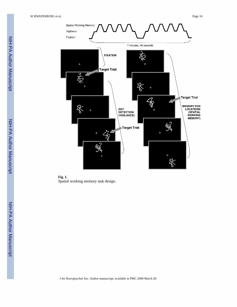
Fig. 1.Spatial working memory task design.
SCHWEINSBURG et al. Page 16
J Int Neuropsychol Soc. Author manuscript; available in PMC 2008 March 20.
NIH
-PA Author Manuscript
NIH
-PA Author Manuscript
NIH
-PA Author Manuscript
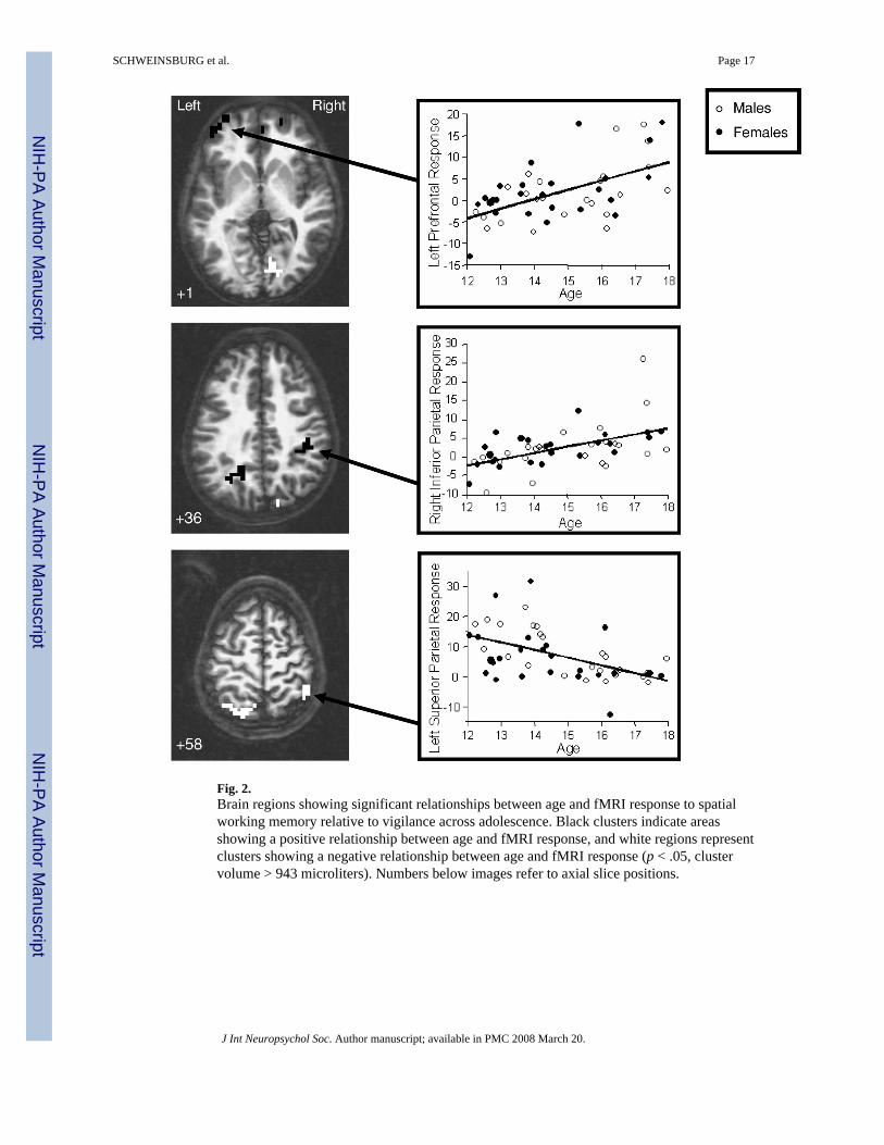
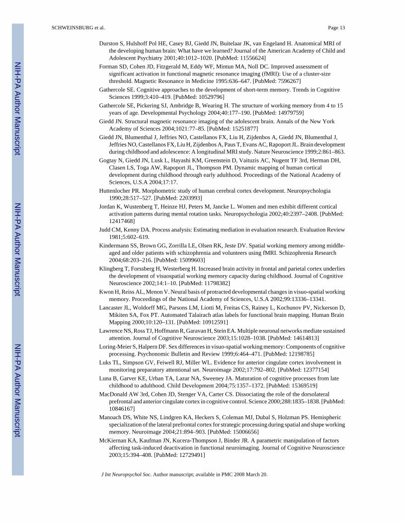
Fig. 2.Brain regions showing significant relationships between age and fMRI response to spatialworking memory relative to vigilance across adolescence. Black clusters indicate areasshowing a positive relationship between age and fMRI response, and white regions representclusters showing a negative relationship between age and fMRI response (p < .05, clustervolume > 943 microliters). Numbers below images refer to axial slice positions.
SCHWEINSBURG et al. Page 17
J Int Neuropsychol Soc. Author manuscript; available in PMC 2008 March 20.
NIH
-PA Author Manuscript
NIH
-PA Author Manuscript
NIH
-PA Author Manuscript
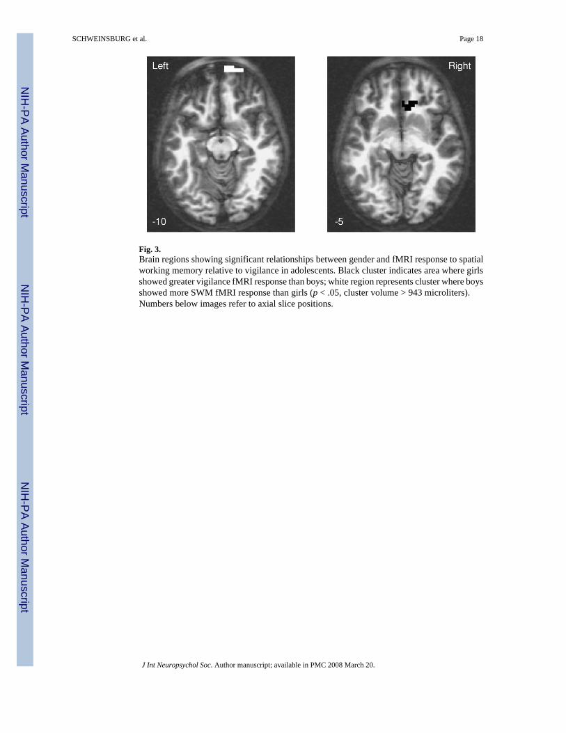
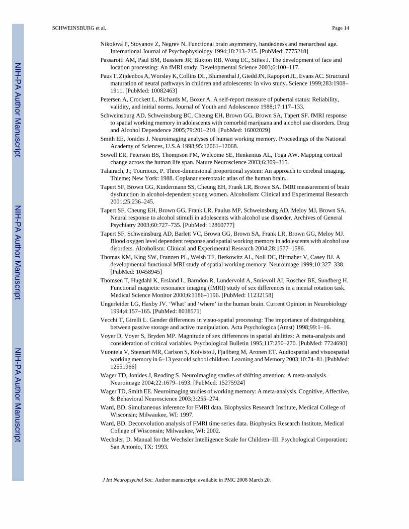
Fig. 3.Brain regions showing significant relationships between gender and fMRI response to spatialworking memory relative to vigilance in adolescents. Black cluster indicates area where girlsshowed greater vigilance fMRI response than boys; white region represents cluster where boysshowed more SWM fMRI response than girls (p < .05, cluster volume > 943 microliters).Numbers below images refer to axial slice positions.
SCHWEINSBURG et al. Page 18
J Int Neuropsychol Soc. Author manuscript; available in PMC 2008 March 20.
NIH
-PA Author Manuscript
NIH
-PA Author Manuscript
NIH
-PA Author Manuscript
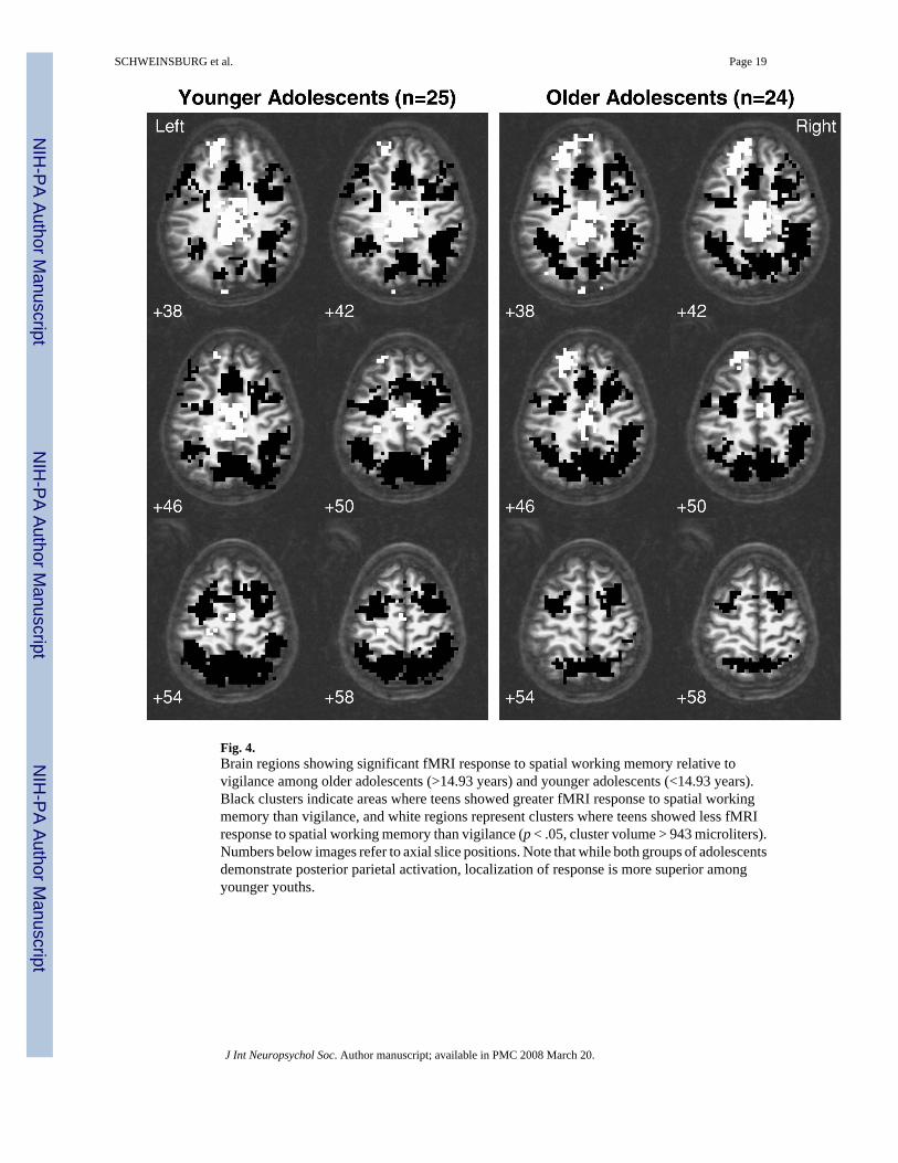
Fig. 4.Brain regions showing significant fMRI response to spatial working memory relative tovigilance among older adolescents (>14.93 years) and younger adolescents (<14.93 years).Black clusters indicate areas where teens showed greater fMRI response to spatial workingmemory than vigilance, and white regions represent clusters where teens showed less fMRIresponse to spatial working memory than vigilance (p < .05, cluster volume > 943 microliters).Numbers below images refer to axial slice positions. Note that while both groups of adolescentsdemonstrate posterior parietal activation, localization of response is more superior amongyounger youths.
SCHWEINSBURG et al. Page 19
J Int Neuropsychol Soc. Author manuscript; available in PMC 2008 March 20.
NIH
-PA Author Manuscript
NIH
-PA Author Manuscript
NIH
-PA Author Manuscript
NIH
-PA Author Manuscript
NIH
-PA Author Manuscript
NIH
-PA Author Manuscript
SCHWEINSBURG et al. Page 20
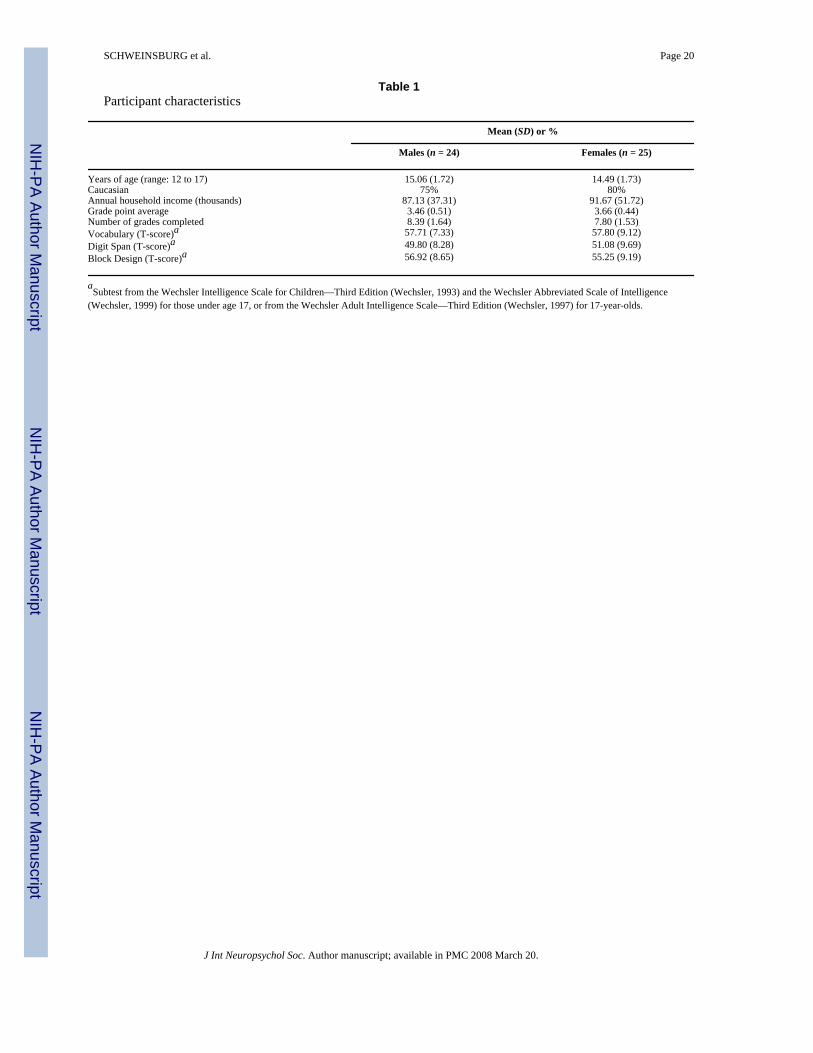
Table 1Participant characteristics
Mean (SD) or %
Males (n = 24) Females (n = 25)
Years of age (range: 12 to 17) 15.06 (1.72) 14.49 (1.73)Caucasian 75% 80%Annual household income (thousands) 87.13 (37.31) 91.67 (51.72)Grade point average 3.46 (0.51) 3.66 (0.44)Number of grades completed 8.39 (1.64) 7.80 (1.53)Vocabulary (T-score)a 57.71 (7.33) 57.80 (9.12)Digit Span (T-score)a 49.80 (8.28) 51.08 (9.69)Block Design (T-score)a 56.92 (8.65) 55.25 (9.19)
aSubtest from the Wechsler Intelligence Scale for Children—Third Edition (Wechsler, 1993) and the Wechsler Abbreviated Scale of Intelligence
(Wechsler, 1999) for those under age 17, or from the Wechsler Adult Intelligence Scale—Third Edition (Wechsler, 1997) for 17-year-olds.
J Int Neuropsychol Soc. Author manuscript; available in PMC 2008 March 20.
NIH
-PA Author Manuscript
NIH
-PA Author Manuscript
NIH
-PA Author Manuscript
SCHWEINSBURG et al. Page 21Ta
ble
2R
egio
ns sh
owin
g si
gnifi
cant
SW
M b
rain
resp
onse
cha
nges
in a
dole
scen
ce
Bro
dman
n's A
rea
Vol
ume
(μl)
Tal
aira
ch c
oord
inat
esE
ffect
size
Coh
en's d
Ana
tom
ic r
egio
nx
yz
Mai
n Ef
fect
for A
ge
Posi
tive
Rel
atio
nshi
p
Bila
tera
l med
ial s
uper
ior f
ront
al g
yrus
1024
012R
52A
7S1.
40
Left
supe
rior &
mid
dle
fron
tal g
yri
9, 1
019
7230
L62
A3S
1.45
Left
infe
rior p
recu
neus
3114
1526
L61
P35
S0.
83
Rig
ht in
ferio
r par
ieta
l lob
ule,
pos
tcen
tral g
yrus
, ins
ula
40, 2
, 13
1501
44R
29P
35S
0.85
N
egat
ive
Rel
atio
nshi
p
Left
supe
rior f
ront
al g
yrus
1111
5812
L62
A8I
−0.7
7
Left
prec
uneu
s & su
perio
r par
ieta
l lob
ule
712
005L
57P
63S
−1.3
3
Rig
ht in
ferio
r par
ieta
l lob
ule
4094
344
R43
P52
S−1
.17
Rig
ht li
ngua
l gyr
us18
2358
5R78
P3S
−1.1
0M
ain
Effe
ct fo
r Gen
der
M
ales
> F
emal
es
Rig
ht a
nter
ior c
ingu
late
24, 3
210
722R
31A
4I15
.50
Rig
ht su
perio
r fro
ntal
gyr
us10
, 11
986
9R66
A11
I−1
1.01
A
ge ×
Gen
der I
nter
actio
n
Rig
ht su
perio
r fro
ntal
gyr
us10
, 11
1029
9R66
A11
I0.
69
Tala
irach
coo
rdin
ates
and
Coh
en's
d re
fer t
o m
axim
um si
gnal
inte
nsity
gro
up d
iffer
ence
with
in th
e cl
uste
r; R
, rig
ht; L
, lef
t; A
, ant
erio
r; P,
pos
terio
r; S
supe
rior;
I, in
ferio
r.
J Int Neuropsychol Soc. Author manuscript; available in PMC 2008 March 20.