Fluorescence-Based Bacterial Bioreporter for Specific Detection of Methyl Halide Emissions in the...
15
Published Ahead of Print 16 August 2013. 10.1128/AEM.01738-13. 2013, 79(21):6561. DOI: Appl. Environ. Microbiol. Bringel, Hubert Schaller and Stéphane Vuilleumier Muhammad Farhan Ul Haque, Thierry Nadalig, Françoise Emissions in the Environment for Specific Detection of Methyl Halide Fluorescence-Based Bacterial Bioreporter http://aem.asm.org/content/79/21/6561 Updated information and services can be found at: These include: SUPPLEMENTAL MATERIAL Supplemental material REFERENCES http://aem.asm.org/content/79/21/6561#ref-list-1 at: This article cites 48 articles, 10 of which can be accessed free CONTENT ALERTS more» articles cite this article), Receive: RSS Feeds, eTOCs, free email alerts (when new http://journals.asm.org/site/misc/reprints.xhtml Information about commercial reprint orders: http://journals.asm.org/site/subscriptions/ To subscribe to to another ASM Journal go to: on October 21, 2013 by UNIVERSITE LOUIS PASTEUR http://aem.asm.org/ Downloaded from on October 21, 2013 by UNIVERSITE LOUIS PASTEUR http://aem.asm.org/ Downloaded from on October 21, 2013 by UNIVERSITE LOUIS PASTEUR http://aem.asm.org/ Downloaded from on October 21, 2013 by UNIVERSITE LOUIS PASTEUR http://aem.asm.org/ Downloaded from on October 21, 2013 by UNIVERSITE LOUIS PASTEUR http://aem.asm.org/ Downloaded from on October 21, 2013 by UNIVERSITE LOUIS PASTEUR http://aem.asm.org/ Downloaded from on October 21, 2013 by UNIVERSITE LOUIS PASTEUR http://aem.asm.org/ Downloaded from on October 21, 2013 by UNIVERSITE LOUIS PASTEUR http://aem.asm.org/ Downloaded from
Transcript of Fluorescence-Based Bacterial Bioreporter for Specific Detection of Methyl Halide Emissions in the...
Published Ahead of Print 16 August 2013. 10.1128/AEM.01738-13.
2013, 79(21):6561. DOI:Appl. Environ. Microbiol. Bringel, Hubert Schaller and Stéphane VuilleumierMuhammad Farhan Ul Haque, Thierry Nadalig, Françoise Emissions in the Environmentfor Specific Detection of Methyl Halide Fluorescence-Based Bacterial Bioreporter
http://aem.asm.org/content/79/21/6561Updated information and services can be found at:
These include:
SUPPLEMENTAL MATERIAL Supplemental material
REFERENCEShttp://aem.asm.org/content/79/21/6561#ref-list-1at:
This article cites 48 articles, 10 of which can be accessed free
CONTENT ALERTS more»articles cite this article),
Receive: RSS Feeds, eTOCs, free email alerts (when new
http://journals.asm.org/site/misc/reprints.xhtmlInformation about commercial reprint orders: http://journals.asm.org/site/subscriptions/To subscribe to to another ASM Journal go to:
on October 21, 2013 by U
NIV
ER
SIT
E LO
UIS
PA
ST
EU
Rhttp://aem
.asm.org/
Dow
nloaded from
on October 21, 2013 by U
NIV
ER
SIT
E LO
UIS
PA
ST
EU
Rhttp://aem
.asm.org/
Dow
nloaded from
on October 21, 2013 by U
NIV
ER
SIT
E LO
UIS
PA
ST
EU
Rhttp://aem
.asm.org/
Dow
nloaded from
on October 21, 2013 by U
NIV
ER
SIT
E LO
UIS
PA
ST
EU
Rhttp://aem
.asm.org/
Dow
nloaded from
on October 21, 2013 by U
NIV
ER
SIT
E LO
UIS
PA
ST
EU
Rhttp://aem
.asm.org/
Dow
nloaded from
on October 21, 2013 by U
NIV
ER
SIT
E LO
UIS
PA
ST
EU
Rhttp://aem
.asm.org/
Dow
nloaded from
on October 21, 2013 by U
NIV
ER
SIT
E LO
UIS
PA
ST
EU
Rhttp://aem
.asm.org/
Dow
nloaded from
on October 21, 2013 by U
NIV
ER
SIT
E LO
UIS
PA
ST
EU
Rhttp://aem
.asm.org/
Dow
nloaded from
Fluorescence-Based Bacterial Bioreporter for Specific Detection ofMethyl Halide Emissions in the Environment
Muhammad Farhan Ul Haque,a Thierry Nadalig,a Françoise Bringel,a Hubert Schaller,b Stéphane Vuilleumiera
Université de Strasbourg, Equipe Adaptations et Interactions Microbiennes dans l’Environnement, Département Microorganismes, Génomes, Environnement, UMR 7156UdS-CNRS Génétique Moléculaire, Génomique, Microbiologie, Strasbourg, Francea; Département Réseaux Métaboliques Végétaux, Institut de Biologie Moléculaire desPlantes, UPR 2357 CNRS, Strasbourg, Franceb
Methyl halides are volatile one-carbon compounds responsible for substantial depletion of stratospheric ozone. Among them,chloromethane (CH3Cl) is the most abundant halogenated hydrocarbon in the atmosphere. Global budgets of methyl halides inthe environment are still poorly understood due to uncertainties in their natural sources, mainly from vegetation, and theirsinks, which include chloromethane-degrading bacteria. A bacterial bioreporter for the detection of methyl halides was devel-oped on the basis of detailed knowledge of the physiology and genetics of Methylobacterium extorquens CM4, an aerobic alpha-proteobacterium which utilizes chloromethane as the sole source of carbon and energy. A plasmid construct with the promoterregion of the chloromethane dehalogenase gene cmuA fused to a promotorless yellow fluorescent protein gene cassette resultedin specific methyl halide-dependent fluorescence when introduced into M. extorquens CM4. The bacterial whole-cell bioreporterallowed detection of methyl halides at femtomolar levels and quantification at concentrations above 10 pM (approximately 240ppt). As shown for the model chloromethane-producing plant Arabidopsis thaliana in particular, the bioreporter may providean attractive alternative to analytical chemical methods to screen for natural sources of methyl halide emissions.
Methyl halides (monohalomethanes) such as chloromethaneare volatile hydrocarbons of environmental concern be-
cause of their toxicity to living organisms and their role in thedepletion of stratospheric ozone (1, 2). Chloromethane (CH3Cl),a gas and the most abundant halogenated hydrocarbon in theatmosphere (currently �550 ppt, with an approximate increase of2.3 to 2.7 ppt annually), is considered to be responsible for over15% of the chlorine-catalyzed destruction of stratospheric ozone(2). Bromomethane (CH3Br) also catalyzes the destruction ofstratospheric ozone (2), and iodomethane (CH3I) was shown toinfluence aerosol formation in the marine boundary layer (3).
Global emissions of chloromethane were recently estimated tobe 4.1 to 4.4 Tg (4), with industrial sources contributing to �10%of total emissions (5). Natural production of other methyl halidesappears to be weaker by 1 order of magnitude at least (2, 6). Nat-ural sources of methyl halides are mainly living vegetation (7, 8),wood rot fungi (9), dead plant material (10), biomass burning,oceans, and coastal waters (11). A thiol methyltransferase in-volved in the production of methyl halides was first isolated fromthe leaves of Brassica oleracea (12). In Arabidopsis thaliana, theS-adenosylmethionine-dependent methyltransferase gene HOL(harmless to ozone layer) was then shown to be involved in theproduction of methyl halides (13, 14). More recent work oncloned versions of a large series of homologs of this gene fromplants, fungi, and bacteria confirmed that the corresponding en-zymes may produce all three methyl halides, further suggestingthat methyl halide production is widespread in the living world(15). In addition, marine bacteria capable of producing methylhalides have also been isolated and characterized (16).
The global budgets of methyl halides are still poorly under-stood (2). This is due to large uncertainties in the sources de-scribed above but also in the sinks of these compounds, whichinclude oxidation by hydroxyl radicals, loss to the stratosphereand to polar ocean waters, uptake by soils, and bacterial degrada-tion (6, 17). Current efforts to constrain the biogeochemical cycles
of methyl halides involve analytical approaches such as gas chro-matography-mass spectroscopy (GC-MS) including stable iso-tope techniques for carbon and hydrogen elements (17–19). Thesemethods are time-consuming and labor-intensive, and this mayconstitute a drawback for screening potential sources of methylhalides in the environment. Bioreporter technology based onknowledge of gene expression and enzyme functions related to themolecules of interest represents a valuable alternative to analyticaltechniques in this context (20, 21).
Insights into the biological transformation of methyl halideshave become available from studies on the physiology and genet-ics of bacteria that can degrade methyl halides and utilize chloro-methane as the only source of carbon and energy for growth,which have been isolated from various environments, includingsoils (22–25), sludge (26–28), seawater (29), and the phyllosphere(30). The biochemistry and genetics of chloromethane degrada-tion have been elucidated in detail for Methylobacterium ex-torquens CM4 (31–34), a strain isolated from soil of a petrochem-ical factory in Tatarstan (23), and the complete genome sequenceof this strain was determined and analyzed (35, 36). Chlorometh-ane dehalogenase consists of the corrinoid methyltransferaseCmuA and the tetrahydrofolate-dependent methyltransferaseCmuB (33, 34). It transforms bromomethane and iodomethane aswell as chloromethane, and its expression was shown to bestrongly induced by chloromethane (31). Indeed, the dehaloge-
Received 28 May 2013 Accepted 12 August 2013
Published ahead of print 16 August 2013
Address correspondence to Stéphane Vuilleumier, [email protected].
Supplemental material for this article may be found at http://dx.doi.org/10.1128/AEM.01738-13.
Copyright © 2013, American Society for Microbiology. All Rights Reserved.
doi:10.1128/AEM.01738-13
November 2013 Volume 79 Number 21 Applied and Environmental Microbiology p. 6561–6567 aem.asm.org 6561
nase proteins CmuA and CmuB were detected in strain CM4grown in the presence of chloromethane but not when methanolwas used as the sole carbon source (36). Promoter regions andtranscription start sites of the chloromethane dehalogenase genescmuA and cmuB were identified upstream of the correspondinggenes (32).
In this work, a promoter-based bioreporter derivative of strainCM4 affording methyl halide-dependent production of fluores-cence from a plasmid-encoded yellow fluorescent protein (YFP)was constructed and characterized in terms of the specificity andsensitivity of its response to methyl halides, and its potential forthe detection of methyl halide emissions by plants was shown.
MATERIALS AND METHODSChemicals and reagents. All chemicals and reagents (purity, �99%) wereobtained from Sigma-Aldrich unless otherwise stated. Buffers, culturemedia, and solutions were prepared in ultrapure water (Purelab Classic;ELGA) and sterilized by autoclaving (20 min at 121°C under 105 Pa) or byfiltration (0.2 �m; Nalgene).
Bacterial strains, growth media, and cultivation conditions. A labo-ratory stock of the chloromethane-degrading strain M. extorquens CM4was used. Methylobacterium was cultivated in chloride-free M3 mineralmedium as described previously (36). Bacterial strains were cultivated inpetri dishes on solid medium supplemented with methanol (MeOH) in-cubated in air-tight glass jars at 30°C. Isolated colonies were used as inoc-ula for liquid cultures performed with 50 ml M3 medium in Erlenmeyerflasks fitted with airtight mininert valve caps (Supelco) at 30°C with 100rpm of agitation, with methanol (10 mM) and/or chloromethane (10mM) as the carbon source. Growth was monitored by measuring theoptical density at 600 nm (OD600). Escherichia coli TOP10 (Invitrogen)was grown at 37°C on Luria-Bertani (LB) rich medium (Difco Laborato-ries). Kanamycin was added at a final concentration of 50 �g ml�1 asrequired.
RNA isolation. Total RNA was extracted from samples of cultures ofM. extorquens taken in the early, mid-, and late exponential growth phases(OD600 of 0.06 to 0.1, 0.15 to 0.3, and �0.35, respectively) by using theNucleoSpin RNAII kit (Macherey-Nagel). RNAprotect solution (Qiagen)was used to stabilize RNA in bacterial culture samples (4 ml; OD600 of�0.06), according to the manufacturer’s recommendations. Cell pelletswere kept frozen at �80°C until further processing. Cells were lysed bylysozyme treatment (final concentration of 2 mg/ml) in 100 ml Tris-EDTA (TE) buffer at 37°C for 15 min prior to RNA extraction accordingto the protocol provided by the manufacturer. RNA preparations weretreated with amplification-grade DNase I (Invitrogen) for 1 h at 25°C andpurified again by using the NucleoSpin RNAII kit. The RNA concentra-tion was estimated spectrophotometrically at 260 nm (NanoDropND1000), and its quality was verified by its A260/A280 ratio.
Reverse transcription and quantitative PCR. cDNA was preparedwith the SuperScript III reverse transcriptase kit (Invitrogen), using 50 ngof DNase-treated RNA and 250 ng of random hexamers (Roche), accord-ing to the manufacturer’s instructions. Control reactions without reversetranscriptase were performed to check for the absence of contaminatingDNA. Quantitative PCR (qPCR) measurements on cDNA preparationswere done with 96-well reaction PCR plates by using a GeneAmp 5700sequence detection system (Applied Biosystems). Primers cmuA802F (5=-TTCAACGGCGAYATGTATCCYGG-3=) (37) and cmuA968R (5=-CCRCCRTTRTAVCCVACYTC-3=) (30) were used for amplification of thecmuA gene, and amplification of the 16S rRNA gene rrnA was performedwith primers BACT1369F and PROK1492R (38). PCRs were carriedout with a final volume of 20 �l, using 4 �l of a cDNA preparation (dilutedwith ultrapure molecular biology-grade water; Sigma) with a 3 �M finalconcentration of each primer and commercial 1� SYBR green PCR mix(Eurogentec). The PCR program included a 10-min denaturation step at95°C, followed by 40 cycles of 15 s of denaturation at 95°C and 1 min of
hybridization/polymerization at 60°C. Relative gene expression levelswere calculated by using the comparative threshold amplification cycle(CT) method (2���CT), as described previously (39). Gene copy numberswere determined by using known amounts of M. extorquens CM4 totalDNA as the reference.
Dehalogenase activity. The chloride released by dehalogenation ofchloromethane into M3 medium by strain CM4 in the presence of chlo-romethane was measured in culture supernatants of centrifuged samples(1 ml). The concentration of chloride was measured spectrophotometri-cally at 340 nm, according to the method of Jörg and Bertau (40), asFeCl2� formed in acidic medium by comparison to a calibration curve ofsodium chloride standards (0 to 20 mM solutions) in M3 medium.
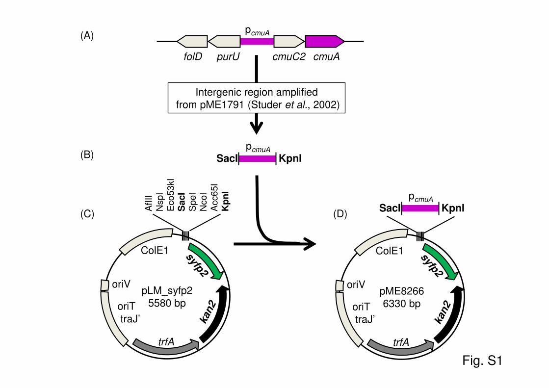
Construction of reporter plasmid pME8266. The promoter region ofthe cmuA gene of strain CM4 (pcmuA) (32) was amplified from promoterprobe plasmid pME1791 (32) as an 801-bp-long PCR fragment spanningexactly the intergenic region between purU and cmuC2 (see Fig. S1 in thesupplemental material) by using PCR primers 5=-ATTTTGAGCTCCGAGCGATTCCCCTCGTC-3= and 5=-ATTTTGGTACCTTAGACGGCACCAGATGC-3=, thereby introducing SacI and KpnI restriction sites (under-lined) for subsequent cloning. PCR was performed with a total volume of50 �l comprising 1 unit of Iproof high-fidelity polymerase (Bio-Rad), 10�l of high-fidelity PCR buffer (5�), 0.2 �M each primer, 0.1 �M eachdeoxynucleoside triphosphate (dNTP), and 10 ng of plasmid pME1791template. The PCR program included a 2-min denaturation step at 95°C,followed by 30 cycles of 20 s of denaturation at 95°C, 30 s of hybridizationat 62°C, and 30 s of polymerization at 72°C and a final 3-min postelonga-tion step at 72°C. The resulting PCR fragment was purified after agarosegel electrophoresis using the Geneclean Turbo kit (MP Biomedicals), di-gested overnight with the enzymes SacI and KpnI (Fermentas), and puri-fied again by using the same kit. The digested PCR fragment was ligatedfor 24 h at 14°C with KpnI- and SacI-digested promoter probe plasmidpLM.syfp2 (see Fig. S1 in the supplemental material), which features apromotorless gene for YFP downstream of its multiple-cloning site and akanamycin resistance gene (41), and transformed into One Shot TOP10chemically competent cells (Invitrogen) according to the manufacturer’sinstructions. Kanamycin-resistant colonies were selected, and plasmidpME8266 featuring the pcmuA-syfp2 fusion was prepared from one trans-formant by using the NucleoSpin plasmid kit (Macherey-Nagel), afterconfirmation of plasmid identity by colony PCR and sequencing. PlasmidpME8266 was introduced into M. extorquens CM4 by electroporation andselection on M3-MeOH-kanamycin plates, as described previously (42).
Fluorescence microscopy. Aliquots (5 ml) of bacterial cultures grownto mid-exponential phase in M3 medium with MeOH (10 mM) and chlo-romethane (10 mM), either alone or in combination, were filteredthrough 0.2-�m Whatman polycarbonate membrane filters. Filters werestained with 4,6-diamidino-2-phenylindole (DAPI) (1-�g/ml solution inwater), placed in the dark for 15 min, washed twice in sterile ultrapurewater and then in ethanol (70%), and mounted onto glass slides by usingmounting oil (BacLight; Molecular Probes). Images were taken by using aLeica DM4000 fluorescence microscope (Leica Microsystems) at a �1,000magnification, operated with either a YFP filter cube (excitation filter,bandpass [BP] 490/20; dichromatic mirror, 510 nm) or a DAPI filter (BP360/40; dichromatic mirror, 400 nm).
Fluorimetric analysis. Samples of growing cultures (1 ml) werewashed and resuspended in M3 medium at a final OD600 of 0.05. Cellsuspensions (200 �l) were transferred onto a 96-well microtiter plate(Nunc), and the OD600 and YFP fluorescence (excitation, 485 nm; emis-sion, 516 nm; bandwidth, 20 nm) were measured at room temperature ina microplate reader (Synergy HT; BioTek). YFP fluorescence values werecorrected by subtracting background values obtained for M3 medium,normalized to an OD600 of 1, and expressed as a percentage of the maxi-mum observed YFP fluorescence (see Fig. 5).
To monitor fluorescence induction after exposure to various com-pounds, a preculture of the reporter strain was grown in M3 mediumsupplemented with MeOH (20 mM) and kanamycin (50 �g/ml) until the
Farhan Ul Haque et al.
6562 aem.asm.org Applied and Environmental Microbiology
late exponential phase of growth (OD600 of �0.3 to 0.4). Cells were cen-trifuged, washed, and resuspended in M3 medium to a final OD600 of 0.2.Cell suspensions (5 ml) were then exposed to compounds of interest inHungate tubes (17-ml total volume). Chloromethane, dichloromethane,succinate, MeOH, and NaCl were provided at 20 mM, and iodomethane,chloroform, and tetrachloromethane were used at 200 �M to avoid po-tential toxic effects (e.g., see references 22 and 31). Samples (200 �l) weretaken after 3 h, transferred onto 96-well microtiter plates, and subjected tofluorimetric analysis as described above.
For determination of the concentration dependence of chlorometh-ane-induced fluorescence, serial 10-fold dilutions of chloromethane gas,designed to yield final concentrations of chloromethane in the range of 2fM to 20 mM, were prepared in airtight Hungate tubes. For the initialdilution, 2.5 ml of chloromethane gas was added to an empty airtightHungate tube (17 ml) by using an airtight syringe. The tube was left toequilibrate for 5 min, 1.7 ml of the gas phase was transferred into thesecond tube, and the procedure was repeated for each further dilution. Foriodomethane, serial 10-fold dilutions were prepared similarly, startingfrom a 100 mM iodomethane stock solution. Cell suspensions (5 ml) ofmethanol-grown bacterial reporter were then added to each tube as de-scribed above. The initial concentration of chloromethane gas waschecked by measuring chloride released from chloromethane in the me-dium after prolonged incubation.
To screen plants for methyl halide emissions, fresh leaves (1 to 6 g) ofliving plants were collected, weighed, and incubated at room temperaturein 300-ml Erlenmeyer flasks fitted with airtight mininert valve caps (Su-pelco). After 24 h of incubation at room temperature, headspace gas (50ml) was sampled from each flask and injected into 60-ml flasks containing5 ml of a methanol-grown bioreporter cell suspension (OD600 of 0.2) fromwhich 50 ml headspace gas had been removed previously. As controls, cellsuspensions were exposed to chloromethane (20 mM) and methanol (20mM) under the same conditions. After 3 h of incubation at 30°C, thefluorescence of bioreporter cell suspensions was determined as describedabove and expressed as YFP fluorescence per g (fresh weight) of leaf ma-terial relative to the fluorescence intensity observed for 20 mM chloro-methane.
Confocal microscopy of A. thaliana exposed to the bioreporterstrain. A. thaliana wild-type strain Col-0 was grown in petri dishes ofMurashige-Skoog medium including vitamins (Duchefa) and supple-mented with 1% sugar and 0.7% Pastagar at 22°C with a 12-h light periodfor 14 days in a phytotron (Plant Climatics). A cell suspension (5 ml at anOD600 of 0.2 per petri dish containing 20 plants) of either bioreporterstrain CM4(pME8266) or control strain CM4(PLM.syf2), grown to mid-exponential phase in M3 medium with MeOH as the sole source of carbonand energy, was overlaid uniformly on leaves and left to evaporate for 1 hunder a laminar flow hood. After incubation for 24 h at 22°C, leaves wereremoved, mounted onto microscope glass slides, and visualized for YFPfluorescence by using the 20� lens of a Zeiss LSM710 confocal laser scan-ning microscope and the YFP filter cube (excitation filter, 488 nm). Flu-orescent cells per mm2 were counted with the “Find maxima imageJ” toolof ImageJ software (http://rsbweb.nih.gov/ij/index.html), using a value of50 for the noise setting. In order to check for strain and plasmid content,qPCR analysis of the cmuA, syfp2, and rrnA genes was performed on DNAextracted from the investigated leaf material, as described above, by usingprimers ACAAGCAGAAGAACGGCATC and GCTTGGACTGGTAGCTCAGG for the syfp2 gene.
Statistical analysis. Experiments were performed in at least two bio-logical replicates, with technical repeats for each biological replicate. Dataare presented as the means with standard deviations. Data were analyzedby using Student’s t test, with different letters in the figures indicatingstatistically significant differences at a P value of �0.05.
RESULTSChloromethane-dependent induction of chloromethane deha-logenase in Methylobacterium extorquens CM4. Strong chloro-
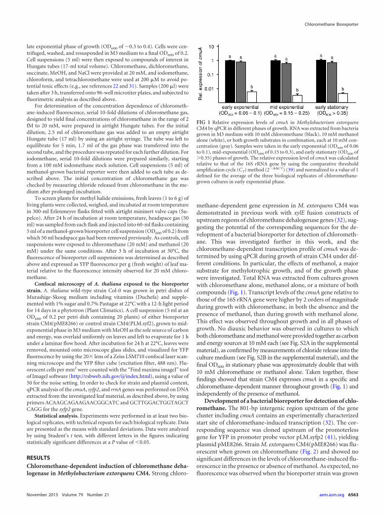
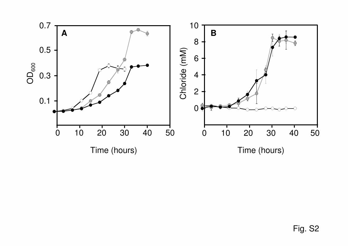
methane-dependent gene expression in M. extorquens CM4 wasdemonstrated in previous work with xylE fusion constructs ofupstream regions of chloromethane dehalogenase genes (32), sug-gesting the potential of the corresponding sequences for the de-velopment of a bacterial bioreporter for detection of chlorometh-ane. This was investigated further in this work, and thechloromethane-dependent transcription profile of cmuA was de-termined by using qPCR during growth of strain CM4 under dif-ferent conditions. In particular, the effects of methanol, a majorsubstrate for methylotrophic growth, and of the growth phasewere investigated. Total RNA was extracted from cultures grownwith chloromethane alone, methanol alone, or a mixture of bothcompounds (Fig. 1). Transcript levels of the cmuA gene relative tothose of the 16S rRNA gene were higher by 2 orders of magnitudeduring growth with chloromethane, in both the absence and thepresence of methanol, than during growth with methanol alone.This effect was observed throughout growth and in all phases ofgrowth. No diauxic behavior was observed in cultures to whichboth chloromethane and methanol were provided together as carbonand energy sources at 10 mM each (see Fig. S2A in the supplementalmaterial), as confirmed by measurements of chloride release into theculture medium (see Fig. S2B in the supplemental material), and thefinal OD600 in stationary phase was approximately double that with10 mM chloromethane or methanol alone. Taken together, thesefindings showed that strain CM4 expresses cmuA in a specific andchloromethane-dependent manner throughout growth (Fig. 1) andindependently of the presence of methanol.
Development of a bacterial bioreporter for detection of chlo-romethane. The 801-bp intergenic region upstream of the genecluster including cmuA contains an experimentally characterizedstart site of chloromethane-induced transcription (32). The cor-responding sequence was cloned upstream of the promoterlessgene for YFP in promoter probe vector pLM.syfp2 (41), yieldingplasmid pME8266. Strain M. extorquens CM4(pME8266) was flu-orescent when grown on chloromethane (Fig. 2) and showed nosignificant differences in the levels of chloromethane-induced flu-orescence in the presence or absence of methanol. As expected, nofluorescence was observed when the bioreporter strain was grown
FIG 1 Relative expression levels of cmuA in Methylobacterium extorquensCM4 by qPCR in different phases of growth. RNA was extracted from bacteriagrown in M3 medium with 10 mM chloromethane (black), 10 mM methanolalone (white), or both growth substrates in combination, each at 10 mM con-centration (gray). Samples were taken in the early exponential (OD600 of 0.06to 0.1), mid-exponential (OD600 of 0.15 to 0.3), and early stationary (OD600 of�0.35) phases of growth. The relative expression level of cmuA was calculatedrelative to that of the 16S rRNA gene by using the comparative thresholdamplification cycle (CT) method (2���CT) (39) and normalized to a value of 1defined for the average of the three biological replicates of chloromethane-grown cultures in early exponential phase.
Chloromethane Bioreporter
November 2013 Volume 79 Number 21 aem.asm.org 6563
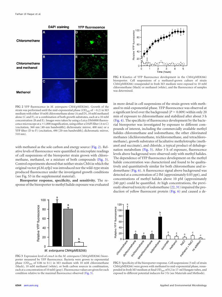
with methanol as the sole carbon and energy source (Fig. 2). Rel-ative levels of fluorescence were quantified in microplate readingsof cell suspensions of the bioreporter strain grown with chloro-methane, methanol, or a mixture of both compounds (Fig. 3).Control experiments showed that neither strain CM4 in which theoriginal vector pLM.syfp2 was introduced nor the wild-type strainproduced fluorescence under the investigated growth conditions(see Fig. S3 in the supplemental material).
Bioreporter response, specificity, and sensitivity. The re-sponse of the bioreporter to methyl halide exposure was evaluated
in more detail in cell suspensions of the strain grown with meth-anol to mid-exponential phase. YFP fluorescence was observed ata significant level over the background (P 0.009) within only 20min of exposure to chloromethane and stabilized after about 3 h(Fig. 4). The specificity of fluorescence development by the bacte-rial bioreporter was investigated by exposure to different com-pounds of interest, including the commercially available methylhalides chloromethane and iodomethane, the other chlorinatedmethanes (dichloromethane, trichloromethane, and tetrachloro-methane), growth substrates of facultative methylotrophs (meth-anol and succinate), and chloride, a typical product of dehaloge-nation metabolism (Fig. 5). After 3 h of exposure, fluorescencelevels above background were observed only with methyl halides.The dependence of YFP fluorescence development on the methylhalide concentration was characterized and found to be qualita-tively and quantitatively similar for both chloromethane and io-domethane (Fig. 6). A fluorescence signal above background wasdetected at a concentration of 2 fM (approximately 0.05 ppt), andconcentrations of methyl halides above 10 pM (approximately240 ppt) could be quantified. At high concentrations, the previ-ously observed toxicity of iodomethane (22, 31) impaired the pro-duction of yellow fluorescent protein (Fig. 6) and caused a de-
FIG 2 YFP fluorescence in M. extorquens CM4(pME8266). Growth of thestrain was performed until the mid-exponential phase (OD600 of �0.2) in M3medium with either 10 mM chloromethane alone (A and D), 10 mM methanolalone (C and F), or a combination of both growth substrates, each at a 10 mMconcentration (B and E). Images were taken by using a Leica DM4000 fluores-cence microscope at a �1,000 magnification, using either a DAPI filter (A to C)(excitation, 360 nm [40-nm bandwidth]; dichromatic mirror, 400 nm) or aYFP filter (D to F) (excitation, 490 [20-nm bandwidth]; dichromatic mirror,510 nm).
FIG 3 Expression level of cmuA in the M. extorquens CM4(pME8266) biore-porter measured by YFP fluorescence. Bacteria were grown to exponentialphase (OD600 of 0.06 to 0.1) in M3 medium with 10 mM chloromethane(black), 10 mM methanol (white), or both carbon sources in combination,each at a concentration of 10 mM (gray). Fluorescence values are given for eachcondition relative to the maximal fluorescence observed (Fig. 5).
FIG 4 Kinetics of YFP fluorescence development in the CM4(pME8266)bioreporter. Cell suspensions of a methanol-grown culture of strainCM4(pME8266) resuspended in fresh M3 medium were exposed to 10 mMchloromethane (black) or methanol (white), and the fluorescence of sampleswas determined.
FIG 5 Specificity of the bioreporter response. Cell suspensions (5 ml) of strainCM4(pME8266) were grown with methanol to mid-exponential phase, resus-pended in fresh M3 medium at final OD600 of 0.2 in 17-ml Hungate tubes, andexposed to different potential inducers for 3 h (see Materials and Methods).
Farhan Ul Haque et al.
6564 aem.asm.org Applied and Environmental Microbiology
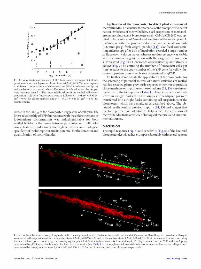
crease in the OD600 of the bioreporter, suggestive of cell lysis. Thelinear relationship of YFP fluorescence with the chloromethane oriodomethane concentration was indistinguishable for bothmethyl halides in the range between picomolar and millimolarconcentrations, underlining the high sensitivity and biologicalspecificity of the bioreporter and its potential for the detection andquantification of methyl halides.
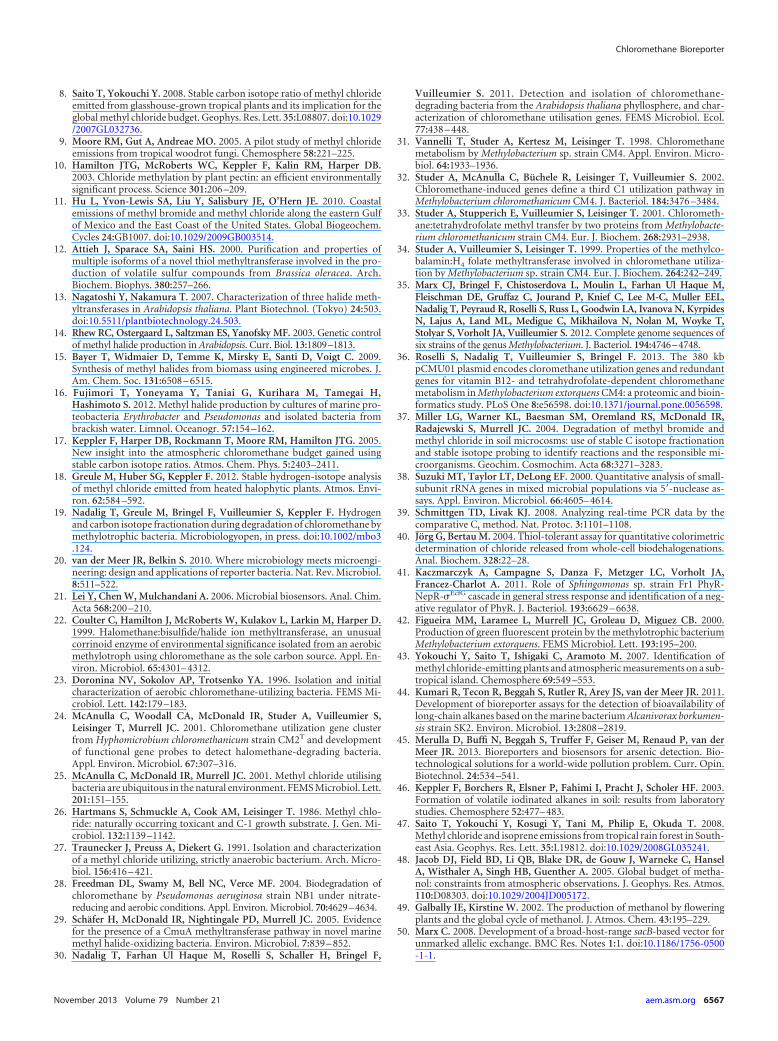
Application of the bioreporter to detect plant emissions ofmethyl halides. To visualize the potential of the bioreporter to detectnatural emissions of methyl halides, a cell suspension of methanol-grown, nonfluorescent bioreporter strain CM4(pME8266) was ap-plied to leaf surfaces of 2-week-old seedlings of the model plant A.thaliana, reported to produce chloromethane in small amounts(0.6 nmol per g [fresh weight] per day [14]). Confocal laser scan-ning microscopy after 24 h of incubation revealed a large numberof fluorescent cells on leaves, whereas no fluorescence was visiblewith the control isogenic strain with the original promotorlessYFP plasmid (Fig. 7). Fluorescence was evaluated quantitatively inplanta (Fig. 7) by counting the number of fluorescent cells permm2 relative to the copy number of the YFP gene for yellow flu-orescent protein present on leaves determined by qPCR.
To further demonstrate the applicability of the bioreporter forthe screening of potential sources of natural emissions of methylhalides, selected plants previously reported either not to producechloromethane or to produce chloromethane (14, 43) were inves-tigated with the bioreporter (Table 1). After incubation of freshleaves in airtight flasks for 24 h, samples of headspace gas weretransferred into airtight flasks containing cell suspensions of thebioreporter, which were analyzed as described above. The ob-tained results confirm previous reports (14, 43) and suggest thatthe bioreporter has potential to help screen for emissions ofmethyl halides from a variety of biological materials and environ-mental sources.
DISCUSSION
The rapid response (Fig. 4) and sensitivity (Fig. 6) of the bacterialbioreporter described here compare favorably with several reports
FIG 6 Concentration dependence of YFP fluorescence development. Cell sus-pensions of a methanol-grown culture of strain CM4(pME8266) were exposedto different concentrations of chloromethane (black), iodomethane (gray),and methanol as a control (white). Fluorescence (F) values for the sampleswere measured after 3 h. The linear relationships of the methyl halide con-centration ([c]) with fluorescence were as follows: F 108.66 � 5.53 [c](R2 0.99) for chloromethane and F 104.71 � 5.25 [c] (R2 0.93) foriodomethane.
FIG 7 Confocal laser microscopy of in planta methyl halide production of A. thaliana. Leaves of 2-week-old A. thaliana Col-0 seedlings were overlaid with equalvolumes of cell suspensions of the bioreporter strain CM4(pME8266) (A) and of the control strain CM4(pLM.syfp2) (B) at the same cell density, revealingfluorescent bioreporter bacteria (green) overlaying the plant leaf (red autofluorescence is from chlorophyll). Copy numbers of the YFP and cmuA genesdetermined by qPCR were closely similar for both bacterial strains (see Table 1 in the supplemental material), whereas numbers of fluorescent cells per mm2
determined by ImageJ analysis were 2,410 970 and 150 120 for the bioreporter and control strains, respectively.
Chloromethane Bioreporter
November 2013 Volume 79 Number 21 aem.asm.org 6565

on the development of bioreporters in recent literature. For exam-ple, a bioreporter assay for detection of various alkanes, based onthe production of an enhanced green fluorescent protein, was re-ported to require between 6 h and 5 days (44). Similarly, knownbioreporters for arsenic are either rapid with moderate sensitivityranges or slower with high sensitivity limits (45). The linearity ofthe response of the methyl halide bioreporter over a wide range ofconcentrations (Fig. 6) is also noteworthy. For chloromethane,the threshold concentration for quantification is similar to thedetection limit of analytical GC-MS methods (100 ppt, i.e., ap-proximately 4 pM), which most often involve sample preconcen-tration (see, e.g., reference 14). For iodomethane, however, ana-lytical chemical methods are more sensitive (approximately 6 ppt[about 0.25 pM] [see, e.g., references 14 and 46]).
The robustness of the developed methyl halide biosensor wassupported by the demonstration, following up on previous studies(31, 32), of chloromethane-dependent transcription of the cmuAgene and expression of chloromethane dehalogenase throughoutgrowth in strain CM4 (Fig. 1; see also Fig. S2 in the supplementalmaterial). The underlying mechanism for this methyl halide-spe-cific regulation remains unknown, and the implication of putativeregulator genes found in the vicinity of cmu genes (30) still needsto be experimentally investigated. Nevertheless, the observed lackof discrimination of bioreporter strain CM4(pME8266) betweendifferent methyl halides was expected, since chloromethane deha-logenase transforms the higher-molecular-weight methyl halidesbromomethane and iodomethane as well as chloromethane (31).Global sources of chloromethane (4.1 to 4.4 Tg year�1 [4]) arelarger by at least an order of magnitude than those of iodomethane(approximately 550 Gg year�1) and bromomethane (approxi-mately 110 Gg year�1) (2, 6). Thus, the use of the bioreporter inscreening for methyl halide emissions in natural environmentswill mainly inform on emissions of chloromethane as the majormethyl halide produced. In particular, terrestrial biomes contrib-ute little to the global budget of CH3I (33 Gg year�1 [2]), withterrestrial plants alone generating 2,200 Gg year�1 chloromethane(2, 4).
As suggested by initial data of the present study (Table 1), the
laboratory screening of plants and various types of vegetation forwhich emissions of chloromethane are not yet known representsan attractive application for the bioreporter. Only few studies sofar have identified plants which emit chloromethane (43, 47), de-spite the fact that vegetation is likely the main contributor toglobal emissions of chloromethane to the atmosphere (2). Giventhat methanol is the most important carbonaceous compoundemitted by vegetation (approximately 100 Tg year�1 [48, 49], i.e.,10- to 100-fold those of chloromethane), the fact that bioreporterfluorescence is not affected by methanol appears most valuable.Moreover, envisaged applications of the bioreporter foresee its usefor experiments in a laboratory setting and on a short time scale(Table 1). Since the antibiotic kanamycin is required for long-term stability of the reporter plasmid, the use of this system inenvironmental settings may require further developments, e.g., bymarkerless chromosomal integration of the reporter system instrain CM4 using methodology established for Methylobacterium(50).
In conclusion, the bioreporter developed in the present workmay represent a useful laboratory tool to increase our knowledgeof natural sources of methyl halides and thereby contribute toconsolidating corresponding global budgets.
ACKNOWLEDGMENTS
We thank Lisa Metzger and Julia Vorholt (ETH Zürich) for providingplasmid pLM.syfp2, Jérôme Mutterer (Institut de Biologie Moléculairedes Plantes, Strasbourg) for help with confocal microscopy, and GisèleHaan-Archipoff (herbarium, Université de Strasbourg) and PhilippeObliger (botanical garden, Université de Strasbourg) for plant material.
We gratefully acknowledge support from the Higher Education Com-mission of Pakistan, in the form of a Ph.D. fellowship to M.F.U.H; fromthe CNRS EC2CO program (2010 to 2011); and from REALISE, the AlsaceNetwork of Laboratories in Environmental Sciences and Engineering(http://realise.unistra.fr/).
REFERENCES1. Penkett SA, Derwent RG, Fabian P, Borchers R, Schmidt U. 1980.
Methyl chloride in the stratosphere. Nature 283:58 – 60.2. Montzka SA, Reimann S, Engel A, Krüger K, O’Doherty S, Sturges WT,
Blake D, Dorf M, Fraser P, Froidevaux L, Jucks K, Kreher K, KuryloMJ, Mellouki A, Miller J, Nielsen O-J, Orkin VL, Prinn RG, Rhew R,Santee ML, Stohl A, Verdonik D. 2011. Scientific assessment of ozonedepletion: 2010. Global ozone research and monitoring project, report no52, chapter 1, p 1– 86. World Meteorological Organization, Geneva, Swit-zerland.
3. O’Dowd CD, Jimenez JL, Bahreini R, Flagan RC, Seinfeld JH, HameriK, Pirjola L, Kulmala M, Jennings SG, Hoffmann T. 2002. Marineaerosol formation from biogenic iodine emissions. Nature 417:632– 636.
4. Xiao X, Prinn RG, Fraser PJ, Simmonds PG, Weiss RF, O’Doherty S,Miller BR, Salameh PK, Harth CM, Krummel PB, Porter LW, MuehleJ, Greally BR, Cunnold D, Wang R, Montzka SA, Elkins JW, DuttonGS, Thompson TM, Butler JH, Hall BD, Reimann S, Vollmer MK,Stordal F, Lunder C, Maione M, Arduini J, Yokouchi Y. 2010. Optimalestimation of the surface fluxes of methyl chloride using a 3-D globalchemical transport model. Atmos. Chem. Phys. 10:5515–5533.
5. Yoshida Y, Wang Y, Zeng T, Yantosca R. 2004. A three-dimensionalglobal model study of atmospheric methyl chloride budget and distribu-tions. J. Geophys. Res. Atmos. 109:D24309. doi:10.1029/2004JD004951.
6. Clerbaux C, Cunnold DM, Anderson J, Engel A, Fraser PJ, Mahieu E,Manning A, Miller J, Montzka SA, Nassar R, Prinn R, Reimann S,Rinsland CP, Simmonds P, Verdonik D, Weiss R, Wuebbles D, Yok-ouchi Y. 2007. Scientific assessment of ozone depletion: 2006. Globalozone research and monitoring project, report no 50, chapter 1. WorldMeteorological Organization, Geneva, Switzerland.
7. Yokouchi Y, Ikeda M, Inuzuka Y, Yukawa T. 2002. Strong emission ofmethyl chloride from tropical plants. Nature 416:163–165.
TABLE 1 Identification of methyl halide-emitting plants using thebioreporter
Plant species or control
Relative bioreporterfluorescencea
([g {fresh wt}]�1)
Reported CH3Clemission(ng [g {dry wt}]�1 h�1)
Plant speciesVitex rotundifolia 52 2,800b
Hoya carnosa 10 Negativeb
Codiaeum variegatum 8 Negativeb
Arabidopsis thaliana 24 12.6c
Controls20 mM CH3Cl 10020 mM methanol 11
a Relative to the fluorescence (set to a value of 100) observed after exposure of thebioreporter to 20 mM CH3Cl (see Materials and Methods for details). Data from arepresentative experiment are shown.b Data were taken from reference 43. Plants were scored negative for emissions below 10ng (g [dry weight])�1 h�1.c Data derived from reference 14, converted from a reported value of 0.6 nmol [g (freshweight)]�1 day�1, assuming that dry weight represents 10% of the fresh weight.
Farhan Ul Haque et al.
6566 aem.asm.org Applied and Environmental Microbiology
8. Saito T, Yokouchi Y. 2008. Stable carbon isotope ratio of methyl chlorideemitted from glasshouse-grown tropical plants and its implication for theglobal methyl chloride budget. Geophys. Res. Lett. 35:L08807. doi:10.1029/2007GL032736.
9. Moore RM, Gut A, Andreae MO. 2005. A pilot study of methyl chlorideemissions from tropical woodrot fungi. Chemosphere 58:221–225.
10. Hamilton JTG, McRoberts WC, Keppler F, Kalin RM, Harper DB.2003. Chloride methylation by plant pectin: an efficient environmentallysignificant process. Science 301:206 –209.
11. Hu L, Yvon-Lewis SA, Liu Y, Salisbury JE, O’Hern JE. 2010. Coastalemissions of methyl bromide and methyl chloride along the eastern Gulfof Mexico and the East Coast of the United States. Global Biogeochem.Cycles 24:GB1007. doi:10.1029/2009GB003514.
12. Attieh J, Sparace SA, Saini HS. 2000. Purification and properties ofmultiple isoforms of a novel thiol methyltransferase involved in the pro-duction of volatile sulfur compounds from Brassica oleracea. Arch.Biochem. Biophys. 380:257–266.
13. Nagatoshi Y, Nakamura T. 2007. Characterization of three halide meth-yltransferases in Arabidopsis thaliana. Plant Biotechnol. (Tokyo) 24:503.doi:10.5511/plantbiotechnology.24.503.
14. Rhew RC, Ostergaard L, Saltzman ES, Yanofsky MF. 2003. Genetic controlof methyl halide production in Arabidopsis. Curr. Biol. 13:1809–1813.
15. Bayer T, Widmaier D, Temme K, Mirsky E, Santi D, Voigt C. 2009.Synthesis of methyl halides from biomass using engineered microbes. J.Am. Chem. Soc. 131:6508 – 6515.
16. Fujimori T, Yoneyama Y, Taniai G, Kurihara M, Tamegai H,Hashimoto S. 2012. Methyl halide production by cultures of marine pro-teobacteria Erythrobacter and Pseudomonas and isolated bacteria frombrackish water. Limnol. Oceanogr. 57:154 –162.
17. Keppler F, Harper DB, Rockmann T, Moore RM, Hamilton JTG. 2005.New insight into the atmospheric chloromethane budget gained usingstable carbon isotope ratios. Atmos. Chem. Phys. 5:2403–2411.
18. Greule M, Huber SG, Keppler F. 2012. Stable hydrogen-isotope analysisof methyl chloride emitted from heated halophytic plants. Atmos. Envi-ron. 62:584 –592.
19. Nadalig T, Greule M, Bringel F, Vuilleumier S, Keppler F. Hydrogenand carbon isotope fractionation during degradation of chloromethane bymethylotrophic bacteria. Microbiologyopen, in press. doi:10.1002/mbo3.124.
20. van der Meer JR, Belkin S. 2010. Where microbiology meets microengi-neering: design and applications of reporter bacteria. Nat. Rev. Microbiol.8:511–522.
21. Lei Y, Chen W, Mulchandani A. 2006. Microbial biosensors. Anal. Chim.Acta 568:200 –210.
22. Coulter C, Hamilton J, McRoberts W, Kulakov L, Larkin M, Harper D.1999. Halomethane:bisulfide/halide ion methyltransferase, an unusualcorrinoid enzyme of environmental significance isolated from an aerobicmethylotroph using chloromethane as the sole carbon source. Appl. En-viron. Microbiol. 65:4301– 4312.
23. Doronina NV, Sokolov AP, Trotsenko YA. 1996. Isolation and initialcharacterization of aerobic chloromethane-utilizing bacteria. FEMS Mi-crobiol. Lett. 142:179 –183.
24. McAnulla C, Woodall CA, McDonald IR, Studer A, Vuilleumier S,Leisinger T, Murrell JC. 2001. Chloromethane utilization gene clusterfrom Hyphomicrobium chloromethanicum strain CM2T and developmentof functional gene probes to detect halomethane-degrading bacteria.Appl. Environ. Microbiol. 67:307–316.
25. McAnulla C, McDonald IR, Murrell JC. 2001. Methyl chloride utilisingbacteria are ubiquitous in the natural environment. FEMS Microbiol. Lett.201:151–155.
26. Hartmans S, Schmuckle A, Cook AM, Leisinger T. 1986. Methyl chlo-ride: naturally occurring toxicant and C-1 growth substrate. J. Gen. Mi-crobiol. 132:1139 –1142.
27. Traunecker J, Preuss A, Diekert G. 1991. Isolation and characterizationof a methyl chloride utilizing, strictly anaerobic bacterium. Arch. Micro-biol. 156:416 – 421.
28. Freedman DL, Swamy M, Bell NC, Verce MF. 2004. Biodegradation ofchloromethane by Pseudomonas aeruginosa strain NB1 under nitrate-reducing and aerobic conditions. Appl. Environ. Microbiol. 70:4629–4634.
29. Schäfer H, McDonald IR, Nightingale PD, Murrell JC. 2005. Evidencefor the presence of a CmuA methyltransferase pathway in novel marinemethyl halide-oxidizing bacteria. Environ. Microbiol. 7:839 – 852.
30. Nadalig T, Farhan Ul Haque M, Roselli S, Schaller H, Bringel F,
Vuilleumier S. 2011. Detection and isolation of chloromethane-degrading bacteria from the Arabidopsis thaliana phyllosphere, and char-acterization of chloromethane utilisation genes. FEMS Microbiol. Ecol.77:438 – 448.
31. Vannelli T, Studer A, Kertesz M, Leisinger T. 1998. Chloromethanemetabolism by Methylobacterium sp. strain CM4. Appl. Environ. Micro-biol. 64:1933–1936.
32. Studer A, McAnulla C, Büchele R, Leisinger T, Vuilleumier S. 2002.Chloromethane-induced genes define a third C1 utilization pathway inMethylobacterium chloromethanicum CM4. J. Bacteriol. 184:3476 –3484.
33. Studer A, Stupperich E, Vuilleumier S, Leisinger T. 2001. Chlorometh-ane:tetrahydrofolate methyl transfer by two proteins from Methylobacte-rium chloromethanicum strain CM4. Eur. J. Biochem. 268:2931–2938.
34. Studer A, Vuilleumier S, Leisinger T. 1999. Properties of the methylco-balamin:H4 folate methyltransferase involved in chloromethane utiliza-tion by Methylobacterium sp. strain CM4. Eur. J. Biochem. 264:242–249.
35. Marx CJ, Bringel F, Chistoserdova L, Moulin L, Farhan Ul Haque M,Fleischman DE, Gruffaz C, Jourand P, Knief C, Lee M-C, Muller EEL,Nadalig T, Peyraud R, Roselli S, Russ L, Goodwin LA, Ivanova N, KyrpidesN, Lajus A, Land ML, Medigue C, Mikhailova N, Nolan M, Woyke T,Stolyar S, Vorholt JA, Vuilleumier S. 2012. Complete genome sequences ofsix strains of the genus Methylobacterium. J. Bacteriol. 194:4746–4748.
36. Roselli S, Nadalig T, Vuilleumier S, Bringel F. 2013. The 380 kbpCMU01 plasmid encodes cloromethane utilization genes and redundantgenes for vitamin B12- and tetrahydrofolate-dependent chloromethanemetabolism in Methylobacterium extorquens CM4: a proteomic and bioin-formatics study. PLoS One 8:e56598. doi:10.1371/journal.pone.0056598.
37. Miller LG, Warner KL, Baesman SM, Oremland RS, McDonald IR,Radajewski S, Murrell JC. 2004. Degradation of methyl bromide andmethyl chloride in soil microcosms: use of stable C isotope fractionationand stable isotope probing to identify reactions and the responsible mi-croorganisms. Geochim. Cosmochim. Acta 68:3271–3283.
38. Suzuki MT, Taylor LT, DeLong EF. 2000. Quantitative analysis of small-subunit rRNA genes in mixed microbial populations via 5=-nuclease as-says. Appl. Environ. Microbiol. 66:4605– 4614.
39. Schmittgen TD, Livak KJ. 2008. Analyzing real-time PCR data by thecomparative Ct method. Nat. Protoc. 3:1101–1108.
40. Jörg G, Bertau M. 2004. Thiol-tolerant assay for quantitative colorimetricdetermination of chloride released from whole-cell biodehalogenations.Anal. Biochem. 328:22–28.
41. Kaczmarczyk A, Campagne S, Danza F, Metzger LC, Vorholt JA,Francez-Charlot A. 2011. Role of Sphingomonas sp. strain Fr1 PhyR-NepR-�EcfG cascade in general stress response and identification of a neg-ative regulator of PhyR. J. Bacteriol. 193:6629 – 6638.
42. Figueira MM, Laramee L, Murrell JC, Groleau D, Miguez CB. 2000.Production of green fluorescent protein by the methylotrophic bacteriumMethylobacterium extorquens. FEMS Microbiol. Lett. 193:195–200.
43. Yokouchi Y, Saito T, Ishigaki C, Aramoto M. 2007. Identification ofmethyl chloride-emitting plants and atmospheric measurements on a sub-tropical island. Chemosphere 69:549 –553.
44. Kumari R, Tecon R, Beggah S, Rutler R, Arey JS, van der Meer JR. 2011.Development of bioreporter assays for the detection of bioavailability oflong-chain alkanes based on the marine bacterium Alcanivorax borkumen-sis strain SK2. Environ. Microbiol. 13:2808 –2819.
45. Merulla D, Buffi N, Beggah S, Truffer F, Geiser M, Renaud P, van derMeer JR. 2013. Bioreporters and biosensors for arsenic detection. Bio-technological solutions for a world-wide pollution problem. Curr. Opin.Biotechnol. 24:534 –541.
46. Keppler F, Borchers R, Elsner P, Fahimi I, Pracht J, Scholer HF. 2003.Formation of volatile iodinated alkanes in soil: results from laboratorystudies. Chemosphere 52:477– 483.
47. Saito T, Yokouchi Y, Kosugi Y, Tani M, Philip E, Okuda T. 2008.Methyl chloride and isoprene emissions from tropical rain forest in South-east Asia. Geophys. Res. Lett. 35:L19812. doi:10.1029/2008GL035241.
48. Jacob DJ, Field BD, Li QB, Blake DR, de Gouw J, Warneke C, HanselA, Wisthaler A, Singh HB, Guenther A. 2005. Global budget of metha-nol: constraints from atmospheric observations. J. Geophys. Res. Atmos.110:D08303. doi:10.1029/2004JD005172.
49. Galbally IE, Kirstine W. 2002. The production of methanol by floweringplants and the global cycle of methanol. J. Atmos. Chem. 43:195–229.
50. Marx C. 2008. Development of a broad-host-range sacB-based vector forunmarked allelic exchange. BMC Res. Notes 1:1. doi:10.1186/1756-0500-1-1.
Chloromethane Bioreporter
November 2013 Volume 79 Number 21 aem.asm.org 6567
Supplemental Material to
“A fluorescence-based bacterial bioreporter for the specific detection of methyl halide emissions
in the environment” (AEM01738-13)
Table S1, Fig. S1, Fig. S2 Fig. S3
Authors
Muhammad Farhan Ul Haque1, Thierry Nadalig
1, Françoise Bringel
1, Hubert Schaller
2,
Stéphane Vuilleumier1*
Affiliations
1 Université de Strasbourg, Equipe Adaptations et Interactions Microbiennes dans
l'Environnement, Département Microorganismes, Génomes, Environnement, UMR 7156 UdS –
CNRS Génétique Moléculaire, Génomique, Microbiologie, Strasbourg, France
2 Département Réseaux Métaboliques Végétaux, Institut de Biologie Moléculaire des Plantes,
UPR 2357 CNRS, Strasbourg (France)
* Corresponding author:
Stéphane Vuilleumier, Université de Strasbourg, UMR 7156 UdS - CNRS, 28 rue Goethe, F-
67083 Strasbourg Cedex, France, phone: +33-3-68-85-20-22; fax: +33-3-68-85-19-26; e-mail:
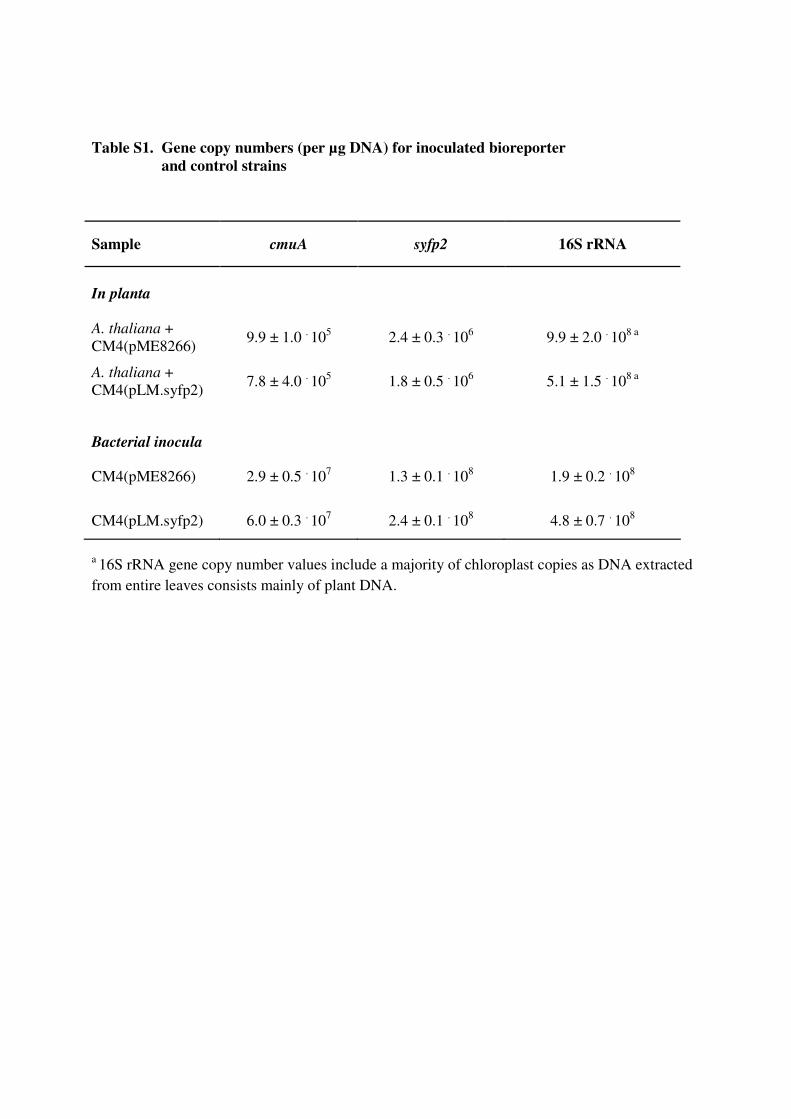
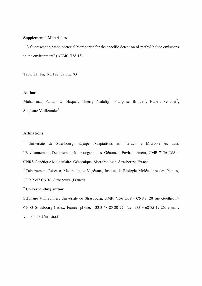
Table S1. Gene copy numbers (per µg DNA) for inoculated bioreporter
and control strains
Sample cmuA syfp2 16S rRNA
In planta
A. thaliana +
CM4(pME8266) 9.9 ± 1.0 . 105 2.4 ± 0.3 . 106 9.9 ± 2.0 . 108 a
A. thaliana +
CM4(pLM.syfp2) 7.8 ± 4.0
. 10
5 1.8 ± 0.5
. 10
6 5.1 ± 1.5
. 10
8 a
Bacterial inocula
CM4(pME8266) 2.9 ± 0.5 . 107 1.3 ± 0.1 . 108 1.9 ± 0.2 . 108
CM4(pLM.syfp2) 6.0 ± 0.3 . 10
7 2.4 ± 0.1
. 10
8 4.8 ± 0.7
. 10
8
a 16S rRNA gene copy number values include a majority of chloroplast copies as DNA extracted
from entire leaves consists mainly of plant DNA.
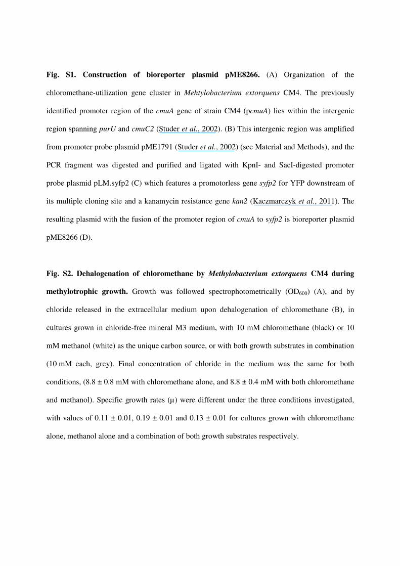
Fig. S1. Construction of bioreporter plasmid pME8266. (A) Organization of the
chloromethane-utilization gene cluster in Mehtylobacterium extorquens CM4. The previously
identified promoter region of the cmuA gene of strain CM4 (pcmuA) lies within the intergenic
region spanning purU and cmuC2 (Studer et al., 2002). (B) This intergenic region was amplified
from promoter probe plasmid pME1791 (Studer et al., 2002) (see Material and Methods), and the
PCR fragment was digested and purified and ligated with KpnI- and SacI-digested promoter
probe plasmid pLM.syfp2 (C) which features a promotorless gene syfp2 for YFP downstream of
its multiple cloning site and a kanamycin resistance gene kan2 (Kaczmarczyk et al., 2011). The
resulting plasmid with the fusion of the promoter region of cmuA to syfp2 is bioreporter plasmid
pME8266 (D).
Fig. S2. Dehalogenation of chloromethane by Methylobacterium extorquens CM4 during
methylotrophic growth. Growth was followed spectrophotometrically (OD600) (A), and by
chloride released in the extracellular medium upon dehalogenation of chloromethane (B), in
cultures grown in chloride-free mineral M3 medium, with 10 mM chloromethane (black) or 10
mM methanol (white) as the unique carbon source, or with both growth substrates in combination
(10 mM each, grey). Final concentration of chloride in the medium was the same for both
conditions, (8.8 ± 0.8 mM with chloromethane alone, and 8.8 ± 0.4 mM with both chloromethane
and methanol). Specific growth rates (µ) were different under the three conditions investigated,
with values of 0.11 ± 0.01, 0.19 ± 0.01 and 0.13 ± 0.01 for cultures grown with chloromethane
alone, methanol alone and a combination of both growth substrates respectively.
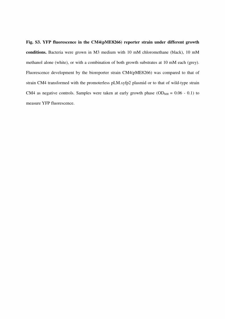
Fig. S3. YFP fluorescence in the CM4(pME8266) reporter strain under different growth
conditions. Bacteria were grown in M3 medium with 10 mM chloromethane (black), 10 mM
methanol alone (white), or with a combination of both growth substrates at 10 mM each (grey).
Fluorescence development by the bioreporter strain CM4(pME8266) was compared to that of
strain CM4 transformed with the promoterless pLM.syfp2 plasmid or to that of wild-type strain
CM4 as negative controls. Samples were taken at early growth phase (OD600 = 0.06 - 0.1) to
measure YFP fluorescence.
pLM_syfp2
5580 bp
syfp2
kan2
trfA
oriT
traJ’
oriV
ColE1
SacI KpnIpcmuA
(A)
(B)
(C) (D)
Intergenic region amplified
from pME1791 (Studer et al., 2002)
pME8266
6330 bp
syfp2
kan2
trfA
oriT
traJ’
oriV
ColE1
AfIII
NspI
Eco53kI
SacI
SpeI
NcoI
Acc65I
Kp
nI
cmuAcmuC2purUfolD
pcmuA
SacI KpnIpcmuA
Fig. S1
Time (hours)
0.1
0.3
0.5
0.7
0 10 20 30 40 50
AO
D600
B
Fig. S2
0
2
4
6
8
10
0 10 20 30 40 50
Ch
lori
de
(m
M)
Time (hours)
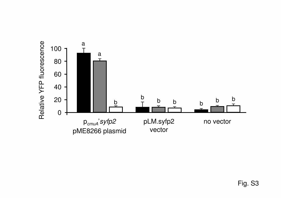
Fig. S3
Re
lative
YF
P f
luo
resce
nce
no vectorpLM.syfp2
vector
pcmuA’syfp2
pME8266 plasmid
100
20
0
40
60
80
a
a
bb
b b bb b