FL4409:Layout 1.qxd
10
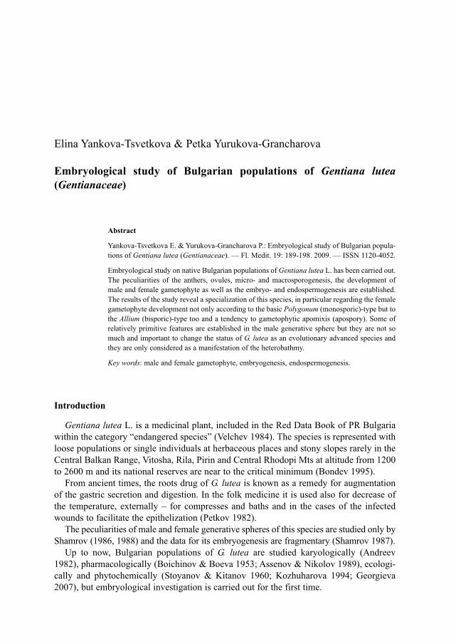
Elina Yankova-Tsvetkova & Petka Yurukova-Grancharova Embryological study of Bulgarian populations of Gentiana lutea (Gentianaceae) Abstract Yankova-Tsvetkova E. & Yurukova-Grancharova P.: Embryological study of Bulgarian popula- tions of Gentiana lutea (Gentianaceae). — Fl. Medit. 19: 189-198. 2009. — ISSN 1120-4052. Embryological study on native Bulgarian populations of Gentiana lutea L. has been carried out. The peculiarities of the anthers, ovules, micro- and macrosporogenesis, the development of male and female gametophyte as well as the embryo- and endospermogenesis are established. The results of the study reveal a specialization of this species, in particular regarding the female gametophyte development not only according to the basic Polygonum (monosporic)-type but to the Allium (bisporic)-type too and a tendency to gametophytic apomixis (apospory). Some of relatively primitive features are established in the male generative sphere but they are not so much and important to change the status of G. lutea as an evolutionary advanced species and they are only considered as a manifestation of the heterobathmy. Key words: male and female gametophyte, embryogenesis, endospermogenesis. Introduction Gentiana lutea L. is a medicinal plant, included in the Red Data Book of PR Bulgaria within the category “endangered species” (Velchev 1984). The species is represented with loose populations or single individuals at herbaceous places and stony slopes rarely in the Central Balkan Range, Vitosha, Rila, Pirin and Central Rhodopi Mts at altitude from 1200 to 2600 m and its national reserves are near to the critical minimum (Bondev 1995). From ancient times, the roots drug of G. lutea is known as a remedy for augmentation of the gastric secretion and digestion. In the folk medicine it is used also for decrease of the temperature, externally – for compresses and baths and in the cases of the infected wounds to facilitate the epithelization (Petkov 1982). The peculiarities of male and female generative spheres of this species are studied only by Shamrov (1986, 1988) and the data for its embryogenesis are fragmentary (Shamrov 1987). Up to now, Bulgarian populations of G. lutea are studied karyologically (Andreev 1982), pharmacologically (Boichinov & Boeva 1953; Assenov & Nikolov 1989), ecologi- cally and phytochemically (Stoyanov & Kitanov 1960; Kozhuharova 1994; Georgieva 2007), but embryological investigation is carried out for the first time.
-
Upload
khangminh22 -
Category
Documents
-
view
0 -
download
0
Transcript of FL4409:Layout 1.qxd
Elina Yankova-Tsvetkova & Petka Yurukova-Grancharova
Embryological study of Bulgarian populations of Gentiana lutea(Gentianaceae)
Abstract
Yankova-Tsvetkova E. & Yurukova-Grancharova P.: Embryological study of Bulgarian popula-tions of Gentiana lutea (Gentianaceae). — Fl. Medit. 19: 189-198. 2009. — ISSN 1120-4052.
Embryological study on native Bulgarian populations of Gentiana lutea L. has been carried out.The peculiarities of the anthers, ovules, micro- and macrosporogenesis, the development ofmale and female gametophyte as well as the embryo- and endospermogenesis are established.The results of the study reveal a specialization of this species, in particular regarding the femalegametophyte development not only according to the basic Polygonum (monosporic)-type but tothe Allium (bisporic)-type too and a tendency to gametophytic apomixis (apospory). Some ofrelatively primitive features are established in the male generative sphere but they are not somuch and important to change the status of G. lutea as an evolutionary advanced species andthey are only considered as a manifestation of the heterobathmy.
Key words: male and female gametophyte, embryogenesis, endospermogenesis.
Introduction
Gentiana lutea L. is a medicinal plant, included in the Red Data Book of PR Bulgariawithin the category “endangered species” (Velchev 1984). The species is represented withloose populations or single individuals at herbaceous places and stony slopes rarely in theCentral Balkan Range, Vitosha, Rila, Pirin and Central Rhodopi Mts at altitude from 1200to 2600 m and its national reserves are near to the critical minimum (Bondev 1995).
From ancient times, the roots drug of G. lutea is known as a remedy for augmentationof the gastric secretion and digestion. In the folk medicine it is used also for decrease ofthe temperature, externally – for compresses and baths and in the cases of the infectedwounds to facilitate the epithelization (Petkov 1982).
The peculiarities of male and female generative spheres of this species are studied only byShamrov (1986, 1988) and the data for its embryogenesis are fragmentary (Shamrov 1987).
Up to now, Bulgarian populations of G. lutea are studied karyologically (Andreev1982), pharmacologically (Boichinov & Boeva 1953; Assenov & Nikolov 1989), ecologi-cally and phytochemically (Stoyanov & Kitanov 1960; Kozhuharova 1994; Georgieva2007), but embryological investigation is carried out for the first time.
In the present work, as a part of complete embryological study undertaken on theBulgarian representatives of the genus Gentiana L., the results of embryological investi-gation on G. lutea are given.
Material and methods
Material of the study (flower buds and flowers at different age) has been collected fromfive native populations of G. lutea (Vitosha Mt, at the peak Golyam Rezen; Rhodopi Mt(Western), the village Kesten; Rila Mt, above the Suhoto lake; Pirin Mt, Banderitsa moun-tain hostel; Balkan Range (Western), Kozya stena summit) and fixed in FAA mixture (for-malin: glacial acetic acid: 70 % ethanol in correlation 5:5:90 parts). The serial paraffin cutswith thickness 12-25 mm, maded with rotary microtome are stained with Heidenhain’shaematoxylin (Rajan S. Sundara 2000). The glass made slides are embedded in Canadabalsam. The observations have been carried out with “Amplival” light microscope and themicrophotographs are made with digital camera “Canon”.
Results
The embryological features are basically the same in all studied populations of G. lutea,unless in the following descriptions particular comments are given.
Male generative sphereThe anthers are tetrasporangiate. The anther’s wall formation conforms to the
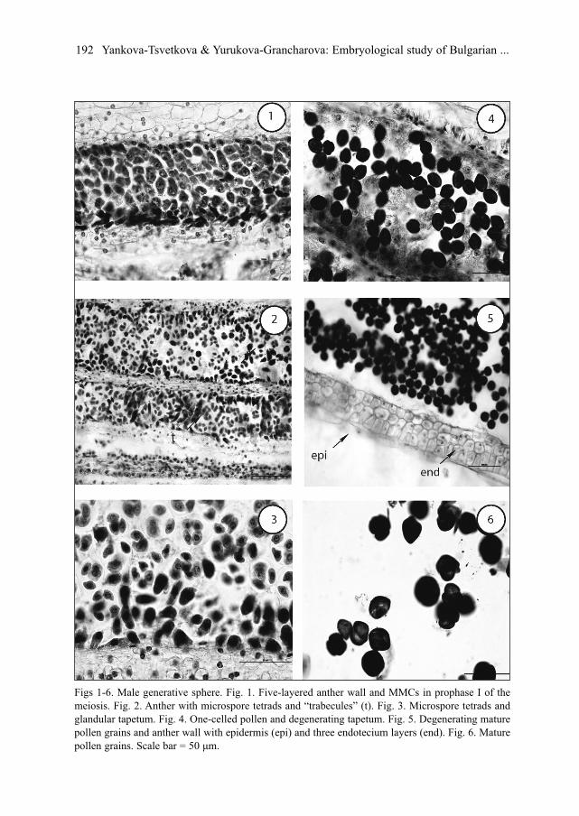
Dicotyledonous-type (Davis 1966) and consists of epidermis, endothecium, two middlelayers and glandular tapetum (Fig. 1) that are clearly distinguished during the heterotypicdivision of the meiosis in microspore mother cells (MMCs).
The epidermal cells clearly enlarge tangentially during the anther ontogenesis. The cellsof the initially one-layered endothecium divide in radial direction, forming two additionalrows that develop typical fibrous thickenings after the formation of microspore tetrads. Incomparison with the other layers of the anther wall, the epidermis and endothecium preservelong time, remaining entire and vital up to the mature pollen stage (Fig. 5). The cells of thetwo middle layers become flattened, pressed from the growing cells of the endothecium andtapetum and degenerate to the end of heterotypic division of the meiosis in MMCs.
During the anther ontogenesis, the glandular tapetum undergoes some changes. As aresult of a mitotic division its initially one-nucleate cells become two-nucleate. The tape-tum cells enlarge and intrude into the anther locules to form “placentoids” (”trabecules”)that separate the sporogenous tissue and later on the microspore tetrads (Fig. 2). After that,the tapetum cells, disintegrate, undergo a lysis and penetrate between the microsporetetrads, forming together unusual mass in which the individual protoplasts are clearly dis-tinguished (Fig. 4). The tapetum begins to degenerate and at the stage of mature pollengrains even its traces are not observed. The wall of the mature anthers in G. lutea consistsof the persistent epidermis and fibrous endothecium.
Sporogenous tissue is 3-, 4-layered and its consisting cells initially are tightly packed toeach other. Later on, they grow, round up, separate from each other and differentiate into
190 Yankova-Tsvetkova & Yurukova-Grancharova: Embryological study of Bulgarian ...
MMCs (Fig. 1). Meiosis runs normally and only insignificant deviations are registered, as:single lagging chromosomes; chromosomes scattered along the division spindle; asym-metrical disposition of the spindles during homeotypic division of the meiosis in MMCs.As a result of a simultaneous microsporogenesis predominantly tetrahedral, isobilateral, T-shaped, rarely decussate and single linear tetrads are formed (Fig. 3).
Soon after their formation, the tetrads disintegrate and each of the microspores differ-entiates into young one-celled pollen grain. The mature pollen is 3-celled at the time ofanther dehiscence (Fig. 6). During the development of male gametophyte some degenerat-ing processes are observed predominantly at 2- or 3-celled pollen stage in different amountin the studied populations but more clearly expressed in the population from Pirin Mt.
Female generative sphereWithin the one-loculate superior ovary of G. lutea, the ovules arranged in two rows form
(occasionally numbering as 50-60). The mature ovule is anatropous, tenuinucellate, unitegmic with a short funiculus and
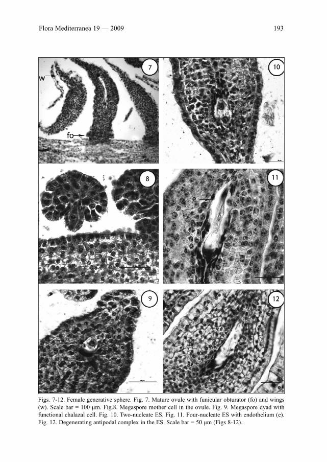
deep and narrow micropyle. During the early stages of its development, a funicular obtu-rator forms (Fig. 7). It is clearly distinguished from the other somatic cells of the ovule,consisting of radial enlarged cells that grow toward to the tip of the funiculus. Except theobturator, other specialized structures like epistase and hypostase are not found but weobserved another specific feature of the ovule, so called “wings” that form at its raphal,chalazal and anti-raphal sides (Fig. 7).
The tenuinucellate ovule possesses the nucellar epidermis with a single row of cells andone-celled archesporium. Its degeneration begins during the early stages of the embryo sac(ES) development. At the four-nucleate ES stage no traces of the nucellus are observed. Inthe mature ovule, the single integument is vigorous, comprising up to 15 rows of cells. Thecells of its outermost layer are one-nucleate, bigger than the other somatic cells and dur-ing ovule’s ontogenesis strongly lengthen radially. At the stage of four-nucleate ES, fromthe innermost layer of a single integument, an endothelium differentiates and its cells aremore clearly lengthened in tangential than radial direction (Fig. 11).
In the young ovule, one-celled archesporium is formed hypodermally (Fig. 8).Archesporogenesis runs without formation of parietal cells. The single archesporial cellenlarges and functions directly as megaspore mother cell (MMC). After meiosis in it, a lin-ear megaspore tetrad forms in the ovule. The chalazal megaspore becomes functional asembryo sac mother cell and successively develops after three mitotic divisions into two-,four- (Figs 10 and 11) and finally eight-nucleate ES. In this way, the ES formation followsthe Polygonum (monosporic)-type with three-celled egg apparatus, two polar nuclei andthree-celled antipodal complex. In the population from Rila Mt we observed in someovules the formation of a megaspore dyad instead of tetrad (Fig. 9). In these cases the ESdevelopment follows the Allium (bisporic)-type from the chalazal megaspore of the dyad.
At the beginning of the mature ES differentiation, the cells of egg apparatus appear mor-phologically similar. Subsequently the egg cell and the synergids become pyriform inshape with typical vacuolization and position of their nucleus. Two synergids are hooked,with filiform apparatus (Fig. 13) and degenerate after the fertilization.
Flora Mediterranea 19 — 2009 191
192 Yankova-Tsvetkova & Yurukova-Grancharova: Embryological study of Bulgarian ...
Figs 1-6. Male generative sphere. Fig. 1. Five-layered anther wall and MMCs in prophase I of themeiosis. Fig. 2. Anther with microspore tetrads and “trabecules” (t). Fig. 3. Microspore tetrads andglandular tapetum. Fig. 4. One-celled pollen and degenerating tapetum. Fig. 5. Degenerating maturepollen grains and anther wall with epidermis (epi) and three endotecium layers (end). Fig. 6. Maturepollen grains. Scale bar = 50 μm.
Flora Mediterranea 19 — 2009 193
Figs. 7-12. Female generative sphere. Fig. 7. Mature ovule with funicular obturator (fo) and wings(w). Scale bar = 100 μm. Fig.8. Megaspore mother cell in the ovule. Fig. 9. Megaspore dyad withfunctional chalazal cell. Fig. 10. Two-nucleate ES. Fig. 11. Four-nucleate ES with endothelium (e).Fig. 12. Degenerating antipodal complex in the ES. Scale bar = 50 μm (Figs 8-12).
194 Yankova-Tsvetkova & Yurukova-Grancharova: Embryological study of Bulgarian ...
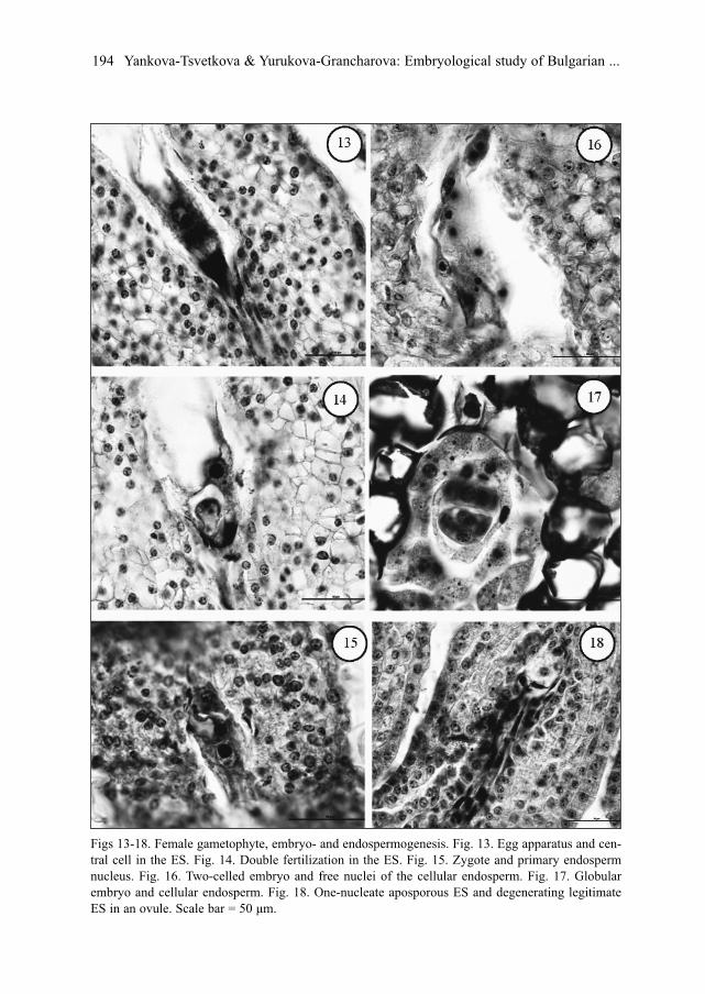
Figs 13-18. Female gametophyte, embryo- and endospermogenesis. Fig. 13. Egg apparatus and cen-tral cell in the ES. Fig. 14. Double fertilization in the ES. Fig. 15. Zygote and primary endospermnucleus. Fig. 16. Two-celled embryo and free nuclei of the cellular endosperm. Fig. 17. Globularembryo and cellular endosperm. Fig. 18. One-nucleate aposporous ES and degenerating legitimateES in an ovule. Scale bar = 50 μm.
Two polar nuclei fuse before the fertilization and form a big central cell that is locatedclosely to the egg cell (Fig. 13).
Antipodal complex consists of three one-nucleate cells with T-shape or linear arrange-ment. In the antipodals, a multiplication of the nuclei runs as well as polyploidization, inparticular in the population from the Balkan Range. Usually, the antipodals degenerateafter the fertilization (Fig. 12).
The legitimate embryo and endosperm form after double porogamous fertilization (Fig.14). The first mitotic division of the zygote (Fig. 15) is transversal (Fig. 16) and theembryogenesis follows the Solanad-type.
Initially, the endosperm is nuclear (Fig. 16). Later on, the endosperm differentiates intocellular one at the globular embryo stage (Fig. 17). Endospermogenesis begins before theembryogenesis and even at the stage of zygote in the ES cavity 15-20 free endospermnuclei present. It is of interest to mention, that sporadically, in ovules of the populationfrom Rila Mt, an additional aposporous ES forms above the one-nucleate degeneratinglegitimate one (Fig. 18).
Discussion
During the study on G. lutea, typical of the family Gentianaceae L. and genus Gentianaembryological characteristics are established, as follows: tetrasporangiate anthers,Dicotyledonous-type of the anther’s wall development with more than one middle layer;fibrous endothecium; 3-celled mature pollen grains; anatropous, tenuinucellate, unitegmicovule; one-celled female archesporium; archesporogenesis without formation of parietalcells; Polygonum-type female gametophyte; Solanad-type embryogenesis and cellular-type of the endosperm (Davis 1966; Poddubnaya-Arnoldi 1982; Shamrov 1987). Besides,a number of new embryological peculiarities are found, announced up to now only forsome other species Gentiana as well as the peculiarities that are not identically interpret-ed from different authors.
Male generative sphereShamrov (1986), concerning the anther’s wall in G. lutea announces for the formation of
two additional layers in it, referring them to the middle layers that consisting cells enlargeand develop fibrous thickenings like the endothecium. Our observations during the study,give us the reason to interpret this fact by a different manner. Because of the additional lay-ers are morphologically identical with the endothecium (including the fibrous thickening oftheir cells and an early degeneration of the middle layers), their origin probably have to beconnected with the endothecium than the middle layers. We consider that histological inves-tigations are needed also to be carried out in order to receive more definite data for clarifi-cation of the origin of these additional layers in the anther’s wall of G. lutea.
The early degeneration of the middle layers to the end of the heterotypic division ofmeiosis in MMCs defines them as ephemeral. Contrary, Shamrov (1986), shows thatthese layers preserve even at the stage of mature pollen grains in the anthers. A longerpreservation of the middle layers is noticed also for other species of the family
Flora Mediterranea 19 — 2009 195
Gentianaceae, as Megacodon stylophorus (C.B.Clark) H. Smith and Veratrilla bailloniiFranch (Xue & Li 2005).
Poddubnaya-Arnoldi (1982) and Shamrov (1987) describe the anther’s tapetum of thefamily Gentianaceae as glandular without transformation in ameboid or periplasmodialone, in particular in G. lutea too (Shamrov 1986). Davis (1966), gives a contrary opinion,considering that the tapetum in this family is glandular with transformation into ameboidone. The same conclusion is made for G. carpatica Kit. from Schnarf (1923) as well as forCanscora diffusa (Vahl) R. Brown ex Roem. & Schult. and C. decussatа (Roxb.) Roem. &Schult. (Maheshwari 1962). Periplasmodial tapetum is announced for Gentiana asclepi-adea L. (Wóycicki 1935), G. pedicellatа (D. Don) Griseb., G. quadrifaria Blume andCanscora decurrens Dalz. (Sankara Rao & Chinnappa 1983).
Our observations reveal that the anther’s tapetum in G. lutea is glandular without trans-formation into ameboid one that support the opinion of Shamrov (1986) concerning to thisspecies. The unusual state of the tapetum established in G. lutea, when its cells penetratebetween the microspore tetrads might to be considered as a beginning of degeneration thana transformation of glandular tapetum into ameboid or periplasmodial one.
During this study, a specific feature in G. lutea is observed, as formation of rows ofcells, crossing the anther locules in radial direction that Shamrov (1986) found in the samespecies and named them ”trabecules”. This peculiarity is established too in other repre-sentatives of the family Gentianaceae like Megacodon stylophorus, Veratrilla baillonii(Xue & Li 2005) and species Swertia L. (Xue & al. 2007) that Steffen & Landmann (1958)named “placentoids”. Because of the similarity between the tapetal cells and “trabecules”,Shamrov (1986) considers that “…it is the tapetum itself, which cells to the side of con-necting tissue and outer anther’s wall during their development intrude between thesporogenous cells, tie together and form these peculiar barriers”.
Female generative sphereThe ovule of G. lutea is anatropous indicated as a typical for the family Gentianaceae
by Poddubnaya-Arnoldi (1982). Shamrov (1987) defines the ovule in the representativesof this family as a preliminary anatropous, rarely – orthotropous in Halenia elliptica D.Don, Cothylanthera tenuis, species Voyria Aubl. or hemitropous in Gentiana tenella Rottb.and Gentianella lingulata G. Agardh. It is indicated that in Gentianella azurea (Bunge)Holub (Liu & He 1996), G. caucasea (Loddiges ex Sims) Holub, G. germanica (Willd.)Boerner (Akhalkatsi & Wagner 1997) and G. ciliatа (L.) Borkh. (our unpublished data) ahemitropous ovule is presented and orthotropous one is shown in Swertia tetrapteraMaxim (Xue & al. 1999).
Shamrov (1999), gives a new classification of the ovule’s types on the basis of thenucellar structure and development and shows that in the family Gentianaceae the ovuleis tenuinucellate – “standard (simpetal) variation” in which the nucellus is presented by thenucellar epidermis that envelopes the megasporocyte during its development and destroysbefore the fertilization. Our observations on the ovule ontogenesis of G. lutea show that itmight be refer to the same ovule type, namely “standard (simpetal) variation”.
We observe a funicular obturator, described by Shamrov (1988) in this species too aswell as the so colled “wings” that Bouman & Schrier (1979) announce for G. asclepiadea.
196 Yankova-Tsvetkova & Yurukova-Grancharova: Embryological study of Bulgarian ...
In G. lutea the endothelium is formed, that is announced up to now for Gentianaceae onlyin G. cruciata L. (Shamrov 1990) and Exacum pumilum Griseb. (Poddubnaya-Arnoldi 1982).
In the ovule of G. lutea the one-celled archesporium forms, that is typical of the familyGentianaceae and genus Gentiana (Poddubnaya-Arnoldi 1982, Shamrov 1987), but in thisspecies Kordjum (1978) and Shamrov (1987, 1988) show also two-celled archesporium.
In the ovules of G. lutea predominantly megaspore tetrad forms, but in some ovules ofthe population of Rila Mt a dyad of megaspores forms instead of a tetrad and ES not devel-ops according to Polygonum (monosporic)-type but follows Allium (bisporic)-type ofdevelopment. The Allium-type female gametophyte is announced also in G. cruciata(Schamrov 1988), G. punctata L. (Bicheva & al. 2004) and other species of Gentianaceaeas Centaurium ramosissimum (Vijayaraghavan & Padmanabhan 1969) and Swertia minorGriesb. (Sankara Rao & Nagaraj 1982). In the Rila Mt population of G. lutea, the forma-tion of an aposporous ES is observed in the chalazal part of some ovules, but the embryoformation is not found. Rudenko (1961) shows nucellar adventive embryony not only inthis species but also in G. punctata, G. livonica Eschf. and G. carpatica Wettst.
The peculiarities of the male and female generative sphere established during the pres-ent embryological study on Bulgarian populations of G. lutea reveal a specialization of thisspecies, in particular regarding the female gametophyte pronounced in the following fea-tures: anatropous, tenuinucellate, unitegmic ovule; funicular obturator; one-celled femalearchesporium; archesporogenesis without formation of parietal cells; Allium (bis-poric)–type female gametophyte together with the basic Polygonum (monosporic)-type;endothelium; tendency to gametophytic apomixis (apospory). Some of relatively primitivefeatures observed in the male generative sphere (multiplication of number of the antherwall layers and the multilayered sporogenous tissue) are not so significant in evolutionaryaspect and essential to change the status of G. lutea as an evolutionary advanced speciesand they are only considered as a manifestation of the heterobathmy.
References
Akhalkatsi, M. & Wagner, J. 1997: Comparative embryology of three Gentianaceae species from theCentral Caucasus and the European Alps. – Pl. Syst. Evol. 204: 39-48.
Andreev, N. 1982: Reports [in Löve, A (ed.) IOPB Chromosome numbers reports, LXXVI]. – Taxon31: 575-576.
Assenov, I. & Nikolov, St. 1989: Pharmacognosy. – Sofia.Bicheva, R., Robeva-Davidova, P. & Yurukova-Grancharova, P. 2004: Macrosporogenesis and
development of the female gametophyte in Gentiana punctata (Gentianaceae). – Phytol.Balcan. 10(2-3): 233-240.
Boichinov, A. & Boeva, A. 1953: On the content of bitter compounds in some local species GentianaL. – Farmacia 4: 29-31.
Bondev, I. 1995: Chorological atlas of the medicinal plants in Bulgaria. – Sofia.Bouman, F. S. & Schier, S. 1979: Ovule ontogeny in seed coat development in Gentiana with a dis-
cussion on the evolutionary origin of the single integument. – Acta Bot. Neerl. 28(6): 468-478.Davis, G. 1966: Systematic Embryology of the Angiosperms. – New York, London & Sydney.Georgieva, E. 2007: Ecological, biological and photochemical peculiarities of Gentiana lutea L. and
Gentiana punctata L. in Bulgaria. – D. Phil. Thesis, Sofia.Johansen, D. 1950: Plant Embryology. – Watham.
Flora Mediterranea 19 — 2009 197
Kozhuharova, E. 1994. Reproductive biology on the species of the genus Gentiana (Gentianaceae)represented in the Bulgarian flora. – D. Phil. Thesis, Sofia.
Kordjum, E. 1978: Evolutionary cytoembryology of Angiosperms. – Kiev.Liu, J. Q. & He, T. N. 1996: Embryological studies of Gentianella azurea. – Acta Bot. Yunnan.
18(2): 151-158.Maheshwari D., H. 1962: Embryological studies in Gentianaceae (Gentianoideae and
Menyanthoideae). – Proc. Ind. Acad. Sci., B. 56(4): 195-216.Petkov, V. 1982: Contemporary phitotherapy. – Sofia.Poddubnaya-Arnoldi, V. A. 1982: Characteristics of the Angiosperms families by cytoembryological
features. – Moskva.Rajan S. Sundara. 2000. Practical Manual of Plant anatomy and Embryology. – New Delhi. Rudenko, F. E. 1961: Apomixis in certain high mountain plants of the Ukrainian Carpathians. – Ukr.
Bot. Zour. 28(6): 24-31. Sankara Rao, K. & Nagaraj, M. 1982: Studies in Gentianaceae: embryology of Swertia minor
(Gentianinae). – Canad. J. Bot. 60: 141-151.— & Chinnappa, C.C. 1983: Studies in Gentianaceae. Microsporangium and pollen. – Canad. J.
Bot. 61(1): 324-336.Shamrov, I. 1986: Anther development of Gentiana lutea (Gentianaceae). – Bot. Zhurn. 71(6): 733-739.— 1987: Family Gentianaceae. – Pp. 137-145 in: Batygina, T.B. & Jakovlev, M.S. (Eds),
Comparative embryology of the Flowering Plants. – Leningrad.— 1988: Ovule and structural characteristics of embryo sac in some members of the Gentianaceae
family. – Bot. Zhurn. 73(2): 213-222.— 1990: The ovule of Gentiana cruciata (Gentianaceae): structural-functional aspects of develop-
ment. – Bot. Zhurn. 75(10): 1363-1379.— 1999: The ovule as a base of the seed reproduction in Flowering plants: Classification of the struc-
tures. – Bot. Zhurn. 84(10): 1-35.Schnarf, K. 1923: Kleine Beiträge zur Entwicklungsgeschichte der Angiospermen. IV. Über
Verhalten des Antherentapetums einiger Pflanzen. – Öster. Bot. Zeitschr. 72(1-5): 242-245.Steffen, K. & Landmann, W. 1958: Entwicklungsgeschichtliche und cytologische Untersuchungen am
Balkentapetum von Gentiana cruciata und Impatiens glandulifera. – Planta 50(4): 423-460.Stojanov, N. & Kitanov, B. 1960: Wild and useful plants in Bulgaria. – Sofia. Velchev, V. 1984: Red Data Book of PR Bulgaria, 1 Plants. – Sofia. Wóycicki, Z. 1935: Zur Entwicklungsgeschichte der Antheren und des Pollens bei einiger
Repräsentanten der Gattung Gentiana. III. Gentiana lutea L. – Acta Soc. Bot. Pol. 12(3): 207-226.Xue, C. Y. & Li, D. Z. 2005: Embryology of Megacodon stylophorus and Veratrilla baillonii
(Gentianaceae): descriptions and systematic implications. – Bot. J. Lin. Soc. 147: 317-331.Xue, C. Y., Ho, T. N. & Liu, J. Q. 1999: Embryology of Swertia tetraptera Maxim. (Gentianaceae)
and its systematic implication. – Acta Phytotax. Sin. 37: 259-263.Xue, C. Y., Ho, T. N. & Li, D. Z. 2007: Embryology of Swertia (Gentianaceae) relative to taxono-
my. – Bot. J. Linn. Soc. 155: 383-400.
Adress of the authors:Elina Yankova-Tsvetkova & Petka Yurukova-GrancharovaInstitute of Botany, Bulgarian Academy of Sciences, Acad. Georgi Bonchev Street,bl. 23, 1113 Sofia, Bulgaria. E-mail: [email protected]
198 Yankova-Tsvetkova & Yurukova-Grancharova: Embryological study of Bulgarian ...