
Fixational saccades reflect volitional action preparation
14
Fixational saccades reflect volitional action preparation Masayuki Watanabe, 1,2 Yuka Matsuo, 3 Ling Zha, 3 Douglas P. Munoz, 1 and Yasushi Kobayashi 4,5,6,7 1 Centre for Neuroscience Studies, Queen’s University, Kingston, Ontario, Canada; 2 Department of Physiology, Kansai Medical University, Osaka, Japan; 3 Department of Social and Environmental Medicine, Graduate School of Medicine, Osaka University, Osaka, Japan; 4 Graduate School of Frontier Biosciences, Osaka University, Osaka, Japan; 5 Center for Information and Neural Networks, National Institute of Information and Communications Technology, Osaka, Japan; 6 ATR Computational Neuroscience Laboratories, Kyoto, Japan; and 7 PRESTO, the Japan Science and Technology Agency, Saitama, Japan Submitted 21 December 2012; accepted in final form 24 April 2013 Watanabe M, Matsuo Y, Zha L, Munoz DP, Kobayashi Y. Fixational saccades reflect volitional action preparation. J Neurophysiol 110: 522–535, 2013. First published May 1, 2013; doi:10.1152/jn.01096.2012.—Human volitional actions are preceded by preparatory processes, a critical mental process of cognitive control for future behavior. Volitional action preparation is regulated by large-scale neural circuits including the cerebral cortex and the basal ganglia. Because volitional action preparation is a covert process, the network dynamics of such neural circuits have been examined by neuroimaging and recording event- related potentials. Here, we examined whether such covert processes can be measured by the overt responses of fixational saccades (in- cluding microsaccades), the largest miniature eye movements that occur during eye fixation. We analyzed fixational saccades while adult humans maintained fixation on a central visual stimulus as they prepared to generate a volitional saccade in response to peripheral stimulus appearance. We used the antisaccade paradigm, in which subjects generate a saccade toward the opposite direction of a periph- eral stimulus. Appropriate antisaccade performance requires the fol- lowing two aspects of volitional control: 1) facilitation of saccades away from the stimulus and 2) suppression of inappropriate saccades toward the stimulus. We found that fixational saccades that occurred before stimulus appearance reflected the dual preparatory states of saccade facilitation and suppression and correlated with behavioral outcome (i.e., whether subjects succeeded or failed to cancel inappro- priate saccades toward the stimulus). Moreover, fixational saccades explained a large proportion of individual differences in behavioral performance (poor/excellent) across subjects. These results suggest that fixational saccades predict the outcome of future volitional ac- tions and may be used as a potential biomarker to detect people with difficulties in volitional action preparation. antisaccade; decision making; executive function; fixation; microsac- cade VOLITIONAL ACTIONS ARE PRECEDED by preparatory processes that preset neural circuits based on the knowledge of current situ- ations and/or environment to guide future behavior (Haggard 2008). Without volitional action preparation, behavior would become reactive rather than predictive, which may explain cognitive behavioral deficits observed in a variety of clinical disorders, such as Parkinson’s disease (Cameron et al. 2012; Cunnington et al. 1997) and attention deficit hyperactivity disorder (Hakvoort Schwerdtfeger et al. 2013; McLoughlin et al. 2010). Volitional action preparation is regulated by large-scale networks integrating the cerebral cortex and the basal ganglia (Nachev et al. 2008). The network dynamics of such neural circuits have been studied extensively with saccadic eye move- ments (Hikosaka et al. 2000; Munoz and Everling 2004; Schall 2004; Sparks 2002; Watanabe and Munoz 2011). In conven- tional paradigms, subjects maintain their eyes on a central fixation point and generate a saccade in response to peripheral visual stimulus appearance. Cognitive models indicate that saccade behavior is shaped partially by neural processes before stimulus appearance, which corresponds to volitional saccade preparation (Carpenter and Williams 1995; Smith and Ratcliff 2004). Correspondingly, recent studies have shown that neural activity changes gradually before stimulus appearance in ac- cordance with preparation (Amador et al. 2004; Everling et al. 1999; Everling and Desouza 2005; Everling and Munoz 2000; Kunimatsu and Tanaka 2010; Watanabe and Munoz 2010; Yoshida and Tanaka 2009). Because volitional action preparation is a covert process by definition, its state cannot be measured directly but inferred from cognitive models or neural activity measurements. Here we hypothesize that volitional action preparation can be mea- sured overtly by analyzing miniature eye movements during fixation. This hypothesis is derived from behavioral studies in which fixational saccades, the largest miniature eye move- ments including microsaccades, are not always generated in- voluntarily but rather are under volitional control (Bridgeman and Palca 1980; Haddad and Steinman 1973; Ko et al. 2010; Kowler and Steinman 1977; Steinman et al. 1967; Winterson and Collewijn 1976). Fixational saccades also reflect the state of covert spatial attention (Brien et al. 2009; Engbert and Kliegl 2003; Gowen et al. 2007; Hafed et al. 2011; Hafed and Clark 2002; Laubrock et al. 2005; Rolfs et al. 2005). Further- more, recent studies have shown a linkage between fixational saccade occurrence and macrosaccade initiation (Hafed and Krauzlis 2010; Rolfs 2007; Rolfs et al. 2006; Sinn and Engbert 2011). Accordingly, neural processes preparing volitional ac- tions may influence fixational saccades to optimize behavioral states for upcoming saccades. We examined whether volitional action preparation is re- flected in the pattern of fixational saccades, using a simple behavioral paradigm called an antisaccade in which subjects generate a saccade toward the opposite direction of a peripheral visual stimulus (Hallett 1978). Appropriate antisaccade perfor- mance requires the following two aspects of volitional saccade control: 1) facilitation of saccades away from the stimulus and Address for reprint requests and other correspondence: M. Watanabe, New Zealand Brain Research Institute, 66 Stewart St., Christchurch 8011, New Zealand (e-mail: [email protected]). J Neurophysiol 110: 522–535, 2013. First published May 1, 2013; doi:10.1152/jn.01096.2012. 522 0022-3077/13 Copyright © 2013 the American Physiological Society www.jn.org at Queens University on August 19, 2013 http://jn.physiology.org/ Downloaded from
Transcript of Fixational saccades reflect volitional action preparation

Fixational saccades reflect volitional action preparation
Masayuki Watanabe,1,2 Yuka Matsuo,3 Ling Zha,3 Douglas P. Munoz,1 and Yasushi Kobayashi4,5,6,7
1Centre for Neuroscience Studies, Queen’s University, Kingston, Ontario, Canada; 2Department of Physiology, KansaiMedical University, Osaka, Japan; 3Department of Social and Environmental Medicine, Graduate School of Medicine,Osaka University, Osaka, Japan; 4Graduate School of Frontier Biosciences, Osaka University, Osaka, Japan; 5Center forInformation and Neural Networks, National Institute of Information and Communications Technology, Osaka, Japan;6ATR Computational Neuroscience Laboratories, Kyoto, Japan; and 7PRESTO, the Japan Science and Technology Agency,Saitama, Japan
Submitted 21 December 2012; accepted in final form 24 April 2013
Watanabe M, Matsuo Y, Zha L, Munoz DP, Kobayashi Y. Fixationalsaccades reflect volitional action preparation. J Neurophysiol 110: 522–535,2013. First published May 1, 2013; doi:10.1152/jn.01096.2012.—Humanvolitional actions are preceded by preparatory processes, a criticalmental process of cognitive control for future behavior. Volitionalaction preparation is regulated by large-scale neural circuits includingthe cerebral cortex and the basal ganglia. Because volitional actionpreparation is a covert process, the network dynamics of such neuralcircuits have been examined by neuroimaging and recording event-related potentials. Here, we examined whether such covert processescan be measured by the overt responses of fixational saccades (in-cluding microsaccades), the largest miniature eye movements thatoccur during eye fixation. We analyzed fixational saccades while adulthumans maintained fixation on a central visual stimulus as theyprepared to generate a volitional saccade in response to peripheralstimulus appearance. We used the antisaccade paradigm, in whichsubjects generate a saccade toward the opposite direction of a periph-eral stimulus. Appropriate antisaccade performance requires the fol-lowing two aspects of volitional control: 1) facilitation of saccadesaway from the stimulus and 2) suppression of inappropriate saccadestoward the stimulus. We found that fixational saccades that occurredbefore stimulus appearance reflected the dual preparatory states ofsaccade facilitation and suppression and correlated with behavioraloutcome (i.e., whether subjects succeeded or failed to cancel inappro-priate saccades toward the stimulus). Moreover, fixational saccadesexplained a large proportion of individual differences in behavioralperformance (poor/excellent) across subjects. These results suggestthat fixational saccades predict the outcome of future volitional ac-tions and may be used as a potential biomarker to detect people withdifficulties in volitional action preparation.
antisaccade; decision making; executive function; fixation; microsac-cade
VOLITIONAL ACTIONS ARE PRECEDED by preparatory processes thatpreset neural circuits based on the knowledge of current situ-ations and/or environment to guide future behavior (Haggard2008). Without volitional action preparation, behavior wouldbecome reactive rather than predictive, which may explaincognitive behavioral deficits observed in a variety of clinicaldisorders, such as Parkinson’s disease (Cameron et al. 2012;Cunnington et al. 1997) and attention deficit hyperactivitydisorder (Hakvoort Schwerdtfeger et al. 2013; McLoughlinet al. 2010).
Volitional action preparation is regulated by large-scalenetworks integrating the cerebral cortex and the basal ganglia(Nachev et al. 2008). The network dynamics of such neuralcircuits have been studied extensively with saccadic eye move-ments (Hikosaka et al. 2000; Munoz and Everling 2004; Schall2004; Sparks 2002; Watanabe and Munoz 2011). In conven-tional paradigms, subjects maintain their eyes on a centralfixation point and generate a saccade in response to peripheralvisual stimulus appearance. Cognitive models indicate thatsaccade behavior is shaped partially by neural processes beforestimulus appearance, which corresponds to volitional saccadepreparation (Carpenter and Williams 1995; Smith and Ratcliff2004). Correspondingly, recent studies have shown that neuralactivity changes gradually before stimulus appearance in ac-cordance with preparation (Amador et al. 2004; Everling et al.1999; Everling and Desouza 2005; Everling and Munoz 2000;Kunimatsu and Tanaka 2010; Watanabe and Munoz 2010;Yoshida and Tanaka 2009).
Because volitional action preparation is a covert process bydefinition, its state cannot be measured directly but inferredfrom cognitive models or neural activity measurements. Herewe hypothesize that volitional action preparation can be mea-sured overtly by analyzing miniature eye movements duringfixation. This hypothesis is derived from behavioral studies inwhich fixational saccades, the largest miniature eye move-ments including microsaccades, are not always generated in-voluntarily but rather are under volitional control (Bridgemanand Palca 1980; Haddad and Steinman 1973; Ko et al. 2010;Kowler and Steinman 1977; Steinman et al. 1967; Wintersonand Collewijn 1976). Fixational saccades also reflect the stateof covert spatial attention (Brien et al. 2009; Engbert andKliegl 2003; Gowen et al. 2007; Hafed et al. 2011; Hafed andClark 2002; Laubrock et al. 2005; Rolfs et al. 2005). Further-more, recent studies have shown a linkage between fixationalsaccade occurrence and macrosaccade initiation (Hafed andKrauzlis 2010; Rolfs 2007; Rolfs et al. 2006; Sinn and Engbert2011). Accordingly, neural processes preparing volitional ac-tions may influence fixational saccades to optimize behavioralstates for upcoming saccades.
We examined whether volitional action preparation is re-flected in the pattern of fixational saccades, using a simplebehavioral paradigm called an antisaccade in which subjectsgenerate a saccade toward the opposite direction of a peripheralvisual stimulus (Hallett 1978). Appropriate antisaccade perfor-mance requires the following two aspects of volitional saccadecontrol: 1) facilitation of saccades away from the stimulus and
Address for reprint requests and other correspondence: M. Watanabe, NewZealand Brain Research Institute, 66 Stewart St., Christchurch 8011, NewZealand (e-mail: [email protected]).
J Neurophysiol 110: 522–535, 2013.First published May 1, 2013; doi:10.1152/jn.01096.2012.
522 0022-3077/13 Copyright © 2013 the American Physiological Society www.jn.org
at Queens U
niversity on August 19, 2013http://jn.physiology.org/
Dow
nloaded from

2) suppression of inappropriate saccades toward the stimulus.Because an antisaccade instruction is given well before stim-ulus appearance, it presets neural circuits controlling antisac-cades to prepare for both saccade facilitation and suppression(Watanabe and Munoz 2010). We therefore examined whethersuch dual preparatory signals (saccade facilitation and suppres-sion), which are processed covertly for upcoming antisaccadeexecution, can be read out overtly from fixational saccades.
METHODSSubjects. Fifty-eight subjects [40 men, 18 women; age: mean !
SD " 21.8 ! 3.0] with normal or corrected to normal visionparticipated in this study. One of the authors (M. Watanabe) wasincluded because virtually the same statistical results were confirmedwith and without this subject. Subjects were paid ¥2,000/h for theirparticipation. They were informed of the nature of the study andconsented to be part of the study. This study was approved by theresearch ethics board of the Osaka University Hospital.
Experimental systems. The control of the behavioral paradigm andthe acquisition of eye position data were carried out by the TEMPO/Win computing system (ReflectiveComputing, St. Louis, MO). Leftand right eye positions were acquired with a fast video-based eyemovement monitor (a dark pupil eye tracking system; iView XHi-Speed, SensoMotoric Instruments, Teltow, Germany). The tempo-ral and spatial resolutions of the pupil tracking were 500 Hz and 0.01°,respectively. A standard nine-point calibration was conducted to aligneye and screen coordinate systems. Drift correction, and additionalgain calibration whenever necessary, was performed after every 50correct trials. Subjects supported their head on a chin/forehead restthat included the support for the camera for eye tracking. A cathoderay tube monitor (60-Hz refresh rate, 1,024 # 768 pixels, 19 in.) wasplaced at 35 cm from the eyes. A bite-bar was installed for additionalsupport of head fixation during the course of experiments, althoughour results did not depend on the use of the bite-bar (see below).
Main behavioral paradigm. Forty-two of the fifty-eight subjectsperformed the following main paradigm (Fig. 1). Each trial waspreceded by a 1,000-ms intertrial interval during which the screen wasilluminated with a diffuse light to prevent dark adaptation (2 cd/m2).After removal of the background light, a circular fixation point (size:0.4°, luminance: 14 cd/m2) appeared in the center of the screenwithout background illumination and subjects were required to directtheir eyes toward the fixation point within 30 s. After they maintainedsteady fixation within a computer-controlled window (!2°) for 700–2,300 ms (exponential distribution with constant expectation; aver-age " 1,000 ms) (Oswal et al. 2007), the fixation point disappeared.
On the majority of trials (80%), a peripheral stimulus (size: 0.4°,luminance: 14 cd/m2, color: yellow) appeared at either 5° left or rightfrom the center of the screen (19° from the border of the monitor)simultaneously with fixation point disappearance. The stimulus re-mained visible for 1,000–1,500 ms. Subjects generated a saccadeeither toward the stimulus (prosaccade) or to the opposite direction ofthe stimulus (antisaccade) based upon fixation point color (red/green)counterbalanced across subjects and maintained fixation on the pe-ripheral stimulus on prosaccade trials or on a blank screen at themirror position of the peripheral stimulus on antisaccade trials. Thesizes of fixation windows for peripheral stimuli were adjusted for eachsubject to accept relatively inaccurate antisaccade end points [width:8.3 ! 4.2° (mean ! SD), height: 5.9 ! 2.0°] (Dafoe et al. 2007;Fischer and Weber 1992; Hallett 1978).
For the remaining 20% of trials, the fixation point reappeared 50 msafter its first disappearance (fixation blink) instead of peripheralstimulus appearance, and subjects maintained fixation for additional1,000–1,500 ms (catch trials). Catch trials were included to evokefixational saccades and detect fixational saccade readiness undervariable behavioral conditions. Another reason for the inclusion ofcatch trials was to replicate the basic characteristics of fixational
saccades in response to abrupt sensory events (see, e.g., Engbert andKliegl 2003; Rolfs et al. 2005).
Subjects received auditory feedback at the end of each trial basedon their performance. This auditory feedback was generated by abuilt-in computer speaker (correct: single beep of 2,300 Hz with150-ms duration; error: 2 beeps of 1,950 Hz with 50-ms duration and50-ms interval). All task conditions described above were randomlyinterleaved in a block of trials. Subjects performed this paradigm untilthey achieved at least 400 correct trials [including all 4 types of trials(pro/anti # saccade/catch)], while they had short breaks every after100 correct trials. There was no explicit requirement of fixationalsaccades for performing this paradigm.
Secondary behavioral paradigm. The remaining 16 subjects per-formed a secondary paradigm designed specifically to examine theinfluence of temporal expectation of peripheral stimulus appearanceon fixational saccades as well as pro- and antisaccades. This paradigmwas the same as the above main paradigm with the following excep-tions (Fig. 1B). Subjects performed two separate blocks of trials, eachof which had fixation duration either randomized (700–2,300 ms,exponential distribution with constant expectation, 1,000 ms on av-erage) or fixed to 1,000 ms. The sequence of the blocks (randomizedfirst or fixed first) was counterbalanced equally across subjects. Catchtrials were not included in this paradigm. Subjects performed eachblock of this paradigm until they achieved at least 200 correct trials(including both pro and anti).
Saccade detection. Eye position data were first processed by adigital filter (3rd-order Butterworth low-pass filter with cutoff fre-quency of 200 Hz). The onset and end of pro- and antisaccades largerthan 2° were identified by radial eye velocity criteria (threshold:30°/s). Because eye positions were recorded binocularly, the onset andend of each saccade were defined by the earlier onset and the later endof both eyes.
Fixational saccades were detected by an algorithm developed byEngbert and colleagues (Engbert and Kliegl 2003; Engbert and Mer-genthaler 2006) (Fig. 2). Briefly, the velocity threshold of fixationalsaccades was defined flexibly depending on the noise level on each
Paradigm Fixation duration Catch
Main(n = 42) Random Included
(20%)
Secondary(n = 16)
Random / Fixed
Notincluded
B
Tim
e
Prosaccade Antisaccade Pro catch Anti catch
Fixation
Stimulus
Saccade
Fixation
Blink (50 ms)
Fixation
A
Fig. 1. Behavioral paradigms. A: 4 types of trials. On prosaccade trials, subjectsfixated on the fixation point and generated a saccade toward a stimulus. Onantisaccade trials, subjects generated a saccade to the opposite direction fromthe stimulus. On pro- and anti-catch trials, subjects fixated on the fixation pointthroughout the trial while a 50-ms fixation blink was introduced. B: differencesbetween main and secondary paradigms. In the main paradigm, fixationdurations were randomized from 700 to 2,300 ms with an average of 1,000 ms,and catch trials were included (10% each for pro and anti). In the secondaryparadigm, there were 2 separate blocks with fixation duration either random-ized or fixed (1,000 ms). Catch trials were not included in this paradigm.
523FIXATIONAL SACCADES REFLECT VOLITIONAL ACTION PREPARATION
J Neurophysiol • doi:10.1152/jn.01096.2012 • www.jn.org
at Queens U
niversity on August 19, 2013http://jn.physiology.org/
Dow
nloaded from

trial (threshold: 6 SDs) (Fig. 2, D and E). The minimum duration offixational saccades that exceeded the velocity threshold was set to 6ms. This analysis was limited to a temporal period in which eyepositions were relatively stable (from 200 ms after the end of asaccade toward the fixation point to the initiation of a pro/antisaccadeon saccade trials or to 2nd fixation point disappearance on catch trials)(Fig. 2A). We analyzed only fixational saccades that occurred simul-taneously in both eyes during at least one data sample (2 ms) to reducethe influence of potential noise on data analyses (Fig. 2, B and C). Thiscriterion of binocularity set a strong constraint on fixational saccadedetection and removed a large number of monocular fixational sac-cades, or noise detected as monocular fixational saccades. This tech-nical limitation was derived from the limited sampling frequency (500Hz for binocular recordings), although this is a standard performanceof currently available video-based eye trackers. The minimum inter-saccade interval was set to 20 ms to avoid defining potential overshootcorrections as new fixational saccades (Moller et al. 2002). Theamplitude, direction, and peak velocity of each binocular fixationalsaccade were analyzed from the right eye. Virtually the same resultswere confirmed by analyzing the left eye data.
Fixational saccades from an example subject detected by the abovecriteria are shown in Fig. 3. Consistent with previous studies, the peakvelocities of fixational saccades increased linearly with their ampli-tudes (Fig. 3A: main sequence), although our data included fixationalsaccades larger than microsaccades in the original report (Zuber et al.1965). The directions of binocular fixational saccades were biasedtoward the horizontal directions (Fig. 3B) (Engbert 2006). The distri-butions of main sequence slopes, amplitudes, and directions from allsubjects are shown in Fig. 4. Pure horizontal (left/right) and vertical(up/down) fixational saccade directions were quantified as 0° and 90°,respectively. The distributions of amplitudes and directions were
characterized by calculating 10, 30, 50, 70, and 90 percentiles in eachsubject, and those values from all subjects are shown cumulatively.The majority of fixational saccade amplitudes were $1°, and mostfixational saccades had amplitudes $2° (Fig. 4B). In the majority ofsubjects, the medians of fixational saccade directions were $30° fromthe horizontal meridian (Fig. 3C), confirming the horizontal bias ofthe directions of binocular fixational saccades at the population level.
Because the minimum amplitude threshold of pro- and antisaccadeswas set to 2° as described above, we adopted the same value for themaximum amplitude threshold of fixational saccades. We excludedtrials if saccades larger than this threshold occurred during fixedtemporal periods for quantitative analyses (see below). Note that Fig.4 contains trials with fixational saccades larger than the amplitudecriterion because they occurred outside the temporal periods forquantitative analyses (see below). Only 0.9 ! 2.3% (mean ! SD) oftrials were excluded by this criterion. We confirmed major findingswith the threshold of 1°, which has been used for human microsaccadestudies with video-based eye trackers (e.g., Engbert and Mergenthaler2006; Hermens et al. 2010; Rolfs et al. 2006). However, this criterionexcluded more trials (5.2 ! 6.8%) than the 2° criterion. Because thereis no specific reason to exclude trials with fixational saccades largerthan 1° in the context of our study, we chose the 2° criterion for resultpresentation.
Fixational saccade quantifications. To quantify the frequency offixational saccade occurrence while subjects prepared for a pro- orantisaccade before stimulus appearance, we defined a prestimulusperiod as the 400-ms window ending 70 ms after stimulus appearance.We took into account the 70-ms offset for the minimum delay ofvisual processing for stimulus appearance before saccade initiation(Fischer and Weber 1993). We chose this temporal period to quantifyfixational saccades that occurred before stimulus appearance and
Right eye
-800 -400 0 400
2°
Right eye
Left eye
A B
C
D E
B C
-60 -40 -20 0 20 40-60
-40
-20
0
20
40
60
-60 -40 -20 0 20 40
B
CB
C
Time fromstimulus appearance (ms)
Horizontal velocity (deg/s) Horizontal velocity (deg/s)
Ver
tical
vel
ocity
(deg
/s) Left eye velocity Right eye velocity
Single trialeye trace
Fixational saccade
Time from fixationalsaccade initiation (ms)
Fixational saccadedetection period
Left eye
Right eye
Left eye
Hor
izon
tal e
ye p
ositi
on
-20 0 20 40
0.5°
0.5°
Velocity threshold(6 standard deviation)
Rig
htw
ard
Leftw
ardFig. 2. Fixational saccade detection on single trial. A: horizontal
eye traces (leftward prosaccade trial). Velocity thresholds forfixational saccade detection were calculated between 200 ms afterfixation initiation and prosaccade initiation, indicated by verticallines. Two fixational saccades (B and C) were detected on thistrial. B and C: fixational saccades. Gray rectangles indicate fixa-tional saccade durations. D and E: horizontal and vertical eyevelocity in the left (D) and right (E) eyes during fixationalsaccade detection period (A). Gray ellipses indicate velocitythresholds. Fixational saccades shown in B and C exceeded thevelocity thresholds simultaneously in both eyes.
524 FIXATIONAL SACCADES REFLECT VOLITIONAL ACTION PREPARATION
J Neurophysiol • doi:10.1152/jn.01096.2012 • www.jn.org
at Queens U
niversity on August 19, 2013http://jn.physiology.org/
Dow
nloaded from

correlate them with pro- and antisaccade behavior that occurred afterstimulus appearance because the neural processes of pro- and anti-saccade preparation are initiated well before stimulus appearance andsuch prestimulus preparatory activity is correlated with behavior (see,e.g., Watanabe and Munoz 2010).
To analyze fixational saccades evoked by a fixation blink on catchtrials, we defined a postblink period as the 400-ms window starting at200 ms after the first fixation point disappearance. We incorporatedthe 200-ms delay because fixational saccades were suppressedstrongly at %200 ms before they were evoked by fixation blinks (seeRESULTS). The suppression and facilitation of fixational saccade gen-eration after an abrupt sensory event are consistent with previousstudies (e.g., Engbert and Kliegl 2003; Rolfs et al. 2005).
Within-subject analyses. The occurrence of fixational saccades andthe timing and performance of pro- and antisaccades depended onmultiple factors. To disentangle the influence of each factor within thebehavioral paradigms, we carried out the following regression analy-ses in individual subjects who performed the main paradigm:
y ! a0 " a1 # [fixation duration] " a2 # [task instruction] (1)
where “fixation duration” indicates a time period from fixation initi-ation (eyes entered into the fixation window) to stimulus appearanceand “task instruction” indicates a prosaccade (&1) or an antisaccade('1) instruction. Fixation durations were normalized by their mean
0
20
40
60
80
100
50 70 90 1100
20
40
60
80
100
Main sequence A
10% 30% 50% 70% 90%
0.1 0.5 1 30
20
40
60
80
100
AmplitudeB
0 30 60 90
DirectionC
Amplitude (deg)
Slope (1/s)
|Direction from horizontal meridian| (deg)
stcejbuS
%stcejbu
S %
% S
ubje
cts
n = 58
Population
2
10% 30% 50% 70% 90%
Fig. 4. Fixational saccade characteristics from all subjects. A: main sequence.B: amplitude. C: direction. “0” and “90” indicate horizontal (left/right) andvertical (up/down) fixational saccades, respectively. Each line in B and Cindicates the cumulative distribution of a specific percentile of fixationalsaccades from each subject.
211.010
100
200Main sequence
A
5%
10%
30°
210°
60°
240°
90°
270°
120°
300°
150°
330°
180° 0°
B Direction
Amplitude (deg)
Pea
k ve
loci
ty (d
eg/s
)
0% 0.5% 1%
% Fixational saccade
Example subject
0.5
50
Fig. 3. Fixational saccade characteristics from an example subject. A: 2-di-mensional histogram of fixational saccade main sequence (slope ! confidenceinterval " 73.8 ! 0.8). B: fixational saccade directions. The percentages offixational saccades were counted within a 10° window shifted by 10°. Exampleeye traces from the same subject are shown in Fig. 2.
525FIXATIONAL SACCADES REFLECT VOLITIONAL ACTION PREPARATION
J Neurophysiol • doi:10.1152/jn.01096.2012 • www.jn.org
at Queens U
niversity on August 19, 2013http://jn.physiology.org/
Dow
nloaded from

and SD before applying this equation. To analyze the frequency offixational saccades, we adopted Poisson regressions with the log linkfunction rather than simple linear regressions because fixational sac-cade occurrence during the prestimulus period was quantified as anonnegative integer value on each trial (i.e., n " 0, 1, 2,...). Accord-ingly, y corresponds to the log of the expected value of fixationalsaccade counts during the prestimulus period [i.e., y " log($), where$ indicates expected value of fixational saccade counts]. The log linkfunction is connected to the probability density function of Poissondistribution [i.e., P(n) " e&$$n/n!, where n indicates a fixationalsaccade count]. For the direction error rates of antisaccades, weadopted the logistic regressions for the binary behavior [i.e., correct orerror; y " log{p/(1 & p)}, where p indicates a direction error rate].For the reaction times of pro- and antisaccades, we applied the sameequation to linear regressions.
For data collected during the secondary paradigm, we carried outthe following regression analyses for all 16 subjects:
y ! y1 " y2 (2)
y1 ! !n
(b0n " b1n # [fixation duration] # b2n
# [task instruction]) # "nth subject#
y2 ! c1 # [random/fixed] " c2 # [1st and 2nd blocks]
where y1 is the same as Eq. 1, except that it calculates the threecoefficients (b0n, b1n, and b2n) in individual subjects using an inter-action with “nth subject,” a set of dummy variables specifying eachsubject [i.e., “ith subject” " 1 for ith subject and 0 for all others]. Incontrast, y2 contains the following two factors that can be dissociatedonly by between-subject comparison: “random/fixed” indicates ablock of fixation durations randomized (0) or fixed ('1), and “1st/2ndblock” indicates the first (0) or second ('1) block. y corresponds toone of the following three values: 1) the log of the expected value offixational saccade counts during the prestimulus period; 2) the logodds of antisaccade direction errors; or 3) correct pro- and antisaccadereaction times. The fittings of these regression models were successful[1) %(9364)
2 " 9,239, P ( 0.8 (Pearson’s %2-test); 2) H(8) " 5.21, P (0.7 (Hosmer-Lemeshow test); 3) R2 " 0.39, F(49,9364) " 122, P $0.0001]. We describe regression coefficients in y2 along with those inEq. 1 by setting “random/fixed” and “1st/2nd block” to zero tosimulate the same condition as the main paradigm (i.e., randomfixation durations in 1st block).
Between-subject analyses. Because both fixational saccade charac-teristics and pro- and antisaccade behavior (reaction times and direc-tion errors) varied significantly across subjects, we analyzed theirrelationships with linear regressions. Fixational saccade frequencieswere characterized by the above Poisson regressions [i.e., constant(a0, b0n), fixation duration (a1, b1n), and task instruction (a2, b2n) inEqs. 1 and 2].
We also quantified the average amplitude of fixational saccadesgenerated during the prestimulus period as follows. We collapsed allconditions in the main paradigm to increase the number of trials withfixational saccades generated during the prestimulus period (the lastfixational saccade was analyzed when multiple fixational saccadesoccurred during the prestimulus period). This enabled us to includethe majority of subjects who performed the main paradigm in thisanalysis (only 2 were excluded). This method was justified becausethe amplitudes of fixational saccades did not depend on fixationduration [mean ! SD " (&0.13 ! 6.64) # 10&2; t(30) " &0.11, P (0.9 (t-test)] or task instruction [(&1.48 ! 6.46) # 10&2; t(30) "&1.28, P ( 0.2], which was analyzed by the above linear regression(Eq. 1; this analysis was limited to 31 subjects who had enough trialswith fixational saccades during the prestimulus period). In the sec-ondary paradigm, we estimated the average amplitudes of fixationalsaccades from the resultant regression coefficients (Eq. 2).
To analyze subjects from the main and secondary paradigmstogether, we created an additional factor of paradigm (0: main, '1:
secondary). We utilized five parameters (constant, fixation duration,task instruction, amplitude, and paradigm) to identify the optimallinear model to explain pro- and antisaccade behavior (reaction timesand direction errors) with a stepwise regression method [term selec-tion criterion: summed square error (sse); P values for the entranceand removal of a term: 0.05 and 0.10, respectively]. The model startedfrom a constant and could develop to include linear, interaction, andsquared terms.
Remaining methods of data analyses. Trials with opposite saccadedirections were collapsed because fixational saccade frequency wasnot different between them prior to stimulus appearance. Becausefoveation of the fixation point was usually acquired by saccades,fixation durations were recalculated from the end of the saccade toacquire the fixation point until the time of stimulus appearance duringoff-line analysis. Ten trials at least were required in each specificcondition (e.g., pro-random-1st block) for data analyses. Only correcttrials were analyzed in all analyses, except for those focusing ondirection errors on antisaccade trials. We utilized similar regressionmodels with several modifications for variable circumstances, whichwe describe in RESULTS. We collected data with the bite-bar in 16 ofthe 42 subjects in the main paradigm and all 16 subjects in thesecondary paradigm. We did not find any influences of the use of thebite-bar on our results of fixational saccades, except for a very minoreffect, which did not influence our conclusion at all (see RESULTS). Alldata analyses were carried out with MATLAB (MathWorks).
RESULTS
Pro- and antisaccade behavior. The pro- and antisaccadebehavior of our subjects was consistent with previous reports(Dafoe et al. 2007; Fischer and Weber 1992; Hallett 1978). Thesame example subject shown in Figs. 2 and 3 who performedthe main paradigm had average ! SD of prosaccade reactiontimes of 177 ! 26 ms (Fig. 5A). There were very few directionerrors on prosaccade trials (1.6%). In contrast, on antisaccadetrials (Fig. 5B), reaction times were longer than prosaccades(250 ! 23 ms) and there were more direction errors (34.2%).In the population of subjects, antisaccade reaction times werelonger than prosaccade reaction times (Fig. 5C) [main: t(41) "&17.2, P $ 0.0001 (paired t-test); secondary: t(15) " &6.48,P $ 0.0001]. Direction error rates were higher on antisaccadetrials than prosaccade trials (Fig. 5D) [main: t(41) " &5.56,P $ 0.0001; secondary: t(15) " &3.80, P $ 0.005].
Fixational saccade behavior. The temporal dynamics offixational saccade occurrence is shown in Fig. 6. The raster anddensity functions of fixational saccade onset times (Fig. 6, Aand B) were obtained from the same example subject shown inFig. 2, Fig. 3, and Fig. 5, A and B. The population averagesof fixational saccade density functions from subjects whoperformed the main paradigm are shown in Fig. 6, C and D.We first replicated the well-established phenomenon of sup-pression and facilitation of fixational saccades by abrupt visualevents on catch trials (Fig. 6, B and D) (e.g., Engbert andKliegl 2003; Rolfs et al. 2005). More importantly, here wefound the following two characteristics of fixational saccadeoccurrence before subjects generated pro- and antisaccades(Fig. 6, A and C; note that the ranges of y-axes are differentbetween saccade and catch trials to capture the different rangesof fixational saccade frequencies): 1) the frequency of fixa-tional saccades decreased with increasing time before stimulusappearance, and 2) such reduction of fixational saccade fre-quency was more significant on antisaccade trials than prosac-cade trials.
526 FIXATIONAL SACCADES REFLECT VOLITIONAL ACTION PREPARATION
J Neurophysiol • doi:10.1152/jn.01096.2012 • www.jn.org
at Queens U
niversity on August 19, 2013http://jn.physiology.org/
Dow
nloaded from

In the following sections, we first establish a relationship be-tween fixational saccade occurrence and pro- and antisaccadeinitiation and then address the above two qualitative observationsin turn.
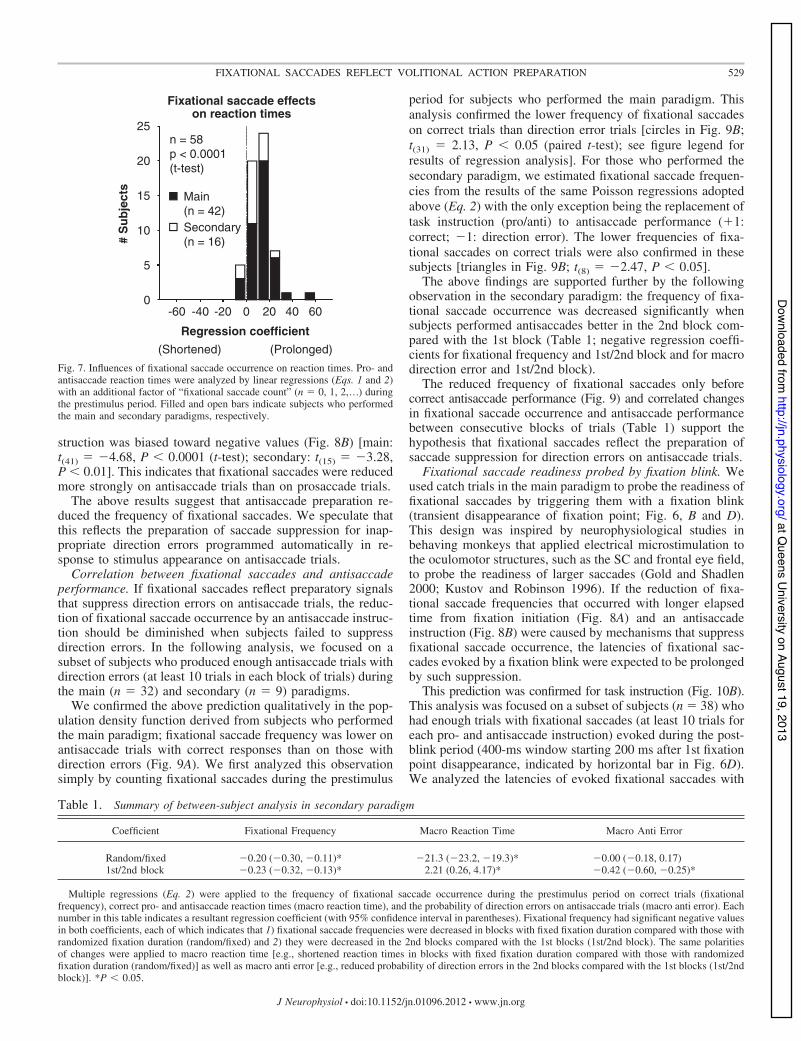
Fixational saccades preceded delayed initiation of pro- andantisaccades. Recent studies have shown that fixational saccadeoccurrence before stimulus appearance precedes delayed initiationof macrosaccades in response to stimulus appearance (Hafed andKrauzlis 2010; Rolfs 2007; Rolfs et al. 2006; Sinn and Engbert2011). We quantified the impact of fixational saccade occurrenceduring the prestimulus period (400-ms window ending 70 ms afterstimulus appearance, indicated by black horizontal bar abovex-axis in Fig. 6C) on the reaction times of pro- and antisaccades,using multiple linear regressions with an additional factor of“fixational saccade count” (n " 0, 1, 2...) (added to Eq. 1 and y1in Eq. 2). The distribution of regression coefficients for fixationalsaccade count calculated in individual subjects was biased towardpositive values (Fig. 7) [main: t(41) " 8.41, P $ 0.0001 (t-test);secondary: t(15) " 4.26, P $ 0.001], indicating that reaction timeswere prolonged after fixational saccade occurrence before stimu-lus appearance.
On the basis of the above observations, we propose thefollowing hypothesis: preparatory mechanisms that facilitatepro- and antisaccade initiation reduce the frequency of fixa-tional saccades before stimulus appearance. We describe evi-dence supporting this hypothesis in the following section.
Fixational saccade reduction with temporal expectation ofstimulus appearance. It has been shown previously that sac-cade reaction times are shortened with increasing elapsed timefrom fixation initiation, which reflects the temporal expectationof stimulus appearance (Oswal et al. 2007; Pare and Munoz1996). Although fixation durations were randomized to gener-ate a constant expectation of stimulus appearance in the mainparadigm, we still found this phenomenon [coefficient forfixation duration: main: mean ! SD " &2.35 ! 5.17, t(41) "&2.94, P $ 0.001 (t-test); secondary: &2.05 ! 3.16, t(15) "&2.59, P $ 0.05]. This might be explained by residual expec-tation of stimulus appearance due to incomplete randomizationof fixation duration (maximum " 2,300 ms). We addressed thisissue more directly by using the secondary paradigm in whichtemporal expectation of stimulus appearance was manipulateddifferently in two separate blocks of trials with fixation dura-tion either randomized or fixed (Fig. 1B). As expected, wefound that reaction times were shortened in blocks with fixedfixation duration compared with those with randomized fixa-tion duration (Table 1; a negative coefficient for macro reactiontime and random/fixed). This indicates that enhanced temporalexpectation of stimulus appearance facilitated pro- and anti-saccade initiation.
If fixational saccades reflect preparatory signals for pro- andantisaccades that underlie the above behavioral phenomena,they are also expected to change with elapsed time from fix-
150 200 250 300
150
200
250
300
Pro (ms)
Ant
i (m
s)
0 20 40 600
20
40
60
Pro (%)
Ant
i (%
)
Direction error rate
Average reaction time
0 100 200 300 400Time from stimulus appearance (ms)
0 100 200 300 400Time from stimulus appearance (ms)
Pro reaction time
Anti reaction time
slairT %
slairT %
Correct
Directionerror
Correct
Directionerror
A
B
C
D
350
350
-10
0
10
20
30
Example subject Population
-10
0
10
20
30
Main (n = 42)Secondary (n = 16)
Main (n = 42)Secondary (n = 16)
n = 58p < 0.0001(t-test)
n = 58p < 0.0001(t-test)
Fig. 5. Prossaccade and antisaccade characteristics.A: distribution of prosaccade reaction times fromexample subject shown in Figs. 2 and 3. B: distri-bution of antisaccade reaction times from the sameexample subject. C: summary of average reactiontimes of pro- and antisaccades. D: summary ofdirection error rates of pro- and antisaccades. Cir-cles and triangles indicate individual subjects whoperformed the main and secondary paradigms, re-spectively. Direction error rates in the secondaryparadigm were calculated directly in blocks withfixed fixation duration instead of using regressioncoefficients because the regression analysis was un-successful because of the limited number of pro-saccade trials with direction errors.
527FIXATIONAL SACCADES REFLECT VOLITIONAL ACTION PREPARATION
J Neurophysiol • doi:10.1152/jn.01096.2012 • www.jn.org
at Queens U
niversity on August 19, 2013http://jn.physiology.org/
Dow
nloaded from

ation initiation as well as temporal expectation of stimulusappearance. We quantified the effects of elapsed time fromfixation initiation on the frequency of fixational saccades dur-ing the prestimulus period by Poisson regressions. The distri-bution of regression coefficients for fixation duration wasbiased toward negative values (Fig. 8A) [main: t(41) " &5.67,P $ 0.0001 (t-test); secondary: t(15) " &3.95, P $ 0.005; seefigure legend for a minor effect of use of the bite-bar]. Thisindicates the reduced frequency of fixational saccades withfixation duration. Furthermore, during the secondary paradigm,the frequency of fixational saccades was decreased in blockswith fixed fixation duration compared with those with random-ized fixation duration (Table 1; a negative coefficient forfixational frequency and random/fixed).
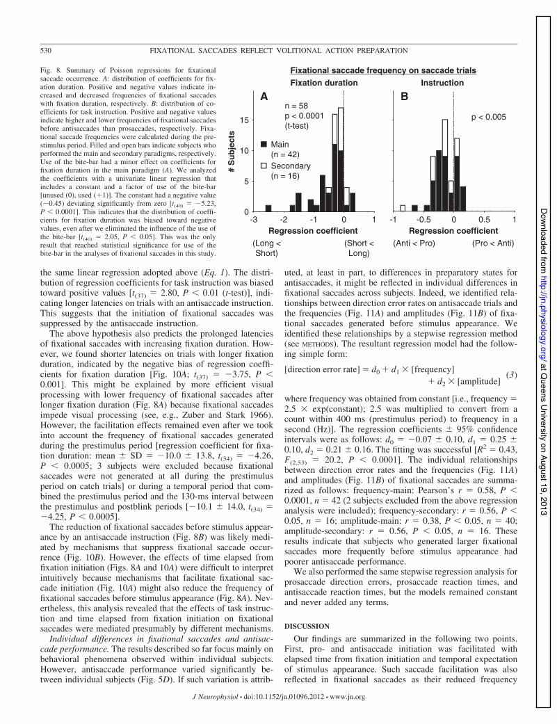
These results suggest that temporal expectation of stimulusappearance facilitates pro- and antisaccade initiation as well asreducing the frequency of fixational saccades before stimulusappearance.
Fixational saccade reduction by antisaccade instruction.The hypothesis that preparatory mechanisms facilitating pro- andantisaccade initiation also reduce the frequency of fixational sac-cades predicts that fixational saccades are reduced more stronglybefore prosaccades than antisaccades because prosaccade reactiontimes are shorter than antisaccade reaction times (Fig. 5, A–C).This prediction is supported further by the fact that neurons in therostral superior colliculus (SC) that are involved in fixationalsaccade generation (Hafed et al. 2009; Hafed and Krauzlis 2012)have weaker activity during fixation on prosaccade trials thanantisaccade trials (Everling et al. 1999).
Despite the above prediction supported by neurophysiolog-ical findings, we observed that fixational saccades were re-duced more strongly before antisaccades than prosaccades(Fig. 6, A and C). We analyzed this phenomenon quantitativelyby applying the same Poisson regressions described above tothe frequency of fixational saccades during the prestimulusperiod. The distribution of regression coefficients for task in-
Saccade trials
-400 -200 0 2000
0.5
1
ProAnti
0 200 400 600
Fixa
tiona
l sac
cade
freq
uenc
y (H
z)
D
Time from 1st fixation pointdisappearance (ms)
Catch trialsA B
0
1
2
Fixa
tiona
l sac
cade
freq
uenc
y (H
z)
Example subject
Population
Time fromstimulus appearance (ms)
Main (n = 42 )
Pro/Antionset
0
2
4
3
1
0
1
2
C
Fig. 6. Time courses of fixational saccade occurrence inthe main paradigm. A: raster and density functions offixational saccade onset times on saccade trials fromexample subject shown in Figs. 2, 3, and 5, A and B.Trials are sorted by pro- and antisaccade reaction times,indicated by circles. B: catch trials from the same examplesubject. Trials are sorted by the latencies of fixationalsaccades evoked by fixation blinks. C and D: averagedensity functions from subjects who performed the mainparadigm on saccade (C) and catch (D) trials, respec-tively. Horizontal bars on x-axes indicate the prestimulusperiod (C) and the postblink period (D), respectively.Blue and red indicate trials with pro- and antisaccadeinstructions, respectively. Density functions were calcu-lated by the convolution of a Gaussian function with SDof 30 ms. The ranges of y-axes are different betweensaccade (A and C) and catch (B and D) trials to capture thedifferent ranges of fixational saccade frequencies.
528 FIXATIONAL SACCADES REFLECT VOLITIONAL ACTION PREPARATION
J Neurophysiol • doi:10.1152/jn.01096.2012 • www.jn.org
at Queens U
niversity on August 19, 2013http://jn.physiology.org/
Dow
nloaded from
struction was biased toward negative values (Fig. 8B) [main:t(41) " &4.68, P $ 0.0001 (t-test); secondary: t(15) " &3.28,P $ 0.01]. This indicates that fixational saccades were reducedmore strongly on antisaccade trials than on prosaccade trials.
The above results suggest that antisaccade preparation re-duced the frequency of fixational saccades. We speculate thatthis reflects the preparation of saccade suppression for inap-propriate direction errors programmed automatically in re-sponse to stimulus appearance on antisaccade trials.
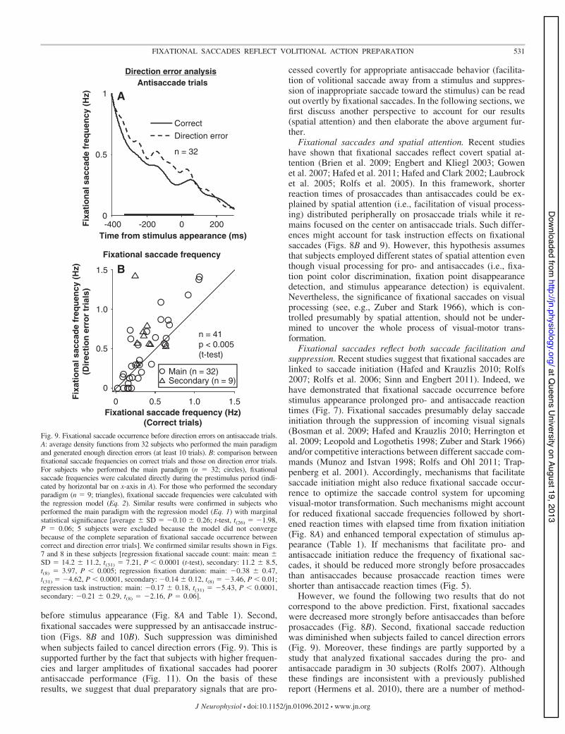
Correlation between fixational saccades and antisaccadeperformance. If fixational saccades reflect preparatory signalsthat suppress direction errors on antisaccade trials, the reduc-tion of fixational saccade occurrence by an antisaccade instruc-tion should be diminished when subjects failed to suppressdirection errors. In the following analysis, we focused on asubset of subjects who produced enough antisaccade trials withdirection errors (at least 10 trials in each block of trials) duringthe main (n " 32) and secondary (n " 9) paradigms.
We confirmed the above prediction qualitatively in the pop-ulation density function derived from subjects who performedthe main paradigm; fixational saccade frequency was lower onantisaccade trials with correct responses than on those withdirection errors (Fig. 9A). We first analyzed this observationsimply by counting fixational saccades during the prestimulus
period for subjects who performed the main paradigm. Thisanalysis confirmed the lower frequency of fixational saccadeson correct trials than direction error trials [circles in Fig. 9B;t(31) " 2.13, P $ 0.05 (paired t-test); see figure legend forresults of regression analysis]. For those who performed thesecondary paradigm, we estimated fixational saccade frequen-cies from the results of the same Poisson regressions adoptedabove (Eq. 2) with the only exception being the replacement oftask instruction (pro/anti) to antisaccade performance ('1:correct; &1: direction error). The lower frequencies of fixa-tional saccades on correct trials were also confirmed in thesesubjects [triangles in Fig. 9B; t(8) " &2.47, P $ 0.05].
The above findings are supported further by the followingobservation in the secondary paradigm: the frequency of fixa-tional saccade occurrence was decreased significantly whensubjects performed antisaccades better in the 2nd block com-pared with the 1st block (Table 1; negative regression coeffi-cients for fixational frequency and 1st/2nd block and for macrodirection error and 1st/2nd block).
The reduced frequency of fixational saccades only beforecorrect antisaccade performance (Fig. 9) and correlated changesin fixational saccade occurrence and antisaccade performancebetween consecutive blocks of trials (Table 1) support thehypothesis that fixational saccades reflect the preparation ofsaccade suppression for direction errors on antisaccade trials.
Fixational saccade readiness probed by fixation blink. Weused catch trials in the main paradigm to probe the readiness offixational saccades by triggering them with a fixation blink(transient disappearance of fixation point; Fig. 6, B and D).This design was inspired by neurophysiological studies inbehaving monkeys that applied electrical microstimulation tothe oculomotor structures, such as the SC and frontal eye field,to probe the readiness of larger saccades (Gold and Shadlen2000; Kustov and Robinson 1996). If the reduction of fixa-tional saccade frequencies that occurred with longer elapsedtime from fixation initiation (Fig. 8A) and an antisaccadeinstruction (Fig. 8B) were caused by mechanisms that suppressfixational saccade occurrence, the latencies of fixational sac-cades evoked by a fixation blink were expected to be prolongedby such suppression.
This prediction was confirmed for task instruction (Fig. 10B).This analysis was focused on a subset of subjects (n " 38) whohad enough trials with fixational saccades (at least 10 trials foreach pro- and antisaccade instruction) evoked during the post-blink period (400-ms window starting 200 ms after 1st fixationpoint disappearance, indicated by horizontal bar in Fig. 6D).We analyzed the latencies of evoked fixational saccades with
-60 -20 0 20 400
5
10
15
25#
Sub
ject
s
Main(n = 42)Secondary(n = 16)
n = 58p < 0.0001(t-test)
Regression coefficient(Shortened) (Prolonged)
Fixational saccade effectson reaction times
20
-40 60
Fig. 7. Influences of fixational saccade occurrence on reaction times. Pro- andantisaccade reaction times were analyzed by linear regressions (Eqs. 1 and 2)with an additional factor of “fixational saccade count” (n " 0, 1, 2,...) duringthe prestimulus period. Filled and open bars indicate subjects who performedthe main and secondary paradigms, respectively.
Table 1. Summary of between-subject analysis in secondary paradigm
Coefficient Fixational Frequency Macro Reaction Time Macro Anti Error
Random/fixed &0.20 (&0.30, &0.11)* &21.3 (&23.2, &19.3)* &0.00 (&0.18, 0.17)1st/2nd block &0.23 (&0.32, &0.13)* 2.21 (0.26, 4.17)* &0.42 (&0.60, &0.25)*
Multiple regressions (Eq. 2) were applied to the frequency of fixational saccade occurrence during the prestimulus period on correct trials (fixationalfrequency), correct pro- and antisaccade reaction times (macro reaction time), and the probability of direction errors on antisaccade trials (macro anti error). Eachnumber in this table indicates a resultant regression coefficient (with 95% confidence interval in parentheses). Fixational frequency had significant negative valuesin both coefficients, each of which indicates that 1) fixational saccade frequencies were decreased in blocks with fixed fixation duration compared with those withrandomized fixation duration (random/fixed) and 2) they were decreased in the 2nd blocks compared with the 1st blocks (1st/2nd block). The same polaritiesof changes were applied to macro reaction time [e.g., shortened reaction times in blocks with fixed fixation duration compared with those with randomizedfixation duration (random/fixed)] as well as macro anti error [e.g., reduced probability of direction errors in the 2nd blocks compared with the 1st blocks (1st/2ndblock)]. *P $ 0.05.
529FIXATIONAL SACCADES REFLECT VOLITIONAL ACTION PREPARATION
J Neurophysiol • doi:10.1152/jn.01096.2012 • www.jn.org
at Queens U
niversity on August 19, 2013http://jn.physiology.org/
Dow
nloaded from
the same linear regression adopted above (Eq. 1). The distri-bution of regression coefficients for task instruction was biasedtoward positive values [t(37) " 2.80, P $ 0.01 (t-test)], indi-cating longer latencies on trials with an antisaccade instruction.This suggests that the initiation of fixational saccades wassuppressed by the antisaccade instruction.
The above hypothesis also predicts the prolonged latenciesof fixational saccades with increasing fixation duration. How-ever, we found shorter latencies on trials with longer fixationduration, indicated by the negative bias of regression coeffi-cients for fixation duration [Fig. 10A; t(37) " &3.75, P $0.001]. This might be explained by more efficient visualprocessing with lower frequency of fixational saccades afterlonger fixation duration (Fig. 8A) because fixational saccadesimpede visual processing (see, e.g., Zuber and Stark 1966).However, the facilitation effects remained even after we tookinto account the frequency of fixational saccades generatedduring the prestimulus period [regression coefficient for fixa-tion duration: mean ! SD " &10.0 ! 13.8, t(34) " &4.26,P $ 0.0005; 3 subjects were excluded because fixationalsaccades were not generated at all during the prestimulusperiod on catch trials] or during a temporal period that com-bined the prestimulus period and the 130-ms interval betweenthe prestimulus and postblink periods [&10.1 ! 14.0, t(34) "&4.25, P $ 0.0005].
The reduction of fixational saccades before stimulus appear-ance by an antisaccade instruction (Fig. 8B) was likely medi-ated by mechanisms that suppress fixational saccade occur-rence (Fig. 10B). However, the effects of time elapsed fromfixation initiation (Figs. 8A and 10A) were difficult to interpretintuitively because mechanisms that facilitate fixational sac-cade initiation (Fig. 10A) might also reduce the frequency offixational saccades before stimulus appearance (Fig. 8A). Nev-ertheless, this analysis revealed that the effects of task instruc-tion and time elapsed from fixation initiation on fixationalsaccades were mediated presumably by different mechanisms.
Individual differences in fixational saccades and antisac-cade performance. The results described so far focus mainly onbehavioral phenomena observed within individual subjects.However, antisaccade performance varied significantly be-tween individual subjects (Fig. 5D). If such variation is attrib-
uted, at least in part, to differences in preparatory states forantisaccades, it might be reflected in individual differences infixational saccades across subjects. Indeed, we identified rela-tionships between direction error rates on antisaccade trials andthe frequencies (Fig. 11A) and amplitudes (Fig. 11B) of fixa-tional saccades generated before stimulus appearance. Weidentified these relationships by a stepwise regression method(see METHODS). The resultant regression model had the follow-ing simple form:
[direction error rate] ! d0 " d1 # [frequency]" d2 # [amplitude]
(3)
where frequency was obtained from constant [i.e., frequency "2.5 # exp(constant); 2.5 was multiplied to convert from acount within 400 ms (prestimulus period) to frequency in asecond (Hz)]. The regression coefficients ! 95% confidenceintervals were as follows: d0 " &0.07 ! 0.10, d1 " 0.25 !0.10, d2 " 0.21 ! 0.16. The fitting was successful [R2 " 0.43,F(2,53) " 20.2, P $ 0.0001]. The individual relationshipsbetween direction error rates and the frequencies (Fig. 11A)and amplitudes (Fig. 11B) of fixational saccades are summa-rized as follows: frequency-main: Pearson’s r " 0.58, P $0.0001, n " 42 (2 subjects excluded from the above regressionanalysis were included); frequency-secondary: r " 0.56, P $0.05, n " 16; amplitude-main: r " 0.38, P $ 0.05, n " 40;amplitude-secondary: r " 0.56, P $ 0.05, n " 16. Theseresults indicate that subjects who generated larger fixationalsaccades more frequently before stimulus appearance hadpoorer antisaccade performance.
We also performed the same stepwise regression analysis forprosaccade direction errors, prosaccade reaction times, andantisaccade reaction times, but the models remained constantand never added any terms.
DISCUSSION
Our findings are summarized in the following two points.First, pro- and antisaccade initiation was facilitated withelapsed time from fixation initiation and temporal expectationof stimulus appearance. Such saccade facilitation was alsoreflected in fixational saccades as their reduced frequency
0
5
15
Fixation duration Instruction
BA
Regression coefficient#
Sub
ject
s
p < 0.005n = 58p < 0.0001(t-test)
(Anti < Pro) (Pro < Anti)Regression coefficient
(Long <Short)
(Short <Long)
-3 -2 -1 0 1 -1 -0.5 0 0.5 1
Main(n = 42)Secondary(n = 16)
10
Fixational saccade frequency on saccade trialsFig. 8. Summary of Poisson regressions for fixationalsaccade occurrence. A: distribution of coefficients for fix-ation duration. Positive and negative values indicate in-creased and decreased frequencies of fixational saccadeswith fixation duration, respectively. B: distribution of co-efficients for task instruction. Positive and negative valuesindicate higher and lower frequencies of fixational saccadesbefore antisaccades than prosaccades, respectively. Fixa-tional saccade frequencies were calculated during the pre-stimulus period. Filled and open bars indicate subjects whoperformed the main and secondary paradigms, respectively.Use of the bite-bar had a minor effect on coefficients forfixation duration in the main paradigm (A). We analyzedthe coefficients with a univariate linear regression thatincludes a constant and a factor of use of the bite-bar[unused (0), used ('1)]. The constant had a negative value(&0.45) deviating significantly from zero [t(40) " &5.23,P $ 0.0001]. This indicates that the distribution of coeffi-cients for fixation duration was biased toward negativevalues, even after we eliminated the influence of the use ofthe bite-bar [t(40) " 2.05, P $ 0.05]. This was the onlyresult that reached statistical significance for use of thebite-bar in the analyses of fixational saccades in this study.
530 FIXATIONAL SACCADES REFLECT VOLITIONAL ACTION PREPARATION
J Neurophysiol • doi:10.1152/jn.01096.2012 • www.jn.org
at Queens U
niversity on August 19, 2013http://jn.physiology.org/
Dow
nloaded from
before stimulus appearance (Fig. 8A and Table 1). Second,fixational saccades were suppressed by an antisaccade instruc-tion (Figs. 8B and 10B). Such suppression was diminishedwhen subjects failed to cancel direction errors (Fig. 9). This issupported further by the fact that subjects with higher frequen-cies and larger amplitudes of fixational saccades had poorerantisaccade performance (Fig. 11). On the basis of theseresults, we suggest that dual preparatory signals that are pro-
cessed covertly for appropriate antisaccade behavior (facilita-tion of volitional saccade away from a stimulus and suppres-sion of inappropriate saccade toward the stimulus) can be readout overtly by fixational saccades. In the following sections, wefirst discuss another perspective to account for our results(spatial attention) and then elaborate the above argument fur-ther.
Fixational saccades and spatial attention. Recent studieshave shown that fixational saccades reflect covert spatial at-tention (Brien et al. 2009; Engbert and Kliegl 2003; Gowenet al. 2007; Hafed et al. 2011; Hafed and Clark 2002; Laubrocket al. 2005; Rolfs et al. 2005). In this framework, shorterreaction times of prosaccades than antisaccades could be ex-plained by spatial attention (i.e., facilitation of visual process-ing) distributed peripherally on prosaccade trials while it re-mains focused on the center on antisaccade trials. Such differ-ences might account for task instruction effects on fixationalsaccades (Figs. 8B and 9). However, this hypothesis assumesthat subjects employed different states of spatial attention eventhough visual processing for pro- and antisaccades (i.e., fixa-tion point color discrimination, fixation point disappearancedetection, and stimulus appearance detection) is equivalent.Nevertheless, the significance of fixational saccades on visualprocessing (see, e.g., Zuber and Stark 1966), which is con-trolled presumably by spatial attention, should not be under-mined to uncover the whole process of visual-motor trans-formation.
Fixational saccades reflect both saccade facilitation andsuppression. Recent studies suggest that fixational saccades arelinked to saccade initiation (Hafed and Krauzlis 2010; Rolfs2007; Rolfs et al. 2006; Sinn and Engbert 2011). Indeed, wehave demonstrated that fixational saccade occurrence beforestimulus appearance prolonged pro- and antisaccade reactiontimes (Fig. 7). Fixational saccades presumably delay saccadeinitiation through the suppression of incoming visual signals(Bosman et al. 2009; Hafed and Krauzlis 2010; Herrington etal. 2009; Leopold and Logothetis 1998; Zuber and Stark 1966)and/or competitive interactions between different saccade com-mands (Munoz and Istvan 1998; Rolfs and Ohl 2011; Trap-penberg et al. 2001). Accordingly, mechanisms that facilitatesaccade initiation might also reduce fixational saccade occur-rence to optimize the saccade control system for upcomingvisual-motor transformation. Such mechanisms might accountfor reduced fixational saccade frequencies followed by short-ened reaction times with elapsed time from fixation initiation(Fig. 8A) and enhanced temporal expectation of stimulus ap-pearance (Table 1). If mechanisms that facilitate pro- andantisaccade initiation reduce the frequency of fixational sac-cades, it should be reduced more strongly before prosaccadesthan antisaccades because prosaccade reaction times wereshorter than antisaccade reaction times (Fig. 5).
However, we found the following two results that do notcorrespond to the above prediction. First, fixational saccadeswere decreased more strongly before antisaccades than beforeprosaccades (Fig. 8B). Second, fixational saccade reductionwas diminished when subjects failed to cancel direction errors(Fig. 9). Moreover, these findings are partly supported by astudy that analyzed fixational saccades during the pro- andantisaccade paradigm in 30 subjects (Rolfs 2007). Althoughthese findings are inconsistent with a previously publishedreport (Hermens et al. 2010), there are a number of method-
-400 -200 0 2000
0.5
1
CorrectDirection error
Antisaccade trials
A
B
Fixa
tiona
l sac
cade
freq
uenc
y (H
z)
n = 32
Time from stimulus appearance (ms)
Fixational saccade frequency
0 0.5 1.0
0
0.5
1.0
1.5
Fixational saccade frequency (Hz)(Correct trials)
Fixa
tiona
l sac
cade
freq
uenc
y (H
z)(D
irec
tion
erro
r tr
ials
)
n = 41p < 0.005(t-test)
1.5
Main (n = 32)Secondary (n = 9)
Direction error analysis
Fig. 9. Fixational saccade occurrence before direction errors on antisaccade trials.A: average density functions from 32 subjects who performed the main paradigmand generated enough direction errors (at least 10 trials). B: comparison betweenfixational saccade frequencies on correct trials and those on direction error trials.For subjects who performed the main paradigm (n " 32; circles), fixationalsaccade frequencies were calculated directly during the prestimulus period (indi-cated by horizontal bar on x-axis in A). For those who performed the secondaryparadigm (n " 9; triangles), fixational saccade frequencies were calculated withthe regression model (Eq. 2). Similar results were confirmed in subjects whoperformed the main paradigm with the regression model (Eq. 1) with marginalstatistical significance [average ! SD " &0.10 ! 0.26; t-test, t(26) " &1.98,P " 0.06; 5 subjects were excluded because the model did not convergebecause of the complete separation of fixational saccade occurrence betweencorrect and direction error trials]. We confirmed similar results shown in Figs.7 and 8 in these subjects [regression fixational saccade count: main: mean !SD " 14.2 ! 11.2, t(31) " 7.21, P $ 0.0001 (t-test), secondary: 11.2 ! 8.5,t(8) " 3.97, P $ 0.005; regression fixation duration: main: &0.38 ! 0.47,t(31) " &4.62, P $ 0.0001, secondary: &0.14 ! 0.12, t(8) " &3.46, P $ 0.01;regression task instruction: main: &0.17 ! 0.18, t(31) " &5.43, P $ 0.0001,secondary: &0.21 ! 0.29, t(8) " &2.16, P " 0.06].
531FIXATIONAL SACCADES REFLECT VOLITIONAL ACTION PREPARATION
J Neurophysiol • doi:10.1152/jn.01096.2012 • www.jn.org
at Queens U
niversity on August 19, 2013http://jn.physiology.org/
Dow
nloaded from

ological differences [e.g., the number of our subjects (n " 58in total) was approximately 6 times larger than in the previousstudy (n " 10 in their immediate task); pro- and antisaccadetrials were randomly interleaved in our paradigms, while theywere blocked in the previous study], which will have to beaddressed in the future. Nevertheless, because fixational sac-cade suppression by antisaccade preparation cannot be ex-plained by the above mechanisms that facilitate saccade initi-ation, it should reflect other mechanisms that are specificallyinvolved in suppression of inappropriate saccades.
We therefore conclude that fixational saccades reflect twoaspects of volitional action preparation required for appropri-ate saccade behavior: 1) facilitation of saccade initiation and2) suppression of inappropriate saccades.
Do fixational saccades reflect preparatory states in superiorcolliculus? The two aspects of volitional saccade preparation(facilitation and suppression) reflected in fixational saccadesmight be originated from the rostral SC because it is critical forfixational saccade generation (Hafed et al. 2009; Hafed andKrauzlis 2012). However, our results are inconsistent with thefollowing fact: rostral SC neurons have higher activity onantisaccade trials than on prosaccade trials during fixation(Everling et al. 1999), while fixational saccades were sup-
pressed more strongly before antisaccades than before prosac-cades (Figs. 8B and 10B).
Neurons in the caudal SC encode larger saccades (Sparks2002), but recent behavioral studies suggest their potentialinvolvement in fixational saccades (Brien et al. 2009; Engbertand Kliegl 2003; Gowen et al. 2007; Hafed et al. 2011; Hafedand Clark 2002; Laubrock et al. 2005; Rolfs et al. 2005).However, this idea has the following inconsistency: the prepa-ratory activity of caudal SC neurons is higher when reactiontimes are shorter (Everling et al. 1999), while fixational sac-cade frequencies were lower when reaction times were shorter(Fig. 7).
Recently developed SC models that integrate the rostral andcaudal SC in a continuous motor map could potentially resolvethe above inconsistencies (Engbert 2012; Hafed et al. 2009;Rolfs et al. 2008). On the basis of the models and physiologicalfindings (Everling et al. 1999), we suggest the followinghypothesis. The spatial distribution of neural activity is focusedon the rostral SC on antisaccade trials, while it is more spreadto the caudal SC on prosaccade trials. If fixational saccades aretriggered when the whole distribution is shifted off the center,they should be more frequent before prosaccades than beforeantisaccades (Fig. 8B) because there are more active neurons
-60 -40 0 20 400
2
8
InstructionFixation duration
A B
# S
ubje
cts
-40 -20 0 20 40
n = 38p < 0.001(t-test)
p < 0.01
Regression coefficient(Long < Short) (Short < Long)
Regression coefficient(Anti < Pro) (Pro < Anti)
0606--20 60
4
6
Evoked fixational saccade latency on catch trialsFig. 10. Summary of evoked fixational saccade latencies oncatch trials. A: distribution of coefficients for fixation dura-tion. Positive and negative values indicate increased anddecreased fixational saccade latencies with fixation duration,respectively. B: distribution of coefficients for task instruc-tion. Positive and negative values indicate longer and shorterfixational saccade latencies on trials with an antisaccadeinstruction than those with a prosaccade instruction, respec-tively. Fixational saccade latencies were calculated from 1stfixation point disappearance. First fixational saccades evokedduring the postblink period were analyzed. This analysis wasfocused on 38 subjects who had enough catch trials withevoked fixational saccades (at least 10 trials). We confirmedthe results of fixational saccades shown in Figs. 7 and 8 inthese subjects [regression fixational saccade count: mean !SD " 14.8 ! 10.9, t-test, t(37) " 8.36, P $ 0.0001; regres-sion fixation duration: &0.49 ! 0.59, t(37) " &5.08,P $ 0.0001; regression task instruction: &0.13 ! 0.19,t(37) " &4.38, P $ 0.0001].
0 0.5 1.0 1.5
0
20
40
60
80
0 0.5 1.0 1.5
Frequency Amplitude
% A
nti d
irec
tion
erro
r tr
ials
Fixational saccadefrequency (Hz)
Fixational saccadeamplitude (deg)
n = 58r = 0.66p < 0.0001(Pearson)
A BMain (n = 40)Secondary (n = 16)
n = 56r = 0.40p < 0.005
Main (n = 42)Secondary (n = 16)
Individual differences
Fig. 11. Individual differences in direction error rates onantisaccade trials and fixational saccades. A: correlationbetween direction error rates and the frequencies offixational saccades before stimulus appearance. B: cor-relation between direction error rates and the amplitudesof fixational saccades before stimulus appearance. Cir-cles and triangles indicate subjects who performed mainand secondary paradigms, respectively.
532 FIXATIONAL SACCADES REFLECT VOLITIONAL ACTION PREPARATION
J Neurophysiol • doi:10.1152/jn.01096.2012 • www.jn.org
at Queens U
niversity on August 19, 2013http://jn.physiology.org/
Dow
nloaded from

prone to random noise. This predicts that those subjects withhigher frequency and larger amplitude of fixational saccadesalso had wider distributions of SC neural activity. This fitsnicely with their higher direction error rates on antisaccadetrials (Fig. 11), because higher preparatory activity in caudalSC neurons is prone to trigger a direction error saccade inresponse to stimulus appearance (Everling et al. 1998).
The above model might also explain the paradoxical effectsof elapsed time from fixation initiation on the frequency (Fig.8A) and latency (Fig. 10A) of fixational saccades by thefollowing mechanism. The left and right caudal SCs receivepreparatory signals that reflect the equal probabilities of up-coming pro- or antisaccade directions (i.e., 50% for bothleftward and rightward) (Dorris and Munoz 1998). Such pre-paratory signals balance the SC map and reduce the frequencyof fixational saccades before stimulus appearance (Fig. 8A).Furthermore, the enhanced activity in the caudal SC facilitatesthe initiation of fixational saccades evoked by a fixation blink(Fig. 10A). However, this hypothesis requires noise reductionnot to trigger fixational saccades with high preparatory activity.
The model provides a promising framework to account forfixational saccade as well as pro- and antisaccade behaviorcorrectively, although it will have to be tested quantitatively(e.g., Engbert 2012). It is also important to address how the SCmap is read out by the brain stem premotor circuitry forfixational saccade control (Otero-Millan et al. 2011; Rolfs et al.2008; Van Gisbergen et al. 1981; Van Horn and Cullen 2012).Nevertheless, the model helps us seek a potential source of thedual preparatory signals of saccade facilitation and suppressionreflected in fixational saccades, which we discuss in the fol-lowing section.
Do fixational saccades reflect preparatory states in basalganglia? The spatial-temporal activity on the SC map iscontrolled by inhibitory output signals from the basal ganglia.The output signals are then controlled by the caudate nucleus,a major input stage of the basal ganglia that integrates a varietyof cortical and thalamic signals (Hikosaka et al. 2000; Mink1996; Watanabe and Munoz 2011). We have reported previ-ously the preparatory activity of putative projection neurons inmonkey caudate nucleus (Watanabe and Munoz 2010). Inter-estingly, the caudate preparatory activity has the following fourcharacteristics that are the mirror image of fixational saccadecharacteristics. 1) Caudate neurons have higher preparatoryactivity before pro- and antisaccades with shorter reactiontimes (for corresponding fixational saccades in this study, seeFig. 7). 2) The caudate preparatory activity increases withtemporal expectation of stimulus appearance (see also Fig. 8Aand Table 1). 3) An antisaccade instruction enhances thepreparatory activity of a subset of caudate neurons encodingvolitional saccades (see also Fig. 8B). 4) Such enhancement ofpreparatory activity is absent when direction errors are gener-ated instead of correct antisaccades (see also Fig. 9).
The striking similarity between the preparatory activity ofcaudate neurons and fixational saccade characteristics suggeststhat saccade facilitation and suppression reflected in fixationalsaccades may originate from the basal ganglia. Indeed, this isin line with their anatomical connections: the caudate nucleusgives rise to two pathways that facilitate and suppress saccadeinitiation, respectively (Hikosaka et al. 2000; Mink 1996;Watanabe and Munoz 2011). Accordingly, we propose a newhypothesis that saccade facilitation and suppression reflected in
fixational saccades are mediated by the facilitation and sup-pression pathways (also known as direct and indirect pathways,respectively) in the basal ganglia. To test this hypothesis, it willbe critical to disentangle the individual contributions of thefacilitation and suppression pathways to understand how sig-nals carried by these pathways converge on the SC map andinfluence fixational saccades.
Future direction of fixational saccade analysis for basal gan-glia disorders. Recent neuroimaging studies have shown thatantisaccade deficits (longer reaction times and higher directionerror rates) in Parkinson’s disease and attention deficit hyperac-tivity disorder, both of which induce basal ganglia dysfunctions(Giedd et al. 2001; Obeso et al. 2000), are explained by inappro-priate volitional saccade preparation (Cameron et al. 2012;Hakvoort Schwerdtfeger et al. 2013). Furthermore, unstable visualfixation by frequent fixational saccades has been reported in thesame disorders (Gould et al. 2001; Shaikh et al. 2011). Ourfindings bridge these two lines of research and suggest that fixa-tional saccades may be used as a potential biomarker to detectpatients with basal ganglia disorders that induce difficulties involitional action preparation.
Inappropriate antisaccade preparation (Cameron et al. 2012;Hakvoort Schwerdtfeger et al. 2013) might be probed as thelack of fixational saccade suppression by an antisaccade in-struction (Figs. 8B and 10B). Abnormal time perception inbasal ganglia disorders (Buhusi and Meck 2005; Castellanosand Tannock 2002) might be detected by fixational saccadeswith their time dependence (Figs. 8A and 10A; Table 1).Saccade impulsivity (Everling and Fischer 1998; Leigh andKennard 2004; Munoz and Everling 2004) might be inferredwell from the frequency and amplitude of fixational saccades(Fig. 11) without the use of cognitively demanding paradigms,such as antisaccades, whose instruction might be difficult tocomprehend in some patients.
Multiple parameters extracted from fixational saccades canbe integrated with the conventional parameters of larger sac-cades (e.g., reaction times) by sophisticated statistical methods,such as machine learning (Benson et al. 2012; Lagun et al.2011; Tseng et al. 2013). Such a new approach may utilizefixational saccades as parts of quantitative clinical diagnosesbased on oculomotor behavior in the future.
ACKNOWLEDGMENTS
We thank Dr. Y. T. Kitamura, S. Asahara, and F. Tanaka for technicalsupport and members of the Munoz lab for comments on this manuscript.
GRANTS
This work was supported by Nakayama Foundation for Human Science,Adaptable and Seamless Technology Transfer Program (ASTEP) throughtarget-driven R&D (AS231Z03528F), Precursory Research for EmbryonicScience and Technology (PRESTO) from the Japan Science and TechnologyAgency, Grants-in-Aid for scientific research from the Japan Society for thePromotion of Science (24120511), and Osaka University Global COE programHuman Behavior and Socioeconomic Dynamics.
DISCLOSURES
No conflicts of interest, financial or otherwise, are declared by the author(s).
AUTHOR CONTRIBUTIONS
Author contributions: M.W. and Y.K. conception and design of research;M.W., Y.M., and L.Z. performed experiments; M.W. analyzed data; M.W.,
533FIXATIONAL SACCADES REFLECT VOLITIONAL ACTION PREPARATION
J Neurophysiol • doi:10.1152/jn.01096.2012 • www.jn.org
at Queens U
niversity on August 19, 2013http://jn.physiology.org/
Dow
nloaded from

D.P.M., and Y.K. interpreted results of experiments; M.W. and Y.K. preparedfigures; M.W. drafted manuscript; M.W., D.P.M., and Y.K. edited and revisedmanuscript; M.W., Y.M., L.Z., D.P.M., and Y.K. approved final version ofmanuscript.
REFERENCES
Amador N, Schlag-Rey M, Schlag J. Primate antisaccade. II. Supplementaryeye field neuronal activity predicts correct performance. J Neurophysiol 91:1672–1689, 2004.
Benson PJ, Beedie SA, Shephard E, Giegling I, Rujescu D, St Clair D.Simple viewing tests can detect eye movement abnormalities that distin-guish schizophrenia cases from controls with exceptional accuracy. BiolPsychiatry 72: 716–724, 2012.
Bosman CA, Womelsdorf T, Desimone R, Fries P. A microsaccadic rhythmmodulates gamma-band synchronization and behavior. J Neurosci 29: 9471–9480, 2009.
Bridgeman B, Palca J. The role of microsaccades in high acuity observationaltasks. Vision Res 20: 813–817, 1980.
Brien DC, Corneil BD, Fecteau JH, Bell AH, Munoz DP. The behaviouraland neurophysiological modulation of microsaccades in monkeys. J EyeMov Res 3: 1–12, 2009.
Buhusi CV, Meck WH. What makes us tick? Functional and neural mecha-nisms of interval timing. Nat Rev Neurosci 6: 755–765, 2005.
Cameron IG, Pari G, Alahyane N, Brien DC, Coe BC, Stroman PW,Munoz DP. Impaired executive function signals in motor brain regions inparkinson’s disease. Neuroimage 60: 1156–1170, 2012.
Carpenter RH, Williams ML. Neural computation of log likelihood incontrol of saccadic eye movements. Nature 377: 59–62, 1995.
Castellanos FX, Tannock R. Neuroscience of attention-deficit/hyperactivitydisorder: The search for endophenotypes. Nat Rev Neurosci 3: 617–628,2002.
Cunnington R, Iansek R, Johnson KA, Bradshaw JL. Movement-relatedpotentials in Parkinson’s disease. Motor imagery and movement prepara-tion. Brain 120: 1339–1353, 1997.
Dafoe JM, Armstrong IT, Munoz DP. The influence of stimulus directionand eccentricity on pro- and anti-saccades in humans. Exp Brain Res 179:563–570, 2007.
Dorris MC, Munoz DP. Saccadic probability influences motor preparationsignals and time to saccadic initiation. J Neurosci 18: 7015–7026, 1998.
Engbert R. Computational modeling of collicular integration of perceptualresponses and attention in microsaccades. J Neurosci 32: 8035–8039, 2012.
Engbert R. Microsaccades: a microcosm for research on oculomotor control,attention, and visual perception. Prog Brain Res 154: 177–192, 2006.
Engbert R, Mergenthaler K. Microsaccades are triggered by low retinalimage slip. Proc Natl Acad Sci USA 103: 7192–7197, 2006.
Engbert R, Kliegl R. Microsaccades uncover the orientation of covert atten-tion. Vision Res 43: 1035–1045, 2003.
Everling S, Desouza JF. Rule-dependent activity for prosaccades and anti-saccades in the primate prefrontal cortex. J Cogn Neurosci 17: 1483–1496,2005.
Everling S, Munoz DP. Neuronal correlates for preparatory set associatedwith pro-saccades and anti-saccades in the primate frontal eye field. JNeurosci 20: 387–400, 2000.
Everling S, Fischer B. The antisaccade: a review of basic research and clinicalstudies. Neuropsychologia 36: 885–899, 1998.
Everling S, Dorris MC, Munoz DP. Reflex suppression in the anti-saccadetask is dependent on prestimulus neural processes. J Neurophysiol 80:1584–1589, 1998.
Everling S, Dorris MC, Klein RM, Munoz DP. Role of primate superiorcolliculus in preparation and execution of anti-saccades and pro-saccades. JNeurosci 19: 2740–2754, 1999.
Fischer B, Weber H. Express saccades and visual attention. Behav Brain Sci16: 553–610, 1993.
Fischer B, Weber H. Characteristics of “anti” saccades in man. Exp Brain Res89: 415–424, 1992.
Giedd JN, Blumenthal J, Molloy E, Castellanos FX. Brain imaging ofattention deficit/hyperactivity disorder. Ann NY Acad Sci 931: 33–49, 2001.
Gold JI, Shadlen MN. Representation of a perceptual decision in developingoculomotor commands. Nature 404: 390–394, 2000.
Gould TD, Bastain TM, Israel ME, Hommer DW, Castellanos FX. Alteredperformance on an ocular fixation task in attention-deficit/hyperactivitydisorder. Biol Psychiatry 50: 633–635, 2001.
Gowen E, Abadi RV, Poliakoff E, Hansen PC, Miall RC. Modulation ofsaccadic intrusions by exogenous and endogenous attention. Brain Res1141: 154–167, 2007.
Haddad GM, Steinman RM. The smallest voluntary saccade: implicationsfor fixation. Vision Res 13: 1075–1086, 1973.
Hafed ZM, Krauzlis RJ. Similarity of superior colliculus involvement inmicrosaccade and saccade generation. J Neurophysiol 107: 1904–1916,2012.
Hafed ZM, Krauzlis RJ. Microsaccadic suppression of visual bursts in theprimate superior colliculus. J Neurosci 30: 9542–9547, 2010.
Hafed ZM, Clark JJ. Microsaccades as an overt measure of covert attentionshifts. Vision Res 42: 2533–2545, 2002.
Hafed ZM, Lovejoy LP, Krauzlis RJ. Modulation of microsaccades inmonkey during a covert visual attention task. J Neurosci 31: 15219–15230,2011.
Hafed ZM, Goffart L, Krauzlis RJ. A neural mechanism for microsaccadegeneration in the primate superior colliculus. Science 323: 940–943, 2009.
Haggard P. Human volition: towards a neuroscience of will. Nat Rev Neurosci9: 934–946, 2008.
Hakvoort Schwerdtfeger RM, Alahyane N, Brien DC, Coe BC, StromanPW, Munoz DP. Preparatory neural networks are impaired in adults withattention-deficit/hyperactivity disorder during the antisaccade task. Neuro-image 2: 63–78, 2013.
Hallett PE. Primary and secondary saccades to goals defined by instructions.Vision Res 18: 1279–1296, 1978.
Hermens F, Zanker JM, Walker R. Microsaccades and preparatory set: acomparison between delayed and immediate, exogenous and endogenouspro- and anti-saccades. Exp Brain Res 201: 489–498, 2010.
Herrington TM, Masse NY, Hachmeh KJ, Smith JE, Assad JA, Cook EP.The effect of microsaccades on the correlation between neural activity andbehavior in middle temporal, ventral intraparietal, and lateral intraparietalareas. J Neurosci 29: 5793–5805, 2009.
Hikosaka O, Takikawa Y, Kawagoe R. Role of the basal ganglia in thecontrol of purposive saccadic eye movements. Physiol Rev 80: 953–978,2000.
Ko HK, Poletti M, Rucci M. Microsaccades precisely relocate gaze in a highvisual acuity task. Nat Neurosci 13: 1549–1553, 2010.
Kowler E, Steinman RM. The role of small saccades in counting. Vision Res17: 141–146, 1977.
Kunimatsu J, Tanaka M. Roles of the primate motor thalamus in thegeneration of antisaccades. J Neurosci 30: 5108–5117, 2010.
Kustov AA, Robinson DL. Shared neural control of attentional shifts and eyemovements. Nature 384: 74–77, 1996.
Lagun D, Manzanares C, Zola SM, Buffalo EA, Agichtein E. Detectingcognitive impairment by eye movement analysis using automatic classifi-cation algorithms. J Neurosci Methods 201: 196–203, 2011.
Laubrock J, Engbert R, Kliegl R. Microsaccade dynamics during covertattention. Vision Res 45: 721–730, 2005.
Leigh RJ, Kennard C. Using saccades as a research tool in the clinicalneurosciences. Brain 127: 460–477, 2004.
Leopold DA, Logothetis NK. Microsaccades differentially modulate neuralactivity in the striate and extrastriate visual cortex. Exp Brain Res 123:341–345, 1998.
McLoughlin G, Albrecht B, Banaschewski T, Rothenberger A, BrandeisD, Asherson P, Kuntsi J. Electrophysiological evidence for abnormalpreparatory states and inhibitory processing in adult ADHD. Behav BrainFunct 6: 66, 2010.
Mink JW. The basal ganglia: focused selection and inhibition of competingmotor programs. Prog Neurobiol 50: 381–425, 1996.
Moller F, Laursen ML, Tygesen J, Sjolie AK. Binocular quantification andcharacterization of microsaccades. Graefes Arch Clin Exp Ophthalmol 240:765–770, 2002.
Munoz DP, Everling S. Look away: the anti-saccade task and the voluntarycontrol of eye movement. Nat Rev Neurosci 5: 218–228, 2004.
Munoz DP, Istvan PJ. Lateral inhibitory interactions in the intermediatelayers of the monkey superior colliculus. J Neurophysiol 79: 1193–1209,1998.
Nachev P, Kennard C, Husain M. Functional role of the supplementary andpre-supplementary motor areas. Nat Rev Neurosci 9: 856–869, 2008.
Obeso JA, Rodriguez-Oroz MC, Rodriguez M, Lanciego JL, Artieda J,Gonzalo N, Olanow CW. Pathophysiology of the basal ganglia in Parkin-son’s disease. Trends Neurosci 23: S8–S19, 2000.
Oswal A, Ogden M, Carpenter RH. The time course of stimulus expectationin a saccadic decision task. J Neurophysiol 97: 2722–2730, 2007.
534 FIXATIONAL SACCADES REFLECT VOLITIONAL ACTION PREPARATION
J Neurophysiol • doi:10.1152/jn.01096.2012 • www.jn.org
at Queens U
niversity on August 19, 2013http://jn.physiology.org/
Dow
nloaded from

Otero-Millan J, Macknik SL, Serra A, Leigh RJ, Martinez-Conde S.Triggering mechanisms in microsaccade and saccade generation: a novelproposal. Ann NY Acad Sci 1233: 107–116, 2011.
Pare M, Munoz DP. Saccadic reaction time in the monkey: advancedpreparation of oculomotor programs is primarily responsible for expresssaccade occurrence. J Neurophysiol 76: 3666–3681, 1996.
Rolfs M. In-Between Fixation and Movement: On the Generation of Micro-saccades and What They Convey About Saccade Preparation. (PhD disser-tation). Potsdam, Germany: University of Potsdam, 2007 (http://opus.kobv.de/ubp/volltexte/2007/1458/).
Rolfs M, Ohl S. Visual suppression in the superior colliculus around the timeof microsaccades. J Neurophysiol 105: 1–3, 2011.
Rolfs M, Kliegl R, Engbert R. Toward a model of microsaccade generation:The case of microsaccadic inhibition. J Vis 8: 5.1–5.23, 2008.
Rolfs M, Laubrock J, Kliegl R. Shortening and prolongation of saccadelatencies following microsaccades. Exp Brain Res 169: 369–376, 2006.
Rolfs M, Engbert R, Kliegl R. Crossmodal coupling of oculomotor controland spatial attention in vision and audition. Exp Brain Res 166: 427–439,2005.
Schall JD. On building a bridge between brain and behavior. Annu RevPsychol 55: 23–50, 2004.
Shaikh AG, Xu-Wilson M, Grill S, Zee DS. “Staircase” square-wave jerks inearly parkinson’s disease. Br J Ophthalmol 95: 705–709, 2011.
Sinn P, Engbert R. Saccadic facilitation by modulation of microsaccades innatural backgrounds. Atten Percept Psychophys 73: 1029–1033, 2011.
Smith PL, Ratcliff R. Psychology and neurobiology of simple decisions.Trends Neurosci 27: 161–168, 2004.
Sparks DL. The brainstem control of saccadic eye movements. Nat RevNeurosci 3: 952–964, 2002.
Steinman RM, Cunitz RJ, Timberlake GT, Herman M. Voluntary controlof microsaccades during maintained monocular fixation. Science 155: 1577–1579, 1967.
Trappenberg TP, Dorris MC, Munoz DP, Klein RM. A model of saccadeinitiation based on the competitive integration of exogenous and endogenoussignals in the superior colliculus. J Cogn Neurosci 13: 256–271, 2001.
Tseng PH, Cameron IG, Pari G, Reynolds JN, Munoz DP, Itti L. High-throughput classification of clinical populations from natural viewing eyemovements. J Neurol 260: 275–284, 2013.
Van Gisbergen JA, Robinson DA, Gielen S. A quantitative analysis ofgeneration of saccadic eye movements by burst neurons. J Neurophysiol 45:417–442, 1981.
Van Horn MR, Cullen KE. Coding of microsaccades in three-dimensionalspace by premotor saccadic neurons. J Neurosci 32: 1974–1980, 2012.
Watanabe M, Munoz DP. Probing basal ganglia functions by saccade eyemovements. Eur J Neurosci 33: 2070–2090, 2011.
Watanabe M, Munoz DP. Presetting basal ganglia for volitional actions. JNeurosci 30: 10144–10157, 2010.
Winterson BJ, Collewijn H. Microsaccades during finely guided visuomotortasks. Vision Res 16: 1387–1390, 1976.
Yoshida A, Tanaka M. Enhanced modulation of neuronal activity during anti-saccades in the primate globus pallidus. Cereb Cortex 19: 206–217, 2009.
Zuber BL, Stark L. Saccadic suppression: Elevation of visual thresholdassociated with saccadic eye movements. Exp Neurol 16: 65–79, 1966.
Zuber BL, Stark L, Cook G. Microsaccades and the velocity-amplituderelationship for saccadic eye movements. Science 150: 1459–1460, 1965.
535FIXATIONAL SACCADES REFLECT VOLITIONAL ACTION PREPARATION
J Neurophysiol • doi:10.1152/jn.01096.2012 • www.jn.org
at Queens U
niversity on August 19, 2013http://jn.physiology.org/
Dow
nloaded from