First records of pseudocerotid flatworms (Platyhelminthes
40
130 First records of pseudocerotid flatworms (Platyhelminthes: Polycladida: Cotylea) from Singapore: A taxonomic report with remarks on colour variation D. Marcela Bolaños 1 , Bin Qi Gan 2 & Rene S. L. Ong 3 Abstract. A detailed taxonomic report of 16 species of polyclad flatworms found in Singapore during the Comprehensive Marine Biodiversity Survey is presented. Representatives of the genera Nymphozoon, Phrikoceros, Pseudobiceros, Pseudoceros, and Tytthosoceros are described using high quality photographs, extended descriptions with information on colour variation, together with a compilation of geographic distribution based on the known records. All species belong to the family Pseudocerotidae and all represent new records for Singapore except Pseudobiceros bedfordi, P. hancockanus, and Pseudoceros indicus. Identifications were based mainly on external morphological characters, with particular emphasis on their living colours and patterns. The genus Nymphozoon is re-described, and a new combination Nymphozoon orsaki nov. comb. is established as it is shown that the monotypic genus Maiazoon is a junior synonym of Nymphozoon. Emended diagnoses for Pseudobiceros damawan, P. hancockanus, and Pseudoceros laingensis are also included. Pseudoceros caeruleocinctus is reinstated as a valid species and is recognised as a senior synonym of Pseudoceros sapphirinus, while Pseudobiceros uniarborensis is identified as a junior synonym of Pseudobiceros hancockanus. This study shows that polyclads are significantly diverse in Singapore, and also highlights the need for further studies using both morphological and molecular data to confirm their identities. Key words. Polyclads, Nymphozoon, Pseudoceros, Pseudobiceros, biodiversity, Indo-Pacific RAFFLES BULLETIN OF ZOOLOGY Supplement No. 34: 130–169 Date of publication: 29 June 2016 http://zoobank.org/urn:lsid:zoobank.org:pub:B2C9CA15-15F9-4160-97A7-835F3E59EDDF © National University of Singapore ISSN 2345-7600 (electronic) | ISSN 0217-2445 (print) 1 Department of Natural Resources and the Environment, University of New Hampshire, Durham, New Hampshire 03824, USA; Email: bolanosmarcela@gmail. com ( * corresponding author) 2 Tropical Marine Science Institute, National University of Singapore, S2S, 18 Kent Ridge Road, 119227, Singapore. 3 National Biodiversity Centre, National Parks Board, 1 Cluny Road, 259569, Singapore. INTRODUCTION The order Polycladida is a group of almost exclusively marine, free-living flatworms belonging to the phylum Platyhelminthes, clade Rhabditophora Ehlers, 1986. They are benthic organisms living in a wide range of environments (e.g., rocky shores, coral reefs, mangroves, seagrass, mudflats), although a few pelagic species are also known (Kato, 1938; Faubel, 1984a). Other polyclad species have been found in association with deep-sea fauna at depths of 600 m and 2600 m in the Gulf of Mexico and the North Pacific Ocean, respectively (Quiroga et al., 2006, 2008). Polyclads are distributed worldwide from temperate regions (Brusa et al., 2009; Brusa & Damborenea, 2011) to tropical waters (Marcus & Marcus, 1968; Newman & Cannon, 1994; Bolaños et al., 2006, 2007; Quiroga et al., 2004; Litvaitis et al., 2010), exhibiting the highest diversity throughout the western Indo-Pacific Ocean (Newman & Cannon, 2003 and references therein). Despite the recent attention the group has received (Apte & Pitale, 2011; Bahia et al., 2012, 2014; Bulnes & Torres, 2014; Jie et al., 2014; Marquina et al., 2014a, b; Noreña et al., 2014; Sreeraj & Raghunathan 2011, 2013; Maghsoudlou & Rahimian, 2014), polyclad biodiversity is still underestimated and poorly documented around the world. The order consists of two suborders: Cotylea and Acotylea. Members of Cotylea are characterised by the presence of a ventral, circular adhesive organ, posterior to the female gonopore, called a sucker, in contrast to those in Acotylea where the sucker is generally lacking (Lang, 1884). Currently, about 366 valid species are assigned to the Cotylea, with the family Pseudocerotidae being the most speciose within the suborder (Newman & Cannon, 1994; Tyler et al., 2006– 2015). The internal reproductive anatomy of pseudocerotids is extremely homogeneous, providing little information for taxonomic identification (Newman & Cannon, 1994). External morphological characters, such as number of male and female gonopores, shape of pseudotentacles, arrangement of cerebral and tentacular eyes, shape of pharynx (simple or folded), surface colour and pattern, are essential to distinguish among genera and species. In fact, pseudocerotids are known for exhibiting the greatest diversity of colours and patterns among polyclads, although cryptically coloured species also occur. For this reason, taxonomic identification within the family based exclusively on colours and patterns is fairly reliable. Preliminary molecular studies have validated species distinction based on colour patterns, as well as the
-
Upload
khangminh22 -
Category
Documents
-
view
1 -
download
0
Transcript of First records of pseudocerotid flatworms (Platyhelminthes
130
Bolaños et al.: Pseudocerotid flatworms from Singapore
First records of pseudocerotid flatworms (Platyhelminthes: Polycladida: Cotylea) from Singapore: A taxonomic report with remarks on colour variation
D. Marcela Bolaños1, Bin Qi Gan2 & Rene S. L. Ong3
Abstract. A detailed taxonomic report of 16 species of polyclad flatworms found in Singapore during the Comprehensive Marine Biodiversity Survey is presented. Representatives of the genera Nymphozoon, Phrikoceros, Pseudobiceros, Pseudoceros, and Tytthosoceros are described using high quality photographs, extended descriptions with information on colour variation, together with a compilation of geographic distribution based on the known records. All species belong to the family Pseudocerotidae and all represent new records for Singapore except Pseudobiceros bedfordi, P. hancockanus, and Pseudoceros indicus. Identifications were based mainly on external morphological characters, with particular emphasis on their living colours and patterns. The genus Nymphozoon is re-described, and a new combination Nymphozoon orsaki nov. comb. is established as it is shown that the monotypic genus Maiazoon is a junior synonym of Nymphozoon. Emended diagnoses for Pseudobiceros damawan, P. hancockanus, and Pseudoceros laingensis are also included. Pseudoceros caeruleocinctus is reinstated as a valid species and is recognised as a senior synonym of Pseudoceros sapphirinus, while Pseudobiceros uniarborensis is identified as a junior synonym of Pseudobiceros hancockanus. This study shows that polyclads are significantly diverse in Singapore, and also highlights the need for further studies using both morphological and molecular data to confirm their identities.
Key words. Polyclads, Nymphozoon, Pseudoceros, Pseudobiceros, biodiversity, Indo-Pacific
RAFFLES BULLETIN OF ZOOLOGY Supplement No. 34: 130–169Date of publication: 29 June 2016http://zoobank.org/urn:lsid:zoobank.org:pub:B2C9CA15-15F9-4160-97A7-835F3E59EDDF
© National University of SingaporeISSN 2345-7600 (electronic) | ISSN 0217-2445 (print)
1Department of Natural Resources and the Environment, University of New Hampshire, Durham, New Hampshire 03824, USA; Email: [email protected] (*corresponding author)2Tropical Marine Science Institute, National University of Singapore, S2S, 18 Kent Ridge Road, 119227, Singapore.3National Biodiversity Centre, National Parks Board, 1 Cluny Road, 259569, Singapore.
INTRODUCTION
The order Polycladida is a group of almost exclusively marine, free-living flatworms belonging to the phylum Platyhelminthes, clade Rhabditophora Ehlers, 1986. They are benthic organisms living in a wide range of environments (e.g., rocky shores, coral reefs, mangroves, seagrass, mudflats), although a few pelagic species are also known (Kato, 1938; Faubel, 1984a). Other polyclad species have been found in association with deep-sea fauna at depths of 600 m and 2600 m in the Gulf of Mexico and the North Pacific Ocean, respectively (Quiroga et al., 2006, 2008). Polyclads are distributed worldwide from temperate regions (Brusa et al., 2009; Brusa & Damborenea, 2011) to tropical waters (Marcus & Marcus, 1968; Newman & Cannon, 1994; Bolaños et al., 2006, 2007; Quiroga et al., 2004; Litvaitis et al., 2010), exhibiting the highest diversity throughout the western Indo-Pacific Ocean (Newman & Cannon, 2003 and references therein). Despite the recent attention the
group has received (Apte & Pitale, 2011; Bahia et al., 2012, 2014; Bulnes & Torres, 2014; Jie et al., 2014; Marquina et al., 2014a, b; Noreña et al., 2014; Sreeraj & Raghunathan 2011, 2013; Maghsoudlou & Rahimian, 2014), polyclad biodiversity is still underestimated and poorly documented around the world.
The order consists of two suborders: Cotylea and Acotylea. Members of Cotylea are characterised by the presence of a ventral, circular adhesive organ, posterior to the female gonopore, called a sucker, in contrast to those in Acotylea where the sucker is generally lacking (Lang, 1884). Currently, about 366 valid species are assigned to the Cotylea, with the family Pseudocerotidae being the most speciose within the suborder (Newman & Cannon, 1994; Tyler et al., 2006–2015). The internal reproductive anatomy of pseudocerotids is extremely homogeneous, providing little information for taxonomic identification (Newman & Cannon, 1994). External morphological characters, such as number of male and female gonopores, shape of pseudotentacles, arrangement of cerebral and tentacular eyes, shape of pharynx (simple or folded), surface colour and pattern, are essential to distinguish among genera and species. In fact, pseudocerotids are known for exhibiting the greatest diversity of colours and patterns among polyclads, although cryptically coloured species also occur. For this reason, taxonomic identification within the family based exclusively on colours and patterns is fairly reliable. Preliminary molecular studies have validated species distinction based on colour patterns, as well as the

131
RAFFLES BULLETIN OF ZOOLOGY 2016
discrimination of genera within pseudocerotids using external morphology (Goggin & Newman, 1996; Litvaitis & Newman, 2001). While molecular data are needed to understand polyclad relationships, the external morphological characters mentioned above form the current basis for pseudocerotid identification. Therefore, it is imperative that animals are examined carefully while still alive, because these features become distorted or lost entirely after fixation.
Little is known about the polyclad fauna of Singapore. The oldest records are by Collingwood (1876) and Laidlaw (1903), who described five species found at the former Singapore Harbour. Collingwood (1876) reported four pseudocerotid species, solely based on drawings and brief notes made by two deceased collectors. Of these, Pseudoceros lacteus (Collingwood, 1876) and Pseudobiceros hancockanus (Collingwood, 1876) are valid species, while Pseudoceros buskii (Collingwood, 1876) and Pseudoceros kelartii (Collingwood, 1876) are currently considered incertae sedis (Faubel, 1984b; Newman & Cannon, 1994; Tyler et al., 2006–2015). Based on a badly damaged specimen, Laidlaw (1903) described the well-known Persian carpet polyclad, Pseudobiceros bedfordi (Laidlaw, 1903). Since then, no other formal taxonomic studies from Singapore have been published.
This study represents the first exhaustive taxonomic report of polyclads found in Singapore during the Comprehensive Marine Biodiversity Survey. This five-year survey was a collaborative effort between the National Parks Board and
National University of Singapore. Its main objective was the documentation of species diversity and distribution of marine life in the country. Approximately 65 cotylean species were encountered during this survey. Of these, 16 species are documented here. Additional descriptions of new records and new species of cotyleans and acotyleans will be presented in future works. All species reported in this study are new records for Singapore except Pseudobiceros bedfordi, Pseudobiceros hancockanus, and Pseudoceros indicus (Newman & Schupp, 2002). This study includes high quality photographs, emended diagnoses for some of the species, extended descriptions with information on colour variation, a compilation of geographic distribution based on known records, and updated discussions and comparisons with similar species based on newly collected and well-preserved material. These have resulted in revised synonymies, the elimination of a genus, and a new species combination. Since colour and patterns are key morphological characters used to distinguish the majority of the cotylean species, histological information of the reproductive anatomy is only incorporated when needed. This study not only shows the significant diversity of the polyclad fauna of Singapore but also highlights the importance of further studies using both morphological and molecular data for species identification.
MATERIAL AND METHODS
From October 2010 to March 2015, specimens were collected from 19 locations around Singapore and its offshore islands (Fig. 1; Table 1). The intertidal zone of Singapore
Fig. 1. Collection sites of cotylean flatworms in Singapore. (1) Pulau Ubin; (2) Pulau Sekudu; (3) Kusu Island; (4) Lazarus Island; (5) St. John’s Island; (6) Pulau Subar Laut; (7) Pulau Subar Darat; (8) Labrador Park; (9) Cyrene Reef; (10) Pulau Hantu; (11) Terumbu Semakau; (12) Pulau Semakau; (13) Beting Bemban Besar; (14) Pulau Salu; (15) Terumbu Salu; (16) Pulau Pawai; (17) Pulau Senang; (18) Raffles Lighthouse; (19) Pulau Jong.

132
Bolaños et al.: Pseudocerotid flatworms from SingaporeTa
ble
1. L
ist o
f the
exa
min
ed p
olyc
lad
spec
imen
s co
llect
ed in
Sin
gapo
re, i
nclu
ding
mus
eum
vou
cher
num
bers
, loc
ality
, dep
th, a
nd c
olou
r pat
tern
gro
up. (
*) p
hoto
grap
hic
reco
rd o
nly;
(–) i
nter
tidal
; (~
) no
cat
egor
isat
ion;
(+)
Bas
ed a
nd m
odifi
ed f
rom
New
man
& C
anno
n (1
997)
.
Spec
ies
Mus
eum
Vou
cher
No.
Loc
ality
Dep
th (m
)C
olou
r Pa
tter
n G
roup
(+)
Nym
phoz
oon
Hym
an, 1
959
N. b
ayer
i Hym
an, 1
959
ZRC
.PLA
.000
2 ZR
C.P
LA.0
003
ZRC
.PL
A.0
004
Pula
u Se
mak
auK
usu
Isla
ndTe
rum
bu S
alu
– – –
~
N. o
rsak
i (N
ewm
an &
Can
non,
199
6) n
ov. c
omb.
ZRC
.PLA
.000
5La
zaru
s Is
land
–~
Phri
koce
ros
New
man
& C
anno
n, 1
996
P. b
aiba
iye
New
man
& C
anno
n, 1
996
ZRC
.PLA
.000
6Pu
lau
Ubi
n–
~
Pseu
dobi
cero
s Fa
ubel
, 198
4
P. b
edfo
rdi (
Laid
law
, 190
3)ZR
C.P
LA.0
007
ZRC
.PLA
.000
8*
Sain
t Joh
n’s
Isla
ndPu
lau
Ubi
nLa
zaru
s Is
land
– – –
5
P. d
amaw
an N
ewm
an &
Can
non,
199
4*
Kus
u Is
land
4–7
4
P. fl
ower
si N
ewm
an &
Can
non,
199
7ZR
C.P
LA.0
009
ZRC
.PLA
.001
0Pu
lau
Sem
akau
Pula
u Se
mak
au8 –
2
P. fu
lgor
New
man
& C
anno
n, 1
994
ZRC
.PLA
.001
1ZR
C.P
LA.0
013
ZRC
.PLA
.001
2ZR
C.P
LA.0
014
ZRC
.PLA
.001
5ZR
C.P
LA.0
016
Sain
t Joh
n’s
Isla
ndSa
int J
ohn’
s Is
land
Teru
mbu
Sem
akau
Raf
fles
Ligh
thou
seTe
rum
bu S
alu
Laza
rus
Isla
nd
– – – – – –
6
P. h
anco
ckan
us (C
ollin
gwoo
d, 1
876)
ZRC
.PLA
.001
8ZR
C.P
LA.0
019
ZRC
.PLA
.002
0ZR
C.P
LA.0
025
ZRC
.PLA
.002
1ZR
C.P
LA.0
022
ZRC
.PLA
.002
3ZR
C.P
LA.0
024
ZRC
.PLA
.002
6ZR
C.P
LA.0
027
Sain
t Joh
n’s
Isla
ndSa
int J
ohn’
s Is
land
Pula
u Su
bar
Dar
atPu
lau
Suba
r D
arat
Pul
au S
alu
Laza
rus
Isla
ndB
etin
g B
emba
n B
esar
Cyr
ene
Ree
fR
affle
s Li
ghth
ouse
Kus
u Is
land
– – – – – 16 – – –4–
20
1
P. h
yman
ae N
ewm
an &
Can
non,
199
7ZR
C.P
LA.0
017
Pula
u Se
nang
5–12
1
*La
zaru
s Is
land
–

133
RAFFLES BULLETIN OF ZOOLOGY 2016
Spec
ies
Mus
eum
Vou
cher
No.
Loc
ality
Dep
th (m
)C
olou
r Pa
tter
n G
roup
(+)
Pseu
doce
ros
Lan
g, 1
884
P. b
ifurc
us P
rudh
oe, 1
989
ZRC
.PLA
.002
8ZR
C.P
LA.0
029
Pula
u U
bin
Laza
rus
Isla
nd– –
3
P. c
aeru
leoc
inct
us H
yman
, 195
9ZR
C.P
LA.0
044
ZRC
.PLA
.004
5ZR
C.P
LA.0
046
Laza
rus
Isla
ndC
yren
e R
eef
Bet
ing
Bem
ban
Bes
ar
–5–
155–
11
2
P. c
onci
nnus
(Col
lingw
ood,
187
6)ZR
C.P
LA.0
030
ZRC
.PLA
.003
1ZR
C.P
LA.0
032
ZRC
.PLA
.003
3ZR
C.P
LA.0
036
ZRC
.PLA
.003
4ZR
C.P
LA.0
035
ZRC
.PLA
.003
7ZR
C.P
LA.0
038
ZRC
.PLA
.003
9ZR
C.P
LA.0
049
Cyr
ene
Ree
fPu
lau
Sem
akau
Sain
t Joh
n’s
Isla
ndLa
zaru
s Is
land
Laza
rus
Isla
ndPu
lau
Paw
aiPu
lau
Salu
Pula
u Su
bar
Laut
Raf
fles
Ligh
thou
sePu
lau
Sena
ngLa
brad
or P
ark
– – – – – – – – –5–
15 –
3
P. in
dicu
s N
ewm
an &
Sch
upp,
200
2ZR
C.P
LA.0
061
ZRC
.PLA
.006
2ZR
C.P
LA.0
063
ZRC
.PLA
.006
4ZR
C.P
LA.0
066
ZRC
.PLA
.005
0ZR
C.P
LA.0
051
ZRC
.PLA
.005
2ZR
C.P
LA.0
053
Pula
u U
bin
Pula
u Se
kudu
Laza
rus
Isla
ndPu
lau
Paw
aiPu
lau
Sena
ngK
usu
Isla
ndPu
lau
Seku
duPu
lau
Han
tuSa
int J
ohn’
s Is
land
– – 16 5–10
5–15 – – – –
4
P. la
inge
nsis
New
man
& C
anno
n, 1
998
ZRC
.PLA
.004
0Pu
lau
Ubi
n–
4
P. r
ubro
tent
acul
atus
Kab
urak
i, 19
23ZR
C.P
LA.0
041
ZRC
.PLA
.004
2ZR
C.P
LA.0
043
Sain
t Joh
n’s
Isla
ndPu
lau
Jong
Raf
fles
Ligh
thou
se
– – –
3
Tytth
osoc
eros
New
man
& C
anno
n, 1
996
T. li
zard
ensi
s N
ewm
an &
Can
non,
199
6ZR
C.P
LA.0
048
*La
zaru
s Is
land
Sain
t Joh
n’s
Isla
nd– –
~
Tabl
e 1.
Con
tinue
d

134
Bolaños et al.: Pseudocerotid flatworms from Singapore
is characterised by diverse substrata, such as rocky and coral rubble areas, sandbars, mudflats, seagrasses, and coral reefs. Animals were hand collected from different habitats in the intertidal and subtidal zones using a soft paintbrush to lift them off the substratum. Specimens were placed into separate containers for transportation. Once in the laboratory, animals were transferred to glass petri dishes, measured, and photographed in vivo using either a Nikon D800 with 60 mm macro lens and speedlight SU-800 flash system or a Canon EOS 5D with MP-E65 macro lens and speedlite 430EX II flash system. For fixation, specimens were placed on pieces of filter paper immersed in sea water in a petri dish and then transferred with the filter paper onto a block of frozen 10% buffered formalin (protocol modified from Newman & Cannon, 1995). Animals were left in the fixative for 24–48 hours and transferred to 70% ethanol for further examination or histological preparation.
Where required, the portion of the animal containing its reproductive structures was dissected for histology. This segment was embedded in paraffin, sagittally sectioned at 7 μm, and stained with haematoxylin and eosin (Bolaños et al., 2007). Sections were mounted in D.P.X. Mountant (Merck Millipore, Darmstadt, Germany) on glass slides. For whole mounts, animals were dehydrated, cleared in Histoclear (EMS, Hartfield, Pennsylvania, USA), and mounted in D.P.X. Mountant. Histological sections were photographed under a compound microscope (Olympus BX43) equipped with a digital camera Olympus DP21.
Identification was based principally on external morphological characters such as shape of pseudotentacles and cerebral eyespot, shape of pharynx, number of male and female gonopores, and most importantly, descriptions of colours and patterns following the system established by Newman & Cannon (1994, 1997, 1998). For each species, the colour and pattern grouping as defined and modified by Newman & Cannon (1997, 1998) was also included (Table 1). Specific locations and distribution were given with each species. All examined specimens were mature, unless otherwise stated.
The material has been deposited in the Zoological Reference Collection (ZRC. PLA) of the Lee Kong Chian Natural History Museum at the National University of Singapore, formerly known as Raffles Museum of Biodiversity Research. This contribution includes wet specimens, whole mounts, and histological sections and it represents the most extensive polyclad collection for the country.
TAXONOMY
Clade Rhabditophora Ehlers, 1986
Order Polycladida Lang, 1884
Suborder Cotylea Lang, 1884
Superfamily Pseudocerotoidea Faubel, 1984
Family Pseudocerotidae Lang, 1884
Nymphozoon Hyman, 1959
Maiazoon Newman & Cannon, 1996a: 1426.Type species: Nymphozoon bayeri Hyman, 1959.
Diagnosis. Pseudocerotidae with two male gonopores posterior to the pharynx and multiple female gonopores arranged in a midventral longitudinal row. Distinctive sucker well separated from the most posterior female pore (Figs. 2D, 4D). Two distinct pseudotentacular types occur; either simple-pointed (Fig. 2A, C) or square-ruffled folds (Fig. 4A, C). Horseshoe-shaped cerebral eyespot (Fig. 2C, 4C) and simple ruffled pharynx (Figs. 2B, 4B). Four clusters of dorsal and ventral pseudotentacular eyes. Male copulatory system double, with oval seminal vesicle and round and small prostatic vesicle. Presence of a thin sclerotized penis stylet, projected into a deep male antrum (Fig. 3A).
Taxonomic remarks. The genus Nymphozoon was originally erected by Hyman (1959) based on the absence of a sucker and the presence of two male and eight female gonopores. Other than Nymphozoon, Maiazoon (Newman & Cannon, 1996) is the only other genus in Cotylea possessing two male gonopores and multiple female gonopores. The authors erected the new genus Maiazoon to distinguish its type species, M. orsaki, from N. bayeri, the type species of Nymphozoon, on the basis of the presence of a sucker, a sclerotized stylet, and variable number of female pores in the former. However, we found the presence of both sucker and stylet in our specimens of N. bayeri (Figs. 2D, 3A), and the number of female gonopores seem to increase with maturity and size of animal, as discussed below.
The internal reproductive anatomy of both genera is highly uniform, and the external characters such as shape of pharynx, cerebral eyespot, and the arrangement of dorsal and ventral pseudotentacular eyes are also relatively homogeneous. Based on these observations, the only differences found between Nymphozoon and Maiazoon were their colour, patterning, and shape of the pseudotentacles, which we consider to be species-specific as seen also in Pseudoceros and Pseudobiceros. Our new findings support the synonymy of the genus Maiazoon with Nymphozoon and the establishment of the new combination Nymphozoon orsaki nov. comb. for Maiazoon orsaki.

135
RAFFLES BULLETIN OF ZOOLOGY 2016
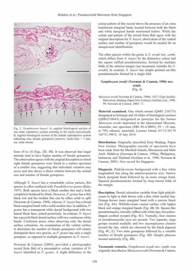
Fig. 2. Nymphozoon bayeri, living animal. A, dorsal view; B, general view of the ventral surface showing a simple pharynx and main intestine. C, anterior region showing simple-folded pseudotentacles and horseshoe shaped cerebral eyespot; D, detailed view of the ventral surface showing two male gonopores (arrowheads), 14 female gonopores (arrows), and sucker. Scale bars: 20 mm [A,B], 10 mm [C, D]. ph, pharynx; in, intestine; su, sucker.
Nymphozoon bayeri Hyman, 1959(Figs. 2, 3)
Nymphozoon bayeri Hyman, 1959: 578 (Type locality: Iwayama Bay, Palau, Micronesia); Newman et al., 2003: 198; Prudhoe, 1985: 30
Material examined. One specimen (ZRC.PLA.0002), 74 × 33 mm, in 70% ethanol, intertidal, Pulau [=Island] Semakau, 1°11.434’N 103°46.005’E, 23 August 2013; one specimen (ZRC.PLA.0003), 92 × 39 mm, in 70% ethanol, intertidal, Kusu Island, 1°13.523’N 103°51.574’E, 3 January 2014; one specimen (ZRC.PLA.0004), 85 × 55 mm, as serial sagittal sections (77 slides), intertidal, Terumbu [=Submerged reef] Salu, 01°12.928’N 103°42.753’E, 23 January 2015.
Distribution. Previously known from Palau, Micronesia. First record for Singapore.
Diagnosis. Conspicuous black margin followed by an inner white band. Presence of a medial longitudinal black stripe bordered by a continuous white band. Simple, pointed pseudotentacular folds.
Description. A wide longitudinal black stripe is present along the median line, extending posteriorly from the cerebral
eyespot to the length of the main intestine (Fig. 2A, B). This stripe is surrounded on each side by a continuous white band, followed by a broad area of greyish black or brown shade (Fig. 2C). Two thick solid marginal bands surround the entire body including the pseudotentacles: an outer black and an inner white (Fig. 2A). Ventral surface translucent white with the same black and white marginal bands as the dorsal surface (Fig. 2B). Simple pointed pseudotentacular folds with white tips and elongated-horseshoe shaped cerebral eyespot (Fig. 2C). Two male gonopores followed posteriorly by a row of several female gonopores (Fig. 2D). Simple ruffled pharynx located anteriorly (Fig. 2B, D).
Taxonomic remarks. Nymphozoon bayeri is the type species of the genus and to date considered to be the only species of Nymphozoon. Hyman (1959) indicated that the lack of a sucker in this species is “presumably associated with the multiplication of female apparatuses that extend into the area where the sucker would normally occur.” However, as discussed above, this statement appears to be erroneous since a distinct sucker was observed in specimens from Singapore (Fig. 2D). This finding demonstrates that N. bayeri is not an atypical member of the Cotylea but instead exhibits the traditional character of the suborder. The number of female gonopores in the specimens collected in this study varies
136
Bolaños et al.: Pseudocerotid flatworms from Singapore
from 10 to 14 (Figs. 2D, 3B). It was observed that larger animals tend to have higher number of female gonopores. This observation agrees with the original description in which eight female gonopores were found in a mature specimen of a smaller size, suggesting that individual variation may occur and also shows a direct relation between the animal size and number of female gonopores.
Although N. bayeri has a remarkable colour pattern, this species is often confused with Pseudobiceros gratus (Kato, 1937). Both species have a black median line and a body completely bordered by black. However, P. gratus has a thin black rim and the median line can be either solid or split (Newman & Cannon, 1994), whereas N. bayeri has a broad black marginal band with a solid median line. In addition, P. gratus has a transparent white dorsal background with two lateral black lines, joined posteriorly. In contrast, N. bayeri has a greyish black dorsal surface with two continuous white bands. Confusion arises when observing only the dorsal surface coloration. An examination of the ventral surface to determine the number of female gonopores will clearly distinguish these two species, as P. gratus has only a single gonopore, as opposed to multiple gonopores in N. bayeri.
Newman & Cannon (2005), provided a photographic record from Bali of a presumptive colour variation of N. bayeri identified as P. gratus. A slight difference in the
colour pattern of this record shows the presence of an extra translucent marginal band, located between both the black and white marginal bands mentioned before. While the colour and pattern of the record from Bali agree with the original description for N. bayeri, observation of the ventral surface and number of gonopores would be needed for an unequivocal identification.
The other species within the genus is N. orsaki nov. comb. which differs from N. bayeri by the distinctive colour and the square ruffled pseudotentacles, formed by multiple folds of the anterior margin (see taxonomic remarks for N. orsaki). In contrast, N. bayeri has simple pointed ear-like pseudotentacles formed by a single fold.
Nymphozoon orsaki (Newman & Cannon, 1996) nov. comb.(Fig. 4)
Maiazoon orsaki Newman & Cannon, 1996a: 1427 (Type locality: Dam awan, Madang, Papua New Guinea); Gosliner et al., 1996: 99; Newman & Cannon, 2003: 71.
Material examined. One whole mount (QMG 210715) designated as holotype and 10 slides of histological sections (QMG210643) designated as paratype for the former Maiazoon orsaki deposited in the Queensland Museum, Australia; one specimen (ZRC.PLA.0005), 59 × 18 mm, in 70% ethanol, intertidal, Lazarus Island, 01°13.381’N 103°51.398’E, 14 July 2014.
Distribution. Originally described from Madang, Papua New Guinea. Photographic records of specimens have been made from the Marshall Islands, Micronesia and Male Atoll, Maldives (Newman & Cannon, 1996a); Philippines, Indonesia, and Thailand (Gosliner et al., 1996; Newman & Cannon, 2005). New record for Singapore.
Diagnosis. Pinkish-cream background with a fine white longitudinal line along the anterior-posterior axis. Narrow black marginal band followed by an inner orange band. Squared pseudotentacles formed by deep lateral folds of the margin.
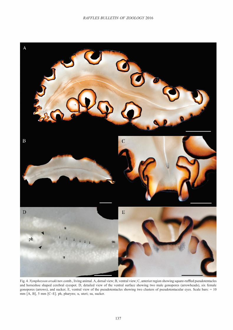
Description. Dorsal coloration variable from light pinkish-cream to light or dark brown with a thin white medial line. Orange-brown inner marginal band with a narrow black rim (Fig. 4A). Whitish-cream ventral surface with lighter black and orange marginal bands (Fig. 4B, D). Square-like pseudotentacles, laterally ruffled with white tips; horseshoe-shaped cerebral eyespot (Fig. 4C). Ventrally, four clusters of pseudotentacular eyes are present. Two separate, large groups located medially and two inconspicuous clusters toward the tips, which are obscured by the black pigment (Fig. 4C, E). Two male gonopores followed by a variable number of female gonopores (Fig. 4D). Simple pharynx located anteriorly (Fig. 4B).
Taxonomic remarks. Nymphozoon orsaki nov. comb. was originally described as Maiazoon orsaki (Newman & Cannon,
Fig. 3. Nymphozoon bayeri. A, sagittal histological section of one male copulatory system pointing at the stylet (arrowhead); B, sagittal histological section of the female reproductive system indicating nine female gonopores (arrows). Scale bars = 1 mm. ma, male atrium.
137
RAFFLES BULLETIN OF ZOOLOGY 2016
Fig. 4. Nymphozoon orsaki nov.comb., living animal. A, dorsal view; B, ventral view; C, anterior region showing square-ruffled pseudotentacles and horseshoe shaped cerebral eyespot. D, detailed view of the ventral surface showing two male gonopores (arrowheads), six female gonopores (arrows), and sucker; E, ventral view of the pseudotentacles showing two clusters of pseudotentacular eyes. Scale bars: = 10 mm [A, B], 5 mm [C–E]. ph, pharynx; u, uteri; su, sucker.

138
Bolaños et al.: Pseudocerotid flatworms from Singapore
1996a) based on the presence of a sucker and a sclerotized stylet. However, the sucker and stylet are features also found in Nymphozoon, indicating that the genus Maiazoon is superfluous and unnecessary. In addition, Newman & Cannon (1996a) mentioned that the presence of three to five female gonopores is a distinctive character for M. orsaki. In our specimen, six female gonopores were observed and the length of our animal was almost double that of the holotype. This finding reinforces the idea that the number of female gonopores is related to the size of the animal. We suggest an emendation of the original description of N. orsaki to include “presence of more than one female gonopore, variable in number.”
Here, we transferred M. orsaki to the genus Nymphozoon, presenting the new combination N. orsaki nov. comb. This represents the second species within the genus. The two species can be easily distinguished by their colours, and patterns and differences in the shape of their pseudotentacles (see taxonomic remarks for Nymphozoon and N. bayeri).
Phrikoceros Newman & Cannon, 1996
Phrikoceros baibaiye Newman & Cannon, 1996(Fig. 5)
Phrikoceros baibaiye Newman & Cannon, 1996a: 1429 (Type locality: Hastings Point, New South Wales, Australia); Newman & Cannon, 2003: 71.
Material examined. One specimen (ZRC.PLA.0006), 32 × 16 mm (preserved specimen), in 70% ethanol, intertidal, Chek Jawa, Pulau Ubin, 1°24.427’N 103°59.564’E, 19 October 2012.
Distribution. New South Wales, Australia. Also reported for Indonesia. New record for Singapore.
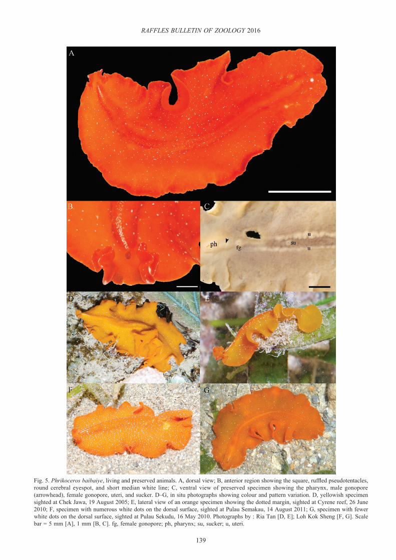
Description. Dorsal background ranging from bright orange, orange-red to orange-brown or rust colour. Numerous or scarce white speckles scattered randomly over the dorsal surface decreasing in number towards the margin (Figs. 5A, E–G). A short line formed by white microdots behind the cerebral eyespots is present (Fig. 5B, D–G). Unevenly spaced white microdots of variable size at rim along the entire body (Fig. 5E). Ventral side of similar colour but without white speckles or microdots. Laterally ruffled, square-like pseudotentacles with white tips formed by a cluster of microdots (Fig. 5B). Circular cerebral eyespot (Fig. 5B), and simple pharynx (Fig. 5C).
Taxonomic remarks. Amongst members of the genus Phrikoceros, P. katoi Newman & Cannon, 1996 and P. diadaleos Newman & Cannon, 1996 are also orange with scattered white dots. However, P. katoi is distinguished by a darker orange colouration at the margin and the presence of two blotches of white microdots, one anteriorly behind the cerebral eyespot and the other near the posterior end. On the other hand, P. diadaleos exhibits dots of a larger size over the entire dorsal surface. A white rim and a thin,
black marginal band are also present around the body. Additionally, P. baibaiye closely resembles Phrikoceros sp.1 Newman & Cannon, 2003. However, Phrikoceros sp. 1 exhibits conspicuous white pseudotentacles while P. baibaiye has only white tips; moreover, Phrikoceros sp.1 lacks the short white line behind the cerebral eyespot distinctive in P. baibaiye. After a close examination of the photographic record of Phrikoceros sp.1, it is believed that this species may not belong to this genus since the pseudotentacles seem to be simple folds, not square or ruffled, which is typical of Phrikoceros.
The diagnosis of the holotype of P. baibaiye from New South Wales, Australia, indicates a rust coloured background with white microdots forming irregular streaks over the entire dorsal surface (Newman & Cannon, 1996a). However, two colour variations from Indonesia have also been reported by Newman & Cannon (2003, 2005). One morphotype has a bright orange dorsal surface with few microdots scattered over the dorsal surface, while the second morphotype is orange-brown with numerous white dots. The specimen described in this study exhibits a bright orange-red colouration, which resembles the first morphotype found in Indonesia more than the holotype from Australia. Additional sightings from Singapore show brilliant orange-yellowish colouration and a variable amount of white spots on the dorsal surface (Fig. 5A, D–G). This difference in colour could be related to geographic distribution, environmental conditions or nutritional habits; however, factors determining polyclad colour variation remain undetermined.
Pseudobiceros Faubel, 1984
Pseudobiceros bedfordi (Laidlaw, 1903)(Fig. 6)
Pseudoceros bedfordi Laidlaw, 1903: 314 (Type locality: Singapore); Bock, 1913: 254; Bresslau, 1933: 59; Kato, 1943: 87; Kato, 1944: 299; Marcus, 1950: 84; Dawydoff, 1952: 82; Hyman, 1954: 220; Hyman, 1959: 566; Prudhoe, 1978: 586; Prudhoe, 1989: 77; George & George, 1979: 43.
Pseudoceros micronesianus Hyman, 1955a: 78.Pseudobiceros bedfordi Faubel, 1984b: 216; Newman & Cannon,
1994: 241; Gosliner et al., 1996: 101; Newman & Cannon, 1997: 343; Newman & Cannon, 2003:81; Newman et al., 2003: 197; Sreeraj & Raghunathan, 2013: 38; Dixit & Raghunathan, 2013: 167.
Material examined. One specimen (ZRC.PLA.0007), 42 × 23 mm, in 70% ethanol, intertidal, Tanjung [=Cape] Hakim, St. John’s Island, 01°13.409’N 103°50.673’E, 2 January 2014; one specimen (ZRC.PLA.0008), 15.5 × 12 mm (preserved specimen), in 70% ethanol, intertidal, Chek Jawa, Pulau Ubin, 01°24.597’N 103°59.680’E, 14 June 2014; one juvenile specimen, photographic record only, intertidal, Lazarus Island, 01°13.381’N 103°51.398’E, 18 May 2014.
Distribution. Singapore; Ifaluk Atoll, Palau, Guam, Onotoa, Saipan, and Marshall Islands, Micronesia; Mindanao, Philippines; Heron Island and Lizard Island, Great Barrier Reef, Australia; Coral Bay, Western Australia; Madang and
139
RAFFLES BULLETIN OF ZOOLOGY 2016
Fig. 5. Phrikoceros baibaiye, living and preserved animals. A, dorsal view; B, anterior region showing the square, ruffled pseudotentacles, round cerebral eyespot, and short median white line; C, ventral view of preserved specimen showing the pharynx, male gonopore (arrowhead), female gonopore, uteri, and sucker. D–G, in situ photographs showing colour and pattern variation. D, yellowish specimen sighted at Chek Jawa, 19 August 2005; E, lateral view of an orange specimen showing the dotted margin, sighted at Cyrene reef, 26 June 2010; F, specimen with numerous white dots on the dorsal surface, sighted at Pulau Semakau, 14 August 2011; G, specimen with fewer white dots on the dorsal surface, sighted at Pulau Sekudu, 16 May 2010. Photographs by : Ria Tan [D, E]; Loh Kok Sheng [F, G]. Scale bar = 5 mm [A], 1 mm [B, C]. fg, female gonopore; ph, pharynx; su, sucker; u, uteri.

140
Bolaños et al.: Pseudocerotid flatworms from Singapore
Fig. 6. Pseudobiceros bedfordi, living animal. A, dorsal view; B, ventral view; C, juvenile worm showing a different pattern with continuous and more symmetric transverse streaks; D, detailed view of the ventral surface showing the pharynx, two male gonopores (arrowheads), female gonopore, uteri, and sucker; E, anterior region showing simple-folded pseudotentacles and cerebral eyespot; F, detail of a transverse streak with a thin white line inside. Scale bars = 5 mm [A-E]. fg, female gonopore; ph, pharynx; su, sucker; u, uteri.
141
RAFFLES BULLETIN OF ZOOLOGY 2016
Laing Island, Papua New Guinea; Sulawesi, Indonesia; Inhaca Island, Mozambique; Havelock Island and Campbell Bay, Andaman and Nicobar Islands, India. Additional distribution information for Japan, Red Sea, and Vietnam is provided by Newman & Cannon (2005).
Description. Variable dark background, ranging from greenish-brown to purplish-black with numerous transverse pinkish-orange streaks delineated with black (Fig. 6A, C, E, F). The transverse streaks are of different lengths and some contain a thin white or cream line inside (Fig. 6F); some streaks form arcs, which extend laterally from the median line but do not reach the margin (Fig. 6A, C). A thick black marginal band surrounds the entire body, including the pseudotentacles (Fig. 6A, C, E). Dorsal surface covered with minute white-yellowish dots, including the black margin but absent in the interior of the transverse streaks (Fig. 6C, E, F). Ventral side ranges from translucent deep pink to pinkish-brown, with a black marginal band (Fig. 6b), Round cerebral eyespot, pseudotentacles as simple folds of the margin (Fig. 6E), and simple pharynx (Fig. 6D).
Taxonomic remarks. Pseudobiceros bedfordi displays a distinctive colour pattern, and is the only representative of colour pattern Group 5 (Newman & Cannon, 1997) characterised by transverse streaks. Originally recorded from Singapore as Pseudoceros bedfordi by Laidlaw (1903), this description was based on fragments of a single specimen. In a brief description offered by the author, the presence of a pair of male gonopores was clearly mentioned. An excellent drawing of the anterior part of the body depicting the colour pattern was also provided. Bock (1913) confirmed the occurrence of a double male reproductive system in elaborate illustrations accompanied by a detailed description of external characters. Since then, this species has been widely documented throughout the Pacific and Indian oceans. Faubel (1984b) erected the new combination Pseudobiceros bedfordi based on the presence of two male gonopores. This combination was validated by Newman and Cannon (1994), who also synonymized the species with Pseudoceros micronesianus after close examination of the colour pattern of the holotype.
A juvenile individual had fewer but more continuous transverse lines arranged in a more symmetrical pattern than the adults (Fig. 6C). This feature was also observed in two other records of juveniles from Madang, Papua New Guinea (Gosliner et al., 1996) and Marshall Islands, Micronesia (Newman & Cannon, 2005). These findings suggest that even though younger worms show fewer and more organised transverse streaks, the pattern is still conserved allowing for the correct identification of the species. Likewise, the background colour can be highly variable in the adults as seen in 15 additional photographic records provided by Newman & Cannon (2005). Again, the pattern is very consistent in all morphotypes.
Pseudobiceros damawan Newman & Cannon, 1994(Fig. 7)
Pseudobiceros damawan Newman & Cannon, 1994: 243 (Type locality: Laing Island, Madang, Papua New Guinea); Newman & Cannon, 1997: 347; Newman et al., 2003: 197; Sreeraj & Raghunathan, 2011: 2; Dixit & Raghunathan, 2013:168.
Material examined. One specimen, 24 × 9 mm, photographic record only, subtidal, 4 to 7 m depth, Kusu Island, 1°13.560’N, 103°51.582’E, 4 March 2014.
Distribution. Madang, Papua New Guinea; Coral Bay, Western Australia; Heron Island, Great Barrier Reef, Australia; Guam, Micronesia; Little Andaman, Andaman and Nicobar Islands, India. Additional records from Indonesia, Marshall Islands, Micronesia, and South Africa (Newman & Cannon, 2005). First record for Singapore.
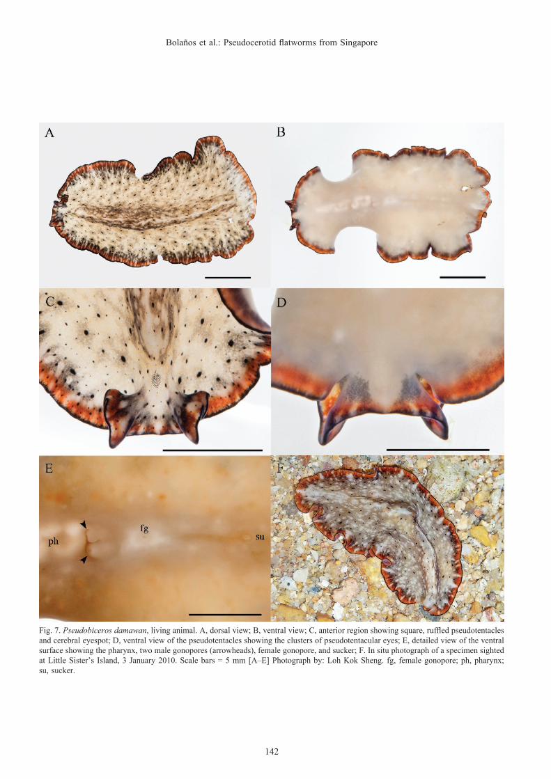
Description. Dorsal surface mottled with grey and white with a fine white longitudinal median line. Black spots of different sizes scattered over the entire surface. An extremely narrow black rim followed by an orange marginal band interrupted with delicate white lateral lines and spots. A black shadow along the orange margin is also present (Fig. 7A, F). Ventral surface creamy-white with the same marginal pattern (Fig. 7B). Cerebral eyes in a round cluster (Fig. 7C). Square and laterally ruffled pseudotentacles with a ventral pseudotentacular eye arrangement in four clusters (Fig. 7C, D), Simple pharynx (Fig. 7E).
Taxonomic remarks. This species is a member of Group 4 (Newman & Cannon, 1994), characterised by spots, dots and mottling. In this group, P. fulvogriseus (Hyman, 1959), P. gardineri (Laidlaw, 1902), and P. murinus Newman & Cannon, 1997 exhibit similar mottled grey backgrounds. However, P. fulvogriseus does not have the orange marginal band, the black spots, and the black rim of P. damawan. Pseudobiceros gardineri possesses black dots but lacks the orange marginal band and the black rim present in P. damawan. Perhaps P. murinus is the most similar species in both colour and pattern to P. damawan, but P. murinus has a transparent rim and a white triangle between its pseudotentacles, which are both absent in P. damawan.
Although the colour pattern of the specimen found in Singapore agrees with the original description, some slight colour variations were observed. The dorsal surface is brownish and not transparent grey, the bigger black spots are surrounded by shade of grey, and a dark grey hue along the orange marginal band is present. Five other colour variations were provided by Newman & Cannon (2005). These morphotypes differ mainly in the colour of the mottled background, which ranged from cream or light brown to dark brown to almost black. The colour of the median region is also variable but in general they all maintain similar colour patterns. Due to the close similarity between P. damawan and P. murinus and the considerable colour variation observed, the diagnosis for the species is emended as follows: “mottled dorsal surface varying from
142
Bolaños et al.: Pseudocerotid flatworms from Singapore
Fig. 7. Pseudobiceros damawan, living animal. A, dorsal view; B, ventral view; C, anterior region showing square, ruffled pseudotentacles and cerebral eyespot; D, ventral view of the pseudotentacles showing the clusters of pseudotentacular eyes; E, detailed view of the ventral surface showing the pharynx, two male gonopores (arrowheads), female gonopore, and sucker; F. In situ photograph of a specimen sighted at Little Sister’s Island, 3 January 2010. Scale bars = 5 mm [A–E] Photograph by: Loh Kok Sheng. fg, female gonopore; ph, pharynx; su, sucker.
143
RAFFLES BULLETIN OF ZOOLOGY 2016
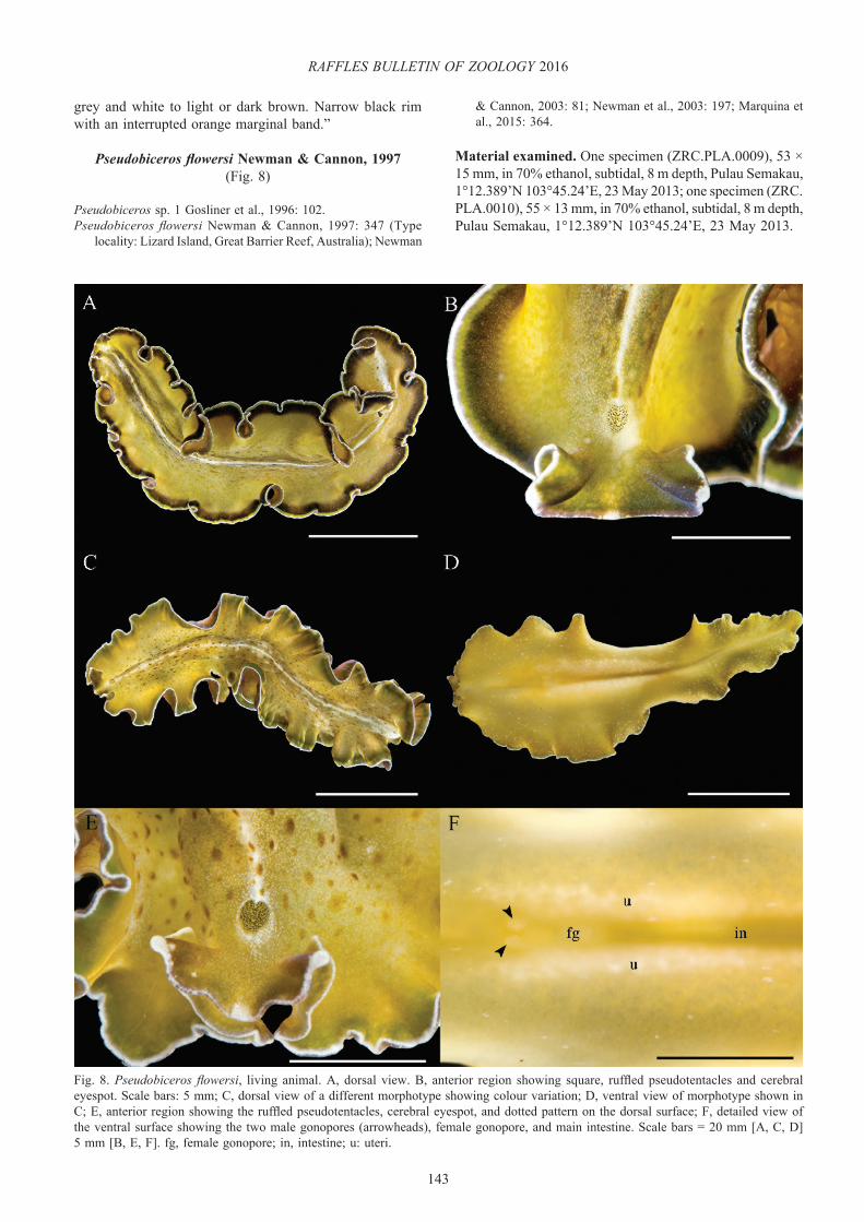
Fig. 8. Pseudobiceros flowersi, living animal. A, dorsal view. B, anterior region showing square, ruffled pseudotentacles and cerebral eyespot. Scale bars: 5 mm; C, dorsal view of a different morphotype showing colour variation; D, ventral view of morphotype shown in C; E, anterior region showing the ruffled pseudotentacles, cerebral eyespot, and dotted pattern on the dorsal surface; F, detailed view of the ventral surface showing the two male gonopores (arrowheads), female gonopore, and main intestine. Scale bars = 20 mm [A, C, D] 5 mm [B, E, F]. fg, female gonopore; in, intestine; u: uteri.
grey and white to light or dark brown. Narrow black rim with an interrupted orange marginal band.”
Pseudobiceros flowersi Newman & Cannon, 1997(Fig. 8)
Pseudobiceros sp. 1 Gosliner et al., 1996: 102.Pseudobiceros flowersi Newman & Cannon, 1997: 347 (Type
locality: Lizard Island, Great Barrier Reef, Australia); Newman
& Cannon, 2003: 81; Newman et al., 2003: 197; Marquina et al., 2015: 364.
Material examined. One specimen (ZRC.PLA.0009), 53 × 15 mm, in 70% ethanol, subtidal, 8 m depth, Pulau Semakau, 1°12.389’N 103°45.24’E, 23 May 2013; one specimen (ZRC.PLA.0010), 55 × 13 mm, in 70% ethanol, subtidal, 8 m depth, Pulau Semakau, 1°12.389’N 103°45.24’E, 23 May 2013.
144
Bolaños et al.: Pseudocerotid flatworms from Singapore
Distribution. This species has been found on the Great Barrier Reef, Australia; Palau, Micronesia; Papua New Guinea; Luzon, Philippines. Additional distribution for the Gulf of Oman and Indonesia (Newman & Cannon, 2005). First record for Singapore.
Description. Yellowish-green or brown background with a white, thin longitudinal median line bordered by dark pigment (Fig. 8A–C). Dense white microdots and brownish-green blotches covering the entire dorsal surface, both decreasing in density near the margin (Fig. 8A, C, E). Body margin with an inner broad, dark brown or black band, followed by a thin, green band, and a narrow white rim along the edges (Fig. 8A, B). Ventral surface similarly coloured as the dorsal side with scattered white microdots (Fig. 8D, F). Pseudotentacles squared, laterally ruffled, and white tips with a small white patch in between and round to oval cerebral eyespot (Fig. 8B, E).
Taxonomic remarks. The colour and pattern of the specimens found in Singapore agrees with the diagnosis for the species and other additional characters mentioned in the original description. However, the dorsal coloration of P. flowersi can also vary from dark brown or olive green (Newman & Cannon, 1997) to bright light green, to almost yellow (Newman & Cannon, 2003, 2005). The two morphotypes reported here have a striking olive green dorsal surface with marked brownish-green spots dispersed widely along the median region (Fig. 8A–C). This feature has not been recorded for this species before. A remarkable difference was found in the pattern of the marginal bands. While one specimen displayed distinct black and green marginal bands (Fig. 8A, B), the other showed only a faint brown pigment on the margin followed by a green margin and a white rim (Fig. 8C, E). Newman & Cannon (2005) also provide some records where the colour of the green band fluctuates from olive green to brownish-orange. In addition, the width of such bands can be wide or narrow to almost imperceptible in some morphotypes. Apparently, variations in the marginal bands in this species are not unusual and it is likely that other variations in colour and thickness of both marginal bands may occur.
Pseudobiceros flowersi, together with P. cinereus (Palombi, 1931), P. strigosus (Marcus, 1950), P. nigromarginatus, (Yeri & Kaburaki, 1918), and P. philippinensis (Kaburaki, 1923) are all characterised by longitudinal stripes that define colour pattern Group 2, (Newman & Cannon, 1997). In this group, only P. flowersi possessed a brown or green dorsal background.
Pseudobiceros fulgor Newman & Cannon, 1994(Fig. 9)
Pseudobiceros fulgor Newman & Cannon, 1994: 245 (Type locality: Heron Islands, Great Barrier Reef, Australia); Gosliner et al., 1996: 101; Newman & Cannon, 1997: 348; Newman & Cannon, 2003: 81.
Material examined. One juvenile specimen (ZRC.PLA.0011), 30 × 21 mm, in 70 % ethanol, intertidal, Tanjung
Hakim, St. John’s Island, 1°13.409’ N 103°50.673’ E, 2 January 2014; one specimen (ZRC.PLA.0012), 55 × 30 mm, in 70% ethanol, intertidal, Terumbu Semakau, 1°12.688’N 103°46.128’E, 13 July 2014; one specimen (ZRC.PLA.0013), 39 × 20 mm, in 70% ethanol, intertidal, St. John’s Island, 1°12.928’N 103°51.099’E, 21 May 2013; one juvenile specimen (ZRC.PLA.0014), 12 × 5 mm, in 70% ethanol, intertidal, Raffles Lighthouse, 1°9.600’N 103°44.456’E, 29 May 2013; one specimen (ZRC.PLA.0015), 50 × 36.5 mm (preserved specimen), in 70% ethanol, intertidal, Terumbu Salu, 1°12.928’N 103°42.753’E, 23 January 2015; one specimen (ZRC.PLA.0016), 45 × 19 mm, in 70 % ethanol, intertidal, Lazarus Island, 01°13.361’N 103°51.396’E, 16 June 2014.
Distribution. Great Barrier Reef, Australia; Bali, Indonesia; Marshall Islands, Micronesia; Anilao, Philippines; Mauritius. Additional record for the Red Sea (Newman & Cannon, 2005). First record for Singapore.
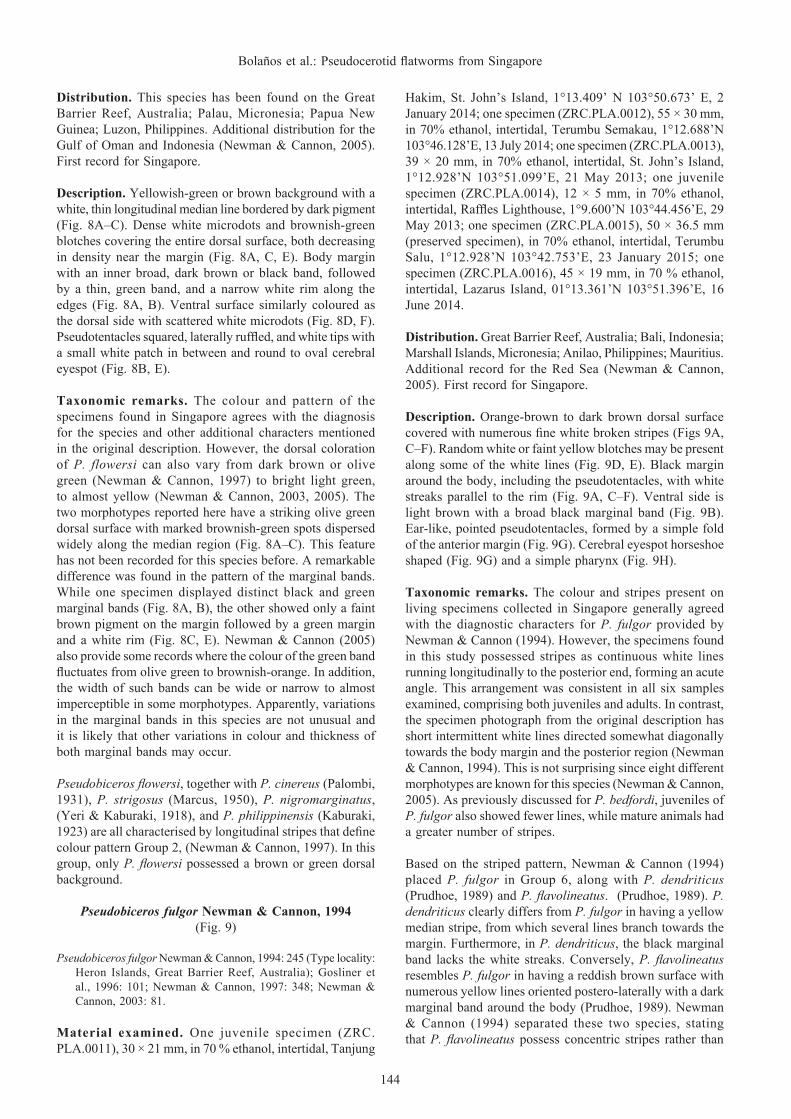
Description. Orange-brown to dark brown dorsal surface covered with numerous fine white broken stripes (Figs 9A, C–F). Random white or faint yellow blotches may be present along some of the white lines (Fig. 9D, E). Black margin around the body, including the pseudotentacles, with white streaks parallel to the rim (Fig. 9A, C–F). Ventral side is light brown with a broad black marginal band (Fig. 9B). Ear-like, pointed pseudotentacles, formed by a simple fold of the anterior margin (Fig. 9G). Cerebral eyespot horseshoe shaped (Fig. 9G) and a simple pharynx (Fig. 9H).
Taxonomic remarks. The colour and stripes present on living specimens collected in Singapore generally agreed with the diagnostic characters for P. fulgor provided by Newman & Cannon (1994). However, the specimens found in this study possessed stripes as continuous white lines running longitudinally to the posterior end, forming an acute angle. This arrangement was consistent in all six samples examined, comprising both juveniles and adults. In contrast, the specimen photograph from the original description has short intermittent white lines directed somewhat diagonally towards the body margin and the posterior region (Newman & Cannon, 1994). This is not surprising since eight different morphotypes are known for this species (Newman & Cannon, 2005). As previously discussed for P. bedfordi, juveniles of P. fulgor also showed fewer lines, while mature animals had a greater number of stripes.
Based on the striped pattern, Newman & Cannon (1994) placed P. fulgor in Group 6, along with P. dendriticus (Prudhoe, 1989) and P. flavolineatus. (Prudhoe, 1989). P. dendriticus clearly differs from P. fulgor in having a yellow median stripe, from which several lines branch towards the margin. Furthermore, in P. dendriticus, the black marginal band lacks the white streaks. Conversely, P. flavolineatus resembles P. fulgor in having a reddish brown surface with numerous yellow lines oriented postero-laterally with a dark marginal band around the body (Prudhoe, 1989). Newman & Cannon (1994) separated these two species, stating that P. flavolineatus possess concentric stripes rather than
145
RAFFLES BULLETIN OF ZOOLOGY 2016
Fig. 9. Pseudobiceros fulgor, living animal. A, dorsal view; B, ventral view; C, adult mature specimen showing pattern variation; D, adult immature specimen showing pattern variation; E, F, juvenile specimens showing colour and pattern variation; G, anterior region showing pseudotentacles and cerebral eyespot; H, detailed view of the ventral surface showing the pharynx, two male gonopores (arrowheads), female gonopore, and sucker. Scale bar = 10 mm [A–F], 5 mm [H], 2 mm [G]. fg, female gonopore; ph, pharynx; su, sucker; u: uteri.

146
Bolaños et al.: Pseudocerotid flatworms from Singapore
broken lines as seen in P. fulgor. Nonetheless, it is worth mentioning that the concentric pattern was described by Prudhoe (1989) from a cleared, preserved specimen and not from a live specimen. If emphasis were to be placed on the concentric stripes, the specimens reported in this study would then be more similar to P. flavolineatus than P. fulgor. Based on a water colour painting, Prudhoe (1989) also mentioned the presence of two irregular rows of black dots along the margin of P. flavolineatus. But in the preserved animals, he observed a broad dark marginal band around the body with three whitish lines, except in the tentacular region, which bears twice the number of white lines. The characteristics mentioned before are misleading because none of the specimens from this study revealed the two rows of black dots along the margin but they displayed the dark marginal band with white lines. Based on the above, P. fulgor shares some traits with P. flavolineatus but it does not share other features. Because Prudhoe’s interpretations were based on water colour paintings and preserved specimens, the diagnostic characters for P. flavolineatus are difficult to define without reference to living specimens.
Pseudobiceros fulgor also resembles Pseudoceros dubius Prudhoe, 1989, but the latter has a narrow black marginal band and a reddish-brown background with numerous scattered yellow streaks sometimes merging with one another (Prudhoe, 1989). However, no further details on the shape of pseudotentacles and cerebral eyespot, type of pharynx, and number of male gonopores are provided for P. dubius. Without this crucial information, it is impossible to validate if P. dubius actually belongs to the genus Pseudoceros or to Pseudobiceros. Because the species identity of P. dubius could not be determined, this species is declared incertae sedis.
Newman & Cannon (2003) included a photograph of P. fulgor from the Marshall Islands showing colour variation (p. 81). This morphotype displays an extremely different colour pattern from the holotype, having the black margin with white marks, more similar to dots than lines, and a dorsal surface cover with a patchy network of dense white pigment. Two other similar records were reported by the same authors (2005), although, in these cases the species was indicated as Pseudobiceros cf. fulgor.
Considering such level of dissimilarity and our observations of at least six different morphotypes in Singapore, it is possible that P. fulgor, P. flavolineatus, and P. dubius all represent the same species. Additional material of the possible variants of P. fulgor and newly collected specimens of P. flavolineatus and P. dubius from the type localities are needed for molecular analyses to confirm their relationship and to determine if P. fulgor shows intraspecific variation.
Pseudobiceros hancockanus (Collingwood, 1876)(Figs. 10, 11)
Proceros hancockanus Collingwood, 1876: 91 (Type locality: Singapore).
Stylochopsis malayensis Collingwood, 1876; 94.
Prostheceraeus hancockanus Lang, 1884: 567.Pseudoceros malayensis Bock, 1913: 258, 259.Pseudoceros hancockanus Kaburaki, 1923: 639.Pseudobiceros hancockanus Newman & Cannon, 1994: 249 [not
Pseudobiceros hancockanus]Pseudobiceros uniarborensis Newman & Cannon, 1994: 252 (Type
locality: Heron Island, Great Barrier Reef, Australia); Newman & Cannon, 1997: 360; Newman & Cannon, 2003: 83; Newman et al., 2003: 197; Gosliner et al., 1996: 103; Apte & Pitale, 2011: 110; Maghsoudlou & Rahimian, 2014: 332; Marquina et al., 2015: 367.
Material examined. One whole mount (QMG210599) designated as holotype for P. uniarborensis deposited in the Queensland Museum, Australia. One specimen designed as a neotype (ZRC.PLA.0026), 53 × 25 mm, in 70% ethanol, intertidal, Raffles Lighthouse, 1°9.600’N 103°44.456’E, 29 May 2013; one specimen (ZRC.PLA.0018), 30.5 × 21.5 mm (preserved specimen), juvenile, in 70% ethanol, intertidal, Tanjung Hakim, St. John’s Island, 1°13.409’N 103°50.673’E, 2 January 2014; Two specimens (ZRC.PLA.0019), 11 × 11.5 mm (preserved specimen), juvenile, and 9.5 × 7 mm (preserved specimen), juvenile, in 70% ethanol, intertidal, Tanjung Hakim, St. John’s Island, 1°13.409’N 103°50.673’E, 2 January 2014; one specimen (ZRC.PLA.0020), 56 × 23 mm, as serial sagittal sections (78 slides), intertidal, Pulau Subar Darat [=Big Sister’s Islands], 1°12.899’ N 103°49.937’E, 9 September 2013; three specimens (ZRC.PLA.0021), 56 × 25 mm, as serial sagittal sections (66 slides), and 32.5 × 26 mm and 40 × 20 mm, in 70% ethanol, intertidal, Pulau Salu, 1°13.002’N 103°42.588’E, 9 August 2014; one specimen (ZRC.PLA.0022), 53 × 21 mm, as serial sagittal sections (186 slides), subtidal, 16 m depth, Lazarus Island, 1°13.317’ N 103°51.170’E, 23 May 2013; one specimen (ZRC.PLA.0023), 48 × 27 mm, in 70% ethanol, intertidal, Beting [=Sandbar] Bemban Besar [=Big], 1°12.149’N 103°44.989’E, 26 May 2013; one specimen (ZRC.PLA.0024), 24 × 15 mm, juvenile, in 70% ethanol, intertidal, Cyrene Reef, 1°15.374’N 103°44.816’E, 27 May 2013; one specimen (ZRC.PLA.0025), 21 × 11 mm, juvenile, in 70% ethanol, subtidal, 15 m depth, Pulau Subar Darat, 1°12.900’N 103°49.880’E, 27 May 2013; two specimens (ZRC.PLA.0027), 41 × 36 mm and 40 × 19 mm, in 70% ethanol, subtidal, 4 to 6 m depth, Kusu Island, 1°13.567’N 103°51.583’E, 14 January 2015.
Distribution. Singapore; Borneo; Heron Island, One Tree Island, and Lizard Island, Great Barrier Reef, Australia; Madang, Papua New Guinea; Tantabiddi, and Coral Bay, Western Australia; Philippines; Guam, Micronesia; Kavratti Island, Lakshadweep, India; Gulf of Oman, Iran. Additional records from Hawaii, Indonesia, Mauritius, and the Red Sea (Newman & Cannon, 2005).
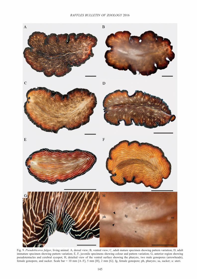
Description. Dorsal pattern variable with a dark brown or velvety black background and three marginal bands: inner bright orange, middle transparent grey, and opaque white rim (Fig. 10A–E). Ventral surface brown with the same marginal bands (Fig. 10B). Black, ear-like, pointed pseudotentacles, bordered only by the opaque white rim and conspicuous white tips (Figs. 10D, 11A–E). Cerebral eyespot in a clear oval area with a short projection anteriorly and a narrow line
147
RAFFLES BULLETIN OF ZOOLOGY 2016
Fig. 10. Pseudobiceros hancockanus, living animal. A, dorsal view of a mature specimen; B, ventral view of a juvenile showing pharynx and sucker; C, dorsal view of a juvenile showing color variation, ramifications of the intestine, and the prominent white colouration between the pseudotentacles; D, ear-like pointed pseudotentacles of a mature specimen without the white pigment in between the pseudotentacles; oval cerebral eyespot in a clear area with short projections; E, in situ photograph of a worm showing the three marginal bands. Scale bars = 5 mm [A–D]. ph, pharynx; su, sucker.

148
Bolaños et al.: Pseudocerotid flatworms from Singapore
Fig. 11. Pseudobiceros hancockanus, living animal. A–D, dorsal view of different worms in which the middle grey marginal band is not distinctive; E, dorsal view of a specimen showing the middle grey marginal band (circled); F, close up view of the marginal bands. Scale bars = 5 mm [A–E]
posteriorly (Figs. 10D, 11C, D). A white-greyish triangle between the pseudotentacles that connects with the clear area around the eyespot can be present (Fig. 10C).
Taxonomic remarks. Pseudobiceros hancockanus was first described by Collingwood (1876) as having a velvety dark dorsal surface with inner orange and outer white marginal bands of equal widths. A particular feature for this species was the simple pseudotentacular folds bordered by the white band but lacking the inner orange one. Kaburaki (1923) confirmed this colour pattern with the specimen reported
from the Philippines but mentioned the presence of a narrow light-grey marginal band instead of being opaque white as originally described. Newman & Cannon (1994) re-described P. hancockanus indicating that the two distinctive marginal bands were of different widths and the squared ruffled pseudotentacles were outlined by both white and orange coloration. In the same paper, the authors described another species, Pseudobiceros uniarborensis (Newman & Cannon, 1994), otherwise similar to P. hancockanus but characterised by the presence of a third narrow grey-translucent band between the white and orange bands. pointed ear-like
149
RAFFLES BULLETIN OF ZOOLOGY 2016
pseudotentacles with white tips but without marginal bands, and a white-grey triangle between the pseudotentacles.
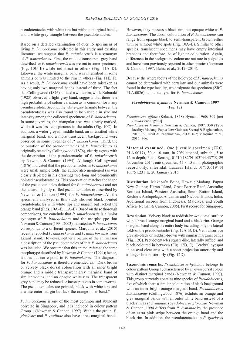
Based on a detailed examination of over 15 specimens of living P. hancockanus collected in this study and existing literature, we suggest that P. uniarborensis is a synonym of P. hancockanus. First, the middle transparent grey band described for P. uniarborensis was present in some specimens (Fig. 10C–E) while indistinct in others (Fig. 11A–D). Likewise, the white marginal band was intensified in some animals or was limited to the rim in others (Fig. 11E, F). As a result, P. hancockanus could have been mistaken as having only two marginal bands instead of three. The fact that Collingwood (1876) noticed a white rim, while Kaburaki (1923) observed a light grey band, suggests that there is a high probability of colour variation as is common for many pseudocerotids. Second, the white-grey triangle between the pseudotentacles was also found to be variable in size and intensity among the collected specimens of P. hancockanus. In some juveniles, the triangular area was clearly marked, whilst it was less conspicuous in the adults (Fig. 10C). In addition, a wider greyish middle band, an intensified white marginal band, and a more translucent background were observed in some juveniles of P. hancockanus. Third, the colouration of the pseudotentacles of P. hancockanus as originally stated by Collingwood (1876), clearly agrees with the description of the pseudotentacles of P. uniarborensis by Newman & Cannon (1994). Although Collingwood (1876) indicated that the pseudotentacles in P. hancockanus were small simple folds, the author also mentioned (as was clearly depicted in his drawing) two long and prominently pointed pseudotentacles. This observation matches the shape of the pseudotentacles defined for P. uniarborensis and not the square, slightly ruffled pseudotentacles re-described by Newman & Cannon (1994) for P. hancockanus. All the specimens analysed in this study showed black pointed pseudotentacles with white tips and margin but lacked the orange band (Figs. 10A–E, 11A–E). Based on these thorough comparisons, we conclude that P. uniarborensis is a junior synonym of P. hancockanus and the morphotype that Newman & Cannon (1994, 2003) indicated as P. hancockanus corresponds to a different species. Marquina et al., (2015) recently reported P. hancockanus and P. uniarborensis from Lizard Island. However, neither a picture of the animal nor a description of the pseudotentacles of that P. hancockanus was included. We presume that this animal refers to the same morphotype described by Newman & Cannon (1994); hence, it does not correspond to P. hancockanus. The diagnosis for P. hancockanus is therefore emended as: “Dark brown or velvety black dorsal colouration with an inner bright orange and a middle transparent grey marginal band of similar widths, and an opaque white rim. The transparent grey band may be reduced or inconspicuous in some worms. The pseudotentacles are pointed, black with white tips and a white outer margin but lack the orange inner band.”
P. hancockanus is one of the most common and abundant polyclad in Singapore, and it is included in colour pattern Group 1 (Newman & Cannon, 1997). Within the group, P. gloriosus and P. evelinae also have three marginal bands.
However, they possess a black rim, not opaque white as P. hancockanus. The dorsal colouration of P. hancockanus can range from opaque black to semi-transparent brown either with or without white spots (Fig. 10A–E). Similar to other species, translucent specimens may have empty intestinal branches and therefore, be of lighter colouration. Again, differences in the background colour are not rare in polyclads and have been previously reported in other species (Newman & Cannon, 1997; Bahia et al., 2012, 2014).
Because the whereabouts of the holotype of P. hancockanus cannot be determined with certainty and our animals were found in the type locality, we designate the specimen (ZRC.PLA.0026) as the neotype for P. hancockanus.
Pseudobiceros hymanae Newman & Cannon, 1997(Fig. 12)
Pseudoceros affinis (Kelaart, 1858) Hyman, 1960: 309 [not Pseudoceros affinis]
Pseudobiceros hymanae Newman & Cannon, 1997: 350 (Type locality: Madang, Papua New Guinea); Sreeraj & Raghunathan, 2013: 39; Dixit & Raghunathan, 2013: 167; Marquina et al., 2015: 366.
Material examined. One juvenile specimen (ZRC.PLA.0017), 30 × 10 mm, in 70% ethanol, subtidal, 5 to 12 m depth, Pulau Senang, 01°10.182’N 103°44.437’E, 29 November 2014; one specimen, 45 × 15 mm, photographic record only, intertidal, Lazarus Island, 01°13.619’ N 103°51.231’E, 20 January 2015.
Distribution. Makapu’u Point, Hawaii; Madang, Papua New Guinea; Heron Island, Great Barrier Reef, Australia; Rottnest Island, Western Australia; South Button Island, Ritchie’s Archipelago, Andaman and Nicobar Islands, India. Additional records from Indonesia, Maldives, and South Africa (Neman & Cannon, 2005). First record for Singapore.
Description. Velvety black to reddish-brown dorsal surface with a broad orange marginal band and a black rim. Orange marginal band along the entire body including only the lateral folds of the pseudotentacles (Fig. 12A, B, D). Ventral surface greyish-black or reddish-brown with similar marginal bands (Fig. 12C). Pseudotentacles square-like, laterally ruffled, and black coloured in between (Fig. 12D, E). Cerebral eyespot in an oval clear area with a short projection anteriorly and a longer line posteriorly (Fig. 12D).
Taxonomic remarks. Pseudobiceros hymanae belongs to colour pattern Group 1, characterised by an even dorsal colour with distinct marginal bands (Newman & Cannon, 1997). This group currently contains nine species of Pseudobiceros, five of which share a similar colouration of black background with an inner bright orange marginal band. Pseudobiceros hancockanus (Collingwood, 1876) exhibits an orange and grey marginal bands with an outer white band instead of a black rim as P. hymanae. Pseudobiceros gloriosus Newman & Cannon, 1994 differs from P. hymanae by the presence of an extra pink stripe between the orange band and the black rim. In addition, the pseudotentacles in P. gloriosus

150
Bolaños et al.: Pseudocerotid flatworms from Singapore
Fig. 12. Pseudobiceros hymanae, living animal. A, dorsal view of an adult immature specimen; B, dorsal view of a juvenile specimen showing a different colour on the dorsal surface with pigment granules; C, ventral view of the juvenile showing the pharynx, sucker, and branches of the intestine; D, anterior region showing square-ruffled pseudotentacles and cerebral eyespot in a clear area with short projections; E, ventral view of the pseudotentacles showing two clusters of pseudotentacular eyes. Scale bars = 5 mm. ph, pharynx; su, sucker; in, intestine.

151
RAFFLES BULLETIN OF ZOOLOGY 2016
are completely bordered by orange and burgundy bands, whereas in P. hymanae the orange margin only extends to the level of the lateral folds. P. periculosus Newman & Cannon, 1994 resembles P. hymanae in that it has the orange band and the black pseudotentacles without marginal bands; however, this species exhibits an extremely narrow transparent rim instead of a black border as P. hymanae.
A juvenile specimen of P. hymanae found in Singapore exhibited a reddish-brown dorsal colouration instead of the velvety black background seen in adults (Fig. 11B). The only species within this group with a similar dorsal colouration is P. evelinae (Marcus, 1950). However, the pseudotentacles in P. evelinae are uniformly black with conspicuous white tips, whereas P. hymanae has pseudotentacles bordered laterally by the orange band and a black rim and has a black colouration in between. Additionally, recent descriptions of P. evelinae (Bahia et al., 2012, 2014) have shown the presence of an extra inner black band that was overlooked by Marcus (1950) in his original description. This may be due to the specimen’s nutritional condition and the fact that the worms were observed on a bright background (Bahia et al., 2014). Because this additional black band was not present in the juvenile and the colour pattern of the pseudotentacles differs between both species, the immature specimen found in this study was not considered to be P. evelinae, despite a similar background colour. Intestinal branches and numerous granule pigments were evident in our specimens, particularly with a light source illuminating the animals. Dorsal colouration can significantly depend on the content of the intestinal branches (Newman & Cannon, 2003; Bahia et al., 2014); hence, the variation from dark to light colour found between the juvenile and the adults was most likely due to lack of food.
Another species with identical colour and pattern as P. hymanae, is P. splendidus (Lang, 1884). Newman & Cannon (1997) placed P. splendidus within colour pattern Group 3, mentioning that emphasis was placed on the presence of white dots rather than the marginal bands. However, Lang’s original description of P. splendidus mentions that the white dots are microscopic, and cannot be distinguished by the naked eye. Furthermore, subsequent descriptions of P. splendidus did not indicate the presence of white dots (Stummer-Traunfels, 1933; Hyman, 1955b; Prudhoe, 1989). Since this trait has not been consistent in all morphotypes, and the marginal bands are considered more significant taxonomically (Litvaitis & Newman, 2001), P. splendidus should be transferred to Group 1 together with the species discussed above. Newman & Cannon (1997) showed that there is a geographic distinction between these two species, P. splendidus is restricted to the Mediterranean and Atlantic oceans, whereas P. hymanae is limited to the Indo-Pacific region. Based on geographic location, the specimen from Singapore was assigned to P. hymanae. However, Hyman (1955b) stated that P. splendidus is a cosmopolitan species and Prudhoe (1989) indicated that this species occurs in Vietnam and the Galapagos archipelago, in addition to the Mediterranean and the Atlantic. Hence, it is highly likely that P. hymanae is a junior synonym of P. splendidus. Further
molecular analyses are required to determine if P. hymanae and P. splendidus are synonyms and if P. evelinae represents a case of colour variation.
Pseudoceros Lang, 1884
Pseudoceros bifurcus Prudhoe, 1989(Fig. 13)
Pseudoceros dimidiatus George & George, 1979: 43, pl 49 fig 7 [Not Pseudoceros bifurcus]
Pseudoceros bifurcus Prudhoe, 1989: 78 (Type locality: M’Sanga Tsohole Reef, Benthedi, Mayotte, Comoro Island, Madagascar); Newman & Cannon, 1994: 216; Gosliner et al., 1996:104; Newman & Cannon, 1998: 299; Newman & Cannon, 2003: 72; Sreeraj & Raghunathan, 2011: 3; Dixit & Raghunathan, 2013: 166.
Material examined. One specimen (ZRC.PLA.0028), 22.5 × 18 mm (preserved specimen), in 70% ethanol, intertidal, Chek Jawa, Pulau Ubin, 1°24.748’N 103°59.711’E, 17 October 2012; one specimen, (ZRC.PLA.0029), 20 × 6 mm, in 70% ethanol, intertidal, Lazarus Island, 1°13.381’N 103°51.398’E, 16 June 2014.
Distribution. Comoro Islands, Madagascar; Heron Island, One Tree Island, and Lizard Island, Great Barrier Reef, Australia; Manado, Sulawesi, Indonesia; Philippines; Little Andaman, Andaman and Nicobar Islands, India. Additional records from Japan, Papua New Guinea, and Thailand (Newman & Cannon, 2005). New record for Singapore.
Description. Dorsal background variable in colour, ranging from evenly blue to bluish-lavender, or light cream- mauve to dark violet. Conspicuous median line of two colours: bright orange anteriorly and white posteriorly (Fig. 13A, B). The longitudinal median line is completely bordered by a dark purple hue but in some cases, the orange area can be free of this delineation (Fig. 13A, B). The white area of the median line can be continuous (Fig. 13A) or interrupted (Fig. 13B). Ventral side of same colour as the dorsal surface. Simple pseudotentacles (Fig. 13A–C). Round cerebral eyespot (Fig. 13C) and folded pharynx (Fig. 13D).
Taxonomic remarks. This species belongs to colour pattern Group 3 characterised by a plain background with longitudinal stripes (Newman & Cannon, 1998). Despite the colour variation of its dorsal surface, this species always has an unmistakable bicoloured, distinctive median line which is orange anteriorly and white posteriorly. In relation to the white area of the median line, two patterns were observed. One specimen showed a smooth continuous white line (Fig. 13A) while the other exhibited white intermittent raised segments of variable size (Fig. 13B). To date, the known records for P. bifurcus have shown a combination of all the diverse dorsal colouration with the different patterns of the median line. However, the bicoloured array of the median line has been consistent in all morphotypes. The original description of P. bifurcus by Prudhoe (1989) was based on a colour transparency where the presence of a white marginal band was observed. Newman & Cannon (1994) stated that

152
Bolaños et al.: Pseudocerotid flatworms from Singapore
Fig. 13. Pseudoceros bifurcus, living and preserved animals. A, dorsal view of a mature specimen with a continuous longitudinal median line; B, dorsal view of a juvenile showing an intermittent longitudinal median line; C, anterior region showing simple-folded pseudotentacles and a round cerebral eyespot; D,ventral view of preserved specimen, showing the pharynx, male gonopore (arrowhead), female gonopore, uteri, and sucker; E, P. bifurcus feeding in the lab with an extended pharyngeal folds, each inside of an individual ascidian zooid. Scale bars: 5 mm [A, B] 1 mm [C, D]. fg, female gonopore; ph, pharynx; su, sucker; u, uteri.

153
RAFFLES BULLETIN OF ZOOLOGY 2016
this margin is an artefact by the bright clear colouration of the animal’s dorsal surface. During photography of living animals, a reflective white border appeared as a result of strong light exposure, possibly confirming the artefact.
Pseudoceros liparus Marcus, 1950 is currently listed as synonym of P. bifurcus (see Newman & Cannon, 1994; Tyler et al., 2006–2015). However, the colour description offered by Marcus (1950), who assigned this new name to Pseudoceros limbatus (Haswell, 1907), only mentioned a light red background with a well-defined purple margin. Neither Marcus (1950) nor Haswell (1907) mentioned the presence of the bicolored longitudinal median line that defines P. bifurcus. Because of the mismatch in colouration between P. liparus and P. bifurcus, they are here considered to be separate species.
Newman & Cannon (1994) indicated that the majority of Pseudoceros are found associated with colonial ascidians on which they feed. While some species are prey-specific, the authors observed that P. bifurcus feeds on different species of ascidians showing a more generalised diet (Newman & Cannon, 1994). In our study, an individual was placed with a colony of ascidians, Eudistoma sp. and the flatworm appeared to extend the folds of the pharynx into the individual zooids (Fig. 13E). As a generalist species, P. bifurcus is suitable for investigations on ecological interactions and the biological role of natural chemical compounds.
Pseudoceros caeruleocinctus Hyman, 1959(Fig. 14)
Pseudoceros caeruleocinctus Hyman, 1959: 569 (Type locality: Palau Islands, Micronesia)
Pseudoceros sapphirinus Newman & Cannon, 1994: 236; Newman & Cannon, 1998: 313; Gosliner et al., 1996: 108; Newman & Cannon, 2003: 76; Newman et al., 2003: 198.
Material examined. One specimen preserved in alcohol (USNM28653) designated as holotype for Pseudoceros caeruleocinctus deposited in the National Museum of Natural History, USA. One specimen (ZRC.PLA.0044), 25 × 16 mm (preserved specimen), in 70% ethanol, intertidal, Lazarus Island, 1°13.381’N 103°51.398’E, 18 May 2014; one specimen (ZRC.PLA.0045), 60 × 24 mm (preserved specimen), in 70% ethanol, subtidal, 5 to 15 m depth, Cyrene Reef, 1°15.677’N 103°44.969’E, 12 May 2014; two specimens (ZRC.PLA.0046), 45 × 25 mm and, 28 × 11 mm, in 70% ethanol, subtidal, 5 to 11 m depth, Beting [=Sandbar] Bemban Besar [=Big], 1°12.316’N 103°44.633’E, 16 November 2014.
Distribution. Heron Island, Lizard Island and One Tree Island, Great Barrier Reef, Australia; Palau and Marshall Islands, Micronesia; Anilao, Philippines. Additional record from Papua New Guinea (Newman & Cannon, 2005). First record for Singapore.
Description. Background reddish-brown to velvety black with a submarginal broad blue band and an extremely thin white rim along the entire body, including the pseudotentacles
(Fig. 14A, C). Ventral surface reddish-brown with white rim (Fig. 14B, D). Simple pseudotentacles. Horseshoe-shaped cerebral eyespot in a clear area (Fig. 14C).
Taxonomic remarks. Pseudoceros caeruleocinctus was originally described by Hyman (1959) from a single preserved specimen (20 × 12 mm) collected in Palau, Micronesia by researchers from Stanford University and the U.S. National Museum. The dorsal colour of the living animal were described by the collector as velvety black with a narrow brilliant blue border. Hyman (1959) noted that the preserved animal had prominent tentacular foldings, one male gonopore, one female gonopore, and a sucker. However, no additional colour details and information regarding the reproductive system or type of pharynx were provided. As a result of this insufficient morphological description, P. caeruleocinctus was placed as incertae sedis (Faubel, 1984b; Tyler et al, 2006–2015). In this study, P. caeruleocinctus is recognised as Pseudoceros sensu lato because of the simple pseudotentacular folds, the folded pharynx, and the presence of a single male copulatory apparatus.
Newman & Cannon (1994) described a similar species, Pseudoceros sapphirinus (Newman & Cannon, 1994), indicating the presence of a blue submarginal band and an extremely narrow white rim as characters that distinguish P. sapphirinus from P. caeruleocinctus. However, after examination of the holotypes of the two species and a detailed comparison of their known records, we conclude that they are the same species. The position of the blue band and the apparent absence of the white rim in P. caeruleocinctus are dubious because Hyman (1959) was not able to examine the live animal and relied solely on limited observations made by the collectors. Using a thin line, Hyman (1959) illustrated the blue border seen in the preserved specimen (Fig. 9, p. 572). However, it is unclear if this line demarcated the limit of the band from the edge or the blue band itself. In the latter case, it would agree with the submarginal position as described for P. sapphirinus. The holotype of P. sapphirinus showed a dark area in the border of the entire body but no trace of a submarginal band (Fig. 14F). This suggests that Hyman (1959) may have mistaken the blue border described by the collector with this artefact in the preserved animal. The holotype of P. caeruleocinctus consisted of several pieces of the animal in poorly preserved condition (Fig. 14E). The largest piece still had some small region of intact margin; however, no evidence of a change in colouration indicating the presence of a marginal or submarginal band was observed (Jon Norenburg, Smithsonian Institution, Washington, DC; pers. comm.). The absence of a white rim in P. caeruleocinctus is another uncertain trait that could have been overlooked by the collector because of its extremely thin nature. In our material from Singapore, this white rim was only detected after careful examination. Furthermore, the pigment of the rim dissolved after fixation, and could no longer be detected in the preserved material (Fig 14F). This may explain why Hyman (1959) was originally unable to observe the presence of a white rim. Moreover, for over 56 years, no other records of P. caeruleocinctus sensu stricto are known. We therefore conclude that P. caeruleocinctus

154
Bolaños et al.: Pseudocerotid flatworms from Singapore
Fig. 14. Pseudoceros caeruleocinctus, living animal. A, dorsal view; B, ventral view; C, anterior region showing simple-folded pseudotentacles and oval cerebral eyespot in a clear area; D, detailed view of the ventral surface showing the pharynx, male gonopore (arrowhead), female gonopore (arrow), uteri, and sucker; E, holotype of P. caeruleocinctus (USNM 28653); F, holotype of Pseudoceros sapphirinus (Queensland Museum, G210444) designated in this study as neotype of P. caeruleocinctus. Scale bars: 10 mm [A, B], 5 mm [C, D]. ph, pharynx; su, sucker; u, uteri.
is a senior synonym of P. sapphirinus. The description of P. sapphirinus by Newman & Cannon (1994) was based on an animal collected from Heron Island, Australia; however, another specimen was later reported from the type locality (Newman et al., 2003). The topotypic material provides additional support for the subjective synonym established here. Because the holotype of P. caeruleocinctus is practically destroyed and the original description of the species is unsatisfactory, the specimen described by Newman & Cannon
(1994, p:236), deposited in the Queensland Museum as P. sapphirinus (G210444), is designated as the neotype for P. caeruleocinctus.
Pseudoceros caeruleocinctus belongs to colour pattern Group 2 (Newman & Cannon, 1998). Within this group, there are a number of species with a velvety black background, but none of them exhibit a similar bright blue, submarginal band with a white rim. The specimens found in Singapore had

155
RAFFLES BULLETIN OF ZOOLOGY 2016
a reddish-black dorsal colouration instead of velvety black as the original description. This colour variation has been repeatedly seen in polyclads with black backgrounds and has been attributed to the absence or presence of food in the intestinal branches. In this species, the ramification of the intestine could be observed when animals were exposed to a direct source of light.
Pseudoceros concinnus (Collingwood, 1876) (Fig. 15)
Proceros concinnus Collingwood, 1876: 90 (Type locality: Pulo Daak Islands, Borneo, Malaysia); Lang, 1884: 593.
Pseudoceros concinnus Kaburaki, 1923:64; Newman & Cannon, 1994: 208; Newman & Cannon, 2003: 73; Dixit & Raghunathan, 2013: 166; Sreeraj & Raghunathan, 2011: 4.
Material examined. One specimen (ZRC.PLA.0030), 21 × 6 mm, in 70% ethanol, intertidal, Cyrene Reef, 1°15.553’N 103°45.062’E, 20 August 2013; one specimen (ZRC.PLA.0031), 16 × 10 mm, in 70% ethanol, intertidal, Pulau Semakau, 1°11.434’ N 103°46.005’ E, 23 August 2013; one juvenile specimen (ZRC.PLA.0032), 11 × 3 mm, in 70% ethanol, intertidal, Tanjung Hakim, St. John’s Island, 1°13.409’N 103°50.673’E, 2 January 2014; one specimen (ZRC.PLA.0033), 32 × 13 mm, in 70% ethanol, intertidal, Lazarus Island, 01o13.381’ N 103o51.398’ E, 2 December 2013; three specimens (ZRC.PLA.0034), 18 × 6 mm, 22 × 7 mm and 19 × 6 mm, in 70% ethanol, intertidal, Pulau Pawai, 1°10.863’N 103°42.256’E, 27 July 2014; two specimens (ZRC.PLA.0035), 22 × 7 mm and 22 × 8 mm, in 70% ethanol, intertidal, Pulau Salu, 1°13.002’N 103°42.588’E, 9 August 2014; three specimens (ZRC.PLA.0036), 33 × 8 mm, 17 × 9 mm and 14 × 9 mm, in 70% ethanol, subtidal, 16 m depth, Lazarus Island, 1°13.317’N 103°51.170’E, 23 May 2013; one specimen (ZRC.PLA.0037), 27 × 9 mm, in 70% ethanol, intertidal, Pulau Subar Laut [=Small Sister’s Island], 1°12.789’N, 103°50.187’E, 26 May 2013; one specimen (ZRC.PLA.0038), 35 × 14 mm, in 70% ethanol, intertidal, Raffles Lighthouse, 1°9.600’N 103°44.456’E, 28 May 2013; one specimen (ZRC.PLA.0039), 27 × 5 mm, in 70% ethanol, subtidal, 5 to 15 m depth, Pulau Senang, 1°10.201’N 103°44.421’E, 25 January 2015; two specimens (ZRC.PLA.0049), 17.5 × 10 mm and 11.5 × 11 mm (preserved specimens), in 70% ethanol, intertidal, Labrador Park, 1°15.925’N 103°48.417’E, 20 August 2013.
Distribution. Borneo, Malaysia; Gulf of Naples, Mediterranean Sea; Straits of Malacca; Tawi Tawi Island, Philippines; Papua New Guinea; Great Nicobar Island, Andaman and Nicobar Islands, India. Additional records from Indonesia and Vietnam (Newman & Cannon, 2005). First record for Singapore.
Description. Dorsal surface opaque white or cream-yellowish, with a continuous blue margin around the entire body, including the pseudotentacles. A conspicuous single or double (parallel) blue median longitudinal stripe, running from posterior to the cerebral eye spot to some distance from the posterior body margin is present; when double, the anterior and posterior ends are joined (Fig. 15A–D).
Ventral surface opaque white with the blue marginal band (Fig. 15E). Simple pseudotentacles (Fig. 15G, H). Complex pharynx, and small round cerebral eyespot surrounded by a blue hue (Fig. 15E–H).
Taxonomic remarks. Since the first description of this species as Proceros concinnus (Collingwood, 1876), there has been a great deal of confusion regarding to which species should the specimens with similar colour and pattern be assigned. Laidlaw (1903) maintained the idea that Proceros concinnus is an euryleptid, probably based on the pointed tentacles depicted by Collingwood (1876). However, Collingwood’s original report described the presence of “two folded tentacles” and the presence of an “arbusculiform alimentary tube” which evidently refers to a Pseudoceros-type pseudotentacles and pharynx, respectively. Kaburaki (1923) erected the new combination Pseudoceros concinnus, and described his specimen as pale cream with a continuous cobalt blue marginal band, numerous lighter hydrophanous spots on the dorsal surface, and a double longitudinal median line. This description differs slightly from the original by a cream-yellowish dorsal colouration with a non-split blue streak along the median line, and several continuous small and larger spots, forming the blue marginal band. We found that P. concinnus indeed show minor variation in dorsal colour and pattern. Some animals were white, while others were cream with either a yellow, orange, or pinkish tint, all likely to be associated with intestinal content. Sreeraj & Raghunathan (2011) described a specimen of P. concinnus from India, having a creamy-white mottled dorsal colouration. This morphotype was also found in Singapore, together with other individuals with either a single or double longitudinal median streak, with or without a faint blue hue. It is noteworthy that no mention has been made concerning the split longitudinal median line in recent reports (Newman & Cannon, 2003, 2005; Dixit & Raghunathan, 2013; Sreeraj & Raghunathan, 2011). For instance, the photographic record presented by Sreeraj & Raghunathan (2011) clearly show two narrow median lines that merge into a single line at posterior and anterior ends, but this was not pointed out by the authors. However, it is not surprising that this characteristic has been overlooked since the split line is barely noticeable, becoming obvious only when the worm turns or stretches. In most of our specimens, the blue median line was split, although they appeared to be a single streak when animals were at rest. We conclude that a split longitudinal median line in P. concinnus is the norm rather than the exception. Other individuals showed a few small, purplish-blue dots on the dorsal surface. However, these purplish spots were ovaries scattered all over the body, and should not be confused with pigment or as part of the animal’s colouration. This is implied by the strong purple stained uteri seen from the ventral surface (Fig. 15F). Therefore, it is highly likely that the hydrophanous spots mentioned by Kaburaki (1923) were also ovaries and not pigment. Despite the slight variation in dorsal colour and pattern found in P. concinnus, a creamy-white background with blue margin and blue median stripe is characteristic of this species, distinguishing P. concinnus from other similar Pseudoceros species.

156
Bolaños et al.: Pseudocerotid flatworms from Singapore
Fig. 15. Pseudoceros concinnus, living animal. A–D, dorsal view of different specimens showing colour variation. A, cream dorsal surface; B, orange tint on the dorsal surface with a bifurcated median line; C, bifurcated longitudinal median line with a blue hue; D, dorsal surface with purple dots (ovaries) scattered over the entire dorsal surface and an orange-brown coloration from the intestinal content; E, ventral view of a juvenile with a slightly extruded pharynx; F, detailed view of the ventral surface showing the pharynx, male gonopore (arrowhead), female gonopore, purple stained uteri filled with eggs, vas deferens, and sucker; G, dorsal view of the simple pseudotentacles, small inverted heart-shaped cerebral eyespot surrounded by a blue hue, and bifurcated median line; H, front view of the simple-folded pseudotentacles and a small round cerebral eyespot surrounded by a blue hue. Scale bars = 5 mm. fg, female gonopore; ph, pharynx; su, sucker; vd, vas deferens; u, uteri.
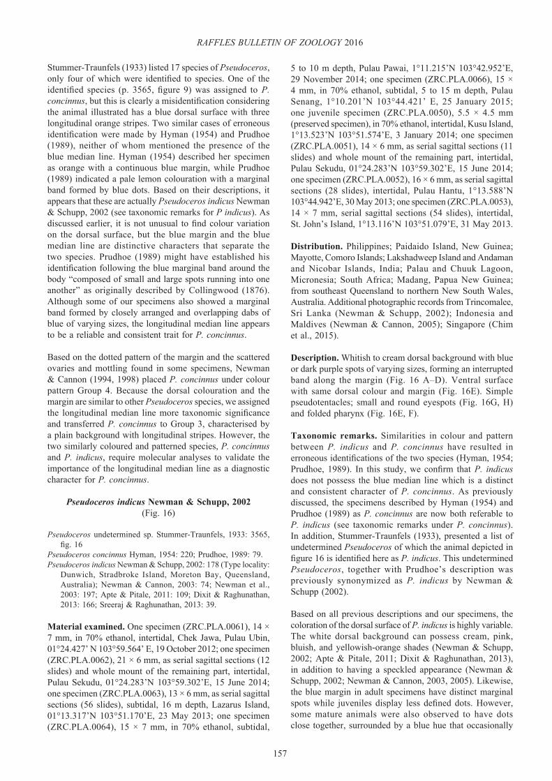
157
RAFFLES BULLETIN OF ZOOLOGY 2016
Stummer-Traunfels (1933) listed 17 species of Pseudoceros, only four of which were identified to species. One of the identified species (p. 3565, figure 9) was assigned to P. concinnus, but this is clearly a misidentification considering the animal illustrated has a blue dorsal surface with three longitudinal orange stripes. Two similar cases of erroneous identification were made by Hyman (1954) and Prudhoe (1989), neither of whom mentioned the presence of the blue median line. Hyman (1954) described her specimen as orange with a continuous blue margin, while Prudhoe (1989) indicated a pale lemon colouration with a marginal band formed by blue dots. Based on their descriptions, it appears that these are actually Pseudoceros indicus Newman & Schupp, 2002 (see taxonomic remarks for P indicus). As discussed earlier, it is not unusual to find colour variation on the dorsal surface, but the blue margin and the blue median line are distinctive characters that separate the two species. Prudhoe (1989) might have established his identification following the blue marginal band around the body “composed of small and large spots running into one another” as originally described by Collingwood (1876). Although some of our specimens also showed a marginal band formed by closely arranged and overlapping dabs of blue of varying sizes, the longitudinal median line appears to be a reliable and consistent trait for P. concinnus.
Based on the dotted pattern of the margin and the scattered ovaries and mottling found in some specimens, Newman & Cannon (1994, 1998) placed P. concinnus under colour pattern Group 4. Because the dorsal colouration and the margin are similar to other Pseudoceros species, we assigned the longitudinal median line more taxonomic significance and transferred P. concinnus to Group 3, characterised by a plain background with longitudinal stripes. However, the two similarly coloured and patterned species, P. concinnus and P. indicus, require molecular analyses to validate the importance of the longitudinal median line as a diagnostic character for P. concinnus.
Pseudoceros indicus Newman & Schupp, 2002(Fig. 16)
Pseudoceros undetermined sp. Stummer-Traunfels, 1933: 3565, fig. 16
Pseudoceros concinnus Hyman, 1954: 220; Prudhoe, 1989: 79. Pseudoceros indicus Newman & Schupp, 2002: 178 (Type locality:
Dunwich, Stradbroke Island, Moreton Bay, Queensland, Australia); Newman & Cannon, 2003: 74; Newman et al., 2003: 197; Apte & Pitale, 2011: 109; Dixit & Raghunathan, 2013: 166; Sreeraj & Raghunathan, 2013: 39.
Material examined. One specimen (ZRC.PLA.0061), 14 × 7 mm, in 70% ethanol, intertidal, Chek Jawa, Pulau Ubin, 01°24.427’ N 103°59.564’ E, 19 October 2012; one specimen (ZRC.PLA.0062), 21 × 6 mm, as serial sagittal sections (12 slides) and whole mount of the remaining part, intertidal, Pulau Sekudu, 01°24.283’N 103°59.302’E, 15 June 2014; one specimen (ZRC.PLA.0063), 13 × 6 mm, as serial sagittal sections (56 slides), subtidal, 16 m depth, Lazarus Island, 01°13.317’N 103°51.170’E, 23 May 2013; one specimen (ZRC.PLA.0064), 15 × 7 mm, in 70% ethanol, subtidal,
5 to 10 m depth, Pulau Pawai, 1°11.215’N 103°42.952’E, 29 November 2014; one specimen (ZRC.PLA.0066), 15 × 4 mm, in 70% ethanol, subtidal, 5 to 15 m depth, Pulau Senang, 1°10.201’N 103°44.421’ E, 25 January 2015; one juvenile specimen (ZRC.PLA.0050), 5.5 × 4.5 mm (preserved specimen), in 70% ethanol, intertidal, Kusu Island, 1°13.523’N 103°51.574’E, 3 January 2014; one specimen (ZRC.PLA.0051), 14 × 6 mm, as serial sagittal sections (11 slides) and whole mount of the remaining part, intertidal, Pulau Sekudu, 01°24.283’N 103°59.302’E, 15 June 2014; one specimen (ZRC.PLA.0052), 16 × 6 mm, as serial sagittal sections (28 slides), intertidal, Pulau Hantu, 1°13.588’N 103°44.942’E, 30 May 2013; one specimen (ZRC.PLA.0053), 14 × 7 mm, serial sagittal sections (54 slides), intertidal, St. John’s Island, 1°13.116’N 103°51.079’E, 31 May 2013.
Distribution. Philippines; Paidaido Island, New Guinea; Mayotte, Comoro Islands; Lakshadweep Island and Andaman and Nicobar Islands, India; Palau and Chuuk Lagoon, Micronesia; South Africa; Madang, Papua New Guinea; from southeast Queensland to northern New South Wales, Australia. Additional photographic records from Trincomalee, Sri Lanka (Newman & Schupp, 2002); Indonesia and Maldives (Newman & Cannon, 2005); Singapore (Chim et al., 2015).
Description. Whitish to cream dorsal background with blue or dark purple spots of varying sizes, forming an interrupted band along the margin (Fig. 16 A–D). Ventral surface with same dorsal colour and margin (Fig. 16E). Simple pseudotentacles; small and round eyespots (Fig. 16G, H) and folded pharynx (Fig. 16E, F).
Taxonomic remarks. Similarities in colour and pattern between P. indicus and P. concinnus have resulted in erroneous identifications of the two species (Hyman, 1954; Prudhoe, 1989). In this study, we confirm that P. indicus does not possess the blue median line which is a distinct and consistent character of P. concinnus. As previously discussed, the specimens described by Hyman (1954) and Prudhoe (1989) as P. concinnus are now both referable to P. indicus (see taxonomic remarks under P. concinnus). In addition, Stummer-Traunfels (1933), presented a list of undetermined Pseudoceros of which the animal depicted in figure 16 is identified here as P. indicus. This undetermined Pseudoceros, together with Prudhoe’s description was previously synonymized as P. indicus by Newman & Schupp (2002).
Based on all previous descriptions and our specimens, the coloration of the dorsal surface of P. indicus is highly variable. The white dorsal background can possess cream, pink, bluish, and yellowish-orange shades (Newman & Schupp, 2002; Apte & Pitale, 2011; Dixit & Raghunathan, 2013), in addition to having a speckled appearance (Newman & Schupp, 2002; Newman & Cannon, 2003, 2005). Likewise, the blue margin in adult specimens have distinct marginal spots while juveniles display less defined dots. However, some mature animals were also observed to have dots close together, surrounded by a blue hue that occasionally

158
Bolaños et al.: Pseudocerotid flatworms from Singapore
Fig. 16. Pseudoceros indicus, living animal. A–C, dorsal view of different specimens showing colour and margin pattern variation; B. Specimen with a pinkish dorsal surface; D, juvenile with a continuous marginal band; E, general ventral surface view of a mature specimen; F, detailed view of the ventral surface showing the complex pharynx, male gonopore (arrowhead), female gonopore, uteri, and sucker; G, dorsal view of the simple pseudotentacles and a small round cerebral eyespot; H, erected pseudotentacles and a very small cerebral eyespot. Scale bars = 5 mm [A–C, F–H], 1 mm [D]. fg, female gonopore; ph, pharynx; su, sucker; u, uteri.

159
RAFFLES BULLETIN OF ZOOLOGY 2016
overlapped, giving the appearance of a continuous line rather than a dotted band. This may create taxonomic confusion with the almost identical species, Pseudoceros gamblei (Laidlaw, 1902), characterised by cream dorsal colouration with a purple or dark blue rim. Laidlaw (1902) stated that the pharynx of P. gamblei had a “curious shape, extending the hindmost folds posteriorly to the level of the female gonopore.” Nevertheless, this feature has been observed in other Pseudoceros species, including P. indicus. Moreover, the folds of the pharynx can relax or contract during fixation; hence, this trait is not of taxonomic significance. The author did not provide any additional detail on the pattern of the margin, and other external characters such as shape of pseudotentacles and cerebral eyespot, and the few details presented on the anatomy of the male reproductive system are typical for the genus Pseudoceros. Newman & Schupp (2002) stated that the pattern of the purple or blue margin could be used to differentiate between P. indicus (dotted) and P. gamblei (continuous). Based on this, the only presumptive records of P. gamblei are from Australia (Newman & Cannon, 2003: p.74) and India (Sreeraj & Raghunathan, 2011), with a relatively thick continuous purplish-magenta and blue margin, respectively. Specimens examined in this study also showed a thick blue continuous marginal band, similar to the morphotype reported from India. However, we consider this as colour variation for P. indicus to avoid more confusion due to the uncertain status of P. gamblei. Also, Litvaitis & Newman (2001) presented a preliminary molecular analysis of two undetermined species, Pseudoceros A and B described both as cream coloured, one with a continuous purple margin (possibly P. gamblei) and the other with a discontinuous and slightly more blue in colour margin (possibly P. indicus). The genetic distance between these species shows a low level of intraspecific variation indicating that they are probably the same species. This result contradicts the distinction made between P. indicus and P. gamblei based on the pattern of the margin, indicating that this might not be a reliable character that allows for species distinction.
Available morphological and molecular data are insufficient to either synonymise or clearly separate P. indicus from P. gamblei. Further molecular analyses are needed to differentiate between these two closely related, possibly identical species. It is highly recommended to include other species with similar margins such as P. concinnus, Pseudoceros goslineri Newman & Cannon, 1994, and Pseudoceros ouini Newman & Cannon, 1994 to confirm the colour and pattern of the margin as valid characters among species of the genus. Until clarification of this case is completed, P. indicus is assigned to colour pattern Group 4 characterised by spots and dots.
The reproductive behaviour and embryonic development of P. indicus were recently examined by Chim et al. (2015). This study indicates the presence of P. indicus in Singapore, and therefore, it is not reported here as a new record for the country.
Pseudoceros laingensis Newman & Cannon, 1998(Figs. 17, 18)
Pseudoceros laingensis Newman & Cannon, 1998: 309 (Type locality: Laing Island, Madang, Papua New Guinea); Newman & Cannon, 2003: 75.
Material examined. One whole mount (QMG210952) designated as holotype and 27 slides of histological sections (QMG210925) designated as paratype for Pseudoceros laingensis deposited in the Queensland Museum, Australia. One specimen (ZRC.PLA.0040), 31 × 25 mm (preserved specimen), as serial sagittal sections (58 slides) and whole mount of the remaining part, intertidal, Chek Jawa, Pulau Ubin, 01°24.748’N 103°59.711’E, 17 October 2012.
Distribution. Madang, Papua New Guinea. Additional record for Indonesia (Newman & Cannon, 2005). First record for Singapore.
Description. Cream background with randomly scattered purple spots over the dorsal surface. Border of the body covered by purple spots of different sizes forming a margin (Fig. 17A, C–H). Ventral surface cream with the same purple spots at margin (Fig. 17B). Simple pseudotentacles, and small cerebral cluster of eyes.
Taxonomic remarks. Pseudoceros laingensis was considered a rare species by Newman & Cannon (1998), and to date no other formal published records are known since its first description. Although this species has a conspicuous margin formed by dots of different sizes, this trait was not mentioned by Newman & Cannon (1998) in the original description. Therefore the diagnosis for P. laingensis is emended as follows: “Cream dorsal surface with scattered purple dots; body margin, including the pseudotentacles, delimited by purple spots varying in size.” P. laingensis was initially placed within colour pattern Group 4, where Newman & Cannon (1998) compared it with P. concinnus indicating that the spots in P. laingensis were scattered over the dorsal surface and not restricted only to the margin as seen in P. concinnus. However, since we transferred P. concinnus to Group 3, P. laingensis becomes the only species with such colour and pattern in Group 4.
Some minor variations in colour pattern were observed in our specimens. The background colour ranged from cream to cream-yellowish or pinkish. Purple dots on the dorsal surface were numerous in some individuals but scarce in others. The purple spots at the margin were abundant, close together, and irregularly sized in most animals compared to the holotype in which the margin had fewer spots with a greater distance between them. In terms of the internal anatomy of the reproductive system, Newman & Cannon (1998) noted the presence of two prostatic vesicles, indicating a possible teratology. However, this unusual character was not observed in the paratype of P. laingensis nor the specimen from Singapore, which showed the typical Pseudoceros-type male reproductive system organisation with only one prostatic vesicle (Figs. 18 A–D). Hence, the presence of two

160
Bolaños et al.: Pseudocerotid flatworms from Singapore
Fig 17. Pseudoceros laingensis, live animal; A, dorsal view; B, ventral view showing the long folds of the pharynx extending posteriorly; C–H, in situ photographs showing colour and pattern variation; C, specimen sighted at Changi Beach, 25 May, 2009; D, specimen sighted at Beting Bronok, 20 August, 2005; E–G, Specimens sighted at Chek Jawa; E, 18 May, 2003; F, 27 July, 2002; G, 23 June, 2007. H, Specimen sighted at Pulau Semakau, 18 April, 2010. Photographs by: Ria Tan [C–G], Loh Kok Sheng [H]. Scale bars: 5 mm [A, B]. ph: pharynx.
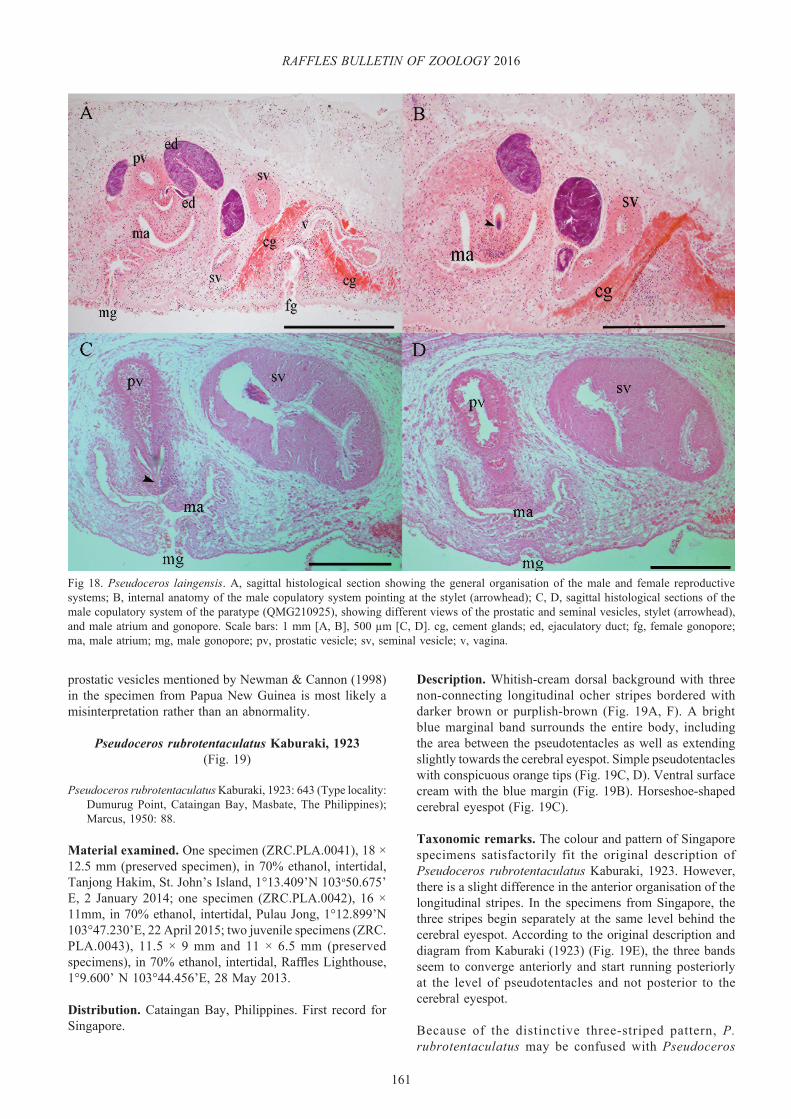
161
RAFFLES BULLETIN OF ZOOLOGY 2016
Fig 18. Pseudoceros laingensis. A, sagittal histological section showing the general organisation of the male and female reproductive systems; B, internal anatomy of the male copulatory system pointing at the stylet (arrowhead); C, D, sagittal histological sections of the male copulatory system of the paratype (QMG210925), showing different views of the prostatic and seminal vesicles, stylet (arrowhead), and male atrium and gonopore. Scale bars: 1 mm [A, B], 500 µm [C, D]. cg, cement glands; ed, ejaculatory duct; fg, female gonopore; ma, male atrium; mg, male gonopore; pv, prostatic vesicle; sv, seminal vesicle; v, vagina.
prostatic vesicles mentioned by Newman & Cannon (1998) in the specimen from Papua New Guinea is most likely a misinterpretation rather than an abnormality.
Pseudoceros rubrotentaculatus Kaburaki, 1923(Fig. 19)
Pseudoceros rubrotentaculatus Kaburaki, 1923: 643 (Type locality: Dumurug Point, Cataingan Bay, Masbate, The Philippines); Marcus, 1950: 88.
Material examined. One specimen (ZRC.PLA.0041), 18 × 12.5 mm (preserved specimen), in 70% ethanol, intertidal, Tanjong Hakim, St. John’s Island, 1°13.409’N 103o50.675’ E, 2 January 2014; one specimen (ZRC.PLA.0042), 16 × 11mm, in 70% ethanol, intertidal, Pulau Jong, 1°12.899’N 103°47.230’E, 22 April 2015; two juvenile specimens (ZRC.PLA.0043), 11.5 × 9 mm and 11 × 6.5 mm (preserved specimens), in 70% ethanol, intertidal, Raffles Lighthouse, 1°9.600’ N 103°44.456’E, 28 May 2013.
Distribution. Cataingan Bay, Philippines. First record for Singapore.
Description. Whitish-cream dorsal background with three non-connecting longitudinal ocher stripes bordered with darker brown or purplish-brown (Fig. 19A, F). A bright blue marginal band surrounds the entire body, including the area between the pseudotentacles as well as extending slightly towards the cerebral eyespot. Simple pseudotentacles with conspicuous orange tips (Fig. 19C, D). Ventral surface cream with the blue margin (Fig. 19B). Horseshoe-shaped cerebral eyespot (Fig. 19C).
Taxonomic remarks. The colour and pattern of Singapore specimens satisfactorily fit the original description of Pseudoceros rubrotentaculatus Kaburaki, 1923. However, there is a slight difference in the anterior organisation of the longitudinal stripes. In the specimens from Singapore, the three stripes begin separately at the same level behind the cerebral eyespot. According to the original description and diagram from Kaburaki (1923) (Fig. 19E), the three bands seem to converge anteriorly and start running posteriorly at the level of pseudotentacles and not posterior to the cerebral eyespot.
Because of the distinctive three-striped pattern, P. rubrotentaculatus may be confused with Pseudoceros

162
Bolaños et al.: Pseudocerotid flatworms from Singapore
Fig. 19. Pseudoceros rubrotentaculatus, living animal. A, dorsal view; B, ventral view showing the pharynx, uteri, and sucker; C, simple pseudotentacles showing orange tips and blue coloration in between, extending to the inverted heart-shaped cerebral eyespot; D, ventral view of the pseudotentacles showing the pseudotentacular eyes; E, original illustration of the dorsal surface of P. rubrotentaculatus from Kaburaki (1923); F, P. rubrotentaculatus feeding in the lab with an extended pharyngeal folds, each on different individual ascidian zooid. Scale bars = 5 mm [A–D]. vd, vas deferens.
tristiatus Hyman, 1959. However, each species exhibits unique colourations that allow their placement as a different species. P. tristiatus has an even blue dorsal surface while P. rubrotentaculatus has a cream dorsal surface with a blue marginal band around the entire body. The longitudinal bands in P. tristiatus are orange and bordered by black or dark purple, whereas P. rubrotentaculatus has ocher bands bordered by darker brown or purplish-brown. In
terms of the arrangement of the longitudinal stripes, in P. rubrotentaculatus, these run separately to the posterior end without merging, whereas in P. tristiatus, the two lateral bands are separated anteriorly and connected posteriorly behind the median stripe forming a U shape. In addition, the pseudotentacles in P. tristiatus have a uniform blue colour, differing from P. rubrotentaculatus which has orange tips.
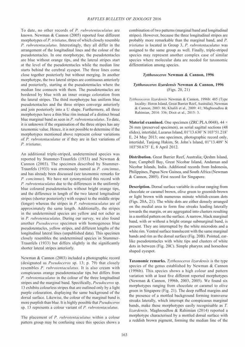
163
RAFFLES BULLETIN OF ZOOLOGY 2016
To date, no other records of P. rubrotentaculatus are known. Newman & Cannon (2005) reported four different morphotypes of P. tristiatus, three of which closely resemble P. rubrotentaculatus. Interestingly, they all differ in the arrangement of the longitudinal lines and the colour of the pseudotentacles. In one morphotype, the pseudotentacles are blue without orange tips, and the lateral stripes start at the level of the pseudotentacles while the median line starts behind the cerebral eyespot. The three lines come close together posteriorly but without merging. In another morphotype, the two lateral stripes are continuous anteriorly and posteriorly, starting at the pseudotentacles where the median line connects with them. The pseudotentacles are bordered by blue with an inner orange coloration from the lateral stripes. The third morphotype has uniform blue pseudotentacles and the three stripes converge anteriorly and join posteriorly into a U shape. Furthermore, all three morphotypes have a thin blue rim instead of a distinct broad blue marginal band as seen in P. rubrotentaculatus. To date, it is unknown if the organisation of the three stripes has any taxonomic value. Hence, it is not possible to determine if the morphotypes mentioned above represent colour variations of P. rubrotentaculatus or if they are in fact variations of P. tristiatus.
An additional triple-striped, undetermined species was reported by Stummer-Traunfels (1933) and Newman & Cannon (2003). The specimen described by Stummer-Traunfels (1933) was erroneously named as P. concinnus, and has already been discussed (see taxonomic remarks for P. concinnus). We have not synonymized this record with P. rubrotentaculatus due to the differences in the uniformly blue coloured pseudotentacles without bright orange tips, and the difference in length of the two lateral longitudinal stripes (shorter posteriorly) with respect to the middle stripe (longer) whereas the stripes in P. rubrotentaculatus are of approximately the same length. Additionally, the stripes in the undetermined species are yellow and not ocher as in P. rubrotentaculatus. During our survey, we also found another Pseudoceros specimen with homogenous blue pseudotentacles, yellow stripes, and different lengths of the longitudinal lateral lines (unpublished data). This specimen closely resembles the undetermined species in Stummer-Traunfels (1933) but differs slightly in the significantly shorter lateral stripes anteriorly.
Newman & Cannon (2003) included a photographic record (designated as Pseudoceros sp. 13; p. 79) that closely resembles P. rubrotentaculatus. It is also cream with conspicuous orange pseudotentacular tips but differs from P. rubrotentaculatus in the colour of the three longitudinal stripes and the marginal band. Specifically, Pseudoceros sp. 13 exhibits colourless stripes that are outlined only by a light purple colouration, displaying the same background of the dorsal surface. Likewise, the colour of the marginal band is more purplish than blue. It is highly possible that Pseudoceros sp. 13 represents a colour variant of P. rubrotentaculatus.
The placement of P. rubrotentaculatus within a colour pattern group may be confusing since this species shows a
combination of two patterns (marginal band and longitudinal stripes). However, because the three longitudinal stripes are probably more remarkable than the marginal band, and P. tristiatus is located in Group 3, P. rubrotentaculatus was assigned to the same group as well. Finally, triple-striped species may represent another complex case of similar species where molecular data are needed for taxonomic differentiation among species.
Tytthosoceros Newman & Cannon, 1996
Tytthosoceros lizardensis Newman & Cannon, 1996(Figs. 20, 21)
Tytthosoceros lizardensis Newman & Cannon, 1996b: 485 (Type locality: Heron Island, Great Barrier Reef, Australia); Newman & Cannon, 2003: 86; Khalili et al., 2009: 41; Maghsoudlou & Rahimian, 2014: 336; Dixit et al., 2015: 3.
Material examined. One specimen (ZRC.PLA.0048), 44 × 26 mm (preserved specimen), as serial sagittal sections (48 slides), intertidal, Lazarus Island, 01°13.630’ N 103°51.218’ E, 24 May 2013; one specimen, photographic record only, intertidal, Tanjong Hakim, St. John’s Island, 01°13.409’ N 103°50.675’ E, 8 April 2012.
Distribution. Great Barrier Reef, Australia; Qeshm Island, Iran; Campbell Bay, Great Nicobar Island, Andaman and Nicobar Islands, India. Additional records from Indonesia, Philippines, Papua New Guinea, and South Africa (Newman & Cannon, 2005). First record for Singapore.
Description. Dorsal surface variable in colour ranging from chocolate or caramel brown, olive green to greenish-brown or light brown with numerous minute whitish-cream dots (Figs. 20A, 21). The white dots are either densely arranged on the medial area to form fine streaks leading laterally towards the margin, or are aggregated into clusters resulting in a mottled pattern on the surface. A narrow, black marginal band, with or without a bright orange submarginal band, is present. They are interrupted by the white microdots and a white rim. Ventral surface translucent with the same marginal bands and rim as the dorsal surface (Fig. 20B). Pointed, ear-like pseudotentacles with white tips and clusters of white dots in between (Fig. 20C). Simple pharynx and horseshoe shaped eyespot.
Taxonomic remarks. Tytthosoceros lizardensis is the type species of the genus established by Newman & Cannon (1996b). This species shows a high colour and pattern variation with at least five different reported morphotypes (Newman & Cannon, 1996b, 2003, 2005). We found six morphotypes ranging from chocolate or caramel to olive green in Singapore (Fig. 21). The deep ruffled margins and the presence of a mottled background forming transverse streaks laterally, which interrupt the conspicuous marginal bands, make these morphotypes easily recognisable as T. lizardensis. Maghsoudlou & Rahimian (2014) reported a morphotype characterised by a mottled dorsal surface with a reddish brown pigment, forming the median line of the

164
Bolaños et al.: Pseudocerotid flatworms from Singapore
Fig. 20. Tytthosoceros lizardensis, living animal. A, dorsal view of the morphotype with and orange band; B, ventral view of the animal showing the pharynx, uteri, and sucker; C, anterior region showing the pointed, simple-folded pseudotentacles and horseshoe-shaped cerebral eyespot; D, a female copepod associated to T. lizardensis, characterised by two rows of opaque white egg sacs. Scale bars = 10 mm [A, B], 5 mm [C]. in, intestine; su, sucker; u, uteri.
body. We also found this morphotype, which differs from other known morphotypes by having minute, reddish dots on its dorsal surface (Fig. 21B, D). Other external features that characterise T. lizardensis include the simple pharynx, a single male gonopore, an elongate and oval body raised medially and slightly tapered posteriorly, and small ear-like pseudotentacles with white tips and cream mottling in-between. Only one morphotype in this study had a remarkable orange submarginal band (Fig. 20A–D), while the others only exhibited a narrow black band with a white rim. As true for many polyclads, the variation of the background colour is most likely due to the contents of their diet. However, the factors that influence the presence of such conspicuous marginal bands still remain unknown. Due to the considerable variation in colour pattern observed in this species, molecular and morphological data are needed to avoid confusion and misidentifications, and to validate the different morphotypes that have been identified as T. lizardensis.
Newman & Cannon (1996b) noted the presence of a small, inconspicuous sucker well separated from the gonopores in T. lizardensis. However, the sucker in our animals from Singapore was rather large and conspicuous. Khalili et al. (2009) also reported the presence of a distinctive sucker in the specimens from Iran. The size of the sucker does
not have any taxonomic value and may be related to the overall size of the animals. The degree of development of the sucker could also be the result of wave exposure in the intertidal area and the necessity for the animals to attach to the substratum.
Tytthosoceros lizardensis can be abundant under rocks in intertidal areas, where up to 20 animals were observed in a small area (Newman & Cannon, 1996b; Khalili et al., 2009). Although a few other species have been observed to aggregate in similar numbers (Boninia divae Marcus & Marcus, 1968, Pseudoceros indicus, and Phrikoceros baibaiye) (pers. obs.), this is rather unusual for polyclads, which are normally found individually or in pairs.
Newman & Cannon (1996b) mentioned the presence of a pair of small copepods associated with T. lizardensis. The copepods were identified as Pseudanthessius newmanae Humes, 1997. A similar association was also observed in our specimen with the bright orange submarginal band, in which 8–10 undetermined copepods were found all over the body (Fig. 20D). Copepods have also been observed on other polyclad species, particularly for flatworms bigger than 6–8 cm (e.g., Acanthozoon sp, Pericelis cata Marcus & Marcus, 1968, Pseudobiceros kriptos Newman
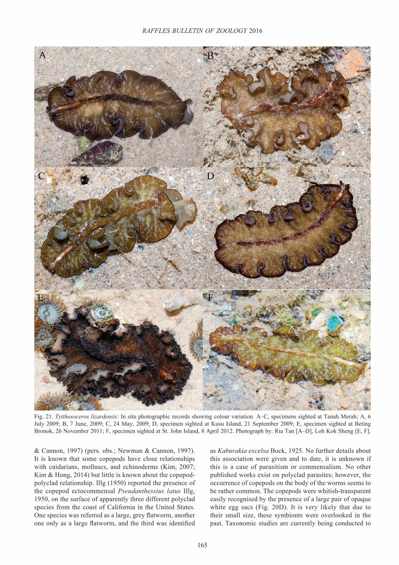
165
RAFFLES BULLETIN OF ZOOLOGY 2016
Fig. 21. Tytthosoceros lizardensis: In situ photographic records showing colour variation. A–C, specimens sighted at Tanah Merah; A, 6 July 2009; B, 7 June, 2009; C, 24 May, 2009; D, specimen sighted at Kusu Island, 21 September 2009; E, specimen sighted at Beting Bronok, 26 November 2011; F, specimen sighted at St. John Island, 8 April 2012. Photograph by: Ria Tan [A–D], Loh Kok Sheng [E, F].
& Cannon, 1997) (pers. obs.; Newman & Cannon, 1997). It is known that some copepods have close relationships with cnidarians, molluscs, and echinoderms (Kim, 2007; Kim & Hong, 2014) but little is known about the copepod-polyclad relationship. Illg (1950) reported the presence of the copepod ectocommensal Pseudanthessius latus Illg, 1950, on the surface of apparently three different polyclad species from the coast of California in the United States. One species was referred as a large, grey flatworm, another one only as a large flatworm, and the third was identified
as Kaburakia excelsa Bock, 1925. No further details about this association were given and to date, it is unknown if this is a case of parasitism or commensalism. No other published works exist on polyclad parasites; however, the occurrence of copepods on the body of the worms seems to be rather common. The copepods were whitish-transparent easily recognised by the presence of a large pair of opaque white egg sacs (Fig. 20D). It is very likely that due to their small size, these symbionts were overlooked in the past. Taxonomic studies are currently being conducted to

166
Bolaños et al.: Pseudocerotid flatworms from Singapore
confirm if Pseudanthessius newmanae is also the copepod species associated with animals from Singapore. More detailed observations and ecological studies are required to accurately determine this type of symbiosis.
DISCUSSION
To date, the knowledge about Singaporean polyclads is limited to the published records of Collingwood (1876), Laidlaw (1903), and Chim et al. (2015). Additional species have been informally documented (Tan, 2008) and other occasional photographic records are known for the country (Newman & Cannon, 2005). A total of 16 species are described in this study, representing the first comprehensive taxonomic work of the order Polycladida in Singapore. Therefore, all the species are first records for the country with the exception of Pseudobiceros bedfordi, Pseudobiceros hancockanus, and Pseudoceros indicus.
All species documented here belong to the family Pseudocerotidae, which is considered the largest and most diverse family within the suborder Cotylea (Tyler et al., 2006–2015). In this study, we describe single species representatives of the genera Phrikoceros and Tytthosoceros, two species of Nymphozoon including the new combination N. orsaki, and six species for Pseudobiceros and Pseudoceros. These pseudocerotids are distinguished by their bright colours and diverse colour patterns (Newman & Cannon 1994, 1996a, b; 1997; 1998; 2003; Apte & Pitale, 2011; Sreeraj & Raghunathan, 2011; 2013; Maghsoudlou & Rahimian, 2014). They are all widely distributed in the Indian and Western Pacific Oceans, despite a major putative biogeographical barrier formed by the islands of the Indonesian archipelago (the Indo-Pacific Barrier). The Singaporean polyclad diversity has a similar species composition with the broader Indo-Pacific fauna. This geographic distribution is most likely related to the indirect type of development that characterises cotyleans and the ability of their larvae to disperse.
Generally, the organisation of the male and female reproductive systems, together with colouration and patterns are key morphological characters used in polyclad taxonomy (Lang, 1884; Bock, 1913; Prudhoe, 1985, 1989; Faubel, 1983, 1984b). However, the reproductive system of most pseudocerotids is homogeneous and therefore of little use to differentiate between species (Newman & Cannon, 1994). Consequently, external features such as the shape of pharynx, pseudotentacles, and cerebral eyespot, number of male and female gonopores, and colour and patterns are now recognised as being the most taxonomically informative, especially for those species belonging to the genera Pseudoceros and Pseudobiceros (Hyman, 1954; Prudhoe, 1989; Newman & Cannon, 1994, 1996a, b; 1997; 1998; 2003; Bolaños et al, 2007). Preliminary molecular analyses have helped to elucidate the validity of the external characters used to differentiate between genera and to determine species separation based on colour and patterns (Goggin & Newman, 1996; Litvaitis
& Newman, 2001). Nevertheless, the results presented in this study confirm that polyclad species exhibit a broad range of colour and pattern variation, and the use of coloration alone can also create confusion for species identification (Litvaitis et al., 2010).
Polyclad colouration often depends on the content of the intestinal branches, which are distributed throughout the entire body and may contribute to the colour of the dorsal surface. It is therefore possible that animals of the same species, found in different habitats and feeding on different prey, may be recognised as two different species. On the other hand, species complexes which include different colour morphotypes are also found within the group. This is the case for Pseudoceros bicolor, which exhibits at least three different colour morphotypes and closely resembles Pseudoceros rawlinsonae (Litvaitis et al., 2010). Here, we identified additional species complexes, specifically, colour variations and slight differences in patterns and arrangements in P. fulgor, P. bifurcus, P. indicus, P. rubrotentaculatus, and T. lizardensis (see taxonomic remarks for each species). However, it is evident that additional molecular analyses are needed to validate colour variations as morphotypes of a species.
The use of morphological characters in polyclad taxonomy has allowed the establishment of the current classification system (Prudhoe, 1985; Faubel, 1983; 1984b). However, some of the diagnostic characters are morphologically plastic and may exhibit extensive variation depending on animal maturity, size, habitat, and nutritional content (Faubel, 1983; Prudhoe, 1985, 1989). For example, the number of eyes and the shape of the cerebral eyespot may change during different stages of life, and the shape of the pharynx might also be controlled by the animal development and size. In species with supernumerary organs such N. bayeri and N. orsaki, the number of female gonopores is likely related to the animal maturity and growth. Although the importance of the examination of newly collected material and live animals, the proper fixation and preservation, and the combination of molecular and morphological analyses has been mentioned repeatedly in other studies (Newman & Cannon, 1994, 1996b; Bahia et al., 2014), we yet again place a great emphasis on these three points. Combining molecular and morphological data will generate more consistent and reliable identifications and will provide a stable phylogenetic framework for understanding the relationships among polyclads.
The results from the Comprehensive Marine Biodiversity Survey shown in this study demonstrate that Singapore has a rich biodiversity of polyclad flatworms comparable to other regions of the Indo-Pacific. The species presented here represent only a portion of the large number of specimens collected during the survey. Additional material belonging to the family Pseudocerotidae as well as other families of Cotylea and Acotylea are waiting to be described. Further taxonomic studies will be presented as a contribution to the knowledge of the polyclad fauna from Singapore.

167
RAFFLES BULLETIN OF ZOOLOGY 2016
ACKNOWLEDGEMENTS
The Singapore Strait marine biodiversity workshop held on St. John’s Island, Singapore was organised by the National Parks Board and National University of Singapore. The workshop, as part of the Comprehensive Marine Biodiversity Survey (CMBS) was supported by generous contributions from Asia Pacific Breweries Singapore, Care-for-Nature Trust Fund, Keppel Care Foundation, Shell Companies in Singapore and The Air Liquide Group. The Shell Biodiversity Fellowship Fund enabled DMB to travel to Singapore and participate in this project after the workshop. We are particularly grateful to Koh Kwan Siong, Tan Yee Keat, and Toh Chay Hoon for their assistance in finding the flatworms in the field over the period of CMBS, and Jon Norenburg from the Smithsonian Institution, Washington DC, for reviewing the holotype of P. caeruleocinctus. We would also like to thank Ria Tan and Loh Kok Sheng for allowing us to use some of their lovely photographs.
LITERATURE CITED
Apte D & Pitale R (2011) New records of polyclad flatworms (Platyhelminthes: Turbellaria) from coral reefs of Lakshadweep Island, India. Journal of the Bombay Natural History Society, 108: 109–113.
Bahia J, Padula V & Delgado M (2012) Five new records and morphological data of polyclad species (Platyhelminthes: Turbellaria) from Rio Grande do Norte, Northeastern Brazil. Zootaxa, 3170: 31–44.
Bahia J, Padula V, Lavrado HP & Quiroga SY (2014) Taxonomy of Cotylea (Platyhelminthes: Polycladida) from Cabo Frio, southeastern Brazil, with the description of a new species. Zootaxa, 3873(5): 495–525.
Bock S (1913) Studien über Polycladen. Zoologiska Bidrag fran Uppsala, 2: 29–344.
Bock S (1925) Papers from Dr. Th. Mortensen’s Pacific Expedition 1914-1916. XXVII. Planarians, Pt. IV. New stylochids. Videnskabelige meddelelser fra Dansk Naturhistorisk Forening, 79: 97–184.
Bolaños DM, Quiroga SY & Litvaitis MK (2006) A new acotylean flatworm, Armatoplana colombiana n. sp. (Platyhelminthes: Polycladida: Stylochoplanidae) from the Caribbean coast of Colombia, South America. Zootaxa, 1162: 53–64.
Bolaños DM, Quiroga SY & Litvaitis MK (2007) Five new species of cotylean flatworms (Platyhelminthes: Polycladida) from the wider Caribbean. Zootaxa, 1650: 1–23.
Bresslau E (1933) Turbellaria. In: Kükenthal W & Krumbach T (eds.) Handbuch der Zoologie. Walter de Gruyter & Co, Berlin, 416 pp.
Brusa F & Damborenea C (2011) Polycladida Acotylea from Patagonia. Redescription of Crassiplana albatrossi (Pseudostylochidae), lectotype designation and first record of Notocomplana palta (Notoplanidae). Zootaxa, 2903: 29–38.
Brusa F, Damborenea C & Quiroga S (2009) First records of the Pseudocerotidae (Platyhelminthes: Polycladida: Cotylea) from Patagonia, Argentina. Zootaxa, 2283: 51–59.
Bulnes VN & Torres Y (2014) Pseudoceros astrorum, a new species of Polycladida (Cotylea, Pseudocerotidae) from Northeastern Brazil. Zootaxa, 3881(1): 94–100.
Chim CK, Ong RSL & Gan BQ (2015) Penis fencing, spawning, parental care and embryonic development in the cotylean flatworm Pseudoceros indicus (Platyhelminthes: Polycladida: Pseudocerotidae) from Singapore. Raffles Bulletin of Zoology, Supplement 31: 60–67.
Collingwood C (1876) On thirty-one species of marine planarians, collected partly by the late Dr Kelaart, F.L.S. at Trincomalee, and partly by Dr Collingwood, F.L.S. in the eastern seas. Transactions of the Linnean Society of London, Series 2, Zoology, 1: 83–98.
Dawydoff CN (1952) Contribution à l’étude des invertebrés de la faune marine benthique de l’Indochine. Bulletin Biologique de la France et de la Belgique, Supplement (39): 1–158.
Dixit S & Raghunathan C (2013) Polyclads of Andaman and Nicobar Islands. Journal of the Andaman Science Association, 18(2): 165–169.
Dixit S, Sivaperuman C & Raghunathan C (2015) Three new records of polyclad flatworms from India. Marine Biodiversity Records, 8: 1–4.
Ehlers U (1986) Comments on a phylogenetic system of the Platyhelminthes. Hydrobiologia, 132: 1–12.
Faubel A (1983) The Polycladida, Turbellaria. Proposal and establishment of a new system. Part I. The Acotylea. Mitteilungen aus dem Hamburgischen zoologischen Museum und Institut, 80: 17–121.
Faubel A (1984a) On the geographical occurrence of pelagic polyclad turbellarians. Cariers de Biologie Marine, 25: 153–168.
Faubel A (1984b) The Polycladida, Turbellaria. Proposal and establishment of a new system. Part II. The Cotylea. Mitteilungen aus dem Hamburgischen Zoologischen Museum und Institut, 80: 189–259.
George JD & George JJ (1979) Marine Life: An Illustrated Encyclopedia of Invertebrates in the Sea. Rigby Ltd, Sydney, 288 pp.
Goggin CL & Newman LJ (1996) Use of molecular data to discriminate pseudocerotid turbellarians. Journal of Helminthology, 7: 123–126.
Gosliner TM, Behrens DW & Williams GC (1996) Coral Reef Animals of the Indo-Pacific. Sea Challengers, Monterey, California, 288 pp.
Haswell WA (1907) Observations on Australian polyclads. Transactions of the Linnean Society of London, Zoology, Series 2, 9: 465–485.
Hyman LH (1954) The polyclad genus Pseudoceros, with special reference to the Indo-Pacific region. Pacific Science, 8: 219–225.
Hyman LH (1955a) Some polyclads from Polynesia and Micronesia. Proceedings of the United States National Museum, 105: 65–82.
Hyman LH (1955b) Some Polyclad flatworms from the West Indies and Florida. Proceedings of the United States National Museum, 104(3341), 115–150.
Hyman LH (1959) A further study of Micronesian polyclad flatworms. Proceedings of the United States National Museum, 108: 543–597.
Hyman L (1960) Second report of Hawaiian polyclads. Pacific Science, 14: 308–309.
Humes AG (1997) Pseudoanthessius newmanae, new species (Copepoda: Poecilostomatoida: Pseudoanthessiidae) from marine turbellarians in Australia. Memoirs of the Queensland Museum, 42: 227–231.
Illg PL (1950) A new copepod, Pseudanthessius latus (Cyclopoida: Lichomolgidae), commensal with a marine flatworm. Journal of the Washington Academy of Science, 40: 129–133.

168
Bolaños et al.: Pseudocerotid flatworms from Singapore
Jie WB, Kuo SC & Mok HK (2014) Re-description of Thysanozoon nigropapillosum (Polycladida: Pseudocerotidae) from the South China Sea, with observations on a novel pre-copulatory structure, sexual behaviour and diet. Raffles Bulletin of Zoology, 62: 764–770.
Kaburaki T (1923) The polyclad turbellarians from the Philippine Islands. Bulletin of the United States National Museum, 100: 635–649.
Kato K (1937) Polyclads collected in Idu, Japan. Japanese Journal of Zoology, 7: 211–232.
Kato K (1938) On a pelagic polyclad, Planocera pellucida (Mertens) from Japan. Zoological Magazine, 50: 230–232.
Kato K (1943) Polyclads from Palao. Bulletin of the Biogeography Society of Japan, 13: 79–90.
Kato K (1944) Polycladida from Japan. The Journal of the Sigenkagaku Kenkyusko, 1(3): 257–318.
Kelaart EF (1858) Description of new and little known species of Ceylon nudibranchiate molluscs and zoophytes. Journal of the Ceylon Branch of the Royal Society, 3: 84–139.
Khalili Z, Rahimian H & Pazooki J (2009) First record of the family Pseudocerotidae (Platyhelminthes, Polycladida, Cotylea) from the Persian Gulf, Iran. ZooKeys, 31: 39–51.
Kim IH (2007) Copepods (Crustacea) associated with marine invertebrates from the Moluccas. The Korean Journal of Systematic Zoology, Special Issue, 6: 1–126.
Kim IH & Hong JS (2014) Copepods (Crustacea, Copepoda, Cyclopoida) associated with marine invertebrates from Thailand. Animal Systematics, Evolution and Diversity, 30(4): 274–318.
Laidlaw FF (1902) The marine Turbellaria, with an account of the anatomy of some species. In: Gardiner JS (ed.) The Fauna and Geography of the Maldive and Laccadive Archipelagoes. University Press, Cambridge. Volume 1, Part 3. Pp. 282–312.
Laidlaw FF (1903) On a collection of Turbellaria Polycladida from the Straits of Malacca. Proceedings of the Zoological Society of London, 1: 301–318.
Lang A (1884) Die Polycladen des Golfes von Neapel und der angrenzenden Meeresabschnitte. Eine Monographie. Fauna und Flora des Golfes von Neapel, Leipzieg, 11: 1–688.
Litvaitis MK & Newman LJ (2001) A molecular framework for the phylogeny of the Pseudocerotidae (Platyhelminthes, Polycladida). Hydrobiologia, 444: 177–182.
Litvaitis MK, Bolaños DM & Quiroga SY (2010) When names are wrong and colours deceive: unraveling the Pseudoceros bicolor species complex (Turbellaria: Polycladida). Journal of Natural History, 44: 829−845.
Maghsoudlou A & Rahimian H (2014) Contribution to the knowledge of cotylean flatworms (Turbellaria, Polycladida) from Iranian coasts: Introducing a new species, with remarks on new records. Zootaxa, 3860(4): 325–342.
Marcus E (1950) Turbellaria brasileiros. Boletim da Faculdade de Filosofia, Ciências e Letras da Universidade de São Paulo, Zoologia, 15(8): 69−190.
Marcus E & Marcus E (1968) Polycladida from Curaçao and faunistically related regions. Studies on the Fauna of Curaçao and other Caribbean Islands, 101: 1−133.
Marquina D, Fernandez-Alvarez FA & Noreña C (2014a) Five new records and one new species of Polycladida (Platyhelminthes) for the Cantabrian coast (North Atlantic) of the Iberian Peninsula. Journal of the Marine Biological Association of the United Kingdom, 95(2): 311–322.
Marquina D, Osca D, Rodriguez J, Fernandez-Despiau E & Noreña C (2014b) State of knowledge of the Acotylea (Polycladida, Platyhelminthes) from the Mediterranean coasts of Spain: new records and new species. Zootaxa, 3780: 108–134.
Marquina D, Aguado MT & Noreña C (2015) New records of Cotylea (Polycladida, Platyhelminthes) from Lizard Island, Great Barrier Reef, Australia, with remarks on the distribution of the Pseudoceros Lang, 1884 and Pseudobiceros Faubel, 1984 species of the Indo-Pacific Marine Region. Zootaxa, 4019(1): 354–377.
Newman LJ & Cannon LRG (1994) Pseudoceros and Pseudobiceros (Polycladida, Pseudocerotidae) from Eastern Australia and Papua New Guinea. Memoirs of the Queensland Museum, 37(1): 205–266.
Newman LJ & Cannon LRG (1995) The importance of preservation, colour and pattern in tropical Pseudocerotidae (Platyhelminthes, Polycladida). Hydrobiologia, 305: 141–143.
Newman LJ & Cannon LRG (1996a) New genera of pseudocerotid flatworms (Platyhelminthes, Polycladida) from Australian and Papua New Guinean coral reefs. Journal of Natural History, 30: 1425–1441.
Newman LJ & Cannon LRG (1996b) Bulaceros, new genus and Tytthosoceros, new genus (Platyhelminthes, Polycladida, Pseudocerotidae) from the Great Barrier Reef, Australia and Papua New Guinea. Raffles Bulletin of Zoology, 44: 479–492.
Newman LJ & Cannon LRG (1997) Nine new Pseudobiceros (Platyhelminthes, Polycladida, Pseudocerotidae) from the Indo-Pacific. The Raffles Bulletin of Zoology, 45: 341–368.
Newman LJ & Cannon LRG (1998) Pseudoceros (Platyhelminthes, Polycladida) from the Indo-Pacific with twelve new species from Australia and Papua New Guinea. The Raffles Bulletin of Zoology, 46: 293–323.
Newman LJ & Cannon LRG (2003) Marine Flatworms: the world of Polyclads. CSIRO Publishing, Melbourne, 112 pp.
Newman LJ & Cannon LRG (2005) Fabulous flatworms: a guide to marine polyclads. ABRS and CSIRO Publishing, Canberra and Melbourne, Australia. CD-ROM interactive guide.
Newman LJ & Schupp P (2002) A new pseudocerotid flatworm (Platyhelminthes, Polycladida) from the Indo-Pacific. Micronesica, 34: 177–184.
Newman LJ, Paulay G & Rittson-Williams R (2003) Polyclad flatworms of Micronesia. Micronesica, 35–36: 189–199.
Noreña C, Marquina D, Perez J & Almon B (2014) First records of Cotylea (Polycladida, Platyhelminthes) for the Atlantic coast of the Iberian Peninsula. ZooKeys, 404: 1–22.
Palombi A (1931) Turbellari della nuovo Guinea. Mémoires du Musée Royal d’Histoire Naturelle de Belgique, 2(8): 1–14.
Prudhoe S (1978) Some polyclad turbellarians new to the fauna of the Australian coasts. Records of the Australian Museum, 31: 586–604.
Prudhoe S (1985) A Monograph on Polyclad Turbellaria. London/Oxford: British Museum of Natural History and Oxford University Press, UK, 259 pp.
Prudhoe S (1989) Polyclad turbellarians recorded from African waters. Bulletin of the British Museum of Natural History 55: 47–96.
Quiroga SY, Bolaños DM & Litvaitis MK (2004) A checklist of polyclad flatworms (Platyhelminthes: Polycladida) from the Caribbean coast of Colombia, South America. Zootaxa, 633: 1–12.
Quiroga SY, Bolaños DM & Litvaitis MK (2006) First description of deep-sea polyclad flatworms from the North Pacific: Anocellidus n. gen. profundus n. sp. (Anocelidae, n. fam.) and Oligocladus voightae n. sp. (Euryleptidae). Zootaxa, 1317: 1–19.
Quiroga SY, Bolaños DM & Litvaitis MK (2008) Two new species of flatworms (Platyhelminthes: Polycladida) from the Gulf of Mexico. Journal of the Marine Biological Association of the United Kingdom, 88: 1363–1370.

169
RAFFLES BULLETIN OF ZOOLOGY 2016
Sreeraj CR & Raghunathan C (2011) New records of pseudocerotid polyclads from Andaman and Nicobar Islands, India. Marine Biodiversity Records, 4: 1–5.
Sreeraj CR & Raghunathan C (2013) Pseudocerotid polyclads (Platyhelminthes, Turbellaria, Polycladida) from Andaman and Nicobar Islands, India. Proceedings of the International Academy of Ecology and Environmental Sciences, 3(1): 36–41.
Stummer-Traunfels R von (1933) Polycladida. Bronn’s Klassen und Ordnungen des Tierreichs, 4, Abteilung 1c 179: 3485–3596.
Tan R (2008) Wild Singapore. www.wildsingapore.com (Accessed 23 June 2015).
Tyler S, Schilling S, Hooge M & Bush LF (Comp.) (2006–2015) Turbellarian taxonomic database. Version 1.7. Available from: http://turbellaria.umaine.edu (Accessed 23 June 2015).
Yeri M & Kaburaki T (1918) Description of some Japanese polyclad turbellaria. The Journal of the College of Science, Imperial University of Tokyo, 39: 3–58.