
First observation of a Fano profile following one step ...
11
HAL Id: jpa-00251706 https://hal.archives-ouvertes.fr/jpa-00251706 Submitted on 1 Jan 1993 HAL is a multi-disciplinary open access archive for the deposit and dissemination of sci- entific research documents, whether they are pub- lished or not. The documents may come from teaching and research institutions in France or abroad, or from public or private research centers. L’archive ouverte pluridisciplinaire HAL, est destinée au dépôt et à la diffusion de documents scientifiques de niveau recherche, publiés ou non, émanant des établissements d’enseignement et de recherche français ou étrangers, des laboratoires publics ou privés. First observation of a Fano profile following one step autoionization into a double photoionization continuum L. Journel, B. Rouvellou, D. Cubaynes, J. Bizau, F. Wuilleumier, M. Richter, P. Sladeczek, K.-H. Selbmann, P. Zimmermann, Henri Bergeron To cite this version: L. Journel, B. Rouvellou, D. Cubaynes, J. Bizau, F. Wuilleumier, et al.. First observation of a Fano profile following one step autoionization into a double photoionization continuum. Journal de Physique IV Proceedings, EDP Sciences, 1993, 03 (C6), pp.C6-217-C6-226. 10.1051/jp4:1993621. jpa-00251706
-
Upload
khangminh22 -
Category
Documents
-
view
0 -
download
0
Transcript of First observation of a Fano profile following one step ...
HAL Id: jpa-00251706https://hal.archives-ouvertes.fr/jpa-00251706
Submitted on 1 Jan 1993
HAL is a multi-disciplinary open accessarchive for the deposit and dissemination of sci-entific research documents, whether they are pub-lished or not. The documents may come fromteaching and research institutions in France orabroad, or from public or private research centers.
L’archive ouverte pluridisciplinaire HAL, estdestinée au dépôt et à la diffusion de documentsscientifiques de niveau recherche, publiés ou non,émanant des établissements d’enseignement et derecherche français ou étrangers, des laboratoirespublics ou privés.
First observation of a Fano profile following one stepautoionization into a double photoionization continuumL. Journel, B. Rouvellou, D. Cubaynes, J. Bizau, F. Wuilleumier, M. Richter,
P. Sladeczek, K.-H. Selbmann, P. Zimmermann, Henri Bergeron
To cite this version:L. Journel, B. Rouvellou, D. Cubaynes, J. Bizau, F. Wuilleumier, et al.. First observation of aFano profile following one step autoionization into a double photoionization continuum. Journal dePhysique IV Proceedings, EDP Sciences, 1993, 03 (C6), pp.C6-217-C6-226. �10.1051/jp4:1993621�.�jpa-00251706�
JOURNAL DE PHYSIQUE IV Colloque C6, supplkment au Journal de Physique 11, Volume 3, novembre 1993
First observation of a Fano profile following one step autoionization into a double photoionization continuum
L. JOURNEL, B. ROUVELLOU, D. CUBAYNES, J.M. BIZAU, F.J. WILLEUMIER, M. RICHTER*, P. SLADECZEK*, K.-H. SELBMANW, P. ZIMMERMANW and H. BERGEROW*
Laboratoire de Spectroscopic Atomique et Ionique, Universite' Paris Sud, URA 775 du CNRS, B. 350, 91405 Orsay, France * Technische Universitat, Berlin, Germany ** LURE, CNRS-Ca-MEN Universite' Paris Sud, B. 209d, 91405 Orsay, France
Abstract: We have measured the double photoionization cross section of sodium atoms between the first 2 ~ 2 2 ~ 5 2~ double photoionization (52.4 eV) and 2s-single 1.3s photoionization thresholds (71.0 eV). We have also observed a Fano profile into the double ionization continuum resulting from the interference between the one-step direct double photoionization process and the resonant double Auger decay of core-excited neutral sodium in the 2s + 3p resonance region. Profiles of absolute partial and total cross sections have been obtained in all important channels. The Fano and Starace parameters, in particular a width of 0.23 eV, have been determined, allowing full characterization of the resonance.
1. INTRODUCTION Double photoionization in the outer shell of atoms has long attracted considerable interest, because it provides a sensitive test for the importance of electron-electron correlation effects. Early works on the rare gases, [1-4] and on some atomic vapors, [5-81 measured the energy dependence of the branching ratio between double and single photoionization cross sections. They found that double photoionization is not a rare event and provides the material for a critical test of atomic calculations. Many Body Perturbation Theory has been used at that time to calculate double photoionization cross sections.[9, 101 Agreement with the first experimental data available was reasonably good for the simplest rare gases, in particular for helium.[l 1] Over the past ten years, the study of double photoionization processes has been extensively developed, owing to the dramatic improvements achieved in the intensity of monochromatic beams of synchrotron radiation (SR) available with newly built dedicated storage rings. Special emphasis was put on two electron processes, including double photoionization. [12-141 In particular, special attention was paid to the threshold[l5-231 and high energy [24-271 behaviors of the double photoionization cross section, mostly in the rare gases. More recently, some results have been obtained in the study of energy- and angle-resolved double photoionization, measuring for the first time coincidences between both electrons ejected in the double photoionization processes, in krypton [28] and in helium.[29] Following this intense experimental activity, considerable progress has also been made in the theoretical description of the process, in particular for helium [30,3 11 and sodium [32] atoms. When the primary vacancy occurs in the inner-shell of an atom by photoabsorption of a single photon, doubly charged ions can be produced as the result of two different mechanisms. In the one-step double photoionization process, a second electron is simultaneously ejected from the outer shell via correlation effects.[9]. In the two-step process [12-14, 331, the ejection of the first electron is followed by Auger decay of the singly charged ion. In the double Auger decay, the excess energy is distributed between two electrons that are simultaneously ejected from the singly charged ion, leading to triply charged ions. Experimental evidence for direct double-photoionization processes [34] and for double Auger decay [35, 361 was found many years ago.
Article published online by EDP Sciences and available at http://dx.doi.org/10.1051/jp4:1993621

218 JOURNAL D E PHYSIQUE IV
Since 1984, a large number of experimental studies has been performed, dealing mostly with multiple photoionization following inner-shell photoexcitation/ionization in the rare gases [37-511 and in some atomic vapors [52-621. Electron and mass spectrometries have both been used. In a photoelectron spectrum, a one-step double photoionization process shows up as a structureless continuum, while a two- step double photoionization processes is identified by the observation of discrete lines corresponding to the ejection of electrons with characteristic energy in each step. In an ion spectrum, both one-step and two-step processes produce doubly charged ions. Thus, it is usually not possible to distinguish between one-step and two-step processes in measuring only the relative abundance of singly and doubly charged ions. In addition, the analysis is made more complicated by the possibly remaining excited electron which can act either as an actor or as a spectator in the relaxation of the core-excited state [46, 49, 631. Moreover, the excited electron can be shaken up to a higher excited orbital [e.g., 4d1°5s25p6 Xe + 4d95s25p66p 2 ~ 1 Xe + (4d105s25p47p Xe+ + e-)] or shaken off into the double photoionization continuum in the so-called resonant double Auger process [e. g. , 4d95s25p66p Xe + 4d105s25p4 Xeu +e- + e-1. The competition between these shake-up and shake-off transitions has been qualitatively observed and quantitatively measured [39, 44, 461. The validity of the spectator model has been questionned by some of the authors and confirmed by other results. Similarly, it has been successively claimed that either the direct one-step route or the indirect two-step route are the dominant processes. The latest results obtained for the rare gases [44, 461 establish that shake-up and shake-down processes gain intensity with increasing quantum numbers of the excited electron orbital and that the most important route to doubly-charged ions is via a two-step Auger decay. Since the one-step direct double photoionization cross section was found to be negligible as compared to the resonant double photoionization cross section, no interference effect has been observed and only Lorentzian profiles have been measured in the double photoionization channel.[44]
2. THE SODIUM CASE Alkali-atoms are unique candidates to study multiple ionization processes following inner-shell excitation/ionization, because of their specific electronic structure. Among them, sodium is the most
favorable case, owing to various theoretical and experimental considerations. The energy level diagrams of Na, Na+ and Na++ are schematically shown in Figure 1, up to 71 eV energy above the ground state. Single
60 ionization of neutral sodium in the 3s-, 2p-, and 2s- subshells requires 5.14 eV, 38.0 eV, and 71.0 eV, respectively. The first double ionization threshold, involving simultaneous removal of the outer shell 3s and first inner-shell 2p electrons occurs at 52.4 eV, according
h to: 2 ~ 2 2 ~ 6 3 s 2s + hv + 2 ~ 2 2 ~ 5 2~ + e- + e-. This means > & 40 that, between 52.4 eV and 71.0 eV energy, the two-step * Auger route cannot occur. Thus, only one-step double
F photoionization involving the simultaneous ionization of
C a 2p-inner electron and of the outer electron can produce
w doubly charged ions in this energy range. At 66.6 eV and above, there are discrete core-excited states forming
20 Rydberg series converging to the 2s-ionization thresholds ( 2 ~ 2 ~ 6 3 s l y 3 s at 71.0 eV [64]). These autoionizing states are accessible by photoexcitation of a 2s-electron according to: 2 ~ ~ 2 ~ ~ 3 s 2~ + hv + 2s2p63snp 2 ~ . Earlier photoabsorption measurements, [65] have observed and identified the first members of the series. These excited states l ie above the first 2 . ~ ~ 2 ~ ~ 2P112, 312 double-
0 Na ionization thresholds, but below the next inner-shell
Na+ Na++ sin le ionization and the second double ionization 8 Fig. 1 - Simplified energy level diagram of neutral, (2s 2p43~, around 100 eV) thresholds. singly-, and doubly-charged atomic sodium. The In the work presented here, we have measured the energy scale refers to the ground State of neutral relative abundance of Na+ and Na++ ions produced sodium. he energy region investigated in this work between 52.4 eV and 71 eV photon energy. At 66.6 eV, extends the 2s22~5 2p1/2,312 double ionizfltion the excited 2 ~ 2 ~ ~ 3 s ( 3 S ) 3 ~ 2~ state decays preferentially thresholds, at 52.4 eV, up to the 2s2p63s 3~ single by autoionization. One of the possible decay route is to ionization thresholds at 71.0 eV [64], respectively. the 2s22p5 2p doubly ionized states. At this photon
energy, the direct one-step double photoionization can

interfer on resonance with the resonant double Auger process (also called resonant double autoionization). All other relaxation schemes lead to singly charged final states of Na+ ions. In Figure 1, we have represented three of them that can also be reached by direct photoionization from the ground state of atomic sodium: 2 ~ ~ 2 ~ 5 3 s l,3P, with on1 one 2p-electron being removed, corresponding to the Y "main line" in a photoelectron spectrum; 2 ~ ~ 2 ~ 3p 2~ with the outer 3s-electron being simultaneously excited onto a 3p-orbital via continuum state interaction (the excited electron has changed its orbital quantum number by one unit), corresponding to the so-called "conjugate shake up" satellites (CSU); and the 2 ~ ~ 2 ~ ~ 4 s lp3P states, with the 3s-outer electron being simultaneously excited onto a 4s-orbital via a monopole transition (the excited electron does not change its orbital quantum number), corresponding to shake up satellites (SU). In a previous work, [66] photoelectron spectra of sodium have been recorded at many photon energies in the same photon energy range, showing strong variations of the relative intensity of some of the photoelectron lines in the energy region of the resonance. Using these previous data and the newly obtained results, we have been able to determine absolute values of the partial photoionization cross sections by normalization to the absolute photoabsorption cross section, to determine the Fano parameters of the resonance in the total photoabsorption cross section [67,68], and to analyze with the Starace's formulation [69] the line profiles in the different ionization channels, in particular in the double photoionization channel that shows a characteristic Fano profile.
3. EXPERIMENT SR from the BESSY storage ring was used between 50 eV and 71 eV to analyze the ratio of Na++ and Na+ ions produced by photoionization. A schematic view of the experimental set up is shown in Figure 2. The light emitted from a bending magnet of BESSY was monochromatized by a toroidal grating monochromator (TGM) with a relative resolution Ahvlhv = 4x10³. Closing the TGM slits to a width of 300 pm and using a 950 lineslmm grating, the photon flux was z 4x10~0 photonslsecond in a band pass of 0.3 eV at 65 eV photon energy and for 100 mA electron current in the ring. To suppress higher order contributions to the monochromatized light, we mounted an aluminum foil of 100 nm thickness into the beam. The monochromatized light was focused into the interaction zone of a time-of-flight-ion-mass analyzer (TOF) where it crossed a beam of Na atoms generated inside a resistively heated furnace. Ions produced by photoionization were extracted and accelerated into the drift section of the TOF by electrostatic pulses of 300 V amplitude, a pulse length of typically 2 ps and a pulse frequency of 25 kHz. Measuring the time of flight of the ions between interaction zone and detector, a separation in regard to the ratio of mass and charge was possible. A typical TOF ion spectrum, measured at 60 eV photon energy, is shown in Figure 3 Numerous ionic lines appear, partly due to photoionization of the residual molecular gases by SR. However, the most intense signal is the Na+ ion line around channel number 2500. Near channel number 1700, one observes also a very small signal that can be identified as a Na++ ion line produced by direct double photoionization of Na atoms.
Channel number Fig. 2- Scheme of the experimental set up used for Fig. 3 - A time-of-flight (TOF) ion spectrum measured at 60 eV the photoion spectromeuy measurements described photon energy. Ionic lines are due to photoionization of the residual in this work (see text for detailed explanation). gas, and to single and clouble photoionization of atomic sodium.
For a large number of photon energies, the ratio of Na++ and Na+ intensities was measured from spectra similar to the one shown in Figure 3. After that proper instrumental corrections, mainly for the ion detector efficiency, have been applied, the relative cross section for formation of Na++ ions was obtained as a function of photon energy. The detailed experimental procedure will be given elsewhere. [70]
JOURNAL D E PHYSIQUE IV
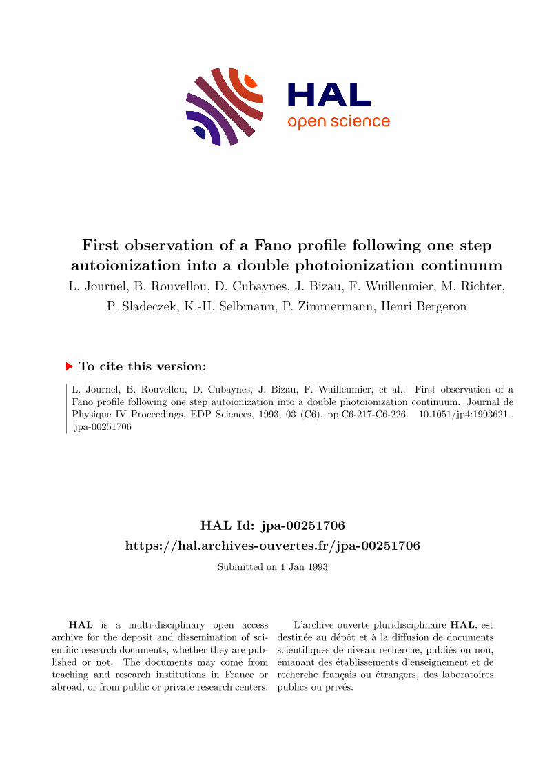
4. RESULTS Figure 4 presents the relative cross section for double photoionization of atomic sodium as a function of photon energy between the double ionization threshold and the single 2s-inner shell ionization thresholds. The data points have been extracted from the Na++/Na+ ratios measured at different photon energies. As expected, no Na++ signal was observed below 52.4 eV and the double photoionization cross section starts
from zero at threshold. Since, between 52.4 eV and 71.0 eV, only + 0.10- o - + + 0 c 0 .- 5 t 0.0s I
? Q >
Let's turn now to the apparently weak Fano-type structure at 66.6 .- - m eV, indicating a strong coupling of the shake off process to the - corresponding 2s 4 3p resonance. First of all, since the resonant 2 0
50 60 70 ' state is an excited state of the neutral atom, only the double
Photon energy (eV) resonant Auger process can contribute to the decay of this excited state into the doubly ionized 2 ~ ~ 2 ~ 5 2 ~ 1 2,3 2 final states.
Pig. 4- Variation. as a function of photon In addition, this process seems to interfer with the direct one-step energy between the first double double photoionization, suggesting that the intensity of both photoionization threshold and the 2s-single processes are in the same order of magnitude. In previous work photoionization threshold, of the Na++/Na+ dealing with decays of core-excited resonances, the direct shake branching ratio measured in this work. off process was negligible compared to the resonant process,
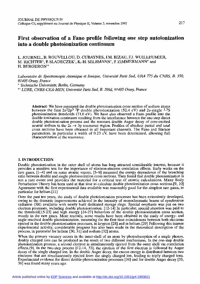
precluding the observation of any Fano-type profile similar to the one observed here. In order to establish a valuable comparison with the effect of the resonance into the singly ionized channels, we recall in Figure 5 an example of the results that have been previously obtained using photoelectron spectrometry for two of the singly ionized final states of Na+ in the same energy region [66]. The branching ratios shown in this figure were determined by measuring a large number of photoelectron spectra in the photon energy range of interest, i.e., the CIS (constant ionic state) method was not used. Variations of the incident photon flux were monitored by measuring the integrated area under the
- 2p-photoelectron line of neon atoms simultaneously introduced in the source volume of the cylindrical mirror analyzer used to energy analyze the photoelectrons (CMA). [66] The variation of
- the Na atoms density as a function of time was also systematically checked by measuring periodically the same
' photoelectron spectrum at a given photon energy. Possible changes of this density were accounted for. In the upper part of the figure, one sees the branching ratio for the shake up satellite
64 66 68 ' intensity ( 2 ~ ~ 2 ~ ~ 4 s / 2 ~ ~ 2 ~ ~ 3 s ) , and, in the lower part, the Photon energy (eV) branching ratio for the first conjugate shake up satellites
~ i ~ . 5 - variation of the branching ratio (2s22p53p / 2s22p53s). The branching ratio is apparently between the shake up (top) or conjugate enhanced on resonance by a factor 2.8 in the CSU channel and
up wttom) satellite (photoioniza~on by a factor 1.5 in the double ionization channel. One does not + excitation) intensities and the main line observe a strong effect in the SU channel. In Table I, we have (single p~otoion~zation) intensity in the 2s + summarized the values of the branching ratios measured off and np resonance region in sodium, ~h~ on resonance in all observed channels, including the single measurements have been made using ionization channel. Note that every feature observed in photoelectron spectrometry (from cubaynes photoabsorption measurements [65] is also observed here, et al., Ref. [66]). especially in the CSU branching ratio. The second resonance of
the series, 2s + 4p, at 69.3 eV photon energy, has also some effect in this channel.
Branching ratios into the different final state channels is an interesting parameter to know. However, a true comparison of relative intensities and profiles can be made only on the energy dependence of the absolute partial photoionization cross sections. This wiIl be the subject of the next paragraph.
direct double photoionization (shake off) contributes to the production of Na++ ions outside of the 2s -+ np resonance energies, the cross section between 52.4 eV and 71.0 eV for the 2 ~ ~ 2 ~ ~ 3 s + hv + 2s22p5 + e- + e- shake off process can be read directly from Figure 4 to increase from 0 to about 1% of the total single photoionization cross section. The sudden increase in the Na++ ion formation at 71.0 eV was also expected, since normal Auger decay of a 2s-vacancy ends up in the Na++ ground state.
Na 2s single ionisation threshoM I
double ionisation 2s-I I threshold f
2p-138-1 - ,

Table I. Branching ratios for photoionization of Na into the various final state channels off resonance and at 66.6 eV resonance energy (2p53s channel = 100).
Final state Off resonance On resonance Enhancement factor
2 ~ ~ 2 ~ ~ 3 ~ 12 33 2.8
2 ~ ~ 2 ~ ~ 4 s 16 19 1.2
2 ~ ~ 2 ~ ~ 4 ~ 2 5 2.5
5. DISCUSSION We have established all partial cross sections on the same relative scale using photoion and photoelectron spectrometries. Then, we determined the absolute scale of the cross sections by normalizing, at 64 eV photon energy, i. e., on the low energy side of the 2s + 3p resonance, the relative cross sections to the absolute photoabsorption cross section as measured by Codling et a1.[71]. This is possible only when all
photoionization channels have been previously analyzed.
3 14 - Na : ~otal Photoabsorption. 1 Let's discuss first the total photoabsorption curve in the
energy region of the 2s + 3p resonance obtained by adding - the contributions of our partial cross sections determined into
the different ionization channels. The resulting curve is shown - in Figure 6 as points with error bars. The error bars take into account the errors in determining the branching ratios, . . ' - including corrections due to variation of the photon flux and
2 0 6 -
possible changes in the atomic density. Like for all data - presented in this paper, the overall uncertainty on the absolute
J 64 66 68 7; 7; value of the total photoabsorption cross section used for
Photon Energy (eV) normalization is not included. Absolute values of the partial cross sections would of course be affected if the normalization
Fig. Total photoabsovtion cross of point would change, but not the shape neither the parameters sodium. This has Obtained in adding of the partial cross sections in the region of the resonance. the partial contributions into the different Note that the values of the cross section on the low energy photoionization as using side of the resonance are 15% to 20% higher, in comparison to photoelectron and photoion spectrometries. the value of the cross section at the top of the 2s + 3p
resonance, than in the data of Wolff et a1.[65]. In fact, these authors have measured only the relative shape of the absorption spectrum. The fact that the 2s + 3p resonance appears with a better contrast in the measured photoabsorption curve than in the reconstructed curve might be attributed to the better resolution of the photoabsorption data, r 0.01 nm against about 0.03 nm in our work.
5.1 Fano parameters for the total photoabsorption cross section. According to Fano, [67, 681 the total photoabsorption cross section otot for the case of an isolated resonance interacting with several continua states can he parametrized in the following form:
otot (E) = oo (E) [p2 (q + E ) ~ / (1 + c2) + 1 - p2]
with: E = (E - E,) / TI2
where the Fano parameters q (profile index) and p2 (correlation coefficient) are assumed to be constant over the resonance, oQ (E) gives the nonresonant cross section, r and Eo are the width and the energy of the resonance, respectively. q and p can be expressed as a function of the wavefunction of the ground and final states.[67, 681 In order to extract the true resonance parameters from the experimental data, a deconvolution of the experimental photoabsorption cross section with the monochromator band pass function must be performed. However, rather than using such a deconvolution, the true resonance profile (generated from the parameters E,, r, q and p2) was convoluted with the monochromator band pass function (a Gaussian
JOURNAL DE PHYSIQUE I V
profile with 0.31 eV FWHM) and then compared with the experimental data by a least-squares fit. For this purpose, a linear behavior of the nonresonant cross section oo was assumed and none of the parameters was a priori kept fixed. Starting values to inject into the fitting procedure were extracted directly from the experimental data. The procedure was repeated systematically, the ~2 values providing
the criterion to select the best set of Table 11. Fano parameters for the 2s -+ 3p resonance in sodium resonance Parameters. Table 11 lists
the final values of the resonance Eo (eV) r (eV> 4 p2 0, ( ~ b ) parameters determined in that way,
using Mathematics software. 66.64 (2) 0.23(1) -1.73(4) 0.13(1) 9.6(6) Codling et al. [7 11 have estimated a q-
value of - 2 (k 0.5), in agreement with previous estimations by Wolff e t
a1.[64] Our determination is more accurate, and confirms the validity of the first estimations. The width of the resonance r, 0.23 eV, is significantly smaller than previously estimated by Codling et al. on the basis of a q-value of -2. It is interesting to compare our value with a completely independent determination of the widths of the transitions: 2 ~ 2 ~ 6 3 s ly3S + 2 ~ 2 2 ~ 5 2~1,2 312 in Na by Breuckman et a1.[72]. Stud ing the Auger spectrum of Na produced by electron impact eicitation, they measured, for 3 both 193s + P transitions, widths of 0.24 (2) eV, in excellent agreement with our determination since the widths of these different transitions is mainly governed by the natural width of the 2s-hole. Our value of 0.23 eV for the width of the 2 ~ 2 ~ ~ 3 ~ 3 ~ 2~ state is e uivalent to an autoionization lifetime of % 2.9x10-l5 sec, i. e., about twenty times less than that of the 2s2p 3p state in Ne measured long time ago by Madden and Codling.[73] This is qualitatively understandable since, in the case of neon, only one channel, the 2s22p5 2 ~ 1 / 2 312 channel, is accessible to autoionization, while many more channels, including the double photoionization channel, are open in the case of the 2 ~ 2 ~ 6 3 ~ 3 ~ 3~ state in sodium. The oscillator strength f of the 2s + 3p transition can also be deduced from our experimental measurements, since it is related to the Fano parameters by the expression:
g f = o . i 9 5 q 2 p 2 0 t r where g is the statistical weight 2J + 1, ot is expressed in megabarns, r is measured in Rydbergs. With the values given in Table 11, we obtain f = 1.7(2) x 10-3, a value almost identical to the oscillator strength of the 2s + 3p transition in neon similarly determined.[73] This equality confirms once more that correlation effects between the core and the outer electrons in sodium have little influence on the inner- shell photoexcitation spectra.
5.2 Partial cross sections Absolute values of the partial cross sections were determined using the same normalization as for the total photoabsorption cross section. They are shown in Figure 7 for the three most important continuum channels studied in this work, i. e., from top to bottom, the CSU channel, the double ionization channel and the single 2p-1 photoionization channel. Note the quite different ordinate scales for each of the three pannels. The experimental values are the points with error bars. The overall error on the total photoabsorption cross section used for normalization, 20% to 25%, is not included in the values of the partial cross sections. Interestingly enough, strongly different profiles appear on resonance in the various continua. In the CSU channel, the cross section has an almost lorentzian profile, as it was already evident in the figure showing the branching ratios, confirming that little interference occurs between the weak direct process and the strong indirect resonant process. This is not the case in the single 2p-l, and, even more, in the double photoionization channels where one observes two similar Beutler-Fano profiles, although the absolute values of the cross sections are strongly different in both continua. Since the first observations in single photoionization of helium 1741, many Fano profiles have been observed [14, 75- 781 in single photoionization channels, but it is the first time, to our knowledge, that such a profile is directly observed following a one-step decay of a neutral core-excited state into a double photoionization continuum. Related measurements are the recent observations of Auger transitions contributing to valence double photoionization [79], and of asymmetric photoelectron line profiles [80, 811 in inner-shell shake up satellites lying above the double ionization threshold involving less bound electrons, suggesting a coupling between discrete satellite states with underlying shake off continua. In Figure 7, the solid lines are the result of a fitting of the experimental data to theoretical formulations including several parameters. Theoretical expressions for the behavior of partial cross sections within a resonance have been formulated by Starace [fig], in a form analogous to the Fano formula describing the behavior of the total cross section in the neighborhood of an isolated resonance. The expression for the partial cross section in the observable photoemission channel p at energy E is given, as a function of the

Fano q parameter and the real and imaginary parts of a new parameter a (p, E), according to:
with: Cl(p,&) = 2 [ q ~ e <aP> - Im < aP > ]
oo (E) is a slowly varying nonresonant partial cross section. The complex parameters %are given by:
4- 64 66 68 70 72
Photon Energy (eV) Fig. 7 - Partial photoionization cross sections for photoionization of atomic sodium into the CSU- , double- and single zp-l photoionization channels, respectively (from top to bottom).
where g represents the ground state, cI, is the final state, mixing discrete and continuum states, and V is the Coulomb interaction. The summation extends over all photoion- ization channels p. In Figure 7, the solid curves represent least- squares fit to the function O(E) convoluted with the instrumental function describing the monochromator band pass. The same procedure was used as for the determination of q and r from the total photoabsorption cross section. All parameters, including T, were again treated as free parameters. The numerical results of the least-squares fit for each photoionization channel are given in Table 111. The r values deduced from the partial cross section measurements are equal to the one determined from the total photoabsorption cross section. The single 2p-1 and double 2p-13s-I partial cross sections have similar profiles, although their absolute values are widely different, by nearly a factor 70 off resonance, the intensity in the double photoionization continuum showing a larger fractional rise (1.5) than does the intensity in the single 2p-l photoionization channel (1.2). The CSU profile is slightly higher on the low energy side, but a symmetric Lorentzian curve will fit into within the experimental error. It has the largest fractional rise on resonance (2.8). The actual value is, in fact higher (z 4.5) because of instrumental broadening. The total cross section which receives about 70% of its intensity from the single ionization channel has a shape similar to the single photoionization cross section.
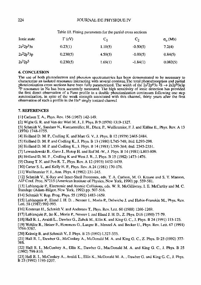
224 JOURNAL DE PHYSIQUE IV
Table 111. Fitting parameters for the partial cross sections
Ionic state r (ev) c 1 c 2 Go (Mb)
6. CONCLUSION The use of both photoelectron and photoion spectrometries has been demonstrated to be necessary to characterize an isolated resonance interacting with several continua.The total photoabso tion and partial ?-' hotoionization cross sections have been fully parametrized. The width of the 2s22p63s S 4 2 . ~ 2 ~ 6 3 ~ 3 ~ % resonance in Na has been accurately measured. The high sensitivity of ionic detection has provided the first direct observation of a Fano profile in a double photoionization continuum following one step autoionization, in spite of the weak strength associated with this channel, thirty years after the first observation of such a profile in the He+ singly ionized channel
7. REFERENCES
[l] Carlson T. A., Phys. Rev. 156 (1967) 142-149. [2] Wight G. R. and Van der Wiel M. J., J. Phys. B 9 (1976) 1319-1327. [3] Schmidt V., Sandner N., Kuntzemiiller, H., Dhez, P., Wuilleumier, F. J. and KQlne E., Phys. Rev. A 13 (1976) 1748-1755. [4] Holland D. M. P., Codling K. and Marr G. V., J. Phys. B 12 (1979) 2465-2484. [5] Holland D. M. and Codling K., J. Phys. B 13 (1980) L745-748; ibid. L293-298. [6] Holland D. M. P. and Codling K., 3. Phys. B 14 (1981) L359-364; ibid. 2345-2351. [7] Lewandowski B., Ganz J., Hotop H. and Ruf M.-W., J. Phys. B 14 (1981) L803-809. [8] Holland D. M. P. , Codling K and West J. B., J. Phys. B 15 (1982) 1473-1476. [9] Chang T. N. and Poe R. T., Phys. Rev. A 12 (1975) 1432-1439. [lo] Carter S . L. and Kelly H. P., Phys. Rev. A 24 (1981) 170-176. [ l l ] Wuilleumier F. J., Ann. Phys. 4 (1982) 231-245. [12] Schmidt V., X-Ray and Inner-Shell Processes, eds. T. A. Carlson, M. 0. Krause and S. T. Manson, AIP Conf. Proc. N0215 (American Institute of Physics, New York, 1990) pp. 559-581. [13] Lablanquie P., Electronic and Atomic Collisions, eds. W. R. McGillivray, I. E. McCarthy and M. C. Standage (Adam-Hilger, New York, 1992) pp. 507-516. [14] Schmidt V. Rep. Prog. Phys. 55 (1992) 1483-1659. [IS] Lablanquie l?, Eland J. H. D. , Nenner I., Morin P., Delwiche J. and Hubin-Franskin M., Phys. Rev. Lett. 58 (1987) 992-995. [16] Kossman H., Schmidt V. and Andersen T., Phys. Rev. Lett. 60 (1988) 1266-1269. [17] Lablanquie P., Ito K., Morin P., Nenner I. and Eland J. H. D., Z. Phys. Dl6 (1990) 77-79. [18] Hall R. I., Avaldi L., Dawber G., Zubek M., Ellis K. and King G. C., J. Phys. B 24 (1991) 115-125. [I91 Wehlitz R., Heiser F., Hemmers O., Langer B., Menzel A. and Becker U., Phys. Rev. Lett. 67 (1991) 3764-3767. [20] Krassig B. and Schmidt V., J. Phys. B 25 (1992) L327-333. [21] Hall R. I., Dawber G., McConkey A., McDonald M. A. and King G. C., Z. Phys. D 23 (1992) 377- 388. [22] Hall R. I., McConkey A., Ellis K., Dawber G., MacDonald M. A. and King G. C., J. Phys. B 25 (1992) 799-8 10. [23] Hall R. I., McConkey A., Avaldi L., Ellis K., McDonald M. A. , Dawber G. and King G. C., J. Phys. B 25 (1992) 1195-1207.

[24] Levin J. C., Lindle D. W., Keller N., Miller R. D., Azuma Y., Berrah Mansour N., Berry H. G. and Sellin I. A., Phys. Rev. Lett. 67 (1991) 968-971. [25] Levin J. C., Sellin I. A., Johnson B. M., Lindle D. W., Miller R. D., Berrah N., Azuma Y., Beny H. G. and Lee D. H., Phys. Rev. A 47 (1993) R16-19. [26] Berrah N., Heiser F., Wehlitz R., Levin J., Whitfield S. B., Viefhaus J., Sellin I. A. and Becker U., Phys. Rev. A 48 (1993) R1733- 1736. [27] Samson J. A. R., Bartlett R. J. and He Z. X. Phys. Rev. 46 (1992) 7277-7280 and rerences therein.. 1281 Mazeau J., Selles P., Waymel D. and Huetz A., Phys. Rev. Lett. 67 (1991) 820-823. [29] Schwarzkopf O., Krassig B., Elmiger J. and Schmidt V., Phys. Rev. Lett. 70 (1993) 3008-3011. [30] Andersson L. R. and Burgdorfer J., Phys. Rev. Lett. 71 (1993) 50-53. [31] Samson J. A. R., Greene C. H. and Bartlett R. J., Phys. Rev. Lett. 71 (1993) 201. [32] Liu Z. W. and Liu J. C., Bull. Arner. Phys. Soc. 38 (1993) 1150. [33] Von Raven E., Meyer M., Pahler M. and Sonntag B., J. Electr. Spectr. Rel. Phen. 52 (1990) 677-688. [34] Krause M. O., Vestal M. L., Johnston W. M. and Carlson T, A., Phys. Rev. 133 (1964) A385-390. [35] Carlson T. A. and Krause M. O., Phys. Rev. Lett. 14 (1965) 390-392. [36] Carlson T. A. and Krause M. O., Phys. Rev. Lett. 17 (1966) 1079-1083. [37] Mukoyarna T., Tonurna T., Yagishita A., Shihata H., Koizumi T., Matsuo T., Shima K. and Tawara J., J. Phys. B 20 (1987) 4453-4460. [38] Lindle D. W., Heimann P. A., Ferrett T. A., Piancastelli M. N. and Shirley D. A., Phys. Rev. A 35 (1987) 4605-4610. 1391 Heimann P. A., Lindle D. W., Ferrett T. A., Liu S. H., Medhurst L. J., Piancastelli M. N., Shirley D. A., Becker U., Kerkhoff H. G., Langer B., Szostak D. and Wehlitz R., J. Phys. B 20 (1987) 5005-5022. [40] Carlson T. A., Mullins D. R., Beall C. E., Yates B. W., Taylor J. W., Lindle D. W.and Grimm F. A., Phys. Rev. A 39 (1989) 1170-1185. [41] Hayaishi T., Murakami E., Yagishita A. Y., Koike F., Morioka Y. and Hansen J. E. J. Phys. B 21 (1988) 3203-3209. [42] Aksela H., Aksela S., Pulkkinen H. and Yagishita A., Phys. Rev. A 40 (1989) 6275-6279. [43] Becker U., Prescher T., Schmidt E., Sonntag B. and Wetzel H.-E., Phys. Rev. A 33 (1989) 3891- 3899. [44] Becker U., Szostak D., Kupsch M., Kerkhoff H. G., Langerand B. and Wehlitz R., J. Phys. B 22 (1989) 749-762. [45] Aksela H., Aksela S., Bancroft, G. M., Tan K. H. and Pulkkinen H., Phys. Rev. A 33 (1989) 3867- 3884. [46] Karnmerling B., Kdssig B. and Schmidt V., J. Phys. B 23 (1990) 4487-4493. [47] Hayaishi T., Yagishita A. Y., Murakami E., Shigemasa E., Morioka Y. and Sasaki T. J. Phys. B 23 (1990) 1633-1639. [48] Hayaishi T., Yagishita A. Y., Shigernasa E., Murakami E. and Morioka Y., J. Phys. B 23 (1990) 443 1-4439. [49] Lablanquie P. and Morin J. Phys. B 24 (1991) 4349-4362. [SO] Hayaishi T., Murakami E., Morioka Y., Aksela H., Aksela S., Shigemasa E. and Yagishita A. Y., Phys. Rev. A 44 (1991) 2771-2774. [51] Ueda K., Shigernasa E., Sato Y., Yagishita A. Y., Ukai M., Maezawa H., Hayaishi T. and Sasaki T. J. Phys. B 24 (1991) 605-613. [52] Sato Y., Hayaishi T., Itikawa Y., Itoh Y., Murakami J., Nagata T., Sasaki T., Sonntag B., Yagishita A. and Yoshino M., J. Phys. B. 18 (1985) 225-231. [53] Nagata T., West J. B., Hayaishi T., Itikawa Y., Itoh Y., Koizumi T., Murakarni J., Sato Y., Shibata H., Yagisgita A. and Yoshino M., J. Phys. B 19 (1986) 1281-1290. [54] Koizumi T., Hayaishi T., Itikawa Y., Nagata T., Sato Y. and Yagishita A., J. Phys. B 20 (1987) 5393- 5401. [55] Yagishita A., Aksela S., Prescher T., Meyer M., Richter M., von Raven E. and Sonntag B., J. Phys. B 21 (1988) 945-953.
226 JOURNAL D E PHYSIQUE IV
[56] Nagata T, Itoh Y., Hayaishi T., Itikawa Y., Koizumi T., Matsuo T., Sata Y., Shigemasa H., Yagishita A. and YoshinoY., J. Phys. B 22 (1989) 3865-3880. [57] Dzionk C., Fiedler W., von Lucke M. and Zimmermann P., Phys. Rev. Lett. 62 (1989) 878-880. [58] Adam M. Y., Hellner L., Dujardin G., Svensson A., Martin P. and Combet Farnoux F., J. Phys. B 22 (1989) 2141-2150. [59] Combet Farnoux F., Dujardin G., Hellner L. and Adam M. Y., X-Ray and Inner-Shell Processes, eds. T. A. Carlson, M. 0. Krause and S. T. Manson, AIP Conf. Proc. N0215 (American Institute of Physics, New York, 1990) pp. 281-291. [60] Koizumi T., Hayaishi T., Itikawa Y., Itoh Y., Matsuo T., Nagata T., Sato Y., Shigemasa E., Yagishita A. and Yoshino M., J. Phys. B 23 (1990) 403-415. [61] Matsuo T., Hayaishi T., Itoh Y., Koizumi T., Nagata T., Sato Y, Shigemasa E., Yagishita A., Yoshino M. and Itikawa Y., J. Phys. B 25 (1992) 121-133. [62] Sonntag B. and Zimmermann P., Rep. Prog. Phys. 55 (1992) 911. [63] Becker U. and Shirley D. A., Physica Scripta T31 (1990) 56-66. [64] Richter M., Bizau J.-M., Cubaynes D., Menzel T., Wuilleumier F. J. and Cam6 B., Europhys. Lett. 12 (1990) 35-40. [65] Wolff H. W., Radler K., Sonntag B. and Haensel R., Z. Physik 257 (1972) 353- 368. [66] Cubaynes D., Bizau J.-M., Richter M. and Wuilleumier F. J., Europhys. Lett. 14 (1991) 747-753. [67] Fano U., Phys. Rev. 124 (1961) 1866-1878. [68] Fano U. and Cooper J. W., Phys. Rev. 137 (1965) A1364-1379. [69] Starace A. F., Phys. Rev. A 16 (1977) 231-242. [70] Sladeczek, Selbman K.-H., Richter M., Zimmermann P., Rouvellou B., Joumel L., Bizau J.-M. and Wuilleumier F. J., to be published. [71] Codling K., Hamley J. R. and West J. B., J. Phys. B 10 (1977) 2797-2807. [72] Breuckmann E., Breuckmann B., Mehlhorn W. and Schmitz W., J. Phys. B 10 (1977) 3135-3150. [73] Codling K., Madden R. P. and Ederer D. L., Phys. Rev. 155 (1967) 26-37. [74] Madden R. P. and Codling K., Phys. Rev. Lett. 10 (1963) 516-518. [75] Codling K., Synchrotron Radiation: Techniques and Applications, ed. C. Kunz (Springer-Verlag, Berlin, 1979) pp. 231-268 and references therein. [76] Kobrin P. H., Becker U., Southworth S., Truesdale C. M., Lindle D. W. and Shirley D. A., Phys. Rev. A 26 (1982) 842-856. [77] Lindle D. W., Ferrett T. A., Heimann P. A. and Shirley D. A., Phys. Rev. A 36 (1987) 2112-2119. [78] Kossmann H., Krassig B. and Schmidt V., J. Phys. B 21 (1988) 1489-1497. [79] Becker U., Wehlitz R., Hemmers O., Langer B. and Menzel A. Phys. Rev. Lett.63 (1989) 1054-57. [80] Svensson S., Martensson N. and Gelius U., Phys. Rev. Lett. 58 (1987) 2639-2641. 1811 Svensson S., Stranges S. and Adam M. Y., Phys. Rev. A 48 (1993) 3051-3055.