First isolation of microorganisms from the gut diverticulum of Aedes aegypti (Diptera: Culicidae):...
6
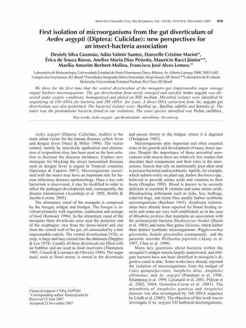
919 919 919 919 919 Mem Inst Oswaldo Cruz, Rio de Janeiro, Vol. 102(8): 919-924, December 2007 First isolation of microorganisms from the gut diverticulum of Aedes aegypti (Diptera: Culicidae): new perspectives for an insect-bacteria association Desiely Silva Gusmªo, Adªo Valmir Santos, Danyelle Cristine Marini*, rica de Souza Russo, Anelise Maria Dias Peixoto, Maurcio Bacci Jœnior**, Marlia Amorim Berbert-Molina, Francisco JosØ Alves Lemos/ + Laboratrio de Biotecnologia, Universidade Estadual do Norte Fluminense Darcy Ribeiro, Av. Alberto Lamego 2000, 28013-602 Campos dos Goytacazes, RJ, Brasil *Faculdades Integradas Maria Imaculada, Mogi-Guau, SP, Brasil **Laboratrio de Evoluªo Molecular, Universidade Estadual Paulista, Rio Claro, SP, Brasil We show for the first time that the ventral diverticulum of the mosquito gut (impermeable sugar storage organ) harbors microorganisms. The gut diverticulum from newly emerged and non-fed Aedes aegypti was dis- sected under aseptic conditions, homogenized and plated on BHI medium. Microbial isolates were identified by sequencing of 16S rDNA for bacteria and 28S rDNA for yeast. A direct DNA extraction from Ae. aegypti gut diverticulum was also performed. The bacterial isolates were: Bacillus sp., Bacillus subtilis and Serratia sp. The latter was the predominant bacteria found in our isolations. The yeast species identified was Pichia caribbica. Key words: Aedes aegypti - gut diverticulum - microbiota - Serratia sp. Aedes aegypti (Diptera: Culicidae, Aedini) is the main urban vector for the human diseases yellow fever and dengue fever (Nasci & Miller 1996). The vector control, mainly by insecticide application and elimina- tion of oviposition sites, has been used as the best solu- tion to decrease the diseases incidence. Explore new strategies for blocking the insect transmitted diseases such as dengue fever is urgent in Tropical countries (Sperana & Capurro 2007). Microorganisms associ- ated with the insect may have an important role for hu- man infectious diseases epidemiology. Once a key role bacterium is discovered, it may be modified in order to affect the pathogen development and, consequently, the disease transmission (Azambuja et al. 2005, Riehle & Jacobs-Lorena 2005). The alimentary canal of the mosquito is composed by the foregut, midgut and hindgut. The foregut is in- volved primarily with ingestion, conduction and storage of food (Romoser 1996). In the alimentary canal of the mosquito three diverticula arise near the posterior end of the esophagus: two from the dorso-lateral and one from the ventral wall of the gut, all surrounded by a thin impermeable cuticle. The ventral diverticulum (VD), or crop, is large and may extend into the abdomen (Dapples & Lea 1974). Usually all three diverticula are filled with air bubbles and are used as food reservoirs (Thompson 1905, Consoli & Loureno-de-Oliveira 1994). The sugar meal, such as floral nectar, is stored in the diverticula and passes slowly to the midgut, where it is digested (Thompson 1905). Microorganisms play important and often essential roles in the growth and development of many insect spe- cies. Despite the importance of these microbial asso- ciations with insects there are relatively few studies that elucidate their components and their roles in the inter- actions. Insects that rely on nutritionally poor diets tend to possess bacterial endosymbionts. Aphids, for example, which subsist solely on plant sap, harbor Buchnera spp., believed to provide amino acids and vitamins to their hosts (Douglas 1989). Blood is known to be severely deficient in essential B vitamins and some amino acids. Bloodsucking arthropods such as ticks, lice, bedbugs, reduviid bugs, and tsetse flies usually harbor symbiotic microorganisms (Buchner 1965). Symbiotic relation- ships have already been reported for blood feeding in- sects and some are very well established, as in the case of Rhodnius prolixus that maintains an association with the actinomycete bacteria Rhodococcus rhodnii (Dasch et al. 1984); and tsetse flies genus Glossina, which harbor three distinct symbiotic microorganisms: Wigglesworthia glossinidia, Sodalis glossinidius (commensal), and the parasitic microbe Wolbachia pipientis (Aksoy et al. 1997, Chen et al. 1999). Many key questions about bacteria within the mosquitos midgut remain largely unanswered, and obli- gate bacteria have not been identified in mosquitos di- gestive canal to date. Some works have already reported the isolation of microorganisms from the midgut of Culex quinquefasciatus, Anopheles dirus, Anopheles albimanus, and Ae. aegypti (Pumpuni et al. 1996, Khampang et al. 1999, Luxananil et al. 2001, Pidiyar et al. 2002, 2004, Gonzalez-Ceron et al. 2003). The microbiota of Anopheles gambiae and Anopheles funestus was also investigated by 16S rDNA sequence by Lindh et al. (2005). The objective of this work was to investigate if Ae. aegypti VD harbored microorganisms. Financial support: CNPq, FAPERJ. + Corresponding author: [email protected] Received 12 June 2007 Accepted 23 November 2007
-
Upload
independent -
Category
Documents
-
view
3 -
download
0
Transcript of First isolation of microorganisms from the gut diverticulum of Aedes aegypti (Diptera: Culicidae):...
919919919919919Mem Inst Oswaldo Cruz, Rio de Janeiro, Vol. 102(8): 919-924, December 2007
First isolation of microorganisms from the gut diverticulum ofAedes aegypti (Diptera: Culicidae): new perspectives for
an insect-bacteria associationDesiely Silva Gusmão, Adão Valmir Santos, Danyelle Cristine Marini*,
Érica de Souza Russo, Anelise Maria Dias Peixoto, Maurício Bacci Júnior**,Marília Amorim Berbert-Molina, Francisco José Alves Lemos/+
Laboratório de Biotecnologia, Universidade Estadual do Norte Fluminense Darcy Ribeiro, Av. Alberto Lamego 2000, 28013-602Campos dos Goytacazes, RJ, Brasil *Faculdades Integradas Maria Imaculada, Mogi-Guaçu, SP, Brasil **Laboratório de Evolução
Molecular, Universidade Estadual Paulista, Rio Claro, SP, Brasil
We show for the first time that the ventral diverticulum of the mosquito gut (impermeable sugar storageorgan) harbors microorganisms. The gut diverticulum from newly emerged and non-fed Aedes aegypti was dis-sected under aseptic conditions, homogenized and plated on BHI medium. Microbial isolates were identified bysequencing of 16S rDNA for bacteria and 28S rDNA for yeast. A direct DNA extraction from Ae. aegypti gutdiverticulum was also performed. The bacterial isolates were: Bacillus sp., Bacillus subtilis and Serratia sp. Thelatter was the predominant bacteria found in our isolations. The yeast species identified was Pichia caribbica.
Key words: Aedes aegypti - gut diverticulum - microbiota - Serratia sp.
Aedes aegypti (Diptera: Culicidae, Aedini) is themain urban vector for the human diseases yellow feverand dengue fever (Nasci & Miller 1996). The vectorcontrol, mainly by insecticide application and elimina-tion of oviposition sites, has been used as the best solu-tion to decrease the diseases incidence. Explore newstrategies for blocking the insect transmitted diseasessuch as dengue fever is urgent in Tropical countries(Sperança & Capurro 2007). Microorganisms associ-ated with the insect may have an important role for hu-man infectious diseases epidemiology. Once a key rolebacterium is discovered, it may be modified in order toaffect the pathogen development and, consequently, thedisease transmission (Azambuja et al. 2005, Riehle &Jacobs-Lorena 2005).
The alimentary canal of the mosquito is composedby the foregut, midgut and hindgut. The foregut is in-volved primarily with ingestion, conduction and storageof food (Romoser 1996). In the alimentary canal of themosquito three diverticula arise near the posterior endof the esophagus: two from the dorso-lateral and onefrom the ventral wall of the gut, all surrounded by a thinimpermeable cuticle. The ventral diverticulum (VD), orcrop, is large and may extend into the abdomen (Dapples& Lea 1974). Usually all three diverticula are filled withair bubbles and are used as food reservoirs (Thompson1905, Consoli & Lourenço-de-Oliveira 1994). The sugarmeal, such as floral nectar, is stored in the diverticula
and passes slowly to the midgut, where it is digested(Thompson 1905).
Microorganisms play important and often essentialroles in the growth and development of many insect spe-cies. Despite the importance of these microbial asso-ciations with insects there are relatively few studies thatelucidate their components and their roles in the inter-actions. Insects that rely on nutritionally poor diets tendto possess bacterial endosymbionts. Aphids, for example,which subsist solely on plant sap, harbor Buchnera spp.,believed to provide amino acids and vitamins to theirhosts (Douglas 1989). Blood is known to be severelydeficient in essential B vitamins and some amino acids.Bloodsucking arthropods such as ticks, lice, bedbugs,reduviid bugs, and tsetse flies usually harbor symbioticmicroorganisms (Buchner 1965). Symbiotic relation-ships have already been reported for blood feeding in-sects and some are very well established, as in the caseof Rhodnius prolixus that maintains an association withthe actinomycete bacteria Rhodococcus rhodnii (Daschet al. 1984); and tsetse flies genus Glossina, which harborthree distinct symbiotic microorganisms: Wigglesworthiaglossinidia, Sodalis glossinidius (commensal), and theparasitic microbe Wolbachia pipientis (Aksoy et al.1997, Chen et al. 1999).
Many key questions about bacteria within themosquito�s midgut remain largely unanswered, and obli-gate bacteria have not been identified in mosquito�s di-gestive canal to date. Some works have already reportedthe isolation of microorganisms from the midgut ofCulex quinquefasciatus, Anopheles dirus, Anophelesalbimanus, and Ae. aegypti (Pumpuni et al. 1996,Khampang et al. 1999, Luxananil et al. 2001, Pidiyar etal. 2002, 2004, Gonzalez-Ceron et al. 2003). Themicrobiota of Anopheles gambiae and Anophelesfunestus was also investigated by 16S rDNA sequenceby Lindh et al. (2005). The objective of this work was toinvestigate if Ae. aegypti VD harbored microorganisms.
Financial support: CNPq, FAPERJ.+Corresponding author: [email protected] 12 June 2007Accepted 23 November 2007

920920920920920 Microorganisms from Aedes aegypti crop � Desiely Silva Gusmão et al.
MATERIALS AND METHODS
Mosquitoes - Insects were obtained from coloniesof Ae. aegypti (Rockfeller strain), maintained in the in-sectary of the Laboratory of Biotechnology (UENF,Campos dos Goytacazes, Brazil). Mosquitoes werereared at 27°C and provided with sterile 10% sucrose. Aglass container filled with sterile distilled water was keptinside each cage to maintain the humidity. Larvae werefed on a sterile minced commercial mouse food. Pupaewere rinsed and transferred to sterile distilled water andmaintained in separate cages in aseptic conditions untiladult emergence. All mosquitoes were newly emergednon-fed females (no blood or sucrose feeding), and weredissected on the same day of emergence.
Isolation of microorganisms - In order to surfacesterilize, mosquitoes were rinsed serially, for 1 min, inthe following solutions: sodium hypochlorite (1%), ster-ile phosphate-buffered saline (PBS) (81 mM Na2HPO4,19 mM NaH2PO4, 150 mM NaCl, pH 7.4) and ethanol(70%). Finally, the insects were rinsed three times inPBS/1 min. Aliquots of 100 µl from the last PBS washeswere plated in brain heart infusion (BHI) agar as controlgroups of the surface sterilization process. The steril-izations and dissections were performed in a laminarflow hood. Mosquitoes were dissected under a micro-scope, in a double cavity glass slide containing sterilePBS. The VD was carefully separated from the midgut,rinsed in sterile PBS, and transferred into a 1.5 ml tube,containing 200 µl of BHI. This procedure was repeateduntil five crops were obtained. The tubes were mixedthoroughly with a pestle, and an aliquot of 100 µl wastransferred to a 50 ml Erlenmeyer containing 20 ml ofBHI medium and 100 µl glucose. An aliquot of 100 µlwas also transferred to Petri dishes containing BHI agar.The Erlenmeyers were incubated at 28ºC for 24-48 h,under agitation (80 rpm). After incubation the cultureswere serially diluted (10-1 through 10-7) and an aliquotof 100 µl of each one was transferred to Petri dishescontaining BHI agar. Plates were incubated at 28ºC for24-48 h. Bacterial isolates were maintained at -70oC ina solution of glycerol (15%) for further identification.We carried out a total of seven isolation assays, fromdifferent groups of Ae. aegypti, at different times. Atthe end, a total of 35 crops were sampled.
Identification of microorganisms - Microorganismswere first screened based on colony characteristics,morphology of isolates, Gram staining and motility bythe hanging drop technique.
DNA extraction from bacterial isolates and ampli-fication of rDNA 16S - The DNA extraction was adaptedfrom Ausubel et al. (1992). The rDNA 16S was ampli-fied using the following universal primers: 27f (Lane etal. 1985) and 1492r (Delong 1992). Polymerase chainreaction (PCR) was performed with Ready-To-Go kit(Amersham Pharmacia Biotech), template DNA solution(2 µl/100 ηg), 27f primer (1 µl/6 ρmol), 1492r primer(1 µl/6 ρmol), 25 mM MgCl2 (1.5 µl), and 17.5 µl ofultra pure water. Cycling parameters for the PCR included
an initial denaturation step at 95oC/5 min, followed by35 cycles of a denaturation step at 95oC/1 min, a primerannealing step at 50oC/1 min, an extension step at 72oC/3 min, and a final step at 72oC/4 min. The 16S rDNA ampli-fication generated a product of approximately 1500 bp.
Amplification of a section rDNA 28S - Yeast DNAwas extracted and purified as described above. The di-vergent D1/D2 domain (nucleotides 63-642 for Saccha-romyces cerevisiae) at the 5´end of the large-subunitrRNA gene was symmetrically amplified with primersNL1 and NL4 (O´Donnell 1993). Each PCR was per-formed with the Ready-To-Go kit (Amersham PharmaciaBiotech) adding solution containing DNA (1 µl), NL-1primer (1.6 µl/6 ρmol), NL-4 primer (1.1 µl/6 ρmol),25mM MgCl2 (1.5 µl), and 17.8 µl of ultra pure water.
DNA extraction from Ae. aegypti gut diverticulum- Ae. aegypti females were dissected and the VD werecarefully separated from the midgut under aseptic con-ditions. The DNA extraction was performed with theWizard genomic DNA purification kit (Promega TM050).
Cloning - After purified, fragments were cloned us-ing the Kit pGem-T Easy Vector Systems (Promega, USA)in Escherichia coli DH5α.
Sequencing reaction - The sequencing reaction wasprepared as follows: Big Dye Terminator Kit (AppliedBiosystems, Foster City, CA) (2 µl), �Save Money� buffer(200 mM Tris HCl, pH 9.0, 5 mM MgCl) (2 µl), forwardT7 or reverse SP6 promoters (1 µl/5.0 ρmoles), DNA (2 µl/100 ηg) and ultra pure water (2 µl). PCR conditions were96°C/10 min, 50°C/5 min, and 60°C/4 min, 40 cycles. Thesequencing of the 16S rRNA gene from the bacterial cloneswas done using a 27f primer (5´-end; 500 nucleotide re-gion) and from the yeast it was done using the NL1 primer.Sequencing was carried out on an ABI Prism 377 DNA se-quencer (Applied Biosystems).
Sequences alignment - The sequences obtained wereinitially compared to the ones deposited at the GenBank,using BLAST (http://www.ncbi.nlm.nih.gov/BLAST). Thegenerated sequences and their homologs, which wereretrieved from the GenBank, were aligned using theCLUSTAL W 1.4 program (Thompson et al. 1994). Phy-logenetic trees were obtained by �neighbor-joining�(PAUP program, Swofford 2000).
Light microscopy - Samples of the VD were fixed in2.5% glutaraldehyde, 0.1 M sodium cacodylate, pH 7.2,for 12 h at room temperature. Tissues were dehydratedin acetone, embedded in Epon and sections of 0.6 µmwere cut with a diamond knife. Samples were stained by to-luidine blue and examined under a Zeiss light microscope.
Measurement of luminal gut pH - Gut pH was mea-sured in vivo by allowing adult mosquitoes to ingest pHindicator dye dissolved in 10% sucrose solution, pH 6.5(adjusted with 0.2 M NaOH) and observing the resultingcolor changes in their diverticulum. The indicator dyeused was 0.5% bromocresol purple [transition interval:pH 6.8 (purple) to pH 5.2 (yellow)]. Mosquitoes weredissected immediately after ingestion of indicator and
921921921921921Mem Inst Oswaldo Cruz, Rio de Janeiro, Vol. 102(8), December 2007
12 and 24 h later in order to observe the color changingin VD content. As the pH of the initial solution was 6.5(dark red), we would expect the color to change gradu-ally to yellow at lower pH.
Medium acidification by Serratia sp. - A pH assayin test tubes and Petri dishes was also carried out in or-der to confirm that Serratia sp. isolated from Ae. aegyptiVD had the ability to acidify the culture medium. Cul-tures of Serratia sp. were transferred to test tubes contain-ing 2 ml of BHI with 2.0% glucose and 0.5% bromocresolpurple (pH 6.5). Tubes were incubated at 28oC and observedafter 12 and 24 h to see the color changes. The same testwas done in Petri dishes containing BHI agar with 2.0%glucose and 0.5% bromocresol purple (pH 6.5).
RESULTS
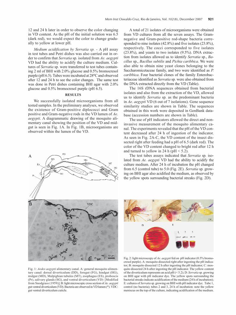
We successfully isolated microorganisms from alltested samples. In the preliminary analyses, we observedthe existence of Gram-positive diplococci and Grampositive and Gram-negative rods in the VD lumen of Ae.aegypti. A diagrammatic drawing of the mosquito ali-mentary canal showing the position of the VD and mid-gut is seen in Fig. 1A. In Fig. 1B, microorganisms areobserved within the lumen of the VD.
A total of 21 isolates of microorganisms were obtainedfrom VD cultures from all the seven assays. The Gram-negative and Gram-positive rod-shape bacteria corre-sponded to nine isolates (42.8%) and five isolates (23.8%),respectively. The cocci corresponded to five isolates(23.8%), and yeasts to two isolates (9.5%). DNA extrac-tion from isolates allowed us to identify Serratia sp., Ba-cillus sp., Bacillus subtilis and Pichia caribbica. We werealso able to obtain nine yeast clones belonging to theSaccharomicetaceae family, and two were identified as P.caribbica. Four bacterial clones of the family Enterobac-teriaceae identified as Serratia sp. were also obtained fromthe DNA extracted directly from the VD (Table).
The 16S rDNA sequences obtained from bacterialisolates and also from the extraction of the VD, allowedus to identify Serratia sp. as the predominant bacteriain Ae. aegypti VD (6 out of 7 isolations). Gene sequencesimilarity studies are shown in Table. The sequencesobtained in this work were deposited in GenBank data-base (accession numbers are shown in Table).
The use of pH indicators allowed the direct and non-invasive measurement of the mosquito alimentary ca-nal. The experiments revealed that the pH of the VD con-tent decreased after 24 h of ingestion of the indicator.As seen in Fig. 2A-C, the VD content of the insect dis-sected right after feeding had a pH of 6.5 (dark red). Thecolor of the VD content changed to bright red after 12 hand turned to yellow in 24 h (pH < 5.2).
The test tubes assays indicated that Serratia sp. iso-lated from Ae. aegypti VD had the ability to acidify theculture medium. After 24 h of incubation the pH changedfrom 6.5 (control tube) to 5.0 (Fig. 2E). Serratia sp. grow-ing on BHI agar also acidified the medium, as observed bythe yellow spots surrounding bacterial streaks (Fig. 2D).
Fig. 1: Aedes aegypti alimentary canal. A: general mosquito alimen-tary canal: dorsal diverticulum (DD), foregut (FG), hindgut (HG),midgut (MD), Malpighian tubules (MT), esophagus (ES), proboscis(Pr), salivary glands (SG), and ventral diverticulum (VD) [Modifiedfrom Snodgrass (1959)]; B: light microscopic cross section of Ae. aegyptigut ventral diverticulum (VD). Bacteria are observed in VD lumen (*). VDC:gut ventral diverticulum cuticle.
Fig. 2: light microscopy of Ae. aegypti fed on pH indicator (0.5% bromo-cresol purple). A: mosquito dissected right after ingesting the pH indica-tor; B: mosquito dissected 12 h after ingesting the pH indicator; C: mos-quito dissected 24 h after ingesting the pH indicator. The yellow contentof the diverticulum represents an acid pH (< 5.2); D: Serratia sp. growingon BHI agar with pH indicator dye. The yellow spots surrounding thebacterial streaks indicate acidification of the medium (24 h of incubation);E: cultures of Serratia sp. growing on BHI with pH indicator dye. Tube 1,control (no bacteria); tubes 2 and 3, 24 h of incubation: note the yellowmeniscus on the top of the culture, indicating acidification of the medium.

922922922922922 Microorganisms from Aedes aegypti crop � Desiely Silva Gusmão et al.
DISCUSSION
Some works have already reported the presence ofbacteria in the midgut of mosquitoes (DeMaio et al.1996, Pumpuni et al. 1996, Luxananil et al. 2001, Zayed& Bream 2004), but have not described any role for thesemicroorganisms in the insect physiology. We have ob-served, under scanning electron microscopy, a dramati-cally high number of small rodlike bacteria embeddedin the food bolus 48 h after blood ingestion (DS Gusmão2006, unpublished observations). DeMaio et al. (1996),Pumpuni et al. (1996) and Zayed and Bream (2004) alsoobserved the increase in bacterial density in mosquitomidgut after blood feeding. DeMaio et al. (1996) andPumpuni et al. (1996) reported a frequent presence ofGram-negative rods belonging to Enterobacteriaceae inadults of Culex as well as Anopheles species. In labora-tory-reared and wild-caught sandflies all predominantspecies identified by Dillon et al. (1996) belonged toEnterobacteriaceae. The genus Serratia has a wide hostrange and has been isolated from various insect groups(Krieg 1987). Serratia sp. is member of the Enterobac-teriaceae family (facultative anaerobic, Gram-negative,cytochrome oxidase negative and catalase positive). Ingeneral, Serratia marcescens is reported most fre-quently as a pathogen of insectary-reared insects (Krieg1987, Grimont & Grimont 1992). S. marcescens wasidentified in intestinal tube of Lutzomyia longipalpis,and was more abundant in sandflies fed on blood andsugar than the ones fed only on blood (Oliveira et al.2001). Iverson et al. (1984) reported the presence ofSerratia liquefaciens and S. marcescens in all sugar beetroot maggot (Diptera: Oititidae) developmental stages,suggesting a symbiosis and a nutritional interdependencebetween these bacteria and the insect. Gonzalez-Ceron
et al. (2003) studied the microorganisms present in themidguts of An. albimanus and identified several Serratiaand Enterobacter species including S. marcescens, E. cloa-cae, and E. amnigenus. In Ae. triseriatus, Culex pipiensand Psorophora columbiae mosquitoes, S. marcescens waswithin the species most frequently isolated (DeMaio et al.1996). S. marcescens with ability to lyse erythrocyteswas also isolated from the gut larvae of the blood feed-ing insect R. prolixus (Azambuja et al. 2004).
Moll et al. (2001) describe an effective mechanismto eliminate gut microorganisms during mosquito meta-morphosis and adult emergence. Bacteria found in re-cently emerged non-fed adults are present from the lar-val and pupal stage and, therefore, have some adaptationsto overcome this mechanism.
According to Solé et al. (1997, 2000), the additionof glucose or other sugar to cell suspensions of S.marcescens resulted in the acidification of the medium.Their results suggest that the acidification of the me-dium could be due to the presence of organic acids re-sulting from bacterial metabolism. These results sup-port our findings, indicating that the bacteria present inthe VD may have a role in the sugar metabolism pro-cess. The acid pH we observed in Ae. aegypti VD may be aresult of the sucrose oxidation by Serratia spp., as seen onthe test tubes and Petri dishes assays (Fig. 2D, E).
Recently, Favia et al. (2007) reported that the mainbacteria species associated with adults and larvae of themosquito Anopheles stephensi are from the genus Asaia,that is related to acetic acid bacteria. They found Asaiasp. in various An. stephensi organs and stages but didnot specify if they found it associated with the VD. Wesuppose that Asaia may also be present in An. stephensiVD once adult mosquitoes, as hypothesized by the au-
TABLEPhylogenetic affiliations of cultured and uncultured microorganisms from the gut diverticulum of Aedes aegypti
based on 16S and 28S rRNA gene analysis
Identification accordingIsolate/Clone Accession N. Closest relative according to Blast (% identity) to phylogenetic analysis
P3a EF092303 Candida sp. AM159101 (100) Pichia caribbicad
P12a EF092301 Bacillus cereus AY279192 (99) Bacillus sp.P13a EF092302 Serratia sp. AM403719 (98) Serratia sp.P15a EF092296 Bacillus subtilis AY881643 (99) Bacillus subtilisC8BPb EF092297 Uncult. bacterium AY436532 (99) Serratia sp.C10BPb EF092298 Serratia marcescens DQ904615 (98) Serratia sp.C11BPb EF092299 Serratia sp. AM403719 (99) Serratia sp.C13BPb EF092300 Serratia sp. AM403719 (98) Serratia sp.C5LPc EF092304 Candida etchellsii AB196193 (99) SaccharomycetaceaeC6LPc EF092305 Pichia caribbica DQ857892 (98) Pichia caribbicaC10LPc EF092307 Pichia caribbica DQ857892 (99) SaccharomycetaceaeC12LPc EF092308 Candida etchellsii AB196193 (99) SaccharomycetaceaeC14LPc EF092306 Pichia caribbica DQ857892 (99) Pichia caribbicaC15LPc EF092310 Pichia sp. DQ835012 (99) SaccharomycetaceaeC16LPc EF092309 Candida etchellsii AB196193 (99) SaccharomycetaceaeC19LPc EF092312 Candida etchellsii AB196190 (98) SaccharomycetaceaeC20LPc EF092311 Candida etchellsii AB196190 (98) Saccharomycetaceae
a: isolates; b: bacteria clones; c: yeast clones; d: ascosporic state of Candida fermentati (Vaughan-Martini et al. 2005).

923923923923923Mem Inst Oswaldo Cruz, Rio de Janeiro, Vol. 102(8), December 2007
thors, could obtain these bacteria from the nectar of tropi-cal flowers, which are Asaia�s natural habitat. Therefore,Serratia and probably Asaia could be contributing to acidifythe VD lumen when metabolizing its sugar content.
There are no reports about the VD pH in Ae. aegypti todate. Gontijo et al. (1998) measured the pH in Lu.longipalpis esophageal diverticulum (crop) with bromo-cresol purple, the same indicator dye used in our study. Theyobtained a pH between 6.0 and 6.5, higher than our results.
Our findings may open new windows for understand-ing Aedes-bacteria interaction. As the VD has no directconnection with the midgut (Fig. 2B), the presence ofbacteria inside this compartment may imply in a morespecific insect-microorganism interaction than previ-ously described. Unlike other insects, Ae. aegypti maynot have a special structure for harboring friendly bac-teria. Thus, the VD would provide a good environmentfor these bacteria that would be protected from the di-rect flux of food (blood) in the gut. Bacteria would re-main in small numbers in the VD and would be releasedto the gut when convenient. As Enterobacteriaceae havethe ability to ferment sugar (Ewing 1986), we could alsohypothesize that one possible role for Serratia sp. wouldbe to metabolize the sugar contained in the diverticu-lum, as seen in our in vitro assays, releasing importantmolecules to adult physiology. Once Serratia sp. wasfound in 85% of our assays and the bacteria embeddedin the food bolus also seemed to be Serratia, we couldalso hypothesize that there is a more complex bacterialinteraction during the digestion process, than simply afortuitous �passing through� the alimentary canal.
ACKNOWLEDGEMENTS
To Center for the Study of Social Insects, UNESP, Rio Claro,São Paulo, Brasil, for performing the sequence reactions.
REFERENCES
Aksoy S, Chen X, Hypsa V 1997. Phylogeny and potential trans-mission routes of midgut-associated endosymbionts of tsetse(Diptera: Glossinidae). Insect Mol Biol 6: 83-190.
Ausubel FM, Brent R, Kingston RE, Moore DD, Seidman JG,Smith JA, Struhl K 1998. Current protocols in molecularbiology, Vol. 1, John Wiley & Sons, New York.
Azambuja P, Feder D, Garcia ES 2004. Isolation of Serratiamarcescens in the midgut of Rhodnius prolixus: impact onthe establishmet of the parasite Trypanosoma cruzi in thevector. Exp Parasitol 107: 89-96.
Azambuja P, Garcia ES, Ratcliffe NA 2005. Gut microbiota andparasite transmision by insect vectors. Trends Parasitol 21:568-572.
Buchner P 1965. Endosymbionts of animals with plant micro-organisms. John Wiley & Sons, New York, 909 pp.
Chen X, Li S, Aksoy S 1999. Concordant evolution of a symbiontwith its host insect species: molecular phylogeny of genusGlossina and its bacteriome-associated endosymbiont,Wigglesworthia glossinidia. J Mol Evol 48: 49-58.
Consoli RAGB, Lourenço-de-Oliveira R 1994. Principais Mos-quitos de Importância Sanitária no Brasil, Fiocruz, Rio deJaneiro, 228 pp.
Dapples CG, Lea AO 1974. Inner surface morphology of the
alimentary canal in Aedes aegypti (L.) (Diptera: Culicidae).Int J Insect Morphol Embryol 3: 433-442.
Dasch GA, Weiss E, Chang K 1984. Endosymbionts of insects. InNR Krieg, JG Holt, Bergey�s Manual of Systematic Bacteriol-ogy, Vol.1, Williams and Wilkins, Baltimore, MD, p. 811-833.
Delong EF 1992. Archea in coastal marine environments. ProcNatl Acad Sci USA 89: 5685-5689.
DeMaio J, Pumpuni CB, Kent M, Beier JC 1996. The midgutbacterial flora of wild Aedes triseriatus, Culex pipiens andPsorophora columbiae mosquitoes. Am J Trop Med Hyg54: 219-223.
Dillon RJ, El Kordy E, Shehata M, Lane RP 1996. The preva-lence of a microbiota in the digestive tract of Phlebotomuspapatasi. Ann Trop Med Parasitol 90: 699-673.
Douglas AE 1989. Mycetocyte symbiosis in insects. Biol RevCamb Philos Soc 64: 409-434.
Ewing WH 1986. Edwards and Ewing�s Identification of En-terobacteriaceae, 4th ed., Elsevier, New York, 536 pp.
Favia G, Ricci I, Damiani C, Raddadi N, Crotti E, Marzorati M,Rizzi A, Urso R, Brusetti L, Borin S, Mora D, Scuppa P,Pasqualini L, Clementi E, Genchi M, Corona S, Negri I, GrandiG, Alma A, Kramer L, Esposito F, Bandi C, Sacchi L,Daffonchio D 2007. Bacteria of the genus Asaia stably as-sociate with Anopheles stephensi, an Asian malarial mos-quito vector. Proc Natl Acad Sci USA 104: 9047-9051.
Gontijo NF, Almeida-Silva S, Costa FF, Mares-Guia ML,Williams P, Melo MN 1998. Lutzomyia longipalpis: pH inthe gut, digestive glycosidases, and some speculations uponLeishmania development. Exp Parasitol 90: 212-219.
Gonzalez-Ceron L, Santillan F, Rodriguez MH, Mendez D,Hernandez-Avila JE 2003. Bacteria in midguts of field-col-lected Anopheles albimanus block Plasmodium vivaxsporogonic development. J Med Entomol 40: 371-374.
Grimont F, Grimont PAD 1992. The genus Serratia. In A Balows,HG Trüper, M Dworkin, W Harder, KH Schleifer (eds), TheProkaryotes. A Handbook on the Biology of Bacteria:Ecophysiology. Isolations, Identification, Applications,Vol. 1, Springer, New York, NY, USA, p. 2822-2248.
Iverson KAL, Bromel MC, Anderson AW, Freeman TP 1984.Bacterial symbionts in the sugar beet root maggot, Tetanopsmyopaeformis (von Roder). Appl Environ Microbiol 47:22-27.
Khampang P, Chungjatupornchai W, Luxananil P, Panyim S 1999.Efficient expression of mosquito-larvicidal proteins in a Gram-negative bacterium capable of recolonization in the guts ofAnopheles dirus larva. Appl Microbiol Biotechnol 51: 79-84.
Krieg A 1987. Diseases caused by bacteria and other prokary-otes. In JR Fuxa, Y Tanada, Epizootiology of insect dis-eases, John Wiley & Sons, New York, p. 323-355.
Lane DJ, Pace B, Olsen GJ, Stahl DA, Sogin ML, Pace NR1985. Rapid determination of 16S ribosomal sequences forphylogenetic analyses. Proc Natl Acad Sci USA 82: 6955-6959.
Lindh JM, Terenius O, Faye I 2005. 16S rRNA gene-based iden-tification of midgut bacteria from field-caught Anophelesgambiae sensu lato and A. funestus mosquitoes reveals newspecies related to known insect symbionts. Appl EnvironMicrobiol 71: 7217-7223.
Luxananil P, Atomi H, Panyim S, Imanaka T 2001. Isolation of
924924924924924 Microorganisms from Aedes aegypti crop � Desiely Silva Gusmão et al.
bacterial strains colonizable in mosquito larval guts as novel hostcells for mosquito control. J Biosci Bioeng 92: 342-345.
Moll RM, Romoser WS, Modrzakowski MC, Moncayo AC,Lerdthusnee K 2001. Meconial peritrophic membranes andthe fate of midgut bacteria during mosquito (Diptera: Culi-cidae) metamorphosis. J Med Entomol 38: 29-32.
Nasci RS, Miller BR 1996. Culicine mosquitoes and the agentsthey transmit. In BJ Beaty, WC Marquardt (eds), The Biol-ogy of Disease Vectors, 1st ed., Colorado University Press,Niwot, CO, p. 85-97.
O�Donnell K 1993. Fusarium and its near relatives. In DRReynolds, JW Taylor (eds), The fungal holomorph: Mitoticand pleomorphic speciation in fungal systematics, CABInternational, Wallingford, p. 225-233.
Oliveira SMP, Morais BA, Gonçalves CA, Giordano-Dias CM,Vilela ML, Brazil RP, Almeida, JM, Asensi MD, Mello RP2001. Microbiota do trato digestivo de fêmeas de Lutzomyialongipalpis (Lutz & Neiva, 1912) (Diptera: Psychodidae)provenientes de colônias alimentadas com sangue e comsangue e sacarose. Cad Saude Publica 17: 229-232.
Pidiyar VJ, Jangid K, Patole MS, Shouche Y 2004. Studies oncultured and uncultured microbiota of wild Culexquinquefasciatus mosquito midgut based on 16S ribosomalRNA gene analysis. Am J Trop Med Hyg 70: 597-603.
Pidiyar VJ, Kaznowski A, Badri NN, Patole MS, Shouche YS2002. Aeromonas culicicola sp. nov., from the midgut ofCulex quinquefasciatus. Int J Syst Evol Microbiol 52:1723-1728.
Pumpuni CB, Demaio J, Kent M, Davis JR, Beier JC 1996. Bac-terial population dynamics in three anopheline species: theimpact on Plasmodium sporogonic development. Am J TropMed Hyg 54: 214-218.
Riehle MA, Jacobs-Lorena M 2005. Using bacteria to express
and display anti-parasite molecules in mosquitoes: current andfuture strategies. Insect Biochem Mol Biol 35: 699-707.
Romoser WS 1996. The vector alimentary system. In BJ Beaty,WC Marquardt (eds), The Biology of Disease Vectors,Colorado University Press, Niwot, CO, p. 298-317.
Snodgrass RE 1959. The anatomical life of the mosquito.Smithsonian Miscellaneous Collections 139: 1-87.
Solé M, Francia A, Rius N, Loren JG 1997. The role of pH in the�glucose effect� on prodigiosin production by non-proliferatingcells of Serratia marcescens. Lett Appl Microbiol 25: 81-84.
Solé M, Rius N, Loren JG 2000. Rapid extracellular acidificationinduced by glucose metabolism in non-proliferating cells ofSerratia marcescens. Internatl Microbiol 3: 39-43.
Sperança MA, Capurro ML 2007. Perspectives in the control ofinfectious diseases by transgenic mosquitoes in the post-genomicera - A Review. Mem Inst Oswaldo Cruz 102: 425-433.
Swofford DL 2000. PAUP: Phylogenetic Analysis Using Par-simony, version 4.0b4a. Smithsonian Institution and SinauerAssociates, Sunderland, MA.
Thompson JD, Higgins DG, Gibson TJ 1994. CLUSTAL W: im-proving the sensitivity of progressive multiple sequence align-ment through sequence weighting, position-specific gap penal-ties and weight matrix choice. Nucleic Acids Res 22: 388-392.
Thompson MT 1905. Alimentary canal of the mosquito. ProcBoston Soc Nat Hist 32: 145-202.
Vaughan-Martini A, Kurtzman CP, Meyer SA, O�Neill EB 2005.Two new species in the Pichia guilliermondii clade: Pichiacaribbica sp. nov. the ascosporic state of Candidafermentati, and Candida carpophila comb. nov. FEMSYeast Res 5: 463-469.
Zayed ME, Bream AS 2004. Biodiversity of the microbial floraassociated with two strains of Culex pipiens (Diptera: Culi-cidae). Commun Agric Appl Biol Sci 69: 229-34.