Final_Copy_2020_11_26_Cole... - University of Bristol ...
296
This electronic thesis or dissertation has been downloaded from Explore Bristol Research, http://research-information.bristol.ac.uk Author: Coleman, Gareth Title: A phylogenomic exploration of early bacterial evolution General rights Access to the thesis is subject to the Creative Commons Attribution - NonCommercial-No Derivatives 4.0 International Public License. A copy of this may be found at https://creativecommons.org/licenses/by-nc-nd/4.0/legalcode This license sets out your rights and the restrictions that apply to your access to the thesis so it is important you read this before proceeding. Take down policy Some pages of this thesis may have been removed for copyright restrictions prior to having it been deposited in Explore Bristol Research. However, if you have discovered material within the thesis that you consider to be unlawful e.g. breaches of copyright (either yours or that of a third party) or any other law, including but not limited to those relating to patent, trademark, confidentiality, data protection, obscenity, defamation, libel, then please contact [email protected] and include the following information in your message: • Your contact details • Bibliographic details for the item, including a URL • An outline nature of the complaint Your claim will be investigated and, where appropriate, the item in question will be removed from public view as soon as possible.
-
Upload
khangminh22 -
Category
Documents
-
view
1 -
download
0
Transcript of Final_Copy_2020_11_26_Cole... - University of Bristol ...

This electronic thesis or dissertation has beendownloaded from Explore Bristol Research,http://research-information.bristol.ac.uk
Author:Coleman, Gareth
Title:A phylogenomic exploration of early bacterial evolution
General rightsAccess to the thesis is subject to the Creative Commons Attribution - NonCommercial-No Derivatives 4.0 International Public License. Acopy of this may be found at https://creativecommons.org/licenses/by-nc-nd/4.0/legalcode This license sets out your rights and therestrictions that apply to your access to the thesis so it is important you read this before proceeding.
Take down policySome pages of this thesis may have been removed for copyright restrictions prior to having it been deposited in Explore Bristol Research.However, if you have discovered material within the thesis that you consider to be unlawful e.g. breaches of copyright (either yours or that ofa third party) or any other law, including but not limited to those relating to patent, trademark, confidentiality, data protection, obscenity,defamation, libel, then please contact [email protected] and include the following information in your message:
•Your contact details•Bibliographic details for the item, including a URL•An outline nature of the complaint
Your claim will be investigated and, where appropriate, the item in question will be removed from public view as soon as possible.

1
A phylogenomic exploration of early bacterial evolution
Gareth Andrew Coleman
A dissertation submitted to the University of Bristol in accordance with the requirements for award of the degree of Doctor of
Philosophy in the Faculty of Life Sciences
University of Bristol School of Biological Sciences Life Sciences Building
24 Tyndall Avenue BS8 1TQ
September 2020
Word count: 46,642

2

3
Abstract There are many challenges associated with the reconstruction of early evolutionary history. This is particularly true in the case of Bacteria. Despite being one of the two primary domains of life, and therefore crucial to our understanding of the early history of life, there is little consensus regarding the deepest evolutionary relationships within the bacterial tree. Due to the large spans of time that have elapsed since the origin of the domain, there are many difficulties in modelling their evolution, with bacterial phylogenies frequently affected by artefacts in the analyses. There are therefore a number of questions still unresolved regarding the relationships between major phyla, the root of the tree, and indeed whether the abundant horizontal gene transfer known to characterise prokaryotic evolution has not obscured vertical signal to the point of rendering a tree analogy moot. Recent discoveries of a huge diversity of new uncultured phyla provide new data, but are often difficult to resolve within the bacterial tree, with the relationships between the major bacterial lineages still showing little resolution. Bacteria also represent the most genetically and metabolically diverse organisms on the planet, and as such there are many questions pertaining to the evolution of diverse physiologies and metabolism through time. In this thesis, we attempt to address these issues by using innovative genomic approaches while incorporating much of the previously unknown bacterial diversity. We produce a rooted tree of Bacteria, demonstrate the inadequacies of outgroup rooting, and quantify the contributions of both vertical and horizontal signal to bacterial evolution. We additionally infer the order of events in early bacterial evolution, and reconstruct ancestral metabolisms for the earliest bacterial lineages. Taken together, these results can be integrated to produce a model of early bacterial evolution which contributes to our understanding of the earliest phase of life on Earth.

4

5
Author Declaration I declare that the work in this dissertation was carried out in accordance with the
requirements of the University's Regulations and Code of Practice for Research
Degree Programmes and that it has not been submitted for any other academic award.
Except where indicated by specific reference in the text, the work is the candidate's
own work. Work done in collaboration with, or with the assistance of, others, is
indicated as such. Any views expressed in the dissertation are those of the author.
Gareth Coleman
11th September 2020

6

7
Statement of collaboration Chapters 2, 3 and 4 are derived from a paper currently under revision in collaboration
with Adrián A. Davín, Tara Mahendrarajah, Anja Spang, Philip Hugenholtz, Gergely J.
Szöllősi, and Tom A. Williams. Gareth A. Coleman is the first author of this paper.
Details of contributions are given on the title page of each chapter.
Coleman, G.A., Davín, A.A., Mahendrarajah, T., Spang, A.A., Hugenholtz, P.,
Szöllősi, G.J. and Williams, T.A., 2020. A rooted phylogeny resolves early bacterial
evolution. bioRxiv. https://www.biorxiv.org/content/10.1101/2020.07.15.205187v1
Chapter 5 has been published as a Coleman et al. (2019) in Genome Biology and
Evolution in collaboration with Richard D. Pancost and Tom A. Williams. Gareth A.
Coleman is the first author of this paper. Coleman, G.A., Pancost, R.D. and Williams, T.A., 2019. Investigating the origins of
membrane phospholipid biosynthesis genes using outgroup-free rooting. Genome
biology and evolution, 11(3), pp.883-898.
https://academic.oup.com/gbe/article/11/3/883/5310093
Chapters 1 and 6 are entirely the work of Gareth A. Coleman
Gareth Coleman
11th September 2020

8

9
Acknowledgements
At the near completion of my PhD, there are many people to thank for their various
contributions, great and small, that ultimately helped me get to this point. First and
foremost, I would like to thank my primary supervisor Tom Williams for giving me this
position, for being a fantastic supervisor, and for always being with me every step of
the way. I would also like to thank my secondary supervisor Rich Pancost for the
pastoral support and advice he gave. Additionally, I would like to thank the Royal
Society for funding this PhD, and the University of Bristol, including all the admin staff,
for hosting and giving access to the resources that made it possible. Many thanks go
to my collaborators, Adrián Davín, Tara Mahendrarajah, Anja Spang, Philip
Hugenholtz, Gergely Szöllősi, who helped make this research so much better, and
allowed a real contribution to science to be made.
I would also like to thank the University of Bristol Palaeobiology Research group, and
all its members who helped me in various ways, both scientifically, and as friends.
Particular thanks go to my fellow lab group members, Céline Petitjean and Edmund
Moody. Additionally, I must thank my various friends, both within and without the
university, for giving me all their love and support throughout this time. I would
therefore like to thank Emma Landon, Anna Williams, Tom Kettlety, Ellen MacDonald,
Nuria Melisa Morales García, Suresh Singh, Rhys Charles, Antonio Ballell Mayoral,
Alessia Tasca, Pierre-Aurélian Gilliot, Tom Smith, Ben Griffin, George Atkinson,
Rebekah Locke, Alasdair Price, and Chris Weaver, amongst many others who have
offered their friendship and support at various times. I also give my thanks and love to
my family for their continued support, including my brother Luke Coleman, my sister
Amy Cheal, and of course my parents Mick and Chris Coleman, to whom I owe so
much.
Lastly, but most importantly, I give my eternal love and gratitude to my husband,
Raphaël, without whom I could not have finished this PhD. I wish to thank him for
loving and supporting me, as well as shouldering the burden of having to putting up
with me, especially during such a fraught time. I could not ask for a better life partner.

10

11
« On ne voit bien qu’avec le cœur. L’essentiel est
invisible pour les yeux. » - Antoine de Saint-Exupéry, Le Petit Prince
Écrivain et poète 1900-1944
« Rien dans la vie n'est à craindre. Ce n'est qu'à être
compris. » - Marie Skłodowska-Curie
Physicienne et scientifique 1867-1934

12

13
Table of Contents
Title page 1 Abstract 3 Author Declaration 5 Statement of collaboration 7 Acknowledgements 9 Table of contents 13 List of figures 16 List of tables 17 Chapter 1 Bacteria and the challenges of reconstructing early evolution 20 1.1 The challenges of deep-time phylogenetics 22
1.2 Approaching deep-time evolution using whole genomes 25
1.3 The case of Bacteria 27
1.4 A rooted tree of Bacteria 28
1.5 Evolution of core metabolism in Bacteria 31
1.6 One membrane or two? The evolution of the cell envelope 35
1.7 Timing of bacterial evolution 38
1.8 The Lipid divide 40
1.9 A model for the evolution of early life 45
Chapter 2
Phylogenomics produces a rooted tree of Bacteria 47
2.1 Introduction 49
2.2 Methods 51
2.3 Results and discussion 59
2.4 Conclusion 89
Chapter 3
Ancestral reconstruction of the last bacterial common ancestor 91
3.1 Introduction 93

14
3.2 Methods 95
3.3 Results and discussion 99
3.4 Conclusion 115
Chapter 4
Genomic evolution of Bacteria through time 119
4.1 Introduction 121
4.2 Methods 123
4.3 Results and discussion 124
4.4 Conclusion 143
Chapter 5
Investigating the Origins of Membrane Phospholipid Biosynthesis Genes Using Outgroup-Free Rooting 146 5.1 Introduction 148
5.2 Methods 151
5.3 Results and discussion 154
5.4 Conclusion 175
Chapter 6
Toward an integrated model of early bacterial evolution 178
6.1 Addressing the challenges of deep-time phylogenetics 180
6.2 The root of the bacterial tree between two large and diverse clades 182
6.3 An acetogenic origin of Bacteria? 183
6.4 The early origin of motile diderm cells 185
6.5 Diversification of Bacteria through time 186
6.6 The Lipid divide 186
6.7 What can we say about the last universal common ancestor? 188
6.8 Future directions 189

15
References 192
Appendices
A. Species tables for Chapter 2 221
B. Full heatmap 237
C. Supplementary tree figures for Chapter 5 (phospholipids) 238
D. Papers derived from work included in this thesis 294
Gareth Coleman
295

16
List of figures
1.1 Schematic bacterial tree with various proposed roots indicated.
1.2 Evolutionary hypothesis for the origin of the outer membrane.
1.3 Phospholipid biosynthesis pathways in Archaea and Bacteria.
1.4 Different models of the origin and early evolution of phospholipid biosynthesis in
Archaea and Bacteria.
2.1 Unrooted bacterial phylogenies inferred from the GTDB dataset.
2.2 Unrooted bacterial phylogenies inferred from the GTDB-independent dataset.
2.3 Outgroup-rooted bacterial phylogeny inferred from the GTDB dataset.
2.4 Outgroup-rooted bacterial phylogenies inferred from the GTDB-independent
dataset.
2.5 RMC and MAD rooted bacterial phylogenies derived from the GTDB-independent
dataset.
2.6 Accuracy of gene rooting methods.
2.7 Root positions determined by ALE for both the GTDB and the GTDB-independent
datasets.
2.8 Verticality of bacterial evolution.
2.9 Relationship between verticality and gene family size.
2.10 Rooted phylogeny of Archaea and Bacteria
3.1 Components of the flagellum and chemotaxis inferred in LBCA.
3.2 Distribution of COG families from key metabolic pathways inferred in LBCA.
3.3 Metabolic map of central metabolic pathways inferred in LBCA.
3.4 Ancestral reconstruction of LBCA.
4.1 Nodes for which ancestral gene content were inferred.
4.2 Relative ages of bacterial clades.
4.3 Evolution of COG family repertoires and inferred genomes size of the bacterial
tree.
4.4 Metabolic map of central carbohydrate pathways in surveyed nodes.
4.5 Metabolic map of acetogenesis and the WLP in surveyed nodes.

17
4.6 Components of the flagellum in surveyed nodes.
5.1 Biosynthesis pathway and composition of phospholipids in Bacteria and Archaea.
5.2 Bayesian consensus tree of archaeal enzymes.
5.3 Bayesian consensus tree of both G3PDH enzymes.
5.4 Bayesian consensus tree of GlpK, PlsC and PlsY enzymes.
List of Tables
2.1 Number of taxa sampled from each clade in the GTDB-independent analysis.
2.2 63 orthologues used to infer the species, with those used in the outgroup rooting
analysis indicated.
2.3 Mean verticality by COG functional category.
2.4 Support for published hypotheses using outgroup rooting.
2.5 Support for published rooting hypotheses form our ALE analysis.
2.6 AU-test results for an ALE root analysis using 3595 COG families. 2.7 Singleton support on the credible set of rooted trees.
3.1 Estimated root origination rates and root presences by COG functional category.
3.2 PPs for presence of glycerolipids in LBCA.
3.3 PPs for presence of lipopolysaccharide genes in LBCA.
4.1 PPs for presence of glycerolipids in LBCA in nodes surveyed.
4.2 PPs for presence of lipopolysaccharide genes in nodes surveyed.
5.1 Distribution of phospholipid biosynthesis genes in bacteria and archaeal phyla.
5.2 PPs for RMC and MAD rooting methods.
5.3 AI scores for MAD roots.
5.4 BIC scores for outgroup rooted trees under IQ-Tree model selection.

18
List of Supplementary Tables
Supplementary Table 1 Protein family annotations (COG and KO) and root presence
posterior probabilities (PPs) for all 3723 gene families under all three branches in the
root region, Chapters 3 and 4 (Excel-formatted spreadsheet).
Supplementary Table 2 Protein family annotations (COG and KO) and root presence
posterior probabilities (PPs) for key pathways used in reconstruction, Chapters 3 and
4 (Excel-formatted spreadsheet).
Supplementary Table 3 Table containing the results from the LOESS regression
analysis of COG family members against genome size, Chapter 4 (tsv file).
Supplementary Table 4 COG families lost on the CPR stem, Chapter 4 (Excel-
formatted spreadsheet).
Supplementary Table 5 Accession numbers for sequences used in phylogenetic analyses in Chapter 5 (Excel-formatted spreadsheet)..

19

20
Chapter 1
Bacteria and the challenges of
reconstructing early evolution This chapter is not part of any publication and has been written by GAC entirely.

21
Abstract Reconstructing deep evolutionary history presents many challenges. This is especially evident in the case of Bacteria. Despite being one of the two primary domains of life, there has been little consensus on the deepest evolutionary relationships in the bacterial tree, especially the position of the root. A number of issues have impeded these endeavours, including difficulties in modelling such long stretches of evolutionary time, the use of inappropriate models and methods, problems with topological artefacts, and ultimately whether tree-like analogies are applicable to bacterial evolution at all. Understanding bacterial phylogeny is also necessary if we wish to understand the evolution of bacterial cells, metabolisms, and other traits. As Bacteria represent the most genetically and metabolically diverse lifeforms on the planet, there are a number of questions regarding the evolution of different metabolic pathways, physiologies and morphological characteristics. With the advent of new sequencing technologies, our knowledge of bacterial diversity has greatly expanded. This offers a wealth of new and important data, but also difficulties in how to integrate this data into our current frameworks of bacterial evolution.

22
1.1 The challenges of deep-time phylogenetics
Throughout human history we have attempted to classify the environment around us,
including how various other life forms with which we share our planet fit into our
concept of the wider world and our own place within it. Whether bound by religious
dogma, or Enlightenment ideas about the continual march to perfection, most
schemes concerning the natural world involved a classification of organisms in a
progression of ever greater complexity, ending with humans at the pinnacle of the
evolutionary scale. However, the publication of Charles Darwin’s On the Origin of
Species in 1859 saw a paradigm shift towards thinking about life not as a scale, but
as a tree of interrelated organisms shaped by natural selection and evolution by
common descent. The discovery of inheritable elements or “genes” (Mendel, 1866)
and the molecule which could carry this information, DNA (Miescher, 1869; Miescher-
Rüsch, 1871; Avery Oswald, Colin and MacLeod, 1944; Franklin and Gosling, 1953a,
1953b; Watson and Crick, 1953) gave a tangible mechanism to how evolution through
descent can actually work, and thus evolutionary biology shifted from simply
classifying things into groups, to trying to understand how different organisms were
related to each other via their shared evolutionary history. With the development of
gene sequencing techniques and advent of the computer age, we are now able to
analyse genetic sequences and extract the evolutionary signal they hold within. As
computers have become more powerful, and the amount of data ever expanding, we
have been afforded the opportunity to resolve some of the deepest and most
fundamental questions within evolutionary biology. However, we face a number of
challenges in this endeavour.
Problems with evolutionary models One of the primary issues within phylogenetics is selecting models that best describe
the evolutionary process (Ripplinger and Sullivan, 2008; Hoff et al., 2016). This is
especially apparent with deep time phylogenies, where the process of evolution has
continued for such extraordinary lengths of time that evolutionary signal is in danger
of being overwritten and lost (Penny et al., 2001; Gascuel, 2005; White et al., 2007).
All models which attempt to describe the evolutionary process are necessarily
simplistic abstractions of what actually occurs, and therefore our reconstructions of the

23
evolutionary past will always be imprecise and lacking in resolution. Nonetheless, the
use of poorly fitting or misspecified models may lead to less accurate results
(Ripplinger and Sullivan, 2008; Hoff et al., 2016; Naser-Khdour et al., 2019), and
therefore investing time in the understanding and development of better models is of
great importance. Such a need has led the development from simple models, where
frequencies of base-pairs or amino acids and the substitution of one for another have
equal probability, as in the Juke-Cantor Model (Jukes, Cantor and Others, 1969), to
more complex models which allow these probabilities to be unequal, such as the
General Time Reversible (GTR) (Tavaré, 1986) and Le and Gascuel (LG) (Le and
Gascuel, 2008) models. However, not all sites evolve at the same rate, with some
evolving much faster than others. More simplistic models which model all sites
homogeneously will be very susceptible to topological artefacts (Foster and Hickey,
1999; Foster, 2004). Two major areas of concern are the impact of composition-driven
long branch attraction (LBA), and taxon sampling. LBA occurs when lineages with high
substitution rates (that is, high rates of evolution) appear similar to each other due to
convergence, causing the analysis to erroneously infer a close relatiohsip between
these taxa and therefore “attracting” them to each other in the tree (Felsenstein, 1978;
Lartillot, Brinkmann and Philippe, 2007a). A common example of this is when long
branches are attracted to the base of the tree, often due to long-branching basal
lineages or an outgroup separated by a long branch (discussed further below). Related
to this, depending on the average branch lengths of taxa in a given dataset, changing
the taxon sampling can further lead to such artefacts. Low taxonomic sampling is
particularly susceptible to this, and improved sampling can help to resolve difficult
phylogenetic problems (Graybeal, 1998; Hedtke, Townsend and Hillis, 2006). If
increased taxonomic sampling is impractical, using multiple independent datasets may
give insight into whether taxon sampling is causing artefacts or lack of resolution in
the phylogeny. More complex models can account for among site variation using site-
specific composition profiles, and thus produce more accurate phylogenies which may
circumnavigate these topological artefacts (Le, Lartillot and Gascuel, 2008). Ultimately
no model will truly describe the evolutionary process with complete accuracy, but
careful selection of appropriate models, or extensive model testing where practical,
will go some way to resolving issues in our phylogenies, or at least reducing very
obvious errors and biases.

24
ward c wheeler long branch attraction random outgourp will attach to the longest branch - largest target. The issues of rooting deep phylogenies Another issue in deep time phylogenetics lies in attempting to determine roots within
phylogenetic trees. The root of a phylogenetic tree represents the first split in that tree,
and thus the node at a given root represents the last common ancestor of the group
in question. The standard approach to rooting phylogenies is to include an outgroup,
i.e. a closely related organism that does not belong to the group under study, the
ingroup (Penny, 1976). A tree is inferred with this outgroup, and the root placed on the
branch leading to it. The resulting branch order within the ingroup gives us the position
of its root. Several problems may arise when attempting to use outgroups. First, the
choice of outgroup requires some prior phylogenetic knowledge about the placement
of the outgroup with the relation to the ingroup. Specifically, there must be confidence
that the outgroup is truly an outgroup and not actually part of the ingroup, while still
being closely related enough to be phylogenetically informative. Second, if the
outgroup is too distant, this may further exaggerate LBA artefacts and distort ingroup
relationships (Gouy, Baurain and Philippe, 2015). Such is the case in many parts of
the tree of life where the nearest outgroup to a clade is separated by a long branch.
Third, analysing both the ingroup and the outgroup may reduce the number of genes
that are conserved between the two groups, and therefore reduce the amount of data
that can be used for tree inference. This will be especially true of clades with distant
outgroups. Fourth, in the case of the entire tree of life, there is no outgroup, rendering
outgroup rooting impossible. Alternatives to outgroup rooting have been employed,
such as the relaxed molecular clock (Thorne, Kishino and Painter, 1998; Kishino,
Thorne and Bruno, 2001), and the recently described MAD rooting method of (Tria,
Landan and Dagan, 2017). The MAD algorithm finds the root position that minimises
pairwise evolutionary rate variation, averaged over all pairs of taxa in the tree.
However, both methods may be sensitive to both composition-driven LBA and to taxon
sampling (Chapter 2 of this thesis).
Vertical or horizontal? The transmission of genetic information A further major issue concerning phylogenetics concerns the type of genetic
transmission and how this affects our modelling of evolution. Traditionally in

25
phylogenetics, evolution has been assumed to be dominated by vertical transmission
of genetic information, with a bifurcating tree describing the majority of evolutionary
relationships. The underlying assumption therefore is that there is ultimately a “true
tree” which explicitly describes all evolutionary relationships. As part of this
assumption, the use of concatenations to build phylogenies is common, where multiple
gene alignments are appended together and modelled as a single gene, under the
assumption that they all follow an underlying species tree. While such approaches are
most likely appropriate for certain parts of the tree of life (evolution of animals for
example), this may not be the case for others. For example, it is known that horizontal
gene transfer (HGT) is extensive in prokaryotes (Ochman, Lawrence and Groisman,
2000; Koonin, Makarova and Aravind, 2002; Heuer and Smalla, 2007). Previously
published analyses have indicated that the vast majority, if not all prokaryotic gene
families have undergone HGT to some extent during their evolutionary history (Dagan
and Martin, 2007; Williams et al., 2017), implying that no single tree fully describes the
evolution of all bacterial genes or genomes (Doolittle, 1999; Doolittle and Bapteste,
2007). This presents a problem to using concatenation as it reduces the number of
genes that evolve on a single species tree and therefore reduces the number of genes
available for use (Dagan and Martin, 2007). Alternatives to traditional tree construction
methods have been used, including phylogenetic networks (Doolittle and Bapteste,
2007; Alvarez-Ponce et al., 2013), which were the first methods to explicitly
acknowledge non-vertical evolution. However, networks can be difficult to integrate
with vertical data and can be difficult to interpret biologically. It is not clear how
extensive horizontal transmission is compared to vertical transmission, with vertical
inheritance still likely being an important part of evolutionary history. Being able to
coherently model both vertical and horizontal signal in the data is therefore very
important when attempting to understand and reconstruct the history of life.
1.2 Approaching deep-time evolution using whole genomes
To address the problems of rooting and prevalence of HGT, we may turn to whole-
genome approaches. Such approaches initially began with attempts to root the tree of
life, for which no outgroup exists, using gene duplications (Iwabe et al., 1989; J. P.

26
Gogarten et al., 1989; Brown and Doolittle, 1995). If a gene conserved across all life
had a duplication before the last universal common ancestor (LUCA), and has copies
preserved in modern taxa, each copy can reciprocally root the other. These methods
were developed to include not just gene duplications, but also gene gains, losses and
HGTs (Csurös, 2010; Abby et al., 2012). Subsequent further development of methods
augmented these models of gene duplication, transfer and loss (DTLs) with
information from gene tree topologies (Abby et al., 2012; Bansal, Alm and Kellis, 2012;
Lafond, Swenson and El-Mabrouk, 2012; Szöllősi, Boussau and Abby, 2012; Szöllősi
et al., 2013; Szöllősi, Davín, et al., 2015; Jacox et al., 2016; Noutahi et al., 2016; de
Oliveira Martins and Posada, 2017; Comte et al., 2019; Zwaenepoel and Van de Peer,
2019). The development of such probabilistic gene tree-species tree reconciliation
methods allows us to calculate the joint likelihood of a reconciled gene family tree and
species tree and rates of DTLs. Ideally, DTLs, rooted gene trees and a rooted species
tree would be jointly modelled, but as this is not currently tractable, it is necessary to
use a two step approach where we infer unrooted gene trees with a species tree-
unaware model, and use the gene tree topologies for the reconciliation analyses. A
potential problem with such approaches is that it relies on gene trees which may be
poorly resolved, and therefore negatively affect the analysis. However, methods such
as Amalgamated Likelihood Estimation (ALE), innovates on previous reconciliation
methods by incorporating uncertainty in the gene trees using conditional clade
probabilities to down-weight poorly resolved regions of the gene trees so they do not
unduly affect the analysis (Szöllősi et al., 2013). These whole-genome approaches
improved on other rooting methods by incorporating a much larger amount of data,
namely whole genomes as opposed to a small selection of conserved orthologues.
One application of these gene family likelihoods is as a measure to compare support
for different rooted species trees. Each competing species tree topology chosen
implies a particular evolutionary history for each gene family regarding transfer, loss
or gain of genes, which can be compared using statistical tree selection tests such as
an Approximately Unbiased (AU) test (Shimodaira, 2002). These likelihoods can be
summed for each candidate rooted phylogeny, and compared to determine the
likelihood of our gene trees given a candidate rooted phylogeny and our model of
DTLs. As this method models the histories of the genes over the tree with regards to
duplications, transfers and losses, it models both vertical and horizontal transmission

27
of genes. We can therefore also estimate rates of HGT over the tree. Furthermore, it
can count the proportion of sampled reconciliations in which a given gene family is
present in a given node, from which a probability of the presence of that gene can be
calculated. This allows us to predict the gene content, and therefore reconstruct the
metabolic capabilities, or any given node in the tree.
1.3 The case of Bacteria
Bacteria are one of the two primary domains of life and represent the most abundant
and metabolically diverse cellular life forms. They inhabit almost all known habitats
and ecosystems, and have evolved a staggering array of physiologies in order to adapt
to such diverse environments. They have a profound effect on the environment around
us and perform vital roles in many biogeochemical cycles. In recent years, our
knowledge of bacterial diversity has greatly expanded due to the development of
techniques for sequencing microbes directly from environmental samples, without the
need for laboratory cultivation (Hug et al., 2016; Mukherjee et al., 2017; Parks et al.,
2017, 2018). Almost all bacterial phyla have seen an increase in what was previously
hidden diversity, and many entirely new lineages and phyla have also been identified.
Notably, this includes a large radiation of previous completely unknown phyla, known
as the Candidate Phyla Radiation (CPR, also known as the Patesciacteria (Brown et
al., 2015; Hug et al., 2016; Castelle and Banfield, 2018; Zhu et al., 2019)). The CPR
comprises lineages that are characterised by small cells and genomes and are
suggested to have predominantly symbiotic or parasitic lifestyles, although little is still
known about their ecology and physiology (Brown et al., 2015; Castelle and Banfield,
2018; Castelle et al., 2018; Beam et al., 2020).
While the great expansion in known bacterial diversity has greatly increased our
understanding about microbial evolution, integrating this new information into testing
hypotheses about the evolution and history of Bacteria has been challenging. Due to
this diversity and their ancient and long evolutionary history, phylogenetic analyses of
Bacteria are highly susceptible to the challenges discussed above, including issues
with LBA, difficulty in determining the root, and extensive HGT. Thus, there are many
28
fundamental questions about the nature of bacterial evolution which have yet to be
answered and which are important in our understanding of the evolution of the early
Earth. In this thesis, we use phylogenetic and whole genome approaches discussed
above to answer the following questions regarding early prokaryotic evolution, which
will be further discussed in the following sections:
1. Can bacterial evolution be described as tree-like and if so, where does the root
lie?
2. How has core metabolism evolved over the course of bacterial evolution, and
what metabolism was present in the last bacterial common ancestor (LBCA)?
3. How did the bacterial cell envelope evolve?
4. What can we say about the timing of bacterial diversification?
5. How have phospholipid membranes evolved across the tree of life?
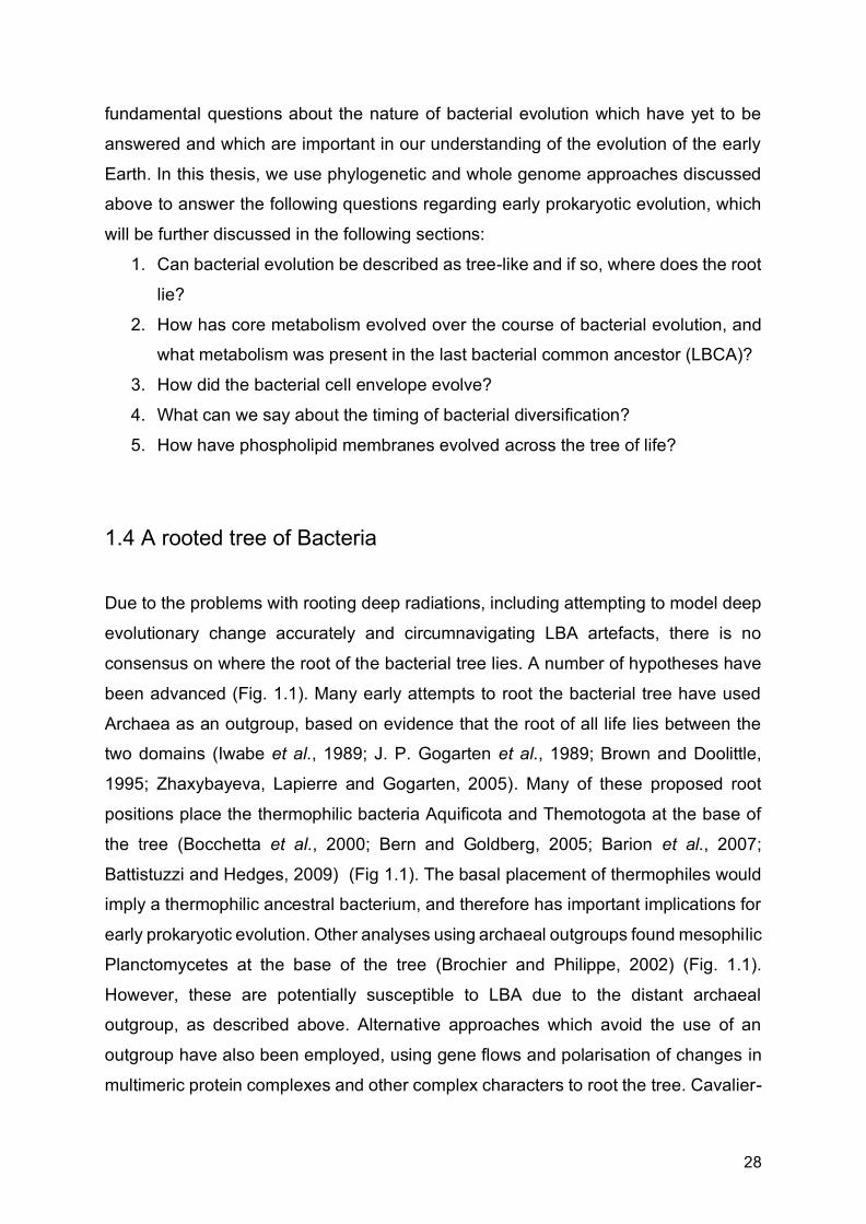
1.4 A rooted tree of Bacteria
Due to the problems with rooting deep radiations, including attempting to model deep
evolutionary change accurately and circumnavigating LBA artefacts, there is no
consensus on where the root of the bacterial tree lies. A number of hypotheses have
been advanced (Fig. 1.1). Many early attempts to root the bacterial tree have used
Archaea as an outgroup, based on evidence that the root of all life lies between the
two domains (Iwabe et al., 1989; J. P. Gogarten et al., 1989; Brown and Doolittle,
1995; Zhaxybayeva, Lapierre and Gogarten, 2005). Many of these proposed root
positions place the thermophilic bacteria Aquificota and Themotogota at the base of
the tree (Bocchetta et al., 2000; Bern and Goldberg, 2005; Barion et al., 2007;
Battistuzzi and Hedges, 2009) (Fig 1.1). The basal placement of thermophiles would
imply a thermophilic ancestral bacterium, and therefore has important implications for
early prokaryotic evolution. Other analyses using archaeal outgroups found mesophilic
Planctomycetes at the base of the tree (Brochier and Philippe, 2002) (Fig. 1.1).
However, these are potentially susceptible to LBA due to the distant archaeal
outgroup, as described above. Alternative approaches which avoid the use of an
outgroup have also been employed, using gene flows and polarisation of changes in
multimeric protein complexes and other complex characters to root the tree. Cavalier-

29
Smith used such approaches in his “transition analysis”, which takes various cellular,
molecular and biochemical characters in order to polarise major transitions and
systematically exclude lineages with derived characters, to suggest a root between
Chloroflexota and all other life, with Archaea and Eukarya branching from within
monoderm Bacteria (Cavalier-Smith, 2006) (Fig. 1.1). Lake et al. (2009) used analyses
of insertions and deletions (indels) within genomes to root the tree within monoderm
bacteria, with this root also representing the root of all life (Fig. 1.1).

30
Fig. 1.1 Schematic representing the bacterial tree, with various proposed root
positions indicated. Note that in the trees of Cavalier Smith and Lake et al. the Archaea
are a sister to Actinobacteriota and Firmicutes respectively. Lake et al. also describe
their root as a “ring of life” (see in text below).
Recent phylogenetic analyses of the whole tree of life, which incorporate the greatly
expanded knowledge of microbial diversity, have place the bacterial root between CPR
and all other Bacteria (Hug et al., 2016; Castelle and Banfield, 2018; Zhu et al., 2019)
(Fig. 1.1). Given the reduced genomes and likely symbiotic nature of CPR, such early
divergence of the clade would have important implications for our understanding of
early prokaryote evolution. The DPANN superphylum is an archaeal clade analogous
to CPR, and recent analyses suggest that the root of Archaea falls between this clade
and other Archaea (Castelle et al., 2015; Williams et al., 2017). If both these root
positions are correct, it would imply the presence of symbiotic, highly reduced
prokaryotic life alongside more conventional prokaryotic cells even at the earliest
stages of evolutionary history. Resolving the position of the root within Bacteria is
therefore imperative if we wish to understand the nature of the earliest life and how it
subsequently evolved.
These discussions on proposed root positions rely on the existence of some
detectable tree-like structure. As discussed above, HTGs are common across
prokaryotes, and it has been argued that thinking of early bacterial evolution in terms
of a bifurcating species tree may be misleading. Indeed, Lake et al. (2009) suggested
that his rooted tree, where roots were successively rejected based on the grouping of
indels, only made sense when represented as a “ring of life”, as many of the genomic
relationships could not be adequately described by, or were incompatible with a tree
diagram. Additionally, HGT has clearly had a profound effect on prokaryotic evolution.
For example, it has been suggested the origin of many major clades within Archaea
were driven by transfers from Bacteria, although transfers were less prevalent from
Archaea to Bacteria (Nelson-Sathi et al., 2015). Gene tree-species trees reconciliation
methods outlined above integrate both tree and network based approaches by
modelling both the vertical and horizontal components of genomes evolution, allowing
us to measure the contribution of both to bacterial evolutionary history. To do this, we
must quantify the amount of vertical evolution with the tree, i.e. the proportion of gene
families which evolve vertically. Quantifying verticality will thus allow us to evaluate

31
how prevalent HGT has been in bacterial evolutionary history, and may give insights
into the origins and drivers of innovation and adaptation in bacterial genomic evolution.
In chapter 2, we present a new rooted tree of Bacteria using ALE. We demonstrate
that other rooting methods, especially outgroup rooting, are not robust and are
susceptible to both composition-driven LBA and taxon sampling. In addition, we
attempt to quantify the extent of HGT through the bacterial tree to determine the extent
to which bacterial evolution can be described as tree-like.
1.5 Evolution of core metabolism in Bacteria
To fully understand the early evolution of life and the role it has played in shaping the
environment around us, we must understand the physiology and metabolic capabilities
of the earliest cells. Relatively little work has been done in reconstructing the ancestral
metabolism of Bacteria, partly due to the complications with unclear phylogeny and
rooting. Furthermore, it is difficult to disentangle such discussions from those
concerning the metabolism and habitat of LUCA, depending on how distant LBCA is
thought to be from LUCA and whether either resembles modern cells, or were both
primitive proto-cells.
Possible paths to carbon fixation Many scenarios concerning the early evolution of life posit that early prokaryotes would
have been autotrophic, and therefore there are key questions regarding which carbon
fixation pathway, electron donors and electron acceptors were used by LBCA. Decker
et al. (1970) used comparative biochemistry to suggest that methanogenesis and
acetogenesis were the oldest forms of energy metabolism in extant microbes. Both
methanogens and acetogens are anaerobes without cytochromes and obtain organic
carbon via the reduction of carbon dioxide by hydrogen, both gases thought to be
abundant on the early Earth (Arndt and Nisbet, 2012). Evidence for ancient origins of
methanogenesis have been seen in the geological record, showing biological methane
production extending back to at least 3.4 Ga (Ueno et al., 2006). Geological reactions
that bear a striking resemblance to core metabolic reactions of methanogens are found

32
to occur spontaneously at hydrothermal vents (Lang et al., 2010; Schrenk, Brazelton
and Lang, 2013), in particular, the generation of methane by serpentinisation. The
discovery of electron bifurcation (Li et al., 2008), a mechanism of energy conservation,
provides a mechanism for both acetogens and methanogens to reduce carbon dioxide
with elections from hydrogen despite the initial part of the reaction being energetically
uphill (Buckel and Thauer, 2013), and further points to ancient carbon fixation, and the
ancient evolution of autotrophy. Methanogens and acetogens both use the Wood-
Ljungdahl Pathway (WLP), which has been suggested as the most ancient carbon
fixation pathway (Fuchs, 2011; Sousa and Martin, 2014; Williams et al., 2017; Adam,
Borrel and Gribaldo, 2018) and previous phylogenetic work has suggested its
presence in both the archaeal (Williams et al., 2017; Adam, Borrel and Gribaldo, 2018)
and bacterial (Adam, Borrel and Gribaldo, 2018) common ancestors. In the WLP,
carbon dioxide is sequentially reduced by hydrogen to methane and acetate
respectively in methanogens and acetogens (Ferry and House, 2006; Lane and
Martin, 2012; Liu, Beer and Whitman, 2012). The pathway can be divided into two
stages, the methyl synthesis stage, and the acetyl synthesis stage. While superficially
similar in both groups, different pterin cofactors for methyl synthesis are used.
Tetrahydrofolate (H4F) is used in acetogens and methanopterin
tetrahydromethanopterin (H4MPT) is used in methanogens (Escalante-Semerena,
Rinehart and Wolfe, 1984; Jones, Donnelly and Wolfe, 1985; Maden, 2000). The
methyl synthesis pathways of both groups also use differing, non-homologous
enzymes. However, the key enzyme complex of the pathway, CODH/ACS (CO
dehydrogenase/acetyl-CoA synthase), is conserved in both domains, and is predicted
to have been present in both the archaeal (Williams et al., 2017; Adam, Borrel and
Gribaldo, 2018) and bacterial common ancestors (Adam, Borrel and Gribaldo, 2018).
If the WLP were present in LUCA, it has been suggested the methyl branch would
have been provided by geochemistry via serpentinisation, while the carbonyl branch
would have been performed by CODH/ACS. The enzymes for the methyl pathway
would have subsequently evolved in Bacteria and Archaea respectively as they
diverged into independent lineages (Martin and Russell, 2003, 2007; Sousa et al.,
2013; Sousa and Martin, 2014; Adam, Borrel and Gribaldo, 2018).
Alternatives pathways utilising the WLP have also been suggested. For example, the
earliest prokaryotic lineages may have had a denitrifying methanotrophic WLP, with

33
methanogenesis arising late and independently from acetogenesis (Nitschke and
Russell, 2013). However, this hypothesis has a number of problems. The late evolution
of methanogens is not compatible with studies of deep phylogeny or other evidence
of early biological methanogenesis (Ueno et al., 2006; Martin and Russell, 2007). It is
also the case that the denitrifying methanotrophy model must take place under
oxidising conditions in the oceans (Sousa et al., 2013), but that under even very mildly
oxidising settings, the accumulation at the vent-ocean interface of reduced organic
compounds ceases to be thermodynamically favourable (McCollom and Amend,
2005). Furthermore, biological methanogenesis also has a geochemical homologue
observed at hydrothermal vents, namely the formation of methane (among other
organic compounds) in serpentising systems (Proskurowski et al., 2008; Lang et al.,
2010; Etiope, Schoell and Hosgörmez, 2011). However, despite the oxic atmosphere
of the present, the geochemical methane oxidation (required for Nitschke and
Russell’s model) has not been observed.
Sulphate reduction is another possible alternative to methanogenesis. Modern
sulphate reducing bacteria respire sulphate to sulphide in a reaction which takes place
in two steps. The first step is the reduction of sulphate to sulphite which requires
energy, and a second step reduces sulphite to sulphide, where energy is released via
a simple respiratory chain. This second part of the process is important as it requires
no energy, and sulphite is thought to have been in abundant supply on the early Earth,
formed by the reaction of SO2 from volcanoes, with water. Many modern autotrophic
sulphur-reducing bacteria also have the WLP for carbon fixation (Rabus, Hansen and
Widdel, 2006). There is geological evidence for the early appearance of this metabolic
pathway, with stable isotopes supporting the origin of sulphate respiration as early as
3.47 Ga (Shen, Buick and Canfield, 2001). This, along with the abundant supply of
sulphate on the early Earth makes the early appearance of this metabolism very
plausible. Further evidence comes from the enzyme dissimilatory sulphite reductase
(Dsr), which seems to be highly conserved across many disparate prokaryotic
lineages, suggesting an ancient origin of this pathway (Wagner et al., 1998). The trees
generated from Dsr were congruent with the 16S rRNA phylogeny of the tree of life,
and which were taken as evidence of vertical inheritance rather than horizontal gene
transfer (Wagner et al., 1998). Others (Klein et al., 2001) found the gene tree to not
be fully compatible with the 16S rRNA tree and therefore inferred horizontal gene

34
transfer, and others still suggesting both vertical descent and horizontal transfer in
different groups (Zverlov et al., 2005). Therefore, it is not completely clear whether
sulphate reduction was present in the earliest life, but the evidence points to this as
an intriguing possibility.
Alternative carbon fixation pathways to the WLP have also been posited. The reverse
TCA cycle has been suggested as a possible ancient carbon fixation pathway
(Wächtershäuser, 1990; Cody et al., 2001; Smith and Morowitz, 2004; Nunoura et al.,
2018), given the widespread presence of the TCA cycle in modern Bacteria, and that
it may function in both the oxidative and reductive direction. Based on a basal position
of the Aquificae, and using biomimetic analysis, Marakushev and Belonogova inferred
a free-living, chemoautotrophic bacterial ancestor, with an ‘archaic metabolic network’
coupling reductive tricarboxylic acid, oxidative tricarboxylic acid and 3-
hydroxypropionic cycles (Marakushev and Belonogova, 2011, 2013). Braakman and
Smith (2012) suggested a combined system of the WLP and reductive tricarboxylic
acid cycle in both LBCA and LUCA.
Generation of energy In addition to specific metabolic pathways, the evolution of energy production and ion
pumping is an essential step in the evolution of physiological capabilities of modern
Bacteria. Herrmann et al. (2008) have suggested that the reduced ferredoxin whose
FeS cluster acts as an “energised coupler” in methanogenesis, has energy currency
characteristics more primitive than those of ATP. The origin of chemical osmotic
coupling, which was hitherto seen as an impossibly large leap in complexity, may have
developed from naturally occurring proton gradients at alkaline hydrothermal vents
(Russell et al., 1994; Russell and Hall, 1997). However, how was this naturally
occurring geochemical ion gradient replaced by ion pumping in order for life to become
independent from geochemical ion gradients, and have the ability to produce their own
ion gradients across membranes? In Archaea, MtrA-H complex found in methanogens
is a potential candidate for ancestral pumping systems, with the Rnf complex in
acetogens a similar candidate for Bacteria (Sousa et al., 2013; Sousa and Martin,
2014). In methanogens, the MtrA-H complex pumps out sodium, whilst transferring the
methyl group from methyl-H4MPT to methyl-CoM, whilst in acetogens Rnf pumps out
sodium whilst taking electrons from reduced ferredoxin to reduce NAD+ (Thauer et al.,

35
2008; Lane and Martin, 2012). The synthesis of low-potential ferredoxin, crucial for
carbon dioxide reduction in both groups, is dependent on electron bifurcation (Buckel
and Thauer, 2013). It is therefore possible to hypothesise the transfer of methyl groups
via MtrA-H complex producing a form of substrate-level pumping, using the abundant
methyl groups and ion gradients at the hydrothermal vent, which could have developed
into an active pumping mechanism without much evolutionary innovation. This may be
the most ancient form of pumping in Archaea. Analogous to this, a similar scenario
could have happened with the bacterial Rnf complex, similarly utilising naturally
occurring ion gradients (Sousa and Martin, 2014). The Rnf complex may therefore
present the most ancient form of ion pumping in Bacteria.
In Chapters 3 and 4 we investigate these questions surrounding the evolution of
metabolism in the earliest Bacteria, and determine which pathways were present at
different ancestral nodes in the tree. In Chapter 3, we present a reconstruction of the
central metabolic pathways present in LBCA. In Chapter 4, we extend this to several
deep nodes in the bacterial tree in order to evaluate the evolution of these pathways
in the deepest parts of the tree.
1.6 One membrane or two? The evolution of the cell envelope
Monoderms vs diderms Bacteria have been classically divided into two groups based on their response to
Gram staining, with some Bacteria resisting the decolourisation step of the process
(Gram 1884). These “gram-negative” Bacteria were shown to resist the
decolourisation by way of a secondary out membrane, exhibiting a “diderm”
architecture, as opposed to those which had a single membrane and did not resist the
decolourisation step (Bladen and Mergenhagen, 1964). The two model organisms,
Bacillus subtilis (a firmicute), and Escherichia coli (a gammaproteobacterium)
respectively epitomise the classic monoderm and diderm phenotypes (Silhavy, Kahne
and Walker, 2010, Megrian et al., 2020). Monoderms typically exhibit a single lipid cell
membrane and a thick peptidoglycan wall with teichoic and lipoteichoic acids. Diderms
instead have a thin peptidoglycan wall with an inner and an outer lipid membrane,

36
often embellished with lipopolysaccharides (LPS). A number of systems are involved
in the assembly of the classic diderm envelope, including LPS synthesis carried out
by the Lpx and Kds enzymes, transport across the inner membrane by MsbA and
transport to the outer membrane via the Lpt system, the assembly and insertion of
proteins into the outer membrane by the Bam and Tam systems, the insertion of
lipoproteins by the Lol system, and the maintenance of lipid asymmetry between the
inner and outer membranes by the Mla system (Antunes et al., 2016; Megrian et al.,
2020). Other machineries that are found in both monoderms and diderms have specific
proteins to anchor themselves to the outer membrane, including the P and L rings
(FlgAHI) in flagella, and secretin (PilQ) for type IV pili. Many bacteria, however, may
exhibit cell envelopes with a mixture of characteristics which do not follow the classic
gram-negative/gram-positive divide (Sutcliffe, 2010).
Scenarios for the origin of the outer membrane A number of different scenarios have been proposed for the evolution of the double
membrane (Fig. 1.2), of which there are two main camps, Monoderm-first and Diderm-
first. Under Monoderm-first hypotheses, the monoderm cell envelope, seen as
ancestral or “primitive” in its architecture, would have been the ancestral state, with
the emergence of the diderm envelope later in evolutionary history as a derived trait.
One scenario, proposed by Lake et al. (2009), suggests that diderms originated as a
fusion between two monoderm bacteria, a firmicute and an actinobacterium (Fig. 1.2).
This has been criticised (Gupta, 2011), especially on the grounds that there appears
to be no evidence to group all diderms form a single clade to the exclusion of all
monoderms. An alternative scenario posits that the outer membrane evolved under
antibiotic selection pressure, where diderm Bacteria lacking LPS represent
evolutionary intermediates to classical diderms (Gupta, 2011) (Fig. 1.2). Specifically,
within this scenario, two gene insertions (Hsp70 and Hsp60 respectively) lead to the
classic diderm envelope seen in most modern species, with the Chloroflexota
(monoderm but having the Hsp70 insert) and the “simple diderm” (i.e. lacking in LPS)
Deinococcota representing transitional stages. Fusobacteriota, Synergistota,
Elusimocrobiota, and the two diderm classes of Firmicutes (the Negativicutes and
Halanaerobiales), represent “atypical diderms” in this scenario, as they exhibit classic
LPS-membranes, but lack the Hsp70 and Hps60 gene insertions (Gupta, 2011), and
would have presumably evolved their membranes independently of other diderms, or

37
through HGT. However, this scenario may not be compatible with current ideas of
bacterial phylogeny (Hug et al., 2016; Parks et al., 2017). Tocheva et al. (2016) have
suggested that the outer membrane may have formed via sporulation, stemming from
the observation that a temporary outer membrane is formed during endospore
formation in Firmicutes before being lost during spore germination (Fig. 1.2). The outer
membrane would have therefore originated once in a spore-forming monoderm
ancestor (Tocheva et al., 2011; Errington, 2013; Tocheva, Ortega and Jensen, 2016).
However, such sporulation seems to be specific to Firmicutes, and therefore this
hypothesis is incompatible with current knowledge of bacterial phylogeny and
evolution.
Fig. 1.2 Summary of different evolutionary hypotheses for the origin of the outer
membrane, including Monoderm-first scenarios proposed by Lake (2009), Gupta
(2011) and Tocheva et al. (2016), and a Diderm-first hypothesis proposed by Cavalier-

38
Smith (2002). Based on Figure 2 form Megrian et al. (2020). PG=peptidoglycan,
IM=inner membrane, OM=outer membrane, LPS=lipopolysaccharides.
Diderm-first hypotheses have also been advocated. It has been suggested that the
earliest Bacteria were diderm (Cavalier-Smith, 2002), with the loss of the outer
membrane occurring due to a mutation which increased the thickness of the
peptidoglycan wall, causing the outer membrane attachments to break (Cavalier-
Smith, 2006) (Fig. 1.2). Within this scenario, the root of life is within Bacteria, with a
single clade of monoderms including Archaea and Eukaryotes, and diderm
Chloroflexota at the base of the tree. However, subsequent analysis have shown
Chloroflexota to be monoderms (Sutcliffe, 2010), and the basal position of the phylum
is contentious (Raymann, Brochier-Armanet and Gribaldo, 2015). More recently, it has
been demonstrated based on phylogenetic analyses of associated genes that the
classically monoderm Firmicutes are ancestrally diderm (Antunes et al., 2016), and
that the monoderm phenotype has arisen multiple times within the phylum. Given the
extensive distribution of the diderm phenotype across the tree, it has been argued that
this scenario with Firmicutes is analogous to what happened across the bacterial tree,
namely a diderm ancestor followed by lineages specific losses (Megrian et al., 2020).
In Chapters 3 and 4 we reconstruct the evolution of cell envelope architecture in
Bacteria. In Chapter 3, we present a reconstruction of the cell envelope of LBCA. In
Chapter 4, we extend this to several deep nodes in order to evaluate the evolution of
cell envelope architecture across the bacterial tree.
1.7 Timing of bacterial evolution
Relatively little research has been carried out attempting to date the bacterial tree,
either absolutely using molecular clocks, or using some form of relative dating. Using
molecular clocks for Bacteria is difficult due to the need for fossil calibrations, which
are sparse and diagnostically uninformative. However, understanding the timing of
evolutionary events within the tree is crucial in understanding the evolution of
metabolism and physiology through bacterial evolution. As discussed above,

39
methanogenesis and acetogenesis, and the associated WLP, are often posited as
some of the earliest emerging metabolisms (Battistuzzi, Feijao and Hedges, 2004;
Ueno et al., 2006; Sousa, Nelson-Sathi and Martin, 2016; Weiss et al., 2016; Williams
et al., 2017; Adam, Borrel and Gribaldo, 2018; Wolfe and Fournier, 2018). Being able
to infer the timing of the emergence of bacterial clades which use this pathway would
be able to lend some support to its ancient origins, whether or not we find evidence
for its presence in LBCA.
Another important debate in bacterial evolution revolves around the timing of the
emergence of Cyanobacteria and its relation to the Great Oxidation Event (occurring
~2.4 Ga), and by extension the origin of oxygenic photosynthesis. The GOE has often
been causally linked to the emergence of the Cyanobacteria (Schirrmeister et al.,
2013; Knoll and Nowak, 2017; Sánchez-Baracaldo et al., 2017), although some
evidence has placed their emergence later in time (Betts et al., 2018). Yet another
debate is the emergence of eukaryotic cells, a key moment in evolutionary history. It
has been suggested to have happened early, possibly contemporaneously with or
even predating the emergence of prokaryotes (Kurland, Collins and Penny, 2006). It
has also been linked to the GOE (Knoll and Nowak, 2017). However, other studies
have suggested that eukaryotes evolved relatively late (Chernikova et al., 2011;
Parfrey et al., 2011; Eme et al., 2014a; Knoll, 2014; N. J. Butterfield, 2015; Betts et al.,
2018). There is now a growing consensus, based on phylogenetics and comparative
genomic evidence, that eukaryotic cells arose from a symbiosis between an archaeal
host cell and a bacterial endosymbiont that evolved into the mitochondrion (Embley
and Martin 2006; Martin et al. 2015; Eme et al. 2017; Roger et al. 2017). Eukaryotic
cells would have to postdate the emergence of Alphaprotebacteria.
Some of the questions above may be partially answered by relative dating, that is
inferring the order in which clades emerged within the bacterial tree. This can be done
using whole-genome approaches such as ALE as they model HGT. Transfers contain
information about the relative divergences because donor lineages are necessarily as
old as the recipient lineages (Chauve et al., 2017; Davín et al., 2018). This approach
has been used to infer that methanogenic Euryarchaeota were the earliest radiating
lineages within Archaea, supporting the ancient origin of methanogenesis (Davín et
al., 2018). The same study also inferred a relatively late radiation of crow-group
40
DPANN, a superphylum of Archaea with highly reduced genomes analogous to CPR
in Bacteria, despite their early divergence within the tree. Inferring the relative age of
diversification of CPR is important in understanding their role, and indeed the role of
highly reduced, streamlined cells, in the early evolution of cellular life.
In Chapter 4, we use HGTs to relatively date the emergence of different clades within
the bacterial tree, allowing us to infer the order of major events in bacterial evolution.
It must be stressed that the analyses and results presented in Chapter 4 are not
absolute dates, that is they only tell the order of the events, not when they occurred or
the time that elapsed between them. However, such relative time information is still of
great use in our attempts to reconstruct the evolutionary history of Bacteria.
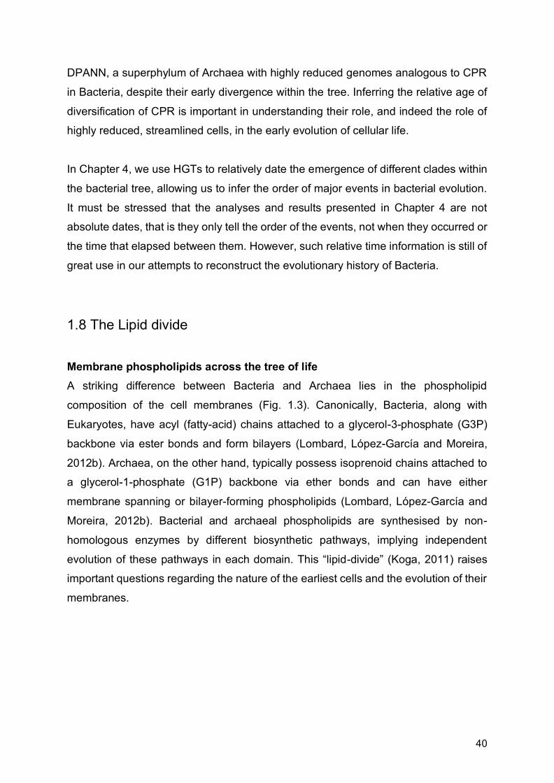
1.8 The Lipid divide
Membrane phospholipids across the tree of life A striking difference between Bacteria and Archaea lies in the phospholipid
composition of the cell membranes (Fig. 1.3). Canonically, Bacteria, along with
Eukaryotes, have acyl (fatty-acid) chains attached to a glycerol-3-phosphate (G3P)
backbone via ester bonds and form bilayers (Lombard, López-García and Moreira,
2012b). Archaea, on the other hand, typically possess isoprenoid chains attached to
a glycerol-1-phosphate (G1P) backbone via ether bonds and can have either
membrane spanning or bilayer-forming phospholipids (Lombard, López-García and
Moreira, 2012b). Bacterial and archaeal phospholipids are synthesised by non-
homologous enzymes by different biosynthetic pathways, implying independent
evolution of these pathways in each domain. This “lipid-divide” (Koga, 2011) raises
important questions regarding the nature of the earliest cells and the evolution of their
membranes.

41
Fig. 1.3 Schematic representation of the phospholipid biosynthesis pathways in
Archaea and Bacteria, based partially on Figure 1 from Peretó et al. (2004).
Despite the lipid divide being important for our understanding of early cellular
evolution, relatively little experimental work has been done to determine the
stereochemistry of phospholipids in individual lineages, with most studies assuming
that bacterial and archaeal lineages will have their respective stereochemistry as a
matter of course. While the limited studies which have determined glycerol
stereochemistry seem to support this divide (Sinninghe Damsté et al., 2002; Weijers
et al., 2006), there is some evidence to suggest that certain Bacteria have the ability
to produce G1P-linked ether lipids. Notably, it has been demonstrated experimentally
that B. subtilis possesses homologues of archaeal G1P dehydrogenase (G1PDH) and
geranylgeranylglyceryl phosphate synthase (GGGPS) (Guldan, Sterner and Babinger,
2008; Guldan et al., 2011), which allow it to produce an archaeal-like phospholipids,
although it is unknown if these are used in the B. subtilis membrane. Aside from
stereochemistry, other characteristics of membrane phospholipids appear to be
variable, often exhibiting a mixture of bacterial and archaeal features. For example,
plasmalogens found in both Eukaryotes and Bacteria have ether bonds (Goldfine,
2010) and some Archaea have been shown to produce membrane lipids with fatty-

42
acids (Gattinger, Schloter and Munch, 2002). Of great interest are branched glycerol
dialkyl glycerol tetra-ethers (brGDGTs) found in the environment, which exhibit
bacterial glycerol stereochemistry, and use branched alkyl chains (rather than
archaeal isoprenoid chains), but which have ether bonds and are membrane spanning,
characteristics usually associated with archaeal lipids (Schouten et al., 2000; Weijers
et al., 2006). These brGDGTs are particularly abundant in peat bogs, where their
unusual mixture characteristics were thought to be bacterial adaptations to low pH
environments (Weijers et al., 2006; Damsté, Sinninghe Damsté, et al., 2007), although
they are now known to occur in a wide range of soil and aquatic environments
(Schouten, Hopmans and Sinninghe Damsté, 2013). The biosynthetic pathways and
associated enzymes for these mixed-type membrane lipids remain enigmatic, but
given the frequency of prokaryotic HGT (Hemmi et al., 2004), it is not unreasonable to
assume that they may reflect pathways of mixed bacterial and archaeal origin. This
indicates that the lipid-divide, thought to be such a defining difference between the two
domains of life, may be less clear-cut than previously thought.
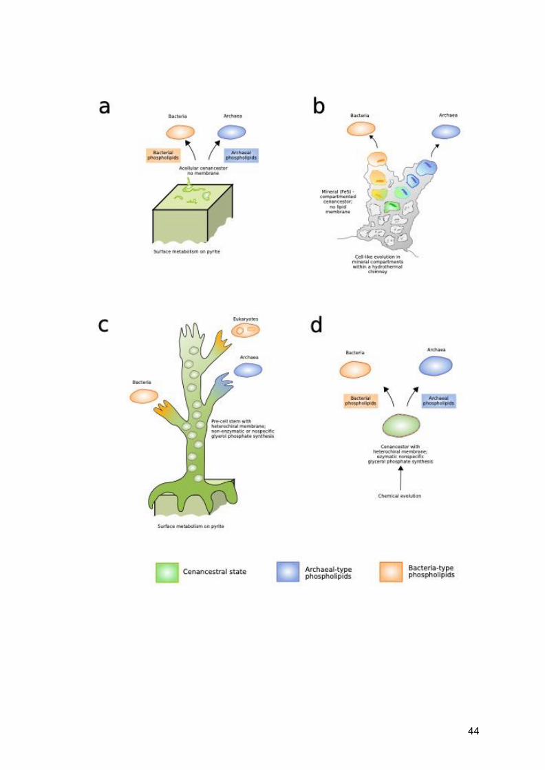
Possible scenarios for the evolution of membrane phospholipids Several different hypotheses have been suggested to explain the origins of the
different pathways, and the nature of the membrane of LUCA, summarised here in Fig.
1.4. There is some debate as to whether LUCA was acellular, living on the surface of
pyrite (Koga et al., 1998) (Fig. 1.4a) or in mineral-bounded compartments within a
hydrothermal chimney (Martin and Russell, 2003) (Fig. 1.4b), with lipid membrane
evolving independently in each domain at a later point. However, the presence of some
genes for lipid biosynthesis (Lombard and Moreira, 2011; Lombard, López-García and
Moreira, 2012b; Koga, 2014; Weiss et al., 2016) and, in particular, a membrane-bound
ATPase (Sojo, Pomiankowski and Lane, 2014; Weiss et al., 2016) in reconstructions
of LUCA implies that it possessed a membrane, although its properties may have
differed from those of modern, prokaryote cell membranes (Lombard, López-García
and Moreira, 2012b; Koga, 2014; Sojo, Pomiankowski and Lane, 2014). Alternatively,
archaeal and bacterial phospholipid biosynthesis may have evolved from a stem of
pre-cells with heterochiral membranes (Wächtershäuser, 2003) (Fig. 1.4c) or a
heterochiral LUCA with membranes synthesised via universal, substrate-nonspecific
enzymes (Peretó, López-García and Moreira, 2004) (Fig. 1.4d). In the latter
hypothesis, the heterochiral membrane would be less stable than a homochiral one,

43
putting selective pressure on the ancestral bacterial and archaeal populations to shift
to a homochiral membrane with either phospholipid type, although there is evidence
to suggest that heterochiral membranes are not less stable than homochiral ones (Fan
et al., 1995; Shimada and Yamagishi, 2011; Caforio et al., 2018).
Fig. 1.4 (below) Representation of four different models of the origin and early
evolution phospholipid biosynthesis in Archaea and Bacteria. a) independent evolution
of archaeal and bacterial pathways from an acellular cenancestor (Kog et al. 1998); b)
independent evolution of archaeal and bacterial pathways from mineral bound
compartments (Martin and Russel 2003); c) evolution of domain specific pathways
form a stem of heterochiral pre-cells (Wächtershäuser, 2003); d) evolution of domain
specific pathways form a fully cellular, heterochiral ancestor (Peretó, López-García
and Moreira, 2004). Based on Figure 2 from Peretó et al. (2004)
44

45
It is also possible that LUCA was homochiral with either type of phospholipid, with the
evolution of the other in its respective lineage later in evolutionary history (Yokobori et
al., 2016), although it is unclear what would prompt such a change. If archaeal
phospholipids are ancestral (Daiyasu et al., 2002; Peretó, López-García and Moreira,
2004; Carbone et al., 2015), the change to bacterial phospholipids within Bacteria may
have been driven by the flexibility and adaptability afforded by bacterial lipid
architecture. Namely, based on chemical considerations, bacterial phospholipids may
be cheaper to make and break. They also allow a greater variety of fatty acyl moieties,
varying in chain length, unsaturation, degree of branching and cyclisation compared
to archaea-type phospholipids, allowing better adaptation to diverse environments.
These characteristics may have given marginal benefits in various dynamic mesophilic
environments, and would be a possible explanation to the relatively higher abundance
of Bacteria compared to Archaea in most environments (Danovaro et al., 2016; Hug
et al., 2016; Castelle and Banfield, 2018). Conversely, if bacterial-type phospholipids
are ancestral (Yokobori et al., 2016), the evolution of archaea membrane may have
been driven by adaptation to high temperatures (Akanuma et al., 2013; Akanuma,
Yokobori and Yamagishi, 2013; Yokobori et al., 2016), as ether bonds are more
thermostable than esters (Vossenberg et al., 1998; Koga, 2012) and are also found in
the membranes of thermophilic Bacteria (Kaur et al., 2015). It should be noted however
that the widespread occurrence of bacterial-, archaeal- and mixed-type membranes
suggest that, except in thermophilic or low pH environments, there seems to be little
advantage to either membrane.
In chapter 3 and 4, we present evidence for the composition of lipid membranes
present in LBCA and subsequent nodes. In Chapter 5, we expand our study to the
whole tree of life. Using expanded taxon sampling, including environmental samples,
and using the best evolutionary models available to us, we present a reconstruction of
the evolutionary history of the gene families involved in phospholipid biosynthesis.
1.9 A model for the evolution of early life

46
As discussed above, there are still a number of fundamental questions about bacterial
evolution that are unresolved. While there are many difficulties and challenges
involved in such deep phylogenetic reconstructions, by using the best models and data
currently available to us, this thesis attempts to answer such questions. We use new
and innovative methods in order to overcome the problems associated with traditional
phylogenetic methods, and which allows us to incorporate a much larger breadth of
data. The results generated from these analyses will allow us to test and suggest novel
models and hypotheses regarding the evolution of Bacteria. Specifically, the use of
whole-genomes approaches, such as ALE, will allow us to root the bacterial tree, as
well as model HGT over time and infer ancestral gene content. The inference of
ancestral gene content will allow us to reconstruct the metabolic capabilities and
habitat of the earliest Bacteria, and answer many of the questions discussed above
relating to the evolution of particular physiologies and characters. There are of course
many caveats to our analyses. Practical concerns must be considered and sometimes
compromises have to be made in order to make the analyses computationally feasible
e.g. the use of maximum likelihood instead of Bayesian analysis, or using reduced
taxon sampling. These caveats and how they can be improved upon are further
discussed through the thesis and in more detail in the Chapter 6. Ultimately, while
none of our analyses are without their caveats, we believe by using innovative
methods and different lines of evidence in this thesis, we can advance the field of
evolutionary microbiology and shed greater light on the earliest period of the history of
life.

47
Chapter 2
Phylogenomics produces a rooted tree of
Bacteria A version of this chapter forms part of a paper under revision in collaboration with
Adrián A. Davín, Tara Mahendrarajah, Anja Spang, Philip Hugenholtz, Gergely J.
Szöllősi, and Tom A. Williams. Gareth A. Coleman is the first author of the paper. The
project was conceived by TAW, GJSz, PH, AS, GAC and AAD. Analyses for the GTDB
dataset were performed by GAC, AAD, TAW and GJSz. GJSz developed new
analytical methods. Pipelines for orthologue selection and gene family generation
were developed by GAC. All authors contributed to interpretation and writing. All
analyses, writing and interpretation for the non-GTDB dataset were performed by
GAC. For the ToL rooting section, species selection was carried out by GAC,
orthologues were given by TAW, HiFix trees generated by Edmund Moody, species
trees inferred by GAC, and all ALE analyses carried out by GAC. All writing and
interpretation for the ToL section was carried by GAC.
Paper preprint as:
Coleman, G.A., Davín, A.A., Mahendrarajah, T., Spang, A.A., Hugenholtz, P.,
Szöllősi, G.J. and Williams, T.A., 2020. A rooted phylogeny resolves early bacterial
evolution. bioRxiv.
BioRxiv preprint for the paper can be found here:
https://www.biorxiv.org/content/10.1101/2020.07.15.205187v1

48
Abstract
Bacteria are the most abundant and metabolically diverse cellular lifeforms on Earth. A rooted bacterial phylogeny provides a framework to interpret this diversity and to understand the nature of early life. Inferring the position of the bacterial root is complicated by incomplete taxon sampling and the long branch to the archaeal outgroup. To circumvent these limitations, we model bacterial genome evolution at the level of gene duplication, transfer and loss events, allowing outgroup-free inference of the root. We infer a rooted bacterial tree on which 68% of gene transmission events are vertical. Our analyses reveal a basal split between Terrabacteria and Gracilicutes, which together encompass almost all known bacterial diversity. However, the position of a few small phyla could not be resolved in relation to these two major clades. In contrast to recent proposals, our analyses strongly reject a root between the Candidate Phyla Radiation (CPR) and all other Bacteria. Instead, we find that the CPR is a sister lineage to the Chloroflexota within the Terrabacteria.

49
2.1 Introduction
Rooting deep radiations (Williams et al., 2017) is among the greatest challenges in
phylogenomics, and there is no consensus on the root of the bacterial tree. Based on
evidence (Iwabe et al., 1989; J. P. Gogarten et al., 1989; Brown and Doolittle, 1995;
Zhaxybayeva, Lapierre and Gogarten, 2005) that the root of the entire tree of life lies
between Bacteria and Archaea, early analyses using an archaeal outgroup placed the
bacterial root near Aquificales/Thermotogales (Bocchetta et al., 2000; Battistuzzi and
Hedges, 2009) or Planctomycetes (Brochier and Philippe, 2002). Alternative
approaches, including analyses of gene flows and polarisation of changes in
multimeric protein complexes and other complex characters (Cavalier-Smith, 2006),
have instead suggested roots within the monoderm (single-membrane) Bacteria
(Lake, 2009), or between Chloroflexi and all other cellular life (Cavalier-Smith, 2006).
The development of techniques for sequencing microbes directly from environmental
samples, without the need for laboratory cultivation, has greatly expanded the genomic
representation of natural prokaryotic diversity (Hug et al., 2016; Mukherjee et al., 2017;
Parks et al., 2017, 2018). Recent phylogenomic analyses of that expanded diversity
have placed the bacterial root between one of these new groups, the Candidate Phyla
Radiation (CPR; also known as Patescibacteria (Brown et al., 2015; Zhu et al., 2019))
and all other Bacteria (Hug et al., 2016; Castelle and Banfield, 2018; Zhu et al., 2019).
The CPR comprises lineages that are characterised by small cells and genomes, and
are suggested to have predominantly symbiotic or parasitic lifestyles, but much
remains to be learned about their ecology and physiology (Brown et al., 2015; Castelle
and Banfield, 2018; Castelle et al., 2018; Beam et al., 2020). If correct, the early
divergence of CPR has important implications for our understanding of the earliest
period of cellular evolution. Taken together with evidence that the root of the archaeal
domain lies between the reduced and predominantly host-associated DPANN
superphylum and the rest of Archaea (Castelle et al., 2015; Williams et al., 2017), the
CPR root would imply that streamlined, metabolically minimalist prokaryotes have co-
existed with the more familiar, self-sufficient lineages throughout the history of cellular
life (Beam et al., 2020).

50
Historically there has also been little agreement on the relationships between different
bacterial phyla. In recent years, some superphyla-level groupings have become widely
accepted, namely FCB/Sphingobacteria (Fibrobacteres, Chlorobi, Bacteroidetes, and
candidate phyla Cloacimonetes, Gemmatimonadetes, Ignavibacteria, Latescibacteria,
Marinimicrobia and Zixibacteria, and the genus Caldithrix) (Gupta, 2004; Hug et al.,
2016; Castelle and Banfield, 2018; Parks et al., 2018) and PVC/Planctobacteria
(Planctomycetes, Verrucomicrobia and Chlamydiae, Lentisphaerae, and candidate
phylum Omnitrophica) (Cavalier-Smith, 2002; Wagner and Horn, 2006; Hug et al.,
2016; Parks et al., 2017; Castelle and Banfield, 2018). More tentative higher level
relationships have also been suggested. Battistuzzi and Hedges (2009) divide the
bacterial tree into two major clades; one clade comprising the gram-positive Bacteria
(Firmicutes and Actinobacteria), Cyanobacteria, Chloroflexi and Deinococcus-
Thermus, which they name Terrabacteria (Battistuzzi, Feijao and Hedges, 2004), as
they assert that this clade was ancestrally terrestrial; and another clade comprising
PVC, FCB and Proteobacteria, which they name Hydrobacteria (Battistuzzi and
Hedges, 2009). The Terrabacteria has received some support in subsequent analyses
(Bern and Goldberg, 2005; Boussau, Guéguen and Gouy, 2008). Hydrobacteria has
alternatively been described by Cavalier-Smith as the Gracilicutes (Cavalier-Smith,
2006), as well being supported by other studies (Boussau, Guéguen and Gouy, 2008).
Yet, despite this progress, the deep relationships within the bacterial tree are still highly
debated, with the position of the root being particularly unclear.
Improved taxon sampling can help to resolve difficult phylogenetic problems
(Graybeal, 1998; Hedtke, Townsend and Hillis, 2006), and the enormous quantity and
diversity of genome data now available presents an unprecedented opportunity to
resolve long-standing questions about the origins and diversification of Bacteria. But
deep phylogenetic divergences are difficult to resolve, both because the phylogenetic
signal for deep relationships is overwritten by new changes through time, and also
because the process of sequence evolution is more complex than the best-fitting
models currently available. In particular, variation in nucleotide or amino acid
composition across the sites of the alignment and the branches of the tree can induce
long branch attraction (LBA) artefacts in which deep-branching, fast-evolving, poorly-
sampled or compositionally biased lineages group together irrespective of their
evolutionary history (Bergsten, 2005). These issues are widely appreciated (Hug et

51
al., 2016) but are challenging to adequately address, particularly when sequences
from thousands of taxa (Hug et al., 2016; Parks et al., 2017, 2018; Castelle and
Banfield, 2018; Zhu et al., 2019) are used to estimate trees of global prokaryotic
diversity, which precludes the use of the best available phylogenetic methods.
The traditional method of outgroup rooting has proven difficult as the closest outgroup
to Bacteria are Archaea. This is highly problematic as Bacteria and Archaea represent
the two fundamental domains of life, branching deep in evolutionary time, and creating
long stem lineages to the crown groups. The information provided by archaeal
outgroups are thus of not much use and may create artefactual results. Gene
duplications are a useful way of resolving roots in clades without the use of an
outgroup, and has been used in the rooting of the tree of life (Iwabe et al., 1989; J. P.
Gogarten et al., 1989; Brown and Doolittle, 1995). Recently, it has also been shown
that the use of gene gains, losses and HGTs can also be used alongside gene
duplications as an effective method of rooting a species tree (Abby et al., 2012;
Szöllosi et al., 2012; Szöllősi et al., 2013). These can be integrated with probabilistic
gene tree-species tree reconciliation methods, such as the recently developed
Amalgamated Likelihood Estimation (ALE) method. When looking for the root in a
species tree, ALE calculates the maximum likelihood of each gene family tree, given
a chosen root position, and rates of gene duplication, transfer and loss (DTLs) (Szöllősi
et al. 2013). Each chosen root position implies a particular evolutionary history of that
gene family, with regards to transfer, loss, or gain of genes, which can be evaluated
under maximum likelihood. ALE is innovative in that it can incorporate uncertainty in
the underlying gene trees using conditional clade probabilities, and has been shown
to be able to infer numbers of gene duplications, transfers, losses and ancestral
genome sizes accurately over large phylogenetic depths (Szöllősi et al. 2013).
2.2 Methods
Taxon sampling To obtain a representative taxon sampling from across known bacterial diversity, we
sampled taxa according to the classification provided by the Genome Taxonomy

52
Database (GTDB r89) (Parks et al., 2018). We sampled 265 genomes from the GTDB
as follows. First, we filtered out the genomes with Quality < 0.75 (Quality is defined as
Completeness - (5*Contamination) (Parks et al., 2017)), and filtered out all phyla
subsequently left with fewer than 10 species. Genomes were sampled from the
remaining taxa on a per-class basis: for classes containing a single order, the genome
with the highest quality score was sampled; for classes containing multiple orders, the
highest quality genome from each of two randomly chosen orders was sampled. This
protocol ensured that every class in the GTDB is represented in the final tree. We then
manually added the genome of Gloeomargarita litophora given its importance in
constraining the phylogeny and timing of chloroplast evolution (Appendix A, Table 1).
To sample representative bacterial taxa independently of the GTDB, we began with
the bacterial portion of a recent global analysis of the tree of life (Hug et al., 2016). We
initially generated our subsample using an algorithm which maximised genetic
difference between lineages in order to select 200 taxa that were evenly distributed
across the tree. However, this selection process was biased in favour of long
branches. We also explored other programmatic ways of selecting taxa based on
genetic diversity, but these were found to be extremely sensitive to the phylogeny and
initial choice of root position. Instead, we inferred a tree of the bacterial portion of the
concatenate under the LG+G4+F model in IQ-Tree. We divided the tree into 7 major
bacterial clades based on a literature search (Table 2.1) and additional environmental
lineages with branch length diversity comparable to the known groups. For each group
defined in this way, we manually subsampled taxa so as to maintain genetic diversity,
while avoiding overly long or short branches. We selected 342 species, comprising
200 ‘classic’ bacteria, 125 CPR bacteria and one bacterial genome respectively from
each of the 17 new phyla described by (Parks et al., 2017) (Appendix A, Table 2). For
all species in both datasets, proteomes were download as amino acid sequences, and
contain plasmids.
Clade No. of taxa sampled
Firmicutes 25
Actinobacteriota+Cyanobacteria+Chloroflexota 35
CPR 125

53
FCB+PVC+Elusimicrobiota 35
Proteobacteria 50
Deltaproteobacteria+Nitrospirota+Acidobacterota+Aquificota 30
FASST+environmental lineages 25
New Phyla (Parks et 2018) 17
Table 2.1 Number of taxa sampled from each clade in the GTDB-independent
analysis.
Orthologue selection We used OMA 2.1.1 (Roth, Gonnet and Dessimoz, 2008) to identify candidate single-
copy bacterial orthologues, and retained those with at least 75% of all species
represented in each family. Sequences were aligned in Mafft using the -auto option,
and trimmed in BMGE 1.12 (Criscuolo and Gribaldo, 2010) using the BLOSUM30
model. Initial trees were inferred for each candidate orthologue under the LG+G+F
model in IQ_TREE 1.6.10. The trees were manually inspected, and we selected
orthologues where the monophyly of 14 pre-defined major lineages was not violated
with bootstrap support >70%, resulting in 63 final orthologues (Table 2.2). The same
selection process was used on both datasets, generating the same set of orthologues.
KO number Gene name Annotation Used in outgroup tree?
K03046 rpoC DNA-directed RNA polymerase subunit beta' y
K03043 rpoB DNA-directed RNA polymerase subunit beta
K02337 dnaE DNA polymerase III subunit alpha y
K03070 secA Protein translocase subunit SecA
K01873 VARS, valS Valine--tRNA ligase
K02335 polA DNA polymerase I y
K01872 AARS, alaS Alanine tRNA ligase
K02469 gyrA DNA gyrase subunit A
K00962 pnp, PNPT1 Polyribonucleotide nucleotidyltransferase y
K02355 fusA, GFM, EFG Translation elongation factor G y
K01972 E6.5.1.2, ligA, ligB DNA ligase NAD
K03702 uvrB Excinuclease ABC subunit B
K02470 gyrB DNA gyrase subunit B

54
K04077 groEL, HSPD1 Molecular chaperone GroEL
K01937 pyrG, CTPS CTP synthase y
K02313 dnaA Chromosomal replication initiator protein
K02314 dnaB Replicative DNA helicase
K02433 gatA, QRSL1 aspartyl-tRNA(Asn)/glutamyl-tRNA(Gln) amidotransferase subunit A
K03076 secY Protein translocase subunit SecY y
K04485 radA, sms DNA repair protein RadA/Sms
K02112 ATPF1B, atpD F-type H+/Na+-transporting ATPase subunit beta y
K03590 ftsA Cell division protein FtsA
K02358 tuf, TUFM Elongation factor Tu y
K06942 ychF Redox Regulated ATPase YchF
K00927 PGK, pgk Phosphoglycerate kinase
K01889 FARSA, pheS Phenylalanine--tRNA ligase alpha subunit
K03551 ruvB Holliday junction branch migration DNA helicase RuvB y
K04485 radA, sms DNA recombination repair protein RecA y
K02835 prfA, MTRF1, MRF1 Peptide chain release factor 1
K02886 RP-L2, MRPL2, rplB 50S ribosomal protein L2 y
K01803 TPI, tpiA Triose phosphate isomerase y
K03438 mraW, rsmH 16S rRNA (cytosine1402-N4)-methyltransferase
K00554 trmD tRNA (guanine37-N1)-methyltransferase
K02863 RP-L1, MRPL1, rplA 50S ribosomal protein L1 y
K03685 rnc, DROSHA, RNT1 Ribonuclease III
K02967 RP-S2, MRPS2, rpsB 30S ribosomal protein S2 y
K02982 RP-S3, rpsC 30S ribosomal protein S3
K02906 RP-L3, MRPL3, rplC 50S ribosomal protein L3 y
K03470 rnhB Ribonuclease HII
K01358 clpP, CLPP ATP dependent Clp protease proteolytic subunit
K06187 recR Recombination protein RecR
K15034 yaeJ Aminoacyl tRNA hydrolase, ribosome-associated protein
K02931 RP-L5, MRPL5, rplE 50S ribosomal protein L5 y
K02933 RP-L6, MRPL6, rplF 50S ribosomal protein L6 y
K02601 nusG Transcription termination antitermination protein NusG
K02988 RP-S5, MRPS5, rpsE 30S ribosomal protein S5 y
K02992 RP-S7, MRPS7, rpsG 30S ribosomal protein S7 y
K03664 smpB SsrA binding protein

55
K02838 frr, MRRF, RRF Ribosome recycling factor
K02867 RP-L11, MRPL11, rplK 50S ribosomal protein L11
K02878 RP-L16, MRPL16, rplP 50S ribosomal protein L16 y
K02871 RP-L13, MRPL13, rplM 50S ribosomal protein L13 y
K02994 RP-S8, rpsH 30S ribosomal protein S8 y
K02948 RP-S11, MRPS11, rpsK 30S ribosomal protein S11 y
K02952 RP-S13, rpsM 30S ribosomal protein S13 y
K02935 RP-L7, MRPL12, rplL 50S ribosomal protein L7/12
K02996 RP-S9, MRPS9, rpsI 30S ribosomal protein S9
K02874 RP-L14, MRPL14, rplN 50S ribosomal protein L14 y
K02887 RP-L20, MRPL20, rplT 50S ribosomal protein L20
K02946 RP-S10, MRPS10, rpsJ 30S ribosomal protein S10 y
K02965 RP-S19, rpsS 30S ribosomal protein S19 y
K02956 RP-S15, MRPS15, rpsO 30S ribosomal protein S15
K02518 infA Translation initiation factor IF 1
Table 2.2 63 single-copy orthologous used to infer the species tree, with those used
in the outgroup rooting analysis indicated.
Species tree inference For the GTDB dataset, we concatenated the 63 orthologues resulting in an alignment
of 18,234 amino acids. We inferred an unrooted phylogeny from this concatenate
under the LG+C60+R8+F model, which was chosen as the best-fitting model by the
BIC criterion in IQ-TREE (Nguyen et al., 2015). We additionally removed the most
compositionally heterogeneous sites from the sequence alignment using Alignment
Pruner (Dombrowski et al., 2020) (20%, 40%, 60% and 80% respectively) and inferred
trees using the same procedure described above in order to compare the resulting
topologies.
For the GTDB-independent analysis, we also concatenated the 63 orthologues
resulting in an alignment of 17,428 amino acids. A tree was inferred in IQ-Tree using
the LG+C20 model, with PMSF (Wang et al., 2018) - an ML implementation based on
a finite mixture of site specific amino acid profiles of a CAT model, which can
circumnavigate problems of long branch attraction (LBA), and therefore suitable for

56
trees with deeply diverging taxa. However, the deep relationships with Bacteria were
not well resolved. We therefore performed sensitivity analyses to evaluate the
robustness of our topology. We created concatenated alignments with a sub-sample
of 17 CPR and with no CPR respectively, and inferred LG+PMSF trees from these
alignments in IQ-Tree. We created two further alignments; one recoded using the four
category scheme of Susko and Roger (2007); and another with a reduced taxon
sample of 98 species. Bayesian CAT+GTR+G4 (Lartillot and Philippe, 2004) trees
were inferred from both of these trees in PhyloBayes (Lartillot and Philippe, 2004;
Lartillot, Brinkmann and Philippe, 2007b). Additionally, we inferred a tree under the
multispecies coalescent model in ASTRAL (Zhang, Sayyari and Mirarab, 2017).
In order to further test hypotheses on topology, we performed a series of approximately
unbiased (AU) tests (Shimodaira, 2002) on the GTDB-independent dataset,
comparing various competing unrooted tree topologies. All AU tests in this chapter
were performed in IQTree, using 1000 RELL boostrap replicates. We compared the
unrooted topologies from the various tree construction methods described above, as
well as trees constrained to test the validity of the “FASSyT” group (a grouping of five
phyla, Fusobacteriota, Aquificota, Synergystota, Spirochaetota and Thermotogota).
We additionally constrained the tree to maintain the monophyly of Terrabacteria
without the CPR. Further, we constrained all monoderms and all diderms to form
clades respectively. We compared all trees to the LG+PMSF tree to determine which
tree topologies could be rejected (AU p-value > 0.05).
Outgroup rooting To root the bacterial tree using an archaeal outgroup, we used a representative
sampling of 148 archaeal genomes, with the concatenated alignment including a
subset of 30 out of the 63 bacterial orthologues that were shared between bacteria
and archaea, as determined by HMM searches and manual inspection of single
orthologue trees. For the GTDB dataset, we inferred the ML tree in IQ-TREE under
the best-fitting LG+C60+R8+F model. We performed an AU test to determine whether
a range of published alternative rooting hypotheses could be rejected, given the model
and data (AU p-value > 0.05). For the non-GTBD dataset, we inferred a tree under the
LG+PMSF model in IQ-Tree for the full alignment, an alignment with reduced CRP

57
and an alignment with no CPR. We additionally inferred a tree under a CAT+GTR+G4
model in PhyloBayes for a recoded alignment.
Outgroup-free rooting methods We rooted the trees derived from the non-GTDB dataset using a lognormal
uncorrelated relaxed molecular clock, with an LG (Le and Gascuel, 2008) substitution
model in BEAST2 (Drummond and Rambaut, 2007; Drummond and Bouckaert, 2015).
We further rooted our trees using minimal ancestor deviation (MAD) (Tria, Landan and
Dagan, 2017), which finds the root position that minimises pairwise evolutionary rate
variation, averaged over all pairs of taxa in the tree, and uses the ambiguity index (AI)
which is defined as the ratio of the MAD value to the second smallest value. The higher
the AI value obtained for a root position, the less statistically distinguishable it is from
the next best root.
Gene family clustering and ALE analysis For the GTDB dataset, we used the protein annotations provided by GTDB, which
were originally obtained using Prodigal. To infer homologous gene families for ALE
inference, we performed an all vs all similarity search using Diamond (Buchfink, Xie
and Huson, 2015) with an E-value threshold of <10-7 to avoid distant hits and k = 0 to
report all the relevant hits.
We performed clustering using MCL (van Dongen, 2000) with an inflation parameter
of 1.2. Current clustering methods are not consummate and the parameters that
determine the granularity of clustering do not have a direct biological motivation.
Setting the value of the MCL inflation parameter therefore involves a trade-off between
inferring large, inclusive clusters that will contain false positives (sequences that are
not part of the real gene family) and small, conservative clusters that may divide real
gene families into several subclusters. An additional practical concern for
phylogenomics is that overly large clusters can align poorly and result in low-quality
single protein trees. In our rooting analysis, we experimented with a range of values
for the inflation parameter, and chose 1.2 because the clusters were inclusive without
a substantial reduction in post-masking alignment length compared to more granular
settings. This resulted in 186,827 gene families and a total of 11,765 families with 4 or
more sequences. We aligned the 11,765 gene families using Mafft with the --auto

58
option, and filtered with BMGE using the BLOSUM30 model. After filtering, 260
alignments contained no high-quality columns and were discarded. We filtered out
sequences containing more than 80% of gaps to produce the final set of alignments.
We also discarded all alignments with less than 30 columns, leaving a total of 11,272
families. The gene trees were computed using IQ-TREE v 1.6.10 using model testing.
Conditional clade probabilities (CCPs) were computed using ALEobserve and the
resulting ALE files were reconciled with the species tree. Loss rates were corrected by
genome completeness, estimated using CheckM (Parks et al., 2015). We tested 62
roots.
For the non-GTDB dataset, we downloaded the genomes from NCBI GeneBank and
used the same pipeline as described above, resulting in 11,781 gene families with 4
or more sequences. The trees and conditional clade probabilities were calculated as
described above. We tested 15 roots using four unrooted topologies: the topology from
the unaltered concatenate, the topology from the recoded concatenate, a topology
constrained to resemble that of the concatenate with reduced sample of CPR, and the
topology from the MSC tree.
Quantifying vertical and horizontal signals in bacterial genome evolution In the context of our analyses, “verticality” is the proportion of inferred evolutionary
events that reflect vertical descent, estimated using gene tree-species tree
reconciliation. We considered two kinds of verticality: branch-wise verticality, the
proportion of vertical evolutionary events on a branch in the species tree; and family-
wise verticality, the proportion of vertical events during the evolution of a specific gene
family. We defined branch-wise verticality as V/(V+O+T), where V is the inferred
number of vertical transmissions of a gene from the ancestral to descendant ends of
the branch; O is the number of new gene originations on the branch; and T is the
number of gene transfers into the branch. We defined family-wise verticality as
V/(V+T), where V and T refer to inferred numbers of events within the history of a gene
family (Table 2.3). The numbers reported here have been averaged over the
reconciliations obtained using the three possible roots. COG Annotation Number of families Median verticality Mean verticality

59
V Defense mechanisms 32 0.5280122078075817 0.5638668252634967
T transduction 88 0.5799451471715107 0.604432180596873
G Carbohydrate 186 0.5913637562346645 0.6015642843633491
Q Secondary metabolites 73 0.5964080412431355 0.6073596199333191
L Replication 179 0.5987938232160366 0.6150912826158655
P Inorganic ion 199 0.5988930441950502 0.6115949584832507
O Post-translational modification 123 0.6013500360389593 0.6131412846715851
K Transcription 131 0.6078333513305463 0.6340799581816328
I Lipid 77 0.6124862586859439 0.6216075914824997
C Energy 239 0.6145542409023125 0.6275371657010402
M Cell wall/membrane 141 0.6155364029228826 0.625358402235949
H Coenzyme 165 0.6233926976132386 0.6295188636764987
E Amino acid 226 0.6235372737809106 0.631566055802421
F Nucleotide 102 0.64234381501306 0.6370022274662093
N Cell motility 70 0.6825519330347292 0.6801963045783017
D Cell cycle 45 0.6849512300407962 0.6905029550824039
U Intracellular trafficking 83 0.6854327621149885 0.6880748036306573
J Translation 177 0.6906677776226816 0.6870530339985472
Table 2.3 Mean verticality V/(V+T) by COG functional category.
2.3 Results and Discussion
An unrooted phylogeny of Bacteria We inferred an unrooted species tree from the concatenation of the 63 markers using
the LG+C60+R8+F model in IQ-Tree 1.6.10 (Fig. 2.1a), which was chosen as the best-
fitting model using the Bayesian Information Criterion. We obtained highly congruent
trees when removing 20-80% of the most compositionally heterogeneous sites from
the alignment (Fig. 2.1b-e), suggesting that the key features of the topology are not
composition-driven LBA artefacts. All trees were consistent with the GTDB taxonomy,

60
with all widely accepted phyla being resolved as monophyletic lineages, including the
proposal that the Tenericutes branch within the Firmicutes (Davis et al., 2013). Higher-
level associations of phyla were also resolved, notably PVC (Wagner and Horn, 2006),
FCB (Gupta, 2004), Cyanobacteria-Magulisbacteria (Anantharaman et al., 2016),
Chloroflexota-Dormibacterota (Ji et al., 2017) and the CPR (Brown et al., 2015).
The largest stable groups in the unrooted tree were the Gracilicutes (Cavalier-Smith,
2006), comprising the majority of diderm lineages; and the Terrabacteria (Battistuzzi,
Feijao and Hedges, 2004), which comprise some diderm lineages in addition to
monoderm and atypical monoderm lineages, and which in our analyses include the
CPR. The position of Fusobacteriota was unstable in the compositionally-stripped
trees, either branching as in the focal tree (40%, 60%, 80% most compositionally
heterogeneous sites removed) or with Deinococcota-Synergistota-Thermotogota
(DST; 20% of sites removed). A clade comprising Aquificota, Campylobacterota and
Deferribacterota (ACD) was recovered in three of the composition-stripped trees (20%,
40%, 60% most compositionally heterogeneous sites removed), but not in the focal
tree or when 80% of the most compositionally heterogeneous sites were removed.
Further, in the tree with 80% of the most compositionally heterogeneous sites
removed, the clade comprising Aramtimonadota and Eremiobacterota, and the clade
comprising Cyanobacteria and Margulisbacteria exchange places respectively.
Fig. 2.1 (below) Maximum likelihood unrooted bacterial phylogeny under the best-fitting substitution model (LG+C60+R8+F) for the full tree (a), and following
the removal of the 20%-80% most compositionally heterogeneous sites (b-e). (a)
We used gene tree-species tree reconciliation to infer the root of the bacterial tree.
The unrooted phylogeny was inferred from a concatenation of 63 marker genes under
the best-fitting LG+C60+R8+F model, which accounts for site-heterogeneity in the
substitution process and uses a mixture of 8 substitution rates estimated from the data
to model across-site evolutionary rate variation. Branches are coloured according to
bootstrap support value. Sites were identified and removed using Alignment Pruner.
(b) 20% most compositionally heterogeneous removed, with 14580/18234 sites
remaining following site stripping; (c) 40% most compositionally heterogeneous
removed, with 10941/18234 sites remaining following site stripping; (d) 60% most
compositionally heterogeneous removed, with 7294/18234 sites remaining following

61
site stripping; (e) 80% most compositionally heterogeneous removed, with 3647/18234
sites remaining following site stripping; Branch supports are ultrafast bootstraps,
branch lengths are proportional to the expected number of substitutions per site. FCB
are the Fibrobacterota, Chlorobiota, Bacteroides, and related lineages; PVC are the
Planctomycetes, Verrucomicrobia, Chlamydiae, and related lineages; FASSyT are
Fusobacteriota, Aquificota, Synergystota, Spirochaetota and Thermotogota.

62

63
For the non-GTDB dataset, we inferred a tree in IQ-Tree using the LG+PMSF model,
a model which accounts for across-site heterogeneity by assigning a conditional mean
amino acid frequency profile for each site, which is calculated from a mixture model
fitted to the data using a guide tree. This allows the model to mitigate possible LBA
artefacts, while using fewer parameters than a full Bayesian CAT model, making it
tractable for ML. In addition to the focal tree for this dataset, we also carried out a
series of topological tests. First, we recorded our alignment using the four-category
scheme of Susko and Roger (2007) and inferred a tree in PhyloBayes using the
CAT+GTR+G4 model (Lartillot and Philippe, 2004), one of the best models for
combating LBA. We further tested the impact of CPR taxon sampling by inferring
LG+PMSF trees from alignments with the number of CPR reduced to 17 taxa, and with
CPR removed completely. Additionally, we reduced the entire sample to 98 taxa and
inferred another CAT+GTR+G4 tree in PhyloBayes. In order to explore a non-
concatenation based approach, we also inferred multispecies coalescent (MSC) tree
from the individually inferred trees of our 63 orthologues in ASTRAL (Zhang, Sayyari
and Mirarab, 2017). Fig. 2.2 gives an overview of the results of these phylogenetic
analyses.
All trees are broadly congruent with those recovered from the GTDB dataset. Major
superphyla, such as the FCB, PVC and CPR are recovered, as are Terrabacteria and
Gracilicutes. Deinococcota, which consistently falls within the “DTS'' clade in trees
derived from the GTDB dataset, is instead recovered as a sister clade to the
Actinobacteriota in all trees. As in the GTDB dataset Fusobacteriota is unstable across
the different trees, as are Aquificota, Spirochaetota, Synergystota, and Thermotogota.
In three of the trees (recoded, reduced CPR sample, and reduced overall taxon sample
concatenates respectively, Fig. 2.2(b,c,e)), these phyla are recovered as monophyletic
(“FASSyT”). In the other trees, Synergistota and Thermotogota are always recovered
as monophyletic, with Fusobacteriota being found either as a sister phylum (the
unaltered concatenate, Fig. 2.2(a)), or on an adjacent branch (concatenate with CPR
removed and the MSC tree Fig. 2.2(d,f)). Aquificota is found within the Gracilicutes in
the unaltered concatenate and the concatenate with CPR removed (Fig. 2.2(a,d)), but
found as a sister phylum to Fusobacteriota is the MSC tree (Fig. 2.2(f)).

64
We constrained the unaltered tree to match the topologies of the various trees and
performed AU tests to attempt to distinguish between the different topologies. We
additionally tested the monophyly of Terrabacteria with and without CPR, the validity
of FASSyT, and a tree where all diderms and monoderms were monophyletic
respectively. In all cases, the tree topology from the unaltered concatenate was
supported, and other topologies rejected (AU p-value > 0.05), including no significant
support for FASSyT as a clade. Indeed, when looking at the orthologous gene trees,
none of them recover FASSyT monophyly. This suggests that some minor aspects of
the tree topology may be affected by composition-driven LBA and taxon sampling.
However, while there is some instability with regards to small phyla, the broad tree
topologies are consistent across all datasets. Most notably, Gracilicutes and
Terrabacteria are both stable across all trees, and the internal relationships between
major phyla are largely consistent. This demonstrates that the key features of the
topology are not due to composition-driven LBA artefacts, problems with
concatenation, or problems with taxon sampling.
Fig. 2.2 (below) Unrooted bacterial phylogenies inferred from the GTDB-
independent dataset. (a) Maximum likelihood unrooted phylogeny inferred under the
LG+PMSF model; (b) Bayesian unrooted phylogeny inferred under CAT+GTR+G4
model from a recoded concatenate using the four category scheme of Susko and
Rogers (2007); (c) Maximum likelihood unrooted phylogeny inferred under the
LG+PMSF model with a reduced CPR sampling; (d) Maximum likelihood unrooted
phylogeny inferred under the LG+PMSF model with CPR removed; (e) Bayesian
unrooted phylogeny inferred under CAT+GTR+G4 model from a reduce sampling of
98 taxa; (f) Unrooted phylogeny inferred under the multispecies coalescent (MSC)
model in ASTRAL. FCB are the Fibrobacterota, Chlorobiota, Bacteroides, and related
lineages; PVC are the Planctomycetes, Verrucomicrobia, Chlamydiae, and related
lineages; FASSyT are Fusobacteriota, Aquificota, Synergystota, Spirochaetota and
Thermotogota.

65

66
Rooting the bacterial tree using outgroups The standard approach to rooting is to include an outgroup in the analysis, and all
published bacterial phylogenies in which CPR form a basal lineage (Hug et al., 2016;
Castelle and Banfield, 2018; Zhu et al., 2019) have made use of an archaeal outgroup.
Outgroup rooting on the bacterial tree, however, has three serious limitations. First,
interpretation of the results requires the assumption that the root of the tree of life lies
between Bacteria and Archaea. While this is certainly the consensus view, the
available evidence is limited and difficult to interpret (Iwabe et al., 1989; J. P. Gogarten
et al., 1989; Brown and Doolittle, 1995; Zhaxybayeva, Lapierre and Gogarten, 2005;
Gouy, Baurain and Philippe, 2015), and alternative hypotheses in which the universal
root is placed within Bacteria have been proposed on the basis of indels (Skophammer
et al., 2007; Lake et al., 2009) or the analysis of slow-evolving characters (Cavalier-
Smith, 2006). Second, the long branch leading to the archaeal outgroup has the
potential to distort within-Bacteria relationships because of LBA. Third, joint analyses
of Archaea and Bacteria are based on the smaller number of genes that are widely
conserved and have evolved vertically since the divergence of the two lineages, and
sequence alignment is more difficult because of the great evolutionary distance
between the domains.
We began by evaluating the performance of outgroup rooting on the GTDB derived
bacterial tree using 143 Archaea and a shared subset of 30 of our phylogenetic
markers (Table 2.2). Using this archaeal outgroup, the ML phylogeny under the best-
fitting model (LG+C60+R8+F, which accounts for site-heterogeneity in the substitution
process) placed the bacterial root between a clade comprising
Cyanobacteria+Margulisbacteria and CPR+Chloroflexota+Dormibacterota on one
side of the root, and all other taxa on the other (Fig. 2.3). However, bootstrap support
for this root, and indeed many other deep branches in both the bacterial and archaeal
subtrees was low (50-80%). We therefore used AU tests to determine whether a range
of published alternative rooting hypotheses (Table 2.4) could be rejected, given the
model and data. The AU test asks whether the optimal trees that are consistent with
these other hypotheses have a significantly worse likelihood score than the maximum
likelihood tree. In this case, the likelihoods of all tested trees were statistically
indistinguishable (AU > 0.05, Extended Data Table 2?). This indicates that outgroup
rooting cannot resolve the bacterial root on this alignment of 30 conserved genes.

67
Root hypothesis log-likelihood difference to ML
p-value Study
Observed outgroup root (Fig. S2) 0 0.71 This study (ML tree)
Between Gracilicutes and Terrabacteria
-7.6 0.55 This study (ALE root, see below)
Thermotogota/Synergistota/Deinococcota basal*
-11.5 0.48 -
Chloroflexota basal -11.6 0.46 Cavalier-Smith (2006)
Planctomycetes basal -13.4 0.47 Brochier and Philippe (2002)
DPANN basal within archaeal outgroup -19.9 0.41 (Castelle et al., 2015; Williams et al., 2017)
CPR basal -20.4 0.35 (Hug et al., 2016; Zhu et al., 2019)
Between Firmicutes and Actinobacteriota -26.8 0.36 Lake et al. (2009)
Fusobacteriota basal -27.3 0.32 -
Table 2.4 Support for published hypotheses using outgroup rooting. Our unrooted
topology was incompatible with some published hypotheses, including a clade of
Thermotogales and Aquificales at the root (Bocchetta et al., 2000; Battistuzzi and
Hedges, 2009).
Fig. 2.3 (below) Maximum likelihood outgroup-rooted bacterial phylogeny from
the GTDB dataset. The maximum likelihood phylogeny obtained under the best-fitting
LG+C60+R8+F model on a concatenation of 30 marker genes shared between
Bacteria and Archaea. The bacterial root (marked by a black arrow) separates CPR,
Cyanobacteria+Margulisbacteria, and Chloroflexota+Dormibacterota from the rest of
the bacterial tree, but this position has poor bootstrap support and a range of
alternative hypotheses could not be rejected statistically; note also that a basal
position for DPANN within Archaea (Williams et al., 2017; Dombrowski et al., 2020)
could not be rejected using an AU test (Table 2.4). FCB are the Fibrobacterota,
Chlorobiota, Bacteroides, and related lineages; PVC are the Planctomycetes,
Verrucomicrobia, Chlamydiae, and related lineages; DST are the Deinococcota,
Synergistota, and Thermatogota; ACD are Aquificota, Campylobacterota, and

68
Deferribacterota; Branch supports are ultrafast bootstraps, as indicated by the colour
key. Branch lengths are proportional to the expected number of substitutions per site.
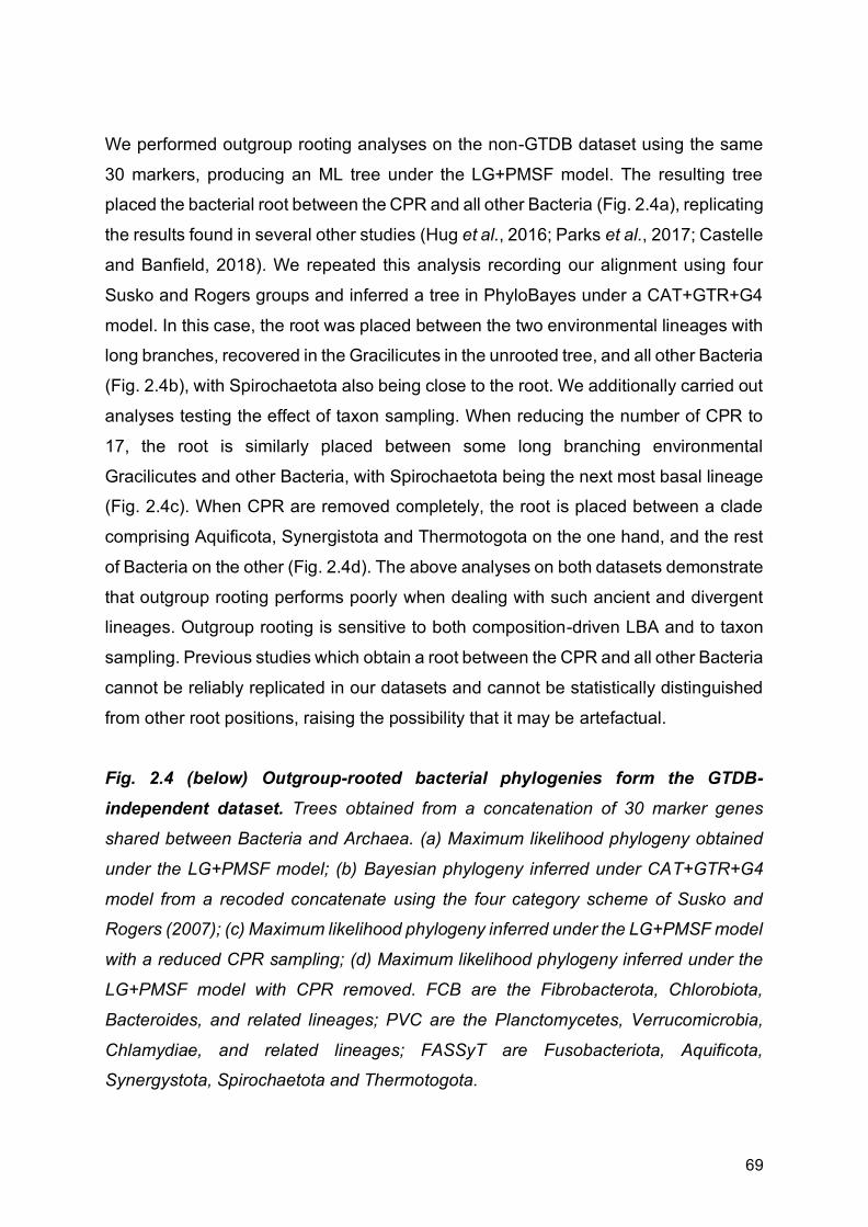
69
We performed outgroup rooting analyses on the non-GTDB dataset using the same
30 markers, producing an ML tree under the LG+PMSF model. The resulting tree
placed the bacterial root between the CPR and all other Bacteria (Fig. 2.4a), replicating
the results found in several other studies (Hug et al., 2016; Parks et al., 2017; Castelle
and Banfield, 2018). We repeated this analysis recording our alignment using four
Susko and Rogers groups and inferred a tree in PhyloBayes under a CAT+GTR+G4
model. In this case, the root was placed between the two environmental lineages with
long branches, recovered in the Gracilicutes in the unrooted tree, and all other Bacteria
(Fig. 2.4b), with Spirochaetota also being close to the root. We additionally carried out
analyses testing the effect of taxon sampling. When reducing the number of CPR to
17, the root is similarly placed between some long branching environmental
Gracilicutes and other Bacteria, with Spirochaetota being the next most basal lineage
(Fig. 2.4c). When CPR are removed completely, the root is placed between a clade
comprising Aquificota, Synergistota and Thermotogota on the one hand, and the rest
of Bacteria on the other (Fig. 2.4d). The above analyses on both datasets demonstrate
that outgroup rooting performs poorly when dealing with such ancient and divergent
lineages. Outgroup rooting is sensitive to both composition-driven LBA and to taxon
sampling. Previous studies which obtain a root between the CPR and all other Bacteria
cannot be reliably replicated in our datasets and cannot be statistically distinguished
from other root positions, raising the possibility that it may be artefactual.
Fig. 2.4 (below) Outgroup-rooted bacterial phylogenies form the GTDB-
independent dataset. Trees obtained from a concatenation of 30 marker genes
shared between Bacteria and Archaea. (a) Maximum likelihood phylogeny obtained
under the LG+PMSF model; (b) Bayesian phylogeny inferred under CAT+GTR+G4
model from a recoded concatenate using the four category scheme of Susko and
Rogers (2007); (c) Maximum likelihood phylogeny inferred under the LG+PMSF model
with a reduced CPR sampling; (d) Maximum likelihood phylogeny inferred under the
LG+PMSF model with CPR removed. FCB are the Fibrobacterota, Chlorobiota,
Bacteroides, and related lineages; PVC are the Planctomycetes, Verrucomicrobia,
Chlamydiae, and related lineages; FASSyT are Fusobacteriota, Aquificota,
Synergystota, Spirochaetota and Thermotogota.

70
Attempting to root the bacterial tree without outgroups

71
We initially tried two rooting methods without outgroups, the relaxed molecular clock
(RMC) in BEAST2 (Drummond and Rambaut, 2007; Drummond and Bouckaert,
2015), and minimal ancestor deviation (MAD) (Tria, Landan and Dagan, 2017) (see
methods). For the RMC, we performed a lognormal uncorrelated relaxed molecular
clock analysis, with the root posterior probabilities (PPs) averaged over the trees
sampled during the Bayesian molecular clock analysis using RootAnnotator. When
performed on the unaltered concatenate, we obtained a root position between CPR
and the rest of Bacteria with a PP of 0.61 (Fig. 2.5(a)). However, several other rooted
positions had comparable PPs. We performed the analysis on the recoded alignment,
with CPR reduced, and with CPR removed. The recoded concatenate recovered a
root between Fusobacteriota and the rest of Bacteria with a PP of 0.58 (Fig. 2.5(b)). A
root between the Gracilicutes and Terrabacteria+FASSyT was found for both the
concatenates with CPR reduced and removed respectively (PP = 0.66 and 0.55
respectively), although FASSyT is monophyletic in the former and paraphyletic in the
later (Fig. 2.5(c-d)). In each case, the PPs were low, with several other root positions
recovered with comparable PPs.
When rooted using MAD, the CPR-root is obtained from the tree on the unaltered and
recoded concatenates (Fig. 2.5(a-b)). When CPR is reduced, the root is between the
Gracilicutes on one side, and the Terrabacteria+FASSyT on the other (Fig. 2.5(c)).
When CPR are removed, the root falls between the Spirochaetota and the rest of
Bacteria (Fig. 2.5(d)). In all cases, the AI was high (>0.9), meaning that the best root
position was not significantly better than the next best. These analyses demonstrate
that, similar to outgroup rooting, the RMC and MAD are susceptible to artefacts due
to LBA, or are sensitive to taxon sampling.
Fig. 2.5 (below) schematic representations of rooted bacterial trees under the lognormal uncorrelated relaxed molecular clock (RMC) using an LG substitution
model, and minimal ancestor deviation (MAD) rooting. (a) RMC and MAD roots for
the unaltered concatenate; (b) RMC and MAD roots for the recoded concatenate using
the four category scheme of Susko and Rogers (2007); (c) RMC and MAD roots with
CPR reduced; (d) RMC and MAD roots with CPR removed. FASSyT are
Fusobacteriota, Aquificota, Synergystota, Spirochaetota and Thermotogota.

72
Whole-genome approaches to rooting
Given the limitations of the above methods to establish the root of the bacterial tree,
we explored another outgroup-free rooting approach using gene tree-species tree
reconciliation (David and Alm, 2011; Szöllosi et al., 2012; Szöllősi et al., 2013; Williams

73
et al., 2017). This approach has recently been applied to root the archaeal tree
(Williams et al., 2017), and similar approaches have been applied to investigate the
root of eukaryotes (Katz et al., 2012; Emms and Kelly, 2017) and to map and
characterise whole genome duplications in plants (Zwaenepoel and Van de Peer,
2019). The method works by explaining the histories of individual gene families in the
context of a shared species tree with a series of speciation, gene origination,
duplication, transfer and loss events. Since these histories depend on the position of
the root, reconciliation likelihoods can be used to estimate the most likely root, in what
can be viewed as a genome-wide extension of the classical approach used to root the
tree of life based on ancient gene duplications (Iwabe et al., 1989; Johann Peter
Gogarten et al., 1989). In addition to leveraging genome-wide data, a further
advantage is the ability to extract root signal from both gene duplications and transfers
(Szöllosi et al., 2012; Williams et al., 2017). Amalgamated Likelihood Estimation (ALE)
improves on earlier approaches by explicitly accounting for uncertainty in the gene
tree topologies and in the events leading to those topologies, while also estimating
rates of gene duplication, transfer and loss directly from the data (Szöllősi et al., 2013).
Simulations suggest that root inferences under ALE are robust to variation in taxon
sampling and that the method finds the correct root even under high levels of gene
transfer (Szöllosi et al., 2012; Williams et al., 2017), suggesting that the approach is
appropriate for the problem at hand.
The ability of the ALEml_undated algorithm to infer the correct gene tree root in the
presence of gene duplications, transfers and losses was previously investigated using
simulations (Williams et al., 2017). Briefly, gene families were simulated on a rooted
species tree using a continuous-time ODTL process (that is, a more complex model
of genome evolution than that implemented in ALEml_undated), and ALEml_undated
was used to estimate the root from subsamples of the simulated families. The
maximum likelihood root according to ALE was the correct root in 95/100 replicates,
and the log likelihood of alternative roots decreased with nodal distance from the
correct root (consistent with the pattern observed in our empirical data, see Fig. 2.7(c),
see below). In the remaining 5 cases, the maximum likelihood root was one branch
away from the true root. Analysis of empirical data suggested that ALE root inferences
are robust to (that is, consistent across) subsets of the data that vary in terms of the
rate of horizontal gene transfer or species representation in gene families (Williams et

74
al., 2017). These properties make the ALE approach appropriate for inferring the root
of Bacteria.
More broadly, species tree-aware phylogenetic methods (such as ALE) have been
shown to be of use in fixing gene tree errors (Szöllősi et al., 2013), and for ancestral
state (Williams et al., 2017) and protein (Groussin et al., 2015) inference. The
additional power of these methods derives from the use of information in the species
tree to decide between gene trees that are statistically equivalent from the point of
view of the phylogenetic likelihood. Recently, a study of gene tree rooting performance
suggested that a parsimony-based, species tree unaware DTL method (RANGER-
DTL) provided more accurate gene tree root estimates than species tree-aware,
probabilistic methods such as ALE and GeneRax (Wade et al., 2020). Gene tree
rooting accuracy is not directly related to species tree rooting accuracy, because the
information on the species tree root in ALE derives from finding the rooted species
tree that maximises the sum of reconciliation likelihoods across gene families. We
nevertheless decided to investigate, in order to understand which properties of the
available methods contribute to, and detract from, rooting accuracy more generally.
To investigate, we re-analysed the data from Figure 5 of Morel et al. (2019), who
simulated sequence alignments on known rooted gene trees. This setup differs from
that of the original study (Wade et al., 2020) in that, where possible, we start directly
from the alignment and not from independently reconstructed gene trees. We chose
this simulation setup because empirical analyses typically proceed from sequence
alignments to inferred trees and then roots. We inferred rooted gene trees roots using
ALE, GeneRax (Morel et al., 2019) (a species-tree aware method that maximises a
joint reconciliation and phylogenetic likelihood to infer rooted gene trees), TreeRecs
(a species-tree aware method that implements a parsimony approach to DTL to infer
rooted gene trees), RANGER-DTL (Bansal et al., 2018) (a species-tree unaware
method that implements a DTL parsimony approach to root input gene trees), MAD
(Tria, Landan and Dagan, 2017) (a species-tree unaware method that roots input gene
trees using minimal ancestor deviation). To quantify gene tree rooting accuracy, we
plotted the rooted Robinsons-Fold (RF) score between the inferred and true rooted
gene trees; this provides an interpretable measure of rooting accuracy even when the
true root bipartition does not appear in the inferred gene tree, as demonstrated recently

75
(Wade et al., 2020). The results (Fig. 2.6) indicate that probabilistic species tree-aware
methods (GeneRax and ALE) provide the most accurate gene tree root inferences
among the methods compared, even when the model used to infer the gene tree is
misspecified, that is, when LG is used for simulation and WAG for inference. In
particular, even when gene trees are inferred with a misspecified substitution model,
ALE gene tree rooting (mean rooted-RF = 0.258) is significantly more accurate than
RANGER-DTL (mean rooted-RF = 0.322, p <10-15 Welch Two Sample t-test) or MAD
(mean rooted-RF = 0.306, p < 10-10 Welch Two Sample t-test) when the latter methods
are provided with input gene trees obtained using the true substitution model.
Fig. 2.6 Accuracy of gene tree rooting methods. Species tree aware methods
(GeneRax, ALE, TreeRecs) are the most accurate, even when gene trees are inferred
with a misspecified (WAG) substitution model. Among species tree unaware methods,
MAD outperforms the parsimony based DTL method RANGER-DTL.
Rooting the bacterial tree using ALE
Given the ability of ALE to accurately infer rooted phylogenies, we used the method to
test the support for 62 root positions on the unrooted topology derived from the GTDB
by reconciling gene trees for 11,272 homologous gene families from the 265 bacterial
genomes. In addition to testing root positions corresponding to published hypotheses
(Table 2.5), we exhaustively tested all inner nodes of the tree above the phylum level.
The ALE analysis rejected all of the roots tested (P < 0.05) except for three adjacent
branches, lying between the two major clades of Gracilicutes and Terrabacteria (Fig.
2.7a). The position of the phylum Fusobacteriota was difficult to resolve in the tree,

76
and contributed to root uncertainty. The three candidate root branches lead to (i)
Terrabacteria+Deinococcus/Thermotoga/Synergistes; (ii) Gracilicutes; (iii)
Fusobacteriota (Fig. 2.7a). Consistent with this being the optimal root region,
alternative roots were rejected with increasing confidence as distance from the optimal
region increased (Fig. 2.7c).
Root p-value Study
CPR basal 2e-04
Hug et al. (2016), Zhu et al. (2019)
Chloroflexota basal 1e-41
Cavalier-Smith (2006)
Between Firmicutes and Actinobacteriota
9e-05
Lake et al. (2009)
Thermotoga/Synergistota/Deinococcota basal*
0.004
Planctomycetes basal 2e-26 Brochier and Philippe (2002)
Table 2.5 Support for published rooting hypotheses from our ALE analyses. *Our
unrooted topology was incompatible with some published hypotheses, including a
clade of Thermotogales and Aquificales at the root (Bocchetta et al., 2000; Battistuzzi
and Hedges, 2009).

77
Fig. 2.7 (below) Root positions determined by ALE for both the GTDB (a) and the
GTDB-independent (b) datasets. (a) Three rooted topologies from the GTDB dataset
could not be rejected by the AU test. The root falls between two major clades of
Bacteria, the Gracilicutes and the Terrabacteria, on one of three statistically equivalent
adjacent branches. AU p-values are 0.476 for Root 1, 0.336 for Root 2 and 0.658 for
Root 3. (b) Two rooted topologies from the GTDB-independent analysis that could not
be rejected by the AU test, from ALE analysis incorporating genome completeness.
AU p-values are 0.973 for Root 1 and 0.064 for Root 2. Both trees are in agreement
with each other and GTDB analysis in placing the root between Terrabacteria and
Gracilicutes, but disagree in the placement of the “FASSyT” taxa comprising
Fusobacteriota, Aquificota, Synergistota, Spirochaetota and Thermotogota. (c) For the
GTDB dataset, all tested alternative roots were rejected (Tables 2.3 and 2.4) with
likelihoods decreasing as a function of distance from the root region. Previously
proposed root positions, including the CPR root, are highlighted in red. D-T stands for
Deinococcus-Thermus; “Deltaproteobacteria” is Desulfuromonadota,
Desulfobacterota, Bdellovibrionota, and Myxococcota. FCB are the Fibrobacterota,
Chlorobiota, Bacteroidota, and related lineages; PVC are the Planctomycetota,
Verrucomicrobiota, Chlamydiota, and related lineages; DST are the Deinococcota,
Synergistota and Thermotogota; ACD are Aquificota, Campylobacterota, and
Deferribacterota; FA are Firmicutes and Actinobacteriota; FASSyT are
Fusobacteriota, Aquificota, Synergystota, Spirochaetota and Thermotogota.

78
79
On the non-GTDB dataset, we calculated the gene family likelihoods of 11,781
homologous gene families derived from our 342 bacterial genomes, under a set of 60
candidate root positions on different unrooted species topologies obtained from
different tree construction methods (see Methods). Of the 60 number of rooted
phylogenies tested, all but two were rejected (Fig. 2.7b) (P < 0.05). Both of these
topologies had candidate root branches between the Gracilicutes and the
Terrabacteria, as in the GTBD analysis. They also both recovered monophyletic
FASSyT, although its position could not be resolved, lying on either side of the root.
The two candidate root branches therefore lead to (i) Terrabacteria; (ii) Gracilicutes.
In order to further mitigate against LBA caused by CPR, we additionally performed
ALE (including the inference of the single gene trees following the same pipeline) with
the CPR removed. The resulting root position was identical to that with the CPR
included. CPR therefore does not seem to affect the ALE rooting results.
MCL families represent, within the limitations of the clustering approach discussed
above, an unbiased view of gene family diversity for the set of genomes we analysed.
We therefore base all our analyses, except those regarding the functional annotation
of LBCA, on the MCL families. By contrast, our COG families are useful for functional
reconstruction (see Chapter 3 for details), but are perhaps less well suited for
investigating other aspects of bacterial evolution because they are constructed only
from proteins that could be annotated with eggNOG-mapper. However, since gene
clustering methods are not consummate and each has strengths and weaknesses, we
also investigated the root signal from the COG families (see Chapter 3 for details on
the construction of the COG families). This analysis resulted in a root region of four
adjacent branches, comprising the root region from the MCL analysis (3 branches)
plus one additional branch, in which Spirochaetota branched on the Terrabacteria side
of the root (Table 2.6). This slightly expanded root region is likely due to the reduced
resolution of the smaller set of COG families in comparison to the full analysis.
Root name LLs AU
Fusobacteriota root (398) -7.3 0.589
Fusobacteriota on Terrabacteria side (527) 7.3 0.519
Fusobacteriota on Gracilicutes side (528) 13.6 0.432

80
Fusobacteriota and Spirochaetota on Terrabacteria side (520) 32.1 0.251
DST root (464) 103.7
0.008
Cyanobacteria on Gracilicutes side (517) 215.8
1e-09
Dormibacterota/Chloroflexota+CPR (510) 372.2
7e-06
CPR root (496) 425.2
2e-67
Dormibacterota/Chloroflexota (505) 630.7
5e-05
Omnitrophota/Verrucomicrobia/Planctomycetes (492) 674.4
2e-04
Fibrobacteria/Bacteroidetes/Marinisomatota (511) 1000.7
3e-71
Table 2.6 AU-test results for an ALE root analysis using 3595 COG families.
While it has not been possible to completely resolve the root, all analyses recover a
root region between Gracilicutes and Terrabacteria. A similar root was previously
reported (Raymann, Brochier-Armanet and Gribaldo, 2015; Adam, Borrel and
Gribaldo, 2018). However, this analysis did not include the CPR, which has been
recently suggested (Hug et al., 2016; Zhu et al., 2019) to represent the earliest
diverging bacterial lineage. Further, our ALE analyses consistently recovered CPR
nested within the Terrabacteria, suggesting that the CPR root is a long branch
attraction artefact.
Is bacterial evolution treelike? How much of bacterial evolution can be explained by the concept of a rooted species
tree? Horizontal gene transfer (HGT) is frequent in prokaryotes, and published
analyses indicate that most or all prokaryotic gene families have experienced HGT
during their history (Dagan and Martin, 2007; Williams et al., 2017). This implies that
there is no single tree that fully describes the evolution of all bacterial genes or
genomes (Doolittle, 1999; Doolittle and Bapteste, 2007). Extensive HGT is existentially
challenging for concatenation, because it greatly curtails the number of genes that
evolve on a single underlying tree (Dagan and Martin, 2006). Phylogenetic networks

81
(Doolittle and Bapteste, 2007; Alvarez-Ponce et al., 2013) were the first methods to
explicitly acknowledge non-vertical evolution, but can be difficult to interpret
biologically. Gene tree-species tree reconciliation integrates tree and network-based
approaches by modelling both the horizontal components of genome evolution (a fully
reticulated network allowing all possible transfers) and the vertical trace (a common
rooted species tree). This framework enables us to quantify the contributions of vertical
and horizontal processes to bacterial evolutionary history.
Our analyses (Fig. 2.8) reveal that most bacterial gene families present in at least two
species (9678/10518 MCL families, 92%) have undergone at least one gene transfer
during their evolution; only very small families have escaped transfer entirely (Fig. 2.9).
Consistent with previous analyses (Jain, Rivera and Lake, 1999; Williams et al., 2017),
transfer rates vary across gene functional categories, with genes functioning in
defence mechanisms (such as antibiotic biosynthesis) and the production of secondary
metabolites being the most frequently transferred, and those involved in translation
and the cell cycle the least (Fig. 2.8(b), Table 2.2). Despite this accumulation of HGT,
most gene families evolve vertically the majority of the time, in that 66% of transmission
events (mean, MCL gene families) seem to evolve vertical along the species tree.
There are a number of caveats that should be considered when interpreting the results
of these analyses. Our inference of HGT events is likely to be an underestimate,
because our broad but sparse taxon sampling does not allow us to detect transfers or
recombination within strains. Similarly, orthologous replacement may resemble vertical
transmission and thus go undetected, leading to a further underestimate of transfer
events. Additionally, we do not specifically consider pseudogene in this analysis.
Pseudogenes are segments of non-functional DNA which resemble functional genes,
often caused by loss of gene function, and may cause us to overestimate transfer
events. However, while not modelled explicitly, pseudogenes should be modelled as a
form of gene loss in ALE, and therefore should not have a profound effect on the
analyses, unless they make up large part of the genomes analysed. We also note that
the inclusion of plasmids in our dataset may also affect transfer rates as plasmids have
are often involved in HGT (Gauri et al. 1994; Varga et al. 2016).

82
While we demonstrate a high proportion of vertical signal, which implies that a tree
may be a suitable way to describe bacterial evolution, we still detect a large amount of
HGT. This poses an important question as to how much transfer is necessary before
a tree is no longer an apt description of bacterial evolution. Furthermore, the
directionality of the transfers may have an impact on our ability to infer a species tree.
If such HGTs are largely random in nature, then a tree is still likely recoverable.
However, if the transfers are highly directional in nature, such directional biases may
negatively impact our tree inferences. Detecting the directionality in our dataset in
difficult, and we have not been able to do it here. However, while it may be difficult to
detect the directionality, we may use simulations to determine the effect that both the
levels and directionality of HGTs have on our ability to recover a tree. By performing
simulations where the level and directionality of HTGs within the simulated gene trees
would vary, we could explore the extent to which a tree is recoverable, and discern the
point at which the levels HGT and directionality were so great that a tree is longer
inferable. This would give us a sense of what percentage of horizontal signal could be
considered so pervasive that a tree is no longer adequate to describe the data in
question.

83
Fig. 2.8 The verticality of bacterial genome evolution. (a) The rooted bacterial
species tree (Fig. 2.7) with branches coloured according to verticality: the fraction of
genes at the bottom of a branch that descend vertically from the top of that branch
(see inset; V = vertical, O = origination, T = transfer into a branch; see Methods). Node
heights reflect relative time order consistent with highly-supported gene transfers. (b)
Verticality by COG functional category: that is, the proportion of gene tree branches
that are vertical V/V+T for COG gene families. Genes involved in information
processing, particularly translation (J), show the highest verticality (median 0.69),
while genes involved in cell defence mechanisms (V, such as genes involved in
antibiotic defence and biosynthesis) are most frequently transferred. (c) For a given
genome, this combination of vertical and horizontal processes gives rise to a
distribution of gene residence times, reflecting how far back in bacterial history genes
are retained. Across all phyla examined, 82% of genes on sampled genomes trace
back to the crown group radiation of that phylum. FCB are the Fibrobacterota,
Chlorobiota, Bacteroidota, and related lineages; PVC are the Planctomycetota,
Verrucomicrobiota, Chlamydiota, and related lineages; DST are the Deinococcota,

84
Synergistota, and Thermotogota; ACD are Aquificota, Campylobacterota, and
Deferribacterota.
Fig. 2.9 The relationship between verticality and gene family size. Most gene
families have experienced many transfers. Verticality varies with gene functional class,
but families with very low transfer rates are small; these might represent young families
that have not yet had enough time to experience gene transfer.
Mapping the branches of the gene trees onto the species tree demonstrates that the
optimal tree provides an apt summary of much of bacterial evolutionary history, even
for the deepest branches of the tree (Creevey et al., 2004; Koonin, Wolf and Puigbò,
2009; Puigbò, Wolf and Koonin, 2010). From the gene’s eye view, gene families evolve
neither entirely vertically nor horizontally: core genes are occasionally transferred, and
even frequently exchanged genes contribute useful vertical signal; for example, the

85
median number of genes that evolve vertically on a branch of the species tree is close
to 998.92 (Table 2.7), far greater than the number of genes that have been
concatenated at the level of all Bacteria. From the perspective of the genome,
constituent genes have different ages, corresponding to the time at which they
originated or were most recently acquired by gene transfer, within the resolution of our
taxonomic sampling. This analysis indicates that, on average, 82% of the genes on all
genomes from adequately represented phyla (5 or more genomes) trace back to the
crown group radiation of that phylum, though all genomes retain a smaller proportion
(10.3-26.7%) of genes that have descended vertically from the stem lineage of their
phylum or even earlier (Figure 2.8(c)).
Root branch 1 2 3
Median singleton support per branch
999.09 998.47
999.21
Singleton support for branches subtending the root
98.259, 140.45
91.56, 151.09
95.25,117.16
Mean verticality 0.68136
0.68137
0.6818
Table 2.7 Singleton support (the number of genes that evolve vertically from one
end of a branch to the other) on the credible set of rooted trees. Root numbers
correspond to the three root branches depicted in Fig. 2.7(a)).
Caveats of ALE While we believe that ALE is a robust method, and an improvement on previous
analyses, there are some caveats of the method which must be considered. In its
current implementation, a two-step process is used, whereby gene trees must be must
be independently inferred using species-tree unaware methods, with the conditional
clade probabilities being calculated for a sample of trees (e.g. posterior sample from
an MCMC, or a bootstrap sample) and reconciled with the species tree to create a
gene tree amalgamated from clades present in the gene tree sample. Ideally, however,
the inference of gene trees would happen jointly with the reconciliation and therefore
be specie-tree aware.

86
Another caveat relates to the manner in which we infer the clusters which are
interpreted as gene trees. As we briefly discuss above, MCL clustering has no
biological basis, and we must accept a trade-off between inferring clusters that are too
liberal, and therefore contain distant or unrelated sequences, and those that are too
conservative and break-up larger gene families. As previously discussed, we have
attempted to partially address these issues by testing different inflection parameters
in MLC (which controls the size of the clusters) and by performing analyses using
COGs (see details above). However, more extensive analyses could be done to test
the effect of gene clustering; for example, repeating the full analyses using different
inflation parameters (although this would be costly and time-consuming), or by
repeating the analyses using a different clustering approach, e.g. using HI Fix (Miele
et al., 2012). This is very an important area to explore further given the extent to which
our analyses rely on the accurate creation of gene families.
While we believe ALE to be robust to taxon sampling, further exploration may be
needed to determine the extent of the effects on our results. We have partially done
this in the GTDB-independent dataset by not only increasing the number of taxa, but
by sampling diversity differently, leading to oversampling of some phyla (e.g. the CPR)
and under sampling of others (e.g. the Firmicutes) with respects to the focal dataset,
and have obtained similar results in both cases. However, multiple replicated datasets
would be needed to test this further. Furthermore, as we note above, the sparse
taxonomic sampling could be leading to an underestimate of transfers, as we cannot
detect transfers or recombinations within closely related lineages. Increasing taxon
sample size could alleviate this problem, although would be computationally
expensive.
A further caveat is that extreme ratios of DTLs (i.e. extreme values of D/T, D/L, T/D,
T/L, L/D or L/T) within the DTL model used by ALE, particularly in small gene families,
could be affecting the root positions recovered in the analysis. A possible way to
explore this would be to plot all DTL rate ratios and progressively remove the most
extreme gene families to see how it would affect the analyses. This would be
analogous to the removal of the most compositionally heterogeneouss sites in an
alignment.

87
Is the universal root in Bacteria? As noted above, while the consensus view is that the root of the tree of life lies between
Archaea and Bacteria, the evidence is limited and can be hard to interpret (Iwabe et
al., 1989; J. P. Gogarten et al., 1989; Brown and Doolittle, 1995; Zhaxybayeva,
Lapierre and Gogarten, 2005; Gouy, Baurain and Philippe, 2015), and alternative
hypotheses in which the universal root is placed within Bacteria have been proposed
(Cavalier-Smith, 2006; Skophammer et al., 2007; Lake et al., 2009). This may
therefore raise questions about the validity of our analyses. However, a strength of the
ALE method used here is that it does not need an outgroup. Thus, if Archaea do indeed
branch within the Bacteria, then the root of Bacteria presented here would in fact be
the universal root of all life. In this scenario, Archaea would be among the number of
groups we did not sample in our dataset, due to necessarily needing a small enough
dataset to be tractable. Thus, our analyses are compatible with a bacterial root of life.
Nonetheless, bacterial phyla not included in our analyses were small, while the
omission of Archaea would represent a much greater loss of data. We therefore
conducted a preliminary analysis where we rooted the tree of life, including the
Archaea, using ALE.
We used 293 species, 149 Bacteria derived from non-GTDB bacterial dataset, and
144 Archaea from (Kellner et al., 2018). These were selected evenly from across the
tree in order to capture the full diversity. We did not include the eukaryotes as it is
widely accepted that they are derived from an endosymbiotic event between Bacteria
and Archaea (Embley and Martin, 2006; Martin, Garg and Zimorski, 2015; López-
García, Eme and Moreira, 2017; Roger, Muñoz-Gómez and Kamikawa, 2017; Eme et
al., 2018), and that the root is almost certainly within the prokaryotes. As we wish
primarily to explore possibilities for the root position within the tree of life, and not to
elucidate anything concerning the origins or phylogenetic position of Eukarya, we
judged it not essential that they be included in the analysis, especially as the long
branch leading to the eukaryotes could be problematic. The genomes for the species
were downloaded from NCBI GeneBank.
We concatenated 51 orthologues and inferred a tree under the LG+C60 model in IQ-
Tree. Within the Archaea, Euryarchaeota and TACK were recovered. DPANN is not
monophyletic in this tree, forming successive outgroups to the other Archaea. This

88
may be due to LBA, as has been shown in other cases (Gouy, Baurain and Philippe,
2015; Williams et al., 2017). Within Bacteria, the topology broadly follows that of the
previous analyses. However, “FASSyT'' is not monophyletic, with Aquificota and
Thermotogota being sister to Terrabacteria/CPR, and Spirochaetota and
Fusobacteriota being recovered within Gracilicutes. Synergistota, along with a few
Terrabacteria, have been pulled to the base of the bacterial clade. Due to the long
branches between Bacteria and Archaea, some of these topological incongruencies
may be due to LBA. Interestingly, the CPR are found within Terrabacteria, and not
close to the branch leading to the Archaea. This is further evidence that tree topologies
where CPR are basal or near the branch to Archaea (Hug et al., 2016; Parks et al.,
2017; Castelle and Banfield, 2018) are likely due to topological artefacts.
In order to test root positions using ALE, homologous gene families with paralogues
were generated using HiFix (Miele et al., 2012). The sequences were aligned in Mafft
(using the auto option) and trimmed in BMGE (using BLOSUM30), and trees of these
gene families were inferred in IQ-Tree under the LG+C60 model. 10 root positions
were tested. All roots could be rejected except for the root between Archaea and
Bacteria (Fig 2.10). As we have demonstrated that ALE seems to not be affected by
LBA artefacts, it is likely that the root of life is between Archaea and Bacteria. However,
it must be noted that a much more thorough analysis of the data, with extensive
topology testing, use of different taxon samplings, and the testing of a much wider
range of roots, would be needed to bring confidence to this result.

89
Fig. 2.10 Rooted phylogeny of Archaea and Bacteria. The maximum likelihood
phylogeny obtained under the LG+C60 model on a concatenation of 51 marker genes
shared between Bacteria and Archaea. The root position between Bacteria and
Archaea was the only root position tested in ALE and could not be rejected by the AU
test (P < 0.05).
2.4 Conclusions
We place the last bacterial common ancestor between two major clades, Terrabacteria
and Gracilicutes, although we could not resolve the position of several smaller
lineages, notably the Fusobacteriota, in relation to those major radiations.
Fusobacteriota currently comprise anaerobic free-living, pathogenic and commensal
diderm bacteria (Brennan and Garrett, 2019), and a clear direction for future work will
90
be to place them on the rooted bacterial tree, particularly if more basal members of
this lineage come to light. We have found these results to be consistent across
different datasets, demonstrating that, while there is still some lack of resolution in the
deepest parts of the tree, there is strong and consistent signal supporting the higher
level relationships we have demonstrated here, and that key features of the topology
are not composition-driven LBA artefacts or caused by taxon sampling.
In contrast to recent outgroup-rooted analyses (Hug et al., 2016; Parks et al., 2017;
Castelle and Banfield, 2018; Zhu et al., 2019), we found no support for a root on the
CPR branch; instead, our analysis suggests that this lineage evolved within
Terrabacteria, from a common ancestor with Chloroflexota. Analyses which recover
CPR in a basal position are likely artefacts, as their position changes when varying the
taxon sampling, or using recoding to account for compositional heterogeneity and
saturation. Furthermore, we demonstrate that outgroup rooting, at least when applied
in this case where the distance to the outgroup is great, is unable to distinguish
between different root positions and is highly susceptible to LBA artefacts. Other
methods, including the relaxed molecular clock and MAD rooting are similarly affected
by these issues. Probabilistic species tree-aware methods, such as ALE, are less
susceptible to LBA artefacts and differences in taxon sampling, can utilise a greater
range of data, and seem to give accurate results compared to other rooting methods.
Our analyses further suggest that, despite extensive horizontal gene transfer, a
phylogenetic tree is an apt representation of bacterial evolution in the sense that most
bacterial gene families evolve vertically most of the time.

91
Chapter 3
Ancestral reconstruction of the last
bacterial common ancestor A version of this chapter forms part of a paper under revision in collaboration with
Adrián A. Davín, Tara Mahendrarajah, Anja Spang, Philip Hugenholtz, Gergely J.
Szöllősi, and Tom A. Williams. Gareth A. Coleman is the first author of the paper. The
project was conceived by TAW, GJSz, PH, AS, GAC and AAD. Protein annotations
and generation of COG families were carried out by TM, AS and TAW. COG
reconciliations carried out by GJSz. Metabolic reconstruction, including metabolic
maps and figures, were carried out and produced by GAC. Writing and interpretation
of metabolic reconstructions were carried out by GAC, AS and TM.
Paper preprint as:
Coleman, G.A., Davín, A.A., Mahendrarajah, T., Spang, A.A., Hugenholtz, P.,
Szöllősi, G.J. and Williams, T.A., 2020. A rooted phylogeny resolves early bacterial
evolution. bioRxiv.
BioRxiv preprint for the paper can be found here:
https://www.biorxiv.org/content/10.1101/2020.07.15.205187v1

92
Abstract Bacteria are the most abundant and metabolically diverse cellular lifeforms on Earth and have had a profound impact on the physical environment. To understand the evolution of complex interaction between the biosphere and geosphere, we must understand the nature of the earliest bacterial cells, including the last bacterial common ancestor (LBCA). Building upon previous work, we use a rooted phylogeny inferred from the modelling of genome evolution at the level of gene duplication, transfer and loss events to reconstruct the ancestral gene content of LBCA. We infer that the LBCA possessed core carbon metabolic pathways, including glycolysis, the reverse tricarboxylic acid cycle and the pentose phosphate pathway, with the possibility of fixing carbon via either the Wood-Ljungdahl or reverse tricarboxylic acid cycles. In addition, we predict that LBCA was a free-living flagellated, rod-shaped cell featuring a double membrane with a lipopolysaccharide outer layer, bacterial phospholipids, a Type III CRISPR-Cas system, Type IV pili, and the ability to sense and respond via chemotaxis.
93
3.1 Introduction
Bacteria inhabit almost all known habitats and ecosystems, and employ huge diversity
of physiologies to adapt to these diverse environments. As such, they perform vital
roles in biogeochemical cycles and have had a profound impact on the physical Earth
throughout history. To understand how such systems, both biological and
biogeochemical, have evolved we must answer questions pertaining to the physiology
and habitat of the earliest life, including that of the last bacterial common ancestor
(LBCA). In particular, how complex or “modern” early prokaryotic cells were in
comparison to present day cells, what kind of environment they lived in, and how these
cells derived energy from their environment.
One of the central questions involves discerning the core carbon metabolism present
in LBCA, specifically to determine if LBCA had the ability to fix carbon, and if so which
pathway it would have used. Modern bacteria fix carbon using several different
pathways, including the Calvin cycle, the 3-hydroxypropionate (3-HP) bicycle and
variations thereof, the Wood-Ljungdahl pathway (WLP) and the reverse tricarboxylic
acid (TCA) cycle, the latter two of which have been suggested to have emerged early
in the history of life. The WLP, found in both methanogenic archaea and acetogenic
bacteria, is thought to be one of the most ancient carbon fixation pathways on the
basis of both biogeochemical and phylogenetic arguments (Fuchs, 2011; Sousa and
Martin, 2014; Weiss et al., 2016; Williams et al., 2017; Adam, Borrel and Gribaldo,
2018). Notably, the key enzyme complex of the pathway, CODH/ACS (CO
dehydrogenase/acetyl-CoA synthase), is conserved in both domains, and is predicted
to have been present in both the archaeal (Williams et al., 2017; Adam, Borrel and
Gribaldo, 2018) and bacterial common ancestors (Adam, Borrel and Gribaldo, 2018).
The reverse TCA cycle has also been suggested as a possible ancient carbon fixation
pathway (Wächtershäuser, 1990; Cody et al., 2001; Smith and Morowitz, 2004;
Nunoura et al., 2018), given the widespread presence of the TCA cycle in modern
Bacteria, and that it may function in both the oxidative and reductive direction. Some
combinations of pathways have been suggested, notably a coupling of both the TCA
cycle and the 3-HP bicycle (Marakushev and Belonogova, 2011, 2013).

94
In addition to the central carbon pathways, there are many questions regarding
LBCA’s structural morphology and its ability to respond to the environment it inhabited.
These include the nature of the cell envelope and whether it possessed all the key
components found in modern Bacteria, the degree to which it could sense
environmental stimuli, and whether or not it was a motile cell. Modern Bacteria typically
have cell membranes comprising a bilayer composed of G3P phospholipids with fatty
acids attached via ester bonds, which contrast with the G1P phospholipids with
isoprenoids attached via ether bonds found in Archaea (Lombard, López-García and
Moreira, 2012b). This “lipid divide” (Koga, 2011) has been touted as a hallmark
difference between Archaea and Bacteria, implying that the earliest Bacteria would
have had bacterial-type lipids, although recent evidence has challenged this
assumption (Sinninghe Damsté et al., 2002; Weijers et al., 2006; Damsté, Rijpstra, et
al., 2007; Goldfine, 2010; Villanueva, Schouten and Damsté, 2017; Caforio et al.,
2018; Coleman, Pancost and Williams, 2019). In addition, modern bacteria possess
peptidoglycan cell walls, and many have an additional outer membrane. This latter
trait is thought to be a derived character within Bacteria (Lake, 2009; Gupta, 2011;
Tocheva, Ortega and Jensen, 2016), though recent work suggests that the outer
membrane arose early in bacterial evolution and may have been present in LBCA
(Sutcliffe, 2010; Antunes et al., 2016; Megrian et al., 2020). Furthermore, many
scenarios describing the evolution of the earliest prokaryotic communities envisage
early cells as non-motile living on substrates, for example within the pores of
hydrothermal vents (Martin and Russell, 2007; Lane and Martin, 2012; Sousa et al.,
2013; Sousa and Martin, 2014), while other evidence points to the early appearance
of the flagellum (Liu and Ochman, 2007a, 2007b), and other machinery for motility and
sensory systems (Melville and Craig, 2013).
To answer these questions, we must be able to predict the suite of genes LBCA
possessed. The DTL method implemented in ALE, as described in Chapter 2, can
count the proportion of sampled reconciliations in which a given gene family is present
in a given node, from which a probability of the presence of that gene can be
calculated. This allows us to predict the gene content, and therefore reconstruct the
metabolic capabilities, of any given node in the tree. The use of this method allows the
incorporation of more data about evolutionary history due to use of gene tree
topologies, and can distinguish between duplication, transfer or loss with respect to
95
changing genome size (Szöllősi et al., 2013; Szöllősi, Tannier, et al., 2015). As the
method takes HGT into account, it circumnavigates the ‘genome of Eden’ problem,
where ancestral genomes of unrealistic size are predicted due to transfer events
(Dagan and Martin, 2007). It must be noted that we can only infer the presence of
genes that are in extant genomes, and that the extinction of gene families will leave
any reconstructions incomplete. This means that, while we can model the probability
of gene family extinction to correct estimated genome size, and infer what modern
features ancient life exhibited and the evolutionary timing of appearances of these
features, we cannot infer the full potential metabolic capabilities of any ancestral
organism. This method has been used in previous research to reconstruct the
metabolic capabilities of LACA, inferring it to be an anaerobe which may have used
the WLP to fix carbon (Williams et al., 2017). In this study, we use the ALE approach
to attempt to reconstruct the gene content and metabolic capabilities of LBCA.
3.2 Methods
Taxon sampling All analyses and metabolic reconstruction in this chapter were carried out based on
the analysis of the GTDB dataset and associated gene family reconciliations detailed
in Chapter 2.
Protein and protein family functional annotation Protein sequences from all genomes in our GTDB taxon sample were annotated using
a variety of databases. Functional annotations were obtained using hmmsearch
v3.1b2 (settings: -E 1e-5)(Finn, Clements and Eddy, 2011) against KOs from the
KEGG Automatic Annotation Server (KAAS) (Aramaki et al., 2020). Additionally, all
proteins were scanned for protein domains using InterProScan (v5.31-70.0; settings:
--iprlookup --goterms) (Jones et al., 2014). Multiple hits corresponding to the individual
domains of a protein are reported using a custom script. For the functional annotation
of the 4256 COG families investigated in our ancestral reconstructions, we assigned
KOs using a majority rule: i.e. we assigned the KO that was reported in > 50% of the
sequences comprising each of the COG families yielding a COG-to-KO mapping file.

96
Subsequently, we mapped COG descriptions, COG Process/Class, Category
description, kegg id, kegg description, and kegg pathway to the COG-to-KO mapping
file. COG descriptions were collected from the root annotations downloaded at
EggNOG (v5.0.0)(Huerta-Cepas et al., 2019). COG functional category and
Process/Class descriptions were derived from EggNOG (v4.0) (Tatusov et al., 2003;
Huerta-Cepas et al., 2016). KO pathways were manually curated based on an KO-to-
pathway mapping file, and were subsequently mapped to the respective KO.
COG gene families for ancestral gene content reconstruction We built a set of gene families based on the COG (Tatusov et al., 2003) database for
ancestral functional inference. To do so, we annotated each genome in the dataset
using eggNOG-mapper v2 (Huerta-Cepas et al., 2017), then clustered proteins into
families based on their COG annotations. For proteins annotated with more than one
COG category (8% of proteins), we included the protein in both COG families. This
resulted in 4256 COG families, of which 3723 had 4 or more sequences. COG families
are ideal for ancestral reconstruction because they comprise all of the sequences on
extant genomes that can be annotated with a given unambiguous function from the
COG ontology. In addition, the hierarchical nature of the COG classification
(comprising gene family annotations nested within 23 broader functional categories)
enabled us to explicitly model the different evolutionary ages of gene functional
classes as part of the analysis, by using category-specific root origination priors (see
below).
Root gene mapping approach
To estimate root presence posterior probabilities (PPs) for each gene family for each
of the three supported roots, we first estimated the root origination prior (O_R) by
maximum likelihood, finding the O_R value that maximises the total reconciliation
likelihood summed over all gene families. We then used the global ML O_R value to
calculate the root presence posterior probabilities for each family; that is, the
probability that one or more copies of a given gene family were present at the root,
given the ML O_R value. These indicated that families with different functions varied
widely in terms of root presence probability, in agreement with established theory
(Jain, Rivera and Lake, 1999); for example, proteins involved in translation (J) had the
highest root presence probabilities among the functional classes investigated (Table

97
3.1). We therefore estimated root origination rates independently for each of the 23
COG functional categories, and used these rates to estimate the posterior probability
of presence at the root node for each gene family. Initial gene content and metabolic
inferences at particular nodes were based on gene families with a PP of >0.95 at that
node. This approach is conservative and can result in a range of PP values for different
proteins within a metabolic pathway. Therefore, we manually investigated the PPs of
key pathways identified from the initial PP cut-off and inferred the presence of specific
pathways or functional modules if most its components were found with PP >0.5.
COG category
No. at root (flat
prior) % at root (flat prior)
No. at root (ML prior, PP
>= 0.95)
% at root (ML prior,
PP >= 0.95)
No. at root (ML prior, PP
>= 0.8)
% at root (ML prior, PP >=
0.8)
No. of families
in category
ML O_R COG
category description
J 3 1.69 125 70.62 157 88.7 177 6739.434
Translation ribosomal structure and biogenesis
F 1 0.98 57 55.88 89 87.25 102 5294.183 Nucleotide transport and metabolism
L 4 2.23 42 23.46 100 55.87 179 1468.033 Replication recombination and repair
H 3 1.82 26 15.76 85 51.52 165 1393.54 Coenzyme transport and metabolism
- 0 0 0 0 1 50 2 1290.933
N 1 1.43 18 25.71 38 54.29 70 1270.758 Cell motility
E 2 0.88 45 19.91 116 51.33 226 1146.195 Amino acid transport and metabolism
D 2 4.44 7 15.56 16 35.56 45 850.0477 Cell cycle
P 4 2.01 16 8.04 64 32.16 199 650.1995 Inorganic ion transport and metabolism
M 1 0.71 15 10.64 49 34.75 141 625.6504
Cell wall/membrane/envelope biogenesis
C 4 1.67 22 9.21 57 23.85 239 581.1914
Energy production and conversion
G 3 1.61 11 5.91 43 23.12 186 513.1411
Carbohydrate transport and metabolism
I 0 0 1 1.3 12 15.58 77 457.9165 Lipid transport and metabolism

98
U 1 1.2 3 3.61 12 14.46 83 364.7495 Intracellular trafficking
K 1 0.76 3 2.29 10 7.63 131 260.9843 Transcription
O 0 0 1 0.81 9 7.32 123 256.3592 Post-translational modification
S 6 0.44 9 0.66 69 5.02 1374 163.0099 Function unknown
B 0 0 0 0 1 20 5 126.1288 Chromatin structure and dynamics
T 0 0 1 1.14 3 3.41 88 125.0658 Signal transduction mechanisms
V 0 0 0 0 0 0 32 76.72498 Defense mechanisms
Q 0 0 0 0 0 0 73 1.196214 Secondary metabolites
A 0 0 0 0 0 0 4 1
RNA processing and modification
Z 0 0 0 0 0 0 2 1 Cytoskeleton
Table 3.1 Estimated root origination rates and root presences by COG functional category, including COGs recoveredas a percentage of the number of gene families in the average extant bacterial genome (indicated by “% at root”).
Impact of root branch on LBCA gene content The credible set of root branches from the ALE analysis comprised three adjacent
branches at the centre of the tree (Chapter 2, Figure 2.7(a)). The difference between
these three root positions relates to the placement of Fusobacteriota, either as the root
branch or as the most basal split on either the Gracilicutes or Terrabacteria+DST
“sides” of the rooted tree. We therefore estimated root PPs for COG families on all
three branches; Supplementary Table 1 provides root PPs under all three roots and
indicates when genes were present in 1, 2, or all 3 candidate root positions.
Metabolic comparisons Results from the PP analysis were used as the framework for metabolic comparisons
and reconstruction of the proteome of LBCA and to explore occurrence of gene
families across the tree (subsequent nodes are dealt with in Chapter 4). First, the
occurrence of an individual COG family across each taxon was counted in R (v3.6.3)
(Supplementary Table 1). This binary presence/absence matrix was combined with

99
the PP values for Nodes corresponding to the CPR, Chloroflexota+CPR,
Chloroflexota, Terrabacteria, DST+Terrabacteria, Gracilicutes-Spirochaetota,
Gracilicutes+Spirochaetota, Root 1, Root 2, and Root 3, filtered with a cutoff of
PP>0.50. The combined count table was summarised using the ddply function of the
plyr package (v1.8.4), which was used to summarise the counts across each
phylogenetic cluster, node, and root. Data is visualised in a heatmap generated using
the ggplot function with geom_tile and facet_grid of the ggplot2 package (v3.2.0).
Heatmap categories for pathways were scaled based on the number of COG families,
results were plotted using the grid.draw function of the grid package (v3.6.3).
Heatmaps were manually merged with a representative tree in Adobe Illustrator
(v22.0.1).
3.3 Results and Discussion
Estimating the size of LBCA’s genome For the following discussion regarding metabolic reconstructions, we refer to the three
branches in the root region as Root 1, Root 2, and Root 3 respectively, as shown in
Figure 2.7(a) (Chapter 2). Based on the root placement and estimated rates of gene
family extinction (Williams et al., 2017), we predict that LBCA encoded 1292.6-2142.9
COG family members, the majority of which (median estimates 65-69.5%; 95% CI 57-
82%) survived to be sampled in at least one present day genome. Based on the
relationship between COG family members and genome size for extant Bacteria
(Pearson’s r = 0.96, P = 8 x 10-153), we estimate the genome size of LBCA to be 2.69Mb
+/- 0.4Mb (standard error) for Root 1 (Fusobacteriota with Terrabacteria; 2.59Mb +/-
0.41Mb for Root 2 (Fusobacteriota with Gracilicutes), and 1.6 +/- 0.5Mb for Root 3
(Fusobacteriota basal).
Information processing, cell division and signaling In what follows, we provide probability ranges across the three branches of the root
region for the presence of key genes. One caveat of our analyses is that the method
has limited power to distinguish between ancestral presence in LBCA as opposed to
origin on an early descendant branch followed by gene transfer; this may contribute to
100
the mapping of some combinations of gene families to LBCA that are likely to be
ancient but, on physiological grounds, are unlikely to have coexisted in a single cell
(see below).
A large number of genes that can be mapped back to LBCA in all three roots with
PPs>0.5 are involved in informational processing and storage machineries such as
translation, transcription and replication, with many being highly supported (PP>0.95).
This includes the majority of ribosomal proteins, tRNA synthetases and genes involved
in their biosynthesis. We also recovered DNA polymerase I, III and IV, DNA
topoisomerase type I, and DNA ligase. Additionally many exonucleases,
endonucleases and ribonucleases are recovered, as well genes for base excision
repair, nucleotide excision repair, mismatch repair and homologous recombination
(Supplementary Table 1).
Furthermore, we detected many genes for cellular processes and signalling, including
cell division, signal transduction, membrane transport, intracellular trafficking,
chemotaxis and cellular mobility. For example, the PP for the presence of the cell
division proteins FtsZ, FtsQ and FtsA (K03531, K03589, and K03590) were >0.9 in
each root. A small number of proteins involved in signal transduction, i.e. two-
component regulatory systems, had a PP>0.95, with many more genes recovered with
a PP>0.5. Additionally, we recover genes for components of 16 ABC transporters
across all three roots, with 23 recovered in both Roots 1 and 2 (PP >0.5). We also
recover evidence for the bacterial secretion system, with four proteins of Sec system
having a PP>0.8 across all three roots. Two of these, SecY (K03076) and SecD/F
(K12257) had a PP>0.95 in roots 1 and 2. Additionally, the GspD (K02453) and GspJ
(K02459) subunits of secretion system II were recovered with a PP>0.8 in all three
roots, or with a PP >0.95 in Root 1 and 2, respectively (Supplementary Table 1).
Cell envelope and motility One interesting aspect of prokaryotic evolution is the so-called ‘lipid divide’ (Koga,
2011) Typically, Archaea have G1P phospholipids with ether bonds and isoprenoid
chains, which are often membrane spanning (Lombard, López-García and Moreira,
2012b). Bacteria, on the other hand, possess G3P phospholipids, typically with ester
bonds and fatty-acid chains, that form bilayers (Lombard, López-García and Moreira,

101
2012b). While several recent studies indicate the existence of lipids with mixed
characteristics, their provenance and the mechanisms by which they are synthesised
are currently unclear (Sinninghe Damsté et al., 2002; Weijers et al., 2006; Damsté,
Rijpstra, et al., 2007; Goldfine, 2010). Nonetheless, such discoveries have led to the
questioning of the lipid divide. Genes encoding components of archaeal lipids have
been found to be widespread in Bacteria (Villanueva, Schouten and Damsté, 2017;
Coleman, Pancost and Williams, 2019) and genes encoding components for bacterial
lipids are found in Archaea, although to a lesser extent (Coleman, Pancost and
Williams, 2019). Furthermore, a recent study demonstrated that E. coli engineered to
have a stable hybrid heterochiral lipid membrane do not experience any change in
growth rate (Caforio et al., 2018). While, phylogenetic analyses suggest that the
archaeal pathway may predate the bacterial one (Coleman, Pancost and Williams,
2019), there was no significant support for the presence of archaeal lipid biosynthesis
genes in LBCA. In fact, genes coding for the enzymes that determine the phospholipid
stereochemistry did not have PPs above a threshold of 0.5 (Table 3.2), though a gene
coding for glycerol-3-phosphate (glpA, K00111) might have been present in Root 1
(PP=0.49), in agreement with some previous research (Yokobori et al., 2016). We do,
however, recover glycerol kinase (GlpK), which can synthesise G3P form glycerol
(K00864, PP=0.9/0.87/0.73). Furthermore, our analyses suggest the presence of PlsY
(K08591, PP=0.94/0.92/0.82) and PlsX (K03621, PP=0.71/0.68/0.38), which attach
the first fatty acid chain to G3P in many Bacteria (some species alternatively use PlsB,
which we do not recover PP>0.5 in any root). We also recover a putative PlsC
(K15781, PP=0.86/0.83/0.64), which attaches the second fatty-acid side-chain. Our
inferences therefore suggest that LBCA had bacterial phospholipid membranes, while
being unable to synthesise archaeal lipids.
FAM kegg_id kegg_description Root 1 Root 2 Root 3
COG0344 K08591 acyl_phosphate:glycerol-3-phosphate_acyltransferase_[EC:2.3.1.275] 0.94 0.92 0.82
COG0554 K00864 glycerol_kinase_[EC:2.7.1.30] 0.9 0.87 0.73
COG2376 K05878 phosphoenolpyruvate---glycerone_phosphotransferase_subunit_DhaK_[EC:2.7.1.121] 0.89 0.86 0.7
COG0560 K15781 putative_phosphoserine_phosphatase_/_1-acylglycerol-3-phosphate_O-acyltransferase_[EC:3.1.3.3_2.3.1.51] 0.86 0.83 0.64

102
COG1502 K06131 cardiolipin_synthase_A/B_[EC:2.7.8.-] 0.79 0.75 0.51
COG4589 K00981 phosphatidate_cytidylyltransferase_[EC:2.7.7.41] 0.78 0.75 0.5
COG3412 K05881 phosphoenolpyruvate---glycerone_phosphotransferase_subunit_DhaM_[EC:2.7.1.121] 0.77 0.74 0.48
COG1597 K07029 diacylglycerol_kinase_(ATP)_[EC:2.7.1.107] 0.73 0.7 0.4
COG0416 K03621 phosphate_acyltransferase_[EC:2.3.1.274] 0.71 0.68 0.38
COG0575 K00981 phosphatidate_cytidylyltransferase_[EC:2.7.7.41] 0.69 0.66 0.34
COG0584 K01126 glycerophosphoryl_diester_phosphodiesterase_[EC:3.1.4.46] 0.66 0.63 0.31
COG1267 K01095 phosphatidylglycerophosphatase_A_[EC:3.1.3.27] 0.65 0.61 0.29
COG4302 K03736 ethanolamine_ammonia-lyase_small_subunit_[EC:4.3.1.7] 0.57 0.53 0.22
COG0578 K00111 glycerol-3-phosphate_dehydrogenase_[EC:1.1.5.3] 0.49 0.45 0.15
COG5379 K13622 S-adenosylmethionine-diacylglycerol_3-amino-3-carboxypropyl_transferase 0.48 0.45 0.15
COG3675 K16818 phospholipase_A1_[EC:3.1.1.32] 0.4 0.37 0.09
COG4303 K03735 ethanolamine_ammonia-lyase_large_subunit_[EC:4.3.1.7] 0.39 0.37 0.08
COG0371 K00096 glycerol-1-phosphate_dehydrogenase_[NAD(P)+]_[EC:1.1.1.261] 0.38 0.36 0.08
COG0240 K00057 glycerol-3-phosphate_dehydrogenase_(NAD(P)+)_[EC:1.1.1.94] 0.32 0.31 0.05
COG3075 K00112 glycerol-3-phosphate_dehydrogenase_subunit_B_[EC:1.1.5.3] 0.3 0.3 0.04
COG1368 K19005 lipoteichoic_acid_synthase_[EC:2.7.8.20] 0.29 0.29 0.04
COG4819 K04019 ethanolamine_utilization_protein_EutA 0.28 0.27 0.03
COG0644 K17830 digeranylgeranylglycerophospholipid_reductase_[EC:1.3.1.101_1.3.7.11] 0.27 0.27 0.03
COG1887 K09809 CDP-glycerol_glycerophosphotransferase_[EC:2.7.8.12] 0.27 0.26 0.03
COG0688 K01613 phosphatidylserine_decarboxylase_[EC:4.1.1.65] 0.24 0.24 0.02
COG0558 K08744 cardiolipin_synthase_(CMP-forming)_[EC:2.7.8.41] 0.19 0.19 0.01
COG2829 K01058 phospholipase_A1/A2_[EC:3.1.1.32_3.1.1.4] 0.16 0.15 0
COG1075 K01046 triacylglycerol_lipase_[EC:3.1.1.3] 0.09 0.09 0
COG2937 K00631 glycerol-3-phosphate_O-acyltransferase_[EC:2.3.1.15] 0.08 0.08 0
103
COG1646 K17104 phosphoglycerol_geranylgeranyltransferase_[EC:2.5.1.41] 0.04 0.04 0
COG4909 K06120 glycerol_dehydratase_large_subunit_[EC:4.2.1.30] 0 0 0
COG5153 K17900 lipase_ATG15_[EC:3.1.1.3] 0 0 0
COG5153 K17900 lipase_ATG15_[EC:3.1.1.3] 0 0 0
Table 3.2 Posterior probabilities for the presence of glycerolipids in the last bacterial
common ancestor (LBCA). Key genes whose Kegg IDs are given in the text are
highlighted in bold. Annotations and PP values for KOs can be found in Supplementary
Table 2 and all KOs in Supplementary Table 1.
Our analyses also suggest that LBCA possessed a fully functioning flagellum (Fig.
3.1), in agreement with the previous research (Liu and Ochman, 2007b). More than
half of the genes involved in flagellar construction are found with a PP>0.5 in all three
roots, with 35/46 present in Roots 1 and 2 (Supplementary Tables 1 and 2), including
components of the C, Ms and P rings, the hook and hook-filament junction, as well as
the Type III secretion system. We also recover three flagellar genes unique to diderm
bacteria, flgH (PP=0.92/0.9/0.77), flgI (PP=1/0.99/0.99), which code for the L and P
ring respectively, as we as flgA (PP=0.96/0.95/0.88). The corresponding proteins
anchor flagella in diderm membranes, indicating that LBCA had a typical gram-
negative flagellum and a double-membrane. In addition to a flagellum, we find 15
proteins for the construction of pili (PP>0.5), including 5 that seem implicated in the
synthesis of a Type IV pilus. The key protein PliQ (K02666), which anchors the pilus
in the outer membrane, is recovered with PP=0.86/0.82/0.62 across the different roots.
Fig. 3.1 (below): Components of the flagellum (a) and chemotaxis (b) inferred in the
last bacterial common ancestor (LBCA). The reconstruction is based on genes that
could be mapped to a given node with PP>0.5. White indicates PP<0.5. The presence
of a gene within a pathway is indicated as shown in the key. Annotations and PP
values for KOs can be found in Supplementary Table 2 and all KOs in Supplementary
Table 1.

104
MotX MotY FlgF
MotB
MotA
FlgG
FlgHFlgI
FliE FlgB FlgC
FliF
FliGFliMFliNFlhA FlhB
FliHFliIFliOFliP
FliQFliR
FliN FliJ FliS FliTFlgA
FlgM FliA
FlhDFlhC
FlgE FliKFlgD
FlgK
FlgL
FliC
FliD
Bacterial chem otaxis
Type III secret ion system
C ring
MS ring
Proxim al rod
P ring
L ring
Distal rod
Proxim al rod
Hook-Filam entjunct ion
Filam ent cap
Outer m em brane
Pept idoglycan layer
Cytoplasm icm em brane
Cytoplasm ic chaperone
Motor/Switch
Early gene products
Late gene products
DNA
DNA
Basalbody
Hook
Filam ent
MCPAt t ractanRepellent Aer
Deam idat ion
Deam idat ion
Air
+ m
-m + p
+ p
+ p
-p
-p
CheV
CheWCheA
CheB
CheR
CheD
CheY
CheC CheZ CheX
FliGFliMFliN MotB
Flagellar Assem bly
MotA
> 0.95 0.5-0.740.75-0.94
Root 1 Root 2 Root 3
a
b

105
Bacteria have classically been divided into Gram-positive monoderms, with a single
cell membrane and a thick peptidoglycan wall, and Gram-negative diderms with a thin
peptidoglycan wall between two cell membranes (Megrian et al., 2020), although many
Bacteria have been shown to exhibit various atypical cell envelopes with a mixture of
different characteristics (Sutcliffe, 2010). It has often been suggested that having a
double-membrane is a derived state (Lake, 2009; Gupta, 2011; Tocheva et al., 2011;
Megrian et al., 2020), though more recent work has raised the possibility for a diderm
ancestor (Antunes et al., 2016; Megrian et al., 2020). The latter would be consistent
with our phylogenetic analyses that resolve diderm Bacteria on either side of the
possible roots (Fig. 3.2). In agreement with this, we found many genes for
lipopolysaccharide biosynthesis proteins, 24 of which had a PP>0.5 (8 with PP>0.9,
see Table 3.3), including the key proteins LpxC (K02535, PP=0.99/0.99/0.98) and
KdsA (K01627, PP=0.92/0.9/0.78). In addition to lipopolysaccharides, two proteins
used in transport across the outer membrane are recovered with PP>0.5, BamA
(K07277, PP=0.65/0.61/0.29) and OmpH (K06142, PP=0.94/0.93/0.82). The inferred
presence of these proteins in LBCA lends further support to the hypothesis that LBCA
was a diderm, featuring an outer membrane with a full complement of
lipopolysaccharides. We additionally recovered various genes encoding proteins for
the construction of the cell wall and cell envelope, including 14 proteins predicted to
be involved in peptidoglycan biosynthesis that had a PP>0.5 in roots 1 and 2.
Moderate support for the presence of mreB (0.9/0.88/0.73, root branches 1-3 as
depicted in Fig. 1(b)), mreC (0.82/0.79/0.57) and mreD (0.86/0.83/0.63) at the root
suggests that LBCA possessed rod-shaped cells.
FAM kegg_id kegg_description Root 1 PPs
Root 2 PPs
Root 3 PPs
COG0774 K02535 UDP-3-O-[3-hydroxymyristoyl]_N-acetylglucosamine_deacetylase_[EC:3.5.1.108] 0.99 0.99 0.98
COG0279 K03271 D-sedoheptulose_7-phosphate_isomerase_[EC:5.3.1.28] 0.99 0.99 0.98
COG0241 K03273 D-glycero-D-manno-heptose_1,7-bisphosphate_phosphatase_[EC:3.1.3.82_3.1.3.83] 0.99 0.98 0.96
COG4370 K00748 lipid-A-disaccharide_synthase_[EC:2.4.1.182] 0.96 0.95 0.88
COG1560 K02517 Kdo2-lipid_IVA_lauroyltransferase/acyltransferase_[EC:2.3.1.241_2.3.1.-] 0.94 0.93 0.83

106
COG4591 K09808 lipoprotein-releasing_system_permease_protein 0.94 0.92 0.82
COG1137 K06861 lipopolysaccharide_export_system_ATP-binding_protein_[EC:3.6.3.-] 0.92 0.9 0.79
COG2877 K01627 2-dehydro-3-deoxyphosphooctonate_aldolase_(KDO_8-P_synthase)_[EC:2.5.1.55] 0.92 0.9 0.78
COG1043 K00677 UDP-N-acetylglucosamine_acyltransferase_[EC:2.3.1.129] 0.9 0.87 0.73
COG1682 K09690 lipopolysaccharide_transport_system_permease_protein 0.89 0.86 0.7
COG1044 K02536 UDP-3-O-[3-hydroxymyristoyl]_glucosamine_N-acyltransferase_[EC:2.3.1.191] 0.88 0.85 0.68
COG1663 K00912 tetraacyldisaccharide_4'-kinase_[EC:2.7.1.130] 0.87 0.84 0.66
COG1682 K09690 lipopolysaccharide_transport_system_permease_protein 0.86 0.83 0.65
COG0763 K00748 lipid-A-disaccharide_synthase_[EC:2.4.1.182] 0.84 0.81 0.6
COG2605 K07031 D-glycero-alpha-D-manno-heptose-7-phosphate_kinase_[EC:2.7.1.168] 0.83 0.8 0.59
COG2870 K03272 D-beta-D-heptose_7-phosphate_kinase_/_D-beta-D-heptose_1-phosphate_adenosyltransferase_[EC:2.7.1.167_2.7.7.70] 0.82 0.79 0.57
COG2121 K09778 Kdo2-lipid_IVA_3'_secondary_acyltransferase_[EC:2.3.1.-] 0.82 0.79 0.57
COG0794 K06041 arabinose-5-phosphate_isomerase_[EC:5.3.1.13] 0.81 0.77 0.54
COG1134 K09691 lipopolysaccharide_transport_system_ATP-binding_protein 0.7 0.67 0.36
COG1212 K00979 3-deoxy-manno-octulosonate_cytidylyltransferase_(CMP-KDO_synthetase)_[EC:2.7.7.38] 0.68 0.65 0.33
COG1134 K09691 lipopolysaccharide_transport_system_ATP-binding_protein 0.66 0.63 0.31
COG0615 K03272 D-beta-D-heptose_7-phosphate_kinase_/_D-beta-D-heptose_1-phosphate_adenosyltransferase_[EC:2.7.1.167_2.7.7.70] 0.61 0.57 0.25
COG1519 K02527 3-deoxy-D-manno-octulosonic-acid_transferase_[EC:2.4.99.12_2.4.99.13_2.4.99.14_2.4.99.15] 0.51 0.47 0.17
COG2956 K19804 lipopolysaccharide_assembly_protein_B 0.5 0.46 0.16
COG0795 K11720 lipopolysaccharide_export_system_permease_protein 0.45 0.42 0.13
COG4785 K05803 lipoprotein_NlpI 0.42 0.4 0.11
COG2980 K03643 LPS-assembly_lipoprotein 0.29 0.29 0.04
COG1368 K19005 lipoteichoic_acid_synthase_[EC:2.7.8.20] 0.29 0.29 0.04
COG4261 K02517 Kdo2-lipid_IVA_lauroyltransferase/acyltransferase_[EC:2.3.1.241_2.3.1.-] 0.27 0.27 0.03
COG5375 K11719 lipopolysaccharide_export_system_protein_LptC 0.18 0.17 0

107
COG3642 K11211 3-deoxy-D-manno-octulosonic_acid_kinase_[EC:2.7.1.166] 0.17 0.16 0
COG1778 K03270 3-deoxy-D-manno-octulosonate_8-phosphate_phosphatase_(KDO_8-P_phosphatase)_[EC:3.1.3.45] 0.1 0.1 0
COG2908 K03269 UDP-2,3-diacylglucosamine_hydrolase_[EC:3.6.1.54] 0.09 0.09 0
COG5416 K08992 lipopolysaccharide_assembly_protein_A 0.06 0.05 0
COG3528 K09953 lipid_A_3-O-deacylase_[EC:3.1.1.-] 0.05 0.05 0
COG1934 K09774 lipopolysaccharide_export_system_protein_LptA 0.04 0.04 0
COG3475 K07271 lipopolysaccharide_cholinephosphotransferase_[EC:2.7.8.-] 0.03 0.03 0
COG3117 K11719 lipopolysaccharide_export_system_protein_LptC 0.03 0.03 0
COG3127 K09808 lipoprotein-releasing_system_permease_protein 0 0 0
Table 3.3 Posterior probabilities for the presence of genes involved in
lipopolysaccharide biosynthesis in LBCA. Annotations and PP values for KOs can be
found in Supplementary Table 2 and all KOs in Supplementary Table 1.
Figure 3.2 (below): Distribution of COG families from key metabolic pathways inferred
to the last bacterial common ancestor (LBCA). The occurrence of COG families in the
taxa sampled in this study are represented as percentage presence across
phylogenetic clusters (phylum) based on a presence/absence table. COG families
inferred to the given nodes and the tree possible root positions (see Methods) are
represented by corresponding PP values (PP>0.5). TCA=Tricarboxylic Acid Cycle,
PPP=Pentose phosphate pathway, ASR=Assimilatory sulphate reduction,
DSR=Dissimilatory suphfate reduction, LPS=Lipopolysaccharide. Annotations and PP
values for all KOs can be found in Supplementary Table 2. Full heatmap can be found
in Appendix B, Fig. 1.

108

109
Carbon metabolism, autotrophy and respiratory complexes
The largest category of genes mapped to LBCA encode proteins involved in
metabolism and transport of amino acids, coenzymes, nucleotides, inorganic ions and
carbohydrates including pathways involved in central carbohydrate metabolism. We
recovered components of several core pathways for carbohydrate metabolism with
high posterior support, namely glycolysis, the tricarboxylic acid (TCA) cycle, and the
pentose phosphate pathway (Fig. 3.3, Supplementary Tables 1 and 2).
There are several carbon fixation pathways in extant autotrophic Bacteria including
the Wood-Ljungdahl Pathway (WLP), the reverse TCA cycle, the Calvin cycle, and the
3-hydroxypropionate bicycle as well as distinct variants for the different pathways
(Fuchs, 2011). While the large subunit of the key enzyme of the Calvin cycle, i.e.
ribulose-bisphosphate carboxylase (RubisCO) and RubisCO-like proteins (RLP) are
widespread in microbes and represent one of the most abundant protein families in
the biosphere, we found only low to moderate support for the presence of a large
subunit RubisCO-encoding gene in in LBCA (PP-range in the three roots: 0.24-0.59),
in agreement with hypotheses in which the Calvin cycle and perhaps the carboxylation
function of RubisCO/RLP evolved late (Erb and Zarzycki, 2018).
In contrast, the reverse TCA cycle with the hallmark enzyme ATP citrate lyase, has
been suggested as a possible ancient carbon fixation pathway (Wächtershäuser,
1990; Cody et al., 2001; Smith and Morowitz, 2004; Nunoura et al., 2018). While our
analyses do not support the presence of ATP citrate lyase in LBCA, we do identify
other enzymes of the TCA, including a citrate synthase as well as subunits of an
oxoacid:ferredoxin oxidoreductase, which may function in the TCA in both the
oxidative and reductive direction (Fig. 3.3, Supplementary Tables 1 and 2). For
example, it has recently been shown that the citrate synthase, originally thought to
operate only in the oxidative direction, can in fact catalyse the reverse reaction and
allows to fix carbon in the facultatively chemolithoautotrophic thermophile
Thermosulfidibacter takaii ABI70S6 (Nunoura et al., 2018). It has also been suggested
that ATP citrate lyase may have emerged at a later stage from the domains of citrate
synthase and succinyl-CoA synthase (Kanao et al., 2001; Nunoura et al., 2018), which
may further suggest that the TCA could operate in the reductive direction without ATP
citrate lyase. Therefore, our analyses do not exclude the possibility that LBCA was

110
capable of using the reverse TCA cycle to fix carbon. Similarly, we find support for
components of the reductive glycine pathway, which shares the methyl-branch of the
WLP (Sanchez-Andrea et al. 2020), although as with the reverse TCA, we do not find
the key enzymes which specifies the direction of the pathway. We therefore cannot
exclude the possibility that the reductive glycine pathway was being used to fix carbon.
Fig. 3.3 (below): Metabolic map of the central metabolic pathways inferred in the last
bacterial common ancestor (LBCA). The reconstruction is based on genes that could
be mapped to a given node with PP >0.5. The presence of a gene within a pathway is
indicated as shown in the key. Annotations and PP values for KOs can be found in
Supplementary Table 2 and all KOs in Supplementary Table 1.

111

112
Additionally, the WLP is generally thought to represent an ancient carbon fixation
pathway on the basis of both biogeochemical and phylogenetic arguments (Fuchs,
2011; Sousa and Martin, 2014; Williams et al., 2017; Adam, Borrel and Gribaldo, 2018)
and previous phylogenetic work has suggested its presence in both the archaeal
(Williams et al., 2017; Adam, Borrel and Gribaldo, 2018) and bacterial (Adam, Borrel
and Gribaldo, 2018) common ancestors. While components of the methyl branch of
the pathway were mapped to the root with PP support >0.95 for all roots, PPs for the
subunits of the hallmark enzyme of the WLP, the carbon monoxide
dehydrogenase/acetyl-CoA synthase (CODH/ACS) were had only moderate root
support (PP=0.5-0.75 for two subunits) and low support (PP <0.5) for other subunits.
Considering the methyl-branch of the WLP is also involved in alternative metabolisms
including formate and folate transformations (Stover, 2009; Brosnan and Brosnan,
2016), it remains unclear whether the lack of strong support for a CODH/ACS in LBCA
indicates that the WLP was absent in the bacterial ancestor or simply reflects the
difficulty of mapping genes to the root with high statistical support. CODH/ACS
subunits are not as widely distributed in extant Bacteria (Fig. 3.3, Supplementary
Tables 1 and 2) as in Archaea, so that their presence in LBCA would require extensive
subsequent loss or HGT throughout the diversification of Bacteria or suggests the later
acquisition of this enzyme complex mediating the carbonyl-branch of the WLP.
On the other hand, our analyses provided strong support for the presence of
phosphate acetyltransferase and acetate kinase, enzymes synthesizing acetate from
acetyl-CoA (K13788, PP=0.86/0.9/0.74; K00925, PP=0.997/0.997/0.98)(Schuchmann
and Müller, 2014). Furthermore, we find all six subunits of an Na+-translocating
ferredoxin:NAD+ oxidoreductase (Rnf) complex, comprised of key genes rnfA
(K03617, PP=0.99/0.99/0.95), rnfB (K03616, PP=0.84/0.95/0.70), rnfC (K03615,
PP=0.77/0.89/0.59), rnfD (K03614, PP=0.89/0.94/0.78), rnfE (K03613,
PP=0.89/0.96/0.77), and rnfG (K03612, PP=0.56/0.74/0.34) in LBCA. The Rnf
complex is a membrane-bound respiratory enzyme that couples the oxidation of
reduced ferredoxin (Fdred) to the reduction of NAD+ through a flavin-based electron
transport chain, which is concomitantly coupled with the translocation of Na+ ions,
generating a transmembrane Na+ motive force (Biegel and Muller, 2010). In the
anaerobic acetogen, Acetobacterium woodii, this chemiosmotic gradient is used to
drive ATP synthesis via a Na+-dependent F1F0 ATP synthase (Biegel et al., 2011).

113
Evidence has shown that the Rnf complex can function reversibly, where it catalyzes
the reduction of oxidised ferredoxin with NADH using a chemiosmotic gradient
(H+/Na+) generated via ATP hydrolysis (Hess, Schuchmann and Müller, 2013;
Westphal et al., 2018). Electron bifurcation through the redox coupling of NADH and
ferredoxin allows for the production of high-energy intermediates from low-potential
electron donors, which can be used to reduce CO2 in the WLP (Buckel and Thauer,
2013; Müller, Chowdhury and Basen, 2018). Taken together and in agreement with
previous work suggesting the antiquity of the WLP (Fuchs, 2011; Sousa and Martin,
2014; Williams et al., 2017; Adam, Borrel and Gribaldo, 2018), this leaves open the
possibility for the presence of the WLP in LBCA and its capability of facultative
acetogenic growth (Schuchmann and Müller, 2014).
Our ancestral reconstructions indicated that LBCA encoded both membrane-bound F-
and V-type ATP synthases. For the F-type ATP synthase, we recover all subunits
PP>0.5 for roots 1 and 2, with the exception of subunit b. The alpha, beta, a and c
subunits are recovered in all roots with PP >0.9. For the V-type ATP synthase, all
subunits are found with PP >0.5 in all roots, except for subunits K and G/H (see
Supplementary Tables 1 and 2). Further, we identify the three subunits of a respiratory
nitrate reductase (Nar) (Sparacino-Watkins, Stolz and Basu, 2014) with moderate PP
values across the three root positions. NarH (K00371) has the highest support across
the all three root positions (PP=0.84/0.81/0.6), with NarG (K00370) being recovered
with moderate support in Roots 1 and 2 (PP=0.62/0.57/0.26) and NarI (K00374) only
being recovered in Root 1 with moderate to low support (0.5/0.48/0.18). This suggests
that LBCA might have had the ability for anaerobic respiration of nitrate.
Other components of the electron transport chain are patchily distributed across the
different roots, with only a few subunits of each complex being found with PP >0.5 in
all given roots (see Supplementary Tables 1 and 2). For instance, while difficult to
annotate accurately in the absence of genome content and gene cluster information,
we found subunits related to hydrogenases (Vignais, Billoud and Meyer, 2001) (e.g.
K18023, PP=0.96/0.97/0.89; K15830, PP=0.99/0.98/0.93), including a protein family
comprising large subunits of [Ni-Fe]-hydrogenases (K00333, PP=0.80/0.92/0.58), in
agreement with the hypothesis that hydrogen was a primordial electron donor (Lane,
Allen and Martin, 2010; Greening et al., 2016). However, we also find the subunits

114
NuoG (K00336), CytB (K00412), and heme-copper type oxidase (CoxA) (K02274) in
all three roots with PP >0.8 (Supplementary Table 1). Support for terminal oxidases
are unexpected given that the atmosphere of the early Earth is predicted to be anoxic
(Arndt and Nisbet, 2012). Interestingly, these genes were also recovered in a study
that inferred the gene set present in the last universal common ancestor (Weiss et al.,
2016). One possible explanation for these results is that aerobes are overrepresented
among sequenced prokaryotic genomes; the wide distribution of these enzymes
across the tips of the tree could then increase their probability to be mapped to the
root in comparative analyses. Similarly, the relative patchy distribution of the key
enzymes of the WLP across the tips of the bacterial tree could result in relatively low
PPs (see above).
Nonetheless, the support for hydrogen as an electron donor, along with the methyl
branch of the WLP and the presence of the Rnf complex, point to an anaerobic
acetogenic LBCA with carbon dioxide as the electron acceptor. However, more in
depth analyses of gene families, and an expanded taxon sampling would be needed
to strengthen these results.
Defence mechanisms, CRISPR Interestingly, we find several CRISPR-associated (Cas) proteins inferred to the root,
suggesting the presence of a putative CRISPR-based prokaryotic immune system in
LBCA. Highly-supported families (PP>0.95) belong to the Class 1 CRISPR-Cas
systems, including the universal and essential Cas protein, Cas1 (K15342,
PP=0.96/0.93/0.89), and three Cas proteins belonging to the Type III effector complex,
Cas5 (K19139, PP=0.96/0.90/0.86), Cas7 (K19140, PP=0.96/0.90/0.85), and SS
(K19138, PP=0.98/0.93/0.89). An additional nine Cas proteins belonging to Type I and
III systems, were inferred to be present in LBCA with PP>0.80 (see Supplementary
Table 2). The presence of eight additional Cas proteins in the root was supported with
PP>0.50. Here we find low support for Cas10 (K19076, PP=0.46/0.26/0), the signature
cleavage protein of Type III systems. With the exception of Cas10, we recover all other
essential and dispensable elements of a complete Type III system with moderate to
high support in our reconstructions suggesting the likely presence of this CRISPR
system in LBCA (Supplementary Table 2).

115
The CRISPR-Cas proteins identified in this analysis were absent in the root of the CPR
(Node 496) and rarely recovered across the taxa evaluated in this study (see
Supplementary Table 2), suggesting absence of CRISPR system components in
members of the CPR clade. These findings are congruent with previous evidence
showing that the CPR lack CRISPR-Cas systems possibly due to their host-associated
or obligate-symbiont lifestyle (Burstein et al., 2016). Recent metagenomic analyses
have uncovered two highly compact Class 2 CRISPR-Cas systems in uncultivated
Bacteria, CRISPR-CasX and CRISPR-CasY, the latter of which was encoded in select
CPR bacterial genomes (Burstein et al., 2017). We find no support for the key Cas
proteins (CasX and CasY) of these novel CRISPR systems in our gene family
reconstructions, suggesting a loss of the canonical CRISPR-Cas loci in this lineage
and the later acquisitions of these novel systems in certain members of the CPR.
The majority of the CRISPR-Cas proteins recovered in our analysis belong to Class 1
systems, which exhibit greater architectural complexity and diversity in their effector
modules compared to their Class 2 counterparts. For this reason, the Class 1
CRISPRs are rarely used for genome modification despite representing up to 90% of
CRISPR-Cas systems (Makarova et al., 2015). It is postulated that this multiplex
nature of Class 1 effector modules, specifically those of Type III systems, likely arose
through a series of duplications and fusions of ancestral RNA recognition motifs (RRM)
(Koonin and Makarova, 2019). While the origin, organization, and composition of the
CRISPR ancestor remains enigmatic, recent evidence has shown that a built-in
signalling pathway in Type III systems comprised of nucleotide-binding (CRISPR-
Associated Rossman Fold, CARF) and RNase (Higher-Eukaryote and Prokaryote
Nucleotide-binding, HEPN) domains may be a key determinant between programmed
cell death or induced dormancy and a targeted immune response (Kazlauskiene et al.,
2017; Niewoehner et al., 2017; Koonin and Makarova, 2019).
3.4 Conclusions Our inferred ancestral gene set for LBCA includes most of the components of the
modern bacterial transcription, translation and DNA replication systems. It also

116
includes an FtsZ-based cell division machinery and pathways for signal transduction,
membrane transport and secretion (Fig. 3.4). Further, we identified proteins involved
in bacterial phospholipid biosynthesis, suggesting that LBCA had bacterial-type ester-
lipid membranes (Fig. 3.4). We also identified most of the proteins required to
synthesise appendages such as flagella and pili as well as to enable quorum sensing,
suggesting that LBCA was motile; which is in agreement with the previous suggestion
that flagella were present in LBCA (Liu and Ochman, 2007b). Since bacterial genes
are typically maintained by strong positive selection (Sela, Wolf and Koonin, 2016),
these findings imply that LBCA lived in an environment in which dispersal, chemotaxis
and surface attachment were advantageous. We also obtained high root posterior
probabilities for proteins mediating outer cell envelope biosynthesis including for LPS,
from which we infer that LBCA possessed a double membrane with an LPS layer.
Consistent with this inference, we obtained high posterior probabilities for the flagellar
subunits FlgH, FlgI and FgA in LBCA, which anchor flagella in diderm membranes
(Antunes et al., 2016), and for the Type IV pilus subunit PilQ, which among extant
bacteria is specific to diderms (Antunes et al., 2016; Megrian et al., 2020). Altogether,
this is consistent with hypotheses (Cavalier-Smith, 2006) in which LBCA was a diderm
(Antunes et al., 2016; Megrian et al., 2020), and argues against scenarios in which the
Gram-negative double membrane originated by endosymbiosis between monoderms
(single-membraned bacteria (Lake, 2009)) or via the arrest of sporulation (Tocheva,
Ortega and Jensen, 2016) in a spore-forming monoderm ancestor. Subsequently,
diderm-to-monoderm transitions may have occurred on multiple occasions within
Bacteria (Antunes et al., 2016; Megrian et al., 2020).
We recovered components of several core pathways for carbohydrate metabolism with
high posterior support, including glycolysis, the TCA cycle, and the pentose phosphate
pathway (Fig. 3.4). We identified several enzymes of the TCA cycle, although the
directionality of the enzymes is difficult to assess (Nunoura et al., 2018). Furthermore,
we identified several enzymes of the methyl-branch of the WLP, for acetate
biosynthesis as well as components of a putative RNF complex, which together may
indicate that LBCA was capable of acetogenic growth (Schuchmann and Müller, 2014)
(Fig. 3.4). However, the key enzyme of the WLP, the Carbon monoxide
dehydrogenase/acetyl-CoA synthase complex (Adam, Borrel and Gribaldo, 2018), had
only moderate to low support for its subunits. Thus, while our analyses provide strong
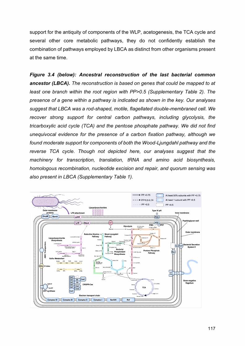
117
support for the antiquity of components of the WLP, acetogenesis, the TCA cycle and
several other core metabolic pathways, they do not confidently establish the
combination of pathways employed by LBCA as distinct from other organisms present
at the same time.
Figure 3.4 (below): Ancestral reconstruction of the last bacterial common
ancestor (LBCA). The reconstruction is based on genes that could be mapped to at
least one branch within the root region with PP>0.5 (Supplementary Table 2). The
presence of a gene within a pathway is indicated as shown in the key. Our analyses
suggest that LBCA was a rod-shaped, motile, flagellated double-membraned cell. We
recover strong support for central carbon pathways, including glycolysis, the
tricarboxylic acid cycle (TCA) and the pentose phosphate pathway. We did not find
unequivocal evidence for the presence of a carbon fixation pathway, although we
found moderate support for components of both the Wood-Ljungdahl pathway and the
reverse TCA cycle. Though not depicted here, our analyses suggest that the
machinery for transcription, translation, tRNA and amino acid biosynthesis,
homologous recombination, nucleotide excision and repair, and quorum sensing was
also present in LBCA (Supplementary Table 1).

118
Finally, our reconstruction also indicated high posterior support for several elements
of an adaptive immunity CRISPR-Cas system (Makarova et al., 2011; Koonin and
Makarova, 2019), including the universally conserved Cas endonuclease, Cas1, which
is essential for spacer acquisition and insertion into CRISPR cassettes (Nuñez et al.,
2014; Makarova et al., 2015). Interestingly, highly supported CRISPR components in
LBCA belong primarily to Class 1 systems, specifically Type I and Type III, which
exhibit greater modular diversity than their Class 2 counterparts, (Koonin and
Makarova, 2019). We recovered a near complete prototypical Type III CRISPR
system, providing strong support for its presence in LBCA. Among other roles,
CRISPR systems are crucial in antiviral defence and activate in response to viral
exposure (Barrangou et al., 2007); therefore these findings are consistent with
hypotheses suggesting that LBCA already co-evolved with parasitic replicators such
as bacteriophages (Koonin, 2014; Krupovic, Dolja and Koonin, 2019).

119
Chapter 4
Genomic evolution of Bacteria through
time A version of this chapter forms part of a paper under revision in collaboration with
Adrián A. Davín, Tara Mahendrarajah, Anja Spang, Philip Hugenholtz, Gergely J.
Szöllősi, and Tom A. Williams. Gareth A. Coleman is the first author of the paper. The
project was conceived by TAW, GJSz, PH, AS, GAC and AAD. Relative time and
verticality analyses were carried out by AAD, GJSz, TW and GAC, with interpretation
and writing carried out by all authors. Metabolic reconstructions, including all
interpretation and writing thereof, were carried out by GAC.
Paper preprint as:
Coleman, G.A., Davín, A.A., Mahendrarajah, T., Spang, A.A., Hugenholtz, P.,
Szöllősi, G.J. and Williams, T.A., 2020. A rooted phylogeny resolves early bacterial
evolution. bioRxiv. BioRxiv preprint for the paper can be found here:
https://www.biorxiv.org/content/10.1101/2020.07.15.205187v1

120
Abstract Given the huge genetic and metabolic diversity, abundance, and ubiquity of Bacteria in the modern environment, understanding how diverse physiologies and morphologies evolved in the early stages of bacterial evolution is central to our understanding of the history of life. Having previously used methods of modelling genome evolution using gene duplication, transfer and loss events to reconstruct the ancestral gene content of the last bacterial common ancestor (LBCA), we now use these methods to explore how LBCA gave rise to the major clades of extant Bacteria. We infer the early radiation of Terrabacteria, particularly CPR, while Cyanobacteria and Gracilicutes emerged later in bacterial diversification. We find that early bacterial evolution was likely dominated by autotrophic, possibly acetogenic, motile diderm cells, living alongside highly reduced and metabolically streamlined cells, followed by the later rise of photosynthetic and aerobic prokaryotes, and the eventually the Eukaryotes.
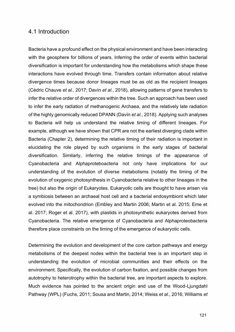
121
4.1 Introduction Bacteria have a profound effect on the physical environment and have been interacting
with the geosphere for billions of years. Inferring the order of events within bacterial
diversification is important for understanding how the metabolisms which shape these
interactions have evolved through time. Transfers contain information about relative
divergence times because donor lineages must be as old as the recipient lineages
(Cédric Chauve et al., 2017; Davín et al., 2018), allowing patterns of gene transfers to
infer the relative order of divergences within the tree. Such an approach has been used
to infer the early radiation of methanogenic Archaea, and the relatively late radiation
of the highly genomically reduced DPANN (Davín et al., 2018). Applying such analyses
to Bacteria will help us understand the relative timing of different lineages. For
example, although we have shown that CPR are not the earliest diverging clade within
Bacteria (Chapter 2), determining the relative timing of their radiation is important in
elucidating the role played by such organisms in the early stages of bacterial
diversification. Similarly, inferring the relative timings of the appearance of
Cyanobacteria and Alphaproteboacteria not only have implications for our
understanding of the evolution of diverse metabolisms (notably the timing of the
evolution of oxygenic photosynthesis in Cyanobacteria relative to other lineages in the
tree) but also the origin of Eukaryotes. Eukaryotic cells are thought to have arisen via
a symbiosis between an archaeal host cell and a bacterial endosymbiont which later
evolved into the mitochondrion (Embley and Martin 2006; Martin et al. 2015; Eme et
al. 2017; Roger et al. 2017), with plastids in photosynthetic eukaryotes derived from
Cyanobacteria. The relative emergence of Cyanobacteria and Alphaproteobacteria
therefore place constraints on the timing of the emergence of eukaryotic cells.
Determining the evolution and development of the core carbon pathways and energy
metabolisms of the deepest nodes within the bacterial tree is an important step in
understanding the evolution of microbial communities and their effects on the
environment. Specifically, the evolution of carbon fixation, and possible changes from
autotrophy to heterotrophy within the bacterial tree, are important aspects to explore.
Much evidence has pointed to the ancient origin and use of the Wood-Ljungdahl
Pathway (WPL) (Fuchs, 2011; Sousa and Martin, 2014; Weiss et al., 2016; Williams et

122
al., 2017; Adam, Borrel and Gribaldo, 2018), and the reverse tricarboxylic acid (TCA)
cycle (Wächtershäuser, 1990; Cody et al., 2001; Smith and Morowitz, 2004; Nunoura
et al., 2018) as carbon fixation pathways. In Chapter 3, we found possible support for
both pathways in the last bacterial common ancestor (LBCA), although the evidence
is difficult to interpret. It is therefore important to determine whether subsequent nodes
possess similar abilities to LBCA, and at which points along the tree these metabolic
capabilities change.
Reconstructing the gene content at different nodes can also help us understand the
evolution of cell morphology, in particular the architecture of the cell envelope and the
evolution of structure involved in cell motility, and test hypotheses on the origins of
these characteristics. As explained in the previous chapter, the “lipid divide” (Koga,
2011) refers to the difference in type of membrane phospholipids produced by Archaea
and Bacteria, and has been touted as a hallmark difference between the two domains,
although recent evidence has challenged these assumptions and found the divide to
be less clear cut than previously thought (Sinninghe Damsté et al., 2002; Weijers et
al., 2006; Damsté, Rijpstra, et al., 2007; Goldfine, 2010; Villanueva, Schouten and
Damsté, 2017; Caforio et al., 2018; Coleman, Pancost and Williams, 2019). The
presence in many Bacteria of genes involved in the biosynthesis of archaeal-like lipids
(Villanueva, Schouten and Damsté, 2017; Coleman, Pancost and Williams, 2019) has
raised questions pertaining to the relative antiquity of both pathways, specifically which
phospholipids were being produced by early Bacteria (Coleman, Pancost and Williams,
2019). We found some evidence for the production of bacterial phospholipids in LBCA
(Chapter 3), but it would be pertinent to discern whether other early nodes with the
bacterial tree were producing bacterial- or archaeal-type phospholipids. Many modern
Bacteria also possess an outer membrane, the origin of which remains debated.
Monoderm-first scenarios have been advocated whereby the outer membrane evolved
stepwise via gene insertions (Gupta, 2011), by endosymbiosis between monoderms
(single-membraned bacteria (Lake, 2009)), or via the arrest of sporulation (Tocheva,
Ortega and Jensen, 2016) in a spore-forming monoderm ancestor. In Chapter 3, we
also find that LBCA possesses an outer membrane, which supports Diderm-first
hypothesis regarding the evolution of diderm envelope architecture (Cavalier-Smith,
2002; Megrian et al., 2020). To strengthen evidence for the Diderm-first scenario, we

123
must explore the presence of the outer membrane in deep nodes, particularly with the
Terrabacteria, where many modern lineages have lost the outer membrane.
To answer these questions, we build on results from Chapter 3 by using patterns of
gene transfer to infer the relative timing of different events within bacterial evolution,
and expanding our metabolic reconstructions to other deep nodes within the bacterial
tree.
4.2 Methods Inference of relative divergence times of bacterial clades We parsed the transfers inferred using ALEml_undated and discarded those with
posterior probability <0.05. We used bootstrapping to estimate constraint support in
the following way: for each of the three candidate species trees, we sampled the gene
families 100 times with replacement and, for each replicate, converted detected
transfers to constraints and performed a MaxTiC analysis (Cedric Chauve et al., 2017;
Davín et al., 2018). A total of 8743, 8629, and 9079 constraints were recovered in at
least 95/100 replicates for the 3 possible roots respectively, and we used this subset
of highly supported constraints in our final analysis. We generated 1000 time orders
compatible with those constraints for every root. We then ranked all interior nodes on
the tree, with the root node having rank 0 and the most recent speciation node having
rank 263.
Metabolic reconstruction Metabolic reconstructions were carried out as detailed in Chapter 3. Briefly, protein
sequences form the GTDB dataset were annotated using both KEGG and COG
databases. Sets of COG families were produced, and those with more than 4
sequences were reconciled with the rooted species trees using a specific origination
prior for each family based on its COG category (see Chapter 3 for details). As in
Chapter 3, initial metabolic infers were based on gene families with a posterior
presence probability (PP) of >0.95, but as this was found to be too conservative for
most metabolic pathways, we investigated the PPs of key pathways identified from the
initial PP cut-off and inferred the presence of a pathway of protein complex or pathway

124
if the majority of its components were found with PPs >0.5. We explored eight nodes
(Fig. 4.1), namely the ancestors of Terrabacteria and Gracilicutes respectively, the
common ancestors of Firmicutes and Actinobacteriota, and Chloroflexota and CPR
respectively, and all the ancestors of the aforementioned individual phyla. We do not
explore any nodes involving Fusobacteriota, or any of the FASSyT/DST taxa (see
Chapter 2) and as a result the ambiguity within the root region has little effect on the
genes recovered at the nodes surveyed.
Fig. 4.1 Nodes for which we inferred ancestral gene content. Note we do not explore
any nodes involving Fusobacteriota or FASSyT/DST due to uncertainty of root position.
FCB are the Fibrobacterota, Chlorobiota, Bacteroides, and related lineages; PVC are
the Planctomycetes, Verrucomicrobia, Chlamydiae, and related lineages; DST are the
Deinococcota, Synergistota, and Thermatogota; ACD are Aquificota,
Campylobacterota, and Deferribacterota.
4.3 Results and Discussion
Relative dating of the bacterial tree Transfers contain information about the relative timing of divergences because donors
must be at least as old as their recipients (Cedric Chauve et al., 2017; Davín et al.,
2018). Since inferred transfer events are uncertain, we used only high-confidence
relative age constraints recovered in at least 95/100 bootstrap replicates (see
Methods) to establish the relative ages of bacterial clades (Fig. 4.2). These analyses
suggest that several groups within Terrabacteria are older than the entire Gracilicutes

125
radiation, including the crown groups of the CPR (97.4% of sampled time orders) and
Firmicutes (100% of sampled time orders). Despite being a derived lineage within
Terrabacteria, the analysis suggests that the radiation of CPR was one of the earliest
events during the diversification of Bacteria (Fig. 4.2). The early radiation of CPR, taken
together with evidence for the basal position of the analogous DPANN superphylum
within Archaea (Castelle et al., 2015; Williams et al., 2017), implies that highly reduced,
metabolic minimalist prokaryotes have been a part of microbial communities
throughout the history of cellular life (Beam et al., 2020), although it should be noted
that relative dating of Archaea using the same method showed crown-group DPANN
to be a relatively late occurrence within archaeal evolution (Davín et al., 2018). By
contrast, the emergence of the Alphaproteobacteria and the photosynthetic
Cyanobacteria were relatively late events during bacterial evolution: the divergence
between Alphaproteobacteria and Magnetococcales was the 153rd of 264 internal
divergences (median rank), while the divergence of photosynthetic Cyanobacteria from
their closest relatives had a median rank of 172nd (Fig. 4.2). These divergences
confirm that the mitochondrial and plastid endosymbioses, and therefore the origin of
eukaryotic cells, occurred during the later stages of bacterial diversification (Parfrey et
al., 2011; Eme et al., 2014b; Knoll, 2014; Nicholas J. Butterfield, 2015; Betts et al.,
2018).
Fig. 4.2 (below) Relative ages of bacterial clades. We used the relative time
information provided by directional (donor-to-recipient) patterns of gene transfer to
infer the relative ages of bacterial clades. Node numbers in the cladogram (left)
correspond to rows in the speciation plot (right). Uncertainties represent the range of
sampled time orders that are consistent with high-confidence constraints implied by
gene transfers. Following the divergence between Terrabacteria and Gracilicutes, the
earliest radiations of extant groups were among Terrabacteria, including CPR and
Firmicutes. Note that gene transfers indicate the order of branching events, but provide
no information about the absolute time intervals separating events. The geological
record provides evidence for oxygenic photosynthesis prior to 3.2 Gya (Betts et al.,
2018), suggesting that divergence 5 - and by extension all earlier divergences -
occurred prior to this date. DST are the Deinococcota, Synergistota, and
Thermotogota.
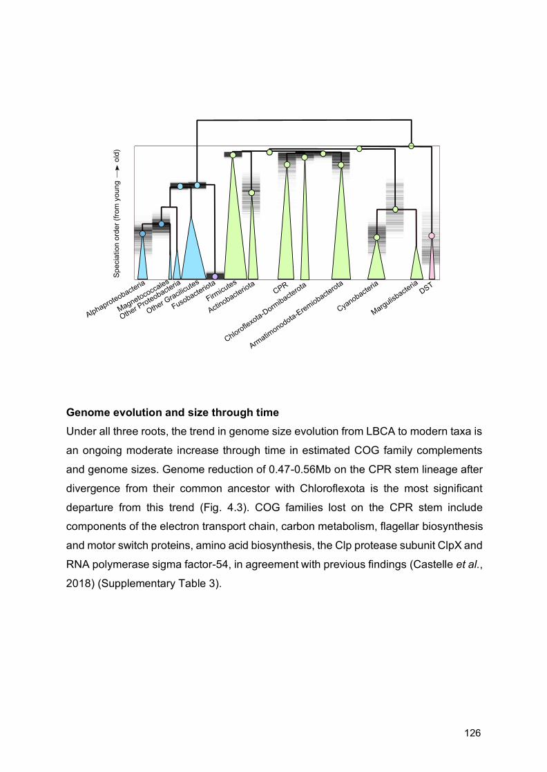
126
Genome evolution and size through time Under all three roots, the trend in genome size evolution from LBCA to modern taxa is
an ongoing moderate increase through time in estimated COG family complements
and genome sizes. Genome reduction of 0.47-0.56Mb on the CPR stem lineage after
divergence from their common ancestor with Chloroflexota is the most significant
departure from this trend (Fig. 4.3). COG families lost on the CPR stem include
components of the electron transport chain, carbon metabolism, flagellar biosynthesis
and motor switch proteins, amino acid biosynthesis, the Clp protease subunit ClpX and
RNA polymerase sigma factor-54, in agreement with previous findings (Castelle et al.,
2018) (Supplementary Table 3).
127
Fig. 4.3 (below) Evolution of COG family repertoires and inferred genome size
over the bacterial tree. (a) The inferred number of COG family members and (b)
inferred genome size at each internal node of the tree. Genome sizes were predicted
from the relationship between COG family members and genome size among extant
Bacteria (LOESS regression). Circle diameter and colour are proportional to family
number or genome size. FCB are the Fibrobacterota, Chlorobiota, Bacteroides, and
related lineages; PVC are the Planctomycetes, Verrucomicrobia, Chlamydiae, and
related lineages; DST are the Deinococcous, Synergistota, and Thermatogota; ACD
are Aquificota, Campylobacterota, and Deferribacterota. The figure depicts inferences
for root 1 (as shown in Chapter 2, Fig. 2.7(a)); the data for all three roots are provided
in Supplementary Table 4.

128
n69 Firmicutes B
n165 Fibrobacterota
n244 Myxococcota
n146 Chloroflexota
n79 Firmicutes B
n153 Dorm
ibacterota
n200
Acido
bacte
riota
n248 Myxococcota
n9 Planctomycetota
n100
Actin
obac
terio
ta
n180
Bac
tero
idot
a
n184
Bac
tero
idot
a
n58 F
irmicu
tes G
n179
Bac
tero
idot
a
n89 Cyanobacteria
n198
Acidob
acter
iota
n50 Firmicu
tes G
n56 F
irmicu
tes G
n74 Firmicutes B
n190
Acido
bacte
riota
n116 Eremiobacterota
n117Arm
atimonadota
n87 Firmicutes
n173
Bac
tero
idot
a
n85 Fusobacteriota
n240 Nitrospiro
ta
n65 Firmicutes E
n192
Acidob
acter
iota
n8 Planctomycetota
n43 Firmicutes K
n118Arm
atimonadota
n224 Desulfobacterotan226 Desulfobacterota
n194
Acidob
acter
iota
n212
Cam
pylo
bact
erot
a
n188 Gem
matim
onadota
n163 Fibrobacterota
n53 F
irmicu
tes G
n94 Margulisbacteria
n63 Firmicutes E
n191
Acido
bacte
riota
n221 Desulfobacterota
n260 Elusimicrobiota
n218 Desulfobacterota
n181
Bac
tero
idot
a
n186 Gem
matim
onadota
n27 Spirochaetota
n82 F
irmicu
tes F
n57 F
irmicu
tes G
n21 Planctomycetota
n172 Ma rin is om
a to ta
n17 Planctomycetota
n247 Myxococcota
n25 Spirochaetota
n187 Gem
matim
onadota
n258 Omnitrophota
n5 Verrucomicrobiota
n143 Patescibacteria
n1 Verrucomicrobiota
n261 Elusimicrobiota
n236 Desulfuromonadota
n86 Firmicutes
n93 Cyanobacteria
n216
Aqui
ficot
a
n148 Chloroflexota
n252 Bdellovibrionota
n154 Dorm
ibacterota
n40 Firmicutes I
n227 Desulfobacterota
n213
Cam
pylo
bact
erot
a
n209 Proteobacteria
n115 Eremiobacterotan242 Nitro
spiro
ta
n241 Nitrospiro
ta
n135 Pates cibacte ria
n78 Firmicutes B
n48 Firmicutes D
n151 Chloroflexota
n222 Desulfobacterota
n102
Actin
obac
terio
ta
n128
Pat
esci
bact
eria
n23 Spirochaetota
n41 Firmicutes I
n205 Proteobacteria
n13 Planctomycetota
n6 Verrucomicrobiota A
n66 Firmicutes B
n36 Firmicutes A
n130
Pat
esci
bact
eria
n107
Actin
obac
terio
ta
n92 Cyanobacteria
n29 Spirochaetota
n97 Margulisbacteria
n150 Chloroflexota
n45 Firmicutes D
n73 Firmicutes B
n233 Desulfobacterota
n144 Chloroflexota
n246 Myxococcota
n134
Pat
esci
bact
eri a
n71 Firmicutes B
n113
Actin
obac
terio
ta
n46 Firmicutes D
n262 Elusimicrobiota
n136
Pat
esci
bact
eria
n202
Acid
obac
terio
ta
n64 Firmicutes E
n32 Firmicutes A
n215
Aqui
ficot
a
n52 Firm
icutes
G
n47 Firmicutes D
n20 Planctomycetota
n220 Desulfobacterota
n76 Firmicutes B
n232 Desulfobacterota
n256 Omnitrophota
n125Arm
atimonadota
n62 Firmicutes E
n137
Pat
esci
bact
eria
n70 Firmicutes B
n108
Actin
obac
terio
ta
n239 Nitrospirota
n164 Fibrobacterota
n127
Pat
esci
bact
eria
n243 Myxococcota
n4 Verrucomicrobiotan60 Firm
icutes E
n174
Bac
tero
idot
a
n167 Cloacim
onadota
n106
Actin
obac
terio
ta
n139P
at esc iba cter ia
n206 Proteobacteria
n37 Firmicutes A
n234 Desulfobacterota
n28 Spirochaetota
n80 Firmicutes C
n24 Spirochaetota
n122 Armatim
onadota
n11 Planctomycetota
n138
Pat
esci
bact
eria
n124Arm
atimonadota
n35 Firmicutes A
n88 Cyanobacteria
n121Arm
atimonadota
n152 Chloroflexota
n208 Proteobacterian155 C
hloroflexota A
n3Verrucom
icrobiota
n228 Desulfobacterota
n33 Firmicutes A
n133
Pa t
esci
bact
eria
n207 Proteobacteria
n132
Pat
esci
bact
eria
n211
Cam
pylo
bact
erot
a
n185
Bac
tero
idot
a
n219 Desulfobacterota
n19 Planctomycetota
n140 Pat esci bacte ri a
n204 Proteobacteria
n210
Dep
ende
ntia
e
n68 Firmicutes B
n145 Chloroflexota
n238 Nitrospirota
n14 Planctomycetota
n229 Desulfobacterota
n103
Actin
obac
terio
ta
n254 Bdellovibrionota
n217
Def
errib
acte
rota
n105
Actin
obac
terio
ta
n162 Fib robact ero ta
n39 Firmicutes An166 C
loacimonadota
n55 F
irmicu
tes G
n195 Acidobacte
riota
n147 Chloroflexota
n91 Cyanobacteria
n26 Spirochaetota
n83 F
irmicu
tesF
n123 Armatim
onadota
n250 Myxococcota
n259 Elusimicrobiota
n169
Mar
inis
omat
ota
n111
Actin
obac
terio
ta
n253 Bdellovibrionota
n255 Desulfobacterota A n38 Firmicutes A
n15 Planctomycetota
n18 Planctomycetota
n129
Pat
esci
bact
eria
n54 F
irmicu
tes G
n245 Myxococcota
n199
Acidob
acter
iota
n112
Actin
obac
terio
ta
n30 Spirochaetota
n170
Ma r
ini s
omat
o ta
n51 Firmicu
tes G
n67 Firmicutes B
n98 Margulisbacteria
n7 Verrucomicrobiota A
n264 Elusimicrobiota
n203 Proteobacteria
n119Arm
atimonadota
n235 Desulfobacterota
n223 Desulfobacterota
n149 Chloroflexota
n84 Fusobacteriota
n131
Pat
esci
bact
eria
n196
Acidoba
cterio
ta
n31 Spirochaetota
n120Arm
atimonadota
n110
Actin
obac
terio
ta
n159 Thermotogota
n42 Firmicutes K
n251 Bdellovibrionota
n12 Planctomycetota
n183
Bac
tero
idot
a
n249 Myxococcota
n193
Acidob
acter
iota
n158 Thermotogota
n168
Mar
i ni s
oma t
ota
n96 Margulisbacteria
n257 Omnitrophota
n230 Desulfobacterota
n171 Marin is om
a tota
n99 Ac
tinob
acte
riota
n77 Firmicutes B
n95 Margulisbacteria
n237 Desulfuromonadota
n81 Firmicutes C
n201
Acido
bacte
riota
n189
Acido
bacte
riota
n178
Bac
tero
idot
a
n175
Bac
tero
idot
a
n177
Bac
tero
idot
a
n263 Elusimicrobiota
n22 Spirochaetota
n59 Firmicu
tes E
n90 Cyanobacteria
n142
Pat
e sc i
bact
eri a
n34 Firmicutes A
n44 Firmicutes D
n197
Acidob
acter
iota
n225 Desulfobacterota
n2Verrucom
icrobiota
n161 Synergistota
n182
Bac
tero
idot
a
n72 Firmicutes B
n109
Actin
obac
terio
ta
n16 Planctomycetota
n104
Actin
obac
terio
tan126 Armatim
onadota
n10 Planctomycetota
n49 Firmicu
tes D
n61 Firmicu
tes E
n157 Deinococcota
n231 Desulfobacterota
n141
Pat
e sci
bact
e ria
n160 Synergistota
n156 Chloroflexota A
n0Verrucom
icrobiota
n214
Aqui
ficot
a
n101
Actin
obac
terio
ta
n176
Bac
tero
idot
a
n114 Eremiobacterota
n75 Firmicutes B
Dorm ibacteriotaChloroflexota
Erem iobacterotaArm at im onodota
MargulisbacteriaCyanobacteria
DST
Desulfobacterota
Desulfurom onadotaMyxococcota
Bdellovibrionota
ProteobacteriaNit rospirota
Acidobacteriota
ACDFCB
PVC
Elusim icrobiota
Spirochaetota
Fusobacteriota
Firm icutes
Act inobacterota
CPR
COG f am i l ies
Increase in num bers of
COG fam ilies
n69 Firmicutes B
n165 Fibrobacterota
n244 Myxococcota
n146 Chloroflexota
n79 Firmicutes B
n153 Dorm
ibacterota
n200
Acido
bacte
riota
n248 Myxococcota
n9 Planctomycetota
n100
Actin
obac
terio
ta
n180
Bac
tero
idot
a
n184
Bac
tero
idot
a
n58 F
irmicu
tes G
n179
Bac
tero
idot
a
n89 Cyanobacteria
n198
Acidob
acter
iota
n50 Firmicu
tes G
n56 F
irmicu
tes G
n74 Firmicutes B
n190
Acido
bacte
riota
n116 Eremiobacterota
n117Arm
atimonadota
n87 Firmicutes
n173
Bac
tero
idot
a
n85 Fusobacteriota
n240 Nitrospiro
ta
n65 Firmicutes E
n192
Acidob
acter
iota
n8 Planctomycetota
n43 Firmicutes K
n118Arm
atimonadota
n224 Desulfobacterotan226 Desulfobacterota
n194
Acidob
acter
iota
n212
Cam
pylob
acte
rota
n188 Gem
matim
onadota
n163 Fibrobac terota
n53 F
irmicu
tes G
n94 Margulisbacteria
n63 Firmicutes E
n191
Acido
bacte
riota
n221 Desulfobacterota
n260 Elusimicrobiota
n218 Desulfobacterota
n181
Bac
tero
idot
a
n186 Gem
matim
onadota
n27 Spirochaetota
n82
Firmicu
tes F
n57 F
irmicu
tes G
n21 Planctomycetota
n172 Ma rin is om
a to ta
n17 Planctomycetota
n247 Myxococcota
n25 Spirochaetota
n187 Gem
matim
onadota
n258 Omnitrophota
n5 Verrucomicrobiota
n143 Patesciba cteria
n1Verrucom
icrobiota
n261 Elusimicrobiota
n236 Desulfuromonadota
n86 Firmicutes
n93 Cyanobacteria
n216
Aqui
ficot
an148 C
hloroflexota
n252 Bdellovibrionota
n154 Dorm
ibacterota
n40 Firmicutes I
n227 Desulfobacterota
n213
Cam
pylo
bact
erot
a
n209 Proteobacteria
n115 Eremiobacterota
n242 Nitrosp
irota
n241 Nitrosp
irota
n135 Patescib acter ia
n78 Firmicutes B
n48 Firmicutes D
n151 Chloroflexota
n222 Desulfobacterota
n102
Actin
obac
terio
ta
n128
Pat
esci
bact
eria
n23 Spirochaetota
n41 Firmicutes I
n205 Proteobacteria
n13 Planctomycetota
n6 Verrucomicrobiota A
n66 Firmicutes B
n36 Firmicutes A
n130
Pat
esci
bact
eria
n107
Actin
obac
terio
ta
n92 Cyanobacteria
n29 Spirochaetota
n97 Margulisbacteria
n150 Chloroflexota
n45 Firmicutes D
n73 Firmicutes B
n233 Desulfobacterota
n144 Chloroflexota
n246 Myxococcota
n134
Pat
esci
bact
eri a
n71 Firmicutes B
n113
Actin
obac
terio
ta
n46 Firmicutes D
n262 Elusimicrobiota
n136
Pat
esci
bact
eria
n202
Acido
bact
erio
ta
n64 Firmicutes E
n32 Firmicutes A
n215
Aqui
ficot
a
n52 Firm
icutes G
n47 Firmicutes D
n20 Planctomycetota
n220 Desulfobacterota
n76 Firmicutes B
n232 Desulfobacterota
n256 Omnitrophota
n125Arm
atimonadota
n62 Firmicutes E
n137
Pat
esci
bact
eria
n70 Firmicutes B
n108
Actin
obac
terio
ta
n239 Nitrospirota
n164 Fibrobacte rota
n127
Pat
esci
bact
eria
n243 Myxococcota
n4 Verrucomicrobiota
n60 Firmicutes E
n174
Bac
tero
idot
a
n167 Cloacim
onadota
n106
Actin
obac
terio
ta
n13 9 Pate sci bac teri a
n206 Proteobacteria
n37 Firmicutes A
n234 Desulfobacterota
n28 Spirochaetota
n80 Firmicutes C
n24 Spirochaetota
n122 Armatim
onadota
n11 Planctomycetota
n138
Pat
esci
bact
eria
n124Arm
atimonadota
n35 Firmicutes A
n88 Cyanobacteria
n121Arm
atimonadota
n152 Chloroflexota
n208 Proteobacteria
n155 Chloroflexota A
n3Verrucom
icrobiota
n228 Desulfobacterota
n33 Firmicutes A
n133
Pat
esci
bac t
eria
n207 Proteobacteria
n132
Pat
esci
bact
eria
n211
Cam
pylo
bact
erot
a
n185
Bac
tero
idot
a
n219 Desulfobacterota
n19 Planctomycetota
n140 Pat escib acter i a
n204 Proteobacteria
n210
Dep
ende
ntia
e
n68 Firmicutes B
n145 Chloroflexota
n238 Nitrospirota
n14 Planctomycetota
n229 Desulfobacterota
n103
Actin
obac
terio
ta
n254 Bdellovibrionota
n217
Def
errib
acte
rota
n105
Actin
obac
terio
ta
n162 Fib robacte ro ta
n39 Firmicutes A
n166 Cloacim
onadota
n55 F
irmicu
tes G
n195 Acidobacte
riota
n147 Chloroflexota
n91 Cyanobacteria
n26 Spirochaetota
n83 F
irmicu
tes F
n123 Armatim
onadota
n250 Myxococcota
n259 Elusimicrobiota
n169
Mar
inis
omat
ota
n111
Actin
obac
terio
ta
n253 Bdellovibrionota
n255 Desulfobacterota A n38 Firmicutes A
n15 Planctomycetota
n18 Planctomycetota
n129
Pate
scib
acte
ria
n54 F
irmicu
tes G
n245 Myxococcota
n199
Acidob
acter
iota
n112
Actin
obac
terio
ta
n30 Spirochaetota
n170
Mar
inis
omat
o ta
n51 Firmicu
tes G
n67 Firmicutes B
n98 Margulisbacteria
n7 Verrucomicrobiota A
n264 Elusimicrobiota
n203 Proteobacteria
n119Arm
atimonadota
n235 Desulfobacterota
n223 Desulfobacterota
n149 Chloroflexota
n84 Fusobacteriota
n131
Pat
esci
bact
eria
n196 Acid
obac
teriota
n31 Spirochaetota
n120Arm
atimonadota
n110
Actin
obac
terio
ta
n159 Thermotogota
n42 Firmicutes K
n251 Bdellovibrionota
n12 Planctomycetota
n183
Bac
tero
idot
a
n249 Myxococcota
n193
Acidob
acter
iota
n158 Thermotogota
n168
Ma r
i nis
omat
ota
n96 Margulisbacteria
n257 Omnitrophota
n230 Desulfobacterota
n171 Mari nisom
a to ta
n99 Ac
tinob
acte
riota
n77 Firmicutes B
n95 Margulisbacteria
n237 Desulfuromonadota
n81 Firmicutes C
n201
Acido
bacte
riota
n189
Acido
bacte
riota
n178
Bac
tero
idot
a
n175
Bac
tero
idot
a
n177
Bac
tero
idot
a
n263 Elusimicrobiota
n22 Spirochaetota
n59 Firmicu
tes E
n90 Cyanobacteria
n142
Pa t
e sci
bact
e ria
n34 Firmicutes A
n44 Firmicutes D
n197
Acidob
acter
iota
n225 Desulfobacterota
n2Verrucom
icrobiota
n161 Synergistota
n182
Bac
tero
idot
a
n72 Firmicutes B
n109
Actin
obac
terio
ta
n16 Planctomycetota
n104
Actin
obac
terio
tan126Arm
atimonadota
n10 Planctomycetota
n49 Firmicu
tes D
n61 Firmicu
tes E
n157 Deinococcota
n231 Desulfobacterota
n141
Pa t
esci
bact
eria
n160 Synergistota
n156 Chloroflexota A
n0Verrucom
icrobiota
n214
Aqui
ficot
a
n101
Actin
obac
terio
ta
n176
Bac
tero
idot
a
n114 Eremiobacterota
n75 Firmicutes B
Dorm ibacteriotaChloroflexota
Erem iobacterotaArm at im onodota
MargulisbacteriaCyanobacteria
DST
Desulfobacterota
Desulfurom onadotaMyxococcota
Bdellovibrionota
ProteobacteriaNit rospirota
Acidobacteriota
ACDFCB
PVC
Elusim icrobiota
Spirochaetota
Fusobacteriota
Firm icutes
Act inobacterota
CPRGen om e size (m b )
Increase in genom e size

129
Evolution of carbon metabolism after LBCA Our reconstructions of the central metabolic pathways are similar to those inferred in
LBCA. We find moderate to strong support (PP=0.5-1) for the presence of glycolysis
in all nodes, attesting to its ancient origins (Romano and Conway, 1996). Support for
the tricarboxylic acid (TCA) cycle and pentose phosphate pathway are less strong, with
very patchy distributions of proteins across different nodes (Fig. 4.4). The ancestor of
CPR in particular seems to lack the majority of the components of these pathways,
with only two proteins (both in the same step of the pathway) and one protein from the
TCA and pentose phosphate pathway recovered respectively.
When looking for evidence of carbon fixation, components of various pathways were
patchy. Evidence for carbon fixation is largely absent in the ancestor of
Actinobacteriota. Although some autotrophic examples within Actinobacteriota are
known (Norris et al., 2011), the large majority are heterotrophic (Lechevalier and
Lechevalier, 1965; Barka et al., 2016), and therefore the ancestor of Actinobacteriota
was also likely heterotrophic. Of the several possible carbon fixation pathways, there
is no evidence for the presence of any variants of the 3-hydroxypropionate bicycle in
any of the nodes. The apparent presence of components of the Calvin Cycle (also
known as the reductive pentose phosphate pathway) most likely represent the pentose
phosphate pathway. The enzyme RuBisCO is unique to the Calvin Cycle, and is not
found in any nodes surveyed. We have some moderate support of both subunits of
oxoacid:ferredoxin oxidoreductase in all nodes (Fig. 4.4), which may function in the
TCA in both the oxidative and reductive direction. However, as noted above, other
components of the TCA cycle are patchily distributed, and we do not recover the
hallmark enzyme ATP citrate lyase in any nodes. The support for the reverse TCA
cycle is highest in the ancestor of Terrabacteria, with six steps of the pathway
recovered with PP>0.5 (Fig. 4.4), although still lacking ATP citrate lyase. We therefore
have little evidence to suggest the presence of the reverse TCA cycle in any of these
nodes.

130
Fig 4.4 Metabolic map of the central carbohydrate pathways (glycolysis, tricarboxylic
acid cycle and pentose phosphate cycle) inferred in the ancestors of Terrabacteria and
DTS, Terrabacteria, Gracilicutes, Chloroflexota and CPR, Chloroflexota, CPR,
Firmicutes and Actinobacteriota, Firmicutes, and Actinobacteriota respectively. The
reconstruction is based on genes that could be mapped to a given node with PP >0.5.
The presence of a gene within a pathway is indicated as shown in the key. Annotations
and PP values for all KOs can be found in Supplementary Table 1.
Acetogenesis in early Bacteria
The components of the methyl branch of the WLP are mapped with moderate to strong
support (PP= 0.5-0.95) across all the nodes, except the common ancestor of CPR and
Chloroflexota and both their respective ancestors, which lack some of the steps (Fig.
4.5). However, as with LBCA we do not find most of the components of the hallmark
enzyme of the WLP, the carbon monoxide dehydrogenase/acetyl-CoA synthase
(CODH/ACS), finding only very moderate support for a single subunit (K14138, PP=
0.66) in the ancestor of Gracilicutes. As the methyl-branch of the WLP may be used in
other metabolic pathways (Stover, 2009; Brosnan and Brosnan, 2016), it is difficult to
assign the presence of WLP to any node based solely on the presence of the methyl
branch. CODH/ACS is not especially common among either Gracilicutes or
Terrabacteria, although it does have wide phylogenetic spread, indicating either

131
multiple transfers of these genes, or a presence in the ancestors of Gracilicutes and
Terrabacteria followed by multiple losses.
Fig. 4.5 Metabolic map of acetogenesis and the Wood-Ljungdahl Pathway inferred in
the ancestors of Terrabacteria and DTS, Terrabacteria, Gracilicutes, Chloroflexota and
CPR, Chloroflexota, CPR, Firmicutes and Actinobacteriota, Firmicutes, and
Actinobacteriota respectively. The reconstruction is based on genes that could be
mapped to a given node with PP >0.5. The presence of a gene within a pathway is
indicated as shown in the key. Annotations and PP values for all KOs can be found in
Supplementary Table 1.
Acetate kinase was recovered with strong support (PP>0.75) in the Gracilicutes and
Terrabacteria ancestor, as well as the common ancestor of Chloroflexota and CPR and
the ancestor of Chloroflexota. Support was more moderate (PP=0.5-0.75) in the
common ancestor of Firmicutes and Actinobacteriota and their respective ancestors,
and was absent in the ancestor of CPR (Fig. 4.5). We recovered phosphate
acyltransferase only with moderate (PP=0.5-0.75) support in the ancestors of
Gracilicutes and Terrabacteria, and not recovered in other nodes (Fig. 4.5).

132
As it was found to be present in LBCA (Chapter 3), we further investigated the
presence of the Na+-translocating ferredoxin:NAD+ oxidoreductase (Rnf) complex,
which comprises six subunits coded by the genes rnfA (K03617), rnfB (K03616), rnfC
(K03615), rnfD (K03614), rnfE (K03613), and rnfG (K03612) respectively. We recover
five of these components (all except rnfG) with PP>0.5 in the ancestor of Gracilicutes
(Supplementary Table 1). Given the role that the Rnf complex may play in the WLP in
acetogens (Buckel and Thauer, 2013; Müller, Chowdhury and Basen, 2018), as well
as evidence for the antiquity of the pathway (Chapter 3 of this thesis, (Fuchs, 2011;
Sousa and Martin, 2014; Williams et al., 2017; Adam, Borrel and Gribaldo, 2018)), we
have some evidence to suggest that the ancestor of Gracilicutes, like LBCA,
possessed the WLP and may have been facultatively acetogenic. Support is less
strong in other nodes (Supplementary Table 1). In the ancestral Terrabacteria, we
recover only rnfA (PP=0.96). Four of the six genes are recovered in the ancestor of
Firmicutes, providing some evidence that it was acetogenic. Only two were recovered
in the ancestor of Actinobacteriota, indicating that it was unlikely to have been able to
use this pathway. Indeed, as discussed above, the last ancestor of Actinobacteriota
was most likely heterotrophic. None of these genes are recovered in the common
ancestor of Chloroflexota and CPR or their respective ancestors, so we cannot say
that acetogenesis was present in these nodes. It is possible that the ancestor of
Terrabacteria was acetogenic, with this pathway being subsequently lost in most
lineages, although maintained in Firmicutes. However, the evidence is only moderate.
Energy generation and respiratory complexes
As in LBCA, our analyses indicate the presence of a F-type ATP synthase, including
components of both the F0 regions (which is embedded into the cell membrane) and
the F1 region respectively, in all nodes. Several of the subunits, e.g. alpha, gamma, a
and c subunits, are recovered in the majority of the nodes with high support (PP>0.9)
(Supplementary Table 1). Other subunits are more patchily distributed. A V-type ATP
synthase is also recovered in the ancestor of Terrabacteria, Gracilicutes, the common
ancestor of Firmicutes and Actinobacteriota and their respective ancestors. The
subunits a (K02117), b (K02118) and d (K02120, stalk) of the V1 region are recovered
in all these nodes with moderate to high support (PP=0.5-1) (Supplementary Table 1).
Of the V0 region, which is embedded in the cell membrane, subunits e and i are widely
recovered. No subunits for V-type ATP synthases are found in the ancestor of

133
Chloroflexota, and only the a subunit is found in the ancestor of CPR and the common
ancestor of both Chloroflexota and CPR respectively. The V-type ATP synthase was
most likely present in most early lineages, and lost on the branch to Chloroflexota and
CPR.
As with our reconstruction of LBCA, we find a patchy distribution of other components
of the electron transport chain, with a small number of subunits for each complex found
with PP >0.5, except in the ancestor of CPR where we find no components of the
electron transport chain, other than the aforementioned ATP synthases
(Supplementary Table 1). Unlike our reconstruction of LBCA, we do not find strong
support for the presence of respiratory nitrate reductase (Nar), so we cannot determine
the ability for anaerobic respiration of nitrate in any of the subsequent nodes. We
recover some subunits related to hydrogenase in the Gracilicutes ancestor (K18023,
PP=0.92; K00333, PP=0.73), and in the Terrabacteria ancestor (K18023, PP=0.92)
suggesting hydrogen as the election doner, in line with our reconstruction of LBCA,
and with previous studies (Lane, Allen and Martin, 2010; Greening et al., 2016)).
Again, similarly to LBCA, we find support for the terminal oxidases NuoG (K00336),
CytB (K00412) in all nodes except the ancestors of Firmicutes and CPR. NuoG has
strong support in the ancestor of Terrabacteria and Gracilicutes (PP=0.85 and 0.91
respectively). Support is more moderate in other nodes (PP=0.5-0.75). CytB has
moderate to strong support (PP>0.7), but support is weaker in the ancestor of
Gracilicutes (PP=0.53). The nodes with the strongest support for the presence of
terminal oxidases are the common ancestor of Chloroflexota and CPR, and the
ancestor of Chloroflexota. Both nodes not only recover CytB with strong support
(PP=0.96 and 1 respectively) and NuoG with moderate support (PP=0.58 and 0.67
respectively) but also have several heme-copper type oxidases, notably coxA
(K02274), coxB (K02275) and coxC (K02276), with moderate to high supports
(PP=0.6-1). It is possible that aerobic respiration was acquired on the branch to
Chloroflexota and CPR before being lost in CPR. It should also be noted that, other
than in the common ancestor of Chloroflexota and CPR and ancestor of Chloroflexota,
we do not find evidence for any heme-copper type oxidases, and that other
components for complexes which comprise these terminal oxidases are patchy in their
distribution or are absent, so we cannot confidently map terminal oxidases to any of

134
these nodes. Furthermore, all of these clades predate the radiation of photosynthetic
Cyanobacteria in our relative dating analysis, including in the case of Chloroflexota and
CPR, which may indicate they predate the oxygenation of the atmosphere, although
this is contentious (Betts et al., 2018).
As was the case with LBCA, it is difficult to determine which electron acceptor these
early Bacteria were using. However, based on the presence of components of the
acetogenesis pathway, carbon dioxide was the most likely candidate, implying that the
ancestors of most clades were anaerobic. This is congruent with models of the
atmosphere of the early Earth (Arndt and Nisbet, 2012). However, more in depth
exploration of the histories of these genes would be needed to bring greater clarity to
this issue, and other electron acceptors, including oxygen, cannot be confidently
rejected by our analyses.
Evolution of membrane phospholipids in Bacteria We inferred the presence of bacterial-type G3P membrane in LBCA (Chapter 3) and
expect similar inferences in subsequent nodes, given the widespread use of G3P lipids
in modern Bacteria. We recover moderate to high support for G3P dehydrogenase
(GpsA, K00057) in all nodes we surveyed (Table 4.1), with the exception of the
ancestor of CPR and the ancestor of Gracilicutes. We additionally recover glycerol
kinase in the Terrabacteria ancestor (K00864, PP=0.84). Similarly to GpsA, PlsX
(K03621, which attaches the first fatty to G3P along with PlsY), was recovered with
moderate to strong support (0.78-0.98) across all nodes except the ancestor of CPR.
Other proteins involved in phospholipid biosynthesis are more sparsely distributed
across the tree (Table 4.1). PlsY (K08591) was found with high support in the
Terrabacteria ancestor (PP=0.93), as well as in both Firmicutes and Actinobacteriota
and their common ancestor (PP=0.94, 0.98 and 0.81 respectively). It was not
recovered in the Gracilicute ancestor, or in the ancestor of CPR and Chloroflexota and
descendent nodes. No PlsC is recovered, except a putative PlsC in the Gracilicutes
ancestor (K15781, PP=0.75). While the distribution is sparse, given the presence for
this pathway in LBCA (Chapter 3) and the distribution of these genes in modern taxa,
it is probable that the ability the produce bacterial G3P lipids was present in all the
deep nodes of the tree, and subsequently lost in CPR. In contrast to the above, we do
not find any strong evidence for the presence of archaeal type lipids in any nodes,

135
despite evidence for the presence in modern representatives of several bacterial phyla
(Villanueva, Schouten and Damsté, 2017; Coleman, Pancost and Williams, 2019), and
their possible ancient presence within Bacteria (Coleman, Pancost and Williams,
2019).
FAM kegg_id kegg_description Terra+DST Terra Graci Chlo+CPR Chlo CPR F+A F A
COG0344 K08591
acyl_phosphate:glycerol-3-phosphate_acyltransferase_[EC:2.3.1.275] 0.97 0.93 0.42 0.46 0.49 0.13 0.94 0.98 0.81
COG0554 K00864 glycerol_kinase_[EC:2.7.1.30] 0.88 0.84 0.21 0.15 0.25 0.03 0.8 0.78 0.75
COG1368 K05878
phosphoenolpyruvate---glycerone_phosphotransferase_subunit_DhaK_[EC:2.7.1.121] 0.86 0.86 0.12 0.85 0.93 0.22 0.93 0.71 1
COG4589 K00981 phosphatidate_cytidylyltransferase_[EC:2.7.7.41] 0.82 0.42 0.38 0.63 0.72 0.26 0.05 0.05 0.01
COG0558 K06131 cardiolipin_synthase_A/B_[EC:2.7.8.-] 0.79 0.72 0.54 0.38 0.7 0.22 0.18 0.13 0.14
COG0416 K03621 phosphate_acyltransferase_[EC:2.3.1.274] 0.78 0.8 0.82 0.9 1 0.25 0.98 0.83 0.97
COG1597 K07029 diacylglycerol_kinase_(ATP)_[EC:2.7.1.107] 0.68 0.85 0.33 0.93 0.95 0.44 0.91 0.92 0.94
COG2376 K05881
phosphoenolpyruvate---glycerone_phosphotransferase_subunit_DhaM_[EC:2.7.1.121] 0.58 0.4 0.34 0.12 0.19 0.02 0.41 0.48 0.16
COG0575 K00981 phosphatidate_cytidylyltransferase_[EC:2.7.7.41] 0.57 0.88 0.7 0.49 0.44 0.86 0.98 0.99 0.9
COG0644 K01126 glycerophosphoryl_diester_phosphodiesterase_[EC:3.1.4.46] 0.49 0.21 0.41 0.26 0.49 0.1 0.11 0.22 0.23
COG0240 K00057
glycerol-3-phosphate_dehydrogenase_(NAD(P)+)_[EC:1.1.1.94] 0.41 0.85 0.15 0.78 0.67 0.27 0.98 0.96 1
COG1502 K03736 ethanolamine_ammonia-lyase_small_subunit_[EC:4.3.1.7] 0.35 0.19 0.17 0.02 0.01 0.01 0.14 0.15 0.01
COG0371 K17830
digeranylgeranylglycerophospholipid_reductase_[EC:1.3.1.101_1.3.7.11] 0.31 0.4 0.04 0.22 0.24 0.1 0.5 0.46 0.82
COG4303 K01095 phosphatidylglycerophosphatase_A_[EC:3.1.3.27] 0.29 0.06 0.62 0.01 0.03 0.08 0.04 0.12 0.02
COG0560 K15781
putative_phosphoserine_phosphatase_/_1-acylglycerol-3-phosphate_O-acyltransferase_[EC:3.1.3.3_2.3.1.51] 0.26 0.23 0.75 0.12 0.27 0.09 0.27 0.14 0.46
COG0584 K00096
glycerol-1-phosphate_dehydrogenase_[NAD(P)+]_[EC:1.1.1.261] 0.23 0.19 0.05 0.07 0.05 0.02 0.24 0.25 0.31
COG4302 K04019 ethanolamine_utilization_protein_EutA 0.21 0.05 0 0 0 0 0.03 0.06 0

136
COG1646 K13622
S-adenosylmethionine-diacylglycerol_3-amino-3-carboxypropyl_transferase 0.19 0.18 0.09 0.01 0.04 0 0 0 0.01
COG0578 K00111
glycerol-3-phosphate_dehydrogenase_[EC:1.1.5.3] 0.17 0.07 0.37 0.06 0.2 0.01 0.06 0.02 0.25
COG3675 K16818 phospholipase_A1_[EC:3.1.1.32] 0.14 0.12 0.08 0.03 0.01 0.01 0.01 0.02 0.01
COG3075 K00112
glycerol-3-phosphate_dehydrogenase_subunit_B_[EC:1.1.5.3] 0.12 0.1 0.12 0.35 0.86 0.03 0.02 0.02 0
COG4819 K08744 cardiolipin_synthase_(CMP-forming)_[EC:2.7.8.41] 0.1 0.15 0.21 0.42 0.66 0.31 0.21 0.27 0.39
COG1267 K03735 ethanolamine_ammonia-lyase_large_subunit_[EC:4.3.1.7] 0.06 0.02 0.12 0 0 0 0 0 0
COG3412 K01046 triacylglycerol_lipase_[EC:3.1.1.3] 0.05 0.11 0.05 0.06 0.06 0.03 0.04 0.06 0.06
COG2829 K01058 phospholipase_A1/A2_[EC:3.1.1.32_3.1.1.4] 0.04 0.04 0 0 0 0 0 0 0
COG0688 K01613 phosphatidylserine_decarboxylase_[EC:4.1.1.65] 0.03 0.04 0.18 0.05 0.13 0.01 0.14 0.31 0.07
COG2937 K00631 glycerol-3-phosphate_O-acyltransferase_[EC:2.3.1.15] 0.01 0.02 0.02 0 0 0 0 0 0
COG1075 K19005 lipoteichoic_acid_synthase_[EC:2.7.8.20] 0.01 0 0.23 0 0 0.01 0 0 0
COG4909 K06120 glycerol_dehydratase_large_subunit_[EC:4.2.1.30] 0 0.01 0 0 0 0 0.03 0.11 0.01
COG5153 K17900 lipase_ATG15_[EC:3.1.1.3] 0 0 0 0 0 0 0 0 0
COG5153 K17900 lipase_ATG15_[EC:3.1.1.3] 0 0 0 0 0 0 0 0 0
COG5379 K17104 phosphoglycerol_geranylgeranyltransferase_[EC:2.5.1.41] 0 0 0 0 0 0 0.01 0.01 0
Table 4.1 Posterior probabilities for the presence of glycerolipids in the ancestors of
Terrabacteria and DTS (Terra+DTS), Terrabacteria (Terra), Gracilicutes (Graci),
Chloroflexota and CPR (Chlo+CPR), Chloroflexota (Chlo), CPR, Firmicutes and
Actinobacteriota (F+A), Firmicutes (F) and Actinobacteriota (A) respectively. Key
genes whose Kegg IDs are given in the text are highlighted in bold. Annotations and
PP values for all KOs can be found in Supplementary Table 1. Evolution of bacterial motility
The flagellum is very widespread across the tree of Bacteria (heatmap in Chapter 3,
Fig. 3.2), and is thought to have evolved early in bacterial evolution (Liu and Ochman,
2007a, 2007b; Miyata et al., 2020). Supporting this, we find evidence for the flagellum
137
in LBCA (Chapter 3). We recover many proteins involved in the flagellar construction
in Terrabacteria and Gracilicutes ancestors respectively (heatmap in Chapter 3, Fig
3.2; Fig. 4.6), including components of the C, Ms and P rings, the proximal and distal
rods and the filament, as well as the Type III secretion system (Fig. 4.6). We similarly
infer a fully functioning flagellum in the ancestor of Firmicutes and Actinobacteriota,
and the Firmicute ancestor. However, all flagellar genes are lost in the ancestor of
Actinobacteriota. Most modern members of Actinobacteriota are non-flagellated,
although some do possess flagella, most likely acquired via HGT, or analogous
flagella-like structures (Barka et al., 2016). We infer a few flagellar genes in the
common ancestor of CPR and Chloroflexota, and the Chloroflexota ancestor, although
it is unclear whether any of these would have had a fully functioning flagellum (Fig.
4.6). Modern members of Chloroflexota are not thought to be flagellated (Miyata et al.,
2020), although there is evidence to suggest that at least some members possess
gram-positive type flagella (Mehrshad et al., 2018), or archaeal flagella (known as
archaella) via HGT from an archaeon (Hug et al., 2013). It is unclear whether these
taxa acquired flagellar genes via HGT or inherited them vertically. No flagellar genes
are recovered in the ancestor of CPR, reflecting their loss in that clade (Castelle et al.,
2018). In addition to the flagellum, we find evidence for the presence of type IV pili
across the deep nodes of the tree, although somewhat patchily distributed (heatmap
in Chapter 3, Fig. 3.2). In the common ancestor of CPR and Chloroflexota and the
ancestors of CPR and Chloroflexota respectively, we infer a small number of proteins
for the construction of pili, but none for the Type IV pilus specifically (Supplementary
Table 1).
Fig. 4.6 Components of the flagellum inferred in the ancestors of Terrabacteria and
DTS, Terrabacteria, Gracilicutes, Chloroflexota and CPR, Chloroflexota, CPR,
Firmicutes and Actinobacteriota, Firmicutes, and Actinobacteriota respectively. The
reconstruction is based on genes that could be mapped to a given node with PP >0.5.
White indicates PP <0.5. The presence of a gene within a pathway is indicated as
shown in the key. Annotations and PP values for all KOs can be found in
Supplementary Table 1.

138
Evolution of the outer membrane Proteins which anchor the flagellum (i.e. Flgl, K02394; FlgH, K02393; and FlgA,
K02386) and the type IV pilus (i.e. PilQ, K02666) into the outer membrane were
recovered in the ancestors of Gracilicutes, Terrabacteria, the common ancestor of
Firmicutes and Actinobacteriota and the ancestor of Firmicutes respectively, indicating
the presence of an outer-membrane at these nodes (heatmap in Chapter 3, Fig. 3.2;
Table 4.2). Congruent with this evidence, we recover a number of other components
involved in the construction of an outer membrane in each of these nodes. We recover
many proteins involved in lipopolysaccharide (LPS) synthesis in the ancestor of both
Gracilicutes and Terrabacteria (heatmap in Chapter 3, Fig. 3.2; Table 4.2), although

139
support is stronger in the former. Indeed, LPS genes are found in all Gracilicutes within
our sample, while the distribution is more patchy in Terrabacteria (heatmap in Chapter
3, Fig. 3.2). Within Terrabacteria, they are found in the Cyanobacteria, two classes of
Firmicutes, Armatimonadota, and some in Eremiobacterota.
FAM kegg_id kegg_description Terra+DST Terra Graci Chlo+CPR Chlo CPR F+A F A
COG0763 K00748 lipid-A-disaccharide_synthase_[EC:2.4.1.182] 0.99 0.81 0.01 0.05 0.03 0.01 0.28 0.39 0.04
COG0279 K03271 D-sedoheptulose_7-phosphate_isomerase_[EC:5.3.1.28] 0.98 0.83 1 0.32 0.43 0.19 0.15 0.06 0.11
COG0774 K02535
UDP-3-O-[3-hydroxymyristoyl]_N-acetylglucosamine_deacetylase_[EC:3.5.1.108] 0.98 0.99 0.98 0.06 0.01 0.02 0.91 0.95 0.04
COG1922 K05946
N-acetylglucosaminyldiphosphoundecaprenol_N-acetyl-beta-D-mannosaminyltransferase_[EC:2.4.1.187] 0.98 0.93 0.98 0.97 0.95 1 0.91 0.96 0.5
COG1137 K06861 lipopolysaccharide_export_system_ATP-binding_protein_[EC:3.6.3.-] 0.93 0.62 0.92 0.04 0 0.01 0.46 0.53 0.03
COG0241 K03273
D-glycero-D-manno-heptose_1,7-bisphosphate_phosphatase_[EC:3.1.3.82_3.1.3.83] 0.88 0.86 0.94 0.79 0.78 0.64 0.28 0.2 0.17
COG1043 K00677
UDP-N-acetylglucosamine_acyltransferase_[EC:2.3.1.129] 0.83 0.98 0.9 0.24 0.06 0.06 0.98 0.99 0.12
COG2605 K07031 D-glycero-alpha-D-manno-heptose-7-phosphate_kinase_[EC:2.7.1.168] 0.82 0.69 0.38 0.55 0.57 0.39 0.19 0.03 0.15
COG4370 K00748 lipid-A-disaccharide_synthase_[EC:2.4.1.182] 0.79 0.99 0.52 0.03 0 0.02 0.7 0.71 0.01
COG1682 K09690 lipopolysaccharide_transport_system_permease_protein 0.77 0.75 0.35 0.74 0.66 0.72 0.8 0.21 0.94
COG1044 K02536
UDP-3-O-[3-hydroxymyristoyl]_glucosamine_N-acyltransferase_[EC:2.3.1.191] 0.76 0.74 0.93 0.15 0.08 0.06 0.45 0.67 0.26
COG1682 K09690 lipopolysaccharide_transport_system_permease_protein 0.73 0.76 0.37 0.79 0.71 0.75 0.79 0.2 0.92
COG1295 K07058 membrane_protein 0.68 0.71 0.06 0.93 0.97 0.12 0.67 0.27 0.72
COG0615 K03272
D-beta-D-heptose_7-phosphate_kinase_/_D-beta-D-heptose_1-phosphate_adenosyltransferase_[EC:2.7.1.167_2.7.7.70] 0.62 0.33 0.63 0.19 0.22 0.07 0.11 0.1 0.07
COG1134 K09691 lipopolysaccharide_transport_system_ATP-binding_protein 0.58 0.6 0.2 0.68 0.44 0.77 0.67 0.11 0.84
COG0795 K11720 lipopolysaccharide_export_system_permease_protein 0.57 0.65 0.77 0.1 0.06 0.07 0.19 0.33 0.09
COG0794 K06041 arabinose-5-phosphate_isomerase_[EC:5.3.1.13] 0.57 0.36 0.83 0.04 0.02 0.05 0.4 0.59 0.24

140
COG1134 K09691 lipopolysaccharide_transport_system_ATP-binding_protein 0.54 0.6 0.12 0.56 0.38 0.69 0.7 0.18 0.83
COG0615 K03272
D-beta-D-heptose_7-phosphate_kinase_/_D-beta-D-heptose_1-phosphate_adenosyltransferase_[EC:2.7.1.167_2.7.7.70] 0.44 0.46 0.65 0.28 0.24 0.28 0.13 0.11 0.05
COG2076 K12962 undecaprenyl_phosphate-alpha-L-ara4N_flippase_subunit_ArnE 0.36 0.33 0.1 0.15 0.04 0.22 0.13 0.12 0.12
COG2870 K03272
D-beta-D-heptose_7-phosphate_kinase_/_D-beta-D-heptose_1-phosphate_adenosyltransferase_[EC:2.7.1.167_2.7.7.70] 0.33 0.65 0.76 0.29 0.24 0.36 0.05 0.08 0.02
COG1560 K02517
Kdo2-lipid_IVA_lauroyltransferase/acyltransferase_[EC:2.3.1.241_2.3.1.-] 0.31 0.24 0.95 0.33 0.95 0.09 0.21 0.1 0.34
COG1519 K02527
3-deoxy-D-manno-octulosonic-acid_transferase_[EC:2.4.99.12_2.4.99.13_2.4.99.14_2.4.99.15] 0.26 0.28 0.43 0 0.02 0 0.43 0.58 0.02
COG1212 K00979
3-deoxy-manno-octulosonate_cytidylyltransferase_(CMP-KDO_synthetase)_[EC:2.7.7.38] 0.19 0.18 0.68 0.03 0.03 0.02 0.17 0.28 0.03
COG2121 K09778
Kdo2-lipid_IVA_3'_secondary_acyltransferase_[EC:2.3.1.-] 0.18 0.16 0.83 0.06 0.08 0.03 0.06 0.15 0
COG1663 K00912 tetraacyldisaccharide_4'-kinase_[EC:2.7.1.130] 0.18 0.05 0.81 0.03 0.03 0 0 0.07 0.02
COG2877 K01627
2-dehydro-3-deoxyphosphooctonate_aldolase_(KDO_8-P_synthase)_[EC:2.5.1.55] 0.17 0.08 0.97 0.01 0.03 0 0.03 0.12 0.01
COG3765 K05789 chain_length_determinant_protein_(polysaccharide_antigen_chain_regulator) 0.17 0.1 0.01 0 0 0 0.01 0 0.01
COG1778 K03270
3-deoxy-D-manno-octulosonate_8-phosphate_phosphatase_(KDO_8-P_phosphatase)_[EC:3.1.3.45] 0.05 0.07 0.12 0.02 0.02 0.02 0.06 0.08 0.02
COG4261 K02517
Kdo2-lipid_IVA_lauroyltransferase/acyltransferase_[EC:2.3.1.241_2.3.1.-] 0.05 0.03 0.16 0 0.01 0.01 0.01 0.01 0.01
COG3117 K11719 lipopolysaccharide_export_system_protein_LptC 0.03 0.01 0 0.01 0 0 0.01 0.05 0
COG3642 K11211
3-deoxy-D-manno-octulosonic_acid_kinase_[EC:2.7.1.166] 0.03 0.04 0.04 0.01 0.02 0.02 0.03 0.02 0.03
COG2908 K03269
UDP-2,3-diacylglucosamine_hydrolase_[EC:3.6.1.54] 0.03 0.01 0.03 0.01 0.01 0.01 0.01 0.01 0.01
COG1442 - - 0.02 0.01 0 0 0 0.01 0.02 0.02 0
COG5416 K08992 lipopolysaccharide_assembly_protein_A 0.01 0.01 0 0.05 0.14 0 0.13 0.68 0.02
COG3528 K09953 lipid_A_3-O-deacylase_[EC:3.1.1.-] 0.01 0.01 0 0 0 0 0.01 0 0.01
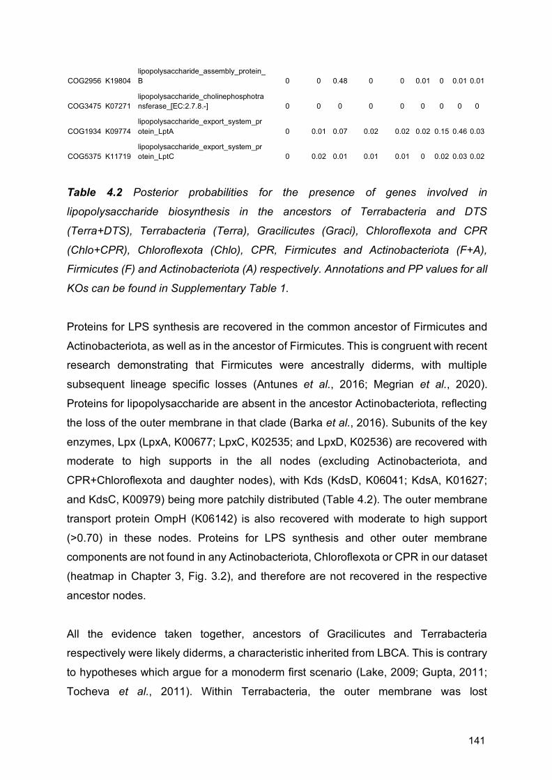
141
COG2956 K19804 lipopolysaccharide_assembly_protein_B 0 0 0.48 0 0 0.01 0 0.01 0.01
COG3475 K07271 lipopolysaccharide_cholinephosphotransferase_[EC:2.7.8.-] 0 0 0 0 0 0 0 0 0
COG1934 K09774 lipopolysaccharide_export_system_protein_LptA 0 0.01 0.07 0.02 0.02 0.02 0.15 0.46 0.03
COG5375 K11719 lipopolysaccharide_export_system_protein_LptC 0 0.02 0.01 0.01 0.01 0 0.02 0.03 0.02
Table 4.2 Posterior probabilities for the presence of genes involved in
lipopolysaccharide biosynthesis in the ancestors of Terrabacteria and DTS
(Terra+DTS), Terrabacteria (Terra), Gracilicutes (Graci), Chloroflexota and CPR
(Chlo+CPR), Chloroflexota (Chlo), CPR, Firmicutes and Actinobacteriota (F+A),
Firmicutes (F) and Actinobacteriota (A) respectively. Annotations and PP values for all
KOs can be found in Supplementary Table 1.
Proteins for LPS synthesis are recovered in the common ancestor of Firmicutes and
Actinobacteriota, as well as in the ancestor of Firmicutes. This is congruent with recent
research demonstrating that Firmicutes were ancestrally diderms, with multiple
subsequent lineage specific losses (Antunes et al., 2016; Megrian et al., 2020).
Proteins for lipopolysaccharide are absent in the ancestor Actinobacteriota, reflecting
the loss of the outer membrane in that clade (Barka et al., 2016). Subunits of the key
enzymes, Lpx (LpxA, K00677; LpxC, K02535; and LpxD, K02536) are recovered with
moderate to high supports in the all nodes (excluding Actinobacteriota, and
CPR+Chloroflexota and daughter nodes), with Kds (KdsD, K06041; KdsA, K01627;
and KdsC, K00979) being more patchily distributed (Table 4.2). The outer membrane
transport protein OmpH (K06142) is also recovered with moderate to high support
(>0.70) in these nodes. Proteins for LPS synthesis and other outer membrane
components are not found in any Actinobacteriota, Chloroflexota or CPR in our dataset
(heatmap in Chapter 3, Fig. 3.2), and therefore are not recovered in the respective
ancestor nodes.
All the evidence taken together, ancestors of Gracilicutes and Terrabacteria
respectively were likely diderms, a characteristic inherited from LBCA. This is contrary
to hypotheses which argue for a monoderm first scenario (Lake, 2009; Gupta, 2011;
Tocheva et al., 2011). Within Terrabacteria, the outer membrane was lost

142
independently in various lineages (Megrian et al., 2020), including the Actinobacteriota,
several classes of Firmicutes (Antunes et al., 2016), and the branch leading to CPR
and Chloroflexota.
Terrabacteria and terrestrialisation In Chapter 2, we recover support for Terrabacteria, a clade first described by Battistuzzi
et al. (2004). They name the group due to its inclusion of many phyla that are partially
or fully terrestrial, and hypothesise that the emergence of Terrabacteria may represent
an early colonisation of the terrestrial environment. They further expand on this,
suggesting the widespread presence of genes related to sporulation and presence of
a monderm cell envelope with a thick peptidoglycan layer confer resistance to various
environmental stresses related to terrestrial habitats, such as desiccation, ultraviolet
radiation, and high salt concentration (Battistuzzi and Hedges, 2009). We recover a
small number of sporulation genes in the ancestors of both Terrabacteria and
Gracilicutes (called “Hydrobacteria” by Battistuzzi and Hedges), yet it is difficult to
assess whether either would have had the ability to form spores, although it seems
unlikely given their sparse distribution. A number of sporulation genes are found in the
ancestor of Firmicutes, which contains many spore forming members. Additionally, as
already explained above, and in Chapter 3, we find evidence for the presence of a
diderm cell envelope architecture in the ancestor of Terrabacteria and its most basally
branching clades. It is therefore difficult to assess the validity of Battistuzzi and Hedges’
hypothesis, but given our results, it seems unlikely.
Terrestrialisation has also been suggested to have evolved via expanding genome
sizes, mediated by acquiring the dnaE2 gene in terrestrial lineages (Wu et al., 2014).
Wu et al. claim that increased genomes size would facilitate greater adaptive ability to
terrestrial environments, which are more heterogeneous than marine environments.
They demonstrate that within clades, terrestrial species have larger genomes than
marine species, with genomes decreasing in clades which re-enter the marine realm.
In our analyses, we do not recover dnaE2 in any of the nodes surveyed. We do see
larger genome sizes within the ancestral nodes of Terrabacteria compared to
Gracilicutes, and larger genomes sizes in the ancestors of predominantly terrestrial
phyla, such as Firmicutes, when compared to predominantly marine phyla, such as

143
Cyanobacteria (Fig. 4.3). However, the differences are not dramatic, and further study
with better taxon sampling would be needed to investigate this further.
4.4 Conclusion
Patterns of gene transfer show that the Terrabacteria radiated earlier than the
Gracilicutes, with all clades within Terrabacteria predating the entire Gracilicute
radiation except for Cyanobacteria and Actinobacteriota. CPR seems to represent an
early radiation, demonstrating that, though not in a basal position in the tree, they have
formed a part of bacterial communities for most of evolutionary history. In contrast,
Cyanobacteria and Alphaproteobacteria both emerged relatively late during bacterial
evolution, which further implies the emergence of eukaryotic cells at a later stage of
the diversification of life (Parfrey et al., 2011; Eme et al., 2014b; Knoll, 2014; Nicholas
J. Butterfield, 2015; Betts et al., 2018), rather than being contemporaneous or pre-
dating the emergence of most bacterial lineages (Kurland, Collins and Penny, 2006).
We recover evidence for central carbohydrate metabolic pathways across the deepest
nodes in the bacterial tree, with glycolysis being strongly supported in all cases, and
other pathways, such as the pentose phosphate pathway and the TCA cycle being
more moderate in their support and inconsistent in their distribution. With regards to
carbon fixation, we do not find evidence of the Calvin Cycle, or any of the three variants
of the 3-hydroxypropionate bicycle in any nodes. Although we identify some
components of the TCA cycle in various nodes, the key enzyme of the reverse TCA
cycle, ATP citrate lyase, is not recovered in any of them. Given that the directionality
of the enzymes is difficult to assess (Nunoura et al., 2018), we cannot conclusively say
whether any of the nodes assessed possessed the reverse TCA cycle, although we
cannot rule out the possibility. The presence of components of the methyl-branch of
the WLP, as well as components of a putative Rnf complex, together may indicate that,
as in LBCA, both the ancestor of Gracilicutes and Terrabacteria were capable of
acetogenic growth (Schuchmann and Müller, 2014). This pathway seems to have been
subsequently lost in Actinobacteriota and in the ancestor of Chloroflexota and CPR, as
although these lineages maintain components of the methyl branch, they lack the Rnf

144
complex. However, we fail to recover any components of the key enzyme of the
pathway, CODH/ACS (Adam, Borrel and Gribaldo, 2018) in any of the nodes, except
for a single subunit in the ancestor of Gracilicutes, making it difficult to conclusively
establish presence of the WLP or acetogenesis. As in LBCA, it has proven difficult to
conclusively establish the central energy metabolism of the earliest Bacteria.
We find evidence of proteins for G3P phospholipids in all the surveyed nodes, except
for CPR, although the components of the pathway are patchily distributed. We also
find no evidence of the presence of archaeal G1P lipids. Early bacterial lineages
therefore likely possessed G3P lipids, as in modern Bacteria, with the ability to produce
membrane phospholipids being lost in the CPR (Castelle et al., 2018). We further
identified most of the proteins required to synthesise motile appendages such as
flagella and pili in the ancestors of Gracilicutes and Terrabacteria. This implies that the
earliest lineages of Bacteria were motile, and likely lived in environments in which
dispersal, chemotaxis and surface attachment would have been advantageous. The
flagellum was lost in Actinobacteriota and the common ancestor of Chloroflexota and
CPR, modern members of which do not typically have flagella (Barka et al., 2016;
Castelle et al., 2018; Miyata et al., 2020).
We recover proteins involved in the construction of the outer cell membrane, including
for LPS biosynthesis, in the ancestors of Gracilicutes and Terrabacteria, from which
we infer that both possessed a double membrane with an LPS layer. Consistent with
this inference, we infer the presence of the flagellar subunits FlgH, FlgI and FgA in,
which anchor flagella in diderm membranes (Antunes et al., 2016), and for the Type
IV pilus subunit PilQ, which among extant bacteria is specific to diderms (Antunes et
al., 2016; Megrian et al., 2020). We additionally recover these proteins in the common
ancestor of Firmicutes and Actinobacteriota, with the loss of these pathways in
Actinobacteriota and the common ancestor of Chloroflexota and CPR. Taken together,
these results are consistent with hypotheses (Cavalier-Smith, 2006) in which the
earliest bacterial lineages were diderms (Antunes et al., 2016; Megrian et al., 2020),
and argue against Monoderm-first scenarios (Lake 2009; Gupta 2011; Tocheva,
Ortega and Jensen 2016). Subsequent diderm-to-monoderm transitions may have
occurred on multiple occasions within Bacteria (Antunes et al., 2016; Megrian et al.,
2020). Furthermore, the presence of flagellar in the deepest nodes further supports
145
hypotheses that the earliest Bacteria were motile organism (Liu and Ochman, 2007a,
2007b), and not non-motile cells living on mineral substrates (Martin and Russell,
2007; Lane and Martin, 2012; Sousa et al., 2013; Sousa and Martin, 2014), with the
evolution of alternative means of motility (Miyata et al., 2020) or loss of motility in later
lineages.
We do not recover evidence of terrestrial adaptation in the ancestral nodes within
Terrabacteria, as had been previously suggested (Battistuzzi, Feijao and Hedges,
2004; Battistuzzi and Hedges, 2009). However, the hypothesised relationship between
genomes size and terrestrialisation (Wu et al., 2014) is less clear, and needs additional
exploration. It seems unlikely that terrestrialisation happened deep within the
terrabacterial tree, but rather occurred independently in various lineages, as is likely
the case in Gracilicutes.
More extensive analyses would need to be done on individual gene families to better
answer some of the outstanding questions, especially relating to carbon fixation and
the evolution of terminal oxidases. However, the early evolution of Bacteria was
seemly dominated by autotrophic, tentatively anaerobic acetogenic, motile diderm
cells on the one hand, and highly reduced, metabolically minimalist cells on the other,
before the later rise of photosynthetic and aerobic Bacteria, and the eventually
appearance of eukaryotic cells.

146
Chapter 5
Investigating the Origins of Membrane
Phospholipid Biosynthesis Genes Using
Outgroup-Free Rooting
This chapter has been published as a Coleman et al. (2019) in Genome Biology and
Evolution in collaboration with Richard D. Pancost and Tom A. Williams. Gareth A.
Coleman is the first author of this paper. The project was conceived by GAC, RDP and
TAW. GAC and TAW designed and performed the analyses. GAC, RDP and TAW
interpreted the results and wrote the manuscript.
Paper published as:
Coleman, G.A., Pancost, R.D. and Williams, T.A., 2019. Investigating the origins of
membrane phospholipid biosynthesis genes using outgroup-free rooting. Genome
biology and evolution, 11(3), pp.883-898.
Paper can be found here:
https://academic.oup.com/gbe/article/11/3/883/5310093

147
Abstract
One of the key differences between Bacteria and Archaea is their canonical membrane phospholipids, which are synthesised by distinct biosynthetic pathways with nonhomologous enzymes. This “lipid divide” has important implications for the early evolution of cells and the type of membrane phospholipids present in the last universal common ancestor. One of the main challenges in studies of membrane evolution is that the key biosynthetic genes are ancient and their evolutionary histories are poorly resolved. This poses major challenges for traditional rooting methods because the only available outgroups are distantly related. Here, we address this issue by using the best available substitution models for single-gene trees, by expanding our analyses to the diversity of uncultivated prokaryotes recently revealed by environmental genomics, and by using two complementary approaches to rooting that do not depend on outgroups. Consistent with some previous analyses, our rooted gene trees support extensive interdomain horizontal transfer of membrane phospholipid biosynthetic genes, primarily from Archaea to Bacteria. They also suggest that the capacity to make archaeal-type membrane phospholipids was already present in the last universal common ancestor.

148
5.1 Introduction Archaea and Bacteria form the two primary domains of life (Williams et al., 2013).
Although similarities in their fundamental genetics and biochemistry, and evidence of
homology in a near-universally conserved core of genes (Weiss et al., 2016) strongly
suggest that Archaea and Bacteria descend from a universal common ancestor
(LUCA), they also differ in ways that have important implications for the early evolution
of cellular life. These differences include DNA replication (Kelman and Kelman, 2014),
transcription (Bell and Jackson, 1998), DNA packaging (Reeve, Sandman and Daniels,
1997), and cell wall compositions (Kandler, 1995). One striking difference is in the
phospholipid composition of the cell membranes (Fig. 5.1), which is particularly
important for understanding the origin of cellular life. Canonically, Archaea have
isoprenoid chains attached to a glycerol-1-phosphate (G1P) backbone via ether bonds
and can have either membrane spanning or bilayer-forming phospholipids (Lombard,
López-García and Moreira, 2012b). Most Bacteria, as well as eukaryotes, classically
have acyl (fatty-acid) chains attached to a glycerol-3-phosphate (G3P) backbone via
ester bonds and form bilayers (Lombard, López-García and Moreira, 2012b), although
a number of exceptions have been documented (Sinninghe Damsté et al., 2002;
Weijers et al., 2006; Damsté, Sinninghe Damsté, et al., 2007; Goldfine, 2010).
Archaeal and bacterial phospholipids are synthesised by non-homologous enzymes
via different biosynthetic pathways (Fig. 5.1). This so-called “lipid divide” (Koga, 2011)
raises some important questions regarding the early evolution of cellular life, including
the nature of the membrane phospholipids present in LUCA and the number of times
cell membranes have evolved.

149
Fig. 5.1 (above) (a) The canonical ether/ester biosynthetic pathways in Archaea and
Bacteria and how they relate to glycerol metabolism. Based on Figure 1 from
Villanueva et al. (2017). Archaeal pathways in blue and yellow (blue = heterotrophic
Archaea and yellow = autotrophic Archaea), bacterial pathway in red. Hypothetical
biosynthetic pathway, as suggested by Villanueva et al. (2017), in dashed lines. (b)
Composition of bacterial and archaeal phospholipids. In Archaea, glycerol-1-
phosphate (G1P) is synthesised from dihydroxyacetone phosphate (DHAP) using the
enzyme glycerol-1-phosphate dehydrogenase (G1PDH). The first and second
isoprenoid chains (GGGPs) are added by geranylgeranylglyceryl phosphate synthase
(GGGPS) and digeranylgeranylglyceryl phosphate synthase (DGGGPS), respectively.
In Bacteria, glycerol-3-phosphate (G3P) is synthesised by glycerol-3-phosphate
dehydrogenase (G3PDH) from DHAP. There are two forms of this enzyme, GpsA and
GlpA/GlpD, encoded by the gps and glp genes, respectively. G3P may also be
produced from glycerol by glycerol kinase (GlpK). In certain Bacteria, such as
Gammaproteobacteria, the first fatty-acid chain is added by a version of glycerol-3-
phosphate acyltransferase called PlsB. Other Bacteria, including most gram-positive
bacteria, use a system which includes another glycerol-3-phosphate acyltransferase,
PlsY, in conjunction with the enzyme PlsX (Parsons and Rock, 2013; Yao and Rock,
2013). The second fatty-acid chain is attached by 1-acylglycerol-3-phosphate O-
acyltransferase (PlsC).
The observation that phospholipid biosynthesis in Bacteria and Archaea is non-
homologous has motivated various hypotheses on the nature of LUCA’s membrane.
The likely presence of some genes for lipid biosynthesis (Lombard and Moreira, 2011;
Lombard, López-García and Moreira, 2012b; Koga, 2014; Weiss et al., 2016) and, in
particular, a membrane-bound ATPase (Sojo, Pomiankowski and Lane, 2014; Weiss
et al., 2016) in reconstructions of LUCA’s genome implies that LUCA possessed a
membrane, although its properties may have been somewhat different to those of
modern, ion-tight prokaryote cell membranes (Lombard, López-García and Moreira,
2012b; Koga, 2014; Sojo, Pomiankowski and Lane, 2014). It has also been suggested
that LUCA may have had a heterochiral membrane (Wächtershäuser, 2003), with later
independent transitions to homochirality in Bacteria and Archaea, driven by increased
membrane stability. However, the available experimental evidence, including the

150
recent engineering of an Escherichia coli cell with a heterochiral membrane (Caforio
et al., 2018), suggests that homochiral membranes are not necessarily more stable
than heterochiral ones (Fan et al., 1995; Shimada and Yamagishi, 2011; Caforio et al.,
2018), requiring some other explanation for the loss of ancestral heterochirality.
Despite the importance of the lipid divide for our understanding of early cellular
evolution, membrane phospholipid stereochemistry of the glycerol moiety has been
directly determined for a surprisingly limited range of Bacteria and Archaea. Since the
initial full structural characterization of archaeol by (Kates, 1978), most subsequent
studies of ether membrane lipids have assumed their stereochemistry while focusing
on other aspects of their structure. Those studies that have determined the glycerol
stereochemistry of membrane lipids (i.e., (Sinninghe Damsté et al., 2002; Weijers et
al., 2006) are largely consistent with the idea that it is a conserved difference between
Bacteria and Archaea. Nonetheless, there is evidence that some Bacteria can make
G1P-linked ether lipids. For example, the model bacterium Bacillus subtilis has been
shown to possess homologs of archaeal glycerol-1-phosphate dehydrogenase
(G1PDH) and geranylgeranylglyceryl phosphate synthase (GGGPS) (Guldan, Sterner
and Babinger, 2008; Guldan et al., 2011). These enzymes allow B. subtilis to
synthesise a typically archaeal ether link between G1P and HepPP, resulting in a lipid
with archaeal characteristics, although there is no evidence that these archaeal-like
lipids are used to make phospholipids or are incorporated into the B. subtilis
membrane.
Apart from stereochemistry, other characteristics of membrane phospholipids appear
to be more variable, showing a mixture of archaeal and bacterial features. For example,
the plasmalogens of animals and anaerobic Bacteria include an ether bond (Goldfine,
2010). Branched glycerol dialkyl glycerol tetra-ether lipids found in the environment
have bacterial stereochemistry and branched rather than isoprenoidal alkyl chains, but
they also contain ether bonds and span the membrane, as observed for canonical
archaeal lipids (Schouten et al., 2000; Weijers et al., 2006). These branched glycerol
dialkyl glycerol tetra-ethers are particularly abundant in peat bogs and were thought to
be produced by Bacteria as adaptations to low pH environments (Weijers et al., 2006;
Damsté, Sinninghe Damsté, et al., 2007), but are now known to occur in a wide range
of soils and aquatic settings (Schouten, Hopmans and Sinninghe Damsté, 2013). The

151
enzymes responsible for their synthesis are currently unknown. On the other side of
the “lipid divide,” some Archaea have been shown to produce membrane lipids with
fatty-acid chains and ester bonds (Gattinger, Schloter and Munch, 2002). The
biosynthetic pathways for all of these mixed-type membrane lipids remain unclear.
However, given the frequency with which prokaryotes undergo horizontal gene transfer
(Garcia-Vallvé, Romeu and Palau, 2000), one possibility is that these mixed
biochemical properties reflect biosynthetic pathways of mixed bacterial and archaeal
origin.
A number of previous studies have investigated the evolutionary origins of
phospholipid biosynthesis genes in Bacteria and Archaea using phylogenetic
approaches, in order to test hypotheses about the nature of membranes in the earliest
cellular life-forms (Peretó, López-García and Moreira, 2004; Koga, 2014; Yokobori et
al., 2016; Villanueva, Schouten and Damsté, 2017). In this study, we build upon that
work by performing comprehensive phylogenetic analyses for the core phospholipid
biosynthesis genes in Bacteria and Archaea: the enzymes that establish membrane
lipid stereochemistry and attach the two carbon chains to the glycerol phosphate
backbone (Fig. 5.1), as the histories of these enzymes are key to understanding the
evolution of membrane biosynthesis and stereochemistry. Our analyses take
advantage of the wealth of new genome data from environmental prokaryotes that has
become available recently, and we employ new approaches for rooting single-gene
trees in order to circumvent some of the difficulties inherent in traditional outgroup
rooting for anciently diverged genes. Our results agree with previous work in
suggesting that LUCA likely possessed a cell membrane. Our rooted gene trees
indicate that transfers of lipid biosynthetic genes from Archaea to Bacteria have
occurred more frequently in evolution, particularly during the early diversification of the
two domains.
5.2 Materials and Methods
Sequence Selection For Archaea, we selected 43 archaeal genomes, sampled evenly across the archaeal
tree. We took corresponding archaeal G1PDH, geranylgeranylglyceryl phosphate

152
synthase (GGGPS), and digeranylgeranylglyceryl phosphate synthase (DGGGPS)
amino acid sequences from the data set of Villanueva et al. (2017) and performed
BlastP searches to find these sequences in genomes not included in that data set. For
Bacteria, we selected 64 bacterial genomes, sampled so as to represent the known
genomic diversity of bacterial phyla (Hug et al., 2016). We used GpsA, GlpA/GlpD, and
GlpK sequences from Yokobori et al. (2016) and performed BlastP searches to find
those sequences in bacterial species not in their data set. For PlsC and PlsY, we took
the corresponding sequences from Villanueva et al. (2017) and performed BlastP
searches to find these sequences in the remaining genomes. For PlsB and PlsX, we
searched for the respective terms in the gene database on the NCBI website, and upon
finding well-verified occurrences, performed BlastP searches to find the corresponding
amino acid sequences in the remaining genomes. We then used BLASTp to look for
bacterial orthologues of the archaeal enzymes and vice versa. We selected sequences
that had an E-value of less the 10e-7 and at least 50% coverage. Accession numbers
for sequences used are provided in Supplementary Table 5.
Phylogenetics The sequences were aligned in mafft (Katoh et al., 2002) using the –auto option and
trimmed in BMGE (Criscuolo and Gribaldo, 2010) using the BLOSUM30 model, which
is most suitable for anciently diverged genes. To construct gene trees from our amino
acid sequences, we first selected the best-fitting substitution model for each gene
according to its Bayesian Information Criterion score using the model selection tool in
IQ-Tree (Nguyen et al., 2015). For all the genes we analyzed, the best-fitting model
was a mixture model combining the Le and Gascuel (LG) exchangeability matrix (Le
and Gascuel 2008) with site-specific composition profiles (the C40, C50, and C60
models (Lartillot and Philippe, 2004; Le and Gascuel, 2008)) to accommodate across-
site variation in the substitution process. LG + C60 was used for G1PDH, DGGGP,
GpsA, GlpA/GlpD, GlpK, and PlsC. LG + 50 was used for PlsY. LG + C40 was used
for GGGPS. A discretised Gamma distribution (Yang, 1994) with four rate categories
was used to model across-site rate variation. The trees were inferred with their
respective models in PhyloBayes (Lartillot and Philippe, 2004; Lartillot, Brinkmann and
Philippe, 2007b); convergence was assessed using the bpcomp and tracecomp
programs (maxdiff < 0.1; effective sample sizes > 100), as recommended by the
authors. We additionally inferred maximum likelihood (ML) trees in IQ-Tree using the
153
LG + C60 model for each enzyme for comparison. We used heads-or-tails (Landan
and Graur, 2007) to assess the impact of alignment uncertainty: starting with the
reversed alignments, we used the same phylogenetics pipeline as described above.
Further testing was carried out by removing the metagenomic data from G1PDH,
GGGPS, DGGGPS, GpsA, GlpA/GlpD, and GlpK, creating new alignments as
described above, and inferring trees from these alignments in IQ-Tree using the LG +
C60 model. We did not remove metagenomic data for PlsC or PlsY, as all of the
archaeal sequences for these trees are derived from metagenome bins. In some
cases, our trees included highly divergent sequences (sometimes forming distinct
clades); we checked the E-values for these hits, and if they were close to or at the 10e-
7 cut-off, they were removed and the analyses were rerun.
The trees were rooted with an uncorrelated lognormal relaxed molecular clock (RMC),
using the LG model with a discretised Gamma distribution (Yang, 1994) with four rate
categories, and a Yule tree prior (Drummond and Rambaut, 2007; Drummond et al.,
2012). We also rooted the trees using minimal ancestor deviation (MAD) rooting (Tria,
Landan and Dagan, 2017). We used two complementary methods: root posterior
probabilities averaged over the trees sampled during the Bayesian molecular clock
analysis using RootAnnotator (Calvignac-Spencer et al., 2014), and the ambiguity
index (AI) implemented in MAD. The AI is defined as the ratio of the MAD value to the
second smallest value. “Ties”, that is, where two or more competing root positions with
equal deviations, would obtain a score of 1, with smaller values obtained in proportion
to the relative quality of the best root position.
For G1PDH, GpsA, and GlpA/GlpD, we also rooted using a subsample of the outgroup
sequences used by Yokobori et al. (2016). The outgroups used were two sequences
annotated as 3-dehydroquinate synthase, five as glycerol dehydrogenase, and five as
alcohol dehydrogenase for G1PDH; six sequences annotated as hydroxyacyl-CoA
dehydrogenase and six as uridine diphosphoglucose 6-dehydrogenase sequences for
GpsA; and 12 sequences annotated as flavin adenine dinucleotide dependent
oxidoreductase for GlpA/GlpD. All three of these trees were inferred under the LG +
C60 model to directly compare to the unrooted trees. Trees were also inferred from
best-fit models selected in IQTree (LG + C60 for G1PDH and GlpA/GlpD and LG +
C50 for GpsA).

154
Eukaryotic orthologues of prokaryotic phospholipid biosynthesis genes (GlpA/GlpD,
GpsA, and PlsC) were identified by performing BlastP searches on 35 eukaryotic
genomes from across eukaryotic diversity using Homo sapiens query as the sequence
in each case, selecting sequences with an E-value of 10e-7 or less, and at least 50%
coverage. We then performed model testing in IQTree and inferred trees in
PhyloBayes using the selected substitution model (LG + C60 for PlsC and LG + C50
for GlpA/GlpD and GpsA).
5.3 Results and Discussion
Distribution of Core Phospholipid Biosynthesis Genes We performed BlastP searches for the enzymes of the canonical archaeal and
bacterial lipid biosynthesis pathways (Fig. 5.1) against all archaeal and bacterial
genomes in the NCBI nr database. Our BLAST searches revealed homologs for all of
the core phospholipid biosynthesis genes of both pathways in both prokaryotic
domains, with the exception of bacterial enzymes PlsB and PlsX, which we did not find
in Archaea. Orthologues of the canonical archaeal genes are particularly widespread
in many bacterial lineages (Table 5.1). Of the 52 bacterial phyla surveyed, 8 had no
orthologues of the archaeal genes (Table 5.1, indicated in red). Six phyla have
orthologues of all three archaeal genes distributed across various genomes (Table 5.1,
indicated in yellow and green). Of these phyla, Firmicutes (genera Bacillus and
Halanaerobium), Actinobacteria (genus Streptomyces), and Fibrobacteres (genera
Chitinispirillum and Chitinivibrio) contain species which have all three genes in their
genomes (Table 5.1, indicated in green). Based on the presence of all three core
biosynthetic genes, and given their recognised role in the synthesis of archaeal-like
lipid components in B. subtilis (Guldan, Sterner and Babinger, 2008; Guldan et al.,
2011), members of Firmicutes, Actinobacteria, and Fibrobacteres lineages of Bacteria
may be capable of making archaea like lipids, although we cannot determine if these
are used in the production of membrane phospholipids. Of the 12 FCB group
(Fibrobacteres, Chlorobi, Bacteroidetes and related lineages) phyla we surveyed, all
12 have GGGPS and DGGGPS orthologues, but only Fibrobacteres and
Cloacimonetes have G1PDH orthologues (see Fig. 5.1 for overview of pathway). In
these species lacking G1PDH, it is unclear whether GGGPS and DGGGPS are active

155
and if so, what they are used for; one possibility is that they catalyze the reverse
reaction, catabolising archaeal lipids as an energy source. However, a very recent
report (Villanueva, von Meijenfeldt and Westbye, 2018) has shown that the GGGPS
and DGGGPS genes from one FCB lineage, Cloacimonetes, support the production of
archaeal-type membrane phospholipids and a mixed membrane when heterologously
expressed in E. coli. This suggests that both E. coli and perhaps Cloacimonetes have
an alternative, as yet unknown mechanism for making G1P, and that some FCB
members may have mixed archaeal and bacterial membranes.
Domain Superphylum Phylum Class G1PDH GGGPS DGGGPS GpsA GlpA/GlpD GlpK PlsC PlsY
Archaea Euryarchaeota Archaeoglobi ✓ ✓ ✓ ✓ ✓ ✓
Halobacteria ✓ ✓ ✓
✓ ✓
Methanobacteria ✓ ✓ ✓ ✓
Methanococci ✓ ✓ ✓
Methanomicrobia ✓ ✓ ✓ ✓ ✓
Thermococci ✓ ✓ ✓
✓ ✓
Thermoplasmatales ✓ ✓ ✓ ✓ ✓ ✓ ✓
TACK Aigarchaeota ✓ ✓ ✓
Crenarchaeota ✓ ✓ ✓ ✓ ✓ ✓
Korarchaeota ✓ ✓ ✓
✓ ✓
Thaumarchaeta ✓ ✓ ✓
Asgard Heimdallarchaeota ✓ ✓ ✓
✓ ✓
Lokiarchaeota ✓ ✓ ✓
✓ ✓ ✓ ✓
Odinarchaeota ✓ ✓ ✓

156
Thorarchaeota ✓ ✓ ✓
✓
DPANN Aenigmarchaeota ✓ ✓ ✓
Diapherotrites ✓ ✓ ✓
✓
Micrarchaeota ✓ ✓ ✓
Nanoarchaeota
Nanohaloarchaeota
Pacearchaeota
✓
Parvarchaeota
✓ ✓
Woesearchaeota
✓
✓ ✓ ✓
Bacteria
Acidobacteria ✓
✓
✓ ✓ ✓
Actinobacteria ✓ ✓ ✓ ✓ ✓ ✓ ✓ ✓
Aminicenantes ✓ ✓ ✓ ✓ ✓
Aquificae
✓ ✓ ✓ ✓ ✓
Armatimonadetes ✓ ✓ ✓ ✓ ✓ ✓
Candidate division KSB1 ✓ ✓ ✓ ✓ ✓ ✓ ✓
Candidate division NC10
✓ ✓ ✓ ✓ ✓
Candidate division TA06 ✓ ✓ ✓ ✓ ✓ ✓
Candidate division WOR-3 ✓ ✓ ✓ ✓ ✓ ✓
Candidatus Edwardsbacteria ✓ ✓ ✓ ✓ ✓
Candidatus Handelsmanbacteria ✓ ✓ ✓ ✓ ✓ ✓

157
Candidatus Kerfeldbacteria ✓ ✓ ✓ ✓ ✓ ✓
Candidatus Magnetoovum ✓ ✓ ✓ ✓ ✓
Candidatus Raymondbacteria ✓ ✓ ✓ ✓ ✓ ✓ ✓ ✓
Chloroflexi ✓ ✓ ✓ ✓ ✓ ✓ ✓ ✓
Chrysiogenetes ✓ ✓ ✓ ✓ ✓
Cloacimonetes ✓ ✓ ✓ ✓ ✓ ✓ ✓ ✓
Cyanobacteria ✓ ✓ ✓ ✓ ✓ ✓ ✓ ✓
Deferribacterales
✓ ✓ ✓ ✓ ✓
Deinococcus-Thermus ✓ ✓ ✓ ✓ ✓ ✓
Dictyoglomi ✓ ✓ ✓ ✓ ✓
Elusimicrobia ✓ ✓ ✓ ✓ ✓ ✓ ✓
Firmicutes ✓ ✓ ✓ ✓ ✓ ✓ ✓ ✓
Fusobacteria ✓ ✓ ✓ ✓ ✓
Melainabacteria ✓ ✓ ✓ ✓ ✓ ✓ ✓
Nitrospinae
✓ ✓
✓ ✓
Nitrospirae ✓ ✓ ✓ ✓ ✓ ✓
Parcubacteria ✓ ✓ ✓ ✓ ✓
Proteobacteria ✓ ✓ ✓ ✓ ✓ ✓ ✓ ✓
Rhodothermaeota ✓ ✓ ✓ ✓ ✓ ✓
Spirochaetes ✓ ✓ ✓ ✓ ✓ ✓ ✓
Synergistetes ✓ ✓ ✓ ✓ ✓
Tenericutes
✓ ✓ ✓ ✓ ✓
Thermobaculum
✓ ✓ ✓ ✓ ✓

158
Thermodesulfobacteria ✓ ✓ ✓
Thermotogae ✓ ✓ ✓ ✓ ✓ ✓ ✓
TMED ✓ ✓ ✓
FCB Chlorobi ✓ ✓ ✓ ✓ ✓ ✓ ✓
Caldithrix ✓ ✓ ✓ ✓ ✓ ✓ ✓
Bacteroidetes ✓ ✓ ✓ ✓ ✓ ✓
Candidatus Marinimicrobia ✓ ✓ ✓ ✓ ✓ ✓ ✓
Fibrobacteres ✓ ✓ ✓ ✓ ✓ ✓
Ignavibacteria ✓ ✓ ✓ ✓ ✓
Gemmatimonadetes ✓ ✓ ✓ ✓ ✓ ✓ ✓
Latescibacteria ✓ ✓ ✓ ✓ ✓
Candidatus Kryptonium ✓ ✓ ✓ ✓ ✓
Candidatus Kryptobacter ✓ ✓ ✓ ✓ ✓
Candidate division Zixibacteria
✓ ✓ ✓
✓ ✓
PVC Chlamydia
✓
✓ ✓ ✓
Planctomycetes ✓
✓ ✓ ✓ ✓ ✓
Lentisphaerae ✓
✓ ✓ ✓ ✓ ✓ ✓
Verrucomicrobia
✓
✓ ✓ ✓
Table 5.1 Distribution of Phospholipid Biosynthesis Genes in Bacterial and Archaeal
Phyla. Ticks represent phyla (class level for Euryarchaeota) with at least one genome
which has a sequence for the corresponding gene. Bacterial phyla where all three
archaeal genes are found are indicated in yellow and green. Those bacterial phyla
where all three archaeal genes are found within the same genome in at least one case
are indicted in green. Those bacterial phyla with no archaeal genes are found are
indicated in red. It should be noted that in the case of environmental lineages, the lack
159
of a tick may not represent absence of genes, given that these represent
metagenomics bins, and the lack of said genes may be due to missing data. FCB are
Fibrobacteres, Chlorobi, and Bacteroidetes and related lineages. PVC are
Planctomycetes, Verrucomicrobia, and Chlamydiae and related lineages. TACK are
Thaumarchaeota, Aigarchaeota, Crenarchaeota, and Korarchaeota. DPANN include
Diapherotrites, Parvarchaeota, Aenigmarchaeota, Nanoarchaeota, and
Nanohaloarchaeota, as well as several other lineages.
Orthologues of the canonical bacterial genes are less widespread in Archaea (table 1).
Of all the genomes surveyed, none contained all homologs. Of the 17 phyla shown in
table 1, 8 had no bacterial homologs in any of their genomes. Orthologues of GpsA,
GplA/GlpD, and Gpk are found in at least one genome of each of the major archaeal
clades (Euryarchaeota, Thaumarchaeota, Aigarchaeota, Crenarchaeota, and
Korarchaeota (TACK), Asgardarchaeota, and Diapherotrites, Parvarchaeota,
Aenigmarchaeota, Nanoarchaeota, and Nanohaloarchaeota, as well as several other
lineages (DPANN) (Williams et al., 2017)). However, they appear sporadically. Within
Euryarchaeota, of the seven classes surveyed, GpsA and GlpK appear in the genomes
of four and GlpA/GlpD in five. Within the TACK superphylum, GlpA/GlpD and GlpK
appear in Crenarchaeota and Korarchaeota, whereas GpsA appears only in a single
crenarchaeote genome (Thermofilum). GpsA and GlpK are also found in at least one
genome in two of the eight DPANN phyla surveyed (Woesearchaeota and GW2011,
and Woesearchaeota and Parvarchaeota, respectively), whereas GlpA/GlpD is found
in a single parvarchaeote genome (Candidatus Parvarchaeum acidiphilum ARMAN-
4). Within the Asgardarchaeota superphylum, no orthologues for GpsA are found, and
only one of the genomes (Lokiarchaeum sp. GC14_75) has GlpA/GlpD or GlpK. PlsC
and PlsY are more restricted, being found mainly in environmental lineages within
Euryarchaeota (Marine Groups II/III, all in class Thermoplasmatales), DPANN, and
Asgardarchaeota (Table 5.1).
Early Origins of Archaeal-Type Membrane Phospholipid Biosynthesis Genes in Bacteria To investigate the evolutionary histories of membrane phospholipid biosynthesis, we
inferred Bayesian single-gene phylogenies from the amino acid alignments using

160
PhyloBayes 4.1 (Lartillot and Philippe, 2004; Lartillot, Brinkmann and Philippe, 2007b).
We selected the best-fitting substitution model for each gene according to its Bayesian
Information Criterion score using the model selection tool in IQ-Tree (Nguyen et al.,
2015). We used two complementary approaches to root these single-gene trees: a
RMC in BEAST (Drummond and Rambaut, 2007; Drummond et al., 2012) (Table 5.2),
and the recently described MAD rooting method of Tria et al. (2017). The MAD
algorithm finds the root position that minimises pairwise evolutionary rate variation,
averaged over all pairs of taxa in the tree. Many of our single-gene trees were poorly
resolved, and we wanted to account for topological uncertainty in our root estimates.
To do so, we used two complementary methods: root posterior probabilities (Table 5.2)
averaged over the trees sampled during the Bayesian molecular clock analysis, and
the AI implemented in MAD, which is defined by Tria et al. (2017) as the ratio of the
MAD value to the second smallest value (Table 5.3). For the genes for which an
outgroup was available (G1PDH, GpsA, and GlpA/GlpD, following Yokobori et al.
2016), we compared our results to traditional outgroup rooting. For more details, see
Materials and Methods.
Gene RMC MAD RMC (metagenomic sequences removed)
G1PDH 0.68 0.62 0.4
GGGPS 0.99 1 1
DGGGPS 0.43 0.79 0.99
GpsA 0.31 0.59 0.41
GlpA/GlpD 0.5 0.44 N/A
GlpK 0.47 0.34 N/A
PlsC 0.28 0.03 N/A
PlsY 0.57 0.85 N/A
Table 5.2 Maximum marginal posterior probabilities for molecular clock and MAD
rooting methods. For several bacterial genes, removing metagenomic sequences
would remove all archaeal sequences and are thus marked with “NA”; see section

161
entitled “Sensitivity to Model Fitting Approach, Alignment Uncertainty, and the Inclusion
of Metagenomic Sequences”.
Gene Bayesian trees ML trees HoT ML trees ML trees, metagenomic data removed
G1PDH 0.997 0.976 0.956 0.973
GGGPS 0.648 0.561 0.681 0.577
DGGGPS 0.947 0.995 0.897 0.659
GpsA 0.979 0.972 0.958 0.983
GlpA/GlpD 0.964 0.906 0.902 N/A
GlpK 0.999 1 0.994 N/A
PlsC 0.996 1 0.999 N/A
PlsY 0.959 0.982 0.926 N/A
Table 5.3 Ambiguity Index (AI) Scores for MAD roots. For several bacterial genes,
removing metagenomic sequences would remove all archaeal sequences and are thus
marked with “NA”; see section entitled “Sensitivity to Model Fitting Approach,
Alignment Uncertainty, and the Inclusion of Metagenomic Sequences”.
G1PDH is the enzyme that establishes phospholipid stereochemistry in Archaea.
Interestingly, the majority of the bacterial G1PDH orthologues do not appear to be
recent horizontal acquisitions from Archaea, but instead form a deep-branching clan
(Wilkinson et al., 2007) (PP = 1), resolved as sister to an archaeal lineage clan (Fig.
5.2(a)). The relationships within the clans are poorly resolved. The root position that
receives the highest posterior support in the RMC analysis is that between the archaeal
and bacterial clans, with a marginal posterior probability of 0.68 (Table 5.2). This is
substantially higher than the next most probable position, which places the root within
the Bacteria with a posterior probability of 0.1. When rooted using MAD, the same root
between the bacterial and archaeal clans is recovered with a marginal posterior
162
probability of 0.62, also substantially higher than the next most probable root of 0.1.
Rooting single-gene trees can prove difficult, and this uncertainty is captured in the low
root probabilities inferred using both the RMC and MAD methods. However, these
analyses can be used to exclude the root from some regions of the trees with a degree
of certainty. In the case of G1PDH, a post-LUCA origin of the gene would predict a root
on the archaeal stem or within the Archaea. In our analyses, no such root position has
a significant probability (i.e., PP > 0.05), and therefore the root is highly unlikely to be
within the Archaea. This is similar to topologies recovered by Peretó et al. (2004) and
Carbone et al. (2015). The bacterial clan mainly comprises sequences from Firmicutes
and Actinobacteria, with most of the other Bacteria grouping together in a single,
maximally supported (PP = 1) lineage suggestive of recent horizontal acquisition from
the Firmicutes/Actinobacteria clade, followed by further HGT.
Fig. 5.2 (below) Bayesian consensus trees of archaeal enzymes. Support values are
Bayesian posterior probabilities. The black arrow and the white arrow indicate the
modal root positions obtained using the RMC and MAD approaches, respectively. The
dashed arrow indicates the RMC and MAD roots for the larger GGGPS subclade.
Archaea in blue-tones and Bacteria in red/pink-tones. (a) G1PDH tree (111 sequences
and 190 positions) inferred under the best-fitting LG + C60 model. (b) GGGPS tree
(133 sequences and 129 positions) inferred under the best-fitting LG + C40 model. (c)
DGGGPS tree (97 sequences and 119 positions) inferred under the best-fitting LG +
C60 model. Terrabacteria are Firmicutes, Actinobacteria, Cyanobacteria, Chloroflexi,
and related lineages. FCB are the Fibrobacteria, Chlorobi, Bacteroidetes, and related
lineages. PVC are the Planctomycetes, Verrucomicrobia, Chlamydiae, and related
lineages. TACK are Thaumarchaeota, Aigarchaeota, Crenarchaeota, and
Korarchaeota. DPANN includes Diapherotrites, Parvarchaeota, Aenigmarchaeota,
Nanoarchaeota, and Nanohaloarchaeota, as well as several other lineages. For full
trees, see Appendix C, Supplementary figures 1–4. For full unrooted trees, see
Appendix C, Supplementary figures 16–18.

163

164
This root position is consistent with two scenarios between which we cannot distinguish
based on the available data. One possibility is an early transfer of G1PDH from stem
Archaea into Bacteria, either into the bacterial stem lineage with subsequent loss in
later lineages, or into the ancestor of Actinobacteria and Firmicutes, with subsequent
transfers to other Bacteria. Alternatively, G1PDH could have already been present in
LUCA, and was subsequently inherited vertically in both Archaea and Bacteria,
followed by loss in later bacterial lineages. The Firmicute sequences within the
archaeal clade appear to be a later transfer into those Firmicutes, apparently from
Thorarchaeota.
GGGPS attaches the first isoprenoid chain to G1P. Phylogenetic analysis of GGGPS
(Fig. 5.2(b)) evidenced two deeply divergent paralogues, with the tree confidently
rooted between them using both the RMC (PP = 0.99) and MAD methods (PP = 1)
(Table 5.2); resolution within each of the paralogs was poor. The recovery of two
distinct paralogues has been noted in several previous studies (Nemoto, Oshima and
Yamagishi, 2003; Boucher, Kamekura and Doolittle, 2004; Lombard, López-García
and Moreira, 2012a; Peterhoff et al., 2014). One of these paralogues comprises
sequences from some Euryarchaeota (including members of the Haloarchaea,
Methanomicrobia, and Archaeoglobi), along with Firmicutes and Actinobacteria. The
other paralogue comprises sequences from the rest of the Archaea, including other
Euryarchaeota, and a monophyletic bacterial clade largely consisting of members of
the FCB lineage. Taken with the root position between the two paralogues, the tree
topology implies an ancestral duplication followed by sorting out of the paralogues and
multiple transfers into Bacteria. Because genes from both GGGPS paralogous clades
have been experimentally characterised as geranylgeranylglyceryl phosphate
synthases (Nemoto, Oshima and Yamagishi, 2003; Boucher, Kamekura and Doolittle,
2004), it appears that this activity was already present in LUCA before the radiation of
the bacterial and archaeal domains. It has been suggested, however, that the firmicute
sequences (which comprise the majority of the sequences in the smaller paralogue)
are used in teichoic acid synthesis (Payandeh et al., 2006). In this case, two apparently
diverging paralogues may be an artefact due to changes in the sequences during
neofunctionalisation. Lombard et al. (2012b), who also find two divergent homologues,
and homologues in a large diversity of FCB bacteria (mostly Bacteroidetes), suggest
that one of these homologues was likely present in the last archaeal common ancestor,

165
whereas the bacterial sequences were likely horizontal transfers. To improve
resolution among the deeper branches of the tree, we inferred an additional phylogeny
focusing just on the larger of the two clades (Appendix C, Supplementary Fig. 3). The
root of this subtree fell between a clade of monophyletic Bacteria and a clade of
Archaea in which six bacterial sequences were interleaved, perhaps as the result of
later gene transfer (PP = 0.8 for the root split, much higher than the next most likely
root, within the Bacteria, with PP = 0.07). This tree might be interpreted as gene
presence in LUCA, followed by some more recent transfers from Archaea to Bacteria.
Given that this gene is a hallmark of archaeal membrane phospholipid biosynthesis,
our data do not exclude the possibility of a very early gene transfer from the archaeal
stem to Bacteria, prior to the radiation of the archaeal domain.
DGGGPS attaches the second isoprenoid chain to G1P. DGGGPS is present in all
sampled Archaea, with the exception of three of the DPANN metagenome bins.
Although the DGGGPS tree is poorly resolved (Fig. 5.2(c)), both the RMC and MAD
root the tree between the same two clades (PP = 0.43 and 0.79, respectively) (Table
5.2). The smaller clade comprises mostly bacterial sequences from the Actinobacteria
and FCB lineages, as well as two archaeal sequences (from the TACK and
Euryarchaeota lineages). The larger clade contains sequences from a diversity of
Bacteria, particularly FCB (also reported by Villanueva et al. 2018), as well as Archaea.
DGGGPS is part of the UbiA protein superfamily, which are involved in a number of
different biosynthetic pathways, including the production of photosynthetic pigments,
and are therefore widely distributed in Bacteria, and are known to have undergone
extensive HGT (Hemmi et al., 2004). Indeed, several of the sequences used in our
analyses (and those in previous studies, such as Villanueva et al. 2017) are annotated
on NCBI as other proteins within this superfamily (see Supplementary Table 5). To
distinguish orthologues of DGGGPS from other, distantly related members of the UbiA
superfamily that might have different functions, we inferred an expanded phylogeny
including our initial sequence set and sequences sampled from the other known UbiA
subfamilies (Appendix C, Supplementary Fig. 25). Surprisingly, this analysis indicated
that the Thaumarchaeota lack an orthologue of the DGGGPS gene that other Archaea
use to attach the second isoprenoid chain; the most closely related Thaumarchaeota
sequences branch within another UbiA subfamily with high posterior support (PP =
0.99). Thaumarchaeota may be using this paralog to perform the same function, or

166
may use another unrelated enzyme to catalyse this reaction. The wide distribution of
this enzyme across both Archaea and Bacteria, and the occurrence of both domains
on either side of the root, for both rooting methods, suggest either multiple transfers
into Bacteria from Archaea, or that DGGGPS was present in LUCA and inherited in
various archaeal and bacterial lineages, followed by many later losses in and transfers
between various lineages.
In sum, our results of archaeal phospholipid biosynthesis genes suggest that there
have been repeated, independent inter domain transfers of these genes from Archaea
to Bacteria throughout the evolutionary history of life. Furthermore, our phylogenetic
analyses do not exclude the possibility that the genes of the archaeal pathway were
present in LUCA. If correct, this would imply that LUCA had the capability to make
archaeal-type membrane phospholipids.
Transfers of Bacterial Membrane Phospholipid Genes into Archaea In contrast to our analyses of proteins of the classical archaeal pathway, phylogenies
of proteins of bacterial-type membrane phospholipid biosynthesis pathways are more
ambiguous and the root positions are not confidently resolved. Homologs of both forms
of glycerol-3-phosphate dehydrogenase (G3PDH) and GlpK are broadly distributed in
Archaea, however, these three enzymes are not exclusive to phospholipid synthesis
and have been shown to be used in glycerol metabolism in some autotrophic Archaea
(Nishihara et al., 1999). Of the enzymes thought to function exclusively in bacterial
membrane phospholipid biosynthesis, we did not find any archaeal homologs for PlsB
or PlsX, and archaeal PlsC and PlsY homologs are patchily distributed and are found
only in metagenomic bins. It therefore seems unlikely that any of these genes function
in membrane phospholipid synthesis in Archaea.
The root positions for each of the trees using both RMC and MAD have low posterior
probabilities (Table 5.2), so that the exact root positions are unclear. Gps and glp are
two genes that code for two forms of glycerol-3-phosphate (G3PDH), GpsA, and
GlpA/GlpD, respectively, which establishes phospholipid stereochemistry in Bacteria.
The deep relationships between the archaeal and bacterial sequences in the GpsA
tree are poorly resolved (Fig. 5.3(a)), while being better resolved for GlpA/GlpD (Fig.
5.3(b)). The root position in both trees is poorly resolved for both rooting methods
(Table 5.2). The highest marginal posterior probability for the root positions recovered

167
in the GpsA tree are 0.31 and 0.59 and for the RMC and MAD, respectively, and 0.5
and 0.44, respectively, for GlpA/GlpD. The tree inferred for GlpK (glycerol synthase,
which can synthesise G3P from glycerol (Fig. 5.4(a)), shows a similar pattern to the
phylogenies of GpsA and GlpA/GlpD. Again, the root positions have low posterior
support (0.47 and 0.34 for the RMC and MAD, respectively). However, in each case,
there is evidence of recent transfers from Bacteria to Archaea, as we recover several
distinct bacterial and archaeal clades with moderate to high support (0.8–1), as also
reported by Villanueva et al. (2017). For all three of these enzymes, the differing root
positions are resolved either within the Bacteria, or with bacterial and archaeal
sequences on both sides of the root. This suggests that these enzymes may have been
present in LUCA, or that the archaeal sequences are later transfers from Bacteria. Due
to incongruence between the rooting methods and the low supports, our analyses do
not robustly reject either of these scenarios.
Fig. 5.3 (below) Bayesian consensus trees of both G3PDH enzymes. Support values
are Bayesian posterior probabilities. The black arrow and the white arrow indicate the
modal root positions obtained using the RMC and MAD approaches, respectively.
Archaea in blue-tones and Bacteria in red/pink-tones. (a) GpsA tree (84 sequences
and 169 positions) inferred under the best-fitting LG + C60 model. (b) GlpA/GlpD tree
(51 sequences and 199 positions) inferred under the best-fitting LG + C60 model.
Terrabacteria are Firmicutes, Actinobacteria, Cyanobacteria, Chloroflexi, and related
lineages. FCB are the Fibrobacteria, Chlorobi, Bacteroidetes, and related lineages.
PVC are the Planctomycetes, Verrucomicrobia, Chlamydiae, and related lineages.
TACK are Thaumarchaeota, Aigarchaeota, Crenarchaeota, and Korarchaeota.
DPANN includes Diapherotrites, Parvarchaeota, Aenigmarchaeota, Nanoarchaeota,
and Nanohaloarchaeota, as well as several other lineages. For full trees, see Appendix
C, Supplementary figures 5 and 6. For full unrooted trees, see Appendix C,
Supplementary figures 19 and 20.

168

169
Fig. 5.4 (below) Bayesian consensus trees of GlpK, PlsC, and PlsY enzymes. Support
values are Bayesian posterior probabilities. The black arrow and the white arrow
indicate the modal root positions obtained using the RMC and MAD approaches,
respectively. Archaea in blue-tones and Bacteria in red/pink-tones. (a) GlpK tree (77
sequences and 363 positions) inferred under the best-fitting LG + C60 model. (b) PlsC
tree (74 sequences and 57 positions) inferred under the best-fitting LG + C60 model.
(c) PlsY tree (60 sequences and 104 positions) inferred under the best-fitting LG + C50
model. Terrabacteria are Firmicutes, Actinobacteria, Cyanobacteria, Chloroflexi, and
related lineages. FCB are the Fibrobacteria, Chlorobi, Bacteroidetes, and related
lineages. PVC are the Planctomycetes, Verrucomicrobia, Chlamydiae, and related
lineages. TACK are Thaumarchaeota, Aigarchaeota, Crenarchaeota, and
Korarchaeota. DPANN includes Diapherotrites, Parvarchaeota, Aenigmarchaeota,
Nanoarchaeota, and Nanohaloarchaeota, as well as several other lineages. For full
trees, see Appendix C, Supplementary figures 7–9. For full unrooted trees, see
Appendix C, Supplementary figures 20–23.

170

171
PlsC and PlsY (which attach fatty acids to G3P) both have many fewer orthologues
among archaeal genomes, all of which are derived from environmental samples
(Embley and Martin, 2006; Martin, Garg and Zimorski, 2015; Eme et al., 2018). Both
trees are poorly resolved (Fig. 5.4). Both are rooted within the Bacteria, with PlsC (Fig.
5.4(b)) having the low posterior of 0.28 (with the next most likely, also within the
Bacteria, being 0.1). The PlsY (Fig. 5.4(c)) has a more certain root position, with a
posterior of 0.57, and the next most probable being 0.1. For PlsY, MAD recovers the
same root as the molecular clock, with a high posterior probability (0.85). When the
PlsC tree is rooted using MAD, the root is resolved between two clades, which are not
recovered in the inferred tree topology (see Appendix C, Supplementary Fig. 8) and
has a low posterior probability of 0.03. All of the archaeal homologs seem to be
horizontal acquisitions from Bacteria.
Sensitivity to Model Fitting Approach, Alignment Uncertainty, and the Inclusion of Metagenomic Sequences The deep branches of our trees are in general poorly resolved, a problem that is
sometimes encountered when inferring phylogenies for ancient single genes (Williams,
Martin Embley and Heinz, 2011). We therefore performed sensitivity analyses to
evaluate the robustness of our biological conclusions to some of the key decision
points in our phylogenetic approach. Our focal analyses are Bayesian, so we also
inferred trees using the same models in the maximum likelihood framework using IQ-
Tree (see Appendix C, Supplementary figures 30–37 for ML topologies, and Table 5.3
for MAD AI scores). The topologies were closely similar to the Bayesian trees, with the
exception of some poorly supported clades that are resolved in the ML tree but are not
present in the Bayesian majority rule consensus tree. The root positions on the
G1PDH, GGGPS, DGGGPS, GpsA, GlpA/GlpD, and PlsC ML trees were identical to
those on the Bayesian trees. The MAD root positions for GlpK and PlsY ML trees differ
from the Bayesian trees, but in both cases the root positions are on adjacent branches
and the changes do not substantially alter our interpretations (Appendix C,
Supplementary figures 35–37).
We evaluated the impact of alignment uncertainty on our results using heads-or-tails
(Landan and Graur, 2007). The reverse alignments were used to infer ML trees in IQ-
Tree using the LG + C60 model. These were broadly congruent with the ML and

172
Bayesian trees on the original alignments, with only minor topological differences in
poorly resolved areas of the trees (Appendix C, Supplementary figures 38–45). The
root positions on the G1PDH, GGGPS, DGGGPS, GlpA/GlpD, GpsA, and PlsC ML
trees were identical to those on the Bayesian trees (Appendix C, Supplementary
figures 38–40, 42, 44). The MAD root positions for GlpK (Appendix C, Supplementary
Fig. 43) and PlsY (Appendix C, Supplementary Fig. 45) ML trees differ for the Bayesian
trees, but in the case of PlsY, the root positions is on an adjacent branch. The MAD
root position for GlpK falls between a Korarchaeum sequence and the rest of the tree.
Due to errors in assembly, metagenome bins sometimes incorporate sequences from
more than one underlying organismal genome (Parks et al., 2015). To evaluate
whether some apparent gene transfers might be artefacts of metagenome assembly,
we repeated our analyses without the inclusion of metagenome-derived sequences,
where possible. We performed these analyses for G1PDH, GGGPS, DGGGPS, GpsA,
GlpA/GlpD, and GlpK, but not for PlsC or PlsY, because all of the archaeal sequences
for these trees are derived from metagenome bins (see Supplementary Table 5). In
the six cases where a reasonable comparison can be made, the topologies and roots
of the trees were closely similar to those in the full analysis (Appendix C,
Supplementary figures 46–52).
These results suggest that, although our analyses do include substantial topological
uncertainty, our overall conclusions are not driven by issues with alignment,
metagenome-derived sequences, or the choice of model fitting approach (maximum
likelihood or Bayesian).
Comparing Outgroup and Outgroup-Free Rooting for Single-Gene Trees Evolutionary interpretations typically depend on rooted trees, but rooting single-gene
trees can prove difficult. The most widely used approach is to place the root on the
branch leading to a predefined outgroup (Penny, 1976). However, this can be
challenging for ancient genes when closely related outgroups are lacking; either the
outgroup method cannot be used at all, or else the long branch leading to the outgroup
can induce errors in the ingroup topology (a phenomenon known as long branch
attraction (LBA) (Gouy, Baurain and Philippe, 2015)).

173
In the case of phospholipid biosynthesis, some of the key genes belong to larger
protein families whose other members, although distantly related, have conserved
structures and related functions (Peretó, López-García and Moreira, 2004). Several
previous studies looking at the history of phospholipid biosynthetic genes have used
these outgroups for rooting. Due in part to the difficulties of outgroup rooting for ancient
genes, these studies have disagreed on the roots for some of these gene trees, leading
to very different evolutionary conclusions. Our outgroup-free results are consistent with
those of Peretó et al. (2004) and Carbone et al. (2015), but not with those of the recent
study of Yokobori et al. (2016). Yokobori et al. used outgroups to root trees for G1PDH,
G3PDH (both GpsA and GlpA/GlpD) and GlpK. Their root inferences differed from ours
in that they found that bacterial G1PDH sequences formed a monophyletic group that
branched from within Archaea, suggesting more recent horizontal transfer from
Archaea to Bacteria, as opposed to transfer from stem Archaea or vertical inheritance
from LUCA (Fig. 4.2(a)). On the other hand, their analysis of GlpA/GlpD recovered
Bacteria on one side of the root, and a clade of Bacteria and Archaea on the other.
They interpreted this as evidence for the presence of GlpA/GlpD in LUCA, and
therefore that LUCA would have had bacterial-type G3P membrane phospholipids.
Single-matrix models, such as those used by Yokobori et al. (2016), have been shown
to be more susceptible to phylogenetic artefacts such as LBA than the profile mixture
models used here (Lartillot, Brinkmann and Philippe, 2007b). To investigate whether
the differences in root inference between our analyses and those of Yokobori et al.
(2016) might be the result of LBA, we performed outgroup rooting analysis on G1PDH,
GpsA, and GlpA/GlpD, augmenting our data sets with a subsample of the outgroups
used by Yokobori et al. and using the same models used to infer the unrooted trees
(LG + C60 in each case). The resulting trees (Appendix C, Supplementary figures 10–
12) show different topologies when compared with the unrooted trees (Appendix C,
Supplementary figures 16, 19, and 20). This suggests that the long branch outgroup
may be distorting the ingroup topology.
We also performed model testing in IQ-Tree and compared the fit of the chosen models
to the models used by Yokobori et al. (see Material and Methods). LG + C60 was
selected for both G1PDH and GlpA/GlpD, whereas LG + C50 was selected for GpsA
(Appendix C, Supplementary Fig. 24). The results of these analyses indicate that the

174
empirical profile mixture models which we have used here fit each of these alignments
significantly better than the single-matrix models of Yokobori et al. (Table 5.4).
However, even analyses under the best-fitting available models show distortion of the
ingroup topology upon addition of the outgroup (Appendix C, Supplementary figures
10–12 and 24), when compared with the unrooted topologies (Appendix C,
Supplementary figures 16, 19, and 20). In each case, we found the root in a different
place to those recovered by Yokobori et al. In the G1PDH tree, we find Bacteria
(Firmicutes) to be most basal, rather the Crenarchaeota found by Yokobori. In the case
of GpsA, Yokobori et al. did not find compelling support for an origin in LUCA, but they
did recover one archaeal lineage (the Euryarchaeota) at the base of the ingroup tree
with low (bootstrap 48) support. Although our GpsA tree is also poorly resolved, we do
not find evidence to support the basal position of the archaeal lineages, and therefore
for the presence of GpsA in LUCA. For GlpA/GlpD, which Yokobori et al. trace back to
LUCA due to the basal position of the archaeal sequences, the outgroup sequences
did not form a monophyletic group, and were instead distributed throughout the tree
(Appendix C, Supplementary Fig. 11). Thus, analyses under the best-fitting available
models did not support the presence of bacterial lipid biosynthesis genes in LUCA.
Further, the distortion of the ingroup topologies suggests that these outgroups may not
be suitable for root inference, at least given current data and methods. The RMC and
the MAD methods have their own assumptions and limitations, but these results
suggest that they may be useful for rooting trees in other contexts, either as part of a
sensitivity test or when suitable outgroups are not available.
Gene (with selected model) BIC score BIC score LG+gamma (used by Yokobori et al.)
G1PDH (LG+C60) 43392.094 44227.872
GlpA/GlpD (LG+C60) 28433.114 28526.816
GpsA (LG+C50) 34483.395 34604.88
Table 5.4 Bayesian Information Criterion (BIC) scores for outgroup rooted trees under
IQ-Tree model selection
175
Origin of Eukaryotic Membrane Phospholipid Biosynthesis Genes Phylogenetics and comparative genomics suggest that eukaryotes arose from a
symbiosis between an archaeal host cell and a bacterial endosymbiont that evolved
into the mitochondrion (Embley and Martin, 2006; Martin, Garg and Zimorski, 2015;
Eme et al., 2018). Genomic and phylogenetic evidence indicates that the host lineage
belonged to the Asgardarchaeota superphylum (Spang et al., 2015; Zaremba-
Niedzwiedzka et al., 2017). The origin of bacterial-type membrane phospholipids in
eukaryotes is therefore an important evolutionary question that has received
considerable attention (Woese, Kandler and Wheelis, 1990; Kandler, 1995; López-
García and Moreira, 2006; Baum and Baum, 2014; Gould, Garg and Martin, 2016).
Given the evidence for transfer of bacterial-type phospholipid biosynthesis genes into
Archaea, one possibility, also raised by the results of Villanueva et al. (2017), is that
eukaryotes may have inherited their bacterial lipids vertically from the archaeal host
cell. Both our study and that of Villanueva et al. (2017) point to the presence of
orthologues for bacterial lipid genes in Asgardarchaeota. These include GlpA/GlpD,
PlsC, and PlsY orthologues in Lokiarchaeum sp. GC14_75, PlsC, and PlsY in
Heimdallarchaeota archaeon LC_2, and PlsY in Thorarchaeota archaeon SMTZ1-83
(Table 1). However, phylogenies of these genes (Appendix C, Supplementary figures
13–15) do not support a specific relationship between eukaryotes and any of the
archaeal sequences, and so do not provide any compelling support for an origin of
eukaryotic lipids via the archaeal host cell.
5.4 Conclusions
Our phylogenetic analyses of lipid biosynthesis genes support two main conclusions
about prokaryotic cell physiology and early cell evolution. First, our results corroborate
previous evidence for extensive horizontal transfer of lipid genes, particularly from
Archaea to Bacteria, from potentially very early to more recent evolutionary times. The
functions of these genes remain unclear, but in B. subtilis (Guldan, Sterner and
Babinger, 2008; Guldan et al., 2011) they are involved in making archaeal-type G1P
ether-linked lipids, whereas in the FCB lineage Cloacimonetes (Villanueva et al. 2018)
they may be involved in synthesizing archaeal-type phospholipids that are incorporated

176
into the bacterial cell membrane. Evidence that these genes have undergone
horizontal transfer, both early in evolution and more recently, provides a potential
mechanism for the remarkable diversity of membrane lipids, and especially ether lipids,
in environmental settings (Schouten, Wakeham and Sinninghe Damsté, 2001). We
also note that it is intriguing that bacterial lipids with archaeal features are particularly
abundant in settings characterised by high archaeal abundances, including cold seeps,
wetlands and geothermal settings (Schouten, Hopmans and Sinninghe Damsté, 2013),
potentially providing ecological opportunity for gene transfer. Experimental work to
characterise the enzymes that make these environmental lipids will be needed to test
this prediction.
A second, and more tentative, result of our study relates to the antiquity of the
canonical archaeal and bacterial pathways. Our analyses suggest that the enzymes
for making G1P lipids may have been present in the common ancestor of Archaea and
Bacteria. Under the consensus view that the root of the tree of life lies between Bacteria
and Archaea, this would imply that LUCA could have made archaeal-type membranes.
This finding is intriguing in light of previous work suggesting the presence of
isoprenoids produced by the mevalonate pathway in LUCA (Lombard and Moreira,
2011; Castelle and Banfield, 2018). By contrast, we found no positive evidence to
suggest that the bacterial pathway was present in LUCA, although our gene trees are
poorly resolved and so we cannot exclude this possibility. The consensus universal
root between Bacteria and Archaea is supported by analyses of ancient gene
duplications (Iwabe et al., 1989; J. P. Gogarten et al., 1989; Zhaxybayeva, Lapierre
and Gogarten, 2005) and genome networks (Dagan et al., 2010), but some analyses
have supported an alternative placement of the root within Bacteria (Cavalier-Smith,
2006; Lake et al., 2009; Williams et al., 2015). Our trees do not exclude a within-
Bacteria root, in which case LUCA would have possessed the bacterial pathway, and
the archaeal pathway would have evolved along the archaeal stem, or in a common
ancestor of Archaea and Firmicutes (Cavalier-Smith, 2006; Lake et al., 2009).
If one membrane lipid pathway evolved before the other, this would imply that one of
the two prokaryotic lineages changed its membrane lipid composition during early
evolution. The evolutionary processes that drive such changes remain unclear, in part
because we still do not fully understand the functional differences between modern
177
archaeal and bacterial membranes. Compared with bacterial-type membranes,
archaeal-type membranes maintain their physiochemical properties over a broader
range of temperatures and may be more robust to other environmental extremes
(Vossenberg et al., 1998; Koga, 2012). If the archaeal pathway is older than the
bacterial pathway, then that could reflect a LUCA adapted to such extreme settings. It
is then intriguing to speculate on the evolutionary drivers for subsequent adoption of
bacterial-type membranes, especially because the Bacteria appear to be more
successful than the Archaea in terms of abundance and genetic diversity (Danovaro
et al., 2016; Hug et al., 2016; Castelle and Banfield, 2018). Moreover, an analogous
change has happened at least once in evolutionary history, during the origin of
eukaryotic cells (Martin, Garg and Zimorski, 2015). Chemical considerations suggest
such bonds ought to be energetically cheaper to make and break, although we know
of no published experimental data on these relative biosynthetic costs. Alternatively,
bacterial-type membrane lipids comprise a variety of fatty acyl moieties, varying in
chain length, unsaturation, degree of branching and cyclisation, and these could impart
a degree of flexibility and adaptability that provides a marginal benefit in dynamic
mesophilic environments. If so, that advantage could translate to bacterial ether lipids
that are also widespread in non-extreme settings and also characterised by a variety
of alkyl forms (Pancost et al., 2001). Conversely, if bacterial-type membranes were
ancestral, the transition to archaeal-type membranes could have been driven by
adaptation to high environmental temperatures: ether bonds are more thermostable
than esters (Vossenberg et al., 1998; Koga, 2012) and are also found in the
membranes of thermophilic Bacteria (Kaur et al., 2015). In any case, the widespread
occurrence of bacterial-type, archaeal-type, and mixed-type membrane lipids in a
range of environments, as well as the widespread occurrence of the associated
biosynthetic genes across both domains, suggests that except for high temperature
and low pH settings, the advantages of either membrane type is marginal.

178
Chapter 6
Towards an integrated model of early
bacterial evolution This chapter is not part of any publication and has been written by GAC entirely.

179
Abstract In this Chapter, we attempt to summarise and integrate the results from the previous chapter to answer the questions laid out at the beginning of the thesis, and better conceptualise our narrative of early bacterial evolution. We outline the contributions made by this thesis to evolutionary biology and microbiology, as well as attempting to address possible caveats in the analyses. We discuss the difficulties in addressing questions of deep phylogeny and possible future directions which will improve research in this field.

180
6.1 Addressing the challenges of deep-time phylogenetics
Choosing the right substitution models A key issue in phylogenetic reconstruction is selecting the best substitution model for
your analysis (Ripplinger and Sullivan, 2008; Hoff et al., 2016). As topological
artefacts, such as long branch attraction (LBA), can be highly problematic when
reconstructing deep-time phylogenies (Felsenstein, 1978; Lartillot, Brinkmann and
Philippe, 2007), we must choose adequate models to deal with these issues. We show
in Chapter 5 that single-matrix models which do not account for across-site variation
perform significantly worse than profile mixture models. Furthermore, we demonstrate
that when subjecting the data of Yokobori et al. to model testing, more complex models
with site specific composition profiles are chosen over simpler models they use in their
paper, illustrating that these more complex models inevitably fit the data better than
the simpler ones. This additionally indicates the importance of using model testing
when performing phylogenetic analyses, to ensure the best available models are being
used for the data in question. As more complex models also necessitate the use of
smaller taxon samples in order to be computationally tractable, large scale analyses
of global prokaryotic diversity (Hug et al., 2016; Parks et al., 2017, 2018; Castelle and
Banfield, 2018; Zhu et al., 2019) currently preclude the use of these models, leading
to possible topological artefacts. In Chapter 2, we therefore use a smaller taxon
sampling to make use of the more complex substitution models with site specific
composition profiles when inferring our species trees. It should be noted however, that
our taxon sampling is still one of the largest bacterial datasets on which such site-
heterogeneous models have been used. Sparse taxon sampling can also lead to
artefacts, especially regarding under-sampled lineages that may be deep-branching
and fast-evolving (Bergsten, 2005). With increased computational resources, we will
be able to expand our taxon sampling, which can help to resolve difficult phylogenetic
problems (Graybeal, 1998; Hedtke, Townsend and Hillis, 2006), while still applying the
best fitting models. However, the choice of taxa may be more important than number,
as having many taxa which are long branching or compositionally biased may cause
further artefacts (see outgroup rooting section below). Yet even the best models are
crude approximations of evolution, and as they improve, so will the results of our
analyses.

181
Outgroup rooting is problematic Another key theme through this thesis has been exploring the problems associated
with outgroup rooting. As previously explained, outgroup rooting is problematic
because it requires prior phylogenetic knowledge, can cause LBA artefacts (Gouy,
Baurain and Philippe, 2015), and reduces the amount of data that can be used. We
demonstrate some of these issues in Chapter 2. We show that results obtained from
outgroup rooting are highly sensitive to taxon sampling, and may be affected by
composition-driven LBA artefacts as demonstrated by the different results obtained
with a recoded alignment. Furthermore, we could not statistically distinguish between
several alternative topologies when an archaeal outgroup was used. In particular, the
oversampling of CPR in the GTDB-independent dataset (based on Hug et al., 2016)
is likely distorting the topology further, demonstrating the use of more taxa many not
always be ideal for solving LBA artefacts, especially if these taxa have long branches
or are in some way compositionally biased. These results make it clear that using
outgroups is not appropriate when rooting deep-time phylogenies such as Bacteria,
due to such distance between the ingroup and the outgroup. In Chapter 5, we further
demonstrate the difficulties in using outgroups to root gene trees. The use of outgroups
from Yokobori et al. (2016) with our taxa resulted in substantially different topologies
from those that they recovered, again demonstrating the effect different taxon
sampling has when using outgroups. Furthermore, we found that the ingroup
topologies were distorted, namely that distantly related genes are causing LBA
artefacts. This was found to be the case even when the best fitting models, with site
specific composition profiles, were used.
Merits and drawbacks of outgroup-free rooting approaches The relaxed molecular clock (RMC) (Thorne, Kishino and Painter, 1998; Kishino,
Thorne and Bruno, 2001) and minimal ancestor (MAD) rooting (Tria, Landan and
Dagan, 2017) are two possible alternatives to outgroup rooting, as they do not require
outgroups and therefore can avoid issues surrounding prior phylogenetic knowledge
and LBA artefacts. As such, in Chapter 5 we present evidence to show that such
approaches may be more appropriate for rooting single gene trees. However, when
applied to rooting species trees, such as the tree of Bacteria in Chapter 2, we see that
both rooting methods are susceptible to changes in taxon sampling and recoding of

182
the data. The root positions obtained were also often not statistically distinguishable
from other roots. This suggests that methods lack statistical power and are not
consummate. Indeed, RMC rooting information, in the absence of calibrations, is
derived from modelling rates across branches, with may be susceptible to long
branches, saturation and compositional biases. Similarly, MAD assumes that the root
minimises rate deviation over the tree, which might not be true. These issues are
particularly evident with regards to the sampling of CPR, where, as with outgroup
rooting, oversampling of this clade leads to LBA artefacts. Additionally, both methods
still use only a small amount of data to inform their choice of root. In contrast, we show
that amalgamated likelihood estimation (ALE) seems to not be susceptible to these
issues, giving the same root region regardless of the taxon sampling used. Even with
the oversampling of the CPR in the GTDB-independent dataset, the same result is
obtained. The method is also informed by much more data, and allows us to model
both vertical and horizontal components of evolution. Furthermore, we show that ALE,
along with other species tree aware outgroup methods, outperforms species unaware
rooting methods, and may thus be the most appropriate rooting method for deep
phylogenies.
6.2 The root of the bacterial tree between two large and diverse
clades
In Chapter 2, we show that the tree of Bacteria comprises two large radiations, the
Gracilicutes and the Terrabacteria. Branching between these two large clades are
several smaller phyla, including the Fusobacteriota, Synergistota and Thermotogota.
The larger clades are stable across different taxon samplings and when
heterogeneous sites are removed, demonstrating that key features of bacterial
topology are not being due to composition or taxon sampling driven LBA artefacts.
The smaller phyla, however, are more susceptible to such artefacts. Better taxon
sampling and a greater understanding of the biology of these phyla may help place
them in the tree with higher confidence. We further demonstrate that the root of the
bacterial tree falls between these two clades, although we could not resolve the root
in relation to the aforementioned small phyla. We find no support for the root on the

183
CPR branch, as proposed in recent outgroup rooted analyses (Hug et al., 2016; Parks
et al., 2017; Castelle and Banfield, 2018; Zhu et al., 2019), with CPR rather being a
derived lineage within Terrabacteria. Instead, our analyses suggest CPR basal
topologies may be LBA artefacts. We also do not find evidence to support the
placement of the root on the branches to the thermophilic taxa Aquificota and
Themotogota (Bocchetta et al., 2000; Bern and Goldberg, 2005; Barion et al., 2007;
Battistuzzi and Hedges, 2009), Planctomycetes (Brochier and Philippe, 2002), or
Chloroflexota (Cavalier-Smith, 2006).
Furthermore, our analyses in Chapter 2 suggest that HGT has had far-reaching effects
on bacterial evolution, and therefore an essential component to model. However, we
have demonstrated that the majority of gene families evolved vertically most of the
time. Although almost no gene families have escaped HGT completely, even those
which are transferred frequently may contain vertical signal. Therefore, a tree my still
be an apt representation of bacterial evolution, including the deepest branches of the
tree (Creevey et al., 2004; Koonin, Wolf and Puigbò, 2009; Puigbò, Wolf and Koonin,
2010), although additional work is stilll needed to help elucidate this further.
6.3 An acetogenic origin of Bacteria?
Attempting to reconstruct the central metabolic pathways of the earliest Bacteria has
proven challenging. Some pathways may be poorly recovered across the early nodes
due to their sparse distributions in modern taxa caused by multiple losses. Others may
be over-represented due to wide occurrence in modern taxa caused by extensive
HGT. While ALE models these variables to circumnavigate these problems, like all
models it is a simplified extraction of the true evolutionary process, albeit one that is
more comprehensive than non-genomic, non-species aware approaches. Similarly,
the reduced taxon sampling necessary to make the analyses tractable also reduced
the probability of recovering gene families in the root. We further use different
origination priors for each COG functional category derived from theory (Jain, Rivera
and Lake, 1999) which, while being a large improvement on using a flat prior, are still
too broad, as many gene families within the same category evolving very differently.

184
These limitations notwithstanding, we believe we have improved on previous attempts
to reconstruct ancestral metabolism of prokaryotic cells. Many such attempts are
framed in the context of hypotheses regarding the environment of the early Earth,
which rely on assumptions for which the data is limited and difficult to interpret. In
contrast, while such hypotheses may act as lines of supporting evidence, we infer our
constructions based on an explicit model of originations, duplication, transfers and
losses (ODTLs) with origination priors on each COG category. Such a model based
approach allows us to give predictions with different degrees of support for the
presence of different genes, and therefore to test different hypotheses on the evolution
of different metabolic pathways. As the parameters of the model are explicit, we can
also identify areas where the model can be improved in the future (discussed in the
last section of this chapter).
Based on the metabolic reconstruction in Chapters 3 and 4, we tentatively hypothesise
that the earliest bacterial cells were anaerobic acetogens. Although the hallmark
enzyme of the Wood-Ljungdahl Pathway (WLP), the carbon monoxide
dehydrogenase/acetyl-CoA synthase (CODH/ACS) had only moderate root support,
the presence of the methyl branch, along with acetate kinase and an Na+-translocating
ferredoxin:NAD+ oxidoreductase (Rnf) complex suggest that early Bacteria had the
capability of facultative acetogenic growth (Schuchmann and Müller, 2014). This is
congruent with previous research suggesting the WLP to be the most ancient form of
carbon fixation (Fuchs, 2011; Sousa and Martin, 2014; Williams et al., 2017; Adam,
Borrel and Gribaldo, 2018). Based on the presence of acetogenesis, these early cells
were likely anaerobic with carbon dioxide as a possible electron acceptor. The
capability for acetogenesis was subsequently lost in other lineages, for example in
Actinobacteriota and the ancestor of CPR and Chloroflexota.
We found some evidence for the presence of a respiratory nitrate reductase in LBCA,
suggesting that it may have had the ability for anaerobic respiration of nitrate, although
we do not recover this in other nodes. We further recover evidence of terminal
oxidases in some nodes, although the distributions are inconsistent and other
components of those protein complexes are not found. We find no evidence for the
respiration of sulphur in any nodes, contrary to hypotheses regarding the early
evolution of sulphur metabolism (Wagner et al., 1998; Shen, Buick and Canfield,

185
2001). The presence of other carbon fixation pathways has been difficult to determine.
Despite some research suggesting some combination of the 3-hydroxypropionic cycle
with other pathways (Marakushev and Belonogova, 2011, 2013; Braakman and Smith,
2012), we find no evidence for this pathway. We also find little for the Calvin Cycle.
The inference of some components of the TCA cycle and glycine pathway in LBCA,
and the knowledge tht they can be both reductive and oxidative (Nunoura et al. 2018;
Sánchez-Andrea 2020), gives some support to the presence of either or both the
reverse TCA cycle (Wächtershäuser, 1990; Cody et al., 2001; Smith and Morowitz,
2004; Nunoura et al., 2018) or the reductive glccyine pathway (Sáchez-Andrea 2020).
However, the lack of the hallmark enzymes of the pathways renders it difficult to
assess in which direction these pathways were being used.
6.4 The early origin of motile diderm cells
In Chapters 3 and 4, we recover proteins involved in the construction of the outer cell
membrane, including for LPS biosynthesis, in LBCA and in subsequent nodes, with
the loss of these pathways in Actinobacteriota and the common ancestor of
Chloroflexota and CPR. Taken together, these results are consistent with hypotheses
in which the earliest bacterial cells were diderms (Cavalier-Smith, 2002; Antunes et
al., 2016; Megrian et al., 2020). This is contrary to hypotheses which argue that the
outer membrane arose later in bacterial evolution, under antibiotic selection pressure
(Gupta, 2011), via endosymbiosis between monoderms (single-membraned bacteria
(Lake, 2009)) or via the arrest of sporulation (Tocheva, Ortega and Jensen, 2016).
Subsequent diderm-to-monoderm transitions have occurred on multiple occasions
within Bacteria (Antunes et al., 2016; Megrian et al., 2020). Furthermore, the presence
of flagellar in the deepest nodes further supports the idea that the earliest Bacteria
were motile organism (Liu and Ochman, 2007a, 2007b), in contrast to scenarios where
the first bacterial cells were non-motile, living on mineral substrates (Martin and
Russell, 2007; Lane and Martin, 2012; Sousa et al., 2013; Sousa and Martin, 2014).
Flagella and pili have been lost in various later lineages, with cells becoming non-
motile or evolving other means of motility (Miyata et al., 2020).

186
6.6 Diversification of Bacteria through time
In Chapter 4, we use patterns of gene transfer derived from our ALE analysis to infer
relative order events within Bacteria diversification. We show that Terrabacteria, and
many of the clades within it, diversified earlier than the Gracilicutes. CPR, despite its
relatively derived position in the tree, diversified relatively early in bacterial
evolutionary history. Given the early branching of DPANN within Archaea (Williams et
al., 2017), this implies an early origin for highly reduced and metabolically streamlined
organisms, potentially living symbiotically with other contemporaneous cells. The
earliest diversifying lineages within Terrabacteria (excluding the CPR) are all inferred
to have been acetogenic, further pointing to the ancient origins of this pathway. In
contrast, lineages that are less likely to be acetogenic, such as Actinobacteriota and
Cyanobacteria, diversify later, implying the later evolution of other metabolic pathways,
including the aerobic metabolisms. Additionally, the later origins of the Cyanobacteria
and Alphaproteobacteria imply that eukaryotic cells arose late in bacterial
diversification. Although the Terrabacteria diversify early, we do not find evidence that
this corresponds with an early adaptation to terrestrialisation, as has been previously
suggested (Battistuzzi, Feijao and Hedges, 2004; Battistuzzi and Hedges, 2009).
Terrestrialisation likely happened later in individual lineages, although the evidence is
difficult to assess. In Chapter 4, we also see a gradual increase in genome size
through the tree, congruent with results in Chapter 2 that most phyla acquire most of
their genes in the crown group. This major exception is in the CPR, where genomes
become highly reduced, congruent with previous findings (Castelle et al., 2018). A
relationship between terrestrialisation and genomes size (Wu et al., 2014) may be
present, but further study would be needed to investigate this further.
6.6 The Lipid divide
In Chapter 5, we provide evidence to show that there has been extensive HGT of
genes involved in the synthesis of phospholipids between Archaea and Bacteria,
particularly from the former to the latter. Such extensive transfers can provide an
explanation for the existence of various phospholipids of mixed characteristics found

187
in the environment (Schouten et al., 2000; Weijers et al., 2006; Damsté et al., 2007;
Schouten, Hopmans and Sinninghe Damsté, 2013), as well as specific examples of
Archaea with lipids with some bacterial characteristics (Gattinger, Schloter and Munch,
2002) and vice versa (Guldan, Sterner and Babinger, 2008; Goldfine, 2010; Guldan et
al., 2011). More tentatively, we present phylogenetic evidence for the possible earlier
appearance of the archaeal pathway and its presence in the last universal common
ancestor (LUCA). In this scenario LBCA would have inherited archaeal phospholipids,
which would have been lost independently in many bacterial lineages, while being
retained in others. The bacterial pathway would have evolved in LBCA and been
retained by most modern lineages. However, we cannot rule out the possibility of
multiple transfers from Archaea to Bacteria, either deep in the bacterial tree, or more
recently. In Chapters 3 and 4, we infer the metabolic capabilities of LBCA and several
deep nodes in the bacterial tree, including genes for phospholipid biosynthesis. We
recover evidence for the presence of bacterial G3P lipids in early bacterial lineages,
excluding the CPR. In contrast, we do not find evidence for any proteins involved in
the synthesis of archaeal phospholipids in LBCA, or in any other node, including the
ancestors of Firmicutes, and Actinobacteriota and Firmicutes respectively. This implies
that the presence of archaeal phospholipid synthesis genes in these bacterial clades
may be due to more recent HGTs. Alternatively, it may be that the patchy distribution
of the genes in modern species has resulted in low presence posterior probabilities.
Or because we aren’t modelling properly – i.e. O_R per COG category rather the per
gene family.
We believe an acellular LUCA (Koga et al., 1998; Martin and Russell, 2003) is unlikely.
Regardless of the stereochemistry, phospholipids are fundamentally similar in their
architecture and biochemistry, and enzymes unique to the Bacteria and Archaeal are
part of larger gene families found in both families. We recover membrane bound
ATPases in LBCA, congruent with similar results inferred in Archaea (Williams et al.,
2017), and LUCA (Sojo, Pomiankowski and Lane, 2014; Weiss et al., 2016). The
presence of some genes for lipid biosynthesis have also been inferred in LUCA
(Lombard and Moreira, 2011; Lombard, López-García and Moreira, 2012; Koga, 2014;
Weiss et al., 2016). These lines of evidence imply that LUCA had a membrane,
although its properties may have differed from those of modern, ion-tight prokaryote
cell membranes (Lombard, López-García and Moreira, 2012; Koga, 2014; Sojo,

188
Pomiankowski and Lane, 2014). If our inferences in Chapter 5 of the greater antiquity
of the archaeal pathway are true, then we could infer that LUCA had a homochiral
membrane with G1P phospholipids which were present in LBCA and lost in various
subsequent bacterial lineages. Given the evidence outlined above and in the Chapters
3-5 however, we cannot exclude a possible heterochiral membrane (Wächtershäuser,
2003; Peretó, López-García and Moreira, 2004), or a membrane completely unlike that
of modern prokaryotic cells.
6.7 What can we say about the last universal common ancestor?
The consensus view is that the root of the tree of life lies between Archaea and
Bacteria (Gogarten et al., 1989; Iwabe et al., 1989; Brown and Doolittle, 1995;
Zhaxybayeva, Lapierre and Gogarten, 2005), although alternative scenarios have
been proposed (Cavalier-Smith, 2006; Skophammer et al., 2007; Lake et al., 2009).
Our analyses in Chapter 2 place the root between Archaea and Bacteria, although
they are not comprehensive. Nonetheless, with the assumption that the root does
indeed lie between the two domains, in comparing our analysis in this thesis with
previous work both on Bacteria and on Archaea, we may be able to make some
inferences about LUCA. Our tentative inference of acetogenesis in LBCA is congruent
with other research (Adam, Borrel and Gribaldo, 2018), and with research suggesting
the presence of the WLP in the last archaeal common ancestor (LACA) (Williams et
al., 2017; Adam, Borrel and Gribaldo, 2018), and in LUCA (Fuchs, 2011; Sousa et al.,
2013; Sousa and Martin, 2014; Weiss et al., 2016; Adam, Borrel and Gribaldo, 2018).
This suggests that LUCA may have been an anaerobic autotrophs using the WLP. The
double membrane is likely an innovation in the bacterial stem, as Archaea are largely
monderm, with some lineages having independently evolved diderm-like
morphologies, although these differ strongly from diderm bacterial cell envelope
(Klingl, 2014). Motility machineries seem to have evolved independently in both
domains (Thomas, Bardy and Jarrell, 2001). As outlined above, given the presence of
essentially similar phospholipids in both domains, and the inference of membrane
bound proteins (Sojo, Pomiankowski and Lane, 2014; Weiss et al., 2016), and some
genes for lipid biosynthesis (Lombard and Moreira, 2011; Lombard, López-García and

189
Moreira, 2012; Koga, 2014; Weiss et al., 2016) in LUCA, we believe that LUCA likely
possessed a membrane. However, the evidence is not completely clear.
In sum, these analyses suggest that LUCA was a cellular, non-motile anaerobe using
the WPL. However, it must be noted that these are pure inferences based on our
results in Bacteria, and other previous studies, and thus should be viewed with caution.
To determine the answers to questions concerning LUCA, we would need to perform
extensive and thorough analyses, including repeating the analyses carried out in this
thesis on the entire tree life. Nonetheless, it is intriguing, based on the results we do
have, to speculate on these questions.
6.8 Future directions
In summary, in this thesis we have presented a rooted tree of Bacteria, and shown
that the retention of vertical single in the data illustrates that the use of the “tree”
analogy to describe the evolution of Bacteria may still be apt. However, we also
demonstrate that the horizontal signal is still of great importance, and that both signals
must be modelled to allow a fuller understanding of prokaryotic evolutionary history.
We have additionally shown that using the right methods and evolutionary models is
of utmost importance, and in particular that outgroup rooting may not be suitable for
rooting deep phylogenies. Finally, we infer that the earliest bacterial cells were free
living, motile, diderms, and were tentatively anaerobic acetogens. The loss of the outer
membrane, motility machinery, and evolution of other metabolic pathways appear to
emerge later in bacterial diversification.
A number of questions still remain standing. Within the bacterial tree, the positions of
some small phyla, such as the Fusobacteriota, are still poorly resolved. Better
sampling of these lineages, and particularly the discovery of more basal members,
may bring needed clarity to this issue. This additional resolution to the tree may also
help to resolve more precisely the position of the root. The root of the tree of life is
another outstanding question. Although we carried out an analysis to root the tree of
life using ALE in Chapter 2, this was only a preliminary analysis. Far more substantial

190
work would be needed to better answer this question, including increased taxon
sampling, increased number of orthologues, and extensive topology testing in order to
produce a reliable species tree. Furthermore, although we carried out a relative dating
analyses, our results, particularly regarding how they relate to important events in the
biogeochemical history of the Earth, could be strengthened by carrying our molecular
clock analyses.
With regards to the ALE analyses specifically, a number of future developments could
improve upon the analyses presented here. Currently, we employ a two step process,
whereby unrooted gene trees are inferred using species-unaware models, before
using the gene tree topologies in the reconciliation analyses. However, ideally we
would jointly model DTL events, the rooted gene trees and a rooted species tree, but
such analyses are not currently tractable. The taxon samplings used in this thesis are
reduced due to computational constraints, and thus the verticality analyses and the
ancestral gene inferences are almost certainly affected by the sparse taxonomic
sampling. The increase in computing power and the writing of more efficient programs
for phylogenetic inference may allow the use of a much larger taxon sampling size,
which would hopefully increase the accuracy of the results. Additionally, when
reconciling the COG families with our species tree, we used an origination prior for
each COG category. Although this greatly improved on previous analyses using a flat
prior, the COG categories are broad and contain many genes which may be evolving
in very different ways. We would therefore ideally use a different origination prior for
every family, although this would be computationally expensive. Both the expanded
taxon sampling and the individual origination priors for each gene family would likely
increase the number of genes recovered at each node and lead to better
reconstructions. Furthermore, alternative pipelines for generating gene families could
be explored. In this thesis, we used a Markov Clustering (MCL) (van Dongen, 2000)
algorithm to generate the gene families, but alternatives, such as HiFix (Miele et al.,
2012) may also be explored and results compared. In a similar vein, repeating these
analyses using other species aware methods, as such GeneRax (Morel et al., 2020)
may also prove useful. Further simulations would also be useful in determining the
extent to which horizontal transfer may affect the ability to infer th tree. Aditionally, the
further development of the ALE method to account for finer grained evolutionary
process, including accounting for orthologus replacement, pseudogenes,

191
recombination, and transfer beteween closely related strains and lineages, will
improve the accuracy of results.
Despite the caveats discussed, these methods are a clear improvement on methods
used previously. We use model based approaches were caveats are explicit, and
where there are clear and realistic areas for development. We believe they represent
important contributions to the fields of evolutionary microbiology and phylogenetics.
Particularly, we have further demonstrated the importance of using holistic, multi-
pronged approaches to answering such illusive questions surrounding the early
evolution of life. Hopefully, with improved models and better taxonomic sampling, and
increased computational power allowing for larger datasets, we will be able to improve
our results and continue to refine our picture of the evolutionary history of life.
192
References
Abby, S. S. et al. (2012) ‘Lateral gene transfer as a support for the tree of life’,
Proceedings of the National Academy of Sciences of the United States of America,
109(13), pp. 4962–4967.
Adam, P. S., Borrel, G. and Gribaldo, S. (2018) ‘Evolutionary history of carbon
monoxide dehydrogenase/acetyl-CoA synthase, one of the oldest enzymatic
complexes’, Proceedings of the National Academy of Sciences of the United States of
America, 115(6), pp. E1166–E1173.
Akanuma, S. et al. (2013) ‘Experimental evidence for the thermophilicity of ancestral
life’, Proceedings of the National Academy of Sciences of the United States of
America, 110(27), pp. 11067–11072.
Akanuma, S., Yokobori, S.-I. and Yamagishi, A. (2013) ‘Comparative Genomics of
Thermophilic Bacteria and Archaea’, in Satyanarayana, T., Littlechild, J., and
Kawarabayasi, Y. (eds) Thermophilic Microbes in Environmental and Industrial
Biotechnology: Biotechnology of Thermophiles. Dordrecht: Springer Netherlands, pp.
331–349.
Alvarez-Ponce, D. et al. (2013) ‘Gene similarity networks provide tools for
understanding eukaryote origins and evolution’, Proceedings of the National Academy
of Sciences of the United States of America, 110(17), pp. E1594–603.
Anantharaman, K. et al. (2016) ‘Thousands of microbial genomes shed light on
interconnected biogeochemical processes in an aquifer system’, Nature
communications, 7, p. 13219.
Antunes, L. C. et al. (2016) ‘Phylogenomic analysis supports the ancestral presence
of LPS-outer membranes in the Firmicutes’, eLife, 5. doi: 10.7554/eLife.14589.
Aramaki, T. et al. (2020) ‘KofamKOALA: KEGG Ortholog assignment based on profile
HMM and adaptive score threshold’, Bioinformatics , 36(7), pp. 2251–2252.
Arndt, N. T. and Nisbet, E. G. (2012) ‘Processes on the Young Earth and the Habitats

193
of Early Life’, Annual review of earth and planetary sciences. Annual Reviews, 40(1),
pp. 521–549.
Avery Oswald, T., Colin, M. and MacLeod, M. M. C. (1944) ‘Studies on the chemical
nature of the substance inducing transformation of pneumococcal types: induction of
transformation by a deoxyribonucleic acid fraction isolated from pneumococcus type
III’, J. of Experimental Medicine, 79(2), pp. 137–158.
Bansal, M. S. et al. (2018) ‘RANGER-DTL 2.0: rigorous reconstruction of gene-family
evolution by duplication, transfer and loss’, Bioinformatics , 34(18), pp. 3214–3216.
Bansal, M. S., Alm, E. J. and Kellis, M. (2012) ‘Efficient algorithms for the reconciliation
problem with gene duplication, horizontal transfer and loss’, Bioinformatics .
academic.oup.com, 28(12), pp. i283–91.
Barion, S. et al. (2007) ‘The first lines of divergence in the Bacteria domain were the
hyperthermophilic organisms, the Thermotogales and the Aquificales, and not the
mesophilic Planctomycetales’, Bio Systems, 87(1), pp. 13–19.
Barka, E. A. et al. (2016) ‘Taxonomy, Physiology, and Natural Products of
Actinobacteria’, Microbiology and molecular biology reviews: MMBR, 80(1), pp. 1–43.
Barrangou, R. et al. (2007) ‘CRISPR provides acquired resistance against viruses in
prokaryotes’, Science, 315(5819), pp. 1709–1712.
Battistuzzi, F. U., Feijao, A. and Hedges, S. B. (2004) ‘A genomic timescale of
prokaryote evolution: insights into the origin of methanogenesis, phototrophy, and the
colonization of land’, BMC evolutionary biology, 4, p. 44.
Battistuzzi, F. U. and Hedges, S. B. (2009) ‘A major clade of prokaryotes with ancient
adaptations to life on land’, Molecular biology and evolution, 26(2), pp. 335–343.
Baum, D. A. and Baum, B. (2014) ‘An inside-out origin for the eukaryotic cell’, BMC
Biology. doi: 10.1186/s12915-014-0076-2.
Beam, J. P. et al. (2020) ‘Ancestral absence of electron transport chains in
Patescibacteria and DPANN’, bioRxiv. doi: 10.1101/2020.04.07.029462.

194
Bell, S. D. and Jackson, S. P. (1998) ‘Transcription and translation in Archaea: a
mosaic of eukaryal and bacterial features’, Trends in microbiology, 6(6), pp. 222–228.
Bergsten, J. (2005) ‘A review of long-branch attraction’, Cladistics, pp. 163–193. doi:
10.1111/j.1096-0031.2005.00059.x.
Bern, M. and Goldberg, D. (2005) ‘Automatic selection of representative proteins for
bacterial phylogeny’, BMC evolutionary biology, 5, p. 34.
Betts, H. C. et al. (2018) ‘Integrated genomic and fossil evidence illuminates life’s early
evolution and eukaryote origin’, Nature ecology & evolution, 2(10), pp. 1556–1562.
Biegel, E. et al. (2011) ‘Biochemistry, evolution and physiological function of the Rnf
complex, a novel ion-motive electron transport complex in prokaryotes’, Cellular and
molecular life sciences: CMLS, 68(4), pp. 613–634.
Biegel, E. and Muller, V. (2010) ‘Bacterial Na -translocating ferredoxin:NAD
oxidoreductase’, Proceedings of the National Academy of Sciences, pp. 18138–
18142. doi: 10.1073/pnas.1010318107.
Bladen, H. A. and Mergenhagen, S. E. (1964) ‘ULTRASTRUCTURE OF
VEILLONELLA AND MORPHOLOGICAL CORRELATION OF AN OUTER
MEMBRANE WITH PARTICLES ASSOCIATED WITH ENDOTOXIC ACTIVITY’,
Journal of bacteriology, 88, pp. 1482–1492.
Bocchetta, M. et al. (2000) ‘Phylogenetic depth of the bacterial genera Aquifex and
Thermotoga inferred from analysis of ribosomal protein, elongation factor, and RNA
polymerase subunit sequences’, Journal of molecular evolution, 50(4), pp. 366–380.
Boucher, Y., Kamekura, M. and Doolittle, W. F. (2004) ‘Origins and evolution of
isoprenoid lipid biosynthesis in archaea’, Molecular microbiology, 52(2), pp. 515–527.
Boussau, B., Guéguen, L. and Gouy, M. (2008) ‘Accounting for horizontal gene
transfers explains conflicting hypotheses regarding the position of aquificales in the
phylogeny of Bacteria’, BMC evolutionary biology, 8, p. 272.
Braakman, R. and Smith, E. (2012) ‘The emergence and early evolution of biological
carbon-fixation’, PLoS computational biology, 8(4), p. e1002455.

195
Brennan, C. A. and Garrett, W. S. (2019) ‘Fusobacterium nucleatum - symbiont,
opportunist and oncobacterium’, Nature reviews. Microbiology, 17(3), pp. 156–166.
Brochier, C. and Philippe, H. (2002) ‘Phylogeny: a non-hyperthermophilic ancestor for
bacteria’, Nature, 417(6886), p. 244.
Brosnan, M. E. and Brosnan, J. T. (2016) ‘Formate: The Neglected Member of One-
Carbon Metabolism’, Annual review of nutrition, 36, pp. 369–388.
Brown, C. T. et al. (2015) ‘Unusual biology across a group comprising more than 15%
of domain Bacteria’, Nature, 523(7559), pp. 208–211.
Brown, J. R. and Doolittle, W. F. (1995) ‘Root of the universal tree of life based on
ancient aminoacyl-tRNA synthetase gene duplications’, Proceedings of the National
Academy of Sciences of the United States of America, 92(7), pp. 2441–2445.
Buchfink, B., Xie, C. and Huson, D. H. (2015) ‘Fast and sensitive protein alignment
using DIAMOND’, Nature methods, 12(1), pp. 59–60.
Buckel, W. and Thauer, R. K. (2013) ‘Energy conservation via electron bifurcating
ferredoxin reduction and proton/Na+ translocating ferredoxin oxidation’, Biochimica et
Biophysica Acta (BBA) - Bioenergetics, 1827(2), pp. 94–113.
Burstein, D. et al. (2016) ‘Major bacterial lineages are essentially devoid of CRISPR-
Cas viral defence systems’, Nature communications, 7, p. 10613.
Burstein, D. et al. (2017) ‘New CRISPR–Cas systems from uncultivated microbes’,
Nature, pp. 237–241. doi: 10.1038/nature21059.
Butterfield, N. J. (2015) ‘Early evolution of the Eukaryota’, Palaeontology. Wiley Online
Library. Available at: https://onlinelibrary.wiley.com/doi/abs/10.1111/pala.12139.
Butterfield, N. J. (2015) ‘Early evolution of the Eukaryota’, Palaeontology, 58(1), pp.
5–17.
Caforio, A. et al. (2018) ‘Converting Escherichia coli into an archaebacterium with a
hybrid heterochiral membrane’, Proceedings of the National Academy of Sciences of
the United States of America, 115(14), pp. 3704–3709.

196
Calvignac-Spencer, S. et al. (2014) ‘Clock Rooting Further Demonstrates that Guinea
2014 EBOV is a Member of the Zaïre Lineage’, PLoS currents, 6. doi:
10.1371/currents.outbreaks.c0e035c86d721668a6ad7353f7f6fe86.
Carbone, V. et al. (2015) ‘Structure and Evolution of the Archaeal Lipid Synthesis
Enzyme sn-Glycerol-1-phosphate Dehydrogenase’, The Journal of biological
chemistry, 290(35), pp. 21690–21704.
Castelle, C. J. et al. (2015) ‘Genomic Expansion of Domain Archaea Highlights Roles
for Organisms from New Phyla in Anaerobic Carbon Cycling’, Current biology: CB.
Elsevier Ltd, 25(6), pp. 690–701.
Castelle, C. J. et al. (2018) ‘Biosynthetic capacity, metabolic variety and unusual
biology in the CPR and DPANN radiations’, Nature reviews. Microbiology. doi:
10.1038/s41579-018-0076-2.
Castelle, C. J. and Banfield, J. F. (2018) ‘Major New Microbial Groups Expand
Diversity and Alter our Understanding of the Tree of Life’, Cell. Elsevier, 172(6), pp.
1181–1197.
Cavalier-Smith, T. (2002) ‘The neomuran origin of archaebacteria, the negibacterial
root of the universal tree and bacterial megaclassification’, International journal of
systematic and evolutionary microbiology, 52(Pt 1), pp. 7–76.
Cavalier-Smith, T. (2006) ‘Rooting the tree of life by transition analyses’, Biology direct,
1, p. 19.
Chauve, C. et al. (2017) ‘MaxTiC: Fast ranking of a phylogenetic tree by Maximum
Time Consistency with lateral gene transfers’. Available at: https://hal.archives-
ouvertes.fr/hal-01532738/.
Chauve, C. et al. (2017) ‘MaxTiC: Fast Ranking Of A Phylogenetic Tree By Maximum
Time Consistency With Lateral Gene Transfers’, bioRxiv. doi: 10.1101/127548.
Chernikova, D. et al. (2011) ‘A late origin of the extant eukaryotic diversity: divergence
time estimates using rare genomic changes’, Biology direct, 6, p. 26.
Cody, G. D. et al. (2001) ‘Geochemical roots of autotrophic carbon fixation:

197
hydrothermal experiments in the system citric acid, H2O-(±FeS)−(±NiS)’, Geochimica
et cosmochimica acta, 65(20), pp. 3557–3576.
Coleman, G. A., Pancost, R. D. and Williams, T. A. (2019) ‘Investigating the Origins of
Membrane Phospholipid Biosynthesis Genes Using Outgroup-Free Rooting’, Genome
biology and evolution, 11(3), pp. 883–898.
Comte, N. et al. (2019) ‘Treerecs: an integrated phylogenetic tool, from sequences to
reconciliations’, bioRxiv. doi: 10.1101/782946.
Creevey, C. J. et al. (2004) ‘Does a tree-like phylogeny only exist at the tips in the
prokaryotes?’, Proceedings. Biological sciences / The Royal Society, 271(1557), pp.
2551–2558.
Criscuolo, A. and Gribaldo, S. (2010) ‘BMGE (Block Mapping and Gathering with
Entropy): a new software for selection of phylogenetic informative regions from
multiple sequence alignments’, BMC evolutionary biology, 10, p. 210.
Csurös, M. (2010) ‘Count: evolutionary analysis of phylogenetic profiles with
parsimony and likelihood’, Bioinformatics , 26(15), pp. 1910–1912.
Dagan, T. et al. (2010) ‘Genome networks root the tree of life between prokaryotic
domains’, Genome biology and evolution, 2, pp. 379–392.
Dagan, T. and Martin, W. (2006) ‘The tree of one percent’, Genome biology, 7(10), p.
118.
Dagan, T. and Martin, W. (2007) ‘Ancestral genome sizes specify the minimum rate of
lateral gene transfer during prokaryote evolution’, Proceedings of the National
Academy of Sciences of the United States of America, 104(3), pp. 870–875.
Daiyasu, H. et al. (2002) ‘Analysis of membrane stereochemistry with homology
modeling of sn-glycerol-1-phosphate dehydrogenase’, Protein engineering, 15(12),
pp. 987–995.
Damsté, J. S. S., Sinninghe Damsté, J. S., et al. (2007) ‘Structural characterization of
diabolic acid-based tetraester, tetraether and mixed ether/ester, membrane-spanning
lipids of bacteria from the order Thermotogales’, Archives of Microbiology, pp. 629–

198
641. doi: 10.1007/s00203-007-0284-z.
Damsté, J. S. S., Rijpstra, W. I. C., et al. (2007) ‘Structural characterization of diabolic
acid-based tetraester, tetraether and mixed ether/ester, membrane-spanning lipids of
bacteria from the order Thermotogales’, Archives of microbiology, 188(6), pp. 629–
641.
Danovaro, R. et al. (2016) ‘Macroecological drivers of archaea and bacteria in benthic
deep-sea ecosystems’, Science advances, 2(4), p. e1500961.
David, L. A. and Alm, E. J. (2011) ‘Rapid evolutionary innovation during an Archaean
genetic expansion’, Nature, 469(7328), pp. 93–96.
Davín, A. A. et al. (2018) ‘Gene transfers can date the tree of life’, Nature ecology &
evolution, 2(5), pp. 904–909.
Davis, J. J. et al. (2013) ‘Genomes of the class Erysipelotrichia clarify the firmicute
origin of the class Mollicutes’, International journal of systematic and evolutionary
microbiology, 63(Pt 7), pp. 2727–2741.
Decker, K., Jungermann, K. and Thauer, R. K. (1970) ‘Energy Production in Anaerobic
Organisms’, Angewandte Chemie International Edition in English, pp. 138–158. doi:
10.1002/anie.197001381.
Dombrowski, N. et al. (2020) ‘Undinarchaeota illuminate the evolution of DPANN
archaea’, bioRxiv. doi: 10.1101/2020.03.05.976373.
van Dongen, S. M. (2000) Graph Clustering by Flow Simulation.
Doolittle, W. F. (1999) ‘Phylogenetic classification and the universal tree’, Science,
284(5423), pp. 2124–2129.
Doolittle, W. F. and Bapteste, E. (2007) ‘Pattern pluralism and the Tree of Life
hypothesis’, Proceedings of the National Academy of Sciences of the United States of
America, 104(7), pp. 2043–2049.
Drummond, A. J. et al. (2012) ‘Bayesian phylogenetics with BEAUti and the BEAST
1.7’, Molecular biology and evolution, 29(8), pp. 1969–1973.

199
Drummond, A. J. and Bouckaert, R. R. (2015) Bayesian Evolutionary Analysis with
BEAST. Cambridge University Press.
Drummond, A. J. and Rambaut, A. (2007) ‘BEAST: Bayesian evolutionary analysis by
sampling trees’, BMC Evolutionary Biology, p. 214. doi: 10.1186/1471-2148-7-214.
Embley, T. M. and Martin, W. (2006) ‘Eukaryotic evolution, changes and challenges’,
Nature, 440(7084), pp. 623–630.
Eme, L. et al. (2014a) ‘On the age of eukaryotes: evaluating evidence from fossils and
molecular clocks’, Cold Spring Harbor perspectives in biology, 6(8). doi:
10.1101/cshperspect.a016139.
Eme, L. et al. (2014b) ‘On the age of eukaryotes: evaluating evidence from fossils and
molecular clocks’, Cold Spring Harbor perspectives in biology, 007.
Eme, L. et al. (2018) ‘Archaea and the origin of eukaryotes’, Nature reviews.
Microbiology. nature.com, 16(2), p. 120.
Emms, D. M. and Kelly, S. (2017) ‘STRIDE: Species Tree Root Inference from Gene
Duplication Events’, Molecular biology and evolution, 34(12), pp. 3267–3278.
Erb, T. J. and Zarzycki, J. (2018) ‘A short history of RubisCO: the rise and fall (?) of
Nature’s predominant CO2 fixing enzyme’, Current opinion in biotechnology, 49, pp.
100–107.
Errington, J. (2013) ‘L-form bacteria, cell walls and the origins of life’, Open biology,
3(1), p. 120143.
Escalante-Semerena, J. C., Rinehart, K. L., Jr and Wolfe, R. S. (1984)
‘Tetrahydromethanopterin, a carbon carrier in methanogenesis’, The Journal of
biological chemistry, 259(15), pp. 9447–9455.
Etiope, G., Schoell, M. and Hosgörmez, H. (2011) ‘Abiotic methane flux from the
Chimaera seep and Tekirova ophiolites (Turkey): Understanding gas exhalation from
low temperature serpentinization and implications for Mars’, Earth and planetary
science letters, 310(1), pp. 96–104.

200
Fan, Q. et al. (1995) ‘Stability against temperature and external agents of vesicles
composed of archaeal bolaform lipids and egg PC’, Biochimica et Biophysica Acta
(BBA) - Biomembranes, 1240(1), pp. 83–88.
Felsenstein, J. (1978) ‘Cases in which Parsimony or Compatibility Methods will be
Positively Misleading’, Systematic biology. Oxford Academic, 27(4), pp. 401–410.
Ferry, J. G. and House, C. H. (2006) ‘The stepwise evolution of early life driven by
energy conservation’, Molecular biology and evolution, 23(6), pp. 1286–1292.
Finn, R. D., Clements, J. and Eddy, S. R. (2011) ‘HMMER web server: interactive
sequence similarity searching’, Nucleic acids research, 39(Web Server issue), pp.
W29–37.
Foster, P. G. (2004) ‘Modeling compositional heterogeneity’, Systematic biology,
53(3), pp. 485–495.
Foster, P. G. and Hickey, D. A. (1999) ‘Compositional bias may affect both DNA-based
and protein-based phylogenetic reconstructions’, Journal of molecular evolution,
48(3), pp. 284–290.
Franklin, R. E. and Gosling, R. G. (1953a) ‘Evidence for 2-chain helix in crystalline
structure of sodium deoxyribonucleate’, Nature. nature.com, 172(4369), pp. 156–157.
Franklin, R. E. and Gosling, R. G. (1953b) ‘Molecular configuration in sodium
thymonucleate’, Nature. nature.com, 171(4356), pp. 740–741.
Fuchs, G. (2011) ‘Alternative pathways of carbon dioxide fixation: insights into the
early evolution of life?’, Annual review of microbiology, 65, pp. 631–658.
Garcia-Vallvé, S., Romeu, A. and Palau, J. (2000) ‘Horizontal gene transfer in bacterial
and archaeal complete genomes’, Genome research, 10(11), pp. 1719–1725.
Gascuel, O. (2005) Mathematics of Evolution and Phylogeny. OUP Oxford.
Gattinger, A., Schloter, M. and Munch, J. C. (2002) ‘Phospholipid etherlipid and
phospholipid fatty acid fingerprints in selected euryarchaeotal monocultures for
taxonomic profiling’, FEMS microbiology letters, 213(1), pp. 133–139.

201
Naik, Gauri A., et al. (1994) ‘Transfer of broad-host-range antibiotic resistance plasmids in soil microcosms.’ Current Microbiology 28.4, pp. 209-215. Gogarten, J. P. et al. (1989) ‘Evolution of the vacuolar H+-ATPase: implications for the
origin of eukaryotes’, Proceedings of the National Academy of Sciences of the United
States of America, 86(17), pp. 6661–6665.
Gogarten, J. P. et al. (1989) ‘Molecular Evolution of H -ATPases. I. Methanococcus
and Sulfolobus are Monophyletic with Respect to Eukaryotes and Eubacteria’,
Zeitschrift für Naturforschung C, pp. 641–650. doi: 10.1515/znc-1989-7-816.
Goldfine, H. (2010) ‘The appearance, disappearance and reappearance of
plasmalogens in evolution’, Progress in lipid research, 49(4), pp. 493–498.
Gould, S. B., Garg, S. G. and Martin, W. F. (2016) ‘Bacterial Vesicle Secretion and the
Evolutionary Origin of the Eukaryotic Endomembrane System’, Trends in
microbiology, 24(7), pp. 525–534.
Gouy, R., Baurain, D. and Philippe, H. (2015) ‘Rooting the tree of life: the phylogenetic
jury is still out’, Philosophical transactions of the Royal Society of London. Series B,
Biological sciences, 370(1678), p. 20140329.
Graybeal, A. (1998) ‘Is it better to add taxa or characters to a difficult phylogenetic
problem?’, Systematic biology, 47(1), pp. 9–17.
Greening, C. et al. (2016) ‘Genomic and metagenomic surveys of hydrogenase
distribution indicate H2 is a widely utilised energy source for microbial growth and
survival’, The ISME journal, 10(3), pp. 761–777.
Groussin, M. et al. (2015) ‘Toward more accurate ancestral protein genotype-
phenotype reconstructions with the use of species tree-aware gene trees’, Molecular
biology and evolution, 32(1), pp. 13–22.
Guldan, H. et al. (2011) ‘Functional assignment of an enzyme that catalyzes the
synthesis of an Archaea-type ether lipid in bacteria’, Angewandte Chemie,
International Edition. Wiley Online Library, 50(35), pp. 8188–8191.
Guldan, H., Sterner, R. and Babinger, P. (2008) ‘Identification and Characterization of
202
a Bacterial Glycerol-1-phosphate Dehydrogenase: Ni2+-Dependent AraM from
Bacillus subtilis’, Biochemistry. American Chemical Society, 47(28), pp. 7376–7384.
Gupta, R. S. (2004) ‘The phylogeny and signature sequences characteristics of
Fibrobacteres, Chlorobi, and Bacteroidetes’, Critical reviews in microbiology, 30(2),
pp. 123–143.
Gupta, R. S. (2011) ‘Origin of diderm (Gram-negative) bacteria: antibiotic selection
pressure rather than endosymbiosis likely led to the evolution of bacterial cells with
two membranes’, Antonie van Leeuwenhoek, 100(2), pp. 171–182.
Hedtke, S. M., Townsend, T. M. and Hillis, D. M. (2006) ‘Resolution of phylogenetic
conflict in large data sets by increased taxon sampling’, Systematic biology, 55(3), pp.
522–529.
Hemmi, H. et al. (2004) ‘(S)-2, 3-Di-O-geranylgeranylglyceryl phosphate synthase
from the thermoacidophilic archaeon sulfolobus solfataricus molecular cloning and
characterization of a membrane-intrinsic prenyltransferase involved in the
biosynthesis of archaeal ether-linked membrane lipids’, The Journal of biological
chemistry. ASBMB, 279(48), pp. 50197–50203.
Herrmann, G. et al. (2008) ‘Energy conservation via electron-transferring flavoprotein
in anaerobic bacteria’, Journal of bacteriology, 190(3), pp. 784–791.
Hess, V., Schuchmann, K. and Müller, V. (2013) ‘The ferredoxin:NAD+ oxidoreductase
(Rnf) from the acetogen Acetobacterium woodii requires Na+ and is reversibly coupled
to the membrane potential’, The Journal of biological chemistry, 288(44), pp. 31496–
31502.
Heuer, H. and Smalla, K. (2007) ‘Horizontal gene transfer between bacteria’,
Environmental biosafety research, 6(1-2), pp. 3–13.
Hoff, M. et al. (2016) ‘Does the choice of nucleotide substitution models matter
topologically?’, BMC bioinformatics, 17, p. 143.
Huerta-Cepas, J. et al. (2016) ‘eggNOG 4.5: a hierarchical orthology framework with
improved functional annotations for eukaryotic, prokaryotic and viral sequences’,

203
Nucleic acids research, 44(D1), pp. D286–93.
Huerta-Cepas, J. et al. (2017) ‘Fast Genome-Wide Functional Annotation through
Orthology Assignment by eggNOG-Mapper’, Molecular biology and evolution, 34(8),
pp. 2115–2122.
Huerta-Cepas, J. et al. (2019) ‘eggNOG 5.0: a hierarchical, functionally and
phylogenetically annotated orthology resource based on 5090 organisms and 2502
viruses’, Nucleic acids research, 47(D1), pp. D309–D314.
Hug, L. A. et al. (2013) ‘Community genomic analyses constrain the distribution of
metabolic traits across the Chloroflexi phylum and indicate roles in sediment carbon
cycling’, Microbiome, 1(1), p. 22.
Hug, L. A. et al. (2016) ‘A new view of the tree of life’, Nature microbiology, 1(April), p.
16048.
Iwabe, N. et al. (1989) ‘Evolutionary relationship of archaebacteria, eubacteria, and
eukaryotes inferred from phylogenetic trees of duplicated genes’, Proceedings of the
National Academy of Sciences of the United States of America, 86(December), pp.
9355–9359.
Jacox, E. et al. (2016) ‘ecceTERA: comprehensive gene tree-species tree
reconciliation using parsimony’, Bioinformatics , 32(13), pp. 2056–2058.
Jain, R., Rivera, M. C. and Lake, J. a. (1999) ‘Horizontal gene transfer among
genomes: the complexity hypothesis’, Proceedings of the National Academy of
Sciences of the United States of America, 96(7), pp. 3801–3806.
Ji, M. et al. (2017) ‘Atmospheric trace gases support primary production in Antarctic
desert surface soil’, Nature, 552(7685), pp. 400–403.
Jones, P. et al. (2014) ‘InterProScan 5: genome-scale protein function classification’,
Bioinformatics , 30(9), pp. 1236–1240.
Jones, W. J., Donnelly, M. I. and Wolfe, R. S. (1985) ‘Evidence of a common pathway
of carbon dioxide reduction to methane in methanogens’, Journal of bacteriology,
163(1), pp. 126–131.

204
Jukes, T. H., Cantor, C. R. and Others (1969) ‘Evolution of protein molecules’,
Mammalian protein metabolism, 3, pp. 21–132.
Kanao, T. et al. (2001) ‘ATP-citrate lyase from the green sulfur bacterium Chlorobium
limicola is a heteromeric enzyme composed of two distinct gene products’, European
journal of biochemistry / FEBS, 268(6), pp. 1670–1678.
Kandler, O. (1995) ‘Cell wall biochemistry in Archaea and its phylogenetic
implications’, Journal of biological physics. Springer, 20(1-4), pp. 165–169.
Kates, M. (1978) ‘The phytanyl ether-linked polar lipids and isoprenoid neutral lipids
of extremely halophilic bacteria’, Progress in the chemistry of fats and other lipids,
15(4), pp. 301–342.
Katoh, K. et al. (2002) ‘MAFFT: a novel method for rapid multiple sequence alignment
based on fast Fourier transform’, Nucleic acids research, 30(14), pp. 3059–3066.
Katz, L. a. et al. (2012) ‘Turning the crown upside down: gene tree parsimony roots
the eukaryotic tree of life’, Systematic biology, 61(4), pp. 653–660.
Kaur, G. et al. (2015) ‘Temperature and pH control on lipid composition of silica sinters
from diverse hot springs in the Taupo Volcanic Zone, New Zealand’, Extremophiles:
life under extreme conditions, 19(2), pp. 327–344.
Kazlauskiene, M. et al. (2017) ‘A cyclic oligonucleotide signaling pathway in type III
CRISPR-Cas systems’, Science, 357(6351), pp. 605–609.
Kellner, S. et al. (2018) ‘Genome size evolution in the Archaea’, Emerging Topics in
Life Sciences. Portland Press Ltd., 2(4), pp. 595–605.
Kelman, L. M. and Kelman, Z. (2014) ‘Archaeal DNA replication’, Annual review of
genetics, 48, pp. 71–97.
Kishino, H., Thorne, J. L. and Bruno, W. J. (2001) ‘Performance of a divergence time
estimation method under a probabilistic model of rate evolution’, Molecular biology.
academic.oup.com. Available at: https://academic.oup.com/mbe/article-
abstract/18/3/352/1073229.

205
Klein, M. et al. (2001) ‘Multiple lateral transfers of dissimilatory sulfite reductase genes
between major lineages of sulfate-reducing prokaryotes’, Journal of bacteriology,
183(20), pp. 6028–6035.
Klingl, A. (2014) ‘S-layer and cytoplasmic membrane - exceptions from the typical
archaeal cell wall with a focus on double membranes’, Frontiers in microbiology, 5, p.
624.
Knoll, A. H. (2014) ‘Paleobiological perspectives on early eukaryotic evolution’, Cold
Spring Harbor perspectives in biology, 6(1). doi: 10.1101/cshperspect.a016121.
Knoll, A. H. and Nowak, M. A. (2017) ‘The timetable of evolution’, Science advances,
3(5), p. e1603076.
Koga, Y. et al. (1998) ‘Did archaeal and bacterial cells arise independently from
noncellular Precursors? A hypothesis stating that the advent of membrane
phospholipid with enantiomeric glycerophosphate backbones caused the separation
of the two lines of descent’, Journal of molecular evolution, 47(5), p. 631.
Koga, Y. (2011) ‘Early evolution of membrane lipids: how did the lipid divide occur?’,
Journal of molecular evolution, 72(3), pp. 274–282.
Koga, Y. (2012) ‘Thermal adaptation of the archaeal and bacterial lipid membranes’,
Archaea , 2012, p. 789652.
Koga, Y. (2014) ‘From promiscuity to the lipid divide: on the evolution of distinct
membranes in Archaea and Bacteria’, Journal of molecular evolution, 78(3-4), pp.
234–242.
Koonin, E. V. (2014) ‘The origins of cellular life’, Antonie van Leeuwenhoek, 106(1),
pp. 27–41.
Koonin, E. V. and Makarova, K. S. (2019) ‘Origins and evolution of CRISPR-Cas
systems’, Philosophical transactions of the Royal Society of London. Series B,
Biological sciences, 374(1772), p. 20180087.
Koonin, E. V., Makarova, K. S. and Aravind, L. (2002) Horizontal Gene Transfer in
Prokaryotes: Quantification and Classification. National Center for Biotechnology

206
Information (US).
Koonin, E. V., Wolf, Y. I. and Puigbò, P. (2009) ‘The phylogenetic forest and the quest
for the elusive tree of life’, Cold Spring Harbor symposia on quantitative biology, 74,
pp. 205–213.
Krupovic, M., Dolja, V. V. and Koonin, E. V. (2019) ‘Origin of viruses: primordial
replicators recruiting capsids from hosts’, Nature reviews. Microbiology, 17(7), pp.
449–458.
Kurland, C. G., Collins, L. J. and Penny, D. (2006) ‘Genomics and the irreducible
nature of eukaryote cells’, Science, 312(5776), pp. 1011–1014.
Lafond, M., Swenson, K. M. and El-Mabrouk, N. (2012) ‘An Optimal Reconciliation
Algorithm for Gene Trees with Polytomies’, in Algorithms in Bioinformatics. Springer
Berlin Heidelberg, pp. 106–122.
Lake, J. A. (2009) ‘Evidence for an early prokaryotic endosymbiosis’, Nature,
460(7258), pp. 967–971.
Lake, J. A. et al. (2009) ‘Genome beginnings: rooting the tree of life’, Philosophical
transactions of the Royal Society of London. Series B, Biological sciences, 364(1527),
pp. 2177–2185.
Landan, G. and Graur, D. (2007) ‘Heads or tails: a simple reliability check for multiple
sequence alignments’, Molecular biology and evolution, 24(6), pp. 1380–1383.
Lane, N., Allen, J. F. and Martin, W. (2010) ‘How did LUCA make a living?
Chemiosmosis in the origin of life’, BioEssays: news and reviews in molecular, cellular
and developmental biology, 32(4), pp. 271–280.
Lane, N. and Martin, W. F. (2012) ‘The origin of membrane bioenergetics’, Cell, 151(7),
pp. 1406–1416.
Lang, S. Q. et al. (2010) ‘Elevated concentrations of formate, acetate and dissolved
organic carbon found at the Lost City hydrothermal field’, Geochimica et cosmochimica
acta, 74(3), pp. 941–952.

207
Lartillot, N., Brinkmann, H. and Philippe, H. (2007a) ‘Suppression of long-branch
attraction artefacts in the animal phylogeny using a site-heterogeneous model’, BMC
evolutionary biology, 7 Suppl 1, p. S4.
Lartillot, N., Brinkmann, H. and Philippe, H. (2007b) ‘Suppression of long-branch
attraction artefacts in the animal phylogeny using a site-heterogeneous model’, BMC
Evolutionary Biology, p. S4. doi: 10.1186/1471-2148-7-s1-s4.
Lartillot, N. and Philippe, H. (2004) ‘A Bayesian Mixture Model for Across-Site
Heterogeneities in the Amino-Acid Replacement Process’, Molecular Biology and
Evolution, pp. 1095–1109. doi: 10.1093/molbev/msh112.
Lechevalier, H. and Lechevalier, M. P. (1965) ‘Classification des actinomycètes
aérobies basée sur leur morphologie et leur composition chimique’, in ANNALES DE
L INSTITUT PASTEUR. MASSON EDITEUR 21 STREET CAMILLE DESMOULINS,
ISSY, 92789 MOULINEAUX CEDEX 9 …, p. 662–+.
Le, S. Q. and Gascuel, O. (2008) ‘An improved general amino acid replacement
matrix’, Molecular biology and evolution, 25(7), pp. 1307–1320.
Le, S. Q., Lartillot, N. and Gascuel, O. (2008) ‘Phylogenetic mixture models for
proteins’, Philosophical transactions of the Royal Society of London. Series B,
Biological sciences, 363(1512), pp. 3965–3976.
Li, F. et al. (2008) ‘Coupled ferredoxin and crotonyl coenzyme A (CoA) reduction with
NADH catalyzed by the butyryl-CoA dehydrogenase/Etf complex from Clostridium
kluyveri’, Journal of bacteriology. Am Soc Microbiol, 190(3), pp. 843–850.
Liu, R. and Ochman, H. (2007a) ‘Origins of flagellar gene operons and secondary
flagellar systems’, Journal of bacteriology, 189(19), pp. 7098–7104.
Liu, R. and Ochman, H. (2007b) ‘Stepwise formation of the bacterial flagellar system’,
Proceedings of the National Academy of Sciences of the United States of America,
104(17), pp. 7116–7121.
Liu, Y., Beer, L. L. and Whitman, W. B. (2012) ‘Methanogens: a window into ancient
sulfur metabolism’, Trends in microbiology, 20(5), pp. 251–258.

208
Lombard, J., López-García, P. and Moreira, D. (2012a) ‘Phylogenomic investigation
of phospholipid synthesis in archaea’, Archaea , 2012, p. 630910.
Lombard, J., López-García, P. and Moreira, D. (2012b) ‘The early evolution of lipid
membranes and the three domains of life’, Nature reviews. Microbiology, 10(7), pp.
507–515.
Lombard, J. and Moreira, D. (2011) ‘Origins and early evolution of the mevalonate
pathway of isoprenoid biosynthesis in the three domains of life’, Molecular biology and
evolution, 28(1), pp. 87–99.
López-García, P., Eme, L. and Moreira, D. (2017) ‘Symbiosis in eukaryotic evolution’,
Journal of theoretical biology. Elsevier, 434, pp. 20–33.
López-García, P. and Moreira, D. (2006) ‘Selective forces for the origin of the
eukaryotic nucleus’, BioEssays, pp. 525–533. doi: 10.1002/bies.20413.
Maden, B. E. (2000) ‘Tetrahydrofolate and tetrahydromethanopterin compared:
functionally distinct carriers in C1 metabolism’, Biochemical Journal, 350 Pt 3, pp.
609–629.
Makarova, K. S. et al. (2011) ‘Evolution and classification of the CRISPR-Cas
systems’, Nature reviews. Microbiology, 9(6), pp. 467–477.
Makarova, K. S. et al. (2015) ‘An updated evolutionary classification of CRISPR-Cas
systems’, Nature reviews. Microbiology, 13(11), pp. 722–736.
Marakushev, S. A. and Belonogova, O. V. (2011) ‘Emergence of the chemoautotrophic
metabolism in hydrothermal environments and the origin of ancestral bacterial taxa’,
Doklady. Biochemistry and biophysics, 439, pp. 161–166.
Marakushev, S. A. and Belonogova, O. V. (2013) ‘The origin of ancestral bacterial
metabolism’, Paleontological Journal. Springer, 47(9), pp. 1001–1010.
Martin, W. F., Garg, S. and Zimorski, V. (2015) ‘Endosymbiotic theories for eukaryote
origin’, Philosophical transactions of the Royal Society of London. Series B, Biological
sciences. royalsocietypublishing.org, 370(1678), p. 20140330.

209
Martin, W. and Russell, M. J. (2003) ‘On the origins of cells: a hypothesis for the
evolutionary transitions from abiotic geochemistry to chemoautotrophic prokaryotes,
and from prokaryotes to nucleated cells’, Philosophical transactions of the Royal
Society of London. Series B, Biological sciences, 358(1429), pp. 59–83; discussion
83–5.
Martin, W. and Russell, M. J. (2007) ‘On the origin of biochemistry at an alkaline
hydrothermal vent’, Philosophical transactions of the Royal Society of London. Series
B, Biological sciences, 362(1486), pp. 1887–1925.
McCollom, T. M. and Amend, J. P. (2005) ‘A thermodynamic assessment of energy
requirements for biomass synthesis by chemolithoautotrophic micro-organisms in oxic
and anoxic environments’, Geobiology. Wiley Online Library, 3(2), pp. 135–144.
Megrian, D. et al. (2020) ‘One or two membranes? Diderm Firmicutes challenge the
Gram-positive/Gram-negative divide’, Molecular microbiology, 113(3), pp. 659–671.
Mehrshad, M. et al. (2018) ‘Hidden in plain sight-highly abundant and diverse
planktonic freshwater Chloroflexi’, Microbiome, 6(1), p. 176.
Melville, S. and Craig, L. (2013) ‘Type IV pili in Gram-positive bacteria’, Microbiology
and molecular biology reviews: MMBR, 77(3), pp. 323–341.
Mendel, G. (1866) ‘Versuche über Pflanzenhybriden Verhandlungen des
naturforschenden Vereines in Brünn, Bd. IV für das Jahr, 1865’, Abhand-lungen, 3, p.
47.
Miele, V. et al. (2012) ‘High-quality sequence clustering guided by network topology
and multiple alignment likelihood’, Bioinformatics . academic.oup.com, 28(8), pp.
1078–1085.
Miescher, F. (1869) ‘Letter I; to Wilhelm His; Tübingen, February 26th, 1869’, Die
Histochemischen und Physiologischen Arbeiten von Friedrich Miescher--Aus dem
sissenschaft--lichen Briefwechsel von F. Miescher, 1, pp. 33–38.
Miescher-Rüsch, F. (1871) Ueber die chemische Zusammensetzung der Eiterzellen.
Miyata, M. et al. (2020) ‘Tree of motility - A proposed history of motility systems in the

210
tree of life’, Genes to cells: devoted to molecular & cellular mechanisms, 25(1), pp. 6–
21.
Morel, B. et al. (2019) ‘GeneRax: A tool for species tree-aware maximum likelihood
based gene tree inference under gene duplication, transfer, and loss’, BioRxiv.
biorxiv.org. Available at: https://www.biorxiv.org/content/10.1101/779066v1.abstract.
Morel, B. et al. (2020) ‘GeneRax: A Tool for Species-Tree-Aware Maximum Likelihood-
Based Gene Family Tree Inference under Gene Duplication, Transfer, and Loss’,
Molecular biology and evolution, 37(9), pp. 2763–2774.
Mukherjee, S. et al. (2017) ‘1,003 reference genomes of bacterial and archaeal
isolates expand coverage of the tree of life’, Nature biotechnology, 35(7), pp. 676–
683.
Müller, V., Chowdhury, N. P. and Basen, M. (2018) ‘Electron Bifurcation: A Long-
Hidden Energy-Coupling Mechanism’, Annual review of microbiology, 72, pp. 331–
353.
Naser-Khdour, S. et al. (2019) ‘The Prevalence and Impact of Model Violations in
Phylogenetic Analysis’, Genome biology and evolution, 11(12), pp. 3341–3352.
Nelson-Sathi, S. et al. (2015) ‘Origins of major archaeal clades correspond to gene
acquisitions from bacteria’, Nature, 517(7532), pp. 77–80.
Nemoto, N., Oshima, T. and Yamagishi, A. (2003) ‘Purification and characterization of
geranylgeranylglyceryl phosphate synthase from a thermoacidophilic archaeon,
Thermoplasma acidophilum’, Journal of biochemistry, 133(5), pp. 651–657.
Nguyen, L.-T. et al. (2015) ‘IQ-TREE: a fast and effective stochastic algorithm for
estimating maximum-likelihood phylogenies’, Molecular biology and evolution, 32(1),
pp. 268–274.
Niewoehner, O. et al. (2017) ‘Type III CRISPR–Cas systems produce cyclic
oligoadenylate second messengers’, Nature, pp. 543–548. doi: 10.1038/nature23467.
Nishihara, M. et al. (1999) ‘sn-glycerol-1-phosphate-forming activities in Archaea:
separation of archaeal phospholipid biosynthesis and glycerol catabolism by

211
glycerophosphate enantiomers’, Journal of bacteriology, 181(4), pp. 1330–1333.
Nitschke, W. and Russell, M. J. (2013) ‘Beating the acetyl coenzyme A-pathway to the
origin of life’, Philosophical transactions of the Royal Society of London. Series B,
Biological sciences, 368(1622), p. 20120258.
Norris, P. R. et al. (2011) ‘Autotrophic, sulfur-oxidizing actinobacteria in acidic
environments’, Extremophiles: life under extreme conditions. Springer, 15(2), pp. 155–
163.
Noutahi, E. et al. (2016) ‘Efficient Gene Tree Correction Guided by Genome Evolution’,
PloS one. journals.plos.org, 11(8), p. e0159559.
Nuñez, J. K. et al. (2014) ‘Cas1–Cas2 complex formation mediates spacer acquisition
during CRISPR–Cas adaptive immunity’, Nature Structural & Molecular Biology, pp.
528–534. doi: 10.1038/nsmb.2820.
Nunoura, T. et al. (2018) ‘A primordial and reversible TCA cycle in a facultatively
chemolithoautotrophic thermophile’, Science, 359(6375), pp. 559–563.
Ochman, H., Lawrence, J. G. and Groisman, E. A. (2000) ‘Lateral gene transfer and
the nature of bacterial innovation’, Nature, 405(6784), pp. 299–304.
de Oliveira Martins, L. and Posada, D. (2017) ‘Species Tree Estimation from Genome-
Wide Data with guenomu’, Methods in molecular biology , 1525, pp. 461–478.
Pancost, R. D. et al. (2001) ‘Three series of non-isoprenoidal dialkyl glycerol diethers
in cold-seep carbonate crusts’, Organic Geochemistry, pp. 695–707. doi:
10.1016/s0146-6380(01)00015-8.
Parfrey, L. W. et al. (2011) ‘Estimating the timing of early eukaryotic diversification with
multigene molecular clocks’, Proceedings of the National Academy of Sciences of the
United States of America, 108(33), pp. 13624–13629.
Parks, D. H. et al. (2015) ‘CheckM: assessing the quality of microbial genomes
recovered from isolates, single cells, and metagenomes’, Genome research, 25(7),
pp. 1043–1055.

212
Parks, D. H. et al. (2017) ‘Recovery of nearly 8,000 metagenome-assembled genomes
substantially expands the tree of life’, Nature microbiology, 2(11), pp. 1533–1542.
Parks, D. H. et al. (2018) ‘A standardized bacterial taxonomy based on genome
phylogeny substantially revises the tree of life’, Nature biotechnology. Nature
Publishing Group, a division of Macmillan Publishers Limited. All Rights Reserved. doi:
10.1038/nbt.4229.
Parsons, J. B. and Rock, C. O. (2013) ‘Bacterial lipids: metabolism and membrane
homeostasis’, Progress in lipid research, 52(3), pp. 249–276.
Payandeh, J. et al. (2006) ‘The crystal structure of (S)-3-O-geranylgeranylglyceryl
phosphate synthase reveals an ancient fold for an ancient enzyme’, The Journal of
biological chemistry, 281(9), pp. 6070–6078.
Penny, D. (1976) ‘Criteria for optimising phylogenetic trees and the problem of
determining the root of a tree’, Journal of molecular evolution. Springer. Available at:
https://idp.springer.com/authorize/casa?redirect_uri=https://link.springer.com/article/
10.1007/BF01739097&casa_token=l-
YG_6Vt4MUAAAAA:oRUdQBia7wQ4ufBDjQbpwqeCkzcMBjYiG7TXv60fkD0jd8quai
ckcPOkcTK-bOkqfFC4it58lJgSTtuHqQ.
Penny, D. et al. (2001) ‘Mathematical elegance with biochemical realism: the covarion
model of molecular evolution’, Journal of molecular evolution, 53(6), pp. 711–723.
Peretó, J., López-García, P. and Moreira, D. (2004) ‘Ancestral lipid biosynthesis and
early membrane evolution’, Trends in biochemical sciences, 29(9), pp. 469–477.
Peterhoff, D. et al. (2014) ‘A comprehensive analysis of the geranylgeranylglyceryl
phosphate synthase enzyme family identifies novel members and reveals
mechanisms of substrate specificity and quaternary structure organization’, Molecular
microbiology. Wiley Online Library, 92(4), pp. 885–899.
Proskurowski, G. et al. (2008) ‘Abiogenic hydrocarbon production at lost city
hydrothermal field’, Science, 319(5863), pp. 604–607.
Puigbò, P., Wolf, Y. I. and Koonin, E. V. (2010) ‘The tree and net components of

213
prokaryote evolution’, Genome biology and evolution, 2, pp. 745–756.
Rabus, R., Hansen, T. A. and Widdel, F. (2006) ‘Dissimilatory Sulfate-and Sulfur-
Reducing Prokaryotes In M. Dworkin, S. Falkow, E. Rosenberg, K.-H. Sheleifer, and
E. Stackebrandt (ed.), The …’. Springer, NewYork.
Raymann, K., Brochier-Armanet, C. and Gribaldo, S. (2015) ‘The two-domain tree of
life is linked to a new root for the Archaea’, Proceedings of the National Academy of
Sciences of the United States of America, 112(21), pp. 6670–6675.
Reeve, J. N., Sandman, K. and Daniels, C. J. (1997) ‘Archaeal histones, nucleosomes,
and transcription initiation’, Cell, 89(7), pp. 999–1002.
Ripplinger, J. and Sullivan, J. (2008) ‘Does choice in model selection affect maximum
likelihood analysis?’, Systematic biology. academic.oup.com, 57(1), pp. 76–85.
Roger, A. J., Muñoz-Gómez, S. A. and Kamikawa, R. (2017) ‘The Origin and
Diversification of Mitochondria’, Current biology: CB. Elsevier, 27(21), pp. R1177–
R1192.
Romano, A. H. and Conway, T. (1996) ‘Evolution of carbohydrate metabolic
pathways’, Research in microbiology, 147(6-7), pp. 448–455.
Roth, A. C. J., Gonnet, G. H. and Dessimoz, C. (2008) ‘Algorithm of OMA for large-
scale orthology inference’, BMC bioinformatics, 9, p. 518.
Russell, M. J. et al. (1994) ‘A hydrothermally precipitated catalytic iron sulphide
membrane as a first step toward life’, Journal of molecular evolution. Springer, 39(3),
pp. 231–243.
Russell, M. J. and Hall, A. J. (1997) ‘The emergence of life from iron monosulphide
bubbles at a submarine hydrothermal redox and pH front’, Journal of the Geological
Society, 154(3), pp. 377–402.
Sánchez-Andrea, I et al. (2020) ‘The reductive glycine pathway allows autotrophic growth of Desulfovibrio desulfuricans.’ Nat. Commun. 11, 5090 (2020)
Sánchez-Baracaldo, P. et al. (2017) ‘Early photosynthetic eukaryotes inhabited low-

214
salinity habitats’, Proceedings of the National Academy of Sciences of the United
States of America, 114(37), pp. E7737–E7745.
Schirrmeister, B. E. et al. (2013) ‘Evolution of multicellularity coincided with increased
diversification of cyanobacteria and the Great Oxidation Event’, Proceedings of the
National Academy of Sciences of the United States of America, 110(5), pp. 1791–
1796.
Schouten, S. et al. (2000) ‘Widespread occurrence of structurally diverse tetraether
membrane lipids: evidence for the ubiquitous presence of low-temperature relatives of
hyperthermophiles’, Proceedings of the National Academy of Sciences of the United
States of America, 97(26), pp. 14421–14426.
Schouten, S., Hopmans, E. C. and Sinninghe Damsté, J. S. (2013) ‘The organic
geochemistry of glycerol dialkyl glycerol tetraether lipids: A review’, Organic
geochemistry, 54, pp. 19–61.
Schouten, S., Wakeham, S. G. and Sinninghe Damsté, J. S. (2001) ‘Evidence for
anaerobic methane oxidation by archaea in euxinic waters of the Black Sea’, Organic
Geochemistry, pp. 1277–1281. doi: 10.1016/s0146-6380(01)00110-3.
Schrenk, M. O., Brazelton, W. J. and Lang, S. Q. (2013) ‘Serpentinization, Carbon,
and Deep Life’, Reviews in Mineralogy and Geochemistry. GeoScienceWorld, 75(1),
pp. 575–606.
Schuchmann, K. and Müller, V. (2014) ‘Autotrophy at the thermodynamic limit of life:
a model for energy conservation in acetogenic bacteria’, Nature reviews. Microbiology,
12(12), pp. 809–821.
Sela, I., Wolf, Y. I. and Koonin, E. V. (2016) ‘Theory of prokaryotic genome evolution’,
Proceedings of the National Academy of Sciences of the United States of America,
113(41), pp. 11399–11407.
Shen, Y., Buick, R. and Canfield, D. E. (2001) ‘Isotopic evidence for microbial sulphate
reduction in the early Archaean era’, Nature, 410(6824), pp. 77–81.
Shimada, H. and Yamagishi, A. (2011) ‘Stability of heterochiral hybrid membrane
215
made of bacterial sn-G3P lipids and archaeal sn-G1P lipids’, Biochemistry, 50(19), pp.
4114–4120.
Shimodaira, H. (2002) ‘An approximately unbiased test of phylogenetic tree selection’,
Systematic biology, 51(3), pp. 492–508.
Silhavy, T. J., Kahne, D. and Walker, S. (2010) ‘The bacterial cell envelope’, Cold
Spring Harbor perspectives in biology, 2(5), p. a000414.
Sinninghe Damsté, J. S. et al. (2002) ‘Linearly concatenated cyclobutane lipids form a
dense bacterial membrane’, Nature, 419(6908), pp. 708–712.
Skophammer, R. G. et al. (2007) ‘Evidence for a gram-positive, eubacterial root of the
tree of life’, Molecular biology and evolution, 24(8), pp. 1761–1768.
Smith, E. and Morowitz, H. J. (2004) ‘Universality in intermediary metabolism’,
Proceedings of the National Academy of Sciences of the United States of America,
101(36), pp. 13168–13173.
Sojo, V., Pomiankowski, A. and Lane, N. (2014) ‘A bioenergetic basis for membrane
divergence in archaea and bacteria’, PLoS biology, 12(8), p. e1001926.
Sousa, F. L. et al. (2013) ‘Early bioenergetic evolution’, Philosophical transactions of
the Royal Society of London. Series B, Biological sciences, 368(1622), p. 20130088.
Sousa, F. L. and Martin, W. F. (2014) ‘Biochemical fossils of the ancient transition from
geoenergetics to bioenergetics in prokaryotic one carbon compound metabolism’,
Biochimica et biophysica acta, 1837(7), pp. 964–981.
Sousa, F. L., Nelson-Sathi, S. and Martin, W. F. (2016) ‘One step beyond a ribosome:
The ancient anaerobic core’, Biochimica et biophysica acta, 1857(8), pp. 1027–1038.
Spang, A. et al. (2015) ‘Complex archaea that bridge the gap between prokaryotes
and eukaryotes’, Nature, 521(7551), pp. 173–179.
Sparacino-Watkins, C., Stolz, J. F. and Basu, P. (2014) ‘Nitrate and periplasmic nitrate
reductases’, Chemical Society reviews, 43(2), pp. 676–706.
Stover, P. J. (2009) ‘One-carbon metabolism-genome interactions in folate-associated

216
pathologies’, The Journal of nutrition, 139(12), pp. 2402–2405.
Susko, E. and Roger, A. J. (2007) ‘On reduced amino acid alphabets for phylogenetic
inference’, Molecular biology and evolution, 24(9), pp. 2139–2150.
Sutcliffe, I. C. (2010) ‘A phylum level perspective on bacterial cell envelope
architecture’, Trends in microbiology, 18(10), pp. 464–470.
Szöllosi, G. J. et al. (2012) ‘Phylogenetic modeling of lateral gene transfer reconstructs
the pattern and relative timing of speciations’, Proceedings of the National Academy
of Sciences of the United States of America, 109(43), pp. 17513–17518.
Szöllősi, G. J. et al. (2013) ‘Efficient Exploration of the Space of Reconciled Gene
Trees’, Systematic biology. Oxford Academic, 62(6), pp. 901–912.
Szöllősi, G. J., Davín, A. A., et al. (2015) ‘Genome-scale phylogenetic analysis finds
extensive gene transfer among fungi’, Philosophical transactions of the Royal Society
of London. Series B, Biological sciences. royalsocietypublishing.org, 370(1678), p.
20140335.
Szöllősi, G. J., Tannier, E., et al. (2015) ‘The inference of gene trees with species
trees’, Systematic biology, 64(1), pp. e42–62.
Szöllősi, G. J., Boussau, B. and Abby, S. S. (2012) ‘Phylogenetic modeling of lateral
gene transfer reconstructs the pattern and relative timing of speciations’, Proceedings
of the. National Acad Sciences. Available at:
https://www.pnas.org/content/109/43/17513.short.
Tatusov, R. L. et al. (2003) ‘The COG database: an updated version includes
eukaryotes’, BMC bioinformatics, 4, p. 41.
Tavaré, S. (1986) ‘Some probabilistic and statistical problems in the analysis of DNA
sequences’, Lectures on mathematics in the life sciences, 17(2), pp. 57–86.
Thauer, R. K. et al. (2008) ‘Methanogenic archaea: ecologically relevant differences
in energy conservation’, Nature reviews. Microbiology, 6(8), pp. 579–591.
Thomas, N. A., Bardy, S. L. and Jarrell, K. F. (2001) ‘The archaeal flagellum: a different

217
kind of prokaryotic motility structure’, FEMS microbiology reviews, 25(2), pp. 147–174.
Thorne, J. L., Kishino, H. and Painter, I. S. (1998) ‘Estimating the rate of evolution of
the rate of molecular evolution’, Molecular biology. academic.oup.com. Available at:
https://academic.oup.com/mbe/article-abstract/15/12/1647/963101.
Tocheva, E. I. et al. (2011) ‘Peptidoglycan remodeling and conversion of an inner
membrane into an outer membrane during sporulation’, Cell, 146(5), pp. 799–812.
Tocheva, E. I., Ortega, D. R. and Jensen, G. J. (2016) ‘Sporulation, bacterial cell
envelopes and the origin of life’, Nature reviews. Microbiology, 14(8), pp. 535–542.
Tria, F. D. K., Landan, G. and Dagan, T. (2017) ‘Phylogenetic rooting using minimal
ancestor deviation’, Nature ecology & evolution, 1, p. 193.
‘Ueber die isolirte Färbung der Schizomyceten in Schnitt- und Trockenpräparaten von
Dr. Gram, Kopenhagen. — Fortschritte der Medicin 1884 No. 6. Ref. Dr. Becker’
(1884) DMW - Deutsche Medizinische Wochenschrift, pp. 234–235. doi: 10.1055/s-
0029-1209285.
Ueno, Y. et al. (2006) ‘Evidence from fluid inclusions for microbial methanogenesis in
the early Archaean era’, Nature, 440(7083), pp. 516–519.
Varga, M., et al. (2016) ‘Molecular characterization of a new efficiently transducing bacteriophage identified in meticillin-resistant Staphylococcus aureus.’ Journal of General Virology 97.1, pp. 258-268. Vignais, P. M., Billoud, B. and Meyer, J. (2001) ‘Classification and phylogeny of
hydrogenases’, FEMS microbiology reviews, 25(4), pp. 455–501.
Villanueva, L., von Meijenfeldt, F. A. B. and Westbye, A. B. (2018) ‘Bridging the divide:
bacteria synthesizing archaeal membrane lipids’, bioRxiv. biorxiv.org. Available at:
https://www.biorxiv.org/content/10.1101/448035v1.abstract.
Villanueva, L., Schouten, S. and Damsté, J. S. S. (2017) ‘Phylogenomic analysis of
lipid biosynthetic genes of Archaea shed light on the “lipid divide”’, Environmental
microbiology, 19(1), pp. 54–69.
Vossenberg, J. L. C. M. van de et al. (1998) ‘The essence of being extremophilic: the

218
role of the unique archaeal membrane lipids’, Extremophiles, pp. 163–170. doi:
10.1007/s007920050056.
Wächtershäuser, G. (1990) ‘Evolution of the first metabolic cycles’, Proceedings of the
National Academy of Sciences of the United States of America, 87(1), pp. 200–204.
Wächtershäuser, G. (2003) ‘From pre-cells to Eukarya--a tale of two lipids’, Molecular
microbiology. Wiley Online Library, 47(1), pp. 13–22.
Wade, T. et al. (2020) ‘Assessing the accuracy of phylogenetic rooting methods on
prokaryotic gene families’, PloS one, 15(5), p. e0232950.
Wagner, M. et al. (1998) ‘Phylogeny of dissimilatory sulfite reductases supports an
early origin of sulfate respiration’, Journal of bacteriology, 180(11), pp. 2975–2982.
Wagner, M. and Horn, M. (2006) ‘The Planctomycetes, Verrucomicrobia, Chlamydiae
and sister phyla comprise a superphylum with biotechnological and medical
relevance’, Current opinion in biotechnology, 17(3), pp. 241–249.
Wang, H.-C. et al. (2018) ‘Modeling Site Heterogeneity with Posterior Mean Site
Frequency Profiles Accelerates Accurate Phylogenomic Estimation’, Systematic
biology, 67(2), pp. 216–235.
Watson, J. D. and Crick, F. H. (1953) ‘Molecular structure of nucleic acids; a structure
for deoxyribose nucleic acid’, Nature, 171(4356), pp. 737–738.
Weijers, J. W. H. et al. (2006) ‘Membrane lipids of mesophilic anaerobic bacteria
thriving in peats have typical archaeal traits’, Environmental microbiology, 8(4), pp.
648–657.
Weiss, M. C. et al. (2016) ‘The physiology and habitat of the last universal common
ancestor’, Nature microbiology, 1(9), p. 16116.
Westphal, L. et al. (2018) ‘The Rnf Complex Is an Energy-Coupled Transhydrogenase
Essential To Reversibly Link Cellular NADH and Ferredoxin Pools in the Acetogen
Acetobacterium woodii’, Journal of bacteriology, 200(21). doi: 10.1128/JB.00357-18.
White, W. T. et al. (2007) ‘Treeness triangles: visualizing the loss of phylogenetic

219
signal’, Molecular biology and evolution, 24(9), pp. 2029–2039.
Wilkinson, M. et al. (2007) ‘Of clades and clans: terms for phylogenetic relationships
in unrooted trees’, Trends in ecology & evolution, 22(3), pp. 114–115.
Williams, T. A. et al. (2013) ‘An archaeal origin of eukaryotes supports only two primary
domains of life’, Nature, 504(7479), pp. 231–236.
Williams, T. A. et al. (2015) ‘New substitution models for rooting phylogenetic trees’,
Philosophical transactions of the Royal Society of London. Series B, Biological
sciences, 370(1678), p. 20140336.
Williams, T. A. et al. (2017) ‘Integrative modeling of gene and genome evolution roots
the archaeal tree of life’, Proceedings of the National Academy of Sciences of the
United States of America, 114(23), pp. E4602–E4611.
Williams, T. A., Martin Embley, T. and Heinz, E. (2011) ‘Informational Gene
Phylogenies Do Not Support a Fourth Domain of Life for Nucleocytoplasmic Large
DNA Viruses’, PLoS ONE, p. e21080. doi: 10.1371/journal.pone.0021080.
Woese, C. R., Kandler, O. and Wheelis, M. L. (1990) ‘Towards a natural system of
organisms: proposal for the domains Archaea, Bacteria, and Eucarya’, Proceedings
of the National Academy of Sciences of the United States of America, 87(12), pp.
4576–4579.
Wolfe, J. M. and Fournier, G. P. (2018) ‘Horizontal gene transfer constrains the timing
of methanogen evolution’, Nature ecology & evolution, 2(5), pp. 897–903.
Wu, H. et al. (2014) ‘The quest for a unified view of bacterial land colonization’, The
ISME journal, 8(7), pp. 1358–1369.
Yang, Z. (1994) ‘Maximum likelihood phylogenetic estimation from DNA sequences
with variable rates over sites: approximate methods’, Journal of molecular evolution,
39(3), pp. 306–314.
Yao, J. and Rock, C. O. (2013) ‘Phosphatidic acid synthesis in bacteria’, Biochimica
et biophysica acta, 1831(3), pp. 495–502.

220
Yokobori, S.-I. et al. (2016) ‘Birth of archaeal cells: molecular phylogenetic analyses
of G1P dehydrogenase, G3P dehydrogenases, and glycerol kinase suggest derived
features of archaeal membranes having G1P polar lipids’, Archaea . Hindawi, 2016.
Available at: https://www.hindawi.com/journals/archaea/2016/1802675/abs/.
Zaremba-Niedzwiedzka, K. et al. (2017) ‘Asgard archaea illuminate the origin of
eukaryotic cellular complexity’, Nature, 541(7637), pp. 353–358.
Zhang, C., Sayyari, E. and Mirarab, S. (2017) ‘ASTRAL-III: Increased Scalability and
Impacts of Contracting Low Support Branches’, in Comparative Genomics. RECOMB
International Workshop on Comparative Genomics, Springer, Cham (Lecture Notes in
Computer Science), pp. 53–75.
Zhaxybayeva, O., Lapierre, P. and Gogarten, J. P. (2005) ‘Ancient gene duplications
and the root(s) of the tree of life’, Protoplasma, 227(1), pp. 53–64.
Zhu, Q. et al. (2019) ‘Phylogenomics of 10,575 genomes reveals evolutionary
proximity between domains Bacteria and Archaea’, Nature communications, 10(1), p.
5477.
Zverlov, V. et al. (2005) ‘Lateral gene transfer of dissimilatory (bi)sulfite reductase
revisited’, Journal of bacteriology, 187(6), pp. 2203–2208.
Zwaenepoel, A. and Van de Peer, Y. (2019) ‘Inference of Ancient Whole-Genome
Duplications and the Evolution of Gene Duplication and Loss Rates’, Molecular biology
and evolution. academic.oup.com, 36(7), pp. 1384–1404.

221
Appendix A
Taxon tables used in analyses for Chapter 2
Table 1 Taxon sampling for the GTDB dataset, Chapter 2
Species code GTDB Taxonomy
n0 d__Bacteria;p__Verrucomicrobiota;c__Kiritimatiellae;o__RFP12;f__UBA1067;g__UBA1200;s__UBA1200
n1 d__Bacteria;p__Verrucomicrobiota;c__Kiritimatiellae;o__Kiritimatiellales;f__UBA1859;g__SZUA-127;s__SZUA-127
n2 d__Bacteria;p__Verrucomicrobiota;c__Lentisphaeria;o__Victivallales;f__Victivallaceae;g__UBA7640;s__UBA7640
n3 d__Bacteria;p__Verrucomicrobiota;c__Lentisphaeria;o__UBA2565;f__UBA2565;g__UBA2565;s__UBA2565
n4 d__Bacteria;p__Verrucomicrobiota;c__Verrucomicrobiae;o__Chthoniobacterales;f__UBA10450;g__Palsa-1382;s__Palsa-1382
n5 d__Bacteria;p__Verrucomicrobiota;c__Verrucomicrobiae;o__Pedosphaerales;f__GCA-2715965;g__GCA-2715965;s__GCA-2715965
n6 d__Bacteria;p__Verrucomicrobiota_A;c__Chlamydiia;o__Parachlamydiales;f__Parachlamydiaceae;g__Parachlamydia;s__Parachlamydia
n7 d__Bacteria;p__Verrucomicrobiota_A;c__Chlamydiia;o__Chlamydiales;f__Chlamydiaceae;g__Chlamydophila;s__Chlamydophila
n8 d__Bacteria;p__Planctomycetota;c__Planctomycetes;o__Pirellulales;f__UBA7805;g__UBA7805;s__UBA7805
n9 d__Bacteria;p__Planctomycetota;c__Planctomycetes;o__Gemmatales;f__HRBIN36;g__HRBIN36;s__HRBIN36
n10 d__Bacteria;p__Planctomycetota;c__Phycisphaerae;o__Phycisphaerales;f__SM1A02;g__UBA5793;s__UBA5793
n11 d__Bacteria;p__Planctomycetota;c__Phycisphaerae;o__SG8-4;f__SG8-4;g__PLanc-01;s__PLanc-01
n12 d__Bacteria;p__Planctomycetota;c__FEN-1346;o__FEN-1346;f__FEN-1346;g__FEN-1346;s__FEN-1346
n13 d__Bacteria;p__Planctomycetota;c__Brocadiae;o__Brocadiales;f__2-02-FULL-50-16-A;g__2-02-FULL-50-16-A;s__2-02-FULL-50-16-A
n14 d__Bacteria;p__Planctomycetota;c__UBA1135;o__UBA1135;f__GCA-002686595;g__GCA-2686265;s__GCA-2686265
n15 d__Bacteria;p__Planctomycetota;c__UBA1135;o__UBA2386;f__GCA-002748355;g__GCA-2748355;s__GCA-2748355
n16 d__Bacteria;p__Planctomycetota;c__GCA-002687715;o__GCA-002687715;f__GCA-002687715;g__GCA-2683135;s__GCA-2683135
n17 d__Bacteria;p__Planctomycetota;c__UBA8108;o__UBA1146;f__UBA1146;g__UBA1146;s__UBA1146
n18 d__Bacteria;p__Planctomycetota;c__UBA8108;o__UBA8108;f__UBA8108;g__UBA8108;s__UBA8108
n19 d__Bacteria;p__Planctomycetota;c__UBA11346;o__GCA-2746535;f__GCA-2746535;g__GCA-2746535;s__GCA-2746535
n20 d__Bacteria;p__Planctomycetota;c__UBA11346;o__UBA11346;f__UBA11346;g__BOG-1363;s__BOG-1363
n21 d__Bacteria;p__Planctomycetota;c__SZUA-567;o__SZUA-567;f__SZUA-567;g__SZUA-421;s__SZUA-421
n22 d__Bacteria;p__Spirochaetota;c__Spirochaetia;o__Sphaerochaetales;f__Sphaerochaetaceae;g__Sphaerochaeta;s__Sphaerochaeta
n23 d__Bacteria;p__Spirochaetota;c__Spirochaetia;o__Borreliales;f__Borreliaceae;g__Borrelia;s__Borrelia
n24 d__Bacteria;p__Spirochaetota;c__GWE2-31-10;o__GWE2-31-10;f__GWE2-31-10;g__GWC1-27-15;s__GWC1-27-15
n25 d__Bacteria;p__Spirochaetota;c__UBA6919;o__GWB1-36-13;f__GWB1-36-13;g__GWB1-36-13;s__GWB1-36-13
n26 d__Bacteria;p__Spirochaetota;c__UBA6919;o__UBA6919;f__UBA6919;g__UBA6919;s__UBA6919
n27 d__Bacteria;p__Spirochaetota;c__Brevinematia;o__Brevinematales;f__GWF1-51-8;g__GWF1-51-8;s__GWF1-51-8
n28 d__Bacteria;p__Spirochaetota;c__Brachyspirae;o__Brachyspirales;f__Brachyspiraceae;g__Brachyspira;s__Brachyspira
n29 d__Bacteria;p__Spirochaetota;c__Leptospirae;o__Turneriellales;f__Turneriellaceae;g__Turneriella;s__Turneriella
n30 d__Bacteria;p__Spirochaetota;c__Leptospirae;o__Leptospirales;f__Leptospiraceae;g__Leptospira_A;s__Leptospira_A
n31 d__Bacteria;p__Spirochaetota;c__UBA4802;o__UBA4802;f__UBA5368;g__UBA5368;s__UBA5368

222
n32 d__Bacteria;p__Firmicutes_A;c__Clostridia;o__SK-Y3;f__SK-Y3;g__SK-Y3;s__SK-Y3
n33 d__Bacteria;p__Firmicutes_A;c__Clostridia;o__Tissierellales;f__PP17-6a;g__PP17-6a;s__PP17-6a
n34 d__Bacteria;p__Firmicutes_A;c__Mahellia;o__Mahellales;f__Mahellaceae;g__Mahella;s__Mahella
n35 d__Bacteria;p__Firmicutes_A;c__Mahellia;o__Caldicoprobacterales;f__UBA3920;g__UBA3920;s__UBA3920
n36 d__Bacteria;p__Firmicutes_A;c__Thermoanaerobacteria;o__Thermoanaerobacterales;f__Thermoanaerobacteraceae;g__Thermoanaerobacterium;s__Thermoanaerobacterium
n37 d__Bacteria;p__Firmicutes_A;c__Thermoanaerobacteria;o__Caldicellulosiruptorales;f__Caldicellulosiruptoraceae;g__Caldicellulosiruptor;s__Caldicellulosiruptor
n38 d__Bacteria;p__Firmicutes_A;c__Thermovenabulia;o__UBA3567;f__UBA3567;g__UBA3567;s__UBA3567
n39 d__Bacteria;p__Firmicutes_A;c__Thermovenabulia;o__Thermovenabulales;f__Tepidanaerobacteraceae;g__Tepidanaerobacter;s__Tepidanaerobacter
n40 d__Bacteria;p__Firmicutes_I;c__Bacilli_A;o__Paenibacillales;f__Paenibacillaceae;g__Paenibacillus_Q;s__Paenibacillus_Q
n41 d__Bacteria;p__Firmicutes_I;c__Bacilli_A;o__Thermoactinomycetales;f__Thermoactinomycetaceae;g__Marininema;s__Marininema
n42 d__Bacteria;p__Firmicutes_K;c__Alicyclobacillia;o__Alicyclobacillales;f__Acidibacillaceae;g__Acidibacillus;s__Acidibacillus
n43 d__Bacteria;p__Firmicutes_K;c__Alicyclobacillia;o__Kyrpidiales;f__Kyrpidiaceae;g__Kyrpidia;s__Kyrpidia
n44 d__Bacteria;p__Firmicutes_D;c__Dethiobacteria;o__Dethiobacterales;f__Dethiobacteraceae;g__Dethiobacter;s__Dethiobacter
n45 d__Bacteria;p__Firmicutes_D;c__Dethiobacteria;o__DTU022;f__DTU022;g__DTU022;s__DTU022
n46 d__Bacteria;p__Firmicutes_D;c__Natranaerobiia;o__Natranaerobiales;f__Natranaerobiaceae;g__Natranaerobius_A;s__Natranaerobius_A
n47 d__Bacteria;p__Firmicutes_D;c__Proteinivoracia;o__UBA4975;f__UBA4975;g__UBA4975;s__UBA4975
n48 d__Bacteria;p__Firmicutes_D;c__Proteinivoracia;o__Proteinivoracales;f__Proteinivoraceae;g__Anaerobranca;s__Anaerobranca
n49 d__Bacteria;p__Firmicutes_D;c__UBA994;o__UBA994;f__UBA994;g__UBA994;s__UBA994
n50 d__Bacteria;p__Firmicutes_G;c__Limnochordia;o__DTU010;f__DTU012;g__DTU012;s__DTU012
n51 d__Bacteria;p__Firmicutes_G;c__Limnochordia;o__Limnochordales;f__Limnochordaceae;g__Limnochorda;s__Limnochorda
n52 d__Bacteria;p__Firmicutes_G;c__DTU065;o__DTU065;f__DTU065;g__DTU065;s__DTU065
n53 d__Bacteria;p__Firmicutes_G;c__SHA-98;o__UBA4971;f__UBA4971;g__UBA4971;s__UBA4971
n54 d__Bacteria;p__Firmicutes_G;c__SHA-98;o__DTUO25;f__DTU025;g__UBA5389;s__UBA5389
n55 d__Bacteria;p__Firmicutes_G;c__DTU014;o__DTU014;f__DTU014;g__DTU014;s__DTU014
n56 d__Bacteria;p__Firmicutes_G;c__UBA5435;o__UBA5435;f__UBA5435;g__UBA5435;s__UBA5435
n57 d__Bacteria;p__Firmicutes_G;c__UBA4882;o__UBA4882;f__UBA4882;g__UBA4882;s__UBA4882
n58 d__Bacteria;p__Firmicutes_G;c__UBA4882;o__UBA10575;f__UBA3943;g__UBA3943;s__UBA3943
n59 d__Bacteria;p__Firmicutes_E;c__Sulfobacillia;o__Sulfobacillales;f__Sulfobacillaceae;g__Sulfobacillus_A;s__Sulfobacillus_A
n60 d__Bacteria;p__Firmicutes_E;c__Symbiobacteriia;o__Symbiobacteriales;f__ZC4RG38;g__ZC4RG38;s__ZC4RG38
n61 d__Bacteria;p__Firmicutes_E;c__Thermaerobacteria;o__Thermaerobacterales;f__Thermaerobacteraceae;g__Thermaerobacter;s__Thermaerobacter
n62 d__Bacteria;p__Firmicutes_E;c__DTU015;o__D8A-2;f__D2;g__DTU015;s__DTU015
n63 d__Bacteria;p__Firmicutes_E;c__DTU015;o__UBA995;f__UBA995;g__UBA995;s__UBA995
n64 d__Bacteria;p__Firmicutes_E;c__UBA3569;o__UBA3569;f__UBA3569;g__UBA3569;s__UBA3569
n65 d__Bacteria;p__Firmicutes_E;c__UBA3569;o__UBA3575;f__UBA3575;g__UBA3575;s__UBA3575
n66 d__Bacteria;p__Firmicutes_B;c__Desulfitobacteriia;o__Heliobacteriales;f__Heliobacteriaceae;g__Heliobacterium;s__Heliobacterium
n67 d__Bacteria;p__Firmicutes_B;c__Desulfitobacteriia;o__Desulfitobacteriales;f__Desulfitobacteriaceae;g__Desulfosporosinus;s__Desulfosporosinus
n68 d__Bacteria;p__Firmicutes_B;c__Peptococcia;o__DRI-13;f__DRI-13;g__DRI-13;s__DRI-13
n69 d__Bacteria;p__Firmicutes_B;c__Peptococcia;o__Peptococcales;f__Peptococcaceae;g__UBA7185;s__UBA7185
n70 d__Bacteria;p__Firmicutes_B;c__Moorellia;o__Moorellales;f__Moorellaceae;g__Moorella;s__Moorella
n71 d__Bacteria;p__Firmicutes_B;c__Moorellia;o__Desulfitibacterales;f__Desulfitibacteraceae;g__Desulfitibacter;s__Desulfitibacter

223
n72 d__Bacteria;p__Firmicutes_B;c__Dehalobacteriia;o__UBA7702;f__UBA7702;g__UBA7702;s__UBA7702
n73 d__Bacteria;p__Firmicutes_B;c__Dehalobacteriia;o__UBA4068;f__UBA5755;g__UBA5755;s__UBA5755
n74 d__Bacteria;p__Firmicutes_B;c__Syntrophomonadia;o__Syntrophomonadales;f__Syntrophothermaceae;g__Syntrophothermus;s__Syntrophothermus
n75 d__Bacteria;p__Firmicutes_B;c__Syntrophomonadia;o__Thermacetogeniales;f__Thermacetogeniaceae;g__Thermacetogenium;s__Thermacetogenium
n76 d__Bacteria;p__Firmicutes_B;c__Desulfotomaculia;o__Ammonifexales;f__Ammonificaceae;g__Ammonifex;s__Ammonifex
n77 d__Bacteria;p__Firmicutes_B;c__Desulfotomaculia;o__Carboxydothermales;f__Carboxydothermaceae;g__Carboxydothermus;s__Carboxydothermus
n78 d__Bacteria;p__Firmicutes_B;c__Thermincolia_A;o__Carboxydocellales;f__Carboxydocellaceae;g__Carboxydocella;s__Carboxydocella
n79 d__Bacteria;p__Firmicutes_B;c__Thermincolia;o__Thermincolales;f__Thermincolaceae;g__Thermincola;s__Thermincola
n80 d__Bacteria;p__Firmicutes_C;c__Negativicutes;o__UBA11029;f__UBA11029;g__UBA11029;s__UBA11029
n81 d__Bacteria;p__Firmicutes_C;c__Negativicutes;o__Sporomusales;f__Thermosinaceae;g__Sporolituus;s__Sporolituus
n82 d__Bacteria;p__Firmicutes_F;c__Halanaerobiia;o__Halanaerobiales;f__DTU029;g__DTU029;s__DTU029
n83 d__Bacteria;p__Firmicutes_F;c__Halanaerobiia;o__Halobacteroidales;f__Acetohalobiaceae;g__Acetohalobium;s__Acetohalobium
n84 d__Bacteria;p__Fusobacteriota;c__Fusobacteriia;o__bac17;f__UBA816;g__UBA816;s__UBA816
n85 d__Bacteria;p__Fusobacteriota;c__Fusobacteriia;o__Fusobacteriales;f__Leptotrichiaceae;g__Leptotrichia;s__Leptotrichia
n86 d__Bacteria;p__Firmicutes;c__Bacilli;o__RFN20;f__CAG-288;g__CAG-288;s__CAG-288
n87 d__Bacteria;p__Firmicutes;c__Bacilli;o__Erysipelotrichales;f__Erysipelotrichaceae;g__Erysipelothrix;s__Erysipelothrix
n88 d__Bacteria;p__Cyanobacteria;c__Vampirovibrionia;o__Gastranaerophilales;f__UBA9579;g__UBA9579;s__UBA9579
n89 d__Bacteria;p__Cyanobacteria;c__Vampirovibrionia;o__Caenarcaniphilales;f__2-02-FULL-35-15;g__2-02-FULL-34-12;s__2-02-FULL-34-12
n90 d__Bacteria;p__Cyanobacteria;c__Cyanobacteriia;o__Gloeomargaritales;f__Gloeomargaritaceae;g__Gloeomargarita;s__Gloeomargarita
n91 d__Bacteria;p__Cyanobacteria;c__Cyanobacteriia;o__Neosynechococcales;f__Neosynechococcaceae;g__Neosynechococcus;s__Neosynechococcus
n92 d__Bacteria;p__Cyanobacteria;c__Cyanobacteriia;o__Elainellales;f__Elainellaceae;g__Elainella;s__Elainella
n93 d__Bacteria;p__Cyanobacteria;c__Sericytochromatia;o__S15B-MN24;f__UBA4093;g__UBA4093;s__UBA4093
n94 d__Bacteria;p__Margulisbacteria;c__WOR-1;o__O2-12-FULL-45-9;f__XYB2-FULL-48-7;g__XYB2-FULL-45-9;s__XYB2-FULL-45-9
n95 d__Bacteria;p__Margulisbacteria;c__WOR-1;o__XYC2-FULL-46-14;f__XYC2-FULL-46-14;g__XYC2-FULL-46-14;s__XYC2-FULL-46-14
n96 d__Bacteria;p__Margulisbacteria;c__ZB3;o__UBA6595;f__UBA6595;g__UBA6595;s__UBA6595
n97 d__Bacteria;p__Margulisbacteria;c__ZB3;o__UBA817;f__GCA-002719695;g__AG-343-D04;s__AG-343-D04
n98 d__Bacteria;p__Margulisbacteria;c__GWF2-35-9;o__GWF2-39-127;f__GWF2-39-127;g__GWF2-39-127;s__GWF2-39-127
n99 d__Bacteria;p__Actinobacteriota;c__Actinobacteria;o__Streptosporangiales;f__Streptosporangiaceae;g__Nocardiopsis;s__Nocardiopsis
n100 d__Bacteria;p__Actinobacteriota;c__Actinobacteria;o__Nanopelagicales;f__Nanopelagicaceae;g__Planktophila;s__Planktophila
n101 d__Bacteria;p__Actinobacteriota;c__UBA4738;o__UBA4738;f__UBA4738;g__UBA5182;s__UBA5182
n102 d__Bacteria;p__Actinobacteriota;c__Acidimicrobiia;o__Acidimicrobiales;f__Bog-793;g__Bog-793;s__Bog-793
n103 d__Bacteria;p__Actinobacteriota;c__Acidimicrobiia;o__UBA5794;f__UBA4744;g__BMS3Bbin01;s__BMS3Bbin01
n104 d__Bacteria;p__Actinobacteriota;c__Thermoleophilia;o__20CM-4-69-9;f__20CM-4-69-9;g__Palsa-739;s__Palsa-739
n105 d__Bacteria;p__Actinobacteriota;c__Thermoleophilia;o__UBA2241;f__UBA2241;g__UBA2241;s__UBA2241
n106 d__Bacteria;p__Actinobacteriota;c__Rubrobacteria;o__Rubrobacterales;f__Rubrobacteraceae;g__Rubrobacter;s__Rubrobacter
n107 d__Bacteria;p__Actinobacteriota;c__Coriobacteriia;o__Coriobacteriales;f__Atopobiaceae;g__Olegusella;s__Olegusella
n108 d__Bacteria;p__Actinobacteriota;c__Coriobacteriia;o__OPB41;f__PALSA-660;g__PALSA-660;s__PALSA-660
n109 d__Bacteria;p__Actinobacteriota;c__UBA9087;o__UBA10029;f__UBA10029;g__CG2-30-50-142;s__CG2-30-50-142
n110 d__Bacteria;p__Actinobacteriota;c__UBA9087;o__UBA9087;f__UBA2594;g__UBA2594;s__UBA2594

224
n111 d__Bacteria;p__Actinobacteriota;c__UBA1414;o__UBA1414;f__UBA1414;g__UBA1414;s__UBA1414
n112 d__Bacteria;p__Actinobacteriota;c__RBG-13-55-18;o__RBG-13-55-18;f__RBG-13-55-18;g__RBG-13-55-18;s__RBG-13-55-18
n113 d__Bacteria;p__Actinobacteriota;c__RBG-13-55-18;o__Fen-727;f__Fen-727;g__FEN-680;s__FEN-680
n114 d__Bacteria;p__Eremiobacterota;c__Eremiobacteria;o__Eremiobacterales;f__Eremiobacteraceae;g__Eremiobacter;s__Eremiobacter
n115 d__Bacteria;p__Eremiobacterota;c__Eremiobacteria;o__UBP12;f__UBA5184;g__Palsa-1483;s__Palsa-1483
n116 d__Bacteria;p__Eremiobacterota;c__UBP9;o__UBA4705;f__UBA4705;g__UBA4705;s__UBA4705
n117 d__Bacteria;p__Armatimonadota;c__Fimbriimonadia;o__Fimbriimonadales;f__Fimbriimonadaceae;g__UBA2017;s__UBA2017
n118 d__Bacteria;p__Armatimonadota;c__Fimbriimonadia;o__OS-L;f__GBS-DC;g__HRBIN15;s__HRBIN15
n119 d__Bacteria;p__Armatimonadota;c__HRBIN16;o__HRBIN16;f__HRBIN16;g__HRBIN16;s__HRBIN16
n120 d__Bacteria;p__Armatimonadota;c__Chthonomonadetes;o__Chthonomonadales;f__Chthonomonadaceae;g__Chthonomonas;s__Chthonomonas
n121 d__Bacteria;p__Armatimonadota;c__BOG-944;o__BOG-944;f__BOG-944;g__BOG-944;s__BOG-944
n122 d__Bacteria;p__Armatimonadota;c__UBA5377;o__UBA1398;f__UBA1398;g__UBA1398;s__UBA1398
n123 d__Bacteria;p__Armatimonadota;c__UBA5377;o__UBA5377;f__UBA5352;g__UBA5352;s__UBA5352
n124 d__Bacteria;p__Armatimonadota;c__Abditibacteria;o__Abditibacteriales;f__Abditibacteriaceae;g__Abditibacterium;s__Abditibacterium
n125 d__Bacteria;p__Armatimonadota;c__Abditibacteria;o__CG2-30-59-28;f__CG2-30-59-28;g__CG2-30-59-28;s__CG2-30-59-28
n126 d__Bacteria;p__Armatimonadota;c__HRBIN17;o__HRBIN17;f__HRBIN17;g__HRBIN17;s__HRBIN17
n127 d__Bacteria;p__Patescibacteria;c__Paceibacteria;o__WO2-41-13;f__UBA11713;g__2-01-FULL-46-13;s__2-01-FULL-46-13
n128 d__Bacteria;p__Patescibacteria;c__Paceibacteria;o__GWC2-36-17;f__M10-OD1-4;g__WO2-46-25;s__WO2-46-25
n129 d__Bacteria;p__Patescibacteria;c__Paceibacteria_A;o__Moranbacterales;f__UBA2206;g__GCA-002790615;s__GCA-002790615
n130 d__Bacteria;p__Patescibacteria;c__ABY1;o__SG8-24;f__GWF2-40-263;g__UBA12396;s__UBA12396
n131 d__Bacteria;p__Patescibacteria;c__ABY1;o__UBA2591;f__UBA2591;g__UBA2591;s__UBA2591
n132 d__Bacteria;p__Patescibacteria;c__Andersenbacteria;o__UBA10190;f__UBA10190;g__HO2-45-11b;s__HO2-45-11b
n133 d__Bacteria;p__Patescibacteria;c__Gracilibacteria;o__Peribacterales;f__Peribacteraceae;g__GWB1-54-5;s__GWB1-54-5
n134 d__Bacteria;p__Patescibacteria;c__Gracilibacteria;o__UBA1369;f__UBA12473;g__UBA12473;s__UBA12473
n135 d__Bacteria;p__Patescibacteria;c__Saccharimonadia;o__Saccharimonadales;f__Saccharimonadaceae;g__UBA2866;s__UBA2866
n136 d__Bacteria;p__Patescibacteria;c__CG2-30-33-46;o__CG2-30-33-46;f__CG2-30-33-46;g__CG2-30-33-46;s__CG2-30-33-46
n137 d__Bacteria;p__Patescibacteria;c__UBA1384;o__UBA12157;f__GCA-002773005;g__GCA-002773005;s__GCA-002773005
n138 d__Bacteria;p__Patescibacteria;c__UBA1384;o__UBA1384;f__1-14-0-10-41-12;g__1-14-0-10-41-12;s__1-14-0-10-41-12
n139 d__Bacteria;p__Patescibacteria;c__WWE3;o__UBA101185;f__UBA10185;g__UBA8498;s__UBA8498
n140 d__Bacteria;p__Patescibacteria;c__4484-211;o__4484-211;f__4484-211;g__4484-211;s__4484-211
n141 d__Bacteria;p__Patescibacteria;c__Microgenomatia;o__UBA1406;f__HO2-37-13b;g__2-12-FULL-37-7b;s__2-12-FULL-37-7b
n142 d__Bacteria;p__Patescibacteria;c__Microgenomatia;o__Levybacterales;f__UBA12049;g__PJMG01;s__PJMG01
n143 d__Bacteria;p__Patescibacteria;c__Dojkabacteria;o__SC72;f__SC72;g__UBA5209;s__UBA5209
n144 d__Bacteria;p__Chloroflexota;c__UBA6077;o__UBA6077;f__UBA6077;g__UBA6077;s__UBA6077
n145 d__Bacteria;p__Chloroflexota;c__Dehalococcoidia;o__UBA2963;f__UBA2963;g__Bin16;s__Bin16
n146 d__Bacteria;p__Chloroflexota;c__Dehalococcoidia;o__SAR202-VII-2;f__SAR202-VII-2;g__SAR202-VII-2;s__SAR202-VII-2
n147 d__Bacteria;p__Chloroflexota;c__Anaerolineae;o__UBA3071;f__CG2-30-64-16;g__CG2-30-64-16;s__CG2-30-64-16
n148 d__Bacteria;p__Chloroflexota;c__Anaerolineae;o__UBA4142;f__UBA4142;g__UBA4142;s__UBA4142
n149 d__Bacteria;p__Chloroflexota;c__Chloroflexia;o__Chloroflexales;f__Roseiflexaceae;g__Roseiflexus;s__Roseiflexus
n150 d__Bacteria;p__Chloroflexota;c__Chloroflexia;o__54-19;f__54-19;g__54-19;s__54-19

225
n151 d__Bacteria;p__Chloroflexota;c__Ktedonobacteria;o__Ktedonobacterales;f__Ktedonobacteraceae;g__Thermogemmatispora;s__Thermogemmatispora
n152 d__Bacteria;p__Chloroflexota;c__UBA2235;o__UBA12225;f__UBA12225;g__UBA12225;s__UBA12225
n153 d__Bacteria;p__Dormibacterota;c__Dormibacteria;o__UBA8260;f__Bog-877;g__Bog-877;s__Bog-877
n154 d__Bacteria;p__Dormibacterota;c__Dormibacteria;o__Dormibacterales;f__Dormibacteraceae;g__Palsa-872;s__Palsa-872
n155 d__Bacteria;p__Chloroflexota_A;c__Ellin6529;o__QHBO01;f__QHBO01;g__QHBO01;s__QHBO01
n156 d__Bacteria;p__Chloroflexota_A;c__Ellin6529;o__CSP1-4;f__CSP1-4;g__Fen-1039;s__Fen-1039
n157 d__Bacteria;p__Deinococcota;c__Deinococci;o__Deinococcales;f__Marinithermaceae;g__Marinithermus;s__Marinithermus
n158 d__Bacteria;p__Thermotogota;c__Thermotogae;o__Thermotogales;f__Thermotogaceae;g__Pseudothermotoga;s__Pseudothermotoga
n159 d__Bacteria;p__Thermotogota;c__Thermotogae;o__Mesoaciditogales;f__Mesoaciditogaceae;g__Mesoaciditoga;s__Mesoaciditoga
n160 d__Bacteria;p__Synergistota;c__Synergistia;o__Synergistales;f__54-24;g__54-24;s__54-24
n161 d__Bacteria;p__Synergistota;c__GBS-1;o__GBS-1;f__GBS-1;g__GBS-1;s__GBS-1
n162 d__Bacteria;p__Fibrobacterota;c__Fibrobacteria;o__Fibrobacterales;f__Fibrobacteraceae;g__Fibrobacter;s__Fibrobacter
n163 d__Bacteria;p__Fibrobacterota;c__Fibrobacteria;o__UBA5070;f__UBA5070;g__UBA5070;s__UBA5070
n164 d__Bacteria;p__Fibrobacterota;c__OXYB2-FULL-49-7;o__OXYB2-FULL-49-7;f__XYB2-FULL-49-35;g__XYB2-FULL-49-35;s__XYB2-FULL-49-35
n165 d__Bacteria;p__Fibrobacterota;c__Chitinivibrionia;o__Chitinivibrionales;f__Chitinivibrionaceae;g__Chitinivibrio;s__Chitinivibrio
n166 d__Bacteria;p__Cloacimonadota;c__Cloacimonadia;o__Cloacimonadales;f__Cloacimonadaceae;g__UBA1060;s__UBA1060
n167 d__Bacteria;p__Cloacimonadota;c__Cloacimonadia;o__JGIOTU-2;f__JGIOTU-2;g__JGIOTU-2;s__JGIOTU-2
n168 d__Bacteria;p__Marinisomatota;c__Marinisomatia;o__Marinisomatales;f__S15-B10;g__S15-B10;s__S15-B10
n169 d__Bacteria;p__Marinisomatota;c__Marinisomatia;o__SCGC-AAA003-L08;f__SCGC-AAA003-L08;g__SCGC-AAA003-L08;s__SCGC-AAA003-L08
n170 d__Bacteria;p__Marinisomatota;c__UBA8477;o__UBA8477;f__UBA8477;g__UBA8477;s__UBA8477
n171 d__Bacteria;p__Marinisomatota;c__AB16;o__AB16;f__TCS52;g__TCS52;s__TCS52
n172 d__Bacteria;p__Marinisomatota;c__UBA2242;o__UBA2242;f__UBA2242;g__UBA2242;s__UBA2242
n173 d__Bacteria;p__Bacteroidota;c__Rhodothermia;o__Rhodothermales;f__UBA10348;g__1-14-0-65-60-17;s__1-14-0-65-60-17
n174 d__Bacteria;p__Bacteroidota;c__Rhodothermia;o__Balneolales;f__HLUCCA01;g__HLUCCA01;s__HLUCCA01
n175 d__Bacteria;p__Bacteroidota;c__Bacteroidia;o__NS11-12g;f__UBA9320;g__UBA9320;s__UBA9320
n176 d__Bacteria;p__Bacteroidota;c__Bacteroidia;o__Cytophagales;f__Thermonemataceae;g__Raineya;s__Raineya
n177 d__Bacteria;p__Bacteroidota;c__Chlorobia;o__Chlorobiales;f__Chlorobiaceae;g__Chlorobium;s__Chlorobium
n178 d__Bacteria;p__Bacteroidota;c__Ignavibacteria;o__Ignavibacteriales;f__Ignavibacteriaceae;g__XYD12-FULL-36-8;s__XYD12-FULL-36-8
n179 d__Bacteria;p__Bacteroidota;c__Ignavibacteria;o__SJA-28;f__OLB5;g__OLB5;s__OLB5
n180 d__Bacteria;p__Bacteroidota;c__UBA10030;o__0-14-0-80-59-12;f__0-14-0-80-59-12;g__0-14-0-80-59-12;s__0-14-0-80-59-12
n181 d__Bacteria;p__Bacteroidota;c__UBA10030;o__UBA10030;f__UBA10030;g__Fen-1268;s__Fen-1268
n182 d__Bacteria;p__Bacteroidota;c__Kryptonia;o__Kryptoniales;f__Kryptoniaceae;g__Kryptonium;s__Kryptonium
n183 d__Bacteria;p__Bacteroidota;c__Kapabacteria;o__Kapabacteriales;f__NICIL-2;g__NICIL-2;s__NICIL-2
n184 d__Bacteria;p__Bacteroidota;c__Kapabacteria;o__Palsa-1295;f__Palsa-1295;g__PALSA-1295;s__PALSA-1295
n185 d__Bacteria;p__Bacteroidota;c__SZUA-365;o__SZUA-365;f__SZUA-365;g__SZUA-365;s__SZUA-365
n186 d__Bacteria;p__Gemmatimonadota;c__Gemmatimonadetes;o__Gemmatimonadales;f__GWC2-71-9;g__20CM-2-65-7;s__20CM-2-65-7
n187 d__Bacteria;p__Gemmatimonadota;c__Gemmatimonadetes;o__RSA9;f__RSA9;g__RSA9;s__RSA9
n188 d__Bacteria;p__Gemmatimonadota;c__Glassbacteria;o__GWA2-58-10;f__GWA2-58-10;g__GWA2-58-10;s__GWA2-58-10
n189 d__Bacteria;p__Acidobacteriota;c__Thermoanaerobaculia;o__UBA2201;f__UBA2201;g__UBA2201;s__UBA2201
n190 d__Bacteria;p__Acidobacteriota;c__Thermoanaerobaculia;o__Gp7-AA8;f__Gp7-AA8;g__QHVT01;s__QHVT01

226
n191 d__Bacteria;p__Acidobacteriota;c__Holophagae;o__Holophagales;f__Holophagaceae;g__Geothrix;s__Geothrix
n192 d__Bacteria;p__Acidobacteriota;c__GCA-2747255;o__GCA-2747255;f__GCA-2747255;g__GCA-2747255;s__GCA-2747255
n193 d__Bacteria;p__Acidobacteriota;c__Acidobacteriae;o__Acidobacteriales;f__Gp1-AA117;g__Gp1-AA17;s__Gp1-AA17
n194 d__Bacteria;p__Acidobacteriota;c__Acidobacteriae;o__2-12-FULL-54-10;f__2-12-FULL-54-10;g__2-02-FULL-61-28;s__2-02-FULL-61-28
n195 d__Bacteria;p__Acidobacteriota;c__Blastocatellia;o__Chloracidobacteriales;f__Chloracidobacteriaceae;g__Chloracidobacterium;s__Chloracidobacterium
n196 d__Bacteria;p__Acidobacteriota;c__Blastocatellia;o__HR10;f__HR10;g__HR10;s__HR10
n197 d__Bacteria;p__Acidobacteriota;c__Vicinamibacteria;o__Vicinamibacterales;f__SCN-69-37;g__SCN-69-37;s__SCN-69-37
n198 d__Bacteria;p__Acidobacteriota;c__Mor1;o__Gp22-AA2;f__Gp22-AA2;g__Gp22-AA3;s__Gp22-AA3
n199 d__Bacteria;p__Acidobacteriota;c__Mor1;o__Mor1;f__Mor1;g__Mor1;s__Mor1
n200 d__Bacteria;p__Acidobacteriota;c__Aminicenantia;o__Aminicenantales;f__RBG-16-66-30;g__RBG-16-66-30;s__RBG-16-66-30
n201 d__Bacteria;p__Acidobacteriota;c__Aminicenantia;o__UBA2199;f__UBA2199;g__UBA2199;s__UBA2199
n202 d__Bacteria;p__Acidobacteriota;c__HRBIN11;o__HRBIN11;f__HRBIN11;g__HRBIN11;s__HRBIN11
n203 d__Bacteria;p__Proteobacteria;c__Zetaproteobacteria;o__CG1-02-64-396;f__CG1-02-64-396;g__CG1-02-64-396;s__CG1-02-64-396
n204 d__Bacteria;p__Proteobacteria;c__Zetaproteobacteria;o__Mariprofundales;f__Mariprofundaceae;g__Mariprofundus;s__Mariprofundus
n205 d__Bacteria;p__Proteobacteria;c__Gammaproteobacteria;o__UBA5158;f__UBA5158;g__2-12-FULL-41-20;s__2-12-FULL-41-20
n206 d__Bacteria;p__Proteobacteria;c__Gammaproteobacteria;o__Ga0077554;f__Ga007554;g__Ga0077554;s__Ga0077554
n207 d__Bacteria;p__Proteobacteria;c__Alphaproteobacteria;o__SP197;f__SP197;g__SP197;s__SP197
n208 d__Bacteria;p__Proteobacteria;c__Alphaproteobacteria;o__Holosporales;f__Holosporaceae;g__40-19;s__40-19
n209 d__Bacteria;p__Proteobacteria;c__Magnetococcia;o__Magnetococcales;f__DC0425bin3;g__DC0425bin3;s__DC0425bin3
n210 d__Bacteria;p__Dependentiae;c__Babeliae;o__Babeliales;f__GCA-2401785;g__GCA-2401785;s__GCA-2401785
n211 d__Bacteria;p__Campylobacterota;c__Campylobacteria;o__Campylobacterales;f__Nitratiruptoraceae;g__Nitratiruptor;s__Nitratiruptor
n212 d__Bacteria;p__Campylobacterota;c__Campylobacteria;o__Nautiliales;f__Nautiliaceae;g__Lebetimonas;s__Lebetimonas
n213 d__Bacteria;p__Campylobacterota;c__Desulfurellia;o__Desulfurellales;f__Hippeaceae;g__Hippea_A;s__Hippea_A
n214 d__Bacteria;p__Aquificota;c__Aquificae;o__Aquificales;f__Aquificaceae;g__Thermocrinis_A;s__Thermocrinis_A
n215 d__Bacteria;p__Aquificota;c__Aquificae;o__Hydrogenothermales;f__Hydrogenothermaceae;g__Persephonella;s__Persephonella
n216 d__Bacteria;p__Aquificota;c__Desulfurobacteriia;o__Desulfurobacteriales;f__Desulfurobacteriaceae;g__Thermovibrio;s__Thermovibrio
n217 d__Bacteria;p__Deferribacterota;c__Deferribacteres;o__Deferribacterales;f__Deferribacteraceae;g__Deferribacter;s__Deferribacter
n218 d__Bacteria;p__Desulfobacterota;c__Desulfobacteria;o__C00003060;f__S7086C20;g__S7086C20;s__S7086C20
n219 d__Bacteria;p__Desulfobacterota;c__Desulfobacteria;o__Desulfatiglandales;f__NaphS2;g__NaphS2;s__NaphS2
n220 d__Bacteria;p__Desulfobacterota;c__Syntrophobacteria;o__BM002;f__BM002;g__BM002;s__BM002
n221 d__Bacteria;p__Desulfobacterota;c__Syntrophobacteria;o__Syntrophobacterales;f__Syntrophobacteraceae;g__Desulfacinum;s__Desulfacinum
n222 d__Bacteria;p__Desulfobacterota;c__Desulfarculia;o__Adiutricales;f__Adiutricaceae;g__Adiutrix;s__Adiutrix
n223 d__Bacteria;p__Desulfobacterota;c__Desulfarculia;o__Desulfarculales;f__Desulfarculaceae;g__Desulfarculus;s__Desulfarculus
n224 d__Bacteria;p__Desulfobacterota;c__Desulfobaccia;o__Desulfobaccales;f__Desulfobaccaceae;g__Desulfobacca;s__Desulfobacca
n225 d__Bacteria;p__Desulfobacterota;c__Thermodesulfobacteria;o__Thermodesulfobacteriales;f__Thermodesulfatatoraceae;g__Thermodesulfatator;s__Thermodesulfatator
n226 d__Bacteria;p__Desulfobacterota;c__Dissulfuribacteria;o__Dissulfuribacterales;f__UBA5754;g__UBA5754;s__UBA5754
n227 d__Bacteria;p__Desulfobacterota;c__Desulfobulbia;o__Desulfobulbales;f__Desulfobulbaceae;g__Desulfobulbus_A;s__Desulfobulbus_A
n228 d__Bacteria;p__Desulfobacterota;c__Desulfofervidia;o__Desulfofervidales;f__Desulfofervidaceae;g__Desulfofervidus;s__Desulfofervidus

227
n229 d__Bacteria;p__Desulfobacterota;c__JdFR-97;o__JdFR-97;f__JdFR-97;g__JdFR-97;s__JdFR-97
n230 d__Bacteria;p__Desulfobacterota;c__Syntrophia;o__Syntrophales;f__UBA2192;g__UBA2192;s__UBA2192
n231 d__Bacteria;p__Desulfobacterota;c__BSN033;o__BSN033;f__UBA1163;g__UBA1163;s__UBA1163
n232 d__Bacteria;p__Desulfobacterota;c__BSN033;o__UBA8473;f__UBA8473;g__UBA8473;s__UBA8473
n233 d__Bacteria;p__Desulfobacterota;c__Desulfomonilia;o__Desulfomonilales;f__Desulfomonilaceae;g__Desulfomonile;s__Desulfomonile
n234 d__Bacteria;p__Desulfobacterota;c__Desulfomonilia;o__UBA1062;f__UBA1062;g__UBA1062;s__UBA1062
n235 d__Bacteria;p__Desulfobacterota;c__Syntrophorhabdia;o__Syntrophorhabdales;f__Syntrophorhabdaceae;g__UBA8905;s__UBA8905
n236 d__Bacteria;p__Desulfuromonadota;c__Desulfuromonadia;o__Desulfuromonadales;f__Desulfuromonadaceae_C;g__Desulfuromonas_B;s__Desulfuromonas_B
n237 d__Bacteria;p__Desulfuromonadota;c__Desulfuromonadia;o__Geobacterales;f__Pelobacteraceae;g__Pelobacter_D;s__Pelobacter_D
n238 d__Bacteria;p__Nitrospirota;c__RBG-16-64-22;o__RBG-16-64-22;f__RBG-16-64-22;g__RBG-16-64-22;s__RBG-16-64-22
n239 d__Bacteria;p__Nitrospirota;c__UBA9217;o__UBA9217;f__UBA9217;g__GWC2-57-13;s__GWC2-57-13
n240 d__Bacteria;p__Nitrospirota;c__Nitrospiria;o__Nitrospirales;f__Nitrospiraceae;g__Palsa-1315;s__Palsa-1315
n241 d__Bacteria;p__Nitrospirota;c__Thermodesulfovibrionia;o__Thermodesulfovibrionales;f__JdFR-86;g__JdFR-86;s__JdFR-86
n242 d__Bacteria;p__Nitrospirota;c__Thermodesulfovibrionia;o__UBA6902;f__UBA6902;g__UBA6902;s__UBA6902
n243 d__Bacteria;p__Myxococcota;c__Myxococcia;o__Myxococcales;f__Vulgatibacteraceae;g__ZC4RG40;s__ZC4RG40
n244 d__Bacteria;p__Myxococcota;c__UBA727;o__UBA727;f__GCA-2721815;g__GCA-2721815;s__GCA-2721815
n245 d__Bacteria;p__Myxococcota;c__Polyangia;o__Haliangiales;f__Haliangiaceae;g__Haliangium;s__Haliangium
n246 d__Bacteria;p__Myxococcota;c__Polyangia;o__Nannocystales;f__Nannocystaceae;g__Ga0077550;s__Ga0077550
n247 d__Bacteria;p__Myxococcota;c__Bradimonadia;o__Bradymonadales;f__Bradymonadaceae;g__Bradymonas;s__Bradymonas
n248 d__Bacteria;p__Myxococcota;c__UBA796;o__UBA796;f__UBA796;g__UBA2385;s__UBA2385
n249 d__Bacteria;p__Myxococcota;c__UBA796;o__UBA9615;f__UBA9615;g__UBA6601;s__UBA6601
n250 d__Bacteria;p__Myxococcota;c__UBA9160;o__UBA9160;f__UBA4427;g__UBA4427;s__UBA4427
n251 d__Bacteria;p__Bdellovibrionota;c__Bacteriovoracia;o__Bacteriovoracales;f__Bacteriovoracaceae;g__21-14-all-39-27;s__21-14-all-39-27
n252 d__Bacteria;p__Bdellovibrionota;c__Bacteriovoracia;o__UBA1018;f__UBA923;g__UBA923;s__UBA923
n253 d__Bacteria;p__Bdellovibrionota;c__UBA2394;o__UBA2394;f__UBA2394;g__1-14-0-20-45-16;s__1-14-0-20-45-16
n254 d__Bacteria;p__Bdellovibrionota;c__Bdellovibrionia;o__Bdellovibrionales;f__1-14-0-10-45-34;g__1-14-0-10-45-34;s__1-14-0-10-45-34
n255 d__Bacteria;p__Desulfobacterota_A;c__Desulfovibrionia;o__Desulfovibrionales;f__Desulfoplanaceae;g__Desulfoplanes;s__Desulfoplanes
n256 d__Bacteria;p__Omnitrophota;c__koll11;o__UBA1572;f__UBA1572;g__UBA6210;s__UBA6210
n257 d__Bacteria;p__Omnitrophota;c__koll11;o__UBA10183;f__UBA10183;g__UBA10183;s__UBA10183
n258 d__Bacteria;p__Omnitrophota;c__Omnitrophia;o__Omnitrophales;f__2-12-FULL-44-17;g__2-12-FULL-44-17;s__2-12-FULL-44-17
n259 d__Bacteria;p__Elusimicrobiota;c__Elusimicrobia;o__UBA1565;f__UBA9628;g__UBA9628;s__UBA9628
n260 d__Bacteria;p__Elusimicrobiota;c__Elusimicrobia;o__F11;f__FEN-1173;g__FEN-1173;s__FEN-1173
n261 d__Bacteria;p__Elusimicrobiota;c__Endomicrobia;o__PHAN01;f__PHAN01;g__PHAN01;s__PHAN01
n262 d__Bacteria;p__Elusimicrobiota;c__Endomicrobia;o__CG1-02-37-114;f__CG1-02-37-114;g__CG1-02-37-114;s__CG1-02-37-114
n263 d__Bacteria;p__Elusimicrobiota;c__UBA5214;o__UBA5214;f__UBA5214;g__UBA5214;s__UBA5214
n264 d__Bacteria;p__Elusimicrobiota;c__UBA8919;o__UBA8919;f__UBA8919;g__UBA8919;s__UBA8919

228
Table 2 Taxon sampling for the GTDB-independent dataset, Chapter 2
Species code NCBI Taxonomy
00-UBP1 Bacteria_Candidatus_UBP1_bacterium_UBA2172
00-UBP10 Bacteria_Candidatus_UBP10_bacterium_UBA1160
00-UBP11 Bacteria_Candidatus_UBP11_bacterium_UBA4055
00-UBP12 Bacteria_Candidatus_UBP12_bacterium_UBA5184
00-UBP13 Bacteria_Candidatus_UBP13_bacterium_UBA5359
00-UBP14 Bacteria_Candidatus_UBP14_bacterium_UBA6098
00-UBP15 Bacteria_Candidatus_UBP15_bacterium_UBA6099
00-UBP16 Bacteria_Candidatus_UBP16_bacterium_UBA6123
00-UBP17 Bacteria_Candidatus_UBP17_bacterium_UBA6191
00-UBP2 Bacteria_Candidatus_UBP2_bacterium_UBA2255
00-UBP3 Bacteria_Candidatus_UBP3_bacterium_UBA1439
00-UBP4 Bacteria_Candidatus_UBP4_bacterium_UBA6127
00-UBP5 Bacteria_Candidatus_UBP5_bacterium_UBA1559
00-UBP6 Bacteria_Candidatus_UBP6_bacterium_UBA1177
00-UBP7 Bacteria_Candidatus_UBP7_bacterium_UBA6624
00-UBP8 Bacteria_Candidatus_UBP8_bacterium_UBA6595
00-UBP9 Bacteria_Candidatus_UBP9_bacterium_UBA1085
ACID1 Bacteria_Acidobacteria_RBG_16_Acidobacteria_68_9
ACID2 Bacteria_Acidobacteria_RIFCSPLOWO2_02_FULL_Acidobacteria_68_18
ACTI1 Bacteria_Actinobacteria_Actinobacteria_Acidimicrobidae_Acidimicrobiales_Acidimicrobineae_Acidimicrobiaceae_Acidimicrobium_ferrooxidans_DSM_10331
ACTI10 Bacteria_Actinobacteria_Actinobacteria_Actinobacteridae_Actinomycetales_Propionibacterineae_Propionibacteriaceae_Aestuariimicrobium_kwangyangense_DSM_21549
ACTI11 Bacteria_Actinobacteria_Actinobacteria_Actinobacteridae_Actinomycetales_Pseudonocardineae_Pseudonocardiaceae_Prauserella_rugosa_DSM_43194
ACTI12 Bacteria_Actinobacteria_Actinobacteria_Actinobacteridae_Actinomycetales_Streptomycineae_Streptomycetaceae_Streptomycetaceae_bacterium_MP113_05
ACTI13 Bacteria_Actinobacteria_Actinobacteria_Actinobacteridae_Actinomycetales_Streptosporangineae_Thermomonosporaceae_Thermomonospora_curvata_DSM_43183
ACTI14 Bacteria_Actinobacteria_Actinobacteria_Actinobacteridae_Bifidobacteriales_Bifidobacteriaceae_Bifidobacterium_animalis_animalis_ATCC_25527
ACTI15 Bacteria_Actinobacteria_Actinobacteria_Coriobacteridae_Coriobacteriales_Coriobacterineae_Coriobacteriaceae_Eggerthella_sp._YY7918
ACTI16 Bacteria_Actinobacteria_Actinobacteria_Micrococcales_Microbacteriaceae_Candidatus_Rhodoluna_lacicola_MWH_Ta8_Sequence_finished_Aug._2010
ACTI17 Bacteria_Actinobacteria_Coriobacteriia_Coriobacteriales_Coriobacteriaceae_Enorma_massiliensis_phI
ACTI2 Bacteria_Actinobacteria_Actinobacteria_Actinobacteridae_Actinomycetales_Actinomycineae_Actinomycetaceae_Arcanobacterium_haemolyticum_DSM_20595
ACTI3 Bacteria_Actinobacteria_Actinobacteria_Actinobacteridae_Actinomycetales_Corynebacterineae_Corynebacteriaceae_Corynebacterium_argentoratense_DSM_44202
ACTI4 Bacteria_Actinobacteria_Actinobacteria_Actinobacteridae_Actinomycetales_Frankineae_Geodermatophilaceae_Blastococcus_saxobsidens_DD2
ACTI5 Bacteria_Actinobacteria_Actinobacteria_Actinobacteridae_Actinomycetales_Micrococcineae_Intrasporangiaceae_Serinicoccus_marinus_MCCC_1A05965
ACTI6 Bacteria_Actinobacteria_Actinobacteria_Actinobacteridae_Actinomycetales_Micrococcineae_Jonesiaceae_Jonesia_denitrificans_DSM_20603
ACTI7 Bacteria_Actinobacteria_Actinobacteria_Actinobacteridae_Actinomycetales_Micrococcineae_Microbacteriaceae_Cryobacterium_sp._MLB_32
ACTI8 Bacteria_Actinobacteria_Actinobacteria_Actinobacteridae_Actinomycetales_Micrococcineae_Microbacteriaceae_Gulosibacter_molinativorax_DSM_13485
ACTI9 Bacteria_Actinobacteria_Actinobacteria_Actinobacteridae_Actinomycetales_Micrococcineae_Microbacteriaceae_Rathayibacter_toxicus_DSM_7488
229
AMIN1 Bacteria_Aminicenantes_OP8_RBG_16_Aminicenantes_66_30
AQUI1 Bacteria_Aquificae_Aquificae_Aquificales_Aquificaceae_Hydrogenobacter_thermophilus_TK_6
AQUI2 Bacteria_Aquificae_Aquificae_Aquificales_Desulfurobacteriaceae_Thermovibrio_ammonificans_HB_1
ARMA1 Bacteria_Armatimonadetes_13_1_40CM_Armatimonadetes_64_14
BACT1 Bacteria_Bacteroidetes_Chlorobi_group_Bacteroidetes_Bacteroidia_Bacteroidales_Bacteroidaceae_Bacteroides_fragilis_NCTC_9343
BACT10 Bacteria_Bacteroidetes_Chlorobi_group_Bacteroidetes_Flavobacteriia_Flavobacteriales_Flavobacteriaceae_Galbibacter_sp._ck_I2_15
BACT11 Bacteria_Bacteroidetes_Chlorobi_group_Bacteroidetes_Flavobacteriia_Flavobacteriales_Flavobacteriaceae_Mesoflavibacter_zeaxanthinifaciens_DSM_18436
BACT12 Bacteria_Bacteroidetes_Chlorobi_group_Bacteroidetes_Flavobacteriia_Flavobacteriales_Flavobacteriaceae_Polaribacter_sp._MED152
BACT13 Bacteria_Bacteroidetes_Chlorobi_group_Bacteroidetes_Flavobacteriia_Flavobacteriales_Flavobacteriaceae_Weeksella_virosa_DSM_16922
BACT14 Bacteria_Bacteroidetes_Chlorobi_group_Bacteroidetes_Flavobacteriia_Flavobacteriales_Flavobacteriaceae_Zunongwangia_profunda_SM_A87
BACT15 Bacteria_Bacteroidetes_Chlorobi_group_Bacteroidetes_Sphingobacteriia_Sphingobacteriales_Chitinophagaceae_Gracilimonas_tropica_DSM_19535
BACT16 Bacteria_Bacteroidetes_Chlorobi_group_Bacteroidetes_Sphingobacteriia_Sphingobacteriales_Chitinophagaceae_Terrimonas_ferruginea_DSM_30193
BACT17 Bacteria_Bacteroidetes_Chlorobi_group_Bacteroidetes_Sphingobacteriia_Sphingobacteriales_Saprospiraceae_Aureispira_sp._CCB_QB1
BACT18 Bacteria_Bacteroidetes_Chlorobi_group_Bacteroidetes_Sphingobacteriia_Sphingobacteriales_Sphingobacteriaceae_Arcticibacter_svalbardensis_MN12_7
BACT19 Bacteria_Bacteroidetes_Chlorobi_group_Bacteroidetes_unclassified_Bacteroidetes_Prolixibacter_bellariivorans_ATCC_BAA_1284
BACT2 Bacteria_Bacteroidetes_Chlorobi_group_Bacteroidetes_Bacteroidia_Bacteroidales_Marinilabiaceae_Marinilabilia_salmonicolor_JCM_21150
BACT3 Bacteria_Bacteroidetes_Chlorobi_group_Bacteroidetes_Bacteroidia_Bacteroidales_Porphyromonadaceae_Butyricimonas_virosa_DSM_23226
BACT4 Bacteria_Bacteroidetes_Chlorobi_group_Bacteroidetes_Bacteroidia_Bacteroidales_Porphyromonadaceae_Parabacteroides_distasonis_ATCC_8503
BACT5 Bacteria_Bacteroidetes_Chlorobi_group_Bacteroidetes_Cytophagia_Cytophagales_Cyclobacteriaceae_Cyclobacterium_marinum_DSM_745
BACT6 Bacteria_Bacteroidetes_Chlorobi_group_Bacteroidetes_Cytophagia_Cytophagales_Cyclobacteriaceae_Indibacter_alkaliphilus_LW1_Draft1
BACT7 Bacteria_Bacteroidetes_Chlorobi_group_Bacteroidetes_Cytophagia_Cytophagales_Cytophagaceae_Fibrella_aestuarina
BACT8 Bacteria_Bacteroidetes_Chlorobi_group_Bacteroidetes_Cytophagia_Cytophagales_Flammeovirgaceae_Cesiribacter_andamanensis_AMV16
BACT9 Bacteria_Bacteroidetes_Chlorobi_group_Bacteroidetes_Flavobacteriia_Flavobacteriales_Flavobacteriaceae_Epilithonimonas_tenax_DSM_16811
BCHL1 Bacteria_Bacteroidetes_Chlorobi_group_Chlorobi_Chlorobia_Chlorobiales_Chlorobiaceae_Chloroherpeton_thalassium_ATCC_35110
CHLA1 Bacteria_Chlamydiae_Chlamydiia_RIFCSPLOWO2_02_FULL_Chlamydiae_45_22
CHLA2 Bacteria_Chlamydiae_Verrucomicrobia_group_Chlamydiae_Chlamydiia_Chlamydiales_Chlamydiaceae_ChlamydiaChlamydophila_group_Chlamydophila_abortus_S263
CHLAV1 Bacteria_Chlamydiae_Verrucomicrobia_group_Verrucomicrobia_Verrucomicrobiae_Verrucomicrobiales_Verrucomicrobiaceae_Rubritalea_marina_DSM_17716_SAORIC_165
CHLO1 Bacteria_Chloroflexi_Anaerolineae_Anaerolineales_Anaerolineaceae_Anaerolinea_thermophila_UNI_1
CHLO2 Bacteria_Chloroflexi_Dehalococcoidia_uncultured_DG_22
CHLO3 Bacteria_Chloroflexi_RBG_13_Chloroflexi_50_10
CHLO4 Bacteria_Chloroflexi_RBG_13_Chloroflexi_52_12
CHLO5 Bacteria_Chloroflexi_RBG_16_Chloroflexi_54_11
CHLO6 Bacteria_Chloroflexi_RBG_16_Chloroflexi_64_43
CHRY1 Bacteria_Chrysiogenetes_Chrysiogenetes_Chrysiogenales_Chrysiogenaceae_Chrysiogenes_arsenatis_DSM_11915
CP1 Bacteria_CP_RIF26_DOLJORAL78_RIF26_32_13
CP2 Bacteria_CP_RIF32_CG2_30_FULL_CP_RIF32_54_10
CP3 Bacteria_CP_TM6_GWF2_TM6_38_10
CPR21 Bacteria_CPR2_CG2_30_FULL_CPR2_33_46

230
CPR22 Bacteria_CPR2_CG2_30_FULL_CPR2_37_16
CPR23 Bacteria_CPR2_GWC2_CPR2_39_10
CPRBERK1 Bacteria_CPR_Berkelbacteria_ACD58_GWA2_ACD58_46_7
CPRBERK2 Bacteria_CPR_Berkelbacteria_ACD58_GWA2_Berkelbacteria_35_9
CPRBERK3 Bacteria_CPR_Berkelbacteria_ACD58_GWA2_Berkelbacteria_38_9
CPRBERK4 Bacteria_CPR_Berkelbacteria_ACD58_GWE1_Berkelbacteria_39_12_complete
CPRBERK5 Bacteria_CPR_Berkelbacteria_CG1_02_FULL_Berkelbacteria_42_45
CPRBERK6 Bacteria_CPR_Berkelbacteria_CG2_30_FULL_Berkelbacteria_39_44
CPRBERK7 Bacteria_CPR_Berkelbacteria_CG2_30_FULL_Berkelbacteria_43_20
CPRBERK8 Bacteria_CPR_Berkelbacteria_RIFCSPLOWO2_01_FULL_Berkelbacteria_50_28
CPRBERK9 Bacteria_CPR_Berkelbacteria_RIFOXYA2_FULL_Berkelbacteria_43_10
CPRCPR1 Bacteria_CPR_CPR1_GWA2_CPR1_42_17
CPRCPR2 Bacteria_CPR_CPR3_CG1_02_FULL_CPR3_42_9
CPRCPR3 Bacteria_CPR_CPR3_GWF2_CPR3_35_18
CPRDOJK1 Bacteria_CPR_Dojkabacteria_WS6_GWA2_WS6_37_6
CPRDOJK2 Bacteria_CPR_Dojkabacteria_WS6_GWE1_WS6_34_7
CPRDOJK3 Bacteria_CPR_Dojkabacteria_WS6_GWF1_WS6_35_23_partial
CPRKAZA1 Bacteria_CPR_Kazan_GWA1_Kazan_50_15_complete
CPRKAZA2 Bacteria_CPR_Kazan_GWA1_Kazan_rel_44_22_partial
CPRKAZA3 Bacteria_CPR_Kazan_RBG_13_KAZAN_50_9
CPRMICR1 Bacteria_CPR_Microgenomates_OP11_Amesbacteria_RIFOXYB1_FULL_OP11_Amesbacteria_44_23
CPRMICR10 Bacteria_CPR_Microgenomates_OP11_Gottesmanbacteria_RIFCSPHIGHO2_01_FULL_OP11_Gottesmanbacteria_42_12
CPRMICR11 Bacteria_CPR_Microgenomates_OP11_Gottesmanbacteria_RIFCSPHIGHO2_02_FULL_OP11_Gottesmanbacteria_39_14
CPRMICR12 Bacteria_CPR_Microgenomates_OP11_Gottesmanbacteria_RIFCSPLOWO2_01_FULL_OP11_Gottesmanbacteria_39_12
CPRMICR13 Bacteria_CPR_Microgenomates_OP11_Gottesmanbacteria_RIFCSPLOWO2_01_FULL_OP11_Gottesmanbacteria_42_22
CPRMICR14 Bacteria_CPR_Microgenomates_OP11_GWA2_OP11_40_6
CPRMICR15 Bacteria_CPR_Microgenomates_OP11_GWA2_OP11_46_16_partial
CPRMICR16 Bacteria_CPR_Microgenomates_OP11_GWB1_OP11_45_5
CPRMICR17 Bacteria_CPR_Microgenomates_OP11_GWF1_OP11_44_10
CPRMICR18 Bacteria_CPR_Microgenomates_OP11_Levybacteria_CG2_30_FULL_Levybacteria_OP11_37_29
CPRMICR19 Bacteria_CPR_Microgenomates_OP11_Levybacteria_RIFCSPHIGHO2_02_FULL_OP11_Levybacteria_37_10
CPRMICR2 Bacteria_CPR_Microgenomates_OP11_Beckwithbacteria_GWC1_OP11_49_16_complete
CPRMICR20 Bacteria_CPR_Microgenomates_OP11_Pacebacteria_CG1_02_FULL_Pacebacteria_OP11_43_31
CPRMICR21 Bacteria_CPR_Microgenomates_OP11_RBG_13_OP11_40_15
CPRMICR22 Bacteria_CPR_Microgenomates_OP11_RBG_13_OP11_40_7b
CPRMICR23 Bacteria_CPR_Microgenomates_OP11_RIF35_RIFCSPHIGHO2_12_FULL_RIF35_44_25
CPRMICR24 Bacteria_CPR_Microgenomates_OP11_RIF36_RIFCSPLOWO2_01_FULL_RIF36_49_14
CPRMICR25 Bacteria_CPR_Microgenomates_OP11_RIFCSPHIGHO2_01_FULL_OP11_45_11
CPRMICR26 Bacteria_CPR_Microgenomates_OP11_RIFCSPLOWO2_01_FULL_OP11_43_14
CPRMICR27 Bacteria_CPR_Microgenomates_OP11_Roizmannbacteria_RIFCSPHIGHO2_01_FULL_OP11_Roizmanbacteria_39_12b
CPRMICR28 Bacteria_CPR_Microgenomates_OP11_Roizmannbacteria_RIFCSPLOWO2_01_FULL_OP11_Roizmanbacteria_45_11
CPRMICR29 Bacteria_CPR_Microgenomates_OP11_Roizmannbacteria_RIFOXYA1_FULL_OP11_Roizmanbacteria_41_12

231
CPRMICR3 Bacteria_CPR_Microgenomates_OP11_Collierbacteria_RIFOXYB1_FULL_OP11_Collierbacteria_49_13
CPRMICR30 Bacteria_CPR_Microgenomates_OP11_Shapirobacteria_GWF2_OP11_rel_37_20
CPRMICR31 Bacteria_CPR_Microgenomates_OP11_Shapirobacteria_Microgenomates_bacterium_SCGC_AAA255_J07_SAK_001_132
CPRMICR32 Bacteria_CPR_Microgenomates_OP11_Woesebacteria_GWD2_OP11_40_19
CPRMICR33 Bacteria_CPR_Microgenomates_OP11_Woesebacteria_RIFCSPHIGHO2_01_FULL_OP11_Woesebacteria_41_10
CPRMICR34 Bacteria_CPR_Microgenomates_OP11_Woesebacteria_RIFOXYA1_FULL_OP11_Woesebacteria_43_9
CPRMICR4 Bacteria_CPR_Microgenomates_OP11_Curtissbacteria_RBG_16_OP11_Curtissbacteria_39_7
CPRMICR5 Bacteria_CPR_Microgenomates_OP11_Curtissbacteria_RIFCSPHIGHO2_12_FULL_OP11_Curtissbacteria_41_17
CPRMICR6 Bacteria_CPR_Microgenomates_OP11_Curtissbacteria_RIFCSPLOWO2_01_FULL_OP11_Curtissbacteria_42_26
CPRMICR7 Bacteria_CPR_Microgenomates_OP11_Daviesbacteria_RIFCSPHIGHO2_12_FULL_OP11_Daviesbacteria_37_16
CPRMICR8 Bacteria_CPR_Microgenomates_OP11_Gottesmanbacteria_GWA1_OP11_43_11
CPRMICR9 Bacteria_CPR_Microgenomates_OP11_Gottesmanbacteria_GWA2_OP11_44_17_partial
CPRPARC1 Bacteria_CPR_Parcubacteria_OD1_CG1_02_FULL_Parcubacteria_OD1_36_42
CPRPARC10 Bacteria_CPR_Parcubacteria_OD1_GWA2_OD1_43_17_A823
CPRPARC11 Bacteria_CPR_Parcubacteria_OD1_GWB1_OD1_49_12
CPRPARC12 Bacteria_CPR_Parcubacteria_OD1_GWB1_OD1_rel_46_8
CPRPARC13 Bacteria_CPR_Parcubacteria_OD1_GWE2_OD1_38_18
CPRPARC14 Bacteria_CPR_Parcubacteria_OD1_Jorgensenbacteria_GWA1_OD1_Jorgensenbacteria_54_12
CPRPARC15 Bacteria_CPR_Parcubacteria_OD1_Jorgensenbacteria_RIFCSPHIGHO2_02_FULL_OD1_Jorgensenbacteria_45_20
CPRPARC16 Bacteria_CPR_Parcubacteria_OD1_Kaiserbacteria_RIFCSPHIGHO2_01_FULL_OD1_Kaiserbacteria_56_24
CPRPARC17 Bacteria_CPR_Parcubacteria_OD1_L1_Parcubacteria_bacterium_SCGC_AAA011_A09_DUSEL_001_189
CPRPARC18 Bacteria_CPR_Parcubacteria_OD1_Magasanikbacteria_GWA2_OD1_45_39_plus
CPRPARC19 Bacteria_CPR_Parcubacteria_OD1_Magasanikbacteria_GWA2_OD1_46_17_plus
CPRPARC2 Bacteria_CPR_Parcubacteria_OD1_CG1_02_FULL_Parcubacteria_OD1_41_12
CPRPARC20 Bacteria_CPR_Parcubacteria_OD1_Moranbacteria_RIFCSPHIGHO2_01_FULL_OD1_Moranbacteria_55_24
CPRPARC21 Bacteria_CPR_Parcubacteria_OD1_MoranbacteriaOD1_i_GWC2_OD1_i_37_82
CPRPARC22 Bacteria_CPR_Parcubacteria_OD1_Nomurabacteria_CG1_02_FULL_Nomurabacteria_OD1_31_12
CPRPARC23 Bacteria_CPR_Parcubacteria_OD1_Nomurabacteria_RIFCSPLOWO2_01_FULL_OD1_Nomurabacteria_36_10b
CPRPARC24 Bacteria_CPR_Parcubacteria_OD1_Parcubacteria_bacterium_SCGC_AAA011_N16_Dusel_001_262
CPRPARC25 Bacteria_CPR_Parcubacteria_OD1_Parcubacteria_bacterium_SCGC_AB_164_E21_SAK_001_209
CPRPARC26 Bacteria_CPR_Parcubacteria_OD1_RBG_16_OD1_47_10
CPRPARC27 Bacteria_CPR_Parcubacteria_OD1_RIF-OD1-2_RIFCSPLOWO2_01_FULL_RIF_OD1_02_44_13
CPRPARC28 Bacteria_CPR_Parcubacteria_OD1_RIF-OD1-2_RIFOXYD2_FULL_RIF_OD1_02_43_21
CPRPARC29 Bacteria_CPR_Parcubacteria_OD1_RIF-OD1-9_RIFCSPLOWO2_02_FULL_RIF_OD1_09_51_11
CPRPARC3 Bacteria_CPR_Parcubacteria_OD1_CG2_30_FULL_Parcubacteria_OD1_36_38
CPRPARC30 Bacteria_CPR_Parcubacteria_OD1_RIF10_RIFCSPLOWO2_01_FULL_RIF10_44_230
CPRPARC31 Bacteria_CPR_Parcubacteria_OD1_RIF15_RIFCSPLOWO2_02_RIF15_39_10
CPRPARC32 Bacteria_CPR_Parcubacteria_OD1_RIF16_RIFCSPLOWO2_12_FULL_RIF16_43_20
CPRPARC33 Bacteria_CPR_Parcubacteria_OD1_RIF17_RIFCSPLOWO2_01_FULL_RIF17_60_25
CPRPARC34 Bacteria_CPR_Parcubacteria_OD1_RIF19_RIFCSPLOWO2_01_FULL_RIF19_46_10
CPRPARC35 Bacteria_CPR_Parcubacteria_OD1_RIF22_RBG_13_RIF22_41_18
CPRPARC36 Bacteria_CPR_Parcubacteria_OD1_RIF4_RIFCSPHIGHO2_12_FULL_RIF4_48_17
CPRPARC37 Bacteria_CPR_Parcubacteria_OD1_RIF4_RIFCSPLOWO2_01_FULL_RIF4_48_11

232
CPRPARC38 Bacteria_CPR_Parcubacteria_OD1_RIF4_RIFCSPLOWO2_02_FULL_RIF4_42_19
CPRPARC39 Bacteria_CPR_Parcubacteria_OD1_RIF6_RIFCSPLOWO2_01_FULL_RIF6_53_11
CPRPARC4 Bacteria_CPR_Parcubacteria_OD1_Falkowbacteria_GWE2_OD1_38_254
CPRPARC40 Bacteria_CPR_Parcubacteria_OD1_RIF6_RIFOXYC2_FULL_RIF6_42_11
CPRPARC41 Bacteria_CPR_Parcubacteria_OD1_RIF9_RIFCSPHIGHO2_01_FULL_RIF9_46_36
CPRPARC42 Bacteria_CPR_Parcubacteria_OD1_RIF9_RIFCSPHIGHO2_02_FULL_RIF9_46_16
CPRPARC43 Bacteria_CPR_Parcubacteria_OD1_RIFCSPHIGHO2_01_FULL_OD1_47_10b
CPRPARC44 Bacteria_CPR_Parcubacteria_OD1_RIFCSPLOWO2_01_FULL_OD1_45_48
CPRPARC45 Bacteria_CPR_Parcubacteria_OD1_RIFCSPLOWO2_01_FULL_OD1_46_25
CPRPARC46 Bacteria_CPR_Parcubacteria_OD1_RIFCSPLOWO2_01_FULL_OD1_48_14
CPRPARC47 Bacteria_CPR_Parcubacteria_OD1_RIFCSPLOWO2_01_FULL_OD1_48_18
CPRPARC48 Bacteria_CPR_Parcubacteria_OD1_Uhrbacteria_RIFCSPLOWO2_02_FULL_OD1_Uhrbacteria_49_11
CPRPARC49 Bacteria_CPR_Parcubacteria_OD1_Uhrbacteria_RIFCSPLOWO2_02_FULL_OD1_Uhrbacteria_54_37
CPRPARC5 Bacteria_CPR_Parcubacteria_OD1_Falkowbacteria_RIFCSPLOWO2_12_FULL_OD1_Falkowbacteria_42_13
CPRPARC50 Bacteria_CPR_Parcubacteria_OD1_Uhrbacteria_RIFOXYC2_FULL_OD1_Uhrbacteria_47_19
CPRPARC51 Bacteria_CPR_Parcubacteria_OD1_Wolfebacteria_RIFOXYB1_FULL_OD1_Wolfebacteria_54_12
CPRPARC52 Bacteria_CPR_Parcubacteria_OD1_Yanofskybacteria_RIFCSPHIGHO2_01_FULL_OD1_Yanofskybacteria_44_17
CPRPARC53 Bacteria_CPR_Parcubacteria_OD1_Yanofskybacteria_RIFCSPHIGHO2_01_FULL_OD1_Yanofskybacteria_44_22
CPRPARC6 Bacteria_CPR_Parcubacteria_OD1_Falkowbacteria_RIFOXYC2_FULL_OD1_Falkowbacteria_36_12
CPRPARC7 Bacteria_CPR_Parcubacteria_OD1_Giovannonibacteria_RIFCSPHIGHO2_02_OD1_Giovannonibacteria_43_16
CPRPARC8 Bacteria_CPR_Parcubacteria_OD1_GWA2_OD1_31_28_partial
CPRPARC9 Bacteria_CPR_Parcubacteria_OD1_GWA2_OD1_37_10
CPRPER1 Bacteria_CPR_PER-ii_GWB1_PER_54_5
CPRPER2 Bacteria_CPR_Peregrinibacteria_CG1_02_FULL_Peregrinibacteria_PER_41_10
CPRPER3 Bacteria_CPR_Peregrinibacteria_CG1_02_FULL_Peregrinibacteria_PER_54_53
CPRPER4 Bacteria_CPR_PeregrinibacteriaPER_GWA2_PER_44_7
CPRPER5 Bacteria_CPR_PeregrinibacteriaPER_PER_GWC2_39_14
CPRPER6 Bacteria_CPR_PeregrinibacteriaPER_PER_GWF2_33_10
CPRSACC1 Bacteria_CPR_Saccharibacteria_TM7_Candidatus_Saccharimonas_aalborgensis_complete
CPRSACC2 Bacteria_CPR_Saccharibacteria_TM7_CG2_30_FULL_Saccharibacteria_TM7_41_52
CPRSACC3 Bacteria_CPR_Saccharibacteria_TM7_RIFCSPHIGHO2_12_FULL_Saccharibacteria_49_19
CPRUNC1 Bacteria_CPR_unclassified_bacteria_RBG_16_CPR_42_10
CPRUNC2 Bacteria_CPR_unclassified_bacteria_RIFCSPHIGHO2_01_FULL_PER_46_8
CPRWWE31 Bacteria_CPR_WWE3_CG2_30_FULL_WWE3_related_40_12
CPRWWE32 Bacteria_CPR_WWE3_CSP1_7
CPRWWE33 Bacteria_CPR_WWE3_RBG_16_WWE3_37_10
CPRWWE34 Bacteria_CPR_WWE3_RIFCSPHIGHO2_01_FULL_WWE3_48_15
CPRWWE35 Bacteria_CPR_WWE3_RIFCSPHIGHO2_12_FULL_WWE3_38_15
CPRWWE36 Bacteria_CPR_WWE3_RIFCSPLOWO2_01_FULL_WWE3_39_13
CPRWWE37 Bacteria_CPR_WWE3_RIFCSPLOWO2_01_FULL_WWE3_42_11
CYAN1 Bacteria_Cyanobacteria_Chroococcales_Crocosphaera_watsonii_WH_8501
CYAN2 Bacteria_Cyanobacteria_Chroococcales_Synechococcus_sp._CC9902
CYAN3 Bacteria_Cyanobacteria_Chroococcales_Thermosynechococcus_elongatus_BP_1

233
CYAN4 Bacteria_Cyanobacteria_Nostocales_Nostocaceae_Aphanizomenon_flos_aquae_NIES_81
CYAN5 Bacteria_Cyanobacteria_Oscillatoriales_Crinalium_epipsammum_PCC_9333
CYAN6 Bacteria_Cyanobacteria_Oscillatoriales_Oscillatoria_sp._PCC_7112
CYAN7 Bacteria_Cyanobacteria_Pleurocapsales_Stanieria_cyanosphaera_PCC_7437
DEFE1 Bacteria_Deferribacteres_Deferribacteres_Deferribacterales_Deferribacteraceae_Denitrovibrio_acetiphilus_DSM_12809
DEIN1 Bacteria_Deinococcus_Thermus_Deinococci_Deinococcales_Deinococcaceae_Deinococcus_geothermalis_DSM_11300
DEIN2 Bacteria_Deinococcus_Thermus_Deinococci_Thermales_Thermaceae_Marinithermus_hydrothermalis_T1_DSM_14884
ELUS1 Bacteria_Elusimicrobia_GWC2_Elusimicrobia_61_19
ELUS2 Bacteria_Elusimicrobia_GWC2_Elusimicrobia_65_9
ELUS3 Bacteria_Elusimicrobia_RIFCSPLOWO2_01_FULL_Elusimicrobia_60_11
ELUS4 Bacteria_Elusimicrobia_RIFOXYB2_FULL_Elusimicrobia_48_7
ELUS5 Bacteria_Elusimicrobia_RIFOXYB2_FULL_Elusimicrobia_62_6
FIBR1 Bacteria_Fibrobacteres_Acidobacteria_Fibrobacteres_Fibrobacteria_Fibrobacterales_CG2_30_FULL_Fibrobacteres_45_31
FIBR2 Bacteria_FibrobacteresAcidobacteria_group_Acidobacteria_Acidobacteriia_Acidobacteriales_Acidobacteriaceae_Acidobacterium_sp._MP5ACTX8
FIBR3 Bacteria_FibrobacteresAcidobacteria_group_Acidobacteria_Holophagae_Holophagales_Holophagaceae_Holophaga_foetida_TMBS4_DSM_6591
FIRM1 Bacteria_Firmicutes_Bacilli_Bacillales_Bacillaceae_Bacillus_anthracis_52_G
FIRM10 Bacteria_Firmicutes_Clostridia_Clostridiales_Clostridiaceae_Youngiibacter_fragile_232.1
FIRM11 Bacteria_Firmicutes_Clostridia_Clostridiales_Clostridiales_incertae_sedis_Clostridiales_Family_XIII._Incertae_Sedis_Mogibacterium_timidum_ATCC_33093
FIRM12 Bacteria_Firmicutes_Clostridia_Clostridiales_Clostridiales_incertae_sedis_Clostridiales_Family_XVII._Incertae_Sedis_Thermaerobacter_marianensis_DSM_12885
FIRM13 Bacteria_Firmicutes_Clostridia_Clostridiales_Lachnospiraceae_Dorea_longicatena_AGR2136
FIRM14 Bacteria_Firmicutes_Clostridia_Clostridiales_Lachnospiraceae_Robinsoniella_sp._KNHs210
FIRM15 Bacteria_Firmicutes_Clostridia_Clostridiales_Peptococcaceae_Pelotomaculum_thermopropionicum_SI
FIRM16 Bacteria_Firmicutes_Clostridia_Clostridiales_Peptococcaceae_Syntrophobotulus_glycolicus_DSM_8271
FIRM17 Bacteria_Firmicutes_Clostridia_Clostridiales_Peptostreptococcaceae_Filifactor_alocis_ATCC_35896
FIRM18 Bacteria_Firmicutes_Clostridia_Clostridiales_Ruminococcaceae_Acetivibrio_cellulolyticus_CD2_DSM_1870_ORNL_annotation
FIRM19 Bacteria_Firmicutes_Clostridia_Clostridiales_Ruminococcaceae_Subdoligranulum_sp._4_3_54A2FAA
FIRM2 Bacteria_Firmicutes_Bacilli_Bacillales_Bacillaceae_Marinococcus_halotolerans_DSM_16375
FIRM20 Bacteria_Firmicutes_Clostridia_Halanaerobiales_Halobacteroidaceae_Orenia_marismortui_DSM_5156
FIRM21 Bacteria_Firmicutes_Clostridia_Thermoanaerobacterales_Thermoanaerobacterales_Family_III._Incertae_Sedis_Thermoanaerobacterium_thermosaccharolyticum_M0795
FIRM22 Bacteria_Firmicutes_Negativicutes_Selenomonadales_Veillonellaceae_Anaerovibrio_sp._RM50
FIRM23 Bacteria_Firmicutes_Negativicutes_Selenomonadales_Veillonellaceae_Veillonella_parvula_DSM_2008
FIRM3 Bacteria_Firmicutes_Bacilli_Bacillales_Bacillaceae_Terribacillus_aidingensis_MP602
FIRM4 Bacteria_Firmicutes_Bacilli_Bacillales_Staphylococcaceae_Staphylococcus_aureus_502A
FIRM5 Bacteria_Firmicutes_Bacilli_Bacillales_Thermoactinomycetaceae_Thermoactinomyces_sp._Gus2_1
FIRM6 Bacteria_Firmicutes_Bacilli_Lactobacillales_Aerococcaceae_Globicatella_sulfidifaciens_DSM_15739
FIRM7 Bacteria_Firmicutes_Bacilli_Lactobacillales_Lactobacillaceae_Sharpea_azabuensis_DSM_18934
FIRM8 Bacteria_Firmicutes_Bacilli_Lactobacillales_Leuconostocaceae_Fructobacillus_fructosus_KCTC_3544_contig00014
FIRM9 Bacteria_Firmicutes_Bacilli_Lactobacillales_Streptococcaceae_Streptococcus_mutans_GS_5
FUSO1 Bacteria_Fusobacteria_Fusobacteriia_Fusobacteriales_Fusobacteriaceae_Fusobacterium_nucleatum_nucleatum_ATCC_25586
FUSO2 Bacteria_Fusobacteria_Fusobacteriia_Fusobacteriales_Leptotrichiaceae_Leptotrichia_goodfellowii_F0264_contig00021

234
GEMA1 Bacteria_Gemmatimonadetes_RIFCSPLOWO2_12_FULL_Gemmatimonadetes_68_9
GEMM1 Bacteria_Gemmatiomonas_uncultured_SG8_23
GN021 Bacteria_GN02_CG1_02_SUB10_Gracilibacteria_GN02_38_174
GN022 Bacteria_GN02_CG2_30_FULL_Gracilibacteria_GN02_37_12
IGNA1 Bacteria_Ignavibacteria_GWA2_Ignavibacteriae_55_25
IGNA2 Bacteria_Ignavibacteria_RBG_16_Ignavibacteria_34_14
LENT1 Bacteria_Lentisphaerae_GWF2_Lentisphaerae_49_21
LENT2 Bacteria_Lentisphaerae_GWF2_Lentisphaerae_52_8
MARG1 Bacteria_Marine_group_A_SAR406_SAR406_cluster_bacterium_JGI_0000039_D08_Combined_Assembly_SAR406_1__SAR406
MELA1 Bacteria_Melainabacteria_GWA2_Melainabacteria_34_9
MELA2 Bacteria_Melainabacteria_MEL_A1
MELA3 Bacteria_Modulibacteria_KSB3_bacterium_UASB270
MODU1 Bacteria_Modulibacteria_KSB3_bacterium_UASB270
NITRN1 Bacteria_Nitrospinae_RIFCSPLOWO2_12_FULL_Nitrospinae_47_7
NITRN2 Bacteria_Nitrospinae_RifCSPlowO2_12_Nitrospinae_39_15
NITRR1 Bacteria_Nitrospirae_CG1_02_FULL_Nitrospirae_44_142
NITRR2 Bacteria_Nitrospirae_GWC2_Nitrospirae_57_9
NITRR3 Bacteria_Nitrospirae_Nitrospira_Candidatus_Nitrospira_defluvii
OMNI1 Bacteria_Omnitrophica_CG1_02_FULL_Omnitrophica_46_14
OMNI2 Bacteria_Omnitrophica_RIFCSPLOWO2_01_FULL_Omnitrophica_50_24
OMNI3 Bacteria_Omnitrophica_WOR-2_RIFCSPHIGHO2_02_FULL_WOR_2_48_11
PLAN1 Bacteria_Planctomycetes_Brocadiales_GWB2_Planctomycetes_41_19
PLAN2 Bacteria_Plantomycetes_Phycisphaerae_uncultured_SMTZ1_79
PLAN3 Bacteria_Plantomycetes_uncultured_DG_23
PROA1 Bacteria_Proteobacteria_Acidithiobacillia_Acidithiobacillales_Thermithiobacillaceae_Thermithiobacillus_tepidarius_DSM_3134
PROA10 Bacteria_Proteobacteria_Alphaproteobacteria_Rhodobacterales_Rhodobacteraceae_Rhodovulum_sp._PH10
PROA11 Bacteria_Proteobacteria_Alphaproteobacteria_Rhodobacterales_Rhodobacteraceae_Roseobacter_Thalassiobium_sp._R2A62_genomic_scaffold_scf_1112329232034
PROA12 Bacteria_Proteobacteria_Alphaproteobacteria_Rhodobacterales_Rhodobacteraceae_Rubellimicrobium_thermophilum_DSM_16684
PROA13 Bacteria_Proteobacteria_Alphaproteobacteria_Rhodospirillales_Acetobacteraceae_Granulibacter_bethesdensis_CGDNIH1
PROA14 Bacteria_Proteobacteria_Alphaproteobacteria_Rhodospirillales_Acetobacteraceae_Saccharibacter_floricola_DSM_15669
PROA15 Bacteria_Proteobacteria_Alphaproteobacteria_Rhodospirillales_Rhodospirillaceae_Inquilinus_limosus_DSM_16000
PROA16 Bacteria_Proteobacteria_Alphaproteobacteria_Rhodospirillales_Rhodospirillaceae_Nisaea_denitrificans_DSM_18348
PROA17 Bacteria_Proteobacteria_Alphaproteobacteria_Rhodospirillales_Rhodospirillaceae_Rhodospirillum_rubrum_S1_ATCC_11170
PROA18 Bacteria_Proteobacteria_Alphaproteobacteria_Rhodospirillales_Rhodospirillaceae_Tistrella_mobilis_KA081020_065
PROA19 Bacteria_Proteobacteria_Alphaproteobacteria_Rickettsiales_Anaplasmataceae_Anaplasma_marginale_St._Maries
PROA2 Bacteria_Proteobacteria_Alphaproteobacteria_Kordiimonadales_Kordiimonas_gwangyangensis_DSM_19435
PROA20 Bacteria_Proteobacteria_Alphaproteobacteria_Rickettsiales_Rickettsiaceae_Rickettsieae_Rickettsia_conorii_Malish_7
PROA21 Bacteria_Proteobacteria_Alphaproteobacteria_Sphingomonadales_Sphingomonadaceae_Citromicrobium_sp._JLT1363
PROA3 Bacteria_Proteobacteria_Alphaproteobacteria_Rhizobiales_Bradyrhizobiaceae_Bosea_sp._UNC402CLCol
PROA4 Bacteria_Proteobacteria_Alphaproteobacteria_Rhizobiales_Hyphomicrobiaceae_Pelagibacterium_halotolerans_B2

235
PROA5 Bacteria_Proteobacteria_Alphaproteobacteria_Rhizobiales_Phyllobacteriaceae_Mesorhizobium_australicum_WSM2073
PROA6 Bacteria_Proteobacteria_Alphaproteobacteria_Rhizobiales_Rhizobiaceae_Kaistia_adipata_DSM_17808
PROA7 Bacteria_Proteobacteria_Alphaproteobacteria_Rhodobacterales_Hyphomonadaceae_Hellea_balneolensis_DSM_19091
PROA8 Bacteria_Proteobacteria_Alphaproteobacteria_Rhodobacterales_Hyphomonadaceae_Hirschia_baltica_ATCC_49814
PROA9 Bacteria_Proteobacteria_Alphaproteobacteria_Rhodobacterales_Rhodobacteraceae_CG2_30_SUB100_Rhodobacteraceae_10_405
PROB1 Bacteria_Proteobacteria_Betaproteobacteria_Burkholderiales_Alcaligenaceae_Bordetella_bronchiseptica_RB50
PROB10 Bacteria_Proteobacteria_Betaproteobacteria_Rhodocyclales_Rhodocyclaceae_Azovibrio_restrictus_DSM_23866
PROB2 Bacteria_Proteobacteria_Betaproteobacteria_Burkholderiales_Comamonadaceae_Comamonas_testosteroni_TK102
PROB3 Bacteria_Proteobacteria_Betaproteobacteria_Burkholderiales_Comamonadaceae_Hylemonella_gracilis_ATCC_19624
PROB4 Bacteria_Proteobacteria_Betaproteobacteria_Burkholderiales_Oxalobacteraceae_Massilia_alkalitolerans_DSM_17462
PROB5 Bacteria_Proteobacteria_Betaproteobacteria_Burkholderiales_unclassified_Burkholderiales_Burkholderiales_Genera_incertae_sedis_Ideonella_sp._B508_1
PROB6 Bacteria_Proteobacteria_Betaproteobacteria_Gallionellales_Gallionellaceae_Gallionella_capsiferriformans_ES_2
PROB7 Bacteria_Proteobacteria_Betaproteobacteria_Methylophilales_Methylophilaceae_Methylovorus_sp._SIP3_4
PROB8 Bacteria_Proteobacteria_Betaproteobacteria_Neisseriales_Neisseriaceae_Deefgea_rivuli_DSM_18356
PROB9 Bacteria_Proteobacteria_Betaproteobacteria_Neisseriales_Neisseriaceae_Stenoxybacter_acetivorans_DSM_19021
PROD1 Bacteria_Proteobacteria_deltaepsilon_subdivisions_Deltaproteobacteria_Bdellovibrionales_Bacteriovoracaceae_Bacteriovorax_marinus_SJ
PROD2 Bacteria_Proteobacteria_deltaepsilon_subdivisions_Deltaproteobacteria_Desulfobacterales_Desulfobacteraceae_Desulfotignum_phosphitoxidans_FiPS_3
PROD3 Bacteria_Proteobacteria_deltaepsilon_subdivisions_Deltaproteobacteria_Desulfovibrionales_Desulfohalobiaceae_Desulfonatronovibrio_hydrogenovorans_DSM_9292
PROD4 Bacteria_Proteobacteria_deltaepsilon_subdivisions_Deltaproteobacteria_Desulfuromonadales_Geobacteraceae_Geoalkalibacter_ferrihydriticus_DSM_17813
PROD5 Bacteria_Proteobacteria_deltaepsilon_subdivisions_Deltaproteobacteria_Myxococcales_Cystobacterineae_Myxococcaceae_Myxococcus_xanthus_DK_1622
PROD6 Bacteria_Proteobacteria_deltaepsilon_subdivisions_Deltaproteobacteria_Syntrophobacterales_Syntrophaceae_Syntrophus_aciditrophicus_SB
PROE1 Bacteria_Proteobacteria_deltaepsilon_subdivisions_Epsilonproteobacteria_Campylobacterales_Helicobacteraceae_Sulfuricurvum_sp._RIFRC_1
PROE2 Bacteria_Proteobacteria_deltaepsilon_subdivisions_Epsilonproteobacteria_Nautiliales_Nautiliaceae_Lebetimonas_sp._JS032
PROG1 Bacteria_Proteobacteria_Gammaproteobacteria_Aeromonadales_Succinivibrionaceae_Anaerobiospirillum_succiniciproducens_DSM_6400
PROG10 Bacteria_Proteobacteria_Gammaproteobacteria_Methylococcales_Methylococcaceae_Methyloglobulus_morosus_KoM1
PROG11 Bacteria_Proteobacteria_Gammaproteobacteria_Oceanospirillales_Oceanospirillaceae_Oceanospirillum_maris_DSM_6286
PROG12 Bacteria_Proteobacteria_Gammaproteobacteria_Oceanospirillales_Saccharospirillaceae_Saccharospirillum_impatiens_DSM_12546
PROG13 Bacteria_Proteobacteria_Gammaproteobacteria_Pseudomonadales_Moraxellaceae_Alkanindiges_illinoisensis_DSM_15370
PROG14 Bacteria_Proteobacteria_Gammaproteobacteria_Pseudomonadales_Moraxellaceae_Psychrobacter_cryohalolentis_K5
PROG15 Bacteria_Proteobacteria_Gammaproteobacteria_Pseudomonadales_Pseudomonadaceae_Pseudomonas_flectens_ATCC_12775
PROG16 Bacteria_Proteobacteria_Gammaproteobacteria_Vibrionales_Vibrionaceae_Photobacterium_profundum_SS9
PROG17 Bacteria_Proteobacteria_Gammaproteobacteria_Xanthomonadales_Xanthomonadaceae_Frateuria_terrea_CGMCC_1.7053
PROG18 Bacteria_Proteobacteria_Gammaproteobacteria_Xanthomonadales_Xanthomonadaceae_Pseudoxanthomonas_spadix_BD_a59
PROG2 Bacteria_Proteobacteria_Gammaproteobacteria_Alteromonadales_Alteromonadaceae_Haliea_rubra_CM41_15a_DSM_19751
PROG3 Bacteria_Proteobacteria_Gammaproteobacteria_Alteromonadales_Alteromonadales_genera_incertae_sedis_Teredinibacter_turnerae_T7902
PROG4 Bacteria_Proteobacteria_Gammaproteobacteria_Alteromonadales_Moritellaceae_Moritella_marina_ATCC_15381
PROG5 Bacteria_Proteobacteria_Gammaproteobacteria_Chromatiales_Chromatiaceae_Thiocapsa_marina_5811_DSM_5653

236
PROG6 Bacteria_Proteobacteria_Gammaproteobacteria_Chromatiales_Ectothiorhodospiraceae_Arhodomonas_aquaeolei_DSM_8974
PROG7 Bacteria_Proteobacteria_Gammaproteobacteria_Enterobacteriales_Enterobacteriaceae_Arsenophonus_nasoniae_DSM_15247
PROG8 Bacteria_Proteobacteria_Gammaproteobacteria_Enterobacteriales_Enterobacteriaceae_Buchnera_Buchnera_aphidicola_str._APS_Acyrthosiphon_pisum
PROG9 Bacteria_Proteobacteria_Gammaproteobacteria_Legionellales_Legionellaceae_Legionella_micdadei_ATCC_33218
PROZ1 Bacteria_Proteobacteria_Zetaproteobacteria_Mariprofundales_Mariprofundaceae_Mariprofundus_ferrooxydans_M34
PROZ2 Bacteria_Proteobacteria_Zetaproteobacteria_Zetaproteobacteria_bacterium_TAG_1
SM2F11 Bacteria_SM2F11_RIFCSPHIGHO2_01_FULL_SM2F11_50_11
SPIR1 Bacteria_Spirochaetes_Spirochaetia_Spirochaetales_Brachyspiraceae_Brachyspira_murdochii_DSM_12563
SPIR2 Bacteria_Spirochaetes_Spirochaetia_Spirochaetales_Leptospiraceae_Leptospira_biflexa_serovar_Patoc_strain_Patoc_1_Paris_I
SPIR3 Bacteria_Spirochaetes_Spirochaetia_Spirochaetales_Spirochaetaceae_Spirochaeta_sp._L21_RPul_D2
SPIR4 Bacteria_Spirochatetes_Spirochaetia_Spirochaetales_RIFOXYC1_FULL_Spirochaetes_54_7
SYNE1 Bacteria_Synergistetes_Synergistia_Synergistales_Synergistaceae_Aminobacterium_colombiense_DSM_12261
SYNE2 Bacteria_Synergistetes_Synergistia_Synergistales_Synergistaceae_Thermanaerovibrio_acidaminovorans_DSM_6589
TENE1 Bacteria_Tenericutes_Mollicutes_Entomoplasmatales_Entomoplasmataceae_Mesoplasma_florum_W37
TENE2 Bacteria_Tenericutes_Mollicutes_Mycoplasmatales_Mycoplasmataceae_Candidatus_Hepatoplasma_crinochetorum_Av
THER1 Bacteria_Thermotogae_Thermotogae_Thermotogales_Thermotogaceae_Kosmotoga_olearia_TBF_19.5.1
THER2 Bacteria_Thermotogae_Thermotogae_Thermotogales_Thermotogaceae_Thermotoga_maritima_MSB8
THERSUL1 Bacteria_Thermodesulfobacteria_Thermodesulfobacteria_Thermodesulfobacteriales_Thermodesulfobacteriaceae_Thermodesulfatator_indicus_CIR29812_DSM_15286
UNC1 Bacteria_unclassified_Bacteria_Poribacteria_Candidatus_Poribacteria_WGA_3G_final_clean_version
WIRT Bacteria_Wirthbacteria_CG2_30_FULL_Wirthbacteria_54_11
WOR1 Bacteria_WOR_1_uncultured_DG_54_3
WOR2 Bacteria_WOR_2_uncultured_SMTZ_29
WOR3 Bacteria_WOR_3_uncultured_SMTZ_42
ZIXI1 Bacteria_Zixibacteria_uncultured_SMTZ_73_2

237
Appendix B
Full heatmap for all COGs used in Chapter 3

238
Appendix C
Supplementary figures for Chapter 5. Supplementary Figure 1. G1PDH full tree, 111 sequences, 190 positions, inferred under LG+C60 model. Root position inferred using relaxed uncorrelated lognormal clock model with a Yule prior and LG substitution model. MAD root indicated with asterisk. Supplementary Figure 2. GGGPS full tree, 133 sequences, 129 positions, inferred under LG+C40 model. Root position inferred using relaxed uncorrelated lognormal clock model with a Yule prior and LG substitution model. MAD root indicated with asterisk. Supplementary Figure 3. GGGPS large subclade, 98 sequences, 166 positions, inferred under LG+C40 model. Root position inferred using relaxed uncorrelated lognormal clock model with a Yule prior and LG substitution model. MAD root indicated with asterisk. Supplementary Figure 4. DGGGPS full tree, 97 sequences, 119 positions, inferred under LG+C60 model. Root position inferred using relaxed uncorrelated lognormal clock model with a Yule prior and LG substitution model. MAD root indicated with asterisk. Supplementary Figure 5. GpsA full tree, 84 sequences, 169 positions, inferred under LG+C60 model. Root position inferred using relaxed uncorrelated lognormal clock model with a Yule prior and LG substitution model. MAD root indicated with asterisk. Supplementary Figure 6. Glp full tree, 51 sequences, 199 positions, inferred under LG+C40 model. Root position inferred using relaxed uncorrelated lognormal clock model with a Yule prior and LG substitution model. MAD root indicated with asterisk. Supplementary Figure 7. GlpK full tree, 77 sequences, 363 positions, inferred under LG+C60 model. Root position inferred using relaxed uncorrelated lognormal clock model with a Yule prior and LG substitution model. MAD root indicated with asterisk. Supplementary Figure 8. PlsC full tree, 74 sequences, 57 positions, inferred under LG+C60 model. Root position inferred using relaxed uncorrelated lognormal clock model with a Yule prior and LG substitution model. MAD root between clade comprising of sequences in red and other sequences.
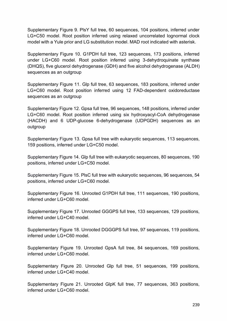
239
Supplementary Figure 9. PlsY full tree, 60 sequences, 104 positions, inferred under LG+C50 model. Root position inferred using relaxed uncorrelated lognormal clock model with a Yule prior and LG substitution model. MAD root indicated with asterisk. Supplementary Figure 10. G1PDH full tree, 123 sequences, 173 positions, inferred under LG+C60 model. Root position inferred using 3-dehydroquinate synthase (DHQS), five glucerol dehydrogenase (GDH) and five alcohol dehydrogenase (ALDH) sequences as an outgroup Supplementary Figure 11. Glp full tree, 63 sequences, 183 positions, inferred under LG+C60 model. Root position inferred using 12 FAD-dependent oxidoreductase sequences as an outgroup Supplementary Figure 12. Gpsa full tree, 96 sequences, 148 positions, inferred under LG+C60 model. Root position inferred using six hydroxyacyl-CoA dehydrogenase (HACDH) and 6 UDP-glucose 6-dehydrogenase (UDPGDH) sequences as an outgroup Supplementary Figure 13. Gpsa full tree with eukaryotic sequences, 113 sequences, 159 positions, inferred under LG+C50 model. Supplementary Figure 14. Glp full tree with eukaryotic sequences, 80 sequences, 190 positions, inferred under LG+C50 model. Supplementary Figure 15. PlsC full tree with eukaryotic sequences, 96 sequences, 54 positions, inferred under LG+C60 model. Supplementary Figure 16. Unrooted G1PDH full tree, 111 sequences, 190 positions, inferred under LG+C60 model. Supplementary Figure 17. Unrooted GGGPS full tree, 133 sequences, 129 positions, inferred under LG+C40 model. Supplementary Figure 18. Unrooted DGGGPS full tree, 97 sequences, 119 positions, inferred under LG+C60 model. Supplementary Figure 19. Unrooted GpsA full tree, 84 sequences, 169 positions, inferred under LG+C60 model. Supplementary Figure 20. Unrooted Glp full tree, 51 sequences, 199 positions, inferred under LG+C40 model. Supplementary Figure 21. Unrooted GlpK full tree, 77 sequences, 363 positions, inferred under LG+C60 model.

240
Supplementary Figure 22. Unrooted PlsC full tree, 74 sequences, 57 positions, inferred under LG+C60 model. Supplementary Figure 23. Unrooted PlsY full tree, 60 sequences, 104 positions, inferred under LG+C50 model Supplementary Figure 24. Gpsa full tree, 96 sequences, 148 positions, inferred under LG+C50 model. Root position inferred using six hydroxyacyl-CoA dehydrogenase (HACDH) and 6 UDP-glucose 6-dehydrogenase (UDPGDH) sequences as an outgroup Supplementary Figure 25. UbiA full tree, 227 sequences, 69 positions, inferred under LG+C60 model. DGGGP sequences in blue, chlorophyll a synthase in green, protoheme IX farnesyltransferase in red, and 4-hydroxybenzoate octaprenyltransferase in black. Supplementary Figure 26. Unrooted GGGPS full tree, 133 sequences, 129 positions, inferred under LG+C60 model. Supplementary Figure 27. Unrooted PlsY full tree, 60 sequences, 104 positions, inferred under LG+C60 model Supplementary Figure 28. GlpA/GlpD full tree with eukaryotic sequences, 80 sequences, 190 positions, inferred under LG+C60 model Supplementary Figure 29. Gpsa full tree with eukaryotic sequences, 113 sequences, 159 positions, inferred under LG+C60 model Supplementary Figure 30. Maximum likelihood G1PDH tree inferred in IQ-Tree under the LG+C60 model. Rooted using MAD Supplementary Figure 31. Maximum likelihood GGGPS tree inferred in IQ-Tree under the LG+C60 model. Rooted using MAD Supplementary Figure 32. Maximum likelihood DGGGPS tree inferred in IQ-Tree under the LG+C60 model. Rooted using MAD Supplementary Figure 33. Maximum likelihood GpsA tree inferred in IQ-Tree under the LG+C60 model. Rooted using MAD Supplementary Figure 34. Maximum likelihood GlpA/GlpD tree inferred in IQ-Tree under the LG+C60 model. Rooted using MAD

241
Supplementary Figure 35. Maximum likelihood GlpK tree inferred in IQ-Tree under the LG+C60 model. Rooted using MAD Supplementary Figure 36. Maximum likelihood PlsC tree inferred in IQ-Tree under the LG+C60 model. Rooted using MAD Supplementary Figure 37. Maximum likelihood PlsY tree inferred in IQ-Tree under the LG+C60 model. Rooted using MAD Supplementary Figure 38. Maximum likelihood G1PDH tree inferred in IQ-Tree under the LG+C60 model from HoT alignments. Rooted using MAD Supplementary Figure 39. Maximum likelihood GGGPS tree inferred in IQ-Tree under the LG+C60 model from HoT alignments. Rooted using MAD Supplementary Figure 40. Maximum likelihood DGGGPS tree inferred in IQ-Tree under the LG+C60 model from HoT alignments. Rooted using MAD Supplementary Figure 41. Maximum likelihood GpsA tree inferred in IQ-Tree under the LG+C60 model from HoT alignments. Rooted using MAD Supplementary Figure 42. Maximum likelihood GlpA/GlpD tree inferred in IQ-Tree under the LG+C60 model from HoT alignments. Rooted using MAD Supplementary Figure 43. Maximum likelihood GlpK tree inferred in IQ-Tree under the LG+C60 model from HoT alignments. Rooted using MAD Supplementary Figure 44. Maximum likelihood PlsC tree inferred in IQ-Tree under the LG+C60 model from HoT alignments. Rooted using MAD Supplementary Figure 45. Maximum likelihood PlsY tree inferred in IQ-Tree under the LG+C60 model from HoT alignments. Rooted using MAD Supplementary Figure 46. Maximum likelihood G1PDH tree inferred in IQ-Tree under the LG+C60 model from alignments with metagenomic data removed. Rooted using MAD, with lognormal relaxed molecular clock show with an asterisk Supplementary Figure 47. Maximum likelihood GGGPS tree inferred in IQ-Tree under the LG+C60 model from alignments with metagenomic data removed. Rooted using MAD, with lognormal relaxed molecular clock show with an asterisk Supplementary Figure 48. Maximum likelihood reduce GGGPS tree inferred in IQ-Tree under the LG+C60 model from alignments with metagenomic data removed. Rooted using MAD, with lognormal relaxed molecular clock show with an asterisk

242
Supplementary Figure 49. Maximum likelihood DGGGPS tree inferred in IQ-Tree under the LG+C60 model from alignments with metagenomic data removed. Rooted using MAD, with lognormal relaxed molecular clock show with an asterisk Supplementary Figure 50. Maximum likelihood GpsA tree inferred in IQ-Tree under the LG+C60 model from alignments with metagenomic data removed. Rooted using MAD, with lognormal relaxed molecular clock show with an asterisk Supplementary Figure 51. Maximum likelihood GlpA/GlpD tree inferred in IQ-Tree under the LG+C60 model from alignments with metagenomic data removed. Rooted using MAD, with lognormal relaxed molecular clock show with an asterisk Supplementary Figure 52. Maximum likelihood GlpK tree inferred in IQ-Tree under the LG+C60 model from alignments with metagenomic data removed. Rooted using MAD, with lognormal relaxed molecular clock show with an asterisk

Bacteria Actinobacteria Verrucosispora sediminis
Bacteria Firmicutes Maledivibacter halophilus
Bacteria Actinobacteria Eubacterium cellulosolvens
Bacteria Actinobacteria Streptomyces sp WAC00263
Archaea TACK Candidatus Korarchaeum cryptofilum
Bacteria Firmicutes Lachnospiraceae bacterium YSB2008
Archaea Euryarchaeota Methanospirillum hungatei
Bacteria Firmicutes Succiniclasticum ruminis
Archaea Euryarchaeota Methanothermobacter thermautotrophicus
Bacteria Thermotogae Mesotoga infera
Bacteria Actinobactria Nocardiopsis gilva
Bactria Actinobacteria Glycomyces sp NRRL B 16210
Bacteria Actinobacteria Kribbella catacumbae
Bacteria Actinobacteria Streptomyces luteus
Archaea Asgardarchaeota Candidatus Thorarchaeota archaeon SMTZ1 45
Bacteria Actinobacteria Verrucosispora maris AB 18 032
Archaea TACK Nitrosopumilus maritimus SCM1
Bacteria Firmicutes Numidum massiliense
Bacteria FCB Chitinispirillum alkaliphilum
Bacteria Candidatus Kerfeldbacteria bacterium RIFOXYA2 FULL 38 24
Bacteria Actinobacteria Glycomyces tenuis
Archaea TACK Sulfolobus solfataricus
Archaea Euryarchaeota Methanocaldococcus jannaschii
Bacteria Firmicutes Novibacillus thermophilus
Archaea TACK uncultured marine thaumarchaeote KM3 72 H08
Bacteria Actinobacteria Couchioplanes caeruleus
Bacteria Firmicutes Caldicellulosiruptor kronotskyensis
Archaea Asgardarchaeota Candidatus Heimdallarchaeota archaeon LC 3
Bacteria Firmicutes Planifilum fulgidum
Archaea TACK Cenarchaeum symbiosum
Archaea Euryarchaeota Methanosarcina mazei Tuc01
Archaea TACK Thermofilum pendens
Archaea TACK Candidatus Caldiarchaeum subterraneum
Archaea Euryarchaeota Methanopyrus kandleri AV19Archaea DPANN Candidatus Micrarchaeota archaeon CG1 02 60 51
Bacteria Firmicutes Christensenella minuta
Bacteria Actinobacteria Asanoa ishikariensis
Bacteria Firmicutes Paenibacillus ihumii
Archaea TACK Staphylothermus marinus
Bacteria PVC Pirellula sp SH Sr6A
Archaea TACK Aeropyrum pernix K1
Bacteria Actinobacteria Patulibacter americanus
Bacteria Actinobacteria Streptomyces violaceorubidus
Bacteria Spirochaetes bacterium GWE1 32 154
Bacteria candidate division TA06 bacterium 32 111
Archaea TACK Fervidicoccus fontis Kam940
Bacteria Proteobacteria Desulfovibrio sp U5L
Archaea Euryarchaeota Thermococcus kodakarensis
Bacteria Firmicutes Anaerobacillus arseniciselenatis
Archaea TACK Acidilobus saccharovorans
Bacteria Firmicutes Kroppenstedtia eburnea
Bacteria Candidatus Melainabacteria bacterium GWF2 37 15
Bacteria Firmicutes Phascolarctobacterium sp CAG 266
Bacteria Firmicutes Paludifilum halophilum
Bacteria Firmicutes Carboxydothermus islandicus
Bacteria Candidatus Magnetoovum chiemensis
Archaea DPANN Candidatus Aenigmarchaeota archaeon ex4484 14
Archaea TACK Ignicoccus hospitalis
Archaea Euryarchaeota Halococcus salifodinae
Bacteria Actinobacteria Salinispora pacifica
Bacteria Cystobacter fuscus DSM 2262
Archaea Euryarchaeota Archaeoglobus fulgidus
Bacteria Firmicutes Melghirimyces thermohalophilus
Archaea Euryarchaeota Thermoplasma volcanium
Bacteria Firmicutes Thermoanaerobacter mathranii
Archaea TACK Marine Group I thaumarchaeote SCGC AAA799 D11
Archaea Euryarchaeota Methanomassiliicoccus luminyensis
Archaea TACK Pyrobaculum aerophilum str IM2
Bacteria Actinobacteria Actinomadura oligospora
Bacteria Actinobacteria Marmoricola aequoreus
Bacteria Actinobacteria Actinoplanes rectilineatus
Bacteria Actinobacteria Solirubrobacter sp URHD0082
Bacteria Actinobacteria Marinactinospora thermotolerans
Bacteria Actinobacteria Haloechinothrix alba
Bacteria Actinobacteria Rubrobacter radiotolerans
Archaea Euryarchaeota Aciduliprofundum boonei
Bacteria Proteobacteria Sorangium cellulosum
Bacteria Firmicutes Paenibacillus ginsengihumi
Bacteria Actinobacteria Micromonospora narathiwatensis
Bacteria Firmicutes Paenibacillus durusBacteria Firmicutes Clostridium arbusti
Bacteria Firmicutes Coprothermobacter platensis
Archaea TACK Ignisphaera aggregans DSM 17230
Bacteria Actinobacteria Allosalinactinospora lopnorensis
Archaea TACK Candidatus Bathyarchaeota archaeon RBG 16 48 13
Archaea TACK Caldivirga maquilingensis
Bacteria Firmicutes Caldalkalibacillus thermarum
Archaea Euryarchaeota Pyrococcus furiosus
Archaea Asgardarchaeota Odinarchaeota archaeon LCB 4
Bacteria FCB Gemmata sp SH PL17
Archaea TACK Candidatus Nitrososphaera gargensis Ga9 2
Bacteria Actinobacteria Kitasatospora
Bacteria Firmicutes Streptococcus pneumoniae
Bacteria Actinobacteria Xiangella phaseoli
Archaea Euryarchaeota Methanocella paludicola SANAE
Bacteria Actinobacteria Glycomyces harbinensis
Bacteria Firmicutes Desmospora sp 8437
Bacteria FCB Chitinivibrio alkaliphilus
Bacteria Actinobacteria Thermomonospora chromogena
Bacteria Firmicutes Selenomonas ruminantium
Bacteria PVC Planctomycetes bacterium
Archaea Euryarchaeota Thermoplasmatales archaeon SCGC AB 539 N05
Archaea Euryarchaeota Halofax MULTISPECIES
Bacteria Firmicutes Bacillus subtilis
Bacteria Actinibacteria Mycobacterium abscessus subsp abscessus
Bacteria Firmicutes Bacillus lentusBacteria Actinobacteria Herbidospora mongoliensis
Archaea TACK Hyperthermus butylicus
Bacteria Actinobacteria Streptosporangium amethystogenes
Bacteria Proteobacteria Azospirillum sp CAG 260
Bacteria Actinobacteria Thermobifida halotolerans
Archaea Euryarchaeota Haloarcula MULTISPECIES
0.74
0.99
1
1
1
0.51
0.83
0.98
0.67
0.64
1
1
0.81
0.85
0.63
0.96
1
1
0.77
1
1
0.79
0.7
0.99
0.81
0.98
0.95
0.85
0.9
0.61
0.71
0.6
1
1
1
0.54
0.67
0.97
1
0.97
0.86
0.99
0.99
0.81
0.8
0.7
1
1
0.99
1
0.9
0.97
1
0.93
0.86
0.68
1
0.78
0.99
0.51
1
0.97
1
1
0.95
0.77
1
0.7
1
0.94
0.97
0.94
0.94
1
1
0.99
0.83
0.85
0.92
1
0.6
0.59
0.65
0.72
0.98
1
0.69
0.75
Tree scale: 1
*
Supplementary Figure 1

Archaea TACK Sulfolobus solfataricus
Bacteria FCB Runella limosa
Archaea TACK Caldivirga maquilingensis
Bacteria Firmicutes Planococcus maritimus
Bacteria FCB Niabella drilacis
Bacteria Firmicutes Bacillus aquimaris
Bacteria FCB Ignavibacteria bacterium CG1 02 37 35
Bacteria Firmicutes Tuberibacillus calidus
Archaea TACK Candidatus Caldiarchaeum subterraneum
Archaea TACK Caldisphaera lagunensis
Archaea TACK Thermofilum pendens
Archaea Asgardarchaeota Candidatus Thorarchaeota archaeon SMTZ1 45
Bactera candidate division TA06 bacterium 32 111
Archaea TACK Acidilobus sp 7A
Archaea TACK Marine Group I thaumarchaeote SCGC AAA799 D11
Archaea Asgardarchaeota Lokiarchaeum sp GC14 75
Bacteria Firmicutes Kroppenstedtia eburnea
Bacteria FCB Gemmatimonadetes bacterium 13 1 40CM 4 69 8
Archaea DPANN Candidatus Micrarchaeota archaeon CG1 02 49 24
Archaea Euryarchaeota Pyrococcus furiosus
Bacteria Firmicutes Paenibacillus durus
Archaea Euryarchaeota Thermoplasma volcanium
Bacteria FCB Flectobacillus sp BAB 3569
Archaea Euryarchaeota Methanomassiliicoccus luminyensis
Archaea Euryarchaeota Methanothermobacter thermautotrophicus
Bacteria Candidatus Raymondbacteria bacterium RifOxyA12 full 50 37
Bacteria Firmicutes Melghirimyces thermohalophilus
Bacteria candidate division TA06 bacterium DG 26
Archaea TACK Staphylothermus marinus
Bacteria FCB Flavisolibacter ginsengisoli
Bacteria Firmicutes Marininema mesophilum
Archaea Asgardarchaeota Candidatus Heimdallarchaeota archaeon LC 3
Bacteria FCB Fibrobacter
Bacteria Firmicutes Caldalkalibacillus thermarum
Bacteria Firmicutes Anaerobacillus arseniciselenatis
Bacteria Actinobacteria bacterium RBG 13 63 9
Bacteria Firmicutes Bacillus subtilis
Archaea Euryarchaeota Methanospirillum hungatei
Bacteria FCB Bernardetia litoralis
Archaea DPANN DUSEL3
Bacteria FCB Sediminibacter sp Hel I 10
Bacteria FCB Fibrobacter 2
Archaea TACK Candidatus Nitrososphaera gargensis Ga9 2
Archaea Euryarchaeota Methanosarcina mazei Tuc01Bacteria Desulfuromonadales bacterium C00003094
Bacteria Firmicutes Tepidanaerobacter acetatoxydans
Bacteria candidate division KSB1 bacterium RBG 16 48 16
Bacteria FCB Candidatus Cloacimonetes bacterium 4572 55
Archaea TACK Thermofilum sp ex4484 79
Bacteria FCB Jejuia pallidilutea
Bacteria Parcubacteria bacterium DG 74 3
Bacteria FCB Chitinispirillum alkaliphilum
Bacteria candidate division WOR 3 bacterium JGI Cruoil 03 44 89
Bacteria FCB Flavihumibacter petaseus
Bacteria FCB Kordia jejudonensis
Bacteria Candidatus Edwardsbacteria bacterium RifOxyC12 full 54 24
Bacteria Firmicutes Streptococcus pneumoniae
Bacteria FCB Formosa sp Hel1 31 208
Archaea TACK Candidatus Korarchaeum cryptofilum
Bacteria Firmicutes Planifilum fulgidum
Bacteria FCB Candidatus Kryptonium thompsoni
Bacteria bacterium TMED217
Bacteria Elusimicrobia bacterium RIFOXYB2 FULL 49 7
Bacteria FCB Ignavibacterium album
Archaea Euryarchaeota Haloferax volcanii
Bacteria candidate division WOR 3 bacterium SM23 60
Archaea TACK Pyrobaculum aerophilum str IM2
Bacteria FCB Bacteroidetes bacterium OLB12
Bacteria FCB Lacinutrix jangbogonensis
Bacteria Firmicutes Fictibacillus sp GDSW R2A3
Archaea Euryarchaeota Methanopyrus kandleri AV19
Bacteria bacterium TMED264
Bacteria FCB Candidatus Marinimicrobia bacterium CG1 02 48 14
Bacteria Firmicutes Paenibacillus ginsengihumi
Bacteria FCB Chlorobium sp GBChlB
Bacteria Actinobacteria Mycobacterium abscessus subsp abscessus
Bacteria Firmicutes Paludifilum halophilum
Bacteria Actinobacteria Streptomyces sp WAC00263
Archaea TACK Fervidicoccus fontis Kam940
Bacteria Firmicutes Peptococcaceae acterium BRH c8a
Bacteria FCB Sporocytophaga myxococcoides
Archaea TACK Ignisphaera aggregans DSM 17230
Archaea Euryarchaeota Thermococcus kodakarensis
Bacteria Parcubacteria bacterium DG 74 2
Bacteria FCB Candidatus Cloacimonas sp 4484 275
Archaea TACK Thermosphaera aggregans
Bacteria Firmicutes Desmospora sp 8437
Archaea Euryarchaeota Thermoplasmatales archaeon SCGC AB 539 N05
Bacteria FCB Schleiferia thermophila
Archaea TACK Thaumarchaeota archaeon ex4484 121
Archaea Euryarchaeota Halococcus salifodinae
Bacteria FCB Chitinivibrio alkaliphilus
Archaea TACK Candidatus Bathyarchaeota archaeon RBG 16 48 13
Archaea TACK Hyperthermus butylicus
Archaea TACK Desulfurococcus amylolyticus
Bacteria FCB Nafulsella turpanensis
Archaea DPANN AR10
Archaea TACK Candidatus Nitrosotenuis cloacae
Bacteria Latescibacteria bacterium DG 63
Bacteria Candidatus Handelsmanbacteria bacterium RIFCSPLOWO2 12 FULL 64 10
Archaea Euryarchaeota Aciduliprofundum boonei
Bacteria FCB candidate division Zixibacteria bacterium 4484 95
Bacteria Firmicutes bacterium ML8 F2
Bacteria Firmicutes Numidum massiliense
Archaea Euryarchaeota Methanocaldococcus jannaschii
Archaea Euryarchaeota Haloarcula marismortui
Bacteria FCB Ohtaekwangia koreensis
Archaea TACK Ignicoccus hospitalis
Archaea TACK Cenarchaeum symbiosum A
Archaea Asgardarchaeota Odinarchaeota archaeon LCB 4
Bacteria Firmicutes Dethiobacter alkaliphilus
Bacteria Candidatus Chrysopegis kryptomonas
Bacteria Firmicutes Geobacillus galactosidasius
Archaea Euryarchaeota Methanocella paludicola SANAE
Archaea TACK Aeropyrum pernix K1
Bacteria FCB Caldithrix abyssi
Archaea TACK Nitrosopumilus maritimus SCM1
Archaea DPAAN Macid
Bacteria FCB Cyclobacterium lianum
Archaea TACK Sulfolobus sp A20
Bacteria Firmicutes Anaerobacillus alkalidiazotrophicus
Bacteria FCB Candidatus Kryptobacter tengchongensis
Bacteria FCB Spirosoma sp 209
Bacteria Firmicutes Bhargavaea cecembensis
Archaea Euryarchaeota Archaeoglobus fulgidus
Archaea DPANN Candidatus Aenigmarchaeota archaeon ex4484 14
Bacteia FCB Psychroserpens burtonensis
Bacteria Firmicutes Calderihabitans maritimus
Bacteria Firmicutes Paenibacillus ihumii
Archaea TACK Acidilobus saccharovorans
Bacteria Firmicutes Bacillus lentus
0.99
0.64
1
0.8
0.73
0.54
0.9
1
0.53 0.99
0.99
0.51
0.97
0.51
1
1
1
0.53
0.94
0.53
1
0.99
1
0.76
0.52
0.99
0.71
0.93
0.67
0.54
0.58
0.99
0.86
0.99
1
0.98
1
0.8
0.9
0.92
0.98
0.99
1
0.99
0.93
0.52
0.97
0.64
1
0.51
1
0.59
0.5
0.97
0.98
0.71
0.86
1
0.67
0.52
0.95
0.98
0.98
0.56
0.91
0.99
0.99
0.77
0.950.71
0.92
0.89
0.87
0.67
1
0.99
0.99
0.8
0.68
0.53
0.87
0.79
0.95
0.66
0.98
0.76
0.89
0.99
0.84
0.59
0.84
0.67
0.86
Tree scale: 1 Supplementary Figure 2
*

Archaea TACK Staphylothermus marinus
Bacteria candidate division WOR 3 bacterium JGI Cruoil 03 44 89
Archaea TACK Pyrobaculum aerophilum str IM2
Bacteria FBC Chlorobium sp GBChlB
Archaea TACK Candidatus Korarchaeum cryptofilum
Archaea TACK Ignisphaera aggregans DSM 17230
Archaea DPANN Candidatus Aenigmarchaeota archaeon ex4484 14
Archaea Euryarchaeota Thermoplasmatales archaeon SCGC AB 539 N05
Archaea TACK Nitrosopumilus maritimus SCM1
Archaea TACK Thaumarchaeota archaeon ex4484 121
Archaea TACK Thermosphaera aggregans
Archaea Euryarchaeota Thermococcus kodakarensis
Archaea Euryarchaeota Thermoplasma volcanium
Bacteria bacterium TMED217
Archaea TACK Acidilobus sp 7A
Bacteria Firmicutes bacterium ML8 F2
Archaea Asgardarchaeota Lokiarchaeum sp GC14 75
Bacteria FCB Flavihumibacter petaseusArchaea Asgardarchaeota Candidatus Heimdallarchaeota archaeon LC 3
Bacteria FCB Bacteroidetes bacterium OLB12
Bacteria candidate division WOR 3 bacterium SM23 60
Bacteria Elusimicrobia bacterium RIFOXYB2 FULL 49 7
Bacteria FBC Fibrobacter sp
Bacteria FCB Caldithrix abyssi
Archaea TACK Acidilobus saccharovorans
Archaea TACK Thermofilum pendens
Bacteria FBC candidate division Zixibacteria bacterium 4484 95
Bacteria FBC Ignavibacterium album
Bacteria FBC Kordia jejudonensis
Archaea Euryarchaeota Pyrococcus furiosus
Bacteria FCB Chitinispirillum alkaliphilum
Bacteria FCB Candidatus Kryptonium thompsoni
Bacteria FCB Nafulsella turpanensis
Archaea Euryarchaeota Methanothermobacter thermautotrophicus
Bacteria FCB Chitinivibrio alkaliphilus
Archaea TACK Sulfolobus solfataricus
Archaea TACK Caldisphaera lagunensis
Archaea TACK Candidatus Nitrososphaera gargensis Ga9 2
Bacteria candidate division TA06 bacterium DG 26
Bacteria FCB Flavisolibacter ginsengisoli
Archaea TACK Caldivirga maquilingensis
Bacteria Firmicutes Dethiobacter alkaliphilus
Archaea Euryarchaeota Methanosarcina mazei Tuc01
Bacteria FCB Candidatus Cloacimonas sp 4484 275
Archaea TACK Candidatus Bathyarchaeota archaeon RBG 16 48 13
Archaea DPANN AR10
Bacteria FCB Flectobacillus sp BAB 3569
Bacteria FCB Candidatus Marinimicrobia bacterium CG1 02 48 14
Bacteria FCB Niabella drilacis
Bacteia FBC Psychroserpens burtonensis
Archaea DPAAN Macid
Archaea TACK Desulfurococcus amylolyticus
Archaea TACK Thermofilum sp ex4484 79
Archaea Euryarchaeota Methanocaldococcus jannaschii
Bacteria FBC Sporocytophaga myxococcoides
Bacteria FCB Cyclobacterium lianum
Bacteria FCB Candidatus Kryptobacter tengchongensis
Archaea TACK Cenarchaeum symbiosum A
Bacteria FBC Ohtaekwangia koreensis
Bacteria Parcubacteria bacterium DG 74 2
Bacteria FBC Jejuia pallidilutea
Bacteria Candidatus Handelsmanbacteria bacterium RIFCSPLOWO2 12 FULL 64 10
Bacteria FBC Formosa sp Hel1 31 208
Archaea TACK Marine Group I thaumarchaeote SCGC AAA799 D11
Bacteria FCB Gemmatimonadetes bacterium 13 1 40CM 4 69 8
Archaea DPANN DUSEL3
Archaea TACK Fervidicoccus fontis Kam940
Bacteria FBC Sediminibacter sp Hel I 10
Bacteria FCB Candidatus Cloacimonetes bacterium 4572 55
Archaea Euryarchaeota Methanomassiliicoccus luminyensis
Bacteria candidate division KSB1 bacterium RBG 16 48 16
Bacteria FCB Runella limosa
Archaea Asgardarchaeota Candidatus Thorarchaeota archaeon SMTZ1 45
Bacteria Candidatus Edwardsbacteria bacterium RifOxyC12 full 54 24
Bacteria FCB Spirosoma sp 209
Archaea TACK Sulfolobus sp A20
Bacteria Candidatus Raymondbacteria bacterium RifOxyA12 full 50 37
Bacteria bacterium TMED264
Bacteria FBC Fibrobacter
Bactera candidate division TA06 bacterium 32 111
Archaea TACK Candidatus Nitrosotenuis cloacae
Bacteria FCB Bernardetia litoralis
Archaea DPANN Candidatus Micrarchaeota archaeon CG1 02 49 24
Bacteria Desulfuromonadales bacterium C00003094
Archaea TACK Ignicoccus hospitalis
Bacteria FBC Lacinutrix jangbogonensis
Bacteria Candidatus Chrysopegis kryptomonas
Archaea Euryarchaeota Aciduliprofundum boonei
Archaea TACK Aeropyrum pernix K1
Bacteria FCB Schleiferia thermophila
Archaea TACK Hyperthermus butylicus
Archaea TACK Candidatus Caldiarchaeum subterraneum
Bacteria FCB Ignavibacteria bacterium CG1 02 37 35
0.65
1
0.56
1
0.52
0.6
0.99
0.77
0.71
0.95
0.58
0.93
0.81
0.99
0.67
0.89
0.94
0.98
0.98
0.99
1
0.93
1
1
0.6
0.99
0.92
0.78
1
0.9
1
0.760.92
0.85
0.99
0.91
0.78
0.81
0.7
0.77
1
0.99
0.71
0.99
0.97
0.97
0.6
1
1
0.57
0.99
1
1
0.9
0.64
0.95
0.99
1
Tree scale: 0.1
*
Supplementary Figure 3

DGGGP FCB Bacteria Chlorobi Chlorobium ferrooxidans
DGGGP Archaea TACK Hyperthermus butylicus
DGGGP Bacteria FCB Psychroserpens burtonensis
DGGGP Bacteria Candidatus Chrysopegis kryptomonas
DGGGP Bacteria Firmicutes Halanaerobium
DGGGP Bacteria FCB Fibrobacter
DGGGP Archaea Euryarchaeota Thermoplasma volcanium
DGGGP Archaea Euryarchaeota Haloferax
DGGGP Bacteria FCB Candidatus Kryptonium thompsoni
DGGGP Bacteria Candidatus Handelsmanbacteria bacterium RIFCSPLOWO2 12 FULL 64 10
DGGGP Archaea TACK Ignicoccus hospitalis
DGGGP FCB Bacteria Caldithrix abyssi DSM 13497
DGGGP Archaea Euryarchaeota Archaeoglobus fulgidus
DGGGP Bacteria Rhodothermaeota Rubricoccus marinus
DGGGP Archaea Euryarchaeota Methanocaldococcus jannaschii
DGGGP Archaea TACK Caldivirga maquilingensis
DGGGP Archaea Asgardarchaeota Lokiarchaeum sp GC14 75
DGGGP Bacteria FCB Ignavibacterium album
DGGGP Archaea Asgardarchaeota Candidatus Thorarchaeota archaeon SMTZ1 45
DGGGP Archaea Euryarchaeota Methanospirillum hungatei
DGGGP Bacteria candidate division TA06 bacterium 32 111
DGGGP Bacteria Parcubacteria bacterium DG 74 3
DGGGP Bacteria FCB Cyclobacterium lianum
DGGGP Bacteria Actinobacteria Kocuria sp SM24M 10
DGGGP Bacteria FCB candidate division Zixibacteria bacterium 4484 95 fa
DGGGP Bacteria FCB Sporocytophaga myxococcoides
DGGGP Bacteria Candidatus Raymondbacteria bacterium RifOxyA12 full 50 37
DGGGP Bacteria Choloroflexi Leptolinea tardivitalis
DGGGP FCB Bacteria Fibrobacteres Fibrobacter sp UWH5
DGGGP Bacteria Actinobacteria Curtobacterium sp UCD KPL2560
DGGGP FCB Bacteria Gemmatimonadetes bacterium 13 1 40CM 4 69 8
DGGGP Archaea DPANN Candidatus Aenigmarchaeota archaeon ex4484 14
DGGGP Bacteria candidate division KSB1 bacterium 4484 87
DGGGP Archaea DPANN DUSEL3
DGGGP FCB Bacteria Bacteroidetes Jejuia pallidilutea
DGGGP FCB Bacteria Candidatus Marinimicrobia bacterium
DGGGP Bacteria candidate division WOR 3 bacterium SM23 60DGGGP Archaea Asgardarchaeota Candidatus Heimdallarchaeota archaeon LC 3
DGGGP Bacteria FCB Bacteroidetes Kordia jejudonensis
DGGGP Archaea TACK Candidatus Caldiarchaeum subterraneum
DGGGP Bacteria Candidatus Cloacimonas sp 4484 275
DGGGP Bacteria Actinobacteria Friedmanniella sagamiharensis
DGGGP FCB Bacteria Bacteroidetes Formosa sp Hel1 31 208
DGGGP Archaea Euryarchaeota Haloarcula
DGGGP Archaea TACK Aeropyrum pernix K1
DGGGP Bacteria FCB Flavisolibacter ginsengisoli DSM 18119
DGGGP Archaea TACK Pyrobaculum aerophilum st IM2
DGGGP Bacteria Candidatus Edwardsbacteria bacterium RifOxyC12 full 54 24
DGGGP Bacteria FCB Ohtaekwangia koreensis
DGGGP Archaea Euryarchaeota Methanothermobacter thermautotrophicus
DGGGP Bacteria Chloroflexi Longilinea arvoryzae
DGGGP Bacteria FCB Candidatus Krytobacter tengchongensis
DGGGP Archaea TACK Thermofilum pendens
DGGGP Archaea Euryarchaeota Methanocella paludicola SANAE
DGGGP Bacteria Actinobacteria Curtobacterium sp ER1 6
DGGGP Bacteria FCB Chitinispirillum alkaliphilum
DGGGP Archaea DPANN Macid
DGGGP Archaea Euryarchaeota Methanopyrus kandleri
DGGGP Archaea DPANN AR10
DGGGP Bacteria Cyanobacteria Chlorogloeopsis fritschii
DGGGP Bacteria Firmicutes Halanaerobium saccharolyticum
DGGGP Bacteria Actinobacteria Rubrobacter radiotolerans
DGGGP FCB Bacteria Candidatus Latescibacteria bacterium DG 63DGGGP Archaea Euryarchaeota Thermococcus kodakarensis
DGGGP Bacteria FCB Bacteroidetes Gelidibacter algens
DGGGP Archaea TACK Sulfolobus solfataricus
DGGGP FCB Bacteria Chlorobi Chlorobium sp GBChlB
DGGGP Archaea Euryarchaeota Halococcus salifodinae
DGGGP Bacteria Proteobacteria Rhodobacteraceae bacterium TMED111
DGGGP FCB Bacteria Latescibacteria bacterium DG 63
DGGGP Bacteria bacterium TMED264DGGGP Bacteria Chloroflexi Anaerolineae bacterium SG8 19
DGGGP Bacteria Actinobacteria Auraticoccus monumenti
DGGGP Archaea Euryarchaeota Thermoplasmatales archaeon SCGC AB 539 N05
DGGGP Archaea Euryarchaeota Methanosarcina mazei Tuc01
DGGGP Bacteria FCB Chitinivibrio alkaliphilus
DGGGP Archaea TACK Staphylothermus marinus
DGGGP Bacteria FCB Bacteroidetes Flammeovirgaceae bacterium TMED290
DGGGP Archaea Asgardarchaeota Candidatus Odinarchaeota archaeon LCB 4
DGGGP FCB Bacteria Ignavibacteria bacterium CG1 02 37 35
DGGGP Bacteria candidate division WOR 3 bacterium JGI Cruoil 03 44 89
DGGGP Bacteria FCB candidate division Zixibacteria bacterium RBG 1
DGGGP Archaea Euryarchaeota Pyrococcus furiosus
DGGGP FCB Bacteria Chlorobi Chlorobium limicola
DGGGP Bacteria Proteobacteria Desulfobacterales bacterium SG8 35 2 fa
DGGGP FCB Bacteria Bacteroidetes Sediminibacter sp Hel I 10
DGGGP Archaea TACK Ignisphaera aggregans DSM 17230
DGGGP FCB Bacteria Candidatus Cloacimonetes bacterium 4572 55
DGGGP Archaea Euryarchaeota Aciduliprofundum boonei
DGGGP FCB Bacteria Candidatus Marinimicrobia bacterium CG1 02 48 14
DGGGP Bacteria FCB Flavihumibacter petaseus
DGGGP Bacteria FCB Schleiferia thermophila
DGGGP Archaea DPANN Candidatus Micrarchaeota archaeon CG1 02 51 15
DGGGP Archaea Euryarchaeota Methanomassiliicoccus luminyensis
DGGGP Archaea TACK Fervidicoccus fontis Kam940
DGGGP Bacteria CPR Parcubacteria bacterium DG 74 2
DGGGP Bacteria Actinobacteria Arthrobacter sp RIT PI e
0.99
0.99
0.99
0.6
0.99
0.93
0.66
0.99
0.58
0.56
0.75
0.51
0.69
0.95
0.99
0.99
1
0.98
0.95
0.98
0.54
0.82
0.98
0.56
1
0.75
0.7
1
0.84
0.99
0.5
0.86
0.99
0.62
0.99
0.94
0.57
0.68
1
0.98
0.97
0.98
0.62
1
0.77
1
0.98
0.99
0.99
0.99
1
0.98
0.87
1
1
0.57
Tree scale: 1Supplementary Figure 4
*

Bacteria PVC Verrucomicrobium spinosum
Bacteria Proteobacteria Vibrio cholerae HE48
Bacteria Proteobacteria Helicobacter saguini
Bacteria Nitrospinae Nitrospina gracilis 3 211
Bacteria Spirochaetes Spirochaeta thermophila
Bacteria Deinococcus Thermus Deinococcus radiodurans
Archaea Euryarchaeota marine group II III euryarchaeote KM3 85 A08
Archaea Euryarchaeota Ferroglobus placidus
Archaea Euryarchaeota archaeon TMED85
Bacteria PVC Chlamydia trachomatis A HAR 13
Bacteria Nitropirae Thermodesulfovibrio thiophilus
Bacteria FCB Fibrobacter succinogenes
Bacteria FCB Gemmatimonadetes Gemmatimonas aurantiaca T 27
Bacteria Aminicenantes Candidatus Aminicenans sakinawicola
Archaea Euryarchaeota Candidatus Thalassoarchaea euryarchaeoteArchaea Euryarchaeota Candidatus Thalassoarchaea euryarchaeote
Bacteria Proteobacteria Erythrobacter litoralis
Bacteria Proteobacteria Rickettsia prowazekii str Rp22
Bacteria Terrabacteria Firmicutes Planococcus maritimus
Bacteria Proteobacteria Mariprofundus ferrooxydans PV 1
Bacteria Spirochaetes Leptospira interrogans
Bacteria Terrabacteria Cyanobacteria Scytonema hofmanni
Bacteria Terrabacteria Firmicutes Streptococcus pneumoniae
Archaea DPANN Candidatus Woesearchaeota archaeon CG10 big fil rev 8 21 14 0 10 36 11Archaea Candidatus Woesearchaeota archaeon CG 4 10 14 0 2 um filter 33 13
Bacteria Terrabacteria Tenericutes Mycoplasma hominis ATCC 23114
Bacteria FCB Ignavibacteriae Ignavibacterium album
Bacteria Fusobacteria Fusobacterium perfoetens
Bacteria Proteobacteria Coxiella burnetii RSA 331
Bacteria Armatimonadetes Chthonomonas calidirosea T49
Bacteria Terrabacteria Chloroflexi Caldilinea aerophila
Archaea Euryarchaeota archaeon TMED97
Bacteria Proteobacteria Geobacter bemidjiensis
Bacteria Terrabacteria Actinobacteria Rubrobacter radiotolerans
Bacteria Aquificae Aquifex aeolicus
Bacteria Thermobaculum Thermobaculum terrenum
Bacteria Proteobacteria Paraburkholderia rhizoxinica HKI 454
Bacteria Lentispaera Lentisphaera araneosa HTCC2155
Bacteria FCB Chlorobium limicola
Bacteria Terrabacteria Firmicutes Planifilum fulgidum
Archaea Euryarchaeota Archaeoglobus fulgidus
Bacteria Proteobacteria Desulfatibacillum aliphaticivorans
Bacteria FCB Cloacimonetes Candidatus Cloacimonas acidaminovorans str Evry
Archaea marine group II III euryarchaeote AD1000 25 A05
Bacteria PVC Planctopirus limnophila
Bacteria Terrabacteria Firmicutes Bacillus subtilis
Bacteria FCB Bacteroides fluxus YIT 12057
Bacteria Proteobacteria Escherichia coli str K 12 substr MG1655
Archaea Euryarchaeota Geoglobus ahangari
Bacteria Candidatus Methylomirabilis oxyfera
Bacteria Terrabacteria Firmicutes Paenibacillus curdlanolyticus YK9
Bacteria Terrabacteria Firmicutes Staphylococcus aureus A9635
Bacteria Acetothermia Candidatus Acetothermus autotrophicum
Archaea Euryarchaeota Methanobrevibacter filiformis
Archaea TACK Thermofilum sp ex4484 15
Bacteria Proteobacteria Desulfarculus baarsii
Bacteria Terrabacteria Actinobacteria Streptomyces sviceus ATCC 29083
Bacteria Chrysiogenetes Chrysiogenes arsenatis
Archaea Euryarchaeota Methanoculleus thermophilusArchaea Euryarchaeota Methanothermobacter thermautotrophicus
Bacteria Proteobacteria Micavibrio aeruginosavorus
Bacteria Proteobacteria Sorangium cellulosum
Bacteria Elusimicrobia Elusimicrobium minutum
Bacteria Proteobacteria Rhizobium gallicum
Bacteria Deferribacterales Deferribacter desulfuricans
Bacteria Spirochaetes Brachyspira pilosicoli WesB
Bacteria Terrabacteria Cyanobacteria Gloeobacter kilaueensis JS1Bacteria Terrabacteria Actinobacteria Actinomyces slackii
Bacteria Terrabacteria Firmicutes Clostridium ultunense Esp
Bacteria Dictyoglomi Dictyoglomus thermophilum
Archaea archaeon GW2011 AR9
Bacteria FCB Chitinivibrio alkaliphilus ACht1
Archaea Euryarchaeota Methanobacterium sp MB1
Bacteria Synergistes Dethiosulfovibrio peptidovorans DSM 11002
Bacteria Terrabacteria Haloplasmatales Haloplasma contractile
Bacteria FCB Caldithrix abyssi DSM 13497
Bacteria Proteobacteria Desulfobacter postgatei
Archaea Euryarchaeota Marine Group II euryarchaeote MED G34
Bacteria Proteobacteria Nitrosomonas eutropha
Bacteria Thermodesulfobacteria Thermodesulfatator atlanticus
Archaea archaeon GW2011 AR11
Bacteria Terrabacteria Cyanobacteria Chroococcidiopsis thermalis
Bacteria Thermotogae Thermotoga maritima MSB8
Bacteria Latescibacteria Candidatus Latescibacter anaerobius
0.99
0.86
0.78
0.99
0.56
0.86
0.85
1
0.69
0.91
0.73
0.99
0.98
0.99
0.680.99
0.78
0.52
0.99
0.99
0.8
0.61
1
0.99
0.5
0.77
1
0.83
0.72
0.91
0.51
0.58
1
0.98
0.89
1
0.99
0.5
0.76
0.63
0.64
0.94
0.94
0.99
0.93
1
Tree scale: 1 Supplementary Figure 5
*

Bacteria Tenericutes Mycoplasma hyorhinisArchaea TACK Candidatus Korarchaeum cryptofilum
Bacteria Spirochaetes Sediminispirochaeta smaragdinae
Archaea Asgardarchaeota Lokiarchaeum sp GC14 75
Archaea Euryarchaeota Halomicrobium katesii
Archaea Euryarchaeota Halorhabdus utahensis
Archaea TACK Staphylothermus hellenicus DSM 12710
Bacteria Spirochaetes Leptospira interrogans str UI 08452Bacteria Proteobacteria Helicobacter felis
Archaea Euryarchaeota Archaeoglobus fulgidus
Archaea TACK Desulfurococcus mucosus
Archaea Euryarchaeota Halapricum
Bacteria Deferribacteres Deferribacter desulfuricansBacteria Terrabacteria Firmicutes Streptococcus porcinus
Bacteria Terrabacteria Actinobacteria Rubrobacter xylanophilus
Archaea Euryarchaeota Halapricum salinum
Bacteria Deinococcus Thermus Deinococcus radiodurans R1
Bacteria Proteobacteria Desulfobacterium autotrophicum
Bacteria Terrabacteria Firmicutes Paenibacillus alvei
Bacteria FCB Bacteroides clarus
Archaea Euryarchaeota Thermococcus gammatolerans
Archaea Euryarchaeota Halosimplex carlsbadense
Archaea Euryarchaeota Halanaeroarchaeum sulfurireducensArchaea Euryarchaeota Haladaptatus paucihalophilus
Archaea Euryarchaeota Palaeococcus pacificus DY20341
Archea TACK sulfolobus islandicus
Bacteria PVC Planctopirus limnophila
Archaea Euryarchaeota Natronomonas moolapensis
Archaea Euryarchaeota Aciduliprofundum boonei
Bacteria Proteobacteria Escherichia coli
Bacteria Proteobacteria Paraburkholderia rhizoxinica
Archaea Euryarchaeota Methanocella arvoryzae
Bacteria Terrabacteria Actinobacteria Bacillus subtilis
Bacteria Proteobacteria Vibrio parahaemolyticus RIMD 2210633
Bacteria Fusobacteria Fusobacterium nucleatum subsp vincentii ATCC 49256
Archaea TACK Thermofilum adornatus
Bacteria Terrabacteria Firmicutes Clostridium perfringens
Bacteria Proteobacteria Geobacter sp M18
Archaea Euryarchaeota Thermoplasma acidophilum
Archaea Euryarchaeota Halococcus salifodinae DSM 8989
Bacteria Thermotogae Thermotoga maritima
Archaea Euryarchaeota Pyrococcus furiosus
Bacteria Terrabacteria Actinobacteria Streptomyces avermitilis
Bacteria Terrabacteria Firmicutes Staphylococcus aureus
Archaea Euryarchaeota Candidatus Halobonum tyrrellensis
Archaea TACK Vulcanisaeta distributa
Archaea Euryarchaeota Picrophilus torridus
Archaea Euryarchaeota Haloarcula californiae
Bacteria Terrabacteria Actinobacteria Actinomyces sp ICM39
Bacteria Spirochaetes Brachyspira pilosicoli WesB
Bacteria Proteobacteria Bradyrhizobium sp BTAi1
0.99
0.890.99
0.99
0.99
1
0.97
0.99
1
0.99
1
0.99
1
1
0.73
0.92
0.89
0.85
0.97
0.970.66
0.99
0.99
0.85
0.99
0.99
0.98
0.95
0.99
0.78
0.99
1
0.81
0.91
0.99
0.95
0.6
0.85
0.83
1
0.58
0.84
0.99
0.98
0.99
Tree scale: 1
*
Supplementary Figure 6

Bacteria Proteobacteria Escherichia coli
Bacteria Spirochaetes Leptospira interrogans
Bacteria Protoebacteria Desulfarculus baarsii
Archaea Euryarchaeota Aciduliprofundum boonei
Bacteria PVC Planctopirus limnophila
Bacteria Acidobacteria Acidobacterium capsulatum
Archaea Euryarchaeota Natronomonas moolapensis
Bacteria Terrabacteria Chloroflexi Chloroflexus aggregans
Archaea Euryarchaeota Halorhabdus utahensis
Bacteria Proteobacteria Mariprofundus ferrooxydans
Bacteria FCB Gemmatimonadetes Gemmatimonas aurantiaca
Archaea TACK Sulfolobus islandicus
Archaea TACK Caldivirga maquilingensis
Bacteria Proteobacteria Geobacter bemidjiensis
Archaea Asgardarchaeota Lokiarchaeum sp GC14 75
Bacteria Proteobcteia Micavibrio aeruginosavorus
Archaea Euryarchaeota Ferroplasma sp Type II
Archaea Euryarchaeota Palaeococcus pacificus
Bacteria Proteobacteria Erythrobacter litoralis
Archaea DPANN Candidatus Woesearchaeota archaeon CG10 big fil rev 8 21 14 0 10 32 24
Bacteria Terrabacteria Actinobacteria Staphylococcus aureus
Archaea Euryarchaeota archaeon TMED117
Archaea Euryarchaeota Ferroplasma acidarmanus
Archaea Euryarchaeota uncultured marine group II III euryarchaeote KM3 86 F07
Bacteria Thermobaculum terrenum
Bacteria PVC Chlamydia trachomatis
Bacteria Thermotogae Thermotoga maritima MSB8
Bacteria Spirochaetes Brachyspira pilosicoli WesB
Bacteria Proteobacteria Desulfatibacillum aliphaticivorans
Archaea Euryarchaeota Thermoplasma acidophilum
Bacteria Proteobacteria Rhizobium gallicum
Bacteria Terrabacteria Firmicutes Streptococcus pneumoniae
Bacteria Fusobacteria Fusobacterium perfoetens
Bacteria Proteobacteria Coxiella burnetii
Archaea Euryarchaeota Acidiplasma sp MBA 1
Archaea TACK Candidatus Korarchaeum cryptofilum OPF8
Bacteria Dictyoglomi Dictyoglomus thermophilum
Archaea Euryarchaeota Pyrococcus abyssi
Archaea Euryarchaeota Candidatus Halobonum tyrrellensis
Bacteia PVC Verrucomicrobium spinosum
Bacteria Proteobacteria Nitrosomonas eutropha
Archaea TACK Vulcanisaeta distributa
Bacteria Deinococcus Thermus Deinococcus radiodurans
Archaea Euryarchaeota Halalkalicoccus jeotgali
Bacteria Terrabacteria Firmicutes Clostridium ultunense
Bacteria Elusimicrobia Elusimicrobium minutum Pei191
Bacteria Terrabacteria Actinobacteria Rubrobacter xylanophilus
Bacteria FCB Chlorobium limicola
Archaea Euryarchaeota Candidatus Bathyarchaeota archaeon B24 2
Archaea Euryarchaeota Halomicrobium mukohataei
Archaea Euryarchaeota Halobacterium salinarum
Archaea DPANN Candidatus Woesearchaeota archaeon CG10 big fil rev 8 21 14 0 10 34 8
Archaea Euryarchaeota Archaeoglobus fulgidus
Archaea TACK Thermofilum pendens
Archaea Euryarchaeota Halosimplex carlsbadense
Archaea Euryarchaeota Haloarcula amylolytica
Bacteria Terrabcteria Actinobacteria Actinomyces slackii
Archaea TACK Desulfurococcus amylolyticus
Bacteria Terrbacteria Firmicutes Bacillus subtilis
Bacteria Proteobacteria Sorangium cellulosum
Archaea Euryarchaeota Haladaptatus paucihalophilus
Archaea Euryarchaeota Halapricum salinum
Bacteria Terrabacteria Firmicutes Paenibacillus curdlanolyticus
Bacteria Terrabacteria Firmicutes Planococcus maritimus
Archaea Euryarchaeota Hadesarchaea archaeon DG 33
Archaea Euryarchaeota Halarchaeum acidiphilum
Bacteria Proteobacteria Vibrio cholerae
Bacteria Terrabacteria Firmicutes Planifilum fulgidum
Archaea Euryarchaeota Picrophilus torridus
Bacteria Terrabacteria Actinobacteria Streptomyces sviceus ATCC 29083
Bacteria Terrabacteria Haloplasmatales Haloplasma contractile
Archaea TACK Staphylothermus hellenicus
Archaea Euryarchaeota Thermococcus eurythermalis
Bacteria Terrabacteria Cyanobacteria Nodosilinea nodulosa
Bacteria Aquificae Aquifex aeolicus
Bacteria Deferribacterales Deferribacter desulfuricans
Bacteria Proteobacteria Paraburkholderia rhizoxinica
0.99
0.97
0.99
0.61
1
0.69
0.95
0.86
0.99
0.58
0.89
0.99
0.99
0.94
0.75
0.82
0.97
0.72
0.98
0.99
0.84
0.66
0.68
0.98
0.89
0.71
0.86
0.9
0.74
0.9
0.99
0.95
0.5
0.56
0.89
0.99
0.68
0.96
0.5
0.95
0.52
1
0.67
0.99
0.991
0.99
0.84
0.66
0.96
0.56
0.68
0.74
0.88
0.98
1
0.99
0.5
0.98
Tree scale: 1
*
Supplementary Figure 7

Bacteria Firmicutes Planococcus maritimus
Archaea Euryarchaeota uncultured marine group II III euryarchaeote KM3 86 F07
Bacteria Firmicutes Planifilum fulgidum
Archaea Euryarchaeota Marine Group III euryarchaeote CG Epi4
Bacteria Thermodesulfobacteria Thermodesulfatator atlanticus
Bacteria Spirochaetes Spirochaeta thermophila
Bacteria Thermotogae Thermotoga maritima MSB8
Bacteria Elusimicrobia Elusimicrobium minutumBacteria Dictyoglomi Dictyoglomus thermophilum
Archaea Euryarchaeota Euryarchaeota archaeon TMED164
Bacteria Thermobaculum Thermobaculum terrenum
Archaea Euryarchaeota Marine Group III euryarchaeote CG Epi6
Bacteria Tenericutes Mycoplasma hominis ATCC 23114
Bacteria Proteobacteria Rhizobium gallicum
Bacteria Nitrospinae Nitrospina gracilis 3 211
Bacteria Proteobacteria Desulfatibacillum aliphaticivorans
Bacteria Aquificae Aquifex aeolicus
Bacteria Proteobacteria Escherichia coli
Bacteria Proteobacteria Coxiella burnetii
Bacteria Acidobacteria Acidobacterium capsulatum
Bacteria candidate division NC10 Candidatus Methylomirabilis oxyfera
Bacteria Proteobacteria Paraburkholderia rhizoxinica
Archaea Euryarchaeota Euryarchaeota archaeon TMED85
Bacteria Proteobacteria Desulfobacter postgatei
Bacteria Proteobacteria Geobacter bemidjiensis
Bacteria Proteobacteria Micavibrio aeruginosavorus
Bacteria Cyanobacteria Nodosilinea nodulosa
Bacteria Proteobacteria Nitrosomonas eutrophaBacteria Proteobacteria Erythrobacter litoralis
Bacteria Synergistes Dethiosulfovibrio peptidovorans
Bacteria Protoebacteria Rickettsia prowazekii
Bacteria Spirochaetes Leptospira interrogans
Bacteria Proteobacteria Helicobacter saguini
Archaea Asgard Candidatus Heimdallarchaeota archaeon LC 2
Bacteria PVC Planctopirus limnophila
Bacteria FBC Ignavibacteriae Ignavibacterium album
Bacteria FBC Fibrobacter succinogenes
Archaea DPANN archaeon GW2011 AR11
Bacteria Lentisphaerae Lentisphaera araneosa
Bacteria Proteobacteria Sorangium cellulosum
Bacteria Chloroflexi Chloroflexus aggregans
Archaea Asgard Lokiarchaeum sp GC14 75
Bacteria Armatimonadetes Chthonomonas calidirosea T49
Bacteria Proteobacteria Mariprofundus ferrooxydans
Bacteria Actinobacteria Rubrobacter xylanophilus
Bacteria Cyanobacteria Chroococcidiopsis thermalis
Bacteria Deferribacteres Deferribacter desulfuricans
Bacteria Cloacimonetes Candidatus Cloacimonas acidaminovorans
Archaea Euryarchaeota Marine Group III euryarchaeote CG Epi3
Bacteria Proteobacteria Desulfarculus baarsii
Bacteria Firmicutes Staphylococcus aureus
Bacteria Gemmatimonadetes Gemmatimonas aurantiaca
Bacteria PVC Verrucomicrobium spinosum
Bacteria FBC Chitinivibrio alkaliphilus ACht1
Bacteria Nitrospirae Thermodesulfovibrio thiophilus
Bacteria Actinobacteria Streptomyces sviceus
Bacteria Proteobacteria Vibrio cholerae
Bacteria Firmicutes Bacillus subtilis
Bacteria PVC Chlamydia trachomatis A HAR 13
Bacteria Firmicutes Streptococcus pneumoniae
Archaea Euryarchaeota Marine Group III euryarchaeote CG Epi1
Archaea Euryarchaeota Marine Group III euryarchaeote CG Epi5
Bacteria FBC Chlorobium limicola
Bacteria Chrysiogenetes Chrysiogenes arsenatis
Bacteria Firmicutes Paenibacillus curdlanolyticus
Bacteria Caldithrix abyssi
Bacteria Actinobacteria Actinomyces slackii
Bacteria Spirochaetes Brachyspira pilosicoli WesB
Archaea Euryarchaeota Euryarchaeota archaeon TMED255
Bacteria FBC Bacteroides fluxus
Bacteria Firmicutes Clostridium ultunense
Archaea Euryarchaeota Marine Group III euryarchaeote CG Epi2
Bacteria Tenericutes Haloplasmatales Haloplasma contractile
Bacteria Deinococcus Thermus Deinococcus radiodurans
0.73
0.52
0.7
0.58
0.79
0.730.55
0.64
0.83
0.99
0.72
0.77
0.96
0.77
0.52
0.53
0.970.66
0.99
0.99
1
0.76
0.98
0.55
Tree scale: 1
FCB
FCBFCB
FCB
FCB
FCB
Supplementary Figure 8

Bacteria PVC Planctopirus limnophila
Bacteria Spirochaetes Leptospira interrogans
Bacteria Terrabacteria Cyanobacteria Nodosilinea nodulosa
Bacteria Proteobacteria Micavibrio aeruginosavorusBacteria Erythrobacter litoralis
Bacteria Terrabacteria Cyanobacteria Chroococcidiopsis thermalis
Bacteria Chrysiogenetes Chrysiogenes arsenatis
Bacteria Terrabacteria Haloplasmatales Haloplasma contractile
Bacteria Proteobacteria Desulfarculus baarsii
Bacteria Deinococcus Thermus Deinococcus radiodurans
Bacteria Thermotogae Thermotoga maritima MSB8
Bacteria Proteobacteria Rhizobium gallicum
Bacteria Armatimonadetes Chthonomonas calidirosea T49
Bacteria Terrabcteria Firmicutes Streptococcus pneumoniae
Bacteria PVC Chlamydia trachomatis A HAR 13
Archaea DPANN Candidatus Woesearchaeota archaeon CG11 big fil rev 8 21 14 0 20 43 8
Bacteria Fusobacteria Fusobacterium perfoetens
Bacteria Tenericutes Mycoplasma hominis
Bacteria Proteobacteria Chlorobium limicola
Bacteria FCB Candidatus Cloacimonetes Cloacimonas acidaminovorans
Bacteria FCB Gemmatimonadetes Gemmatimonas aurantiaca
Bacteria Terrabacteria Firmicutes Clostridium ultunense Esp
Bacteria Spirochaetes Spirochaeta thermophilaBacteria Dictyoglomi Dictyoglomus thermophilum
Bacteria Terrabacteria Firmicutes Staphylococcus aureus
Bacteria Terrabacteria Firmicutes Bacillus subtilisBacteria Terrabacteria Firmicutes Planococcus maritimus
Archaea Asgardarchaeota Candidatus Heimdallarchaeota archaeon LC 2
Bacteria Proteobacteria Paraburkholderia rhizoxinica
Bacteria Proteobacteria Vibrio cholerae
Bacteria Aquificae Aquifex aeolicusBacteria Acidobacteria Acidobacterium capsulatum
Bacteria Nitrospinae Nitrospina gracilis 3 211
Bacteria FCB Caldithrix Caldithrix abyssi
Bacteria Proteobacteria Geobacter
Bacteria Deferribacteres Deferribacter desulfuricans
Archaea DPANN Candidatus Pacearchaeota archaeon CG06 land 8 20 14 3 00 35 12Archaea Asgardarchaeota Lokiarchaeum sp GC14 75
Bacteria Synergistetes Dethiosulfovibrio peptidovorans
Bacteria Proteobacteria Mariprofundus ferrooxydans
Bacteria Thermobaculum Thermobaculum terrenum
Bacteria Proteobacteria Coxiella burnetii
Bacteria PVC Lentisphaerae Lentisphaera araneosa
Bacteria PVC Verrucomicrobium spinosum
Bacteria Thermodesulfobacteria Thermodesulfatator atlanticus
Bacteria Terrabacteria Actinobacteria Rubrobacter xylanophilus
Bacteria Proteobacteria Sorangium cellulosum So ce56
Archaea Asgardarchaeota Candidatus Thorarchaeota archaeon SMTZ1 83
Bacteria Proteobacteria Desulfobacter postgatei
Bacteria Proteobacteria Escherichia coli
Bacteria Proteobacteria Helicobacter saguini
Bacteria Prtoeobacteria Fibrobacter
Bacteria FCB Ignavibacteriae Ignavibacterium album
Bacteria Terrabacteria Firmicutes Paenibacillus curdlanolyticus
Bacteria Spirochaetes Brachyspira pilosicoli
Bacteria Nitrospirae Thermodesulfovibrio thiophilus
Bacteria Terrabacteria Firmicutes Planifilum fulgidum
Bacteria Proteobacteria Desulfatibacillum aliphaticivorans
Bacteria Proteobacteria Nitrosomonas eutropha
Bacteria Elusimicrobia Elusimicrobium minutum
0.72
0.55
0.93
0.96
0.99
0.97
0.76
0.88
0.98
0.96
0.66
0.71
0.670.85
0.97
0.92
0.57
0.99
0.510.56
0.99
0.99
0.83
0.64
0.61
1
0.75
Tree scale: 1
*
Supplementary Figure 9

Bacteria Actinobacteria Streptomyces sp WAC00263
Bacteria FCB Chitinispirillum alkaliphilum
Bacteria Actinobacteria Verrucosispora sediminis
Archaea TACK Pyrobaculum aerophilum str IM2
Archaea Euryarchaeota Methanospirillum hungatei
Bacteria Actinobacteria Herbidospora mongoliensis
Bacteria FCB Gemmata sp SH PL17
Archaea Euryarchaeota Methanothermobacter thermautotrophicus
Synechococcus sp JA 2 3B a 2 13 OUTGROUP YP 476598 1 alcohol dehydrogenase
Archaea Euryarchaeota Methanopyrus kandleri AV19
Bacteria Actinobacteria Marinactinospora thermotolerans
Bacteria Actinobacteria Xiangella phaseoliBacteria Actinobacteria Verrucosispora maris AB 18 032
Bacteria Actinobacteria Micromonospora narathiwatensis
Desulfatibacillum alkenivorans AK 01 OUTGROUP YP 002430034 1 glycerol dehydrogenase
Bacteria Actinobacteria Kribbella catacumbae
Bacteria Actinobacteria Couchioplanes caeruleus
Bacteria Firmicutes Bacillus subtilis
Archaea TACK Nitrosopumilus maritimus SCM1
Bacteria Firmicutes Anaerobacillus arseniciselenatis
Bacteria Firmicutes Novibacillus thermophilus
Coleofasciculus chthonoplastes PCC 7420 OUTGROUP ZP 05024185 1 3 dehydroquinate synthase
Bactria Firmicutes Glycomyces sp NRRL B 16210
Archaea TACK Ignicoccus hospitalis
Desulfarculus baarsii DSM 2075 OUTGROUP YP 003806763 1 iron containing alcohol dehydrogenase
Bacteria Firmicutes Paenibacillus durus
Bacteria Proteobacteria Desulfovibrio sp U5L
Archaea Asgardarcgaeota Candidatus Heimdallarchaeota archaeon LC 3
Archaea TACK Cenarchaeum symbiosum
Archaea Euryarchaeota Methanosarcina mazei Tuc01
Archaea TACK Candidatus Caldiarchaeum subterraneum
Bacteria Actinobacteria Streptomyces violaceorubidus
Archaea Euryarchaeota Thermococcus kodakarensis
Bacteria Proteobacteria Azospirillum sp CAG 260
Arthrospira platensis str Paraca OUTGROUP ZP 06380832 1 iron containing alcohol dehydrogenase
Bacteria PVC Pirellula sp SH Sr6A
Bacteria Firmicutes Desmospora sp 8437
Bacteria Actinibacteria Mycobacterium abscessus subsp abscessus
Chlorobium ferrooxidans DSM 13031 OUTGROUP ZP 01385829 1 Iron containing alcohol dehydrogenase
Archaea TACK Aeropyrum pernix K1
Bacteria Actinobacteria Glycomyces harbinensis
Bacteria Candidatus Melainabacteria bacterium GWF2 37 15
Bacteria Firmicutes Melghirimyces thermohalophilus
Archaea DPANN Candidatus Aenigmarchaeota archaeon ex4484 14
Bacteria Firmicutes Paludifilum halophilum
Bacteria Actinobacteria Patulibacter americanus
Archaea TACK Sulfolobus solfataricus
Bacteria Actinobacteria Thermobifida halotolerans
Archaea Euryarchaeota Methanocaldococcus jannaschii
Bacteria Firmicutes Caldalkalibacillus thermarum
Archaea TACK Acidilobus saccharovorans
Archaea TACK uncultured marine thaumarchaeote KM3 72 H08
Bacteria Firmicutes Streptococcus pneumoniae
Bacteria Cystobacter fuscus DSM 2262
Bacteria Firmicutes Christensenella minuta
Bacteria Firmicutes Clostridium arbusti
Bacteria PVC Planctomycetes bacterium
Bacteria Actinobacteria Asanoa ishikariensis
Bacteria FCB Chitinivibrio alkaliphilus
Bacteria Firmicutes Planifilum fulgidum
Bacteria Actinobacteria Streptosporangium amethystogenes
Bacteria Firmicutes Succiniclasticum ruminis
Bacteria Candidatus Kerfeldbacteria bacterium RIFOXYA2 FULL 38 24
Archaea Euryarchaeota Archaeoglobus fulgidus
Archaea Euryarchaeota Halococcus salifodinae
Archaea TACK Candidatus Korarchaeum cryptofilum
Bacteria Firmicutes Thermoanaerobacter mathranii
Bacteria Actinobacteria Eubacterium cellulosolvens
Sinorhizobium meliloti 1021 OUTGROUP NP 386656 1 glycerol dehydrogenase
Bacteria Firmicutes Coprothermobacter platensis
Bacteria Actinobacteria Kitasatospora
Archaea TACK Marine Group I thaumarchaeote SCGC AAA799 D11
OUTGROUP pdb 1KQ3 A Chain A 1 Glycerol Dehydrogenase
Bacteria Actinobacteria Haloechinothrix alba
Bacteria Firmicutes Paenibacillus ihumii
Archaea Euryarchaeota Thermoplasma volcanium
Bacteria Thermotogae Mesotoga infera
Bacteria Actinobacteria Glycomyces tenuis
Bacteria Actinobacteria Marmoricola aequoreus
Archaea Asgardarchaeota Candidatus Thorarchaeota archaeon SMTZ1 45
Bacteria Firmicutes Numidum massiliense
Archaea TACK Thermofilum pendens
Bacteria Actinobacteria Allosalinactinospora lopnorensis
Bacteria Actinobacteria Solirubrobacter sp URHD0082
Bacteria Actinobacteria Thermomonospora chromogena
Bacteria Actinobacteria Actinoplanes rectilineatus
Bacteria Firmicutes Caldicellulosiruptor kronotskyensis
Archaea Euryarchaeota Thermoplasmatales archaeon SCGC AB 539 N05
Enterococcus italicus DSM 15952 OUTGROUP ZP 07895093 1 putative glycerol dehydrogenase
Bacteria Firmicutes Lachnospiraceae bacterium YSB2008
Bacteria Actinobacteria Streptomyces luteus
Bacteria Firmicutes Kroppenstedtia eburnea
Archaea Euryarchaeota Aciduliprofundum boonei
Bacteria Candidatus Magnetoovum chiemensis
Microcystis aeruginosa NIES 843 OUTGROUP YP 001657591 1 glycerol dehydrogenase
Archaea TACK Fervidicoccus fontis Kam940
Bacteria Actinobacteria Salinispora pacifica
Bacteria Firmicutes Paenibacillus ginsengihumi
Archaea TACK Candidatus Bathyarchaeota archaeon RBG 16 48 13
Methylobacterium nodulans ORS 2060 OUTGROUP YP 002500804 1 iron containing alcohol dehydrogenase
Archaea TACK Caldivirga maquilingensis
Bacteria Firmicutes Bacillus lentus
Archaea Euryarchaeota Pyrococcus furiosus
Bacteria Actinobactria Nocardiopsis gilva
Archaea TACK Hyperthermus butylicus
Bacteria Firmicutes Selenomonas ruminantium
Archaea TACK Candidatus Nitrososphaera gargensis Ga9 2
Bacteria Proteobacteria Sorangium cellulosum
Bacteria Firmicutes Maledivibacter halophilus
Bacteria Spirochaetes bacterium GWE1 32 154
Bacteria Firmicutes Phascolarctobacterium sp CAG 266
Archaea Euryarchaeota Methanocella paludicola SANAE
Bacteria Actinobacteria Actinomadura oligospora
Archaea Euryarchaeota Methanomassiliicoccus luminyensis
Archaea Euryarchaeota Halofax MULTISPECIES
Archaea DPANN Candidatus Micrarchaeota archaeon CG1 02 60 51
Desulfotomaculum nigrificans DSM 574 OUTGROUP ZP 08114177 1 3 dehydroquinate synthase
Bacteria candidate division TA06 bacterium 32 111
Archaea TACK Staphylothermus marinus
Archaea TACK Ignisphaera aggregans DSM 17230
Bacteria Firmicutes Carboxydothermus islandicus
Archaea Euryarchaeota Haloarcula MULTISPECIES
Bacteria Actinobacteria Rubrobacter radiotolerans
0.56
0.91
1
1
0.97
0.81
1
1
0.76
0.9
1
0.96
1
0.75
0.98
0.99
0.96
0.98
0.81
1
0.98
0.94
0.68
0.91 0.99
0.58
0.68
0.99
0.67
0.97
0.61
0.63
1
0.94
0.52
0.8
0.77
1
1
0.98
0.73
1
0.92
0.99
0.81
0.65
1
0.98
1
0.54
0.83
0.56
1
0.95
1
0.7
0.98
0.99
0.86
0.53
0.55
0.79
1
0.98
1
0.89
0.79
0.71
0.92
1
0.96
1
0.98
0.59
1
1
1
0.75
1
0.77
0.99
0.92
0.61
0.96
0.72
0.72
0.99
1
0.98
0.91
0.65
0.99
1
0.74
1
1
0.73
1
0.6
Tree scale: 1Supplementary Figure 10
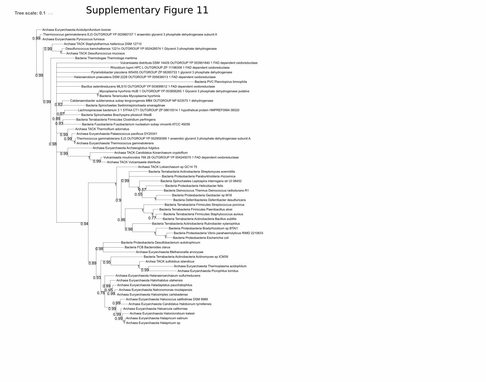
Archaea Euryarchaeota Haloarcula californiae
Bacteria Thermotogae Thermotoga maritima
Archaea Euryarchaeota Picrophilus torridus
Caldanaerobacter subterraneus subsp tengcongensis MB4 OUTGROUP NP 623575 1 dehydrogenase
Bacteria Proteobacteria Desulfobacterium autotrophicum
Bacteria Terrabacteria Firmicutes Paenibacillus alvei
Bacteria Tenericutes Mycoplasma hyorhinis
Bacteria Terrabacteria Actinobacteria Bacillus subtilis
Bacteria Deferribacteres Deferribacter desulfuricans
Archaea Euryarchaeota Thermoplasma acidophilum
Bacteria Proteobacteria Escherichia coli
Archea TACK sulfolobus islandicus
Bacteria Spirochaetes Brachyspira pilosicoli WesB
Archaea Euryarchaeota Halapricum salinum
Archaea Euryarchaeota Halococcus salifodinae DSM 8989
Bacteria Proteobacteria Geobacter sp M18
Archaea Euryarchaeota Pyrococcus furiosus
Archaea TACK Vulcanisaeta distributa
Archaea Euryarchaeota Haladaptatus paucihalophilus
Lachnospiraceae bacterium 3 1 57FAA CT1 OUTGROUP ZP 08610514 1 hypothetical protein HMPREF0994 06520
Archaea Euryarchaeota Candidatus Halobonum tyrrellensis
Bacteria FCB Bacteroides clarus
Archaea Euryarchaeota Halosimplex carlsbadense
Rhizobium lupini HPC L OUTGROUP ZP 11198308 1 FAD dependent oxidoreductase
Bacteria Spirochaetes Leptospira interrogans str UI 08452
Archaea TACK Lokiarchaeum sp GC14 75
Vulcanisaeta moutnovskia 768 28 OUTGROUP YP 004245070 1 FAD dependent oxidoreductase
Archaea Euryarchaeota Archaeoglobus fulgidus
Archaea Euryarchaeota Halapricum sp
Bacteria Spirochaetes Sediminispirochaeta smaragdinae
Archaea Euryarchaeota Aciduliprofundum boonei
Thermococcus gammatolerans EJ3 OUTGROUP YP 002959389 1 anaerobic glycerol 3 phosphate dehydrogenase subunit A
Bacteria Proteobacteria Bradyrhizobium sp BTAi1
Bacteria Fusobacteria Fusobacterium nucleatum subsp vincentii ATCC 49256
Archaea Euryarchaeota Halomicrobium katesii
Halanaerobium praevalens DSM 2228 OUTGROUP YP 005836013 1 FAD dependent oxidoreductase
Archaea Euryarchaeota Natronomonas moolapensis
Bacteria Proteobacteria Vibrio parahaemolyticus RIMD 2210633
Pyramidobacter piscolens W5455 OUTGROUP ZP 06265733 1 glycerol 3 phosphate dehydrogenase
Bacteria Terrabacteria Firmicutes Staphylococcus aureus
Bacillus selenitireducens MLS10 OUTGROUP YP 003699012 1 FAD dependent oxidoreductase
Archaea Euryarchaeota Palaeococcus pacificus DY20341
Bacteria Terrabacteria Actinobacteria Streptomyces avermitilis
Archaea Euryarchaeota Methanocella arvoryzae
Archaea TACK Desulfurococcus mucosus
Bacteria Terrabacteria Firmicutes Clostridium perfringens
Archaea TACK Staphylothermus hellenicus DSM 12710
Archaea Euryarchaeota Halanaeroarchaeum sulfurireducensArchaea Euryarchaeota Halorhabdus utahensis
Bacteria Proteobacteria Helicobacter felis
Archaea TACK Thermofilum adornatus
Bacteria Terrabacteria Actinobacteria Rubrobacter xylanophilus
Mycoplasma hyorhinis HUB 1 OUTGROUP YP 003856265 1 Glycerol 3 phosphate dehydrogenase putative
Bacteria PVC Planctopirus limnophila
Bacteria Deinococcus Thermus Deinococcus radiodurans R1
Bacteria Proteobacteria Paraburkholderia rhizoxinica
Bacteria Terrabacteria Firmicutes Streptococcus porcinus
Desulfurococcus kamchatkensis 1221n OUTGROUP YP 002428574 1 Glycerol 3 phosphate dehydrogenase
Vulcanisaeta distributa DSM 14429 OUTGROUP YP 003901840 1 FAD dependent oxidoreductase
Thermococcus gammatolerans EJ3 OUTGROUP YP 002960157 1 anaerobic glycerol 3 phosphate dehydrogenase subunit A
Bacteria Terrabacteria Actinobacteria Actinomyces sp ICM39
Archaea TACK Candidatus Korarchaeum cryptofilum
Archaea Euryarchaeota Thermococcus gammatolerans
0.99
0.98
1
0.99
10.86
0.99
0.99
0.99
0.97
0.99
0.99
1
0.93
0.99
1
0.95
0.98
1
0.99
1
0.82
0.99
0.99
0.89
0.93
0.94
1
0.99
1
1
0.570.55
0.78
1
0.99
0.9
0.99
1
0.99
1
0.99
0.95
0.98
1
0.99
0.77
Tree scale: 0.1 Supplementary Figure 11

Corynebacterium efficiens OUTGROUP WP 006769599 1 3 hydroxyacyl CoA dehydrogenase
Bacteria Caldithrix abyssi DSM 13497
Eubacterium callanderi OUTGROUP WP 013382372 1 UDP glucose GDP mannose dehydrogenase family protein
Bacteria Elusimicrobia Elusimicrobium minutum
Archaea Euryarchaeota archaeon TMED85
Bacteria Terrabacteria Cyanobacteria Scytonema hofmanni
Bacteria Chrysiogenetes Chrysiogenes arsenatis
Bacteria Terrabacteria Cyanobacteria Gloeobacter kilaueensis JS1
Bacteria Terrabacteria Firmicutes Planococcus maritimus
Bacteria Synergistes Dethiosulfovibrio peptidovorans DSM 11002
Bacteria Proteobacteria Erythrobacter litoralis
Bacteria Terrabacteria Firmicutes Streptococcus pneumoniae
Bacteria Terrabacteria Firmicutes Bacillus subtilis
Bacteria Latescibacteria Candidatus Latescibacter anaerobius
Archaea archaeon GW2011 AR11
Bacteria FCB Chlorobium limicolaBacteria Fusobacteria Fusobacterium perfoetens
Bacteria Proteobacteria Paraburkholderia rhizoxinica HKI 454
Bacteria Thermobaculum Thermobaculum terrenum
Bacteria PVC Chlamydia trachomatis A HAR 13
Desulfarculus baarsii OUTGROUP WP 013258669 1 3 hydroxyacyl CoA dehydrogenase
Archaea Euryarchaeota Methanothermobacter thermautotrophicus
Bacteria Spirochaetes Leptospira interrogans
Archaea marine group II III euryarchaeote AD1000 25 A05
Bacteria Deinococcus Thermus Deinococcus radiodurans
Bacteria Terrabacteria Actinobacteria Rubrobacter radiotolerans
Bacteria Proteobacteria Rickettsia prowazekii str Rp22
Acidithiobacillus ferrooxidans OUTGROUP WP 012607014 1 UDP glucose GDP mannose dehydrogenase family protein
Acidobacterium capsulatum OUTGROUP WP 015895922 1 UDP glucose GDP mannose dehydrogenase family protein
Bacteria Proteobacteria Mariprofundus ferrooxydans PV 1
Bacteria Proteobacteria Coxiella burnetii RSA 331
Archaea Euryarchaeota marine group II III euryarchaeote KM3 85 A08
Bacteria Candidatus Methylomirabilis oxyfera
Methanobacterium formicicum DSM 3637 OUTGROUP EKF86026 1 UDP glucose 6 dehydrogenase
Bacteria Ignavibacteriae Ignavibacterium album
Bacteria Proteobacteria Desulfarculus baarsii
Archaea Euryarchaeota Methanoculleus thermophilusArchaea Euryarchaeota Methanobacterium sp MB1
Ruminococcus flavefaciens OUTGROUP WP 009984763 1 3 hydroxyacyl CoA dehydrogenase family protein
Bacteria Gemmatimonadetes Gemmatimonas aurantiaca T 27
Bacteria Terrabacteria Cyanobacteria Chroococcidiopsis thermalis
Bacteria Lentispaera Lentisphaera araneosa HTCC2155
Archaea Euryarchaeota Marine Group II euryarchaeote MED G34
Bacteria Terrabacteria Firmicutes Paenibacillus curdlanolyticus YK9
Gemmatimonas aurantiaca OUTGROUP WP 015894566 1 3 hydroxyacyl CoA dehydrogenase
Bacteria Proteobacteria Desulfobacter postgatei
Bacteria Proteobacteria Rhizobium gallicum
Bacteria FCB Chitinivibrio alkaliphilus ACht1
Bacteria Terrabacteria Chloroflexi Caldilinea aerophila
Archaea Candidatus Woesearchaeota archaeon CG 4 10 14 0 2 um filter 33 13
OUTGROUP sp Q57871 1 WECC METJA RecName Full UDP N acetyl D mannosamine dehydrogenase AltName Full UDP ManNAc 6 dehydrogenase
Bacteria Proteobacteria Vibrio cholerae HE48
Archaea Euryarchaeota archaeon TMED97
Bacteria Proteobacteria Sorangium cellulosum
Bacteria PVC Planctopirus limnophila
Bacteria Dictyoglomi Dictyoglomus thermophilum
Bacteria Deferribacterales Deferribacter desulfuricans
Bacteria Acetothermia Candidatus Acetothermus autotrophicum
Bacteria Terrabacteria Firmicutes Staphylococcus aureus A9635
Bacteria Thermodesulfobacteria Thermodesulfatator atlanticus
Bacteria Proteobacteria Nitrosomonas eutropha
Bacteria PVC Verrucomicrobium spinosum
Archaea Euryarchaeota Candidatus Thalassoarchaea euryarchaeote
Bacteria Terrabacteria Firmicutes Clostridium ultunense Esp
Bacteria Aquificae Aquifex aeolicus
Sulfolobus solfataricus OUTGROUP WP 010923104 1 UDP glucose GDP mannose dehydrogenase family protein
Bacteria Spirochaetes Spirochaeta thermophila
Bacteria Terrabacteria Actinobacteria Streptomyces sviceus ATCC 29083
Archaea Euryarchaeota Ferroglobus placidus
Bacteria FCB Fibrobacter succinogenes
Bacteria Spirochaetes Brachyspira pilosicoli WesB
Bacteria Armatimonadetes Chthonomonas calidirosea T49
Bacteria FCB Bacteroides fluxus YIT 12057
Bacteria Tenericutes Mycoplasma hominis ATCC 23114
Archaea archaeon GW2011 AR9
Fischerella muscicola OUTGROUP WP 016868808 1 3 hydroxyacyl CoA dehydrogenase
Bacteria Aminicenantes Candidatus Aminicenans sakinawicola
Archaea DPANN Candidatus Woesearchaeota archaeon CG10 big fil rev 8 21 14 0 10 36 11
Bacteria Terrabacteria Firmicutes Planifilum fulgidum
Bacteria Proteobacteria Micavibrio aeruginosavorus
Bacteria Proteobacteria Escherichia coli str K 12 substr MG1655
Bacteria Proteobacteria Desulfatibacillum aliphaticivorans
Archaea Euryarchaeota Methanobrevibacter filiformis
Bacteria Terrabacteria Actinobacteria Actinomyces slackii
Cyclobacterium marinum OUTGROUP WP 014018215 1 3 hydroxyacyl CoA dehydrogenase family protein
Bacteria Proteobacteria Geobacter bemidjiensis
Bacteria Haloplasmatales Haloplasma contractile
Archaea TACK Thermofilum sp ex4484 15
Archaea Euryarchaeota Archaeoglobus fulgidus
Bacteria Cloacimonetes Candidatus Cloacimonas acidaminovorans str Evry
Bacteria Nitrospinae Nitrospina gracilis 3 211
Archaea Euryarchaeota Geoglobus ahangari
Bacteria Thermotogae Thermotoga maritima MSB8
Bacteria Nitropirae Thermodesulfovibrio thiophilus
Bacteria Proteobacteria Helicobacter saguini
Archaea Euryarchaeota Candidatus Thalassoarchaea euryarchaeote
0.92
1
1
0.99
0.89
0.99
0.990.89
0.51
0.97
0.92
0.57
1
0.99
0.52
0.72
0.99
0.69
0.57
0.69
0.98
1
0.95
11
0.96
0.81
1
1
0.96
0.75
0.83
0.79
0.88
0.74
0.52
0.5
0.97
0.53
1
1
0.79
0.65
0.58
1
0.97
0.97
0.73
1
1
0.51
Tree scale: 1 Supplementary Figure 12

Bacteria Terrabacteria Firmicutes Paenibacillus curdlanolyticus YK9
Eukaryota Thalassiosira oceanica
Eukaryota Physcomitrella patens
Reticulomyxa filosa Eukaryota ETO15778 1 ran binding protein 1
Bacteria Proteobacteria Helicobacter saguini
Bacteria Proteobacteria Erythrobacter litoralis
Bacteria Deinococcus Thermus Deinococcus radiodurans
Bacteria Acetothermia Candidatus Acetothermus autotrophicum
Bacteria Proteobacteria Vibrio cholerae HE48
Archaea Euryarchaeota Geoglobus ahangari
Bacteria Fusobacteria Fusobacterium perfoetens
Eukaryota Porphyra umbilicalis
Bacteria Proteobacteria Paraburkholderia rhizoxinica HKI 454
Eukaryota Thecamonas trahens
Eukaryota Arabidopsis thaliana
Eukaryota Nutamonas longa
Bacteria Terrabacteria Firmicutes Streptococcus pneumoniae
Bacteria FCB Cloacimonetes Candidatus Cloacimonas acidaminovorans str Evry
Bacteria FBC Fibrobacter succinogenes
Eukaryota Acanthamoeba castellanii
Bacteria Spirochaetes Leptospira interrogans
Archaea marine group II III euryarchaeote AD1000 25 A05
Eukaryota Cryptophyceae sp
Eukaryota Schizosaccharomyces pombe
Bacteria Thermobaculum Thermobaculum terrenum
Bacteria Armatimonadetes Chthonomonas calidirosea T49
Bacteria Synergistes Dethiosulfovibrio peptidovorans DSM 11002
Bacteria Terrabacteria Cyanobacteria Chroococcidiopsis thermalis
Archaea Candidatus Woesearchaeota archaeon CG 4 10 14 0 2 um filter 33 13
Archaea Euryarchaeota Marine Group II euryarchaeote MED G34
Bacteria Proteobacteria Sorangium cellulosum
Bacteria Terrabacteria Actinobacteria Actinomyces slackii
Bacteria Proteobacteria Rhizobium gallicum
Bacteria Terrabacteria Firmicutes Bacillus subtilis
Bacteria Proteobacteria Geobacter bemidjiensis
Bacteria PVC Lentispaera Lentisphaera araneosa HTCC2155
Bacteria Aminicenantes Candidatus Aminicenans sakinawicola
Bacteria PVC Verrucomicrobium spinosum
Archaea Euryarchaeota marine group II III euryarchaeote KM3 85 A08
Eukaryota Xenopus tropicalisEukaryota Cyanophora paradoxa
Eukaryota Tetraselmis sp
Bacteria Proteobacteria Micavibrio aeruginosavorus
Chondrus crispus Eukaryota XP 005710380 1 Glycerol 3 phosphate dehydrogenase
Archaea Euryarchaeota Candidatus Thalassoarchaea euryarchaeote
Archaea TACK Thermofilum sp ex4484 15
Bacteria FCB Ignavibacteriae Ignavibacterium album
Bacteria FBC Bacteroides fluxus YIT 12057
Eukaryota Bodo saltans
Bacteria FCB Gemmatimonadetes Gemmatimonas aurantiaca T 27
Bacteria Proteobacteria Nitrosomonas eutropha
Bacteria Thermotogae Thermotoga maritima MSB8
Bacteria Tenericutes Mycoplasma hominis ATCC 23114
Bacteria Terrabacteria Cyanobacteria Scytonema hofmanni
Archaea Euryarchaeota archaeon TMED97
Archaea archaeon GW2011 AR11
Archaea Euryarchaeota Methanothermobacter thermautotrophicusArchaea Euryarchaeota Methanoculleus thermophilus
Bacteria Dictyoglomi Dictyoglomus thermophilum
Archaea Euryarchaeota Archaeoglobus fulgidus
Eukaryota Hematodinium spp
Archaea Euryarchaeota Ferroglobus placidus
Bacteria Proteobacteria Escherichia coli str K 12 substr MG1655
Bacteria Terrabacteria Cyanobacteria Gloeobacter kilaueensis JS1
Eukaryota Mantamonas plastica
Bacteria Proteobacteria Desulfatibacillum aliphaticivorans
Bacteria Proteobacteria Desulfarculus baarsii
Eukaryota Monosiga brevicollis
Bacteria Aquificae Aquifex aeolicus
Bacteria Thermodesulfobacteria Thermodesulfatator atlanticus
Bacteria Spirochaetes Spirochaeta thermophila
Archaea Euryarchaeota Candidatus Thalassoarchaea euryarchaeote
Bacteria Nitrospinae Nitrospina gracilis 3 211
Bacteria Spirochaetes Brachyspira pilosicoli WesB
Bacteria Terrabacteria Firmicutes Planococcus maritimus
Eukaryota Stygiella incarcerata
Bacteria PVC Planctopirus limnophila
Bacteria Terrabacteria Firmicutes Staphylococcus aureus A9635
Archaea Euryarchaeota Methanobrevibacter filiformis
Bacteria Terrabacteria Firmicutes Clostridium ultunense Esp
Eukaryota Chondrus crispus
Bacteria FBC Chitinivibrio alkaliphilus ACht1
Bacteria FCB Latescibacteria Candidatus Latescibacter anaerobius
Eukaryota Capsaspora owczarzaki
Bacteria Nitropirae Thermodesulfovibrio thiophilus
Archaea Euryarchaeota Methanobacterium sp MB1
Eukaryota Paramecium tetraurelia
Bacteria PVC Chlamydia trachomatis A HAR 13
Eukaryota Reticulomyxa filosa
Bacteria Elusimicrobia Elusimicrobium minutum
Eukaryota Emiliania huxleyi
Archaea archaeon GW2011 AR9
Bacteria Terrabacteria Actinobacteria Streptomyces sviceus ATCC 29083
Bacteria Terrabacteria Actinobacteria Rubrobacter radiotolerans
Bacteria Proteobacteria Mariprofundus ferrooxydans PV 1
Bacteria Terrabacteria Haloplasmatales Haloplasma contractile
Bacteria Proteobacteria Rickettsia prowazekii str Rp22
Eukaryota Ancyromonas sigmoides
Archaea DPANN Candidatus Woesearchaeota archaeon CG10 big fil rev 8 21 14 0 10 36 11
Bacteria Terrabacteria Firmicutes Planifilum fulgidum
Eukaryota Choanocystis sp
Bacteria Terrabacteria Chloroflexi Caldilinea aerophila
Bacteria Deferribacterales Deferribacter desulfuricans
Archaea Euryarchaeota archaeon TMED85
Eukaryota Guillardia theta
Bacteria Proteobacteria Coxiella burnetii RSA 331
Bacteria Proteobacteria Desulfobacter postgatei
Bacteria FCB Caldithrix abyssi DSM 13497
Bacteria Candidatus Methylomirabilis oxyfera
Eukaryota Gefionella okellyi
Eukaryota Angomonas deanei
Bacteria Chrysiogenetes Chrysiogenes arsenatis
Bacteria FBC Chlorobium limicola
0.83
0.99
0.86
0.74
0.5
0.62
0.9
0.71
1
0.91
0.61
0.97
0.99
1
0.9
0.97
0.99
0.98
0.99
1
0.89
0.66
0.99
0.99
0.98
0.99
0.78
0.62
0.61
0.99
0.99
0.99
0.91
0.59
0.99
0.92
0.5
0.71
0.75
0.94
0.97
0.72
0.92
1
0.99
0.98
0.99
0.71
0.9
Tree scale: 1 Supplementary Figure 13

Bacteria Tenericutes Mycoplasma hyorhinis
Eukaryota Porphyra umbilicalis
Archaea TACK Staphylothermus hellenicus DSM 12710
Archaea Euryarchaeota Halapricum salinum
Bacteria Proteobacteria Escherichia coli
Archaea Euryarchaeota Halorhabdus utahensis
Archaea Euryarchaeota Halococcus salifodinae DSM 8989
Eukaryota Thalassiosira oceanica
Bacteria Terrabacteria Firmicutes Staphylococcus aureus
Eukaryota Xenopus tropicalis
Eukaryota Physcomitrella patens
Eukaryota Angomonas deanei
Archaea Euryarchaeota Picrophilus torridus
Bacteria Proteobacteria Geobacter sp M18
Eukaryota Acanthamoeba castellanii
Eukaryota Choanocystis sp
Eukaryota Stentor coeruleus
Archaea TACK Thermofilum adornatus
Archaea Euryarchaeota Haloarcula californiae
Archaea Euryarchaeota Methanocella arvoryzae
Archaea Euryarchaeota Thermococcus gammatolerans
Archaea Euryarchaeota Aciduliprofundum boonei
Bacteria Terrabacteria Actinobacteria Streptomyces avermitilis
Bacteria Terrabacteria Firmicutes Streptococcus porcinus
Bacteria FBC Bacteroides clarus
Bacteria Terrabacteria Actinobacteria Bacillus subtilis
Eukaryota Nutamonas longa
Eukaryota Stygiella incarcerata
Bacteria Spirochaetes Leptospira interrogans str UI 08452
Eukaryota Guillardia theta
Eukaryota Gefionella okellyi
Bacteria Thermotogae Thermotoga maritima
Bacteria Proteobacteria Vibrio parahaemolyticus RIMD 2210633
Archaea Euryarchaeota Halomicrobium katesii
Eukaryota Bodo saltans
Archea TACK sulfolobus islandicus
Bacteria Deferribacteres Deferribacter desulfuricans
Archaea Euryarchaeota Palaeococcus pacificus DY20341
Archaea TACK Candidatus Korarchaeum cryptofilum
Archaea Euryarchaeota Candidatus Halobonum tyrrellensis
Bacteria Spirochaetes Sediminispirochaeta smaragdinae
Bacteria Proteobacteria Helicobacter felis
Eukaryota Cyanophora paradoxa
Eukaryota Spironucleus salmonicidaBacteria PVC Planctopirus limnophila
Archaea TACK Desulfurococcus mucosus
Archaea Euryarchaeota Halapricum sp
Eukaryota Arabidopsis thaliana
Eukaryota Trimastix marina
Bacteria Proteobacteria Bradyrhizobium sp BTAi1
Eukaryota Reticulomyxa filosa
Eukaryota Mantamonas plastica
Bacteria Proteobacteria Desulfobacterium autotrophicum
Eukaryota Tetraselmis sp
Archaea Euryarchaeota Haladaptatus paucihalophilus
Eukaryota Dictyostelium discoideum
Bacteria Terrabacteria Actinobacteria Actinomyces sp ICM39
Eukaryota Paramecium tetraurelia
Eukaryota Hematodinium spp
Archaea Euryarchaeota Thermoplasma acidophilum
Eukaryota Emiliania huxleyi
Bacteria Proteobacteria Paraburkholderia rhizoxinica
Eukaryota Physarum polycephalum
Bacteria Fusobacteria Fusobacterium nucleatum subsp vincentii ATCC 49256
Eukaryota Schizosaccharomyces pombe
Bacteria Terrabacteria Firmicutes Clostridium perfringens
Archaea Euryarchaeota Halanaeroarchaeum sulfurireducens
Bacteria Terrabacteria Actinobacteria Rubrobacter xylanophilus
Archaea Euryarchaeota Pyrococcus furiosus
Bacteria Terrabacteria Firmicutes Paenibacillus alvei
Archaea TACK Vulcanisaeta distributa
Eukaryota Ancyromonas sigmoides
Archaea TACK Lokiarchaeum sp GC14 75
Eukaryota Capsaspora owczarzaki
Archaea Euryarchaeota Archaeoglobus fulgidus
Bacteria Deinococcus Thermus Deinococcus radiodurans R1
Eukaryota Thecamonas trahens
Archaea Euryarchaeota Halosimplex carlsbadense
Bacteria Spirochaetes Brachyspira pilosicoli WesB
Archaea Euryarchaeota Natronomonas moolapensis
0.74
0.97
0.87
0.98
0.86
0.99
0.95
0.57
0.98
0.99
0.99
0.99
0.96
0.98
0.96
0.62
0.99
0.68
0.93
0.98
0.99
0.6
0.98
0.99
0.55
1
0.65
0.63
0.98
0.65
0.98
0.99
0.99
0.96
0.99
0.59
0.99
0.51
0.86
0.77
0.96
0.98
0.59
1
0.51
0.95
0.98
0.92
0.99
0.55
0.99
1
0.93
0.64
Tree scale: 1 Supplementary Figure 14

Eukaryota Cryptophyceae sp
Archaea Asgardarchaeota Lokiarchaeum sp GC14 75
Bacteria FCB Ignavibacteriae Ignavibacterium album
Eukaryota Tetraselmis sp
Bacteria PVC Lentisphaerae Lentisphaera araneosa
Bacteria Thermotogae Thermotoga maritima MSB8
Bacteria Proteobacteria Geobacter bemidjiensis
Bacteria Cloacimonetes Candidatus Cloacimonas acidaminovorans
Bacteria Proteobacteria Desulfarculus baarsii
Bacteria Terrabacteria Firmicutes Planifilum fulgidum
Bacteria FCB Gemmatimonadetes Gemmatimonas aurantiaca
Bacteria Spirochaetes Leptospira interrogans
Bacteria FCB Bacteroides fluxus
Archaea Euryarchaeota Marine Group III euryarchaeote CG Epi3
Bacteria Deferribacteres Deferribacter desulfuricans
Bacteria Spirochaetes Brachyspira pilosicoli WesB
Bacteria candidate division NC10 Candidatus Methylomirabilis oxyfera
Bacteria Proteobacteria Desulfobacter postgatei
Bacteria Nitrospinae Nitrospina gracilis 3 211
Eukaryota Choanocystis sp
Eukaryota Bodo saltans
Archaea Euryarchaeota Euryarchaeota archaeon TMED85
Eukaryota Bodo saltans
Archaea Euryarchaeota Euryarchaeota archaeon TMED255
Bacteria Proteobacteria Sorangium cellulosum
Archaea Asgardarchaeota Candidatus Heimdallarchaeota archaeon LC 2
Bacteria Thermobaculum Thermobaculum terrenum
Bacteria Tenericutes Mycoplasma hominis ATCC 23114
Bacteria Terrabacteria Firmicutes Streptococcus pneumoniae
Bacteria Aquificae Aquifex aeolicus
Bacteria Terrabacteria Cyanobacteria Chroococcidiopsis thermalis
Eukaryota Thalassiosira oceanica
Bacteria Proteobacteria Mariprofundus ferrooxydans
Bacteria Dictyoglomi Dictyoglomus thermophilum
Bacteria Proteobacteria Helicobacter saguini
Eukaryota Chondrus crispus
Eukaryota Schizosaccharomyces pombe
Bacteria Haloplasmatales Haloplasma contractile
BacteriaFCB Chitinivibrio alkaliphilus ACht1
Eukaryota Xenopus tropicalis
Archaea Euryarchaeota Marine Group III euryarchaeote CG Epi5
Eukaryota Physcomitrella patens
Eukaryota Hematodinium spp
Bacteria Terrabacteria Firmicutes Paenibacillus curdlanolyticus
Eukaryota Physarum polycephalum
Bacteria FCB Caldithrix abyssi
Eukaryota Nutamonas longa
Archaea Euryarchaeota Euryarchaeota archaeon TMED164
Bacteria Proteobacteria Paraburkholderia rhizoxinica
Bacteria Terrabacteria Actinobacteria Streptomyces sviceus
Archaea Euryarchaeota Marine Group III euryarchaeote CG Epi6
Bacteria PVC Planctopirus limnophila
Bacteria Spirochaetes Spirochaeta thermophila
Bacteria Protoebacteria Rickettsia prowazekii
Bacteria Terrabacteria Actinobacteria Rubrobacter xylanophilus
Bacteria FBC Fibrobacter succinogenes
Eukaryota Dictyostelium discoideum
Eukaryota Ancyromonas sigmoides
Bacteria Terrabcteria Firmicutes Bacillus subtilis
Bacteria Thermodesulfobacteria Thermodesulfatator atlanticus
Eukaryota Spironucleus salmonicida
Bacteria Nitrospirae Thermodesulfovibrio thiophilus
Bacteria Proteobacteria Desulfatibacillum aliphaticivorans
Bacteria PVC Verrucomicrobium spinosum
Eukaryota Mantamonas plastica
Bacteria Terrabacteria Cyanobacteria Nodosilinea nodulosa
Bacteria Proteobacteria Nitrosomonas eutropha
Bacteria Deinococcus Thermus Deinococcus radiodurans
Eukaryota Porphyra umbilicalis
Bacteria Proteobacteria Coxiella burnetii
Bacteria Chrysiogenetes Chrysiogenes arsenatis
Bacteria Acidobacteria Acidobacterium capsulatum
Bacteria Elusimicrobia Elusimicrobium minutum
Bacteria Terrabacteria Chloroflexi Chloroflexus aggregans
Bacteria Proteobacteria Escherichia coli
Eukaryota Capsaspora owczarzaki
Bacteria FCB Chlorobium limicola
Bacteria Terrabacteria Firmicutes Staphylococcus aureus
Bacteria Terrabacteria Actinobacteria Actinomyces slackii
Bacteria PVC Chlamydia trachomatis A HAR 13
Bacteria Terrabacteria Firmicutes Clostridium ultunense
Eukaryota Emiliania huxleyi
Bacteria Proteobacteria Erythrobacter litoralis
Eukaryota Guillardia theta
Archaea Euryarchaeota uncultured marine group II III euryarchaeote KM3 86 F07
Bacteria Synergistes Dethiosulfovibrio peptidovorans
Bacteria Armatimonadetes Chthonomonas calidirosea T49Archaea DPANN archaeon GW2011 AR11
Archaea Euryarchaeota Marine Group III euryarchaeote CG Epi4
Bacteria Proteobacteria Vibrio cholerae
Eukaryota Arabidopsis thaliana
Bacteria Terrabacteria Firmicutes Planococcus maritimus
Archaea Euryarchaeota Marine Group III euryarchaeote CG Epi2
Archaea Euryarchaeota Marine Group III euryarchaeote CG Epi1
Bacteria Proteobacteria Micavibrio aeruginosavorus
Bacteria Proteobacteria Rhizobium gallicum
0.91
0.85
0.58
0.67
0.75
0.97
0.79
0.98
0.72
0.69
0.62
0.95
0.57
0.98
1
0.99
0.82
0.64
0.93
0.71
0.99
0.99
0.59
0.99
0.87
0.68
0.63
0.68
0.91
0.97
0.63
0.99
0.51
0.53
0.57
0.64
0.53
0.96
0.63
Tree scale: 1 Supplementary Figure 15

Bacteria Actinobacteria Verrucosispora sediminis
Bacteria Firmicutes Maledivibacter halophilus
Bacteria Actinobacteria Eubacterium cellulosolvens
Bacteria Actinobacteria Streptomyces sp WAC00263
Archaea TACK Candidatus Korarchaeum cryptofilum
Bacteria Firmicutes Lachnospiraceae bacterium YSB2008
Archaea Euryarchaeota Methanospirillum hungatei
Bacteria Firmicutes Succiniclasticum ruminis
Archaea Euryarchaeota Methanothermobacter thermautotrophicus
Bacteria Thermotogae Mesotoga infera
Bacteria Actinobactria Nocardiopsis gilva
Bactria Actinobacteria Glycomyces sp NRRL B 16210
Bacteria Actinobacteria Kribbella catacumbae
Bacteria Actinobacteria Streptomyces luteus
Archaea Asgardarchaeota Candidatus Thorarchaeota archaeon SMTZ1 45
Bacteria Actinobacteria Verrucosispora maris AB 18 032
Archaea TACK Nitrosopumilus maritimus SCM1
Bacteria Firmicutes Numidum massiliense
Bacteria FCB Chitinispirillum alkaliphilum
Bacteria Candidatus Kerfeldbacteria bacterium RIFOXYA2 FULL 38 24
Bacteria Actinobacteria Glycomyces tenuis
Archaea TACK Sulfolobus solfataricus
Archaea Euryarchaeota Methanocaldococcus jannaschii
Bacteria Firmicutes Novibacillus thermophilus
Archaea TACK uncultured marine thaumarchaeote KM3 72 H08
Bacteria Actinobacteria Couchioplanes caeruleus
Bacteria Firmicutes Caldicellulosiruptor kronotskyensis
Archaea Asgardarchaeota Candidatus Heimdallarchaeota archaeon LC 3
Bacteria Firmicutes Planifilum fulgidum
Archaea TACK Cenarchaeum symbiosum
Archaea Euryarchaeota Methanosarcina mazei Tuc01
Archaea TACK Thermofilum pendens
Archaea TACK Candidatus Caldiarchaeum subterraneum
Archaea Euryarchaeota Methanopyrus kandleri AV19Archaea DPANN Candidatus Micrarchaeota archaeon CG1 02 60 51
Bacteria Firmicutes Christensenella minuta
Bacteria Actinobacteria Asanoa ishikariensis
Bacteria Firmicutes Paenibacillus ihumii
Archaea TACK Staphylothermus marinus
Bacteria PVC Pirellula sp SH Sr6A
Archaea TACK Aeropyrum pernix K1
Bacteria Actinobacteria Patulibacter americanus
Bacteria Actinobacteria Streptomyces violaceorubidus
Bacteria Spirochaetes bacterium GWE1 32 154
Bacteria candidate division TA06 bacterium 32 111
Archaea TACK Fervidicoccus fontis Kam940
Bacteria Proteobacteria Desulfovibrio sp U5L
Archaea Euryarchaeota Thermococcus kodakarensis
Bacteria Firmicutes Anaerobacillus arseniciselenatis
Archaea TACK Acidilobus saccharovorans
Bacteria Firmicutes Kroppenstedtia eburnea
Bacteria Candidatus Melainabacteria bacterium GWF2 37 15
Bacteria Firmicutes Phascolarctobacterium sp CAG 266
Bacteria Firmicutes Paludifilum halophilum
Bacteria Firmicutes Carboxydothermus islandicus
Bacteria Candidatus Magnetoovum chiemensis
Archaea DPANN Candidatus Aenigmarchaeota archaeon ex4484 14
Archaea TACK Ignicoccus hospitalis
Archaea Euryarchaeota Halococcus salifodinae
Bacteria Actinobacteria Salinispora pacifica
Bacteria Cystobacter fuscus DSM 2262
Archaea Euryarchaeota Archaeoglobus fulgidus
Bacteria Firmicutes Melghirimyces thermohalophilus
Archaea Euryarchaeota Thermoplasma volcanium
Bacteria Firmicutes Thermoanaerobacter mathranii
Archaea TACK Marine Group I thaumarchaeote SCGC AAA799 D11
Archaea Euryarchaeota Methanomassiliicoccus luminyensis
Archaea TACK Pyrobaculum aerophilum str IM2
Bacteria Actinobacteria Actinomadura oligospora
Bacteria Actinobacteria Marmoricola aequoreus
Bacteria Actinobacteria Actinoplanes rectilineatus
Bacteria Actinobacteria Solirubrobacter sp URHD0082
Bacteria Actinobacteria Marinactinospora thermotolerans
Bacteria Actinobacteria Haloechinothrix alba
Bacteria Actinobacteria Rubrobacter radiotolerans
Archaea Euryarchaeota Aciduliprofundum boonei
Bacteria Proteobacteria Sorangium cellulosum
Bacteria Firmicutes Paenibacillus ginsengihumi
Bacteria Actinobacteria Micromonospora narathiwatensis
Bacteria Firmicutes Paenibacillus durusBacteria Firmicutes Clostridium arbusti
Bacteria Firmicutes Coprothermobacter platensis
Archaea TACK Ignisphaera aggregans DSM 17230
Bacteria Actinobacteria Allosalinactinospora lopnorensis
Archaea TACK Candidatus Bathyarchaeota archaeon RBG 16 48 13
Archaea TACK Caldivirga maquilingensis
Bacteria Firmicutes Caldalkalibacillus thermarum
Archaea Euryarchaeota Pyrococcus furiosus
Archaea Asgardarchaeota Odinarchaeota archaeon LCB 4
Bacteria FCB Gemmata sp SH PL17
Archaea TACK Candidatus Nitrososphaera gargensis Ga9 2
Bacteria Actinobacteria Kitasatospora
Bacteria Firmicutes Streptococcus pneumoniae
Bacteria Actinobacteria Xiangella phaseoli
Archaea Euryarchaeota Methanocella paludicola SANAE
Bacteria Actinobacteria Glycomyces harbinensis
Bacteria Firmicutes Desmospora sp 8437
Bacteria FCB Chitinivibrio alkaliphilus
Bacteria Actinobacteria Thermomonospora chromogena
Bacteria Firmicutes Selenomonas ruminantium
Bacteria PVC Planctomycetes bacterium
Archaea Euryarchaeota Thermoplasmatales archaeon SCGC AB 539 N05
Archaea Euryarchaeota Halofax MULTISPECIES
Bacteria Firmicutes Bacillus subtilis
Bacteria Actinibacteria Mycobacterium abscessus subsp abscessus
Bacteria Firmicutes Bacillus lentusBacteria Actinobacteria Herbidospora mongoliensis
Archaea TACK Hyperthermus butylicus
Bacteria Actinobacteria Streptosporangium amethystogenes
Bacteria Proteobacteria Azospirillum sp CAG 260
Bacteria Actinobacteria Thermobifida halotolerans
Archaea Euryarchaeota Haloarcula MULTISPECIES
0.74
0.99
1
1
1
0.51
0.83
0.98
0.67
0.64
1
1
0.81
0.85
0.63
0.96
1
1
0.77
1
1
0.79
0.99
0.81
0.98
0.95
0.85
0.9
0.61
0.71
0.6
1
1
1
0.54
0.67
0.97
1
0.97
0.86
0.99
0.7
0.81
0.8
0.7
1
1
0.99
1
0.9
0.97
1
0.93
0.99
0.68
1
0.59
0.99
0.51
1
0.97
1
1
0.95
0.77
1
0.7
1
0.98
0.94
0.97
0.94
0.94
1
1
0.99
0.83
0.85
0.92
1
0.6
0.86
0.65
0.72
0.78
1
0.69
0.75
Tree scale: 1 Supplementary Figure 16
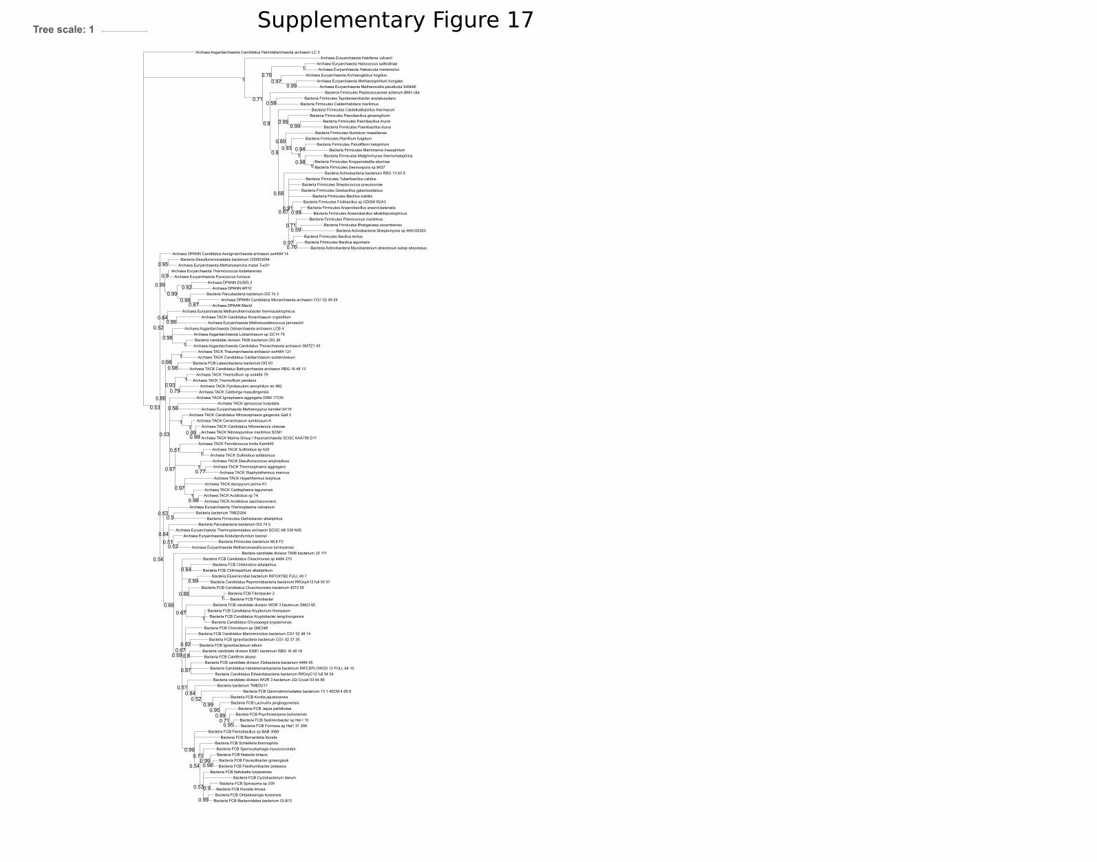
Archaea TACK Sulfolobus solfataricus
Bacteria FCB Runella limosa
Archaea TACK Caldivirga maquilingensis
Bacteria Firmicutes Planococcus maritimus
Bacteria FCB Niabella drilacis
Bacteria Firmicutes Bacillus aquimaris
Bacteria FCB Ignavibacteria bacterium CG1 02 37 35
Bacteria Firmicutes Tuberibacillus calidus
Archaea TACK Candidatus Caldiarchaeum subterraneum
Archaea TACK Caldisphaera lagunensis
Archaea TACK Thermofilum pendens
Archaea Asgardarchaeota Candidatus Thorarchaeota archaeon SMTZ1 45
Bactera candidate division TA06 bacterium 32 111
Archaea TACK Acidilobus sp 7A
Archaea TACK Marine Group I thaumarchaeote SCGC AAA799 D11
Archaea Asgardarchaeota Lokiarchaeum sp GC14 75
Bacteria Firmicutes Kroppenstedtia eburnea
Bacteria FCB Gemmatimonadetes bacterium 13 1 40CM 4 69 8
Archaea DPANN Candidatus Micrarchaeota archaeon CG1 02 49 24
Archaea Euryarchaeota Pyrococcus furiosus
Bacteria Firmicutes Paenibacillus durus
Archaea Euryarchaeota Thermoplasma volcanium
Bacteria FCB Flectobacillus sp BAB 3569
Archaea Euryarchaeota Methanomassiliicoccus luminyensis
Archaea Euryarchaeota Methanothermobacter thermautotrophicus
Bacteria Candidatus Raymondbacteria bacterium RifOxyA12 full 50 37
Bacteria Firmicutes Melghirimyces thermohalophilus
Bacteria candidate division TA06 bacterium DG 26
Archaea TACK Staphylothermus marinus
Bacteria FCB Flavisolibacter ginsengisoli
Bacteria Firmicutes Marininema mesophilum
Archaea Asgardarchaeota Candidatus Heimdallarchaeota archaeon LC 3
Bacteria FCB Fibrobacter
Bacteria Firmicutes Caldalkalibacillus thermarum
Bacteria Firmicutes Anaerobacillus arseniciselenatis
Bacteria Actinobacteria bacterium RBG 13 63 9
Bacteria Firmicutes Bacillus subtilis
Archaea Euryarchaeota Methanospirillum hungatei
Bacteria FCB Bernardetia litoralis
Archaea DPANN DUSEL3
Bacteria FCB Sediminibacter sp Hel I 10
Bacteria FCB Fibrobacter 2
Archaea TACK Candidatus Nitrososphaera gargensis Ga9 2
Archaea Euryarchaeota Methanosarcina mazei Tuc01Bacteria Desulfuromonadales bacterium C00003094
Bacteria Firmicutes Tepidanaerobacter acetatoxydans
Bacteria candidate division KSB1 bacterium RBG 16 48 16
Bacteria FCB Candidatus Cloacimonetes bacterium 4572 55
Archaea TACK Thermofilum sp ex4484 79
Bacteria FCB Jejuia pallidilutea
Bacteria Parcubacteria bacterium DG 74 3
Bacteria FCB Chitinispirillum alkaliphilum
Bacteria candidate division WOR 3 bacterium JGI Cruoil 03 44 89
Bacteria FCB Flavihumibacter petaseus
Bacteria FCB Kordia jejudonensis
Bacteria Candidatus Edwardsbacteria bacterium RifOxyC12 full 54 24
Bacteria Firmicutes Streptococcus pneumoniae
Bacteria FCB Formosa sp Hel1 31 208
Archaea TACK Candidatus Korarchaeum cryptofilum
Bacteria Firmicutes Planifilum fulgidum
Bacteria FCB Candidatus Kryptonium thompsoni
Bacteria bacterium TMED217
Bacteria Elusimicrobia bacterium RIFOXYB2 FULL 49 7
Bacteria FCB Ignavibacterium album
Archaea Euryarchaeota Haloferax volcanii
Bacteria FCB candidate division WOR 3 bacterium SM23 60
Archaea TACK Pyrobaculum aerophilum str IM2
Bacteria FCB Bacteroidetes bacterium OLB12
Bacteria FCB Lacinutrix jangbogonensis
Bacteria Firmicutes Fictibacillus sp GDSW R2A3
Archaea Euryarchaeota Methanopyrus kandleri AV19
Bacteria bacterium TMED264
Bacteria FCB Candidatus Marinimicrobia bacterium CG1 02 48 14
Bacteria Firmicutes Paenibacillus ginsengihumi
Bacteria FCB Chlorobium sp GBChlB
Bacteria Actinobacteria Mycobacterium abscessus subsp abscessus
Bacteria Firmicutes Paludifilum halophilum
Bacteria Actinobacteria Streptomyces sp WAC00263
Archaea TACK Fervidicoccus fontis Kam940
Bacteria Firmicutes Peptococcaceae acterium BRH c8a
Bacteria FCB Sporocytophaga myxococcoides
Archaea TACK Ignisphaera aggregans DSM 17230
Archaea Euryarchaeota Thermococcus kodakarensis
Bacteria Parcubacteria bacterium DG 74 2
Bacteria FCB Candidatus Cloacimonas sp 4484 275
Archaea TACK Thermosphaera aggregans
Bacteria Firmicutes Desmospora sp 8437
Archaea Euryarchaeota Thermoplasmatales archaeon SCGC AB 539 N05
Bacteria FCB Schleiferia thermophila
Archaea TACK Thaumarchaeota archaeon ex4484 121
Archaea Euryarchaeota Halococcus salifodinae
Bacteria FCB Chitinivibrio alkaliphilus
Archaea TACK Candidatus Bathyarchaeota archaeon RBG 16 48 13
Archaea TACK Hyperthermus butylicus
Archaea TACK Desulfurococcus amylolyticus
Bacteria FCB Nafulsella turpanensis
Archaea DPANN AR10
Archaea TACK Candidatus Nitrosotenuis cloacae
Bacteria FCB Latescibacteria bacterium DG 63
Bacteria Candidatus Handelsmanbacteria bacterium RIFCSPLOWO2 12 FULL 64 10
Archaea Euryarchaeota Aciduliprofundum boonei
Bacteria FCB candidate division Zixibacteria bacterium 4484 95
Bacteria Firmicutes bacterium ML8 F2
Bacteria Firmicutes Numidum massiliense
Archaea Euryarchaeota Methanocaldococcus jannaschii
Archaea Euryarchaeota Haloarcula marismortui
Bacteria FCB Ohtaekwangia koreensis
Archaea TACK Ignicoccus hospitalis
Archaea TACK Cenarchaeum symbiosum A
Archaea Asgardarchaeota Odinarchaeota archaeon LCB 4
Bacteria Firmicutes Dethiobacter alkaliphilus
Bacteria Candidatus Chrysopegis kryptomonas
Bacteria Firmicutes Geobacillus galactosidasius
Archaea Euryarchaeota Methanocella paludicola SANAE
Archaea TACK Aeropyrum pernix K1
Bacteria FCB Caldithrix abyssi
Archaea TACK Nitrosopumilus maritimus SCM1
Archaea DPAAN Macid
Bacteria FCB Cyclobacterium lianum
Archaea TACK Sulfolobus sp A20
Bacteria Firmicutes Anaerobacillus alkalidiazotrophicus
Bacteria FCB Candidatus Kryptobacter tengchongensis
Bacteria FCB Spirosoma sp 209
Bacteria Firmicutes Bhargavaea cecembensis
Archaea Euryarchaeota Archaeoglobus fulgidus
Archaea DPANN Candidatus Aenigmarchaeota archaeon ex4484 14
Bacteia FCB Psychroserpens burtonensis
Bacteria Firmicutes Calderihabitans maritimus
Bacteria Firmicutes Paenibacillus ihumii
Archaea TACK Acidilobus saccharovorans
Bacteria Firmicutes Bacillus lentus
0.99
0.64
1
0.8
0.73
0.54
0.9
1
0.53 0.99
0.99
0.51
0.97
0.51
1
1
1
0.53
0.94
0.53
1
0.99
1
0.76
0.52
0.99
0.71
0.93
0.67
0.54
0.58
0.99
0.86
0.99
1
0.98
1
0.8
0.9
0.92
0.98
0.99
1
0.99
0.93
0.52
0.97
0.64
1
0.51
1
0.59
0.5
0.97
0.98
0.71
0.86
1
0.67
0.52
0.95
0.98
0.98
0.56
0.91
0.99
0.99
0.77
0.950.71
0.92
0.89
0.87
0.67
1
0.99
0.99
0.8
0.68
0.53
0.87
0.79
0.95
0.66
0.98
0.76
0.89
0.99
0.84
0.59
0.84
0.67
0.86
Tree scale: 1 Supplementary Figure 17

DGGGP FCB Bacteria Chlorobi Chlorobium ferrooxidans
DGGGP Archaea TACK Hyperthermus butylicus
DGGGP Bacteria FCB Psychroserpens burtonensis
DGGGP Bacteria Candidatus Chrysopegis kryptomonas
DGGGP Bacteria Firmicutes Halanaerobium
DGGGP Bacteria FCB Fibrobacter
DGGGP Archaea Euryarchaeota Thermoplasma volcanium
DGGGP Archaea Euryarchaeota Haloferax
DGGGP Bacteria FCB Candidatus Kryptonium thompsoni
DGGGP Bacteria Candidatus Handelsmanbacteria bacterium RIFCSPLOWO2 12 FULL 64 10
DGGGP Archaea TACK Ignicoccus hospitalis
DGGGP Bacteria FCB Caldithrix abyssi DSM 13497
DGGGP Archaea Euryarchaeota Archaeoglobus fulgidus
DGGGP Bacteria Rhodothermaeota Rubricoccus marinus
DGGGP Archaea Euryarchaeota Methanocaldococcus jannaschii
DGGGP Archaea TACK Caldivirga maquilingensis
DGGGP Archaea Asgardarchaeota Lokiarchaeum sp GC14 75
DGGGP Bacteria FCB Ignavibacterium album
DGGGP Archaea Asgardarchaeota Candidatus Thorarchaeota archaeon SMTZ1 45
DGGGP Archaea Euryarchaeota Methanospirillum hungatei
DGGGP Bacteria candidate division TA06 bacterium 32 111
DGGGP Bacteria Parcubacteria bacterium DG 74 3
DGGGP Bacteria FCB Cyclobacterium lianum
DGGGP Bacteria Actinobacteria Kocuria sp SM24M 10
DGGGP Bacteria FCB candidate division Zixibacteria bacterium 4484 95 fa
DGGGP Bacteria FCB Sporocytophaga myxococcoides
DGGGP Bacteria Candidatus Raymondbacteria bacterium RifOxyA12 full 50 37
DGGGP Bacteria Choloroflexi Leptolinea tardivitalis
DGGGP Bacteria FCB Fibrobacteres Fibrobacter sp UWH5
DGGGP Bacteria Actinobacteria Curtobacterium sp UCD KPL2560
DGGGP Bacteria FCB Gemmatimonadetes bacterium 13 1 40CM 4 69 8
DGGGP Archaea DPANN Candidatus Aenigmarchaeota archaeon ex4484 14
DGGGP Bacteria candidate division KSB1 bacterium 4484 87
DGGGP Archaea DPANN DUSEL3
DGGGP Bacteria FCB Bacteroidetes Jejuia pallidilutea
DGGGP Bacteria FCB Candidatus Marinimicrobia bacterium CG1 02 48 14 fa
DGGGP Bacteria candidate division WOR 3 bacterium SM23 60
DGGGP Archaea Asgardarchaeota Candidatus Heimdallarchaeota archaeon LC 3
DGGGP Bacteria FCB Bacteroidetes Kordia jejudonensis
DGGGP Archaea TACK Candidatus Caldiarchaeum subterraneum
DGGGP Bacteria Candidatus Cloacimonas sp 4484 275
DGGGP Bacteria Actinobacteria Friedmanniella sagamiharensis
DGGGP Bacteria FCB Bacteroidetes Formosa sp Hel1 31 208
DGGGP Archaea Euryarchaeota Haloarcula
DGGGP Archaea TACK Aeropyrum pernix K1
DGGGP Bacteria FCB Flavisolibacter ginsengisoli DSM 18119
DGGGP Archaea TACK Pyrobaculum aerophilum st IM2
DGGGP Bacteria Candidatus Edwardsbacteria bacterium RifOxyC12 full 54 24
DGGGP Bacteria FCB Ohtaekwangia koreensis
DGGGP Archaea Euryarchaeota Methanothermobacter thermautotrophicus
DGGGP Bacteria Chloroflexi Longilinea arvoryzae
DGGGP Bacteria FCB Candidatus Krytobacter tengchongensis
DGGGP Archaea TACK Thermofilum pendens
DGGGP Archaea Euryarchaeota Methanocella paludicola SANAE
DGGGP Bacteria Actinobacteria Curtobacterium sp ER1 6
DGGGP Bacteria FCB Chitinispirillum alkaliphilum
DGGGP Archaea DPANN Macid
DGGGP Archaea Euryarchaeota Methanopyrus kandleri
DGGGP Archaea DPANN AR10
DGGGP Bacteria Cyanobacteria Chlorogloeopsis fritschii
DGGGP Bacteria Firmicutes Halanaerobium saccharolyticum
DGGGP Bacteria Actinobacteria Rubrobacter radiotolerans
DGGGP Bacteria FCB Candidatus Latescibacteria bacterium DG 63DGGGP Archaea Euryarchaeota Thermococcus kodakarensis
DGGGP Bacteria FCB Bacteroidetes Gelidibacter algens
DGGGP Archaea TACK Sulfolobus solfataricus
DGGGP Bacteria FCB Chlorobi Chlorobium sp GBChlB
DGGGP Archaea Euryarchaeota Halococcus salifodinae
DGGGP Bacteria Proteobacteria Rhodobacteraceae bacterium TMED111
DGGGP Bacteria FCB Latescibacteria bacterium DG 63
DGGGP Bacteria bacterium TMED264DGGGP Bacteria Chloroflexi Anaerolineae bacterium SG8 19
DGGGP Bacteria Actinobacteria Auraticoccus monumenti
DGGGP Archaea Euryarchaeota Thermoplasmatales archaeon SCGC AB 539 N05
DGGGP Archaea Euryarchaeota Methanosarcina mazei Tuc01
DGGGP Bacteria FCB Chitinivibrio alkaliphilus
DGGGP Archaea TACK Staphylothermus marinus
DGGGP Bacteria FCB Bacteroidetes Flammeovirgaceae bacterium TMED290
DGGGP Archaea Asgardarchaeota Candidatus Odinarchaeota archaeon LCB 4
DGGGP Bacteria FCB Ignavibacteria bacterium CG1 02 37 35
DGGGP Bacteria candidate division WOR 3 bacterium JGI Cruoil 03 44 89
DGGGP Bacteria FCB candidate division Zixibacteria bacterium RBG 1
DGGGP Archaea Euryarchaeota Pyrococcus furiosus
DGGGP FCB Bacteria Chlorobi Chlorobium limicola
DGGGP Bacteria Proteobacteria Desulfobacterales bacterium SG8 35 2 fa
DGGGP Bacteria FCB Bacteroidetes Sediminibacter sp Hel I 10
DGGGP Archaea TACK Ignisphaera aggregans DSM 17230
DGGGP Bacteria FCB Candidatus Cloacimonetes bacterium 4572 55
DGGGP Archaea Euryarchaeota Aciduliprofundum boonei
DGGGP Bacteria FCB Candidatus Marinimicrobia bacterium CG1 02 48 14
DGGGP Bacteria FCB Flavihumibacter petaseus
DGGGP Bacteria FCB Schleiferia thermophila
DGGGP Archaea DPANN Candidatus Micrarchaeota archaeon CG1 02 51 15
DGGGP Archaea Euryarchaeota Methanomassiliicoccus luminyensis
DGGGP Archaea TACK Fervidicoccus fontis Kam940
DGGGP Bacteria CPR Parcubacteria bacterium DG 74 2
DGGGP Bacteria Actinobacteria Arthrobacter sp RIT PI e
0.99
0.99
0.99
0.99
0.6
0.54
0.93
0.66
0.99
0.58
0.56
0.75
0.51
0.69
0.95
0.99
0.99
1
0.98
0.95
0.98
0.56
0.82
0.98
1
0.75
0.7
1
0.84
0.99
0.5
0.86
0.99
0.62
0.99
0.94
0.57
0.68
1
0.98
0.97
0.98
0.62
1
0.77
1
0.98
0.99
0.99
0.99
1
0.98
0.87
1
1
0.57
Tree scale: 1 Supplementary Figure 18

Bacteria PVC Verrucomicrobium spinosum
Bacteria Proteobacteria Vibrio cholerae HE48
Bacteria Proteobacteria Helicobacter saguini
Bacteria Nitrospinae Nitrospina gracilis 3 211
Bacteria Spirochaetes Spirochaeta thermophila
Bacteria Deinococcus Thermus Deinococcus radiodurans
Archaea Euryarchaeota marine group II III euryarchaeote KM3 85 A08
Archaea Euryarchaeota Ferroglobus placidus
Archaea Euryarchaeota archaeon TMED85Bacteria PVC Chlamydia trachomatis A HAR 13
Bacteria Nitropirae Thermodesulfovibrio thiophilus
Bacteria FCB Fibrobacter succinogenes
Bacteria FCB Gemmatimonadetes Gemmatimonas aurantiaca T 27
Bacteria Aminicenantes Candidatus Aminicenans sakinawicola
Archaea Euryarchaeota Candidatus Thalassoarchaea euryarchaeoteArchaea Euryarchaeota Candidatus Thalassoarchaea euryarchaeote
Bacteria Proteobacteria Erythrobacter litoralis
Bacteria Proteobacteria Rickettsia prowazekii str Rp22
Bacteria Terrabacteria Firmicutes Planococcus maritimus
Bacteria Proteobacteria Mariprofundus ferrooxydans PV 1
Bacteria Spirochaetes Leptospira interrogans
Bacteria Terrabacteria Cyanobacteria Scytonema hofmanni
Bacteria Terrabacteria Firmicutes Streptococcus pneumoniae
Archaea DPANN Candidatus Woesearchaeota archaeon CG10 big fil rev 8 21 14 0 10 36 11
Archaea Candidatus Woesearchaeota archaeon CG 4 10 14 0 2 um filter 33 13
Bacteria Terrabacteria Tenericutes Mycoplasma hominis ATCC 23114
Bacteria FCB Ignavibacteriae Ignavibacterium album
Bacteria Fusobacteria Fusobacterium perfoetens
Bacteria Proteobacteria Coxiella burnetii RSA 331
Bacteria Armatimonadetes Chthonomonas calidirosea T49
Bacteria Terrabacteria Chloroflexi Caldilinea aerophila
Archaea Euryarchaeota archaeon TMED97
Bacteria Proteobacteria Geobacter bemidjiensis
Bacteria Terrabacteria Actinobacteria Rubrobacter radiotolerans
Bacteria Aquificae Aquifex aeolicus
Bacteria Thermobaculum Thermobaculum terrenum
Bacteria Proteobacteria Paraburkholderia rhizoxinica HKI 454
Bacteria PVC Lentispaera Lentisphaera araneosa HTCC2155
Bacteria FCB Chlorobium limicola
Bacteria Terrabacteria Firmicutes Planifilum fulgidum
Archaea Euryarchaeota Archaeoglobus fulgidus
Bacteria Proteobacteria Desulfatibacillum aliphaticivorans
Bacteria FCB Cloacimonetes Candidatus Cloacimonas acidaminovorans str Evry
Archaea marine group II III euryarchaeote AD1000 25 A05
Bacteria PVC Planctopirus limnophila
Bacteria Terrabacteria Firmicutes Bacillus subtilis
Bacteria FCB Bacteroides fluxus YIT 12057
Bacteria Proteobacteria Escherichia coli str K 12 substr MG1655
Archaea Euryarchaeota Geoglobus ahangari
Bacteria Candidatus Methylomirabilis oxyfera
Bacteria Terrabacteria Firmicutes Paenibacillus curdlanolyticus YK9
Bacteria Terrabacteria Firmicutes Staphylococcus aureus A9635
Bacteria Acetothermia Candidatus Acetothermus autotrophicum
Archaea Euryarchaeota Methanobrevibacter filiformis
Archaea TACK Thermofilum sp ex4484 15
Bacteria Proteobacteria Desulfarculus baarsii
Bacteria Terrabacteria Actinobacteria Streptomyces sviceus ATCC 29083
Bacteria Chrysiogenetes Chrysiogenes arsenatis
Archaea Euryarchaeota Methanoculleus thermophilusArchaea Euryarchaeota Methanothermobacter thermautotrophicus
Bacteria Proteobacteria Micavibrio aeruginosavorus
Bacteria Proteobacteria Sorangium cellulosum
Bacteria Elusimicrobia Elusimicrobium minutum
Bacteria Proteobacteria Rhizobium gallicum
Bacteria Deferribacterales Deferribacter desulfuricans
Bacteria Spirochaetes Brachyspira pilosicoli WesB
Bacteria Terrabacteria Cyanobacteria Gloeobacter kilaueensis JS1Bacteria Terrabacteria Actinobacteria Actinomyces slackii
Bacteria Terrabacteria Firmicutes Clostridium ultunense Esp
Bacteria Dictyoglomi Dictyoglomus thermophilum
Archaea archaeon GW2011 AR9
Bacteria FCB Chitinivibrio alkaliphilus ACht1
Archaea Euryarchaeota Methanobacterium sp MB1
Bacteria Synergistes Dethiosulfovibrio peptidovorans DSM 11002
Bacteria Terrabacteria Haloplasmatales Haloplasma contractile
Bacteria FCB Caldithrix abyssi DSM 13497
Bacteria Proteobacteria Desulfobacter postgatei
Archaea Euryarchaeota Marine Group II euryarchaeote MED G34
Bacteria Proteobacteria Nitrosomonas eutropha
Bacteria Thermodesulfobacteria Thermodesulfatator atlanticus
Archaea archaeon GW2011 AR11
Bacteria Terrabacteria Cyanobacteria Chroococcidiopsis thermalis
Bacteria Thermotogae Thermotoga maritima MSB8
Bacteria FCB Latescibacteria Candidatus Latescibacter anaerobius
0.99
0.86
0.78
0.99
0.56
0.86
0.85
1
0.69
0.91
0.73
0.99
0.98
0.680.99
0.78
0.52
0.99
0.99
0.8
0.61
1
0.99
0.5
0.77
1
0.51
0.72
0.91
0.99
0.58
1
0.98
0.89
1
0.99
0.5
0.76
0.63
0.64
0.94
0.94
0.99
0.83
0.93
1
Tree scale: 1 Supplementary Figure 19

Bacteria Tenericutes Mycoplasma hyorhinis
Archaea TACK Candidatus Korarchaeum cryptofilum
Bacteria Spirochaetes Sediminispirochaeta smaragdinae
Archaea Asgardarchaeota Lokiarchaeum sp GC14 75
Archaea Euryarchaeota Halomicrobium katesii
Archaea Euryarchaeota Halorhabdus utahensis
Archaea TACK Staphylothermus hellenicus DSM 12710
Bacteria Spirochaetes Leptospira interrogans str UI 08452Bacteria Proteobacteria Helicobacter felis
Archaea Euryarchaeota Archaeoglobus fulgidus
Archaea TACK Desulfurococcus mucosus
Archaea Euryarchaeota Halapricum
Bacteria Deferribacteres Deferribacter desulfuricansBacteria Terrabacteria Firmicutes Streptococcus porcinus
Bacteria Terrabacteria Actinobacteria Rubrobacter xylanophilus
Archaea Euryarchaeota Halapricum salinum
Bacteria Deinococcus Thermus Deinococcus radiodurans R1
Bacteria Proteobacteria Desulfobacterium autotrophicum
Bacteria Terrabacteria Firmicutes Paenibacillus alvei
Bacteria FCB Bacteroides clarus
Archaea Euryarchaeota Thermococcus gammatolerans
Archaea Euryarchaeota Halosimplex carlsbadense
Archaea Euryarchaeota Halanaeroarchaeum sulfurireducensArchaea Euryarchaeota Haladaptatus paucihalophilus
Archaea Euryarchaeota Palaeococcus pacificus DY20341
Archea TACK sulfolobus islandicus
Bacteria PVC Planctopirus limnophila
Archaea Euryarchaeota Natronomonas moolapensis
Archaea Euryarchaeota Aciduliprofundum boonei
Bacteria Proteobacteria Escherichia coli
Bacteria Proteobacteria Paraburkholderia rhizoxinica
Archaea Euryarchaeota Methanocella arvoryzae
Bacteria Terrabacteria Actinobacteria Bacillus subtilis
Bacteria Proteobacteria Vibrio parahaemolyticus RIMD 2210633
Bacteria Fusobacteria Fusobacterium nucleatum subsp vincentii ATCC 49256
Archaea TACK Thermofilum adornatus
Bacteria Terrabacteria Firmicutes Clostridium perfringens
Bacteria Proteobacteria Geobacter sp M18
Archaea Euryarchaeota Thermoplasma acidophilum
Archaea Euryarchaeota Halococcus salifodinae DSM 8989
Bacteria Thermotogae Thermotoga maritima
Archaea Euryarchaeota Pyrococcus furiosus
Bacteria Terrabacteria Actinobacteria Streptomyces avermitilis
Bacteria Terrabacteria Firmicutes Staphylococcus aureus
Archaea Euryarchaeota Candidatus Halobonum tyrrellensis
Archaea TACK Vulcanisaeta distributa
Archaea Euryarchaeota Picrophilus torridus
Archaea Euryarchaeota Haloarcula californiae
Bacteria Terrabacteria Actinobacteria Actinomyces sp ICM39
Bacteria Spirochaetes Brachyspira pilosicoli WesB
Bacteria Proteobacteria Bradyrhizobium sp BTAi1
0.99
0.890.99
0.6
0.99
0.99
1
0.97
0.99
1
0.99
1
0.99
1
1
0.73
0.92
0.89
0.85
0.97
0.970.66
0.99
0.85
0.99
0.99
0.84
0.95
0.99
0.78
0.99
1
0.81
0.91
0.99
0.95
0.98 0.85
0.83
1
0.58
0.99
0.99
0.98
0.99
Tree scale: 1 Supplementary Figure 20

Bacteria Proteobacteria Escherichia coli
Bacteria Spirochaetes Leptospira interrogans
Bacteria Protoebacteria Desulfarculus baarsii
Archaea Euryarchaeota Aciduliprofundum boonei
Bacteria PVC Planctopirus limnophila
Bacteria Acidobacteria Acidobacterium capsulatum
Archaea Euryarchaeota Natronomonas moolapensis
Bacteria Terrabacteria Chloroflexi Chloroflexus aggregans
Archaea Euryarchaeota Halorhabdus utahensis
Bacteria Proteobacteria Mariprofundus ferrooxydans
Bacteria FCB Gemmatimonadetes Gemmatimonas aurantiaca
Archaea TACK Sulfolobus islandicus
Archaea TACK Caldivirga maquilingensis
Bacteria Proteobacteria Geobacter bemidjiensis
Archaea Asgardarchaeota Lokiarchaeum sp GC14 75
Bacteria Proteobcteia Micavibrio aeruginosavorus
Archaea Euryarchaeota Ferroplasma sp Type II
Archaea Euryarchaeota Palaeococcus pacificus
Bacteria Proteobacteria Erythrobacter litoralis
Archaea DPANN Candidatus Woesearchaeota archaeon CG10 big fil rev 8 21 14 0 10 32 24
Bacteria Terrabacteria Actinobacteria Staphylococcus aureus
Archaea Euryarchaeota archaeon TMED117
Archaea Euryarchaeota Ferroplasma acidarmanus
Archaea Euryarchaeota uncultured marine group II III euryarchaeote KM3 86 F07
Bacteria Thermobaculum terrenum
Bacteria PVC Chlamydia trachomatis
Bacteria Thermotogae Thermotoga maritima MSB8
Bacteria Spirochaetes Brachyspira pilosicoli WesB
Bacteria Proteobacteria Desulfatibacillum aliphaticivorans
Archaea Euryarchaeota Thermoplasma acidophilum
Bacteria Proteobacteria Rhizobium gallicum
Bacteria Terrabacteria Firmicutes Streptococcus pneumoniae
Bacteria Fusobacteria Fusobacterium perfoetens
Bacteria Proteobacteria Coxiella burnetii
Archaea Euryarchaeota Acidiplasma sp MBA 1
Archaea TACK Candidatus Korarchaeum cryptofilum OPF8
Bacteria Dictyoglomi Dictyoglomus thermophilum
Archaea Euryarchaeota Pyrococcus abyssi
Archaea Euryarchaeota Candidatus Halobonum tyrrellensis
Bacteia PVC Verrucomicrobium spinosum
Bacteria Proteobacteria Nitrosomonas eutropha
Archaea TACK Vulcanisaeta distributa
Bacteria Deinococcus Thermus Deinococcus radiodurans
Archaea Euryarchaeota Halalkalicoccus jeotgali
Bacteria Terrabacteria Firmicutes Clostridium ultunense
Bacteria Elusimicrobia Elusimicrobium minutum Pei191
Bacteria Terrabacteria Actinobacteria Rubrobacter xylanophilus
Bacteria FCB Chlorobium limicola
Archaea Euryarchaeota Candidatus Bathyarchaeota archaeon B24 2
Archaea Euryarchaeota Halomicrobium mukohataeiArchaea Euryarchaeota Halobacterium salinarum
Archaea DPANN Candidatus Woesearchaeota archaeon CG10 big fil rev 8 21 14 0 10 34 8
Archaea Euryarchaeota Archaeoglobus fulgidus
Archaea TACK Thermofilum pendens
Archaea Euryarchaeota Halosimplex carlsbadense
Archaea Euryarchaeota Haloarcula amylolytica
Bacteria Terrabcteria Actinobacteria Actinomyces slackii
Archaea TACK Desulfurococcus amylolyticus
Bacteria Terrbacteria Firmicutes Bacillus subtilis
Bacteria Proteobacteria Sorangium cellulosum
Archaea Euryarchaeota Haladaptatus paucihalophilus
Archaea Euryarchaeota Halapricum salinum
Bacteria Terrabacteria Firmicutes Paenibacillus curdlanolyticus
Bacteria Terrabacteria Firmicutes Planococcus maritimus
Archaea Euryarchaeota Hadesarchaea archaeon DG 33
Archaea Euryarchaeota Halarchaeum acidiphilum
Bacteria Proteobacteria Vibrio cholerae
Bacteria Terrabacteria Firmicutes Planifilum fulgidum
Archaea Euryarchaeota Picrophilus torridus
Bacteria Terrabacteria Actinobacteria Streptomyces sviceus ATCC 29083
Bacteria Proteobacteria Haloplasmatales Haloplasma contractile
Archaea TACK Staphylothermus hellenicus
Archaea Euryarchaeota Thermococcus eurythermalis
Bacteria Terrabacteria Cyanobacteria Nodosilinea nodulosa
Bacteria Aquificae Aquifex aeolicusBacteria Deferribacterales Deferribacter desulfuricans
Bacteria Proteobacteria Paraburkholderia rhizoxinica
0.99
0.97
0.99
0.61
1
0.69
0.95
0.86
0.99
0.520.89
0.99
0.99
0.94
0.75
0.820.97
0.72
0.98
0.99
0.66
0.68
0.98
0.89
0.71
0.86
0.9
0.74
0.9
0.99
0.95
0.5
0.56
0.58
0.99
0.68
0.96
0.5
0.95
0.66
1
0.67
0.99
0.89
0.991
0.99
0.84
0.84
0.96
0.56
0.68
0.74
0.88
0.98
1
0.99
0.5
0.98
Tree scale: 1 Supplementary Figure 21

Bacteria Terrabacteria Firmicutes Planococcus maritimus
Archaea Euryarchaeota uncultured marine group II III euryarchaeote KM3 86 F07
Bacteria Terrabacteria Firmicutes Planifilum fulgidum
Archaea Euryarchaeota Marine Group III euryarchaeote CG Epi4
Bacteria Thermodesulfobacteria Thermodesulfatator atlanticus
Bacteria Spirochaetes Spirochaeta thermophila
Bacteria Thermotogae Thermotoga maritima MSB8
Bacteria Elusimicrobia Elusimicrobium minutumBacteria Dictyoglomi Dictyoglomus thermophilum
Archaea Euryarchaeota Euryarchaeota archaeon TMED164
Bacteria Thermobaculum Thermobaculum terrenum
Archaea Euryarchaeota Marine Group III euryarchaeote CG Epi6
Bacteria Tenericutes Mycoplasma hominis ATCC 23114
Bacteria Proteobacteria Rhizobium gallicum
Bacteria Nitrospinae Nitrospina gracilis 3 211
Bacteria Proteobacteria Desulfatibacillum aliphaticivorans
Bacteria Aquificae Aquifex aeolicus
Bacteria Proteobacteria Escherichia coli
Bacteria Proteobacteria Coxiella burnetii
Bacteria Acidobacteria Acidobacterium capsulatum
Bacteria candidate division NC10 Candidatus Methylomirabilis oxyfera
Bacteria Proteobacteria Paraburkholderia rhizoxinica
Archaea Euryarchaeota Euryarchaeota archaeon TMED85
Bacteria Proteobacteria Desulfobacter postgatei
Bacteria Proteobacteria Geobacter bemidjiensis
Bacteria Proteobacteria Micavibrio aeruginosavorus
Bacteria Terrabacteria Cyanobacteria Nodosilinea nodulosa
Bacteria Proteobacteria Nitrosomonas eutrophaBacteria Proteobacteria Erythrobacter litoralis
Bacteria Synergistes Dethiosulfovibrio peptidovorans
Bacteria Protoebacteria Rickettsia prowazekii
Bacteria Spirochaetes Leptospira interrogans
Bacteria Proteobacteria Helicobacter saguini
Archaea Asgardarchaeota Candidatus Heimdallarchaeota archaeon LC 2
Bacteria PVC Planctopirus limnophila
Bacteria FCB Ignavibacteriae Ignavibacterium album
Bacteria FCB Fibrobacter succinogenes
Archaea DPANN archaeon GW2011 AR11
Bacteria PVC Lentisphaerae Lentisphaera araneosa
Bacteria Proteobacteria Sorangium cellulosum
Bacteria Terrabacteria Chloroflexi Chloroflexus aggregans
Archaea Asgardarchaeota Lokiarchaeum sp GC14 75
Bacteria Armatimonadetes Chthonomonas calidirosea T49
Bacteria Proteobacteria Mariprofundus ferrooxydans
Bacteria Terrabacteria Actinobacteria Rubrobacter xylanophilus
Bacteria Terrabacteria Cyanobacteria Chroococcidiopsis thermalis
Bacteria Deferribacteres Deferribacter desulfuricans
Bacteria FCB Cloacimonetes Candidatus Cloacimonas acidaminovorans
Archaea Euryarchaeota Marine Group III euryarchaeote CG Epi3
Bacteria Proteobacteria Desulfarculus baarsii
Bacteria Terrabacteria Firmicutes Staphylococcus aureus
Bacteria FCB Gemmatimonadetes Gemmatimonas aurantiaca
Bacteria PVC Verrucomicrobium spinosum
Bacteria FCB Chitinivibrio alkaliphilus ACht1
Bacteria Nitrospirae Thermodesulfovibrio thiophilus
Bacteria Terrabacteria Actinobacteria Streptomyces sviceus
Bacteria Proteobacteria Vibrio cholerae
Bacteria Terrabcteria Firmicutes Bacillus subtilis
Bacteria PVC Chlamydia trachomatis A HAR 13
Bacteria Terrabacteria Firmicutes Streptococcus pneumoniae
Archaea Euryarchaeota Marine Group III euryarchaeote CG Epi1
Archaea Euryarchaeota Marine Group III euryarchaeote CG Epi5
Bacteria FCB Chlorobium limicola
Bacteria Chrysiogenetes Chrysiogenes arsenatis
Bacteria Terrabacteria Firmicutes Paenibacillus curdlanolyticus
Bacteria FCB Caldithrix abyssi
Bacteria Terrabacteria Actinobacteria Actinomyces slackii
Bacteria Spirochaetes Brachyspira pilosicoli WesB
Archaea Euryarchaeota Euryarchaeota archaeon TMED255
Bacteria FCB Bacteroides fluxus
Bacteria Terrabacteria Firmicutes Clostridium ultunense
Archaea Euryarchaeota Marine Group III euryarchaeote CG Epi2
Bacteria Terrabacteria Haloplasmatales Haloplasma contractile
Bacteria Deinococcus Thermus Deinococcus radiodurans
0.73
0.52
0.7
0.58
0.79
0.730.55
0.64
0.83
0.99
0.72
0.77
0.96
0.77
0.52
0.970.66
0.99
0.99
1
0.76
0.98
0.55
0.53
Tree scale: 1 Supplementary Figure 22
Bacteria PVC Planctopirus limnophila
Bacteria Spirochaetes Leptospira interrogans
Bacteria Terrabacteria Cyanobacteria Nodosilinea nodulosa
Bacteria Proteobacteria Micavibrio aeruginosavorusBacteria Proteobacteria Erythrobacter litoralis
Bacteria Terrabacteria Cyanobacteria Chroococcidiopsis thermalis
Bacteria Chrysiogenetes Chrysiogenes arsenatis
Bacteria Terrabacteria Haloplasmatales Haloplasma contractile
Bacteria Proteobacteria Desulfarculus baarsii
Bacteria Deinococcus Thermus Deinococcus radiodurans
Bacteria Thermotogae Thermotoga maritima MSB8
Bacteria Proteobacteria Rhizobium gallicum
Bacteria Armatimonadetes Chthonomonas calidirosea T49
Bacteria Terrabcteria Firmicutes Streptococcus pneumoniae
Bacteria PVC Chlamydia trachomatis A HAR 13
Archaea DPANN Candidatus Woesearchaeota archaeon CG11 big fil rev 8 21 14 0 20 43 8
Bacteria Fusobacteria Fusobacterium perfoetens
Bacteria Tenericutes Mycoplasma hominis
Bacteria Proteobacteria Chlorobium limicola
Bacteria FCB Candidatus Cloacimonetes Cloacimonas acidaminovorans
Bacteria FCB Gemmatimonadetes Gemmatimonas aurantiaca
Bacteria Terrabacteria Firmicutes Clostridium ultunense Esp
Bacteria Spirochaetes Spirochaeta thermophilaBacteria Dictyoglomi Dictyoglomus thermophilum
Bacteria Terrabacteria Firmicutes Staphylococcus aureus
Bacteria Terrabacteria Firmicutes Bacillus subtilisBacteria Terrabacteria Firmicutes Planococcus maritimus
Archaea Asgardarchaeota Candidatus Heimdallarchaeota archaeon LC 2
Bacteria Proteobacteria Paraburkholderia rhizoxinica
Bacteria Proteobacteria Vibrio cholerae
Bacteria Aquificae Aquifex aeolicusBacteria Acidobacteria Acidobacterium capsulatum
Bacteria Nitrospinae Nitrospina gracilis 3 211
Bacteria FCB Caldithrix Caldithrix abyssi
Bacteria Proteobacteria Geobacter
Bacteria Deferribacteres Deferribacter desulfuricans
Archaea DPANN Candidatus Pacearchaeota archaeon CG06 land 8 20 14 3 00 35 12Archaea Asgardarchaeota Lokiarchaeum sp GC14 75
Bacteria Synergistetes Dethiosulfovibrio peptidovorans
Bacteria Proteobacteria Mariprofundus ferrooxydans
Bacteria Thermobaculum Thermobaculum terrenum
Bacteria Proteobacteria Coxiella burnetii
Bacteria PVC Lentisphaerae Lentisphaera araneosa
Bacteria PVC Verrucomicrobium spinosum
Bacteria Thermodesulfobacteria Thermodesulfatator atlanticus
Bacteria Terrabacteria Actinobacteria Rubrobacter xylanophilus
Bacteria Proteobacteria Sorangium cellulosum So ce56
Archaea Asgardarchaeota Candidatus Thorarchaeota archaeon SMTZ1 83
Bacteria Proteobacteria Desulfobacter postgatei
Bacteria Proteobacteria Escherichia coli
Bacteria Proteobacteria Helicobacter saguini
Bacteria Prtoeobacteria Fibrobacter
Bacteria FCB Ignavibacteriae Ignavibacterium album
Bacteria Terrabacteria Firmicutes Paenibacillus curdlanolyticus
Bacteria Spirochaetes Brachyspira pilosicoli
Bacteria Nitrospirae Thermodesulfovibrio thiophilus
Bacteria Terrabacteria Firmicutes Planifilum fulgidum
Bacteria Proteobacteria Desulfatibacillum aliphaticivorans
Bacteria Proteobacteria Nitrosomonas eutropha
Bacteria Elusimicrobia Elusimicrobium minutum
0.72
0.55
0.93
0.96
0.99
0.97
0.76
0.88
0.98
0.96
0.66
0.830.71
0.670.85
0.97
0.92
0.99
0.510.56
0.99
0.99
0.57
0.64
0.61
1
0.75
Tree scale: 1 Supplementary Figure 23

Corynebacterium efficiens OUTGROUP WP 006769599 1 3 hydroxyacyl CoA dehydrogenase
Bacteria FCB Caldithrix abyssi DSM 13497
Eubacterium callanderi OUTGROUP WP 013382372 1 UDP glucose GDP mannose dehydrogenase family protein
Bacteria Elusimicrobia Elusimicrobium minutum
Archaea Euryarchaeota archaeon TMED85
Bacteria Terrabacteria Cyanobacteria Scytonema hofmanni
Bacteria Chrysiogenetes Chrysiogenes arsenatis
Bacteria Terrabacteria Cyanobacteria Gloeobacter kilaueensis JS1
Bacteria Terrabacteria Firmicutes Planococcus maritimus
Bacteria Synergistes Dethiosulfovibrio peptidovorans DSM 11002
Bacteria Proteobacteria Erythrobacter litoralis
Bacteria Terrabacteria Firmicutes Streptococcus pneumoniae
Bacteria Terrabacteria Firmicutes Bacillus subtilis
Bacteria FCB Latescibacteria Candidatus Latescibacter anaerobius
Archaea archaeon GW2011 AR11
Bacteria FCB Chlorobium limicolaBacteria Fusobacteria Fusobacterium perfoetens
Bacteria Proteobacteria Paraburkholderia rhizoxinica HKI 454
Bacteria Thermobaculum Thermobaculum terrenum
Bacteria PVC Chlamydia trachomatis A HAR 13
Desulfarculus baarsii OUTGROUP WP 013258669 1 3 hydroxyacyl CoA dehydrogenase
Archaea Euryarchaeota Methanothermobacter thermautotrophicus
Bacteria Spirochaetes Leptospira interrogans
Archaea marine group II III euryarchaeote AD1000 25 A05
Bacteria Deinococcus Thermus Deinococcus radiodurans
Bacteria Terrabacteria Actinobacteria Rubrobacter radiotolerans
Bacteria Proteobacteria Rickettsia prowazekii str Rp22
Acidithiobacillus ferrooxidans OUTGROUP WP 012607014 1 UDP glucose GDP mannose dehydrogenase family protein
Acidobacterium capsulatum OUTGROUP WP 015895922 1 UDP glucose GDP mannose dehydrogenase family protein
Bacteria Proteobacteria Mariprofundus ferrooxydans PV 1
Bacteria Proteobacteria Coxiella burnetii RSA 331
Archaea Euryarchaeota marine group II III euryarchaeote KM3 85 A08
Bacteria Candidatus Methylomirabilis oxyfera
Methanobacterium formicicum DSM 3637 OUTGROUP EKF86026 1 UDP glucose 6 dehydrogenase
Bacteria FCB Ignavibacteriae Ignavibacterium album
Bacteria Proteobacteria Desulfarculus baarsii
Archaea Euryarchaeota Methanoculleus thermophilusArchaea Euryarchaeota Methanobacterium sp MB1
Ruminococcus flavefaciens OUTGROUP WP 009984763 1 3 hydroxyacyl CoA dehydrogenase family protein
Bacteria FCB Gemmatimonadetes Gemmatimonas aurantiaca T 27
Bacteria Terrabacteria Cyanobacteria Chroococcidiopsis thermalis
Bacteria PVC Lentispaera Lentisphaera araneosa HTCC2155
Archaea Euryarchaeota Marine Group II euryarchaeote MED G34
Bacteria Terrabacteria Firmicutes Paenibacillus curdlanolyticus YK9
Gemmatimonas aurantiaca OUTGROUP WP 015894566 1 3 hydroxyacyl CoA dehydrogenase
Bacteria Proteobacteria Desulfobacter postgatei
Bacteria Proteobacteria Rhizobium gallicum
Bacteria FCB Chitinivibrio alkaliphilus ACht1
Bacteria Terrabacteria Chloroflexi Caldilinea aerophila
Archaea Candidatus Woesearchaeota archaeon CG 4 10 14 0 2 um filter 33 13
OUTGROUP sp Q57871 1 WECC METJA RecName Full UDP N acetyl D mannosamine dehydrogenase AltName Full UDP ManNAc 6 dehydrogenase
Bacteria Proteobacteria Vibrio cholerae HE48
Archaea Euryarchaeota archaeon TMED97
Bacteria Proteobacteria Sorangium cellulosum
Bacteria PVC Planctopirus limnophila
Bacteria Dictyoglomi Dictyoglomus thermophilum
Bacteria Deferribacterales Deferribacter desulfuricans
Bacteria Acetothermia Candidatus Acetothermus autotrophicum
Bacteria Terrabacteria Firmicutes Staphylococcus aureus A9635
Bacteria Thermodesulfobacteria Thermodesulfatator atlanticus
Bacteria Proteobacteria Nitrosomonas eutropha
Bacteria PVC Verrucomicrobium spinosum
Archaea Euryarchaeota Candidatus Thalassoarchaea euryarchaeote
Bacteria Terrabacteria Firmicutes Clostridium ultunense Esp
Bacteria Aquificae Aquifex aeolicus
Sulfolobus solfataricus OUTGROUP WP 010923104 1 UDP glucose GDP mannose dehydrogenase family protein
Bacteria Spirochaetes Spirochaeta thermophila
Bacteria Terrabacteria Actinobacteria Streptomyces sviceus ATCC 29083
Archaea Euryarchaeota Ferroglobus placidus
Bacteria FCB Fibrobacter succinogenes
Bacteria Spirochaetes Brachyspira pilosicoli WesB
Bacteria Armatimonadetes Chthonomonas calidirosea T49
Bacteria FCB Bacteroides fluxus YIT 12057
Bacteria Terrabacteria Tenericutes Mycoplasma hominis ATCC 23114
Archaea archaeon GW2011 AR9
Fischerella muscicola OUTGROUP WP 016868808 1 3 hydroxyacyl CoA dehydrogenase
Bacteria Aminicenantes Candidatus Aminicenans sakinawicola
Archaea DPANN Candidatus Woesearchaeota archaeon CG10 big fil rev 8 21 14 0 10 36 11
Bacteria Terrabacteria Firmicutes Planifilum fulgidum
Bacteria Proteobacteria Micavibrio aeruginosavorus
Bacteria Proteobacteria Escherichia coli str K 12 substr MG1655
Bacteria Proteobacteria Desulfatibacillum aliphaticivorans
Archaea Euryarchaeota Methanobrevibacter filiformis
Bacteria Terrabacteria Actinobacteria Actinomyces slackii
Cyclobacterium marinum OUTGROUP WP 014018215 1 3 hydroxyacyl CoA dehydrogenase family protein
Bacteria Proteobacteria Geobacter bemidjiensis
Bacteria Terrabacteria Haloplasmatales Haloplasma contractile
Archaea TACK Thermofilum sp ex4484 15
Archaea Euryarchaeota Archaeoglobus fulgidus
Bacteria FCB Cloacimonetes Candidatus Cloacimonas acidaminovorans str Evry
Bacteria Nitrospinae Nitrospina gracilis 3 211
Archaea Euryarchaeota Geoglobus ahangari
Bacteria Thermotogae Thermotoga maritima MSB8
Bacteria Nitropirae Thermodesulfovibrio thiophilus
Bacteria Proteobacteria Helicobacter saguini
Archaea Euryarchaeota Candidatus Thalassoarchaea euryarchaeote
0.92
1
1
0.99
0.89
0.99
0.990.89
0.51
0.97
0.92
0.57
1
0.99
0.72
0.99
0.69
0.57
0.69
0.98
1
0.95
11
0.96
0.81
1
1
0.96
0.75
0.83
0.79
0.79
0.88
0.74
0.52
0.5
0.97
0.53
1
1
0.52
0.65
0.58
1
0.97
0.97
0.73
1
1
0.51
Tree scale: 1 Supplementary Figure 24

4 hydroxybenzoate octaprenyltransferase Bacteria Actinobacteria Lentzea jiangxiensis
DGGGP Bacteria FCB Ignavibacterium album
DGGGP Archaea Euryarchaeota Thermococcus kodakarensis
DG
GG
P Ba
cter
ia F
CB
Can
dida
tus
Kryp
toni
um th
omps
oni
4 hydroxybenzoate octaprenyltransferase Archaea TACK Fervidicoccus sp
DG
GG
P Ar
chae
a Eu
ryar
chae
ota
Met
hano
mas
siliic
occu
s lu
min
yens
is
DGGGP Archaea DPANN AR10
4 hydroxybenzoate polyprenyltransferase Bacteria Cyanobacteria Prochlorococcus sp MIT 0601
protoheme IX farnesyl
transfe
rase Bacteria Firm
icutes P
aenibacillus g
insengihumi
DG
GG
P Archaea DPAN
N C
andidatus Micrarchaeota archaeon C
G1 02 51 15
DG
GG
P B
acte
ria F
CB
Cyc
loba
cter
ium
lian
um
DG
GG
P Archaea DPAN
N M
acid
DG
GG
P Ba
cter
ia F
CB
Oht
aekw
angi
a ko
reen
sis
DGGGP Bacteria
Actinobacte
ria Curto
bacteriu
m sp UCD KPL2560
DG
GG
P Bacteria Chloroflexi bacterium
RBG
16 72 14 fa
DGGGP Bacteria FCB Candidatus Latescibacteria bacterium DG 63
protoheme IX farnesyltransferase Bacteria Betaproteobacteria Methylobacillus rhizosphaerae
protoheme IX farnesyltransferase Bacteria Actinobacteria Glaciibacter superstes
DGGGP Archaea Eurya
rchaeota
Haloarcula
protoheme IX farnesyltransferase Bacteria Actinobacteria Mycobacteroides abscessus subsp abscessus
chlorophyll synthase ChlG Bacteria Alphaproteobacteria Polynucleobacter acidiphobus
DGG
GP Bacteria FCB Chitinivibrio alkaliphilus
4 hydroxybenzoate octaprenyltransferase Bacteria Cyanobacteria Mastigocladus laminosus
DGGGP Archaea TACK Sulfolobus solfataricus
DGGGP Bacteria FCB Ignavibacteria bacterium CG1 02 37 35
DGG
GP
Bact
eria
Cho
loro
flexi
Lept
olin
ea ta
rdivi
talis
DG
GG
P Ar
chae
a TA
CK
Acid
ilobu
s sa
ccha
rovo
rans
DGGGP Bacteria Actinobacteria Streptomyces luteus
DG
GG
P Bacteria FCB Spirosom
a sp 209
DGGGP Bacteria FCB Caldithrix abyssi DSM 13497
chlorophyll synthase ChlG Bacteria FCB Prosthecochloris sp V1
DGGGP Archaea Euryarchaeota Methanospirillum hungatei
4 hydroxybenzoate octaprenyltransferase Archaea archaeon BMS3Bbin15
DGGGP Archaea Euryarchaeota Methanocaldococcus jannaschii
4 hydroxybenzoate polyprenyltransferase Bacteria Cyanobacteria Prochlorococcus marinus
prot
ohem
e IX
farn
esyl
trans
fera
se A
rcha
ea T
ACK
Can
dida
tus
Cal
diar
chae
um s
ubte
rrane
um
DGG
GP
Bact
eria
Firm
icute
s De
sulfo
tom
acul
um g
ibso
niae
DG
GG
P Ba
cter
ia C
PR P
arcu
bact
eria
bac
teriu
m D
G 7
4 2
DGGGP Archaea TACK Fervidicoccus fontis Kam940
4 hydroxybenzoate octaprenyltransferase Bacteria PVC Verrucomicrobia bacterium
DGGGP Archaea Asgardarchaeota Candidatus Heimdallarchaeota archaeon LC 3pro
tohem
e IX fa
rnesy
ltrans
feras
e Bac
teria
PVC Omnit
rophic
a WOR 2
bacte
rium G
WA2 6
3 20
DGG
GP Bacteria FCB Chitinispirillum
alkaliphilum
4 hydroxybenzoate octaprenyltransferase Bactera Firmicutes Fictibacillus gelatini
4 hydroxybenzoate octaprenyltransferase Bacteria Firmicutes Bacillus sp SRB 336
DG
GG
P Ba
cter
ia C
andi
datu
s C
hrys
opeg
is k
rypt
omon
as
DG
GG
P Ar
chae
a TA
CK
Cen
arch
aeum
sym
bios
um A
DGGGP Bacteria FCB Chlorobi Chlorobium sp GBChlB
protoheme IX farnesyltransferase Bacteria Actinobacteria Homoserinimonas sp OAct 916
DG
GG
P Archaea Asgardarchaeota Lokiarchaeum sp G
C14 75
DGGGP Bacteria Actin
obacteria Curto
bacterium sp
ER1 6
chlorophyll synthase ChlG Bacteria Gammaproteobacteria Chromatium okenii
prot
ohem
e IX
farn
esylt
rans
fera
se cy
oE A
rcha
ea T
ACK
uncu
lture
d m
arine
thau
mar
chae
ote
KM3
74 C
104 hydroxybenzoate octaprenyltransferase Archaea TACK Candidatus Korarchaeum cryptofilum
DGGGP Bacteria FCB candidate division Zixibacteria bacterium RBG 1
chlorophyll synthase ChlG Bacteria PVC Planctomycetaceae bacterium TMED241
chlorophyll synthase ChlG Bacteria Cyanobacteria Nostoc sp MBR 210
hypo
thet
ical
pro
tein
CEE
45 0
9885
Arc
haea
Asg
ard
Can
dida
tus
Hei
mda
llarc
haeo
ta a
rcha
eon
B3 J
M 0
8
4 hydroxybenzoate octaprenyltransferase Bacteria Alphaproteobacteria Tistlia consotensis
DG
GG
P Archaea Euryarchaeota Halococcus salifodinae
DG
GG
P A
rcha
ea T
AC
K C
andi
datu
s K
orar
chae
um c
rypt
ofilu
m
bacteriochlorophyll chlorophyll a synthase Bacteria Actinobacteria Actinomycetales bacterium
prot
ohem
e IX
farn
esyl
trans
fera
se B
acte
ria C
yano
bact
eria
Osc
illato
riale
s cy
anob
acte
rium
CG
2 30
44
21
DGGGP Bacteria FCB Schleiferia thermophila
bacteriochlorophyll synthase Bacteria Chloroflexi Kouleothrix aurantiaca
DGGGP Bacteria Rhodothermaeota Rubricoccus marinus
4 hydroxybenzoate octaprenyltransferase Bacteria Firmicutes Brevibacillus laterosporus
DGG
GP
Bact
eria
FCB
Bac
tero
idet
es F
lam
meo
virga
ceae
bac
teriu
m T
MED
290
chlorophyll synthase ChlG Bacteria Chloroflexi Oscillochloris trichoides
Protoheme IX farnesyltransferase Archaea Asgard C
andidatus Heim
dallarchaeota archaeon LC 3
4 hydroxybenzoate polyprenyltransferase Bacteria Betaproteobacteria Pelistega indica
hypothetical protein DJ555 06660 Archaea TAC
K Desulfurococcaceae archaeon
DGG
GP
Bact
eria
Firm
icute
s Ha
lana
erob
ium
4 hydroxybenzoate octaprenyltransferase Bacteria Firmicutes Virgibacillus halodenitrificans
4 hydroxybenzoate octaprenyltransferase Bacteria Actinobacteria Mycobacterium tuberculosis
DGG
GP
Bact
eria
Firm
icute
sPe
ptoc
occa
ceae
bac
teriu
m B
RH c
8a
DGG
GP Bacteria candidate division W
OR 3 bacterium
SM23 60
4 hydroxybenzoate octaprenyltransferase Bacteria Gammaproteobacteria Pantoea allii
4 hydroxybenzoate octaprenyltransferase Archaea TACK Pyrodictium delaneyi
DGGGP Bacteria candidate division TA06 bacterium 32 111
chlorophyll synthase ChlG Bacteria FCB Chlorobaculum limnaeum
cytochrome C oxidase assembly factor CtaB Bacteria Actinobacteria Mycobacterium tuberculosis
DGGGP Archaea TACK Ignicoccus hospitalis
DGGGP
Bacte
ria F
CB C
hloro
bi Ch
lorob
ium fe
rroox
idans
protoheme IX farnesyltransferase Bacteria C
hloroflexi Caldilinea aerophila
DGGGP Bacteria Actinobacteria Arthrobacter sp RIT PI e
DGGGP Bacteria FCB Candidatus Cloacimonetes bacterium 4572 55
protoh
eme I
X farne
syltra
nsfer
ase A
rchae
a Eury
archa
eota
Haloba
cteria
les ar
chae
on Q
S 4 69
225
DGGGP Archaea TACK Ignisphaera aggregans DSM 17230
DGGGP Ba
cteria
FCB
Bacte
roide
tes K
ordia
jejud
onen
sis
DG
GG
P Bacteria FCB R
unella limosa
DGGGP Archaea Euryarchaeota Methanosarcina mazei Tuc01
prot
ohem
e IX
farn
esyl
trans
fera
se A
rcha
ea E
urya
rcha
eota
Mar
ine
Gro
up II
eur
yarc
haeo
te M
ED G
33
DGGGP Bacteria FCB Candidatus Marinimicrobia bacterium CG1 02 48 14 fa
4 hydroxybenzoate octaprenyltransferase Archaea Euryarchaeota Archaeoglobus profundus
DGGGP Ba
cteria
FCB
Bacte
roide
tes Je
juia p
allidi
lutea
DGGGP Bacteria Candidatus Edwardsbacteria bacterium RifOxyC12 full 54 24
protoheme IX fa
rnesyltra
nsferase Arch
aea Euryarch
aeota Halobaculum gomorrense
bacteriochlorophyll chlorophyll synthetase Bacteria Gammaproteobacteria Alteromonadaceae bacterium TMED101
4 hydroxybenzoate octaprenyltransferase Bacteria PVC Planctomycetaceae bacterium TMED241
protoheme IX
farnesyltransferase Bacteria A
lphaproteobacteria Rhizobiales bacterium
TME
D83
DGGGP Bacteria Parcubacteria bacterium DG 74 3
4 hydroxybenzoate octaprenyltransferase Archaea Euryarchaeota Geoglobus acetivorans
DG
GG
P Archaea TACK C
aldivirga maquilingensis
DGGGP Bacteria bacterium TMED264
prot
ohem
e IX
farn
esyl
trans
fera
se B
acte
ria C
yano
bact
eria
Pse
udan
abae
na s
p
DGGGP Bacteria Candidatus Handelsmanbacteria bacterium RIFCSPLOWO2 12 FULL 64 10
protoheme IX farnesyltransferase Bacteria Actinobacteria Mycetocola saprophilus
DG
GG
PBa
cter
ia F
CB
Fibr
obac
ter
4 hydroxybenzoate octaprenyltransferase Archaea Candidatus Korarchaeum cryptofilum
prot
ohem
e IX
farn
esyl
trans
fera
se A
rcha
ea E
urya
rcha
eota
Mar
ine
Gro
up II
I eur
yarc
haeo
te C
G B
athy
1
DG
GG
P Ba
cter
ia F
CB
Fibr
obac
tere
s Fi
brob
acte
r sp
UW
H5
4 hydroxybenzoate octaprenyltransferase Bacteria Alphaproteobacteria Thalassobaculum salexigens
chlorophyll synthase ChlG Bcteria Alphaproteobacteria Bradyrhizobium sp S23321
prot
ohem
e IX
farn
esylt
rans
fera
se A
rcha
ea TA
CK C
andid
atus
Geo
ther
mar
chae
ota
arch
aeon
ex4
572
27
DG
GG
P Bacteria G
emm
atimonadates G
emm
atirosa kalamazoonesis
protoheme IX farnesyltransferase Bacteria Betaproteobacteria Pseudogulbenkiania sp NH8B
4 hydroxybenzoate octaprenyltransferase Bacteria Firmicutes Streptococcus pneumoniae
DG
GG
P Ba
cter
ia P
rote
obac
teria
Rho
doba
cter
acea
e ba
cter
ium
TM
ED11
1
protoheme IX farnesyl
transfe
rase Bacteria Firm
icutes C
ohnella thermotolerans
DG
GG
P Ba
cter
ia A
ctin
obac
teria
Tes
sara
cocc
us b
endi
goen
sis
protoh
eme I
X farne
syltra
nsfer
ase A
rchae
a Eury
archa
eota
Therm
oplas
matales
arch
aeon
SW 10 69
26
proto
heme I
X farn
esylt
rans
feras
e Bacter
ia FC
B Igna
vibac
teria
bacte
rium
DG
GG
P Ba
cter
ia F
CB
Flav
ihum
ibac
ter p
etas
eus
DG
GG
P Archaea TACK Therm
ofilum pendens
DGGGP Bacteria candidate division WOR 3 bacterium JGI Cruoil 03 44 89
4 hydroxybenzoate octaprenyltransferase Bacteria Alphaproteobacteria Thioclava indica
4 hydroxybenzoateoctaprenyltransferase Archaea TACK Hypertherm
us butylicus
chlorophyll synthase ChlG Bacteria Betaproteobacteria Rubrivivax benzoatilyticus
geranylgeranylglycerol phosphate geranylgeranyltransferase Archaea Euryarchaeotoa Halobacteriales archaeon Q
S 3 64 16
DGG
GP
Bact
eria
Chl
orof
lexi
Long
ilinea
arv
oryz
ae
hypothetical protein CND89 01695 Archaea Euryarchaeota Marine G
roup II euryarchaeote MED G
38
DGGGP Ar
chae
a TAC
K Can
didatu
s Nitro
sosp
haer
a gar
gens
is Ga9
2
4 hydroxybenzoate octaprenyltransferase Archaea Euryarchaeota Thermococcus profundus
DGGGP Archaea TACK Hyperthermus butylicus
protoheme IX farnesyltra
nsferase Bacteria Alphaproteobacteria Ruegeria sp NKC1 1
DGGGP Bacteria FCB Candidatus Cloacimonas sp 4484 275
4 hydroxybenzoate octaprenyltransferase Bacteria Firmicutes Agitococcus lubricus
prot
ohem
e IX
farn
esyl
trans
fera
se B
acte
ria C
yano
bact
eria
Glo
eoba
cter
kila
ueen
sis
4 hydroxybenzoate octaprenyltransferase Bacteria FCB Sphingobacterium cellulitidis
chlorophyll a synthase Bacteria Cyanobacteria Synechocystis sp PCC 6803
protoheme IX fa
rnesyltra
nsferas
e Archaea Eurya
rchaeota Salinigranum ru
brum
protoheme IX farnesyltransferase Bacteria Alphaproteobacteria Pleomorphomonas oryzae
DG
GG
P Ba
cter
ia P
rote
obac
teria
Dev
osia
ribo
flavi
na
DG
GG
P Archaea TACK Staphylotherm
us marinus
DGGGP Bacteria Actin
obacteria Kocuria sp
SM24M 10
prenyltransferase Archaea TACK Ignicoccus islandicus DSM 13165
DGGGP Archaea Euryarchaeota Methanopyrus kandleri
4 hydroxybenzoate octaprenyltransferase Archaea TACK Pyrolobus fumarii
DGGGP Bacteria FCB Candidatus Marinimicrobia bacterium CG1 02 48 14
Protoheme IX farnesyltransferase Bacteria Firmicutes Virgibacillus halodenitrificans
4 hydroxybenzoate octaprenyl transferase Bacteria Actinobacteria Streptomyces sparsogenes DSM 40356
4 hydroxybenzoate octaprenyltransferase Bacteria Gammaproteobacteria Stenotrophomonas rhizophila
4 hydroxybenzoate octaprenyltransferase Bacteria Betaproteobacteria Achromobacter xylosoxidans
DGGGP Archaea DPANN Candidatus Aenigmarchaeota archaeon ex4484 14
protoheme IX farnesyltransferase Bacteria C
hloroflexi bacterium
DGGGP Bacteria Chloroflexi Anaerolineae bacterium SG8 19
4 hydroxybenzoate octaprenyltransferase Bacteria PVC C
hlamydia sp H
15 1957 10C
DGGGP Bacteria FCB Gemmatimonadetes bacterium 13 1 40CM 4 69 8
protoh
eme I
X farne
syltra
nsfer
ase B
acter
ia FCB G
emmati
monas
sp S
G8 38 2
4 hydroxybenzoate octaprenyltransferase Bacteria Cyanobacteria Nostoc sp 3335mG
DGGGP Archaea Euryarchaeota Thermoplasma volcanium
4 hydroxybenzoate octaprenyltransferase Bacteria Gammaproteobacteria Enterobacter cloacae
chlorophyll synthase ChlG Bacteria FCB Chlorobium phaeobacteroides
DGGGP Bacter
ia Acti
noba
cteria
Friedm
annie
lla sa
gamiha
rensis
DGGGP Bacteria candidate division KSB1 bacterium 4484 87
DGGGP Archaea TACK Aeropyrum pernix K1
DGG
GP
Arch
aea
TACK
Nitr
osop
umilu
s m
aritim
us S
CM1
DGGGP Bac
teria
FCB Bac
teroid
etes G
elidib
acter
alge
ns
chlorophyll synthase ChlG Bacteria Gammaproteobacteria Ectothiorhodospira sp BSL 9
chlorophyll synthase ChlG Bacteria Alphaproteobacteria Sphingomonas sp PAMC 26605
4 hydroxybenzoate octaprenyltransferase Bacteria Betaproteobacteria Alcaligenes faecalis
Prot
ohem
e IX
farn
esylt
rans
fera
se 2
Arc
haea
TAC
K Ca
ndida
tus N
itros
ocald
us ca
vasc
uren
sisD
GG
GP Archaea D
PANN
DU
SEL3
chlorophyll synthase ChlG Bacteria Firmicutes Heliobacterium modesticaldum
prot
ohem
e IX
farn
esyl
trans
fera
se A
rcha
ea E
urya
rcha
eota
Arc
haeo
glob
us p
rofu
ndus
DGGGP Archaea Euryarchaeota Pyrococcus furiosus
DG
GG
P Ar
chae
a Eu
ryar
chae
ota
Acid
ulip
rofu
ndum
boo
nei
4 hydroxybenzoate octaprenyltransferase Bacteria Actinobacteria Actinoplanes lutulentus
DGGGP Archaea Euryarchaeota Thermoplasmatales archaeon SCGC AB 539 N05
protoheme IX farnesyltransferase Bacteria Gammaproteobacteria Escherichia coli
4 hydroxybenzoate octaprenyltransferase Bacteria Gammaproteobacteria Pseudomonas stutzeri
DGGGP Archaea Asgardarchaeota Candidatus Thorarchaeota archaeon SMTZ1 45
4 hydroxybenzoate octaprenyltransferase Bacteria FCB Flavobacteriaceae bacterium Hp12
protoheme IX
farnesyltransferase Bacteria FCB Flavobacteriaceae bacterium
Hp12
DG
GG
P Archaea Euryarchaeota Haloferax
4 hydroxybenzoate octaprenyltransferase Bacteria Betaproteobacteria Bordetella bronchiseptica
DG
GG
P Ba
cter
ia F
CB
Can
dida
tus
Kryt
obac
ter t
engc
hong
ensi
s
DGGGP Bacteria FCB Latescibacteria bacterium DG 63
DG
GG
P A
rcha
ea E
urya
rcha
eota
Arc
haeo
glob
us fu
lgid
us
DG
GG
P Archaea Euryarchaeota M
ethanocella paludicola SA
NA
E
protoheme IX farnesyltransferase Bacteria PVC Chlamydiales bacterium 38 26
chlorophyll synthase ChlG Bacteria Betaproteobacteria Polynucleobacter acidiphobus
proto
heme I
X farn
esylt
rans
feras
e Bac
teria
PVC Plan
ctopir
us hy
drilla
e
protoheme IX fa
rnesyltra
nsferase
Archaea Eurya
rchaeota Halorubrum sp
48 1 W
DG
GG
P Ba
cter
ia F
irmic
utes
Sul
foba
cillu
s th
erm
osul
fidoo
xida
ns
DGGGP Bacter
ia Acti
noba
cteria
Auratic
occu
s mon
umen
ti
DGGGP Bac
teria
FCB B
acter
oidete
s For
mosa s
p Hel1
31 20
8
DG
GG
P Archaea TACK Pyrobaculum
aerophilum st IM
2
DG
GG
P Ba
cter
ia F
CB
Flav
isol
ibac
ter g
inse
ngis
oli D
SM 1
8119
DGGGP Bac
teria
FCB Psy
chros
erpen
s burt
onen
sis
DG
GG
P Ba
cter
ia F
CB
Spor
ocyt
opha
ga m
yxoc
occo
ides
DGGG
P Ba
cteria
FCB
Chlo
robi
Chlor
obium
limico
la
protoheme IX farnesyltransferase Bacteria Betaproteobacteria Snodgrassella alvi
DGGGP Bacteria Proteobacteria Desulfobacterales bacterium SG8 35 2 fa
DGGGP Bacteria Cyanobacteria Chlorogloeopsis fritschii
DGG
GP Archaea Asgardarchaeota Candidatus O
dinarchaeota archaeon LCB 4
DGGGP Ar
chae
a TAC
K Can
didat
us C
aldiar
chae
um su
bter
rane
um
DGGGP Bacteria Candidatus Raymondbacteria bacterium RifOxyA12 full 50 37
DGG
GP Archaea TACK Candidatus Nitrosoarchaeum
koreensisDGGGP Archaea Euryarchaeota Methanothermobacter thermautotrophicus
4 hydroxybenzoate polyprenyltransferase Bacteria FCB Rhodothermaceae bacterium TMED105
4 hydroxybenzoate polyprenyltransferase Bacteria Alphaproteobacteria Rhodospirillaceae bacterium BRH c57
chlorophyll synthase ChlG Bacteria Cyanobacteria Stanieria cyanosphaera
4 hydroxybenzoate polyprenyltransferase Bacteria Firmicutes Synechocystis sp PCC 6803
4 hydroxybenzoate octaprenyltransferase Archaea TACK Aeropyrum cam
ini
protoheme IX farnesyltransferase Bacteria Actinobacteria Promicromonospora sukumoe
DGG
GP
Bact
eria
Firm
icute
s Ha
lana
erob
ium
sac
char
olyt
icum
bacteriochlorophyll chlorophyll a synthase Bacteria Actinobacteria Streptomyces purpurogeneiscleroticus
prot
ohem
e IX
farn
esylt
rans
fera
se A
rcha
ea TA
CK Nitr
osos
phae
ra vi
enne
nsis
DGGGP Bac
teria
FCB B
acter
oidete
s Sed
imini
bacte
r sp H
el I 1
0
DGGGP Bacteria FCB candidate division Zixibacteria bacterium 4484 95 fa
prot
ohem
e IX
farn
esylt
rans
fera
se A
rcha
ea T
ACK
Thau
mar
chae
ota
arch
aeon
13
1 40
CM 3
8 12
DGG
GP
Arch
aea
TACK
Mar
ine
Gro
up I
thau
mar
chae
ote
SCG
C AA
A799
D11
4 hydroxybenzoate polyprenyltransferase Archaea Euryarchaeota archaeon TMED255
4 hydroxybenzoate octaprenyltransferase Archaea TACK Fervidicoccus fontis
DGGGP Bacteria
Actinobacte
ria Rubrobacte
r radiotolerans
0.98
0.95
0.79
0.91
0.95
1
0.88
1
0.85
0.61
0.96
0.96
0.63
0.99
1
0.99
0.99
0.83
0.99
0.53
1
0.68
0.99
0.99
0.76
0.65
0.6
0.54
0.71
0.99
0.97
1
0.99
0.97
0.97
0.98
0.95
0.98
0.99
0.72
0.99
0.68
0.64
0.75
10.56
0.99
0.58
0.94
0.99
0.93
0.69
0.99
0.98
0.88
1
0.81
0.62
0.69
0.53
0.75
0.9
0.530.99
0.62
0.65
1
0.87
1
0.95
0.53
1
0.71
1
0.7
0.99
0.72
1
0.87
0.71
0.73
0.53
0.96
0.87
0.97
0.57
0.79
0.95
0.95
0.99
0.55
0.96
0.94
0.99
0.5
1
0.95
0.99
0.62
0.72
1
0.95
0.61
0.97
0.99
0.96
0.59
0.66
0.93
0.75
0.99
0.99
0.99
0.99
0.93
0.690.57
0.96
0.99
0.99
0.92
0.81
0.98
0.93
0.99
0.91
0.95
0.99
0.9
0.96
0.62
0.78
1
Tree scale: 1 Supplementary Figure 25

Archaea TACK Marine Group I thaumarchaeote SCGC AAA799 D11
Bacteria Firmicutes bacterium ML8 F2
Bacteria Firmicutes Peptococcaceae acterium BRH c8a
Archaea TACK Ignisphaera aggregans DSM 17230
Bacteria FCB candidate division Zixibacteria bacterium 4484 95
Bacteria Firmicutes Kroppenstedtia eburnea
Archaea DPANN DUSEL3
Bacteria FCB Bacteroidetes bacterium OLB12
Bacteria FCB Formosa sp Hel1 31 208
Bacteria Desulfuromonadales bacterium C00003094
Archaea TACK Thermosphaera aggregans
Archaea DPANN Candidatus Aenigmarchaeota archaeon ex4484 14
Archaea TACK Sulfolobus solfataricus
Archaea TACK Thermofilum pendens
Archaea TACK Desulfurococcus amylolyticus
Bacteria Firmicutes Streptococcus pneumoniae
Archaea Asgardarchaeota Odinarchaeota archaeon LCB 4
Bacteria FCB Flectobacillus sp BAB 3569
Bacteria FCB Candidatus Kryptonium thompsoni
Archaea TACK Caldisphaera lagunensis
Archaea Euryarchaeota Thermococcus kodakarensis
Bacteria FCB Bernardetia litoralis
Archaea TACK Thaumarchaeota archaeon ex4484 121
Bacteria FCB Flavihumibacter petaseus
Archaea Asgardarchaeota Candidatus Heimdallarchaeota archaeon LC 3
Bacteria FCB Runella limosa
Archaea Euryarchaeota Archaeoglobus fulgidus
Archaea TACK Candidatus Nitrososphaera gargensis Ga9 2
Bacteria FCB Fibrobacter
Bacteria candidate division WOR 3 bacterium SM23 60
Bacteria Parcubacteria bacterium DG 74 3
Archaea Euryarchaeota Methanopyrus kandleri AV19
Bacteria Firmicutes Planococcus maritimus
Bactera candidate division TA06 bacterium 32 111
Bacteria Candidatus Chrysopegis kryptomonas
Archaea DPANN Candidatus Micrarchaeota archaeon CG1 02 49 24
Bacteria Firmicutes Anaerobacillus alkalidiazotrophicus
Bacteria FCB Schleiferia thermophila
Bacteria Firmicutes Numidum massiliense
Bacteria FCB Sediminibacter sp Hel I 10
Bacteria Firmicutes Melghirimyces thermohalophilus
Archaea TACK Acidilobus sp 7A
Archaea Euryarchaeota Halococcus salifodinae
Archaea Asgardarchaeota Lokiarchaeum sp GC14 75
Bacteria Candidatus Raymondbacteria bacterium RifOxyA12 full 50 37
Archaea TACK Aeropyrum pernix K1
Archaea TACK Candidatus Korarchaeum cryptofilum
Bacteria Actinobacteria Mycobacterium abscessus subsp abscessus
Bacteria FCB Chitinispirillum alkaliphilum
Archaea Euryarchaeota Methanospirillum hungatei
Bacteria FCB Cyclobacterium lianum
Bacteria Firmicutes Anaerobacillus arseniciselenatis
Bacteria Firmicutes Paludifilum halophilum
Archaea Euryarchaeota Methanothermobacter thermautotrophicus
Bacteria Firmicutes Paenibacillus durus
Bacteria Firmicutes Desmospora sp 8437
Bacteria Firmicutes Dethiobacter alkaliphilusArchaea Euryarchaeota Thermoplasma volcanium
Bacteria FCB Candidatus Cloacimonetes bacterium 4572 55
Bacteria Parcubacteria bacterium DG 74 2
Bacteria Firmicutes Bhargavaea cecembensis
Bacteria FCB Chitinivibrio alkaliphilus
Bacteria Firmicutes Caldalkalibacillus thermarum
Bacteria Candidatus Handelsmanbacteria bacterium RIFCSPLOWO2 12 FULL 64 10
Bacteria FCB Flavisolibacter ginsengisoli
Bacteria candidate division TA06 bacterium DG 26
Bacteria FCB Candidatus Marinimicrobia bacterium CG1 02 48 14
Archaea TACK Candidatus Nitrosotenuis cloacae
Bacteria Elusimicrobia bacterium RIFOXYB2 FULL 49 7
Archaea Asgardarchaeota Candidatus Thorarchaeota archaeon SMTZ1 45
Archaea TACK Ignicoccus hospitalis
Archaea Euryarchaeota Methanosarcina mazei Tuc01
Archaea DPANN AR10
Bacteria FCB Niabella drilacis
Bacteria Actinobacteria Streptomyces sp WAC00263
Bacteria FCB Ignavibacteria bacterium CG1 02 37 35
Archaea Euryarchaeota Methanomassiliicoccus luminyensis
Bacteria FCB Spirosoma sp 209
Archaea Euryarchaeota Aciduliprofundum boonei
Archaea TACK Acidilobus saccharovorans
Bacteria FCB Kordia jejudonensis
Bacteria candidate division KSB1 bacterium RBG 16 48 16
Bacteria Firmicutes Tuberibacillus calidus
Archaea TACK Hyperthermus butylicus
Bacteria FCB Jejuia pallidilutea
Bacteria FCB Latescibacteria bacterium DG 63Archaea TACK Candidatus Caldiarchaeum subterraneum
Archaea TACK Cenarchaeum symbiosum A
Bacteria Firmicutes Calderihabitans maritimus
Bacteria Firmicutes Bacillus lentus
Bacteria FCB Candidatus Kryptobacter tengchongensis
Archaea TACK Thermofilum sp ex4484 79
Bacteria FCB Lacinutrix jangbogonensis
Archaea TACK Caldivirga maquilingensis
Archaea DPAAN Macid
Bacteria Firmicutes Fictibacillus sp GDSW R2A3
Bacteria FCB Fibrobacter
Archaea Euryarchaeota Pyrococcus furiosus
Bacteria Firmicutes Paenibacillus ihumii
Bacteria FCB Sporocytophaga myxococcoides
Bacteria FCB Caldithrix abyssi
Archaea Euryarchaeota Haloarcula marismortui
Bacteria Firmicutes Paenibacillus ginsengihumi
Bacteria FCB Ohtaekwangia koreensis
Archaea Euryarchaeota Thermoplasmatales archaeon SCGC AB 539 N05
Bacteria bacterium TMED264
Bacteria FCB Candidatus Cloacimonas sp 4484 275
Bacteria Firmicutes Bacillus aquimaris
Archaea TACK Candidatus Bathyarchaeota archaeon RBG 16 48 13
Bacteria FCB Gemmatimonadetes bacterium 13 1 40CM 4 69 8
Bacteria Actinobacteria bacterium RBG 13 63 9
Bacteria FCB Nafulsella turpanensis
Archaea TACK Staphylothermus marinus
Archaea Euryarchaeota Methanocaldococcus jannaschii
Bacteria Firmicutes Bacillus subtilis
Archaea TACK Nitrosopumilus maritimus SCM1
Bacteia FCB Psychroserpens burtonensis
Bacteria Firmicutes Marininema mesophilum
Archaea Euryarchaeota Haloferax volcanii
Archaea TACK Sulfolobus sp A20
Bacteria Firmicutes Tepidanaerobacter acetatoxydans
Bacteria bacterium TMED217
Bacteria Candidatus Edwardsbacteria bacterium RifOxyC12 full 54 24
Bacteria FCB Ignavibacterium album
Archaea Euryarchaeota Methanocella paludicola SANAE
Bacteria candidate division WOR 3 bacterium JGI Cruoil 03 44 89
Archaea TACK Fervidicoccus fontis Kam940
Bacteria Firmicutes Geobacillus galactosidasius
Archaea TACK Pyrobaculum aerophilum str IM2
Bacteria Firmicutes Planifilum fulgidum
Bacteria FCB Chlorobium sp GBChlB
0.58
0.96
1
0.66
0.97
0.99
0.55
0.88
1
0.5
0.92
0.92
0.99
0.95
0.94
1
0.99
0.82
0.74
0.93
0.66
0.91
0.83
0.64
0.99
0.93
0.52
0.83
1
0.96
0.99
0.58
0.53
0.94
0.56
0.66
0.6
0.99
0.86
1
0.99
0.98
1
1
1
0.970.94
0.78
0.81
0.95
0.94
0.8
0.66
0.51
0.66
0.53
0.95
1
0.78
1
0.75
0.53
0.85
0.73
0.97
0.79
0.54
0.54
0.52
0.58
1
0.98
1
1
1
0.79
0.63
0.63
0.99
1
1
0.98
0.99
Tree scale: 1Supplementary Figure 26

Bacteria Spirochaetes Brachyspira pilosicoliBacteria Prtoeobacteria Fibrobacter
Archaea Asgardarchaeota Lokiarchaeum sp GC14 75
Bacteria Terrabacteria Firmicutes Paenibacillus curdlanolyticus
Bacteria Proteobacteria Rhizobium gallicum
Bacteria Proteobacteria Nitrosomonas eutropha
Bacteria Terrabacteria Cyanobacteria Nodosilinea nodulosa
Bacteria Dictyoglomi Dictyoglomus thermophilum
Bacteria PVC Lentisphaerae Lentisphaera araneosa
Bacteria Terrabacteria Firmicutes Planococcus maritimus
Bacteria PVC Chlamydia trachomatis A HAR 13
Archaea DPANN Candidatus Pacearchaeota archaeon CG06 land 8 20 14 3 00 35 12
Bacteria Proteobacteria Coxiella burnetii
Bacteria Spirochaetes Spirochaeta thermophila
Bacteria Haloplasmatales Haloplasma contractile
Bacteria Chrysiogenetes Chrysiogenes arsenatis
Bacteria Terrabacteria Firmicutes Clostridium ultunense Esp
Bacteria Spirochaetes Leptospira interrogans
Bacteria Terrabacteria Firmicutes Bacillus subtilis
Bacteria Proteobacteria Desulfatibacillum aliphaticivorans
Bacteria Proteobacteria Sorangium cellulosum So ce56
Bacteria PVC Planctopirus limnophila
Bacteria Acidobacteria Acidobacterium capsulatum
Bacteria FCB Ignavibacteriae Ignavibacterium album
Bacteria Terrabacteria Firmicutes Staphylococcus aureus
Bacteria Thermotogae Thermotoga maritima MSB8
Archaea Asgardarchaeota Candidatus Heimdallarchaeota archaeon LC 2
Bacteria FCB Gemmatimonadetes Gemmatimonas aurantiaca
Bacteria Proteobacteria Desulfarculus baarsii
Bacteria Proteobacteria Vibrio cholerae
Bacteria Proteobacteria Chlorobium limicola
Bacteria Proteobacteria Mariprofundus ferrooxydans
Bacteria FCB Caldithrix Caldithrix abyssi
Bacteria Proteobacteria Helicobacter saguini
Bacteria Deferribacteres Deferribacter desulfuricans
Bacteria Candidatus Cloacimonetes Cloacimonas acidaminovorans
Bacteria Erythrobacter litoralis
Archaea Asgardarchaeota Candidatus Thorarchaeota archaeon SMTZ1 83
Bacteria Terrabacteria Actinobacteria Rubrobacter xylanophilus
Bacteria Proteobacteria Desulfobacter postgatei
Bacteria Thermobaculum Thermobaculum terrenum
Bacteria Proteobacteria Paraburkholderia rhizoxinica
Bacteria Nitrospirae Thermodesulfovibrio thiophilus
Bacteria Armatimonadetes Chthonomonas calidirosea T49
Archaea DPANN Candidatus Woesearchaeota archaeon CG11 big fil rev 8 21 14 0 20 43 8
Bacteria Synergistetes Dethiosulfovibrio peptidovorans
Bacteria Fusobacteria Fusobacterium perfoetens
Bacteria Terrabacteria Cyanobacteria Chroococcidiopsis thermalis
Bacteria Terrabcteria Firmicutes Streptococcus pneumoniae
Bacteria Elusimicrobia Elusimicrobium minutum
Bacteria Proteobacteria Geobacter
Bacteria Thermodesulfobacteria Thermodesulfatator atlanticus
Bacteria Deinococcus Thermus Deinococcus radiodurans
Bacteria Nitrospinae Nitrospina gracilis 3 211
Bacteria Proteobacteria Escherichia coli
Bacteria Aquificae Aquifex aeolicus
Bacteria Tenericutes Mycoplasma hominis
Bacteria PVC Verrucomicrobium spinosum
Bacteria Terrabacteria Firmicutes Planifilum fulgidum
Bacteria Proteobacteria Micavibrio aeruginosavorus
0.64
1
0.99
0.98
0.82
0.56
0.51
0.88
0.68
0.98
0.99
0.67
0.5
0.56
0.83
0.930.98
0.56
0.81
0.99
0.83
0.99
0.99
0.53
Tree scale: 1Supplementary Figure 27

Bacteria Tenericutes Mycoplasma hyorhinis
Archaea TACK Lokiarchaeum sp GC14 75
Archaea Euryarchaeota Archaeoglobus fulgidus
Eukaryota Physcomitrella patens
Archaea TACK Staphylothermus hellenicus DSM 12710
Bacteria Proteobacteria Escherichia coli
Archea TACK sulfolobus islandicus
Eukaryota Porphyra umbilicalis
Eukaryota Hematodinium spp
Bacteria Terrabacteria Firmicutes Staphylococcus aureus
Eukaryota Capsaspora owczarzaki
Eukaryota Arabidopsis thaliana
Eukaryota Ancyromonas sigmoides
Archaea Euryarchaeota Thermoplasma acidophilum
Eukaryota Schizosaccharomyces pombe
Eukaryota Paramecium tetraurelia
Archaea TACK Thermofilum adornatus
Archaea Euryarchaeota Candidatus Halobonum tyrrellensis
Bacteria Proteobacteria Desulfobacterium autotrophicum
Archaea Euryarchaeota Thermococcus gammatolerans
Archaea Euryarchaeota Aciduliprofundum boonei
Bacteria Terrabacteria Actinobacteria Streptomyces avermitilis
Bacteria Terrabacteria Firmicutes Streptococcus porcinus
Bacteria Terrabacteria Actinobacteria Bacillus subtilis
Archaea Euryarchaeota Haloarcula californiae
Archaea Euryarchaeota Halapricum sp
Bacteria Spirochaetes Leptospira interrogans str UI 08452
Eukaryota Gefionella okellyi
Bacteria Thermotogae Thermotoga maritima
Bacteria Proteobacteria Vibrio parahaemolyticus RIMD 2210633
Eukaryota Xenopus tropicalis
Eukaryota Angomonas deanei
Archaea Euryarchaeota Haladaptatus paucihalophilus
Eukaryota Thecamonas trahens
Bacteria Proteobacteria Geobacter sp M18
Archaea Euryarchaeota Palaeococcus pacificus DY20341
Archaea TACK Vulcanisaeta distributa
Archaea Euryarchaeota Halococcus salifodinae DSM 8989
Archaea Euryarchaeota Halanaeroarchaeum sulfurireducens
Bacteria Spirochaetes Sediminispirochaeta smaragdinae
Bacteria Proteobacteria Helicobacter felis
Eukaryota Choanocystis sp
Eukaryota Spironucleus salmonicidaBacteria PVC Planctopirus limnophila
Archaea TACK Desulfurococcus mucosus
Archaea Euryarchaeota Halapricum salinum
Eukaryota Trimastix marina
Bacteria Proteobacteria Bradyrhizobium sp BTAi1
Eukaryota Physarum polycephalum
Eukaryota Mantamonas plastica
Archaea Euryarchaeota Halomicrobium katesii
Eukaryota Thalassiosira oceanica
Eukaryota Tetraselmis sp
Archaea Euryarchaeota Halosimplex carlsbadense
Eukaryota Cyanophora paradoxa
Archaea Euryarchaeota Methanocella arvoryzae
Eukaryota Stygiella incarcerata
Eukaryota Guillardia theta
Eukaryota Emiliania huxleyi
Eukaryota Acanthamoeba castellanii
Eukaryota Dictyostelium discoideum
Bacteria Proteobacteria Paraburkholderia rhizoxinica
Eukaryota Nutamonas longa
Bacteria Fusobacteria Fusobacterium nucleatum subsp vincentii ATCC 49256
Bacteria FCB Bacteroides clarus
Eukaryota Reticulomyxa filosa
Bacteria Terrabacteria Firmicutes Clostridium perfringens
Eukaryota Bodo saltans
Bacteria Terrabacteria Actinobacteria Rubrobacter xylanophilus
Archaea Euryarchaeota Pyrococcus furiosus
Bacteria Terrabacteria Firmicutes Paenibacillus alvei
Bacteria Terrabacteria Actinobacteria Actinomyces sp ICM39
Bacteria Deferribacteres Deferribacter desulfuricans
Archaea TACK Candidatus Korarchaeum cryptofilum
Archaea Euryarchaeota Halorhabdus utahensis
Bacteria Deinococcus Thermus Deinococcus radiodurans R1
Eukaryota Stentor coeruleus
Archaea Euryarchaeota Natronomonas moolapensis
Bacteria Spirochaetes Brachyspira pilosicoli WesB
Archaea Euryarchaeota Picrophilus torridus
0.95
1
0.98
0.95
0.87
0.58
0.91
0.99
0.56
0.96
0.69
0.99
0.990.99
0.96
0.98
0.58
0.61
0.85
0.6
0.99
0.97
0.92
0.99
0.99
0.98
0.99
0.56
1
0.97
0.63
0.64
0.73
0.86
0.99
0.77
0.99
0.99
0.98
0.92
0.51
0.98
0.99
0.51
0.94
1
0.5
0.98
0.64
0.98
0.53
0.97
1
0.94
0.62
Tree scale: 1 Supplementary Figure 28

Eukaryota Gefionella okellyi
Bacteria Terrabacteria Firmicutes Planococcus maritimus
Eukaryota Ancyromonas sigmoides
Eukaryota Thalassiosira oceanica
Bacteria Spirochaetes Spirochaeta thermophila
Bacteria Proteobacteria Desulfobacter postgatei
Bacteria Tenericutes Mycoplasma hominis ATCC 23114
Bacteria PVC Verrucomicrobium spinosum
Bacteria Terrabacteria Cyanobacteria Scytonema hofmanni
Bacteria FCB Chitinivibrio alkaliphilus ACht1
Eukaryota Nutamonas longa
Bacteria Proteobacteria Mariprofundus ferrooxydans PV 1
Eukaryota Arabidopsis thaliana
Eukaryota Emiliania huxleyi
Bacteria Fusobacteria Fusobacterium perfoetens
Bacteria Chrysiogenetes Chrysiogenes arsenatis
Bacteria Deinococcus Thermus Deinococcus radiodurans
Bacteria Synergistes Dethiosulfovibrio peptidovorans DSM 11002
Archaea Euryarchaeota Methanothermobacter thermautotrophicus
Eukaryota Chondrus crispus
Eukaryota Bodo saltans
Eukaryota Cyanophora paradoxa
Chondrus crispus Eukaryota XP 005710380 1 Glycerol 3 phosphate dehydrogenase
Archaea Euryarchaeota Methanoculleus thermophilus
Bacteria Terrabacteria Cyanobacteria Gloeobacter kilaueensis JS1
Bacteria Terrabacteria Haloplasmatales Haloplasma contractile
Bacteria Proteobacteria Helicobacter saguini
Bacteria Thermodesulfobacteria Thermodesulfatator atlanticus
Archaea Candidatus Woesearchaeota archaeon CG 4 10 14 0 2 um filter 33 13
Bacteria Spirochaetes Brachyspira pilosicoli WesB
Bacteria Proteobacteria Micavibrio aeruginosavorus
Bacteria Proteobacteria Rickettsia prowazekii str Rp22
Eukaryota Schizosaccharomyces pombe
Bacteria Nitrospinae Nitrospina gracilis 3 211
Archaea Euryarchaeota Methanobacterium sp MB1
Bacteria Terrabacteria Firmicutes Planifilum fulgidum
Bacteria Proteobacteria Coxiella burnetii RSA 331
Eukaryota Guillardia thetaEukaryota Cryptophyceae sp
Eukaryota Monosiga brevicollis
Archaea DPANN Candidatus Woesearchaeota archaeon CG10 big fil rev 8 21 14 0 10 36 11
Eukaryota Paramecium tetraurelia
Eukaryota Physcomitrella patens
Bacteria Proteobacteria Paraburkholderia rhizoxinica HKI 454
Bacteria FCB Latescibacteria Candidatus Latescibacter anaerobius
Reticulomyxa filosa Eukaryota ETO15778 1 ran binding protein 1
Bacteria FCB Caldithrix abyssi DSM 13497
Bacteria Aquificae Aquifex aeolicus
Bacteria FCB Chlorobium limicola
Archaea Euryarchaeota Ferroglobus placidus
Bacteria Proteobacteria Geobacter bemidjiensis
Bacteria Thermobaculum Thermobaculum terrenum
Bacteria Terrabacteria Firmicutes Bacillus subtilis
Archaea Euryarchaeota Archaeoglobus fulgidus
Archaea Euryarchaeota Candidatus Thalassoarchaea euryarchaeote
Bacteria Acetothermia Candidatus Acetothermus autotrophicum
Eukaryota Stygiella incarcerata
Bacteria Terrabacteria Chloroflexi Caldilinea aerophila
Archaea Euryarchaeota marine group II III euryarchaeote KM3 85 A08
Archaea Euryarchaeota archaeon TMED97
Bacteria Terrabacteria Actinobacteria Actinomyces slackii
Archaea TACK Thermofilum sp ex4484 15
Archaea Euryarchaeota Marine Group II euryarchaeote MED G34
Bacteria Terrabacteria Actinobacteria Rubrobacter radiotolerans
Bacteria PVC Planctopirus limnophila
Bacteria Thermotogae Thermotoga maritima MSB8
Eukaryota Choanocystis sp
Bacteria Proteobacteria Vibrio cholerae HE48
Bacteria Dictyoglomi Dictyoglomus thermophilum
Eukaryota Reticulomyxa filosa
Bacteria Terrabacteria Cyanobacteria Chroococcidiopsis thermalis
Bacteria Proteobacteria Desulfatibacillum aliphaticivorans
Bacteria Spirochaetes Leptospira interrogans
Bacteria Terrabacteria Firmicutes Staphylococcus aureus A9635
Bacteria Proteobacteria Nitrosomonas eutropha
Eukaryota Capsaspora owczarzaki
Bacteria Nitropirae Thermodesulfovibrio thiophilus
Bacteria FCB Ignavibacteriae Ignavibacterium album
Eukaryota Thecamonas trahens
Bacteria FCB Gemmatimonadetes Gemmatimonas aurantiaca T 27
Bacteria FCB Bacteroides fluxus YIT 12057
Bacteria Terrabacteria Firmicutes Paenibacillus curdlanolyticus YK9
Eukaryota Mantamonas plastica
Bacteria Elusimicrobia Elusimicrobium minutum
Bacteria Cloacimonetes Candidatus Cloacimonas acidaminovorans str Evry
Bacteria Terrabacteria Actinobacteria Streptomyces sviceus ATCC 29083
Archaea Euryarchaeota Geoglobus ahangari
Archaea Euryarchaeota Methanobrevibacter filiformis
Bacteria Terrabacteria Firmicutes Streptococcus pneumoniae
Bacteria PVC Lentispaera Lentisphaera araneosa HTCC2155
Archaea Euryarchaeota archaeon TMED85
Eukaryota Angomonas deanei
Eukaryota Acanthamoeba castellanii
Bacteria Proteobacteria Rhizobium gallicum
Archaea archaeon GW2011 AR9
Bacteria FCB Fibrobacter succinogenes
Eukaryota Tetraselmis sp
Bacteria Proteobacteria Sorangium cellulosum
Bacteria Terrabacteria Firmicutes Clostridium ultunense Esp
Bacteria Aminicenantes Candidatus Aminicenans sakinawicola
Bacteria Proteobacteria Erythrobacter litoralis
Archaea Euryarchaeota Candidatus Thalassoarchaea euryarchaeote
Bacteria Proteobacteria Desulfarculus baarsii
Archaea archaeon GW2011 AR11
Eukaryota Porphyra umbilicalis
Bacteria PVC Chlamydia trachomatis A HAR 13
Eukaryota Xenopus tropicalis
Bacteria Armatimonadetes Chthonomonas calidirosea T49
Eukaryota Hematodinium spp
Bacteria Candidatus Methylomirabilis oxyfera
Bacteria Deferribacterales Deferribacter desulfuricans
Archaea marine group II III euryarchaeote AD1000 25 A05
Bacteria Proteobacteria Escherichia coli str K 12 substr MG1655
1
0.65
0.99
0.57
0.55
0.880.81
0.99
0.52
0.99
0.99
0.98
0.97
0.92
0.51
0.91
0.99
0.99
0.8
0.5
0.99
0.99
0.52
0.88
0.99
0.69
0.93
0.96
0.78
0.63
0.9
0.99
0.73
0.99
0.95
0.58
0.99
0.98
0.99
0.9
0.99
0.52
0.6
0.98
0.74
0.71
0.98
1
0.99
0.82
0.64
0.87
0.89
0.96
Tree scale: 1 Supplementary Figure 29

Bacteria Firmicutes Clostridium arbusti
Archaea Euryarchaeota Thermococcus kodakarensis
Bacteria Firmicutes Melghirimyces thermohalophilus
Bacteria Actinobacteria Haloechinothrix alba
Bacteria Actinobacteria Glycomyces harbinensis
Bacteria Firmicutes Caldalkalibacillus thermarum
Bacteria Actinobacteria Thermomonospora chromogena
Bacteria Actinobactria Nocardiopsis gilva
Bacteria Firmicutes Bacillus lentus
Bacteria FCB Chitinispirillum alkaliphilum
Bactria Actinobacteria Glycomyces sp NRRL B 16210
Archaea TACK Sulfolobus solfataricus
Bacteria Candidatus Magnetoovum chiemensis
Desulfovibrio sp U5L
Bacteria candidate division TA06 bacterium 32 111
Archaea TACK Candidatus Korarchaeum cryptofilum
Archaea DPANN Candidatus Aenigmarchaeota archaeon ex4484 14
Archaea Euryarchaeota Thermoplasma volcanium
Archaea TACK Ignicoccus hospitalis
Bacteria Actinobacteria Verrucosispora maris AB 18 032
Archaea Euryarchaeota Pyrococcus furiosus
Bacteria Actinobacteira Rubrobacter radiotolerans
Archaea TACK Acidilobus saccharovorans
Archaea Euryarchaeota Archaeoglobus fulgidus
Bacteria Firmicutes Maledivibacter halophilus
Archaea Asgardarchaeota Candidatus Heimdallarchaeota archaeon LC 3
Bacteria Actinobacteria Eubacterium cellulosolvens
Bacteria Firmicutes Anaerobacillus arseniciselenatis
Bacteria Actinobacteria Streptomyces luteus
Archaea Euryarchaeota Halococcus salifodinae
Bacteria Actinobacteria Thermobifida halotolerans
Bacteria Actinobacteria Actinoplanes rectilineatus
Bacteria Actinobacteria Streptosporangium amethystogenes
Bacteria Firmicutes Selenomonas ruminantium
Bacteria Actinobacteria Asanoa ishikariensis
Bacteria Firmicutes Lachnospiraceae bacterium YSB2008
Bacteria Actinobacteria Patulibacter americanus
Bacteria PVC Planctomycetes bacterium
Bacteria Proteobacteria Sorangium cellulosum
Archaea Euryarchaeota Thermoplasmatales archaeon SCGC AB 539 N05
Bacteria Actinobacteria Couchioplanes caeruleus
Bacteria FCB Chitinivibrio alkaliphilus
Archaea TACK Candidatus Bathyarchaeota archaeon RBG 16 48 13
Archaea Euryarchaeota Halofax MULTISPECIES
Archaea TACK Staphylothermus marinus
Bacteria Firmicutes Paenibacillus ihumii
Archaea TACK Thermofilum pendens
Archaea DPANN Candidatus Micrarchaeota archaeon CG1 02 60 51
Bacteria Firmicutes Kroppenstedtia eburnea
Bacteria Firmicutes Christensenella minuta
Archaea Euryarchaeota Methanocaldococcus jannaschii
Bacteria Firmicutes Carboxydothermus islandicus
Bacteria Firmicutes Novibacillus thermophilus
Bacteria Actinobacteria Actinomadura oligospora
Bacteria Cystobacter fuscus DSM 2262
Archaea Euryarchaeota Methanosarcina mazei Tuc01
Bacteria Firmicutes Caldicellulosiruptor kronotskyensis
Archaea Asgardarchaeota Candidatus Thorarchaeota archaeon SMTZ1 45
Bacteria Actinobacteria Marmoricola aequoreus
Bacteria Firmicutes Thermoanaerobacter mathranii
Bacteria Actinobacteria Verrucosispora sediminis
Archaea TACK Nitrosopumilus maritimus SCM1
Bacteria Actinobacteria Allosalinactinospora lopnorensis
Archaea TACK uncultured marine thaumarchaeote KM3 72 H08
Bacteria Actinobacteria Glycomyces tenuis
Bacteria Firmicutes Paenibacillus ginsengihumi
Archaea TACK Fervidicoccus fontis Kam940
Archaea Euryarchaeota Methanocella paludicola SANAE
Archaea Euryarchaeota Methanopyrus kandleri AV19
Bacteria Firmicutes Streptococcus pneumoniae
Archaea TACK Caldivirga maquilingensis
Bacteria Actinobacteria Kitasatospora
Bacteria PVC Pirellula sp SH Sr6A
Bacteria Actinobacteria Xiangella phaseoli
Bacteria Firmicutes Planifilum fulgidum
Archaea Euryarchaeota Aciduliprofundum boonei
Bacteria Proteobacteria Azospirillum sp CAG 260
Archaea TACK Candidatus Nitrososphaera gargensis Ga9 2
Bacteria Actinobacteria Salinispora pacifica
Archaea Asgardarchaeota Odinarchaeota archaeon LCB 4
Archaea TACK Aeropyrum pernix K1
Bacteria Actinobacteria Streptomyces violaceorubidus
Bacteria Firmicutes Coprothermobacter platensis
Bacteria Firmicutes Bacillus subtilis
Bacteria Actinibacteria Mycobacterium abscessus subsp abscessus
Archaea Euryarchaeota Haloarcula MULTISPECIES
Archaea TACK Candidatus Caldiarchaeum subterraneum
Archaea TACK Cenarchaeum symbiosum
Bacteria Actinobacteria Solirubrobacter sp URHD0082
Archaea TACK Pyrobaculum aerophilum str IM2
Bacteria Firmicutes Succiniclasticum ruminis
Archaea Euryarchaeota Methanomassiliicoccus luminyensis
Bacteria Actinobacteria Streptomyces sp WAC00263
Bacteria Spirochaetes bacterium GWE1 32 154
Bacteria Candidatus Kerfeldbacteria bacterium RIFOXYA2 FULL 38 24
Bacteria Firmicutes Desmospora sp 8437
Archaea TACK Marine Group I thaumarchaeote SCGC AAA799 D11
Bacteria Firmicutes Numidum massiliense
Bacteria Firmicutes Paenibacillus durus
Bacteria Actinobacteria Herbidospora mongoliensis
Bacteria Firmicutes Phascolarctobacterium sp CAG 266
Archaea Euryarchaeota Methanothermobacter thermautotrophicus
Bacteria Firmicutes Paludifilum halophilum
Bacteria Actinobacteria Micromonospora narathiwatensis
Bacteria Actinobacteria Kribbella catacumbae
Archaea TACK Ignisphaera aggregans DSM 17230
Archaea Euryarchaeota Methanospirillum hungatei
Bacteria Thermotogae Mesotoga infera
Bacteria Gemmata sp SH PL17
Bacteria Candidatus Melainabacteria bacterium GWF2 37 15
Bacteria Actinobacteria Marinactinospora thermotolerans
Archaea TACK Hyperthermus butylicus
93
64
94
100
62
44
99
5746
75
75
100
100
100
98
100
90
99
51
98
97
100
86
41
56
93
48
71
46
98
91
100
100
70
67
100
75
84
50
54
95
100
73
83
93
100
100
64
85
100
99
95
43
78
71
84
72
69
99
63
93
100
71
72
89
95
98
99
95
27
100
99
43
98
99100
99
99
70
93
40
99
65
100
68
99
100
78
93
100
100
100
99
10085
69
99
75
44
100
95
99
93
92
67
86
99
88
100
Tree scale: 1 Supplementary Figure 30

Archaea Asgardarchaeota Odinarchaeota archaeon LCB 4
Archaea Euryarchaeota Thermoplasma volcanium
Bacteria Firmicutes Paenibacillus ginsengihumi
Archaea TACK Marine Group I thaumarchaeote SCGC AAA799 D11
Bacteria bacterium TMED264
Bacteria FCB Candidatus Kryptonium thompsoni
Bacteria Firmicutes Fictibacillus sp GDSW R2A3
Archaea TACK Candidatus Caldiarchaeum subterraneum
Bacteria Firmicutes Streptococcus pneumoniae
Archaea Euryarchaeota Pyrococcus furiosus
Bacteria FCB Formosa sp Hel1 31 208
Archaea TACK Hyperthermus butylicus
Archaea DPANN Candidatus Micrarchaeota archaeon CG1 02 49 24
Bacteria Candidatus Edwardsbacteria bacterium RifOxyC12 full 54 24
Bacteria Firmicutes Paenibacillus durus
Bacteria Firmicutes Kroppenstedtia eburnea
Bacteria Firmicutes Paenibacillus ihumii
Bacteria Desulfuromonadales bacterium C00003094
Archaea Euryarchaeota Thermoplasmatales archaeon SCGC AB 539 N05
Archaea TACK Candidatus Korarchaeum cryptofilum
Bacteria Candidatus Handelsmanbacteria bacterium RIFCSPLOWO2 12 FULL 64 10
Bacteria FCB Candidatus Marinimicrobia bacterium CG1 02 48 14
Bacteria Firmicutes Melghirimyces thermohalophilus
Archaea TACK Acidilobus sp 7A
Bacteria Firmicutes Bacillus subtilis
Bactera candidate division TA06 bacterium 32 111
Archaea TACK Candidatus Bathyarchaeota archaeon RBG 16 48 13
Bacteria FCB Ignavibacteria bacterium CG1 02 37 35
Bacteria FCB Flectobacillus sp BAB 3569
Bacteria FCB Fibrobacter
Bacteria FCB Nafulsella turpanensisBacteria FCB Flavisolibacter ginsengisoli
Bacteria Firmicutes Paludifilum halophilum
Bacteria FCB Chitinivibrio alkaliphilus
Bacteria FCB Lacinutrix jangbogonensis
Bacteria Candidatus Chrysopegis kryptomonas
Bacteria Candidatus Raymondbacteria bacterium RifOxyA12 full 50 37
Bacteria FCB Bernardetia litoralis
Bacteria FCB Spirosoma sp 209
Bacteria Parcubacteria bacterium DG 74 3
Archaea TACK Ignisphaera aggregans DSM 17230
Archaea TACK Staphylothermus marinusArchaea TACK Thermosphaera aggregans
Archaea Euryarchaeota Halococcus salifodinae
Archaea DPAAN Macid
Bacteria FCB Ohtaekwangia koreensis
Bacteria Firmicutes Bacillus lentus
Archaea TACK Thermofilum pendens
Bacteria Firmicutes Caldalkalibacillus thermarum
Archaea TACK Sulfolobus sp A20
Bacteria Firmicutes bacterium ML8 F2
Archaea TACK Thaumarchaeota archaeon ex4484 121
Archaea TACK Sulfolobus solfataricus
Bacteria FCB Schleiferia thermophilaBacteria FCB Cyclobacterium lianum
Bacteria FCB Gemmatimonadetes bacterium 13 1 40CM 4 69 8
Bacteria Firmicutes Tuberibacillus calidus
Bacteria FCB Sediminibacter sp Hel I 10
Bacteria bacterium TMED217
Bacteria candidate division TA06 bacterium DG 26Archaea Euryarchaeota Methanosarcina mazei Tuc01
Bacteria FCB Chlorobium sp GBChlB
Bacteria FCB Chitinispirillum alkaliphilum
Archaea TACK Cenarchaeum symbiosum A
Bacteria FCB Candidatus Kryptobacter tengchongensis
Archaea TACK Aeropyrum pernix K1
Archaea Euryarchaeota Methanospirillum hungatei
Bacteria Firmicutes Planifilum fulgidum
Bacteria FCB Kordia jejudonensis
Archaea Asgardarchaeota Candidatus Thorarchaeota archaeon SMTZ1 45
Archaea Euryarchaeota Aciduliprofundum boonei
Bacteria FCB Caldithrix abyssi
Bacteria Firmicutes Bhargavaea cecembensis
Archaea Euryarchaeota Methanopyrus kandleri AV19
Bacteria FCB Niabella drilacis
Archaea Euryarchaeota Haloarcula marismortui
Archaea TACK Fervidicoccus fontis Kam940
Archaea TACK Candidatus Nitrosotenuis cloacae
Bacteria Firmicutes Numidum massiliense
Bacteria FCB Latescibacteria bacterium DG 63
Bacteria Firmicutes Anaerobacillus alkalidiazotrophicus
Archaea DPANN DUSEL3
Archaea Euryarchaeota Methanomassiliicoccus luminyensis
Bacteria FCB Ignavibacterium album
Archaea Asgard Candidatus Heimdallarchaeota archaeon LC 3
Bacteria FCB Candidatus Cloacimonetes bacterium 4572 55
Archaea Euryarchaeota Haloferax volcanii
Archaea Euryarchaeota Methanothermobacter thermautotrophicus
Archaea TACK Ignicoccus hospitalis
Bacteria Firmicutes Desmospora sp 8437
Archaea TACK Caldisphaera lagunensis
Bacteria Actinobacteria bacterium RBG 13 63 9
Bacteria FCB Bacteroidetes bacterium OLB12
Archaea TACK Candidatus Nitrososphaera gargensis Ga9 2
Bacteria FCB Flavihumibacter petaseus
Bacteria candidate division WOR 3 bacterium JGI Cruoil 03 44 89
Bacteria Firmicutes Calderihabitans maritimus
Bacteria FCB candidate division Zixibacteria bacterium 4484 95
Bacteia FCB Psychroserpens burtonensis
Archaea DPANN Candidatus Aenigmarchaeota archaeon ex4484 14
Bacteria Actinobacteria Mycobacterium abscessus subsp abscessus
Archaea DPANN AR10
Bacteria FCB Sporocytophaga myxococcoides
Bacteria FCB Runella limosa
Archaea Asgardarchaeota Lokiarchaeum sp GC14 75
Bacteria Firmicutes Planococcus maritimus
Bacteria Firmicutes Tepidanaerobacter acetatoxydans
Archaea TACK Nitrosopumilus maritimus SCM1
Bacteria Firmicutes Dethiobacter alkaliphilus
Archaea TACK Pyrobaculum aerophilum str IM2
Archaea Euryarchaeota Thermococcus kodakarensis
Bacteria FCB Candidatus Cloacimonas sp 4484 275
Bacteria Firmicutes Anaerobacillus arseniciselenatis
Archaea TACK Acidilobus saccharovorans
Bacteria Actinobacteria Streptomyces sp WAC00263
Bacteria Firmicutes Peptococcaceae acterium BRH c8a
Archaea Euryarchaeota Methanocella paludicola SANAEArchaea Euryarchaeota Archaeoglobus fulgidus
Bacteria Firmicutes Bacillus aquimaris
Bacteria Parcubacteria bacterium DG 74 2
Archaea TACK Thermofilum sp ex4484 79
Bacteria Firmicutes Geobacillus galactosidasius
Bacteria Firmicutes Marininema mesophilum
Bacteria Elusimicrobia bacterium RIFOXYB2 FULL 49 7
Archaea Euryarchaeota Methanocaldococcus jannaschii
Bacteria FCB Jejuia pallidilutea
Bacteria candidate division KSB1 bacterium RBG 16 48 16
Archaea TACK Caldivirga maquilingensis
Bacteria FCB Fibrobacter sp
Bacteria candidate division WOR 3 bacterium SM23 60
Archaea TACK Desulfurococcus amylolyticus
61
100
10091
27
77
32
76
100
100
70
68
98
95
59
91
9764
75
92
47
100
51
67
88
54
88
94
80
39
81
67
75
63
65
100
99
100
100
88
100
99
68
77
67
100
94
92
87
82
63
78
87
67
94
94
88
94
100
43
9943
84
64
54
45
100
100
88
61
95
95
100
100
62
72
66
81
87
42
64
92
100
63
97
75
100
100
100
86
95
61
92
66
51
99
99
100
54
82
62
99
77
100
99
81
59
66
100
56
100
97
91
54
79
39
75
45
100
98
100
59
76
100
49
59
77
98
Tree scale: 1 Supplementary Figure 31

DGGGP Bacteria FCB Caldithrix abyssi DSM 13497
DGGGP Bacteria FCB Bacteroidetes Kordia jejudonensis
DGGGP Archaea DPANN Macid
DGGGP Bacteria FCB Sporocytophaga myxococcoides
DGGGP Bacteria FCB Cyclobacterium lianum
DGGGP Archaea TACK Ignicoccus hospitalis
DGGGP Archaea Euryarchaeota Pyrococcus furiosus
DGGGP Archaea Euryarchaeota Thermoplasma volcanium
DGGGP Archaea TACK Sulfolobus solfataricus
DGGGP Bacteria Chloroflexi Longilinea arvoryzae
DGGGP Archaea Euryarchaeota Aciduliprofundum boonei
DGGGP Bacteria Candidatus Handelsmanbacteria bacterium RIFCSPLOWO2 12 FULL 64 10
DGGGP Bacteria bacterium TMED264
DGGGP Bacteria FCB Flavisolibacter ginsengisoli DSM 18119
DGGGP Bacteria FCB candidate division Zixibacteria bacterium RBG 1
DGGGP Bacteria Actinobacteria Curtobacterium sp ER1 6
DGGGP Bacteria FCB Candidatus Marinimicrobia bacterium
DGGGP Bacteria FCB Bacteroidetes Jejuia pallidilutea
DGGGP Bacteria FCB Chitinispirillum alkaliphilum
DGGGP Bacteria FCB Latescibacteria bacterium DG 63
DGGGP Archaea TACK Thermofilum pendens
DGGGP Bacteria FCB Chlorobi Chlorobium sp GBChlB
DGGGP Archaea Euryarchaeota Methanothermobacter thermautotrophicus
DGGGP Archaea Asgardarchaeota Candidatus Heimdallarchaeota archaeon LC 3
DGGGP Archaea Euryarchaeota Thermoplasmatales archaeon SCGC AB 539 N05
DGGGP Archaea DPANN DUSEL3
DGGGP Archaea Euryarchaeota Halococcus salifodinae
DGGGP Bacteria FCB Bacteroidetes Sediminibacter sp Hel I 10
DGGGP Bacteria candidate division KSB1 bacterium 4484 87
DGGGP Bacteria FCB Bacteroidetes Flammeovirgaceae bacterium TMED290
DGGGP Bacteria FCB Candidatus Cloacimonas sp 4484 275
DGGGP Bacteria FCB Ignavibacterium album
DGGGP Bacteria Candidatus Raymondbacteria bacterium RifOxyA12 full 50 37
DGGGP Archaea Asgardarchaeota Candidatus Thorarchaeota archaeon SMTZ1 45
DGGGP Archaea TACK Candidatus Caldiarchaeum subterraneum
DGGGP Archaea TACK Staphylothermus marinus
DGGGP Bacteria FCB Candidatus Marinimicrobia bacterium CG1 02 48 14
DGGGP Bacteria Choloroflexi Leptolinea tardivitalis
DGGGP Bacteria Actinobacteria Friedmanniella sagamiharensis
DGGGP Bacteria FCB Flavihumibacter petaseus
DGGGP Bacteria Actinobacteria Arthrobacter sp RIT PI e
DGGGP Archaea TACK Hyperthermus butylicus
DGGGP Bacteria Actinobacteria Kocuria sp SM24M 10
DGGGP Bacteria FCB Candidatus Cloacimonetes bacterium 4572 55
DGGGP Archaea Euryarchaeota Methanomassiliicoccus luminyensis
DGGGP Bacteria FCB Bacteroidetes Formosa sp Hel1 31 208
DGGGP Archaea Euryarchaeota Methanosarcina mazei Tuc01
DGGGP Archaea Euryarchaeota Haloarcula
DGGGP Bacteria FCB candidate division Zixibacteria bacterium 4484 95 fa
DGGGP Archaea TACK Pyrobaculum aerophilum st IM2
DGGGP Archaea Euryarchaeota Methanocella paludicola SANAE
DGGGP Bacteria FCB Bacteroidetes Gelidibacter algens
DGGGP Bacteria Firmicutes Halanaerobium saccharolyticumDGGGP Bacteria Firmicutes Halanaerobium
DGGGP Archaea Euryarchaeota Methanocaldococcus jannaschii
DGGGP Archaea DPANN AR10
DGGGP Archaea Euryarchaeota Methanopyrus kandleri
DGGGP Bacteria Proteobacteria Desulfobacterales bacterium SG8 35 2 fa
DGGGP Bacteria Chloroflexi Anaerolineae bacterium SG8 19
DGGGP Bacteria FCB Ohtaekwangia koreensis
DGGGP Archaea Euryarchaeota Thermococcus kodakarensis
DGGGP Archaea TACK Fervidicoccus fontis Kam940
DGGGP Bacteria FCB Fibrobacter
DGGGP Archaea DPANN Candidatus Micrarchaeota archaeon CG1 02 51 15
DGGGP Bacteria Candidatus Chrysopegis kryptomonas
DGGGP Bacteria Cyanobacteria Chlorogloeopsis fritschii
DGGGP Bacteria Rhodothermaeota Rubricoccus marinus
DGGGP Bacteria FCB Chitinivibrio alkaliphilus
DGGGP Bacteria candidate division TA06 bacterium 32 111
DGGGP Bacteria FCB Candidatus Kryptonium thompsoni
DGGGP Archaea DPANN Candidatus Aenigmarchaeota archaeon ex4484 14
DGGGP Bacteria FCB Ignavibacteria bacterium CG1 02 37 35
DGGGP Bacteria FCB Candidatus Krytobacter tengchongensis
DGGGP Bacteria Proteobacteria Rhodobacteraceae bacterium TMED111
DGGGP Archaea TACK Caldivirga maquilingensis
DGGGP Bacteria FCB Fibrobacteres Fibrobacter sp UWH5
DGGGP Bacteria Parcubacteria bacterium DG 74 3
DGGGP Bacteria FCB Psychroserpens burtonensis
DGGGP Bacteria FCB Chlorobi Chlorobium limicola
DGGGP Bacteria Actinobacteria Curtobacterium sp UCD KPL2560
DGGGP Bacteria FCB Gemmatimonadetes bacterium 13 1 40CM 4 69 8
DGGGP Bacteria CPR Parcubacteria bacterium DG 74 2
DGGGP Bacteria FCB Schleiferia thermophila
DGGGP Archaea TACK Ignisphaera aggregans DSM 17230
DGGGP Archaea Euryarchaeota Archaeoglobus fulgidus
DGGGP Archaea Asgardarchaeota Lokiarchaeum sp GC14 75
DGGGP Archaea Euryarchaeota Haloferax
DGGGP Bacteria FCB Chlorobi Chlorobium ferrooxidans
DGGGP Bacteria candidate division WOR 3 bacterium JGI Cruoil 03 44 89
DGGGP Bacteria candidate division WOR 3 bacterium SM23 60
DGGGP Archaea Euryarchaeota Methanospirillum hungatei
DGGGP Bacteria Actinobacteria Auraticoccus monumenti
DGGGP Bacteria FCB Candidatus Latescibacteria bacterium DG 63
DGGGP Bacteria Candidatus Edwardsbacteria bacterium RifOxyC12 full 54 24
DGGGP Archaea Asgardarchaeota Candidatus Odinarchaeota archaeon LCB 4
DGGGP Bacteria Actinobacteria Rubrobacter radiotolerans
DGGGP Archaea TACK Aeropyrum pernix K1
38
58
89
58
100
57
72
43
94
98
88
96
100
43
80
82
95
67
88
82
48
97
96
53
38
100
50
44
99
100
67
34
92
88
100
100
96
92
56100
73
100
65
100
99
73
98
99
69
100
61
75
77
88
74
97
97
43
73
100
56
85
64
24
99
100
94
56
100
65
94
94100
100
69
100
63
92
100
100
98
76
100
99
97
99
62
68
99
97
71
53
98
16
Tree scale: 1 Supplementary Figure 32

Bacteria FCB Latescibacteria Candidatus Latescibacter anaerobius
Bacteria Spirochaetes Spirochaeta thermophila
Bacteria Proteobacteria Coxiella burnetii RSA 331
Bacteria Acetothermia Candidatus Acetothermus autotrophicum
Bacteria Proteobacteria Geobacter bemidjiensis
Bacteria Thermodesulfobacteria Thermodesulfatator atlanticus
Bacteria FCB Ignavibacteriae Ignavibacterium album
Archaea Euryarchaeota Methanoculleus thermophilus
Bacteria Proteobacteria Paraburkholderia rhizoxinica HKI 454
Bacteria Proteobacteria Rhizobium gallicum
Bacteria Terrabacteria Cyanobacteria Chroococcidiopsis thermalis
Bacteria Deinococcus Thermus Deinococcus radiodurans
Archaea Euryarchaeota Ferroglobus placidus
Archaea marine group II III euryarchaeote AD1000 25 A05
Bacteria Proteobacteria Mariprofundus ferrooxydans PV 1
Archaea Euryarchaeota Methanobacterium sp MB1
Bacteria Terrabacteria Cyanobacteria Scytonema hofmanni
Bacteria Thermotogae Thermotoga maritima MSB8
Bacteria FCB Bacteroides fluxus YIT 12057
Bacteria Terrabacteria Firmicutes Planococcus maritimus
Bacteria Dictyoglomi Dictyoglomus thermophilum
Bacteria Deferribacterales Deferribacter desulfuricans
Bacteria Terrabacteria Cyanobacteria Gloeobacter kilaueensis JS1
Bacteria Candidatus Methylomirabilis oxyfera
Archaea Euryarchaeota Methanobrevibacter filiformis
Bacteria Proteobacteria Escherichia coli str K 12 substr MG1655
Bacteria Armatimonadetes Chthonomonas calidirosea T49
Bacteria Terrabacteria Actinobacteria Actinomyces slackii
Archaea Euryarchaeota archaeon TMED97
Bacteria Proteobacteria Nitrosomonas eutropha
Archaea Euryarchaeota Candidatus Thalassoarchaea euryarchaeote
Bacteria Terrabacteria Firmicutes Paenibacillus curdlanolyticus YK9
Bacteria Synergistes Dethiosulfovibrio peptidovorans DSM 11002
Bacteria FCB Caldithrix abyssi DSM 13497
Bacteria Aquificae Aquifex aeolicus
Bacteria Haloplasmatales Haloplasma contractile
Bacteria Proteobacteria Rickettsia prowazekii str Rp22
Archaea DPANN Candidatus Woesearchaeota archaeon CG10 big fil rev 8 21 14 0 10 36 11Archaea archaeon GW2011 AR9
Bacteria FCB Fibrobacter succinogenes
Bacteria PVC Planctopirus limnophila
Bacteria FCB Chlorobium limicola
Bacteria Proteobacteria Desulfobacter postgateiBacteria FCB Gemmatimonadetes Gemmatimonas aurantiaca T 27
Bacteria Spirochaetes Brachyspira pilosicoli WesB
Bacteria Elusimicrobia Elusimicrobium minutum
Bacteria Proteobacteria Desulfatibacillum aliphaticivorans
Bacteria Terrabacteria Firmicutes Staphylococcus aureus A9635
Bacteria Terrabacteria Chloroflexi Caldilinea aerophila
Archaea archaeon GW2011 AR11
Archaea Euryarchaeota Methanothermobacter thermautotrophicus
Bacteria Aminicenantes Candidatus Aminicenans sakinawicola
Bacteria Terrabacteria Firmicutes Bacillus subtilis
Bacteria Proteobacteria Micavibrio aeruginosavorus
Bacteria Terrabacteria Actinobacteria Rubrobacter radiotolerans
Archaea Euryarchaeota marine group II III euryarchaeote KM3 85 A08
Bacteria Tenericutes Mycoplasma hominis ATCC 23114
Archaea Euryarchaeota Archaeoglobus fulgidus
Bacteria Thermobaculum Thermobaculum terrenum
Archaea Euryarchaeota Marine Group II euryarchaeote MED G34
Bacteria Proteobacteria Erythrobacter litoralis
Archaea Euryarchaeota archaeon TMED85
Bacteria Proteobacteria Sorangium cellulosum
Bacteria Chrysiogenetes Chrysiogenes arsenatis
Archaea Candidatus Woesearchaeota archaeon CG 4 10 14 0 2 um filter 33 13
Bacteria PVC Chlamydia trachomatis A HAR 13
Bacteria Terrabacteria Firmicutes Clostridium ultunense Esp
Bacteria Nitropirae Thermodesulfovibrio thiophilus
Archaea TACK Thermofilum sp ex4484 15
Bacteria FCB Cloacimonetes Candidatus Cloacimonas acidaminovorans str Evry
Bacteria Proteobacteria Helicobacter saguini
Archaea Euryarchaeota Candidatus Thalassoarchaea euryarchaeote
Bacteria PVC Lentispaera Lentisphaera araneosa HTCC2155
Bacteria Fusobacteria Fusobacterium perfoetens
Bacteria Proteobacteria Vibrio cholerae HE48
Bacteria Terrabacteria Firmicutes Planifilum fulgidum
Bacteria PVC Verrucomicrobium spinosum
Bacteria Proteobacteria Desulfarculus baarsii
Bacteria Spirochaetes Leptospira interrogans
Bacteria Terrabacteria Actinobacteria Streptomyces sviceus ATCC 29083
Bacteria FCB Chitinivibrio alkaliphilus ACht1
Archaea Euryarchaeota Geoglobus ahangari
Bacteria Terrabacteria Firmicutes Streptococcus pneumoniae
Bacteria Nitrospinae Nitrospina gracilis 3 211
88
69
51
75
57
89
73
73
99100
93
79
27
88
53
89
95
53
25
75
80
96
26
32
67
88
100
100
100
84
52
66
100
92
100
46
61
100
67
100
49
48
95
89
52
38
68
100
68
49
78
83
73
54
62
48
62
49
100
93
100
97
61
100
92
44
99
30
70
100
39
79
54
100
30
84
100
53
82
42
75
Tree scale: 1Supplementary Figure 33
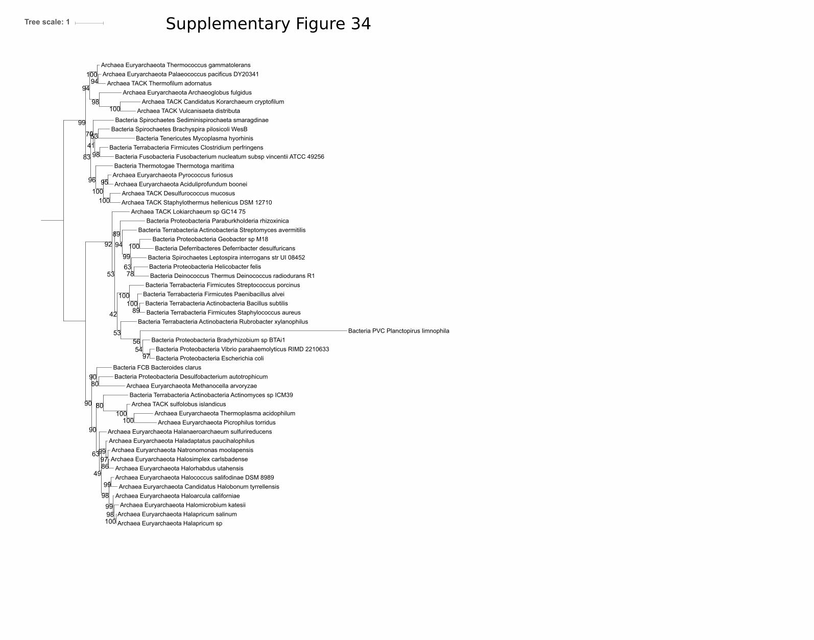
Bacteria Proteobacteria Helicobacter felis
Archaea Euryarchaeota Natronomonas moolapensis
Bacteria Deinococcus Thermus Deinococcus radiodurans R1
Archaea Euryarchaeota Haloarcula californiae
Archaea Euryarchaeota Thermoplasma acidophilum
Bacteria Fusobacteria Fusobacterium nucleatum subsp vincentii ATCC 49256
Bacteria Spirochaetes Sediminispirochaeta smaragdinae
Bacteria Tenericutes Mycoplasma hyorhinis
Archaea Euryarchaeota Halanaeroarchaeum sulfurireducens
Archaea Euryarchaeota Candidatus Halobonum tyrrellensis
Bacteria Proteobacteria Bradyrhizobium sp BTAi1
Bacteria Terrabacteria Firmicutes Staphylococcus aureus
Archaea TACK Staphylothermus hellenicus DSM 12710
Archaea Euryarchaeota Halococcus salifodinae DSM 8989
Archaea Euryarchaeota Halapricum salinum
Bacteria Terrabacteria Actinobacteria Rubrobacter xylanophilus
Archaea Euryarchaeota Methanocella arvoryzae
Archaea Euryarchaeota Halomicrobium katesii
Bacteria PVC Planctopirus limnophila
Archaea TACK Vulcanisaeta distributa
Archaea Euryarchaeota Picrophilus torridus
Bacteria Proteobacteria Geobacter sp M18
Archaea TACK Thermofilum adornatus
Archaea Euryarchaeota Thermococcus gammatolerans
Bacteria Terrabacteria Actinobacteria Actinomyces sp ICM39
Bacteria Proteobacteria Escherichia coliBacteria FCB Bacteroides clarus
Archaea TACK Lokiarchaeum sp GC14 75
Bacteria Terrabacteria Actinobacteria Streptomyces avermitilis
Bacteria Proteobacteria Desulfobacterium autotrophicum
Archaea Euryarchaeota Aciduliprofundum boonei
Archaea Euryarchaeota Haladaptatus paucihalophilus
Archaea TACK Desulfurococcus mucosus
Bacteria Deferribacteres Deferribacter desulfuricans
Bacteria Thermotogae Thermotoga maritima
Archaea Euryarchaeota Archaeoglobus fulgidus
Bacteria Terrabacteria Actinobacteria Bacillus subtilis
Bacteria Spirochaetes Brachyspira pilosicoli WesB
Bacteria Terrabacteria Firmicutes Streptococcus porcinus
Bacteria Proteobacteria Paraburkholderia rhizoxinica
Archea TACK sulfolobus islandicus
Archaea Euryarchaeota Halapricum sp
Archaea Euryarchaeota Halorhabdus utahensis
Bacteria Spirochaetes Leptospira interrogans str UI 08452
Bacteria Terrabacteria Firmicutes Paenibacillus alvei
Archaea Euryarchaeota Pyrococcus furiosus
Bacteria Proteobacteria Vibrio parahaemolyticus RIMD 2210633
Archaea TACK Candidatus Korarchaeum cryptofilum
Archaea Euryarchaeota Halosimplex carlsbadense
Archaea Euryarchaeota Palaeococcus pacificus DY20341
Bacteria Terrabacteria Firmicutes Clostridium perfringens
95
54
99
53
89
53
100
80
99
98
63
90
100
92
86
100
97
56
97
99
100
100
99
41
94
89
80
100
90
98
98
99
70
90
94
78
100
94
100
53
100
49
42
63
100
98
83
96
Tree scale: 1 Supplementary Figure 34

Bacteria Terrabacteria Firmicutes Paenibacillus curdlanolyticus
Archaea Euryarchaeota Halosimplex carlsbadense
Bacteria Proteobacteria Coxiella burnetii
Archaea TACK Sulfolobus islandicus
Bacteria Proteobacteria Desulfatibacillum aliphaticivorans
Archaea TCK Staphylothermus hellenicus
Archaea Euryarchaeota Halorhabdus utahensis
Bacteria Terrabacteria Chloroflexi Chloroflexus aggregans
Archaea Euryarchaeota Haloarcula amylolytica
Archaea Euryarchaeota Halarchaeum acidiphilum
Archaea TACK Desulfurococcus amylolyticus
Archaea Euryarchaeota Halomicrobium mukohataei
Archaea Euryarchaeota Ferroplasma sp Type II
Bacteria Elusimicrobia Elusimicrobium minutum Pei191
Archaea Euryarchaeota Acidiplasma sp MBA 1
Bacteria Terrabacteria Firmicutes Clostridium ultunense
Bacteria FCB Chlorobium limicola
Bacteria Terrabacteria Cyanobacteria Nodosilinea nodulosa
Bacteria Aquificae Aquifex aeolicus
Bacteria Acidobacteria Acidobacterium capsulatum
Archaea Euryarchaeota Halalkalicoccus jeotgali
Archaea Euryarchaeota Palaeococcus pacificus
Bacteria PVC Planctopirus limnophila
Bacteria Proteobacteria Vibrio cholerae
Bacteria Terrabacteria Firmicutes Planococcus maritimus
Bacteria PVC Chlamydia trachomatis
Archaea TACK Caldivirga maquilingensis
Bacteria Fusobacteria Fusobacterium perfoetens
Archaea TACK Candidatus Korarchaeum cryptofilum OPF8
Bacteria Terrabacteria Actinobacteria Rubrobacter xylanophilus
Archaea Euryarchaeota Thermoplasma acidophilum
Bacteria Terrabacteria Haloplasmatales Haloplasma contractile
Bacteria Terrbacteria Firmicutes Bacillus subtilis
Archaea DPANN Candidatus Woesearchaeota archaeon CG10 big fil rev 8 21 14 0 10 32 24
Archaea Euryarchaeota Aciduliprofundum boonei
Bacteria Proteobacteria Mariprofundus ferrooxydans
Bacteria Proteobacteria Sorangium cellulosum
Archaea Euryarchaeota Picrophilus torridus
Bacteria Proteobcteia Micavibrio aeruginosavorus
Bacteria Proteobacteria Erythrobacter litoralis
Bacteria Thermotogae Thermotoga maritima MSB8
Archaea Euryarchaeota Halapricum salinum
Archaea Euryarchaeota Ferroplasma acidarmanus
Bacteria Terrabacteria Actinobacteria Streptomyces sviceus ATCC 29083
Bacteria Proteobacteria Paraburkholderia rhizoxinica
Bacteria Proteobacteria Escherichia coli
Archaea Euryarchaeota Haladaptatus paucihalophilus
Archaea TACK Thermofilum pendens
Bacteria Terrabacteria Firmicutes Planifilum fulgidum
Bacteria Thermobaculum terrenum
Bacteria Protoebacteria Desulfarculus baarsii
Archaea Euryarchaeota Pyrococcus abyssi
Archaea Euryarchaeota Candidatus Halobonum tyrrellensis
Bacteia PVC Verrucomicrobium spinosum
Archaea DPANN Candidatus Woesearchaeota archaeon CG10 big fil rev 8 21 14 0 10 34 8
Bacteria Spirochaetes Leptospira interrogans
Archaea Euryarchaeota uncultured marine group II III euryarchaeote KM3 86 F07
Archaea Euryarchaeota Thermococcus eurythermalis
Archaea Euryarchaeota Natronomonas moolapensis
Bacteria Deferribacterales Deferribacter desulfuricans
Bacteria Deinococcus Thermus Deinococcus radiodurans
Archaea TACK Vulcanisaeta distributa
Bacteria Terrabacteria Actinobacteria Staphylococcus aureus
Archaea Euryarchaeota Halobacterium salinarum
Bacteria Dictyoglomi Dictyoglomus thermophilum
Bacteria FCB Gemmatimonadetes Gemmatimonas aurantiaca
Bacteria Terrabacteria Firmicutes Streptococcus pneumoniae
Bacteria Terrabcteria Actinobacteria Actinomyces slackii
Bacteria Proteobacteria Rhizobium gallicum
Bacteria Proteobacteria Nitrosomonas eutropha
Bacteria Spirochaetes Brachyspira pilosicoli WesB
Bacteria Proteobacteria Geobacter bemidjiensis
64
54
54
64
98
91
37
74
100
87
91
82
69
100
100
98
51
76
62
70
40
73
93
93
94
66
100
100
39
97
27
50
62
62
74
77
53
40
94
61
56
77
100
100
73
89
98
59
100
100
76
68
56
91
65
85
72
67
49
94
95
73
62
81
100
26
97
99
100
Tree scale: 1Supplementary Figure 35

Bacteria FCB Ignavibacteriae Ignavibacterium album
Bacteria Terrabacteria Chloroflexi Chloroflexus aggregans
Bacteria Nitrospinae Nitrospina gracilis 3 211
Bacteria Thermodesulfobacteria Thermodesulfatator atlanticus
Bacteria PVC Chlamydia trachomatis A HAR 13
Bacteria Synergistes Dethiosulfovibrio peptidovorans
Bacteria Proteobacteria Desulfarculus baarsii
Bacteria Protoebacteria Rickettsia prowazekii
Bacteria Nitrospirae Thermodesulfovibrio thiophilus
Bacteria Aquificae Aquifex aeolicus
Bacteria Terrabacteria Cyanobacteria Nodosilinea nodulosa
Bacteria Proteobacteria Desulfobacter postgatei
Bacteria Proteobacteria Desulfatibacillum aliphaticivorans
Archaea Euryarchaeota Euryarchaeota archaeon TMED255
Archaea Euryarchaeota uncultured marine group II III euryarchaeote KM3 86 F07
Bacteria Terrabacteria Firmicutes Clostridium ultunense
Bacteria Thermotogae Thermotoga maritima MSB8
Archaea DPANN archaeon GW2011 AR11
Bacteria Terrabacteria Firmicutes Streptococcus pneumoniae
Bacteria Spirochaetes Leptospira interrogans
Bacteria FCB Bacteroides fluxus
Archaea Euryarchaeota Marine Group III euryarchaeote CG Epi5
Archaea Asgardarcheota Candidatus Heimdallarchaeota archaeon LC 2
Archaea Euryarchaeota Marine Group III euryarchaeote CG Epi6
Bacteria Spirochaetes Brachyspira pilosicoli WesB
Bacteria Terrabacteria Firmicutes Planifilum fulgidum
Bacteria Proteobacteria Mariprofundus ferrooxydans
Bacteria Proteobacteria Escherichia coli
Archaea Euryarchaeota Euryarchaeota archaeon TMED164
Bacteria PVC Lentisphaerae Lentisphaera araneosa
Bacteria Terrabacteria Firmicutes Staphylococcus aureus
Bacteria Haloplasmatales Haloplasma contractile
Archaea Euryarchaeota Marine Group III euryarchaeote CG Epi1
Bacteria Proteobacteria Geobacter bemidjiensis
Bacteria Deinococcus Thermus Deinococcus radiodurans
Bacteria Terrabacteria Cyanobacteria Chroococcidiopsis thermalis
Bacteria Terrabcteria Firmicutes Bacillus subtilis
Bacteria Proteobacteria Coxiella burnetii
Bacteria Spirochaetes Spirochaeta thermophila
Bacteria Proteobacteria Micavibrio aeruginosavorus
Bacteria Proteobacteria Rhizobium gallicum
Bacteria Proteobacteria Nitrosomonas eutropha
Bacteria PVC Verrucomicrobium spinosum
Bacteria FCB Fibrobacter succinogenes
Bacteria Proteobacteria Vibrio cholerae
Bacteria Proteobacteria Helicobacter saguini
Bacteria Proteobacteria Sorangium cellulosum
Bacteria Terrabacteria Actinobacteria Streptomyces sviceus
Bacteria candidate division NC10 Candidatus Methylomirabilis oxyfera
Bacteria FCB Chitinivibrio alkaliphilus ACht1
Bacteria Chrysiogenetes Chrysiogenes arsenatis
Bacteria Tenericutes Mycoplasma hominis ATCC 23114
Bacteria Thermobaculum Thermobaculum terrenum
Bacteria FCB Caldithrix abyssi
Bacteria Deferribacteres Deferribacter desulfuricans
Bacteria Elusimicrobia Elusimicrobium minutum
Bacteria Terrabacteria Actinobacteria Actinomyces slackii
Archaea Euryarchaeota Marine Group III euryarchaeote CG Epi2
Bacteria Terrabacteria Firmicutes Planococcus maritimus
Bacteria FCB Cloacimonetes Candidatus Cloacimonas acidaminovorans
Archaea Euryarchaeota Marine Group III euryarchaeote CG Epi3
Archaea Euryarchaeota Euryarchaeota archaeon TMED85
Archaea Euryarchaeota Marine Group III euryarchaeote CG Epi4
Bacteria PVC Planctopirus limnophila
Bacteria FCB Chlorobium limicola
Archaea Asgardarchaeota Lokiarchaeum sp GC14 75
Bacteria Dictyoglomi Dictyoglomus thermophilum
Bacteria Armatimonadetes Chthonomonas calidirosea T49
Bacteria FCB Gemmatimonadetes Gemmatimonas aurantiaca
Bacteria Terrabacteria Actinobacteria Rubrobacter xylanophilus
Bacteria Proteobacteria Paraburkholderia rhizoxinica
Bacteria Acidobacteria Acidobacterium capsulatum
Bacteria Terrabacteria Firmicutes Paenibacillus curdlanolyticus
Bacteria Proteobacteria Erythrobacter litoralis
85
73
90
66
100
42
97
34
40
23
6259
50
28
83
57
78
91
77
96
40
91
67
93
46
26
82
67
31
100
54
66
39
72
64
87
64
50
99
95
41
90
84
99
75
17
67
42
48
23
65
78
63
67
44
86
67
75
100
71
58
89
72
50
91
52
35
72
23
71
38
Tree scale: 1 Supplementary Figure 36

Bacteria Spirochaetes Leptospira interrogans
Bacteria Proteobacteria Micavibrio aeruginosavorus
Bacteria Terrabcteria Firmicutes Streptococcus pneumoniae
Bacteria Thermobaculum Thermobaculum terrenum
Bacteria Proteobacteria Chlorobium limicolaBacteria FCB Caldithrix Caldithrix abyssi
Bacteria Erythrobacter litoralis
Bacteria Elusimicrobia Elusimicrobium minutum
Bacteria FCB Gemmatimonadetes Gemmatimonas aurantiaca
Bacteria PVC Planctopirus limnophila
Bacteria Terrabacteria Actinobacteria Rubrobacter xylanophilus
Bacteria Prtoeobacteria Fibrobacter
Bacteria FCB Ignavibacteriae Ignavibacterium album
Bacteria Thermodesulfobacteria Thermodesulfatator atlanticus
Bacteria PVC Verrucomicrobium spinosum
Archaea Asgardarchaeota Lokiarchaeum sp GC14 75
Bacteria Proteobacteria Coxiella burnetii
Bacteria Proteobacteria Paraburkholderia rhizoxinica
Bacteria Proteobacteria Mariprofundus ferrooxydans
Bacteria Terrabacteria Cyanobacteria Nodosilinea nodulosa
Bacteria Proteobacteria Nitrosomonas eutropha
Archaea DPANN Candidatus Woesearchaeota archaeon CG11 big fil rev 8 21 14 0 20 43 8
Bacteria Terrabacteria Firmicutes Staphylococcus aureus
Bacteria Proteobacteria Escherichia coli
Bacteria PVC Lentisphaerae Lentisphaera araneosa
Bacteria Nitrospinae Nitrospina gracilis 3 211Bacteria Nitrospirae Thermodesulfovibrio thiophilus
Archaea Asgardarchaeota Candidatus Thorarchaeota archaeon SMTZ1 83
Bacteria Proteobacteria Desulfarculus baarsii
Bacteria Proteobacteria Vibrio cholerae
Bacteria Acidobacteria Acidobacterium capsulatum
Bacteria Spirochaetes Spirochaeta thermophila
Bacteria Terrabacteria Firmicutes Paenibacillus curdlanolyticus
Bacteria Aquificae Aquifex aeolicus
Bacteria Terrabacteria Firmicutes Planifilum fulgidum
Bacteria Spirochaetes Brachyspira pilosicoli
Bacteria Proteobacteria Rhizobium gallicum
Bacteria Terrabacteria Firmicutes Planococcus maritimus
Bacteria PVC Chlamydia trachomatis A HAR 13
Bacteria Terrabacteria Firmicutes Bacillus subtilis
Bacteria Thermotogae Thermotoga maritima MSB8
Bacteria Deferribacteres Deferribacter desulfuricans
Bacteria Chrysiogenetes Chrysiogenes arsenatis
Archaea Asgardarchaeota Candidatus Heimdallarchaeota archaeon LC 2
Bacteria Tenericutes Mycoplasma hominisBacteria Terrabacteria Firmicutes Clostridium ultunense Esp
Bacteria Haloplasmatales Haloplasma contractile
Bacteria Terrabacteria Cyanobacteria Chroococcidiopsis thermalis
Bacteria Proteobacteria Desulfobacter postgatei
Bacteria Proteobacteria Geobacter
Bacteria Proteobacteria Desulfatibacillum aliphaticivorans
Bacteria FCB Candidatus Cloacimonetes Cloacimonas acidaminovorans
Bacteria Synergistetes Dethiosulfovibrio peptidovorans
Bacteria Dictyoglomi Dictyoglomus thermophilum
Bacteria Proteobacteria Sorangium cellulosum So ce56
Bacteria Armatimonadetes Chthonomonas calidirosea T49
Archaea DPANN Candidatus Pacearchaeota archaeon CG06 land 8 20 14 3 00 35 12
Bacteria Proteobacteria Helicobacter saguini
Bacteria Deinococcus Thermus Deinococcus radiodurans
Bacteria Fusobacteria Fusobacterium perfoetens
74
78
29
98
26
67
66
57
100
47
29
69
72
37
75
51
98
69
64
91
21
43
37
70
82
50
780
84
73
99
100
6
75
50
98
28
99
75
96
7
94
75
10
98
12
42
95
60
88
34
46
99
98
99
84
47
Tree scale: 1 Supplementary Figure 37

Bacteria Actinobacteria Marinactinospora thermotolerans
Bacteria Firmicutes Selenomonas ruminantium
Bacteria Actinobacteria Actinomadura oligospora
Bactria Actinobacteria Glycomyces sp NRRL B 16210
Archaea Euryarchaeota Pyrococcus furiosus
Bacteria Actinobacteria Haloechinothrix alba
Archaea TACK Thermofilum pendensArchaea Euryarchaeota Archaeoglobus fulgidus
Bacteria Actinobacteria Herbidospora mongoliensis
Bacteria Firmicutes Christensenella minuta
Bacteria Actinobacteria Streptomyces violaceorubidus
Bacteria Actinobacteria Streptosporangium amethystogenes
Archaea TACK Caldivirga maquilingensis
Archaea Euryarchaeota Thermoplasma volcanium
Bacteria Firmicutes Kroppenstedtia eburnea
Bacteria Cystobacter fuscus DSM 2262
Bacteria Firmicutes Paenibacillus ginsengihumi
Archaea Asgardarchaeota Candidatus Thorarchaeota archaeon SMTZ1 45
Archaea Asgardarchaeota Odinarchaeota archaeon LCB 4
Bacteria Actinobacteria Eubacterium cellulosolvens
Archaea TACK Cenarchaeum symbiosum
Bacteria Actinobacteria Asanoa ishikariensis
Bacteria Candidatus Kerfeldbacteria bacterium RIFOXYA2 FULL 38 24
Archaea Asgardarchaeota Candidatus Heimdallarchaeota archaeon LC 3
Bacteria Firmicutes Melghirimyces thermohalophilus
Archaea Euryarchaeota Thermoplasmatales archaeon SCGC AB 539 N05
Archaea Euryarchaeota Halococcus salifodinae
Archaea Euryarchaeota Methanosarcina mazei Tuc01
Archaea TACK Candidatus Nitrososphaera gargensis Ga9 2
Archaea TACK Acidilobus saccharovorans
Bacteria Candidatus Melainabacteria bacterium GWF2 37 15
Bacteria Firmicutes Paludifilum halophilum
Archaea TACK Ignicoccus hospitalis
Archaea TACK Nitrosopumilus maritimus SCM1Archaea TACK Marine Group I thaumarchaeote SCGC AAA799 D11
Bacteria Firmicutes Caldicellulosiruptor kronotskyensis
Archaea DPANN Candidatus Aenigmarchaeota archaeon ex4484 14
Bacteria Firmicutes Planifilum fulgidum
Bacteria FCB Gemmata sp SH PL17
Bacteria Actinobacteria Xiangella phaseoli
Bacteria Spirochaetes bacterium GWE1 32 154
Bacteria candidate division TA06 bacterium 32 111
Bacteria Proteobacteria Sorangium cellulosum
Archaea TACK uncultured marine thaumarchaeote KM3 72 H08
Bacteria Actinobacteria Kribbella catacumbae
Archaea Euryarchaeota Methanocella paludicola SANAE
Bacteria Proteobacteria Azospirillum sp CAG 260
Archaea TACK Staphylothermus marinus
Archaea TACK Candidatus Bathyarchaeota archaeon RBG 16 48 13
Bacteria Actinobactria Nocardiopsis gilva
Bacteria FCB Chitinispirillum alkaliphilum
Bacteria Actinobacteria Solirubrobacter sp URHD0082
Bacteria Actinobacteria Paenibacillus durus
Bacteria Firmicutes Carboxydothermus islandicus
Bacteria Proteobacteria Desulfovibrio sp U5L
Bacteria Actinobacteria Micromonospora narathiwatensis
Bacteria Actinobacteria Patulibacter americanus
Bacteria Firmicutes Bacillus subtilis
Bacteria Firmicutes Clostridium arbusti
Bacteria Firmicutes Thermoanaerobacter mathranii
Archaea Euryarchaeota Methanocaldococcus jannaschii
Bacteria Actinobacteria Marmoricola aequoreus
Bacteria Firmicutes Maledivibacter halophilus
Bacteria Actinobacteria Glycomyces tenuis
Archaea DPANN Candidatus Micrarchaeota archaeon CG1 02 60 51
Archaea Euryarchaeota Aciduliprofundum boonei
Archaea Euryarchaeota Thermococcus kodakarensis
Bacteria Firmicutes Streptococcus pneumoniae
Archaea TACK Sulfolobus solfataricus
Bacteria Firmicutes Numidum massiliense
Bacteria Actinobacteria Kitasatospora
Archaea Euryarchaeota Methanomassiliicoccus luminyensis
Bacteria Actinobacteria Glycomyces harbinensis
Archaea TACK Candidatus Korarchaeum cryptofilum
Archaea TACK Aeropyrum pernix K1
Bacteria Firmicutes Desmospora sp 8437
Bacteria Actinobacteria Verrucosispora sediminis
Bacteria Actinobacteria Streptomyces luteus
Bacteria Actinobacteria Allosalinactinospora lopnorensis
Bacteria Actinobacteria Actinoplanes rectilineatus
Bacteria Firmicutes Bacillus lentus
Bacteria Firmicutes Phascolarctobacterium sp CAG 266
Bacteria Actinibacteria Mycobacterium abscessus subsp abscessus
Bacteria Firmicutes Lachnospiraceae bacterium YSB2008
Archaea TACK Candidatus Caldiarchaeum subterraneum
Archaea TACK Ignisphaera aggregans DSM 17230
Bacteria PVC Pirellula sp SH Sr6A
Bacteria Actinobacteria Couchioplanes caeruleus
Bacteria Firmicutes Paenibacillus ihumii
Archaea TACK Pyrobaculum aerophilum str IM2
Archaea Euryarchaeota Haloarcula MULTISPECIES
Archaea Euryarchaeota Methanospirillum hungatei
Bacteria Actinobacteria Streptomyces sp WAC00263
Bacteria Candidatus Magnetoovum chiemensis
Archaea Euryarchaeota Methanothermobacter thermautotrophicus
Bacteria Actinobacteria Thermomonospora chromogena
Bacteria PVC Planctomycetes bacterium
Bacteria Firmicutes Rubrobacter radiotolerans
Bacteria Firmicutes Caldalkalibacillus thermarum
Bacteria Firmicutes Anaerobacillus arseniciselenatis
Bacteria Actinobacteria Thermobifida halotolerans
Archaea Euryarchaeota Halofax MULTISPECIES
Bacteria Firmicutes Coprothermobacter platensis
Archaea Euryarchaeota Methanopyrus kandleri AV19
Bacteria Firmicutes Succiniclasticum ruminis
Bacteria Thermotogae Mesotoga infera
Bacteria Actinobacteria Verrucosispora maris AB 18 032Bacteria Actinobacteria Salinispora pacifica
Bacteria Firmicutes Chitinivibrio alkaliphilus
Bacteria Firmicutes Novibacillus thermophilus
Archaea TACK Fervidicoccus fontis Kam940
Archaea TACK Hyperthermus butylicus
51
64
34
45
98
56
100
57
28
100
44
69
95
93
100
40
63
76
93
100
71
100
99
100
83
96
89
99
100
92
61
39
96
100
63
99
100
60
100
87
100
23
56
80
99
91
100
71
58
100
70
82
48
100
64
100
100
100
38
77
78
79
5330
53
93
97
100
100
65
61
100
100
100
100
98
97
99
64
99
100
100
79
88
99
100
78
92
78
99
56
100
99
80
99
99
56
100
99
97
72
47
100
96
100
55
51
79
100
Tree scale: 1Supplementary Figure 38

Bacteria Candidatus Handelsmanbacteria bacterium RIFCSPLOWO2 12 FULL 64 10
Bacteria candidate division KSB1 bacterium RBG 16 48 16
Bacteria FCB Spirosoma sp 209
Bacteria FCB Flavisolibacter ginsengisoli
Bacteria Firmicutes Geobacillus galactosidasius
Archaea DPANN DUSEL3
Archaea TACK Ignicoccus hospitalis
Bacteria Firmicutes Bacillus lentus
Archaea Euryarchaeota Methanocaldococcus jannaschii
Bactera candidate division TA06 bacterium 32 111
Bacteia FCB Psychroserpens burtonensis
Bacteria Firmicutes Planifilum fulgidum
Archaea DPAAN Macid
Bacteria Firmicutes Tepidanaerobacter acetatoxydans
Bacteria Firmicutes Marininema mesophilum
Archaea TACK Thermofilum sp ex4484 79
Bacteria FCB Bacteroidetes bacterium OLB12
Archaea Euryarchaeota Aciduliprofundum boonei
Bacteria FCB Jejuia pallidilutea
Bacteria Candidatus Raymondbacteria bacterium RifOxyA12 full 50 37
Archaea TACK Thermofilum pendens
Bacteria Firmicutes Paenibacillus ihumii
Archaea Euryarchaeota Methanocella paludicola SANAE
Bacteria Firmicutes Paludifilum halophilum
Bacteria FCB Lacinutrix jangbogonensis
Archaea Asgardarchaeota Candidatus Thorarchaeota archaeon SMTZ1 45
Archaea TACK Acidilobus sp 7AArchaea Euryarchaeota Methanosarcina mazei Tuc01
Archaea TACK Ignisphaera aggregans DSM 17230
Archaea TACK Thaumarchaeota archaeon ex4484 121
Archaea Euryarchaeota Haloarcula marismortui
Archaea TACK Candidatus Bathyarchaeota archaeon RBG 16 48 13
Bacteria FCB Bernardetia litoralis
Archaea Euryarchaeota Thermoplasma volcanium
Archaea TACK Candidatus Nitrosotenuis cloacae
Bacteria Actinobacteria Mycobacterium abscessus subsp abscessus
Archaea TACK Nitrosopumilus maritimus SCM1
Bacteria FCB Chlorobium sp GBChlB
Bacteria Firmicutes Bacillus subtilis
Bacteria FCB FibrobacterBacteria FCB Fibrobacter
Archaea TACK Acidilobus saccharovorans
Archaea TACK Sulfolobus sp A20
Archaea TACK Caldivirga maquilingensis
Archaea Euryarchaeota Archaeoglobus fulgidus
Bacteria Firmicutes Paenibacillus durus
Bacteria Firmicutes Paenibacillus ginsengihumi
Bacteria Firmicutes Anaerobacillus arseniciselenatis
Bacteria Firmicutes Numidum massiliense
Bacteria Firmicutes Calderihabitans maritimus
Bacteria FCB Flectobacillus sp BAB 3569
Bacteria Firmicutes Streptococcus pneumoniae
Bacteria FCB Sporocytophaga myxococcoides
Bacteria Firmicutes Planococcus maritimus
Bacteria FBC Ignavibacteria bacterium CG1 02 37 35
Bacteria Candidatus Chrysopegis kryptomonas
Archaea TACK Candidatus Caldiarchaeum subterraneum
Archaea TACK Desulfurococcus amylolyticus
Bacteria FCB Schleiferia thermophila
Bacteria FCB Nafulsella turpanensis
Bacteria Firmicutes Melghirimyces thermohalophilus
Archaea Asgardarchaeota Lokiarchaeum sp GC14 75
Bacteria Desulfuromonadales bacterium C00003094
Bacteria bacterium TMED264
Bacteria FCB candidate division Zixibacteria bacterium 4484 95
Bacteria candidate division WOR 3 bacterium SM23 60
Archaea Euryarchaeota Halococcus salifodinae
Archaea TACK Staphylothermus marinus
Archaea Asgardarchaeota Odinarchaeota archaeon LCB 4
Archaea TACK Caldisphaera lagunensis
Bacteria FCB Niabella drilacis
Bacteria Firmicutes Fictibacillus sp GDSW R2A3
Bacteria FCB Caldithrix abyssi
Bacteria FCB Gemmatimonadetes bacterium 13 1 40CM 4 69 8
Bacteria FCB Cyclobacterium lianum
Bacteria Elusimicrobia bacterium RIFOXYB2 FULL 49 7
Bacteria FCB Chitinivibrio alkaliphilus
Archaea Euryarchaeota Haloferax volcanii
Bacteria bacterium TMED217
Bacteria Firmicutes Tuberibacillus calidus
Archaea DPANN Candidatus Micrarchaeota archaeon CG1 02 49 24
Bacteria FCB Runella limosa
Archaea Asgardarchaeota Candidatus Heimdallarchaeota archaeon LC 3
Archaea Euryarchaeota Thermoplasmatales archaeon SCGC AB 539 N05
Archaea TACK Sulfolobus solfataricus
Bacteria FCB Candidatus Kryptobacter tengchongensis
Bacteria Firmicutes Peptococcaceae acterium BRH c8a
Bacteria Firmicutes Desmospora sp 8437
Bacteria candidate division TA06 bacterium DG 26Archaea Euryarchaeota Pyrococcus furiosus
Archaea TACK Cenarchaeum symbiosum A
Archaea TACK Thermosphaera aggregans
Bacteria Firmicutes Dethiobacter alkaliphilus
Bacteria FCB Formosa sp Hel1 31 208
Archaea TACK Fervidicoccus fontis Kam940
Archaea Euryarchaeota Thermococcus kodakarensis
Bacteria Parcubacteria bacterium DG 74 3
Archaea TACK Aeropyrum pernix K1
Archaea TACK Marine Group I thaumarchaeote SCGC AAA799 D11
Bacteria Candidatus Edwardsbacteria bacterium RifOxyC12 full 54 24
Archaea Euryarchaeota Methanomassiliicoccus luminyensis
Archaea Euryarchaeota Methanothermobacter thermautotrophicus
Bacteria Firmicutes bacterium ML8 F2
Bacteria Actinobacteria bacterium RBG 13 63 9
Bacteria FCB Chitinispirillum alkaliphilum
Archaea DPANN AR10
Bacteria Firmicutes Kroppenstedtia eburnea
Bacteria FCB Kordia jejudonensis
Bacteria Actinobacteria Streptomyces sp WAC00263
Bacteria FCB Candidatus Marinimicrobia bacterium CG1 02 48 14
Archaea TACK Hyperthermus butylicus
Bacteria FCB Candidatus Cloacimonas sp 4484 275
Bacteria Firmicutes Bhargavaea cecembensis
Bacteria FCB Flavihumibacter petaseus
Bacteria FBC Ignavibacterium album
Bacteria Firmicutes Caldalkalibacillus thermarum
Archaea DPANN Candidatus Aenigmarchaeota archaeon ex4484 14
Archaea Euryarchaeota Methanospirillum hungatei
Bacteria Firmicutes Anaerobacillus alkalidiazotrophicus
Bacteria FCB Ohtaekwangia koreensis
Archaea Euryarchaeota Methanopyrus kandleri AV19
Bacteria FCB Latescibacteria bacterium DG 63
Bacteria Firmicutes Bacillus aquimaris
Bacteria Parcubacteria bacterium DG 74 2
Bacteria FCB Candidatus Kryptonium thompsoni
Bacteria candidate division WOR 3 bacterium JGI Cruoil 03 44 89
Archaea TACK Candidatus Nitrososphaera gargensis Ga9 2
Bacteria FCB Sediminibacter sp Hel I 10
Archaea TACK Candidatus Korarchaeum cryptofilum
Archaea TACK Pyrobaculum aerophilum str IM2
Bacteria FCB Candidatus Cloacimonetes bacterium 4572 55
92
35
100
95
90
98
94
85
29
78
84
20
92
93
87
98
95
67
44
39
61
68
100
65
67
37
82
100
58
100
73
91
93
51
55
65
100
78
9493
64
99
66
97
98
92
57
94
90
77
76
97
21
85
97
91
76
100
55
55
74
99
36
100
64
100
86
100
81
64
76
71
97
95
98
56
37
100
85
93
99
100
87
48
99
67
92
60
59
65
50
75
100
99
86
84
98
98
100
100
45
77
100
100
99
100
92
62
98
81
73
81
100
49
64
100100
78
100
96
72
44100
49
76
100
9694
Tree scale: 0.1 Supplementary Figure 39

Archaea Asgardarchaeota Candidatus Thorarchaeota archaeon SMTZ1 45
Archaea Euryarchaeota Thermoplasmatales archaeon SCGC AB 539 N05
Bacteria candidate division TA06 bacterium 32 111Bacteria FCB Cyclobacterium lianum
Bacteria FCB candidate division Zixibacteria bacterium 4484 95 fa
Bacteria FCB Candidatus Marinimicrobia bacterium CG1 02 48 14
Bacteria FCB Bacteroidetes Flammeovirgaceae bacterium TMED290
Bacteria FCB Chlorobi Chlorobium sp GBChlB
Archaea Euryarchaeota HaloarculaBacteria Actinobacteria Curtobacterium sp UCD KPL2560
Bacteria FCB Candidatus Cloacimonetes bacterium 4572 55
Bacteria candidate division WOR 3 bacterium SM23 60
Bacteria FCB Fibrobacteres Fibrobacter sp UWH5
Bacteria Actinobacteria Kocuria sp SM24M 10
Archaea Euryarchaeota Archaeoglobus fulgidus
Archaea Euryarchaeota Methanospirillum hungatei
Bacteria FCB Bacteroidetes Gelidibacter algens
Bacteria Actinobacteria Auraticoccus monumenti
Bacteria CPR Parcubacteria bacterium DG 74 2
Archaea TACK Staphylothermus marinus
Bacteria FCB candidate division Zixibacteria bacterium RBG 1
Bacteria FCB Bacteroidetes Kordia jejudonensis
Bacteria Actinobacteria Curtobacterium sp ER1 6
Bacteria FCB Latescibacteria bacterium DG 63
Archaea Asgardarchaeota Candidatus Heimdallarchaeota archaeon LC 3
Bacteria FCB Ignavibacterium album
Archaea Euryarchaeota Haloferax
Archaea DPANN AR10
Archaea Euryarchaeota Pyrococcus furiosus
Bacteria Candidatus Edwardsbacteria bacterium RifOxyC12 full 54 24
Archaea TACK Marine Group I thaumarchaeote SCGC AAA799 D11
Bacteria Proteobacteria Desulfobacterales bacterium SG8 35 2 fa
Bacteria FCB Ohtaekwangia koreensis
Bacteria Proteobacteria Devosia riboflavina
Archaea TACK Fervidicoccus fontis Kam940
Bacteria FCB Ignavibacteria bacterium CG1 02 37 35
Bacteria FCB Chitinivibrio alkaliphilus
Bacteria Chloroflexi bacterium RBG 16 72 14 fa
Archaea Euryarchaeota Methanothermobacter thermautotrophicus
Bacteria FCB Runella limosa
Bacteria Actinobacteria Friedmanniella sagamiharensis
Archaea TACK Candidatus Korarchaeum cryptofilum
Bacteria Chloroflexi Anaerolineae bacterium SG8 19
Archaea TACK Thermofilum pendens
Bacteria FCB Bacteroidetes Sediminibacter sp Hel I 10
Bacteria FCB Candidatus Kryptonium thompsoni
Archaea TACK Ignisphaera aggregans DSM 17230
Bacteria Firmicutes Halanaerobium saccharolyticum
Bacteria Candidatus Cloacimonas sp 4484 275
Bacteria FCB Fibrobacter
Bacteria Chloroflexi Longilinea arvoryzae
Archaea DPANN Candidatus Aenigmarchaeota archaeon ex4484 14
Archaea DPANN Macid
Bacteria FCB Flavisolibacter ginsengisoli DSM 18119
Bacteria FCB Sporocytophaga myxococcoides
Bacteria bacterium TMED264
Archaea TACK Caldivirga maquilingensis
Bacteria Actinobacteria Streptomyces luteus
Bacteria FCB Chlorobi Chlorobium limicola
Bacteria FCB Bacteroidetes Formosa sp Hel1 31 208
Archaea Asgardarchaeota Candidatus Odinarchaeota archaeon LCB 4
Bacteria FCB Chlorobi Chlorobium ferrooxidans
Archaea Euryarchaeota Methanomassiliicoccus luminyensis
Bacteria FCB Bacteroidetes Jejuia pallidilutea
Bacteria FCB Candidatus Marinimicrobia bacterium
Archaea TACK Hyperthermus butylicus
Bacteria Rhodothermaeota Rubricoccus marinus
Bacteria Firmicutes Desulfotomaculum gibsoniae
Archaea DPANN Candidatus Micrarchaeota archaeon CG1 02 51 15
Archaea TACK Cenarchaeum symbiosum A
Bacteria Cyanobacteria Chlorogloeopsis fritschii
Bacteria FCB Candidatus Latescibacteria bacterium DG 63
Archaea Euryarchaeota Halococcus salifodinae
Archaea TACK Candidatus Nitrosoarchaeum koreensis
Archaea TACK Candidatus Caldiarchaeum subterraneum
Archaea Euryarchaeota Methanocaldococcus jannaschii
Bacteria Actinobacteria Rubrobacter radiotolerans
Bacteria FCB Gemmatimonadates Gemmatirosa kalamazoonesis
Archaea Euryarchaeota Thermoplasma volcanium
Bacteria Candidatus Handelsmanbacteria bacterium RIFCSPLOWO2 12 FULL 64 10
Bacteria FCB Chitinispirillum alkaliphilum
Archaea Euryarchaeota Aciduliprofundum boonei
Bacteria Firmicutes Halanaerobium
Bacteria FCB Spirosoma sp 209
Archaea TACK Ignicoccus hospitalis
Bacteria Actinobacteria Arthrobacter sp RIT PI e
Archaea TACK Aeropyrum pernix K1
Bacteria candidate division KSB1 bacterium 4484 87
Archaea TACK Sulfolobus solfataricus
Archaea Euryarchaeota Thermococcus kodakarensis
Archaea DPANN DUSEL3
Archaea Euryarchaeota Methanocella paludicola SANAE
Archaea TACK Nitrosopumilus maritimus SCM1
Bacteria FCB Gemmatimonadetes bacterium 13 1 40CM 4 69 8
Bacteria FCB Flavihumibacter petaseus
Bacteria FBC Psychroserpens burtonensis
Bacteria FCB Caldithrix abyssi DSM 13497
Bacteria Choloroflexi Leptolinea tardivitalis
Archaea Euryarchaeota Methanopyrus kandleri
Archaea TACK Acidilobus saccharovorans
Bacteria Proteobacteria Rhodobacteraceae bacterium TMED111
Bacteria Parcubacteria bacterium DG 74 3
Bacteria Candidatus Raymondbacteria bacterium RifOxyA12 full 50 37
Bacteria FCB Schleiferia thermophila
Archaea Euryarchaeota Methanosarcina mazei Tuc01
Bacteria Firmicutes Sulfobacillus thermosulfidooxidans
Bacteria Firmicutes Peptococcaceae bacterium BRH c8a
Archaea TACK Candidatus Nitrososphaera gargensis Ga9 2
Archaea TACK Pyrobaculum aerophilum st IM2
Bacteria FCB Candidatus Krytobacter tengchongensis
Bacteria Candidatus Chrysopegis kryptomonas
Archaea Asgardarchaeota Lokiarchaeum sp GC14 75
Bacteria candidate division WOR 3 bacterium JGI Cruoil 03 44 89
Bacteria Actinobacteria Tessaracoccus bendigoensis
35
42
75
27
100
91
99
92
99
39
80
40
100
100
30
84
100
64
77
83
30
100
61
84
85
66
100
100
91
48
32
34
49
100
99
85
16
87
61
100
97
49
36
76
67
63
76
71
67
100
78
33
61
75
75
87
98
100
29
81
9394
70
35
90
100
91
99
98
28
100
98
73
100
98
24
100
84
26
65
48
71
98
96
100
100
29
100
100
68
99
72
90
65
75
86
83
92
40
33
100
87
99
100
97
85
37
40
99
73
100
Tree scale: 1 Supplementary Figure 40

Bacteria Deferribacterales Deferribacter desulfuricans
Bacteria Acetothermia Candidatus Acetothermus autotrophicum
Bacteria Proteobacteria Escherichia coli str K 12 substr MG1655
Bacteria Thermobaculum Thermobaculum terrenum
Archaea Euryarchaeota Marine Group II euryarchaeote MED G34
Bacteria PVC Verrucomicrobium spinosum
Bacteria Thermotogae Thermotoga maritima MSB8
Bacteria Aminicenantes Candidatus Aminicenans sakinawicola
Bacteria Proteobacteria Erythrobacter litoralis
Bacteria Terrabacteria Firmicutes Clostridium ultunense Esp
Bacteria Proteobacteria Vibrio cholerae HE48
Bacteria Dictyoglomi Dictyoglomus thermophilum
Archaea Euryarchaeota marine group II III euryarchaeote KM3 85 A08
Bacteria PVC Chlamydia trachomatis A HAR 13
Archaea Euryarchaeota Methanobacterium sp MB1
Bacteria Terrabacteria Firmicutes Staphylococcus aureus A9635
Bacteria Chrysiogenetes Chrysiogenes arsenatis
Bacteria Proteobacteria Sorangium cellulosum
Bacteria Terrabacteria Cyanobacteria Gloeobacter kilaueensis JS1
Archaea DPANN Candidatus Woesearchaeota archaeon CG10 big fil rev 8 21 14 0 10 36 11
Bacteria Terrabacteria Firmicutes Planococcus maritimus
Bacteria Proteobacteria Mariprofundus ferrooxydans PV 1
Archaea archaeon GW2011 AR11
Bacteria FCB Bacteroides fluxus YIT 12057
Bacteria Terrabacteria Chloroflexi Caldilinea aerophila
Bacteria Haloplasmatales Haloplasma contractile
Bacteria Terrabacteria Firmicutes Paenibacillus curdlanolyticus YK9
Bacteria Thermodesulfobacteria Thermodesulfatator atlanticus
Bacteria Nitrospinae Nitrospina gracilis 3 211
Bacteria Proteobacteria Geobacter bemidjiensis
Bacteria Terrabacteria Cyanobacteria Scytonema hofmanni
Bacteria Spirochaetes Leptospira interrogans
Archaea Euryarchaeota Geoglobus ahangari
Bacteria Terrabacteria Actinobacteria Actinomyces slackii
Bacteria Proteobacteria Rickettsia prowazekii str Rp22
Bacteria Synergistes Dethiosulfovibrio peptidovorans DSM 11002
Bacteria Proteobacteria Desulfarculus baarsii
Bacteria FCB Ignavibacteriae Ignavibacterium album
Bacteria Proteobacteria Helicobacter saguini
Bacteria FCB Latescibacteria Candidatus Latescibacter anaerobius
Archaea Euryarchaeota Methanothermobacter thermautotrophicus
Bacteria Spirochaetes Spirochaeta thermophila
Bacteria Deinococcus Thermus Deinococcus radiodurans
Archaea Candidatus Woesearchaeota archaeon CG 4 10 14 0 2 um filter 33 13
Bacteria PVC Planctopirus limnophila
Archaea Euryarchaeota archaeon TMED85
Archaea TACK Thermofilum sp ex4484 15
Bacteria FCB Caldithrix abyssi DSM 13497
Bacteria FCB Gemmatimonadetes Gemmatimonas aurantiaca T 27
Bacteria Terrabacteria Firmicutes Bacillus subtilis
Bacteria Proteobacteria Coxiella burnetii RSA 331
Bacteria Terrabacteria Actinobacteria Rubrobacter radiotolerans
Bacteria Proteobacteria Desulfobacter postgatei
Bacteria Terrabacteria Firmicutes Streptococcus pneumoniae
Bacteria Proteobacteria Nitrosomonas eutropha
Bacteria Terrabacteria Actinobacteria Streptomyces sviceus ATCC 29083
Archaea Euryarchaeota archaeon TMED97
Bacteria Spirochaetes Brachyspira pilosicoli WesB
Bacteria Proteobacteria Micavibrio aeruginosavorus
Archaea Euryarchaeota Methanobrevibacter filiformis
Bacteria Nitropirae Thermodesulfovibrio thiophilus
Bacteria PVC Lentispaera Lentisphaera araneosa HTCC2155
Bacteria Fusobacteria Fusobacterium perfoetens
Bacteria Candidatus Methylomirabilis oxyfera
Archaea archaeon GW2011 AR9
Bacteria FCB Chlorobium limicola
Bacteria Proteobacteria Paraburkholderia rhizoxinica HKI 454
Bacteria Proteobacteria Desulfatibacillum aliphaticivorans
Archaea Euryarchaeota Candidatus Thalassoarchaea euryarchaeote
Bacteria Proteobacteria Rhizobium gallicum
Bacteria Terrabacteria Firmicutes Planifilum fulgidum
Archaea Euryarchaeota Ferroglobus placidus
Bacteria Aquificae Aquifex aeolicus
Bacteria FCB Chitinivibrio alkaliphilus ACht1
Bacteria Tenericutes Mycoplasma hominis ATCC 23114
Bacteria FCB Cloacimonetes Candidatus Cloacimonas acidaminovorans str Evry
Archaea Euryarchaeota Methanoculleus thermophilus
Archaea Euryarchaeota Archaeoglobus fulgidus
Bacteria Elusimicrobia Elusimicrobium minutum
Bacteria Terrabacteria Cyanobacteria Chroococcidiopsis thermalis
Bacteria Armatimonadetes Chthonomonas calidirosea T49
Archaea Euryarchaeota Candidatus Thalassoarchaea euryarchaeote
Bacteria FCB Fibrobacter succinogenes
Archaea marine group II III euryarchaeote AD1000 25 A05
55
70
85
43
93
71
55
96
100
100
43
47
98
97
83
99
14
100
73
100
100
100
7682
32
48
69
48
73
56
89
92
100
63
100
99
33
100
73
100
56
35
82
24
46
35
62
97
100
34
98
68
99
67
27
8759
98
27
86
99
27
100
45
100
34
21
54
100
35
88
22
95
100
82
86
53
38
100
96
45
Tree scale: 1 Supplementary Figure 41

Archaea Euryarchaeota Candidatus Halobonum tyrrellensis
Archaea Euryarchaeota Archaeoglobus fulgidus
Archaea Euryarchaeota Pyrococcus furiosus
Bacteria Proteobacteria Paraburkholderia rhizoxinica
Bacteria Spirochaetes Brachyspira pilosicoli WesB
Bacteria Spirochaetes Leptospira interrogans str UI 08452
Archaea Euryarchaeota Haladaptatus paucihalophilus
Bacteria Terrabacteria Actinobacteria Streptomyces avermitilis
Archaea Euryarchaeota Halanaeroarchaeum sulfurireducens
Archea TACK sulfolobus islandicus
Bacteria Proteobacteria Helicobacter felis
Bacteria Terrabacteria Firmicutes Paenibacillus alvei
Bacteria Fusobacteria Fusobacterium nucleatum subsp vincentii ATCC 49256Bacteria Thermotogae Thermotoga maritima
Archaea TACK Desulfurococcus mucosus
Bacteria Terrabacteria Actinobacteria Actinomyces sp ICM39
Archaea Euryarchaeota Methanocella arvoryzae
Archaea Euryarchaeota Thermococcus gammatolerans
Bacteria PVC Planctopirus limnophila
Archaea Euryarchaeota Palaeococcus pacificus DY20341
Archaea Euryarchaeota Halorhabdus utahensisArchaea Euryarchaeota Halosimplex carlsbadense
Bacteria FCB Bacteroides clarus
Bacteria Terrabacteria Firmicutes Staphylococcus aureus
Archaea Euryarchaeota Thermoplasma acidophilum
Archaea TACK Candidatus Korarchaeum cryptofilum
Bacteria Terrabacteria Firmicutes Streptococcus porcinus
Archaea Euryarchaeota Halapricum sp
Archaea TACK Vulcanisaeta distributa
Bacteria Terrabacteria Actinobacteria Bacillus subtilis
Archaea Euryarchaeota Halapricum salinum
Bacteria Deferribacteres Deferribacter desulfuricans
Bacteria Proteobacteria Escherichia coliBacteria Proteobacteria Vibrio parahaemolyticus RIMD 2210633
Archaea Euryarchaeota Aciduliprofundum boonei
Archaea Euryarchaeota Picrophilus torridus
Bacteria Proteobacteria Desulfobacterium autotrophicum
Archaea Euryarchaeota Halomicrobium katesii
Bacteria Terrabacteria Firmicutes Clostridium perfringens
Archaea Euryarchaeota Halococcus salifodinae DSM 8989
Archaea TACK Staphylothermus hellenicus DSM 12710
Bacteria Proteobacteria Bradyrhizobium sp BTAi1
Bacteria Tenericutes Mycoplasma hyorhinis
Archaea Euryarchaeota Haloarcula californiae
Archaea Euryarchaeota Natronomonas moolapensis
Archaea TACK Lokiarchaeum sp GC14 75
Archaea TACK Thermofilum adornatus
Bacteria Terrabacteria Actinobacteria Rubrobacter xylanophilus
Bacteria Deinococcus Thermus Deinococcus radiodurans R1
Bacteria Spirochaetes Sediminispirochaeta smaragdinae
Bacteria Proteobacteria Geobacter sp M18
100
93
98
94
100
96
100
73
99
87
98
96
99
89
89
96
78
96
100
10099
55
87
99
90
100
32
75
45
100
60
58
100
80
57
9283
99
53
100
98
97
100
98
98
100
94
32
Tree scale: 1 Supplementary Figure 42

Bacteria Proteobacteria Vibrio cholerae
Archaea Euryarchaeota Halapricum salinum
Bacteria Thermobaculum terrenum
Bacteria Elusimicrobia Elusimicrobium minutum Pei191
Archaea TACK Thermofilum pendens
Bacteria Terrabcteria Actinobacteria Actinomyces slackii
Archaea Euryarchaeota Thermoplasma acidophilum
Bacteria FCB Gemmatimonadetes Gemmatimonas aurantiaca
Bacteria Proteobacteria Nitrosomonas eutropha
Archaea Euryarchaeota Halarchaeum acidiphilum
Bacteria Terrabacteria Chloroflexi Chloroflexus aggregans
Bacteria Protoebacteria Desulfarculus baarsii
Bacteria Terrabacteria Firmicutes Planifilum fulgidum
Bacteria Proteobacteria Sorangium cellulosum
Bacteria Proteobacteria Coxiella burnetii
Bacteria Acidobacteria Acidobacterium capsulatum
Bacteria Terrbacteria Firmicutes Bacillus subtilis
Bacteria Terrabacteria Firmicutes Streptococcus pneumoniae
Archaea Euryarchaeota Ferroplasma sp Type II
Archaea Euryarchaeota Halomicrobium mukohataei
Bacteria Terrabacteria Firmicutes Planococcus maritimus
Bacteria FCB Chlorobium limicola
Archaea TACK Sulfolobus islandicus
Archaea Euryarchaeota Halorhabdus utahensis
Bacteria Aquificae Aquifex aeolicus
Bacteria Proteobacteria Mariprofundus ferrooxydans
Bacteria Deinococcus Thermus Deinococcus radiodurans
Bacteria Deferribacterales Deferribacter desulfuricans
Archaea Euryarchaeota Halalkalicoccus jeotgali
Bacteria Terrabacteria Firmicutes Clostridium ultunense
Bacteria Haloplasmatales Haloplasma contractile
Archaea Euryarchaeota Acidiplasma sp MBA 1
Archaea Euryarchaeota Ferroplasma acidarmanus
Bacteria Proteobacteria Erythrobacter litoralis
Bacteria Spirochaetes Leptospira interrogans
Bacteria Dictyoglomi Dictyoglomus thermophilum
Archaea Euryarchaeota Thermococcus eurythermalis
Bacteria PVC Chlamydia trachomatis
Bacteria Spirochaetes Brachyspira pilosicoli WesB
Bacteria Terrabacteria Actinobacteria Staphylococcus aureus
Archaea TACK Vulcanisaeta distributa
Archaea Euryarchaeota uncultured marine group II III euryarchaeote KM3 86 F07
Bacteria Terrabacteria Actinobacteria Rubrobacter xylanophilus
Bacteria Terrabacteria Firmicutes Paenibacillus curdlanolyticus
Bacteria PVC Planctopirus limnophila
Archaea Euryarchaeota Halosimplex carlsbadense
Archaea Euryarchaeota Halobacterium salinarum
Archaea Euryarchaeota Palaeococcus pacificus
Bacteria Proteobacteria Paraburkholderia rhizoxinica
Bacteria Proteobacteria Geobacter bemidjiensis
Bacteria Proteobacteria Rhizobium gallicum
Archaea Euryarchaeota Pyrococcus abyssi
Archaea CPR Candidatus Woesearchaeota archaeon CG10 big fil rev 8 21 14 0 10 32 24
Archaea Euryarchaeota Natronomonas moolapensis
Archaea CPR Candidatus Woesearchaeota archaeon CG10 big fil rev 8 21 14 0 10 34 8
Bacteria Thermotogae Thermotoga maritima MSB8
Archaea Euryarchaeota Aciduliprofundum boonei
Archaea Euryarchaeota Candidatus Halobonum tyrrellensis
Bacteria Proteobacteria Escherichia coli
Archaea Euryarchaeota Haladaptatus paucihalophilus
Archaea TACK Candidatus Korarchaeum cryptofilum OPF8
Archaea Euryarchaeota Haloarcula amylolytica
Bacteria Proteobcteia Micavibrio aeruginosavorus
Bacteria Terrabacteria Actinobacteria Streptomyces sviceus ATCC 29083
Archaea Euryarchaeota Picrophilus torridus
Bacteria Proteobacteria Desulfatibacillum aliphaticivorans
Bacteria Fusobacteria Fusobacterium perfoetens
Archaea TACK Desulfurococcus amylolyticus
Bacteia PVC Verrucomicrobium spinosum
Bacteria Terrabacteria Cyanobacteria Nodosilinea nodulosa
Archaea TACK Staphylothermus hellenicus
Archaea TACK Caldivirga maquilingensis
25
56
100
81
60
60
86
63
66
98
96
99
30
49
100
98
85
43
58
58
67
99
70
99
100
100
100
84
98
96
61
99
89
60
99
95
100
72
51
62
84
58
99
55
77
53
46
94
85
69
98
96
59
98
66
59
100
10074
60
68
100
24
87
60
69
100
75
99
Tree scale: 1 Supplementary Figure 43
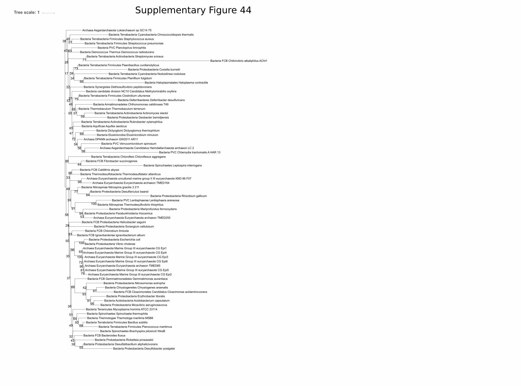
Bacteria FCB Fibrobacter succinogenes
Bacteria Terrabcteria Firmicutes Bacillus subtilis
Bacteria Terrabacteria Cyanobacteria Nodosilinea nodulosa
Bacteria FCB Cloacimonetes Candidatus Cloacimonas acidaminovorans
Archaea Euryarchaeota Marine Group III euryarchaeote CG Epi5
Bacteria Haloplasmatales Haloplasma contractile
Archaea Euryarchaeota uncultured marine group II III euryarchaeote KM3 86 F07
Bacteria FCB Chitinivibrio alkaliphilus ACht1
Archaea Asgardarchaeota Lokiarchaeum sp GC14 75
Bacteria Nitrospirae Thermodesulfovibrio thiophilus
Bacteria Protoebacteria Rickettsia prowazekii
Bacteria Synergistes Dethiosulfovibrio peptidovorans
Bacteria Nitrospinae Nitrospina gracilis 3 211
Bacteria Proteobacteria Mariprofundus ferrooxydans
Bacteria Proteobacteria Desulfatibacillum aliphaticivorans
Bacteria Proteobacteria Sorangium cellulosum
Bacteria Proteobacteria Micavibrio aeruginosavorus
Bacteria Proteobacteria Paraburkholderia rhizoxinica
Bacteria FCB Bacteroides fluxus
Bacteria PVC Planctopirus limnophila
Bacteria Thermodesulfobacteria Thermodesulfatator atlanticus
Bacteria Terrabacteria Chloroflexi Chloroflexus aggregans
Bacteria Proteobacteria Nitrosomonas eutropha
Bacteria Proteobacteria Erythrobacter litoralis
Bacteria PVC Verrucomicrobium spinosum
Bacteria Terrabacteria Firmicutes Streptococcus pneumoniae
Bacteria Aquificae Aquifex aeolicus
Bacteria Terrabacteria Firmicutes Clostridium ultunense
Bacteria FCB Chlorobium limicola
Bacteria Proteobacteria Rhizobium gallicum
Bacteria Terrabacteria Actinobacteria Streptomyces sviceus
Bacteria FCB Gemmatimonadetes Gemmatimonas aurantiaca
Bacteria Deferribacteres Deferribacter desulfuricans
Archaea Euryarchaeota Euryarchaeota archaeon TMED164
Archaea Euryarchaeota Euryarchaeota archaeon TMED255
Bacteria Acidobacteria Acidobacterium capsulatum
Bacteria Proteobacteria Coxiella burnetii
Bacteria Spirochaetes Brachyspira pilosicoli WesB
Bacteria Spirochaetes Spirochaeta thermophila
Bacteria candidate division NC10 Candidatus Methylomirabilis oxyfera
Bacteria Terrabacteria Actinobacteria Rubrobacter xylanophilus
Bacteria Spirochaetes Leptospira interrogans
Bacteria Terrabacteria Firmicutes Planifilum fulgidum
Bacteria PVC Lentisphaerae Lentisphaera araneosa
Bacteria PVC Chlamydia trachomatis A HAR 13
Bacteria FCB Proteobacteria Helicobacter saguini
Bacteria Chrysiogenetes Chrysiogenes arsenatis
Bacteria Thermobaculum Thermobaculum terrenum
Bacteria Proteobacteria Desulfobacter postgatei
Bacteria Armatimonadetes Chthonomonas calidirosea T49
Bacteria Proteobacteria Escherichia coli
Bacteria Terrabacteria Firmicutes Planococcus maritimus
Bacteria Terrabacteria Cyanobacteria Chroococcidiopsis thermalis
Bacteria Terrabacteria Actinobacteria Actinomyces slackii
Archaea Euryarchaeota Marine Group III euryarchaeote CG Epi3
Bacteria Terrabacteria Firmicutes Staphylococcus aureus
Bacteria Proteobacteria Geobacter bemidjiensis
Archaea DPANN archaeon GW2011 AR11
Bacteria Proteobacteria Desulfarculus baarsii
Archaea Asgardarchaeota Candidatus Heimdallarchaeota archaeon LC 2
Bacteria Tenericutes Mycoplasma hominis ATCC 23114
Archaea Euryarchaeota Euryarchaeota archaeon TMED85
Bacteria Dictyoglomi Dictyoglomus thermophilum
Bacteria Proteobacteria Vibrio cholerae
Bacteria FCB Caldithrix abyssi
Bacteria Thermotogae Thermotoga maritima MSB8
Bacteria Deinococcus Thermus Deinococcus radiodurans
Bacteria Terrabacteria Firmicutes Paenibacillus curdlanolyticus
Archaea Euryarchaeota Marine Group III euryarchaeote CG Epi2
Bacteria Elusimicrobia Elusimicrobium minutum
Archaea Euryarchaeota Marine Group III euryarchaeote CG Epi4
Bacteria FCB Ignavibacteriae Ignavibacterium album
Archaea Euryarchaeota Marine Group III euryarchaeote CG Epi1
Archaea Euryarchaeota Marine Group III euryarchaeote CG Epi6
58
34
37
32
95
72
72
59
73
66
100
93
96
51
57
27
42
100
56
39
89
43
75
17 34
87
53
51
100
39
91
39
50
55
49
28
79
49
54
53
65
35
91
94
53
33
38
56
66
96
46
81
40
39
29
84
71
98
60
32
50
64
40
44
68
69
56
65
93
77
47
Tree scale: 1 Supplementary Figure 44

Archaea Asgardarchaeota Candidatus Thorarchaeota archaeon SMTZ1 83
Bacteria Proteobacteria Geobacter
Bacteria Spirochaetes Brachyspira pilosicoli
Bacteria Terrabacteria Cyanobacteria Nodosilinea nodulosa
Bacteria Proteobacteria Coxiella burnetii
Bacteria Proteobacteria Vibrio cholerae
Bacteria Proteobacteria Nitrosomonas eutropha
Bacteria Elusimicrobia Elusimicrobium minutum
Bacteria Thermotogae Thermotoga maritima MSB8
Bacteria Thermobaculum Thermobaculum terrenum
Bacteria Deferribacteres Deferribacter desulfuricans
Bacteria Proteobacteria Paraburkholderia rhizoxinica
Bacteria Terrabacteria Cyanobacteria Chroococcidiopsis thermalis
Bacteria Terrabacteria Firmicutes Clostridium ultunense Esp
Bacteria Proteobacteria Desulfobacter postgatei
Bacteria Terrabacteria Firmicutes Planifilum fulgidum
Bacteria Nitrospirae Thermodesulfovibrio thiophilus
Bacteria Tenericutes Mycoplasma hominis
Bacteria PVC Planctopirus limnophila
Archaea DPANN Candidatus Woesearchaeota archaeon CG11 big fil rev 8 21 14 0 20 43 8
Bacteria Proteobacteria Fibrobacter
Bacteria Armatimonadetes Chthonomonas calidirosea T49
Bacteria Haloplasmatales Haloplasma contractile
Bacteria Spirochaetes Leptospira interrogans
Bacteria PVC Chlamydia trachomatis A HAR 13
Bacteria FCB Ignavibacteriae Ignavibacterium album
Bacteria Proteobacteria Sorangium cellulosum So ce56
Bacteria Spirochaetes Spirochaeta thermophila
Bacteria Proteobacteria Micavibrio aeruginosavorus
Bacteria Proteobacteria Rhizobium gallicum
Bacteria Aquificae Aquifex aeolicus
Bacteria Thermodesulfobacteria Thermodesulfatator atlanticus
Bacteria Terrabacteria Firmicutes Staphylococcus aureus
Bacteria Chrysiogenetes Chrysiogenes arsenatis
Bacteria Fusobacteria Fusobacterium perfoetens
Bacteria Proteobacteria Escherichia coli
Bacteria PVC Lentisphaerae Lentisphaera araneosa
Bacteria Proteobacteria Mariprofundus ferrooxydans
Bacteria Deinococcus Thermus Deinococcus radiodurans
Bacteria Proteobacteria Erythrobacter litoralis
Bacteria Dictyoglomi Dictyoglomus thermophilum
Archaea Asgardarchaeota Candidatus Heimdallarchaeota archaeon LC 2
Bacteria FCB Gemmatimonadetes Gemmatimonas aurantiaca
Bacteria Terrabacteria Actinobacteria Rubrobacter xylanophilus
Bacteria Terrabacteria Firmicutes Paenibacillus curdlanolyticus
Bacteria FCB Caldithrix Caldithrix abyssi
Bacteria Acidobacteria Acidobacterium capsulatum
Bacteria Terrabacteria Firmicutes Bacillus subtilis
Bacteria Synergistetes Dethiosulfovibrio peptidovorans
Bacteria Nitrospinae Nitrospina gracilis 3 211
Bacteria PVC Verrucomicrobium spinosum
Bacteria Proteobacteria Chlorobium limicola
Archaea DPANN Candidatus Pacearchaeota archaeon CG06 land 8 20 14 3 00 35 12
Bacteria Candidatus Cloacimonetes Cloacimonas acidaminovorans
Bacteria Terrabacteria Firmicutes Planococcus maritimus
Archaea Asgardarchaeota Lokiarchaeum sp GC14 75
Bacteria Proteobacteria Desulfatibacillum aliphaticivorans
Bacteria Proteobacteria Helicobacter saguini
Bacteria Terrabcteria Firmicutes Streptococcus pneumoniae
Bacteria Proteobacteria Desulfarculus baarsii
90
31
78
45
39
40
11
48
43
9039
52
40
20
56
100
9929
100
93
66
74
31
24
45
97
47
23
11
36
37
27
42
55
14
33
56
69
100
41
33
95
54
13
40
64
89
95
60
42
63
34
29
78
97
98
96
Tree scale: 1 Supplementary Figure 45

Bacteria Firmicutes Caldicellulosiruptor kronotskyensis
Archaea Euryarchaeota Methanothermobacter thermautotrophicus
Bacteria FCB Chitinispirillum alkaliphilum
Bacteria Actinobacteria Asanoa ishikariensis
Bacteria Actinobacteria Salinispora pacifica
Bacteria Firmicutes Paenibacillus durus
Bacteria Firmicutes Kroppenstedtia eburnea
Bacteria Actinibacteria Mycobacterium abscessus subsp abscessus
Bacteria Actinobacteria Marmoricola aequoreus
Archaea TACK Candidatus Caldiarchaeum subterraneum
Bacteria Firmicutes Novibacillus thermophilus
Bacteria Actinobacteria Verrucosispora maris AB 18 032
Bacteria Actinobacteria Xiangella phaseoli
Archaea Euryarchaeota Thermoplasmatales archaeon SCGC AB 539 N05
Bacteria Firmicutes Paludifilum halophilum
Bacteria Firmicutes Streptococcus pneumoniae
Bacteria Firmicutes Maledivibacter halophilus
Bacteria Thermotogae Mesotoga infera
Archaea TACK Sulfolobus solfataricus
Bacteria Actinobacteria Solirubrobacter sp URHD0082
Bacteria Actinobacteria Micromonospora narathiwatensis
Archaea Euryarchaeota Archaeoglobus fulgidus
Archaea Euryarchaeota Methanospirillum hungatei
Bacteria Firmicutes Bacillus subtilis
Bacteria PVC Pirellula sp SH Sr6A
Bacteria Actinobacteria Rubrobacter radiotolerans
Bacteria Firmicutes Christensenella minuta
Bacteria Firmicutes Paenibacillus ihumii
Archaea Euryarchaeota Aciduliprofundum boonei
Bacteria Actinobacteria Herbidospora mongoliensis
Bacteria Actinobacteria Streptomyces sp WAC00263
Bacteria Firmicutes Anaerobacillus arseniciselenatis
Bacteria Actinobacteria Kitasatospora
Archaea Euryarchaeota Methanosarcina mazei Tuc01
Bacteria Actinobacteria Patulibacter americanus
Bacteria Actinobacteria Eubacterium cellulosolvens
Bacteria Actinobacteria Marinactinospora thermotolerans
Bacteria Actinobacteria Glycomyces harbinensis
Archaea Euryarchaeota Thermoplasma volcanium
Bacteria Actinobacteria Streptosporangium amethystogenes
Archaea TACK Staphylothermus marinus
Archaea Euryarchaeota Thermococcus kodakarensis
Bacteria Firmicutes Carboxydothermus islandicus
Archaea TACK Ignicoccus hospitalis
Archaea Euryarchaeota Methanopyrus kandleri AV19
Bactria Actinobacteria Glycomyces sp NRRL B 16210
Bacteria Firmicutes Caldalkalibacillus thermarum
Archaea TACK Nitrosopumilus maritimus SCM1
Bacteria Firmicutes Paenibacillus ginsengihumi
Archaea TACK Candidatus Korarchaeum cryptofilum
Bacteria Candidatus Magnetoovum chiemensis
Bacteria Firmicutes Clostridium arbusti
Bacteria FCB Gemmata sp SH PL17
Archaea TACK Caldivirga maquilingensis
Bacteria Proteobacteria Desulfovibrio sp U5L
Bacteria Firmicutes Desmospora sp 8437
Archaea Euryarchaeota Halococcus salifodinae
Archaea TACK Pyrobaculum aerophilum str IM2
Archaea TACK Candidatus Nitrososphaera gargensis Ga9 2
Bacteria Actinobacteria Couchioplanes caeruleus
Bacteria Actinobacteria Thermobifida halotolerans
Bacteria Firmicutes Lachnospiraceae bacterium YSB2008
Bacteria Actinobacteria Verrucosispora sediminis
Archaea TACK Aeropyrum pernix K1
Bacteria Firmicutes Thermoanaerobacter mathranii
Archaea Euryarchaeota Haloarcula MULTISPECIES
Bacteria Actinobacteria Streptomyces violaceorubidus
Archaea Euryarchaeota Methanomassiliicoccus luminyensis
Bacteria Firmicutes Succiniclasticum ruminis
Bacteria Actinobacteria Haloechinothrix alba
Bacteria Proteobacteria Sorangium cellulosum
Bacteria Actinobacteria Actinoplanes rectilineatus
Bacteria Firmicutes Bacillus lentus
Bacteria Firmicutes Selenomonas ruminantium
Archaea TACK Fervidicoccus fontis Kam940
Archaea TACK Marine Group I thaumarchaeote SCGC AAA799 D11
Bacteria Actinobacteria Actinomadura oligospora
Bacteria Actinobacteria Streptomyces luteus
Bacteria Firmicutes Phascolarctobacterium sp CAG 266
Archaea Euryarchaeota Methanocella paludicola SANAE
Archaea TACK Thermofilum pendens
Archaea Euryarchaeota Halofax MULTISPECIES
Archaea TACK Hyperthermus butylicus
Archaea TACK Ignisphaera aggregans DSM 17230
Bacteria Actinobacteria Kribbella catacumbae
Archaea TACK Acidilobus saccharovorans
Bacteria Cystobacter fuscus DSM 2262
Archaea TACK Cenarchaeum symbiosum
Bacteria Actinobacteria Thermomonospora chromogena
Archaea Euryarchaeota Methanocaldococcus jannaschii
Bacteria Firmicutes Melghirimyces thermohalophilus
Bacteria FCB Chitinivibrio alkaliphilus
Bacteria Actinobacteria Glycomyces tenuis
Bacteria Actinobacteria Allosalinactinospora lopnorensis
Archaea Euryarchaeota Pyrococcus furiosus
Bacteria Firmicutes Coprothermobacter platensis
Bacteria Firmicutes Numidum massilienseBacteria Firmicutes Planifilum fulgidum
Bacteria Actinobactria Nocardiopsis gilva
86
100
92
81
93
97
98
91
100
100
62
87
99
41
31
100
100
100
99
99
90
88
97
100
96
91
91
100
62
69
99
65
83
99
95
100
62
51
9699
96
83
90
100
99
100
100
100
28
75
100
96
51
47
98
84
87
99
96
93
100
100
100
89
100
50
100
100
100
100
100
98
99
92
100
59
68
96
6181
89
33
37
98
100
93
94
67
100
80
100
35
53
91
93
93
Tree scale: 1 Supplementary Figure 46
*

Archaea TACK Sulfolobus sp A20
Bacteria FCB Sediminibacter sp Hel I 10
Bacteria FCB Chitinispirillum alkaliphilum
Bacteria FCB Kordia jejudonensis
Bacteria Firmicutes Planococcus maritimus
Bacteria Actinobacteria Mycobacterium abscessus subsp abscessus
Archaea TACK Caldivirga maquilingensis
Archaea TACK Caldisphaera lagunensis
Bacteria FCB Candidatus Kryptobacter tengchongensis
Archaea TACK Fervidicoccus fontis Kam940
Archaea TACK Hyperthermus butylicus
Bacteria FCB Flectobacillus sp BAB 3569
Archaea Euryarchaeota Haloferax volcanii
Bacteria Firmicutes Kroppenstedtia eburnea
Archaea Euryarchaeota Aciduliprofundum boonei
Bacteria FCB Runella limosa
Bacteria Firmicutes Anaerobacillus alkalidiazotrophicus
Archaea Euryarchaeota Thermoplasmatales archaeon SCGC AB 539 N05
Bacteria Firmicutes Bacillus subtilis
Bacteria Firmicutes Melghirimyces thermohalophilus
Bacteria FCB Lacinutrix jangbogonensis
Bacteria Firmicutes Caldalkalibacillus thermarum
Bacteria FCB Spirosoma sp 209
Archaea TACK Aeropyrum pernix K1
Bacteria Firmicutes Bhargavaea cecembensis
Bacteria Firmicutes Marininema mesophilumBacteria Firmicutes Paludifilum halophilum
Archaea TACK Thermosphaera aggregans
Bacteria Firmicutes Fictibacillus sp GDSW R2A3
Bacteria FCB Ohtaekwangia koreensis
Archaea TACK Pyrobaculum aerophilum str IM2
Archaea TACK Cenarchaeum symbiosum A
Bacteria FCB Formosa sp Hel1 31 208
Archaea TACK Acidilobus sp 7A
Archaea Euryarchaeota Methanopyrus kandleri AV19
Bacteria FCB Cyclobacterium lianum
Bacteria Firmicutes Planifilum fulgidum
Archaea Euryarchaeota Thermoplasma volcanium
Archaea Euryarchaeota Haloarcula marismortui
Archaea TACK Staphylothermus marinus
Archaea TACK Candidatus Bathyarchaeota archaeon RBG 16 48 13
Archaea TACK Marine Group I thaumarchaeote SCGC AAA799 D11
Bacteria FCB Fibrobacter
Bacteria FCB Sporocytophaga myxococcoides
Bacteria FCB Flavisolibacter ginsengisoli
Archaea Euryarchaeota Methanosarcina mazei Tuc01
Bacteria FCB Caldithrix abyssi
Archaea TACK Candidatus Nitrosotenuis cloacae
Bacteria FCB Fibrobacter sp
Archaea TACK Nitrosopumilus maritimus SCM1
Bacteria Firmicutes Tuberibacillus calidus
Bacteria Firmicutes Calderihabitans maritimus
Bacteria FCB Jejuia pallidilutea
Archaea Euryarchaeota Halococcus salifodinae
Archaea Euryarchaeota Methanomassiliicoccus luminyensis
Bacteria Firmicutes Geobacillus galactosidasius
Bacteria FCB Nafulsella turpanensis
Archaea TACK Candidatus Korarchaeum cryptofilum
Bacteia FCB Psychroserpens burtonensis
Bacteria Firmicutes Paenibacillus ginsengihumiBacteria Firmicutes Tepidanaerobacter acetatoxydans
Archaea TACK Candidatus Caldiarchaeum subterraneum
Bacteria Firmicutes Paenibacillus ihumii
Bacteria Candidatus Chrysopegis kryptomonas
Archaea TACK Ignicoccus hospitalis
Bacteria Firmicutes Paenibacillus durus
Bacteria Firmicutes Bacillus lentus
Archaea TACK Ignisphaera aggregans DSM 17230
Archaea Euryarchaeota Methanospirillum hungatei
Archaea Euryarchaeota Thermococcus kodakarensisArchaea Euryarchaeota Pyrococcus furiosus
Bacteria FCB Niabella drilacis
Archaea Euryarchaeota Methanocella paludicola SANAE
Bacteria FCB Ignavibacterium album
Archaea TACK Acidilobus saccharovorans
Bacteria Firmicutes Bacillus aquimaris
Archaea TACK Desulfurococcus amylolyticus
Bacteria FCB Schleiferia thermophila
Bacteria FCB Flavihumibacter petaseus
Archaea TACK Sulfolobus solfataricus
Bacteria Firmicutes Anaerobacillus arseniciselenatis
Archaea TACK Thermofilum pendens
Archaea TACK Candidatus Nitrososphaera gargensis Ga9 2
Archaea Euryarchaeota Methanocaldococcus jannaschii
Bacteria FCB Chitinivibrio alkaliphilus
Bacteria Firmicutes Numidum massiliense
Bacteria Actinobacteria bacterium RBG 13 63 9
Bacteria Firmicutes Streptococcus pneumoniae
Bacteria Firmicutes Desmospora sp 8437
Archaea Euryarchaeota Methanothermobacter thermautotrophicus
Bacteria Firmicutes Dethiobacter alkaliphilus
Bacteria FCB Bernardetia litoralis
Archaea Euryarchaeota Archaeoglobus fulgidus
Bacteria FCB Candidatus Kryptonium thompsoni
Bacteria Actinobacteria Streptomyces sp WAC00263
79
100
84
16
38
64
99
100
100
99
61
56
100
88
69
99
100
86
99
54
76
99
89
90
80
96
51
95
80
93
50
100
100
48
81
75
99
81
95
83
52
100
100
29
100
65
58
69
30
85
92
75
7778
30
85
51
46
87
100
86
75
97
53
100
49
49
72
100
76
97
100
76
53
78
98
79
99
93
100
69
100
82
100
45
55
63
63
95
79
93
100
Tree scale: 1Supplementary Figure 47
*

Archaea TACK Candidatus Caldiarchaeum subterraneum
Bacteria FCB Caldithrix abyssi
Archaea TACK Marine Group I thaumarchaeote SCGC AAA799 D11
Archaea TACK Candidatus Nitrososphaera gargensis Ga9 2
Archaea TACK Ignisphaera aggregans DSM 17230
Archaea TACK Caldivirga maquilingensis
Bacteria FCB Chitinivibrio alkaliphilus
Bacteria FCB Bernardetia litoralis
Bacteria FCB Formosa sp Hel1 31 208
Archaea TACK Acidilobus sp 7A
Bacteria FCB Flavihumibacter petaseus
Bacteria FCB Sporocytophaga myxococcoides
Bacteria FCB Candidatus Chrysopegis kryptomonas
Bacteria FCB Fibrobacter sp
Bacteia FCB Psychroserpens burtonensis
Archaea Euryarchaeota Thermoplasma volcanium
Bacteria FBC Ignavibacterium album
Archaea TACK Acidilobus saccharovorans
Bacteria FCB Flavisolibacter ginsengisoli
Bacteria FCB Schleiferia thermophila
Bacteria FCB Kordia jejudonensis
Archaea TACK Candidatus Korarchaeum cryptofilum
Bacteria FCB Candidatus Kryptobacter tengchongensis
Bacteria FCB Flectobacillus sp BAB 3569
Bacteria FCB Spirosoma sp 209
Archaea Euryarchaeota Methanomassiliicoccus luminyensis
Bacteria FCB Niabella drilacis
Bacteria FCB Lacinutrix jangbogonensis
Bacteria FCB Candidatus Kryptonium thompsoni
Archaea Euryarchaeota Aciduliprofundum boonei
Archaea TACK Caldisphaera lagunensis
Archaea TACK Desulfurococcus amylolyticus
Archaea TACK Pyrobaculum aerophilum str IM2
Archaea TACK Aeropyrum pernix K1
Archaea TACK Sulfolobus sp A20
Bacteria FCB Jejuia pallidilutea
Bacteria FCB Ohtaekwangia koreensis
Archaea Euryarchaeota Methanocaldococcus jannaschii
Archaea TACK Staphylothermus sp
Archaea TACK Fervidicoccus fontis
Archaea Euryarchaeota Methanopyrus kandleri AV19
Archaea TACK Thermofilum pendens
Bacteria FCB Chitinispirillum alkaliphilum
Archaea Euryarchaeota Methanothermobacter thermautotrophicus
Archaea TACK Hyperthermus butylicus
Bacteria FCB Sediminibacter sp Hel I 10
Bacteria Firmicutes Dethiobacter alkaliphilus
Archaea Euryarchaeota Pyrococcus furiosus
Archaea TACK Fervidicoccus fontis Kam940
Bacteria FCB Fibrobacter
Archaea TACK Sulfolobus solfataricus
Archaea TACK Ignicoccus hospitalis
Bacteria FCB Cyclobacterium lianum
Archaea TACK Thermofilum sp ex4484 79
Archaea TACK Cenarchaeum symbiosum A
Archaea Euryarchaeota Thermoplasmatales archaeon SCGC AB 539 N05
Archaea Euryarchaeota Thermococcus kodakarensis
Archaea Euryarchaeota Methanosarcina mazei Tuc01
Bacteria FCB Runella limosa
Archaea TACK Staphylothermus marinus
Bacteria FCB Nafulsella turpanensis
Archaea TACK Nitrosopumilus maritimus SCM1
43
100
93
100
47
78
63
100
64
100
59
100
64
95
100
91
93
77
99
85
96
99
84
100
96
64
53
100
95
100
99
100
100
86
63
86
66
74
53
83
99
99
60
82
100
76
41
99
25
48
94
52
80
73
88
87
77
89
99
Tree scale: 0.1
*
Supplementary Figure 48

DGGGP Bacteria FCB Caldithrix abyssi DSM 13497
DGGGP Archaea TACK Fervidicoccus fontis Kam940
DGGGP Bacteria Rhodothermaeota Rubricoccus marinus
DGGGP Bacteria FCB Psychroserpens burtonensis
DGGGP Bacteria FCB Ignavibacterium album
DGGGP Archaea TACK Staphylothermus marinus
DGGGP Bacteria Actinobacteria Auraticoccus monumenti
DGGGP Archaea TACK Ignisphaera aggregans DSM 17230
DGGGP Archaea Euryarchaeota Methanosarcina mazei Tuc01
DGGGP Bacteria FCB Bacteroidetes Jejuia pallidilutea
DGGGP Archaea Euryarchaeota Methanopyrus kandleri
DGGGP Archaea Euryarchaeota Methanothermobacter thermautotrophicus
DGGGP Bacteria Actinobacteria Curtobacterium sp UCD KPL2560
DGGGP Bacteria FCB Candidatus Krytobacter tengchongensis
DGGGP Archaea TACK Hyperthermus butylicus
DGGGP Bacteria FCB Schleiferia thermophila
DGGGP Archaea Euryarchaeota Pyrococcus furiosus
DGGGP Bacteria Actinobacteria Curtobacterium sp ER1 6
DGGGP Bacteria FCB Cyclobacterium lianum
DGGGP Archaea Euryarchaeota Methanocaldococcus jannaschii
DGGGP Archaea TACK Thermofilum pendens
DGGGP Bacteria Chloroflexi Longilinea arvoryzae
DGGGP Archaea TACK Candidatus Caldiarchaeum subterraneum
DGGGP Archaea TACK Sulfolobus solfataricus
DGGGP Bacteria FCB Flavihumibacter petaseus
DGGGP Bacteria Cyanobacteria Chlorogloeopsis fritschii
DGGGP Bacteria FCB Candidatus Kryptonium thompsoni
DGGGP Archaea Euryarchaeota Thermococcus kodakarensis
DGGGP Archaea TACK Ignicoccus hospitalis
DGGGP Archaea Euryarchaeota Aciduliprofundum boonei
DGGGP Bacteria Actinobacteria Kocuria sp SM24M 10
DGGGP Bacteria FCB Bacteroidetes Sediminibacter sp Hel I 10
DGGGP Bacteria FCB Chlorobi Chlorobium ferrooxidans
DGGGP Archaea Euryarchaeota Thermoplasma volcanium
DGGGP Archaea TACK Aeropyrum pernix K1
DGGGP Bacteria Candidatus Chrysopegis kryptomonas
DGGGP Archaea Euryarchaeota Thermoplasmatales archaeon SCGC AB 539 N05
DGGGP Bacteria FCB Flavisolibacter ginsengisoli DSM 18119
DGGGP Bacteria Firmicutes Halanaerobium saccharolyticumDGGGP Bacteria Firmicutes Halanaerobium
DGGGP Bacteria FCB Bacteroidetes Gelidibacter algens
DGGGP Bacteria FCB Chlorobi Chlorobium limicola
DGGGP Archaea Euryarchaeota Methanocella paludicola SANAE
DGGGP Bacteria Actinobacteria Rubrobacter radiotolerans
DGGGP Archaea Euryarchaeota Methanomassiliicoccus luminyensis
DGGGP Archaea Euryarchaeota Methanospirillum hungatei
DGGGP Bacteria FCB Chitinispirillum alkaliphilum
DGGGP Archaea Euryarchaeota Halococcus salifodinae
DGGGP Archaea TACK Caldivirga maquilingensis
DGGGP Bacteria FCB Ohtaekwangia koreensis
DGGGP Bacteria Actinobacteria Friedmanniella sagamiharensis
DGGGP Archaea Euryarchaeota Haloarcula
DGGGP Bacteria Actinobacteria Arthrobacter sp RIT PI e
DGGGP Bacteria Choloroflexi Leptolinea tardivitalis
DGGGP Bacteria FCB Bacteroidetes Kordia jejudonensis
DGGGP Bacteria FCB Fibrobacter
DGGGP Bacteria FCB Chitinivibrio alkaliphilus
DGGGP Archaea Euryarchaeota Archaeoglobus fulgidus
DGGGP Archaea TACK Pyrobaculum aerophilum st IM2
DGGGP Archaea Euryarchaeota Haloferax
DGGGP Bacteria FCB Fibrobacteres Fibrobacter sp UWH5
DGGGP Bacteria FCB Bacteroidetes Formosa sp Hel1 31 208
DGGGP Bacteria FCB Sporocytophaga myxococcoides
99100
69
100
43
90
67
94
95
41
51
80
100
97
87
100
100
100
87
100
38
95
77
86
66
61
99
97
5762
96
100
83
57
93
100
24
84
92
87
95
98
98
95
97
75
94
91
94
100
76
81
49
64
60
87
46
71
100
94
Tree scale: 1
*
Supplementary Figure 49

Bacteria Terrabacteria Cyanobacteria Chroococcidiopsis thermalis
Bacteria Terrabacteria Actinobacteria Streptomyces sviceus ATCC 29083
Bacteria Spirochaetes Leptospira interrogans
Bacteria Synergistes Dethiosulfovibrio peptidovorans DSM 11002
Bacteria Terrabacteria Actinobacteria Actinomyces slackii
Bacteria Proteobacteria Geobacter bemidjiensis
Bacteria Armatimonadetes Chthonomonas calidirosea T49
Bacteria PVC Verrucomicrobium spinosum
Bacteria Acetothermia Candidatus Acetothermus autotrophicum
Bacteria Terrabacteria Chloroflexi Caldilinea aerophila
Bacteria Elusimicrobia Elusimicrobium minutum
Bacteria Proteobacteria Rickettsia prowazekii str Rp22
Bacteria Terrabacteria Firmicutes Planococcus maritimus
Bacteria Aquificae Aquifex aeolicus
Bacteria Terrabacteria Firmicutes Planifilum fulgidum
Archaea Euryarchaeota Geoglobus ahangari
Bacteria FCB Bacteroides fluxus YIT 12057
Bacteria Proteobacteria Escherichia coli str K 12 substr MG1655
Bacteria Terrabacteria Tenericutes Mycoplasma hominis ATCC 23114
Bacteria Nitropirae Thermodesulfovibrio thiophilus
Bacteria Proteobacteria Rhizobium gallicum
Bacteria PVC Planctopirus limnophila
Bacteria Proteobacteria Sorangium cellulosum
Bacteria Terrabacteria Firmicutes Staphylococcus aureus A9635
Bacteria FCB Latescibacteria Candidatus Latescibacter anaerobius
Bacteria Terrabacteria Firmicutes Clostridium ultunense Esp
Bacteria Proteobacteria Erythrobacter litoralis
Bacteria Spirochaetes Brachyspira pilosicoli WesB
Bacteria FCB Gemmatimonadetes Gemmatimonas aurantiaca T 27
Bacteria Proteobacteria Paraburkholderia rhizoxinica HKI 454
Bacteria Terrabacteria Cyanobacteria Scytonema hofmanni
Bacteria Proteobacteria Helicobacter saguini
Bacteria FCB Caldithrix abyssi DSM 13497
Bacteria Terrabacteria Firmicutes Paenibacillus curdlanolyticus YK9
Archaea Euryarchaeota Candidatus Thalassoarchaea euryarchaeote
Bacteria FCB Fibrobacter succinogenes
Bacteria Proteobacteria Desulfobacter postgatei
Bacteria Proteobacteria Desulfarculus baarsii
Archaea Euryarchaeota Methanoculleus thermophilus
Bacteria Aminicenantes Candidatus Aminicenans sakinawicola
Bacteria FCB Chitinivibrio alkaliphilus ACht1
Bacteria Terrabacteria Haloplasmatales Haloplasma contractile
Bacteria Proteobacteria Nitrosomonas eutropha
Bacteria Dictyoglomi Dictyoglomus thermophilum
Bacteria Deferribacterales Deferribacter desulfuricans
Archaea Euryarchaeota Ferroglobus placidus
Bacteria Terrabacteria Firmicutes Streptococcus pneumoniae
Archaea Euryarchaeota Methanobrevibacter filiformis
Bacteria Terrabacteria Firmicutes Bacillus subtilis
Archaea Euryarchaeota Methanothermobacter thermautotrophicus
Bacteria Spirochaetes Spirochaeta thermophila
Archaea Euryarchaeota Methanobacterium sp MB1
Bacteria Proteobacteria Desulfatibacillum aliphaticivorans
Bacteria Proteobacteria Mariprofundus ferrooxydans PV 1
Archaea Euryarchaeota Archaeoglobus fulgidus
Bacteria Fusobacteria Fusobacterium perfoetens
Bacteria PVC Lentispaera Lentisphaera araneosa HTCC2155
Bacteria Terrabacteria Cyanobacteria Gloeobacter kilaueensis JS1
Bacteria Nitrospinae Nitrospina gracilis 3 211
Bacteria Proteobacteria Vibrio cholerae HE48
Bacteria Thermobaculum Thermobaculum terrenum
Bacteria FCB Chlorobium limicola
Bacteria Terrabacteria Actinobacteria Rubrobacter radiotolerans
Bacteria PVC Chlamydia trachomatis A HAR 13
Bacteria Thermotogae Thermotoga maritima MSB8
Bacteria FCB Ignavibacteriae Ignavibacterium album
Bacteria Proteobacteria Micavibrio aeruginosavorus
Bacteria Chrysiogenetes Chrysiogenes arsenatis
Bacteria Proteobacteria Coxiella burnetii RSA 331
Bacteria Deinococcus Thermus Deinococcus radiodurans
Bacteria Thermodesulfobacteria Thermodesulfatator atlanticus
63
49
85
37
92
100
50
59
100
56
63
89
97
100
31
46
91
28
55
84
100
84
100
66
100
50
81
99
2962
88
56
56
78
51
91
73
100
100
71
52
51
22
72
69
91
100
57
78
79
87
75
31
79
71
54
45
39
57
87
85
74
68
84
92
54
45
89
Tree scale: 1
*
Supplementary Figure 50

Archaea Euryarchaeota Methanocella arvoryzae
Bacteria Proteobacteria Helicobacter felis
Archaea Euryarchaeota Aciduliprofundum boonei
Bacteria Thermotogae Thermotoga maritima
Bacteria Terrabacteria Firmicutes Clostridium perfringens
Archaea Euryarchaeota Halococcus salifodinae DSM 8989
Archaea Euryarchaeota Pyrococcus furiosus
Bacteria Terrabacteria Firmicutes Streptococcus porcinus
Bacteria Terrabacteria Actinobacteria Rubrobacter xylanophilus
Bacteria Terrabacteria Actinobacteria Actinomyces sp ICM39
Archaea Euryarchaeota Candidatus Halobonum tyrrellensis
Bacteria Fusobacteria Fusobacterium nucleatum subsp vincentii ATCC 49256
Archaea Euryarchaeota Natronomonas moolapensis
Archaea Euryarchaeota Halapricum salinum
Archaea TACK Staphylothermus hellenicus DSM 12710
Archea TACK sulfolobus islandicus
Archaea Euryarchaeota Halosimplex carlsbadense
Archaea Euryarchaeota Picrophilus torridus
Archaea Euryarchaeota Archaeoglobus fulgidus
Bacteria Terrabacteria Firmicutes Staphylococcus aureusBacteria Terrabacteria Actinobacteria Bacillus subtilis
Archaea Euryarchaeota Haladaptatus paucihalophilus
Bacteria FCB Bacteroides clarus
Bacteria Spirochaetes Brachyspira pilosicoli WesB
Bacteria Proteobacteria Escherichia coli
Archaea Euryarchaeota Palaeococcus pacificus DY20341
Bacteria Deferribacteres Deferribacter desulfuricans
Archaea TACK Desulfurococcus mucosus
Archaea Euryarchaeota Thermoplasma acidophilum
Bacteria Proteobacteria Geobacter sp M18
Archaea TACK Candidatus Korarchaeum cryptofilum
Archaea Euryarchaeota Haloarcula californiae
Bacteria Proteobacteria Bradyrhizobium sp BTAi1
Bacteria Terrabacteria Actinobacteria Streptomyces avermitilis
Archaea Euryarchaeota Thermococcus gammatolerans
Archaea Euryarchaeota Halorhabdus utahensis
Bacteria Proteobacteria Vibrio parahaemolyticus RIMD 2210633
Archaea Euryarchaeota Halomicrobium katesii
Bacteria Tenericutes Mycoplasma hyorhinis
Bacteria Proteobacteria Desulfobacterium autotrophicum
Archaea TACK Vulcanisaeta distributa
Archaea Euryarchaeota Halanaeroarchaeum sulfurireducens
Bacteria Deinococcus Thermus Deinococcus radiodurans R1
Bacteria PVC Planctopirus limnophila
Bacteria Proteobacteria Paraburkholderia rhizoxinicaBacteria Terrabacteria Firmicutes Paenibacillus alvei
Archaea TACK Thermofilum adornatus
Bacteria Spirochaetes Sediminispirochaeta smaragdinae
Bacteria Spirochaetes Leptospira interrogans str UI 08452
87
100
86
99
89
82
95
87
100
100
96
100
58
96
100
94
78
46
89
99
98
92
98
99
43
61
65
95
89
84
100
95
33
91
91
100
43
74
99
100
88
68
98
61
22
100
Tree scale: 0.1Supplementary Figure 51
*
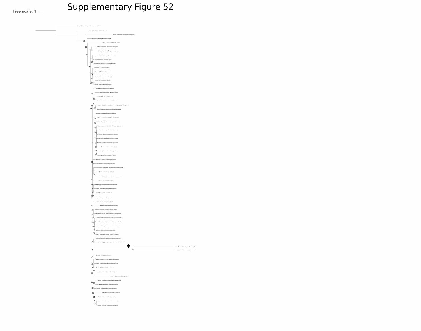
Archaea Euryarchaeota Acidiplasma sp MBA 1
Bacteria Elusimicrobia Elusimicrobium minutum Pei191
Bacteria Terrabacteria Firmicutes Streptococcus pneumoniae
Bacteria Protoebacteria Desulfarculus baarsii
Bacteria Spirochaetes Leptospira interrogans
Bacteria Terrabacteria Chloroflexi Chloroflexus aggregans
Bacteria Terrabacteria Cyanobacteria Nodosilinea nodulosa
Archaea TACK Candidatus Korarchaeum cryptofilum OPF8
Archaea Euryarchaeota Halobacterium salinarum
Bacteria Thermobaculum terrenum
Archaea Euryarchaeota Thermoplasma acidophilum
Bacteria Aquificae Aquifex aeolicus
Archaea Euryarchaeota Aciduliprofundum boonei
Bacteria Proteobacteria Geobacter bemidjiensis
Bacteria Terrabacteria Firmicutes Staphylococcus aureus
Bacteria Proteobacteria Rhizobium gallicum
Bacteria Proteobacteria Escherichia coli
Archaea TACK Caldivirga maquilingensis
Bacteria Proteobacteria Desulfatibacillum aliphaticivorans
Bacteria FCB Chlorobium limicola
Bacteria Acidobacteria Acidobacterium capsulatum
Archaea TACK Desulfurococcus amylolyticus
Bacteria FCB Gemmatimonadetes Gemmatimonas aurantiaca
Bacteria Terrabacteria Firmicutes Planococcus maritimus
Archaea Euryarchaeota Pyrococcus abyssi
Bacteria Terrabacteria Actinobacteria Streptomyces sviceus ATCC 29083
Archaea Euryarchaeota Halapricum salinum
Archaea Euryarchaeota Halorhabdus utahensis
Bacteria Fusobacteria Fusobacterium perfoetens
Bacteria Proteobacteria Nitrosomonas eutropha
Bacteria Terrabacteria Haloplasmatales Haloplasma contractile
Bacteria Proteobacteria Mariprofundus ferrooxydans
Bacteria Proteobacteria Vibrio cholerae
Archaea Euryarchaeota Halosimplex carlsbadense
Bacteria Proteobacteria Sorangium cellulosum
Bacteria Proteobcteia Micavibrio aeruginosavorus
Bacteria Terrabacteria Firmicutes Clostridium ultunense
Archaea TACK Staphylothermus hellenicus
Bacteria PVC Chlamydia trachomatis
Bacteria Terrabcteria Actinobacteria Actinomyces slackii
Archaea TACK Thermofilum pendens
Bacteria Terrabacteria Firmicutes Planifilum fulgidum
Archaea Euryarchaeota Palaeococcus pacificus
Bacteria Proteobacteria Erythrobacter litoralis
Bacteria Proteobacteria Paraburkholderia rhizoxinica
Archaea TACK Vulcanisaeta distributa
Bacteria Terrabacteria Firmicutes Paenibacillus curdlanolyticus
Archaea Euryarchaeota Halarchaeum acidiphilum
Archaea Euryarchaeota Haladaptatus paucihalophilus
Archaea Euryarchaeota Halalkalicoccus jeotgali
Archaea TACK Sulfolobus islandicus
Bacteria Dictyoglomi Dictyoglomus thermophilum
Archaea Euryarchaeota Thermococcus eurythermalis
Archaea Euryarchaeota Haloarcula amylolytica
Archaea Euryarchaeota Candidatus Halobonum tyrrellensis
Archaea Euryarchaeota Natronomonas moolapensis
Bacteria Proteobacteria Coxiella burnetii
Bacteria PVC Planctopirus limnophila
Archaea Euryarchaeota Picrophilus torridus
Bacteria Deinococcus Thermus Deinococcus radiodurans
Bacteria Terrabacteria Actinobacteria Rubrobacter xylanophilus
Bacteria Thermotogae Thermotoga maritima MSB8
Bacteia PVC Verrucomicrobium spinosum
Bacteria Spirochaetes Brachyspira pilosicoli WesB
Archaea Euryarchaeota Ferroplasma acidarmanus
Archaea Euryarchaeota Halomicrobium mukohataei
Bacteria Terrbacteria Firmicutes Bacillus subtilis
Bacteria Deferribacterales Deferribacter desulfuricans
60
61
43
96
83
39
100
98
92
96
76
96
96
100
77
66
95
78
66
100
96
77
76
71
34
92
63
100
27
82
36
89
61
95
47
91
95
97
62
64
84
99
71
100
10
85
100
100
99
70
69
99
50
90
98
86
99
100
99
100
84
57
95
96
87
Tree scale: 1Supplementary Figure 52
*

243
Appendix D
Papers derived from work included in this thesis.
Coleman, G.A., Pancost, R.D. and Williams, T.A., 2019. Investigating the origins of
membrane phospholipid biosynthesis genes using outgroup-free rooting. Genome
biology and evolution, 11(3), pp.883-898.
Coleman, G.A., Davín, A.A., Mahendrarajah, T., Spang, A.A., Hugenholtz, P.,
Szöllősi, G.J. and Williams, T.A., 2020. A rooted phylogeny resolves early bacterial
evolution. bioRxiv.
Gareth Coleman
295