FINAL PROGRAMME - European Society of Cardiology
160
FINAL PROGRAMME
-
Upload
khangminh22 -
Category
Documents
-
view
2 -
download
0
Transcript of FINAL PROGRAMME - European Society of Cardiology

FINAL PROGRAMME

SCHEDULE AT A GLANCE
7:00
7:30
8:00
9:00
10:00
10:15
11:00
12:00
12:30
13:00
13:30
14:00
15:00
15:45
16:00
16:30
17:00
17:30
18:00
18:30
19:00
19:30
20:00
20:30
Mod
erat
ed
e-po
ster
/ e-
case
s
Brea
kBr
eak
Brea
kBr
eak
Mod
erat
ed
e-po
ster
/ e-
case
s
Mod
erat
ed
e-po
ster
/ e-
case
s
Brea
kBr
eak
Mod
erat
ed
e-po
ster
/ e-
case
s
Mod
erat
ed
e-po
ster
/ e-
case
s
Drin
k
Facu
lty &
Mem
bers
Loun
ge
Scie
ntifi
c Ses
sions
Scie
ntifi
c Ses
sions
Sate
llite
Sym
posiu
mSc
ient
ific S
essio
nsSc
ient
ific S
essio
ns
Drin
kPo
ster
sess
ion
SCM
R / I
SMRM
Wor
ksho
p
Facu
lty &
Mem
bers
Loun
geSp
eake
r Ser
vice
Cen
ter
Cour
se
Regi
stra
tion
Spea
ker S
ervi
ce C
ente
r
Regi
stra
tion
Exhi
bitio
n
Wel
com
e Dr
ink
Sate
llite
Sym
posiu
mSc
ient
ific S
essio
nsSc
ient
ific S
essio
ns
Regi
stra
tion
Facu
lty &
Mem
bers
Loun
ge
Tues
day
30 Ja
nuar
y
Exhi
bitio
nM
eet &
Gre
et
Wed
nesd
ay
31 Ja
nuar
y
Frid
ay
2
Febr
uary
Cour
se
Spea
ker S
ervi
ce C
ente
rRe
gist
ratio
n
Spea
ker S
ervi
ce C
ente
r
Cour
se
Cour
se
Cour
se
Thur
sday
1 Fe
brua
ry
Satu
rday
3 Fe
brua
ry
SCM
R / I
SMRM
Wor
ksho
pSc
ient
ific S
essio
ns
Scie
ntifi
c Ses
sions
Scie
ntifi
c Ses
sions
Scie
ntifi
c Ses
sions
Regi
stra
tion
Cour
se
Spea
ker S
ervi
ce C
ente
r
Exhi
bitio
nFa
culty
& M
embe
rs Lo
unge
Scie
ntifi
c Ses
sions
Post
er se
ssio
n
Post
er se
ssio
n
Sate
llite
Sym
posiu
m
INDEXCommittee Members . . . 04Welcome Address . . . . 05Faculty . . . . . . . 06 General Information . . . 10Scientific Programme Info . . 12Social Events and Initiatives . 14CME Accreditation . . . . 15Sessions and Tracks Description 16About the EACVI . . . . 21About SCMR . . . . . 26Wednesday 31 January . . 31Thursday 1 February . . . 36Friday 2 February . . . . 54Saturday 3 February . . . 81Exhibition Floor Plan . . 142Exhibitors Directory . . . 143Industry Sessions . . . 149Notes . . . . . . . 155General Floor Plan . . . 159
4
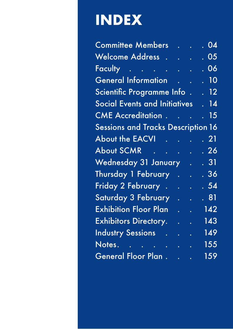
COMMITTEE MEMBERS
Presidents
Matthias FriedrichSCMR President
Bogdan A. Popescu, Chiara Bucciarelli-DucciEACVI President / EACVI CMR Vice-President and Section Chair
Programme Committee
Programme Co-Chairperson Juliano Fernandes (SCMR)Programme Co-Chairperson Robin Nijveldt (EACVI)Abstract Co-Chairperson Allison Hays (SCMR)Abstract Co-Chairperson Jose Rodriguez Palomares (EACVI)
Case Sessions DirectorsJohn Paul CarpenterMichael Hope
Anthony Aletras
Mouaz Al-Mallah
Andrew Arai
Colin Berry
Stephen Cheung
Pierre Croisille
Stephen Darty
Rohan Dharmakumar
Adam Dorfman
Ingo Eitel
Vanessa Ferreira
Alison Fletcher
Bernhard Gerber
John Greenwood
Matthias Gutberlet
Anna Herrey
Peng Hu
Theodoros D. Karamitsos
Peter Kellman
Han W. Kim
Sebastian Kozerke
Tomas Lapinskas
Pier Giorgio Masci
Manish Motwani
Vivek Muthurangu
Marcelo Nacif
Krishna Nayak
Reza Nezafat
Christopher T Nguyen
Gianluca Pontone
Kanishka Ratnayaka
Erik Schelbert
David Sosnovik
Michael D. Taylor
Anne Marie Valente
Emanuela Valsangiacomo
Florian von Knobelsdorff
Katherine Wu
Alistair Young
Stefan Zimmerman
ISMRM-SCMR Workshop Programme ChairsPeter KellmanSebastian Kozerke

4 5www.cmr2018.org
WELCOME ADDRESS
The international CMR community will gather in Barcelona for CMR 2018, the joint EuroCMR*/SCMR meeting organised by the European Association of Cardiovascular Imaging (EACVI), a registered branch of the European Society of Cardiology, and the Society for Cardiovascular Magnetic Resonance (SCMR).
CMR 2018 will focus on the theme of “Improving Clinical Value by Technical Advances”, emphasising the common goal of improving clinical outcomes in cardiovascular disease through innovation in basic MR development and medical engineering.
By providing a space where experts from all over the world with different experience and background share knowledge, the meeting offers more than 35 hours of presentations and cutting-edge information both on a novice and advanced level.
Particular care is given to guarantee time for discussions, specialised topics, case presentations, speed mentoring and focused pre-conferences as well as sessions for non-physicians and basic scientists. Attendees new to CMR will have the opportunity of pursuing a Level 1 Certification Track and learn the basic foundation required for a research or clinical career in the field.
In addition to the main meeting, a co-hosted ISMRM workshop will focus on Ischemia Imaging, from pathophysiological models, to advancements in imaging sequences and clinical applications.
Top leaders in the field of cardiology and radiology will provide the important context for this exciting event.
Let CMR 2018 expose you to state-of-the-art technological advances and innovative clinical imaging trials while generating new ideas to allow CMR to flourish worldwide!
Matthias G. Friedrich Robin Nijveldt, Juliano FernandesSCMR President Programme Co-Chairpersons
Bogdan A. Popescu, Chiara Bucciarelli-Ducci Jose Francisco Rodríguez Palomares, Allison HaysEACVI President / EACVI CMR Abstract Co-ChairpersonsVice-President and Section Chair

6
FACULTY
Robert Adam, United KingdomNiti Aggarwal, United StatesBobby Agrawal, United KingdomTimothy Albert, United StatesAnthony Aletras, GreeceZahra Alizadeh Sani, IranRasha Al-Lamee, United KingdomMouaz Al-Mallah, United StatesAna Almeida, PortugalGiovanni Aquaro, ItalyAndrew Arai, United StatesHåkan Arheden, SwedenJanine Arruda, United StatesMichael Atalay, United StatesLeon Axel, United StatesSonya Babu-Narayan, United StatesJohn Baksi, United KingdomW. Patricia Bandettini, United StatesPuja Banka, United StatesAna Barac, United StatesAnna Baritussio, United KingdomMatthew Barrett, United StatesCristina Basso, ItalyJeroen Bax, NetherlandsAernout Beek, NetherlandsSebastiaan Bekkers, NetherlandsRebecca Beroukhim, United StatesAntonio Berruezo, United StatesColin Berry, United KingdomNuno Bettencourt, PortugalAnish Bhuva, United KingdomRobert Biederman, United StatesStefan Biesbroek, The NetherlandsGiovanni Biglino, United KingdomDavid Bluemke, United StatesRene Botnar, United KingdomRedha Boubertakh, United KingdomJens Bremerich, SwitzerlandDavid Broadbent, United KingdomLouise Brown, United Kingdom Lorna Browne, United StatesJosep Brugada, SpainJennifer Bryant, SingaporeChiara Bucciarelli-Ducci, United KingdomCraig Butler, Canada
Michael Campbell, United StatesAdrienne Campbell-Washburn, United StatesGabriella Captur, United KingdomIacopo Carbone, ItalyMarcus Carlsson, SwedenJohn Paul Carpenter, United KingdomJames Carr, United StatesJoão Cavalcante, United StatesSteven Chamuleau, NetherlandsCarmen Chan, Hong KongRaymond Chan, CanadaPairoj Chattranukulchai, ThailandYu-Cheng Cheng, China (People’s Republic)Stephen Cheung, Hong KongAmedeo Chiribiri, United KingdomByoung Wook Choi, Republic of KoreaRunyawan Chotenimitkhun, ThailandNadine Choueiter, United StatesKelvin Chow, United StatesAnthony Christodoulou, United StatesTaylor Chung, United StatesGuido Claessen, BelgiumCindy Comeau, United StatesAndrew Cookson, United KingdomRichard Coulden, CanadaTracy Coulson, United KingdomPierre Croisille, FranceAnna Lisa Crowley, United StatesArun Dahiya, AustraliaErica Dall’Armellina, United KingdomStephen Darty, United StatesDana Dawson, United KingdomFrancesco De Cobelli, ItalyErasmo De La Pena-Almaguer, MexicoAlbert de Roos, NetherlandsClaudia Deluigi, SwitzerlandRohan Dharmakumar, United StatesEdward DiBella, United StatesAdam Dorfman, United StatesMarc Dweck, United KingdomChristopher Dyke, United StatesRobert Edelman, United StatesThor Edvardsen, NorwayIngo Eitel, GermanySuzette Elias-Smale, The Netherlands
6 7www.cmr2018.org
FACULTY
Henrik Engblom, SwedenDaniel Ennis, United StatesFrederick Epstein, United StatesPeter Ewert, GermanyJuliano Fernandes, BrazilCovadonga Fernández Golfin, SpainVictor Ferrari, United StatesVanessa Ferreira, United KingdomPatricia Feuchter, United KingdomPaul Finn, United StatesScott Flamm, United StatesAndrew Flett, United KingdomMark Fogel, United StatesMarianna Fontana, United KingdomSanjeev Francis, United StatesChristopher Francois, United StatesMarco Francone, ItalyHerbert Frank, AustriaMatthias Friedrich, CanadaOliver Gaemperli, SwitzerlandPankaj Garg, United KingdomRuchira Garg, United StatesJerome Garot, FranceJenifer Geradin, United StatesBernhard Gerber, BelgiumTal Geva, United StatesJulia Grapsa, United StatesJohn Greenwood, United KingdomGerald Greil, United StatesSimon Greulich, GermanyLindsay Griffin, United StatesJohn Grizzard, United StatesLars Grosse-Wortmann, CanadaMatthias Gutberlet, GermanyGabriela Guzman, SpainDan Halpern, United StatesMark C. K. Hamilton, United KingdomYuchi Han, United StatesYiying Han, SingaporeKate Hanneman, United StatesIwan Harries, United KingdomAllison Hays, United StatesJohn Heitner, United StatesWillem Helbing, NetherlandsAnja Hennemuth, GermanyAnna Herrey, United Kingdom
Rocio Hinojar, SpainAlexander Hirsch, NetherlandsBen Holloway, United KingdomMichael Hope, United StatesKan Hor, United StatesTracy Horn, United StatesAlbert Hsaio, United StatesBob Hu, United StatesPeng Hu, United StatesGreg Hundley, United StatesTarique Hussain, United StatesMasaki Ishida, JapanRon Jacob, United StatesMichael Jerosch-Herold, United StatesMeng Jiang, China (People’s Republic)Jason Johnson, United StatesRobert Judd, United StatesKimberly Kallianos, United StatesDinesh Kalra, United StatesTheodoros Karamitsos, GreeceNiall Keenan, United KingdomSebastian Kelle, GermanyChristian Kellenberger, SwitzerlandPeter Kellman, United StatesJaffar Khan, United StatesAhmed Kharabish, GermanyArash Kheradvar, United StatesPhilip Kilner, United KingdomHan Kim, United StatesRaymond Kim, United StatesPaul Knaapen, NetherlandsDaniel Knight, United KingdomGrigorios Korosoglou, GermanyTushar Kotecha, United KingdomSebastian Kozerke, SwitzerlandRebecca Kozor, AustraliaDara Kraitchman, United StatesChristopher Kramer, United StatesAndreas Kumar, CanadaKarl Kunze, United StatesRaymond Kwong, United StatesGregory Lanza, United StatesTomas Lapinskas, LithuaniaGerhard Laub, United StatesChris Lawton, United StatesAurore Lyon, United Kingdom
8
FACULTY
Robert Lederman, United StatesDaniel Lee, United StatesTim Leiner, NetherlandsBenedetta Leonardi, ItalySteve Leung, United StatesEylem Levelt, United KingdomDebiao Li, United StatesGabriela Liberato, BrazilJoao Lima, United StatesHarold Litt, United StatesAlexander Liu, United KingdomChristian Loewe, AustriaMassimo Lombardi, ItalyJimmy Lu, United StatesJoaquin Lucena, SpainAlicia Maceira, SpainPer Lav Madsen, United StatesViviana Maestrini, ItalyAshkan Malayeri, United StatesJoseph Mammarappallil, United StatesRobert Manka, SwitzerlandWarren Manning, United StatesMichael Markl, United StatesHugo Marques, PortugalNassir Marrouche, United StatesEdward Martin, United StatesPierGiorgio Masci, United StatesChristian Matter, SwitzerlandSophie Mavrogeni, GreeceGerry McCann, United KingdomFiona Mcmillan, United KingdomDaniel Messroghli, United StatesChristopher Miller, United KingdomSarah Moharem-Elgamal, United KingdomJames Moon, United KingdomManish Motwani, United KingdomElie Mousseaux, FranceKanae Mukai, United StatesMonica Mukherjee, United StatesGiuseppe Muscogiuri, ItalyVivek Muthurangu, United KingdomSaul Myerson, United KingdomEike Nagel, GermanyAkhil Narang, United States
Jagat Narula, United StatesLuigi Natale, ItalyKrishna Nayak, United StatesStefan Neubauer, United KingdomReza Nezafat, United StatesChristopher Nguyen, United StatesSonia Nielles-Vallespin, United StatesRobin Nijveldt, NetherlandsWendy Norman, United StatesNtobeko Ntusi, South AfricaKaren Ordovas, United StatesAmit Patel, United StatesDudley Pennell, United KingdomAlessia Pepe, ItalyMartina Perazzolo Marra, ItalyEsther Pérez-David, SpainSteffen Petersen, United KingdomStefan Piechnik, United KingdomJuan Carlos Plana, United StatesFrancesca Pluchinotta, ItalyTomaz Podlesnikar, NetherlandsGerald Pohost, United StatesGianluca Pontone, United StatesBogdan Popescu, RomaniaAndrew Powell, United StatesSanjay Prasad, United KingdomClaudia Prieto, United KingdomRajesh Puranik, United StatesKuberan Pushparajah, United KingdomMichael Quail, United KingdomAlexander Radbruch, GermanyFrank Rademakers, BelgiumFrancesca Raimondi, FranceSubha Raman, United StatesRajiv Ramasawmy, United StatesVikas Rathi, United StatesRahul Rathod, United StatesKanishka Ratnayaka, United StatesReza Razavi, United KingdomSurendranath Reddy, United StatesNathaniel Reichek, United StatesDaniel Roberts, United StatesCarlos Rochitte, BrazilJose Rodriguez-Palomares, SpainArno Roest, Netherlands
8 9www.cmr2018.org
FACULTY
Toby Rogers, United StatesStefania Rosmini, United KingdomDaniel Rueckert, United KingdomVal Runge, SwitzerlandTobias Rutz, SwitzerlandHajime Sakuma, JapanMichael Salerno, United StatesChris Saunderson, United KingdomTobias Schaeffter, GermanyMichael Schär, United StatesErik Schelbert, United StatesSilvia Schievano, United KingdomEhud Schmidt, United StatesJeanette Schulz-Menger, GermanyJuerg Schwitter, SwitzerlandAurelio Secinaro, ItalyNicole Seiberlich, United StatesJoseph Selvanayagam, AustraliaRoxy Senior, United KingdomJuliana Serafim, BrazilDipan Shah, United StatesBehzad Sharif, United StatesChetan Shenoy, United StatesMaria Siebes, NetherlandsLilia Sierra-Galan, MexicoOrlando Simonetti, United StatesAnvesha Singh, United KingdomMaria Eduarda Siqueira, BrazilTim Slesnick, United StatesPhilipp Sommer, GermanyDavid Sosnovik, United StatesOliver Speck, United StatesBarbara Srichai, United StatesKevin Steel, United StatesMatthias Stuber, United StatesAvan Suinesiaputra, New ZealandMushabbar Syed, United StatesGergely Szantho, United KingdomBalaji Tamarappoo, United StatesOliver Tann, United KingdomMichael Taylor, United StatesAndrew Taylor, AustraliaRaquel Themudo, SwedenChristoph Tillmanns, GermanyAdam Timmis, United States
Solenn Toupin, United StatesMarly Uellendahl, BrazilMartin Ugander, SwedenSeth Uretsky, United StatesSergio Uribe, ChileAnne Marie Valente, United StatesEmanuela Valsangiacomo Buechel, SwitzerlandRob van der Geest, NetherlandsRamon van Loon, NetherlandsPim van Ooij, NetherlandsAlbert van Rossum, NetherlandsMoriel Vandsburger, United StatesVassilis Vassiliou, United KingdomHein Verberne, United StatesMagalie Viallon, FranceInga Voges, United KingdomFlorian von Knobelsdorff, GermanyRachel Wald, CanadaMichelle Walkdon, United KingdomJonathan Weinsaft, United StatesRobert Weiss, United StatesDavid Wendell, United StatesMark Westwood, United KingdomJames White, United StatesRohan Wijesurendra, United KingdomRonald Williams, United StatesJoel Wilson, United StatesWalter Witschey, United StatesSteven Wolff, United StatesJames Wong, United KingdomGraham Wright, CanadaHui Xue, United StatesQi Yang, United StatesShi-Joon Yoo, CanadaAlistair Young, New ZealandAli N Zaidi, United StatesKarolina Zareba, United StatesStefan Zimmerman, United States

10
GENERAL INFORMATION
Venue CCIB – Centre Convencions Internacional de Barcelona Plaça de Willy Brandt, 11-14, 08019 Barcelona, Spain Phone: +34 932301000 www.ccib.es
Admission Your badge is required for admission to all scientific & Badges sessions and to the activities at CMR 2018. Please be sure to wear your badge at all times.
Registration Desk The registration desk is located in the main hall.
Opening hours: Tuesday 30 January 15:00 - 19:00 Wednesday 31 January 07:30 - 19:00 Thursday 1 February 07:30 - 19:00 Friday 2 February 07:30 - 19:00 Saturday 3 February 07:30 - 17:00
Your registration includes unlimited access to all scientific sessions, coffee breaks and the social events on Thursday, Friday and Saturday evenings.
Free WiFi Network Name: cmr2018 Username: cmr2018 Password: cmr2018
CMR2018 Mobile App An interactive CMR 2018 mobile app is available for attendees, on all mobile devices and tablets.Follow the instructions below to access the app. 1. Search for “eventScribe” in the Apple App Store or Google Play Store. Install and open the app. Then, search for “CMR 2018.” Click to launch. 2. For pre-registrants, select “Login” and enter your username and password that was emailed to you. 3. For onsite registrants, select “Create Account.”
Exhibition Opening Tuesday 30 January ClosedHours Wednesday 31 January Closed Thursday 1 February 10:15 - 20:30 Friday 2 February 08:00 - 20:30 Saturday 3 February 08:00 - 19:30
Do not forget to visit the exhibits and collect the stamps for the prize draw (see Exhibition section for more details)

10 11www.cmr2018.org
GENERAL INFORMATION
Faculty & Members Enjoy a cup of tea/coffee in a relaxed atmosphere; Lounge the Faculty & Members Lounge is open to SCMR members, EACVI Silver & Gold members and faculty. Printers are available.
Opening hours: Wednesday 31 January 07:30 - 19:00 Thursday 1 February 07:30 - 19:00 Friday 2 February 07:30 - 19:00 Saturday 3 February 07:30 - 18:30
CME Certificates Please submit the completed UEMS/EACCME Evaluation Form (inserted in your delegate bag) at the registration desk in order to receive the UEMS/EACCME Certificate. Certificates will be delivered at the registration desk as of Friday 2 February.
Coffee Breaks Coffee and refreshments will be served to delegates in the and Refreshments Exhibition Area. A cash bar is available in the Exhibition Area
Twitter Follow us and tweet on #cmr2018
Liability and The Congress Secretariat and Organisers cannot accept Insurance liability for personal accidents, loss or damage to private property of participants, either during or as result of the Congress. Participants are advised to take out their own personal travel and health insurance for their trip.
Safety Please do not leave bags or suitcases unattended at any time, and Security whether inside or outside the session halls.
Pl. Europa, 17-19 1st floor 08908L’ Hospitalet del LlobregatBarcelona (Spain)Phone: +34 93 882 38 [email protected]
Technical Secretariat

12
SCIENTIFIC PROGRAMME INFO
Speaker We kindly ask all speakers to check-in with our IT partner Service Center CYIM in the Speaker Service Center. You can update / edit your presentations there or upload a new version. Please note that deadline to do so is 2 hours prior to your presentation time. Support technicians will be available to assist you during the opening hours:
Tuesday 30 January 15:00 - 19:00 Wednesday 31 January 07:30 - 19:00 Thursday 1 February 07:30 - 19:00 Friday 2 February 07:30 - 19:00 Saturday 3 February 07:30 - 17:00
Special Sessions Don’t miss our special sessions (more details in the programme): - Opening Plenary: Thursday 1 February - 1:30pm – 2:45pm, Plenary Room - Early Career Plenary: Thursday 1 February – 6:05pm – 7:00pm, Plenary Room - Early Career Awards - Oral Abstracts: Thursday and Friday 1-2 February, Room 4 - Invasive Live Case: Friday 2 February – 11:40am – 12:25pm, Plenary Room - Outreach Sessions: Saturday 3 February – 8:00am – 12:30pm, Plenary Room - Best Moderated ePoster: Saturday 3 February – 1:30pm – 2:55pm, Room 6 - Closing Plenary: Saturday 3 February – 4:30pm – 6:30pm, Plenary Room
Do not miss the special satellite symposia from our Industry partners in the Lecture room (please check the programme for full details).

12 13www.cmr2018.org
SCIENTIFIC PROGRAMME INFO
Level 1 CMR Certification To receive your CMR Level 1 Certificate after the meeting, you must be pre-registered and prove your attendance at 85% of the sessions of the dedicated track (look for the Level 1 label on the programme pages). Please be sure to scan your badge at the beginning and at the end of each session of the dedicated track. The barcode scanners are located at the entrance of each lecture room. Certificate will be sent by email after the Congress. Some sessions included in the track are parallel sessions; this has been taken into account for the 85%.
Special Courses Seats are limited and pre-registration was mandatory to attend these sessions. They will be run in the Course Room (please check the programme for full details).
Level 2/3 Dedicated Case Sessions (stress perfusion and congenital) Seats are limited and pre-registration was mandatory to attend these courses. They will be run in the Hands-on Room (please check the programme for full details). Certificates will be provided after the sessions at the registration desk.
Abstracts, Cases, Posters, ePosters and eCases Posters and Cases are displayed in the Poster Area (exhitibion hall). Moderated ePoster/eCase sessions are presented during coffee breaks from Thursday until Saturday (please check the programme for full details).
Abstract/Cases sessions are run throughout the congress in various rooms (please check the programme for full details).
Session Reports Senior experts in the field will provide a report on the key messages from a selection of sessions. Available on EACVI and SCMR websites.
Session Recordings Most of the sessions will be recorded and available on EACVI and SCMR websites.

14
SPECIAL EVENTS AND INITIATIVES
Prize Draw Enter our lucky draw for a chance to win an iPad! Visit all booths and collect exhibitors’ stamps on your competition card (inserted in your delegate bag). Once you collected ALL stamps, hand it to the registration desk.
Exhibition opening hours: Tuesday 30 January Closed Wednesday 31 January Closed Thursday 1 February 10:15 - 20:30 Friday 2 February 08:00 - 20:30 Saturday 3 February 08:00 - 19:30
The lucky winner will be announced during the Closing Plenary.
Social Events • Wednesday 31 January - 18:30 – Welcome drink for workshop attendees only. • Thursday 1 February – 19:00 – A light welcome drink will be served in the exhibition area after the Early Career Plenary. • Friday 2 February – 19:00 – The Meet & Greet Reception will be held in the exhibition area. • Saturday 3 February – 19:00 – A cocktail will be served as a farewell in the exhibition area.
Speed Mentoring Speed mentoring in groups will be run in the Mentoring Room on Level 1 (Room 129&130) according to the appointment received by registered delegates during Break Times. Applications on site will depend on availability (please check).
Congress News Pick up your daily congress news at the exhibition entrance every morning before the first session or take your chance to grab the last ones either on the EACVI or SCMR stands, at the registration desks or in the Faculty and Members lounge.
European CMR Exams The CMR and CHD CMR certification exams will take place at the CCIB in rooms 211 and 212 (Level 2) on Wednesday 31 January 2018. Please note that the CMR exam participation is subject to distinct registration. Please bring your exam registration confirmation to access exam. Registered exam participants have received detailed instructions. If not, please check with the registration desk.
14 15www.cmr2018.org
CME ACCREDITATION
Accreditation
The CMR 2018 – The joint EuroCMR/ SCMR Meeting, Barcelona, Spain, 31/01/2018-03/02/2018 has been accredited by the European Accreditation Council for Continuing Medical Education (EACCME®) with 24 European CME credits (ECMEC®s). Each medical specialist should claim only those hours of credit that he/she actually spent in the educational activity.Through an agreement between the Union Européenne des Médecins Spécialistes and the American Medical Association, physicians may convert EACCME® credits to an equivalent number of AMA PRA Category 1 CreditsTM. Information on the process to convert EACCME® credit to AMA credit can be found at www.ama-assn.org/education/earn-credit-participation-international-activities.
Live educational activities, occurring outside of Canada, recognised by the UEMS-EACCME® for ECMEC®s are deemed to be Accredited Group Learning Activities (Section 1) as defined by the Maintenance of Certification Program of the Royal College of Physicians and Surgeons of Canada.
Please submit the completed UEMS/EACCME Evaluation Form (inserted in your delegate bag)at the end of the meeting in order to receive the UEMS/EACCME Certificate. Certificates will be sent by email after the congress.
Disclosure
All speakers and chairpersons participating in the programme have disclosed potential conflict of interest that might cause a bias in the presentations. The Organising Committee is responsable for ensuring that all potential conflict of interest relevant to the programme are declared to the audience prior to the CME activities.

16
SESSIONS AND TRACKS DESCRIPTION
1. ISMRM/SCMR Workshop - Cardiovascular Magnetic Resonance in Ischemic Heart Disease (separate registration required)
Dates and Times:
Wed, 31 Jan 31 8:00 – 18:00 Thu, 1 Feb 8:00 – 12:30 Location: Plenary Room (rooms 111+112) – Level 1
Description: The workshop seeks to discuss ischemic heart dis-ease in its full depth as it relates to CMR. This 1+1/2 day pro-gram will start by reviewing basic physiologic and pathophysio-logical details of the coronary circulation that serve as a basis to our understanding of CMR stress exams. It will then follow with a discussion on methods of perfusion imaging, quantification strategies and non-contrast methods to evaluate ischemia. On the second day, the discussion will include how CMR perfusion correlates to other methods and will end with open questions and a debate with top experts in the field.
Within the program, specific abstracts which correlate to the topics discussed will be presented both orally and in poster for-mat, providing further support for the lectures and debates.
Who should attend: This workshop should be attended by any member of the community who is performing or plans to run stress CMR exams. Given the current evidence of the many CMR trials in IHD showing the benefits of the method, ischemia imaging is one of the priorities in our educational program for CMR2018 and the workshop will serve as a solid base for all other sessions in the meeting.
2. Pediatric and Congenital Pre-Conference
Dates and Times: Thu, 1 Feb 8:00a – 12:30
Location: Lecture Room (Hall 6) – Level 0
Description: This session integrates with other sessions to comprise the Pediatric and Congenital Track along CMR2018. It is the starting point for the track and therefore has the objective of providing ground knowledge on the topic. It starts with a description of important sequences and techniques in the CMR environment used for these types of exams. In the second and third part, the most common diseases are discussed, with a focus on how to set up CMR protocols for those pathologies as well as discussing the most important details one should look for when performing and interpreting these exams.
Who should attend: All CMR professionals who plan to start performing pediatric and congenital CMR exams or already do so but want to review basic aspects of the exam or enhance their current knowledge. This preconference course intends to provide more basic and intermediate knowledge on this topic so that subsequent sessions can explore more advanced scenarios.
3. Physician Pre-Conference: Basic MRI and Physics
Dates and Times: Thu, 1 Feb 8:00 – 12:30 Location: Case Room (Hall 8) – Level 0
Description: This preconference course seeks to provide the ed-ucational base necessary for physicians and other professional to understand basic MRI concepts in safety, physics, sequence designs and contrast usage, with a focus on CMR. In addition to that, the main sequences and protocols used in CMR will be introduced first with a description on the technical method and then on a how-to basis, providing delegates with both the back-ground knowledge and how to apply them in practice.
Who should attend: Professionals new to CMR and those who already practice the method but want to be updated in the ba-sic techniques and principles of MRI. Its focus is on beginner and intermediate knowledge, with a very practical approach to be readily applicable in your CMR routine.
4. Opening Plenary - CMR’s Role in Arrhythmia and Sudden Cardiac Death
A Joint Session with the European Heart Rhythm Association (EHRA)
Date and Time: Thu, Feb 1 12:30 – 14:45 Location: Plenary Room (rooms 111+112) – Level 1 Description: The grand opening of CMR2018, this session will celebrate the presence of the CMR Community in Barcelona by focusing on a topic which is so well developed in the city relat-ing to Arrhythmia. Keynote lectures on this topic will highlight recent developments in EP and ventricular arrhythmias, how the CMR community can collaborate with EP physicians, the lessons in SCD by CMR and an update on imaging patients with implantable devices.
Who should attend: All delegates
5. Moderated ePosters and eCases
Dates and Times: Thu, 1 Feb 15:00-15:45 Fri, 2 Feb 2 10:15 – 11:00 and 15:45 – 16:30 Sat, 3 Feb 10:15 – 11:00
Location: Exhibition Hall – Level 0
Description: ePoster and eCases will be presented in four ses-sions during the break times, moderated by some of the top experts in each topic. The ePosters will be divided in 12 of the most important topics in CMR and the best poster from each
Level 1 Track
Pediatrics/ Congenital
Track
LectureSessions
Case-basedSessions
Abstract-basedSessions
SpecialDessions
Level 1Track
TechnologistTrack
SocialEvents
Courses IndustrySessions

16 17www.cmr2018.org
SESSIONS AND TRACKS DESCRIPTION
topic will be selected to run for the Best ePoster Award. For the eCases, 4 topics have been chosen and will also be discussed with experts in their field. The best way to gain new knowledge and have direct and close access to presenters of high quality science in CMR.
Who should attend: All delegates
6. Lecture Sessions (LS1 to LS7)
Dates and Times:
Thu, 1 Feb 15:45 – 17:10 Fri, 2 Feb 08:00 – 09:25; 13:30 – 14:55; 16:30 – 18:00 Sat, 3 Feb 08:00 – 09:25; 11:00 – 12:30; 13:30 – 14:55
Location: Lecture Room (Hall 6) – Level 0
Description: The 7 lecture sessions are 1h25min sessions in a tra-ditional lecture format but always ending or allowing for plenty of discussion on the topic selected. Its objective is to thoroughly present and review an important theme in CMR, with a focus on intermediate and advanced knowledge. The topics selected represent either new fields where CMR is advancing rapidly or key subjects in which CMR has an important role for clinical de-cision making. The intentions of these sessions are not only to provide the most up-to-date scientific data on each lecture but to allow for key and open questions to be discussed among all participants. Moderators of these sessions are encouraged to promote discussion after each talk or in a round-table debate, whatever fits best each topic. Four of the seven sessions are joint-sessions, expanding the interface of CMR with other clini-cal or imaging specialties.
Who should attend: All delegates; key focus on novel and state-of-the-art fields and how CMR is impacting the clinical manage-ment on these topics.
7. Short Lectures (SL1 – SL5)
Dates and Times:
Thu, 1 Feb 17:15 – 18:00 Fri, 2 Feb 09:30 – 10:15; 15:00 – 15:45; Sat, 3 Feb 09:30 – 10:15; 15:00 – 15:45;
Location: Lecture Room (Hall 6) – Level 0
Description: These 5 sessions are 45min long sessions with three lectures in each one of them. They also focus on key top-ics in CMR, but this time with a more closed look at specific CMR-centered themes such as tissue characterization, valvu-lar heart disease, clinical trials in CMR, function and structur-al analysis and viability. Lecture titles selected by the Program Committee tackle some of the most difficult questions in each topic and are all common problems faced in CMR labs in daily practice.
Who should attend: All delegates; focus on novel and advanced questions on routine protocols used in clinical CMR.
8. Cases Sessions (CR1 – CR7)
Dates and Times:
Thu, 1 Feb 15:45 – 17:10 Fri, 2 Feb 08:00 – 09:25; 13:30 – 14:55; 16:30 – 18:00pm Sat, 3 Feb 08:00 – 09:25; 11:00 – 12:30; 13:30 – 14:55
Location: Case Room (Hall 8) – Level 0
Description: One of the most popular sessions in the last meetings, these 7 sessions will feature seven cases presented in a specific key CMR topic. These were all submitted cases and the selection this year set a very high bar: there is no doubt the cases presented will feature a key unique feature that makes each one of them very special. To introduce and conclude each topic, a speaker was invited to provide a review of the topic of the session, relating to the cases presented but setting a stage to where the current knowledge in CMR stands. In summary, sessions which will give delegates both a review on key aspects on the topic selected as well as an opportunity to share with presenters the angst and difficulties of coming by special imaging characteristics.
Who should attend: All delegates; rare/unusual cases with differential diagnosis discussions balanced with cases in which CMR helped find the diagnosis or provided unique imaging features.
9. Didactic Case Sessions (DC1 – DC5)
Dates and Times:
Thu, 1 Feb 17:15 – 18:00 Fri, 2 Feb 09:30 – 10:15; 15:00 – 15:45; Sat, 3 Feb 09:30 – 10:15; 15:00 – 15:45;
Location: Case Room (Hall 8) – Level 0
Description: These new sessions will be presented by men-tor-fellow pairs, and focus on common clinical topics, discussing routine approaches and insights into emerging approaches that address key problems. There will be 5 sessions, each with 2 cas-es per session with plenty of time for discussions and audience participation. The idea of the session in general is to present common clinical cardiovascular problems/challenges, discuss-ing the assessment with different imaging modalities and the particular value of CMR in the work-up. Additionally, an insight into state-of-the-art techniques can be discussed. to extend their practice.
Who should attend: All delegates; focus on problem-based learning with step-by-step resolution of each case and audi-ence discussion; it is envisaged that this session will be useful for both routine clinical CMR practitioners and those looking to extend their practice.
10. Oral Abstract sessions (OA1 – OA7)
Dates and Times:
Thu, 1 Feb 15:45 – 17:10 Fri, 2 Feb 08:00 – 09:25; 13:30 – 14:55; 16:30 – 18:00 Sat, 3 Feb 08:00a – 09:25; 11:00 – 12:30; 13:30 – 14:55

18
Location: Room 4 ( room 113) – Level 1
Description: The sessions will present the most recent scien-tific discoveries in the CMR field. These abstracts represent the cutting-edge knowledge in CMR and are divided in 7 ses-sions. Three of these sessions will feature the highest quality abstracts presented by Early Career members who qualified for the awards in Basic, Translational and Clinical areas. The other four sessions represent key themes in CMR and will also display the most recent advances in their topics. Each abstract presen-tation will be followed by a chance for questions and discussion of the data presented, moderated by two experts in the field. These sessions intend to show us where the field is guided to.
Who should attend: All delegates; focus on novel discoveries and most recent advancements in CMR knowledge;
11. Special Sessions
Dates and Times:
Thu, 1 Feb 17:15 – 18:00 (2 parallel sessions) Fri, 2 Feb 09:30 – 10:15; 15:00 – 15:45; Sat, 3 Feb 15:00 – 15:45 (2 parallel sessions)
Location: Room 4 (room 113) and Room 5 ( room 114) – Level 1
Description: There will be 6 sessions which were named as “Special Sessions” as they intend to promote the discussion of a selected question or theme in different ways. Three of these sessions are organized as debates, with moderators and invited speakers engaging in answering specific questions posed by the Program Committee with the possibility of using a supporting presentation. The other three have been organized as tradition-al lectures but also with the focus on answering practical daily questions. Independently of their format, the main objective of these sessions is that the question posed in the title of each topic is answered by the Faculty Members and that delegates can interact and engage in the discussion.
Who should attend: All delegates; specific questions will be re-sponded and debates promoted.
12. Focus Sessions
Dates and Times:
Thu, 1 Feb 15:45 – 17:10 Fri, 2 Feb 08:00 – 09:25; 13:30 – 14:55; 16:30 – 18:00 Sat, 3 Feb 08:00 – 09:25; 11:00 – 12:30; 13:30 – 14:55
Location: Room 5 (room 114) – Level 1
Description: These 7 sessions complement the oral abstract portion of the program with the presentation of high quality abstracts selected to cover a specific key topic for the program. These topics were chosen as they pose specific needs of the CMR community or represent important clinical aspects found in routine scanning. They include making CMR faster and easier, acute coronary syndromes, functional analysis, normal limits,
SESSIONS AND TRACKS DESCRIPTION
right ventricle evaluation, coronary imaging and implementing a CMR program. To introduce each topic, a carefully selected lecture was chosen, setting the stage for the following abstracts which will then advance the field or respond to some questions presented previously. The objective of these sessions is to give delegates a review on the subject and, at the same time, a di-rection to where current developments are pointing in these key topics.
Who should attend: All delegates; focus on novel scientific ad-vances in selected topics
13. Interventional Course
Dates and Times: Thu, 1 Feb 1 15:00 – 19:00
Location: Room 6 (room 115) – Level 1
Description: The main objective of this course is to discuss this theme among different specialized groups throughout the world, from basic case examples to more advanced views on devices, imaging techniques and procedures. The course starts with moderated case discussions, goes on to a description of MRI catheterization techniques which involve some interven-tion (stress, NO, exercise, etc) and finalizes with lectures on present and future of devices. This year, the course was moved inside the program instead of being placed as a pre-conference in order to avoid overlap with other sessions that might be of interest to the same delegates.
Who should attend: CMR professionals interested in learning about interventional CMR, whether they already practice or in-tend to start a program; both adult and pediatric topics will be covered.
14. Non-Cardiologist Course
Dates and Times: Thu, 1 Feb 15:00 – 19:00
Location: Tech Room (room 116) – Level 1
Description: This course intends to provide all non-Cardiologists in the meeting with the key knowledge in this clinical area that interfaces with other areas of CMR. It starts by reviewing ba-sic aspects of cardiological anatomy, function and physiology and advances through the main elements of common diseases studied in CMR such as atherosclerosis, ischemic heart disease, cardiomyopathies, valvular heart disease and congenital heart disease. Finally, basic treatment guidelines are presented for CAD, heart failure and valve disease. All topics are given by ex-pert clinical professionals and should provide a robust basis to understand the most relevant clinical aspects of cardiovascular disease that will ease the understanding of why certain aspects of CMR are performed. This year, the course was moved inside the program instead of being placed as a pre-conference in or-der to avoid overlap with other sessions that might be of inter-est to the same delegates.
Who should attend: Radiologists, Engineers, Physicists, non-MDs interested in learning more about basic cardiovascular concepts and diseases fundamental to CMR overall knowledge.
18 19www.cmr2018.org
SESSIONS AND TRACKS DESCRIPTION
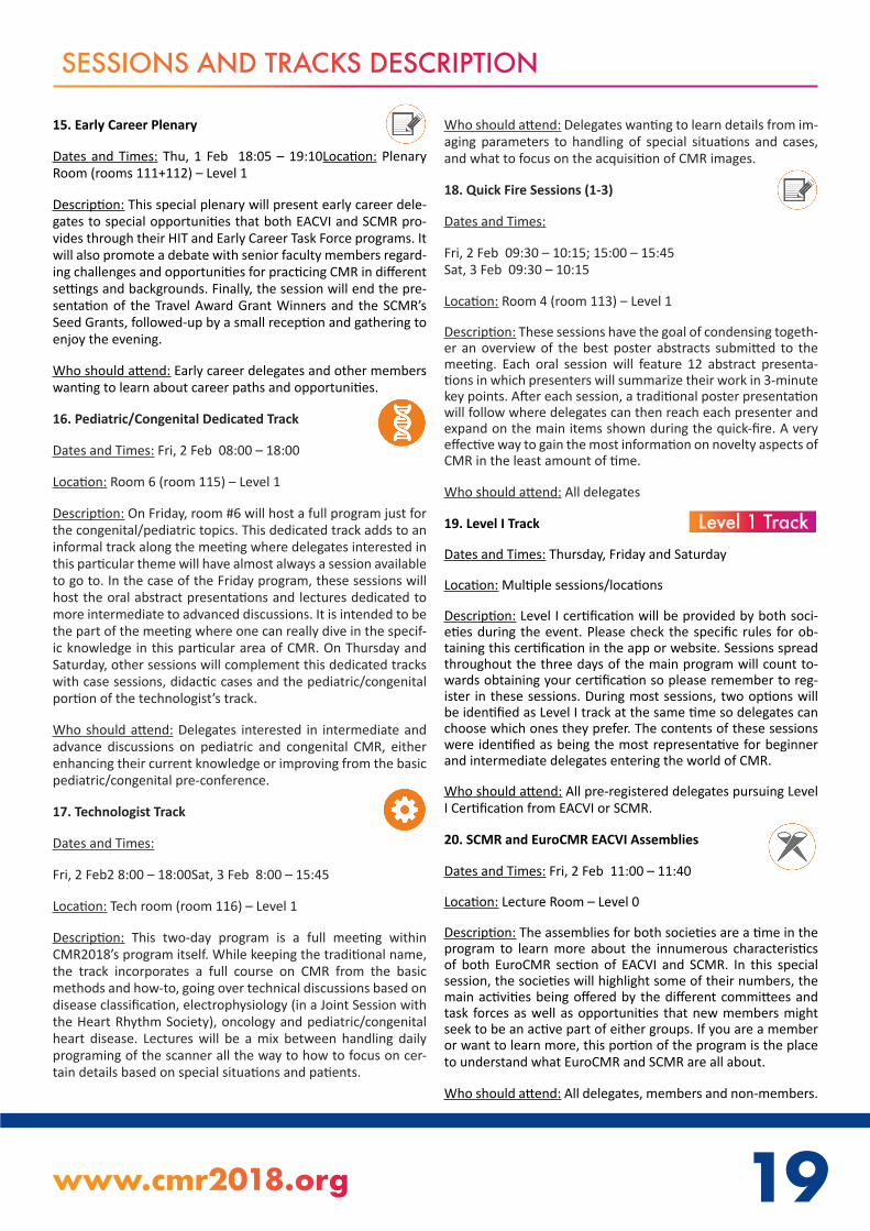
15. Early Career Plenary
Dates and Times: Thu, 1 Feb 18:05 – 19:10Location: Plenary Room (rooms 111+112) – Level 1
Description: This special plenary will present early career dele-gates to special opportunities that both EACVI and SCMR pro-vides through their HIT and Early Career Task Force programs. It will also promote a debate with senior faculty members regard-ing challenges and opportunities for practicing CMR in different settings and backgrounds. Finally, the session will end the pre-sentation of the Travel Award Grant Winners and the SCMR’s Seed Grants, followed-up by a small reception and gathering to enjoy the evening.
Who should attend: Early career delegates and other members wanting to learn about career paths and opportunities.
16. Pediatric/Congenital Dedicated Track
Dates and Times: Fri, 2 Feb 08:00 – 18:00
Location: Room 6 (room 115) – Level 1
Description: On Friday, room #6 will host a full program just for the congenital/pediatric topics. This dedicated track adds to an informal track along the meeting where delegates interested in this particular theme will have almost always a session available to go to. In the case of the Friday program, these sessions will host the oral abstract presentations and lectures dedicated to more intermediate to advanced discussions. It is intended to be the part of the meeting where one can really dive in the specif-ic knowledge in this particular area of CMR. On Thursday and Saturday, other sessions will complement this dedicated tracks with case sessions, didactic cases and the pediatric/congenital portion of the technologist’s track.
Who should attend: Delegates interested in intermediate and advance discussions on pediatric and congenital CMR, either enhancing their current knowledge or improving from the basic pediatric/congenital pre-conference.
17. Technologist Track
Dates and Times:
Fri, 2 Feb2 8:00 – 18:00Sat, 3 Feb 8:00 – 15:45
Location: Tech room (room 116) – Level 1
Description: This two-day program is a full meeting within CMR2018’s program itself. While keeping the traditional name, the track incorporates a full course on CMR from the basic methods and how-to, going over technical discussions based on disease classification, electrophysiology (in a Joint Session with the Heart Rhythm Society), oncology and pediatric/congenital heart disease. Lectures will be a mix between handling daily programing of the scanner all the way to how to focus on cer-tain details based on special situations and patients.
Who should attend: Delegates wanting to learn details from im-aging parameters to handling of special situations and cases, and what to focus on the acquisition of CMR images.
18. Quick Fire Sessions (1-3)
Dates and Times:
Fri, 2 Feb 09:30 – 10:15; 15:00 – 15:45 Sat, 3 Feb 09:30 – 10:15
Location: Room 4 (room 113) – Level 1
Description: These sessions have the goal of condensing togeth-er an overview of the best poster abstracts submitted to the meeting. Each oral session will feature 12 abstract presenta-tions in which presenters will summarize their work in 3-minute key points. After each session, a traditional poster presentation will follow where delegates can then reach each presenter and expand on the main items shown during the quick-fire. A very effective way to gain the most information on novelty aspects of CMR in the least amount of time.
Who should attend: All delegates
19. Level I Track
Dates and Times: Thursday, Friday and Saturday
Location: Multiple sessions/locations
Description: Level I certification will be provided by both soci-eties during the event. Please check the specific rules for ob-taining this certification in the app or website. Sessions spread throughout the three days of the main program will count to-wards obtaining your certification so please remember to reg-ister in these sessions. During most sessions, two options will be identified as Level I track at the same time so delegates can choose which ones they prefer. The contents of these sessions were identified as being the most representative for beginner and intermediate delegates entering the world of CMR.
Who should attend: All pre-registered delegates pursuing Level I Certification from EACVI or SCMR.
20. SCMR and EuroCMR EACVI Assemblies
Dates and Times: Fri, 2 Feb 11:00 – 11:40
Location: Lecture Room – Level 0
Description: The assemblies for both societies are a time in the program to learn more about the innumerous characteristics of both EuroCMR section of EACVI and SCMR. In this special session, the societies will highlight some of their numbers, the main activities being offered by the different committees and task forces as well as opportunities that new members might seek to be an active part of either groups. If you are a member or want to learn more, this portion of the program is the place to understand what EuroCMR and SCMR are all about.
Who should attend: All delegates, members and non-members.
Level 1 Track
20
24. Best SCMR Case of the Week
Dates and Times:
Sat, 3 Feb 15:00 – 15:45
Location: Room 6 ( room 115) – Level 1
Description: SCMR hosts in its website cases submitted on a routine basis that are published online after peer-review. Each year, the best cases are voted by the reviewers and the top 5 are selected to participate in a special session where they are pre-sented orally for the selection of the Best Case of the Week. The five cases selected this year have very special teaching points and represent very well the objectives of the online cases. A great way to end the presentation of cases in the meeting in an entertaining and educational competition.
Who should attend: All delegates
25. Closing Plenary (including OSCARS Session)
Dates and Times: Sat, 3 Feb 16:30 – 18:30Location: Plenary Room (rooms 111+112) – Level 1
Description: The farewell session of CMR2018 includes different special items that make this plenary unique and provides dele-gates take-home key messages from the meeting. It starts with the EACVI keynote lecture on “Myocardial plasticity, adaptation and maladaptation: insights from CMR”. Followed by this lec-ture, the friendly competition between top CMR practitioners takes place in the renovated CMR Oscars – the Image Competi-tion. This year will feature North versus South competitors from different places of the world to face the exquisite questions pre-pared by our hosts. Finally, the session ends with the top high-lights of the meeting presented by the Chairs, the Awards cere-mony which will announce the winners of the abstract awards and SCMR Gold Medal Award and the final remarks from SCMR/EuroCMR.
Who should attend: All delegates
26. Exhibition and Product Theater
Dates and Times: Thursday, Friday and Saturday
Location: Exhibition Hall – Level 0
Description: The exhibitors will showcase the best of their products in Level 0 just outside the Lecture and Case rooms. 18 compa-nies will be present in this floor space allowing delegates to learn about the latest commercial features in CMR products. Along with the Exhibition Hall, special events will take place in the Product Theater, located just along the Poster area. In the Product Theater, exhibitors have prepared feature presenta-tions that highlight important details on specific areas of CMR and will allow delegates to learn more about how their prod-ucts may help solve daily practical problems in the scanner and in post-processing solutions.
Who should attend: All delegates
SESSIONS AND TRACKS DESCRIPTION
21. Invasive Live Case
Dates and Times:
Fri, 2 Feb 11:40 – 12:25
Location: Plenary Room (rooms 111+112) – Level 1
Description: Delegates will have the opportunity to watch a live transmission of an invasive CMR procedure performed by one of the top centers in the world directly from King’s College London. The moderators in Barcelona will guide us through the nuts and bolts of how an actual invasive CMR exam is performed while delegates can watch as the case is done online. A very in-teresting complement to the Interventional Course and other interventional sessions along the meeting in this exciting field.
Who should attend: All delegates
22. Outreach Session
Dates and Times:
Sat, 3 Feb 8:00 – 12:30
Location: Plenary Room (rooms 111+112) – Level 1
Description: As the name implies, this session provides and interface between the CMR community and other specialties and societies. The four topics selected provide delegates with a chance of listening to how CMR extends to clinical practice in forefront areas. Outcomes research and participation of CMR in guidelines starts the program, followed by the use of CMR in ischemic heart disease in light of other modalities, moves to diagnosing inflammatory heart disease in multisystem diseases and ends with examples of CMR use in population based studies and big data. A whole morning with cutting-edge presentations that highlight and summarize CMR2018’s theme of “Improving Clinical Value by Technical Advances”
Who should attend: All delegates
23. Best of Moderated ePosters
Dates and Times:
Sat, 3 Feb 13:30 – 14:55
Location: Room 6 (room 115) – Level 1
Description: After the 12 presentations in each topic in the Ex-hibition Hall, the best poster in each group will be asked to be presented one more time now against each other competing for the Best Moderated Poster Award. A new set of judges will moderate the presentations and have the task of selecting the best poster to be presented in the Closing Plenary. This session will highlight very high quality presentations that were selected first from their original submissions and secondly after a second round in the ePosters presentations.
Who should attend: All delegates
20 21www.cmr2018.org
ABOUT THE EACVI
The EACVI at a glance
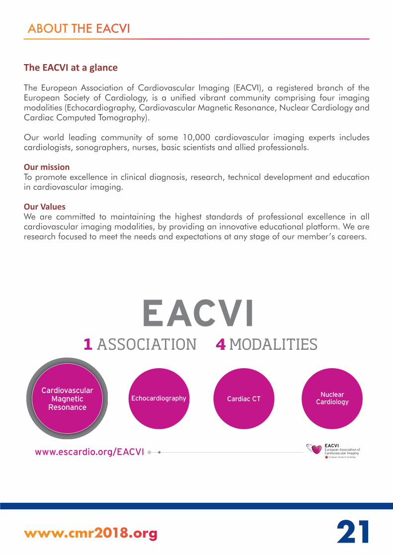
The European Association of Cardiovascular Imaging (EACVI), a registered branch of the European Society of Cardiology, is a unified vibrant community comprising four imaging modalities (Echocardiography, Cardiovascular Magnetic Resonance, Nuclear Cardiology and Cardiac Computed Tomography).
Our world leading community of some 10,000 cardiovascular imaging experts includes cardiologists, sonographers, nurses, basic scientists and allied professionals.
Our mission To promote excellence in clinical diagnosis, research, technical development and education in cardiovascular imaging.
Our ValuesWe are committed to maintaining the highest standards of professional excellence in all cardiovascular imaging modalities, by providing an innovative educational platform. We are research focused to meet the needs and expectations at any stage of our member’s careers.
Sans titre-1 1 15/01/2018 17:01

ABOUT THE EACVI
The EACVI products
Congresses and events
• EuroEcho-Imaging 2018: 5-8 December 2018, Milan - Italy• EuroCMR 2019: 2-4 May 2019, Venice - Italy • ICNC 2019: 12-14 May 2019, Lisbon - Portugal
Educational products •We offer a wide range of educational products in all imaging modalities both online and in the form of face-to-face events.•Training and research grants as well as individual certification programmes in CMR, Echocardiography, Nuclear Cardiology and Cardiac CT are also available.
Learn more at www.escardio.org/EACVI
Publications
Stop by the EACVI stand and buy your copies.*
•Books - 2018 Compendium: comprises a selection of EACVI scientific papers published in 2016 and 2017. - Textbook on Echocardiography – second edition - Echo Handbook - Upcoming: Textbook on CMR Pre-order form available.
•CMR Pocket Guides - Congenital Heart Disease - Physics for Clinicians - General Pocket Guide – 2017 edition
* Exclusive discounts for Silver and Gold EACVI members.
Free samples of the European Heart Journal – Cardiovascular Imaging (EHJ - CI), the EACVI’s official journal, are available at the EACVI stand during CMR 2018. EHJ – CI 2017 impact factor:5.99.
• Abridged recommendations9 titles available including some in Spanish, French, Portuguese and Chinese.
22

…see you at the EACVI stand
*Instead of 156€ for the 2018 membership. Restricted to paying CMR2018 delegates. Not applicable to day ticket holders.
W A N TM O R E ?
Mix and match the ESC membershippackages that best suit your needs forunique savings and bene�ts.
EACVI SILVERMEMBERSHIPfor 100€only during the congress *

Learn from imaging experts
Including didactic video tutorials in TOE, TTE, CMR, Nuclear Cardiology, Cardiac CT and much more
E-learning programmes in Echocardiography, in Cardiovascular Magnetic Resonance
and in Nuclear Cardiology and Cardiac Computed Tomography.
www.escardio.org/e-learning
d8319-Pubs_EACVI-2017-A4_v6.indd 2 18/10/2017 17:04

Edited by Massimo LombardiSven Plein
Steffen PetersenChiara Bucciareli-DucciEmanuela Valsangiacomo BuechelCristina BassoVictor Ferrari
Published in partnership with the European Association of Cardiovascular Imaging and written by a team of expert authors, this textbook is fully aligned with the ESC Core Curriculum and EACVI Core Syllabus on CMR.
• Takes a practical, disease-oriented approach with treatment of congenital heart disease and GUCH throughout
• Beautifully illustrated, and physical principles are enriched with schematic animations
• Online access to the digital version is included with purchase of the print book, with accompanying videos referenced within the text available on Oxford Medicine Online
• The digital version is updated twice a year to reflect the latest clinical practice guidelines and research evidence
The EACVI Textbook of
Cardiovascular Magnetic Resonance
Available now to pre-order at www.oup.com/uk/series/esc
PUBLISHING 2018 978-0-19-877973-5 HARDBACK | ONLY £145.00
*with promo code AMPROMD9
30%DISCOUNT*
Learn from imaging experts
Including didactic video tutorials in TOE, TTE, CMR, Nuclear Cardiology, Cardiac CT and much more
E-learning programmes in Echocardiography, in Cardiovascular Magnetic Resonance
and in Nuclear Cardiology and Cardiac Computed Tomography.
www.escardio.org/e-learning
d8319-Pubs_EACVI-2017-A4_v6.indd 2 18/10/2017 17:04

ABOUT SCMR
WHO WE ARE:
The Society for Cardiovascular Magnetic Resonance (SCMR) is the recognized representative and advocate for physicians, scientists, and technologists who work in the field of cardiovascular magnetic resonance (CMR). SCMR is the principal international, independent organization committed to the further development of CMR through education, quality control, research, and training.
MISSION:
To improve cardiovascular health by advancing the field of CMR. We accomplish our mission through education, advocacy, networking, research and clinical excellence.
VISION:
The expanded recognition and utilization of CMR will improve cardiovascular health and outcomes.
SCMR is continually developing new benefits and educational programs designed to meet the needs of members. Join with over 2,600 professionals from around the world and take advantage of SCMR’s membership benefits:
• Discount to attend the Annual Scientific Sessions.• Discounted article submissions to the open access Journal of Cardiovascular Magnetic Resonance. • Discount to attend the SCMR Workshops.• Reduced price to obtain SCMR Letter of Certification.• Access to online learning portal (some courses offered to members only).• Free downloads of resources for professionals new to CMR, patients, referring providers, and technologists (some resources available to members only).• Eligible to receive SCMR supported awards, scholarships and grants (members only).• Stay informed of latest CMR news, guidelines and position papers, and Cases of the Week.• Ability to network top experts in the field and search the membership directory.• SCMR advocates key issues on behalf of members, patients and supports Working Groups by specialty and region.
Visit SCMR.org to become a member and stay up-to-date on all SCMR activities.
26
27www.cmr2018.org
Your Source for CMR EducationONLINE LEARNING
CASES OF THE WEEK
JOURNAL OF CARDIOVASCULAR MAGNETIC RESONANCE
SCMR.org26

28
BEST IMAGE FROM SCMR 2017
“Pop heArt”
Rohin Francis
Royal Free Hospital, London

28 29www.cmr2018.org
2018 SCMR GOLD MEDAL AWARD
The Board of Trustees of the Society for Cardiovascular Magnetic Resonance is pleased to announce the 2018 Gold Medal Award recipient is Andrew E Arai, MD, FAHA, Chief, Advanced Cardiovascular Imaging Group at National Heart, Lung and Blood Institute, National Institutes of Health. This award is presented annually by the SCMR for outstanding achievement in the field of CMR as well as exemplary service to the Society.
Dr. Arai has been deeply involved in SCMR for more than a decade. He began Board service in 2006, served as Program Chairman for the 13th Annual Meeting in 2010, and ultimately was elected President in 2012, where he lead the Society through a challenging time period, while leaving it substantially for the better. Dr. Arai has also been instrumental in furthering CMR recognition by serving on leadership positions within the American College of Cardiology, American Heart Association, and the Cardiac MRI Study Group of the International Society of Magnetic Resonance in Medicine. Dr. Arai has also been a distinguished leader developing new MRI techniques for cardiac imaging including determination of extracellular volume fraction and quantitative myocardial perfusion, and bringing recognition of the clinical applications of CMR across the spectrum of disease processes including left ventricular remodeling, myocardial ischemia and infarction, and the use of CMR in evaluating patients with chest pain presenting to the emergency department. Dr. Arai and his team have been robust leaders and contributors to multiple, large, multicenter trials that have been critical in advancing the broad, clinical use of CMR techniques, and elevating CMR’s diagnostic and prognostic role. Finally, Dr. Arai has a strong track record of mentoring a large number of CMR clinician researchers, many of whom have gone on to establish highly productive and widely recognized careers and CMR centers of their own.

30
3. Create your schedule
Browse the event information and create a personal schedule by selecting the star next to presentation titles. These favorited presentations can now be found under “My Schedule.”
No mobile device? No Problem.
As long as you have an internet connection, you can also create your schedule through your laptop via this link:
https://tinyurl.com/CMR2018
2. Login to your event’s App
Search for “eventscribe” in the Apple App Store or Google Play Store. INSTALL and OPEN the app then “SEARCH” for “CMR 2018.” CLICK to launch.
Use event code:CMR 2018
Exhibitor Floor Plan
Attendees can highlight and favorite booths and exhibitors using the interactive floor plan.
*Download before you go! On-Site WiFi service can affect the functionality of the app.
Create & Share Schedules
Attendees can schedule sessions and personal items, then sync with their own calendars!
Searchable Posters
Attendees can find posters by searching for any word used in a poster title, abstract, keyword or presenter list.
Make the Most of Your On-Site Experience!*
Social Features
Attendees can view and communicate with other app users, speakers, and exhibitors.
[email protected] | 410.638.9239

31www.cmr2018.org
WED
NESD
AY
31 JAN
UA
RY
30
WEDNESDAY 31 JANUARY - PROGRAMME AT A GLANCE
Plenary Room
8:00 - 10:00 SCMR/ISMRM Workshop - Session 1
10:00 - 12:00 SCMR/ISMRM Workshop - Session 2
12:00 - 13:00 Break
13:00 - 15:00 SCMR/ISMRM Workshop - Session 3
15:30 - 17:40 SCMR/ISMRM Workshop - Session 4
17:40 - 19:00 Poster viewing and welcome drink

32
WED
NES
DA
Y 3
1 JA
NU
ARY WEDNESDAY 31 JANUARY
08:00 – 10:00 PLENARY ROOMSCMR/ISMRM Workshop Session 1 A joint session with the International Society for Magnetic Resonance in Medicine
Physiologic and pathophysiologic regulation of coronary circulation
Chairs: Peter Kellman, PhD (National Heart, Lung, and Blood Institute, National Institutes of Health)Sebastian Kozerze, PhD (ETH Zurich)
08:00 - 08:05 Welcome address Peter Kellman, PhD (National Heart, Lung, and Blood Institute, National Institutes of Health)
08:05 - 08:10 Welcome address Sebastian Kozerze, PhD, (ETH Zurich)
08:10 - 08:30 Coronary artery and microvascular disease Colin Berry, PhD FRCP (University of Glasgow)
08:30 - 08:50 Coronary circulation and microcirculation Maria Siebes, PhD (University of Amsterdam)
08:50 - 09:10 Biological mechanisms of Coronary Disease Christian Matter, Prof. (University Hospital Zurich)
09:10 - 09:30 Current status and where we fall shortEike Nagel (University Hospital Frankfurt; Institute for Experimental and Translational Cardiovascular Imaging; DZHK Centre for Cardiovascular Imaging)
10:00 – 12:00 PLENARY ROOMSCMR/ISMRM Workshop Session 2A joint session with the International Society for Magnetic Resonance in Medicine
Perfusion Imaging
Chairs: Michael Salerno, MD, PhD (University of Virginia Health System)Behzad Sharif, PhD (Cedars-Sinai Medical Center, Los Angeles)
10:00 - 10:20 How perfusion is done currently and current technical problems?Tim Leiner, MD, PhD (Utrecht University Medical Center)
10:20 - 10:40 Dark rim artifactsBehzad Sharif, PhD (Cedars-Sinai Medical Center, Los Angeles)
10:40 - 11:00 Acquisition strategies and k-space sampling Michael Salerno, MD, PhD (University of Virginia Health System)

32 33www.cmr2018.org
WED
NESD
AY
31 JAN
UA
RY
WEDNESDAY 31 JANUARY
11:00 - 11:20 Ungated perfusion acquisitionEdward DiBella, PhD (University of Utah)
11:20 - 11:30 Discussion
11:30 - 11:40 Steady-State Pulsed Arterial Spin Labeling Is Faster and Provides LowerVariability for Quantification of Myocardial Perfusion Reserve in MiceCompared to Flow Alternating Inversion Recovery Look-Locker ASLSophia Cui, B.S. (University of Virginia)
11:40 - 11:50 Ultra-high spatial resolution spiral myocardial perfusion imaging with whole heart coverage at 3TYang Yang, PhD (University of Virginia)
11:50 - 12:00 Simultaneous Multi Slice (SMS) SSFP first pass myocardial perfusion at 1.5 TeslaMuhummad Sohaib, MBBS, MRCP (St. Thomas’ Hospital, London; King’s College London)
13:00 – 15:00 PLENARY ROOMSCMR/ISMRM Workshop Session 3A joint session with the International Society for Magnetic Resonance in Medicine
Perfusion Quantification and Models
Chairs: Karl Kunze, MSc (TU Munich)Tobias Schaeffter (Physikalisch-Technische Bundesanstalt)
13:00 - 13:15 Arterial Input FunctionPeter Kellman, PhD (National Heart, Lung, and Blood Institute, National Institutes of Health)
13:15 - 13:30 Input function delay estimationMichael Jerosch-Herold, PhD (Harvard Medical School)
13:30 - 13:45 Tissue modelsD. Broadbent, MSc (Leeds Teaching Hospitals NHS Trust)
13:45 - 14:00 Other Tissue Parameter Estimates (MBV, PS, ECV)Hui Xue, PhD (National Heart, Lung, and Blood Institute)
14:00 - 14:15 Extracellular VolumeKarl Kunze, MSc (TU Munich)
14:15 - 14:30 Combining T1 mapping and first-pass perfusion measurements for improved estimation of model parameters Edward DiBella, PhD (University of Utah)
14:30 - 14:45 Computer simulation of cardiac perfusionAndrew Cookson, MA (Cantab), PhD (University of Bath)
14:45 - 15:00 Discussion

34
WED
NES
DA
Y 3
1 JA
NU
ARY WEDNESDAY 31 JANUARY
15:30 – 17:40 PLENARY ROOMSCMR/ISMRM Workshop Session 4A joint session with the International Society for Magnetic Resonance in Medicine
Non Contrast Methods
Chairs: Rohan Dharmakumar, PhD (Cedars-Sinai Medical Center) Krishna Nayak, PhD (University of Southern California)
15:30 - 15:45 Blood Oxygen Level Depend (BOLD)Debiao Li, PhD (Cedars-Sinai Medical Center)
15:45 - 16:00 Arterial Spin Labeling (ASL)Krishna Nayak, PhD (University of Southern California)
16:00 - 16:15 Native T1Vanessa Ferreira, MD, DPhil (University of Oxford)
16:15 - 16:30 Intravoxel Incoherent Motion Imaging (IVIM)Magalie Viallon, PhD Dr (CREATIS UMR CNRS 5220 - INSERM U1206/ Hopital Universitaire de Saint-Etienne)
16:30 - 16:45 C02 challengeRohan Dharmakumar, PhD (Cedars-Sinai Medical Center)
16:45 - 17:00 Discussion
17:00 - 17:10 Preliminary Comparison between Intravoxel Incoherent Motion (IVIM) imaging with Quantitative Myocardial First Pass Perfusion (FPP) and Extracellular Volume (ECV) MappingChristopher Nguyen, PhD (Cedars-Sinai Medical Center)
17:10 - 17:20 Cardiac fMRI - A new approach for identifying myocardial oxygenation changes in the heart with unprecedented confidenceHsin-Jung Yang, PhD (Cedars-Sinai Medical Center)
17:20 - 17:30 Quantification of Native T1 Rest/Stress Reactivity without T1 Mapping: Towards a Noncon-trast Surrogate Marker of Myocardial Blood Volume Reserve Using a Novel Gradient-Echo Hybrid 2D/3D Acquisition SchemeBehzad Sharif, PhD (Cedars-Sinai Medical Center)
17:30 - 17:40 Magnifying Myocardial BOLD Sensitivity Through Time-Resolved Imaging of Regadenoson PhamacokineticsHsin-Jung Yang, PhD (Cedars-Sinai Medical Center)
34 35www.cmr2018.org
WED
NESD
AY
31 JAN
UA
RY
WEDNESDAY 31 JANUARY
17:40 – 19:00 SCMR/ISMRM Workshop PLENARY ROOMA joint session with the International Society for Magnetic Resonance in Medicine
Poster viewing and welcome drink
WP01 CMR Strain Analysis during Breathing Maneuvers for the Detection of Single Vessel Coronary Artery DiseaseMohamad Rabbani, B.Sc., MDCM Candidate (McGill University)
WP02 Coronary Endothelial Function Testing using Continuous Cardiac ASL-CMRAhsan Javed, MS (University of Southern California)
WP03 Development of a stress-only perfusion gradient marker for detection of coronary microvascular dysfunction in women with no obstructive CAD: a new quantitative approach validated by invasively measured coronary reactivityZulma Sandoval, PhD (Cedars-Sinai Medical Center, Los Angeles)
WP04 Myocardial infarction with normal coronary arteries by coronary angiography and the relevance of cardiac magnetic resonance for its diagnostic classification.Guadalupe Pérez Quintana, MD (Instituto Nacional de Cardiología Ignacio Chávez)
WP05 Optimal Flip Angle for Steady Pulsed Arterial Spin Labeled CMRHung Do, PhD (University of Southern California)
WP06 Robust motion correction of myocardial perfusion MRI dataCian Scannell, BSc, MRes (King’s College London)
WP07 The Global Myocardial Oxygenation Response to Breathing Maneuvers is Reduced in a Non-Selective Cohort of Patients with Coronary Artery DiseaseGiulia Vinco, MD (McGill University Health Centre, University of Verona)

36
THU
RSD
AY
1 F
EBRU
ARY THURSDAY 1 FEBRUARY - PROGRAMME AT A GLANCE
Plen
ary
Room
Lect
ure
Room
Ca
se R
oom
Room
4Ro
om 5
Room
6Te
ch R
oom
Cour
se R
oom
Hand
s on
Room
Exhi
bitio
n Ha
ll
12:3
0-
13:3
0Sa
telli
te
Sym
posiu
m
13:3
0-
14:4
5Op
enin
g
Plen
ary
15:0
0-
15:4
5M
oder
ated
ePo
ster
s 1,2
,3
Mod
erat
ed e
Case
1
15:4
5-
17:1
0Le
ctur
e
Se
ssio
n 1
Case
Se
ssio
n 1
Or
al A
bstr
act
Se
ssio
n 1
Fo
cus
Sess
ion
1
Spec
ial C
ours
e 1
Disc
over
y - S
essio
n 1
17:1
5-
18:0
0Sh
ort L
ectu
re 1
Dida
ctic
Case
Se
ssio
n 1
Spec
ial
Sess
ion
2
Spec
ial
Sess
ion
1
18:0
5-
19:0
0Ea
rly C
aree
r Pl
enar
y
19:0
0-
19:3
0W
elco
me
drin
k (in
Exh
ibiti
on H
all)
08:0
0 - 1
2:30
SCM
R/IS
MRM
W
orks
hop
Pedi
atric
al/
Cong
enita
l
Pr
econ
fere
nce
Phys
ician
's
Pr
econ
fere
nce
Inte
rven
tiona
l
Co
urse
Non-
card
iolo
gist
Co
urse

36 37www.cmr2018.org
THU
RSDA
Y 1 FEBRU
ARY
THURSDAY 1 FEBRUARY
08:00 - 09:55 PLENARY ROOMSCMR/ISMRM Co-Provided WorkshopA joint session with the International Society for Magnetic Resonance in Medicine
Independent Validation
Chairs: Amedeo Chiribiri, MD, PhD (King’s College London)Juerg Schwitter, 1) Division of Cardiology and Center of Cardiac Magnetic Resonance, Cardio-vascular Department, University Hospital of Lausanne, CHUV, Switzerland. (1) Division of Car-diology and Center of Cardiac Magnetic Resonance, Cardiovascular Department, University Hospital of Lausanne, CHUV, Switzerland.)
08:00 - 08:15 Microspheres as a myocardial perfusion reference standardAnthony Aletras, PhD (Aristotle University of Thessaloniki/Lund University)
08:15 - 08:30 PET/MRHenrik Engblom, MD, PhD (Lund University, Skane University Hospital, Department of Clinical Sciences Lund, Clinical Physiology, Lund, Sweden)
08:30 - 08:45 CMR Coronary Sinus FlowRaquel Themudo, MD, PhD (Department of Clinical Physiology, Karolinska Institutet and Ka-rolinska University Hospital)
08:45 - 09:00 Cath correlation, CFR, FFR, & IMRTushar Kotecha, MRCP(UK) MBChB (Royal Free London NHS Foundation Trust)
09:00 - 09:15 Phantom studiesAmedeo Chiribiri, MD, PhD (King’s College London)
09:15 - 09:25 Simultaneous quantitative myocardial perfusion with hybrid PET-MRI imaging in a 3D prin-ted phantom using gadolinium contrast and 13-N AmmoniaMuhummad Sohaib Nazir, MBBS, MRCP (St. Thomas’ Hospital, London; King’s College London)
09:25- 09:35 Pattern of ischemic injury is modulated by coronary architecture: A quantitative CMR studyNilesh Ghugre, PhD (Sunnybrook Research Institute, University of Toronto)
09:35 - 09:45 Splenic T2-Mapping: a Novel Method for the Assessment of Splenic Blood Flow during Ade-nosine StressTommaso D’Angelo, MD (University of Messina)
09:45 - 09:55 Quantitative perfusion in patients at high risk of coronary artery diseaseKristopher Knott, MBBS, MA (University College London)

38
THU
RSD
AY
1 F
EBRU
ARY THURSDAY 1 FEBRUARY
08:00 - 09:15 LECTURE ROOMPediatric/Congenital Preconference
Pediatric/Congenital Basics
Chairs: Arno Roest (Leiden University Medical Center)Gerald Greil, MD, PhD (UT Southwestern/Children’s Medical Center Dallas)
08:00 - 08:15 Function and Flow- Protocols:How to acquire, postprocessing tips and pitfallsArno Roest (Leiden University Medical Center)
08:15 - 08:30 Function and Flow- Making sense of the numbers: Complex Flow CalculationsRajesh Puranik, Prof (University of Sydney)
08:30 - 08:45 Anatomy: What’s in my toolbox?Christian Kellenberger, MD (Diagnostic Imaging, University Children’s Hospital Zurich, Swit-zerland)
08:45 - 09:00 CMR- Safe environment for Children?Wendy Norman, DCR(R), DRI (Institute of Cardiovascular Science, University College London)
08:00 - 09:15 CASE ROOMPhysician’s Preconference
Basics of MRI
Chairs: Robert Edelman, MD (NorthShore University HealthSystem)Michael Schär, PhD (Johns Hopkins University)
08:00 - 08:12 Key hardware components: What’s inside?Walter Witschey, PhD (Perelman School of Medicine, University of Pennsylvania)
08:12 - 08:24 MRI Safety: magnetic field biologic effects, precautions and implications for magnet set-up, device safetyOliver Speck (Otto-von-Guericke University)
08:24 - 08:36 MR Physics Part 1: basic concepts, signal spatial encoding, basic pulse sequencesGerhard Laub, (Dr. Laub Consulting LLC)
08:36 - 08:48 MR Physics Part 2: Advanced and fast pulse sequences and parallel imagingAnthony Aletras, PhD. (Aristotle University of Thessaloniki/Lund University)
08:48 - 09:00 Gadolinium: Principles of contrast enhancement and patient safetyDavid Bluemke, MD, PhD (University of Wisconsin School of Medicine and Public Health)
09:00 - 09:15 Discussion
Level 1 Track

38 39www.cmr2018.org
THU
RSDA
Y 1 FEBRU
ARY
THURSDAY 1 FEBRUARY 09:30 - 11:00 LECTURE ROOM Pediatric/Congenital Preconference Acquired Pediatric Heart Disease
Chairs: Michael Taylor, MD, PhD (Cincinnati Children’s Hospital Medical Center)Benedetta Leonardi, Dr. (Pediatric Hospital and Research Institute Bambino Gesù)
09:30 - 09:45 Nonischemic cardiomyopathiesMichael Taylor, MD, PhD (Cincinnati Children’s Hospital Medical Center)
09:45 - 10:00 Myocarditis and pericardial diseasePuja Banka, MD (Boston Children’s Hospital)
10:00 - 10:15 Vascular Disease: Kawasaki and other systemic vascular disordersLorna Browne, MD (Children’s Hospital Colorado)
10:15 - 10:30 Post-transplant evaluationCraig Butler, MD, MSc, FRCPC (Mazankowski Alberta Heart Institute)
10:30 - 10:45 Cardiac Tumors in ChildrenRebecca Beroukhim, MD (Massachusetts General Hospital)
09:30 - 10:50 CASE ROOMPhysician’s Preconference
CMR Applications 1
Chairs: Stefan Zimmerman, MD (Johns Hopkins University School of Medicine)Michael Atalay, Michael (Alpert Medical School at Brown University)
09:30 - 09:42 Ventricular and atrial function - technical methodsAlicia Maceira, MD,PhD, FESC (ERESA Medical Group / CEU Cardenal Herrera University)
09:42 - 09:54 Ventricular and atrial function - How ToEylem Levelt, MBBS, DPhil (University of Leicester)
09:54 - 10:06 CMR Stress Imaging - technical methodsEdward DiBella, PhD (University of Utah)
10:06 - 10:18 CMR Stress Imaging - How ToManish Motwani, MB ChB, PhD (Manchester Heart Centre)
10:18 - 10:30 Tissue characterization: contrast enhanced techniques - technical methodsCarlos Rochitte, MD PhD (Heart Institute, InCor, University of São Paulo Medical School and Heart Hospital, HCOR)
10:30 - 10:42 Tissue characterization: contrast enhanced techniques - How ToRobert Manka, MD (University Hospital Zürich and ETH Zürich)
10:42 - 10:50 Discussion
Level 1 Track

40
THU
RSD
AY
1 F
EBRU
ARY THURSDAY 1 FEBRUARY
10:30 - 12:00 PLENARY ROOMSCMR/ISMRM Co-Provided WorkshopA joint session with the International Society for Magnetic Resonance in Medicine Open Questions in Ischemic Heart Disease
Chairs: Subha Raman, MD, MSEE (The Ohio State University)James Moon, MD (UCL)
10:30 - 10:45 Do we need resting perfusion? (Absolute Stress Flow vs Flow reserve)Juerg Schwitter (University Hospital of Lausanne, CHUV, Switzerland)
10:45 - 11:00 How to monitor effective stress? is splenic cut-off reliable?James Moon, MD (UCL)
11:00 - 11:15 How to standardize measurements, display, and reporting?Carlos Rochitte, MD PhD (Heart Institute, InCor, University of São Paulo Medical School and Heart Hospital, HCOR)
11:15 - 11:30 What are normal & disease values?Louise Brown, MBChB, BMedSc (University of Leeds)
11:30 - 11:45 Exercise vs Pharmacological StressSubha Raman, MD, MSEE (The Ohio State University)
11:45 - 12:00 Differentiating between obstructive CAD and microvascular diseaseIngo Eitel, MD (University Heart Center Lübeck)
11:05 - 12:30 CASE ROOMPhysician’s Preconference: CMR Applications 2 Physician’s Preconference: Basic CMR and Physics
Chairs: Håkan Arheden, MD PhD (Lund University, Skåne University Hospital, Department of Clinical Sciences Lund, Clinical Physiology, Lund, Sweden)Niall Keenan, MD(Res) FESC (West Herts NHS Trust)
11:05 - 11:17 T1/T2-weighted and parametric mapping - technical methodsSonia Nielles-Vallespin, PhD, MSc (NIH)
11:17 - 11:29 T1/T2-weighted and parametric mapping - How ToAlexander Liu, MBBS BSc (University of Oxford)
11:29 - 11:41 Vascular imaging: Flow and MRA - technical methodsPim van Ooij, PhD (Amsterdam Medical Center)
11:41 - 11:53 Vascular imaging: Flow and MRA - How ToPierre Croisille, MD PhD (University of Lyon)
Level 1 Track

40 41www.cmr2018.org
THU
RSDA
Y 1 FEBRU
ARY
THURSDAY 1 FEBRUARY
11:53- 12:05 MRI assessment of valve structure and function - technical methods
Marcus Carlsson, MD, PhD (Lund University, Skane University Hospital)
12:05- 12:17 MRI assessment of valve structure and function - How ToKate Hanneman, MD (University of Toronto)
12:17 - 12:30 Discussion
11:15 - 12:30 LECTURE ROOMPediatric/Congenital Preconference Congenital Heart Disease
Chairs: Adam Dorfman, MD (University of Michigan)Sonya Babu-Narayan, FRCP PhD (Royal Brompton Hospital)
11:15 - 11:30 Tetralogy of FallotSonya Babu-Narayan, FRCP PhD (Royal Brompton Hospital)
11:30 - 11:45 Transposition of the Great Arteries: Arterial SwitchFrancesca Raimondi, Pediatric Cardiologist (University Paris Descartes, Sorbonne)
11:45 - 12:00 Transposition of the Great Arteries: Atrial SwitchInga Voges, MD (Royal Brompton)
12:00 - 12:15 Single Ventricle: pre-stage IIJimmy Lu, MD (University of Michigan)
12:15 - 12:30 Aortic diseaseFrancesca Pluchinotta, MD (IRCCS Policlinico San Donato, Milan, Italy)
12:00 - 12:30 PLENARY ROOMSCMR/ISMRM Co-Provided WorkshopA joint session with the International Society for Magnetic Resonance in Medicine
Closing Plenary
Chairs: Peter Kellman, PhD (National Heart, Lung, and Blood Institute, National Institutes of Health)Sebastian Kozerke, PhD (ETH Zurich)
12:00 - 12:15 Consensus and next stepsAmedeo Chiribiri, MD PhD (King’s College London)
12:15 - 12:30 Panel discussion
42
THU
RSD
AY
1 F
EBRU
ARY THURSDAY 1 FEBRUARY
12:30 - 13:30 LECTURE ROOM Bayer Satellite Symposium Spotlight on aspects of Gd presence and optimized management of CAD patients with CMR
Chairs: Eike Nagel, MD, PhD (University Hospital Frankfurt; Institute for Experimental and Translational Cardiovascular Imaging; DZHK Centre for Cardiovascular Imaging)
12:35 - 13:00 Presence of Gadolinium in the brain and body - What matters and what do we need to know?Val Runge, MD (University Institute of Diagnostic, Interventional and Pediatric Radiology)
13:00 - 13:25 Stress perfusion imaging to guide the management of patients with stable coronary artery diseaseEike Nagel, MD, PhD (University Hospital Frankfurt; Institute for Experimental and Translatio-nal Cardiovascular Imaging; DZHK Centre for Cardiovascular Imaging)
13:30 - 14:45 PLENARY ROOMOpening PlenaryA joint session with the European Heart Rhythm Association (EHRA)
CMR’s Role in Arrhythmia and Sudden Cardiac Death
Chairs: Chiara Bucciarelli-Ducci, MD, PhD (Bristol Heart Institute, Bristol NIHR Biomedical Research Centre (Cardiovascular Section), University of Bristol)Matthias Friedrich, MD (McGill University Health Centre)
13:30 - 13:35 Welcome Address EACVIBogdan Popescu, MD, PhD (University of Medicine and Pharmacy ‘Carol Davila’, Institute of Cardiovascular Diseases, Bucharest. Romania)
13:35 - 13:40 Welcome Address SCMRMatthias Friedrich, MD (McGill University Health Centre)
13:40 - 13:45 CMR2018 IntroductionJuliano Fernandes, MD, PhD (Jose Michel Kalaf Research Institute)Robin Nijveldt, MD, PhD (Radboudumc)
13:45 - 14:00 Developments in electrophysiology and ventricular arrhythmiasJosep Brugada, (University of Barcelona)
14:00 - 14:15 CMR and the Elecrophysiology Community: how can we best help each other?Antonio Berruezo, MD, PhD (University of Barcelona)
14:15 - 14:30 Lessons from CMR in Sudden DeathNathaniel Reichek, MD (St Francis HospitalFr) SCMR 2017 Gold Medal Winner
14:30 - 14:45 Update on Imaging of Patients with Arrhythmia and Implantable DevicesHarold Litt, MD-PhD (Perelman School of Medicine of the University of Pennsylvania)

42 43www.cmr2018.org
THU
RSDA
Y 1 FEBRU
ARY
THURSDAY 1 FEBRUARY
15:00 - 16:25 ROOM 6Interventional CMR Course
ICMR Case Discussions
Chairs: Tobias Schaeffter, (Physikalisch-Technische Bundesanstalt, King’s College London)Jaffar Khan, BM BCh (NHLBI, NIH)
15:00 - 15:05 WelcomeKanishka Ratnayaka, MD (Rady Children’s Hospital)
15:05 - 15:15 ICMR case presentations: pediatric/congenital heart diseaseKuberan Pushparajah, MD (Kings College London and Guy’s & St Thomas)
15:25 - 15:35 ICMR case presentations: adultDaniel Knight, (UCL Department of CMR, Royal Free Hospital)
15:45 - 15:55 ICMR case presentations: electrophysiologyPhilipp Sommer, MD, FHRS, FESC (Heart Center Leipzig)
16:05 - 16:15 ICMR case presentations: lessons from year oneSurendranath Reddy, MD (University of Texas Southwestern Medical Center)
15:00 - 16:15 TECH ROOMNon-Cardiologists Course
Anatomy & Physiology
Chairs: Han Kim, MD (Duke University) Robert Weiss, MD (Russell H. Morgan Department of Radiology and Radiological Science, The Johns Hopkins University School of Medicine, Baltimore, MD, United States)
15:00 - 15:05 Welcome Han Kim, MD (Duke University) Robert Weiss, MD (Russell H. Morgan Department of Radiology and Radiological Science, The Johns Hopkins University School of Medicine, Baltimore, MD, United States)
15:05 - 15:20 Anatomy of the heart & great vesselsDavid Sosnovik, MD (Harvard Medical School - Massachusetts General Hospital)
15:20 - 15:35 Cardiac FunctionLilia Sierra-Galan, MD, MCvT (American British Cowdray Medical Center)
15:35 - 15:50 Coronary CirculationTomas Lapinskas, MD (Medical Academy, Lithuanian University of Health Sciences)
15:50 - 16:05 Vascular PhysiologyGregory Lanza, MD PhD (Washington University Medical School)
16:05 - 16:15 Discussion

44
THU
RSD
AY
1 F
EBRU
ARY THURSDAY 1 FEBRUARY
15:00 - 15:45 EXHIBITION HALL (EPOSTER STATION 1)Moderated ePoster Session 1
Coronary and Vascular Imaging
Chairs: Rene Botnar, PhD (King’s College London)Masaki Ishida, MD, PhD (Mie Universtiy Hospital)
15:00 - 15:08 Precision and Accuracy of Coronary Cross-Sectional Area MRI Measurements Used to Mea-sure Coronary Endothelial FunctionMichael Schär, PhD (Russell H. Morgan Department of Radiology and Radiological Science, Johns Hopkins University School of Medicine)
15:08 - 15:16 Increased Coronary Vessel Wall Thickness and Association with Myocardial Diastolic Func-tion in Human Immune Deficiency Virus Infection.Ahmed Hamimi, MD/PhD - FRCR (National Institutes of Health)
15:16 - 15:24 Coronary 4D-Flow MRI using Stack-of-Stars Acquisition: towards noninvasive pressure gra-dient measurement in the coronary arteriesZixin Deng, MS (Cedars-Sinai Medical Center)
15:24 - 15:32 Fully Quantitative 3D Dynamic Contrast Enhanced (DCE) Imaging of Carotid Vessel Wall by Fast T1 MappingNan Wang, M.S. (Cedars-Sinai Medical Center/UCLA)
15:32 - 15:40 Added Value of MRI-based Vascular Calcification Visualization for the Assessment of Lower Extremity Arterial Stenosis in Patients with Peripheral Artery Disease Undergoing Quies-cent Interval Single-Shot (QISS) MRAAkos Varga-Szemes, MD, PhD (Medical University of South Carolina)
15:00 - 15:45 EXHIBITION HALL (EPOSTER STATION 2)Moderated ePoster Session 2
Non-Ischemic Cardiomyopathy
Chairs: Alessia Pepe, MD, PhD (Fondazione G. Monasterio CNR Regione Toscana)Daniel Messroghli, MD (Deutsches Herzzentrum Berlin and Charité University Medicine Berlin)
15:00 - 15:08 Myocardial Fibrosis in Myotonic Muscular Dystrophy: Highly Prevalent But Not Predictive of Pacemaker ImplantationAndrea Cardona, MD (The Ohio State University Wexner Medical Center)
15:08 - 15:16 Long-term longitudinal prospective CMR study in patients with thalassemia majorAlessia Pepe, MD, PhD (Fondazione G. Monasterio CNR Regione Toscana)
15:16 - 15:24 Myocardial Characterization and Strain as a Diagnostic Tool in Anthracycline-Induced Car-diotoxicity: A Preclinical ModelCory Noel, M.D. (Texas Children’s Hospital - Baylor College of Medicine
44 45www.cmr2018.org
THU
RSDA
Y 1 FEBRU
ARY
THURSDAY 1 FEBRUARY
15:24 - 15:32 Effect of lipid storage and left ventricular hypertrophy on systolic strain in Fabry DiseaseAntonia Camporeale, MD (IRCCS Policlinico San DOnato)
15:32 - 15:40 Subclinical Fabry CardiomyopathySabrina Nordin, MRCP (Barts Heart Centre)
15:00 - 15:45 EXHIBITION HALL (EPOSTER STATION 3)Moderated ePoster Session 3
Cardiac Involvement in Systemic Disease
Chairs: Erasmo De La Pena-Almaguer, MD (Tecnologico de Monterrey)Meng Jiang, Meng (Renji Hospital, Shanghai Jiaotong University)
15:00 - 15:08 T1 mapping in myocardial regions without focal fibrosis in patients with Chagas heart diseaseRodrigo Melo, MD (Heart Institute (InCor), University of Sao Paulo)
15:08 - 15:16 Myocardial inflammation and Edema Revealed by Cardiac Magnetic Resonance in Antiretroviral-Naïve and Antiretroviral- Exposed HIV PatientsKatia Menacho, MD (Barts Heart Centre)
15:16 - 15:24 Detection of myocardial deformation changes assessed by CMR tissue-tracking in patients with acute myocarditis with preserved ejection fractionAna Almeida, MD, PhD (University Hospital Santa Maria, Lisbon, Portugal)
15:24 - 15:32 Characterize Ventricular Remodeling Features of Cardiac Sarcoidosis with Magnetic Resonance ImagingChenying Lu, (Lishui Hospital of Zhejiang University)
15:32 - 15:40 Myocardial T1 mapping and Tissue-Tracking Strain Analysis with 1.5T Magnetic Resonance in Patients with Type 2 Diabetes MellitusYukun Cao, (Department of Radiology, Union Hospital, Tongji Medical College, Huazhong University of Science and Technology)
15:00 - 15:45 EXHIBITION HALL (ECASE STATION 4)Moderated eCase Session 1
Ischemic Heart Disease and Acute Chest Pain
Chairs: Steve Leung, MD (University of Kentucky) Joel Wilson, MD (Overlake Hospital Medical Center)
15:00 - 15:08 CMR diagnosis of microvascular dysfunction? It’s all in the mapTushar Kotecha, MRCP(UK) MBChB (Royal Free London NHS Foundation Trust)
15:08 - 15:16 Incidental dramatic finding with cardiac MRI viability testing: Left Ventricular Free Wall Rupture Missed by EchocardiographyNiels Menck, (HELIOS Klinikum Erfurt)
46
THU
RSD
AY
1 F
EBRU
ARY THURSDAY 1 FEBRUARY
15:16 - 15:24 Spontaneous coronary artery dissection and Takotsubo cardiomyopathy: MRI demonstration of the underlying mechanismAntonella Cecchetto, (Ospedale Sacro Cuore Don Calabria - Negrar Verona)
15:24 - 15:32 The other toxic face of cocaineEstefania De Garate, MD (Bristol Heart Institute, Bristol NIHR Biomedical Research Centre (Cardiovascular Section) University of Bristol)
15:32 - 15:40 CMR sees everything - the joy of interventional cardiologistsTobias Fuchs, MD, FESC, FSCCT (Bristol Heart Institute, Bristol NIHR Biomedical Research Centre (Cardiovascular Section), University of Bristol)
15:45 - 17:10 LECTURE ROOMLecture Session 1 A joint session with the Heart Failure Society of America (HFSA)
Current and Future Perspectives to Improve Patient Care in Heart Failure
Chairs: Daniel Messroghli, MD (Deutsches Herzzentrum Berlin and Charité University Medicine Berlin)Erik Schelbert, MD, MS (University of Pittsburgh; Heart and Vascular Institute of UPMC)
15:45 - 16:00 New insights into heart failure by magnetic resonance spectroscopyGiovanni Aquaro, (Fondazione Toscana Gabriele Monasterio, Pisa (Italy))
16:00 - 16:15 The invaluable role of CMR in the differential diagnosis of heart failurePierGiorgio Masci, PierGiorgio Masci (Centre for Cardiac MR - University Hospital of Lausanne)
16:15 - 16:30 The role of CMR in risk stratification: a myth or or enough evidence?Sanjay Prasad, MD (Royal Brompton Hospital and Imperial College, London)
16:30 - 16:45 CMR as multiparametric tool to understand the pathophysiology in HFpEFAndrew Taylor, PhD, M.B., B.S. (Monash University, Melbourne, Australia)
16:45 - 17:00 Future clinical needs from imaging in heart failureDaniel Messroghli, MD (Deutsches Herzzentrum Berlin and Charité University Medicine Berlin)
17:00 - 17:10 Discussion
15:45 - 17:15 CASE ROOMCase Session 1
Ischemic Heart Disease and Acute Chest Pain
Chairs: Aloha Meave González, MD (Instituto Nacional de Cardiología Ignacio Chavez)Gerry McCann, MB ChB, MD (University of Leicester)
15:45 - 15:55 How to Perform CMR in the Acute Chest Pain ScenarioMarco Götte
Level 1 Track

46 47www.cmr2018.org
THU
RSDA
Y 1 FEBRU
ARY
THURSDAY 1 FEBRUARY
15:55 - 16:05 Out of Hospital Cardiac arrest in a 14-year-old soccer player: don’t forget the coronaries!Alessandra Scatteia, MD (Division of Cardiology, Ospedale Medico-Chirurgico Accreditato Vi-lla dei Fiori, Acerra, Naples, Italy)
16:05 - 16:15 T1 mapping detect distal bed matrix changes in coronary artery lesions’ configuration changes in developing ischemia, a case reportAhmed Kharabish, MSc, PhD (Bad Krozingen Heart Center, Freiburg University, Germany)
16:15 - 16:25 Is it an aneurysm or a pseudoaneurysm? Does CMR has any special clue to tell us?Lilia Sierra-Galan, MD, MCvT (American British Cowdray Medical Center)
16:25 - 16:35 Pixel-wise Quantitative Stress Perfusion Imaging of Coronary Microvascular DysfunctionDavid Corcoran, MBChB BSc (Hons) (University of Glasgow)
16:35 - 16:45 Acute Right Ventricular Myocardial Infarction Mimicking Anterior Myocardial Infarction characterized by CMR: expected the unexpectedAnna Giulia Pavon, Cardio-Thoracic Vascular Department, San Raffaele Hospital, Milan, Italy (Cardiac MR Center-Cardiologiy Unit-University Hospital-CHUV-Lausanne, Switzerland)
16:45 - 16:55 Chronic Intramyocardial Hemorrhage Detected by T1 and T2 star Mapping after Reperfu-sion of ST-Segment-Elevation Myocardial Infarction: A Case ReportYohei Yamauchi (Osaka Medical college)
16:55 - 17:05 Thrombosed Saphenous Vein Graft Aneurysm Mistaken for a Right Atrial MassRoshin Mathew, MD (University of Virginia Health System)
17:05 - 17:15 LGE and Beyond: What CMR Adds in the Investigation of Ischemic Heart DiseaseJens Bremerich, MD (University Hospital Basel)
15:45 - 17:10 ROOM 4Oral Abstract Session 1
Early Career Awards 1: Clinical
Chairs: Victor Ferrari, MD (Hospital of the University of Pennsylvania and Penn Cardiovascular Institute, Cardiovascular Division, Perelman School of Medicine, University of Pennsylvania, Philadelphia, PA, USA)Subha Raman, MD, MSEE (The Ohio State University)Bernhard Gerber, MD PhD (Cliniques St. Luc, Université Catholique de Louvain)
15:45 - 15:57 Biologic and Prognostic Validation of Delayed Enhancement (DE-) CMR for Cancer-Associated Cardiac Masses – Multimodality Comparison to Positron Emission Tomography (PET)Angel Chan, MD, PhD (Memorial Sloan Kettering Cancer Center)
15:57 - 16:09 Native myocardial T1 for clinical diagnosis of cardiac amyloidosis: ready for prime time – a 715 patient prospective studyAndrea Baggiano (Centro Cardiologico Monzino, Istituto di Ricovero e Cura a Carattere Scientifico)

48
THU
RSD
AY
1 F
EBRU
ARY THURSDAY 1 FEBRUARY
16:09 - 16:21 Adenosine stress T1-mapping offers a novel gadolinium-free approach for perfusion assessment in hypertrophic cardiomyopathyBetty Raman, MBBS, FRACP (University of Oxford, Division of Cardiovascular Medicine)
16:21 - 16:33 Combined high-resolution stress perfusion and scar assessment in patients with ischaemic cardiomyopathyAdriana Villa, MD (King’s College London)
16:33 - 16:45 Coronary Hyper-Intensive Plaques Identified by Coronary Atherosclerosis T1-weighted Characterization Relates to Vulnerable Plaque Features and Clinical Severity in Patients with Acute Coronary SyndromeWei Yu, M.D, PhD (Department of Radiology, Anzhen Hospital, Capital Medical University)
16:45 - 16:57 Left atrial ejection fraction: a novel imaging biomarker for diagnosis and prognosis in heart failure with preserved ejection fractionPrathap Kanagala, MBBS, MRCP (Aintree University Hospital, Liverpool)
16:57 - 17:10 Maldistribution of Pulmonary Blood Flow in Patients After the Fontan Operation Is Associated with Lower Exercise CapacityTarek Alsaied, MD, MSc (Boston Children’s Hospital)
15:45 - 17:10 ROOM 5Focus Session 1
Making CMR Easier and Faster
Chairs: Redha Boubertakh, PhD (Barts Health NHS Trust)Rob van der Geest, PhD (Department of Radiology, Leiden University Medical Center, The Netherlands)
15:45 - 16:00 Accelerating Acquisition and Post-Processing in CMRDaniel Lee, MD, MSc (Northwestern University Feinberg School of Medicine)
16:00 - 16:10 Automated versus manual valve tracking for assessment of valvular flow and regurgitation with 4D Flow MRIJos Westenberg, PhD (Department of Radiology, Leiden University Medical Center, Leiden, The Netherlands)
16:10 - 16:20 Motion-corrected free-breathing LGE delivers superior imaging in half the time: a 400-patient studyIlaria Lobascio, MD (Barts Heart Centre)
16:20 - 16:30 Functional Late-Gadolinium Enhancement ImagingSebastian Weingärtner, PhD. (Electrical and Computer Engineering, University of Minnesota; Center for Magnetic Resonance Research, University of Minnesota; Computer Assisted Clinical Medicine, Heidelberg University)
16:30 - 16:40 Reproducibility of Regional and Global Longitudinal Strain Measurements Made Using Single Beat Strain-Encoded CMR
Neha Goyal, MD (University of Chicago)

48 49www.cmr2018.org
THU
RSDA
Y 1 FEBRU
ARY
THURSDAY 1 FEBRUARY
16:40 - 16:50 Texture analysis applied on cardiac magnetic resonance T1 and T2 mapping in patients with biopsy-proven infarct-like acute myocarditis: initial resultsBettina Baeßler, M.D. (University Hospital of Cologne, Department of Radiology)
16:50 - 17:00 A bi-ventricular atlas for machine learning analysis of Hypertrophic Cardiomyopathy CMRsGeorgia Doumou, MRes (MRC - Clinical Science Centre)
17:00 - 17:10 Automated aorta localization and quality control for cine CMR in the UK Biobank population cohortLuca Biasiolli, DPhil (University of Oxford)
15:45 - 17:10 COURSE ROOMSpecial Course 1
Preparing for Discovery - Session 1
Chairs: Philip Kilner, (Emerson College, UK)James Moon, MD (UCL)
16:25 - 17:50 TECH ROOMNon-Cardiologists Course
Pathophysiology
Chairs: Ana Almeida, MD, PhD (University Hospital Santa Maria, Lisbon, Portugal)Joao Lima, MD, MBA (Johns Hopkins University)
16:25 - 16:40 AtherosclerosisMarc Dweck, MD PhD (University of Edinburgh)
16:40 - 16:55 Ischemic Heart DiseaseW. Patricia Bandettini, MD (National Heart, Lung and Blood Institute, National Institutes of Health)
16:55 - 17:10 CardiomyopathySanjay Prasad, MD (Royal Brompton Hospital and Imperial College, London)
17:10 - 17:25 Valvular Heart DiseaseJoão Cavalcante, MD, FACC (University of Pittsburgh/UPMC)
17:25 - 17:40 Congenital Heart DiseaseRuchira Garg, MD FACC FASE (Cedars-Sinai Medical Cente)
17:40 - 17:50 Discussion

50
THU
RSD
AY
1 F
EBRU
ARY THURSDAY 1 FEBRUARY
16:35 - 17:50 ROOM 6Interventional CMR Course
Provocative MRI Catheterization
Chairs: Toby Rogers, BM BCh, PhD (National Heart, Lung and Blood Institute, National Institutes of Health)Philipp Sommer, MD, FHRS, FESC (Heart Center Leipzig)
16:35 - 16:45 MRI stress catheterization: FontanJames Wong, MA MRCPCH (King’s College London)
16:45 - 16:55 MRI catheterization with Nitric OxideMichael Quail, MD (King’s College London)
16:55 - 17:05 Discussion
17:05 - 17:15 PVR, right heart function with MRI exerciseGuido Claessen, MD, PhD (KU Leuven, Department of Cardiovascular Science)
17:15 - 17:25 MRI stress catheterization: exerciseToby Rogers, BM BCh, PhD (National Heart, Lung and Blood Institute, National Institutes of Health)
17:25 - 17:35 Magnetic Resonance-Augmented Cardiopulmonary Exercise TestingVivek Muthurangu, MD (Institute of Cardiovascular Science, University College London)
17:35 - 17:50 Discusssion
17:15 - 18:00 CASE ROOMDidactic Case Session 1
MR Incompatible Situations - How do I deal with Renal Failure and Devices?
Chairs: Anna Baritussio, MD (Bristol Heart Institute, Bristol NIHR Biomedical Research Centre (Cardiovascular Section), University of Bristol)David Wendell, PhD (Duke University Medical Center)
17:15 - 17:38 Ferumoxtyol MRA for TAVR planning in patients with renal failureMichael Hope, M.D. (University of California, San Francisco)Kimberly Kallianos, MD (University of California, San Francisco)
17:38 - 18:00 Maximizing diagnostic yield of cardiac MRI in patients with devicesJames Carr, MD (Northwestern University)Matthew Barrett, MD (Northwestern Memorial Hospital)
Level 1 Track
50 51www.cmr2018.org
THU
RSDA
Y 1 FEBRU
ARY
THURSDAY 1 FEBRUARY
17:15 - 18:00 ROOM 4Special Session 2
Hypertrophic Heart: CMR for Diagnosis - Discuss with the Experts
Chairs: James White, MD (Libin Cardiovascular Institute, Cummings School of Medicine, University of Calgary)John Baksi, PhD MRCP (Royal Brompton Hospital and Imperial College London, London)
17:15 - 18:00 Athlete, hypertensive or hypertrophic heart?Viviana Maestrini, PhD, MD (Sapienza University of Rome)
CMR as first line diagnostic test in children at risk for HCM?Michael Campbell, MD, MHA (Duke University)
The role of T1 mapping and ECV over LGE in diagnosing HCMMichael Salerno, MD, PhD (University of Virginia Health System)
Classic risk factors suffice in HCM patients to decide ICD therapyRebecca Kozor, PhD (University of Sydney)
17:15 - 18:00 ROOM 5Special Session 1
Parametric Mapping in Pediatric and Congenital Heart Disease
Chairs: Andrew Flett, MBBS, MD (res) (University Hospital Southampton) Rachel Wald, MD FRCPC (Joint Department of Medical Imaging)
17:15 - 17:30 Parametric Mapping in PediatricsLars Grosse-Wortmann, MD (The Hospital for Sick Children)
17:30 - 17:45 Parametric Mapping in Congenital Heart DiseaseSonya Babu-Narayan, FRCP PhD (Royal Brompton Hospital)
17:15 - 18:00 LECTURE ROOMShort Lecture 1
Tissue Characterization
Chairs: Vanessa Ferreira, MD, DPhil (University of Oxford)Ingo Eitel, MD (University Heart Center Lübeck)
17:15 - 17:30 Tissue characterization in acute coronary syndromes and myocardial infarctionColin Berry, PhD FRCP (University of Glasgow)
17:30 - 17:45 Tissue characterization for myocarditis assessment and the new Lake Louise criteriaAernout Beek, MD, PhD (VU University Medical Center, Amsterdam, The Netherlands)
17:45 - 18:00 Tissue characterization in cardiomyopathiesJames Moon, MD (UCL)
Level 1 Track

52
THU
RSD
AY
1 F
EBRU
ARY THURSDAY 1 FEBRUARY
18:00 - 19:00 ROOM 6Interventional CMR Course
ICMR Imaging & Devices: Present and Future
Chairs: Tarique Hussain, MD, PhD (University of Texas Southwestern)Graham Wright, PhD (Sunnybrook Research Institute, University of Toronto)
18:00 - 18:10 ICMR with off-the-shelf devicesJaffar Khan, BM BCh (NHLBI, NIH)
18:10 - 18:20 MR guided electrophysiology imaging and cathetersSolenn Toupin, Siemens Healthineers France (Siemens Healthineers France)
18:20 - 18:30 Real-time phase contrast MRIRajiv Ramasawmy, PhD (National Institutes of Health)
18:30 - 18:40 ICMR will enable next stage in structural heart interventionKanishka Ratnayaka, MD (Rady Children’s Hospital)
18:40 - 18:50 ICMR defibrillatorsEhud Schmidt, PhD (Johns Hopkins University)
18:50 - 19:00 MRI catheterization guidewiresPeter Ewert, Prof. Dr. (Pediatric Cardiology and Congenital Heart Disease, German Heart Center Munich)
18:00 - 19:00 TECH ROOMNon-Cardiologists Course
Therapeutic Concepts
Chairs: Dudley Pennell, MD (Royal Brompton Hospital)Han Kim, MD (Duke University)
18:00 - 18:15 Treating CAD: drugs, coronary internvention & surgeryAndreas Kumar, MD, MSc (Northern Ontario School of Medicine)
18:15 - 18:30 Heart failure therapy: drugs and devicesJames White, MD (Libin Cardiovascular Institute, Cummings School of Medicine, University of Calgary)
18:30 - 18:45 Treating valves - surveillance, surgery & transcatheter interventionBalaji Tamarappoo, MD, PhD (Cedars Sinai Medical Center)
18:45 - 19:00 Discussion
52 53www.cmr2018.org
THU
RSDA
Y 1 FEBRU
ARY
THURSDAY 1 FEBRUARY
18:05 - 19:10 PLENARY ROOMEarly Career Plenary
Chairs: Matthias Friedrich, MD (McGill University Health Centre)Chiara Bucciarelli-Ducci, MD, PhD (Bristol Heart Institute, Bristol NIHR Biomedical Research Centre (Cardiovascular Section), University of Bristol)
18:05 - 18:10 EACVI HITAnna Baritussio, MD (Bristol Heart Institute, Bristol NIHR Biomedical Research Centre (Cardiovascular Section), University of Bristol)
18:10 - 18:15 SCMR Early Career Task ForceAkhil Narang, MD (University of Chicago)
18:15 - 18:25 Certification in CMRMark Westwood, MBBS MD FRCP FESC (Barts)
18:25 - 18:55 A career as a basic researcher in CMROrlando Simonetti, PhD (The Ohio State University)
A Female Radiologist perspective in CMRLuba Frank, (University of Texas Medical Branch)
Practicing CMR in a clinical academic environmentVanessa Ferreira, MD, DPhil (University of Oxford)
Should I stay or should I go? International experiencesSteven Chamuleau, MD, PhD (Utrecht University Medical Center)
18:55 - 19:10 Seed Grants and Travel Grants AwardsChiara Bucciarelli-Ducci, MD, PhD (Bristol Heart Institute, Bristol NIHR Biomedical Research Centre (Cardiovascular Section), University of Bristol)Nicole Seiberlich, PhD. (Case Western Reserve University)Michael Markl, PhD (Northwestern University)
19:10 - 20:00 Welcome drink (in the Exhibition Hall)

54
FRID
AY
2 F
EBRU
ARY FRIDAY 2 FEBRUARY - PROGRAMME AT A GLANCE
Plen
ary
Room
Lect
ure
Room
Ca
se R
oom
Room
4Ro
om 5
Room
6Te
ch R
oom
Cour
se R
oom
Hand
s on
Room
Exhi
bitio
n Ha
ll
8:00
-8:
45
8:45
-9:
25
9:30
-10
:15
Shor
t Lec
ture
2
Dida
ctic
Case
Se
ssio
n 2
Q
uick
Fire
1
Sp
ecia
l Se
ssio
n 3
Pedi
atric
Tr
ack
Tech
nolo
gist
Tr
ack
Sess
ion
2Pe
rfusio
n Se
ssio
n 1
(C
ircle
)
10:1
5-
11:0
0M
oder
ated
ePo
ster
4,5
,6
Mod
erat
ed e
Case
2
11:0
0-
11:4
0EA
CVI a
nd S
CMR
Asse
mbl
y
11:4
0-
12:2
5In
vasiv
e
Live
Case
12:3
0-
13:3
0Sa
telli
te S
ympo
sium
13:3
0-
14:5
5Le
ctur
e Se
ssio
n 3
Case
Ses
sion
3Or
al A
bstr
act
Se
ssio
n 3
Focu
s Se
ssio
n 3
Pedi
atric
Tr
ack
Tech
nolo
gist
Tr
ack
Sess
ion
3Sp
ecia
l Cou
rse
2
An
atom
y - S
essio
n 1
Leve
l 2/3
case
se
ssio
n
15:0
0-
15:4
5Sh
ort L
ectu
re 3
Dida
ctic
Case
Sess
ion
3Q
uick
Fire
2
Spec
ial
Sess
ion
4Pe
diat
ric
Trac
kTe
chno
logi
st
Trac
k Se
ssio
n 4
Perfu
sion
Sess
ion
2
(Med
is)
15:4
5-
16:3
0M
oder
ated
ePo
ster
s 7,8
,9
M
oder
ated
eCa
se 3
16:3
0-
18:0
0Le
ctur
e Se
ssio
n 4
Case
Ses
sion
4Or
al A
bstr
act
Se
ssio
n 4
Focu
s Se
ssio
n 4
Pedi
atric
Tr
ack
Tech
nolo
gist
Tr
ack
Sess
ion
5Sp
ecia
l Cou
rse
2
Anat
omy
- Ses
sion
2
18:0
5-
19:0
0So
ftwar
e
Fa
ce-o
ff
19:0
0-
20:0
0M
eet &
Gre
et R
ecep
tion
(in E
xhib
ition
Hal
l)
Lect
ure
Sess
ion
2
Ca
se S
essio
n 2
Oral
Abs
trac
t
Sess
ion
2 Fo
cus
Sess
ion
2
Pedi
atric
Tr
ack
Tech
nolo
gist
Tr
ack
Sess
ion
1 Le
vel 2
/3 ca
se
sess
ion
Spec
ial C
ours
e 1
Disc
over
y - S
essio
n 2

54 55www.cmr2018.org
FRIDA
Y 2 FEBRU
ARY
FRIDAY 2 FEBRUARY
08:00 - 09:25 LECTURE ROOMLecture Session 2A joint session with the Medical Image Computing Assisted Intervention (MICCAI)
Machine Learning Artificial Intelligence and Post Processing
Chairs: Alistair Young, PhD (University of Auckland, New Zealand)Erica Dall’Armellina, MD, DPhil (Leeds Institute of Cardiovascular and Metabolic Medicine, Division of Biomedical Imaging,University of Leeds)
08:00 - 08:15 Machine Learning in Medical Imaging: A sea change?Daniel Rueckert, PhD (Imperial College London)
08:15 - 08:30 Cardiac function and computer assisted diagnosisAvan Suinesiaputra, PhD (The University of Auckland, New Zealand)
08:30 - 08:45 Deep Learning for CMRTim Leiner, MD, PhD (Utrecht University Medical Center)
08:45 - 09:00 Redefining risk with Artificial IntelligenceAurore Lyon, PhD (Department of Computer Science, University of Oxford)
09:10 - 09:15 Artificial Intelligence and Machine Learning in 4D FlowAlbert Hsaio, MD, PhD (UC San Diego
09:15 - 09:25 Discussion
08:00 - 09:30 CASE ROOMCase Session 2
Non-ischemic Cardiomyopathies
Chairs: Marianna Fontana, MD, PhD (UCL Department of CMR, Royal Free Hospital)Rocio Hinojar, MD (Ramón y Cajal University Hospital)
08:00 - 08:10 Should I Perform Parametric Mapping in All Patients with Non-Ischemic Cardiomyopathies?Vanessa Ferreira, MD, DPhil (University of Oxford)
08:10 - 08:20 A Clinical Situation Where Cardiac MRI Made a DifferenceAbdalla Elagha (Cairo university)
08:20 - 08:30 The dissociation between pathological progression and clinical stability: A Case Report of Hypertrophic CardiomyopathyDavid Wen, (University of Oxford)Betty Raman, MBBS, FRACP (University of Oxford, Division of Cardiovascular Medicine)
08:30 - 08:40 Myocardial Edema as a Novel Biomarker of Risk in Familial Dilated CardiomyopathyMohamed Elamin, M.D. (The Ohio State University)
Level 1 Track
56
FRID
AY
2 F
EBRU
ARY FRIDAY 2 FEBRUARY
08:40 - 08:50 Right ventricular outflow tract obstruction in the setting of hypertrophic cardiomyopathyBharath Sathya, MD (National Institutes of Health/NHLBI)
08:50 - 09:00 Athlete’s Heart or Hypertrophic Cardiomyopathy?Dina Radenkovic, BSc; MBBS Candidate (Barts Heart Centre & UCL Medical School, London, UK)
09:00 - 09:10 CMR relaxometry in Freidreich ataxiaElena Giulia Milano, MD (Bristol Heart Institute)
09:10 - 09:20 An unexplained arrhythmias: the fine line between cardiac sarcoidosis and arrhytmogenic cardiomyopathy.Martina Previato, MD (Department of Cardiac, Thoracic and Vascular Sciences. University of Padua. Italy.)
09:20 - 09:30 Diagnostic biopsy and regenerative therapy with real-time MR guidanceSteven Chamuleau, MD, PhD (Utrecht University Medical Center)
08:00 - 09:25 ROOM 4Oral Abstract Session 2
Early Career Awards 2: Translational
Chairs: Frederick Epstein, PhD (University of Virginia)Albert de Roos, MD, PhD (Department of Radiology, Leiden University Medical Center, Leiden, The Netherlands)Robert Weiss, MD (Russell H. Morgan Department of Radiology and Radiological Science, The Johns Hopkins University School of Medicine, Baltimore, MD, United States)
08:00 - 08:12 3D High resolution imaging of ventricular scar: head-to-head comparison of three late gadolinium enhancement (LGE) sequences in a porcine infarct model at 1.5TRahul Mukherjee, MBBS (King’s College London)
08:12 - 08:24 Effect of rare variants in Hypertrophic Cardiomyopathy genes on cardiac morphology in health and disease using machine-learning of CMRAntonio de Marvao, MRCP, PhD (MRC London Institute of Medical Sciences)
08:24 - 08:36 Feasibility and reproducibility of a free-breathing, multi-shot, navigated image acquisition for ventricular volume quantification during continuous exercise: A pilot study of healthy controlsPei Chew, MBChB(Hons) (University of Leeds)
08:36 - 08:48 Fractal analysis of right ventricular trabeculae in pulmonary hypertensionTimothy Dawes, FRCA, PhD. (Imperial College London)
08:48 - 09:00 Microvascular Dysfunction is a Coronary Artery Specific Phenomenon, associated with Elevated Microvascular Resistance, Myocardial Hypo-Perfusion and Dysfunction - Time for a Paradigm ShiftAlexander Liu, MBBS BSc (University of Oxford)

56 57www.cmr2018.org
FRIDA
Y 2 FEBRU
ARY
FRIDAY 2 FEBRUARY
09:00 - 09:12 Simultaneous 3D Whole-Heart Bright-Blood Visualization of the Coronary Sinus and Heart Anatomy and Black-Blood PSIR Depiction of Atrial Walls for Non-Contrast Enhanced Interventional PlanningGiulia Ginami, PhD (King’s College London)
09:12 - 09:25 Validation of myocardial perfusion mapping: from invasive physiology to mappingTushar Kotecha, MRCP(UK) MBChB (Royal Free London NHS Foundation Trust)
08:00 - 09:25 ROOM 5Focus Session 2
CMR in Acute Coronary Syndromes
Chairs: Jose Rodriguez-Palomares, MD, PhD (Attendant Physician)Manish Motwani, MB ChB, PhD (Manchester Heart Centre)
08:00 - 08:15 Going beyond LGE to predict viability and recovery in ACSLuigi Natale, MD (Catholic University of Rome)
08:15 - 08:25 Long-term outcomes of unrecognized myocardial infarction in the elderly – Findings from the ICELAND MI studyTushar Acharya (National Heart, Lung, and Blood Institute, NIH)
08:25 - 08:35 Prognostic Value Of Global Circumferential Strain As Assessed By Feature-Tracking Cardiac Magnetic Resonance In Patients With A First St-Elevation Myocardial InfarctionGaetano Nucifora, MD, PhD (University Hospital of South Manchester)
08:35 - 08:45 Optimized Prognosis Assessment in ST-Segment Elevation Myocardial Infarction Using a Cardiac Magnetic Resonance Imaging Risk ScoreThomas Stiermaier, MD (University Heart Center Lübeck)
08:45 - 08:55 Free-breathing Late Gadolinium-enhanced CMR to Identify the Cause of Myocardial Infarction with Non-obstructed Coronary ArteriesPierre-Francois Lintingre, (CHU Bordeaux)
08:55 - 09:05 Lipomatous Metaplasia of Hemorrhagic Myocardial Infarction is a Self-Perpetuating Process Driven by Foam Cell Formation and Iron RecyclingIvan Cokic, MD (Cedars-Sinai Medical Center)
09:05 - 09:15 Prognostic value of featuring-tracking cardiac magnetic strain parameters in ST-segment elevation myocardial infarction patients with a concurrent chronic total occlusion.Joëlle Elias, MD (Academic Medical Center Amsterdam)
09:15 - 09:25 Relationship between CMR-derived parameters of ischemia / reperfusion damage and the timing of CMR after reperfused ST-segment elevation myocardial infarctionAnna Giulia Pavon, Cardio-Thoracic Vascular Department, San Raffaele Hospital, Milan, Italy (Cardiac MR Center-Cardiologiy Unit-University Hospital-CHUV-Lausanne, Switzerland)

58
FRID
AY
2 F
EBRU
ARY FRIDAY 2 FEBRUARY
08:00 - 09:26 ROOM 6Oral Abstract Session 8
CMR in Pediatrics and Congenital Heart Diseases I
Chairs: Reza Razavi, MD, PhD (King’s College London)Janine Arruda, MD (Rainbow Babies and Children’s Hospital)
08:00 - 08:15 Multicentric Pediatric CMR Studies - Lessons and Results from the LGE in HCM StudyLars Grosse-Wortmann, MD (The Hospital for Sick Children)Raymond Chan, MD MPH FRCPC (Toronto General Hospital)
08:15 - 08:27 Selective heart rate inhibition optimizes exercise haemodynamics and energetic efficiency of the single ventricle in patients with Fontan circulationBram Ruijsink, MD (King’s College London)
08:27 - 08:39 Surgical planning prediction of hepatic flow distribution for Fontan revisions using preoperative boundary conditions: a comparison with post-operative dataPhillip Trusty, MS (Georgia Institute of Technology)
08:39 - 08:51 Assessing myocardial fibre architecture in ex vivo specimens of congenital heart disease.Cyril Tous, ME, MR, MB, BE, B.Sc (The University of Auckland)
08:51 - 09:02 CMR-determined RV dysfunction but not dilatation correlates with prognostic reductions in Cardiopulmonary Exercise Performance in Repaired Tetralogy of FallotImran Rashid, PhD FRACP (King’s College London)
09:02 - 09:14 Ascending aortic wall shear stress in TGA patients after arterial switch operation in children and young adolescentsRoel van der Palen, MD (Division of Pediatric Cardiology, Department of Pediatrics, Leiden University Medical Center, Leiden, The Netherlands)
09:14 - 09:26 Atlas based methods for understanding single ventricle pathologiesKathleen Gilbert, BE(hons), PhD (University of Auckland, New Zealand)
08:00 - 09:25 TECH ROOMTechnologist Track Session 1
CMR Technical Methods
Chairs: Stephen Darty, BSRT (Duke University)Jennifer Bryant, PhD (National Heart Centre Singapore)
08:00 - 08:05 WelcomeStephen Darty, BSRT (Duke University)
08:05 - 08:25 Cine and FlowNiall Keenan, MD(Res) FESC (West Herts NHS Trust)

58 59www.cmr2018.org
FRIDA
Y 2 FEBRU
ARY
FRIDAY 2 FEBRUARY
8:25 - 08:45 T1/T2 parametric mappingByoung Wook Choi, MD, PhD (Yonsei University)
08:45 - 09:05 PerfusionNuno Bettencourt, MD, PhD (Faculty of Medicine, University of Porto)
09:05- 09:25 T2* parametric mappingChristopher Dyke, MD (Alaska Heart and Vascular Institute)
08:00 - 10:15 HANDS ON ROOMLevel 2/3 case sessions
Perfusion - Session 1
Chairs: Robin Nijveldt, MD, PhD (Radboudumc)Ana Almeida, MD, PhD (University Hospital Santa Maria, Lisbon, Portugal)John Paul Carpenter, MD(Res) FRCP (Poole Hospital NHS Foundation Trust)
08:45 - 10:15 COURSE ROOMSpecial Course 1
Preparing for Discovery - Session 2
Chairs: James Moon, MD (UCL)Philip Kilner, (Emerson College, UK.)
09:30 - 10:15 LECTURE ROOMShort Lecture 2
The Role of CMR in Valvular Heart Disease
Chairs: Per Lav Madsen, MD DMSc (University of Copenhagen)Steven Wolff, MD, PhD (Carnegie Hill Radiology)
09:30 - 09:45 The additional value of CMR over TTE in mitral regurgitant diseaseSeth Uretsky, MD, FACC (Morristown Medical Center)
09:45 - 10:00 The additional value of CMR over TTE in aortic regurgitant diseaseSaul Myerson, MD, FRCP (University of Oxford)
10:00 - 10:15 The role of CMR in TAVI planning and follow-upGianluca Pontone (Centro Cardiologico Monzino, IRCCs)
Level 1 Track
60
FRID
AY
2 F
EBRU
ARY FRIDAY 2 FEBRUARY
09:30 - 10:15 CASE ROOMDidactic Case Session 2
Valvular Function and Flow - Is CMR Useful in Timing of Repair?
Chairs: Victor Ferrari, MD (Hospital of the University of Pennsylvania and Penn Cardiovascular Institute, Cardiovascular Division, Perelman School of Medicine, University of Pennsylvania, Philadelphia, PA, USA)João Cavalcante, MD, FACC (University of Pittsburgh/UPMC)
09:30 - 09:53 Aortic regurgitation - when echo is not sufficient for timing of surgeryTomaz Podlesnikar, MD (Leiden University Medical Center, Leiden, The Netherlands) Rob van der Geest, PhD (Department of Radiology, Leiden University Medical Center, The Nederlans)
09:53 - 10:15 Mitral stenosis - the role of CMRMaria Eduarda Siqueira, MD,PhD (Sociedade Brasileira de Cardiologia)Marly Uellendahl, MD, PhD (DASA São Paulo , Universidade Federal de São Paulo - Unifesp)
09:30 - 10:15 ROOM 4Quick Fire Session 1
Basic
Chairs: Christopher Nguyen, PhD (Cedars-Sinai Medical Center)Peng Hu, PhD (UCLA)
09:30 - 09:33 Stop drawing circles! Deep learning for automating volumetric analysisRhodri Davies, MRCP, PHD (Barts Heart Centre, London, United Kingdom)
09:33 - 09:36 Machine Learning Based Modelling of Segmental Native T1 Distribution to Classify Cardiomyopathy State in Patients with Unexplained Left Ventricular HypertrophyMariam Narous (University of Alberta)
09:36 - 09:39 A novel acquisition strategy for Dark-Blood T1-TSE Imaging improves blood signal suppression, image sharpness, and overall clinical image qualityWolfgang Rehwald, PhD (Siemens Healthineers and Duke Cardiovascular MR Center)
09:39 - 09:42 The importance of Magnetization Transfer on simulation-based quantitative Magnetic Resonance Imaging techniquesChristos Xanthis, PhD (Lund University, Skane University Hospital, Department of Clinical Physiology, Lund, Sweden)
09:45 - 09:48 Intramyocardial injections guided by active MR-tracking for regenerative therapySteven Wenker, MD (Utrecht University Medical Center)
09:48 - 09:51 Quantitative Coronary Flow Imaging using Breath-hold Cine FISS Arterial Spin-Labeled MR AngiographyRobert Edelman, MD (NorthShore University HealthSystem)
Level 1 Track

60 61www.cmr2018.org
FRIDA
Y 2 FEBRU
ARY
FRIDAY 2 FEBRUARY
09:51 - 09:54 Toward Clinically-Practical Free-breathing Whole-Heart 3D Cine with Isotropic Resolution and High ContrastPeng Lai, PhD. (GE Healthcare)
09:54 - 09:57 Histone Deacetylase Expression in the Human Heart and Brown Adipose Tissue Imaged In Vivo with Simultaneous PET-MR of 11C-MartinostatChoukri Mekkaoui, PhD. (Harvard Medical School - Massachusetts General Hospital - Athinoula A. Martinos Center for Biomedical Imaging - Boston, MA)
10:00 - 10:03 Differential quantification of interstitial and dense myocardial fibrosis by high field MRI in a murine myocardial infarction model according to cardiac PW1 cell expression.Khaoula Bouazizi, PhD (Institute of Cardiometabolism and Nutrition)
10:03 - 10:06 Prospective correction of patient-specific respiratory motion in T1 mappingMichael Bush, M.S. (The Ohio State University)
10:06 - 10:09 Deep Learning for Fully Automatic Contouring of the Left Ventricle in Cardiac T1 MappingEvan Hann, (University of Oxford)
10:09 - 10:12 Comprehensive Evaluation of Global and Regional Macroscopic and Microscopic Myocardial Fibrosis by Cardiac MR: Intra-individual Comparison of Gadobutrol Versus Gadoterate MeglumineJames Carr, MD (Northwestern University)
09:30 - 10:15 ROOM 5Special Session 3
CMR in Myocarditis: Diagnosis, Treatment Evaluation and Outcome
Chairs: Cristina Basso, MD, PhD (University of Padova Medical School, Padua, Italy)Marco Francone, Medicine (Policlinico Umberto I, Sapienza University of Rome)
09:30 - 09:45 Should all patients with suspected myocarditis undergo CMR ?Simon Greulich, (University of Tübingen)
09:45 - 10:00 The role of CMR in myocarditis: too early or enough evidence for clinical practice ?Jerome Garot, MD, PhD (Cardiology)
10:00 - 10:15 CMR in myocarditis: Lake Louise Criteria, T1- and T2-Mapping or all?Matthias Gutberlet, MD (University of Leipzig – Heart Centre, Department of Diagnostic and Interventional Radiology)
Level 1 Track
62
FRID
AY
2 F
EBRU
ARY FRIDAY 2 FEBRUARY
09:30 - 10:15 ROOM 6Pediatric Track
QEE: Quality, Economics and Education in Congenital CMR
Chairs: Kan Hor, MD (Nationwide Children’s Hospital)Steffen Petersen, MD DPhil MPH FRCP (Queen Mary University of London)
09:30 - 09:45 Quality Improvement in congenital CMR imagingNadine Choueiter, MD (The Pediatric Heart Center, The Children’s Hospital at Montefiore, Albert Einstein College of Medicine)
09:45 - 10:00 The economics of congenital heart diseaseAli N Zaidi, MD (Montefiore Heart and Vascular Care Center, Albert Einstein College of Medicine)
10:00 - 10:15 The EuroCMR exam in congenital and pediatric heart diseaseOliver Tann, MRCP, FRCR (Great Ormond Street Hospital)
09:30 - 10:15 TECH ROOMTechnologist Track Session 2
CMR How To
Chairs: Ronald Williams, B.A. (Allegheny General Hospital)Pairoj Chattranukulchai, MD, MSc (Cardiac Center, King Chulalongkorn Memorial Hospital, Chulalongkorn University, Bangkok, Thailand)
09:30 - 09:41 Ventricular volumesRamon van Loon (VU University Medical Center)
09:41 - 09:53 LGE and black blood imagingYiying Han (Clinical Imaging Research Centre)
09:53 - 10:04 Perfusion imagingTracy Horn (Papworth Hospital NHS Foundation Trust)
10:04 - 10:15 Vessel and valve flow imagingDaniel Roberts, BSc (Barts Health NHS Trust)

62 63www.cmr2018.org
FRIDA
Y 2 FEBRU
ARY
FRIDAY 2 FEBRUARY
10:15 - 11:00 EXHIBITION HALL (EPOSTER STATION 1)Moderated ePoster Session 4
Women’s Cardiovascular Disease
Chairs: Christina Deluigi, (University Hospital Bern)Sophie Mavrogeni, MD, PhD (Onassis Cardiac Surgery Center)
10:15 - 10:23 A history of pre-eclampsia is not associated with cardiomyopathy beyond the peripartum periodLauren Coles, BSc (Department of Cardiovascular Sciences, University of Leicester and NIHR Biomedical Research Unit, Glenfield Hospital)
10:23 - 10:31 Parametric CMR and myocardial feature tracking identify early myocardial involvement in patients with systemic lupus erythematodesVera Lachmann, MD (Division of Cardiology, Pulmonology and Vascular Medicine, Heinrich Heine University, Düsseldorf, Medical Faculty, Germany)
10:31 - 10:39 Cardiac involvement of asymptomatic carriers of Duchenne and Becker muscular dystrophyRoman Panovsky, International Clinical Research Center, 1st Department of Internal Medicine/Cardioangiology (St. Anne´s Faculty Hospital, Brno, Czech Republic)
10:39 - 10:47 Gender Differences in Referral for Cardiovascular Magnetic Resonance (CMR) Imaging: Inaugural Findings from the Cardiovascular Imaging Registry of Calgary (CIROC)Carmen Lydell, MD (Stephenson Cardiac Imaging Centre, Libin Cardiovascular Institute of Alberta, University of Calgary)
10:47 - 10:55 Role of gender in clinical presentation, morpho-functional and tissue characterization features of arrhythmogenic cardiomyopathy.Anna Piccoli, MD (Division of Cardiology, Department of Medicine, University of Verona, Verona, Italy)
10:15 - 11:00 EXHIBITION HALL (EPOSTER STATION 2)Moderated ePoster Session 5
Rapid, Efficient Imaging
Chairs: Leon Axel, PhD, MD (NYU School of Medicine)Claudia Prieto, PhD (King’s College London)
10:15 - 10:23 Measuring myocardial tissue velocity with single breath hold Golden-Angle radial high temporal resolution phase contrast CMR: A comparison with pulsed wave tissue Doppler echocardiographyAlexander Fyrdahl, M. Sc. (Karolinska Institutet)
10:23 - 10:31 High-Resolution Dynamic Topaz T1 Mapping Using Low Rank Tensor RegularizationBurhaneddin Yaman, (Electrical and Computer Engineering, University of Minnesota; Center for Magnetic Resonance Research, University of Minnesota)

64
FRID
AY
2 F
EBRU
ARY FRIDAY 2 FEBRUARY
10:31 - 10:39 3D Whole-Ventricle, Free-breathing, ECG-less, Myocardial T1 Mapping with CMR MultitaskingJaime Shaw, MS (Cedars-Sinai Medical Center)
10:39 - 10:47 Towards 4D flow High-resolution Imaging with a priori Knowledge Incorporating the Navier-Stokes equations and the discontinuous Galerkin method (4D flow HIKING)Johannes Töger, PhD (Lund University, Skåne University Hospital)
10:47 - 10:55 Real-time assessment of myocardial deformation: the intra- and inter-observer agreement of LV strain using strain-encoded CMR imagingTomas Lapinskas, MD (Medical Academy, Lithuanian University of Health Sciences)
10:15 - 11:00 EXHIBITION HALL (EPOSTER STATION 3)Moderated ePoster Session 6
Pre-Clinical Validation of an Existing Technique
Chairs: Daniel Ennis, PhD (Department of Radiological Sciences, University of California, Los Angeles, CA, USA.)Dudley Pennell, MD (Royal Brompton Hospital)
10:15 - 10:23 Acute effects of haemodialysis on cardiac function and tissue characterisationTushar Kotecha, MRCP(UK) MBChB (Royal Free London NHS Foundation Trust)
10:23 - 10:31 Donor and recipient characteristics are associated with CMR-derived cardiac structural and functional differences following heart transplantationRyan Dolan, MD (Northwestern University)
10:31 - 10:39 Validation of fully automated quantitative myocardial perfusion by cardiovascular magnetic resonance compared to coronary sinus flow at 1.5T and 3TRaquel Themudo, MD, PhD (Department of Clinical Physiology, Karolinska Institutet and Karolinska University Hospital)
10:39 - 10:47 Myocardial trabeculae improve left ventricular function: a combined UK Biobank and computational analysisTimothy Dawes, FRCA, PhD. (Imperial College London)
10:47 - 10:55 Impact of Large Hiatus Hernia on Cardiac Function. An observational cohort study by Cardiac Magnetic Resonance.Pamela Milito (Università degli studi di Milano)
10:15 - 11:00 EXHIBITION HALL (ECASE STATION 4)Moderated eCase Session 2
Non-Ischemic Cardiomyopathy
Chairs: Kevin Steel, DO (USAF)Luigi Natale, MD (Catholic University of Rome)
10:15 - 10:23 In patients with vanishing white matter disease, could cardiac MRI reveal a concealed pathology?Dina Haroun, MBBCh,MSc (Radiology fellow Aswan Heart Center (Magdi Yacoub Heart Foundation ),assistant lecturer Radiology department - Kasr Al Ainy Cairo university.)

64 65www.cmr2018.org
FRIDA
Y 2 FEBRU
ARY
FRIDAY 2 FEBRUARY
10:23 - 10:31 Biventricular dysfunction in neuronal ceroid lipofuscinosis: dual pathology or a unifying aetiology?Lydia Bos, (Hospital de la Santa Creu i Sant Pau)
10:31 - 10:39 First Case of Quantitative Perfusion Mapping in Fabry CardiomyopathyRebecca Hughes, MBBS MRCP (Barts Heart Centre)
10:39 - 10:47 Fulminant myocarditis with checkpoint inhibitor immunotherapy: Late gadolinium enhancement on cardiovascular magnetic resonance with pathological correlationVinh Nguyen, MD (Yale School of Medicine)
10:47 - 10:55 Reversible cardiomyopathy in a patient with juvenile hemochromatosis after iron-chelating agents for the treatment of iron overload.Lorena Capeline, (University of São Paulo)
11:00 - 11:40 LECTURE ROOMEACVI and SCMR Assembly The assemblies for both societies are a time in the program to learn more about the innumerous characteristics of both EuroCMR section of EACVI and SCMR. In this special session, the societies will highlight some of their numbers, the main activities being offered by the different committees and task forces as well as opportunities that new members might seek to be an active part of either groups. If you are a member or want to learn more, this portion of the program is the place to understand what EuroCMR and SCMR are all about.
11:40 - 12:25 PLENARY ROOMInvasive Live Case
Chairs: Reza Razavi, MD, PhD (King’s College London)
11:40 - 11:50 Introduction Reza Razavi, MD, PhD (King’s College London)
11:50 - 12:10 Live Case from King’s College London Kuberan Pushparajah (King’s College London)
12:10 - 12:25 Discussion and conclusions Reza Razavi, MD, PhD (King’s College London)
12:30 - 13:30 LECTURE ROOMCircle Cardiovascular Imaging Satellite Symposium
The CMR Technology of the Future is Here
Chairs: Matthias Friedrich, MD (McGill University Health Centre)Chiara Bucciarelli-Ducci, MD, PhD (Bristol Heart Institute, Bristol NIHR Biomedical Research Centre (Cardiovascular Section), University of Bristol)
12:30 - 13:30 Machine Learning and Big Imaging DataSteffen Petersen, MD DPhil MPH FRCP (Queen Mary University of London)
66
FRID
AY
2 F
EBRU
ARY FRIDAY 2 FEBRUARY
Imaging Myocardial FibersDudley Pennell, MD (Royal Brompton Hospital)
Improved Time Efficiency in CMR Imaging Matthias Stuber, PhD (University of Lausanne)
3D multi-parametric MRI: get all in oneRene Botnar, PhD (King’s College London)
Remote and Automated ScanningBob Hu, MD (HeartVista)
Cardiothoracic 4D Flow MRIMichael Markl, PhD (Northwestern University)
13:30 - 15:00 CASE ROOMCase Session 3
Pericardial Disease
Chairs: Jens Bremerich, MD (University Hospital Basel)Marco Francone, Medicine (Policlinico Umberto I, Sapienza University of Rome)
13:30 - 13:40 Pearls of the PericardiumMouaz Al-Mallah, MD MSc (King AbdulAziz Cardiac Center)
13:40 - 13:50 Primary pericardial hydatidosis (Echinocococcosis): a great mimicker.Manphool Singhal, (Post Graduate Institute of Medical Education and Research, Chandigarh. India)
13:50 - 14:00 A 20 year-old female with chest pain, pericardial effusion and pericardial mass.Arlene Sirajuddin, MD (National Institutes of Health)
14:00 - 14:10 Tuberculous Pericarditis. From overt pathology to healing. Insights from CMREnric Cascos-Garcia, (Hospital Universitari Germans Trias i Pujol. Badelona. Barcelona)
14:10 - 14:20 A Cardiac Mass Missed on CT and Echocardiography: The Added Value of CMRAkhil Narang, MD (University of Chicago)
14:20 - 14:30 Recurrent chest pain following surgical repair of cor triatriatum in a young teenagerRukmini Komarlu, MD (Cleveland Clinic Children’s Hospital)
14:30 - 14:40 Complex pericardial cyst and congestive hepatopathy: The role of cardiac MRI with pathologic validationVinh Nguyen, MD (Yale School of Medicine)
14:40 - 14:50 Unusual Cause of Chest PainTatiana Stipalova (International Clinical Research Center, St. Anne´s Faculty Hospital, Brno, Czech Republic)
14:50 - 15:00 Pericardial constriction - how good is CMR at confirming the diagnosis?Robert Biederman, MD, FACC, FAHA (Allegheny General Hospital)
Level 1 Track

66 67www.cmr2018.org
FRIDA
Y 2 FEBRU
ARY
FRIDAY 2 FEBRUARY
13:30 - 14:55 LECTURE ROOMLecture Session 3
CMR and Womens’ Heart Disease
Chairs: Allison Hays, MD (Division of Cardiology, The Johns Hopkins University School of Medicine, Baltimore, MD, United States)Anna Herrey, MD PhD MRCP (Barts Heart Centre)
13:30 - 13:45 Noninvasive Imaging to Evaluate Women with Ischemic Heart DiseaseSubha Raman, MD, MSEE (The Ohio State University)
13:45 - 14:00 Myocardial infarction with non-obstructive coronary arteriesSuzette Elias-Smale, (Radboudumc Nijmegen)
14:00 - 14:15 Microvascular DiseaseDana Dawson, MD, PhD (University of Aberdeen)
14:15 - 14:30 Takotsubo CardiomyopathyBarbara Srichai, MD (Georgetown University School of Medicine)
14:30 - 14:45 Parametric Maps, Function and Cardiac Mass: What is Different in Women?Niti Aggarwal, MD (University of Wisconsin)
14:45 - 14:55 Discussion
13:30 - 14:55 ROOM 4Oral Abstract Session 3
Non Ischemic Cardiomyopathy: Evaluation and Outcomes
Chairs: Sebastian Kelle, MD, PhD (German Heart Center Berlin)PierGiorgio Masci, PierGiorgio Masci (Centre for Cardiac MR – University Hospital of Lausanne)
13:30 - 13:42 Creatine Kinase Kinetics are Increased in Obese Heart Failure: Is This The Answer to the Obesity Paradox?Jennifer Rayner, BMBCh, MRCP(UK) (OCMR, University of Oxford)
13:42 - 13:54 Risk stratifying patients with suspected myocarditis with extracellular volume assessment by cardiovascular magnetic resonance imagingChristoph Gräni, MD (Brigham and Women’s Hospital, Harvard Medical School, Boston)
13:54 - 14:06 Prognostic implications of apical hypertrophic cardiomyopathyClement Cholet, M.D. (University of Pittsburgh Medical Center)
14:06 - 14:18 T1-mapping as a biomarker for myocardial disease in Fabry diseaseAlexander Hirsch, MD, PhD (Erasmus Medical Center)
14:18 - 14:30 The prevalence of microvascular obstruction in cardiac sarcoidosisSabiha Gati, MRCP (UK), PhD (Royal Brompton & Harefield NHS Trust)
Level 1 Track

68
FRID
AY
2 F
EBRU
ARY FRIDAY 2 FEBRUARY
14:30 - 14:42 Role of myocardial strain assessed by CRM tissue-tracking to predict adverse cardiovascular events in cardiac amyloidosis.Ilaria Dentamaro, MD (Hospital Vall d’Hebron Barcelona)
14:42 - 14:55 CMR to assess treatment response in cardiac AL amyloidosis – findings from the ALchemy studyAna Martinez Naharro, MD (UCL Department of CMR, Royal Free Hospital)
13:30 - 14:55 ROOM 5Focus Session 3
Global and Regional Function Analysis
Chairs: Gerald Pohost, MD (USC)Christopher Miller, MBChB PhD (University Hospital of Manchester)
13:30 - 13:45 Diastolic Function and Feature Tracking to Aid in Better Function QuantificationElie Mousseaux, MD, PhD (European Hospital Georges Pompidou APHP; INSERM U970 PARCC Paris descartes University)
13:45 - 13:55 Evaluation of e-prime with Cardiac Magnetic Resonance Cine imaging—Preliminary Validation by EchocardiographyDana Peters, PhD (Yale University)
13:55 - 14:05 Mechanistic insight into TakoTsubo cardiomyopathy beyond apical ballooning by myocardial deformation and statistical shape modelling analysisGiulia Pontecorboli, MD (University of Florence, Italy, Bristol Heart Institute, Bristol, UK)
14:05 - 14:15 Left ventricular interstitial fibrosis and dysfunction in early chronic kidney disease: 2 year follow up studyManvir Hayer, MBChB (University Hospitals Birmingham)
14:15 - 14:25 Association between cardiovascular magnetic resonance measures of extracellular volume and global longitudinal strain and comparison of their association with subsequent outcomesFredrika Fröjdh, MS (Institute of Karolinska)
14:25 - 14:35 Prognostic and Functional implications of Left Atrial Late Gadolinium EnhancementMichael Quail, MD (King’s College London)
14:35 - 14:45 Presence and Severity of Left Ventricular Diastolic Dysfunction Reflects Myocardial Interstitial ExpansionAndrea Cardona, MD (The Ohio State University Wexner Medical Center)
14:45 - 14:55 Exercise-induced cardiac remodelling in novice marathon runnersAndrew D’Silva, MBBS MRCP(UK) (St George’s Univeristy of London)
68 69www.cmr2018.org
FRIDA
Y 2 FEBRU
ARY
FRIDAY 2 FEBRUARY
13:30 - 14:55 ROOM 6Pediatric Track
Technical Advances in Pediatric/Congenital CMR
Chairs: Vivek Muthurangu, MD (Institute of Cardiovascular Science, University College London)Taylor Chung, MD (UCSF Benioff Children’s Hospital Oakland)
13:30 - 13:45 Fast Imaging in Pediatric CMR: A PrimerAdrienne Campbell-Washburn, PhD (National Heart, Lung, and Blood Institute, National Institutes of Health)
13:45 - 14:00 Virtual StentingAnja Hennemuth, Dr. (Fraunhofer MEVIS)
14:00 - 14:15 Machine LearningDaniel Rueckert (Imperial College London)
14:15 - 14:30 Journey to the 4th Dimension - 4D cine imagingAndrew Powell, M.D. (Harvard Medical School and Boston Children’s Hospital)
14:30 - 14:45 Exercise 4D flow: myth or reality?Christopher Francois, MD (University of Wisconsin - Madison)
13:30 - 14:55 TECH ROOMTechnologist Track Session 3
Ischemic Heart Disease
Chairs: Patricia Feuchter, MSc (MRI) Radiology (Barts Heart Centre)Fiona Mcmillan, BSc (Hons) (University of Oxford)
13:30 - 13:50 Ischemia physiologyTheodoros Karamitsos, MD, PhD (1st Cardiology Department, Aristotle University of Thessaloniki, AHEPA Hospital)
13:50 - 14:10 Vasodilator Stress CMR ImagingAnvesha Singh, MBChB, PhD (Glenfield Hospital)
14:10 - 14:30 Dobutamine Stress CMRRichard Coulden, MB BS (University of Alberta Hospital)
14:30 - 14:50 Treadmill Stress CMRKarolina Zareba, MD (The Ohio State University Wexner Medical Center)

70
FRID
AY
2 F
EBRU
ARY FRIDAY 2 FEBRUARY
13:30 - 14:55 COURSE ROOMSpecial Course 2
Live video demonstration – Ischemic and non-ischemic myocardial diseases with tissue characterization - Session 1
Chairs: Joaquin Lucena, MD PhD (Forensic Pathology Department. Institute of Legal Medicine and Forensic Sciences. Seville (SPAIN))Cristina Basso, MD, PhD (University of Padova Medical School, Padua, Italy)Martina Perazzolo Marra, MD, PhD (Department of Cardiac, Thoracic and Vascular Sciences)
13:30 - 15:45 HANDS ON ROOMLevel 2/3 case sessions
Perfusion - Session 2
Chairs: John Paul Carpenter, MD(Res) FRCP (Poole Hospital NHS Foundation Trust)Ana Almeida, MD, PhD (University Hospital Santa Maria, Lisbon, Portugal)Robin Nijveldt, MD, PhD (Radboudumc)
15:00 - 15:45 LECTURE ROOMShort Lecture 3
Clinical Trials and Drug Development by CMR
Chairs: Bernhard Gerber, MD PhD (Cliniques St. Luc, Université Catholique de Louvain)Albert de Roos, MD, PhD (Department of Radiology, Leiden University Medical Center, Leiden, The Netherlands)
15:00 - 15:15 CMR Clinical Trials in Coronary Artery Disease: lessons and future directionsJohn Greenwood, PhD (University of Leeds)
15:15 - 15:30 CMR biomarkers as endpoints in STEMI clinical trial design – which most indicates treatment successGerry McCann, MB ChB, MD (University of Leicester)
15:30 - 15:45 How imaging can become a key player in the future of clinical trial and drug developmentRaymond Kwong, MD, MPH (Brigham and Womens Hospital)
70 71www.cmr2018.org
FRIDA
Y 2 FEBRU
ARY
FRIDAY 2 FEBRUARY
15:00 - 15:45 CASE ROOMDidactic Case Session 3
Parametric Mapping - How can CMR help my Day-to-Day Practice?
Chairs: Stefan Neubauer, MD, FRCP (University of Oxford)Julia Grapsa, MD, PhD (Cleveland Clinic Lerner College of Medicine)
15:00 - 15:23 Inflammatory cardiomyopathy: More than meets the eyeStefan Biesbroek, MD (VU University Medical Center)Albert van Rossum, Prof. dr. (VU University Medical Center)
15:23 - 15:45 Mild septal hypertrophy: Do we need parametric mapping?Chris Saunderson, MBChB (University of Leeds)Erica Dall’Armellina, MD, DPhil (Leeds Institute of Cardiovascular and Metabolic Medicine, Division of Biomedical Imaging,University of Leeds)
15:00 - 15:45 ROOM 4Quick Fire Session 2
Translational
Chairs: Monica Mukherjee, MD, MPH (Johns Hopkins University School of Medicine)Amit Patel, MD (University of Chicago)
15:00 - 15:03 Automatic Ejection Fraction in Three MinutesOkai Addy, PhD. (HeartVista, Inc)
15:03 - 15:06 Quantitative cardiac gene transfer imaging with CEST-MRI and a genetically encoded reporter geneMoriel Vandsburger, PhD (University of California, Berkeley)
15:06 - 15:09 Myocardial Blood Flow falls during stress in Hypertrophic Cardiomyopathy. A Perfusion mapping study.Claudia Camaioni, MD (Barts Heart Center)
15:09 - 15:12 MR Augmented Right Heart Catheterization in children with pulmonary arterial hypertension: Prognostic SignificanceBejal Pandya, MBBS (Barts Heart Centre)
15:15 - 15:18 RF Ablation of the Left Atrium and Pulmonary Vein Ostia is well Visualized by Non-contrast-enhanced MRI in a Swine ModelMichael Guttman, MS (The Johns Hopkins University, School of Medicine)
15:18 - 15:21 Non-Contrast Stress T1 Mapping CMR to Detect Mocardial Ischemia - Initial ExperienceSebastian Bohnen, MD (University Heart Center Hamburg)
15:21 - 15:24 Selective apheresis of C-reactive protein reduces myocardial reperfusion injury in patients with ST-segment elevation myocardial infarctionTomas Lapinskas, MD (Medical Academy, Lithuanian University of Health Sciences)
Level 1 Track

72
FRID
AY
2 F
EBRU
ARY FRIDAY 2 FEBRUARY
15:24 - 15:27 Automatic Optimal Frequency Adjustment for High Field Cardiac MR Imaging via Deep LearningJames Goldfarb, PhD (St Francis Hospital)
15:30 - 15:33 LA strain as an early imaging biomarker of Cardiotoxity: Cardiotox – CMR sub-studyAnna Reid, MBChB (University Hospital of South Manchester)
15:33 - 15:36 Comparison of Cardiac Magnetic Resonance-Derived Blood Oximetry to Invasive Catheterization in Heart Transplant RecipientsJuliet Varghese, PhD (The Ohio State University)
15:36 - 15:39 Novel insights into disease mechanism from in-vivo assessment of creatine kinase kinetics in hypertrophic cardiomyopathy.Betty Raman, MBBS, FRACP (University of Oxford, Division of Cardiovascular Medicine)
15:39 - 15:42 Myocardial scar imaging using contrast steady state in a chronic porcine infarct modelJohn Whitaker, BM BCh (King’s College London/Beth Israel Deaconess Medical Centre)
15:00 - 15:45 ROOM 5Special Session 4
Interventional CMR: From Birth to Primetime - Discuss with the Experts
Chairs: Adrienne Campbell-Washburn, PhD (National Heart, Lung, and Blood Institute, National Institutes of Health)Tarique Hussain, MD, PhD (University of Texas Southwestern)
15:00 - 15:45 How to get MRI in every catheterization lab, and the next steps towards translationRobert Lederman, MD (National Heart, Lung, and Blood Institute, National Institutes of Health)
MR catheterization vs MR electrophysiologyVivek Muthurangu, MD (Institute of Cardiovascular Science, University College London)
What do I need to start ICMR?Tobias Schaeffter, (Physikalisch-Technische Bundesanstalt, King’s College London)
15:00 - 15:48 ROOM 6Oral Abstract Session 9
CMR in Pediatrics and Congenital Heart Diseases II
Chairs: Emanuela Valsangiacomo Buechel, (University Childre’s Hospital Zurich)Anne Marie Valente, MD (Harvard Medical School)
15:00 - 15:12 MR-Augmented Cardiopulmonary Exercise Testing in Young Adults with the Fontan CirculationNathaniel Barber, MBBS, MSc (UCL Institute of Cardiovascular Science & Great Ormond Street Hospital for Children)
Level 1 Track

72 73www.cmr2018.org
FRIDA
Y 2 FEBRU
ARY
FRIDAY 2 FEBRUARY
15:12 - 15:24 Assessment of caval blood flow distribution in Fontan circulation using arterial spin labeled measurement of pulmonary perfusionJoshua Greer, BS (Dept. of Radiology, UT Southwestern Medical Center)
15:24 - 15:36 Factors limiting exercise in adults with transposition of the great arteries: an exercise cardiac magnetic resonance studyFrederik Helsen, MD (KU Leuven / University Hospitals Leuven)
15:36 - 15:48 Differences in right ventricular-pulmonary vascular coupling between standard tetralogy of Fallot vs. Pulmonary atresia and association with other CMR and clinical indicesSujatha Buddhe, MD MS (Seattle Children’s Hospital)
15:00 - 15:45 TECH ROOMTechnologist Track Session 4
Non ischemic Cardiomyopathy
Chairs: Dinesh Kalra, MD, FACC (Rush University Medical Center Cardiology)Wendy Norman, DCR(R), DRI (Institute of Cardiovascular Science, University College London)
15:00 - 15:11 MyocarditisJeanette Schulz-Menger, MD (Charité University Medicine Berlin and Helios Clinics)
15:11 - 15:23 Hypertrophic cardiomyopathyChristopher Kramer, MD (University of Virginia)
15:23 - 15:34 The non ischemic dilated heartRobert Adam, MBBS BSc(Hons) (University Hospital Southampton)
15:34 - 15:45 Glycogen storage disorders and dystrophiesMichael Campbell, MD, MHA (Duke University)
15:45 - 16:30 EXHIBITION HALL (EPOSTER STATION 1)Moderated ePoster Session 7
CMR in Arrhythmias
Chairs: Kelvin Chow, PhD (Siemens Medical Solutions USA, Inc.)Daniel Lee, MD, MSc (Northwestern University Feinberg School of Medicine)
15:45 - 15:53 Energetic and Vortex Size Alterations of Left Atrial Hemodynamics Using 4D Flow MRI: A Study in Patients with History of Paroxysmal Atrial FibrillationJulio Garcia, PhD (University of Calgary)
15:53 - 16:01 Evaluation of LGE-CMR for predicting VT ablation sites in nonischemic cardiomyopathy – direct comparison with electroanatomic maps.Avanti Gulhane, M.D. (Perelman School of Medicine, University of pennsylvania)
Level 1 Track
74
FRID
AY
2 F
EBRU
ARY FRIDAY 2 FEBRUARY
16:01 - 16:09 Determination of Conducting Channels from LGE CMR in patients with Myocardial Infarction- Comparison with Electroanatomic Mapping for Ventricular Tachycardia AblationAvanti Gulhane, M.D. (Perelman School of Medicine, University of pennsylvania)
16:09 - 16:17 Robust myocardial T1 measurement in PVC patients with arrhythmia-insensitive-SASHA (AI-SASHA)Zachary Rodgers, MD, PhD (UNIVERSITY OF PENNSYLVANIA HEALTH SYSTEM)
16:17 - 16:25 Visualizing radiofrequency ablation lesions in scarred arrhythmia substrate using non-contrast T1-weighted cardiac magnetic resonance imagingSusumu Tao, MD, PhD (Johns Hopkins University, school of medicine)
15:45 - 16:30 EXHIBITION HALL (EPOSTER STATION 2)Moderated ePoster Session 8
CMR to Predict Outcomes
Chairs: Orlando Simonetti, PhD (The Ohio State University)Erik Schelbert, MD, MS (University of Pittsburgh; Heart and Vascular Institute of UPMC)
15:45 - 15:53 An advanced electrocardiographic prognosis score predicts heart failure hospitalization or death beyond CMR measures including global longitudinal strain and extracellular volume fractionMaren Maanja, MD (Karolinska Institutet)
15:53 - 16:01 Predicting MACE recurrence in ventricular fibrillation cardiac arrest survivors: a cardiovascular magnetic resonance imaging studyAnna Baritussio, MD (Bristol Heart Institute, Bristol NIHR Biomedical Research Centre (Cardiovascular Section), University of Bristol)
16:01 - 16:09 Magnetic Resonance Adenosine Perfusion Imaging as Gatekeeper of Invasive Coronary Intervention (MAGNET) – Results of the Randomized Controlled TrialDominik Buckert, M.D. (University Hospital Ulm)
16:09 - 16:17 Reduced global longitudinal strain by feature tracking CMR is an independent predictor of all-cause mortalityHarsimar Singh, (St. Francis Hospital)
16:17 - 16:25 Cardiac outcome by CMR in SCADAbtehale Al-hussaini, MBBS,BSC, MRCP. (Cardiovascular Sciences, Glenfield Hospital, University Hospitals of Leicester NHS Trust, Leicester United Kingdom)
74 75www.cmr2018.org
FRIDA
Y 2 FEBRU
ARY
FRIDAY 2 FEBRUARY
15:45 - 16:30 EXHIBITION HALL (EPOSTER STATION 3)Moderated ePoster Session 9
CMR in Acute Coronary Syndromes
Chairs: Tomas Lapinskas, MD (Medical Academy, Lithuanian University of Health Sciences)Mushabbar Syed, MD, FACC (Loyola University Medical Center)
15:45 - 15:53 Ticagrelor and left ventricular remodeling post-MI: assessment by cardiac magnetic resonance and molecular analysis.Manuel Gutiérrez, MD (Hospital de Viladecans, Institut Català de la Salut (ICS). Cardiovascular Research Center (CSIC-ICCC), IIB-HSCSP.)
15:53 - 16:01 Differences in left ventricular volume and ejection fraction in patients with early and classic remodeling after acute myocardial infarction studied by cardiac MRI (CMR)Martin Sinn, MD (Diagnostic and Interventional Radiology, University Hospital Eppendorf, Hamburg, Germany)
16:01 - 16:09 Insulin resistance modifies the therapeutic effect of Omega-3 Acid Ethyl Esters on Left Ventricular Remodeling After Acute Myocardial Infarction: The OMEGA-REMODEL Randomized Clinical Trial.Kana Fujikura, MD, PhD (Brigham and Women’s Hospital)
16:09 - 16:17 Different deformation behaviors assessed by Cardiac Magnetic Resonance-Feature Tracking analysis among patients with myocardial rupture due to myocardial infarctionAlice Niero, MD (Depatment of Cardiac, Thoracic and Vascular Sciences. University of Padua. Italy)
16:17 - 16:25 Segmental Longitudinal Myocardial Strains Can Distinguish The Healthy, Area-at-risk and Infarcted Myocardial Segments In Acute Ischaemic Reperfusion InjurySalomon Narodden, (Imperial College London)
15:45 - 16:30 EXHIBITION HALL (ECASE STATION 4)Moderated eCase Session 3
Inflammatory
Chairs: Sebastiaan Bekkers, MD, PhD (Maastricht University Medical Center)Herbert Frank, (University Hospital Tulln)
15:45 - 15:53 Syncope on exertion in a young maleTommaso d’Angelo, MD (University of Messina)
15:53 - 16:01 The “Disappearing” Paraganglioma...Now you see it, now you don’tFaraz Kureshi, MD, MSc (National Institutes of Health)
16:01 - 16:09 An ill defined right atrial abnormality on echocardiogram in a patient with Erdheim-Chester diseaseFaraz Kureshi, MD, MSc (National Institutes of Health)
76
FRID
AY
2 F
EBRU
ARY FRIDAY 2 FEBRUARY
16:09 - 16:17 An interesting case of MyocarditisLuke Dancy, MBBS, MRCP (Kings College Hospital)
16:17 - 16:25 Post-MI pericarditis or Dressler syndrome post-open heart surgery? Key role of CMR in the differential diagnosis.Lilia Sierra-Galan, MD, MCvT (American British Cowdray Medical Center)
16:30 - 18:00 LECTURE ROOMLecture Session 4 A joint session with the Nuclear Cardiology and Cardiac CT Section of the EACVI
Advanced Ischemia Imaging
Chairs: Andrew Arai, MD (National Heart, Lung and Blood Institute, National Institutes of Health)Thor Edvardsen, MD, FESC (Oslo University Hospital)
16:30 - 16:45 ISCHEMIA TrialRoxy Senior, MD,DM,FRCP,FESC,FACC (Royal Brompton Hospital,London and Imperial College London)
16:45 - 17:00 MR-INFORM TrialEike Nagel, MD, PhD (University Hospital Frankfurt; Institute for Experimental and Translational Cardiovascular Imaging; DZHK Centre for Cardiovascular Imaging)
17:00 - 17:15 Automated quantification of stress perfusion imagingHui Xue, PhD (National Heart, Lung, and Blood Institute)
17:15 - 17:30 Native parametric mapping in stress imagingStefan Neubauer, MD, FRCP (University of Oxford)
17:30 - 17:45 Stress imaging from history to outome - Comparison of different modalitiesOliver Gaemperli, Prof (University Heart Center Zurich)
17:45 - 18:00 Discussion
16:30 - 18:00 CASE ROOMCase Session 4
Miscellaneous
Chairs: Scott Flamm, MD MBA (Cleveland Clinic)Christian Loewe, Prof. Dr. (Medical University of Vienna, Austria)
16:30 - 16:40 Something new in my field of view: incidental findings in CMRMark C. K. Hamilton, FRCR (Bristol Royal Infirmary)
16:40 - 16:50 Left Ventricular Aneurysm After Blunt Force Trauma in a Pediatric PatientMichael Brock, MD (University of Florida Congenital Heart Center)
Level 1 Track
Level 1 Track
76 77www.cmr2018.org
FRIDA
Y 2 FEBRU
ARY
FRIDAY 2 FEBRUARY
16:50 - 17:00 A rare case of giant submitral left ventricular diverticulumRui Placido, MD (Santa Maria University Hospital, Cardiology Department, Lisbon Academic Medical Center, CCUL, Lisbon, Portugal)
17:00 - 17:10 Resting perfusion defect in a case of acute fulminent myocarditisAshish Doshi, MD, PhD (Children’s National Medical Center)
17:10 - 17:20 Improving recognition of intra-myocardial fat by CMRAna Martinez Naharro, MD (UCL Department of CMR, Royal Free Hospital)
17:20 - 17:30 Large Left Atrial Appendage Aneurysm: Imaging Identification of a Rare AnomalyNoelle Garster, MD, MS (Medical College of Wisconsin)
17:30 - 17:40 The use of CMR in diagnosing and quantifying an unusual extracardiac shuntLuca Conti, MD (Mater Dei Hospital)
17:40 - 17:50 Comprehensive coronary artery aneurysm assessment by CMRMartin Mair (University Hospital Bern)
17:50 - 18:00 Suspected Myocardial Infarction and Non-Obstructive Coronary Arteries: now what?Joseph Selvanayagam, FRACP, DPhil (Flinders University, Adelaide, SA, Australia)
16:30 - 18:00 ROOM 4Oral Abstract Session 4
Early Career Awards 3: Basic Science
Chairs: Nicole Seiberlich, PhD. (Case Western Reserve University)Matthias Stuber, PhD (University of Lausanne)Dara Kraitchman, VMD, PhD (Johns Hopkins University School of Medicine)
16:30 - 16:42 Convolutional recurrent neural networks for dynamic cardiac MR image reconstructionChen Qin, (Imperial College London)
16:42 - 16:54 Staging reperfused myocardial infarctions with T2 CMR: insights into the dependence on infarction type with ex-vivo validationGuan Wang, MD (Cedars-Sinai Medical Center)
16:54 - 17:06 High resolution cardiac T1 mapping based on SUPERChenxi Hu, PhD (Yale University)
17:06 - 17:18 A quantitative comparison of navigator-gated Cartesian and self-navigated radial free-breathing 3D bSSFP whole-heart coronary MRAJohn Heerfordt, MSc (Department of Radiology, University Hospital (CHUV) and University of Lausanne (UNIL) & Advanced Clinical Imaging Technology, Siemens Healthcare, Lausanne, Switzerland)

78
FRID
AY
2 F
EBRU
ARY FRIDAY 2 FEBRUARY
17:18 - 17:30 Four-minute whole-heart coronary MRA with sub-millimeter isotropic resolution and 100% respiratory scan efficiencyAurelien Bustin, MSc (Technische Universität München)
17:30 - 17:42 Free-breathing cine DENSE using phase-cycling with matchmaking and stimulated-echo image-based navigatorsXiaoying Cai, BS (University of Virginia)
17:42 - 17:55 The dependence of fatty remodeling of infarct territories on iron remnants from acute myocardial infarctions: a serial CMR studyXingmin Guan, MS (Cedars-Sinai Medical Center)
16:30 - 18:00 ROOM 5Focus Session 4
Is it Normal?
Chairs: Alicia Maceira, MD,PhD, FESC (ERESA Medical Group / CEU Cardenal Herrera University)Warren Manning, MD (Beth Israel Deaconess Medical Center, Harvard Medical School)
16:30 - 16:50 Most Common Diagnostic Pitfalls in CMR: differentiating pathological vs normal variant statesMouaz Al-Mallah, MD MSc (King AbdulAziz Cardiac Center)
16:50 - 17:00 Measuring myocardial performance across health and disease using contraction fractionRhodri Davies, MRCP, PHD (Barts Heart Centre, London, United Kingdom)
17:00 - 17:10 The impact of heart rate and inotropy on LV flow patterns and energetics in the normal heart: a 4D flow dobutamine CMR studyJonathan Sundin, (Linköping University)
17:10 - 17:20 Incremental Value of Blood-oxygen-level-dependent in addition to stress-perfusion CMR at 3Tesla for Detecting Ischemia in Patients with Suspected Coronary Artery DiseaseAna Almeida, MD, PhD (University Hospital Santa Maria, Lisbon, Portugal)
17:20 - 17:30 Clinical application of cardiac hyperpolarized magnetic resonance: initial experiencesDamian Tyler, PhD. (University of Oxford)
17:30 - 17:40 Comparing Myocardial Fibrosis Quantification Methods For Risk Stratification in Patients With Suspected Myocarditis Using Cardiac Magnetic Resonance ImagingChristoph Gräni, MD (Brigham and Women’s Hospital, Harvard Medical School, Boston)
17:40 - 17:50 Increased Homing of Mast Cells to Sites of Iron-Driven Inflammation Promotes Macrophage Foam Cell Formation and Fatty Degeneration of Hemorrhagic Myocardial InfarctionIvan Cokic, MD (Cedars-Sinai Medical Center)
17:50 - 18:00 Z-score scaling for native T1 mapping: Application to normal data from different MR systems and to cardiac amyloidosisRiccardo Kranzusch, (Deutsches Herzzentrum Berlin and Charité University Medicine Berlin)

78 79www.cmr2018.org
FRIDA
Y 2 FEBRU
ARY
FRIDAY 2 FEBRUARY
16:30 - 18:00 ROOM 6Pediatric Track
The Single Ventricle
Chairs: Emanuela Valsangiacomo Buechel (University Childre’s Hospital Zurich)Mark Fogel, MD (Children’s Hospital of Philadelphia)
16:30 - 16:45 Introduction to Single Ventricle: anatomy and physiologyTal Geva, MD (Harvard Medical School and Boston Children’s Hospital)
16:45 - 17:00 CFD and Clinical PlanningTim Slesnick, MD (Emory University School of Medicine)
17:00 - 17:15 4D Flow and Energy Dissipation in the FontanMarcus Carlsson, MD, PhD (Lund University, Skane University Hospital, Department of Clinical Sciences Lund, Clinical Physiology, Lund, Sweden)
17:15 - 17:30 Creating a Fontan Research RegistryRahul Rathod, MD (Boston Children’s Hospital)
17:30 - 17:45 Imaging of Liver Disease in Fontan PatientsShi-Joon Yoo, MD (The Hospital for Sick Children)
16:30 - 17:55 TECH ROOMTechnologist Track Session 5
CMR Outcomes and Surgical Planning
Chairs: Patricia Feuchter, MSc (MRI) Radiology (Barts Heart Centre)Stephen Darty, BSRT (Duke University)
16:30 - 16:50 Stress CMR in Cardiology, does it help?Hajime Sakuma, MD, PhD (Department of Radiology, Mie University Hospital)
16:50 - 17:10 Flow Independant Dark Blood Delayed Enhancement - Dark Blood Delayed EnhancementHan Kim, MD (Duke University)
17:10 - 17:30 CMR of the thoracic aorta - surgical planning, TAVI, and 4D flowJoseph Mammarappallil, MD, PhD (Duke Medical Center)
17:30 - 17:50 CMR for mitral valve surgeryCindy Comeau, BS,RT(MR)(N),FSMRT (NeoSoft, LLC, Pewaukee, WI)

80
FRID
AY
2 F
EBRU
ARY FRIDAY 2 FEBRUARY
16:30 - 18:00 COURSE ROOMSpecial Course 2
Live video demonstration – Ischemic and non-ischemic myocardial diseases with tissue characterization - Session 2
Chairs: Martina Perazzolo Marra, MD, PhD (Department of Cardiac, Thoracic and Vascular Sciences)Cristina Basso, MD, PhD (University of Padova Medical School, Padua, Italy)Joaquin Lucena, MD PhD (Forensic Pathology Department. Institute of Legal Medicine and Forensic Sciences. Seville (SPAIN))
18:05 - 19:00 PLENARY ROOMSoftware Face-off
Chairs: Gianluca Pontone, (Centro Cardiologico Monzino, IRCCs)Christopher Miller, MBChB PhD (University Hospital of Manchester)
Arterys Albert Hsiao (La Jolla, USA) Medis Medical Imaging Systems David Hautemann (Leiden, NL) Medviso AB Einar Heiberg (Lund, SE) Neosoft, LLC Steven Wolff (New York, USA) TeraRecon Alberto Clemente (Masa, IT)
19:00 - 20:00Meet & Greet Reception (in the Exhibition Hall)
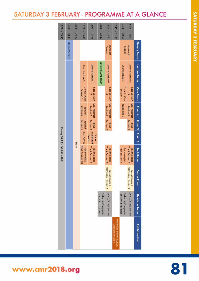
81www.cmr2018.org
SATU
RDA
Y 3 FEBRU
ARY
80
SATURDAY 3 FEBRUARY - PROGRAMME AT A GLANCE
Plenary RoomLecture Room
Case Room
Room 4
Room 5
Room 6
Tech RoomCourse Room
Hands on RoomExhibition Hall
8:00-
9:25Lecture Session 5
Case Session 5
Oral Abstract Session 5
Focus Session 5
Technologist Track Session 6
Special Course 3 3D Printing - Session 1
Level 2/3 case session
9:30-
10:15Short Lecture 4
Didactic Case Session 4
Quick Fire 3
Technologist Track Session 7
Pediatric/Congenital Session 1 (M
edis)
10:15-
11:00M
oderated ePoster 10,11,12 M
oderated eCase 4
11:00-
12:30Outreach Session
Lecture Session 6Case Session
6Oral Abstract
Session 6Focus
Session 6Technologist
Track Session 8Special Course 3
3D Printing - Session 2Level 2/3 case session
12:30-
13:30Satellite Sym
posium
Pediatric/Congenital Session 2 (Circle)
13:30-
14:55Lecture Session 7
Case Session 7
Oral Abstract Session 7
Focus Session 7
Best of m
oderated ePoster
Technologist Track Session 9
15:00-
15:45Short Lecture 5
Didactic Case Session 5
Special Session 6
Special Session 5
Best COTWTechnologist
Track Session 10
15:45-
16:30
16:30-
18:30Closing Plenary
18:30-
19:30
Outreach Session
Closing Drink (in Exhibition Hall)
Break

82
SATU
RDA
Y 3
FEB
RUA
RY SATURDAY 3 FEBRUARY
08:00 - 09:15 PLENARY ROOMOutreach Session
Outcomes Research and Imaging
Chairs: Jagat Narula, MD, PhD (The Mount Sinai Hospital)John Greenwood, PhD (University of Leeds)
08:00 - 08:12 Use and Abuse of CMR Perfusion ImagingAdam Timmis, (Bart’s Heart Centre, London, UK)
08:12 - 08:24 The clinical value and cost-effectiveness of diagnostic imagingFrancesco De Cobelli, MD, EBCR (San Raffaele University Hospital)
08:24 - 08:36 Outcomes research in cardiovascular imagingJeroen Bax, MD, PhD (FESC)
08:36 - 08:48 Is it Time to Include CMR as a Core Attribute in Classification of Cardiomyopathies?Jagat Narula, MD, PhD (The Mount Sinai Hospital)
08:48 - 09:00 How does CMR fit in international guidelines?Florian von Knobelsdorff, MD (Dept. of Cardiology, Clinic Agatharied, University of Munich)
09:00 - 09:15 Discussion
08:00 - 09:25 LECTURE ROOMLecture Session 5 A joint session with the European Society of Cardiac Radiology (ESCR)
MRA and 4D Flow
Chairs: Florian von Knobelsdorff, MD (Dept. of Cardiology, Clinic Agatharied, University of Munich)Matthias Gutberlet, MD (University of Leipzig – Heart Centre, Department of Diagnostic and Interventional Radiology)
08:00 - 08:15 MR Coronary Angiography - ready for clinical use in 2018?Matthias Stuber, PhD (University of Lausanne)
08:15 - 08:30 Is there a role for MRA in acute aortic syndrome?James Carr, MD (Northwestern University)
08:30 - 08:45 Improvements in 4D flow image acquisition and postprocessing - Update 2018Michael Markl, PhD (Northwestern University)
08:45 - 09:00 4D flow MR to better understand intracardiac hemodynamicsHåkan Arheden, MD PhD (Lund University, Skåne University Hospital, Department of Clinical Sciences Lund, Clinical Physiology, Lund, Sweden)
09:00 - 09:15 Thoracic aorta 4D flow - When can we use it for clinical decision making?Sergio Uribe, PhD, (Pontificia Universidad Católica de Chile)
09:15 - 09:25 Discussion
Level 1 Track

82 83www.cmr2018.org
SATU
RDA
Y 3 FEBRU
ARY
SATURDAY 3 FEBRUARY
08:00 - 09:30 CASE ROOMCase Session 5
Systemic and Inflammatory Disease
Chairs: Alexander Hirsch, MD, PhD (Erasmus Medical Center)Covadonga Fernández Golfin, Dr (Ramon y Cajal University Hospital.Madrid)
08:00 - 08:10 How can CMR help to identify infectious diseases affecting the heartMarly Uellendahl, MD, PhD (DASA São Paulo, Universidade Federal de São Paulo - Unifesp)
08:10 - 08:20 Imaging of acute myocarditis in patient with systemic lupus erythematosusAndreja Cercek Cerne, MD, PhD (University Medical Centre Ljubljana)
08:20 - 08:30 The classic cardiac involvment in Erdheim-Chester diseaseKlaus Schumacher, (State University of Campinas - UNICAMP)
08:30 - 08:40 Serial CMR Follow Up of Left Ventricular Pseudoaneurysm and Aneurysm In a Child With May-Thurner Syndrome.Ronak Naik, MD, FACC (University of Tennessee Health Science Center)
08:40 - 08:50 Cardiac sarcoidosis with features mimicking hypertrophic obstructive cardiomyopathyStacey Elangovan, MD (Hospital of the University of Pennsylvania)
08:50 - 09:00 Cardiac Amyloidosis with Normal Transthoracic EchoAdrián Löffler, MD (University of Virginia Health System)
09:00 - 09:10 Giant cell myocarditis: Cardiac MR findings and pathology correlation on endomyocardial biopsy and explanted heart.Mohamed Abdelrazek, (Ottawa University)
09:10- 09:20 Unknown systemic lupus erythematosus initially suspected by CMR.Lilia Sierra-Galan, MD, MCvT (American British Cowdray Medical Center)
09:20 - 09:30 Is something infiltrating the heart?Chetan Shenoy, M.D. (Cardiovascular Division, Department of Medicine, University of Minnesota)
08:00 - 09:25 ROOM 4Oral Abstract Session 5
Diagnosis of Ischemic Heart Disease and Perfusion
Chairs: Jose Rodriguez-Palomares, MD, PhD (Attendant Physician)Karolina Zareba, MD (The Ohio State University Wexner Medical Center)
08:00 - 08:12 Fully Quantitative Cardiac Magnetic Resonance Myocardial Perfusion Ready for Clinical Use: A comparison between Magnetic Resonance Imaging and Positron Emission TomographyHenrik Engblom, MD, PhD (Lund University, Skane University Hospital, Department of Clinical Sciences Lund, Clinical Physiology, Lund, Sweden)
Level 1 Track

84
SATU
RDA
Y 3
FEB
RUA
RY SATURDAY 3 FEBRUARY
08:12 - 08:24 The relative contributions of myocardial perfusion, blood volume and extracellular volume to native T1 and native T2 at rest and during adenosine stress in normal physiologyJannike Nickander, MD (Karolinska Institutet)
08:24 - 08:36 Prognostic and incremental value of individual components of a cardiovascular magnetic resonance examination in patients with suspected ischaemic heart disease: long-term follow-up of CE-MARCPeter Swoboda, PhD (University of Leeds)
08:36 - 08:48 Gender Difference in Response to Adenosine Stress Perfusion CMRLouise Brown, MBChB, BMedSc (University of Leeds)
08:48 - 09:00 Prognosis and Cardiac function Outcome in Spontaneous Coronary Artery Dissection; SCAD UK studyAhmed Abdelaty, MSc MRCP (University of Leicester, UK)
09:00 - 09:12 Ceroid-Induced Apoptosis of Siderophage-Derived Foam Cells Underlies the Perpetual Recruitment of Macrophages and Expansion of “Death Zone” in Hemorrhagic Myocardial InfarctionsIvan Cokic, MD (Cedars-Sinai Medical Center)
09:12 - 09:25 Gadolinium-free Cardiac MRI Stress T1-mapping accurately diagnoses and differentiates between obstructive epicardial coronary artery disease and microvascular dysfunctionAlexander Liu, MBBS BSc (University of Oxford)
08:00 - 09:25 ROOM 5Focus Session 5
Right Ventricle Evaluation
Chairs: Gabriela Guzman, MD,PhD (University Hospital La Paz)Monica Mukherjee, MD, MPH (Johns Hopkins University School of Medicine)
08:00 - 08:15 How to acquire, post-process and interpret right ventricle imagesStefan Zimmerman, MD (Johns Hopkins University School of Medicine)
08:15 - 08:25 Hemodynamic forces in the left and right ventricles of the human heart using 4D flow magnetic resonance imaging: phantom validation and reproducibilityJohannes Töger, PhD (Lund University, Skåne University Hospital)
08:25 - 08:35 Tissue-Based Determinants of Right Ventricular Dysfunction in Ischemic Mitral Regurgitation – Incremental Utility of Non-ischemic Fibrosis Beyond CMR Evidenced Ischemia and Infarction BurdenJiwon Kim, MD (Weill Cornell Medical College)
08:35 - 08:45 The relationship between pulmonary artery-right ventricle coupling and right ventricle remodeling in untreated pulmonary artery hypertension patientsYang Dong, BMsci (Cardiology Department West China Hospital Sichuan University)

84 85www.cmr2018.org
SATU
RDA
Y 3 FEBRU
ARY
SATURDAY 3 FEBRUARY
08:45 - 08:55 The Right Ventricle in Aortic Stenosis: RV Hypercontractility assessed by CMR is an Early Marker of Clinical Decompensation and Cardiac Recovery in AS with Normal Left Ventricular FunctionMarzia Rigolli, MBChB, MD (University of Oxford)
08:55 - 09:05 Prognostic Role of Cardiac Magnetic Resonance in Arrhythmogenic Right Ventricular Cardiomyopathy.Giovanni Aquaro (Fondazione Toscana Gabriele Monasterio, Pisa (Italy))
09:05 - 09:15 Validation of right ventricular cardiac magnetic resonance feature tracking in patients with CTEPH (chronic thromboembolic pulmonary hypertension) after PEA (pulmonary endarterectomy)Sabine Zitzmann, MD (Kerckhoff-Heart-Center, Department of Cardiology, Bad Nauheim, Germany)
09:15 - 09:25 Ventricular interaction at rest and during exercise in the pressure overloaded right heart: an exercise cardiac magnetic resonance study.Mathias Claeys, MD (KU Leuven / University Hospitals Leuven)
08:00 - 09:25 TECH ROOMTechnologist Track Session 6
Oncology
Chairs: Tracy Coulson, DCR(R) (InHealth Limited)Chris Lawton, BSc Hons / PGC (Bristol Heart Institute)
08:00 - 08:20 CMR in the oncology patientIwan Harries, MD (Bristol Heart Institute, Bristol NIHR Biomedical Research Centre (Cardiovascular Section), University of Bristol)
08:20 - 08:40 Intra cardiac and myocardial massesJohn Grizzard, MD (VCU Health Systems)
08:40 - 09:00 Extra cardiac massesHugo Marques (Hospital da Luz)
09:00 - 09:20 Radiation and cytotoxic induced cardiomyopathyStephen Cheung, MBBS, FRCR (Queen Mary Hospital)

86
SATU
RDA
Y 3
FEB
RUA
RY SATURDAY 3 FEBRUARY
08:00 - 09:25 COURSE ROOMSpecial Course 3
From 2D images to 3D models – Insights into the applications of 3D printing in cardiology - Session 1
Chairs: Silvia Schievano, PhD (UCL Institute of Cardiovascular Science & Great Ormond Street Hospital)Giovanni Biglino, BEng, PhD (Bristol Heart Institute, School of Clinical Sciences, University of Bristol)
08:00 - 10:15 HANDS ON ROOMLevel 2/3 case sessions
Pediatric/Congenital - Session 1
Chairs: Oliver Tann, MRCP, FRCR (Great Ormond Street Hospital) Emanuela Valsangiacomo Buechel (University Childre’s Hospital Zurich) Tobias Rutz (Division of Cardiology and Center of Cardiac Magnetic Resonance, Cardiovascular Department, University Hospital of Lausanne, CHUV, Switzerland.)
09:15 - 10:15 PLENARY ROOMOutreach Session
Ischemic Heart Disease
Chairs: Hein Verberne, (University of Amsterdam, Amsterdam, the Netherlands)Pierre Croisille, MD PhD (University of Lyon)
09:15 - 09:30 Functional testing versus anatomic imaging for initial diagnosis of CADRasha Al-Lamee, MB BS MA (Imperial College Healthcare NHS Trust)
09:30 - 09:45 Ischemia and viability to guide revascularizationAndrew Arai, MD (National Heart, Lung and Blood Institute, National Institutes of Health)
09:45 - 10:00 Hybrid ImagingPaul Knaapen, Paul (VUMC)
10:00 - 10:15 Discussion

86 87www.cmr2018.org
SATU
RDA
Y 3 FEBRU
ARY
SATURDAY 3 FEBRUARY
09:30 - 10:15 LECTURE ROOMShort Lecture 4 A joint session with the Asian Society of Cardiovascular Imaging (ASCI)
Function and Structural Analysis
Chairs: Christopher Nguyen, PhD (Cedars-Sinai Medical Center)Carmen Chan, FRCP (Queen Mary Hospital)
09:30 - 09:45 Left Ventricular Microstructure to Macrostructure: Which scale matters?Daniel Ennis, PhD (Department of Radiological Sciences, University of California, Los Angeles, CA, USA.)
09:45 - 10:00 Cardiac function: What more can we learn from strain?Frank Rademakers, MD, PhD (KU Leuven)
10:00 - 10:15 Feature Tracking: CINE is routine everywhere, is feature tracking ready for the clinic?Kan Hor, MD (Nationwide Children’s Hospital)
09:30 - 10:15 CASE ROOMDidactic Case Session 4
Congenital/Pediatrics
Chairs: Anne Marie Valente, MD (Harvard Medical School)Adam Dorfman, MD (University of Michigan)
09:30 - 09:53 CMR calculation of Qp/Qs guides managementAndrew Powell, M.D. (Harvard Medical School and Boston Children’s Hospital)Aurelio Secinaro, MD (Bambino Gesù Children’s Hospital and Research Institute, Rome. Italy)
09:53 - 10:15 Simple congenital heart defect - maybe not so simple after all?Dan Halpern, Director, Adult Congenital Heart Disease Program (New York University Langone Health)Sonya Babu-Narayan, FRCP PhD (Royal Brompton Hospital)
09:30 - 10:15 ROOM 4Quick Fire Session 3
Clinical
Chairs: Gabriela Liberato, MD (Sao Lucas Diagnostic Center)Akhil Narang, MD (University of Chicago)
09:30 - 09:33 Clinical feasibility of 2-minute aortic 4D flow MRI: initial experience at two centersEmilie Bollache, PhD (Northwestern University)
Level 1 Track
Level 1 Track

88
SATU
RDA
Y 3
FEB
RUA
RY SATURDAY 3 FEBRUARY
09:33 - 09:36 T1 mapping for the prediction of treatment response in AL amyloidosisRosario Perea Palazón, MD PhD (Hospital Clínic. Universitat de Barcelona)
09:36 - 09:39 Prognostic Utility of Blood Oxygen Level Dependent (BOLD) Cardiovascular Magnetic Resonance (CMR) imaging in Asymptomatic Chronic Kidney Disease (CKD) Patients with and without Diabetes Mellitus.Ranjit Shah, MBBS, FRACP (South Australian Health and Medical Research Institute, Adelaide, SA, Australia)
09:39 - 09:42 Diagnostic Performance of Fully Automated Pixel-wise Myocardial Blood Flow Maps of Stress and Rest CMR Perfusion in PatientsLi-Yueh Hsu, DSc (National Heart, Lung and Blood Institute, National Institutes of Health)
09:45 - 09:48 Phenotype development in Cardiac Fabry disease proceeds through four stages: a prospective 182-patient studySabrina Nordin, MRCP (Barts Heart Centre)
09:48 - 09:51 When criteria for ICD implantation in the primary prevention of sudden death among patients with hypertrophic cardiomyopathy don’t get along: an analysis of late gadolinium-enhancement and the European and American guidelinesPedro Freitas (Hospital de Santa Cruz)
09:51 - 09:54 Obesity paradox and myocardial injury by cardiac magnetic resonance imaging in ST-elevation myocardial infarctionGeorg Fuernau, MD (University Heart Center Lübeck)
09:54 - 09:57 Importance of operator training and rest perfusion on the diagnostic accuracy of stress perfusion CMRAdriana Villa, MD (King’s College London)
10:00 - 10:03 Diffuse interstitial fibrosis is associated with reduced myocardial strain in heart failure with preserved and reduced ejection fractionXiaodan Zhao, PhD (National Heart Centre Singapore)
10:03 - 10:06 Clinical Impact of Magnetic Resonance Imaging in Non-Approved Cardiac DevicesIbrahim Saeed, MD (Saint Luke’s Mid-America Heart Institute)
10:06 - 10:09 The Short-Term Prognosis Values of combined T1 Mapping and Feature Tracking by Cardiovascular Magnetic Resonance in Dilated CardiomyopathyHui Liu, M.D. (Guangdong General Hospital, Guangdong Academy of Medical Sciences)
10:09 - 10:12 Impact of aortic geometrical characteristics on abnormal flow pattern in the proximal descending aorta in Marfan patients: a 4D flow MRI studyAndrea Guala, (Hospital Universitari Vall d´Hebron. Vall d’Hebron Institut de Recerca. Universitat Autònoma de Barcelona)

88 89www.cmr2018.org
SATU
RDA
Y 3 FEBRU
ARY
SATURDAY 3 FEBRUARY
09:30 - 10:10 TECH ROOMTechnologist Track Session 7
Oral Abstracts
Chairs: Stephen Darty, BSRT (Duke University)Jennifer Bryant, PhD (National Heart Centre Singapore)
09:30 - 09:40 CMR in Children under General Anaesthesia: A Single Institutional ExperienceRick Wage, DCR (R) (Royal Brompton Hospital NHS trust)
09:40 - 09:50 Left Atrial Appendage Mass – Thrombus or Myxoma?Robert Biederman, MD, FACC, FAHA (Allegheny General Hospital)
09:50 - 10:00 The thinking technologist’s guide to investigating ventricular tachycardiaBao Ru Leong, BSc (National Heart Centre Singapore)
10:00 - 10:10 Live detection of left atrial appendage thrombi in cardiac amyloidosis: an emerging clinical problemSarah Anderson, (UCL Department of Cardiac MRI)
10:15 - 11:00 EXHIBITION HALL (EPOSTER STATION 1)Moderated ePoster Session 10
New Techniques in CMR
Chairs: Claudia Prieto, PhD (King’s College London)Raymond Kim, MD (Duke University Medical Center)
10:15 - 10:23 Diffusion tensor cardiovascular magnetic resonance assessment of recovered dilated cardiomyopathyZohya Khalique, MBBS (NIHR Cardiovascular Biomedical Research Unit, Royal Brompton Hospital)
10:23 - 10:31 Comprehensive 3D Cine Steady-state Free Precession and 3D Cine Phase Contrast Cardiovascular Magnetic Resonance ExaminationMehdi H. Moghari, PhD. (Harvard Medical School and Boston Children’s Hospital)
10:31 - 10:39 3D SASHA myocardial T1 mapping with high accuracy and improved precisionGiovanna Nordio, Biomedical Engineering (King’s College London)
10:39 - 10:47 Whole-heart k-t PCA accelerated multi-venc 4D Flow CMR - a study in 45 healthy volunteersJan Robert Kroeger, (University Hospital Cologne)
10:47 - 10:55 BART and Gadgetron Integration used for Real-Time Cardiac Cine ReconstructionMahamadou Diakite, PhD (National Institutes of Health)

90
SATU
RDA
Y 3
FEB
RUA
RY SATURDAY 3 FEBRUARY
10:15 - 11:00 EXHIBITION HALL (EPOSTER STATION 2)Moderated ePoster Session 11
Postprocesing and Workflow in CMR
Chairs: Stephen Cheung, MBBS, FRCR (Queen Mary Hospital)Martin Ugander, MD, PhD (Karolinska Institutet, and Karolinska University Hospital, Stockholm, Sweden)
10:15 - 10:23 A Quality Assurance Program for Standardizing T1-mapping for International Multicentre Studies - A Hypertrophic Cardiomyopathy (HCMR) Sub-StudyQiang Zhang, PhD (University of Oxford Centre for Clinical Magnetic Resonance Research)
10:23 - 10:31 Automatic Quantification of Pulse Wave Velocity: Application for population-based CMR studies.Rahil Shahzad, PhD (Department of Radiology, Leiden University Medical Center, Leiden, The Netherlands)
10:31 - 10:39 Physiological and clinical relevance of left atrial strain measured by feature-tracking magnetic resonance imaging in a large group of Chinese healthy volunteers and patientsWeihao Li, BMSci (Cardiology Division, West China Hospital, Sichuan University)
10:39 - 10:47 T1 and T2 mapping at rest and stress for ischemic and scar detection in coronary artery disease: external validation in an independent cohortElen Elen, MD, FIHA (National Cardiovascular Center Harapan Kita, Jakarta-Indonesia; Institute for Experimental and Translational Cardiovascular Imaging, University Hospital Frankfurt-Germany)
10:47 - 10:55 We don’t know how to measure wall thickness in HCM –Time for a guideline?Gabriella Captur, MD PhD MRCP MSc (Barts Heart Centre)
10:15 - 11:00 EXHIBITION HALL (EPOSTER STATION 3)Moderated ePoster Session 12
Vascular and Valvular MRI
Chairs: Kate Hanneman, MD (University of Toronto)Elie Mousseaux, MD, PhD (European Hospital Georges Pompidou APHP; INSERM U970 PARCC Paris descartes University)
10:15 - 10:23 Blood pressure reduction with six months of exercise training is mediated by changes in proximal but not distal aortic stiffness as assessed by CMRAnish Bhuva, MRCP MBBS (Barts Heart Centre and Institute of Cardiovascular Science, UCL)
10:23 - 10:31 Aortic wall motion is critical to accurately simulate fluid dynamics at the aortic archRamon Pons Cots, Grade in Chemical Engineering, Master in Chemical Engineering (Institut Químic de Sarrià (IQS))
90 91www.cmr2018.org
SATU
RDA
Y 3 FEBRU
ARY
SATURDAY 3 FEBRUARY
10:31 - 10:39 Automated 3D CMR aortic morphometry demonstrates the enhanced value of volume indices as compared to diameters in hypertension and aneurysmal aortic diseasesThomas Dietenbeck, PhD (Sorbonne Universités)
10:39 - 10:47 Does raphe in bicuspid aortic valve have an impact on flow dynamics in the ascending aorta?Aroa Ruiz-Muñoz, MSc (Hospital Universitari Vall d´Hebron. Vall d’Hebron Institut de Recerca. Universitat Autònoma de Barcelona)
10:47 - 10:55 Value of cardiac MRI for assessment of aneurysm of the ascending aorta in patients with aortic valve InsufficiencyEllianna Hoff (Sharon Regional Health System)
10:15 - 11:00 EXHIBITION HALL (ECASE STATION 4)Moderated eCase Session 4
Masses
Chairs: Gabriella Captur, MD PhD MRCP MSc (Barts Heart Centre)Jonathan Weinsaft, MD (Weill Cornell Medical Center)
10:15 - 10:23 Asymmetrical thickening of the left ventricular wallWooi Kok Lim, MD(USM), MRCPCH(UK) (Sarawak Heart Centre, Kota Samarahan, Sarawak, Malaysia)
10:23 - 10:31 Risk of Embolization of Left Atrial Myxoma associated with Carney’s ComplexBharath Sathya, MD (National Institutes of Health/NHLBI)
10:31 - 10:39 Unusual ‘tumor’ of the mitral valve presenting with multiple territory cardio-embolic brain infarctionJoseph Lembo, DO (Hartford Hospital)
10:39 - 10:47 Obstructive cardiac tumor with a capsule: fibroma or rhabdomyoma?Barbara Burkhardt, M.D. (Dept. of Pediatrics, UT Southwestern Medical Center)
10:47 - 10:55 Intracardiac Cyst: A Mistaken IdentityEileen Gajo, BS, MD (University of Chicago (NorthShore), Evanston Hospital)
11:00 - 11:45 PLENARY ROOMOutreach Session
Inflammatory Heart Disease - Multisystem Diseases
Chairs: Ntobeko Ntusi, MD DPhil (University of Cape Town and Groote Schuur Hospital)Theodoros Karamitsos, MD, PhD (1st Cardiology Department, Aristotle University of Thessaloniki, AHEPA Hospital)

92
SATU
RDA
Y 3
FEB
RUA
RY SATURDAY 3 FEBRUARY
11:00 - 11:12 Systemic vasculitis involvement of the heartIacopo Carbone, Medicine (Policlinico Umberto I, Sapienza University of Rome)
11:12 - 11:24 Myocardial manifestations and diagnosis of systemic sclerosisJoao Lima, MD, MBA (Johns Hopkins University)
11:24 - 11:36 How can CMR change outcomes in rheumatologic multisystem diseasesSophie Mavrogeni, MD, PhD (Onassis Cardiac Surgery Center)
11:36 - 11:45 Discussion
11:00 - 12:30 LECTURE ROOMLecture Session 6
New Techniques in Development
Chairs: Reza Nezafat, PhD (Department of Medicine (Cardiovascular Division) Beth Israel Deaconess Medical Center, Harvard Medical School)Timothy Albert, MD, FACC (Tanner Health System)
11:00 - 11:15 CMR auto-segmentation - When the circle drawing can stop?Arash Kheradvar, MD, PhD (University of California, Irvine)
11:15 - 11:30 CMR with Ferumoxytol - Where do we stand?Paul Finn, MD (Departments of Radiology, Medicine and Biomedical Physics, UCLA)
11:30 - 11:45 Non-Contrast Myocardial Scar Imaging: Promises and challengesMoriel Vandsburger, PhD (University of California, Berkeley)
11:45 - 12:00 Synthetic CMR and CMR FingerprintingNicole Seiberlich, PhD. (Case Western Reserve University)
12:00 - 12:15 Free breathing Multi-Dimensional CMRAnthony Christodoulou, PhD (Cedars-Sinai Medical Center)
12:15 - 12:30 Discussion
11:00 - 12:30 CASE ROOMCase Session 6
Pediatric and Congenital Heart Disease
Chairs: Willem Helbing, MD, PhD (Department of Pediatrics, division of Pediatric Cardiology, Erasmus Medical Center, Rotterdam, the Netherlands)Ruchira Garg, MD FACC FASE (Cedars-Sinai Medical Center)
11:00 - 11:10 4D Flow in Pediatric and Congenital Heart DiseaseMark Fogel, MD (Children’s Hospital of Philadelphia)
Level 1 Track

92 93www.cmr2018.org
SATU
RDA
Y 3 FEBRU
ARY
SATURDAY 3 FEBRUARY
11:10 - 11:20 Advances in prediction of interventional outcome in univentricular heartsPetter Frieberg, MD, MSc (Lund University, Department of Clinical Sciences Lund, Clinical Physiology, Skane University Hospital, Lund, Sweden)
11:20 - 11:30 3D “modeling” and “printing” based on MR and CT data in neonate with complex twisted heart: new frontier for clinical decision and optimal surgical approachAurelio Secinaro, MD (Bambino Gesù Children’s Hospital and Research Institute, Rome. Italy)
11:30 - 11:40 Regadenoson myocardial stress perfusion CMR in a 2-month-old with transposition of the great arteries status post arterial switch with left coronary obstructionJames Wilkinson, MD (Texas Children’s Hospital, Baylor College of Medicine)
11:40 - 11:50 Could CMR guide the surgical plan in patients with transposition of the great arteries?Mahmoud Shaaban, MBBCh, MSc. (Aswan Heart Center (Magdi Yacoub Foundation) - Cardiology department, Tanta University)
11:50 - 12:00 Optimizing 3D whole heart imaging in complex congenital heart disease: ferumoxytol and respiratory suspensionWilson King, MD (Saint Louis University)
12:00 - 12:10 A bump in the road - what is this arch?Nilanjana Misra, MBBS, FAAP (Cohen Children’s Medical Center of NY)
12:10 - 12:20 Utilization of 4D Flow for evaluation of adult congenital heart disease: a case of congenital pulmonary atresia status post repairMelany Atkins, MD (Fairfax Radiological Consultants, Inova Fairfax Hospital)
12:20 - 12:30 What cardiovascular magnetic resonance adds in congenital heart diseaseRachel Wald, MD FRCPC (Joint Department of Medical Imaging)
11:00 - 12:30 ROOM 4Oral Abstract Session 6
CMR and Arrhythmias
Chairs: Esther Pérez-David, MD PhD (Hospital Gregorio Marañón, Madrid, Spain)Kanishka Ratnayaka, MD (Rady Children’s Hospital)
11:00 - 11:12 CMR-derived circumferential strain measures and the risk of ventricular arrhythmia in patients with prior myocardial infarction and implantable cardioverter defibrillatorElisabeth Paiman, MD (Department of Radiology, Leiden University Medical Center)
11:12 - 11:24 Risk Stratification of Patients with Apparently Idiopathic Premature Ventricular Contractions: Data from a Multicenter International Cardiac Magnetic Resonance StudyGaetano Nucifora, MD, PhD (University Hospital of South Manchester)
11:24 - 11:36 Recovery of left ventricular function with CRT. Is it enough to protect from arrhythmic events?Adelina Doltra, MD, PhD (Hospital Clínic de Barcelona)

94
SATU
RDA
Y 3
FEB
RUA
RY SATURDAY 3 FEBRUARY
11:36 - 11:48 MRI DENSE Strain Predicts Long-Term Survival and ICD Therapies with CRT DefibrillatorsKenneth Bilchick, MD, MS (University of Virginia Health System)
11:48 - 12:00 Prevalence of myocardial scar and myocardial inflammation in patients with sustained and non-sustained ventricular arrhythmias: A cardiac magnetic resonance and 18F-FDG cardiac PET studyKalie Kebed, MD (University of Chicago)
12:00 - 12:12 Atrial Scar on CMR to Predict Pulmonary Vein Reconnection after Catheter Ablation for Paroxysmal Atrial FibrillationHubert Cochet, (IHU LIRYC - CHU / Université de Bordeaux)
12:12 - 12:25 Mind the Gap: Insights into Recurrent Atrial Fibrillation Post Cryoablation with Novel Cylindrical Navigator and 3-Tesla ImagingIbrahim Saeed, MD (Saint Luke’s Mid-America Heart Institute)
11:00 - 12:30 ROOM 5Focus Session 6
Coronary Imaging: The Last Frontier
Chairs: Qi Yang, PhD (Cedars-Sinai Medical Center)Rene Botnar, PhD (King’s College London)
11:00 - 11:20 Imaging the coronaries with CMR: where do we stand?Debiao Li, PhD (Cedars-Sinai Medical Center)
11:20 - 11:30 Radial Fast Interrupted Steady-State (FISS) Cine Imaging for the Evaluation of Heart Valves and Coronary ArteriesIoannis Koktzoglou, PhD (NorthShore University HealthSystem)
11:30 - 11:40 3D whole-heart free-breathing coronary lumen and vessel wall imaging with interleaved T2prep-IRGiorgia Milotta, MSc (King’s College London)
11:40 - 11:50 Respiratory Self-Gated Stack-of-Stars 3D Cine MRI for the Proximal Coronary Arteries: Initial Steps towards Volumetric Endothelial Function AssessmentGabriele Bonanno, PhD (Division of Cardiology and Division of MR Research, The Johns Hopkins University School of Medicine, Baltimore, MD, United States)
11:50 - 12:00 T2-Prepared Multidimensional Outer Volume Suppression for Coronary ImagingDavid Zeng, MS (Stanford University)
12:00 - 12:10 Subclinical Coronary Atherosclerosis in Young Women: Magnetic Resonance Coronary Vessel Wall Imaging as a Predictor of Coronary Plaque BurdenKhaled Abd-Elmoniem, PhD, MHS (National Institutes of Health)
Level 1 Track

94 95www.cmr2018.org
SATU
RDA
Y 3 FEBRU
ARY
SATURDAY 3 FEBRUARY
12:10 - 12:20 Association of regional and systemic adipose tissue with coronary endothelial dysfunction in patients with HIVSahar Soleimanifard, PhD (The Johns Hopkins Univeristy School of Medicine)
12:20 - 12:30 Motion-resolved free-breathing coronary MRA in heart transplant recipients at 3TJessica Bastiaansen, PhD (Department of Radiology, University Hospital (CHUV) and University of Lausanne (UNIL))
11:00 - 12:30 TECH ROOMTechnologist Track Session 8A joint session with the Heart Rhythm Society (HRS)
Electrophysiology
Chairs: Michelle Walkdon, Bsc (hons) Diagnostic Radiography and PG Dip Radiology (MRI)Nassir Marrouche, Nassir Marrouche (University of Utah Health)
11:00 - 11:20 CMR Assessment and Guidance in Atrial FibrillationNassir Marrouche, Nassir Marrouche (University of Utah Health)
11:20 - 11:40 Non-contrast methods to evaluate the myocardium in tachyarrhythmiasRohan Wijesurendra, MBBS (University of Oxford)
11:40 - 12:00 Scanning the arrhythmic patient - ICD’s and PPM’sKanae Mukai, MD (Salinas Valley Memorial Health System)
12:00 - 12:20 CMR for patients undergoing CRTAnish Bhuva, MRCP MBBS (Barts Heart Centre and Institute of Cardiovascular Science, UCL.)
11:00 - 12:30 COURSE ROOMSpecial Course 3
From 2D images to 3D models – Insights into the applications of 3D printing in cardiology - Session 2
Chairs: Giovanni Biglino, BEng, PhD (Bristol Heart Institute, School of Clinical Sciences, University of Bristol)Silvia Schievano, PhD (UCL Institute of Cardiovascular Science & Great Ormond Street Hospital)
11:00 - 13:15 HANDS ON ROOMLevel 2/3 case sessions
Pediatric/Congenital - Session 2
Chairs: Oliver Tann, MRCP, FRCR (Great Ormond Street Hospital)

96
SATU
RDA
Y 3
FEB
RUA
RY SATURDAY 3 FEBRUARY
11:45 - 12:30 PLENARY ROOMOutreach Session
Population Based Studies and Big Data
Chairs: Raymond Kwong, MD, MPH (Brigham and Womens Hospital)Christopher Kramer, MD (University of Virginia)
11:45 - 11:57 Big Data Analytics at the Cardiac Atlas ProjectAvan Suinesiaputra, PhD (The University of Auckland, New Zealand)
11:57 - 12:09 What we learned so far from the UK BiobankSteffen Petersen, MD DPhil MPH FRCP (Queen Mary University of London)
12:09 - 12:21 Imaging databases and clinical trial remote collaborationsRobert Judd, PhD (Duke University Medical center)
12:21 - 12:30 Discussion
12:30 - 13:30 LECTURE ROOMGuerbet Satellite Symposium
Updates on Gadolinium Based Contrast Agents Usage in CMR
Chairs: Michael Campbell, MD, MHA (Duke University)Tim Leiner, MD, PhD (Utrecht University Medical Center)
12:30 - 12:42 Three decades of GBCA clinical experienceTim Leiner, MD, PhD (Utrecht University Medical Center)
12:42 - 12:54 EMA Recommendations on GBCA SafetyAlex Rovira, MD (Hospital Universitari Vall d’Hebron)
12:54 - 13:06 NIH Perspective on GBCA SafetyAshkan Malayeri, MD (NIH)
13:06 - 13:18 Gadolinium-based contrast agents in pediatric MRIMichael Campbell, MD, MHA (Duke University)
13:18 - 13:30 Discussion
13:30 - 14:55 LECTURE ROOMLecture Session 7
Imaging in Cardio-Oncology
Chairs: David Sosnovik, MD (Harvard Medical School - Massachusetts General Hospital)W. Patricia Bandettini, MD (National Heart, Lung and Blood Institute, National Institutes of Health)
13:30 - 13:45 Clinical perspective and scope of problemSanjeev Francis, MD (Tufts University School of Medicine)
Level 1 Track
96 97www.cmr2018.org
SATU
RDA
Y 3 FEBRU
ARY
SATURDAY 3 FEBRUARY
13:45 - 14:00 Guidelines: Echo and MRIJuan Carlos Plana (Baylor College of Medicine)
14:00 - 14:15 MRI in clinical Cardio-OncologyAna Barac, MD, PhD (Medstar Heart and Vascular Institute)
14:15 - 14:30 Reseach and experimental approachesGreg Hundley, MD (Wake Forest University Health Sciences)
14:30 - 14:45 Molecular imaging in drug developmentDavid Sosnovik, MD (Harvard Medical School - Massachusetts General Hospital)
14:45 - 14:55 Discussion
13:30 - 15:00 CASE ROOMCase Session 7
Cardiac Masses
Chairs: Barbara Srichai, MD (Georgetown University School of Medicine)Massimo Lombardi, Cardiologist and Radiologist (II.R.C.C.S., Policlinico San Donato)
13:30 - 13:40 A recommended CMR protocol for assessment of cardiac massesJonathan Weinsaft, MD (Weill Cornell Medical Center)
13:40 - 13:50 Intracardiac bronchogenic cystPatrick Norton, MD (University of Virginia)
13:50 - 14:00 Primary cardiac lymphoma. A CMR clue to recognize it.Bianca Granados-Pinedo, MD (American British Cowdray Medical Center)
14:00 - 14:10 Growing from within: dyspnea due to progressive stenosis of multiple heart vesselsFilipa Valente, MD (Hospital Universitari Vall d´Hebron. Vall d’Hebron Institut de Recerca. Universitat Autònoma de Barcelona)
14:10 - 14:20 All left ventricular hypertrophies are not created equal: a case of left ventricular metastasis from breast cancer.Alessandra Scatteia, MD (Division of Cardiology, Ospedale Medico-Chirurgico Accreditato Villa dei Fiori, Acerra, Naples, Italy)
14:20 - 14:30 When the heart knows first: high grade neuroendocrine carcinoma of the heart.Jitka Starekova, MD (University Medical Center Hamburg-Eppendorf (UKE))
14:30 - 14:40 Approaching to the Etiology of a Right Ventricle Mass with Cardiac Magnetic ResonanceKatia Menacho, MD (Barts Heart Centre)
14:40 - 14:50 Comprehensive cardiovascular magnetic resonance for isolated cardiac cystAnna Giulia Pavon, Cardio-Thoracic Vascular Department, San Raffaele Hospital, Milan, Italy (Cardiac MR Center-Cardiologiy Unit-University Hospital-CHUV-Lausanne, Switzerland)
14:50 - 15:00 A mass was found during echocardiography: can CMR help?Ron Jacob, MD, FACC, FASE, FSCCT (PENN Medicine)
Level 1 Track
98
SATU
RDA
Y 3
FEB
RUA
RY SATURDAY 3 FEBRUARY
13:30 - 14:55 ROOM 4Oral Abstract Session 7A joint session with the Society for Magnetic Resonance Angiography (SMRA)
CMR in Valvular and Vascular Diseases
Chairs: Michael Markl, PhD (Northwestern University)Vassilis Vassiliou, MBBS, PhD (Norwich Medical School and Royal Brompton Hospital)
13:30 - 13:42 Regional wall shear stress analysis in the aortic arch and its relation to dilation in bicuspid aortic valve diseaseAroa Ruiz-Muñoz, MSc (Hospital Universitari Vall d´Hebron. Vall d’Hebron Institut de Recerca. Universitat Autònoma de Barcelona)
13:42 - 13:54 Hemodynamic flow changes in bicuspid aortic valve aortopathy are stable over timeMalenka Bissell, Md, DPhil (Oxford University)
13:54 - 14:06 Progression of Myocardial Fibrosis in Aortic Stenosis: A Multicentre Cardiac Magnetic Resonance StudyRussell Everett, BSc MD (University of Edinburgh)
14:06 - 14:18 Correlation of 4D flow MRI aortic wall shear stress to medial elastin fiber thinning in patients with a bicuspid aortic valveEmilie Bollache, PhD (Northwestern University)
14:18 - 14:30 CMR Longitudinal Strain Analysis in Aortic StenosisNick Spath, BSc MD (University of Edinburgh)
14:30 - 14:42 Real-time flow using a golden-angle spiral acquisitionRajiv Ramasawmy, PhD (National Institutes of Health)
14:42 - 14:55 In Vivo Quantification of Aortic Stiffness in Abdominal Aortic Aneurysm Patients: A Longitudinal StudyHuiming Dong, MS (The Ohio State University Wexner Medical Center)
13:30 - 14:55 ROOM 5Focus Session 7
Added Value of CMR in a Multimodality Imaging Scenario
Chairs: Edward Martin, MD (Oklahoma Heart Institute)Yu-Cheng Cheng, MD (West China Hospital ,Sichuan University)
13:30 - 13:45 How to succesfully implement a CMR programChristoph Tillmanns, MD (Diagnostikum Berlin)
13:45 - 13:55 MRI DENSE Strain Imaging Provides a Superior Assessment for CRT Response Compared with 3D EchocardiographyKenneth Bilchick, MD, MS (University of Virginia Health System)

98 99www.cmr2018.org
SATU
RDA
Y 3 FEBRU
ARY
SATURDAY 3 FEBRUARY
13:55 - 14:05 Cardiac Magnetic Resonance Imaging and Cardiac Computed Tomography versus Transesophageal Echocardiography for the Diagnosis of Left Atrial Appendage Thrombus: A Systematic Review and Meta-analysisTasnim Vira, MD (Sunnybrook Health Sciences Centre)
14:05 - 14:15 Cardiovascular Magnetic Resonance Imaging to guide Transcatheter Aortic Valve Replacement: A Comparison with Computed TomographyAgnes Mayr, MD (University Clinic of Radiology, Medical University of Innsbruck)
14:15 - 14:25 Progression of aneurysm of the ascending aorta: A CMR perspectiveSebastian Boland, MBA (Sharon Regional Health System)
14:25 - 14:35 The diagnostic value of Adenosine MR following a positive Coronary Calcium Score as a gatekeeper of invasive coronary angiography in 644 patients with stable chest pain.Dorine Rijlaarsdam-Hermsen, (HMC Bronovo, Erasmus mc)
14:35 - 14:45 Is there a role for cardiac MRI stress imaging after an abnormal SPECT study?Rohith Raghavendran, BS (Sharon Regional Health System)
14:45 - 14:55 CMR Provides Superior Accuracy and Reduced Heterogeneity for Detecting Coronary Artery Disease: A Systematic Review and Meta-Analysis of Stress Imaging Methods using HSROC Methods in 23,051 Patients.Lilia Sierra-Galan, MD, MCvT (American British Cowdray Medical Center)
13:30 - 14:55 ROOM 6Best Moderated ePoster Session
Chairs: Sebastian Kozerke, PhD (ETH Zurich)Jeanette Schulz-Menger, MD (Charité University Medicine Berlin and Helios Clinics)Robert Biederman, MD, FACC, FAHA (Allegheny General Hospital)
13:30 - 14:55 TECH ROOMTechnologist Track Session 9
Congenital 1
Chairs: Stephen Darty, BSRT (Duke University)Wendy Norman, DCR(R), DRI (Institute of Cardiovascular Science, University College London)
13:30 - 13:50 Transposition of the Great ArteriesGergely Szantho, MD (University Hospital of Wales - Cardiff, Cardio-Thoracic Department and Hospitals Bristol - Heart Institute)
13:50 - 14:10 Congenital and surgical shuntsGiuseppe Muscogiuri, MD (Bambino Gesù Children’s Hospital and Research Institute, Rome. Italy)

100
SATU
RDA
Y 3
FEB
RUA
RY SATURDAY 3 FEBRUARY
14:10 - 14:30 Tetralogy of FallotAnne Marie Valente, MD (Harvard Medical School)
14:30 - 14:50 The Single Ventricle HeartJason Johnson, MD MHS (LeBonheur Children’s Hospital, University of Tennessee)
15:00 - 15:45 LECTURE ROOMShort Lecture 5
MRI viability How and Why
Chairs: Albert van Rossum, Prof. dr. (VU University Medical Center)Yuchi Han, MD, MMSc (University of Pennsylvania)
15:00 - 15:15 Impact of viability to guide CTO and select CRTDipan Shah, MD, FACC (Houston Methodist DeBakey Heart & Vascular Center)
15:15 - 15:30 LGE is the optimal method for viabilityRaymond Kim, MD (Duke University Medical Center)
15:30 - 15:45 Dobutamine Stress should be added to all viability studiesGrigorios Korosoglou, (GRN Academic Teaching Hospital Weinheim)
15:00 - 15:45 CASE ROOMDidactic Case Session 5
Ischemic vs. Nonischemic Cardiomyopathy - Will CMR Provide the Best Answer?
Chairs: Orlando Simonetti, PhD (The Ohio State University)Pankaj Garg, MD, PhD (University of Leeds, UK)
15:00 - 15:23 Severe LV impairment? CMR will answer the questionLouise Brown, MBChB, BMedSc (University of Leeds)John Paul Carpenter, MD(Res) FRCP (Poole Hospital NHS Foundation Trust)
15:23 - 15:45 Early cardiomyopathy detection: CMR evaluation of anatomy vs functionDavid Bluemke, MD, PhD (University of Wisconsin School of Medicine and Public Health)Lindsay Griffin, MD (University of Wisconsin)
Level 1 Track
Level 1 Track

100 101www.cmr2018.org
SATU
RDA
Y 3 FEBRU
ARY
SATURDAY 3 FEBRUARY
15:00 - 15:45 ROOM 5Special Session 5
Tissue Characterization: Discuss with the Experts
Chairs: Matthias Friedrich, MD (McGill University Health Centre)Jeanette Schulz-Menger, MD (Charité University Medicine Berlin and Helios Clinics)
15:00 - 15:45 Sequences, protocols and post-processing: what do you use in your routine and why?Stefan Piechnik, PhD, MScEE (University of Oxford)
When to use native versus post-contrast T1 versus ECV?Martin Ugander, MD, PhD (Karolinska Institutet, and Karolinska University Hospital, Stockholm, Sweden)
Is T1 and T2 mapping ready for routine clinical use?Amit Patel, MD (University of Chicago)
15:00 - 15:45 ROOM 4Special Session 6
New Techniques Ready For Clinical Application
Chairs: Kelvin Chow, PhD (Siemens Medical Solutions USA, Inc.)John Heitner, MD (New York Presbyterian-Brooklyn Methodist Hospital)
15:00 - 15:15 T1 and T2 mapping, 3D vs. 2D acquisition, validation, strengths and weaknessesPeng Hu, PhD (UCLA)
15:15 - 15:30 Myocardial deformation, feature tracking, reproducibility, applicationsSebastian Kelle, MD, PhD (German Heart Center Berlin)
15:30 - 15:45 Fat-water separated imaging, mDixon vs. conventional fat imaging, clinical applications, accuracyJames Carr, MD (Northwestern University)
15:00 - 15:45 ROOM 6Best of SCMR Cases of the Week
Chairs: Vikas Rathi, MD (Bon Secours Health System)Michael Campbell, MD, MHA (Duke University)
15:00 - 15:08 Invisible Cardiac LesionAnna Lisa Crowley, MD (Duke University)

102
SATU
RDA
Y 3
FEB
RUA
RY SATURDAY 3 FEBRUARY
15:08 - 15:16 Intramyocardial hematoma mimicking hypertrophic cardiomyopathy-evaluation by multimodality imagingArun Dahiya, Griffith University, Queensland (RBWH and Logan Hospital , Queensland, Australia)
15:16 - 15:24 Unexpected Jeopardy: CMR in atypical chest pain but chronic aortic dissection with cardiac schwannomaRunyawan Chotenimitkhun, MD, MsC
15:24 - 15:32 Lower extremity anasarca: long term complication of PAPVRJenifer Geradin, MD (Emory University, Atlanta Georgia USA)
15:32 - 15:40 Congenital malformation of the coronary drainage system and trabecular architectureStefania Rosmini, MD, PhD (Barts Heart Centre)
15:00 - 15:45 TECH ROOMTechnologist Track Session 10
Congenital 2
Chairs: Wendy Norman, DCR(R), DRI (Institute of Cardiovascular Science, University College London)Stephen Darty, BSRT (Duke University)
15:00 - 15:10 Coronary AnomaliesBen Holloway, MBChB FRCR (BSCI/BSCCT)
15:10 - 15:20 Ebstein’s AnomalyZahra Alizadeh Sani, (MD,Associate professor of cardiology ,Rajaie Cardiovascular Medical &Research Centere,Tehran,Iran)
15:20 - 15:30 Congenital aortic arch anomaliesBobby Agrawal, FRCS FRCR (Papworth Hospital)
15:30 - 15:40 Imaging the Pediatric Patient in the MRI environment: sedation and other strategiesAhmed Kharabish, MSc, PhD (Bad Krozingen Heart Center, Freiburg University, Germany)
15:40 - 15:45 Closing RemarksStephen Darty, BSRT (Duke University)

102 103www.cmr2018.org
SATU
RDA
Y 3 FEBRU
ARY
SATURDAY 3 FEBRUARY
16:30 - 18:35 PLENARY ROOMClosing Plenary
Chairs: Matthias Stuber, PhD (University of Lausanne) Steffen Petersen, MD DPhil MPH FRCP (Queen Mary University of London)
16:30 - 16:45 Keynote Lecture EACVI Section CMR: Myocardial plasticity, adaptation and maladaptation: insights from CMRJames Moon, MD (UCL)
16:45 - 17:30 CMR OSCARS - The Image CompetitionLilia Sierra-Galan, MD, MCvT (American British Cowdray Medical Center)Tomas Lapinskas, MD (Medical Academy, Lithuanian University of Health Sciences)Rajesh Puranik, A/Prof (University of Sydney)James Carr, MD (Northwestern University)Ron Jacob, Ron Jacob MD, FACC, FASE, FSCCT (PENN Medicine)Ntobeko Ntusi, MD DPhil (University of Cape Town and Groote Schuur Hospital)Anne Marie Valente, MD (Harvard Medical School)Juliana Serafim, MD (Hospital Pró-Cardíaco)Mark Westwood, MBBS MD FRCP FESC (Barts)Karen Ordovas, MD, MAS (University of California San Francisco)
17:30 - 17:45 Highlights from CMR2018Juliano Fernandes, MD, PhD (Jose Michel Kalaf Research Institute)Robin Nijveldt, MD, PhD (Radboudumc)
Chairs: Chiara Bucciarelli-Ducci, MD, PhD (Bristol Heart Institute, Bristol NIHR Biomedical Research Centre (Cardiovascular Section), University of Bristol) Matthias Friedrich, MD (McGill University Health Centre)
17:45 - 18:20 Awards Presentations and Incoming SCMR President
18:20 - 18:30 Closing Remarks Chiara Bucciarelli-Ducci, MD, PhD (Bristol Heart Institute, Bristol NIHR Biomedical Research Centre (Cardiovascular Section), University of Bristol)
18:35 - 19:00Farewell Drink (in the Exhibition Hall)

104
FRIDAY 2 FEBRUARY
10:15 - 11:00 EXHIBITION HALLPoster Session 1
P001 Impact of Diffuse Myocardial Fibrosis on Left Ventricular Dysfunction in Patient with Atrial Fibrillation Evaluation with Cardiovascular Magnetic Resonance T1 mappingLei Zhao (Beijing Anzhen Hospital, Capital Medical University)
P002 Metallic Implants in the MRI Environment; Effect on Loops, Stimulators and Retained Pacemaker/ICD LeadsRobert Biederman, MD, FACC, FAHA (Allegheny General Hospital)
P003 Easy 2D measurements to predict left atrial sphericity in patients with paroxysmal atrial fibrillationAlbert Teis (Hospital Universitari Germans Trias. Badalona. Bacelona. Spain.)
P004 Left atrial blood flow dynamics and hemostasis after electrical cardioversion of atrial fibrillationMerih Cibis, PhD (Linkoping University)
P005 Real-time cardiac cine using radial bSSFP sequence with trajectory auto-correctionGuoxi Xie, PhD (School of Basic Science, Guangzhou Medical University, Guangzhou, China; Shenzhen Institutes of Advanced Technology, Chinese Academy of Sciences)
P006 Substantial prevalence of a type II left ventricular contraction pattern by feature tracking CMR in patients with non-specific intraventricular conduction delayDaniel Loewenstein (Karolinska Institutet)
P007 Image-based patient-specific simulations of atrial flow can predict regions of blood stasis in sinus rhythm and atrial fibrillationDesmond Dillon-Murphy, PhD MSc (Kings College London)
P008 Feasibility of noncontrast T1 and T2 parametric mapping in assessment of acute ventricular abla-tion lesions in childrenLaura Olivieri, M.D. (Children’s National Medical Center)
P009 Energy Loss and Kinetic Energy of the Left Ventricle in Patients with Paroxysmal Atrial FibrillationJulio Garcia, PhD (University of Calgary)
P010 Atrial fibrosis detected using LGE-MRI is a progression phenomenon accelerated by atrial fibrilla-tion: Comparison between patients without and with Atrial fibrillation.Christian Mahnkopf, MD (Klinikum Coburg, Dept. of Cardiology)
P011 Evaluation of Estimation Methods for Missing Premature Ventricular Contractile Beats During Re-al-Time CMR Slice AcquisitionSaharsh Dass, BS (University of Pennsylvania)
P012 Clinical and morpho-functional evaluation of young athletes with exercise-induced ventricular arr-hythmias: a study performed by cardiac magnetic resonanceAlberto Cipriani, MD (Department of Cardiac, Thoracic and Vascular Sciences, University of Padova, Italy)

104 105www.cmr2018.org
FRIDAY 2 FEBRUARY
P014 Combined evaluation of aortic pulse wave velocity, epicardial fat volume, left ventricular strain and fibrosis in patients with hypertension and diabetes mellitusRami Homsi (University Hospital Bonn, Department of Radiology)
P015 Diagnostic Utility of Vasodilator Cardiac Magnetic Resonance Imaging in Patients with Reduced Left Ventricular Ejection FractionAkhil Narang, MD (University of Chicago)
P016 Left ventricular Performance in Takotsubo Syndrome and Aborted Myocardial Infarction: prelimi-nary data of a prospective multicenter CMR studyNadine Abanador-Kamper (Department of Cardiology, HELIOS Medical Center Wuppertal, University Hospital Witten/Herdecke, Wuppertal, Germany; Center for Clinical Medicine Witten/Herdecke Uni-versity Faculty of Health, Wuppertal, Germany)
P017 How cardiac magnetic resonance imaging could help the differential diagnosis in MINOCA? - Single center data of a 5-year periodHajnalka Vágó (Heart and Vascular Center of Semmelweis University)
P018 Utilization of Superparamagnetic Iron Oxide Contrast Agents for the Determination of Myocardial Adipose Inflammation Using T2 Star as a Marker for Coronary Artery Disease in HIV Positive Pa-tientsKorey Haddox, D.O. (University of Cincinnati Medical Center)
P019 Machine Learning of 3D Myocardial Deformation from Cine MRI for the Identification of Segmental Viability in Patients with Ischemic CardiomyopathyAlessandro Satriano, PhD. (Stephenson Cardiac Imaging Centre, Libin Cardiovascular Institute of Al-berta, University of Calgary)
P020 Regional function after acute myocardial infarction: strain analysis is superior to wall thickening in detecting microvascular injuryHenk Everaars (VU univeristy medical center)
P021 When Ordered by LVEF, Internal and External Energy Expenditure Exhibit a Strong Periodicity in the Cardiovascular System; a New Paradigm?Robert Biederman, MD, FACC, FAHA (Allegheny General Hospital)
P022 Characterization of Ischemic Cardiomyopathy with Water-Fat Separation via End-to-end Deep Lear-ningJames Goldfarb, PhD (St Francis Hospital)
P023 Serum Uric Acid Level During Acute MI Modifies the Therapeutic Effects of High Dose Omega-3 Fatty Acids on Left Ventricular RemodelingKyoichi Kaneko, MD, PhD (Brigham and Women’s Hospital and Showa University)
P024 Cardiac magnetic resonance imaging to differentiate acute NSTEMI from acute ACS-like myocarditisEnver Tahir, MD (University Medical Center Hamburg-Eppendorf)
P025 Dobutamine stress CMR out-performs LGE and adenosine in predicting CTO-PCI response (CARIS-MA_CTO preliminary results)Silvia Pica, MD (IRCCS Policlinico San Donato, Multimodality Imaging Center, Milan)

106
FRIDAY 2 FEBRUARY
P026 Characterization of T2 and T2* relaxation and strain in disease progression post acute myocardial infarctionNilesh Ghugre, PhD (Sunnybrook Research Institute, University of Toronto)
P027 Quantification of the area-at-risk by post-contrast T1 mapping and detection of intramyocardial hemorrhage by post-contrast T2* mapping in reperfused STEMI patientsNazia Chowdhury, BSc (Cardiovascular and Metabolic Disorders Program, Duke-National University Singapore Medical School: National Heart Research Institute Singapore, National Heart Centre, Singapore)
P028 Prevalence of inducible ischaemia across the strata of ejection fraction and according to the pattern of late gadolinium enhancementPamela Brown, MBBS (University Hospital South Manchester)
P029 Spin-Spin Dephasing on Cine-CMR for Assessment of Blood Pool Stasis – A Novel Physiologic Marker for Post-Myocardial Infarction Left Ventricular ThrombusNeil Mehta, MD (Weill Cornell Medical College)
P030 Relation of low-density lipoprotein cholesterol with microvascular injury and clinical outcome in revascularized ST-elevation myocardial infarctionMartin Reindl (University Clinic of Internal Medicine III, Cardiology and Angiology, Medical University of Innsbruck)
P031 Impact of atrial fibrillation during ST-elevation myocardial infarction on infarct characteristics and prognosisSebastian Reinstadler, MD (University Clinic of Internal Medicine III, Cardiology and Angiology, Medical University of Innsbruck)
P032 During vasodilatory stress, which hemodynamic indicator correlates best with myocardial perfusion as measured by first pass contrast enhanced cardiac MRI.Richard Coulden, MB BS (University of Alberta Hospital)
P033 Inability of Iron-Loaded Macrophages to Switch from Glycolytic to Oxidative Phenotype Promotes Foam Cell Formation and Fat Deposition in Hemorrhagic MI: Early Findings from a PET/MRI Study with Histological ValidationIvan Cokic, MD (Cedars-Sinai Medical Center)
P034 Impact of global and local left ventricular remodeling in severe ischemic mitral regurgitationFausto Pizzino (Fondazione Toscana Gabriele Monasterio and Scuola Superiore Sant’Anna di Pisa)
P035 Global Longitudinal Strain is reduced during maximum myocardial hyperaemia in patients with significant coronary artery disease.Pankaj Garg, MD, PhD (University of Leeds, UK)
P036 Impact of a breathing-maneuver stimulus on myocardial oxygenation and left ventricular strain in patients with coronary artery diseaseBarbara Spicher (Inselspital Bern, Anesthesia)
P037 Infarct healing during long-term follow-up after ST-elevation myocardial infarctionHans-Josef Feistritzer, MD (University Clinic of Internal Medicine III, Cardiology and Angiology, Medical University of Innsbruck)

106 107www.cmr2018.org
FRIDAY 2 FEBRUARY
P038 Feasibility Study of a Single Breath-hold, 3D mDIXON Pulse Sequence for Late Gadolinium Enhancement Imaging of Ischaemic ScarJames Foley, MBChB BSc (Hons) (University of Leeds)
P039 Safety of Regadenoson Stress Cardiac Magnetic Resonance Imaging in Heart Transplant RecipientsFelipe Kazmirczak, MD (University of Minnesota)
P040 Fluid-dynamic changes in post ischemic dilated cardiomyopathy before and after surgical ventricular restoration: an integrated methodological approach based on CMR 4D flowAntonia Camporeale, MD (IRCCS Policlinico San DOnato)
P041 Inter-study Reproducibility of Strain Assessed by Displacement Encoding with Stimulated Echoes (DENSE) and Feature-tracking in Patients post-Myocardial Infarction.Kenneth Mangion, MD MRCP (University of Glasgow)
P042 Quantification of Late Gadolinium Enhancement with Contrast-enhancedCardiovascular MR Imaging for Coronary Artery Chronic Total OcclusionWei Yu, MD,PhD (Deparment of Radiology, Anzhen Hospital, Capital Medical University)
P043 Diffuse myocardial fibrosis in heart failure with preserved and reduced ejection fraction assessed by CMR native T1 and extracellular volume fraction mapping on 3TXiaodan Zhao, PhD (National Heart Centre Singapore)
P044 T1 and T2 mapping of myocardium in Coronary Artery Bypass GraftingJohn-Paul Tantiongco, BSc (Honous) BMBS (Flinders University)
P045 Females have higher myocardial blood flow, myocardial blood volume and myocardial extracellular volume compared to males - both at rest and during adenosine stress cardiovascular magnetic resonanceJannike Nickander, MD (Karolinska Institutet)
P046 Prevalence of extracardiac findings on adenosine stress perfusion CMR in patients with preserved ejection fractionSohail Iqbal, MBBS (University Hospital South Manchester)
P047 Rest Perfusion Characteristics in Patients with Chronic Hemorrhagic Myocardial Infarctions: Early FindingsEric Johnson (Biomedical Imaging Research Institute, Dept of Biomedical Sciences, Cedars-Sinai Medical Center)
P048 Left Atrial Longitudinal Strain by CMR Feature Tracking in ST Elevation Myocardial InfarctionAriane Pacheco, MD (Heart Institute - InCor - University of São Paulo Medical School, São Paulo, Brazil)
P049 Personal Profiling using Genotyping and Metabolomics Markedly Accentuates Therapeutic Response of Omega-3 Fatty Acid on Post-AMI Remodeling – The OMEGA-REMODEL Randomized Multicenter TrialRaymond Kwong, MD, MPH (Brigham and Womens Hospital)
P050 Atrial Mechanics and their Prognostic Impact in Takotsubo Syndrome: A Cardiovascular Magnetic Resonance Imaging StudySören Backhaus, MD (Georg-August-University Göttingen Universitätsmedizin)

108
FRIDAY 2 FEBRUARY
P051 Prediction of infarct size and adverse cardiac outcomes by tissue tracking-cardiac magnetic resonance imaging in ST-segment elevation myocardial infarctionYeonyee Yoon, MD (Seoul National University Bundang Hospital)
P052 Beyond the MagnaSafe Trial; Where Do We Go from Here? A Focus Beyond Simply CMR SafetyRobert Biederman, MD, FACC, FAHA (Allegheny General Hospital)
P053 Outcomes Prediction in PAH via the CardioMEMs Implantable Pulmonary Artery Device Integrated with CMR; Does CMR-Derived Emax have a Prognostic Role in Advanced PHTN?Robert Biederman, MD, FACC, FAHA (Allegheny General Hospital)
P054 Long-term prognostic implications of previous silent myocardial infarction detected by cardiovascular magnetic resonance in patients presenting with first acute myocardial infarctionRaquel Amier, MD (VU University Medical Center)
P055 Prognostic value of midwall fibrosis in patients with preserved ejection fraction and structurally normal heart (pilot study)Avirup Guha, MBBS, MD (Ohio State University)
P056 Treatment Effects of Chaperone Therapy with the Novel Oral Drug Migalastat on Cardiac Involvement in Fabry DiseaseJonas Müntze, MD (Department of Internal Medicine I and Comprehensive Heart Failure Center, University Hospital Wuerzburg)
P057 Incremental Value of Extracellular Volume measured by cardiac magnetic resonance to Predict Reverse Remodeling in New Onset Non-ischemic Dilated CardiomyopathyWeihao Li, BMSci (Cardiology Division, West China Hospital, Sichuan University)
P058 Effect of coffee consumption on cardiac structure and function from UK Biobank Imaging studyKenneth Fung, MBBS MRCP (William Harvey Research Institute, NIHR Cardiovascular Biomedical Research Unit at Barts, Queen Mary University of London, Charterhouse Square, London, EC1M 6BQ, UK)
P059 Non-Alcoholic Fatty Liver Disease is Associated with Increased Epicardial Adipose Tissue and Impaired Cardiovascular Remodelling and FunctionInes Abdesselam, PhD (Oxford University)
P060 Cardiac Magnetic Resonance Myocardial Feature Tracking for Optimized Prediction of Cardiovascular Events Following Myocardial InfarctionIngo Eitel, MD (University Heart Center Lübeck)
P061 The prognostic value of left ventricular strain assessed on cardiovascular magnetic resonance cine images in patients with light chain amyloidosisXiao Li (Peking Union Medical College Hospital)
P062 Early detection of cardiac structural and functional abnormalities in adult Myotonic Dystrophy Type 1 patients using advanced cardiac magnetic resonance imagingKavitha Abdul Razak (Logan Hospital, Meadowbrook, Australia)

108 109www.cmr2018.org
FRIDAY 2 FEBRUARY
P063 Pediatric hematopoietic stem cell transplantation (HSCT) late effects on cardiovascular function and myocardial tissue characteristicsElisabeth Paiman, MD (Department of Radiology, Leiden University Medical Center, Leiden, The Netherlands)
P064 Left Ventricle Replacement fibrosis detected by Cardiac Magnetic Resonance (CMR) is associated with Major Adverse Cardiovascular Events (MACE) in Systemic Sclerosis PatientsGilles Soulat, MD (European Hospital Georges Pompidou APHP; INSERM U970 PARCC Paris descartes University)
P065 Impact of Indexation Method and Body Mass Index on Prevalence of High Left Ventricular Mass: The Framingham Heart StudyMichael Chuang, MD, ScM (Beth Israel Deaconess Medical Center)
P066 Clinical Significance of Late Gadolinium Enhancement at Right Ventricular Insertion Point in Non-ischemic Dilated Cardiomyopathy PatientsJin-Young Kim (Keimyung University Dongsan Medical Center)
P067 Long-term prognosis of acute myocarditis with chest pain: incidence and characteristics of recurrencesEsther Perez David, MD, PhD (Hospital Gregorio Maranon)
P068 Effects of the High Flow Arterio-venous Fistula on Right Ventricular Contractility in Hemodialysis PatientsSheena Bhagirath (OLVG Teaching Hospital)
P069 Impact of Arterio-venous Fistula Flow on Ventricular Contractility in Hemodialysis Patients – a Cardiac Magnetic Resonance StudySheena Bhagirath (OLVG Teaching Hospital)
P070 CMR-Feature Tracking by Magnetic Resonance Imaging Post Cancer Therapy in Survivors of Hodgkin’s LymphomaElizabeth Hillier, B.Sc., PhD Student (McGill University)
P071 Role of global circumferential strain in patients with myocarditis and normal ejection fraction assessed by CMRBostjan Berlot, MD (University Medical Centre Ljubljana)
P072 Cine-CMR Guided Predictive Model for Echocardiography-Based Assessment of Right Ventricular Remodeling – Validation of Novel Linear-Based IndicesJiwon Kim, MD (Weill Cornell Medical College)
P073 Right ventricular strain quantification using Cardiac Magnetic Resonance with Multimodality Tissue Tracking in The Multi-Ethnic Study of Atherosclerosis (MESA) Chronic Obstructive Pulmonary Disease (COPD) study.Michio Ozaki, BS,RT (Johns Hopkins University)
P074 Decision Support Software Ensures Appropriate Cost-Effective Use of Cardiac Magnetic ResonanceChristopher Francois, MD (University of Wisconsin - Madison)

110
FRIDAY 2 FEBRUARY
P075 MRI-guided catheterization in children and young adults with congenital heart disease using the pSAT sequence: Initial findings in diagnostic procedures.Mari Nieves Velasco Forte, MBBS (Queen Elizabeth Hospital/King’s College London)
P076 Active metallic-braided catheters and metallic guidewires equipped with miniature resonant floating RF traps (MBaluns) for heat amelioration: Designs and ValidationEhud Schmidt, PhD (Johns Hopkins University)
P077 Creatine chemical exchange saturation transfer (CrCEST) voxel and ROI-wise decay maps at 3T for the study of peripheral artery diseaseHelen Sporkin, BS (University of Virginia)
P078 Determination of the Optimal Method for 31P-Cardiac MR Spectral AnalysisAnna Reid, MBChB (University Hospital of South Manchester)
P079 Weight Loss in Obesity is Associated with a Fall in the Creatine Kinase Rate ConstantJennifer Rayner, BMBCh, MRCP(UK) (OCMR, University of Oxford)
P080 Metabolic Disease Impairs Response to Cardiac Rehabilitation – Insights from Skeletal Muscle SpectroscopyVidhya Kumar (Ohio State University)
P081 Translation of the Non-Invasive Measurement of Creatine Kinase Flux in Human Myocardium: from Bench to MagnetMark Peterzan, BSc (Hons) MRCP(UK) MA (University of Oxford)
P082 Cardiac ATP delivery rates in chronic severe mitral regurgitationMark Peterzan, BSc (Hons) MRCP(UK) MA (University of Oxford)
P083 Cardiac ATP delivery rates in chronic pressure overloadMark Peterzan, BSc(Hons) MRCP(UK) MA (University of Oxford)
P084 ‘Crash diets’ cause acute impairment of cardiac function with associated myocardial lipid accumulationJennifer Rayner, BMBCh, MRCP(UK)(OCMR, University of Oxford)
P085 Myocardial triglyceride content and cardiac function in patients with left ventricular hypertrophy: comparison between severe aortic valve stenosis, hypertensive heart disease, and hypertrophic cardiomyopathyEiryu Sai, M.D., PhD. (Juntendo Tokyo Koto Geriatric Medical Center, Juntendo University Graduate School of Medicine)
P086 23Na MRI Reveals Altered Levels of Tissue Sodium Concentrations in Obese TeenagersMarcus Kelm, M.D. (German Heart Center Berlin)
QF1-01 Stop drawing circles! Deep learning for automating volumetric analysis Rhodri Davies (Barts Heart Centre, London, United Kingdom)
110 111www.cmr2018.org
FRIDAY 2 FEBRUARY
QF1-02 Machine Learning Based Modelling of Segmental Native T1 Distribution to Classify Cardiomyopathy State in Patients with Unexplained Left Ventricular Hypertrophy Mariam Narous (University of Alberta)
QF1-03 A novel acquisition strategy for Dark-Blood T1-TSE Imaging improves blood signal suppression, image sharpness, and overall clinical image quality Wolfgang Rehwald (Siemens Healthineers and Duke Cardiovascular MR Center)
QF1-04 The importance of Magnetization Transfer on simulation-based quantitative Magnetic Resonance Imaging techniques Christos Xanthis (Lund University, Skane University Hospital, Department of Clinical Physiology, Lund, Sweden)
QF1-05 Intramyocardial injections guided by active MR-tracking for regenerative therapy Steven Wenker (Utrecht University Medical Center)
QF1-06 Quantitative Coronary Flow Imaging using Breath-hold Cine FISS Arterial Spin-Labeled MR Angiography Robert Edelman (NorthShore University HealthSystem)
QF1-07 Toward Clinically-Practical Free-breathing Whole-Heart 3D Cine with Isotropic Resolution and High Contrast Peng Lai (GE Healthcare)
QF1-08 Histone Deacetylase Expression in the Human Heart and Brown Adipose Tissue Imaged In Vivo with Simultaneous PET-MR of 11C-Martinostat Choukri Mekkaoui (Harvard Medical School - Massachusetts General Hospital - Athinoula A. Martinos Center for Biomedical Imaging - Boston, MA)
QF1-09 Differential quantification of interstitial and dense myocardial fibrosis by high field MRI in a murine myocardial infarction model according to cardiac PW1 cell expression. Khaoula Bouazizi (Institute of Cardiometabolism and Nutrition)
QF1-10 Prospective correction of patient-specific respiratory motion in T1 mapping Michael Bush (The Ohio State University)
QF1-11 Deep Learning for Fully Automatic Contouring of the Left Ventricle in Cardiac T1 Mapping Evan Hann (University of Oxford)
QF1-12 Comprehensive Evaluation of Global and Regional Macroscopic and Microscopic Myocardial Fibrosis by Cardiac MR: Intra-individual Comparison of Gadobutrol Versus Gadoterate Meglumine James Carr (Northwestern University)
TP01 Alcohol Induced Tako-Tsubo Cardiomyopathy (TCM)Chris Lawton, PGC, BSC (Bristol Heart Institute)
TP02 Arrhythmia:– Image quality and new techniquesJoana Leal, BSc (Oxford Centre for Magnetic Resonance Research)

112
FRIDAY 2 FEBRUARY
TP03 Effects of non pacer ICD metallic implants in the CMR environmentRobert Biederman, MD, FACC, FAHA (Allegheny General Hospital)
TP04 Where Are You Going to Slice It? Our Look at Aortic RegurgitationRonald Williams, B.A. (Allegheny General Hospital)
WP01 CMR Strain Analysis during Breathing Maneuvers for the Detection of Single Vessel Coronary Artery DiseaseMohamad Rabbani, B.Sc., MDCM Candidate (McGill University)
WP02 Coronary Endothelial Function Testing using Continuous Cardiac ASL-CMRAhsan Javed, MS (University of Southern California)
WP03 Development of a stress-only perfusion gradient marker for detection of coronary microvascular dysfunction in women with no obstructive CAD: a new quantitative approach validated by invasively measured coronary reactivityZulma Sandoval, PhD (Cedars-Sinai Medical Center, Los Angeles)
WP04 Myocardial infarction with normal coronary arteries by coronary angiography and the relevance of cardiac magnetic resonance for its diagnostic classification.Guadalupe Pérez Quintana, MD (Instituto Nacional de Cardiología Ignacio Chávez)
WP05 Optimal Flip Angle for Steady Pulsed Arterial Spin Labeled CMRHung Do, PhD (University of Southern California)
WP06 Robust motion correction of myocardial perfusion MRI dataCian Scannell, BSc, MRes (King’s College London)
WP07 The Global Myocardial Oxygenation Response to Breathing Maneuvers is Reduced in a Non-Selective Cohort of Patients with Coronary Artery DiseaseGiulia Vinco, MD (McGill University Health Centre, University of Verona)
15:45 - 16:30 EXHIBITION HALLPoster Session 2
CP01 Gluten free diet to prevent myocardial infarction ? How cardiac MRI solved the enigmaSoufiani Aida (Ibn Sina Hospital)
CP02 The vanishing coronary artery syndromeMihiri Wettasinghe, MBBS, MD (Radiology) (University Hospitals of Leicester NHS Trust)
CP03 Anterior STEMI – A lucky escapeTushar Kotecha, MRCP (UK) MBChB (Royal Free London NHS Foundation Trust)

112 113www.cmr2018.org
FRIDAY 2 FEBRUARY
CP04 Fatal outcome after myocardial infarction and pulmonary damage due to CS gas intoxicationSaoussen Antit (Department of Cardiology, Interior Security Forces Hospital, La Marsa, Tunisia)
CP05 When big is bigger - a multimodality imaging case.Camilla Torlasco, MD (University of Milan-Bicocca. IRCCS Istituto Auxologico Italiano, Milan, Italy)
CP06 Delayed chemotherapy induced cardiomyopathyJAE SUP JUN (Department of Radiology, The Catholic University of Korea, Seoul St. Mary’s Hospital)
CP07 A case of Fabry’s disease misdiagnosed at its onsetRosario Perea Palazón, MD PhD (Hospital Clínic. Universitat de Barcelona)
CP08 A nice case (of a wrong diagnosis) of hypertrophic cardiomyopathy.Chrysanthos Grigoratos, MD (Institute of Life Sciences, Scuola Superiore Sant’Anna, Pisa, Italy)
CP09 Right ventricular sarcoidosis, mimic for arrhythmogenic right ventricular cardiomyopathyNicholas Tadeo, MD (Aurora Health Care - Saint Luke’s Medical Center)
CP10 A case of Peripartum Cardiomyopathy and Autosomal Dominant Polycystic Kidney DiseaseLucia Occhi, MD (ASST Grande Ospedale Metropolitano Niguarda)
CP11 Revealing the Unexpected: Cardiac Magnetic Resonance in the Diagnosis of Sinus of Valsalva AneurysmKatia Menacho, MD (Barts Heart Centre)
CP12 A Rare Etiology Of Mitral Regurgitation Revealed By CMRAbdalla Elagha, Cairo university (cairo university)
CP13 Sever pulmonary stent stenosis after failed Ross procedureGladys Juncà (Hospital Universitari Germans Trias i Pujol. Badelona. Barcelona)
CP14 Subvalvular left ventricular outflow tract aneurysm in the setting of remote bicuspid aortic valve endocarditisThuy Nguyen, MD (University Hospitals Cleveland Medical Center)
CP15 Ferumoxytol-Enhanced MR Venography in End-Stage Renal DiseasePuja Shahrouki, M.D. (Department of Radiology, David Geffen School of Medicine at UCLA)
CP16 Small & highly mobile cardiac masses: how to image?Riad Abou Zahr, MD (University of Texas Southwestern Medical Center UTSW)
CP17 A strange worm in the right heart...José Correia (Centro Hospitalar de Tondela Viseu EPE)
CP18 CMR and CT/PET Correlation in a Case of Metastatic Pancreatic PlasmacytomaRoshin Mathew, MD (University of Virginia Health System)
CP19 An incidental finding in a young female following a motor vehicle collision status post hysterectomy two weeks priorRydhwana Hossain, MD (University of Maryland)

114
FRIDAY 2 FEBRUARY
CP20 An unusual case of asymmetrical ‘Left Ventricular Hypertrophy’Emma Burnhope, BM, MRCP (King’s College, London)
CP21 Long term complications of the Takeuchi repair for Anomalous Left Coronary Artery off the Pulmonary Artery ( ALCAPA)Ranjini Srinivasan (National Institute of Health)
CP22 Three-Dimensional printed cardiac model in assisting surgical planning in Heterotaxy patient with complex systemic and pulmonary venous drainageArpit Agarwal, MD (University of Texas Medical School at Houston)
CP23 An Unusual Case of Tethered Right Coronary CuspRanjini Srinivasan (National Institute of Health)
CP24 Right Superior Vena Cava Drainage into the Left Atrium Diagnosed in the Peripartum PeriodMagdalena Janus, PhD, MD (1st Department of Cardiology, Poznan University of Medical Sciences)
CP25 A case of noonan’s syndrome: hypertrophic cardiomyopathy in adulthoodRola Khedraki, MD (Scripps Clinic)
P087 Improved CMR protocol for semi-quantitative assessment of myocardial stress perfusion in childrenShivani Patel, MBBS (Ann and Robert H Lurie Children’s Hospital of Chicago)
P088 Free-Breathing Retrospectively Cardiac Gated Balanced Steady-State Free Precession Cine Imaging: Evaluation of Clinical Performance in 100 Pediatric PatientsJames Wilkinson, MD (Texas Children’s Hospital, Baylor College of Medicine)
P089 Statistical Shape Modeling of the Left Ventricle in Tetralogy of Fallot Using Cardiac Magnetic Resonance ImagingNickolas Forsch, BS (UC San Diego)
P090 Tissue tracking versus feature tracking for strain measurement on cardiac magnetic resonanceJimmy Lu, MD (University of Michigan)
P091 Effect of General Anesthesia on Cardiac Magnetic Resonance Derived Cardiac Function in Tetralogy of Fallot.Steve Muyskens, MD , FACC (Cook Children’s Medical Center)
P092 Quantitative Regional Fibrosis Burden in Duchenne Muscular Dystrophy Patients by Cardiac Magnetic Resonance ImagingRamkumar Krishnamurthy, PhD., D-ABMP-MRI (Nationwide Children’s Hospital)
P093 Flow asymmetry in the aortic root in TGA patients after arterial switch operationRoel van der Palen, MD (Division of Pediatric Cardiology, Department of Pediatrics, Leiden University Medical Center, Leiden, The Netherlands)
P094 Impact of aortopulmonary collateral flow on single ventricle function and blood flow haemodynamics in patients after the Fontan procedure: a longitudinal CMR studyHeiner Latus, Dr.(German Heart Centre Munich)
114 115www.cmr2018.org
FRIDAY 2 FEBRUARY
P095 Effects of food intake on Fontan hemodynamics assessed by a novel, dynamic CMR protocolJakob Hauser, MBBS (University College London)
P096 Anything but open-and-shut’: Patient-specific, 3D printed heart valves within models of congenital heart diseaseNick Byrne (Guy’s and St. Thomas’ NHS Foundation Trust, King’s College London School of Biomedical Engineering & Imaging Sciences)
P097 Relative pressure gradients across the right ventricular outflow tract measured by 4D flow CMRPiet Werner (Technical University Munich)
P098 Comparison of Intra-Cardiac Magnetic Resonance Blood Oximetry to Invasive Catheterization in Pediatric Patients with Congenital Heart DiseaseJuliet Varghese, PhD (The Ohio State University)
P099 Fetal Cardiac Cine MRI Using Doppler Ultrasound GatingFabian Kording (Department of Diagnostic and Interventional Radiology, University Medical Center Hamburg-Eppendorf, Hamburg, Germany, Germany)
P100 Functional impact of CMR measures of diffuse myocardial fibrosis in patients with Duchenne muscular dystrophyJoseph Pagano, MD (The Hospital for Sick Children)
P101 Right Ventricular Remodelling following Pulmonary Valve Replacement in Patients with Tetralogy of FallotVictoria Stoll, DPhil, BMBCh (University of Birmingham)
P102 Cardiac Magnetic Resonance T2 Mapping in the Surveillance of Acute Allograft Rejection in Pediatric Cardiac Transplant PatientsNeeta Sethi, MD (Children’s National Health System)
P103 Native T1 Values Can Identify Pediatric Patients with MyocarditisRohan Kumthekar, M.D. (Children’s National Health System)
P104 Native T1 measurements in pediatric heart transplant patients correlate with history of prior rejection episodesAshish Doshi, MD, PhD (Children’s National Medical Center)
P105 Comparison between 4D and 2D CMR flow in quantitative assessment of pulmonary blood flow in surgically repaired tetralogy of Fallot patientsMahmoud Shaaban, MBBCh, MSc. (Aswan Heart Center (Magdi Yacoub Foundation) - Cardiology department, Tanta University)
P106 Papillary muscle mass quantification using cardiovascular magnetic resonance for the diagnosis of pediatric left ventricular non-compactionJason Johnson, MD MHS (LeBonheur Children’s Hospital, University of Tennessee)
P107 Parameters of biventricular deformation in patients with repaired and unrepaired Ebstein’s anomaly – assessment by tissue tracking cardiovascular magnetic resonanceSimona Marcora (Azienda Ospedaliera Papa Giovanni XXIII , Bergamo)

116
FRIDAY 2 FEBRUARY
P108 Native T1 measurements from CMR identify severity of myocardial disease over time in patients with Duchenne muscular dystrophy on therapyLaura Olivieri, M.D.(Children’s National Medical Center)
P109 Impact of gender and age on CMR parameters in repaired tetralogy of Fallot: insights from a large, prospective, international study of children and adults with chronic pulmonary regurgitation. From the CORRELATE study investigatorsBenedetta Leonardi, MD (Ospedale Pediatrico Bambino Gesu, Rome)
P110 Evaluation of Myocardial T1 Mapping and Extracellular Volume (ECV) After Pediatric Heart TransplantationMargaret Samyn, MD (Medical College of Wisconsin/ Children’s Hospital of Wisconsin)
P111 Ventricular volume assessment in Adult Congenital Heart Disease using single breath-hold Compressed-Sensing Cardiac Cine MRSrinivas Ananth Narayan, MRCPCH, MD (Kings College London)
P112 Diffuse and focal myocardial fibrosis late after Norwood procedureNaira Mkrtchyan, Dr. Med. (Pediatric Cardiology and Congenital Heart Disease, German Heart Center Munich)
P113 The value of shunt size and ventricular volumes at rest and during dobutamine stress in predicting the effect of transcutaneous closure of atrial septal defectSigurdur Stephensen (Clinical Physiology, Lund University, Lund, Sweden.)
P114 Focal Scar and Diffuse Myocardial Fibrosis on CMR in Patients with History of Repaired Tetralogy of FallotHubert Cochet (IHU LIRYC - CHU / Université de Bordeaux)
P115 Combined T1 mapping and stress perfusion CMR: A promising non-contrast assessment of myocardial perfusion in children.Lazaro Hernandez, MD (Joe DiMaggio Children’s Hospital)
P116 Patients with repaired Tetralogy of Fallot and pulmonary regurgitation have higher hemodynamic forces in the right ventricle compared to controlsPia Sjöberg, M.D. (Lund University, Skane University Hospital, Department of Clinical Sciences Lund, Clinical Physiology, Lund, Sweden)
P117 Left atrial strain assessment in children with repaired tetralogy of FallotRichard Friesen, MD (Seattle Children’s Hospital)
P118 Pediatric cancer survivors have no MRI evidence of diffuse myocardial fibrosis and demonstrate preserved systolic and diastolic functionWadi Mawad, MD (The Hospital for Sick Children)
P119 Parameters of biventricular dyssynchrony in patients with repaired and unrepaired Ebstein’s anomaly – assessment by tissue tracking cardiovascular magnetic resonanceSimona Marcora (Azienda Ospedaliera Papa Giovanni XXIII , Bergamo)

116 117www.cmr2018.org
FRIDAY 2 FEBRUARY
P120 Intraventricular Vorticity increases during Stress in Fontan patients: Volumetric Analysis by 4D Flow CMRVivian Kamphuis, MD (Leiden University Medical Center, Netherlands Heart Institute)
P121 Right Ventricular Myocardium T1 and T2 Mapping in Patients with Repaired Tetralogy of FallotMatheus Godoi, MD (Hospital do Coração - Heart Hospital - HCOR, São Paulo, Brazil)
P122 Congenital Aortic Arch Repair in Bicuspid Aortic Valve Results in Altered 4D Flow CharacteristicsLajja Desai, M.D. (Northwestern University School of Medicine, Ann and Robert H. Lurie Children’s Hospital of Chicago)
P123 Evaluation of aortic stiffness for repaired coarctation of aorta by pulse-wave velocity with MRIQiong Yao (Children’s Hospital of Fudan University, China)
P124 T1 mapping for non-invasive quantification of diffuse myocardial fibrosis in children and adolescents with primary inherited cardiomyopathyNadya Al-Wakeel - Marquard, M.D. (German Heart Center Berlin)
P125 Imaging 4D tricuspid annulus morphology and motion in congenital heart diseases using long-axis CMR imagingLiang Zhong, PhD (National Heart Centre Singapore; Duke-NUS Medical School)
P126 T1 mapping for cardiac iron in childrenAmna Abdel-Gadir, MBBS.MRCP (UK)(University College London and Barts Heart Centre)
P127 Intraventricular kinetic energy increase exceeds rise in viscous energy loss during stress in Fontan patients: a 4D Flow CMR studyVivian Kamphuis, MD (Leiden University Medical Center, Netherlands Heart Institute)
P128 Remodelling of right ventricular compartments after pulmonary valve replacement or reconstruction in patients with repaired tetralogy of FallotEva Kis, MD (Department of Pediatric Cardiology, UKSH, Christian-Albrechts University Kiel, Germany and Gottsegen György Hungarian Institute of Cardiology, Pediatric Heart Centre, Budapest, Hungary)
P129 Abnormal blood flow dynamics are associated with anatomical torsion of the aortic arch and eccentric geometry of the RV in Patients with Hypoplastic Left Heart Syndrome after three-stage palliationDominik Gabbert, PhD (Department of Congenital Heart Disease and Paediatric Cardiology, University Hospital Schleswig-Holstein UKSH)
P130 Visualization of 4D MRI vascular flow patterns in pediatric pulmonary arterial hypertensionAnar Shah, DO (Children’s Hospital Colorado)
P131 Utility of three-dimensional whole heart imaging (3D-WHI) in the detection of acute myocarditis in paediatric patientsDavide Curione, MD (Bambino Gesù Children’s Hospital and Research Institute, Rome, Italy)
P132 Myocardial Characterization and Strain as a Diagnostic Tool in Pediatric Patients Receiving Anthracycline ChemotherapyCory Noel, M.D. (Texas Children’s Hospital - Baylor College of Medicine)

118
FRIDAY 2 FEBRUARY
P133 T1/T2 mapping for diagnosis of myocarditis in pediatric patientsMatthew Cornicelli, MD (Ann & Robert H. Lurie Children’s Hospital of Chicago)
P134 Characterization of Cardiac Function and Rotational Mechanics in Boys with Duchenne’s Muscular DystrophyPatrick Magrath, MS (University of California, Los Angeles)
P135 MRI findings in patients underwent different types of Total Cavo Pulmonary Connection.Paolo Ciliberti, MD (Bambino Gesù Children’s Hospital and Research Institute, Rome. Italy)
P136 Accuracy in Evaluating Cardiac Geometry, Function and Ventricular Strain in Children with Chronic Heart Failure from the MD-Pedigree study: Echocardiography versus Cardiac MRIAurelio Secinaro, MD (Bambino Gesù Children’s Hospital and Research Institute, Rome. Italy)
P137 Utilization of compressed sensing image acceleration in pediatric cardiac magnetic resonance imagingTimothy Slesnick (Emory University School of Medicine, Children’s Healthcare of Atlanta)
P138 Comprehensive structure and function assessment by CMR in pediatric heart transplant patients.Nazia Husain, MBBS, MPH (Ann & Robert H. Lurie Children’s Hospital of Chicago)
P139 Management of unbalanced pulmonary blood flow in univentricular patients: surgical intervention by intra-pulmonary septation and evaluation by CMR and catheterizationKeisuke Sato, MD (Shizuoka Children’s Hospital, Department of Cardiology)
P140 Regional Changes in Myocardial Strain During 6-month Exposure to Cardiotoxic Chemotherapy: Is there a Phenotype for Cardiotoxicity?Reis Hansen (Stephenson Cardiac Imaging Centre, Libin Cardiovascular Institute of Alberta, University of Calgary)
P141 Assessment of Pulmonary Artery Mean Pressure with MRI and 4D flow vortex assessment.Lindsey Crowe (Geneva University Hospitals)
P142 The CMR Impact on Pacemaker/ICD Parameters; an in vivo Reproducibility Study Pre and Post MRI; Cause for Alarm?Robert Biederman, MD , FACC, FAHA (Allegheny General Hospital)
P143 Effects of Blood Pressure on Aortic Morphology and Stiffness in the Adult PopulationKwan Woo Paik (Duke NUS Medical School)
P144 Longitudinal, lateral and septal contribution to ventricular stroke volume remains unchanged after long-duration spaceflightKatarina Steding-Ehrenborg, RPT, PhD(Lund University, Skåne University Hospital, Department of Clinical Sciences Lund, Clinical Physiology, Lund, Sweden)
P145 Tracking Electromechanical Activity in Ultra-thin Layers of Cardiac Tissue on 3T MRI ScannersVladimir Shusterman, MD, PhD (PinMed, Inc.)

118 119www.cmr2018.org
FRIDAY 2 FEBRUARY
P146 CMR-derived Strain in Adult Cancer Survivors with a Normal Left Ventricular Ejection Fraction: an Age and Sex Matched Case Control StudyIwan Harries, MBBCh BSc (Bristol Heart Institute, Bristol NIHR Biomedical Research Centre (Cardiovascular Section), University of Bristol)
P147 Novel Real-time Feedback Slice Tracking with a Predictive Algorithm and Spatially Resolved MR-compatible Ultrasound for Cardio-vascular MRI.Lindsey Crowe (Geneva University Hospitals)
P148 What have we Learned from Echo-TAVR; Can we apply it to CMR-TAVR?Victor Farah, MD (Allegheny General Hospital)
P149 The heart in Wilson’s disease. A 3-Tesla cardiac magnetic resonance imaging study.Silvio Quick, MD (Heart Center Dresden, Technische Universität Dresden)
P150 Myocardial remodeling and tissue characterization by CMR in endurance athletesSoraya Merchan (Cardiac Imaging Unit, Cardiology Department, Salamanca University Hospital)
P151 Late characterisation of cardiac effects following Anthracycline and Trastuzumab treatment in breast cancer patientsJoseph Selvanayagam, FRACP, DPhil (Flinders University, Adelaide, SA, Australia)
P152 Right Ventricular Function after Anthracycline TherapyThiago de Souza, MD, Msc (UNICAMP)
P153 Age and Sex Dependent Effects of Trabeculae and Papillary Muscle Inclusion on Left Ventricular Volumetry by Cardiac Magnetic ResonanceVincent Chen (Northwestern University Feinberg School of Medicine)
P154 Added value of right ventricular 3-chamber view in patients with pulmonary hypertension using a comprehensive right ventricular myocardial systolic strain analysis with Feature-Tracking cardiac magnetic resonance.Theresia Akhlaghi, B.Sc. (Lund University, Dept of Clinical Sciences, Clinical Physiology, Lund, and Skane University Hospital, Lund)
P155 Towards improving applicability of respiratory self-gating (RSG): a multi-band approachChenxi Hu, PhD (Yale University)
P156 Incremental value of cardiac deformation analysis in patients with troponin-positive chest pain and unobstructed coronary arteries: a cardiovascular magnetic resonance imaging studyRocio Hinojar, MD (Ramon Y Cajal University Hospital)
P157 Thalassemia Intermedia: a truly cardiac iron-overload free disease?Camilla Torlasco, MD (University of Milan-Bicocca. IRCCS Istituto Auxologico Italiano, Milan, Italy)
P158 Blunted myocardial oxygenation response in Heart Failure with Preserved Ejection Fraction (HFpEF)Kady Fischer, PhD (McGill University Health Center, University Hospital Bern)
P159 Focal myocardial fibrosis in high endurance exercise ahtletesBlanca Domenech, MD (Hospital Josep Trueta)
120
FRIDAY 2 FEBRUARY
P160 Artefacts in 1.5 Tesla and 3 Tesla cardiac magnetic resonance imaging in patients with leadless cardiac pacemakersDaniel Kiblboeck (Kepler University Hospital Linz, Austria)
P161 Left ventricular function and left atrial size: insights from parametric CMRRobert Adam, MBBS, BSc (Hons) (University Hospital Southampton)
P162 Unexpected extracardiac and non-ischaemic cardiac findings in a large cohort of patients undergoing stress perfusion cardiac magnetic resonance.Chrysanthos Grigoratos, MD (Fondazione Toscana Gabriele Monasterio, Pisa (Italy))
P163 Prevalence of aneurysm of the ascending aorta in hypertensive patientsJordan Long (Sharon Regional Health System)
P164 Simulating diffusion tensor cardiovascular magnetic resonance using a histology-based virtual microstructureJan Rose, MSc (Imperial College London)
P165 Impact of CMR in the diagnosis of acute coronary syndrome with unobstructed coronary arteriesNadine Abanador-Kamper (Department of Cardiology, HELIOS Medical Center Wuppertal, University Hospital Witten/Herdecke, Wuppertal, Germany; Center for Clinical Medicine Witten/Herdecke University Faculty of Health, Wuppertal, Germany)
P166 The Costly Paradox of CMR. Is it a disruptive Health-Care Technology?Nelu-Mihai Trofenciuc (Victor Babes University of Medicine and Pharmacy Timisoara)
P167 Effect of obesity on native T1 values assessed by T1 mapping in hypertrophic cardiomyopathyJie Wang, BMSci (Department of Cardiology, West China Hospital, Sichuan University)
P168 Comparison of the tissue characteristics between MYH7 and MYBPC3-caused hypertrophic cardiomyopathy with CMR in Chinese peopleJie Wang, BMSci (Department of Cardiology, West China Hospital, Sichuan University)
P169 Prevalence and clinical relevance of extra-cardiac findings in CMR imagingCesare Mantini (Department of Neuroscience, Imaging and Clinical Sciences, “G. D’ Annunzio” University, Chieti, Italy)
P170 Measuring the effects of strain on the diffusion tensor in myocardial tissue: Design and initial testing of a phantomAndrew Scott, PhD (Royal Brompton Hospital)
P171 The prevalence of myocarditis in Dengue patients: Non-invasive detection of cardiac involvement and evaluation of cardiac function using contrast-enhanced MRIMohamad Nazrulhisham Mad Naser, MBBS (UM), MIntMed (UM)(Department of Cardiology, Hospital Pulau Pinang, Malaysia)
P172 The Importance of extra Cardiac anatomy when reportingLuke Dancy, MBBS, MRCP (Kings College Hospital)

120 121www.cmr2018.org
FRIDAY 2 FEBRUARY
P173 Myocardial strain assessed by CRM tissue-tracking in the differential diagnosis of left ventricular hypertrophy.Ilaria Dentamaro, MD (Hospital Vall d’Hebron Barcelona)
P174 Does Native Myocardial T1 Relaxation Time and Extracellular Volume Fraction Improve After Autologous Stem Cell Transplantation in Cardiac Amyloidosis: A Case Series.Luis Enriquez Rodriguez, MD (Hospital Clinico San Carlos)
P175 The influence of abnormal left atrial area on pulmonary haemodynamic assessment by septal curvature: an analysis of 114 patients with same day right heart catheter and CMR studiesHossam Fayed, MBChB MSc MRCP(UK) (UCL Department of Cardiac MRI, Royal Free Pulmonary Hypertension Service)
P176 Association of Left Ventricular Remodeling and Systolic Function with Native T1 MappingConnie Tsao, MD , MPH(Beth Israel Deaconess Medical Center)
P177 Characterizing Left Ventricular Displacements During Active Diastolic Filling with CINE DENSEPatrick Magrath, MS (University of California, Los Angeles)
QF2-01 Automatic Ejection Fraction in Three Minutes Okai Addy (HeartVista, Inc)
QF2-02 Quantitative cardiac gene transfer imaging with CEST-MRI and a genetically encoded reporter gene Moriel Vandsburger (University of California, Berkeley)
QF2-03 Myocardial Blood Flow falls during stress in Hypertrophic Cardiomyopathy. A Perfusion mapping study.
Claudia Camaioni (Barts Heart Center)
QF2-04 MR Augmented Right Heart Catheterization in children with pulmonary arterial hypertension: Prognostic Significance
Bejal Pandya (Barts Heart Centre)
QF2-05 RF Ablation of the Left Atrium and Pulmonary Vein Ostia is well Visualized by Non-contrast-enhanced MRI in a Swine Model
Michael Guttman (The Johns Hopkins University, School of Medicine)
QF2-06 Non-Contrast Stress T1 Mapping CMR to Detect Mocardial Ischemia - Initial Experience Sebastian Bohnen (University Heart Center Hamburg)
QF2-07 Selective apheresis of C-reactive protein reduces myocardial reperfusion injury in patients with ST-segment elevation myocardial infarction
Tomas Lapinskas (Medical Academy, Lithuanian University of Health Sciences)
QF2-08 Automatic Optimal Frequency Adjustment for High Field Cardiac MR Imaging via Deep Learning James Goldfarb (St Francis Hospital)
QF2-09 LA strain as an early imaging biomarker of Cardiotoxity: Cardiotox – CMR sub-study Anna Reid (University Hospital of South Manchester)

122
FRIDAY 2 FEBRUARY
QF2-10 Comparison of Cardiac Magnetic Resonance-Derived Blood Oximetry to Invasive Catheterization in Heart Transplant Recipients
Juliet Varghese (The Ohio State University)
QF2-11 Novel insights into disease mechanism from in-vivo assessment of creatine kinase kinetics in hypertrophic cardiomyopathy.
Betty Raman (University of Oxford, Division of Cardiovascular Medicine)
QF2-12 Myocardial scar imaging using contrast steady state in a chronic porcine infarct model John Whitaker (King’s College London/Beth Israel Deaconess Medical Centre)

122 123www.cmr2018.org
SATURDAY 3 FEBRUARY
10:15 - 11:00 EXHIBITION HALLPoster Session 3
P178 Absence of T1 hyperintensity in the brain of high-risk iron loaded thalassemia patients after multiple high doses of Gadobutrol for cardiac LGEAlessia Pepe, MD, PhD (Fondazione G. Monasterio CNR Regione Toscana)
P179 New occurrences of macroscopic myocardial fibrosis in thalassemia at long term by multiple follow-upAlessia Pepe, MD , PhD (Fondazione G. Monasterio CNR Regione Toscana)
P180 Quantifying Inflammation in Atherosclerotic Plaque: a Fluorine-19 MRI Mouse Study at 3TRoberto Colotti (Lausanne University Hospital (CHUV))
P181 Manganese-enhanced T1 mapping in myocardial infarction: validation with 18F-FDG PET/MRNick Spath, BSc MD (University of Edinburgh)
P182 Differences in intracellular lifetime of water between patients with and without concentric and eccentric left ventricular hypertrophy cannot be detected in a clinical setting at 1.5TMagnus Lundin, MD (Karolinska institutet, CMR group)
P183 Acute adverse events in cardiac MR imaging with Gadolinium based contrast agents: results from the European Society of Cardiovascular Radiology (ESCR) MRCT registry in 72,839 patients.Johannes Uhlig, MD MPH (University Medical Center Göttingen, Institute for Diagnostic and Interventional Radiology)
P184 Myocardial Contraction Fraction: Distribution, Determinants and Normal Reference Values in AdultsMichael Chuang, MD, ScM (Beth Israel Deaconess Medical Center)
P185 Anisocytosis and its association with LV structure and function in the UK Biobank cohortMarinos Koulouroudias, MBBS, BMedSci (William Harvey Research Institute, NIHR Cardiovascular Biomedical Research Unit at Barts, Queen Mary University of London, Charterhouse Square, London, EC1M 6BQ, UK)
P186 Prevalence and clinical relevance of incidental extra-cardiac findings in the Hamburg City Health Study - a prospective single-center population study of German middle-aged and old population compared with published pooled dataMaxim Avanesov, MD (UKE)
P187 Reference values for myocardial T1, T2 and Extracellular Volume Fraction at 3.0 Tesla in the Hamburg City Health Population StudyJan Schneider, MD (University Heart Center)
P188 Cross-Vendor Validation of Synthetic ECV Calculation at 1.5 TeslaGraham Fent, MBChB, BSc (University of Leeds)
P189 Diagnostic performance of cardiac magnetic resonance T1 and T2 mapping in patients with biopsy-proven DCM-like acute myocarditisBettina Baeßler, M.D. (University Hospital of Cologne, Department of Radiology)
124
SATURDAY 3 FEBRUARY
P190 Comparison of image quality with and without the use of an abdominal restrictive band in non-contrast enhanced, navigator 3D thoracic MRARichard Coulden, MB BS (University of Alberta Hospital)
P191 Automated detection of clinical and genetic effects on three-dimensional cardiac phenotypes using MR imaging and computational modelling.Carlo Biffi, MSc (Imperial College London)
P192 Myocardial Scar Sub-Component Visualization using Multi-Parametric Cardiac Maps from a Single ScanZahra Hosseini (Imaging Research Laboratories, Robarts Research Institute)
P193 Pre and post-contrast dark-blood SASHA T1 mapping - an initial feasibility studyGabriella Captur, MD PhD MRCP MSc (Barts Heart Centre)
P194 Normal Pediatric and Adult Regional Biventricular Myocardial Motion by Tissue Phase MappingAlexander Ruh, PhD (Northwestern University)
P195 Clinical value of dark blood late gadolinium enhancement without additional magnetisation preparationCaroline Van De Heyning, MD PhD (St. Thomas’ Hospital, London; King’s College London; Antwerp University Hospital)
P196 Feasibility of Fetal cine CMR based on Doppler Ultrasound Triggering and Compressed SensingAnthony Aletras, PhD. (Aristotle University of Thessaloniki/Lund University)
P197 4D flow MRI for the analysis of celiac trunk and mesenteric artery stenosesFlorian Siedek, MD (University Hospital Cologne)
P198 High resolution in-vivo diffusion tensor cardiovascular magnetic resonance using a variable density interleaved spiral trajectoryMargarita Gorodezky, M. Sc. (Cardiovascular Magnetic Resonance Unit, Royal Brompton Hospital)
P199 Influence of different post-processing tools on myocardial T1 and T2 relaxation times generated by cardiac T1 and T2 mappingDavid Zopfs, M.D. (University Hospital Cologne, Department of Diagnostic and Interventional Radiology)
P200 Non-ECG, free-breathing myocardial T1-T2 mapping using CMR multitasking: Application to acute myocardial infarctionAnthony Christodoulou, PhD (Cedars-Sinai Medical Center)
P201 Clinical Utility and Feasibility of Cardiac Magnetic Resonance Wideband Protocol in Patients with ICD and Ventricular TachycardiaAmit Patel, MD (University of Chicago)
P202 Improved segmental myocardial strain reproducibility using deformable registration algorithms in comparison with feature tracking CMR and speckle tracking echocardiographyJie Wang, BMSci (Department of Cardiology, West China Hospital, Sichuan University)
124 125www.cmr2018.org
SATURDAY 3 FEBRUARY
P203 Septal curvature during exercise in pulmonary hypertension- a pilot CMR studyRaj Puranik, PhD FRACP (Royal Prince Alfred Hospital, University of Sydney)
P204 Accelerating mutli-slice reduced field of view cardiac T2-ADC mapping with a restore pulseKévin Moulin (Department of Radiological Sciences, University of California, Los Angeles, CA)
P205 Feasibility and Reproducibility of Automated Mapping of Left Ventricular Kinetic Energy using Four-dimensional Flow Imaging in post MI patients and healthy volunteers.Pankaj Garg, MD, PhD (University of Leeds, UK)
P206 Repeatability of myocardial T1 and T2 mapping using MR Fingerprinting and comparison to clinical standardsShivani Pahwa, MD (University Hospitals Cleveland Medical Center/CWRU)
P207 Early and late peak mitral inflow kinetic energy mapping of the left ventricle is associated with age in the healthy populationSaul Crandon, M.Res. (University of Leeds)
P208 Clinical feasibility of 4D phase-contrast flow CMR imaging in hemodynamic assessment of congenital heart disease patients: A comparison with 2D flowLiang Zhong, PhD (National Heart Centre Singapore; Duke-NUS Medical School)
P209 Minimising noise floor effects in spiral diffusion tensor cardiovascular magnetic resonanceMargarita Gorodezky, M. Sc. (Cardiovascular Magnetic Resonance Unit, Royal Brompton Hospital)
P210 Contributions of Afterload and Contractility to Adverse Effects of Chemotherapy on Myocardial FunctionNathaniel Reichek, MD FACC (St. Francis Hospital-The Heart Center and Stony Brook University)
P211 Quantitative assessment of left ventricular volumes and function using real-time strain-encoded CMR imaging: method agreement analysisTomas Lapinskas, MD (Medical Academy, Lithuanian University of Health Sciences)
P212 T1-Mapping in healthy volunteers – Influence of age and contrast mediaEdyta Blaszczyk, MD (Working Group on Cardiovascular Magnetic Resonance, Experimental and Clinical Research Center a joint cooperation between the Charité Medical Faculty and the Max-Delbrueck Center for Molecular Medicine HELIOS Klinikum Berlin Buch, Germany)
P213 Distribution of left ventricular trabeculation across gender and age in a cohort of 140 healthy Caucasian subjectsZakarya Bentatou, MS (Aix-Marseille Univ, CNRS, CRMBM, Marseille, France)
P214 Reproducibility and Performance of SASHA and MOLLI T1 Mapping in Volunteers at 1.5T and 3TKelvin Chow, PhD (Siemens Medical Solutions)
P215 Temporal variability of quantitative myocardial cmr and blood biomarkers in healthy volunteersMustafa Altaha, MBBS (University Health Network, University of Toronto, Faculty of Medicine, Institute of Medical Science)

126
SATURDAY 3 FEBRUARY
P216 Relation of global and segmental Pulse Wave Velocities of the thoracic aorta with ageing and Left Ventricular remodelling by using 4D flow analysis in healthy volunteers.Gilles Soulat, MD (European Hospital Georges Pompidou APHP; INSERM U970 PARCC Paris descartes University)
P217 How assess Early to late peak mitral filling ratio (E/A) obtained after acquisition of a full coverage 4D flow of the heart and great vessels is highly and better related to normal aging than such ratio obtained at echocardiographyYousef Alattar (European Hospital Georges Pompidou APHP; INSERM U970 PARCC Paris descartes University)
P218 Potential role for T2 mapping and myocardial strain (DENSE) analysis for surveillance of acute myocarditisAmrit Lota, BMBCh BA (CMR Unit Royal Brompton Hospital and National Heart and Lung Institute, Imperial College London)
P219 Should Left Ventricular Global Longitudinal Strain Replace Ejection Fraction as the Preferred Measurement to Quantify Left Ventricular Systolic Function?Valentina Volpato, MD (University of Chicago)
P220 Association of Baseline Demographics with T1, T2 and ECV Values in Healthy VolunteersMark Nolan, MBBS (University Health Network, University of Toronto)
P221 Effects of Vitamin D on Cardiac structure and Function in patients with Chronic Kidney Disease: Insights in myocardial deformation using magnetic resonance-feature tracking techniqueEmmanuel Androulakis (St Georges University Hospital NHS Foundation Trust)
P221 Effects of Vitamin D on Cardiac structure and Function in patients with Chronic Kidney Disease: Insights in myocardial deformation using magnetic resonance-feature tracking techniqueDebasish Banerjee, MD (Cardiology Clinical Academic Group and ^Renal Unit, St Georges University Hospital NHS Foundation Trust)
P222 MR imaging for aortic annular sizingJames Carr, MD (Northwestern University)
P223 Myocardial Extracellular Volume Expansion and Cardiomyocyte Hypertrophy Measured by CMR Correlate With Friedreich’s Ataxia Ranking Score in Patients Without Heart Failure.Karen Girotto, MD (State University of Campinas - UNICAMP)
P223 Myocardial Extracellular Volume Expansion and Cardiomyocyte Hypertrophy Measured by CMR Correlate With Friedreich’s Ataxia Ranking Score in Patients Without Heart Failure.Otávio Coelho-Filho, MD, PhD, MPH (State University of Campinas - UNICAMP)
P224 Late gadolinium enhancement predicts worse prognosis, adverse remodelling and need for defibrillator implantation in non-ischemic dilated cardiomyopathyAndrea Barison, MD, PhD (Fondazione G. Monasterio CNR Regione Toscana)
P225 Longitudinal prospective CMR study in pediatric thalassemia major patientsAlessia Pepe, MD, PhD (Fondazione G. Monasterio CNR Regione Toscana)

126 127www.cmr2018.org
SATURDAY 3 FEBRUARY
P226 The strong link between pancreas and heart in thalassemia major.Alessia Pepe, MD, PhD (Fondazione G. Monasterio CNR Regione Toscana)
P227 Reduced Myocardial Perfusion Reserve in Systolic Heart Failure is related to NYHA class but not Degree of LV Dysfunction.Louise Brown, MBChB, BMedSc (University of Leeds)
P228 Hemodynamic effects of pharmacological stress with adenosine in patients with left ventricular systolic dysfunctionGaurav Gulsin, MBChB (Hons) MRCP(UK) (The University of Leicester)
P229 Assessment of focal myocardial scar by native T1 value and extra cellular volume in patient with cardiac sarcoidosis: comparison with late gadolinium enhancement cardiac imaging.Makito Sato, MD (Gunma University Hospital)
P230 Diffuse Myocardial Fibrosis in Pediatric Patients with Marfan and Loeys-Dietz SyndromesGauri Karur, MD (The Hospital for Sick Children, University of Toronto)
P231 Defining the Association between the Extent and Location of Mid-wall Late Gadolinium Enhancement and Outcome in Dilated CardiomyopathyBrian Halliday, BSc MBChB (Royal Brompton Hospital and National Heart and Lung Institute, Imperial College London)
P232 The effect of cardiac allograft vasculopathy and fibrosis on the extracellular volume in heart transplant recipients: a preliminary studyRuud van Heeswijk, PhD (Department of Radiology, University Hospital (CHUV) and University of Lausanne)
P233 Native myocardial T1 correlates with left ventricular volumes and function in patients treated with anthracyclineIwan Harries, MBBCh BSc (Bristol Heart Institute, Bristol NIHR Biomedical Research Centre (Cardiovascular Section), University of Bristol)
P234 Differences of left and right heart involvement in patients with acute myocarditis.Silvio Quick, MD (Heart Center Dresden, Technische Universität Dresden)
P235 Prevalence of Left Ventricular Noncompaction pattern among patients with Congenital Dyserythropoietic Anemia Type I - Cardiac Magnetic Resonance assessmentAryeh Shalev (Soroka University Medical Center, Ben-Gurion University of the Negev, Beer-Sheva, Israel.)
P236 Impairment of coronary flow reserve determined by the CMR coronary sinus flow measurement in patients with heart failure with preserved ejection fraction.Yoshitaka Goto, MD (Department of Radiology, Mie University Hospital)
P237 CMR feature tracking for characterization of patients with heart failure with preserved ejection fraction.Haruno Sakuma, MD (Mie University Hospital)
P238 Diffuse myocardial fibrosis and coronary microvascular dysfunction are characteristic of HFpEFAdrián Löffler, MD (University of Virginia Health System)
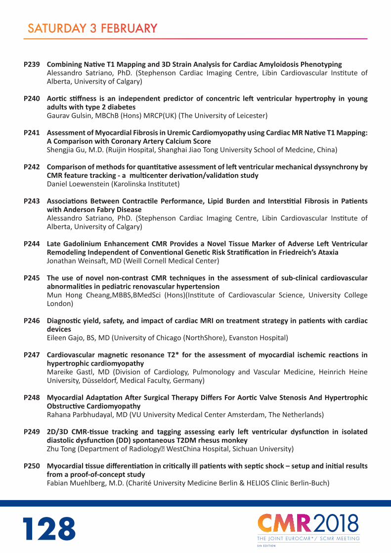
128
SATURDAY 3 FEBRUARY
P239 Combining Native T1 Mapping and 3D Strain Analysis for Cardiac Amyloidosis PhenotypingAlessandro Satriano, PhD. (Stephenson Cardiac Imaging Centre, Libin Cardiovascular Institute of Alberta, University of Calgary)
P240 Aortic stiffness is an independent predictor of concentric left ventricular hypertrophy in young adults with type 2 diabetesGaurav Gulsin, MBChB (Hons) MRCP(UK) (The University of Leicester)
P241 Assessment of Myocardial Fibrosis in Uremic Cardiomyopathy using Cardiac MR Native T1 Mapping: A Comparison with Coronary Artery Calcium ScoreShengjia Gu, M.D. (Ruijin Hospital, Shanghai Jiao Tong University School of Medcine, China)
P242 Comparison of methods for quantitative assessment of left ventricular mechanical dyssynchrony by CMR feature tracking - a multicenter derivation/validation studyDaniel Loewenstein (Karolinska Institutet)
P243 Associations Between Contractile Performance, Lipid Burden and Interstitial Fibrosis in Patients with Anderson Fabry DiseaseAlessandro Satriano, PhD. (Stephenson Cardiac Imaging Centre, Libin Cardiovascular Institute of Alberta, University of Calgary)
P244 Late Gadolinium Enhancement CMR Provides a Novel Tissue Marker of Adverse Left Ventricular Remodeling Independent of Conventional Genetic Risk Stratification in Friedreich’s AtaxiaJonathan Weinsaft, MD (Weill Cornell Medical Center)
P245 The use of novel non-contrast CMR techniques in the assessment of sub-clinical cardiovascular abnormalities in pediatric renovascular hypertensionMun Hong Cheang,MBBS,BMedSci (Hons)(Institute of Cardiovascular Science, University College London)
P246 Diagnostic yield, safety, and impact of cardiac MRI on treatment strategy in patients with cardiac devicesEileen Gajo, BS, MD (University of Chicago (NorthShore), Evanston Hospital)
P247 Cardiovascular magnetic resonance T2* for the assessment of myocardial ischemic reactions in hypertrophic cardiomyopathyMareike Gastl, MD (Division of Cardiology, Pulmonology and Vascular Medicine, Heinrich Heine University, Düsseldorf, Medical Faculty, Germany)
P248 Myocardial Adaptation After Surgical Therapy Differs For Aortic Valve Stenosis And Hypertrophic Obstructive CardiomyopathyRahana Parbhudayal, MD (VU University Medical Center Amsterdam, The Netherlands)
P249 2D/3D CMR-tissue tracking and tagging assessing early left ventricular dysfunction in isolated diastolic dysfunction (DD) spontaneous T2DM rhesus monkeyZhu Tong (Department of Radiology, WestChina Hospital, Sichuan University)
P250 Myocardial tissue differentiation in critically ill patients with septic shock – setup and initial results from a proof-of-concept studyFabian Muehlberg, M.D. (Charité University Medicine Berlin & HELIOS Clinic Berlin-Buch)
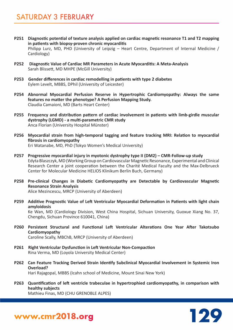
128 129www.cmr2018.org
SATURDAY 3 FEBRUARY
P251 Diagnostic potential of texture analysis applied on cardiac magnetic resonance T1 and T2 mapping in patients with biopsy-proven chronic myocarditisPhilipp Lurz, MD, PHD (University of Leipzig – Heart Centre, Department of Internal Medicine / Cardiology)
P252 Diagnostic Value of Cardiac MR Parameters in Acute Myocarditis: A Meta-AnalysisSarah Blissett, MD MHPE (McGill University)
P253 Gender differences in cardiac remodelling in patients with type 2 diabetesEylem Levelt, MBBS, DPhil (University of Leicester)
P254 Abnormal Myocardial Perfusion Reserve in Hypertrophic Cardiomyopathy: Always the same features no matter the phenotype? A Perfusion Mapping Study.Claudia Camaioni, MD (Barts Heart Center)
P255 Frequency and distribution pattern of cardiac involvement in patients with limb-girdle muscular dystrophy (LGMD) - a multi-parametric CMR studyAnca Florian (University Hospital Münster)
P256 Myocardial strain from high-temporal tagging and feature tracking MRI: Relation to myocardial fibrosis in cardiomyopathyEri Watanabe, MD, PhD (Tokyo Women’s Medical University)
P257 Progressive myocardial injury in myotonic dystrophy type II (DM2) – CMR-Follow-up studyEdyta Blaszczyk, MD (Working Group on Cardiovascular Magnetic Resonance, Experimental and Clinical Research Center a joint cooperation between the Charité Medical Faculty and the Max-Delbrueck Center for Molecular Medicine HELIOS Klinikum Berlin Buch, Germany)
P258 Pre-clinical Changes in Diabetic Cardiomyopathy are Detectable by Cardiovascular Magnetic Resonance Strain AnalysisAlice Mezincescu, MRCP (University of Aberdeen)
P259 Additive Prognostic Value of Left Ventricular Myocardial Deformation in Patients with light chain amyloidosisKe Wan, MD (Cardiology Division, West China Hospital, Sichuan University, Guoxue Xiang No. 37, Chengdu, Sichuan Province 610041, China)
P260 Persistent Structural and Functional Left Ventricular Alterations One Year After Takotsubo CardiomyopathyCaroline Scally, MBChB, MRCP (University of Aberdeen)
P261 Right Ventricular Dysfunction in Left Ventricular Non-CompactionRina Verma, MD (Loyola University Medical Center)
P262 Can Feature Tracking Derived Strain Identify Subclinical Myocardial Involvement in Systemic Iron Overload?Hari Rajagopal, MBBS (Icahn school of Medicine, Mount Sinai New York)
P263 Quantification of left ventricle trabeculae in hypertrophied cardiomyopathy, in comparison with healthy subjectsMathieu Finas, MD (CHU GRENOBLE ALPES)

130
SATURDAY 3 FEBRUARY
P264 Chagas myocardial fibrosis undistinguishable from myocardial infarction by LGE: Can T1 mapping help?Thamara Morais, MD (Heart Institute (InCor), University of Sao Paulo)
P265 Focal regions of hypoenhancement visualized by early gadolinium enhancement imaging in patients with hypertrophic cardiomyopathyTushar Acharya (National Heart, Lung, and Blood Institute, NIH)
P266 Role of T1 mapping in patients with granulomatous cardiomyopathyRicha Kothari, DNB, MBBS (Narayana Institute of Cardiac Sciences)
P267 Late gadolinium enhancement patterns in patients with hypereosinophilia: does CMR help identifying the etiology?Julien Pagniez, MD (Lille University Hospital)
P268 Cardiac MR-Derived Global and Regional Myocardial Strain is Similar in Both HIV-positive and HIV-negative Patients with Heart FailureBradley Allen, MD (Northwestern University)
P269 The length of posterior mitral valve leaflet distinguish hypertrophic cardiomyopathy from Fabry diseaseEzequiel Espinosa, MD (Hospital Italiano de Buenos Aires)
P269 The length of posterior mitral valve leaflet distinguish hypertrophic cardiomyopathy from Fabry diseaseDiego Perez de Arenaza, MD (Hospital Italiano de Buenos Aires)
P270 Left anterior fascicular block is associated with increased left ventricular scar burden and reduced left ventricular ejection fraction but not with mortalityJohan von Schéele (Karolinska Institutet, and Karolinska University Hospital, Stockholm, Sweden)
P271 Association of myocardial deformation by tissue tracking CMR with fibrosis in patients with Fabry diseaseRui Placido, MD (Santa Maria University Hospital, Cardiology Department, Lisbon Academic Medical Center, CCUL, Lisbon, Portugal)
P272 Genotype-Phenotype correlation in ARVC – a CMR studySilvia Castelletti, MD (Istituto Auxologico Italiano)
P273 Extracellular Volume Imaging, Aortic Distensibility and CMR-Feature Tracking by Magnetic Resonance Imaging Post Mediastinal Radiotherapy in Survivors of Hodgkin’s LymphomaYing Jia Gong (McGill University)
P274 Usefulness of early assessment of Takotsubo cardiomyopathy patients with cardiac magnetic resonance imagingEmmanuel Androulakis (St Georges University Hospital NHS Foundation Trust)
P275 Resolution of Late Gadolinium Enhancement in a Series of Pediatric Patients with Clinical MyocarditisMarc Lee, MD (University of Tennessee Health Science Center)

130 131www.cmr2018.org
SATURDAY 3 FEBRUARY
P276 Inter-vendor reproducibility of left and right ventricular cardiovascular magnetic resonance myocardial feature-trackingRoman Gertz, BSc (DZHK (German Centre for Cardiovascular Research) partner site Göttingen)
P277 Dynamic Cardiac Off-resonance Mapping for Real-time Spiral De-blurringMatthew Restivo, PhD (National Heart, Lung, and Blood Institute, National Institutes of Health)
P278 Left-Ventricular Strain Measured by 2D vs 3D Tissue Tracking Requires Separate Normal ValuesVolker Rasche, PhD (University Hospital of Ulm)
P279 coreMRI: an online web platform for database generation in simulation-based quantitative MR methodsChristos Xanthis, PhD (Lund University, Skane University Hospital, Department of Clinical Physiology, Lund, Sweden)
P280 Left-Ventricular Strain Reference Values of Healthy Volunteers by Age and Gender as Measured with 3D Tissue TrackingVolker Rasche, PhD (University Hospital of Ulm)
P281 Standardization of myocardial T2 mapping measurements: normal values and reproducibility in the presence of health and diseaseJasmin Haslbauer, MSc, cand.med. (Goethe University Hospital Frankfurt am Main)
P282 Standardisation of myocardial T1 mapping measurements for reproducible clinical use in the presence of health and diseaseMengzhen Chen, Cand. med. (Goethe University Hospital Frankfurt am Main)
P282 Standardisation of myocardial T1 mapping measurements for reproducible clinical use in the presence of health and diseaseValentina Puntmann, MD, PhD (Universitaetsklinikum Frankfurt)
P283 4D Flow Evaluation of Altered Flow Patterns in Patients with Pulmonary HypertensionJulio Garcia, PhD (University of Calgary)
P284 LV patient specific models and shape analysis from 4500 UK Biobank studiesKathleen Gilbert, BE(hons), PhD (University of Auckland, New Zealand)
P285 A deep learning approach for the segmental analysis of myocardial T1 mappingNicola Martini, PhD (Fondazione G. Monasterio CNR Regione Toscana)
P286 Identification and Sorting of End-Systolic and End-Diastolic images in Real-Time Cine Imaging Using Laplacian Eigenmaps and Image RegistrationAnthony Aletras,PhD.(Aristotle University of Thessaloniki/Lund University)
P287 Left Ventricle Segmentation via Deep Learning Networks: Superfast and Accurate Analysis of Cine MRIQian Tao, PhD (Department of Radiology, Leiden University Medical Center, The Netherlands)

132
SATURDAY 3 FEBRUARY
P288 Method for objective automatic assessment of wall motion abnormality from cineMR imagesIulia Popescu (University of Oxford, Centre for Clinical Magnetic Resonance Research)
P289 The effect of imaging gradients on estimates of cardiac DTI metricsCyril Tous, ME, MR, MB, BE, B.Sc (The University of Auckland)
P290 Automatic Ventricular Segmentation Using a Convolutional Neural Network: results from Circle Cardiovascular Imaging on UK Biobank Cardiac MR CINE ImagesAlireza Sojoudi, MSc (Circle Cardiovascular Imaging Inc)
P291 Architecture and pipeline to enable large scale analysis of 4D flow MRI dataMichael Scott, MS (Northwestern University)
P292 Respiratory Effects on Ventricular Performance using 4D Flow MRIAlejandro Roldán-Alzate (UW-Madison)
P293 Differentiation of left ventricular mechanics in high- and low-gradient aortic stenosis by cardiac magnetic resonance (CMR) feature trackingSabine Zitzmann, MD (Kerckhoff-Heart-Center, Department of Cardiology, Bad Nauheim, Germany)
P294 Inter- and intraobserver reproducibility of right ventricular cardiac magnetic resonance feature tracking in patients with CTEPH (chronic thromboembolic pulmonary hypertension) after PEA (pulmonary endarterectomie)Sabine Zitzmann, MD (Kerckhoff-Heart-Center, Department of Cardiology, Bad Nauheim, Germany)
P295 Semiautomatic segmentation of the blood pool for real time cines from exercise stress CMRBoyang Su, PhD (National Heart Centre Singapore)
P296 Evaluation of right ventricular cardiac magnetic resonance feature tracking strain analysis in patients with chronic thromboembolic pulmonary hypertension before and after pulmonary endarterectomyJulia Vietheer, MD (Kerckhoff-Heart-Center, Department of Cardiology, Bad Nauheim, Germany)
P297 Local longitudinal strain from tagged MRI can help distinguish between hypertrophic cardiomyopathy and septal knuckleTeodora Chitiboi, PhD (NYU School of Medicine)
P298 Impact of image motion correction on the evaluation of stress perfusion CMRFlorian von Knobelsdorff, MD (Dept. of Cardiology, Clinic Agatharied, University of Munich)
P299 Semi-automatic geometrical characterization of patients with bicuspid aortic valve using finite element methodsJulio Sotelo (Pontificia Universidad Católica de Chile)
P300 Extensive analysis of hemodynamics parameters on 4D flow MRI data of patients with bicuspid aortic valve using finite element methodsJulio Sotelo (Pontificia Universidad Católica de Chile)
P301 Identifying left atrial enlargement by CMR: comparison between two widely used methods for volumes quantificationThamara Morais, MD (Heart Institute (InCor), University of Sao Paulo)

132 133www.cmr2018.org
SATURDAY 3 FEBRUARY
P302 Comprehensive 3D flow characterization in patients with Dilated Cardiomyopathy (DCM) from 4D flow MRI data using a finite element method and 17-Segment BullseyesPamela Franco (Pontificia Universidad Católica de Chile)
P303 Deep Learning Approach for Left Ventricular Endocardium Segmentation in Long-Axis C a r d i a c MR Imaging
Liang Zhong, PhD (National Heart Centre Singapore; Duke-NUS Medical School)
P305 Automatic Artifacts Detection as Operative Scan-aided Tool in an Autonomous MRI EnvironmentWenwen Jiang (Heartvista, Inc)
P306 Numerical Simulation of Pulmonary Hypertension: Effect of 2D Flow and 4D Flow MRI-Based Inlet Boundary ConditionsChristopher Francois, MD (University of Wisconsin - Madison)
P307 Left ventricular cardiac magnetic resonance parameters of elite, young and masters athletes with quantitative analysis of papillary muscle and trabecular massIbolya Csecs, MD (Heart and Vascular Center of Semmelweis University)
P308 Assessment of automatic late gadolinium enhancement scar tissue analysis techniques in HIV-cardiomyopathyYoko Kato, MD, PhD (Johns Hopkins University)
P309 Flexible 3D printed acrylic insert to maintain in-vivo shape: Application to ex vivo imaging of chronic myocardial infarction in swineJohn Whitaker, BM BCh (King’s College London/Beth Israel Deaconess Medical Centre)
P310 Measurements of normal myocardial T1, partition coefficient and extracellular volume at 3 Tesla using MOLLI, ShMOLLI and SASHATarik Hafyane, MSC (Montreal Heart Institute)
P311 Myocardial extracellular volume fraction derived from synthetic hematocrit can lead to significant clinical misclassificationAndrea Cardona, MD (The Ohio State University Wexner Medical Center)
P312 Initial Validation of Free-Breathing Navigator Cardiac Quantitative Susceptibility Mapping for Blood Pool Oxygenation as Verified Invasively via Right Heart CatheterizationYan Wen, M.S. (Cornell University)
P313 Early Detection of Myocardial Involvement by T1 Mapping of Cardiac Magnetic Resonance in Idiopathic Inflammatory MyopathyLiuyu Yu, BMsci (Sichuan University West China Hospital)
P314 Left atrial deformation in patients with cryptogenic stroke: a cardiovascular magnetic resonance myocardial feature-tracking studyJohannes Kowallick, MD (University Medical Center Göttingen)
P315 4D Flow CMR using semi-automated retrospective valve tracking for assessment of severe mitral regurgitationCarmen Blanken, BS (Academic Medical Center Amsterdam)
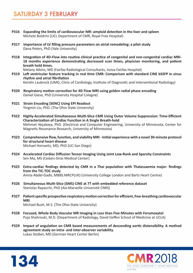
134
SATURDAY 3 FEBRUARY
P316 Expanding the limits of cardiovascular MR: amyloid detection in the liver and spleenMichele Boldrini (UCL Department of CMR, Royal Free Hospital)
P317 Importance of LV filling pressure parameters on atrial remodeling: a pilot studyDana Peters, PhD (Yale University)
P318 Integration of 4D-Flow into routine clinical practice of congenital and non-congenital cardiac MRI-18 months experience demonstrating decreased scan times, physician monitoring, and patient breath hold times.Melany Atkins, MD (Fairfax Radiological Consultants, Inova Fairfax Hospital)
P319 Left ventricular feature tracking in real time CMR: Comparison with standard CINE bSSFP in sinus rhythm and atrial fibrillationKerstin Laubrock (UMG, Clinic of Cardiology, Institute of Diagnostic and Interventional Radiology)
P320 Respiratory motion correction for 4D Flow MRI using golden radial phase encodingDaniel Giese, PhD (University Hospital Cologne)
P321 Strain Encoding (SENC) Using EPI ReadoutYingmin Liu, PhD. (The Ohio State University)
P322 Highly-Accelerated Simultaneous Multi-Slice CMR Using Outer Volume Suppression: Time-Efficient Characterization of Cardiac Function in A Single Breath-holdMehmet Akçakaya, PhD. (Electrical and Computer Engineering, University of Minnesota; Center for Magnetic Resonance Research, University of Minnesota)
P323 Comprehensive flow, function, and viability MRI - Initial experience with a novel 30-minute protocol for structural heart diseaseMichael Horowitz, MD, PhD (UC-San Diego)
P324 Accelerated Cardiac Diffusion Tensor Imaging Using Joint Low-Rank and Sparsity ConstraintsSen Ma, MS (Cedars-Sinai Medical Center)
P325 Extra-cardiac findings detected by CMR in a Thai population with Thalassaemia major- findings from the TIC-TOC studyAmna Abdel-Gadir, MBBS.MRCP(UK) (University College London and Barts Heart Centre)
P326 Simultaneous Multi-Slice (SMS) CINE at 7T with embedded reference datasetStanislas Rapacchi, PhD (Aix-Marseille Université CNRS)
P327 Patient specific prospective respiratory motion correction for efficient, free-breathing cardiovascular MRIMichael Bush, M.S. (The Ohio State University)
P328 Focused, Whole Body Vascular MR Imaging in Less than Five Minutes with FerumoxytolPuja Shahrouki, M.D. (Department of Radiology, David Geffen School of Medicine at UCLA)
P329 Impact of angulation on CMR based measurements of descending aortic distensibility. A method agreement study on intra- and inter-observer variability.Lukas Stoiber, MD (German Heart Center Berlin)

134 135www.cmr2018.org
SATURDAY 3 FEBRUARY
P330 Cardiac and Respiratory Self-Gated Motion-Corrected Free-breathing Spiral Cine ImagingRuixi Zhou, MS (University of Virginia)
P331 Compressed Sensing real-time Cine Imaging in patients with cardiac arrhythmia: Does it help to reduce mis-triggering artifacts?Julien Pagniez, MD (Lille University Hospital)
P332 Left ventricle wall motion disorders in myocardial infarction depicted by compressed sensing real-time cine imaging.Valentina Silvestri, MD (Lille University Hospital)
P333 Simultaneous multi-slice bSSFP CMR: Is it feasible?Vanessa Landes, MS (University of Southern California)
P334 Velocity sensitivity of inner-volume cardiac echo planar imagingAhsan Javed, MS (University of Southern California)
P335 Can Cardiac Magnetic Resonance Feature Tracking Predict Clinical Cardiovascular Events in Asymptomatic Aortic Stenosis Patients with Normal Ejection Fraction?Moon Young Kim, MD, MS (Seoul National University Hospital)
P336 Can atrial size and shape predict which patients experience a decline in left ventricular ejection fraction after surgery for mitral regurgitation?Jessica Webb, BM BCh MRCP (King’s College London)
P337 Aortic stenosis septal hypertrophy and its regression post AVR is more plastic in males than females: insights from a 3D cardiac atlasAnish Bhuva, MRCP MBBS (Barts Heart Centre and Institute of Cardiovascular Science, UCL.)
P338 Late enhancement and reverse left ventricular remodeling in percutaneous edge-to-edge mitral repairBernard Paelinck (Antwerp University Hospital)
P339 CMR structural and fibrosis assessment predicts prognosis in chronic rheumatic valvular heart diseaseLilia Sierra-Galan, MD , MCvT(American British Cowdray Medical Center)
P340 Evaluation of Pressure Drop using 4D Flow MRI: Clinical Feasibility in Patients with Bicuspid Aortic ValveJulio Garcia, PhD (University of Calgary)
P341 Reduced Global Longitudinal Strain in Rheumatic Mitral Stenosis with Good Left Ventricular Ejection Fraction: A Cardiac Magnetic Resonance Feature Tracking StudyElen Elen, MD, FIHA (National Cardiovascular Center Harapan Kita, Jakarta-Indonesia; Institute for Experimental and Translational Cardiovascular Imaging, University Hospital Frankfurt-Germany)
P342 Comparison of 4D Flow MRI to Doppler Echocardiography: Assessment of Mean Transvalvular Aortic Pressure GradientAlex Barker, PHD (Northwestern University)

136
SATURDAY 3 FEBRUARY
P343 A novel Cardiac MRI-based 3D method to design personalized aortic valves: The sinus-mirror valve hypothesisMichael Silberbach, MD (Oregon Health & Science University)
P344 Relationship between haemodynamic progression, left ventricular response and presence of myocardial fibrosis in aortic stenosisMiquel Gomez (Parc de Salut Mar Barcelona)
P345 Aliased flow planimetry by low-VENC phase-contrast CMR imaging for the assessment of effective orifice area in aortic valve stenosis: a proof-of-concept studyFabrizio Ricci (Department of Neuroscience, Imaging and Clinical Sciences, “G. D’ Annunzio” University, Chieti, Italy)
P346 Assessment of flow using phase contrast velocity mapping with radial acquisition ultrashort echo times for irregular heart rhythmsMichaela Hell (Department of Cardiology, Faculty of Medicine, Friedrich-Alexander University Erlangen-Nürnberg, Germany)
P347 Systolic Flow Displacement Correlates with Ascending Aortic Growth in Patients with Isolated Bicuspid Aortic ValveNicholas Burris, MD (University of Michigan)
P348 Left Ventricle Remodeling in Patients with Bicuspid Aortic ValveKenichiro Suwa, MD (Northwestern University Feinberg School of Medicine Department of Radiology)
P349 The role of cardiovascular magnetic resonance imaging in prediction of developement of pulmonary hypertension in patients with severe aortic stenosisAudrone Vaitiekiene, MD (Lithuanian University of Health sciences)
P350 Discordance of Aortic Regurgitation grading between Cardiac Magnet Resonance Imaging and Transthoracic Echocardiography.Ulf Neisius, MD PhD (Beth Israel Deaconess Medical Center, Harvard Medical School)
P351 Association between Aortic Regurgitation Severity and Post-Valve Replacement Left Ventricular Remodeling: Cardiovascular Magnetic Resonance Imaging vs Transthoracic Echocardiography.Ulf Neisius, MD PhD (Beth Israel Deaconess Medical Center, Harvard Medical School)
P352 4D flow MRI assessment of the thoracic aorta using variable density k-t acceleration. Feasibility, reproducibility and clinical implication in ascending aneurysm on bicuspid aortic valve.Kaoru Tanaka, M.D., PhD (Universitair Ziekenhuis Brussel)
P353 Diastolic flow in the descending aorta by cardiovascular magnetic resonance imaging for quantification of aortic regurgitationAndreas Kammerlander, MD (Medical University of Vienna)
P354 Impact of Aortic Valve Replacement on flow profiles in the ascending AortaSarah Nordmeyer, MD (Deutsches Herzzentrum Berlin)

136 137www.cmr2018.org
SATURDAY 3 FEBRUARY
P355 Cardiovascular Magnetic Resonance Predictors of Left Ventricular Remodeling 1 Year After Mitral Valve RepairAriane Pacheco, MD (Heart Institute - InCor - University of São Paulo Medical School, São Paulo, Brazil)
P356 Relationship between breathing pattern and aortic flow measurement: head-to-head comparison between high temporal resolution free-breathing phase contrast CMR and standard breath-hold phase contrast sequenceGabriella Vincenti,1) Division of Cardiology and Center of Cardiac Magnetic Resonance, Cardiovascular Department, University Hospital of Lausanne, CHUV, Switzerland.(1) Division of Cardiology and Center of Cardiac Magnetic Resonance, Cardiovascular Department, University Hospital of Lausanne, CHUV, Switzerland)
P357 Single Breath Hold 4D Flow Acquisition of the Aortic ValveAdam RIch, PhD. (The Ohio State University)
P358 T1 mapping abnormalities in patients with aortic stenosis. Relationship with clinical features and myocardial deformation assessed by tissue trackingAna Almeida, MD, PhD (University Hospital Santa Maria, Lisbon, Portugal)
P359 Myocardial Strain Before and After Percutaneous Mitral Commisurotomy for Severe Mitral StenosisRaouf AbdelRaouf (Dar Al Fouad hospital, 6th october city, Egypt)
P360 Aortic Remodeling after Anthracycline TherapyThiago de Souza, MD, Msc (UNICAMP)
P361 Safety and Image Quality of the Combined use of Cardiac MRI and the CardioMEMS HF System in Progressive PAH; a Proof of Concept StudyRobert Biederman, MD, FACC, FAHA (Allegheny General Hospital)
P362 Steady State MRA of the Thoracic Aorta Using IR SSFP and a High Relaxivity Contrast AgentJames Carr, MD (Northwestern University)
P363 High-resolution intracranial vessel wall MR imaging allows for black-blood angiographic visualization of the lenticulostriate artery at 3T: A validation against 7T TOF-MRAZihao Zhang, PhD. (State Key Laboratory of Brain and Cognitive Science, Beijing MR Center for Brain Research, Institute of Biophysics, Chinese Academy of Sciences)
P364 Quantitative Multi-Contrast CMR of Carotid Atherosclerosis: Pushing for Higher Clinical ValueYibin Xie, PhD (Cedars-Sinai Medical Center)
P365 Assessment of congenital vascular anomalies in people affected by ThalidomideJulius Weinrich, M.D (University Medical Center Hamburg-Eppendorf)
P366 Automatic Image-Based Background Phase Correction in the Presence of Wrap-Around ArtifactAaron Pruitt, B.S. (The Ohio State University)
P367 Arterial geometry, flow and stiffness indices from thoracic aorta and carotid MRI in elderly hypertensivesEtienne Charpentier (Sorbonne Universités, UPMC Univ Paris 06, INSERM UMRS 1146, CNRS UMR 7371, Laboratoire d’Imagerie Biomédicale)
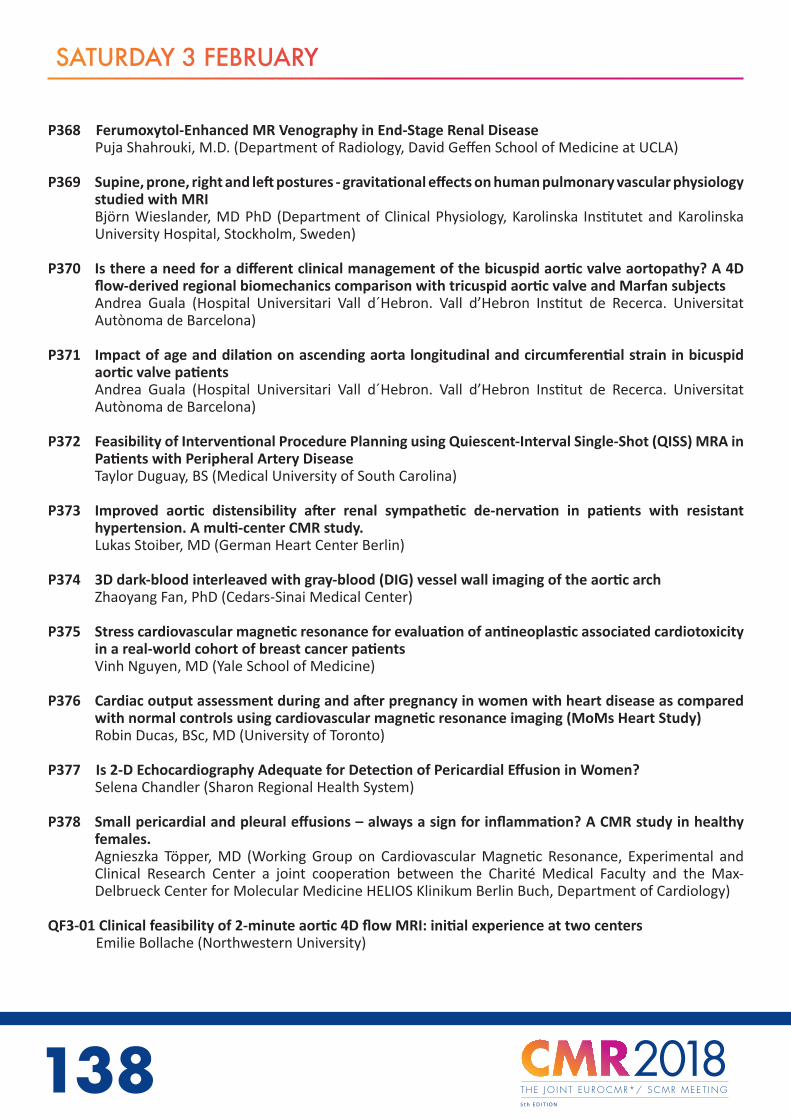
138
SATURDAY 3 FEBRUARY
P368 Ferumoxytol-Enhanced MR Venography in End-Stage Renal DiseasePuja Shahrouki, M.D. (Department of Radiology, David Geffen School of Medicine at UCLA)
P369 Supine, prone, right and left postures - gravitational effects on human pulmonary vascular physiology studied with MRIBjörn Wieslander, MD PhD (Department of Clinical Physiology, Karolinska Institutet and Karolinska University Hospital, Stockholm, Sweden)
P370 Is there a need for a different clinical management of the bicuspid aortic valve aortopathy? A 4D flow-derived regional biomechanics comparison with tricuspid aortic valve and Marfan subjectsAndrea Guala (Hospital Universitari Vall d´Hebron. Vall d’Hebron Institut de Recerca. Universitat Autònoma de Barcelona)
P371 Impact of age and dilation on ascending aorta longitudinal and circumferential strain in bicuspid aortic valve patientsAndrea Guala (Hospital Universitari Vall d´Hebron. Vall d’Hebron Institut de Recerca. Universitat Autònoma de Barcelona)
P372 Feasibility of Interventional Procedure Planning using Quiescent-Interval Single-Shot (QISS) MRA in Patients with Peripheral Artery DiseaseTaylor Duguay, BS (Medical University of South Carolina)
P373 Improved aortic distensibility after renal sympathetic de-nervation in patients with resistant hypertension. A multi-center CMR study.Lukas Stoiber, MD (German Heart Center Berlin)
P374 3D dark-blood interleaved with gray-blood (DIG) vessel wall imaging of the aortic archZhaoyang Fan, PhD (Cedars-Sinai Medical Center)
P375 Stress cardiovascular magnetic resonance for evaluation of antineoplastic associated cardiotoxicity in a real-world cohort of breast cancer patientsVinh Nguyen, MD (Yale School of Medicine)
P376 Cardiac output assessment during and after pregnancy in women with heart disease as compared with normal controls using cardiovascular magnetic resonance imaging (MoMs Heart Study)Robin Ducas, BSc, MD (University of Toronto)
P377 Is 2-D Echocardiography Adequate for Detection of Pericardial Effusion in Women?Selena Chandler (Sharon Regional Health System)
P378 Small pericardial and pleural effusions – always a sign for inflammation? A CMR study in healthy females.Agnieszka Töpper, MD (Working Group on Cardiovascular Magnetic Resonance, Experimental and Clinical Research Center a joint cooperation between the Charité Medical Faculty and the Max-Delbrueck Center for Molecular Medicine HELIOS Klinikum Berlin Buch, Department of Cardiology)
QF3-01 Clinical feasibility of 2-minute aortic 4D flow MRI: initial experience at two centers Emilie Bollache (Northwestern University)

138 139www.cmr2018.org
SATURDAY 3 FEBRUARY
QF3-02 T1 mapping for the prediction of treatment response in AL amyloidosis Rosario Perea Palazón (Hospital Clínic. Universitat de Barcelona)
QF3-03 Prognostic Utility of Blood Oxygen Level Dependent (BOLD) Cardiovascular Magnetic Resonance (CMR) imaging in Asymptomatic Chronic Kidney Disease (CKD) Patients with and without Diabetes Mellitus. Ranjit Shah (South Australian Health and Medical Research Institute, Adelaide, SA, Australia)
QF3-04 Diagnostic Performance of Fully Automated Pixel-wise Myocardial Blood Flow Maps of Stress and Rest CMR Perfusion in Patients Li-Yueh Hsu (National Heart, Lung and Blood Institute, National Institutes of Health)
QF3-05 Phenotype development in Cardiac Fabry disease proceeds through four stages: a prospective 182-patient study Sabrina Nordin (Barts Heart Centre)
QF3-06 When criteria for ICD implantation in the primary prevention of sudden death among patients with hypertrophic cardiomyopathy don’t get along: an analysis of late gadolinium-enhancement and the European and American guidelines Pedro Freitas (Hospital de Santa Cruz)
QF3-07 Obesity paradox and myocardial injury by cardiac magnetic resonance imaging in ST-elevation myocardial infarction Georg Fuernau (University Heart Center Lübeck)
QF3-08 Importance of operator training and rest perfusion on the diagnostic accuracy of stress perfusion CMR Adriana Villa (King’s College London)
QF3-09 Diffuse interstitial fibrosis is associated with reduced myocardial strain in heart failure with preserved and reduced ejection fraction Xiaodan Zhao (National Heart Centre Singapore)
QF3-10 Clinical Impact of Magnetic Resonance Imaging in Non-Approved Cardiac Devices Ibrahim Saeed (Saint Luke’s Mid-America Heart Institute)
QF3-11 The Short-Term Prognosis Values of combined T1 Mapping and Feature Tracking by Cardiovascular Magnetic Resonance in Dilated Cardiomyopathy Hui Liu (Guangdong General Hospital, Guangdong Academy of Medical Sciences)
QF3-12 Impact of aortic geometrical characteristics on abnormal flow pattern in the proximal descending aorta in Marfan patients: a 4D flow MRI study Andrea Guala (Hospital Universitari Vall d´Hebron. Vall d’Hebron Institut de Recerca. Universitat Autònoma de Barcelona)
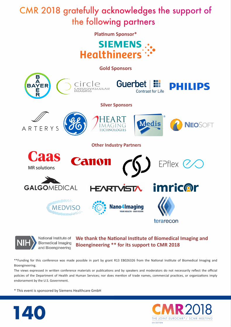
140
Platinum Sponsor*
**Funding for this conference was made possible in part by grant R13 EB026326 from the National Institute of Biomedical Imaging and
Bioengineering.
The views expressed in written conference materials or publications and by speakers and moderators do not necessarily reflect the official
policies of the Department of Health and Human Services; nor does mention of trade names, commercial practices, or organizations imply
endorsement by the U.S. Government.
* This event is sponsored by Siemens Healthcare GmbH
Gold Sponsors
Silver Sponsors
Other Industry Partners
MR solutions
•
CMR 2018 gratefully acknowledges the support of the following partners
We thank the National Institute of Biomedical Imaging and Bioengineering ** for its support to CMR 2018
140
Advanced Visualization by IntelliSpace Portal delivers CT, MR and other clinical applications, also directly to your IntelliSpace Cardiovascular Workspace.
“
“ Cardiac MR and CT together (on the IntelliSpace Portal) have lowered overall analysis time by 20%-30%*
Dr. Gaby Weissman, M.D.MedStar Washington Hospital Center, Washington D.C., USA
* Results are specific to the institution where they were obtained and may not reflect the results achievable at other institutions.
IntelliSpace Portal
2171388_Philips_Cardiac_MR_CT_advertisement_V3.indd 1 18/12/17 10:10
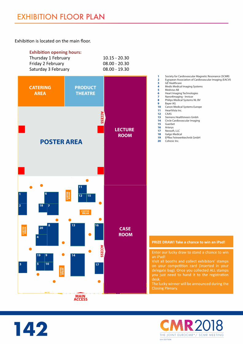
142
EXHIBITION FLOOR PLAN
Exhibition is located on the main floor.
Exhibition opening hours: Thursday 1 February 10.15 - 20.30 Friday 2 February 08.00 - 20.30 Saturday 3 February 08.00 - 19.30
PRIZE DRAW! Take a chance to win an iPad!
Enter our lucky draw to stand a chance to win an iPad!Visit all booths and collect exhibitors’ stamps on your competition card (inserted in your delegate bag). Once you collected ALL stamps you just need to hand it to the registration desk.The lucky winner will be announced during the Closing Plenary.
1 Society for Cardiovascular Magnetic Resonance (SCMR)2 European Association of Cardiovascular Imaging (EACVI)3 GE Healthcare4 Medis Medical Imaging Systems5 Medviso AB6 Heart Imaging Technologies7 Nano4Imaging - Imricor8 Philips Medical Systems NL BV 9 Bayer AG10 Canon Medical Systems Europe
T H E J O I N T E U R O C M R * / S C M R M E E T I N G
ACC
ESS
LECTURE ROOM
CASE ROOM
MAIN ACCESS
COFF
EE
BR
EAK
COFF
EE
BR
EAK
COFF
EE
BR
EAK
COFFEE BREAK
9
105
14
16
17
1
3
13
4
8
6
7
11
12 15
2
11 HeartVista Inc.12 CAAS13 Siemens Healthineers Gmbh14 Circle Cardiovascular Imaging15 Guerbet16 Arterys 17 Neosoft, LLC18 Galgo Medical19 EP�ex Feinwerktechnik GmbH20 Cohesic Inc.
18
20
POSTER AREAA
CCESS
CATERINGAREA
PRODUCTTHEATRE
19
1 Society for Cardiovascular Magnetic Resonance (SCMR)2 European Association of Cardiovascular Imaging (EACVI)3 GE Healthcare4 Medis Medical Imaging Systems5 Medviso AB6 Heart Imaging Technologies7 Nano4Imaging - Imricor8 Philips Medical Systems NL BV 9 Bayer AG10 Canon Medical Systems Europe
T H E J O I N T E U R O C M R * / S C M R M E E T I N G
ACC
ESS
LECTURE ROOM
CASE ROOM
MAIN ACCESS
COFF
EE
BR
EAK
COFF
EE
BR
EAK
COFF
EE
BR
EAK
COFFEE BREAK
9
105
14
16
17
1
3
13
4
8
6
7
11
12 15
2
11 HeartVista Inc.12 CAAS13 Siemens Healthineers Gmbh14 Circle Cardiovascular Imaging15 Guerbet16 Arterys 17 Neosoft, LLC18 Galgo Medical19 EP�ex Feinwerktechnik GmbH20 Cohesic Inc.
18
20
POSTER AREA
ACC
ESS
CATERINGAREA
PRODUCTTHEATRE
19
142 143www.cmr2018.org
EXHIBITORS DIRECTORY
Society for Cardiovascular Magnetic Resonance (SCMR) Booth 119 Mantua Road, Mt. RoyalNJ 08061U.S.A.
The Society for Cardiovascular Magnetic Resonance (SCMR) is the recognized representative and advocate for physicians, scientists, and technologists who work in the field of cardiovascular magnetic resonance (CMR). Our mission is to improve cardiovascular health by advancing the field of CMR. We accomplish our mission through education, advocacy, networking, research and clinical excellence. Visit the booth to join the Society at a discounted fee and pick up a free SCMR product.
SCMR Headquarters Office Tel.: [email protected] www.scmr.org
Journal of Cardiovascular Magnetic Resonance (JCMR) Booth 1 c/w Beth Israel Deaconess Medical Center 330 Brookline Avenue Boston, MA 02215 U.S.A. JCMR, the official journal of the Society for Cardiovascular MR, is an open access, online journal that publishes articles on all aspects of basic and clinical research on the design, development, manufacture, and evaluation of magnetic resonance methods applied to the cardiovascular system. The only journal devoted exclusively to cardiovascular MR, JCMR aims to provide an international forum for communicating the latest findings and reviews on the burgeoning field of cardiovascular MR imaging and spectroscopy.
Email [email protected] Web jcmr-online.biomedcentral.com
European Association of Cardiovascular Imaging (EACVI) Booth 2European Society of Cardiology2035 Route des Colles BIOT CS 80179 France
Created in 2003, the European Association of Cardiovascular Imaging (EACVI), a registered branch of the ESC, is the leading international network in its field. EACVI is a vibrant and unified group of 9 000+ experts representing 4 imaging modalities under one entity (Echo, CMR, Nuclear Cardiology and Cardiac CT). Our mission is: “to promote excellence in clinical diagnosis, research, technical development and education in cardiovascular imaging”. EACVI is committed to maintaining the highest standards of professional excellence in all aspects of cardiovascular imaging.
Contact: EACVI [email protected] Tel.: +33 (0)4 92 94 76 00 www.escardio.org/Sub-specialty-communities/European-Association-of-Cardiovascular-Imaging-(EACVI)

144
GE Healthcare Booth 3Nightingales Lane Chalfont St Giles Buckinghamshire HP8 4SP United Kingdom
Harnessing data and analytics across hardware, software and biotech, GE Healthcare is the $18 billion healthcare business of GE. As a leading provider of medical imaging equipment, with a track record of more than 100 years in the industry and more than 50,000 employees across 100 countries, we transform healthcare by delivering better outcomes for providers and patients.
Contact: Marie-Claire [email protected] www.gehealthcare.com
Medis Medical Imaging Systems Booth 4Schuttersveld 92316 XG LeidenNetherlands
Since 1989 Medis medical imaging systems has been a leading global provider of software for the quantification of cardiovascular images. Its software solutions enable medical professionals to ensure efficient, cost-effective and high-quality image analysis. Visit our booth (B2) and we will be happy to show you our latest new Medis Suite.
Contact: Daniel Garcia [email protected].: 0031715223244www.medis.nl
Medviso AB Booth 5Griffelvägen 3 224 67 Lund Sweden
Medviso AB is a spinn-off company from Lund Cardiac MR group. We strive to deliver world-leading validated image analysis solutions. We are firmly rooted in research advances and take it to clinical routine by our software Segment CMR. We support the research community with our freely available research software Segment.
Contact: Einar Heiberg [email protected] Tel.: +46 76 1836442www.medviso.com
Heart Imaging Technologies Booth 6 5003 Southpark Drive, 140Durham, NC, 27713United States
You are welcome to see the latest version of Precession CMR workflow solution. An onsite system just using a browser, providing access to Diagnostic-quality DICOM images with CMR workstation tools (Volumes, Venc, T1/T2, etc), structured reports for CMR and CCT with searchable database. Precession is ideal solution for routine clinical use and sharing of de-identified images with your colleagues or 2nd opinion.
Contact: Matthew Judd [email protected].: 919 323 4104www.heartit.com
EXHIBITORS DIRECTORY

144 145www.cmr2018.org
Imricor Booth 7400 Gateway Blvd Burnsville, MN 55337United States
Imricor Medical Systems, Inc. is a privately held company committed to unlocking the potential of interventional cardiac magnetic resonance (iCMR) by delivering MR compatible systems and devices that allow physicians to perform cardiac ablations under real-time MRI-guidance.
Contact: Jodi Lamberti [email protected] Tel.: 952 818 8417www.imricor.com
Nano4Imaging Booth 7Zentrum für Biomedizintechnik (ZBMT),Pauwelsstr. 1752074 AachenGermany
Nano4Imaging GmbH in Aachen (DE) wants to move cardiovascular diagnostics and interventions from the cathlab to the MRI. We do this with special focus on mon-itoring and treatment of structural heart diseases and pulmonary hypertension, enabling hemodynamics in MRI. Nano4Imaging holds CE, FDA and Health Canada registrations for its MRI compatible guide wire (EmeryGlide).
Contact: Paul J.A. Borm Tel.: +49 241 [email protected]
Philips Medical Systems NL B.V. Booth 8Veenpluis 4-6, Building QR 03245684 PC BestThe Netherlands
Royal Philips of the Netherlands is a leading health technology company focused on improving people’s health & enabling better outcomes across the health con-tinuum from healthy living and prevention, to diagnosis, treatment and home care. Philips leverages advanced technology and deep clinical & consumer insights to deliver integrated solutions.
Contact: Jouke Smink [email protected] Tel.: +31 6 10585021http://www.philips.com/a-w/about/news.html
Bayer AG Booth 9Müllerstrasse 178 13353 BerlinGermany
Bayer AG’s Radiology unit is a global leader in developing and manufacturing con-trast agents used in X-ray, computed tomography (CT) and magnetic resonance im-aging (MRI), as well as injection systems for diagnostic and therapeutic medical procedures in CT, MRI, cardiovascular and peripheral vascular disease.
Contact: Silke Gerlach [email protected] Tel.: +4930468192931 https://radiology.bayer.com/
EXHIBITORS DIRECTORY

146
Canon Medical Systems Europe Booth 10Zilverstraat 12718RP ZoetermeerNetherlands
Canon Medical offers a full range of diagnostic medical imaging solutions including CT, X-Ray, Ultrasound and MRI, across the globe. As of December 2016, Canon Medical became a member of the Canon Group. In line with our continued Made For life philosophy, patients are at the heart of everything we do. Our mission is to provide medical professionals with solutions that support their efforts in contributing to the health and wellbeing of patients worldwide. Our goal is to deliver optimum health opportunities for patients through uncompromised performance, comfort and safety features.
Contact: Jan de Bruijn [email protected] Tel.: +31611526311 https://eu.medical.canon/
Heartvista Inc. Booth 114984 El Comino Real, Ste.102 Los Altos, CA 94022United States
Heartvista develops autonomous MRI systems using machine learning techniques. HeartVsita’s first self-driving clinical MRI product, SmartHeart App, assists a rapid complete cardiac MR examination with a single button click.
Contact: Juan Santos [email protected] Tel.: +1 (650)714 8057www.heartvista.com
CAAS Booth 12Philipsweg 1, 6227 AJ Maastricht Netherlands Pie Medical Imaging BV has 30 years of experience in cardiovascular analysis software and is well known for its CAAS and 3mensio product lines. Advanced image analysis workflows for X-Ray, CT and Cardiac MRI help to make complex imaging tasks easy and deliver reproducible results for clinical decision-making, planning, and guidance.
Contact: Kelly van [email protected] Tel.: +31 (0)43 328 13 28 www.piemedicalimaging.com
Siemens Booth 13Allee am Röthelheimpark 291052 ErlangenGermany
We, at Siemens Healthineers, are passionate about enabling healthcare providers to deliver high-quality patient care, and to do so affordably. Managing rapid procedure growth and minimizing costs while at the same time achieving clinical excellence are some of the central challenges affecting health-care around the globe. Unique technologies offer our customers exceptional image quality, efficiency & speed, and patient friendliness, while at the same time providing investment protection.Equipped with these technologies and a strong global collaboration network, we enable our customers to lead MRI.
[email protected] + 49 (0)9131-84-0 www.siemens.com/mri
EXHIBITORS DIRECTORY
•MR solutions

146 147www.cmr2018.org
Circle Cardiovascular Imaging Booth 141100, 800 5 Ave SWT2P 3T6 Calgary, AlbertaCanada
Circle Cardiovascular Imaging develops highly accurate, versatile, robust, and intuitive cardiovascular post-processing software for the viewing and analyzing of CMR and CCT images. Circle’s role in clinical and research settings maximizes patients’ achievable benefit by enabling healthcare providers to complete effective and precise analysis.
Contact: Randy Jordan [email protected] Tel.: 1 (403) 338 1870www.circlecvi.com
Guerbet Booth 15BP 57400 95943 Roissy CDG Cedex France
Guerbet is a pioneer in the contrast agent field with over 90 years’ experience and is one of the leaders in medical imaging worldwide. The Group offers a full range of pharmaceutical products, medical devices and services for X-ray, MRI and Interventional Radiology to improve the diagnosis and treatment of patients. Chloé Thiery
[email protected] Tel. +33 1 45 91 51 94 www.guerbet.com
Arterys Booth 1651 Federal StreetSan Francisco, CA 94107United States Arterys was founded in 2011 to facilitate the global advancement of medicine through data, artificial intelligence and technology. The company was the first to receive FDA clearance for a cloud-based product with Artificial Intelligence. Contact: [email protected] Tel.: 650 319 7230www.arterys.com
NeoSoft, LLC Booth 17N27 W23910A Paul Road, 53072United States
SuiteHEART® software by NeoSoft is the first-of-its-kind cardiac MRI analysis tool that performs vendor independent pre-processed autosegmentaton. Developed by a private-practice radiologist specializing in cardiac imaging, it delivers reproducible and reliable results in a fraction of the time. Demonstrations will be at the NeoSoft booth.
Contact: Malory Larson [email protected] Tel.: 262 522 6155 www.neosoftllc.com
EXHIBITORS DIRECTORY

148
Galgo Medical Booth 18Carrer del Comte d’Urgell, 143 08036 BarcelonaSpain
ADAS-VT (Automatic Detection of Arrhythmic Substrate) is a standalone post-processing software solution for the planning of ablation treatment of ventricular tachycardia using Cardiac MR Images. Benefits for the electrophysiologist: - Visualize the myocardium in 3D - Identify Optimal Ablation Targets - Decide Approach (Endo-Epi, Combined?)- Export LV to formats accepted by navigation systems”
Contact: Jacobo G. de Biedma [email protected].: +34 659965923www.adas3d.com
EPflex Feinwerktechnik GmbH Booth 19Im Schwoellbogen 2472581 Dettingen an der ErmsGermany
We are one of the pioneers in the development and manufacture of metallic components for minimally invasive medicine.Over the years, we have repeatedly developed solutions that have opened up completely new possibilities that were undreamt-of for diagnosis and treatment for doctors. These include, among others, the world‘s first MRT-compatible guidewire.
Contact: Simone Hornberger [email protected] Tél.: 0049 7123 9784 34www.epflex.com
Cohesic Inc. Booth 20#271 3553 31st Street NWCalgary Alberta T2L 2K7Canada
Cohesic develops software to enable personalized cardiovascular care to improve outcomes for patients and lower costs. Our tools include standardized data capture & reporting for physicians (Acuity) and patients (Clarity). Our intuitive cloud-based applications create a cohesive system for the capture and meaningful use of data in patient care.
Contact: Leslie Macumber or Jordan [email protected] or [email protected]: 1-416-931-4763 / 1-403-831-0154cohesic.com
EXHIBITORS DIRECTORY

148 149www.cmr2018.org
INDUSTRY SESSIONS
Thursday 1 February
Product Theatre
15.00-15.45 Siemens Product Theatre
Continuously pioneering cardiovascular MRI Christian Schuster (Siemens Healthineers)
Free breathing cardiac MRI: First clinical results with MAGNETOM Vida Wolfgang Wüst (Universitätsklinikum Erlangen)
Friday 2 February
Product Theatre
10.15-11.00 GE Healthcare Product Theatre
Streamlined Clinical CMR Exams with ViosWorks powered by Deep Learning
10:20 - 10:30 Martin Janich (GE Healthcare) 10:30 - 10:40 John Axerio-Cilies (Arterys) 10:40 - 10:50 Vicente Martínez de Vega Fernández (Quirónsalud Madrid University Hospital)
Saturday 3 February
Product Theatre
10.15-11.00 Arterys Product Theatre
Clinical experience with Deep Learning automation and validation of 4D flow MRI compared to Echo
10:20 – 10:30 John Axerio-Cilies 10:30 – 11:00 Jean-François Paul
150
INDUSTRY SESSIONS
Thursday 1 February
Lecture Room12:30 - 13:30 Satellite Symposium organised by Bayer AG
Spotlight on aspects of Gd presence and optimized management of CAD patients with CMR Chairs: Eike Nagel, MD, PhD (University Hospital Frankfurt; Institute for Experimental and Translational Cardiovascular Imaging; DZHK Centre for Cardiovascular Imaging)
12:35 - 13:00 Presence of Gadolinium in the brain and body - What matters and what do we need to know?Val Runge, MD (University Institute of Diagnostic, Interventional and Pediatric Radiology)
13:00 - 13:25 Stress perfusion imaging to guide the management of patients with stable coronary artery disease
Eike Nagel, MD, PhD (University Hospital Frankfurt; Institute for Experimental and Translational Cardiovascular Imaging; DZHK Centre for Cardiovascular Imaging) Friday 2 February Friday 2 February
Plenary Room18:05 – 19:00 Software Face-off Chairs: Gianluca Pontone, (Centro Cardiologico Monzino, IRCCs) Christopher Miller, MBChB PhD (University Hospital of Manchester) Arterys Albert Hsiao (La Jolla, USA) Medis Medical Imaging Systems David Hautemann (Leiden, NL) Medviso AB Einar Heiberg (Lund, SE) Neosoft, LLC Steven Wolff (New York, USA) TeraRecon Alberto Clemente (Masa, IT)
150 151www.cmr2018.org
INDUSTRY SESSIONS
Lecture Room12:30 - 13:30 Satellite Symposium organised by Circle CVI
The CMR Technology of the Future is Here Chairs: Matthias Friedrich, MD (McGill University Health Centre)
Chiara Bucciarelli-Ducci, MD, PhD (Bristol Heart Institute, Bristol NIHR Biomedical Research Centre (Cardiovascular Section), University of Bristol)
Machine Learning and Big Imaging Data
Steffen Petersen, MD DPhil MPH FRCP (Queen Mary University of London)
Imaging Myocardial FibersDudley Pennell, MD (Royal Brompton Hospital)
Improved Time Efficiency in CMR ImagingMatthias Stuber, PhD (University of Lausanne)
3D multi-parametric MRI: get all in oneRene Botnar, PhD (King’s College London)
Remote and Automated ScanningBob Hu, MD (HeartVista)
Cardiothoracic 4D Flow MRIMichael Markl, PhD (Northwestern University)
Hands on Room08:00 - 10:15 Level 2/3 case sessions
Perfusion - Session 1
Chairs: Robin Nijveldt, MD, PhD (Radboudumc)Ana Almeida, MD, PhD (University Hospital Santa Maria, Lisbon, Portugal)
John Paul Carpenter, MD(Res) FRCP (Poole Hospital NHS Foundation Trust)
Supported by Circle Cardiovascular Imaging
Hands on Room13:30 - 15:45 Level 2/3 case sessions Perfusion - Session 2
Chairs: Robin Nijveldt, MD, PhD (Radboudumc)Ana Almeida, MD, PhD (University Hospital Santa Maria, Lisbon, Portugal)John Paul Carpenter, MD(Res) FRCP (Poole Hospital NHS Foundation Trust)
Supported by Medis Medical Imaging Systems

152
INDUSTRY SESSIONS
Saturday 3 February
Lecture Room 12:30 - 13:30 Satellite Symposium organised by Guerbet
Updates on Gadolinium Based Contrast Agents Usage in CMR
Chairs: Michael Campbell, MD, MHA (Duke University) Tim Leiner, MD, PhD (Utrecht University Medical Center)
12:30 - 12:42 Three decades of GBCA clinical experienceTim Leiner, MD, PhD (Utrecht University Medical Center)
12:42 - 12:54 EMA Recommendations on GBCA SafetyAlexander Radbruch, MD, JD (German Cancer Research Center)
12:54 - 13:06 NIH Perspective on GBCA SafetyAshkan Malayeri, MD (NIH)
13:06 - 13:18 Gadolinium-based contrast agents in pediatric MRIMichael Campbell, MD, MHA (Duke University)
Hands on Room08:00 - 10:15 Level 2/3 case sessions
Congenital - Session 1Chairs: Oliver Tann, MRCP, FRCR (Great Ormond Street Hospital) Emanuela Valsangiacomo Buechel (University Childre’s Hospital Zurich) Tobias Rutz (Division of Cardiology and Center of Cardiac Magnetic Resonance, Cardiovascular Department, University Hospital of Lausanne, CHUV, Switzerland.)
Supported by Medis Medical Imaging Systems
Hands on Room11:00 - 13.30 Level 2/3 case sessions
Congenital - Session 2
Chairs: Oliver Tann, MRCP, FRCR (Great Ormond Street Hospital)
Supported by Circle Cardiovascular Imaging
152
G.M
KT.R
I.01.
2017
.085
2
* Gadovist® 1.0 provides diagnostic confidence and is well tolerated 1
1 Gadovist® 1.0 mmol / mL solution for injection. Composition: 1 mL solution for injection contains 604.72 mg gadobutrol (equiv. 1.0 mmol gadobutrol containing 157.25 mg gadolinium) as active ingredient. Excipient with known effect: 1 mL contains 0.00056 mmol (equivalent to 0.013 mg) of sodium. Indications: For diagnostic use only. Gadovist® 1.0 is indicated in adults and children of all ages (including term neonates) for: 1.) Contrast enhancement in cranial and spinal magnetic resonance imaging (MRI); 2.) Contrast-enhanced MRI of liver or kidneys in patients with high suspicion or evidence of having focal lesions to classify these lesions as benign or malignant; 3.) Contrast enhancement in MR angiography; 4.) MR Imaging of pathologies of the whole body. Gadovist® 1.0 facilitates visualisation of abnormal structures or lesions and helps in the differentiation between healthy and pathological tissue. Gadovist® 1.0 should be used only when diagnostic information is essential and not available with unenhanced magnetic resonance imaging (MRI). Posology: Gadovist® 1.0 should only be administered by healthcare professionals experienced in the field of clinical MRI practice. The lowest dose that provides sufficient enhancement for diagnostic purposes should be used. The dose should be calculated based on the patient’s body weight, and should not exceed the recommended dose per kilogram of body weight detailed in this section. Gadovist® 1.0 is for intravenous administration only. Contraindications: Hypersensitivity to the active substance or any of the excipients. Special warnings and precautions for use: While injecting Gadovist® 1.0 into veins with a small lumen there is the possibility of adverse effects such as reddening and swelling. The usual safety requirements for MRI, especially the exclusion of ferromagnetic materials, also apply when using Gadovist® 1.0. Hypersensitivity reactions: As with other intravenous contrast agents, Gadovist® 1.0 can be associated with anaphylactoid / hypersensitivity or other idiosyncratic reactions, characterized by cardiovascular, respiratory or cutaneous manifestations, and ranging to severe reactions including shock. In general, patients with cardiovascular disease are more susceptible to serious or even fatal outcomes of severe hypersensitivity reactions. The risk of hypersensitivity reactions may be higher in case of 1.) previous reaction to contrast media; 2.) history of bronchial asthma; 3.) history of allergic disorders. In patients with an allergic disposition the decision to use Gadovist® 1.0 must be made after particularly careful evaluation of the risk-benefit ratio. Most of these reactions occur within half an hour of administration. Therefore, post-procedure observation of the patient is recommended. Medication for the treatment of hypersensitivity reactions as well as preparedness for institution of emergency measures are necessary. Delayed reactions (after hours up to several days) have been rarely observed. Impaired renal function: Prior to administration of Gadovist® 1.0 it is recommended that all patients are screened for renal dysfunction by obtaining laboratory tests. There have been reports of nephrogenic systemic fibrosis (NSF) associated with use of some gadolinium-containing contrast agents in patients with acute or chronic severe renal impairment (GFR < 30 mL / min / 1.73 m2). Patients undergoing liver transplantation are at particular risk since the incidence of acute renal failure is high in this group. As there is a possibility that NSF may occur with Gadovist® 1.0, it should therefore only be used in patients with severe renal impairment and in patients in the perioperative liver transplantation period after careful risk / benefit assessment and if the diagnostic information is essential and not available with non-contrast enhanced MRI. Haemodialysis shortly after Gadovist® 1.0 administration may be useful at removing Gadovist® 1.0 from the body. There is no evidence to support the initiation of haemodialysis for prevention or treatment of NSF in patients not already undergoing haemodialysis. Neonates and infants: Due to immature renal function in neonates up to 4 weeks of age and infants up to 1 year of age, Gadovist® 1.0 should only be used in these patients after careful consideration. Elderly: As the renal clearance of Gadovist® 1.0 may be impaired in the elderly, it is particularly important to screen patients aged 65 years and older for renal dysfunction. Seizure disorders: Like with other gadolinium containing contrast agents special precaution is necessary in patients with a low threshold for seizures. Pregnancy and lactation: There are no data from the use of Gadovist® 1.0 in pregnant women. Gadovist® 1.0 should not be used during pregnancy unless the clinical condition of the woman requires use of Gadovist® 1.0. Continuing or discontinuing of breast feeding for a period of 24 hours after administration of Gadovist® 1.0, should be at the discretion of the doctor and lactating mother. Undesirable effects: The overall safety profile of Gadovist® 1.0 is based on data from more than 6,300 patients in clinical trials and from post-marketing surveillance. The most frequently observed adverse drug reactions (≥ 0.5 %) in patients receiving Gadovist® 1.0 are headache, nausea and dizziness. The most serious adverse drug reactions in patients receiving Gadovist® 1.0 are cardiac arrest and severe anaphylactoid reactions (including respiratory arrest and anaphylactic shock). Delayed anaphylactoid reactions (hours later up to several days) have been rarely observed. Most of the undesirable effects were of mild to moderate intensity. Following adverse reactions have been observed: 1.) Common (≥ 1 / 100 to < 1 / 10) headache, nausea; 2.) Uncommon (≥ 1 / 1,000 to < 1 / 100) hypersensitivity/anaphylactoid reaction, dizziness, dysgeusia, paresthesia, dyspnea, vomiting, erythema, pruritus, rash, injection site reaction, feeling hot; 3.) Rare (≥ 1 / 10,000 to < 1 / 1,000) loss of consciousness, convulsion, parosmia, tachycardia, palpitations, dry mouth, malaise, feeling cold; 4.) Not known: cardiac arrest, NSF. Patients with an allergic disposition suffer more frequently than others from hypersensitivity reactions. Isolated cases of NSF have been reported with Gadovist® 1.0. Paediatric population: Frequency, type and severity of adverse reactions in children of all ages (including term neonates) are consistent with the adverse drug reaction profile known in adults. Overdose: The maximum daily single dose tested in humans is 1.5 mmol gadobutrol / kg body weight. No signs of intoxication from an overdose have so far been reported during clinical use. In case of inadvertent overdosage, cardiovascular monitoring (including ECG) and control of renal function is recommended as a measure of precaution. In case of overdose in patients with renal insufficiency, Gadovist® 1.0 can be removed by haemodialysis. After 3 haemodialysis sessions approx. 98 % of the agent are removed from the body. However, there is no evidence that haemodialysis is suitable for prevention of NSF. Reporting of suspected adverse reactions: Healthcare professionals are asked to report any suspected adverse reactions via the national reporting system or to [email protected]. Date of revision of text: December 2017. Please note: For current prescribing information refer to the package insert and / or contact your local Bayer AG.
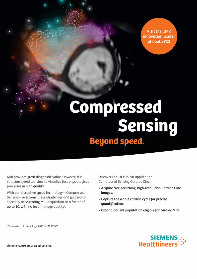
MRI provides great diagnostic value. However, it is still considered too slow to visualize fast physiological processes in high quality.
With our disruptive speed technology – Compressed Sensing – overcome these challenges and go beyond speed by accelerating MRI acquisition at a factor of up to 10, with no loss in image quality1.
siemens.com/compressed-sensing
Discover the 1st clinical application – Compressed Sensing Cardiac Cine:
• Acquire free-breathing, high-resolution Cardiac Cineimages.
• Capture the whole cardiac cycle for precisequantification.
• Expand patient population eligible for cardiac MRI.
1 Sudarski et. al., Radiology. 2016 Jul 11:151002
Beyond speed.
Compressed Sensing
Visit the CMR innovation leader
at booth #13
4493_MSC_C_AD_Compressed_Sensing_SCMR_8.5x11.indd 1 21.12.16 09:31

155www.cmr2018.org
NOTES
156
NOTES
156 157www.cmr2018.org
NOTES

158Discover our latest innovations atwww.philips.com/thenextmrwave
2D Cine imaging without Compressed SENSE2.0 x 1.6 x 8.0 mm, 10 s
2D Cine imaging with Compressed SENSE2.0 x 1.6 x 8.0 mm, 5.6 s
Magnetic Resonance
Speed done right, every time.
In a society where reimbursements are declining and chronic conditions lead to increased MR procedures and longer waiting times, there is ever increasing pressure on the radiology department. Today, further attempts to accelerate compromise image quality or are limited to a narrow range of scans. Therefore, to meet the increased demand for productivity, a technology break-through in acceleration is still required.
This is why Philips brings Compressed SENSE, a breakthrough in productivity. Compressed SENSE is not only about faster sequences, it is much more. It is about accelerating full patient examinations and thus guaranteeing the next level in productivity and image quality to empower your staff to focus where it matters the most, enhanced patient care.
2171386_Philips_Compressed_SENSE_cardiac_advertisement_V3.indd 1 19/12/17 11:46

158 159www.cmr2018.orgDiscover our latest innovations atwww.philips.com/thenextmrwave
2D Cine imaging without Compressed SENSE2.0 x 1.6 x 8.0 mm, 10 s
2D Cine imaging with Compressed SENSE2.0 x 1.6 x 8.0 mm, 5.6 s
Magnetic Resonance
Speed done right, every time.
In a society where reimbursements are declining and chronic conditions lead to increased MR procedures and longer waiting times, there is ever increasing pressure on the radiology department. Today, further attempts to accelerate compromise image quality or are limited to a narrow range of scans. Therefore, to meet the increased demand for productivity, a technology break-through in acceleration is still required.
This is why Philips brings Compressed SENSE, a breakthrough in productivity. Compressed SENSE is not only about faster sequences, it is much more. It is about accelerating full patient examinations and thus guaranteeing the next level in productivity and image quality to empower your staff to focus where it matters the most, enhanced patient care.
2171386_Philips_Compressed_SENSE_cardiac_advertisement_V3.indd 1 19/12/17 11:46
GENERAL FLOOR PLANLEVEL 1
LEVEL 0
MAIN ENTRANCE
PlenaryRoom
(111+112)
Room 4(113)
Room 5(114)
Room 6(115)
TechRoom(116)
SpeakerServiceCenter
CourseRoom
(133+134)
Hands-onRoom
(131+132)
SpeedMentoring(129+130)
EACVI &
SCMRMeetingRooms
Registration Area
To Faculty & MembersLounge
Case Room Lecture Room
Exhibition PosterArea Product
Theatre
LEVEL 1
LEVEL 0
MAIN ENTRANCE
PlenaryRoom
(111+112)
Room 4(113)
Room 5(114)
Room 6(115)
TechRoom(116)
SpeakerServiceCenter
CourseRoom
(133+134)
Hands-onRoom
(131+132)
SpeedMentoring(129+130)
EACVI &
SCMRMeetingRooms
Registration Area
To Faculty & MembersLounge
Case Room Lecture Room
Exhibition PosterArea Product
Theatre

22ND ANNUAL SCMR SCIENTIFIC SESSIONSGLOBAL CMR: INNOVATION AND CLINICAL OUTCOMES
February 6 – 9, 2019Seattle, USA • Hyatt Regency
#EuroCMR
www.escardio.org/EACVI
E u r o C M R 2019
17th Annual Meeting on Cardiovascular Magnetic Resonance (CMR)of the European Association of Cardiovascular Imaging (EACVI)
EXTENDING THE CLINICAL VALUE OF CMR THROUGH QUALITY AND EVIDENCE
2-4 May
V e n i c eI T A L Y
#EuroCMR
www.escardio.org/EACVI
E u r o C M R 2019
17th Annual Meeting on Cardiovascular Magnetic Resonance (CMR)of the European Association of Cardiovascular Imaging (EACVI)
EXTENDING THE CLINICAL VALUE OF CMR THROUGH QUALITY AND EVIDENCE
2-4 May
V e n i c eI T A L Y