2015-October.pdf - Hong Kong College of Cardiology
66
Volume 23 / Number 2 October 2015 Journal of the Hong Kong College of CARDIOLOGY ISSN 1027-7811 Including Abstracts of 19th Annual Scientific Meeting of the Institute of Cardiovascular Science and Medicine and the 10th Across the Strait Scientific Conference on Cardiovascular Science 21 November 2015 Hong Kong
-
Upload
khangminh22 -
Category
Documents
-
view
0 -
download
0
Transcript of 2015-October.pdf - Hong Kong College of Cardiology
Volume 23 / Number 2 October 2015
Journal of the Hong Kong College of
CARDIOLOGY
ISSN 1027-7811
Including Abstracts of19th Annual Scientific Meeting of the
Institute of Cardiovascular Science and Medicine and the10th Across the Strait Scientific Conference on Cardiovascular Science
21 November 2015Hong Kong
October 2015J HK Coll Cardiol, Vol 23 i
Journal of the Hong Kong College of Cardiology
Journal of the Hong Kong College of Cardiology (ISSN 1027-7811) is published bi-yearly by Medcom Limited, Room 504-5,Cheung Tat Centre, 18 Cheung Lee Street, Chai Wan, Hong Kong, tel (852) 2578 3833, fax (852) 2578 3929, email: [email protected]
Indexed in EMBASE/Excerpta Medica
Editor-in-ChiefChu-Pak Lau
Editorial BoardRaymond Hon-Wah ChanWai-Kwong ChanWai-Hong ChenChun-Ho ChengBernard CheungChung-Seung ChiangMoses S S ChowWing-Hing ChowKatherine FanChi-Lai HoKau-Chung HoDavid Sai-Wah Ho
International Editorial ConsultantsA John Camm Hamid IkramShih-Ann Chen David T KellyVictor Dzau Bertram PittBarry A Franklin William C RobertsDayi Hu Delon Wu
Section EditorsJohn E Sanderson, Editor of Clinical CardiologySuet-Ting Lau, Editor of Preventive CardiologyKau-Chung Ho, Editor of Invasive CardiologyYuk-Kong Lau, Editor of Non-invasive CardiologyChu-Pak Lau, Editor of Pacing and ElectrophysiologyCyrus R Kumana, Editor of Basic Cardiology: PharmacologyWai-Kwong Chan, Editor of Images in Cardiology: ECG
Cyrus R KumanaSuet-Ting LauYuk-Kong LauTin-Chu LawKathy Lai-Fun LeeStephen Wai-Luen LeeMaurice P LeungSum-Kin LeungWai-Suen LeungWing-Hung LeungShu-Kin LiArchie Ying-Sui Lo
Ngai-Shing MokJohn E SandersonBrian TomlinsonHung-Fat TseKai-Fat TseTak-Ming TseSiu-Hong WanKwok-Yiu WongAlexander Shou-Pang WongKam-Sang WooCheuk-Man Yu

October 2015 J HK Coll Cardiol, Vol 23ii
INSTRUCTION FOR AUTHORS
The Journal of the Hong Kong College of Cardiology publishes peer-reviewed articles on all aspects of cardiovascular disease, includingoriginal clinical studies, review articles and experimental investigations. As official journal of the Hong Kong College of Cardiology, the journalpublishes abstracts of reports to be presented at the Scientific Sessions of the College as well as reports of the College-sponsored conferences.
Manuscripts submitted to this journal must not be under simultaneous consideration by any other publication and should not have beenpublished elsewhere in substantially similar form. The letter of submission must so affirm. A transfer of copyright form to be signed by all authorsto the Hong Kong College of Cardiology should accompany all submitted articles. All manuscripts should be submitted to the Editor-in-Chief,Journal of the Hong Kong College of Cardiology, c/o Medcom Limited, Room 504-5, Cheung Tat Centre, 18 Cheung Lee Street, Chai Wan,Hong Kong, Email: [email protected].
Manuscript PreparationManuscripts must be submitted in English in triplicate (one originaland two copies) and typed double-spaced on A4 size white bond paper.This applies to all parts of the manuscript, i.e. references, legends,etc. Liberal margins should be left at the top and bottom, as well asthe sides. Except for editorials, images/ECG and letters, all manuscriptshould be submitted in the following order: Title Page, Abstract, Text,References, Tables, Legends, and Figures. Each page, beginning withthe summary, should also include the senior author's surname typedon the upper, left-hand corner. The author should not make any changesin the proofs except for corrections of editorial errors, if any, and/orcorrection of typesetter's errors. Employees of industry may notevaluate or comment about the products of a competitor. A commercialname should not be part of a manuscript title. Finally, authors shouldmake no claims of priority in their manuscripts.
Title Page- Include full name(s), degree(s) and affiliation(s) of author(s); list
under file.- Give a running title of 3 to 6 words.- At the bottom of the page, include information about grants,if
applicable.- Add: "Address for reprint:...", followed by full name, address,
telephone and fax numbers.
Abstract- Abstract should be after title page and numbered page 1.- It should not exceed 250 words for major articles; case reports
should have abstracts of no more than 100 words.- At the end of the abstract, provide a maximum of 6 key words
suitable for indexing.- Abbreviations should be kept to a minimum and must be explained
when they first appear; after first use, abbreviations alone may beused.
- Standard abbreviations should be used for all measurements(SI units).
Text- The text should follow the abstract and begin on a new page, as
should References, Tables, and Legends.- Abbreviations not defined in the abstract should be explained when
they first appear in the text.- References should be cited in numerical order, as should tables
and figures.
References- Number in the order in which they appear in the text.- Abbreviate titles of periodicals according to the style of the Index
Medicus.- Follow the format (arrangement, punctuation) shown below:
Periodicals1. Lewis T. Paroxysmal tachycardia. Heart 1909;1:43-72.
(if more than three authors, please use "et al." after the third).
Books (edited by other authors of article)2. Furman S. Pacemaker follow-up. In Barold SS, (eds): Modern
Cardiac Pacing. Mount Kisco, New York, Futura PublishingCompany, 1985, pp. 889-958.
Books (identical author and editor)3. Chung EK. Principles of Cardiac Arrhythmias. Baltimore, MD,
Williams & Wilkins, 1977, pp. 97-188.
Abstracts4. Same as periodicals and followed by "(abstract)".
Tables- Tables should supplement, but not duplicate, the text.- Tables should be numbered consecutively in order of appearance
in the text.- Each table must be given an Arabic numeral and a title, placed at
the top of the page.- Abbreviations used in the table should be foot-noted and explained
in the order in which they appear in the table, if they have not beenpreviously used.
- Any material which is not self-explanatory should be foot-noted as well.
Legends- Be sure that legends and figures correspond.- Identify all abbreviations used in a figure at the end of each legend,
if the abbreviation has not been used in the text.- Be sure abbreviations used for measurements are standard SI unit.
Figures- Submit either 3 black and white glossy prints or 2 prints and one
photocopy, preferably of 13 cm x 18 cm (5" x 7") size.- On the back of each figure, indicate number, senior author's
surname, top of illustration; all of this should be written lightlywith soft, black pencil.
- Submit written permission from publisher(s) for any figure whichhas been published previously.
- Do not use clips on illustrations; submit them in an envelope backedby cardboard.
- Any lettering or scale of measurement used in an illustration mustbe large enough to be legible in the event of half-size reduction.
- Do not send original art-work, X-rays, or ECGs.- Photographs in which a patient or other person is identifiable must
have written permission from that person. The consent must statespecifically what the person is consenting to and what restrictions,if any, the person has placed upon the publication of the photo-graph. All restrictions must be strictly observed.
- Colour illustrations are costly and will be charged to the author.- Authors should inquire about cost from the publisher before
submitting a colour illustration.
EthicsPublished studies on human subjects should indicate the nature ofconsent and the approval of the institutional ethics committee ifdeemed appropriate. In case of animal experiments, ethical approvalmust be enclosed.
The author is responsible for all material presented in a paper. Thejournal disclaims all responsibility for such material. No product orservice advertised in this publication is guaranteed or warranted eitherby the Editors or publisher. Neither the Editors nor publisher guaranteeany claims made by a manufacturer or an author in regard to a productor service. If a trademark item is named, the name(s) and address(es)of the manufacturer(s) or supplier(s), in addition to the generic name,should be foot-noted.
Reprints are available. Ordering information can be obtained from theabove address.
Subscription RatesLocal Subscription: HK$200/year (including postage)Overseas Subscription: US$120/year (including airmail postage)
October 2015J HK Coll Cardiol, Vol 23 iii
Journal of the Hong Kong College of Cardiology
October 2015Volume 23, No. 2
• ORIGINAL ARTICLE
Investigating the Association Between Anti-
Brucella Titers and the Development of
Coronary Artery Disease in Middle-East
Umayya Musharrafieh, Ali Choukair, Abdul
Rahman Bizri, Hala Tamim, Antoine Nasrallah,
Samir Alam ...............................................68
• CASE REPORT
Tricuspid Aseptic Endocarditis Revealing
Right Endomyocardial Fibrosis During an
Unrecognized Behçet's Disease: Two Cases
Report
Hanane Benhalla, Ibrahim Doumbouya,
Malika Noureddine, Rachida Habbal ..........75
Table of Contents
• 19TH ANNUAL SCIENTIFIC MEETING OF THEINSTITUTE OF CARDIOVASCULAR SCIENCEAND MEDICINE AND THE 10TH ACROSS THESTRAIT SCIENTIFIC CONFERENCE ONCARDIOVASCULAR SCIENCE
Organizing Committee...................................79
Scientific Programme.....................................80
Abstracts..........................................................82

October 2015 J HK Coll Cardiol, Vol 23iv
The Hong Kong College of Cardiology
The CouncilPresident Shu-Kin LiPresident-Elect Yuk-Kong LauHonorary Secretary Suet-Ting LauHonorary Treasurer Ngai-Yin ChanImmediate Past President Kam-Tim ChanAccreditation and Education Committee Chairman Tak-Fu TseScientific Committee Chairman Chung-Wah SiuChief Editor Chu-Pak LauGeneral Affairs and Public Relations Committee Chairman Shu-Kin LiCouncil Members Kwok-Keung Chan
Wai-Kwong ChanBoron Cheung-Wah ChengChung-Seung ChiangCharles Kau-Chung HoChu-Pak LauStephen Wai-Luen LeeGodwin Tat-Chi LeungChung-Wah SiuKin-Lam TsuiThomas TunggalChris Kwok-Yiu WongCheuk-Man Yu
Honorary Legal Adviser Peggy CheungHonorary Auditor Patrick Lung-Tak Wong
Correspondence forHong Kong College of Cardiology
Secretariat, Room 1116, Bank of America Tower, 12 Harcourt Road, Hong Kong.Tel: (852) 2899 2035, Fax: (852) 2899 2045
E-mail: [email protected]
October 2015 J HK Coll Cardiol, Vol 23ii
INSTRUCTION FOR AUTHORS
The Journal of the Hong Kong College of Cardiology publishes peer-reviewed articles on all aspects of cardiovascular disease, includingoriginal clinical studies, review articles and experimental investigations. As official journal of the Hong Kong College of Cardiology, the journalpublishes abstracts of reports to be presented at the Scientific Sessions of the College as well as reports of the College-sponsored conferences.
Manuscripts submitted to this journal must not be under simultaneous consideration by any other publication and should not have beenpublished elsewhere in substantially similar form. The letter of submission must so affirm. A transfer of copyright form to be signed by all authorsto the Hong Kong College of Cardiology should accompany all submitted articles. All manuscripts should be submitted to the Editor-in-Chief,Journal of the Hong Kong College of Cardiology, c/o Medcom Limited, Room 504-5, Cheung Tat Centre, 18 Cheung Lee Street, Chai Wan,Hong Kong, Email: [email protected].
Manuscript PreparationManuscripts must be submitted in English in triplicate (one originaland two copies) and typed double-spaced on A4 size white bond paper.This applies to all parts of the manuscript, i.e. references, legends,etc. Liberal margins should be left at the top and bottom, as well asthe sides. Except for editorials, images/ECG and letters, all manuscriptshould be submitted in the following order: Title Page, Abstract, Text,References, Tables, Legends, and Figures. Each page, beginning withthe summary, should also include the senior author's surname typedon the upper, left-hand corner. The author should not make any changesin the proofs except for corrections of editorial errors, if any, and/orcorrection of typesetter's errors. Employees of industry may notevaluate or comment about the products of a competitor. A commercialname should not be part of a manuscript title. Finally, authors shouldmake no claims of priority in their manuscripts.
Title Page- Include full name(s), degree(s) and affiliation(s) of author(s); list
under file.- Give a running title of 3 to 6 words.- At the bottom of the page, include information about grants,if
applicable.- Add: "Address for reprint:...", followed by full name, address,
telephone and fax numbers.
Abstract- Abstract should be after title page and numbered page 1.- It should not exceed 250 words for major articles; case reports
should have abstracts of no more than 100 words.- At the end of the abstract, provide a maximum of 6 key words
suitable for indexing.- Abbreviations should be kept to a minimum and must be explained
when they first appear; after first use, abbreviations alone may beused.
- Standard abbreviations should be used for all measurements(SI units).
Text- The text should follow the abstract and begin on a new page, as
should References, Tables, and Legends.- Abbreviations not defined in the abstract should be explained when
they first appear in the text.- References should be cited in numerical order, as should tables
and figures.
References- Number in the order in which they appear in the text.- Abbreviate titles of periodicals according to the style of the Index
Medicus.- Follow the format (arrangement, punctuation) shown below:
Periodicals1. Lewis T. Paroxysmal tachycardia. Heart 1909;1:43-72.
(if more than three authors, please use "et al." after the third).
Books (edited by other authors of article)2. Furman S. Pacemaker follow-up. In Barold SS, (eds): Modern
Cardiac Pacing. Mount Kisco, New York, Futura PublishingCompany, 1985, pp. 889-958.
Books (identical author and editor)3. Chung EK. Principles of Cardiac Arrhythmias. Baltimore, MD,
Williams & Wilkins, 1977, pp. 97-188.
Abstracts4. Same as periodicals and followed by "(abstract)".
Tables- Tables should supplement, but not duplicate, the text.- Tables should be numbered consecutively in order of appearance
in the text.- Each table must be given an Arabic numeral and a title, placed at
the top of the page.- Abbreviations used in the table should be foot-noted and explained
in the order in which they appear in the table, if they have not beenpreviously used.
- Any material which is not self-explanatory should be foot-noted as well.
Legends- Be sure that legends and figures correspond.- Identify all abbreviations used in a figure at the end of each legend,
if the abbreviation has not been used in the text.- Be sure abbreviations used for measurements are standard SI unit.
Figures- Submit either 3 black and white glossy prints or 2 prints and one
photocopy, preferably of 13 cm x 18 cm (5" x 7") size.- On the back of each figure, indicate number, senior author's
surname, top of illustration; all of this should be written lightlywith soft, black pencil.
- Submit written permission from publisher(s) for any figure whichhas been published previously.
- Do not use clips on illustrations; submit them in an envelope backedby cardboard.
- Any lettering or scale of measurement used in an illustration mustbe large enough to be legible in the event of half-size reduction.
- Do not send original art-work, X-rays, or ECGs.- Photographs in which a patient or other person is identifiable must
have written permission from that person. The consent must statespecifically what the person is consenting to and what restrictions,if any, the person has placed upon the publication of the photo-graph. All restrictions must be strictly observed.
- Colour illustrations are costly and will be charged to the author.- Authors should inquire about cost from the publisher before
submitting a colour illustration.
EthicsPublished studies on human subjects should indicate the nature ofconsent and the approval of the institutional ethics committee ifdeemed appropriate. In case of animal experiments, ethical approvalmust be enclosed.
The author is responsible for all material presented in a paper. Thejournal disclaims all responsibility for such material. No product orservice advertised in this publication is guaranteed or warranted eitherby the Editors or publisher. Neither the Editors nor publisher guaranteeany claims made by a manufacturer or an author in regard to a productor service. If a trademark item is named, the name(s) and address(es)of the manufacturer(s) or supplier(s), in addition to the generic name,should be foot-noted.
Reprints are available. Ordering information can be obtained from theabove address.
Subscription RatesLocal Subscription: HK$200/year (including postage)Overseas Subscription: US$120/year (including airmail postage)

October 2015J HK Coll Cardiol, Vol 23 iii
Journal of the Hong Kong College of Cardiology
October 2015Volume 23, No. 2
• ORIGINAL ARTICLE
Investigating the Association Between Anti-
Brucella Titers and the Development of
Coronary Artery Disease in Middle-East
Umayya Musharrafieh, Ali Choukair, Abdul
Rahman Bizri, Hala Tamim, Antoine Nasrallah,
Samir Alam ...............................................68
• CASE REPORT
Tricuspid Aseptic Endocarditis Revealing
Right Endomyocardial Fibrosis During an
Unrecognized Behçet's Disease: Two Cases
Report
Hanane Benhalla, Ibrahim Doumbouya,
Malika Noureddine, Rachida Habbal ..........75
Table of Contents
• 19TH ANNUAL SCIENTIFIC MEETING OF THEINSTITUTE OF CARDIOVASCULAR SCIENCEAND MEDICINE AND THE 10TH ACROSS THESTRAIT SCIENTIFIC CONFERENCE ONCARDIOVASCULAR SCIENCE
Organizing Committee...................................79
Scientific Programme.....................................80
Abstracts..........................................................82
October 2015 J HK Coll Cardiol, Vol 23iv
The Hong Kong College of Cardiology
The CouncilPresident Shu-Kin LiPresident-Elect Yuk-Kong LauHonorary Secretary Suet-Ting LauHonorary Treasurer Ngai-Yin ChanImmediate Past President Kam-Tim ChanAccreditation and Education Committee Chairman Tak-Fu TseScientific Committee Chairman Chung-Wah SiuChief Editor Chu-Pak LauGeneral Affairs and Public Relations Committee Chairman Shu-Kin LiCouncil Members Kwok-Keung Chan
Wai-Kwong ChanBoron Cheung-Wah ChengChung-Seung ChiangCharles Kau-Chung HoChu-Pak LauStephen Wai-Luen LeeGodwin Tat-Chi LeungChung-Wah SiuKin-Lam TsuiThomas TunggalChris Kwok-Yiu WongCheuk-Man Yu
Honorary Legal Adviser Peggy CheungHonorary Auditor Patrick Lung-Tak Wong
Correspondence forHong Kong College of Cardiology
Secretariat, Room 1116, Bank of America Tower, 12 Harcourt Road, Hong Kong.Tel: (852) 2899 2035, Fax: (852) 2899 2045
E-mail: [email protected]
October 2015J HK Coll Cardiol, Vol 23 68
Investigating the Association Between Anti-Brucella Titers and theDevelopment of Coronary Artery Disease in Middle-East
UMAYYA MUSHARRAFIEH,1 ALI CHOUKAIR,2 ABDUL RAHMAN BIZRI,2 HALA TAMIM,3 ANTOINENASRALLAH,2 SAMIR ALAM2
From 1Department of Family Medicine, American University of Beirut Medical Center; 2Department of InternalMedicine, American University of Beirut Medical Center; 3Faculty of Health Sciences, American University of Beirut,Beirut, Lebanon
MUSHARRAFIEH ET AL.: Investigating the Association Between Anti-Brucella Titers and the Development ofCoronary Artery Disease in Middle-East. Background: Several studies have suggested an association between certaininfectious agents and coronary artery disease (CAD). A possible role for brucella in contributing to cardiovasculardisease through its ability to produce a chronic inflammatory state was hypothesized. In this study, brucella wasevaluated for its possible pathogenic role in significant occlusive coronary artery disease through testing for thepresence of positive brucella antibody titers. Methods and Results: Patients referred for coronary angiography at theAmerican University of Beirut Medical Center between January 2005 and February 2009 were tested for C-reactiveprotein (CRP) quantitative level and the presence of brucella antibody titers using ELISA. All participants were askedto fill a questioner relevant to demographics, risk factors for CAD, and presence of comorbidities. Results: 424subjects were categorized into two groups; those with greater than 75% stenosis in at least one coronary artery andthose with normal or less than 75% stenosis. Among patients with positive anti-brucella titers, 70.6% had CAD whileamong patients with negative anti-brucella titers 74.9% had CAD (P=0.514). In patients with elevated CRP level(≥3 mg/L), 14.9% had positive titer for brucella, whereas in those with low CRP level (<3 mg/L), 5.8% had positivetiters for brucella (P=0.016). Conclusions: The findings failed to show an association between anti-brucella titers andthe development of significant CAD despite the presence of a significant correlation between brucella antibodypositivity and elevated CRP. Further studies are needed to explore the role of this infectious agent with other knowncardiac risk factors. (J HK Coll Cardiol 2015;23:68-74)
Brucella, Coronary artery disease, C-reactive protein, Inflammation
C A D
2005 1 2009 2C CRP EL I SA
C A D 4 2 4 7 5 %75% 70.6% CAD74.9% CAD P=0.514 CRP ≥3 mg/L 14.9%
CRP <3 mg/L 5.8% P=0.016C A D C R P
C
Address for reprints: Dr. Ali ChoukairDepartment of Internal Medicine, American University of BeirutMedical Center, Beirut, Lebanon
Email: [email protected]
Received June 18, 2015; revision accepted October 6, 2015
October 2015 J HK Coll Cardiol, Vol 2369
BRUCELLA INFECTION AND ATHEROSCLEROTIC HEART DISEASE
Introduction
Coronary artery disease (CAD) continues to bethe leading cause of morbidity and mortality in bothindustrialized and developing countries despite advancesin prevention, detection, and treatment.1 Hyperlipidemia,hypertension, smoking, diabetes, family history, obesityand inactivity are well-documented risk factors for CAD.2
However, in approximately half of the patients withCAD, the above mentioned risk factors are not presentand thus the search for other factors is warranted.3
Previous studies have suggested a possible role forinfectious agents in the pathogenesis of CAD.4,5
Chlamydia pneumonia , 6 Helicobacter pylori ,7
and Porphyromonas gingivalis,8 cytomegalovirus,9
herpes simplex virus,10 and hepatitis A virus11 were allevaluated.
Brucellosis is a debilitating disease caused byBrucella spp. that can affect different organs and may ifinappropriately treated lead to a chronic illness. Boththe acute and chronic manifestations of brucellosis aredue to inflammatory phenomena. There is currentevidence that Brucella spp., can infect and survive withinthe endothelial cells, and can induce a pro-inflammatoryresponse that might be involved in the vascularmanifestations of brucellosis. The release of adhesionmolecules and pro-inflammatory chemochines during abrucella infection plays an important role in theactivation of the endothelial system. This activation maybe responsible for the pathogenesis of the damage inthe vascular system.12 The link between brucellainfection and atherogenic lipid profile coupled withendothelial dysfunction may be a contributing factor inthe development of coronary artery disease.13
Brucella is known to be endemic in the MiddleEast region with a reported incidence between 40-69per 100,000 population in countries like Saudi Arabia,Kuwait and Jordan.14
In Lebanon, a study conducted by Araj et alincluded 597 persons with high risk occupations foundthat the overall seroprevalence of brucella titers rangedfrom 26-61%.15 Data from the Lebanese Ministry ofPublic Health reveals an annual average of 220 brucellacases reported over the past 14 years.16 In a recent studyfrom Greece acute brucellosis was associated with a shift
of serum lipids, lipoproteins, and associated enzymestoward a more atherogenic lipid profile, which is notfully restored 4 months after treatment.17 Along the sametoken, abnormal flow-mediated dilatation (FMD)measurement as reflection of endothelial function andthus the first stage of atherogenesis, might be a markerof arterial dysfunction, increased cardiovascular risk andatherosclerosis.18
The aim of the present study is to investigate thepresence of any association between brucella serologypositivity and the occurrence of significant coronaryartery disease.
Materials and Methods
Study PopulationFollowing Institutional Review Board approval,
all patients admitted to the American University of BeirutMedical Center and referred for coronary angiographybetween January 2005 and February 2009 wereapproached for recruitment in the study. Writteninformed consent was obtained from eligible patientsfor drawing and using blood samples for scientificresearch after explaining the aim of the study and natureof the procedure.
Based on the diagnostic findings of the coronaryangiography, patients enrolled were categorized into twogroups. The first group included patients with normalcoronary arteries and those with less than 75% luminalstenosis by angiography, the second group includedpatients with 75% luminal stenosis or more in at leastone coronary artery. All those who have experiencedmyocardial infarction within the previous 6 months,valvular heart disease, or non-atheroscleroticcardiomyopathy were excluded.
Study DesignAtherosclerosis Risk Factors
Medical records of enrolled patients werereviewed for demographics (age and gender), thepresence of known risk factors for CAD including familyhistory, diabetes mellitus (DM), hypertension (HTN),hypercholesterolemia (HCL), obesity, smoking, anddietary habits. Medical records were also reviewed for
J HK Coll Cardiol, Vol 23 70October 2015
MUSHARRAFIEH ET AL.
models were used to assess the independent associationsof various risk factors to CAD.
Results
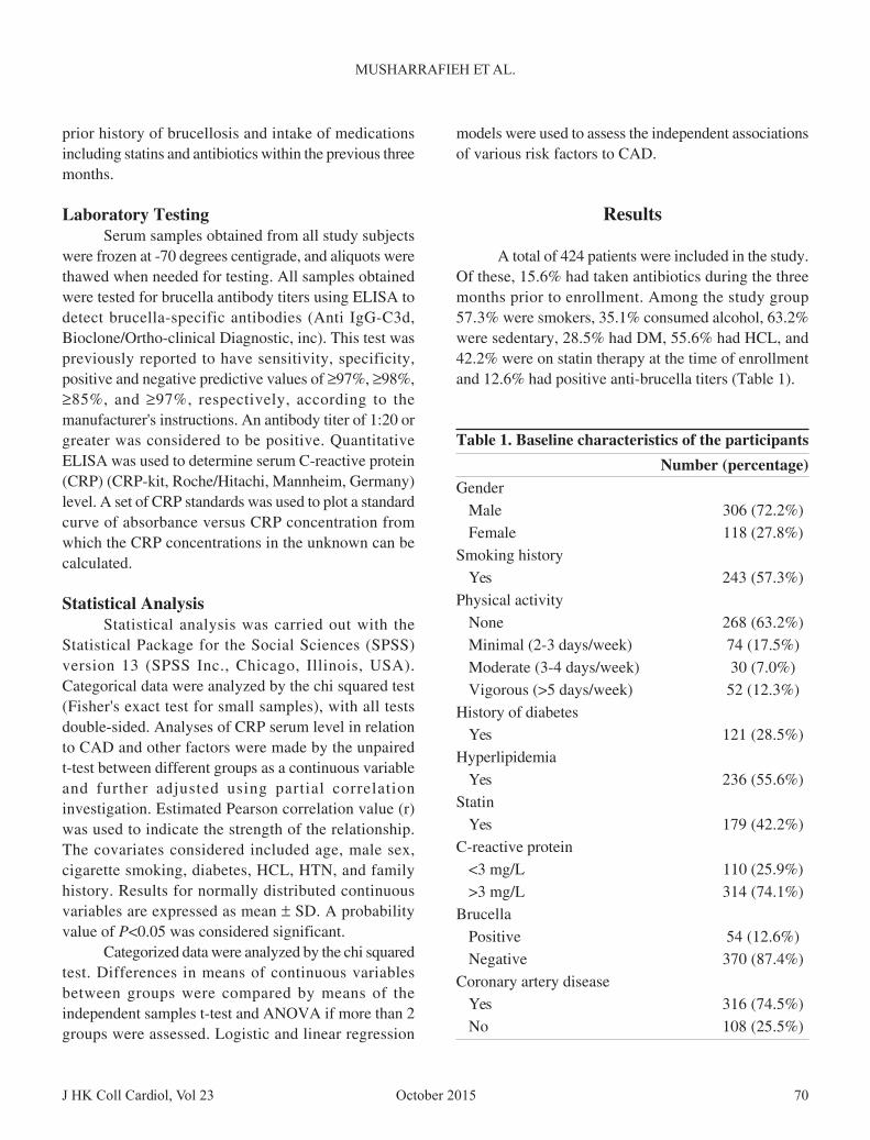
A total of 424 patients were included in the study.Of these, 15.6% had taken antibiotics during the threemonths prior to enrollment. Among the study group57.3% were smokers, 35.1% consumed alcohol, 63.2%were sedentary, 28.5% had DM, 55.6% had HCL, and42.2% were on statin therapy at the time of enrollmentand 12.6% had positive anti-brucella titers (Table 1).
prior history of brucellosis and intake of medicationsincluding statins and antibiotics within the previous threemonths.
Laboratory TestingSerum samples obtained from all study subjects
were frozen at -70 degrees centigrade, and aliquots werethawed when needed for testing. All samples obtainedwere tested for brucella antibody titers using ELISA todetect brucella-specific antibodies (Anti IgG-C3d,Bioclone/Ortho-clinical Diagnostic, inc). This test waspreviously reported to have sensitivity, specificity,positive and negative predictive values of ≥97%, ≥98%,≥85%, and ≥97%, respectively, according to themanufacturer's instructions. An antibody titer of 1:20 orgreater was considered to be positive. QuantitativeELISA was used to determine serum C-reactive protein(CRP) (CRP-kit, Roche/Hitachi, Mannheim, Germany)level. A set of CRP standards was used to plot a standardcurve of absorbance versus CRP concentration fromwhich the CRP concentrations in the unknown can becalculated.
Statistical AnalysisStatistical analysis was carried out with the
Statistical Package for the Social Sciences (SPSS)version 13 (SPSS Inc., Chicago, Illinois, USA).Categorical data were analyzed by the chi squared test(Fisher's exact test for small samples), with all testsdouble-sided. Analyses of CRP serum level in relationto CAD and other factors were made by the unpairedt-test between different groups as a continuous variableand further adjusted using partial correlationinvestigation. Estimated Pearson correlation value (r)was used to indicate the strength of the relationship.The covariates considered included age, male sex,cigarette smoking, diabetes, HCL, HTN, and familyhistory. Results for normally distributed continuousvariables are expressed as mean ± SD. A probabilityvalue of P<0.05 was considered significant.
Categorized data were analyzed by the chi squaredtest. Differences in means of continuous variablesbetween groups were compared by means of theindependent samples t-test and ANOVA if more than 2groups were assessed. Logistic and linear regression
Table 1. Baseline characteristics of the participants
Number (percentage)Gender
Male 306 (72.2%)Female 118 (27.8%)
Smoking historyYes 243 (57.3%)
Physical activityNone 268 (63.2%)Minimal (2-3 days/week) 74 (17.5%)Moderate (3-4 days/week) 30 (7.0%)Vigorous (>5 days/week) 52 (12.3%)
History of diabetesYes 121 (28.5%)
HyperlipidemiaYes 236 (55.6%)
StatinYes 179 (42.2%)
C-reactive protein<3 mg/L 110 (25.9%)>3 mg/L 314 (74.1%)
BrucellaPositive 54 (12.6%)Negative 370 (87.4%)
Coronary artery diseaseYes 316 (74.5%)No 108 (25.5%)
October 2015 J HK Coll Cardiol, Vol 2371
BRUCELLA INFECTION AND ATHEROSCLEROTIC HEART DISEASE
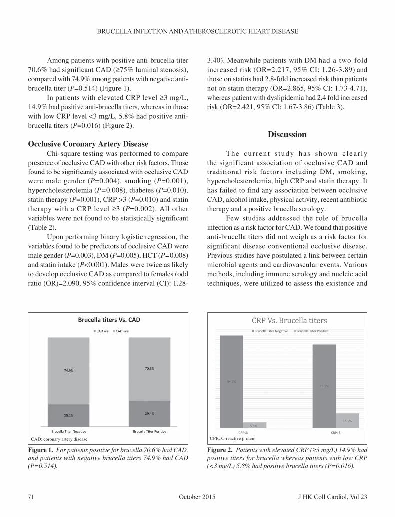
Among patients with positive anti-brucella titer70.6% had significant CAD (≥75% luminal stenosis),compared with 74.9% among patients with negative anti-brucella titer (P=0.514) (Figure 1).
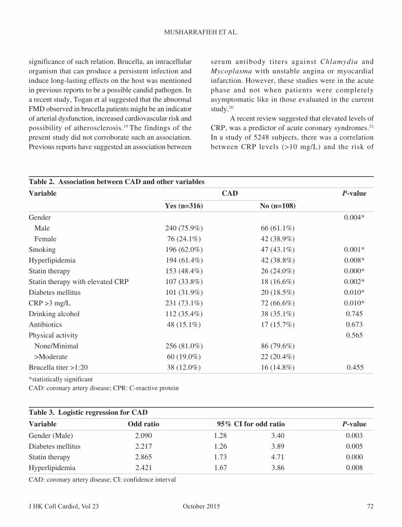
In patients with elevated CRP level ≥3 mg/L,14.9% had positive anti-brucella titers, whereas in thosewith low CRP level <3 mg/L, 5.8% had positive anti-brucella titers (P=0.016) (Figure 2).
Occlusive Coronary Artery DiseaseChi-square testing was performed to compare
presence of occlusive CAD with other risk factors. Thosefound to be significantly associated with occlusive CADwere male gender (P=0.004), smoking (P=0.001),hypercholesterolemia (P=0.008), diabetes (P=0.010),statin therapy (P=0.001), CRP >3 (P=0.010) and statintherapy with a CRP level ≥3 (P=0.002). All othervariables were not found to be statistically significant(Table 2).
Upon performing binary logistic regression, thevariables found to be predictors of occlusive CAD weremale gender (P=0.003), DM (P=0.005), HCT (P=0.008)and statin intake (P<0.001). Males were twice as likelyto develop occlusive CAD as compared to females (oddratio (OR)=2.090, 95% confidence interval (CI): 1.28-
3.40). Meanwhile patients with DM had a two-foldincreased risk (OR=2.217, 95% CI: 1.26-3.89) andthose on statins had 2.8-fold increased risk than patientsnot on statin therapy (OR=2.865, 95% CI: 1.73-4.71),whereas patient with dyslipidemia had 2.4 fold increasedrisk (OR=2.421, 95% CI: 1.67-3.86) (Table 3).
Discussion
The cur ren t s tudy has shown c lea r lythe significant association of occlusive CAD andtraditional risk factors including DM, smoking,hypercholesterolemia, high CRP and statin therapy. Ithas failed to find any association between occlusiveCAD, alcohol intake, physical activity, recent antibiotictherapy and a positive brucella serology.
Few studies addressed the role of brucellainfection as a risk factor for CAD. We found that positiveanti-brucella titers did not weigh as a risk factor forsignificant disease conventional occlusive disease.Previous studies have postulated a link between certainmicrobial agents and cardiovascular events. Variousmethods, including immune serology and nucleic acidtechniques, were utilized to assess the existence and
Figure 1. For patients positive for brucella 70.6% had CAD,and patients with negative brucella titers 74.9% had CAD(P=0.514).
Figure 2. Patients with elevated CRP (≥3 mg/L) 14.9% hadpositive titers for brucella whereas patients with low CRP(<3 mg/L) 5.8% had positive brucella titers (P=0.016).
CAD: coronary artery disease CPR: C-reactive protein
J HK Coll Cardiol, Vol 23 72October 2015
MUSHARRAFIEH ET AL.
significance of such relation. Brucella, an intracellularorganism that can produce a persistent infection andinduce long-lasting effects on the host was mentionedin previous reports to be a possible candid pathogen. Ina recent study, Togan et al suggested that the abnormalFMD observed in brucella patients might be an indicatorof arterial dysfunction, increased cardiovascular risk andpossibility of atherosclerosis.19 The findings of thepresent study did not corroborate such an association.Previous reports have suggested an association between
serum antibody titers against Chlamydia andMycoplasma with unstable angina or myocardialinfarction. However, these studies were in the acutephase and not when patients were completelyasymptomatic like in those evaluated in the currentstudy.20
A recent review suggested that elevated levels ofCRP, was a predictor of acute coronary syndromes.21
In a study of 5248 subjects, there was a correlationbetween CRP levels (>10 mg/L) and the risk of
Table 2. Association between CAD and other variables
Variable CAD P-value
Yes (n=316) No (n=108)
Gender 0.004*
Male 240 (75.9%) 66 (61.1%)
Female 76 (24.1%) 42 (38.9%)
Smoking 196 (62.0%) 47 (43.1%) 0.001*
Hyperlipidemia 194 (61.4%) 42 (38.8%) 0.008*
Statin therapy 153 (48.4%) 26 (24.0%) 0.000*
Statin therapy with elevated CRP 107 (33.8%) 18 (16.6%) 0.002*
Diabetes mellitus 101 (31.9%) 20 (18.5%) 0.010*
CRP >3 mg/L 231 (73.1%) 72 (66.6%) 0.010*
Drinking alcohol 112 (35.4%) 38 (35.1%) 0.745
Antibiotics 48 (15.1%) 17 (15.7%) 0.673
Physical activity 0.565
None/Minimal 256 (81.0%) 86 (79.6%)
>Moderate 60 (19.0%) 22 (20.4%)
Brucella titer >1:20 38 (12.0%) 16 (14.8%) 0.455
*statistically significantCAD: coronary artery disease; CPR: C-reactive protein
Table 3. Logistic regression for CAD
Variable Odd ratio 95% CI for odd ratio P-value
Gender (Male) 2.090 1.28 3.40 0.003
Diabetes mellitus 2.217 1.26 3.89 0.005
Statin therapy 2.865 1.73 4.71 0.000
Hyperlipidemia 2.421 1.67 3.86 0.008
CAD: coronary artery disease; CI: confidence interval
October 2015 J HK Coll Cardiol, Vol 2373
BRUCELLA INFECTION AND ATHEROSCLEROTIC HEART DISEASE
cardiovascular events as well as all-cause mortality.22
Patients with high CRP were more likely to have moresevere CAD disease than those with lower CRP levels(63.2% had at least one vessel disease greater than 75%vs. 44.3% and 45.8%) (P=0.001) This is consistent withrecent reports showing a significant correlation betweenserum CRP levels and severity of CAD as assessed byangiographic Gensini score.23 Our results are inagreement with these reports and confirm that elevatedlevels of CRP are associated with at least one vesseldisease with stenosis of greater than 75%, unrelated tobrucella infection. Another report suggested thatBrucella species can infect and survive withinendothelial cells, and can induce a pro-inflammatoryresponse that might be involved in the vascularmanifestations of brucellosis.24 Despite the findings inour report that patients with elevated CRP (≥3) weremore likely to have positive anti-brucella titers thanthose with low CRP (14.9% vs. 5.8%), it was difficultto prove the presence of any significant associationas a contributory factor to occlusive CAD. Ournegative findings do not negate the possible role ofbrucella as a preparative trigger in CAD. It has beenshown that the response of the host to brucellainfection and inflammation leads to an increase inoxidized lipids in the serum, and brings about LDLoxidation in vivo. Oxidative modification of LDL is oneof the important events leading to the development ofatherosclerosis.13 Although the degree of inflammationinduced by brucella seems to be lower than in infectionscaused by other organism, the chronic nature ofthe infection argues in favor of inflammation as acause of tissue damage. This is due to a low degree ofstimulation but incessant inflammatory tissue damagingresponse.24
Our results cannot evaluate the combined effectof various triggers or risk factors in the pathogenesis ofocclusive disease. A weak trigger of inflammation as ina chronic brucella infection in a high risk patient maylead to a different CAD outcome when compared to amore potent trigger in a low risk patient.
It is reasonable to think that the same trigger ofinflammation may lead to different outcomes in patientswith different other risks for CAD. Ozbudak et al havealluded to this through a study that showed a synergisticeffect of infection and cholesterol rich diet on
atherosclerosis in pulmonary arteries. The authorsconcluded that antibiotics and anti-inflammatory agentscould be useful in prevention.25
Some published data have suggested that theaggregate effect of co-infection with multiple organismsrather than one organism may be responsible for theatherosclerotic role. This has been eluded to as the"infectious burden" or "pathogen burden".17,26 In onestudy, over 75% of CAD patients had been exposed toat least three of five pathogens tested, suggesting apossible link between increased pathogen burden andthe risk of CAD irrespective of traditional risk factors.27
Thus, the contribution of infectious organisms toatherosclerosis pathogenesis is likely to involvesimultaneous direct and indirect mechanisms involvingmultiple organisms.
Limitation
The authors recognize the selection bias inherentin enrolling subjects from a patient population that isundergoing an invasive cardiovascular procedure. Theresults obtained may not be necessarily applicable tothe population as a whole. Another important limitationis attempting to categorize the study population intothose with and without significant occlusive disease(more than 75% stenosis) irrespective of the presenceof lumen-limiting CAD or not. In addition, other riskfactors assessment for CAD may not be comprehensive,in that information on hypertension, with or without anti-hypertensive therapy is lacking.
Conclusion
The current study failed to show an associationbetween positive brucella serology and the developmentof significant occlusive CAD, despite the presence of asignificant correlation between brucella antibodypositivity and elevated CRP.
Further studies are needed to explore anyrelationship between brucella infection and CADincluding acute versus chronic infection, co-infectionwith other pathogens, and the interaction with otherknown risk factors.

J HK Coll Cardiol, Vol 23 74October 2015
MUSHARRAFIEH ET AL.
References
1. National Heart, lung, and blood Institute Fact Book. USDepartment of Health and Human Services Monographs: March,1996:30-52.
2. Farmer JA, Gotto AM. Dyslipidemia and other risk factors forcoronary artery disease. In: Braunwald E, editor. Heart Disease.A Textbook of Cardiovascular Medicine. 5th ed. Philadephia:WB Saunder; 1997:1126-69.
3. Robinson K, Mayer EL, Miller DP, et al. Hyperhomocystinemiaand low pyridoxal phosphate: common and independentreversible risk factors for coronary artery disease. Circulation1995;92:2825-30.
4. Danesh J, Collins R, Peto R. Chronic infections and coronaryheart disease: is there a link? Lancet 1997;350:430-6.
5. Libby P, Egan D, Skarlatos S. Roles of infectious agents inatherosclerosis and re-stenosis: an assessment of the evidenceand need for future research. Circulation 1997;96:4095-103.
6. Markus HS, Sitzer M, Carrington D, Mendall MA, SteinmetzH. Chlamydia pneumoniae infection and early asymptomaticcarotid atherosclerosis. Circulation 1999;100:832-7.
7. Pasceri V, Cammarota G, Patti G, et al. Association of virulentHelicobacter pylori strains with ischemic heart disease.Circulation 1998;97:1675-9.
8. Chiu B. Multiple infections in carotid atherosclerotic plaques.Am Heart J 1999;138:534-6.
9. Muhlestein JB, Horne BD, Carlquist JF, et al. Cytomegalovirusseropositivity and C-reactive protein have independent andcombined predictive value for mortality in patients withangiographically demonstrated coronary artery disease.Circulation 2000;102:1917-23.
10. Alber DG, Powell KL, Vallance P, et al. Herpesvirus infectionaccelerates atherosclerosis in the apolipoprotein E-deficientmouse. Circulation 2000;102:779-85.
11. Zhu J, Quyyumi AA, Norman JE, et al. The possible role ofhepatitis A virus in the pathogenesis of atherosclerosis. J InfectDis 2000;182:1583-7.
12. Ferrero MC, Bregante J, DelpinoMV, et al. Proinflammatoryresponse of human endothelial cells to Brucella infection.Microbes Infect 2011;13:852-61.
13. Apostolou F, Gazi IF, Kostoula A, et al.ersistence of anatherogenic lipid profile after treatment of acute infection withBrucella. J Lipid Res 2009;50:2532-9.
14. Dajani Y, Masoud A, Barakat HF. Epidemiology and diagnosis
of human brucellosis in Jordan. J Trop Med Hyg 1989;92:209-14.
15. Araji G, Azzam R. Seroprevalence of brucella antibodies amongpersons in high risk occupation in Lebanon. Epidemiol Infect1996;117:281-8.
16. Lebanese Ministery of Public Health: Communicable DiseasesSurveil lance. [http:/ /www.moph.gov.lb/Prevention/Surveillance/Pages/PastYears.aspx]
17. Epstein SE, Zhou YF, Zhu J. Infection and atherosclerosis:emerging mechanistic paradigms. Circulation 1999;100:e20-8.
18. Epstein SE, Zhu J, Burnett MS, et al. Infection andatherosclerosis: potential roles of pathogen burden andmolecular mimicry. Arterioscler Thromb Vasc Biol 2000;20:1417-20.
19. Togan T, Cifci O, Turan H, et al. Could there be an associationbetween chronic brucellosis and endothelial damage? J InfectDev Ctries 2015;9:48-54.
20. Maia IL, Nicolau JC, Machado MN, et al. Prevalence ofChlamydia pneumoniae and Mycoplasma pneumoniae indifferent forms of coronary disease. Arq Bras Cardiol 2009;92:405-11, 422-8, 439-45.
21. Corrales-Medina VF, Madjid M, Musher DM. Role of acuteinfection in triggering acute coronary syndromes. Lancet InfectDis 2010;10:83-92.
22. Hamer M, Chida Y, Stamatakis E. Association of very highlyelevated C-reactive protein concentration with cardiovascularevents and all-cause mortality. Clin Chem 2010;56:132-5.
23. Masood A, Jafar SS, Akram Z. Serum high sensitivity C-reactiveprotein levels and the severity of coronary atherosclerosisassessed by angiographic gensini score. J Pak Med Assoc 2011;61:325-7.
24. Baldi PC, Giambartolomerie GH. Immunopathology of BrucellaInfection. Recent Pat Antiinfect Drug Discov 2013;8:18-26.
25. Ozbudak O, Ozbudak IH, Turkay C, et al. Effect of a cholesterolrich diet, recurrent infection and possible treatment modalitieson the pulmonary vascular system: an experimental study. WestIndian Med J 2011;60:132-6.
26. Elkind MS. Infectious burden: a new risk factor and treatmenttarget for atherosclerosis. Infect Disord Drug Targets 2010;10:84-90.
27. Madjid M, Vela D. Systemic infections cause exaggerated localinflammation in atherosclerotic coronary arteries: clues to thetriggering effect of acute infections on acute coronarysyndromes. Tex Heart Inst J 2007;1:11-8.

October 2015 J HK Coll Cardiol, Vol 2375
Tricuspid Aseptic Endocarditis Revealing Right EndomyocardialFibrosis During an Unrecognized Behçet's Disease: Two Cases Report
HANANE BENHALLA, IBRAHIM DOUMBOUYA, MALIKA NOUREDDINE, RACHIDA HABBAL
From Ibn Rochd University Hospital of Casablanca, Morocco
BENHALLA ET AL: Tricuspid Aseptic Endocarditis Revealing Right Endomyocardial Fibrosis During anUnrecognized Behçet's Disease: Two Cases Report. We report two cases of endomyocardial fibrosis, revealed byverrucous tricuspid valvulitis extending to the right ventricular endomyocardium and complicated by a right heartfailure, initially misdiagnosed and treated as infective endocarditis, during an unrecognized Behçet's disease. (J HKColl Cardiol 2015;23:75-78)
Behçet's disease, Cardiac MRI in Behçet's disease, Endomyocardial fibrosis
MRI
Address for reprints: Dr. Hanane Benhalla23 lotissement lamia, Bourgogne Casablanca, Morocco
Email: [email protected]
Received November 19, 2014; revision accepted June 11, 2015
Introduction
Endomyocardial fibrosis is rare in Behçet'sdisease (BD), we report 2 cases suffering from BDcomplicated by ventricular pseudo-tumor formationshown in the echocardiography. This deceptiveappearance evoked the initial diagnosis of infectiveendocarditis with thrombosis.
Case 1
Patient of 20-year-old with no particular medicalhistory, admitted for an initial diagnosis of infective
endocarditis in the right heart. Physical examinationdisclosed murmur of tricuspid regurgitation, signs ofright sided cardiac failure, and bilateral papillaryoedema. Brain resonance magnetic imaging showedsuperior sagittal and right lateral sinus thrombosis.Electrocardiography showed right atrial hypertrophy andincomplete right bundle branch block.
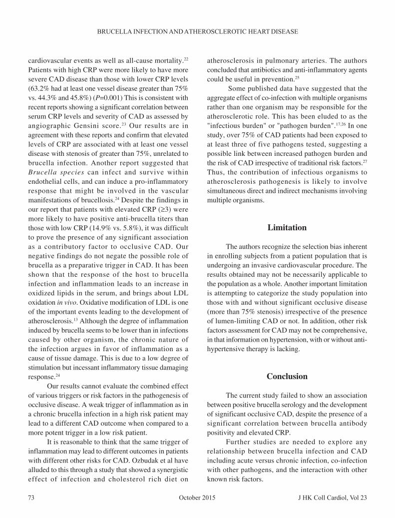
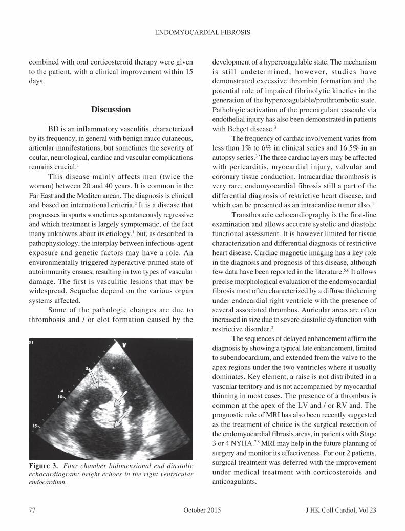
Echocardiography disclosed an enlarged rightatrium, severe narrowing of the in flow tract and themiddle part of the right ventricle. Bright echoes wereseen within the inferior and the middle parts of the rightventricular endocardium (Figure 1).
Nine blood cultures were negatives contrastingwith disturbed inflammatory analysis and a fever,initially the patient was treated by intravenous antibioticswithout any improvement, and it is with the onset ofgenital and oral ulceration, pseudo folliculitis in her skinthat BD was established as a final diagnosis after anextensive work-up to exclude de infective endocarditis.
Cardiac magnetic imaging showed a mass ofintermediate signal intensity on T1 weighted images withright ventricular dilation complicated by a cavitary
J HK Coll Cardiol, Vol 23 76October 2015
BENHALLA ET AL.
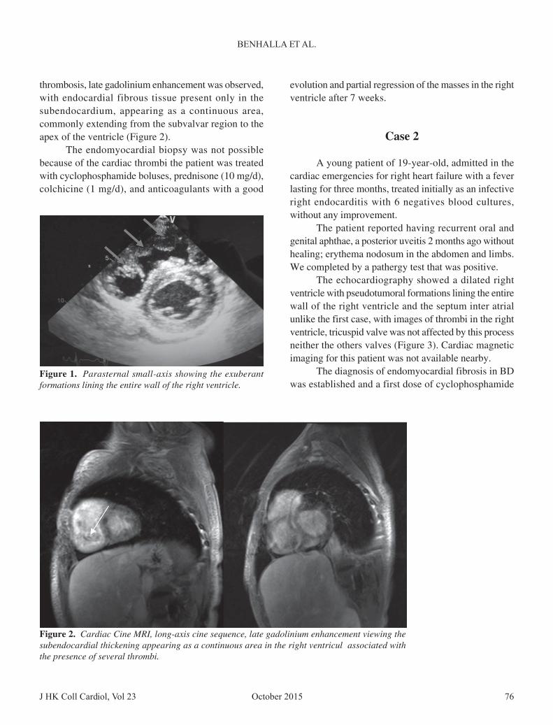
thrombosis, late gadolinium enhancement was observed,with endocardial fibrous tissue present only in thesubendocardium, appearing as a continuous area,commonly extending from the subvalvar region to theapex of the ventricle (Figure 2).
The endomyocardial biopsy was not possiblebecause of the cardiac thrombi the patient was treatedwith cyclophosphamide boluses, prednisone (10 mg/d),colchicine (1 mg/d), and anticoagulants with a good
evolution and partial regression of the masses in the rightventricle after 7 weeks.
Case 2
A young patient of 19-year-old, admitted in thecardiac emergencies for right heart failure with a feverlasting for three months, treated initially as an infectiveright endocarditis with 6 negatives blood cultures,without any improvement.
The patient reported having recurrent oral andgenital aphthae, a posterior uveitis 2 months ago withouthealing; erythema nodosum in the abdomen and limbs.We completed by a pathergy test that was positive.
The echocardiography showed a dilated rightventricle with pseudotumoral formations lining the entirewall of the right ventricle and the septum inter atrialunlike the first case, with images of thrombi in the rightventricle, tricuspid valve was not affected by this processneither the others valves (Figure 3). Cardiac magneticimaging for this patient was not available nearby.
The diagnosis of endomyocardial fibrosis in BDwas established and a first dose of cyclophosphamide
Figure 2. Cardiac Cine MRI, long-axis cine sequence, late gadolinium enhancement viewing thesubendocardial thickening appearing as a continuous area in the right ventricul associated withthe presence of several thrombi.
Figure 1. Parasternal small-axis showing the exuberantformations lining the entire wall of the right ventricle.
October 2015 J HK Coll Cardiol, Vol 2377
ENDOMYOCARDIAL FIBROSIS
development of a hypercoagulable state. The mechanismis still undetermined; however, studies havedemonstrated excessive thrombin formation and thepotential role of impaired fibrinolytic kinetics in thegeneration of the hypercoagulable/prothrombotic state.Pathologic activation of the procoagulant cascade viaendothelial injury has also been demonstrated in patientswith Behçet disease.3
The frequency of cardiac involvement varies fromless than 1% to 6% in clinical series and 16.5% in anautopsy series.3 The three cardiac layers may be affectedwith pericarditis, myocardial injury, valvular andcoronary tissue conduction. Intracardiac thrombosis isvery rare, endomyocardial fibrosis still a part of thedifferential diagnosis of restrictive heart disease, andwhich can be presented as an intracardiac tumor also.4
Transthoracic echocardiography is the first-lineexamination and allows accurate systolic and diastolicfunctional assessment. It is however limited for tissuecharacterization and differential diagnosis of restrictiveheart disease. Cardiac magnetic imaging has a key rolein the diagnosis and prognosis of this disease, althoughfew data have been reported in the literature.5,6 It allowsprecise morphological evaluation of the endomyocardialfibrosis most often characterized by a diffuse thickeningunder endocardial right ventricle with the presence ofseveral associated thrombus. Auricular areas are oftenincreased in size due to severe diastolic dysfunction withrestrictive disorder.2
The sequences of delayed enhancement affirm thediagnosis by showing a typical late enhancement, limitedto subendocardium, and extended from the valve to theapex regions under the two ventricles where it usuallydominates. Key element, a raise is not distributed in avascular territory and is not accompanied by myocardialthinning in most cases. The presence of a thrombus iscommon at the apex of the LV and / or RV and. Theprognostic role of MRI has also been recently suggestedas the treatment of choice is the surgical resection ofthe endomyocardial fibrosis areas, in patients with Stage3 or 4 NYHA.7,8 MRI may help in the future planning ofsurgery and monitor its effectiveness. For our 2 patients,surgical treatment was deferred with the improvementunder medical treatment with corticosteroids andanticoagulants.
combined with oral corticosteroid therapy were givento the patient, with a clinical improvement within 15days.
Discussion
BD is an inflammatory vasculitis, characterizedby its frequency, in general with benign muco cutaneous,articular manifestations, but sometimes the severity ofocular, neurological, cardiac and vascular complicationsremains crucial.1
This disease mainly affects men (twice thewoman) between 20 and 40 years. It is common in theFar East and the Mediterranean. The diagnosis is clinicaland based on international criteria.2 It is a disease thatprogresses in spurts sometimes spontaneously regressiveand which treatment is largely symptomatic, of the factmany unknowns about its etiology,1 but, as described inpathophysiology, the interplay between infectious-agentexposure and genetic factors may have a role. Anenvironmentally triggered hyperactive primed state ofautoimmunity ensues, resulting in two types of vasculardamage. The first is vasculitic lesions that may bewidespread. Sequelae depend on the various organsystems affected.
Some of the pathologic changes are due tothrombosis and / or clot formation caused by the
Figure 3. Four chamber bidimensional end diastolicechocardiogram: bright echoes in the right ventricularendocardium.
J HK Coll Cardiol, Vol 23 78October 2015
BENHALLA ET AL.
Conclusion
The discovery of endocardial masses in apatient suspected having an infective endocarditis,negative blood cultures with criteria of Behçetdiseases, should arouse suspicion of the diagnosisof endomyocardial fibrosis. The cardiac MRIallows precise characterization of particular tissuefibrosis with the delayed enhancement sequences,and could help prognost ic s trat i f icat ion andplanning for therapeutic intervention in thosepatients.
Disclosures
The authors declare that there is no conflict ofinterest.
References
1. Mahrholdt H, Wagner A, Judd RM, et al. Delayed enhancementcardiovascular magnetic resonance assessment of non-ischaemiccardiompyopathies. Eur Heart J 2005;26:1461-74.
2. Caudron J, Fares J, Bauer F, et al. And all Evaluation of leftventricular diastolic function with cardiac MR Imaging.Radiographics 2011;31:239-59.
3. Huong DL, Wechsler B, Papo T, et al. Endomyocardial fibrosisin Behçet's disease. Ann Rheum Dis 1997;56:205-8.
4. Bukhman G, Ziegler J, Parry E. Endomyocardial fibrosis: stilla mystery after 60 years. PLoS Negl Trop Dis 2008;2:e97.
5. Mocumbi AO, Yacoub S, Yacoub MH. Neglected tropicalcardiomyopaties: II Endomyocardial fibrosis: myocardialdisease. Heart 2008;94:384-90.
6. Cury RC, Abbara S, Sandoval LJ, et al. Images in cardiovascularmedicine. Visualization of endomyocardial fibrosis by delayed-enhancement magnetic resonance imaging. Circulation 2005;111:e115-7.
7. Salemi VM, Rochitte CE, Shiozaki AA, et al. Late gadoliniumenhancement magnetic resonance imaging in the diagnosis andprognosis of endomyocardial fibrosis patients. Circ CardiovascImaging 2011;4:304-11.
8. Jacquier A, Bartoli B, Flavian A, et al. Delayed myocardialenhancement: optimizing the MR imaging protocol. J Radiol2010;91:598-601.
October 2015J HK Coll Cardiol, Vol 23 79
19th Annual Scientific Meeting ofthe Institute of Cardiovascular Science and Medicine andthe 10th Across the Strait Scientific Conference onCardiovascular Science
21 November 2015Hong Kong Convention and Exhibition Centre
Hong Kong
Organised byThe Institute of Cardiovascular Science and Medicine, The University of Hong Kongin association withThe Institute of Vascular Medicine, Chinese University of Hong KongThe Stem Cell and Regenerative Medicine Consortium, The University of Hong KongThe Chinese Association of PathophysiologyThe Chinese Association of PhysiologyThe Chinese Association of PharmacologyThe Chinese Physiological SocietyTaiwan Neurosurgical Society
Meeting CommitteeCo-Chairmen: Prof. Bernard M.Y. CHEUNG
Prof. Yu HUANG
Vice-Chairmen: Prof. Mao-Tsun LINProf. Guang-Jin ZHU
Organising Prof. Juei-Tang CHENG Prof. Feng GAO Prof. Sheng-Huang HSIAOCommittee: Prof. Chao-Yu MAO Prof. Li-Ling WU Prof. Xi-Yong YU
Prof. Xiao-Rong ZENG
Faculty: Prof. Ji-Min CAO Prof. Cheng-Kuei CHANG Prof. Ching-Ping CHANGProf. Qi CHEN Prof. Si-Feng CHEN Prof. Guan-Hua DUProf. Jie DU Prof. Xue-Jun LI Prof. Jia-Wei LINProf. Bao-Feng YANG Prof. Huang-Tian YANG Prof. You-Yi ZHANGProf. Yi ZHU
Local Organising Dr. Heather J. BALLARD Prof. Ken R. BOHELER Dr. Michael P.H. CHANCommittee: Dr. M.L. FUNG Dr. M.C.W. KONG Dr. George P.H. LEUNG
Dr. Susan W.S. LEUNG Prof. Ronald A. LI Prof. Ricky Y.K. MANDr. Eva H.C. TANG Prof. Paul M. VANHOUTTE Dr. Q. YANGDr. Kelvin K.H. YIU
Meeting SecretariatLLink LimitedAddress: Room 2302, 23/F, Kwai Hung Holdings Centre, 89 King's Road, North Point, Hong KongTel: (852) 2566 2889; Fax: (852) 2570 4773; Email: [email protected]: http://www.icsm-hk.org

October 2015 J HK Coll Cardiol, Vol 2380
SCIENTIFIC PROGRAMME
21 NOVEMBER 2015 (SATURDAY)
08:30-09:00 Registration
09:00-10:40 Invited LecturesChairpersons: Prof. Yu Huang, Chinese University of Hong Kong, Hong Kong
Prof. Ying-Tung Lau, Chang Gung University of Science and Technology, Taiwan
SIRT3 Mediates the Anti-oxidant effect of Hydrogen Sulfide in Endothelial CellsProf. Yong Ji, Nanjing Medical University, China
MicroRNA Mediation of Endothelial Inflammatory Response to Smooth Muscle Cells and its Inhibitionby Atheroprotective Shear StressProf. Jeng-Jiann Chiu, National Health Research Institutes, Taiwan
Salvianolic Acid A Ameliorates Vascular Endothelial Dysfunctions in Diabetic Rats through Anti-oxidants and Anti-hyperglycemia StressProf. Guan-Hua Du, Chinese Academy of Medical Sciences and Peking Union Medical College, China
Astragaloside IV Protects Against Cerebral Contusion, Neuronal Apoptosis, and Neurologic Deficitsin Traumatic Brain Injured RatsProf. Cheng-Kuei Chang, Taipei Medical University, Taiwan
09:00-10:40 Oral Presentations for Young Investigator AwardSponsored by Sun Chieh Yeh Heart FoundationChairpersons: Prof. Huang-Tian Yang, Shanghai Institutes for Biological Sciences, China
Dr. Qin Yang, Chinese University of Hong Kong, Hong Kong10:40-11:00 Coffee break, poster viewing and booth visit11:00-11:50 Free Communications
Chairpersons: Prof. Cheng-Kuei Chang, Taipei Medical University, TaiwanProf. Yong Ji, Nanjing Medical University, China
11:00-11:50 Poster Presentations for Young Investigator AwardSponsored by Sun Chieh Yeh Heart FoundationChairpersons: Prof. Chao-Yu Miao, Second Military Medical University, China
Dr. Susan Leung, The University of Hong Kong, Hong Kong
11:50-13:10 Plenary Lectures IChairpersons: Prof. Mao-Tsun Lin, Chi Mei Medical Center, Taiwan
Dr. Heather Ballard, The University of Hong Kong, Hong Kong
Sudden Cardiac Death in the YoungDr. Anna Maria Lang-Choy, University of Dundee, UK
Aquaporin-1 Translocation and Degradation Partially Mediates the Water Transportation Mechanismof AcetazolamideProf. Xue-Jun Li, Peking University, China
Inflammation and Biomarkers in Congestive Heart FailureProf. Wei-Hsien Yin, National Yang-Ming University, Taiwan
October 2015J HK Coll Cardiol, Vol 23 81
13:10-14:10 Lunch
14:10-14:40 Opening Ceremony
14:40-16:00 Plenary Lectures IIChairpersons: Prof. G Liu, Peking University Health Science Center, China
Prof. Bernard Cheung, The University of Hong Kong, Hong Kong
Hyperhomocysteinemia-promoted Atherosclerosis via T Cell Priming ActivationProf. Xian Wang, Peking University, China
Tackling Atrial Fibrillation at the Population LevelDr. David Siu, The University of Hong Kong, Hong Kong
Vascular AgingProf. Alex F. Chen, Central South University, China
16:00-16:30 Coffee break, poster viewing and booth visit
16:30-17:45 Invited LecturesChairpersons: Prof. You-Yi Zhang, Peking University, China
Dr. ML Fung, The University of Hong Kong, Hong Kong
The Janus Face of Angiopoietin Like-2 in Heart and VesselsProf. Eric Thorin, Institut de Cardiologie de Montréal, Canada
Cardiac Differentiation of Pluripotent Stem Cells and Myocardial RepairProf. Huang-Tian Yang, Shanghai Institutes for Biological Sciences, China
Whole Body Cooling during Resuscitation Improves Neurotrauma Outcome in a Rat Model ofHemorrhagic ShockProf. Hung-Jung Lin, Taiwan Joint Commission on Hospital Accreditation, Taiwan
16:30-17:45 Invited LecturesChairpersons: Prof. Alex F. Chen, Central South University, China
Dr. Susan Leung, The University of Hong Kong, Hong Kong
NAMPT Metabolic Signaling in Vascular RepairProf. Chao-Yu Miao, Second Military Medical University, China
Teaching of Gender Difference in Cardiovascular PhysiologyProf. Ying-Tung Lau, Chang Gung University of Science and Technology, Taiwan
Human-Like Genetic Engineered Hamsters: Creation of a New Era of Translational CardiovascularResearchProf. G Liu, Peking University Health Science Center, China
17:45-18:00 Closing Remarks and Young Investigator Award CeremonyProf. Bernard Cheung, The University of Hong Kong, Hong Kong
18:00 Annual General Meeting
October 2015 J HK Coll Cardiol, Vol 2382
ICSM, 19TH ANNUAL SCIENTIFIC MEETING AND10TH ACROSS THE STRAIT SCIENTIFIC CONFERENCE ON CARDIOVASCULAR SCIENCE
ABSTRACTSAbstracts for Invited Lectures:
IL1.SIRT3 MEDIATES THE ANTI-OXIDANT EFFECT OFHYDROGEN SULFIDE IN ENDOTHELIAL CELLSLP Xie,1 GL Meng,1 S Li,1 X Tang,1 Y Ma,1 Y Hang,2 Y Ji1
1Key Laboratory of Cardiovascular Disease and Molecular Intervention, NanjingMedical University, China; 2Department of Vascular Biology, The ChineseUniversity of Hong Kong, Hong Kong
Aim: Oxidative stress is a key contributor to endothelial dysfunction andassociated cardiovascular diseases. Hydrogen sulfide (H2S) is an anti-oxidantgasotransmiter that protects endothelial cells against oxidative stress. Silentinformation regulator 2 (SIR2) is a functionally important transcriptional factorfamily in endothelial cells under oxidative stress. H2S was able to regulateactivity of several transcriptional factors. The aim of this study is to investigatethe possible role of SIRT3 in the antioxidant effect of H2S in endothelialcells.Results: Cultured EA.hy926 endothelial cells were exposed to hydrogenperoxide (H2O2) as a model of oxidative stress-induced cell injury. GYY4137,a slow releasing H2S donor, improved cell viability and reduced oxidativestress and apoptosis following H2O2 treatment. H2S reversed the H2O2-mediated inhibition of MAPKs phosphorylation, down-regulated of sirtuin3(SIRT3) mRNA and enhanced expression of superoxide dismutase 2 andisocitrate dehydrogenase 2. H2S increased activator protein 1 (AP-1) bindingactivity of SIRT3 promoter via enhancing its S-sulfhydration which effectwas absent in the presence of the specific AP-1 inhibitor SR11302 orcurcumin. Paraquat injection into mice induced defected endothelium-dependent aortic vasodilatation and increased oxidative stress in mice aortaand small mesenteric artery, which were improved by GYY4137administration. This vasculoprotective effect of H2S was absent in SIRT3knockout mice.
Innovation: These results highlight a novel role for SIRT3 in the protectiveeffect of H2S on oxidant damage in endothelium both in vitro and in vivo.Conclusion: H2S enhances AP-1 binding activity with the SIRT3 promoter,thereby up-regulating SIRT3 expression and ultimately reducing oxidant-provoked vascular endothelial dysfunction.
IL2.M i c r o R N A M E D I A T I O N O F E N D O T H E L I A LINFLAMMATORY RESPONSE TO SMOOTH MUSCLECELLS AND ITS INHIBITION BY ATHEROPROTECTIVESHEAR STRESSJJ ChiuInstitute of Cellular and System Medicine, National Health Research Institutes,Taiwan
In atherosclerotic lesions, synthetic smooth muscle cells (sSMCs) induceaberrant microRNA (miR) profiles in endothelial cells (ECs) under flowstagnation. Increase in shear stress induces favorable miR modulation tomitigate sSMC-induced inflammation. We addressed the role of miRs insSMC-induced EC inflammation and its inhibition by shear stress.Coculturing ECs with sSMCs under static condition causes initial increasesof 4 anti-inflammatory miRs (146a/708/451/98) in ECs followed by decreasesbelow basal levels at 7 days; the increases for miR-146a/708 peaked at 24hours and those for miR-451/98 lasted for only 6 to 12 hours. Shear stress(12 dynes/cm(2)) to cocultured ECs for 24 hours augments these 4 miRexpressions. In vivo, these 4 miRs are highly expressed in neointimal ECs ininjured arteries under physiological levels of flow, but not expressed underflow stagnation. MiR-146a, miR-708, miR-451, and miR-98 targetinterleukin-1 receptor-associated kinase, inhibitor of nuclear factor-κB kinasesubunit-γ, interleukin-6 receptor, and conserved helix-loop-helix ubiquitouskinase, respectively, to inhibit nuclear factor-κB signaling, which exertsnegative feedback control on the biogenesis of these miRs. Nuclear factor-E2-related factor (Nrf)-2 is critical for shear-induction of miR-146a incocultured ECs. Silencing either Nrf-2 or miR-146a led to increased neointima
formation of injured rat carotid artery under physiological levels of flow.Overexpressing miR-146a inhibits neointima formation of rat or mousecarotid artery induced by injury or flow cessation. Out results indicate thatNrf-2-mediated miR-146a expression is augmented by atheroprotective shearstress in ECs adjacent to sSMCs to inhibit neointima formation of injuredarteries.
ICSM, 19TH ANNUAL SCIENTIFIC MEETING AND10TH ACROSS THE STRAIT SCIENTIFIC CONFERENCE ON CARDIOVASCULAR SCIENCE
ABSTRACTS
October 2015 83
Abstracts for Invited Lectures:
J HK Coll Cardiol, Vol 23
IL3.SALVIANOLIC ACID A AMELIATES VASCULARENDOTHELIAL DISFUNCTIONS IN DIABETIC RATSTHROUGH ANTI-OXIDANTS AND ANTI-HYPERGLYCEMIASTRESSGH Du, R Zhao, JK Song, XY Yang, P Wu, BY Hou, L Sun, Y Lv, L ZhangBeijing Key Laboratory of Drug Target and Screening Research, Institute ofMateria Medica, Chinese Academy of Medical Science and Peking UnionMedical College, China
Aim: As a risk factor for cardiovascular disease, diabetes mellitus (DM)contributes to vascular complications. The pathological basis of that isendothelial dysfunctions, according to experimental evidences and substantialclinical reports. Salvianolic acid A (SalA) is an active compound isolatedfrom the root of traditional Chinese medicine Salvia miltiorrhiza Bunge, whichis used treating cardiovascular diseases for thousands of years. It has beenfound representing anti-inflammatory, antioxidant, anticarcinogenic,antiplatelet and antifibrotic properties. In present experiments, we launchedthe project aiming to figure out the in vivo/in vitro antidiabetic effect of SalAand the underlying mechanisms by alleviating endothelial dysfunctions.Methods: The models of Alloxan-induced type 1 diabetic mice, streptozotocin(STZ,60 mg/kg, i.p.) induced type 2 diabetic Sprague-Dawley (SD) rats, high-fat diet (HFD) and low-dose streptozotocin (STZ)-induced type 2 diabeticwistar rats were employed for the evaluation of SalA effects (1 mg/kg, p.o.90% purity). The cultured endothelial cells were used to explore themechanism of SalA actions.Results: In both type 1 and type 2 diabetic animals, SalA regulated theirfasting blood glucose (FBG) and blood glucose. In type 2 diabetic models,SalA reduced the level of serum Von Willebrand factor (vWF), ameliorated
IL4.ASTRAGALOSIDE IV PROTECTS AGAINST CEREBRALCONTUSION, NEURONAL APOPTOSIS, AND NEUROLOGICDEFICITS IN TRAUMATIC BRAIN INJURED RATSCK Chang,1 CP Chang,2,3 MT Lin4
1Graduate Institute of Injury Prevention and Control, Taipei Medical University;Neurosurgical Department, Taipei Medical University Shuang-Ho Hospital;2Department of Biotechnology, Southern Taiwan University of Science andTechnology; 3The Ph.D. Program for Neural Regenerative Medicine, TaipeiMedical University; 4Department of Medical Research, Chi Mei Medical Center,Taiwan
Objectives: Astragaloides (AST) is the main component of astragalus withthe function of antioxidation, immune regulation, and promotion ofintelligence. It was traditionally prescribed in the prevention and treatmentof vascular and cerebrovascular diseases, aging, immune function disordersand other diseases. A mixture of astragalasides can improve memory in agedmice, and astragaloside IV can reduce brain infarction in mice or in rats afterfocal cerebral ischemia. However, the therapeutic effects of astragalasides intraumatic brain injury (TBI) models and how it affects the proposed microglialoverexpression of TNF-α has not been determined.Materials: Anesthetized rats, after the onset of traumatic brain injury, weredivided into two groups and given the vehicle solution (1 mL/kg of bodyweight) or AST (80 mg kg-1). Vehicle or AST solutions were administeredintraperitoneally and one hour after traumatic brain injury. AST or salinewas injected immediately 1 h post-TBI, and the effect on the maximal angleof an inclined plane that the rats could cling to, neurological severity score(NSS), cylinder test, foot placing test, and ladder climbing test were assessed
the vascular reactivity of aorta ring, increased plantar blood perfusion andvascular activities, alleviated the vascular pathological changes in diabeticrats. SalA ameliates diabetes symptoms and alleviates vascular endothelialdisfunction to keep vascular on healthy biological conditions. The underlyingcellular and molecular mechanisms can be summarized as follows: a) regulatethe glucose metabolism. SalA ameliorated the abnormal blood glucose byregulates glucose metabolism, increasing ATP production, decreasingmitochondrial membrane potential (MMP), improving mitochondrial functionvia Ca2+/calmodulin-dependent; b) Anti-oxidant effects. Oxidative stress isaugmented in diabetic complications, treatment of diabetic animals withantioxidant SalA decreased the diabetes-induced MDA and NOS upregulation,reduced production of AGEs and ROS; c) Anti-inflammatory effects.Inflammatory mediators associated with vascular complications of diabetes,SalA may ameliorated vessel lession by blocking inflammatory cascades.Conclusions: The data indicated that SalA exhibits the antidiabetic effects,it could also protect against vascular endothelial dysfunction in diabetes,which might be owing to its antioxidative and anti-inflammatory effects.
1 day before surgery and 3 days post-TBI. The effect of rats' cerebral contusionzone was assessed 3 days after TBI. The effect on immunofluorescencestaining for neuronal nuclei (NeuN) and TUNEL, GFAP, and Iba1, and Iba1and TNF-α, in the rats' damage brain areas was assessed 3 days after TBI.Results: Astragaloside causes attenuation of TBI-induced cerebral contusion,neuronal apoptosis, and neurological motor dysfunction. Traumatic braininjury-induced neuronal apoptosis, gliosis, and activated microglia (evidencedby changed their morphology into an amoeboid shape as well as microgliaoverexpression of tumor necrosis factor-α) were all AST therapy-reduced.Conclusions: Our results indicate that AST therapy may protect againsttraumatic brain outcomes after traumatic brain injury through mechanismsattenuating neuronal apoptosis, gliosis, and activated microglia in the injuredbrain tissues.
October 2015 J HK Coll Cardiol, Vol 2384
ICSM, 19TH ANNUAL SCIENTIFIC MEETING AND10TH ACROSS THE STRAIT SCIENTIFIC CONFERENCE ON CARDIOVASCULAR SCIENCE
ABSTRACTSAbstracts for Invited Lectures:
IL5.SUDDEN CARDIAC DEATH IN THE YOUNGAM ChoyUniversity of Dundee, UK
Sudden cardiac death (SCD) in a young person is a tragic experience andloss for families and communities, as victims are usually at the peak of theirphysical health, and socio economic potential. The causes of young SCD canbe classified in to distinct entities that are often asymptomatic during life,with SCD being the first manifestation, and the challenge for clinicians ishow these young deaths can be prevented. It is recognised that inheritedcardiac conditions including the cardiomyopathies and channelopathies oftenunderlie SCD, and it is important that the correct diagnosis is made by post-mortem, including genetic post-mortem, so that immediate relatives can beassessed for the condition and offered treatment, thus preventing furtherdeaths. However, achieving this outcome is challenging as it requiresprofessional awareness of these conditions; effective communication betweenthe forensic pathologist, coroner, and general practitioner, as well as definedreferral pathways and access to specialist cardiologists and clinical genetics.In my presentation, I will discuss the genetic causes of young SCD, screeningprograms and a multidisciplinary approach required for the effectivemanagement of families affected by ICC.
IL7.INFLAMMATION AND BIOMARKERS IN CONGESTIVEHEART FAILUREWH YinDivision of Cardiology, Heart Center, Cheng Hsin General Hospital and Facultyof Medicine, National Yang Ming University, Taiwan
Heart failure (HF) remains one of the most important problems in cardiologydespite the progress in its treatment. HF is a complex syndrome with a hostof pathophysiological mechanisms in action. Inflammation, an integralcomponent of homeostasis, is a complex tissue response to stressors thatattempts to mitigate their effect and initiate healing. Inflammation plays acritical role in the development, course, severity and outcomes of HF. Thedelicate balance of pro- and anti-inflammatory processes can lead to beneficialor detrimental effects to the failing heart. Early detection of changesdeveloping in the heart is the key in improving the treatment's effectiveness.It appears that determining specific, sensitive biomarkers reflecting thecomplex pathophysiology of HF and using them to detect asymptomaticcardiac alterations may become a crucial screening tool, assisting in theidentification of patients requiring further diagnostic examinations. In thispresentation, the evidence on inflammatory biomarkers and their potentialrole in prognosis and therapeutic decisions for patients with HF will bereviewed.
IL6.AQUAPORIN-1 TRANSLOCATION AND DEGRADATIONPARTIALLY MEDIATES THE WATER TRANSPORTATIONMECHANISM OF ACETAZOLAMIDEJZ Zhang, Y An, XJ LiDepartment of Pharmacology, Peking University, China
It has been proposed that AQPs might have involved in certain clinical diseasessuch as brain and pulmonary edema, glaucoma and tumor etc. Our previousstudy has proved two carbonic anhydrase inhibitors (acetazolamide andtopiramate) could significantly inhibit the expression of AQP1 protein both invivo and in vitro and could dramatically inhibit the function of watertransportation across the cell membrane. We also observed that the both drugscould depress the tumor metastasis and provided evidences that thesecompounds might dramatically produce the anti-angiogenesis effects in thetumor tissues. AQP1 being located in renal proximal tubules is required forurine concentration. In this present study, we testify whether regulating toAQP1 will mediate the diuretic effect of AZA. We utilized Sprague Dawleyrats, as well as AQP1 knock-out (AQP1-/-) mice to examine urine volume, andhuman kidney-2 (HK-2) cell line was used for in vitro mechanism study. Todetect AQP1-binding proteins, we established a Surface Plasmon Resonance(SPR)-based method, which is usually utilized to discover molecules interaction.We found that AZA decreased CAs activity initially but the activity graduallyrecovered. Contrarily, diuretic effect was consistently significant. AQP1 proteinexpression was significantly decreased on day 7 and 14. By utilizing AQP1-/-
mice, we found diuretic effect of AZA was cancelled on day 14, while urinevolume continuously increased in wild-type mice. SPR results indicated AQP1was physiologically bound by myosin heavy chain (MHC). In vitro study resultsproved AZA facilitated AQP1 translocation onto cell membrane by promotinginteraction with MHC, dependent on ERK/ myosin light chain kinase (MLCK)pathway activation. MHC inhibitor BDM and ERK inhibitor U0126 bothabolished above effect of AZA. Eventually AZA induced AQP1 ubiquitination,while proteasome inhibitor MG132 reversed AZA's down-regulating effect uponAQP1. Our results identified AZA exerted diuretic effect through an innovativemechanism by regulating AQP1 and verified its inhibitory mechanism was viapromoting MHC-dependent translocation onto cell membrane and then ubiquitinmediated degradation, implicating a novel mechanism and target for diureticagent discovering.
ICSM, 19TH ANNUAL SCIENTIFIC MEETING AND10TH ACROSS THE STRAIT SCIENTIFIC CONFERENCE ON CARDIOVASCULAR SCIENCE
ABSTRACTS
October 2015 85
Abstracts for Invited Lectures:
J HK Coll Cardiol, Vol 23
IL11.THE JANUS FACE OF ANGIOPOIETIN LIKE-2 IN HEARTAND VESSELSE ThorinMontreal Heart Institute, Research Center and Department of Surgery, Facultyof Medicine, Université de Montréal, Canada
Angiopoietin-like 2 (angptl2) is a pro-inflammatory protein that inducesvascular inflammation and endothelial dysfunction. The impact of angptl2on cardiac function is, however, unknown. We hypothesized that angptl2contributes to pressure overload-induced cardiac dysfunction. Cardiac andvascular endothelial functions were investigated in angptl2 knockdown mice(KD) versus wild-type (WT) littermates, in response to a 6-week pressureoverload induced by transverse aortic constriction (TAC). The increase insystolic pressure in the aortic arch induced by TAC was lower in KD than inWT mice. In addition, while cerebral arteries displayed vascular remodelingand endothelial dysfunction in WT-TAC mice, arterial structure and functionwere preserved in KD-TAC mice. In contrast, KD-TAC mice displayedamplified cardiac hypertrophy (increased heart weight / tibia length ratio)and worsening of cardiac dysfunction measured by echocardiography (reducedejection fraction and stroke volume) and Millar pressure catheter (increasedminimal and end diastolic pressures, reduced relaxation rate) compared toWT-TAC mice. Cardiac mRNA and protein expression of NADPH oxidaseNOX4, a major source of oxidative stress in the heart, was increased only inKD-TAC mice. Cardiac-specific reduction of NOX4 expression by singlei.v. injection of adeno-associated virus AAV9 shRNA in KD-TAC micesignificantly limited cardiac hypertrophy and remarkably prevented the cardiacover-dysfunction caused by the absence of angptl2 in response to TAC. In
conclusion, angptl2 knockdown paradoxically worsens cardiac hypertrophyand contractile dysfunction induced by pressure overload. Cardiac up-regulation of NOX4 expression and/or activity could contribute to thisaggravated cardiac dysfunction.
IL8.H Y P E R H O M O C Y S T E I N E M I A - P R O M O T E DATHEROSCLEROSIS VIA T CELL PRIMING ACTIVATIONX Wang, J Feng, SL LvDepartment of Physiology and Pathophysiology, Basic Medical College, PekingUniversity, China
Homocysteine (Hcy) is a sulfur-containing non-constitutive amino acidderived from the essential amino acid methionine. Numerous clinical studieshave established hyperhomocysteinemia (HHcy) as an independent risk factorfor cardiovascular diseases in human. We and others have demonstrated thatHHcy accelerates atherosclerosis by affecting the immuno-inflammatoryresponse by stimulating chemokine/cytokine secretion from monocytes andT lymphocytes. In this process, reactive oxygen species (ROS) act as amediator in the T lymphocyte priming activation by Hcy. However, the exactmechanism of that has not been known yet. In this study, we postulated thatby inducing the metabolic reprogramming, Hcy promoted T-cell proliferationand cytokine IFN-β secretion with upregulated ER-mitochondrial coupling.Our results showed that Hcy (30 µgM) increased T cell proliferation whichwas accompanied by elevated oxidative phosphorylation (OXPHOS),membrane potential, ATP production, mitochondrial mass, and endoplasmicreticulum (ER) stress. While stimulation of ER stress by ER stress inducerdithiothreitol (DTT) mimicked Hcy-mediated T cell proliferation, inhibitionof ER stress by 4-phenylbutyric acid (PBA) or mitochondrial respiration byrotenone blocked Hcy-induced T cell proliferation and IFN-β secretion.Mechanistically, we found that Hcy increased ER-mitochondria coupling asevidenced by SIM and tethering protein expression. The increased ER-mitochondria interaction contributed to the enhanced ER stress, increased
Ca2+ transfer from ER to mitochondria, elevated OXPHOS and ATPproduction. Uncoupling ER-mitochondria by microtubule inhibitornocodazole attenuated Hcy-stimulated mitochondrial membrane potential andIFN-β secretion, suggesting that juxtaposition of mitochondria with the ERis required for the Hcy-promoted mitochondrial dysfunction and T cellactivation as well as vascular Inflammation. In conclusion, we have foundthat Hcy promoted T-cell proliferation and IFN-β secretion by inducing themetabolic reprogramming through regulation of ER-mitochondrial coupling.Our study highlights the importance of metabolic regulation in T cell primingactivation, thus shed new light in understanding HHcy-acceleratedatherosclerosis pathogenesis.
October 2015 J HK Coll Cardiol, Vol 2386
ICSM, 19TH ANNUAL SCIENTIFIC MEETING AND10TH ACROSS THE STRAIT SCIENTIFIC CONFERENCE ON CARDIOVASCULAR SCIENCE
ABSTRACTSAbstracts for Invited Lectures:
IL14.NAMPT METABOLIC SIGNALING IN VASCULAR REPAIRCY MiaoDepartment of Pharmacology, Second Military Medical University, China
Vascular repair is an endogenous defense mechanism after ischemic diseasesuch as stroke and myocardial infarction. Nicotinamide adenine dinucleotide(NAD), a well-known coenzyme involved in electron transport chain forgeneration of adenosine triphosphate, has emerged as a dictator regulatingvarious biological signaling pathways. Nicotinamide phosphoribosyltransferase (NAMPT), also an adipokine known as visfatin, is the rate-limitingenzyme for NAD biosynthesis in mammals. NAMPT may also act in a non-enzymatic manner, presumably mediated by unknown receptor(s). Recently,we and others show that NAMPT and NAMPT-controlled NAD metabolismregulate fundamental biological functions in endothelial cells, vascular smoothmuscle cells and endothelial progenitor cells. Sirtuins (SIRTs; the most studiedis SIRT1) are NAD sensors and may mediate the regulatory effects of NAMPT-NAD axis in these cells and vascular repair. Here, we discuss the currentdata regarding NAMPT and NAMPT-controlled NAD metabolism in vascularrepair and the potential translational application of NAMPT-related productsin treatment of cardio-cerebro-vascular disease.
IL15.TEACHING OF GENDER DIFFERENCE IN CARDIOVASCULARPHYSIOLOGYYT LauChang Gung University of Science and Technology, Taiwan
The differences in men and women's normal function and in their relateddiseases are often neglected in teaching of basic medicine. However, womendiffer significantly from men in most physiological systems. We thus haveadded a 20-minnte unit on gender difference in the teaching of cardiovascular(CV) physiology for medical students in Chang Gung University. The contentof this unit includes: (1) Gender differences in average lifespan; (2) Genderdifferences in cardiovascular diseases (CVD); (3) Gender differences in CVproperties/functions; (4) Implications of gender difference in CV medicine.Several topics were listed (e.g. atherosclerosis, exercise and CV function)for the students to choose for this 20-minnte period, and more than 80%students voted for gender difference in CVD in the past 3 years. It isimportant to address the impact of sex/gender on normal functions andCVD, an early exposure to this topic should be helpful for the medicalstudents in this regard.
IL12.CARDIAC DIFFERENTIATION OF PLURIPOTENT STEMCELLS AND MYOCARDIAL REPAIRN Cao, H Liang, M Liu, JX Huang, Q Liu, L Qiang, W Qiang, P Zhang,HT YangKey Laboratory of Stem Cell Biology and Laboratory of Molecular Cardiology,Institute of Health Sciences, Shanghai Institutes for Biological Sciences, ChineseAcademy of Sciences & Shanghai Jiao Tong University School of Medicine,China
Pluripotent stem cells (PSCs)-, including embryonic stem cells (ESCs) andinduced pluripotent stem cells (iPSCs), derived cardiovascular progenitorcells (CVPCs) and cardiomyocytes hold great promise for the study of earlycardiovascular development, drug screening, disease model, and myocardialregeneration. However, the efficient generation of these cells is a prerequisitefor their applications. Moreover, the developmental logic and molecularregulation underlying their self-renewal versus the cardiac lineage decisionshave not yet been fully understood. To address these issues, we investigatedthe regulatory mechanisms underlying the cardiac lineage fate decision,screened key signals controlling the generation and maintenance of CVPCsderived from PSCs (hPSC-CVPCs), and developed novel methods toefficiently induce human (h) CVPCs positive in MESP1/2, SSEA1, ISL1,MEF2C and GATA4 from hPSCs under feeder- and serum-free cultureconditions. They possess the ability to generate three major cell lineages ofthe heart, yet lack tumorigenic ability. To determine their therapeutic valuesto myocardial infarction (MI), we then transplanted these cells to mice withacute MI. The post-MI cardiac performance in the hPSC-CVPCs group issignificantly improved compared with the control one. Consistently, the infarctarea in the hPSC-CVPC group is smaller than the one in the control. The
findings proved new insight into the regulatory mechanisms of cardiogenesis.The established systems permit one to efficiently obtain hPSC-derivedCVPCs, which represent a powerful tool to study early embryoniccardiovascular development and offer a potentially safe source for developingapproaches for myocardial repair (Grants: Research Programs of CAS andCSIRO (XDA01020204, GJHZ1225); NSFC (31030050, 81470422), NBRPC(2011CB965300), NSTPC (2012ZX09501-001-001)).
ICSM, 19TH ANNUAL SCIENTIFIC MEETING AND10TH ACROSS THE STRAIT SCIENTIFIC CONFERENCE ON CARDIOVASCULAR SCIENCE
ABSTRACTS
October 2015 87
Abstracts for Invited Lectures:
J HK Coll Cardiol, Vol 23
IL16.HUMAN-LIKE GENETIC ENGINEERED HAMSTERS:CREATION OF A NEW ERA OF TRANSLATIONALCARDIOVASCULAR RESEARCHG LiuPeking University Health Science Center, China
Cardiac- and cerebral vascular diseases are the number one killer of humanbeings. Among these diseases, atherosclerosis represents the majority ofunderlying pathological alterations which cause the development andprogression of the diseases. It was well established that high blood cholesterolalone is sufficient to result in atherosclerosis in coronary, cerebral andperipheral atherosclerosis. One of common causes of high blood cholesterolis familial hypercholesterolemia (FH), an autosomal dominant-inheritedgenetic disorder that leads to elevated blood cholesterol levels. FH may presentas severely elevated total cholesterol and low density lipoprotein (LDL)cholesterol levels or as premature coronary heart disease (CHD). After theseminal discovery by Brown and Goldstein that mutations in the low-densitylipoprotein receptor (LDLR) was the cause of monogenetic FH, over 1,500mutations of this gene have been detected and these account for more than80% of cases of FH. Heterozygous FH (plasma cholesterol around 300 mg/dl) is rather common among general population with an estimated prevalenceof 1 in 300 to 500 in the world, while the incidence of homozygotes (Ch over600 mg/dl) occurs about one per million. Though LDLR deleted mice hadbeen generated 20 years ago and widely used until today, the lack of CETP,different lipoprotein profile from humans and absence of phenotypes inheterozygosity in mice limited the utility of LDLR KO mice as ideal human-like model in cardiovascular studies. In contrary, the hamsters as small rodents,
posses many features resembling that of human lipid metabolism, such ashigh CETP level, intestinal ApoB editing, endothelium-anchored hepaticlipase, low hepatic LDL receptor and hence enhanced susceptibility toatherosclerosis. To capitalize the full usefulness of hamsters we first optimizedthe methodology to manipulate the hamster's fertilized eggs and generatedthe first GFP transgenic hamsters. We then applied Crisp-Cas9 technologyand created LDLR deleted hamsters. The heterzygous LDLR targeted hamstersdeveloped hypercholesterolemia at age of 6 weeks on chow diet (plasma Ch280 vs 180 mg/dl of wildtype litermates). Moreover, the homozygout malesand females had plasma cholesterol levels near 1000 mg/dl, 4-5 times higherthan wild type litermates. These LDLR targeted hamsters are undercharacterization of their lipoprotein profile, apolipoprotein distribution,response to various nutritional and pharmaceutical manipulations. Ultimately,the development of atherosclerosis in coronary and cerebral arteries will beevaluated in these LDLR deleted hamsters which will perhaps be the idealsmall rodent models for human CAD and stroke.
October 2015 J HK Coll Cardiol, Vol 2388
ICSM, 19TH ANNUAL SCIENTIFIC MEETING AND10TH ACROSS THE STRAIT SCIENTIFIC CONFERENCE ON CARDIOVASCULAR SCIENCE
ABSTRACTSAbstracts for Oral Presentation:
OP1.
OP2.
STABILIZATION OF G-QUADRUPLEX DOWN-REGULATEMICRORNA-24 EXPRESSIONJ Gao,1 YC Qi,1 W Tan,2 Y Gu,2 M Xu#1
1Department of Cardiology, Institute of Vascular Medicine, Peking UniversityThird Hospital, Key Laboratory of Molecular Cardiovascular Sciences, Ministryof Education and Key Laboratory of Cardiovascular Molecular Biology andRegulatory Peptides, Ministry of Health; 2Beijing National Laboratory forMolecular Sciences, Key Laboratory of Bioorganic Chemistry and MolecularEngineering of Ministry of Education, Department of Chemical Biology,College of Chemistry and Molecular Engineering, Peking University, China
Objectives: G-quadruplexes (G4) are non-canonical nucleotide secondarystructures formed by stacks of guanine tetrads. The function of G4 in genecoding regions has been extensively studied, however, less is known aboutG4 motifs in non-coding gene regions. Herein, this study focuses on whetherthe G-rich sequence upstream of mir-24-1 could fold into a G4structure andregulate the expression of miR-24. Our research provides a new mechanismof microRNA regulation by stabilization of G4.Methods and Results: (1) The G4 displayed parallel conformationcharacteristics by Circular dichroism spectroscopy (CD), Electrosprayionization mass spectrometry (ESI-MS) and Nuclear magnetic resonancespectroscopy (NMR) indicated the G4 has 3 layers of G-quartet planes. Andthe DMS-foot printing assays clarified the location of key guanosinesparticipated in the formation of G4. (2) Two types of adenoviral expressionvectors were constructed to overexpress miR-24. The G4 type virus carriedwild type G-rich sequenceand the NG4 type virus carried mutation G-richsequence, which lost the ability of folding into a G4. These adenovirus weretransfected into HEK-293A cells ,and small molecule tetrandrine(TET),G4ligand, was added intoHEK-293A cells for 12 hours, qRT-PCR was performed
miR-17-3P IS REQUIRED FOR EXERCISE-INDUCEDCARDIAC GROWTH AND PROTECTS AGAINSTMYOCARDIAL ISCHEMIA-REPERFUSION INJURYJJ Xiao,1,2* J Shi,2 H Wang,2 YH Bei,1 QK Xuan,2 W Sun,2 H Liu,2 XL Li,2
XQ Kong2*
1Regeneration and Ageing Lab, Experimental Center of Life Sciences, Schoolof Life Science, Shanghai University; 2Department of Cardiology, The FirstAffiliated Hospital of Nanjing Medical University, China
Aims: We previously have reported that miR-222 was necessary for exercise-induced cardiac growth and protected against pathological cardiac remodeling.However, the role of other miRNAs in this process is unclear. This studyaims at investigating the role of members of the miR-17-92 cluster and theirstar miRNAs in exercise-induced cardiac growth.Methods and Results: miRNAs including miR-17-3p, -17-5p, -18a-3p,-18a-5p, -19a-3p, -19a-5p, -19b-1-5p, -19b-3p, -20a-3p, -20a-5p, -92a-3pand -92a-5p were determined by quantitative reverse transcription polymerasechain reactions (RT-PCRs) in heart samples from mice undergoing 3-weekswimming training. miR-17-3p and miR-18a-3p were found to be increasedin swimming mice models while only miR-17-3p was further found to beincreased in voluntary cage wheel exercise mice models. Interestingly, miR-17-3p was decreased in ventricular tissues of DCM patients and TAC mice.In isolated cardiomyocytes, forced-expression of miR-17-3p inducedcardiomyocytes hypertrophy and proliferation as determined by α-actininand EdU or Ki-67 staining. miR-17-3p was found to indirectly increase theAKT/PI3K pathway, contributing to cardiomyocytes hypertrophy. TIMP-3was identified as a target gene responsible for cardiaomyocyte proliferationby Western blotting, luciferase assays and rescue experiments. In vivo, using
heart weight-to-body weight (HW/BW (mg/gm)) ratio, heart weight-to-tibialength (HW/TL (mg/mm)) ratio and EdU/α-actinin staining, mice with miR-17-3p inhibition (n=9) showed milder cardiac growth and less cardiomyocyteproliferation compared with control group (n=8) after the 3-week swimmingexercise model. Moreover, subjected to ischemia-reperfusion injury, micewith forced-expression of miR-17-3p (n=10) showed significantly lessimpairment of cardiac function measured by echocardiography than negativecontrol group (n=11). Also, cardiomyocyte proliferation was significantlyenhanced in the miR-17-3p agomiR treated ischemia-reperfusion mice asassessed by EdU/α-actinin staining.Conclusions: miR-17-3p is required for exercise-induced cardiac growthand protects against myocardial ischemia-reperfusion injury. miR-17-3prepresents a novel therapeutic target for cardiac repair and regeneration.
to detect the miR-24 expression.The expression level of miR-24 was down-regulated by TET dose dependently in G4 type virus transfected cells, whilewas unchanged in the cells transfected NG4-AEV. (3) CRISPR/Cas9 systemwas used to construct G-rich sequence Knock-Out Sprague Dawley Rats (KOSD Rats). Compared to wild type SD rats (WT SD Rats), expression of miR-24 is much higher in Knock-Out SD Rats (WT:n=7, KO:n=7, P<0.001).Consequently, Junctophilin2 (JP2) and P27, the down-stream signalingofmiR-24,were significantly down-regulated in KO SD Rats (WT:n=7, KO:n=7, P<0.05).Conclusion: The stabilization of G4 down regulates the expression of miR-24. This study shed new light on microRNA regulation.National Natural Science Foundation of China (No. 91339105) , NaturalScience Foundation of Beijing (No. 7142165). Beijing Municipal Commissionof Science and Technology (NO.Z141100000214006).
ICSM, 19TH ANNUAL SCIENTIFIC MEETING AND10TH ACROSS THE STRAIT SCIENTIFIC CONFERENCE ON CARDIOVASCULAR SCIENCE
ABSTRACTS
October 2015J HK Coll Cardiol, Vol 23
Abstracts for Oral Presentation:
89
OP4.BONE MORPHOGENIC PROTEIN 4-SMAD INDUCED UP-REGULATION OF PLATELET-DERIVED GROWTHFACTOR AA IMPAIRS ENDOTHELIAL FUNCTIONWN Hu, XY Tian, Y HuangSchool of Biomedical Sciences, The Chinese University of Hong Kong, HongKong
Objectives: Bone morphogenic protein 4 (BMP4) is an important mediatorof endothelial dysfunction in cardio-metabolic diseases while platelet-derivedgrowth factors (PDGFs) are major angiogenic and pro-inflammatory mediatoralthough the functional link between these two factors is unknown. Thepresent study investigated whether PDGF mediates BMP4-induced endothelialdysfunction in diabetes.Methods and Results: We generated Ad-BMP4 to overexpress BMP4 andAd-Pdgfa-shRNA to knockdown Pdgfa in mice through tail intravenousinjection. SMAD4-shRNA lentivirus, SMAD1-shRNA and SMAD5 shRNAadenovirus were used for knockdown in human endothelial cells. We foundthat PDGFAA impaired endothelium-dependent vasodilation in aorta andmesenteric resistance arteries. BMP4 upregulated PDGFAA in human andmouse endothelial cells, which was abolished by BMP4 antagonist noggin orknockdown of SMAD1/5 or SMAD4. BMP4-impared relaxation in mouseaorta was also ameliorated by PDGF-AA neutralizing antibody. Tail injectionof Ad-Pdgfa-shRNA ameliorates endothelial dysfunction induced by BMP4overexpression (Ad-BMP4) in vivo. Serum PDGF-AA was elevated in theboth diabetic patients and diabetic db/db mice compared with non-diabeticcontrols. Pdgfa-shRNA or Bmp4-shRNA adenovirus reduced serum PDGF-AA concentration in db/db mice. PDGF-AA neutralizing antibody or tail
injection with Pdgfa-shRNA adenovirus improved endothelial function inaorta and mesenteric resistance arteries from db/db mice.Conclusions: The present study provides novel evidences showing PDGF-AA impairs endothelium-dependent vasodilation, and PDGF-AA mediatesBMP4-induced adverse effect on endothelial cell function through SMAD1/5 and SMAD4 -dependent mechanisms. Inhibition of PGDF-AA amelioratesvascular dysfunction in diabetic mice.
OP3.MEDIATION OF ER STRESS IN HOMOCYSTEINE-INDUCEDBKca CHANNEL DYSFUNCTION IN CORONARY ARTERIES:ROLE OF PERK-FOXO-3a ACTIVATIONWT Sun, XC Wang, CM Yu, Q YangDivision of Cardiology, Department of Medicine and Therapeutics, Instituteof Vascular Medicine, Li Ka Shing Institute of Health Sciences, Institute ofInnovative Medicine, Faculty of Medicine, The Chinese University of HongKong; The Chinese University of Hong Kong Shenzhen Research Institute,China
Objectives: Being abundantly expressed in vascular smooth muscle cells,large-conductance Ca2+-activated K+ (BKCa) channels significantly contributeto the control of vascular tone. Although homocysteine is known as anendoplasmic reticulum (ER) stress inducer and previous studies suggest theinhibitory effect of homocysteine on BKCa channels, whether ER stress isinvolved in homocysteine-induced channel inhibition remains unknown.Furthermore, by what mechanisms ER stress may mediate homocysteine-induced BKCa channel dysfunction remains completely unexplored.Methods: Porcine small coronary arteries denuded of endothelium werestudied for the relaxant response to the BKCa channel opener NS1619. Primarycultured porcine coronary arterial smooth muscle cells (PCASMCs) wereused for mRNA and protein analysis of BKCa channels, as well as patch-clamprecording of BKCa channel current.Results: The NS1619-induced relaxation was significantly attenuated inhomocysteine-exposed arteries whereas co-incubation of the arteries withER stress inhibitors, either TUDCA or 4-PBA restored the response. Theselective PERK inhibitor GSK2606414 showed potent protective effectagainst homocysteine on the NS1619-induced relaxation and the whole-cellBKCa current, suggesting the mediation of PERK-ER stress signaling in
homocysteine-induced channel dysfunction. Homocysteine lowered theprotein level of β1 but not α subunit of BKCa without downregulating mRNAexpression of both subunits. Downregulation of BKCa β1 was attenuated byTUDCA, 4-PBA, and GSK2606414. Inhibition of PERK with GSK2606414suppressed the nucleic translocation of forkhead box O transcription factor-3a (FOXO-3a) in homocysteine-exposed PCASMCs, associated with anincrease of BKca β1 protein level. In PCASMCs transfected with FOXO-3asiRNA, restoration of BKCa β1 protein level was observed after homocysteineexposure.Conclusions: ER stress-mediated downregulation of BKCa β1 subunit isinvolved in homocysteine-induced smooth muscle BKCa channel dysfunctionin coronary arteries. Activation of FOXO-3a by PERK signaling plays a keyrole in the ER stress-mediated BKCa channel inhibition.Supported by RGC/GRF CUHK4774/12M & CUHK14118414; NSFC81200123; Lui Che Woo Institute of Innovative Medicine - CARE theme8303303, and CUHK Direct Grant 4054182.
October 2015 J HK Coll Cardiol, Vol 2390
ICSM, 19TH ANNUAL SCIENTIFIC MEETING AND10TH ACROSS THE STRAIT SCIENTIFIC CONFERENCE ON CARDIOVASCULAR SCIENCE
ABSTRACTSAbstracts for Oral Presentation:
OP6.OVER EXPRESSION OF DRAM1 PREVENT AGAINSTMYOCARDIAL ISCHEMIA INJURY BY IMPROVINGAUTOPHAGY FLOWYY Qin, FJ Chen, XQ WuDepartment of Pharmacology, School of Pharmacy, Guangzhou MedicalUniversity, China
Objectives: Our previous studies showed that autophagy was induced sharplyin the early phase after acute myocardial infarction, but autophagy substrateP62 accumulated with the extension of ischemia. Our results indicated thatautophagy flow was impaired after sustained myocardial ischemia. DRAM1(Damage-regulated autophagy modulator 1) was reported as one of the mostimportant lysosome membrane protein to mediate the interaction ofautophagosome and the lysosome. In this study, we investigated whetherDRAM1 may improve the autophagy flow and prevent against myocardialischemia injury.Methods and Results: Adenoviral vector expressing DRAM1 or nulladenoviral vector was injected into rat peri-infarct myocardium after leftanterior descending (LAD) coronary artery ligation. DRAM1 expression wasconfirmed by Western blotting and immunofluorescence. Echocardiographicanalysis demonstrated relatively preserved cardiac function in the adeno-DRAM1 animals 21 days after LAD ligation. The left ventricular function ofadeno-DRAM1 animals was improved compared to the adeno-null rats.Myocardial remodeling was attenuated after overexpression of DRAM1, whilewas exacerbated in the adeno-null rats respectively with hematoxylin-eosinstaining and Masson's trichrome staining. Overexpression DRAM1 diminishedmyocardial apoptosis compared with the adeno-null rats with Tunel staining.Overexpression DRAM1 decreased infarct size and increased new blood
vessels density. Increased autophagy and autophagy-lysosome were found inthe infarct border of hearts overexpressed with DRAM1. OverexpressionDRAM1 attenuated the accumulation of autophagy LC3-II and substrateprotein P62/SQSTM1 obviously, which caused by sustained ischemia withWestern blotting.Conclusions: Overexpression DRAM1 improved the impaired autophagy flowcaused by sustained myocardial ischemia, and prevented against myocardialischemia injury.
OP5.HOMOCYSTEINE ACTIVATES B CELLS VIA REGULATINGPKM2-DEPENDENT METABOLIC REPROGRAMMINGJC Deng, J Feng, X WangDepartment of Physiology and Pathophysiology, School of Basic MedicalSciences, Peking University Health Science Center, China
Objectives: Homocysteine (Hcy) is a sulfur-containing amino acid formedduring the metabolism of methionine. Hyperhomocysteinemia (HHcy) hasbeen implicated as an independent risk factor for atherosclerosis. In this study,we investigated the role of metabolic reprogramming in Hcy-induced B cellactivation and the underlying mechanisms involved.Methods: Splenic B cells isolated from C57BL/6J mice were stimulated by100 µM Hcy for different times. For certain experiments, B cells were pre-incubated with inhibitors for 30 min before Hcy stimulation. For in vivoexperiments, ApoE-/- mice were intraperitoneal injected with a selectivePyruvate Kinase M2 (PKM2) inhibitor shikonin (SKN, 1.2 mg/kg) or solventcontrol every three days. Three days after first injection, ApoE-/- mice werefed normal mouse chow diet supplemented with or without 1.8 g/L Hcy indrinking water for 2 weeks. Aortic root atherosclerotic lesions were stainedwith oil red O. Immunoglobulin levels were determined by ELISA. Theexpression and activity of PKM2 were detected by Western blotting and acontinuous assay coupled to lactate dehydrogenase, respectively. Oxygenconsumption rates (OCR) and extracellular acidification rates (ECAR) weremeasured with an XF24 extracellular flux analyser.Results: Hcy induced B cell proliferation and antibody production bothin vitro and in vivo. Following Hcy stimulation, B cells upregulated bothlactate production and oxygen consumption, suggesting a relative balancedmetabolic reprogramming. Glucose uptake was also increased in Hcy-inducedB cells. B cell activation was abolished by glycolytic inhibitor 2-deoxyglucose,
indicating that glycolytic pathway is critical for this process. Furtherinvestigations indicate that PKM2, one of the key enzymes involved inglycolytic pathway, plays an important role in this process. While both theprotein expression and enzyme activity of PKM2 were increased by Hcy in Bcells, PKM2 inhibition with SKN sharply suppressed Hcy-induced metabolicreprogramming and B cell activation. Similarly, PKM2 small interfering RNAsilencing diminished Hcy-induced B cell activation. Further studies suggestthat reactive oxygen species and mTOR signaling pathway participate in Hcy-induced B cell activation and function through regulating PKM2. Consistentwith our in vitro findings, we showed that SKN treatment in vivo amelioratedHHcy-accelerated atherosclerotic lesion formation and diminished plasmaimmunoglobulin levels as well.Conclusions: Our data demonstrate that Hcy-activated B cells require PKM2to support metabolic reprogramming, which leads to B cell proliferation andantibody production. Targeting metabolic regulator PKM2 may be atherapeutic approach to ameliorate HHcy-accelerated atherosclerosis.
ICSM, 19TH ANNUAL SCIENTIFIC MEETING AND10TH ACROSS THE STRAIT SCIENTIFIC CONFERENCE ON CARDIOVASCULAR SCIENCE
ABSTRACTS
October 2015J HK Coll Cardiol, Vol 23
Abstracts for Oral Presentation:
91
OP8.C A R T I L A G E O L I G O M E R I C M A T R I X P R O T E I ND E F I C I E N C Y P R O M O T E S A T H E R O S C L E R O T I CCALCIFICATION VIA MODULATING MACROPHAGEPHENOTYPESC Gao, Y Fu, W KongThe Department of Physiology and Pathophysiology, School of Basic MedicalSciences, Peking University; and Key Laboratory of Molecular CardiovascularScience, Ministry of Education, China
Vascular calcification significantly increases cardiovascular disease morbidityand mortality. We recently reported that cartilage oligomeric matrix protein(COMP), a normal vascular extracellular matrix, is an endogenous inhibitorof vascular smooth muscle cell (VSMC) calcification. However, whetherCOMP affects atherosclerotic calcification is unknown. We firstly evaluatedthe atherosclerotic plaques and calcification in COMP-/- mice. Compared withApoE-/- mice, COMP-/-ApoE-/- mice fed with chow diet for 12 months displayedmore extensive atherosclerotic lesions and calcification in the aortic roots,arches and the innominate arteries. Next, COMP was mainly expressed invascular smooth muscle cells (VSMCs) and macrophages duringatherosclerosis. To investigate which COMP contributed to atheroscleroticcalcification, crossed bone marrow transplantation (BMT) was appliedbetween ApoE-/- and COMP-/-ApoE-/- mice. Enhanced calcification wasobserved in both ApoE-/- and COMP-/-ApoE-/- mice transplanted with COMP-/-
ApoE-/- bone marrow compared to the respective recipients transplanted withApoE-/- bone marrow. These data indicated that macrophages lacking COMPplayed a critical role in atherosclerotic calcification. Furthermore, throughcomparable microarray analysis on WT and COMP-/- macrophages, we
OP7.THE ROLE OF miR-27a-REGULATED G-PROTEIN COUPLEDRECEPTOR KINASE 6 IN ABNORMAL PROLIFERATION OFENDOTHELIAL CELL DURING HYPERTENSIONL Wang, YX Qi, ZL JiangInstitute of Mechanobiology & Medical Engineering, School of Life Sciences& Biotechnology, Shanghai Jiao Tong University, China
Objectives: In hypertension, elevated blood pressure results in high cyclicstretch, which causes abnormal paracrine of vascular smooth muscle cells(VSMCs) and induced abnormal proliferation of endothelial cells (ECs). Ithas been reported that G protein-coupled receptor kinases (GRKs) are involvedin the EC dysfunction of hypertension. GRKs regulate a large amount ofproteins and are also regulated by many molecules, including microRNA(miR). Recent studies revealed that miRs can be secreted extracellularly andparticipate in the communication between ECs and VSMCs during manycardiovascular disorders. Here we hypnotized that cyclic stretch may modulatethe VMSC secretion of miRs via microparticles (MPs), which subsequentlyinduces EC dysfunction during hypertension.Methods: Using hypertension rat model, the expression of GRK6 in thoracicaorta ECs was detected by immunofluorescence, the proliferation of ECswas detected by in situ BrdU immunofluorescence kit, and the VSMC-derivedMPs were observed by transmission electron microscope (TEM). Using FX-4000T Strain Unit, 5% and 15% cyclic stretch at 1.25 Hz were applied toVSMCs in vitro for 24 h, and then, ECs were treated with the MPs isolatedfrom the VSMC media. In addition, miR-27a mimics and inhibitor, and GRK6siRNA were transfected into ECs by Lipofectamine 2000. The expression ofmiR-27a was detected by real-time PCR, the expression of GRK6 was detectedby western blotting and the proliferation of ECs was detected by BrdU ELISAassay.
Results: The in vivo experiments showed that the expression of GRK6 inECs at thoracic aorta of hypertensive rat was lower than that in normotensivecontrol, but EC proliferation was higher. Then, we found the existence ofVSMC-MPs in vivo and in vitro, and revealed the expression of miR-27a inVSMC-MPs. The expression of miR-27a in MPs that isolated from the mediaof VSMCs subjected to 15% cyclic stretch (15%-VSMC-MPs) was higherthan that in 5%-VSMC-MPs. Dual luciferase reporter assays proved the targetbetween miR-27a and GRK6. The expression of GRK6 in ECs treated with15%-VSMC-MPs was lower than that treated with 5%-VSMC-MPs, whilethe EC proliferation was higher. Further more, biotinylated miR-27a, whichwere previously transfected into VSMCs, was detected in the VSMC-MP-treated ECs, indicating the transfer of miR-27a from VSMCs to ECs viaMPs. In addition, the up-regulation of miR-27a in ECs by mimics decreasedthe expression of GRK6 and increased the proliferation; while the down-regulation of miR-27a in ECs by inhibitor had the opposite effect. Suppressingthe expression of GRK6 in ECs by GRK6 siRNA increased cell proliferation.Conclusions: MiR-27a and the target GRK6 played significant roles in theproliferation of ECs during hypertension. High cyclic stretch increased thesecretion of miR-27a from VSMCs via MPs, which could subsequently betransferred into ECs, decreased the expression of GRK6, and finally inducedthe proliferation of ECs. (This work was supported by grants from the NSFCNo. 11232010, 11172178, 11222223)
explored that COMP deficient macrophages showed an atherogenic andosteogenic phenotype based on the upregulated genes related to inflammation,ROS production, lipid uptake and osteogenesis. These results reveal thatCOMP deficiency drove macrophages as the atherogenic and osteogenicphenotype to aggravate atherosclerotic lesion and calcification.
October 2015 J HK Coll Cardiol, Vol 2392
ICSM, 19TH ANNUAL SCIENTIFIC MEETING AND10TH ACROSS THE STRAIT SCIENTIFIC CONFERENCE ON CARDIOVASCULAR SCIENCE
ABSTRACTSAbstracts for Oral Presentation:
OP10.PROSTAGLANDIN E RECEPTOR SUBTYPE 4 (EP4)REGULATES LIPID DROPLETSIZE AND MITOCHONDRIALACTIVITY VIAFsp27 IN WHITE ADIPOSE TISSUEF Ying,1 Y Cai,2 EHC Tang1,3
1Department of Pharmacology and Pharmacy; 2Department of Anesthesiology;3School of Biomedical Sciences, Li Ka Shing Faculty of Medicine, TheUniversity of Hong Kong, Hong Kong
Objectives: A salient feature of chronic β3-adrenergic agonist activation ispronounced remodeling of white adipose tissue (WAT), which includes thetransformation of the lipid vacuole from unilocular into multiocular appearanceand mitochondrial biogenesis with resulting elevation in whole body metabolicrate. Although prostaglandin E receptor subtype 4 (EP4; one subtype ofprostaglandin E2receptor) stimulation affect metabolic functions, no availableinformation focuses on its role in WAT remodeling. Hence, the aim of thisstudy was to investigate whether or not genetic ablation of EP4 affects WATremodeling mediated by β3-adrenergic agonist.Methods: EP4+/+ and EP4-/- mice (12-15 weeks old male) received eithersaline or CL316243 (a highly selective β3-adrenergic agonist, 1 mg/kg/day,i.p) for 10 days. The effect of EP4 deficiency on energy expenditure (asdetermined by indirect calorimetry), fat tissues morphology (as determinedby H&E staining), mitochondrial biogenesis (as determined with electronmicroscopy and with the MitoTracker dye through confocal microscopy) andmitochondrial activity (as reflected by mitochondria complex I/IV activitymeasurement) in mice with or without CL316243 treatment were compared.In addition, the expression of genes and proteins involved in lipid dropletformation in fat pats was also studied.
OP9.CONCENTRATION-DEPENDENT VASCULAR EFFECTS OFDIVALENT MANGANESECMS Detremmerie, SWS Leung, PM VanhoutteState Key Laboratory for Pharmaceutical Biotechnology and Department ofPharmacology and Pharmacy, Li Ka Shing Faculty of Medicine, TheUniversity of Hong Kong, Hong Kong
Objectives: Divalent manganese is a cofactor for soluble guanylyl cyclase(sGC), the enzyme producing cyclic GMP in vascular smooth muscle cellsthat causes relaxation. Manganese competes with magnesium to activate sGC.These divalent cations can bias the activity of the enzyme to produce cyclicnucleotides other than cyclic GMP, in particular cyclic AMP and cyclic IMP.Cyclic IMP, preferably produced in the presence of magnesium, can causecontraction in precontracted arteries of the pig. The objective of the presentstudy was to identify the mechanisms by which manganese, compared tomagnesium [used in standard physiological solutions at millimolarconcentrations for functional studies], affects endothelium-dependent and-independent relaxations by influencing intracellular levels of cyclicnucleotides.Methods: Ex vivo experiments were designed using isolated rat aortae ofSprague-Dawley rats of 12-14 weeks of age and porcine coronary arteries(collected at the local abattoir). The arteries were cut into rings and used formeasurement of vascular reactivity in conventional organ chambers usingpharmacological inhibitors of endothelial nitric oxide (NO) synthase(LNAME) and of cyclooxygenases (indomethacin). In some rings, theendothelium was removed through insertion of a wooden toothpick. Afterobtaining an optimal resting tension, the rings were treated with millimolarconcentrations of manganese or magnesium for fortyfive minutes andprecontracted with phenylephrine (rat aortae) or U46619 (porcine coronary
arteries). Subsequently, concentration-dependent responses were obtainedwith acetylcholine (rat arteries), bradykinin (porcine coronary arteries) andthe NO-donor SNP. In parallel, in vitro experiments were conducted usingcultured porcine coronary artery smooth muscle cells (PCASMCs). The cellswere exposed to millimolar concentrations of manganese or magnesium inthe presence or absence of a sGC-activator (YC-1). Intracellular cyclic GMPlevels were measured using a commercially available ELISA kit.Results: In vivo, millimolar ranges of manganese significantly decreasedendothelium-dependent relaxations to acetylcholine in rat aortae and tobradykinin in porcine coronary arteries compared to physiological levels ofmagnesium. This effect was abolished by endothelium-removal. In addition,L-NAME partially reversed the effect of manganese, while indomethacin didnot. Manganese also decreased, but to a smaller extent, endothelium-independent relaxations to SNP in both porcine coronary arteries and rataortae. In parallel, the in vitro study showed that manganese alone or incombination with YC-1 blocks the production of cyclic GMP.Conclusions: Taken into conjunction, these experiments show that manganeseat millimolar levels decreases endothelium-dependent and -independentrelaxations compared to physiological levels of magnesium. This is likelyexplained through a decrease in cyclic GMP production. The effect ofmanganese is endothelium-dependent. In the case of endothelium-dependentrelaxations (to acetylcholine or bradykinin), the effect of manganese ismediated by endothelial NO synthase, but not by cyclooxygenases. Thesefindings indicate that in the presence of manganese NO does not act as avasodilator and must lead to production of cyclic nucleotides other than cyclicGMP to cause contraction, similar to what has been reported under hypoxicconditions.
Results: The elevation metabolic rate caused by chronic CL316243 treatmentwas significantly greater in EP4-/- mice. CL316243 significantly fragmentedthe unilocular lipid droplet into multiocular lipid vacuoles. It also increasedmitochondria biogenesis and mitochondria cytochrome c oxidase activity aswell as coenzyme Q1 activity. These changes were significantly amplified inmice with EP4 deficiency and were selectively seen in subcutaneous WAT,but were absent in epididymal or brown adipose tissue. The mRNA and proteinexpression of fat specific protein 27 (Fsp27), a protein that promote fusionof triglycerides and formation of unilocular lipid droplet, were significantlydiminished in subcutaneous WAT of EP4 deficient mice.Conclusions: The current findings implies that EP4 regulates the extent ofWAT remodeling and that in its absence, Fsp27 expression in subcutaneousWAT is repressed provoking greater CL316243-induced multiocular lipiddroplets and mitochondrial activity-causing an overall increase in whole bodymetabolic rate in these animals.The work is supported by Research Grant Council Early Career Scheme(ECS) (Project code: 27101314).
ICSM, 19TH ANNUAL SCIENTIFIC MEETING AND10TH ACROSS THE STRAIT SCIENTIFIC CONFERENCE ON CARDIOVASCULAR SCIENCE
ABSTRACTS
October 2015J HK Coll Cardiol, Vol 23
Abstracts for Oral Presentation:
93
OP12.OBESITY EXACERBATES RAT ISCHEMIC STROKETHROUGH ENHANCING ADIPONECTIN-CONTAININGNEURONAL APOPTOSISMH Wu,1 CC Chio,2 CP Chang,3,4 MT Lin4
1Department of Neurology, Chi Mei Medical Center, Liouying; 2Departmentof Surgery, Chi Mei Medical Center; 3Department of Biotechnology, SouthernTaiwan University of Science and Technology; 4Department of MedicalResearch, Chi Mei Medical Center, Taiwan
Objectives: A diet containing of high levels of saturated fat has been linkedto a dramatic rise in obesity. Long term exposure to high fat, "Western diet"(WD), are detrimental to ischemic brain injury.Materials: To explore the role of adiponectin (APN) and its adiponectinreceptors (ADRs) in the development of acute cerebral injury, we subjectedWD and control diet (CD) rats to 1 hour of middle cerebral artery occlusionfollowing by 23 hours of reperfusion.Results: Compared with CD rats, WD rats exhibited higher levels of braininfarct, neurologic deficits, brain edema and apoptosis of APN-containingneurons, upregulation of ADR-1 and p38 mitogen-activated protein kinase(p38 MAPK) and downregulation of ADR-2 in ischemic brain tissuesincluding frontal cortex, striatum, and hippocampus. Increasing percentagesof APN-containing neurons by baculovirus-mediated administration of APN,in addition to reducing apoptosis of APN-containing neurons in ischemicbrain tissues, significantly attenuated brain infarct and edema, neurologicdeficits, and altered expression of ADR-1, p38 MAPK, and ADR-2 in bothWD and CD group rats.Conclusions: These data suggest negative correlation between percentageof APN-containing neurons and ischemic stroke. Obesity could exacerbaterat stroke injury by enhancing apoptosis of APN-containing neurons inischemic brain tissues, probably via modulating ADRs and p38 MAPK.
OP11.THE ORPHAN RECEPTOR NOR1 PARTICIPATES INCARDIAC HYPERTROPHY BY REGULATING PARP-1 ANDSIRT1XJ Feng, H Gao H, JT Ye, PQ LiuDepartment of Pharmacology and Toxicology, School of PharmaceuticalSciences, Sun Yat-Sen University, China
Background and Purpose: NOR1 belongs to NR4A subfamily of the nuclearhormone receptor superfamily and is involved in regulation of glucose and fatmetabolism. However, its potential contribution to cardiovascular diseasesremains to be elucidated. In the present study, the roles of NOR1 in cardiachypertrophy induced by isoproterenol (ISO) and the underlying molecularmechanism were investigated.Experimental Approach: The expression of NOR1 was detected inhypertrophic cardiomyocytes induced by ISO. After NOR1 overexpression bytransfection or depletion by RNA interference in neonatal rat cardiomyocytes,cellular hypertrophy was monitored by measuring cell surface area and themRNA levels of hypertrophic biomarkers. Further, the interaction betweenNOR1 and PARP-1 was investigated by co-immunoprecipitation. The expressionof NOR1 and activity of PARP-1 were measured in vivo, using ISO model ofcardiac hypertrophy in rats.Key Results: ISO treatment significantly upregulated NOR1 expression andPARP-1 activity both in vitro and in vivo. Specific genetic silencing of NOR1attenuated ISO-induced cardiomyocyte hypertrophy. Furthermore, we identifieda physical interaction between NOR1 and PARP-1, whose enzyme activity couldbe enhanced by NOR1 transfection. Overexpression of NOR1 but not its mutantNOR1 (C293Y), resulted in increased cellular surface area and the mRNAlevels of ANF and BNP, which could be inhibited by PARP-1 inhibitor 3AB orPARP-1 RNA interference.Conclusions and Implications: Our findings provide the first evidence thatNOR1 is involved in ISO-induced cardiac hypertrophy. The prohypertrophiceffect of NOR1 can be at least partially attributed to its regulation of PARP-1enzyme activity.
OP13.LOSS OF OSTEOGLYCIN PROMOTES ANGIOGENESIS INLIMB ISCHEMIA MOUSE MODELS VIA MODULATION OFVEGF-VEGFR2 SIGNALING PATHWAYPJ GaoShanghai Institute of Hypertension/Department of Hypertension, RuiJinHospital, Shanghai Jiaotong University School of Medicine, China
Objectives: Osteoglycin (OGN) has been noted for implications incardiovascular diseases in recent studies, particularly its correlation withcardiac remodeling. However, the relationship between OGN and angiogenesisremains unknown. Therefore, we aimed to investigate the effect of OGN onischemia-induced angiogenesis and address the underlying mechanisms.Methods: Immunofluorescence and western blot were used to detect OGNexpression under ischemic condition. Limb ischemia model was establishedin OGN-/- (n=12) mice and wild type (WT, n=12). Perfusion recovery wasestimated by Laser Doppler imaging. Histology, capillary formations andinflammation response in gastrocnemicus muscle after ischemia wereevaluated by H.E staining and immunohistochemistry. Besides, alkali inducedcorneal neovascularization model and ex vivo aortic ring sprouting modelwere established in both OGN-/- and WT mice and angiogenesis were assessed.In vitro studies, tube formation, proliferation and migration of humanumbilical vein endothelial cells (HUVECs) were assessed after transfectionof either OGN siRNA or overexpression plasmid. Western blot was used todetect expression and activation of VEGFR2 signaling. Protein-proteininteraction was assessed by co-immunoprecipitation (co-IP) and detailedinformation was provided by molecular docking.Results: We observed OGN down-regulation with angiogenesis in
intermuscular endothelial cells of angiogenic tissues in patients with peripheralartery disease and mice of limb ischemia model. Although, histology change,inflammatory cell infiltration, and inflammatory cytokines expression afterischemia in OGN-/- mice did not differ from WT ones. Perfusion recovery ratewas higher in OGN-/- mice. The percent of blood flow recovery of wild typemice was 24.8%, 44.6% and 63.5% respectively at day 4, day 7 andday 14 after limb ischemia, while OGN-/- mice was 46.5%, 71.5%, and 83.0%at the same time points. Capillary density was also higher in OGN-/- mice ingastrocnemicus muscle of the ischemia limb. Moreover, enhanced ofangiogenesis was observed in OGN-/- mice after alkali injury. Aortic ringsisolated from OGN-/- mice had stronger sprouting than those from WT ones.Tube formation was increased in HUVECs with OGN knockdown, comparedwith the negative control. Proliferation and migration were also enhanced inHUVECs with OGN knockdown. Meanwhile OGN overexpression suppressedcell functions of tube formation, proliferation and migration. Further studyrevealed that OGN depletion significantly promoted the activation of vascularendothelial growth factor receptor 2 (VEGFR2) and its downstream signalingpathways without affecting the protein level of vascular endothelial growthfactor (VEGF) or VEGFR2. co-IP assay revealed that OGN associated withVEGFR2 and negatively modulated the interaction between VEGF andVEGFR2. Molecular docking revealed that Tyr196, Glu176 and Glu108 ofOGN form hydrogen bonds with residues Glu140, Tyr194 and Tyr190 of VEGF2,respectively, which is the main binding affinity between OGN and VEGF2.Conclusions: OGN is an important negative regulator of VEGF-VEGFR2signaling for endothelial cell tube formation, proliferation, migration, as wellas ischemia induced angiogenesis. Thus, our findings provided a noveltherapeutic target of ischemic vascular diseases.
October 2015 J HK Coll Cardiol, Vol 2394
ICSM, 19TH ANNUAL SCIENTIFIC MEETING AND10TH ACROSS THE STRAIT SCIENTIFIC CONFERENCE ON CARDIOVASCULAR SCIENCE
ABSTRACTSAbstracts for Oral Presentation:
OP14.THE MECHANISM AND PHARMACOLOGICAL RESCUE OFBERBERINE-INDUCED hERG CHANNEL DEFICIENCYBX LiDepartment of Pharmacology, Harbin Medical University, China
Aims: The hERG (human ether-a-go-go-related gene) potassium channelwhich plays a crutial role in cardiac repolarization is the target of diversetherapeutic drugs. Berberine (BBR) was previously found that cansimultaneous block hERG and inhibit its membrane expression. However,the regulatory mechanisms of BBR effects on hERG at cell membrane levelremain unknown. The present study was designed to investigate in detailhow BBR decreased hERG expression on cell surface and further explore itspharmacological rescue strategies.Methods and Results: Transfection and western bloting were applied toinvestigate the role of caveolin-1 (Cav1) and the canonical drug binding sitesof hERG (a tyrosine 652 and a phenylalanine 656) in S6 domain played inBBR-induced hERG expression defect on membrane. The whole-cell patchclamp technique was used to record hERG current and action potential duration(APD). After BBR treatment, the expression of caveolin-1 reduces and theuse of caveolin-1 specific siRNA (Cav1-siRNA) diminishes BBR-causedhERG inhibition. In addition, BBR obviously descreases WT-hERGexpression, whereas has no significant inhibition on hERG mutation Y652A-hERG and F656V-hERG. These results indicate that caveolin-1 and residuesTyr652, Phe656 account for BBR-induced hERG defects on membrane. Thedata of rescue experiments show that astemizole, fexofenadine and resveratrolcan rescue surface expression of hERG channel, whereas only fexofenadineand resveratrol restore hERG current. Moreover, fexofenadine and resveratrolalso shorten APD prolongation disrupted by BBR.
Conclusion: Our study demonstrates that (1) BBR promotes mature hERGdegradation via accelerating caveolin-1 turnover, and (2) aromatic tyrosine(Tyr652) and phenylalanine (Phe656) in S6 domain mediate BBR-inducedreduction of hERG on membrane, which suggest that BBR reduces hERGmembrane stability with multiple mechanisms. Furthermore, fexofenadineand resveratrol, shorten APD prolongated by BBR, have a potential effect ofalleviating the cardiotoxicity of BBR.
OP15.RANDOMISED CONTROLLED TRIAL OF THE EFFECT OFPHYTOSTEROL-ENRICHED LOW-FAT MILK ON LIPIDPROFILE IN CHINESECL Cheung,1,2,3 KC Ho,1 CW Sing,1 MF Tsoi,2 KF Cheng,1 BMY Cheung2,3,4
1Department of Pharmacology and Pharmacy; 2Department of Medicine; 3StateKey Laboratory of Pharmaceutical Biotechnology; 4Institute of CardiovascularScience and Medicine, The University of Hong Kong, Hong Kong
Objective: Phytosterols found naturally in plants are known to reducecholesterol absorption in the gut. The Chinese diet typically contains manyvegetables and not much meat; we therefore aimed to test if phytosterols areas effective in Chinese.Method: There were 221 (41 men, 180 women; age 24-79) subjects whoconsented to participate in the study (ClinicalTrials.gov ID: NCT02541201),the protocol of which was approved by the Institutional Review Board of theUniversity of Hong Kong/Hospital Authority Hong Kong West Cluster.Subjects were randomised to double-blind intake of a phytosterol-enrichedlow-fat milk or a conventional low-fat milk for three weeks. Before everybreakfast and lunch, they had a 273 ml serving. Active treatment contained1.5 g/day phytosterol. Fasting blood samples for lipid profile were takenbefore and at the end of study. Body weight, waist circumference and bloodpressure were also measured.Results: LDL-cholesterol decreased from 3.22±0.08 to 3.06±0.08 mmol/L inthe phystosterol group and increased from 3.08±0.08 to 3.20±0.08 mmol/Lin controls. Comparing treatment with control, the decrease in LDL-cholesterol was 9.5±2.0% (p<0.001). There were no significant changes inHDL-cholesterol, triglycerides, body weight or blood pressure. Five subjects(2.3%; 4 in treatment group) withdrew.
Conclusion: Consumption of a phytosterol-enriched low-fat milk led to asignificant fall in LDL-cholesterol. This can be recommended as part of ahealthy diet for people with mildly elevated cholesterol levels and not at highcardiovascular risk. Statins and ezetimibe remain the treatment of choice forthose with high cholesterol levels or high cardiovascular risk.
ICSM, 19TH ANNUAL SCIENTIFIC MEETING AND10TH ACROSS THE STRAIT SCIENTIFIC CONFERENCE ON CARDIOVASCULAR SCIENCE
ABSTRACTS
October 2015 95
Abstracts for Chaired Posters:
J HK Coll Cardiol, Vol 23
CP1.
CP2.
NOREPINEPHRINE INCREASES THE MONOAMINEOXIDASE A AND OXIDATIVE STRESS IN SH-SY5Y CELLSJJ Li,1 CS Lam,1 GL Tipoe,1,2 ML Fung1,2
1School of Biomedical Sciences; 2Research Centre of Heart, Brain, Hormoneand Healthy Aging, Li Ka Shing Faculty of Medicine, The University ofHong Kong, Hong Kong
Background: Obstructive sleep apnea (OSA) is a major breathing disorderaffecting 5-7% of adult population, which increases risks for stroke andcardiovascular morbidities and mortalities. Episodes of oxygen desaturationcaused by obstruction of the airway of OSA patients manifested as intermittenthypoxia (IH) significantly contributes to the excessive production of reactiveoxygen species, which cause oxidative stress and inflammation in thepathogenesis of cardiovascular disease. Also, OSA patients are reportedlywith elevated levels of sympathetic and norepinephrine (NE) activities.Monoamine oxidase isoform-A (MAO-A) deaminates NE and produces H2O2
as a byproduct during its catalytic reaction. Additionally, 2,3-dioxygenase(IDO) is closely associated with oxidative stress and inflammation [5]. Werecently observed that inhibition of MAO-A decreases the IH-induced celldeath in SH-SY5Y cells.Aims and Objectives: We aim to examine the role of NE in the IH-inducedcell death. In this study, we test the hypothesis that NE could increase theexpression of MAO-A and IDO in SH-SY5Y cells.Methods: Cultured SH-SY5Y cells were used in this study, whichconstitutively express MAO-A but not MAO-B. Cells were treated with NEat concentrations of 0.01 µM and 0.1 µM to mimic the NE overspill in OSApatients. Clorgyline (10 µM) was used as a selective inhibitor of MAO-A;
C1q/TUMOR NECROSIS FACTOR-RELATED PROTEIN 6ATTENUATED POST-INFARCT CARDIAC FIBROSIS BYTARGETING RhoA/MRTF-A PATHWAYCL Zhang, H Lei, D Wu, FY Fu, L Li, LL WuDepartment of Physiology and Pathophysiology, Peking University HealthScience Center, China
Objective: C1q/tumor necrosis factor-related protein-6 (CTRP6) is a newlyidentified adiponectin paralog with modulation effects on metabolism andinflammation. However, the cardiovascular function of CTRP6 remainsunknown. This study aimed to investigate the effect of CTRP6 on post-infarctcardiac fibrosis and its underlying mechanisms.Methods: Experimental myocardial infarction (MI) was induced by leftanterior descending coronary artery ligation in Sprague-Dawley rats. Picricacid Sirius red staining was used to determine cardiac fibrosis. Quantitative-PCR, western blotting and immunohistochemical staining were used to detectthe expression of CTRP6 and pro-fibrotic molecules. Cardiac fibroblasts (CFs)isolated from adult rat heart were cultured to investigate transforming growthfactor-β1 (TGF-β1)-induced myofibroblast differentiation and its underlyingmechanisms.Results: Myocardial expression of CTRP6 was significantly decreased post-MI. CTRP6 supplement by intromyocardial injection of adenovirus alleviatedcardiac fibrosis, and inhibited myofibroblast differentiation as well as theexpression of collagen I, collagen III, and connective tissue growth factorpost-MI. Treatment of CFs with recombinant CTRP6 significantly inhibited
transforming growth factor-β1 (TGF-β1)-induced myofibroblastdifferentiation. CTRP6 increased the phosphorylation of AMP-activatedprotein kinase (AMPK) and Akt in cultured CFs and post-MI hearts.Pretreatment with adenine 9-β-D-arabinofuranoside (AraA), an AMPKinhibitor, or LY294002, a phosphatidylinositol-3-kinase (PI3K) inhibitor,abolished the protective effect of CTRP6 on TGF-β1-induced profibroticresponse. Furthermore, CTRP6 significantly decreased TGF-β1-induced RhoAactivation and myocardin-related transcription factor-A (MRTF-A) nucleartranslocation, whereas had no effect on TGF-β1-induced Smad3phosphorylation and nuclear translocation, these effects of CTRP6 wereblocked by AMPK or Akt inhibition.Conclusion: CTRP6 attenuates cardiac fibrosis via inhibiting myofibroblastsdifferentiation post-MI. AMPK and Akt activation are responsible for theCTRP6-mediated anti-fibrotic effect by targeting RhoA/MRTF-A pathway.
Western blotting was used to assess the level of protein expression of MAO-A, IDO and antioxidant enzyme superoxide dismutase (SOD2). Also, GSSG/GSH ratio was obtained to evaluate the level of oxidative stress.Results: We found significant increased levels of MAO-A and IDO expressionsin the SH-SY5Y cells treated with NE (0.01 µM and 0.1 µM) for 48 hours. Inaddition, the ratio of GSSG/GSH was significantly increased and the level ofSOD2 expression was decreased by the NE treatment, suggesting an elevatedlevel of oxidative stress. Furthermore, the effects of NE on MAO-A and IDOexpression and oxidative stress were partially antagonized by 10 µMclorgyline.Conclusion: The overspill of NE in the OSA patient could worsen the IH-induced oxidative stress and inflammation via increased levels of MAO-Aand IDO expression, which may play a mechanistic role in thepathophysiological response to chronic intermittent hypoxia.
October 2015 J HK Coll Cardiol, Vol 2396
ICSM, 19TH ANNUAL SCIENTIFIC MEETING AND10TH ACROSS THE STRAIT SCIENTIFIC CONFERENCE ON CARDIOVASCULAR SCIENCE
ABSTRACTSAbstracts for Chaired Posters:
CP3.M i R - 3 3 M E D I A T E V E I N G R A F T N E O I N T I M A LHYPERPLASIA AND VENOUS SMCs PROLIFERATIONUNDER CYCLIC STRETCHK Huang, ZQ Yan, KX Wang, H Bao, XH Chen, P Zhang, QP Yao, BR Shen,YX Qi, ZL JiangInstitute of Mechanobiology & Medical Engineering, School of Life Sciences& Biotechnology, Shanghai Jiao Tong University, China
Objectives: Autologous saphenous veins are often grafted to patients withmultivessel-coronary-artery disease. After grafted to replace a diseased artery,the vein is exposed to substantially higher pressure and higher mechanicalstretch inevitably, which contributes to neointimal hyperplasia. It has beenproved that proliferation of venous smooth muscle cells (SMCs), induced byarterial cyclic stretch is crucial for neointimal hyperplasia and narrowing ofthe vessel lumen. MiR-33 has been shown to regulate cell proliferation, andcell cycle progression. However, the role of miR-33 in neointimal hyperplasiaof grafted vein caused by arterial cyclic stretch remains unknown.Methods: Vein grafts were generated by "cuff" technique, and the jugularvein on the right side was used as a control. Paraffin sections of grafted veinsstained with Elastin-Van Gesion to detect neointimal hyperplasia; RT-PCRwas carried out to disclose miR-33 expression levels; and western blottingwas used to examine expressions of BMP-3, Smad2, p-Smad2, Smad5 andp-Smad5 in grafted veins. FX 4000 in vitro Strain Union was used to apply10% cyclic stretch to cultured venous SMCs, which mimics the arterialenvironment. Web-accessible databases and dual luciferase reporter assay wereused to detect the target of miR-33. The inhibitor and mimics of miR-33, andthe specific siRNA and recombinant protein of BMP-3 were used respectivelyto demonstrate the effects of miR-33 and Bmp3 on proliferation of venous
CP4.OBESITY IS RELATED TO IMMUNOGLOBULIN EC Li,1 TK Tsang,1 HL Yao,1 HY Tan,2 X Zhou,3 BMY Cheung1
1Li Ka Shing Faculty of Medicine, The University of Hong Kong, Hong Kong;2School of Biological Science, The University of Hong Kong, Hong Kong;3Department of Cardiology, The Second Affiliated Hospital of SoochowUniversity, China
Immunoglobulin E (IgE), a measurement of allergy, plays an essential role inimmunity and hypersensitivity. In order to know whether such an associationcan be also found in adults, we explored data on 1933 men and 1802 womenin the National Health and Nutrition Examination Survey (NHANES) 2005-2006. We included participants aged above 20 years who had valid data onbody mass index (BMI) and serum IgE level. Pregnant women were excluded.We found in adults that obesity (OR=2.63, 95%CI=1.62-4.27) was associatedwith higher odds of asthma episodes, compared with normal weight. Obesity,but not overweight, was also associated with allergic symptoms (OR=1.27,95%CI=1.08-1.49). Elevated serum levels of liver enzymes (all P≤0.001)and C-reactive protein (P<0.001) were associated with an increment in BMI.IgE level increased with BMI; it increased progressively from 33.70 kU/L inthe bottom quartile to 45.62 kU/L in the top quartile of BMI. The results alsoshowed an association between BMI and IgE level in the overall population(p=0.074, P=0.002) and among women (p=0.088, P=0.006). After adjustmentfor race/ethnicity, age, sex (except in sex-specific analysis), physical activity,alcohol consumption and smoking, the association between BMI and IgElevel in the overall population (p=0.070, P=0.005) and women (p=0.074,P=0.013) remained significant. After further adjustment for C-reactive protein,serum levels of liver enzymes and correlations among serum levels of liver
SMCs. Furthermore, perivascular multi-point injection of agomiR-33 wasperformed to verify the function of miR-33 in vivo.Results: Neointima of grafted veins was notably increased after 4-week grafts.The expression of miR-33 was decreased in 1-week, 2-week and 4-weekgrafted veins, and the remarkable decrease was detected in 2-week group.The expressions of BMP-3, p-Smad5 and p-Smad2 were all increased ingrafted veins. The similar results were found in venous SMCs subjected to10% (arterial) cyclic stretch in vitro. 10%-cyclic stretch significantly promotedproliferation of veinous SMCs, declined expression of miR-33, and elevatedexpressions of BMP-3, p-Smad5 and p-Smad2. Accordingly, miR-33 mimicsrepressed, while inhibitor increased proliferation of venous SMCs. MiR-33directly targeted to BMP-3 and regulated its expression. Recombinant BMP-3 increased proliferation of SMCs and expressions of p-Smad5, p-Smad2;while BMP-3 siRNA had the opposite effect. Furthermore, perivascular multi-point injection of agomiR-33 in vivo disclosed that agomiR-33 not onlyattenuated expressions of BMP-3, p-Smad2, and p-Samd5, but alsodecelerated neointimal formation of grafted veins.Conclusions: MiR-33/BMP-3/Smad signaling pathway regulates proliferationof venous SMCs, which is induced by arterial cyclic stretch. MiR-33 is atarget to attenuate neointimal hyperplasia of grafted vessel, may has potentialclinical application. (This research was supported by grants from the NationalNatural Science Foundation of China, Nos. 11232010, 11222223, 11172178)
enzymes, the association was attenuated but remained significant in the overallpopulation (p=0.047, P=0.044) and women (p=0.059, P=0.043). Theattenuation in the association between BMI and IgE after controlling for liverenzymes and C-reactive protein suggests that hepatic inflammation mayaccount for part of the association. Although allergy may not be the mostconsequential health risk for adult obesity, it does provide additionalmotivation to undertake the difficult challenge of fighting obesity. If there isa causal link between obesity and elevated IgE level, efforts to control bodyweight may also be beneficial for the associated allergic diseases.
ICSM, 19TH ANNUAL SCIENTIFIC MEETING AND10TH ACROSS THE STRAIT SCIENTIFIC CONFERENCE ON CARDIOVASCULAR SCIENCE
ABSTRACTS
October 2015 97
Abstracts for Chaired Posters:
J HK Coll Cardiol, Vol 23
CP5.CHRONIC INTERMITTENT HYPOXIA EXACERBATESDEPRESSIVE-LIKE BEHAVIORS IN HIGH FAT DIET-FEDMICECS Lam,1 MYK Lee,2 JCW Mak,2,3,4 ACH Fung,1 GL Tipoe,1,4 ML Fung1,4
1School of Biomedical Sciences; 2Department of Medicine; 3Department ofPharmacology and Pharmacy; 4Research Centre of Heart, Brain, Hormone &Healthy Aging, Li Ka Shing Faculty of Medicine, The University of HongKong, Hong Kong
Background: There is a high prevalence of obstructive sleep apnea (OSA)in the obese population. Depressive symptoms are commonly manifested inobese and OSA patients as well as in animal models, but the pathophysiologicalmechanism remains elusive. Neuroinflammation plays an important role inmajor depressive disorder (MDD) with an activation of brain indoleamine2,3, dioxygenase (IDO-1), a catabolic enzyme of tryptophan and serotonin,which depletes serotonin availability. We have shown that chronic intermittenthypoxia (CIH) manifested as episodic oxygen desaturation in severe OSAcondition induces oxidative stress and inflammation in the rat hippocampus.Significant neuroinflammation was also observed in obese mice.Aims and hypothesis: We aim to examine whether CIH exacerbates high fatdiet (HFD)-induced depressive-like behaviors. It is hypothesized that CIHaggravates HFD-induced depressive-like behaviors with an increased IDO-1expression and activity mediated by oxidative stress and inflammation, leadingto serotonin deficiency and apoptosis in the mice hippocampus.Methods: Adult male C57BL/6N mice (4-week old after weaning) were eitherfed with HFD (45% fat) or regular diet for 13 weeks. The mice were treatedwith intermittent hypoxia (with inspired oxygen levels altering between
CP6.IMMEDIATE EARLY GENE X1 (IEX-1) AMELIORATES VASCULARCALCIFICATION THROUGH INHIBITING MITOCHONDRIALREACTIVE OXYGEN SPECIES PRODUCTIONY Zhao, MM Zhao, MJ Xu, X WangDepartment of Physiology and Pathophysiology, Basic Medical College,Peking University, China
Introduction: Vascular calcification (VC) is the major risk factor forcardiovascular mortality in chronic renal failure (CRF) patients, but thepathogenesis remains largely unknown and effective therapeutic targets areurgent to be explored. The Immediate Early Gene X-1 (IEX-1) plays a role incontrolling cellular growth and differentiation. As IEX-1 expressed in thecartilage tissue and it can modulate protein phosphatase, we aimed to studywhether IEX-1 affects VC.Methods and Results: IEX-1 inhibited VC in vitro and in vivo. In culturedBASMCs, compared with cells overexpressing GFP then treated with Pi, thecells overexpressing IEX-1 then treated with Pi had decreased calcium content,BALP activity, Ca2+ incorporation rate and osteogenic gene expression.Alizarin Red staining result confirmed the inhibitory effect of IEX-1 on VC.In contrast, BASMCs knock-down of IEX-1 expression had the opposite effecton VC. In adenine-induced CRF rat model, aortic calcium content alsodecreased in rats treated with adenovirus overexpressing IEX-1, whencompared to CRF rats treated with pluronic gel as a control. We thendemonstrated that IEX-1 expression was down-regulated in calcified aortaand BASMCs treated with Pi. Further, IEX-1 overexpression inhibited
Pi-induced osteogenic gene expression and reversed Pi-induced smoothmuscle cell lineage markers in BASMCs. In our previous study, wedemonstrated that mitochondrial reactive oxygen species (ROS)-activatednuclear factor-κB promotes Pi-induced calcification in vitro and in vivo.Thus we tested whether IEX-1 reduced VC via modulating mitochondrialROS. ROS counting showed that IEX-1 overexpression significantly reducedH2O2 activated mitochondrial ROS production, this result was furtherconfirmed by Mito tracker staining, in which BASMCs overexpressing IEX-1 had significantly lower florescence intensity. Finally, western blottingshowed that IEX-1 overexpression activated SOD2, but not SOD1 expression,in BASMCs.Conclusions: The results demonstrated that IEX-1 blocked mitochondrialROS production, causing ameliorated VC in vitro and in vivo.
21-5% per min for 8 hours per day for a week, Apnea-Hyponea Index at 60)or in room air (normoxic control) starting at the 9th week for 4 weeks. Forcedswimming test and sucrose preference test were employed to assess thebehavioral despair and hedonic status of the mice. Hippocampi were harvestedfor the measurement of IDO-1 activity, oxidative stress, inflammatory andapoptotic markers assessed by Western blot and enzyme-linkedImmunosorbent assay.Results: The immobility time was increased in the forced swimming test andthe percentage of sucrose consumption was decreased in the sucrosepreference test in the HFD or hypoxic group when compared to the normoxiccontrol; these changes were doubled in the mice co-treated with HFD andhypoxia. In addition, the level of lipid peroxidation was increased and theprotein expressions of antioxidant enzymes (SOD-2, GPx-1) were decreasedin the HFD or hypoxic group and these changes were significantly augmentedin the co-treated mice. Moreover, inflammatory mediators (TNF-α, IL-1βand IL-6) and apoptotic markers (cleaved caspase-3, cleaved PARP-1) wereincreased in the HFD or hypoxic group and were significantly increased inthe co-treated group. Furthermore, levels of the IDO expression and activity(ratio of KYN/TRP) were increased in the HFD or hypoxic group and weresignificantly elevated in the co-treated mice.Conclusion: CIH treatment aggravates depressive-like behaviors inHFD-fed mice with significant increased levels of oxidative stress,neuroinflammation and IDO-1 activity, which could augment the depletionof serotonin and apoptosis in the hippocampus.
October 2015 J HK Coll Cardiol, Vol 2398
ICSM, 19TH ANNUAL SCIENTIFIC MEETING AND10TH ACROSS THE STRAIT SCIENTIFIC CONFERENCE ON CARDIOVASCULAR SCIENCE
ABSTRACTSAbstracts for Chaired Posters:
CP8.EXOGENOUS H2S INHIBITS HIGH-GLUCOSE-INDUCEDAPOPTOSIS VIA INDUCING PROTECTIVE AUTOPHAGY INH9C2 CELLSF Yang, JC Wu, XJ Yu, JJ Wu, JQ Liu, AL Sun, WH ZhangDepartment of Pathophysiology, Harbin Medical University, China
Objective: Diabetic cardiomyopathy (DCM) has become a major cause ofdiabetes-related morbidity and mortality. Cardiac oxidative stress is associatedwith DCM, which is triggered by hyperglycemia. Increasing evidences haveproved that hydrogen sulfide (H2S) fulfills a positive role in regulating diabeticmyocardial injury. Autophagy is initiated by the formation of autophagosomeswhich nonselectively sequester long-lived proteins and cytoplasmic organellessuch as mitochondria, endoplasmic reticulum and ribosomes. Whileoveractivation of autophagy is incompatible with cell growth, this self-cannibalistic process may protect cells from various kinds of stress. In relationto its regulation, the AMP - dependent kinase (AMPK) and mammalian targetof rapamycin (mTOR) pathways play important roles in the control of autophagy.In the present study, we demonstrated the protective autophagy by H2S inH9C2 cells with high glucose culture and the involvement of AMPK and mTORsignaling.Methods: The present study was undertaken to investigate the effects of H2Son cell injury induced by high glucose in H9C2 cell line. H9C2 cells wereincubated in normal glucose (5.5 mM), 25 mM, and 40 mM glucose for 24 h tomimic the hyperglycemia in DCM in vitro. Then we added 50, 100, and200 µM GYY4137, as donor of H2S , and measured the cell viability by MTTassay.0.5 mM 5-amino-4-imidazole-carboxamide riboside (AICAR, an AMPKactivator) and 1 mM Compound C (CC, an AMPK inhibitor) were used toidentity whether the AMPK/mTOR signal pathway was involved in
CP7.EFFECTS OF CARDIAC RESYNCHRONIZATION THERAPYON DISPERSION OF REPOLARIZATION AND VENTRICULARARRHYTHMIAC Xue, W Hua, C CaiThe Cardiac Arrhythmia Center, State Key Laboratory of CardiovascularDisease, Fuwai Hospital, National Center for Cardiovascular Diseases, ChineseAcademy of Medical Sciences and Peking Union Medical College, China
Objective: The prolongation of TpTe is the independent risk factor of chronicheart failure mortality. The purpose of this study was to determine the impactof cardiac resynchronization therapy (CRT) on dispersion of repolarizationand ventricular arrhythmia in heart failure patients.Methods: A total of 86 consecutive patents who underwent their first CRTimplantation at our center were studied. The electrocardiogram andechocardiogram were collected at pre-operation, immediate post operation,and after 1 year follow-up respectively and were analyzed for QRS duration,QTc interval, TpTe. Ventricular arrhythmia data was obtained through thepacemaker records, including premature ventricular beats (PVCs) and PVCruns. Response was defined as absolute improvement in LVEF 10% frombaseline. Changes of QRS duration, QTc interval, TpTe at different periodand ventricular arrhythmias were compared between responders and non-responders.Results: 43 patients as responders and 43 patients as non-responders, withoutany baseline and medication difference between groups. Although no statisticalsignificance, subgroup analysis revealed obvious decreased TpTe inresponsers. After 1 year, QRS duration and TpTe were shorter than immediatepost operation andpre-operation in responders (both P<0.05), while nosignificant differences were found in non-responders. After 1 year followup,
H2S-mediated cardioprotection. We tested ultrastructural changes in the cellsand the expression of autophagy-associated proteins in H9C2 cells. Treatingthe cells with H2S for 24 h or 48 h was used to observe the formation ofautophagic vacuoles as determined by immunofluorescent staining for LC3-II.AMPK was lowered using target-specific siRNA molecules (Cell SignalingTechnology, USA)Results: We demonstrated that HG decreased cell viability. 25 mM HG treatmentfor 24 h was chosen as our model group for further study. 100 µM GYY4137treatments significantly attenuated HG-induced cell viability decrement; AICARhad similar effects to H2S treatment while CC attenuated H2S-mediatedcardioprotection. Ultrastructural analysis by electron microscopy revealed theformation of autophagosome or secondary lysosomes with the residual digestedmaterial in H2S -treated H9C2. H2S also prevented high glucose-mediatedchanges in Bcl-2/Bax levels, mitochondrial membrane potential (∆ψm)dissipation, cytochrome c release, caspase-3 activation. Moreover, exposure toH2S led to features characteristic of autophagy, including increased formationof LC3-II autophagic vacuoles and acidic vesicular organelles as determinedby immunofluorescence and acridine orange staining, respectively. LC3-II levelswas increased in high glucose group after treatment with H2S for 48 h. Theexpression of Beclin-1, another protein involved in macroautophagy, was alsoaltered. Further mechanistic investigation revealed that H2S stimulated thephosphorylation of AMP - activated protein kinase (AMPK) and inhibited thephosphorylation of mammalian target of rapamycin (mTOR) compared withthe HG model group while Com C attenuated H2S - mediated AMPKphosphorylation increase and mTOR phosphorylation decrement.Conclusion: Collectively, we demonstrate that H2S likely protects against HG-induced cytotoxicity by inducing protective autophagy via the AMPK pathwayin H9C2 cells.
responders suffered less PVCs and PVC runs than nonresponders (bothP<0.01). Multiple regression analysis demonstrated that postoperation TpTeas an independent predictor of lg (PVCs) (B=0.143, OR:1.154, P=0.001)and lg (PVC runs) (B=0.122, OR:1.047, P=0.001).Conclusions: In heart failure patients who received CRT, respondent forthis therapy can reduces QTc and TpTe, suggesting that CRT promotesclinically significant reverse electrical and mechanical remodelling. IncreasedTpTe is associated with increased incidence of ventricular arrhythmias inCRT.
ICSM, 19TH ANNUAL SCIENTIFIC MEETING AND10TH ACROSS THE STRAIT SCIENTIFIC CONFERENCE ON CARDIOVASCULAR SCIENCE
ABSTRACTS
October 2015 99
Abstracts for Chaired Posters:
J HK Coll Cardiol, Vol 23
CP9.AUTOANTIBODIES AGAINST βββββ1-ADRENOCEPTOR INDUCEISLET INJURY THROUGH T LYMPHOCYTESYL Gong, HY Xiong, YH Du, W Wang, WL Xu, HR Liu*Department of Physiology and Pathophysiology, School of Basic MedicalSciences, Capital Medical University, China
Objective: To investigate the change of pancreas islet and blood glucoseafter the administration of β1-AA and determine the role of T lymphocytes inthis mechanism.Methods: β1-AA active immunization rats model was established, islet wascollected at 24th , 28th , 32rd , 36th weeks and fixed in 10% formaldehydeand embedded in paraffin. And then the tissue sections were stained withhematoxylin and eosin (HE). β1-AA passive immunization BALB/c mice/nude mice model were built up to detect the change of blood glucose byIPGTT. T lymphocytes were treated by β1-AA alone or with metoprolol orβ1-AR-ECII for 6, 12, 24, 48 hours, then the supernatant of each group ineach time point was added to NIT-1β cell line, after 48 hours incubationLDH and insulin level was detected.Results: (1) Significant irregular structure change was witnessed at 28th weekgroup compared with vehicle. The edge of islet is unclear, area of islet wasdecreased while T lymphocytes infiltration. (2) Blood glucose in β1-AA groupwas significantly increased compared with vehicle (8th week: 13.47±3.85v.s. 8.3±1.39 mmol/L,**P<0.01), but glucose level in passive immunizationnude mice model had no apparent difference(P>0.05). (3) Insulin of NIT-1cells was markedly decreased after the administration of T cells supernatantwhich treated by β1-AA compared with vehicle (**P<0.01). And metoprololcan partially counteracted the effect of β1-AA on the reduction of insulin
CP10.EXOGENOUS HYDROGEN SULFIDE AMELIORATESMITOCHONDRIAL FUNCTION BY UP-REGULATING Sirt3IN EXPERIMENTAL DIABETES MELLITUS MODELSY Sun, XJ Yu, JC Wu, SY Dong, WH ZhangDepartment of Pathophysiology, Harbin Medical University, China
Background: Sirt3 is a member of the sirtuin family of NAD+-dependentprotein deacetylases that is localized in mitochondria and regulatesmitochondrial function. Mitochondrial dysfunction is a key contributing factorin type2 diabetes. The endogenous gasotransmitter hydrogen sulfide (H2S)can act in a cytoprotective manner.Methods: We tested the mitochondrial respiratory rate, respiratory chaincomplex activity, the expression of Sirt3 and total acetylation level in db/dbmice and treatment of NaHS in db/db mice. Neonatal cardiomyocytes wereinduced by hyperlipidemisa and high glucose. We tested the effects ofexogenous H2S on reactive oxygen species (ROS) production andmitochondrial membrane potential by MitoSOX Red and JC-1.Result: In db/db mice, cardiac tissue developed lipid accumulation,mitochondrial dysfunction, the low expression of Sirt3 and hyperacetylation.The function of mitochondria was improved in db/db mice with treatment ofexogenous H2S, the level of acetylation was decreased and the expression ofSirt3 was recovered after treatment of H2S.Conclusion: We conclude that exogenous H2S plays a critical role in diabetesthrough up-regulating Sirt3 to improve cardiac mitochondrial function.
(##P<0.01). (4) LDH level in response to T cells supernatant was increased(**P<0.01) in the time-dependent form. While metoprolol and β1-AR-ECII
could suppress this impairment (6 hour: #P<0.05, 48 hour: ##P<0.01).Conclusions: β1-AA long term existence impair pancreas islet structure andincrease blood glucose, and the change of insulin secretion and injury inβ cell caused by β1-AA may probably through T lymphocytes.
October 2015 J HK Coll Cardiol, Vol 23100
ICSM, 19TH ANNUAL SCIENTIFIC MEETING AND10TH ACROSS THE STRAIT SCIENTIFIC CONFERENCE ON CARDIOVASCULAR SCIENCE
ABSTRACTSAbstracts for Posters:
P01.
P02.
CTRP3 ATTENUATES POST-INFARCT CARDIAC FIBROSISBY TARGETING Smad3 ACTIVATION AND INHIBITINGMYOFIBROBLAST DIFFERENTIATIONH Feng, CL Zhang, D Wu, H Lei, JY Wang, FY Fu, L Li, LL WuDepartment of Physiology and Pathophysiology, Peking University HealthScience Center, China
Objectives: C1q/tumor necrosis factor-related protein-3 (CTRP3) is a noveladipokine with modulation effects on metabolism, inflammation, andcardiovascular system. This study aimed to investigate the effect of CTRP3on cardiac fibrosis and its underlying mechanism.Methods: Rat myocardial infarction (MI) model was established by leftanterior descending coronary artery ligation. Masson’ trichrome staining wasused to detect cardiac fibrosis. Adult rat cardiac fibroblasts (CFs) werecultured. Real-time PCR, western blotting and immunofluorescence were usedto detect mRNA and protein expression of fibrosis-related markers.Results: The myocardial expression of CTRP3 was significantly decreasedpost-MI. Adenovirus-delivered CTRP3 supplement attenuated myocardialhypertrophy, inhibited interstitial fibrosis, and decreased the number ofmyofibroblasts post-MI. In cultured CFs, CTRP3 inhibited whereas CTRP3small interfering RNA (siRNA) facilitated the expression of α-SMA, CTGF,collagen I, and collagen III induced by TGF-β1. CTRP3 also attenuated TGF-β1-induced Smad3 phosphorylation, nuclear translocation, and interactionwith p300. CTRP3 increased the phosphorylation of AMP-activated proteinkinase (AMPK) in both rat hearts and the cultured CFs. Adenine 9-β-D-arabinofuranoside (AraA), an AMPK inhibitor, abolished the protective effectof CTRP3 against TGF-β1-induced profibrotic response and Smad3 activation.
EXOGENOUS HYDROGEN SULFIDE RESTORESCARDIOPROTECTION OF ISCHEMIC POST-CONDITIONINGVIA INHIBITION OF mPTP OPENING IN THE AGINGCARDIOMYOCYTESHZ Li,1,3* C Zhang,2 WM Sun,1 L Li,1 B Wu,1 SZ Bai,1 HX Li,1 X Zhong,1,3
R Wang,4 LY Wu,5 CQ Xu1,3*
1Department of Pathophysiology, Harbin Medical University, China;2Department of Emergency, Heilongjiang Provincial Hospital, China; 3The KeyLaboratory of Cardiovascular Medicine Research (Harbin Medical University),Ministry of Education, China; 4Department of Biology, Lakehead University,Canada; 5Department of Health Science, Lakehead University, Canada
Aims: The physiological and pathological roles of hydrogen sulfide (H2S) inthe regulation of cardiovascular functions have been recognized. H2S protectsagainst the hypoxia/reoxygenation (H/R)-induced injury and apoptosis ofcardiomyocytes, and ischemic post-conditioning (PC) plays an important rolein cardioprotection from H/R injury in neonatal cardiomyocytes but not inaging cardiomyocytes. Whether H2S is involved in the recovery of PC-inducedcardioprotection in aging cardiomyocytes is unclear.Methods: The aging of cardiomyocytes was identified via detection of SAβ-gal activity, cell division index and AGEs contents. The apoptosis wasassessed by flow cytometry and Hoechst 33342 staining. The gene expressionwas analysed by western blotting and Real-Time PCR. The mitochondrialmembrane potential and mitochondrial permeability transition pore (mPTP)opening were examined with JC-1, and coincubation of calcein-AM and cobaltchloride, respectively, using laser con focal microscopy.
Results: We found that both H/R and PC decreased cystathionine-γ-lyase(CSE) expression and the production rate of H2S. Supplementation of NaHSprotected against H/R-induced apoptosis, the expression of cleaved caspase-3 and cleaved caspase-9, the release of cytochrome c (Cyt c), and mPTPopening. The addition of NaHS also counteracted the reduction of cell viabilitycaused by H/R and increased the phosphorylation of ERK1/2, PI3K, Akt,GSK-3β and mitochondrial membrane potential. Additionally, NaHS increasedBcl-2 expression, promoted PKC-ε translocation to the cell membrane, andactivated mitochondrial ATP-sensitive K channels (mitoKATP). PC alone didnot provide cardioprotection in H/R-treated aging cardiomyocytes, whichwas significantly restored by the supplementation of NaHS.Conclusions: Our results suggest that exogenous H2S restores PC-inducedcardioprotection via the inhibition of mPTP opening by the activation of theERK1/2-GSK-3β, PI3K-Akt-GSK-3β and PKC-ε-mitoKATP pathways in agingcardiomyocytes. These findings provide a novel target for the treatment ofaging ischemic cardiomyopathy.Acknowledgments: This research is supported by the National NaturalScience Foundation of China (no. 81270273, no. 81000059, no. 81270311,no. 81400210), the Natural Science Foundation of Heilongjiang (no.LC201430).
Conclusions: CTRP3 attenuates cardiac fibrosis by inhibiting myofibroblastdifferentiation and the subsequent extracellular matrix production. AMPK isrequired for the anti-fibrotic effect of CTRP3 through targeting Smad3activation and inhibiting myofibroblast differentiation.
ICSM, 19TH ANNUAL SCIENTIFIC MEETING AND10TH ACROSS THE STRAIT SCIENTIFIC CONFERENCE ON CARDIOVASCULAR SCIENCE
ABSTRACTS
October 2015 101
Abstracts for Posters:
J HK Coll Cardiol, Vol 23
ACTIVATED EFFECT OF βββββ-ESTROGEN ON BKCa INMESENTERIC ARTERY SMOOTH MUSCLE CELLS OFPRE-MENOPAUSE AND POST-MENOPAUSE WOMENJ Cheng, XR Zeng, XQ Tan, PY Li, J Wen, W Zhou, Y YangKey Laboratory of Medical Electrophysiology, Ministry of Education, andthe Institute of Cardiovascular Research, Luzhou Medical College, China
Objective: Epidemiologic studies indicate that gender differences exist inessential hypertension. Premenopausal women have a much reduced incidenceof hypertension compared with age-matched men. However, post-menopausalwomen develop increased incidence from hypertension. Laboratory researchessuggest that estrogen has beneficial cardiovascular effects through their abilityto modulate their function; however, these mechanisms remain incompletelyunderstood.Methods: To apply acute enzyme method to isolate women mesentery arterysmooth muscle cells and record large-conductance Ca2+-activated potassiumchannel currents using perforate whole cell patch technique, and to examinethe effects of β-E2 on BKCa of women mesenteric artery vascular smoothmuscle cells (VSMCs) of pre-menopause women non-hypertension group(PNH), post-menopause women non-hypertension group (NH) and post-menopause women essential hypertension group (EH), and to explore therelation among β-E2, BKCa and women essential hypertension, and to identifythat the mechanisms of effect of β-E2 on pre-menopause and post-menopausewomen essential hypertension.
Results: (1) Comparisons of effects of estrogen on BKCa macroscopic currentsbetween PNH, NH and EH groups: (i) At +60 mV, the current densities ofBKCa of PNH group increased 0.97±0.40 times after adding 100 µMβ-E2.(ii) At +60 mV, the current densities of BKCa of NH group increased 0.75±0.47 times after adding 100 µMβ-E2. (iii) At +60 mV, the current densitiesof BKCa of EH group increased 0.60±0.33 times after adding 100 µMβ-E2.(2) Effects of ICI 182780 on BKCa of women mesenteric artery smooth musclecells. At +60 mV, the current densities of BKCa of women mesenteric arterysmooth muscle cells could increase from15.89±4.87 pA/pF to 27.88±6.75pA/pF(P<0.01, n=23). There was inhibitory effect on BKCa after adding ICI182780 subsequently. The current densities of BKCa of women mesentericartery smooth muscle cells could decrease to 20.15±6.21 pA/pF (P<0.05,n=23).Conclusions: (1) β-E2 could activate BKCa macroscopic currents on PNHgroup, NH group, EH group. Compared with PNH group, the effect of estrogenon BKCa in NH was lower. That suggest that β-E2 play an important role inpre-menopause women' heart protection. Compared with NH group, the effectof estrogen on BKCa in EH was lower. These data suggest that theresponsiveness of effect of estrogen was lower on blood vessel afterhypertension and menopause was a factor of happening of hypertension;(2) β-E2 could make women mesenteric artery relax, and the effect couldpartly be inhibited by ICI 182780, so BKCa and ER were involved mainly inthe mechanisms of E-induced relaxation in mesenteric artery.
P04.
THE PROTECTIVE EFFECT OF N-ACETYLCYSTEINE ONACUTE LUNG INJURYCM ChenDepartment of Intensive Care Medicine, Chi-Mei Medical Center, Taiwan
Objectives: To examine the pharmacological effects of N-Acetylcysteine(NAC) in a two-hit model of acid-aspiration induced inflammation followedby ventilator-induced lung injury (VILI).Methods: Rats received intra-tracheal instillation of hydrochloric acid as afirst hit to induce systemic inflammation. They were then randomized toreceive mechanical ventilation (MV) as a second hit, with a high tidal volume(TV) of 15 mL/kg and zero positive end-expiratory (PEEP), or a protectivestrategy of low TV of 6 mL/kg with a PEEP of 5 cm H2O, with a fraction ofinspired oxygen (FiO2) of 40% during the 4-hour experimental period. NAC(150 mg/kg) or placebo was administered intravenously before different MVstrategies for 4 hours. The following data were collected: blood gases,mechanics (static compliance and respiratory elastance), lung edema,extended lung destruction (lung injury scores and lung histology), neutrophilrecruitment in the lung and cytokine production.Results: The hemodynamics including blood pressure and heart rates weresimilar at baseline and were not different in all animals during the study.Compared with placebo treated rats, NAC administration attenuated lunginjury, with improved oxygenation, preserved lung mechanics and destructionand diminished inflammation.Conclusions: Based on the two-hit model, NAC administration improvedthe physiologic and biologic profiles of this experimental model of VILI.Clinician may consider about the rescue effect of NAC to apply in furtherclinical practice.
P03.
October 2015 J HK Coll Cardiol, Vol 23102
ICSM, 19TH ANNUAL SCIENTIFIC MEETING AND10TH ACROSS THE STRAIT SCIENTIFIC CONFERENCE ON CARDIOVASCULAR SCIENCE
ABSTRACTSAbstracts for Posters:
ClC-3 DEFICIENCY PREVENTS ATHEROSCLEROTICLESION DEVELOPMENT IN ApoE-/- MICEJ Tao, CZ Liu, J Yang, ZZ Xie, MM Ma, XY Li , FY Li, GL Wang, JG Zhou,YH Du,* YY Guan*Department of Pharmacology, Cardiac and Cerebral Vascular ResearchCenter, Zhongshan School of Medicine, Sun Yat-Sen University, China
Objectives: Recent evidence suggested that ClC-3, encoding Cl- channel orCl-/H+ antiporter, plays a critical role in regulation of a variety of physiologicalfunctions. However, remarkably little is known about whether ClC-3 isinvolved in atherosclerosis. This study aims to establish the involvementand direct role of ClC-3 in atherogenesis and underlying mechanisms byusing ClC-3 and ApoE double null mice.Methods and Results: After a 16-week western-type high-fat diet, the ClC-3+/+
ApoE-/- mice developed widespread atherosclerotic lesions in aorta. However,the lesion size was significantly reduced in aorta of ClC-3-/-ApoE-/- mice.Compared with the ClC-3+/+ controls, there was significantly decreasedox-LDL binding and uptake in isolated peritoneal macrophages from ClC-3-/-
mice. Moreover, the expression of scavenger receptor SR-A, but not CD36,was significantly decreased in both ClC-3-/- peritoneal macrophages and aorticlesions from ClC-3-/-ApoE-/- mice. These findings were further confirmed inox-LDL-treated RAW264.7 macrophages, which showed that silence of ClC-3inhibited SR-A expression, ox-LDL accumulation and foam cell formation,whereas overexpression of ClC-3 produced the opposite effects. In addition,ClC-3 siRNA significantly inhibited, whereas ClC-3 overexpression increased,the phosphorylation of JNK/p38 MAPK in ox-LDL-treated RAW264.7 foamcells. Pretreatment with JNK or p38 inhibitor abolished ClC-3-inducedincrease in SR-A expression and ox-LDL uptake. Finally, the increased JNK/p38 phosphorylation and SR-A expression induced by ClC-3 could bemimicked by reduction of [Cl-]i by low Cl- solution.Conclusions: Our findings demonstrated that ClC-3 deficiency inhibitsatherosclerotic lesion development, possibly via suppression of JNK/p38MAPK dependent SR-A expression and foam cell formation.
P05. P06.THE EFFECTS OF HEDAN TABLET ON OXIDATIVESTRESS AND ATHEROSCLEROSIS OF ApoE-/- MICEF Duan, YJ Zhou, BQ Yu, CY Liu, ZH Liu, MX Xu, XH CaoBasic Medical College, Hebei University, China
Aim: To observe the effects of Hedan tablet on ox-LDL, NO, IL-6, AMCP-1, AICAM-1 and expression of ICAM-1 of ApoE-/- mice.Methods: Fifty ApoE-/- mice were randomly divided into normal control group,model group, two Hedan tablet treated groups and simvastatin treated group.Mice in control group were given ordinary chow, and mice in other groupswere given high cholesterol diet. After 12 weeks, mice were sacrificed toobserve ox-LDL, NO, AIL-6, MCP-1, ICAM-1 concentration. Expression ofICAM-1 in aorta were observed by immunohistochemistry.Results: Compared with control group,ox-LDL, IL-6, MCP-1, ICAM-1 weresignificantly increased, NO was significantly decreased, expression of ICAM-1 in aorta was up-regulated in model group (P<0.01). Compared with modelgroup, ox-LDL, IL-6, MCP-1, ICAM-1were significantly decreased, NO wassignificantly decreased, expression of ICAM-1 in aorta was down-regulatedin Hedan tablet treated groups and simvastatin treated group (P<0.01,P<0.05).Conclusion: Hedan tablet can reduce ox-LDL, IL-6, MCP-1, ICAM-1, elevateNO and down-regulate expression of ICAM-1 in aorta of ApoE-/- mice, andthen inhibit the formation of atherosclerosis of ApoE-/- mice.
THE ROLE OF TRPV4 IN IMPAIRED FLOW-INDUCEDD I L A T I O N O F M E S E N T E R I C A R T E R I E S W I T HADVANCING AGEX Wang,1# J Lia,1# ZW Li,1 LM Liu,3 JH Zhu,1 B Shen,1,2* J Du1*1Department of Physiology, Basic Medical College, Anhui MedicalUniversity; 2Central Laboratory of Molecular and Cellular Biology of BasicMedical College, Anhui Medical University; 3Department of Physiology andPathophysiology, Peking University Health Science Center, China
Shear stress induced by blood flow acts through the endothelial cells (ECs)to regulate vascular tone and diameter. The flow-stimulated intracellular Ca2+
concentration ([Ca2+]i ) rise in ECs serves as an important early event in flow-induced vessel dilation. Several studies have demonstrated an involvementof the transient receptor potential vanilloid subtype 4 (TRPV4), a Ca2+
permeable cation channel, in facilitating this Ca2+ increase. In the presentstudy, pressure myography was utilized to show that both flow- and TRPV4activator 4αPDD-induced vessel dilation of mesenteric arteries significantlydecreased in aged rats compared to young rats. Flow- and 4αPDD-induced[Ca2+]i rise also declined in primary cultured mesenteric artery endothelialcells (MAECs) from aged rats. Western blotting and immunostaining studiesshowed a decrease in TRPV4 expression levels in MAECs of aged rats relativeto young rats. We employed a lentiviral construct carrying TRPV4 to enhanceTRPV4 expression level. It is very interesting that flow- and 4αPDD-induceddilation was significantly restored in mesenteric arteries isolated from lenti-TRPV4-treated rats than in those from empty lentivector-treated rats. Theseresults suggest that the TRPV4-mediated [Ca2+]i rise in ECs is critical to thecell-dependent flow response. The impairment of TRPV4-mediated Ca2+
signal contributes to endothelial dysfunction with advancing age in ratmesenteric artery.
P07.CARDIOVASCULAR HEALTH IN CHILDREN WITHDEVELOPMENTAL COORDINATION DISORDERSSM Fong,1 GCC Chow,1 YTY Cheng,1 JCY Leung,1 TTT Yam,1 WY Ki,2
DJ Macfarlane1
1Institute of Human Performance, The University of Hong Kong, Hong Kong;2Health, Physical Education and Recreation Department, Emporia StateUniversity, USA
Objectives: Children with developmental coordination disorder participateless frequently in physical activities and have higher weight status than theirtypically-developing peers. These factors might compromise theircardiovascular health. This study aimed to compare the peripheral arterialresistance, arterial elasticity and resting heart rate and blood pressure betweenchildren with and without DCD.Methods: Thirty-seven children with DCD (mean age±SD=7.7±1.4 years)and 50 age- and sex-matched children with typical development (meanage±SD=7.4±1.4 years) participated in the study. Peripheral (radial artery)arterial resistance and arterial elasticity were measured by a Dopplerultrasound machine and an arterial hardness and blood pressure monitor,respectively. Resting heart rate and blood pressure were also assessed usingthe same blood pressure monitor.Results: Independent t test results revealed that peripheral arterial resistance(ultrasonic resistance index), arterial elasticity (H-value) and resting heartrate (in bpm) and blood pressure (in mmHg) were comparable betweenchildren with DCD and those with typical development (all p>0.05).Conclusions: It seems that cardiovascular health of our DCD participantswas as good as the typically-developing children. Further study may explorethe changes in cardiovascular indices during exercise testing (i.e.,cardiovascular fitness) and the functional aerobic capacity of children withDCD.
P08.
ICSM, 19TH ANNUAL SCIENTIFIC MEETING AND10TH ACROSS THE STRAIT SCIENTIFIC CONFERENCE ON CARDIOVASCULAR SCIENCE
ABSTRACTS
October 2015 103
Abstracts for Posters:
J HK Coll Cardiol, Vol 23
P10.P09.
P11.
CARTILAGE INTERMEDIATE LAYER PROTEIN-1ATTENUATES PRESSURE OVERLOAD-INDUCEDCARDIAC FIBROSIS BY TARGETING TGF-βββββ1L Li, CL Zhang, D Wu, LL WuDepartment of Physiology and Pathophysiology, Peking University HealthScience Center, China
Objective: Cartilage intermediate layer protein-1 (CILP-1), a monomericextracellular matrix glycoprotein expressed mainly in the middle zones ofarticular cartilage, interacts directly with transforming growth factor-β1 (TGF-β1). Recent studies showed that CILP-1 was upregulated in the heart tissuefollowing cardiac ischemia reperfusion injury. This study explored the effectof CILP-1 on myocardial interstitial fibrosis and revealed the possiblemolecular mechanism.Methods: Male C57BL/6 mice were subjected to transverse aortic constriction(TAC) for 2, 4, and 8 weeks. Neonatal rat cardiac fibroblasts (CFs) wereobtained through enzymatic digestion of the hearts. Gene and proteinexpression of molecules was detected by using quantitative-PCR and Westernblot analysis, respectively. Immunofluorescent staining was used to detectthe distribution of CILP-1.Results: We found that CILP-1 was expressed in both cardiac myocytes andfibroblasts in adult mice heart. Myocardial expression of CILP-1 wasupregulated in mice subjected to TAC for 2, 4, and 8 weeks. AAV-9-mediateddelivery of CILP-1 into mice increased the binding of CILP-1 with TGF-β1,attenuated interstitial fibrosis, and improved cardiac function. In culturedCFs, CILP-1 overexpression inhibited myofibroblast differentiation andexpression of profibrotic molecules induced by TGF-β1. Furthermore, CILP-1 attenuated TGF-β1-induced Smad 3 phosphorylation and nucleartranslocation.Conclusion: CILP-1 alleviates pressure overload-induced cardiac fibrosisand dysfunction. CILP-1 exerts its anti-fibrotic effect through targeting TGF-β1 signaling. This study will offer a new therapeutic strategy for preventingand treating myocardial interstitial remodeling.
INHIBITORY EFFECT OF QUERCETIN ON APOPTOSIS OFRAW264.7 CELLS AND THE EXPRESSION OF CHOPY Wen, ZP ShangDepartment of Pathophysiology Taishan Medical University, China
Aim: To observe the effects and mechanisms of quercetin on the ER stress-induced apoptosis of RAW264.7 cells.Methods: The ER stress-induced apoptosis of RAW264.7 cells was causedby thapsingargin. Quercetin was added into cells. Cell viability detected byMTT, the apoptotic rate was determined by flow cytometry assay, themorphological changes of the cells were observed under laser scanningconfocal microscopy, the protein levels of CHOP were determined by Westernblotting.Results: In the RAW264.7 cells treated with thapsingargin (1 µmol/L) 24h,the cell viability was decreased, and the apoptotic rate was increased, theexpression of CHOP was up-regulated .In the cells treated with thapsingargin+ quercetin, the cell viability was increased, and the apoptotic rate wasdecreased, the expression of CHOP was down-regulated as compared to thosethapsingargin group .Conclusion: Pretreatment of quercetin inhibits the development of apoptosisin RAW264.7 cells induced by thapsingargin, one of the mechanisms maybe correlated with down-regulating the expression of CHOP.
FUNCTIONAL INHIBITION OF UREA TRANSPORTERU T - B I N D U C E S E N D O T H E L I U M - D E P E N D E N TVASODILATION AND LOWERS BLOOD PRESSURE VIAL-ARGININE-ENOS-NOPATHWAYY Sun,1 C Lau,2 Y Jia,1 Y Li,1 W Wang,1 Y Huang,2 H Zhou,1 B Yang1
1Key Laboratory of Molecular Cardiovascular Sciences, Ministry ofEducation, Department of Pharmacology, School of Basic Medical Sciences,Peking University Health Science Center, China; 2Institute of VascularMedicine and Li Ka Shing Institute of Health Sciences, The ChineseUniversity of Hong Kong, Hong Kong
Objectives: Urea transporter B (UT-B), a channel protein facilitatingtransmembrane urea transporting, is expressed in renal and multiple extrarenaltissues, especially in vascular endothelial cells. The purpose of this studywas to examine the role of UT-B in regulating blood pressure and to investigatethe underlying mechanism.Methods: Two animal models, UT-B null mice and spontaneous hypertensiverats (SHRs), were employed in this study. PU-14, a UT-B inhibitor, is usedas a tool drug for UT-B functional inhibition. Blood pressure was measuredby the tail-cuff method. SHRs and Wistar-Kyoto rats (WKYs) were treatedwith PU-14 (50 mg/kg, subcutaneous injection, every 6 hours) for a week tomeasure the blood pressure and urine output. The vasodilation capacity toacetylcholine or PU-14 was determined with isolated vascular perfusion assay.Additionally, we detected some vasoactive factors in the plasma in wild-type and UT-B null mice with ELISA kits. The expression levels of eNOS,COX-1, COX-2, arginase I and arginase II were measured in the thoracicaortas of mice and SHRs with Western blot. Bovine aortic endothelial cells(BAECs) were studied as an in vitro model to further clarify the mechanism.
Results: The deletion of UT-B caused lower blood pressure in mice. Afteradministration of PU-14 for one week, diastolic blood pressure, systolic bloodpressure and mean arterial pressure all dropped in both SHRs and WKYs.Acetylcholine-induced endothelium-dependent relaxation is augmented inUT-B null mouse aortas. PU-14 caused endothelium-dependent relaxationsin mouse aortas and mesenteric arteries and these relaxations were abolishedby the presence of Nω-Nitro-L-arginine methyl ester hydrochloride(L-NAME, NOS inhibitor). At the same time, PU-14 caused endothelium-dependent vasorelaxation in both WKYs and SHRs aortas, which can beblocked by L-NAME and/or indomethacin (COX inhibitor). PU-14 also causedsimilar endothelium-dependent vasodilation in rat aortas with injured ornormal endothelium, suggesting that inhibition of UT-B has a protective rolein impaired endothelium. Western blot analysis of thoracic aortas showedthe up-regulated expression of eNOS, COX-1 and COX-2, and decreasedexpression of arginase I in UT-B null mice, with the same results showed inSHR model. In addition, plasma prostacyclin was increased and angiotensinII was decreased in UT-B null mice. In in vitro experiments, PU-14 and/or25 mmol/L urea treatment increased NO level as well as the expression andeNOS, and COX-1 and COX-2, decreased arginase I in bovine aorticendothelial cells. And with increasing urea or PU-14 treatment, expressionof arginase I was gradually decreased, while eNOS, p-eNOS, COX-1 andCOX-2 were up-regulated in a dosage-dependent manner, with no changesof arginase II, meaning a mutual regulation between L-arginine-arginase-urea pathway and L-arginine-eNOS-NO pathway.Conclusion: The present study suggests that UT-B may be a novel drugtarget of anti-hypertension medication, and UT-B inhibitor may be a potentialdiuretic for antihypertensive medication.
October 2015 J HK Coll Cardiol, Vol 23104
ICSM, 19TH ANNUAL SCIENTIFIC MEETING AND10TH ACROSS THE STRAIT SCIENTIFIC CONFERENCE ON CARDIOVASCULAR SCIENCE
ABSTRACTSAbstracts for Posters:
P12.EFFECTS OF HYDROGEN SULFIDE ON INFLAMMATORYFACTORS IN ACUTE MYOCARDIAL ISCHEMIA INJURYIN RATSJX Zhang, F Liu, LF Li, QZ Zhang, LJ XieHebei Academy of Medical Sciences, China
Objective: To observe study effects of H2S on the activity of NF-κB inmyocardial tissue and inflammatory cytokines, such as tumor necrosis factor-α (TNF-α), interleukin-1β (IL-1β), interleukin-6 (IL-6), and intercellularadhesion molecule-1 (ICAM-1) in acute myocardial ischemia in rats, andexplore the possible mechanism.Methods: Fourty-eight male SD rats were randomly divided into shamoperation group, ischemia group, ischemia + NaHS low, middle and highdose groups. The acute myocardial ischemia model was established by ligatingthe left anterior descending coronary artery (LAD) of the rats. Saline wasintraperitoneally administrated in ischemia group. LADs were not ligatedbut only threaded in sham operation group in rats. In ischemia + NaHS low,middle and high dose groups NaHS (0.78, 1.56, 3.12 mg/kg) wasintraperitoneally injected respectively at 3 hours after ischemia. All the ratswere killed at 6 hours after the operation. The contents of IL-1β, IL-6 andTNF-α in serum and myocardial tissue were respectively measured byenzyme-linked immunosorbent assay (ELISA). The expression of ICAM-1,mRNA and NF-κB in myocardial tissue were respectively detected by semi-quantitative PCR and Western- blotting.Results: (1) Compared with those of the sham operation group, the contentsof IL-1β, IL-6 and TNF-α in serum were significantly increased in ischemiagroup in rats (P<0.01). Compared with those of the ischemia group, the
contents of IL-1β and IL-6 in serum were significantly deceased in ischemia+ NaHS low, middle and high dose groups in rats; and the content of TNF-αin serum was significantly decreased in ischemia + NaHS middle and highdose groups in rats. (2) Compared with those of the sham operation group,the contents of IL-1β, IL-6 and TNF-α in myocardial tissue were significantlyincreased in ischemia group in rats (P<0.01). Compared with those of theischemia group, the contents of IL-1β and TNF-α in myocardial tissue weresignificantly deceased in ischemia + NaHS low, middle and high dose groupsin rats; and the content of IL-6 in myocardial tissue was significantly decreasedin ischemia + NaHS middle and high dose groups in rats. (3) The expressionof ICAM-1 mRNA in myocardial tissue was significantly increased inischemia group compared with that of the sham operation group in rats(P<0.01). Compared with that of the ischemia group, the expression of ICAM-1 mRNA in myocardial tissue was significantly decreased in ischemia + NaHSlow, middle and high dose groups in rats (P<0.05 or P<0.01). 4 There wasonly trace of expression of NF-κB in myocardial tissue in the sham operationgroup in rats. Compared with that of the sham operation group, the expressionof NF-κB was significantly increased in ischemia group in rats (P<0.01).Compared with that of the ischemia group, the expression of NF-κB inmyocardial tissue was significantly decreased in ischemia + NaHS middleand high dose groups in rats.Conclusion: The administration of NaHS after acute myocardial ischemiacould inhibit the activation of NF-κB, reduce the expression of IL-1β,TNF-α, IL-6 and ICAM-1, thus ameliorate the myocardial injury.
P13.SYNERGISTIC EFFECT OF PAEONIFLORIN ANDLIGUSTRAZINE PHOSPHATE ON THE ACTIN FILAMENT -INDUCED PLATELET ACTIVATION LEVEL ANDPLATELET GELSOLIN IN VITROY Liu,1,2 CY Guo,3 HJ Yin,1,2 DZ Shi,1,2 KJ Chen1,2
1Cardiovascular Diseases Center, Xiyuan Hospital of China Academy ofChinese Medical Sciences; 2China Heart Institute of Chinese Medicine, ChinaAcademy of Chinese Medical Sciences; 3Traditional Chinese medicinedepartment, Beijing Obstetrics and Gynecology Hospital, Capital MedicalUniversity, China
Objective: To investigate the effect of Paeoniflorin and Ligustrazinephosphate on the actin filament (F-actin)-induced platelet activation leveland platelet gelsolin in vitro.Methods: Taken washing platelets of 15 healthy volunteers as the investigatedsubjects, and F-actin of different concentration as the platelet aggregation/activation inducers while Arachidonic Acid (AA), Adenosine diphosphate(ADP) and Thrombin as the positive control inducers. Meanwhile, we takecombination of Paeoniflorin and Ligustrazine phosphate as the interventiondrugs while aspirin as the positive control drug. The platelet CD62p expressionis detected by FCM (flow cytometry), platelet aggregation rate is detectedby platelet aggregation analyzer, and the platelet gelsolin is detected byELISA.Results: (1) Compare with control group, F-actin of different concentrations,AA, ADP and Thrombin groups all can induce platelet aggregation andactivation in vitro markedly (P<0.05 or P<0.01), and concentration-dependent
of F-actin. F-actin of 10 µmol/L has the similar induced platelet aggregation/activation with AA, ADP and Thrombin at the same concentration (P>0.05);(2) Compare with the control group, F-actin of 2.5 µmol/L, ADP and Thrombinhave no marked difference on platelet gelsolin level (P>0.05),while F-actinof 5 µmol/L and 10 µmol/L and AA can increase platelet gelsolin significantly(P<0.05), and F-actin of 10 µmol/L has the similar effect on the plateletgelsolin level with AA at the same concentration (P>0.05); (3) Paeoniflorin(1 mg.mL-1) and ligustrazine phosphate (1 mg.mL-1) can inhibit F-actin(10 µM) and AA (10 µM) induced platelet aggregation/activation in vitro(P<0.05), which have the similar effect with aspirin (P>0.05). Pretreatmentof paeoniflorin and ligustrazine phosphate on the washing platelet can reducethe platelet gelsolin level of after F-acin and AA-induced platelet aggregation/activation in vitro (P<0.05), but pretreatment of aspirin has no similar effect(P>0.05).Conclusion: F-actin can induce platelet aggregation/activation in vitro, andhigh concentration of F-actin can increase the platelet gelsolin level ofactivated platelet, which has the similar effect with AA. Paeoniflorin andligustrazine phosphate can inhibit platelet aggregation/activation in vitro andalso reduce the platelet gelsolin level of activated platelet. But aspirin has nosuch effect in vitro.
ICSM, 19TH ANNUAL SCIENTIFIC MEETING AND10TH ACROSS THE STRAIT SCIENTIFIC CONFERENCE ON CARDIOVASCULAR SCIENCE
ABSTRACTS
October 2015 105
Abstracts for Posters:
J HK Coll Cardiol, Vol 23
P15.
VAGAL ACTIVATION IMPROVES MITOCHONDRIALQUALITY CONTROL IN CARDIOVASCULAR DISEASESWJ Zang, M Xu, XY Bi, X He, L Sun, M Zhao, RQ Xue, XJ YuDepartment of Pharmacology, Xi’an Jiaotong University Health ScienceCenter, China
Background: Mitochondrial dysfunction is considered to be a causative factorin the pathophysiology of diverse cardiovascular diseases, thus mitochondriarepresent a promising target for future therapeutic interventions. It is well-known that vagal nerve stimulation (VNS) and vagal neurotransmitteracetylcholine (ACh) exert favorable effects in cardiovascular diseases.However, little information is available on the effects of vagal activation onmitochondrial quality control and the underlying mechanisms are stillunknown.Objectives: The present studies were designed to determine the effects ofVNS on mitochondrial dynamics in response to myocardial injury, andevaluate the influence of ACh on mitophagy, mitochondrial unfolded proteinresponse (UPRmt) and mitochondrial ROS production during hypoxia/reoxygenation (H/R) in cardiomyocytes or vascular endothelial cells.Methods: Using models of isoproterenol-induced myocardial infarction ratsand hypoxia/reoxygenation-injured cardiomyocytes and endothelial cells, wedetected the protein expression of mitochondrial dynamics (fission andfusion), mitophagy as well as UPRmt by Western blot. The mitochondrialmorphology was observed by transmission electron microscopy.Mitochondrial ROS production and the release of proapoptotic signalsfrom mitochondria were measured by confocal microscopy andimmunofluorescence. The siRNA transfection was performed for genetic
downregulation of type-3 muscarinic ACh receptor (M3AChR) and M2AChR.Results: Our results demonstrate that vagal activation play important rolesin mitochondrial quality control. (1) VNS balanced mitochondrial fissionand fusion which was disrupted in isoproterenol-induced myocardial infarctrats. (2) ACh promoted mitophagy to eliminate damaged mitochondria andmaintained normal mitochondrial morphology and function in H/R-injuredcardiomyocytes. (3) ACh markedly attenuated H/R-induced mitochondrialROS overproduction in cardiomyocytes, and M2AChR siRNA blocked theantioxidant effects of ACh. (4) In endothelial cells, H/R stimulation increasedthe index of UPRmt (heat shock protein 60, lon protease) and the release ofproapoptotic signals, which were prevented by ACh. Additionally, theseprotective effects of ACh were abrogated by 4-diphenylacetoxy-N-methylpiperidine methiodide (a M3 AChR inhibitor) or M3AChR siRNA.(5) ACh depressed endoplasmic reticulum-mitochondria interaction atreperfusion, thereby improving mitochondrial morphology and function andprotecting the vascular endothelial cells.Conclusion: Vagal activation plays a salutary role in cardiovascularprotection via improving mitochondrial quality control. These results increaseour understanding of vagal activation-afforded beneficial effects onmitochondrial regulation, indicating that vagal modulation is a novel andpromising strategy for prevention and management of cardiovascular diseases.Supported by: Grant from National Natural Science Foundation of China(Major International Joint Research Project, No. 81120108002; GeneralProject, No. 81473203), Specialized Research Fund for the Doctoral Programof Higher Education (No. 20130201130008).
P14.
GLUCAGON-LIKE PEPTIDE-1 IMPROVES ENDOTHELIALFUNCTION BY UNCOUPLING PROTEIN 2-MEDIATEDA U T O P H A G Y I N A N G I O T E N S I N I I - I N D U C E DHYPERTENSIVE MICELM Liu,1 J Liu,2 Y Huang2
1Department of Physiology and Pathophysiology, Peking University HealthScience Center, China; 2Institute of Vascular Medicine and Li Ka Shing Instituteof Health Sciences, The Chinese University of Hong Kong, Hong Kong
Objective: Glucagon-like peptide-1 (GLP-1) has protective effects on vascularendothelial function. Autophagy plays an important role in the homeostasisof cells and tissues. However, the regulatory role GLP-1 in autophagy ofvascular endothelium in hypertension remains largely unknown.Methods: Angiotensin II (Ang II)- or PBS-loaded osmotic pumps wereimplanted under ketamine/xylazineanesthesia (75/6 mg/kg body weight) inC57BL/6 and received dipeptidyl peptidase-IV inhibitor MK-626administration (3 mg/kg/day) or vehicle for 2 weeks. Aortae were isolatedand suspended in the myograph for force measurement. Protein expressionor phosphorylation was evaluated by Western blot.Results: Ang II induced the increase of blood pressure in C57BL/6J mice andimpaired endothelium-dependent relaxation in mouse aortae. Moreover, Ang IIresulted in the reduced phosphorylations of AMPK and eNOS, the decreasedexpressions of UCP2 and Atg7, the increased ratio of LC3B-I/LC3B-II, and theelevated levels of p62 and mTOR. However, MK-626 inhibited the increase ofblood pressure induced by Ang II, increased active GLP-1 level in plasma,improved endothelium-dependent relaxations, elevated phosphorylations ofAMPK and eNOS, up-regulated expressions of UCP2 and Atg7, and reducedratio of LC3B-I/LC3B-II and levels of p62 and mTOR. Furthermore, UCP2silencing virus inhibited the protective effects of GLP-1 on endothelial function.Conclusion: GLP-1 induced the activation of AMPK/UCP2 and then enhancedauthophagy, thus ameliorating endothelial dysfunction in Ang II-inducedhypertensive mice. The present study elucidates the new mechanisms underlyingthe improvement of vascular endothelial function by GLP-1 in hypertensive mice(supported by National Natural Science Foundation of China, No. 81200183and 81570445).
P16.GLUCOSE METABOLISM IN VASCULAR REMODELINGM HanDepartment of Biochemistry and Molecular Biology, College of BasicMedicine, Hebei Medical University, China
The pentose phosphate pathway (PPP) plays an essential role in cellproliferation via production of NADPH. Smooth muscle (SM) 22α protein isinvolved in the regulation of vascular smooth muscle cell (VSMC) phenotypes.To identify the relationship between NADPH production and SM22α activityin the development and progression of vascular diseases, glucose-6-phosphatedehydrogenase (G6PD) activity was measured using a specific kit, andapoptosis was determined by TUNEL assay. We showed that the expressionand activity of G6PD is promoted in PDGF-BB-induced proliferative VSMCs.PDGF-BB induced G6PD membrane translocation and activation in a SM22αK21 ubiquitination-dependent manner. The ubiquitinated SM22α interactedwith G6PD, and mediated G6PD membrane translocation. Furthermore, wefound that TNF receptor associated factor (TRAF) 6 mediated SM22α K21ubiquitination in a K63-linked manner upon PDGF-BB stimulation.Knockdown of TRAF6 decreased the membrane translocation and activityof G6PD, parallel with reduced SM22α K21 ubiquitination. Increased NADPHgeneration enhanced VSMC viability, and reduced apoptosis in vivo and invitro via glutathione (GSH) homeostasis. We provide evidence that TRAF6-induced SM22α ubiquitination maintains VSMC survival through increasein G6PD activity and NADPH production. TRAF6-SM22α-G6PD pathwayis a novel mechanism by which SM22α provides a link between glucosemetabolism and VSMC survival, and plays an important role for vascularimpairment after injury and plaque stability during development ofatherosclerosis.
October 2015 J HK Coll Cardiol, Vol 23106
ICSM, 19TH ANNUAL SCIENTIFIC MEETING AND10TH ACROSS THE STRAIT SCIENTIFIC CONFERENCE ON CARDIOVASCULAR SCIENCE
ABSTRACTSAbstracts for Posters:
P17.ROLE OF HYDROGEN SULFIDE IN ENDOTHELIUM-DERIVED RELAXING FACTORS-MEDIATED RELAXATIONAND HYPERPOLARIZATION IN RAT CEREBRAL ARTERIESM Wang,1* YY Hu,1* YF Fan,1 Y Guo,1 FL Chen,1 S Chen,2 QL Li,3∆ ZW Chen1∆
1Department of Pharmacology, Anhui Medical University; 2Xinglin Collegeof Liaoning University of Traditional Chinese Medicine; 3Anhui KeyLaboratory of Modernized Chinese Material Medica, Anhui University ofTraditional Chinese Medicine, China*These authors contributed equally to this work
Endothelium-derived hyperpolarizing factor (EDHF), nitric oxide (NO) andprostaglandin I2 (PGI2) are major endothelium-derived relaxing factors (EDRFs). Role of H2S in EDRFs-mediated responses was investigated in ratcerebral arteries. By using siRNA technique, rat cystathionine-γ-lyase was knocked down as indicated by decreases of cystathionine-γ-lyase expression in cerebral arteries and serum H2S. Acetylcholine inducedendothelium-dependent hyperpolarizations of vascular smooth musclecells and dilations in rat cerebral middle artery and basilar artery. Theseresponses were attenuated in the presence of NO synthase inhibitor NG-nitro-L-arginine methyl ester (L-NAME, 30 µmol/L) and cyclooxygenaseinhibitor indomethacin (10 µmol/L). Acetylcholine-induced non-NO/PGI2-mediated responses were abolished by K+ channel inhibitors charybdotoxin+ apamin. In cerebral arteries from cystathionine-γ-lyase knockdown rat,acetylcholine-induced responses were attenuated in the absence or presenceof L-NAME and indomethacin, and the remaining non-NO/PGI2-mediatedresponses were abolished by charybdotoxin + apamin or cystathionine-γ-lyaseinhibitor propargylglycine. L-NAME but not indomethacin attenuatedacetylcholine-induced responses in normal rat basilar artery and cystathionine-γ-lyase knockdown did not affect NO-mediated responses. In rat cerebralarteries, bradykinin-induced dilations were attenuated by propargylglycine orco-application of L-NAME and indomethacin. NaHS significantly augmentedCa2+-activated K+ current in rat basilar artery vascular smooth muscle cells,and this effect was eliminated by tetraethylammonium. Conclusion: EDHF and NO are involved in the responses of rat cerebralarteries; The EDHF-mediated responses were due to H2S activating Ca2+-activated K+ channel.
P18.TRAF6-MEDIATED SM22ΑΑΑΑΑ K21 UBIQUITINATIONP R O M O T E S G 6 P D A C T I V A T I O N A N D N A D P HPRODUCTION, CONTRIBUTING TO GSH HOMEOSTASISAND VSMC SURVIVAL IN VITRO AND IN VIVOLH Dong, L Li, M HanDepartment of Biochemistry and Molecular Biology, College of Basic Medicine,Key Laboratory of Medical Biotechnology of Hebei Province, Key Laboratory ofNeural and Vascular Biology of Ministry of Education, Hebei Medical University,China
Objective: To identify the relationship between dihydronicotinamide adeninedinucleotide phosphate (NADPH) production and SM22α activity in thedevelopment and progression of vascular diseases.Methods: Vascular smooth muscle cells (VSMCs) were transduced for 24 hourswith the respective adenovirus constructs, using a multiplicity of infection of 100.VSMC apoptosis was determined by TUNEL assay. For in vivo analyses using acarotid artery ligation model, 25% pluronic F-127 gel containing the adenovirus at aconcentration of 1010 pfu/mL was spread evenly around the outside of the left carotidarteries of the subject mice. Carotid arteries were harvested 14 days after ligation.Results: We showed that the expression and activity of glucose-6-phosphatedehydrogenase (G6PD) are promoted in platelet-derived growth factor (PDGF)-BB-induced proliferative VSMCs. Platelet-derived growth factor-BB induced G6PDmembrane translocation and activation in an SM22α K21 ubiquitination-dependentmanner. Specifically, the ubiquitinated SM22α interacted with G6PD and mediatedG6PD membrane translocation. Furthermore, we found that tumor necrosis factorreceptor-associated factor (TRAF) 6 mediated SM22α K21 ubiquitination in a K63-linked manner on PDGF-BB stimulation. Knockdown of TRAF6 decreased themembrane translocation and activity of G6PD, in parallel with reduced SM22α K21ubiquitination. Elevated levels of activated G6PD consequent to PDGF-BB inductionled to increased NADPH generation through stimulation of the pentose phosphatepathway, which enhanced VSMC viability and reduced apoptosis in vivo and in vitrovia glutathione homeostasis.Conclusions: We provide evidence that TRAF6-induced SM22α ubiquitinationmaintains VSMC survival through increased G6PD activity and NADPH production.The TRAF6-SM22α-G6PD pathway is a novel mechanism underlying the associationbetween glucose metabolism and VSMC survival, which is beneficial for vascularrepair after injury but facilitates atherosclerotic plaque stability.
P19.LncRNA Hand2-AS1 REGULATES VSMC PHYNOTYPICSWITCHSG Sun, X Xu, SB Miao, M HanDepartment of Biochemistry and Molecular Biology, Hebei MedicalUniversity, China
Vascular smooth muscle cell (VSMC) phenotypic switch is a commonpathological feature of vascular remodeling diseases. Long non-coding RNAs(lncRNAs) have many important regulatory functions, but the functions inVSMC phenotypic switch are largely unkown. Here, we identified that Hand2(heart and neural crest derivatives expressed 2) gene and lncRNA Hand2antisense RNA 1 (Hand2-AS1) are co-expressed, and their expression levelsare significant decreased in dedifferentiated VSMC by RNA-seq and qRT-PCR analysis. By using both gain-of-function and loss-of-function approaches,we found Hand2 promote VSMC phenotypic switch by regulating SM22α,a differentiated VSMC maker gene. Furthermore, we demonstrated thatlncRNA Hand2-AS1 binds to the Hand2 gene promoter, and increases Hand2expression at transcriptional level. MiR-138-5p inhibits Hand2 expressionby targeting its 3'-untranslated region. LncRNA Hand2-AS1 is a competitiveendogenous RNA, blocks miR-138-5p to targeting Hand2, and increasesHand2 expression at post-transcriptional level. In summary, these findingsprovide a novel mechanism that one lncRNA can regulate one target genefrom both transcriptional and post-transcriptional level, our results indicatelncRNA Hand2-AS1, Hand2, and miR-138-5p can form a regulation loop toparticipate in VSMC phenotypic switch.
ICSM, 19TH ANNUAL SCIENTIFIC MEETING AND10TH ACROSS THE STRAIT SCIENTIFIC CONFERENCE ON CARDIOVASCULAR SCIENCE
ABSTRACTS
October 2015 107
Abstracts for Posters:
J HK Coll Cardiol, Vol 23
P21.THE PROTECTIVE EFFECT OF DIOSCIN-CONTAININGSERUM AGAINST HYDROGEN PEROXIED-MEDIATEDOXIDATIVE STRESS AND APOPTOSIS IN NEONATAL RATCARDIOMYOCYTESX Li, M Shang, JQ Song, Y Wang, Z Xi, K Yi, K Wen∆
Department of Pharmacology, School of Basic Medical Sciences, TianjinMedical University, China
Objectives: To investigate the effect of dioscin on hydrogen peroxide (H2O2)-induced oxidative stress and apoptosis in cardiomyocytes in vitro and itsunderlying mechanisms with the method of Serum Pharmacology.Methods: Primary cultures of neonatal rat cardiomyocytes pretreated withdioscin-containing serum or blank serum were exposed to 100 µmol/L H2O2for 2h. Cell viability, malondialdehyde (MDA) contents, lactatedehydrogenase (LDH) release, total superoxide dismutase (T-SOD) andcatalase activities were measured. Apoptosis and caspase-3 activity weredetermined by Hoechst staining or fluorometric assay kit. To explore themechanisms, western blot analysis was performed to detect the expressionsof Bax, Bcl-2 and activation of AKT.Results: Dioscin-containing serum significantly protected the cardiomyocytesfrom H2O2 injury, as evidenced by decreased MDA contents, LDH releaseand increased cell viability in a concentration-dependent manner. Moreimportantly, dioscin decreased cardiomyocyte apoptosis while increased theactivities of T-SOD and catalase. Furthermore, dioscin-containing serumpretreatment up-regulated AKT phosphorylation of AKT, reduced the ratioof Bax/Bcl-2, and decreased caspase-3 activity.Conclusions: Dioscin protects H2O2 -induced cardiomyocytes injury byreducing oxidative damage and apoptosis. The underlying mechanism mightbe activation of PI3K/AKT signaling pathway and regulation of proapoptoticsignaling cascades.
P22.PROTECTIVE EFFECTS OF SALVIANOLIC ACID A ONMYOCARDIAL ISCHEMIA INDUCECD BY LIGATINGCORONARY ARTERY IN RATSLH Fang,1 XN Xu,1 YC Chen,1 TY Yuan,1 L Li,1 YH Wang,1 Y Lu,2 GH Du1
1Beijing Key Laboratory of Drug Targets Identification and Drug Screening;2Beijing Key Laboratory of Polymorphic Drugs, Institute of Materia Medica,Chinese Academy of Medical Sciences and Peking Union Medical College, China
Aim: To investigate the protective effect of Salvianolic acid A (Sal A) onacute myocardial ischemia and its possible mechanism, and compare itspotency with salvianolate.Methods: Left anterior descending (LAD) coronary artery in rats was occludedfor 1h and Sal A dissolved in saline was administered intraperitoneally.Coronary vasodilating action was evaluated in the isolated rat heart perfusionmodel. Electrocardiograph, infarction index and serum myocardial enzymeswere determined to evaluate the effect of Sal A. Some other observationswere carried out to explore whether inhibiting inflammation and relaxingcoronary artery is involved in the mechanisms underlying SalA.Results: In myocardial ischemia injured rats Sal A could dose-dependentlyimprove ischemic ECG changes, reduce myocardial infarction index,myocardial enzyme leakage and myocardial inflammatory cytokine level suchas IL-6 and TNF-α. Sal A (10 µmol·L-1) could also improve coronary flowsignificantly.Conclusions: These results suggested that Sal A can protect rat againstmyocardial ischemia injury. Anti-inflammation and relaxing coronary arterymay contribute to the protective effect of Sal A.Foundation item: This study was supported by the national scientific &technological major special project "significant creation of new drugs"(2013ZX09103001-008, 2012ZX09103101-078, 2013ZX09508104) andtheNational Natural Science Foundation of China (No. 81573645).
P20.EFFECTS AND MECHANISM OF DIOSCIN ON RATCARDIAC CONTRACTILITYX Liu,1 YQ Yin,2 Y Kang2*1Department of Pathophysiology; 2Department of Pharmacology, TianjinMedical University, China
Objective: To investigate the effects of Dioscin (Dio) on sodium current(INa), L-type calcium current (ICa,L) and intracellular free calcium concentrationin isolated rat ventricular myocytes and to study its effects on rat myocardialcontractility as well as explore its mechanism preliminarily.Methods: (1) Single rat ventricular myocyte was isolated from adult rat heartby enzymatic dissociation. Effects of dioscin on sodium current (INa), L-typecalcium current (ICa,L) were observed and recorded with whole-cell patchclamp. (2) Left ventricle contractile function was measured using theLangendorff non-recirculating mode of isolated rat heart perfusion. Effectsof dioscin and dioscin in presence of SEA0400, sodium-calcium exchanger(NCX) inhibitor, were investigated by measuring left ventricular systolicpressure (LVSP) and left ventricular end diastolic pressure (LVEDP). Also,heart rate (HR), peak rate of rise/fall of left ventricular pressure (±dp/dtmax)of isolated rat heart were calculated. After perfusion, isolated rat hearts werecollected to analyze the content of superoxide dismutase (SOD) using SODkit. (3) Effects of dioscin and SEA0400 on intracellular free calciumconcentration in rat H9c2 cells were measured by labeling intracellular freecalcium ion with Fluo3-AM and then detected the fluorescence intensity withconfocal microscopy. Mitochondrial membrane potential was detected withmultifunctional microplate reader in order to determine whether dioscin maycause cellular injuries.Results: (1) Dioscin shifted downward the I-V curve with increased currentdensity of sodium current compared with those in the control group. Withthe 0.1, 1, 10 µmol·L-1 dioscin, the peak current density were dose-dependentlychanged from -52.91±1.58 pA·pF-1 to -57.93±2.28 (n=8, p<0.05), -76.14±2.76and -82.40±4.71 pA·pF-1 (n=8, p<0.01) respectively. Dioscin facilitated the
activation and recovery process of sodium current, but not the inactivationprocess. (2) Compared with the control group, dioscin shifted upward theI-V curve with decreased current density of calcium current. With the 0.1, 1,10 µmol·L-1 dioscin, the peak current density were dose-dependently changedfrom -3.46±0.15 pA·pF-1 to -3.31±0.12 (n=8, p<0.05), -3.03±0.13 and-2.69±0.18 pA·pF-1 (n=8, p<0.01). Dioscin retarded the activation andrecovery process of calcium current, but had no significant effects on theinactivation process. (3) With 0.1, 1, 10 µmol·L-1 dioscin, the relativefluorescence intensity of intracellular free calcium concentrations wereincreased significantly from 16.62±0.89 to 21.48±0.80, 25.68±0.69 and19.84±0.66 (n=8, p<0.01) respectively. While in presence of SEA0400, therelative fluorescence intensity was changed by 0.1,1, 10 µmol·L-1 dioscin to11.20±0.82, 17.09±0.63 and 14.80±0.47 (n=8, p<0.01). With 1 µmol·L-1
dioscin, the relative fluorescence intensity was changed to 8.17±0.50 and12.73±0.39 (n=8, p<0.01) without calcium or sodium in the extracellularfluid. (4) According to the results from multifunctional microplate reader,0.1, 1 µmol·L-1 dioscin, SEA0400 as well as 1 µmol·L-1 dioscin with SEA0400all showed no significant effects on the mitochondrial membrane potential ofrat H9c2 cells, while with effects of 10 µmol·L-1 dioscin, the ratio of JC-1monomer and J-aggregates was changed from 1.14±0.03 to 1.35±0.06 (n=6,p<0.01), causing a decrease in the mitochondrial membrane potential. (5) With0.1, 1 µmol·L-1 dioscin, LVSP and dp/dtmax were significantly enhanced by13%, 18% and 11%, 9% respectively. With the 10 µmol·L-1 dioscin, LVSPand dp/dtmax were both decreased. (6) 1 µmol·L-1 SEA0400 alone showed nosignificant effect but can partly inhibited the effects of dioscin on cardiacperformances in isolated Langendorff-perfused rat hearts. (7) With 0.1, 1,10 µmol·L-1 dioscin, the content of SOD in isolated Langendorff-perfusedrat hearts was changed from 2.67±0.23 µg·mL-1 to 3.16±0.10, 3.32±0.24 (n=6,p<0.01) and 2.95±0.16 µg·mL-1 (n=6, p<0.05) respectively.Conclusions: Dioscin shows positive inotropic effect on isolated rat heart,enhancing the LVSP and +dp/dtmax by increasing Na+ influx and facilitatingthe reverse mode of the sodium-calcium exchanger.
October 2015 J HK Coll Cardiol, Vol 23108
ICSM, 19TH ANNUAL SCIENTIFIC MEETING AND10TH ACROSS THE STRAIT SCIENTIFIC CONFERENCE ON CARDIOVASCULAR SCIENCE
ABSTRACTSAbstracts for Posters:
GENETIC VARIATION REGULATES THE TSLP/TSLPRECEPTOR AXIS AND ASSOCIATES WITH CORONARYARTERY DISEASESF Nie,1 Q Fan,1 LF Zha,1 YH Liao,1 HS Zhang,1 F Wang,2 X Tu,3 KQ Wang,3 X Cheng1
1Laboratory of Cardiovascular Immunology, Institute of Cardiology, UnionHospital, Tongji Medical College of Huazhong University of Science andTechnology, China; 2Department of Biostatistics and Epidemiology, Universityof Pennsylvania, USA; 3Key Laboratory of Molecular Biophysics of Ministryof Education, College of Life Science and Technology, Center for HumanGenome Research, Cardio-X Institute, Huazhong University of Science andTechnology, China
Objectives: It has been reported that thymic stromal lymphopoietin (TSLP)and the TSLP receptor axis play an important role in cardiovascular diseases,including coronary artery disease (CAD). However, the exact relationshipbetween this pathway and CAD is not clear yet. Here, we investigate the geneticrole of the TSLP/TSLP receptor axis in CAD in the Chinese Han population.Methods: A three-stage case control association analysis was performed for3628 CAD cases and 3776 controls using common variants in the genes TSLP,interleukin 7 receptor (IL7R) and TSLPR. In addition, reporter gene analysisand circulation-level study were carried out, in order to clarify the function ofthe reference variants. Finally, interaction analysis between the referencefunctional variants in association with CAD was conducted. Logistic regressionanalysis was used to perform the interaction analysis and adjustment of traditionalrisk factors for CAD.Results: Each gene had a variant significantly associated with CAD (rs3806933T
in TSLP, Padj=4.34×10⁻⁵, OR=1.18, 95%CI: 1.09-1.27; rs6897932T in IL7R,Padj=1.13×10⁻⁷, OR=1.31, 95%CI: 1.19-1.45; g.19646A>GA in TSLPR, Padj=2.04×10⁻⁶, OR=1.20, 95%CI: 1.11-1.29). Luciferase activity analysis indicatedthat changes in the alleles of rs3806933 and rs6897932 could influence theirgene expression with P values of less than 0.005. In addition, the reference "T"allele of rs3806933 might increase the TSLP protein expression level.Furthermore, interaction analysis showed that the subjects with bothrs3806933_TT in TSLP and rs6897932_TT in IL7R were more likely to sufferfrom CAD, with an OR value of 4.65.Conclusions: Genetic variants in the TSLP/TSLP receptor axis could regulatethe expression of the genes involved and contribute to a risk of CAD, indicatinga causal role of the axis in the development of CAD.
P23. P24.EFFECTS OF SODIUM HYDROSULFIDE ON LARGE-CONDUCTANCE CA2+ ACTIVATED K+ CHANNEL INMIDDLE CEREBRAL ARTERY SMOOTH MUSCLE CELLSY Guo, XQ Tong, ZW ChenDepartment of Pharmacology, Anhui Medical University, China
Previous studies suggest that hydrogen sulfide (H2S) mediate vasorelaxationand hyperpolarization of the vascular smooth muscle cells (VSMCs) ofcerebral arteries. However, the ionic mechanisms underlying this action atthe cellular level remain unclear. Large-conductance Ca2+-activated K+
channels (BKCa) in VSMCs are critical regulators of membrane potential andvascular tone. In this study, the effects of sodium hydrosulfide (NaHS), thedonor of H2S, on large-conductance Ca2+-activated K+ channel wereinvestigated in VSMCs of rat middle cerebral artery (MCA). Single smoothmuscle cells from MCA of rats were studied. Whole-cell patch-clamprecording was conducted to detect the BKCa current. Western blot was alsoused to explore the protein expression of BKCa in VSMCs. In rat middlecerebral VSMCs, NaHS (10-500 nmol/L) increased the BKCa currentremarkably in a concentration-dependent manner. This response wassignificantly inhibited by iberiotoxin (IBTX,100 nmol/L) which canparticularly block the BKCa current. The protein expression of BKCa in middlecerebral VSMCs treated with NaHS or IBTX has no significant differencewhen compared with the control. Our data suggest that NaHS increase theBKCa current in middle cerebral VSMCs and without changing the proteinexpression of BKCa channel, which may contribute to its regulatory action inthe vasorelaxation and hyperpolarization in cerebral vessel.
P25.I L - 2 1 P R O M O T E S M Y O C A R D I A L I S C H E M I A /REPERFUSION INJURY THROUGH MODULATION OFNEUTROPHIL INFILTRATIONKJ Wang, N Xia, TT Tang, J Jiao, BJ Lv, M Zhang, YZ Lu, S Wen, XD Zhou,JY Li, SF Nie, YH Liao, X Cheng*Laboratory of Cardiovascular Immunology, Institute of Cardiology, UnionHospital, Tongji Medical College of Huazhong University of Science andTechnology; Laboratory of Biological Targeted Therapy of the Ministry ofEducation, China
Background: The immune system plays important roles in driving the acuteinflammatory response following myocardial injury. Interleukin-21 (IL-21) isa cytokine with multiple immunomodulatory effects and promotes thedevelopment of autoimmune diseases and inflammatory disorders. A recentstudy indicated that T cell-derived IL-21 promoted brain injury followingischemic stroke in mice. However, its role in myocardial ischemia/reperfusion (I/R) injury remains unknown.Methods: Effects of IL-21 were examined in a mouse model of myocardialI/R injury. Myocardial injury, neutrophils infiltration and expressions ofchemokine KC and MIP-2 were studied in vivo. In cultured cardiomyocytes(CMs) and cardiac fibroblasts (CFs) isolated from neonatal mouse, wedetermined the effects of IL-21 on the expression of KC and MIP-2 by real-time PCR and ELISA. The mechanisms of signaling pathway underlying theseeffects were explored by western blot.Results: IL-21 was elevated within the acute phase of murine I/R injury.Exogenous IL-21 aggravated myocardial injury as illustrated by increased infarctsize, cardiac troponin T levels and reduced cardiac function, whereas anti-IL-21 neutralizing antibody treatment exerted the opposite effects.IL-21 increased cardiac infiltrating neutrophils as detected by flow cytometry,and increased myocardial mRNA expressions of KC, MIP-2 following I/R
in vivo. In vitro study demonstrated IL-21 receptor expressions on isolatedmurine neutrophils, CMs and CFs. IL-21 treatment induced neutrophil migrationas revealed by chemotaxis assay. Meanwhile, IL-21 induced mRNA and proteinexpression of KC and MIP-2 in CMs and CFs. The mechanisms underlying theinduction of IL-21 on chemokine expression were associated with the activationof AKT and NF-κB in CMs, and p38 MAPK and NF-κB in CFs. Moreover,inhibition of AKT or NF-κB pathways in CMs, and inhibition of p38 MAPKor NF-κB pathways in CFs reversed these effects.Conclusions: IL-21 plays a pathogenic role in myocardial I/R, most likely bypromoting cardiac neutrophil infiltration.
ICSM, 19TH ANNUAL SCIENTIFIC MEETING AND10TH ACROSS THE STRAIT SCIENTIFIC CONFERENCE ON CARDIOVASCULAR SCIENCE
ABSTRACTS
October 2015 109
Abstracts for Posters:
J HK Coll Cardiol, Vol 23
EFFECTS OF SIMVASTATIN ON THE EXPRESSION OFeNOS IN RENAL ISCHEMIA REPERFUSION INJURYXH Xia, J Jing, LJ Niu, YL Wang, ZH MiaoHebei Academy of Medical Sciences, China
Objective: To investigate the effects of Simvastatin on the expression ofeNOS in renal ischemia-reperfusion injury (RI/RI).Methods: Sixty male Sprague-Dawley rats were divided into five groupsrandomly: (1) Sham group; (2) ischemia-reperfusion group (I/R); (3) low-dose Simvastatin group (Sim-L, 5 mg/kg/d); (4) middle-dose Simvastatingroup (Sim-M, 20 mg/kg/d); (5) high-dose simvastatin group (Sim-H, 40mg/kg/d). Sim-L, M and H group rats were given oral Simvastatin 5, 20 and40 mg/kg/d treatment respectively for 2 weeks. The model of RI/RI wasinduced by bilateral clamping the renal artery and vein for 45 minutes followedby reperfusion. After 6 and 24 hours of reperfusion, the blood samples weretaken for detecting contents of serum creatinine (Scr), serum urea nitrogen(BUN). After blood was taken, both side of kidney were excised for observingrenal histological examination, content of Nitric Oxide (NO), activity ofsuperoxide dismutase (SOD), the content of malondialdehyde (MDA) andthe expression of endothelial nitric oxide synthase (eNOS).Results: After RI/RI, the renal tubule epithelial cells showed signs of damage inI/R group rats. the contents of Scr, BUN and MDA were significantly increasedin I/R group rats than that of sham group rats (P<0.01); Compared with the I/Rgroup rats, contents of Scr, BUN and MDA were significantly lower in Sim-L,M and H groups (P<0.05). Contents of NO and activity of SOD were significantlyincreased (P<0.01) in Sim-M and Sim-H groups; Compared with the I/R group,the protein expression of eNOS were significantly increased in Sim-M and Sim-H groups, but not in Sim-L group rats.Conclusions: These results suggest that Simvastatin could reduce renal tissueinjury and improve renal function in RI/RI rats. The protective effects ofSimvastatin to the RI/RI may be related to the anti-free radical damage andupregulates eNOS expression and NO production.
P26. P27.C h l a m y d i a p n e u m o n i a e I N F E C T I O N P R O M O T E SANGIOGENESIS VIA THE IQGAP1 RELATED SIGNALINGPATHWAYBB Wang, LJ Zhang, JY Liu, L Ma, HW Wang, XY Chen, X Liu, LJ Zhang*Department of Physiology and Pathophysiology, School of Basic MedicalSciences, Tianjin Medical University, China
Objectives: Chlamydia pneumoniae (C. pneumoniae) infection contributes toatherosclerotic plaque instability and rupture. Intraplaque neovascularizationfavors the progression of atherosclerotic plaque toward rupture. However, it isstill an enigma how C. pneumoniae infection induces angiogenesis, althoughC. pneumoniae infection plays a possible role in this process. Therefore, weinvestigated the effect of C. pneumoniae infection on angiogenesis, and furtherexplored the roles of IQ domain GTPase-activating protein 1 (IQGAP1) - NeuralWiskott-Aldrich syndrome protein (N-WASP) signaling in C. pneumoniaeinfection-induced angiogenesis.Methods and Results: C. pneumoniae infection significantly enhancedangiogenesis as assessed by the tube formation assay possibly by inducingvascular endothelial cell (VEC) migration in the wound healing and Transwellmigration assays. Subsequently, immunoprecipitation and western blot resultsshowed that C. pneumoniae infection induced significant increases in thephosphorylations of IQGAP1 and N-WASP in VECs. Then, we found that thephosphorylations of IQGAP1 and N-WASP were both required for theangiogenesis induced by C. pneumoniae infection by using a Src tyrosine kinaseinhibitor 4-Amino-5-(4-chlorophenyl)-7-(t-butyl)pyrazolo(3,4-d)pyrimidine(PP2), and the PKC inhibitors (chelerythrine and GF 109203X) and an N-WASPinhibitor wiskostatin. Immunoprecipitation results revealed that IQGAP1physically associated with N-WASP after C. pneumoniae infection of VECs.Actin polymerization assay further showed that in C. pneumoniae-infectedVECs, IQGAP1 and N-WASP were both recruited to filamentous actin, andshared some common compartments in localization at the leading edge, whichwas impaired after the depletion of IQGAP1 by using siRNAs. Moreover,siRNA-mediated knockdown of IQGAP1 also significantly decreased N-WASPphosphorylation at Tyr256 induced by C. pneumoniae infection.Conclusions: C. pneumoniae infection promotes angiogenesis by inducing VECmigration via IQGAPQ1-N-WASP signaling pathway.
P28.GRANULOCYTE COLONY-STIMULATING FACTORPROMOTES THE PROLIFERATION OF CARDIAC SIDEPOPULATION CELLS BY AKT-GATA4 PATHWAYH Gong, Z Chen, B Zhou, G Zhang, J Jia, Y Li, C Yang, S Hung, Z Ding,P Ying, Y ZouZhongshan Hospital, Fudan University, China
Objectives: Granulocyte colony-stimulating factor (G-CSF) was initiallyreported to induce myocardial regeneration by promoting mobilization ofbone marrow stem cells or side population cells to the injured heart aftermyocardial infarction (MI). Cardiac side population cells (CSPs), one ofcandidates for cardiac stem cells, has been reported to be able to differentiateinto cardiovascular cells in vitro and in vivo. Our previous study showed thatG-CSF improved cardiac function against ischemic-reperfusion injury byStat3/HSF1 pathway in animal study. However, whether the cardiac-protectionof G-CSF is associated with the effect on CSPs is unclear. Here, we aim tostudy the effect and the potential mechanisms of G-CSF on CSPs.Methods: MI model was established by ligation of left anterior descendingartery in mice. Mice were treated with recombinant human G-CSF (100 ug/kg/day) or saline by subcutaneous injection for 7 days consecutively afterMI. CSPs were isolated and counted from mice by fluorescence-activatedcell sorting (FACS) analysis. In vitro, CSPs from neonatal rat were purifiedand cultured with or without G-CSF. The proliferation of cultured CSPs wasanalyzed by luminescent cell viability assay.Results: After MI, the ratio of CSPs was increased 2-fold compared to shammice, and G-CSF greatly promoted the effect. In vitro, G-CSF significantlyenhanced the proliferation of CSPs in concentration-dependent manner.Further analysis showed that G-CSF increased phospho-AKT and expressionof GATA4 in cultured CSPs. AKT inhibitor dramatically suppressed G-CSF-induced-proliferation of CSPs in vitro and in vivo. It also inhibited the
upregulation of GATA4 induced by G-CSF in cultured CSPs. Si-GATA4 notonly effectively downregulated the expression of GATA4 by about 80%, butalso partly abolished the phosphor-AKT induced by G-CSF in cultured CSPs.In addition, G-CSF induced- proliferation of CSPs was greatly reversed bysi-GATA4 in vitro. G-CSF-treated-CSPs (5×10⁶) were transplanted intoinjured heart after MI. Immunofluorescence staining showed that transplantedCSPs could express α-MHC, one of markers of cardiomyocytes at 2-weekafter MI. Cardiac function was significantly improved by CSPs transplantationat 4-week after MI.Conclusions: G-CSF greatly activated AKT signaling, the phospho-AKTupregulated the expression of GATA4, which in turn, promoted the activationof AKT, resulting in the proliferation of CSPs. Thus, G-CSF may exert cardio-protective effect against MI by promoting the proliferation of CSPs andsupport myocardial regeneration in injured heart.
October 2015 J HK Coll Cardiol, Vol 23110
ICSM, 19TH ANNUAL SCIENTIFIC MEETING AND10TH ACROSS THE STRAIT SCIENTIFIC CONFERENCE ON CARDIOVASCULAR SCIENCE
ABSTRACTSAbstracts for Posters:
P29.EFFECT OF ZNIC IN MYOCARDIAL ISCHEMIA/REPERFUSION INJURYSY Cui,1 YG He,1 H Zheng,1 L Li,1 YF He,1 ZL Xu,2 JK Xi1,2
1Heart Institute, North China University of Science and Technology;2Department of Physiology and Pathophysiology, Tianjin Medical University,China
While the role of endoplasmic reticulum stress (ERS) in myocardial ischemia/reperfusion (I/R) injury has been extensively investigated, the precisemechanism by which inhibition of ERS induces cardioprotection remainsunclear. We aimed to explore the mechanism of ERS inhibition-inducedcardioprotection against I/R injury, focusing on the role of Zn2+ and themitochondrial permeability transition pore (mPTP). Exposure of H9c2 cellsto 800 µM H2O2 for 20 min increased GRP78 and GRP94 expressions,suggesting that H2O2 can induce ERS. Cells treated with H2O2 showed asignificant decrease in TMRE fluorescence compared to the normal group,indicating that H2O2 can induce the mPTP opening. In contrast, ERS inhibitorTUDCA prevented the loss of TMRE fluorescence, implying that inhibitionof ERS can prevent the mPTP opening. This effect of TUDCA was blockedby zinc chelator TPEN, indicating a role of Zn2+ in the action of TUDCA onthe mPTP opening. In support, TUDCA increased intracellular free zinc, asindicated by a marked increase in Newport Green DCF fluorescence intensity.In isolated rat hearts, GRP78 expression was not altered at 10 min ofreperfusion, but was markedly increased 30 and 60 min after the onset ofreperfusion. Hearts treated with TUDCA showed a significant reduction ofGRP78 expression, and effect that was reversed by TPEN. Theimmunofluorescence study also showed that the effect of TUDCA on GRP78
expression was reversed by TPEN. Experiments with transmission electronmicroscopy and hematoxylin-eosin staining revealed that TUDCA preventedendoplasmic reticulum and mitochondrial damages at reperfusion, which wasblocked by TPEN. In conclusion, inhibition of ERS protects the heart fromreperfusion injury through prevention of the mPTP opening. Increasedintracellular free Zn2+ accounts for the cardioprotective effect of ERSinhibition.This work was supported by the China Postdoctoral Science Foundation(CPSF 2014M560190), the Research Project of Education Department ofHebei Province (ZD 2014006, QN2014092), North China University ofScience and Technology Foundation for Outstanding Young Scholars(JP 201302) and Project of Hebei administration of traditional ChineseMedicine (2014196).
P30.THE INHIBITORY EFFECT OF HYDROGEN SULFIDE ONPLATELET AGGREGATION AND THE UNDERLYINGMECHANISMSLL Zhong, L Lv, JY Yang, XH Liao, JG Yu, R Wang, P ZhouDepartment of Physiology and Pathophysiology, Shanghai Medical College,Fudan University, China
Objectives: The present study explored the effects of H2S on human plateletin vitro and on mouse platelet ex vivo, which was also studied underdyslipidemia conditions.Methods: Platelet aggregation was analysed using lumi-aggregometer.Thromboxane A2 formation was measured via ELISA kit. Cytosolic Ca2+ wasdetected using fura-2 AM. Protein phosphorylation was analyzed by Westernblotting.Results: NaHS at concentrations from 0.01 to 10 mM inhibited theaggregation of washed human platelets with an IC of 93.8 µM. The collagen-induced platelet aggregation, ATP release, and TXA2 formation were alsoinhibited by NaHS incubation. Furthermore, NaHS significantly decreasedintracellular calcium level in washed human platelets stimulated with collagenand inhibited collagen-induced c-PLA2, p38 MAPK, ERK, JNK, PLC-γ2 andAkt phosphorylation. Injection of NaHS (1 µmol/g) into mice significantlyinhibited the collagen-induced platelet aggregation. Finally, NaHS inhibitedthe aggregation of washed human platelets induced by ox-LDL plus collagen.The collagen-induced rapid platelet aggregation in apoE knockout mice wasalso significantly decreased by NaHS treatment.Conclusions: Our study showed that H2S was able to inhibit plateletaggregation induced by collagen. The underlying mechanisms are related toH2S-induced changes in various signaling pathways as well as [Ca2+]i in theplatelet. The interaction of H2S and platelets is also affected by lipidmetabolism.
P31.HYDROGEN SULFIDE INHIBITS AMPKA AND PREVENTSROS-MEDIATED ENDOTHELIAL APOPTOSIS ANDAUTOPHAGE IN DIABETES MELLITUSWH Zhang, JQ Liu, YH Xi, L Wang, H Li, CQ Xu, FH LuDepartment of Pathophysiology, Harbin Medical University, China
Vasculopathy is a major complication of diabetes mellitus and endothelialdysfunction contributes to the development of this complication, but themechanisms of endothelial dysfunction in this setting are incompletelyunderstood. We have previously reported that hydrogen sulfide (H2S) preventsdiabetic vasculopathy by inhibiting reactive oxygen species (ROS) production,which resulted in an increased consumption of endogenous H2S. The goal ofthe present studies was to investigate the role of changes in H2S homeostasisin the pathogenesis of hyperglycemic endothelial apoptosis and autophage.High glucose (40 mmol/L) and palmitate (100 nmol/L) increased intracellularROS through a mechanism involving in regulation between cytosolic andmitochondrial ROS generation. NaHS (100 µmol/L) inhibited high glucose-induced ROS generation, mitochondrial membrane potential collapse, andendothelial cell apoptosis and autophage. Additionally, the AMPK siRNAand the inhibitor compound C mimicked protective effects of H2S. H2Sreplacement therapy normalized hyperglycemia induced AMPKaphosphorylation, ROS generation, and autophage in endothelial cells. Thesefindings highlight a novel mechanism by which H2S inhibits AMPKa andprotects against hyperglycemia-induced vasculopathy.Acknowledgments: This study was supported by the National Natural ScienceFoundation of China (81370421, 81370330).
ICSM, 19TH ANNUAL SCIENTIFIC MEETING AND10TH ACROSS THE STRAIT SCIENTIFIC CONFERENCE ON CARDIOVASCULAR SCIENCE
ABSTRACTS
October 2015 111
Abstracts for Posters:
J HK Coll Cardiol, Vol 23
P32. P33.SALVIANOLIC ACID A INIBITS SMOOTH MUSCLE CELLPROLIFERATION THOUGH CREB-MEDIATED P21INDUCTION AND PREVENTS INJURY-INDUCEDNEOINTIMAL HYPERPLASIAL Sun,1,2 R Zhao,1,2 L Zhang,1,2 WK Zhang,3 SQ Yang,1,2 GR He,1,2 JK Song,1,2
W Zhang,1,2 GH Du1
1Chinese Academy of Medical Science and Peking Union Medical College;2Beijing Key Laboratory of Drug Targets Identification and Drug Screening;3Department of Pharmacology, Institute of Clinical Medical Sciences, China-Japan Friendship Hospital, China
Objectives: Cyclic adenosine monophosphate (cAMP)/proteinkinase A(PKA)/cAMP response element (CRE) binding protein (CREB) signalingcascade negatively regulates platelet derived growth factor-BB (PDGF-BB)induced smooth muscle cell (SMC) proliferation, which is a critical event inthe initiation and development of restenosis and atherosclerotic lesions.Salvianolic acid A (SAA) is one of the most abundant polyphenols extractedfrom salvia. The aim of this study is to investigate whether SAA exerts anaction on PDGF-BB-induced proliferation via cAMP/PKA/CREB mechanism.Methds and Results: SAA blunts PDGF-BB-induced human umbilical arterysmooth muscle cell (hUASMC) proliferation via p21 induction, as evidenced byits increased mRNA and protein expression levels. The SAA-induced upregulationof p21 involves the cAMP/PKA signaling pathway; a cAMP analog mimickedthe effects of SAA, and a specific cAMP/PKA inhibitor opposed these effects.SAA also activated CREB, including phosphorylation at Ser133, and inducedits nuclear translocation. Deletion and mutational analysis of the p21 promoters,co-immunoprecipitation and western blot analysis showed that CRE is essentialfor SAA-induced p21 protein expression. Transfection of dominant-negativeCREB (mutated Ser133) plasmids into hUASMCs attenuated SAA-stimulatedp21 expression. SAA upregulated p21 expression and activated CREB in theneointima of balloon-injured arteries in vivo.Conclusions: Our results indicate that SAA promotes p21 expression in SMCsthrough the cAMP/PKA/CREB signaling cascade in vitro and prevents injury-induced neointimal hyperplasia.
THE ROLE OF CATESTATIN IN CHRONIC INTERMITTENTHYPOXIA-INDUCED HYPERTENSION IN RATSXF Fan, Y Li, L Ding, QQ Zheng, XP Xing, YP Zhang, JM Fan, YS GongInstitute of Hypoxia Medicine, Wenzhou Medical University, China
Objective: To address whether catestatin (CST) is involved in intermittenthypoxia (IH)-induced hypertension in rats.Methods: Rats were randomly divided into control (Con), IH, and IH+CSTgroup. Mean pressure (MP), systolic pressure (SP), and diastolic pressure(DP) were measured by left heart catheterization. Levels of CST, NE, E, andHis in plasma were detected by ELISA. Expression of chromogranin A(CHGA) in the cerebrum, cerebellum, medulla oblongata, left ventricle, andlung tissue were detected by western blot.Results: Compared with Con group, rats in IH group exhibited a significantincrease of blood pressure after IH treatment for 10 days (p<0.05), whichwas markedly decreased with CST application (p<0.05). In addition, MP,SP, and DP in IH group were significantly increased, which were greatlydecreased with CST application (p<0.05). IH treatment increased the ratio ofRV/LV+S, dp/dt max and dp/dt min, while IH+CST decreased dp/dtmax, inthe left ventricular. Mechanistically, IH treatment decreased CST and Hiswhile increased NE and E levels in plasma, which were reversed with CSTtreatment. CHGA levels in left ventricle and lung tissue as well as in centralsystem, in IH group were decreased, which were restored with CST treatment.Conclusions: Decrease of CST while increase of NE and E levels in plasmamay be involved in IH-induced hypertension in rats. CST treatmentameliorates hypertension induced by IH in rats.This work was supported by the National Natural Science Foundation ofChina (81200039), the Natural Science Foundation of Zhejiang Province ofChina (LY12H01004), the Foundation of Zhejiang Educational Committee(Y201326833).
P34.MECHANISM OF ATRIAL FIBRILLATION IS RELATED TOCHANGES OF COLLAGEN TYPES AND ULTRASTRUCTUREIN RHEUMATIC HEART VALVULAR DISEASEYY Jiang,1 X Zhang,1 HT Hou,1 J Wang,1 XC Liu,1 Q Yang,1,2 GW He1,3*
1TEDA International Cardiovascular Hospital, China; 2Department of Medicine& Therapeutics, The Chinese University of Hong Kong, Hong Kong; 3TheAffiliated Hospital of Hangzhou Normal University & Zhejiang University;Department of Surgery, Oregon Health and Science University, USA
Purpose: Atrial fibrillation (AF) is a common complication in heart valvulardisease and is associated with cellular structure abnormality. We aimed tostudy the collagen types and to investigate the changes of morphology andcell ultrastructure in the tissue of the right atrium from patients with mitralvalvular disease and permanent AF.Methods: The right atrial appendage obtained from patients with mitralvalvular disease associated with either sinus rhythm (SR, n=4) or permanentAF (AF ≥6 months, n=4). One part of the sample was used for histologicalanalysis and collagen fiber quantitation; the other part was analyzed bytransmission electron microscope to observe cell ultrastructure.Results: In both patients, Type II collagen was found under capillaryendothelial and vascular basement membrane. Type II collagen was positivein cardiomyocytes and fibril cardiac muscle. Type III collagen was colored inmesothelial cells, vascular endothelial cells, and myocyte cytoplasm. TypeIV collagen was located around cardiomyocytes, especially on vascularbasement membrane and myocardial cell membrane. Atrial fibrosis was higherin AF patients than that of SR patients. In AF patients, there was a largenumber of immature fibrocytes among myocytes. The connection of myocyteswas disrupted or disorientated. Electron microscope study revealed that thecell striations were cracked or disappeared and that "Glycogen Lake" existedin the myocardial cells. Further, mitochondrion was swelling.
Conclusions: Our study identified different types of collagen in the atrialappendage and revealed that there are atrial fibrosis and ultrastructuralabnormalities related to AF in the mitral valvular disease. The present studyprovides evidence of structure changes that may lead to the abnormal conductanceof the electrical signal and arrhythmia in the heart valvular disease.
October 2015 J HK Coll Cardiol, Vol 23112
ICSM, 19TH ANNUAL SCIENTIFIC MEETING AND10TH ACROSS THE STRAIT SCIENTIFIC CONFERENCE ON CARDIOVASCULAR SCIENCE
ABSTRACTSAbstracts for Posters:
P35.TERUTROBAN, A TP RECEPTOR ANTAGONIST,RESTORES RENAL ARTERY TONE, BUT NOT RENALFUNCTION IN MOUSE WITH 5/6 NEPHRECTOMYQY Tang,1# ZH Zhao,2# LM Lu,3 TY Zhu,1 ZG Zhang,2 PM Vanhoutte,4 Y Shi5
1Department of Urology, Zhongshan Hospital, Fudan University, China;2Department of Pathology, Faculty of Medicine, Fudan University, China;3Department of Physiology, Faculty of Medicine, Fudan University, China;4Department of Pharmacology and Pharmacy, Faculty of Medicine, TheUniversity of Hong Kong, Hong Kong; 5Biomedical Research Center,Zhongshan Hospital, Fudan University, China#Both authors contribute equally
Objectives: Thromboxane A2 (TXA2) is assumed to contribute to the processof renal dysfunction. The present study was designed to investigate whetherterutroban, a specific antagonist of thromboxane/prostaglandin (TP) receptor,protects against renal damage in 5/6 nephrectomy.Methods: C57/BL6 mice were randomly grouped into sham-operated (2K),5/6 nephroectomy groups (5/6K-off) and 5/6 nephroectomy treated withterutroban (10 mg/kg/d) groups (Terutroban). Renal artery and kidney werecollected for vascular function study, Western blotting, immunohistochemistry(IHC) assay and enzyme-linked immunosorbent assay (ELISA), respectively.Results: Four weeks after the surgery, arterial blood pressure was comparableamong the three groups. However mice in terutroban group had higher levelsof serum creatinine and lower survival. Compared with 2K groups, 5/6K-offmice had significantly higher levels of renal blood flow as well as a bluntedrelaxation to acetylcholine. Production of prostacyclin (PGI2) and thromboxaneB2 (TXB2), but no prostaglandin E2 (PGE2), were significantly increased inthe renal artery of 5/6K-off group. Terutroban restored the renal blood flow,but not the acetylcholine-induced relaxation in the renal artery. It is probablydue to the blockade effect of terutroban on the smooth muscle since terutroban
P36.SPERMINE INHIBITS HYPOXIA-INDUCED PULMONARYARTERY SMOOTH MUSCLE CELL PROLIFERATION ANDRELATED PATHWAYSC Wei,1 HZ Li,1 YH Wang,1 HX Li,1 SZ Bai,1 R Wang,1,2 CQ Xu1
1Department of Pathophysiology, Harbin Medical University, China;2Department of Health Science, Lakehead University, Canada
Background: Spermine, one composition of polyamine, plays a decisiverole in the periodic cell proliferation and apoptosis. Pulmonary vascularremodeling is the significant pathological feature of hypoxia-inducedpulmonary hypertension (HPH), while pulmonary artery smooth muscle cell(PASMCs) proliferation plays the leading role in pulmonary vascularremodeling. Therefore, the present study was performed to observe therelationship between hypoxia-induced PASMCs proliferation and polyaminemetabolism and to explore the effects and related mechanism of exogenousspermine in the PASMCs proliferation progress.Methods: PASMCs were cultured with Cobalt chloride (CoCl2) to establishhypoxia model, and spermine at different final concentration ( 0.1, 1, 10,100 µmol/L) were added to the medium of PASMCs 40 min before hypoxiafor pretreated. Cell proliferation was measured with MTT, cell counting kit-8 and BrdU. Cell cycle was measured by flow cytometry. The related proteinexpressions were measured using western blot.Results: The results showed that PASMCs treated by CoCl2 at 50 µmol/L for24h were obviously proliferated. The expression of ODC, the key enzymes ofpolyamine biosynthesis, was decreased and the expression of SSAT, the keyenzymes of terminal degradation of polyamine was increased in hypoxia condition.Exogenous spermine at 1 and 10 µmol/L concentration significantly inhibitedhypoxia-induced PASMCs proliferation, arrested the cells in G1/G0-phase,deceased cyclin D1 expressions, increased P27 expressions, and suppressed
treatment significantly reduced U46619-induced vasconstriction in renalarteries. Interestingly, terutroban increased the production of TXB2, but notPGI2 or PGE2, in the renal artery. This probably is a compensatory effect onprostaglandins production. In kidney cortex, 5/6K-off group had significantlylower levels of PGE2 and TXB2 when compared with 2K group. Terutrobanmarkedly increased all three prostaglandins levels.Conclusion: Terutroban restores renal artery function, but not renal functionin mouse with 5/6 nephrectomy. It suggests that kidney has more complicatedregulations than renal artery. High levels of prostanoids in kidneys maycontribute to renal damage in terutroban group. Further experiments will focuson examining the underlying mechanisms.
phosphorylation of ERK1/2, PI3K and AKT, but the above-metioned paramerswere not affected obviously by spermine at 0.1 and 100 µmol/L concentration.Conclusions: These results suggested that hypoxia could cause polyaminemetabolism disorder, and spermine at 1 and 10 µmol/L concentration couldinhibite chemical hypoxia-induced human PASMCs proliferation viasuppressed ERK 1/2 and PI3K / AKT-associated pathways. These findingswill provide new insight into the prevention and treatment of HPH.Acknowledgements: This research is supported by the National NaturalScience Foundation of China (No. 81070123, 81270311)
ICSM, 19TH ANNUAL SCIENTIFIC MEETING AND10TH ACROSS THE STRAIT SCIENTIFIC CONFERENCE ON CARDIOVASCULAR SCIENCE
ABSTRACTS
October 2015 113
Abstracts for Posters:
J HK Coll Cardiol, Vol 23
P37.ANTIINFLAMMATION OF APELIN IN AMELIORATIONHYPOXIA PULMONARY ARTERY HYPERTENSION ISTHROUGH AN INHIBITION OF NF-κκκκκB PATHWAYJM Fan, L Ding, Y Li, QQ Zheng, JB Guo, DM Xia, H Liu, SZ Mao, XF Fan,YS Gong∆
Institute of Hypoxia Medicine, Wenzhou Medical University, China
Objective: To investigate whether antiinflammation of apelin in ameliorationof HPAH is through an inhibition of nuclear factor-κB (NF-κB) pathway.Methods: Mice were exposed in a normobaric hypoxic chamber with afraction of inspired oxygen (FiO2) of ~10%, or hypoxia combined with apelinapplication, 23 h/d, continued for 2 weeks. Mean right ventricular pressure(mRVP) was measured by pulmonary artery catheterization. Right ventriclehypertrophy (RVH) was assessed by the ratio of right ventricle/left ventricleplus septum (RV/LV+S). Pulmonary vascular remodeling, inflammatoryresponse, and nucleus translocation of NF-κB were also examined.Results: Hypoxia increased mRVP and RVH as well as TNF-α and IL-1βlevels in lung tissue, which were blocked with apelin application. Hypoxia-increased NF-κB translocation into the nucleus of lung tissue was suppressedby apelin pretreatment, suggesting amelioration of apelin in hypoxic PAH inmice is through antiinflammation by a robust inhibition of the NF-κB pathway.Utilizing mice with NF-κB deletion (p50-/-), we found there was no obviousmRVP or RVH or inflammation response after two weeks hypoxia exposure,further confirming NF-κB activation is involved in the development HPAHand RVH.Conclusion: Our data highlights the potential role of apelin/NF-κB axis as anovel regulatory pathway involved in antiinflammation and amelioration ofHPAH and RVH.This work was supported by the Natural Science Foundation of ZhejiangProvince of China (LY12H01004, Y2091033).
P38.EFFECT OF EXOGENOUS CST ON BLOOD PRESSURE ANDCARDIAC FUNCTION IN RENAL HYPERTENSION RATSXF Fan, QQ Zheng, L Ding, Y Li, XY Chen, R Chen, XR Wang, YX Gong∆
Institute of Hypoxia Medicine, Wenzhou Medical University, China
Objective: To examine the role of catestatin (CST) in cardiac function andblood pressure regulation in renal hypertension induced by the method of'2-kidney 1-clip (2K1C)' in rats.Methods: Forty male Sprague Dawley rats were randomly divided into fourgroups: Control, Control+CST, 2K1C, and 2K1C+CST group. Six weeksafter 2K1C operation, CST (80 µg/100g weight) was administrated. The bloodpressure and cardiac function were measured by left ventricular and arteriacruralis catheter. Levels of histamine (His), epinephrine (E) and CST in plasmawere measured by ELISA assay. Levels of nitro oxide (NO) in plasma andleft ventricular tissue were measured by nitrate reduction method. Calciumreceptor-like receptor (CRLR) gene expression levels were tested by real-time PCR.Results: Compared with Control group, (1) the blood pressure of rats in2K1C group was increased gradually from the 3rd week after 2K1C operation,and reached maximum in the 6th week after 2K1C operation. (2) Systolicfunction and diastolic function parameters of rats in 2K1C group were higherthan those in Control group, 2K1C combined with CST applicationsignificantly lowered systolic function and diastolic function parameters, whencompared with those in 2K1C group. (3) The pressure volume loop (PVL) in2K1C group was to the right and its area was increasingly shifted under theeffect of pressure load. However, the PVL in 2K1C+CST group was left andits area was decreasingly shifted, when compared with those in 2K1C group.
(4) Levels of CST in 2K1C group were lower than those in Control group.However, levels of His, E and NO in 2K1C group were higher than those inControl group. 2K1C combined with CST application significantly increasedHis and NO, when compared with those in 2K1C or Control group. (5) Resultsfrom real-time PCR showed that levels of CRLR gene expression in leftventricular of rats in 2K1C group were lower when compared with those inthe Control group. 2K1C combined with CST application significantlydecreased CRLR gene expression in left ventricular, when compared withthose in 2K1C group.Conclusions: These results indicate CST plays an important role in anti-hypertention and cardiac function inhibition, the role of CST may be throughHis, calcitonin gene-related peptide and NO pathway.This work was supported by the National Natural Science Foundation ofChina (81200039), the Natural Science Foundation of Zhejiang Province ofChina (LY12H01004), the Foundation of Zhejiang Educational Committee(Y201326833).
October 2015 J HK Coll Cardiol, Vol 23114
ICSM, 19TH ANNUAL SCIENTIFIC MEETING AND10TH ACROSS THE STRAIT SCIENTIFIC CONFERENCE ON CARDIOVASCULAR SCIENCE
ABSTRACTSAbstracts for Posters:
TETRAMETHYLPYRAZINE REGULATES SOLUBLEEPOXIDE EXPRESSION IN CORONARY ENDOTHELIUM:ANTI-ER STRESS AGAINST ANGIOTENSIN IISK Mak, WT Sun, XC Wang, Q Yang, CM YuDivision of Cardiology, Department of Medicine and Therapeutics, Instituteof Vascular Medicine, Li Ka Shing Institute of Health Sciences, Institute ofInnovative Medicine, Faculty of Medicine, The Chinese University of HongKong, Hong Kong; The Chinese University of Hong Kong Shenzhen ResearchInstitute, China
Objectives: Tetramethylpyrazine (TMP), an active ingredient of Chinese herbChuanxiong, is known for cardioprotective benefits due to its anti-oxidantand anti-inflammatory properties. Endoplasmic reticulum (ER) stress-inducedendothelial dysfunction is involved in cardiovascular disorders such ashypertension. Whether TMP possesses anti-ER stress effect on endotheliumagainst angiotensin II (Ang-II) remains unexplored. Dysregulation ofendothelial soluble epoxide hydrolase (sEH) occurs in hypertension. WhetherAng-II affects sEH through the induction of ER stress and whether TMPregulates sEH in ER-stressed endothelium are barely studied.Methods: Endothelial cells were isolated by enzymatic digestion from porcinecoronary arteries and primary cultured cells were used for western blotanalysis.Results: Endothelial cells exposed to Ang-II showed marked upregulation ofER stress molecules including p-PERK, p-IRE1a, and GRP78, along with asignificant increase of sEH expression, which were suppressed by co-incubation of the cells with ER stress inhibitors, either 4-PBA or TUDCA.TMP was effective in inhibiting Ang-II-induced ER stress at concentrations
P39.≥10 µM with maximal effect achieved by 100 µM. Downregulation of ERstress molecules by TMP in Ang-II-exposed cells is accompanied by asignificant decrease in sEH expression.Conclusions: ER stress mediates Ang-II-induced sEH upregulation incoronary endothelial cells. TMP downregulates Ang-II-induced sEHupregulation, which is related to its anti-ER stress capacity.Supported by RGC/GRF CUHK4774/12M & CUHK14118414; NSFC81200123; Lui Che Woo Institute of Innovative Medicine - CARE theme8303303, and CUHK Direct Grant 4054182.
P41.CONDITIONAL KNOCKOUT OF FGF13 IN MURINEH E A R T S C A U S E S F L E C A I N I D E - I N D U C I B L EVENTRICULAR TACHYARRHYTHMIAC Wang,1* H Tang,1 EQ Wei,2 ZH Wang,1 J Yang,3 YJ Zhang,1 GS Pitt,2 HL Zhang1*
1Department of Pharmacology, Hebei Medical University, China; 2Ion ChannelResearch Unit, Department of Medicine/Cardiology and Pharmacology andCancer Biology, Duke University Medical Center, USA; 3Department ofPhysiology, Hebei Medical University, China
Aims: Cardiac Na+ channels play critical roles in impulse generation andconduction in ventricular tissue. Fibroblast growth factor homologous factors(FHFs) are essential modulators of Na+ channels and FGF12, the dominantFHFs in human heart, has recently been described as a locus for BrugadaSyndrome. FGF13, the primary FHFs in murine heart, has been shown tomodulate cardiac Na+ channels and affect impulse conduction in culturedcardiomyocytes. However, the roles of FHFs on cardiac electrical activitiesin vivo remain largely unknown.Methods and Results: Here, we generated inducible, cardiomyocyte-restrictedFGF13 conditional knockout mice and studied their cardiac electrophysiologicalproperties in vivo and in vitro. Compared to wildtype and Cre control mice,FGF13 knockout mice had a prolonged QRS duration on electrocardiogram.Administration of flecainide, a class IC Na+ channel blocker, further prolongedQRS duration in FGF13 knockout mice and triggered ventriculartachyarrhythmias. Action potentials in FGF13 knockout mice exhibited slowerupstrokes, reduced amplitude, and longer durations. Whole-cell patch clamprecordings from ventricular myocytes demonstrated a ~25% reduction in peakNa+ channel current densities and a hyperpolarizing shift in steady-stateinactivation, suggesting that observed arrhythmias in FGF13 knockout miceresulted at least in part from a loss of functioning Na+ channels.Conclusions: FGF13 is a critical cardiac Na+ channel modulator and FGF13mediated sodium channel dysregulation is arrhythmogenic in mice.
P40.THE ROLE OF CaSR IN oxLDL-INDUCED PROLIFERATIONAND MIGRATION IN A7r5 CELLSZ Li,1,2 JX Yang,1 CQ Xu,1 Y Tian,1 HX Li1
1Department of Pathophysiology, Harbin Medical University, China; 2Departmentof Nephrology, The First Affiliated Hospital, Harbin Medical University, China
Objective: To explore the role of Calcium-sensing receptor (CaSR) inoxidized low-density lipoprotein (oxLDL)-induced proliferation and migrationin rat thoracic aorta smooth muscle cells (A7r5 cells) and signalingmechanisms.Method: BrdU incorporation assay to detect cell proliferation;Wound healingassay and Transwell migration analysis to detect cell migration; The proteinexpression of CaSR, PCNA, ERK MAPK pathway and PI3K/AKT pathwaywas detected by Western blot analysis.Results: (1) Low dose oxLDL (10 µg/mL) for 24h induced proliferation andmigration in A7r5 cells; (2) oxLDL increased CaSR expression in A7r5 cells;(3) CaSR antagonist (NPS2390) inhibited the effect of oxLDL , and the agonistGdcl3 further enhanced the effect of oxLDL; (4) oxLDL stimulated the proteinexpression of p-AKT and p-ERK; (5) PI3K/AKT pathway inhibitor(LY294002) and ERK MAPK pathway inhibitor (PD98059) could inhibitoxLDL-induced proliferation and migration in A7r5 cells; (6) NPS2390inhibited oxLDL-induced the expression of p-AKT and p-ERK, whereasGdCl3 has the opposite effect.Conclusion: (1) OxLDL induced proliferation and migration in A7r5 cells;(2) Involvement of CaSR in oxLDL-induced proliferation and migration inA7r5 cells; (3) Involvement of CaSR in oxLDL-induced proliferation andmigration in A7r5 cells via PI3K/AKT and ERK MAPK signal pathway.Acknowledgments: This study was supported by the National Natural ScienceFoundation of China (no. 81300200)
ICSM, 19TH ANNUAL SCIENTIFIC MEETING AND10TH ACROSS THE STRAIT SCIENTIFIC CONFERENCE ON CARDIOVASCULAR SCIENCE
ABSTRACTS
October 2015 115
Abstracts for Posters:
J HK Coll Cardiol, Vol 23
P42.MACROPHAGE MIGRATION INHIBITORY FACTORPROMOTES EXPRESSION OF GLUT4 GLUCOSET R A N S P O R T E R T H R O U G H M E F 2 A N D Z a c 1 I NCARDIOMYOCYTESYY Liang, JN Zhu, WS Zhu, QX Lin, CY Deng, M Yang, ZX ShanMedical Research Center of Guangdong General Hospital, GuangdongProvincial Cardiovascular Institute, Guangdong Academy of Medical Sciences,China
Objective: Evidence shows that both macrophage migration inhibitory factor(MIF) and GLUT4 glucose transporter are involved in diabetic cardiomyopathy(DCM), but it remains largely unknown whether and how MIF regulatesGLUT4 expression in cardiomyocytes. The present study aims to investigatethe mechanism underlying the modulation of GLUT4 by MIF incardiomyocytes.Material and Methods: Activations of AKT and AMPK signaling, andexpressions of MIF, GLUT4 and the candidate GLUT4 regulation associatedtranscription factors in the diabetic mouse myocardium were determined.The screened transcription factors mediating MIF-promoted GLUT4expression were verified by RNA interference (RNAi) and electrophoreticmobility shift assay (EMSA), respectively.Results: MIF was increased, but GLUT4 was decreased in the diabetic mousemyocardium. MIF could enhance glucose uptake and up-regulate GLUT4expression in NMVCs. Expressions of transcription factor MEF2A, -2C, -2Dand Zac1 were significantly up-regulated in MIF-treated neonatal mouseventricular cardiomyocytes (NMVCs), and markedly reduced in the diabeticmyocardium. Knockdown of MEF2A, -2C, -2D and Zac1 could significantlyinhibit glucose uptake and GLUT4 expression in cardiomyocytes. Moreover,
EMSA results revealed that transcriptional activities of MEF2 and Zac1 weresignificantly increased in MIF-treated NMVCs. Additionally, MIF effects wereinhibited by an AMPK inhibitor compound C and siRNA targeting MIFreceptor CD74, suggesting the involvement of CD74-dependent AMPKactivation.Conclusions: Transcription factor MEF2 and Zac1 mediate MIF-inducedGLUT4 expression through CD74-dependent AMPK activation incardiomyocytes.
CHOLINERGIC STIMULATION WITH PYRIDOSTIGMINEEXERTS PROTECTIVE EFFECTS IN RAT MODELS OFCARDIAC REMODELINGY Lu, M Zhao, X He, XJ Yu, WJ Zang*Department of Pharmacology, Xi'an Jiaotong University Health ScienceCenter, China
Background: Cardiac remodeling in cardiovascular diseases is alwaysaccompanied by enhanced sympathetic activity and withdrawal ofparasympathetic control. Recent studies showed that vagal nerve stimulationreduced myocardial infarction areas and improved cardiac function.Pyridostigmine, a reversible cholinesterase inhibitor, may be potentiallybeneficial for cardiovascular protection through improving vagal activity.However, little information is available regarding the effects of pyridostigmineon cardiac remodeling.Methods: The present study investigated the effect of pyridostigmine ontwo rat models of cardiac remodeling: i) myocardial infarction was inducedby coronary artery ligation, and ii) cardiac hypertrophy was established byconstriction of abdominal aorta. Vagal nerve activity was evaluated by theintensity and frequency of discharge and the serum level of acetylcholine.Hemodynamic parameters and cardiac function were measured by PowerLaband echocardiography. Cardiomyocyte hypertrophy and interstitial fibrosiswas measured by hematoxylin-eosin staining, Masson's trichrone andPicrosiriusred red. The biomarkers and possible signaling pathways for cardiacremodeling were quantitated by western-blot analyses.Results: Pyridostigmine restored vagal tone and markedly attenuated cardiacdysfunction in both models. i) In rats with myocardial infarction, treatment
P43.with pyridostigmine significantly improved hemodynamics and reducedcollagen deposition and the level of biomarkers for cardiac fibrosis (MMP-2expression and activity). The protective effect of pyridostigmine on cardiacfibrosis may be related to inhibition of TGF-β1/TAK1 pathway. ii) In ratswith abdominal aortic constriction, pyridostigmine administration enhanceddischarge of vagal nerve and increased serum ACh level. Pyridostigmineattenuated cardiomyocyte hypertrophy evidenced by decreased cardiomyocytecross-sectional diameter and left ventricular weight/body weight.Pyridostigmine reduced the expression of ANP, BNP and b-MHC. Our resultssuggested that pyridostigmine inhibited CaN/NFAT3/GATA4 signalingcascade to protect myocardium against myocardial hypertrophy.Conclusion: Increased vagal tone induced by pyridostigmine is important torestore the balance of the autonomic nervous system and ameliorated cardiacdysfunction in two models of cardiac remodeling. Our study may provideevidences that pharmacological treatment involving reversible cholinesteraseinhibitor exerted beneficial effects in cardiovascular diseases.Supported by: Grant from National Natural Science Foundation of China(Major International Joint Research Project, No. 81120108002; GeneralProject, No. 81473203), Specialized Research Fund for the Doctoral Programof Higher Education (No. 20130201130008).
October 2015 J HK Coll Cardiol, Vol 23116
ICSM, 19TH ANNUAL SCIENTIFIC MEETING AND10TH ACROSS THE STRAIT SCIENTIFIC CONFERENCE ON CARDIOVASCULAR SCIENCE
ABSTRACTSAbstracts for Posters:
P44.IMPROVING VAGAL ACTIVITY AMELIORATES CARDIACFIBROSIS INDUCED BY ANGIOTENSIN II: IN VIVO ANDIN VITROJJ Liu, N Huang, Y Lu, M Zhao, XJ Yu, YH Yang, WJ Zang*Department of Pharmacology, Xi'an Jiaotong University Health ScienceCenter, China
Background: Cardiac remodeling is characterized by overactivity of the renin-angiotensin system (RAS) and withdrawal of vagal activity. We hypothesizedthat improving vagal activity could attenuate cardiac fibrosis induced byangiotensin II (Ang II) in vivo and in vitro.Methods: Rats were subjected to abdominal aorta constriction (AAC) withor without pyridostigmine (PYR) (31 mg/kg/d). After 8 weeks, PYRsignificantly decreased Ang II level, AT1 protein expression, and collagendeposition in cardiac tissue and improved heart rate variability and baroreflexsensitivity (BRS), which were abolished by atropine. In vitro, treatment ofcardiac fibroblasts (CFs) with Ang II (10-7 M) for 24 h increased cellproliferation, migration, transformation, and secretory properties, which weresignificantly diminished by acetylcholine (ACh, 10-6 M).Results: Ang II significantly increased collagen type I expression as well asmetalloproteinase (MMP)-2 expression and activity. Transforming growthfactor (TGF)-β1 expression and Smad3 phosphorylation presented a similartrend. Notably, the knockdown of the acetylcholine M2 receptor (M2 AChR)by siRNA could abolish ACh anti-fibrotic action.
Conclusion: These data implicated cholinesterase inhibitor can increase vagalactivity and reduce local Ang II level, and ACh can inhibit Ang II pro-fibroticeffects. Our findings suggested that the parasympathetic nervous system canserve as a promising target for cardiac remodeling treatment.Supported by: Grant from National Natural Science Foundation of China(Major International Joint Research Project, No. 81120108002; GeneralProject, No. 81473203, 81471471, 81000102), Specialized Research Fundfor the Doctoral Program of Higher Education (No. 20130201130008).
P45.TARGETED INHIBITION OF ATP-BINDING CASSETTETRANSPORTER A1 PROTEIN DEGRADATION PROMOTESREVERSE CHOLESTEROL TRANSPORT AND REDUCESATHEROSCLEROSIS IN MICELZ Huang, BY Fan, A Ma, HB ZhuState Key Laboratory for Bioactive Substances and Functions of NaturalMedicines, Beijing Key Laboratory of New Drug Mechanisms andPharmacological Evaluation Study, Ministry of Health Key Laboratory ofBiosynthesis of Natural Products, Institute of Materia Medica, ChineseAcademy of Medical Sciences & Peking Union Medical College, China
ABCA1 is a 2,261-amino-acid integral membrane protein that comprises twohalves of similar structure. Each half has a transmembrane domain containingsix helices and a nucleotide binding domain (NBD) containing two conservedpeptide motifs known as Walker A and Walker B, which are present in manyproteins that utilize ATP, and a Walker C signature unique to ABC transporters.ABCA1 is the important regulator of circulating HDL-C and cellularcholesterol homeostasis, which utilizes ATP to generate the energy needed totransport free cholesterol and phospholipids across membranes to lipid-freeapoA-I. It participates in the plasma HDL-C biogenesis and initiates the reversecholesterol efflx. Mutations in ABCA1 genes cause a variety of diseases,including Tangier disease, familial HDL deficiency and disturbances in lipidand lipoprotein metabolism. Overexpression of ABCA1 protein leads toincrease foam macrophages cholesterol efflux and alleviate atheroscleroticplaque formation. Studies found that LXR agonists upregulate the expressionof ABCA1 and ABCG1 to accelerate cholesterol efflux to reduceatherosclerotic plaque development. Unfortunetaly, current synthesized LXRagonists are nonspecific activate LXRα and β to induce fatty acid synthesis,which result in hypertriglycerides, fatty liver or hepatic steatosis. However,there were fewer studies focused on posttranscriptional regulating of ABCA1
protein. Thus, we hypothesized that inhibition of ABCA1 protein degradationcould raise ABCA1 turnover and enhance cholesterol efflux, resulting inreduced atherosclerotic plaque formation. Pulse-chase analysis revealed thata small molecule (IMM-H007) significantly increased ABCA1 proteinexpression in vitro and in vivo, without obviously effects on ABCA1 mRNAlevels, indicated that IMM-H007 may not regulate ABCA1 on transcriptionallevels. Next, we observed that IMM-H007 inhibited ABCA1 degradation andfacilitated its cell-surface localization in THP-1 macrophages. The mechanismmaybe involved in suppressing calpain activity to inhibit calpain-mediatedABCA1 protein degradation. The increased ABCA1 distribution on plasmamembrane would be predicted to the augment of HDL-C biogenesis, whichhas been proved in different animals fed with chow diet or western diet. Inaddition, enhanced fecal neutrol cholesterol excretion was also found inparallel with the increased HDL-C levels. In vitro isotope and fluorescencestaining tests demonstrated that IMM-H007-treated HDL was functional asfacilitating cellular cholesterol efflux, reducing intracellular lipidaccumulation. Further, we observed that IMM-H007 promoted cholesterolefflux via ABCA1-mediated pathway in the study of different cholesterolefflux pathways' contributions. In vivo RCT assay showed that IMM-H007enhanced HDL-dependent cholesterol efflux function by promoting RCT tothe plasma, liver, and feces. Finally, apoE-/- mice fed with western dietcontaining 1.25% cholesterol and indicated doses of IMM-H007 treatmentshowed reduction of plaque size and lipid content, in addition, the number ofmacrophages decreased and collagen content increased in aortic rootcryosection, suggesting IMM-H007 increase markers of plaque stability.In conclusion, our findings reveal that raising HDL levels by inhibiting ABCA1protein degradation via suppressing calpain activity, enhances RCT and decreasesplaque formation, suggesting that it is a promising HDL therapeutic strategy toprotect against atherosclerotic. IMM-H007 could be a potential compound forthe development of drugs for raising HDL quantity and function.
ICSM, 19TH ANNUAL SCIENTIFIC MEETING AND10TH ACROSS THE STRAIT SCIENTIFIC CONFERENCE ON CARDIOVASCULAR SCIENCE
ABSTRACTS
October 2015 117
Abstracts for Posters:
J HK Coll Cardiol, Vol 23
P46.LIPOPROTEIN (A) IS A RISK FACTOR FOR CORONARYA R T E R Y D I S E A S E I N C H I N E S E H A N E T H N I CPOPULATION MODIFIED BY SOME TRADITIONAL RISKFACTORS: A CROSS-SECTIONAL STUDY OF 3,462 CASESAND 6,125 CONTROLSDP Cai, YM He, XJ Yang, X Zhao, HF XuDivision of Cardiology, the First Affiliated Hospital of Soochow University,China
Background: Lipoprotein (a) (Lp (a)) is a well-established risk factor forcoronary artery disease (CAD) in Caucasians. However, data regarding theassociation of Lp (a) with CAD are lacking in Chinese Han population.Methods: Cross-sectional study of 3,462 cases and 6,125 controls wasperformed for identifying the association of Lp (a) with CAD and its possibleinteractions with risk factors on CAD in Chinese.Results: The Lp (a) levels in Chinese Han population were on average muchlower than those in Caucasians. The suggested Lp (a) cutoff for discriminatingCAD from non-CAD was 78.9 mg/dl. On a continuous scale, after fully-multivariate adjustment, the odds ratios per higher 10 mg/dl Lp (a) levelswere 1.079 for initial IHD, 1.129 for initial MI, and 1.139 for prior CAD. Ona categorical scale, after fully-multivariate adjustment, the odds ratios forinitial IHD were 1.093 for Q2 (2nd Lp (a) quintile), 1.184 for Q3, 1.236 forQ4, and 1.586 for Q5, in reference to Q1; the odds ratios for initial MI were1.493 for Q2, 1.633 for Q3, 1.933 for Q4, and 2.821 for Q5, in reference toQ1; the odds ratios for prior CAD were 1.201 for Q2, 1.208 for Q3, 1.379 forQ4, and 2.096 for Q5, in reference to Q1. Significant interactions were foundbetween Lp (a) and primary hypertension, body mass index, total cholesterol,and creatinin on CAD subdivisions.Conclusions: The Lp (a) distribution in Chinese Han population differs fromthat in Caucasian populations. The higher Lp (a) level is a confirmative riskfactor for CAD, which can be modified by some risk factors in Chinese Hanpopulation. Our study provides direct evidences for differential approach tohigher Lp (a) levels in Chinese.
U P C O N V E R S I O N N A N O P A R T I C L E - M E D I A T E DP H O T O D Y N A M I C T H E R A P Y I N D U C E S T H P - 1M A C R O P H A G E A P O P T O S I S T H R O U G H T H EMITOCHONDRIAL CASPASE PATHWAYX Zhu,1 ZY Zhong,1 XS Li,1 JY Kou,1 YQ Jiang,1 Y Tian,1,2 LM Yang1
1Department of Pathophysiology, Harbin Medical University; 2Division ofCardiology, The First Affiliated Hospital, Harbin Medical University, China
Objectives: Atherosclerosis (AS) is the most vital cardiovascular disease,which poses a great threat to human health. Macrophages play an importantrole in the progression of AS. Photodynamic therapy (PDT) has emerged as auseful therapeutic modality not only in the treatment of cancer but also inthe treatment of AS. The purpose of this study was to determine the molecularmechanisms underlying the activity of PDT, using mesoporous-silica-coatedupconversion fluorescent nanoparticles encapsulating chlorin e6 (UCNPs-Ce6) in the induction of apoptosis in THP-1 macrophages.Methods: We investigated the ability of UCNPs-Ce6-mediated PDT to induceTHP-1 macrophage apoptosis by facilitating the induction of reactive oxygenspecies (ROS) and regulation of mitochondrial permeability transition pore(MPTP) to depolarize mitochondrial membrane potential (MMP). Both Baxtranslocation and the release of cytochrome C were examined usingimmunofluorescence and Western blotting.Results: Our results indicated that the levels of ROS were significantlyincreased in the PDT group, resulting in both MPTP opening and MMPdepolarization, which led to apoptosis. In addition, immunofluorescence andWestern blotting revealed that PDT induced both Bax translocation and therelease of cytochrome C, as well as upregulation of cleaved caspase-9, cleavedcaspase-3, and cleaved poly (ADP-ribose) polymerase.Conclusions: UCNPs-Ce6-mediated PDT induces apoptosis in THP-1macrophages via ROS bursts.The proapoptotic factor Bax subsequentlytranslocates from the cytosol to the mitochondria, resulting in the MPTPopening and cytochrome C release. This study demonstrated the great potentialof UCNPs-Ce6-mediated PDT in the treatment of AS.Foundation: This study was supported by the National Natural ScienceFoundation of China (81000688, 81271734, 81571833)
P47.
P48.MICRORNA-133B REGULATES PROLIFERATION OFVASCULAR SMOOTH MUSCLE CELLS INDUCED BY LOWSHEAR STRESSY Han, YY Ma, L Wang, YX Qi, ZL JiangInstitute of Mechanobiology & Medical Engineering, Shanghai Jiao TongUniversity, China
Objectives: Shear stress, especially low shear stress (LowSS), plays a crucialrole in vascular remodeling, which is a major component of manycardiovascular diseases. Our previous study revealed that shear stress inducesthe secretion of insulin-like growth factor1 (IGF1) from endothelial cells(ECs) and then modulates vascular smooth muscle cells (VSMCs) viaparacrine control. The crosstalk between ECs and VSMCs has been provedto be very important to vascular remodeling. MicroRNAs (miRs) are crucialfor diverse cellular processes, but the expressions and functions of miRs inVSMCs responding to IGF1 induced by mechanical stimuli remain unclear.In the current study, we sought to determine the effect of miRs on endothelialIGF1- induced VSMC proliferation under shear stress.Methods: Using parallel-plate flow chamber system, normal shear stress(NSS, 15 dyn/cm2) and LowSS (5 dyn/cm2) were applied to ECs co-culturedwith VSMCs. The prediction of miRs modulated by IGF1 were analyzed byIngenuity Pathway Analysis software (IPA). By using the websites of miRWalkand TargetScan, we picked out five target genes of miRs. 100 ng/mLrecombinant protein of IGF1 (rIGF1) was added to the culture medium tostimulate VSMCs for 12 h. The expression level of miRs and mRNA level oftheir target genes were analyzed by Real-time RT-PCR. The expression ofcertain target genes was detected by western blot. miRs mimics and inhibitorwere used to confirm the interplay between miRs and their target genes. Theproliferation of VSMCs was determined by EdU flow cytometry assay kits.
Results: IPA revealed that IGF1 might regulate 4 types of miRs, i.e. miR-1,miR-133b and miR-378a. In static, the expression of miR133b and miR-378a was significantly increased by rIGF1 stimulation. Compared to NSS,ECs subjected to LowSS induced miR-133b expression in co-culturedVSMCsbut miR-378a did not change obviously. Ndrg1, a putative targetgene of miR133b, was suppressed under LowSS. I miR-133b expression wasup-regulated after miR-133b mimic transfection and down-regulated aftermiR-133b inhibitor transfection. The predicted target genes of miR-133b,Ptbp1 and Ndrg1, were significantly decreased by miR-133b mimics treatmentat mRNA and protein levels. Ndrg1 was significantly increased by miR-133binhibitor treatment at mRNA level. After miR-133b mimics transfection, theproliferation of VSMCs was promoted.Conclusions: Our results suggest that LowSS induces the secretion of IGF-1 from ECs, which subsequently paracrine influences the expression of miR-133b in the neighboring VSMCs, which regulates its target gene Ndrg1 andthen results in the proliferation of VSMCs. (This research was supported bygrant from the National Natural Science Foundation of China, Nos. 11002091,11232010).
October 2015 J HK Coll Cardiol, Vol 23118
ICSM, 19TH ANNUAL SCIENTIFIC MEETING AND10TH ACROSS THE STRAIT SCIENTIFIC CONFERENCE ON CARDIOVASCULAR SCIENCE
ABSTRACTSAbstracts for Posters:
P49.THE ROLE OF Rab28 IN VASCULAR REMODELING INHYPENTENSIVE RATSBR Shen, X Gu, QP Yao, K Huang, YX Qi, ZL JiangInstitute of Mechanobiology & Medical Engineering, School of Life Sciences& Biotechnology, Shanghai Jiao Tong University, China
Objectives: Rab28 is a mechanosensitive protein, which is regulated by shearstress and mechanical strain in vascular tissues and cells. The study was toinvestigate the role of Rab28 in vascular remodeling and related molecularmechanisms.Methods: Hypertensive rat model was established by abdominal aorticconstriction, and sham-operated animal was used as control. Vascular smoothmuscle cells (VSMCs) were subjected to the strains in vitro of 0% (staticcontrol), 5% (normal tensile strain), or 15% elongation (high tensile strain)at 1.25 Hz for 12 or 24 h. The expression level of Rab28, FOXO4, p-FOXO4,FOXO1, p-FOXO1 as well as differentiated VSMC markers, including α-actin, sm22 and calponin, were detected by western blot. Rab28 siRNA wastransfected to VSMCs, and then the expression levels of Rab28, α-actin,sm22 and calponin were detected by western blot.Results: The expressions of Rab28, FOXO4, p-FOXO4, FOXO1, p-FOXO1and differentiated VSMC markers, including α-actin, sm22 and calponin,were all significantly increased in hypertension rat, in contrast to sham control.In contrast to 5% elongations (normal strain), 15% strain (high strain), invitro, significantly increased expressions of Rab28, FOXO4, p-FOXO4,FOXO1, p-FOXO1. In contrast to 0% elongation, α-actin, sm22 and calponinincreased in 5% elongation, while the expressions of α-actin, sm22 andcalponin were significantly increased in 15% strain after applied for 12 and24 hours. Specific siRNA of Rab28 also significantly decreased expressionsof α-actin, sm22 and calponin.
P50.NEUROPEPTIDE Y PROMOTES VASCULAR REMODELINGON PATHOGENESIS RESEARCH IN PREGNANCYHYPERTENSIVEP Zhang, QP Yao, YY Liu, XH Chen, YX Qi, LZ Gao, ZL JiangInstitute of Mechanobiology & Medical Engineering, School of Life Sciences& Biotechnology, Shanghai Jiao Tong University, China
Objectives: The increased proliferation of vascular smooth muscle cells(VSMCs) plays important roles in pathophysiological vascular remodelingduring pregnancy in hypertension. However, the mechanisms involved in thisprocess remain unclear. Recent researches revealed that peripheral NeuropeptideY (NPY) may participate in vascular remodeling during hypertension. Here,we detected the role of NPY in proliferation and migration of VSMCs duringgestational hypertension.Methods: Using pregnant hypertensive rats, induced by intraperitoneal injectionof L-nitro-arginine methylester (L-NAME), the plasma concentration of NPYwas detected by radioimmunoassay. Opening angle and H&E staining wereused to detect the vascular remodeling during pregnant in hypertension in vivo.Expressions of collagen I and collagen III were analyzed by western boltting.Further more, recombinant NYP and different NPY receptor antagonist wereused to stimuli cultured VSMCs in vitro. Proliferation of VSMCs was detectedby WST-1.Results: Compared the saline groups, the NPY concentration, the thickness ofsmooth muscle layer and artery open angle were all significantly increased inrats with hypertension in pregnant. The ratio of collagen I to collagen III wassignificantly decreased in pregnant hypertension. In cultured VSMCs, NPYmost effectively stimulated the proliferation of VSMCs at 10-6 mol/L, similarto the plasma concentration in L-NAME hypertension in pregnant rats. NPYup-regulated the expressions of both Y1 and Y5 receptors, and increased the
Conclusions: Cyclic strain modulates expression of Rab28, which inducesexpressions of FOXO4, p-FOXO4, FOXO1, p-FOXO1, and further regulatesVSMC differentiation. These results indicate that Rab28 and FOXOs playkey roles in vascular remodeling in hypertension. (This work was supportedby grants from the National Natural Science Foundation of China, Nos.31170892, 11232010)
phosphorylations of STAT3 on Tyr705 and Ser727 residues. The NPY-inducedVSMC proliferation was reduced by Y5 receptor antagonist, and fully blockedby combinations with other antagonist, including Y2+Y5, Y1+Y5, andY1+Y2+Y5.Conclusions: These results suggest that the elevated plasma concentration ofNPY during hypertension in pregnancy may induce VSMC proliferation mainlyvia Y5 receptor, which subsequently modulates STAT3 signaling pathway. NPYplays important roles in pathophysiological vascular remodeling duringpregnancy in hypertension. (This work was supported by grants from theNational Natural Science Foundation of China, Nos. 11102112, 11372190.)
ICSM, 19TH ANNUAL SCIENTIFIC MEETING AND10TH ACROSS THE STRAIT SCIENTIFIC CONFERENCE ON CARDIOVASCULAR SCIENCE
ABSTRACTS
October 2015 119
Abstracts for Posters:
J HK Coll Cardiol, Vol 23
P51.M I N O C Y C L I N E - S U P P R E S S E D P E R I P H E R A LINFLAMMATION IS ASSOCIATED WITH HYPOXIA-INDUCED NEONATAL BRAIN INJURYKY Xu, YL Huang, J Xiao, WZ Wang, HC Li, LJ Li, T Yang, LX Huang,L Yang, H Jiang, F LiDepartment of Pathology and Pathophysiology, School of Basic MedicalScience, Medical University, China
Objective: While extensive studies report that neonatal hypoxia-ischemia(HI) induces long-term cognitive impairment via inflammatory responses inthe brain, little is known about the role of leukocyte-mediated peripheralinflammation response in HI injury.Methods: Here we used a HI rodent model by subjecting postnatal day 0(P0d) rat pups to systemic hypoxia (3.5h), a condition that is commonlyseen in clinic neonates, to examine how leukocytes responded to hypoxiaand whether this peripheral inflammation response was associated to cognitivedeficits.Results: We found that hypoxia significantly increased leukocytes not onlyin blood, but also increased the monocytes in CNS, indicated by expressionlevel of C-C chemokine receptor type 2 (CCR2). The early onset of peripheralinflammation response was followed by a late onset of brain inflammationthat was demonstrated by level of cytokine IL-1β and ionized calcium bindingadapter molecule 1 (ba1; activated microglial cell marker). Interrupted blood-brain barrier (BBB), hypomyelination and learning and memory deficits wereseen after hypoxia. Interestingly, the cognitive function was highly correlatedwith hypoxia-induced leukocyte response. Notably, administration ofminocycline even after the onset of hypoxia significantly suppressedleukocytemediated inflammation as well as brain inflammation, demonstratingneuroprotection in systemic hypoxia-induced brain damage.Conclusion: Our data provided new insights that systemic hypoxia induceslong-term cognitive dysfunction, which involves the leukocyte-mediatedperipheral inflammation response.
P52.ATORVASTATIN SUPPRESSES TRPM2 CHANNEL TO PROTECTENDOTHELIAL CELLS FROM OXIDATIVE INJURYXC Ru,1 LB Qian,2 Q Xia,2 XQ Yao3
1Department of Basic Medical Sciences, Huzhou University School ofMedicine, China; 2Department of Physiology, Zhejiang University School ofMedicine, China; 3School of Biomedical Sciences, the Chinese Universityof Hong Kong, Hong Kong
Objective: Vascular oxidative injury is involved in numerous cardiovasculardiseases. Statins - a family of lipid lowering drugs - appear to display non-lipid modifying abilities. The present study was designed to investigate therole of TRPM2, a ROS-sensitive Ca2+ entry channel, in the vasoprotectiveeffect of atorvastatin.Methods and results: Atorvastatin (50 mg·kg-1·d-1) was intragastricallyadministrated to rats for 4 weeks. Segments of third-order branches of themesenteric artery were isolated for isometric tension recording. After H2O2exposure, improvement of acetylcholine-induced relaxation was showed inarterial rings from atorvastatin-administrated rats, compared with these fromcontrol group. Immunoblots showed that atorvastatin led to a down-regulatedexpression of TRPM2 in isolated arterial endothelial cells. Compared withthe endothelial cells from control group, the TRPM2-mediated [Ca2+]i rise inresponse to hypoxanthine-xanthine oxidase was reduced in the endothelialcells from atorvastatin group. H2O2-induced apoptosis of endothelial cellswas examined using MTT assay, DNA ladder formation analysis, and DAPI-based nuclear condensation assay. Cells from atorvastatin group were moreresistant to the H2O2-induced apoptosis. The difference in cell apoptosisbetween atorvastatin group and control group was abolished by the TRPM2-specific blocking antibody TM2E3.Conclusion: Atorvastatin reduces oxidative stress-induced endothelial cellinjury and improves arterial function by suppressing the expression/functionof TRPM2 channels.
P53.THE CHANGE OF PLASMA PROTEIN C ACTIVITY INMYOCARDIAL INFARCTIONGB Zhang,* F Wang, SY Zhou, J WuDepartment of Pathophysioloy, Wanan Medical College, China
Objective: To explore the change of plasma protein C activity and conceivablemechanisms in myocardial infarction.Methods: 30 male SD rats were randomly divided into control group andacute myocardial infarction (AMI) group. Rats from AMI group needed forcardiac left anterior descending coronary artery ligation and the control groupwithout any treatment. RM6240 biological signal collector was performedfor ECG signal collection. Rats carotid artery blood were taken in 0.5h, 1h,2h and 4h after surgery, dual channel blood coagulation instrument was usedfor routinely testing of fibrinogen (FIB), activation of blood coagulation time(APTT), prothrombin time (PT) and thrombin time (TT); chromogenicsubstrate assay was used to test the activity of plasma protein C; antigen-antibody reaction was applied to detect fibrin degradation products (FDP),D-Dimer (D-Dimer); ELISA assay was used to test Vascular endothelial cellprotein C receptor (EPCR), thrombosis, regulatory proteins (TM) and thecontent of serum troponin I (cTNI). All rats were sacrificed for cardiac HEstaining after above testing.Results: Contrasting with control group, ECG of rats in AMI group displayedthat S-T section pushed up of arch upward, QRS complex morphologicalchanged and T wave rised; the activity of plasma protein C began to decrease in1h after surgery (P<0.05) and the content of EPCR and TM began to increaseand more obviously in 2h ( P<0.05) ; APTT and PT were shorter and the levelof FIB was increased in 2h after operation (more markedly in 4h, P<0.01);both FDP and D-Dimer got to particle agglutination in 2 h and more significantlyin 4h (P<0.01).
Conclusion: In myocardial infarction, plasma protein C system was activatedand gradually consumed, which is the key part of ischemic injury. Plasmaprotein C testing may be the predictive indicator in clinical myocardialinfarction patients.
October 2015 J HK Coll Cardiol, Vol 23120
ICSM, 19TH ANNUAL SCIENTIFIC MEETING AND10TH ACROSS THE STRAIT SCIENTIFIC CONFERENCE ON CARDIOVASCULAR SCIENCE
ABSTRACTSAbstracts for Posters:
P54.GENOME-WIDE ASSOCIATION STUDY OF ESSENTIALHYPERTENSION IN CHINESE TIBETAN POPULATIONY Lin,1 GP Li,2 KY Zhang,1 LB Deng,3 LB SangGe,4 WY Zhou,2 CY Cui,5
RC ZuoMa,4 ZH Deng,1 DL Ning,1 MY Cong,1 JP Li,1 JC Liu,1* CC Qiu1
1Institute of Polygenic Diseases, Qiqihar Medical University; 2Institute of BasicMedical Sciences, Chinese Academy of Medical Sciences/Peking UnionMedical College; 3Faculty of Basic Medical Science, Nanchang University;4People's Hospital of the Tibet Autonomous Region; 5Medical College of TibetUniversity, China
Essential hypertension (EH) is a complex trait disease, resulting from theinteraction of genetic and environmental factors. In China, there is a significantdifference of EH incidence and prevalence among different ethnic groups andregions. As one of the major ethnic minority groups in China, the unique geneticbackground and unsuitable hypoxia environment make Tibetans an ideal ethnicgroup for research on the effects of genetic environmental factors and theinteraction of both. In order to identify the susceptibility genes to EH, a two-stage genome-wide association study (GWAS) was conducted. The first, weperformed genotyping based-pooling DNA in cases-controls by Affymetrix SNP6.0 array, which comprised 300 patients (SBP >160 mmHg and DBP>95 mmHg) and 300 unrelated control individuals (SBP <130 mmHg and DBP<80 mmHg) within families with EH. After a series of quality controlprocedures, we retained 591912 autosomal SNPs for the estimation of relativeallele frequency (RAF). Using the Z combination test, more than the 600 top-ranking loci (P<5×10⁻⁵) were screened and 23 loci reached statistical significanceat the genome-wide scale (Bonferroni correction, P<5.8×10⁻⁸). In the stage 2replication study, we performed the genotyping for 120 top-ranking loci bySpectroCHip11-G384 array, in a large population (n>1000 individuals for each)which were recruited from the region at altitudes from 3700 meters to 5000meters above sea level. The results showed that there was 3 possibilities basedon the odd ratio (OR) and P values, among 120 loci, 12 loci genotype isassociated with susceptibility of EH (OR≥1.20, P<0.05); 11 loci genotype isassociated with prevention of EH (OR<0.8, P<0.05), rest OR located 0.81-1.19 as mediate. Our results add evidence for synergistic effect of susceptibilitygene and preventive genes on genetic predisposition for EH.
P55.SURFACEOME ANALYSIS OF HUMAN PLURIPOTENTSTEM CELL DERIVED CARDIOMYOCYTESE Poon,1,2 RL Gundry,3 KR Boheler1,2
1Stem Cell and Regenerative Medicine Consortium; 2School of BiomedicalSciences, LKS Faculty of Medicine, The University of Hong Kong;3Department of Biochemistry, Medical College of Wisconsin, USA
Objectives: Human pluripotent stem cell derived cardiomyocytes (hPSC-CMs) are highly heterogeneous and show a variety of chamber types(ventricular, atrial and pacemaker) and maturation states. Immunophenotypingand isolation of homogeneous subpopulations using antibodies against surfaceproteins would be critical for the use of hPSC-CMs for drug screening andtransplantation. Our aim is to characterize the surfaceome of hPSC-CMs.Methods and Results: Using a chemoproteomic approach, we have developeda cell surface proteome repository containing >700 N-linked glycoproteinsfrom hPSC-CMs cultured for 15, 30, 60 and 90 days. Of these, some candidatesshow differential abundance across different stages of differentiation, and/orventricular/atrial prevalence, and may be used for the isolation of stage- andchamber-specific subpopulations.Conclusions: Immunophenotyped subpopulations will permit more robustdisease modelling and pharmacotoxicology studies; and in the long-term,may help identify suitable cells for therapeutic applications.
P56.ADPRC EXPRESSION IS REGULATED BY RasGRP1 ANDDOWNSTREAM OF RAS/JNK PATHWAY IN AORTA OFApoE-/- MICEYM Li, HT Li, XF Wang, DM Zhang, ZQ LiDepartment of Medical Functional Science, Capital Medical UniversityYanjing Medical College, China
As a transmembrane glycoprotein, adenosine diphosphate ribosyl cyclase(ADPRC) is expressed in various cells including vascular smooth musclecell (VSMC) and has a multitude of diverse functions, such as catalyzingcADPR and NAADP production which enhance intracellular Ca2+
concentration to promote VSMC proliferation and migration in pathogenesisof atherosclerosis. By now, the signal pathway of ADPRC expression in aortaof AS mice is still unclear.The current study was designed to explore themechanisms of ADPRC expression in VSMC of aorta in ApoE-/- mice. In thisstudy, 20 Male ApoE-/- mice (6 weeks old, 18-22 g) on a C57BL/6J backgroundwere randomly divided into four groups. Group 1 (NC group, n=5) was fedwith normal chow, Group 2 (HFD group, n=5) was fed with high-fat Western-type diet (0.25% cholesterol, 21% fat), Group 3 (HFD+SP group, n=5) wasfed with high-fat Western-type diet and JNK specific inhibitor SP600125(30 mg/kg body weight per day), Group 4 (HFD+Cur group, n=5) was fedwith high-fat Western-type diet and AP-1 inhibitor of nordihydroguaiareticacid (NDGA, 100 mg/kg body weight per day) for 8 weeks. Five male C57BL/6J mice served as control. After 8 weeks, mice were anesthetized with chloralhydrate; aorta was removed and immediately frozen in liquid nitrogen. Aorticatherosclerotic lesions, VSMC proliferation and migration were assessed byhistomorphological observation. ADPRC expression and alterations ofRasGRP1, Ras, JNK, phospho-JNK, c-Jun and phospho-c-Jun weredetermined by Western Blot. AP-1 DNA binding activity were determined
by Electrophoretic Mobility Shift Assay (EMSA). The results showed thatan increased expression of ADPRC and VSMC proliferation and migrationin aortic atherosclerotic lesions derived from HFD group were observedfollowed by increased expression of RasGRP1, Ras, JNK, phospho-JNK,c-Jun, phospho-c-Jun and AP-1 DNA binding activity. The enhancedexpression of ADPRC and followed alterations of phospho-JNK, phospho-c-Jun and AP-1 DNA binding activity as well as VSMC proliferation andmigration were absent in NC group and C57BL/6J control mice. Treatmentwith SP600125 or NDGA decreased ADPRC expression and reversed VSMCproliferation and migration in HFD+SP and HFD+Cur group. These data showsthat high-fat Western-type diet induced ADPRC expression in aorta of ApoE-/-
mice may be via RasGRP1 and downstream of Ras/JNK pathway to enhanceAP-1 DNA binding activity.This study was supported by the National Nature Science Foundation of China(No. 81271165) and Beijing Nature Science Foundation (No.7142019).
ICSM, 19TH ANNUAL SCIENTIFIC MEETING AND10TH ACROSS THE STRAIT SCIENTIFIC CONFERENCE ON CARDIOVASCULAR SCIENCE
ABSTRACTS
October 2015 121
Abstracts for Posters:
J HK Coll Cardiol, Vol 23
P57.P R O T E C T I V E E F F E C T S O F P H Y S I O N O NCARDIOVASCULAR FUNCTION VIA ACCENTUATION OFATRIAL NATRIURETIC PEPTIDE SECRETIONGH Zhou,1 SS Sun,2 QQ Yang,2 ZQ Zhou,2 SN Jin,2 JF Wen1
1Department of Cardiovascular Endocrinology, Institute of Atherosclerosis,Taishan Medical University; 2School of Pharmacy, Taishan MedicalUniversity, China
Objectives: Physion, an active anthraquinone constituent isolated from therhubarb, a traditional Chinese herbal medicine which is widely used in clinicaltreatment, has cardiovascular protective properties. However, the effects ofphysion on the cardiovascular system and its pharmacological mechanismsof action have not been studied. The purpose of the present study was toexplore the effects of physion on the cardiovascular function, and whetherthe cardiovascular effects of physion are related to accentuation of the atrialnatriuretic peptide (ANP) secretion.Methods: The changes in systolic blood pressure and heart rate of rats invivo were measured using pressure transducer. To explorer the effect ofphysion on the secretion of ANP and its mechanisms involved, experimentswere performed in isolated perfused beating rabbit atria allowing measurementof ANP secretion, atrial pulse pressure, and stroke volume.Results: Physion decreased systolic blood pressure and heart rate in aconcentration-dependent manner. Physion increased ANP secretionconcomitantly with a decrease in atrial pulse pressure and stroke volume in aconcentration-dependent manner. Furthermore, the physion-induced changesin ANP secretion and atrial dynamics were attenuated by inhibition of L-typeCa2+ channels with nifedipine.Conclusions: The present study demonstrates that physion induceshypotensive effect via accentuation of atrial natriuretic peptide secretionthrough inhibition of L-type Ca2+ channels.Acknowledgements: This work was supported by research grants from theNational Natural Science Foundation of China (Nos. 81270920, 31571175),the Natural Science Foundation of Shandong Province (No. ZR2015HM037)
P58.HOMOCYSTEINE AND FOLIC ACID ON THE EFFECT OFHYPERTENSIVE CEREBRAL HEMORRHAGEH Zhao, J Xi, JZ ChenState Key Laboratory of Cardiovascular Disease, Fuwai Hospital, NationalCenter for Cardiovascular Diseases, Chinese Academy of Medical Sciencesand Peking Union Medical College, China
Objective: Study the different outcome on the C57BL/6 mice fed with differentconcentration of homocysteine and folate acid.Method: The mouse were divided into six groups. One: Homocysteine +Angiotensin II (20); Two: Homocysteine+Folic Acid+angiotensin II (18) Three:Homocysteine+L-NAME+angiotensin II (20); Four: Homocysteine+FolicAcid+L-NAME+Angiotensin II (17); Five: L-NAME + angiotensin II (20);Six: angiotensin II (19). Five group was positive control group of cerebralhemorrhage and six groups was hypertension control. Concentration:Homocysteine 1.8 g/L. Folic Acid 0.071 µg/g/dag and L-NAME 100 ng/kg/day in water; Angiotensin II 1000 ng/kg/min in osmotic pumps. Feeding timeis 16 weeks.Results: Mice fed homocysteine one week after later, blood homocysteineconcentration reached 23.4 µmol/L, folic acid concentration reached 19.8ng/ml. Mice fed 16 weeks later: Homocysteine + Angiotensin II group diedtwo; Homocysteine+Folic Acid+angiotensin II group died three, one died ofcerebral hemorrhage; Homocysteine+L-NAME+angiotensin II group diedfourteen, thirteen died of cerebral hemorrhage; Homocysteine+Folic Acid+L-NAME+Angiotensin II group died sixteen, thirteen died of cerebralhemorrhage; L-NAME + angiotensin II group died fifteen, ten died of cerebralhemorrhage; angiotensin II group died one not for cerebral hemorrhage. wefound angiotensin II + homocysteine + L-NAME group and Ang II + L-NAMEgroup: p=0.77 >0.05; Angiotensin II + homocysteine group Ang II group:p=0.174 >0.05 from the survival curves. We also compared the mortality ofmice and found no significant difference (p>0.05). We found that homocysteinemay have a certain impact on the hypertensive intracerebral hemorrhage in mouseand the impact of folic acid on cerebral hemorrhage did not reflect.Conclusion: Mouse have hypertension, fed homocysteine maybe have somepositive effect on intracerebral hemorrhage; folic acid to reduce thehypertensive intracerebral hemorrhage was not observed. The quantity of themuose in our study is limted and we need for further research.
P59.THE MECHANISMS OF VASORELAXATION INDUCED BYETHANOL EXTRACT OF Rumex acetosa IN RAT AORTAYY Sun,1 ZQ Zhou,1 SS Sun,1 JF Wen,2 SN Jin1
1Department of Traditional Chinese Medicine, School of Pharmacy, TaishanMedical University; 2Institute of Atherosclerosis, Taishan Medical University,China
Objectives: Rumex acetosa L. (RA) is an important traditional Chinesemedicine (TCM) commonly used in clinic for a long history in China and theaerial parts of RA has a wide variety of pharmacological actions such asdiuretic, anti-hypertensive, anti-oxidative, and anti-cancer effect. However,the vasorelaxant effect of RA has not been previously evaluated. The presentstudy was to evaluate the vasorelaxant effect and define the mechanism ofaction of the ethanol extract of Rumex acetosa L. (ERA) in rat aorta.Methods: ERA was examined for their vascular relaxant effects in isolatedphenylephrine-precontracted rat thoracic aorta. In addition, human umbilicalvein endothelial cells (HUVECs) were used to exam nitric oxygen (NO)synthase (NOS) activity by measuring NO production in the culture medium.Results: The phosphorylation level of Akt and eNOS were assessed by Westernblot analysis in the cultured HUVECs. ERA-induced endothelium-dependentrelaxations were abolished by L-NAME (an NOS inhibitor) or ODQ (a sGCinhibitor), but not by indomethacin. Extracellular Ca2+ depletion, diltiazem(an inhibitor of L-type Ca2+ channel), and modulators of the store-operatedCa2+ entry (SOCE), thapsigargin, 2-aminoethyl diphenylborinate and Gd3+,markedly attenuated the ERA-induced vasorelaxation. Inhibition of PI3-kinase/Akt signaling pathway markedly reduced the ERA-inducedvasorelaxation. Furthermore, the ERA-induced vasorelaxation was
significantly attenuated by BaCl2, an inward rectifier K+ channels (KIR)blocker, but not by tetraethylammonium, glibenclamide, or 4-aminopyridine.In HUVECs, ERA increased NO formation, which was prevented by L-NAMEand by removing extracellular Ca2+. In addition, ERA promotedphosphorylation of Akt and eNOS, which were prevented by wortmanninand LY294002, indicating that ERA induces eNOS phosphorylation throughthe PI3-kinase/Akt pathway.Conclusions: The ERA-induced vasorelaxation was dependent on endothelialintegrity and NO production, and was mediated by endothelial PI3-kinase/Akt- and Ca2+-eNOS-NO signaling. The activation of KIR channels are alsoinvolved in the ERA-induced vasorelaxation.Acknowledgements: This work was supported by research grants from theNational Natural Science Foundation of China (Nos. 81270920, 31571175),the Natural Science Foundation of Shandong Province (No. ZR2015HM037)
October 2015 J HK Coll Cardiol, Vol 23122
ICSM, 19TH ANNUAL SCIENTIFIC MEETING AND10TH ACROSS THE STRAIT SCIENTIFIC CONFERENCE ON CARDIOVASCULAR SCIENCE
ABSTRACTSAbstracts for Posters:
P60. P61.A D I P O N E C T I N A M E L I O R A T E S E N D O T H E L I A LINFLAMMATION FOLLOWED BY INSULIN RESISTANCEVIA BLOCKING ROS/IKKβββββ/IRS-1 PATHWAYSWW Zhao, XN Zhang, CH Wu, XP ChenState Key Laboratory of Quality Research in Chinese Medicine, Institute ofChinese Medical Sciences, University of Macau, Macao
Endothelial inflammation, triggered by free fatty acids, endotoxins and others,has a close relationship with insulin resistance (IR). Herein, we investigatedthe effect of adiponectin (APN), an adipocytes derived hormone, on palmiticacid (PA)-induced inflammation followed by IR in human umbilical veinendothelial cells (HUVECs). PA induced significant up-regulation of TNF-α, IL-6, and ICAM-1 mRNA expression, which was inhibited by APN. APNdramatically suppressed PA-induced protein expression of ICAM-1, NOX2,phosphorylated IKKβ, NF-κB p65, p38MAPK. It also inhibited PA-inducedROS formation. Furthermore, both APN and NAC attenuated PA-inducedserine phosphorylation (S307) of IRS-1 and restored IRS-1 tyrosinephosphorylation (PY99) in response to insulin. In addition, APN restoredinsulin-mediated PI3K, Akt and eNOS phosphorylation and NO productionin the presence of PA. These results showed that APN improves PA-inducedIR. The beneficial effect of APN was possibly mediated by regulatinginflammatory cytokines expression, inhibiting of ROS formation andpromoting NO production via modulating NOX2, p38MAPK, NF-κB, IRS-1and eNOS in endothelial cells.This study was supported by the Science and Technology Development Fund,Macao S.A.R (FDCT) (021/2012/A1) and the Research Fund of University ofMacau (MRG007/CXP/2013/ICMS).
O X Y G E N - S E N S I T I V E M I T O C H O N D R I A LACCUMULATION OF CYSTATHIONINE βββββ-SYNTHASEMEDIATED BY LON PROTEINHJ Teng,1 B Wu,1,2 KX Zhao,3 GD Yang,3 LY Wu,4,5 R Wang1
1Department of Biology, Lakehead University, Canada; 2Department ofPathophysiology, Harbin Medical University, China; 3School of Kinesiology,Lakehead University, Canada; 4Department of Health Sciences, LakeheadUniversity, Canada; 5Thunder Bay Regional Research Institute, Canada
Objectives: Cystathionine β-synthase (CBS) is a nuclear encoding hemeprotein, playing a key role in homocysteine and cysteine metabolism andendogenous H2S production. Lon protease is a major protease in mitochondrialmatrix in mammalian cells, being engaged in the degradation of proteins toprevent protein aggregation. The mechanisms by which Lon protease recognizesand regulates the degradation of CBS proteins are unknown to date. This studyaimed at addressing the mechanisms for the interaction of CBS and Lon.Methods: Mitochondrial and cytosolic localization of CBS under normoxiaand hypoxia were detected by Western blotting. The mechanisms for heme-dependent recognition and degradation of mitochondrial CBS by Lon proteasewere determined using DNA transfection, short interfering RNA transfectionand co-immunoprecipitation assay.Results: CBS exists in both the cytosol and mitochondria in liver cells undernormoxic conditions. Hypoxia triggers the accumulation of CBS insidemitochondria and reperfusion normalizes CBS level and H2S production inmitochondria. The oxygenation status of the heme group contained in CBSprotein is the determining factor for CBS recognition and degradation by Lonprotease in mitochondrial matrix.Conclusions: Our findings provide a fundamental and general mechanism foroxygen-sensitive regulation of mitochondrial functions by linking oxygenationlevel to the accumulation/degradation of mitochondrial heme proteins.
P62.FALL IN BLOOD LEAD LEVEL IN THE US POPULATION1999-2012MF Tsoi, AJ Cheung, TT Cheung, BMY CheungDepartment of Medicine, The University of Hong Kong, Hong Kong
Objective: Chronic low-level exposure to lead is thought to be associatedwith increased blood pressure in adults. Therefore, we analyzed the latesttrends in blood lead level in the US population.Method: We used the 1999-2012 data on blood lead in the US NationalHealth Nutrition and Examination Survey (NHANES). Participants with bloodlead measurements were included. They were stratified according to samplingyear, age and gender. Data were analyzed using the complex samplingfunctions of SPSS version 22.Results: There were 7970, 8946, 8373, 8407, 8266, 8793 and 7920 participantsin NHANES 1999-2000, 2001-2002, 2003-2004, 2005-2006, 2007-2008, 2009-2010 and 2011-2012, respectively. Blood lead levels were (geometric mean [95%confidence interval]) 1.77 [1.72-1.81], 1.57 [1.54-1.61], 1.52 [1.48-1.55], 1.41[1.38-1.44], 1.38 [1.35-1.41], 1.23 [1.21-1.25] and 1.09 [1.06-1.21] for adultsaged 20 or above, and 1.43 [1.39-1.47], 1.16 [1.13-1.19], 1.18 [1.15-1.21], 0.99[0.96-1.01], 0.98 [0.96-1.01], 0.81[0.80-0.83], 0.66 [0.64-0.68] for children,respectively. Both decreasing trends were significant (p<0.001). Compared tochildren aged 7 or above, children aged 6 or below had significant higher bloodlead levels (1999-2000: 1.24 [1.20-1.28] vs. 2.08 [2.01-2.26]; 2001-2002: 1.03[1.00-1.06] vs. 1.65 [1.57-1.73]; 2003-2004: 1.03 [1.01-1.06] vs. 1.69[1.61-1.78]; 2005-2006: 0.86 [0.84-0.88] vs. 1.41 [1.35-1.48]; 2007-2008:0.85 [0.83-0.88]vs. 1.46 [1.40-1.54]; 2009-2010: 0.72 [0.70-0.74] vs. 1.15[1.10-1.19]; 2011-2012: 0.59 [0.57-0.61] vs. 0.93 [0.88-0.99]; p<0.001).Conclusions: Blood lead level has been decreasing in the US populationduring the period 1999-2012. The blood lead level in children aged 6 orbelow is of concern.
P63.COMP DEFICIENCY AGGRAVATES VASCULARSENESCENCEML Wang, W KongDepartment of Physiology and Pathophysiology, School of Basic MedicalSciences, Peking University, China
Rationale: Vascular smooth muscle cells (VSMCs) undergo senescence aftertheir limited proliferation during lifespan. The dysfunction of senescent cellscontributes to vascular morbidity and promotes vascular diseases. Cartilageoligomeric matrix protein (COMP), one kind of vascular extracellular matrixmostly expressed in VSMCs, plays a critical role in maintaining vascularhomeostasis.Objective: We aimed to explore whether COMP affected VSMCs senescenceand subsequent vascular dysfunction.Methods and Results: COMP was significantly decreased in the aortas fromC57BL/6J mice with increasing ages (2, 5, 12, 20 months). Moreover, COMPwas also downregulated in the aortas from senescent mouse models as ApoE-/-
and Klotho-/- mice compared with their wide type (WT) littermates. It indicatedCOMP expression was positively related with senescence. In vitro, COMPknockdown by small interfering (si) RNA led to the elevation of β-gal stainingand the upregulation of senescence markers including p53, p21, p16 andγH2AX in VSMCs. Intriguingly, COMP overexpression by infection ofadenovirus transduction could inhibit the COMP deficiency-increasedsenescence markers expression. Furthermore, COMP-/- mice also displayedthe senescent characteristics in the aortas compared with their WT littermates.In accordance, COMP deficiency induced the vascular dysfunction, asevidenced by the significantly reduced phenylephrine-induced contractionand acetylcholine-induced relaxation. Finally, using SM22-COMP transgenicmice, we observed that COMP overexpressed in VSMCs retarded thecalcification, an important indicator of vascular senescence.Conclusion: Our data revealed that COMP decelerated the vascularsenescence, which may shed light on the new strategies for vascularphysiology and pathology.
ICSM, 19TH ANNUAL SCIENTIFIC MEETING AND10TH ACROSS THE STRAIT SCIENTIFIC CONFERENCE ON CARDIOVASCULAR SCIENCE
ABSTRACTS
October 2015 123
Abstracts for Posters:
J HK Coll Cardiol, Vol 23
P64.IMPACT OF END-STAGE RENAL DISEASE ON LONG-TERM SURVIVAL AFTER FIRST-EVER MECHANICALVENTILATION:A POPULATION-BASED STUDYCM Chen, HN ShenDepartment of Intensive Care Medicine, Chi-Mei Medical Center, Taiwan
Objectives: Patients with end-stage renal disease (ESRD) usually have multi-comorbidities, predisposing to acute organ failures and in-hospital mortality.We aimed to assess the impact of ESRD on long-term risk of death after first-ever mechanical ventilation (MV) for acute respiratory failure, which remainspoorly understood.Method: Retrospective comparison of patients with or without ESRD afterfirst-ever MV between 1999 and 2008. Patients were followed from the indexadmission date to death or the end of 2011. Primary outcome was death afterMV. The effect of ESRD on risk of death after MV was assessed using a Coxproportional hazard regression model.Setting: The patients after first-ever MV from Taiwan's National HealthInsurance Research Database (NHIRD).Patients: We identified all patients (n=38,659) receiving first-ever invasiveMV between 1999 and 2008 from 1,000,000 beneficiaries who were randomlyselected from Taiwan's NHIRD. Patients with ESRD (n=1,185; mean age65.9 years, men 51.5%) were individually matched to those without ESRDin a 1:8 ratio using propensity score matching method.Results: After the propensity score matching, the baseline characteristics ofthe two cohorts were well balanced. The incidence of death was higher inpatients with ESRD than in those without ESRD (342.30 vs. 179.67 per1,000 person-years, p<0.001), representing a covariate-adjusted hazard ratioof 1.43 (95% confidence interval 1.31-1.51). Among those who survived todischarge, patients with ESRD were not associated with long-term mortality(more than 4 years).Conclusions: ESRD increases the risk of death after first-ever MV, but along-term tracing seemed a similar survival.
P65.TBX5 GENE MUTATION IN VENTRICULAR SEPTALDEFECTX Zhang,1 HT Hou,1 YY Jiang,1 J Wang,1 XC Liu,1 Q Yang,1.2 GW He 1,3
1TEDA International Cardiovascular Hospital, Tianjin, China; 2Departmentof Medicine & Therapeutics, The Chinese University of Hong Kong, HongKong; 3The Affiliated Hospital of Zhejiang Normal University & Zhejiangniversity; Department of Surgery, Oregon Health and Science University,Portland, Oregon, USA
Purpose: Ventricular septal defect (VSD) is the most frequently occurringcongenital heart disease (CHD) in newborns. A number of genetic studieshave linked TBX5 mutations to cardiac abnormalities. Here, we identifypotential pathogenic mutations for TBX5 and provide insights into the etiologyof isolated VSD in Chinese patients.Methods: Case-control mutational analysis was performed in 354 patientswith isolated VSD and 341 healthy controls. First, all the coding exons andintron-exon boundaries of TBX5 were sequenced in VSD patients andcontrols. Sanger sequencing with high-resolution melting (HRM) curveanalysis was then combined to detect new TBX5 mutation and identify itsfrequency.Results: A novel heterozygous missense mutations (c. 40C>A) was identifiedin TBX5 gene exon-2. This mutation leads to proline to threonine substitutionat position 14, which is highly conserved among many species. The damagingand disease causing of this mutation is predicted by Polypen2, SIFT andMutation Taster. No more c. 40C>A mutation was found in enlarger casesand controls by HRM analysis.Conclusion: We identified a novel heterozygous missense mutation in TBX5gene exon-2 in isolated VSD patients in Chinese population and found thatthis missense mutation probably causes the disease. Further, our resultsshowed the important role of HRM as a reliable and efficient method todetermine disease-related gene mutation in congenital heart disease.
P66.SELECTIVE STIMULATION OF LARGE-CONDUCTANCECA2+-ACTIVATED K+ CHANNELS BY EQUALXL Deng,1 HY Sun,2 GR Li2,3
1Department of Physiology and Pathophysiology, School of Basic MedicalSciences, Xi'an Jiaotong University Health Science Center, China; 2Departmentof Medicine, Li Ka Shing Faculty of Medicine, The University of Hong Kong,Hong Kong; 3Xiamen Heart Center, Xiamen University, China
Objectives: Equol [7-hydroxy-3-(4'-hydroxyphenyl)-chroman], an activemetabolite of the soy isoflavone daidzein, is produced by intestinal microbialflora in some individuals consuming daidzein, and is considered to beresponsible for the cardiovascular benefits of soy. However, it is unclear whetherequol would affect cardiovascular K+ currents. The present study was designedto investigate the effects of equol on cardiovascular K+ channel currents.Methods: A whole-cell patch clamp technique was employed to recordedtransient outward K+ current (Ito) and ultra-rapid delayed rectifier K+ current(IKur) in isolated human atrial myocytes, and Kv4.3, Kv1.5, hERG channel current(IhERG) and human cardiac KCNQ1/KCNE1 channel current (IKs) stably expressedin HEK 293 cells, as well as human large-conductance Ca2+-activated K+ (BKCa)channel current expressed in HEK 293 cells.Results: We found that equol at 10 µM inhibited cardiac channel currentsincluding human atrial native Ito and IKur, Kv4.3, Kv1.5 currents as well as IhERG
and IKs expressed in HEK 293 cells in concentration-dependent manner withIC50 of 30.1 µM for Ito, 36.1 µM for IKur, 30.3 µM for Kv4.3, 16.2 µM forKv1.5, 31.7 µM for IhERG and 25 µM for IKs, respectively. It is interesting tonote that equol at 1 µM induced a remarkable activation of BKCa current inHEK 293 cells expressing both α and β1 subunits, but not the sole α-subunit.Conclusion: These results demonstrate the first information that equolselectively stimulates BKCa channel current by acting on its β1 subunit, whichprobably contributes to the equol-mediated vasodilation action reported in earlystudy.
October 2015 J HK Coll Cardiol, Vol 23124
ICSM, 19TH ANNUAL SCIENTIFIC MEETING AND10TH ACROSS THE STRAIT SCIENTIFIC CONFERENCE ON CARDIOVASCULAR SCIENCE
ABSTRACTSAbstracts for Posters:
P68.SEXSUAL DIMORPHISM IN EXPRESSION PROFILE OFSUBSTANCE P RECEPTORS (NKS) IN VICSERALAFFERENT REFLEX PATHWAYM Yuan,1,2* Y Liu,1* XJ Guo,1 HY Chen,3 GF Qiao,1,2 WN Shou,3 BY Li1
1Department of Pharmacology; and 2The Key Laboratory of CardiovascularMedicine Research, Ministry of Education, Harbin Medical University, China;3Riley Heart Research Center, Wells Center for Pediatric Research, Departmentof Pediatrics, Indiana University School of Medicine, USA*These authors contributed equally to this works
Background: Substance-P, a tachykinin (NK), distributes in nodose gangliaand near coronary vessels in the heart. NK receptors (NK1-3) express differentlevels in nodose ganglia and heart between sexes, suggesting that it could beone of the reasons why blood pressure in women is different from age-matchedmen. Importantly, three types of aortic baroreceptor afferent neurons (BRNs)have been identified in nodose ganglia, i.e., myelinated A-type, Ah-type, andunmyelinated C-type. A unique and functionally distinct class of low-thresholdAh-type BRNs is observed only in adult female rats, rather than males. Therefore,we hypotheses that the sex-specific expression of NK receptors in Ah-typeBRNs in females may lead to a gender differences in neurocontrol of circulationand blood pressure regulation.Methods: Real-time RT-PCR, western blot, immunostaining, whole-cell patchtechnique, and single-cell RT-PCR were performed on fluorescently andelectrophysiologically identified BRNs isolated from adult male, age-matchedfemale, and ovariectomized rats.Results: RT-PCR analysis showed that the mRNA expression for NK1 wasdramatically difference among groups (P<0.01; n=4), while no differences wereobserved in both NK2 and NK3; Western bolt results indicated that the proteinexpression for NK1 was also up-regulated significantly among groups(P<0.01; n=10); Immunohistochemical data showed that NK1 receptors
distributed on both on the cell membrane and cytoplasm; Single-cell RT-PCRshowed that myelinated Ah-type BRNs expressed a relatively higher level ofNK1 mRNA (87.5%) compared with A-type (57.1%) and C-type neurons(42.8 %).Conclusions: These data have demonstrated for the first time that NK1 receptor,rather than NK2 and NK3, plays a potential role in neurocontrol of circulationby transmitting baroreceptor afferent reflex information; and presumably, Ah-type BRNs are key players more likely in sexual dimorphism of baroreflex andblood pressure regulation under physiological and disease condition.
P67.S E X D I F F E R E N C E S I N E X P R E S S I O N O F 5 -HYDROXYTRYPTAMINE RECEPTORS IN NODOSEGANGLIA POTENTIAL IMPACT ON MYOCARDIALINFARCTION WITH CHEST PAINXYu,1,2* ZY Yan,1* Y Liu,1 XJ Guo,1 HY Chen,3 GF Qiao,1,2 WN Shou,3 BY Li1
1Department of Pharmacology; and 2The Key Laboratory of CardiovascularMedicine Research, Ministry of Education, Harbin Medical University, China;3Riley Heart Research Center, Wells Center for Pediatric Research, Departmentof Pediatrics, Indiana University School of Medicine, USA*These authors contributed equally to this works
Background: Accumulated evidences have shown that 5-HT activates vagalafferents and transmits pain in baroreflex afferent pathway, and plays anoticeable role in the cardiac pain. The current investigation aims to explorethe gender difference in expression profiles of 5-HT receptors, including5-HT1AR, 5-HT1BR, 5-HT1DR, 5-HT2AR, 5-HT3AR and 5-HT3BR.Methods: Whole-cell patch clamp, RT-PCR, western-blot, and immunostainingwere performed using ganglion t issue or in f luorescently andelectrophysiologically identified baroreceptor neurons (BRNs) from adult male,age-match female and ovariectomized rats.Results: 5-HT induced membrane depolarization and inward currents in Ah-types concentration-dependently. The mRNA expression of 5-HT1AR, 5-HT1BRand 5-HT1DR showed no difference among sexes (P>0.05). The mRNA andprotein for 5-HT2AR, 5-HT3AR and 5-HT3BR showed significant differenceamong groups (P<0.05). Immunohistochemical data indicated that, in A-typeor Ah-types, 5-HT2AR was located on the nucleus and a little on the cellmembrane, 5-HT3AR was on the cell membrane and cytolymph, and 5-HT3BRwas on the nucleus and cell membrane. In C-types, 5-HT2AR and 5-HT3AR weremostly in cytolymph. The protein expressions for 5-HT3AR and 5-HT3BR showeda higher level in male MI model rats compared with sham control
(P<0.05) and females as well (P<0.05), whereas, the protein expression for5-HT2AR had no difference in both males (P>0.05) and females (P>0.05).Conclusions: 5-HT could participate, at least partially, in sex differences inMI-mediated cardiac pain through visceral afferent reflex/baroreflex afferentpathway. Converging lines of observations suggest that 5-HT3AR may play akey role in the fact that women are less likely to present chest pain than aged-match men. The RT-PCR and western blot data suggested that 5-HT1R and5-HT2AR make a little or no effects on gender differences of chest pain.

October 2015 J HK Coll Cardiol, Vol 23 125
List of Reviewers 2015
CHAN Carmen
CHAN Ngai Yin
CHEUNG Bernard
CHEUNG Yiu Fai
CHIU Wing Hung
JIM Man Hong
KO Ryan
LAU Chak Sing
LEE Kang Yin Michael
SIU Chung Wah David
YIU Kai Hung
WOO Kam Sang
WU Eugene