2015-April.pdf - Hong Kong College of Cardiology
76
Volume 23 / Number 1 April 2015 Journal of the Hong Kong College of CARDIOLOGY ISSN 1027-7811 Including Abstracts of Twenty-Third Annual Scientific Congress Hong Kong College of Cardiology 29 May 2015 – 31 May 2015 Hong Kong
-
Upload
khangminh22 -
Category
Documents
-
view
2 -
download
0
Transcript of 2015-April.pdf - Hong Kong College of Cardiology

Volume 23 / Number 1 April 2015
Journal of the Hong Kong College of
CARDIOLOGY
ISSN 1027-7811
Including Abstracts ofTwenty-Third Annual Scientific Congress
Hong Kong College of Cardiology29 May 2015 – 31 May 2015
Hong Kong
April 2015J HK Coll Cardiol, Vol 23 i
Journal of the Hong Kong College of Cardiology
Journal of the Hong Kong College of Cardiology (ISSN 1027-7811) is published bi-yearly by Medcom Limited, Room 504-5,Cheung Tat Centre, 18 Cheung Lee Street, Chai Wan, Hong Kong, tel (852) 2578 3833, fax (852) 2578 3929, email: [email protected]
Indexed in EMBASE/Excerpta Medica
Editor-in-ChiefChu-Pak Lau
Editorial BoardRaymond Hon-Wah ChanWai-Kwong ChanWai-Hong ChenChun-Ho ChengBernard CheungChung-Seung ChiangMoses S S ChowWing-Hing ChowKatherine FanChi-Lai HoKau-Chung HoDavid Sai-Wah Ho
International Editorial ConsultantsA John Camm Hamid IkramShih-Ann Chen David T KellyVictor Dzau Bertram PittBarry A Franklin William C RobertsDayi Hu Delon Wu
Section EditorsJohn E Sanderson, Editor of Clinical CardiologySuet-Ting Lau, Editor of Preventive CardiologyKau-Chung Ho, Editor of Invasive CardiologyYuk-Kong Lau, Editor of Non-invasive CardiologyChu-Pak Lau, Editor of Pacing and ElectrophysiologyCyrus R Kumana, Editor of Basic Cardiology: PharmacologyWai-Kwong Chan, Editor of Images in Cardiology: ECG
Cyrus R KumanaSuet-Ting LauYuk-Kong LauTin-Chu LawKathy Lai-Fun LeeStephen Wai-Luen LeeMaurice P LeungSum-Kin LeungWai-Suen LeungWing-Hung LeungShu-Kin LiArchie Ying-Sui Lo
Ngai-Shing MokJohn E SandersonBrian TomlinsonHung-Fat TseKai-Fat TseTak-Ming TseSiu-Hong WanKwok-Yiu WongAlexander Shou-Pang WongKam-Sang WooCheuk-Man Yu

April 2015 J HK Coll Cardiol, Vol 23ii
INSTRUCTION FOR AUTHORS
The Journal of the Hong Kong College of Cardiology publishes peer-reviewed articles on all aspects of cardiovascular disease, includingoriginal clinical studies, review articles and experimental investigations. As official journal of the Hong Kong College of Cardiology, the journalpublishes abstracts of reports to be presented at the Scientific Sessions of the College as well as reports of the College-sponsored conferences.
Manuscripts submitted to this journal must not be under simultaneous consideration by any other publication and should not have beenpublished elsewhere in substantially similar form. The letter of submission must so affirm. A transfer of copyright form to be signed by all authorsto the Hong Kong College of Cardiology should accompany all submitted articles. All manuscripts should be submitted to the Editor-in-Chief,Journal of the Hong Kong College of Cardiology, c/o Medcom Limited, Room 504-5, Cheung Tat Centre, 18 Cheung Lee Street, Chai Wan,Hong Kong, Email: [email protected].
Manuscript PreparationManuscripts must be submitted in English in triplicate (one originaland two copies) and typed double-spaced on A4 size white bond paper.This applies to all parts of the manuscript, i.e. references, legends,etc. Liberal margins should be left at the top and bottom, as well asthe sides. Except for editorials, images/ECG and letters, all manuscriptshould be submitted in the following order: Title Page, Abstract, Text,References, Tables, Legends, and Figures. Each page, beginning withthe summary, should also include the senior author's surname typedon the upper, left-hand corner. The author should not make any changesin the proofs except for corrections of editorial errors, if any, and/orcorrection of typesetter's errors. Employees of industry may notevaluate or comment about the products of a competitor. A commercialname should not be part of a manuscript title. Finally, authors shouldmake no claims of priority in their manuscripts.
Title Page- Include full name(s), degree(s) and affiliation(s) of author(s); list
under file.- Give a running title of 3 to 6 words.- At the bottom of the page, include information about grants,if
applicable.- Add: "Address for reprint:...", followed by full name, address,
telephone and fax numbers.
Abstract- Abstract should be after title page and numbered page 1.- It should not exceed 250 words for major articles; case reports
should have abstracts of no more than 100 words.- At the end of the abstract, provide a maximum of 6 key words
suitable for indexing.- Abbreviations should be kept to a minimum and must be explained
when they first appear; after first use, abbreviations alone may beused.
- Standard abbreviations should be used for all measurements(SI units).
Text- The text should follow the abstract and begin on a new page, as
should References, Tables, and Legends.- Abbreviations not defined in the abstract should be explained when
they first appear in the text.- References should be cited in numerical order, as should tables
and figures.
References- Number in the order in which they appear in the text.- Abbreviate titles of periodicals according to the style of the Index
Medicus.- Follow the format (arrangement, punctuation) shown below:
Periodicals1. Lewis T. Paroxysmal tachycardia. Heart 1909;1:43-72.
(if more than three authors, please use "et al." after the third).
Books (edited by other authors of article)2. Furman S. Pacemaker follow-up. In Barold SS, (eds): Modern
Cardiac Pacing. Mount Kisco, New York, Futura PublishingCompany, 1985, pp. 889-958.
Books (identical author and editor)3. Chung EK. Principles of Cardiac Arrhythmias. Baltimore, MD,
Williams & Wilkins, 1977, pp. 97-188.
Abstracts4. Same as periodicals and followed by "(abstract)".
Tables- Tables should supplement, but not duplicate, the text.- Tables should be numbered consecutively in order of appearance
in the text.- Each table must be given an Arabic numeral and a title, placed at
the top of the page.- Abbreviations used in the table should be foot-noted and explained
in the order in which they appear in the table, if they have not beenpreviously used.
- Any material which is not self-explanatory should be foot-noted as well.
Legends- Be sure that legends and figures correspond.- Identify all abbreviations used in a figure at the end of each legend,
if the abbreviation has not been used in the text.- Be sure abbreviations used for measurements are standard SI unit.
Figures- Submit either 3 black and white glossy prints or 2 prints and one
photocopy, preferably of 13 cm x 18 cm (5" x 7") size.- On the back of each figure, indicate number, senior author's
surname, top of illustration; all of this should be written lightlywith soft, black pencil.
- Submit written permission from publisher(s) for any figure whichhas been published previously.
- Do not use clips on illustrations; submit them in an envelope backedby cardboard.
- Any lettering or scale of measurement used in an illustration mustbe large enough to be legible in the event of half-size reduction.
- Do not send original art-work, X-rays, or ECGs.- Photographs in which a patient or other person is identifiable must
have written permission from that person. The consent must statespecifically what the person is consenting to and what restrictions,if any, the person has placed upon the publication of the photo-graph. All restrictions must be strictly observed.
- Colour illustrations are costly and will be charged to the author.- Authors should inquire about cost from the publisher before
submitting a colour illustration.
EthicsPublished studies on human subjects should indicate the nature ofconsent and the approval of the institutional ethics committee ifdeemed appropriate. In case of animal experiments, ethical approvalmust be enclosed.
The author is responsible for all material presented in a paper. Thejournal disclaims all responsibility for such material. No product orservice advertised in this publication is guaranteed or warranted eitherby the Editors or publisher. Neither the Editors nor publisher guaranteeany claims made by a manufacturer or an author in regard to a productor service. If a trademark item is named, the name(s) and address(es)of the manufacturer(s) or supplier(s), in addition to the generic name,should be foot-noted.
Reprints are available. Ordering information can be obtained from theabove address.
Subscription RatesLocal Subscription: HK$200/year (including postage)Overseas Subscription: US$120/year (including airmail postage)
April 2015J HK Coll Cardiol, Vol 23 iii
Journal of the Hong Kong College of Cardiology
April 2015Volume 23, No. 1
• ORIGINAL ARTICLE
Surgical Repair of Post Myocardial Infarction
Ventricular Septal Rupture: Experience at a
Tertiary Care Hospital
Imran Khan, Waseem Riaz, Tipu Khan,Zafar Tufail, Abdul Waheed ........................1
• CASE REPORT
Transcatheter Aortic Valve in Valve
Implantation Through a Prosthesis Carotid
Artery: First Case
Hanane Benhalla and Camelia Sorea .........6
• SPECIAL ARTICLES IN MEMORY OF
DR. CHIU-ON PUN
Dr. CO Pun Memorial Lecture
Chu-Pak Lau ................................................10
Obituary of Dr. CO Pun
Patrick Tak-Him Ko ....................................21
Table of Contents
• TWENTY-THIRD ANNUAL SCIENTIFIC
CONGRESS
Organizing Committee....................................23
Scientific Programme......................................24
Abstracts...........................................................30

April 2015 J HK Coll Cardiol, Vol 23iv
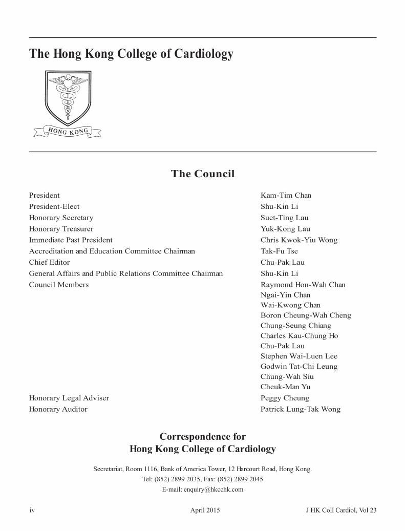
The Hong Kong College of Cardiology
The Council
President Kam-Tim ChanPresident-Elect Shu-Kin LiHonorary Secretary Suet-Ting LauHonorary Treasurer Yuk-Kong LauImmediate Past President Chris Kwok-Yiu WongAccreditation and Education Committee Chairman Tak-Fu TseChief Editor Chu-Pak LauGeneral Affairs and Public Relations Committee Chairman Shu-Kin LiCouncil Members Raymond Hon-Wah Chan
Ngai-Yin ChanWai-Kwong ChanBoron Cheung-Wah ChengChung-Seung ChiangCharles Kau-Chung HoChu-Pak LauStephen Wai-Luen LeeGodwin Tat-Chi LeungChung-Wah SiuCheuk-Man Yu
Honorary Legal Adviser Peggy CheungHonorary Auditor Patrick Lung-Tak Wong
Correspondence forHong Kong College of Cardiology
Secretariat, Room 1116, Bank of America Tower, 12 Harcourt Road, Hong Kong.Tel: (852) 2899 2035, Fax: (852) 2899 2045
E-mail: [email protected]
April 2015 J HK Coll Cardiol, Vol 23ii
INSTRUCTION FOR AUTHORS
The Journal of the Hong Kong College of Cardiology publishes peer-reviewed articles on all aspects of cardiovascular disease, includingoriginal clinical studies, review articles and experimental investigations. As official journal of the Hong Kong College of Cardiology, the journalpublishes abstracts of reports to be presented at the Scientific Sessions of the College as well as reports of the College-sponsored conferences.
Manuscripts submitted to this journal must not be under simultaneous consideration by any other publication and should not have beenpublished elsewhere in substantially similar form. The letter of submission must so affirm. A transfer of copyright form to be signed by all authorsto the Hong Kong College of Cardiology should accompany all submitted articles. All manuscripts should be submitted to the Editor-in-Chief,Journal of the Hong Kong College of Cardiology, c/o Medcom Limited, Room 504-5, Cheung Tat Centre, 18 Cheung Lee Street, Chai Wan,Hong Kong, Email: [email protected].
Manuscript PreparationManuscripts must be submitted in English in triplicate (one originaland two copies) and typed double-spaced on A4 size white bond paper.This applies to all parts of the manuscript, i.e. references, legends,etc. Liberal margins should be left at the top and bottom, as well asthe sides. Except for editorials, images/ECG and letters, all manuscriptshould be submitted in the following order: Title Page, Abstract, Text,References, Tables, Legends, and Figures. Each page, beginning withthe summary, should also include the senior author's surname typedon the upper, left-hand corner. The author should not make any changesin the proofs except for corrections of editorial errors, if any, and/orcorrection of typesetter's errors. Employees of industry may notevaluate or comment about the products of a competitor. A commercialname should not be part of a manuscript title. Finally, authors shouldmake no claims of priority in their manuscripts.
Title Page- Include full name(s), degree(s) and affiliation(s) of author(s); list
under file.- Give a running title of 3 to 6 words.- At the bottom of the page, include information about grants,if
applicable.- Add: "Address for reprint:...", followed by full name, address,
telephone and fax numbers.
Abstract- Abstract should be after title page and numbered page 1.- It should not exceed 250 words for major articles; case reports
should have abstracts of no more than 100 words.- At the end of the abstract, provide a maximum of 6 key words
suitable for indexing.- Abbreviations should be kept to a minimum and must be explained
when they first appear; after first use, abbreviations alone may beused.
- Standard abbreviations should be used for all measurements(SI units).
Text- The text should follow the abstract and begin on a new page, as
should References, Tables, and Legends.- Abbreviations not defined in the abstract should be explained when
they first appear in the text.- References should be cited in numerical order, as should tables
and figures.
References- Number in the order in which they appear in the text.- Abbreviate titles of periodicals according to the style of the Index
Medicus.- Follow the format (arrangement, punctuation) shown below:
Periodicals1. Lewis T. Paroxysmal tachycardia. Heart 1909;1:43-72.
(if more than three authors, please use "et al." after the third).
Books (edited by other authors of article)2. Furman S. Pacemaker follow-up. In Barold SS, (eds): Modern
Cardiac Pacing. Mount Kisco, New York, Futura PublishingCompany, 1985, pp. 889-958.
Books (identical author and editor)3. Chung EK. Principles of Cardiac Arrhythmias. Baltimore, MD,
Williams & Wilkins, 1977, pp. 97-188.
Abstracts4. Same as periodicals and followed by "(abstract)".
Tables- Tables should supplement, but not duplicate, the text.- Tables should be numbered consecutively in order of appearance
in the text.- Each table must be given an Arabic numeral and a title, placed at
the top of the page.- Abbreviations used in the table should be foot-noted and explained
in the order in which they appear in the table, if they have not beenpreviously used.
- Any material which is not self-explanatory should be foot-noted as well.
Legends- Be sure that legends and figures correspond.- Identify all abbreviations used in a figure at the end of each legend,
if the abbreviation has not been used in the text.- Be sure abbreviations used for measurements are standard SI unit.
Figures- Submit either 3 black and white glossy prints or 2 prints and one
photocopy, preferably of 13 cm x 18 cm (5" x 7") size.- On the back of each figure, indicate number, senior author's
surname, top of illustration; all of this should be written lightlywith soft, black pencil.
- Submit written permission from publisher(s) for any figure whichhas been published previously.
- Do not use clips on illustrations; submit them in an envelope backedby cardboard.
- Any lettering or scale of measurement used in an illustration mustbe large enough to be legible in the event of half-size reduction.
- Do not send original art-work, X-rays, or ECGs.- Photographs in which a patient or other person is identifiable must
have written permission from that person. The consent must statespecifically what the person is consenting to and what restrictions,if any, the person has placed upon the publication of the photo-graph. All restrictions must be strictly observed.
- Colour illustrations are costly and will be charged to the author.- Authors should inquire about cost from the publisher before
submitting a colour illustration.
EthicsPublished studies on human subjects should indicate the nature ofconsent and the approval of the institutional ethics committee ifdeemed appropriate. In case of animal experiments, ethical approvalmust be enclosed.
The author is responsible for all material presented in a paper. Thejournal disclaims all responsibility for such material. No product orservice advertised in this publication is guaranteed or warranted eitherby the Editors or publisher. Neither the Editors nor publisher guaranteeany claims made by a manufacturer or an author in regard to a productor service. If a trademark item is named, the name(s) and address(es)of the manufacturer(s) or supplier(s), in addition to the generic name,should be foot-noted.
Reprints are available. Ordering information can be obtained from theabove address.
Subscription RatesLocal Subscription: HK$200/year (including postage)Overseas Subscription: US$120/year (including airmail postage)

April 2015J HK Coll Cardiol, Vol 23 iii
Journal of the Hong Kong College of Cardiology
April 2015Volume 23, No. 1
• ORIGINAL ARTICLE
Surgical Repair of Post Myocardial Infarction
Ventricular Septal Rupture: Experience at a
Tertiary Care Hospital
Imran Khan, Waseem Riaz, Tipu Khan,
Zafar Tufail, Abdul Waheed ........................1
• CASE REPORT
Transcatheter Aortic Valve in Valve
Implantation Through a Prosthesis Carotid
Artery: First Case
Hanane Benhalla and Camelia Sorea .........6
• SPECIAL ARTICLES IN MEMORY OF
DR. CHIU-ON PUN
Dr. CO Pun Memorial Lecture
Chu-Pak Lau ................................................10
Obituary of Dr. CO Pun
Patrick Tak-Him Ko ....................................21
Table of Contents
• TWENTY-THIRD ANNUAL SCIENTIFIC
CONGRESS
Organizing Committee....................................23
Scientific Programme......................................24
Abstracts...........................................................30
April 2015 J HK Coll Cardiol, Vol 23iv
The Hong Kong College of Cardiology
The Council
President Kam-Tim ChanPresident-Elect Shu-Kin LiHonorary Secretary Suet-Ting LauHonorary Treasurer Yuk-Kong LauImmediate Past President Chris Kwok-Yiu WongAccreditation and Education Committee Chairman Tak-Fu TseChief Editor Chu-Pak LauGeneral Affairs and Public Relations Committee Chairman Shu-Kin LiCouncil Members Raymond Hon-Wah Chan
Ngai-Yin ChanWai-Kwong ChanBoron Cheung-Wah ChengChung-Seung ChiangCharles Kau-Chung HoChu-Pak LauStephen Wai-Luen LeeGodwin Tat-Chi LeungChung-Wah SiuCheuk-Man Yu
Honorary Legal Adviser Peggy CheungHonorary Auditor Patrick Lung-Tak Wong
Correspondence forHong Kong College of Cardiology
Secretariat, Room 1116, Bank of America Tower, 12 Harcourt Road, Hong Kong.Tel: (852) 2899 2035, Fax: (852) 2899 2045
E-mail: [email protected]

April 2015J HK Coll Cardiol, Vol 23 1
Surgical Repair of Post Myocardial Infarction Ventricular SeptalRupture: Experience at a Tertiary Care Hospital
IMRAN KHAN, WASEEM RIAZ, TIPU KHAN, ZAFAR TUFAIL, ABDUL WAHEED
From Department of Cardiac Surgery, Punjab Institute of Cardiology, Lahore, Pakistan
KHAN ET AL.: Surgical Repair of Post Myocardial Infarction Ventricular Septal Rupture: Experience at aTertiary Care Hospital. Background: Early surgery is indicated for ventricular septal rupture (VSR) that developsafter myocardial infarction (MI). Surgical repair carries a high mortality. The purpose of this study was to findout the in hospital outcome of the surgical repair of this complication at a tertiary care high volume centre.Methods and Results: A retrospective descriptive study was done by checking the hospital record of all thosepatients who had undergone surgical repair of post myocardial infarction ventricular septal rupture from January2008 to August 2014. The hospital ethical committee gave permission for the study. All the patients underwentidentical surgical procedure for the repair of septal rupture. Perioperative variables were recorded and descriptivestatistics obtained. A total of 40 such patients were identified including 24 (60%) male and 16 (40%) femalepatients with a mean age of 55.4±10.7 years. Intra-aortic balloon pump was used in 27 (62.5%) patientspreoperatively. Nine out of 40 patients were operated within 2 weeks of the occurrence of VSR. While 23(57.5%) were operated after the 3 weeks of VSR. Six out of 9 patients died who were operated within 2 weeks.One out of 23 patients died who presented after 3 weeks duration after post MI VSR. Coclusion: Still a largenumber of patients suffer from post MI VSR in our setup. Surgical treatment carries high mortality especiallythose operated within first week. Patch repair of the ventricular septal is an acceptable treatment strategy forboth anterior and posterior located septal ruptures. (J HK Coll Cardiol 2015;23:1-5)
Intraaortic Balloon pump, Outcomes, Post myocardial infarction ventricular septal rupture
V S R2 0 0 8
1 2 0 1 4 84 0
24 60% 16 40% 55.4±10.7 27 62.5%9 VSR 2 23 57.5% VSR 3 9 6
2 3 1
Address for reprints: Dr. Imran KhanDepartment of Cardiac Surgery, Punjab Institute of Cardiology,Lahore, Pakistan
Email: [email protected]
Received January 2, 2015; revision accepted February 12, 2015

April 2015 J HK Coll Cardiol, Vol 232
SURGICAL REPAIR OF POST MI VSR
Introduction
Ventricular septal rupture is one of the mechanicalcomplications of myocardial infarction. Acutemyocardial infarction can lead to many mechanicalcomplications like rupture of the free wall andpseudoaneurysm, rupture of the ventricular septum,acute mitral regurgitation, and tamponade.1 Postmyocardial infarction (MI) ventricular septal rupture(VSR) occurred in 1-3% patients in pre-thrombolyticera but with the advent of thrombolytic agents theincidence has reduced to 0.5-1%.2 It carries a highsurgical mortality and early intervention is warranted.Post MI VSR is either antero-apical or infero-posterior.Different surgical techniques are used for their repair.With advancement in perioperative management likethe use of intra-aortic balloon pump and better surgicaltechniques, the operative mortality has decreased overthe years.3 But in spite of such developments, it stillcarries a high operative mortality in patients who presentearly. Those surviving the first 30 days postoperativelyhave a good long term survival.4 This retrospective studydescribes the in hospital outcome of surgical treatmentof post MI VSR in a high volume unit.
Materials and Methods
This retrospective observational study wasconducted at the Department of Cardiac Surgery, PunjabInstitute of Cardiology, Lahore, Pakistan. It is a highvolume centre where annually 2100 cardiac operationsare performed on the average. Hospital record of allthose patients operated for ventricular septal ruptureafter myocardial infarction from January 2008 toAugust 2014 was studied and various preoperative,intraoperative and postoperative variables wererecorded. The time interval from the onset of symptomsto surgery was recorded. A 30 day follow up wasobtained from the hospital record for all patientsincluding telephonic information about those patientswho had left the hospital before 30 days period. Theoutcome and in hospital mortality of all the patientswere collected.
Surgical Technique Surgical technique was identical for all the
patients. The VSR was approached from the leftventricle with incision in the infarcted myocardium. Apatch reconstruction of the VSR with Dacron™ wasperformed in every patient. Pledgeted prolene sutureswere used with pledgets on both the right ventricularand left ventricular side of the septum. The defect inthe left ventricular wall was closed using Teflon™ feltsand taking big enough bites in the myocardium toinclude the infarcted or aneurysmal myocardium.Concomitant coronary artery bypass grafting (CABG)was performed where needed. No additional procedurewas needed in any patient.
Data Analysis
The SPSS (version 16, SPSS Inc.) was used forthe data analysis. Quantitative variable were presentedas mean±standard deviation and the qualitative variableswere presented as frequency and percentages.
Results
A total of 40 patients were included in the study.Number of male patients was 24 (60%) and femalepatients were 16 (40%). Mean age was 55.4±10.7 years.The clinical and demographic characteristics of thepatients are presented in Table 1. Intra-aortic balloonpump (IABP) was used in 27 (62.5%) patientspreoperatively. The mean ejection fraction of all thepatients was 42.33±10%. Nine out of 40 patients wereoperated within 2 weeks of the occurrence of VSR. Mostof the patients (23 (57.5%)) were operated after the3 weeks of VSR. Preoperative cardiac angiogram couldnot be obtained in 2 patients as they had to be operatedquickly due to haemodynamic deterioration.Concomitant CABG was performed in 29 (72.5%)patients. Six out of 9 patients died who were operatedwithin 2 weeks. Five of those 6 patients were operatedin the first week after VSR diagnosis. Out of those fivepatients, 4 were in cardiogenic shock who received

April 2015J HK Coll Cardiol, Vol 23 3
KHAN ET AL.
IABP preoperatively. Three of these patients did notsurvive the procedure. Mortality was very low in thosepresenting late i.e., only one out of 23 patients died whopresented after 3 weeks duration after post MI VSR(Table 2). Residual ventricular septal defect (VSD) wasdetected in 5 patients on postoperative echocardiogram.But none of these 5 patients died. Mortality wassignificantly low in patients in whom preoperative IABPwas used. Three out of 4 patients with postero-inferiorVSR died while mortality was significantly low inpatients presenting with antero-apical VSR i.e.5 out of 36.
Discussion
To the best of our knowledge, this is the largestreport on the outcomes of surgical repair of postmyocardial infarction ventricular septal rupture inPakistan. Over a period of five years, 40 patients wereoperated for post myocardial infarction which is a largenumber of patients compared to the internationalliterature. The reason for this high number may be thehuge population that is covered by our hospital andsecondly, a large number of patients still do not haveaccess to instant thrombolytic therapy in this part ofthe world. Reports in 70s and early 80s, whenthrombolytic therapy wasn't common, showed a highincidence of post MI VSR. The GUSTO-I trial thenmentioned an incidence of 0.20% and the reason theygave for the decreasing incidence was more and moreavailability of thrombolytic therapy.2
This mechanical complication of myocardialinfarction carries very high in-hospital mortality. Reportby George and colleagues described the surgical resultsof post MI VSR from the Society of Thoracic Surgeonsdatabase.5 Mortality in their report is 42.9%. They have
Table 1. Clinical characteristics of the patientsVariable Patients n=40Age 55.4±10.7 yearsMale/Female 24 (60%)/16 (40%)Diabetes mellitus 13 (32.5%)Hypertension 27 (65%)History of smoking 10 (25%)Preoperative IABP 27 (62.5%)EF 42.33±10%Cardiogenic shock 4 (10%)Postero-inferior location of VSR 4 (10%)Antero-apical location of VSR 36 (90%)Concomitant CABG 29 (72.5%)Mean CPB time 95.45±25.06 minutesMean cross clamp time 67.9±13.88 minutesLength of stay in ICU 11±5 daysResidual VSD 5 (12.5%)CVA 3 (7.5%)Postoperative acute kidney injury 13 (32.5%)Pleural effusion requiring tapping 6 (15%)Postoperative RRT 4 (10%)IABP: intra aortic balloon pump; EE: ejection fraction; VSR:ventricular septal rupture; CABG: coronary artery bypassgrafting; CPB: cardiopulmonary bypass; ICU: intensive careunit; VSD: ventricular septal defect; CVA: cerebrovascularaccident; RRT: renal replacement therapy
Table 2. Relationship of time duration from diagnosis to surgery with early outcomeTime duration from diagnosis to surgery In-hospital outcome Total No. of patients
Survival Mortality<2 weeks 3 6 (66.67%) 9 (22.5%)2 weeks to 3 weeks 7 1 (12.5%) 8 (20%)>3 weeks 22 1 (4.3%) 23 (57.5%)Total 32 8 (20%) 40
mentioned time interval from MI to operation and thensurgery. They showed that 54% of the patients operatedwithin 7 days of MI had in-hospital mortality. Andersand colleagues similarly showed a mortality of 41%.6
But mortality rates as low as 19% have also beenreported.7 Morality in our study was 20% which isevidently on the lower side compared to most of theinternational reports. This can be explained by the fact

April 2015 J HK Coll Cardiol, Vol 234
SURGICAL REPAIR OF POST MI VSR
the most of the patients presented late and 90% patientshad antero-apical VSR. Both these factors have beenproved to be predictors of survival in previousreports.5 Cardiogenic shock is an important risk factorfor mortality in these patients.5 Only four of the patientsin our study reached the hospital with cardiogenic shock.They were emergently operated and only one of thosefour patients survived showing the very high mortalityrate in this subgroup of patients.
The time for intervention is very much decidedby the haemodynamic status of the patient. Cardiogenicshock warrants immediate surgery. If the patient ishaemodynamically stable, optimization with inotropesand mechanical cardiac support can be achieved andsurgery performed after a delay of 3-4 weeks. If there isclinical deterioration, immediate surgery is indicated.8
Patients operated within one week of occurrence of VSRcarry a very high mortality.9 On the other hand, patientsoperated after the 2 weeks period after VSR carried avery low mortality as evident from our study. The reasonfor high mortality in early operated patient may be theacute haemodynamically decompensated state of thepatient and secondly the fresh, friable margins of thedefect where necrotic process is still going on.10 Wecannot wait for that length of time and deny early surgeryto patients on ethical grounds as we don't know whichpatient will survive the initial high mortality period. Soevery patient should be given a chance and operatedearly when the diagnosis is made.11
Intra-aortic balloon pump is an important additionto the management of post MI VSR. It was used in62.5% of the patients in our study. The use of IABP inthese patients significantly reduces mortality as shownby our report. IABP significantly reduces left to rightshunt and afterload in these patients thus improving thehaemodynamics.12 The current guidelines for themanagement of post MI VSR recommends the routinepreoperative use of IABP for every patient diagnosedwith this problem.13
Whether to do concomitant CABG or not is asubject of debate. Coronary arteries have a varied patternof disease in patients with post MI VSR. Cox andcolleagues and Leavey and colleagues found singlevessel disease to be more common in their patients.14,15
Triple vessel disease was found in 48.2% of the patientsand concomitant CABG was performed in 72.5% of the
patients in our study. Barkera and colleagues in theirarticle found triple vessel disease to be more commonin patients with post MI VSR.16 The high number ofpatients with triple vessel disease in our study may bedue to the extensive nature of coronary artery diseasein this part of the world.17
Small residual VSD was observed in five patientspostoperatively in our study. These patients did notsuffer from any additional morbidity and they survivedthe immediate postoperative period. Yam and colleaguesobserved patients with residual VSD for 10 years andfound excellent long term outcomes.18 We cannot inferat present from these findings as to what should be thefate of small residual defects that does not cause anyshunt. Transcatheter closure of these defects has beendescribed in literature.19
Our study is a retrospective report of the repairof post MI VSR at a single centre, thus carries all thedrawbacks of a retrospective study. The sample size isalso not statistically powered so as to find out all thepredictors of mortality accurately. It doesn't describethe medium or long term follow up of the patientsdescribed thus we don't know the usefulness of the patchrepair procedure in the long term. But this descriptivestudy gives an idea about the diseases burden and itssurgical outcome in a developing country. We hope thatwith better facilities of thrombolytic therapy theincidence of ventricular septal rupture in the settings ofacute MI will decrease. Advancements in theperioperative care and the availability of ventricularassist devices in this part of the world will certainlyimprove the surgical outcome of the patients especiallythose who present early to the hospital and those withcardiogenic shock.
Conflict of Interest Statement
None
Funding
This research received no specific grant from anyfunding agency in the public, commercial, or not-for-profit sectors.

April 2015J HK Coll Cardiol, Vol 23 5
KHAN ET AL.
References
1. Lavie CJ, Gersh BJ. Mechanical and electrical complicationsof acute myocardial infarction. Mayo Clin Proc 1990;65:709-30.
2. Crenshaw BS, Granger CB, Birnbaum Y, et al. Risk factors,angiographic patterns, and outcomes in patients with ventricularseptal defect complicating acute myocardial infarction. GUSTO-I (Global Utilization of Streptokinase and TPA for OccludedCoronary Arteries) Trial Investigators. Circulation 2000;101:27-32.
3. Maltais S, Ibrahim R, Basmadjian AJ, et al. PostinfarctionVentricular Septal Defects: Towards a New TreatmentAlgorithm? Ann Thorac Surg 2009;87:687-92
4. Yam N, Au TW, Cheng LC. Post-infarction ventricular septaldefect: surgical outcomes in the last decade. Asian CardiovascThorac Ann 2013;21:539-45.
5. Arnaoutakis GJ1, Zhao Y, George TJ, et al. Surgical repair ofventricular septal defect after myocardial infarction: outcomesfrom the Society of Thoracic Surgeons National Database. AnnThorac Surg 2012;94:436-44.
6. Jeppsson A, Liden H, Johnsson P, et al. Surgical repair of postinfarction ventricular septal defects: a national experience. EurJ Cardiothorac Surg 2005;27:216-21.
7. Moreyra AE, Huang MS, Wilson AC, et al; MIDAS Study Group(MIDAS 13): Trends in incidence and mortality rates ofventricular septal rupture during acute myocardial infarction.Am J Cardiol 2010;106:1095-100.
8. Papalexopoulou N, Young CP, Attia RQ. What is the best timingof surgery in patients with post-infarct ventricular septalrupture? Interact Cardiovasc Thorac Surg 2013;16:193-6.
9. Labrousse L, Choukroun E, Chevalier JM, et al. Surgery forpost infarction ventricular septal defect (VSD): risk factors forhospital death and long term results. Eur J Cardiothorac Surg2002;21:725-32.
10. Deville C, Fontan F, Chevalier JM, et al. Surgery of post-infarction ventricular septal defect: risk factors for hospital deathand long-term results. Eur J Cardiothorac Surg 1991;5:167-75.
11. David TE, Dale L, Sun Z. Post infarction ventricular septalrupture: repair by endocardial patch with infarct exclusion.J Thorac Cardiovasc Surg 1995;110:1315-22.
12. Testuz A, Roffi M, Bonvini RF. Left-to-right shunt reductionwith intra-aortic balloon pump in postmyocardial infarctionventricular septal defect. Catheter Cardiovasc Interv 2013;81:727-31.
13. 2013 ACCF/AHA guideline for the management of ST-elevationmyocardial infarction: a report of the American College ofCardiology Foundation/American Heart Association Task Forceon Practice Guidelines. J Am Coll Cardiol 2013;61:e78-140.
14. Cox FF, Plokker HW, Morshuis WJ, et al. Importance ofcoronary revascularization for late survival after postinfarctionventricular septal rupture. Eur Heart J 1996;17:1841-5.
15. Leavey S, Galvin J, McCann H, et al. Post-myocardial infarctionventricular septal defect: an angiographic study. Ir J Med Sci1994;163:182-3.
16. Barkera TA, Ramnarineb IR, Wood EB, et al. Repair of post-infarct ventricular septal defect with or without coronary arterybypass grafting in the northwest of England: a 5-year multi-institutional experience. Eur J Cardiothorac Surg 2003;24:940-6.
17. Zaman MJ, Philipson P, Chen R, et al. South Asians andcoronary disease: is there discordance between effects onincidence and prognosis? Heart 2013;99:729-36.
18. Yam N, Au TW, Cheng LC. Post-infarction ventricular septaldefect: surgical outcomes in the last decade. Asian CardiovascThorac Ann 2013;21:539-45.
19. Assenza GE, McElhinney DB, Valente AM, et al. Transcatheterclosure of post-myocardial infarction ventricular septal rupture.Circ Cardiovasc Interv 2013;6:59-67.

April 2015 J HK Coll Cardiol, Vol 236
Transcatheter Aortic Valve in Valve Implantation Through aProsthesis Carotid Artery: First Case
HANANE BENHALLA AND CAMELIA SOREA
From Brussels Heart Center, Brussels, Belgium
BENHALLA AND SOREA: Transcatheter Aortic Valve in Valve Implantation Through a Prosthesis CarotidArtery: First Case: In recent years, transcatheter aortic valve implantation has become an emerging alternative forhigh-risk patients with severe aortic stenosis. We report a case of transcatheter aortic valve implantation with theself-expanding Medtronic CoreValve bioprosthesis through a left common carotid artery. This 83-year-old malepatient presented with heart failure due to a severe degenerative aortic bioprosthesis, with several comorbidities,resulting in a logistic EuroScore II of 36%. Consequently, he was rejected to undergo surgery and a transcatheterapproach was planned. And due to severe peripheral vascular disease with iliofemoral lesions, significant calcificationsof the innominate artery, we considered a left carotid access through a prosthesis carotid in Dacron. The procedurewas successful without cardiac, cerebrovascular, or access complications. And it appears to be a valuable alternativeaccess for patients with severe peripheral vascular disease. (J HK Coll Cardiol 2015;23:6-9)
Carotid bioprosthesis, Percutaneous aortic valve, Valve in valve
83 logistic EuroScore II 36%
Address for reprints: Dr. Hanane Benhalla23 lotissement lamia, Bourgogne Casablanca, Morocco
Email: [email protected]
Received November 19, 2014; revision accepted March 25, 2015
Introduction
The first percutaneous aortic valve replacementhas been performed by Dr. A. Cribier in 2002, openinga new therapeutic approach to patients at high surgicalrisk for conventional operation by sternostomy.
Within a short t ime, there has been animprovement of the material and a simplification of the
procedure, the surgical approach used and theimplantation technique. However patient selectionremains crucial. We describe a percutaneous aorticvalve implantation (a CoreValve bioprosthesis) in adegenerated bioprosthesis without an adequate vascularaccess, and which we decide to add prosthesis carotidin Dacron in per operative procedure.
Case Report
An 83-year-old male, presented with heart failuredue to a severe degenerative aortic bioprosthesisoperated in 2009, with comorbidities included diabetes,hypertension, chronic pulmonary obstructive disease,

April 2015J HK Coll Cardiol, Vol 23 7
BENHALLA AND SOREA
as well as peripheral and coronary artery disease,resulting in a logistic EuroScore II of 36%.Preinterventional morphological patient screeningincluded transthoracic as well as transesophagealechocardiography, confirmed a severely calcified aorticbioprosthesis with a mean transprosthesis gradient of53 mmHg, valve area 0.8 cm2, and ejection fraction of40%.
Computed tomographic illustrate showed aperipheral vascular disease with iliofemoral lesions,the diameter of the common iliac artery was of 5 mmand the subclavian arteries were about 5-6 mm withs ignif icant ca lc i f ica t ions and unfavourableangulations of the innominate artery. Therefore, weconsidered a left carotid access, with a diameter of6.5 mm, as the only solution with the use of aprosthesis carotid in Dacron to facilitate theprocedure. The aortic annulus diameter, and thedistance of the coronary ostia were also evaluated.
The carotid artery was first reopened underlocal anaesthesia and then after intubation andinduction of general anaesthesia, endarterectomy ofthe left carotid artery was performed. Thereafter, an8 mm Dacron prosthesis was connected to the leftcommon carotid artery in an end-to-side fashion anda sheath was introduced into it. Via this approach, aself-expanding aortic valve prosthesis (CoreValve,Medtronic) was placed in typical position but 4 mmbelow (Figures 1 & 2). Importantly, the introducersheath was not advanced into the carotid artery, sothat antegrade blood flow was maintained during theentire TAVI procedure without further shunting. Alocalization 4 mm below the position is consideredideal for implantation. In this case (Figure 3) thismay explain the immediate postoperative transaorticprosthesis mean gradient of 20 mmHg ,with aorticprosthesis area of 1.1 cm2.
In the Post-operative follow up there is animportant improvement of signs of heart failurewithin the first week for the patient.
The pat ient was seen one month af terthe procedure with a clear clinical improvementand reg ress ion o f h i s ep i sodes o f ca rd iacdecompensation.
Figure 1. Computed tomographic viewing the aorticprosthetic ring with calcifications before and after theendovalve implantation.
Discussion
The entry in our case of the right native carotidartery was facilitated by the introduction of a prosthesiscarotid in Dacron. The advantages of this procedure,are an optimal neuromonitoring during the carotidsurgery in local anaesthesia and a simple implantationof the catheter-based aortic valve prosthesis via the same

April 2015 J HK Coll Cardiol, Vol 238
TRANSCATHETER AORTIC VALVE
Figure 3. Computed tomographic view of the percutaneousvalve relative to the ring of the degenerated bioprosthesis.
Figure 2. Angiographic view of the introduction of thepercutaneous aortic valve on the degenerated bioprosthesisand the result seen in the chest radiograph.
access and during an only short period of generalanaesthesia. Importantly, as already mentioned above,the introducing sheath for the TAVI must only beadvanced into the "Dacron chimney" and not furtherinto the carotid artery to provide a sufficient antegradeflow throughout the whole procedure.1,2
Very limited experience exists with surgicalaccess via the carotid artery. In a small series reportedby Modine et al of 12 patients, the procedure wassuccessful in all. There was no mortality, but 1 patienthad a stroke , which mean that electroencephalogrammonitoring in parallel to the procedure seems necessaryto monitor cerebral perfusion.3
The median time for the implantation of the valvein valve is about 120 minutes in the European register.3
For our case, it was 180 minutes with a fluoroscopytime of 36 minutes. This was explained by the initialimplantation of the Dacron prosthesis.
For the valve in valve procedure the mostcommon risk is the less optimal deployment of thepercutaneous valve, secondary to calcificationsusually present on the bioprosthesis especially if itis asymmetric,3 Correct sizing is paramount, asundersizing may increase the risk of paravalvularregurgitation or valve migration whereas oversizingmay lead to leaflet distortion within the transcatheter

April 2015J HK Coll Cardiol, Vol 23 9
BENHALLA AND SOREA
heart valve, There is also often the need to implant apermanent pacemaker (5.7% to 20% for Corvalve)which remains the most common event described inthe first 30 days with a positioning 2 to 6 mmbelow the base of the aortic annulus, which mayinterfere with the atrioventricular node, located veryclose to the aortic region and sub-membranousseptum.3,4
Conclusion
The common caro t id access has beendemonstrated to be a feasible and safe access routefor TAVI. It appears to be a valuable alternative accessfor patients who cannot undergo trans-femoral TAVI,to expand the benefit from this technology with lessbleeding events, less access-related complicationsand immediate patient ambulation.
Disclosures
The authors declare that there is no conflict ofinterest.
References
1. Henn LW, Makkar RR, Fontana GP. Valve in valve TAVI fordegenerated surgical prostheses. Cardiac Interventions Today- August 2010.
2. Modine T, Sudre A, Delhaye C, et al. Transcutaneous aorticvalve implantation using the left carotid access: feasibility andearly clinical outcomes. Ann Thorac Surg 2012;93:1489-94.
3. Modine T, Lemesle G, Azzaoui R, et al. Aortic valve implantwith the CoreValve ReValving System via left carotid arteryaccess: first case report. J Thorac Cardiovasc Surg 2010;140:928-9.
4. Ng AC, van der Kley F, Delgado V, et al. Percutaneous valve-in-valve procedure for severe paravalvular regurgitation inaortic Bioprosthesis. JACC Cardiovasc Imaging 2009;2:522-3.

Special Articles
in Memory
of
Dr. Chiu-On Pun

April 2015J HK Coll Cardiol, Vol 23 10
Subclinical Atrial Fibrillation: A New Paradigm for Stroke Prevention
CHU-PAK LAU
From Cardiology Division, Department of Medicine, Queen Mary Hospital, the University of Hong Kong,Hong Kong
LAU: Subclinical Atrial Fibrillation: A New Paradigm for Stroke Prevention. Approximately one out of five strokesis associated with atrial fibrillation (AF). AF is often intermittent and asymptomatic. Detection of AF after cryptogenicstroke will change therapy from antiplatelet to oral anti-coagulation agents for secondary stroke prevention. A criticalstep is to convert "covert" AF into ECG documented AF. External rhythm recording devices have registered a highincidence of AF to occur after a cryptogenic stroke, but are limited by short duration of continuous recordings.Invasive cardiac monitoring using insertable leadless cardiac monitors (ICMs) are sensitive means to identify subclinicalAF (SCAF) after cryptogenic stroke, and AF has been reported to occur in up to 30% of these patients. It will be evenmore attractive to identify SCAF before a stroke occurs. Recent series in pacemaker and implantable cardioverterdefibrillator showed that short episodes of SCAF increased stroke risk, with odds ratio ~2.2-3.1 compared to thosewithout SCAF recorded. However, temporal sequence of recorded SCAF and stroke occurrence was uncertain, andthe overall stroke risk was lower compared with patients with clinical AF at similar risk scores. This article reviewsthe incidence and clinical role of using implanted devices to detect SCAF and discuss the implication of SCAF sodetected in primary and secondary stroke prevention. (J HK Coll Cardiol 2015;23:10-20)
Atrial Fibrillation, Implantable Cardioverter Defibrillator, Pacemaker, Stroke
2 0 %
3 0 %
2.2-3.1
Address for reprints: Prof. Chu-Pak LauCardiology Division, Department of Medicine, Queen Mary Hospital,the University of Hong Kong, Hong Kong
Email: [email protected]
Received November 11, 2014; revision accepted May 5, 2015
*The content of the special article will be presented on theDr. CO Pun memorial lecture on 30 May 2015, and is part ofa review article in press in the Europace Supplement forCardiorhythm 2015.
April 2015 J HK Coll Cardiol, Vol 2311
SUBCLINICAL ATRIAL FIBRILLATION
Introduction
Epidemiological evidence suggests that atrialfibrillation (AF) increase ischaemic stroke risk by 5- to6-fold independent of other risk factors.1 AF relatedstroke tends to be more severe,2 and mortality rate ishigher (70-80%) compared with stroke without AF.There is also a high recurrence rate.3
AF episodes are often asymptomatic, and AF maypresent for the first time with complications such asthromboembolism and heart failure. Indeed, paroxysmalAF has similar stroke risk compared to sustained AF.4
About 25% of strokes has no obvious underlyingcerebrovascular disease or other stroke risk factors.AF is an important underlying mechanism forcardioembolism in these patients. Apart from overtneurological deficits, recurrent cerebral emboli cancause cognitive dysfunction and dementia.5
In the absence of documented AF, secondary andprimary prophylaxis of stroke relies on the use ofantiplatelet agents. If AF is the cause of stroke, aspirincan reduce stroke risk by 22% compared to placebo.However, oral anticoagulation using warfarin canadditional reduce this risk by 38-63% compared toaspirin.6 More recently, non-vitamin K oralanticoagulation agents (NOACs) have been shown tobe at least non-inferior to warfarin and have a lowerincidence of major haemorrhagic complications, thusimproving the risk-benefits ratio of stroke preventionin AF. This background makes primary and secondaryprophylaxis of AF related stroke attractive. A criticalstep is to document underlying AF.
AF Documentation
Due to the intermittent occurrence and oftenasymptomatic presentation of AF, routine ECG, 24 hourHolter and patient triggered recording devices have lowdetection rate of AF. Several types of external monitorswith attached electrodes have allowed intermediate termcontinuous AF monitoring. Electrodes used include wetor dry electrodes.7 These provide not only patient
triggered recordings, but automatic recording if AFoccurs. Patient tolerability has improved with improvedelectrode design.
Longer term recording of cardiac rhythm ispossible with implantable leadless cardiac monitors(ICMs), such as by the Medtronic Reveal XTTM.As P waves are not well detected in ICMs, irregularityof RR interval is used as a surrogate for AF, the so calledLorentz plot8 is arithmetically used to register AF. Thishad been tested in the XPECT Trial.9 In this study, 247patients with high AF burden received the Reveal XTTM.The stored rhythm was recorded simultaneously withthe ECG to an external Holter monitor for 46 h. Thisshowed a sensitivity, specificity, positive predictivevalue and negative predictive value of the Reveal XTto identify AF of 96.1, 85.4, 79.3 and 97.4%respectively, with an accuracy of 98.5%. Further, falsepositivity is reduced by superimposing the R waves toexamine for a possible P wave. The device has beenfurther miniaturized and can be implanted with aninjection mechanism (the LINQTM, Medtronic).
Obviously, cardiovascular implantable electronicdevices (CIEDs) with attached atrial electrodes providean excellent recording of AF. In the pacemakerpopulation, Israel et al10 showed in CIEDs, asignificantly higher sensitivity of AF detection
Table 1. Technical aspects of AF detection usingimplanted atrial leads
1. Closely spaced atrial bipolar lead
2. Bipolar sensing (atrial tip-atrial ring)
3. High atrial sensitivity (0.1-0.5 mV)(minimise under/intermittent sensing for AFdetection)
4. Short PVARP
5. PVAB ≥25 ms to reject far field R wave
6. AEGM validation
7. Duration of AHRE to consider as AFAF=Atrial fibrillation; AEGM=atrial electrogram;AHRE=Atrial high rate episode PVAB=Post ventricular atrialblanking; PVARP=Post ventricular atrial refractory period

LAU
J HK Coll Cardiol, Vol 23 12April 2015
compared to Holter recordings. Table 1 shows thetechnical aspect to detect AF in CIEDs. Accuraterecording requires a closely spaced atrial bipole (<1 cm),an appropriate atrial sensitivity setting, post ventricularatrial refractory period (PVARP) and blanking (PVAB)adjustment.
Programming appropriate atrial rates and episodedurations cut off is also critical to register AF. A lowcut off rate of AF detection will increase false positivitydue to inclusion of noises and far field events such as Rwave, whereas a high rate cut off will miss AF episodeswhen atrial signals become small or fall within thePVARP and PVAB. Programming a short cut off episodeduration will increase sensitivity to detect brief AFepisodes, but will include false positive detection ofnoise, whereas a longer cut off duration will increaseaccuracy but miss brief AF events. A validation of theMedtronic. AT500 and GEM III AT algorithmssuggested sensitive AF detection if far field R waveswere rejected.11 In 5,769 pacemaker detected "atrial highrate events" (AHREs), Kaufman et al12 examined therelative contribution of programmed cut off detectionrate and duration on accuracy of AF detection. Anincrease in cut off detection rate from 190 to 250 bpmreduces false positive detection, especially if shorter AFdetection rates were programmed. A cut off detectionduration of >6 minutes will have a 17.3% false positivedetection, compared to 3.3% if detection duration of>6 hours was programmed. It was concluded thatvalidation by atrial electrograms (AEGMs) would beimportant for shorter detected AF episodes of>6 minutes, whereas this became less critical for longerepisodes >6 hours. As most studies do not vigorouslyrelate symptoms with device detected AF episodes, thisreview uses the term subclinical AF (SCAF) for AFdetection by implanted, to distinguish them from theoccurrence of clinical AF.
Secondary Prevention in CryptogenicStroke
Kishore et al13 summarized 32 trials which haveused either external monitors or ICMs to detect AF in
patients after ischaemic stroke or transient ischemicattack (TIA). There was substantial heterogeneitybetween studies, with an overall detection rate of anynew AF occurring in 11.5%, with higher detection ratein selected (e.g. cryptogenic stroke) versus unselectedpatients groups (13.4 vs 6.2%). Longer duration ofrecording and older patient age increased the chance ofAF detection. There was insufficient data on the stroketype (lacunar vs non lacunar) and the timing of startingrecording after the index stroke on AF detection rate. Alarge prospective study7 recruited 572 ambulatorypatients with a mean age of 55 years at a mean of 75days after a stroke or TIA. Patients were randomized toreceive either a 30-day event-triggered external recorderusing dry electrodes or with another 24h Holterrecording. The primary end point was detectedAF >30 s, which was reached in 16.1% of patients versusonly 3.2% using Holter only, and had led to an increasein oral anticoagulation use (18.6 vs 11.1%). ClinicalAF was only detected in 0.5% of patients after 90 days,and AF was more often detected if the device wasadministered within 30 days of the index stroke. Thelimitations of this study are the delay in administratingAF recording, exclusion of more serious stroke andnon-ambulatory patients, and lack of AF burdenmeasurement (as only <2.5 minutes per episode can bemeasured). Occurrence of AF after 30 days was notdetermined by this study.
More prolonged monitoring using invasiverecordings in a similar population was reported inthe CRYSTAL AF study.14 In 441 patients at a meanof 38 days after a cryptogenic stroke either an externalHolter or ICM (Reveal XT) was used to assess thetime of first AF >30 s occurrence (Figure 1). AF wasdetected in 12.4% compared with 2.0% using ICMcompared to Holter (p<0.001), again resulting in ahigher percentage of oral anticoagulation use (14.7vs 6.0%, p<0.007), and a trend to a lower recurrentstroke rate (7.1 vs 9.1%). At 3 years, the deviceprojected battery life, AF was detected in 30% ofpatients. Most of the episodes of detected AF wereasymptomatic (74% and 79% at 6 and 12 monthsrespectively). Other prospective studies havedocumented a variable incidence of AF detection of

April 2015 J HK Coll Cardiol, Vol 2313
SUBCLINICAL ATRIAL FIBRILLATION
15.9%,13 with the results of the Stroke Prior toDiagnosis of Atrial Fibrillation Using Long TermObservation with Implantable Cardiac MonitoringApparatus Reveal (SURPRISE) showing an incidenceof new AF in 18.6% in 3 years.15
Taken together, AF is a common occurrence incryptogenic stroke and its detection will significantlyaffect the use of antithrombotic treatment. Since AFis detected in up to 30% of such patients in 3 years,arguably antithrombotic therapy with NOACs maybe considered in such patients even without AFdocumentation. This is the subject of 2 prospectiverandomized studies in which either dabigatran orapixaban will be compared to aspirin in patients withcryptogenic stroke.
Primary Prevention of Stroke by EarlyScaf Detection Using Implanted Devices
The strong association between clinical AF and
ischaemic stroke, and the proven benefit ofanticoagulation prophylaxis make early AF detectionof clinical interest. Implanted CIEDs are the mostreliable methods to detect AF. However, especially forshorter and asymptomatic SCAF episodes, it is uncertainif they predict clinical AF development or stroke (andother thromboembolic events) themselves. Therelationship of SCAF detected by CIEDs and futureclinical AF and stroke provides important backgroundinformation for the clinical importance of SCAF.
Frequency of SCAF Detected by CIED andRelation to Clinical AF
Gillis et al16 reported SCAF to be detected in 68%of 231 patients with sinus node disease, and an incidenceof 50.6% of SCAF was documented in 617 patients withDDD pacemakers.17 An incidence of 44% of AF wasdetected in 226 patients during a long follow up periodof 7 years,18 with a much higher incidence of AF in
Figure 1. Prolonged external monitoring and Implantable leadless cardiac monitor (ICM) versus Holter recording to detect AFin patients after cryptogenic stroke. AF was detected in 8.9%, 12.4% and 30% by ICMs at 6, 12 and 36 months respectively. Datareproduced from the references 7 and 14.
LAU
J HK Coll Cardiol, Vol 23 14April 2015
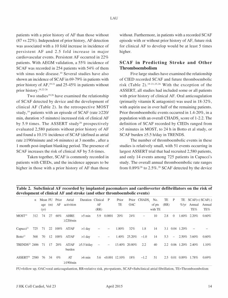
patients with a prior history of AF than those without(87 vs 22%). Independent of prior history, AF detectionwas associated with a 10 fold increase in incidence ofpersistent AF and 2.5 fold increase in majorcardiovascular events. Persistent AF occurred in 22%patients. With AEGM validation, a 55% incidence ofSCAF was recorded in 254 patients with 54% of themwith sinus node disease.10 Several studies have alsoshown an incidence of SCAF in 69-79% in patients withprior history of AF,19-21 and 25-45% in patients withoutprior history.19,22-26
Two studies19,26 have examined the relationshipof SCAF detected by device and the development ofclinical AF (Table 2). In the retrospective MOSTstudy,19 patients with an episode of SCAF (rate ≥220/min, duration >5 minutes) increased risk of clinical AFby 5.9 times. The ASSERT study26 prospectivelyevaluated 2,580 patients without prior history of AFand found a 10.1% incidence of SCAF (defined as atrialrate ≥190/minute and >6 minutes) at 3 months , after a1 month post-implant blanking period. The presence ofSCAF increases the risk of clinical AF by 5.6 times.
Taken together, SCAF is commonly recorded inpatients with CIEDs, and the incidence appears to behigher in those with a prior history of AF than those
without. Furthermore, in patients with a recorded SCAFepisode with or without prior history of AF, future riskfor clinical AF to develop would be at least 5 timeshigher.
SCAF in Predicting Stroke and OtherThromboembolism
Five large studies have examined the relationshipof CIED recorded SCAF and future thromboembolicrisk (Table 2).19-21,25,26 With the exception of theASSERT, all studies had included some or all patientswith prior history of clinical AF. Oral anticoagulation(primarily vitamin K antagonist) was used in 18-32%,with aspirin use in over half of the remaining patients.Prior thromboembolic events occurred in 1.4-20%, in apopulation with an overall CHADS2 score of 1-2.2. Thedefinition of SCAF recorded by CIEDs ranged from>5 minutes in MOST, to 24 h in Botto et al study, orSCAF burden ≥5.5 h/day in TRENDS.
The number of thromboembolic events in thesestudies is relatively small, with 51 events occurring inlargest ASSERT trial that had recruited 2,580 patients,and only 14 events among 725 patients in Capucci'sstudy. The overall annual thromboembolic rate rangesfrom 0.89%26 to 2.5%.20 SCAF detected by the device
Table 2. Subclinical AF recorded by implanted pacemakers and cardioverter defibrillators on the risk ofdevelopment of clinical AF and stroke (and other thromboembolic events)
n Mean FU Prior Atrial Duration Clinical P Prior Prior CHADS2 No. TE P TE SCAF(+) SCAF(-)age (m) AF activition AF TE OAC of pts (RR) %/yr Annual Annual(yr) (RR) with TE TE% TE%
MOST19 312 74 27 60% AHRE >5 min 5.9 0.0001 20% 24% − 10 2.8 0 1.60% 2.20% 0.60%≥220/min
Capucci21 725 71 22 100% AT/AF >1 day − − 1.80% 32% 1.8 14 3.1 0.04 1.20% − −
Botto12 568 70 12 100% AT/AF >1 day − − 1.40% 25.20% ~1.0 14 5.3 − 2.50% 3.60% 0.60%
TRENDS25 2486 71 17 20% AT/AF ≥5.5 h/day − − 13.40% 20.80% 2.2 40 2.2 0.06 1.20% 2.40% 1.10%burden
ASSERT26 2580 76 34 0% AT >6 min 5.6 <0.001 12.10% 18% ~1.2 51 2.5 0.01 0.89% 1.78% 0.69%
≥190/min
FU=follow up, OAC=oral anticoagulantion, RR=relative risk, pts=patients, SCAF=Subclinical atrial fibrillation, TE=Thromboembolism

April 2015 J HK Coll Cardiol, Vol 2315
SUBCLINICAL ATRIAL FIBRILLATION
increased the relative risk for thromboembolism by afactor of 2.2-5.3 compared to no SCAF detected. Annualthromboembolic rates were similarly higher in thosepatients with detected SCAF versus those without.
The event rates for the ASSERT which includedonly patients without prior AF are tabulated in Table 3.SCAF detected at 3 months increased the relative riskof thromboembolic events and clinical AF to 2.81 and5.0 respectively. Similar to other studies, no differencein total and cardiovascular mortality has been reportedso far.
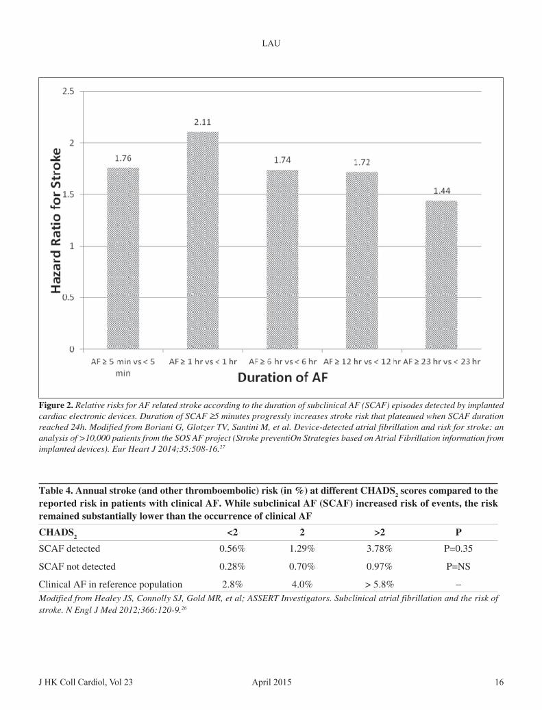
SCAF Duration and CHADS2 Scores in Relationto Thromboembolism
In a retrospective analysis, Botto el al20 hadattempted to stratify thromboembolic risk in a cohortof 567 patients with a total number of 14 events. Therewas a relationship between duration of SCAF andCHADS2 score with stroke event. At a CHADS2 scoreof 1, only AF >24 h would increase the annual strokerate to 4% compared to 0.6% for SCAF that lastedshorter. At a CHADS2 score of 2, any SCAF >5 minutesresulted in a 4% stroke rate. In the TRENDS study, onlySCAF burden >5.5 h/day increased thromboembolic rateto 2.4%/year, in a population of CHADS2 score of 2.2.Likewise, the thromboembolic risk in ASSERT becamesignificant only when SCAF was >17.72 h.
In a pooled analysis of over 10,000 patients,Boriani et al27 compared the duration of CIED recorded
SCAF and stroke risk (Figure 2). The hazard ratio ofSCAF >5 minutes was similar to the impact of havingsustained AF, and progressively increased the SCAFduration, and the risk appeared to plateau off whenSCAF reached 24 hours.
Despite these findings, the annual risk forthromboembolism in these studies are lower thanexpected when compared to patients with clinical AFwith similar CHADS2 scores. Indeed, in the ASSERTstudy involving patients without prior AF, the annualrates of thromboembolic events with SCAF detectedwere lower than expected from published risk ofCHADS2 score (Table 4). The reason for the lower strokerisk for SCAF compared to clinical AF is uncertain.Possibilities include: patients with CIEDs are differentfrom clinical AF patients, that atrial leads might havegenerated a different type of AF, or SCAF may representless severe or early AF that require time to become anestablish risk. Finally, a substantial percent of patientsin these studies were on anti-thrombotic therapy whichwould have reduced embolic risk.
Is Subclinical AF Only a Risk Marker forStroke?
The traditional belief is that AF results incardioembolic events from atrial clots due to mechanicalstasis in the atrium. Indeed, in trans-esophageal
Table 3. Clinical outcome of patients with CHADS2 ≥≥≥≥≥2 who were recruited in the ASSERT trial
Event Asymptomatic AF detected by device SCAFPresent N=187 Absent N=1790 present vs. absent
Events %/ year Events %/year RR 95% CI P
Ischaemic stroke or systemic embolism 10 2.19 35 0.79 2.81 1.39-5.68 0.004Vascular death 16 3.51 137 3.10 1.11 0.66-1.87 0.68Stroke / MI / vascular death 25 5.48 185 4.18 1.29 0.85-1.95 0.24
Clinical atrial fibrillation or flutter 29 6.36 61 1.38 5.00 3.21-7.79 <0.00001
MI = Myocardial infraction; SCAF = Subclinical AF detected (Rate >190/min and >6 mins)Modified from Healey JS, Connolly SJ, Gold MR, et al; ASSERT Investigators. Subclinical atrial fibrillation and the risk ofstroke. N Engl J Med 2012;366:120-9.26
LAU
J HK Coll Cardiol, Vol 23 16April 2015
Table 4. Annual stroke (and other thromboembolic) risk (in %) at different CHADS2 scores compared to thereported risk in patients with clinical AF. While subclinical AF (SCAF) increased risk of events, the riskremained substantially lower than the occurrence of clinical AF
CHADS2 <2 2 >2 P
SCAF detected 0.56% 1.29% 3.78% P=0.35
SCAF not detected 0.28% 0.70% 0.97% P=NS
Clinical AF in reference population 2.8% 4.0% > 5.8% −Modified from Healey JS, Connolly SJ, Gold MR, et al; ASSERT Investigators. Subclinical atrial fibrillation and the risk ofstroke. N Engl J Med 2012;366:120-9.26
Figure 2. Relative risks for AF related stroke according to the duration of subclinical AF (SCAF) episodes detected by implantedcardiac electronic devices. Duration of SCAF ≥5 minutes progressly increases stroke risk that plateaued when SCAF durationreached 24h. Modified from Boriani G, Glotzer TV, Santini M, et al. Device-detected atrial fibrillation and risk for stroke: ananalysis of >10,000 patients from the SOS AF project (Stroke preventiOn Strategies based on Atrial Fibrillation information fromimplanted devices). Eur Heart J 2014;35:508-16.27

April 2015 J HK Coll Cardiol, Vol 2317
SUBCLINICAL ATRIAL FIBRILLATION
echocardiographic studies, substantial risk for left atrialthrombosis occurred when AF lasted > 48 hours.28 Thisled to the guideline recommendation of prior oralanticoagulation for 4 weeks before cardioversion forAF > 48 hours. Recent observations suggest paroxysmalAF increased stroke risks29 as sustained AF. Indeed,even transient episodes of AF or high atrial rates inducedby atrial pacing can lead to increased platelet activationand thrombin generation.30,31
With an implanted CIED, it is possible to relatethe temporal occurrence of SCAF and stroke and otherthromboembolic events. Daoud et al32 analysed thedevice recordings of the 40 patients who developed anevent in the TRENDS study. They documented that onlyhalf of these patients had a SCAF before the event. Ofthese, only about half had SCAF 30 days before thethromboembolism to suggest a causative mechanism.Overall, 29/40 (72.5%) of patients had no close temporalproximity of SCAF to stroke and may be considered tohave stroke due to non-cardioembolic causes. The only
factors that predicted SCAF to occur before athromboembolic event are patients with a long durationof entry into the study (485±273 vs 251±221days, p<0.01) and a higher mean and maximum AFburden.
In a similar analysis, the ASSERT investigators33
showed that only 51% (26/51) patients with stroke(or other thromboembolism) had SCAF occurringbefore, and only 8% of the overall cohort had SCAFwithin 1 month of the event (Figure 3).This suggeststhat in patients without prior AF, as many as 92% ofthe stroke may be due to non-cardiac emboli not relatedto SCAF.
Taken together, SCAF detected by CIEDspredicted the occurrence of clinical AF and increasedthe risk of stroke. However, especially in studies inwhich most patients did not have AF at the baseline,the annual risk for stroke (and other thromboembolism)when SCAF was detected is lower than expected fromclinical AF with equal risk factors. A temporal relation
Figure 3. Temporal relationship between subclinical AF (SCAF) recorded by either a pacemaker or implantable cardioverterdefibrillator in the 18 patients who had such an episode before the stroke (or thromboembolic events). The vertical line shows thetime of implant and the red lines shows AF detection.33

LAU
J HK Coll Cardiol, Vol 23 18April 2015
Table 5. Anticoagulation consideration when only subclinical AF (SCAF) is recorded by an implantable cardiacelectronic device
Confirmed it is AF A trial electrogram validation; duration and rate programmed
CHADS2 = 0 Warfarin not needed
CHADS2 = 1 Warfarin may not be needed (or start warfarin if AF ~ 24h)
CHADS2 = 2 Consider warfarin if AF ~ 24h (possibly shorter if NOACs are used)
CHADS2 ≥3 Warfarin is indicated
between SCAF occurrence and stroke was plausible onlyin a minority of patients. These suggest non-cardioembolic causes may be more important in patientswith only SCAF detected without clinical AF.
When Should SCAF Be Anticoagulated?
The IMPACT trial34 is a prospective randomizedtrial that randomized over 2000 patients with ICD orCRTD to receive vitamin K antagonists or not based onthe CHADS2 score, and the presence or absence ofSCAF as detected by CIED and monitored by remotemonitoring. The moderate risk group (CHADS2 ≤4) wasrandomized to receive warfarin in the presence of SCAFor to terminate warfarin when SCAF becameundetected. The primary end point was a composite ofstroke, embolism or major bleed. Early results werepresented and suggested anticoagulation guided bySCAF detected by CIED to be equal to routine clinicalcare. The reasons for the neutral result are not certain,and the full report is awaited.
After validation of device recorded SCAF to beaccurate AF registration, it seems reasonable now toconsider their thromboembolic risk in the decision foranticoagulation. In patients with prior clinical AF, theyshould be anticoagulated according to their CHADS2
or CHA2DS2-VASc scores as suggested by currentguideline.35 There is no guideline for patients with onlySCAF detected (Table 5). For CHADS2 = 0, SCAFrequires no anticoagulant, whereas most clinicians
would start oral anticoagulants for CHADS2 ≥3. ForCHADS2 = 1, based on the ASSERT trial, the risk ofstroke probably is outweighed by the risk of warfarin.At CHADS2 = 2, warfarin is likely indicated. Longerepisodes of SCAF (especially close to 24 h) increasestroke risk. When NOACs are considered, it seemsreasonable to initiate anticoagulation for a lowerCHADS2 score or shorter AF duration. At present thereis no objective cohort data or randomized data to confirmthis recommendation.
Conclusion
In the presence of a cryptogenic stroke, SCAF ofup to 30% in 3 years can be recorded by an implantedICM. While there is no randomized data, most wouldconsider the use of oral anticoagulation instead ofaspirin therapy in secondary prevention for recurrentstroke. More controversy centered about SCAF recordedby implanted CIEDs. Recorded SCAFs predictedclinical AF. However, recorded SCAF, while increasingstroke (and other thromboembolic risk) occurred at amagnitude that is substantially less than what occurredwhen AF developed clinically. In addition, a temporalrelation between SCAF and stroke occurred only in aminority of patients. Until more data become available,the use of oral anticoagulation in this cohort remainsexpert opinion, although CHADS2 score and durationof AF may help to identify a group of patients who maybe such candidates.

April 2015 J HK Coll Cardiol, Vol 2319
SUBCLINICAL ATRIAL FIBRILLATION
References
1. Romero JR, Wolf PA. Epidemiology of Stroke: Legacy of theFramingham Heart Study. Glob Heart 2013;8:67-75.
2. Dulli DA, Stanko H, Levine RL. Atrial fibrillation is associatedwith severe acute ischemic stroke. Neuroepidemiology 2003;22:118-23.
3. Stroke Risk in Atrial Fibrillation Working Group. Independentpredictors of stroke in patients with atrial fibrillation: asystematic review. Neurology 2007;69:546-54.
4. Hohnloser SH, Pajitnev D, Pogue J, et al; ACTIVE WInvestigators. Incidence of stroke in paroxysmal versussustained atrial fibrillation in patients taking oral anticoagulationor combined antiplatelet therapy: an ACTIVE W Substudy.J Am Coll Cardiol 2007;50:2156-61.
5. Knecht S, Oelschläger C, Duning T, et al. Atrial fibrillation instroke-free patients is associated with memory impairment andhippocampal atrophy. Eur Heart J 2008;29:2125-32.
6. Hart RG, Pearce LA, Aguilar MI. Adjusted-dose warfarin versusaspirin for preventing stroke in patients with atrial fibrillation.Ann Intern Med 2007;147:590-2.
7. Gladstone DJ, Spring M, Dorian P, et al; EMBRACEInvestigators and Coordinators. Atrial fibrillation in patientswith cryptogenic stroke. N Engl J Med 2014;370:2467-77.
8. Esperer HD, Esperer C, Cohen RJ. Cardiac arrhythmias imprintspecific signatures on Lorenz plots. Ann NoninvasiveElectrocardiol 2008;13:44-60.
9. Hindricks G, Pokushalov E, Urban L, et al; XPECT TrialInvestigators. Performance of a new leadless implantable cardiacmonitor in detecting and quantifying atrial fibrillation: Resultsof the XPECT trial. Circ Arrhythm Electrophysiol 2010;3:141-7.
10. Israel CW, Neubauer H, Olbrich HG, et al; BEATS StudyInvestigators. Incidence of atrial tachyarrhythmias in pacemakerpatients: results from the Balanced Evaluation of AtrialTachyarrhythmias in Stimulated patients (BEATS) study. PacingClin Electrophysiol 2006;29:582-8.
11. Purerfellner H, Gillis AM, Holbrook R, Hettrick DA. Accuracy ofatrial tachyarrhythmia detection in implantable devices witharrhythmia therapies. Pacing Clin Electrophysiol 2004;27:983-92.
12. Kaufman ES, Israel CW, Nair GM, et al; ASSERT SteeringCommittee and Investigators. Positive predictive value ofdevice-detected atrial high-rate episodes at different rates anddurations: an analysis from ASSERT. Heart Rhythm 2012;9:1241-6.
13. Kishore A, Vail A, Majid A, et al. Detection of atrial fibrillationafter ischemic stroke or transient ischemic attack: a systematicreview and meta-analysis. Stroke 2014;45:520-6.
14. Sanna T, Diener HC, Passman RS, et al; CRYSTAL AFInvestigators. Cryptogenic stroke and underlying atrialfibrillation. N Engl J Med 2014;370:2478-86.
15. Christensen LM, Krieger DW, Hojberg S, et al. Paroxysmalatrial fibrillation occurs often in cryptogenic ischaemic stroke.Final results from the SURPRISE study. Eur J Neurol 2014;21:884-9.
16. Gillis AM, Morck M. Atrial fibrillation after DDDR pacemakerimplantation. J Cardiovasc Electrophysiol 2002;13:542-7.
17. Defaye P, Dournaux F, Mouton E. Prevalence ofsupraventricular arrhythmias from the automated analysis ofdata stored in the DDD pacemakers of 617 patients: the AIDAstudy. The AIDA Multicenter Study Group. AutomaticInterpretation for Diagnosis Assistance. Pacing ClinElectrophysiol 1998;21:250-5.
18. Tse HF, Lau CP. Prevalence and clinical implications of atrialfibrillation episodes detected by pacemaker in patients with sicksinus syndrome. Heart. 2005;91:362-4.
19. Glotzer TV, Hellkamp AS, Zimmerman J, et al; MOSTInvestigators. Atrial high rate episodes detected by pacemakerdiagnostics predict death and stroke: report of the AtrialDiagnostics Ancillary Study of the MOde Selection Trial(MOST). Circulation. 2003;107:1614-9.
20. Botto GL, Padeletti L, Santini M, et al. Presence and durationof atrial fibrillation detected by continuous monitoring: crucialimplications for the risk of thromboembolic events.J Cardiovasc Electrophysiol 2009;20:241-8.
21. Capucci A, Santini M, Padeletti L, et al; Italian AT500 RegistryInvestigators. Monitored atrial fibrillation duration predictsarterial embolic events in patients suffering from bradycardiaand atrial fibrillation implanted with antitachycardiapacemakers. J Am Coll Cardiol 2005;46:1913-20.
22. Mittal S, Stein K, Gilliam FR 3rd, et al. Frequency, duration,and predictors of newly-diagnosed atrial fibrillation followingdual-chamber pacemaker implantation in patients without aprevious history of atrial fibrillation. Am J Cardiol 2008;102:450-3.
23. Cheung JW, Keating RJ, Stein KM, et al. Newly detected atrialfibrillation following dual chamber pacemaker implantation. JCardiovasc Electrophysiol 2006;17:1323-8.
24. Ziegler PD, Glotzer TV, Daoud EG, et al. Detection ofpreviously undiagnosed atrial fibrillation in patients with strokerisk factors and usefulness of continuous monitoring in primarystroke prevention. Am J Cardiol 2012;110:1309-14.
25. Glotzer TV, Daoud EG, Wyse DG, et al. The relationshipbetween daily atrial tachyarrhythmia burden from implantabledevice diagnostics and stroke risk: the TRENDS study. CircArrhythm Electrophysiol 2009;2:474-80.
26. Healey JS, Connolly SJ, Gold MR, et al; ASSERT Investigators.Subclinical atrial fibrillation and the risk of stroke. N Engl JMed 2012;366:120-9.
27. Boriani G, Glotzer TV, Santini M, et al. Device-detected atrialfibrillation and risk for stroke: an analysis of >10,000 patientsfrom the SOS AF project (Stroke preventiOn Strategies basedon Atrial Fibrillation information from implanted devices). EurHeart J 2014;35:508-16.
28. Klein AL, Grimm RA, Jasper SE, et al; ACUTE Steering andPublications Committee for the ACUTE Investigators. Efficacyof transesophageal echocardiography-guided cardioversion ofpatients with atrial fibrillation at 6 months: a randomizedcontrolled trial. Am Heart J 2006;151:380-9.
29. Hohnloser SH, Pajitnev D, Pogue J, et al; ACTIVE WInvestigators. Incidence of stroke in paroxysmal versussustained atrial fibrillation in patients taking oral anticoagulationor combined antiplatelet therapy: an ACTIVE W Substudy.J Am Coll Cardiol 2007;50:2156-61.
30. Akar JG, Jeske W, Wilber DJ. Acute onset human atrial

LAU
J HK Coll Cardiol, Vol 23 20April 2015
fibrillation is associated with local cardiac platelet activationand endothelial dysfunction. J Am Coll Cardiol 2008;51:1790-3.
31. Lim HS, Willoughby SR, Schultz C, et al. Effect of atrialfibrillation on atrial thrombogenesis in humans: impact of rateand rhythm. J Am Coll Cardiol 2013;61:852-60.
32. Daoud EG, Glotzer TV, Wyse DG, et al; TRENDS Investigators.Temporal re la t ionship of a t r ia l tachyarrhythmias ,cerebrovascular events, and systemic emboli based on storeddevice data: a subgroup analysis of TRENDS. Heart Rhythm2011;8:1416-23.
33. Brambatti M, Connolly SJ, Gold MR, et al; ASSERTInvestigators. Temporal relationship between subclinical atrial
fibrillation and embolic events. Circulation 2014;129:2094-9.34. Ip J, Waldo AL, Lip GY, et al; IMPACT Investigators.
Multicenter randomized study of anticoagulation guided byremote rhythm monitoring in patients with implantablecardioverter-defibrillator and CRT-D devices: Rationale, design,and clinical characteristics of the initially enrolled cohort TheIMPACT study. Am Heart J 2009;158:364-70.
35. Camm AJ, Lip GY, De Caterina R, et al; ESC Committee forPractice Guidelines-CPG; Document Reviewers. 2012 focusedupdate of the ESC Guidelines for the management of atrialfibrillation: an update of the 2010 ESC Guidelines for themanagement of atrial fibrillation-developed with the specialcontribution of the European Heart Rhythm Association.Europace 2012;14:1385-413.
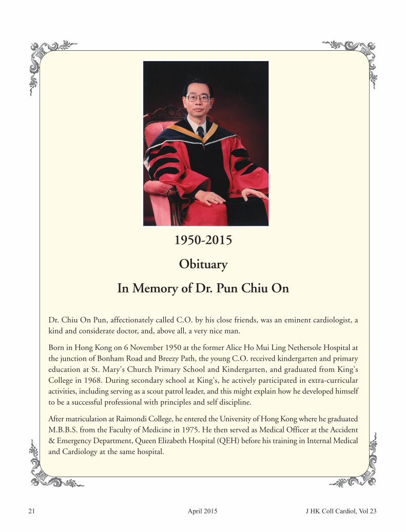
April 2015 J HK Coll Cardiol, Vol 2321
1950-2015
Obituary
In Memory of Dr. Pun Chiu On
Dr. Chiu On Pun, affectionately called C.O. by his close friends, was an eminent cardiologist, akind and considerate doctor, and, above all, a very nice man.
Born in Hong Kong on 6 November 1950 at the former Alice Ho Mui Ling Nethersole Hospital atthe junction of Bonham Road and Breezy Path, the young C.O. received kindergarten and primaryeducation at St. Mary's Church Primary School and Kindergarten, and graduated from King'sCollege in 1968. During secondary school at King's, he actively participated in extra-curricularactivities, including serving as a scout patrol leader, and this might explain how he developed himselfto be a successful professional with principles and self discipline.
After matriculation at Raimondi College, he entered the University of Hong Kong where he graduatedM.B.B.S. from the Faculty of Medicine in 1975. He then served as Medical Officer at the Accident& Emergency Department, Queen Elizabeth Hospital (QEH) before his training in Internal Medicaland Cardiology at the same hospital.

April 2015J HK Coll Cardiol, Vol 23 22
In 1980, Dr. Pun obtained his M.R.C.P and went for oversea training at Gardiner Institute, Universityof Glasgow on a government overseas training scholarship. In 1984, he was promoted to the positionof Senior Medical Officer, Prince of Wales Hospital (PWH) cum Honorary Clinical Lecturer inMedicine at the Chinese University of Hong Kong, Department of Medicine. In 1985-1986, heworked as Visiting Cardiology Associate at the Department of Cardiology, the Royal MelbourneHospital, Melbourne, Australia where he learned advanced electrophysiology and pacing techniquesfrom leading cardiologists like Dr. Harry Mond. In 1987, he obtained TESTAMUR NASPExAM(Special Competency in Cardiac Pacing), North American Society of Pacing and ClinicalElectrophysiology. From 1987, he obtained fellowship from various colleges including FHKCP,FRCP (Glasgow), FRCP (Edinburg) and in 1993 he became founding Fellow of the Hong KongAcademy of Medicine, FHKAM (Med).
During his tenure as Medical Officer at Q.E.H. and later Senior Medical Officer at P.W.H.,Dr. Pun was well-known as a hard-working doctor, dedicated to his work, kind and gentle to juniorcolleagues as well as all his patients. He was most willing to teach, and indeed he spent a lot of timeteaching young doctors and nurses. His work ethics, his passion for teaching younger cardiologistsand his kindness to people are simply exemplary. Yet, it is equally remarkable Dr. Pun always managedto keep a low profile. He left the public hospital and went into private practice in 1996, and continuedto serve his patients who had nothing but praise and respect for him.
Dr. Pun had served as council member of the Hong Kong College of Cardiology since 1992, and asPresident from 2007 to 2009. He had been a director of the Hong Kong Heart Foundation for thepast sixteen years. He played a major role in promoting activities of the College, and the Foundationin all aspects: training young cardiologists and allied health, organizing scientific and CME meetings,heart health education for the public and up-holding the medical professionalism and the highstandard of cardiology in Hong Kong. He certainly had won the respect and admiration by hiscolleagues and all those who knew him.
We are all saddened by the news that Dr. Pun Chiu On passed away so suddenly, at the pinnacle ofhis illustrious career, on February 15, 2015. His passing away is a big loss to all for us. May C.O. restin peace, and no doubt his sprits will remain in our hearts for years to come!
Past PresidentOn behalf of the Council of Hong Kong College of Cardiology

April 2015J HK Coll Cardiol, Vol 23 23
Twenty-Third Annual Scientific Congress
29 - 31 May 2015Sheraton Hong Kong Hotel and Towers
Hong Kong
Hong Kong College of Cardiology
Organizing Committee
Chairman : Chung-seung Chiang
Members : Kam-tim ChanRaymond Hon-wah ChanNgai-yin ChanWai-kwong ChanMin-ji CharngAdolphus Kai-tung ChauChun-ho ChenHaozhu ChenJilin ChenJiyan ChenMingzhe ChenBoron Cheung-wah ChengZhimin DuMario EvoraRunlin GaoJunbo GeYaling HanCharles Kau-chung HoDayi HuYong HuoPatrick Tak-him Ko
Chu-pak LauSuet-ting LauYuk-kong LauMichael Kang-yin LeeKathy Lai-fun LeeStephen Wai-luen LeeGodwin Tat-chi LeungShu-kin LiShuguang LinChung-wah SiuNing TanTak-fu TseFangzheng WangLefeng WangChris Kwok-yiu WongKam-sang WooBo XuMan-ching YamYuejin YangCheuk-man Yu
Scientific Committee
Co-Chairmen : Runlin Gao Yuejin YangShuguang Lin Mario EvoraChung-seung Chiang Min-ji CharngJunbo Ge

April 2015 J HK Coll Cardiol, Vol 2324
Scientific Programme
Friday, 29 May 2015
0800 3/F Registration
0900-1030 Tang Room Free Paper SessionIschemic Heart DiseasePercutaneous Coronary InterventionCongenital and Structural Heart Diseases
Ming II Room Cardiac Imaging
1030-1100 Terrace of Coffee Break & Visit ExhibitsSung Room
1100-1230 Tang Room Free Paper SessionHeart FailureCardiac SurgeryCardiac ArrhythmiaCardiac Rehabilitation
Ming II Room Hypertension and HyperlipidemiaMiscellaneous
1230-1400 Oyster Bar & LunchSky Lounge
1400-1500 Ballroom C Best Paper Oral Presentation
1500-1630 Ballroom C Mainland / Taiwan / Hong Kong Expert ForumLong-term Outcomes of Left Main PCI: New Insights from Bo Xu (PR China)
Fu Wai DatabaseHow to Deal with Life-threatening Complications during Wei-hsian Yin (Taiwan)
TAVIOptimization of Culotte Stenting for Non-Left Main Bifurcation Liang-long Chen (PR China)
1630-1700 Terrace of Coffee Break & Visit ExhibitsSung Room
1700-1830 Ballroom C Mainland / Taiwan / Hong Kong Expert ForumMitral Valve Repair: How to Choose the Right Ring Song Wan (Hong Kong)Percutaneous Implantation of a Ventricular Partitioning Device Ya-wei Xu (PR China)
for Treatment of Ischemic Heart Failure: Initial Experience andChallenge Cases from Shanghai Tenth People's Hospital
Management Strategy for In-stent Restenosis of DES Wei-min Li (PR China)
1845-1930 Ballroom C Hong Kong Heart Foundation LectureStroke Prevention in Atrial Fibrillation Amongst Chinese: David CW Siu (Hong Kong)
a Not-to-missed Opportunity
1930-2100 Ballroom A&B Welcome Dinner
J HK Coll Cardiol, Vol 23 25April 2015
0830-1300 Ballroom C Joint World Association of Chinese Cardiologists andHong Kong College of Cardiology Scientific Symposium
The Symposium is sponsored by Medtronic International Ltd.
Part A: Frontier in Cardiology 0830-0850 Disruptive Technology in Clinical Research: Alan CY Yeung (USA)
Mobile Technology in Personalized CV Health
0850-0910 Quality of Care and Outcomes Research: How They Fit Henry H Ting (USA)
0910-0930 From Bench to Bedside - a Personal Experience Yean-leng Lim (Australia)
0930-0950 Disease Management and Control: From Research to Michael LX Fu (Sweden)Clinical Practice in Sweden
0950-1010 The Ways to Perform Clinical Research besides Daily Clinic Kai Hu (Germany)Work for Cardiologists
Part B: Hypertension 1010-1030 Efficacy of Folic Acid Therapy in Primary Prevention of Stroke Yong Huo (PR China)
among Adults with Hypertension in China
1030-1050 Management of Hypertension in 2015 Chung-seung Chiang(Hong Kong)
1050-1110 Young-onset Hypertension in Taiwan Jaw-wen Chen (Taiwan)
1110-1120 Tea break
Part C: Imaging and New Technology 1120-1140 From Research to Clinical Practice of Cardiac MRI: Dali Feng (USA)
Inspiration and Insight from Interesting Patients
1140-1200 Current Challenges in Genetic and Clinical Reseach on Jian-ming Li (USA)Sudden Cardiac Death
1200-1220 Endothelial Progenitor Cells and Cardiovascular Diseases Shing-jong Lin (Taiwan)
1220-1240 What We Have Learnt after Two Decades of PCI: Insights from Stephen WL LeeNovel Imaging Technology and Novel Devices (Hong Kong)
1240-1300 New Trend and Development of Left Atrial Appendage Occlusion Jiang-tao Yu (Germany)

April 2015 J HK Coll Cardiol, Vol 2326
Saturday, 30 May 2015
0800 3/F Registration
0830-1230 Ballroom C Joined Symposium - Cross-straits Medicine Exchange Association /Hong Kong College of CardiologyGuideline and Practice: Clinical Case Based Conference (GAP-CCBC)(Presentation in English or Putonghua)
A Case of Takayasu's Arteritis with Coronary Artery Involvement Kai Liu (PR China)Beijing Fuwai Hospital, Institute of Cardiovascular Disease
Cardiogenic Shock after Failed Reverse Wire in LM Bifurcation Feng-yu Kuo (Taiwan)by Radial Approach
Kaohsiung Veterans General Hospital
The "Tears" of a Young Lady Shi-xin Yi (PR China)Guangdong General Hospital
Complications in Percutaneous Left Atrial Appendage Occlusion Ya-wei Xu (PR China)Shanghai Tenth People's Hospital
Utilization of BVS to Treat LAD Stenosis for a Patient Who Hsin-fu Lee (Taiwan)May be a Candidate for CABG in the Future
Chang Gung Memorial Hospital, Linkou
Let's Have a Up & Down Roller Coaster Ride! Yuet-wong ChengQueen Elizabeth Hospital (Hong Kong)
Antithrombotic Therapy for Atrial Fibrillation and Coronary Yi-hong Hua (PR China)Artery Disease
Beijing Fuwai Hospital, Institute of Cardiovascular Disease
My Unpredictable Nightmare You Never Know Man-cai Fong (Taiwan)Cheng Hsin General Hospital
Fight with Aortic Dissection Ting Gong (PR China)Hebei Yan Da Hospital
Resolution of Heyde's Syndrome after Transcatheter Aortic Bin Wang (PR China)Valve Implantation - a Case Report
Xiamen Cardiovascular Hospital
Facilitated PCI in Inferior STEMI U-po Lam (Macau)Conde S Januario General Hospital
A 46-year-old Woman with Myocardial Infarction Nan Chu (PR China)Affiliated Zhongshan Hospital of Da Lian University

J HK Coll Cardiol, Vol 23 27April 2015
0930-1200 Ballroom Symposium for Allied Cardiovascular Health Professionals 2015 :A&B Management of Acute Cardiac Emergencies
Management of Acute Myocardial Infarction Arthur SY Yung(Hong Kong)
Acute Management of Life Threatening Arrhythmias Andy WK Chan(Hong Kong)
Management of Acute Aortic Dissection Lik-cheung Cheng(Hong Kong)
Management of Acute Pulmonary Embolism Danny HF Chow(Hong Kong)
1230-1345 Oyster Bar & LunchSky Lounge
1400-1430 Ballroom 23rd ASC Opening CeremonyGuest-of-Honor: Dr. Donald K.T. LiPresident of Hong Kong Academy of Medicine
1430-1530 Ballroom Medtronic SymposiumStent or Not? Consideration for Stenting Small Vessel Vincent OH Kwok
(Hong Kong)The Latest Result of Global Symplicity Registry in Korea Byeong-keuk Kim (Korea)Leadless Pacemaker Chu-pak Lau (Hong Kong)
1530-1630 Ballroom Symposium on Anti-Platelet Therapy UpdateUnmet Needs for Dual Anti-Platelet Therapy - Marc Cohen (USA)
From Evidence to Clinical Practice
1630-1800 Ballroom Plenary LecturesThe Role of Vasodilatory Beta-blocker: a European Perspective Josep Redon (Spain)Clinical Application of NOACs in Daily Practice Hung-fat Tse (Hong Kong)Treatment of Severe Hypercholesterolemia - Min-ji Charng (Taiwan)
Current Concept and Evidence
1830-1915 Ballroom C Dr. Chiu-on Pun Memorial LectureSilent Atrial Fibrillation: a New Paradigm for Stroke Prevention Chu-pak Lau (Hong Kong)
1915-2045 Ballroom DinnerA&B
*Coffee break will be served at 10:30 - 11:00 & 17:00 - 18:30 at 4/F of Sung Terrace.

April 2015 J HK Coll Cardiol, Vol 2328
Sunday, 31 May 2015
0800 3/F Registration
0830-1030 Ballroom C PCI Cases DiscussionPrize Presentation
1030-1100 Terrace of Coffee Break & Visit ExhibitsSung Room
1100-1300 Ballroom C Plenary LecturesPolymer Free BA9TM Drug Coated Stent : Insights from the Stephen WL Lee
EGO-BioFreedom Study (Hong Kong)Filling the Gaps in the Management of AF Patients Taking NOACs: Raymond SM Wong
What's New in the Antidote Development? (Hong Kong)The Latest Update on NOACs: From the Management of Jan Steffel (Switzerland)
SPAF to VTE/PE TreatmentCardioprotection of Beta-blockers along CV Continuum Pak-hei Chan (Hong Kong)
1300-1430 Ballroom A&B Lunch
1430-1530 Ballroom C Plenary LecturesAbsorb BVS: Clinical Trial Update and Potential Patient Benefits Krishna Sudhir (USA)Does Lesion Preparation Matter? Kam-tim Chan
(Hong Kong)
1530-1700 Ballroom C Symposium on Transcatheter Structural Heart InterventionChanging Landscape of TAVI - Update of Evidence Michael KY Lee
(Hong Kong)Something You Need to Know about MitraClip Boron CW Cheng
(Hong Kong)Percutaneous Left Atrial Appendage Closure: an Update and Ngai-yin Chan
Results from a Hong Kong Registry (Hong Kong)
1700-1730 Terrace of Coffee Break & Visit ExhibitsSung Room
1730-1900 Ching Room Symposium on Cardiac PacingThe Role of Pacemaker and Implantable Loop Recorder in Syncope Kathy LF Lee (Hong Kong)Pacing in Sick sinus Syndrome and Atrioventricular Block - Yat-sun Chan (Hong Kong)
Which Pacing Site and Which Pacing Algorithm?Pacing for Heart Failure - at the Heart or at the Nerves? Jo Jo SH Hai (Hong Kong)
1900-2030 Sung Room Farewell Dinner
J HK Coll Cardiol, Vol 23 29April 2015
Paediatric Cardiology Symposium Programme
Saturday, 30 May 2015
0830-0835 Tang Room Welcome Address Kai-tung Chau(Hong Kong)
0835-1030 Tang Room Paediatric Cardiology Symposium IEbstein Anomaly: Imaging & Novel Surgical Strategies Benjamin Eidem (USA)Echocardiographic Assessment of Fetal Cardiovascular Function Hui-shen Wang (PR China)Transcatheter Closure of Ventricular Septal Defect: Strategies for Xu Zhang (PR China)
Device Selection and Results of Mid-to-Long Term Follow UpThe Current Status of Fetal Cardiac Arrhythmia Yi-min Hua (PR China)A Tale of Two Systems: Fontan-associated Liver Disease Pak-cheong Chow
(Hong Kong)
1030-1100 4/F of Coffee Break & Visit ExhibitsSung Terrace
1100-1230 Tang Room Free Paper SessionPaediatric Cardiology I
1230-1345 Oyster Bar & LunchSky Lounge
1400-1430 Ballroom C Opening Ceremony
1430-1610 Tang Room Paediatric Cardiology Symposium IIEvaluation of Ventricular Diastolic Function in Children: Benjamin Eidem (USA)
Integration into Clinical PracticeOutcomes of Transcatheter Occlusion of Patent Ductus Fang Liu (PR China)
Arteriosus in Infants less than 12 monthsLong Term Outcome Following Transcatheter Pulmonary Robin HS ChenValvotomy for Pulmonary Atresia with Intact Ventricular Septum (Hong Kong)Initial Experience with the New Pul-Stent in Treating Post-operative Ting-liang Liu (PR China)
Branch Pulmonary Artery Stenosis
1610-1810 Tang Room Free Paper SessionPaediatric Cardiology II

April 2015J HK Coll Cardiol, Vol 23 30
HK COLLEGE OF CARDIOLOGY, TWENTY-THIRD ANNUAL SCIENTIFIC CONGRESS
ABSTRACTSAbstracts for Free Paper Session:
ISCHEMIC HEART DISEASE AND PERCUTANEOUS CORONARY INTERVENTION
Novel Application of Transradial Coronary Catheter Ikari Left5 Fr. (IL3.5/4) for both Coronary and Selective CarotidAngiographyM Chan, CJ Wu, PH Sung, HY Fang1Department of Medicine, Tseung Kwan O Hospital, Hong Kong; 2Divisionof Cardiology, Department of Internal Medicine, Chang Gung MemorialHospital - Kaohsiung Medical Center, Chang Gung University College ofMedicine, Taiwan
Abstract Aims: To assess the feasibilty and safety of using a single 5 FrenchIkari left catheter for both transradial diagnostic coronary angiography andselective carotid angiography.Methods and Results: A group of patients (115) presenting to Chang GungMemorial Hospital Kaohsiung, from 1/2010 to 1/2013 with cerebral ischemicsymptoms or a history of stroke or transient ischemic attack with non-invasiveimaging evidence of significant carotid stenosis (>=70%) were selected forthis study. We evaluated the success rate of using a single ikari left transradialcatheter for coronary angiography and subsequent carotid angiography.Complications recorded included those related to access site, cardiovascularcomplications including stroke, myocardial infarction, hyperperfusionsyndrome and death. Ninety-five percent of patients successfully underwentboth coronary and carotid angiography with the 5 french ikari left cathetervia transradial access. 7% developed acute complications, with the majoritybeing minor. Two patients (2%) developed stroke, of which one was aftersubsequent carotid stenting.Conclusions: Use of the 5 French Ikari left catheter is safe and effectivewith a high success rate for both transradial coronary and carotid angiography.
Levels of Asymmetric Dimethylarginine (ADMA), An EndogenousNitric Oxide Synthase Inhibitor, and Risk of Coronary ArteryDisease: A Meta-analysis Based on 4,713 ParticipantsC Xuan,1,2
H Li,1 BB Zhang,3 GW He,2,4 QW Tian,1 LM Lun1
1Department of Clinical Laboratory, The Affiliated Hospital of QingdaoUniversity, China; 2Basic Medical Research Center, TEDA InternationalCardiovascular Hospital, China; 3Department of Molecular Microbiology,Oslo University Hospital, Norway; 4Department of Surgery, Oregon Healthand Science University, USA
Background: Asymmetric dimethylarginine (ADMA) acts as an endogenousinhibitor of endothelial NO synthase (eNOS) by competing with L-arginine.This causes low expression of NO and endothelial dysfunction. Numerousstudies have evaluated the association between the serum ADMA level andrisk of coronary artery disease (CAD). However, results from previous studiesare conflicting.Methods: Pubmed, Web of Science, Embase, Ovid, Cochrane databases weresearched to identify eligible studies published in English before December2014. The association was assessed by weighted mean differences (WMD)with 95% confidence intervals (CI). The publication bias was explored byBegg's and Egger's test. Sensitivity analysis was performed to evaluate thestability of results.Results: A total of 16 case-control studies containing 2,939 patients and1,774 controls were involved in the meta-analysis. The pooled result indicatedthat the CAD patients had a higher level of ADMA than health controls(WMD: 0.248, 95% CI: 0.156-0.340; P=1.16 e-7). Sensitivity Analysis
suggested that the result was stable. Further subgroup analysis found a similarpattern in patients with myocardial infraction (WMD: 0.397, 95% CI:0.112-0.683; P=0.0106), in stable angina pectoris (WMD: 0.197, 95% CI:0.031-0.364; P=0.02), and in unstable angina pectoris (WMD: 0.857, 95%CI: 0.293-1.420; P=0.003.Conclusions: The meta-analysis results indicated that the elevated ADMAlevel is associated with an increased risk of CAD.
HK COLLEGE OF CARDIOLOGY, TWENTY-THIRD ANNUAL SCIENTIFIC CONGRESS
ABSTRACTSAbstracts for Free Paper Session:
April 2015 J HK Coll Cardiol, Vol 2331
ISCHEMIC HEART DISEASE AND PERCUTANEOUS CORONARY INTERVENTION
Behavioural Risk Factors: Health − − − − − Related Quality of LifeAmong Patient with Coronary Heart DiseaseR ShresthaNursing Campus Maharajgunj, Nepal
Introduction / Objective: It is gradually emerging as a major public healthproblem in many developing countries including Nepal. Consequences ofCHD are that it often results in depletion of the health related quality of life(HRQL) of patients. The aim of this study was to assess the HRQL of CHDpatients.Methods: A descriptive cross sectional design was used to assess HRQL of254 CHD patients attending out-patient department of Shahid GangalalNational Heart Centre (SGNHC), Bansbari, Kathmandu, Nepal. Purposivesampling technique was used for data collection by face to face interviewtechnique using the MacNew Health − related Quality of Life tool.Results: The results of the study showed that mean ± SD for emotional domainwas 3.87±1.01, for physical domain was 3.81±0.88 and social domain was3.82±0.88 and global score of HRQL was 3.83±0.87 on 7- point likert scale.The different demographic characteristics influencing HRQL was found tobe sex, living status and Occupation. As the different domains of HRQL wassignificantly correlated with each others. The descriptive statistics ofindividual domain scores according to level of HRQL was found to be greaternumber in below average group in each domain including global HRQL score.Conclusions: In all domains, the HRQL of CHD patients attending the cardiaccentre is below average Hence, HRQL of CHD patients needs to be enhancedby regular follow up, organization of counseling sessions and use of selfinstructional module (SIM) on "Living well with CHD".
Clinical Outcome of Bare Metal In-stent Restenosis Treated withDrug Eluting StentRCY Fung, MH Jim, CK Chan, PK Cheung, W Hui, K FanCardiac Medical Unit, Grantham Hospital, Hong Kong
Purpose: Although the use of drug eluting stents (DES) significantly reducescoronary restenosis rate, due to various reasons, a large number of patientsare still implanted with bare metal stents (BMS). These patients may presentto us with symptomatic in-stent restenosis (ISR). BMS ISR is not a benignphenomenon with a significant number of patients present with acute coronarysyndrome. How best to manage these patients is still an important question.In the treatment of BMS ISR, most existing studies only involved patientswith first generation DES. The majority of these studies confirmed its superiorclinical outcome. Although the second generation DES has better efficacyand safety than the first generation DES in treating de novo coronary lesions,only very limited BMS ISR studies involved patients with second generationDES. They were either small in sample size or short in follow up time. Inthis retrospective cohort study, patients with BMS ISR diagnosedangiographically and treated with first or second or mixed generations DESbetween 1 January 2006 and 31 June 2011 were included. The primaryobjective is to determine the incidence of major adverse cardiac event(MACE). The secondary objective is to determine if there is any independentpredictor for MACE.Method: Consecutive patients presented with BMS ISR treated with DES inGrantham Hospital (Hong Kong, China), United Christian Hospital (HongKong, China) or Royal Alexandra Hospital (Edmonton, Canada) between
1 January 2006 and 31 June 2011 were included. Those who have non-native coronary artery BMS ISR or DES ISR or BMS ISR not treated DESwere excluded. Clinical and procedural data were retrieved from the computerbased electronic clinical records, percutaneous coronary intervention registriesof the individual hospitals and the Alberta Provincial Project for OutcomeAssessment in Coronary Heart Disease (APPROACH) registry. MACE wasdefined as stent thrombosis, all cause mortality, target lesion revascularization(TLR) and myocardial infarction (MI).Results: A total of 207 patients were identified. Eighty-six (41.5%) patientswere treated with first generation (paclitaxel and sirolimus) stents, onehundred and ten (53.1%) patients were treated with second generation(everolimus , zotarolimus, biolimus) stents and 11 (5.3%) patients had mixedfirst and second generation stents implanted. The mean follow up time was3.3±1.9 years. The number of MACE was 50 (24.2%). There were 15 (7.2%)deaths, 2 (1.0%) stent thrombosis, 12 (5.8%) TLR and 22(10.6%) MI. Afteradjustment for cofounders, the only independent predictors of MACE wereleft ventricular ejection fraction less than 35% (hazard ratio (HR) 5.2, 95%confidence interval (CI) 2.4±11.4, p<0.001), renal impairment at the timeof PCI (HR 2.5, 95% CI 1.2-5.4, p=0.026) and previous history of ischaemicstroke (HR 2.9, 95% CI 1.4-5.8, p= 0.003).Discussion: This study provides long term follow up data (a mean follow uptime of 3.3±1.9 years) for patients implanted with first and second generationDES. Moreover it includes patients from different countries which allow usto control for regional differences. The overall MACE rate for the treatmentof BMS ISR by DES was 24.2% which was comparing favorably to that ofthe balloon angioplasty and brachytherapy.

April 2015J HK Coll Cardiol, Vol 23 32
HK COLLEGE OF CARDIOLOGY, TWENTY-THIRD ANNUAL SCIENTIFIC CONGRESS
ABSTRACTSAbstracts for Free Paper Session:
ISCHEMIC HEART DISEASE AND PERCUTANEOUS CORONARY INTERVENTION
Outcomes in Reperfusion Therapy of Octogenarians withST-segment Elevation Myocardial InfarctionYW Cheng, CF Tsang, CK Kwok, NH Luk, SF Chui, YH Cheng,KC Chan, LK Chan, HS Ma, CY Wong, LY Tam, CL Fu, CW Chan,KY Lee, KC Ho, KT Chan, CS ChiangDepartment of Medicine, Queen Elizabeth Hospital, Hong Kong
Background: As a result of increased life expectancy, octogenariansconstitute an increasing proportion of hospital admission for ST-segmentelevation myocardial infarction (STEMI). STEMI in older patients is knownto associate with higher mortality. We evaluated the results of reperfusiontherapy in octogenarians in a regional hospital.Methods: A retrospective review of all patients older than 80 years old withdiagnosis of STEMI who either activated primary PCI or given thrombolytictherapy between Jan 2013 to Feb 2015 in Queen Elizabeth Hospital wasperformed. Patients with delayed presentation and those who refusereperfusion treatment were excluded. Their clinical characteristics andoutcomes were analyzed.Result: A total of 57 patients, 30 male (52.6%) and 27 (47.4%) female, rangedfrom 80 to 98 years old with a mean age of 84.8±4.36 were identified. Ten(17.5%) patients presented with shock before reperfusion therapy. PrimaryPCI was activated in 32 patients (56.1%). Thrombolytic treatment in formof Streptokinase and Tenecteplase was given in 16 (28.1%) and 9 (15.8%)patients respectively. Anterior STEMI was the most common presentation.Six patients did not have coronary angiography performed after thrombolytictreatment either because of patient refusal or development of free wallrupture. Twenty-two (38.6%) patients showed single vessels disease on
1 Year Outcome Comparing Drug Coated Balloon OnlyAngioplasty Versus Second Generation DES in Diabetic Patientswith de novo Small Vessel Coronary Artery DiseaseTT Nyein, D Sinaga, A Sim, ASP Seah, FH Jafary , HH Ho,PJL OngTan Tock Seng Hospital, Singapore
Aims: PCI in patients with diabetes and small vessel coronary artery disease(SVD) has higher risk of adverse clinical events. Drug coated balloon (DCB)has been used to treat de-novo coronary lesion with promising result. Wecompared the 1 year clinical outcome of drug coated balloon only angioplasty(DCBA) versus second generation DES in SVD amongst diabetic patientsin a 'real world' setting.Methods and Results: We retrospectively analyzed the 1 year outcome ofall diabetic patients who underwent PCI to de-novo lesion with DCB or secondgeneration DES with device size ≤2.5 mm between 2011 to 2013. Ninety-two patients were intervened with DCBA (100% Sequent Please), male71.1%, mean age 60.4±10.9 years old? HbA1c 8.4±2.6%; 91.3% hadhyperlipidaemia, 88.0% had hypertension and 25.0% had history of smoking.28.7% of the patients presented with stable angina, 53.2% with NSTEACSand 16.0% with STEMI. The most common target vessel was left anteriordescending artery at 41.5% and type C lesion accounted for 46.8% of thecases. The demographics and presentations of the 80 DES patients were notsignificantly different from the DCBA group. DES used were ResoluteIntegrity (36.3%), Xience (26.3%), Biomatrix (25.0%), Promus (8.8%) andNobori (3.8%). The reference diameter in the DCB treated vessel was2.23±0.28 mm vs 2.44±0.21 mm in the DES group (p=0.09). The average
size of the DCB used was smaller than the corresponding DES (diameter2.26±0.21 mm vs 2.36±0.12 mm, p<0.01 and length 20.3±5.7 mm vs22.7±7.5 mm, p<0.01 respectively). There was a trend towards a larger acutelumen gain after DES implantation compared to DCBA (1.6±0.5 mm vs1.1±0.9 mm, p=0.10 respectively). Despite the predominantly ACSpresentation, patients treated with DCB received significantly shorterduration of dual antiplatelet therapy (DAPT) than the DES group (7.0±4.7months vs 11.7±1.9 months, p<0.0001 respectively). At 1 year, 9 majoradverse clinical events (MACE) were observed at DCBA arm versus 12 inthe DES group (9.6% vs 15.0% respectively p=0.76). There was lower numberof myocardial infarction (MI) observed in the DCBA group than the DESgroup (2 (2.2%) vs 8 (10.0%), p<0.05 respectively). There were otherwiseno significant differences in death (0 (0%) vs 3 (3.8%) p=0.09) or targetlesion revascularization rate (7.4% vs 6.3%, p=0.77) between those treatedwith DCB and DES respectively.Conclusions: Diabetic patients with SVD in this Asian cohort have poorlycontrolled HbA1c and rampant comorbidities. Those treated with DCB onlyangioplasty have smaller reference vessel size and achieved smaller acutelumen gain compared to those treated with DES. However, such anatomicaladvantage with DES did not translate into better MACE rate compared to theDCB group at 1 year follow up. In fact, patients treated with DCB havelower MI rate and received shorter course of DAPT. Drug coated balloonmay be a safe and effective alternative to modern DES in the treatment ofde novo small vessel disease in diabetic patients.
subsequent coronary angiography. Five patients turned out to have minorCAD. Among them, 1 patient diagnosed Tako-tsubo cardiomyopathywhile 3 patients had concomitant atrial fibrillation. As at end March 2015,21 (36.8%) patients died within one year. Cumulative mortality at hospitaldischarge was 24.6% and by 30 days was 19.3%. Patient with DiabetesMellitus, shock on presentation or eGFR less than 40 ml/min/1.73 m2 wasassociated with higher mortality (p<0.05). Six (10.5%) patients died of free-wall rupture. Ten (17.5%) patients had bleeding episode after reperfusiontherapy while 5 (8.8%) patients had major bleeding requiring bloodtransfusion. None of patient had intracranial hemorrhage. Re-infarctionoccurred in 2 (8%) patients receiving thrombolytic treatment.Conclusion: Reperfusion therapy was delivered in octogenarian populationwith reasonable success. However, short term adverse event rates includingmortality are still significant despite advances in drug-eluting stents, newerantithrombotic agents and intensive care in the management of STEMI.
HK COLLEGE OF CARDIOLOGY, TWENTY-THIRD ANNUAL SCIENTIFIC CONGRESS
ABSTRACTSAbstracts for Free Paper Session:
April 2015 J HK Coll Cardiol, Vol 2333
CONGENITAL AND STRUCTURAL HEART DISEASES
A Novel Mutation of TBX5 Gene in Chinese Patients with IsolatedVentricular Septal DefectXi Zhang,1 HT Hou,1 YY Jiang,1 J Wang,1 XL Wang,1 XC Liu,1 ZG Liu,1
Q Yang,1,2 GW He1,3
1TEDA International Cardiovascular Hospital, China; 2Department ofMedicine & Therapeutics, The Chinese University of Hong Kong, HongKong; 3The Affiliated Hospital of Hangzhou Normal University & ZhejiangUniversity; Department of Surgery, Oregon Health and Science University,USA
Purpose: Ventricular septal defect (VSD) is the most frequently occurringcongenital heart disease (CHD) in newborns. A number of genetic studieshave linked TBX5 mutations to cardiac abnormalities. Here, we identifypotential pathogenic mutations for TBX5 and provide insights into the etiologyof isolated VSD in Chinese patients.Methods: Case-control mutational analysis was performed in 354 patientswith isolated VSD and 341 healthy controls. First, all the coding exons andintron-exon boundaries of TBX5 were sequenced in VSD patients and controls.Sanger sequencing with high-resolution melting (HRM) curve analysis wasthen combined to detect new TBX5 mutation and identify its frequency.Results: A novel heterozygous missense mutations (c. 40C>A) was identifiedin TBX5 gene exon-2. This mutation leads to proline to threonine substitutionat position 14, which is highly conserved among many species. The damagingand disease causing of this mutation is predicted by Polypen2, SIFT andMutation Taster. No more c. 40C>A mutation was found in enlarger casesand controls by HRM analysis.
Conclusion: We identified a novel heterozygous missense mutation in TBX5gene exon-2 in isolated VSD patients in Chinese population and found thatthis missense mutation probably causes the disease. Further, our resultsshowed the important role of HRM as a reliable and efficient method todetermine disease-related gene mutation in congenital heart disease.
Comparison of Transcatheter Aortic Valve Implantation (TAVI)in Extreme-risk and High-risk Patients in the Local AsianPopulationMKY Lee,1 LK Chan,1 KC Chan,1 SF Chui,1 NH Luk,1 HS Ma,1 CY Wong,1
KT Chan,1 CS Chiang,1 CB Lam,1 C Leung,1 MC Chan,1 MY Fan,1
KW Leung,1 HL Cheung,2 V Ng,2 CC Ma,2 E So,3 D Fok,3 YF Chow,3
MK Chan,4 W Chan,4 SF Yip,4 YF Cheung,4 YH Szeto4
1Department of Medicine; 2Department of Cardiothoracic Surgery;3Department of Anaesthesiology; 4Department of Diagnostic Radiology &Imaging, Queen Elizabeth Hospital, Hong Kong
Introduction: Transcatheter Aortic Valve Implantation (TAVI) for treatmentof severe aortic stenosis (AS) has been proved to improve survival and qualityof life in a group of inoperable and high-risk symptomatic patients. Thisinvolves percutaneous implantation of a transcatheter heart valve throughthe femoral or subclavian artery routes without subjecting the patients toopen heart surgery or cardio-pulmonary bypass. Limited data is available onthe safety and efficacy in the local Asian population.Objectives: We report the results of the TAVI procedures done in QueenElizabeth Hospital (QEH) for a group of extreme-risk and high-risksymptomatic severe AS patients and compare with the CoreValve US PivotalTrial.Methods: The TAVI procedures are done in QEH by a multi-disciplinaryTAVI heart team comprising cardiologists , cardiac surgeons,anaesthesiologists, radiologists and cardiac nurses. All potential patientswould be interviewed independently by the cardiologists and cardiac surgeons.The TAVI Heart Team then decided whether the patient should undergo SAVRor TAVI. All patients were assessed by echocardiogram, CT scan andangiogram to decide on suitability. Echocardiogram and 6-minute walk testwould be performed according to schedule post-procedure. All complications
would be reported to an independent Safety Monitoring Committee. Alldata will be captured by the local QEH Registry. Patients were consideredextreme-risk (or inoperable) if their Logistic EuroSCORE was ≥20 and high-risk if it was between 10-20.Results: From December 2010 to January 2015, 45 patients (29 malesand 16 females) with symptomatic severe AS underwent the TAVIprocedure. Average age was 83.2±5.1 and 80.2±4.6 years and the meanLogistic EuroSCORE was 32.4±9.7 and 12.1±3.9 for extreme-risk andhigh-risk patients respectively. All procedures were done under generalanaesthesia in our cardiac catheterization laboratory or hybrid OR. Aorticvalve area improved from 0.62±0.2 cm2 to 1.83±0.31 cm2 and from0.75±0.15 cm2 to 1.96±0.35 cm2 with mean gradient decreased from51.3±13.0 mmHg to 8.6±6.7mmHg and from 49.3±12.2 mmHg to 9.2±2.9 mmHg for extreme-risk and high-risk group respectively. Majorityof patients have only trivial to mild aortic regurgitation during subsequentfollow-up. Permanent pacemakers were implanted in 5 patients (25%)for extreme-risk and 2 patients (8%) for high-risk group. 30-day all-cause mortality was 0% and 4% for extreme-risk and high-riskrespectively in our cohort and it was 8.4% and 3.3% respectively in theCoreValve US Pivotal Trial. One-year all-cause mortality was 15% and8% for extreme-risk and high-risk patients respectively in our cohortand it was 24.3% and 14.2% respectively in the CoreValve US PivotalTrial. All patients showed marked symptomatic improvement in terms ofNYHA Functional Class, 6-minute walk test and quality of lifemeasurement. This compares favourably with results of the CoreValveUS Pivotal Trial and the Asia TAVI Registry.Conclusions: Being a high-risk procedure, TAVI was shown to be safe andfeasible in a group of extreme-risk and high-risk symptomatic severe ASlocal Asian elderlies and this benefit compares favourably with internationalclinical trials and registries.

April 2015J HK Coll Cardiol, Vol 23 34
HK COLLEGE OF CARDIOLOGY, TWENTY-THIRD ANNUAL SCIENTIFIC CONGRESS
ABSTRACTSAbstracts for Free Paper Session:
CONGENITAL AND STRUCTURAL HEART DISEASES
Changes in Echocardiographic, Functional and PsychologicalImprovement in Adults Following Percutaneous TranscatheterAtrial Septal Defect Closure: A Pilot StudyKW Chang,1 PW Yam,2 YH Wong,2 SP Lau,1 HK Poon1
1Department of Occupational Therapy; 2Department of Medicine andGeriatrics, Tuen Mun Hospital, Hong Kong
Purpose: To investigate the echocardiographic improvement and clinicaloutcomes of patients <60 years and patients ≥60 years after atrial septal defect(ASD) percutaneous closure.Methods: Retrospective study of 38 adult patients underwent percutaneousASD closure in TMH were reviewed. We had collected and compared thetwo patients groups with age <60 years and age ≥60 years on the last pre-operative and earliest post-operative echocardiographic measurement ofpulmonary arterial systolic pressure (PASP) and right ventricular internaldiastolic diameter (RVID). Another retrospective analysis was performedamong 13 patients with half yearly follow up on functional performance andquality of life by occupational therapist.Results: For the aspect of cardiac remodeling, changes in PASP in patientsgroups <60 years and ≥60 years both decreased significantly with p-value<0.001. Significant improvement in RV dilation was also noted in the twopatients groups (p-value <0.001). Twelve patients attended all occupationaltherapy assessment sessions, with mean age of 49.25; and among them4 patients with age over 60. Percutaneous ASD occlusions were all done viafemoral approach. Amplatzer septal occlude (St. Jude Medical) was the deviceof choice. The atrial septal device was successfully implanted in all patients.
Friedman test was used to analyze among the outcome measures of thepatients after 6 months of follow-up. Statistically significant differenceswere observed in functional capacity, mood status and quality of life, withp-value ranged from 0.002 to 0.05.Conclusions: Percutaneous transcatheter ASD closure is a safe procedurewith satisfactory short-term outcomes even after the age of 60.

HK COLLEGE OF CARDIOLOGY, TWENTY-THIRD ANNUAL SCIENTIFIC CONGRESS
ABSTRACTSAbstracts for Free Paper Session:
April 2015 J HK Coll Cardiol, Vol 2335
CONGENITAL AND STRUCTURAL HEART DISEASES
Transcatheter Aortic Valve Implantation by Hydra TranscatheterAortic Valve System: First-in-man ExperienceL S ndergaard,1 GSH Cheung,1,2 EB Wu2
1Rigshospitalet, Copenhagen, Denmark; 2Prince of Wales Hospital, HongKong
Background: Transcatheter treatment of aortic stenosis is becoming part ofstandard therapy. Specifically, patients considered at high or prohibitivesurgical risk have been consistently shown to benefit from this type ofintervention. A number of different transcatheter aortic valves arecommercially available. Recently, the Hydra Aortic Valve was developed.We report the results of the first-in- human implantations of the Hydra Valve.Methods:Patients: Implantation of the Hydra transcatheter aortic valve was performedin 15 patients with symptomatic severe aortic stenosis in the period fromMay 2014 to March 2015 at two invasive centers (King ChulalongkornMemorial Hospital and Rajavithi Hospital, both Bangkok, Thailand). In allcases, surgical treatment was deferred per heart team decision due to anexpected high surgical risk. All patients gave informed consent for theprocedure. Pre-procedural examinations included trans-thoracicechocardiography, coronary angiography, and multi-slice, ECG-gatedcomputed tomography (CT) to visualize aortic annulus, aorta, and iliofemoralaccess vessels. For follow-up, patients had trans-thoracic echocardiographyperformed before discharge and at 1 month after the procedure. Anycomplications were documented at these time points.Hydra transcatheter aortic valve:Valve: The Hydra Aortic Valve self-expanding stent frame is made of nitinoland the 3 leaflets and sealing cuff are bovine pericardium, see Figure 1. Thethree tentacles (antenna) on the stent frame provide flexible anchors at theoutflow which conforms to the shape of the aorta, while the inflow sectionof the frame exerts a higher radial force to ensure attachment to the aorticannulus. The pericardial part of the bioprosthesis is sutured to the stentframe. The valve is produced in 3 sizes, 22, 26, and 30 mm, see Figure 1 forprosthesis dimensions and sizing choice for various annular diameters.Results:Population: All patients were symptomatic severe aortic stenosis, 2 patients(14%) had severe symptoms (NYHA class 3).Outcome: Implantation of the Hydra Valve was accomplished in all cases.There were no instances of delivery system failure. In 5 patients, valve positionwas suboptimal after final release, and a second Hydra Valve was implantedsuccessfully in these patients. Two procedures were complicated with aorticdissection. All patients survived the procedure, two patients died within thefirst 30 days. One patient required permanent pacing.Twelve patients completed full echocardiographic assessment at 30 days.Prosthetic valve Mean aortic gradient declined from 49 mmHg pre-procedureto 11 mmHg,Discussion: In this report, we describe the first results from human use ofthe new self-expandable transcatheter Hydra Aortic Valve in humans. Itsefficacy for treating degenerative aortic stenosis is documented.Safety and efficacy: The two mortalities within the first 30 days were notrelated to valve function.
Vascular complications included two aortic dissections, which is normallya rare occurrence. Access site complications encountered were mostly mild,and comparable in frequency to those generally reported elsewhere. Thecomplication rate is primarily related to the size of the access sheath. Withits 18F delivery system, the Hydra Aortic Valve is similar in profile to severalother next-generation transcatheter valves. Atrioventricular block requiringnew pacemaker implantation was seen in only one patient. This is a reassuringresult, as a relatively high rate of pacemaker implantation has been apersistent feature of the most commonly used self-expanding aortic valve,the Medtronic CoreValve. If confirmed in larger populations, this featurecould potentially be attributed to the lack of flaring of the inflow end of theprosthesis. Post-procedural valve gradients demonstrated no significantobstruction to flow, and the values are in line with results from othertranscatheter aortic valves. Paravalvular leaks were common, but in onlyone case was it of more than mild severity at 30-day follow-up. This iscomparable to results from other valves without sealing skirts. However,the presence of paravalvular leakage also contributed to necessitatepostdilatation in 6 cases and the deployment of a second valve in 5 cases.Conclusion: The Hydra Aortic Valve is useful for transcatheter treatment ofsevere aortic stenosis. Initial results indicate hemodynamic performance ofthe implanted bioprosthesis is good, and complication rates seem acceptable.Further evaluation in larger series is needed.
Figure 1. The hydra valve.

April 2015J HK Coll Cardiol, Vol 23 36
HK COLLEGE OF CARDIOLOGY, TWENTY-THIRD ANNUAL SCIENTIFIC CONGRESS
ABSTRACTSAbstracts for Free Paper Session:
CARDIAC IMAGING
Impacts of Valvulo-Arterial Impedance on Severe Aortic Stenosis−−−−− A Multi-Imaging Modalities ProspectiveWCW LamLondon, United Kingdom
Background: Aortic stenosis (AS) has become more prevalent as thepopulation is aging and advances in cardiac imaging makes AS more readilydiagnosable. Valvulo-arterial impedance (Zva), an index to assess overallafter-loads, has been proven to positively correlate with the AS severity.Zva also carries prognostic value. As most patients with severe AS areapparently asymptomatic, Zva may be useful in further sub-categorizingsevere AS and treatment plan can be tailored accordingly. This study aimsto evaluate the impact of Zva on cardiac parameters in patients with severeaortic stenosis by means of cardiovascular magnetic resonance (CMR).Method: All patients with severe aortic stenosis were referred to the CMRunit of Royal Brompton Hospital, London, for pre-operative CMR. Eachpatient had a trans-thoracic echocardiography within one week from theCMR study. Zva was calculated as the sum of systolic blood pressure andthe aortic valve continuous-wave Doppler mean gradient divided by the leftventricular stroke volume index. Patients were categorized into 2 groupsaccording to their calculated Zva. "High Zva" is defined as Zva higherthan or equal to 4.5 mmHg/mL/m2 whereas "Low Zva" is defined as Zvalower than 4.5 mmHg/mL/m2. Various echocardiographic and CMRparameters were compared between the 2 groups. Statistical analysis wasdone by t-test and ANOVA.Results: 52 patients were screened and 27 of them were excluded due tomissing clinical data. Total of 25 patients were recruited into the finalanalysis. They are categorized into 2 groups, high Zva (n=12) and low Zva(n=13). Baseline characteristics between the 2 groups were comparable
except patients with high Zva had higher systolic blood pressure. Onechocardiography, calculated aortic valve areas (AVA) by continuityequation, dimensionless performance index (DPI) and left ventricular ejectionfraction (LVEF) were comparable between the 2 groups. However, on CMR,patients with high Zva had lower LVEF, higher left ventricular end-diastolicvolume (LVEDV), higher right ventricular end-diastolic volume (RVEDV),higher left atrial volume index (LAVI) and higher left ventricular mass index(LVMI). Findings are summarized in Table 1.Conclusion: In the setting of severe aortic stenosis, those with high Zva hadworse cardiac parameters on CMR than those with low Zva. These findingsmay provide insights on treatment directions and prognosis for patients withsevere aortic stenosis.
Table 1. Echocardiographic and CMR parameters between severe AS patients with highZva and low Zva
Low Zva (n=13) High Zva (n=12) P-valueAge 79.4 76.6 0.85SBP 132.4 149.8 0.026Zva 3.72±0.17 5.16±0.77 0.007DPI 0.25±0.03 0.23±0.05 0.64AVA-echo 0.86±0.09 0.86±0.13 0.91LVMI-CMR 81±16.2 108±18 0.034LVEF-CMR 65±13.7 49±10.3 0.048LVEDV-CMR 127±38.4 223±64.1 0.042RVEF-CMR 59±6.3 56±10.4 0.1RVEDV-CMR 110.2±26 124.6±22 0.042LAVI-CMR 65±15.3 76±12.5 0.044AVA-CMR 0.78±0.17 0.81±0.07 0.295
Effect of Continuous Positive Airway Pressure Therapy on RightVentricular Function by Speckle Tracking in Obstructive SleepApnea SubjectsCY Yung,1 J Chan,2 A Yamada,2 HC Fan,3 K Chan,1 YK Lau1
1Department of Cardiology, Ruttonjee and Tang Shiu Kin Hospitals, HongKong; 2University, Gold Coast, Australia; 3Department of RespiratoryMedicine, Ruttonjee and Tang Shiu Kin Hospitals, Hong Kong
Background: Obstructive sleep apnea (OSA) is associated with rightventricular (RV) dysfunction and use of continuous positive airway pressure(CPAP) may improve RV function. 2D speckle tracking is a relatively newmethod for assessment of RV function and has recently been included ininternational guidelines for RV function assessment. We hypothesize thatRV strain is more sensitive than conventional echo parameters in detectingearly changes in RV function.Purpose: To investigate the effect of OSA on right ventricular function andthe impact of CPAP therapy.Method: We prospectively recruited 23 patients (19 male, mean age54+/-9) with newly diagnosed with OSA (mean respiratory disturbance index(RDI) 30.6+/21.3) and follow up for 1 month after initiation of CPAP therapy.Echocardiographic images were acquired with a modified apical 4 chamberview. RV global and free wall longitudinal strain by speckle tracking, tricuspidannular plane systolic excursion (TAPSE), and RV fractional area change(FAC) were measured. Offline strain analysis was performed by cardiologistsoff-site experienced in strain analysis and blinded to clinical data.
Results: At baseline, the mean RV global strain and free wall strain were-23.1+/-3.0% and -26.9+/-4.4% respectively, with TAPSE 2.21+/-0.2 cmand FAC 34.5+/-8.2%. At one month after CPAP therapy, therewas statistically significant improvements in RV free wall strain(-30.4+/-4.4%, relative change 13.0%, p=0.03) and a trend towardsimprovement in RV global strain (-24.5+/-3.5%, relative change 6.06%,p=0.082). There were no significant changes in TAPSE (2.25+/-2.9 cm,p=0.37) and FAC (35.9+/-10.3%, p=0.57) post CPAP therapy.Conclusion: Use of CPAP therapy improves RV function in subjects withOSA. RV free wall longitudinal strain is more sensitive in detecting subtlechanges in RV function compared to conventional parameter of TAPSE andFAC.

HK COLLEGE OF CARDIOLOGY, TWENTY-THIRD ANNUAL SCIENTIFIC CONGRESS
ABSTRACTSAbstracts for Free Paper Session:
April 2015 J HK Coll Cardiol, Vol 2337
CARDIAC IMAGING
Effect of Surgical Correction of Tetralogy of Fallot onShort-term Right and Left Ventricular Function as Determinedby Two-Dimensional Speckle Tracking EchocardiographyY Li, M Xie, X Wang, Q LuDepartment of Ultrasonography, Union Hospital, Tongji Medical College,Huazhong University of Science and Technology, China
Purpose: The impact of surgical repair on short-term right ventricular (RV)and left ventricular (LV) function in patients with tetralogy of Fallot (TOF)is scarce. The purpose of our study is to assess biventricular function inpatients with TOF before and after operation by two-dimensional speckletracking echocardiography (STE).Methods: 36 patients with TOF before, and 1 week, 3 months, and 6 monthsafter operation were studied. RV and LV global longitudinal strain (GLS)and strain rate (GLSRs), and LV global circumferential and radial strain andstrain rate were measured by STE.Results: Compared with controls, lower RV GLS and GLSRs in preoperativepatients with TOF decreased further at 1 week after operation, then improvedat 3 months and 6 months after operation, but remained lower than normalvalues. While LV longitudinal, circumferential and radial strain and strainrate improved at 1 week after operation, and increased to normal level at 6months after operation. RV and LV strain and strain rate had no correlationwith the type of surgery. Age was the independent predictor of RV GLS andGLSRs (beta1=-0.212, P1=0.012; beta2=-0.180, P2=0.033). Age and LV end-diastolic volume were the independent predictor of LV strain and strain rate.
Conclusion: After repair of TOF, LV function rapidly improves, but RVfunction underwent transient deterioration, followed by a improvement towardnormal values. The difference in right and left ventricular recovery and RVtransient changes after operation can be subtly analyzed by STE. STE appearsto be a valuable tool for follow-up assessment of biventricular function aftercongenital heart disease surgery.

April 2015J HK Coll Cardiol, Vol 23 38
HK COLLEGE OF CARDIOLOGY, TWENTY-THIRD ANNUAL SCIENTIFIC CONGRESS
ABSTRACTSAbstracts for Free Paper Session:
CARDIAC IMAGING
The Clinical Value of Transthoracic Echocardiography inDiagnosis of Anomalous Origin of Pulmonary ArteryY Song, 1 J Wang,1 TO Cheng, 1,2 M Xie, 1 X Wang,1 L Yuan,1
Y Yang,1 L Wang1
1Department of Ultrasound, Union Hospital, Tongji Medical College,Huazhong University of Science and Technology, China; 2Department ofMedicine, George Washington University, USA
Purpose: Anomalous origin of pulmonary artery (AOPA) is a rare butimportant congenital malformation which frequently involves the rightpulmonary artery. Preoperative transthoracic echocardiography (TTE) is theprimary imaging tool in the diagnosis of AOPA. The purpose of this studywas to review the clinical values of TTE for the diagnosis of AOPA.Methods: We retrospectively analyzed the TTE characteristics of 14 caseswith anomalous origin of the right pulmonary artery from the ascending aorta(AORPA) or unilateral absent pulmonary artery (UAPA) from 2007 to 2014in one single institution. The results were compared with the findings atsurgery in order to assess the diagnostic value of TTE.Results: (1) The corrective surgeries were performed in all 14 cases. TheTTE diagnoses of 13 cases were consistent with findings at surgery. Thecoincidence rate for TTE and surgical findings was 92.9%. AORPA andUAPA shared some TEE features in common, which are the exit of twoconcordant ventricular outflows tracts and absence of a normal pulmonaryarterial bifurcation. (2) Nine patients had AORPA, which all had a proximalorigin from the posterior or lateral aspect of the ascending aorta close to theaortic valves in our series. All patients had a left aortic arch. The parasternallong axis view and short axis view showed the anomalous vessel arose fromthe ascending aorta above the aortic valve (Figure 1). The associatedcardiovascular abnormalities included aortopulmonary septal defect,interruption of aortic arch, patent ductus arteriosus, ventricular septal defectand atrial septal defect. Severe pulmonary artery hypertension was noted in
all cases (Table 1). (3) Five patients had UAPA. Three patients with absentleft pulmonary artery had Tetralogy of Fallot while 2 patients with absentright pulmonary artery had patent ductus arteriosus. TTE reveals absence ofthe right or left pulmonary artery from any angle.Conclusions: TTE plays an important role in the non-invasive and accuratediagnosis of AOPA. TTE can clearly display its site of origin and course, aswell as other associated malformations and hemodynamic changes.
Table 1. The clinical data on 9 patients with AORPA
Pt. Gender/Age AORPA(mm) LPA (mm) D-AVO (mm) TR (mmHg) Shunt direction Associated cardiovascular abnormalities
1 M/7yr 16 10 23 90 L-R PDA2 M/3yr 17 15 22 / Bi APSD,IAA,PDA3 M/2m 7 5 12 86 Bi VSD,ASD4 F/4m 9 8 12 97 /* ASD5 M/29d 7 5 7 62 Bi APSD,IAA,PDA6 M/21d 9 7 19 / Bi APSD,IAA,PDA7 F/4m 5 6 10 / R-L APSD,IAA,PDA8** F/6yr 12 16 24 117 L-R PDA9 M/19d 4.5 7 / 81 Bi PDAPt: patient; M: male; F: female; yr: years; m: months; d: days. AORPA: the diameter of the anomalous right pulmonary artery; LPA: the diameter of the left pulmonary artery; D-AVO: the
distance from origin site to the aortic valve; TR: tricuspid regurgitation; L-R: left to right; Bi: bidirectional; R-L: right to left; PDA: patent ductus arteriosus; APSD: aortopulmonary septal
defect; IAA: interruption of aortic arch; VSD: ventricular septal defect; ASD: atrial septal defect
*The shunt direction of ASD is difficult to be visualized because of the turbulence of severe tricuspid regurgitation.
**Misdiagnosed echocardiographically as anomalous origin of left pulmonary artery from the ascending aorta
Figure 1. Parasternal short axis view of AOPRA.AO: aorta, PA:pulmonary artery, PDA: patent ductus arteriosus, RPA: right pulmonaryartery, LPA: left pulmonary artery

HK COLLEGE OF CARDIOLOGY, TWENTY-THIRD ANNUAL SCIENTIFIC CONGRESS
ABSTRACTSAbstracts for Free Paper Session:
April 2015 J HK Coll Cardiol, Vol 2339
CARDIAC IMAGING
Diagnostic Value Oftransthoracic Echocardiography in PatientsWith Coarctation of Aorta: The Chinese Experience in 53 PatientsStudied Between 2008 and 2012 in One Major Medical CenterM Xie,1 Z Sun,1 L Li,1 TO Cheng,2 L Zhang,1 X Wang,1 N Dong,3 Q Lv,1
K Li,1 L Yuan,1 J Wang1
1Department of Ultrasound, Union Hospital, Tongji Medical College,Huazhong University of Science and Technology, Hubei Province KeyLaboratary of Molecular Imaging, China; 2Department of CardiovascularSurgery, Union Hospital of Tongji Medical College, Huazhong Universityof Science and Technology, China; 3Department of Medicine, GeorgeWashington University Medical Center, USA
Purpose: Although aortography is well known as the "gold standard" for thediagnosis of coarctation of aorta (CoA), the method is invasive, expensiveand not readily accepted by some patients. Ultrasound diagnosis for CoA isnon-invasive, inexpensive, readily accepted by every patient, and can berepeated as frequently as necessary. The purpose of thispresentation is toevaluate the applicability of transthoracic echocardiography for the diagnosisof CoA.Methods: The echocardiographic appearances of 53 patients with CoA whohad undergone surgery during a 5-year period from January 2008 to October2012 were analyzed retrospectively, and the results were compared withfindings at surgery.Results: Fifty-three patients with CoA include six with isolated CoA and47 of CoA associated with other cardiacanomalies. Of the 53 operatedpatients, 48 were correctly diagnosed preoperatively by echocardiography,while two were misdiagnosed as interrupted aortic archand the diagnosiswasmissed in three other patients. Thus the diagnostic accuracy rate was90.6%, and the misdiagnosis rate was 9.4%.
Conclusions: Preoperative echocardiographic evaluation offers verysatisfactory anatomic assessment in most patients with CoA. It makespreoperative angiography unnecessary. Thus transthoracicechocardiographyshould be the first-line method for the diagnosis of coarctation of the aorta.
Effect of Aerobic Exercise on Echocardiographic EpicardialAdipose Tissue Thickness, Anthropometrics and Lipids inOverweight and Obese IndividualsCB Kalyana, GM Arun, K Padma, G Vasudeva, N KrishnanandaManipal University, India
Background: Echocardiographically measured epicardial adipose tissuethickness (EEATT) has been recently proposed as therapeutic target in obesitymanagement and it is also considered as an important risk factor forcardiovascular diseases. The objective of study was to investigate the effectof aerobic exercise training on EEATT, anthropometrics and body compositionin healthy overweight and mild obese Indians.Methodology: 170 healthy mild obese sedentary women (90) and men (80)with Body Mass Index of 25.00 to 34.99 were randomly assigned to exercisegroup to complete 12 weeks of moderate intensity treadmill exercise andcontrol group participants were not given any lifestyle modification. All theparticipants underwent anthropometric evaluation, measurement of aerobiccapacity and blood test for lipid profile. EEATT was measured by anexperienced sonographer. Data analysis was done using repeated measuresANOVA.Results: EEATT values significantly reduced by -16.24% following 12 weeksof aerobic exercise along with a reduction in weight by 2.96%, body massindex by 3.11%, waist circumference by 2.29%. The study group also hadchanges in total cholesterol by -2.60%, low density lipoprotein cholesterolby - 4.23%, high density lipoprotein cholesterol by 5.18%, triglycerides by-6.53% and fasting blood sugar by -3.08% with an improvement in aerobiccapacity measured as VO2 peak by 12.18%. There were no significantchanges seen in the control group after 12 weeks of no intervention.
Conclusion: 12 weeks of moderate intensity aerobic exercise is effective inreducing the EEATT in overweight and obese individuals. The reduction inEEATT was associated with an improvement in body composition andmetabolic parameters.

April 2015J HK Coll Cardiol, Vol 23 40
HK COLLEGE OF CARDIOLOGY, TWENTY-THIRD ANNUAL SCIENTIFIC CONGRESS
ABSTRACTSAbstracts for Free Paper Session:
CARDIAC IMAGING
Correlation of Echocardiographic Epicardial Adipose TissueThickness with Metabolic Parameters, C-reactive Protein andAerobic Capacity in Asian-IndiansCB Kalyan, GM Arun, S NafeezManipal University, India
Echocardiographic epicardial adipose tissue thickness (EEATT) is an indicatorof visceral fat (VF) in the body and a risk factor of cardiovascular diseases.The objective of our study was to correlate EEATT with body fat percentage(BFP) and VF levels measured using Bioelectrical Impedance Analyser (BIA),lipid profile and fasting blood sugar (FBS) levels in overweight and obeseAsian-Indians.Methodology: A cross sectional study was conducted on 170 overweightand mildly obese Asian-Indian individuals. Waist circumference (WC) wasmeasured in centimetres as per the guidelines of National Institute of Health(NIH). Four electrodes Bio Impedance Analyser was used to record the BFPand VF levels. Blood test was conducted after 10-12 hours of fasting; lipidprofile and measurement of highly sensitive C-reactive protein (HS-CRP)were performed. All the participants also underwent a trans-thoracic two-dimensional M-mode echocardiogram (Philips iE 33) by an experiencedsonographer to measure EEATT. The cardio respiratory endurance test onthe participants was conducted using Bruce protocol, aerobic capacity (VO2peak) was expressed in ml/kg/m2. Pearson's correlation coefficient test wasused.
Results: EEATT was found to show good correlation with WC (r=0.72) andVF levels (r=0.77) measured by BIA. It showed moderate correlation withweight (r=0.47), BMI (r=0.52) and BFP (r=0.46) HSCRP (r=0.47), FBS(r=0.35), LDL-C(r=0.33), TG(r=0.31) and TC (r=0.29). EEATT valuesshowed weak negative correlation with HDL-C (r= -0.12) and VO2 peakvalues (r= -0.15)Conclusion: EEATT has not been correlated with abnormal metabolic profileand aerobic capacity in overweight and obese Asian-Indians, but correlatedwell with measures of central obesity.
A Rare Case of Giant Left Coronary Artery Aneurysm: Diagnosisby EchocardiographyL Wang,1 J Wang,1 TO Cheng,1,2 M Xie,1 X Wang,1 Y Song1
1Department of Ultrasound, Union Hospital, Tongji Medical College,Huazhong University of Science and Technology, China; 2Department ofMedicine, George Washington University, USA
Clinical Presentation: A 61-year-old Chinese female presented at a localhospital with a complaint of retro-sternal pain, which radiated to the back.The pain got more serious when she was excited or doing heavy activity andcould be relieved at rest. In recent 3 years, the symptom was significantlyaggravated.Imaging Findings: Tranthoracic echocardiography (TTE) showed that acystic aneurysm-like structure (with approximate dimensions of 7.7 cm×7.2 cm×8.6 cm) was along the course of left coronary artery in the parasternalgreat artery short-axis view. The aneurysm was located between the leftcoronary sinus and the pulmonary artery, the wall was markedly thickenedand presented as inhomogeneous enhancement in echogenicity, and regulariso-echo (with strong echo of calcification inside). CDFI showed that vortexsignal could be detected in cystic structure, continuous flow spectrum wasdetected, and there was no blood flow signal in iso-echo area of the wall.Computed tomography angiography (CTA) of coronary artery revealed thaton the left of middle mediastinum, a mass shadow (with approximatedimensions of 8.5 cm×7.6 cm×6.2 cm) with mixture density was seen in theterritory of left coronary artery and left anterior descending branch with spotor strip calcification.
Role of Imaging in Patient Care: Non-invasive imaging tools for diagnosisof CAA include coronary angiography, CTA and Echocardiography etc.Combined with different imaging methods, the dilation lesion, shape andextent of coronary artery could be assessed accurately. When feeding arteriesand parent artery of aneurysm are tiny, coronary CTA and coronaryarteriography are superior to TTE, and can provide more accurate andcomprehensive information. When the aneurysm was too giant, especiallywhen chambers are pressed, tiny rupture can be easily missed. TTE is moresensitive to detect the slight shunt from chambers and aorta. TTE can preciselydisplay the images of aneurysm, aorta or chambers. TTE, CTA and coronaryarteriography have respective advantages and disadvantages.Summary and Discussion Points:1. TTE has advantages in accurately diagnosis of G-CAA.2. Echocardiography results are highly consistent with what was revealed in
surgery.3. TTE is more accurate than CTA under some circumstance. It is economical,
convenient and non-invasive, and it has good repeatability and becomethe first choice for diagnosis and follow-up study of coronary aneurysm.

HK COLLEGE OF CARDIOLOGY, TWENTY-THIRD ANNUAL SCIENTIFIC CONGRESS
ABSTRACTSAbstracts for Free Paper Session:
April 2015 J HK Coll Cardiol, Vol 2341
CARDIAC IMAGING
Sinus of Valsalva Aneurysm Extending Into Left Ventricle:Combined Diagnosis of Conventional and Live Three-dimensionalEchocardiographyWJ Zhang, YL Yang, MX Xie, XF Wang, Q Lv, L He, J Wang, L Li, J Zhang,Y Wang, JF LiuDepartment of Ultrasonography, Union Hospital, Tongji Medical College,Huazhong University of Science and Technology; Hubei Provincial KeyLaboratory of Molecular Imaging, China
Objective: To investigate image features of sinus of Valsalva aneurysm (SVA)extending into left ventricle by conventional and live three-dimensional (3D)echocardiography and evaluate diagnostic value of the two imagingmodalities.Methods: Echocardiographic features of 5 patients with SVA extending intothe left ventricle treated at our hospital were reviewed and analyzedretrospectively, and compared with surgical findings. Four were combineddiagnosed by conventional and 3D echocardiography before surgery.Results: The SVA extending into the left ventricle presented a thin-walledsaccular lesion with the origin adjacent to the aortic annulus. The aneurysmswere observed going back and forth between the aortic root and the leftventricle in 4 with intact interventricular septum, even into the aorta overthe annulus level when the aneurismal origin was wide enough, or betweenthe left and the right ventricles via a ventricular septal defect in anothercase. In 4 patients with combined diagnosis, either conventional or live 3Dechocardiography could clearly delineate the origin, extending position,morphologic change or motility of the aneurysm, complicated aortic valvularlesion (mainly presenting annulus displacement and valve prolapse) and
associated obstruction of the left ventricular outflow tract. Furthermore, 3Dechocardiography was superior to conventional ultrasound in the indicationof the aneurismal defect and both prolapse of the involved aortic annulus andvalve.Conclusions: The SVA extending into the left ventricle has distinguishedechocardiographic features and could be accurately diagnosed by eitherconventional or live 3D echocardiography. 3D echocardiography is moresensitive to whether ruptured or not and more favorable to evaluate occurrencemechanism of the aortic regurgitation than conventional echocardiography.
Assessment of Flow Field of Right Ventricle in Diastole by VectorFlow Mapping in Patients with Atrial Septal Defect Before andAfter ClosureJ Liu, W Wu, M Liu, Y YangUnion Hospital, China
Objective: To assess the changes of the flow field in diastole of the rightventricle before and a short term after surgery in patients with the atrialseptal defect (ASD) by the vector flow mapping (VFM).Methods: 20 patients with ASD were enrolled as patient group, while 20healthy volunteers were chosen as control group. The pre- and post-operativecharacteristics of the flow field were compared by vector, streamline andvortex modes of the VFM, separately. The parameters at basal, mid and apicallevels of the two groups were also recorded and compared, including diastolicpeak velocity (Vp), diastolic peak flow (Fp) and diastolic Q+ (DQ+).Results: Before ASD closure, the ASD group had intensive but disorderedvector and stream lines, the vortex area below the tricuspid leaflets in diastolewas also larger than the control group. After operation, the line intensity anddirection consistence were recovered and the vortex area was reduced tosome degree, but still different from the control group. The Vp, Fp and DQ+in each level of right ventricle were lower than those before operation, butstill higher than the control group.Conclusions: The hemodynamics of the flow field in the right ventricle ofthe ASD patients was improved after operation, but still not completelyrecovered in the short time. The vector flow mapping could be used for thepostoperative monitoring and follow-up of the cardiac hemodynamics.

April 2015J HK Coll Cardiol, Vol 23 42
HK COLLEGE OF CARDIOLOGY, TWENTY-THIRD ANNUAL SCIENTIFIC CONGRESS
ABSTRACTSAbstracts for Free Paper Session:
HEART FAILURE
Human Umbilical Cord Blood derived Mesenchymal Stem Cellsimprove Cardiac Function in cTnTR141W Transgenic Mouse ofDilated Cardiomyopathy Through Multiple MechanismsX Gong, G WangFuwai Hospital, National Center for Cardiovascular Diseases; ChineseAcademy of Medical Sciences and Peking Union Medical College, China
Purpose: Cell transplantation is a promising strategy in regenerativemedicine, the beneficial effects of bone marrow mesenchymal stem cells(BM-MSCs) in the heart diseases are widely reported, however, MSCs inthese studies are animal autogenous derived, data are still scarce on MSCsfrom human umbilical cord blood(UCB-MSCs). We investigated whetherthe intramyocardial xenogeneic administration of UCB-MSCs offers benefitin preserving heart function in cTnTR141W transgenic mouse model of dilatedcardiomyopathy (DCM).Methods: Cultured UCB-MSCs, which were identified by morphology,differentiation and cell surface markers, were transplanted into cTnTR141W
transgenic mouse to examine apoptosis, fibrosis, vasculogenesis and theirassociated Akt pathway. We also determined levels of VEGF and IGF-1,growth factors required for their differentiation into cardiacmyocytes, whichalso contributes in cardiac regeneration and improved heart function.Moreover, medium produced by MSCs preconditioned under normoxia orhypoxia was collected for subsequent in vitro assays.
Results: MSCs significantly decreased chamber dilation and contractiledysfunction in cTnTR141W mouse. MSCs transplanted hearts showed asignificant decrease in cardiac apoptosis and their regulation with Aktpathway. Cardiac fibrosis and cytoplasmic vacuolization in the MSCs groupwas significantly lower. Importantly, the levels of VEGF and IGF-1 wereincreased in MSCs transplanted hearts, suggesting endogenous cardiacregeneration. In vitro, the MSC-conditioned medium displayed anti-apoptoticactivity in h9c2 cardiomyocytes subjected to hypoxia, further confirmed theparacrine effects of MSCs.Conclusion: UCB-MSCs preserve cardiac function after intramyocardialtransplantation in a DCM mouse model, which may be associated withinhibition of cellular apoptosis, inflammatory, up-regulated expressions ofAkt, VEGF, IGF-1 and enhanced angiogenesis.
Efficacy and Safety of 1-h Infusion of Recombinant Human AtrialNatriuretic Peptide in Patients with Acute Decompensated Heart Failure:A Phase III, Randomised, Double-blind, Placebo-controlled, MulticentreTrialG Wang,1 P Wang,1 Y Li,2 W Liu,3 S Bai,3 Y Zhen,4 D Li,5 P Yang,6 Y Chen,6
L Hong,7 J Sun,8 J Chen,9 X Wang,10 J Zhu,11 D Hu,11 H Li,12 T Wu,13 J Huang,1
H Tan,1 J Zhang,1 Z Liao,1 L Yu,1 Y Mao,1 S Ye,1 L Feng,1 Y Hua,1 X Ni,1
Y Zhang,1 Y Wang,14 W Li,14 X Luan,1 X Sun,1 S Wang1
1Department of Cardiology, Fuwai Hospital, National Centre forCardiovascular Diseases, Chinese Academy of Medical Sciences and PekingUnion Medical College; 2Key Laboratory of Cardiovascular Drugs ofMinistry of Health, Cardiovascular Institute and Fu wai Hospital, ChineseAcademy of Medical Sciences and Peking Union Medical College;3Department of Cardiology, Beijing Anzhen Hospital, The Capital UniversityMedical Sciences; 4Department of Cardiology, The First Hospital of JilinUniversity; 5Department of Cardiology, The Affiliated Hospital of XuzhouMedical College; 6Department of Cardiology, China-Japan FriendshipHospital, Jilin University; 7Department of Cardiology, Jiangxi ProvincialPeople's Hospital; 8Department of Cardiology, The First People's Hospitalof Changzhou; 9Department of Cardiology, First Affiliated Hospital, Collegeof Medicine, Zhejiang University; 10Department of Cardiology, BeijingMilitary General Hospital; 11Department of Cardiology, Peking UniversityPeople's Hospital; 12Department of Cardiology, The 254 Hospital of People'sLiberation Army; 13Department of Cardiology, Guangzhou Red CrossHospital; 14Medical Research & Biometrics Centre, National Centre forCardiovascular Diseases, China
Aims: Although atrial natriuretic peptide is a promising agent for acutedecompensated heart failure (ADHF), no large clinical trial was reported toback its efficacy.Methods and Results: This randomised, double-blind, placebo-controlled,multicenter study randomised 477 patients to receive either recombinanthuman ANP (rhANP) or placebo for 1-h infusion at a ratio of 3:1 incombination of standard therapy. The coprimary end-point was dyspnoeaimprovement at 12 hours among all patients and decrease of pulmonarycapillary wedge pressure (PCWP) at one hour among catheterized patients.Results: 358 (93 catheterized) patients were randomly assigned to the rhANPgroup, and 118(28 with catheter) to the placebo group. The proportion ofpatients with dyspnoea improvement at 12 hours tended to be more in therhANP group (32.0%) than that in the placebo group (25.4%, P=0.177). Thosetreated with rhANP had a significantly greater reduction in PCWP at 1-hthan with placebo (-7.74±5.95 vs. -1.82±4.47 mmHg, P<0.001). Cardiac indexincreased and systemic vascular resistance decreased more significantly inthe rhANP group. The rate of renal impairment and other adverse eventswithin 3 days were similar in the two groups. Mortality at one month was3.1% in the rhANP group versus 2.5% in the placebo group (hazardratio1.21, 95% confidence interval [CI], 0.34 to 4.26; P=1.00).Conclusion: For patients with ADHF, in addition to stand therapy, 1-hinfusion of rhANP had prompt, transient haemodynamic improvement and asmall, nonsignificant effect on dyspnoea, with acceptable safety. (WHOInternational Clinical Trials Registry Platform (ICTRP) number, ChiCTR-IPR-14005719.)

HK COLLEGE OF CARDIOLOGY, TWENTY-THIRD ANNUAL SCIENTIFIC CONGRESS
ABSTRACTSAbstracts for Free Paper Session:
April 2015 J HK Coll Cardiol, Vol 2343
HEART FAILURE
Pregnancy Complicated with Heart Failure: Analysis of RiskFactors in a Tertiary Hospital in IndonesiaAK Putri,1 ANA Fattah,2 R Prakoso,1 R Irwinda,2 N Wibowo2
1Department of Cardiology & Vascular Medicine University of Indonesia,National Cardiovascular Center Harapan Kita Jakarta; 2Department ofObstetrics and Gynecology, Faculty of Medicine, University of Indonesia-Cipto Mangunkusumo National General Hospital, Indonesia
Background: Pregnancy with heart failure (HF) is an important issue becauseit accounts for 35% of female cardiovascular mortality. It will lead to thehigh rate of maternal and perinatal morbidity and mortality, especially in thelow-resources settings without advance facilities. A number of studies haveshown various incidence of HF during pregnancy. This study aim to find theincidence of HF during pregnancy and its risk factors in a Indonesian tertiaryhospital.Methods: This retrospective evaluation was performed during January toDecember 2013 at Cipto Mangunkusumo Hospital, Jakarta-Indonesia, atertiary-care hospital. The data was obtained from singleton deliveriesdatabase including patients with HF and normal patients. Bivariate andmultivariate analysis were carried out finding at the risk factors of HF.Results: Seventeen out of 2132 the subjects had HF (7.7/1000). Of these, themedian age of maternal was 31 years (range 19-41), 7 subjects (41.2%) wereprimigravida, 5 subjects (29%) had severe preeclampsia. Most patients(52.9%) were in NYHA functional class II. After adjusting with age andparity, anemia during labor (adjusted OR 2.86, 95%CI 1.028-7.98), asthmaduring pregnancy (adjusted OR 1.89, 95%CI 1.54-7.75), superimposedpreeclampsia (adjusted OR 246.05, 95%CI 35.173-1721.3) and chronic
hypertension (adjusted OR 60.376, 95% CI 4.99-729.1) were found as riskfactors of HF during pregnancy. However severe preeclampsia was notassociated with HF.Conclusion: Anemia during labor, asthma, superimposed preeclampsia andchronic hypertension were the risk factors of HF during pregnancy.
Characteristics, Managements, and One-year Outcome ofPatients with Acute Heart Failure in the Beijing Region: A Reportof the Beijing Acute Heart Failure Registry StudyS Wang, G WangEmergency and Critical Center, Department of Cardiology, Fuwai Hospital,National Center for Cardiovascular Diseases, Chinese Academy of MedicalSciences and Peking Union Medical College, China
Purpose: Acute heart failure (AHF) causes high mortality and morbidity.The Beijing Acute Heart Failure Registry study aimed to assess thecharacteristics, management, and predictors of mortality in patients with AHFin the Beijing population.Methods: Patients presenting with AHF in 14 emergency departments (EDs)in Beijing between 1 January 2011 and 23 September 2012 were registered,and followed up by one year.Results: 3335 patients (median age 71 years, 53.2% male) were enrolled inthe registry. Coronary heart disease (43.3%) was the commonest etiology,while myocardium ischemia (30.2%) was the main precipitant, of AHF. Beta-blockers, angiotensin-converting enzyme inhibitors or angiotensin receptorblockers (ACEIs/ARBs), and mineralocorticoid receptor antagonists (MRAs)were given after discharge to 39.9%, 28.7%, and 23.6% of patients,respectively. Overall mortality rates in EDs, at 30 days and 1 year were3.81%, 15.3%, and 32.2%, respectively. Factors such as older age, higherNYHA functional class, lower systolic blood pressure, lower serum sodium,lower hemoglobin, and higher heart rate, higher serum creatinine, higher
brain natriuretic peptide (BNP) or N terminal pro BNP, underuse of beta-blockers and ACEIs/ARBs independently predicted death in short and longterm. In addition, worsening HF independently predicted death at 1 year(hazard ratio: 0.848; 95% confidence interval: 0.725 - 0.992; P=0.0399).Conclusion: AHF accounted for high mortality, and recommended therapies,which is protective against AHF, was underused in the Beijing population.These data may provide evidence for the guideline update, and it is essentialto establish a program to promote the implementation of recommendedtherapies in the management of AHF.

April 2015J HK Coll Cardiol, Vol 23 44
HK COLLEGE OF CARDIOLOGY, TWENTY-THIRD ANNUAL SCIENTIFIC CONGRESS
ABSTRACTSAbstracts for Free Paper Session:
HEART FAILURE
Acute Chord Rupture of the Mitral Valve Posterior Leaflet inHeart Failure Progression in Hypertensive Patient with CoronaryHeart Disease and Chronic Obstructive Lung DiseaseZ Larysa,1 L Nataliia,1 V Dmytro,2 L Dmytro,2 K Igor,3 S Irina2
1Kharkiv National Medical University, Department of Internal Medicine No 3;2GI "Zaycev V.T. Institute of General and Urgent Surgery of NAMS ofUkraine"; 3Kharkiv Regional Hospital, Ukraine
Introduction and case report description: A 76-year-old woman wasadmitted because of dyspnea, lower limbs edema and fever during theprevious several days. She noted a sharp appearance of sudden dyspnea aweek ago. In the district hospital chest X-ray was carried out and bilaterallower lobe pneumonia was revealed. There wasn't any significant positivetreatment outcome. Thus heart failure was regarded the leading causes ofthe disease, and the patient was referred to the cardiology department. Overthe past 20 years patient suffered from arterial hypertension and regularlytook antihypertensive drugs. She had a 30 years history of work with paintmaterials. Upon admission she had attenuation of the percussion sound inthe basal parts of the lungs. Pulmonary auscultation revealed hard breathing,dry wheezing. Also the left heart boundaries expanding, systolic murmur atall points of auscultation, mainly in the area of the projection of the mitralvalve, which is held in the left axillary region, edema of the limbs wereregistered.Description of the problem, procedures, techniques and/or equipmentused: Mitral chordae tendinae rupture is a rare but important cause of severemitral valve insufficiency and left ventricular dysfunction in patients withdegenerative mitral valve disease. The electrocardiogram showed sinusrhythm, left ventricular hypertrophy with no signs of ischemia. Transthoracicechocardiography reported the left heart cavities enlargement, first stageaortic valve stenosis, 2nd stage of pulmonary hypertension (73 mmHg) andmitral valve posterior leaflet chordae tendinae rupture with severe mitralregurgitation. Angiography showed 40% stenosis of the proximal left anteriordescending artery and 40% stenosis of the proximal segment of the right
coronary artery. Multidetector computed tomography with angiography ofthe pulmonary arteries revealed the presence of cardiomegaly, pulmonaryarterial hypertension and moderate bilateral hydrothorax. Convincingevidence for pulmonary embolism at the time of the study hasn't beenidentified.Questions, problems or possible differential diagnosis: The patient hadaortic stenosis, but it was slightly expressed and could not cause a substantialheart chamber enlargement, severe left ventricular failure, dyspnea, double-sided hydrothorax, double-sided congestive pneumonia. In addition therewere particular auscultation pictures − systolic murmur mainly in the mitralvalve with the left axilla radiation. The presence of the patient heart chamberdilatation because of ischemic cardiomyopathy were excluded duringcoronary angiography - no severe and hemodynamically significant coronaryartery lesions revealed. The patient presented with the signs of pulmonaryhypertension (73mm Hg) with the sudden appearance of dyspnea andrequired differential diagnosis with thromboembolism of pulmonary arterysmall branches. We conducted angiography of the pulmonary artery andrevealed no pulmonary embolism. Patient had chronic obstructive pulmonarydisease, but after a week of bronchodilator therapy ERF values improvedsignificantly (FEV1 increased significantly) from 0.7 to 1.5, dyspneadecreased, but remained.Answers and discussion: The diagnosis of acute chord rupture of the mitralvalve posterior leaflet is usually difficult to quickly establish in patientswith left ventricular dysfunction and requires very careful differentialdiagnosis especially in old-patients with co-morbidities.Conclusions and implications for clinical practice: Acute chord ruptureof the mitral valve posterior leaflet is rare but serious cause of severe mitralvalve insufficiency, acute heart failure manifestation or chronic heart failureprogression and leads to difficulties in the differential diagnosis of the reasonsof heart failure. Acute chord rupture in degenerative mitral valve diseaseshould be suspected in older patients with left ventricular dysfunction andco-morbidities as a cause of heart failure.

HK COLLEGE OF CARDIOLOGY, TWENTY-THIRD ANNUAL SCIENTIFIC CONGRESS
ABSTRACTSAbstracts for Free Paper Session:
April 2015 J HK Coll Cardiol, Vol 2345
CARDIAC SURGERY
Current Status of Hybrid Thoracic Aortic InterventionR Wong,1 S Yu,2 YH Lam,3 J Lau,3 CM Chu,2 I Wan,1 S Wan,1 M Underwood1
1Division of Cardiothoracic Surgery, Department of Surgery; 2Departmentof Imaging and Intervention Radiology; 3Division of Vascular Surgery,Department of Surgery, The Chinese University of Hong Kong, Hong Kong
Introduction: Thoracic aortic diseases involving aortic arch and descendingthoracic aorta are the most challenging group of conditions and conventionaltreatments are associated with significant morbidity and mortality. In recentyears, a hybrid approach combing thoracic aortic endovascular aortic repair(TEVAR) and minimally invasive surgery results in better clinical outcomes.Purpose: To provide better service to patients with thoracic aortic diseasesthrough a team approach.Methods: We prospectively studied patients underwent TEVAR in Princeof Wales Hospital from 1/2008 to 1/2015. A multi-disciplinary team heldregular meeting to determine best interventional approach and review post-operative outcomes. Patients' demographics, operative procedures, post-operative complications including bleeding, stroke, paraplegia and mortalitywere recorded. Data was prospectively collected and entered for analysisafter being validated by a research assistant.Results: A total of 107 patients received TEVAR during the study period.There were 7 patients with total aortic de-branching, 45 patients with extra-anatomical bypass of head and neck vessels to facilitate TEVAR. Two patientsdeveloped post-operative bleeding requiring re-exploration. Four patientsdeveloped minor stroke and 3 patients developed paraplegia that resolvedafter lumbar drain for cerebral spinal fluid drainage. The overall procedural
related mortality is 2.9% (1 from retrograde type A dissection, 2 from rupturedfalse lumen) and 30-day mortality is 6.7%. The overall survival at 1 year,3 year and 5 year are 86%, 83% and 81% respectively.Conclusions: In modern era, the multi-disciplinary team approach to thoracicaortic pathologies results in better clinical outcome. The team approachensures best clinical care delivered to patients.

April 2015J HK Coll Cardiol, Vol 23 46
HK COLLEGE OF CARDIOLOGY, TWENTY-THIRD ANNUAL SCIENTIFIC CONGRESS
ABSTRACTSAbstracts for Free Paper Session:
CARDIAC ARRHYTHMIA
Identification of Collagen Types and Ultrastructure Changes inthe Atrial Tissue from Patients with Mitral Valvular Disease andPermanent Atrial FibrillationYY Jiang,1 X Zhang,1 HT Hou,1 J Wang,1 ZG Liu,1 XC Liu,1 Q Yang,1,2
GW He1,3
1TEDA International Cardiovascular Hospital, China; 2Department ofMedicine & Therapeutics, The Chinese University of Hong Kong, HongKong; 3The Affiliated Hospital of Hangzhou Normal University & ZhejiangUniversity; Department of Surgery, Oregon Health and Science University,USA
Purpose: Atrial fibrillation (AF) is a common complication in heart valvulardisease and is associated with cellular structure abnormality. We aimed tostudy the collagen types and to investigate the changes of morphology andcell ultrastructure in the tissue of the right atrium from patients with mitralvalvular disease and permanent AF.Methods: The right atrial appendage obtained from patients with mitralvalvular disease associated with either sinus rhythm (SR, n=4) or permanentAF (AF=6 months, n=4). One part of the sample was used for histologicalanalysis and collagen fiber quantitation; the other part was analyzed bytransmission electron microscope to observe cell ultrastructure.Results: In both patients, Type I collagen was found under capillaryendothelial and vascular basement membrane. Type II collagen was positivein cardiomyocytes and fibril cardiac muscle. Type III collagen was coloredin mesothelial cells, vascular endothelial cells, and myocyte cytoplasm. TypeIV collagen was located around cardiomyocytes, especially on vascularbasement membrane and myocardial cell membrane. Atrial fibrosis was
higher in AF patients than that of SR patients. In AF patients, there was alarge number of immature fibrocytes among myocytes. The connection ofmyocytes was disrupted or disorientated. Electron microscope study revealedthat the cell striations were cracked or disappeared and that "Glycogen Lake"existed in the myocardial cells. Further, mitochondrion was swelling.Conclusions: Our study identified different types of collagen in the atrialappendage and revealed that there are atrial fibrosis and ultrastructuralabnormalities related to AF in the mitral valvular disease. The present studyprovides evidence of structure changes that may lead to the abnormalconductance of the electrical signal and arrhythmia in the heart valvulardisease.

HK COLLEGE OF CARDIOLOGY, TWENTY-THIRD ANNUAL SCIENTIFIC CONGRESS
ABSTRACTSAbstracts for Free Paper Session:
April 2015 J HK Coll Cardiol, Vol 2347
CARDIAC REHABILITATION
Application of Mobile Applications to Improve Patients’ Healthafter Cardiac RehabilitationYMW Mak,1 YMM Chan,2 S Wong,2 WY Chu,3 CS Yue1
1Division of Cardiology, Department of Medicine and Geriatrics, UnitedChristian Hospital; 2Physiotherapy Department, United Christian Hospital;3Health Resources Centre, United Christian Hospital, Hong Kong
Introduction: Medicine is getting into a digital era. Telemedicine is aninnovative way that more patients might be able to improve their health.There are health-related mobile applications (apps) developed and availablefor download. Those apps can help patients improve and empower theirlifestyle, including daily physical activities, nutrition and weight management.Purpose: The aim of this abstract is to explore if mobile medicine and mobileapps are popular and applicable to cardiac patients.Methods: After phase II cardiac rehabilitation (CR), cardiac patients wouldbe enrolled in a health qigong class that empowered them to sustain exercisehabit and provide peer support. A questionnaire was designed to assess ifthose patients had used health apps to track their heart rate or exercise record.Results: A total of 38 questionnaires were done. There were 27 males (71%),their age ranged from 46 to 91 years. Among them, 15 (39%) patients tookpulse rate, 4 (11%) used the rate of perceived rate of exertion and 1 (3%)used polar to monitor their exercise intensity. Six (16%) patients wouldrecheck their pulse rate immediately after exercise. Seventeen (45%) weresmartphones user, only 3 patients had heard about and/or tried health apps.Among those 17 patients, 82% of patients would download health apps thatwere free of charge and 94% of patients would track their heart rate whileexercise.
Conclusions: In this survey, 45% of patients own a smartphone and 8% (3out of 38) patients had tried those mobile applications. However, 94% ofthose smartphones users would apply mobile medicine or mobile applicationsto track and log their health record. For health care professionals in cardiacrehabilitation, with the quick development and widespread of mobile phonetechnologies, knowledge and recommendation of safe and reliable mobileapplications with validation to cardiac patients deserves further study.

April 2015J HK Coll Cardiol, Vol 23 48
HK COLLEGE OF CARDIOLOGY, TWENTY-THIRD ANNUAL SCIENTIFIC CONGRESS
ABSTRACTSAbstracts for Free Paper Session:
HYPERTENSION AND HYPERLIPIDEMIA
Probucol Treatment Results in Regression of Tendon Xanthomasand Xanthelasma in Patients with Heterozygous FamilialHypercholesterolaemiaM Hu,1 D Yeung,2 DF Wang,2 J F Griffith,2 B Tomlinson1
1Department of Medicine & Therapeutics; 2Department of Imaging andInterventional Radiology, The Chinese University of Hong Kong, Hong Kong
Objectives: Probucol is a di-phenolic anti-oxidant drug with anti-atherosclerotic effects. It is known to reduce HDL-C and it may increasereverse cholesterol transport. The present study examined the effect ofprobucol on reducing tendon xanthoma and xanthelasma in Chinese patientswith familial hypercholesterolaemia (FH).Methods: In this single arm, open label study, Chinese patients with FHwere treated with probucol 500 mg twice daily with the morning and eveningmeals for 24 weeks. Fasting lipids were measured at baseline and after 12and 24 weeks. The volumes of the Achilles tendons were measured bymagnetic resonance imaging at baseline and after treatment with probucolfor 12 and 24 weeks. The size of xanthelasma was assessed by theinvestigators.Results: A total of 24 patients participated in the study and 6 patientswithdrew due to side effects or personal reasons. In the 18 patients (13 females,58.1±8.9 years) who completed the study, there was a significant reductionin plasma HDL-C (from 1.72±0.40 mmol/L to 0.93±0.32 mmol/L), but nosignificant change in LDL-C or triglycerides. The mean(±SE) volumesof the Achilles tendons on both sides were significantly reduced (Left:13.85±1.73 cm3 to 12.96±1.60 cm3, P=0.002; Right: 13.86±1.73 cm3
to 12.98±1.60 cm3, P=0.002) after 24 weeks treatment. The lipid contentof the Achilles tendons on both sides was also significantly reduced at theend of the study (Left: 18.7±0.8% to 17.4±0.5%, P<0.05; Right: 18.5±0.6% to 17.6±0.7%, P<0.05). There was no correlation between the changesin Achilles tendon volume and the changes in HDL-C levels or other lipidswith probucol. The size of xanthelasma was reduced in 4 out of the 5 patientswith these deposits.Conclusion: This small study showed that treatment with probucol for24 weeks resulted in regression of size and lipid content of tendon xanthomawhich was independent of the changes in lipids.
Association of Systolic Blood Pressure with Body Weight and theMetabolic Syndrome in ChildrenAJ Cheung,1 BMY Cheung2
1Tonbridge School, England; 2Department of Medicine, The University ofHong Kong, Queen Mary Hospital, Hong Kong
Introduction: Our previous study in British schoolboys suggested a strongrelationship between systolic blood pressure (SBP) and obesity. Tocharacterize this relationship further, we analyzed the latest United StatesNational Health and Nutrition Examination Survey (NHANES) data.Methods: 1981 participants (1007 boys, 974 girls) of NHANES 2011-2 age<20 (mean age 13.0±3.5 yrs) were included in the analysis. The NationalCenter for Health Statistics Research Ethics Review Board approved theprotocol. Certified personnel measured blood pressure. Central laboratoriesanalyzed the blood samples. Lifestyle information was obtained usingquestionnaires.Results: SBP and diastolic blood pressure (DBP) correlated with body weight(BW) (r=0.51 and 0.28 respectively, p<0.001) more strongly than with BMIor waist circumference. SBP correlated more strongly with BW in boys thangirls (r=0.57 and 0.38 respectively, p<0.001; age-adjusted r=0.34 and 0.26respectively, p<0.001). In boys, SBP also correlated with serum insulin, HDLand triglycerides (r=0.22, 0.22 and 0.32 respectively, p<0.001), but thesecorrelations became insignificant when adjusted for BW. There was nosignificant association between SBP or DBP with smoking, alcohol intake,quantity or quality of sleep, hours of television viewing, hours at computer,or amount or rigor of physical exercise. In a general linear model, BW, ageand gender explained 30% (35% in boys and 16% in girls) of the variance inSBP.
Conclusions: Increased SBP in children is strongly related to BW; in boys,it is also associated with components of the metabolic syndrome. Our resultsemphasize the importance of children's eating habits.
HK COLLEGE OF CARDIOLOGY, TWENTY-THIRD ANNUAL SCIENTIFIC CONGRESS
ABSTRACTSAbstracts for Free Paper Session:
April 2015 J HK Coll Cardiol, Vol 2349
Significance of ER Stress-mediated KCa Channel Inhibition inEndothelial DysfunctionWT Sun, XC Wang, CM Yu, SH Pun, MJ Underwood, GW He,Q YangDivision of Cardiology, Department of Medicine and Therapeutics, Instituteof Vascular Medicine, Li Ka Shing Institute of Health Sciences, Institute ofInnovative Medicine, and Shenzhen Research Institute, The ChineseUniversity of Hong Kong, Hong Kong; and TEDA InternationalCardiovascular Hospital, China
Background and Objectives: Although hyperhomocysteinemia has beenlong recognized as an independent risk factor for atherosclerotic diseases,the underlying mechanisms by which homocysteine induces endothelialdysfunction remains incompletely understood. Whether endothelial calcium-activated potassium (KCa) channels are involved is uncertain and thesignificance of endoplasmic reticulum (ER) stress in KCa channel-dependentendothelial function in hyperhomocysteinemia remains unexplored. Weinvestigated the effect of homocysteine on endothelial KCa channels incoronary vasculature with further exploration of the role of ER stress.Methods: Relaxation mediated by intermediate- and small-conductance KCa
(IKCa and SKCa) channels was studied using myograph in porcine coronaryarteries. In coronary endothelial cells, whole cell K+ currents were recordedby patch-clamp with further differentiation of IKCa and SKCa components,and protein levels of IKCa and SKCa channels were determined for both whole-cell and surface expressions.
Results: Homocysteine impaired bradykinin-induced IKCa and SKCa-dependent EDHF-type relaxation and attenuated the vasorelaxant responseto the channel activator. IKCa and SKCa channel currents were suppressed byhomocysteine. Inhibition of ER stress during homocysteine exposure preventsthe suppression of IKCa and SKCa currents, associated with improved EDHF-type response and channel activator-induced relaxation. Homocysteine didnot alter whole-cell protein levels of IKCa and SKCa whereas lowered surfaceexpressions of these channels, which were restored by ER stress inhibition.Conclusions: Homocysteine induces endothelial dysfunction through amechanism involving ER stress-mediated suppression of IKCa and SKCa
channels. Inhibition of membrane translocation of these channels by ER stressis responsible for the suppressive effect of homocysteine on channel activity.This study provides new mechanistic insights into homocysteine-inducedendothelial dysfunction and advances our knowledge of the significance ofER stress in vascular disorders.Supported by GRF CUHK4774/12M & CUHK14118414, CUHK direct grant4054104, and NSFC 81200123.
MISCELLANEOUS
Serum Calcium and Incident Diabetes: A Retrospective Study inHong Kong Chinese and Meta-AnalysisKF Cheng,1 CW Sing,3 KC Ho,3 AWC Kung,1 BMY Cheung,1,2,5
ICK Wong,3,5 KCB Tan,1,2 CL Cheung1,2,3,4,5
1Department of Medicine; 2Research Centre of Heart, Brain, Hormone &Healthy Aging; 3Department of Pharmacology and Pharmacy; 4Centre forGenomic Sciences; 5The State Key Laboratory of PharmaceuticalBiotechnology, Li Ka Shing Faculty of Medicine, The University of HongKong, Hong Kong
Purpose: Bone and mineral metabolism plays a role in glucose metabolism.Recent studies suggested that serum calcium is also associated with the riskof diabetes. Aims. We aimed to evaluate the association of serum calciumwith incident diabetes in Hong Kong Chinese.Methods: We conducted a retrospective cohort study on 1702 male and 4394female Hong Kong Chinese aged 20 or above free of diabetes at baseline,and the incident diabetes was determined from electronic patient recorddatabases. We also conducted a systematic literature review for similar studiesand calculated an overall relative risk (RR) using a fixed-effect model. Results:In 59130.9 person-years of follow-up, 631 participants developed diabetes.Serum total calcium (third quartile, HR 1.42; 95% CI 1.12-1.8; highestquartile, HR 1.41; 95% CI 1.11-1.79; as compared to the lowest quartile)were significantly associated with incident diabetes. Significant interactionswith BMI and age were observed. Greater total calcium intake wassignificantly associated with less incident diabetes (comparing extremequartiles, hazard ratio 0.78; 95% confidence interval 0.61-0.98). In meta-
analysis of three studies including ours, with a total of 34,117 participants,the pooled RR of incident diabetes was 1.43 (95% CI 1.20, 1.70) inindividuals with serum calcium level greater than 2.44 mmol/L.Conclusion: Elevated serum total calcium and lower total calcium intakewere associated with incident diabetes. The mechanism warrants furtherinvestigation.

April 2015J HK Coll Cardiol, Vol 23 50
HK COLLEGE OF CARDIOLOGY, TWENTY-THIRD ANNUAL SCIENTIFIC CONGRESS
ABSTRACTSAbstracts for Free Paper Session:
MISCELLANEOUS
The Arterial Stiffness Assessment in An Abdominal AortaAneurysm Model of Rat by Pulse Wave Velocity In VivoP Ren, Q Li, L Zhang, W Han, P Zhang, J Wang, J Li, M XieDepartment of Ultrasonography, Union Hospital, Tongji Medical College,Huazhong University of Science and Technology, China
Purpose: Pulse wave velocity (PWV) is linked to arterial stiffness andbecoming an essential prognostic indicator for cardiovascular disease.Supersonic Shear Imaging (SSI) is a novel non-invasive technique based onremote palpation of biological tissues that can image with very high temporalresolution and quantify the local viscoelastic properties of tissues, which canbe applied to measure PWV for arterial stiffness evaluation. In the study, weaimed to demonstrate the feasibility and reproducibility of PWV to monitorthe arterial stiffness in a rat abdominal aorta aneurysm (AAA) model.Methods: AAA was induced using a CaCI2 model for 4 weeks in order toinvestigate the utility of PWV for detecting disease. A total of 40 adult maleSprague-Dawley rats (20 each group) were placed under anesthesia and theinfrarenal abdominal aorta was treated with saline (control group) or CaCI2(AAA group). The aortas were imaged using an ultrasound system (Aixplorer,SuperSonic Imagine) for PWV measurement every two weeks, and the PWVwere measured at the beginning of systole (BS) and the end of systole (ES)with display of the standard deviation for each measurement. Aortas wereharvested for histological analysis as well.
Result: The PWV at BS and ES were significantly increased in the AAAgroup at week 2 and week 4 compared with control group, (P=0.01, P<0.001,P=0.002, P<0.001). Histological analysis showed that aortic collagendepositions, elastin fibers and smooth muscle cell numbers were reduced inthe AAA group, (P=0.03, P=0.01, P<0.001). There was a good correlationbetween PWV (BS) and PWV (ES) with collagen content respectively,(r=0.573, P<0.05, r=0.615, P<0.05).Conclusions: PWV is a promising noninvasive technique with the potentialto accurately quantify arterial stiffness of vascular disease in vivo; it maycontribute to the detection of early stages of cardiovascular disease.
Ultrasound-targeted microbubble destruction (UTMD) assisteddelivery of shRNA against PHD2 into H9C2 cellsL Zhang,1 Z Sun,1 RJ Lee,2 G Xiang,3 Q Lv,1 X Wang,1 P Ren,1 Y Yang,1
J Wang,1 S Ge,1,4 M Xie1
1Department of Ultrasound, Union Hospital, Tongji Medical College,Huazhong University of Science and Technology, Hubei Province KeyLaboratory of Molecular Imaging, China; 2Division of Pharmaceutics, Collegeof Pharmacy, The Ohio State University, USA; 3School of Pharmacy, TongjiMedical College, Huazhong University of Science and Technology, China;4The Heart Center, St. Christopher's Hospital for Children/Drexel UniversityCollege of Medicine, USA
Purpose: Gene therapy has great potential for human diseases. Developmentof efficient delivery systems is critical to its clinical translation. Recent studieshave shown that microbubbles in combination with ultrasound (US) can beused to facilitate gene delivery. An aim of this study is to investigate whetherthe combination of US-targeted microbubble destruction (UTMD) andpolyethylenimine (PEI) (UTMD/PEI) can mediate even greater genetransfection efficiency than UTMD alone and to optimize ultrasonicirradiationparameters. Another aim of this study is to investigate the biologicaleffects of PHD2-shRNA after its transfection into H9C2 cells.Methods: pEGFP-N1oreukaryotic shPHD2-EGFP plasmid was mixed withalbumin-coated microbubbles and PEI to form complexes for transfection.After these were added into H9C2 cells, the cells were exposed to US withvarious sets of parameters. The cells were then harvested and analyzed forgene expression.
Results: UTMD/PEI was shown to be highly efficient in gene transfection.AnUS intensity of 1.5 W/cm2, a microbubble concentration of 300 µl/ml, anexposure time of 45s, and a plasmid concentration of 15 µg/ml were foundto be optimal for transfection. UTMD/PEI-mediated PHD2-shRNAtransfectionin H9C2 cells significantly down regulated the expression ofPHD2 and increased expression of HIF-1β and downstream angiogenesisfactors VEGF, TGF-α and bFGF.Conclusions: UTMD/PEI, combined with albumin-coated microbubbles,warrants further investigation for therapeutic gene delivery.
HK COLLEGE OF CARDIOLOGY, TWENTY-THIRD ANNUAL SCIENTIFIC CONGRESS
ABSTRACTSAbstracts for Free Paper Session:
April 2015 J HK Coll Cardiol, Vol 2351
PAEDIATRIC CARDIOLOGY
Measurements in Pediatric Patients with Cardiomyopathies:Comparison of Cardiac Magnetic Resonance Imaging andechocardiographyY Zhang, J TianThe Children's Hospital of Chongqing Medical University, China
Aims: Cardiomyopathies are common cardiovascular diseases in children.Cardiac magnetic resonance imaging (cMRI) and echocardiography (Echo)are routinely used in the detection and diagnosis of pediatriccardiomyopathies. In this study, we compared and explored the correlationbetween these two measurements in pediatric patients with variouscardiomyopathies.Methods and Results: A total of 53 pediatric patients with cardiomyopathyhospitalized in recent three years in our hospital were collected and analyzed.All of them and 22 normal controls were measured with both cMRI andEcho. The cardiac functional score of the patients was graded according tothe criteria set by New York Heart Association. The cardiac function indexesmeasured with both cMRI and echo included left ventricular end diastolicvolume (EDV), end systolic volume (ESV), ejection fraction (EF), fractionalshortening (FS). These parameters were somehow lower in cMRImeasurements compared to that in echo measurements. Index of the diastolicfunction such as peak filling rate (PFR) measured with cMRI had a goodcorrelation with clinical cardiac functional score, while the index of thediastolic function (E/A, IVRT) measured with Echo was not well correlatedwith the clinical cardiac functional score. Significant systolic dysfunctionwas detected by cMRI in 34 patients with dilated cardiomyopathy, left
ventricular noncompaction, or endocardial fibroelastosis. Significantdiastolic dysfunction was detected by cMRI in 19 patients with hypertrophiccardiomyopathy or restrictive cardiomyopathy showing an alteration in PFRand EDV.Conclusion: Both cMRI and Echo are of great value in the diagnosis and theassessment of cardiac function in pediatric patients with cardiomyopathy.CMRI could accurately display the characteristic morphological changes inthe hearts with cardiomyopathies, and late gadolinium enhancement on cMRImay reveal myocardial fibrosis, which has obvious advantages over Echomeasurements in the diagnosis. Furthermore, cMRI can measure quantitativelythe ventricular function because it does not make invalid geometricalassumptions.
Alcohol-induced Histone H3K9 Hyperacetylation and CardiacHypertrophy Are Reversed by a Histone Acetylases InhibitorAnacardic Acid in Developing Murine HeartsC Peng,1,2 W Zhang,1,2 W Zhao,1,2 J Zhu,2,4 X Huang,5 J Tian1,3
1Heart Center, Children's Hospital of Chongqing Medical University, China;2Key Laboratory of Pediatrics in Chongqing, China; 3Ministry of EducationKey Laboratory of Child Development and Disorders, China; 4ChongqingInternational Science and Technology Cooperation Center for ChildDevelopment and Disorders, China; 5Department of Biomedical Science,Charlie E. Schmidt College of Medicine, Florida Atlantic University, USA
Background: The expression of cardiac genes is precisely regulated, andany perturbation may cause developmental defects. In a previous study, wedemonstrated that alcohol consumption during pregnancy could lead touncontrolled expressions of cardiac genes and eventually result in cardiacdysplasia. However, the underlying mechanisms remain unclear. In the presentstudy, we have investigated the alcohol-induced cardiac hypertrophy and itspotential mechanisms. Furthermore, the protective effect of anacardic acidagainst the alcohol-induced cardiac hypertrophy has been explored inexperimental mice.Methods and Results: C57BL/6 pregnant mice were gavaged with 56%ethanol or saline and the hearts of their fetus were collected for analysis.Binding of p300, CBP, PCAF, SRC1, except GCN5, were increased to theNKX2.5 promoter in fetal mouse hearts exposed to alcohol. Increasedacetylation of H3K9 and increased mRNA expression of NKX2.5, β-MHCand Cx43 were observed in the same samples. Treatment with a pan-acetylaseinhibitor, anacardic acid, reduced the binding affinity of p300 and PCAF to
the NKX2.5, β-MHC, Cx43 promoters and attenuated H3K9 hyperacetylation.Interestingly, anacardic acid down-regulated over-expression of these cardiacgenes induced by alcohol and ultimately attenuated ethanol-induced cardiachypertrophy in fetal mice.Conclusions: Our results indicate that alcohol exposure during pregnancycould lead to fetal cardiac hypertrophy. The over-expression of NKX2.5,β-MHC, Cx43 mediated by p300 and PCAF may be critical mechanisms ofalcohol-induced cardiac hypertrophy. Anacardic acid can down-regulate theover-expression of cardiac genes and reverse cardiac hypertrophy caused byalcohol treatment in pregnant mice, suggesting it could be a potentialtherapeutic agent for the treatment of cardiac hypertrophy.

April 2015J HK Coll Cardiol, Vol 23 52
HK COLLEGE OF CARDIOLOGY, TWENTY-THIRD ANNUAL SCIENTIFIC CONGRESS
ABSTRACTSAbstracts for Free Paper Session:
Clinical Research on Fetal BradycardiaK Zhou, Y Hua, C Wang, Y LiDepartment of Pediatric Cardiology, West China Second University Hospital,Sichuan University, China
Objective: Investigate in the prevalence, spectrum, clinical features andprognosis of fetal bradycardia, as well as the relationship between fetalbradycardia and maternal anti-SSA/Ro and anti-SSB/La.Methods: The cases of fetal bradycardia prenatal diagnosed at West ChinaSecond University Hospital were enrolled in this study during 2013-01 to2013-12. With the determined of prenatal echocardiography and postnatalelectrocardiogram, whileas the serum level of maternal anti-SSA/Ro and anti-SSB/La and status of maternal autoimmune diseases, the prevalence,spectrum, clinical features and prognosis of fetal bradycardia were recordedand analyzed.Results: 45 cases of fetal bradycardia were enrolled, the gestational age were23-34 weeks (mean28.2w). Among them, there were 21 cases of III°AVB,16 cases of sinus bradycardia, 8 cases of irregular bradycardia (3 cases ofII°AVB, 2 cases of LQTS and other 3cases). 8 cases of heart rate (HR) lessthan 55 bpm, 19 cases HR between 55-70 bpm and 18 cases of HR more than70 bpm. 10 cases of fetuses were revealed companied with degrees of fetalhydrops (6 cases HR below 55 bpm, 2 cases HR between 55-70 bpm, and 2cases HR more than 70 bpm). Maternal autoantibodies positive (anti-SSA/Ro and anti-SSB/La) were found in 24 mother, among them, 5 cases ofsystemic lupus erythematosus, 3 cases of Sjogren's disease, 4 cases ofundifferentiated connective tissue disease and 12 cases without clinicalsymptom; and to their fetuses, there were 17 cases of III°AVB, 5 cases of
sinus bradycardia, 2 cases of irregular bradycardia. To the suffered fetuses,17 cases were companied with cardiovascular malformation (12 cases ofIII°AVB, 3 cases of sinus bradycardia, 2 cases of irregular bradycardia). Inthis study, there were no any cases received prenatal drug therapy. All thefetuses HR below 55 bpm were terminated after prenatal doagnosis. To the19 cases HR between 55-70 bpm, 10 of them terminated, 3 cases of III°AVB,5 cases of sinus bradycardia, 1 case of LQTS (postnatal diagnosis). To the18 cases HR more than 70 bpm, 5 of them terminated, 6 cases of III°AVB,5 cases of sinus bradycardia, 1 case of LQTS (postnatal diagnosis).Conclusion: Common type of fetal bradycardia including III°AVB, sinusbradycardia and irregular bradycardia (such as the II°AVB, LQTS). Theprognosis of fetal bradycardia was poor, and some types of fetal bradycardiahave closely relationship to connective tissue disease, especially maternalautoantibodies positive (anti-SSA/Ro and anti-SSB/La).
PAEDIATRIC CARDIOLOGY
BNP/NT-proBNP for Predicting Hemodynamically SignificantPatent Ductus Arteriosus in Premature Infants: A Meta-analysisG Zhong, Y Li, H WangDepartment of Pediatric Cardiology, The First Affiliated Hospital of SunYat-Sen University, China
Objective: To systematically evaluate the utility of Brain natriuretic peptide/N terminal proBrain natriuretic peptide (BNP/NT-proBNP) in predictinghemodynamically significant patent ductus arteriosus (hsPDA) in prematureinfants.Methods: Related literatures published since the date of each database'sestablishment till April 2014 were searched and collected from onlinedatabases including PubMed, EMbase, the Cochrane Library, Web ofKnowledge, Ovid, CMB, WanFang Data, CNKI and VIP Data. Relevantperiodicals and references of the included studies were also manuallyretrieved. According to the inclusion and exclusion criteria, related studieswere selected, data extracted, and quality of the included studies evaluatedaccording to the QUADAS2 by two reviewers independently. After cross-checking and reaching an agreement, meta-analysis was conducted byRevMan 5.2 and MetaDisc1.4, with which homogeneity test was conducted,data from the eligible studies combined, and pooled sensitivity, specificity,positive likelihood ratio, negative likelihood ratio as well as the area underthe SROC (AUC) were analyzed.
Results: Seven studies with a total of 370 cases were included. The result ofmata-analysis showed that, the DOR of BNP/NT-proBNP for prediction ofhsPDA in preterm infants was 105.96[95%CI (47.81,234.82)]. The pooledsensitivity, specificity, positive likelihood ratio, negative likelihood ratio,and the AUC were 0.94 [95%CI (0.89,0.98)], 0.91[95%CI (0.87,0.94)],9.58 [95%CI (6.52,14.06)], 0.08 [95%CI (0.04,0.14)] and 0.9417,respectively.Conclusion: Elevated BNP/NT-proBNP level was significantly associatedwith the presence of hsPDA in preterm infants. BNP/NT-proBNP is a powerfulpredictor in predicting hsPDA on preterm infants.

HK COLLEGE OF CARDIOLOGY, TWENTY-THIRD ANNUAL SCIENTIFIC CONGRESS
ABSTRACTSAbstracts for Free Paper Session:
April 2015 J HK Coll Cardiol, Vol 2353
PAEDIATRIC CARDIOLOGY
Clinical Analysis of Percutaneous Balloon PulmonaryValvuloplasty for Pulmonary Stenosis in Merger withSubravalvular Pulmonary Stenosis and Supravalvular PulmonaryStenosis in ChildrenJ Tuo, Y Chen, M Li, Q Yi, T Lv, X Ji, YH BaiDepartment of Cardiology, the Children’s Hospital of Chongqing MedicalUniversity, China
Objective: To evaluate the clinical efficacy of percutaneous balloonpulmonary valvuloplasty(PBPV) for pulmonary stenosis(PS) in merger withsubvalvular pulmonary stenosis (mild) and supravalvular pulmonary stenosis.Methods: A total of 45 patients of pulmonary stenosis in merger withsubvalvular pulmonary stenosis (mild) and supravalvular pulmonary stenosis,who encountered in author's hospital during past 10 years, were enrolled inthis study. The diagnosis was conformed by clinical manifestations, ECG,X-ray, echocardiography. The size of valve ring, valvular area, the pulmonaryvalve differential pressure was confirmed before the PBPV. The balloon withthe diameter being (20%-40%)bigger than the valve ring was confirmed.After the PBPV, the PA-RV (pulmonary artery-right ventricle) pressure wasmeasured immediately. The pulmonary valve open, ECG, pulmonary valveand tricuspid valve regurgitation were checked.Results: 45 case of pulmonary stenosis in merger with subvalvular pulmonarystenosis (mild) and supravalvular pulmonary stenosis, 42 cases ofintervention is successful, the success rate was 93.333%. In the pulmonarystenosis in merger with subvalvular pulmonary stenosis (mild) andsupravalvular pulmonary stenosis group, Preoperative and postoperative
pulmonary valve differential pressure was statistically significant(P<0.05).In pulmonary stenosis in merger with subvalvular pulmonary stenosis (mild)and supravalvular pulmonary stenosis, Preoperative and postoperativepulmonary valve differential pressure was statistically significant(P<0.05).Conclusion: The blood flow velocity of right ventricular outflow tract isless than 3.810 m/s, PBPV surgery is the treatment of subvalvular pulmonarystenosis (mild). Percutaneous balloon pulmonary valvuloplasty is a safe andeffective method for pulmonary stenosis with supravalvular pulmonarystenosis (the diameter of supravalvular pulmonary stenosis is greater than 5mm). In pulmonary stenosis in merger with subvalvular pulmonary stenosis(mild) and supravalvular pulmonary stenosis, the effect of percutaneousballoon pulmonary valvuloplasty is very well for the well developed ofpulmonary valve ring and mild-to-moderate dysphasia pulmonary valve ring.
Interventional Closure of Ventricular Septal Deffct Close to theAortic Valve in ChildrenY Li, H Wang, S Li, L Zhu, Y Qin, X Li, Y LinDepartment of Pediatric Cardiology, The First Affiiated Hospital, Sun Yat-Sen University, China
Purpose: It is still controversial of transcatheter closure of sub-aorticventricular septal defect (VSD) in children, especially with aortic valveprolapse (AVP). The aim of our study was to explore the feasibility andsafety of interventional closing VSD in children with the superior rim<1.5 mm from the aortic valve by left ventricular (LV) angiography.Methods: 19 patients, with a LV angiography-determined non-subarterialVSD of which the superior rim was <1.5 mm from the aortic valve, consentedto receive interventional therapy using devices made in China from Jun 2008to Oct 2014. Devices were implanted under transthoracic echocardiography(TTE) and fluoroscopy monitoring.Results: These patients were 11 males and 8 females, age 3-16 years, weighing13.0-44.6 kg. TTE showed limited aortic regurgitation (AR) in 2 (10.5%),mild AVP in 5 (26.3%) and membranous ventricular septal aneurysm in3 (15.8%) patients before operation. Angiography showed diameter of theVSDs ranged 2-16 mm. Devices with a waist diameter 0-3 mm larger thanthe VSD were chosen. Sixteen (84.2%) defects were successfully closedusing the symmetrical VSD occluder. Two of those used the eccentricoccluder at first but failed and switched to the symmetrical occluder-onehad residual shunt and the other had a poorly deployed device with its leftdisk blocking the LV outflow track. Three (15.8%) were closed using the
eccentric occluder. One of those three patients switched to the eccentricoccluder after a failed attempt with the symmetrical one, for it caused anotch in the right coronary cusp on repeated LV angiography and an increasedAR was detected by TTE. All patients were discharged 7-10 days post-operation. Two months to 6 years' follow-up found no aortic valve perforationor stenosis, no worsening nor improvement to the previously existed AVPand AR, no tricuspid valve damage or III°AVB.Conclusions: In children, some of the VSD sitting close to the aortic valveeven with mild AR or AVP, can be effectively treated using the interventionalapproach. The symmetrical VSD occluder is suitable for most cases of thiskind.

April 2015J HK Coll Cardiol, Vol 23 54
HK COLLEGE OF CARDIOLOGY, TWENTY-THIRD ANNUAL SCIENTIFIC CONGRESS
ABSTRACTSAbstracts for Free Paper Session:
Percutaneous Transcatheter Closure of Congenital CoronaryArtery Fistulae with Patent Ductus Arteriosus Occluder inChildren: Focus on Patient Selection and Intermediate-termFollow-up ResultsY Hua, C Wang, K Zhou, Y Li, Y ZhangDepartment of Pediatric Cardiology, West China Second University Hospital,Sichuan University, China
Backgrounds: The prognostic implications of clinically silent coronary arteryfistula (CAF) and its intermediate/long-term outcomes after transcatheterclosure have not been well studied, especially in children.Purpose: This study intended to determine the prognostic implications ofasymptomatic CAF and to evaluate the intermediate follow-up outcomesfollowing transcatheter occlusion with patent ductus arteriosus (PDA) inchildren.Methods: Eighteen children with congenital CAF were divided into twogroups: the intervention group (n=14; maximal coronary artery diameter[MCD] ≥5 mm and/or fistulous orifice diameter [FOD] >2 mm), and thenon-intervention group (n=4; MCD <5 mm and FOD ≤2 mm). Patients inthe intervention group received percutaneous occlusion with PDA occluder.Clinical outcomes and follow-up data were analyzed and compared betweendifferent groups.Results: Patients with clinically silent CAF were followed for 8-130 months.At the mean follow-up of 36 months, patients in the non-intervention groupdid not show any changes in the measured parameters over time. In contrast,patients in the intervention group showed significant increase of MCD
(8.31±2.16 mm to 12.75±3.01 mm; P=0.001) and FOD (3.75±3.42 mmto 4.82±1.81 mm; P=0.03). In addition, 3 cases of aneurysm formation and2 cases of mild heart failure were detected before the patients received theattempted transcatheter closure. A total of 14 patients underwent cardiaccatheterization with an attempt to close the CAF. Placement of occlusiondevices succeeded in 10 patients (71.5%) and failed in 4 patients (28.5%).Ten children with successful transcatheter closure were followed 3-62 months(median, 36 months). At the medial time of 36 months, all patients withclosure were in New York Heart Association functional class I andasymptomatic. The MCD decreased from 9.66±3.86 mm to 7.82±3.83 mm(P=0.36).Conclusions: All asymptomatic CAFs in children with MCD ≥5 mm and/orFOD >2 mm should be closed as early as possible to prevent latercomplications. Transcatheter closure of CAF using the PDA occluder is aneffective and safe approach in appropriately selected children and showedfavorable intermediate-term follow-up outcomes.
PAEDIATRIC CARDIOLOGY
Maternal Exposure to Di(2-ethyhexyl)phthalate Impacts FetalCardiac DevelopmentC Wang, K Zhou, Y Li, Y Zhang, Y HuaDepartment of Pediatric Cardiology, West China Second University Hospital,Sichuan University, China
Backgrounds: Di(2-ethyhexyl)phthalate(DEHP), a commonly used phthalateester plasticizer, is a endocrine disruptor resulting in increased incidence ofvarious developmental malformations. However, effects of maternal DEHPexposure on cardiac development have not been assessed.Purpose: This study aims to explore the associations between maternaloccupational exposures to phthalates periconceptionally and isolatedcongenital heart defects (CHDs) in human, and to evaluate the effects ofmaternal DEHP exposures on fetal cardiac development in mice.Methods: A case-control study with standardized data collection involving761 children with isolated CHDs and 609 children without any congenitalmalformations was conducted in Sichuan Province of China from March in2012 to August in 2013. An adjusted job exposure matrix was used formaternal occupational DEHP exposure assessment. Logistic regressionanalysis was performed to assess the associations between maternaloccupational DEHP exposures and CHDs. Totally, 75 female pregnant C57BLmice were randomized equally into 5 groups consisting the blank group,vehicle group, and three DEHP groups (0.5 g/Kg, 1 g/Kg and 2 g/Kg). Pregnantdams in different groups received respective intervention by gavage oncedaily from E6.5-E14.5. HE staining was used to examine the fetal cardiacmalformations. Fetal cardiac development-related genes (Nkx2.5, GATA4,TBX5, MEF2C, CHF1) mRNA and protein expression were determined byreal-time quantitative PCR.
Results: Maternal occupational exposures to phthalates periconceptionallyare associated with perimembranous ventricular septal defect (PmVSD)(P=0.001, adjusted OR 3.7, 95%CI 1.7-8.0), patent ductus arteriosus (PDA)(P=0.002, adjusted OR 3.8, 95%CI 1.6-8.9), secundum atrial septal defect(s-ASD) (P=0.008, adjusted OR 3.5, 95%CI 1.4-8.7) and pulmonary valvestenosis (PS) (P=0.035, adjusted OR 4.2, 95%CI 1.1-16.0). Maternal exposuresto DEHP could induce various fetal cardiac malformations (including septaldefects, myocardial developmental abnormalities, hypoplasia) in mice witha dose-dependent matter. The GATA4, MEF2C and CHF1 mRNA and proteinexpression of fetal heart were significantly down-regulated by DEHP, whichis dose dependent.Conclusions: Maternal exposures to phthalates periconceptionally increasethe risk of some CHDs phenotype. Administration of DEHP in pregnant micecan result in various fetal cardiac malformations.

HK COLLEGE OF CARDIOLOGY, TWENTY-THIRD ANNUAL SCIENTIFIC CONGRESS
ABSTRACTSAbstracts for Free Paper Session:
April 2015 J HK Coll Cardiol, Vol 2355
PAEDIATRIC CARDIOLOGY
Efficacy of Prenatal Diagnosis of Major Congenital Heart Diseaseon Perinatal Management and Perioperative Mortality:A Systematic Review and Meta-AnalysisY Li, Y Hua, C Wang, D Mu, K ZhouDepartment of Pediatric Cardiology, West China Second University Hosiptal,Sichuan University, China
Objective: No general agreement has been made on the effectiveness ofprenatal diagnosis besides hospitalized outcomes. Hence, a meta-analysis ofpublished literature was conducted to assess the effect of prenatal diagnosis.Methods: A review of the literature has identified relevant studies up toDecember 2013. A meta-analysis was performed followed the guidelinesfrom the Cochrane review group and the PRISMA statement. Studies wereidentified by searching PUBMED, EMBASE, the Cochrane Central Registerof Controlled Trials and WHO clinical trials registry center. Meta-analysiswas performed in a fixed/random-effect model using Revman 5.1.1.Results: The results from 13 cohort studies in 12 articles were analyzed todetermine the optimal treatment with the lower rate of perioperative mortalityin prenatal diagnoses. The superiority of a prenatal diagnosishad beenprovenbecause the surgical procedure could be scheduled during the earlyneonatal period (95% CI, -0.76, -0.40).The prenatal diagnosis had alsoremarkably reduced the preoperativeand postoperative mortality rate for casesof transposition of great arteries (95%CI=0.06, 0.80; 95% CI=0.01, 0.82,respectively), as well as overall results with all subtypes (95% CI=0.18,0.94; 95% CI=0.46, 0.94, respectively).
Conclusion: Prenatal diagnosis has an efficacy on perinatal managementproviding an earlier intervention for major congenital heart disease, but onlyresults in a reduction of perioperative mortality in cases of transposition ofgreat arteries. Furtherinvestigations are necessary for evaluating the benefitsfrom prenatal diagnosis on life quality during long-term follow-up.
Evaluation Oxidative Stress in Placenta of Fetal CardiacDysfunction Rat Model and Antioxidant Defenses of MaternalVitamin C Supplementation with the Impacts on P-glycoproteinY Li, K Zhou, C Wang, D Mu, Y HuaDepartment of Pediatric Cardiovascular Disease, West China SecondUniversity Hospital, Sichuan University; Ministry of Education KeyLaboratory of Women and Children's Diseases and Birth Defects, West ChinaSecond University Hospital, Sichuan University, China
Aim: The oxidative stress of placenta during fetal heart dysfunction (FHD)is lack of evaluation. So we carried an experiment to explore whether vitaminC (VitC) can be supplied for placental protection under FHD and its impactson P-glycoprotein expression.Methods: Fetal heart dysfunction was induced by twice intra-amnioticinjections of isoproterenol, then (VitC) was supplied. HE staining was usedto evaluate placental histology, and oxidative stress was measured by totalantioxidant capacity, total superoxide dismutase, and level of advancedoxidation protein products (AOPPs) as well as apoptosis rate. Real-time PCRwas adopted to measure the expressions of superoxide dismutase-1 (Sod-1),glutathione peroxidase-1 (Gpx-1) and endothelial nitric oxide synthase (eNOS)in placenta. Finally western-blot was performed to detect P-glycoproteinexpression.Results: All isoproterenol twice treated fetuses exhibited significant(P<0.05) contractile dysfunction by fetal echocardiography compared toothers. The HE staining showed that severe placental hydrops in FHD group,and hydrops could be reduced by VitC treatment. Total antioxidant capacityand total superoxide dismutase decreased in FHD and elevated after VitCsupplementation. And level of AOPPs increased in FHD and dropped after
VitC supplementation. Apoptosis analysis demonstrated that there was amild increasing apoptosis rate of FHD Reductions of Sod-1 and eNOS mRNAexpression were confirmed in FHD, but these could recovered after VitCsupplementation, with same tendency of the P-glycoprotein.Conclusion: Severe oxidative injuries were identified in placentas of FHDwith P-glycoprotein repression. VitC administration can reduce the oxidativestress and rebuild the protective mechanism of placenta.

April 2015J HK Coll Cardiol, Vol 23 56
HK COLLEGE OF CARDIOLOGY, TWENTY-THIRD ANNUAL SCIENTIFIC CONGRESS
ABSTRACTSAbstracts for Free Paper Session:
PAEDIATRIC CARDIOLOGY
Clinical Study of Patent Ductus Arteriosus Associated withVentricular Noncompaction in ChildrenL Zhang, X Huang, P Huang, T Wang, J Yuan, F GongHeart Center, Affiliated Guangzhou Women and Children's Medical Centerof Guangzhou Medical College, China
Objective: To summarize the clinical characteristics of patent ductusarteriosus (PDA) associated with noncompaction of the ventricularmyocardium (NVM) in children and explore the effect of PDA interventionaltherapy / surgical treatment on PDA associated with NVM children' prognosis.Methods: Medical records contained 42 children of PDA associated withNVM (24 male and 18 female, media age 1 year 2 months) from January2000 to December 2014 were collected, which included the treatment group(n=31) and no treatment group(n=11). Their clinical manifestations, the effectof interventional or surgical treatment and follow-up were summarize andcompared. These children would be compared with 18 children of isolatedNVM (isolated group, 8 male and 10 female, media age 2 year 2 months) aswell.Results: (1) In the treatment group, 19 of which had recurrent pneumonia,shortness of breath after activities, cyanosis, or backward growth. 18 of the19 cases had improvement after therapy, and only 1 case still had backwardgrowth. However, 5 case in the no treatment group had backward growth andhad no obvious improvement to the end of follow-up. The rate of clinicalalleviation in treatment group was obviously higher than that in no treatmentgroup. (2) Left atrium diameter(LAD) and left ventricular diastolic diameter(LVDD) in treatment group at 1 month, 6 months and 1 year after
interventional or surgical therapy were significantly narrowing when theywere compared with those before therapy. (3) The difference betweentreatment group and no treatment group was significant in Ejection fraction(EF), LAD and LVDD to the end of follow-up.Conclusion: Children of PDA associated with NVM have different clinicalsymptoms, and they rarely occur severe arrhythmia and blood clots. PDAmay aggravate NVM children' clinical symptoms and affect their heartfunction. PDA interventional or surgical therapy can improve their prognosis.
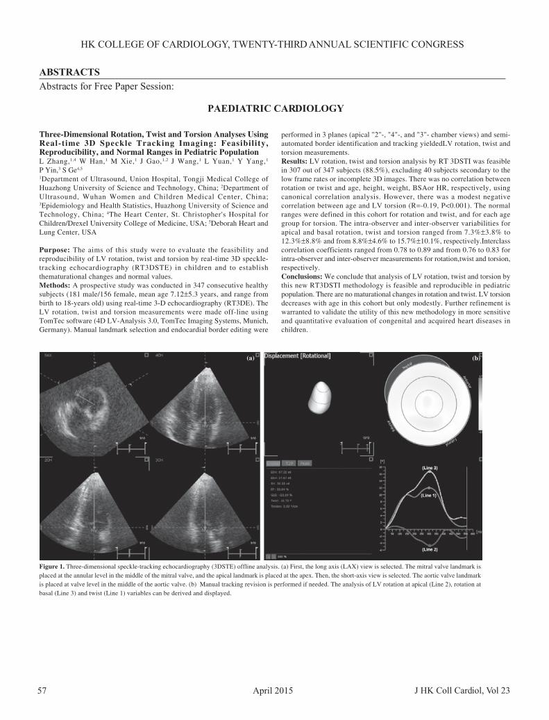
HK COLLEGE OF CARDIOLOGY, TWENTY-THIRD ANNUAL SCIENTIFIC CONGRESS
ABSTRACTSAbstracts for Free Paper Session:
April 2015 J HK Coll Cardiol, Vol 2357
PAEDIATRIC CARDIOLOGY
Three-Dimensional Rotation, Twist and Torsion Analyses UsingReal-time 3D Speckle Tracking Imaging: Feasibility,Reproducibility, and Normal Ranges in Pediatric PopulationL Zhang,1,4 W Han,1 M Xie,1 J Gao,1,2 J Wang,1 L Yuan,1 Y Yang,1
P Yin,3 S Ge4,5
1Department of Ultrasound, Union Hospital, Tongji Medical College ofHuazhong University of Science and Technology, China; 2Department ofUltrasound, Wuhan Women and Children Medical Center, China;3Epidemiology and Health Statistics, Huazhong University of Science andTechnology, China; 4The Heart Center, St. Christopher's Hospital forChildren/Drexel University College of Medicine, USA; 5Deborah Heart andLung Center, USA
Purpose: The aims of this study were to evaluate the feasibility andreproducibility of LV rotation, twist and torsion by real-time 3D speckle-tracking echocardiography (RT3DSTE) in children and to establishthematurational changes and normal values.Methods: A prospective study was conducted in 347 consecutive healthysubjects (181 male/156 female, mean age 7.12±5.3 years, and range frombirth to 18-years old) using real-time 3-D echocardiography (RT3DE). TheLV rotation, twist and torsion measurements were made off-line usingTomTec software (4D LV-Analysis 3.0, TomTec Imaging Systems, Munich,Germany). Manual landmark selection and endocardial border editing were
performed in 3 planes (apical "2"-, "4"-, and "3"- chamber views) and semi-automated border identification and tracking yieldedLV rotation, twist andtorsion measurements.Results: LV rotation, twist and torsion analysis by RT 3DSTI was feasiblein 307 out of 347 subjects (88.5%), excluding 40 subjects secondary to thelow frame rates or incomplete 3D images. There was no correlation betweenrotation or twist and age, height, weight, BSAor HR, respectively, usingcanonical correlation analysis. However, there was a modest negativecorrelation between age and LV torsion (R=-0.19, P<0.001). The normalranges were defined in this cohort for rotation and twist, and for each agegroup for torsion. The intra-observer and inter-observer variabilities forapical and basal rotation, twist and torsion ranged from 7.3%±3.8% to12.3%±8.8% and from 8.8%±4.6% to 15.7%±10.1%, respectively.Interclasscorrelation coefficients ranged from 0.78 to 0.89 and from 0.76 to 0.83 forintra-observer and inter-observer measurements for rotation,twist and torsion,respectively.Conclusions: We conclude that analysis of LV rotation, twist and torsion bythis new RT3DSTI methodology is feasible and reproducible in pediatricpopulation. There are no maturational changes in rotation and twist. LV torsiondecreases with age in this cohort but only modestly. Further refinement iswarranted to validate the utility of this new methodology in more sensitiveand quantitative evaluation of congenital and acquired heart diseases inchildren.
Figure 1. Three-dimensional speckle-tracking echocardiography (3DSTE) offline analysis. (a) First, the long axis (LAX) view is selected. The mitral valve landmark isplaced at the annular level in the middle of the mitral valve, and the apical landmark is placed at the apex. Then, the short-axis view is selected. The aortic valve landmarkis placed at valve level in the middle of the aortic valve. (b) Manual tracking revision is performed if needed. The analysis of LV rotation at apical (Line 2), rotation atbasal (Line 3) and twist (Line 1) variables can be derived and displayed.
(a) (b)

April 2015J HK Coll Cardiol, Vol 23 58
HK COLLEGE OF CARDIOLOGY, TWENTY-THIRD ANNUAL SCIENTIFIC CONGRESS
ABSTRACTSAbstracts for Free Paper Session:
PAEDIATRIC CARDIOLOGY
Plasma Levels of Monokine Induced by Interferon-gamma/Chemokine (C-X-C motif) Ligand 9, Thymus and Activation-Regulated Chemokine/Chemokine (C-C motif) Ligand 17 inChildren with Kawasaki DiseaseS Feng, SK Yadav, F Gao, Q YiDepartment of Cardiovascular Medicine, Children's Hospital of ChongqingMedical University, Zhongshan Er Road, Yuzhong District, China
Background: Monokines induced by interferon-gamma/Chemokine (C-X-C motif) ligand 9 (MIG/CXCL9), thymusand activation-regulated chemokine/Chemokine (C-C motif) ligand 17 (TARC/CCL17) are chemotactic factorsthat specifically collect and activate leukocytes, which are considered aschemoattractants of T helper cells. In the present study, we have investigatedthe effects of T helper type-1 (Th1) cells and T helper type-2 (Th2) cells inKawasaki disease (KD) by determining plasma levels of MIG/CXCL9 andTARC/CCL17 and exploring the relationship between MIG/CXCL9, TARC/CCL17 levels and coronary artery lesions (CAL).Methods: 43 children patients with KD and 19 healthy controls were includedin this study. General characteristics were obtained from all subjects. Plasmaconcentrations of chemotactic factors of MIG/CXCL9 and TARC/CCL17were measured by enzyme-linked immunosorbent assay (ELISA) for allsubjects.Results: Plasma levels of MIG/CXCL9, TARC/CCL17, and the ratios ofMIG/TARC were significantly elevated in pediatric patients with KDcompared to that in the control group. There were also significant higherlevels of MIG/CXCL9, TARC/CCL17, MIG/TARC ratios and prominentlylower Hb levels in KD with CAL compared to KD without coronary artery
lesions (NCAL). Hb was significantly decreased and plasma MIG/CXCL9levels had a significant negative correlation with CRP in KD with CALpatients (KD-CAL), whereas a positive correlation of plasma MIG/CXCL 9with WBC was observed in KD without CAL patients (KD-NCAL).Conclusion: Th1 and Th2 cells may be involved in an imbalanced activationin pediatric KD patients during an acute period of the disease. Furthermore,immune lesions of vessels in KD patients may be mediated by the imbalancedactivation of Th1 and Th2 cells.
Early and Mid-term Outcomes of Transventricular ValvotomyApproach for Initial Treatment of Pulmonary Atresia with IntactVentricular SeptumG Chen, B Jia, L Wu, F Liu, M Ye, Y MiChildren's Heart Center, Children's Hospital of Fudan University, China
Objective: To review the short and medium term surgical outcomes oftransventricular pulmonary valvotomy(TPV) in patients with pulmonaryatresia-intact ventricular septum(PA/IVS) and non-right ventricular dependantcoronary circulation.Background: For patients with pulmonary atresia with intact ventricularseptum, percutaneous radiofrequency-assisted valvotomy and balloon dilationis the first choice for most patients. A kind of alternative hybrid procedure:transventricular pulmonary valvotomy is now widely used in developingcountries without radiofrequency perforation equipments in cath labs.However, data on outcomes with this strategy are mixed.Methods: Data were reviewed retrospectively for 21 patients with PA/IVSand non-right ventricular dependant coronary circulation treated with TPVfrom 2010-2013 in our center. Patients who had severe neonatal Ebsteinmalformation, or initial interventional management at another institution wereexcluded. The mean body weight of the 21 patients was (3.2±0.7)kg andmean age was (16±8)d. The Z-score of tricuspid valve ranged from -2.5~2.2 (-0.9±0.4). The TPV procedure was guided by the Micro-TEE probe, toconfirm the position of perforation on the valve. All the patients werefollowed up for over 12 months. All the patients were divided into two groups:Bi-ventricle repair(BVR) and non-BVR group and the percutaneous
saturation, Z-score of tricuspid valve, reintervetion data were collected at3, 6, 12, 24, 36, 48 months after the procedure.Results: There was one early death after the TPV procedure for severehyoxemia and parents refused to reoperation. Twelve patients finallyunderwent BVR and 5 underwent one and half ventricle repair. Three patientswere still on follow-up after TPV. The mean follow-up time was (32.3±10.3) months (range 12-60 months) without late death. Early reinterventionwere needed in 4 patient(20%). One was in BVR group and accepted thepercutaneous pulmonary valve balloon dilation for residual obstruction.Three were in one-BVR group and got hyoxemia need modified B-T shuntsin 2 and PDA stent in 1 patient(8.3% vs 37.5%, p<0.01). The Z-score inBVR group was significantly larger than non-BVR group, with meanZ-score of (-0.5±0.3) and (-1.5±0.6), respectively (p=0.01). The freedomfrom reoperation was 80, 70, 35% at 6 months, 1 and 3 years, respectively.In BVR group, 9 of 12 received the ASD device closure, by catheter orsurgery. In non-BVR group, 5 received bidirectional Glenn shunt andclose the ASD; 1 received reconstruction of right ventricle outflowfor muscular obstruction. The mean Z-score ranged from -2~2.2 (-0.5±0.4), -2.1~2.2(-0.5±0.3), -2.3~2.0(-0.2±0.3), -0.4~2.2(0.5±0.8), -0.5~2.2(0.6±0.8) at 3 months, 6 months, 1year, 2 and 3 years after TPV.Conclusions: The results of a hybrid approach to treating patients with PA/IVS are excellent. TPV can be used as an alternative method to transcatheterapproach. Smaller TV size is associated with greater likelihood ofreintervention prior to discharge, and may serve as a surrogate for early RVinadequacy.
HK COLLEGE OF CARDIOLOGY, TWENTY-THIRD ANNUAL SCIENTIFIC CONGRESS
ABSTRACTSAbstracts for Free Paper Session:
April 2015 J HK Coll Cardiol, Vol 2359
Operation Treatment of Left-to-right Shunt Congenital HeartDisease with Pulmonary Arterial Hypertension in 210 casesT Shi, Y Chen, M Li, C WuChildren's Hospital of Chongqing Medical University, China
Purpose: To analyze the clinical characteristics and the operation treatmentof left-to-right shunt congenital heart disease(CHD) with pulmonary arterialhypertension (PAH).Methods: Totally 210 cases of left-to-right CHD with PAH were collectedfrom September 2012 to September 2013, whose clinical characteristics wereretrospectively analyzed and divided into the intervention group (86 cases)and the surgical group (124 cases) according to different therapeutic methods.The similarities and differences of clinical features, operation time, volumeof blood transfusion and curative effect between the two groups were analyzed.Results: Among the clinical manifestations and signs, recurrent respiratorytract infections, feeding interruption, different levels malnutrition, cardiacmurmur and pulmonary second heart sounds loudly were the most common.The children with cyanosis, severe malnutrition or pulmonary second heartsounds loudly in the surgical group were more than that in the interventiongroup. The children with growth and development backwardness in theintervention group were more than that in the surgical group (P<0.05). Thecomparison of the results of chest X ray (mainly showed that multiplepulmonary blood and increased heart shadow) and electrocardiogram (mainlyshowed that double ventricular hypertrophy and right bundle branch block)between the intervention group and the surgical group mostly had no statisticalsignificance (P>0.05). The right ventricular hypertrophy occurred more oftenin the surgical group than in the intervention group (P<0.05). The operationtime, volume of blood transfusion, postoperative mechanical ventilation
duration, intensive care unit (ICU) care duration, hospitalized duration weresignificantly longer in surgical group than in intervention group (P<0.05).The operation success rate of the two groups were as high as 97.67% and97.58%, respectively. And there was no statistical significance between thetwo groups (P>0.05).The pulmonary arterial systolic pressure (PASP) in thetwo groups were significantly decreased after operation (P<0.05).Conclusions: CHD with PAH is not easy to early diagnosis. The detectionof echocardiography and PASP will do great help in the regularly follow-upin outpatient service. For the children who conform to the interventionaltreatment for indications can be preferred intervention treatment. But for thechildren who are low age and with cyanosis or severe malnutrition orpulmonary second heart sounds loudly or the right ventricular hypertrophyshowed in electrocardiogram or larger diameter of the defect or higher PASP,surgical treatment should be chose as soon as possible.
PAEDIATRIC CARDIOLOGY
Epicardial Pacemaker Implantation and Follow-up in Infants andChildrenWB Zhange, M Ye, G Chen, WS Qiu, XG Yan, B JiaThe Cardiovascular Center, Children's Hospital of Fudan University, China
Objective: To evaluate the feasibility and safety of permanent epicardialpacing in infants and children.Methods: During the period from July 2005 to June 2014, we collected thedata of 18 children (11 males) with a mean weight of 13.3±5.8 Kg thatunderwent permanent epicardial pacemaker implantation in the cardiovascularcenter of Children's Hospital of Fudan University. 11 cases diagnosed asatrium-ventricle block (AVB) after operation of congenital heart disease(CHD) while other 7 cases underwent no heart surgery. The surgical operationrecords and changes of the parameters of the permanent cardiac pacemakersin follow-up were retrospectively analyzed.Results: The 18 patients had a total of 21 permanent epicardial pacing leads.VVI permanent cardiac pacemakers were implanted in 15 cases and DDD in3 cases. All the patients had their implantation via midline thoracotomy andall surgical epicardial electrode leads were successfully implanted on theright atrium and the right ventricle. The pulse generators were placed in theabdominal wall in a subcutaneous or submuscular pockets and the pacingleads were prepared for the growing of the children. Follow-ups were takenat an average interval of 32.2 months. There were no dead cases during themanagement. There was only one lead fracture and one infected woundhappened to the patients. Impedance, sensing and stimulation thresholds werein the normal range.
Conclusion: Epicardial pacemaker is a premier procedure in infants andchildren with anatomical abnormalities because of the multiple problemsthat these special patients pose: age, body size, somatic growth and lifestyle.Permanent pacing in pediatric age group is relatively safe.

April 2015J HK Coll Cardiol, Vol 23 60
HK COLLEGE OF CARDIOLOGY, TWENTY-THIRD ANNUAL SCIENTIFIC CONGRESS
ABSTRACTSAbstracts for Free Paper Session:
PAEDIATRIC CARDIOLOGY
Prenatal Diagnosis of Vascular Ring by Fetal EchocardiopraphyCombined with STIC TechniqueY Wang, M Xie, W Han, L Hong, H Cao, Y Yang, K Li, Y Hu, M WangDepartment of Ultrasound, Union Hospital of Tongji Medical College,Huazhong University of Science and Technology, Hubei Provincial KeyLaboratory of Molecular Imaging, China
Objective: Vascular ring is a rare congenital heart disease, this study aimsto evaluate the clinical value of fetal echocardiopraphy combined with STICtechnique on the diagnosis of vascular ring.Methods: According to the guidelines of ASE, 285 cases of pregnant womenwere examined by prenatal echocardiography in Union hospital during Marchto December 2014, using sequential segmental approach. STIC techniquewas used to analyze the cardiac volume. All data were collected and storedfor further off-line analysis. The sonographic features and prenatalmanifestations of vascular ring were summarized.Results: (1) Eight cases of fetal vascular ring were diagnosed by prenatal
echocardiography combined with STIC technique. Five cases were confirmedby follow-up echocardiography after delivery, and other 3 cases were provenby autopsy after labor induction. (2) Three vessel and trachea view showed:aortic arch on the right side of the trachea while duct artery arch on the leftin 7 cases, forming a U-shaped vascular ring around the trachea (Figure 1).The other case showed aortic arch on the left side of the trachea, and theazygos vein abnormally running between the spine and the trachea, andthen flowing into the superior vena cava on the right side of trachea. TheU-shaped vascular ring was constituted by the azygos vein on the right sideof the trachea and the aortic arch on the left side (Figure 2).Conclusion: (1) The three vessel and trachea view is a vital view on thediagnosis of fetal vascular ring, especially for the vascular ring consisted ofdextroaortic arch and left ductus arteriosus, and other types of abnormalvessel orientation around the trachea as well. (2) STIC technique can showthe orientation and adjacent structures of the great vessels, which couldprovide more diagnostic information of fetal vascular ring. It is also importantfor the prognosis and management of newborns.
Figure 1. U shape vessel ring around trachea by 2D and CDFI: right aortic arch and left ductus arteriosus. Figure 2. U shape vessel rings around trachea by STIC:right aortic arch and left ductus arteriosus.
Figure 3. U shape vessel ring around trachea by 2D and CDFI: aortic arch and azygos vein. Figure 4. The aortic arch and the azygos vein formed theU type vessel rings around trachea by STIC.
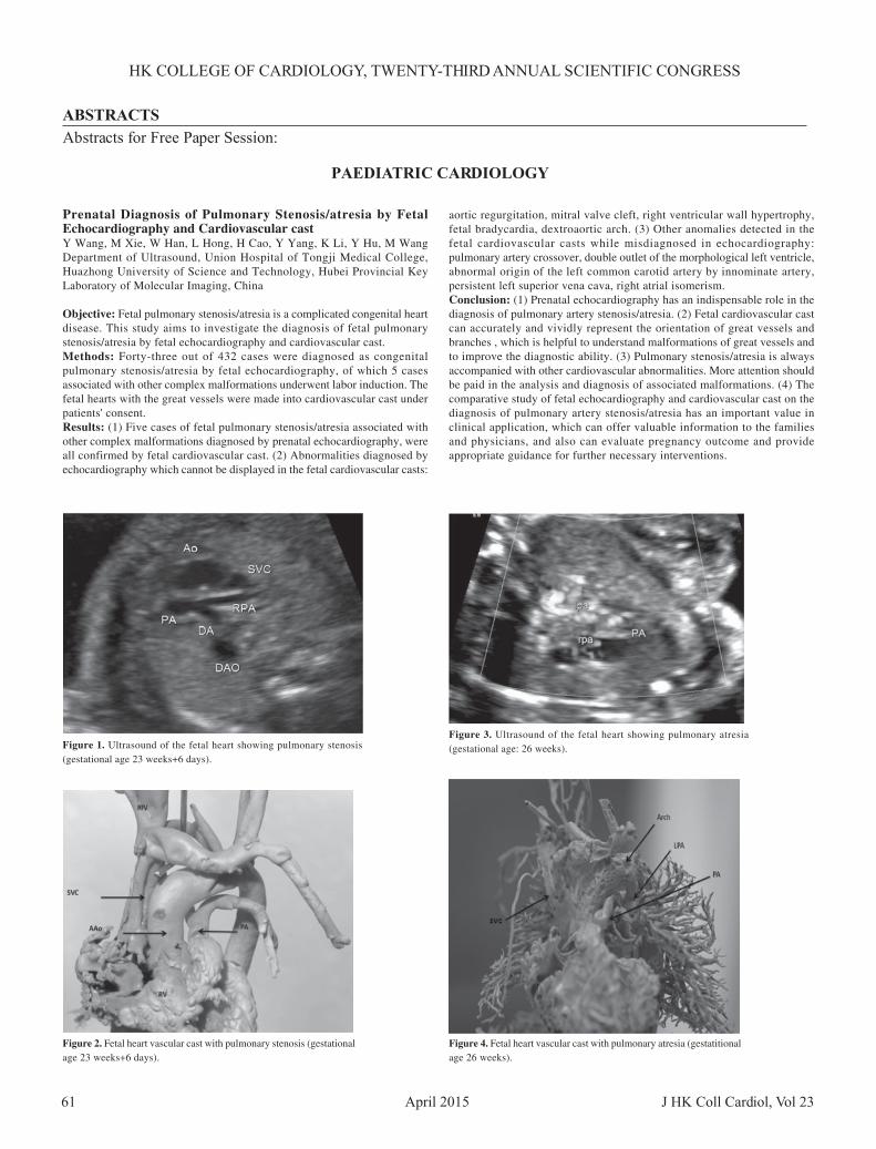
HK COLLEGE OF CARDIOLOGY, TWENTY-THIRD ANNUAL SCIENTIFIC CONGRESS
ABSTRACTSAbstracts for Free Paper Session:
April 2015 J HK Coll Cardiol, Vol 2361
PAEDIATRIC CARDIOLOGY
Prenatal Diagnosis of Pulmonary Stenosis/atresia by FetalEchocardiography and Cardiovascular castY Wang, M Xie, W Han, L Hong, H Cao, Y Yang, K Li, Y Hu, M WangDepartment of Ultrasound, Union Hospital of Tongji Medical College,Huazhong University of Science and Technology, Hubei Provincial KeyLaboratory of Molecular Imaging, China
Objective: Fetal pulmonary stenosis/atresia is a complicated congenital heartdisease. This study aims to investigate the diagnosis of fetal pulmonarystenosis/atresia by fetal echocardiography and cardiovascular cast.Methods: Forty-three out of 432 cases were diagnosed as congenitalpulmonary stenosis/atresia by fetal echocardiography, of which 5 casesassociated with other complex malformations underwent labor induction. Thefetal hearts with the great vessels were made into cardiovascular cast underpatients' consent.Results: (1) Five cases of fetal pulmonary stenosis/atresia associated withother complex malformations diagnosed by prenatal echocardiography, wereall confirmed by fetal cardiovascular cast. (2) Abnormalities diagnosed byechocardiography which cannot be displayed in the fetal cardiovascular casts:
aortic regurgitation, mitral valve cleft, right ventricular wall hypertrophy,fetal bradycardia, dextroaortic arch. (3) Other anomalies detected in thefetal cardiovascular casts while misdiagnosed in echocardiography:pulmonary artery crossover, double outlet of the morphological left ventricle,abnormal origin of the left common carotid artery by innominate artery,persistent left superior vena cava, right atrial isomerism.Conclusion: (1) Prenatal echocardiography has an indispensable role in thediagnosis of pulmonary artery stenosis/atresia. (2) Fetal cardiovascular castcan accurately and vividly represent the orientation of great vessels andbranches , which is helpful to understand malformations of great vessels andto improve the diagnostic ability. (3) Pulmonary stenosis/atresia is alwaysaccompanied with other cardiovascular abnormalities. More attention shouldbe paid in the analysis and diagnosis of associated malformations. (4) Thecomparative study of fetal echocardiography and cardiovascular cast on thediagnosis of pulmonary artery stenosis/atresia has an important value inclinical application, which can offer valuable information to the familiesand physicians, and also can evaluate pregnancy outcome and provideappropriate guidance for further necessary interventions.
Figure 1. Ultrasound of the fetal heart showing pulmonary stenosis(gestational age 23 weeks+6 days).
Figure 2. Fetal heart vascular cast with pulmonary stenosis (gestationalage 23 weeks+6 days).
Figure 3. Ultrasound of the fetal heart showing pulmonary atresia(gestational age: 26 weeks).
Figure 4. Fetal heart vascular cast with pulmonary atresia (gestatitionalage 26 weeks).

April 2015J HK Coll Cardiol, Vol 23 62
HK COLLEGE OF CARDIOLOGY, TWENTY-THIRD ANNUAL SCIENTIFIC CONGRESS
ABSTRACTSAbstracts for Free Paper Session:
PAEDIATRIC CARDIOLOGY
Percutaneous Balloon Valvuloplasty for Severe Pulmonary ValveStenosis in Infants: A 10-Year Institutional Experience andLongterm OutcomesL He, F Liu, L Wu, XC Liang, CH Qi, HY Deng, GY HuangPediatric Heart Center, Children's Hospital of Fudan University, China
Background: Although percutaneous balloon pulmonary valvuloplasty (BPV)is the mainstay of treatment for significant pulmonary stenosis with domingleaflets, it's widely considered to be difficult and relatively large risk foryounger and severe stenosis children. This study was undertakenretrospectively to summarize and analysize its immediate and long-termefficacy and safety in neonates and infants with severe or critical valvularpulmonary stenosis.Methods: Seventy-nine patients aged 5d~3y with critical or severe pulmonaryvalve stenosis admitted to our hospital from January 2005 to December 2014underwent balloon valvuloplasty. Among them, 10 neonates had criticalpulmonary stenosis, who had a tripartite right ventricle with moderate tosevere tricuspid regurgitation (TR). Severe TR was seen in 12 and moderateTR in 6 out of other 69 patients of over one month of age. Right ventricularsystolic pressure in all patients was equal to or greater than systemic pressure.43 patients had PFO with right-to-left or bi-directional shuntl, 10 patientshad PDA, 1 patient had multiple small muscular ventricular septal defects,and 1 patient had atrial septal defect, who undergone the ASD occlusiontwo-year later. Dilatation with 2 balloons sequentially in one procedure wasperformed in 12 patients and dilatation with 1 balloon in the other patients.
Results: The pulmonary valvuloplasty was accomplished in 74 of the 79patients, and the dilatation success rate was 93.67%. Four were failure tocross the pulmonary valve with balloon catheter because of the almost atreticvalve in 2 and severe spasm of the RVOT in another 2 patients. One wasfailure to get the pressure gradient across the pulmonary valve down becauseof the annulus dysplasia. Immediately after dilatation, the systemic pressuregradient from right ventricle to pulmonary artery decreased from 50-132(76.25±23.7) mmHg to 4-96 (25.29±19.2) mmHg (P<0.001). No significantcomplications in all patients during or post dilation except cardiac tamponadein one. During a 2 month to 9.6 years follow-up (mean 5.01 years), datashowed that: (1) pressure gradient crossing pulmonary valve measured byechocardiography further decreased or remained stable in 70 cases, exceptone neonate and three infants, whose pressure gradient gradually increased,and needed a second dilatation. Re-dilatation rate was 5.4% (4/74). No caseneeded further surgery; (2) Tricuspid regurgitation reduced in all patientsexcept for three whose RV were dysplasia; (3) Mild or no pulmonaryregurgitation was seen in most of patients post-dilatation, except moderatein six and severe in one. (4) All 10 PDAs closed spontaneously in 3-6 monthsof follow-up and muscular VSDs were closed as well in 3 months of follow-up. (5) All patients were doing well with asymptoms and acyanosis.Conclusions: Balloon pulmonary valvuloplasty (BPV) is safe and effectivein attaining both immediate and long term reduction of pulmonary valvulargradients and is currently the preferred therapeutic modality for valvular PSeven small baby patients.
Safety and Efficacy of Warfarin plus Aspirin CombinationTherapy for Giant Coronary Artery Aneurysm Secondary toKawasaki Disease: A Meta-AnalysisD Sun, K Wang, S Qin, Y PangDepartment of Pediatrics, The First Affiliated Hospital of Guangxi MedicalUniversity, Nanning, Guangxi Autonomous Region, China
Objective: To compare the safety and efficacy of warfarin plus aspirin versusaspirin alone for the treatment of children with giant coronary artery aneurysm(CAA) secondary to Kawasaki disease (KD).Methods: We searched the PubMed, EMBASE, Cochrane Library, CNKI,WANFAN and VIP databases. We selected case-controlled trials of warfarinplus aspirin versus aspirin alone for the treatment of children with giant CAAsecondary to KD.Results: Six retrospective studies met our inclusion criteria. There was nosignificant difference between the warfarin plus aspirin and aspirin alonegroups in the rate of CAA regression (OR 1.38, 95% CI 0.52-3.68, p=0.52)or the incidence of persistent CAA (OR 2.34, 95% CI 0.16-33.50, p=0.53),coronary artery stenosis (OR 0.55, 95% CI 0.18-1.72, p=0.30) or thrombusformation (OR 0.50, 95% CI 0.15-1.69, p=0.26). There was evidence thatwarfarin plus aspirin reduced the incidence of coronary artery occlusion (OR0.08, 95% CI 0.02-0.29, p<0.0001), cardiac infarction (OR 0.27, 95% CI0.11-0.63, p=0.003) and death (OR 0.18, 95% CI0.04-0.88, p=0.03).Conclusion: Warfarin plus aspirin therapy reduced the incidence of occlusion,cardiac infarction and death in children with giant CAA secondary to KD.
ABSTRACTSAbstracts for Poster Presentations:
April 2015 J HK Coll Cardiol, Vol 2363
HK COLLEGE OF CARDIOLOGY, TWENTY-THIRD ANNUAL SCIENTIFIC CONGRESS
DNA Methylation Regulated Cardiac Myofibrils Gene ExpressionDuring Heart Development in MouseY Xu,1 L Liu,1,2 B Pan,1 J Zhu,2 C Nan,3 X Huang,3 J Tian1
1Heart Centre, Children's Hospital of Chongqing Medical University, China;2Ministry of Education Key Laboratory of Child Development and Disorders;Key Laboratory of Pediatrics in Chongqing, CSTC2009CA5002; ChongqingInternational Science and Technology Cooperation Center for ChildDevelopment and Disorders, China; 3Department of Biomedical Science,Charlie E. Schmidt College of Medicine, Florida Atlantic University, USA
It is well known that epigenetic plays an important role in controlling theregulation of gene expression during biological development. Troponin I isone of the regulatory myofibril protein in heart, which can control thecontraction and relaxation. And troponin is a good model to study theregulation of cardiac gene expression during heart development. Our previousstudies have demonstrated that the slow skeletal troponin I (ssTnI), alsocalled fetal troponin I, is partially regulated by epigenetic modification,such as histone acethylation and methylation, and we demonstrated thatssTnI was partially regulated by thyroid hormone. In this study, we exploredthe epigenetic role of DNA methylation in regulating ssTnI expression. Thecondition of DNA methylation and the methylation level in CpG islandsand CpG island shores were detected using methylation specific PCR (MSP)and bisulfite sequence PCR (BSP) in 2000 bp upstream and 100 bp upstreamof ssTnI gene promoter. In additional, we explored DNA methylation levelof CpG islands and shores in the cardiac troponin I (cTnI) gene promoter.
We found that the DNA methylation levels of CpG island shores in ssTnIgene promoter were decreased, which corresponded to a decreased expressionof ssTnI gene in mouse heart, but the DNA methylation level of CpG islandsin this gene were not changed during heart development. However, themethylation level of CpG islands and shores in cTnI were not changed inthe development period. Our results indicated that DNA methylaion wasinvolved in the epigenetic regulation of ssTnI and played a role in TnIisoforom switching in heart during the development.
Alcohol Exposure Induced Histone3 Hyperacetylation Leadingto Overexpression of Cardiac Transcription Factors ThroughERK1/2 Signaling Pathway in H9c2 CellsW Gao, X Huang, J Tian1Heart Centre, Children's Hospital of Chongqing Medical University; 2KeyLaboratory of Developmental Disease in Childhood (Chongqing MedicalUniversity), Ministry of Education, China; 3Key Laboratory of Pediatrics inChongqing; 4Chongqing International Science and Technology CooperationCenter for Child Development and Disorders; 5Department of BiomedicalScience, Charlie E. Schmidt College of Medicine, Florida Atlantic University,USA
Aims: Alcohol has been widely accepted as a common detrimental exposureduring pregnancy, which may cause fetal cardiac development abnormalities.Our previous studies displayed that alcohol could induce histonehyperacetylation and over-expression of cardiac transcription factors bothin vivo and in vitro. The objective of this study is to investigate the role ofERK1/2 signaling pathway in alcohol-induced histone acetylation imbalanceand up-regulation of transcription factors in H9c2 cells.Materials and Methods: Cardiac cell line, H9c2 cell was used in this study.U0126, a specific inhibitor of ERK1/2 pathway was employed to block theERK1/2 signaling pathway. Western blot analysis was used to detect thephosphorylation levels of ERK1/2 and the acetylation levels of histone3.Real-time PCR was employed to measure the expression of GATA4 andMEF2c. The histone3 acetylation levels in the promoter regions of GATA4and MEF2C were analyzed using chromatin immunoprecipitation (ChIP)assays.
Results: Alcohol could enhance the levels of phosphorylated ERK1/2, andU0126 significantly inhibited alcohol-induced ERK1/2 signaling pathwayactivation and the hyperacetylation of histone3. U0126 decreased alcohol-induced over-expression of GATA4 and MEF2c, and the basal expressionlevel of GATA4, but not MEF2c. Interestingly, ChIP assay showed that U0126significantly down-regulated alcohol-induced hyperacetylation of histone3in the promoter region of GATA4 and MEF2c, and the basal acetylationlevel of histone3 in the promoter region of GATA4, but not MEF2c.Conclusions: These data indicated that U0126 may reverse theoverexpression of GATA4 and MEF2c through down-regulation of histone3acetylation level in their promoter regions, suggesting that ERK1/2 signalingpathway might be a potential target for the intervention of alcohol inducedcongenital heart diseases.
April 2015J HK Coll Cardiol, Vol 23 64
ABSTRACTSAbstracts for Poster Presentations:
HK COLLEGE OF CARDIOLOGY, TWENTY-THIRD ANNUAL SCIENTIFIC CONGRESS
Alcohol Increase the Acetylation of Histone H3 to CauseAbnormal Expression of Cardiac Genes in H9C2 Cells via JNKSignaling PathwayW Zhang,1,2 M Zheng,1,2 C Peng,1,2 W Gao,1,2 J Zhu,2 L Liu,2 J Tian 1
1Heart Centre, Children's Hospital of Chongqing Medical University;2Ministry of Education Key Laboratory of Child Development and Disorders,China
Purpose: Based on the intervention of alcohol, SP600125 was used to blockthe JNK signaling pathway in H9C2 cells, to observe the changes in thelevel of Histone H3 acetylation and over-expression of downstream cardiacspecific genes caused by alcohol exposure, to explore whether JNK mediatedthe effects of intervention by alcohol in H9C2 cells. In this study, we testthe hypothesis that JNK signaling pathway mediated the hyper-acetylationof histone H3 and over-expression of cardiac specific genes induced byalcohol exposure.Methods: The optimal dose: After treated the H9C2 cells with alcohol for36 hour, added different concentrations of SP600125 in the cells to blockthe JNK pathway. MTT assay was used to detect the cells viabilities todetermine the optimal dose of SP600125. The H9C2 cells were divided intofive groups, blank group, DMSO group, Alc proup, SP group and Alc plusSP group. The histone acetylation enzyme activity was detected bycolorimetric assay in H9C2 cells. The level of acetylated histone H3 wasmeasured by western-blotting in H9C2 cells. The expression of cardiacrelated genes dHAND and eHAND were determined by RT-Q-PCR H9C2cells. The acetyled histone H3 which were combined in the promoter regionof dHAND and eHAND were detected by ChIP-RT-Q- PCR in H9C2 cells.
Results: There was no difference between the SP group and DMSO groupwhen treated with 0 µM, 5 µM, 7.5 µM and 10 µM of SP600126 in H9C2cells (p>0.05). There was significant difference between the SP group andDMSO group when treated with 15 µM, 20 µM and 25 µM dose of SP600126in H9C2 cells (P<0.05).The HATs activity and level of AcH3 in Alc grouphad significant difference compared o the blank group and DMSO group(p<0.05). The HATs activity and level of AcH3 in SP group had no differencecompared to the blank group and DMSO group (p>0.05). The HATs activityand level of AcH3 in Alc and SP group had no difference compared to theblank group, DMSO group, or SP group (p>0.05).The mRNA expression ofcardiac related genes dHAND and eHAND were increased in the Alc groupcompared to the blank group, or DMSO group (p<0.05). The Alc and SPgroup was no difference compared to the blank group, DMSO group, or SPgroup (p>0.05). There was no difference between the SP group and the blankgroup, or DMSO group (p>0.05). The AcH3 which were combined in thepromoter region of cardiac related gene dHAND and eHAND were increasedin the Alc group compared to the blank group, SP group, or DMSO group (p<0.05). The Alc and SP group was no difference compared to the blankgroup, DMSO group, or SP group (p>0.05).Conclusions: JNK pathway mediated the effect that Alc increase the AcH3which were combined in the promoter region of cardiac related genes dHANDand eHAND.
Dyslipidemia in Pregnancy May Contribute to Increased Risk ofCongenital Heart DefectsH Ba, HS WangPediatric Cardiology Department, The First Affiliated Hospital of Sun Yat-Sen University, China
Objective: To investigate the relation between the presence of dyslipidemiain pregnant women in China and the risk of Congenital heart defects(CHD).Methods: A total of 54 pregnant women in 24 to 28 weeks gestation periodwere enrolled in this study between March 2013 and June 2014. The casegroup included 18 women who had fetus with cardiac defects, and eachcase was matched with 2 controls with no pregnancy complications. Meanages were 29.06 (SD=3.11) years and 29.03 (SD=3.9) years in the case groupand control group, respectively. Main outcome measures were totalcholesterol (TC), triglycerides (TG), low-density lipoprotein cholesterol(LDL-c), apolipoprotein.Results: Those in the case group had significantly higher mean LDL-cholesterol (4.15 vs 3.45) and apolipoprotein B (1.23 vs 1.08) (all P<0.05)than the control group. After adjustment for potential confounders, a lipidprofile with apolipoprotein B>1.21 mg /dl is associated with a higherincidence rate of CHD (61% vs 22%).Univariable logistic regression analysisreveals that CHD risk increases by each standard deviation increase in LDL-cholesterol and apolipoprotein B.An additional backwards stepwise logisticregression model shows that apolipoprotein B is independently and moststrongly associated with CHD risk, OR 111.99 (95%CI: 1.2-10252.11)perstandard deviation increase.
Conclusions: Serum LDL-cholesterol and Apolipoprotein B levels aresignificantly higher in case group than the control group. Apolipoprotein isassociated with a higher incidence rate of CHD. Increased LDL-cholesteroland Apolipoprotein B may be involved in the pathological mechanism ofCongenital heart defects.

ABSTRACTSAbstracts for Poster Presentations:
April 2015 J HK Coll Cardiol, Vol 2365
HK COLLEGE OF CARDIOLOGY, TWENTY-THIRD ANNUAL SCIENTIFIC CONGRESS
Associations between ABCB1 and ABCG2 gene polymorphismsof children and isolated septal defects in a Han Chinese populationC Wang, K Zhou, Y Li, Y Zhang, Y HuaDepartment of Pediatric Cardiology, West China Second University Hospital,Sichuan University, China
Backgrounds: Substantial epidemiological data have demonstrated thatseveral toxicants/drugs exposures periconceptionally are associated with anincreased risk of congenital heart defects (CHDs). P-glycoprotein (P-gp)and breast cancer resistance protein (BCRP) in the placenta, encoded by theABCB1 and ABCG2 gene in human, play an essential role in regulating fetalexposure to toxicants/drugs.Purpose: This study aimed to explore the associations betweenpolymorphisms of the ABCB1 and ABCG2 gene of children and isolatedseptal defects in a Han Chinese population, and to investigate the impact ofthese polymorphisms on the transcription and translation activities of theABCB1 and ABCG2 gene, respectively.Methods: An age and gender matched case-control study involving 210pairs was conducted. Genotyping of the ABCB1 and ABCG2 genepolymorphisms was conducted by sequencing. Forty-six placenta tissuesand umbilical cords from healthy Han Chinese mothers with uncomplicatedpregnancy were collected. Placental P-gp and BCRP mRNA and proteinexpression were determined by real-time quantitative PCR and western-blotrespectively.Results: For the 3435C>T polymorphism of ABCB1 gene, more cases werecarriers of the CC/CT genotypes (OR: 2.0, 95%CI: 1.1-3.5, P-value: 0.021).For the 34G>A polymorphism of ABCG2 gene, more cases were carriers of
the GA/AA genotypes (adjusted OR: 1.6, 95%CI: 1.0-2.3). There were nodifferences in the genotype distributions and allele frequencies of 421C>Apolymorphism of the ABCG2 gene. The placental ABCB1 mRNA expressionof the TT genotype was significantly higher than that of the CC genotype(P=0.03). Compared with TT genotype, lower placental P-gp expressionwas observed for the CC/CT genotypes. The ABCG2 mRNA and proteinexpression did not differ among the three genotypes of 421C>Apolymorphism. For the 34G>A polymorphism, the ABCG2 mRNA andprotein expression of the GG genotype was significantly higher than that ofthe AA genotype.Conclusions: The 3435C>T polymorphism within the ABCB1 gene and34G>A polymorphism within the ABCG2 gene of the children are associatedwith isolated septal defects in a Han Chinese population, presumably throughregulation of placental P-gp and BCRP expression, respectively.
Effect of BMP9 and Wnt3a Induce C3H10T1/2 CellsDifferentiation Into Cardiomyocyte-like Cells In VitroS Liu,1 Y Chen,1 S Fang,1 X Geng2
1Children's Hospital of Chongqing Medical University: Ministry of EducationKey Laboratory of Child Development and Disorders, Key Laboratory ofPediatrics in Chongqing, Chongqing International Science and TechnologyCooperation Center for Child Development and Disorders; 2Department ofPediatrics, Yantai Affilated Hospital of Binzhou Medical University, China
Purpose: To investigate the effect of co-induction by BMP9 and Wnt3a ondifferentiation of C3H10T1/2 cells into cardiomyocyte-like cells in vitro.Methods: C3H10T1/2 cells were randomly divided into 5 groups, (1) Blankgroup (untreated cells), (2) GFP group (cells transfected with AdEasy-GFP),(3) Wnt3a group (cells transfected with AdEasy-Wnt3a), (4) BMP9 group(cells transfected with AdEasy-BMP9), (5) BMP9 and Wnt3a co-inductiongroup (cells transfected with AdEasy-BMP9 and AdEasy-Wnt3a). Westernblot and immunofluorescence were used to detect the expression of cardiac-specific proteins Cx43 and cTnT. Q-RT-PCR was used to detect theexpression of genes GATA4 and MEF2C 3 weeks after transfection.Results: The expression of proteins Cx43 and cTnT in BMP9 and Wnt3aco-induction group were found higher than control group, GFP group andWnt3a group (P<0.05), but showed no difference compared with BMP9group (P>0.05) 3 weeks after transfection. The expression trend of genesamong five groups is similar to proteins.Conclusion: Co-induction by BMP9 and Wnt3a can promote C3H10T1/2cells differentiation into cardiomyocyte-like cells.
Innominate Artery Cannulation for Antegrade CerebralPerfusion in Neonates for Complex Cardiac SurgeryH Zhong, X Wang, M Ye, B JiaChildren's Hospital of Fudan University Cardiovascular Center, China
Background: Antegrade Cerebral Perfusion (ACP) is widely performed inneonates for aortic and complex cardiac surgery. The purpose of the presentstudy was to evaluate the flow measurements and safety of directioncannulation of the innominate artery.Methods: This was a retrospective review of consecutive neonates whounderwent ACP from January 2011 to July 2014. Patient characteristics,surgical and hemodynamic measurements and postoperative neurologicfindings were recorded.Results: 24 neonates underwent ACP during the studying period. There were2 operative deaths (8.3%) for the whole cohort. One patient who underwentcompletely repair died of postoperative heart failure, whereas 1 died ofoverwhelming sepsis. Of the 22 surviving patients, 2 had neurologicabnormalities. 2 patients were discharged with a diagnosis of hypotonia,one had experienced cardiac arrest in the intensive unit and another hadundergone preperative ultrasonography, which demonstrated calcificationof the basal ganglia. 20/22 survivors had normal neurologic evaluation ondischarge.Concusion: Innominate Artery Cannulation for antegrade cerebral perfusionis a safe and reliable technique in neonates by low mobidity and mortality,and should be the standard in complex cardiac surgery.

April 2015J HK Coll Cardiol, Vol 23 66
ABSTRACTSAbstracts for Poster Presentations:
HK COLLEGE OF CARDIOLOGY, TWENTY-THIRD ANNUAL SCIENTIFIC CONGRESS
The Study of Fetal Rat Model of Intra-amniotic IsoproterenolInjection Induced Heart Dysfunction and Phenotypic Switch ofContractile ProteinsY Li, Y Hua, C Wang, D Mu, K ZhouDepartment of Pediatric Cardiovascular Disease, West China SecondUniversity Hospital, Sichuan University; Ministry of Education KeyLaboratory of Women and Children's Diseases and Birth Defects, West ChinaSecond University Hospital, Sichuan University, China
Objective: To establish a reliable isoproterenol induced heart dysfunctionfetal rat model and understand the switches of contractile proteins.Methods: 45 pregnant rats were divided into 15 mg/kg-once, 15 mg/kg-twice, sham-operation-once, sham-operation-twice and control groups. And18 adult rats were divided into isoproterenol-treated and control groups.HE staining, Masson staining and transmission electron microscope wereperformed. Apoptotic rate assessed by TUNEL analysis and expressions ofANP, BNP, MMP-2 and CTGF of hearts were measured.Results: Intra-amniotic injections of isoproterenol were supplied on E14.5and E15.5 for fetuses and 7 days continuous intraperitoneal injections wereperformed for adults. Then echocardiography was performed with M-modeview assessment on E18.5 and 6 weeks later, respectively. Isoproterenol-twice treated fetuses exhibited significant changes in histological evaluation,and mitochondrial damages were significant severe with increased apoptoticrate. ANP and BNP increased and that of MMP-2 increased in isoproterenol-twice treated group compared to control group, without CTGF. The isoformstransition of troponin I and myosin heavy chain of fetal heart dysfunctionwere opposite to adult procedure. The administration of intra-amnioticisoproterenol to fetal rats could induce heart dysfunction.Conclusion: This clinically-relevant model can be used to explorepathophysiological responses during fetal heart dysfunction. The regulationof contractile proteins of fetuses was different from adult procedure.
Multiple Pulmonary Nodules in Kawasaki Disease: A Case ReportP Huang, L Zhang, J Yuan, F Gong, X Huang, L JinHeart Center, Affiliated Guangzhou Women and Children's Medical Centerof Guangzhou Medical College, China
A 8-month-old boy was admitted with a prolonged fever, bilateral bulbarconjunctival injection without exudate, red lips and truncal rash. amoxycillinclavulonate had been prescribed without clinical response. Physicalexamination on admission was significant for irritability and mild cervical,epitrochlear, axillary, and inguinal lymphadenopathy. Laboratory studiesshowed an elevated C-reactive protein, anemia, thrombocytosis, an elevatederythrocyte sedimentation rate, sterile pyuria and normal liver enzymes.Microbial cultures (blood, pharyngeal swab) were negative for pathogenicspecimens. Serologic tests for mycoplasma, chlamydia, cytomegalovirus,Epstein-Barr virus, influenza, parainfluenza, adenovirus and enterovirus werenegative. Mantoux test, bone scan, bone marrow aspiration, rheumatoidfactor, antineutrophil antibody, antineutrophil cytoplasm antibodies,antinuclear factor, and serum complements were negative or normal.Echocardiography performed on day 8 of illness revealed left coronaryarteries dilation without aneurysms. Chest X-ray demonstrated bilaterallydisseminated multiple pulmonary nodules 6 mm in diameter and ground-glass alterations. A lung CT scan showed numerous miliary nodules in diffuseand random distribution in both lungs. He was diagnosed with Kawasakidisease and treated with intravenous immunoglobulins and aspirin. The infantwas afebrile on day 14. A repeat chest X-ray on day 15 showed resolution ofthe pulmonary nodules. He was discharged on day 21 in good clinicalrecovery. An echocardiogram on day 18 was normal, as were follow-upechocardiograms 1, 2, 4 and 6 months after the acute stage of the disease.
The Influences of Influenza on Kawasaki DiseaseX Huang, P Huang, L Zhang, X Xie, S Xia, F Gong, J Yuan, L JinHeart Center, Affiliated Guangzhou Women and Children's Medical Centerof Guangzhou Medical College, China
Objective: To explore the influences of influenza on Kawasaki disease (KD).Methods: This retrospective study included 1053 children with KD fromJanuary 1, 2011 to December 31, 2013, among which 705 cases acceptedthe same examination of etiology that were considered comparable. Theepidemiology characteristics of the KD, influenza, and KD concomitant withinfluenza were summarized. The 15 cases of KD only with influenza A or/and B virus positive formed case group. Each case was matched with 2control cases and formed control group consisted of 30 children. Thedifferences of clinical manifestations, course of disease, and response tointravenous immunoglobulin (IVIG), laboratory test results, andechocardiographic detection between the two groups were compared.Results: The study showed that 34 cases (4.82%) with KD had documentedinfluenza infections. KD concomitant with influenza occurred in spring andsummer more than in winter and autumn, which was similar to all of the KDpatients hospitalized. Besides, influenza occurred all the year, but it outbrokein April and May, with a small peak in September. The median of whiteblood cell (WBC) and the rate of polymorphonuclear leukocytes (PMN)values were significantly higher in control group than in case group, whereasit was opposite in the lymphocytes and creatine kinase isoenzyme (CK-MB) values comparison. Patients in the case group had a longer medianfever course, a higher rate diagnosed with incomplete KD, and a longermedian time to be diagnosed compared with the control group.
Conclusion: Influenza virus infection had influence on KD to some extent,including its pathological damage, clinical manifestations, diagnoses andtreatment. The relationship of KD with influenza is unknown, which needmore researches. The presence of a concomitant influenza should be takenmore consideration during treatment with salicylates in patients with KD.

ABSTRACTSAbstracts for Poster Presentations:
April 2015 J HK Coll Cardiol, Vol 2367
HK COLLEGE OF CARDIOLOGY, TWENTY-THIRD ANNUAL SCIENTIFIC CONGRESS
Prenatal Diagnosis of Fetal Ebstein's Anomaly and New Finding in theFollow-ups: A Case ReportH Cao, M Xie, W Han, L Hong, Y Wang, Y HuDepartment of Ultrasonography, Union Hospital, Tongji Medical College,Huazhong University of Science and Technology, China
Clinical Presentation: A 31-year-old, gravida 2, para 0 woman undergoingprenatal echocardiography at 24.4 weeks' gestation was diagnosed as fetalEbstein's anomaly. Her first pregnancy was terminated due to stopped growthof the embryo at 6 week' gestation. She underwent another two follow-upexaminations at 28 and 30.5 weeks' gestation respectively. Echocardiographyshowed a typical manifestation of Ebstein's anomaly and a progressivedevelopment in the follow-ups, with a new finding of muscular ventricularseptal defect in the last exam. In view of the serious condition and poorprognosis of the fetus, the family decided to induce labor at 31 weeks'gestation. The diagnosis was proved by autopsy with the agreement ofparents. The results of autopsy showed: 1) dysplastic septal and posteriortricuspid leaflet displaced downward to the apical about 4~5 mm; 2) globalcardiac enlargement with obvious right atrium dilation; 3) two defectsobserved near the apex of muscular ventricular septal.
Imaging Findings: The four-chamber view showed a mild apicaldisplacement of the tricuspid valve. The distance between the septalhingepoints of the tricuspid and the mitral valve was about 5mm withoutsignificant change over time. However, the right atrium and atrialized rightventricle were gradually dilated with increased cardiothoracic ratio. Amoderate tricuspid regurgitation detected by CDFI in the first exam wasdeveloping into severe degree in the follow-ups. Besides, a new finding wasrecognized in the last exam. A muscular ventricular septal defect not shownin the first two exams was found by CDFI this time.Role of Imaging in Patient Care: Fetal echocardiography plays anindispensable role in the diagnosis of Ebstein's anomaly, which can offervaluable information to the families and clinicians, and provide appropriateguidance for further necessary interventions.Summary and Discussion Points: Fetal echocardiography is capable ofdiagnosing Ebstein's anomaly; and follow-up is also important to show thedevelopment of the disease, especially the dilation of right atrium and thedegree of tricuspid regurgitation. Associated abnormalities such as muscularventricular septal defect unable to discover in the second trimester might berecognized in the later follow-ups, which should be paid more attention to.
Figure. A. Apical displacement of the tricuspid valve; B. Tricuspid regurgitation detected by CDFI;C. Hight velocity shunt in the muscular part of interventricular septum (grey arrow); D. Dysplastic septaltricuspid leaflet downward displacement to the apical; E. Muscular ventricular septal defect with a diameterof 3 mm (black arrow). STL, septal tricuspid leaflet; ATL, anterior tricuspid leaflet; MVSD, muscularventricular septal defect.