Final Program - Pediatric Orthopaedic Society of North ...
556
UPCOMING MEETINGS 13th Annual International Pediatric Orthopaedic Symposium December 6 – 10, 2016 – Orlando, Florida Presented by POSNA and AAOS POSNA Specialty Day March 18, 2017 – San Diego, California EPOS/POSNA (EPOSNA) 2017 Combined Pre-Course and Annual Meeting May 3 – 6, 2017 Barcelona, Spain WELCOME
-
Upload
khangminh22 -
Category
Documents
-
view
0 -
download
0
Transcript of Final Program - Pediatric Orthopaedic Society of North ...
UPCOMING MEETINGS
13th Annual International Pediatric Orthopaedic SymposiumDecember 6 – 10, 2016 – Orlando, FloridaPresented by POSNA and AAOS POSNA Specialty DayMarch 18, 2017 – San Diego, California EPOS/POSNA (EPOSNA) 2017 Combined Pre-Course and Annual MeetingMay 3 – 6, 2017Barcelona, Spain
WELCOME

-2-
-3-
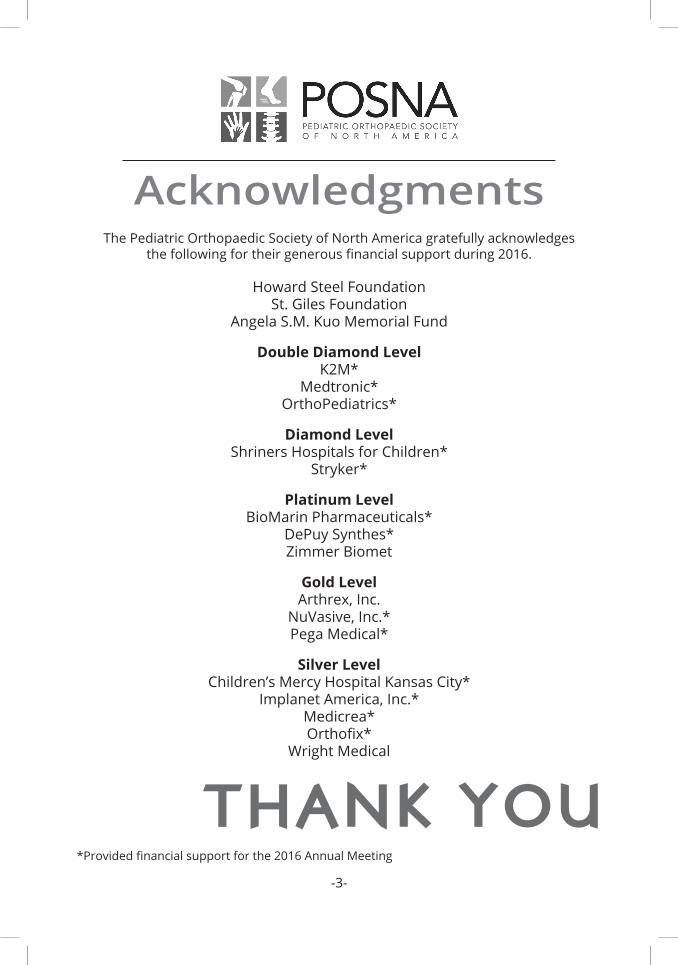
Acknowledgments The Pediatric Orthopaedic Society of North America gratefully acknowledges
the following for their generous financial support during 2016.
Howard Steel FoundationSt. Giles Foundation
Angela S.M. Kuo Memorial Fund
Double Diamond LevelK2M*
Medtronic*OrthoPediatrics*
Diamond LevelShriners Hospitals for Children*
Stryker*
Platinum Level BioMarin Pharmaceuticals*
DePuy Synthes*Zimmer Biomet
Gold LevelArthrex, Inc.
NuVasive, Inc.*Pega Medical*
Silver LevelChildren’s Mercy Hospital Kansas City*
Implanet America, Inc.*Medicrea*Orthofix*
Wright Medical
*Provided financial support for the 2016 Annual Meeting
THANK YOU

-4-
WELCOMEOn behalf of our local host, Randy Loder and our Program Committee, I welcome you to Indianapolis for the 2016 POSNA Annual Meeting. Many POSNA volunteers and staff have been working diligently to make our experience in Indianapolis a memorable one. The meeting opens on Wednesday morning with the Pre-Course entitled: “Trends and Application in Pediatric Lower Extremity Deformity.” The scientific sessions begin Wednesday afternoon. Susan Scherl and her Program Committee have assembled some of the highest rated abstracts into an outstanding session on sports, followed by one on upper extremity and trauma.
The opening ceremony on Wednesday evening allows us to recognize our industry sponsors and the outstanding achievement of several of our members. The Awards Committee has selected the following individuals: Humanitarian Award, John Herzenberg and Special Effort and Excellence Award, Brian Snyder. The DistinguishedAchievement Award recipient, Dennis Wenger, will be introduced on Thursday. Donald Davidson, the historian of the Indianapolis Motor Speedway, will be our 2016 Steele lecturer. He is a renowned and witty speaker, and will share his insights on the history of our country’s most famous track and the race it hosts each Memorial Day weekend, billed as “The Greatest Spectacle in Racing”.
The Presidential Guest lecturer on Friday morning will be Jim Roach, well-known to members as a pediatric orthopaedic educator, critical thinker, and leader. Jim’s lecture entitled “Skeptic Driven Innovation” promises to educate and challenge our thinking.
Symposia breakout sessions on Thursday will include Practice Management (The Electronic Medical Record Applications in Pediatric Orthopaedics), COUR (Adapting Care to Austere Environments), Research (Study Design and POSNA Supported Research Highlights), and NP/PA (Walk This Way – Foot Abnormalities). The Second Annual Arabella Leet Young Member Forum, sponsored by a generous grant from Shriners Hospital, will be held on Thursday as well.
Clinical and Basic Science Award papers will be read on Friday morning. POSNA Subspecialty Day will continue in its Friday afternoon time slot. Six concurrent sessions (Hip, Spine, Sports, Hand/Upper Extremity, Trauma, and Neuromuscular/Lower Extremity) will feature concurrent free paper and symposia formats.
The social schedule includes a welcome reception in the Marriott immediately following Opening Ceremonies on Wednesday evening. In response to member feedback from past annual meetings, we will again leave Thursday afternoon open to allow an afternoon for recreation, community building, and exploration of our host city. There are a number of exciting venues for exploration in and around Indianapolis, including White River State Park and the Indianapolis State Museum walking distance from the hotel, and the Indianapolis Motor Speedway and Hall of Fame Museum, and Hoosier Gymnasium, where the iconic Indiana movie was filmed 30 years ago, a short drive away. “Five players on the floor functioning as one
-5-
single unit: team, team, team - no one more important that the other.” still gives me goosebumps whenever I see this film.
Thursday evening, per tradition, will again be reserved for fellowship reunions and other gatherings with friends and colleagues.
Our closing reception on Friday evening will be at the NCAA Hall of Fame Museum, located directly adjacent to the hotel and just a short walk away. The dress code for the closing reception will be “athletic casual”…we encourage you all to wear spirit wear from your favorite NCAA University team. I plan to be decked out in Michigan Maize and Blue, of course! The museum offers interactive athletic activities, so come prepared to participate.
The educational program this year is second to none. I am excited to see you all in the city of fast cars and basketball for POSNA 2016.
Lori Karol, MD, POSNA President
-6-
ABOUT POSNAThe Pediatric Orthopaedic Society of North America (POSNA) is a group of professionals comprised mostly of pediatric orthopaedic surgeons. We are board certified in orthopaedic surgery and have participated in additional training to become specialized in the care of children’s musculoskeletal health and our practice reflects this dedication. We, as a group, strive to become the authoritative source on such care through appropriate research that will lead to the best evidence-based patient care.
MISSION STATEMENT To improve the care of children with musculoskeletal disorders through education, research, and advocacy.
POSNA extends sincere appreciation to K2Mfor their support in the printing of the final program.
-7-
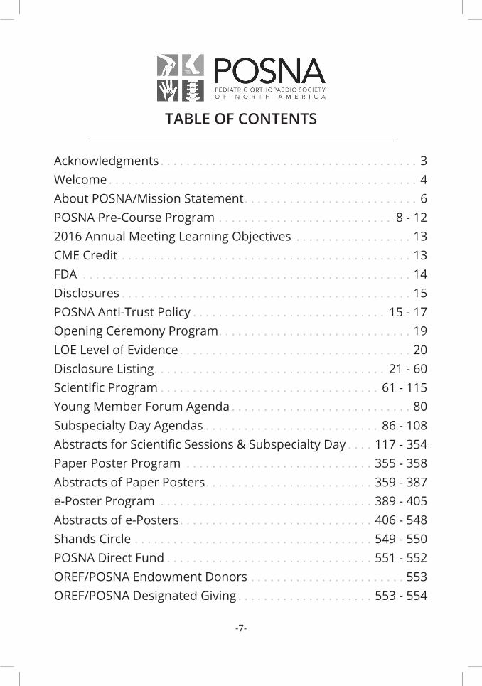
TABLE OF CONTENTS
Acknowledgments . . . . . . . . . . . . . . . . . . . . . . . . . . . . . . . . . . . . . . . . 3Welcome . . . . . . . . . . . . . . . . . . . . . . . . . . . . . . . . . . . . . . . . . . . . . . . . 4About POSNA/Mission Statement. . . . . . . . . . . . . . . . . . . . . . . . . . . 6POSNA Pre-Course Program . . . . . . . . . . . . . . . . . . . . . . . . . . . 8 - 122016 Annual Meeting Learning Objectives . . . . . . . . . . . . . . . . . . 13CME Credit . . . . . . . . . . . . . . . . . . . . . . . . . . . . . . . . . . . . . . . . . . . . . 13FDA . . . . . . . . . . . . . . . . . . . . . . . . . . . . . . . . . . . . . . . . . . . . . . . . . . . 14Disclosures . . . . . . . . . . . . . . . . . . . . . . . . . . . . . . . . . . . . . . . . . . . . . 15POSNA Anti-Trust Policy . . . . . . . . . . . . . . . . . . . . . . . . . . . . . . 15 - 17Opening Ceremony Program. . . . . . . . . . . . . . . . . . . . . . . . . . . . . . 19LOE Level of Evidence . . . . . . . . . . . . . . . . . . . . . . . . . . . . . . . . . . . . 20Disclosure Listing. . . . . . . . . . . . . . . . . . . . . . . . . . . . . . . . . . . . 21 - 60Scientific Program . . . . . . . . . . . . . . . . . . . . . . . . . . . . . . . . . . 61 - 115Young Member Forum Agenda . . . . . . . . . . . . . . . . . . . . . . . . . . . . 80Subspecialty Day Agendas . . . . . . . . . . . . . . . . . . . . . . . . . . . 86 - 108Abstracts for Scientific Sessions & Subspecialty Day . . . . 117 - 354Paper Poster Program . . . . . . . . . . . . . . . . . . . . . . . . . . . . . 355 - 358Abstracts of Paper Posters. . . . . . . . . . . . . . . . . . . . . . . . . . 359 - 387e-Poster Program . . . . . . . . . . . . . . . . . . . . . . . . . . . . . . . . . 389 - 405Abstracts of e-Posters. . . . . . . . . . . . . . . . . . . . . . . . . . . . . . 406 - 548Shands Circle . . . . . . . . . . . . . . . . . . . . . . . . . . . . . . . . . . . . . 549 - 550POSNA Direct Fund . . . . . . . . . . . . . . . . . . . . . . . . . . . . . . . . 551 - 552OREF/POSNA Endowment Donors . . . . . . . . . . . . . . . . . . . . . . . . 553OREF/POSNA Designated Giving . . . . . . . . . . . . . . . . . . . . . 553 - 554
-8-
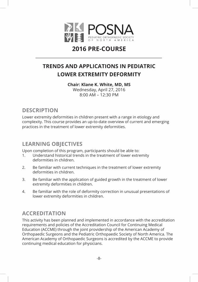
2016 PRE-COURSE
TRENDS AND APPLICATIONS IN PEDIATRIC LOWER EXTREMITY DEFORMITY
Chair: Klane K. White, MD, MS Wednesday, April 27, 2016
8:00 AM – 12:30 PM
DESCRIPTIONLower extremity deformities in children present with a range in etiology and complexity. This course provides an up-to-date overview of current and emerging practices in the treatment of lower extremity deformities.
LEARNING OBJECTIVESUpon completion of this program, participants should be able to:1. Understand historical trends in the treatment of lower extremity deformities in children. 2. Be familiar with current techniques in the treatment of lower extremity deformities in children. 3. Be familiar with the application of guided growth in the treatment of lower
extremity deformities in children. 4. Be familiar with the role of deformity correction in unusual presentations of
lower extremity deformities in children.
ACCREDITATIONThis activity has been planned and implemented in accordance with the accreditation requirements and policies of the Accreditation Council for Continuing Medical Education (ACCME) through the joint providership of the American Academy of Orthopaedic Surgeons and the Pediatric Orthopaedic Society of North America. The American Academy of Orthopaedic Surgeons is accredited by the ACCME to provide continuing medical education for physicians.
-9-
CONTINUING MEDICAL EDUCATIONThe American Academy of Orthopaedic Surgeons designates this live activity for a maximum of 4.25 AMA PRA Category 1 Credits™. Physicians should claim only the credit commensurate with the extent of their participation in the activity.
POSNA extends sincere appreciation to Medtronicfor their support of the Pre-Course program.
-10-
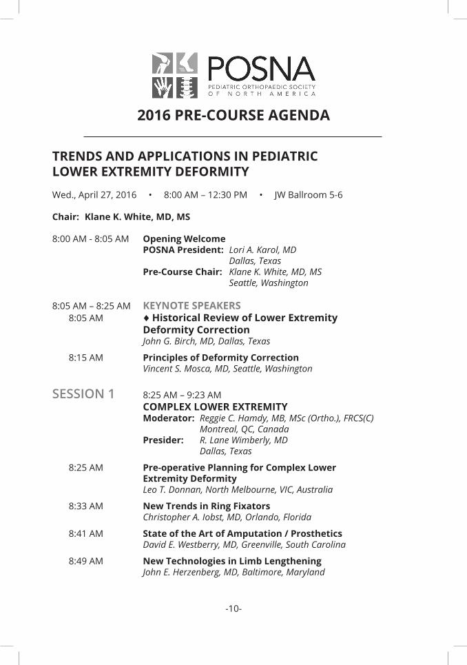
2016 PRE-COURSE AGENDA
TRENDS AND APPLICATIONS IN PEDIATRIC LOWER EXTREMITY DEFORMITY
Wed., April 27, 2016 • 8:00 AM – 12:30 PM • JW Ballroom 5-6
Chair: Klane K. White, MD, MS
8:00 AM - 8:05 AM Opening Welcome POSNA President: Lori A. Karol, MD Dallas, Texas Pre-Course Chair: Klane K. White, MD, MS Seattle, Washington
8:05 AM – 8:25 AM KEYNOTE SPEAKERS 8:05 AM ♦ Historical Review of Lower Extremity Deformity Correction John G. Birch, MD, Dallas, Texas
8:15 AM Principles of Deformity Correction Vincent S. Mosca, MD, Seattle, Washington
SESSION 1 8:25 AM – 9:23 AM COMPLEX LOWER EXTREMITY Moderator: Reggie C. Hamdy, MB, MSc (Ortho.), FRCS(C) Montreal, QC, Canada Presider: R. Lane Wimberly, MD Dallas, Texas
8:25 AM Pre-operative Planning for Complex Lower Extremity Deformity Leo T. Donnan, North Melbourne, VIC, Australia
8:33 AM New Trends in Ring Fixators Christopher A. Iobst, MD, Orlando, Florida
8:41 AM State of the Art of Amputation / Prosthetics David E. Westberry, MD, Greenville, South Carolina
8:49 AM New Technologies in Limb Lengthening John E. Herzenberg, MD, Baltimore, Maryland
-11-
TRENDS AND APPLICATIONS IN PEDIATRIC LOWER EXTREMITY DEFORMITY, (cont.) Wed., April 27, 2016
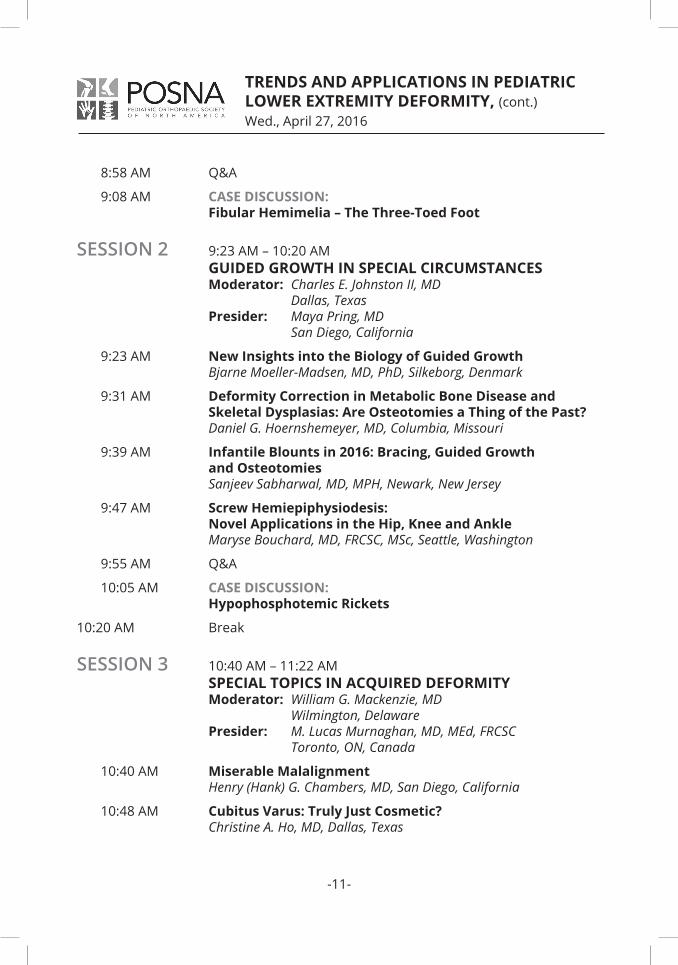
8:58 AM Q&A
9:08 AM CASE DISCUSSION: Fibular Hemimelia – The Three-Toed Foot
SESSION 2 9:23 AM – 10:20 AM GUIDED GROWTH IN SPECIAL CIRCUMSTANCES Moderator: Charles E. Johnston II, MD Dallas, Texas Presider: Maya Pring, MD San Diego, California
9:23 AM New Insights into the Biology of Guided Growth Bjarne Moeller-Madsen, MD, PhD, Silkeborg, Denmark
9:31 AM Deformity Correction in Metabolic Bone Disease and Skeletal Dysplasias: Are Osteotomies a Thing of the Past? Daniel G. Hoernshemeyer, MD, Columbia, Missouri
9:39 AM Infantile Blounts in 2016: Bracing, Guided Growth and Osteotomies Sanjeev Sabharwal, MD, MPH, Newark, New Jersey
9:47 AM Screw Hemiepiphysiodesis: Novel Applications in the Hip, Knee and Ankle Maryse Bouchard, MD, FRCSC, MSc, Seattle, Washington
9:55 AM Q&A
10:05 AM CASE DISCUSSION: Hypophosphotemic Rickets
10:20 AM Break
SESSION 3 10:40 AM – 11:22 AM SPECIAL TOPICS IN ACQUIRED DEFORMITY Moderator: William G. Mackenzie, MD Wilmington, Delaware Presider: M. Lucas Murnaghan, MD, MEd, FRCSC Toronto, ON, Canada
10:40 AM Miserable Malalignment Henry (Hank) G. Chambers, MD, San Diego, California
10:48 AM Cubitus Varus: Truly Just Cosmetic? Christine A. Ho, MD, Dallas, Texas
-12-
TRENDS AND APPLICATIONS IN PEDIATRIC LOWER EXTREMITY DEFORMITY, (cont.) Wed., April 27, 2016
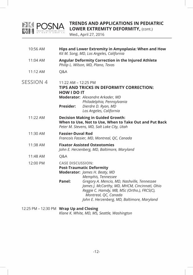
10:56 AM Hips and Lower Extremity in Amyoplasia: When and How Kit M. Song, MD, Los Angeles, California
11:04 AM Angular Deformity Correction in the Injured Athlete Philip L. Wilson, MD, Plano, Texas
11:12 AM Q&A
SESSION 4 11:22 AM – 12:25 PM TIPS AND TRICKS IN DEFORMITY CORRECTION: HOW I DO IT Moderator: Alexandre Arkader, MD Philadelphia, Pennsylvania Presider: Dierdre D. Ryan, MD Los Angeles, California
11:22 AM Decision Making in Guided Growth: When to Use, Not to Use, When to Take Out and Put Back Peter M. Stevens, MD, Salt Lake City, Utah
11:30 AM Fassier-Duval Rod Francois Fassier, MD, Montreal, QC, Canada
11:38 AM Fixator Assisted Osteotomies John E. Herzenberg, MD, Baltimore, Maryland
11:48 AM Q&A
12:00 PM CASE DISCUSSION: Post-Traumatic Deformity Moderator: James H. Beaty, MD Memphis, Tennessee Panel: Gregory A. Mencio, MD, Nashville, Tennessee James J. McCarthy, MD, MHCM, Cincinnati, Ohio Reggie C. Hamdy, MB, MSc (Ortho.), FRCS(C), Montreal, QC, Canada John E. Herzenberg, MD, Baltimore, Maryland
12:25 PM – 12:30 PM Wrap Up and Closing Klane K. White, MD, MS, Seattle, Washington
-13-
2016 ANNUAL MEETING
LEARNING OBJECTIVESUpon completion of this program, participants should be able to: Objective 1: Discuss and understand at least three new developments in pediatric orthopaedics. Objective 2: Implement at least two new techniques or practices in their care of patients. Objective 3: Understand the impact of advances in molecular science, genetics, and biomechanics on future pediatric orthopaedic practice. Objective 4: Implement a quality, safety, and value initiative to their practice.
ACCREDITATIONThis Annual Meeting of the Pediatric Orthopaedic Society of North America has been planned and implemented in accordance with the Essential Areas and Policies of the Accreditation Council for Continuing Medical Education (ACCME) through the joint providership of the American Academy of Orthopaedic Surgeons and POSNA. The American Academy of Orthopaedic Surgeons is accredited by the ACCME to provide continuing medical education for physicians.
CONTINUING MEDICAL EDUCATIONThe American Academy of Orthopaedic Surgeons designates this live activity for a maximum of 21 AMA PRA Category 1 Credits™. Physicians should claim only the credit commensurate with the extent of their participation in the activity. Scientific Program – 14.5 Subspecialty Day Program – 3.5 Symposia Program
• Research – 1.5 • COUR – 1.5 • Practice Management – 1.5 • POPS NP / PA – NA • Young Member Forum – 1.5
-14-
FDA STATEMENT (UNITED STATES)
Some drugs or medical devices demonstrated at the POSNA Pre-Course and Annual Meeting may not have been cleared by the FDA or have been cleared by the FDA for specific purposes only. The FDA has stated that it is the responsibility of the physician to determine the FDA clearance status of each drug or medical device he or she wishes to use in clinical practice. Academy policy provides that “off label” uses of a drug or medical device may be described in the Academy’s CME activities so long as the “off label” use of the drug or medical device is also specifically disclosed (i.e., it must be disclosed that the FDA has not cleared the drug or device for the described purpose). Any drug or medical device is being used “off label” if the described use is not set forth on the product’s approval label. ♦ Indicates those faculty presentations in which the FDA has not cleared the drug
and/or medical device for the use described (ie., the drug or medical device is being discussed for an “off label” use).
DISCLAIMERThe material presented at the POSNA Pre-Course and Annual Meeting has been made available by the Pediatric Orthopaedic Society of North America for educational purposes only. The material is not intended to represent the only, nor necessarily best, method or procedure appropriate for the medical situations discussed, but rather is intended to present an approach, view, statement or opinion of the faculty which may be helpful to others who face similar situations. POSNA disclaims any and all liability for injury or other damages resulting to any individual attending the Annual Meeting and for all claims which may arise out of the use of the techniques demonstrated therein by such individuals, whether these claims shall be asserted by physician or any other person.
GENERAL MEETING INFORMATION
-15-
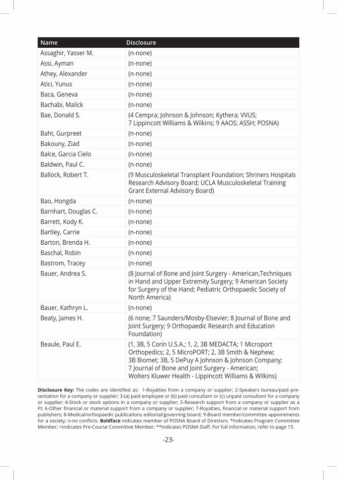
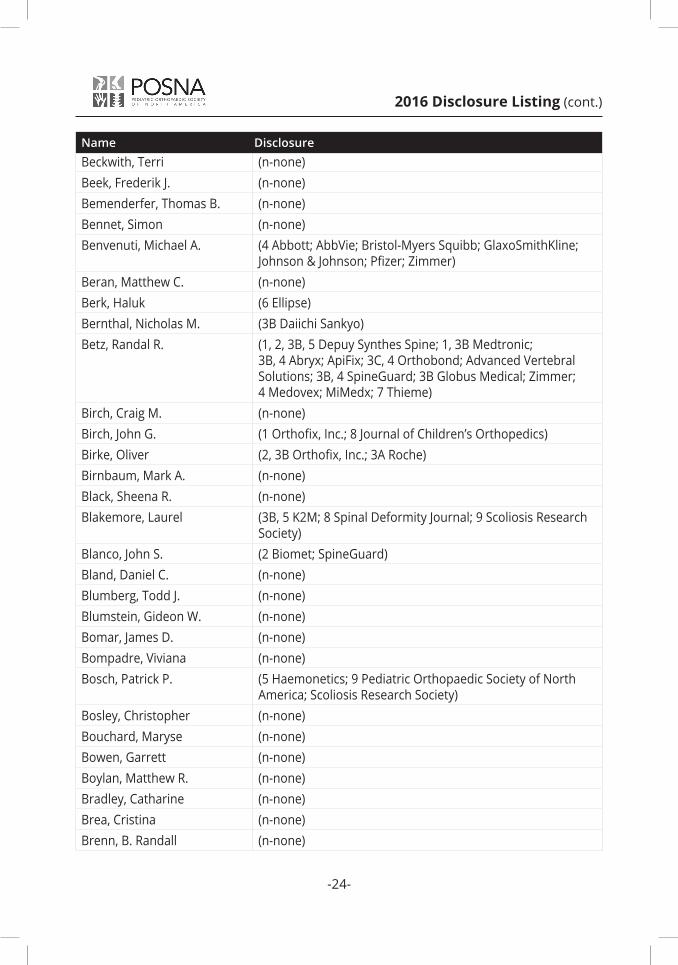
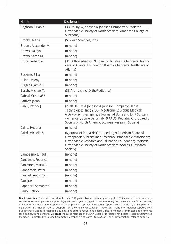
DISCLOSUREEach faculty member at the POSNA Pre-Course and Annual Meeting has been asked to disclose if he or she has received something of value from a commercial company or institution, which relates directly or indirectly to the subject of their presentation. The Academy has identified the options to disclose as follows:
n = Nothing to disclose; 1 = Royalties from a company or supplier; 2 = Speakers bureau/paid presentations for a company or supplier; 3A = Paid employee for a company or supplier; 3B = Paid consultant for a company or supplier;3C = Unpaid consultant for a company or supplier;4 = Stock or stock options in a company or supplier; 5 = Research support from a company or supplier as a PI; 6 = Other financial or material support from a company or supplier; 7 = Royalties financial or material support from publishers; 8 = Medical/Orthopaedic publications editorial/governing board;9 = Board member/committee appointments for a society;* = Program Committee Member; + = Pre-Course Committee Member; ** = POSNA Staff
LANGUAGEEnglish will be the official language of the POSNA Pre-Course and Annual Meeting.
POSNA ANTITRUST POLICYDiscussions at the POSNA Pre-Course and Annual Meeting often cover a broad range of topics pertinent to the interests or concerns of orthopaedic surgeons. As a general rule, except as noted below, discussions at POSNA meetings can address topics with-out raising antitrust concerns if the discussions are kept scrupulously free of even the suggestion of private regulation of the profession. However, a number of topics that might be (and have been) discussed at POSNA meetings may raise significant complex antitrust concerns. These include: • Membership admissions, rejections, restrictions, and terminations; • Method of provision and sale of POSNA products and services to non-members; • Restrictions in the selection and requirements for exhibitors at the POSNA Annual Meeting or in CME activities; • Collecting and distributing certain orthopaedic practice information, particularly involving practice charges and costs; • Obtaining and distributing orthopaedic industry price and cost information; • Professional certification programs; • Group buying and selling; and • Inclusions or exclusion of other medical societies in organizational activities or offerings.
-16-
When these and related topics are discussed, the convener or members of the POSNA group should seek counsel from its General Counsel.
POSNA urges its Board, committees and other groups not to participate in discussions that may give the appearance of or constitute an agreement that would violate the antitrust laws. Notwithstanding this reliance, it is the responsibility of each POSNA Board or committee member to avoid raising improper subjects for discussion. This policy has been prepared to ensure that POSNA members and other participants in POSNA meetings are aware of this obligation.
The “Do Nots” and “Dos” presented below highlight only the most basic antitrust principles. POSNA members and others participating in POSNA meetings should con-sult with the General Counsel in all cases involving specific questions, interpretations or advice regarding antitrust matters.
Do Not1. Do not, in fact or appearance, discuss or exchange information regarding: a. Individual company prices, price changes, price differentials, mark-ups, discounts, allowances, credit terms, etc. or any other data that may bear on price, such as costs, production, capacity, inventories, sales, etc. b. Raising, lowering or “stabilizing” orthopaedic prices or fees; c. What constitutes a fair profit or margin level; d. The availability of products or services; or e. The allocation of markets, territories or patients.
2. Do not suggest or imply that POSNA members should or should not deal with certain other persons or companies.
3. Do not foster unfair practices regarding advertising, standardization, certification or accreditation.
4. Do not discuss or exchange information regarding the above matters during social gatherings, incidental to POSNA-sponsored meetings.
5. Do not make oral or written statements on important issues on behalf of POSNA without appropriate authority to do so.
Do1. Do adhere to prepared agenda for all POSNA meetings. It is generally permissible for agendas to include discussions of such varied topics as professional economic trends, advances and problems in relevant technology or research, various aspects of the science and art of management, and relationships with local, state or federal governments.
2. Do object whenever meeting summaries do not accurately reflect the matters that occurred.
3. Do consult with General Counsel on all antitrust questions relating to discussions at POSNA meetings.
4. Do object to and do not participate in any discussions or meeting activities that you believe violate the antitrust laws; dissociate yourself from any such discussions or activities and leave any meeting in which they continue.
-17-
SPECIAL GUIDELINES FOR COLLECTING AND DISTRIBUTING INFORMATION The collection and distribution of information regarding business practices is a traditional function of associations and is well-recognized under the law as appropriate, legal and consistent with the antitrust laws. However, if conducted improperly, such information gathering and distributing activities might be viewed as facilitating an express or implied agreement among association members to adhere to the same business practices. For this reason, special general guidelines have developed over time regarding association’s reporting on information collected from and disseminated to members. Any exceptions to these general guidelines should be made only after discussion with General Counsel. These general guidelines include: 1. Member participation in a statistical reporting program is voluntary. A statistical reporting program should be conducted without coercion or penalty. Non-members should be allowed to participate in a statistical reporting program if eligible; however, if a fee is involved, non-members may be charged
a reasonably higher fee than members. 2. Information should be collected via a written instrument that clearly sets forth
what is being requested.
3. The data that is collected should be about past transactions or activities; particularly if the survey deals with prices and price terms (including charges, costs, wages, benefits, discounts, etc.), it should be historic, i.e., more than three months old.
4. The data should be collected by either POSNA or an independent third party
not connected with any one member. 5. Data on individual orthopaedic surgeons should be kept confidential. 6. There should be a sufficient number of participants to prevent specific responses or data from being attributable to any one respondent. As a general
rule, there should be at least five respondents reporting data upon which any statistic or item is based, and no individual’s data should represent more than 25% on a weighted average of that statistic or item.
7. Composite/aggregate data should be available to all participants – both members and non-members. The data may be categorized, e.g., geographically, and ranges and averages may be used. No member should be given access to
the raw data. Disclosure of individual data could serve to promote uniformity and reduce competition.
8. As a general rule, there should be no discussion or agreement as to how members and non-members should adjust, plan or carry out their practices
based on the results of the survey. Each member should analyze the data and make business decisions independently.
-18-
NO SMOKING POLICYSmoking is not permitted during any meeting or event.
NO CAMERAS OR VIDEO CAMERASCameras or video cameras may not be used in any portion of the scientific session.
NO REPRODUCTIONSNo reproductions of any kind including audio tapes and videotapes may be made of the presentations at this meeting without the prior written permission of POSNA. POSNA reserves all of its rights to such material and commercial reproduction is specifically prohibited.
PHOTOGRAPHSRegistration and attendance at, or participation in, POSNA activities constitutes an agreement by the registrant to allow POSNA to use and distribute (both now and in the future) the registrant’s or attendee’s image in POSNA member communications and promotional materials.
-19-
2016 OPENING CEREMONY
WEDNESDAY, APRIL 27, 2016
6:30 PM - 9:30 PM • JW Marriott • JW Ballroom 5-6
6:30 PM – 6:40 PM WELCOME POSNA President: Lori A. Karol, MD Local Host: Randall T. Loder, MD
6:40 PM – 6:50 PM Introductions of Distinguished Guests • International Presidents • New Members • Distinguished Achievement Award Recipient • Presidential Guest Speaker • SLAOTI Traveling Fellows • APPOS Traveling Fellows • COUR Visiting Scholars
6:50 PM – 6:55 PM Presentation of the St. Giles Young Investigator Award Donald R. Huene, MD and Richard T. Arkwright, MD
6:55 PM – 7:00 PM Presentation of the Arthur H. Huene Award Donald R. Huene, MD and Richard T. Arkwright, MD
7:00 PM - 7:05 PM Presentation of the Angela S.M. Kuo Memorial Award Ken N. Kuo, MD
7:05 PM -7:10 PM Presentation of the Humanitarian Award Lori A. Karol, MD
7:10 PM -7:15 PM Presentation of the Special Effort and Excellence Award Lori A. Karol, MD
7:15 PM – 7:30 PM Recognition of Industry Sponsors Lori A. Karol, MD
7:30 PM Introduction of Steel Lecturer Lori A. Karol, MD
7:35 PM – 8:00 PM Steel Lecture Mr. Donald Davidson-Historian, Indianapolis Motor Speedway Hall of Fame Museum “A Brief History of the Indianapolis Motor Speedway”
8:00 PM – 9:30 PM Welcome Reception
POSNA extends sincere appreciation to Medtronicfor their support of the welcome reception.
-20-
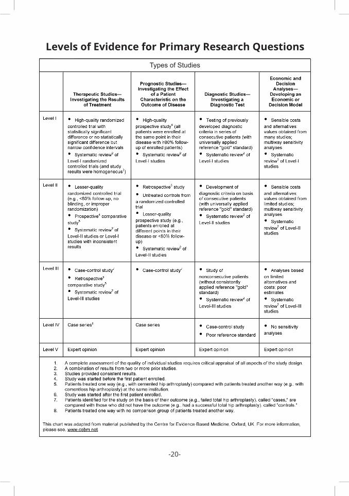
Levels of Evidence for Primary Research Questions
-12-
Types of Studies
Levels of Evidence for Primary Research Questions
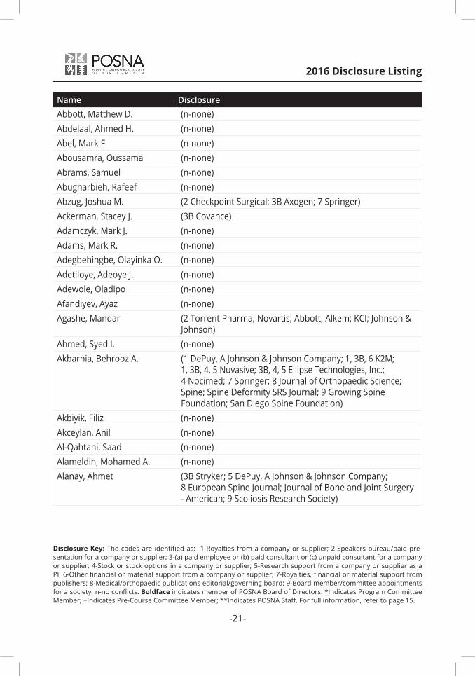
Abbott, Matthew D. (n-none)Abdelaal, Ahmed H. (n-none)Abel, Mark F (n-none)Abousamra, Oussama (n-none)Abrams, Samuel (n-none)Abugharbieh, Rafeef (n-none)Abzug, Joshua M. (2 Checkpoint Surgical; 3B Axogen; 7 Springer)Ackerman, Stacey J. (3B Covance)Adamczyk, Mark J. (n-none)Adams, Mark R. (n-none)Adegbehingbe, Olayinka O. (n-none)Adetiloye, Adeoye J. (n-none)Adewole, Oladipo (n-none)Afandiyev, Ayaz (n-none)Agashe, Mandar (2 Torrent Pharma; Novartis; Abbott; Alkem; KCI; Johnson &
Johnson)Ahmed, Syed I. (n-none)Akbarnia, Behrooz A. (1 DePuy, A Johnson & Johnson Company; 1, 3B, 6 K2M;
1, 3B, 4, 5 Nuvasive; 3B, 4, 5 Ellipse Technologies, Inc.; 4 Nocimed; 7 Springer; 8 Journal of Orthopaedic Science; Spine; Spine Deformity SRS Journal; 9 Growing Spine Foundation; San Diego Spine Foundation)
Akbiyik, Filiz (n-none)Akceylan, Anil (n-none)Al-Qahtani, Saad (n-none)Alameldin, Mohamed A. (n-none)Alanay, Ahmet (3B Stryker; 5 DePuy, A Johnson & Johnson Company;
8 European Spine Journal; Journal of Bone and Joint Surgery - American; 9 Scoliosis Research Society)
-21-
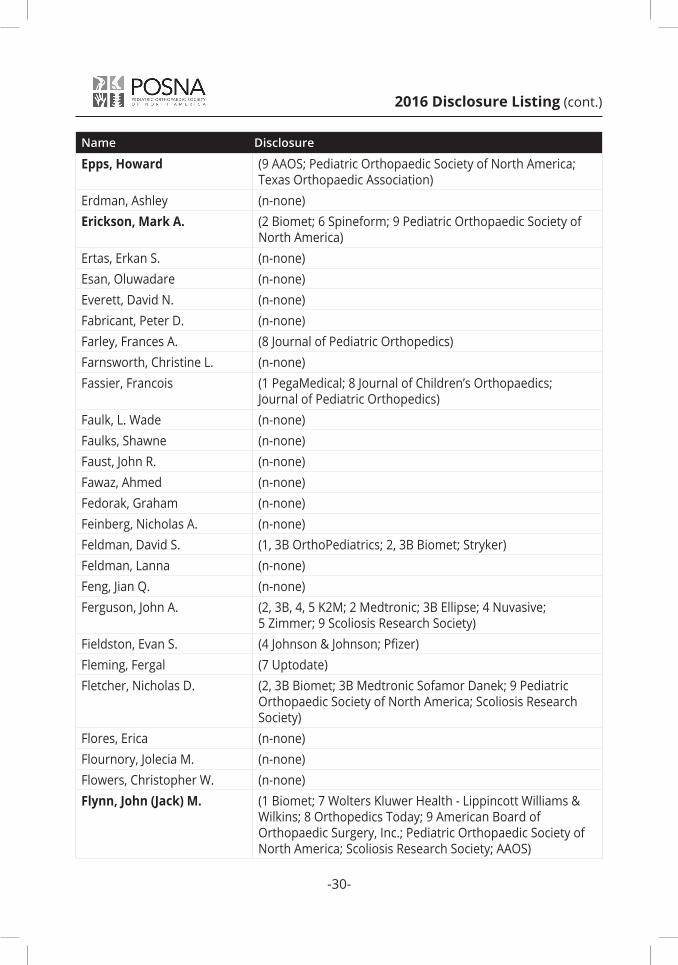
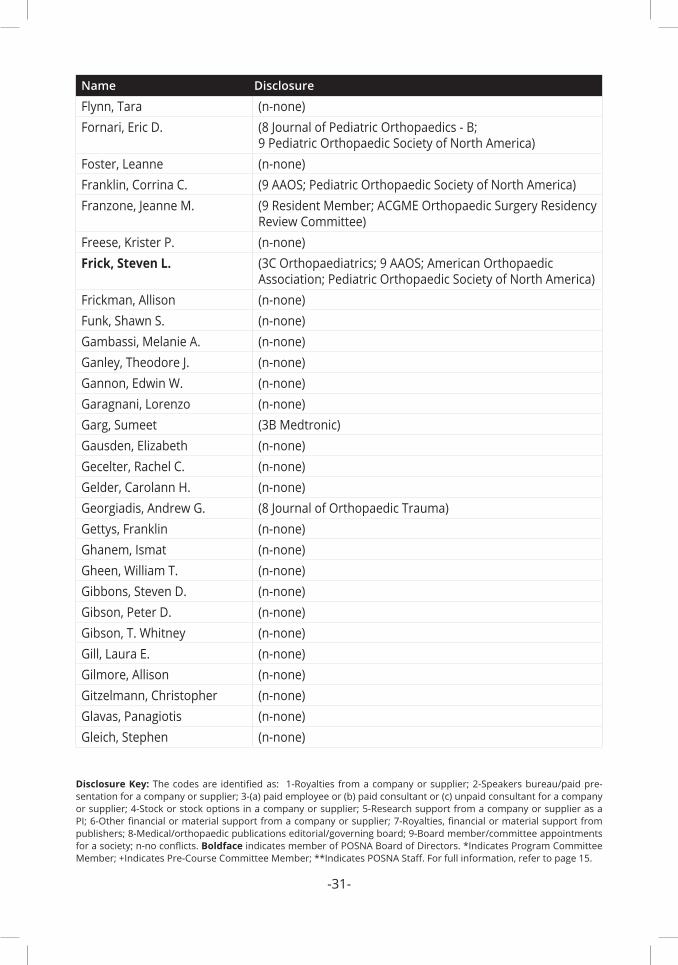
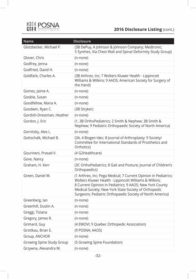
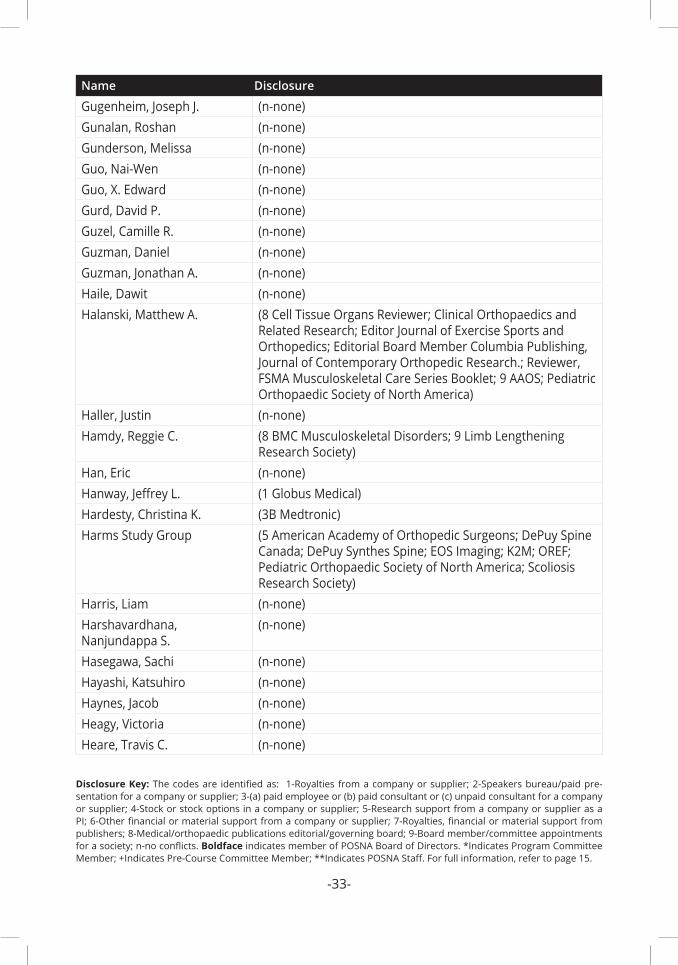
Disclosure Key: The codes are identified as: 1-Royalties from a company or supplier; 2-Speakers bureau/paid pre-sentation for a company or supplier; 3-(a) paid employee or (b) paid consultant or (c) unpaid consultant for a company or supplier; 4-Stock or stock options in a company or supplier; 5-Research support from a company or supplier as a PI; 6-Other financial or material support from a company or supplier; 7-Royalties, financial or material support from publishers; 8-Medical/orthopaedic publications editorial/governing board; 9-Board member/committee appointments for a society; n-no conflicts. Boldface indicates member of POSNA Board of Directors. *Indicates Program Committee Member; +Indicates Pre-Course Committee Member; **Indicates POSNA Staff. For full information, refer to page 15.
2016 Disclosure Listing
Name Disclosure

2016 Disclosure Listing (cont.)
-22-
Albanese, Stephen (8 AAOS Now; Spine Deformity Journal 9 Accreditation Council for Graduate Medical Education; American Board of Orthopaedic Surgery, Inc.; Pediatric Orthopaedic Society of North America)
Albright, Jay C. (2 Arthrex, Inc; DJ Orthopaedics; 3B DJ Orthopaedics; Orthopaedic Scientific Research Foundation; 9 Pediatric Orthopaedic Society of North America; PRISM)
Alman, Benjamin (4 ScarX; 9 Shrine Research Advisory Board)Alvarez, Christine (n-none)Alzahrani, Mohammad M. (n-none)Amaral, Terry D. (9 Pediatric Orthopaedic Society of North America;
Scoliosis Research Society)Amoli, Marielle A. (n-none)Amundson, Laura A. (n-none)An, Thomas J. (4, 5 Merck; 5 Johnson & Johnson)An, Tonya W. (n-none)Anadio, Jennifer M. (n-none)Anderson, Allen F (1 OrthoPediatrics; 2 ETO; 3B Aquire B2B; Mitek;
OrthoPediatrics; 8 AM J Sports Med , Orthopedic Journal Sports Med; 9 American Orthopaedic Society for Sports Medicine; Herodicus Society; International Society of Arthroscopy, Knee Surgery, and Orthopaedic Sports Medicine)
Anderson, David J. (2B Medtronic)Anderson, Ryan L. (n-none)Andras, Lindsay M. (2 Biomet; Medtronic; 4 Eli Lilly; 7 Orthobullets; 9 Pediatric
Orthopaedic Society of North America; Scoliosis Research Society)
Anticevic, Darko (8 Wolters Kluwer Health - Lippincott Williams & Wilkins; 9 European Pediatric Orthopaedic Society (EPOS); Interna-tional Federation of Paediatric Orthopaedic Societies (IFPOS))
Aquina, Christopher T. (n-none)Arana, Erika (n-none)Arbucci, John A. (n-none)Arkader, Alexandre (3C Orthopediatrics SAB)Asghar, Jahangir (9 Scoliosis Research Society)Aslan, Cihan (n-none)
Name Disclosure
-23-
Disclosure Key: The codes are identified as: 1-Royalties from a company or supplier; 2-Speakers bureau/paid pre-sentation for a company or supplier; 3-(a) paid employee or (b) paid consultant or (c) unpaid consultant for a company or supplier; 4-Stock or stock options in a company or supplier; 5-Research support from a company or supplier as a PI; 6-Other financial or material support from a company or supplier; 7-Royalties, financial or material support from publishers; 8-Medical/orthopaedic publications editorial/governing board; 9-Board member/committee appointments for a society; n-no conflicts. Boldface indicates member of POSNA Board of Directors. *Indicates Program Committee Member; +Indicates Pre-Course Committee Member; **Indicates POSNA Staff. For full information, refer to page 15.
Assaghir, Yasser M. (n-none)Assi, Ayman (n-none)Athey, Alexander (n-none)Atici, Yunus (n-none)Baca, Geneva (n-none)Bachabi, Malick (n-none)Bae, Donald S. (4 Cempra; Johnson & Johnson; Kythera; VVUS;
7 Lippincott Williams & Wilkins; 9 AAOS; ASSH; POSNA)Baht, Gurpreet (n-none)Bakouny, Ziad (n-none)Balce, Garcia Cielo (n-none)Baldwin, Paul C. (n-none)Ballock, Robert T. (9 Musculoskeletal Transplant Foundation; Shriners Hospitals
Research Advisory Board; UCLA Musculoskeletal Training Grant External Advisory Board)
Bao, Hongda (n-none)Barnhart, Douglas C. (n-none)Barrett, Kody K. (n-none)Bartley, Carrie (n-none)Barton, Brenda H. (n-none)Baschal, Robin (n-none)Bastrom, Tracey (n-none)Bauer, Andrea S. (8 Journal of Bone and Joint Surgery - American,Techniques
in Hand and Upper Extremity Surgery; 9 American Society for Surgery of the Hand; Pediatric Orthopaedic Society of North America)
Bauer, Kathryn L. (n-none)Beaty, James H. (6 none; 7 Saunders/Mosby-Elsevier; 8 Journal of Bone and
Joint Surgery; 9 Orthopaedic Research and Education Foundation)
Beaule, Paul E. (1, 3B, 5 Corin U.S.A.; 1, 2, 3B MEDACTA; 1 Microport Orthopedics; 2, 5 MicroPORT; 2, 3B Smith & Nephew; 3B Biomet; 3B, 5 DePuy A Johnson & Johnson Company; 7 Journal of Bone and Joint Surgery - American; Wolters Kluwer Health - Lippincott Williams & Wilkins)
Name Disclosure
2016 Disclosure Listing (cont.)
-24-
Beckwith, Terri (n-none)Beek, Frederik J. (n-none)Bemenderfer, Thomas B. (n-none)Bennet, Simon (n-none)Benvenuti, Michael A. (4 Abbott; AbbVie; Bristol-Myers Squibb; GlaxoSmithKline;
Johnson & Johnson; Pfizer; Zimmer)Beran, Matthew C. (n-none)Berk, Haluk (6 Ellipse)Bernthal, Nicholas M. (3B Daiichi Sankyo)Betz, Randal R. (1, 2, 3B, 5 Depuy Synthes Spine; 1, 3B Medtronic;
3B, 4 Abryx; ApiFix; 3C, 4 Orthobond; Advanced Vertebral Solutions; 3B, 4 SpineGuard; 3B Globus Medical; Zimmer; 4 Medovex; MiMedx; 7 Thieme)
Birch, Craig M. (n-none)Birch, John G. (1 Orthofix, Inc.; 8 Journal of Children’s Orthopedics)Birke, Oliver (2, 3B Orthofix, Inc.; 3A Roche)Birnbaum, Mark A. (n-none)Black, Sheena R. (n-none)Blakemore, Laurel (3B, 5 K2M; 8 Spinal Deformity Journal; 9 Scoliosis Research
Society)Blanco, John S. (2 Biomet; SpineGuard)Bland, Daniel C. (n-none)Blumberg, Todd J. (n-none)Blumstein, Gideon W. (n-none)Bomar, James D. (n-none)Bompadre, Viviana (n-none)Bosch, Patrick P. (5 Haemonetics; 9 Pediatric Orthopaedic Society of North
America; Scoliosis Research Society)Bosley, Christopher (n-none)Bouchard, Maryse (n-none)Bowen, Garrett (n-none)Boylan, Matthew R. (n-none)Bradley, Catharine (n-none)Brea, Cristina (n-none)Brenn, B. Randall (n-none)
Name Disclosure
-25-
Disclosure Key: The codes are identified as: 1-Royalties from a company or supplier; 2-Speakers bureau/paid pre-sentation for a company or supplier; 3-(a) paid employee or (b) paid consultant or (c) unpaid consultant for a company or supplier; 4-Stock or stock options in a company or supplier; 5-Research support from a company or supplier as a PI; 6-Other financial or material support from a company or supplier; 7-Royalties, financial or material support from publishers; 8-Medical/orthopaedic publications editorial/governing board; 9-Board member/committee appointments for a society; n-no conflicts. Boldface indicates member of POSNA Board of Directors. *Indicates Program Committee Member; +Indicates Pre-Course Committee Member; **Indicates POSNA Staff. For full information, refer to page 15.
Brighton, Brian K. (3B DePuy, A Johnson & Johnson Company; 9 Pediatric Orthopaedic Society of North America; American College of Surgeons)
Brooks, Maria (5 Gilead Sciences, Inc.)Broom, Alexander M. (n-none)Brown, Kaitlyn (n-none)Brown, Sarah M. (n-none)Bruce, Robert W. (3C OrthoPediatrics; 9 Board of Trustees - Children’s Health-
care of Atlanta, Foundation Board - Children’s Healthcare of Atlanta)
Buckner, Elisa (n-none)Bulat, Evgeny (n-none)Burgess, Jamie K. (n-none)Busch, Michael T. (3B Arthrex, Inc; OrthoPediatrics)Cabral, Cristina** (n-none)Caffrey, Jason (n-none)Cahill, Patrick J. (2, 3B DePuy, A Johnson & Johnson Company; Ellipse
Technologies, Inc.; 2, 3B, Medtronic; 2 Globus Medical; 6 DePuy Synthes Spine; 8 Journal of Bone and Joint Surgery - American; Spine Deformity; 9 AAOS; Pediatric Orthopaedic Society of North America; Scoliosis Research Society)
Caine, Heather (n-none)Caird, Michelle S. (8 Journal of Pediatric Orthopedics; 9 American Board of
Orthopaedic Surgery, Inc.; American Orthopaedic Association; Orthopaedic Research and Education Foundation; Pediatric Orthopaedic Society of North America; Scoliosis Research Society)
Campagnola, Paul J. (n-none)Canavese, Federico (n-none)Canizares, Maria F. (n-none)Cannamela, Peter (n-none)Cantrell, Anthony C. (n-none)Cao, Jue (n-none)Capehart, Samantha (n-none)Carry, Patrick (n-none)
Name Disclosure
2016 Disclosure Listing (cont.)
-26-
Carsen, Sasha (n-none)Carter, Cordelia W. (9 AAOS; Pediatric Orthopaedic Society of North America)Carter, Erin M. (9 Little People of America, Inc.)Casey, Virginia F. (n-none)Castelein, Rene M. (5 Medtronic)Chambers, Henry (Hank) G. (3B Orthopediatrics; 5 Allergan Corporation; 8 Developmental
Medicine and Child Neurology; 9 American Academy for Cerebral Palsy and Developmental Medicine; Pediatric Research in Sports Medicine PRISM)
Chau, Wai Wang (n-none)Chaudhary, Rajeev (n-none)Chen, Cynthia (n-none)Chen, Huanxiong (n-none)Cheng, Jack C.Y. (8 Journal of Pediatric Orthopedics)Cheng, Tegan L. (n-none)Cherkasskiy, Lillia (n-none)Chettier, Rakesh (n-none)Cheung, Kenneth M.C. (3B, 5 Ellipse Technologies; 8 Journal of Orthopaedic Surgery;
Spine Deformity; 9 Hong Kong College of Orthopaedic Surgeons; Scoliosis Research Society)
Cheverda, Andrii (n-none)Children’s Spine Study Group (5 DePuy, A Johnson & Johnson Company)Cho, Robert H. (3B DePuy Spine; Medtronic Sofamor Danek; OrthoPediatrics;
8 Orthopedics)Choi, Edmund (n-none)Choi, Paul D. (2, 3B Stryker; 3B Integra)Choudhry, Dinesh K. (n-none)Christino, Melissa A. (n-none)Chu, Alice (n-none)Chu, Winnie C. (n-none)Chukwunyerenwa, Chukwudi K.
(n-none)
Church, Chris (n-none)Citron, Kate P. (n-none)Clark, Christian (n-none)Clarke, Nicholas (9 International Hip Dysplasia Institute)
Name Disclosure
-27-
Disclosure Key: The codes are identified as: 1-Royalties from a company or supplier; 2-Speakers bureau/paid pre-sentation for a company or supplier; 3-(a) paid employee or (b) paid consultant or (c) unpaid consultant for a company or supplier; 4-Stock or stock options in a company or supplier; 5-Research support from a company or supplier as a PI; 6-Other financial or material support from a company or supplier; 7-Royalties, financial or material support from publishers; 8-Medical/orthopaedic publications editorial/governing board; 9-Board member/committee appointments for a society; n-no conflicts. Boldface indicates member of POSNA Board of Directors. *Indicates Program Committee Member; +Indicates Pre-Course Committee Member; **Indicates POSNA Staff. For full information, refer to page 15.
Clarke, Zachary (n-none)Clements, David H. (2 Synthes; 2, 3B, 5 DePuy, A Johnson & Johnson Company;
8 First Consult; 9 Scoliosis Research Society)Clohisy, John C. (3B Microport Orthopedics, Inc.; 3B, 5 Smith & Nephew;
5 Pivot Medical; Zimmer; 7 Wolters Kluwer Health - Lippincott Williams & Wilkins)
Cole, Heather (n-none)Connor, Justin R. (n-none)Conrad, Ernest U. (3C Stryker; 6 LifeNet Health Northwest Tissue Division)Coobs, Benjamin R. (n-none)Cook, Katherine (n-none)Cook, Thomas (n-none)Cooper, Anthony (9 Canadian Orthopaedic Association)Cooperman, Daniel R. (n-none)Copley, Lawson A.B. (3C Epic; 7 Saunders/Mosby-Elsevier; 9 Pediatric Orthopaedic
Society of North America)Cordasco, Frank A. (1 CONMED Linvatec; 1, 3B Arthrex, Inc; 7 Wolters Kluwer
Health - Lippincott Williams & Wilkins; 8 Journal of Shoulder and Elbow Surgery; 9 American Orthopaedic Society for Sports Medicine; American Shoulder and Elbow Surgeons; Arthroscopy Association of North America)
Cornwall, Roger (n-none)Cory, Esther (3A Prometheus Labs; 3A, 4 Otonomy)Cowl, Clayton (n-none)Craig, Clifford L. (4 Johnson & Johnson; 8 Journal of Orthopedic History)Creek, Aaron T. (n-none)Crenshaw, Thomas D. (n-none)Cuomo, Anna V. (9 Pediatric Orthopaedic Society of North America)Cyr, Micaela (n-none)D’Astous, Jacques L. (n-none)Dabney, Kirk W. (3B DePuy, A Johnson & Johnson Company; Medtronic)Dardashti, Navid (n-none)Davis, Roy B. (n-none)Dayanidhi, Sudarshan (n-none)
Name Disclosure
2016 Disclosure Listing (cont.)
-28-
De La Rocha, Adriana (n-none)De La Roza, Kevin (n-none)De Mendonça, Rodrigo G. (5 Medicrea)Dede, Ozgur (n-none)Delfosse, Erin M. (n-none)Demirkiran, H. Gokhan (n-none)Dempsey, Molly (9 Society for Pediatric Radiology )Demyan, Yuri (n-none)Denning, Jaime R. (n-none)Deo, Nikita (n-none)Desai, Sameer (n-none)Deshpande, Rasika (n-none)Dias, Luciano (n-none)DiFazio, Rachel L. (n-none)Ding, Qian (n-none)Ditro, Colleen P. (n-none)Do Monte, Felipe A. (n-none)Dobbe, Ashlee (n-none)Dobbs, Matthew B. (1, 3B D-Bar Enterprises; 8 Clinical Orthopaedics and Related
Research; 9 Association of Bone and Joint Surgeons; Orthopaedic Research and Education Foundation)
Döderlein, Leonhard (n-none)Dodwell, Emily (n-none)Dolan, Lori (9 Pediatric Orthopaedic Society of North America;
Scoliosis Research Society)Donnan, Alice (n-none)Donnan, Leo T. (n-none)Donohue, Kenneth W. (n-none)Doyle, Shevaun M. (n-none)Dreher, Thomas (n-none)Dua, Karan (n-none)Dudevoir, Michelle L. (n-none)Duhaime, Morris A. (n-none)
Name Disclosure
-29-
Disclosure Key: The codes are identified as: 1-Royalties from a company or supplier; 2-Speakers bureau/paid pre-sentation for a company or supplier; 3-(a) paid employee or (b) paid consultant or (c) unpaid consultant for a company or supplier; 4-Stock or stock options in a company or supplier; 5-Research support from a company or supplier as a PI; 6-Other financial or material support from a company or supplier; 7-Royalties, financial or material support from publishers; 8-Medical/orthopaedic publications editorial/governing board; 9-Board member/committee appointments for a society; n-no conflicts. Boldface indicates member of POSNA Board of Directors. *Indicates Program Committee Member; +Indicates Pre-Course Committee Member; **Indicates POSNA Staff. For full information, refer to page 15.
Duncan, Stephen T. (3B Biomet; Mitek; Smith & Nephew; 3C Morph; 8 Journal of Arthroplasty; 9 American Association of Hip and Knee Surgeons)
Dunn, Samuel H. (n-none)Duque Orozco, Maria Del Pilar
(n-none)
Dwek, Jerry R. (n-none)Dworkin, Aviva (n-none)Eastlack, Robert K. (1 Globus Medical; 2 Eli Lilly; 3B Aesculap/B.Braun; Alphatec
Spine; DePuy, A Johnson & Johnson Company; DiFusion; DJ Orthopaedics; Integra; Invuity; K2M; Seaspine; Stryker; Titan; Ulrich; 4 Alphatec Spine; Carevature; DiFusion; Invuity; Spine Innovations; 3B, 4, 5 Nuvasive; 9 Scoliosis Research Society; Society of Lateral Access Surgery)
Eberson, Craig P. (1 Globus Medical; 2 Stryker Spineorthofix; 3B Orthofix, Inc.; 8 Journal of the American Academy of Orthopaedic Surgeons; 9 Pediatric Orthopaedic Society of North America; Scoliosis Research Society)
Ebramzadeh, Edward (3B Corin U.S.A.; 5 Arthrex, Inc; Zimmer; Biomet; I-Spine; Tri-Med; Amgen Co; Extremity Medical; AOS; Synthes; 8 Journal of Bone and Joint Surgery - American; Journal of Applied Biomaterials and Functional Materials; Journal of Orthopaedic Trauma)
Edmonds, Eric W. (9 American Orthopaedic Society for Sports Medicine; Pediatric Orthopaedic Society of North America)
El-Hawary, Ron (3B, 5 DePuy, A Johnson & Johnson Company; Medtronic 3B Halifax Biomedical Inc.; 9 Chest Wall and Spine Deformity Foundation; Pediatric Orthopaedic Society of North America; Scoliosis Research Society)
Eliceiri, Kevin (n-none)Elliott, Marilyin (n-none)Ellis, Henry B. (3B Smith & Nephew)Elsebaie, Hazem B. (4 Ellipse, K Spine)Elward, Alexis (n-none)Emans, John B. (1, 3B, 3C Synthes; 3B, 3C Medtronic Sofamor Danek;
8 Journal of Children’s Orthopaedics)Encisa, Clarissa (n-none)
Name Disclosure
2016 Disclosure Listing (cont.)
-30-
Epps, Howard (9 AAOS; Pediatric Orthopaedic Society of North America; Texas Orthopaedic Association)
Erdman, Ashley (n-none)Erickson, Mark A. (2 Biomet; 6 Spineform; 9 Pediatric Orthopaedic Society of
North America)Ertas, Erkan S. (n-none)Esan, Oluwadare (n-none)Everett, David N. (n-none)Fabricant, Peter D. (n-none)Farley, Frances A. (8 Journal of Pediatric Orthopedics)Farnsworth, Christine L. (n-none)Fassier, Francois (1 PegaMedical; 8 Journal of Children’s Orthopaedics;
Journal of Pediatric Orthopedics)Faulk, L. Wade (n-none)Faulks, Shawne (n-none)Faust, John R. (n-none)Fawaz, Ahmed (n-none)Fedorak, Graham (n-none)Feinberg, Nicholas A. (n-none)Feldman, David S. (1, 3B OrthoPediatrics; 2, 3B Biomet; Stryker)Feldman, Lanna (n-none)Feng, Jian Q. (n-none)Ferguson, John A. (2, 3B, 4, 5 K2M; 2 Medtronic; 3B Ellipse; 4 Nuvasive;
5 Zimmer; 9 Scoliosis Research Society)Fieldston, Evan S. (4 Johnson & Johnson; Pfizer)Fleming, Fergal (7 Uptodate)Fletcher, Nicholas D. (2, 3B Biomet; 3B Medtronic Sofamor Danek; 9 Pediatric
Orthopaedic Society of North America; Scoliosis Research Society)
Flores, Erica (n-none)Flournory, Jolecia M. (n-none)Flowers, Christopher W. (n-none)Flynn, John (Jack) M. (1 Biomet; 7 Wolters Kluwer Health - Lippincott Williams &
Wilkins; 8 Orthopedics Today; 9 American Board of Orthopaedic Surgery, Inc.; Pediatric Orthopaedic Society of North America; Scoliosis Research Society; AAOS)
Name Disclosure
-31-
Disclosure Key: The codes are identified as: 1-Royalties from a company or supplier; 2-Speakers bureau/paid pre-sentation for a company or supplier; 3-(a) paid employee or (b) paid consultant or (c) unpaid consultant for a company or supplier; 4-Stock or stock options in a company or supplier; 5-Research support from a company or supplier as a PI; 6-Other financial or material support from a company or supplier; 7-Royalties, financial or material support from publishers; 8-Medical/orthopaedic publications editorial/governing board; 9-Board member/committee appointments for a society; n-no conflicts. Boldface indicates member of POSNA Board of Directors. *Indicates Program Committee Member; +Indicates Pre-Course Committee Member; **Indicates POSNA Staff. For full information, refer to page 15.
Flynn, Tara (n-none)Fornari, Eric D. (8 Journal of Pediatric Orthopaedics - B;
9 Pediatric Orthopaedic Society of North America)Foster, Leanne (n-none)Franklin, Corrina C. (9 AAOS; Pediatric Orthopaedic Society of North America)Franzone, Jeanne M. (9 Resident Member; ACGME Orthopaedic Surgery Residency
Review Committee)Freese, Krister P. (n-none)Frick, Steven L. (3C Orthopaediatrics; 9 AAOS; American Orthopaedic
Association; Pediatric Orthopaedic Society of North America)Frickman, Allison (n-none)Funk, Shawn S. (n-none)Gambassi, Melanie A. (n-none)Ganley, Theodore J. (n-none)Gannon, Edwin W. (n-none)Garagnani, Lorenzo (n-none)Garg, Sumeet (3B Medtronic)Gausden, Elizabeth (n-none)Gecelter, Rachel C. (n-none)Gelder, Carolann H. (n-none)Georgiadis, Andrew G. (8 Journal of Orthopaedic Trauma)Gettys, Franklin (n-none)Ghanem, Ismat (n-none)Gheen, William T. (n-none)Gibbons, Steven D. (n-none)Gibson, Peter D. (n-none)Gibson, T. Whitney (n-none)Gill, Laura E. (n-none)Gilmore, Allison (n-none)Gitzelmann, Christopher (n-none)Glavas, Panagiotis (n-none)Gleich, Stephen (n-none)
Name Disclosure
2016 Disclosure Listing (cont.)
-32-
Glotzbecker, Michael P. (3B DePuy, A Johnson & Johnson Company; Medtronic; 5 Synthes, Via Chest Wall and Spinal Deformity Study Group)
Glover, Chris (n-none)Godfrey, Jenna (n-none)Godfried, David H. (n-none)Goldfarb, Charles A. (3B Arthrex, Inc; 7 Wolters Kluwer Health - Lippincott
Williams & Wilkins; 9 AAOS; American Society for Surgery of the Hand)
Gomez, Jaime A. (n-none)Goobie, Susan (n-none)Goodfellow, Maria A. (n-none)Goodwin, Ryan C. (3B Stryker)Gordish-Dressman, Heather (n-none)Gordon, J. Eric (1, 3B OrthoPediatrics; 2 Smith & Nephew; 3B Smith &
Nephew; 9 Pediatric Orthopaedic Society of North America)Gornitzky, Alex L. (n-none)Gottschalk, Michael B. (3A, 4 Biogen Idec; 8 Journal of Arthroplasty; 9 Society/
Committee for International Standards of Prosthetics and Orthotics)
Gourineni, Prasad V. (4 G2Healthcare)Gove, Nancy (n-none)Graham, H. Kerr (3C OrthoPediatrics; 8 Gait and Posture; Journal of Children’s
Orthopaedics)Green, Daniel W. (1 Arthrex, Inc; Pega Medical; 7 Current Opinion in Pediatrics;
Wolters Kluwer Health - Lippincott Williams & Wilkins; 8 Current Opinion in Pediatrics; 9 AAOS; New York County Medical Society; New York State Society of Orthopedic Surgeons; Pediatric Orthopaedic Society of North America)
Greenberg, Ian (n-none)Greenhill, Dustin A. (n-none)Greggi, Tiziana (n-none)Gregory, James R. (n-none)Grimard, Guy (4 EMOVI; 9 Quebec Orthopedic Association)Grottkau, Brian E. (9 POSNA; AAOS)Group, ANCHOR (n-none)Growing Spine Study Group (5 Growing Spine Foundation)Grzywna, Alexandra M. (n-none)
Name Disclosure
-33-
Disclosure Key: The codes are identified as: 1-Royalties from a company or supplier; 2-Speakers bureau/paid pre-sentation for a company or supplier; 3-(a) paid employee or (b) paid consultant or (c) unpaid consultant for a company or supplier; 4-Stock or stock options in a company or supplier; 5-Research support from a company or supplier as a PI; 6-Other financial or material support from a company or supplier; 7-Royalties, financial or material support from publishers; 8-Medical/orthopaedic publications editorial/governing board; 9-Board member/committee appointments for a society; n-no conflicts. Boldface indicates member of POSNA Board of Directors. *Indicates Program Committee Member; +Indicates Pre-Course Committee Member; **Indicates POSNA Staff. For full information, refer to page 15.
Gugenheim, Joseph J. (n-none)Gunalan, Roshan (n-none)Gunderson, Melissa (n-none)Guo, Nai-Wen (n-none)Guo, X. Edward (n-none)Gurd, David P. (n-none)Guzel, Camille R. (n-none)Guzman, Daniel (n-none)Guzman, Jonathan A. (n-none)Haile, Dawit (n-none)Halanski, Matthew A. (8 Cell Tissue Organs Reviewer; Clinical Orthopaedics and
Related Research; Editor Journal of Exercise Sports and Orthopedics; Editorial Board Member Columbia Publishing, Journal of Contemporary Orthopedic Research.; Reviewer, FSMA Musculoskeletal Care Series Booklet; 9 AAOS; Pediatric Orthopaedic Society of North America)
Haller, Justin (n-none)Hamdy, Reggie C. (8 BMC Musculoskeletal Disorders; 9 Limb Lengthening
Research Society)Han, Eric (n-none)Hanway, Jeffrey L. (1 Globus Medical)Hardesty, Christina K. (3B Medtronic)Harms Study Group (5 American Academy of Orthopedic Surgeons; DePuy Spine
Canada; DePuy Synthes Spine; EOS Imaging; K2M; OREF; Pediatric Orthopaedic Society of North America; Scoliosis Research Society)
Harris, Liam (n-none)Harshavardhana, Nanjundappa S.
(n-none)
Hasegawa, Sachi (n-none)Hayashi, Katsuhiro (n-none)Haynes, Jacob (n-none)Heagy, Victoria (n-none)Heare, Travis C. (n-none)
Name Disclosure
2016 Disclosure Listing (cont.)
-34-
Hedequist, Daniel J. (9 AAOS; Pediatric Orthopaedic Society of North America)Hedrick, Brittany (n-none)Heflin, John A. (3B Globus Medical; Medtronic Sofamor Danek)Heinrich, Angela L. (n-none)Helenius, Ilkka (3A, 3B, 5, 6 Medtronic; 5 Baxter; Bonalive)Helvie, Peter F (n-none)Hendawi, Tariq K. (n-none)Henley, John D. (1 Motion Analysis)Hennrikus, William L. (9 Pediatric Orthopaedic Society of North America;
Society of Military Orthopaedic Surgeons)Herman, Martin J. (7 Springer, Jaypee Publishing; 8 Journal of Pediatric
Orthopaedics; 9 AAOS; Pediatric Orthopaedic Society of North America)
Herrera Soto, Jose A. (1, 2 Biomet; 2, 3B Biomet Spine; OrthoPediatrics; Spine Form; Spineguard; 9 Pediatric Orthopaedic Society of North America; Scoliosis Research Society, Spine Form Device Monitoring Committee)
Herzenberg, John E. (3B Orthofix, Inc.; OrthoPediatrics; Smith & Nephew; 3B, 5 Ellipse Technologies, Inc.)
Herzog, Mackenzie M. (n-none)Hesham, Khalid (n-none)Hesketh, Kim (n-none)Heyrani, Nasser (n-none)Heyworth, Benton E. (9 American Orthopaedic Society for Sports Medicine;
Pediatric Orthopaedic Society of North America)Hildahl, Blake (n-none)Hill, Jaclyn F. (n-none)Hill, Joshua (n-none)Hines, Adam C. (n-none)Hire, Justin (n-none)Ho, Christine A. (7 Wolters Kluwer Health - Lippincott Williams & Wilkins;
9 Pediatric Orthopaedic Society of North America)Ho, Michelle (n-none)Hodgson, Antony (4 Traumis Surgical Systems; 9 Computer Assisted
Orthopaedic Surgery)Hoernschemeyer, Daniel G. (1, 3B, 4 Orthopediatrics; 3B Biomarin; 5 Stryker Spine)
Name Disclosure
-35-
Disclosure Key: The codes are identified as: 1-Royalties from a company or supplier; 2-Speakers bureau/paid pre-sentation for a company or supplier; 3-(a) paid employee or (b) paid consultant or (c) unpaid consultant for a company or supplier; 4-Stock or stock options in a company or supplier; 5-Research support from a company or supplier as a PI; 6-Other financial or material support from a company or supplier; 7-Royalties, financial or material support from publishers; 8-Medical/orthopaedic publications editorial/governing board; 9-Board member/committee appointments for a society; n-no conflicts. Boldface indicates member of POSNA Board of Directors. *Indicates Program Committee Member; +Indicates Pre-Course Committee Member; **Indicates POSNA Staff. For full information, refer to page 15.
Hoffinger, Scott A. (2, 3B, 4 Orthopediatrics; 4 Smith and Nephew; 9 American Academy for Cerebral Palsy and Developmental Medicine; AAOS)
Holroyd, Ben (n-none)Hooper, Perry (4 Mylan Inc.)Hopkins, Christopher M. (n-none)Horn, Bernard D. (4 Johnson & Johnson; 7 JayPee Brothers Medical Publishing
Company; 9 AAOS)Hornberger, Caroline V. (n-none)Horowitz, Kevin S. (n-none)Hosseini, Pooria (n-none)Hosseinzadeh, Pooya (n-none)Hresko, Michael T. (3B, 5 Abbvie; Lilly; 3B Abbott; GlaxoSmithKline; Horizon
Pharma; Merck; SeraCare; 4 Johnson and Johnson; 8 Arthritis and Rheumatism; 9 American College of Rheumatology, Arthritis Foundation; Pediatric Orthopaedic Society of North America; Scoliosis Research Society)
Huang, Henry (n-none)Huang, Ming Tung (n-none)Hui, Steve C. (n-none)Hui, Zhixin (n-none)Hulet, David Andrew (n-none)Hung, Alec Lik Hang (n-none)Hung, Vivian Wing Yin (n-none)Ice, Anusara Carolyn (n-none)Ihnow, Stephanie (n-none)Imrie, Meghan N. (n-none)Ingall, Eitan M. (n-none)Ingram, Michael (n-none)Iobst, Christopher A. (2 Smith & Nephew; 3B Ellipse Technologies; Orthofix, Inc.)Iriarte, Ivan (n-none)
Name Disclosure
2016 Disclosure Listing (cont.)
-36-
Ishiguro, Naoki (2 Chugai Pharmaceutical Co Ltd; Abbott; Astellas Pharma Inc; Bristol-Myers Squibb; Daiichi-Sankyo; Eisai Co Ltd; Hisamitsu Pharmaceutical Co Inc; Janssen Pharmaceutical K.K; Kaken Pharmaceutical Co Ltd; Mitsubishi Tanabe Pharmaceutical; Otsuka Pharmaceutical Co Ltd; Pfizer; Taisho Toyama Pharmaceutical Co Ltd; Takeda Pharmaceutical Co Ltd)
Iwinski, Henry J. (n-none)Izuka, Byron H. (n-none)Jackson, Taylor (n-none)Jadhav, Siddharth (7 Springer)Jain, Amit (n-none)James, Michelle A. (8 Journal of Bone and Joint Surgery - American;
9 American Board of Orthopaedic Surgery, Inc.)Janicki, Joseph (Jay) A. (4 Pfizer; 9 Pediatric Orthopaedic Society of North America)Jaquith, Bradley (n-none)Jayawardena, Asitha (n-none)Jeans, Kelly (n-none)Jeffords, Megan (n-none)Jew, Michael H. (n-none)Jha, Aaradhana (n-none)Jimenez, Jesus A. (n-none)Jimenez, Nathalia (n-none)Jo, Chan-Hee (n-none)Johnston, Charles E. (1 Medtronic Sofamor Danek; 7 Saunders/Mosby-Elsevier;
8 Orthopedics,Journal of Childrens Orthopedics; 9 Scoliosis Research Society; Pediatric Orthopaedic Society of North America)
Jones, Kerwyn (3B OrthoPediatrics)Josyula, Sowmya (n-none)Juricic, Maria (n-none)Kadhim, Muayad (n-none)Kalantre, Sarika (n-none)Kalish, Leslie A. (n-none)Kan, Herman (7 Elsevier; Springer)Karbach, Lauren E. (n-none)
Name Disclosure
-37-
Disclosure Key: The codes are identified as: 1-Royalties from a company or supplier; 2-Speakers bureau/paid pre-sentation for a company or supplier; 3-(a) paid employee or (b) paid consultant or (c) unpaid consultant for a company or supplier; 4-Stock or stock options in a company or supplier; 5-Research support from a company or supplier as a PI; 6-Other financial or material support from a company or supplier; 7-Royalties, financial or material support from publishers; 8-Medical/orthopaedic publications editorial/governing board; 9-Board member/committee appointments for a society; n-no conflicts. Boldface indicates member of POSNA Board of Directors. *Indicates Program Committee Member; +Indicates Pre-Course Committee Member; **Indicates POSNA Staff. For full information, refer to page 15.
Karkenny, Alexa J. (n-none)Karlin, Lawrence I. (6 K2M )Karol, Lori A. (7 Journal of the American Academy of Orthopaedic
Surgeons; Saunders/Mosby-Elsevier; 8 Journal of the American Academy of Orthopaedic Surgeons; 9 Pediatric Orthopaedic Society of North America)
Kasser, James R. (7 Wolters Kluwer Health - Lippincott Williams & Wilkins; 8 Journal of Bone and Joint Surgery - American; 9 Boston Childrens Hospital)
Katsaros, Gianna D. (n-none)Kaufman, Brian (3A Biomet)Kay, Robert M. (4 Biomet; Johnson & Johnson; Medtronic; Pfizer; Zimmer;
8 Journal of Pediatric Orthopedics; 9 Commission for Motion Lab Accreditation; Pediatric Orthopaedic Society of North America)
Kean, John R. (n-none)Kelley, Simon (3B Smith & Nephew; 9 International Hip Dysplasia Institute)Kelly, Brian A. (n-none)Kelly, Derek M. (2 Medtronic; 7 Elsevier Health; 9 Pediatric Orthopaedic
Society of North America)Kelly, Shannon M. (n-none)Kemppainen, John W. (n-none)Kenkre, Tanya (n-none)Kestel, Lauryn A. (n-none)Kiebzak, Gary M. (4 Mako Surgical Corp,Capstone Therapuetics Corp;
8 J of Clinical Densitometry)Kim, Harry K.W. (6 3D Matrix, Inc; Genentech)Kim, Young Jo (3C Siemens Heath Care; 6 Siemens Health Care; 8 Journal of
Hip Preservation Surgery; Orthopedic Reviews; Osteoarthritis and Cartilage; 9 Pediatric Orthopaedic Society of North America)
Kinchaya-Polischuk, Tamara (n-none)Kissinger, Catherine D. (n-none)Kitoh, Hiroshi (n-none)Klajn, Justyna (n-none)
Name Disclosure
2016 Disclosure Listing (cont.)
-38-
Klingele, Kevin E. (n-none)Knapik, Derrick (n-none)Knapp, Dennis R. (1 Biomet)Kocher, Mininder S. (3B OrthoPediatrics; Ossur; Smith & Nephew; 4 Fixes 4 Kids;
Pivot Medical; 5 Ossur; 7 Saunders/Mosby-Elsevier; 9 AAOS; ACL Study Group; American Orthopaedic Society for Sports Medicine; Harvard Medical School; Harvard School of Public Health; Herodicus Society; Pediatric Orthopaedic Society of North America; PRISM; Steadman Philippon Research Institute)
Kose, Nusret (n-none)Koury, Kenneth L. (n-none)Kozin, Scott H. (3B Checkpoint; 9 American Society for Surgery of the Hand)Kramer, Andrea S. (n-none)Kramer, Dennis E. (n-none)Krengel, Walter F (4 Amgen Co; Bristol-Myers Squibb; Edwards Life Sciences;
GNC; HCA; MAKO; TIva Pharmaceuiticals; Vertex; 8 Evidence Based Spine Journal Ad Hoc Reveiwer, Clinical Journal of Pain Ad Hoc Reviewer, CORRAd Hoc Reviewer)
Kruk, Peter (n-none)Kuivila, Thomas E. (n-none)Kutsikovich, Jeffrey I. (n-none)Kwasny, Mary J. (n-none)La Rosa, Guido (n-none)Lafage, Virginie (2 Medicrea; 2, 3B Nuvasive; 2, 4, 9 Nemaris Inc; 2, 5 DePuy,
A Johnson & Johnson Company)Lall, Ajay (n-none)Lam, Tsz Ping (5 Pfizer)Lamont, Lauren E. (n-none)Lancaster, Timothy (n-none)Laor, Tal (8 Springer; 9 Society for Pediatric Radiology)Lark, Robert K. (3C Orthopaedic Innovations; Ultros, LLC; 9 Pediatric Ortho-
paedic Society of North America; Scoliosis Research Society)Larson, A. Noelle (9 Pediatric Orthopaedic Society of North America;
Scoliosis Research Society)Larson, Jill E. (n-none)Lasebikan, Omolade A. (n-none)
Name Disclosure
-39-
Disclosure Key: The codes are identified as: 1-Royalties from a company or supplier; 2-Speakers bureau/paid pre-sentation for a company or supplier; 3-(a) paid employee or (b) paid consultant or (c) unpaid consultant for a company or supplier; 4-Stock or stock options in a company or supplier; 5-Research support from a company or supplier as a PI; 6-Other financial or material support from a company or supplier; 7-Royalties, financial or material support from publishers; 8-Medical/orthopaedic publications editorial/governing board; 9-Board member/committee appointments for a society; n-no conflicts. Boldface indicates member of POSNA Board of Directors. *Indicates Program Committee Member; +Indicates Pre-Course Committee Member; **Indicates POSNA Staff. For full information, refer to page 15.
Lattanza, Lisa L. (2, 3B Acumed, LLC; Tornier; 3B Tornier; 4 Mylad; 9 Perry Initiative; Ruth Jackson Orthopaedic Society; American Society for Surgery of the Hand)
Latz, Kevin H. (9 Pediatric Orthopaedic Society of North America)Lawrence, J. Todd R. (1 Sawbones/Pacific Research Laboratories;
4 Practice Medical Instruments, LLC)Lea, Justin (n-none)Leddy, Kelly L. (n-none)Ledonio, Charles Gerald T. (3B Greatbatch; 5 Medtronic)Lee, Julia (n-none)Lee, Mark C. (7 Wolters Kluwer Health - Lippincott Williams & Wilkins;
9 Pediatric Orthopaedic Society of North America)Lee, Kwong Man (n-none)Lee, Rushyuan J. (n-none)Lee, Wayne Y. (n-none)Leiferman, Ellen (n-none)Lelkes, Valdis (n-none)Lenhart, Rachel L. (n-none)Lenke, Lawrence G. (1, 3B Medtronic; 3B K2M; 3B, 5 DePuy, A Johnson & Johnson
Company; Axial Biotech; 7 Quality Medical Publishing; 8 Spine, Journal of Spinal Disorders & Techniques; Scoliosis; Backtalk Scoliosis Assn; Journal of Neurosurgery: Spine; Spine Deformity Journal; www.iscoliosis.com; www.spineuniverse.com; 9 AAOS; Orthopaedic Research and Education Foundation; Scoliosis Research Society)
Lennon, Nancy (n-none)Lepon, Ariel K. (n-none)Leshikar, Holly B. (n-none)Leveille, Lise (n-none)Lewine, Eliza B. (n-none)Li, G. Ying (9 Pediatric Orthopaedic Society of North America;
Scoliosis Research Society)Li, Mengyang M. (n-none)Lieber, Richard L. (3B Halozyme, Inc.,Mainstay Medical, Inc.; 3B, 5, 6 Allergan,
Inc.; 7 Wolters Kluwer Health - Lippincott Williams & Wilkins)
Name Disclosure
2016 Disclosure Listing (cont.)
-40-
Liebrecht, Debra A. (n-none)Lightdale - Miric, Nina R. (n-none)Lin, Cheng-Wei T. (n-none)Lin, Chii J. (n-none)Lind, Allison A. (n-none)Lindberg, Antoinette W. (3A Oppo Medical)Lindberg, Daniel (7 NEJM Journal Watch Emergency Medicine; UpToDate;
9 Ray E. Helfer Society)Lindgren, Amelia (n-none)Lindsay, Eduardo A. (n-none)Little, David G. (2 Alexion Pharma; 3C Orthopediatrics; 5 Amgen Co;
Celgene; N8 Medical; Norvartis; 7 IBMS BoneKey; 8 Journal of Children’s Orthopaedics; 9 Orthopaedic Research Society)
Little, Kevin J. (7 Oakstone Publishing - Board Review Material; 9 American Association for Hand Surgery; American Society for Surgery of the Hand; Pediatric Orthopaedic Society of North America)
Liu, Raymond W. (6 Orthopediatrics; 8 Journal of Pediatric Orthopaedics; 9 Board of Specialty Societies; Limb Lengthening Research Society; Pediatric Orthopaedic Society of North America)
Liu, Yang (n-none)Loder, Randall T. (1 Hodder Publishing, UK; 3B OrthoPediatrics; 8 Journal of
Pediatric Orthopaedics; Journal of Children’s Orthopaedics)Loftis, Christopher (n-none)Lonner, Baron (1, 2, 3B DePuy, A Johnson & Johnson Company; 2 K2M;
4 Paradigm Spine; Spine Search; 5 AO Spine; Grant from Depuy Synthes to Setting Scoliosis Straight Foundation; John and Marcella Fox Fund; OREF; 8 SpineUniverse.com; SRS Spine Deformity Journal; 9 Depuy Spine; Scoliosis Research Society; Spine Search)
Lovejoy, John F. (9 Pediatric Orthopaedic Society of North America)Lovejoy, Steven A. (n-none)Lowdon, Hamish (n-none)Lu, Xiang (n-none)Lu, Yubo (n-none)Lucas, Justin (n-none)Luderowski, Eva (n-none)Lugo, Soniely (n-none)
Name Disclosure
-41-
Disclosure Key: The codes are identified as: 1-Royalties from a company or supplier; 2-Speakers bureau/paid pre-sentation for a company or supplier; 3-(a) paid employee or (b) paid consultant or (c) unpaid consultant for a company or supplier; 4-Stock or stock options in a company or supplier; 5-Research support from a company or supplier as a PI; 6-Other financial or material support from a company or supplier; 7-Royalties, financial or material support from publishers; 8-Medical/orthopaedic publications editorial/governing board; 9-Board member/committee appointments for a society; n-no conflicts. Boldface indicates member of POSNA Board of Directors. *Indicates Program Committee Member; +Indicates Pre-Course Committee Member; **Indicates POSNA Staff. For full information, refer to page 15.
Luhmann, Scott J. (1 Globus Medical; 2, 3B Medtronic Sofamor Danek; Stryker; 3B DePuy, A Johnson & Johnson Company; 9 Pediatric Ortho-paedic Society of North America; Scoliosis Research Society)
Lynch, Thomas S. (n-none)M Ei, H ai Bo (n-none)Ma, Dongyang (n-none)Mackenzie, William G. (2 Biomarin; 3C DePuy, A Johnson & Johnson Company;
8 Journal of Children’s Orthopaedics; Journal of Pediatric Orthopaedics; 9 Medical Advisory Board of the Little People of America)
Mackenzie, William (n-none)Magnabosco, Elizabeth L. (n-none)Maguire, Kathleen J. (n-none)Mahan, Susan T. (n-none)Mahmoud, Mohamed (n-none)Makarov, Marina (1, 3B Orthofix, Inc.)Makhni, Melvin (n-none)Man, Gene C. (n-none)Mandler, Tessa (n-none)Manson, Meredith (n-none)Mansour, Elie J. (n-none)Margalit, Adam (n-none)Marks, Michelle (9 Scoliosis Research Society; Setting Scoliosis Straight
Foundation FKA Harms Study Group Foundation)Martin, Benjamin D. (9 Pediatric Orthopaedic Society of North America;
United States Bone and Joint Initiative)Martsyniak, Stepan (n-none)Martus, Jeffrey E. (9 AAOS; Pediatric Orthopaedic Society of North America)Massaad, Abir F. (n-none)Matheney, Travis H. (9 Pediatric Orthopaedic Society of North America)Matsumoto, Hiroko (3B Children’s Spine Foundation; 6 Biomet; DePuy,
A Johnson & Johnson Company; Medtronic; Research Support: Children’s Spine Foundation, SRS, POSNA, CPIRF; Stryker; Synthes)
Name Disclosure
2016 Disclosure Listing (cont.)
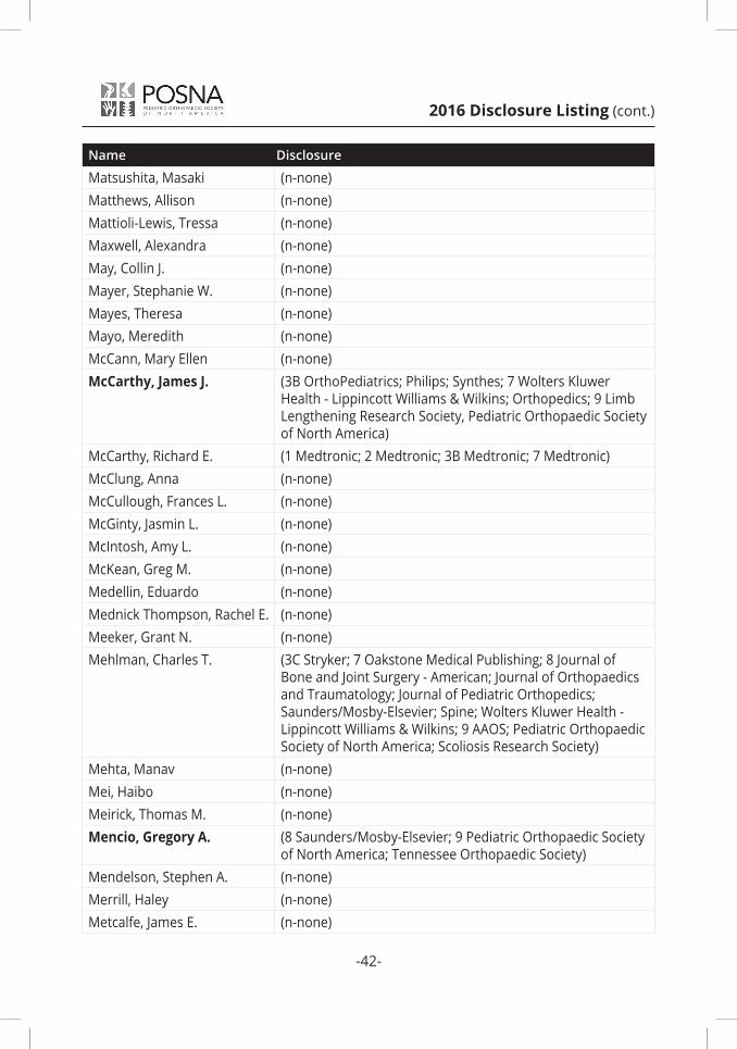
-42-
Matsushita, Masaki (n-none)Matthews, Allison (n-none)Mattioli-Lewis, Tressa (n-none)Maxwell, Alexandra (n-none)May, Collin J. (n-none)Mayer, Stephanie W. (n-none)Mayes, Theresa (n-none)Mayo, Meredith (n-none)McCann, Mary Ellen (n-none)McCarthy, James J. (3B OrthoPediatrics; Philips; Synthes; 7 Wolters Kluwer
Health - Lippincott Williams & Wilkins; Orthopedics; 9 Limb Lengthening Research Society, Pediatric Orthopaedic Society of North America)
McCarthy, Richard E. (1 Medtronic; 2 Medtronic; 3B Medtronic; 7 Medtronic)McClung, Anna (n-none)McCullough, Frances L. (n-none)McGinty, Jasmin L. (n-none)McIntosh, Amy L. (n-none)McKean, Greg M. (n-none)Medellin, Eduardo (n-none)Mednick Thompson, Rachel E. (n-none)Meeker, Grant N. (n-none)Mehlman, Charles T. (3C Stryker; 7 Oakstone Medical Publishing; 8 Journal of
Bone and Joint Surgery - American; Journal of Orthopaedics and Traumatology; Journal of Pediatric Orthopedics; Saunders/Mosby-Elsevier; Spine; Wolters Kluwer Health - Lippincott Williams & Wilkins; 9 AAOS; Pediatric Orthopaedic Society of North America; Scoliosis Research Society)
Mehta, Manav (n-none)Mei, Haibo (n-none)Meirick, Thomas M. (n-none)Mencio, Gregory A. (8 Saunders/Mosby-Elsevier; 9 Pediatric Orthopaedic Society
of North America; Tennessee Orthopaedic Society)Mendelson, Stephen A. (n-none)Merrill, Haley (n-none)Metcalfe, James E. (n-none)
Name Disclosure
-43-
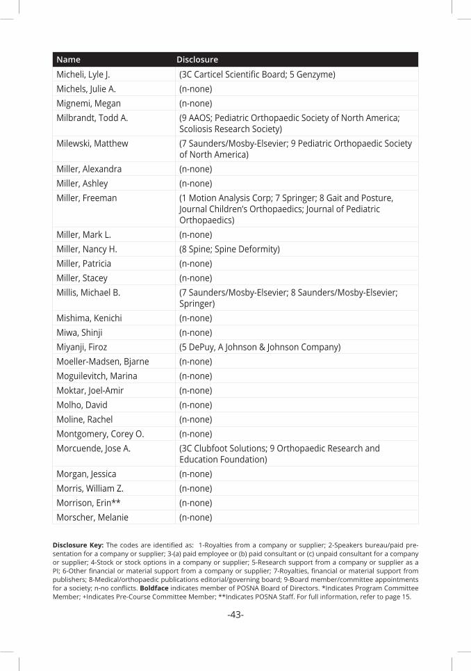
Disclosure Key: The codes are identified as: 1-Royalties from a company or supplier; 2-Speakers bureau/paid pre-sentation for a company or supplier; 3-(a) paid employee or (b) paid consultant or (c) unpaid consultant for a company or supplier; 4-Stock or stock options in a company or supplier; 5-Research support from a company or supplier as a PI; 6-Other financial or material support from a company or supplier; 7-Royalties, financial or material support from publishers; 8-Medical/orthopaedic publications editorial/governing board; 9-Board member/committee appointments for a society; n-no conflicts. Boldface indicates member of POSNA Board of Directors. *Indicates Program Committee Member; +Indicates Pre-Course Committee Member; **Indicates POSNA Staff. For full information, refer to page 15.
Micheli, Lyle J. (3C Carticel Scientific Board; 5 Genzyme)Michels, Julie A. (n-none)Mignemi, Megan (n-none)Milbrandt, Todd A. (9 AAOS; Pediatric Orthopaedic Society of North America;
Scoliosis Research Society)Milewski, Matthew (7 Saunders/Mosby-Elsevier; 9 Pediatric Orthopaedic Society
of North America)Miller, Alexandra (n-none)Miller, Ashley (n-none)Miller, Freeman (1 Motion Analysis Corp; 7 Springer; 8 Gait and Posture,
Journal Children’s Orthopaedics; Journal of Pediatric Orthopaedics)
Miller, Mark L. (n-none)Miller, Nancy H. (8 Spine; Spine Deformity)Miller, Patricia (n-none)Miller, Stacey (n-none)Millis, Michael B. (7 Saunders/Mosby-Elsevier; 8 Saunders/Mosby-Elsevier;
Springer)Mishima, Kenichi (n-none)Miwa, Shinji (n-none)Miyanji, Firoz (5 DePuy, A Johnson & Johnson Company)Moeller-Madsen, Bjarne (n-none)Moguilevitch, Marina (n-none)Moktar, Joel-Amir (n-none)Molho, David (n-none)Moline, Rachel (n-none)Montgomery, Corey O. (n-none)Morcuende, Jose A. (3C Clubfoot Solutions; 9 Orthopaedic Research and
Education Foundation)Morgan, Jessica (n-none)Morris, William Z. (n-none)Morrison, Erin** (n-none)Morscher, Melanie (n-none)
Name Disclosure
2016 Disclosure Listing (cont.)
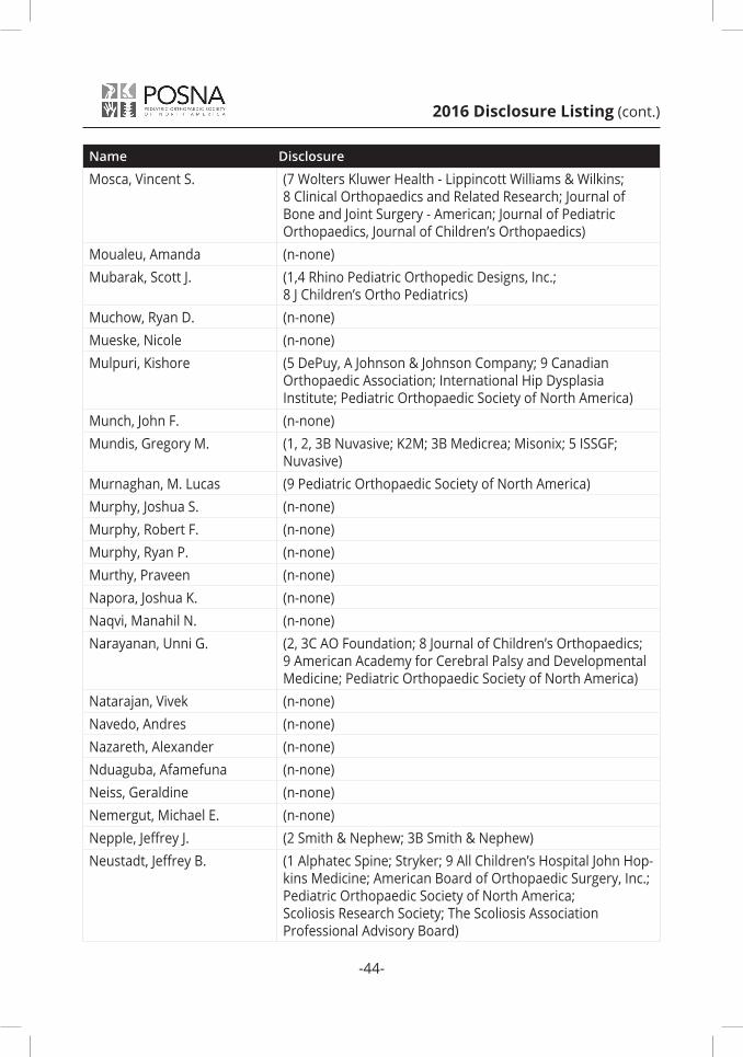
-44-
Mosca, Vincent S. (7 Wolters Kluwer Health - Lippincott Williams & Wilkins; 8 Clinical Orthopaedics and Related Research; Journal of Bone and Joint Surgery - American; Journal of Pediatric Orthopaedics, Journal of Children’s Orthopaedics)
Moualeu, Amanda (n-none)Mubarak, Scott J. (1,4 Rhino Pediatric Orthopedic Designs, Inc.;
8 J Children’s Ortho Pediatrics)Muchow, Ryan D. (n-none)Mueske, Nicole (n-none)Mulpuri, Kishore (5 DePuy, A Johnson & Johnson Company; 9 Canadian
Orthopaedic Association; International Hip Dysplasia Institute; Pediatric Orthopaedic Society of North America)
Munch, John F. (n-none)Mundis, Gregory M. (1, 2, 3B Nuvasive; K2M; 3B Medicrea; Misonix; 5 ISSGF;
Nuvasive)Murnaghan, M. Lucas (9 Pediatric Orthopaedic Society of North America)Murphy, Joshua S. (n-none)Murphy, Robert F. (n-none)Murphy, Ryan P. (n-none)Murthy, Praveen (n-none)Napora, Joshua K. (n-none)Naqvi, Manahil N. (n-none)Narayanan, Unni G. (2, 3C AO Foundation; 8 Journal of Children’s Orthopaedics;
9 American Academy for Cerebral Palsy and Developmental Medicine; Pediatric Orthopaedic Society of North America)
Natarajan, Vivek (n-none)Navedo, Andres (n-none)Nazareth, Alexander (n-none)Nduaguba, Afamefuna (n-none)Neiss, Geraldine (n-none)Nemergut, Michael E. (n-none)Nepple, Jeffrey J. (2 Smith & Nephew; 3B Smith & Nephew)Neustadt, Jeffrey B. (1 Alphatec Spine; Stryker; 9 All Children’s Hospital John Hop-
kins Medicine; American Board of Orthopaedic Surgery, Inc.; Pediatric Orthopaedic Society of North America; Scoliosis Research Society; The Scoliosis Association Professional Advisory Board)
Name Disclosure
-45-
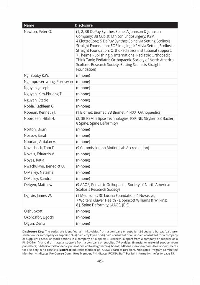
Disclosure Key: The codes are identified as: 1-Royalties from a company or supplier; 2-Speakers bureau/paid pre-sentation for a company or supplier; 3-(a) paid employee or (b) paid consultant or (c) unpaid consultant for a company or supplier; 4-Stock or stock options in a company or supplier; 5-Research support from a company or supplier as a PI; 6-Other financial or material support from a company or supplier; 7-Royalties, financial or material support from publishers; 8-Medical/orthopaedic publications editorial/governing board; 9-Board member/committee appointments for a society; n-no conflicts. Boldface indicates member of POSNA Board of Directors. *Indicates Program Committee Member; +Indicates Pre-Course Committee Member; **Indicates POSNA Staff. For full information, refer to page 15.
Newton, Peter O. (1, 2, 3B DePuy Synthes Spine, A Johnson & Johnson Company; 3B Cubist; Ethicon Endosurgery; K2M; 4 ElectroCore; 5 DePuy Synthes Spine via Setting Scoliosis Straight Foundation; EOS Imaging; K2M via Setting Scoliosis Straight Foundation; OrthoPediatrics institutional support; 7 Theime Publishing; 9 International Pediatric Orthopedic Think Tank; Pediatric Orthopaedic Society of North America; Scoliosis Research Society; Setting Scoliosis Straight Foundation)
Ng, Bobby K.W. (n-none)Ngamprasertwong, Pornswan (n-none)Nguyen, Joseph (n-none)Nguyen, Kim-Phuong T. (n-none)Nguyen, Stacie (n-none)Noble, Kathleen G. (n-none)Noonan, Kenneth J. (1 Biomet; Biomet; 3B Biomet; 4 FIXX Orthopaedics)Noordeen, Hilali H. (2, 3B K2M, Ellipse Technologies, KSPINE; Stryker; 3B Baxter;
8 Spine, Spine Deformity)Norton, Brian (n-none)Nossov, Sarah (n-none)Nourian, Ardalan A. (n-none)Novacheck, Tom F (9 Commission on Motion Lab Accreditation)Novais, Eduardo V. (n-none)Noyes, Katia (n-none)Nwachukwu, Benedict U. (n-none)O’Malley, Natasha (n-none)O’Malley, Sandra (n-none)Oetgen, Matthew (9 AAOS; Pediatric Orthopaedic Society of North America;
Scoliosis Research Society)Ogilvie, James W. (1 Medtronic; 3C Lucina Foundation; 4 Nuvasive;
7 Wolters Kluwer Health - Lippincott Williams & Wilkins; 8 J. Spine Deformity, JAAOS, JBJS)
Oishi, Scott (n-none)Okoroafor, Ugochi (n-none)Olgun, Deniz (n-none)
Name Disclosure
2016 Disclosure Listing (cont.)
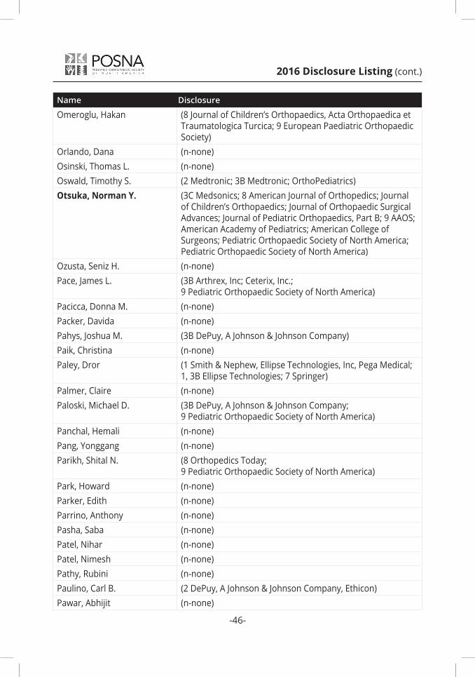
-46-
Omeroglu, Hakan (8 Journal of Children’s Orthopaedics, Acta Orthopaedica et Traumatologica Turcica; 9 European Paediatric Orthopaedic Society)
Orlando, Dana (n-none)Osinski, Thomas L. (n-none)Oswald, Timothy S. (2 Medtronic; 3B Medtronic; OrthoPediatrics)Otsuka, Norman Y. (3C Medsonics; 8 American Journal of Orthopedics; Journal
of Children’s Orthopaedics; Journal of Orthopaedic Surgical Advances; Journal of Pediatric Orthopaedics, Part B; 9 AAOS; American Academy of Pediatrics; American College of Surgeons; Pediatric Orthopaedic Society of North America; Pediatric Orthopaedic Society of North America)
Ozusta, Seniz H. (n-none)Pace, James L. (3B Arthrex, Inc; Ceterix, Inc.;
9 Pediatric Orthopaedic Society of North America)Pacicca, Donna M. (n-none)Packer, Davida (n-none)Pahys, Joshua M. (3B DePuy, A Johnson & Johnson Company)Paik, Christina (n-none)Paley, Dror (1 Smith & Nephew, Ellipse Technologies, Inc, Pega Medical;
1, 3B Ellipse Technologies; 7 Springer)Palmer, Claire (n-none)Paloski, Michael D. (3B DePuy, A Johnson & Johnson Company;
9 Pediatric Orthopaedic Society of North America)Panchal, Hemali (n-none)Pang, Yonggang (n-none)Parikh, Shital N. (8 Orthopedics Today;
9 Pediatric Orthopaedic Society of North America)Park, Howard (n-none)Parker, Edith (n-none)Parrino, Anthony (n-none)Pasha, Saba (n-none)Patel, Nihar (n-none)Patel, Nimesh (n-none)Pathy, Rubini (n-none)Paulino, Carl B. (2 DePuy, A Johnson & Johnson Company, Ethicon)Pawar, Abhijit (n-none)
Name Disclosure
-47-
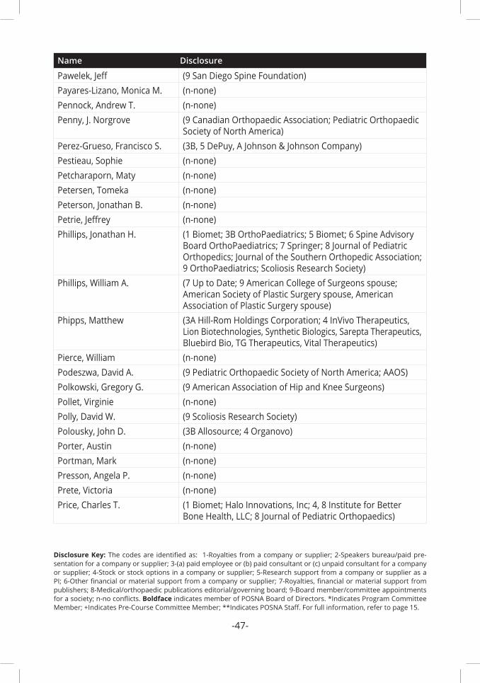
Disclosure Key: The codes are identified as: 1-Royalties from a company or supplier; 2-Speakers bureau/paid pre-sentation for a company or supplier; 3-(a) paid employee or (b) paid consultant or (c) unpaid consultant for a company or supplier; 4-Stock or stock options in a company or supplier; 5-Research support from a company or supplier as a PI; 6-Other financial or material support from a company or supplier; 7-Royalties, financial or material support from publishers; 8-Medical/orthopaedic publications editorial/governing board; 9-Board member/committee appointments for a society; n-no conflicts. Boldface indicates member of POSNA Board of Directors. *Indicates Program Committee Member; +Indicates Pre-Course Committee Member; **Indicates POSNA Staff. For full information, refer to page 15.
Pawelek, Jeff (9 San Diego Spine Foundation)Payares-Lizano, Monica M. (n-none)Pennock, Andrew T. (n-none)Penny, J. Norgrove (9 Canadian Orthopaedic Association; Pediatric Orthopaedic
Society of North America)Perez-Grueso, Francisco S. (3B, 5 DePuy, A Johnson & Johnson Company)Pestieau, Sophie (n-none)Petcharaporn, Maty (n-none)Petersen, Tomeka (n-none)Peterson, Jonathan B. (n-none)Petrie, Jeffrey (n-none)Phillips, Jonathan H. (1 Biomet; 3B OrthoPaediatrics; 5 Biomet; 6 Spine Advisory
Board OrthoPaediatrics; 7 Springer; 8 Journal of Pediatric Orthopedics; Journal of the Southern Orthopedic Association; 9 OrthoPaediatrics; Scoliosis Research Society)
Phillips, William A. (7 Up to Date; 9 American College of Surgeons spouse; American Society of Plastic Surgery spouse, American Association of Plastic Surgery spouse)
Phipps, Matthew (3A Hill-Rom Holdings Corporation; 4 InVivo Therapeutics, Lion Biotechnologies, Synthetic Biologics, Sarepta Therapeutics, Bluebird Bio, TG Therapeutics, Vital Therapeutics)
Pierce, William (n-none)Podeszwa, David A. (9 Pediatric Orthopaedic Society of North America; AAOS)Polkowski, Gregory G. (9 American Association of Hip and Knee Surgeons)Pollet, Virginie (n-none)Polly, David W. (9 Scoliosis Research Society)Polousky, John D. (3B Allosource; 4 Organovo)Porter, Austin (n-none)Portman, Mark (n-none)Presson, Angela P. (n-none)Prete, Victoria (n-none)Price, Charles T. (1 Biomet; Halo Innovations, Inc; 4, 8 Institute for Better
Bone Health, LLC; 8 Journal of Pediatric Orthopaedics)
Name Disclosure
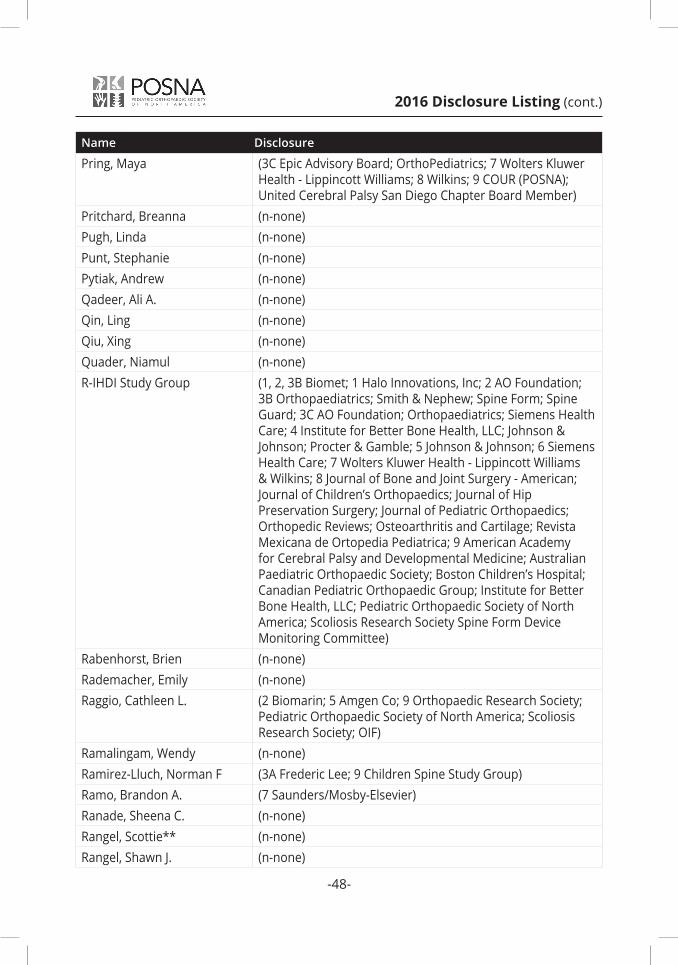
2016 Disclosure Listing (cont.)
-48-
Pring, Maya (3C Epic Advisory Board; OrthoPediatrics; 7 Wolters Kluwer Health - Lippincott Williams; 8 Wilkins; 9 COUR (POSNA); United Cerebral Palsy San Diego Chapter Board Member)
Pritchard, Breanna (n-none)Pugh, Linda (n-none)Punt, Stephanie (n-none)Pytiak, Andrew (n-none)Qadeer, Ali A. (n-none)Qin, Ling (n-none)Qiu, Xing (n-none)Quader, Niamul (n-none)R-IHDI Study Group (1, 2, 3B Biomet; 1 Halo Innovations, Inc; 2 AO Foundation;
3B Orthopaediatrics; Smith & Nephew; Spine Form; Spine Guard; 3C AO Foundation; Orthopaediatrics; Siemens Health Care; 4 Institute for Better Bone Health, LLC; Johnson & Johnson; Procter & Gamble; 5 Johnson & Johnson; 6 Siemens Health Care; 7 Wolters Kluwer Health - Lippincott Williams & Wilkins; 8 Journal of Bone and Joint Surgery - American; Journal of Children’s Orthopaedics; Journal of Hip Preservation Surgery; Journal of Pediatric Orthopaedics; Orthopedic Reviews; Osteoarthritis and Cartilage; Revista Mexicana de Ortopedia Pediatrica; 9 American Academy for Cerebral Palsy and Developmental Medicine; Australian Paediatric Orthopaedic Society; Boston Children’s Hospital; Canadian Pediatric Orthopaedic Group; Institute for Better Bone Health, LLC; Pediatric Orthopaedic Society of North America; Scoliosis Research Society Spine Form Device Monitoring Committee)
Rabenhorst, Brien (n-none)Rademacher, Emily (n-none)Raggio, Cathleen L. (2 Biomarin; 5 Amgen Co; 9 Orthopaedic Research Society;
Pediatric Orthopaedic Society of North America; Scoliosis Research Society; OIF)
Ramalingam, Wendy (n-none)Ramirez-Lluch, Norman F (3A Frederic Lee; 9 Children Spine Study Group)Ramo, Brandon A. (7 Saunders/Mosby-Elsevier)Ranade, Sheena C. (n-none)Rangel, Scottie** (n-none)Rangel, Shawn J. (n-none)
Name Disclosure
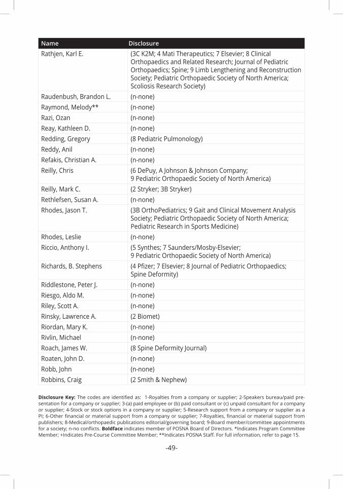
-49-
Disclosure Key: The codes are identified as: 1-Royalties from a company or supplier; 2-Speakers bureau/paid pre-sentation for a company or supplier; 3-(a) paid employee or (b) paid consultant or (c) unpaid consultant for a company or supplier; 4-Stock or stock options in a company or supplier; 5-Research support from a company or supplier as a PI; 6-Other financial or material support from a company or supplier; 7-Royalties, financial or material support from publishers; 8-Medical/orthopaedic publications editorial/governing board; 9-Board member/committee appointments for a society; n-no conflicts. Boldface indicates member of POSNA Board of Directors. *Indicates Program Committee Member; +Indicates Pre-Course Committee Member; **Indicates POSNA Staff. For full information, refer to page 15.
Rathjen, Karl E. (3C K2M; 4 Mati Therapeutics; 7 Elsevier; 8 Clinical Orthopaedics and Related Research; Journal of Pediatric Orthopaedics; Spine; 9 Limb Lengthening and Reconstruction Society; Pediatric Orthopaedic Society of North America; Scoliosis Research Society)
Raudenbush, Brandon L. (n-none)Raymond, Melody** (n-none)Razi, Ozan (n-none)Reay, Kathleen D. (n-none)Redding, Gregory (8 Pediatric Pulmonology)Reddy, Anil (n-none)Refakis, Christian A. (n-none)Reilly, Chris (6 DePuy, A Johnson & Johnson Company;
9 Pediatric Orthopaedic Society of North America)Reilly, Mark C. (2 Stryker; 3B Stryker)Rethlefsen, Susan A. (n-none)Rhodes, Jason T. (3B OrthoPediatrics; 9 Gait and Clinical Movement Analysis
Society; Pediatric Orthopaedic Society of North America; Pediatric Research in Sports Medicine)
Rhodes, Leslie (n-none)Riccio, Anthony I. (5 Synthes; 7 Saunders/Mosby-Elsevier;
9 Pediatric Orthopaedic Society of North America)Richards, B. Stephens (4 Pfizer; 7 Elsevier; 8 Journal of Pediatric Orthopaedics;
Spine Deformity)Riddlestone, Peter J. (n-none)Riesgo, Aldo M. (n-none)Riley, Scott A. (n-none)Rinsky, Lawrence A. (2 Biomet)Riordan, Mary K. (n-none)Rivlin, Michael (n-none)Roach, James W. (8 Spine Deformity Journal)Roaten, John D. (n-none)Robb, John (n-none)Robbins, Craig (2 Smith & Nephew)
Name Disclosure
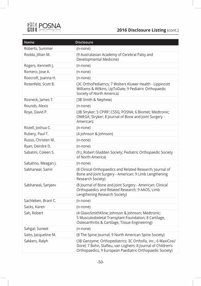
2016 Disclosure Listing (cont.)
-50-
Roberts, Summer (n-none)Rodda, Jillian M. (9 Australasian Academy of Cerebral Palsy and
Developmental Medicine)Rogers, Kenneth J. (n-none)Romero, Jose A. (n-none)Roocroft, Joanna H. (n-none)Rosenfeld, Scott B. (3C OrthoPediatrics; 7 Wolters Kluwer Health - Lippincott
Williams & Wilkins, UpToDate; 9 Pediatric Orthopaedic Society of North America)
Rosneck, James T. (3B Smith & Nephew)Rounds, Alexis (n-none)Roye, David P. (3B Stryker; 5 CPIRF; CSSG; POSNA; 6 Biomet; Medtronic;
OMEGA; Stryker; 8 Journal of Bone and Joint Surgery - American)
Rozell, Joshua C. (n-none)Rubery, Paul T. (4 Johnson & Johnson)Russo, Christen M. (n-none)Ryan, Deirdre D. (n-none)Sabatini, Coleen S. (9 J. Robert Gladden Society; Pediatric Orthopaedic Society
of North America)Sabatino, Meagan J. (n-none)Sabharwal, Samir (8 Clinical Orthopaedics and Related Research; Journal of
Bone and Joint Surgery - American; 9 Limb Lengthening Research Society)
Sabharwal, Sanjeev (8 Journal of Bone and Joint Surgery - American; Clinical Orthopaedics and Related Research; 9 AAOS; Limb Lengthening Research Society)
Sachleben, Brant C. (n-none)Sacks, Karen (n-none)Sah, Robert (4 GlaxoSmithKline; Johnson & Johnson; Medtronic;
5 Musculoskeletal Transplant Foundation; 8 Cartilage, Osteoarthritis & Cartilage, Tissue Engineering)
Sahgal, Suneet (n-none)Saito, Jacqueline M. (8 The Spine Journal; 9 North American Spine Society)Sakkers, Ralph (3B Genzyme; Orthopediatrics; 3C Orthofix, Inc.; 6 MaxiCosi/
Dorel; 7 Bohn, Stafleu, van Loghem; 8 Journal of Children’s Orthopaedics; 9 European Paediatric Orthopaedic Society)
Name Disclosure
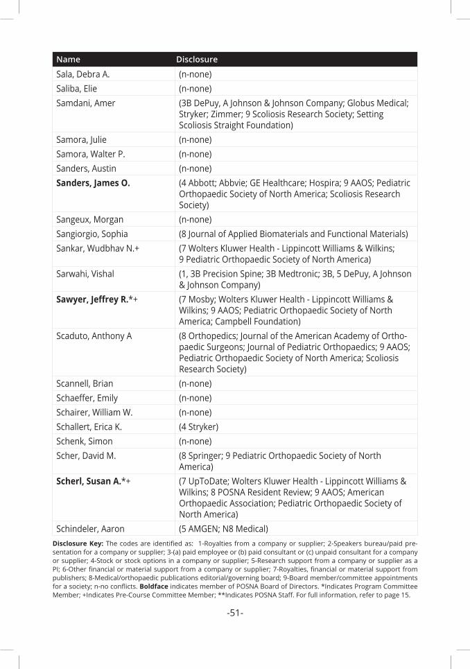
-51-
Disclosure Key: The codes are identified as: 1-Royalties from a company or supplier; 2-Speakers bureau/paid pre-sentation for a company or supplier; 3-(a) paid employee or (b) paid consultant or (c) unpaid consultant for a company or supplier; 4-Stock or stock options in a company or supplier; 5-Research support from a company or supplier as a PI; 6-Other financial or material support from a company or supplier; 7-Royalties, financial or material support from publishers; 8-Medical/orthopaedic publications editorial/governing board; 9-Board member/committee appointments for a society; n-no conflicts. Boldface indicates member of POSNA Board of Directors. *Indicates Program Committee Member; +Indicates Pre-Course Committee Member; **Indicates POSNA Staff. For full information, refer to page 15.
Sala, Debra A. (n-none)Saliba, Elie (n-none)Samdani, Amer (3B DePuy, A Johnson & Johnson Company; Globus Medical;
Stryker; Zimmer; 9 Scoliosis Research Society; Setting Scoliosis Straight Foundation)
Samora, Julie (n-none)Samora, Walter P. (n-none)Sanders, Austin (n-none)Sanders, James O. (4 Abbott; Abbvie; GE Healthcare; Hospira; 9 AAOS; Pediatric
Orthopaedic Society of North America; Scoliosis Research Society)
Sangeux, Morgan (n-none)Sangiorgio, Sophia (8 Journal of Applied Biomaterials and Functional Materials)Sankar, Wudbhav N.+ (7 Wolters Kluwer Health - Lippincott Williams & Wilkins;
9 Pediatric Orthopaedic Society of North America)Sarwahi, Vishal (1, 3B Precision Spine; 3B Medtronic; 3B, 5 DePuy, A Johnson
& Johnson Company)Sawyer, Jeffrey R.*+ (7 Mosby; Wolters Kluwer Health - Lippincott Williams &
Wilkins; 9 AAOS; Pediatric Orthopaedic Society of North America; Campbell Foundation)
Scaduto, Anthony A (8 Orthopedics; Journal of the American Academy of Ortho-paedic Surgeons; Journal of Pediatric Orthopaedics; 9 AAOS; Pediatric Orthopaedic Society of North America; Scoliosis Research Society)
Scannell, Brian (n-none)Schaeffer, Emily (n-none)Schairer, William W. (n-none)Schallert, Erica K. (4 Stryker)Schenk, Simon (n-none)Scher, David M. (8 Springer; 9 Pediatric Orthopaedic Society of North
America)Scherl, Susan A.*+ (7 UpToDate; Wolters Kluwer Health - Lippincott Williams &
Wilkins; 8 POSNA Resident Review; 9 AAOS; American Orthopaedic Association; Pediatric Orthopaedic Society of North America)
Schindeler, Aaron (5 AMGEN; N8 Medical)
Name Disclosure
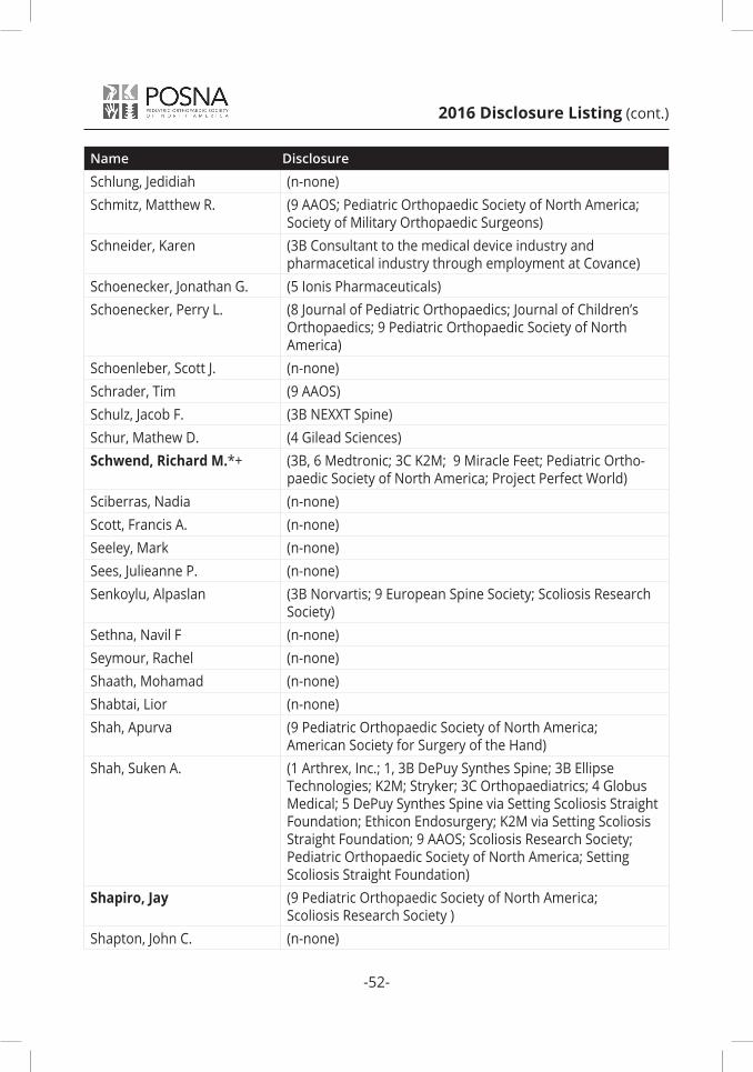
2016 Disclosure Listing (cont.)
-52-
Schlung, Jedidiah (n-none)Schmitz, Matthew R. (9 AAOS; Pediatric Orthopaedic Society of North America;
Society of Military Orthopaedic Surgeons)Schneider, Karen (3B Consultant to the medical device industry and
pharmacetical industry through employment at Covance)Schoenecker, Jonathan G. (5 Ionis Pharmaceuticals)Schoenecker, Perry L. (8 Journal of Pediatric Orthopaedics; Journal of Children’s
Orthopaedics; 9 Pediatric Orthopaedic Society of North America)
Schoenleber, Scott J. (n-none)Schrader, Tim (9 AAOS)Schulz, Jacob F. (3B NEXXT Spine)Schur, Mathew D. (4 Gilead Sciences)Schwend, Richard M.*+ (3B, 6 Medtronic; 3C K2M; 9 Miracle Feet; Pediatric Ortho-
paedic Society of North America; Project Perfect World)Sciberras, Nadia (n-none)Scott, Francis A. (n-none)Seeley, Mark (n-none)Sees, Julieanne P. (n-none)Senkoylu, Alpaslan (3B Norvartis; 9 European Spine Society; Scoliosis Research
Society)Sethna, Navil F (n-none)Seymour, Rachel (n-none)Shaath, Mohamad (n-none)Shabtai, Lior (n-none)Shah, Apurva (9 Pediatric Orthopaedic Society of North America;
American Society for Surgery of the Hand)Shah, Suken A. (1 Arthrex, Inc.; 1, 3B DePuy Synthes Spine; 3B Ellipse
Technologies; K2M; Stryker; 3C Orthopaediatrics; 4 Globus Medical; 5 DePuy Synthes Spine via Setting Scoliosis Straight Foundation; Ethicon Endosurgery; K2M via Setting Scoliosis Straight Foundation; 9 AAOS; Scoliosis Research Society; Pediatric Orthopaedic Society of North America; Setting Scoliosis Straight Foundation)
Shapiro, Jay (9 Pediatric Orthopaedic Society of North America; Scoliosis Research Society )
Shapton, John C. (n-none)
Name Disclosure
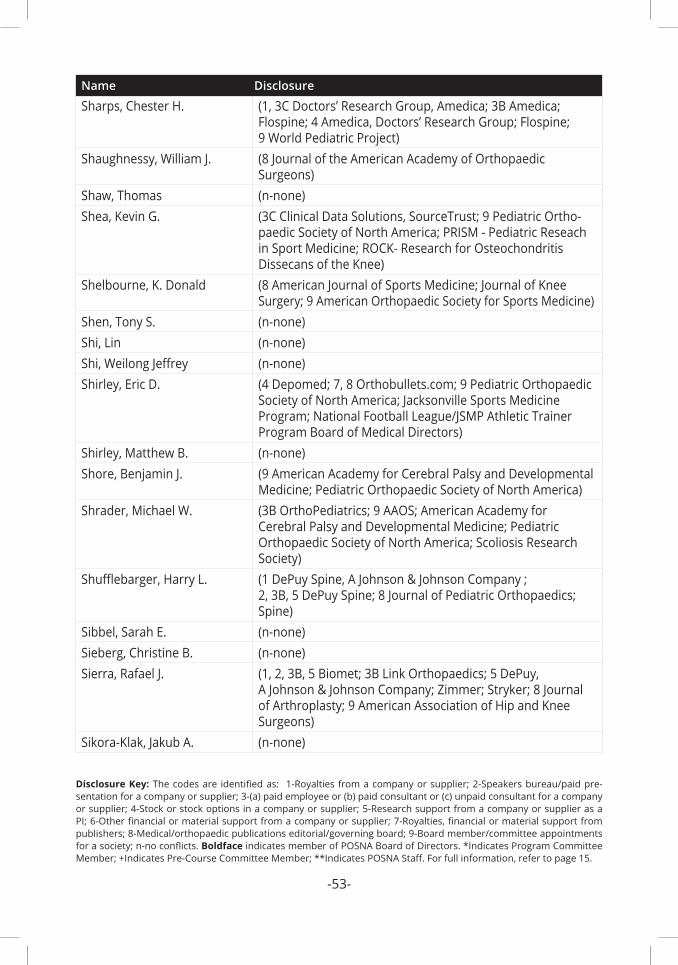
-53-
Disclosure Key: The codes are identified as: 1-Royalties from a company or supplier; 2-Speakers bureau/paid pre-sentation for a company or supplier; 3-(a) paid employee or (b) paid consultant or (c) unpaid consultant for a company or supplier; 4-Stock or stock options in a company or supplier; 5-Research support from a company or supplier as a PI; 6-Other financial or material support from a company or supplier; 7-Royalties, financial or material support from publishers; 8-Medical/orthopaedic publications editorial/governing board; 9-Board member/committee appointments for a society; n-no conflicts. Boldface indicates member of POSNA Board of Directors. *Indicates Program Committee Member; +Indicates Pre-Course Committee Member; **Indicates POSNA Staff. For full information, refer to page 15.
Sharps, Chester H. (1, 3C Doctors’ Research Group, Amedica; 3B Amedica; Flospine; 4 Amedica, Doctors’ Research Group; Flospine; 9 World Pediatric Project)
Shaughnessy, William J. (8 Journal of the American Academy of Orthopaedic Surgeons)
Shaw, Thomas (n-none)Shea, Kevin G. (3C Clinical Data Solutions, SourceTrust; 9 Pediatric Ortho-
paedic Society of North America; PRISM - Pediatric Reseach in Sport Medicine; ROCK- Research for Osteochondritis Dissecans of the Knee)
Shelbourne, K. Donald (8 American Journal of Sports Medicine; Journal of Knee Surgery; 9 American Orthopaedic Society for Sports Medicine)
Shen, Tony S. (n-none)Shi, Lin (n-none)Shi, Weilong Jeffrey (n-none)Shirley, Eric D. (4 Depomed; 7, 8 Orthobullets.com; 9 Pediatric Orthopaedic
Society of North America; Jacksonville Sports Medicine Program; National Football League/JSMP Athletic Trainer Program Board of Medical Directors)
Shirley, Matthew B. (n-none)Shore, Benjamin J. (9 American Academy for Cerebral Palsy and Developmental
Medicine; Pediatric Orthopaedic Society of North America)Shrader, Michael W. (3B OrthoPediatrics; 9 AAOS; American Academy for
Cerebral Palsy and Developmental Medicine; Pediatric Orthopaedic Society of North America; Scoliosis Research Society)
Shufflebarger, Harry L. (1 DePuy Spine, A Johnson & Johnson Company ; 2, 3B, 5 DePuy Spine; 8 Journal of Pediatric Orthopaedics; Spine)
Sibbel, Sarah E. (n-none)Sieberg, Christine B. (n-none)Sierra, Rafael J. (1, 2, 3B, 5 Biomet; 3B Link Orthopaedics; 5 DePuy,
A Johnson & Johnson Company; Zimmer; Stryker; 8 Journal of Arthroplasty; 9 American Association of Hip and Knee Surgeons)
Sikora-Klak, Jakub A. (n-none)
Name Disclosure
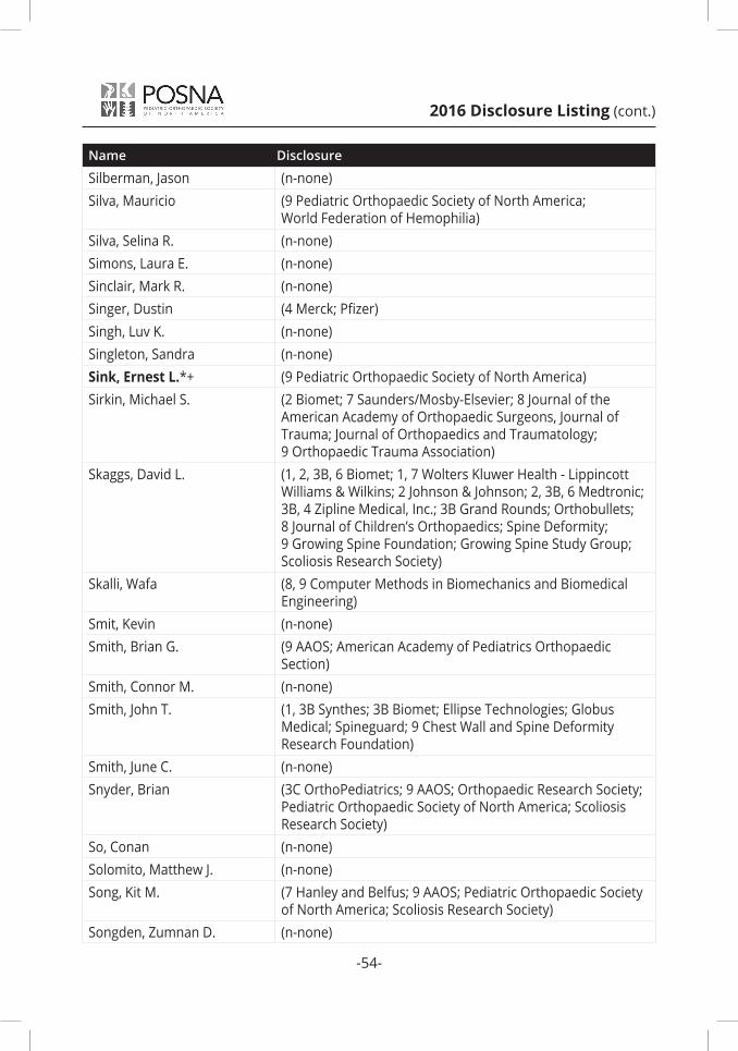
2016 Disclosure Listing (cont.)
-54-
Silberman, Jason (n-none)Silva, Mauricio (9 Pediatric Orthopaedic Society of North America;
World Federation of Hemophilia)Silva, Selina R. (n-none)Simons, Laura E. (n-none)Sinclair, Mark R. (n-none)Singer, Dustin (4 Merck; Pfizer)Singh, Luv K. (n-none)Singleton, Sandra (n-none)Sink, Ernest L.*+ (9 Pediatric Orthopaedic Society of North America)Sirkin, Michael S. (2 Biomet; 7 Saunders/Mosby-Elsevier; 8 Journal of the
American Academy of Orthopaedic Surgeons, Journal of Trauma; Journal of Orthopaedics and Traumatology; 9 Orthopaedic Trauma Association)
Skaggs, David L. (1, 2, 3B, 6 Biomet; 1, 7 Wolters Kluwer Health - Lippincott Williams & Wilkins; 2 Johnson & Johnson; 2, 3B, 6 Medtronic; 3B, 4 Zipline Medical, Inc.; 3B Grand Rounds; Orthobullets; 8 Journal of Children’s Orthopaedics; Spine Deformity; 9 Growing Spine Foundation; Growing Spine Study Group; Scoliosis Research Society)
Skalli, Wafa (8, 9 Computer Methods in Biomechanics and Biomedical Engineering)
Smit, Kevin (n-none)Smith, Brian G. (9 AAOS; American Academy of Pediatrics Orthopaedic
Section)Smith, Connor M. (n-none)Smith, John T. (1, 3B Synthes; 3B Biomet; Ellipse Technologies; Globus
Medical; Spineguard; 9 Chest Wall and Spine Deformity Research Foundation)
Smith, June C. (n-none)Snyder, Brian (3C OrthoPediatrics; 9 AAOS; Orthopaedic Research Society;
Pediatric Orthopaedic Society of North America; Scoliosis Research Society)
So, Conan (n-none)Solomito, Matthew J. (n-none)Song, Kit M. (7 Hanley and Belfus; 9 AAOS; Pediatric Orthopaedic Society
of North America; Scoliosis Research Society)Songden, Zumnan D. (n-none)
Name Disclosure
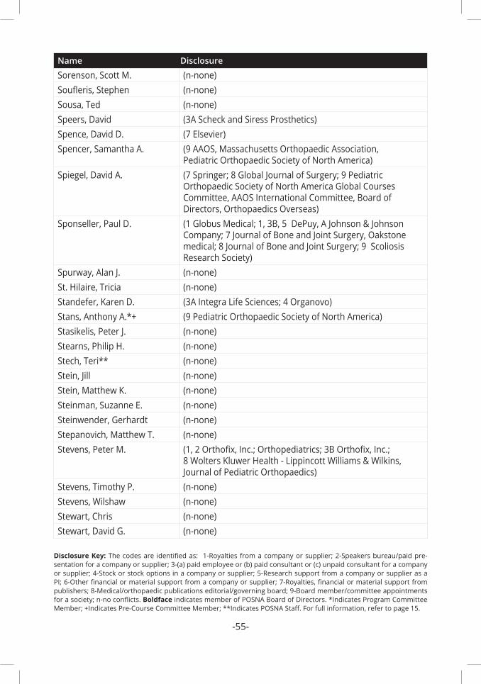
-55-
Disclosure Key: The codes are identified as: 1-Royalties from a company or supplier; 2-Speakers bureau/paid pre-sentation for a company or supplier; 3-(a) paid employee or (b) paid consultant or (c) unpaid consultant for a company or supplier; 4-Stock or stock options in a company or supplier; 5-Research support from a company or supplier as a PI; 6-Other financial or material support from a company or supplier; 7-Royalties, financial or material support from publishers; 8-Medical/orthopaedic publications editorial/governing board; 9-Board member/committee appointments for a society; n-no conflicts. Boldface indicates member of POSNA Board of Directors. *Indicates Program Committee Member; +Indicates Pre-Course Committee Member; **Indicates POSNA Staff. For full information, refer to page 15.
Sorenson, Scott M. (n-none)Soufleris, Stephen (n-none)Sousa, Ted (n-none)Speers, David (3A Scheck and Siress Prosthetics)Spence, David D. (7 Elsevier)Spencer, Samantha A. (9 AAOS, Massachusetts Orthopaedic Association,
Pediatric Orthopaedic Society of North America)Spiegel, David A. (7 Springer; 8 Global Journal of Surgery; 9 Pediatric
Orthopaedic Society of North America Global Courses Committee, AAOS International Committee, Board of Directors, Orthopaedics Overseas)
Sponseller, Paul D. (1 Globus Medical; 1, 3B, 5 DePuy, A Johnson & Johnson Company; 7 Journal of Bone and Joint Surgery, Oakstone medical; 8 Journal of Bone and Joint Surgery; 9 Scoliosis Research Society)
Spurway, Alan J. (n-none)St. Hilaire, Tricia (n-none)Standefer, Karen D. (3A Integra Life Sciences; 4 Organovo)Stans, Anthony A.*+ (9 Pediatric Orthopaedic Society of North America)Stasikelis, Peter J. (n-none)Stearns, Philip H. (n-none)Stech, Teri** (n-none)Stein, Jill (n-none)Stein, Matthew K. (n-none)Steinman, Suzanne E. (n-none)Steinwender, Gerhardt (n-none)Stepanovich, Matthew T. (n-none)Stevens, Peter M. (1, 2 Orthofix, Inc.; Orthopediatrics; 3B Orthofix, Inc.;
8 Wolters Kluwer Health - Lippincott Williams & Wilkins, Journal of Pediatric Orthopaedics)
Stevens, Timothy P. (n-none)Stevens, Wilshaw (n-none)Stewart, Chris (n-none)Stewart, David G. (n-none)
Name Disclosure
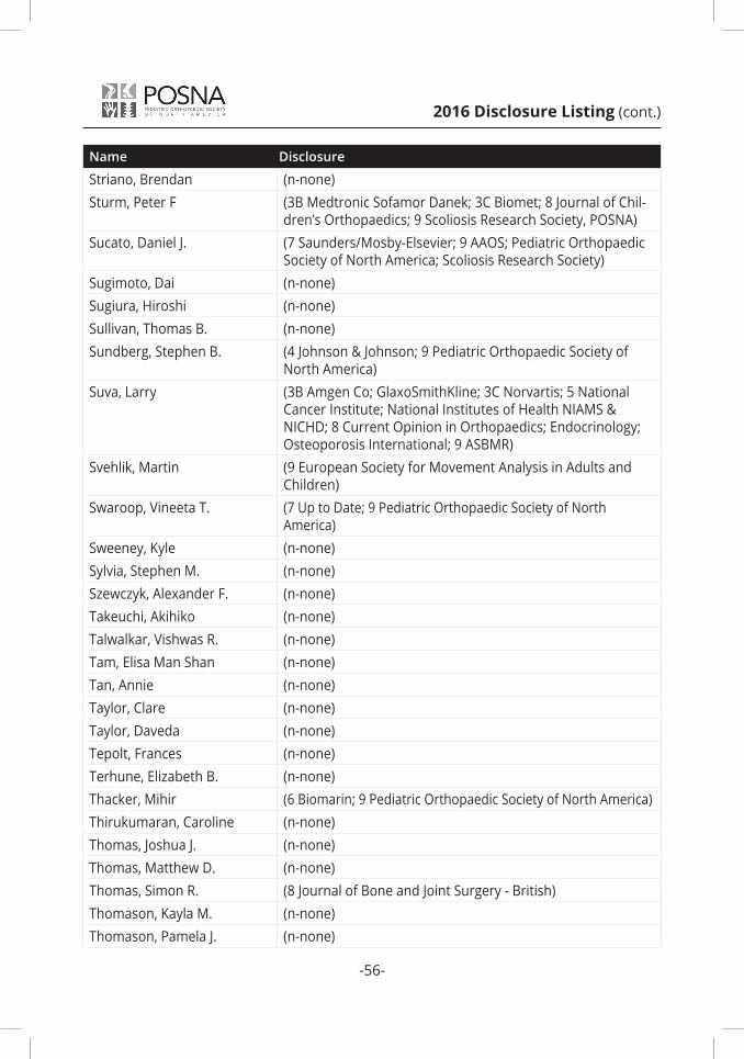
2016 Disclosure Listing (cont.)
-56-
Striano, Brendan (n-none)Sturm, Peter F (3B Medtronic Sofamor Danek; 3C Biomet; 8 Journal of Chil-
dren’s Orthopaedics; 9 Scoliosis Research Society, POSNA)Sucato, Daniel J. (7 Saunders/Mosby-Elsevier; 9 AAOS; Pediatric Orthopaedic
Society of North America; Scoliosis Research Society)Sugimoto, Dai (n-none)Sugiura, Hiroshi (n-none)Sullivan, Thomas B. (n-none)Sundberg, Stephen B. (4 Johnson & Johnson; 9 Pediatric Orthopaedic Society of
North America)Suva, Larry (3B Amgen Co; GlaxoSmithKline; 3C Norvartis; 5 National
Cancer Institute; National Institutes of Health NIAMS & NICHD; 8 Current Opinion in Orthopaedics; Endocrinology; Osteoporosis International; 9 ASBMR)
Svehlik, Martin (9 European Society for Movement Analysis in Adults and Children)
Swaroop, Vineeta T. (7 Up to Date; 9 Pediatric Orthopaedic Society of North America)
Sweeney, Kyle (n-none)Sylvia, Stephen M. (n-none)Szewczyk, Alexander F. (n-none)Takeuchi, Akihiko (n-none)Talwalkar, Vishwas R. (n-none)Tam, Elisa Man Shan (n-none)Tan, Annie (n-none)Taylor, Clare (n-none)Taylor, Daveda (n-none)Tepolt, Frances (n-none)Terhune, Elizabeth B. (n-none)Thacker, Mihir (6 Biomarin; 9 Pediatric Orthopaedic Society of North America)Thirukumaran, Caroline (n-none)Thomas, Joshua J. (n-none)Thomas, Matthew D. (n-none)Thomas, Simon R. (8 Journal of Bone and Joint Surgery - British)Thomason, Kayla M. (n-none)Thomason, Pamela J. (n-none)
Name Disclosure
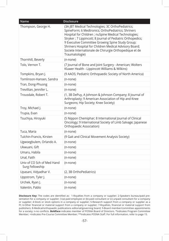
-57-
Disclosure Key: The codes are identified as: 1-Royalties from a company or supplier; 2-Speakers bureau/paid pre-sentation for a company or supplier; 3-(a) paid employee or (b) paid consultant or (c) unpaid consultant for a company or supplier; 4-Stock or stock options in a company or supplier; 5-Research support from a company or supplier as a PI; 6-Other financial or material support from a company or supplier; 7-Royalties, financial or material support from publishers; 8-Medical/orthopaedic publications editorial/governing board; 9-Board member/committee appointments for a society; n-no conflicts. Boldface indicates member of POSNA Board of Directors. *Indicates Program Committee Member; +Indicates Pre-Course Committee Member; **Indicates POSNA Staff. For full information, refer to page 15.
Thompson, George H. (3A JBT Medical Technologies; 3C OrthoPediatrics; SpineForm; 6 Medtronics; OrthoPediatrics; Shriners Hospital for Children ; nuSpine Medical Technologies; Stryker ; 7 Lippincott; 8 Journal of Pediatric Orthopedics; 9 Executive Committee Growing Spine Study Group; Shriners Hospital for Children Medical Advisory Board; Societe Internationale de Chirurgie Orthopedique et de Traumatologie)
Thornhill, Beverly (n-none)Tolo, Vernon T. (7 Journal of Bone and Joint Surgery - American; Wolters
Kluwer Health - Lippincott Williams & Wilkins)Tompkins, Bryan J. (9 AAOS; Pediatric Orthopaedic Society of North America)Tomlinson-Hansen, Sandra (n-none)Tran, Dong-Phuong (n-none)Trevillian, Jennifer L. (n-none)Trousdale, Robert T. (1, 3B DePuy, A Johnson & Johnson Company; 8 Journal of
Arthroplasty; 9 American Association of Hip and Knee Surgeons; Hip Society; Knee Society)
Troy, Michael J. (n-none)Trupia, Evan (n-none)Tsuchiya, Hiroyuki (5 Nippon Chemiphar; 8 International Journal of Clinical
Oncology; 9 International Society of Limb Salvage; Japanese Orthopaedic Association)
Tuca, Maria (n-none)Tulchin-Francis, Kirsten (9 Gait and Clinical Movement Analysis Society)Ugwoegbulem, Orlando A. (n-none)Ukwuani, Gift (n-none)Umaru, Habila (n-none)Unal, Faith (n-none)Univ of CO Sch of Med Hand Surg Fellowship
(n-none)
Upasani, Vidyadhar V. (2, 3B OrthoPediatrics)Uppstrom, Tyler J. (n-none)Urchek, Ryan J. (n-none)Valentin, Pablo (n-none)
Name Disclosure
2016 Disclosure Listing (cont.)
-58-
Van Bosse, Harold J.P. (n-none)Van De Sande, Michiel (6 Daiichi; Implantcast; 8 Current Orthopaedic Practice)Vanderby, Ray (n-none)Vanderhave, Kelly L. (n-none)Varghese, Ranjit A. (9 American Academy for Cerebral Palsy and Developmental
Medicine; Pediatric Orthopaedic Society of North America)Verma, Kushagra (n-none)Vessey, Judith A. (4 Johnson & Johnson; Pfizer; Wyeth)Vitale, Michael G. (1, 3B, 5, 6 Biomet ; 3B Stryker; Medtronic Sofamor Danek;
5 Childrens Spine Foundation; OREF; SRS, POSNA, OSRF; 6 DePuy, A Johnson & Johnson Company; FOX, Children’s Spine Foundation; Medtronic; OMEGA; Synthes; 9 Children’s Spine Foundation; IPOS; Pediatric Orthopaedic Society of North America)
Vorhies, John (n-none)Vuillermin, Carley (n-none)Wai Ping Fiona, Yu (n-none)Waldron, Sean (n-none)Walker, Janet (n-none)Wall, Lindley B. (n-none)Wang, Dan (n-none)Wang, Defang (n-none)Wang, Kevin (n-none)Wang, Yunfei (n-none)Wang, Zhiwei (n-none)Ward, James (n-none)Ward, Kenneth (n-none)Warner, William C. (3C Medtronic Sofamor Danek; 7 Saunders/Mosby-Elsevier)Watcha, Mehernoor F (n-none)Waters, Peter M. (4 Celgene; Sangamo; 7, 8 Wolters Kluwer Health - Lippincott
Williams & Wilkins; 9 American Society for Surgery of the Hand; Pediatric Orthopaedic Society of North America)
Watkins, Summer (n-none)Watts, Hugh G. (n-none)Wedge, John H. (4 Procter & Gamble; Johnson & Johnson; 8 Journal of
Pediatric Orthopedics)Weinberg, Douglas S. (n-none)
Name Disclosure
-59-
Disclosure Key: The codes are identified as: 1-Royalties from a company or supplier; 2-Speakers bureau/paid pre-sentation for a company or supplier; 3-(a) paid employee or (b) paid consultant or (c) unpaid consultant for a company or supplier; 4-Stock or stock options in a company or supplier; 5-Research support from a company or supplier as a PI; 6-Other financial or material support from a company or supplier; 7-Royalties, financial or material support from publishers; 8-Medical/orthopaedic publications editorial/governing board; 9-Board member/committee appointments for a society; n-no conflicts. Boldface indicates member of POSNA Board of Directors. *Indicates Program Committee Member; +Indicates Pre-Course Committee Member; **Indicates POSNA Staff. For full information, refer to page 15.
Weinstein, Stuart L. (7 Wolters Kluwer Health - Lippincott Williams & Wilkins)Weiss, Jennifer M. (9 AAOS; Pediatric Orthopaedic Society of North America;
Research for Osteochondritis Dissecans of the Knee; Ruth Jackson Orthopaedic Society)
Wells, Joel E. (n-none)Wells, Lawrence (9 Philadelphia Orthopaedic Society)Wendolowski, Stephen (n-none)Wenger, Dennis R. (3B OrthoPediatrics; 4 Rhino Pediatric Orthopedic Designs;
7 Wolters Kluwer Health - Lippincott Williams & Wilkins; 8 Jour-nal of Pediatric Orthopaedics, Journal of Children’s Orthopaedics)
Westberry, David E. (n-none)Wetjen, Nicholas (n-none)Whitaker, Amanda T. (3A Lumenis; 5 OREF)White, Klane K.+ (2 Biomarin; Genzyme; 7 UptoDate; 8 Frontiers in Pedatrics;
Journal of Pediatric Orthopedics; Journal of the American Academy of Orthopaedic Surgeons; 9 Pediatric Orthopaedic Society of North America)
Whitlock, Patrick W. (n-none)Widmann, Roger F (8 Springer: Journal of Children’s Orthopaedics Editorial
Board; 9 Pediatric Orthopaedic Society of North America)Williams, Amy K. (n-none)Williams, Nicole (2 Biomarin)Williamson, Drew F.K. (n-none)Wilson, Philip L. (7 Elsevier)Wimberly, R. Lane (n-none)Witbreuk, Melinda (n-none)Wolcott, Kori (n-none)Wolf, Sebastian I. (8 Gait and Posture; 9 European Society for Movement
Analysis in Children and Adults)Woon, Colin (n-none)Woon, Regina (n-none)Wren, Tishya (4 Arthrocare; 8 Gait and Posture)Wu, Christine (n-none)Wyatt, Charles W. (n-none)
Name Disclosure
2016 Disclosure Listing (cont.)
-60-
Yaacoub, Jean Jacques (n-none)Yamamoto, Norio (n-none)Yan, Peng (n-none)Yaszay, Burt (1 OrthoPediatrics; 1,2, 3B K2M; 2, 3B, 5 DePuy, A Johnson &
Johnson Company; 2 Stryker; 3B Globus Medical; Nuvasive; 5 Harms Study Group; 8 Spine Deformity; 9 Scoliosis Research Society; POSNA; AAOS)
Yaszemski, Michael J. (3B K2M, Inc.; Medtronic; 8 Journal of Biomedical Materials Research-J. Wiley, Inc.; Techniques in Orthopedics; 9 AAOS; Society of Military Orthopaedic Surgeons)
Yazici, Muharrem (2 DePuy, A Johnson & Johnson Company; 3B None; 8 J Chil Orthop)
Yellin, Joseph L. (4 Eli Lilly; Merck)Yilgor, Caglar (n-none)Yip, Benjamin H. (n-none)Yong, Qiu (n-none)Yoon, Richard S. (n-none)Yorgova, Petya (n-none)Young, Ernest (n-none)Young, Megan L. (n-none)Yu, Chunying (n-none)Yurii, Guk (n-none)Zaltz, Ira (3B Pivot Medical; 5 DePuy, A Johnson & Johnson Company)Zbojniewicz, Andrew M. (n-none)Zelenty, William D. (n-none)Zhang, Jiajun (n-none)Zhang, Wei (n-none)Zhao, Caixia (n-none)Zhu, Feng (n-none)Zhu, Guanghui (n-none)Zionts, Lewis E. (9 Pediatric Orthopaedic Society of North America)Zlotolow, Dan A. (1, 3B, 5, 6 Arthrex, Inc; 3B Osteomed; 4 McGinley
Orthopaedics; 7 Saunders/Mosby-Elsevier; 9 American Society for Surgery of the Hand)
Zurakowski, David (n-none)Zyma, Andrii (n-none)
Name Disclosure
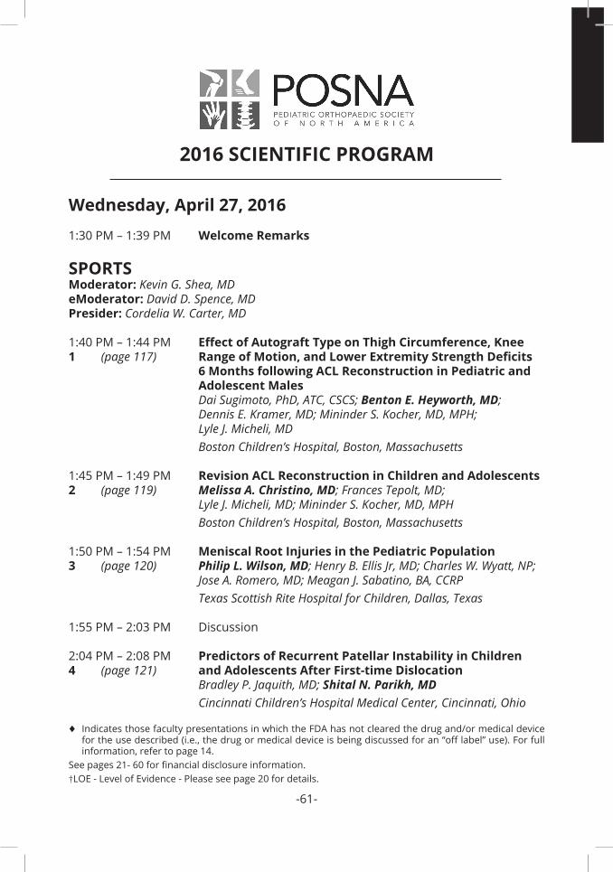
-61-
2016 SCIENTIFIC PROGRAM
♦ Indicates those faculty presentations in which the FDA has not cleared the drug and/or medical device for the use described (i.e., the drug or medical device is being discussed for an “off label” use). For full information, refer to page 14.
See pages 21- 60 for financial disclosure information. †LOE - Level of Evidence - Please see page 20 for details.
Wednesday, April 27, 2016
1:30 PM – 1:39 PM Welcome Remarks
SPORTSModerator: Kevin G. Shea, MD eModerator: David D. Spence, MDPresider: Cordelia W. Carter, MD
1:40 PM – 1:44 PM Effect of Autograft Type on Thigh Circumference, Knee1 (page 117) Range of Motion, and Lower Extremity Strength Deficits 6 Months following ACL Reconstruction in Pediatric and Adolescent Males Dai Sugimoto, PhD, ATC, CSCS; Benton E. Heyworth, MD; Dennis E. Kramer, MD; Mininder S. Kocher, MD, MPH; Lyle J. Micheli, MD Boston Children’s Hospital, Boston, Massachusetts
1:45 PM – 1:49 PM Revision ACL Reconstruction in Children and Adolescents2 (page 119) Melissa A. Christino, MD; Frances Tepolt, MD; Lyle J. Micheli, MD; Mininder S. Kocher, MD, MPH Boston Children’s Hospital, Boston, Massachusetts
1:50 PM – 1:54 PM Meniscal Root Injuries in the Pediatric Population3 (page 120) Philip L. Wilson, MD; Henry B. Ellis Jr, MD; Charles W. Wyatt, NP; Jose A. Romero, MD; Meagan J. Sabatino, BA, CCRP Texas Scottish Rite Hospital for Children, Dallas, Texas
1:55 PM – 2:03 PM Discussion
2:04 PM – 2:08 PM Predictors of Recurrent Patellar Instability in Children4 (page 121) and Adolescents After First-time Dislocation Bradley P. Jaquith, MD; Shital N. Parikh, MD Cincinnati Children’s Hospital Medical Center, Cincinnati, Ohio
2016 Scientific ProgramWEDNESDAY, APRIL 27, 2016 (cont.)
-62-
†LOE - Level of Evidence - Please see page 20 for details.See pages 21- 60 for financial disclosure information.
2:09 PM – 2:13 PM Does Medial Patellofemoral Ligament Repair on a First5 (page 122) Time Pediatric Patellar Dislocation Decrease the Future Rate of Dislocation? Haley Merrill, MD; Ian Greenberg, BS, Luv Singh, MD; Donna M. Pacicca, MD Children’s Mercy Hospital, Kansas City, Missouri 2:14 PM – 2:18 PM Pre and Post Season Elbow MRI Studies in Little League 6 (page 123) Players: A Longitudinal Study Andrew Pytiak, MD; Philip P. Stearns, NP; Joanna H. Roocroft, MA; Jerry R. Dwek, MD; Peter Kruk, MD; Tracey P. Bastrom, MA; Andrew T. Pennock, MD Rady Children’s Hospital, San Diego, California 2:19 PM – 2:27 PM Discussion
UPPER EXTREMITY Moderator: Christine A. Ho, MD eModerator: Apurva Shah, MD, MBA Presider: Kevin J. Little, MD 2:28 PM – 2:32 PM Comparison of Ultrasound and MRI for the Diagnosis of 7 (page 124) Glenohumeral Dysplasia in Brachial Plexus Birth Palsy Kenneth Donohue; Kevin J. Little, MD; Scott H. Kozin, MD; Brian Norton, MD; Dan A. Zlotolow, MD Shriners Hospital for Children, Philadelphia, Pennsylvania 2:33 PM – 2:37 PM ♦ External Rotation Predicts Outcomes after Closed 8 (page 125) Glenohumeral Joint Reduction with Botulinum Toxin A in Brachial Plexus Birth Palsy Dustin A. Greenhill, MD; Scott H. Kozin, MD; Dan A. Zlotolow, MD Shriners Hospital for Children - Philadelphia, Pennsylvania 2:38 PM – 2:42 PM Follow-up Study on the Effects of Tendon Transfers and 9 (page 126) Open Reduction on Glenohumeral Deformity in Brachial Plexus Birth Palsy Carley Vuillermin, MBBS, FRACS; Eliza B. Lewine; Donald S. Bae, MD; Peter M. Waters, MD Boston Children’s Hospital, Boston, Massachusetts
-63-
♦ Indicates those faculty presentations in which the FDA has not cleared the drug and/or medical device for the use described (i.e., the drug or medical device is being discussed for an “off label” use). For full information, refer to page 14.
2:43 PM – 2:51 PM Discussion 2:52 PM – 2:56 PM Objective Sensory Testing in Children: When is it Reliable? 10 (page 127) Karan Dua, MD; Timothy P. Lancaster, BS; Joshua M. Abzug, MD University of Maryland School of Medicine, Baltimore, Maryland 2:57 PM – 3:01 PM Ultrasound Guided Supraclavicular Nerve Block in 11 (page 129) Closed Reduction Percutaneous Pinning of Pediatric Supracondylar Humerus Fractures Scott B. Rosenfeld, MD; Chris Glover, MD; Yang Liu, MD; Kim P. Nguyen, MD; Nihar Patel, MD; Thomas Shaw, MD; Eduardo Medellin, BS; Jonathan A. Guzman, BA; Samantha Capehart, RN; Mehernoor F. Watcha, MD; Henry Huang, MD Texas Children’s Hospital, Houston, Texas 3:02 PM – 3:06 PM Early Results of Single Plug OATS for Osteochondritis 12 (page 131) Dissecans of the Capitellum Eitan M. Ingall, BA; Eliza B. Lewine; Donald S. Bae, MD Boston Children’s Hospital, Boston, Massachusetts 3:07 PM – 3:15 PM Discussion 3:16 PM – 3:20 PM A Comparison of Fixation Methods in Patients with 13 (page 132) Diaphyseal Forearm Fractures Krister P. Freese, MD; L. Wade Faulk, MD, MBA; Robin M. Baschal, BA; Claire Palmer, MS; Sarah Sibbel, MD Children’s Hospital Colorado, Aurora, Colorado 3:21 PM – 3:25 PM Conservative Management of Minimally Displaced 14 (page 133) (≤ 2 mm) Fractures of the Lateral Humeral Condyle in Pediatric Patients: A Systematic Review Derrick M. Knapik, MD; Allison Gilmore, MD; Raymond W. Liu, MD University Hospitals Case Medical Center, Cleveland, Ohio 3:26 PM – 3:30 PM Incidence and Outcome of Avascular Necrosis following 15 (page 135) Lateral Humeral Condyle Fracture in Children Lior Shabtai, MD; Nina Lightdale-Miric, MD; Alexandre Arkader, MD; Alexis Rounds, BS; James L. Pace, MD Children’s Hospital Los Angeles, Los Angeles, California 3:31 PM – 3:39 PM Discussion
3:40 PM – 4:00 PM Break
2016 Scientific ProgramWEDNESDAY, APRIL 27, 2016 (cont.)
-64-
†LOE - Level of Evidence - Please see page 20 for details.See pages 21- 60 for financial disclosure information.
TRAUMA Moderator: Todd A. Milbrandt, MD eModerator: Jeffrey E. Martus, MD Presider: Carley Vuillermin, MBBS, FRACS 4:01 PM – 4:05 PM Surgical Management of Pediatric Supracondylar 16 (page 136) Humerus Fractures in the United States: 12 Year National Trends John Vorhies, MD; Meghan N. Imrie, MD; Lawrence A. Rinsky, MD; Scott A. Hoffinger, MD Stanford University Medical Center, Stanford, California 4:06 PM – 4:10 PM Is the “Appropriate Use Criteria” for Type II 17 (page 138) Supracondylar Humerus Fractures Really Appropriate? Howard Park, MD; Erin M. Delfosse, NP; Hemali Panchal; Mauricio Silva, MD Orthopaedic Institute for Children, Los Angeles, California 4:11 PM – 4:15 PM Factors that Predict Instability in Pediatric Diaphyseal 18 (page 139) Both Bone Forearm Fractures Jeffrey Kutsikovich, MD; Christopher M. Hopkins, MD; Edwin Gannon, BS; Derek M. Kelly, MD; Jeffrey R. Sawyer, MD University of Tennessee Health Science Center, Campbell Clinic, Memphis, Tennessee, USA 4:16 PM – 4:24 PM Discussion 4:25 PM – 4:29 PM Complications After Plate Fixation of Displaced 19 (page 140) Pediatric Midshaft Clavicle Fractures G. Ying Li, MD; Peter F. Helvie, BS; Matthew D. Abbott, MD; Frances A. Farley, MD; Michelle S. Caird, MD University of Michigan, Ann Arbor, Michigan 4:30 PM – 4:34 PM Teaching the Basics: Development and Validation of a 20 (page 141) Distal Radius Reduction & Casting Model Mark Seeley, MD; Peter D. Fabricant, MD, MPH; J. Todd Lawrence, MD Children’s Hospital of Philadelphia, Philadelphia, Pennsylvania
-65-
♦ Indicates those faculty presentations in which the FDA has not cleared the drug and/or medical device for the use described (i.e., the drug or medical device is being discussed for an “off label” use). For full information, refer to page 14.
4:35 PM – 4:39 PM To Valve or Not to Valve: A Prospective Randomized Trial 21 (page 143) of Casting Options for Pediatric Forearm Fractures Paul C. Baldwin, MD; Mark C. Lee, MD; Eric Han, BS; Matthew J. Solomito, PhD; Anthony Parrino, MD Connecticut Children’s Hospital, Hartford, Connecticut 4:40 PM – 4:48 PM Discussion 4:49 PM – 4:53 PM Functional Bracing for Treatment of Pediatric Diaphyseal 22 (page 145) Femur Fractures: An Alternative to Spica Casting Andrea S. Kramer, MD; Colin Woon, MD; David Speers, CPO, LPO Advocate Children’s Hospital, Park Ridge, Illinois 4:54 PM – 4:58 PM Pediatric Orthopaedic Lower Extremity Trauma 23 (page 146) and Venous Thromboembolism Robert F. Murphy, MD; Manahil N. Naqvi, BS; Patricia E. Miller, MS; Lanna F. Feldman, MS; Benjamin J. Shore, MD, MPH, FRCSC Boston Children’s Hospital, Boston, Massachusetts 4:59 PM – 5:03 PM Percutaneous vs Open Reduction and Fixation for Tillaux 24 (page 147) and Triplane Fractures: A Multi-Center Study William D. Zelenty, BS; Richard S. Yoon, MD; Lior Shabtai, MD; Debra A. Sala, MS, PT; Regina Woon, MPH, CCRP; Michelle Ho, BS; Allison Matthews, MSCR; Paul D. Choi, MD; Benjamin D. Martin, MD; Bernard D. Horn, MD; David S. Feldman, MD; Norman Y. Otsuka, MD; David H. Godfried, MD NYU Hospital for Joint Diseases, New York, New York 5:04 PM – 5:12 PM Discussion
2016 Scientific ProgramTHURSDAY, APRIL 28, 2016
-66-
†LOE - Level of Evidence - Please see page 20 for details.See pages 21- 60 for financial disclosure information.
Thursday, April 28, 2016 8:00 AM – 8:04 AM Welcome and Remarks
HIP Moderator: Ernest L. Sink, MD eModerator: Eduardo V. Novais, MD Presider: Pooya Hosseinzadeh, MD 8:05 AM – 8:09 AM Assessment of Femoral Head Perfusion by ICP-Monitoring 25 (page 148) is an Accurate Indication of Osteonecrosis after Modified Dunn for Treatment of Unstable SCFE Travis C. Heare, MD; Lauryn A. Kestel, BS; Patrick Carry, BA; Ernest L. Sink, MD; Eduardo V. Novais, MD Children’s Hospital Colorado, Aurora, Colorado 8:10 AM – 8:14 AM Developmental Dysplasia of the Hip and Laterality: 26 (page 150) The Importance of Graded Severity of the Contralateral Hip Emily Schaeffer, PhD; Charles T. Price, MD; Kishore Mulpuri, MD; R-IHDI Study Group British Columbia Children’s Hospital, Vancouver, British Columbia 8:15 AM – 8:19 AM Contrast-Enhanced MRI After Reduction of Infant Hip 27 (page 151) Dislocation Predicts Avascular Necrosis on Initial Scan Only Travis H. Matheney, MD; Benjamin J. Shore, MD, MPH, FRCSC; Michael P. Glotzbecker, MD; Patricia E. Miller, MS; Young Jo Kim, MD Boston Children’s Hospital, Boston, Massachusetts 8:20 AM – 8:28 AM Discussion 8:29 AM – 8:33 AM Closed Reduction for Developmental Dysplasia of the Hip: 28 (page 152) Outcomes from a Multi-Center, Prospective Study Group Wudbhav N. Sankar, MD; Alex Gornitzky, BS; Nicholas Clarke, FRCS; Jose A. Herrera-Soto, MD; Simon P. Kelley, MBChB, MRCS, FRCS; Travis H. Matheney, MD; Kishore Mulpuri, MD; Vidyadhar S.V. Upasani, MD; Nicole Williams; Charles T. Price, MD; R-IHDI Study Group Children’s Hospital of Philadelphia, Philadelphia, Pennsylvania
-67-
♦ Indicates those faculty presentations in which the FDA has not cleared the drug and/or medical device for the use described (i.e., the drug or medical device is being discussed for an “off label” use). For full information, refer to page 14.
8:34 AM – 8:38 AM A Reliable and Valid Competency Based Simulated 29 (page 153) Learning Module for the Application of a Pavlik Harness Based on International Expert Consensus Simon P. Kelley, MBChB, MRCS, FRCS; Catharine Bradley, BScPT, MSc; Joel Moktar, MD; Alexandra Maxwell, BS; John H. Wedge, MD; M. Lucas Murnaghan, MD, MEd, FRCSC The Hospital for Sick Children, Toronto, Ontario, Canada 8:39 AM – 8:43 AM Evaluation of Brace Treatment for Dislocated Infantile 30 (page 154) Developmental Dysplasia of The Hip in A Prospective Cohort: Defining the Success Rate and Variables Associated with Failure Vidyadhar S.V. Upasani, MD; James D. Bomar, MPH; Travis H. Matheney, MD; Charles T. Price, MD; Kishore Mulpuri, MD; Scott J. Mubarak, MD; R-IHDI Study Group Rady Children’s Hospital, San Diego, California 8:44 AM – 8:52 AM Discussion 8:53 AM – 8:57 AM Younger Age at the Time of Closed Reduction Minimizes 31 (page 156) Acetabular Dysplasia in Children with DDH Lauren LaMont, MD; Anthony I. Riccio, MD; ChanHee Jo, PhD; Zachary Clarke; R. Lane Wimberly, MD Texas Scottish Rite Hospital, Dallas, Texas 8:58 AM – 9:02 AM Predictors of Persistent Postoperative Pain and 32 (page 157) Associated Outcomes Following Hip Osteotomy for Developmental Dysplasia of the Hip Michael B. Millis, MD; Justyna Klajn, MS; Laura E. Simons, PhD; Christine B. Sieberg, PhD; Garrett Bowen Boston Children’s Hospital, Boston, Massachusetts 9:03 AM – 9:07 AM Long Term Results Following the Bernese Periactabular 33 (page 158) Osteotomy: The Washington University Experience Joel E. Wells, MD; Kayla M. Thomason, BS; Geneva Baca, BA; Perry L. Schoenecker, MD; John C. Clohisy, MD Washington University School of Medicine, Saint Louis, Missouri 9:08 AM – 9:16 AM Discussion
2016 Scientific ProgramTHURSDAY, APRIL 28, 2016 (cont.)
-68-
†LOE - Level of Evidence - Please see page 20 for details.See pages 21- 60 for financial disclosure information.
9:17 AM – 9:21 AM Does Previous Hip Arthoscopy Impact the Clinical 34 (page 159) Outcomes of PAO Surgery? An ANCHOR Cohort Study Benjamin R. Coobs, MD; John C. Clohisy, MD; Geneva Baca, BA; Perry L. Schoenecker, MD; Jeffrey J. Nepple, MD; Michael B. Millis, MD; Young Jo Kim, MD; Ira Zaltz, MD; Rafael J. Sierra, MD; Wudbhav N. Sankar, MD; Ernest L. Sink, MD; David A. Podeszwa, MD; Daniel J. Sucato, MD, MS; Robert T. Trousdale, MD; Paul E. Beaule, MD; ANCHOR Group Washington University School of Medicine, Saint Louis, Missouri 9:22 AM – 9:26 AM MRI in Idiopathic, Stable, Slipped Capital Femoral 35 (page 160) Epiphysis: Evaluation of Contralateral Pre-slip Kevin E. Klingele, MD; Julie Samora, MD; Walter P. Samora III, MD Nationwide Children’s Hospital, Columbus, Ohio 9:27 AM – 9:31 AM Treatment of Acute, Unstable versus Chronic, Stable 36 (page 161) Slipped Capital Femoral Epiphysis using the Modified Dunn Procedure Kevin E. Klingele, MD; Walter P. Samora III, MD Nationwide Children’s Hospital, Columbus, Ohio 9:32 AM – 9:40 AM Discussion 9:41 AM – 9:45 AM The Modified Dunn is Superior to In-Situ Pinning for 37 (page 162) Anatomic Restoration and Clinical Outcome with Similar Osteonecrosis Rate in Unstable SCFE Eduardo V. Novais, MD; Lauryn A. Kestel, BS; Claire Palmer, MS; Travis C. Heare, MD; Ernest L. Sink, MD Children’s Hospital Colorado, Aurora, Colorado 9:46 AM – 9:50 AM More Accurate Diagnosis with MRI in Suspected Hip Sepsis 38 (page 163) Simon R. Thomas, MA, FRCS; Simon Bennet FRCS, MBChB; Joshua Hill, MBChB Bristol Royal Hospital for Children, Bristol, United Kingdom 9:51 AM – 9:55 AM Hip Instability is an Under Recognized and Significant 39 (page 164) Complication after Surgical Hip Dislocation in Patients with Slipped Capital Femoral Epiphysis Vidyadhar S.V. Upasani, MD; Rasika R. Deshpande, BS; Joseph J. Gugenheim, MD; Oliver Birke, MD; David G. Little, MBBS, FRACS; Michael B. Millis, MD Boston Children’s Hospital, Boston, Massachusetts
-69-
♦ Indicates those faculty presentations in which the FDA has not cleared the drug and/or medical device for the use described (i.e., the drug or medical device is being discussed for an “off label” use). For full information, refer to page 14.
9:56 AM – 10:04 AM Discussion 10:05 AM – 10:20 AM Distinguished Achievement Award Dennis R. Wenger, MD
10:21 AM – 10:36 AM Break
CONCURRENT SESSION I: SPINE Moderator: Jonathan H. Phillips, MD eModerator: Patrick J. Cahill, MD Presider: Robert H. Cho, MD 10:40 AM – 10:44 AM Can Bone Mineral Density (BMD) Predict the Curve 40 (page 166) Progression and Risk of Surgery in Newly Diagnosed Girls with Adolescent Idiopathic Scoliosis (AIS)? Benjamin Hon Kei Yip; Fiona Wai Ping Yu; Vivian Wing Yin Hung; Tsz Ping Lam, MD; Ling Qin; Bobby Kin Wah Ng, MD; Jack Cheng Department of Orthopaedics and Traumatology, The Chinese University of Hong Kong, Hong Kong 10:45 AM – 10:49 AM Is Constant Back Pain an Adequate Predictor of the 41 (page 167) Presence of a Significant Pathology Associated with Pediatric Back Pain? Norman Ramirez-Lluch, MD; Pablo Valentin, BS; Ivan Iriarte, MD; Soniely Lugo, MD Hospital de la Concepcion, San German, Puerto Rico 10:50 AM – 10:54 AM Multimodality Pain Management Improves Outcomes 42 (page 168) and Satisfaction and Reduces Length of Stay after PSF for AIS Suken A. Shah, MD; Karen Sacks; Kenneth J. Rogers, PhD; Geraldine Neiss, PhD; Petya Yorgova, MS; Randy Brenn, MD; Dinesh K. Choudhry, MD Nemours / Alfred I. duPont Hospital for Children, Wilmington, Delaware 10:55 AM – 11:03 AM Discussion 11:04 AM – 11:08 AM Are Traction or Bending Radiographs Helpful 43 (page 170) in Selection of Fusion Levels for AIS? Walter F. Krengel III, MD; Viviana Bompadre, PhD; Gregory Redding, MD; Klane K. White, MD, MS Seattle Children’s Hospital, Seattle, Washington
2016 Scientific ProgramTHURSDAY, APRIL 28, 2016 (cont.)
-70-
†LOE - Level of Evidence - Please see page 20 for details.See pages 21- 60 for financial disclosure information.
11:09 AM – 11:13 AM Cost Analysis of Adolescent Idiopathic Scoliosis Surgery: 44 (page 171) Early Discharge Decreases Hospital Costs Much Less Than Intraoperative Variables Under the Control of the Surgeon Brandon L. Raudenbush, DO; David P. Gurd, MD; Thomas E. Kuivila, MD; R. Tracy Ballock, MD; Ryan C. Goodwin, MD Cleveland Clinic, Cleveland, Ohio 11:14 AM – 11:18 AM Predictors of Distal Adding-on in Thoracic Major Curves 45 (page 172) in Adolescent Idiopathic Scoliosis Joshua S. Murphy, MD; Vidyadhar S.V. Upasani, MD; Burt Yaszay, MD; Tracey P. Bastrom, MA; Amer Samdani, MD; Lawrence G. Lenke, MD; Peter O. Newton, MD Rady Children’s Hospital, San Diego, California 11:19 AM – 11:27 AM Discussion 11:28 AM – 11:32 AM Peri-operative and Delayed Major Complications 46 (page 173) Following Surgical Correction of AIS in 3530 Patients Carrie Bartley, MA; Burt Yaszay, MD; Tracey P. Bastrom, MA; Suken A. Shah, MD; Baron Lonner, MD; Jahangir Asghar, MD; Firoz Miyanji, MD; Amer Samdani, MD; Peter O. Newton, MD Rady Children’s Hospital, San Diego, California 11:33 AM – 11:37 AM “Risk Adjusted” Comparative Infection Rates in 47 (page 175) Adolescent Idiopathic Scoliosis Peter O. Newton, MD; Burt Yaszay, MD; Tracey P. Bastrom, MA; Harms Study Group Rady Children’s Hospital, San Diego, California 11:38 AM – 11:42 AM Neuromotor Sub-Classification of GMFCS-5 Predicts 48 (page 177) Complications and Health Related Quality of Life in Patients with Cerebral Palsy after Spine Fusion Amit Jain, MD; Paul D. Sponseller, MD; Suken A. Shah, MD; Amer Samdani, MD; Patrick J. Cahill, MD; Burt Yaszay, MD; Unni G. Narayanan, MBBS, MSc, FRCSC; Peter O. Newton, MD; Michelle Marks, PT, MA Johns Hopkins, Baltimore, Maryland 11:43 AM – 11:51 AM Discussion
-71-
♦ Indicates those faculty presentations in which the FDA has not cleared the drug and/or medical device for the use described (i.e., the drug or medical device is being discussed for an “off label” use). For full information, refer to page 14.
11:52 AM – 11:56 AM Repeat Surgical Intervention Following Definitive 49 (page 179) Instrumentation and Fusion for Adolescent Idiopathic Scoliosis: A 25-Year Update Megan Mignemi, MD; Dong-Phuong Tran, MS; Brandon A. Ramo, MD; B. Stephens Richards, MD Texas Scottish Rite Hospital for Children, Dallas, Texas 11:57 AM – 12:01 PM Reduced Pulmonary Function in AIS Patients with 50 (page 180) Hypokyphosis: Mean 30-Year Follow-up A. Noelle Larson, MD; William J. Shaughnessy, MD; Charles G. Ledonio, MD; David W. Polly Jr, MD; Michael A. Yaszemski, MD, PhD; Clayton Cowl, MD, MS Mayo Clinic, Rochester, Minnesota 12:02 PM – 12:06 PM Cervical Spine Disease Common after Pediatric 51 (page 182) Treatment of AIS at Mean 30-Year Follow-Up Ernest Young, MD; A. Noelle Larson, MD; Michael A. Yaszemski, MD, PhD Mayo Clinic, Rochester, Minnesota 12:07 PM – 12:15 PM Discussion
CONCURRENT SESSION II:CONGENTIAL/SYNDROME, LOWER EXTREMITY/NEUROMUSCULAR Moderator: Darko Anticevic, MD, PhD eModerator: Eric D. Shirley, MD Presider: Corrina C. Franklin, MD 10:40 AM – 10:44 AM Role of Body Cast Application for Scoliosis Associated 52 (page 184) with Prader-Willi Syndrome Harold J.P. van Bosse, MD; Patrick J. Cahill, MD Shriners Hospital for Children, Philadelphia, Pennsylvania 10:45 AM – 10:49 AM Does Open Reduction of Arthrogrypotic Hips 53 (page 185) Cause Stiffness? Harold J.P. van Bosse, MD; Dustin A. Greenhill, MD Shriners Hospital for Children, Philadelphia, Pennsylvania
2016 Scientific ProgramTHURSDAY, APRIL 28, 2016 (cont.)
-72-
†LOE - Level of Evidence - Please see page 20 for details.See pages 21- 60 for financial disclosure information.
10:50 AM – 10:54 AM Fassier-Duval Telescoping Rodding in Osteogenesis 54 (page 186) Imperfecta: Rod Revision and Survivorship Samantha A. Spencer, MD; Victoria Prete, BS; Patricia E. Miller, MS; Amanda T. Whitaker, MD; Susan T. Mahan, MD, MPH; Collin J. May, MD; James R. Kasser, MD Boston Children’s Hospital, Boston, Massachusetts 10:55 AM – 11:03 AM Discussion 11:04 AM – 11:08 AM Peroneal Nerve Function Before and Following Surgical 55 (page 188) Excision of a Proximal Fibular Osteochondroma Daniel J. Sucato, MD, MS; Kevin Smit, MD, FRCSC; Craig M. Birch, MD Texas Scottish Rite Hospital for Children, Dallas, Texas 11:09 AM – 11:13 AM Complications Associated with Epiphysiodesis for 56 (page 189) Management of Leg Length Discrepancy Karl E. Rathjen, MD; Marina Makarov; Brandon A. Ramo, MD; Chukwudi Chukwunyerenwa, MD; Samuel Dunn; Dustin Singer, BA; John G. Birch, MD Texas Scottish Rite Hospital for Children 11:14 AM – 11:18 AM Early Complications Associated with Limb Lengthening 57 (page 190) using a Magnetically Activated Intramedullary Lengthening Device in Pediatric Patients Mark L. Miller, MD; J. Eric Gordon, MD Washington University, Saint Louis, Missouri 11:19 AM – 11:27 AM Discussion 11:28 AM – 11:32 AM Frequency of Crouch Gait in Spastic Diplegic Patients 58 (page 191) With and Without History of Tendo Achilles Lengthening Lise Leveille, MD; Ashley Erdman, BS, MBA; Kelly Jeans, MSc; Kirsten Tulchin-Francis, PhD; Lori A. Karol, MD Texas Scottish Rite Hospital for Children, Dallas, Texas 11:33 AM – 11:37 AM Percutaneous Hamstring Lengthening Surgery is as 59 (page 192) Effective as Open Lengthening in Children with Cerebral Palsy Robert M. Kay, MD; Alexander Nazareth, MS; Susan A. Rethlefsen, DPT; Ted C. Sousa, MD; Nicole M. Mueske, MS; Tishya A.L. Wren, PhD Children’s Hospital Los Angeles, Los Angeles, California
-73-
♦ Indicates those faculty presentations in which the FDA has not cleared the drug and/or medical device for the use described (i.e., the drug or medical device is being discussed for an “off label” use). For full information, refer to page 14.
11:38 AM – 11:42 AM The Gait Outcomes Assessment List (GOAL): 60 (page 193) Validation of a New Measure of Gait Function for Children with Cerebral Palsy Annie Tan, BSc; Alice Donnan, BSc; Pam J. Thomason, BPHysio; Jill M. Rodda, PhD, PT; H. Kerr Graham, MD, FRCS (Ed), FRACS; Unni G. Narayanan, MBBS, MSc, FRCSC The Royal Children’s Hospital, Melbourne, Australia 11:43 AM – 11:51 AM Discussion 11:52 AM – 11:56 AM Isolated VDRO (Varus Derotation Proximal Femoral 61 (page 194) Osteotomy) with Medial Capsular Release for Spastic Quadriplegic Hips with Underlying Acetabular Dysplasia Laura E. Gill, MD; Kevin E. Klingele, MD; John R. Kean, MD Nationwide Children’s Hospital, Columbus, Ohio 11:57 AM – 12:01 PM Use of the Multiplier Method to Predict Leg Length 62 (page 195) Discrepancy in Children with Spastic Hemiplegic Cerebral Palsy Allison Frickman, BA; Khalid Hesham, MD; Steve D. Gibbons, MD; Michelle L. Dudevoir, MD; Breanna Pritchard, BA; Jason T. Rhodes, MD Children’s Hospital Colorado, Aurora, Colorado 12:02 PM – 12:06 PM Self Reported Quality of Life in Adolescence and Young 63 (page 197) Adults with Cerebral Palsy Nancy Lennon, MSPT; Chris Church, MPT; Freeman Miller, MD; Julieanne P. Sees, DO Nemours / Alfred I. duPont Hospital for Children, Wilmington, Delaware 12:07 PM – 12:15 PM Discussion
-74-
†LOE - Level of Evidence - Please see page 20 for details.See pages 21- 60 for financial disclosure information.
SYMPOSIA PROGRAM
COUR SYMPOSIUM
Thurs., April 28, 2016 • 1:15 PM – 3:15 PM • JW Ballroom 7-8
Chair: Coleen S. Sabatini, MD, MPH
ADAPTING CARE TO AUSTERE ENVIRONMENTS – PEDIATRIC ORTHOPAEDICS IN RESOURCE-LIMITED AREAS
1:15 PM – 1:20 PM Welcome and Introduction to the Symposium Coleen S. Sabatini, MD, MPH
1:20 PM – 1:50 PM COUR Scholar Introductions and Presentations Coleen S. Sabatini, MD, MPH Scholars: Mandar Vikes Agashe, MBBS (India) Garcia Cielo Estrera Balce, MD (Philippines) Erika Iliana Arana Hernandez, MD (Mexico) Aradhana Jirendra Jha, MD (Nepal)
1:50 PM – 2:05 PM Neglected Clubfoot Kenneth J. Noonan, MD
2:05 PM – 2:15 PM Discussion
2:15 PM – 2:30 PM Neglected Sepsis of the Hip David A. Spiegel, MD
2:30 PM – 2:40 PM Septic Hip Discussion
2:40 PM – 2:55 PM Chronic Osteomyelitis J. Norgrove Penny, MD
2:55 PM – 3:05 PM Chronic Osteomyelitis Discussion
3:05 PM – 3:15 PM Discussion and Closure
-75-
♦ Indicates those faculty presentations in which the FDA has not cleared the drug and/or medical device for the use described (i.e., the drug or medical device is being discussed for an “off label” use). For full information, refer to page 14.
SYMPOSIA PROGRAM
RESEARCH WORKSHOP
Thurs., April 28, 2016 • 1:15 PM – 2:45 PM • JW Ballroom 1-4
Co-Chairs: Michelle S. Caird, MD; Joseph (Jay) Janicki, MD; Jonathan G. Schoenecker, MD, PhD; M. Wade Shrader, MD
Introduction Michelle S. Caird, MD
PERIOD 1 STUDY DESIGN – IT ALL DEPENDS ON THE QUESTION Moderators: M. Wade Shrader, MD Jonathan G. Schoenecker, MD, PhD
1:15 PM – 1:22 PM Outcomes 2015–What Should we be Measuring? Condition Specific Measures, QOL, Pedi-CAT, PROMIS Benjamin J. Shore, MD, MPH, FRCSC; M. Wade Shrader, MD
1:23 PM – 1:30 PM Multi-Center Study Groups: Where is Pediatric Orthopaedics in 2016? Benton E. Heyworth, MD
1:31 PM – 1:38 PM The “Alphabet Soup” of Quality Improvement: What Does It All Mean? Michael G. Vitale, MD, MPH
1:39 PM – 1:46 PM Philosophy of Establishing a Translational Research Project Jonathan G. Schoenecker, MD, PhD
1:47 PM – 1:54 PM Discussion
-76-
†LOE - Level of Evidence - Please see page 20 for details.See pages 21- 60 for financial disclosure information.
SYMPOSIA PROGRAM RESEARCH SYMPOSIUM, (cont.)
PERIOD 2 POSNA SUPPORTED RESEARCH HIGHLIGHTS Moderators: Michelle S. Caird, MD Joseph (Jay) Janicki, MD
1:55 PM – 1:59 PM 2013 Biomet Spine Research Grant Nicholas M. Bernthal, MD A Mouse Model of Spinal Implant Infection: A Novel Method to Evaluate Metal Susceptibility to Infection and Anti-microbial Coatings
2:00 PM – 2:04 PM 2014 Biomet Research Grant Matthew A. Halanski, MD A New Porcine Model for Pediatric Spinal Deformity
2:05 PM – 2:09 PM 2014 Angela M. Kuo Memorial Award Roger Cornwall, MD Muscle Contracture Pathophysiology in CP and Neonatal Brachial Plexus Injury
2:10 PM – 2:14 PM 2014 Arthur Huene Memorial Research Award Lawson A.B. Copley, MD Gene Expression of Methycillin-resistant and Methycillin-sensitive Staphylococcus Aureus Isolates Which Cause Acute Hematogenous Osteomyelitis in Children–The Impact on Clinical Severity of Illness
2:15 PM – 2:22 PM Discussion
PERIOD 3 GRANT DEVELOPMENT STRATEGIES Moderators: Mininder S. Kocher, MD, MPH Michelle S. Caird, MD
2:23 PM – 2:28 PM Opportunities for Funding after POSNA: RO1 an Ultimate ROI Michelle S. Caird, MD
2:29 PM – 2:34 PM Industry Funding: Partnering for Innovation and Improvement in Patient Care Peter O. Newton, MD
2:35 PM – 2:39 PM Philanthropy Funding: Grateful Patients and Research Giving Mininder S. Kocher, MD, MPH
2:40 PM – 2:47 PM Discussion
-77-
♦ Indicates those faculty presentations in which the FDA has not cleared the drug and/or medical device for the use described (i.e., the drug or medical device is being discussed for an “off label” use). For full information, refer to page 14.
SYMPOSIA PROGRAM
PRACTICE MANAGEMENT SYMPOSIUM
Thurs., April 28, 2016 • 1:15 PM – 2:45 PM • JW Ballroom 9-10
Co-Chairs: Lawson A.B. Copley, MD; John F. Lovejoy III, MD; Jeffrey B. Neustadt, MD; Stephen B. Sundberg, MD; Timothy S. Oswald, MD; Elizabeth L. Magnabosco, MD; David J. Anderson, MD; Chester H. Sharps, MD
EMR APPLICATIONS IN PEDIATRIC ORTHOPAEDICS
1:15 PM–1:20 PM Introduction and Review of AAP SOOr 2014 Survey Lawson A.B. Copley, MD
1:20 PM – 1:30 PM Review of PMC John F. Lovejoy III, MD User Vendor Landscape Map Reasons for dissatisfaction Reasons for satisfaction Opportunities for improvement
1:30 PM–1:45 PM Strategic Employment Lawson A.B. Copley, MD PROs Discrete data identification Practice Efficiency Measurement Provider Satisfaction Assessment Multi-center Studies through Sharing of Big Data REDCap and Oberd 3rd Party Solutions for Information Sharing Clinical Research Management Systems
Panel Discussion 1:45 PM – 1:51 PM I. Epic Systems – Lawson A.B. Copley, MD 1:52 PM – 1:58 PM II. Cerner – Stephen B. Sundberg, MD 1:59 PM – 2:05 PM III. NextGen – David J. Anderson, MD 2:06 PM – 2:12 PM IV. Athena – Elizabeth L. Magnabosco, MD 2:13 PM – 2:19 PM V. GE Centricity – Jeffrey B. Neustadt, MD
-78-
†LOE - Level of Evidence - Please see page 20 for details.See pages 21- 60 for financial disclosure information.
2:20 PM – 2:26 PM VI. Allscripts – Chester H. Sharps, MD 2:27 PM – 2:33 PM VII. SRS – Timothy S. Oswald, MD
2:34 PM – 2:45 PM General Discussion Q&A Consideration of strategy to develop common ground among POSNA members regarding which PROs, discrete data elements, and quality measures should be captured electronically for sharing.
SYMPOSIA PROGRAMPRACTICE MANAGEMENT SYMPOSIUM, (cont.)
-79-
♦ Indicates those faculty presentations in which the FDA has not cleared the drug and/or medical device for the use described (i.e., the drug or medical device is being discussed for an “off label” use). For full information, refer to page 14.
SYMPOSIA PROGRAM
POPS NP / PA
Thurs., April 28, 2016 • 1:15 PM – 3:00 PM • Room 203-5
Co-Chairs: Colleen P. Ditro, DNP, CPNP; Leslie Rhodes, DNP, PPCNP-BC
Faculty: Mihir M. Thacker, MD; Vincent S. Mosca, MD; Derek M. Kelly, MD
WALK THIS WAY...FOOT ABNORMALITIES
1:15 PM – 1:35 PM Presentation I: Diagnosis and Treatment of Clubfoot Mihir M. Thacker, MD
1:35 PM – 1:45 PM Discussion
1:45 PM – 2:05 PM Presentation II: Pediatric Flat Feet Vincent S. Mosca, MD
2:05 PM – 2:15 PM Discussion
2:15 PM – 2:30 PM Break
2:30 PM – 2:50 PM Presentation III: Congenital Vertical Talus Derek M. Kelly, MD
2:50 PM – 3:00 PM Discussion
-80-
†LOE - Level of Evidence - Please see page 20 for details.See pages 21- 60 for financial disclosure information.
YOUNG MEMBER FORUM
SECOND ANNUAL ARABELLA LEET YOUNG MEMBER FORUM
Thurs., April 28, 2016 • 5:00 PM – 6:30 PM • JW Ballroom 7-8
Moderator: Brian G. Smith, MD
5:00 PM – 5:20 PM How to Work with your Boss and the Administration How to Develop your Practice to Enhance your Stature in the Eyes of your Chief and the Executive Suite David L. Skaggs, MD, MMM
5:20 PM – 5:30 PM Discussion 5:30 PM – 5:50 PM Volunteering in Resource Poor Environments: Doing Well by Doing Good Where, When, How and Why do Volunteer Work; What is COUR? Richard M. Schwend, MD
5:50 PM – 6:00 PM Discussion
6:00 PM – 6:20 PM Quality Initiatives in your Clinical Practice How to Incorporate Quality and Safety Metrics in One’s Practice: How to Initiate Research on Quality and Safety Topics Michael G. Vitale, MD, MPH
6:20 PM – 6:30 PM Discussion
POSNA extends sincere appreciation to Shriners Hospitals for Childrenfor their support of the Arabella Leet Young Member Forum.
-81-
♦ Indicates those faculty presentations in which the FDA has not cleared the drug and/or medical device for the use described (i.e., the drug or medical device is being discussed for an “off label” use). For full information, refer to page 14.
Friday, April 29, 2016 8:00 AM – 8:04 AM Welcome and Remarks
CLINICAL AWARD PAPERS Moderator: Dennis R. Wenger, MD eModerator: Lindsay M. Andras, MD Presider: Bryan J. Tompkins, MD 8:05 AM – 8:09 AM Preventing Curve Progression with Calcium and Vitamin D 64 (page 199) Supplementation in Adolescent Idiopathic Scoliosis (AIS) – A Randomised Double-blinded Placebo-controlled Trial Tsz Ping Lam, MD; Benjamin Hon Kei Yip; Elisa Man Shan Tam; Gene Chi Wai Man; Wayne YW Lee; Fiona Wai Ping Yu; Bobby Kin Wah Ng, MD; Jack Cheng; Kwong Man Lee Department of Orthopaedics and Traumatology, the Chinese University of Hong Kong, Prince of Wales Hospital, Hong Kong 8:10 AM – 8:14 AM Discussion 8:15 AM – 8:19 AM Is There Value In Having Radiology Provide a Second 65 (page 201) Reading in Pediatric Orthopedic Clinic? Vivek Natarajan, MD; Patrick P. Bosch, MD; Ozgur Dede, MD; Stephen A. Mendelson, MD; Maria Brooks, PhD; Tanya Kenkre, PhD; James W. Roach, MD Children’s Hospital of Pittsburgh, Philadelphia, Pennsylvania 8:20 – 8:24 AM Discussion 8:25 AM – 8:29 AM Abduction Bracing versus Natural History in Hip 66 (page 202) Dysplasia: Multicenter Randomized Controlled Trial Virginie Pollet, MD; Ralph Sakkers, MD, PhD; Michiel van de Sande, MD, PhD; Melinda Witbreuk; Erik Beek; Rene M. Castelein, MD, PhD Wilhelmina Children’s Hospital, Utrecht, the Netherlands 8:30 AM – 8:34 AM Discussion
2016 Scientific ProgramFRIDAY, APRIL 29, 2016
2016 Scientific ProgramFRIDAY, APRIL 29, 2016 (cont.)
-82-
†LOE - Level of Evidence - Please see page 20 for details.See pages 21- 60 for financial disclosure information.
8:35 AM – 8:39 AM Results of AIS Surgery at a Minimum of 10 Years Using 67 (page 203) Modern Implant Systems- Are Patients Troubled by Symptoms and a High Reoperation Rate? Daniel J. Sucato, MD, MS; Kaitlyn Brown, BS; Anna McClung, RN, BSN Texas Scottish Rite Hospital for Children, Dallas, Texas 8:40 AM – 8:44 AM Discussion 8:45 AM – 8:49 AM 5-Year Reoperation Rate and Causes for Revision 68 (page 204) After Idiopathic Scoliosis Surgery Syed Ahmed, MD; Tracey P. Bastrom, MA; Burt Yaszay, MD; Peter O. Newton, MD; Harms Study Group Rady Children’s Hospital, San Diego, California 8:50 AM – 8:54 AM Discussion 8:55 AM – 8:59 AM Outcomes of Physeal Sparing ACL Reconstruction with IT 69 (page 206) Band in Skeletally Immature Children Mininder S. Kocher, MD, MPH; Benton E. Heyworth, MD; Frances Tepolt, MD; Lyle J. Micheli, MD Boston Children’s Hospital, Boston, Massachusetts 9:00 AM – 9:04 AM Discussion 9:05 AM – 9:09 AM Pediatric Femur Fracture Management: A Multicenter 70 (page 208) Analysis of the Trends Pre and Post 2009 AAOS Clinical Practice Guidelines John D. Roaten, MD; Jeffrey R. Sawyer, MD; Joseph L. Yellin, BA; John (Jack) M. Flynn, MD; Micaela Cyr, BA; Sumeet Garg, MD; Alexander M. Broom, BA; Lindsay M. Andras, MD Le Bonheur Children’s Hospital, Memphis, Tennessee 9:10 AM – 9:14 AM Discussion 9:15 AM – 9:19 AM The Effect of Spastic Hip Dysplasia on Health Related 71 (page 209) Quality of Life in Non-ambulatory Children with Cerebral Palsy Rachel L. DiFazio, PhD, RN, PPCNP-BC, FAAN; Benjamin J. Shore, MD, MPH, FRCSC; Judith Vessey, PhD, MBA, RN, FAAN; Patricia E. Miller, MS; Brian D. Snyder, MD, PhD Boston Children’s Hospital, Boston, Massachusetts
-83-
♦ Indicates those faculty presentations in which the FDA has not cleared the drug and/or medical device for the use described (i.e., the drug or medical device is being discussed for an “off label” use). For full information, refer to page 14.
9:20 AM – 9:24 AM Discussion 9:25 AM – 9:29 AM Percutaneous Heel Cord Release for Clubfoot: 72 (page 210) A Retrospective, Multicenter Cost Analysis Matthew Abbott, MD; Brittany Hedrick, MD; Franklin K. Gettys, MD; B. Stephens Richards, MD; Ryan D. Muchow, MD; ChanHee Jo, PhD; Amanda Moualeu, MPH University of Michigan, Mott Children’s Hospital, Ann Arbor, Michigan 9:30 AM – 9:34 AM Discussion 9:35 AM – 9:39 AM Posttraumatic Stress in Children and Adolescents 73 (page 211) Following Surgery for Orthopedic Trauma Collin J. May, MD; Benjamin J. Shore, MD, MPH, FRCSC; Patricia E. Miller, MS; Manahil N. Naqvi, BS; Justyna M. Klajn, MS; Emily Rademacher, BS; Daniel J. Hedequist, MD Boston Children’s Hospital, Boston, Massachusetts 9:40 AM – 9:44 AM Discussion 9:45 AM – 9:49 AM Management of Clubfoot Relapses with the Ponseti 74 (page 212) Method: Results of a Survey of POSNA Members Pooya Hosseinzadeh, MD; Jose A. Morcuende, MD, PhD; Lori Dolan, PhD; Gary Kiebzak, PhD Baptist Children’s Hospital, Miami, Florida 9:50 AM – 9:54 AM Discussion 9:55 – 9:59 AM Single Event Multilevel Surgery in Bilateral Spastic 75 (page 213) Cerebral Palsy: A Long Term, Multicenter, Follow-up Study Thomas Dreher, MD, PhD; Martin Svehlik, MD; Leonhard Döderlein, MD; Sebastian I. Wolf, PhD; Pam Thomason, BPHysio; Morgan Sangeux; Gerhardt Steinwender, MD; H. Kerr Graham, MD, FRCS (Ed), FRACS Heidelberg University Hospital, Heidelberg, Germany 10:00 AM – 10:04 AM Discussion
10:05 AM – 10:25 AM Break
2016 Scientific ProgramFRIDAY, APRIL 29, 2016 (cont.)
-84-
†LOE - Level of Evidence - Please see page 20 for details.See pages 21- 60 for financial disclosure information.
BASIC SCIENCE AWARD PAPERS Moderator: Jonathan G. Schoenecker, MD, PhDeModerator: Scott B. Rosenfeld, MDPresider: Megan Mignemi, MD 10:26 AM – 10:31 AM ♦ Intra-Osseous Delivery of BMP-2 Using a Self-Assembling 76 (page 215) Peptide Hydrogel, RADA16 Matthew Phipps, PhD; Felipe Do Monte, MD; Manav Mehta, PhD; Harry K.W. Kim, MD, MS Texas Scottish Rite Hospital for Children, Dallas, Texas 10:32 AM – 10:37 AM Fibroblast Growth Factor Receptor 3 Modulates Fracture 77 (page 216) Repair By Controlling the Balance of Intramembranous and Endochondral Bone Formation Simon P. Kelley, MBChB, MRCS, FRCS; Chunying Yu; Benjamin A. Alman, MD, FRCSC The Hospital for Sick Children, Toronto, Ontario 10:38 AM – 10:43 AM The Role of Periosteal Fibers in Accelerated Bone Growth 78 (page 218) Rajeev Chaudhary, MS; Ellen Leiferman, DVM; Ray Vanderby, PhD; Kevin Eliceiri; Paul J. Campagnola, PhD; Matthew A. Halanski, MD University of Wisconsin, Madison, Wisconsin 10:44 AM – 10:52 AM Discussion 10:53 AM – 10:58 AM Pharmacokinetic Profile of Intravenous Tranexamic Acid 79 (page 220) in Adolescent Idiopathic Scoliosis Surgery Susan Goobie, MD; Michael T. Hresko, MD; MaryEllen McCann, MD; Daniel J. Hedequist, MD; Michael P. Glotzbecker, MD; Navil F. Sethna, MD; Lawrence I. Karlin, MD; Andres Navedo, MD; John B. Emans, MD; Brenda H. Barton; David Zurakowski, PhD Boston Children’s Hospital, Boston, Massachusetts 10:59 AM – 11:04 AM Morphological Symmetry and Spatial Orientation of the 80 (page 222) Vestibular Systems- Are There Any Differences Between the Progressive and Non-progressive Adolescent Idiopathic Scoliosis Vs Normal Controls? Fiona Wai Ping Yu; Steve Cheuk Ngai Hui; Bobby Kin Wah Ng, MD; Defang Wang; Jack Cheng; Winnie Chiu Wing Chu; Lin Shi Department of Orthopaedics and Traumatology, The Chinese University of Hong Kong, Hong Kong
-85-
♦ Indicates those faculty presentations in which the FDA has not cleared the drug and/or medical device for the use described (i.e., the drug or medical device is being discussed for an “off label” use). For full information, refer to page 14.
11:05 AM – 11:10 AM Development of Injectable MSC Micro-tissue to Repair 81 (page 223) Cartilage Defects Using 3D Bioprinting Brian E. Grottkau, MD; Dongyang Ma; Zhixin Hui; Yonggang Pang, MD, PhD Massachusetts General Hospital for Children, Boston, Massachusetts 11:11 AM – 11:19 AM Discussion 11:20 AM – 11:22 AM Introduction of Presidential Speaker 11:23 AM – 11:43 AM Presidential Speaker – James W. Roach, MD “Skeptic Driven Innovation” 11:44 AM – 11:49 AM EPOS/POSNA (EPOSNA) Annual Meeting 2017/ Other Announcements 11:50 AM – 12:15 PM Presidential Transfer
-86-
†LOE - Level of Evidence - Please see page 20 for details.See pages 21- 60 for financial disclosure information.
2016 SUBSPECIALTY DAY AGENDA
HAND/UPPER EXTREMITY SUBSPECIALTY DAY
CONTROVERSIES IN PEDIATRIC UPPER EXTREMITY SURGERY
Fri., April 29, 2016 • 1:00 PM – 5:10 PM • Room 203-5
Co-Chairs: Dan A. Zlotolow, MD and Christine A. Ho, MD
PERIOD 1 FREE PAPERS Moderators: Dan A. Zlotolow, MD Christine A. Ho, MD
1:00 PM – 1:04 PM The Effect of Scapular Position on MRI Measurements82 (page 224) of Glenohumeral Dysplasia Caused by Brachial Plexus Birth Palsy Roger Cornwall, MD; Tal Laor, MD; Andrew M. Zbojniewicz, MD; Jill Stein, MD Cincinnati Children’s Hospital, Cincinnati, Ohio
1:05 PM – 1:09 PM Ultrasound Screening for Posterior Shoulder Dislocation83 (page 225) in Infants with Brachial Plexus Birth Palsy Andrea S. Bauer, MD; Justin Lucas, MD; Nasser Heyrani, MD; Ryan Anderson, MD; Michelle A. James, MD Shriners Hospitals for Children-Northern California, San Francisco, California
1:10 PM – 1:14 PM ♦ Onabotulinum Toxin A Injections to Triceps Unmasks84 (page 226) Elbow Flexion in Infant Brachial Plexus Birth Palsy – A Case Series Mark J. Adamczyk, MD; Suneet Sahgal, MD; Melanie Morscher, BS; Matthew D. Thomas Akron Children’s Hospital, Akron, Ohio
1:15 AM – 1:24 PM Discussion
-87-
♦ Indicates those faculty presentations in which the FDA has not cleared the drug and/or medical device for the use described (i.e., the drug or medical device is being discussed for an “off label” use). For full information, refer to page 14.
HAND/UPPER EXTREMITY SUBSPECIALTY DAY, (cont.)FRIDAY, APRIL 29, 2016
1:25 PM – 1:29 PM Capitellar Fractures in Children and Adolescents: 85 (page 227) Classification and Resultsof Treatment Praveen Murthy, MD; Manahil N. Naqvi, BS; Carley Vuillermin, MBBS, FRACS; Peter M. Waters, MD; Donald S. Bae, MD Boston Children’s Hospital, Boston, Massachusetts
1:30 PM – 1:34 PM Functional Impact of Congenital Hand Differences: 86 (page 229) Early Results from the Congenital Upper Limb Differences (CoULD) Registry Donald S. Bae, MD; Andrea S. Bauer, MD; Carley Vuillermin, MBBS, FRACS; Peter M. Waters, MD; Charles A. Goldfarb, MD; Lindley B. Wall, MD; Summer Roberts, MA; Maria Canizares, MD; Patricia E. Miller, MS Boston Children’s Hospital, Boston, Massachusetts
1:35 PM – 1:39 PM Severity of Ulnar Ray Deficiency and its Relationship 87 (page 231) with Lower Extremity Deficiencies Justin Lea, MD; Scott A. Riley, MD; Janet Walker, MD Shriners Hospital for Children, Lexington, Kentucky
1:40 PM – 1:49 PM Discussion
1:50 PM – 1:54 PM Ultrasound Assessment of Trigger Thumb Resolution 88 (page 233) Christine Wu, BA; Michelle S. Caird, MD; Clifford L. Craig, MD University of Michigan, Ann Arbor, Michigan
1:55 PM – 1:59 PM Variation Amongst Pediatric Orthopaedic Surgeons When 89 (page 234) Diagnosing and Treating Pediatric Distal Radius Fractures Karan Dua, MD; Matthew K. Stein, BS, MS; Brian K. Brighton, MD, MPH; William L. Hennrikus, MD; Martin J. Herman, MD; J. Todd Lawrence, MD; Charles T. Mehlman, DO, MPH; Norman Y. Otsuka, MD; M. Wade Shrader, MD; Brian G. Smith, MD; Paul D. Sponseller, MD; Joshua M. Abzug, MD University of Maryland School of Medicine, Baltimore, Maryland
2:00 PM – 2:04 PM Risk Factors for Complications in Open Forearm 90 (page 235) Fractures in the Pediatric Population Adam C. Hines, MD; Marilyn Elliott; Kevin Smit, MD, FRCSC; Daniel J. Sucato, MD, MS; R. Lane Wimberly, MD; Anthony I. Riccio, MD Texas Scottish Rite Hospital, Dallas, Texas
-88-
†LOE - Level of Evidence - Please see page 20 for details.See pages 21- 60 for financial disclosure information.
HAND/UPPER EXTREMITY SUBSPECIALTY DAY, (cont.)FRIDAY, APRIL 29, 2016
2:05 PM – 2:09 PM Management of Pediatric Type I Open Fractures 91 (page 236) Matthew Schur, BA; Jenna Godfrey, MD, MS; Lior Shabtai, MD; Sarah Nossov, MD; Amy K. Williams, MD; Antoinette W. Lindberg, MD; Selina Silva, MD; Michelle S. Caird, MD; Paul D. Choi, MD; Alexandre Arkader, MD Children’s Hospital Los Angeles, Los Angeles, California
2:10 PM -2:19 PM Discussion
2:20 PM – 2:39 PM Break
PERIOD 22:40 PM – 3:00 PM Symbrachydactyly: Phalanx or Toe or? Moderator: Andrea S. Bauer, MD Lorenzo Garagnani, MD; Roger Cornwall, MD
3:00 PM – 3:20 PM ♦ Syndactyly: Graft, Flap, or Synthetic? Moderator: Joshua M. Abzug, MD Lorenzo Garagnani, MD; Dan A. Zlotolow, MD
3:20 PM – 3:40 PM ♦ MHE in the Forearm: Lengthen, Release, or Accept Deformity? Moderator: Roger Cornwall, MD Scott Oishi, MD, FACS; Dan A. Zlotolow, MD
3:40 PM – 4:00 PM Break
PERIOD 34:00 PM – 4:20 PM Madelungs / Focal Fibrocartilaginous Dysplasia: Utility of Early Release? Moderator: Scott Oishi, MD, FACS Suzanne E. Steinman, MD; Christine A. Ho, MD
4:25 PM – 4:45 PM Supracondylar: What to Do with Pink Pulseless? Moderator: Christine A. Ho, MD Steven L. Frick, MD; Donald S. Bae, MD
4:50 PM – 5:10 PM Monteggia: When is it Too Late? Moderator: Lisa Lattanza, MD Joshua M. Abzug, MD; Donald S. Bae, MD
-89-
♦ Indicates those faculty presentations in which the FDA has not cleared the drug and/or medical device for the use described (i.e., the drug or medical device is being discussed for an “off label” use). For full information, refer to page 14.
HIP SUBSPECIALTY DAY
Fri., April 29, 2016 • 1:00 PM – 5:10 PM • JW Ballroom 5-6
Co-Chairs: Wudbhav N. Sankar, MD and Kishore Mulpuri, MD
PERIOD 1 FREE PAPERS Moderators: Wudbhav N. Sankar, MD Kishore Mulpuri, MD
1:00 PM – 1:04 PM Standing Radiographs to Assess Hip Pathology: 92 (page 237) Analysis of Spinal Alignment and Pelvic Parameters Using Upright EOS Images Andrew Pytiak, MD; James D. Bomar, MPH; Jonathan B. Peterson, MD; Matthew R. Schmitz, MD; Andrew T. Pennock, MD; Dennis R. Wenger, MD; Vidyadhar S.V. Upasani, MD Rady Children’s Hospital, San Diego, California
1:05 PM – 1:09 PM Efficacy of Selective Ultrasound Hip Screening for 93 (page 239) Developmental Dysplasia of the Hip Based on Traditional Risk Factors Clare Taylor, MBBS MRCS; Nadia Sciberras, MD; James E. Metcalfe, FRCS, MBChB; Ben Holroyd, FRCS (Tr & Orth) Plymouth Hospitals, Devon, United Kingdom
1:10 PM – 1:14 PM Comparison of an Automatic Technique to Current 94 (page 241) Practice in Calculating Dysplasia Metrics in 2D Ultrasound Images of the Neonatal Hip Niamul Quader; Emily Schaeffer, PhD; Kishore Mulpuri, MD; Anthony P. Cooper, FRCS; Antony Hodgson, PhD; Rafeef Abugharbieh, PhD British Columbia Children’s Hospital, Vancouver, BC, Canada
1:15 PM – 1:24 PM Discussion
2016 SUBSPECIALTY DAY AGENDA

-90-
†LOE - Level of Evidence - Please see page 20 for details.See pages 21- 60 for financial disclosure information.
1:25 PM – 1:29 PM A Prospective Study of Pavlik Harness Treatment 95 (page 243) Variations for Dislocatable and Dislocated Hips in Infantile DDH Adam C. Hines, MD; Terri Beckwith, BA, MPH; ChanHee Jo, PhD; Harry K.W. Kim, MD, MS Texas Scottish Rite Hospital for Children, Dallas, Texas
1:30 PM – 1:34 PM Reliability of a Modified Complication Classification 96 (page 244) System in Pediatric Orthopaedic Patients Emily Dodwell, MD; Rubini Pathy, MD; Daniel W. Green, MD; David M. Scher, MD; John S. Blanco, MD; Shevaun M. Doyle, MD; Eva Luderowski; Ernest L. Sink, MD Hospital for Special Surgery, New York, New York
1:35 PM – 1:39 PM Longterm Radiographic and Functional Outcome of 97 (page 246) Medial Approach in Open Hip Reduction for DDH Virginie Pollet, MD Sophia Children’s Hospital, Erasmus Medical Center, Rotterdam, The Netherlands
1:40 PM – 1:49 PM Discussion
1:50 PM – 1:54 PM Age at Reduction a Significant Risk Factor for Pelvic 98 (page 247) Osteotomy in Developmental Hip Dysplasia Todd J. Blumberg, MD; Viviana Bompadre, PhD; Klane K. White, MD, MS Seattle Children’s Hospital, Seattle, Washington
1:55 PM – 1:59 PM Purposeful Closed Reduction and Pinning in Unstable 99 (page 248) Slipped Capital Femoral Epiphysis Does Not Result in Increased Risk for Avascular Necrosis Joshua K. Napora, MD; William Z. Morris, MD; Allison Gilmore, MD; Christina K. Hardesty, MD; George H. Thompson, MD; Raymond W. Liu, MD Rainbow Babies and Children’s Hospital, Cleveland, Ohio
2:00 PM – 2:04 PM Treatment of Chronic, Stable Slipped Capital Femoral 100 (page 249) Epiphysis via Surgical Hip Dislocation with Combined Osteochondroplasty and Imhauser Osteotomy Kevin E. Klingele, MD Nationwide Children’s Hospital, Columbus, Ohio
HIP SUBSPECIALTY DAY, (cont.)FRIDAY, APRIL 29, 2016
-91-
♦ Indicates those faculty presentations in which the FDA has not cleared the drug and/or medical device for the use described (i.e., the drug or medical device is being discussed for an “off label” use). For full information, refer to page 14.
2:05 PM – 2:09 PM Patient Specific 3D Print Models Aid Surgical Planning of101 (page 250) Proximal Femoral Osteotomy for Slipped Capital Femoral Epiphysis Lillia Cherkasskiy, PhD; Jason P. Caffrey; Alexander F. Szewczyk, BS; Esther Cory, BS, MA; James D. Bomar, MPH; Christine L. Farnsworth, MS; Megan Jeffords, MS; Dennis R. Wenger, MD; Robert Sah, MD, ScD; Vidyadhar S.V. Upasani, MD Rady Children’s Hospital, San Diego, California
2:10 PM – 2:19 PM Discussion
2:20 PM – 2:30 PM Break
PERIOD 2 BEYOND TYPICAL DDH: INDICATIONS FOR TREATMENT OF HIP DYSPLASIA / DISLOCATION
2:30 PM – 2:40 PM Hip Displacement in CP and CP-like Hips Benjamin J. Shore, MD, MPH, FRCSC
2:40 PM – 2:50 PM Syndromic DDH Ernest L. Sink, MD
2:50 PM – 3:00 PM Hip in Skeletal Dysplasia Klane K. White, MD, MS
3:00 PM – 3:10 PM Myelo Hip Vineeta T. Swaroop, MD
3:10 PM – 3:20 PM Q&A
3:20 PM – 3:40 PM Cases – In or Out? Wudbhav N. Sankar, MD; Kishore Mulpuri, MD
3:40 PM – 4:00 PM Break
HIP SUBSPECIALTY DAY, (cont.)FRIDAY, APRIL 29, 2016
-92-
†LOE - Level of Evidence - Please see page 20 for details.See pages 21- 60 for financial disclosure information.
HIP SUBSPECIALTY DAY, (cont.)FRIDAY, APRIL 29, 2016
PERIOD 3 SURGICAL AND NON-SURGICAL TECHNIQUES IN THE PEDIATRIC HIP
4:00 PM – 4:08 PM Treatment Technique: Indications and Technique of Traction for DDH John G. Birch, MD
4:08 PM – 4:16 PM ♦ Surgical Technique: CP Salvage Robert M. Kay, MD
4:16 PM – 4:24 PM Surgical Technique: Chiari Osteotomy Travis H. Matheney, MD
4:24 PM – 4:34 PM Q&A
4:34 PM – 4:42 PM Non-Osteotomy Treatment TX of LCPD Perry L. Schoenecker, MD
4:42 PM – 4:50 PM Surgical Technique: Hip Distraction Simon P. Kelley, MBChB, MRCS, FRCS
4:50 PM – 4:58 PM Surgical Technique: Triple Osteotomy Ira Zaltz, MD
4:58 PM – 5:10 PM Q&A
POSNA extends sincere appreciation to Children’s Mercy Hospital, Kansas Cityfor their support of the Hip Subspecialty Day.
-93-
♦ Indicates those faculty presentations in which the FDA has not cleared the drug and/or medical device for the use described (i.e., the drug or medical device is being discussed for an “off label” use). For full information, refer to page 14.
NEUROMUSCULAR / LOWER EXTREMITY SUBSPECIALTY DAY
Fri., April 29, 2016 • 1:00 PM – 5:10 PM • JW Ballroom 1-2
Co-Chairs: Michelle S. Caird, MD and Todd A. Milbrandt, MD
PERIOD 1 CHALLENGES WITH THE PONSETI METHOD: WHAT WE HAVE LEARNED IN THE PAST DECADE Moderator: Pooya Hosseinzadeh, MD
1:00 PM – 1:08 PM Relapsed Clubfoot: “A Stubborn Tendency” We All See It. What Is It? What Can Be Done About It? Pooya Hosseinzadeh, MD
1:08 PM – 1:14 PM Bracing: How Can I Get Parents to Use This Thing? How Long Do I Insist My Patients Wear It? Todd A. Milbrandt, MD
1:14 PM – 1:20 PM Gait Analysis and Pedobarographs in Defining Relapsed Deformity: Is There a Role? Lori A. Karol, MD
1:20 PM – 1:30 PM Discussion
1:31 PM – 1:39 PM The Treatment of Relapsed Clubfoot: ATTT and Beyond Lewis E. Zionts, MD
1:39 PM – 1:47 PM Soft Tissue Release and Osteotomy: Is There Still a Role? How to Avoid Complications? Derek M. Kelly, MD
1:47 PM – 1:55 PM The Case of the Complex Idiopathic Clubfoot: How Do I Diagnose It? How Do I Treat It? Kenneth J. Noonan, MD
2016 SUBSPECIALTY DAY AGENDA
-94-
†LOE - Level of Evidence - Please see page 20 for details.See pages 21- 60 for financial disclosure information.
NEUROMUSCULAR/LOWER EXTREMITY SUBSPECIALTY DAY, (cont.)
FRIDAY, APRIL 29, 2016
1:55 PM – 2:00 PM The Genetics of Clubfoot in the 21st Century: Are We Getting Close to Finding the Clubfoot Gene? Matthew B. Dobbs, MD
2:00 PM – 2:10 PM Discussion
2:10 PM – 2:30 PM Break
PERIOD 2 SUPER FREE PAPER SESSION Moderator: Michelle S. Caird, MD
2:30 PM – 2:34 PM Parental Understanding of an In-Toeing Gait 102 (page 252) Jacob F. Schulz, MD; David Molho; Stephen M. Sylvia, BS; Eric D. Fornari, MD Children’s Hospital at Montefiore, Bronx, New York
2:35 PM – 2:39 PM Transverse Plane Assessment of the Lower Extremity: 103 (page 253) Comparison of Clinical Measures, Motion Capture, and Biplanar Radiography David E. Westberry, MD; Linda Pugh; Roy B. Davis, PhD Shriners Hospital for Children-Greenville, Greenville, South Carolina
2:40 PM – 2:44 PM Prevalence of Specific Gait Abnormalities in Children 104 (page 255) with Cerebral Palsy Revisited: Influence of Age and GMFCS Level Robert M. Kay, MD; Susan A. Rethlefsen, PT; Gideon W. Blumstein, MD; Tishya A.L. Wren, PhD Children’s Hospital Los Angeles, Los Angeles, California
2:45 PM – 2:54 PM Discussion
2:55 PM – 2:59 PM Subclassification of GMFCS 4 and 5 Cerebral Palsy by 105 (page 257) Neuromotor Impairments Predicts Complications and Quality of Life Following Hip Surgery Alexandra Miller, BA; Ranjit A. Varghese, MD; Amit Jain, MD; Paul D. Sponseller, MD; Hamlish Lowdon, MEng Johns Hopkins, Baltimore, Maryland
-95-
♦ Indicates those faculty presentations in which the FDA has not cleared the drug and/or medical device for the use described (i.e., the drug or medical device is being discussed for an “off label” use). For full information, refer to page 14.
NEUROMUSCULAR/LOWER EXTREMITY SUBSPECIALTY DAY, (cont.)
FRIDAY, APRIL 29, 2016
3:00 PM – 3:04 PM Hip Dysplasia in Hypotonic Cerebral Palsy 106 (page 258) Oussama Abousamra, MD; Maria Del Pilar Duque Orozco, MD; Kenneth J. Rogers, PhD; Julieanne P. Sees, DO; Kirk W. Dabney, MD; Freeman Miller, MD; Justin Connor, MD Nemours / Alfred I. duPont Hospital for Children, Wilmington, Delaware
3:05 PM – 3:09 PM Comparing Outcomes of Tibial Derotational Osteotomy 107 (page 259) in Cerebral Palsy vs. Myelodysplasia Rachel Mednick Thompson, MD; Stephanie Ihnow, MD; Luciano Dias, MD; Vineeta T. Swaroop, MD Rehabilitation Institute of Chicago, Chicago, Illinois
3:10 PM – 3:19 PM Discussion
3:20 PM – 3:24 PM Tibial Rotation Osteotomies in a Matched Cohort of 108 (page 261) Myelodysplasia and Cerebral Palsy Children Aaron T. Creek, MD; Peter J. Stasikelis, MD Shriners Hospitals for Children, Greenville, South Carolina
3:25 PM – 3:29 PM A Comparison of Screw vs. Drill and Curettage 109 (page 262) Epiphysiodesis to Correct Leg Length Discrepancy Michael Troy, BS; Patricia E. Miller, MS; Benjamin J. Shore, MD, MPH, FRCSC; Daniel J. Hedequist, MD; Susan T. Mahan, MD; Benton E. Heyworth, MD; Samantha A. Spencer, MD; James R. Kasser, MD; Michael P. Glotzbecker, MD Boston Children’s Hospital, Boston, Massachusetts
3:30 PM – 3:34 PM The Response of the Ipsilateral Femur to Leg Length 110 (page 263) Discrepancy in Unilateral Infantile and Adolescent Blount Disease is Different Marina Makarov; John G. Birch, MD; Chanhee Jo, PhD; Taylor Jackson; Connor Smith, BA Texas Scottish Rite Hospital for Children, Dallas, Texas
3:35 PM – 3:39 PM Growth Modulation is Effective for Lower Extremity 111 (page 264) Malalignment in Pediatric Patients with Hypophosphatemic Rickets Steven D. Gibbons, MD; Daniel J. Sucato, MD, MS Texas Scottish Rite Hospital for Children, Dallas, Texas
-96-
†LOE - Level of Evidence - Please see page 20 for details.See pages 21- 60 for financial disclosure information.
3:40 PM – 3:49 PM Discussion
3:50 PM – 4:00 PM Break
PERIOD 3 NOVEL NEUROSURGICAL APPROACHES AND ORTHOPAEDIC PERSPECTIVES FOR CEREBRAL PALSY AND SPINA BIFIDA – WHAT CAN WE LEARN? Moderator: Todd A. Milbrandt, MD
4:00 PM – 4:15 PM Dorsal Rhizotomy: Which Patients Are Candidates and What Are the Benefits? Nicholas Wetjen, MD
4:15 PM – 4:25 PM Setting Expectations in Dorsal Rhizotomy: What Have We Learned from an Orthopaedic Perspective? Tom F. Novacheck, MD
4:25 PM – 4:35 PM Discussion
4:35 PM – 4:50 PM Myelomeningocele Fetal Surgery: Moving from MOMS Trial to Practice Nicholas Wetjen, MD
4:50 PM – 5:00 PM Myelomeningocele Fetal Surgery: Does This Change Orthopaedic Findings and Treatment Down the Road? Vineeta T. Swaroop, MD
5:00 PM – 5:10 PM Discussion
POSNA extends sincere appreciation to OrthoPediatricsfor their support of the Neuromuscular/Lower Extremity Subspecialty Day.
NEUROMUSCULAR/LOWER EXTREMITY SUBSPECIALTY DAY, (cont.)
FRIDAY, APRIL 29, 2016
-97-
♦ Indicates those faculty presentations in which the FDA has not cleared the drug and/or medical device for the use described (i.e., the drug or medical device is being discussed for an “off label” use). For full information, refer to page 14.
SPINE SUBSPECIALTY DAY
Fri., April 29, 2016 • 1:00 PM – 5:10 PM • JW Ballroom 7-8
Co-Chairs: Burt Yaszay, MD and A. Noelle Larson, MD
PERIOD 1 EPIPHANY! HOW I HAVE CHANGED MY PRACTICE Moderators: Burt Yaszay, MD A. Noelle Larson, MD
1:00 PM – 1:06 PM Bracing for Scoliosis Peter O. Newton, MD
1:07 PM – 1:13 PM Approach to Congenital Spine Mark A. Erickson, MD
1:14 PM – 1:20 PM Treatment of Early Onset Scoliosis Charles E. Johnston II, MD
1:21 PM – 1:35 PM Cases / Discussion
1:35 PM – 1:41 PM Scoliosis and Skeletal Dysplasia Paul D. Sponseller, MD
1:42 PM – 1:48 PM Pediatric Disc Disease John T. Smith, MD
1:49 PM – 1:56 PM Surgical Approach to AIS Stuart L. Weinstein, MD
1:56 PM – 2:10 PM Cases / Discussion
2:10 PM – 2:30 PM Break
2016 SUBSPECIALTY DAY AGENDA
-98-
†LOE - Level of Evidence - Please see page 20 for details.See pages 21- 60 for financial disclosure information.
SPINE SUBSPECIALTY DAY, (cont.)FRIDAY, APRIL 29, 2016
PERIOD 2 FREE PAPERS Moderators: Patrick J. Cahill, MD Klane K. White, MD, MS
2:30 PM – 2:34 PM Removing the Idiopathic from Adolescent Idiopathic 112 (page 265) Scoliosis: Next Generation Sequencing of a 209 Gene Panel Suggests Alternative Diagnoses Kenneth Ward, MD; James W. Ogilvie, MD; Rakesh Chettier Affiliated Genetics and Lucina Foundation, Salt Lake City, Utah
2:35 PM – 2:39 PM BMI Hides the Curve: Using the Scoliometer as a 113 (page 266) Referral Tool for Patients with Different BMIs Adam Margalit, BS; Greg M. McKean; Rushyuan Lee, MD; Paul D. Sponseller, MD The Johns Hopkins Hospital, Baltimore, Maryland
2:40 PM – 2:44 PM Delay to Surgery and Deformity Progression Leads to 114 (page 268) Increased Levels of Fusion in Risser 0, Premenarchal Adolescent Idiopathic Scoliosis (AIS) Patients: A Retrospective Cohort Study Brandon A. Ramo, MD; Dong-Phuong Tran; Sumeet Garg, MD; Mark A. Erickson, MD; Anil Reddy, BS; Kaitlyn Brown, BS Texas Scottish Rite Hospital for Children, Dallas, Texas
2:45 PM – 2:54 PM Discussion
2:55 PM – 2:59 PM Implant Material is Related to Late Deep Postoperative 115 (page 269) Infection in Adolescent Idiopathic Scoliosis Scott J. Schoenleber, MD; Paul D. Sponseller, MD; Harry L. Shufflebarger, MD; Baron S. Lonner, MD; Geraldine Neiss, PhD; Petya Yorgova, MS; Tracey P. Bastrom, MA; Suken A. Shah, MD Nemours / Alfred I. duPont Hospital for Children, Wilmington, Delaware
3:00 PM – 3:04 PM Accelerated Discharge Protocol for Posterior Spinal 116 (page 270) Fusion (PSF) Patients with Adolescent Idiopathic Scoliosis (AIS) Decreases Hospital Post-Operative Charges 22% Austin Sanders, BA; Lindsay M. Andras, MD; Harry Ted Sousa, MD; Cathy Kissinger, RN; David L. Skaggs, MD, MMM Children’s Hospital Los Angeles, Los Angeles, California
-99-
♦ Indicates those faculty presentations in which the FDA has not cleared the drug and/or medical device for the use described (i.e., the drug or medical device is being discussed for an “off label” use). For full information, refer to page 14.
SPINE SUBSPECIALTY DAY, (cont.)FRIDAY, APRIL 29, 2016
3:05 PM – 3:09 PM A Critical Assessment of ≥10-Year Follow-up of Spinal 117 (page 271) Fusions for Adolescent Idiopathic Scoliosis: Outcome Improvement Required Peter O. Newton, MD; Tracey P. Bastrom, MA; Carrie Bartley, MA; Burt Yaszay, MD; Michelle Marks, PT, MA; Harms Study Group Rady Children’s Hospital, San Diego, California
3:10 PM – 3:19 PM Discussion
3:20 PM – 3:24 PM Comparison of Surgical Outcome of Adolescent 118 (page 273) Idiopathic Scoliosis and Young Adult Idiopathic Scoliosis: A Match-Pair Analysis of 160 Lenke I Patients Hongda Bao, MD, PhD; Feng Zhu, MD, PhD; Peng Yan, MD, PhD; Jack CY Cheng, MD; Bobby Kin Wah Ng, MD; Qiu Yong Nanjing Drum Tower Hospital, Nanjing, China
3:25 PM – 3:29 PM Do We Underestimate the Ability of Patients to Return 119 (page 275) to Physical and Athletic Activities after Scoliosis Surgery? A Validated Patient Questionnaire Based Study Stephen Wendolowski, BS; Vishal Sarwahi, MD; Rachel Gecelter, BS; Dana Orlando, ST; Terry D. Amaral, MD; Melanie A. Gambassi, NP; Dan Wang, MS Montefiore Medical Center, Bronx, New York
3:30 PM – 3:34 PM Does Correction of Pelvic Obliquity Result in Improved 120 (page 277) Outcomes and Sitting Tolerance for Patients with Cerebral Palsy Scoliosis? Brian E. Kaufman, MD; Josh M. Pahys, MD; Firoz Miyanji, MD; Burt Yaszay, MD; Amer Samdani, MD; Patrick J. Cahill, MD; Mark F. Abel, MD; Paul D. Sponseller, MD; Suken A. Shah, MD Nemours / Alfred I. duPont Hospital for Children, Wilmington, Delaware
3:35 PM – 3:39 PM Sacral Alar Iliac (SAI) Screws Fail 75% Less Frequently 121 (page 278) than Iliac Screws in Neuromuscular Scoliosis Lior Shabtai, MD; Lindsay M. Andras, MD; Mark Portman, BS; Liam R. Harris; Paul D. Choi, MD; Vernon T. Tolo, MD; David L. Skaggs, MD, MMM Children’s Hospital Los Angeles, Los Angeles, California
3:40 PM – 3:49 PM Discussion
3:50 PM – 4:00 PM Break
-100-
†LOE - Level of Evidence - Please see page 20 for details.See pages 21- 60 for financial disclosure information.
PERIOD 3 NEW TECHNOLOGY IN SPINE Moderators: Matthew E. Oetgen, MD Laurel C. Blakemore, MD
4:00 PM – 4:06 PM How Essential is this New Technology? Matthew E. Oetgen, MD
4:07 PM – 4:13 PM Intraoperative Navigation John (Jack) M. Flynn, MD
4:14 PM – 4:20 PM 3D Models in Spine Surgery Anthony A. Stans, MD
4:21 PM – 4:27 PM EOS Suken A. Shah, MD
4:28 PM – 4:35 PM Discussion
CERVICAL SPINE UPDATE
4:40 PM – 4:46 PM The Cervical Spine in Mucopolysaccaridoses William G. Mackenzie, MD
4:47 PM – 4:52 PM ♦ Atlanto-occipital Dislocation Joshua M. Pahys, MD
4:53 PM – 4:59 PM ♦ Cervical Kyphosis Jonathan H. Phillips, MD
5:00 PM – 5:10 PM Cases
POSNA extends sincere appreciation to OrthoPediatricsfor their support of the Spine Subspecialty Day.
SPINE SUBSPECIALTY DAY, (cont.)FRIDAY, APRIL 29, 2016
-101-
♦ Indicates those faculty presentations in which the FDA has not cleared the drug and/or medical device for the use described (i.e., the drug or medical device is being discussed for an “off label” use). For full information, refer to page 14.
2016 SUBSPECIALTY DAY AGENDA
SPORTS SUBSPECIALTY DAY
PATELLAR INSTABILITY
Fri., April 29, 2016 • 1:00 PM – 5:19 PM • JW Ballroom 9-10
Co-Chairs: M. Lucas Murnaghan, MD, MEd, FRCSC and Lawrence Wells, MD
PERIOD 1
1:00 PM – 1:25 PM Patellar Instability – Lessons Learned over a Career K. Donald Shelbourne, MD, Guest Lecturer
1:30 PM – 1:35 PM An Approach to Classification of Patellar Instability Sasha Carsen, MD, FRCSC, MBA
1:35 PM – 1:40 PM Physical Examination and Imaging for Patellar Instability Jennifer M. Weiss, MD
1:40 PM – 1:45 PM Physical Therapy Protocols and Bracing Matthew D. Milewski, MD
1:45 PM – 1:50 PM Congenital Patellar Dislocation Michael T. Busch, MD
1:50 PM – 1:55 PM Management of First Time Dislocator Andrew T. Pennock, MD
1:55 PM – 2:00 PM Rotational and Angular Deformity John D. Polousky, MD
2:00 PM – 2:10 PM Discussion
2:10 PM – 2:30 PM Break
-102-
†LOE - Level of Evidence - Please see page 20 for details.See pages 21- 60 for financial disclosure information.
PERIOD 2 TECHNICAL TIPS: HOW I DO IT.
2:30 PM – 2:35 PM Patellar Tendon Transfer with Open Physes Mininder S. Kocher, MD, MPH
2:35 PM – 2:40 PM Tibial Tubercle Osteotomy Henry B. Ellis, Jr, MD
2:40 PM – 2:45 PM MPFL Technique Shital N. Parikh, MD
2:45 PM – 2:50 PM Osteochondral Injury Theodore J. Ganley, MD
2:50 PM – 3:40 PM Patellar Instability – Complex and Challenging Case Discussion Panel Moderator: M. Lucas Murnaghan, MD, MEd, FRCSC
3:40 PM – 4:00 PM Break
PERIOD 3 FREE PAPERS Moderator: Lawrence Wells, MD
4:00 PM – 4:04 PM Retrospective Cohort Study of 207 Cases of 122 (page 279) Osteochondritis Dissecans of the Knee: Risk Factors and Outcomes Associated with Surgical Treatment Maria Tuca, MD; John Arbucci; Jason Silberman, BA; Eva Luderowski, BA; Tyler J. Uppstrom, BA; Joseph Nguyen, MPH; Daniel W. Green, MD Hospital for Special Surgery, New York, New York
4:05 PM – 4:09 PM Radiographic Assessment of Anatomic Risk Factors 123 (page 280) Associated with Acute, Lateral Patellar Dislocation in the Immature Knee Kevin E. Klingele, MD; Matthew C. Beran, MD; Walter P. Samora III, MD Nationwide Children’s Hospital, Columbus, Ohio
SPORTS SUBSPECIALTY DAY, (cont.)FRIDAY, APRIL 29, 2016
-103-
♦ Indicates those faculty presentations in which the FDA has not cleared the drug and/or medical device for the use described (i.e., the drug or medical device is being discussed for an “off label” use). For full information, refer to page 14.
4:10 PM – 4:14 PM Concomitant Collateral Ligament Injuries in Pediatric 124 (page 281) Patients with Anterior Cruciate Ligament (ACL) Tears Rushyuan Lee, MD; Afam Nduaguba; Melissa Gunderson, BA; Theodore J. Ganley, MD Children’s Hospital of Philadelphia, Philadelphia, Pennsylvania
4:15 PM – 4:24 PM Discussion
4:25 PM – 4:29 PM Medial Patellofemoral Ligament Reconstruction with 125 (page 282) Hamstring and Biotenodesis Screw Fixation in Children and Adolescents: A Large Consecutive Series T. Whitney Gibson, DO; Julia Lee, MD; Linda Pugh; Grant Meeker, MS, BS Shriners Hospital for Children, Greenville, South Carolina
4:30 PM – 4:34 PM Lateral Radiographic ACL and LCL Landmarks in 126 (page 283) All-Epiphyseal Femoral Drilling Kevin G. Shea, MD, MPH; Peter Cannamela, BS; Elizabeth Terhune, BA; Peter D. Fabricant, MD, MPH; John D. Polousky, MD; Matthew D. Milewski, MD; Theodore J. Ganley, MD; Allen Anderson, MD St. Luke’s Health System, Kansas City, Missouri
4:35 PM – 4:39 PM Is the Intercondylar Notch Size a Risk Factor for Failure 127 (page 284) in Skeletally Immature Athletes that Undergo ACL Reconstructions? Maria Tuca, MD; Elizabeth Gausden, MD; Frank Cordasco, MD; Daniel W. Green, MD Hospital for Special Surgery, New York, New York
4:40 PM – 4:49 PM Discussion
4:50 PM – 4:54 PM Combined Femoral Catheter-Sciatic Nerve Block has 128 (page 285) Superior Postoperative Outcomes than Femoral Nerve Catheter Alone for ACL Reconstruction in the Pediatric Population Jay C. Albright, MD, Patrick Carry, BA, Tessa Mandler, MD, Ariel K. Lepon, BA, Anusara C. Ice, BS Children’s Hospital Colorado, Aurora, Colorado
SPORTS SUBSPECIALTY DAY, (cont.)FRIDAY, APRIL 29, 2016
-104-
†LOE - Level of Evidence - Please see page 20 for details.See pages 21- 60 for financial disclosure information.
4:55 PM – 4:59 PM Validated Pediatric Functional Outcomes of 129 (page 286) All-Epiphyseal ACL Reconstructions –Does Reinjury Affect Outcomes? Sheena C. Ranade, MD; Christian A. Refakis, BA; Kelly L. Leddy, MHS; Lawrence Wells, MD; J. Todd Lawrence, MD; Theodore J. Ganley, MD The Children’s Hospital of Philadelphia, Philadelphia, Pennsylvania
5:00 PM – 5:04 PM A Radiographic Analysis of Hip Morphologic Differences 130 (page 287) between Genders in Adolescents with FAI Ryan C. Goodwin, MD; Perry Hooper, DO; James Rosneck, MD; Thomas S. Lynch, MD Cleveland Clinic, Cleveland, Ohio
5:05 PM – 5:09 PM Return to Play Following Open Treatment of 131 (page 288) Femoroacetabular Impingement in Adolescent Competitive Athletes Stephanie W. Mayer, MD; Meredith Mayo, MD; Lauryn A. Kestel, BS; Patrick Carry, BA; Eduardo V. Novais, MD Children’s Hospital Colorado, Aurora, Colorado
5:10 PM – 5:19 PM Discussion
SPORTS SUBSPECIALTY DAY, (cont.)FRIDAY, APRIL 29, 2016
-105-
♦ Indicates those faculty presentations in which the FDA has not cleared the drug and/or medical device for the use described (i.e., the drug or medical device is being discussed for an “off label” use). For full information, refer to page 14.
2016 SUBSPECIALTY DAY AGENDA
TRAUMA SUBSPECIALTY DAY
Fri., April 29, 2016 • 1:00 PM – 5:19 PM • JW Ballroom 3-4
Co-Chairs: Daniel J. Hedequist, MD and Brian P. Scannell, MD
PERIOD 1
1:00 PM – 1:15 PM Orthopaedic Aspects of Non-accidental Trauma Susan A. Scherl, MD
1:15 PM – 1:35 PM Pediatric Orthopaedist Role in the Diagnosis of NAT from the Perspective of a Child Abuse Pediatrician Dan Lindberg, MD
1:35 PM – 1:50 PM Femur Fractures in Young Children: Defining the Cutoff for NAT, Is it Ambulatory Age or 36 Months? Richard M. Schwend, MD; Ernest L. Sink, MD
1:50 PM – 2:10 PM Panel Discussion: NAT Cases Susan A. Scherl, MD; Dan Lindberg, MD; Richard M. Schwend, MD; Ernest L. Sink, MD
2:10 PM – 2:30 PM Break
PERIOD 2 MASTERS KNOWLEDGE FOR TRUE PEDIATRIC ORTHOPAEDIC EMERGENCIES
2:30 PM – 2:45 PM Masters Technique: Treatment of Type-3 Supracondylar Humerus Fractures Associated with Vascular Compromise Donald S. Bae, MD
-106-
†LOE - Level of Evidence - Please see page 20 for details.See pages 21- 60 for financial disclosure information.
TRAUMA SUBSPECIALTY DAY, (cont.)FRIDAY, APRIL 29, 2016
2:45 PM – 3:00 PM Masters Technique: Treatment of Femoral Neck Fractures David D. Spence, MD
3:00 PM – 3:15 PM Life and Limb Threatening Pediatric Orthopaedic Infections Jonathan G. Schoenecker, MD, PhD
3:15 PM – 3:30 PM Compartment Syndrome Anthony I. Riccio, MD
3:30 PM – 3:40 PM Discussion
3:40 PM – 4:00 PM Break
PERIOD 3 FREE TRAUMA PAPERS
4:00 PM – 4:04 PM Validation of Patient Reported Outcome Measurement 132 (page 289) Information System (PROMIS®) for Detection of Posttraumatic Stress in Children and Adolescents Following Surgery for Acute Orthopaedic Injury Collin J. May, MD; Daniel J. Hedequist, MD; Patricia E. Miller, MS; Emily Rademacher, BS; Justyna Klajn, MS; Manahil N. Naqvi, BS; Benjamin J. Shore, MD, MPH, FRCSC Boston Children’s Hospital, Boston, Massachusetts
4:05 PM – 4:09 PM Analysis of Pelvic Fracture Pattern and Overall 133 (page 291) Orthopaedic Injury Burden in Children Sustaining Pelvic Fractures Based on Skeletal Maturity Mohamad K. Shaath, MD; Kenneth L. Koury, MD; Peter D. Gibson, MD; Valdis M. Lelkes, MD; Mark R. Adams, MD; Michael S. Sirkin, MD; Mark C. Reilly, MD Rutgers University – New Jersey Medical School, Newark, New Jersey
4:10 PM – 4:14 PM Child Safety Restraint Status and Age in Motor Vehicle 134 (page 292) Collisions Predict Type and Severity of Traumatic Injuries Christopher Loftis, BSc; Jeffrey R. Sawyer, MD; Derek M. Kelly, MD Campbell Clinic, Germantown, Tennessee
4:15 PM – 4:24 PM Discussion
-107-
♦ Indicates those faculty presentations in which the FDA has not cleared the drug and/or medical device for the use described (i.e., the drug or medical device is being discussed for an “off label” use). For full information, refer to page 14.
TRAUMA SUBSPECIALTY DAY, (cont.)FRIDAY, APRIL 29, 2016
4:25 PM – 4:29 PM Fracture Table Application for Pediatric Femur Fractures: 135 (page 293) Incidence and Risk Factors Associated with Adverse Outcomes Brian A. Kelly, MD; Manahil N. Naqvi, BS; Emily Rademacher, BS; Patricia E. Miller, MS; Daniel J. Hedequist, MD; Michael P. Glotzbecker, MD; Travis H. Matheney, MD; Benjamin J. Shore, MD, MPH, FRCSC Boston Children’s Hospital, Boston, Massachusetts
4:30 PM – 4:34 PM Biomechanical Analysis of Retrograde Flexible 136 (page 295) Intramedullary Nail Constructs in a Simulated Pediatric Femur Fracture Model Daniel C. Bland, MD; Sheena R. Black, MD; William Pierce, BS Biomedical Engineering; R. Lane Wimberly, MD; Anthony I. Riccio, MD Texas Scottish Rite Hospital for Children, Dallas, Texas
4:35 PM – 4:39 PM Metaphyseal Fractures in Pediatric Patients are 137 (page 297) Associated with Lower DXA Z-Scores Stephanie Ihnow, MD; Rachel Mednick Thompson, MD; Mary J. Kwasny, PhD; Joseph (Jay) Janicki, MD Ann & Robert H. Lurie Children’s Hospital of Chicago, Chicago, Illinois
4:40 PM – 4:49 PM Discussion
4:50 PM – 4:54 PM Perfusion Assessment after Pediatric Supracondylar 138 (page 299) Humerus Fracture with Near Infrared Spectroscopy Brian P. Scannell, MD; Brian K. Brighton, MD, MPH; Kelly L. Vanderhave, MD; Rachel Seymour, PhD; Steven L. Frick, MD Carolinas Medical Center, Charlotte, North Carolina
4:55 PM – 4:59 PM Standardized Note Templates Improve Pre-Operative 139 (page 301) Neurovascular Screening Practices for Supracondylar Humerus Fractures: Results of a Quality Improvement Initiative Jue Cao, MD; Maria Goodfellow, BA; Patrick Carry, BA; Francis A. Scott, MD; Travis C. Heare, MD; Nancy H. Miller, MD Children’s Hospital Colorado, Aurora, Colorado
-108-
†LOE - Level of Evidence - Please see page 20 for details.See pages 21- 60 for financial disclosure information.
5:00 PM – 5:04 PM Pulseless Supracondylar Humerus Fracture with AIN 140 (page 303) or Median Nerve Injury – An Absolute Indication for Open Reduction? Liam R. Harris; Alexander M. Broom, BA; Alexandre Arkader, MD; John (Jack) M. Flynn, MD; Joseph L. Yellin, BA; Patrick W. Whitlock, MD; Ashley Miller, MD; David L. Skaggs, MD, MMM; Paul D. Choi, MD Children’s Hospital Los Angeles, Los Angeles, California
5:05 PM – 5:09 PM Treatment of Stable Pediatric Elbow Fractures with a 141 (page 305) Modified Long Arm Cast Byron H. Izuka, MD; Krister P. Freese, MD Kapiolani Medical Center for Women and Children, Honolulu, Hawaii
5:10 PM – 5:19 PM Discussion
TRAUMA SUBSPECIALTY DAY, (cont.)FRIDAY, APRIL 29, 2016
-109-
♦ Indicates those faculty presentations in which the FDA has not cleared the drug and/or medical device for the use described (i.e., the drug or medical device is being discussed for an “off label” use). For full information, refer to page 14.
Saturday, April 30, 2016 8:00 AM – 8:05 AM Welcome and Remarks Best Paper Awards
QUALITY, SAFETY, VALUE Moderator: James O. Sanders, MD eModerator: Kerwyn C. Jones, MD Presider: Ryan D. Muchow, MD 8:06 AM – 8:10 AM In Patients with Nonidiopathic Spinal Deformity, Risk of 142 (page 306) Surgical Site Infection can Range from 2.0% to 54.8% - Results of a Novel Risk Severity Score Jeanne M. Franzone, MD; Hiroko Matsumoto, MA; William G. Mackenzie, MD; Michael J. Troy, BS; Brendan Striano, BA; Kody Barrett, MD; Michael P. Glotzbecker, MD; John (Jack) M. Flynn, MD; David L. Skaggs, MD, MMM; Michael G. Vitale, MD, MPH Columbia University Medical Center, New York, New York 8:11 AM – 8:15 AM Ending School Screening Not Associated with Increased 143 (page 308) Scoliosis Curve Magnitudes: A Population-Based Study William J. Shaughnessy, MD; Todd A. Milbrandt, MD; Anthony A. Stans, MD; A. Noelle Larson, MD; Joshua Thomas, MD Mayo Clinic and Olmsted County Medical Center, Rochester Epidemiology Project, Rochester, Minnesota 8:16 AM – 8:20 AM Determining the Prevalence and Costs of Unnecessary 144 (page 310) Referrals in Adolescent Idiopathic Scoliosis Thomas M. Meirick, BA; Apurva Shah, MD, MBA; Lori Dolan, PhD; Stuart L. Weinstein, MD University of Iowa, Iowa City, Iowa 8:21 AM – 8:29 AM Discussion
2016 Scientific ProgramSATURDAY, APRIL 30, 2016
-110-
†LOE - Level of Evidence - Please see page 20 for details.See pages 21- 60 for financial disclosure information.
2016 Scientific ProgramSATURDAY, APRIL 30, 2016
8:30 AM – 8:34 AM Issues Affecting the Practice of Pediatric Orthopaedic 145 (page 311) Surgery: Results of the 2014 Workforce Survey of American Academy of Pediatrics Section on Orthopaedics and POSNA Pooya Hosseinzadeh, MD; Lawson A.B. Copley, MD; Richard M. Schwend, MD; Jeffrey R. Sawyer, MD University of Tennessee-Campbell Clinic, Memphis, Tennessee 8:35 AM – 8:39 AM Should Obligatory Call Schedules Become a Thing of the 146 (page 312) Past? The 2015 POSNA Membership Survey Regarding Trauma Care Kevin H. Latz, MD; Mark R. Sinclair, MD; Allison Lind, RN, MN, MPH, CPNP Children’s Mercy Hospital, Kansas City, Missouri 8:40 AM – 8:44 AM Subspecialty Training Among Recent Graduates of 147 (page 313) Pediatric Orthopedic Fellowships Pooya Hosseinzadeh, MD; Michael P. Glotzbecker, MD; Jeffrey R. Sawyer, MD Baptist Children’s Hospital, Miami, Florida 8:45 AM – 8:53 AM Discussion 8:54 AM – 8:58 AM Cost Savings from Utilization of a Pediatric Ambulatory 148 (page 314) Surgery Center for Orthopaedic Day Surgery Peter D. Fabricant, MD, MPH; Mark Seeley, MD; Joshua C. Rozell, MD; Evan S. Fieldston, MD, MBA; John (Jack) M. Flynn, MD; Lawrence Wells, MD; Theodore J. Ganley, MD Children’s Hospital of Philadelphia, Philadelphia, Pennsylvania 8:59 AM – 9:03 AM A Dedicated Fracture Reduction Room: 149 (page 316) A Cost Effective Alternative to the Operating Room Patrick W. Whitlock, MD; Kevin J. Little, MD; Sandra Singleton, MBA; Mohamed Mahmoud; Pornswan Ngamprasertwong, MD, MS; James J. McCarthy, MD, MHCM Cincinnati Children’s Hospital, Cincinnati, Ohio
-111-
♦ Indicates those faculty presentations in which the FDA has not cleared the drug and/or medical device for the use described (i.e., the drug or medical device is being discussed for an “off label” use). For full information, refer to page 14.
9:04 AM – 9:08 AM Life-long Learning or Just Assessing Clinical 150 (page 317) Documentation? Efficacy of the Supracondylar Fracture Performance Improvement Module Tracey P. Bastrom, MA; John W. Kemppainen, MD; Joanna H. Roocroft, MA; John Munch, MPA; Eric W. Edmonds, MD Rady Children’s Hospital, San Diego, California 9:09 AM – 9:17 AM Discussion 9:18 AM – 9:22 AM Significant Variation in Blood Transfusion Practice 151 (page 318) Persists Following Idiopathic Adolescent Scoliosis Surgery Natasha O’Malley, FRCS (Tr & Orth); Sandra M. O’Malley; Fergal J. Fleming, FRCS; Katia Noyes, PhD, MPH; Christopher T. Aquina, MD; Paul T. Rubery, MD University of Rochester Medical Center, Rochester, New York 9:23 AM – 9:27 AM Variation in Practice Patterns and Resource Utilization 152 (page 319) for Spinal Fusion Procedures Across Children’s Hospitals Brian K. Brighton, MD, MPH; Debra A. Liebrecht, RN, CPHQ; CarolAnn H. Gelder, RN, MSN; Jacqueline M. Saito, MD; Douglas C. Barnhart, MD, MSPH; Shawn J. Rangel, MD, MSCE Levine Children’s Hospital, Charlotte, North Carolina 9:28 AM – 9:32 AM Meta-Analysis and Economic Decision Model for 153 (page 321) First-Time Traumatic Patella Dislocation in Children and Adolescents Benedict Nwachukwu, MD; Conan So, BA; William W. Schairer, MD; Daniel W. Green, MD; Emily Dodwell, MD Hospital for Special Surgery, New York, New York 9:33 AM – 9:41 AM Discussion 9:42 AM – 10:02 AM Break
2016 Scientific ProgramSATURDAY, APRIL 30, 2016
-112-
†LOE - Level of Evidence - Please see page 20 for details.See pages 21- 60 for financial disclosure information.
TUMOR/INFECTION Moderator: Alexandre Arkader, MDeModerator: Lawson A.B. Copley, MDPresider: Natasha O’Malley, FRCS (Tr & Orth) 10:03 AM – 10:07 AM Long Term Results of Using Frozen Bone Autograft for 154 (page 323) Reconstruction after Resection of Osteosarcoma Around the Knee in Children Ahmed H. Abdelaal, MD, Msc; Norio Yamamoto, MD, PhD; Katsuhiro Hayashi, MD, PhD; Akihiko Takeuchi, MD, PhD; Shinji Miwa, MD, PhD; Ahmad F. Fawaz, MD, PhD; Yasser Assaghir, MD, PhD; Mohamed A. Alameldin, MD, PhD; Hiroyuki Tsuchiya, MD, PhD Kanazawa University Hospital, Kanazawa, Ishikawa, Japan 10:08 AM – 10:12 AM Physeal Growth After Juxta-Physeal Resections for 155 (page 325) Pediatric Sarcoma Limb Salvage: Does The Physis Function? Amy K. Williams, MD; Tressa Mattioli-Lewis, MPH; John C. Shapton, BS; Stephanie Punt, BS; Ernest U. Conrad III, MD Seattle Children’s Hospital, Seattle, Washington 10:13 AM – 10:17 AM Do Surgeons Need to Rescrub during Operations that 156 (page 327) Last Longer than Three Hours? Pooria Hosseini, MD, MSc; Gregory M. Mundis, MD; Robert K. Eastlack, MD; Allen Nourian, MD; Jeff Pawelek, BS; Stacie Nguyen, MPH; Behrooz A. Akbarnia, MD San Diego Spine Foundation, San Diego, California 10:18 AM – 10:26 AM Discussion 10:27 AM – 10:31 AM Unexpectedly Similar Clinical Severity of MSSA vs. MRSA 157 (page 329) Musculoskeletal Infection Thomas J. An, BA; Michael A. Benvenuti, BS; Megan Mignemi, MD; Jeffrey E. Martus, MD; Gregory A. Mencio, MD; Steven A. Lovejoy, MD; Jonathan G. Schoenecker, MD, PhD Vanderbilt Children’s Hospital, Nashville, Tennessee
-113-
♦ Indicates those faculty presentations in which the FDA has not cleared the drug and/or medical device for the use described (i.e., the drug or medical device is being discussed for an “off label” use). For full information, refer to page 14.
10:32 AM – 10:36 AM Unexpected Effects of Antibiotics on Culture Yield in 158 (page 331) Pediatric Musculoskeletal Infection (MSKI) Michael A. Benvenuti, BS; Thomas J. An, BA; Megan Mignemi, MD; Gregory A. Mencio, MD; Steven A. Lovejoy, MD; Jeffrey E. Martus, MD; Jonathan G. Schoenecker, MD, PhD Vanderbilt Children’s Hospital, Nashville, Tennessee 10:37 AM – 10:41 AM Is a Corticotomy Required in the Treatment of 159 (page 333) Subperiosteal Abscesses in the Pediatric Population? Corey O. Mongtomery, MD MS; Brant C. Sachleben, MD; Austin Porter, MPH; Larry J. Suva, PhD; Brien M. Rabenhorst, MD Arkansas Children’s Hospital, Little Rock, Arkansas 10:42 AM – 10:50 AM Discussion
FOOT/ANKLE Moderator: Derek M. Kelly, MDeModerator: Collin J. May, MDPresider: Jaime R. Denning, MD 10:51 AM – 10:55 AM Epidemiology of Clubfoot: A Meta-analysis Identifying 160 (page 334) Modifiable Risk Factors Cynthia Chen, BA; Sowmya Josyula, MD; John S. Blanco, MD; Shevaun M. Doyle, MD; Roger F. Widmann, MD; David Scher, MD; Ernest L. Sink, MD; Emily Dodwell, MD Hospital for Special Surgery, New York, New York 10:56 AM – 11:00 AM Oral Sucrose for Pain Relief during Clubfoot Casting: 161 (page 336) A Double-Blinded Randomized Controlled Trial Todd A. Milbrandt, MD; Ryan D. Muchow, MD; Janet L. Walker, MD; Vishwas R. Talwalkar, MD; Henry J. Iwinski, MD Shriners Hospital for Children, Lexington, Kentucky 11:01 AM – 11:05 AM Anterior Tibial Tendon Transfer: How Many Patients 162 (page 338) Treated Using the Ponseti Method Will Ultimately Need One? Lewis E. Zionts, MD; Sophia Sangiorgio, PhD; Kathryn L. Bauer, MD; Michael H. Jew, BS; Edward Ebramzadeh, PhD Orthopaedic Institute for Children and the Geffen School of Medicine at UCLA, Los Angeles, California 11:06 AM – 11:14 AM Discussion
2016 Scientific ProgramSATURDAY, APRIL 30, 2016
-114-
†LOE - Level of Evidence - Please see page 20 for details.See pages 21- 60 for financial disclosure information.
11:15 AM – 11:19 AM Clubfoot Relapse: Does Presentation Differ Based on 163 (page 340) Age at Initial Relapse? Susan T. Mahan, MD, MPH; Samantha A. Spencer, MD; Collin J. May, MD; James R. Kasser, MD Boston Children’s Hospital, Boston, Massachusetts 11:20 AM – 11:24 AM Are Radiographs Predictive of Clinical Outcomes in 164 (page 341) Idiopathic Clubfeet? B. Stephens Richards, MD; Shawne Faulks, MSN, RN, CNS; Ozan Razi, MD; Amanda Moualeu, MPH; ChanHee Jo, PhD Texas Scottish Rite Hospital, Dallas, Texas 11:25 AM – 11:29 AM Gait Following Nonoperative and Surgical Treatment 165 (page 343) for Clubfoot at Age 10 Years Kelly Jeans, MSc; Ashley Erdman, BS, MBA; Lori A. Karol, MD Texas Scottish Rite Hospital for Children, Dallas, Texas 11:30 AM – 11:38 AM Discussion
EARLY ONSET SCOLIOSIS Moderator: Ron El-Hawary, MD, MSc, FRCSC eModerator: Craig P. Eberson, MD Presider: G. Ying Li, MD 11:39 AM – 11:43 AM Curve Reduction in Patients Younger than 10 Years 166 (page 345) of Age Treated with Elongation, Derotation and Flexion Casting Technique Performed under General Anesthesia and Neuromuscular Blocking Drugs Federico Canavese, MD University Hospital Estaing, Clermont Ferrand, France 11:44 AM – 11:48 AM Results of Magnet driven Growing Rods (MdGR) for 167 (page 347) Early-onset Scoliosis (EOS) at Minimum Follow-up of Five Years Nanjundappa Harshavardhana, MS (Orth), EBOT; MHH Noordeen, FRCS (Tr & Orth) Royal National Orthopaedic Hospital, Stanmore, Harley Street Clinic and BMI Clementine Churchill Hospital in London, United Kingdom
-115-
♦ Indicates those faculty presentations in which the FDA has not cleared the drug and/or medical device for the use described (i.e., the drug or medical device is being discussed for an “off label” use). For full information, refer to page 14.
11:49 AM – 11:53 AM VEPTR Implantation to Treat Children with Early Onset 168 (page 349) Scoliosis without Rib Abnormalities: A Prospective Multicenter Study Ron El-Hawary, MD, MSc, FRCSC; Muayad Kadhim, MD; Michael G. Vitale, MD, MPH; John T. Smith, MD; Amer Samdani, MD; John (Jack) M. Flynn, MD; Children’s Spine Study Group Children’s Hospital of Philadelphia, Philadelphia, Pennsylvania 11:54 AM – 12:02 PM Discussion 12:03 PM – 12:07 PM Maintenance of Preoperative Pulmonary Functions 169 (page 351) in Growing Rod Graduates Despite Improved Thoracic Height and Curve Correction Charles E. Johnston II, MD; Dong-Phuong Tran, MS; Anna McClung, RN, BSN Texas Scottish Rite Hospital, Dallas, Texas 12:08 PM – 12:12 PM Complications and Radiographic Outcomes of Posterior 170 (page 352) Spinal Fusion (PSF) and Observation (OB) in Patients Who Have Undergone Distraction-Based Treatment for Early Onset Scoliosis (EOS) Rodrigo Goes de Mendonca, MD; Amer Samdani, MD; Ron El-Hawary, MD, MSc, FRCSC; Alan Spurway, MSc; John T. Smith, MD; John B. Emans, MD; Tricia A. St Hilaire, MPH; Tara Flynn, BA; Stephen J. Soufleris, BS; Ryan P. Murphy, BS; Jeffrey R. Sawyer, MD; Children’s Spine Study Group Le Bonheur Children’s Hospital, Memphis, Tennessee 12:13 PM – 12:17 PM Classification of Early Onset Scoliosis Predicts 171 (page 353) Complications after Initiation of Growth Friendly Spine Surgery Christen M. Russo, MD; Nicholas A. Feinberg, BA; Evan P. Trupia, MD; Hiroko Matsumoto; John T. Smith, MD; Amer Samdani, MD; Sumeet Garg, MD; John (Jack) M. Flynn, MD; Michael G. Vitale, MD, MPH; Children’s Spine Study Group; Growing Spine Study Group Columbia University Medical Center, New York, New York 12:18 PM – 12:26 PM Discussion

-116-
†LOE - Level of Evidence - Please see page 20 for details.See pages 21- 60 for financial disclosure information.
-117-
♦ Indicates those faculty presentations in which the FDA has not cleared the drug and/or medical device for the use described (i.e., the drug or medical device is being discussed for an “off label” use). For full information, refer to page 14.
Wed., 4/27/16 Sports, Paper #1, 1:40 PM POSNA-2016
Effect of Autograft Type on Thigh Circumference, Knee Range of Motion, and Lower Extremity Strength Deficits 6 Months following ACL Reconstruction in Pediatric and Adolescent Males
Dai Sugimoto, PhD, ATC, CSCS; Benton E. Heyworth, MD; Dennis E. Kramer, MD; Mininder S. Kocher, MD, MPH; Lyle J. Micheli, MD Boston Children’s Hospital, Boston, Massachusetts
LOE-Therapeutic-Level II
Purpose: Restoration of thigh circumference size, knee range of motion (ROM), and lower extremity strength 6-months following anterior cruciate ligament reconstruction (ACLR) are often used as basic assessment criteria for return-to-play decisions. The influence of graft type in ACLR on these parameters has not been adequately studied, particularly in pediatric and adolescent populations. Therefore, the study purpose was to compare the post-operative thigh circumference, knee ROM, and lower extremity strength in pediatric and adolescent males 6 months following ACLR, based on three autograft types: bone-patellar tendon-bone (BTB), hamstring tendon (HS), and iliotibial band (ITB).
Methods: A prospective cohort study design was used. At 6-months post-operatively, thigh circumference, knee ROM, and quadriceps, hamstrings, hip abductor, hip extensor strength, and hamstring: quadriceps strength (H:Q) ratio were measured. Deficits of each variable between the uninvolved and ACLR limb were compared for patients with BTB (N=10), HS (N=51), and ITB (N=10) autografts. Differences identified at baseline comparisons, including demographics (height, weight, and age), pre-operative functional knee outcome surveys (UCLA activity score, Lysolm knee functional score, IKDC-subjective scores), and meniscus tears were treated as potentially confounding covariates. Analysis of covariance with Bonferroni correction was employed.
Results: A total of 71 male ACLR patients (age:16.7±2.2 years old, height:174.2±12.0 cm, weight:74.3±17.5 kg) were enrolled. No significant differences in thigh circumference or ROM deficit were seen between graft type groups. After incorporating baseline demographical differences as covariates, patients with BTB demonstrated significantly greater deficits (-19%) in quadriceps strength compared to HS (-2%, p=0.001) and ITB (+7%, p=0.001) groups. Patients with HS showed significantly greater deficits (-32%) in hamstring strength compared to BTB (-4%, p=0.001) and ITB (0%, p=0.001) groups. The H:Q ratio was significantly lower in HS cohort than the BTB (p=0.002) and ITB (p=0.023) groups. The ITB group did not show gross or relative deficits (p=0.923) in hip abduction strength.
Conclusion: Pediatric and adolescent males who underwent ACLR using BTB and HS autografts demonstrated significant deficits in knee extension and flexion strength, respectively, at 6-months post-operative assessments. Significant deficits were not seen in ACLR males using ITB autograft.
-118-
†LOE - Level of Evidence - Please see page 20 for details.See pages 21- 60 for financial disclosure information.
Significance: As rehabilitation principles and techniques following ACLR continue to evolve, emphasis must be placed on optimal strength training in muscle groups involving donor sites. Given the functional implications of lower extremity muscle weakness, 6 months may be unlikely to be appropriate for a full return to sports in pediatric and adolescent patients when BTB or HS autografts are utilized.

-119-
♦ Indicates those faculty presentations in which the FDA has not cleared the drug and/or medical device for the use described (i.e., the drug or medical device is being discussed for an “off label” use). For full information, refer to page 14.
Wed., 4/27/16 Sports, Paper #2, 1:45 PM POSNA-2016
Revision ACL Reconstruction in Children and Adolescents
Melissa A. Christino, MD; Frances Tepolt, MD; Lyle J. Micheli, MD; Mininder S. Kocher, MD, MPH Boston Children’s Hospital, Boston, Massachusetts
LOE-Prognostic-Level IV
Purpose: Results of revision ACL reconstruction in pediatric patients has not been well studied. The purpose of this study was to assess the demographics, technique, and results of ACL revision in children and adolescents.
Methods: This was a retrospective case series and outcomes assessment of all pediatric/adolescent patients (<18 years) who underwent revision ACL surgery at a single institution. Charts were reviewed for patient demographics, injury characteristics, operative details, surgical complications, and patient outcome. Patient-oriented outcome measures were also sent to all patients and included the Pedi-IKDC, Tegner Activity Scale, Lysholm Knee Score, and a self-designed Physical Activity Survey to assess return to sport.
Results: Ninety revision ACL reconstructions were performed by 7 surgeons in 88 patients (44 male, 44 female). Average patient age at the time of revision was 16.6 years (SD 1.69), and 28.8% of patients were skeletally immature. Time to failure after primary ACL reconstruction was 1.28 years (SD 1.06) and revision surgery was performed 1.56 years (SD 1.02) after the index procedure. The most common mechanism of failure was noncontact sports injuries. 74.4% of patients had additional intraarticular injuries that required surgical intervention at the time of revision. Revision graft type included allograft (61.1%), patellar tendon (21.1%), hamstring (16.7%), and iliotibial band (1.1%). There was a 20% graft reinjury rate. Subsequent surgical procedures after revision were required in 25.5% of knees. 20% of revision reconstructions had contralateral ACL injuries, and this percentage was higher among those who went on to injure their revision graft (33%). 50% of patients completed outcome measures with an average time since revision of 5.1 years. The mean outcome scores were: Pedi-IKDC 71.7 (SD 12.6), Lysholm 79 (SD 13.2), Tegner 6.6 (range 6-10). 69% of patients reported returning to sports at an average of 8.9 months (3-36), however, only 55.2% of these reported being able to return to the same level of play.
Conclusion: Revision ACL reconstruction in pediatric patients was associated with suboptimal patient reported outcomes, high complication rates, high graft retear rates, high risk of contralateral ACL injury, and compromised return to sports rates.
Significance: This is the first study to report characteristics and outcomes of revision ACL reconstruction in an exclusively pediatric and adolescent patient population. With increasing numbers of ACL reconstructions being performed in young patients, more failures requiring revision will occur. These results suggest a need to further evaluate and improve upon revision surgery in pediatric patients.
-120-
†LOE - Level of Evidence - Please see page 20 for details.See pages 21- 60 for financial disclosure information.
Wed., 4/27/16 Sports, Paper #3, 1:50 PM POSNA-2016
Meniscal Root Injuries in the Pediatric Population
Philip L. Wilson, MD; Henry B. Ellis Jr, MD; Charles W. Wyatt, NP; Jose A. Romero, MD; Meagan J. Sabatino, BA, CCRPTexas Scottish Rite Hospital for Children, Dallas, Texas
LOE-Prognostic-Level IV
Purpose: To describe posterior meniscal root injuries in a pediatric population.
Methods: Consecutive patients <16 years old treated at a single institution for posterior meniscal root injury over a 36 month period were reviewed. Demographics and associated injuries were documented. MRIs were evaluated for the presence of meniscal root injuries, meniscal extrusion, and associated injuries. Arthroscopic images were utilized to categorize root tears according to the Laprade classification system and treatment was noted.
Results: Of 147 patients under the age of 16 treated with meniscal tears, 27 (18.4%) patients with posterior meniscal root injuries were identified. The mean age was 14.7 years (12.1-15.9) and 9 (33%) had open knee physes at presentation. Male gender was most prevalent in the series overall, M: F = 2.4:1; with 8 of 9 (89%) skeletally immature patients male. There were 5 medial root injuries, 21 lateral root injuries and one knee with both roots injured. Of the medial root injuries: two had an associated ACL tear, one a tibial spine, one a multi-ligamentous knee injury, and one was an isolated root tear. All 21 patients with lateral root injuries had an associated ACL tear, as did the patient with injury to both roots. Most injuries, 19 (70.4%), were non-contact; while 8 (29.6%) were contact or high-energy mechanisms. A medial root avulsion, classification type 5, was the predominate medial root tear pattern - noted in 5 of 6 (83.3%) medial injuries. And all 5 medial root avulsions reported a high-energy mechanism. A transverse root tear, classification type 2, was the most common pattern of lateral root tear - noted in 11 of 22 (50.0%) patients. Meniscal extrusion was present in 7 of the 26 (26.9%) patients with an MRI. Extrusion was seen in 4 of 5(80%) medial root injuries with MR imaging. All patients underwent an arthroscopic anatomic repair. Twenty-six underwent repair with sutures through an interosseous tunnel and one a direct soft tissue repair.
Conclusion: Meniscal root injuries are uncommon, yet may occur in the pediatric population; most often in association with ligamentous injuries. Medial root injuries are often avulsions associated with high energy injury mechanisms and are at risk of developing meniscal extrusion.
Significance: Posterior meniscal root injuries, if untreated, may result in loss of chondroprotective and stabilizing meniscal function - similar to a menisectomy. Although rare, these injuries should be considered in the athletic pediatric patient.
-121-
♦ Indicates those faculty presentations in which the FDA has not cleared the drug and/or medical device for the use described (i.e., the drug or medical device is being discussed for an “off label” use). For full information, refer to page 14.
Wed., 4/27/16 Sports, Paper #4, 2:04 PM POSNA-2016
Predictors of Recurrent Patellar Instability in Children and Adolescents After First-time Dislocation
Bradley P. Jaquith, MD; Shital N. Parikh, MDCincinnati Children’s Hospital Medical Center, Cincinnati, Ohio
LOE-Therapeutic-Level II
Purpose: The purpose of the study was to examine risk factors in patients with first-time patellofemoral dislocations to develop a prediction model of recurrence.
Methods: A single institution retrospective review of all patients with a first-time patellofemoral dislocation from 2002 – 2013 was performed. Demographic risk factors (age, gender, laterality, mechanism of injury, and history of contralateral patellar dislocation) and radiographic risk factors (increased patella height, trochlear dysplasia, and skeletal immaturity) were examined. Patella height was measured using Caton-Deschamps index (CDI). Trochlear dysplasia was assessed using the two-grade Dejour classification and skeletal immaturity was assessed based on the distal femur and proximal tibia physis (open, closing, or closed).
Results: 266 knees in 250 patients were included in the study. Of these 222 (83.5%) were treated nonoperatively and 44 (16.5%) were treated surgically. Of the knees treated nonoperatively, 77 (34.7%) had a recurrence. Significant risk factors for recurrence on univariate analysis were age ≤ 14 years, history of contralateral patellar dislocation, trochlear dysplasia, skeletal immaturity, and a Caton-Deschamps index > 1.45. Multivariate analysis was performed and trochlear dysplasia and skeletal immaturity were the most significant factors with odds ratios of 3.56 and 2.23 respectively. The presence of all four multivariate risk factors (CDI > 1.45, history of contralateral patellar dislocation, trochlear dysplasia, and skeletal immaturity) had a predicted risk of recurrence of 88%. The presence of any three risk factors had a predicted risk of about 75% and the presence of any two risk factors had a predicted risk of about 55%.
Conclusion: Trochlear dysplasia, skeletal immaturity, Caton-Deschamps index > 1.45, and a history of contralateral patellar dislocation were all significant risk factors for recurrence in patients with first-time patellar dislocations. A predictive model for calculation of recurrence risk was developed for any combination of the different risk factors.
Significance: This information is useful when counseling patients and their families following first-time patellar dislocation about prognosis and potential outcomes.
-122-
†LOE - Level of Evidence - Please see page 20 for details.See pages 21- 60 for financial disclosure information.
Wed., 4/27/16 Sports, Paper #5, 2:09 PM POSNA-2016
Does Medial Patellofemoral Ligament Repair on a First Time Pediatric Patellar Dislocation Decrease the Future Rate of Dislocation?
Haley Merrill, MD; Ian Greenberg, BS, Luv Singh, MD; Donna M. Pacicca, MDChildren’s Mercy Hospital, Kansas City, Missouri
LOE-Therapeutic-Level III
Purpose: The purpose of this study was to determine the rate of redislocation amongst patients treated with primary medial patellofemoral ligament (MPFL) repair after a first time patellar dislocation, when compared to the 35% redislocation rate of nonoperatively treated patients quoted in the literature.
Methods: The study, which was an IRB approved, level 3, retrospective study, reviewed all patients between 10-18 years old treated at a tertiary referral center between Jan 1, 2003 and July 31, 2012 with patellar dislocations. Exclusion criteria included less than 3 months of follow up, congenital disorders, delay in presentation of greater than 2 weeks, and additional repair or reconstruction of the extensor mechanism. The study group was composed of 20 patients who had a MPFL repair (while undergoing knee arthroscopy for loose body removal) after a first time dislocation. This was compared to 72 patients who were treated with bracing alone. Of note, a third group of 15 patients that underwent knee arthroscopy for loose body removal without MPFL repair was also analyzed. The primary outcome measured was rate of redislocation between subgroups. Chi square analysis and one-way ANOVA were used to analyze the data.
Results: Of the patients that met criteria (N=107), the mean age was 14 years with a gender distribution of 55% female (N=59) and 45% (N=48) male, and an overall redislocation rate of 32% (N=35). Seventy-five percent (N=81) sustained their initial dislocation from a noncontact, twisting mechanism, as compared to 13% (N=14) from a direct blow to the knee. Post-treatment instability rates for the three groups were as follows: 25% (5/20) in the MPFL repair study group, 54% (39/72) in the bracing group, and 27% (4/15) in the arthroscopy group. These differences were significant (p=0.03).
Conclusion: For adolescent patients ages 10-18 with a first time patellofemoral dislocation, a MPFL repair appears to lower rate of redislocation when compared to nonoperative management. Redislocation was also lower in the group that underwent arthroscopy with loose body removal (without MPFL repair).
Significance: Recurrent patellar dislocation is a significant cause of ongoing morbidity in adolescent populations. These data show that there may be a subset of patients that may benefit from early MPFL repair. This could be due in part to better compliance with therapy. Future analysis of risk factors will help to characterize this subset of patients.
-123-
♦ Indicates those faculty presentations in which the FDA has not cleared the drug and/or medical device for the use described (i.e., the drug or medical device is being discussed for an “off label” use). For full information, refer to page 14.
Wed., 4/27/16 Sports, Paper #6, 2:14 PM POSNA-2016
Pre and Post Season Elbow MRI Studies in Little League Players: A Longitudinal Study
Andrew Pytiak, MD; Philip P. Stearns, NP; Joanna H. Roocroft, MA; Jerry R. Dwek, MD; Peter Kruk, MD; Tracey P. Bastrom, MA; Andrew T. Pennock, MDRady Children’s Hospital, San Diego, California
LOE-Prognostic-Level II
Purpose: Youth baseball is extremely popular, but it has been associated with elbow pain and pathology. The purpose of this study was to examine pre- and post-season Magnetic Resonance Imaging (MRI) changes in Little League baseball players and correlate these findings with the players’ throwing history and physical exams.
Methods: A prospective study of Little League players age 10 -13 years was performed. Players were recruited prior to the start of the season and underwent bilateral elbow MRI. All players underwent a physical exam and responded to a questionnaire addressing their playing history and arm pain. At the end of the season, the players underwent repeat physical exam and MRI of their throwing arm. MRIs were read by two blinded radiologists. During the season, player statistics including innings played and pitch counts were recorded. Physical exam findings and players statistics were compared between subjects with and without MRI changes utilizing chi-square and ANOVA techniques.
Results: Twenty-six players were enrolled. On pre-season MRI, nine players (35%) had 12 positive MRI findings; edema of the medial epicondyle (ME) apophysis (7), fragmentation of ME (2), and edema of the sublime tubercle (3). The two factors associated with a positive MRI were year round play (47% vs 11%, p<0.01) and working with a private coach (71% vs 21%, p=0.02). A history of pain was also associated with year round play and a private coach (p<0.05). Loss of internal rotation was associated with an abnormal MRI (p = 0.04). Post-season, 25 players (96%) returned for follow-up. Ten players (40%) had an abnormal MRI of which 8 (32%) had new/worsening findings. There was a significant difference in distal humeral physeal width measured pre-to-post-season (1.54 mm vs 2.31 mm p<0.001). There was a significant decrease in internal rotation measured pre- to post-season of the shoulder in all patients regardless of MRI findings (62° vs 43°, p=0.001). Pitch counts, player position, and throwing curve balls/sliders were not significantly associated with pain or MRI abnormality post season (p >0.10). Descriptively, 60% of self-reported pitchers/catchers had post season MRI findings versus 40% of other field positions (p=0.3).
Conclusion: Pre-season and post-season MRI abnormalities of the medial elbow are common in Little League baseball players.
Significance: While Little League pitching guidelines are intended to decrease overuse injuries, our data indicate that further refinement of these guidelines addressing year round play, pain and private coaching should be considered.

-124-
†LOE - Level of Evidence - Please see page 20 for details.See pages 21- 60 for financial disclosure information.
Wed., 4/27/16 Upper Extremity, Paper #7, 2:28 PM POSNA-2016
Comparison of Ultrasound and MRI for the Diagnosis of Glenohumeral Dysplasia in Brachial Plexus Birth Palsy
Kenneth Donohue; Kevin J. Little, MD; Scott H. Kozin, MD; Brian Norton, MD; Dan A. Zlotolow, MDShriners Hospital for Children, Philadelphia, Pennsylvania
LOE-Diagnostic-Level IV
Purpose: To investigate whether ultrasonography (US) for the diagnosis of humeral head subluxation and glenoid morphology correlates with Magnetic resonance Imaging (MRI) findings in infants and toddlers with brachial plexus birth palsy (BPBP).
Methods: We performed a prospective investigation of 39 consecutive patients (14 male, 25 female) with BPBP at two separate institutions. All patients underwent both US and MRI for suspected glenohumeral dysplasia. The studies were obtained an average of 2 months apart (range 0 – 6 months). Age at US ranged from 7-52 months, and age at MRI ranged from 6-53 months. Four blinded independent evaluators were given the entire US and MRI study of each patient and asked to perform measurements on the US and MRI for alpha-angle, percentage of humeral head displacement (PHHD), and glenoid version, as well as measurements only on the MRI for the percentage of humeral head anterior to the middle of the glenoid fossa (PHHA).
Results: We found strong inter-rater reliability for alpha-angle on MRI, glenoid version on MRI, and alpha-angle on US (intra-class correlation coefficient, 0.83, 0.75, 0.78). Inter-rater reliability for PHHD on MRI and US was fair (0.70, 0.68), and inter-rater reliability for glenoid version on US and PHHA on MRI was poor (0.30, 0.57). A Bland- Altman analysis was used to evaluate measurement agreement between MRI and US in respect to each parameter. US was found to underestimate alpha angle and glenoid version by an average of 13±23 degrees and 6±17 degrees respectively. US was found to overestimate PHHD by 4±12 degrees. There was a high degree of variability between measurements performed on MRI and US, which persisted despite modifications in measuring technique by the most senior author. Conclusion: We found both US and MRI measurements to be reliable and internally consistent. However, the poor correlation between MRI and US calls into question the validity of using US as a stand-alone examination for glenohumeral dysplasia in children with BPBP. To the authors’ knowledge, this is the first study to investigate the inter-rater reliability of ultrasound measurements in children ≥1yr old and children with significant glenoid remodeling (Water’s Type IV-V). Significance: This study demonstrates that cartilage sensitive techniques on MRI remain the gold standard to fully evaluate the glenohumeral joint. The role of US may be as a screening tool for specific patient populations (Water’s type VI) or as a way of evaluating glenohumeral joint reduction in real-time.
-125-
♦ Indicates those faculty presentations in which the FDA has not cleared the drug and/or medical device for the use described (i.e., the drug or medical device is being discussed for an “off label” use). For full information, refer to page 14.
Wed., 4/27/16 Upper Extremity, Paper #8, 2:33 PM POSNA-2016
♦ External Rotation Predicts Outcomes after Closed Glenohumeral Joint Reduction with Botulinum Toxin A in Brachial Plexus Birth Palsy
Dustin A. Greenhill, MD; Scott H. Kozin, MD; Dan A. Zlotolow, MDShriners Hospital for Children - Philadelphia, Pennsylvania
LOE-Prognostic-Level IV
Purpose: Few studies have investigated outcomes after Botulinum toxin A (Botox) injections into the shoulder internal rotator muscles during shoulder closed reduction and spica cast immobilization in children with brachial plexus birth palsy (BPBP). The purpose of this study was to report success rates after treatment and identify pretreatment predictors of success.
Methods: Children with BPBP treated with Botox during closed glenohumeral joint reduction and casting were included. Minimum follow-up was one year. Included patients did not receive concomitant shoulder surgery nor undergo microsurgery within eight months. Records were reviewed for severity of palsy, age, physical exam scores, passive external rotation (PER), and subsequent orthopaedic procedures (repeat injections, repeat reduction, shoulder tendon transfers, and humeral osteotomy). Treatment success was defined in three separate ways: no subsequent surgical reduction, no subsequent closed or surgical reduction, and no subsequent procedure plus adequate external rotation.
Results: Forty-nine patients were included. Average age at time of treatment was 11.5 months. Average follow up was 21.1 months (range 1-9 years). After treatment, 69% did not require subsequent open or arthroscopic reduction. However, 65% required some form of re-reduction. Furthermore, only 16% of the original 49 patients obtained adequate active external rotation without any subsequent procedure. Increased PER (average 41±14 degrees, OR=1.21, p=0.01) and AMS external rotation (average 1.3, OR=2.36, p=0.02) predicted optimal treatment success. Limited pretreatment PER (average -1±17 degrees) was associated with treatment failure. Using the optimal definition for success, all patients with pretreatment PER>30 degrees qualified as successes and all patients with PER<15 degrees were treatment failures.
Conclusion: Pretreatment PER greater than 30 degrees can help physicians identify which patients are most likely to experience successful outcomes after shoulder closed reduction with Botox and cast immobilization. Closed glenohumeral reduction in appropriate patients may, in theory, optimize glenohumeral joint development. However, a large proportion of these patients will still have mild shoulder subluxation or external rotation deficits warranting subsequent intervention.
Significance: Botox injections with shoulder closed reduction and casting may avoid subsequent reduction procedures and optimize glenohumeral joint development. However, a large proportion of patients still have functional deficits warranting subsequent surgery. Physicians should assess passive external rotation prior to closed treatment as a predictor of success. Close follow-up is also required to optimize functional outcomes and minimize glenohumeral dysplasia.
-126-
†LOE - Level of Evidence - Please see page 20 for details.See pages 21- 60 for financial disclosure information.
Wed., 4/27/16 Upper Extremity, Paper #9, 2:38 PM POSNA-2016
Follow-up Study on the Effects of Tendon Transfers and Open Reduction on Glenohumeral Deformity in Brachial Plexus Birth Palsy
Carley Vuillermin, MBBS, FRACS; Eliza B. Lewine; Donald S. Bae, MD; Peter M. Waters, MDBoston Children’s Hospital, Boston, Massachusetts
LOE-Therapeutic-Level IV
Purpose: Persistent muscle imbalance, abnormal muscular development and soft tissue contractures about the shoulder in brachial plexus birth palsy can lead to progressive shoulder displacement and glenohumeral dysplasia. Open reduction with musculotendinous lengthening and tendon transfers has become the standard of care. The purpose of this study was to examine the effects of this operation on glenoid version and humeral head migration over the medium term.
Methods: Sixteen patients with documented pre-existing mild-moderate glenohumeral deformity with a minimum of 3 years of radiographic follow up who had undergone open glenohumeral joint reduction with latissimus dorsi and teres major tendon transfers and concomitant musculotendinous lengthenings of the pectoralis major and/or subscapularis were identified. Cross sectional imaging was assessed and graded for glenoid version, humeral head subluxation and glenohumeral deformity classification.
Results: All patients had a minimum of 3 radiographic series available for review. The average duration of radiographic follow up was 4.8 years with a range of 3-6 years. The mean glenoid version improved from -30° to -15° (p<0.01). The mean percentage of humeral head anterior to the middle of the glenoid improved from 5% to 43% (p<0.01). The mean glenohumeral deformity score improved from 4 to 2 (p=<0.01). All parameters showed the greatest magnitude of improvement between preoperative measurements and imaging at 1 year however further significant improvement occurred between 1 year and final follow up for glenoid version and deformity score. There was no significant change in the percentage of humeral head anterior to the middle of the glenoid after 1 year.
Conclusion: Open reduction with tendon transfers and musculotendinous lengthening for shoulder displacement in mild-moderate glenohumeral deformity in brachial plexus birth palsy results in improvement of humeral head position, glenoid version and glenohumeral deformity classification over the medium term. Although the greatest degree of remodelling occurs in the first year there are significant ongoing improvements in version and classification with time. Centring of the humeral head on the glenoid correlates with the initial surgical procedure and did not show significant change with remodelling. Future study to correlate these findings with long-term shoulder function will give improved clinical relevance. Significance: Before determining the outcome of glenohumeral joint morphology after open reduction, soft tissue releases and tendon transfers, longitudinal follow up is imperative as remodelling can be observed to continue over the medium term.

-127-
♦ Indicates those faculty presentations in which the FDA has not cleared the drug and/or medical device for the use described (i.e., the drug or medical device is being discussed for an “off label” use). For full information, refer to page 14.
Wed., 4/27/16 Upper Extremity, Paper #10, 2:52 PM POSNA-2016
Objective Sensory Testing in Children: When is it Reliable?
Karan Dua, MD; Timothy P. Lancaster, BS; Joshua M. Abzug, MDUniversity of Maryland School of Medicine, Baltimore, Maryland
LOE-Diagnostic-Level II
Purpose: Sensory testing is a vital component of the physical examination in patients with a suspected nerve injury. In the pediatric population, this can be difficult to perform given a child’s inability to communicate if numbness is present. The purpose of this study was to determine the age at which objective sensory testing can be utilized in children.
Methods: Normal, uninjured patients aged 2-17 years were enrolled. Monofilament and static/moving two-point discrimination tests were performed bilaterally assessing the radial, median, and ulnar nerves. Three trials were performed for each test in each nerve distribution and the child was considered to be able to perform the test if they answered correctly all three times. Statistical analysis was performed utilizing one-way ANOVA, univariable linear regression, and Welch’s t-test.
Results: 176 children were tested utilizing monofilament and two-point discrimination tests. The median and ulnar nerve distributions were more sensitive than the radial nerve during monofilament testing (p <0.0001). For both static and moving two-point discrimination, children display the best discrimination ability in the median nerve distribution, followed by the ulnar nerve, and then the radial nerve (p <.000001). For all nerve distributions, children can better discriminate moving points compared to static points (p <0.0001). Hand dominance generally did not affect monofilament or two-point discrimination scores, except for monofilament testing of the radial nerve, which indicated better sensitivity on the dominant hand (p= 0.04). All children 5 years and older were capable of performing the monofilament test, whereas children ages 3 and 4 years could only perform it 33% and 50%, respectively. All children 5 and older in our cohort were capable of performing the monofilament test, which is a significantly greater percentage than for children 3 and 4 years old, of whom only 38% were capable (p=0.006). All children 7 and older were capable of performing the two-point discrimination tests,
-128-
†LOE - Level of Evidence - Please see page 20 for details.See pages 21- 60 for financial disclosure information.
which is a significantly greater percentage than for 6 year olds, of whom only 73% were capable (p=0.04).
Conclusion: Objective sensory testing can be reliably performed in school aged children. Threshold testing utilizing a monofilament can be performed uniformly in children 5 years of age and older, and sometimes as young as 3-4 years. whereas Ddensity testing utilizing two- point discrimination can be performed uniformly in children 7 years of age and older, with decreasing reliability in younger children.
Significance: Monofilament and two-point discrimination sensory testing can reliably be performed in school aged children.
-129-
♦ Indicates those faculty presentations in which the FDA has not cleared the drug and/or medical device for the use described (i.e., the drug or medical device is being discussed for an “off label” use). For full information, refer to page 14.
Wed., 4/27/16 Upper Extremity, Paper #11, 2:57 PM POSNA-2016
Ultrasound Guided Supraclavicular Nerve Block in Closed Reduction Percutaneous Pinning of Pediatric Supracondylar Humerus Fractures
Scott B. Rosenfeld, MD; Chris Glover, MD; Yang Liu, MD; Kim P. Nguyen, MD; Nihar Patel, MD; Thomas Shaw, MD; Eduardo Medellin, BS; Jonathan A. Guzman, BA; Samantha Capehart, RN; Mehernoor F. Watcha, MD; Henry Huang, MDTexas Children’s Hospital, Houston, Texas
LOE-Therapeutic-Level I
Purpose: Supracondylar humerus (SCH) fractures are the most common fracture requiring surgery in children. Closed reduction percutaneous pinning (CRPP) is a relatively small procedure, but postoperative pain can delay discharge, increase anxiety, and decrease patient and parent satisfaction. Treatment of pain with opioids may have side effects including nausea, vomiting, respiratory depression and dysphoria. Ultrasound guided regional anesthesia (UGRA) has been shown retrospectively to improve short term pain control in pediatric orthopedic surgical procedures, but no prospective comparisons have been performed. The goal of this study was to compare post-operative pain scores and narcotic consumption in children receiving supraclavicular nerve blocks via UGRA versus those receiving IV opioids for SCH fractures.
Methods: IRB approved, prospective, randomized, double blinded trial including patients age 3-17 years undergoing CRPP of SCH fracture. 111 were enrolled and underwent general anesthesia but were randomized to receive either intraoperative supraclavicular nerve block (UGRA group) or postoperative intravenous opioids (IV Opioid group). Exclusion criteria included: neurovascular compromise on pre-op exam, allergy to anesthetics, bleeding diathesis, ASA status >4, sleep apnea, and treatment with open reduction. Outcomes assessed included verbal pain scores, FLACC scores, and PAED scores (15, 30 minutes and 1, 2, 6, 12, 24, 48 hours), morphine equivalent opioid consumption, time to first analgesia request, incidence of nausea, vomiting, itching, and respiratory depression, and time in recovery room. Student t-test, non-parametric tests, and chi-squared tests were used for statistical analysis.
Results: (See Table 1) 1. Intraop opioid use was significantly less in UGRA group. 2. Post-op morphine use was significantly lower in UGRA group. 3. Need for opioid rescue in PACU was significantly lower in UGRA group. 4. No difference in nausea, vomiting, pruitis, respiratory depression. 5. Pain scores were significantly lower in the UGRA group through the first 60 minutes following the procedure. 6. Time to discharge from PACU was significantly lower in UGRA group. 7. Pain scores were significantly higher 12 hours after arriving home in UGRA group.
Conclusion: UGRA provides superior analgesia for CRPP of SCH fractures when compared to IV opioids as evidenced by decreased opioid consumption, decreased pain scores,
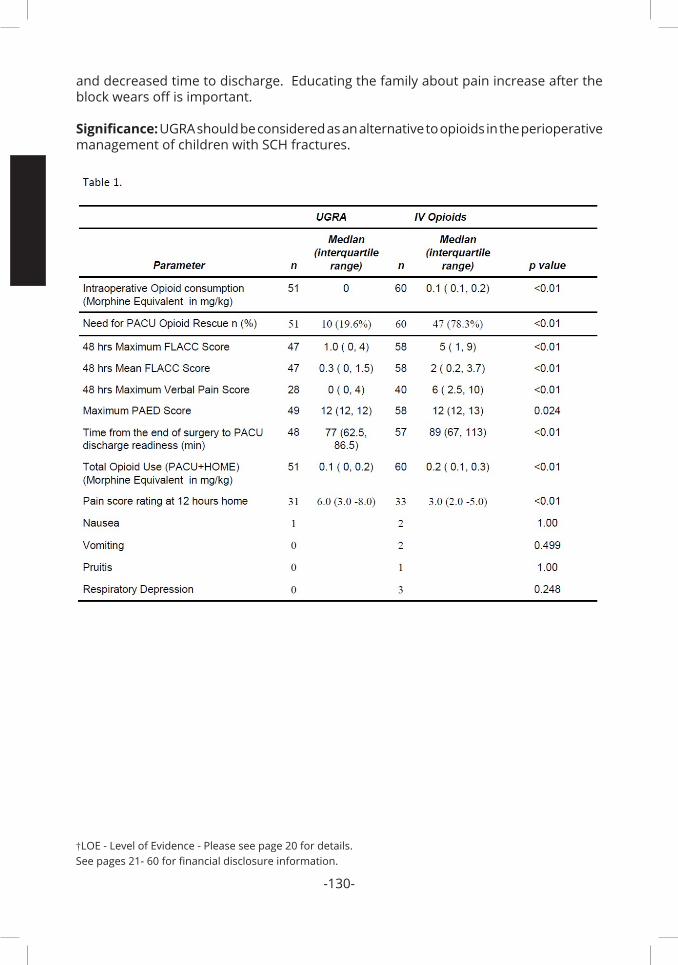
-130-
†LOE - Level of Evidence - Please see page 20 for details.See pages 21- 60 for financial disclosure information.
and decreased time to discharge. Educating the family about pain increase after the block wears off is important.
Significance: UGRA should be considered as an alternative to opioids in the perioperative management of children with SCH fractures.
-131-
♦ Indicates those faculty presentations in which the FDA has not cleared the drug and/or medical device for the use described (i.e., the drug or medical device is being discussed for an “off label” use). For full information, refer to page 14.
Wed., 4/27/16 Upper Extremity, Paper #12, 3:02 PM POSNA-2016
Early Results of Single Plug OATS for Osteochondritis Dissecans of the Capitellum
Eitan M. Ingall, BA; Eliza B. Lewine; Donald S. Bae, MDBoston Children’s Hospital, Boston, Massachusetts
LOE-Therapeutic-Level IV
Purpose: Osteochondral autologous transplantation surgery (OATS) is advocated for unstable osetochondritis dissecans (OCD) of the capitellum. We hypothesize that OATS using a single-plug technique is safe and effective in alleviating pain and restoring function for unstable OCD.
Methods: Sixteen patients with unstable OCD treated with single-plug OATS were evaluated. Mean age at surgery was 15.2 years; there were 11 males. Etiology of OCD was presumed to be sports participation, including baseball (n=9) and gymnastics (n=3). Indications for surgery included unstable, deep OCD lesions; 4 lesions were uncontained, and 4 patients had OATS after failed prior surgery. OATS was performed via an anconeus muscle-splitting approach; donor grafts were harvested from the lateral femoral condyle. Functional outcomes were quantified using Timmerman and QuickDASH scores. Mean clinical, radiographic, and questionnaire follow-up was 11.8, 9.9, and 22.5 months, respectively.
Results: Preoperatively, all patients had tenderness over the capitellum, 10 (63%) limited elbow motion, and 8 (50%) mechanical symptoms. 13 (87.5%) patients reported pain with activities of daily living. Thirteen patients had grade III unstable in-situ lesions, and 3 had grade IV OCD with loose bodies. Mean preoperative lesion size on magnetic resonance imaging (MRI) was 10.9 mm in coronal width, 10.1 mm sagittal width, and 7.6 mm sagittal depth. OATS grafts averaged 9.31 mm in diameter and 11.9mm in depth. At most recent clinical follow-up, 14 (87.5%) patients reported no pain or tenderness with activities of daily living. Of the 10 patients with preoperative elbow stiffness, 9 had achieved full motion. Mean elbow flexion and extension improved from 116.9 to 131.9 degrees and 18 to 1 degrees, respectively. All patients with postoperative MRI’s demonstrated graft incorporation and restoration of articular integrity and subchondral bone; one patient had slight articular incongruity. Mean subjective and objective Timmerman scores improved from 84.7 and 52.8 preoperatively to 91.5 and 97.1 postoperatively, respectively. Overall Timmerman scores improved from 137.5 (fair) to 188.6 (excellent). There were no postoperative complications. All donor knees were asymptomatic with full motion and strength.
Of the 11 patients with questionnaire follow-up, 9 (82%) returned to participation in their primary sport, while 11 (100%) indicated they had resumed general sports participation. Mean QuickDASH score was 2.95.
Conclusion: Single-plug OATS is effective in improving pain and elbow function, with high return to sports rates and little donor-site morbidity.
Significance: OATS is a viable treatment option for treatment of unstable capitellar OCD in adolescents.
-132-
†LOE - Level of Evidence - Please see page 20 for details.See pages 21- 60 for financial disclosure information.
Wed., 4/27/16 Upper Extremity, Paper #13, 3:16 PM POSNA-2016
A Comparison of Fixation Methods in Patients with Diaphyseal Forearm Fractures
Krister P. Freese, MD; L. Wade Faulk, MD, MBA; Robin M. Baschal, BA; Claire Palmer, MS; Sarah Sibbel, MDChildren’s Hospital Colorado, Aurora, Colorado
LOE-Therapeutic-Level IV
Purpose: There is currently a lack of consensus as to the optimal treatment of diaphyseal forearm fractures in adolescent patients. Flexible intramedullary nail fixation and plate osteosynthesis are commonly used surgical treatments in diaphyseal forearm fractures. We hypothesized that adolescent patients treated with plate fixation would have improved outcomes when compared to patients treated with intramedullary fixation.
Methods: A retrospective study was performed on all adolescent patients age 10-16 years old and treated with intramedullary fixation or plate fixation for a diaphyseal both bone forearm fracture between 2005 and 2014. In addition, patients were only included if they were followed to union. Patients were excluded if they underwent single bone or hybrid fixation, had ipsilateral upper extremity injuries, had metabolic bone disease, had a refracture, or were lost to follow up. Demographic and clinical data were collected. Radiographs were reviewed to evaluate post-operative radial bow magnitude and location, time to union, and residual angulation. Complications were graded using the modified Clavien-Dindo Classification system.
Results: A total of 102 patients were identified that met the inclusion and exclusion criteria. Of these, 32 were treated with plate fixation and 70 with intramedullary fixation. The mean patient age was 14.2 and 12.1 respectively (P<0.01). The complication rate in the intramedullary nail group was 29% compared to 12% in the plate fixation group (P=0.13). In the intramedullary nail group, 55% of complications were considered major. There were no major complications in the plate fixation group (P=0.1). The radial bow was significantly more distal and smaller in magnitude in the intramedullary fixation group (P<0.01). Of the patients who underwent intramedullary fixation, 76% required an open reduction of at least one forearm bone. There was increased time to radiographic union in patients treated with intramedullary fixation when compared to those treated with plates, 68 days versus 58 days (P=0.03). A second operation was necessary for 91% of patients treated with intramedullary fixation compared to only 3% of patients treated with a plate (P<0.01).
Conclusion: Diaphyseal forearm fractures in adolescent patients remain challenging injuries to treat. Forearm bony anatomy is not completely restored with intramedullary fixation. In addition, we demonstrated a trend towards increased complication rates and complication severity in patients treated with intramedullary fixation.
Significance: Careful consideration should be given to the method of fixation of diaphyseal forearm fractures in adolescent patients, as this study has identified a trend towards increased complication rate and severity in those treated with intramedullary fixation.
-133-
♦ Indicates those faculty presentations in which the FDA has not cleared the drug and/or medical device for the use described (i.e., the drug or medical device is being discussed for an “off label” use). For full information, refer to page 14.
Wed., 4/27/16 Upper Extremity, Paper #14, 3:21 PM POSNA-2016
Conservative Management of Minimally Displaced (≤ 2 mm) Fractures of the Lateral Humeral Condyle in Pediatric Patients: A Systematic Review
Derrick M. Knapik, MD; Allison Gilmore, MD; Raymond W. Liu, MDUniversity Hospitals Case Medical Center, Cleveland, Ohio
LOE-Therapeutic-Level IV
Purpose: Fractures to the lateral humeral condyle are the second most common type of elbow fracture in the pediatric population and can lead to complications related to displacement of the fracture fragment. The ideal treatment for minimally displaced fractures (measuring ≤ 2 mm) is controversial. The purpose of this study was to systematically review the rate of subsequent displacement for minimally displaced fractures following immobilization, the time span in which fracture displacement generally occurs and the complications related to fracture fragment displacement despite immobilization.
Methods: A systematic review was performed on March 29, 2015 using PubMed, Biosis Preview, SPORTDiscus, PEDro and EMBASE databases to identify and evaluate studies related to displacement of lateral humeral condyle fractures in pediatric patients. Search terms included: lateral condyle fracture, pediatric, displacement and immobilization. Inclusion criteria consisted of human subjects, Level I-IV evidence, patients under age 17 with fracture displacement initially measuring < 2 mm, treated with immobilization in a cast or splint with reported outcomes. Exclusion criteria involved animal studies, Level V evidence, patients age 18 or older, fracture with > 2 mm of displacement or those initially treated with surgical fixation. The search identified 33 studies, of which 6 studies were found to met the inclusion criteria following review of the articles (Table 1).
Results: The six studies that matched the search criteria yielded a total of 355 patients with minimally displaced fractures of the lateral humeral condyle treated initially with immobilization. 14.9% (53 of 355) of patients were discovered to have subsequent fracture fragment displacement. Fracture displacement primarily occurred within the first week following injury in 87% of subjects who experienced subsequent displacement. Mal-union, non-union and loss of motion were the most commonly identified complications following subsequent displacement.
Conclusion: Our results demonstrated a 14.9% risk of subsequent displacement in minimally displaced fractures ≤ 2 mm. When monitoring these fractures, follow up radiographs up to one week are generally sufficient to detect for possible delayed fracture displacement as 87% of subsequent displacements are shown to occur within the first week. Significance: This review features the greatest number of subjects examined for subsequent displacement of minimally displaced lateral humeral condyle fractures in pediatric patients the literature. Gaining a better understanding of the natural history
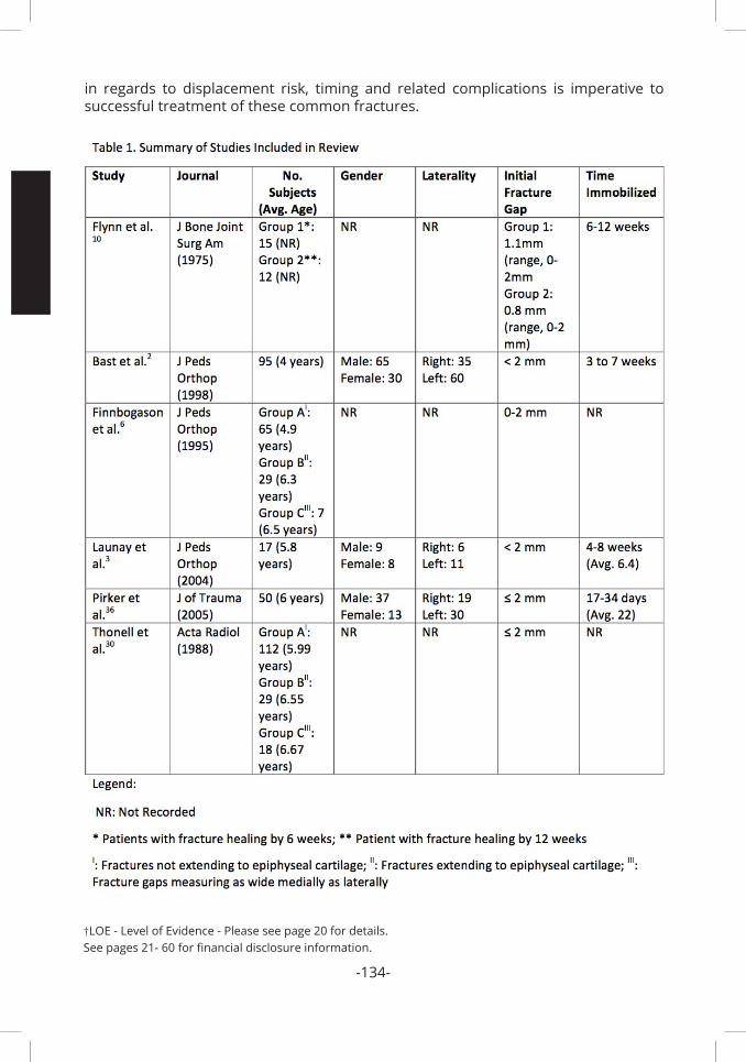
-134-
†LOE - Level of Evidence - Please see page 20 for details.See pages 21- 60 for financial disclosure information.
in regards to displacement risk, timing and related complications is imperative to successful treatment of these common fractures.
-135-
♦ Indicates those faculty presentations in which the FDA has not cleared the drug and/or medical device for the use described (i.e., the drug or medical device is being discussed for an “off label” use). For full information, refer to page 14.
Wed., 4/27/16 Upper Extremity, Paper #15, 3:26 PM POSNA-2016
Incidence and Outcome of Avascular Necrosis following Lateral Humeral Condyle Fracture in Children
Lior Shabtai, MD; Nina Lightdale-Miric, MD; Alexandre Arkader, MD; Alexis Rounds, BS; James L. Pace, MDChildren’s Hospital Los Angeles, Los Angeles, California
LOE-Therapeutic-Level IV
Purpose: Avascular necrosis (AVN) following lateral humeral condyle fracture is a known, but poorly understood complication. Fracture severity and delay in treatment have often been cited as risk factors. The purposes of this study are to report the incidence, outcomes, and potential significant contributing factors leading to AVN following lateral condyle fractures.
Methods: A retrospective review of children diagnosed with a lateral condyle fracture between 2001-2014 at level-1 tertiary pediatric center was designed. Information collected included demographic data, classification, time from injury to surgery, operative versus non-operative management, open versus closed reduction and type of fixation. For patients with AVN, elbow range of motion (ROM), pain and deformity were measured as well at last follow-up. AVN cases were diagnosed by x-ray.
Results: 500 children were available for review. The incidence of AVN was 1.4% (7/500), of which 2/178 (1.1%) were type 2 fractures and 5/168 (3%) type 3 fractures. There were 5/315 (1.6%) boys and 2/185 (1%) girls. The average age was 3.3 years for the AVN group compared to 5 years of the total group. All cases of AVN occurred after open reduction and percutaneous fixation (ORPP) (P=0.01) as initial treatment. Type 3 fractures were associated with AVN (p=0.041). After average follow-up of 59 weeks (range 24- 150 weeks), all patients regained full ROM except one who had lack of 15º of full extension and 10º full flexion. One patient required manipulation under anesthesia and physiotherapy but at final follow-up had full ROM. Five patients were asymptomatic while two patients had mild and inconsistent pain. No evidence of cubitus valgus or varus deformity was found in this group. No significant correlation was found between type of fixation, (p=0.24), gender, (p=0.64), age, (p =0.059) and time from injury to surgery and AVN (p=0.91). All patients were treated within 48 hours of injury.
Conclusion: AVN following lateral condyle fracture is an established, clinically relevant complication but appears to be rare with a rate of only 1.4%. Type 3 fractures and treatment with open reduction are significantly associated with AVN. Progression of deformity related to AVN can occur after acute fracture treatment and continued follow-up is needed due to the young age of this group. Most cases have no pain and full ROM at last follow-up.
Significance: The rate of AVN following lateral condyle fractures is 1.4%. Open reduction is associated with AVN. Most cases do not have pain and full range of motion at last follow-up.
-136-
†LOE - Level of Evidence - Please see page 20 for details.See pages 21- 60 for financial disclosure information.
Wed., 4/27/16 Trauma, Paper #16, 4:01 PM POSNA-2016
Surgical Management of Pediatric Supracondylar Humerus Fractures in the United States: 12 Year National Trends
John Vorhies, MD; Meghan N. Imrie, MD; Lawrence A. Rinsky, MD; Scott A. Hoffinger, MDStanford University Medical Center, Stanford, California
LOE-Economic & Decision-Level IV
Purpose: Supracondylar humerus fractures are one of the most common pediatric fractures to be treated surgically in the US. Despite voluminous data on these fractures, national trends have not been reported. We used an inpatient national database to describe national trends in prevalence, cost of and types of reduction and percutaneous pinning of pediatric supracondylar humerus fractures.
Methods: The Healthcare Cost and Utilization Project’s Kid’s Inpatient Database is the largest publicly-available all-payer pediatric inpatient care database in the US. It contains data from 3 million discharges per year and is weighted to produce national estimates. We queried years 2000-2012 using ICD9-CM diagnosis and procedure codes to identify patients treated surgically for fixation of supracondylar humerus fractures in patients under 18 years.
Results: Over the time period evaluated an estimated 44,115 inpatient surgical fixation procedures were performed on pediatric supracondylar humerus fractures. The patient population was 47% female with an average age of 5.9 years. Mean hospital length of stay was 1.3 days. Total surgical volume was relatively stable over the years with 8,582 procedures performed in 2000 and 8,446 performed in 2012. Mean inpatient hospital charges for treatment of a supracondylar humerus fracture rose from $9,393 in 2000 $19,720 in 2012 (p<0.001) (adjusted for 2012 USD) (Figure 1A). Over the time studied an increasing proportion of fractures were treated at pediatric hospitals; 45% in 2000 vs 63% in 2009 (p<0.001), as well as an increase in closed vs open reduction and pinning with 19% of isolated closed supracondylar fractures treated open in 2000 versus 14% treated open in 2012 (p<0.001) (Figure 1B). Fractures were more likely to be treated open when treated at general hospitals versus pediatric hospitals 21% vs 11% (p<0.001).
Conclusion: Our data demonstrate national trends in increasing specialization and increasing cost of fracture treatment. Although we found a trend towards fewer fractures treated open and a lower percentage of open procedures at pediatric hospitals, a higher percentage of procedures are done open than was expected by the authors based on experience and published literature.
Significance: Here we provide the most comprehensive assessment of US national trends in supracondylar humerus fracture treatment in the literature to date. These data can serve to guide education and policy decisions in the allotment of resources to pediatric trauma care.
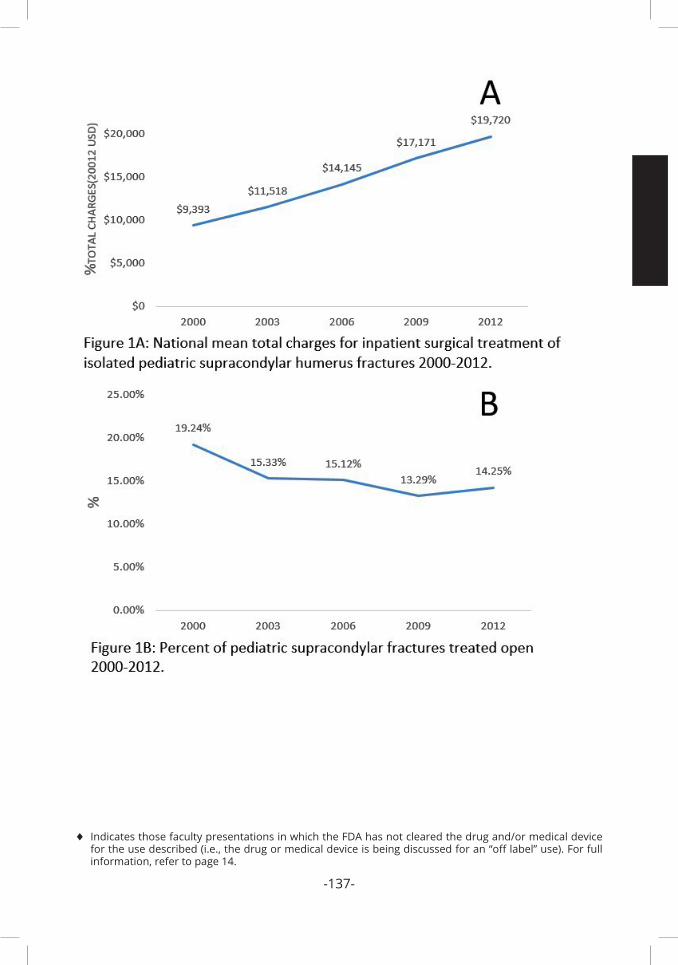
-137-
♦ Indicates those faculty presentations in which the FDA has not cleared the drug and/or medical device for the use described (i.e., the drug or medical device is being discussed for an “off label” use). For full information, refer to page 14.
-138-
†LOE - Level of Evidence - Please see page 20 for details.See pages 21- 60 for financial disclosure information.
Wed., 4/27/16 Trauma, Paper #17, 4:06 PM POSNA-2016
Is the “Appropriate Use Criteria” for Type II Supracondylar Humerus Fractures Really Appropriate?
Howard Park, MD; Erin M. Delfosse, NP; Hemali Panchal; Mauricio Silva, MDOrthopaedic Institute for Children, Los Angeles, California
LOE-Therapeutic-Level III
Purpose: The AAOS has developed the Appropriate Use Criteria (AUC) for the treatment of Supracondylar Humerus Fractures (SCHF), recommending pinning for all type II SCHF’s. However, previous studies have suggested that some of the less severe type II SCHF’s can be successfully treated without surgery if close follow up is achieved. Our purpose is to analyze data collected prospectively on a large cohort of type II SCHF’s. Methods: We reviewed clinical and radiographic information on 1,123 type II pediatric SCHF that were enrolled in a prospective registry and were followed for a minimum of 8 weeks. The characteristics of the patients that were treated without surgery were compared to those of patients that were treated surgically. Treatment outcomes, as assessed by the final clinical and radiographic alignment, range of motion of the elbow, and complications, were compared between the groups to define clinical and radiographic features that related to success or failure of non-operative management. Results: During the course of treatment, 142 fractures (12.6%) were found to have unsatisfactory alignment with non-operative management and were taken for surgery. Ultimately, 819 fractures (73%) were treated non-operatively, and 304 fractures (27%) were treated surgically. At final follow up, outcome measures of change in carrying angle, range of motion, and complications did not show clinically significant differences between treatment groups. Fractures without rotational deformity or coronal angulation, and with a shaft-condylar angle (SCA) of greater than 30 degrees were more likely to be associated with successful non-surgical treatment. Logistic regression showed that rotational deformity (Odds Ratio [OR] 15.0, p<0.0001), varus (OR 4.6, p<0.0001) or valgus malalignment (OR 3.0, p<0.0001), and a shaft-condylar angle (SCA) of less than 30 degrees (OR 3.8, p<0.0001) were strongly related to selection for surgery. Patients with isolated extension deformity, but none of the other features, were more likely to complete successful non-operative management. Conclusion: Pinning all type II SCHF, as recommended by the AUC, would have resulted in unnecessary surgery in 73% of patients in this series. Given the wide range of injury severity within the type II category of SCHF, better discrimination of factors commonly associated with successful non-operative treatment is required.
Significance: In this particular series, fractures with an isolated extension deformity (without rotational or coronal malalignment) were more likely to complete successful non-operative management.
-139-
♦ Indicates those faculty presentations in which the FDA has not cleared the drug and/or medical device for the use described (i.e., the drug or medical device is being discussed for an “off label” use). For full information, refer to page 14.
Wed., 4/27/16 Trauma, Paper #18, 4:11 PM POSNA-2016
Factors that Predict Instability in Pediatric Diaphyseal Both Bone Forearm Fractures
Jeffrey Kutsikovich, MD; Christopher M. Hopkins, MD; Edwin Gannon, BS; Derek M. Kelly, MD; Jeffrey R. Sawyer, MDUniversity of Tennessee Health Science Center, Campbell Clinic, Memphis, Tennessee
LOE-Therapeutic-Level IV
Background/Purpose: Diaphyseal forearm fractures are among the most common fractures in children. Significantly displaced or angulated fractures are treated with initial closed reduction and immobilization, with follow-up to determine if displacement occurs. The purpose of this study was to determine what factors upon initial presenta-tion would predict failure of initial closed reduction and casting.
Methods: Radiographic and hospital records of skeletally immature patients who underwent closed reduction and casting of diaphyseal forearm fractures in the emer-gency department were evaluated. Demographic, time course, and radiographic data were evaluated at presentation and at varying time intervals until union was achieved. Univariate logistic regression analysis of these factors was performed to identify pre-dictors of failure of initial closed reduction and immobilization as defined as requiring a repeat procedure.
Results: 188 patients meeting the inclusion criteria were identified and analyzed. 174 patients had adequate follow-up to union. The average patient age was 7.7 years old and 68% of patients were male. A total of 19 patients underwent a repeat procedure. Patients who underwent a repeat procedure had an average initial reduction time of 36.9 ± 22.2 minutes, whereas those patients who did not require additional procedures had an initial reduction time of 23.4 ± 11.8 minutes (P <0.0103). Odds of requiring repeat reduction were the greatest in those patients who presented with fractures translated greater than or equal to 50% in any plane (odds ratio [OR] = 10.1; 95% confidence interval [CI] 3.1-33.1), age greater than 9 years (OR = 4.1; 95% CI 1.5-11.3), complete fracture of the radius (OR = 9.1; 95% CI 2.0-40.5), follow-up angulation of the radius >15° on lateral radiographs (OR = 5.0; 95% CI 1.3-18.6), follow-up angulation of the ulna >10° on AP radiographs (OR = 8.7; 95% CI 2.7-28.4), and follow-up translation of either bone >50% (OR = 13.5; 95% CI 4.5-40.2). There was no significant correlation with respect to initial angulation parameters and cast index.
Conclusion: Patients requiring lengthy initial reductions are at an increased risk of having a repeat procedure than those with short initial reduction times. Age, initial translation, complete fractures of the radius, and residual translation on follow-up are highly predictive of patients having repeat procedures. These patients require carefully monitored follow-up and families should be counseled appropriately as to their risk of repeat intervention.
-140-
†LOE - Level of Evidence - Please see page 20 for details.See pages 21- 60 for financial disclosure information.
Wed., 4/27/16 Trauma, Paper #19, 4:25 PM POSNA-2016
Complications After Plate Fixation of Displaced Pediatric Midshaft Clavicle Fractures
G. Ying Li, MD; Peter F. Helvie, BS; Matthew D. Abbott, MD; Frances A. Farley, MD; Michelle S. Caird, MDUniversity of Michigan, Ann Arbor, Michigan
LOE-Therapeutic-Level IV
Purpose: Operative treatment of displaced pediatric midshaft clavicle fractures has become increasingly popular, despite lack of evidence that surgical management leads to superior outcomes compared to nonsurgical management. Complications, such as wound infection and plate irritation necessitating removal, have been reported in adults. Few studies have examined the complication rate in children. The purpose of this study was to evaluate complications after plate fixation of midshaft clavicle fractures in the pediatric population.
Methods: This was a retrospective case series. We identified patients 10 to 18 years old who had undergone plate fixation of a displaced midshaft clavicle fracture between 2009 and 2014. Patients who had surgery for a malunion or nonunion and patients with less than 6 months of follow-up were excluded. Demographic data, radiographic measures, implant data, return to activity, and complications were recorded. Any complication that led to unplanned surgery was considered a major complication. Complications that resolved with nonoperative management or did not require any treatment were considered minor. Descriptive analyses were performed.
Results: We analyzed 36 patients (25 males, 11 females) with 37 fractures. The average age at surgery was 14.5 ± 1.7 years and mean follow-up was 1.2 ± 0.8 years. All of the fractures healed and average time to return to activity was 58 ± 28 days. The overall complication rate was 81.1% (30/37): 54.1% (20/37) implant prominence or irritation, 18.9% (7/37) numbness below the incision, 5.4% (2/37) superficial wound dehiscence or infection, and 2.7% (1/37) refracture after implant removal. The major complication rate was 37.8% (14/37). All of these patients underwent a second surgery for implant removal secondary to prominence or pain. Average time to implant removal after the index surgery was 1.3 ± 1.0 years (range, 0.5-4.3 years). One complication occurred after implant removal. This patient sustained a refracture while snowboarding 22 days postoperatively. The fracture was treated nonoperatively. The minor complication rate was 43.2% (16/37). Of the 6 patients with implant-related complaints who did not undergo removal, 3 continued to have pain at final follow-up. The patients with numbness below their incision and wound dehiscence were treated with observation, and the patient with a superficial wound infection was treated with antibiotics with no long-term sequelae.
Conclusion: Implant prominence or irritation is common after plate fixation of displaced pediatric midshaft clavicle fractures. A second surgery for implant removal may be necessary.
Significance: Patients should be appropriately counseled regarding complications prior to plate fixation of midshaft clavicle fractures.
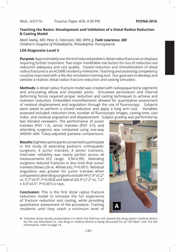
-141-
♦ Indicates those faculty presentations in which the FDA has not cleared the drug and/or medical device for the use described (i.e., the drug or medical device is being discussed for an “off label” use). For full information, refer to page 14.
Wed., 4/27/16 Trauma, Paper #20, 4:30 PM POSNA-2016
Teaching the Basics: Development and Validation of a Distal Radius Reduction & Casting Model
Mark Seeley, MD; Peter D. Fabricant, MD, MPH; J. Todd Lawrence, MDChildren’s Hospital of Philadelphia, Philadelphia, Pennsylvania
LOE-Diagnostic-Level II
Purpose: Approximately one-third of reduced pediatric distal radius fractures re-displace requiring further treatment. Two major modifiable risk factors for loss of reduction are reduction adequacy and cast quality. Closed reduction and immobilization of distal radius fractures is an ACGME residency milestone. Teaching and assessing competency could be improved with a life-like simulation training tool. Our goal was to develop and validate a realistic distal radius fracture reduction and casting simulator.
Methods: A distal radius fracture model was created with radiopaque bony segments and articulating elbow and shoulder joints. Simulated periosteum and internal deforming forces required proper reduction and casting techniques to achieve and maintain reduction. Embedded monofilaments allowed for quantitative assessment of residual displacement and angulation through the use of fluoroscopy. Subjects were asked to perform a closed reduction and apply a long arm cast. Variables assessed included: reduction time, number of fluoroscopic images, casting time, cast index, and residual angulation and displacement. Subject grading was performed by two blinded reviewers. The performance of junior trainees (PGY 1-2), senior trainees (PGY 3-5), and attending surgeons was compared using one-way ANOVA with Tukey-adjusted pairwise comparisons. Results: Eighteen participants consented to participate in the study (8 attending pediatric orthopaedic surgeons, 6 junior trainees, 4 senior trainees). Interrater reliability was nearly perfect across all measurements (ICC range: 0.94-0.99). Attending surgeons reduced fractures in less time than junior trainees (60sec±26 vs. 460sec±62, P<0.001). Residual angulation was greater for junior trainees when compared to attending surgeons on both AP (7.4°±5.2° vs. 0.7°±0.9°, P=0.003) and lateral (26.9°±7.2° vs. 7.2° ± 4.5°±4.5°, P=0.001) x-rays.
Conclusion: This is the first distal radius fracture reduction model to simulate the full experience of fracture reduction and casting, while providing quantitative assessment of the procedure. Training residents until they reach a minimum level of
-142-
†LOE - Level of Evidence - Please see page 20 for details.See pages 21- 60 for financial disclosure information.
competency may be an integral part of orthopedic training. This simulator could easily be integrated into many other accreditation and training programs, including emergency medicine.
Significance: In addition to the noted educational value reported by the trainee and attending surgeon participants, the current study has shown that the performance metrics are reliable and valid when stratified across level of training and expertise. A fracture reduction and casting curriculum built around this model may maximize learning opportunities while minimizing patient risk.
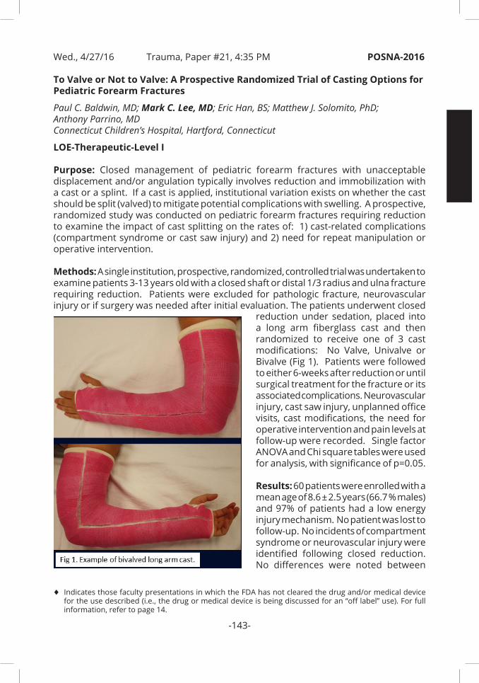
-143-
♦ Indicates those faculty presentations in which the FDA has not cleared the drug and/or medical device for the use described (i.e., the drug or medical device is being discussed for an “off label” use). For full information, refer to page 14.
Wed., 4/27/16 Trauma, Paper #21, 4:35 PM POSNA-2016
To Valve or Not to Valve: A Prospective Randomized Trial of Casting Options for Pediatric Forearm Fractures
Paul C. Baldwin, MD; Mark C. Lee, MD; Eric Han, BS; Matthew J. Solomito, PhD; Anthony Parrino, MDConnecticut Children’s Hospital, Hartford, Connecticut
LOE-Therapeutic-Level I
Purpose: Closed management of pediatric forearm fractures with unacceptable displacement and/or angulation typically involves reduction and immobilization with a cast or a splint. If a cast is applied, institutional variation exists on whether the cast should be split (valved) to mitigate potential complications with swelling. A prospective, randomized study was conducted on pediatric forearm fractures requiring reduction to examine the impact of cast splitting on the rates of: 1) cast-related complications (compartment syndrome or cast saw injury) and 2) need for repeat manipulation or operative intervention. Methods: A single institution, prospective, randomized, controlled trial was undertaken to examine patients 3-13 years old with a closed shaft or distal 1/3 radius and ulna fracture requiring reduction. Patients were excluded for pathologic fracture, neurovascular injury or if surgery was needed after initial evaluation. The patients underwent closed
reduction under sedation, placed into a long arm fiberglass cast and then randomized to receive one of 3 cast modifications: No Valve, Univalve or Bivalve (Fig 1). Patients were followed to either 6-weeks after reduction or until surgical treatment for the fracture or its associated complications. Neurovascular injury, cast saw injury, unplanned office visits, cast modifications, the need for operative intervention and pain levels at follow-up were recorded. Single factor ANOVA and Chi square tables were used for analysis, with significance of p=0.05. Results: 60 patients were enrolled with a mean age of 8.6 ± 2.5 years (66.7 % males) and 97% of patients had a low energy injury mechanism. No patient was lost to follow-up. No incidents of compartment syndrome or neurovascular injury were identified following closed reduction. No differences were noted between
-144-
†LOE - Level of Evidence - Please see page 20 for details.See pages 21- 60 for financial disclosure information.
groups with regards to pain levels (p = 0.3) or frequency of cast modifications (p=0.2). 20 casts were wedged at the discretion of the orthopaedic provider without significant differences between groups (p = 0.1). No significant difference was identified between groups with regard to subsequent need for surgical stabilization (p = 0.361) and none of the 60 patients required repeat closed reduction under sedation. Conclusion: There remains variability in the management of a circumferential, long-arm fiberglass applied to pediatric patients with forearm fractures after reduction. The data suggests cast splitting does not change clinical outcome with respect to neurovascular complications, cast modifications, pain levels, and need for repeat reduction.
Significance: Consideration should be given to limiting cast splitting after reduction of low energy pediatric forearm fractures to minimize saw-related injury and maximize clinical efficiency.

-145-
♦ Indicates those faculty presentations in which the FDA has not cleared the drug and/or medical device for the use described (i.e., the drug or medical device is being discussed for an “off label” use). For full information, refer to page 14.
Wed., 4/27/16 Trauma, Paper #22, 4:49 PM POSNA-2016
Functional Bracing for Treatment of Pediatric Diaphyseal Femur Fractures: An Alternative to Spica Casting
Andrea S. Kramer, MD; Colin Woon, MD; David Speers, CPO, LPOAdvocate Children’s Hospital, Park Ridge, Illinois
LOE-Therapeutic-Level III
Purpose: Closed Reduction and Spica casting (SC) is the traditional treatment of diaphyseal femoral fractures in pediatric patients ages 0 to 5years. However, there are many disadvantages to SC. SC requires general anesthesia, is cumbersome for parents/patients, and difficult to clean and maintain. Additionally, a second cast application is at times necessary when there is progressive malalignment or significant soilage. We hypothesized that diaphyseal femur fractures in this age range could be more easily managed with immediate application of functional fracture bracing (FFB). FFB allows for consistent compression of the fractured limb, is more comfortable, easier to clean, and more cost effective than SC.
Methods: Using case-control design, we compared the clinical, economic and functional outcomes of pediatric patients aged 0 to 5years with displaced and non displaced femoral shaft fractures treated with FFB versus those treated with SC. We evaluated subjective clinical outcomes retrospectively using the Pediatric Outcomes Data Collection Instrument (PODCI) and objective clinical outcomes by assessing post-treatment radiographs in orthogonal planes for angular malalignment and shortening. We evaluated economic outcomes by comparing procedural and equipment costs. Statistical comparisons between groups were performed using the Wilcoxon Mann-Whitney test and Student’s T-test.
Results: There were 41 patients and 43 patients in the FFB and SC groups respectively. All patients had minimum of 2 years follow-up. The PODCI questionnaire revealed very high patient satisfaction with FBB. None of the patients had a limp or subjective leg length discrepancy at their most recent follow-up. All fractures went on to union with 6 weeks of immobilization. Comparison of fracture site angulation revealed significant correction of angulation between pre-treatment and most recent post-treatment orthogonal radiographs . There were no significant differences in magnitude of angular correction between groups (p>0.05). Economic comparison revealed that FB was significantly less costly overall compared with SC (P<0.05).FFB eliminates the need for general anesthesia, surgical and anesthesia charges.
Conclusion: FFB is equally effective to SC in correction and maintenance of fracture alignment, time to union, and functional outcomes but is better tolerated by patients and their parents. Its open design improves hygiene, skin surveillance, and eases transport /lifting as it weighs substantially less than SC. The overall cost of FFB is lower and can be applied immediately without need for general anesthesia and operating room time.
Significance: This study suggests that FFB should be considered a viable alternative to SC in isolated pediatric femoral shaft fractures age 0-5.

-146-
†LOE - Level of Evidence - Please see page 20 for details.See pages 21- 60 for financial disclosure information.
Wed., 4/27/16 Trauma, Paper #23, 4:54 PM POSNA-2016
Pediatric Orthopaedic Lower Extremity Trauma and Venous Thromboembolism
Robert F. Murphy, MD; Manahil N. Naqvi, BS; Patricia E. Miller, MS; Lanna F. Feldman, MS; Benjamin J. Shore, MD, MPH, FRCSCBoston Children’s Hospital, Boston, Massachusetts
LOE-Prognostic-Level IV
Purpose: Research on venous thromboembolism events (VTE), such as deep venous thrombosis (DVT) and pulmonary embolism (PE), in pediatric orthopaedic trauma patients is sparse. We describe the national incidence of VTE associated with pediatric lower extremity orthopaedic trauma, and characterize injury patterns and VTE treatment methods.
Methods: The Pediatric Health Information System (PHIS) was queried from 2004-2013 using ICD-9 codes for lower extremity fractures (pelvis, femur, tibia, ankle, foot) and dislocations (hip, knee, ankle, subtalar) and VTE. Records were queried for age, diagnoses, and VTE treatment.
Results: During the study period 285,611 clinical encounters reported lower extremity trauma. Of those, 167 patients were simultaneously coded with VTE (99 DVT, 50 PE, 18 combined DVT/PE), for an incidence of VTE associated with pediatric lower extremity trauma of 0.058%. Patients were from 39 centers with an average age of 12.9 (range 0-19). There were 249 fractures and 21 dislocations, with 25 (15%) patients sustaining more than one lower extremity injury. The most common fracture location was the femur/femoral neck (95), tibia/ankle (92), and pelvis (44). 72% (121/167) of patients were treated with anticoagulation medication, and the most common was low molecular weight heparin (111/167, 66%).
Conclusion: The incidence of VTE events associated with lower extremity orthopaedic trauma is 0.058%. Adolescents and polytrauma patients with injuries about the femur/femoral neck, tibia/ankle and pelvis are more commonly affected. LMWH is commonly used to treat VTE in pediatric and adolescent patients.
Significance: Clinicians treating children with lower extremity pediatric orthopaedic trauma should maintain a high level of suspicion for the development of VTE. Special attention should be paid toward adolescents and patients with multiple injuries.
-147-
♦ Indicates those faculty presentations in which the FDA has not cleared the drug and/or medical device for the use described (i.e., the drug or medical device is being discussed for an “off label” use). For full information, refer to page 14.
Wed., 4/27/16 Trauma, Paper #24, 4:59 PM POSNA-2016
Percutaneous vs Open Reduction and Fixation for Tillaux and Triplane Fractures: A Multi-Center Study
William D. Zelenty, BS; Richard S. Yoon, MD; Lior Shabtai, MD; Debra A. Sala, MS, PT; Regina Woon, MPH, CCRP; Michelle Ho, BS; Allison Matthews, MSCR; Paul D. Choi, MD; Benjamin D. Martin, MD; Bernard D. Horn, MD; David S. Feldman, MD; Norman Y. Otsuka, MD; David H. Godfried, MDNYU Hospital for Joint Diseases, New York, New York
LOE-Therapeutic-Level III
Purpose: For Tillaux and Triplane ankle fractures, treatment via both open and percutaneous techniques has been described. A comprehensive literature review demonstrates supportive evidence for both techniques, however there is no general consensus on which is superior with regards to minimizing residual gap or preventing growth disturbance. In this study, we present a multi-center initiative comparing the two techniques in a large, cohort comparison.
Methods: Four academic pediatric orthopaedic centers participated in this retrospective cohort comparison study. Two cohorts were formulated dependent on operative technique: percutaneous (PERC) or open reduction (OPEN). Inclusion criteria included all healthy, adolescent children undergoing operative fixation for either Tillaux or Triplane ankle fractures with minimum 1-year follow-up. Data collected included age, gender, BMI, diagnosis, time to surgery, operative technique, initial displacement, residual gap, and/or radiographic signs of growth disturbance.
Results: A total of 65 patients met inclusion criteria and were included for analysis. The OPEN group consisted of 48 patients, while the PERC group consisted of 17 patients. There were no significant differences in age, gender, BMI, or diagnosis between the two cohorts. No significant differences were found between groups for initial displacement (4.3 vs 4.4 mm; p = 0.83 ), or presence of growth arrest (0 vs 6, p = 0.29). The OPEN group had significantly more complications, including infection, nerve/muscle injury and persistent pain, than the PERC group (19 vs 0%; p < 0.01).
Conclusion: In this multi-center study, both techniques yielded expected results in regards to growth arrest; being this close to skeletal maturity, we expected very little physeal involvement. However, more importantly is the higher rate of complications noted with an open approach than a percutaneous approach.
Significance: An open approach in treating adolescent Tillaux and Triplane fractures may result in a higher complication rate when compared to percutaneous technique. Due to a lower concern for physeal involvement, perhaps percutaneous technique should be the standard, while we await larger, prospective trials to see if a true difference is extrapolated.
-148-
†LOE - Level of Evidence - Please see page 20 for details.See pages 21- 60 for financial disclosure information.
Thurs., 4/28/16 Hip, Paper #25, 8:05 AM POSNA-2016
Assessment of Femoral Head Perfusion by ICP-Monitoring is an Accurate Indication of Osteonecrosis after Modified Dunn for Treatment of Unstable SCFE
Travis C. Heare, MD; Lauryn A. Kestel, BS; Patrick Carry, BA; Ernest L. Sink, MD; Eduardo V. Novais, MDChildren’s Hospital Colorado, Aurora, Colorado
LOE-Therapeutic-Level IV
Purpose: The modified Dunn procedure has gained popularity for the treatment of unstable slipped capital femoral epiphysis (SCFE). We investigated whether assessment of femoral head perfusion during the modified Dunn procedure is sensitive and specific in predicting the development of osteonecrosis after unstable SCFE.
Methods: After IRB approval, we identified 27 patients who underwent a modified Dunn procedure for treatment of unstable SCFE who were followed for a minimum of one year. Femoral head perfusion was assessed by the presence of bleeding after a 2mm drilling and by an intra-cranial pressure (ICP) monitor before dissection of the retinacular vessels and after fixation of the epiphysis. The sensitivity, specificity, positive predictive value and negative predictive value of these intra-operative findings as potential clinical tests for detecting the subsequent development of osteonecrosis were performed. A secondary analysis was performed to identify risk factors associated with osteonecrosis.
Results: There were 16 males and 11 females with a mean age of 12.6 years (range, 8-17 years). The median duration of follow-up was 2.5 (range: 1 to 8.0 years). Seven (26%) patients developed osteonecrosis. Overall, the presence of a strong waveform on ICP monitoring after fixation of the epiphysis was associated with the highest area under the curve value, indicating it may represent the best clinical test for differentiating between patients that will versus will not develop osteonecrosis. The sensitivity, specificity, positive predictive value, and negative predictive values associated with the absence of bleeding of the femoral head and ICP monitoring prior to retinacular dissection and after epiphysis fixation are in Table 1. There was no difference in demographics and clinical characteristics including slip severity, length of symptoms, time to treatment and type of fixation between patients that develop osteonecrosis and those who did not develop osteonecrosis.
Conclusion: Intraoperative assessment of femoral head perfusion with an ICP monitor after fixation of epiphysis was the best clinical test to identify patients that develop osteonecrosis following modified Dunn procedure for the treatment of an unstable SCFE. We did not identify factors predictive of osteonecrosis in our cohort.
Significance: This study provides evidence supporting the application of ICP probe for monitoring of femoral head perfusion during modified Dunn procedure for treatment of unstable SCFE. In this cohort ICP monitoring performed better than simple observation of the presence of femoral head bleeding as a test to identify patients that developed osteonecrosis.
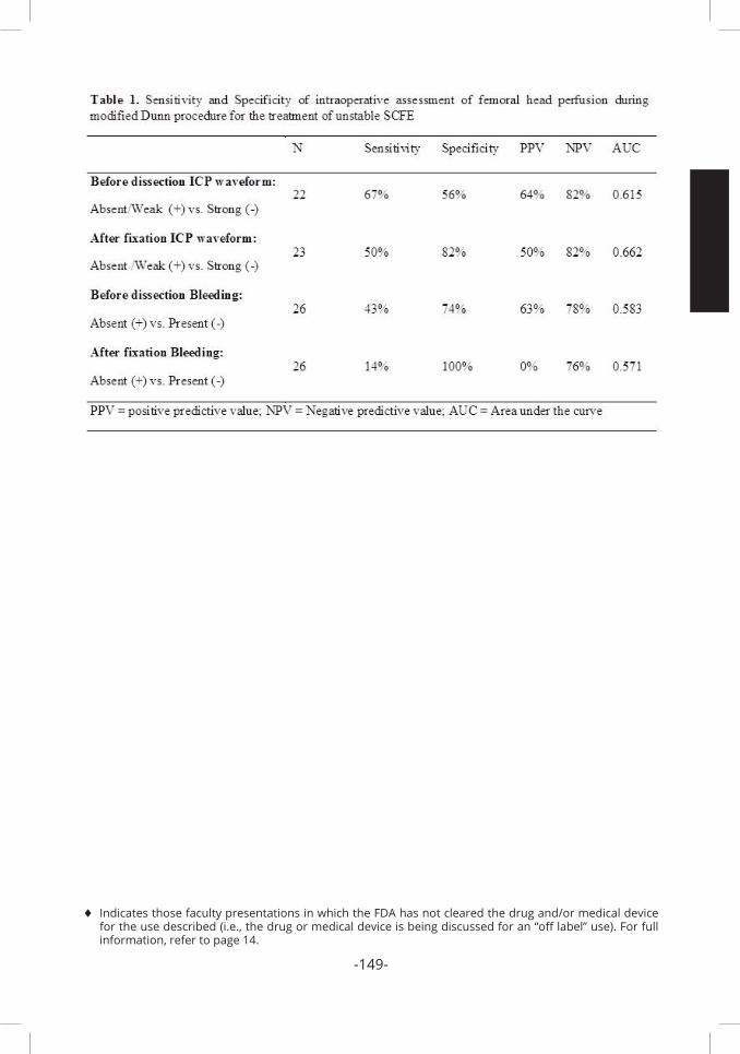
-149-
♦ Indicates those faculty presentations in which the FDA has not cleared the drug and/or medical device for the use described (i.e., the drug or medical device is being discussed for an “off label” use). For full information, refer to page 14.

-150-
†LOE - Level of Evidence - Please see page 20 for details.See pages 21- 60 for financial disclosure information.
Thurs., 4/28/16 Hip, Paper #26, 8:10 AM POSNA-2016
Developmental Dysplasia of the Hip and Laterality: The Importance of Graded Severity of the Contralateral Hip
Emily Schaeffer, PhD; Charles T. Price, MD; Kishore Mulpuri, MD; R-IHDI Study GroupBritish Columbia Children’s Hospital, Vancouver, British Columbia
LOE-Prognostic-Level IV
Purpose: Laterality and bilaterality have been reported as prognostic variables in DDH outcomes. However, there is little clarity across the literature on the reporting of laterality in developmental dysplasia of the hip (DDH) due to the variability in severity of the condition. It is widely accepted that the left hip is most frequently affected; however, the true incidence of unilateral left, unilateral right and bilateral cases can be hard to quantify and compare across studies. The purpose of this study was to examine laterality accounting for graded severity in a multi-centre, international prospective observational study of infants with hip dysplasia in order to demonstrate the complexity of this issue. Methods: A multi-centre, prospective database of infants diagnosed with DDH between the ages of 0 and 18 months was analyzed from 2010 to April 2015. Patients less than six months were enrolled in the study if at least one hip was frankly dislocated. Patients between 6 and 18 months were enrolled if they had any form of hip dysplasia. Each hip was classified as reduced, dysplastic, dislocatable/subluxable, dislocated reducible or dislocated irreducible. Baseline diagnosis was used to classify patients into a graded laterality category accounting for hip status within the DDH spectrum.
Results: A total of 496 patients were included in the analysis; 328 were <6 months old at diagnosis and 168 were between 6 and 18 months old. Of these patients, 421 had at least one frankly dislocated hip. Unilateral left hip dislocations were most common, with 223 patients, followed by unilateral right and bilateral dislocations with 106 and 92 respectively. Stratifying these patients based on status of the contralateral hip, 54 unilateral left and 31 unilateral right dislocated patients also had a dysplastic or unstable contralateral hip. There were significantly fewer bilateral patients in the 6-18 month group (p=0.0005). When classifying laterality by affected hip, bilaterality became the predominant finding, comprising 42% of all patients.
Conclusion: The distribution of unilateral left, unilateral right and bilateral cases was greatly impacted by the method of classification. Distinct patterns were seen when considering dislocated hips only, or when considering both dislocated and dysplastic/unstable hips.
Significance: Findings from this multi-centre prospective study demonstrate the necessity to account for the graded severity in hip status when reporting DDH laterality. In order to accurately compare laterality across studies, a standardized, comprehensive classification should be established, as contralateral hip status may impact prognosis and treatment outcomes.
-151-
♦ Indicates those faculty presentations in which the FDA has not cleared the drug and/or medical device for the use described (i.e., the drug or medical device is being discussed for an “off label” use). For full information, refer to page 14.
Thurs., 4/28/16 Hip, Paper #27, 8:15 AM POSNA-2016
Contrast-Enhanced MRI After Reduction of Infant Hip Dislocation Predicts Avascular Necrosis on Initial Scan Only
Travis H. Matheney, MD; Benjamin J. Shore, MD, MPH, FRCSC; Michael P. Glotzbecker, MD; Patricia E. Miller, MS; Young Jo Kim, MDBoston Children’s Hospital, Boston, Massachusetts
LOE-Diagnostic-Level IV
Purpose: Global decreased enhancement on contrast MRI has been associated with development of femoral avascular necrosis following closed reduction of infant hip dislocation. Improved enhancement on follow-up MRI performed at spica cast change has been seen in femoral epiphyses with global decreased perfusion on initial scan. The purposes of this study were (1) to confirm reports of an association between decreased femoral epiphysis enhancement and avascular necrosis and (2) determine whether improved enhancement on follow-up MRI predicted a better outcome for hips with global decreased enhancement after initial reduction. Methods: Institutional review board approval was obtained and a retrospective evaluation of 30 hips (27 patients) undergoing closed or open reduction for dislocation was conducted with a minimum 2-year follow-up. Immediate post-operative and average 6-week follow-up MRI was performed in spica cast. Images were de-identified and coronal and axial T1 fat saturation MRI sequences with gadolinium were obtained. A single orthopaedic surgeon assessed all images and femoral epiphyses were classified as “normal” vascular channel appearance, “patchy” change, or “global decrease” in enhancement. Age at surgery, reduction type (closed/open), presence of an ossific nucleus (ON) on ultrasound or radiograph, and degree of hip abduction in cast were recorded. Minimum 2-year follow-up radiographs were reviewed and AVN graded according to the criteria of Kalamchi and MacEwen (K&M). Results: Average age at surgery was 7.5 (+ 2.7) months. Average follow-up 2.56 (1.8 to 3.9) years. Fifty seven percent were closed reductions. Thirteen percent had an ossific nucleus at surgery. Thirteen hips had enhancement defect, 7 with global decrease. Seven hips developed AVN K&M grade > 1, five had global decreased enhancement and one each with patchy or normal enhancement. Three of five hips with global decreased enhancement improved their appearance to “normal” and one hip improved to “patchy” appearance. Global decreased enhancement was predictive of AVN at initial MRI (p=0.02) but not at follow-up MRI (p = 0.51). Age at surgery, type of reduction, lack of an ON, and amount of abduction were not predictive of AVN. Conclusion: Global decreased enhancement on MRI of the femoral epiphysis predicts AVN following closed and open reduction of infant hip dislocation. While subsequent MRI at cast change may be useful in assessing the quality of the reduction, unfortunately, improved appearance on follow-up MRI does not predict a better outcome. Significance: Confirmation that MRI predicts AVN following infant hip reductions. Follow-up MRI may be most beneficial to assess reduction.
-152-
†LOE - Level of Evidence - Please see page 20 for details.See pages 21- 60 for financial disclosure information.
Thurs., 4/28/16 Hip, Paper #28, 8:29 AM POSNA-2016
Closed Reduction for Developmental Dysplasia of the Hip: Outcomes from a Multi-Center, Prospective Study Group
Wudbhav N. Sankar, MD; Alex Gornitzky, BS; Nicholas Clarke, FRCS; Jose A. Herrera-Soto, MD; Simon P. Kelley, MBChB, MRCS, FRCS; Travis H. Matheney, MD; Kishore Mulpuri, MD; Vidyadhar S.V. Upasani, MD; Nicole Williams; Charles T. Price, MD; R-IHDI Study GroupChildren’s Hospital of Philadelphia, Philadelphia, Pennsylvania
LOE-Therapeutic-Level IV
Purpose: Closed reduction (CR) is a common treatment option for infantile developmental dysplasia of the hip (DDH). However, relatively little prospectively collected data exists regarding its results and complications. The purpose of this observational, prospective, multi-center study was to determine the early outcomes following CR.
Methods: The prospectively collected database for an international multi-center study group on DDH was analyzed for patients treated from 2010-2014. Baseline demographics, clinical exam, radiographic/ultrasonographic data, and history of previous orthotic treatment were assessed. At minimum one-year follow-up, failure was defined as an IHDI grade 3 or 4 hip (i.e. redislocation) and/or need for open reduction. The incidence of avascular necrosis (AVN), residual dysplasia, and need for further surgery were also assessed.
Results: 78 patients undergoing CR for 87 hips were enrolled with a mean age of 8 months (range, 1–20). Of these, 8 hips (9%) were unable to be closed reduced initially. At most recent follow-up (median 22 months; range, 12–36), 72 of 79 initially successful CRs (91%) remained stable – there were 7 failures (6 open reductions, 1 IHDI 3) at a median of 4 months (range, 1 – 6) following the initially successful index closed reduction. Excluding these failures, 67/72 hips (93%) were IHDI grade 1 and 5/72 (7%) were IHDI grade 2 at final follow-up. The likelihood of failure was not affected by initial clinical reducibility of the hip (p=0.434), age at initial closed reduction (p=0.897) or previous treatment in brace (p=0.222). Excluding those hips that went on to open reduction, 21/73 hips (29%) developed AVN, and the risk of osteonecrosis was not affected by pre-reduction reducibility of the hip (p=0.854), age at closed reduction (p=0.761), presence of an ossific nucleus at the time of reduction (p=0.688) or previous treatment in brace (p=0.619). Mean acetabular index on most recent radiographs was 25° (±6°), and was also unaffected by any of the above variables. During the follow-up period, 8/72 successfully closed reduced hips (11%) underwent acetabular and/or femoral osteotomy for residual dysplasia.
Conclusion: Following an initially successful CR, 91% of hips remained stably reduced and 29% went on to develop radiographic AVN. History of femoral head reducibility prior to CR, previous orthotic bracing, and age at CR did not correlate with success rates or chances of developing AVN.
Significance: CR can be successful in achieving hip reduction, but concerns persist regarding AVN and need for further surgery; these findings argue for continued follow-up of this prospectively collected cohort.
-153-
♦ Indicates those faculty presentations in which the FDA has not cleared the drug and/or medical device for the use described (i.e., the drug or medical device is being discussed for an “off label” use). For full information, refer to page 14.
Thurs., 4/28/16 Hip, Paper #29, 8:34 AM POSNA-2016
A Reliable and Valid Competency Based Simulated Learning Module for the Application of a Pavlik Harness based on International Expert Consensus
Simon P. Kelley, MBChB, MRCS, FRCS; Catharine Bradley, BScPT, MSc; Joel Moktar, MD; Alexandra Maxwell, BS; John H. Wedge, MD; M. Lucas Murnaghan, MD, MEd, FRCSCThe Hospital for Sick Children, Toronto, Ontario, Canada
LOE-Not Applicable
Purpose: The use of simulated learning is increasing in medical education as it offers unlimited learning opportunities in a low risk setting. In orthopaedic surgery, the mainstay of educational programs has been the training of surgical skills with lesser emphasis on non-operative management techniques. Accordingly, formal educational methods and evaluation tools specific to Pavlik Harness application do not exist, despite its widespread use and potential complications related to inappropriate application. This study sought to develop a reliable and valid learning simulation module and evaluation tool based on international expert consensus to standardize, teach and evaluate PH application.
Methods: Consensus was sought from 10 content experts using Delphi methodology on the key items for safe and effective PH application. The resulting items were listed to form an Objective Structured Assessment of Technical Skill (OSATS). The OSATS was used as a framework for the development of a learning simulation module, including an infant model and audiovisual instructional media. Thirty five participants were selected into 3 a priori groups (expert, intermediate and novice) based on perceived competence with the application of the Pavlik Harness. Three content experts assessed randomized and de-identified videotapes of each participant on two occasions separated by two weeks using the OSATS and two global rating scales (GRS). The reliability and validity of the OSATS was then evaluated using ICC statistics and ANOVA.
Results: Consensus was obtained after two rounds of structured surveying. The Delphi methodology used for OSATS development ensured face and content validity. The resulting OSATS contained 25 items. The reliability of the OSATS was excellent with an ICC of 0.96 for inter-rater and 0.98 for test-retest reliability. Construct validity was excellent with the OSATS correlating highly with both GRS (>0.90). In addition, the OSATS clearly discriminated between expert, intermediate and novice users of the Pavlik Harness.
Conclusion: We have developed a competency based simulation module for learning the application of a Pavlik Harness based on the consensus of an international group of experts in hip dysplasia. The corresponding OSATS has been shown to be a reliable and valid method for assessing correct Pavlik Harness application that can discriminate between expert, intermediate and novice users.
Significance: This learning module will form the cornerstone of formal teaching for the application of the Pavlik Harness for DDH.

-154-
†LOE - Level of Evidence - Please see page 20 for details.See pages 21- 60 for financial disclosure information.
Thurs., 4/28/16 Hip, Paper #30, 8:39 AM POSNA-2016
Evaluation of Brace Treatment for Dislocated Infantile Developmental Dysplasia of The Hip in A Prospective Cohort: Defining the Success Rate and Variables Associated with Failure
Vidyadhar S.V. Upasani, MD; James D. Bomar, MPH; Travis H. Matheney, MD; Charles T. Price, MD; Kishore Mulpuri, MD; Scott J. Mubarak, MD; R-IHDI Study GroupRady Children’s Hospital, San Diego, California
LOE-Prognostic-Level IV
Purpose: Bracing has been shown to be an effective treatment for dislocated infantile hips; however, previous studies have been single-center or retrospective. The purpose of this study was: 1) to evaluate the success rate of brace treatment for dislocated infantile hips in an international, multi-center, prospective cohort, and 2) to identify the variables associated with brace failure.
Methods: All infantile hip dislocations were verified with ultrasound or radiograph prior to initiation of treatment and children were followed prospectively for a minimum of twenty months. 146 infants with 188 dislocated hips were studied. Successful treatment was defined as bracing that did not require surgical treatment and resulted in a clinically and radiographically reduced hip. Mann-Whitney test, Chi-square analysis and Fisher’s exact test were used to identify risk factors for brace failure. A multivariate logistic regression model was used to determine the probability of brace failure with the risk factors identified.
Results: Brace treatment was successful in 155 of the 188 dislocated hips in this series (82%). Six variables were found to be significant risk factors of failure: an initially irreducible hip (p=0.003), a Graf IV hip (p=0.04), a right hip dislocation (p=0.008), treatment initiated after age 3 weeks (p=0.03), treatment with a static brace (p=0.001), and developing femoral nerve palsy during brace treatment (p<0.001). Patients with zero risk factors had a 0% probability of failure compared to 100% probability of failure with five risk factors (Table).
Conclusion: Successful brace treatment for dislocated infantile hips can be predicted using the outcome probabilities from this study.
Significance: This prospective, multicenter, study outlines six risk factors for brace failure in infants with dislocated hips that are treated at less than six months of age and provides the probability of brace failure by the number of risk factors, thereby helping to guide management and educate families.
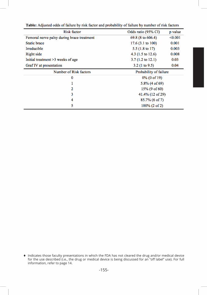
-155-
♦ Indicates those faculty presentations in which the FDA has not cleared the drug and/or medical device for the use described (i.e., the drug or medical device is being discussed for an “off label” use). For full information, refer to page 14.

-156-
†LOE - Level of Evidence - Please see page 20 for details.See pages 21- 60 for financial disclosure information.
Thurs., 4/28/16 Hip, Paper #31, 8:53 AM POSNA-2016
Younger Age at the Time of Closed Reduction Minimizes Acetabular Dysplasia in Children with DDH
Lauren LaMont, MD; Anthony I. Riccio, MD; ChanHee Jo, PhD; Zachary Clarke; R. Lane Wimberly, MDTexas Scottish Rite Hospital, Dallas, Texas
LOE-Prognostic-Level II
Purpose: To determine if early closed reduction and cast immobilization reduces the need for later pelvic osteotomy in children with congenital hip dysplasia (CDH). Methods: We retrospectively reviewed all patients at a single center from 1980-2008 who had a closed reduction for CDH prior to three years of age. We excluded patients with fewer than 2 years of follow-up. Medical records were reviewed to determine demographic information, common CDH risk factors, operative and non-operative treatments attempted, length of spica casting, and the eventual need for pelvic osteotomy. Radiographs at the time of closed reduction and two years following were measured to determine the acetabular index (AI).
Results: 223 children underwent closed reduction during the study period of which 205 (80%) had a successful closed reduction. 143/205 (70%) did not have further pelvic surgery and 62 (30%) underwent a subsequent Pemberton or Salter type osteotomy for residual acetabular dysplasia. Patients who needed acetabular surgery for residual dysplasia were significantly older (54.8 weeks versus 31.3 weeks, p<0.01) and heavier (7.8 kg vs. 9.7 kg, p<0.01) at the time CDH was diagnosed then those that did not require later surgery. They were also older at the time of closed reduction (0.8 years versus 1.2 years, p<0.01) than those who did not require further surgery. The AI at the time of closed reduction was a significant predictor of residual dysplasia: patients treated without further surgery had an average pre-reduction AI of 30.8° (range 15°-45°) vs. 36.3° (range 27°-45°) in those undergoing later pelvic osteotomy (p<0.01). We were unable to find a statistically significant difference in the need for later acetabular surgery when comparing duration of casting, family history, adductor tenotomy, description of medial dye pool, presence of bilateral disease, or the use of pre-reduction traction.
Conclusion: Children undergoing successful closed reduction for CDH who did not need for further surgery to correct residual acetabular dysplasia were on average 23 weeks younger at the time of diagnosis and 4.5 months younger at the time of closed reduction than patients requiring later pelvic osteotomy. Patients with less severe dysplasia at reduction, have a reduced likelihood for later pelvic osteotomy.
Significance: Closed reduction attempts at early ages may reduce the possibility of residual acetabular dysplasia. This should influence surgeons to attempt reductions whenever other treatments fail, rather than waiting until an older age for a more traditional closed vs. open reduction procedure.
-157-
♦ Indicates those faculty presentations in which the FDA has not cleared the drug and/or medical device for the use described (i.e., the drug or medical device is being discussed for an “off label” use). For full information, refer to page 14.
Thurs., 4/28/16 Hip, Paper #32, 8:58 AM POSNA-2016
Predictors of Persistent Postoperative Pain and Associated Outcomes Following Hip Osteotomy for Developmental Dysplasia of the Hip
Michael B. Millis, MD; Justyna Klajn, MS; Laura E. Simons, PhD; Christine B. Sieberg, PhD; Garrett BowenBoston Children’s Hospital, Boston, Massachusetts
LOE-Prognostic-Level II
Purpose: Factors contributing to persistent postsurgical pain (PPP) are poorly defined in young people and developmental considerations are poorly understood. With over 5 million children undergoing surgery yearly and 25% of adults referred to chronic pain clinics identifying surgery as the antecedent, there is a need to elucidate factors that contribute to PPP in young people. The present prospective study includes patients with Developmental Dysplasia of the Hip (DDH) undergoing hip osteotomy at a children’s hospital. Methods: The HOOS and the SF-12 Health Survey were administered to 614 patients prior to surgery with 422 patients completing follow-up data (1, 2, and 5-years post-surgery). When examining baseline characteristics for those who completed follow-up versus those who did not, the only significant difference is that patients with more than one surgery were less likely to complete follow-up measures. Pain, Quality of Life, and Functioning across time were examined using SAS PROC TRAJ procedure, a mixture model that estimates a regression model for each discrete group within the population. Longitudinal pain trajectories were empirically grouped. Baseline pre-operative characteristics of age, gender, preoperative pain, quality of life, functioning, and mental health that could potentially distinguish trajectory groups were examined. Results: Two-trajectory models each emerged for pain, functioning, and quality of life indicating treatment responders (low pain, high functioning, and high quality of life) as well treatment non-responders (high pain, low functioning, and low quality of life). Older age was a risk factor for high pain and poor quality of life trajectories (p<.05). Poor pre-operative mental health was a risk factor for higher pain, poorer quality of life, and decreased functioning over time. Patients in the high pain trajectory had significantly worse pre-operative functioning (p<.001) and lower quality of life (p<.001). Those in the poorer functioning trajectory had higher pre-surgical pain (p<.001) and lower quality of life (p<.001) while patients in the poorer quality of life trajectory were significantly more likely to have higher pre-surgical pain (p<.001) and decreased functioning (p<.001).
Conclusion: This is the largest study to date to examine longitudinal pain, quality of life, and functioning trajectories for adolescent and young adult surgical patients with DDH.
Significance: Chronic pain is a serious public health concern, with the United States spending $19 billion annually on pain-related disability. Identifying predictors of poor long-term outcomes in pediatric samples with postsurgical pain may prevent the development of chronic pain into adulthood as well as inform pre-surgical preparation and post-surgical care.

-158-
†LOE - Level of Evidence - Please see page 20 for details.See pages 21- 60 for financial disclosure information.
Thurs., 4/28/16 Hip, Paper #33, 9:03 AM POSNA-2016
Long Term Results Following the Bernese Periactabular Osteotomy: The Washington University Experience
Joel E. Wells, MD; Kayla M. Thomason, BS; Geneva Baca, BA; Perry L. Schoenecker, MD; John C. Clohisy, MDWashington University School of Medicine, Saint Louis, Missouri
LOE-Therapeutic-Level IV
Purpose: The Bernese periacetabular osteotomy (PAO) continues to be a commonly performed non-arthroplasty option to treat symptomatic developmental hip dysplasia (DDH). There are few long-term follow up studies evaluating results following PAO. This study aimed to evaluate the long-term outcome of the Bernese PAO.
Methods: One hundred eighty-three dysplastic hips (157 patients) underwent PAO between January 1994 and August 2008 by two surgeons. Of those, 45 hips (41 patients) were lost to follow up. The remaining 138 hips (in 117 patients) were retrospectively reviewed at an average of 10.2 years (range 6.9 to 19.7). Hips were evaluated for activity, pain, and general health using the UCLA Activity Score, SF-12, Modified Harris Hip Score (MHHS), and Western Ontario and McMaster Universities Osteoarthritis Index (WOMAC). Both preoperative and long-term follow up radiographs were reviewed. Results: One hundred twenty-five hips (91%) remained preserved and did not undergo conversion to total hip arthroplasty (THA) or have a revision PAO. Ten hips (7%) were converted to THA at an average 6.86 years (range 1.3 to 16.4) from PAO and three hips (2%) had a revision PAO at average 3.4 years. Kaplan-Meier analysis with THA as the end point revealed a survival rate (95% CI) of 97% (94-99%) at 5 years, 93% (87-97%) at 10 years, and 89% (79-96%) at 15 years. The MHHS improved 21.5 points (from 65 to 85.5, p<0.001), and the UCLA score improved 1.1 points (from 6.3 to 7.4, p<0.05). All WOMAC subscale scores demonstrated clinically significant improvement after the PAO. Comparison of preoperative and follow-up radiographs demonstrated an average improvement of 21.2° (from mean 7.5° to 30.3°, p <0.0001) in the lateral center-edge angle, 20.7° (from mean 7.2° to 30.3°, p<0.0001) in the anterior center-edge angle, and 17.8° (mean 23° to 4.2°, p< 0.0001) in Tönnis angle.
Conclusion: In this long-term follow-up study, the majority of hips that underwent PAO have minimal to no pain, are active, and have positive outcomes at an average of 10.2 years (range 6.9 to 19.7) postoperatively. The average ten-year results were very good with a low conversion rate to THA and 89% survivorship at 15 years. This study demonstrates the durability of the Bernese PAO at long-term follow up.
Significance: Periacetabular osteotomy is a leading joint-preserving treatment for pre- or mildly arthritic hip dysplasia. Reporting the long-term outcome of this procedure is critical to our understanding of its ability to relieve symptoms and delay arthroplasty in this younger patient population.
-159-
♦ Indicates those faculty presentations in which the FDA has not cleared the drug and/or medical device for the use described (i.e., the drug or medical device is being discussed for an “off label” use). For full information, refer to page 14.
Thurs., 4/28/16 Hip, Paper #34, 9:17 AM POSNA-2016
Does Previous Hip Arthoscopy Impact the Clinical Outcomes of PAO Surgery? An ANCHOR Cohort Study
Benjamin R. Coobs, MD; John C. Clohisy, MD; Geneva Baca, BA; Perry L. Schoenecker, MD; Jeffrey J. Nepple, MD; Michael B. Millis, MD; Young Jo Kim, MD; Ira Zaltz, MD; Rafael J. Sierra, MD; Wudbhav N. Sankar, MD; Ernest L. Sink, MD; David A. Podeszwa, MD; Daniel J. Sucato, MD, MS; Robert T. Trousdale, MD; Paul E. Beaule, MD; ANCHOR GroupWashington University School of Medicine, Saint Louis, Missouri
LOE-Therapeutic-Level III
Purpose: Hip arthroscopy (HS) alone in the setting of acetabular dysplasia remains controversial. Failure to address the abnormal pathomechanics of the hip may lead to persistent or recurrent symptoms. Subsequent periacetabular osteotomy (PAO) may be needed to correct the underlying structural abnormality. The impact of failed hip arthroscopy on a subsequent PAO has not been fully defined. Our purpose was to analyze the clinical results of PAO surgery for the treatment of acetabular dysplasia in patients with a history of failed HS and compare these to a matched control of hips treated with PAO as the index procedure but having had no prior HS.
Methods: A prospective, multicenter hip preservation database of over 3175 procedures was queried to identify patients with acetabular dysplasia who underwent a PAO after a failed HS. Fifty-one patients/hips were identified from 2008 through December 2012. Demographic, radiographic measures and patient reported outcomes [modified Harris Hip score (mHHS), UCLA and WOMAC] were reviewed. These results were then compared to a 2:1 matched control group of patients that underwent PAO without prior HS.
Results: The average clinical follow-up was 24.2 months and average 20 months between the failed hip arthroscopy and PAO. A majority are female (94%) and Caucasian (90%) patients with an average age of 25.4 and BMI 23.4. The average LCEA improved from 14.7°to 32.2°, AI from 16.1° to 3.8°, ACE angle from 16° to 35°. All patient reported outcomes improved. UCLA activity score improved from 5.7 to 7.1 (p = 0.0014) and the mHHS improved from 55.8 to 79.3 (p < 0.0001). WOMAC normative subscales improved including pain from 51.7 to 81.1 (p < 0.0001), stiffness from 52 to 75.7 (p < 0.0001), physical function from 59.7 to 86.4 (p < 0.0001) and total score from 57.9 to 84.7 (p < 0.0001).
Conclusion: These data suggest that after failed HS, a subsequent PAO can provide clinically significant improvements in pain, function and activity. However, the overall improvement following PAO in the setting of prior HS was statistically worse when compared to a matched control group of patients treated with PAO as the index procedure.
Significance: Treatment of acetabular dysplasia with hip arthroscopy alone may not correct the abnormal pathomechanics of the hip, and can be associated with recurrent or persistent symptoms. Some patients may be best treated by undergoing the most comprehensive surgical strategy from the onset, thus avoiding multiple procedures on the same hip and poorer expected clinical outcomes.

-160-
†LOE - Level of Evidence - Please see page 20 for details.See pages 21- 60 for financial disclosure information.
Thurs., 4/28/16 Hip, Paper #35, 9:22 AM POSNA-2016
MRI in Idiopathic, Stable, Slipped Capital Femoral Epiphysis: Evaluation of Contralateral Pre-slip
Kevin E. Klingele, MD; Julie Samora, MD; Walter P. Samora III, MDNationwide Children’s Hospital, Columbus, Ohio
LOE-Diagnostic-Level II
Purpose: Slipped Capital Femoral Epiphysis (SCFE) is the most common hip disorder in adolescents. Sequential SCFE can occur in 20-80% of patients. Various risk factors, scoring systems and radiographic parameters attempt to predict a sequential slip. MRI findings for a “preslip” are well documented, however indications for prophylactic pinning of an asymptomatic hip are not definitive. The purpose of this study was to determine if MRI evaluation of the contralateral hip in patients presenting with unilateral SCFE could predict sequential, contralateral SCFE.
Methods: A prospective, double-blinded study evaluated patients from 2011-2013 admitted for surgical management of unilateral SCFE. Inclusion criteria were as follows: evidence of unilateral SCFE and normal radiographs of the asymptomatic, contralateral hip. Exclusion criteria included: bilateral SCFE, endocrinopathies, renal disease, malignancy, previously treated hips, or contraindications to MRI. MRI of the asymptomatic hip was performed at the time of index procedure and the results were blinded to the treating surgeon. Patients were followed with serial radiographs until a contralateral slip occurred or until physeal closure. Demographics, SCFE classification, Posterior Slope Angle (PSA), and Modified Oxford Bone Score (MOBS) were recorded and statistical analysis performed.
Results: 34 patients were included, of which 29 (85%) had complete clinical and radiographic follow-up. Six patients (17.6%) developed a sequential slip requiring in situ pinning at an average of 8.4 months after initial presentation. Seven patients had positive MRI findings (presence of focal or diffuse physeal widening, synovitis, bone marrow edema). All sequential slips had positive MR findings at the index procedure, and one hip with positive MRI findings did not slip (sensitivity 100%, specificity 96.4%, PPV 85.7%, NPV 100%). Posterior Slope Angle (PSA) predicted 1 of 6 sequential slips (sensitivity 16.7%, specificity 82.1%, PPV 16.7%, NPV 82.1%) and the Modified Oxford Bone Score (MOBS) predicted 2 of 6 sequential slips (sensitivity 33.3%, specificity 92.9%, PPV 50%, NPV 86.7%). An open triradiate cartilage was present in all 6 patients with sequential slips (sensitivity 100%, specificity 78.6%, PPV 50%, NPV 100%).
Conclusion: In this series of patients, MRI findings consistent with a “pre-slip” were present in all patients that eventually went on to a sequential SCFE, showing higher predictive value than previously described methods.
Significance: The ability of MRI to evaluate a concurrent, contralateral pre-slip with high sensitivity prior to treatment of a unilateral slip would prevent unnecessary “prophylactic” pinning. In addition, sequential SCFE may represent an asymptomatic bilateral “pre-slip” condition.
-161-
♦ Indicates those faculty presentations in which the FDA has not cleared the drug and/or medical device for the use described (i.e., the drug or medical device is being discussed for an “off label” use). For full information, refer to page 14.
Thurs., 4/28/16 Hip, Paper #36, 9:27 AM POSNA-2016
Treatment of Acute, Unstable versus Chronic, Stable Slipped Capital Femoral Epiphysis using the Modified Dunn Procedure
Kevin E. Klingele, MD; Walter P. Samora III, MDNationwide Children’s Hospital, Columbus, Ohio
LOE-Therapeutic-Level III
Purpose: The modified Dunn procedure has been shown to be a safe and effective in treating acute, unstable (SCFE): multiple studies have shown restoration of anatomy and function with acceptable rates of complications. There is a paucity of literature comparing the modified Dunn procedure in stable SCFE. The purpose of this study is to compare a single surgeon’s experience of treating acute, unstable versus chronic, stable SCFE managed via a modified Dunn procedure.
Methods: Retrospective chart review was performed on 44 skeletally immature patients who underwent the modified Dunn procedure for SCFE. Patients were divided into stable or unstable based on intraoperative stability. Demographics, operative reports, inpatient/outpatient charts, and preoperative/postoperative radiographs were reviewed and compared. Statistical analysis performed.
Results: 31 consecutive hips (29 patients) with acute, unstable slips, and 17 consecutive hips (15 patients) with chronic, stable slips were reviewed (avg age 12.5/13.8 years) (p=0.05). Mean follow-up was 27.9 months (acute) and 36.5 months (chronic). No statistical significance was seen between the groups with regards to follow-up time, heterotopic ossification, or removal of symptomatic hardware. Average post operative slip angle was 14.2 (acute) and 25.3 (chronic) (p=0.001). Greater trochanteric height averaged 6.2 mm below center of femoral head in the acute group and 6.2 mm above in the chronic group (p<0.001). Average femoral neck length 34.1 mm (acute) and 27.1 mm (chronic) (p<0.001). Two patients (6%) developed avascular necrosis (AVN) in the acute group and 5 patients (29.4%) in the chronic group (p=0.027). Three patients in the chronic group developed post-operative hip instability requiring additional treatment. All patients with hip instability developed AVN. Conclusion: While both acute and chronic SCFE can be successfully treated with the modified Dunn procedure, the complication rate is statistically higher in patients with chronic SCFE, specifically both AVN and post-operative instability. Additionally, it is more difficult to establish normal anatomic indexes if done without the addition of a relative neck lengthening procedure.
Significance: The modified Dunn procedure has great utility in the correction of the anatomic deformity associated with SCFE, but should be used with caution in patients with chronic, stable SCFE. Other treatment options should be considered in the management of moderate to severe chronic, stable SCFE.

-162-
†LOE - Level of Evidence - Please see page 20 for details.See pages 21- 60 for financial disclosure information.
Thurs., 4/28/16 Hip, Paper #37, 9:41 AM POSNA-2016
The Modified Dunn is Superior to In-Situ Pinning for Anatomic Restoration and Clinical Outcome with Similar Osteonecrosis Rate in Unstable SCFE
Eduardo V. Novais, MD; Lauryn A. Kestel, BS; Claire Palmer, MS; Travis C. Heare, MD; Ernest L. Sink, MDChildren’s Hospital Colorado, Aurora, Colorado
LOE-Therapeutic-Level III
Purpose: Treatment of unstable slipped capital femoral epiphysis (SCFE) remains highly controversial. The purpose of this study was to compare the modified Dunn procedure versus in-situ pinning for treatment of unstable SCFE in terms of (1) proportion of osteonecrosis of the femoral head and other complications; (2) radiographic correction; (3) clinical outcome; and (4) number of unplanned reoperations.
Methods: Following IRB approval, 56 patients who underwent treatment for unstable SCFE between August/2002 and August/2014 were identified. Eight patients with less than one-year follow-up were excluded. Thus, 27 patients treated by modified Dunn and 18 by in-situ pinning were included. Preoperative and most recent radiographs were assessed for Southwick angle, femoral head-neck offset and alpha angle measurements. Osteonecrosis was assessed on postoperative radiographs by femoral head collapse. The Heyman and Herndon system was used to assess the clinical outcome at minimum one year of follow-up. Results: Osteonecrosis of the femoral head occurred in 26% (7/27) of patients in the modified Dunn group and 28% (5/18) of patients in the in-situ pinning group (p=1). Restoration of proximal femoral head and neck anatomy assessed by the lateral Southwick angle (p<0.001) the alpha angle (p<0.001) and the femoral head and neck offset (p<0.001) was better achieved in the modified Dunn group compared to the in-situ pinning group. Likewise, the modified Dunn procedure was superior to provide better clinical outcome scores than in-situ pinning. At latest follow-up, 67% (18/27) in the modified Dunn group and 28% (5/18) in the in-situ pinning group had good or excellent Heyman & Herndon outcomes (p=0.016). There was no difference (p=0.894) in unplanned reoperations between the groups, which most likely is related to the similar osteonecrosis proportion in the two groups. Seventy-four percent (20/27) of patients in the modified Dunn group and 67% (12/18) of patients in the in-situ pinning group did not undergo any additional unplanned procedures during the study period (p=0.894).
Conclusion: The modified Dunn procedure allowed for better restoration of the proximal femur anatomy and improvement in hip pain, limp and motion with similar rates of osteonecrosis in comparison to in-situ pinning.
Significance: Although the rate of osteonecrosis after unstable SCFE was not improved with the application of the modified Dunn procedure, the findings of the current study support the use of the modified Dunn based on better radiographic and clinical outcomes when compared to in-situ pinning.
-163-
♦ Indicates those faculty presentations in which the FDA has not cleared the drug and/or medical device for the use described (i.e., the drug or medical device is being discussed for an “off label” use). For full information, refer to page 14.
Thurs., 4/28/16 Hip, Paper #38, 9:46 AM POSNA-2016
More Accurate Diagnosis with MRI in Suspected Hip Sepsis
Simon R. Thomas, MA, FRCS; Simon Bennet FRCS, MBChB; Joshua Hill, MBChBBristol Royal Hospital for Children, Bristol, United Kingdom
LOE-Diagnostic-Level III
Purpose: Mignemi et al published data in 2014 for 53 children with acutely irritable hips and criteria consistent with septic arthritis managed with a protocol of MRI scanning at presentation. 32% had pericapsular myositis, of which 30% resolved with antibiotics alone, whereas only 15% had septic arthritis. This led us to review our own UK experience.
Methods: During the 6 year period 2007-2013 we searched our institution’s PACS system to retrieve MRI and ultrasound scans performed for children with suspected hip sepsis and 2 or more Kocher criteria for septic arthritis. Recent unsuccessful hip washout was not a contra-indication.
Results: 303 ultrasound scans fulfilled inclusion criteria; a subset of 56 children also underwent MRI scanning (at presentation in 47 cases and after washout of microscopy-negative hip fluid with failure to improve in 9 cases). Average age was 6.2 years (2 weeks to 16 years). M:F ratio was 1.2. Septic arthritis was diagnosed in 12% and 9% respectively in the ultrasound group and MRI sub-group. Osteomyelitis and pyomyositis were 20 and 9 times more likely respectively to be diagnosed with MRI than with ultrasound alone. In 42% of the MRI subgroup multifocal infections were demonstrated. All cases of septic arthritis underwent immediate surgical washout. 7 cases of severe multifocal bone, soft tissue and joint infection identified by MRI underwent drainage of one or more soft tissue abscesses. Only 2/12 cases of isolated pyomyositis required drainage; the remainder was successfully managed with antibiotics only. 2/32 cases of osteomyelitis required cortical drilling and/or drainage of a subperiosteal abscess. Differentiating a sympathetic from a septic hip effusion in the setting of pericapsular myositis and/or osteomyelitis was more difficult but there were no instances of MRI delaying prompt hip washout when necessary.
Conclusion: Selective use of MRI in this series confirms a much higher rate of extra-articular infection than predicted by standard investigations including hip ultrasound in children presenting with suspected septic arthritis of the hip. There was a high rate of coexisting bone, joint and soft tissue infection.
Significance: Accepting that the MRI subgroup in this study was more severely septic and/or diagnostically challenging, we propose switching to a protocol of universal preoperative MRI scanning in the expectation that better targeted and fewer unnecessary surgeries will improve outcomes in this group of children.

-164-
†LOE - Level of Evidence - Please see page 20 for details.See pages 21- 60 for financial disclosure information.
Thurs., 4/28/16 Hip, Paper #39, 9:51 AM POSNA-2016
Hip Instability is an Under Recognized and Significant Complication after Surgical Hip Dislocation in Patients with Slipped Capital Femoral Epiphysis
Vidyadhar S.V. Upasani, MD; Rasika R. Deshpande, BS; Joseph J. Gugenheim, MD; Oliver Birke, MD; David G. Little, MBBS, FRACS; Michael B. Millis, MDBoston Children’s Hospital, Boston, Massachusetts
LOE-Therapeutic-Level IV
Purpose: The surgical hip dislocation technique was described in 2001 to facilitate circumferential exposure of the femoral head and acetabulum. Iatrogenic post-operative hip instability after this procedure has traditionally not been of significant concern. The purpose of this study was to review a series of patients from five different institutions that developed hip instability after the surgical hip dislocation approach was used to treat severe proximal femoral deformity associated with slipped capital femoral epiphysis (SCFE).
Methods: After obtaining institutional review board approval, a review of 10 cases of post-operative hip instability after surgical hip dislocation was performed. Our analysis aimed to identify the risk factors associated with this complication and to develop recommendations to avoid this complication.
Results: Hip dislocation or subluxation was identified between 2 days and 2 months post-operatively. A majority of the patients (80%) were treated with a closed reduction and Petrie cast or abduction brace for 6 weeks. One patient required open reduction and the final patient underwent reduction with an articulating external fixator. The primary factors predisposing these hips to post-operative instability were thought to be: 1) extension and external rotation contractures of the periarticular musculature with gradual loss of flexion and internal rotation; 2) anterior hip capsule attenuation after an extensive intra-operative capsulotomy; 3) excessive femoral neck shortening; and 4) secondary acetabular dysplasia from the severe proximal femoral deformity. Five patients developed femoral head avascular necrosis, two patients developed chondrolysis and end stage degenerative joint disease and three patients healed without further complication (Figure 1). One patient required a total hip arthroplasty.
Conclusion: Hip instability after surgical hip dislocation for SCFE is an under-recognized and potentially devastating complication. It may be appropriate to consider all hips that have been treated for SCFE-associated deformity as being “at risk” for postoperative anterior instability following a surgical dislocation approach.
Significance: Maintaining anterior hip precautions for several weeks post-operatively, with the hips flexed and avoiding external rotation, may avoid the postoperative dislocations. Future research on a modification of the surgical dislocation that leaves the ligamentum teres intact, or comparative studies of proximal femoral osteotomies that avoid surgical dislocation should be performed in patients with chronic severe SCFE deformity.
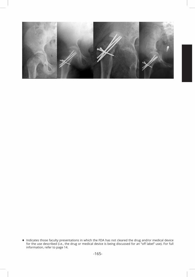
-165-
♦ Indicates those faculty presentations in which the FDA has not cleared the drug and/or medical device for the use described (i.e., the drug or medical device is being discussed for an “off label” use). For full information, refer to page 14.
-166-
†LOE - Level of Evidence - Please see page 20 for details.See pages 21- 60 for financial disclosure information.
Thurs., 4/28/16 Concurrent Session I: Spine, Paper #40, 10:40 AM POSNA-2016
Can Bone Mineral Density (BMD) Predict the Curve Progression and Risk of Surgery in Newly Diagnosed Girls with Adolescent Idiopathic Scoliosis (AIS)?
Benjamin Hon Kei Yip; Fiona Wai Ping Yu; Vivian Wing Yin Hung; Tsz Ping Lam, MD; Ling Qin; Bobby Kin Wah Ng, MD; Jack ChengDepartment of Orthopaedics and Traumatology, The Chinese University of Hong Kong, Hong Kong
LOE-Prognostic-Level I
Purpose: Osteopenia was found to occur in ~30% of AIS patients that can persist beyond skeletal maturity and recognized as a prognostic factor for curve progression. No previous study has been conducted to assess the predictive ability of BMD on curve progression in terms of Cobb angle reaching surgical threshold of 45 degrees or having gone through surgery. The aim of this study was to investigate the incremental prognostic value of osteopenia at initiative clinical visit on surgical outcomes through longitudinal follow up.
Methods: Between 1995-2014, 513 AIS girls with Cobb angle ≥10° but ≤40° (i.e. had not yet reached the study outcome) without prior treatment were recruited and followed from their first visit till skeletal maturity defined as age ≥15.5 years and ≥2 years post-menarchal with an average follow-up time of 5.1 years(SD=2.6).Bilateral hips were measured by DXA at first clinic visit, followed by regular follow-up clinical & radiological assessments. The study outcome was indication of need for surgery defined as Cobb angle≥45° or actually had undergone surgery. Statistically, Akaike Information Criterion (AIC) was used to evaluate incremental prognostic value of osteopenia status (OST) when compared to conventional model.
Results: At first clinical visit, the mean age=13.1 (SD=1.1), with an average Cobb angle of 25°(SD=5.7), and an average of the z-score of BMD of -0.4(SD=0.7). Among 513 subjects, 55 progressed to Cobb angle≥45° or went through surgery. The proportions of subjects with osteopenia having the need for or actually having gone through surgery Vs that without osteopenia were 17.2% and 7.6% respectively. Cox proportional hazard model with osteopenia status had better overall performance than model without (AIC533.13 vs 528.56) and improved the data fitness significantly (p=0.0104). AIS patients with osteopenia had significantly higher risk (HR2.24, p=0.011) of indication of surgery, while lower baseline Cobb angle (HR1.15), menarche status (HR0.414) and older baseline age (HR0.532) were significantly associated with lesser risk of indication of needs for surgery.
Conclusion: Results suggested that AIS patients with osteopenia status would have significantly higher risk of deterioration to the surgical level.
Significance: Early evaluation of osteopenia status that could reflect the bone quality and mechanical bone strength might with further validations be an important additional investigation in predicting the risk of curve progression and subsequent need for surgery at the initial presentation. This could have important potential clinical implications in helping with the prognostication and bracing treatment decision.
-167-
♦ Indicates those faculty presentations in which the FDA has not cleared the drug and/or medical device for the use described (i.e., the drug or medical device is being discussed for an “off label” use). For full information, refer to page 14.
Thurs., 4/28/16 Concurrent Session I: Spine, Paper #41, 10:45 AM POSNA-2016
Is Constant Back Pain an Adequate Predictor of the Presence of a Significant Pathology Associated with Pediatric Back Pain?
Norman Ramirez-Lluch, MD; Pablo Valentin, BS Ivan Iriarte, MD; Soniely Lugo, MDHospital de la Concepcion, San German, Puerto Rico
LOE-Economic & Decision-Level III
Purpose: Several studies have suggested that back pain in the majority of pediatric patients does not have an identifiable cause. In 2008 Feldmanet al, proposed a systematic approach to evaluated the possibility of underlying pathologies in pediatric back pain population, which utilized MRI for patients with constant pain, night pain, radicular pain, or abnormal neurological examination after an initial history, physical examination, and negative radiographic examination. The purpose of this study is determine the sensitivity, specificity, and likelihood ratio of constant and intermittent back pain to predict the presence of an underlying pathology associated with the back pain complaint. Methods: For a 24-month period, all patients that presented with a chief complaint of back pain were prospectively enrolled. After a complete history and physical examination, the patients were evaluated by x-ray and magnetic resonance imaging. Patients that presented injuries due to trauma, previous diagnosis for back pain or any underlying pathologies not related to spinal health or function were excluded.
Results: Of a total of 275 patients, the mean age found was 14.5 years old, with 188 (69.68%) of all patients being females. A total of 224 (80.87%) reported suffering from lumbar pain. Constant pain was present in 91 cases (33.1%) and intermittent pain in 184 (66.9%). Using constant pain as an indicator for an MRI study, the sensitivity of a positive MRI was found in 35% of cases. The specificity of constant pain was 68% with a Positive Predictive Value of 37% and the Negative Predictive Value of 66%. When evaluating cases that reported intermittent pain, 34% of cases presented a positive MRI finding with a Negative Likelihood Ratio of intermittent pain revealed a factor of 0.96. The Positive Likelihood Ratio of constant pain resulted by a factor of 1.1.
Conclusion: Constant or intermittent pain are not strong indicators towards a positive or negative MRI. With the use of an MRI study, one out of three pediatric patients with back pain will have a positive diagnosis. Although Feldman’s algorithm is an effective tool in diagnosis of pediatric back pain, modification of this algorithm should simplify pain description to avoid the high incidence of missed pathologies.
Significance: The study confirms that intermittent or constant pain are not a discriminating factor towards a significant pathology and proposes the necessary changes to Feldman’s algorithm, without which one third of patients suffering intermittent back pain would not be properly diagnosed or treated.
-168-
†LOE - Level of Evidence - Please see page 20 for details.See pages 21- 60 for financial disclosure information.
Thurs., 4/28/16 Concurrent Session I: Spine, Paper #42, 10:50 AM POSNA-2016
Multimodality Pain Management Improves Outcomes and Satisfaction and Reduces Length of Stay after PSF for AIS
Suken A. Shah, MD; Karen Sacks; Kenneth J. Rogers, PhD; Geraldine Neiss, PhD; Petya Yorgova, MS; Randy Brenn, MD; Dinesh K. Choudhry, MDNemours / Alfred I. duPont Hospital for Children, Wilmington, Delaware
LOE-Therapeutic-Level II
Purpose: Gabapentin and clonidine have been administered for acute and chronic pain management. These drugs can likely reduce opioid required for analgesia and likely decrease their side effects.Goal was to evaluate the use of these medications in conjunction with PCA opioids in children with adolescent idiopathic scoliosis (AIS) undergoing posterior spinal fusion (PSF) in relation to improve comfort, reduce opoid consumption and reduce length of stay.
Methods: Following IRB approval, 127 consecutive patients undergoing PSF for AIS by a single surgeon were prospectively assigned to one of three pain management regimens in this cohort study and reviewed retrospectively: Group P: PCA only (42), Group G: PCA + Gabapentin (45), Group C: PCA + Gabapentin + Clonidine (40). Group G patients had standard PCA settings, 1 dose of Gabapentin (10 mg/kg up to 600 mg) given one hour before surgery, and continued postop day 1 (100 mg – 200 mg TID) until discharge. Group C patients had the same plus a Clonidine patch (50 mcg) starting in PACU. Both medications were continued until discharge. Morphine use on postop day 0 and day 1, pain scores (VAS), sedation scores, opioid related side effects, time to ambulation and length of hospital stay (LOS) were calculated.
Results: ANOVA and post hoc analysis by Tukeys test for numeric data. Demographics, blood loss, surgical time, fusion levels or coronal/sagittal plane corrections had no difference. Significant differences were observed between groups in morphine use, transition to orals, day of ambulation and LOS (see Table). No differences among groups in pain scores (VAS), sedation scores, pruritis and emesis. Children in group G and C ambulated and transitioned to orals sooner than group P (control). Significant differences were observed in the PCA use pattern between the groups on the first postop night.
Conclusion: Gabapentin brought improvement in many aspects of post op recovery. Addition of transdermal clonidine further improved overall comfort on the 1st postop night as observed from the PCA use pattern. We feel that the analysis of pattern of PCA pump use brings objectivity to the evaluation of degree of comfort on the night following PSF surgery.
Significance: Perioperative gabapentin and clonidine are effective adjuncts to improve pain control and comfort, reduce opioid consumption, increase mobility and reduce length of stay for the early stages of recovery in pediatric patients undergoing PSF for AIS.
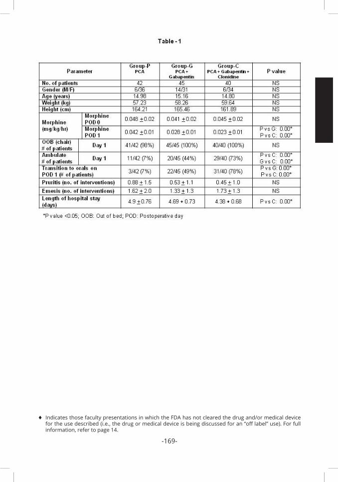
-169-
♦ Indicates those faculty presentations in which the FDA has not cleared the drug and/or medical device for the use described (i.e., the drug or medical device is being discussed for an “off label” use). For full information, refer to page 14.
-170-
†LOE - Level of Evidence - Please see page 20 for details.See pages 21- 60 for financial disclosure information.
Thurs., 4/28/16 Concurrent Session I: Spine, Paper #43, 11:04 AM POSNA-2016
Are Traction or Bending Radiographs Helpful in Selection of Fusion Levels for AIS?
Walter F. Krengel III, MD; Viviana Bompadre, PhD; Gregory Redding, MD; Klane K. White, MD, MSSeattle Children’s Hospital, Seattle, Washington
LOE-Prognostic-Level III
Purpose: Both Traction(Tr) and Bending(Bend) radiographs have been advocated to assess flexibility and assist in selection of fusion levels for Idiopathic Scoliosis (AIS). We sought to define the usefulness of these radiographs compared to Standard PA and Lateral Scoliosis radiographs (PALat) alone in selection of fusion levels. Methods: Patients having surgery for AIS with preoperative radiographs that included PA, Lateral, Bending and Traction films were identified. Two experienced (>5 years) pediatric orthopedic spine surgeons selected preferred fusion levels for each patients. Four different rating sessions were performed for each surgeon. One with PA +Lat films only, a second with PALat and Bend, a third with PALat and Tr, and a fourth with PALat, Bend and Tr(All films). Intra and Interobserver reliability for selection of identical fusion levels for each set of radiographs was calculated with ICCs. Lenke class, lumbar curve magnitude, and levels selected were analyzed for those where additional views altered surgeon selection.
Results: Intra-observer reliability of UIV(0.95,0.96) and LIV(1.0, 0.91)were excellent comparing PALat to PALat, Bend and Tr (all films). Of 49 patients UIV selection was altered from that of PALat alone in 5 different patients by each surgeon (2 lower and 3 higher). Of these ten patients all were in Lenke 3(6), 6(2), 5(2)or 1(1). LIV selection was altered by additional views (+Bend, +Trac or All) in 13 patients by 1 surgeon and 0 by the other. Additional films resulted in a shorter fusion in 9 of the 13, only 1 of which was greater than 1 level. Longer fusions were selected in 4 of the 13. Most changes affected only T11-L2 selections. Inter-observer reliability for UIV and LIV for PALAT (0.83, 0.9), +Bend(0.8,0.75), + TRAC(0.82,0.83), and ALL(0.81,0.85) were best for PA and Lateral Only. Conclusion: The cost and additional radiation exposure incurred with bending and traction radiographs used preoperatively for the selection of AIS fusion levels may not be warranted. Significance: The addition of Bending or Traction Radiographs to standard PA and Lateral images does not improve intra or inter-observer reliability of selected fusion levels compared to PA and Lat alone.
-171-
♦ Indicates those faculty presentations in which the FDA has not cleared the drug and/or medical device for the use described (i.e., the drug or medical device is being discussed for an “off label” use). For full information, refer to page 14.
Thurs., 4/28/16 Concurrent Session I: Spine, Paper #44, 11:09 AM POSNA-2016
Cost Analysis of Adolescent Idiopathic Scoliosis Surgery: Early Discharge Decreases Hospital Costs Much Less Than Intraoperative Variables Under the Control of the Surgeon
Brandon L. Raudenbush, DO; David P. Gurd, MD; Thomas E. Kuivila, MD; R. Tracy Ballock, MD; Ryan C. Goodwin, MDCleveland Clinic, Cleveland Ohio
LOE-Economic & Decision-Level IV
Purpose: To review the contribution of accelerated discharge to the total cost of a single episode of care related to the surgical treatment of idiopathic scoliosis at a single institution. Methods: We recently implemented an accelerated discharge program for patients undergoing spinal fusion for adolescent idiopathic scoliosis (AIS). In order to determine the relative contribution of early discharge to the total cost of scoliosis surgery, a cost analysis was performed of all AIS patients treated over an 18-month period from January 2014 through June 2015. We retrospectively reviewed the hospital charts and hospital costs of all patients treated surgically with an ICD-9 code of 737.30 (Idiopathic Scoliosis). Patients were treated by one of three board-certified and fellowship trained pediatric spinal deformity surgeons. ICD-9 Clinical Modification codes for hospital procedures included 81.05, 81.06, 81.07, and 81.08; codes did not include revision surgery cases. Itemized hospital costs and length of stay were analyzed collectively and by surgeon. Results: Over the study period, 76 AIS patients were treated surgically. The accelerated discharge program reduced average length of stay from 4.4 days in 2014 to 3.4 days in 2015 for all three surgeons, resulting in a 3.5% decrease in the average cost per episode of care. In contrast, the average cost per episode of care for the least expensive surgeon (Surgeon A) was 39.4% less than surgeon B in 2014 and 18.9% less than surgeon B in 2015, and was 54% less than surgeon C in 2014 and 52% less than surgeon C in 2015. The cost savings for surgeon A were primarily from a combination of surgeon-dependent intraoperative variables, most notably lower implant costs and minimizing the use of disposable sterile items such as specialized hemostatic agents and ultrasonic bone scalpels.
Conclusion: Intraoperative variables under the direct control of the surgeon contribute much more to cost reduction per episode of care than an accelerated discharge program for surgically treated AIS patients.
Significance: Cost containment for surgical procedures will be paramount for hospitals as reimbursement models change across the United States. Although accelerated discharge programs are one method to reduce costs, a much more substantial impact can be obtained by reducing intraoperative variables under the direct control of the spinal deformity surgeon.

-172-
†LOE - Level of Evidence - Please see page 20 for details.See pages 21- 60 for financial disclosure information.
Thurs., 4/28/16 Concurrent Session I: Spine, Paper #45, 11:14 AM POSNA-2016
Predictors of Distal Adding-on in Thoracic Major Curves in Adolescent Idiopathic Scoliosis
Joshua S. Murphy, MD; Vidyadhar S.V. Upasani, MD; Burt Yaszay, MD; Tracey P. Bastrom, MA; Amer Samdani, MD; Lawrence G. Lenke, MD; Peter O. Newton, MDRady Children’s Hospital, San Diego, California
LOE-Therapeutic-Level III
Purpose: Previous studies have recommended selecting the last substantially touched vertebra (LSTV) [the most proximal vertebra whose pedicle is touched by the center sacral vertical line (CSVL)] as the lowest instrumented vertebra (LIV) for Lenke 1AR curves [main thoracic curve with ‘A’ lumbar modifier and L4 tilt to the right (thoracic overhang / King type IV curve)]. The purpose of this study was to determine whether this recommendation was valid for both Lenke 1 and 2 curve types with AR lumbar modifiers and to identify pre-operative risk factors of distal adding-on.
Methods: One hundred sixty patients with a thoracic major curve and AR lumbar modifier who underwent posterior instrumented spinal fusion with an all pedicle screw construct between 2008 and 2012 were reviewed. All patients had minimum 2-year follow-up. Adding-on was defined as (1) an increase in Cobb angle of at least 5 degrees and distalization of the end vertebra or (2) a change in disc angulation of 5 degrees or greater below the LIV from the first erect to the 2-year follow-up radiograph. Variables predictive of adding-on were identified using a multivariate binary logistic regression model.
Results: Twenty seven patients (17%) were identified as having distal adding-on of their primary thoracic curve; however only 8 of 89 patients (9%) fused to the LSTV developed adding-on (p=0.005). Three variables were found to be significant predictors of adding-on: LIV proximal to LSTV (O.R. 3.60; p=0.01), Risser zero (O.R. 4.88; p=0.02), and C7-CSVL distance <2cm (O.R. 3.91; p=0.01). The risk of adding-on increased as the number of predictors increased from 16% with 1 risk factor to 80% when all 3 pre-operative risk factors were present (p<0.001).
Conclusion: Choosing the LSTV as the LIV in Lenke 1 and 2 curve patterns with an AR lumbar modifier significantly decreases the risk of distal adding-on. Skeletally immature patients, those fused short of LSTV, and those with relative coronal balance pre-operatively are at increased risk of distal adding-on between the initial post-operative visit and two year follow-up.
Significance: LIV selection continues to be challenging in AIS. Selection of the LSTV as the LIV in Lenke 1 and 2 AR curve patterns significantly decreases the risk of distal adding-on, especially in skeletally immature patients.

-173-
♦ Indicates those faculty presentations in which the FDA has not cleared the drug and/or medical device for the use described (i.e., the drug or medical device is being discussed for an “off label” use). For full information, refer to page 14.
Thurs., 4/28/16 Concurrent Session I: Spine, Paper #46, 11:28 AM POSNA-2016
Peri-operative and Delayed Major Complications Following Surgical Correction of AIS in 3530 Patients
Carrie Bartley, MA; Burt Yaszay, MD; Tracey P. Bastrom, MA; Suken A. Shah, MD; Baron Lonner, MD; Jahangir Asghar, MD; Firoz Miyanji, MD; Amer Samdani, MD; Peter O. Newton, MDRady Children’s Hospital, San Diego, California
LOE-Therapeutic-Level IV
Purpose: The purpose of this study is to report on the rate of major complications following surgically treated AIS both in the peri-operative period and at >2yrs follow-up.
Methods: A prospectively collected (1995-2014), multicenter database of patients who had surgical correction of AIS was reviewed for all major complications. A complication was defined as major if it resulted in re-operation, was considered life-threatening, or resulted in spinal cord/nerve root injury. 3530 patients with pre-op and first post-op data were included. A subset of 2115 pts with >2yrs f/u comprised the cohort for delayed complications. Overall complication rates were calculated, as well as percentage of complications by year of surgery and approach type.
Results: 3530 patients (mean age 14.8±2.2 years) were reviewed. Mean thoracic Cobb was 51±16° and mean lumbar Cobb was 39±14°. There were 364 anterior spinal fusions (ASF) and 3166 posterior spinal fusions (PSF; 146 with anterior release). There were 193 major complications, 93 (2.6%) of which occurred peri-op (≤6wks). The majority of peri-op complications were wound (1%), neurologic (0.5%), pulmonary (0.4%), instrumentation (0.4%), or gastrointestinal (0.2%) related. One patient died. The mean peri-op complication rate based upon year of surgery ranged from 0-10.5% (Figure). The complication rate for each surgical approach was 3% ASF, 2.6% PSF (2.5% PSF only, 5.5% PSF with anterior release). The major complication rate for those with >2yr f/u was 4.4%; all but 2 had a re-operation (4.4%). The majority of these major complications were wound (2.1%) and instrumentation (0.9%) related.
Conclusion: After surgery for AIS, a 2.6% rate of major peri-op complications and a 4.4% rate of major complications at >2 years post-op can be anticipated. This is critical information to guide patients and inform payers. Fortunately, the complication rate has decreased in the last decade with attention focused on safety and quality from all stakeholders.
Significance: Reporting accurate complications rates to patients and payers is important in the management of AIS. Following surgical correction in 3530 AIS patients, 193 major complications were observed. Given the rate of complications, further study into operative management of AIS is warranted.
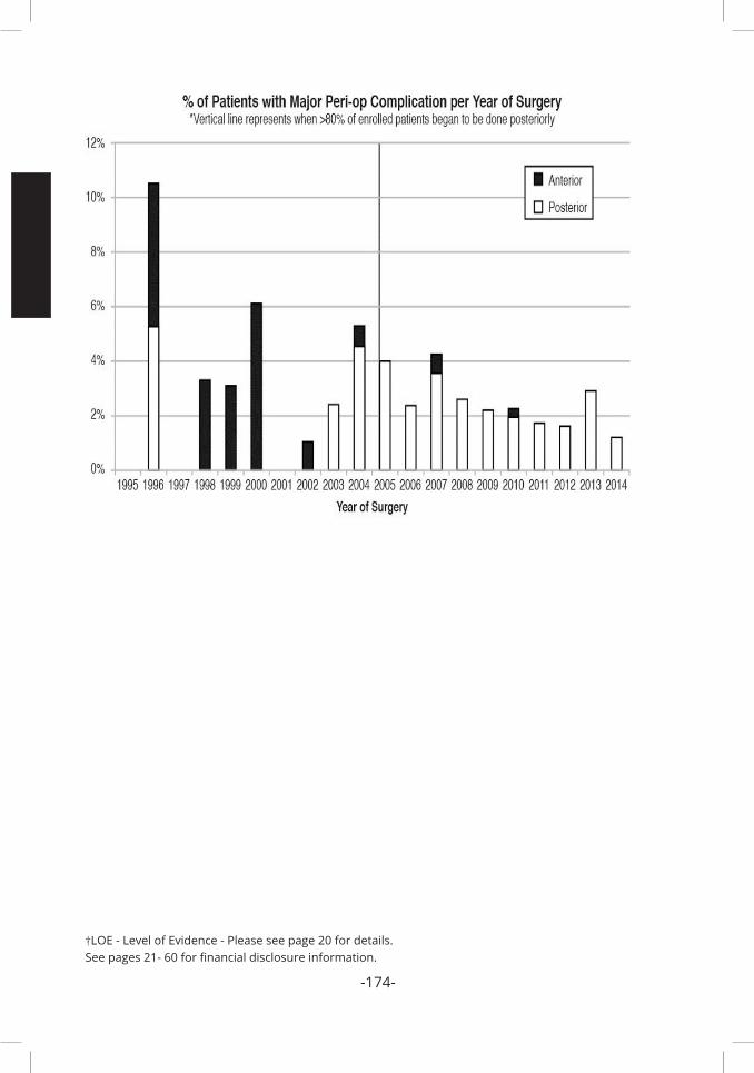
-174-
†LOE - Level of Evidence - Please see page 20 for details.See pages 21- 60 for financial disclosure information.

-175-
♦ Indicates those faculty presentations in which the FDA has not cleared the drug and/or medical device for the use described (i.e., the drug or medical device is being discussed for an “off label” use). For full information, refer to page 14.
Thurs., 4/28/16 Concurrent Session I: Spine, Paper #47, 11:33 AM POSNA-2016
“Risk Adjusted” Comparative Infection Rates in Adolescent Idiopathic Scoliosis
Peter O. Newton, MD; Burt Yaszay, MD; Tracey P. Bastrom, MA; Harms Study GroupRady Children’s Hospital, San Diego, California
LOE-Prognostic-Level II
Purpose: Patients, payors, and surgeons are becoming increasingly interested in surgeon “performance measures”. Understanding patient factors that increase risks of an adverse event are important for comparison of such metrics amongst various surgeons/centers. The purpose of this study was to ascertain whether patient characteristics, beyond the control of the surgeon, are associated with increased risk of post-operative infection after surgical correction of adolescent idiopathic scoliosis (AIS) and establish a “risk adjusted” method of reporting postoperative infection rates.
Methods: AIS patients with an infection within 90 days of surgery were reviewed. Patients with a deep infection (I&D performed) were compared to those without an infection with regards to age, gender, body mass index (BMI), primary Cobb magnitude, and sagittal kyphosis (T5-T12). A multivariate binary logistic regression model was created to identify predictors. The risk model was evaluated using the c-statistic, Hosmer-Lemeshow (H-L) test, and Brier score. The actual infection rate by site was divided by the predicted infection rate and multiplied by the overall rate to create a “risk adjusted” rate.
Results: 2499 patients were analyzed, of which 22 patients (0.88%) had an infection within 90 days of surgery. Obesity (BMI>30) was the only significant risk factor of infection (OR 8.8, p≤0.001), with the resultant model demonstrating good discrimination and calibration. For centers with ≥100 patients enrolled, the predicted infection rates based on proportion of obese patients versus actual observed infection rates are seen in Table 1. The range for predicted infection rates was between 0.7 and 1.3%. The range for risk adjusted infection rates varied more substantially from 0.2 to 1.7%.
-176-
†LOE - Level of Evidence - Please see page 20 for details.See pages 21- 60 for financial disclosure information.
Conclusion: For the surgical correction of AIS, the only significant patient risk factor of postoperative infection was a BMI >30. To assess a surgeon or center’s infection rate, there must be a correction made for the proportion of obese patients in their cohort. A risk adjusted infection rate in AIS allows for more accurate assessment of performance and comparison among centers.
Significance: Obese patients experienced much greater risk of infection after surgery for adolescent idiopathic scoliosis. Risk adjusted infection rates provide a more valid comparison across centers/surgeons.
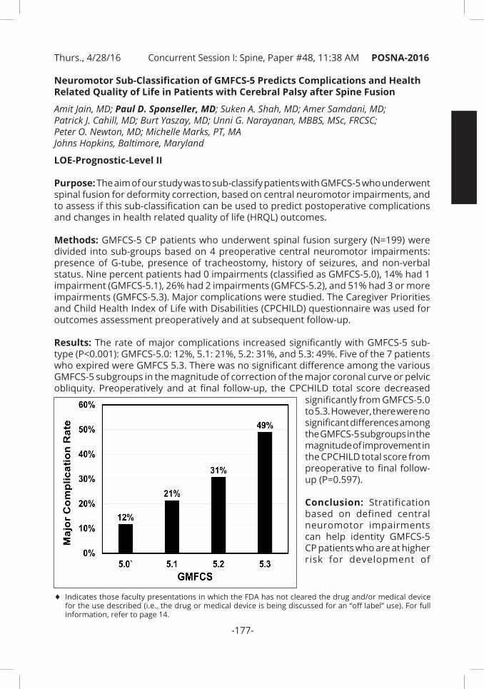
-177-
♦ Indicates those faculty presentations in which the FDA has not cleared the drug and/or medical device for the use described (i.e., the drug or medical device is being discussed for an “off label” use). For full information, refer to page 14.
Thurs., 4/28/16 Concurrent Session I: Spine, Paper #48, 11:38 AM POSNA-2016
Neuromotor Sub-Classification of GMFCS-5 Predicts Complications and Health Related Quality of Life in Patients with Cerebral Palsy after Spine Fusion
Amit Jain, MD; Paul D. Sponseller, MD; Suken A. Shah, MD; Amer Samdani, MD; Patrick J. Cahill, MD; Burt Yaszay, MD; Unni G. Narayanan, MBBS, MSc, FRCSC; Peter O. Newton, MD; Michelle Marks, PT, MAJohns Hopkins, Baltimore, Maryland
LOE-Prognostic-Level II
Purpose: The aim of our study was to sub-classify patients with GMFCS-5 who underwent spinal fusion for deformity correction, based on central neuromotor impairments, and to assess if this sub-classification can be used to predict postoperative complications and changes in health related quality of life (HRQL) outcomes.
Methods: GMFCS-5 CP patients who underwent spinal fusion surgery (N=199) were divided into sub-groups based on 4 preoperative central neuromotor impairments: presence of G-tube, presence of tracheostomy, history of seizures, and non-verbal status. Nine percent patients had 0 impairments (classified as GMFCS-5.0), 14% had 1 impairment (GMFCS-5.1), 26% had 2 impairments (GMFCS-5.2), and 51% had 3 or more impairments (GMFCS-5.3). Major complications were studied. The Caregiver Priorities and Child Health Index of Life with Disabilities (CPCHILD) questionnaire was used for outcomes assessment preoperatively and at subsequent follow-up.
Results: The rate of major complications increased significantly with GMFCS-5 sub-type (P<0.001): GMFCS-5.0: 12%, 5.1: 21%, 5.2: 31%, and 5.3: 49%. Five of the 7 patients who expired were GMFCS 5.3. There was no significant difference among the various GMFCS-5 subgroups in the magnitude of correction of the major coronal curve or pelvic obliquity. Preoperatively and at final follow-up, the CPCHILD total score decreased
significantly from GMFCS-5.0 to 5.3. However, there were no significant differences among the GMFCS-5 subgroups in the magnitude of improvement in the CPCHILD total score from preoperative to final follow-up (P=0.597).
Conclusion: Stratification based on defined central neuromotor impairments can help identity GMFCS-5 CP patients who are at higher risk for development of
-178-
†LOE - Level of Evidence - Please see page 20 for details.See pages 21- 60 for financial disclosure information.
complications with spinal fusion surgery. Patients in higher GMFCS-5 subgroups had worse health related quality of life outcomes. This data will assist in better counseling patients and families.
Significance: Stratification based on defined central neuromotor impairments can help identity GMFCS-5 CP patients who are at higher risk for development of complications with spinal fusion surgery.
-179-
♦ Indicates those faculty presentations in which the FDA has not cleared the drug and/or medical device for the use described (i.e., the drug or medical device is being discussed for an “off label” use). For full information, refer to page 14.
Thurs., 4/28/16 Concurrent Session I: Spine, Paper #49, 11:52 AM POSNA-2016
Repeat Surgical Intervention Following Definitive Instrumentation and Fusion for Adolescent Idiopathic Scoliosis: A 25-year Update
Megan Mignemi, MD; Dong-Phuong Tran, MS; Brandon A. Ramo, MD; B. Stephens Richards, MDTexas Scottish Rite Hospital for Children, Dallas, Texas
LOE-Prognostic-Level III
Purpose: Recent trends in the surgical treatment of AIS have included increased use of all-pedicle screw constructs, more posterior-only approaches, and improved correction techniques. The purpose of this study is to determine the rate of reoperation in AIS patients undergoing spine fusion from 2008-2012, to determine the reasons for reoperation in this study population and to compare these results to the previous 20 years of clinical experience from the authors’ institution.
Methods: IRB approved, single institution, retrospective review of prospectively collected data on 467 AIS patients undergoing spinal fusion from 2008-2012. Demographic, clinical, radiographic, and surgical data were collected on all patients for the index procedure and any reoperations. This cohort was compared to previously published cohorts of patients from the authors’ institution who underwent spinal fusion for AIS between 1988-2007.
Results: The rate of reoperation in this 5-year cohort of patients was 9.9%. The most common indications for reoperation were infection (4.5% (2.4% delayed infections and 2.1% acute infections)), symptomatic implants (2.1%), and misplaced pedicle screws (1.7%). When compared to the 2003-2007 cohort, the rate of reoperation for acute infection and misplaced pedicle screws increased significantly (p=0.01 and p=0.04), while the rate of reoperation for curve progression decreased (p=0.01). Reoperations for acute infections and misplaced pedicle screws also increased significantly (p=0.047 and p=0.042) compared to the 1988-2002 cohort, while the rate of reoperation for pseudarthrosis decreased (p=0.002).
Conclusion: Despite the use of newer implant systems and improved surgical techniques, the rate of reoperation after spinal fusion for AIS was stable. A decrease in psuedarthrosis and curve progression occurred, but we noted an increase in infection and malpositioned pedicle screws. Higher infection rates were seen in patients with stainless steel implants, increased implant densities and fusion above T4.
Significance: The rate of reoperation following spinal fusion for AIS has not changed significantly over time, although the indications for reoperation continue to evolve.

-180-
†LOE - Level of Evidence - Please see page 20 for details.See pages 21- 60 for financial disclosure information.
Thurs., 4/28/16 Concurrent Session I: Spine, Paper #50, 11:57 AM POSNA-2016
Reduced Pulmonary Function in AIS Patients with Hypokyphosis: Mean 30-Year Follow-up
A. Noelle Larson, MD; William J. Shaughnessy, MD; Charles G. Ledonio, MD; David W. Polly Jr, MD; Michael A. Yaszemski, MD, PhD; Clayton Cowl, MD, MSMayo Clinic, Rochester, Minnesota
LOE-Prognostic-Level III
Purpose: Spinal fusion surgery is recommended for the treatment of AIS in order to prevent future pulmonary complications. There are limited long-term outcomes studies reporting on the effect of spinal fusion and sagittal plane alignment on pulmonary function in adulthood. We hypothesized that sagittal plane parameters affect pulmonary function in patients treated for AIS with minimum 20-year follow-up.
Methods: From 1975-1992, 341 children were treated for AIS (childhood curve magnitude > 35°). One braced patient with a history of substance abuse died secondary to pulmonary disease at age 43. Pulmonary function testing results were available at most recent follow-up for 52 patients (mean follow-up 31 years, range 21-40). Mean age at follow-up was 45 (range, 33-56). Childhood treatment included bracing (19), observation (6), and spinal fusion in 27, with Harrington (16), hook (9), or anterior (2) instrumentation. Mean adult thoracic Cobb in the nonoperative cohort was 41° (9-71), and 33° in the operative (13-52). Hypokyphosis was defined as T2-T12 < 20°. Results: Pulmonary function results were significantly reduced in both operative and nonoperative patients with hypokyphosis (Table). This relationship was particularly strong in the nonoperative cohort with hypokyphosis associated with reduced FVC% (82% vs. 92%, p=0.048), FEV1 (0.76 vs. 0.9, p=0.018), FEF25-75 (1.9 vs.2 .6, p=0.03), FEF% (95% vs. 69%, p=0.03). Increased thoracic coronal Cobb was associated with decreased FEV1 (p=0.03) and FEF25-75 (p=0.03). Surgical patients with fusion above T4 had reduced FEV1%, FEV1/FVC, and FEF% compared to those fused below T4 (Table). At latest follow-up, there were no differences in pulmonary function noted between the surgical and nonoperative cohort. Of the total cohort, 50% had < 80% predicted max voluntary ventilation (MVV), 25% had < 50th percentile maximal expiratory pressures, and 87% had less than 80th percentile. Patient reported outcomes were quite favorable (mean EQ5D, SRS Total 3.9, SRS Pain 3.9, SRS Function 4.0, ODI 15, SAQ Total 4 Conclusion: Abnormalities on pulmonary function testing are common in mid-life following treatment of AIS and are associated with residual hypokyphosis and high thoracic fusions. The ongoing clinical implications are unknown, but deserve further exploration. Significance: T2-T12 kyphosis less than 20° and fusion above T4 were associated with reduced pulmonary function in AIS patients at a mean 30-years following childhood treatment. Correction of hypokyphosis of thoracic hypokyphosis at the time of spine fusion may be important for lifetime pulmonary function.
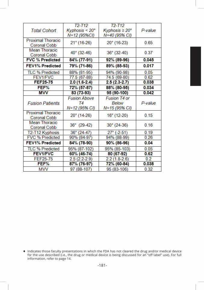
-181-
♦ Indicates those faculty presentations in which the FDA has not cleared the drug and/or medical device for the use described (i.e., the drug or medical device is being discussed for an “off label” use). For full information, refer to page 14.
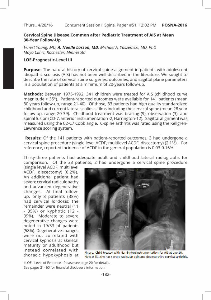
-182-
†LOE - Level of Evidence - Please see page 20 for details.See pages 21- 60 for financial disclosure information.
Thurs., 4/28/16 Concurrent Session I: Spine, Paper #51, 12:02 PM POSNA-2016
Cervical Spine Disease Common after Pediatric Treatment of AIS at Mean 30-Year Follow-Up
Ernest Young, MD; A. Noelle Larson, MD; Michael A. Yaszemski, MD, PhDMayo Clinic, Rochester, Minnesota
LOE-Prognostic-Level III
Purpose: The natural history of cervical spine alignment in patients with adolescent idiopathic scoliosis (AIS) has not been well-described in the literature. We sought to describe the rate of cervical spine surgeries, outcomes, and sagittal plane parameters in a population of patients at a minimum of 20-years follow-up.
Methods: Between 1975-1992, 341 children were treated for AIS (childhood curve magnitude > 35°). Patient-reported outcomes were available for 141 patients (mean 30 years follow-up, range 21-40). Of those, 33 patients had high quality standardized childhood and current lateral scoliosis films including the cervical spine (mean 28 year follow-up, range 20-39). Childhood treatment was bracing (9), observation (3), and spinal fusion (CD-7, anterior instrumentation -2, Harrington-12). Sagittal alignment was measured using the C2-C7 Cobb angle. C-spine arthritis was rated using the Kellgren-Lawrence scoring system. Results: Of the 141 patients with patient-reported outcomes, 3 had undergone a cervical spine procedure (single level ACDF, multilevel ACDF, discectomy) (2.1%). For reference, reported incidence of ACDF in the general population is 0.03-0.16%.
Thirty-three patients had adequate adult and childhood lateral radiographs for comparison. Of the 33 patients, 2 had undergone a cervical spine procedure (single level ACDF, multilevel ACDF, discectomy) (6.2%). An additional patient had severe cervical radiculopathy and advanced degenerative changes. At final follow-up, only 8 patients (38%) had cervical lordosis; the remainder were neutral (11 - 35%) or kyphotic (12 - 39%). Moderate to severe degenerative changes were noted in 19/33 of patients (58%). Degenerative changes were not correlated with cervical kyphosis at skeletal maturity or adulthood but instead correlated with thoracic hypokyphosis at
-183-
♦ Indicates those faculty presentations in which the FDA has not cleared the drug and/or medical device for the use described (i.e., the drug or medical device is being discussed for an “off label” use). For full information, refer to page 14.
skeletal maturity (p < 0.05). No differences in alignment or outcome measures were noted between nonoperative and childhood fusion patients. Decreased SRS and ODI scores were correlated with thoracic hypokyphosis in adulthood.
Conclusion: Cervical spine malalignment persists in adulthood following both operative and nonoperative AIS treatment with accompanying high rates of radiographic cervical spondylosis. Historic surgical treatment of AIS in childhood did not appear to improve cervical sagittal alignment over the child’s lifetime. Residual thoracic hypokyphosis may be related to poor functional outcomes and increased cervical arthritis in adulthood.
Significance: This work is one of very few studies on the long term outcomes of the sagittal cervical spine in patients treated for AIS. This information will help attempts at maintaining proper sagittal balance in patients being treated for AIS.

-184-
†LOE - Level of Evidence - Please see page 20 for details.See pages 21- 60 for financial disclosure information.
Thurs., 4/28/16 Concurrent Session II, Paper #52, 10:40 AM POSNA-2016
Role of Body Cast Application for Scoliosis Associated with Prader-Willi Syndrome
Harold J.P. van Bosse, MD; Patrick J. Cahill, MDShriners Hospital for Children, Philadelphia, Pennsylvania
LOE-Therapeutic-Level IV
Purpose: Approximately 40% of children with Prader-Willi syndrome (PWS) develop scoliosis. Half of the children develop curves before 4 years of age, likely related to their characteristic profound hypotonia. Overall, 15% will require surgical intervention. The purpose of this retrospective study is to evaluate the effectiveness of serial spinal casting for infants and young children with PWS to either correct a curve, or at the least delay the necessity for expandable implant surgery.
Methods: Since 2008, 19 of the 28 patients with PWS and scoliosis curves greater than 25° undergoing Mehta body cast application had a minimum of 2 years follow up. Standard Mehta casts were applied, using a Risser table, under general anaesthesia. Casts were changed every 2 to 4 months, depending on the child’s age.
Results: The average age at initial cast application was 33 months (14 -60 months). An average of 8 casts (range 3-18) were applied, and average follow up since first cast application was 38 months. The pre casting Cobb angle of 58° (range 27-106°) was reduced to 37° (range 16-109°) at the latest followup. The overall correction achieved in all the cases was 44%, the rib-vertebral angle difference decreased from 46° to 31°, Nash-Moe grade decreased only from 2 to 1.5, and space available for lung for the group remained essentially unchanged. Six patients, all uniparental disomy type PWS, with an average pre-casting curve of 36° (range 29° - 44°) had curve correction to an average of 15° (60% improvement), at which point the casting was discontinued. At an average of 32 months since cast cessation, curves only increased to 20°. Three patients with large initial curves (54° - 109°) had placement of expandable spine implants an average of 51 months after initial cast. Deletion type PWS trended towards a worse outcome.
Conclusion: Mehta style spinal casting is an important modality to treat infantile scoliosis in children with PWS. We found two general outcomes for our patients; for curves less than 50° at cast initiation, spinal casting could reduce the curve size enough to allow graduation to a brace and subsequent weaning from brace. In curves larger than 50°, casting helped postpone the need for surgery from 3.5 to nearly 5 years.
Significance: The reported complication rate for spine surgery in children with PWS is as high as 56%. Mehta spinal casting provides a means to avoid or postpone spine surgery in cases of PWS infantile scoloisis.
-185-
♦ Indicates those faculty presentations in which the FDA has not cleared the drug and/or medical device for the use described (i.e., the drug or medical device is being discussed for an “off label” use). For full information, refer to page 14.
Thurs., 4/28/16 Concurrent Session II, Paper #53, 10:45 AM POSNA-2016
Does Open Reduction of Arthrogrypotic Hips Cause Stiffness?
Harold J.P. van Bosse, MD; Dustin A. Greenhill, MDShriners Hospital for Children, Philadelphia, Pennsylvania
LOE-Therapeutic-Level IV
Purpose: Congenital hip dislocation in arthrogryposis multiplex congenita occurs in 15-30% of patients. Other authors have reported good outcomes after open reduction of arthrogrypotic hips, but many still feel that these hips are best left dislocated because of concerns of iatrogenic hip stiffness. We believe that arthrogrypotic hips have pre existing limitations to hip motion, and the range of motion will be only mildly affected by relocation. This retrospective study compares pre and postoperative hip ranges and total arcs of motions, and evaluates ambulatory abilities of patients having undergone open reductions of arthrogrypotic hips.
Methods: Since 2008, 22 consecutive patients with arthrogryposis underwent 33 open reductions of congenitally dislocated hips (11 bilateral), with a minimum 2 years’ followup. Average age at surgery was 22 months, followup averaged 42 months after surgery. Open reductions were performed through a medial approach, with femoral shortening osteotomies as needed for reduction stability. The ligamentum teres was used to tether the femoral head into the acetabulum. Hip motions were recorded pre operatively, at hardware removal, and at latest followup, as was ambulatory ability.
Results: Fifteen hips had flexion contractures greater than 20° preoperatively (average 31°), which improved an average 23° at followup; 29 hips had less than 45° frogleg abduction (average 23°), improved an average 17°; 18 hips had less than 30° abduction preoperatively (average 20°) which improved an average 12°, all p-values <0.003. Hips that had less than 90° of flexion showed no significant change from pre-operative to followup. Flexion-extension total arc of motion (TAM) for all hips decreased only 8° from pre-operative to followup (p = 0.045), and the internal-external rotational TAM decreased 19° (p = 0.01). Comparing the 11 unilateral dislocated hips to their opposite side, the various TAMs were not statistically distinguishable at followup. Of the 22 patients, 15 were independently ambulatory at followup, most with braces, and 3 were progressing but still walker dependent. Four were non ambulatory at followup.
Conclusion: Open reduction of congenitally dislocated hips in children with arthrogryposis does not lead to stiffness. The pre-existing hip motion limitations were slightly worsened, whereas the lower limb positioning was improved and more appropriate for ambulation. Specifically, hip extension and abduction were improved, flexion and adduction mildly decreased. The majority become independent ambulators.
Significance: Arthrogrypotic children often have potential for ambulation if their limb positioning can be optimized. Reducing their congenitally dislocated hips does not cause stiffness, but can enhance limb functioning.

-186-
Thurs., 4/28/16 Concurrent Session II, Paper #54, 10:50 AM POSNA-2016
Fassier-Duval Telescoping Rodding in Osteogenesis Imperfecta: Rod Revision and Survivorship
Samantha A. Spencer, MD; Victoria Prete, BS; Patricia E. Miller, MS; Amanda T. Whitaker, MD; Susan T. Mahan, MD, MPH; Collin J. May, MD; James R. Kasser, MDBoston Children’s Hospital, Massachusetts
LOE-Therapeutic-Level IV
Purpose: This paper retrospectively examined telescoping Fassier-Duval rod survivorship and revision factors in OI patients from 2005-2014. Methods: Patient characteristics were summarized for all subjects. Major complications such as infection were evaluated. Rod characteristics were analyzed for potential predictors of revision. Revision was defined as at least one revision surgery on a rod initially treated at our institution. Risk factors analyzed included age, sex, height, weight, GMFCS level, OI type, bone (femur or tibia), laterality, and history of prior procedures as well as simultaneous versus staggered multi-bone rodding. Univariate and multivariable logistic regression with a general estimating equations approach was used for predictors of rod revision. Survivorship analysis was performed from time to first revision using the Kaplan-Meier product-limit method to account for censoring.
Results: Fifty-two rods were implanted in 19 subjects during the study. Fifty-three percent of patients had type III OI. Seventy-five percent of patients were amubulatory in some capacity (GMFCS 1-3). Patients had average 6.4 years follow-up. Eighteen (35%) rods had been revised by 2.8 years (range 29 days to 7.6 years). Thirteen rods required 1 revision, 4 rods required 2 revisions, and 1 rod required 3 revisions. Patients without revisions were followed for an average of 4.4 years (range 0.9 to 8.5 years). No risk factor studied predicted revision. There were no deep surgical infections. During the total study period, 53% of patients had at least one revision; and 47% had no revision surgeries. A Kaplan-Meier survivorship curve was generated based on our data to estimate risk of rod revision over time.
Conclusion: Telescoping rods perform well but have increasing revision rates over time. Age, sex, height, weight, GMFCS level, OI type, bone (femur or tibia), laterality (left or right), and prior procedures had no significant effect on risk of revision. Simultaneous multi-bone rodding did not increase revision rates.
Significance: This study had average 6.4 year follow up on telescoping Fassier-Duval rods in OI showing a Kaplan-Meier survivorship estimate of 60% (95% confidence interval from 40-80%) at 5 years. During the study period, 47% of rods had no revisions. Based on this study, patients and families can be more accurately counseled regarding the risk of revision surgery over time. †LOE - Level of Evidence - Please see page 20 for details.See pages 21- 60 for financial disclosure information.

-187-
♦ Indicates those faculty presentations in which the FDA has not cleared the drug and/or medical device for the use described (i.e., the drug or medical device is being discussed for an “off label” use). For full information, refer to page 14.
-188-
†LOE - Level of Evidence - Please see page 20 for details.See pages 21- 60 for financial disclosure information.
Thurs., 4/28/16 Concurrent Session II, Paper #55, 11:04 AM POSNA-2016
Peroneal Nerve Function Before and Following Surgical Excision of a Proximal Fibular Osteochondroma
Daniel J. Sucato, MD, MS; Kevin Smit, MD, FRCSC; Craig M. Birch, MDTexas Scottish Rite Hospital for Children, Dallas, Texas
LOE-Therapeutic-Level III
Purpose: Osteochondromas occur are most commonly in the distal femur, proximal tibia and fibula and the proximal humerus. There are no large studies focusing on the clinical presentation, management and outcome of treatment for patients with an osteochondroma involving the proximal fibula. The purpose of this study is to specifically understand the manifestation of the proximal fibular osteochondroma on the preoperative peroneal nerve function, and how surgical management of the osteochondroma affects function immediately postoperatively and at long-term followup.
Methods: This is an IRB-approved retrospective review of a consecutive series of patients with a proximal fibular osteochondroma (PFO) treated operatively at a single institution from 1990 to 2013. The medical record was carefully reviewed to identify demographic data, clinical data especially the status of the peroneal function at various time points.
Results: There were 25 patients with 31 affected extremities who underwent surgical excision of the PFO at an average age of 12.4 years (range 3.0-17.9 years). There were 16 males and 9 females. The underlying diagnosis was isolated PFO in 2(8%) patients and multiple hereditary exostosis (MHE) in 23(92%) patients. Preoperatively, 9 (29%) had a foot drop and 22 (71%) did not. Those with preoperative foot drop underwent surgery at a younger age (9.1 vs 13.8 years) (p<0.004). Five of the nine (55.5%) had complete resolution, three (33.3%) had improvement, and one (11.1%) persisted postoperatively and required AFO. Of the 22 who were normal preoperatively, 5 (22.7%) developed a postoperative foot drop- three (60%) completely resolved, 1 (20%) improved, and 1 (20%) persisted and was found to have a transected nerve at exploration. In total, 23 of the 25 (92%) patients who had a PFO excision, had a normal or near-normal peroneal nerve function including those who had poor function preoperatively.
Conclusion: A proximal fibular osteochondroma can result in a high incidence of peroneal nerve dysfunction prior to any treatment, but responds the majority of the time to surgical intervention with removal of the osteochondroma. For those who have normal preoperative function, 1 in 4 will develop a postoperative foot drop but nearly all improve spontaneously unless iatrogenic injured.
Significance: Patients with a proximal fibular osteochondroma have a preoperative peroneal nerve dysfunction 30% of the time and 23% of those who were normal preoperatively have postoperative dysfunction. Fortunately, nearly all patients have a complete recovery following excision of the osteochondroma.

-189-
♦ Indicates those faculty presentations in which the FDA has not cleared the drug and/or medical device for the use described (i.e., the drug or medical device is being discussed for an “off label” use). For full information, refer to page 14.
Thurs., 4/28/16 Concurrent Session II, Paper #56, 11:09 AM POSNA-2016
Complications Associated with Epiphysiodesis for Management of Leg Length Discrepancy
Karl E. Rathjen, MD; Marina Makarov; Brandon A. Ramo, MD; Chukwudi Chukwunyerenwa, MD; Samuel Dunn; Dustin Singer, BA; John G. Birch, MDTexas Scottish Rite Hospital for Children
LOE-Therapeutic-Level IV
Purpose: Epiphysiodesis is well-established surgical treatment for the management of leg length discrepancy (LLD) in children; however, a variety of complications may occur. This study evaluates the nature, rate, and potential risk factors of complications associated with epiphysiodesis in a large patient population treated in one institution. Methods: We evaluated the medical and radiographic records of 868 children who had lower extremity epiphysiodesis to manage LLD between 1980 and 2011.
Results: Sixty-three patients (7.3%) incurred complications of some type. Twenty-four patients (2.8%) had complications unrelated to physeal growth; all resolved without surgical treatment. These included two patients with transient intraoperative complications (laryngospasm and allergic rash), 8 with transient neurologic symptoms (6 cutaneous nerve dysesthesia or numbness and 2 peroneal nerve neuropathies) and 14 with postoperative knee stiffness which resolved with therapy. Thirty-nine patients (4.5%) developed complications related to the epiphysiodesis itself, including 6 patients who developed potential overcorrection of LLD (3 had contralateral epiphysiodesis to prevent overcorrection, while 3 declined), and 33 (3.8%) patients who developed angular deformity and/or continued growth of the physis. Of these 33 patients, 10 had no further treatment, 16 had re-exploration of the epiphysiodesis site and 7 underwent corrective osteotomy. Compared to patients who did not develop angular deformity, these 33 patients had significantly greater LLD (5.6 vs. 3.7 cm, respectively, p<0.001), were younger (10.7 vs. 11.7 in girls; 12.4 vs. 13.5 in boys; p<0.001) and were more likely to have a congenital etiology for their LLD (p<0.001). Conclusion: The rate of complications following epiphysiodesis for LLD was 7.3%, the most prevalent being the development of angular deformity in 3.8% of 868 patients. Congenital etiology, younger age, and larger LLD were risk factors for the development of angular deformity.
Significance: Both surgeons and families should be aware of nature and rate of complications associated with epiphysiodesis.

-190-
†LOE - Level of Evidence - Please see page 20 for details.See pages 21- 60 for financial disclosure information.
Thurs., 4/28/16 Concurrent Session II, Paper #57, 11:14 AM POSNA-2016
Early Complications Associated with Limb Lengthening using a Magnetically Activated Intramedullary Lengthening Device in Pediatric Patient
Mark L. Miller, MD; J. Eric Gordon, MDWashington University, Saint Louis, Missouri
LOE-Therapeutic-Level IV
Purpose: Limb lengthening of the lower extremities in pediatric patients through distraction osteogenesis has traditionally been accomplished through monolateral or hexapod external fixation and the early complications of distraction osteogenesis has been well described. Recent technology, with the development of a magnetically activated intramedullary lengthening device, has allowed distraction osteogenesis without the use of external fixation. The aim of our study is to describe the early complications, within 3 months, and risk factors associated with lower extremity limb lengthening in pediatric patients using a magnetically driven intramedullary lengthening device.
Methods: All pediatric patients undergoing limb lengthening using a magnetically driven intramedullary device performed by two attending surgeons at two hospitals within one institution between July 2013 and February 2015 were retrospectively reviewed with IRB approval. Demographic data, radiographs, medical records were reviewed for each patient. Problems, obstacles, and complications occurring were determined based on Paley description for the Ilizarov method (CORR 1990). Complications were considered early complication if they occurred within 6 months of initial implantation.
Results: 17 unique pediatric patients (17 limb segments) underwent lower extremity limb lengthening using a magnetically driven intramedullary lengthening device between July 2013 and May 2015. 13 antegrade trochanteric femoral lengthenings, 2 retrograde femoral lengthenings and 2 tibial lengthenings were performed. Patient age range was 10-18 (average 15.2). 9 patients had congenital limb length discrepancy, 8 patients had a developmental cause for their limb length discrepancy. 6 of 17 patients sustained complications based on Paley criteria. Complications included two lengthenings aborted due to knee subluxation, one failure to form regenerate, one intraoperative fracture from anterior nail protrusion, one injury to nerve branch of superficial peroneal nerve to the EHL, and one nail which did not lengthen due to patient’s soft tissue distance between internal magnet and external control device. 5 of 6 complications occurred in patients with a congenital cause for limb length discrepancy.
Conclusion: Lower extremity limb lengthening using a magnetically driven intramedullary lengthening device is developing technology which has been used to treat pediatric patients with limb length discrepancy. Despite relative ease of insertion of the device, early complications can occur, even in the hands of surgeons experienced with lower extremity lengthening using circular external fixation.
Significance: Surgeons without experience in lower extremity lengthening should exercise caution when using internal magnetically driven intramedullary devices and be mindful of the early complications which can occur.
-191-
♦ Indicates those faculty presentations in which the FDA has not cleared the drug and/or medical device for the use described (i.e., the drug or medical device is being discussed for an “off label” use). For full information, refer to page 14.
Thurs., 4/28/16 Concurrent Session II, Paper #58, 11:28 AM POSNA-2016
Frequency of Crouch Gait in Spastic Diplegic Patients With and Without History of Tendo Achilles Lengthening
Lise Leveille, MD; Ashley Erdman, BS, MBA; Kelly Jeans, MSc; Kirsten Tulchin-Francis, PhD; Lori A. Karol, MDTexas Scottish Rite Hospital for Children, Dallas, Texas
LOE-Therapeutic-Level III
Purpose: The natural history of gait pattern change in children with spastic diplegia is a transition from toe walking to progressive hip and knee flexion with eventual crouch gait. This has been attributed to the adolescent growth spurt, progressive lever arm dysfunction, and iatrogenic weakening of the soleus with isolated tendo achilles lengthening (TAL). The relative contribution of TAL to the development of crouch gait is uncertain. The purpose of this study was to identify the frequency of crouch gait in spastic diplegic patients with and without history of prior TAL.
Methods: Patients with spastic diplegia greater than 10 years of age with instrumented gait analysis were reviewed. Exclusion criteria included diagnosis other than cerebral palsy, prior dorsal root rhizotomy, or incomplete past surgical history. Patients were divided into three groups: Group 1, no prior orthopedic surgical intervention; Group 2, prior orthopaedic surgery without TAL; Group 3, prior orthopaedic surgery with TAL. Instrumented gait analysis data was analyzed. Gait data were analyzed using a single randomized limb from each patient.
Results: One hundred and seventy-eight patients were identified: 39 in Group 1, 49 in Group 2, and 90 in Group 3. Mean time from TAL to gait analysis was 7.5 years (range 1.0-14.6 years). Mean age at TAL was 6.3 years (range 1.2-17.5 years). There was no significant difference in age, BMI, walking speed, or cadence between groups. Kinematic analysis showed no significant difference in mean stance phase maximum knee or ankle flexion between groups. There was no significant difference in frequency of increased mid stance knee flexion between groups (Group 1, 53.8%; Group 2 46.9%; Group 3, 43.3%, p=0.546). There was a trend towards increased frequency in excessive stance phase ankle dorsiflexion in Group 3 (60% Group 3 vs 46.2% Group 1, and 40% Group 2, p=0.071). Crouch gait (stance minimum hip flexion > 30, mid stance minimum knee flexion > 200, and stance maximum ankle dorsiflexion > 150) was seen with similar frequency in all groups (Group 1, 23.1%; Group 2, 18.4%; Group 3, 26.7%; p=0.544).
Conclusion: There is a trend towards increased frequency of excessive stance phase ankle dorsiflexion in spastic diplegic patients with prior TAL. However, no significant difference in frequency of crouch gait between patients with and without history of TAL was identified.
Significance: Crouch gait is part of the natural history of gait pattern change in spastic diplegic patients independent of prior surgical intervention.

-192-
†LOE - Level of Evidence - Please see page 20 for details.See pages 21- 60 for financial disclosure information.
Thurs., 4/28/16 Concurrent Session II, Paper #59, 11:33 AM POSNA-2016
Percutaneous Hamstring Lengthening Surgery is as Effective as Open Lengthening in Children with Cerebral Palsy
Robert M. Kay, MD; Alexander Nazareth, MS; Susan A. Rethlefsen, DPT; Ted C. Sousa, MD; Nicole M. Mueske, MS; Tishya A.L. Wren, PhDChildren’s Hospital Los Angeles, Los Angeles, California
LOE-Therapeutic-Level III
Purpose: To compare the results of open versus percutaneous hamstring lengthening on kinematics in ambulatory children with Cerebral Palsy (CP).
Methods: This retrospective cohort study included 68 ambulatory children with CP who underwent hamstring lengthening surgery (HSL) with both preoperative and postoperative gait analysis testing between 1997 and 2015. 49 subjects underwent open hamstring lengthening surgery (oHSL, mean age 8.4 ± 2.5 years) and 19 subjects underwent percutaneous hamstring lengthening surgery (pHSL, mean age 8.3 ± 2.2 years). Lower extremity three-dimensional kinematic data was collected while subjects walked at a self-selected speed. Outcome variables for operative limbs were compared within and between groups using t-tests, X2 tests and multiple regression analysis.
Results: Average time from surgery to post-operative gait testing was longer in oHSL group (32.1 vs. 20.3 months, p = 0.013). Age at time of surgery, concomitant surgeries and distribution of Gross Motor Function Classification System (GMFCS) levels were similar between groups. Baseline range of motion, strength and kinematic variables did not differ between groups.
Significant post-operative decreases in knee flexion at initial contact were seen for both oHSL (D12.7° ± 13.4°, p < 0.0001) and pHSL (D19.1° ± 13.1°, p < 0.0001) groups. Increased post- operative maximum knee extension in stance was found for both oHSL (D8.2° ± 16.8°, p = 0.001) and pHSL (D14.4° ± 16.5°, p = 0.001) groups. Mean pelvic tilt increased (anteriorly) after hamstring lengthening in both oHSL (D3.7° ± 7.4°, p = 0.001) and pHSL (D4.8° ± 7.2°, p = 0.001) groups.
No significant differences between oHSL and pHSL were found when comparing post-operative changes in kinematic variables between groups. These results persisted after adjusting for the covariate, time from surgery to gait testing.
Conclusion: Percutaneous HSL is as effective as open HSL in improving stance phase knee kinematics during gait in children with CP.
Significance: Percutaneous HSL is a quick, minimally invasive procedure that results in equivalent improvement in gait kinematics to open lengthening. Percutaneous HSL is tolerated well by patients, and since it allows for rapid rehabilitation it may be preferable to the open procedure.
-193-
♦ Indicates those faculty presentations in which the FDA has not cleared the drug and/or medical device for the use described (i.e., the drug or medical device is being discussed for an “off label” use). For full information, refer to page 14.
Thurs., 4/28/16 Concurrent Session II, Paper #60, 11:38 AM POSNA-2016
The Gait Outcomes Assessment List (GOAL): Validation of a New Measure of Gait Function for Children with Cerebral Palsy
Annie Tan, BSc; Alice Donnan, BSc; Pam J. Thomason, BPHysio; Jill M. Rodda, PhD, PT; H. Kerr Graham, MD, FRCS (Ed), FRACS; Unni G. Narayanan, MBBS, MSc, FRCSCThe Royal Children’s Hospital, Melbourne, Australia
LOE-Diagnostic-Level III
Purpose: Although a number of valid and reliable outcome measures have been described to measure gait function in children with cerebral palsy (CP), none have been developed with the input of children with CP, their parents or caregivers. The Gait Outcomes Assessment List (GOAL) was developed by a multidisciplinary team, from HSC Toronto, to reflect the aims and priorities of children and their parents. It has been designed to address all domains of the WHO-ICF. The purpose of this study was to assess the validity of the GOAL as a measure of gait function in comparison with the current technical “gold standard” three-dimensional gait analysis.
Methods: This was a retrospective analysis of prospectively gathered data in 90 children and youth with CP, GMFCS levels I-III, mean age 12 years 1 month (3 years 5 months) with a range of 6-20 years, who attended a clinical gait analysis laboratory in a 12 month period. During a full biomechanical assessment which included three dimensional gait analysis, parents completed the GOAL questionnaire. From three dimensional kinematic data, the Gait Profile Score (GPS) and Gait Variable Scores (GVS) were calculated. Spearman Rank correlations were performed to compare total GOAL scores and GPS, and individual domain and item GOAL scores, with selected kinematic criteria and GVS scores. A high GOAL score indicates high function and a high GPS score indicates lower gait function.
Results: There was a strong negative correlation between total GOAL score and the GPS with a Pearson’s correlation of r = -0.66, (p<0.01). The correlation between gait appearance domain and GPS was r = -0.56, (p<0.01). A moderate positive relationship was found between total GOAL score and gait velocity, r = 0.43, (p<0.01).
Conclusion: The GOAL questionnaire shows strong negative correlations with the Gait Profile Score suggesting that we now have for the first time a more balanced approach to the assessment of gait function in ambulant children with CP.
Significance: The GOAL questionnaire in conjunction with 3DGA, addresses all domains of the WHO-ICF for ambulant children with CP. We suggest that these assessment tools be used concurrently.

-194-
†LOE - Level of Evidence - Please see page 20 for details.See pages 21- 60 for financial disclosure information.
Thurs., 4/28/16 Concurrent Session II, Paper #61, 11:52 AM POSNA-2016
Isolated VDRO (Varus Derotation Proximal Femoral Osteotomy) with Medial Capsular Release for Spastic Quadriplegic Hips with Underlying Acetabular Dysplasia
Laura E. Gill, MD; Kevin E. Klingele, MD; John R. Kean, MDNationwide Children’s Hospital, Columbus, Ohio
LOE-Therapeutic-Level III
Purpose: There is a high prevalence of hip instability in non-ambulatory patients with spastic quadriplegia. The goal of treatment is to create and/or maintain a reduced and stable hip theoretically decreasing the incidence of painful subluxation/dislocation. Previous literature suggests that improved outcome is associated with combined femoral and pelvic sided procedures for hips with an acetabular index (AI) >25-30 degrees. The purpose of this study is to determine the efficacy of proximal femoral varus, derotational osteotomy (VDRO) and shortening done in combination with an extensive, medial capsular release in a consecutive series of patients treated for spastic hip subluxation or dislocation with associated acetabular dysplasia.
Methods: A retrospective chart review was performed of patients undergoing bilateral soft tissue balancing, VDRO with shortening, and medial capsular release from 1992 to 2012 by a single surgeon at a large pediatric tertiary care institution. Inclusion criteria included non-ambulatory patients with cerebral palsy and hip instability. Exclusion criteria included patients with incomplete charts or radiographic evaluation, patients with < 2 years of follow-up and deceased patients. Preoperative (AI), pre and postoperative Reimer’s migration index (MI) were recorded.
Results: Twenty four patients with 32 hips met all inclusion/exclusion criteria. Thirty seven percent were females (9/24); 63% males (15/24) with average age of 8.27 years (Range 3-15) at the time of surgery. Average follow-up duration was 6.12 years (Range 2-13). The mean preoperative AI and MI were 29.63 degrees (Range 18-45) and 71.69% (Range 36-100), respectively. The mean postoperative MI was 16.22, with four of 32 hips (13%) progressing to an MI between 40-50%, and two hips (6.25%) to an MI between 50-60%. No hips had a MI >60 % and no patients underwent revision surgery for re-subluxation at latest follow up. Four patients (12.5%) required re-operation for periprosthetic femur fractures. The rate of radiographic AVN in this series was 12.5% (4/32). No hips with migration index >40% reported pain at latest follow-up.
Conclusion: Isolated, varus producing proximal femoral osteotomy with shortening in combination with medial capsular release is effective in maintaining long term hip stability, even in patients with preexisting acetabular dysplasia. This series produced an 18.75% (6/32) incidence of recurrent subluxation (MI>40%), however, none of these hips were painful.
Significance: Femoral sided surgery alone is a viable option to prevent painful progressive hip dislocation, potentially decreasing morbidity in comparison to combined femoral/pelvic osteotomies for a difficult patient population, often with many preexisting comorbidities.
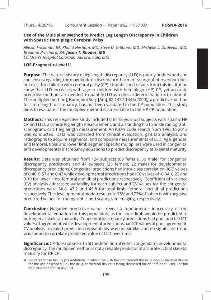
-195-
♦ Indicates those faculty presentations in which the FDA has not cleared the drug and/or medical device for the use described (i.e., the drug or medical device is being discussed for an “off label” use). For full information, refer to page 14.
Thurs., 4/28/16 Concurrent Session II, Paper #62, 11:57 AM POSNA-2016
Use of the Multiplier Method to Predict Leg Length Discrepancy in Children with Spastic Hemiplegic Cerebral Palsy
Allison Frickman, BA; Khalid Hesham, MD; Steve D. Gibbons, MD; Michelle L. Dudevoir, MD; Breanna Pritchard, BA; Jason T. Rhodes, MDChildren’s Hospital Colorado, Aurora, Colorado
LOE-Prognostic-Level II
Purpose: The natural history of leg length discrepancy (LLD) is poorly understood and consensus regarding the magnitude of discrepancy that merits surgical intervention does not exist for children with cerebral palsy (CP). Unpublished results from this institution show that LLD increases with age in children with hemiplegic (HP) CP, yet accurate predictive methods are needed to quantify LLD as a clinical determination in treatment. The multiplier method [J Bone Joint Surg [Am], 82:1432-1446 (2000)], a predictive method for limb-length discrepancy, has not been validated in the CP population. This study aims to evaluate if the multiplier method is amendable to the HP CP population.
Methods: This retrospective study included 0 to 18-year-old subjects with spastic HP CP and LLD, a clinical leg length measurement, and a standing hip to ankle radiograph, scanogram, or CT leg length measurement. An ICD-9 code search from 1995 to 2013 was conducted. Data was collected from clinical evaluation, gait lab analysis, and radiographs to acquire segmental and composite measurements of LLD. Age, gender, and femoral, tibial and lower limb segment specific multipliers were used in congenital and developmental discrepancy equations to predict discrepancy at skeletal maturity.
Results: Data was obtained from 124 subjects (68 female, 56 male) for congenital discrepancy predictions and 47 subjects (25 female, 22 male) for developmental discrepancy predictions. Congenital predictions had intra-class correlation (ICC) values of 0.40, 0.57 and 0.43 while developmental predictions had ICC values of -0.04, 0.22 and 0.10 for lower limb, femoral and tibial predictions respectively. Coefficient of variance (CV) analysis addressed variability for each subject and CV values for the congenital predictions were 66.8, 47.2 and 45.8 for total limb, femoral and tibial predictions respectively. The developmental model resulted in 75% and 77% of subjects with negative predicted values for radiographic and scanogram imaging, respectively.
Conclusion: Negative predictive values reveal a fundamental inaccuracy of the developmental equation for this population, as the short limb would be predicted to be longer at skeletal maturity. Congenital discrepancy predictions had poor and fair ICC values of agreement, while developmental predictions had ICC values of poor agreement. CV analysis revealed prediction repeatability was not similar and no significant trend was found to correlate predicted value of LLD over time.
Significance: CP does not seem to fit the definition of either congenital or developmental discrepancy. The multiplier method is not a reliable predictor of accurate LLD at skeletal maturity for HP CP.
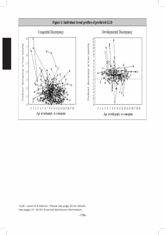
-196-
†LOE - Level of Evidence - Please see page 20 for details.See pages 21- 60 for financial disclosure information.
-197-
♦ Indicates those faculty presentations in which the FDA has not cleared the drug and/or medical device for the use described (i.e., the drug or medical device is being discussed for an “off label” use). For full information, refer to page 14.
Thurs., 4/28/16 Concurrent Session II, Paper #63, 12:02 PM POSNA-2016
Self Reported Quality of Life in Adolescence and Young Adults with Cerebral Palsy
Nancy Lennon, MSPT; Chris Church, MPT; Freeman Miller, MD; Julieanne P. Sees, DONemours / Alfred I. duPont Hospital for Children, Wilmington, Delaware
LOE-Prognostic-Level III
Purpose: The aim of this study was to compare self-reported quality of life (QoL) to proxy reported QoL among adolescents with cerebral palsy (CP).
Methods: Participants were adolescents with CP aged 11-20, classified at GMFCS levels I-V, able to read at or above a fourth grade level who completed the self-reported Pediatric Outcomes Data Collection Instrument (PODCI) between January 2012 and June 2015. The PODCI was completed using an iPad during visits to a hospital-based CP specialty clinic. Patients with motor limitations completed the assessment with the aid of clinic staff. Welch two sample t-tests were used to examine differences between self- and proxy-reporting of QoL in five PODCI domains and in PODCI global scores.
Results: PODCI scores from 50 matched pairs of parents/adolescents with CP at GMFCS levels I(14), II(18), III(11), and IV(7) were examined. Number of participants in middle school, high school, and post-secondary age categories were: 11 to 14 yrs (35), 15 to 18 yrs (14) and 19-20 yrs (1). 84% of the paired samples were from the same clinic visit. There were 29 males and 21 females. Mean self-reported scores were significantly higher than mean parent-reported scores in four domains (p<0.05): Upper Extremity and Physical Function (p=0.018), Sports and Physical Functioning (p=0.005), Happiness (p=0.023), and Global Functioning (p=0.018).
Conclusion: Adolescents perceive a higher quality of life in the domains of upper extremity function, sports function, happiness, and global function relative to their parents’ perceptions. Parent and adolescent perceptions were not significantly different in the domains of transfers/ basic mobility and pain/ comfort.
Significance: Self- and parent-reported adolescent PODCI results should be treated separately in clinical settings and in future research. While it is encouraging that adolescents are optimistic about their function, it is important that they convey accurate information about their abilities to health care providers. Guided and accurate self-reporting will help adolescents receive the necessary support to optimize their function and well being while transitioning to adulthood.
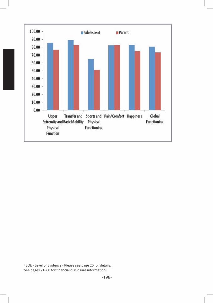
-198-
†LOE - Level of Evidence - Please see page 20 for details.See pages 21- 60 for financial disclosure information.
-199-
♦ Indicates those faculty presentations in which the FDA has not cleared the drug and/or medical device for the use described (i.e., the drug or medical device is being discussed for an “off label” use). For full information, refer to page 14.
Fri., 4/29/16 Clinical Award Papers, Paper #64, 8:05 AM POSNA-2016
Preventing Curve Progression with Calcium and Vitamin D Supplementation in Adolescent Idiopathic Scoliosis (AIS) – A Randomised Double-blinded Placebo-controlled Trial
Tsz Ping Lam, MD; Benjamin Hon Kei Yip; Elisa Man Shan Tam; Gene Chi Wai Man; Wayne YW Lee; Fiona Wai Ping Yu; Bobby Kin Wah Ng, MD; Jack Cheng; Kwong Man LeeDepartment of Orthopaedics and Traumatology, the Chinese University of Hong Kong, Prince of Wales Hospital, Hong Kong
LOE-Therapeutic-Level I
Purpose: Adolescent Idiopathic Scoliosis(AIS) is a prevalent three-dimensional spinal deformity associated with low bone mass. This study aimed at evaluating the therapeutic effect of oral calcium+Vit-D supplementation for improving low bone mass and preventing curve progression in immature AIS girls.
Methods: This was a randomised double-blinded placebo-controlled trial on 330 AIS girls(mean age:12.9±0.9 years) with femoral neck aBMD Z-score<0 and radiological Cobb angle≥15°. They were randomly allocated to Group1(N=110, placebo), Group2(N=110, 600mg calcium+400IU Vit-D3/day) and Group3(N=110, 600mg calcium+800IU Vit-D3/day). Treatment lasted for 2 years. At baseline and at the end of 2-year treatment, serum 25(OH)Vit-D assay, Dual-Energy X-ray Absorptiometry(DXA) of femoral necks and High-resolution Peripheral Quantitative Computed Tomography(HR-pQCT) at the non-dominant distal radius were performed. Scoliosis was managed at regular follow-up visits and bracing was prescribed according to standard protocol. Curve progression was defined as increase in Cobb angle of the major curve≥6° taken without brace on standard standing postero-anterior whole spine radiograph. “Intention-To-Treat” principle was followed for analysis on those who completed the 2-year treatment.
Results: 270(81.8%) subjects completed the 2-year treatment(Fig 1:Patient Flow Diagram). At baseline, mean serum 25(OH)Vit-D was 41.2±14.7nmol/L and mean Cobb angle was 25.9±8.4°. Mean increase in serum 25(OH)Vit-D level after 2-year treatment were 6.3±15.3, 20.4±19.6 and 28.0±23.3nmol/L for Group1, 2 and 3 respectively(p<0.001). Changes in aBMD and BMC at femoral neck, Average vBMD, Trabecular vBMD, BV/TV, Trabecular Number and Separation at distal radius at the end of 2-year treatment were significantly different between groups showing anabolic bone effects with calcium+Vit-D supplementation(p<0.05). At the latest follow-up(N=132, Fig 1), 21.7% in Group3 and 24.4% in Group2 had curve progression≥6° as compared with 46.7% in Group1(p<0.05). Logistic regression analysis showed the rate of curve progression remained significantly lower in Group 2 and 3 after adjustment for baseline maturity, baseline curve severity and bracing history(p<0.05). For those with baseline serum 25(OH)Vit-D≤50nmol/L(N=103), 16.2% had curve progression in Group 3 as compared with 48.6% in Group 1(p=0.003).
Conclusion: The results provided strong evidences that daily 600mg calcium+400/800 IU Vit-D3 improves low bone mass and prevents curve progression in AIS. Vit-D status
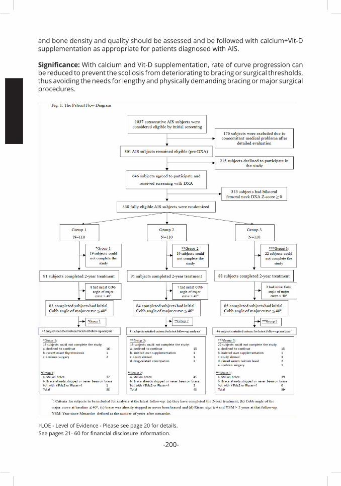
-200-
†LOE - Level of Evidence - Please see page 20 for details.See pages 21- 60 for financial disclosure information.
and bone density and quality should be assessed and be followed with calcium+Vit-D supplementation as appropriate for patients diagnosed with AIS.
Significance: With calcium and Vit-D supplementation, rate of curve progression can be reduced to prevent the scoliosis from deteriorating to bracing or surgical thresholds, thus avoiding the needs for lengthy and physically demanding bracing or major surgical procedures.
-201-
♦ Indicates those faculty presentations in which the FDA has not cleared the drug and/or medical device for the use described (i.e., the drug or medical device is being discussed for an “off label” use). For full information, refer to page 14.
Fri., 4/29/16 Clinical Award Papers, Paper #65, 8:15 AM POSNA-2016
Is There Value In Having Radiology Provide a Second Reading in Pediatric Orthopedic Clinic?
Vivek Natarajan, MD; Patrick P. Bosch, MD; Ozgur Dede, MD; Stephen A. Mendelson, MD; Maria Brooks, PhD; Tanya Kenkre, PhD; James W. Roach, MDChildren’s Hospital of Pittsburgh, Philadelphia, Pennsylvania
LOE-Economic & Decision-Level IV
Purpose: In most children’s hospitals, a radiologist provides a second interpretation for radiographs taken in the pediatric orthopedic clinic. There are significant costs associated with these radiology reads, and it is unclear whether they impart additional clinically relevant information beyond the orthopedist’s initial interpretation. The purpose of this study is to determine the clinical significance and cost-effectiveness of having radiology provide a second interpretation of films in a hospital-based pediatric orthopedic clinic.
Methods: A retrospective review of all radiographs obtained over a four-month period in the pediatric orthopedic clinic of a single children’s hospital was performed. For each radiographic series, the orthopedic note and the radiology interpretation were reviewed and a determination was made whether the radiology read provided new clinically useful information and/or a new diagnosis, whether it recommended further imaging, or if it missed a diagnosis that was reflected in the orthopedic note. The cost associated with the radiology read for each study was also quantified.
Results: The charts of 1,570 consecutive clinic patients who were seen in the pediatric orthopedic clinic from January-April 2012 were reviewed. There were 2,509 radiographic studies performed, of which 2,265 had both a documented orthopedic note and radiology read. The radiologist’s interpretation added new, clinically important information in 0.8% (18/2265) of these studies. The radiologist suggested a new diagnosis in 1.6% (36/2265) of these studies, although the additional diagnosis was considered to be clinically relevant in only 19/36 of these cases. In 1.9% (44/2265) of the studies, it was determined that the radiologist missed the diagnosis or clinically important information that could affect treatment. The total cost of the professional fees for the radiology interpretations was $87,450. For each occurrence in which the radiology read provided an additional diagnosis or clinically important information beyond the orthopedic note, an overall cost of $2,364 was incurred.
Conclusion: Most radiographs obtained in hospital-based pediatric orthopedic clinics are interpreted by both the orthopedist and a radiologist. This study demonstrates that the radiologist’s interpretation rarely provides additional clinically relevant information and is associated with significant costs.
Significance: Nowadays, healthcare practices are under increased scrutiny to improve cost-effectiveness. The results of this study suggest that eliminating the requirement to have the radiologist interpret radiographs in the pediatric orthopedic clinic may be a valuable cost-saving measure with minimal clinical implications.

-202-
†LOE - Level of Evidence - Please see page 20 for details.See pages 21- 60 for financial disclosure information.
Fri., 4/29/16 Clinical Award Papers, Paper #66, 8:25 AM POSNA-2016
Abduction Bracing versus Natural History in Hip Dysplasia: Multicenter Randomized Controlled Trial
Virginie Pollet, MD; Ralph Sakkers, MD, PhD; Michiel van de Sande, MD, PhD; Melinda Witbreuk; Erik Beek; Rene M. Castelein, MD, PhDWilhelmina Children’s Hospital, Utrecht, the Netherlands
LOE-Therapeutic-Level I
Purpose: Stable hip dysplasia is treated with abduction bracing till normal bony roof angle on ultrasound or acetabular index on pelvis X-rays is adequate for the child’s age. Little is known about the natural history. This Multicenter Randomized Controlled Trial was performed to study the outcome of treatment with abduction bracing versus no treatment in dysplastic hips without instability.
Methods: Children between 3 and 4 months of age with stable but dysplastic hips on ultrasound (Graf type IIB and IIC) of 5 participating hospitals were randomized to either Pavlik harness treatment group (59 patients) or no treatment group (53 patients). Patients with more severe Graf types or clinical instability and those who were previously treated were excluded from this study. At 6 and 12 weeks interval, the ultrasound was repeated in all patients. As standard of care, patients underwent a pelvis X-ray at the age of 9 months and after walking age. Residual dysplasia was assessed by measuring the bony roof angle according to Graf classification on ultrasound and acetabular index according to Tönnis classification on pelvis X-rays. One senior pediatric radiologist read all images.
Results: After randomization, the treatment groups were fully comparable for all important predictors of outcome. The average increase in alpha angle in the Pavlik group over 6 weeks was 3.6°. In the non-treatment group this was 3°.
At 6 weeks there was no difference in treatment effect: Pavlik group mean angle 58.8° ±5.5°, non-treatment group 58°±5.2°, mean difference -1.39° (95% CI: -3.46, 0.67), p-value 0.18. At 12 weeks, values were 60.5°±3.8°, 60°±5.6° respectively, mean difference -1.0° (95%CI: -2.92, 0.91), p-value 0.30. Analysis of secondary outcomes showed no treatment differences at 9 months of age (p= 0.82) and at latest follow-up (p= 0.35) in Tönnis classification for residual dysplasia.
Conclusion: The natural history of hips with stable dysplasia (Graf type IIB and IIC) showed a similar development and improvement as did the treated hips. The Pavlik harness did not improve overall outcome and both groups showed similar rates of normalization and residual dysplasia. This study indicates that these hips should not be treated, but follow-up is necessary to identify those hips that will fail to improve spontaneously.
Significance: With time most Graf IIB and IIC dysplastic hips will resolve without treatment. Further research is needed to identify the hips that will fail to improve.
-203-
♦ Indicates those faculty presentations in which the FDA has not cleared the drug and/or medical device for the use described (i.e., the drug or medical device is being discussed for an “off label” use). For full information, refer to page 14.
Fri., 4/29/16 Clinical Award Papers, Paper #67, 8:35 AM POSNA-2016
Results of AIS Surgery at a Minimum of 10 Years Using Modern Implant Systems- Are Patients Troubled by Symptoms and a High Reoperation Rate?
Daniel J. Sucato, MD, MS; Kaitlyn Brown, BS; Anna McClung, RN, BSNTexas Scottish Rite Hospital for Children, Dallas, Texas
LOE-Therapeutic-Level II
Purpose: To review prospectively collected data on a series of patients at 10 years following surgery for adolescent idiopathic scoliosis (AIS).
Methods: Patients who received surgical treatment for AIS from 2002-2005 were recruited from a single institution’s prospective database to return for a 10 year evaluation. Outcomes included radiographic and clinical parameters, PFTs and the SRS-22r outcome instrument collected preoperatively and at 2 and 10 years following initial surgery.
Results: There were 72 patients (10 male & 62 female) who returned for a minimum 10 year follow-up. Improvements occurred from preop to 10 years for main coronal Cobb (57.9 to 29.0 degrees), coronal balance (1.4 cm to 1.0 cm), and trunk shift (1.9 to 0.8 cm) (p<0.001). A mild loss of coronal Cobb correction was seen from 2 to 10 years (25.9 to 29.0 degrees) (p=0.05). PFTs were the same preoperatively and at 10 years for FEV1-% pred (71.7 to 75.2%), and FVC-%pred (78.1 to 76.6%) without differences noted between posterior and anterior approaches. No significant loss of pulmonary values was seen between 2 and 10 years. Worse PFTs correlated with a larger residual coronal Cobb magnitude. The SRS-22r scores improved at 10 years for appearance (3.5 to 4.1), satisfaction (3.8 to 4.3), and total SRS score (4.0 to 4.1) (p<0.05) domains without differences in the remaining domains. Coronal balance was the only radiographic or clinical parameter that correlated to improved appearance and satisfaction scores (p<0.05). Patients demonstrated improved SRS pain scores at 2 years, but saw a significant decrease at 10 years (4.3 to 4.0) (p<0.007). At 10 years follow-up, 15 (20.8%) patients had complaints of pain, and 6 (8.3%) patients had a repeat operation for infection (2), prominent implants (2), and painful implants (2).
Conclusion: Patients undergoing surgical treatment for AIS have improved maintenance of most radiographic parameters, maintain their preoperative PFTs, demonstrate satisfaction, and are happy with their appearance which correlates with their overall coronal balance. Despite these good results the incidence of persistent pain is 20% and 8% of patients required another procedure.
Significance: Long-term outcome studies following AIS surgery are few. While patients display improvements in radiographic, clinical, and pulmonary parameters and are satisfied with their appearance at 10 years postop, a significant number of patients report consistent pain. Additionally, revision rates remain relatively high at 8%.

-204-
†LOE - Level of Evidence - Please see page 20 for details.See pages 21- 60 for financial disclosure information.
Fri., 4/29/16 Clinical Award Papers, Paper #68, 8:45 AM POSNA-2016
5-Year Reoperation Rate and Causes for Revision After Idiopathic Scoliosis Surgery
Syed Ahmed, MD; Tracey P. Bastrom, MA; Burt Yaszay, MD; Peter O. Newton, MD; Harms Study GroupRady Children’s Hospital, San Diego, California
LOE-Therapeutic-Level III
Purpose: Data on contemporary revision surgery rates after idiopathic scoliosis surgery beyond the 2 year post-operative follow up visit in the adolescent and young adult population is limited. The purpose of this study was to define the rate and cause of surgical revision 5 years after scoliosis surgery.
Methods: Patients enrolled in a multi-center observational prospective idiopathic scoliosis surgical registry from 1995-2009 were reviewed. Any spine re-operation was defined as a “terminal event”. Actuarial survivorship analysis was performed to determine cumulative survival, which adjusts for patients lost to follow up by censoring and counting half person years in each interval, adjusting the denominator as necessary. Time intervals for the actuarial curve were defined as 0 to <3 months, 3 months to <1 year, 1 to <2 years, 2 to <5 years, and 5 to 10 years. Registry data and radiographs were reviewed and five categories for reoperation assigned: 1) implant failure and/or pseudarthrosis, 2) implant misplacement and/or prominence, 3) wound complication and/or infection, 4) residual deformity and/or progression, and 5) other.
Results: 1,435 patients from 12 centers were included. The majority were female (80%), with major thoracic curves (76% Lenke 1-4), and average age of 15±2 years (10-22) at surgery. Most had posterior spinal instrumentation and fusion (81%). At this time, 75 (5.2%) patients required re-operation. 22 occurred within 3 mo post-op, 10 more before 1 year, 12 more before 2 years, another 20 by 5 years and 10 more after 5 years. This corresponded to an actuarial cumulative survival of 98.3% at 3 months, 97.5% at 1 year, 96.6% at 2 years, 93.9% at 5 years, and 89.8% at the final interval (5-10 years). The timing of reoperation based on various causes is seen in the Figure. Only one patient (unplanned staged procedure) was categorized as “other.” and 5) other.
Conclusion: Revisions for scoliosis surgery may occur well after 2 years with a 5 year survivorship of 93.9%. Reasons for re-operation were not uniformly distributed over time: implant related issues and infection the cause early, while late infection was the most common cause after 2 years. Future studies should focus on longer term follow up and solutions to the issue of late infections.
Significance: Limited data exists in characterizing the long-term risk of reoperation in scoliosis surgery. This data suggests need for revision surgery continues beyond 2 years postoperatively.
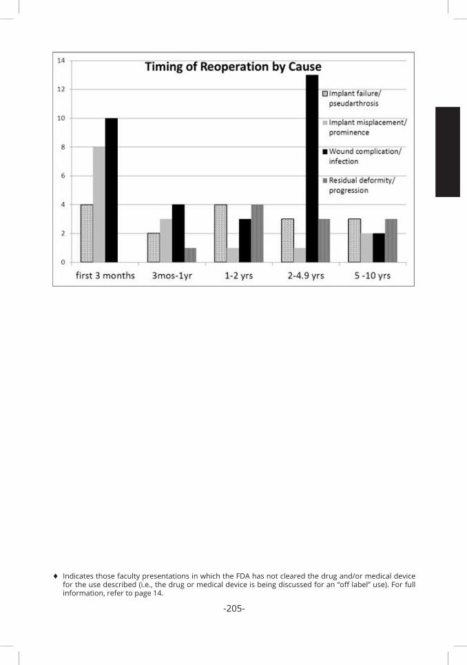
-205-
♦ Indicates those faculty presentations in which the FDA has not cleared the drug and/or medical device for the use described (i.e., the drug or medical device is being discussed for an “off label” use). For full information, refer to page 14.

-206-
†LOE - Level of Evidence - Please see page 20 for details.See pages 21- 60 for financial disclosure information.
Fri., 4/29/16 Clinical Award Papers, Paper #69, 8:55 AM POSNA-2016
Outcomes of Physeal Sparing ACL Reconstruction with IT Band in Skeletally Immature Children
Mininder S. Kocher, MD, MPH; Benton E. Heyworth, MD; Frances Tepolt, MD; Lyle J. Micheli, MDBoston Children’s Hospital, Boston, Massachusetts
LOE-Therapeutic-Level IV
Purpose: While intra-substance ACL tears in children and adolescents are becoming increasingly common, controversy remains about how best to manage these injuries in the setting of open physes. The purpose of this study was to assess outcomes of a large cohort having undergone physeal sparing ACL reconstruction with autogenous iliotibial band graft.
Methods: This was a retrospective case series to include all Tanner stage 1-2 patients having undergone physeal sparing, combined intra- and extra-articular reconstruction of the ACL with autogenous iliotibial band graft at a single institution Oct 1989 - Oct 2012. Following IRB approval, patient medical records were reviewed, and patient-derived functional outcomes at a minimum of 2-year follow-up were obtained.
Results: 237 patients, 86% male, mean age 11.2 +/- 1.7 years (range 5.7 – 15.6 years), underwent ACL reconstruction on 240 knees, 52% of left laterality. The mechanism of injury was non-contact in 71.6% of cases, with 77.8% of injuries having occurred during organized sport, most commonly football (25.1%), skiing/snowboarding (19.3%) and soccer (10.6%). Duration from injury to surgery was mean 6.3 +/- 6.0 months (range 3 days – 4.2 years). Associated intra- or extra-articular knee injuries were seen in 52.3% of cases, 41.7% with torn lateral meniscus and 7.5% torn medial meniscus, treated with repair in 79.0% and 77.8% of cases, respectively. Graft failure occurred in 9/240 cases (3.8%), 8/9 requiring subsequent revision ACL reconstruction. Other complications included arthrofibrosis requiring lysis of adhesions (2.1%), septic joint requiring incision and drainage (I & D) (0.4%) and wound dehiscence requiring I & D (0.4%). Subsequent knee surgery due to meniscal/chondral injury was required in 5.8% of cases, while 6.0% of patients sustained contralateral ACL tear requiring reconstruction. There were no cases of growth disturbance, either leg length discrepancy or angular deformity. Subjective outcomes were obtained from 56% of patients at a mean 6.2 years post-operatively (range 2.1 – 24.9 years): Lysholm mean 93.4 +/- 9.9, Pedi-IKDC mean 83.9 +/- 8.9 and Tegner Activity Scale mean 7.8 (range 2-10; mode 7). 96.5% of patients reported return to sport.
Conclusion: Physeal sparing, combined intra- and extra-articular reconstruction of the anterior cruciate ligament with use of an autogenous iliotibial band graft in skeletally immature prepubescent children provides excellent functional outcomes, minimal risk of growth disturbance and a low revision rate.
-207-
♦ Indicates those faculty presentations in which the FDA has not cleared the drug and/or medical device for the use described (i.e., the drug or medical device is being discussed for an “off label” use). For full information, refer to page 14.
Significance: Physeal sparing, combined intra- and extra-articular ACLR with ITB graft in prepubescent children provides excellent functional outcomes, minimal risk of growth disturbance and low revision rate.

-208-
†LOE - Level of Evidence - Please see page 20 for details.See pages 21- 60 for financial disclosure information.
Fri., 4/29/16 Clinical Award Papers, Paper #70, 9:05 AM POSNA-2016
Pediatric Femur Fracture Management: A Multicenter Analysis of the Trends Pre and Post 2009 AAOS Clinical Practice Guidelines
John D. Roaten, MD; Jeffrey R. Sawyer, MD; Joseph L. Yellin, BA; John (Jack) M. Flynn, MD; Micaela Cyr, BA; Sumeet Garg, MD; Alexander M. Broom, BA; Lindsay M. Andras, MDLe Bonheur Children’s Hospital, Memphis, Tennessee
LOE-Therapeutic-Level III
Purpose: The AAOS adopted a CPG (2009) for pediatric femoral shaft fractures when surgical treatment was becoming more popular, especially in younger patients. The purpose of this study was to analyze the treatment of pediatric femoral shaft fractures in the context of the AAOS CPG guidelines at high-volume, geographically-separated, pediatric trauma centers over a10 year period straddling the AAOS CPG to determine if CPG changed treatment.
Methods: We reviewed an IRB-approved consecutive series of pediatric femoral shaft fractures (ages 0-18 yo) from 4 high-volume, geographically-distant Level I pediatric trauma centers between 2004 and 2013 (5 yrs pre and post 2009 CPG). Treatment method was analyzed by center and age group.
Results: Of the 2646 patients with femoral shaft fractures, 1604(58%) were treated non-operatively. There were 568 fractures (21%) treated with flexible intramedullary nailing (FIMN) (mean 7.7 yo; 1.5-17.7 yo), of which, 14% were <5yo. There were 309 fractures (11.7%) treated with locked IM nails (LIMN) (mean 13.3 yo; 6.9-18yo), of which 53 (17.2%) were <11 yo. Plating was used 196 fractures (7.4%) (mean 8.2 yo, 2.6-16.6yo) and 1.5% were <5 yo. Analysis pre/post 2009 AAOS CPG publication revealed a significant increase in the use of LIMN in patients <11 yo (0.56% to 3.84% ) p<0.001. There was an also increase in surgical management regardless of technique for patients <5 yo (6.4% to 8.3%). There were treatment differences between centers, most notably 74% of fractures treated with plating were performed at center 4. Other center-based variations include Center 2 having the highest rate (41%) of FIMN in children <5yo and Center 1 having the highest rate (63%) of LIMN <11yo.
Conclusion: A decade-long analysis of pediatric femur fracture treatment at four pediatric trauma centers shows continued trends towards operative management in younger children. Following AAOS CPG publication, significant increases in LIMN in patients < 11 yo and the increased surgical treatment in patients <5 years old has occurred. Considerable variability in treatment method and CPG adherence between centers exists.
Significance: This study highlights the need for further outcome studies to determine the optimal treatment methods for pediatric femoral shaft fractures as well as the fact that there may be a need to update the AAOS CPG guidelines.
-209-
♦ Indicates those faculty presentations in which the FDA has not cleared the drug and/or medical device for the use described (i.e., the drug or medical device is being discussed for an “off label” use). For full information, refer to page 14.
Fri., 4/29/16 Clinical Award Papers, Paper #71, 9:15 AM POSNA-2016
The Effect of Spastic Hip Dysplasia on Health Related Quality of Life in Non-ambulatory Children with Cerebral Palsy
Rachel L. DiFazio, PhD, RN, PPCNP-BC, FAAN; Benjamin J. Shore, MD, MPH, FRCSC; Judith Vessey, PhD, MBA, RN, FAAN; Patricia E. Miller, MS; Brian D. Snyder, MD, PhDBoston Children’s Hospital, Boston, Massachusetts
LOE-Therapeutic-Level II
Purpose: The aims of this prospective level II study are to: (1) evaluate the relationship of migration percentage (MP) and acetabular index (AI) measured pre-operatively on radiographs of GMFCS IV-V CP children with spastic hip dysplasia to HRQOL measured by the CPCHILD™ instrument before and after reconstructive hip surgery; (2) determine if the relative extent of improvement in MP following reconstructive hip surgery affects subsequent HRQOL measured by CPCHILD™ over the ensuing 2 years; (3) identify which clinical predictors affect HRQOL after reconstructive hip surger in non-ambulatory children with CP. Methods: A prospective cohort study (N=38) design was used to measure changes before and after reconstructive hip surgery. MP, AI, and CPCHILD™ scores were analyzed using Pearson’s correlation analysis before surgery and 6 weeks, 3, 6, 12, and 24 months after surgery. Subgroup analysis was used to compare patients with preoperative MP≥50% (incipient hip dislocation) versus MP<50% (hip subluxation) and to compare AI in patients who had an acetabuloplasty and those who did not. Linear mixed models were used to analyze changes in MP, AI and CPCHILD™ scores over time. Results: The pre-operative MP inversely correlated with the pre-operative CPCHILD™ total score (p=0.002; r=-0.50). This relationship was influenced by the extent of surgical correction and post-operative time interval: 12 months after surgery, for each 1% correction in MP, the CPCHILD™ total score increased by 0.4 points from baseline (p<0.001); 24 months after surgery, for each 1% correction in MP, the CPCHILD™ total score increased by 0.8 points (p<0.001). There was no correlation between AI and CPCHILD™ total score before or after surgery (p=0.09 to 0.71). Pre-operative CPCHILD™ total scores were different between MP groups (mean difference = 13; 95% CI=3.3-22.8; p=0.01). However after hip surgery, changes in CPCHILD™ total score improved similarly for both groups. Conclusion: A greater than minimally clinically significant improvement in HRQOL was observed after reconstructive hip surgery in non-ambulatory children with severe CP with spastic hip dysplasia, supporting the effectiveness of this intervention. Significance: In this era of healthcare reform, orthopedic surgeons are charged with demonstrating the value of their surgical interventions. Reconstruction hip surgery for non-ambulatory children with CP is costly both financially and emotionally. Findings from this prospective level II study provide objective evidence that treatment of spastic hip dysplasia by reconstructive hip surgery in non-ambulatory children with severe CP reliably improves HRQOL in proportion to the extent that the MP was improved.

-210-
†LOE - Level of Evidence - Please see page 20 for details.See pages 21- 60 for financial disclosure information.
Fri., 4/29/16 Clinical Award Papers, Paper #72, 9:25 AM POSNA-2016
Percutaneous Heel Cord Release for Clubfoot: A Retrospective, Multicenter Cost Analysis
Matthew Abbott, MD; Brittany Hedrick, MD; Franklin K. Gettys, MD; B. Stephens Richards, MD; Ryan D. Muchow, MD; ChanHee Jo, PhD; Amanda Moualeu, MPHUniversity of Michigan, Mott Children’s Hospital, Ann Arbor, Michigan
LOE-Economic & Decision-Level III
Purpose: Ponseti method of treatment is the standard of care for idiopathic clubfoot. Following serial casting, percutaneous heelcord tenotomy (TAL) is performed to correct residual equinus. This procedure can be performed in either the outpatient clinic or the operating room. The purpose of this study was to evaluate the expense of this procedure by examining hospital charges in both settings.
Methods: We retrospectively reviewed charts of 382 idiopathic clubfoot patients with a mean age of 2.4 months (range 0.6-26.6 months) treated with Ponseti method at three independent institutions. Patients were divided into three groups depending on the setting for the TAL procedure: 140 patients in outpatient clinic (CL), 219 in the operating room with discharge following the procedure (OR), and 23 in the operating room with admission to hospital for observation (OR+). Medical records were reviewed to analyze age, deformity, peri-operative complications, and specific time spent in each setting. Because hospital charges varied between institutions, for this review all three groups were standardized to one institution’s 2015 charge structure. ANOVA and Chi-square tests were used for comparison of variables between the three groups.
Results: Charges between the three groups undergoing TAL (CL, OR, OR+) were found to be significantly different ($3840.60 vs. $7962.30 vs. $9110.00 respectively; p<.001). As there are different charges for unilateral and bilateral deformities, when separated out based on laterality the significant cost difference persisted across all three groups in similar fashion (p<0.001). There were 9 total peri-operative complications (6 returns to the ER and 3 unexpected admissions to the hospital). The CL group had no complications, the OR group had 5 (2.3%), and the OR+ group had 4 (17.4%). The OR+ group statistically had a higher rate of complications compared to the other two groups (p=0.006). Additionally, it was found that total event time was significantly different with the CL group being the shortest compared to the OR and OR+ groups. (129.1 minutes vs. 171.7 minutes vs. 1571.6 minutes; p<.001).
Conclusion: Hospital charges and total event time were significantly less when percutaneous TAL was performed in the outpatient clinic as compared to the operating room. In addition, performing the procedure in the clinic was associated with the lowest rate of complications.
Significance: Performing percutaneous TAL in clinic is safe, requires less time, and is a more cost-effective alternative to the operating room in patients less than 3 months of age.
-211-
♦ Indicates those faculty presentations in which the FDA has not cleared the drug and/or medical device for the use described (i.e., the drug or medical device is being discussed for an “off label” use). For full information, refer to page 14.
Fri., 4/29/16 Clinical Award Papers, Paper #73, 9:35 AM POSNA-2016
Posttraumatic Stress in Children and Adolescents Following Surgery for Orthopedic Trauma
Collin J. May, MD; Benjamin J. Shore, MD, MPH, FRCSC; Patricia E. Miller, MS; Manahil N. Naqvi, BS; Justyna M. Klajn, MS; Emily Rademacher, BS; Daniel J. Hedequist, MDBoston Children’s Hospital, Boston, Massachusetts
LOE-Prognostic-Level II
Purpose: Traumatic injuries are common in childhood, and are a significant source of disability and dysfunction. Beyond the physical effects of injury, it has become clear that children also suffer psychological sequelae as a result of their experiences, including posttraumatic stress disorder (PTSD). In this study, we aimed to determine the incidence of PTSD symptoms and risk factors for their development in the population of children and adolescents undergoing surgery for orthopedic trauma.
Methods: We performed a prospective study of all consecutive children ages 8-18 years undergoing surgery for orthopaedic trauma at a single level-1 trauma center. Patient, injury and treatment characteristics were obtained at baseline for all patients. Assessment of PTSD symptoms occurred at subsequent follow up visits, using the Child PTSD Symptom Scale (CPSS), a validated self-report indicator of posttraumatic stress symptoms in children. All patients were seen within 60 days of surgery for injury, and considered to have high levels of PTSD symptoms when scoring 11 or higher on the scale. Uni- and multivariable logistic regression was used to determine potential risk factors for PTSD.
Results: There were 116 subjects included in the analysis. The cohort had an average age at surgery of 12.8 years (range 8 to 18.8 years) and was 66% male. Twenty-one subjects had high levels of PTSD symptoms (18.1%; 95% CI = 11.6 to 26.3%). On univariate analysis, older age (14.1 versus 12.5 years, p=0.03), history of previous surgery (33% versus 15%, p=0.05), length of stay more than 1 day (38% versus 14%, p=0.01) and injury to the lower extremity over upper extremity (p=0.005) were predictive of high PTSD symptoms. Multivariable analysis determined that the only independent risk factor for high PTSD symptoms was upper versus lower extremity injury. It was found that lower extremity injuries requiring surgery had nearly 4 times the odds of risk for PTSD (OR=3.9; 95% CI= 1.45 to 10.41; p=0.005).
Conclusion: Posttraumatic stress following surgery for orthopedic trauma is common, with 18% of all patients undergoing surgery for trauma experiencing a high level of PTSD symptoms. This was true even after relatively minor injuries. Increased risk of PTSD symptoms was found on multivariable analysis to be associated with lower extremity injury.
Significance: The high rate of posttraumatic stress symptoms present in children after surgery for orthopaedic injuries highlights the need for more comprehensive screening of patients such that timely intervention and referral may take place.
-212-
Fri., 4/29/16 Clinical Award Papers, Paper #74, 9:45 AM POSNA-2016
Management of Clubfoot Relapses with the Ponseti Method: Results of a Survey of POSNA Members
Pooya Hosseinzadeh, MD; Jose A. Morcuende, MD, PhD; Lori Dolan, PhD; Gary Kiebzak, PhDBaptist Children’s Hospital, Miami, Florida
LOE-Therapeutic-Level III
Purpose: A previous survey of POSNA members has shown that Ponseti method is now used by an overwhelming majority of members as the first line for treatment of children with clubfeet. Despite the high rate of initial success with this method, relapses occur frequently. We studied the opinions of POSNA members about the management of clubfoot relapses after initial correction using the Ponseti method.
Methods: A 31-question survey was created focusing on the treatment of clubfeet after initial correction, bracing protocol, rate of relapses, diagnosis of relapses, and their treatment. The questionnaire was approved by the POSNA Evidence Based Committee and was sent electronically to all POSNA members.
Results: 321 members responded to the survey. 94% of respondents were fellowship trained in pediatric orthopedics. Ponseti method was reported as the initial method used to treat clubfeet by 97.5% of the respondents. The Mitchell orthosis was the most commonly used (50.5%) followed by the Denis-Browne bars (25%). Recommended duration of bracing varied among members with 23% only recommending 2 years, 32.6% recommending 3 years, and 34.2% recommending 4 years of bracing. Tight heel cord was reported as the first sign of relapse by 59% of respondents followed by dynamic supination reported by 30%. Dynamic supination was defined as the forefoot supination during swing phase of the gait by 57% of members followed by weight bearing on lateral side of the foot by 28.2% of members. The rate of relapse was reported less than 10%, 10-20%, and 20-40% by 22%, 52%, and 24.8% of respondents respectively. Casting (55.4%) and casting with tenotomy (23.1%) were reported as the two most common first line treatments for relapses. 62% reported routine casting before Tibialis Anterior Tendon Transfer (TATT). Heel cord tenotomy (74.5%) and posterior capsular release (43.1%) were the two most common procedures reported in addition to TATT in the treatment of clubfoot relapse.
Conclusion: Reported duration of bracing, first sign of relapse, and additional procedures in addition to TATT were considerably different among survey participants. Posterior capsular release, although not recommended by Ponseti, was reported by 43% of the respondents.
Significance: The opinion of POSNA members vary considerably when it comes to the duration of bracing, diagnosis, and treatment of clubfoot relapse. Most importantly, posterior capsular releases are still used by almost half of the members, with potential implications for long term outcomes.
†LOE - Level of Evidence - Please see page 20 for details.See pages 21- 60 for financial disclosure information.
-213-
♦ Indicates those faculty presentations in which the FDA has not cleared the drug and/or medical device for the use described (i.e., the drug or medical device is being discussed for an “off label” use). For full information, refer to page 14.
Fri., 4/29/16 Clinical Award Papers, Paper #75, 9:55 AM POSNA-2016
Single Event Multilevel Surgery in Bilateral Spastic Cerebral Palsy: A Long Term, Multicenter, Follow-up Study
Thomas Dreher, MD, PhD; Martin Svehlik, MD; Leonhard Döderlein, MD; Sebastian I. Wolf, PhD; Pam Thomason, BPHysio; Morgan Sangeux; Gerhardt Steinwender, MD; H. Kerr Graham, MD, FRCS (Ed), FRACSHeidelberg University Hospital, Heidelberg, Germany
LOE-Therapeutic-Level IV
Purpose: Although one RCT and several cohort studies support the efficacy of SEMLS, the majority of studies to date have limited numbers and short term follow up. The purpose of this study was to investigate the efficacy and safety of SEMLS in a long term, multicenter follow up study.
Methods: Three centers who perform SEMLS using similar indications, surgical techniques and outcome measures, pooled long term outcome data to study efficacy using the Gait Profile Score (GPS) and safety, using the Modified Clavien Dindo (MCD) system for surgical adverse envents. GPS scores were compared immediately before, 1-2 years (short term) and 5-15 years after SEMLS (long term follow up).
Results: Baseline demographic and GPS scores were similar across the 3 centers, confirming a uniform approach to surgical indications. 231 patients (142 male, 89 female) were available for clinical and standardized three-dimensional gait analysis review at a mean of 9 years post SEMLS. Mean pre-operative (T1) GPS scores were 16.3° and 11.3° at short term follow up (T2). This significant improvement of 5° was >3 time the MCID (1.6°). At long term follow up (T3) mean GPS was maintained at 11.4° for the group as a whole (Figure 1). Subset analysis revealed that 78% of the patients maintained or further improved their T2 status at skeletal maturity (T3). However, 16% of the patients had some degree of deterioration but only 6% returned to their pre SEMLS baseline. Secondary minor surgery at the time of hardware removal or later was necessary in 26% of the patients.
Conclusion: 78% of the subjects experienced a long-term clinically and statistically significant improvement in gait function at skeletal maturity compared to baseline status. A minority of patients experienced deteriortation of clinical importance and a minortiy of patients needed minor secondary corrections about which patients and parents should be informed during planning of SEMLS.
Significance: The majority of ambulant children with bilateral spastic cerebral palsy who were managed by SEMLS based on three-dimensional gait analysis, in large volume centers experience a significant improvement in gait function which persists through the adolescent growth spurt to skeletal maturity.
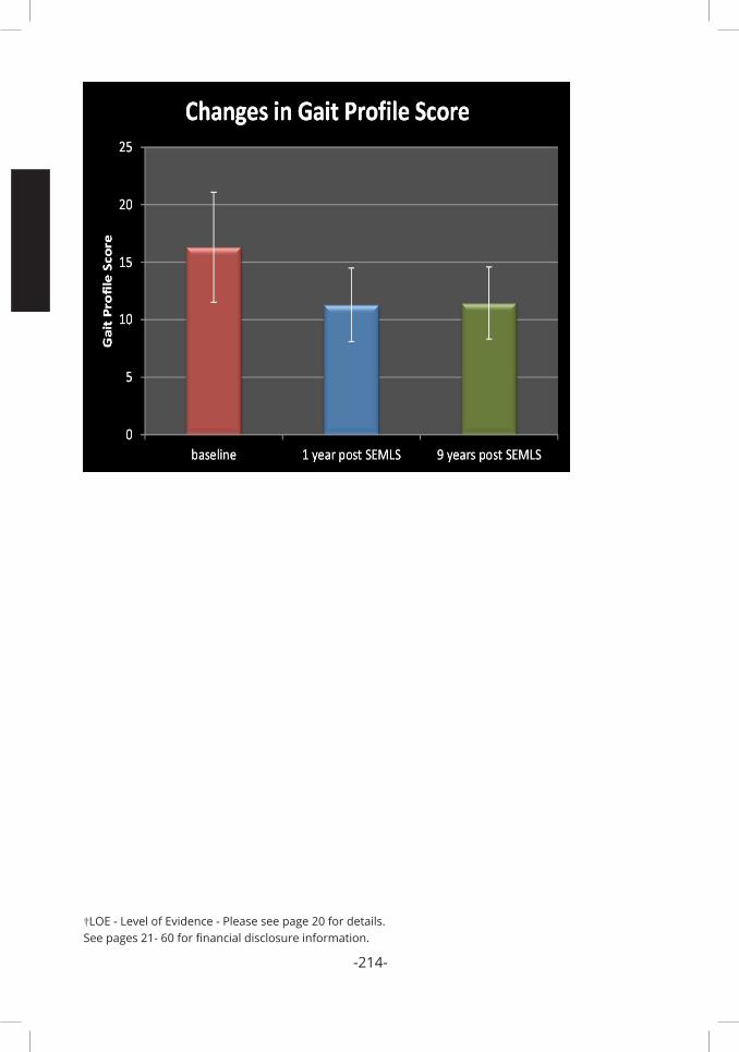
-214-
†LOE - Level of Evidence - Please see page 20 for details.See pages 21- 60 for financial disclosure information.
-215-
♦ Indicates those faculty presentations in which the FDA has not cleared the drug and/or medical device for the use described (i.e., the drug or medical device is being discussed for an “off label” use). For full information, refer to page 14.
Fri., 4/29/16 Basic Science Award Papers, Paper #76, 10:26 AM POSNA-2016
♦ Intra-Osseous Delivery of BMP-2 Using a Self-Assembling Peptide Hydrogel, RADA16
Matthew Phipps, PhD; Felipe Do Monte, MD; Manav Mehta, PhD; Harry K.W. Kim, MD, MSTexas Scottish Rite Hospital for Children, Dallas, Texas
LOE-Therapeutic-Level IV
Purpose: Local delivery of biological agents capable of stimulating bone healing offers potential new treatments for Perthes disease. A local infusion of BMP-2 has been shown to stimulate new bone formation and decrease deformity in a piglet model of ischemic osteonecrosis. The infusion of BMP-2 without a delivery agent, however, was associated with unwanted dissemination of BMP-2 out of the femoral head and heterotopic ossification. The purpose of this study was to assess the local distribution and retention of an injectable delivery agent called RADA16 (PuraMatrix©, 3-D Matrix, Inc.). This injectable hydrogel becomes a gel upon injection and exposure to physiological levels of salt in the femoral head.
Methods: 21 pig cadaver femoral heads were used to study the backflow and the distribution of RADA16 following an intra-osseous infusion. Micro-CT analysis following the infusion of RADA16 mixed with a radio-contrast solution was performed. The release rate of BMP-2 mixed in RADA16 was determined by an in-vitro elution experiment. Bioactivity of BMP-2 released from RADA16 was determined using western blot analysis for phosphorylation of SMAD1/5/8 in pig bone marrow stromal cells. The effect of RADA16 on proliferation of pig marrow stromal cells was also studied. Statistical analyses included t-test and ANOVA.
Results: Micro-CT analysis revealed a significant decrease in the amount of back flow of radio-contrast solution mixed with RADA16 down the needle track compared to the radio-contrast solution without RADA16 (p<0.05). Furthermore, RADA16 mixed with radio-contrast solution showed good distribution within the femoral head as evaluated by micro-CT. In vitro elution experiment revealed that RADA16 slowed the release rate of BMP-2 following an initial burst of release. Furthermore, BMP-2 released from RADA16 maintained its biological activity to induce phosphorylation of SMAD1/5/8 in pig marrow stromal cells. Lastly, RADA16 produced a significant increase in the proliferation of pig marrow stromal cells in culture compared to tissue culture plastic (p<0.05) suggesting that it is a suitable matrix for supporting marrow stromal cell proliferation.
Conclusion: RADA16 dispersed through the femoral head trabecular bone while preventing the back flow of radio-contrast solution through the needle track. It released bioactive BMP-2 in a controlled fashion and supported the growth of bone marrow stromal cells in vitro.
Significance: These results support the use of RADA16 as an intra-osseous delivery vehicle for BMP-2. Further studies are warranted to test the efficacy of RADA16 mixed with BMP-2 in in vivo models of ischemic osteonecrosis.
-216-
†LOE - Level of Evidence - Please see page 20 for details.See pages 21- 60 for financial disclosure information.
Fri., 4/29/16 Basic Science Award Papers, Paper #77, 10:32 AM POSNA-2016
Fibroblast Growth Factor Receptor 3 Modulates Fracture Repair By Controlling the Balance of Intramembranous and Endochondral Bone Formation
Simon P. Kelley, MBChB, MRCS, FRCS; Chunying Yu; Benjamin A. Alman, MD, FRCSCThe Hospital for Sick Children, Toronto, Ontario
LOE-Prognostic-Level I
Purpose: Fibroblast growth factor receptor 3 (fgfr3) mutations cause skeletal dysplasias such as achondroplasia and CATSHL syndrome. Fgfr3 is known to be a negative regulator of chondrocyte proliferation affecting long bone endochondral growth. However we observed that individuals with achondroplasia showed enhanced bone regeneration during distraction osteogenesis (intramembranous ossification) for limb lengthening. We hypothesized that fgfr3 affects mesenchymal proliferation and osteoblast differentiation, which act in concert with its known effects on chondrocyte function, a manifestation of which is the abnormal healing of bone fractures.
Methods: We used an established murine semi-stabilized tibia fracture model in genetically modified fgfr3 knockout mice and their wild type controls. Fractures were harvested and analyzed at critical time points (PFD3, 7, 14 and 21) using histomorphometry, micro-CT and gene expression analysis (qPCR) to assess fracture callus structure and composition. The proliferative ability of bone marrow MSCs (BM-MSCs) from fgfr3+/- and WT mice was studied using an in-vitro colony forming unit-fibroblast (CFU-F) assay, BrdU assay, limiting dilution and qPCR. The ability of BM-MSCs to differentiate to osteoblasts and chondrocytes in-vitro was examined using colony forming unit-osteoblast (CFU-O) and colony forming unit–chondrocyte (CFU-C) assays respectively, and were analyzed using staining techniques and qPCR.
Results: We show that fgfr3 modulates the size and structure of healing fracture callus with the fgfr3 mutant calli showing peak callus size earlier than their WT controls (D7), yet showing reduced callus size at the later time points (D14, D21). Despite the overall rapidity of healing, the structural integrity of the healed mutant fractures was diminished. In controlled in-vitro assays fgfr3 was shown to regulate the number, and proliferative ability of osteochondral progenitors with profound effects on osteogenic and chondrogenic differentiation of murine bone marrow MSCs, whereby under-expression of fgfr3 decreases osteogenic differentiation but increases chondrogenic differentiation. These findings were recapitulated in our in-vivo model of fracture healing in fgfr3+/- knockout mice; peripheral subperiosteal bone formation from the intramembranous pathway was diminished whilst central bone formation from the endochondral pathway was accelerated.
Conclusion: Fgfr3 acts as a switch, which controls the balance of intramembranous and endochondral bone formation in healing fractures by modulating different cell types of the mesenchymal lineage.
-217-
♦ Indicates those faculty presentations in which the FDA has not cleared the drug and/or medical device for the use described (i.e., the drug or medical device is being discussed for an “off label” use). For full information, refer to page 14.
Significance: Modulation of fgfr3 signaling may offer the ability to enhance the proliferation and differentiation of skeletal progenitor cells and also to direct cells towards particular bone regenerative pathways, which has enormous appeal for treating common musculoskeletal disorders such as fracture repair.
-218-
†LOE - Level of Evidence - Please see page 20 for details.See pages 21- 60 for financial disclosure information.
Fri., 4/29/16 Basic Science Award Papers, Paper #78, 10:38 AM POSNA-2016
The Role of Periosteal Fibers in Accelerated Bone Growth
Rajeev Chaudhary, MS; Ellen Leiferman, DVM; Ray Vanderby, PhD; Kevin Eliceiri; Paul J. Campagnola, PhD; Matthew A. Halanski, MDUniversity of Wisconsin, Madison, Wisconsin
LOE-Therapeutic-Level III
Purpose: Increased longitudinal bone growth occurs following periosteal resection. The accepted mechanism for this accelerated growth is that the periosteum serves as a mechanical tether to restrict physeal growth. If this accepted mechanism is true, one would expect: (1) periosteal fibers adjacent to the physis to be under tension during growth and (2) periosteal fibers adjacent to the physis to relax following metaphyseal periosteal resection. In this study, we utilize Second Harmonic Generation (SHG) imaging to assess the alignment of the periosteal fibers on the bone before and after periosteal resection.
Methods: Twelve seven-week old New Zealand White rabbits were sacrificed and periosteal resections performed at the distal femoral and proximal tibial metaphyses. The resected periosteal strips were stretched to different strains in a materials testing system (MTS), fixed, and imaged using (SHG) microscopy. Collagen fiber alignment at each strain (-20% 0%, 5%, and 10%) was then determined computationally using CurveAlign. The periosteum adjacent to the physis of six femurs and six tibiae were then imaged on-the-bone, before and after, metaphyseal periosteal resection. One-way ANOVA statistics were performed on all data.
Results: Increasing periosteal strain increased periosteal fiber alignment (p < 0.0001). Average alignment coefficients ([0 1], 0 = not aligned, 1 = perfectly aligned) and standard deviations for each strain were as follows: 0.346 ± 0.17, 0.509 ± 0.15, 0.535 ± 0.23, 0.628 ± 0.11 (distal femur) and 0.360 ± 0.18, 0.586 ± 0.17, 0.734 ± 0.21, 0.657 ± 0.19 (proximal tibia). Imaging of the periosteum at different regions of the bone demonstrated complex regional differences in fiber orientation. Periosteal fiber alignment adjacent to the physes before and after periosteal resection resulted in minimal change in fiber alignment indicating minimal change in periosteal strain following the procedure (Figure 1). This figure demonstrates SHG images of periosteal fibers at various locations on the bone before and after resection. The only change in fiber morphology is localized to the edge of the resection.
Conclusion: Periosteal fibers adjacent to the growth plate do not appear to be under tension in the growing limb and the alignments of these fibers remain unchanged following periosteal resection.
Significance: This study reports, for the first time, the complex organization of periosteal fibers on bone using SHG imaging. As the alignment of fibers adjacent to the physis changed little following periosteal resection, this study calls into question the long-accepted role of the periosteum acting as a simple mechanical tether restricting growth at the physis.
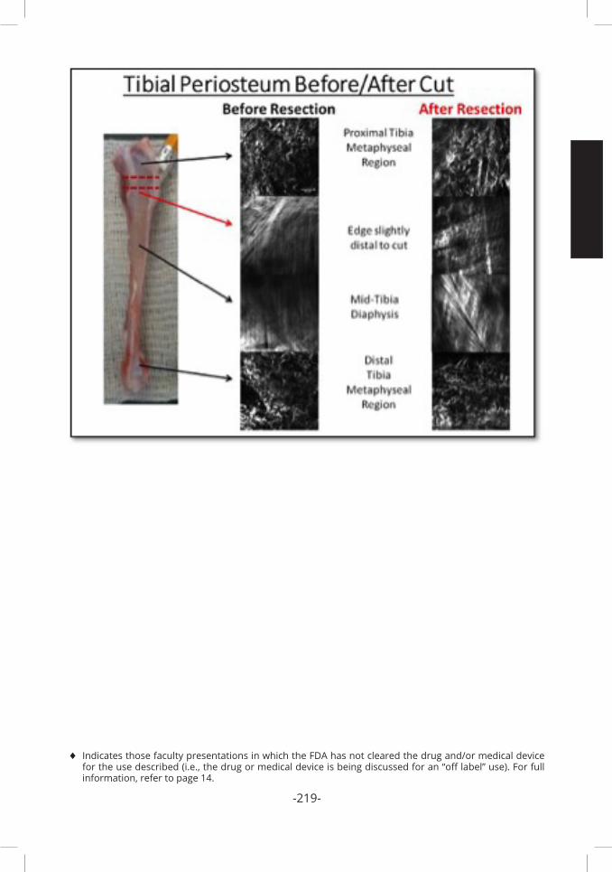
-219-
♦ Indicates those faculty presentations in which the FDA has not cleared the drug and/or medical device for the use described (i.e., the drug or medical device is being discussed for an “off label” use). For full information, refer to page 14.
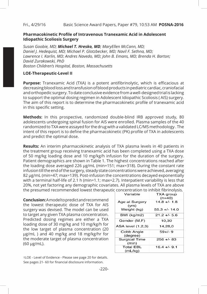
-220-
†LOE - Level of Evidence - Please see page 20 for details.See pages 21- 60 for financial disclosure information.
Fri., 4/29/16 Basic Science Award Papers, Paper #79, 10:53 AM POSNA-2016
Pharmacokinetic Profile of Intravenous Tranexamic Acid in Adolescent Idiopathic Scoliosis Surgery
Susan Goobie, MD; Michael T. Hresko, MD; MaryEllen McCann, MD; Daniel J. Hedequist, MD; Michael P. Glotzbecker, MD; Navil F. Sethna, MD; Lawrence I. Karlin, MD; Andres Navedo, MD; John B. Emans, MD; Brenda H. Barton; David Zurakowski, PhDBoston Children’s Hospital, Boston, Massachusetts
LOE-Therapeutic-Level II
Purpose: Tranexamic Acid (TXA) is a potent antifibrinolytic, which is efficacious at decreasing blood loss and transfusion of blood products in pediatric cardiac, craniofacial and orthopedic surgery. To date conclusive evidence from a well-designed trial is lacking to support the optimal dosing regimen in Adolescent Idiopathic Scoliosis ( AIS) surgery. The aim of this report is to determine the pharmacokinetic profile of tranexamic acid in this specific setting.
Methods: In this prospective, randomized double-blind IRB approved study, 80 adolescents undergoing spinal fusion for AIS were enrolled. Plasma samples of the 40 randomized to TXA were assayed for the drug with a validated LC/MS methodology . The intent of this report is to define the pharmacokinetic (PK) profile of TXA in adolescents and predict the optimal dose.
Results: An interim pharmacokinetic analysis of TXA plasma levels in 40 patients in the treatment group receiving tranexamic acid has been completed using a TXA dose of 50 mg/kg loading dose and 10 mg/kg/h infusion for the duration of the surgery. Patient demographics are shown in Table 1. The highest concentrations reached after the loading dose averaged 226 μg/mL (min=151; max=318). During the constant rate infusion till the end of the surgery, steady state concentrations were achieved, averaging 82 μg/mL (min=47; max=139). Post-infusion the concentrations decayed exponentially with a terminal half-life of 2.1 h (min=1.1; max=2.7). Interpatient variability is less that 20%, not yet factoring any demographic covariates. All plasma levels of TXA are above the presumed recommended lowest therapeutic concentration to inhibit fibrinolysis.
Conclusion: A model to predict and recommend the lowest therapeutic dose of TXA for AIS surgery was devised. The model can be used to target any given TXA plasma concentration. Predicted dosing regimes are either a TXA loading dose of 30 mg/kg and 10 mg/kg/h for the low target of plasma concentration (20 μg/mL ) and 40 mg/kg and 18 mg/kg/hr for the moderate target of plasma concentration (60 μg/mL).
-221-
♦ Indicates those faculty presentations in which the FDA has not cleared the drug and/or medical device for the use described (i.e., the drug or medical device is being discussed for an “off label” use). For full information, refer to page 14.
Significance: This is the first report of the pharmacokinetic profile of TXA in AIS surgery. A pharmacokinetic model was developed to predict the lowest optimal therapeutic dose for AIS surgery. In view of recent reports of seizures with higher dose regimens, PK modeling is crucial for improving safety. Based on a presumed therapeutic target plasma concentration of 20 μg/mL, a TXA loading dose of 30 mg/kg and 10 mg/kg/hr is required to maintain plasma levels to inhibit fibrinolysis.

-222-
†LOE - Level of Evidence - Please see page 20 for details.See pages 21- 60 for financial disclosure information.
Fri., 4/29/16 Basic Science Award Papers, Paper #80, 10:59 AM POSNA-2016
Morphological Symmetry and Spatial Orientation of the Vestibular Systems- Are There Any Differences Between the Progressive and Non-progressive Adolescent Idiopathic Scoliosis Vs Normal Controls?
Fiona Wai Ping Yu; Steve Cheuk Ngai Hui; Bobby Kin Wah Ng, MD; Defang Wang; Jack Cheng; Winnie Chiu Wing Chu; Lin ShiDepartment of Orthopaedics and Traumatology, The Chinese University of Hong Kong, Hong Kong
LOE-Prognostic-Level III
Purpose: Abnormalities in vestibular system (VS) were observed in patients with Adolescent Idiopathic Scoliosis (AIS) and could be a cause of postural imbalance and might contribute to the development and progression of spinal deformity in AIS. The objective of this study was to investigate the morphological symmetry and difference of spatial orientation of the semicircular canals between progressive and non-progressive AIS patients as compared with age-matched normal controls using 3D volumetric Magnetic Resonance Imaging (MRI) and computational analysis.
Methods: 79 AIS patients presented first time with typical major right-thoracic curve and 42 age-matched normal controls (NC) were recruited into this study and followed up regularly for a minimum of 2 years. Curve progression was defined as increase of Cobb angle of more than 6 degrees. High resolution T2-weighted images of their VS were obtained. The anatomical variances of the semicircular canals were described using approximated best-fit circle. Lengths and angles of the lines joining centers of the best-fit circles, radius of the circles and the rotational angles around the three major orthogonal axes were used for shape description of the VS. The measurements of the AIS groups were compared with those of the normal control group using one-way ANOVA.
Results: Out of the 79 patients, 43 and 36 belonged to the non-progressive and progressive group respectively. Statistically significant differences were observed in the shape analysis of the left-side VS between AIS and normal controls. The distance between the centers of the lateral and superior canals was P-AIS.
Conclusion: There is evidence of abnormal morphoanatomical changes in the vestibular system in AIS, affecting only the left VS in AIS girls with predominantly right thoracic curve. Such differences are more prominent in AIS with progressive curve than those with non-progressive curve.
Significance: Unbalanced vestibulospinal control may probably result from the asymmetry in vestibular input and might contribute to the development and/or progression of scoliotic curve in AIS.
♦ Indicates those faculty presentations in which the FDA has not cleared the drug and/or medical device for the use described (i.e., the drug or medical device is being discussed for an “off label” use). For full information, refer to page 14.
-223-
Fri., 4/29/16 Basic Science Award Papers, Paper #81, 11:05 AM POSNA-2016
Development of Injectable MSC Micro-tissue to Repair Cartilage Defects Using 3D Bioprinting
Brian E. Grottkau, MD; Dongyang Ma; Zhixin Hui; Yonggang Pang, MD, PhDMassachusetts General Hospital for Children, Boston, Massachusetts
LOE-Not Applicable
Purpose: Articular cartilage exhibits a poor intrinsic capacity for repair. Tissue engineering (TE) may be a promising approach to solve this vexing problem. A commonly used TE method that combines bone marrow mesenchymal stem cells (MSC) with bulk hydrogel results in void spaces in the in vivo implantation due to volume shrinkage and poor mechanical properties of the hydrogel. In this project, we aimed to develop volume stabilized human MSC micro-tissues using 3D live cell bioprinting to repair a defect in articular cartilage through self-assembly of the micro-tissues.
Methods: The micro-tissues, consisting of a pre-optimized combination of MSCs and hydrogels, were directly bioprinted using our custom-developed 3D bioprinter. In vitro characteristics of the bioprinted MSC micro-tissues, such as the diameter, histological morphology, chondrogenic differentiation, cell viability, cell migration, outgrowth and fusion were investigated.. MSC micro-tissues were injected into a prepared 2-mm diameter cartilage defect in a porcine cartilage explant model to evaluate integration. Injections of MSC suspended in hydrogel were used as the control.
Results: The size of the MSC - could be precisely controlled by the software. Histological staining revealed that cells were distributed evenly throughout the micro-tissue and surrounded by rich extracellular matrix. Live/dead staining assay revealed that the bioprinted cells were viable over a 21-day period of time during in vitro culture. In contrast to conventional techniques, chondrogenic differentiation of our 3D micro-tissues is improved. Cell outgrowths were observed when micro-tissue was embedded in the hydrogel. When placing multiple MSC micro-tissues together, they immediately adhered together and formed a macroscopic tissue. Our printed microtissue can be directly injected though a 1.4mm I.D. needle. After 3 weeks in culture, the injected structure appeared to have integrated into the surrounding cartilage tissue. These micro-tissues retained better viability than the control group. There were no shrinkage or void spaces observed in the experimental group with even distribution of micro-tissues comparing with over 70% volume shrinkage in the control group.
Conclusion: We have successfully developed a novel approach, using 3D live cell bioprinting, to generate MSC micro-tissues that demonstrate promise in repairing cartilage defects via a minimally invasive approach.
Significance: Our novel live cell bioprinting technology will be very promising in repairing cartilage defects via a minimally invasive approach.
-224-
†LOE - Level of Evidence - Please see page 20 for details.See pages 21- 60 for financial disclosure information.
Fri., 4/29/16 Hand/UE Subspecialty Day, Paper #82, 1:00 PM POSNA-2016
The Effect of Scapular Position on MRI Measurements of Glenohumeral Dysplasia Caused by Brachial Plexus Birth Palsy
Roger Cornwall, MD; Tal Laor, MD; Andrew M. Zbojniewicz, MD; Jill Stein, MDCincinnati Children’s Hospital Medical Center, Cincinnati Ohio
LOE-Diagnostic-Level III
Purpose: Brachial plexus birth palsy (BPBP) frequently causes glenohumeral dysplasia. Quantification of this dysplasia on magnetic resonance (MR) imaging can determine the need for and the success of nonsurgical or surgical intervention. However, the position of the scapula on the thorax frequently is asymmetric between affected and unaffected shoulders and can change following surgery. We hypothesize that scapular position affects dysplasia measurements. To test this hypothesis, we compared measurements of glenoid version and humeral head subluxation obtained from standard axial images to those obtained from reformatted axial images aligned perpendicular to the scapular plane (“corrected axial”), which corrects for scapulothoracic position.
Methods: MR images were analyzed from 19 BPBP patients (0.8-18 years, median 2.4 years) without prior shoulder surgery. Three pediatric radiologists measured the glenoid version angle (GVA) and percentage of humeral head anterior to the midscapular line (PHHA) on standard and corrected axial images of the affected and unaffected shoulders. Scapular tilt in the coronal plane was measured and averaged between the three reviewers. Measurements were compared between the standard and corrected axial planes using paired t-tests. Interrater reliability was calculated using intraclass correlation coefficients. Effect of scapular tilt on the difference between standard and corrected GVA and PHHA measurements was assessed with linear regression.
Results: The GVA of the affected shoulder was significantly greater on standard than corrected images (p=0.002). Standard axial images overestimated glenoid retroversion by an average of 5° and as much as 34°. PHHA was significantly less in the affected shoulders on standard than corrected images (p<0.001). Standard axial images overestimated humeral head posterior subluxation by an average of 5% and as much as 33% of humeral head width. Increased scapular tilt of the affected shoulder significantly predicted increased discrepancy between standard and corrected axial measurements of GVA (p=0.03) but not PHHA. Unaffected shoulders showed no significant difference in GVA or PHHA measurements between standard and corrected axial planes. Interrater reliability ranged from fair to substantial and did not differ between standard and corrected planes.
Conclusion: Standard axial images of shoulders affected by BPBP overestimate the severity of glenohumeral dysplasia, due at least in part to the variable coronal tilt of the scapula on the thorax.
Significance: The current study demonstrates the importance of, and offers a means to control for the confounding effect of scapular malposition in the quantification of glenohumeral dysplasia caused by brachial plexus birth palsy.
♦ Indicates those faculty presentations in which the FDA has not cleared the drug and/or medical device for the use described (i.e., the drug or medical device is being discussed for an “off label” use). For full information, refer to page 14.
-225-
Fri., 4/29/16 Hand/UE Subspecialty Day, Paper #83, 1:05 PM POSNA-2016
Ultrasound Screening for Posterior Shoulder Dislocation in Infants with Brachial Plexus Birth Palsy
Andrea S. Bauer, MD; Justin Lucas, MD; Nasser Heyrani, MD; Ryan Anderson, MD; Michelle A. James, MDShriners Hospitals for Children--Northern California, Sacramento, California
LOE-Diagnostic-Level IV
Purpose: Posterior shoulder dislocation in brachial plexus birth palsy (BPBP) occurs in infancy (1). Early detection is essential to guide treatment, but clinical examination may miss these dislocations (2). Ultrasound (US) is an inexpensive way to evaluate the glenohumeral joint without radiation or sedation. US measurements include posterior humeral head displacement (PHHD; percentage of humeral head displaced posterior to the axis of the scapula) and the alpha angle (intersection of posterior scapular margin with a line tangential to the humeral head through the glenoid) (3). The purpose of this study was to determine the prevalence of shoulder dislocation in infants with BPBP, the likelihood of developing progressive instability, and the utility of US in identifying these patients.
Methods: All infants presenting to our BPBP clinic are referred for shoulder US; this is a retrospective review over an 18 month period. Inclusion criteria were a concurrent physical examination and US before 1 year of age. Physical examination consisted of Active Movement Scale (AMS) and passive external rotation (PER) of the shoulder. US measurements included PHHD and alpha angle. Posterior shoulder dislocation was defined as both PHHD > 0.5 and alpha angle > 30 degrees.
Results: Sixty-nine infants met inclusion criteria; 45% male. Average age at first US was 6 months (range 1 to 12 months). Fifteen infants (21.7%) demonstrated shoulder dislocation at initial US, with an age range of 2-12 months. Infants with a dislocated shoulder demonstrated significantly less mean PER in adduction than those with a reduced shoulder (24 versus 66 degrees). Average total AMS was not significantly different between groups. Individual AMS scores for external rotation (ER) and internal rotation (IR) were compared (IR-ER). Average IR-ER difference was 5.2 for infants with a dislocation and 3.4 for those without. Of those 54 infants who had a reduced shoulder on initial US, an additional 9 (16.7%) developed a dislocation on subsequent US prior to 1 year of age.
Conclusion: Shoulder dislocation is common in BPBP; 20% of infants had a dislocation at presentation, as early as 2 months of age. Limited PER and a large IR-ER difference on the AMS were associated with dislocation. An additional 16.7% of infants who had a reduced shoulder initially developed dislocation prior to 1 year.
Significance: US shoulder screening is appropriate in infants with BPBP, especially those with limited PER and a large IR-ER difference. Serial scans may be needed as some children will develop late instability.
-226-
†LOE - Level of Evidence - Please see page 20 for details.See pages 21- 60 for financial disclosure information.
Fri., 4/29/16 Hand/UE Subspecialty Day, Paper #84, 1:10 PM POSNA-2016
♦ Onabotulinum Toxin A Injections to Triceps Unmasks Elbow Flexion in Infant Brachial Plexus Birth Palsy--A Case Series
Mark J. Adamczyk, MD; Suneet Sahgal, MD; Melanie Morscher, BS; Matthew D. ThomasAkron Children’s Hospital, Akron, Ohio
LOE-Therapeutic-Level III
Purpose: Brachial plexus birth palsy (BPBP) is a neurologic injury that can result in mild to full paralysis of the affected upper extremity. In severe cases, nerve surgery is recommended before 1 year of age. Several studies report gains in elbow flexion with onabotulinum toxin A (BTX) injections to the triceps; however, the use in infants is not widely reported. The purpose of this study is to present our experience using BTX injections to the triceps in infants < 6 months of age, primarily to unmask biceps function and determine the need for nerve surgery.
Methods: An IRB-approved retrospective review identified 16 infants < 1 year of age meeting inclusion criteria seen at a single BPBP center, 8 of which received BTX injections to the triceps before 6 months. Elbow flexion and upper extremity function were evaluated using the Toronto score. Indications for triceps BTX injection included a Toronto elbow flexion score of ≤ 1.3 at 3 months of age with no marked improvement between visits. Success was defined as full elbow flexion and no nerve surgery. Three groups were compared: BTX group (n=4); BTX with subsequent nerve surgery (n=4), and conservative treatment only (n=8).
Results: Of the 8 infants that received BTX injections to the triceps, 50% achieved full elbow flexion without nerve surgery. Gains in elbow flexion once attained, were maintained in all cases. The average age at the time of BTX injection was 4 months. The average age at nerve surgery was 11 months, with elbow flexion and total Toronto scores averaging 1.2 and 9.0, respectively. Compared to the conservative treatment group, the BTX and the BTX-surgery group had similar elbow flexion scores initially (all groups averaged 0.2), were worse at 3 months (1.1 vs. 0.7 vs. 0.4), and took longer to achieve full elbow flexion (6 months vs. 7 vs. 26). No complications from BTX occurred to date, with age at last visit averaging 56 months.
Conclusion: In this case series, BTX injection to the triceps in infants < 6 months of age successfully unmasked elbow flexion which aided in critical surgical decisions before the patient was 1 year old. Careful patient selection most likely contributed to this success.
Significance: BTX injection to the triceps in infants < 6 months of age may unmask elbow flexion and decrease the need for nerve surgery in patients < 1 year old; however, further study is warranted.
♦ Indicates those faculty presentations in which the FDA has not cleared the drug and/or medical device for the use described (i.e., the drug or medical device is being discussed for an “off label” use). For full information, refer to page 14.
-227-
Fri., 4/29/16 Hand/UE Subspecialty Day, Paper #85, 1:25 PM POSNA-2016
Capitellar Fractures in Children and Adolescents: Classification and Results of Treatment
Praveen Murthy, MD; Manahil N. Naqvi, BS; Carley Vuillermin, MBBS, FRACS; Peter M. Waters, MD; Donald S. Bae, MD Boston Children’s Hospital, Boston, Massachusetts
LOE-Therapeutic-Level IV
Purpose: There is limited published information regarding capitellar fractures in the pediatric population. The purpose of this investigation was to characterize capitellar fracture patterns in children and adolescents and to assess early clinical and radiographic results of treatment.
Methods: A retrospective analysis was performed of 46 patients with capitellar fractures treated at a tertiary pediatric hospital from 2004 to 2014. Mean age was 11.9 years (range 4 to 16); 31 (67%) were male. Medical records and radiographs were evaluated for fracture pattern, treatment, healing, and complications. Fractures were categorized based upon prevailing patterns of injury, and a classification system developed. Mean follow-up was 11.1 months (range 1.0-63.0 months).
Results: All patients had one of four capitellar fracture patterns (Figure). Type I fractures (n=7, 15%) were incomplete and/or non-displaced. Type II fractures (n=8, 17%) were posterior shear injuries, typically associated with dislocation events. Type III fractures (n=24, 52%) were displaced anterior shear fractures of the entire capitellum. Type IV fractures (n=7, 15%) were acute osteochondral injuries with varying amounts of articular involvement and subchondral edema.
All type I fractures were treated with cast immobilization, with successful bony healing and functional restoration in all cases. Among type II fractures, four patients (50%) underwent surgical treatment with bony healing and restoration of elbow motion. Of the four patients treated non-operatively, three had loss of elbow motion and one reported mechanical symptoms. Three patients received cross-sectional imaging and were noted to have loose nonunion fragments, one with heterotopic ossification requiring secondary surgery. All type III fractures underwent ORIF with pins (21%) or compression screws (79%); 16 (67%) went on to bony union with full or near full elbow motion. Five of 24 patients (20%) developed osteonecrosis. Six patients (25%) underwent secondary surgery for elbow contracture, osteonecrosis, implant prominence, and/or intra-articular loose bodies; two additional patients with osteonecrosis and mild flexion contracture were treated non-operatively. Among type IV fractures, six (86%) were initially treated operatively with bony healing and functional restoration; the seventh case underwent secondary surgery for delayed union of osteochondral fragments.
Conclusion: Type I non-displaced fractures heal successfully with cast immobilization, and good results may be expected with type III anterior shear fractures after ORIF.
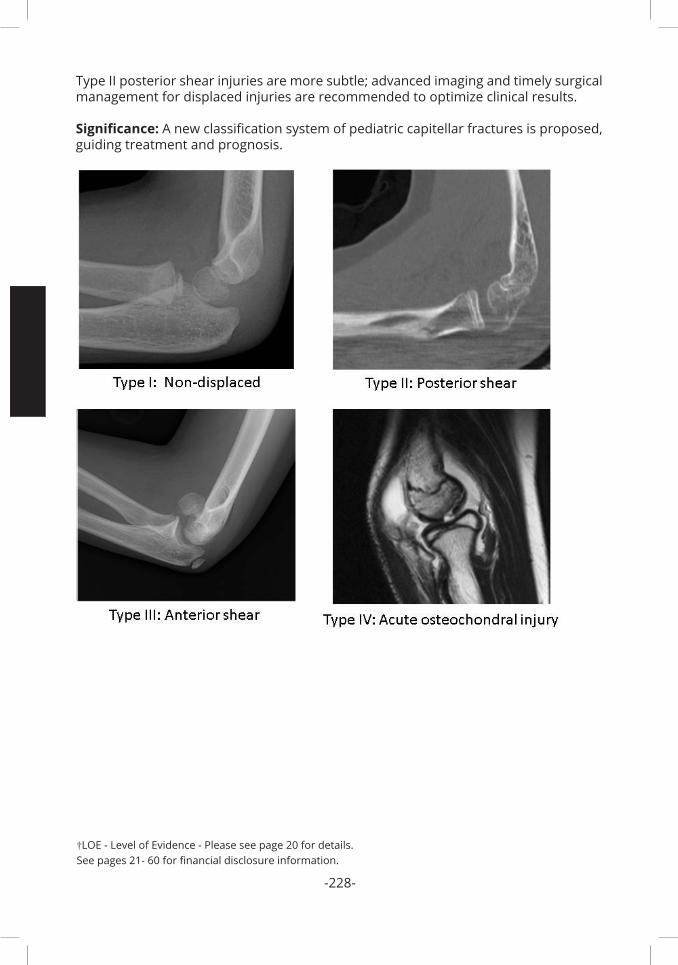
-228-
†LOE - Level of Evidence - Please see page 20 for details.See pages 21- 60 for financial disclosure information.
Type II posterior shear injuries are more subtle; advanced imaging and timely surgical management for displaced injuries are recommended to optimize clinical results.
Significance: A new classification system of pediatric capitellar fractures is proposed, guiding treatment and prognosis.

♦ Indicates those faculty presentations in which the FDA has not cleared the drug and/or medical device for the use described (i.e., the drug or medical device is being discussed for an “off label” use). For full information, refer to page 14.
-229-
Fri., 4/29/16 Hand/UE Subspecialty Day, Paper #86, 1:30 PM POSNA-2016
Functional Impact of Congenital Hand Differences: Early Results from the Congenital Upper Limb Differences (CoULD) Registry
Donald S. Bae, MD; Andrea S. Bauer, MD; Carley Vuillermin, MBBS, FRACS; Peter M. Waters, MD; Charles A. Goldfarb, MD; Lindley B. Wall, MD; Summer Roberts, MA; Maria Canizares, MD; Patricia E. Miller, MS Boston Children’s Hospital, Boston, Massachusetts
LOE-Prognostic-Level II
Purpose: The purpose of this investigation was to characterize the functional, emotional, and social impact of congenital upper limb differences on affected children and families using validated functional outcome instruments. Methods: From June 2014 to September 2015, patients from two pediatric hospitals have been enrolled in the prospective, multicenter Congenital Upper Limb Differences (CoULD) registry. Pertinent clinical and radiographic data were collected, and diagnoses were categorized according to the Oberg-Manske-Tonkin (OMT) classification system. Demographic information including age, gender, family history, and associated conditions were recorded. Functional outcomes were assessed annually using the Pediatric Outcomes Data Collection Instrument (PODCI) and Patient Reported Outcomes Measurement Information System (PROMIS); appropriate PROMIS instruments were used to assess upper extremity function, pain, anxiety, depression, and peer relationships.
Results: 444 patients enrolled in the CoULD registry were analyzed. Median patient age was 2.9 years (range 4 days to 17 years). Overall, 236 PODCI and 105 PROMIS were completed by parents; 69 PODCI and 74 PROMIS were completed by patients. There were 378 patients (85%) with malformations, 7 (2%) with deformations, and 57 (13%) with dysplasias. 204 and 174 had hand plate only versus upper limb malformations, respectively. 218 patients had bilateral involvement. 122 (27%) had other orthopaedic conditions, most commonly affecting the lower extremities (n=92); 117 (26%) had other medical conditions, most frequently neurological (n=33) and/or cardiac (n=31).
Overall, median normative global PODCI score for patients was 47 (IQR 33 to 53). Overall, median UE PROMIS for patients was 35 (IQR 32 to 37). Median pain, anxiety, depression, and peer relationship scores for patients were 39 (IQR 34 to 48), 42 (IQR 34 to 51), 40 (IQR 35 to 52), and 54 (IQR 47 to 64), respectively. Patients with entire limb involvement had lower PODCI happiness (52 vs. 60, p=0.02) and higher PROMIS pain (41 vs. 34, p=0.04) than hand plate only. Parent-rated scores were lower for entire limb involvement in PODCI upper extremity (51 vs 45, p=0.01), sports (50 vs. 47, p=0.04), and global PODCI (52 vs. 46, p=0.01).There were no differences in function between patients with unilateral versus bilateral involvement, except in PODCI Sports subscore (52 vs. 46 p=0.03).
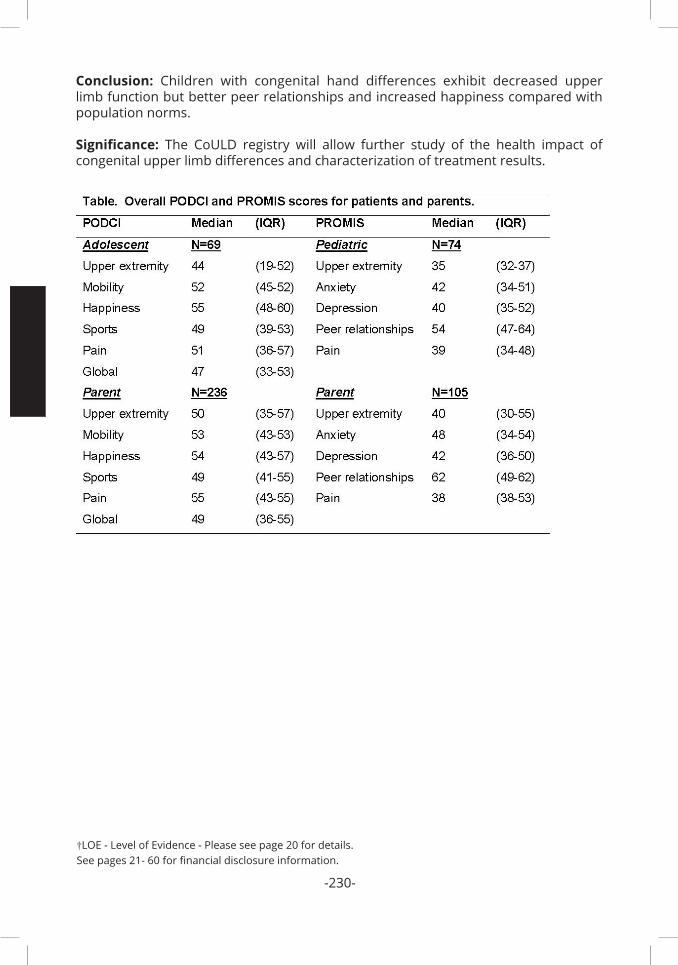
-230-
†LOE - Level of Evidence - Please see page 20 for details.See pages 21- 60 for financial disclosure information.
Conclusion: Children with congenital hand differences exhibit decreased upper limb function but better peer relationships and increased happiness compared with population norms.
Significance: The CoULD registry will allow further study of the health impact of congenital upper limb differences and characterization of treatment results.
♦ Indicates those faculty presentations in which the FDA has not cleared the drug and/or medical device for the use described (i.e., the drug or medical device is being discussed for an “off label” use). For full information, refer to page 14.
-231-
Fri., 4/29/16 Hand/UE Subspecialty Day, Paper #87, 1:35 PM POSNA-2016
Severity of Ulnar Ray Deficiency and its Relationship with Lower Extremity Deficiencies
Justin Lea, MD; Scott A. Riley, MD; Janet Walker, MDShriners Hospital for Children, Lexington, Kentucky
LOE-Prognostic-Level III
Purpose: Ulnar ray deficiency (URD) represents a spectrum of pathology ranging from missing ulnar rays to complete absence of the ulna. Radio-humeral synostosis and hypoplastic upper extremities may also be seen. There are few reports in the literature on the association of lower extremity anomalies in patients with URD. From an embryological standpoint, understanding the association of upper and lower extremity anomalies may provide for a more comprehensive understanding of the URD disease process. This retrospective study reports on the association of lower extremity anomalies in the setting of URD.
Methods: An IRB-approved retrospective chart review of patients with a diagnosis of URD treated at our institution was performed. Radiographs were reviewed and the upper extremity anomalies were classified utilizing the classification systems of Al-Qattan (shoulder), Kummel (elbow), Ogden (ulna), Ogino/ Kato (fingers), and Cole/Manske (thumb/1st web deformity). Lower extremity involvement was based on fibular-only, femoral-only or combined deficiency. Statistics were completed using ANOVA and Chi-square analysis.
Results: 78 limbs in 60 patients were identified. There were 42 males and 18 females (M:F=2.3). 34 patients (57%) had an associated lower extremity anomaly. 19 had fibular deficiency, 12 had combined femoral and fibular deficiency, and 3 had femoral deficiency alone. 59% of the lower extremity anomalies were bilateral. 46% of URD was bilateral. Mean number of digits on the URD involved upper extremity was 3.7. Higher grade thumb/1st web space deformity was associated with a greater number of missing digits (p=0.0001). Similarly, greater ulnar dysplasia was correlated with a greater number of missing digits (p=0.0053). Presence of a lower limb anomaly was associated with less severe URD based on elbow, ulnar, and thumb/1st web parameters (p=0.03, p=0.003, p=0.02, respectively).
Conclusion: Lower extremity anomalies are commonly found in association with URD (57%). There are statistically significant correlations between the degree of thumb/1st web space deformity and the severity of ulnar hypoplasia with the number of missing digits. URD with lower extremity anomalies are more likely to have a normal elbow, ulna, and, thumb/1st web space than URD in isolation. Additionally, isolated fibular and fibular/femoral combined deficiency is more commonly seen with URD than isolated femoral dysplasia.
-232-
†LOE - Level of Evidence - Please see page 20 for details.See pages 21- 60 for financial disclosure information.
Significance: This study demonstrates that lower extremity anomalies are commonly found in association with URD. Lower extremity involvement is correlated with less severe URD. This is consistent with the embryological timing of proximal upper extremities developing before the lower extremities.

♦ Indicates those faculty presentations in which the FDA has not cleared the drug and/or medical device for the use described (i.e., the drug or medical device is being discussed for an “off label” use). For full information, refer to page 14.
-233-
Fri., 4/29/16 Hand/UE Subspecialty Day, Paper #88, 1:50 PM POSNA-2016
Ultrasound Assessment of Trigger Thumb Resolution
Christine Wu, BA; Michelle S. Caird, MD; Clifford L. Craig, MD University of Michigan, Ann Arbor, Michigan
LOE-Diagnostic-Level IV
Purpose: Building on previously reported patients, the purpose of this study is to clinically and ultrasonographically compare the outcomes of trigger thumb treated non-operatively and surgically. We postulate that deferring surgical treatment for at least 2 years—thereby treating non-operatively—does not compromise the ultimate outcome of trigger thumb, as seen by clinical and ultrasound assessment.
Methods: A prospective analysis was conducted on children with trigger thumb followed to resolution with operative or non-operative treatment, or a minimum of 2 years follow-up, from 2008 to 2015. All patients were initially examined clinically and with dynamic ultrasound to measure A1 pulley thickness, cross-sectional area under the A1 pulley, and maximum cross-sectional area of the flexor pollicus longus tendon. Repeat ultrasound assessments were done at follow-up appointments. The decision for surgery depended on family preference and more recently child’s age. Persistence of the flexor tendon nodule, pulley thickness, and mismatch between cross-sectional areas under the A1 pulley and of the tendon was assessed ultrasonographically, along with thumb range of motion clinically.
Results: Sixty trigger thumbs in 49 patients were imaged by ultrasound. Sixteen thumbs resolved spontaneously by an average age 4.92 years, 31 underwent surgical release at an average age 4.08 years, and 13 are still followed. Thumb flexion and extension was clinically assessed in postoperative patients, and 13 patients reported ultrasound findings, as the rest were not postoperatively analyzed by ultrasound. Ultrasonography exhibited 13 (81.25%) non-operative and 9 (69.23%) operative thumbs with nodules of equal or increased size at resolution. Pulley thickness predominantly remained the same or increased in 14 (87.5%) non-operative and 12 (92.31%) operative thumbs. The majority of non-operative (56.25%) and operative thumbs (61.54%) indicated similar or smaller mismatch ratios, signifying a persistent or widened mismatch post-resolution. Of surgically treated patients, 50% under 4 years old and 66.67% over 4 years old had equivalent or greater mismatches, with 52.94% under 4 and 50% over 4 experiencing full active thumb flexion and extension within one month postoperatively.
Conclusion: Ultrasonographic measurements demonstrated similar correction mechanisms of thumbs that resolved spontaneously and surgically, as well as between operative patients less than and older than 4 years old.
Significance: Delaying surgery for trigger thumb release, with the current concerns about cognitive effects of general anesthesia prior to the age of 4, does not compromise the clinical result or alter the mechanism of resolution seen on ultrasound.
-234-
†LOE - Level of Evidence - Please see page 20 for details.See pages 21- 60 for financial disclosure information.
Fri., 4/29/16 Hand/UE Subspecialty Day, Paper #89, 1:55 PM POSNA-2016
Variation Amongst Pediatric Orthopaedic Surgeons When Diagnosing and Treating Pediatric Distal Radius Fractures
Karan Dua, MD; Matthew K. Stein, BS, MS; Brian K. Brighton, MD, MPH; William L. Hennrikus, MD; Martin J. Herman, MD; J. Todd Lawrence, MD; Charles T. Mehlman, DO, MPH; Norman Y. Otsuka, MD; M. Wade Shrader, MD; Brian G. Smith, MD; Paul D. Sponseller, MD; Joshua M. Abzug, MDUniversity of Maryland School of Medicine, Baltimore, Maryland
LOE-Not Applicable-Level V
Purpose: Distal radius fractures are a common injury in the pediatric population. Radiographic examination and classification of the fractures can be difficult due to the many potential types of fractures that can occur. The purpose of this study was to determine if there is agreement amongst pediatric orthopaedic surgeons when diagnosing and treating pediatric distal radius fractures.
Methods: Nine fellowship trained pediatric orthopaedic surgeons all from different institutions assessed 100 sets of anteroposterior and lateral pediatric distal radius fractures. Surgeons were asked to complete a questionnaire describing the fractures, prescribing the type of treatment and length of immobilization, determining the next follow-up visit, and whether or not they would obtain new radiographs at the follow-up visit.
Kappa statistics were performed to assess the agreement amongst examiners with the chance agreement removed. Strength of agreement was determined based on guidelines outlined by Landis and Koch.
Results: There was fair agreement when diagnosing and classifying pediatric distal radius fractures (K = 0.372). Diagnoses included torus, greenstick, salter-harris II, and extra-physeal fractures. There was slight agreement on the type of treatment the surgeons would prescribe (K = .074). Similarly, surgeons did not agree on the length of immobilization (K = -.0064).
When assessing follow-up visits, there was slight agreement on when the first follow-up visit should occur (K = .085) and if new radiographs should be obtained at this visit (K = .126).
Conclusion: There is substantial variation amongst pediatric orthopaedic surgeons when diagnosing and treating pediatric distal radius fractures.
Significance: Standardization of treatment for pediatric distal radius fractures is needed in order to be more cost effective and potentially decrease radiation exposure. Future prospective studies should be performed to assess the needed frequency and use of radiographs when treating pediatric distal radius fractures as well as the type of immobilization needed.
♦ Indicates those faculty presentations in which the FDA has not cleared the drug and/or medical device for the use described (i.e., the drug or medical device is being discussed for an “off label” use). For full information, refer to page 14.
-235-
Fri., 4/29/16 Hand/UE Subspecialty Day, Paper #90, 2:00 PM POSNA-2016
Risk Factors for Complications in Open Forearm Fractures in the Pediatric Population
Adam C. Hines, MD; Marilyn Elliott; Kevin Smit, MD, FRCSC; Daniel J. Sucato, MD, MS; R. Lane Wimberly, MD; Anthony I. Riccio, MD Texas Scottish Rite Hospital, Dallas, Texas
LOE-Prognostic-Level III
Purpose: Infection and re-fracture are well-known complications following open pediatric forearm fractures. The purpose of this paper is to determine the incidence of these complications and to identify predictors for their occurrence specifically analyzing factors related to the patient, injury pattern, and treatment following the surgical management of pediatric open forearm fractures.
Methods: This is an IRB-approved retrospective review of a consecutive series of patients who had an open forearm fracture treated at a single-institution pediatric level 1 trauma center from 2007-2013. A medical record review and radiographic analysis was performed for each patient.
Results: There were 262 patients, 207 boys and 55 girls, at an average age of 9.7 years. The Gustillo-Anderson classification was type 1 in 219 (83.5%), type 2 in 39 (14.1%) and type 3 in 4 (1.5%) patients. All patients had an irrigation and debridement in the operating room. The overall incidence of infection was 3.4% (9 patients). The only predictor for developing infection were patients who were noted to have a contaminated wound at the initial debridement (21% vs. 2.2%, p=0.002) There were no differences seen with regard to timing of antibiotics (p=0.87), time to formal debridement (p=0.20), Type 1 versus Type 2 or 3 open fractures (3.4% vs 5.0%, p =0.64), 24 hours vs. 48 hours of post-operative IV antibiotics (5.2% vs 3.5%, p =0.53), or when comparing diaphyseal, distal, and Monteggia fracture patterns (3.6 vs. 2.9% vs. 5.9%, p=0.81) or when comparing buried or exposed intramedullary implants (3.5% vs. 4.2%, p>0.99). The overall refracture rate was 2.3 % (6 patients) and did not correlate type of open wound type (p>0.99) or fracture pattern (0.4973), although 5 of the 6 re-fractures were in diaphyseal injuries. Twenty-eight (10.7%) patients returned to the operating room for additional treatment; 21 of which were for removal of implants after healing.
Conclusion: The incidence of infection following open both-bone fractures in children is 3.4% and correlates primarily with the contamination seen at the time of the initial debridement.
Significance: Careful evaluation of the degree of contamination at the time of the initial debridement should direct surgeons to more aggressive and repeated debridement to reduce the risk of infection.
-236-
†LOE - Level of Evidence - Please see page 20 for details.See pages 21- 60 for financial disclosure information.
Fri., 4/29/16 Hand/UE Subspecialty Day, Paper #91, 2:05 PM POSNA-2016
Management of Pediatric Type I Open Fractures
Matthew Schur, BA; Jenna Godfrey, MD, MS; Lior Shabtai, MD; Sarah Nossov, MD; Amy K. Williams, MD; Antoinette W. Lindberg, MD; Selina Silva, MD; Michelle S. Caird, MD; Paul D. Choi, MD; Alexandre Arkader, MD Children’s Hospital Los Angeles, Los Angeles, California
LOE-Therapeutic-Level III
Purpose: The management of pediatric Type I open fractures remains controversial. The aim of this study is to compare outcomes in non-operative and operative treatment and to identify complications related to both treatments.
Methods: A multi-center retrospective chart review was performed to identify all Type I open forearm, wrist and tibia fractures treated between 2001 to 2014 at four high volume pediatric centers. Patients who had multiple traumatic injuries, were immunocompromised, or who did not have final radiographs indicating healing were excluded. Non-operative management consisted of wound irrigation in the emergency department and antibiotic administration.
Results: 224 patients met inclusion criteria. Mean age at admission was 10.0 years (range 2.3-18.3 years), with 161 (72%) male and 63 (28%) female patients. 175 fractures were treated operatively (78.1%), 49 fractures were treated non-operatively (21.9%). Cefazolin was the most commonly administered antibiotic (88% of patients). There were 12 complications, 11 in the operative group (6.3%) and one in the non-operative group (2.0%) (p = 0.246). There were two infections; one in the operative group (0.5%) and one in the non-operative group (0.6%) with no significant difference between the groups (p=0.334). In the operative group, there were three compartment syndromes requiring release (1.7%), three malunions (1.7%), and one each of delayed union, neuropraxia, and a stiff wrist requiring surgery. The incidence of malunion was significantly higher in fractures treated with operative debridement and casting alone, 2/35 (5.7%), versus those treated operatively with an implant, 1/140 (0.7%, p = 0.041). We otherwise did not find a significant difference in complications seen by fracture type or fixation.
Conclusion: There was no significant difference in infection rate or complication rate in those treated operatively versus those treated non-operatively. There were, however, more complications associated with operative treatment including compartment syndrome and malunion. Malunions were most common in patients treated with operative debridement and casting.
Significance: In the treatment of open fractures, consideration should be given to the similar safety profiles of operative and non-operative treatment. There are complications associated with both operative and non-operative treatment.
♦ Indicates those faculty presentations in which the FDA has not cleared the drug and/or medical device for the use described (i.e., the drug or medical device is being discussed for an “off label” use). For full information, refer to page 14.
-237-
Fri., 4/29/16 Hip Subspecialty Day, Paper #92, 1:00 PM POSNA-2016
Standing Radiographs to Assess Hip Pathology: Analysis of Spinal Alignment and Pelvic Parameters Using Upright EOS Images
Andrew Pytiak, MD; James D. Bomar, MPH; Jonathan B. Peterson, MD; Matthew R. Schmitz, MD; Andrew T. Pennock, MD; Dennis R. Wenger, MD; Vidyadhar S.V. Upasani, MD Rady Children’s Hospital, San Diego, California
LOE-Diagnostic-Level IV
Purpose: Debate continues regarding the ideal pelvis radiograph (standing versus standardized supine) to assess acetabular morphology and hip pathology. The purpose of this study was to assess the relationship between sagittal spinopelvic alignment and measures of acetabular coverage in asymptomatic adolescents to determine if increased pelvic incidence and lumbar lordosis would have increased acetabular coverage.
Methods: Upright biplanar (anteroposterior and lateral) spinopelvic radiographs were obtained using EOS® imaging technique. Lateral center edge angle (LCEA) and Tonnis angle were calculated on the anteroposterior images, and the lateral images were analyzed for lumbar lordosis (LL), pelvic incidence (PI), pelvic tilt (PT), and sacral slope (SS). Data were analyzed for significant correlations between the sagittal measures of pelvic orientation and coronal measurements of acetabular coverage.
Results: Ninety-nine subjects were included in this study after the exclusion criteria were applied. LCEA was found to have a moderate inverse correlation with LL (rs = -0.304, p<0.001) and SS (rs = -0.365, p<0.001). LCEA was found to have a weak inverse correlation with PI (rs = -0.226, p = 0.001). LCEA was not found to be significantly correlated with PT (rs = 0.084, p = 0.238). Tonnis angle was not found to be correlated with any of the sagittal measurements. LCEA was found to be abnormal in 28 hips. Tonnis angle was found to be abnormal in 13 hips. Four hips were found to have both abnormal LCEA and Tonnis angles.
Conclusion: We rejected our hypothesis that increased pelvic incidence is correlated with increased acetabular coverage. Our findings suggest an individual’s acetabulum and pelvic tilt develop as an adaptation to one’s particular sagittal alignment (Figure). Secondly, a standing, weight-bearing AP radiograph is likely the most accurate and physiologically relevant assessment of hip morphology and pathology as this position of the pelvis reflects the functional position of the hip.
Significance: These data call into question the recent trend towards standardized supine radiographic view for assessing acetabular coverage. Measuring the LCEA on weight-bearing radiographs gives the surgeon the most physiologically relevant information when deciding whether surgical intervention is warranted.
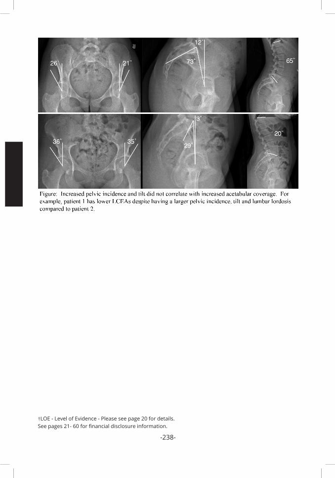
-238-
†LOE - Level of Evidence - Please see page 20 for details.See pages 21- 60 for financial disclosure information.

-239-
♦ Indicates those faculty presentations in which the FDA has not cleared the drug and/or medical device for the use described (i.e., the drug or medical device is being discussed for an “off label” use). For full information, refer to page 14.
Fri., 4/29/16 Hip Subspecialty Day, Paper #93, 1:05 PM POSNA-2016
Efficacy of Selective Ultrasound Hip Screening for Developmental Dysplasia of the Hip Based on Traditional Risk Factors
Clare Taylor, MBBS MRCS; Nadia Sciberras, MD; James E. Metcalfe, FRCS, MBChB; Ben Holroyd, FRCS (Tr & Orth)Derriford Hospital, Plymouth, Devon, United Kingdom
LOE-Prognostic-Level III
Purpose: There is ongoing debate regarding the use of targeted ultrasound screening programmes vs universal ultrasound screening programmes in diagnosing developmental dysplasia of the hip (DDH) in newborn infants. The potential benefits of universal screening are reduced late presentation and need for surgery but at a higher cost. In 2014 PHE published the UK National Screening Committee’s document on Newborn and Infant Physical Examination (NIPE) guidelines. It stated that all infants should have a clinical examination and if this was abnormal or they had one of the two specific risk factors, breech presentation or positive family history, they should have an ultrasound scan.
The ‘traditional’ risk factors for DDH also include, First born, Female, Oligohydramnios, Birth weight >4.5kg, and packaging disorders.
Our unit would like to move to universal ultrasound screening to further reduce the late presentation rate and need for complex surgery. We investigated the relationship between the traditional risk factors by which we identify high risk infants, and the presence of these risk factors in our population diagnosed with DDH.
Methods: Between January 2008 and December 2013 we carried out a retrospective review of the paediatric hip database which contains the results of all ultrasound scans performed at out unit. This infants were referred as recommended by the NIPE guidelines. All ultrasound scans were performed by one of our senior Neonatologists. Results: During the study period there were 28,365 live births, and 2526 (8.9%) infants (1391 females, 1135 males) referred to the service with ‘at risk’ factors or clinical signs of instability. A diagnosis of DDH requiring treatment was made in 175 hips (6.2 per 1000 live births); 86% girls, 14% boys.
Odds ratios were determined for each of the ‘at risk’ variables. First born (OR 2.221, p<0.05) and female sex (OR 5.330, p<0.05), were significantly associated with the diagnosis of DDH.
Conclusion: ‘At risk’ factors that are traditionally thought to be highly predictive of DDH such as family history were not significant. We believe that current targeted screening based on traditional risk factors correlates poorly with the diagnosis of DDH in our population.
-240-
†LOE - Level of Evidence - Please see page 20 for details.See pages 21- 60 for financial disclosure information.
Significance: We are currently fine tuning our practice, current economic constraints would prevent universal screening however based on our data there is an argument for expanding our current practice to include all first born and female infants in our screening programme.
-241-
♦ Indicates those faculty presentations in which the FDA has not cleared the drug and/or medical device for the use described (i.e., the drug or medical device is being discussed for an “off label” use). For full information, refer to page 14.
Fri., 4/29/16 Hip Subspecialty Day, Paper #94, 1:10 PM POSNA-2016
Comparison of an Automatic Technique to Current Practice in Calculating Dysplasia Metrics in 2D Ultrasound Images of the Neonatal Hip
Niamul Quader; Emily Schaeffer, PhD; Kishore Mulpuri, MD; Anthony P. Cooper, FRCS; Antony Hodgson, PhD; Rafeef Abugharbieh, PhD British Columbia Children’s Hospital, Vancouver, BC, Canada
LOE-Diagnostic-Level IV
Purpose: Ultrasound (US) is the standard imaging modality used to screen for developmental dysplasia of the hip in infants. In current practice, images are deemed either adequate or inadequate for interpretation by radiologists or orthopaedic surgeons and, if adequate, diagnostic dysplasia metrics are determined. Both manual steps introduce significant sources of inter-observer variability which can affect misdiagnosis rates. To eliminate subjectivity in this process, we developed an automatic method to identify adequate US images and extract dysplasia metrics. The purpose of this study was to validate the efficacy of this automatic method by comparing results with observer-determined dysplasia metrics on a set of US images. Methods: A total of 693 US images from scans of 35 infants were analyzed. Images were labeled as adequate or inadequate by trained clinicians at a single institution. The same clinicians measured α and β angles on adequate images to diagnose dysplasia. We trained our image classifier on random sets of 415 images and used it to assess the remaining 278 images. On the images judged adequate, we automatically estimated α and β angles. We compared the two methods for discrepancies in adequacy determination, metric variability and incidences of missed early diagnosis or over-treatment.
Results: Excellent agreement was seen in image adequacy classification between the automatic and manual methods (Kappa coefficient =0.912). On each adequate US image, α and β angle measurements were compared, producing mixed levels of agreement between methods. Mean discrepancies of 1.78°±4.72° and 8.91°±6.437° were seen for α and β angles, respectively. Standard deviations of the angle measures across multiple images from a single patient scan were significantly reduced by the automatic method for both α (p<0.05) and β (p<0.01) angles. Additionally, the automatic method classified three hips (two patients) as Graf type II and two hips (two patients) as type III, while the manual method classified them as type I and II, respectively. Both cases flagged as type III patients by the automatic system subsequently failed Pavlik harness treatment and were booked for surgery.
Conclusion: The automatic method produced excellent agreement with radiologists in scan adequacy classification and significantly reduced measurement variability. Good agreement between methods was found in Graf classification. In instances of disagreement, subsequent clinical findings seemed to support the classification of the automatic method.
-242-
†LOE - Level of Evidence - Please see page 20 for details.See pages 21- 60 for financial disclosure information.
Significance: This proposed method presents an alternative automatic, near-real-time analysis for US images that may potentially significantly improve dysplasia metric reliability and reduce missed early diagnoses without increasing over-treatment.
-243-
♦ Indicates those faculty presentations in which the FDA has not cleared the drug and/or medical device for the use described (i.e., the drug or medical device is being discussed for an “off label” use). For full information, refer to page 14.
Fri., 4/29/16 Hip Subspecialty Day, Paper #95, 1:25 PM POSNA-2016
A Prospective Study of Pavlik Harness Treatment Variations for Dislocatable and Dislocated Hips in Infantile DDH
Adam C. Hines, MD; Terri Beckwith, BA, MPH; ChanHee Jo, PhD; Harry K.W. Kim, MD, MSTexas Scottish Rite Hospital for Children, Dallas, Texas
LOE-Therapeutic-Level II
Purpose: Wide variation exists in the use of Pavlik Harness (PH) for management of infantile DDH. The purpose of this study was to determine if frequency of follow-up visits and the duration of harness wear (23 vs. 24 hours) affected the outcome of obtaining stable, reduced hips in infants with dislocatable (Barlow +) and or dislocated (Ortolani +) hips.
Methods: This prospective study enrolled patients from 2009 to 2014 with the following criteria: infants <6 months of age with an unstable hip exam treated with PH. Excluded were ages >6 months at diagnosis and fixed or teratologic dislocations. Recommended duration of PH wear and frequency of clinic visits were analyzed in 11 surgeons. PH success was defined as a clinically and radiographically stable hip that did not require open or closed reduction, or the use of an abduction orthosis to obtain stability. Statistical analysis included Mann-Whitney and Fisher exact tests.
Results: 154 patients (193 hips, 131 females) with unstable hips had a mean age of presentation of 19±25 days (range 4-175). Mean follow up was 26.1±17.3 months. 39 patients (25%) had bilateral unstable hips. 108 hips were dislocatable and 85 were dislocated. Overall success rate of PH for dislocatable and dislocated hips was 94% and 85%, respectively. Treatment pattern for dislocatable and dislocated hips was similar for each surgeon, except for 1 surgeon who followed dislocated hips more frequently. Only 4 surgeons obtained an ultrasound prior to initiating PH. Dislocated hips that failed PH treatment presented later (mean 44 days vs. 18 days, p=0.047). Although no statistical difference was seen in the success rate of different harness wear schedule in either the dislocatable or dislocated hips (p=0.17 and p=0.76), all 6 failures in the dislocatable hip cohort were in patients who were in the harness for 23 hours daily. There was no difference in the success rate of a group of surgeons who assessed their patients almost weekly vs. a group assessing their patient once or twice during the first 4 week period (dislocatable p=0.68, dislocated p=0.09).
Conclusion: This study shows that variations in PH treatment, such as the duration of harness wear and the frequency of clinic visits, do not affect the success rate for infantile DDH.
Significance: This study suggests that weekly clinic visits and 24-hour PH regimen may not be necessary to obtain stably reduced hips in infants <6 months of age with dislocatable or dislocated hips.
-244-
†LOE - Level of Evidence - Please see page 20 for details.See pages 21- 60 for financial disclosure information.
Fri., 4/29/16 Hip Subspecialty Day, Paper #96, 1:30 PM POSNA-2016
Reliability of a Modified Complication Classification System in Pediatric Orthopedic Patients
Emily Dodwell, MD; Rubini Pathy, MD; Daniel W. Green, MD; David M. Scher, MD; John S. Blanco, MD; Shevaun M. Doyle, MD; Eva Luderowski; Ernest L. Sink, MD Hospital for Special Surgery, New York, New York
LOE-Diagnostic-Level III
Purpose: There is currently no standardized complication grading classification routinely used for pediatric orthopedic surgical procedures. The Clavien-Dindo classification used in general surgery was modified and validated in 2011 by Sink et al. and has been used regularly to classify complications following hip preservation surgery. The aim of this study was to adapt and validate Sink et al.’s modification of the Clavien-Dindo classification system for grading complications following surgical interventions of the upper and lower extremities and spine in pediatric orthopedic patients.
Methods: Sink et al.’s modification of the Clavien-Dindo classification system was further modified for pediatric orthopaedic procedures (Table 1). The modified grading scheme was based on the treatment required to treat the complication and the long term morbidity of the complications. Forty-five complication scenarios were developed. Seven pediatric orthopedic surgeons were trained to use the modified system and they each graded the scenarios on two occasions. The scenarios were presented in a different random order each time they were graded. Fleiss’ and Cohen’s κ statistics were performed to test for inter-rater and intra-rater reliabilities, respectively.
Results: The overall Fleiss’ κ value for inter-rater reliability was 0.772 (95% CI, 0.744-0.799). The weighted κ was 0.765 (95% CI, 0.703-0.826) for Grade I, 0.692 (95% CI, 0.630-0.753) for Grade II, 0.733 (95% CI, 0.671-0.795) for Grade III, 0.657(95% CI, 0.595-0.719) for Grade IVa, 0.769 (95% CI, 0.707-0.83)1 for Grade IVb and 1.000 for Grade V (p value <0.001). The Cohen’s κ value for intra-rater reliability was 0.918 (95% CI, 0.887–0.947). These tests show that the adapted classification system has high inter- and intra-rater reliabilities for grading complications following pediatric orthopedic surgery.
Conclusion: Given the high intra- and inter-rater reliability and simplicity of this system, adoption of this grading scheme as a standard of reporting complications in pediatric orthopaedic surgery could be considered.
Significance: The evaluation of surgical outcomes should include the ability to reliably grade surgical complications. A reproducible, reliable system to assess pediatric surgical complications will be a valuable tool for improving surgical practices and patient outcomes.
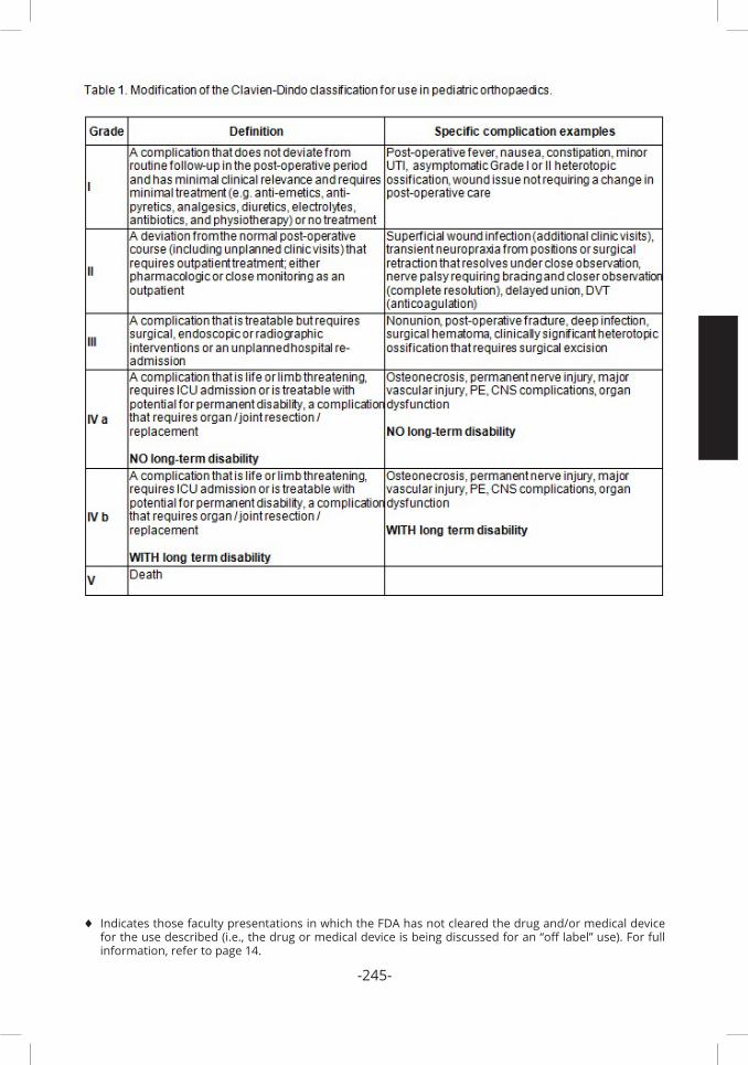
-245-
♦ Indicates those faculty presentations in which the FDA has not cleared the drug and/or medical device for the use described (i.e., the drug or medical device is being discussed for an “off label” use). For full information, refer to page 14.
-246-
†LOE - Level of Evidence - Please see page 20 for details.See pages 21- 60 for financial disclosure information.
Fri., 4/29/16 Hip Subspecialty Day, Paper #97, 1:35 PM POSNA-2016
Longterm Radiographic and Functional Outcome of Medial Approach in Open Hip Reduction for DDH
Virginie Pollet, MD Sophia Children’s Hospital, Erasmus Medical Center, Rotterdam, The Netherlands
LOE-Therapeutic-Level IV Purpose: After failure of conservative treatment in Developmental Dysplasia of the Hip (DDH), open reduction of the hip will be the following treatment. The medial approach according to Ferguson is a valid approach but controversial results have been reported in the literature with a high rate of avascular necrosis (AVN) of the hip as major complication. This retrospective cohort study evaluates the long-term radiographic and functional outcome of dislocated hips treated by the medial approach.
Methods: After Ethics Board approval, patients younger than 1 year of age at time of surgery treated with open medial approach for dislocated hip by a single surgeon in our hospital between 1985 and 2010 were included in this study. Radiographs preoperatively, 1 year postoperatively, 5 years postoperatively and at latest follow-up were assessed for AVN and Severin outcome score. Functional outcomes were determined using questionnaires such as the PODCI or the HOOS depending on the patient’s age.
Results: Fifty-two patients with fifty-eight hips were included in the study. The median follow-up was 12,7 years. At latest follow-up 20 hips (34,5%) showed signs of significant proximal growth disturbance due to avascular necrosis. There was no protective effect of the presence of ossified nucleus at time of surgery. In thirteen hips (22,4%) additional surgery was performed. Thirteen hips (22,4%) were classified as Severin III or higher, thus showed poor radiologic outcome. Of these hips, the age at time of surgery was significantly higher than the hips with a good Severin score (I-II) (p = 0,045). The patients reported outcome showed that pain, activities of daily living, symptoms and global functional were lower in patients with poor Severin classifications.
Conclusion: The medial approach for open hip reduction leads to a high rate of progressive proximal growth disturbance due to AVN. Residual dysplasia and persisting subluxation will cause pain and discomfort at later age. Careful long-term follow-up is needed as some growth disturbances only appear after 5 years postoperatively.
Significance: This study confirms the high rate of growth disturbance due to AVN causing worse functional outcome and pain after medial approach for open hip reduction. A long-term follow-up in these cases is necessary with possible secondary procedures to prevent residual dysplasia at skeletal maturity.

-247-
♦ Indicates those faculty presentations in which the FDA has not cleared the drug and/or medical device for the use described (i.e., the drug or medical device is being discussed for an “off label” use). For full information, refer to page 14.
Fri., 4/29/16 Hip Subspecialty Day, Paper #98, 1:50 PM POSNA-2016
Age at Reduction a Significant Risk Factor for Pelvic Osteotomy in Developmental Hip Dysplasia
Todd J. Blumberg, MD; Viviana Bompadre, PhD; Klane K. White, MD, MSSeattle Children’s Hospital, Seattle, Washington
LOE-Prognostic-Level IV
Purpose: Significant controversy exists regarding the optimal timing and treatment for a dislocated hip in patients with developmental dysplasia of the hip (DDH). Early reduction of the dislocated hip initiates remodeling and further development of the dysplastic acetabulum. Even with successful reduction, a number of children still go on to require an secondary reconstructive procedure due to poor acetabular development. Given the risk of an additional anesthetic and surgical procedure, the purpose of this study is to determine at what age to expect insufficient remodeling of the acetabulum after successful reduction of a dislocated hip.
Methods: All patients treated with closed or open reduction for idiopathic DDH over an eight-year period were identified. Average follow-up was 2.8 years. Patients that had an acetabular index >30 degrees two years after reduction were indicated for pelvic osteotomy. A logistic regression model was utilized to evaluate the role of age at reduction and risk of secondary osteotomy for residual dysplasia.
Results: Thirty-three patients with 38 dislocated hips underwent operative reduction over the 8-year study period. Four patients (5 hips) required an open approach to achieve congruent reduction. 15 patients (16 hips, 42%) had persistent dysplasia with increased acetabular index and were indicated for pelvic osteotomy. Patients that underwent reduction after nine months of age had a significantly higher risk of residual dysplasia requiring secondary procedure, with a significant correlation between age at reduction and need for pelvic osteotomy (p=0.003).
Conclusion: There is a high rate of secondary procedures in the treatment of DDH. Delayed reduction is one risk factor for a secondary reconstructive procedure. The odds of an osteotomy increase by 1.35 for each month that a hip remains dislocated. Depending on the age of the patient, there may be benefit to performing reduction with early osteotomy rather than isolated closed reduction.
Significance: Residual acetabular dysplasia after reduction in children over 9 months of age is common. These findings underscore the need for early reduction of a dislocated hip to optimize acetabular remodeling. For patients older than nine months of age with idiopathic hip dislocation, families should be counseled about the high rate of secondary procedures and evaluated for potential early pelvic osteotomy, as this may prevent the need for a secondary procedure.
-248-
†LOE - Level of Evidence - Please see page 20 for details.See pages 21- 60 for financial disclosure information.
Fri., 4/29/16 Hip Subspecialty Day, Paper #99, 1:55 PM POSNA-2016
Purposeful Closed Reduction and Pinning in Unstable Slipped Capital Femoral Epiphysis Does Not Result in Increased Risk For Avascular Necrosis
Joshua K. Napora, MD; William Z. Morris, MD; Allison Gilmore, MD; Christina K. Hardesty, MD; George H. Thompson, MD; Raymond W. Liu, MD Rainbow Babies and Children’s Hospital, Cleveland, Ohio
LOE-Therapeutic-Level III
Purpose: The treatment of stable Slipped Capital Femoral Epiphysis (SCFE) is generally accepted as in situ pinning; however, significant controversy exists involving the treatment of unstable SCFE, which is associated with a high rate of avascular necrosis (AVN). Zaltz et al. recently reviewed 15 studies with a wide variety of reduction techniques and determined the overall unstable SCFE AVN rate to be 24%. In their review the largest study reporting purposeful reduction found a 47% AVN rate in 26/30 patients. The purpose of this study was to further investigate the AVN rate with purposeful closed reduction and pinning of the capital femoral epiphysis.
Methods: We retrospectively reviewed 221 patients with 302 hips with SCFE between 2000 to 2014. Forty-eight patients (50 hips) presented with unstable SCFE, of which 17 patients (17 hips) had a scheduled MRI between 1 and 6 months postoperatively from surgery, while 31 patients (33 hips) were evaluated using only plain radiograph to detect AVN. All unstable SCFEs were treated by a gentle reduction method. The patient was placed on a fracture table, traction was applied, and the affected leg was mildly internally rotated with patella never passing further then perpendicular to floor allowing for a gentle reduction. Closed pinning was performed using one or two cannulated screws. Southwick angles were measured prior to reduction and at first postoperative visit.
Results: No stable SCFEs went on to develop AVN. Thirteen (26%) unstable SCFEs developed AVN. Of patients screened with MRI AVN developed in 7/17 (41%) hips at a mean of 2.5 months (range, 0.9-5.0 months) postoperatively. Of patients followed with plain radiographs alone, AVN developed in 6/33(18%) hips at a mean of 6.8 months (range, 2.1-21.1 months) after surgery. Mean follow up for MRI screened unstable SCFEs was 29.2 months compared to unstable SCFEs followed by only plain radiographs of 22.1 months. Southwick angles were measured in a subset of 24 patients that had adequate pre-reduction and post-reduction radiographs. Mean change in Southwick angle was 19° ± 17°.
Conclusion: Despite potentially inflating our rate with the use of early detection MRI scans, we found an AVN rate comparable to the published literature with the use of gentle reduction on a fracture table with traction and internal rotation.
Significance: Purposeful gentle reduction appears to be a reasonable low morbidity option in the treatment in unstable SCFE and does not appear to increase the risk of AVN.
-249-
♦ Indicates those faculty presentations in which the FDA has not cleared the drug and/or medical device for the use described (i.e., the drug or medical device is being discussed for an “off label” use). For full information, refer to page 14.
Fri., 4/29/16 Hip Subspecialty Day, Paper #100, 2:00 PM POSNA-2016
Treatment of Chronic, Stable Slipped Capital Femoral Epiphysis via Surgical Hip Dislocation with Combined Osteochondroplasty and Imhauser Osteotomy
Kevin E. Klingele, MDNationwide Children’s Hospital, Columbus, Ohio
LOE-Therapeutic-Level IV
Purpose: Severity of residual deformity following SCFE is directly related to early hip DJD. Restoration of proximal femoral morphology during the treatment of SCFE optimizes outcome. Recent treatment, including the modified Dunn procedure, restores anatomy with significant risk for AVN. The Imhauser osteotomy is a safe and effective way to treat patients with residual deformity. Full restoration of anatomy is difficult in hips with severe deformity and significant metaphyseal prominence. The purpose of this study is to evaluate effectiveness and safety of combined Imhauser osteotomy and osteochondroplasty via surgical hip dislocation approach in both acute and delayed settings.
Methods: Retrospective chart review was performed on a consecutive series of patients who underwent Imhauser osteotomy and osteochondroplasty via surgical hip dislocation for treatment of chronic, stable SCFE. Patients were divided into acute or delayed treatment groups based on whether the osteotomy was performed as the initial slip treatment. Demographics, operative details, radiographs, and complications were recorded. Variables were analyzed and statistical analysis performed.
Results: Nineteen patients (15 male, 4 female, average 13.7 years) with chronic, stable SCFE treated with a combined Imhauser osteotomy and osteochondroplasty via surgical hip dislocation approach were reviewed. Six osteotomies were performed acutely in combination with in-situ pinning, 13 were delayed at least 6 months after in-situ pinning (average 21.7 months). Two hips had labral tears that required repair. Mean follow up 15.3 (delayed) and 19.3 (acute) months. Average improvement in slip angle was 40.7 (delayed) and 50.2 (acute) degrees (p=0.0916), final postoperative slip angle averaged 15.8 (delayed) and 17.8 (acute) degrees (p=0.544). Femoral neck length and greater trochanteric height were similar between both groups. Average alpha angle at final follow-up measured 55.8 (delayed) and 60.8 (acute) degrees (p=0.542). One patient (acute) had hardware failure within 2 weeks of osteotomy requiring revision ORIF. One patient (delayed) required bone grafting and revision ORIF for nonunion at the osteotomy site. No cases of AVN were identified.
Conclusion: Imhauser osteotomy combined with osteochondroplasty via surgical hip dislocation approach is a safe and effective treatment of moderate-severe, stable SCFE performed in both the acute and delayed setting. Proximal femoral anatomy can be restored, labral pathology addressed, and the physis stabilized with minimal complication or AVN risk.
Significance: Restoration of proximal femoral morphology may improve long term hip function and prevent or delay early DJD.

-250-
†LOE - Level of Evidence - Please see page 20 for details.See pages 21- 60 for financial disclosure information.
Fri., 4/29/16 Hip Subspecialty Day, Paper #101, 2:05 PM POSNA-2016
Patient Specific 3d Print Models Aid Surgical Planning Of Proximal Femoral Osteotomy For Slipped Capital Femoral Epiphysis
Lillia Cherkasskiy, PhD; Jason P. Caffrey; Alexander F. Szewczyk, BS; Esther Cory, BS, MA; James D. Bomar, MPH; Christine L. Farnsworth, MS; Megan Jeffords, MS; Dennis R. Wenger, MD; Robert Sah, MD, ScD; Vidyadhar S.V. Upasani, MDRady Children’s Hospital, San Diego, California
LOE-Therapeutic-Level III
Purpose: Slipped capital femoral epiphysis (SCFE) results in a complex three-dimensional (3D) deformity of the proximal femur that limits hip range of motion and may lead to early degenerative joint disease. A flexion, valgus, internal rotation producing proximal femoral osteotomy (Imhauser) has been described to improve hip mechanics. The purpose of this study was to evaluate the benefits of using 3D print technology to aid in surgical planning.
Methods: Fifteen children with symptomatic post-SCFE proximal femoral deformity were included in this study. Ten patients were treated by a single surgeon with (n=5) and without (n=5) a 3D model for pre-operative planning, and compared to patients treated by senior partners without the use of a model (n=5) to evaluate for a learning curve effect. Pre-operative computed tomography (CT) with 3D reconstruction was obtained in all 15 children for pre-operative planning. The CT data was used to create patient specific 3D print models in five children and a mock surgery was performed using these 3D prints to plan deformity correction (Figure). Peri-operative data including patient BMI, surgical time, and fluoroscopy time were recorded. Radiographic data included pre- and post-operative Southwick slip angle (SSA), neck shaft angle (NSA), articular surface to trochanter distance (ATD), and medial proximal femoral angle (MPFA). ANOVA and the Kruskal-Wallis test were used to compare data between the three groups.
Results: Measurements were performed by two independent observers and found to be similar (ICC scores range: 0.81-0.98). Children in all three groups were similar in regards to BMI at time of the proximal femoral osteotomy. Radiographic deformity was not statistically different pre-operatively, except for the ATD (p=0.044). Post-operative radiographic parameters were equally improved in all three groups. On average, surgical time decreased by 44 minutes and 38 minutes, and fluoroscopy time decreased by 50% and 25% in the model group compared to the no model and senior groups, respectively.
Conclusion: Patient specific 3D models that allow the surgeon to practice the osteotomy prior to entering the operating room may be the next breakthrough for complex surgical planning. Preliminary results suggest that 3D models can substantially decrease surgical time and fluoroscopy time while allowing for similar deformity correction.
Significance: 3D models can be an invaluable surgical tool to understand complex three-dimensional orthopedic deformities. These models may be especially useful to
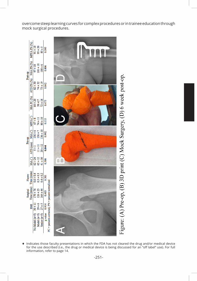
-251-
♦ Indicates those faculty presentations in which the FDA has not cleared the drug and/or medical device for the use described (i.e., the drug or medical device is being discussed for an “off label” use). For full information, refer to page 14.
overcome steep learning curves for complex procedures or in trainee education through mock surgical procedures.
-252-
†LOE - Level of Evidence - Please see page 20 for details.See pages 21- 60 for financial disclosure information.
Fri., 4/29/16 Neuromuscular/Lower Extremity POSNA-2016 Subspecialty Day, Paper #102, 2:30 PM
Parental Understanding of an In-Toeing Gait
Jacob F. Schulz, MD; David Molho; Stephen M. Sylvia, BS; Eric D. Fornari, MD Children’s Hospital at Montefiore, Bronx, New York
LOE-Prognostic-Level II
Purpose: The anxiety surrounding gait patterns in infants and children is multifactorial. Evaluation of an in-toeing gait is one of the most common referrals made to an orthopedic specialist. Many families have difficulty understanding that in-toeing can be a part of normal growth and development, and that the vast majority of children do not require treatment. The purpose of this study was to investigate parental perceptions of an in-toeing gait both before and after consultation with an orthopedic practitioner and to assess how effectively we communicate with patients and their families.
Methods: Parents of children referred to one of two fellowship trained pediatric orthopedic surgeons at a single tertiary referral center for evaluation of in-toeing gait completed a 22-question survey that sought to assess their perceptions of in-toeing. These surveys were administered at two time intervals: pre-visit and post-visit. Parental understanding was assessed using a 6 question scale, giving each parent an understanding score from 0-6. Statistical analysis was performed using Fisher exact test and a general linear model.
Results: Between June 2014 and April 2015, 48 parents completed the questionnaires and met inclusion criteria. Before their visits, parents of all ages reported similar anxiety regarding in-toeing (p=1). After their visits, parents aged 18-25 reported higher anxiety compared to parents older than 26 (p=0.014). Parents with a high school degree or less reported higher anxiety after their visits as compared to parents who had received a higher education (p=0.009). Post-visit low understanding scores were associated with higher levels of anxiety (p<0.001). Higher understanding scores resulted in lower anxiety levels (p<0.001). Additionally, parents who reported high anxiety after their visit stated they were more likely to seek additional care (p < 0.001). Parent’s first language was not related to post-visit anxiety (p=0.481). The strongest predictor of high post-visit anxiety when adjusted for age, education level, and language, was a low post-visit understanding score (p<0.001).
Conclusion: Younger parents with lower education levels were most likely to leave visits with high anxiety and poor understanding. Regardless of age, education level, and language, low understanding leads to high anxiety.
Significance: Parents with low understanding and high anxiety were most likely to consider seeking further medical care for their child, which could place additional stress on them and an already overburdened healthcare system. This study identifies demographic factors that may be markers of poor understanding and suggests a potential benefit for improved models of communication.
-253-
♦ Indicates those faculty presentations in which the FDA has not cleared the drug and/or medical device for the use described (i.e., the drug or medical device is being discussed for an “off label” use). For full information, refer to page 14.
Fri., 4/29/16 Neuromuscular/Lower Extremity POSNA-2016 Subspecialty Day, Paper #103, 2:35 PM
Transverse Plane Assessment of the Lower Extremity: Comparison of Clinical Measures, Motion Capture, and Biplanar Radiography
David E. Westberry, MD; Linda Pugh; Roy B. Davis, PhD Shriners Hospital for Children-Greenville, Greenville, South Carolina
LOE-Diagnostic-Level I
Purpose: Accurate examination and assessment of transverse plane deviations are necessary for appropriate treatment planning and counseling of patients. Multiple measurement methods are available to assess transverse plane alignment of the lower extremity including clinical examination, 3-dimensional radiography with CT or MRI, computerized motion analysis, and 3-dimensional modeling based on biplanar radiography (EOS imaging ®, Paris, France). A study was performed to estimate correlation of measurements obtained using these methods in the assessment of transverse plane alignment of the lower extremity in children.
Methods: All patients who completed a comprehensive gait analysis between January, 2014 and June, 2015, and who also had same-day biplanar standing AP and lateral radiographs with 3D transverse plane calculations of the lower extremity were included. Clinical evaluation of tibial torsion was assessed by examination of the thigh-foot axis, bimalleolar axis with the knee flexed (BMA-F), and bimalleolar axis with the knee extended (BMA-E). Femoral anteversion was measured with the child in the prone position, with the hip extended, and the knee flexed. Gait analysis data included assessment of the static and dynamic hip and knee rotation angles during stance. Biplanar radiographs were converted to 3D models with calculation of transverse plane parameters.
Results: The study cohort included 381 patients with an average age of 12.5 years (range, 7.2-21.4). Diagnoses included cerebral palsy in 198 (52%), in-toeing or out-toeing in a neurologically normal child in 83 (22%), idiopathic toe walking in 22 (6%), TEV deformity in 19 (5%), and other in 44 (11.5%). Linear regression analysis revealed a weak relationship between the assessment of tibial torsion with biplanar radiography compared with clinical measures and motion data. There was higher correlation of biplanar radiographic results with motion data than with physical examination measures of tibial torsion. Similarly, for femoral anteversion, a weak relationship was demonstrated between the assessment with biplanar radiography compared with clinical measures and motion data. Physical examination measures of hip rotation were significantly more highly correlated (p=0.0012) than with motion data.
Conclusion: Three-dimensional models derived from biplanar radiographs failed to predict quantitative hip and knee rotation in gait. Radiographic assessment of rotational abnormalities of the lower extremities using biplanar radiography is of limited value. Significance: Static measures of tibial torsion and femoral anteversion are insufficient to predict the dynamic component of gait. Treatment decisions involving these
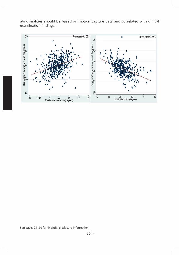
-254-
See pages 21- 60 for financial disclosure information.
abnormalities should be based on motion capture data and correlated with clinical examination findings.
-255-
♦ Indicates those faculty presentations in which the FDA has not cleared the drug and/or medical device for the use described (i.e., the drug or medical device is being discussed for an “off label” use). For full information, refer to page 14.
Fri., 4/29/16 Neuromuscular/Lower Extremity POSNA-2016 Subspecialty Day, Paper #104, 2:40 PM
Prevalence of Specific Gait Abnormalities in Children with Cerebral Palsy Revisited: Influence of Age and GMFCS Level
Robert M. Kay, MD; Susan A. Rethlefsen, PT; Gideon W. Blumstein, MD; Tishya A.L. Wren, PhD Children’s Hospital Los Angeles, Los Angeles, California
LOE-Prognostic-Level IV
Purpose: We investigated the impact of Gross Motor Function Classification System (GMFCS) level, age and prior surgery on the prevalence of gait abnormalities in patients with cerebral palsy (CP).
Methods: Gait abnormalities were identified from gait analysis data in 1005 patients with CP. Patient demographics included 59% male; age 9.8 +/- 3.9 years, with GMFCS level I: N=282 (28%), II: N=320 (32%), III: 289 (29%), IV: 114 (11%). Univariate logistic regression assessed effects of age, previous surgery and GMFCS level. Multivariate logistic regression was used to adjust for covariates within each GMFCS level. Statistical significance set at p<0.05.
Results: The odds of rotational malalignment, external tibial torsion, out-toeing, pes valgus, calcaneus, excessive hip flexion and crouch increased with age (p<0.02). The odds of out-toeing, rotational malalignment, external tibial torsion, calcaneus, stiff knee, pes valgus, excessive hip flexion, crouch and excessive hip adduction all increased with prior surgery (p<0.05). The odds of scissoring, excessive hip flexion, stiff knee, crouch, pes valgus, excessive hip adduction, calcaneus, out-toeing, rotational malalignment and external tibial torsion all increased with increasing GMFCS level (p<0.03).
GMFCS level I - Odds of rotational malalignment, external tibial torsion, pes valgus, excessive hip flexion and crouch increased with age (p<0.03). Odds of outtoeing and stiff knee gait increased with prior surgery (p<0.02).
GMFCS level II - Odds of rotational malalignment, pes valgus, outtoeing, pes varus and calcaneus increased with age (p<0.05). Odds of stiff knee, rotational malalignment, excessive hip flexion, external tibial torsion and outtoeing all increased with prior surgery (p<0.04).
GMFCS level III - Odds of rotational malalignment, external tibial torsion, pes valgus, calcaneus, outtoeing and excessive hip flexion increased with age (p<0.05). Odds of outtoeing, calcaneus and stiff knee increased with prior surgery (p<0.05).
GMFCS level IV – Prevalence of gait problems was not impacted by age. Odds of outtoeing, calcaneus, stiff knee, rotational malalignment and pes valgus increased with prior surgery (p<0.03).
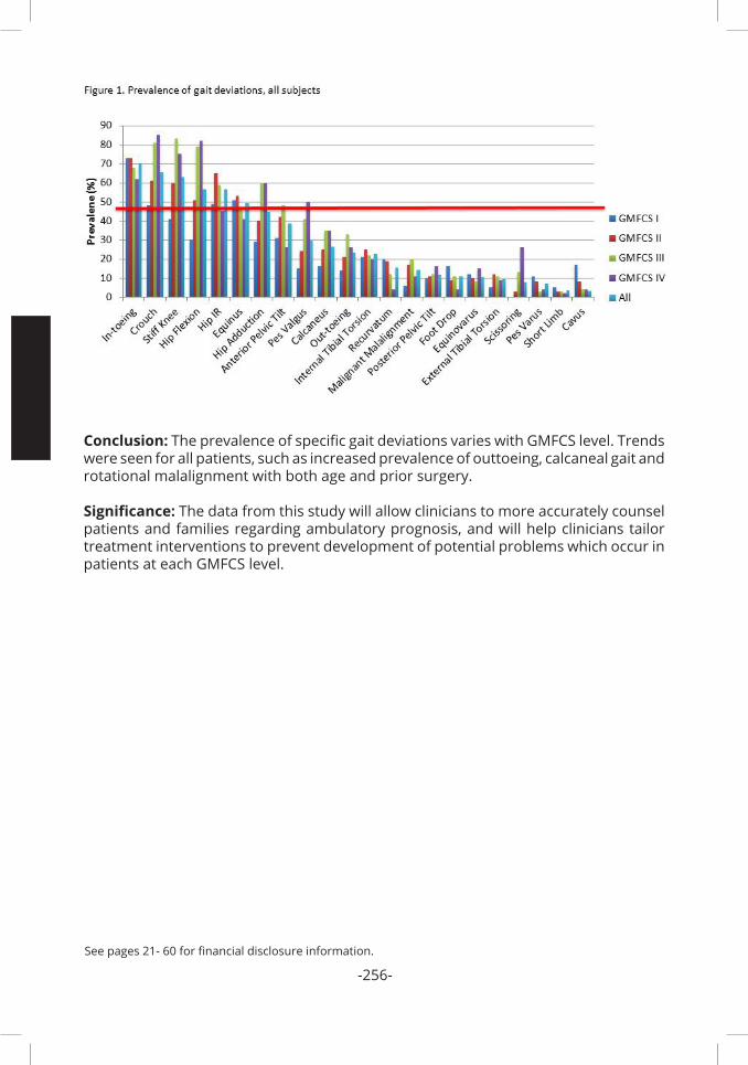
-256-
See pages 21- 60 for financial disclosure information.
Conclusion: The prevalence of specific gait deviations varies with GMFCS level. Trends were seen for all patients, such as increased prevalence of outtoeing, calcaneal gait and rotational malalignment with both age and prior surgery.
Significance: The data from this study will allow clinicians to more accurately counsel patients and families regarding ambulatory prognosis, and will help clinicians tailor treatment interventions to prevent development of potential problems which occur in patients at each GMFCS level.
-257-
♦ Indicates those faculty presentations in which the FDA has not cleared the drug and/or medical device for the use described (i.e., the drug or medical device is being discussed for an “off label” use). For full information, refer to page 14.
Fri., 4/29/16 Neuromuscular/Lower Extremity POSNA-2016 Subspecialty Day, Paper #105, 2:55 PM
Subclassification of GMFCS 4 and 5 Cerebral Palsy by Neuromotor Impairments Predicts Complications and Quality of Life Following Hip Surgery
Alexandra Miller, BA; Ranjit A. Varghese, MD; Amit Jain, MD; Paul D. Sponseller, MD; Hamlish Lowdon, MEng Johns Hopkins, Baltimore, Maryland
LOE-Prognostic-Level II
Purpose: The aim of our study was to sub-classify GMFCS 4 and 5 cerebral palsy patients based on preoperative neuromotor impairments to allow for risk stratification of complications after hip surgery.
Methods: A retrospective chart review was performed to identify GMFCS 4 and 5 CP patients between ages 5 and 14 who underwent surgery for hip dysplasia at a single institution from 2002-2013, and had a minimum of 2 year clinical follow-up. 68 patients (41 males, 27 females) were identified. Following neuromotor impairments were queried: tracheostomy, G-tube, history of seizures, or non-verbal status. Patients were sub-classified based on number of neuromotor impairments presents: no impairments (n=8), 1 impairment (n=9), 2 impairments (n=19), and 3 impairments (n=32). Major complications were queried.
Results: Rate of major complication after hip surgery varied significantly with number of neuromotor impairments (P<0.001): 0% major complication rate in patients with 0 impairments, 11% rate with 1 impairment, 16% rate with 2 impairments, and 41% rate with 3 impairments.
Conclusion: Stratification of GMFCS- 4 and 5 CP patients based on preoperative neuromotor impairments may help identify patients at higher risk for post-operative major complications after hip surgery for dysplasia. This data may assist with counseling patients and families regarding risk-benefit ratio of surgery in GMFCS children and preoperative planning.
Significance: Stratification of GMFCS- 4 and 5 CP patients based on preoperative neuromotor impairments may help identify patients at higher risk for post-operative major complications after hip surgery for dysplasia.
-258-
See pages 21- 60 for financial disclosure information.
Fri., 4/29/16 Neuromuscular/Lower Extremity POSNA-2016 Subspecialty Day, Paper #106, 3:00 PM
Hip Dysplasia in Hypotonic Cerebral Palsy
Oussama Abousamra, MD; Maria Del Pilar Duque Orozco, MD; Kenneth J. Rogers, PhD; Julieanne P. Sees, DO; Kirk W. Dabney, MD; Freeman Miller, MD; Justin Connor, MDNemours / Alfred I. duPont Hospital for Children, Wilmington, Delaware
LOE-Prognostic-Level IV
Purpose: Central congenital hypotonia (hypotonic cerebral palsy) can result from heterogeneous abnormalities in the upper motor neuron. Little has been reported about the orthopaedic manifestations in this context. This study aimed to evaluate the natural history of the hip in a population of children with central hypotonia.
Methods: Records of all children with central congenital hypotonia, seen between 2004 and 2014, were reviewed. Children with Down syndrome were excluded. Pelvis radiographs were reviewed and categorized into four age groups (<2 years; 2-4.9; 5-7.9 and >8 years). One radiograph per patient per year was reviewed. Migration percentage (MP) and acetabular index (AI) were measured. Patients with minimum two year radiographic follow up were identified to assess the progression over time. For surgical cases, Tonnis, lateral center edge and neck shaft angles were measured as well.
Results: A total of 63 children (52% males, 48% females) were identified. In 79% of cases, no underlying cause of hypotonia could be identified. Developmental delay was encountered in 92% of children and 60% were independent ambulators (GMFCS I and II). MP showed a significant increase after age 5 and was at its highest between 5-8 years (more hips with MP>40% were noted in this age group). Twenty three children were followed for an average of 4.2 years (2-8.8 years). Mean age at first and last evaluation was 3.4 and 7.6 years, respectively. MP did not show a clinically significant change over follow up (20% to 24%) and the increase of MP after age 5 could not be confirmed. Considering 8% a true change in MP; 14 hips had higher MP, 7 hips had lower MP and the remaining 25 hips did not show a true MP change during follow up. Similarly, AI did not show a clinically significant change (21 to 17 degrees). Four patients (3 at GMFCS IV and 1 at GMFCS V) needed surgery for hip dysplasia and MP>50%; their ages were between 5.6 and 9.2 years. Hip morphology measurements improved after surgery.
Conclusion: Children with hypotonic cerebral palsy may develop hip dysplasia without pain or movement limitation. Most of the hips tend not to be progressive even if they subluxate.
Significance: Early normal screening (<5 years) does not prevent the risk of later displacement. Radiographic surveillance is recommended and evaluations could be performed every 2-4 years (longer intervals than spastic hips) since in the majority of hypotonic hips, subluxation is not progressive.
-259-
♦ Indicates those faculty presentations in which the FDA has not cleared the drug and/or medical device for the use described (i.e., the drug or medical device is being discussed for an “off label” use). For full information, refer to page 14.
Fri., 4/29/16 Neuromuscular/Lower Extremity POSNA-2016 Subspecialty Day, Paper #107, 3:05 PM
Comparing Outcomes of Tibial Derotational Osteotomy in Cerebral Palsy vs Myelodysplasia
Rachel Mednick Thompson, MD; Stephanie Ihnow, MD; Luciano Dias, MD; Vineeta T. Swaroop, MDRehabilitation Institute of Chicago, Chicago, Illinois
LOE-Prognostic-Level IV
Purpose: Rotational deformities of the tibia are common in patients with myelodysplasia and cerebral palsy. In both groups, significant improvement in gait biomechanics has been reported following tibial derotational osteotomy (TDO). Reported complication and re-rotation rates are widely variable. The purpose of this study is to compare a cohort of children with myelodysplasia to a cohort with cerebral palsy and review outcomes of derotational osteotomies in terms of complications and need for repeat derotation surgery.
Methods: Retrospective chart review was completed on all TDOs performed in a tertiary referral center on patients with myelodysplasia or cerebral palsy from 1985 to 2013 in patients >3.5 years old with >2 years follow-up. Charts were reviewed for demographics, direction and degree of derotation at index surgery, incidence of complications, and need for repeat derotational osteotomy. Two-sample T-tests were used to compare characteristics of the two groups. Generalized linear logit models were used to identify independent risk factors for complication and re-rotation amongst the two cohorts.
Results: 118 patients (164 limbs) met inclusion criteria. Average follow up was 6.45 years. Incidence of complications was 10.18%, with a 2.82% incidence of major complications including fracture, deep infection and hardware failure. After adjusting for sex and age, odds of an associated complication were not statistically significantly different between the two groups (p=0.8424). However, odds of having a complication were 2.6x higher for female compared to male patients (p=0.0327). Prior to adjusting for sex and age, patients with cerebral palsy were less likely to require re-rotation compared to children with myelodysplasia (Odds Ratio (OR), 0.105; [95% confidence interval, 0.013-0.848]; p=0.0347), but after adjusting for sex and age, neither group was more likely to require re-rotation (p=0.1899). Probability of requiring re-rotation was 24.5% less likely per year increase in age (p=0.0211). Initial thigh-foot angle, degrees and direction of correction were not associated with re-rotation in either group. Neither level of spinal involvement nor GMFCS reached statistical significance in predicting need for re-rotational surgery.
Conclusion: With meticulous operative technique, derotational osteotomy of the tibia in children with neuromuscular diseases is safe and effective treatment for tibial torsion, with an acceptable complication rate of 10.18% and a major complication rate of 2.82%. Risk of re-rotation decreases significantly in both groups with increasing age.
-260-
See pages 21- 60 for financial disclosure information.
Significance: The association between age at initial surgery and need for re-rotation should help guide treatment of children with tibial torsion and aid providers in appropriate counseling of patients considering this surgery.
-261-
♦ Indicates those faculty presentations in which the FDA has not cleared the drug and/or medical device for the use described (i.e., the drug or medical device is being discussed for an “off label” use). For full information, refer to page 14.
Fri., 4/29/16 Neuromuscular/Lower Extremity POSNA-2016 Subspecialty Day, Paper #108, 3:20 PM
Tibial Rotation Osteotomies in a Matched Cohort of Myelodysplasia and Cerebral Palsy Children
Aaron T. Creek, MD; Peter J. Stasikelis, MDShriners Hospitals for Children, Greenville, South Carolina
LOE-Prognostic-Level III
Purpose: Children with myelodysplasia and cerebral palsy (CP) commonly have torsional malalignments of the lower extremities. Those that are ambulatory often warrant a rotational osteotomy to improve gait. The purpose of this study is to present the frequency and nature of complications in a matched cohort of myelodysplasia and CP patients that undergo rotational osteotomies with a minimum of two years of follow up.
Methods: A retrospective chart review was performed on all tibial rotational osteotomies performed on children with myelodysplasia and age/BMI/GMFCS-matched CP patients between 1997 and 2012. For study inclusion a child had to be ambulatory and undergoing a primary tibial rotation osteotomy for improved ambulation. All children had undergone preoperative and one-year postoperative gait analysis. Demographic, operative and postoperative data were collected. Outcome measures were complications both major (deep infections, fractures, wound dehiscence, and recurrence of rotational deformity requiring revision surgery) and minor (superficial infections, ulcerations from cast/brace-wear, and minor incisional issues).
Results: Twenty patients with myelodysplasia underwent a tibial rotation osteotomy during our study period and 16 (22 limbs) were eligible for inclusion with an average follow-up period of 7 years (range 2-13). The corresponding number of patients (16 patients, 24 limbs) with CP were matched with an average follow up period of 6 years (range 2-11). Average age in the myelodysplasia cohort was 8.4 years (range 4-15) and was 8.9 years (range 5-12) in the CP cohort. In each cohort, 4 patients were GMFCS I, 3 were GMFCS II, 6 were GMFCS III and 3 were GMFCS IV. Nine out of 16 myelodysplasia and all 16 CP patients underwent concomitant procedures at the time of the rotational osteotomy. Four out of 16 (25%) patients with myelodysplasia had a major postoperative complication (1-nonunion, 3-deformity recurrence) and 2/16 (12%) had a minor complication (1-superficial wound dehiscence and 1-superficial infection). There was one major complication (recurrence of internal rotation) in the cerebral palsy population and three minor complications (superficial infections) in 3/16 (19%) patients.
Conclusion: We found significantly more major complications in the myelodysplasia group as compared to the CP group.
Significance: The majority of children in both groups had good outcomes. Children with Myelodysplasia can be expected to experience more major complications than children with CP when undergoing osteotomy.
-262-
See pages 21- 60 for financial disclosure information.
Fri., 4/29/16 Neuromuscular/Lower Extremity POSNA-2016 Subspecialty Day, Paper #109, 3:25 PM
A Comparison of Screw vs. Drill and Curettage Epiphysiodesis to Correct Leg Length Discrepancy
Michael Troy, BS; Patricia E. Miller, MS; Benjamin J. Shore, MD, MPH, FRCSC; Daniel J. Hedequist, MD; Susan T. Mahan, MD; Benton E. Heyworth, MD; Samantha A. Spencer, MD; James R. Kasser, MD; Michael P. Glotzbecker, MD Boston Children’s Hospital, Boston, Massachusetts
LOE-Therapeutic-Level III
Purpose: To compare two common surgical techniques of epiphysiodesis: drill/curettage epiphysiodesis versus cross screw epiphysiodesis. The study hypothesis is that these two techniques have similar efficacy, but demonstrate differences in hospital stay and complication rates.
Methods: Between 2004 and 2012 a retrospective review of patients 0-18 years of age who required an epiphysiodesis to correct a leg length discrepancy (LLD) of 2-6 cm with minimum 2 year follow up were identified. Only cases with screw (S) or drill/curettage (D) epiphysiodesis technique were included. Recorded patient and surgical characteristics included age at surgery, sex, location of epiphysiodesis, laterality, operative time, length of hospital stay (LOS), and subsequent hardware removal. LLD, expected growth remaining (EGR), and bone age were estimated for each patient prior to surgery and at last visit. The change in each measure was calculated by subtracting the preoperative measure from the last visit measure. The correction ratio (actual correction in LLD divided by the expected correction, change in EGR) was estimated along with a 95% confidence interval to assess whether the correction in leg length was achieved. Surgical and outcome characteristics were compared between treatment groups.
Results: 127 patients underwent epiphysiodesis in the femur (50%), tibia (30%), or both (20%). The cohort was 50% male, with a mean age of 12.6 years (7.7-17.4) at surgery. Median follow up was 3.5 years (2-9.7). Twenty patients underwent screw epiphysiodesis and 107 patients had drill/curettage epiphysiodesis. Both treatment groups achieved expected LLD correction at last visit (screw: correction ratio=0.83 (0.61-1.32); drilling: correction ratio=0.97 (-0.42-2.08)), with no significant difference between groups. Median operative time was also similar between the two groups, (S: 45 minutes ((30-87)); D: 46 minutes (37-66); p=0.91). There was no difference in LOS: 57% of S patients stayed one day or more vs 70% of the D group (both groups median=1 day (0-1); p=0.42). 35% of group S underwent a second unplanned operation (6/7 for hardware removal, 1/7 infection), while only 6.5% of group D underwent a second operation (1 arthrofibrosis, 4 repeat epiphysiodesis, 2 contralateral leg epiphysiodesis) (p=0.01).
Conclusion: Effectiveness in achieving expected correction, length of stay, and operative time are similar between the two surgical techniques of screw and drill/curettage epiphysiodesis. However, screw epiphysiodesis patients are more likely to need a second operation.
Significance: This background work can be used to design future prospective studies that can compare cost effectiveness and patient reported outcomes of these two techniques.
-263-
♦ Indicates those faculty presentations in which the FDA has not cleared the drug and/or medical device for the use described (i.e., the drug or medical device is being discussed for an “off label” use). For full information, refer to page 14.
Fri., 4/29/16 Neuromuscular/Lower Extremity POSNA-2016 Subspecialty Day, Paper #110, 3:30 PM
The Response of the Ipsilateral Femur to Leg Length Discrepancy in Unilateral Infantile and Adolescent Blount Disease is Different
Marina Makarov; John G. Birch, MD; Chanhee Jo, PhD; Taylor Jackson, BS; Connor Smith, BSTexas Scottish Rite Hospital for Children, Dallas, Texas
LOE-Prognostic-Level IV
Purpose: Patients with either Infantile or Adolescent Blount disease frequently demonstrate secondary angular changes in the distal femur. We sought to determine if the femur responds to the leg length discrepancy associated with unilateral disease.
Methods: We identified 62 patients (31 each, infantile and adolescent) treated surgically at our institution between 2000-2014 with unilateral involvement and adequate preoperative radiographs. Radiographs were analyzed for evidence of acceleration or deceleration of the ipsilateral femur relative to the contralateral healthy leg. Femoral length differences ≥ 0.5 cm was selected as evidence of acceleration or deceleration.
Results: Mean preoperative age of patients with infantile Blount disease was 9.8 years (range, 5.3-15.8); in the adolescent group, the mean age was 14.5 years (range, 9.8-18). Black ethnicity was predominant in both groups (64 and 74%, respectively). The adolescent group was predominantly male (26:5, 84% male), whereas the infantile group was predominantly female (18:13, 58% female). Patients with adolescent Blount disease presented with significantly greater leg length discrepancy (3.2cm vs.1.7cm; p<0.01) than those with infantile Blount disease, and consisted of combined tibial shortening (average, 1.3 cm) and femoral shortening (average, 1.9 cm). Conversely, patients with infantile Blount disease demonstrated more pronounced tibial discrepancy (average, 2 cm, p=0.03) but significant overgrowth of the ipsilateral femur (average, 0.9 cm; p<0.01). This resulted in partial compensation of the tibia-induced leg length discrepancy, and represented a significant difference compared to adolescent Blount disease.
Conclusion: Leg length discrepancy due to tibial shortening was typically ameliorated by modest femoral overgrowth in infantile Blount disease, whereas it was typically aggravated by more substantial femoral growth deceleration in adolescent Blount disease.
Significance: This observation further suggests that infantile and adolescent Blount diseases are distinctly different entities, and that the adolescent form is at least a more regional rather than focal disease. Associated femoral deformities must be must be taken into consideration in surgical reconstructive strategies for Blount disease, particularly in the adolescent form.

-264-
See pages 21- 60 for financial disclosure information.
Fri., 4/29/16 Neuromuscular/Lower Extremity POSNA-2016 Subspecialty Day, Paper #111, 3:35 PM
Growth Modulation is Effective For Lower Extremity Malalignment in Pediatric Patients with Hypophosphatemic Rickets
Steven D. Gibbons, MD; Daniel J. Sucato, MD, MSTexas Scottish Rite Hospital for Children, Dallas, Texas
LOE-Therapeutic-Level IV
Purpose: Growth modulation can be used as an alternative to osteotomy for lower extremity mechanical axis deformities. To our knowledge, this is the first study evaluating the efficacy of growth modulation for correction of coronal plane deformities in pediatric patients with hypophosphatemic rickets.
Methods: This is an IRB-approved retrospective analysis of all children with hypophosphatemic rickets who underwent growth modulation for correction of lower extremity mechanical axis deformities (varus, valgus, windswept) at a single institution from 2000-2013. Outcomes related to degree of correction from the pre-operative and final mechanical axis measurements were made for all limbs. Limbs/cases were grouped into varus, valgus or windswept deformity. Rebound of deformity after implant removal, average age at time of surgery, mean post-operative follow-up, and need for corrective osteotomy after growth modulation were recorded. Differences between deformity groups were analyzed using non-parametric measures with alpha <0.05.
Results: Twenty-nine limbs in sixteen patients underwent growth modulation (7 valgus, 18 varus, 4 windswept). There were thirteen female and three male patients; average age at time of surgery was 8.8 (range 3.8-12.1). Average pre-operative mechanical axis deviation for valgus, varus, and windswept limbs was 10.3°, 35.8°, and 19°, respectively. Mechanical axis deviation at most recent follow-up was 4.6, 14.2, and 13.8 degrees for valgus, varus, and windswept limbs, respectively. The incidence of correction to within 10° of a normal mechanical axis was 100% (7 of 7) for valgus, 38.9% (7 of 18) for varus limbs, and 75% (3 of 4) for windswept limbs. Implants were removed in 5 patients (9 limbs – 4 varus, 4 valgus, 1 windswept). Only one patient (varus limb) had a rebound of >10°after implant removal. Six patients - eleven limbs - had reached skeletal maturity, and 55% (6 of 11) of these limbs were within 10° of normal. No patients requested additional operative intervention for pain or deformity.
Conclusion: Growth modulation in pediatric patients with hypophosphatemic rickets is an effective treatment modality in the skeletally immature patients. This technique provides better correction when a valgus or a windswept deformity is present than when varus is present. Surgeons should be confident this is a good technique and should be aware of the more challenging varus deformity and plan early intervention.
Significance: Growth modulation provides a minimally invasive correction for lower extremity mechanical axis deformities in pediatric patients with hypophosphatemic rickets.
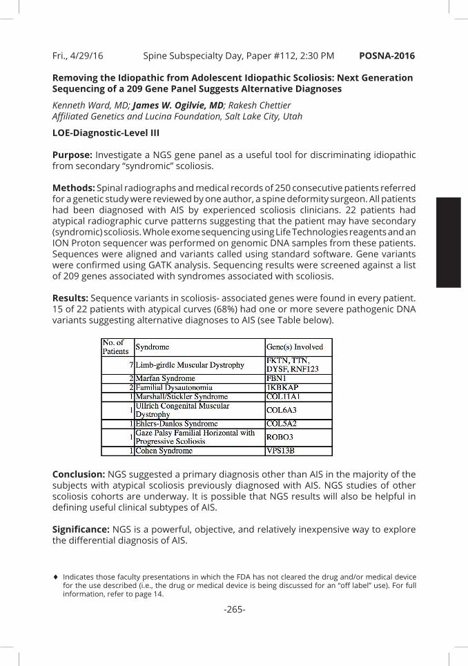
-265-
♦ Indicates those faculty presentations in which the FDA has not cleared the drug and/or medical device for the use described (i.e., the drug or medical device is being discussed for an “off label” use). For full information, refer to page 14.
Fri., 4/29/16 Spine Subspecialty Day, Paper #112, 2:30 PM POSNA-2016 Removing the Idiopathic from Adolescent Idiopathic Scoliosis: Next Generation Sequencing of a 209 Gene Panel Suggests Alternative Diagnoses
Kenneth Ward, MD; James W. Ogilvie, MD; Rakesh ChettierAffiliated Genetics and Lucina Foundation, Salt Lake City, Utah
LOE-Diagnostic-Level III
Purpose: Investigate a NGS gene panel as a useful tool for discriminating idiopathic from secondary “syndromic” scoliosis.
Methods: Spinal radiographs and medical records of 250 consecutive patients referred for a genetic study were reviewed by one author, a spine deformity surgeon. All patients had been diagnosed with AIS by experienced scoliosis clinicians. 22 patients had atypical radiographic curve patterns suggesting that the patient may have secondary (syndromic) scoliosis. Whole exome sequencing using Life Technologies reagents and an ION Proton sequencer was performed on genomic DNA samples from these patients. Sequences were aligned and variants called using standard software. Gene variants were confirmed using GATK analysis. Sequencing results were screened against a list of 209 genes associated with syndromes associated with scoliosis.
Results: Sequence variants in scoliosis- associated genes were found in every patient. 15 of 22 patients with atypical curves (68%) had one or more severe pathogenic DNA variants suggesting alternative diagnoses to AIS (see Table below).
Conclusion: NGS suggested a primary diagnosis other than AIS in the majority of the subjects with atypical scoliosis previously diagnosed with AIS. NGS studies of other scoliosis cohorts are underway. It is possible that NGS results will also be helpful in defining useful clinical subtypes of AIS.
Significance: NGS is a powerful, objective, and relatively inexpensive way to explore the differential diagnosis of AIS.
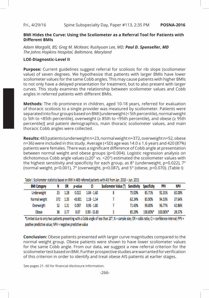
-266-
See pages 21- 60 for financial disclosure information.
Fri., 4/29/16 Spine Subspecialty Day, Paper #113, 2:35 PM POSNA-2016 BMI Hides the Curve: Using the Scoliometer as a Referral Tool for Patients with Different BMIs
Adam Margalit, BS; Greg M. McKean; Rushyuan Lee, MD; Paul D. Sponseller, MDThe Johns Hopkins Hospital, Baltimore, Maryland
LOE-Diagnostic-Level II
Purpose: Current guidelines suggest referral for scoliosis for rib slope (scoliometer value) of seven degrees. We hypothesize that patients with larger BMIs have lower scoliometer values for the same Cobb angles. This may cause patients with higher BMIs to not only have a delayed presentation for treatment, but to also present with larger curves. This study examines the relationship between scoliometer values and Cobb angles in referred patients with different BMIs. Methods: The rib prominence in children, aged 10-18 years, referred for evaluation of thoracic scoliosis to a single provider was measured by scoliometer. Patients were separated into four groups based on BMI [underweight (< 5th percentile), normal weight (≥ 5th to <85th percentile), overweight (≥ 85th to <95th percentile), and obese (≥ 95th percentile)] and patient demographics, main thoracic scoliometer values, and main thoracic Cobb angles were collected.
Results: 483 patients (underweight n=23, normal weight n=372, overweight n=52, obese n=36) were included in this study. Average (+SD) age was 14.0 ± 1.6 years and 420 (87%) patients were females. There was a significant difference of Cobb angle at presentation between normal weight and obese groups (p=0.004). Logistic regression analysis on dichotomous Cobb angle values (≥20° vs. <20°) estimated the scoliometer values with the highest sensitivity and specificity for each group, as 8° (underweight, p=0.022), 7° (normal weight, p<0.001), 7° (overweight, p=0.087), and 5° (obese, p=0.070). (Table I)
Conclusion: Obese patients presented with larger curve magnitudes compared to the normal weight group. Obese patients were shown to have lower scoliometer values for the same Cobb angle. From our data, we suggest a new referral criterion for the scoliometer test based on BMI. Further prospective studies are warranted for verification of this criterion in order to identify and treat obese AIS patients at earlier stages.
-267-
♦ Indicates those faculty presentations in which the FDA has not cleared the drug and/or medical device for the use described (i.e., the drug or medical device is being discussed for an “off label” use). For full information, refer to page 14.
Significance: The differing chest wall thicknesses in patients with various BMIs may alter the scoliometer value for a given rotational deformity. This may explain why overweight and obese patients with AIS present with larger curve magnitudes compared to patients with normal BMIs. Obesity has quadrupled since the scoliometer criterion was developed. This alteration of an existing screening test based on BMI can improve diagnostic efficacy. This may improve patient care by identifying scoliosis in obese patients at earlier stages.
-268-
See pages 21- 60 for financial disclosure information.
Fri., 4/29/16 Spine Subspecialty Day, Paper #114, 2:40 PM POSNA-2016 Delay to Surgery and Deformity Progression Leads to Increased Levels of Fusion in Risser 0, Premenarchal Adolescent Idiopathic Scoliosis (AIS) Patients: A Retrospective Cohort Study
Brandon A. Ramo, MD; Dong-Phuong Tran; Sumeet Garg, MD; Mark A. Erickson, MD; Anil Reddy, BS; Kaitlyn Brown, BSTexas Scottish Rite Hospital for Children, Dallas, Texas
LOE-Prognostic-Level IV
Purpose: We previously demonstrated that a delay to surgery >6 months can lead to increased deformity in patients who are Risser 0. We sought to ascertain whether this increase in deformity results in treatment changes such as longer levels of fusion or greater operative challenges.
Methods: After IRB approval, a retrospective cohort of 143 AIS patients whose date of surgery exceeded six months from the date of surgical recommendation were identified from two centers. 4 surgeons selected levels of fusion for all 34 Risser 0 patients and 48 Risser 1-5 patients in a blinded fashion. Proposed levels of fusion were compared between groups with regard to levels selected at time of surgical decision vs. those selected at actual preoperative time ³6 months later. Additionally, the 34 Risser 0 patients were able to be matched to 75 control subjects from a prospective database by age, Cobb, and Lenke type to compare effects of delay to surgery on the actual surgical fusion levels and other surgical variables (surgical time, EBL, and osteotomy utilization). Statistical methods included use of ANOVA for continuous variables and Fisher’s exact test for categorical variables.
Results: Surgeon agreement when choosing levels of fusion was generally good: the mean intraobserver ICC for all 4 surgeons was 0.8661 and mean interobserver ICC was 0.8165. Surgeons chose to increase levels of fusion in 61% of Risser 0 patients after a delay in treatment vs. in only 15% of Risser 1-5 patients, p<.001. Mean increase in fusion levels was 1.4 levels in Risser 0 vs. 0.1 levels in Risser 1-5, p<.001. The increase in Risser 0 fusions resulted from a mean increase of 0.8 levels for UIV and 0.6 levels for LIV.
Conclusion: Risser 0 patients who delay AIS surgery >6 months are at risk for longer fusions and increased complexity of surgical treatment.
Significance: Surgeons should minimize delays to surgical treatment for Risser 0 AIS patients who meet surgical indications.

-269-
♦ Indicates those faculty presentations in which the FDA has not cleared the drug and/or medical device for the use described (i.e., the drug or medical device is being discussed for an “off label” use). For full information, refer to page 14.
Fri., 4/29/16 Spine Subspecialty Day, Paper #115, 2:55 PM POSNA-2016 Implant Material is Related to Late Deep Postoperative Infection in Adolescent Idiopathic Scoliosis
Scott J. Schoenleber, MD; Paul D. Sponseller, MD; Harry L. Shufflebarger, MD; Baron S. Lonner, MD; Geraldine Neiss, PhD; Petya Yorgova, MS; Tracey P. Bastrom, MA; Suken A. Shah, MDNemours / Alfred I. duPont Hospital for Children, Wilmington, Delaware
LOE-Prognostic-Level IV
Purpose: Late infection occurs in up to 7% of patients after posterior spinal fusion (PSF) for adolescent idiopathic scoliosis (AIS). The use of stainless steel has been identified as a potential risk factor for developing deep infection. Currently, evidence regarding titanium is limited and there are no studies evaluating the risk of infection in patients with cobalt chrome instrumentation. This study aimed to determine risk factors for the development of late infection after PSF for AIS, specifically the effect of implant material.
Methods: A prospective, multicenter AIS database was used to identify patients with a minimum of 2-year follow up after PSF. 1235 patients were included. Late infections were defined as those that presented at least 6 months after the index procedure and required operative debridement. Univariate and multivariate logistic regression analyses were utilized to identify important demographic, intraoperative, and postoperative risk factors for late infection.
Results: The overall rate of late infection was 2.3% (28 patients), occurring at a mean of 2.6 years (maximum 6.2 years) after the index procedure. Race, implant material, and institution were identified as risk factors in both the univariate and multivariate analyses (p<0.05). Of those patients who developed deep infection, 27 (96.4%) had stainless steel (SS), 1 (3.6%) had cobalt chrome (CoCr), and none had titanium (Ti) instrumentation, resulting in significantly different rates of infection between implant materials (SS 3.0%; CoCr 0.5%; Ti 0%; p=0.01).
Conclusion: Implant material was an important factor in late postoperative infection following PSF for AIS. Ti and CoCr implants offered lower rates of late infection.
Significance: Patients should have long-term surveillance after PSF due to the possibility of late infection, since some infections occurred several years later.
-270-
See pages 21- 60 for financial disclosure information.
Fri., 4/29/16 Spine Subspecialty Day, Paper #116, 3:00 PM POSNA-2016 Accelerated Discharge Protocol for Posterior Spinal Fusion (PSF) Patients with Adolescent Idiopathic Scoliosis (AIS) Decreases Hospital Post-Operative Charges 22%
Austin Sanders, BA; Lindsay M. Andras, MD; Harry Ted Sousa, MD; Cathy Kissinger, RN; David L. Skaggs, MD, MMMChildren’s Hospital Los Angeles, Los Angeles, California
LOE-Economic & Decision-Level IV
Purpose: Charges for PSF for AIS can range from $77,362 to $152,637. Implants (22%), inpatient and intensive care unit bed costs (22%), and operating room costs (9.9%) are the top three contributing factors. Length of stay following PSF for AIS averages 5.2-6.5 days. The purpose of this study was to determine if implementing an accelerated protocol could decrease our average hospital stay and what impact this had on postoperative pain management.
Methods: Our tertiary pediatric medical center implemented an accelerated discharge protocol following PSF for AIS in June of 2013, with multidisciplinary collaboration of nursing, physical therapy, pain management and surgical team. This is a retrospective review of all consecutive patients undergoing PSF for AIS before (June 1, 2008-May 31, 2013= traditional protocol) and after (June 1, 2013 – October 22, 2014= accelerated protocol) protocol implementation.
Results: There were 194 patients in the traditional pathway and 90 patients in the accelerated pathway. No significant differences in age at surgery, sex, or number of levels fused were present between the groups. Patients managed under the accelerated discharge had an average hospital stay of 3.7 days compared to 5.02 days for the traditional discharge (p<0.001). There was no difference in reported pain between patients in the accelerated protocol or the traditional protocol for POD1 (3.40 vs. 3.23, p=0.4332); however, patients in the accelerated protocol reported slightly more pain in the accelerated protocol compared to the traditional protocol for pain on day of discharge (3.19 vs. 2.46, p=0.0010). There was no increased incidence of wound complications between the two groups (7/194 vs. 3/90, p=0.91) or readmission (3/194 vs. 4/90, p=0.213). Hospital charges for post-operative care were significantly less in the accelerated discharge group than in the traditional group ($18,360 vs. $23,640, p<0.0001). This corresponded to a 22% ($5,280/$23,640) decrease in postoperative hospital charges.
Conclusion: Reducing length of stay to 3 days following posterior spinal fusion for adolescent idiopathic scoliosis reduces post-operative charges by 22% without increasing post-operative complications.
Significance: Accelerated discharge following PSF for AIS did not increase risk to the patients and produced significant post-operative savings.
-271-
♦ Indicates those faculty presentations in which the FDA has not cleared the drug and/or medical device for the use described (i.e., the drug or medical device is being discussed for an “off label” use). For full information, refer to page 14.
Fri., 4/29/16 Spine Subspecialty Day, Paper #117, 3:05 PM POSNA-2016 A Critical Assessment of ≥10-Year Follow-up of Spinal Fusions for Adolescent Idiopathic Scoliosis: Outcome Improvement Required
Peter O. Newton, MD; Tracey P. Bastrom, MA; Carrie Bartley, MA; Burt Yaszay, MD; Michelle Marks, PT, MA; Harms Study GroupRady Children’s Hospital, San Diego, California
LOE-Therapeutic-Level III
Purpose: To report on prospectively collected >10 year radiographic and patient reported outcomes of surgical correction for major thoracic adolescent idiopathic scoliosis (AIS) patients.
Methods: A prospective multicenter registry of patients (1995-2004) who had surgical correction of AIS was reviewed. Patients with major thoracic curves (Lenke 1-4) who had pre-op and >ten year follow-up data were included. Change in thoracic coronal Cobb, 3D T5-T12 kyphosis (3D kyphosis based on 2D prediction model), and SRS outcome scores were evaluated. Complication data and radiographs were reviewed to objectively and subjectively grade patients with “ideal” outcomes versus those with a “less than ideal” outcome. Patients requiring reoperation and those with radiographs concerning for future need of revision were considered “less than ideal”.
Results: 92 patients with an average of 10.4 years follow-up (range 10-13 years) were included. There were 39 posterior, 36 thoracoscopic anterior, and 17 open anterior approaches. Primary thoracic Cobb improved from 52°±10° to 25°±8° at 2 years (p<0.001) which was maintained at 10 years (25°±9°). Estimated 3D T5-T12 kyphosis was 5°±12° pre-operatively which improved to 25°±10° at 2 years (p<0.001) and was maintained at 10 years 25°±11°. 22 patients had a “less than ideal” outcome and 7 of those had revision surgery. 11 were c lass i f ied as implant fai lure/pseudarthrosis, 9 were residual/progression of deformity, 1 misplaced implant, 1 infection. Patients with less than ideal outcomes had significantly less thoracic Cobb correction at 2 and 10 years, less increase in
-272-
See pages 21- 60 for financial disclosure information.
kyphosis from pre to 2 years, and were significantly less likely to have a top score (4- 5) in the SRS-24 domains of self-image and satisfaction (Table, p<0.05). There were no differences based on approach (p>0.05).
Conclusion: On average radiographic outcomes were stable between 2 and 10 years postop, however 24% of the outcomes were “less than ideal” and 8% required a second surgical procedure. Patients reported lower SRS-24 self-image and satisfaction scores when they required revision or had radiographics rated as “less than ideal”. Half of the revisions occurred after the 2nd postop year indicating the need for longer follow-up.
Significance: The majority of AIS patients, regardless of operative approach, are doing well by radiographic and patient reported assessments 10 years postop. We hope the shift to pedicle screw posterior constructs in the past decade will prove beneficial in the long term, as clearly there is room to reduce the frequency of long-term complications and revisions in patients fused for AIS.
-273-
♦ Indicates those faculty presentations in which the FDA has not cleared the drug and/or medical device for the use described (i.e., the drug or medical device is being discussed for an “off label” use). For full information, refer to page 14.
Fri., 4/29/16 Spine Subspecialty Day, Paper #118, 3:20 PM POSNA-2016 Comparison of Surgical Outcome of Adolescent Idiopathic Scoliosis and Young Adult Idiopathic Scoliosis: A Match-Pair Analysis of 160 Lenke I Patients
Hongda Bao, MD, PhD; Feng Zhu, MD, PhD; Peng Yan, MD, PhD; Jack CY Cheng, MD; Bobby Kin Wah Ng, MD; Qiu YongNanjing Drum Tower Hospital, Nanjing, China
LOE-Therapeutic-Level III
Purpose: Despite numerous reports on the satisfactory surgical correction of adolescent idiopathic scoliosis (AIS), some patients still have the consumption that delay of surgery into young adulthood may be more beneficial. The aim of this study is to investigate if the surgical outcome of young adults was equivalent to adolescents for surgical correction of thoracic idiopathic scoliosis.
Methods: This is a retrospective 1:1 matched cohort study with minimum 2 year follow-up. A total of 80 pairs were recruited with the following inclusion criteria: (1) female idiopathic scoliosis with Lenke Type 1 curve; (2) underwent selective fusion; (3) adolescents age from 10 to 18 years and young adults age from 19 to 29 years; (4) one-stage posterior approach; (5) all-pedicle-screws instrumentations; (6) with major Cobb angle 45°-80°.The AIS patients and young adult idiopathic scoliosis (AdIS) patients were matched for apex, major thoracic curve magnitude, lumbar curve magnitude, time of surgery and follow-up.
Results: The age at the time of surgery in AdIS patients averaged 22.21±3.34 years, significantly larger than that of AIS patients (14.47±1.56 years, P<0.001). The AdIS patients were observed to have significant lower curve flexibility (48.22% for AdIS and 56.62% for patients, P=0.006). Accordingly, lower correction rate (71.41%±0.10 vs. 79.49%±0.09) and larger post-operative major Cobb angle (15.63±4.76º vs. 11.27±3.89º) were found in AdIS patients (P=0.002 and 0.002 respectively). Regarding quality of life, no significant difference was observed between the two groups during follow-up.
Conclusion: AIS patients would gain greater benefit from surgical correction compared to paired AdIS patients from the view of radiographic parameters with no difference of HRQOL during follow-up assessment. The results are vital for spine surgeons to communicate with all AIS patients and their families who considering delay of surgery into young adulthood.
Significance: The current study stressed that AdIS patients had a slight but significant worse radiographic outcome in a 1:1 matched pair analysis with a large sample size. The results indicated that spine surgeons should inform the AIS patients and their families about the potential downside of delaying surgery until young adulthood which may or may not achieve the same clinical outcome because of the incremental rigidity of curve with aging.
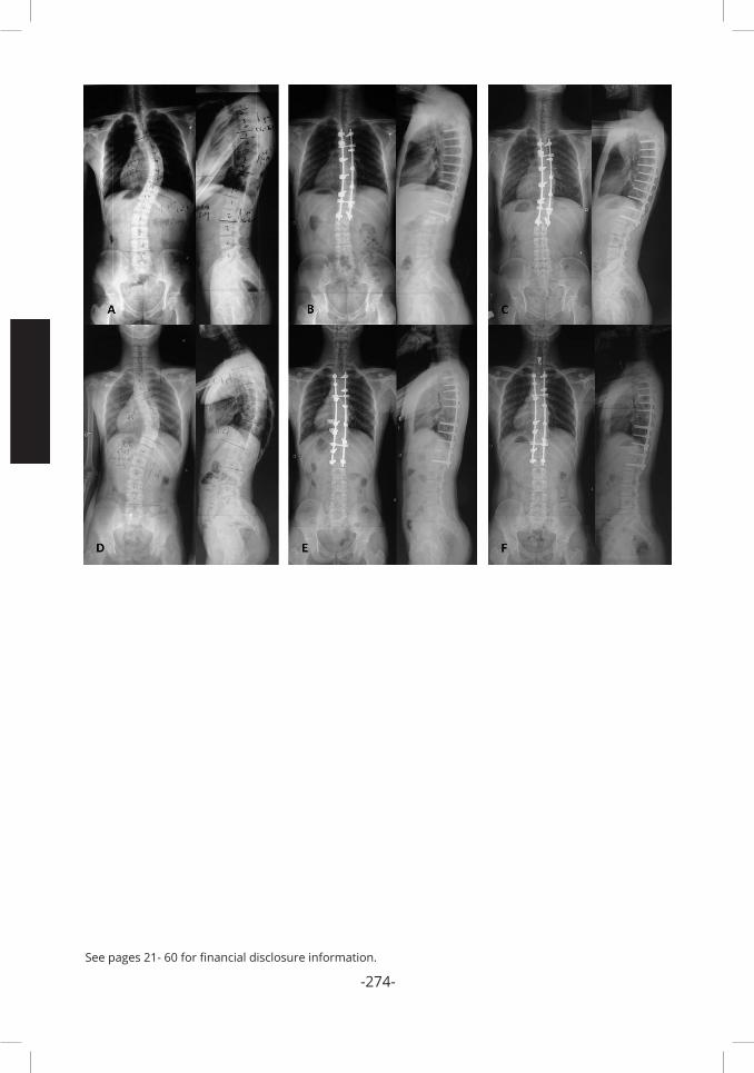
-274-
See pages 21- 60 for financial disclosure information.
-275-
♦ Indicates those faculty presentations in which the FDA has not cleared the drug and/or medical device for the use described (i.e., the drug or medical device is being discussed for an “off label” use). For full information, refer to page 14.
Fri., 4/29/16 Spine Subspecialty Day, Paper #119, 3:25 PM POSNA-2016 Do We Underestimate the Ability of Patients to Return to Physical and Athletic Activities after Scoliosis Surgery? A Validated Patient Questionnaire Based Study
Stephen Wendolowski, BS; Vishal Sarwahi, MD; Rachel Gecelter, BS; Dana Orlando, ST; Terry D. Amaral, MD; Melanie A. Gambassi, NP; Dan Wang, MSMontefiore Medical Center, Bronx, New York
LOE-Diagnostic-Level III
Purpose: This study seeks to assess when our patients are returning to their normal, physical and athletic activities following scoliosis surgery.
Methods: A twenty-four question survey was validated. Patient demographic, XR measurements, EBL length of stay, levels fused, and operative time were recorded. Wilcoxon signed rank tests and Fisher’s exact tests were utilized.
Results: 95 patients completed the survey with a median age of 15 years, 51° pre-operative Cobb, 15.8° post-operative Cobb, and 11 levels fused. By 3 months post-surgery, 77% (72/93) patients returned to school, 37% (30/81) to gym, 52% (48/93) to carrying backpacks, and 60% (54/90) to bending. 43% (30/87) returned to running by 3 months and 80% (70/87) by 6 months. By 6 months, 54% (27/50) returned to non-contact sports, and 64% (21/33) returned to contact sports. For those who returned to contact or non-contact sports by 6 months, 70% (34/48) of the patients reported being at their preoperative. Patients who returned to school and gym after 3 months had a significantly higher BMI (School: 21.8 vs 20.3, p = 0.039; Gym: (21.4 vs. 19.2, p=0.048). Also, patients who returned to gym and carrying their backpack after 3 months were typically fused at L3 or L4 (Gym: p=0.065; Backpack: p=0.006) We found that patients who returned to bending and running after 3 months were typically fused at L3 or L4. (Bending: p = 0.001, Running: p=0.029). No variables were associated with patients returning to sports before or after 3 months. At most recent follow ups, there was no loss of correction, implant failure, or complications.
Conclusion: Patients return to activities much earlier than recommended. Age and LIV are important determinants for returning to physical activity at an earlier time. A quarter of the patients returned to contact/non-contact sports within 3 months, and over half returned by 6 months. The majority of the patients (93% non-contact and 96% contact sports) returned to their preoperative level within 1 year.
Significance: AIS patients undergoing pedicle screw PSF are capable of returning to sports earlier than expected and at their preoperative level for contact and non-contact sports.
-276-
See pages 21- 60 for financial disclosure information.
Earlier return to sports was not seen to have detrimental effect on curve correction and implant fixation. Recommendations for returning to activity after surgery may need to be modified to address this patient reality. ation. Recommendations for returning to activity after surgery may need to be modified to address this patient reality.

-277-
♦ Indicates those faculty presentations in which the FDA has not cleared the drug and/or medical device for the use described (i.e., the drug or medical device is being discussed for an “off label” use). For full information, refer to page 14.
Fri., 4/29/16 Spine Subspecialty Day, Paper #120, 3:30 PM POSNA-2016 Does Correction of Pelvic Obliquity Result in Improved Outcomes and Sitting Tolerance for Patients with Cerebral Palsy Scoliosis?
Brian E. Kaufman, MD; Josh M. Pahys, MD; Firoz Miyanji, MD; Burt Yaszay, MD; Amer Samdani, MD; Patrick J. Cahill, MD; Mark F. Abel, MD; Paul D. Sponseller, MD; Suken A. Shah, MDNemours / Alfred I. duPont Hospital for Children, Wilmington, Delaware
LOE-Prognostic-Level II
Purpose: Severe pelvic obliquity (PO) associated with spinal deformity in cerebral palsy (CP) patients interferes with sitting balance and quality of life (QOL). Surgeons and caregivers rate sitting balance as their highest surgical indication or priority. To date, no guidelines exist regarding optimal alignment for these patients to facilitate care, alleviate pain, and improve QOL. This study evaluated the relationship between PO and QOL in CP patients with scoliosis undergoing posterior spinal fusion (PSF).
Methods: A prospectively collected multi-center database was reviewed for all CP patients with scoliosis who underwent PSF and had minimum two-year follow-up. A total of 101 patients were identified. QOL was evaluated using the validated CPCHILD clinical patient related outcome measure. Radiographs, sitting tolerance, and QOL data were evaluated at preoperative, initial postoperative, and two-year follow-up visits.
Results: There was significant improvement in mean preoperative PO to 2 years postoperatively (21.8° to 7.5°, p<0.001) and sitting tolerance. There was no significant correlation between the absolute magnitude of PO at 2 years postoperatively and QOL. A significant correlation did exist, however, between percent correction of PO from preoperative to 2 years postoperatively and overall QOL (p=0.02), especially in the positioning, transfers, and mobility domain. QOL data revealed a significant correlation with decreased PO and increased QOL in nearly all domains, specifically PO<10° vs. 10-19° (p=0.03) and PO<10° vs. PO >20° (p<0.001). There were 15 patients with spastic hip subluxation/dislocation. The hip status did not correlate with postoperative PO, percent correction of PO, or postoperative QOL scores.
Conclusion: This prospectively collected study evaluated 101 CP patients who underwent PSF for severe scoliosis and PO with two-year follow-up of X-rays, clinical data, and patient-related outcomes. Regardless of stage of treatment (pre- or postoperative), PO <10° was associated with the highest QOL scores and sitting tolerance. The percent correction of PO achieved after surgery was associated with an improved overall QOL score (especially the positioning, transfers, and mobility domain) at 2 years postoperatively.
Significance: Severe pelvic obliquity associated with spinal deformity does interfere with sitting and QOL in severely affected (GMFCS 5) children with cerebral palsy. Leveling the pelvis as much as possible during PSF is desirable to improve patient sitting balance and alignment.

-278-
See pages 21- 60 for financial disclosure information.
Fri., 4/29/16 Spine Subspecialty Day, Paper #121, 3:35 PM POSNA-2016 Sacral Alar Iliac (SAI) Screws Fail 75% Less Frequently than Iliac Screws in Neuromuscular Scoliosis
Lior Shabtai, MD; Lindsay M. Andras, MD; Mark Portman, BS; Liam R. Harris; Paul D. Choi, MD; Vernon T. Tolo, MD; David L. Skaggs, MD, MMM Children’s Hospital Los Angeles, Los Angeles, California
LOE-Therapeutic-Level III
Purpose: Despite recent popularity of SAI screws for fusion to the pelvis for NMS, there is little data regarding the failure rate of this technique compared to traditional modes of iliac fixation. Theoretical advantages of the SAI screws are obviating the need for a rod to iliac screw connector and a lower implant profile. The purpose of this study is to determine if sacral alar iliac (SAI) screws have fewer failures than iliac screws in neuromuscular scoliosis (NMS).
Methods: Review of neuromuscular patients treated with posterior spinal fusion with pelvic fixation from 2004- 2012 with minimum two year follow up was conducted. Medical records and imaging studies were reviewed. Patients were divided into 2 groups based on the type of pelvic fixation (SAI or iliac screws), and implant failures were compared between the groups.
Results: 101 patients were reviewed, including 55 patients with iliac screws and 46 patients with SAI screws. Implant failures included: disengagement of the rod to iliac screw connector (10%, 10/101), separation of screw head from screw shaft (4%, 4/101) and set screw disengagement (2%, 2/101). The SAI group had a lower implant failure rate (7%, 3/46) compared to the iliac screw group (24%, 13/55) (p= 0.031). Rod to iliac screw connectors failed in 18%, (10/55) of patients. There were significantly less surgical revisions in the SAI group (2%, 1/46) for pelvic screw prominence compared to the iliac screw group (11%, 6/55), p=0.027.
Conclusion: SAI screws had a lower rate of implant failure and revision surgery compared to iliac screws. If rod to screw connector failures are excluded, the failure rate of SAI screws of 6.5% (3/46) is similar to that of iliac screws 5.5% (3/55), therefore the most important advantage of the SAI technique may be obviating the need for a screw to rod connector.
Significance: These results offer new, evidenced-based support for the expanding use of SAI screws over iliac screws in pelvic fixation, for the treatment of neuromuscular scoliosis.
-279-
♦ Indicates those faculty presentations in which the FDA has not cleared the drug and/or medical device for the use described (i.e., the drug or medical device is being discussed for an “off label” use). For full information, refer to page 14.
Fri., 4/29/16 Sports Subspecialty Day, Paper #122, 4:00 PM POSNA-2016 Retrospective Cohort Study of 207 Cases of Osteochondritis Dissecans of the Knee: Risk Factors and Outcomes Associated with Surgical Treatment
Maria Tuca, MD; John Arbucci; Jason Silberman, BA; Eva Luderowski, BA; Tyler J. Uppstrom, BA; Joseph Nguyen, MPH; Daniel W. Green, MD Hospital for Special Surgery, New York, New York
LOE-Therapeutic-Level IV
Purpose: Describe the clinical characteristics, image findings, and outcomes of patients with juvenile osteochondritis dissecans (JOCD) of the knee.
Methods: Retrospective cohort study of knee JOCD patients assessed by a single pediatric orthopaedic surgeon at a tertiary care center between 2005-2015. Diagnoses were confirmed by MRI. Patellar dislocations or osteochondral fractures were excluded. Demographic data, sports played, comorbidities, surgical procedures, and clinical data were extracted from charts. Images were analyzed to identify location and size of lesions. Chi-square or Fisher’s exact tests were used to compare discrete variables, and Mann-Whitney U and Kruskal Wallis tests to compare continuous variables between groups.
Results: Sample consisted of 180 patients (207 knees) (124 boys/56 girls). Average age at diagnosis was 12.7 years (5.6-18.1). Asthma was the most frequently associated morbidity (13.3%). Majority were active in sports (80.8%), mainly soccer (36.7%) and basketball (29.4%). JOCD was present bilaterally in 27 patients (15%), 14 knees had bifocal OCD (6.8%), and 1 patient bifocal lesions in both knees. Most common location was medial femoral condyle (56.3%) followed by lateral femoral condyle (23.1%), trochlea (11.4%), patella (9%), and tibia (0.5%). In the sagittal view, most common location was the middle third of the condyles (48.7%). Surgery was performed in 71 knees (34.2%). The average normalized area of non-surgical JOCD lesions was 6.8 (0.1-18), whereas surgical lesions averaged a significantly higher area of 7.7 (0.5-17) (p=0.023). Average BMI was 21.6 versus 20.2 for surgical and non-surgical patients, respectively, significantly higher for those who underwent surgery (p=0.002). Most common procedure was fixation with bioabsorbable nails (62.5%), using an average of 4 nails (1-9); the second most common procedure was drilling (40.3%). Fixation was achieved all-arthroscopic in 76%. Revision surgery was required in 10 knees (13.9%). Most common revision procedures were arthroscopic debridement (80%) and implant removal (30%). Most surgical patients had postoperative MRIs (55 knees), with an average radiological follow-up of 14.5 months (2.1-55.4).
Conclusion: JOCD occurs more frequently in young adolescent athlete boys, affecting the middle third of the medial femoral condyle. In our cohort, 1/3 of the patients had surgery, where bigger lesions and higher BMI were risk factors for operative treatment. At short-term follow up, the success rate following surgery was 84%.
Significance: To our knowledge, this is the largest single-center JOCD cohort reported. Results allow us to gain insight into this pathology, and better define the presentation, natural history and risk factors for progression for JOCD.
-280-
See pages 21- 60 for financial disclosure information.
Fri., 4/29/16 Sports Subspecialty Day, Paper #123, 4:05 PM POSNA-2016 Radiographic Assessment of Anatomic Risk Factors Associated with Acute, Lateral Patellar Dislocation in the Immature Knee
Kevin E. Klingele, MD; Matthew C. Beran, MD; Walter P. Samora III, MDNationwide Children’s Hospital, Columbus, Ohio
LOE-Prognostic-Level II
Purpose: Acute, lateral patellar dislocation is a common injury in adolescent and pediatric patients. Non-operative management is advocated for patients without history of recurrent instability or osteochondral injury. Operative management aims to correct anatomic risk factors predisposing to the development of recurrent instability. Pathologic thresholds for operative management are reported in adults, but may not be applicable to the immature knee. Recent studies have outlined changing patellofemoral morphology which occurs with growth. The purpose of this study is to report morphological differences between skeletally immature patients with and without acute, lateral patellar instability.
Methods: Retrospective review of all skeletally immature patients diagnosed with acute, lateral patellar dislocation and who had undergone MRI of the affected knee. A control group of patients without a history of patellar instability who had undergone MRI imaging of the knee within the same age range was identified. Six radiographic measurements were recorded and compared between groups: Lateral trochlear inclination (LTI), trochlear facet asymmetry (TFA), trochlear depth (TD), tibial tuberosity-trochlear groove (TT-TG), sulcus angle (SA) and patellar height ratio (Insall-Salvati).
Results: 178 patients were included for analysis (study=108, control =70) (mean age 13.7 and 12.1 years) (p=<0.001). Patients in the study group were observed to have statistically significant differences in all radiographic measurements including: decreased LTI (p<0.001), increased TFA (p<0.001), and increased SA (p<0.001). Mean trochlear depth was 3.4 mm and 5.6 mm respectively (p<0.001). Patients in the study group were noted to have an increased patellar height ratio (p<0.001) and TT-TG distance (p<0.001).
Conclusion: Morphologic abnormalities may predispose skeletally immature patients to an increased risk for acute lateral patellar instability. All recorded measurements of patellofemoral morphology were abnormal compared to an age-matched control group. Further research is needed to determine the clinical implications of these differences in order to optimize both surgical and non-surgical management.
Significance: This study is the first to compare radiographic differences in patellofemoral morphology between skeletally immature patients with and without acute, lateral patellofemoral instability. Findings suggest that anatomic differences may predispose the immature knee to such injury. As the indications for operative management in this population continue to evolve, the current findings may help to establish pathologic threshold values prompting consideration for operative correction.
-281-
♦ Indicates those faculty presentations in which the FDA has not cleared the drug and/or medical device for the use described (i.e., the drug or medical device is being discussed for an “off label” use). For full information, refer to page 14.
Fri., 4/29/16 Sports Subspecialty Day, Paper #124, 4:10 PM POSNA-2016 Concomitant Collateral Ligament Injuries in Pediatric Patients with Anterior Cruciate Ligament (ACL) Tears
Rushyuan Lee, MD; Afam Nduaguba; Melissa Gunderson, BA; Theodore J. Ganley, MDChildren’s Hospital of Philadelphia, Philadelphia, Pennsylvania
LOE-Prognostic-Level II
Purpose: To determine if obese and overweight children have a higher rate of concomitant collateral ligament injuries in the setting of ACL tear and require more surgical intervention
Methods: We retrospectively reviewed the records on all patients under 18 years of age presenting to our institution with ACL tears from January 2009 to October 2013 for sex, age, height, weight, and collateral ligament injury and treatment. Patients were divided into two groups for comparison: (1) normal weight (2) obese/overweight (defined as > 85 %ile Body Mass Index (BMI) for age). Statistical analysis was performed with Chi square tests.
Results: The study group consisted of 523 patients with ACL injuries, of which 109 (20.8%) had concomitant collateral ligament injuries. The group sustained 99 (18.9%) medial collateral ligament injuries (MCL), 21 (4%) lateral collateral ligament (LCL) injuries, and 11 (2.1%) of these were combined MCL and LCL injuries.
The study group consisted of 340 normal weight (65%) and 181 obese and overweight (34.6%). Two patients were underweight. There was no statistically significant difference between the normal weight and obese/overweight groups in terms of MCL injuries (19.7% vs. 17.7%), LCL injuries (4.1% vs. 3.9%), and combined injuries (2.1% vs. 2.2%). Though there appeared to be a trend towards a delay in collateral ligament injury diagnosis between the normal weight and obese/overweight groups, (10.8% vs. 20%), this was not statistically significant.
The treatment for collateral ligament injuries did not differ between the normal weight and obese/overweight groups. Bracing was successful (77% vs. 74.3%) and surgical intervention was required at comparable rates (24.3% vs. 22.9%), though more normal weight patients compared to obese/overweight patients underwent primary repair (17/18, 94.4% vs. 4/8, 50% with p=0.02) rather than reconstruction. Conclusion: BMI does not play a factor in the rate of collateral ligament injuries in the setting of ACL tears in children and adolescents, nor does it influence successful brace treatment of collateral ligament injuries.
Significance: Obese and overweight pediatric patients sustain collateral ligament injuries in the setting of ACL tears at the same rate as normal weight patients, and can also be successfully treated with bracing and rehab.
-282-
See pages 21- 60 for financial disclosure information.
Fri., 4/29/16 Sports Subspecialty Day, Paper #125, 4:25 PM POSNA-2016 Medial Patellofemoral Ligament Reconstruction with Hamstring and Biotenodesis Screw Fixation in Children and Adolescents: A Large Consecutive Series
T. Whitney Gibson, DO; Julia Lee, MD; Linda Pugh; Grant Meeker, MS, BSShriners Hospital for Children-Greenville, Greenville, South Carolina
LOE-Therapeutic-Level IV
Purpose: Patellar instability is a common condition in children and adolescents. Congenital abnormalities, structural malalignment and traumatic injuries have been implicated as etiologies for patella instability and subsequent dislocations. Conservative management has been shown to be effective for first time dislocaters while surgical intervention including medial patellofemoral ligament reconstruction (MPFL) provides an effective means of stabilization for the mulitiply dislocated patella. This study describes our institutional experience with MPFL reconstruction in a pediatric population.
Methods: We retrospectively analyzed clinical presentation and outcomes of all patients undergoing MPFL reconstruction by a single surgeon over a six year period.
Results: Fifty-six extremities in 47 patients under the age of 18 underwent MPFL reconstruction with hamstring tendon and biotenodesis screw fixation at the femoral origin and patella insertion. Average age for surgery was 14.8 years with 1.9 years of follow up. Six extremities (10.7) had re-dislocations after stabilization surgery and three of those underwent revision MPFL reconstruction with resolution of the instability. 46/56 (82.7) extremities report good to excellent results after surgery with no physeal injury.
Conclusion: MPFL reconstruction with hamstring tendon and biotenodesis screw fixation in children with recurrent patella dislocations is an effective, reproducible stabilization technique with a low complication rate and no growth disturbance.
Significance: This is the first large consecutive series describing this surgical technique for patella stabilization in children and adolescents with recurrent dislocations.
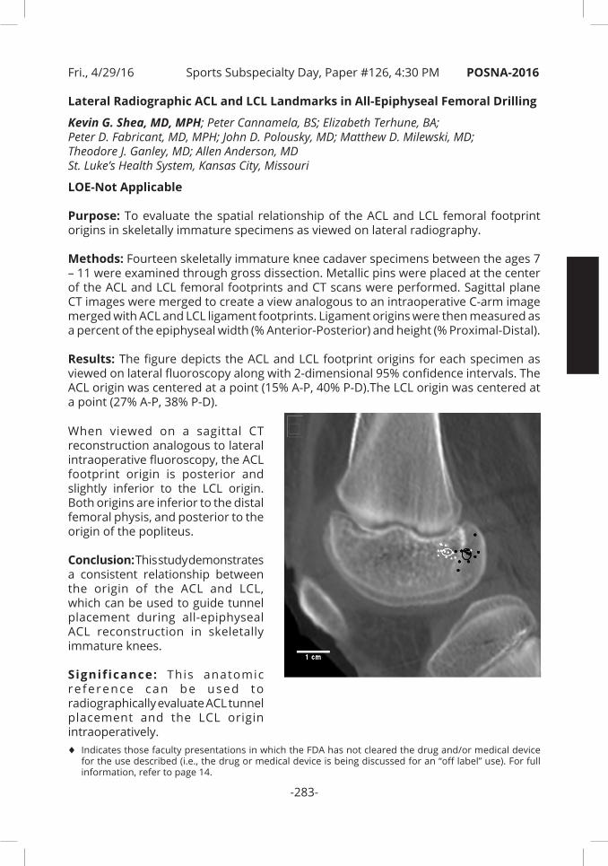
-283-
♦ Indicates those faculty presentations in which the FDA has not cleared the drug and/or medical device for the use described (i.e., the drug or medical device is being discussed for an “off label” use). For full information, refer to page 14.
Fri., 4/29/16 Sports Subspecialty Day, Paper #126, 4:30 PM POSNA-2016 Lateral Radiographic ACL and LCL Landmarks in All-Epiphyseal Femoral Drilling
Kevin G. Shea, MD, MPH; Peter Cannamela, BS; Elizabeth Terhune, BA; Peter D. Fabricant, MD, MPH; John D. Polousky, MD; Matthew D. Milewski, MD; Theodore J. Ganley, MD; Allen Anderson, MDSt. Luke’s Health System, Kansas City, Missouri
LOE-Not Applicable
Purpose: To evaluate the spatial relationship of the ACL and LCL femoral footprint origins in skeletally immature specimens as viewed on lateral radiography.
Methods: Fourteen skeletally immature knee cadaver specimens between the ages 7 – 11 were examined through gross dissection. Metallic pins were placed at the center of the ACL and LCL femoral footprints and CT scans were performed. Sagittal plane CT images were merged to create a view analogous to an intraoperative C-arm image merged with ACL and LCL ligament footprints. Ligament origins were then measured as a percent of the epiphyseal width (% Anterior-Posterior) and height (% Proximal-Distal).
Results: The figure depicts the ACL and LCL footprint origins for each specimen as viewed on lateral fluoroscopy along with 2-dimensional 95% confidence intervals. The ACL origin was centered at a point (15% A-P, 40% P-D).The LCL origin was centered at a point (27% A-P, 38% P-D).
When viewed on a sagittal CT reconstruction analogous to lateral intraoperative fluoroscopy, the ACL footprint origin is posterior and slightly inferior to the LCL origin. Both origins are inferior to the distal femoral physis, and posterior to the origin of the popliteus.
Conclusion: This study demonstrates a consistent relationship between the origin of the ACL and LCL, which can be used to guide tunnel placement during all-epiphyseal ACL reconstruction in skeletally immature knees.
Significance: This anatomic re ference can be used to radiographically evaluate ACL tunnel placement and the LCL origin intraoperatively.
-284-
See pages 21- 60 for financial disclosure information.
Fri., 4/29/16 Sports Subspecialty Day, Paper #127, 4:35 PM POSNA-2016 Is the Intercondylar Notch Size a Risk Factor for Failure in Skeletally Immature Athletes that Undergo ACL Reconstructions?
Maria Tuca, MD; Elizabeth Gausden, MD; Frank Cordasco, MD; Daniel W. Green, MDHospital for Special Surgery, New York, New York
LOE-Prognostic-Level III
Purpose: To determine whether alterations in the intercondylar notch size are associated with failure of anterior cruciate ligament reconstruction (ACLR) in skeletally immature athletes. Previous reports have shown that smaller notches are associated with a higher risk of ACL tears, yet to our knowledge no study has evaluated if notch size predicts failure following ACLR.
Methods: Case-control study of 32 skeletally immature athletes who underwent ACLR by the same surgical team in a 1:1 sample of age- and sex-matched controls. A total of 16 failed ACL-reconstructed knees were compared to 16 intact ACL-reconstructed knees with a minimum of 2 years follow-up. Average skeletal age of the studied population was 14 years (range 10 - 17). Notch width (NW) and notch width index (NWI) were measured in coronal proton density weighted MRI studies using a method previously reported for the pediatric population. Comparison between groups was performed using the Wilcoxon Rank sum test and P-values < 0.05 were considered significant.
Results: NW was significantly smaller in the failed-ACLR group compared to the intact-ACLR group, with an average of 18.6mm versus 22.4mm, respectively (z=-3.883; p <0.0001). NWI was also significantly lower in failed-ACLR knees versus the control group, with an average of 0.257 and 0.319, respectively (z=-4.788; p < 0.0001).Conclusion: Notch Width and Notch Width Index are significant predictors for failure in skeletally immature athletes that undergo ACL reconstructions.
Significance: According to the results of our study, notch dimensions are not only risk factors for ACL tears, but also for failure following ACL reconstructions. Hence, in the setting of small notch width indices, surgeons should consider to include a notchplasty during the ACL reconstruction surgery.
-285-
♦ Indicates those faculty presentations in which the FDA has not cleared the drug and/or medical device for the use described (i.e., the drug or medical device is being discussed for an “off label” use). For full information, refer to page 14.
Fri., 4/29/16 Sports Subspecialty Day, Paper #128, 4:50 PM POSNA-2016 Combined Femoral Catheter-Sciatic Nerve Block has Superior Postoperative Outcomes than Femoral Nerve Catheter Alone for ACL Reconstruction in the Pediatric Population
Jay C. Albright, MD, Patrick Carry, BA, Tessa Mandler, MD, Ariel K. Lepon, BA, Anusara C. Ice, BSChildren’s Hospital Colorado, Aurora, Colorado
LOE-Therapeutic-Level III
Purpose: To compare the effectiveness of femoral nerve catheter to femoral nerve catheter plus single injection sciatic nerve block (combination) for pain management in pediatric patients undergoing anterior cruciate ligament (ACL) reconstruction.
Methods: Adolescent subjects who underwent ACL reconstruction between December 13, 2013 and September 1, 2014 were retrospectively reviewed. During the study period, all subjects underwent general anesthesia. In addition, patients either received a continuous femoral nerve catheter or continuous femoral nerve catheter with single injection sciatic nerve block. Multivariable linear regression, modified Poisson regression or Cox-proportional hazards regression analyses were used to compare intravenous (IV) morphine equivalent dose, pain scores, incidence of adverse side effects, and post-anesthesia care unit length of stay (PACU LOS) across the two groups. Propensity scores were used to minimize bias due to the non-randomized nature of the regional anesthesia protocol allocation.
Results: The study population included 21 subjects in the femoral nerve catheter group and 38 subjects in the combined, femoral nerve catheter plus single injection sciatic nerve block group. There was no difference [p>0.05] in the incidence of adverse side effects or opioid prescription refill requests between groups. Total IV morphine equivalent dose [adjusted mean difference: 0.9 mg/kg, 95% CI: 0.01 to 0.16; p = 0.0257], maximum verbal pain score recorded in the PACU [adjusted mean difference: 2.9 units, 95% CI: 1.2 to 4.8, p = 0.0015] as well as the percentage of subjects requiring one or more opioid doses in the PACU [adjusted risk ratio: 1.7, 95% CI: 0.4 to 7.7, p = 0.5093] was significantly increased in the femoral nerve catheter group relative to the combined group. PACU length of stay was also significantly increased [adjusted hazard ratio: 2.4, 95% CI: 1.3 to 4.5, p = 0.0058] in the femoral nerve catheter group [median PACU LOS: 163 min, 95% CI: 135 to 199 min] relative to the combined group 129 minutes [95% CI: 120 to 138 minutes.
Conclusion: The use of combined femoral nerve catheter plus single injection sciatic nerve block is an effective pain management technique for reducing post-operative opioid administration, pain scores, and PACU LOS following adolescent ACL reconstruction.
Significance: In a climate lacking standard protocols for pain management of pediatric ACL reconstructions, this study suggests use of femoral nerve catheter plus single injection sciatic nerve block is superior for pain management to femoral nerve catheter for ACL reconstruction.
-286-
See pages 21- 60 for financial disclosure information.
Fri., 4/29/16 Sports Subspecialty Day, Paper #129, 4:55 PM POSNA-2016 Validated Pediatric Functional Outcomes of All-Epiphyseal ACL Reconstructions –Does Reinjury Affect Outcomes
Sheena C. Ranade, MD; Christian A. Refakis, BA; Kelly L. Leddy, MHS; Lawrence Wells, MD; J. Todd Lawrence, MD; Theodore J. Ganley, MDThe Children’s Hospital of Philadelphia, Philadelphia, Pennsylvania
LOE-Therapeutic-Level IV
Purpose: Increased level and frequency of sports participation among prepubescent children has resulted higher incidence of ACL tears in skeletally immature athletes. Previous studies reported clinical outcomes of All Epiphyseal-ACL Reconstruction (AE-ACLR) without using patient-oriented pediatric-specific clinical outcome measures. This prospective cohort study utilizes pediatric-specific scores in the medium to long term (range: 10.6-78.0 months, mean: 48 months) following AE-ACLR in skeletally-immature athletes with the secondary aim to examine how pertinent surgery-related variables (i.e. complications, re-injury, re-rupture) affect patient-reported outcomes.
Methods: After IRB approval, a query of AE-ACLR patients that underwent surgery between April 2009 and January 2015 with a minimum of 6 months follow up at a single institution performed by one of three surgeons yielded 140 possible subjects. Two subjects were excluded for atypical presentations (i.e. tibial hemimelia). Telephone consent was obtained from 65 subjects and (if applicable) their parents; of which 50 subjects completed an online survey comprised of the Pedi-IKDC, HSS Pedi-FABS, and PROMIS physical functionality scores with an additional questionnaire regarding subsequent re-injury. Demographic information was collected via electronic medical records.
Results: Thirty-seven males and 13 females underwent AE-ACLR for ACL tear and completed the survey. Six subjects (4 male and 2 female) re-tore their AE-ACLRs at an average of 2.8 years postoperatively (95% confidence interval (CI): 1.6 to 4.0). The average age at initial injury in the re-tear group was 11.2 years (95% CI: 9.3-13.0), and 12.3 years (95% CI: 11.8-12.8) in the intact group. The Pedi-IKDC score mean was 94.5 for the combined groups (95% CI: 92.9-96.1), 91.4 (95% CI: 83.9-99.0) for re-tears and 94.9 (95% CI: 93.4-96.4) intact AE-ACLRs. The PROMIS Score mean was 99.1 (95% CI: 97.5-100.7) for both groups, 98.0 (95% CI: 94.8-101.2) for re-tears and 99.3 (95% CI: 98.7-99.8) for intact AE-ACLRs. The HSS-Pedi FABS mean was 22.3 (95% CI: 20.3-24.4) for both groups, 21.8(95% CI: 18.9-24.7) for re-tear and 22.4(95% CI: 20.1-24.7) for intact AE-ACLRs.
Conclusion: Preliminary data demonstrates overall excellent average outcomes among patients undergoing AE-ACLR. 12 percent of patients suffered re-tears, and this group trended toward poorer Pedi-IKDC and PROMIS scores. There is no statistically significant difference between the age at injury among the AE-ACLR re-tear and intact groups as the sample size of AE-ACLR re-tear patients is, as yet, only six patients.
Significance: This study represents the initial step toward ideally identifying possible age, gender, and functionally-based risk factors for ACL re-tear in patients undergoing AE-ACLR.
-287-
♦ Indicates those faculty presentations in which the FDA has not cleared the drug and/or medical device for the use described (i.e., the drug or medical device is being discussed for an “off label” use). For full information, refer to page 14.
Fri., 4/29/16 Sports Subspecialty Day, Paper #130, 5:00 PM POSNA-2016 A Radiographic Analysis of Hip Morphologic Differences between Genders in Adolescents with FAI
Ryan C. Goodwin, MD; Perry Hooper, DO; James Rosneck, MD; Thomas S. Lynch, MD Cleveland Clinic, Cleveland, Ohio
LOE-Prognostic-Level II
Purpose: To determine the radiographic differences between sexes in the adolescent population in patients with FAI (Femoroacetabular Impingement) as well as ascertain if BMI contributes to FAI symptomatology.
Methods: We retrospectively identified 177 patients aged from 13 to 18 years who were treated for FAI with hip arthroscopy performing a labral debridement or repair and/or femoral osteoplasty. Using MRI and plain radiograph, lateral center edge angle, Tonnis angle, alpha angle, and anterior center edge angle were measured. The intraclass correlation coefficient between readers was calculated. Multiple linear regression models incorporating age, gender and BMI with the radiographic measurements as outcomes were created to evaluate differences while controlling for other variables.
Results: BMI was lower among females at 22.3 compared to 23.7 in males (p = 0.048). The lateral center edge angle and Tonnis angle on MRI and plain radiograph displayed no statistically significant differences between genders after controlling for BMI and age. The BMI and age adjusted mean alpha angle was higher in males than females on both plain radiograph (55.9° verses 45.2°; p < 0.0001) and MRI (54.1° versus 42.5°; p < 0.0001). The intraclass correlation coefficient demonstrated moderate to strong agreement between the three radiographic image readers.
Conclusion: In adolescents that have undergone hip arthroscopy for femoroacetabular impingement, there are sex-dependent characteristic differences demonstrated pre-operatively on MRI and plain radiograph which show increased cam-type deformities in adolescent males.
Significance: Femoroacetabular impingement (FAI) continues to become an increasingly diagnosed condition in adolescents. No studies to date have assessed the radiographic differences in adolescent males and females who present with FAI and undergo hip arthroscopy. In patients presenting with symptoms of FAI who underwent hip arthroscopy, our results show that males have a larger cam deformity than females.

-288-
See pages 21- 60 for financial disclosure information.
Fri., 4/29/16 Sports Subspecialty Day, Paper #131, 5:05 PM POSNA-2016 Return to Play Following Open Treatment of Femoroacetabular Impingement in Adolescent Competitive Athletes
Stephanie W. Mayer, MD; Meredith Mayo, MD; Lauryn A. Kestel, BS; Patrick Carry, BA; Eduardo V. Novais, MDChildren’s Hospital Colorado, Aurora, Colorado
LOE-Therapeutic-Level IV
Purpose: Femoroacetabular impingement (FAI) associated with sequelae of pediatric hip disorders or with stress related to high level of sports participation has been increasingly recognized. However, the ability to return to sports participation has not been determined in adolescent athletes. We asked (1) what proportion of adolescent athletes return to play following open treatment of FAI? (2) What is the median time to return to play? (3) Is there an improvement in patient centered measures of hip pain, function and quality of life after surgery?
Methods: After IRB approval, adolescents who participated in competitive sports and underwent open treatment of FAI using the surgical hip dislocation approach were assessed at a minimum 1-year follow-up. Patients completed a self-reported questionnaire centered on the time and level of return to play. Pain and functional outcomes were assessed by the modified Harris Hip Scores [mHHS] and the Hip Disability and Osteoarthritis Outcome Scores [HOOS]. Femoral head-neck morphology was assessed by the alpha angle and femoral head-neck offset measured preoperatively and at the most recent follow-up.
Results: Twenty-four adolescent competitive athletes with a mean age of 15.5 ±2.0 years and average of 22 months (range, 12-38 months) follow-up were included. Among the 24 adolescent competitive athletes, 21 [87.5%; 95%CI: 67.6 to 97.3%] successfully returned to play after open FAI treatment. The median time to return to play was 7 months [95%CI: 6-10 months]. Of the 21 athletes that returned to play, 19 (90%) returned at a level that was equivalent to or greater than their level of play prior to surgery. Three (12.5%) athletes did not return to play and indicated that failure to return to play was unrelated to their hip. There was significant improvement in mHHS [p<0.0001] and HOOS in all five dimensions [p<0.0001]. There was a significant improvement in alpha [p<0.0001] angle and offset [p<0.0001]. One complication was noted in one patient that fell and required revision of greater trochanter fixation.
Conclusion: Adolescent athletes undergoing open FAI surgery can expect to return to the same or better level of sports participation during the first year after surgery with a low rate of complications and high rate of improvement in hip-specific outcome scores.
Significance: Among patients that returned to sports, the median time to return to play was seven months after surgery. This information may facilitate targeting the timing for returning to sport and help with patient’s motivation and compliance with rehabilitation.
-289-
♦ Indicates those faculty presentations in which the FDA has not cleared the drug and/or medical device for the use described (i.e., the drug or medical device is being discussed for an “off label” use). For full information, refer to page 14.
Fri., 4/29/16 Trauma Subspecialty Day, Paper #132, 4:00 PM POSNA-2016 Validation of Patient Reported Outcome Measurement Information System (PROMIS®) for Detection of Posttraumatic Stress in Children and Adolescents Following Surgery for Acute Orthopaedic Injury
Collin J. May, MD; Daniel J. Hedequist, MD; Patricia E. Miller, MS; Emily Rademacher, BS; Justyna Klajn, MS; Manahil N. Naqvi, BS; Benjamin J. Shore, MD, MPH, FRCSCBoston Children’s Hospital, Boston, Massachusetts
LOE-Diagnostic-Level II
Purpose: Posttraumatic stress following childhood injury is common, and left untreated is a risk for worse functional outcomes, poorer quality of life, cognitive impairment, and dysfunctional relationships. Early diagnosis of posttraumatic stress is critical for timely management; however, current screening tools are lengthy and difficult to administer. In this study, we aimed to assess the efficiency and validity of the PROMIS computerized adaptive test to identify those patients with high levels of posttraumatic stress symptoms.
Methods: We performed a prospective study of consecutive children ages 8-18 years undergoing surgery for orthopaedic trauma at a single center. The child PTSD symptom scale (CPSS), a validated self-report indicator of posttraumatic stress symptoms in children, was utilized as the gold standard. Patients were considered to have high levels of PTSD symptoms when scoring 11 or higher on the scale. Efficiency was summarized by the average time to complete. Convergent and divergent validity of PROMIS Anger and Anxiety domains was assessed with Pearson’s correlation analysis. Univariate logistic regression and receiver operating characteristic analysis were analysis was used to determine if PROMIS domains could discriminate across PTSD risk groups. Receiver operating characteristic (ROC) analysis, including estimating area under the ROC curve (AUC) along with exact 95% confidence intervals (CI), was used to quantify establish the discriminant validity.
Results: There were 116 subjects included in the analysis. Twenty-one subjects had high levels of PTSD symptoms (18.1%; 95% CI = 11.6 to 26.3%). The CPSS instrument took 167.4 (± 48.7) seconds versus 54 (IQR 40-89) and 50 (IQR 36-74) seconds for the PROMIS Anger and Anxiety domains, respectively. Assessment of convergent validity showed PROMIS domains significantly correlated with CPSS scores (Anger: p<0.001; r=0.51, Anxiety: p<0.0001; r=0.50). Divergent validity was assessed through comparison of PROMIS domains with a functional outcome measure, the UCLA activity score. PROMIS domains were not correlated with UCLA (Anger: r=-0.21, Anxiety: r=0.13). PROMIS domains were able to discriminate across PTSD groups (Anger p=0.001 and Anxiety p<0.001).
Conclusion: The PROMIS computer adaptive test domains for anger and anxiety were shown to be reliable, efficient, and valid tools for evaluating posttraumatic stress symptoms in children following surgery for orthopedic trauma.
-290-
See pages 21- 60 for financial disclosure information.
Significance: Posttraumatic stress symptoms in children are common, and early diagnosis and intervention has the potential to decrease the significant psychological and physical sequelae associated with PTSD. We demonstrate that the PROMIS tool can be effectively utilized in the clinical setting to screen for those children at risk for PTSD symptoms.

-291-
♦ Indicates those faculty presentations in which the FDA has not cleared the drug and/or medical device for the use described (i.e., the drug or medical device is being discussed for an “off label” use). For full information, refer to page 14.
Fri., 4/29/16 Trauma Subspecialty Day, Paper #133, 4:05 PM POSNA-2016 Analysis of Pelvic Fracture Pattern and Overall Orthopaedic Injury Burden in Children Sustaining Pelvic Fractures Based on Skeletal Maturity
Mohamad K. Shaath, MD; Kenneth L. Koury, MD; Peter D. Gibson, MD; Valdis M. Lelkes, MD; Mark R. Adams, MD; Michael S. Sirkin, MD; Mark C. Reilly, MDRutgers University - New Jersey Medical School, Newark, New Jersey
LOE-Prognostic-Level IV
Purpose: Pediatric pelvic fractures are rare injuries and result from high-energy mechanisms. With the intention of examining the effects of advancing skeletal maturity on pelvic and extremity fracture patterns, we performed a retrospective study to review pelvic fracture patterns and concomitant orthopaedic injuries in children who have an open triradiate cartilage (TRO), compared to children in which triradiate cartilage was closed (TRC). We hypothesize that based on the patency of the triradiate cartilage; these injuries will differ, ultimately leading to a difference in management.
Methods: Using a trauma database, we retrospectively reviewed all patients less than 18 years of age with pelvic fractures presenting to a level 1 trauma center during a 14 year period. Radiographs and CT scans were reviewed. Pelvic injuries were classified using the modified Torode classification. Epidemiologic data included age, race, gender time of presentation, mechanism of injury, Glasgow Coma Scale (GCS), Injury Severity Score(ISS), Abbreviated Injury Score (AIS), and need for surgical orthopedic intervention. Orthopaedic injuries were also independently extracted and classified.
Results: 178 patients met inclusion criteria, 60 TRO and 118 TRC. The average age for the TRO and TRC groups was 8.3 and 16.3, respectively. The mortality rates were 0% (TRO) and 5.9% (TRC). There were no statistically significant relationships between the GCS, ISS, and AIS between groups. The most common mechanism of injury in the TRO group was a vehicle striking a pedestrian compared to a motor vehicle accident in the TRC group (p<.05 for both). All children in both groups (100%) suffered extremity injuries. TRC patients were more likely to have hip dislocations (p<.01), acetabular fractures (p<.05), sacral fractures (.05), and Torode type IV fractures (p<.01). They were also more likely to require operative orthopaedic management for their pelvic and extremity fractures, independently (p<.05). TRO patients were more likely to sustain pelvic rami fractures (p<.05) and have Torode type IIIa fractures (p<.01).
Conclusion: We suggest that children with open triradiate cartilage are a unique subset of patients and should be distinguished from patients with a closed triradiate cartilage. Patients with a closed triradiate cartilage should be treated similar to adults as they share a similar mechanism of injury, mortality rate, and need for operative fixation.
Significance: This is the only study to our knowledge to report and compare pelvic fracture pattern and concomitant injuries between patients who are skeletally immature and mature.
-292-
See pages 21- 60 for financial disclosure information.
Fri., 4/29/16 Trauma Subspecialty Day, Paper #134, 4:10 PM POSNA-2016 Child Safety Restraint Status and Age in Motor Vehicle Collisions Predict Type and Severity of Traumatic Injuries
Christopher Loftis, BSc; Jeffrey R. Sawyer, MD; Derek M. Kelly, MD Campbell Clinic, Germantown, Tennessee
LOE-Prognostic-Level IV
Purpose: The leading cause of morbidity and mortality in minors is motor vehicle collisions (MVC). Multiple studies show that child restraint systems and age affect the outcomes of an MVC. Little data exists comparing specific traumatic injuries with child restraint status or age. The purpose of this study was to evaluate the relationship between childhood MVC injuries, age, and restraint status.
Methods: Patient charts were retrospectively reviewed to identify patients with an age ≤12 years old involved in an MVC and meeting level 1 trauma criteria from 1/2007 to 7/2014 treated at a free-standing, level 1, full-serviced pediatric hospital. Charts were reviewed for demographic and injury specific information to form groupings by injury type. Age ranges were selected and age groupings were established in accordance with the State’s Child Safety Restraint Laws. Restraint status is documented on every patient as part of prospective trauma registry.
Results: A total of 967 patients were included in the study with an average age of 6.39 years old. Being properly restrained was the most common restraint status in all age groups except 4-8 year-olds who were mostly improperly restrained. No statistical difference was observed for orthopedic injuries between restraint status groups; but internal thoracic injuries (p = 0.010), open wound to the head (p = 0.011), and open wound to the upper extremity (p = 0.021) were more common in improperly and unrestrained patients. Unrestrained and improperly restrained 9-12 year-olds had significantly more open wounds to the head (p = 0.024), open wounds of the upper extremity (p = 0.019), and vascular injuries (p = 0.02). When comparing injury types to age groupings; upper extremity fractures (p = 0.025), femur fractures (p = 0.003), dislocations (p = 0.026), and spinal fractures (p = 0.046) were found to be significantly higher in older age groupings.
Conclusion: More attention could be paid to prevention of orthopedic injuries in older children by changes in regulations or automotive safety equipment. Rear facing child safety seats could possibly be improved to prevent head trauma in the youngest patients. Knowledge of the patient’s age, along with restraint status, could guide the provider to more common trauma-related diagnoses.
Significance: This information may help raise clinical suspicion of these injuries in children based on MVC history prior to ED arrival.
-293-
♦ Indicates those faculty presentations in which the FDA has not cleared the drug and/or medical device for the use described (i.e., the drug or medical device is being discussed for an “off label” use). For full information, refer to page 14.
Fri., 4/29/16 Trauma Subspecialty Day, Paper #135, 4:25 PM POSNA-2016 Fracture Table Application for Pediatric Femur Fractures: Incidence and Risk Factors Associated with Adverse Outcomes
Brian A. Kelly, MD; Manahil N. Naqvi, BS; Emily Rademacher, BS; Patricia E. Miller, MS; Daniel J. Hedequist, MD; Michael P. Glotzbecker, MD; Travis H. Matheney, MD; Benjamin J. Shore, MD, MPH, FRCSCBoston Children’s Hospital, Boston, Massachusetts
LOE-Therapeutic-Level III
Purpose: The AAOS treatment guidelines recommends operative fixation for children with diaphyseal femur fractures six and older. Flexible elastic nails, submuscular plating and rigid locked intramedullary nails are common methods of fixation for pediatric femur fractures (PFF) in which the fracture table is used to aid reduction. Little is known about complications associated with fracture table use in PFF. Therefore, the purpose of this study was to determine the incidence and risk factors associated with adverse outcomes related to fracture table application for PFF.
Methods: A retrospective chart review of all children less than 18 years of age treated for a femur fracture with the use of the fracture table between 2004 and 2015 at a single tertiary pediatric hospital was performed. Data on demographics, mechanism, treatment, and fracture table related complications was gathered. Complications of interest included: nerve palsy, skin breakdown/ulceration, vascular injury and compartment syndrome. Penalized likelihood logistic regression was used to determine risk factors associated with adverse outcomes.
Results: 260 patients were included in this review. There were 8 patients with nerve palsies related to positioning and traction on the fracture table (1 bilateral and 6 ipsilateral peroneal nerve palsies, 1 contralateral tibial nerve palsy; incidence of 3.08% (Table 1)). No other fracture table related complications were recorded. Patients who developed a nerve palsy were significantly heavier (78.7kg versus 44.3kg, p<0.001) and had a significantly longer mean surgical time (188.6 minutes versus 117.0 minutes, p<0.001). Multivariate analysis demonstrated weight to be the only significant risk factor for complications, with a 5% increase in odds of complication with each addition kg (OR 1.05; (CI 1.03-1.08); p<0.001).
Conclusion: Nerve palsy related to the use of the fracture table during the fixation of PFF occurred in 3.08% of patients in our series. Patients who developed nerve palsies were significantly heavier and had significantly longer surgical times. While the use of the fracture table for fixation of PFF is safe, every effort should be made to minimize time in traction to avoid iatrogenic nerve palsy, particularly in heavier children (>80kg).
Significance: This is the first reported series of pediatric complications associated with the fracture table. With the increasing frequency of operative fixation of PFF, the use of the fracture table is safe but can be associated with iatrogenic injury. Orthopedic
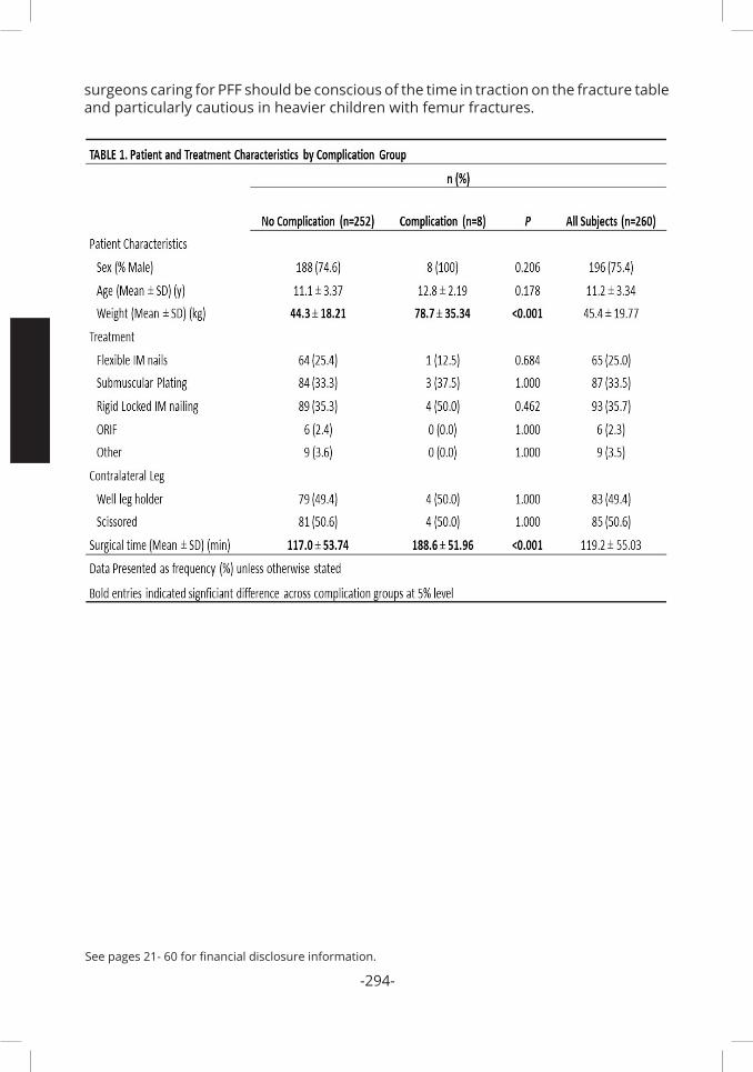
-294-
See pages 21- 60 for financial disclosure information.
surgeons caring for PFF should be conscious of the time in traction on the fracture table and particularly cautious in heavier children with femur fractures.
-295-
♦ Indicates those faculty presentations in which the FDA has not cleared the drug and/or medical device for the use described (i.e., the drug or medical device is being discussed for an “off label” use). For full information, refer to page 14.
Fri., 4/29/16 Trauma Subspecialty Day, Paper #136, 4:30 PM POSNA-2016 Biomechanical Analysis of Retrograde Flexible Intramedullary Nail Constructs in a Simulated Pediatric Femur Fracture Model
Daniel C. Bland, MD; Sheena R. Black, MD; William Pierce, BS Biomedical Engineering; R. Lane Wimberly, MD; Anthony I. Riccio, MDTexas Scottish Rite Hospital for Children, Dallas, Texas
LOE-Therapeutic-Level V
Purpose: To compare the rotational and bending stiffness of two flexible intramedullarynail constructs in a pediatric femur fracture model. Methods: Eighty adolescent-sized composite femurs were used to create transverse (40 femurs) and oblique (40 femurs) mid-diaphyseal fractures. Retrograde flexible intramedullary nailing (FIMN) of the femurs was performed using either 3.5mm titanium (Ti) flexible nails or 3.5mm stainless steel (SS) flexible nails. Each type of nail was inserted in one of two configurations: two “C” shaped nails placed through medial and lateral entry sites or one “C” shaped nail and one “S” shaped nail placed through a single lateral entry site.
Models were tested in 10 cycles of axial rotation to +/- 1N-m of torque at a rate of 0.5°/sec under 36kg of compression using a servo-electric material testing system. The models were then tested in axial compression to 10° of varus bending. Relative angulation between the proximal and distal fragments was monitored using an Inertial Measurement Unit with bending stiffness defined as the maximum force required to create 10° of varus at the fracture site.
Results: No differences were noted in rotational stiffness comparing Ti and SS nails regardless of nail configuration or fracture pattern. Comparable rotational stability was found for C+C and C+S configurations with SS implants for both fracture patterns. The C+S construct (0.6 n-M/degree) was stiffer in torsion than the C+C construct (0.4 n-M/degree) with Ti implants in the transverse fracture model (p=0.03).
The C+S construct demonstrated statistically significant superiority in bending stiffness regardless of implant material or fracture pattern. SS nails provided greater bending stiffness than Ti nails in both fracture patterns regardless of nail construct (Figure 1).
Conclusion: A C+S FIMN construct confers greater bending stiffness than the C+C construct, regardless of implant material for both transverse and oblique fractures. In the transverse fracture model, the C+S construct provided greater rotational stiffness for Ti nail fixation only. While Ti and SS implants conferred comparable rotational stiffness, SS nails were superior at resisting bending forces in both fracture patterns.
Significance: In the first biomechanical study to compare different retrograde FIMN constructs, the C+S nail configuration demonstrated superiority in resisting coronal plane deformation than the more commonly used C+C construct.
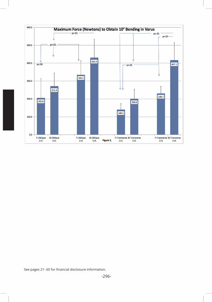
-296-
See pages 21- 60 for financial disclosure information.

-297-
♦ Indicates those faculty presentations in which the FDA has not cleared the drug and/or medical device for the use described (i.e., the drug or medical device is being discussed for an “off label” use). For full information, refer to page 14.
Fri., 4/29/16 Trauma Subspecialty Day, Paper #137, 4:35 PM POSNA-2016 Metaphyseal Fractures In Pediatric Patients Are Associated with Lower DXA Z-Scores
Stephanie Ihnow, MD; Rachel Mednick Thompson, MD; Mary J. Kwasny, PhD; Joseph (Jay) Janicki, MDAnn & Robert H. Lurie Children’s Hospital of Chicago, Chicago, Illinois
LOE-Prognostic-Level III
Purpose: Dual energy x-ray absorptiometry (DXA) is the gold standard for measuring bone density. Prospective studies have demonstrated that children with previous fractures have lower bone mass than those without fractures. However, these studies have not accounted for fracture location. Metaphyseal fractures should theoretically be more closely associated with, and perhaps reflective of, current bone density as measured by DXA, given its relatively young age when compared to diaphyseal bone. The purpose of this study was to compare DXA scores between children with and without fractures, and to evaluate the relationship between DXA and fracture location.
Methods: A prospective case-control study of patients aged 8 to 16 years was completed. DXA scores of children with fractures and without chronic diseases were compared to those of healthy controls without fractures. Patients were separated by fracture location into metaphyseal and non-metaphyseal cohorts. The cohorts were further compared for disparities in age, sex, race, medical history, family history of fracture or osteoporosis, nutrition and sun exposure.
Results: Of the 95 participants, there were 39 without fractures and 56 with fractures, of which 32 were metaphyseal. The control group was older (11.9 vs. 10.2, p<0.001) than the fractured cohort, and the fractured cohort was more likely to have had a history of previous fracture (21 vs. 2, p<0.001). No differences were seen between metaphyseal and non-metaphyseal cohorts in terms of demographic data. Fractured participants had significantly lower DXA z-scores than non-fractured participants (-0.50 ± 0.94 vs 0.21 ± 1.05, p<0.001). Lower DXA z-scores were associated with an increased fracture risk (OR 2.08 [95% CI, 1.32 to 3.33]; p=0.002) independent of age, gender, race, multivitamin use and sun exposure. Further, amongst the fractured cohort, subjects with metaphyseal fractures had significantly lower DXA z-scores compared to patients with non-metaphyseal fractures (-0.76 ± 0.95 vs -0.17 ± 0.84, p=0.017). Low DXA z-scores were associated with fractures in metaphyseal bone (OR 2.22 [95% CI, 1.12 to 4.34]; p=0.023) independent of age, gender and family history.
Conclusion: Children with fractures have lower DXA z-scores than those without fractures. Amongst children with fractures, those with metaphyseal fractures have significantly lower DXA z-scores than those with non-metaphyseal fractures.
Significance: Metaphyseal fractures are especially reflective of low bone mass density. Pediatric patients with metaphyseal fractures may be a target population for intervention
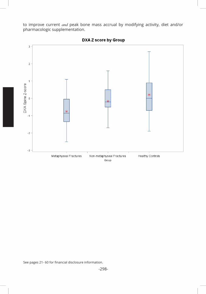
-298-
See pages 21- 60 for financial disclosure information.
to improve current and peak bone mass accrual by modifying activity, diet and/or pharmacologic supplementation.
-299-
♦ Indicates those faculty presentations in which the FDA has not cleared the drug and/or medical device for the use described (i.e., the drug or medical device is being discussed for an “off label” use). For full information, refer to page 14.
Fri., 4/29/16 Trauma Subspecialty Day, Paper #138, 4:50 PM POSNA-2016 Perfusion Assessment after Pediatric Supracondylar Humerus Fracture with Near Infrared Spectroscopy
Brian P. Scannell, MD; Brian K. Brighton, MD, MPH; Kelly L. Vanderhave, MD; Rachel Seymour, PhD; Steven L. Frick, MD Carolinas Medical Center, Charlotte, North Carolina
LOE-Diagnostic-Level III
Purpose: Near infrared spectroscopy (NIRS) measures the percentage of hemoglobin oxygen saturation in microcirculation. The purpose was to determine baseline perfusion values with NIRS in pediatric forearms distal to a supracondylar humerus fracture (SCHF) and compare this to uninjured forearms.
Methods: Over a 10 month period, consecutive patients with a SCHF needing operative fixation were approached for consent. Participants had NIRS pads placed on the injured and uninjured volar forearm. Data was continuously collected but blinded to the surgeon. Monitoring was performed pre- and post-operatively on each forearm. Distal pulses, Doppler signal, pulse oximetry measurements, motor/neurological function were recorded pre/post-operatively. Data was also collected on 20 controls without injuries.
Results: 71 patients with mean age of 6yo (range, 2–10) had complete data collected. There were 55 type III fractures, 10 type II fractures, and 6 type IV fracture. 8 patients with type III fractures did not have a palpable pulse at presentation (“perfused, pulseless”). 20 controls were slightly older, with a mean age of 7.2 years (range, 3-11).
Controls had a mean tissue oxygenation of 80% (range, 61-94). In the SCHF with a pulse (n=63), the mean tissue oxygenation during the entire study period was significantly higher in the injured forearm, 89.6%, compared to in the uninjured forearm, 82.6% (p<0.001). Preoperatively there was no difference between the injured (83.8%) and uninjured (82.7%) forearms. Postoperatively, the mean tissue oxygenation was significantly higher in the injured forearm, 89.2% vs 81.7% (p<0.01). In the injured side forearms, there was a significant increase in mean tissue oxygenation from pre- to post-operative (p=0.004). We found no correlation between oxygen saturation measured by pulse oximetry and NIRS monitoring of the volar forearm.
Data was collected on 8 “perfused, pulseless” patients. There was a decreased mean tissue oxygenation seen in the injured side forearms (72.4%) compared to the uninjured forearms (86.8%). The injured forearms pre-operatively had a mean tissue oxygenation of only 71.7%, but did improve to 82.4% post-operatively.
Conclusion: Children presenting with a SCHF and a palpable pulse had a significant increase in tissue oxygenation of the ipsilateral forearm measured by NIRS, as compared to the contralateral uninjured forearm. In children without a pulse, NIRS values were lower than controls preoperatively and did not reach the hyperemia levels seen in the palpable pulse group.
Significance: NIRS is an objective measurement of distal perfusion and can assess/monitor perfusion after SCHF in “perfused, pulseless” patients.
-300-
See pages 21- 60 for financial disclosure information.
-301-
♦ Indicates those faculty presentations in which the FDA has not cleared the drug and/or medical device for the use described (i.e., the drug or medical device is being discussed for an “off label” use). For full information, refer to page 14.
Fri., 4/29/16 Trauma Subspecialty Day, Paper #139, 4:55 PM POSNA-2016 Standardized Note Templates Improve Pre-Operative Neurovascular Screening Practices for Supracondylar Humerus Fractures: Results of a Quality Improvement Initiative
Jue Cao, MD; Maria Goodfellow, BA; Patrick Carry, BA; Francis A. Scott, MD; Travis C. Heare, MD; Nancy H. Miller, MDChildren’s Hospital Colorado, Aurora, Colorado
LOE-Therapeutic-Level III
Purpose: The importance of pre-operative neurovascular screening practices in the management of pediatric supracondylar humerus fractures is well documented. However, few studies have investigated interventions aimed at improving documentation practices. The purpose of this study is to compare the quality and completeness of pre-operative neurovascular examinations before and after the implementation of a standardized note template.
Methods: We performed a retrospective analysis of pediatric supracondylar humerus fractures (Type III&IV) that underwent operative treatment at our institution before (08/2009-08/2010, pre-template) and after implementation (08/2012-08/2013, template) of standardized note templates. Demographics (gender and age), clinical characteristics (types of reductions, presence of concomitant fractures, concomitant nerve or vascular injuries, and duration of follow-up), and completeness of pre-operative neurovascular documentation (vascular, motor, and sensory) were collected. Logistic regression analyses were used to compare completeness of neurovascular exams and the timing of nerve injury identification (pre-operatively versus intra-operatively/post-operatively) in the pre-template versus the template groups.
Results: A total of 225 and 298 subjects were included in the pre-template and template groups, respectively. There was a dramatic improvement in the documentation of vascular exams in the template group (85.9%) as compared to the pre-template group (8.1%). There was a significant improvement in documentation of the vascular [Odds Ratio (OR): 69.4, 95% CI: 38.8 to 124.2, p<0.0001], motor [OR: 16.9, 95% CI: 9.1 to 31.2, p<0.0001], and sensory [OR: 22.5, 95% CI: 12.2 to 41.6, p<0.0001] exams in the template group. Nerve injuries were more likely to be identified prior to surgery in the template group compared to the pre-template group [Odds Ratio: 2.9, 95% CI: 0.9 to 9.2, p = 0.0701]. However, vascular injuries were slightly less likely to be identified prior to surgery in the template group compared to the pre-template group [Odds Ratio: 0.9, 95% CI: 0.1 to 5.8, p <0.9999].
Conclusion: Template use in our facility significantly increased the detailed documentation of the neurovascular exams preoperatively during the management of pediatric supracondylar humerus fractures. A trend towards preoperative identification of nerve injury was noted following template implementation.
-302-
See pages 21- 60 for financial disclosure information.
Significance: EMR’s have the potential to improve patient care, ease communication among providers, and allow for better clinical research. However, the completeness, accuracy and quality of the data are concerns. Standardized templates can provide a consistent method to ensure more complete documentation during the assessment of specific diagnoses in which the notation of neurovascular problems may affect intervention and outcome.

-303-
♦ Indicates those faculty presentations in which the FDA has not cleared the drug and/or medical device for the use described (i.e., the drug or medical device is being discussed for an “off label” use). For full information, refer to page 14.
Fri., 4/29/16 Trauma Subspecialty Day, Paper #140, 5:00 PM POSNA-2016 Pulseless Supracondylar Humerus Fracture with AIN or Median Nerve Injury – An Absolute Indication for Open Reduction?
Liam R. Harris; Alexander M. Broom, BA; Alexandre Arkader, MD; John (Jack) M. Flynn, MD; Joseph L. Yellin, BA; Patrick W. Whitlock, MD; Ashley Miller, MD; David L. Skaggs, MD, MMM; Paul D. Choi, MD Children’s Hospital Los Angeles, Los Angeles, California
LOE-Therapeutic-Level IV
Purpose: Management of the pulseless supracondylar humerus fracture remains controversial. In particular, the combination of pulseless supracondylar humerus fracture and anterior interosseous nerve (AIN) or median nerve injury may have increased overall risk. The purpose of this study was to assess the necessity for open versus closed surgical management of pulseless supracondylar humerus fractures with concomitant anterior interosseous nerve (AIN) or median nerve injury in children.
Methods: A retrospective review was performed at three pediatric trauma hospitals on all children age 5-15 who sustained a Gartland type III or type IV supracondylar humerus fracture with the combination of absent distal palpable pulses and AIN or median nerve injury between 2000 and 2014. In addition to choice of treatment, details regarding preoperative and postoperative exam findings, follow-up course, and outcome were also recorded.
Results: 76 patients with displaced Gartland type III or type IV supracondylar humerus fractures presented with the combination of absent distal pulses and AIN or median nerve injury and met inclusion criteria. Twenty-one of 76 (27.6%) cases underwent open reduction, antecubital fossa exploration (OR) versus 55 (72.4%) that were treated with closed reduction and percutaneous fixation (CR). Indications for opening included concern for artery entrapment (n=11), inadequate closed reduction (n=9), and concern for nerve entrapment (n=6). The risk of compartment syndrome was higher in open cases (5/20, 25.0%) than closed cases (1/55, 1.8%) (p=0.001). The incidence of reoperation was also higher with open cases (4/20, 20%) than closed cases (2/55, 3.6%) (p=0.021). Open reduction was also significantly associated with increased time to surgery (18.7 hours ±31.1 vs 9.0 hours ±4.8, p=0.026) and length of hospitalization (4.0 days ±4.2 vs 2.0 days±1.6, p=0.004) compared to closed reduction. Overall, all but six (of 76, 7.9%) patients ultimately had complete resolution of preoperative nerve palsy with no significant difference in rate of clinical nerve recovery between the treatment groups (20/21 [95.2%] in OR, 50/55 [90.9%] in CR] (p=0.531).
Conclusion: Outcomes following open and closed surgical management of pulseless grade III or IV supracondylar humerus fracture with AIN or median nerve injury are ultimately both favorable and may suggest that open reduction is not always necessary.
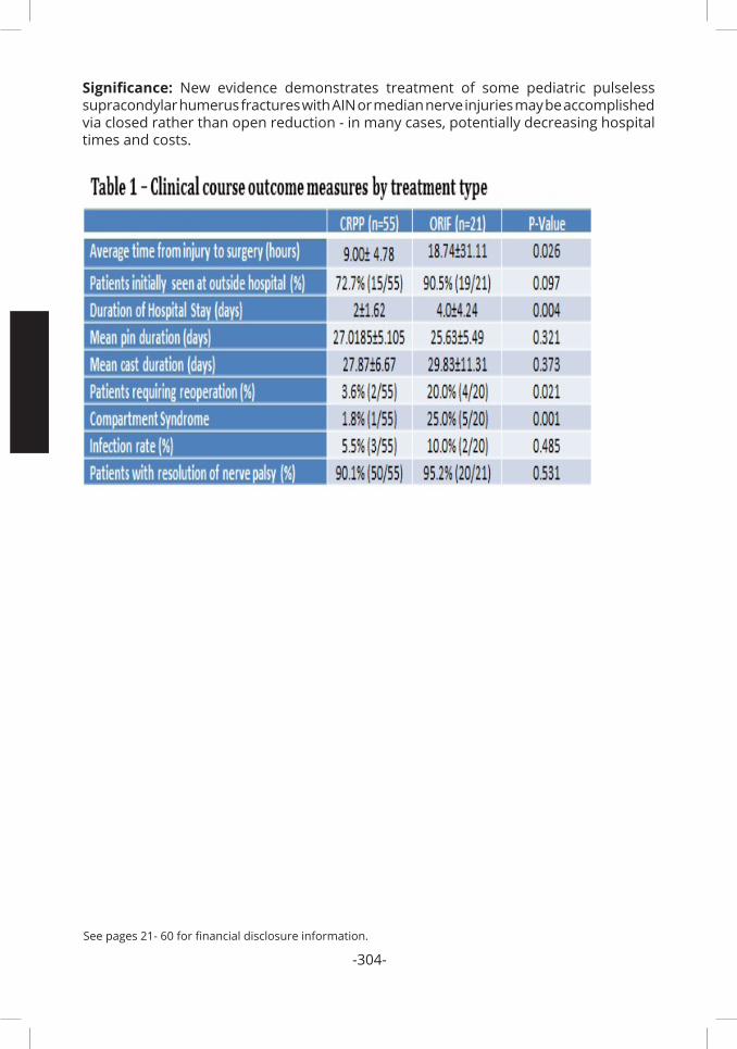
-304-
See pages 21- 60 for financial disclosure information.
Significance: New evidence demonstrates treatment of some pediatric pulseless supracondylar humerus fractures with AIN or median nerve injuries may be accomplished via closed rather than open reduction - in many cases, potentially decreasing hospital times and costs.
-305-
♦ Indicates those faculty presentations in which the FDA has not cleared the drug and/or medical device for the use described (i.e., the drug or medical device is being discussed for an “off label” use). For full information, refer to page 14.
Fri., 4/29/16 Trauma Subspecialty Day, Paper #141, 5:05 PM POSNA-2016 Treatment of Stable Pediatric Elbow Fractures with a Modified Long Arm Cast
Byron H. Izuka, MD; Krister P. Freese, MDKapiolani Medical Center for Women and Children, Honolulu, Hawaii
LOE-Therapeutic-Level IV
Purpose: Pediatric elbow fractures are among the most commonly encountered fractures for pediatric orthopedic surgeons. Approximately 10% of fractures in skeletally immature patients occur about the elbow. Many of these injuries are stable and do not require operative intervention. In our study patients were treated with a modified long arm cast that ends proximal to the ulnar styloid. The aim of our study is to determine if modified long arm casting of stable elbow fractures provides satisfactory clinical and radiographic outcomes.
Methods: A retrospective review of prospectively collected data was performed. Consecutive patients between September 2010 and September 2012 with stable elbow fractures treated with a gauntlet cast were included in the study. Charts were reviewed to determine demographic data, fracture type, mechanism of injury, time of follow up, duration of immobilization, complications from treatment and final clinical outcome.
Results: 125 patients were prospectively collected and no patients were lost to follow up. The average age of the patients was 5.7 years old. There were 67 males and 58 females. The most frequently encountered mechanisms of action were ground level falls and playground injuries. The most commonly treated fracture types were supracondylar humerus (62.9%), olecranon (13.7%), and radial neck (12.9%). The average time of immobilization was 26.5 days with an average follow up of 31.6 days. 122 of the patients had full elbow range of motion at final follow up. There were 4 minor complications. The most common complication was skin irritation adjacent to the ulnar styloid. The clinical outcome was satisfactory in all patients.
Conclusion: The treatment of stable pediatric elbow fractures with a modified long arm cast results in excellent clinical outcomes. In our experience this form of treatment leads to high patient and parental satisfaction. Modified long arm casts represent an efficient and patient friendly treatment option for commonly encountered fractures in this young and active population.
Significance: Value = outcomes / cost. Our approach increases value for our patients by increasing the numerator (less joint stiffness, less muscle weakness, improved function and improve hygiene) without increasing cost.
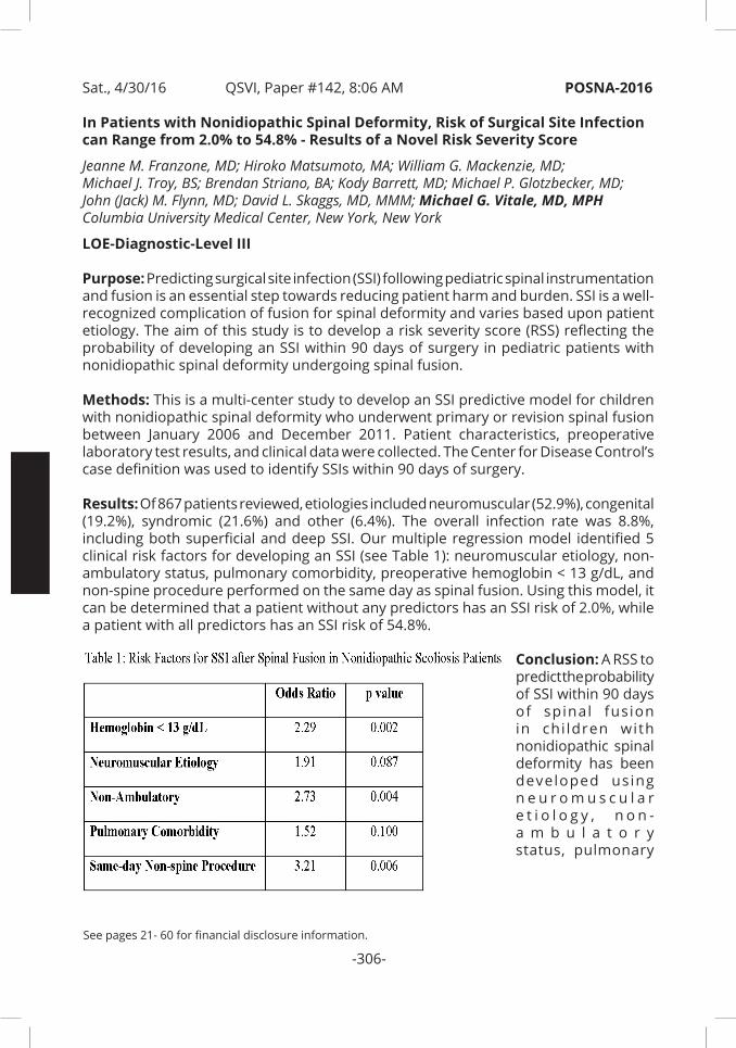
-306-
See pages 21- 60 for financial disclosure information.
Sat., 4/30/16 QSVI, Paper #142, 8:06 AM POSNA-2016 In Patients with Nonidiopathic Spinal Deformity, Risk of Surgical Site Infection can Range from 2.0% to 54.8% - Results of a Novel Risk Severity Score
Jeanne M. Franzone, MD; Hiroko Matsumoto, MA; William G. Mackenzie, MD; Michael J. Troy, BS; Brendan Striano, BA; Kody Barrett, MD; Michael P. Glotzbecker, MD; John (Jack) M. Flynn, MD; David L. Skaggs, MD, MMM; Michael G. Vitale, MD, MPH Columbia University Medical Center, New York, New York
LOE-Diagnostic-Level III
Purpose: Predicting surgical site infection (SSI) following pediatric spinal instrumentation and fusion is an essential step towards reducing patient harm and burden. SSI is a well-recognized complication of fusion for spinal deformity and varies based upon patient etiology. The aim of this study is to develop a risk severity score (RSS) reflecting the probability of developing an SSI within 90 days of surgery in pediatric patients with nonidiopathic spinal deformity undergoing spinal fusion.
Methods: This is a multi-center study to develop an SSI predictive model for children with nonidiopathic spinal deformity who underwent primary or revision spinal fusion between January 2006 and December 2011. Patient characteristics, preoperative laboratory test results, and clinical data were collected. The Center for Disease Control’s case definition was used to identify SSIs within 90 days of surgery.
Results: Of 867 patients reviewed, etiologies included neuromuscular (52.9%), congenital (19.2%), syndromic (21.6%) and other (6.4%). The overall infection rate was 8.8%, including both superficial and deep SSI. Our multiple regression model identified 5 clinical risk factors for developing an SSI (see Table 1): neuromuscular etiology, non-ambulatory status, pulmonary comorbidity, preoperative hemoglobin < 13 g/dL, and non-spine procedure performed on the same day as spinal fusion. Using this model, it can be determined that a patient without any predictors has an SSI risk of 2.0%, while a patient with all predictors has an SSI risk of 54.8%.
Conclusion: A RSS to predict the probability of SSI within 90 days of spinal fusion in chi ldren with nonidiopathic spinal deformity has been developed using n e u r o m u s c u l a r e t i o l o g y , n o n -a m b u l a t o r y status, pulmonary
-307-
♦ Indicates those faculty presentations in which the FDA has not cleared the drug and/or medical device for the use described (i.e., the drug or medical device is being discussed for an “off label” use). For full information, refer to page 14.
comorbidity, preoperative hemoglobin, and the performance of a non-spine procedure on the day of spinal fusion.
Significance: This RSS will be useful to surgeons when considering operative intervention in children with nonidiopathic scoliosis, and will improve shared decision making with patients and families during preoperative counseling. As healthcare shifts from volume to value, risk stratification using the RSS will be essential to improving health outcomes. It will also aid policy makers and administrators in determining fair, reliable, and valid risk-adjusted outcome measures.
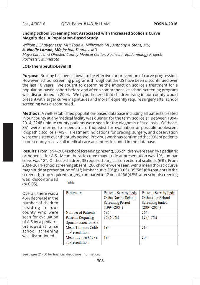
-308-
See pages 21- 60 for financial disclosure information.
Sat., 4/30/16 QSVI, Paper #143, 8:11 AM POSNA-2016 Ending School Screening Not Associated with Increased Scoliosis Curve Magnitudes: A Population-Based Study
William J. Shaughnessy, MD; Todd A. Milbrandt, MD; Anthony A. Stans, MD; A. Noelle Larson, MD; Joshua Thomas, MD Mayo Clinic and Olmsted County Medical Center, Rochester Epidemiology Project, Rochester, Minnesota
LOE-Therapeutic-Level III
Purpose: Bracing has been shown to be effective for prevention of curve progression. However, school screening programs throughout the US have been discontinued over the last 10 years. We sought to determine the impact on scoliosis treatment for a population-based cohort before and after a comprehensive school screening program was discontinued in 2004. We hypothesized that children living in our county would present with larger curve magnitudes and more frequently require surgery after school screening was discontinued.
Methods: A well-established population-based database including all patients treated in our county at any medical facility was queried for the term ‘scoliosis.’ Between 1994-2014, 2248 unique county patients were seen for the diagnosis of ‘scoliosis’. Of those, 851 were referred to a pediatric orthopedist for evaluation of possible adolescent idiopathic scoliosis (AIS). Treatment indications for bracing, surgery, and observation were consistent over the study period. Previous work has confirmed that 99% of patients in our county receive all medical care at centers included in the database.
Results: From 1994-2004 (school screening present), 585 children were seen by a pediatric orthopedist for AIS. Mean thoracic curve magnitude at presentation was 19°; lumbar curve was 18°. Of those children, 35 required surgical correction of scoliosis (6%). From 2004- 2014 (school screening absent), 266 children were seen, with a mean thoracic curve magnitude at presentation of 21°; lumbar curve 20° (p>0.05). 35/585 (6%) patients in the screened group required surgery, compared to 12 out of 266 (4.5%) after school screening was discontinued (p>0.05). Overall, there was a 45% decrease in the number of children residing in our county who were seen for evaluation of AIS by a pediatric orthopedist once school screening was discontinued.
-309-
♦ Indicates those faculty presentations in which the FDA has not cleared the drug and/or medical device for the use described (i.e., the drug or medical device is being discussed for an “off label” use). For full information, refer to page 14.
Conclusion: There was no difference in curve magnitude at presentation, rates of bracing, or rates of surgery after school screening was discontinued in our population-based cohort.
Significance: In a population-based cohort, cessation of school screening did not result in more severe disease at presentation or a higher rate of surgical treatment for AIS patients. Cessation of school screening led to a nearly 50% reduction in both total referrals and those requiring treatment. It appears that referral mechanisms in the absence of screening are less sensitive and no more accurate than the screening programs themselves.
-310-
See pages 21- 60 for financial disclosure information.
Sat., 4/30/16 QSVI, Paper #144, 8:16 AM POSNA-2016 Determining the Prevalence and Costs of Unnecessary Referrals in Adolescent Idiopathic Scoliosis
Thomas M. Meirick, BA; Apurva Shah, MD, MBA; Lori Dolan, PhD; Stuart L. Weinstein, MDUniversity of Iowa, Iowa City, Iowa
LOE-Economic & Decision-Level IV
Purpose: Adolescent idiopathic scoliosis (AIS) is a condition that has been associated with unnecessary referrals to pediatric orthopaedic surgeons, but the provider and patient costs associated with these referrals remain unknown. The purpose of this study was to determine the prevalence of unnecessary AIS referrals as well as the associated costs.
Methods: We retrospectively reviewed the electronic medical records of all patients with suspected AIS referred to a single tertiary care medical center during 2013 and 2014. Spine radiographs were reviewed to determine whether the referral was medically “unnecessary,” defined by a Cobb angle <20 degrees. The average patient and provider costs for unnecessary referrals were then calculated using supplemental information from a prospective cross-sectional survey of 24 patients and families (none declined participation). Provider costs included orthopaedic evaluation and diagnostic imaging. Patient costs included transportation expenses and lost wages. The cost of orthopaedic consultation was calculated with time-driven activity-based costing (TDABC). The cost of diagnostic imaging was based on a relative value unit costing methodology. Transportation costs were based on roundtrip travel distance calculated with an online mapping program and standard mileage rates used by the Internal Revenue Service. Lost wages were based on an estimate of yearly income provided by the U.S. Census Bureau accounting for parental sex, educational attainment, and county of residence.
Results: 337 patients presented for initial evaluation of AIS during the study period. 16.6% had a Cobb angle <10 degrees and 38.9% had a Cobb angle <20 degrees. Neither insurance status nor race affected the rate of unnecessary referrals. The mean total cost of an unnecessary referral was $779.92: $231.07 in patient costs and $548.85 in provider costs. Patient transportation costs averaged $98.34 (range $1.34-$335.61) and lost wages averaged $132.73 ($34.26-$512.31). The average cost of orthopaedic evaluation was estimated at $309.85 including provider time and clinic overhead. The average cost of unnecessary diagnostic imaging was $239.00.
Conclusion: Nearly half of all referrals for AIS are medically unnecessary. The average cost of an unnecessary referral is approximately $780, imposing significant costs on both patients and the healthcare system.
Significance: Our study is the first to examine the costs associated with unnecessary referrals for AIS using TDABC and primary data, in contrast to prior investigations that report charges or reimbursements. This type of actual cost data is essential to improving health care value delivery and to appropriately reexamine scoliosis screening guidelines.
-311-
♦ Indicates those faculty presentations in which the FDA has not cleared the drug and/or medical device for the use described (i.e., the drug or medical device is being discussed for an “off label” use). For full information, refer to page 14.
Sat., 4/30/16 QSVI, Paper #145, 8:30 AM POSNA-2016 Issues Affecting the Practice of Pediatric Orthopaedic Surgery: Results of the 2014 Workforce Survey of American Academy of Pediatrics Section on Orthopaedics and POSNA
Pooya Hosseinzadeh, MD; Lawson A.B. Copley, MD; Richard M. Schwend, MD; Jeffrey R. Sawyer, MDUniversity of Tennessee-Campbell Clinic, Memphis, Tennessee
LOE-Not Applicable
Purpose: The opinions of the pediatric orthopaedic workforce are shaped by market forces, regulatory processes, and individual experience. This report summarizes the findings of the recent Workforce Survey of the American Academy of Pediatrics (AAP) Section on Orthopaedics (SOOr) and POSNA.
Methods: In 2014, the AAP Section on Orthopaedics and POSNA generated a 108 item survey to assess perceptions of pediatric orthopaedic surgeons about current issues that affect practice. The survey was electronically sent to 856 POSNA and 141 AAP-SOOr members. Responses were topically organized to report current workforce composition, practice patterns, and perceptions about electronic medical record.
Results: Responses were collected from August to December, 2014 from 496 (49.7%) survey recipients including 83 of 141 (58.9%) AAP-SOOr members and 413 of 856 (48.2%) POSNA members. Males comprised 77.0% and females 23.0% of those surveyed. Nearly all participants (429 of 437, 98.2%) indicated that they provide direct patient care and work an average of 58.8 hours per week. Many (204 of 496, 41.1%) indicated that they would soon limit practice or retire. A majority (313 of 416, 75.2%) indicated that there was increased competition in their geographic area, predominantly due to pediatric orthopaedic subspecialists (281 of 313, 89.8%). There were 118 (23.8%) survey participants who reported plans to hire a new practice associate. Respondents expressed dissatisfaction with electronic medical records (EMR) due to inefficiency (258 of 496, 52%) and interference with the patient-physician relationship (178 of 496, 35.9%).
Conclusion: Compared to the 1998 AAP survey of pediatric orthopedic surgeons, the demographics of the pediatric orthopedic workforce have changed with more female pediatric orthopedic surgeons in practice. Primary practice location and hours worked per week have not changed. There is an increased perception of local competition among pediatric orthopedic surgeons. Despite concerns of competition there continues to be increasing work volume and complexity. External processes such as the evolution of the EMR are perceived to negatively impact practice efficiency and satisfaction.
Significance: This survey provides new information about the pediatric orthopedic workforce in the United States.
-312-
See pages 21- 60 for financial disclosure information.
Sat., 4/30/16 QSVI, Paper #146, 8:35 AM POSNA-2016 Should Obligatory Call Schedules Become a Thing of the Past? The 2015 POSNA Membership Survey Regarding Trauma Care
Kevin H. Latz, MD; Mark R. Sinclair, MD; Allison Lind, RN, MN, MPH, CPNPChildren’s Mercy Hospital, Kansas City, Missouri
LOE-Not Applicable
Purpose: To examine the current trends in trauma call coverage of pediatric orthopaedic surgeons in North America and to identify predictors of surgeon on-call satisfaction.
Methods: In 2015 the approximately 1200 active members of the Pediatric Orthopaedic Society of North America (POSNA) were surveyed regarding emergency room on-call practices. 410 members completed the survey with a response rate of 35%. Information collected included call frequency, practice setting, satisfaction with call and comfort level with pediatric trauma. This information was analyzed and compared to the 2006 and 2010 trauma call surveys of POSNA membership using two sample difference in proportion tests. Logistic regression analyses were used to identify predictors of call satisfaction amongst pediatric orthopaedic surgeons.
Results: 53% of pediatric orthopaedic surgeons had access to a designated trauma operating room in 2015, up from 39% in 2010 and 24% in 2006. 55% of pediatric orthopaedic surgeons currently receive a stipend for taking call, up from 35% in 2010 and 28% in 2006. While 83% of pediatric orthopaedic surgeons believe that trauma call is an integral part of their practice, only 53% are satisfied with their call experience. Individual attitude towards the importance of trauma call is the greatest predictor of call satisfaction (OR: 3.8, p=0.002). The odds of surgeon satisfaction for those who do not cover call at more than one campus is 2.5 times greater than the odds of those who cover calls at multiple campuses (p=0.001). Moreover, having access to a designated trauma room increases the odds of satisfaction by 40% (p=0.005). The odds of being satisfied in one’s call decrease by 3% for every year in practice (p=0.012) and also decrease by 3% with each increasing year of age of the surgeon (p=0.019). Having resident or fellow support, access to a vascular surgeon, hand call, and working at a pediatric level 1 trauma center also increased one’s odds of call satisfaction.
Conclusion: Access to a designated trauma operating room and financial compensation for call coverage have steadily increased over the past decade. A sizable majority of respondents continue to feel that trauma care is an integral part of being a pediatric orthopedist. Despite this, 47% of respondents remain dissatisfied with their trauma call arrangements. The age and attitude of the individual surgeon, along with logistic hospital support, predict satisfaction of surgeons providing trauma coverage.
Significance: Obligatory call schedules may place aging surgeons in increasingly dissatisfying circumstances. Movement towards funded call coverage systems involving willing participants with reserved operating room availability should be encouraged.
-313-
♦ Indicates those faculty presentations in which the FDA has not cleared the drug and/or medical device for the use described (i.e., the drug or medical device is being discussed for an “off label” use). For full information, refer to page 14.
Sat., 4/30/16 QSVI, Paper #147, 8:40 AM POSNA-2016 Subspecialty Training Among Recent Graduates of Pediatric Orthopedic Fellowships
Pooya Hosseinzadeh, MD; Michael P. Glotzbecker, MD; Jeffrey R. Sawyer, MD Baptist Children’s Hospital, Miami, Florida
LOE-Not Applicable
Purpose: A recent survey of pediatric orthopedic fellowship graduates has shown that close to 30% of graduates seek additional subspecialty training. Although additional post fellowship training can enrich the experience and qualifications of the trainee it also is associated with additional financial and personal burden. It is not currently known why close to one third of pediatric orthopedic fellowship graduated feel the necessity for additional training. We surveyed the recent graduates of pediatric orthopedic fellowships with additional subspecialty training to study the reasons behind this new trend.
Methods: A 15 question survey was approved by the POSNA Practice Management Committee. 24 pediatric orthopedic fellowship graduates had indicated on a prior survey that they had additional subspecialty training. The survey was sent via email to those 24 fellowship graduates with two follow up email reminders.
Results: 15 out of 24 recipients (62%) completed the survey. Sports medicine and spine surgery fellowships were the two most common (33% and 26%, respectively) subspecialty fellowship trainings reported by the survey participants. The majority of the participants (73%) indicated that they spent a full 12 months for the subspecialty training. 73% of the participants indicated that they treated both adults and children during their subspecialty training. Personal interest was the most commonly reported as the reason for pursuing subspecialty training by (80%). However 33% of the participants cited a competitive job market as rationale for pursuing additional training. Close to half (xx%) of the respondents indicated that more than half of their current practice is focused on their subspecialty training, 73% believed that the additional training helped them in getting their current job and overwhelming majority (93%) reported that they believed the additional subspecialty training was worth the time and effort.
Conclusion: Sports medicine and spine surgery are the two most common subspecialty trainings sought by the recent graduates of pediatric orthopedic fellowships. Personal interest and the presence of competitive job market are the two reported main reasons behind seeking subspecialty training. An overwhelming majority believe that the additional training was worth the time and effort.
Significance: This is the first study to evaluate the pattern and the causes of subspecialty trainings sought by one third of recent graduates of pediatric orthopedic fellowships.
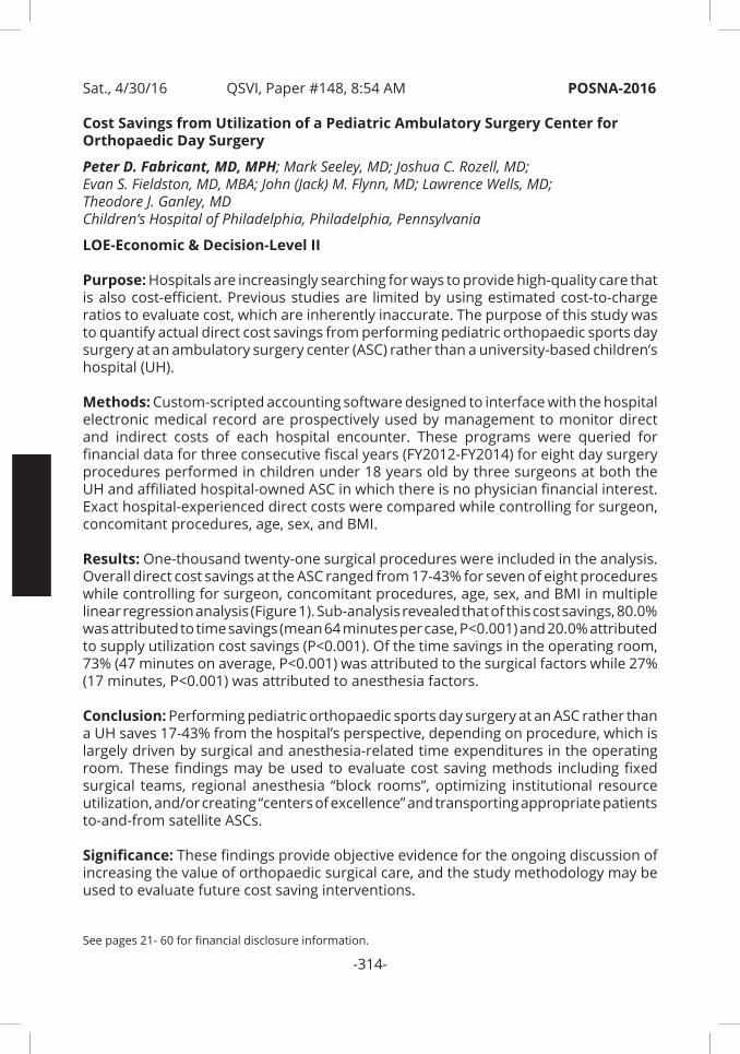
-314-
See pages 21- 60 for financial disclosure information.
Sat., 4/30/16 QSVI, Paper #148, 8:54 AM POSNA-2016 Cost Savings from Utilization of a Pediatric Ambulatory Surgery Center for Orthopaedic Day Surgery
Peter D. Fabricant, MD, MPH; Mark Seeley, MD; Joshua C. Rozell, MD; Evan S. Fieldston, MD, MBA; John (Jack) M. Flynn, MD; Lawrence Wells, MD; Theodore J. Ganley, MD Children’s Hospital of Philadelphia, Philadelphia, Pennsylvania
LOE-Economic & Decision-Level II
Purpose: Hospitals are increasingly searching for ways to provide high-quality care that is also cost-efficient. Previous studies are limited by using estimated cost-to-charge ratios to evaluate cost, which are inherently inaccurate. The purpose of this study was to quantify actual direct cost savings from performing pediatric orthopaedic sports day surgery at an ambulatory surgery center (ASC) rather than a university-based children’s hospital (UH).
Methods: Custom-scripted accounting software designed to interface with the hospital electronic medical record are prospectively used by management to monitor direct and indirect costs of each hospital encounter. These programs were queried for financial data for three consecutive fiscal years (FY2012-FY2014) for eight day surgery procedures performed in children under 18 years old by three surgeons at both the UH and affiliated hospital-owned ASC in which there is no physician financial interest. Exact hospital-experienced direct costs were compared while controlling for surgeon, concomitant procedures, age, sex, and BMI.
Results: One-thousand twenty-one surgical procedures were included in the analysis. Overall direct cost savings at the ASC ranged from 17-43% for seven of eight procedures while controlling for surgeon, concomitant procedures, age, sex, and BMI in multiple linear regression analysis (Figure 1). Sub-analysis revealed that of this cost savings, 80.0% was attributed to time savings (mean 64 minutes per case, P<0.001) and 20.0% attributed to supply utilization cost savings (P<0.001). Of the time savings in the operating room, 73% (47 minutes on average, P<0.001) was attributed to the surgical factors while 27% (17 minutes, P<0.001) was attributed to anesthesia factors.
Conclusion: Performing pediatric orthopaedic sports day surgery at an ASC rather than a UH saves 17-43% from the hospital’s perspective, depending on procedure, which is largely driven by surgical and anesthesia-related time expenditures in the operating room. These findings may be used to evaluate cost saving methods including fixed surgical teams, regional anesthesia “block rooms”, optimizing institutional resource utilization, and/or creating “centers of excellence” and transporting appropriate patients to-and-from satellite ASCs.
Significance: These findings provide objective evidence for the ongoing discussion of increasing the value of orthopaedic surgical care, and the study methodology may be used to evaluate future cost saving interventions.
-315-
♦ Indicates those faculty presentations in which the FDA has not cleared the drug and/or medical device for the use described (i.e., the drug or medical device is being discussed for an “off label” use). For full information, refer to page 14.
-316-
See pages 21- 60 for financial disclosure information.
Sat., 4/30/16 QSVI, Paper #149, 8:59 AM POSNA-2016 A Dedicated Fracture Reduction Room: A Cost Effective Alternative to the Operating Room
Patrick W. Whitlock, MD; Kevin J. Little, MD; Sandra Singleton, MBA; Mohamed Mahmoud; Pornswan Ngamprasertwong, MD, MS; James J. McCarthy, MD, MHCMCincinnati Children’s Hospital, Cincinnati, Ohio
LOE-Economic & Decision-Level IV
Purpose: This study compared cost, return to activity, and complications after closed treatment of fractures performed in an operating room (OR) versus closed reduction in a dedicated fracture reduction room (FRR). Our hypothesis was that there would be significantly lower cost associated with treatment in the FRR without additional increase in complications or time to return to full activity.
Methods: Patients who presented to our pediatric orthopaedic clinic with closed fractures of the radius, ulna or both necessitating closed reduction were retrospectively reviewed. Patients who presented in 2014 and returned for their last scheduled follow-up appointment were included. Total cost, American Society of Anesthesiologists (ASA) class, anesthesia and orthopaedic complications; time to return to full activity, sex, age and length of follow-up were recorded.
Results: 21 patients met inclusion in the FRR group (16 males, 5 females), 22 patients in the OR group (18 males, 4 females). Mean patient age in the FRR group was 7.3 ± 2.8 years, in the OR group 7.6 ± 4.2 years, p>0.05. Patients treated in the FRR incurred charges of $5,337 ± $1203 vs. $10,455 ± $2290 in the OR (p<0.001). FRR patients had a mean return to activity of 7.3 ± 2.1 weeks vs. 6.7 ± 2.3 weeks in the OR (p>0.05). FRR patients had a mean length of follow-up of 11.9 ± 15.8 weeks vs. 16.7 ± 14.7 weeks in the OR (p>0.05). There were two (9.5%) complications in the FRR group (1 cast pain, 1 refracture at 6 months) vs. 11 (50%) complications in the OR group by three predominant mechanisms (1. Noncompliance/Accidental: patient removed cast, foreign body in cast, refracture at 2 months, fall in cast with laceration necessitating repair, wet cast, new cortical defect, fall with 1st metacarpal base fracture; 2. Initial Injury: growth arrest necessitating epiphysiodesis and TFCC repair; 3. Loss of Reduction: necessitated internal fixation, remolding of cast). ASA classes in the FRR group were 1 (16) and 2 (5), and 1 (13) and 2 (9) in the OR group. There were no anesthesia complications in either group.
Conclusion: The implementation of a FRR resulted in a significant reduction in cost without increased complications or time to return to full activity.
Significance: Implementation of a dedicated FRR is safe, cost –effective and may aid in decreasing OR utilization and increasing convenience for families.
-317-
♦ Indicates those faculty presentations in which the FDA has not cleared the drug and/or medical device for the use described (i.e., the drug or medical device is being discussed for an “off label” use). For full information, refer to page 14.
Sat., 4/30/16 QSVI, Paper #150, 9:04 AM POSNA-2016 Life-long Learning or Just Assessing Clinical Documentation? Efficacy of the Supracondylar Fracture Performance Improvement Module
Tracey P. Bastrom, MA; John W. Kemppainen, MD; Joanna H. Roocroft, MA; John Munch, MPA; Eric W. Edmonds, MDRady Children’s Hospital, San Diego, California
LOE-Not Applicable
Purpose: Maintenance of certification (MOC) within a medical society requires continuing medical education that demonstrates life-long learning, cognitive expertise, and practice-based self-assessment. This study assesses the Practice Improvement Module (PIM) system to enhance outcomes (Part II MOC credit: evidence of life-long learning) and/or quality improvement processes (Part IV MOC credit: evidence of practice–based self-assessment).
Methods: A prospective study of supracondylar fractures operatively treated at a single institution was performed. Fractures treated by both junior (<2 years experience) and experienced attending staff were enrolled. After approximately 10 patients were enrolled, surgeons underwent education based on the references included within the PIM Option #1, and then a second set of patients were enrolled. Each child’s notes were tracked by research staff and audited for documentation in accordance with the PIM. Pre- to post- PIM education data was compared for the entire cohort and by level of experience (junior versus experienced).
Results: Six surgeons (4 junior, 2 experienced) and 113 patients were included. Junior staff demonstrated a statistically significant improvement in the percentage of time the operative site was signed and documented in the chart from pre (38%) to post (65%) PIM (p=0.02). They also demonstrated a non-significant increase in the documentation of limb perfusion prior to discharge (35% pre, 66% post, p=0.10). There was no significant difference in actual fracture outcome pre- or post- PIM at any level of surgeon experience regarding radiographic appearance or need for re-operation (p>0.10).
Conclusion: At a level 1 academic training institution, the supracondylar fracture PIM was related more to improved documentation habits among junior staff only, without impact on overall outcomes. Frequent discussion and evaluation of surgical technique in a training environment may explain the lack of relationship between the education portion of the PIM and clinical/radiographic outcomes in this study. Future study on the efficacy of the PIM at impacting outcomes at a non-academic center may be valuable.
Significance: Since no improvement in outcomes were generated, the PIM system may not be useful in providing evidence for life-long learning as it relates to surgical outcomes (Part II MOC). However, the PIM system does appear to provide evidence for efficacy of practice-based self-assessment since it directly records a surgeons documentation habits real-time for outside MOC committee review. Reform in the MOC to utilize the PIM system for Part IV credit may be warranted.
-318-
See pages 21- 60 for financial disclosure information.
Sat., 4/30/16 QSVI, Paper #151, 9:18 AM POSNA-2016 Significant Variation in Blood Transfusion Practice Persists Following Idiopathic Adolescent Scoliosis Surgery
Natasha O’Malley, FRCS (Tr & Orth); Sandra M. O’Malley; Fergal J. Fleming, FRCS; Katia Noyes, PhD, MPH; Christopher T. Aquina, MD; Paul T. Rubery, MDUniversity of Rochester Medical Center, Rochester, New York
LOE-Therapeutic-Level III
Purpose: Perioperative blood transfusions are costly and linked to adverse clinical outcomes. We investigated the factors associated with variation in blood transfusion utilization following primary spinal fusion for idiopathic adolescent scoliosis (IAS) and its association with infectious complications.
Methods: Data was extracted from the Statewide Planning and Research Cooperative System (SPARCS), a hospital discharge database for New York State. Using International Classification of Diseases (ICD-9), all patients included had a diagnosis of IAS and underwent spinal fusion from 2000 to 2013. Individual surgeon and facility volume were calculated as the number of spinal fusions performed per year using the unique surgeon identifier and unique facility identifier numbers. Bivariate and mixed-effects logistic regression analyses were performed to assess the factors associated with receiving a perioperative allogeneic red blood cell transfusion. Additional multivariable analysis examined the relationship between transfusion and infectious complications.
Results: 6,230 patients underwent IAS surgery, of whom 4,517 were female (73%). A posterior approach was undertaken in 83% of cases, whereas an anterior approach was performed in 8.3% of cases. 27.77 % of patients received a perioperative blood transfusion. After controlling for patient, surgeon, and hospital-level factors, significant variation in transfusion rates was present across both surgeons and hospitals with a 8-fold difference observed in adjusted transfusion rates between the lowest and highest utilization for hospitals and a 4-fold difference observed for surgeons (p < 0.0001). Blood transfusion was also independently associated with wound infection (OR = 2.18, 95% CI = 1.16 and 4.10), and pneumonia (OR = 1.77, 95% CI = 1.24 and 2.51).
Conclusion: Significant variation in perioperative blood transfusion utilization exists at both the surgeon and hospital level. These findings are unexplained by patient-level factors and other known surgeon and hospital characteristics.
Significance: The data suggests that variation is due to provider preferences and/or lack of standardized transfusion protocols. Implementing institutional transfusion guidelines is necessary to reduce unwarranted variation and possibly decrease infectious complication rates in idopathic adolescent scoliosis surgery.
-319-
♦ Indicates those faculty presentations in which the FDA has not cleared the drug and/or medical device for the use described (i.e., the drug or medical device is being discussed for an “off label” use). For full information, refer to page 14.
Sat., 4/30/16 QSVI, Paper #152, 9:23 AM POSNA-2016 Variation in Practice Patterns and Resource Utilization for Spinal Fusion Procedures Across Children’s Hospitals
Brian K. Brighton, MD, MPH; Debra A. Liebrecht, RN, CPHQ; CarolAnn H. Gelder, RN, MSN; Jacqueline M. Saito, MD; Douglas C. Barnhart, MD, MSPH; Shawn J. Rangel, MD, MSCE Levine Children’s Hospital, Charlotte, North Carolina
LOE-Therapeutic-Level II
Purpose: Spinal fusion procedures for spinal deformity are one of the most costly procedures done at children’s hospitals across the country. Variation in care is one key component to the treatment-related costs of spinal fusion procedures. The purpose of the study was to examine the variation in practice patterns and resource utilization associated with spinal fusion procedures at ACS National Surgical Quality Improvement Program-Pediatric (NSQIP-Pediatric) participating hospitals.
Methods: Procedure targeted custom field variables were developed within the NSQIP-Pediatric Program. In addition to the traditional variables, data was collected specifically related to spinal fusion procedures including etiology of spinal deformity, pre-operative MRI utilization, intraoperative use of neuromonitioring, antifibrinolyitics, adjunct antibiotics in the wound or bone graft, and post-operative ICU utilization. Rates of utilization were calculated for each hospital and compared to the aggregate cohort data. A hospital was then defined as an “outlier” if its rate and 95% binomial proportion confidence interval for a given metric did not include the aggregate rate of the entire cohort.
Results: 43 participating hospitals contributed 1594 cases to the NSQIP-Pediatric Spinal Fusion Pilot in 2014. Variation was seen in all measures including preoperative imaging with MRI (aggregate rate 31.9%; range across hospitals: 0-100%), intraoperative use of neuromonitoring (aggregate rate 95.1%; range 77.5-100%), antifibrinolytic use (aggregate rate 72.5%; range 0-100%), and adjunct antibiotic use (aggregate rate 73.6%; range 0-100%). The aggregate cohort rate for ICU utilization was 43.1% (range across hospitals: 20-100%) and varied by diagnosis with idiopathic scoliosis patients utilizing the ICU at a rate of 31.1% (range across hospitals 0-100%) and neuromuscular patients having a 84.7% utilization rate (range 20-100%).
Conclusion: There is significant variation in practice patterns and resource utilization associated with spinal fusion procedures across children’s hospitals nationally.
Significance: Measures relating to best practice and resource utilization for spinal fusion procedures allow for national benchmarking, comparative performance reporting and enhanced collaborative quality improvement efforts by physicians and hospitals.
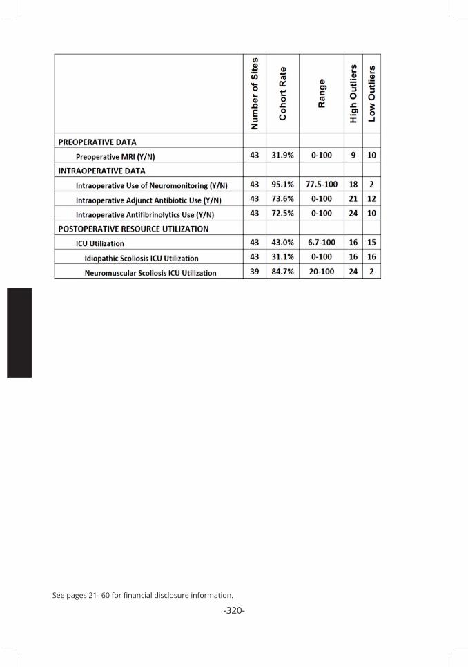
-320-
See pages 21- 60 for financial disclosure information.
-321-
♦ Indicates those faculty presentations in which the FDA has not cleared the drug and/or medical device for the use described (i.e., the drug or medical device is being discussed for an “off label” use). For full information, refer to page 14.
Sat., 4/30/16 QSVI, Paper #153, 9:28 AM POSNA-2016 Meta-Analysis and Economic Decision Model for First-Time Traumatic Patella Dislocation in Children & Adolescents
Benedict Nwachukwu, MD; Conan So, BA; William W. Schairer, MD; Daniel W. Green, MD; Emily Dodwell, MD Hospital for Special Surgery, New York, New York
LOE-Economic & Decision-Level II
Purpose: Surgical versus non-surgical management of first time traumatic patella dislocation in children and adolescents remains controversial with a potentially higher rate of treatment failure in this population. The purpose of this study was 1) to perform a comparative analysis to determine the rate of repeat dislocation and clinical outcome between operative and non-operative management of first time traumatic patellar dislocation in children and adolescents and 2) to use these analyses to inform an economic decision model comparing the cost-effectiveness of: A) non-operative treatment only; B) initial non-operative management with surgery for recurrent dislocation and C) immediate surgery. Methods: A systematic review of the MEDLINE database identified 11 eligible studies. Meta-analysis was performed using weighted mean pooled cohort statistics and these results informed a markov decision analysis. Cost analysis for the model was performed from a societal perspective using both direct and indirect costs. Surgical cost was obtained from a statewide ambulatory surgery database and indirect costs from anticipated caregiver (parental) lost productivity (work absenteeism). Effectiveness was expressed in quality adjusted life year (QALY) estimates derived from the literature.
Results: 637 knees were included in our meta-analysis; 470 non-operative and 157 operative. Conservative management was associated with a 31% rate of recurrent dislocation whereas operative management had a 22% recurrence rate (p=0.04). Compared to non-operative management, immediate surgical treatment was associated with a significant minimal clinically significant difference in quality of life and sporting domains. In the base case for our model, the non-operative treatment only strategy was the least costly ($7,670) but also the least effective (6.72 QALY); initial non-operative management with delayed surgery cost $11,010 for a 7.01 QALY benefit while immediate surgical treatment cost $17,690 and provided 7.10 QALY benefits. Compared to non-operative treatment only, initial non-operative treatment with delayed surgery was associated with an ICER of $11,150/QALY. When immediate surgery was compared to a strategy of delayed surgery, immediate surgery provided incremental benefit at an ICER of $75,150/QALY.
Conclusion: Operative treatment of first time patella dislocation in children and adolescents is associated with a lower risk of recurrent dislocation and higher health related quality of life and sporting function.
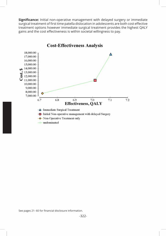
-322-
See pages 21- 60 for financial disclosure information.
Significance: Initial non-operative management with delayed surgery or immediate surgical treatment of first time patella dislocation in adolescents are both cost effective treatment options however immediate surgical treatment provides the highest QALY gains and the cost effectiveness is within societal willingness to pay.
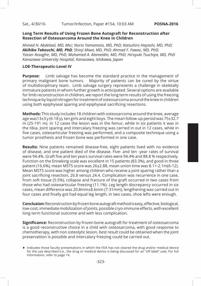
-323-
♦ Indicates those faculty presentations in which the FDA has not cleared the drug and/or medical device for the use described (i.e., the drug or medical device is being discussed for an “off label” use). For full information, refer to page 14.
Sat., 4/30/16 Tumor/Infection, Paper #154, 10:03 AM POSNA-2016 Long Term Results of Using Frozen Bone Autograft for Reconstruction after Resection of Osteosarcoma Around the Knee in Children
Ahmed H. Abdelaal, MD, Msc; Norio Yamamoto, MD, PhD; Katsuhiro Hayashi, MD, PhD; Akihiko Takeuchi, MD, PhD; Shinji Miwa, MD, PhD; Ahmad F. Fawaz, MD, PhD; Yasser Assaghir, MD, PhD; Mohamed A. Alameldin, MD, PhD; Hiroyuki Tsuchiya, MD, PhDKanazawa University Hospital, Kanazawa, Ishikawa, Japan
LOE-Therapeutic-Level IV
Purpose: Limb salvage has become the standard practice in the management of primary malignant bone tumors. Majority of patients can be cured by the virtue of multidisciplinary team. Limb salvage surgery represents a challenge in skeletally immature patients in whom further growth is anticipated. Several options are available for limb reconstruction in children, we report the long term results of using the freezing technique by liquid nitrogen for treatment of osteosarcoma around the knee in children using both epiphyseal sparing and epiphyseal sacrificing resections.
Methods: This study includes 18 children with osteosarcoma around the knee, average age was13±3 y (6-18 y), ten girls and eight boys. The mean follow-up period was 75±32.7 m (25-191 m). In 12 cases the lesion was in the femur, while in six patients it was in the tibia. Joint sparing and intercalary freezing was carried in out in 12 cases, while in five cases, osteoarticular freezing was performed, and a composite technique using a tumor prosthesis and frozen bone was performed in one case.
Results: Nine patients remained disease-free, eight patients lived with no evidence of disease, and one patient died of the disease. Five- and ten -year rates of survival were 94.4%. Graft five and ten years survival rates were 94.4% and 88.8 % respectively. Function on the Enneking scale was excellent in 15 patients (83.3%), and good in three patient (16.6%), mean MSTS score was 26±2.88, mean union time was 8.1+-2.1m(6-12).Mean MSTS score was higher among children who receive a joint sparing rather than a joint sacrificing resection, 26.8 versus 24.4. Complication was recurrence in one case, from soft tissue (5.5%), collapse and fracture of the graft occurred in two cases from those who had osteoarticular freezing (11.1%). Leg length discrepancy occurred in six cases, mean difference was 20.8mm±8.6mm (7-31mm), lengthening was carried out in four cases and finally got had equal leg length, in two cases, shoe lefts were enough.
Conclusion: Reconstruction by frozen bone autograft method is easy, effective, biological, low-cost, immediate mobilization of joints, possible cryo-immune effects, with excellent long term functional outcome and with less complication.
Significance: Reconstruction by frozen bone autograft for treatment of osteosarcoma is a good reconstructive choice in a child with osteosarcoma, with good response to chemotherapy, with non osteolytic lesion, best result could be obtained when the joint preservatiion is possible and intercalary freezing could be carried out.
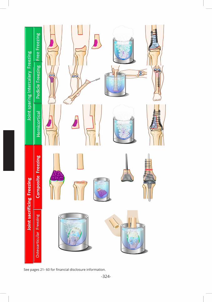
-324-
See pages 21- 60 for financial disclosure information.
-325-
♦ Indicates those faculty presentations in which the FDA has not cleared the drug and/or medical device for the use described (i.e., the drug or medical device is being discussed for an “off label” use). For full information, refer to page 14.
Sat., 4/30/16 Tumor/Infection, Paper #155, 10:08 AM POSNA-2016 Physeal Growth After Juxta-Physeal Resections For Pediatric Sarcoma Limb Salvage: Does The Physis Function?
Amy K. Williams, MD; Tressa Mattioli-Lewis, MPH; John C. Shapton, BS; Stephanie Punt, BS; Ernest U. Conrad III, MD Seattle Children’s Hospital, Seattle, Washington
LOE-Therapeutic-Level III
Purpose: The preservation of physeal function is a critical issue for pediatric limb salvage because of growth potential in children under age 12 years. The goal of this study was to compare condyle versus physeal-sparing resections in the distal femur and tibia regarding physeal arrest and subsequent leg length discrepancy.
Methods: Pediatric limb salvage cases in patients with Ewing’s or Osteosarcoma about the knee were reviewed. 45 patients who underwent allograft reconstruction were included in the cohort with an assessment of physeal growth after juxtaphyseal resections. Patient records and radiographic images were reviewed retrospectively to assess the surgical allograft fixation techniques and leg length consequences following condyle sparing (CS) versus physeal-sparing (PS) resections. Patient cohorts were also divided into three age categories (<10, 10-16, or >16 years of age) and allograft fixation techniques were categorized (parallel, peripheral, central, combination). Trans-physeal fixation used in the younger (under 12 years) patients was removed at 6 months post-operatively if the fixation crossed the physis.
Results: Our comparison of CS vs. PS resections showed the following: CS resections had greater LLD than PS resection (23.7mm vs. 28.5mm). The younger the patient, the more likely they were to have a LLD (<10 years had 41.7mm, 10-16 years had 25.7mm, >16 years had 9.1mm). In the younger age group, PS resections were associated with closer surgical resection to the physis (mean 6.6mm) and a higher incidence of physeal arrest. Postoperative physeal growth in the PS resection cohort was most prevalent in the younger patients who averaged 16.3mm vs. 8.3mm vs. 0.07mm (<10 years, 10-16 years, >16 years). Both central and peripheral physeal fixation techniques produced physeal arrest and LLD at a similar incidence. Younger patients with closer physeal margins had higher risk of physeal failure and LLD. Pediatric limb salvage that places a physeal margin less than one cm should not expect a functional physis post-operatively. Lastly, PS resections had a slightly higher incidence of revision procedures (34.7% vs. 23.9%) than CS resection patients.
Conclusion: Despite attempting to preserve the physis in limb salvage procedures, the type of allograft fixation utilized resulted in poor post-operative physeal function. In addition, resections within 1 cm of the physis appear to have a significant impact on post-operative physeal function.
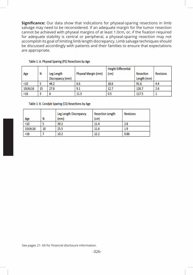
-326-
See pages 21- 60 for financial disclosure information.
Significance: Our data show that indications for physeal-sparing resections in limb salvage may need to be reconsidered. If an adequate margin for the tumor resection cannot be achieved with physeal margins of at least 1.0cm, or, if the fixation required for adequate stability is central or peripheral, a physeal-sparing resection may not accomplish its goal of limiting limb length discrepancy. Limb salvage techniques should be discussed accordingly with patients and their families to ensure that expectations are appropriate.
-327-
♦ Indicates those faculty presentations in which the FDA has not cleared the drug and/or medical device for the use described (i.e., the drug or medical device is being discussed for an “off label” use). For full information, refer to page 14.
Sat., 4/30/16 Tumor/Infection, Paper #156, 10:13 AM POSNA-2016 Do Surgeons Need to Rescrub during Operations that Last Longer than Three Hours?
Pooria Hosseini, MD, MSc; Gregory M. Mundis, MD; Robert K. Eastlack, MD; Allen Nourian, MD; Jeff Pawelek, BS; Stacie Nguyen, MPH; Behrooz A. Akbarnia, MDSan Diego Spine Foundation, San Diego, California
LOE-Prognostic-Level II
Purpose: Despite advances in infection control, surgical site infection (SSI) remains a substantial cause of morbidity and mortality in hospitalized patients. A variety of methods are being used to decrease SSI, however SSI can still cause complications especially in long operations (>3 hours). There is evidence that the efficacy of the scrubbing material fades away after three hours. Hence, it can be postulated that the scrubbed surgeon’s hands may become a progressive source of contamination during surgery. In addition, it has been shown that gloves do not provide a permanent physical barrier against microbes. We aimed to determine the level of surgeons’ recurrent hand contamination after uninterrupted operations lasting more than three hours.
Methods: Three spine surgeons at the same facility used the same scrubbing technique and material. Exclusion criteria included procedures less than three hours, and procedures that required changing of both sets of gloves. Twenty consecutive spine surgeries meeting these criteria were included. Surgeons’ hands were swabbed with sterile cotton tip applicators and 5 ml sterile 75 mM Phosphate Buffered Saline with 0.1% Triton-X (pH 7.9) at the following time points: Prior to hand scrubbing (pre-scrub), immediately following hand scrubbing (post-scrub) and immediately following surgery (post-operative).
Results: All samples had zero colony count at post-scrub. None of the cases lasting 3-4 hours had colony growth post-operatively. Surgeries that were at least five hours had the post-operative colony count reach the same level or higher than the pre-scrub. The longer the operation, the higher the level of contamination post-operatively with a linear regression coefficient of 0.89 and p=0.005. Fig 1.
Conclusion: Our results demonstrate that after four hours, a surgeon’s hands become recolonized, despite preoperative scrubbing that successfully removed contaminants. In some cases, there was higher colony forming unit / ml after the procedure than before any scrubbing had occurred. Based on these findings, consideration should be given to rescrubbing during the course of surgical procedures extending beyond four hours.
Significance: Hands of healthcare workers gets equally or even more contaminated compared to pre-wash timepoint after four hours of uninterrupted operation. This finding justifies rescrubbing every four hour in operations lasting longer than four hours.
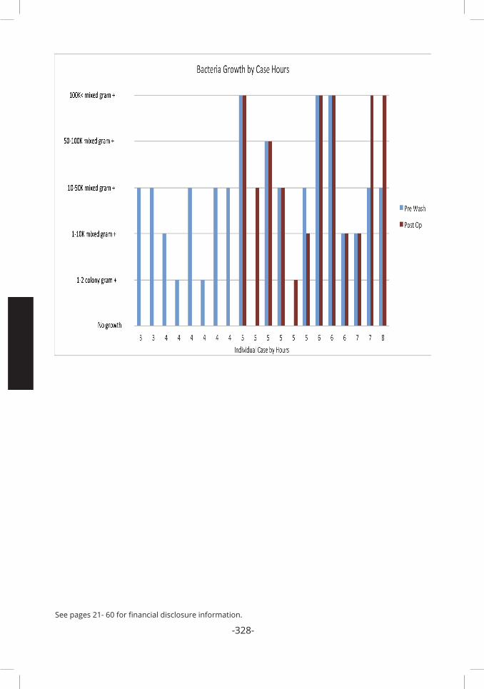
-328-
See pages 21- 60 for financial disclosure information.
-329-
♦ Indicates those faculty presentations in which the FDA has not cleared the drug and/or medical device for the use described (i.e., the drug or medical device is being discussed for an “off label” use). For full information, refer to page 14.
Sat., 4/30/16 Tumor/Infection, Paper #157, 10:27 AM POSNA-2016 Unexpectedly Similar Clinical Severity of MSSA vs. MRSA Musculoskeletal Infection
Thomas J. An, BA; Michael A. Benvenuti, BS; Megan Mignemi, MD; Jeffrey E. Martus, MD; Gregory A. Mencio, MD; Steven A. Lovejoy, MD; Jonathan G. Schoenecker, MD, PhDVanderbilt Children’s Hospital, Nashville, Tennessee
LOE-Prognostic-Level II
Purpose: Studies have shown that methicillin resistant Staph. aureus (MRSA) causes more virulent musculoskeletal infection (MSKI) compared to methicillin susceptible Staph. aureus (MSSA). Based on these results, clinical prediction algorithms have been developed to differentiate MRSA from MSSA earlier in a patient’s clinical course. In this study, the authors tested the hypothesis that MRSA causes worse hospital outcomes than MSSA for pediatric MSKI at their institution.
Methods: An IRB approved retrospective study of children with MRSA and MSSA MSKI was conducted over 5 years (2008-2013). Laboratory values and clinical outcomes were recorded from the electronic medical record. Clinical outcomes included length of stay, number of operative interventions, duration of antibiotics, and ICU LOS. In addition, infection severity was determined for each infection. Infection severity was defined as local or disseminated according to the VODIS classification system (Figure 1a).
Results: Overall, there were 49 cases of MRSA-MSKI and 43 cases of MSSA-MSKI included in the study. There was no significant difference in the number of MRSA and MSSA infections in the disseminated severity group (see Figure 1b). With subgroup analysis of outcomes for disseminated infections, there was no significant difference in LOS, number of patients that required ICU level care, and duration of antibiotics between MRSA and MSSA. However, within the disseminated group, MRSA infections had more operative interventions than MSSA infections. With subgroup analyses for local infections, there was also no significant difference in outcomes (LOS, ICU, duration of antibiotics, and # times to OR) between MRSA and MSSA (see figure 1c).
Conclusion: Unexpectedly, the analysis did not demonstrate a significant difference in most hospital outcomes for MSSA and MRSA at this tertiary care hospital. Among severe, disseminated infections, MRSA and MSSA were equally represented as causative pathogens. However, MRSA caused an increased number of operative interventions compared to MSSA for disseminated infections.
Significance: The results demonstrate no significant difference in the severity of infection for MSSA compared to MRSA at the author’s institution. The regional variability in MRSA and MSSA severity may be due to epidemiological factors such as virulence factor expression and strain prevalence in different areas of the country.
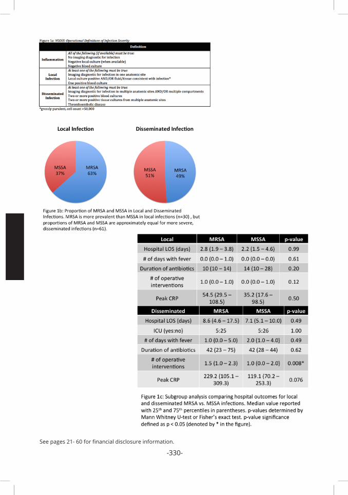
-330-
See pages 21- 60 for financial disclosure information.
-331-
♦ Indicates those faculty presentations in which the FDA has not cleared the drug and/or medical device for the use described (i.e., the drug or medical device is being discussed for an “off label” use). For full information, refer to page 14.
Sat., 4/30/16 Tumor/Infection, Paper #158, 10:32 AM POSNA-2016 Unexpected Effects of Antibiotics on Culture Yield in Pediatric Musculoskeletal Infection (MSKI)
Michael A. Benvenuti, BS; Thomas J. An, BA; Megan Mignemi, MD; Gregory A. Mencio, MD; Steven A. Lovejoy, MD; Jeffrey E. Martus, MD; Jonathan G. Schoenecker, MD, PhD Vanderbilt Children’s Hospital, Nashville, Tennessee
LOE-Prognostic-Level II
Purpose: Musculoskeletal infection (MSKI) is a common cause of pediatric hospitalization. Although MSKI causes relatively little long-term morbidity and mortality, hospital outcomes and costs vary dramatically with severity of infection. The mainstays of treatment for children with MSKI include antibiotics and surgical debridement. Previous studies have shown that delays in diagnosis and initiation of antibiotics lead to worse outcomes. One reason for this delay is that some clinicians favor holding antibiotics until a tissue culture can be taken in an effort to improve culture yields. We hypothesize that because local infections tend to form avascular foci, antibiotics may affect blood culture results, but should not decrease the yield from tissue cultures. Methods: An IRB approved retrospective study of children with MSKI from 2008-2013 was conducted. These patients were classified into local and disseminated groups based on the severity of their infection (Figure 1a). Antibiotic start time, blood and tissue culture time and results, and hospital outcomes were determined for all patients and compared across groups (Figure 1b).
Results: Representative data is shown in Figure 1b and shown graphically in Figure 1c. Patients with local infection who received antibiotics prior to tissue culture were significantly more likely to have a positive culture. Antibiotic start time did not have an effect on tissue culture yield in the disseminated infection group. Although not significant, there was a trend toward fewer positive blood cultures in patients with both local and disseminated infection when antibiotics were started prior to culture.
Conclusion: Starting antibiotics early in the course of local MSKI does not negatively affect tissue culture results, and leads to improved tissue culture yield. Antibiotics appeared to decrease the yield of blood cultures in the local group, but a small sample size of patients receiving antibiotics before culture limited analysis.
Significance: For children with MSKI, antibiotics should be started as soon as possible following blood culture, but should not be delayed by tissue culture, especially in local infections.
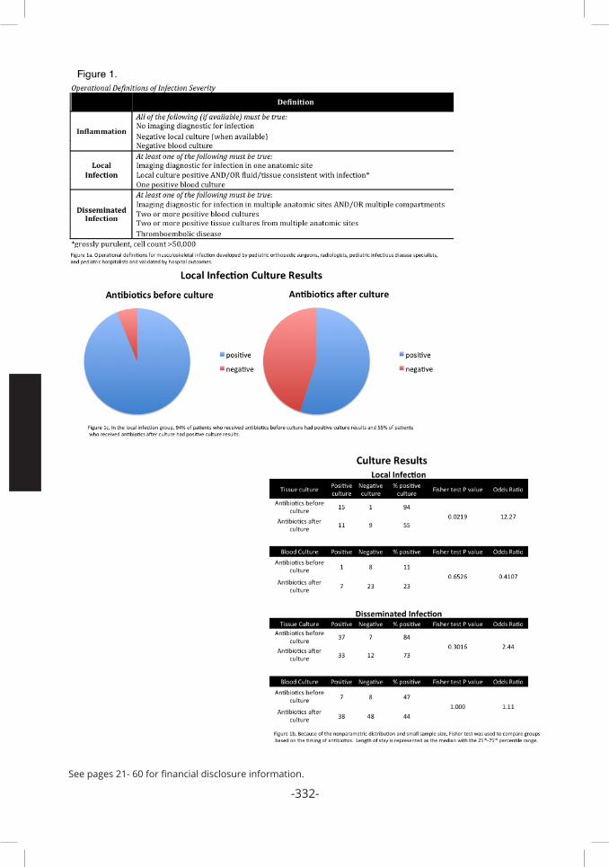
-332-
See pages 21- 60 for financial disclosure information.
-333-
♦ Indicates those faculty presentations in which the FDA has not cleared the drug and/or medical device for the use described (i.e., the drug or medical device is being discussed for an “off label” use). For full information, refer to page 14.
Sat., 4/30/16 Tumor/Infection, Paper #159, 10:37 AM POSNA-2016 Is a Corticotomy Required in the Treatment of Subperiosteal Abscesses in the Pediatric Population?
Corey O. Mongtomery, MD MS; Brant C. Sachleben, MD; Austin Porter, MPH; Larry J. Suva, PhD; Brien M. Rabenhorst, MD Arkansas Children’s Hospital, Little Rock, Arkansas
LOE-Therapeutic-Level II
Purpose: Subperiosteal abscesses are typically treated with surgical decompression. If advanced imaging studies demonstrate the presence of intramedullary involvement of infection, a corticotomy is typically indicated to decompress the infection. The treatment of patients with a subperiosteal abscess without intramedullary involvement is less clear. Currently, little evidence exists regarding whether or not a corticotomy is needed in addition to the drainage. We tested our hypothesis that a corticotomy during debridement of a subperiosteal abscess will decrease the requirement for further surgical debridement and decrease the recurrence rate of infection.
Methods: Following IRB approval, a retrospective chart review was performed on all patients surgically treated for a subperiosteal abscess at a single institution from 2004-2012. Inclusion criteria included <18 years of age, radiographic evidence (either CT or MRI) of a subperiosteal abscess without overt intramedullary involvement; less than 3 weeks of symptoms Exclusion criteria included evidence of chronic infection > 3 weeks, patients with increased risk of osteomyelitis. Medical charts were reviewed for radiographic evidence of a subperiosteal abscess, age, number of surgical debridements, type of bacteria, whether or not a corticotomy was performed, and all complications. Comparisons were made using the Fisher exact test and p<0.05 was considered significant.
Results: A total of 127 patients were identified that met the inclusion criteria for surgical debridement of a subperiosteal abscess between 2004 and 2012. Fifty nine (59) patients were excluded, and sixty eight (68) were included. Our analysis identified two groups; 30 patients had a corticotomy during the index procedure and 38 patients that only had surgical drainage of the abscess without a corticotomy. These groups were evaluated for type of bacterial infection, age at onset of infection, and post-operative complications. Our analysis demonstrated an odds ratio of 6.46 (p=0.011) in favor of a corticotomy to decrease the risk of future surgeries. There was no significant correlation with type of infection, age or complications or any other parameters.
Conclusion: These results demonstrate that a corticotomy during surgical debridement of a subperiosteal abscess significantly decreases the risk of future surgical procedures with no significant increased in risk of fracture. Larger studies should be performed to confirm these observations.
Significance: The addition of a corticotomy during the surgical treatment of a subperiosteal abscess appears to significantly decrease the chances of subsequent surgeries and should be considered by the treating surgeon.
-334-
See pages 21- 60 for financial disclosure information.
Sat., 4/30/16 Foot/Ankle, Paper #160, 10:51 AM POSNA-2016 Epidemiology of Clubfoot: A Meta-analysis Identifying Modifiable Risk Factors
Cynthia Chen, BA; Sowmya Josyula, MD; John S. Blanco, MD; Shevaun M. Doyle, MD; Roger F. Widmann, MD; David Scher, MD; Ernest L. Sink, MD; Emily Dodwell, MDHospital for Special Surgery, New York, New York
LOE-Prognostic-Level III
Purpose: A combination of genetic and environmental risk factors have been reported as causes of clubfoot including amniocentesis, maternal smoking during pregnancy, summer conception, lower parity, parental age, and breech position in utero. The aim of this study was to conduct a meta-analysis to assess the best evidence of risk factors for the development of clubfoot.
Methods: We performed a systematic literature search for studies that investigated risk factors for clubfoot. Data on study design, patient characteristics, and risk estimates were extracted. Pooled effect estimates were calculated using random and fixed effects models based on study heterogeneity.
Results: Twenty one case-control studies and three cohort studies were included in this analysis. On the Newcastle-Ottawa Scale, eleven studies were of higher quality (≥ 7) and nine studies were of lower quality (< 7). Studies with a quality score below five were excluded. The initial analysis identified several significant modifiable risk factors associated with an increased risk of clubfoot including smoking (OR = 1.54 95%CI:1.42-1.66), cesarean section (OR = 1.43 95%CI:1.35-1.51), and single marital status (OR = 1.18 95%CI:1.11-1.24). Non-modifiable factors that were significantly associated with increased risk of clubfoot included male sex, twin gestation, family history, and race. Diabetes and maternal education were not found to be significant. The sub-analysis of high quality studies identified woman lacking prenatal care (OR = 1.5 95%CI:1.21-1.87), maternal age < 20 (OR = 1.14 95%CI:1.07-1.20), Medicaid insurance (OR = 1.28 95%CI:1.09-1.28), and premature birth (OR = 3.66 95%CI:3.31-4.06) as additional factors associated with clubfoot.
Conclusion: This meta-analysis identified several modifiable risk factors significant to the development of clubfoot. The most notable and clinically relevant risk factors include smoking and lack of pre-natal care.
Significance: To our knowledge this is the first meta-analysis assessing risk factors in clubfoot. A knowledge of modifiable risk factors such as smoking and pre-natal care may play an important role in counselling patients before and during pregnancy. Strategies aimed at reducing risk factors may aid in decreasing the incidence of infants born with clubfoot.
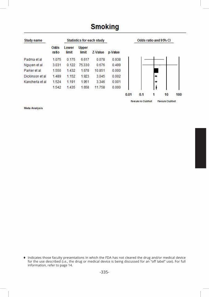
-335-
♦ Indicates those faculty presentations in which the FDA has not cleared the drug and/or medical device for the use described (i.e., the drug or medical device is being discussed for an “off label” use). For full information, refer to page 14.
-336-
See pages 21- 60 for financial disclosure information.
Sat., 4/30/16 Foot/Ankle, Paper #161, 10:56 AM POSNA-2016 Oral Sucrose for Pain Relief during Clubfoot Casting: A Double-Blinded Randomized Controlled Trial
Todd A. Milbrandt, MD; Ryan D. Muchow, MD; Janet L. Walker, MD; Vishwas R. Talwalkar, MD; Henry J. Iwinski, MD Shriners Hospital for Children, Lexington, Kentucky
LOE-Therapeutic-Level I
Purpose: Idiopathic clubfoot treatment is now primarily treated by manipulation and casting utilizing the Ponseti technique. During the casting, the infant can become fussy and irritable. The goal of this study was to determine which intervention could decrease this pain response in infants undergoing Ponseti manipulation and casting for idiopathic clubfeet. Our hypothesis was that the administration of oral sucrose or solution or milk would be the most effective in accomplishing that goal.
Methods: We conducted a double-blinded randomized controlled trial at a tertiary pediatric orthopaedic center on 33 children (average age 17.94 days STD 20.51 days) undergoing clubfoot manipulation and casting and their guardians. Each cast was considered a new event and was randomized to an oral 20% sucrose solution (S), water (W), or milk (M) in a bottle (breast or non-breast). We assessed the neonatal infant pain scale (NIPS), heart rate (HR), and oxygen saturation (O2) before, during and after the casting. We also surveyed the adults regarding their experience during casting.
Results: A total of 131 casts were randomized and analyzed (37 M, 42 S, 39 W). Each child underwent an average of 3.97 casts (STD 1.74). There were no significant differences seen between the groups prior to casting in their mean NIPS score (M-2.2; STD 2.38, Sucrose-1.84 STD 2.18, water 1.61 STD 2.12). However during casting, both milk (0.91 STD 1.26 p=.0005) and sucrose (0.64 STD 1.27 p<.0001) were significantly less than water (2.27 STD 2.03) but not different from each other (p=.33). Post casting, the sucrose NIPS score (0.69 STD 1.53) continued to be significantly less than milk (2.11 STD 2.37) p=0.0065. There were also no significant differences pre-cast, during cast, or post-cast for HR or O2. In addition, there was no correlation between parental anxiety nor family environmental factors and average NIP scores.
Conclusion: Sucrose solution and milk during Ponseti casting and manipulation were effective in decreasing the pain response in children undergoing manipulation and casting for clubfeet. The sucrose solution administration continued the pain relief into the post-casting period.
Significance: In addition to the benefits of improving the patient experience during casting, a less irritable child may mean better casting and possibly improved results.
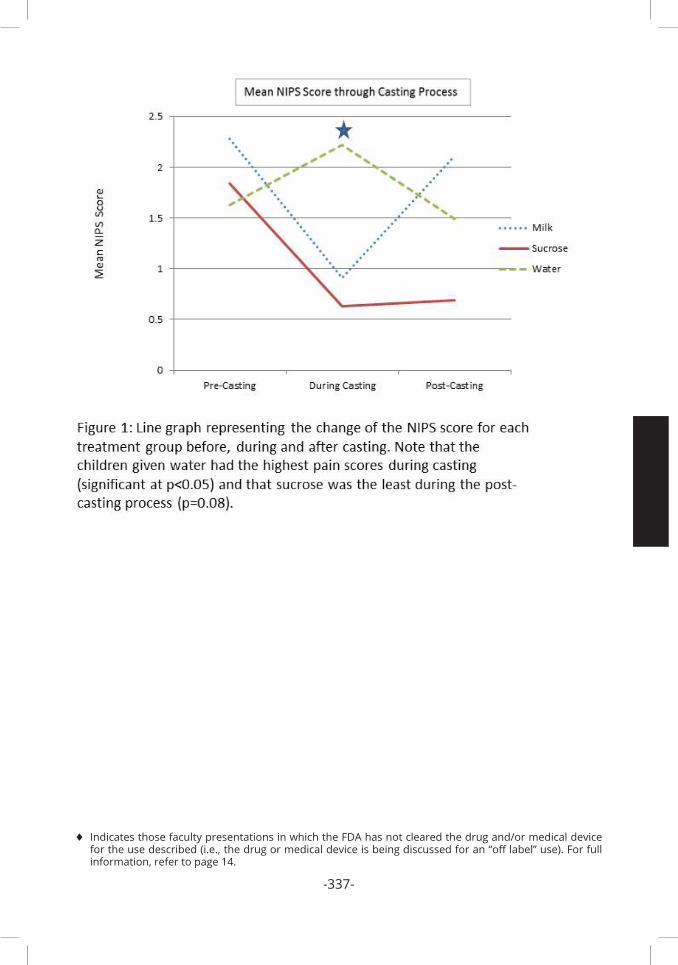
-337-
♦ Indicates those faculty presentations in which the FDA has not cleared the drug and/or medical device for the use described (i.e., the drug or medical device is being discussed for an “off label” use). For full information, refer to page 14.
-338-
See pages 21- 60 for financial disclosure information.
Sat., 4/30/16 Foot/Ankle, Paper #162, 11:01 AM POSNA-2016 Anterior Tibial Tendon Transfer: How Many Patients Treated Using the Ponseti Method Will Ultimately Need One?
Lewis E. Zionts, MD; Sophia Sangiorgio, PhD; Kathryn L. Bauer, MD; Michael H. Jew, BS; Edward Ebramzadeh, PhDOrthopaedic Institute for Children and the Geffen School of Medicine at UCLA, Los Angeles, California
LOE-Prognostic-Level III
Purpose: The Ponseti Method has become the standard of care for the treatment of idiopathic clubfoot. The most common problem associated with this technique is a relapsed deformity, often treated in patients 2.5 years and older by transfer of the anterior tibial tendon to the third cuneiform. Presently, there is insufficient information to properly counsel families whose infants are beginning Ponseti treatment on the risk of needing later tendon transfer surgery. In the present study, we used survivorship analysis to calculate the probability of a relapsed deformity requiring surgical intervention as a function of age, using a large cohort of idiopathic clubfoot patients followed for as long as nine years.
Methods: All idiopathic clubfoot patients seen at the authors’ institution during the study period who received no previous outside treatment, and were followed for >2.5 years, were included. Kaplan-Meier Survival analysis was used and Chi-Square analysis (log rank Mantel-Cox) was used to determine the overall influence of various patient factors on survival time. Survival time was determined by the date of anterior tibial tendon transfer (ATTT) surgery. Additionally, the influence of patient characteristics, socio-economic variables, and treatment variables on need for surgery were calculated. Further, age at the time of first, second, and third relapse was analyzed.
Results: Of 137 patients who met the inclusion criteria, 34 (24.8%) underwent an ATTT. The mean follow-up was 55±17 mos. Among patients whose families reported compliance with bracing (N=54), the probability of survival without requiring a tendon transfer was 81% at 6 years compared to 58% in patients whose families were not compliant. Overall, parent-reported compliance with bracing was the most significant factor associated with reducing the need for surgery (Odds Ratio 6.88, P<0.01). The median age at first relapse was 12.6 months. Of those patients who developed a relapse, 34 (51%) went on to require an ATTT.
Conclusion: This is the first study to report the probability of requiring ATTT surgery during early childhood when treated using the Ponseti method. While the overall risk of undergoing an ATTT reached 29% at six years, this risk was significantly reduced by compliance with bracing. Additionally, among patients who experienced a relapse, half went on to require surgery.
Significance: The number of patients undergoing ATTT will increase with longer follow-up, especially in families that fail to adhere with post-corrective bracing. Emphasis must be placed on avoiding an early relapse to reduce the need for later surgery.
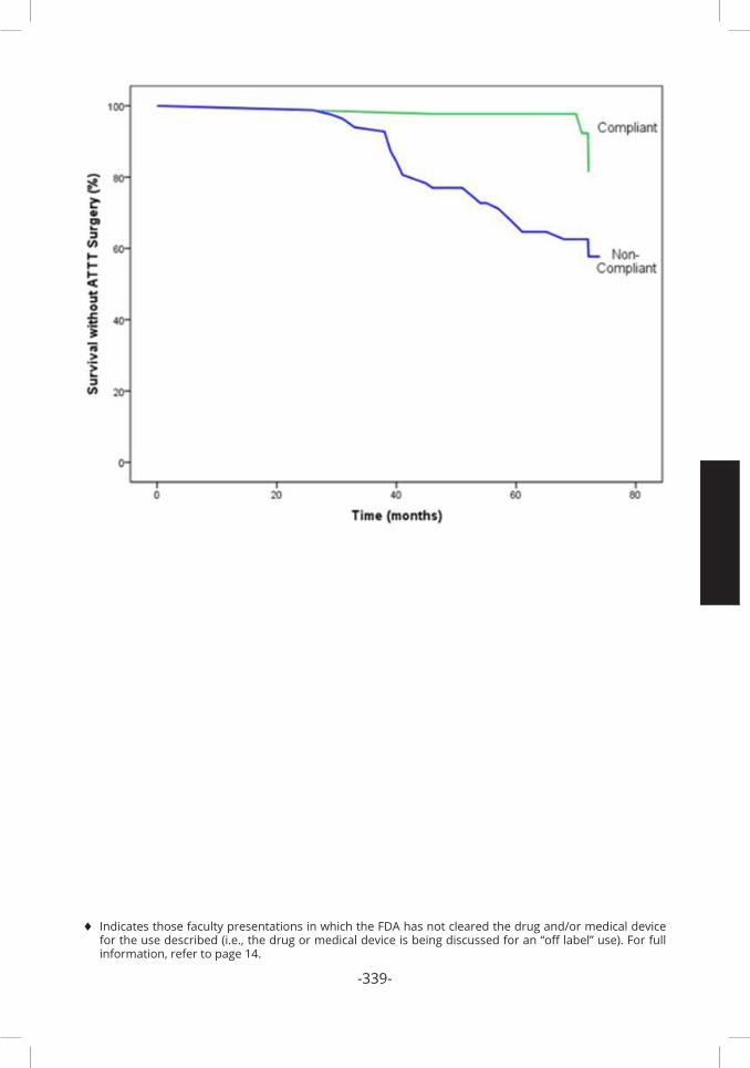
-339-
♦ Indicates those faculty presentations in which the FDA has not cleared the drug and/or medical device for the use described (i.e., the drug or medical device is being discussed for an “off label” use). For full information, refer to page 14.
-340-
See pages 21- 60 for financial disclosure information.
Sat., 4/30/16 Foot/Ankle, Paper #163, 11:15 AM POSNA-2016 Clubfoot Relapse: Does Presentation Differ Based on Age at Initial Relapse?
Susan T. Mahan, MD, MPH; Samantha A. Spencer, MD; Collin J. May, MD; James R. Kasser, MD Boston Children’s Hospital, Boston, Massachusetts
LOE-Prognostic-Level III
Purpose: Treatment of idiopathic clubfoot with Ponseti method is now standard, but predicting relapse can be difficult. Bracing recommendations to age four is typical but challenging in some, and may not be necessary in all patients. The purpose of this study is to compare patterns of bracing and age of relapse to help determine if predictable patterns exist.
Methods: Utilizing our clubfoot registry, 70 patients with idiopathic clubfoot treated initially with Ponseti technique who had relapse of their clubfoot were identified. Relapse was identified as patient who after initial treatment had subsequent need for return to casting or surgery due to recurrent deformity. Our department rate for relapse during this time is about 27%. Data collected included demographic, treatment, and brace compliance. Patients who suffered initial relapse before age 2 were compared to those who suffered initial relapse after age 2.
Results: Of the 70 patients who suffered relapse of their clubfoot, 56% (39/70) had their initial relapse prior to age 2 while 44% (31/70) were after age 2 years. Of the patients who relapsed prior to age 2, 28% (11/39) were compliant with bracing while 72% were non-compliant. For patients who initially relapsed after age 2, 74% (23/39) were compliant with bracing while 26% were non-compliant (p<0.001). Of those who had initial relapse prior to age 2, a subsequent relapse was seen in 69% (27/39). If patient was compliant with bracing, subsequent relapse occurred in 64% (7/11), while if a patient was non-compliant with bracing subsequent relapse occurred in 71% (20/28) (p=0.7).
Conclusion: Patients with idiopathic clubfoot who suffer recurrence prior to age 2 years are significantly more likely to be non-compliant with bracing than those who suffer recurrence after age 2. After initial relapse prior to age 2, bracing compliance does not affect likelihood of subsequent recurrence.
Significance: Bracing compliance as part of Ponseti treatment for idiopathic clubfoot remains very important. Relapse prior to age 2 years appears to be more related to bracing compliance than relapse after age 2 years. Further work on identifying clubfeet that are at high or low risk to relapse may result in modifications of the bracing recommendations.
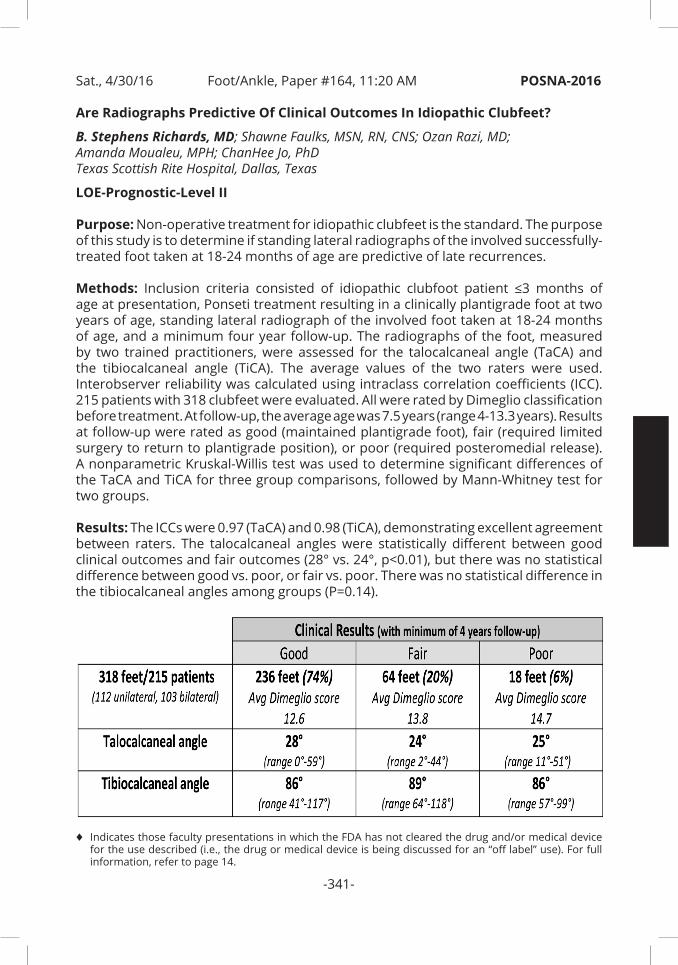
-341-
♦ Indicates those faculty presentations in which the FDA has not cleared the drug and/or medical device for the use described (i.e., the drug or medical device is being discussed for an “off label” use). For full information, refer to page 14.
Sat., 4/30/16 Foot/Ankle, Paper #164, 11:20 AM POSNA-2016 Are Radiographs Predictive Of Clinical Outcomes In Idiopathic Clubfeet?
B. Stephens Richards, MD; Shawne Faulks, MSN, RN, CNS; Ozan Razi, MD; Amanda Moualeu, MPH; ChanHee Jo, PhDTexas Scottish Rite Hospital, Dallas, Texas
LOE-Prognostic-Level II
Purpose: Non-operative treatment for idiopathic clubfeet is the standard. The purpose of this study is to determine if standing lateral radiographs of the involved successfully-treated foot taken at 18-24 months of age are predictive of late recurrences.
Methods: Inclusion criteria consisted of idiopathic clubfoot patient ≤3 months of age at presentation, Ponseti treatment resulting in a clinically plantigrade foot at two years of age, standing lateral radiograph of the involved foot taken at 18-24 months of age, and a minimum four year follow-up. The radiographs of the foot, measured by two trained practitioners, were assessed for the talocalcaneal angle (TaCA) and the tibiocalcaneal angle (TiCA). The average values of the two raters were used. Interobserver reliability was calculated using intraclass correlation coefficients (ICC). 215 patients with 318 clubfeet were evaluated. All were rated by Dimeglio classification before treatment. At follow-up, the average age was 7.5 years (range 4-13.3 years). Results at follow-up were rated as good (maintained plantigrade foot), fair (required limited surgery to return to plantigrade position), or poor (required posteromedial release). A nonparametric Kruskal-Willis test was used to determine significant differences of the TaCA and TiCA for three group comparisons, followed by Mann-Whitney test for two groups.
Results: The ICCs were 0.97 (TaCA) and 0.98 (TiCA), demonstrating excellent agreement between raters. The talocalcaneal angles were statistically different between good clinical outcomes and fair outcomes (28° vs. 24°, p<0.01), but there was no statistical difference between good vs. poor, or fair vs. poor. There was no statistical difference in the tibiocalcaneal angles among groups (P=0.14).
-342-
See pages 21- 60 for financial disclosure information.
Conclusion: Most clubfeet that were clinically plantigrade at 2 years of age remained so, while one-fourth subsequently required some surgery for late recurrence. We found that measurements of TiCA and TaCA from standing lateral radiographs taken at 18-24 months of age were not helpful in identifying those who experienced late relapse requiring surgical intervention.
Significance: Standing lateral radiographs of the feet following early clinical correction of clubfeet don’t provide useful information for the future outcome, and should not be obtained.
-343-
Sat., 4/30/16 Foot/Ankle, Paper #165, 11:25 AM POSNA-2016 Gait Following Nonoperative and Surgical Treatment for Clubfoot at Age 10 Years
Kelly Jeans, MSc; Ashley Erdman, BS, MBA; Lori A. Karol, MDTexas Scottish Rite Hospital for Children, Dallas, Texas
LOE-Therapeutic-Level II
Purpose: The purpose of this study was to assess gait and strength in 10yr olds with idiopathic clubfoot, comparing the function of nonoperative to surgically treated feet relative to age matched controls.
Methods: 159 patients (236 clubfeet) underwent gait analysis and isokinetic ankle strength testing at age 10.2yrs (9.7-11.5yrs). Patients were initially treated nonoperatively (NO), however some feet required surgical management. Feet were divided into extra-articular (EA) (tendon transfers, soft tissue release, lateral column shortenings), and intra-articular (IA) (posterior release (PR) or posterior-medial release (PMR) +/- additional procedures). Gait variables included sagittal ankle kinematics and kinetics and foot progression angle (FPA). Maximum isokinetic dorsiflexion (DF) and plantarflexion (PF) strength were also collected. Comparisons between NO, EA, IA and controls (n=40) were made using ANOVA with post-hoc Tukey test. A subsequent analysis of PR and PMR feet within the IA group was also conducted.
Results: 129 feet remained NO, 25 underwent EA surgery and 82 required IA surgery (39 PR/43 PMR). Table 1 No differences between NO, EA and IA were found in the kinematic data. Kinetically, compared to NO, IA feet had decreased ankle moment, power and impulse. EA feet showed a trend for increased kinetic values compared to IA, but did not meet statistical significance. Compared to controls, all groups exhibited reduced ankle moment, however limitations in PF within NO and IA groups lead to a reduction in overall dynamic ankle range of motion (ROM) and signficant reduction in ankle power. FPA was statistically different than controls, but this may not be clinically signficant. Further analysis of the IA group shows that limited PF and decreased ROM is driven by the PMR feet (p<0.0009). Maximum peak DF and PF strength (Biodex) were reduced in all groups compared to controls. Both NO and EA groups had significantly greater PF strength than the IA group.
Conclusion: Few differences are seen between NO, EA and IA at age 10yrs. Results following NO and IA management are showing significant limitations in ankle PF which results in decreased ankle ROM, moment and power. EA feet have fairly normal ankle motion, however the trend towards decreased power is also present in this group. Decreased isokinetic ankle strength indicates global weakness in NO and surgically treated clubfeet.
Significance: Ankle and foot motion following clubfoot treatment may be objectively assessed using gait analysis. These outcome measures may help clinicians understand the long-term effect treatment has on their patient’s function.
♦ Indicates those faculty presentations in which the FDA has not cleared the drug and/or medical device
for the use described (i.e., the drug or medical device is being discussed for an “off label” use). For full information, refer to page 14.
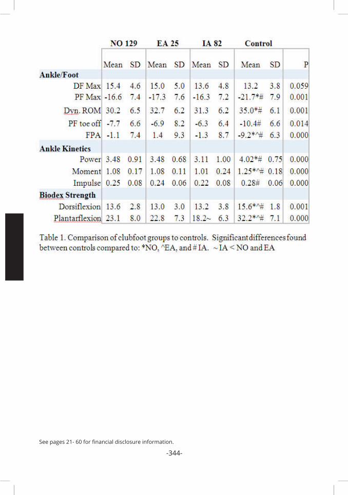
-344-
See pages 21- 60 for financial disclosure information.
-345-
♦ Indicates those faculty presentations in which the FDA has not cleared the drug and/or medical device for the use described (i.e., the drug or medical device is being discussed for an “off label” use). For full information, refer to page 14.
Sat., 4/30/16 Early Onset Scoliosis, Paper #166, 11:39 AM POSNA-2016 Curve Reduction in Patients Younger than 10 Years of Age Treated with Elongation, Derotation and Flexion Casting Technique Performed under General Anesthesia and Neuromuscular Blocking Drugs
Federico Canavese, MD University Hospital Estaing, Clermont Ferrand, France
LOE-Therapeutic-Level III
Purpose: Elongation, derotation, flexion (EDF) casting technique is a custom-made thoracolumbar cast based on a three dimensional correction concept. The primary objective of the present study was to measure changes on plain radiographs of patients younger than 10 years of age with scoliosis treated with EDF casting technique. The second aim was to evaluate the effectiveness of the EDF plaster technique realized under general anesthesia (GA) and neuro-muscular blocking drugs, i.e. curare.
Methods: A retrospective comparative case series study was performed in which were included 44 skeletally immature patients. The inclusion criteria were: diagnosis of scoliosis with onset between 4 and 10 years, ambulatory patients, curve magnitude (Cobb angle) over 30° and documented progression (curve progression was assessed on two consecutive radiographs taken at 6-month interval and was defined as an increase >6°), and no prior treatment. Three patient groups were selected. Group 1: EDF cast applied with patients awaken and no anesthesia; Group 2: EDF cast applied under GA without neuromuscular blocking drugs; Group 3: EDF cast applied under GA with neuromuscular blocking drugs. All the patients were treated with two serial EDF casts by 2.5 months each. All measurements were taken from the radiographic exams. Cobb’s angle; Mehta’s angle and Nash and Moe grade of rotation were assessed before and after applying the cast. Thirty-four (77.3%) patients were followed-up at least 24 months after removal of last EDF cast.
Results: Eighteen patients (15 females) were included in Group 1, 12 (10 females) in Group 2 and 14 (9 females) in Group 3. Groups were comparable at baseline (p>0.05). Serial EDF casting was more effective at initial curve reduction and in preventing curve progression when applied under GA with neuromuscular blocking drugs (p<0.001 and p<0.03, respectively). Metha’s angle and Nash and Moe score improved significantly in all groups of patients. During follow-up period, six patients required surgery in Group 1 (33.3%), 3 patients required surgery in Group 2 (25%) and 2 patients underwent surgery in Group 3 (15%).
Conclusion: EDF casting is a safe technique that can positively influence the natural evolution of scoliosis in patients younger than 10 years of age by reducing and slowing curve progression in both frontal (Cobb’s angle) and transverse plane (rib vertebral angle difference and apical vertebral rotation degree).

-346-
See pages 21- 60 for financial disclosure information.
Significance: EDF casting achieves better correction when applied under GA. Adding neuromuscular blocking drugs improve correction during EDF cast treatment and reduce curve progression at 24 months follow-up.
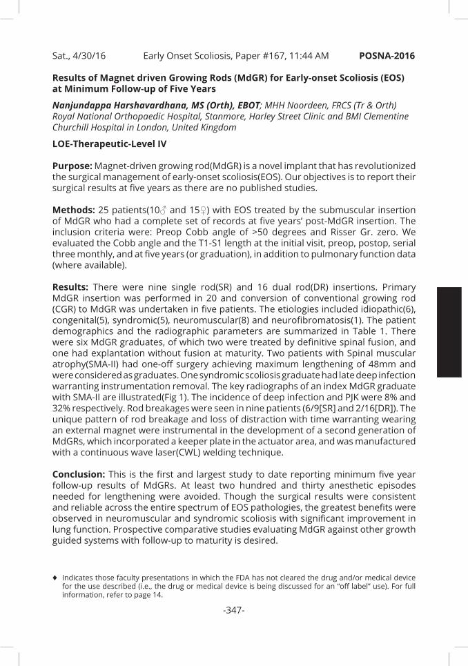
-347-
♦ Indicates those faculty presentations in which the FDA has not cleared the drug and/or medical device for the use described (i.e., the drug or medical device is being discussed for an “off label” use). For full information, refer to page 14.
Sat., 4/30/16 Early Onset Scoliosis, Paper #167, 11:44 AM POSNA-2016 Results of Magnet driven Growing Rods (MdGR) for Early-onset Scoliosis (EOS) at Minimum Follow-up of Five Years
Nanjundappa Harshavardhana, MS (Orth), EBOT; MHH Noordeen, FRCS (Tr & Orth) Royal National Orthopaedic Hospital, Stanmore, Harley Street Clinic and BMI Clementine Churchill Hospital in London, United Kingdom
LOE-Therapeutic-Level IV
Purpose: Magnet-driven growing rod(MdGR) is a novel implant that has revolutionized the surgical management of early-onset scoliosis(EOS). Our objectives is to report their surgical results at five years as there are no published studies.
Methods: 25 patients(10♂ and 15♀) with EOS treated by the submuscular insertion of MdGR who had a complete set of records at five years’ post-MdGR insertion. The inclusion criteria were: Preop Cobb angle of >50 degrees and Risser Gr. zero. We evaluated the Cobb angle and the T1-S1 length at the initial visit, preop, postop, serial three monthly, and at five years (or graduation), in addition to pulmonary function data (where available).
Results: There were nine single rod(SR) and 16 dual rod(DR) insertions. Primary MdGR insertion was performed in 20 and conversion of conventional growing rod (CGR) to MdGR was undertaken in five patients. The etiologies included idiopathic(6), congenital(5), syndromic(5), neuromuscular(8) and neurofibromatosis(1). The patient demographics and the radiographic parameters are summarized in Table 1. There were six MdGR graduates, of which two were treated by definitive spinal fusion, and one had explantation without fusion at maturity. Two patients with Spinal muscular atrophy(SMA-II) had one-off surgery achieving maximum lengthening of 48mm and were considered as graduates. One syndromic scoliosis graduate had late deep infection warranting instrumentation removal. The key radiographs of an index MdGR graduate with SMA-II are illustrated(Fig 1). The incidence of deep infection and PJK were 8% and 32% respectively. Rod breakages were seen in nine patients (6/9[SR] and 2/16[DR]). The unique pattern of rod breakage and loss of distraction with time warranting wearing an external magnet were instrumental in the development of a second generation of MdGRs, which incorporated a keeper plate in the actuator area, and was manufactured with a continuous wave laser(CWL) welding technique.
Conclusion: This is the first and largest study to date reporting minimum five year follow-up results of MdGRs. At least two hundred and thirty anesthetic episodes needed for lengthening were avoided. Though the surgical results were consistent and reliable across the entire spectrum of EOS pathologies, the greatest benefits were observed in neuromuscular and syndromic scoliosis with significant improvement in lung function. Prospective comparative studies evaluating MdGR against other growth guided systems with follow-up to maturity is desired.
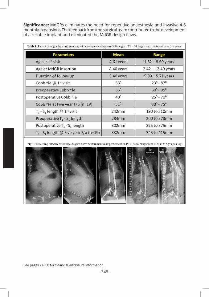
-348-
See pages 21- 60 for financial disclosure information.
Significance: MdGRs eliminates the need for repetitive anaesthesia and invasive 4-6 monthly expansions.The feedback from the surgical team contributed to the development of a reliable implant and eliminated the MdGR design flaws.
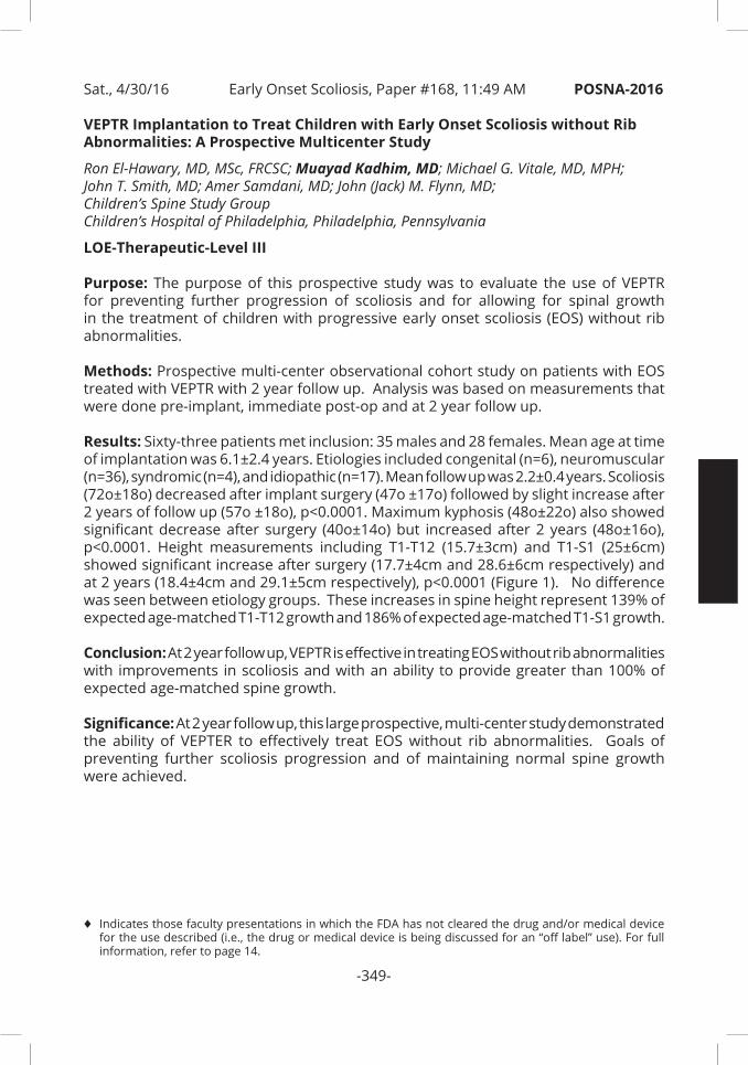
-349-
♦ Indicates those faculty presentations in which the FDA has not cleared the drug and/or medical device for the use described (i.e., the drug or medical device is being discussed for an “off label” use). For full information, refer to page 14.
Sat., 4/30/16 Early Onset Scoliosis, Paper #168, 11:49 AM POSNA-2016 VEPTR Implantation to Treat Children with Early Onset Scoliosis without Rib Abnormalities: A Prospective Multicenter Study
Ron El-Hawary, MD, MSc, FRCSC; Muayad Kadhim, MD; Michael G. Vitale, MD, MPH; John T. Smith, MD; Amer Samdani, MD; John (Jack) M. Flynn, MD; Children’s Spine Study GroupChildren’s Hospital of Philadelphia, Philadelphia, Pennsylvania
LOE-Therapeutic-Level III
Purpose: The purpose of this prospective study was to evaluate the use of VEPTR for preventing further progression of scoliosis and for allowing for spinal growth in the treatment of children with progressive early onset scoliosis (EOS) without rib abnormalities.
Methods: Prospective multi-center observational cohort study on patients with EOS treated with VEPTR with 2 year follow up. Analysis was based on measurements that were done pre-implant, immediate post-op and at 2 year follow up.
Results: Sixty-three patients met inclusion: 35 males and 28 females. Mean age at time of implantation was 6.1±2.4 years. Etiologies included congenital (n=6), neuromuscular (n=36), syndromic (n=4), and idiopathic (n=17). Mean follow up was 2.2±0.4 years. Scoliosis (72o±18o) decreased after implant surgery (47o ±17o) followed by slight increase after 2 years of follow up (57o ±18o), p<0.0001. Maximum kyphosis (48o±22o) also showed significant decrease after surgery (40o±14o) but increased after 2 years (48o±16o), p<0.0001. Height measurements including T1-T12 (15.7±3cm) and T1-S1 (25±6cm) showed significant increase after surgery (17.7±4cm and 28.6±6cm respectively) and at 2 years (18.4±4cm and 29.1±5cm respectively), p<0.0001 (Figure 1). No difference was seen between etiology groups. These increases in spine height represent 139% of expected age-matched T1-T12 growth and 186% of expected age-matched T1-S1 growth.
Conclusion: At 2 year follow up, VEPTR is effective in treating EOS without rib abnormalities with improvements in scoliosis and with an ability to provide greater than 100% of expected age-matched spine growth.
Significance: At 2 year follow up, this large prospective, multi-center study demonstrated the ability of VEPTER to effectively treat EOS without rib abnormalities. Goals of preventing further scoliosis progression and of maintaining normal spine growth were achieved.
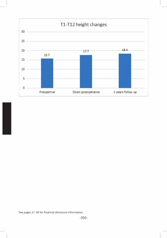
-350-
See pages 21- 60 for financial disclosure information.
-351-
♦ Indicates those faculty presentations in which the FDA has not cleared the drug and/or medical device for the use described (i.e., the drug or medical device is being discussed for an “off label” use). For full information, refer to page 14.
Sat., 4/30/16 Early Onset Scoliosis, Paper #169, 12:03 PM POSNA-2016 Maintenance of Preoperative Pulmonary Functions in Growing Rod Graduates Despite Improved Thoracic Height and Curve Correction
Charles E. Johnston II, MD; Dong-Phuong Tran, MS; Anna McClung, RN, BSNTexas Scottish Rite Hospital, Dallas, Texas
LOE-Therapeutic-Level III
Purpose: Long term outcomes of growing rod instrumentation are becoming more available as adolescents achieving sufficient spine length or requiring “final” fusion complete the lengthening segment of their treatment. We wish to report radiographic and pulmonary outcomes in 10 patients completing growing rod instrumentation treatment.
Methods: This is an IRB approved retrospective review of early-onset scoliosis patients from a single institution. Ten patients completed their surgical and radiographic at surgery with follow-up spanning from initial presentation to complete. Outcomes measured include radiographic parameters, pulmonary function test (PFT), and surgical data.
Results: Diagnoses included 3 idiopathic, 2 congenital, 1 ambulatory neuromuscular, and 4 syndromic. Mean preop age was 72.2 months (range 48 – 97), with preliminary non-operative treatment (halo-traction, cast/brace) delaying GRI 27 months (range 19 – 62) in 7 cases. Most recent surgery at age 137 months (range 122.7 – 157.2) included definitive fusion in 5 cases, with 2.5 yr f/u post fusion, and lengthening only in 1 patient, with 3 – 4 year further observation. Mean total procedures/patient were 8.5, including initial implantation, 2 unplanned revision/I&D, and 6.3 lengthenings (range 3 – 9). At last f/u mean age was 14 years (11.1 – 17). T1-12 height increased from 14.1 cm (9.9 – 17.7) preop to 22.6 (19.2 – 32.9) at last surgery, to 24.1 (19.2 – 32.9) at last f/u. Curve magnitude decreased from 94o (73 – 123) to 49o at last surgery and to 44 o (26 – 57) at last f/u. PFT’s were first performed at mean age 6+11 (4+10 – 8+7); last PFTs were performed at mean age 13+8 (11+7 – 17). Absolute FEV1 doubled from 0.70 L (0.4 – 1.2) to 1.6 L (1 – 2.4), however % pred FEV1 decreased from 56.1 to 55.3%. Absolute FVC also increased from 0.70 L (0.4 – 1.2) to 2.0 L (1 – 3) but only slight increase in % pred FVC, 56.2 to 58.9%. 7 rod/anchor complications in 4 patients were the only complications.
Conclusion: In spite of significant thoracic length gain and curve correction during 7 years of management with acceptable complication rate, pulmonary functions outcome at final follow-up maintained at preoperative values.
Significance: Significant improvement in curve deformity in orthopedics surgery only maintains pulmonary function at the preoperative level according to the current respiratory practice.
-352-
Sat., 4/30/16 Early Onset Scoliosis, Paper #170, 12:08 PM POSNA-2016 Complications and Radiographic Outcomes of Posterior Spinal Fusion (PSF) and Observation (OB) in Patients Who Have Undergone Distraction-Based Treatment for Early Onset Scoliosis (EOS)
Rodrigo Goes de Mendonca, MD; Amer Samdani, MD; Ron El-Hawary, MD, MSc, FRCSC; Alan Spurway, MSc; John T. Smith, MD; John B. Emans, MD; Tricia A. St Hilaire, MPH; Tara Flynn, BA; Stephen J. Soufleris, BS; Ryan P. Murphy, BS; Jeffrey R. Sawyer, MD; Children’s Spine Study Group Le Bonheur Children’s Hospital, Memphis, Tennessee
LOE-Therapeutic-Level III
Purpose: One treatment strategy for EOS patients is rib/spine-based distraction followed by PSF. However, some patients do not undergo PSF at distraction completion. The purpose of this study is to compare these two groups.
Methods: An IRB-approved review of the Children’s Spine Study Group (CSSG) registry was performed. Children with EOS undergoing OB or PSF at distraction completion were included. Radiographic parameters at the beginning and end of distraction, post PSF (PSF group), and final follow up including coronal/sagittal curve, sagittal spine length, T1-T12 and T1-S1 height were measured.
Results: There were 37 patients: 25 (67%) underwent PSF and 12 (33%) OB. The mean follow up is 15.6/16.2 years from distraction initiation in the PSF/OB groups respectively. The most common cEOS curve in both was C3. The mean age of initiation of distraction was 4.2 years in both groups. The mean procedure number in the PSF/OB groups was 11.3 (range 3-21)/8.7 (range 2-16) respectively.
At distraction completion, the groups were similar in coronal curve (59° PSF/51° OB), kyphosis (52° PSF/50° OB), T1-T12 length(24.2cm PSF/21.4cm OB) and T1-S1 length (39.0cm PSF/36.0cm OB). There was some intial correction in the PSF group (mean age 12.9 years) in coronal curve (10°), kyphosis (8°), T1-T2 length (1.6cm) and T1-S1 length (2.0cm). Post-PSF there were 23 complications in 13 patients (35%, range 1-4/pt) of which 11(48%) were device and 12 (52%) were disease-related. No complications occurred in the OB group. At final follow-up there was slightly less scoliosis (55° vs 59°), kyphosis (47° vs 56°) and greater spine length in the PSF group (T1-T12: 25.8 vs 22.8cm)(T1-S1: 39.0 vs 37.8cm). There were 4 additional procedures in the PSF and 0 in the OB group.
Conclusion: The decision to perform a PSF after distraction is complicated and multifactorial. While PSF does provide deformity correction and increased spine height/length it carries a high complication rate. Further studies are necessary to determine the optimal treatment for EOS patients completing distraction.
Significance: This study provides information useful in decision making regarding PSF or OB at the end of distraction-based treatment.
See pages 21- 60 for financial disclosure information.
-353-
♦ Indicates those faculty presentations in which the FDA has not cleared the drug and/or medical device for the use described (i.e., the drug or medical device is being discussed for an “off label” use). For full information, refer to page 14.
Sat., 4/30/16 Early Onset Scoliosis, Paper #171, 12:13 PM POSNA-2016 Classification of Early Onset Scoliosis Predicts Complications after Initiation of Growth Friendly Spine Surgery
Christen M. Russo, MD; Nicholas A. Feinberg, BA; Evan P. Trupia, MD; Hiroko Matsumoto; John T. Smith, MD; Amer Samdani, MD; Sumeet Garg, MD; John (Jack) M. Flynn, MD; Michael G. Vitale, MD, MPH; Children’s Spine Study Group; Growing Spine Study GroupColumbia University Medical Center, New York, New York
LOE-Prognostic-Level II
Purpose: There are high risks of postoperative complications of treatment with growing spine instrumentation for patients with early onset scoliosis (EOS). Currently, there is no reliable system to predict the risk of complications within this heterogeneous patient population. The purpose of this study is to investigate the predictive validity of the Classification of Early Onset Scoliosis (C-EOS) in regards to postoperative complications.
Methods: This is a retrospective study of patients undergoing growth friendly instrumentation for the treatment of EOS, with ≥5-year follow-up, identified in a national registry. We classified these patients using the C-EOS: Congenital (C), Neuromuscular (M), Syndromic (S), Idiopathic (I); major Cobb angle 1: <20°, 2: 20-50°, 3: 51-90°, 4: >90°; and kyphosis -: <20°, N: 20-50, +: >50°. Device and medical complications (e.g. device migration (DM), surgical site infection (SSI)), were classified using Smith classification for EOS. (I: no unplanned surgery, IIA: one unplanned surgery, IIB: multiple unplanned surgeries, III: abandoned instrumentation, IV: Death); “severe device complications” (SDC) were identified as IIB, III, and IV.
Results: Out of 156 patients (Congenital=70, Neuromuscular=40, Syndromic=28 and Idiopathic=18), 105 (67%) had device complications. 63 had SDC, 53 had DM, and 37 had SSI. In the neuromuscular, and syndromic etiologies, at Cobb angles over 50° (Classes 3 and 4), SDC increased to greater than 50%. Patients with congenital etiology and hyperkyphosis had increased risk of SDC at Cobb Classes 3 and 4. In Cobb Class 4 (>90°) patients with congenital etiology and hyperkyphosis, DM risk more than doubled versus lower Cobb Classes. Patients with neuromuscular etiology and hyperkyphosis had a 33% DM risk at Cobb Class 2, and approximately a 50% risk at Class 3 and above. In patients with neuromuscular etiology and hyperkyphosis, SSI risk was approximately 40% regardless of Cobb Class. In patients with syndromic etiology and hyperkyphosis, the risk of SSI was approximately 60% regardless of Cobb Class.
Conclusion: More severe C-EOS categories (neuromuscular etiology, higher Cobb Class, abnormal kyphosis) have increased risks of device complications with growth friendly surgery.
Significance: The Classification of Early Onset Scoliosis (C-EOS) has predictive validity for device-related complications in surgically treated patients with EOS.
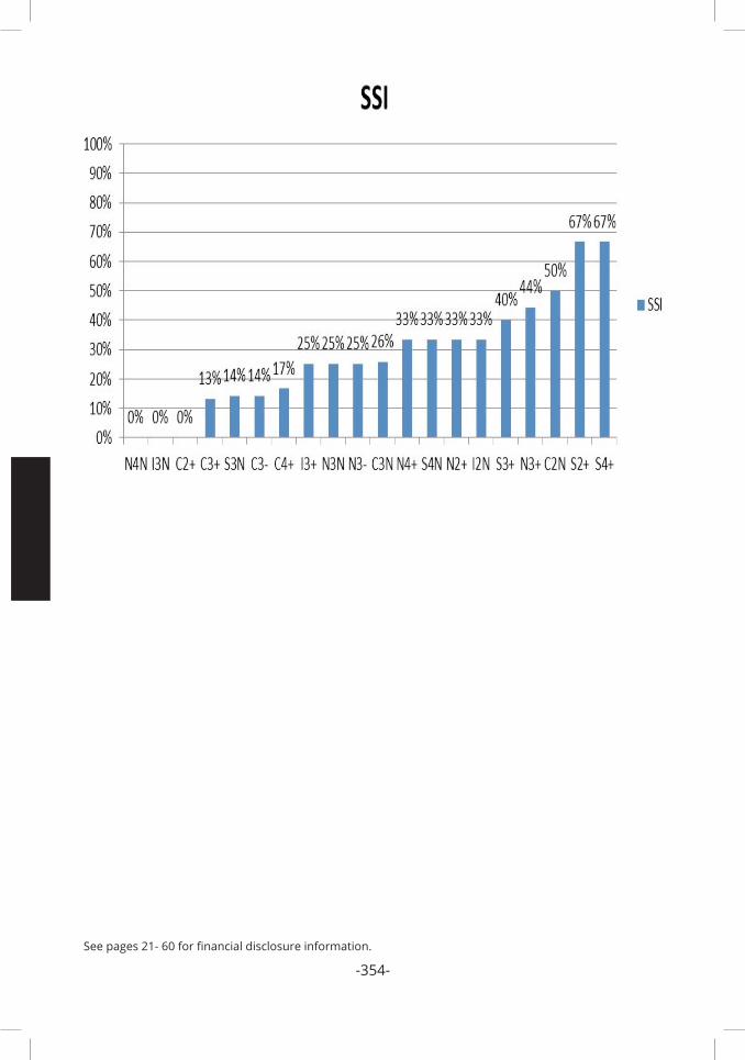
-354-
See pages 21- 60 for financial disclosure information.
-355-
♦ Indicates those faculty presentations in which the FDA has not cleared the drug and/or medical device for the use described (i.e., the drug or medical device is being discussed for an “off label” use). For full information, refer to page 14.
2016 POSTER PROGRAM
Paper Poster #1 (page 359) The Effects of Tendon Transfer to Cartilage: Insights into Tibialis Anterior Tendon Transfer in the Young Child Sam Abrams, MD; Thomas Crenshaw, PhD; Rachel Lenhart, PhD; Ellen Leiferman, DVM; Kenneth J. Noonan, MD; Matthew A. Halanski, MD American Family Children’s Hospital, University of Wisconsin, Madison Wisconsin Paper Poster #2 (page 361) Validation and Modification of a Severity of Illness Score for Children with Acute Hematogenous Osteomyelitis Megan Mignemi, MD; Alexander Athey, BS; Taylor Gheen, BA; Eduardo A. Lindsay, MD; ChanHee Jo, PhD; Lawson A.B. Copley, MD Children’s Medical Center of Dallas/Texas Scottish Rite Hospital for Children, Dallas, Texas Paper Poster #3 (page 363) Autograft vs Allograft Comparison in MPFL Reconstruction in Pediatric Patients Sean Waldron, MD; Christopher Flowers, MD; Tariq K. Hendawi, MD Ochsner Medical Center, New Orleans, Louisiana Paper Poster #4 (page 364) Peak D-Dimer is Predictive of Hospital Outcomes for Staph. aureus MSK Infection Thomas J. An, BA; Michael A. Benvenuti, BS; Megan Mignemi, MD; Gregory A. Mencio, MD; Steven A. Lovejoy, MD; Jonathan G. Schoenecker, MD, PhD; Jeffrey E. Martus, MD Vanderbilt Children’s Hospital, Nashville, Tennessee Paper Poster #5 (page 366) Ollier’s Disease: A Review of Disease Characteristics and Prognostic Factors Amy K. Williams, MD; Stephanie Punt, BS; John C. Shapton, BS; Ernest U. Conrad III, MD Seattle Children’s Hospital, Seattle, Washington
Paper Poster #6 (page 367) Pediatric Orthopaedic Surgery: Do We Have A Safe Practice? Mohammad M. Alzahrani, MBBS, MSc; Saad M. AlQahtani, MBBS, MSc (C), FRCS; Reggie C. Hamdy, MB, MSc (Ortho.), FRCS(C) Shriners Hospital for Children, Division of Orthopaedics, McGill University, Montreal, Quebec, Canada Paper Poster #7 (page 369) Do Males and Females Get Different Jobs within Pediatric Orthopedics? A Comparison from the POSNA Needs Assessment Survey Marielle A. Amoli, MD; Eric W. Edmonds, MD; Derek M. Kelly, MD; Jeffrey R. Sawyer, MD; John (Jack) M. Flynn, MD; Michael P. Glotzbecker, MD Le Bonheur Children’s Hospital, Memphis, Tennessee Paper Poster #8 (page 370) Patient Satisfaction in a High Volume Academic Clinic: Do Physician Assistants and Residents Help or Hurt? Benjamin D. Martin, MD; Allison Matthews, MSCR; Megan L. Young, MD; Yun Fei Wang, PhD; Matthew E. Oetgen, MD Children’s National Health System, Washington, District of Columbia Paper Poster #9 (page 371) Does Initial Cast Correction Predict Treatment Success for Infantile Scoliosis? Jaime A. Gomez, MD; Alexandra M. Grzywna, BA; Patricia E. Miller, MS; Lawrence I. Karlin, MD; John B. Emans, MD; Sumeet Garg, MD; James O. Sanders, MD; Paul D. Sponseller, MD; Jacques L. D’Astous, MD; Michael P. Glotzbecker, MD; Children’s Spine Study Group; Growing Spine Study Group Boston Children’s Hospital, Boston, Massachusetts Paper Poster #10 (page 373) Psychological Profile of Children Who Require Repetitive Surgeries for Early-onset Scoliosis: Is a Poor Quality of Life the Cost for a Straighter Spine? Cihan Aslan, MD; Erkan Sabri Ertas, MD; Seniz Ozusta, MD; H. Gokhan Demirkiran, MD; Z. Deniz Olgun, MD; Fatih Unal, MD; Muharrem Yazici, MD Hacettepe University, Ankara, Turkey Paper Poster #11 (page 374) Prevalence and Risk Factors of Venous Thromboembolism Among 57,183 Pediatric Orthopedic Trauma Patients: A Database Analysis Daniel Guzman; Samir Sabharwal, BA; Caixia Zhao, MD; Sanjeev Sabharwal, MD, MPH Rutgers-New Jersey Medical School, Newark, New Jersey
2016 Paper Poster Program, (cont.)
-356-
See pages 21- 60 for financial disclosure information.
Paper Poster #12 (page 376) Metal Ion Release During Growth-friendly Instrumentation for Early-Onset Scoliosis Ayaz Afandiyev, MD; Caglar Yilgor, MD; H. Gokhan Demirkiran, MD; Alpaslan Senkoylu, MD; Ahmet Alanay, MD; Muharrem Yazici, MD; Filiz Akbiyik, MD Hacettepe University, Ankara, Turkey Paper Poster #13 (page 377) Growing Rod and Vertical Expandable Prosthetic Titanium Rib Perform Differently for Idiopathic Early Onset Scoliosis at 5-year Follow-up Paul D. Sponseller, MD; Anna McClung, RN, BSN; Jeff Pawelek, BS; Ron El-Hawary, MD, MSc, FRCSC; George H. Thompson, MD; John T. Smith, MD; Michael G. Vitale, MD, MPH; Behrooz A. Akbarnia, MD; Malick Bachabi, MD; Children’s Spine Study Group; Growing Spine Study Group Johns Hopkins Medicine, Baltimore, Maryland Paper Poster #14 (page 379) Changes in Sagittal Cervical Alignment After Posterior Spinal Fusion for Adolescent Idiopathic Scoliosis: An Evaluation of 141 Patients Josh M. Pahys, MD; Jahangir Asghar, MD Shriners Hospital for Children - Philadelphia, Philadelphia, Pennsylvania Paper Poster #15 (page 380) Utility of Pinhole Magnification Skeletal Scintigraphy for Assessment of Revascularization of the Post-operative Pediatric Hip Holly B. Leshikar, MD, MPH; Heather Cole, BS; Gregory G. Polkowski II, MD; Jonathan G. Schoenecker, MD, PhD Vanderbilt Children’s Hospital, Nashville, Tennessee Paper Poster #16 (page 382) Growth Modulation by Stimulating the Growth Plate: A Pilot Study Kathleen D. Reay, MD; Camille R. Guzel, BS; Benjamin A. Alman, MD, FRCSC; Robert K. Lark, MD Duke University Medical Center, Durham, North Carolina Paper Poster #17 (page 383) Magnetically-controlled Growing Rods for Early Onset Scoliosis: A Multicenter Study of 23 Cases with Minimum 2 Years Follow-up Pooria Hosseini, MD, MSc; Jeff Pawelek, BS; Gregory Mundis, MD; Burt Yaszay, MD; John Ferguson, MD; Ilkka Helenius, MD; Kenneth Cheung, MD; Ahmet Alanay, MD; Alpaslan Senkoylu, MD; Hazem Elsebaie, MD; Behrooz A. Akbarnia, MD San Diego Spine Foundation, San Diego, California
-357-
♦ Indicates those faculty presentations in which the FDA has not cleared the drug and/or medical device for the use described (i.e., the drug or medical device is being discussed for an “off label” use). For full information, refer to page 14.
Paper Poster #18 (page 384) Implant Complications After Magnetically-Controlled Growing Rods for Early Onset Scoliosis: A Multicenter Retrospective Review Edmund Choi, MD; Burt Yaszay, MD; Gregory Mundis, MD; Pooria Hosseini, MD, MSc; Jeff Pawelek, BS; Ahmet Alanay, MD; Haluk Berk, MD; Kenneth Cheung, MD; H. Gokhan Demirkiran, MD; John Ferguson, MD; Tiziana Greggi, MD; Ilkka Helenius, MD; Guido La Rosa, MD; Alpaslan Senkoylu, MD; Behrooz A. Akbarnia, MD San Diego Spine Foundation, San Diego, California Paper Poster #19 (page 385) Parental Assessment of Outcomes of Congenital Upper Limb Differences: Analysis of the Pediatric Outcomes Data Collection Instrument (PODCI) Lindley B. Wall, MD; Tony S. Shen, BS; Summer Roberts, MA; Charles Goldfarb, MD Shriners Hospital for Children, St. Louis, Missouri Paper Poster #20 (page 386) Deletions of 5’ HOXC Genes are Associated with Lower Extremity Malformations Including Clubfoot and Vertical Talus Matthew B. Dobbs, MD Washington University School of Medicine, Saint Louis, Missouri
-358-
See pages 21- 60 for financial disclosure information.
2016 Paper Poster Program, (cont.)

-359-
♦ Indicates those faculty presentations in which the FDA has not cleared the drug and/or medical device for the use described (i.e., the drug or medical device is being discussed for an “off label” use). For full information, refer to page 14.
Paper Poster #1 POSNA-2016 The Effects of Tendon Transfer to Cartilage: Insights into Tibialis Anterior Tendon Transfer in the Young Child
Sam Abrams, MD; Thomas Crenshaw, PhD; Rachel Lenhart, PhD; Ellen Leiferman, DVM; Kenneth J. Noonan, MD; Matthew A. Halanski, MD American Family Children’s Hospital, University of Wisconsin, Madison Wisconsin
Purpose: Tibialis Anterior Tendon Transfers (TATT) are commonly performed in young children following Posneti casting for clubfeet and Dobbs casting for vertical tali. The recommended method for securing the tendon in each setting differs. In clubfeet, the classic TATT is delayed until cuneiform ossification and involves advancing the tendon through a hole drilled in the ossified cuneiform, whereas in the Dobbs method, the tendon is sewn onto the surface of the cartilaginous talus presumably to prevent injury to the un-ossified bone. In both procedures transferring the tendon through an un-ossified bone is avoided, however, the effect of transferring a tendon through an un-ossified bone (i.e. its cartilaginous precursor) is not well documented in the literature. The aim of this study was to determine if a tendon transferred through an un-ossified bone had any undesirable effect on subsequent bone development.
Methods: Twenty-one un-ossified porcine calcaneal apophysis underwent one of three surgical procedures (demonstrated in the attached figure) including: (A) apophyseal tunnel placement (no tendon present)(N=6), (B) placement of a detached tendon stump through the cartilanginous apophyseal tunnel (N=5), and (C) classic tendon transfer through the cartilaginous tunnel (N=10). The pigs were then sacrificed at 3 or 6 weeks. Gross anatomic, radiographic, and histologic evaluation of the calcanei was performed. Five calcanei underwent tendon transfer to bone in order to compare the load to failure between ossified and cartilaginous transfers using an MTS machine.
Results: The presence of tendon placed through the cartilaginous apophysis significantly altered the anatomic and radiographic appearance of the subsequent ossific nucleus when compared to the tunnel alone (15/15 vs. 2/6) p=0.0025. After tendon transfer through the cartilaginous tunnel, the calcaneal apophysis was split or fragmented. Representative examples of the radiographic, gross, and histologic appearance of normal (D, E, F) and altered (D’, E’, F’) calcaneal apophysis are shown in the attached figure. These demonstrate persistant cartilage and soft tissue replacement of the ossific nucleus in these areas. Despite these changes, MTS testing demonstrated that the average load to failure was not significantly different between osseous and non-osseous transfers.
Conclusion: Tendon transfers through un-ossified bones interfered with normal bone development leading to an undesired fragmentation of the ossific nucleus.
Significance: While the long-term consequence of these structural changes is unknown, these findings support current reccomendations to avoid tendon transfers through un-ossified bones and suggest the use of alternative methods of tendon fixation if transfers need to be performed prior to ossification.
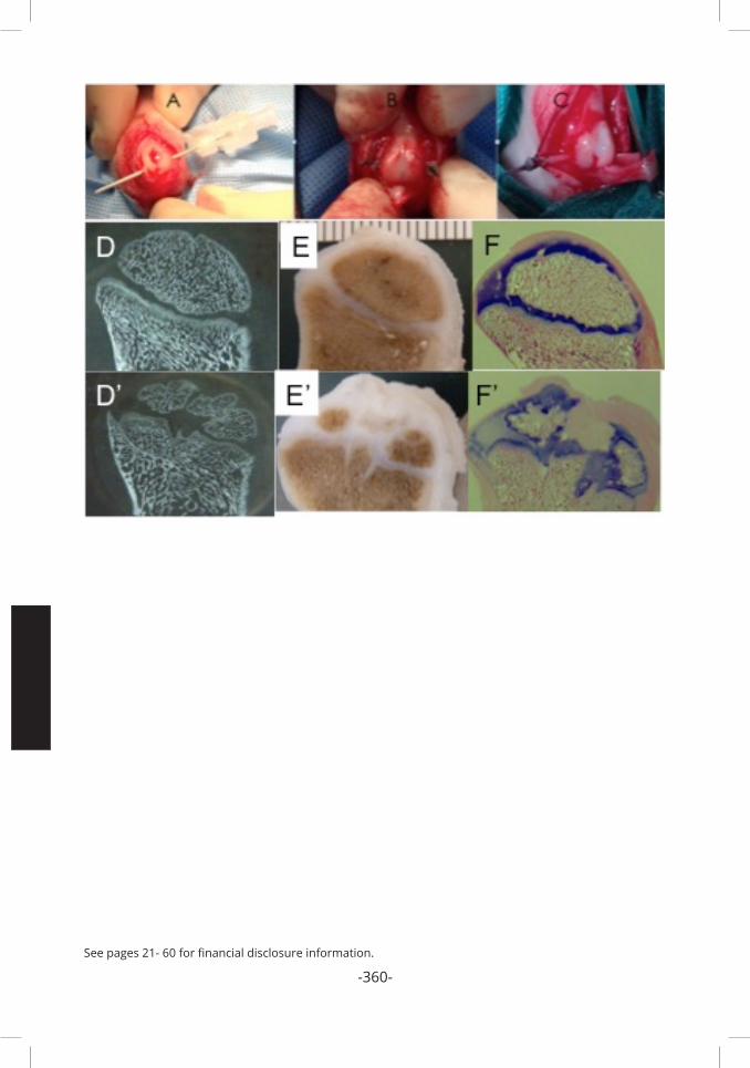
-360-
See pages 21- 60 for financial disclosure information.
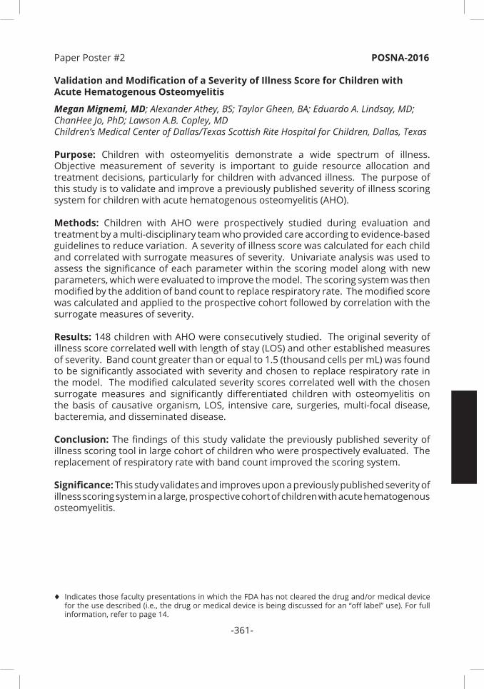
-361-
♦ Indicates those faculty presentations in which the FDA has not cleared the drug and/or medical device for the use described (i.e., the drug or medical device is being discussed for an “off label” use). For full information, refer to page 14.
Paper Poster #2 POSNA-2016 Validation and Modification of a Severity of Illness Score for Children with Acute Hematogenous Osteomyelitis
Megan Mignemi, MD; Alexander Athey, BS; Taylor Gheen, BA; Eduardo A. Lindsay, MD; ChanHee Jo, PhD; Lawson A.B. Copley, MD Children’s Medical Center of Dallas/Texas Scottish Rite Hospital for Children, Dallas, Texas Purpose: Children with osteomyelitis demonstrate a wide spectrum of illness. Objective measurement of severity is important to guide resource allocation and treatment decisions, particularly for children with advanced illness. The purpose of this study is to validate and improve a previously published severity of illness scoring system for children with acute hematogenous osteomyelitis (AHO).
Methods: Children with AHO were prospectively studied during evaluation and treatment by a multi-disciplinary team who provided care according to evidence-based guidelines to reduce variation. A severity of illness score was calculated for each child and correlated with surrogate measures of severity. Univariate analysis was used to assess the significance of each parameter within the scoring model along with new parameters, which were evaluated to improve the model. The scoring system was then modified by the addition of band count to replace respiratory rate. The modified score was calculated and applied to the prospective cohort followed by correlation with the surrogate measures of severity.
Results: 148 children with AHO were consecutively studied. The original severity of illness score correlated well with length of stay (LOS) and other established measures of severity. Band count greater than or equal to 1.5 (thousand cells per mL) was found to be significantly associated with severity and chosen to replace respiratory rate in the model. The modified calculated severity scores correlated well with the chosen surrogate measures and significantly differentiated children with osteomyelitis on the basis of causative organism, LOS, intensive care, surgeries, multi-focal disease, bacteremia, and disseminated disease.
Conclusion: The findings of this study validate the previously published severity of illness scoring tool in large cohort of children who were prospectively evaluated. The replacement of respiratory rate with band count improved the scoring system.
Significance: This study validates and improves upon a previously published severity of illness scoring system in a large, prospective cohort of children with acute hematogenous osteomyelitis.
-362-
See pages 21- 60 for financial disclosure information.
-363-
♦ Indicates those faculty presentations in which the FDA has not cleared the drug and/or medical device for the use described (i.e., the drug or medical device is being discussed for an “off label” use). For full information, refer to page 14.
Paper Poster #3 POSNA-2016 Autograft vs Allograft Comparison in MPFL reconstruction in Pediatric Patients
Sean Waldron, MD; Christopher Flowers, MD; Tariq K. Hendawi, MD Ochsner Medical Center, New Orleans, Louisiana
Purpose: Evaluate the differences in survivorship, clinical outcomes, and cost between autograft and allograft usage in Medial Patellofemoral ligament reconstruction in Pediatric patients.
Methods: This is a retrospective review of 56 patients who underwent MPFL reconstruction between 2012 and 2015 by a single surgeon for both acute and chronic patellar instability. Autograft Gracilis tendon was used for Group 1 ( 21 patients). Allograft Gracilis tendon was used for Group 2 (35 patients). Kujala Scores were gathered on all patients postoperatively. Survivorship, surgical time, and cost were calculated and compared.
Results: 56 patients were included in this study. Average age of all patients was 16 years old. There was no difference in age or chronicity between the groups. Group 1 (Autograft) showed significantly longer operative times (135 min vs 97 min; P < .001 ), higher rates of graft failure ( 28% vs 0%; P = .002) and worse Kujala Scores (80 vs 92 P = .003) postoperatively. All failures were in chronic dislocators and occurred at 14 months on average. With regards to cost, autograft did require more time for harvest and closure (40 min = $445 for this procedure) but allograft cost $1058. Overall, autograft was significantly more costly due to cost of reoperation.
Conclusion: In our review of graft choices for MPFL reconstruction, allograft is recommended for chronic patellar instability due to improved survivorship and clinical outcome scores, lower overall cost, and reoperation rate.
Significance: This study may show the significance of considering allograft tendon in MPFL reconstruction.
-364-
See pages 21- 60 for financial disclosure information.
Paper Poster #4 POSNA-2016 Peak D-Dimer is Predictive of Hospital Outcomes for Staph. aureus MSK Infection
Thomas J. An, BA; Michael A. Benvenuti, BS; Megan Mignemi, MD; Gregory A. Mencio, MD; Steven A. Lovejoy, MD; Jonathan G. Schoenecker, MD, PhD; Jeffrey E. Martus, MDVanderbilt Children’s Hospital, Nashville, Tennessee
Purpose: S. aureus expresses virulence factors that utilize the host coagulation system to facilitate abscess formation and hematogenous dissemination. During the initial stages of infection, S. aureus virulence factors coagulase and von Willebrand binding protein (vWbp) activate the coagulation system and promote fibrin deposition to form an abscess. Later expression of staphylokinase (SAK) leads to hematogenous dissemination by acting as a plasminogen activator to promote abscess rupture. Given these mechanisms of S. aureus dissemination, the authors tested the hypothesis that elevated fibrin breakdown products (D-dimer) in pediatric patients would be predictive of degree of infection dissemination and hospital outcomes.
Methods: An IRB approved retrospective study of children evaluated for S. aureus musculoskeletal infection (MSKI) was conducted over 5 years (2008-2013). Patients with D-dimer measurements were included in the study. Standard laboratory values and relevant clinical outcomes were collected. Infection severity was defined according to VODIS operational definitions defined in Figure 1a.
Results: Peak D-Dimer for S. aureus MSKI correlated significantly with hospital outcomes (length of stay (LOS), number of operative procedures, ICU LOS, days with fever > 101.5) (Figure 1b). The median initial and peak D-Dimers for disseminated infections were higher than those of local infections (Figure 1c), but this difference was not statistically significant. A cut-off D-dimer value of 1.2 ug/ml significantly differentiated disseminated infections from local infections with a sensitivity of 86% and a specificity of 50%.
Conclusion: Peak D-dimer levels were predictive of hospital outcomes for pediatric patients with S. aureus MSKI. Initial and peak D-dimer in disseminated infection were higher than in local infection, but the difference was not statistically significant. The difference in D-dimer between local and disseminated infection correlates with the pathophysiology of Staph. aureus dissemination through abscess rupture.
Significance: In children with S. aureus MSKI, monitoring D-Dimer at presentation and throughout their hospital stay allows for improved severity stratification.
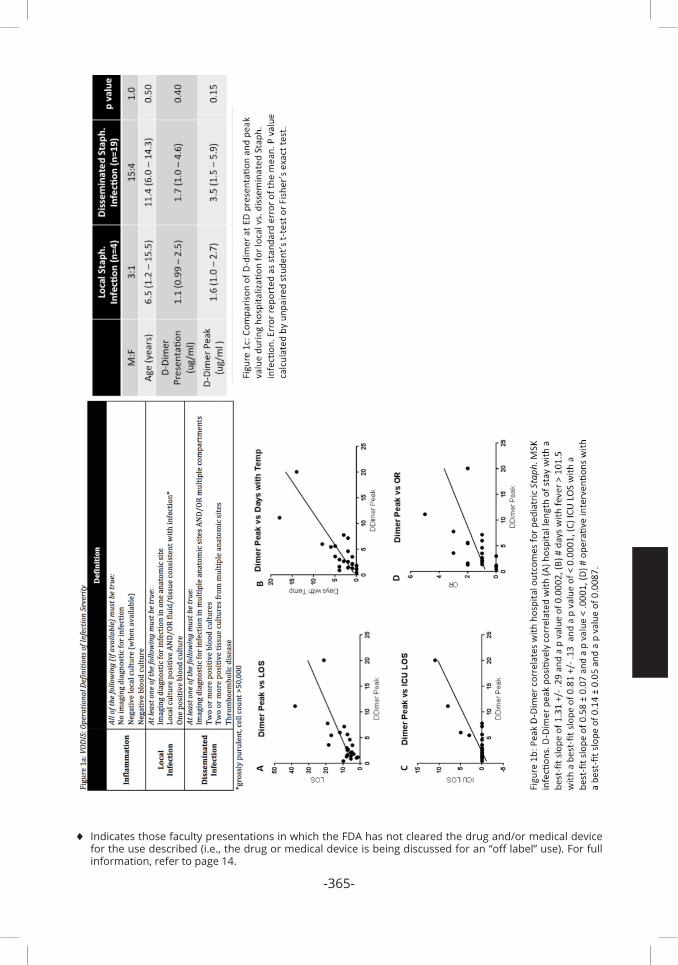
-365-
♦ Indicates those faculty presentations in which the FDA has not cleared the drug and/or medical device for the use described (i.e., the drug or medical device is being discussed for an “off label” use). For full information, refer to page 14.

-366-
See pages 21- 60 for financial disclosure information.
Paper Poster #5 POSNA-2016 Ollier’s Disease: A Review of Disease Characteristics and Prognostic Factors
Amy K. Williams, MD; Stephanie Punt, BS; John C. Shapton, BS; Ernest U. Conrad III, MD Seattle Children’s Hospital, Seattle, Washington
Purpose: Ollier’s Disease is a relatively rare condition in which patients have multiple enchondromas with the potential for malignant transformation to chondrosarcoma. It can vary from multiple mild enchondromas in the hands or feet to severe and debilitating cases involving multiple extremities, with leg length discrepancies. From our patient review, our purpose was to define prognostic variables that will influence treatment decisions.
Methods: All patients with an assigned diagnosis of Ollier’s Disease who were treated at our participating institutions were included. These diagnoses were found through ICD-9 coding and Sarcobase Tumor Registry from January 2000 through September 2015. All patients’ charts and associated imaging were retrospectively reviewed for: clinical presentation, age at presentation, co-morbidities/syndromes, extremity involvement, number of lesions, diameter of largest lesion, pathologic evaluation, number and type of surgical interventions, function, and progression to chondrosarcoma.
Results: On preliminary review of data from 13 patients showed the average age at presentation was 6.75 (0.92-13.92) years. Eight of the 13 patients were over age 18 years at last follow up with a mean age of 31.8 years. The primary site of involvement was most commonly the upper extremity 46%. Anatomic involvement favored the upper extremity (6/13), lower extremity (2/13), and both upper/lower (5/13). Unilateral distribution of enchondromas did not occur in the majority of patients. The average number of lesions per patient was 5.8. The average diameter of the largest lesion was 52 mm (7mm -100mm). 30.8% (4/13) patients had significant leg length discrepancy or poor function. The average number of surgical interventions per patient was 4.5 with the maximum being 16. 46% of these patients had progression to chondrosarcoma. The average diameter of the largest lesion in the patients with a secondary chondrosarcoma was 85 mm (45mm-100mm). Surgical interventions in the chondrosarcoma group averaged 6.5 procedures per patient and no patient with a secondary chondrosarcoma developed lung metastases.
Conclusion: Early trends in our data show that disease severity and progression to chondrosarcoma may be associated with upper extremity involvement and increased size (5-10cm) of the largest lesion. Secondary chondrosarcoma typically occurs with subtle increase in tumor size that should be followed carefully with dedicated imaging (MRI).
Significance: Patients with enchondromas greater than 5cm in diameter should be followed more carefully at 6-12 month intervals for possible development of chondrosarcomas.
-367-
♦ Indicates those faculty presentations in which the FDA has not cleared the drug and/or medical device for the use described (i.e., the drug or medical device is being discussed for an “off label” use). For full information, refer to page 14.
Paper Poster #6 POSNA-2016 Pediatric Orthopaedic Surgery: Do We Have A Safe Practice?
Mohammad M. Alzahrani, MBBS, MSc; Saad M. AlQahtani, MBBS, MSc (C), FRCS; Reggie C. Hamdy, MB, MSc (Ortho.), FRCS(C) Shriners Hospital for Children, Division of Orthopaedics, McGill University, Montreal, Quebec, Canada
Purpose: Forceful and repetitive maneuvers constitute a majority of pediatric orthopaedic surgical tasks thus subjecting the surgeon to added risks of musculoskeletal disorders during their years of practice. The aim of this study is to assess prevalence, characteristics and impact of musculoskeletal disorders among orthopaedic pediatric surgeons; who are considered a large proportion of the orthopaedic community.
Methods: A modified version of the physical discomfort survey was sent to surgeon members of the Pediatric Orthopaedic Society of North America (POSNA) via e-mail. Data was collected and descriptive statistics were analyzed. For data analysis, one-way ANOVA (Analysis Of Variance) and Fisher Exact test were performed to compare the variables where appropriate. P values<0.05 were considered statistically significant.
Results: A total of 402 surgeons completed the survey during the period of data collection. Of the respondents 76.4% were males, more than 84% were ≤65 years old and 81.6% were in practice for ≤30 years (Fig.1, 2). Majority (72.9%) of the participants were working in an academic institute and 8% worked in more than one institute. Overall 67% of the respondents reported that they had a work related MSK disorder. The majority of musculoskeletal complaints and disorders were low back pain (28.6%) and lateral elbow epicondylitis (15.4%) (Fig.3). Twenty six percent of surgeons with an injury required surgical treatment and 31.1% needed time off work due to their disorder. The number disorders diagnosed showed a significant increase with increasing age (p<0.001), working in a non-academic institute (p<0.05), working in more than 1 institute (p<0.05), practicing for more than 21 years (p<0.05). Also requiring treatment and time off due to the disorder were associated with increased number of disorders (p<0.001). in addition, surgeons who were >56 years old (p<0.001), in practice for more than 21 years (p<0.001), surgeons requiring surgical management of their disorder (p<0.001) and surgeons with an exacerbation of a previous disorder (p<0.001) were more likely to require time off work.
Conclusion: This study is the first of its kind to assess musculoskeletal injuries sustained by pediatric orthopaedic surgeons. The high incidence of these disorders may have a financial and psychological burden on the surgeon and thus the healthcare system.
Significance: This study sheds a light on awareness and possible preventive measures to help decrease the incidence of these disorders not only in orthopaedic surgeons but also the general surgical population.
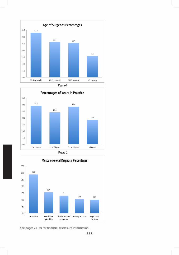
-368-
See pages 21- 60 for financial disclosure information.
-369-
♦ Indicates those faculty presentations in which the FDA has not cleared the drug and/or medical device for the use described (i.e., the drug or medical device is being discussed for an “off label” use). For full information, refer to page 14.
Paper Poster #7 POSNA-2016 Do Males and Females Get Different Jobs within Pediatric Orthopedics? A Comparison from the POSNA Needs Assessment Survey
Marielle A. Amoli, MD; Eric W. Edmonds, MD; Derek M. Kelly, MD; Jeffrey R. Sawyer, MD; John (Jack) M. Flynn, MD; Michael P. Glotzbecker, MD Le Bonheur Children’s Hospital, Memphis, Tennessee
Purpose: Over the past decades more females have entered surgical specialties. However, little research exists regarding how a surgeon’s gender impacts their practice. This study evaluates if gender differences exist within pediatric orthopaedic surgeons regarding practice types, on-call responsibilities, surgical volume, reasons for job choice, working with fellows/residents, and mentorship.
Methods: The 2015 Needs Assessment Survey consisting of 99 questions was sent to the POSNA membership. For the purposes of this survey, responses were analyzed by gender (a self-reported designation on the survey). Chi-squared testing assessed for statistical significance.
Results: A total of 530 surveys were returned (43.2% response rate). There were 422 males (79.6%) and 108 females (20.4%). There was a statistically significant difference in the practice types males and females joined (p<0.013). Males were more likely to become hospital or university employees, and go into solo practice compared to females that tended to join non-affiliated, multi-specialty or pediatric orthopaedic group practices. Additionally, there was a significant difference in reported number of surgical cases per week (25.6% of males reporting performing > 7 surgeries per week vs only 9.8% of females p< .004) and number of surgical hours per week (53.5% males reporting 10+ hours surgery per week vs only 31.9% females p<0.00001). There was also a significant difference in the percentage of males planning on retiring in the next five years compared to females (p<0.022). There was no difference in the amount of call, percentage working with residents or fellows, number of times one has changed practices, or mentorship available to new partners. In addition there was little difference in regards to reasons for one’s job selection as both genders rated quality of partners and an interesting practice as the two key factors when choosing a job.
Conclusion: While male and female surgeons choose a job based on similar characteristics, there are significant gender differences in practice models and surgical volumes reported. A greater percentage of males are going to reduce their workload or retire in the next 5 years, which may further increase the percentage of female surgeons in the workforce.
Significance: At this point, gender differences have not been studied for surgeons in pediatric specialties. However, given that we have identified gender differences in pediatric orthopedics, future work is required to fully understand the differences and their effect on the workforce. This study will serve as a benchmark for the future studies addressing these issues.

-370-
See pages 21- 60 for financial disclosure information.
Paper Poster #8 POSNA-2016 Patient Satisfaction in a High Volume Academic Clinic: Do Physician Assistants and Residents Help or Hurt?
Benjamin D. Martin, MD; Allison Matthews, MSCR; Megan L. Young, MD; Yun Fei Wang, PhD; Matthew E. Oetgen, MD Children’s National Health System, Washington, District of Columbia
Purpose: Patient satisfaction is a common metric to evaluate physicians and hospitals. Achieving high satisfaction scores is challenging in a high volume clinic. To improve access to care is such clinics, mid-level providers are utilized, including physician assistants (PAs). It has yet to be reported whether a patient’s satisfaction is influenced by the level or quantity of providers seen at a consultation visit. We sought to determine if these factors negatively impact satisfaction.
Methods: A consecutive set of English speaking patients presenting to a single tertiary outpatient pediatric orthopaedic clinic over a four-week period were confidentially surveyed. The survey included six questions from the institution’s Press-Ganey satisfaction survey, an estimate of total time in clinic, and the provider(s) seen at that visit (PA only, PA and attending, resident and attending, or attending only). The Press-Ganey results between the four groups were compared using a two-tailed analysis of covariance (ANCOVA) with an alpha level of 0.05.
Results: A total of 269 surveys were returned for an overall response rate of 29%. Of those surveyed, 19 (7%) were seen by a PA only, 89 (34.5%) by a PA and attending, 76 (29.5%) by a resident and attending, and 75 (29.1%) by an attending only. No statistical significance was detected in satisfaction between the provider seen and type of visit (initial or follow-up). Multiple satisfaction variables, including the adequacy of explanation of care provided (p=0.0154), patient’s confidence in care provided (p=0.0203) and likelihood of recommending provider (p=0.0055), were inversely related to total time in clinic.
Conclusion: We found overall time spent in clinic, rather than the type of provider seen, significantly influences patient reported satisfaction of outpatient pediatric orthopaedic office visits with shorter clinic visits being associated with higher patient reported satisfaction scores. Our findings show, pediatric orthopaedic patients are generally satisfied with care provided by all levels of medical professionals including physician assistants and orthopaedic residents. Patient reported satisfaction is increasingly important to healthcare systems and utilizing orthopaedic residents and mid-level providers to evaluate patients in outpatient clinics does not appear to negatively impact patient satisfaction.
Significance: Provider type used to evaluate patients in outpatient pediatric orthopaedic clinics does not appear to influence patient visit satisfaction, rather satisfaction scores appear to be inversely related to the length of the clinic visit, with shorter clinic visits producing higher patient satisfaction.
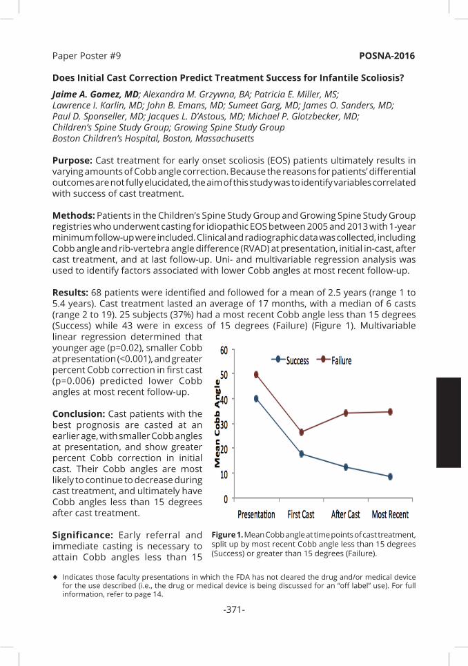
-371-
♦ Indicates those faculty presentations in which the FDA has not cleared the drug and/or medical device for the use described (i.e., the drug or medical device is being discussed for an “off label” use). For full information, refer to page 14.
Paper Poster #9 POSNA-2016 Does Initial Cast Correction Predict Treatment Success for Infantile Scoliosis?
Jaime A. Gomez, MD; Alexandra M. Grzywna, BA; Patricia E. Miller, MS; Lawrence I. Karlin, MD; John B. Emans, MD; Sumeet Garg, MD; James O. Sanders, MD; Paul D. Sponseller, MD; Jacques L. D’Astous, MD; Michael P. Glotzbecker, MD; Children’s Spine Study Group; Growing Spine Study Group Boston Children’s Hospital, Boston, Massachusetts
Purpose: Cast treatment for early onset scoliosis (EOS) patients ultimately results in varying amounts of Cobb angle correction. Because the reasons for patients’ differential outcomes are not fully elucidated, the aim of this study was to identify variables correlated with success of cast treatment.
Methods: Patients in the Children’s Spine Study Group and Growing Spine Study Group registries who underwent casting for idiopathic EOS between 2005 and 2013 with 1-year minimum follow-up were included. Clinical and radiographic data was collected, including Cobb angle and rib-vertebra angle difference (RVAD) at presentation, initial in-cast, after cast treatment, and at last follow-up. Uni- and multivariable regression analysis was used to identify factors associated with lower Cobb angles at most recent follow-up.
Results: 68 patients were identified and followed for a mean of 2.5 years (range 1 to 5.4 years). Cast treatment lasted an average of 17 months, with a median of 6 casts (range 2 to 19). 25 subjects (37%) had a most recent Cobb angle less than 15 degrees (Success) while 43 were in excess of 15 degrees (Failure) (Figure 1). Multivariable
Figure 1. Mean Cobb angle at time points of cast treatment, split up by most recent Cobb angle less than 15 degrees (Success) or greater than 15 degrees (Failure).
linear regression determined that younger age (p=0.02), smaller Cobb at presentation (<0.001), and greater percent Cobb correction in first cast (p=0.006) predicted lower Cobb angles at most recent follow-up.
Conclusion: Cast patients with the best prognosis are casted at an earlier age, with smaller Cobb angles at presentation, and show greater percent Cobb correction in initial cast. Their Cobb angles are most likely to continue to decrease during cast treatment, and ultimately have Cobb angles less than 15 degrees after cast treatment.
Significance: Early referral and immediate casting is necessary to attain Cobb angles less than 15
-372-
See pages 21- 60 for financial disclosure information.
degrees in infantile idiopathic scoliosis. Patients’ percent Cobb correction, which may represent curve flexibility and/or cast quality, is a predictor of treatment success when age and starting Cobb angle are also taken into account.
-373-
♦ Indicates those faculty presentations in which the FDA has not cleared the drug and/or medical device for the use described (i.e., the drug or medical device is being discussed for an “off label” use). For full information, refer to page 14.
Paper Poster #10 POSNA-2016 Psychological Profile of Children Who Require Repetitive Surgeries for Early-onset Scoliosis: Is a Poor Quality of Life the Cost for a Straighter Spine?
Cihan Aslan, MD; Erkan Sabri Ertas, MD; Seniz Ozusta, MD; H. Gokhan Demirkiran, MD; Z. Deniz Olgun, MD; Fatih Unal, MD; Muharrem Yazici, MD Hacettepe University, Ankara, Turkey
Purpose: Early-onset scoliosis (EOS) impacts psychosocial status with its requirement for repetitive surgeries. Despite the increasing awareness of psychosocial alterations in chronic surgical disorders, there is a dearth of information regarding this group. This study aimed to assess the psychosocial status of EOS children undergoing multiple procedures and evaluate the associations with etiological variables.
Methods: EOS patients 6-18 years old, undergoing traditional growing rod treatment with more than 5 surgical procedures, ambulators, and neurologically and mentally intact were included. Patients were screened for psychiatric disorders before inclusion. Quality of Life Scale for Children (PedsQL), Strengths and Difficulties Questionnaire (SDQ) self-report form, Beck Depression Inventory, Children Depression Inventory (CDI), Beck Anxiety Inventory (BAI), Self-Report for Childhood Anxiety Related Disorders (SCARED) were completed by the children. Quality of Life Scale Parental Form, Strengths and Difficulties Questionnaire Parent Form, and Attention Deficiency and Hyperactivity Scale were completed by their parents.
Results: 21 (9 male, 12 female) patients met the inclusion criteria. Average age was 6.4 years (range 4-10.5) at index surgery; 13.5 years (range 8-17) at final follow-up. The mean number of procedures (incl. index and lengthenings) was 13 (range 6 to 18). Mean follow-up was 83.9 months (range 36-122). Depression was observed in 23.8% of patients, and generalized anxiety disorder (GAD) in 33%. No correlation was found between psychiatric diagnoses and orthopedic variables. Patients in the study group were more likely than the general population to have a psychiatric diagnosis. Number of procedures undergone was found to correlate negatively with BAI, SCARED and the behavioral difficulties domain of SDQ parent form score and positively with emotional functioning, psychosocial health summary score, pedsQL total score and increased social and physical functioning per parent reports. Non-idiopathic etiology was found to be related to increased behavioral difficulties and hyperactivity/concentration difficulties compared to idiopathic patients. Non-idiopathic patients also demonstrated significantly lower physical, emotional and social functioning.
Conclusion: A higher prevalence of depressive and anxiety symptoms were observed in patients with EOS along with dysfunctional areas of daily life. Other comorbidities may also contribute to dysfunction and other difficulties.
Significance: Determination of the aspects of EOS treatment that have a negative impact on psychosocial functioning may allow for more competent help to be provided to these patients.
-374-
See pages 21- 60 for financial disclosure information.
Paper Poster #11 POSNA-2016 Prevalence and Risk Factors of Venous Thromboembolism Among 57,183 Pediatric Orthopedic Trauma Patients: A Database Analysis
Daniel Guzman; Samir Sabharwal, BA; Caixia Zhao, MD; Sanjeev Sabharwal, MD, MPH Rutgers-New Jersey Medical School, Newark, New Jersey
Purpose: There remains uncertainty regarding the prevalence and risk factors of Venous Thromboembolism (VTE) in the pediatric orthopedic trauma population. The purpose of our study was to determine the prevalence of VTE amongst children sustaining musculoskeletal trauma and assess which subsets of this population are most at risk for being diagnosed with VTE.
Methods: After obtaining the 2012 KID HCUP inpatient database from the Agency for Healthcare Research and Quality, we conducted analyses in SAS (version 9.3). Four groups—upper limb fractures, lower limb fractures, vertebral fractures, pelvic fractures—were identified, extracted, and assessed, individually and as a pooled “total fractures” population. Additionally, we established and assessed a “multiple fractures” category, which captured patients fulfilling at least two of the four groups’ criteria. We calculated the prevalence of VTE (Deep Venous Thrombosis and/or Pulmonary Embolism) and mortality, as well as association of VTE with putative categorical risk factors—osteomyelitis, spinal cord injury, Central Venous Catheter (CVC) placement, internal and external fixation, obesity, coagulopathy, diabetes, blood loss—and generated odds ratios via Mantel-Haenszel testing. We analyzed putative continuous risk factors for VTE—namely age, length of stay and hospital charges—via independent samples t-testing. Univariate analyses were corroborated with a multivariate logistic regression of the pooled population.
Results: The table below displays the results of our analysis. Note that for cells with fewer than 10 patients, an asterisk (*) replaces the numerical figure, per HCUP patient protection guidelines.
Conclusion: The overall prevalence of VTE in the orthopedic pediatric trauma population was 6.8 per 1,000 children, with highest frequency observed amongst children sustaining fractures of the axial skeleton (vertebral/pelvic) and multiple fractures. Patients diagnosed with VTE were significantly older and accrued longer length of stay and higher cost of care. Of the available risk factors, CVC placement, coagulopathy, and spinal cord injury had the highest association with a diagnosis of VTE. Children undergoing external fixation of fractures have a much higher prevalence of VTE than those treated with internal fixation. In pediatric patients with upper limb fractures, VTE was significantly associated with increased mortality.
Significance: Based on this cross-sectional study, we identified the prevalence of VTE and associated factors among distinct subsets of children and adolescents sustaining musculoskeletal trauma. A prospective, multicenter study would be helpful to confirm
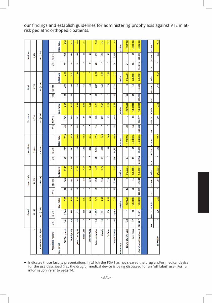
-375-
♦ Indicates those faculty presentations in which the FDA has not cleared the drug and/or medical device for the use described (i.e., the drug or medical device is being discussed for an “off label” use). For full information, refer to page 14.
our findings and establish guidelines for administering prophylaxis against VTE in at-risk pediatric orthopedic patients.
-376-
See pages 21- 60 for financial disclosure information.
Paper Poster #12 POSNA-2016 Metal Ion Release During Growth-friendly Instrumentation for Early-Onset Scoliosis
Ayaz Afandiyev, MD; Caglar Yilgor, MD; H. Gokhan Demirkiran, MD; Alpaslan Senkoylu, MD; Ahmet Alanay, MD; Muharrem Yazici, MD; Filiz Akbiyik, MD Hacettepe University, Ankara, Turkey
Purpose: Spinal instruments are reported to cause localized metal debris and distribute systemically to settle on distant organs. Children with early onset deformities are instrumented at earlier ages and live with these metallic implants for a substantial amount of time. Yet, no research has yet focused on metal distribution in growing rod applications. The aim of this study is to compare age-matched growing rod (GR) and magnetically controlled growing rods (MCGR) groups to non-instrumented controls.
Methods: The GR and MCGR applications of three clinics were included in this study. The blood samples were obtained in three months between December 2014 and February 2015 on a first come first served basis for this cross-sectional study. A total of 52 children were included in this study. GR group included 15 children. Mean age was 10.7 (range 6-15). MCGR group included 22 children. Mean age was 8.5 (range 2-13). Fifteen age-matched non-operated children formed the control group. Mean age was 10.4 (range 5-15). Serums were analyzed for titanium, vanadium, aluminum, and boron levels. Kruskal-wallis and Mann-Whitney U tests were used to compare between groups.
Results: Mean serum titanium level in the control, GR and MCGR groups were 2.8±1.4, 7.3±4.3 and 10.2±6.8 µg/L, consecutively. GR and MCGR group serum titanium levels were higher than the controls (P=0.008 and P=0.000). Mean serum vanadium level in the control, GR and MCGR groups were 0.2±0.0, 0.2±0.0 and 0.5±0.5 µg/L, consecutively. MCGR group serum vanadium level is higher than the controls (P=0.000) and the GR group (P=0.004). Mean serum aluminum level in the control, GR and MCGR groups were 5.4±4.1, 8.1±7.4 and 7.8±5.1 µg/L, consecutively. Mean serum boron level in the control, GR and MCGR groups were 86.7±2.7, 86.9±2.5 and 85.0±6.6 µg/L, consecutively. The distribution of aluminum and boron were found to be similar across three groups (P=0.675 and P=0.396).
Conclusion: Both GR and MCGR applications significantly release titanium and vanadium. MCGR releases more titanium and significantly more vanadium than the traditional GR. This excessive release seems to be related to the magnetic distractions and the structural properties of the MCGR device.
Significance: Increased metal ion levels during growth-friendly treatment for EOS have not been previously reported. The long-term effect of this increase is unknown, and requires further research.
-377-
Paper Poster #13 POSNA-2016 Growing Rod and Vertical Expandable Prosthetic Titanium Rib Perform Differently for Idiopathic Early Onset Scoliosis at 5-year Follow-up
Paul D. Sponseller, MD; Anna McClung, RN, BSN; Jeff Pawelek, BS; Ron El-Hawary, MD, MSc, FRCSC; George H. Thompson, MD; John T. Smith, MD; Michael G. Vitale, MD, MPH; Behrooz A. Akbarnia, MD; Malick Bachabi, MD; Children’s Spine Study Group; Growing Spine Study Group Johns Hopkins Medicine, Baltimore, Maryland
Purpose: Distraction-based surgical treatment is employed in idiopathic early-onset scoliosis (IEOS) to allow spinal growth while preventing curve progression until skeletal maturity. Our aim was to compare clinical and radiographic results between two forms of distraction for IEOS: GR and VEPTR.
Methods: Two multicenter EOS databases were studied to identify GR and VEPTR patients who had idiopathic etiologies with minimum 5-year follow up and ≥ 4 lengthenings. Study time points included pre-operative, immediate post-operative, and most recent visit prior to final treatment.
Results: 50 GR and 22 VEPTR patients were included. Mean age at surgery was similar (5.5 yrs GR vs. 4.3yrs VEPTR, p=.0444), as was preoperative main curve, kyphosis, spinal and thoracic height (Table 1). The number of levels spanned was similar (11.5 GR vs. 12.1 VEPTR, p=.318); a unilateral construct was present in 50% (11/22) and 30% (15/50) of VEPTR and GR patients respectively. Follow up duration was similar and VEPTR patients underwent more total procedures (14.8 VEPTR vs. 9.6 GR, p=<0.001). Postoperatively, GR patients had significantly greater curve correction while kyphosis and height measurements were similar (Table 1). Single GR constructs tended to have fewer complications while the curve correction was equivalent to dual constructs. Bilateral constructs tended to be associated with fewer overall procedures, attained better curve correction, maintained correction better and had more complications. VEPTR patients had a higher incidence of deep infection (22.7% VEPTR vs. 8% GR, p= 0.084). The number of patients with at least one complication was similar (66.0% GR vs. 81.8% VEPTR, p=.174). At the time of final fusion, GR patients had less loss of initial curve correction and greater % gain in thoracic height (Table 1). At their most recent visit, GR patients maintained their curve correction better, had less kyphosis, and 15% greater gain in thoracic height (Table1).
Conclusion: GR patients had significantly greater initial correction of their main curves and maintained this correction at latest follow-up. GR patients showed greater continued growth of thoracic height during the lengthening period. At most recent follow-up, GR patients had greater % gain thoracic height, less kyphosis, and a lower incidence of wound related complications.
♦ Indicates those faculty presentations in which the FDA has not cleared the drug and/or medical device for the use described (i.e., the drug or medical device is being discussed for an “off label” use). For full information, refer to page 14.
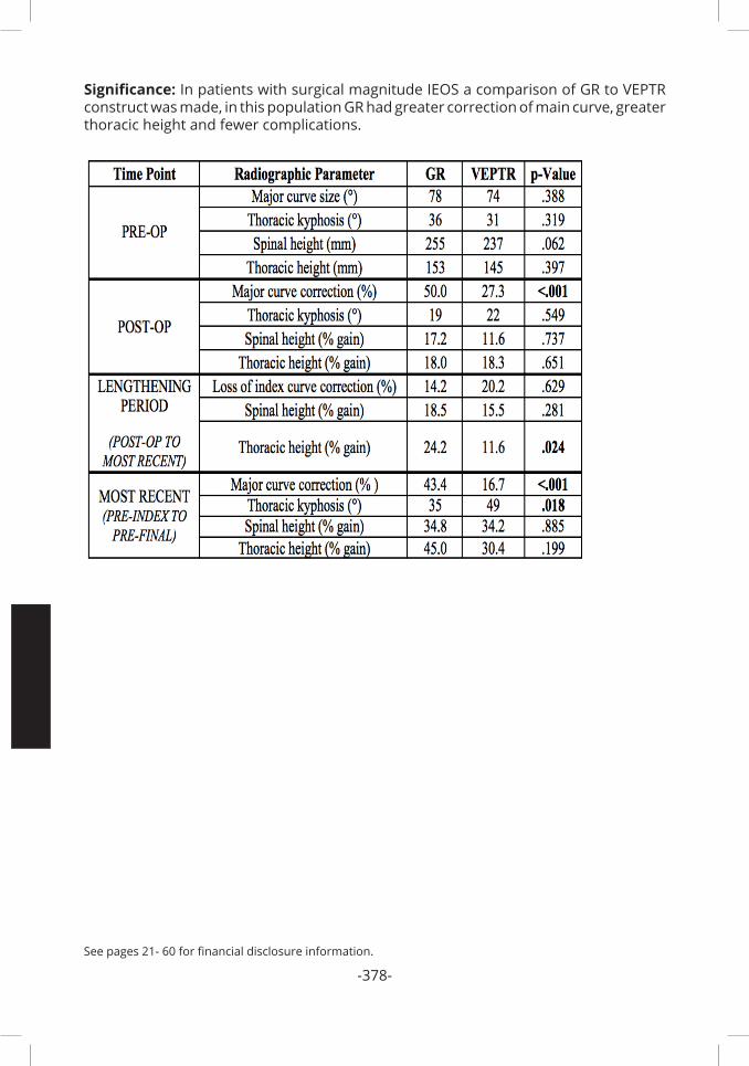
-378-
See pages 21- 60 for financial disclosure information.
Significance: In patients with surgical magnitude IEOS a comparison of GR to VEPTR construct was made, in this population GR had greater correction of main curve, greater thoracic height and fewer complications.
-379-
♦ Indicates those faculty presentations in which the FDA has not cleared the drug and/or medical device for the use described (i.e., the drug or medical device is being discussed for an “off label” use). For full information, refer to page 14.
Paper Poster #14 POSNA-2016 Changes in Sagittal Cervical Alignment After Posterior Spinal Fusion for Adolescent Idiopathic Scoliosis: An Evaluation of 141 Patients
Josh M. Pahys, MD; Jahangir Asghar, MD Shriners Hospital for Children - Philadelphia, Philadelphia, Pennsylvania
Purpose: Loss of normal thoracic kyphosis (TK: T2-T12 Cobb) is often seen in patients with adolescent idiopathic scoliosis (AIS). However, its effect on the cervical sagittal alignment before and after posterior spinal fusion (PSF) has been less well studied. Cervical kyphosis (CK) is strongly associated with reduced health related quality of life measures and increased disability scores in adults, however its effects in the AIS population is unknown.
Methods: A multicenter, prospective AIS database retrospectively identified 141 patients with minimum 2-year followup after PSF with preop, initial postop, and 2 year postop radiographs that included the skull to pelvis. CK was defined as a positive: C2-C7 Cobb>0°, while cervical lordosis (CL) was negative: C2-C7 Cobb<0°.
Results: Factors associated with developing post-op CK were: preop CK (p=0.001, r = 0.28), lower preop/postop TK (p<0.01, r = -0.37), lower preop/postop T1 slope (p<0.01, r = -0.62), and negative postop C7 sagittal vertical axis (p=0.04, r = -0.39). (Figure 1) 75% of patients with preop CK remained kyphotic at 2 years (p=0.001) and had lower preop/postop SRS scores (pain, function, total score; p<0.05). At 2 years, mean TK measured 32.9°+/-10.3°, which was an increase of only 17.3% from preop (p=0.6). 76 patients (54%) had an increase in TK, while TK decreased in 65 patients (46%). Sub-analysis revealed that patients with a postop TK>40°, reliably maintained or achieved postop CL (p=0.007). However, TK>40° was seen in only 23% of patients.
Conclusion: This is the largest study to date to evaluate the cervical alignment in adolescent idiopathic scoliosis (AIS) patients before and after posterior spinal fusion (PSF). Preoperative cervical kyphosis (CK) led to a higher rate of cervical kyphosis and decreased SRS scores at 2 years postop. Postoperative thoracic kyphosis>40° consistently resulted in maintaining and/or achieving cervical lordosis. In our cohort, however, cervical lordosis was only present in 35.6% of patients at 2 years postop. This study further highlights the importance of proper sagittal plane restoration during deformity correction for AIS. In this multicenter study, the majority of patients demonstrated suboptimal cervical and thoracic sagittal alignment after surgery, which resulted in lower quality of life scores at two years postoperative.
Significance: The majority of patients in this study demonstrated cervical kyphosis postoperatively, which has been associated with decreased HRQOL scores in adults. However, postoperative thoracic kyphosis (T2-T12) >40° was able to reliably achieve or maintain cervical lordosis in these AIS patients and may serve as a potential goal for surgeons. This study further highlights the need for adequate sagittal plane restoration of not only the thoracolumbar spine, but also the cervical spine to optimize patient outcomes.
-380-
Paper Poster #15 POSNA-2016 Utility of Pinhole Magnification Skeletal Scintigraphy for Assessment of Revascularization of the Post-operative Pediatric Hip
Holly B. Leshikar, MD, MPH; Heather Cole, BS; Gregory G. Polkowski II, MD; Jonathan G. Schoenecker, MD, PhD Vanderbilt Children’s Hospital, Nashville, Tennessee
Purpose: Fracture healingrequires proper revascularization. The vascularity of the proximal femur in the skeletally immature is susceptible to revascularization deficits following injury because of the precarious intracapsular blood flow to the proximal femoral epiphysis. The physis acts as an avascular barrier preventing metaphyseal vessels from entering the epiphysis. At physeal closure, the barrier no longer exists which allows ingrowth of the metaphyseal arteries.Therefore, assessment of revascularization of the proximal femur following acute injury (slipped capital femoral epiphysis (SCFE) or femoral neck fracture) or proximal osteotomy such as a capital realignment or femoral head reduction in children is critical for determining progression of weight bearing and/or need for further intervention . Although excellent in demonstrating vascularity in bone, metallic implants limit the utility of CT or MR angiography in this patient population. We hypothesized that pinhole skeletal scintigraphy is not only is capable of demonstrating revascularization but provides sufficient detail to determine the pattern of revascularization of the proximal metaphysis and epiphysis in this population.
Methods: All skeletally immature patients who underwent pinhole scintography following operative treatment for SCFE or femoral neck fracture from January 2013 to October 2015 were included. We imaged 17 patients in total: four patients with femoral neck fractures, five patients with epiphyseal unstable SCFE (Delbet type 1 femoral neck fractures), five patients with SCFE following capital realignment and three patients with Legg Calve Perthes’ disease following femoral head reduction with the method. Vascularity was assessed for timing of return to flow that crossed the fracture or osteotomy and pattern of revascularization.
Results: Patients were not allowed to weight bear until definitive evidence of revascularization. Thirteen of 17 patients were found to have excellent vascularity on scintography by four weeks post trauma or osteotomy and were allowed to weight bearing without any subsequent complications or further interventions. Four hips failed to revascularize. In all of these cases, scintography provided valuable information as to the location and extent of avascularity directing further treatment (figure 1). Three of four of these patients who failed to revascularize progressed to need total hip replacement.
Conclusion: Pinhole Magnification Skeletal Scintigraphy provides a detailed evaluation of the revascularization of the proximal femur metaphysis and epiphysis of children with treatment for acute injuries. Surprisingly, most children revascularize their proximal femur within 4-5 weeks following trauma or surgery.
Significance: Pinhole skeletal scintigraphy provides critical information regarding extent of revascularization in the post-operative immature, which is critical in critical in determining the timing of weight bearing, or if further intervention is needed. See pages 21- 60 for financial disclosure information.
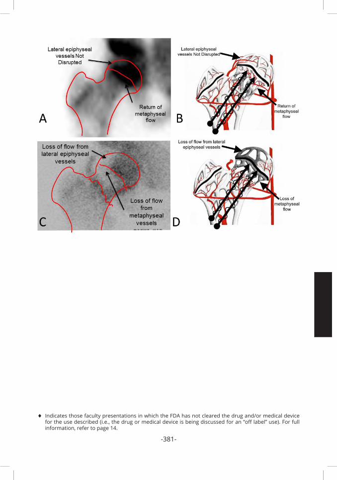
-381-
♦ Indicates those faculty presentations in which the FDA has not cleared the drug and/or medical device for the use described (i.e., the drug or medical device is being discussed for an “off label” use). For full information, refer to page 14.
-382-
See pages 21- 60 for financial disclosure information.
Paper Poster #16 POSNA-2016 Growth Modulation by Stimulating the Growth Plate: A Pilot Study
Kathleen D. Reay, MD; Camille R. Guzel, BS; Benjamin A. Alman, MD, FRCSC; Robert K. Lark, MD Duke University Medical Center, Durham, North Carolina
Purpose: Limb length discrepancy and angular deformities of the growing skeleton have traditionally been treated by methods that induce permanent or temporary growth arrest. A potential alternative to correct these deformities is to stimulate the growing skeleton. The purpose of the study is to determine if the application of low-pulse ultrasound or direct injection of growth hormone near the physis could be used to stimulate growth locally in rabbits.
Methods: Three month old rabbits (equivalent to a 12 year old child) were randomized to treatment with one of four protocols: 1) high-dose 400 μg/kg periosteal injection at the distal femoral metaphysis with recombinant Growth Hormone (GH) (Somatropin), 2) transdermal low frequency pulsed ultrasound (LFPU); 3) controls included periosteal injection with saline, or 4) no treatment (negative control). Calcein blue 10mg/kg (Sigma M1255, 4-Methylumbelliferone-8-methyliminodiacetic acid) was injected periosteally on 1st and 18th days. The animals were sacrificed 3 days after the second calcein injection. Following femoral bone harvest, the bones were preserved in 70% Ethanol and embedding in methyl methacrylate to preserve the calcein label for growth rate calculation and for histology analysis. Measurement by a blinded reviewer from the growth plate to the first calcein labeled line gave the growth rate. The contralateral leg was used as an additional control.
Results: Control animals (n=10) grew 0.32mm (standard deviation 0.12mm) in the 21 days since the first calcein injection. Rabbit growth plates stimulated with low-pulse ultrasound grew 0.52mm (standard deviation 0.18mm, n=5), and growth hormone stimulation grew 0.68mm (standard deviation 0.24mm, n=5). The contralateral growth hormone stimulated leg grew 0.48mm. The growth difference between the ultrasousnd and growth hormone stimulated legs compared to the growth of control and contralateral legs was found to be statistically significant (p<0.05).
Conclusion: Direct injection of a slow release growth hormone and application of low pulse ultrasound stimulation around the distal femoral physis in rabbits has a positive affect on growth in the short term.
Significance: Selective growth stimulation to functioning growth plates may have a clinical application in patients with a deformed skeleton. It may be possible to develop a growth method to address limb and spine deformities in growing children by stimulating the growth plate.

-383-
♦ Indicates those faculty presentations in which the FDA has not cleared the drug and/or medical device for the use described (i.e., the drug or medical device is being discussed for an “off label” use). For full information, refer to page 14.
Paper Poster #17 POSNA-2016 Magnetically-controlled Growing Rods for Early Onset Scoliosis: A Multicenter Study of 23 Cases with Minimum 2 Years Follow-up
Pooria Hosseini, MD, MSc; Jeff Pawelek, BS; Gregory Mundis, MD; Burt Yaszay, MD; John Ferguson, MD; Ilkka Helenius, MD; Kenneth Cheung, MD; Ahmet Alanay, MD; Alpaslan Senkoylu, MD; Hazem Elsebaie, MD; Behrooz A. Akbarnia, MD San Diego Spine Foundation, San Diego, California
Purpose: Magnetically-controlled growing rods (MCGR) for early-onset scoliosis has been reported to provide adequate spinal growth and curve correction while eliminating surgical lengthening procedures. This study was designed to report the clinical and radiographic results of MCGR patients with 2-year follow up. Methods: A retrospective study of MCGR patients with the following inclusion criteria: 1) Major curve size ≥ 30° 2) T1-T12 height < 22 cm; 3) ten years old and younger; all at the time of index surgery; was performed. Of 54 patients enrolled, 23 had 2-year follow-up. Both de novo and conversion patients were evaluated at baseline, 6, 12, and 24 months.
Results: Mean pre-operative age in the de novo group was 6.6±2.6 vs. 8.3±2.2 years for the conversion group. A total of 41 adverse events occurred in 11 patients, of which 14 events were implant related. Major coronal curve magnitude improved from 61.3° to 34.3° from baseline to post operation in de novo cases and from 49.4° to 43.8° in conversion cases. Curve correction was maintained for two years in both groups. T1-S1 height improved from 252.7 to 288.9 mm in de novo cases and was maintained for two years. However, conversion cases had some decline in T1- S1 height (270.3 at base line to 294.4 mm post MCGR and 290.2 mm at 2 years follow up; mean loss of 4.2 mm (1.5%) from post operation to two years, p > 0.05).
Conclusion: In this study it appeared that the major curve magnitude correction and thoracic height in de novo cases were comparable with historic TGR data and MCGR is safe and effective in the treatment of EOS in de novo cases.
In addition, it was shown that MCGR is safe in conversion cases, although the conversion cases in this study had smaller major curve correction and not statistically significant decline in T1-S1 height after two years.
The amount of spinal and thoracic growth appears to have been limited in patients with previous TGR treatment. Based on this, we suggest to set realistic expectations for growth achievement in conversion cases based on patient age, length of TGR treatment, and amount of growth prior to MCGR conversion.
Significance: This study showed satisfactory curve correction and growth is achieved among primary cases. T1-S1 height in conversion cases had a slight decline in two years. However, this decline was not statistically significant.
-384-
See pages 21- 60 for financial disclosure information.
Paper Poster #18 POSNA-2016 Implant Complications After Magnetically-Controlled Growing Rods for Early Onset Scoliosis: A Multicenter Retrospective Review
Edmund Choi, MD; Burt Yaszay, MD; Gregory Mundis, MD; Pooria Hosseini, MD, MSc; Jeff Pawelek, BS; Ahmet Alanay, MD; Haluk Berk, MD; Kenneth Cheung, MD; H. Gokhan Demirkiran, MD; John Ferguson, MD; Tiziana Greggi, MD; Ilkka Helenius, MD; Guido La Rosa, MD; Alpaslan Senkoylu, MD; Behrooz A. Akbarnia, MD San Diego Spine Foundation, San Diego, California
Purpose: Traditional growing rods (TGR) have a reported wound and implant complication rate as high as 58%. It is unclear whether the use of MCGR will affect this rate. This study was performed to characterize surgical complications following MCGR in early onset scoliosis (EOS).
Methods: A multicenter retrospective review of MCGR cases was performed. Inclusion criteria were: 1) diagnosis of EOS of any etiology; 2) ten years and younger at time of index surgery; 3) pre-op major curve size >30 degrees; 4) pre-operative thoracic spine height <22cm. Complications were categorized as wound-related and instrumentation-related. Complications were also classified as early (<6 months from index surgery) versus late (>6 months). Distraction technique and interval of distraction was surgeon preference without standardization across sites.
Results: 54 MCGR patients met inclusion criteria. Twenty-four (16 primary and 8 conversion) had a minimum two-year follow-up. There were 30 primary and 24 conversion procedures. Mean age at initial surgery was 7.3 years (range 2.4 to 11 years), and mean duration of follow-up 19.4 months. Twenty-one (38.8%) of 54 patients had at least one complication. Fifteen (27.8%) had at least one revision surgery. Six (11.1%) had broken rods (2- 4.5 and 4- 5.5 mm rods); two 5.5 mm rods failed early (4 months) and four late (mean = 14.5 months). Six (11.1%) patients experienced one episode of lack or loss of lengthening, of which four patients subsequently lengthened. Seven patients (13.0%) had either proximal or distal fixation-related complication at average of 8.4 months. Two patients (3.7%) had infections requiring incision and drainage; one early (2 weeks) with wound drainage and one late (8 months). The late case required removal of one of the dual rods.
Conclusion: This study shows that compared to traditional growing rods, MCGR has a lower infection rate (3.7% vs. 11.1%). MCGR does not appear to prevent common implant related complications such as rod or foundation failure. The long-term implication remains to be determined.
Significance: Compared to traditional growing rods, MCGR has a lower infection rate. However, it does not prevent common implant related complications of TGR.
-385-
♦ Indicates those faculty presentations in which the FDA has not cleared the drug and/or medical device for the use described (i.e., the drug or medical device is being discussed for an “off label” use). For full information, refer to page 14.
Paper Poster #19 POSNA-2016 Parental Assessment of Outcomes of Congenital Upper Limb Differences: Analysis of the Pediatric Outcomes Data Collection Instrument (PODCI)
Lindley B. Wall, MD; Tony S. Shen, BS; Summer Roberts, MA; Charles Goldfarb, MD Shriners Hospital for Children, St. Louis, Missouri
Purpose: The Pediatric Outcomes Questionnaire Instrument (PODCI) is a validated tool to evaluate children with orthopedic conditions. The purpose of this study was to determine the range of PODCI scores for children with a wide variety of congenital upper limb differences and to examine the scoring effect of the patient’s surgical history, family history, severity of involvement, and syndromic associations.
Methods: We reviewed the PODCI for 109 patients, aged 2-18 years, treated for a non-traumatic condition. Charts were reviewed for diagnoses classified based on the Oberg, Manske, Tonkin Classification, as well as gender, age, extent of limb involvement, laterality, family history, surgical history, and syndrome association.
Results: 80/109 (73%) patients had a malformation, 12 (11%) had a deformation, and 17 (16%) had a dysplasia. The cohort as a whole had a Happiness PODCI score that was similar to the “normal” population (p>0.05), yet a lower PODCI score for upper extremity and global function (p<0.05). Patients with a dysplasia had a higher upper extremity function scores than those with malformations or deformations (p<0.05), but similar happiness and global function scores. Complete upper limb involvement and lower extremity involvement statistically lowered the PODCI score within our study cohort, while a positive family history and syndromic association increased PODCI scores.
Conclusion: This study showed that there is a similar level of perceived happiness between children/adolescents with congenital upper extremity conditions compared to the normal pediatric population based on PODCI scores. In contrast, the perceived upper extremity and global function is significantly decreased in the congenital cohort compared to normals. This investigation also revealed that the extent of upper extremity involvement, lower extremity involvement, family history, and syndromic association may affect PODCI scores as independent variables and should be taken into consideration in studies of upper extremity congenital anomalies.
Significance: Pediatric patients with congenital upper extremity conditions were found to have similar happiness levels to normals; however, certain independent variables can affect PODCI functional scores and thus must be considered during outcomes analysis for this population.
-386-
See pages 21- 60 for financial disclosure information.
Paper Poster #20 POSNA-2016 Deletions of 5’ HOXC Genes are Associated with Lower Extremity Malformations Including Clubfoot and Vertical Talus
Matthew B. Dobbs, MD Washington University School of Medicine, Saint Louis, Missouri
Purpose: Congenital vertical talus has previously been associated with a single missense mutation (M391K) in HOXD10 in several families. However, this same gene mutation was not seen in sporadic cases of vertical talus. While the genetic basis of clubfoot is also poorly understood, abnormalities in genes involved in early limb development including PITX1 and TBX4 are responsible for approximately 5% of familial clubfoot. Here, we describe chromosome 12q13 microdeletions involving the 5’ HOXC genes in four families with vertical talus or clubfoot, and demonstrate trans-acting effects of this deletion on HOXD gene expression.
Methods: Probands (N=253) with clubfoot or vertical talus were screened for copy number variants (CNVs) using Multiplexed Direct Genomic Selection (MDiGS), a pooled BAC capture approach for targeted CNV detection. Samples were individually indexed and hybridized to a biotinylated BAC bait RP11-578A18 (BACPAC Resources) in pools of 96 samples and post-capture samples were sequenced on a MiSeq Personal Sequencer (Illumina). Copy number was determined using normalized read counts for each test sample compared to the average normalized read counts for all other co-captured samples and exact breakpoints were identified by gapped alignment of one-end anchored read pairs as previously described.
Results: We report chromosome 12q13.13 microdeletions ranging from 13-175 kb and involving the 5’ HOXC genes in four families segregating congenital lower limb malformations, including clubfoot, vertical talus, and hip dysplasia. The microdeletions share a minimal overlap within a noncoding region upstream of HOXC13, with variable phenotypes depending upon the inclusion of HOXC13, HOXC12 or the HOTAIR lncRNA within the deletion. Coding sequence analysis revealed HOXC11 p.Ser191Phe segregating with clubfoot in a small family and an enrichment of HOXC12 p.Asn176Lys in patients with clubfoot and vertical talus compared to controls (rs189468720, p=0.0057, OR=3.8).
Conclusion: Because PITX1 acts upstream of TBX4 and HOX genes during hindlimb specification and binds directly to regulatory elements near Hoxc10 and Hoxc11, our results support a critical role for hindlimb transcriptional regulators in clubfoot and vertical talus pathogenesis.
Significance: This study provides clinically relevant information regarding the genetic basis of clubfoot and vertical talus that will be immediately applicable for the diagnosis and genetic counseling of patients with these disorders. Because HOXC microdeletions are associated with increased risk of severe treatment resistance and hip dysplasia, accurate molecular diagnosis will ultimately translate into personalized and improved management of patients with lower limb malformations.
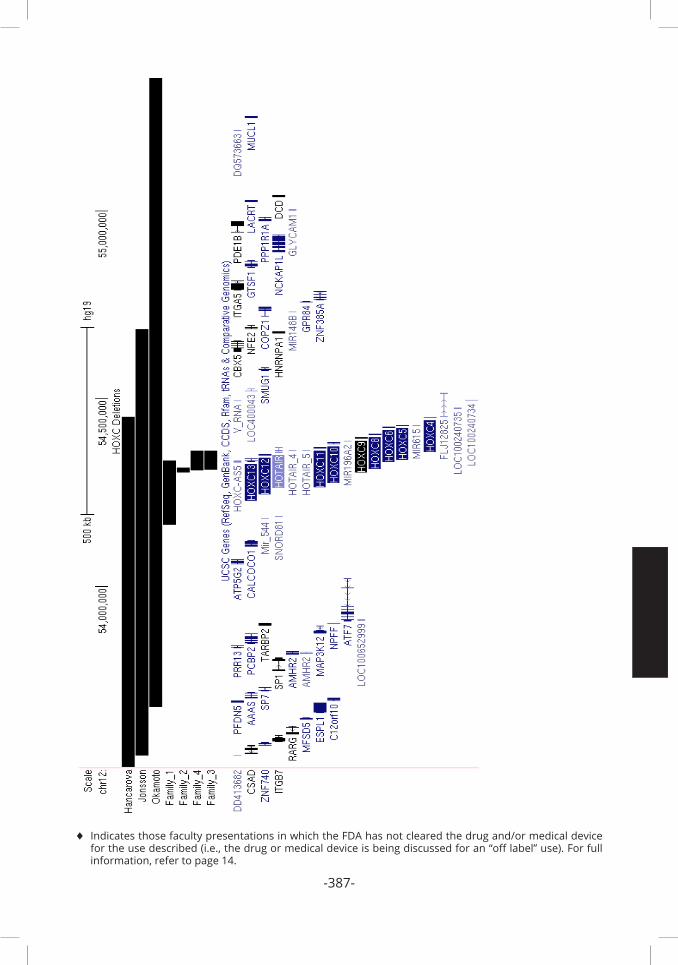
-387-
♦ Indicates those faculty presentations in which the FDA has not cleared the drug and/or medical device for the use described (i.e., the drug or medical device is being discussed for an “off label” use). For full information, refer to page 14.

-388-
See pages 21- 60 for financial disclosure information.
-389-
♦ Indicates those faculty presentations in which the FDA has not cleared the drug and/or medical device for the use described (i.e., the drug or medical device is being discussed for an “off label” use). For full information, refer to page 14.
e-Poster #1 (page 406) Obesity and Physical Status Predict Surgical Site Infection in Pediatric Idiopathic Spinal Deformity Patients Jeanne M. Franzone, MD; Hiroko Matsumoto, MA; William G. Mackenzie, MD; Michael J. Troy, BS; Kody Barrett, MD; Brendan Striano, BA; Michael P. Glotzbecker, MD; John (Jack) M. Flynn, MD; David L. Skaggs, MD, MMM; Michael G. Vitale, MD, MPH Columbia University Medical Center, New York, New York e-Poster #2 (page 407) The Association between Post-Operative Complications and Satisfaction Scores among Children Treated Surgically Caroline P. Thirukumaran, MBBS, MHA; Christopher Gitzelmann, MD; Kori Wolcott, BSN, RN; Julie A. Michels, MS, RN; Timothy Stevens, MD, MBA; James O. Sanders, MD University of Rochester, Rochester, New York e-Poster #3 (page 408) Sanders vs. Risser: Which System Should be Used to Develop Bracing Indications? Lori Dolan, PhD; Stuart L. Weinstein, MD University of Iowa, Iowa City, Iowa e-Poster #4 (page 410) Prophylactic Antibiotic Use in Pediatric Orthopaedic Surgery: Reducing Variability, Cost and Risks Adam Margalit, BS; Gift Ukwuani; Rushyuan J. Lee, MD; Paul D. Sponseller, MD The Johns Hopkins Hospital, Baltimore, Maryland e-Poster #5 (page 412) Pedicle Screw Safety: How Much Anterior Breach Is Safe? A Cadaveric and CT Based Study Vishal Sarwahi, MD; Terry D. Amaral, MD; Kathleen J. Maguire, MD; Rachel C. Gecelter, BS; Monica M. Payares-Lizano, MD; Ajay F. Lall, MD; Dan Wang; Beverly Thornhill, MD Montefiore Medical Center, Bronx, New York
2016 e-POSTER PROGRAM
-390-
See pages 21- 60 for financial disclosure information.
2016 e-Poster Program, (cont.)
e-Poster #6 (page 413) Adolescent Idiopathic Scoliosis Patients are at Increased Risk for Pulmonary Hypertension which Reverses after Scoliosis Surgery Vishal Sarwahi, MD; Sarika Kalantre, MD; Rachel C. Gecelter, BS; Aviva F. Dworkin; Terry D. Amaral, MD; Kathleen J. Maguire, MD; Ajay F. Lall, MD; Marina Moguilevitch, MD; Dan Wang, MS Montefiore Medical Center, Bronx, New York e-Poster #7 (page 414) Limb Length Multipliers in Real Life - Age versus Growth Spurt Timing to Predict Final Limb Length Lauren E. Karbach, MD; Thomas Osinski, BS; Xiang Lu, MA; Raymond W. Liu, MD; Xing Qiu, PhD; Daniel R. Cooperman, MD; James O. Sanders, MD University of Rochester, Rochester, New York e-Poster #8 (page 416) Management of Displaced Supracondylar Fractures of the Humerus Using Lateral versus Crossed K Wires: A Prospective Randomized Trial Kishore Mulpuri, MD; Ashlee Dobbe, BS, FRCS (Ortho), MD, MSc; Emily Schaeffer, PhD; Firoz Miyanji, MD; Christine M. Alvarez, MD, FRCSC; Anthony P. Cooper, FRCS; Christopher W. Reilly, MD British Columbia Children’s Hospital, Vancouver, British Columbia, Canada e-Poster #9 (page 417) Coexisting DDH and Spastic Hip Disease? The Treatment of Dislocated Hips in Infants with Spasticity Christian A. Refakis, BA; Wudbhav N. Sankar, MD Children’s Hospital of Philadelphia, Philadelphia, Pennsylvania e-Poster #10 (page 418) Clinical Indications Associated with Abnormal MRIs in a Pediatric Spine Deformity Practice Karl E. Rathjen, MD; Ozan Razi, MD; Erica Flores, RN, MSN; Heather D. Caine, BS; Dong-Phuong Tran, MS; ChanHee Jo, PhD; Adriana De La Rocha, PhD Texas Scottish Rite Hospital for Children, Dallas, Texas e-Poster #11 (page 419) T2 Judgment Day: Does this UIV Guarantee Postoperative Shoulder Balance? Burt Yaszay, MD; Tracey P. Bastrom, MA; Carrie Bartley, MA; Baron Lonner, MD; Suken A. Shah, MD; Firoz Miyanji, MD; Jahangir Asghar, MD; Peter O. Newton, MD Rady Children’s Hospital, San Diego, California
-391-
♦ Indicates those faculty presentations in which the FDA has not cleared the drug and/or medical device for the use described (i.e., the drug or medical device is being discussed for an “off label” use). For full information, refer to page 14.
e-Poster #12 (page 420) The Cost of Complexity in Pediatric Scoliosis Management Melvin Makhni, MD; Amelia Lindgren, BS; Navid Dardashti, MPH; Kevin Wang, BA; Hiroko Matsumoto, MA; Jesus A. Jimenez, BS; Michael G. Vitale, MD, MPH Columbia University Medical Center, New York, New York e-Poster #13 (page 421) Plantar Pressures Following Nonoperative and Surgical Treatment for Clubfoot at 10 Years of Age Kirsten Tulchin-Francis, PhD; Ashley Erdman, BS, MBA; Kelly Jeans, MSc; Lori A. Karol, MD Texas Scottish Rite Hospital for Children, Dallas, Texas e-Poster #14 (page 422) Average 10-Year Results of the Bernese Periacetabular Osteotomy for Severe Acetabular Dysplasia Stephen T. Duncan, MD; Jeff Petrie, MD; Kayla M. Thomason, BS; Geneva Baca, BA; Perry L. Schoenecker, MD; John C. Clohisy, MD Washington University School of Medicine, Saint Louis, Missouri e-Poster #15 (page 423) Value-Based Medicine: Application to AIS Patients Undergoing Posterior Spinal Fusion – Determination of Cost and Outcome Daniel J. Sucato, MD, MS; Heather D. Caine, BS; David Everett, BS Texas Scottish Rite Hospital for Children, Dallas, Texas e-Poster #16 (page 424) T1 Tilt Angle and Clavicle Angle on Immediate Post-Op X Rays are the Best Predictors of Shoulder Balance in AIS Patients Vishal Sarwahi, MD; Stephen Wendolowski, BS; Rachel C. Gecelter, BS; Alexa J. Karkenny, MD, Terry D. Amaral, MD; Abhijit Pawar, MD Montefiore Medical Center, Bronx, New York e-Poster #17 (page 425) Scoliosis in Down Syndrome Oussama Abousamra, MD; Maria Del Pilar Duque Orozco, MD; Kenneth J. Rogers, PhD; Julieanne P. Sees, DO; Freeman Miller, MD Nemours / Alfred I. duPont Hospital for Children, Wilmington, Delaware e-Poster #18 (page 426) Do Professional Society Advocacy Campaigns Have an Impact on Pediatric Orthopaedic Injuries? Alexa J. Karkenny, MD; Kathleen J. Maguire, MD; Norman Y. Otsuka, MD The Children’s Hospital at Montefiore, Montefiore Medical Center, Bronx, New York
-392-
See pages 21- 60 for financial disclosure information.
2016 e-Poster Program, (cont.)
e-Poster #19 (page 427) Femoral Nerve Block with or without Sciatic Nerve Block for Pediatric Anterior Cruciate Ligament Reconstruction: a Retrospective Review Jeffrey L. Hanway, MD; Leanne Foster, MD; Heather Gordish-Dressman, PhD; Sophie Pestieau, MD Children’s National Health System, Washington, District of Columbia e-Poster #20 (page 429) Three-Dimensional MRI Quantification of Femoral Head Deformity in Legg-Calvé-Perthes Disease (LCPD) Karen D. Standefer, BS; Molly Dempsey, MD; ChanHee Jo, PhD; Harry K.W. Kim, MD, MS Texas Scottish Rite Hospital for Children, Dallas, Texas e-Poster #21 (page 430) Adolescents Maintain Hip Strength and Function 5 yrs Following a Ganz Periacetabular Osteotomy Daniel J. Sucato, MD, MS; Kirsten Tulchin-Francis, PhD; David A. Podeszwa, MD; Adriana De La Rocha, PhD; Christopher Stewart, BS; Wilshaw Stevens, BS Texas Scottish Rite Hospital for Children, Dallas, Texas e-Poster #22 (page 431) A Prospective Randomized Comparative Study of PCA, Epidural Catheters and Lumbar Plexus Catheters For Analgesia After Hip Surgery in Children Klane K. White, MD, MS; Nathalia Jimenez, MD; Nancy E. Gove, PhD Seattle Children’s Hospital, Seattle, Washington e-Poster #23 (page 432) Damage Patterns Seen with Femoroacetabular Impingement in Children Prasad V. Gourineni, MD; Summer Watkins, MSN, RN, PNP-AC Advocate Children’s Hospital, Oak Lawn, Illinois e-Poster #24 (page 434) Hip Dysplasia in Patients with Cerebral Palsy Treated with Bernese Periacetabular Osteotomy Mark L. Miller, MD; John C. Clohisy, MD; Perry L. Schoenecker, MD Washington University, Saint Louis, Missouri e-Poster #25 (page 435) Selective Thoracic Fusion in AIS: Beware of Over Correction Shawn S. Funk, MD; Megan Mignemi, MD; Kaitlyn Brown, BS; Amy L. McIntosh, MD Texas Scottish Rite Hospital, Dallas, Texas
-393-
♦ Indicates those faculty presentations in which the FDA has not cleared the drug and/or medical device for the use described (i.e., the drug or medical device is being discussed for an “off label” use). For full information, refer to page 14.
e-Poster #26 (page 437) Can Facebook Alleviate Anxiety in Teenagers Undergoing Scoliosis Surgery? A Prospective Non-Randomized Controlled Study Melanie A. Gambassi, NP; Stephen Wendolowski, BS; Rachel C. Gecelter, BS; Dana Orlando, ST; Terry D. Amaral, MD; Vishal Sarwahi, MD Montefiore Medical Center, Bronx, New York e-Poster #27 (page 438) Costs and Complications of Increased Length of Stay Following Scoliosis Surgery Matthew R. Boylan, ScB; Aldo M. Riesgo, MD; Alice Chu, MD; Carl B. Paulino, MD; David S. Feldman, MD NYU Hospital for Joint Diseases, New York, New York e-Poster #28 (page 439) Pelvic Tilt Affects Measurement of the Acetabular Index by Volume-Rendered CT Tim Schrader, MD; Scott M. Sorenson, MD; Justin Hire, MD; John R. Faust, MD; Anthony C. Cantrell, BS; Mackenzie M. Herzog, MPH Children’s Healthcare of Atlanta, Atlanta, Georgia e-Poster #29 (page 441) Variations in Surgical Rates of Pediatric Distal Radius Fractures Across Institutions: Analysis of the PHIS Database Susan T. Mahan, MD, MPH; Apurva Shah, MD, MBA; Leslie A. Kalish, ScD; Lanna F. Feldman, MS; Donald S. Bae, MD Boston Children’s Hospital, Boston, Massachusetts e-Poster #30 (page 443) The Impact of the Patient Positioning on 3D Postural Parameters and Functional Balance in Bi-planar Spinal Radiography of Adolescent Idiopathic Scoliosis Saba Pasha, PhD; John (Jack) M. Flynn, MD; Patrick J. Cahill, MD The Children’s Hospital of Philadelphia, Philadelphia, Pennsylvania e-Poster #31 (page 445) The Results of Pharmacotherapy and Lower Extremity Corrective Osteotomies in Children with Osteogenesis Imperfecta Andrii Zyma, MD; Guk Yurii, MD; Tamara Kinchaya-Polischuk, MD; Andrii Cheverda, MD; Stepan Martcyniak; David G. Stewart Jr., MD; Yuri Demyan, MD Institute of Orthopedics and Traumatology, National Academy of Medical Sciences, Kiev, Ukraine
-394-
See pages 21- 60 for financial disclosure information.
2016 e-Poster Program, (cont.)
e-Poster #32 (page 447) Lateral Femoral Cutaneous Nerve Palsy Occurred in 25% of Patients Following Posterior Spinal Fusion for Adolescent Idiopathic Scoliosis Austin Sanders, BA; Lindsay M. Andras, MD; Paul D. Choi, MD; Vernon T. Tolo, MD; David L. Skaggs, MD, MMM Children’s Hospital Los Angeles, Los Angeles, California e-Poster #33 (page 448) A Model of Pre-Pseudarthrosis (Congenital Tibia Dysplasia) Associated with Neurofibromatosis Type 1 David G. Little, MBBS, FRACS, PhD; Nikita Deo, BSc; Tegan L. Cheng, BE BMedSci; Aaron Schindeler, BSc PhD The Children’s Hospital at Westmead, Sydney, Australia e-Poster #34 (page 449) The PASS (Pediatric/Adolescent Shoulder Survey): A Youth Questionnaire with Discriminant Validity and Responsiveness to Change Tracey P. Bastrom, MA; Joanna H. Roocroft, MA; Andrew T. Pennock, MD; Eric W. Edmonds, MD Rady Children’s Hospital, San Diego, California e-Poster #35 (page 451) Best Practice Guidelines for the Surgical Treatment of Early Onset Scoliosis Michael P. Glotzbecker, MD; Tricia A. St Hilaire, MPH; Growing Spine Study Group; George H. Thompson, MD; Michael G. Vitale, MD, MPHChildren’s Spine Study Group, San Diego, California e-Poster #36 (page 452) 8-plates & I-plates for Epiphysiodesis Kevin S. Horowitz, MD; Jaclyn F. Hill, MD; Ali A. Qadeer, BS Baylor College of Medicine, Houston, Texas e-Poster #37 (page 453) Hip Arthroscopy Failure in the Setting of Acetabular Dysplasia: A Concerning Trend? Tonya An, BS; Jacob Haynes; Jeffrey J. Nepple, MD; John C. Clohisy, MD; Perry L. Schoenecker, MD; Michael B. Millis, MD; Young Jo Kim, MD; Ira Zaltz, MD; Rafael J. Sierra, MD; Wudbhav N. Sankar, MD; Ernest L. Sink, MD; David A. Podeszwa, MD; Daniel J. Sucato, MD, MS; Robert T. Trousdale, MD; Paul E. Beaule, MD; ANCHOR Group Washington University School of Medicine, Saint Louis, Missouri
-395-
♦ Indicates those faculty presentations in which the FDA has not cleared the drug and/or medical device for the use described (i.e., the drug or medical device is being discussed for an “off label” use). For full information, refer to page 14.
e-Poster #38 (page 455) Reducing Anxiety, Improving Communication and Increasing Satisfaction in Families During Orthopedic Procedures with Mobile Technology Jose A. Herrera-Soto, MD; Dennis R. Knapp, MD; Mark A. Birnbaum, MD; Jonathan H. Phillips, MD; Kevin de la Roza, MD Arnold Palmer Hospital for Children, Orlando, Florida e-Poster #39 (page 457) Implementing a Multidisciplinary Clinical Pathway Can Reduce the Surgical Site Infection Rate After Posterior Spinal Fusion In High Risk Patients Michael P. Glotzbecker, MD; Michael J. Troy, BS; Patricia E. Miller, MS; Alexandra M. Grzywna, BA; Michael T. Hresko, MD; Lawrence I. Karlin, MD; John B. Emans, MD; Brian D. Snyder, MD, PhD; Daniel J. Hedequist, MD Boston Children’s Hospital, Boston, Massachusetts e-Poster #40 (page 458) Investigating Caregiver Perspectives on Health Status and Comfort in Children with Cerebral Palsy Undergoing Surgery for Hip Displacement Stacey Miller, PT; Emily Schaeffer, PhD; Maria Juricic, PT; Kim Hesketh, MSc, PT; Kishore Mulpuri, MD British Columbia Children’s Hospital, Vancouver, British Columbia, Canada e-Poster #41 (page 459) Both Family History and Swaddling are Correlated with the Occurrence and Hip Types in Developmental Dysplasia of the Hip Hakan Omeroglu, MD; Nusret Kose, MD; Anil Akceylan, MD Eskisehir Osmangazi University Hospital, Eskisehir, Turkey e-Poster #42 (page 460) Prevalence of Vitamin D Deficiency in Pediatric Patients with Scoliosis Preparing for Spinal Surgery Peter F. Sturm, MD; Jennifer M. Anadio, MA; Theresa Mayes Cincinnati Children’s Hospital Medical Center, Cincinnati, Ohio e-Poster #43 (page 461) Obesity and Both-Bone Forearm Fractures Ugochi Okoroafor, MD; Jasmin L. McGinty, MD SSM Health Cardinal Glennon Children’s Hospital, St. Louis, Missouri
-396-
See pages 21- 60 for financial disclosure information.
2016 e-Poster Program, (cont.)
e-Poster #44 (page 462) A New Simplified Thumb Ossification Composite Index (TOCI) - Can Predict the Peak Height Velocity, Skeletal Maturity and Curve Progression in Adolescent Idiopathic Scoliosis? Alec Lik Hang Hung; Wai-Wang Chau; Benjamin Hon Kei Yip; Fiona Wai Ping Yu; Vivian Wing Yin Hung; Tsz Ping Lam, MD; Bobby Kin Wah Ng, MD; Jack Cheng Department of Orthopaedics and Traumatology, Prince of Wales Hospital, Hong Kong, SAR e-Poster #45 (page 463) Age at Surgery, Amount of Arthrosis, and Severity of Dysplasia May Help Predict Early Versus Late Failure of Bernese Periacetabular Osteotomy Travis H. Matheney, MD; Joel E. Wells, MD, MPH; Evgeny Bulat, MS; Patricia E. Miller, MS; Young Jo Kim, MD; Michael B. Millis, MD Boston Children’s Hospital, Boston, Massachusetts e-Poster #46 (page 464) Abnormal Sclerostin Expression in Serum and Bone Tissue Osteocyte Lacuna-canaliculi System in AIS – a New Novel Finding Huanxiong Chen, MD; Wayne YW Lee; Jiajun Zhang, BSc, MSC; Bobby Kin Wah Ng, MD; Tsz Ping Lam, MD; Jian Q. Feng, PhD; Jack Cheng; Kwong Man Lee Prince of Wales Hospital, Shatin, Hong Kong, SAR e-Poster #47 (page 466) Maternal Vitamin D Deficient Diets Increase Kyphosis in Offspring Matthew A. Halanski, MD; Blake Hildahl, MD; Rajeev Chaudhary, MS; Rachel L. Lenhart, PhD; Thomas D. Crenshaw, PhD; Laura A. Amundson, BS American Family Children’s Hospital, University of Wisconsin, Madison, Wisconsin e-Poster #48 (page 468) ♦Combined Pharmacologic and Biomechanical treatment of Congenital Pseudarthrosis of the Tibia: 100% Union; No Refractures? Dror Paley, MD; Davida Packer, MD; Craig A. Robbins, MD; John Robb, PA-C; Raymond W. Liu, MD Paley Institute, West Palm Beach, Florida e-Poster #49 (page 470) Genu Varum in Achondroplasia and Hypochondroplasia Hiroshi Kitoh, MD; Kenichi Mishima; Masaki Matsushita, MD; Hiroshi Sugiura; Sachi Hasegawa; Naoki Ishiguro, MD Nagoya University Graduate School of Medicine, Nagoya, Aichi, Japan
-397-
♦ Indicates those faculty presentations in which the FDA has not cleared the drug and/or medical device for the use described (i.e., the drug or medical device is being discussed for an “off label” use). For full information, refer to page 14.
e-Poster #50 (page 472) Skill Acquisition and Retention Following Simulated Pavlik Harness Learning Simon P. Kelley, MBChB, MRCS, FRCS; Joel Moktar, MD; Alexandra Maxwell, BS; Catharine Bradley, BScPT, MSc; John H. Wedge, MD; M. Lucas Murnaghan, MD, MEd, FRCSC Hospital for Sick Children, Toronto, Ontario, Canada
e-Poster #51 (page 473) Back Pain in Cerebral Palsy Patients is Markedly Reduced After Spinal Fusion for Scoliosis Burt Yaszay, MD; Tracey P. Bastrom, MA; Paul D. Sponseller, MD; Suken A. Shah, MD; Firoz Miyanji, MD; Jahangir Asghar, MD; Patrick J. Cahill, MD; Amer Samdani, MD; Peter O. Newton, MD Rady Children’s Hospital, San Diego, California e-Poster #52 (page 474) Does Using an EMR Template Improve Documentation for Pediatric Supracondylar Fractures? Mark J. Adamczyk, MD; Ryan J. Urchek, MD; Melanie Morscher, BS Akron Children’s Hospital, Akron, Ohio e-Poster #53 (page 475) Complications and Radiographic Outcomes of Posterior Spinal Fusion (PSF) in Patients with Spinal Muscular Atrophy (SMA) Following Growth Friendly Treatment Rodrigo Goes de Mendonca, MD; John A. Heflin, MD; Paul D. Sponseller, MD; George H. Thompson, MD; Michael G. Vitale, MD, MPH; John T. Smith, MD; Tara Flynn, BA; Ryan P. Murphy, BS; Jolecia M. Flournory, BA; Jeffrey R. Sawyer, MD;Children’s Spine Study Group; Growing Spine Study Group Le Bonheur Children’s Hospital, Memphis, Tennessee e-Poster #54 (page 476) The Cost Effectiveness of Universal Screening Programs for Scoliosis Matthew E. Oetgen, MD; Kathleen G. Noble; Shannon M. Kelly, MD; Allison Matthews, MSCR Children’s National Health System, Washington, District of Columbia e-Poster #55 (page 477) Management of Terminal Bony Overgrowth of the Humerus with Osteocartilaginous Grafts Graham T. Fedorak, MD, FRCSC; Anna V. Cuomo, MD; Hugh G. Watts, MD; Anthony A. Scaduto, MD Shiners Hospitals for Children, Los Angeles, Los Angeles, California
2016 e-Poster Program, (cont.)
-398-
See pages 21- 60 for financial disclosure information.
e-Poster #56 (page 478) Final Fusion in Patients Treated with Rib Based Distraction: A Review of Peri-operative Results John A. Heflin, MD; Jessica Morgan, BS; Victoria Heagy, BS; John T. Smith, MD University of Utah, Salt Lake City, Utah e-Poster #57 (page 479) Intra-wound Vancomycin Reduces the Infection in Growth-Friendly Spine Surgery Justin Haller, MD; David Andrew Hulet, BS; Qian Ding, PhD; Angela P. Presson, PhD; John A. Heflin, MD; John T. Smith, MD University of Utah, Salt Lake City, Utah e-Poster #58 (page 481) Both Decreased and Increased Relative Acetabular Volume are Associated with Increased Hip Arthritis in an Osteological Review of 1090 Hips Douglas S. Weinberg, MD; Drew F.K. Williamson, BA; Michael B. Millis, MD; Raymond W. Liu, MD Case Western Reserve University, Cleveland, Ohio e-Poster #59 (page 483) Does Clavicle Grow beyond Skeletal Maturity? Pooya Hosseinzadeh, MD; Cristina Brea, BS; Gary Kiebzak, PhD Baptist Children’s Hospital, Miami, Florida e-Poster #60 (page 484) Deficient Quality of Care for Clubfoot: Implications for Patient-Centered Care Asitha Jayawardena, MD; Jennifer L. Trevillian, BS; Thomas Cook, PT, PhD; Jose A. Morcuende, MD, PhD University of Iowa, Iowa City, Iowa e-Poster #61 (page 485) Implementing a National Clubfoot Treatment Program in Nigeria Jose A. Morcuende, MD, PhD; Thomas Cook, PT, PhD; Olayinka Adegbehingbe, MD; Tomeka Petersen, MSW; Omolade A. Lasebikan, MBChB, MD, MPH; Adeoye J. Adetiloye, MD, FWACS; Habila Umaru, MD; Oladipo Adewole, MD; Orlando Ugwoegbulem III, MBBS, MD; Zumnan D. Songden, MD; Oluwadare Esan, MBBS, MD; Edith A. Parker, PhD University of Iowa, Iowa City, Iowa
-399-
♦ Indicates those faculty presentations in which the FDA has not cleared the drug and/or medical device for the use described (i.e., the drug or medical device is being discussed for an “off label” use). For full information, refer to page 14.
e-Poster #62 (page 486) Comparison Between Participation and Desire to Participate in Activities Among Children with Skeletal Dysplasias Cathleen L. Raggio, MD; Meredith Manson; Erin M. Carter, MS; Kate P. Citron, BS Hospital for Special Surgery, New York, New York e-Poster #63 (page 488) Knee Hyperextension in Children with Cerebral Palsy: Outcomes over a Minimum of Five Year Follow Up Oussama Abousamra, MD; Daveda Taylor, DPT; Chris Church, MPT; Maria Del Pilar Duque Orozco, MD; Nancy Lennon, MSPT; John D. Henley, PhD; Julieanne P. Sees, DO; Freeman Miller, MD; Justin R. Connor, MD Nemours / Alfred I. duPont Hospital for Children, Wilmington, Delaware e-Poster #64 (page 490) Cost Analysis of Magnetically-Controlled Growing Rods compared with Traditional Growing Rods for Early Onset Scoliosis in the United States: An Integrated Healthcare Delivery System Perspective David W. Polly Jr, MD; Stacey J. Ackerman, MSE, PhD; Karen B. Schneider, PhD; Jeff Pawelek, BS; Behrooz A. Akbarnia, MD University of Minnesota, Dept. of Orthopaedic Surgery, Saint Paul, Minnesota e-Poster #65 (page 491) Does Correction of Pelvic Obliquity Result in Improved Outcomes and Sitting Tolerance for Patients with Cerebral Palsy Scoliosis? Brian E. Kaufman, MD; Josh M. Pahys, MD Nemours / Alfred I. duPont Hospital for Children, Wilmington, Delaware e-Poster #66 (page 492) Comparison of 3D Muscular Parameters Between Children with Cerebral Palsy and Typically Developing Children Abir Massaad, MSc; Ayman Assi, PhD; Ziad Bakouny; Wafa Skalli, PhD; Ismat Ghanem, MD Faculty of Medicine, University of Saint-Joseph, Beirut, Lebanon e-Poster #67 (page 493) Alterations in Acetabular and Proximal Femur Geometry in Subjects with Down Syndrome Elie Joseph Mansour, MD; Jean Jacques Yaacoub, MD; Ayman Assi, PhD; Ziad Bakouny; Wafa Skalli, PhD; Ismat Ghanem, MD Faculty of Medicine, University of Saint-Joseph, Beirut, Lebanon
2016 e-Poster Program, (cont.)
-400-
See pages 21- 60 for financial disclosure information.
e-Poster #68 (page 494) Subjects with Down Syndrome Have Altered Coronal and Sagittal Spino-Pelvic Alignment Jean Jacques Yaacoub, MD; Elie Joseph Mansour, MD; Ziad Bakouny; Ayman Assi, PhD; Virginie Lafage, PhD; Ismat Ghanem, MD Faculty of Medicine, University of Saint-Joseph, Beirut, Lebanon e-Poster #69 (page 495) Serial Casting for Stiff Clubfoot According to Ponseti: Timing of Achilles Tendon Ismat Ghanem, MD; Elie Saliba, MD; Ayman Assi, PhD Hotel-Dieu de France Hospital, University of Saint-Joseph, Beirut, Lebanon e-Poster #70 (page 496) Validation of the Gait Outcomes Assessment List (GOAL) Questionnaire: A Goal Based Outcome Measure for Ambulatory Children with Cerebral Palsy Unni G. Narayanan, MBBS, MSc, FRCSC; Rachel Moline, BA; Clarissa Encisa, BSc, MPH The Hospital for Sick Children, Toronto, Ontario, Canada e-Poster #71 (page 497) The Rate and Risk of Curve Progression following Skeletal Maturity– Does the Story End with Curve Magnitude? Firoz Miyanji, MD; Christopher W. Reilly, MD; Suken A. Shah, MD; David H. Clements; Amer Samdani, MD; Sameer Desai, BS; Baron Lonner, MD; Harry L. Shufflebarger, MD; Randal R. Betz, MD; Peter O. Newton, MD British Columbia Children’s Hospital, Vancouver, British Columbia, Canada e-Poster #72 (page 499) Normal Human Spine Growth During Childhood and Adolescence to Maturity with Prediction of Final Spine Height Developed from a Longitudinal Cohort of Children Followed Through Growth Completion James O. Sanders, MD; Lauren E. Karbach, MD; Thomas Osinski, BS; Xiang Lu, MA; Xing Qiu, PhD; Raymond W. Liu, MD; Daniel R. Cooperman, MD University of Rochester and the Golisano Children’s Hospital at Strong, Rochester, New York e-Poster #73 (page 501) Femoral Neck Aspiration Aids in the Diagnosis of Osteomyelitis in Children with Septic Arthritis of the Hip Jedidiah E. Schlung, BS; Tracey P. Bastrom, MA; Peter O. Newton, MD; Scott J. Mubarak, MD; Vidyadhar S.V. Upasani, MD Rady Children’s Hospital, San Diego, California

-401-
♦ Indicates those faculty presentations in which the FDA has not cleared the drug and/or medical device for the use described (i.e., the drug or medical device is being discussed for an “off label” use). For full information, refer to page 14.
e-Poster #74 (page 503) Preventing DJK by Applying the Stable Sagittal Vertebra Concept to Selective Thoracic Fusion in AIS Alexander M. Broom, BA; Lindsay M. Andras, MD; Kody Barrett, MD; Andrew G. Georgiadis, MD; John (Jack) M. Flynn, MD; Vernon T. Tolo, MD; David L. Skaggs, MD, MMM Children’s Hospital Los Angeles, Los Angeles, California e-Poster #75 (page 505) Restoring Sagittal Balance Improves Clinical Outcomes for Cerebral Palsy Patients with Spinal Deformity Kushagra Verma, MD, MS; Suken A. Shah, MD; Firoz Miyanji, MD; Peter O. Newton, MD; Burt Yaszay, MD; Maty Petcharaporn, BS; Tracey P. Bastrom, MA; Unni G. Narayanan, MBBS, MSc, FRCSC; Paul D. Sponseller, MD Nemours / Alfred I. duPont Hospital for Children, Wilmington, Delaware e-Poster #76 (page 507) Radiographic Outcomes of Shilla Growth Guidance System and Traditional Growing Rods through Definitive Treatment Scott J. Luhmann, MD; June C. Smith, MPH; Anna McClung, RN, BSN; Frances L. McCullough, RNP, MNSc; Richard E. McCarthy, MD; George H. Thompson, MD; Growing Spine Study Group St. Louis Children’s Hospital, Saint Louis, Missouri e-Poster #77 (page 509) Radiographic Assessment of Angular Correction of Genu Varum and Genu Valgus Using Guided Growth: The Correlation Between Screw Divergence and Change in Anatomic Alignment Weilong Jeffrey Shi, BS; Kyle R. Sweeney, MD; Michael B. Gottschalk, MD; Robert W. Bruce, MD; Nicholas D. Fletcher, MD Emory University, Atlanta, Georgia e-Poster #78 (page 511) Choice of Anchors – Rib vs. Spine: Importance of Proximal Anchor Number Michael G. Vitale, MD, MPH; Hiroko Matsumoto, MA; Nicholas A. Feinberg, BA; Evan P. Trupia, MD; Matthew B. Shirley, BS; Sumeet Garg, MD; John (Jack) M. Flynn, MD; Peter F. Sturm, MD; Francisco Sanchez Perez-Grueso; David P. Roye Jr, MD; David L. Skaggs, MD, MMM; Children’s Spine Study Group; Growing Spine Study Group Columbia University Medical Center, New York, New York
-402-
See pages 21- 60 for financial disclosure information.
2016 e-Poster Program, (cont.)
e-Poster #79 (page 513) Improving The Handoff Process Between Operating Room and Pediatric Intensive Care Unit Stephen J. Gleich, MD; Michael E. Nemergut, MD, PhD; Anthony A. Stans, MD; Dawit T. Haile, MD; Angela L. Heinrich, RN; Christopher L. Bosley, RRT-NPS; James W. Ward, MBA Mayo Clinic, Rochester, Minnesota e-Poster #80 (page 514) 2D versus 3D Measurements of AIS Spinal Parameters in Clinical Care Saba Pasha, PhD; Patrick J. Cahill, MD; John (Jack) M. Flynn, MD The Children’s Hospital of Philadelphia, Philadelphia, Pennsylvania e-Poster #81 (page 516) Spica Casting for Pediatric Femur Fractures: Treatment Practice and Value Outcomes Caroline V. Hornberger, PA-C; Mary K. Riordan, BA; William A. Phillips, MD Texas Children’s Hospital, Houston, Texas e-Poster #82 (page 517) Selective Thoracic Fusion of a Left Decompensated Main Thoracic Curve: Proceeding with Caution Burt Yaszay, MD; Tracey P. Bastrom, MA; Carrie Bartley, MA; Suken A. Shah, MD; Baron Lonner, MD; Jahangir Asghar, MD; Firoz Miyanji, MD; Patrick J. Cahill, MD; Peter O. Newton, MD Rady Children’s Hospital, San Diego, California e-Poster #83 (page 519) Multicenter Evaluation of Spinal Deformity in Skeletal Dysplasias with Growing Rod Systems Klane K. White, MD, MS; Viviana Bompadre, PhD; Gregory Redding, MD; Walter F. Krengel III, MD; Suken A. Shah, MD; William G. Mackenzie, MD Seattle Children’s Hospital, Seattle, Washington e-Poster #84 (page 520) Enhancing the Economy of Surgery: Reducing Cost and Variation of Surgeon Preference Cards for Supracondylar Humerus Fractures Thomas B. Bemenderfer, MD; Kelly L. Vanderhave, MD; Brian P. Scannell, MD; Virginia F. Casey, MD; Michael D. Paloski, DO; Christian Clark, MD; Brian K. Brighton, MD, MPH Carolinas Healthcare System/Levine Children’s Hospital, Charlotte, North Carolina
-403-
♦ Indicates those faculty presentations in which the FDA has not cleared the drug and/or medical device for the use described (i.e., the drug or medical device is being discussed for an “off label” use). For full information, refer to page 14.
e-Poster #85 (page 521) Age-Based Patellofemoral Morphology in the Immature Knee Kevin E. Klingele, MD; Matthew C. Beran, MD Nationwide Children’s Hospital, Columbus, Ohio e-Poster #86 (page 523) Implementation of a Dedicated Pediatric Orthopaedic Trauma Room Gianna D. Katsaros, MS; Wendy J. Ramalingam, MD; Jaime R. Denning, MD; Patrick W. Whitlock, MD Cincinnati Children’s Hospital Medical Center, Cincinnati, Ohio e-Poster #87 (page 524) Symptomatic Leg Length Discrepancy following Open Reduction and Salter’s Innominate Osteotomy in DDH Children Chii-Jeng Lin, MD, PhD; Ming-Tung Huang, MD; Nai-Wen Guo, PhD National Cheng Kung University, Tainan, Taiwan e-Poster #88 (page 525) Mitochondrial Enzyme Activity is Reduced in Skeletal Muscle in Children with Cerebral Palsy Sudarshan Dayanidhi, PhD; Elisa Buckner, PhD; Henry (Hank) G. Chambers, MD; Simon Schenk, PhD; Richard L. Lieber, PhD Rady Children’s Hospital, San Diego, California e-Poster #89 (page 526) 3 Dimensional Loss of Thoracic Kyphosis is Associated with Greater Degrees of Thoracic Idiopathic Scoliosis Peter O. Newton, MD; Thomas B. Sullivan, MD; Tracey P. Bastrom, MA; Carrie Bartley, MA; Lori Dolan, PhD; Stuart L. Weinstein, MD; Harms Study Group Rady Children’s Hospital, San Diego, California e-Poster #90 (page 528) The Safety of Percutaneous Achilles Tenotomy in Idiopathic Clubfoot Deformity Correction in an Outpatient Setting Mengyang M. Li, MD; Guy Grimard, MD; Morris A. Duhaime, MD; Panagiotis (Peter) Glavas, MD Sainte-Justine University Hospital Center, Montreal, Quebec, Canada e-Poster #91 (page 529) Vancomycin Tissue Concentrations during Posterior Spinal Fusions in Neuromuscular Scoliosis James R. Gregory, MD; June C. Smith, MPH; Sarah Brown, PhD; Alexis Elward, MD, MPH; Scott J. Luhmann, MD Washington University School of Medicine, St. Louis, Missouri
-404-
See pages 21- 60 for financial disclosure information.
2016 e-Poster Program, (cont.)
e-Poster #92 (page 531) Trochlear Dysplasia: Are the Current Classifications and Radiographic Measurements Appropriate in the Skeletally Immature Patient? Matthew T. Stepanovich, MD; James D. Bomar, MPH; Andrew T. Pennock, MD Rady Children’s Hospital, San Diego, California e-Poster #93 (page 533) Fibroblast Growth Factor Receptor Three Regulates Fracture Remodeling Through an Osteoclast Independent Effect Simon P. Kelley, MBChB, MRCS, FRCS; Chunying Yu; Baht Gurpreet, PhD; Benjamin A. Alman, MD, FRCSC The Hospital for Sick Children, Toronto, Ontario, Canada e-Poster #94 (page 534) Relationships between Three Classification Systems in Brachial Plexus Birth Palsy Dustin A. Greenhill, MD; Sandra Tomlinson-Hansen, BA; Scott H. Kozin, MD; Dan A. Zlotolow, MD Shriners Hospital for Children - Philadelphia, Pennsylvania e-Poster #95 (page 536) Validity of Beighton’s Score in a Paediatric Population Nimesh Patel, MRCS; Peter J. Riddlestone, MBBS; Roshan Gunalan; James E. Metcalfe, FRCS, MBChB; Ben Holroyd, FRCS (Tr & Orth) Derriford Hospital, Plymouth, United Kingdom e-Poster #96 (page 537) Relative Impact of Pre-Operative Hip Location vs Post-Operative Abduction Angle on Epiphyseal Perfusion After Closed Reduction and Spica Casting for DDH Michael Rivlin, MD; Scott B. Rosenfeld, MD; Herman Kan, MD; Wei Zhang, PhD; Erica K. Schallert, MD; Siddharth P. Jadhav, MD Texas Children’s Hospital, Houston, Texas e-Poster #97 (page 538) Pavlik Harness Initiation in Barlow Positive Hips: Can We Wait? Katherine Cook, BS, BA; Michael Ingram, BA; Jill E. Larson, MD; Jamie Burgess, PhD; Joseph (Jay) Janicki, MD Lurie Children’s Hospital, Chicago, Illinois
-405-
♦ Indicates those faculty presentations in which the FDA has not cleared the drug and/or medical device for the use described (i.e., the drug or medical device is being discussed for an “off label” use). For full information, refer to page 14.
e-Poster #98 (page 539) Unique Trabecular Bone Micro-structure and Mechanical Property in Iliac Crest Bone Biopsy of Adolescent Idiopathic Scoliosis Wayne YW Lee; Zhiwei Wang, MD, PhD; Jiajun Zhang, BSc, MSC; Huanxiong Chen, MD; Tsz Ping Lam, MD; Bobby Kin Wah Ng, MD; X. E. Guo, PhD; Jack Cheng Prince of Wales Hospital, Shatin, Hong Kong SAR e-Poster #99 (page 541) A Quantitative Method for Radiologic Assessment of Skeletal Maturity using the Distal Femur Derrick M. Knapik, MD; James O. Sanders, MD; Allison Gilmore, MD; Daniel R. Cooperman, MD; Raymond W. Liu, MD University Hospitals Case Medical Center, Cleveland, Ohio e-Poster #100 (page 543) The Mid-term Outcome of a Combined Surgical Technique for Congenital Psuedarthrosis of the Tibia (A Study of 59 Cases with Mean 4.6 Year Follow-up) Haibo Mei, MD; Guanghui Zhu Hunan Children’s Hospital, Changsha, China e-Poster #101 (page 544) Rectus Femoris Transfer Surgery in Cerebral Palsy Patients with Stiff Knee Gait Ted C. Sousa, MD; Alexander Nazareth, MS; Susan A. Rethlefsen, DPT; Nicole M. Mueske, MS; Tishya A.L. Wren, PhD; Robert M. Kay, MD Children’s Hospital Los Angeles, Los Angeles, California e-Poster #102 (page 545) Stable, Moderate to Severe Slipped Capital Femoral Epiphysis: Choosing a Staged Imhauser Correction versus a Single Event Modified Dunn Procedure Jakub A. Sikora-Klak, MD; James D. Bomar, MPH; Christina Paik, PA-C; Dennis R. Wenger, MD; Vidyadhar S.V. Upasani, MD Rady Children’s Hospital, San Diego, California e-Poster #103 (page 547) Is There an Association Between Hip Motion and Severity of Acetabular Dysplasia? Analysis of the ANCHOR PAO Cohort Peter D. Fabricant, MD, MPH; Wudbhav N. Sankar, MD; Mark Seeley, MD; John C. Clohisy, MD; Young Jo Kim, MD; Michael B. Millis, MD; David A. Podeszwa, MD; Perry L. Schoenecker, MD; Ernest L. Sink, MD; Daniel J. Sucato, MD, MS; Ira Zaltz, MD Children’s Hospital of Philadelphia, Philadelphia, Pennsylvania and Washington University, St. Louis, Missouri
-406-
See pages 21- 60 for financial disclosure information.
e-Poster #1 POSNA-2016 Obesity and Physical Status Predict Surgical Site Infection in Pediatric Idiopathic Spinal Deformity Patients
Jeanne M. Franzone, MD; Hiroko Matsumoto, MA; William G. Mackenzie, MD; Michael J. Troy, BS; Kody Barrett, MD; Brendan Striano, BA; Michael P. Glotzbecker, MD; John (Jack) M. Flynn, MD; David L. Skaggs, MD, MMM; Michael G. Vitale, MD, MPH Columbia University Medical Center, New York, New York
Purpose: Surgical site infection (SSI) in children with idiopathic spinal deformity requiring spinal fusion is less common than SSI in nonidiopathic patients, but this complication has a significant negative impact on quality of life, caretaker burden and overall healthcare expenditure. The aim of this study is to develop a risk severity score (RSS) reflecting the probability of developing a SSI within 90 days of surgery in pediatric patients with idiopathic spinal deformity undergoing spinal fusion.
Methods: This is a multi-center study to develop an SSI predictive model for children with idiopathic spinal deformity who underwent primary or revision spinal fusion between January 2006 and December 2011. Patient characteristics, preoperative laboratory test results, and clinical data were collected. The Center for Disease Control’s case definition was used to identify SSIs within 90 days of surgery.
Results: Of 1353 patients reviewed, 1.85% of idiopathic spinal deformity patients developed an SSI. In our multiple regression model, patients with an American Society of Anesthesiologists (ASA) Physical Status Classification of 2, 3 or 4 were 3.4 times as likely to obtain an SSI (p=0.029) when compared to those with an ASA classification of 1. Those patients with body mass index (BMI) greater than 30 kg/m2 were 2.5 times as likely to develop an SSI (p=0.028). A patient without either of these risk factors has an SSI risk of 0.5%, while a patient with both risk factors has an SSI risk of 4.0%.
Conclusion: A RSS to predict the probability of SSI within 90 days of spinal fusion in children with idiopathic spinal deformity has been developed using ASA classification and BMI.
Significance: This RSS will serve as a useful tool to communicate risk inherent in spinal fusion for idiopathic spinal deformity patients among healthcare providers and families. Our data demonstrates that within the idiopathic etiology, there exists a range of risk related to patients’ preoperative health. Identification of this risk allows for the opportunity to improve health outcomes by optimizing a patient’s health prior to surgery.

-407-
♦ Indicates those faculty presentations in which the FDA has not cleared the drug and/or medical device for the use described (i.e., the drug or medical device is being discussed for an “off label” use). For full information, refer to page 14.
e-Poster #2 POSNA-2016 The Association between Post-Operative Complications and Satisfaction Scores among Children Treated Surgically
Caroline P. Thirukumaran, MBBS, MHA; Christopher Gitzelmann, MD; Kori Wolcott, BSN, RN; Julie A. Michels, MS, RN; Timothy Stevens, MD, MBA; James O. Sanders, MD University of Rochester, Rochester, New York Purpose: Patient satisfaction scores are important metrics for informing patient-centered quality initiatives in hospitals and increasingly for reimbursement. The relationship between satisfaction and quality of surgical care are not clear. The purpose of this study is to evaluate whether there is a relationship between patient satisfaction and surgical complications in children.
Methods: We identified 30 day complications among pediatric surgical patients using National Surgical Quality Improvement Program Pediatric (NSQIP-P) database and linked this to satisfactions scores from the Press Ganey survey from 2010 to 2013. We constructed pooled cross-sectional multivariate logistic regression models for 49 survey questions. Our dependent variables were indicators of survey response; 1 indicated “Very Good” or “Good” (favorable), and 0 indicated “Fair”, “Poor” or “Very Poor” (unfavorable). Our independent variable indicated occurrence of at least one post-operative complication prior to survey completion.
Results: Our cohort comprised of 486 NSQIP-P cases whose care givers responded to Press Ganey survey. The unadjusted complication rate was 9%. After controlling for relevant covariates, cases with complications were significantly less likely to provide favorable response to questions about (1) discharge instructions for child care at home (Odds Ratio [OR]: 0.05; 95%Confidence Interval [CI]: 0.01 to 0.31; p<0.001); and (2) staff efforts to address emotional needs of patient (OR: 0.28; 95%CI: 0.08 to 0.99; p=0.05). Additionally, the presence of a room-mate significantly predicted an unfavorable response in these models.
Conclusion: Patient satisfaction issues may potentially have an important role in complication reduction. Satisfaction regarding adequacy of discharge instructions and the emotional needs of patients may provide early recognition of problems which could be proactively addressed to prevent some post-operative complications. Significance: The role of satisfaction scores as a measure of quality surgical care is unclear. This study provides evidence that satisfaction scores can potentially be used to identify patient issues addressable through quality improvement efforts to decrease complication rates and improve patient outcomes.
-408-
See pages 21- 60 for financial disclosure information.
e-Poster #3 POSNA-2016 Sanders vs. Risser: Which System Should be Used to Develop Bracing Indications?
Lori Dolan, PhD; Stuart L. Weinstein, MD University of Iowa, Iowa City, Iowa
Purpose: The goal of this study was to 1) develop evidence-based bracing indications which minimize undertreatment without excessive overtreatment and 2) to determine if the Sanders maturity stage or the Risser grade confers an advantage toward meeting this goal.
Methods: We included untreated subjects from the BrAIST database who were considered at high risk for significant curve progression (Cobb angle 20-40°, Risser grade 0-2) who reached a study endpoint. Logistic regression models were created to predict the endpoint of progression to ≥50° prior to skeletal maturity (failure). Candidate variables age, gender, maximum Cobb angle, kyphosis, and the SRS classification were considered. Two models were developed, one using the SMS and the other using Risser. The minimum probability at which subjects should be considered high risk, implying need for a brace, was selected with the goal of keeping the false negative rate (i.e. undertreatment) at ≤10% while maximizing overall accuracy (i.e. appropriate treatment).
Results: Data from 86 subjects were included. In the SMS model, the only other significant predictor was the Cobb angle (model R2 = 0.56). The Risser model included Cobb angle and gender (model R2 = 0.58). Table 1 compares model performance. The optimal probability of failure from the Risser model was ≥25%, and ≥30% probability using from the SMS model. This indicates that both sexes at Risser 0, Cobb angle 23+ and males at Risser 1-2 with Cobb>30 degrees should be considered high risk (62% of sample). Using the SMS, all patients at SMS 1-2, Cobb angle 20+ and SMS 3, Cobb angle 25+ should be considered high risk (59% of sample). The SMS cutoff resulted in a 9% false negative rate/80% overall accuracy rate compared to 10% and 76% respectively for the Risser cutoff.
Conclusion: Use of either model in practice could result in reduction of inappropriate treatment by approximately 40%. When comparing the maturity indicators, use of the SMS resulted in both slightly lower false negative rates and higher overall accuracy in predicting which patients will have significant curve progression, without the need for separate indications for male and female patients. Clinicians need to consider whether these results warrant adding a hand film to the PA and lateral films typically ordered at the initial visit.
Significance: This project provides evidence-based practice alternatives, either of which could result in more appropriate bracing prescription. Although the SMS model performs slightly better, use of either model could reduce the percentage of patients unnecessarily treated relative to currently used indications, while still maintaining a relatively low rate of under-treatment.
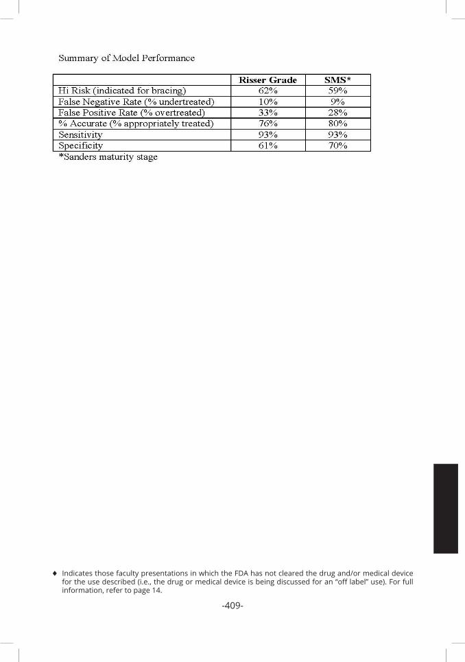
-409-
♦ Indicates those faculty presentations in which the FDA has not cleared the drug and/or medical device for the use described (i.e., the drug or medical device is being discussed for an “off label” use). For full information, refer to page 14.
-410-
See pages 21- 60 for financial disclosure information.
e-Poster #4 POSNA-2016 Prophylactic Antibiotic Use in Pediatric Orthopaedic Surgery: Reducing Variability, Cost and Risks
Adam Margalit, BS; Gift Ukwuani; Rushyuan J. Lee, MD; Paul D. Sponseller, MD The Johns Hopkins Hospital, Baltimore, Maryland
Purpose: To evaluate the use of antibiotic prophylaxis in the Operating Room compared to existing guidelines. We hypothesize that there will be variability and overuse of prophylactic antibiotics for pediatric orthopaedic procedures.
Methods: Retrospective review of all procedures for two providers at our institution between January 2014 and September 2015. All surgical procedures were subdivided into seven categories including: open with implants (osteotomies, open reduction and internal fixation (ORIF), arthrodesis, hemiepiphysiodesis, and reconstruction procedures), open without implants (epiphysiodesis, soft tissue releases, excision of bony or soft-tissue masses, and removal of hardware (ROH) procedures), percutaneous with implants (closed reduction and percutaneous fixation procedures (CRPF)), percutaneous without implants (halo application, arthrograms, and injections), non-invasive (closed reductions and casting), spinal procedures (all types), and arthroscopy. Cases performed for infections were excluded from this study. The incidence of antibiotic prophylaxis use was recorded for each group and compared to current guidelines.
Results: A total of 1,164 patients (open with implants n=291, open without implants n=245, percutaneous with implants n=146, percutaneous without implants n=33, non-invasive n=76, spine n=330, and arthroscopy n=43,) are included in this study. The rate of antibiotic prophylaxis use is 96%, 63%, 56%, 9%, 9%, 99%, and 100% for each group respectively. Note, antibiotics were given for 9% of non-invasive cases.
Conclusion: Prophylactic antibiotics have been shown to benefit patients undergoing major operations. Procedures with clear indications for antibiotics had a near 100% compliance rate and included osteotomies, ORIF, and spinal procedures. However, there is less evidence for less invasive procedures. Recent retrospective studies have shown that antibiotics may not be needed for percutaneous procedures or open cases without implants. In our study, these cases had the most variability (48%) and (63%) of all procedures. Further prospective studies are warranted to establish evidence-based guidelines of antibiotic prophylaxis in these procedures.
Significance: In our series of cases, approximately 25% of all surgeries (including ROH and CRPF procedures) may not need prophylactic antibiotics. With the average cost per dose of Cefazolin estimated to be about $45.00, this would infer savings. Patient care will also improve by decreasing drug-related complications, such as drug allergies and the emergence of new drug-resistant organisms. It identifies an area of variability for further research.
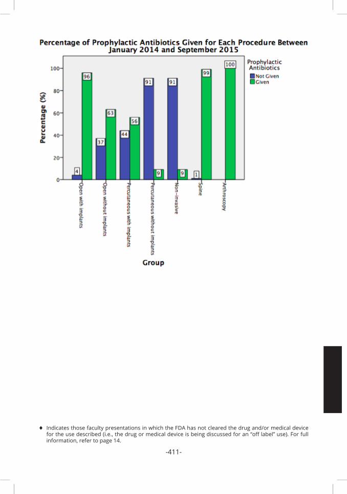
-411-
♦ Indicates those faculty presentations in which the FDA has not cleared the drug and/or medical device for the use described (i.e., the drug or medical device is being discussed for an “off label” use). For full information, refer to page 14.
-412-
See pages 21- 60 for financial disclosure information.
e-Poster #5 POSNA-2016 Pedicle Screw Safety: How Much Anterior Breach Is Safe? A Cadaveric and CT Based Study
Vishal Sarwahi, MD; Terry D. Amaral, MD; Kathleen J. Maguire, MD; Rachel C. Gecelter, BS; Monica M. Payares-Lizano, MD; Ajay F. Lall, MD; Dan Wang; Beverly Thornhill, MD Montefiore Medical Center, Bronx, New York
Purpose: The purpose of this study is to determine the safety limits of an anterior/ anterior lateral misplaced pedicle screw in spinal deformity. While, the limits of medial breach (< 3mm) are known, the safe limits for anterior/ antero- lateraly breach are not yet defined.
Methods: This study had two parts. In part I, 8 cadavers were instrumented with 6 x 30mm and 6x40mm bilaterally from T1-S1. Screws were randomly inserted under navigation guidance into 3 positions: in, out medial, out anterior laterally. Ct scan was performed, followed by gross dissection to determine screw position. In part II, post-operative CT scans of 165 patients operated on for spine deformity were reviewed for screw misplacement (2800 screws). The amount of breach for Anterior/antero- lateral was measured. Protrusions were also evaluated for proximity to vital structures. All scans were reviewed by musculoskeletal radiologist.
Results: Part I: 285 screws were inserted. 93 were misplaced anterior/antero- laterally. On gross dissection, 89 were misplaced; 67 protruded through soft tissue and were easily visualized while 32 were covered entirely by soft tissue but were palpable. These 32 screws did not endanger any structures. All 32 screws protruded < 4mm on CT scan. Part II: 117(4.14%) screws were misplaced anterior/ antero- laterally. 31(26.5%) were adjacent to vital structures. Fisher’s exact test showed < 4mm breach has significantly lower likelihood of impingement ( p<0.001) and sensitivity 81%. Screws adjacent/impinging the aorta protruded an avg 5.7 mm (SE 0.6), while screws not involving the aorta breached an avg 3.9 mm (SE 0.2), p=0.005.
Conclusion: Anterior/ antero-lateral breaches < 4mm on CT poses no significant risk of impingement and therefore can be considered safe.
Significance: Pedicle screw anterior misplacement potentially can cause injury to vascular and visceral structure. CT scans utilized to assess these misplacements, tend to underestimate the pre-vertebral soft tissue and may overestimate screw misplacements. This study defines the amount of anterior misplacement on CT scan which may be safe as it is covered by the pre-vertebral soft tissues.
-413-
♦ Indicates those faculty presentations in which the FDA has not cleared the drug and/or medical device for the use described (i.e., the drug or medical device is being discussed for an “off label” use). For full information, refer to page 14.
e-Poster #6 POSNA-2016 Adolescent Idiopathic Scoliosis Patients are at Increased Risk for Pulmonary Hypertension which Reverses after Scoliosis Surgery
Vishal Sarwahi, MD; Sarika Kalantre, MD; Rachel C. Gecelter, BS; Aviva F. Dworkin; Terry D. Amaral, MD; Kathleen J. Maguire, MD; Ajay F. Lall, MD; Marina Moguilevitch, MD; Dan Wang, MS Montefiore Medical Center, Bronx, New York
Purpose: This study seeks to determine the incidence of pulmonary hypertension (pulm HT) in Adolescent Idiopathic Scoliosis (AIS) patients, its relationship with the size of the curve and the effect of surgery on pulm HT.
Methods: Retrospective chart and XR review of AIS patients with PSF from 2009 –2013 was done. 2D echos were reviewed for structural heart disease, aortic root size, and Tricuspid regurgitant jet velocity (TRV). Right ventricular systolic pressure (RVSP) was estimated using the Bernoulli’s equation (4*(TRV)2 + right atrial pressure). TRV ≥ 2.8 m/s indicates pulmonary HT. 2D echo of 180 aged matched healthy adolescents served as control. XR measurements, PFTs and operative parameters were collected. Fisher’s exact tests, Chi-square tests, and Wilcoxon signed rank tests were done.
Results: 202 AIS patients had screening 2D echos. Average age was 14.9 ± 3.1 years. Average Cobb angle was 51.2±11.8°. Two (1.0%) patients had ASD, two (1.0%) patients had VSD., 3 had MVP (2.8%) and 4 had Mitral regurg (3.7%)..The right heart anomalies were mild tricuspid regurgitation in 28 AIS patients compared to 15 in Control (p<0.001). 42 AIS patients had abnormal RVSP/TRV values (≥ 2.8 m/s) (p<0.001) indicating pulmonary HT in AIS patients as opposed to none in controls. In AIS group, there was no significance between lenke type, PFT, extubation, pH, pCO2, EBL, or length of surgery between patients with abnormal TRV/RVSP and normal TRV/RVSP. 42 AIS patients with elevated TRV/RVSP had follow up 2D echo, post-op. All of these patients had RVSP/TRV < 2.8 m/s after corrective scoliosis surgery with a mean of 2.1 m/s and a range of 1.40-2.35 (p<0.0001), which shows reversal of pulmonary HT to normal values.
Conclusion: Screening 2D echo identifies structural heart defects and pulmonary HT. Pulm HT in AIS is likely not due to lung compromise as PFTs did not correlate. Scoliosis surgery reverses pulmonary HT, avoiding potentially fatal compromise. This may indicate that a curving spine may be contributing to its etiology.
Significance: This is the first study to document evidence of pulm HT in AIS patients. Pulm HT, which can potentially be fatal, reverts to normal after corrective scoliosis surgery. These findings provide direct evidence of immediate benefit of scoliosis surgery and can change the entire scoliosis treatment paradigm.
-414-
See pages 21- 60 for financial disclosure information.
e-Poster #7 POSNA-2016 Limb Length Multipliers in Real Life - Age versus Growth Spurt Timing to Predict Final Limb Length
Lauren E. Karbach, MD; Thomas Osinski, BS; Xiang Lu, MA; Raymond W. Liu, MD; Xing Qiu, PhD; Daniel R. Cooperman, MD; James O. Sanders, MD University of Rochester, Rochester, New York
Purpose: The Paley multiplier method is commonly used in pediatric orthopedics to predict future limb growth and provide estimations for epiphyseodesis timing. However, age based multipliers may be prone to error during the adeolescent growth spurt because of marked variability between children’s growth spurt timing. This study uses longitudinal data to determine limb length multipliers during childhood and adolescence and compares them both to age and to growth spurt timing.
Methods: From a longitudinal cohort of normal children, we identified those completing growth. We normalized heights as percentages of final at maturity and compared them to PHV timing and age. The subjects’ anthropometrics of standing symphyseal, crestal, and ASIS height and concomitant pelvic radiographs were used to measure height to the top of the femoral head for lower extremity length determination. For the upper extremity, anthropometrics from the tip of the middle finger to the acromion were measured directly. Multipliers were calculated based on length at maturity compared to both age and peak height velocity (PHV) timing.
Results: 54 subjects had completed their growth at the study terminus (35 f, 19m). As shown in the figures, the distribution of the multipliers was more widely distributed by chronological age than by PHV timing. Boys and girls had identical multipliers compared to the PHV, and the upper and lower extremity multipliers were equal. The Paley multipliers (line in the boys’ and girls’ graphs) were within the range identified for age-based multipliers but did not reflect the individual subject variability.
Conclusion: 1) The Paley multiplier does not reflect the wide range of future limb growth found in normal children.
2) Timing relative to the PHV provides reliable multipliers for limb length while chronological age has limited usefulness near adolescence. 3) Multipliers for the upper and lower extremity are equivalent relative to growth spurt timing and are identical for boys and girls.
Significance: Growth spurt timing is more important than age in predicting final limb length. Identifying where a child is relative to their growth spurt should provide reliable estimates of future upper and lower extremity length.
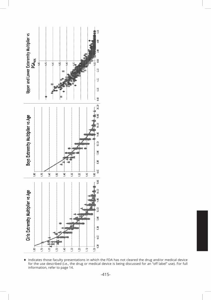
-415-
♦ Indicates those faculty presentations in which the FDA has not cleared the drug and/or medical device for the use described (i.e., the drug or medical device is being discussed for an “off label” use). For full information, refer to page 14.
-416-
See pages 21- 60 for financial disclosure information.
e-Poster #8 POSNA-2016 Management of Displaced Supracondylar Fractures of the Humerus Using Lateral versus Crossed K Wires: A Prospective Randomized Trial
Kishore Mulpuri, MD; Ashlee Dobbe, BS, FRCS (Ortho), MD, MSc; Emily Schaeffer, PhD; Firoz Miyanji, MD; Christine M. Alvarez, MD, FRCSC; Anthony P. Cooper, FRCS; Christopher W. Reilly, MD British Columbia Children’s Hospital, Vancouver, British Columbia, Canada
Purpose: Closed reduction and percutaneous pinning has become the most common technique for the treatment of Type III displaced supracondylar humerus fractures in children. The purpose of this study was to evaluate whether the loss of reduction in lateral K wiring is non-inferior to crossed K wiring in this procedure.
Methods: A prospective randomized non-inferiority trial was conducted. Patients aged 3-7 presenting to the Emergency Department with a diagnosis of Type III supracondylar humerus fracture were eligible for inclusion in the study. Consenting patients were block randomized into one of two groups based on wire configuration (lateral or crossed K wires). Surgical technique and post-operative management were standardized between the two groups. The primary outcome was loss of reduction, measured by the change in Baumann’s angle immediately post –operation compared to that at the time of K wire removal at 3 weeks. Secondary outcome data collected included Flynn’s elbow score, the humero-capitellar angle, and evidence of iatrogenic ulnar nerve injury. Data was analyzed using a t-test for independent means.
Results: A total of 52 patients were enrolled at baseline with 23 allocated to the lateral pinning group (44%) and 29 to the cross pinning group (56%). 6 patients (5 crossed, 1 lateral) received a third wire and 1 participant (crossed) did not return for x-rays at pin removal and were therefore excluded from analysis. A total of 45 patients were subsequently analyzed (22 lateral and 23 crossed). The mean change in Baumann’s angle was 1.05 degrees, 95% CI [-0.29, 2.38] for the lateral group and 0.13 degrees, 95% CI [-1.30, 1.56] for the crossed group. There was no significant difference between the groups in change in Baumann’s Angle at the time of pin removal (p = 0.18). Two patients in the crossed group developed post-operative iatrogenic ulnar nerve injuries, while none were reported in the lateral group.
Conclusion: Preliminary analysis shows that loss of reduction in Baumann’s angle with lateral K wires is not inferior to crossed K wires in the management of Type III supracondylar humerus fractures in children.
Significance: The results of this study suggest that orthopaedic surgeons who currently use crossed K wires could consider switching to lateral K wires in order to reduce the risk of iatrogenic ulnar nerve injuries without significantly compromising reduction.

-417-
♦ Indicates those faculty presentations in which the FDA has not cleared the drug and/or medical device for the use described (i.e., the drug or medical device is being discussed for an “off label” use). For full information, refer to page 14.
e-Poster #9 POSNA-2016 Coexisting DDH and Spastic Hip Disease? The Treatment of Dislocated Hips in Infants with Spasticity
Christian A. Refakis, BA; Wudbhav N. Sankar, MD Children’s Hospital of Philadelphia, Philadelphia, Pennsylvania Purpose: While many studies have separately investigated the treatment of developmental dysplasia of the hip (DDH) and spastic hip disease, little data exists regarding the treatment of infants with dislocated hips as well as underlying spasticity. The purpose of this study was to review our results following the surgical treatment of children with these seemingly coexisting conditions.
Methods: We retrospectively reviewed all children < 3 years of age who underwent hip reconstruction for dislocated hips in the setting of cerebral palsy or other spastic/high tone neuromuscular disease. Medical records were reviewed to determine demographic data, procedures performed, and treatment course including complications and need for further surgery. Pre-operative and post-operative radiographs were used to determine International Hip Dysplasia Institute (IHDI) grade of dislocation, acetabular index, migration percentage, and the presence of avascular necrosis.
Results: 11 patients with 15 hips met our inclusion criteria with a mean age of 19.9 ± 8.0 months (range 6-34 months). Preoperatively, 12/15 hips (80%) were IHDI grade 4 and 3/15 (20%) were IHDI grade 3. Mean acetabular index was 29.3 ± 7.5° (range 19-46°). Patients underwent open reduction (15 hips), adductor tenotomy (14 hips), varus derotational femoral osteotomy (10 hips), and pelvic osteotomy (12 hips). At a mean follow-up of 34.5 ± 19.2 months, 13 of 15 hips were IHDI grade 1 (86.7%), one was IHDI grade 2 (6.7%) and one hip was IHDI 3 (6.7%). The mean post-operative migration index was 8.1 ± 22.1%, and the mean acetabular index was 21.3 ± 8.7°. No patients developed radiographically significant osteonecrosis. Complications included two femur fractures (13.3%) and one symptomatic implant that required early removal. Only the one patient with an IHDI 3 outcome underwent further surgery in the form of triple innominate osteotomy and revision femoral osteotomy.
Conclusion: In this series of infants with hip dislocations and underlying spasticity, open reduction +/- pelvic osteotomy and/or femoral varus derotational osteotomy had >90% success rate in achieving and maintaining adequate hip reduction at intermediate-term follow-up. Avascular necrosis was not a significant complication in this series, but femoral insufficiency fractures were.
Significance: In the unique population of infants with dislocated hips and underlying spasticity, comprehensive hip reconstruction is largely successful with an acceptable rate of complications.
-418-
See pages 21- 60 for financial disclosure information.
e-Poster #10 POSNA-2016 Clinical Indications Associated with Abnormal MRIs in a Pediatric Spine Deformity Practice
Karl E. Rathjen, MD; Ozan Razi, MD; Erica Flores, RN, MSN; Heather D. Caine, BS; Dong-Phuong Tran, MS; ChanHee Jo, PhD; Adriana De La Rocha, PhD Texas Scottish Rite Hospital for Children, Dallas, Texas Purpose: In an effort to better define the appropriate use of MRI of the spine, we sought to determine which clinical indications are most (and least) frequently associated with abnormal MRI findings in patients presenting to pediatric spine deformity surgeons.
Methods: An IRB-approved, prospective analysis of the clinical indications for obtaining an MRI of the spine by the treating pediatric orthopaedic surgeons. Between, 2009-2011, surgeons ranked the clinical indications driving their decision to obtain a MRI of the spine. These prospectively recorded clinical indications were correlated with MRI results. MRI results were categorized as normal, abnormal/intra-dural or abnormal/structural.
Results: 453 patients had a MRI of the complete spine. 245 of 453 (54%) were normal and 208 of 453 (46%) were abnormal [95 intra-dural, 123 structural, 10 both]. Pain was the most frequently listed #1 indication [76/453; 16.7%]. Pain was associated with abnormal structural findings (facet or disc abnormalities) [32/76; 42.1%; p=0.0013]. Congenital scoliosis [28/46; 60.9%; p=0.032] and age at presentation [18/56; 32.1%; p=0.027] were significantly associated with abnormal MRIs. 37 patients [8.2%] had findings that led to a neurosurgical procedure. Pain as an indication was associated with NOT requiring neurosurgery [1/76; 1.3%; p=0.011] while congenital scoliosis [8/46; 17.4%; p=0.016]; curve magnitude at presentation [4/17; 23.5%; p=0.042] and abnormal abdominal reflexes [6/26; 23.1%; p-0.0042] were associated with neurosurgery.
Conclusion: Pain is a common indication for an MRI of the spine and is associated facet or disc abnormalities and NOT associated with neurosurgical pathology. Congenital scoliosis, curve magnitude at presentation and abnormal abdominal reflexes were the indications associated with neurosurgical pathology.
Significance: Pain is the most common indication for a spine MRI and NOT associated with neurosurgery. Congenital scoliosis, initial curve magnitude and abnormal abdominal reflexes were associated with neurosurgery.
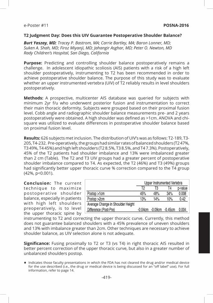
-419-
♦ Indicates those faculty presentations in which the FDA has not cleared the drug and/or medical device for the use described (i.e., the drug or medical device is being discussed for an “off label” use). For full information, refer to page 14.
e-Poster #11 POSNA-2016 T2 Judgment Day: Does this UIV Guarantee Postoperative Shoulder Balance?
Burt Yaszay, MD; Tracey P. Bastrom, MA; Carrie Bartley, MA; Baron Lonner, MD; Suken A. Shah, MD; Firoz Miyanji, MD; Jahangir Asghar, MD; Peter O. Newton, MD Rady Children’s Hospital, San Diego, California Purpose: Predicting and controlling shoulder balance postoperatively remains a challenge. In adolescent idiopathic scoliosis (AIS) patients with a risk of a high left shoulder postoperatively, instrumenting to T2 has been recommended in order to achieve postoperative shoulder balance. The purpose of this study was to evaluate whether an upper instrumented vertebra (UIV) of T2 reliably results in level shoulders postoperatively.
Methods: A prospective, multicenter AIS database was queried for subjects with minimum 2yr f/u who underwent posterior fusion and instrumentation to correct their main thoracic deformity. Subjects were grouped based on their proximal fusion level. Cobb angle and radiographic shoulder balance measurements pre- and 2 years postoperatively were obtained. A high shoulder was defined as >1cm. ANOVA and chi-square was utilized to evaluate differences in postoperative shoulder balance based on proximal fusion level.
Results: 626 subjects met inclusion. The distribution of UIV’s was as follows: T2-189, T3-205, T4-232. Pre-operatively, the groups had similar rates of balanced shoulders (T2 47%, T3 49%, T4 45%) and high left shoulders (T2 8.5%, T3 8.5%, and T4 7.3%). Postoperatively, 45% of the T2 patients had shoulder imbalance and 13% were imbalanced greater than 2 cm (Table). The T2 and T3 UIV groups had a greater percent of postoperative shoulder imbalance compared to T4. As expected, the T2 (46%) and T3 (49%) groups had significantly better upper thoracic curve % correction compared to the T4 group (42%, p<0.001).
Conclusion: The current technique to maximize postoperat ive shoulder balance, especially in patients with high left shoulders preoperatively, is to level the upper thoracic spine by instrumenting to T2 and correcting the upper thoracic curve. Currently, this method does not guarantee balanced shoulders with a 45% prevalence of uneven shoulders and 13% with imbalance greater than 2cm. Other techniques are necessary to achieve shoulder balance, as UIV selection alone is not adequate.
Significance: Fusing proximally to T2 or T3 (vs T4) in right thoracic AIS resulted in better percent correction of the upper thoracic curve, but also in a greater number of unbalanced shoulders postop.
-420-
See pages 21- 60 for financial disclosure information.
e-Poster #12 POSNA-2016 The Cost of Complexity in Pediatric Scoliosis Management
Melvin Makhni, MD; Amelia Lindgren, BS; Navid Dardashti, MPH; Kevin Wang, BA; Hiroko Matsumoto, MA; Jesus A. Jimenez, BS; Michael G. Vitale, MD, MPH Columbia University Medical Center, New York, New York Purpose: Tertiary care hospitals receive a disproportionate share of complex patients. Most studies identify risk factors that predict increased complications or length of stays. Little has been published investigating the impact of clinical factors on cost of care in children with scoliosis. We sought to quantitate the increased costs associated with the care of pediatric patients with complex spine deformity.
Methods: We performed a retrospective cohort study comparing estimated costs for children who underwent pediatric scoliosis fusion surgery at a single academic institution between May 1, 2008 and December 31, 2010. Severity was gauged by clinical data: idiopathic vs non-idiopathic scoliosis, the presence of a major comorbidity or ambulation status. We obtained estimated cost data for patient hospitalizations (initial and readmissions if relevant) for a minimum of four-years after the date of surgery from the Pediatric Health Information System. We compared how these severity categorizations affected costs.
Results: The records of 155 children ages 3-18 were reviewed. The cohort included 90 patients with idiopathic scoliosis, and 65 with non-idiopathic scoliosis. 29 patients experienced readmissions related to their procedures; a total of 212 admissions were included. The average cost was $57,001. Mean costs were 59.5% higher to manage non-idiopathic cases ($63,725.62) than idiopathic cases ($39,933.31; p<0.001). Significant difference in costs were seen when patients were classified by major comorbidity (p<0.001) or ambulation (p<0.001). Higher costs linearly correlated with younger patient age (0.211; p=0.019), larger number of levels fused (0.419; p<0.001), larger major curve (0.461; p<0.001), and increased length of stay (0.812; p<0.001). When APR-DRGs were obtained for the pediatric population over the same period, the average expected payment for scoliosis fusions ranged from $62,000-$70,661 depending on severity.
Conclusion: More complex patients were significantly more expensive to manage. Increased costs were associated with increased patient difficulty, increased lengths of stay, complications, readmissions and reoperations. Higher overall costs of care were seen for patients who were younger, had non-idiopathic scoliosis, had major comorbidities, were non-ambulatory, and those who had larger preoperative curves or more levels fused.
Significance: Complex patients are disproportionately managed at tertiary care centers. Adverse outcomes have traditionally been reported in terms that are not directly applicable to public policy. The staggering cost difference presents a challenge to a sustainable business model, especially with the proposal of bundled payment plans. By understanding the magnitude of the financial burden of caring for complex patients, reimbursement legislation can be proposed to sufficiently account for these disparities.
-421-
♦ Indicates those faculty presentations in which the FDA has not cleared the drug and/or medical device for the use described (i.e., the drug or medical device is being discussed for an “off label” use). For full information, refer to page 14.
e-Poster #13 POSNA-2016 Plantar Pressures Following Nonoperative and Surgical Treatment for Clubfoot at 10 Years of Age
Kirsten Tulchin-Francis, PhD; Ashley Erdman, BS, MBA; Kelly Jeans, MSc; Lori A. Karol, MD Texas Scottish Rite Hospital for Children, Dallas, Texas
Purpose: To assess plantar loading following nonoperative and surgical treatment for clubfoot (CF) at 10yrs of age.
Methods: 159 patients (236 clubfeet) underwent plantar pressure analysis at age 10.2yrs (9.7 to 11.5yrs). All subjects were initially treated nonoperatively (NO) with Ponseti casting or the French physiotherapy method. Feet which required surgical correction were divided into extra-articular (EA; tendon transfers, soft tissue release, lateral column shortening) and intra-articular (IA; posterior or posterior medial release, with/without additional procedures). Dynamic footprints were divided into medial and lateral hindfoot, midfoot and forefoot regions. Contact area (% of total foot contact), contact time (% of roll-over process) and peak pressure were assessed for each region. Hindfoot to forefoot angle (HF-FF angle) was used to quantify forefoot adductus, and the location of the center of pressure (COP) line relative to the foot axis (heel to 2nd toe) was assessed. Incidence rates of abnormal patterns were calculated using criteria relative to the control group (N=40 age-matched children). Comparison of the NO, EA and IA groups were made using chi-square tests with post-hoc analysis.
Results: Subjects treated NO (N=129 feet) had an increased incidence of forefoot adductus as measured by the HF-FF angle compared to the EA group (N=25 feet; 64% vs. 44%, p<.043); all CF groups had increased prevalence of forefoot adductus compared to the controls. Incidence of lateralization of the COP was significantly increased in all three groups compared to controls with corresponding reductions in contact area and peak pressure in the medial forefoot. A greater number of CF subjects (across all groups) had decreased contact area in the medial forefoot, with the NO group demonstrating increased lateral forefoot peak pressure There was a reduction in hindfoot contact time for the NO group (medial [p<.029] and lateral [p<.021]) suggestive of early heel rise. The EA group had a higher incidence of increased medial forefoot pressure (vs. NO & IA, p<.001) and contact area (vs. NO and IA, p<.001) which may indicate midfoot collapse or loss of medial longitudinal arch in some patients.
Conclusion: Approximately 60% of all clubfeet had increased forefoot adductus, with 38% outside two standard deviations of normal. Center of pressure was lateralized in all clubfoot groups. Early heel rise in the NO group may relate to a higher incidence of mild residual equinus.
Significance: Persistent abnormalities in plantar pressures are seen in 10yr old patients with clubfoot irrespective of treatment.
-422-
See pages 21- 60 for financial disclosure information.
e-Poster #14 POSNA-2016 Average 10-Year Results of the Bernese Periacetabular Osteotomy for Severe Acetabular Dysplasia
Stephen T. Duncan, MD; Jeff Petrie, MD; Kayla M. Thomason, BS; Geneva Baca, BA; Perry L. Schoenecker, MD; John C. Clohisy, MD Washington University School of Medicine, Saint Louis, Missouri
Purpose: The Bernese periacetabular osteotomy (PAO) is a well-described intervention which has been shown to be effective in reducing the symptoms of acetabular dysplasia. For patients with severe acetabular dysplasia with subluxation of the femoral head or presence of a secondary acetabulum, surgical realignment procedures remain controversial, and the efficacy of acetabular reorientation has been questioned. The purpose of this study was to analyze the average 10 year clinical and radiographic results of the Bernese PAO in the treatment of adolescent and young adult patients with symptomatic, severe acetabular dysplasia.
Methods: This retrospective cohort analyzed patients who underwent a PAO for severe acetabular dysplasia as defined by a lateral center edge angle (LCEA) < 5˚ or Group IV or V according to the Severin classification. All patients had hip pain and sufficient hip joint congruency on radiographs to be considered candidates for PAO. Clinical data collected included patient demographics, radiographic measurements, modified Harris Hip score (MHHS), and Western Ontario and McMaster Universities Osteoarthritis Index (WOMAC).
Results: The hip preservation database of the senior author (J.C.C.) was queried and 30 patients (36 hips; 22 females and 8 males) with an average age of 21.8 years (range, 11-60) were identified who had been treated with a PAO for severe acetabular dysplasia. The mean follow up was 10.2 years (range, 7-19.7) and mean BMI 23.2 kg/m2. On average, the lateral center edge angle and anterior center edge angle improved a mean of 30.8˚ (from -8.1˚ to 26.6˚, p<0.0001) and 24.3˚ (from-0.96˚ to 23.1˚, p< 0.0001), respectively. The MHHS improved an average of 16.5 points (from 68.2 to 82.1, p<0.001). The WOMAC subscore for pain improved by an average of 13.3 points postoperatively, though this was not statistically significant. Two hips (7.4%) required conversion to total hip arthroplasty at an average 5.4 years post-PAO, and three patients required revision PAO (11.1%) at an average 3.4 years post-PAO.
Conclusion: Periacetabular osteotomy is an effective treatment for severe acetabular dysplasia in adolescent and young adult patients. At an average of 10.2 years, our clinical and radiographic outcomes demonstrate improved hip function and major deformity correction in this population.
Significance: There is a paucity of long-term data regarding PAO, especially in the setting of severe acetabular dysplasia of the hip. This series illustrates the utility of the Bernese PAO providing powerful deformity correction as well as improved outcome measures in this population.
-423-
♦ Indicates those faculty presentations in which the FDA has not cleared the drug and/or medical device for the use described (i.e., the drug or medical device is being discussed for an “off label” use). For full information, refer to page 14.
e-Poster #15 POSNA-2016 Value-Based Medicine: Application to AIS Patients Undergoing Posterior Spinal Fusion – Determination of Cost and Outcome
Daniel J. Sucato, MD, MS; Heather D. Caine, BS; David Everett, BS Texas Scottish Rite Hospital for Children, Dallas, Texas
Purpose: In the new health care environment of value based medicine, we need to understand cost and outcome. There are no studies which have evaluated this for adolescent idiopathic scoliosis (AIS) patients undergoing posterior spinal fusion (PSF).
Methods: This is a retrospective review of a consecutive series of prospectively collected data for all Lenke 1A curves over a 3 year period. A direct cost analysis was performed with established cost categories that have similar generation characteristics e.g., Surgery, Operating Room, Room & Bed, Clinics, etc. and cost components have been identified based on resource consumption. Variation in costs was calculated for each cost component using an appropriate unit of measure and actual cost incurred. Patient reported outcome measures were determined using the SRS-22 quality of life questionnaire.
Results: There were 49 (5 males, 44 females) AIS patients who had a PSF at an average age of 14.7 (11.3-19.2) by 7 different surgeons. All patients had a preoperative Lenke 1A curve with an average Cobb angle of 57.1 ± 8.2 degrees. The number of levels fused was 10.3, implant density of 1.36 screws/level, surgical time was 264.1 min, and EBL was 641 cc. There was significant variation amongst surgeons in implant density (p<0.001), and surgical time (P<0.001). The total average cost for the surgical procedure was $23,962 including surgery cost (surgeon, implant, supplies) at $15,860 (S12,835-$20,402) and operating room cost (anesthesiology, nursing, other staff) at $8,102 ($5,784-12,241). There was a significant difference for OR cost (p=0.002) and total surgery cost (p<0.001) between surgeons. The primary drivers of surgery cost were the type/ number of implants and the driver for the operating room costs was surgical time. All five domains of the SRS-22 improved: pain (3.9 to 4.1), appearance (3.4 to 4.4), activity (4.0 to 4.1), mental (3.7 to 4.2), and satisfaction (3.9 to 4.7) and the total SRS score improved (3.8 to 4.2) without differences between providers.
Conclusion: In a homogeneous group of AIS patients undergoing surgery for Lenke 1A curves, there is significant variation in the number of implants used and operative time which directly affects the cost of surgery. Improvement in SRS outcome scores were seen without significant differences amongst providers.
Significance: There is significant variation in the cost of AIS surgery which is most significantly affected by implant costs and surgical time. Decreasing variation to improve costs is a viable goal in the treatment of these patients and should not influence the improved outcomes seen in this study.
-424-
See pages 21- 60 for financial disclosure information.
e-Poster #16 POSNA-2016 T1 Tilt Angle and Clavicle Angle on Immediate Post-Op X Rays are the Best Predictors of Shoulder Balance in AIS Patients
Vishal Sarwahi, MD; Stephen Wendolowski, BS; Rachel C. Gecelter, BS; Alexa J. Karkenny, MD, Terry D. Amaral, MD; Abhijit Pawar, MD Montefiore Medical Center, Bronx, New York
Purpose: This study seeks to investigate preoperative and postoperative predictors of shoulder imbalance for patients undergoing adolescent idiopathic scoliosis surgery utilizing all pedicle screw constructs.
Methods: Charts and XRs of AIS patients undergoing PSF with pedicle screws from 2005-2013 were analyzed. Using radiographic shoulder height, patients were divided into 3 groups: normal (0-1cm), mild imbalance (1-2cm) and severe imbalance (>2cm). Patient demographics, T1 tilt, clavicle angle, correction of PT/MT curve, LIV tilt, and coronal/sagittal balance were measured at preop, postop, and final follow up. Fisher’s exact, Kruskal Wallis and ANOVA tests were performed.
Results: 195 AIS patients with a median age of 14.9 yrs, 53° preop Cobb, 13.6° postop Cobb, with a median FU of 30 mths were included. 81 patients pre-op had normal shoulder balance, 59 had mild imbalance, and 55 severe imbalance. At immediate postop, 102 were normal, 60 had mild, and 30 had severe imbalance (p = 0.003). At most recent FU, shoulder heights were unchanged (p =0.423). Preoperatively, the three groups were similar in terms of LIV tilt, coronal/sagittal balance, Lenke types. T1 tilt and clavicle angle were significantly increased in patients with severe imbalance (p = <0.001). Postoperatively, the three groups were similar in all except T1 tilt and clavicle angle. Patients with severe imbalance at postop showed significantly higher median T1 tilt (-4.45) and median clavicle angle (-5.20) (p = <0.001). 34 patients with PT fusion did not show correlation with % cobb correction (<50, >50%). Lenke subgroups were not predictors of shoulder imbalance.
Conclusion: T1 tilt and clavicle angle are significantly associated with abnormal shoulder balance before and after surgery. Clavicle angle of 0 degrees and neutral T1 tilt is associated with normal shoulder level. In contrast, clavicle and T1 tilt greater or equal to 5 degrees correlates with abnormal shoulder balance before and after surgery.
Significance: Pedicle screw instrumentation in AIS patients allow for restoration to normal shoulder balance in majority of patients. T1 tilt and clavicle angle are the best predictors of final shoulder balance. Surgeons should focus on restoring T1 tilt & clavicle angle to 0° or between 2-3° for mildly uneven shoulders. Both angles >5° indicate severe shoulder imbalance.

-425-
♦ Indicates those faculty presentations in which the FDA has not cleared the drug and/or medical device for the use described (i.e., the drug or medical device is being discussed for an “off label” use). For full information, refer to page 14.
e-Poster #17 POSNA-2016 Scoliosis in Down Syndrome
Oussama Abousamra, MD; Maria Del Pilar Duque Orozco, MD; Kenneth J. Rogers, PhD; Julieanne P. Sees, DO; Freeman Miller, MD Nemours / Alfred I. duPont Hospital for Children, Wilmington, Delaware
Purpose: Scoliosis has been reported in Down syndrome with a prevalence of 10%. Cardiac surgery via a thoracotomy has been blamed for causing scoliosis in adolescents. Bracing effectiveness has been controversial and surgery has had a high rate of complications. However, the available reports covered a few decades of experience. In this study, the aim is to report the recent experience in scoliosis management. Treatment options and outcomes are reported as well.
Methods: Records of all children with Down syndrome, who were seen at our institution between 2004 and 2014, were reviewed. Scoliosis diagnosis was confirmed on radiographs with curves more than 10 degrees. Children with conditions other than Down syndrome, that might affect the spine, were not included. Curve levels, direction and magnitude were recorded. Risser stage, before brace application and at last visit, was recorded as well as curve progression in brace. Preop, postop and last visit curve magnitudes, fusion levels and postop complications were also recorded. A comparison between children with and without scoliosis, in terms of their cardiac surgery history, was performed using chi square test with a level of significance at 0.05.
Results: Out of 581 children with Down syndrome, 62 children with scoliosis (18 boys and 44 girls) were identified. Mean age at diagnosis was 13.8 years (8-20.1 years). Double (S shaped) curve was the most common pattern (52%). Mean curve magnitude at presentation was 31 degrees (13-66 degrees). Bracing was successful in 5 of 7 patients over 24 months period. Ten children had posterior spinal fusion, using pedicle screws, with a mean follow up of 2.6 years (1-7.3 years). Average hospitalization was 5 days. One case of deep wound infection and one superficial infection were noted. No revision was needed. No difference was found in the cardiac history between children with scoliosis and children who did not develop scoliosis (p = 0.104).
Conclusion: Scoliosis is a common finding in children with Down syndrome. Screening is recommended before the deformity becomes clinically evident with large curves. Bracing can be tried for early detected cases, taking the compliance issue into consideration. Spinal fusion is a successful treatment with good results and low complication rate over the short term follow up.
Significance: This study emphasizes the importance of scoliosis screening in Down syndrome. Recent advances in scoliosis surgery show promising results indicating that people with Down syndrome have been enjoying orthopaedic advances as well as advances in other medical fields.
-426-
See pages 21- 60 for financial disclosure information.
e-Poster #18 POSNA-2016 Do Professional Society Advocacy Campaigns Have an Impact on Pediatric Orthopaedic Injuries?
Alexa J. Karkenny, MD; Kathleen J. Maguire, MD; Norman Y. Otsuka, MD The Children’s Hospital at Montefiore, Montefiore Medical Center, Bronx, New York
Purpose: The American Academy of Orthopaedic Surgeons (AAOS) and the American Academy of Pediatrics (AAP) both participate in advocacy by publishing official recommendations for injury prevention. Pediatric orthopaedic injuries have been ascribed to trampolines, lawnmowers and all-terrain vehicles (ATVs) for instance, and childhood use of these has been discouraged by AAOS and AAP campaigns. The purpose of this study is to find a temporal association between these recommendations and trends in injury rates, as exemplified by the three aforementioned products and the relevant AAOS and AAP publications.
Methods: A retrospective review of fractures associated with trampolines, lawnmowers, and ATVs from 1991 to 2014 was performed using the National Electronic Injury Surveillance System (NEISS), which identifies the number of consumer product-related injuries to patients treated at NEISS-participating emergency departments. Sample case rates for fractures involving trampolines, lawnmowers, and ATVs among patients ages 2 to 18 years were recorded and percent changes year-to-year were calculated. A timeline of all AAOS and AAP advocacy statements published on the products from 1991 to 2014 was created.
Results: Fractures associated with trampolines increased from 1991 to a peak in 2004, despite a 1999 AAP statement. A 2005 AAOS statement corresponded with a 3.3% drop in fractures from 2004 to 2005. AAOS published an update in 2010, with a 7.5% drop from 2009. A 2012 AAP statement coincided with a 6.4% increase from 2011, followed by a 15% drop by 2013. For lawnmower injuries, a 2001 AAP statement corresponded with a 32% drop from 2000, the largest decrease until 2014. AAOS statements in 2011 and 2012 did not coincide with drops, but were followed by 12% and 43% decreases in 2013 and 2014, respectively. ATV injury rates did not drop in conjunction with 2000, 2005, or 2007 AAOS and AAP statements, but were followed by a 25% drop from 2007 to 2008. A 2010 AAOS statement correlated with a 27% drop from 2009, and a 2013 AAP statement with a 21% drop from 2012.
Conclusion: Studying the influence of a profession’s advocacy is valuable for strengthening its impact. Although the AAOS or AAP statements did not universally coincide with dropping fracture rates, statements often were associated with substantial decreases in following years. This may be because injury prevention messages are dispersed from providers to the public over time.
Significance: This study is the first to investigate the correlation between professional societies’ injury prevention campaigns and associated pediatric orthopaedic injury rates.
-427-
♦ Indicates those faculty presentations in which the FDA has not cleared the drug and/or medical device for the use described (i.e., the drug or medical device is being discussed for an “off label” use). For full information, refer to page 14.
e-Poster #19 POSNA-2016 Femoral Nerve Block with or without Sciatic Nerve Block for Pediatric Anterior Cruciate Ligament Reconstruction: a Retrospective Review
Jeffrey L. Hanway, MD; Leanne Foster, MD; Heather Gordish-Dressman, PhD; Sophie Pestieau, MD Children’s National Health System, Washington, District of Columbia
Purpose: Anterior cruciate ligament (ACL) reconstruction is associated with a significant amount of postoperative pain. Peripheral nerve blockade is often used in adult ACL repairs to improve pain control while decreasing opioid consumption and opioid induced side effects. But little data exist regarding use of this pain management in a pediatric population. The goal of this study was to compare perioperative opioid requirements and immediate postoperative outcomes of children undergoing general anesthesia for ACL reconstruction who received a femoral nerve block (FNB), both femoral and sciatic nerve blocks (FSNB), or no block (NB).
Methods: A retrospective review was conducted of all healthy patients, ages 10-19 years, undergoing ACL reconstruction at a single tertiary-care pediatric hospital between January 2012 and September 2014. Two patient groups, those amenable to regional anesthesia received either a FNB or FSNB after induction of general anesthesia, were compared. Patients not amenable to regional anesthesia received intravenous opioids with or without intra-articular infiltration of local anesthetic. Intraoperative and post-anesthesia care unit (PACU) opioid use, PACU pain scores, PACU length of stay (LOS), hospital admission, and incidence of nausea were examined.
Results: A total of 127 patients met inclusion criteria and were included in analysis. The FNB group consisted of 38 patients, FSNB group of 43 patients and 46 were not amenable to regional anesthesia. No significant differences in age, weight or gender were seen between treatment groups. Perioperative opioid use is shown in Table 1, with both block groups requiring statistically significant less morphine than the no block group and the FSNB group requiring significantly less fentanyl then the other groups. The FSNB group had less patients reporting severe pain (pain scale >6) in the PACU compared to the FSB and no lock group (FSNB: 12% vs. FNB: 84%, No block: 65%, p=0.003) There was no difference in PACU LOS, incidence of nausea, or hospital admission between the groups.
Conclusion: We found that a femoral nerve block, with or without sciatic nerve block, significantly reduced perioperative morphine requirement in pediatric patients undergoing ACL reconstruction. Further, combination of both blocks led to a significant reduction in pain scores as well as a decrease in fentanyl use. This study demonstrates the effectiveness of supplemental peripheral nerve blocks in a pediatric population undergoing ACL reconstruction.
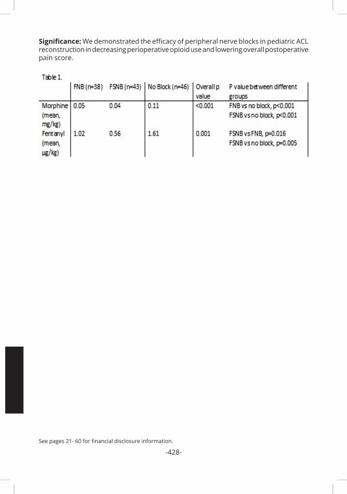
-428-
See pages 21- 60 for financial disclosure information.
Significance: We demonstrated the efficacy of peripheral nerve blocks in pediatric ACL reconstruction in decreasing perioperative opioid use and lowering overall postoperative pain score.
-429-
♦ Indicates those faculty presentations in which the FDA has not cleared the drug and/or medical device for the use described (i.e., the drug or medical device is being discussed for an “off label” use). For full information, refer to page 14.
e-Poster #20 POSNA-2016 Three-Dimensional MRI Quantification of Femoral Head Deformity in Legg-Calvé-Perthes Disease (LCPD)
Karen D. Standefer, BS; Molly Dempsey, MD; ChanHee Jo, PhD; Harry K.W. Kim, MD, MS Texas Scottish Rite Hospital for Children, Dallas, Texas Purpose: Using plain radiographs to quantify femoral head deformity in LCPD is subjective and inaccurate. The purpose of this study was to quantify the femoral head deformity in patients with LCPD using a novel 3D MRI reconstruction and analytic method.
Methods: 17 patients (6-14 years old, mean 9.9 ± 2.0 years) with unilateral or bilateral LCPD were enrolled into this prospective IRB approved study. All patients had 1 or 2 MRI scans using 1.5T scanner. In 12 patients, 2nd scans were obtained 4 to 14 months after the 1st scan. A total of 53 femoral head scans (20 normal and 33 affected femoral heads) were used to create 3D reconstructions of the femoral heads using MIMICS and 3Matics imaging analysis softwares. We developed and tested 3D MRI volume method as an indicator of femoral head deformity. In this method, the femoral head volume derived from 3D MRI is divided by the volume of a proportionately sized perfect hemisphere to obtain a volume ratio. Theoretical test shapes were used to validate the accuracy of the method. Percent articular surface point deviations and 3D femoral head height were also calculated as measures of deformity and compared to the volume method. 30 LCPD hips were segmented and analyzed twice to assess the intra-observer reliability. Statistical analyses included Pearson’s correlation and intraclass correlation coefficient (ICC).
Results: Theoretical calculations with test shapes showed 94% accuracy of the 3D MRI volume method. An intra-class correlation between the first and second measurements showed good intra-observer reliability (0.85, 95% CI: 0.71-0.92, N=30). The two other measures of deformity, percent articular surface point deviations and 3D femoral head height, showed excellent correlation with the MRI volume method (r=0.96, 95% CI: 0.93-0.98 and r=0.97, 95% CI: 0.95-0.98, respectively). The method was able to quantify small changes in the femoral head shape over time. Normal femoral heads rescanned after 4-14 months showed 0-3% change in the volume ratio, whereas femoral heads affected with LCPD showed 15% improvement to 33% worsening of the volume ratio in the second scan. In one case, pre- and post-Petrie cast treatment scans showed 15% improvement of the ratio in 4 months.
Conclusion: 3D MRI volume method allowed accurate and reliable quantification of the femoral head deformity in patients with LCPD.
Significance: This method may serve as a useful tool to evaluate the effect of treatment on the femoral head shape in patients with LCPD.
-430-
See pages 21- 60 for financial disclosure information.
e-Poster #21 POSNA-2016 Adolescents Maintain Hip Strength and Function 5 yrs Following a Ganz Periacetabular Osteotomy
Daniel J. Sucato, MD, MS; Kirsten Tulchin-Francis, PhD; David A. Podeszwa, MD; Adriana De La Rocha, PhD; Christopher Stewart, BS; Wilshaw Stevens, BS Texas Scottish Rite Hospital for Children, Dallas, Texas
Purpose: The effectiveness of the Ganz periacetabular osteotomy (PAO) in treating adolescent hip dysplasia (AHD) with longer term results is limited. This is the first study to report radiographic and functional outcome data in adolescents undergoing a PAO 5 years after treatment.
Methods: IRB-approved prospective consecutive series of adolescent patients treated with a PAO for HD. All patients had a minimum of 5 years post-operative functional and strength data. Radiographic, functional scores and 3-D gait analysis were performed.
Results: 29 patients (25 females/4 males) with 32 hips who underwent a PAO at 15.7 ± 13.1 yrs (10.2-25.2 yrs) were included. All radiographic parameters improved from pre-op to 2 yrs and were maintained at final follow-up: LCEA (2.4° to 32.0° to 30.0°, p<.001), AIWBZ (25.8° to 7.6° to 8.5°, p<.001) and significant improvement in the VCEA from pre-op to last follow-up (-1.4° to 27.1°, p<.001). Pre-operatively 18 hips were Tonnis 0, 13 Tonnis 1 and one Tonnis 2; at 5 yrs follow-up, 18 hips were Tonnis 0, 10 Tonnis 1, three Tonnis 2 and one Tonnis 3. Walking speed and hip flexor strength was maintained at 2 yrs and at last follow-up, with no significant differences over time (92.9 to 90.9 to 89.6 Nm/kg, p=.945). Abductor strength improved from pre-operative to 2yrs and was maintained at last follow-up (68.3 to 84.7 to 87.7 Nm/kg) (p=.105). The mHHS was significantly improved at 2 years and last follow-up compared to pre-operatively (67.1 to 79.1 to 75.7, p=.001), without change between 2yrs and last follow-up (p=0.546). Preoperatively, 57% of patients had moderate/marked pain but only 17% at 5 year follow-up and 30 hips were doing well. One patient progressed to a Tonnis 3 with complete loss of joint space and another patient had a revision PAO for persistent symptoms and inadequate acetabular coverage achieved at the initial PAO.
Conclusion: At a minimum 5 year follow-up, the Ganz PAO provides excellent radiographic correction and significant improvement in functional scores with maintenance of hip strength in adolescents with HD. This is the first study to demonstrate the recovery and maintenance of hip abductor and hip flexor strength at longer-term follow-up after treatment with a PAO in adolescents.
Significance: The Ganz PAO is effective in treating hip dysplasia in adolescents with maintenance of correction, functional outcomes, and hip abductor and flexor strength at a minimum 5 yrs post-op.

-431-
♦ Indicates those faculty presentations in which the FDA has not cleared the drug and/or medical device for the use described (i.e., the drug or medical device is being discussed for an “off label” use). For full information, refer to page 14.
e-Poster #22 POSNA-2016 A Prospective Randomized Comparative Study of PCA, Epidural Catheters and Lumbar Plexus Catheters For Analgesia After Hip Surgery in Children
Klane K. White, MD, MS; Nathalia Jimenez, MD; Nancy E. Gove, PhD Seattle Children’s Hospital, Seattle, Washington
Purpose: The lumbar plexus is composed of the femoral, obturator and lateral femoral cutaneous nerves. The advent of continuous peripheral nerve catheters offers continuous lumbar plexus catheter (LPC) as a viable technique for prolonged postoperative analgesia for pediatric hip surgery. Lumbar epidural catheters (LEC) and intravenous patient controlled analgesia pumps (IV-PCA) can also provide effective continuous postoperative analgesia following hip surgery. In contrast to LEC and IV-PCA, children may be discharged home with LPCs, and do not require urinary catheterization postoperatively. The most effective method of pain control following major hip surgery, such as pelvic osteotomy, has not been established.
Methods: We undertook a prospective, randomized-control trial comparing LPC to LEC and IV-PCA, stratified by age group (1-5, 6-18 years), after unilateral hip surgery in patients aged 1 to 18 years with a primary outcome of hospital length of stay. We compared pain scores, opioid consumption and side effects as secondary outcomes. Patients were followed daily as inpatients until hospital discharge or catheter removal. The analyses were conducted using an ANOVA within each age group.
Results: LOS was not significantly different between treatment groups (p = 0.60, p = 0.18 for age groups < 6 and ≥ 6 years). For patients < 6 years old, the LEC (1.9 d) and LPC (2.0 d) had a similar LOS. For patients ≥ 6 years old, LPC had the shortest LOS (2.2 d), compared to LEC (2.9 d) and IV-PCA (3.2 d). The mean of maximum pain scores for POD0 thru POD2 were not significantly different (p = 0.10, p = 0.48). LPC did have the lowest mean and maximum pain scores for all groups. For the older group, the total morphine equivalent per kg for POD 0 - 2, was significantly different across the treatment groups (p = 0.003). For the older patients, IV- PCA has higher morphine equivalent use over POD 0-2. The LEC and LPC have similar values for morphine equivalent use for both age groups.
Conclusion: Regional anesthesia reduces opiate use after pediatric hip surgery. Length of stay and postoperative pain scales tend to improve with LPC.
Significance: In a prospective, randomized-control study of regional analgesia after hip surgery in children, regional anesthesia is shown to effectively control pain and reduce opiate requirements. When choosing a LPC as the method of regional anesthesia, the patient also has the advantage of continued control of analgesia after discharge from the hospital.
-432-
See pages 21- 60 for financial disclosure information.
e-Poster #23 POSNA-2016 Damage Patterns Seen with Femoroacetabular Impingement in Children
Prasad V. Gourineni, MD; Summer Watkins, MSN, RN, PNP-AC Advocate Children’s Hospital, Oak Lawn, Illinois
Purpose: Femoroacetabular impingement (FAI) in children is not well defined. The incidence of cam, pincer, mixed, abrasion, and contre-coup patterns of damage were studied in a large group of children.
Methods: 279 children (9 - 18 years of age; 191 male and 88 female) underwent arthroscopy in 342 hips for hip pain from FAI over a nine year period. Clinical examination and plain radiographs suggested the diagnosis. Magnetic resonance imaging and diagnostic hip injections were used to exclude other causes of hip pain. Alpha angle, epiphyseal extension sign, and trochanteric height were used to document femoral morphology. Center edge angle, anterior wall coverage, and posterior wall sign were used to document acetabular morphology. Intraoperative findings of labral damage, acetabular changes, and femoral head findings were used to identify patterns.
Results: 52 hips had clear signs of slipped epiphysis. 225 hips had clear cam morphology based on alpha angle and/or epiphyseal extension sign and without a slip. Anterior over coverage with or without posterior wall abnormalities was seen in 189 hips. 101 hips had normal anterior coverage. Cam damage alone with labral sparing, chondrolabral separation, carpet phenomenon, or flap separation was seen in 63 hips. Pincer impingement with labral crushing, narrow band of acetabular chondromalacia, femoral head bands, and neck grooves were seen in 78 hips. Mixed pattern with both cam and pincer findings was seen in 110 hips. All 52 hips with slipped epiphysis showed synovitis, labral fraying, erythema, or crushing. 45 hips showed abrasion of the acetabular cartilage. 39 hips did not fit into a clear pattern. Impingement cysts were common and femoral head chondromalacia was rare. Labral lesions were common but unstable labral tears were not seen. Contre-coup damage was not noted in any hip.
Conclusion: Majority of hips with FAI showed damage to suggest cam, pincer, mixed, or abrasion patterns.. Radiographic and arthroscopic patterns correlated in 42% of non-slip hips and 100% of slip hips. Hips with good range of internal rotation at 90 degrees of flexion and lack of pincer pattern arthroscopically were treated with femoral neck osteoplasty and without acetabular rim trimming and labral repair. Hips with decreased rotation were treated with rim resection, labral repair, and femoral neck osteoplasty to optimize bony morphology and maximize hip motion improvement.
Significance: Identifying the patterns of damage in the hips has the potential to explain the pathogenesis of FAI, direct the arthroscopic treatment, and evaluate longer term results and patient reported outcomes.
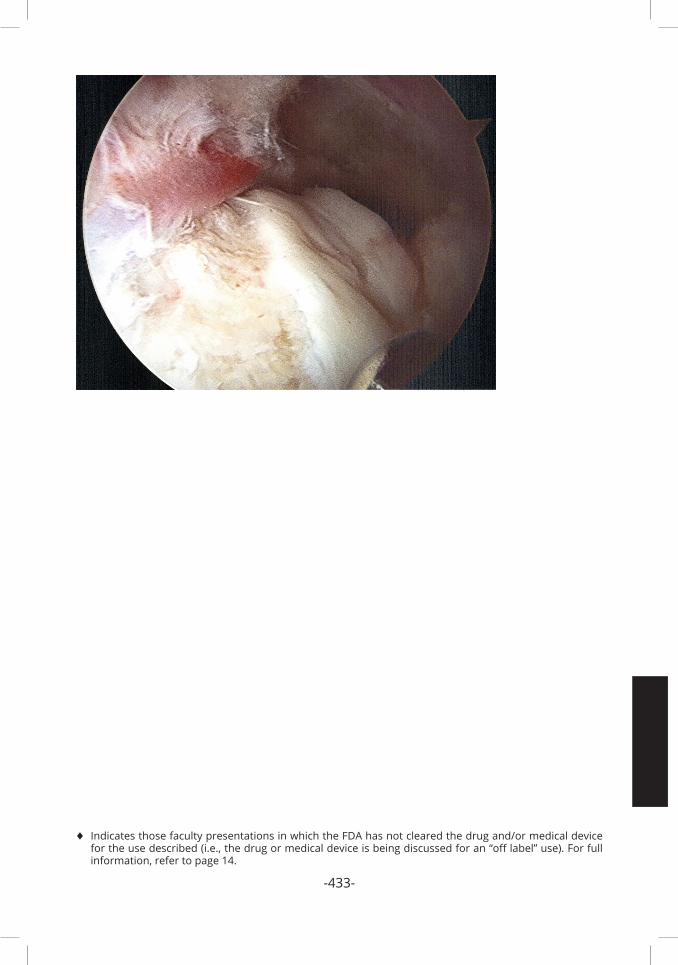
-433-
♦ Indicates those faculty presentations in which the FDA has not cleared the drug and/or medical device for the use described (i.e., the drug or medical device is being discussed for an “off label” use). For full information, refer to page 14.
-434-
See pages 21- 60 for financial disclosure information.
e-Poster #24 POSNA-2016 Hip Dysplasia in Patients with Cerebral Palsy Treated with Bernese Periacetabular Osteotomy
Mark L. Miller, MD; John C. Clohisy, MD; Perry L. Schoenecker, MD Washington University, Saint Louis, Missouri
Purpose: Treatment of symptomatic hip dysplasia in patients with spastic cerebral palsy (CP) is challenging and surgical options are controversial. This study examines our experience with Bernese periacetabular osteotomy, in combination with adjunctive treatments, for the treatment of hip dysplasia in patients with CP.
Methods: Between 2001 and 2012, 14 patients (16 hips) with symptomatic hip dysplasia associated with cerebral palsy were comprehensively treated with a periacetabular osteotomy and adjunctive procedures by the senior authors and retrospectively reviewed. The median age at surgery was 17 (range 13-30). 15 of 16 patients had Gross Motor Function Classification System (GMFCS) levels of 1 or 2 prior to surgical intervention. One patient was GMFCS level 4 preoperatively. We compared preoperative pain, validated hip functional scores and radiographic parameters of hip dysplasia to postoperative results. 14 of 16 patients had at least 12 months of follow-up with a median of 35 months of follow-up. Adjunctive treatments were performed in 10 hips: 6 hips had open adductor tenotomies and 5 hips had concomitant proximal femoral osteotomy.
Results: The primary outcome was the Modified Harris Hip Score (MHHS). Preoperatively, the patients had an average MHHS of 57. Postoperatively, at the most recent clinic visit (range 13-125 months), the average MMHS was 75 for an increase of 18 (p=0.01). The pain component of the MHHS improved from 20 to 37 (p<0.01). Tonnis angles improved from an average of 29 degrees preoperatively to an average of 5.7 degrees postoperatively. Lateral Center-Edge Angles improved -8 degrees to 28 degrees. Anterior center-edge angles increased from 16 degrees to 32 degrees. Migration indices improved from 52% and 15%.
Conclusion: In combination with appropriate adjunctive treatments, Bernese Periacetabular osteotomy can be an effective treatment for painful hip dysplasia in patients with spastic cerebral palsy through appropriate lateral and posterior coverage of the acetabulum and limitation of migration of the femoral head. A clinically significant improvement in pain and function has been documented in follow-up.
Significance: This is the first comprehensive study to show that Bernese periacetabular osteotomy can effectively correct acetabular deformity and hip joint subluxation associated with spastic cerebral palsy. For this selected group of patients, pain has resolved and function has notably improved. Periacetabular osteotomy should be considered as a surgical option for hip dysplasia in appropriately selected patients with spastic cerebral palsy.
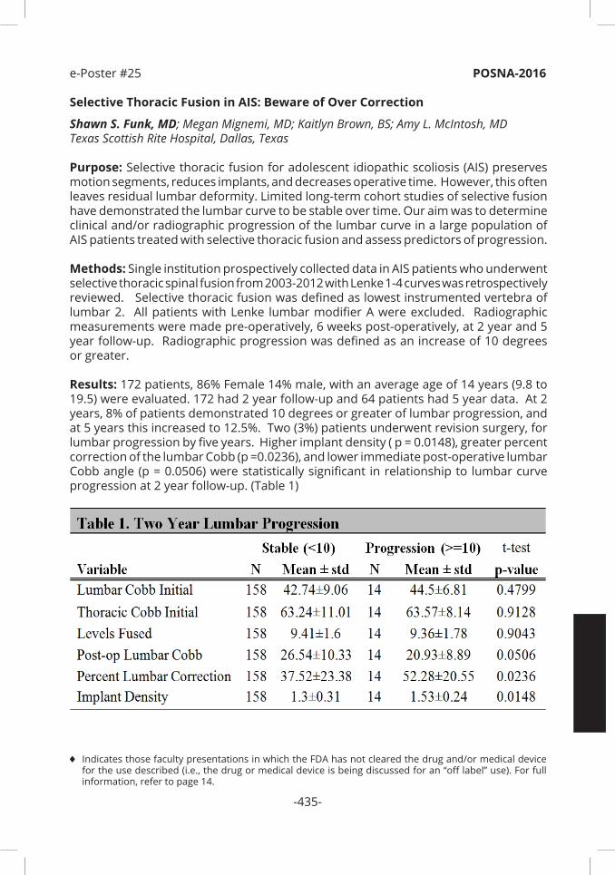
-435-
♦ Indicates those faculty presentations in which the FDA has not cleared the drug and/or medical device for the use described (i.e., the drug or medical device is being discussed for an “off label” use). For full information, refer to page 14.
e-Poster #25 POSNA-2016 Selective Thoracic Fusion in AIS: Beware of Over Correction
Shawn S. Funk, MD; Megan Mignemi, MD; Kaitlyn Brown, BS; Amy L. McIntosh, MD Texas Scottish Rite Hospital, Dallas, Texas
Purpose: Selective thoracic fusion for adolescent idiopathic scoliosis (AIS) preserves motion segments, reduces implants, and decreases operative time. However, this often leaves residual lumbar deformity. Limited long-term cohort studies of selective fusion have demonstrated the lumbar curve to be stable over time. Our aim was to determine clinical and/or radiographic progression of the lumbar curve in a large population of AIS patients treated with selective thoracic fusion and assess predictors of progression.
Methods: Single institution prospectively collected data in AIS patients who underwent selective thoracic spinal fusion from 2003-2012 with Lenke 1-4 curves was retrospectively reviewed. Selective thoracic fusion was defined as lowest instrumented vertebra of lumbar 2. All patients with Lenke lumbar modifier A were excluded. Radiographic measurements were made pre-operatively, 6 weeks post-operatively, at 2 year and 5 year follow-up. Radiographic progression was defined as an increase of 10 degrees or greater.
Results: 172 patients, 86% Female 14% male, with an average age of 14 years (9.8 to 19.5) were evaluated. 172 had 2 year follow-up and 64 patients had 5 year data. At 2 years, 8% of patients demonstrated 10 degrees or greater of lumbar progression, and at 5 years this increased to 12.5%. Two (3%) patients underwent revision surgery, for lumbar progression by five years. Higher implant density ( p = 0.0148), greater percent correction of the lumbar Cobb (p =0.0236), and lower immediate post-operative lumbar Cobb angle (p = 0.0506) were statistically significant in relationship to lumbar curve progression at 2 year follow-up. (Table 1)
-436-
See pages 21- 60 for financial disclosure information.
Conclusion: Spinal balance and correction of the lumbar curve remain stable over time in the majority (87.5-92%) of selective thoracic fusion patients. However, the lumbar curve progressed ≥ 10 degrees in 8% of patients at 2 years and 12.5% at 5 years. 3% of patients underwent revision surgery for lumbar curve progression by 5 years post-op. Long-term studies suggest that continued progression outside the 2-5 year time period is unlikely. Maximal correction of deformity may add unnecessary difficulty to surgical correction and be an act of futility as greater correction is associated with rebound lumbar progression.
Significance: Higher implant density, greater percent lumbar correction, and smaller residual lumbar curves are more likely to progress ≥10 degrees in the first 5 years post-operatively.

-437-
♦ Indicates those faculty presentations in which the FDA has not cleared the drug and/or medical device for the use described (i.e., the drug or medical device is being discussed for an “off label” use). For full information, refer to page 14.
e-Poster #26 POSNA-2016 Can Facebook Alleviate Anxiety in Teenagers Undergoing Scoliosis Surgery? A Prospective Non-Randomized Controlled Study
Melanie A. Gambassi, NP; Stephen Wendolowski, BS; Rachel C. Gecelter, BS; Dana Orlando, ST; Terry D. Amaral, MD; Vishal Sarwahi, MD Montefiore Medical Center, Bronx, New York Purpose: This study seeks to evaluate a Facebook-based scoliosis community’s ability to reduce preoperative anxiety in Adolescent Idiopathic Scoliosis (AIS) patients.
Methods: All patients had a pre-operative discussion regarding risks, complications, post-operative plan and recovery. Patients participating on a “Private” Facebook page were included in the Facebook group, while patients who did not, were controls. Anxiety was measured using the State Trait Anxiety Inventory (STAI). STAI consists of 40 questions: 20 questions evaluate anxiety at that specific moment (state anxiety) and 20 questions evaluate their anxiety in general (trait anxiety). The mental health domain from SRS-30 questionnaire was analyzed prior to Facebook participation. Two tailed t-tests were utilized.
Results: There were 17 patients in the Facebook group and 41 in the control group. Mean STAI score for state anxiety was 38.17 for the controls and 43.94 for the Facebook group (p=0.061). Mean STAI score for trait anxiety was 35.29 for the controls and 45.82 for the Facebook group (p=0.004). The trait (baseline) anxiety scores in both groups were comparable to those found in high school students. However, patients in the control group had higher state anxiety scores than trait scores, indicating increased anxiety. In contrast, the Facebook group had lower state anxiety scores than trait scores, which indicate better emotional state. The average mental health domain SRS-30 score was 4.05 (0.66) for the controls and 3.74 (0.85) for the Facebook group (p=0.377).
Conclusion: AIS patients undergoing surgery tend to have surgery increased state of anxiety while preparing for surgery. Among the teenage population, Facebook is an easily available and age-appropriate tool that can lower their anxiety scores and can help them prepare better for surgery.
Significance: 80% of teenagers utilize social networking and thus a Facebook support group would engage AIS patients on a platform familiar to them. By allowing patients to connect with others who have had PSF, share their fears, thoughts and questions about surgery, can potentially reduce anxiety, act as mental health and social support interventions and improve quality of care.
-438-
See pages 21- 60 for financial disclosure information.
e-Poster #27 POSNA-2016 Costs and Complications of Increased Length of Stay Following Scoliosis Surgery
Matthew R. Boylan, ScB; Aldo M. Riesgo, MD; Alice Chu, MD; Carl B. Paulino, MD; David S. Feldman, MD NYU Hospital for Joint Diseases, New York, New York
Purpose: Accelerated discharge protocols for scoliosis surgery have recently been described in the literature. However, there is limited data describing the association of increased length of stay with outcomes of this procedure. The aim was to evaluate the cost and complications of increased length of stay following scoliosis surgery.
Methods: The Statewide Planning and Research Cooperative System database was used to identify patients with adolescent idiopathic scoliosis (AIS) who underwent spinal fusion between October 1, 2007 and September 30, 2012 at high-volume institutions (>20 cases per year) in the state of New York. Regression models of each outcome were adjusted for age, gender, race, insurance, comorbidity score, and perioperative complications.
Results: Among the 1,787 patients with AIS who underwent spinal fusion, the mean length of stay was 4.88 days (95% CI=4.83-4.94; SD=1.18). In the perioperative period, 473 (26.47%) underwent transfusion and 217 (12.14%) had documented difficulty with pain control. An additional 1-day in the hospital was associated with $12,002 (95% CI=8,919-15,085; p<0.001) of insurance charges, $5,099 (95% CI=4,246-5,952; p<0.001) of hospital costs, 24% increased risk (OR=1.24; 95% CI=1.01-1.52; p=0.039) of 90-day all-cause readmission, and 51% increased risk (OR=1.51; 95% CI=1.15-1.97; p=0.003) of 90-day return to the operating room.
Conclusion: Increased length of stay following scoliosis surgery is associated with higher costs and increased risk of 90-day readmission and return to operating room. Protocols to decrease length of stay have potential benefits to patients, hospitals and insurers.
Significance: This is the first study to our knowledge to attempt to quantify the economic costs and association with adverse outcomes of increased length of stay following scoliosis surgery.
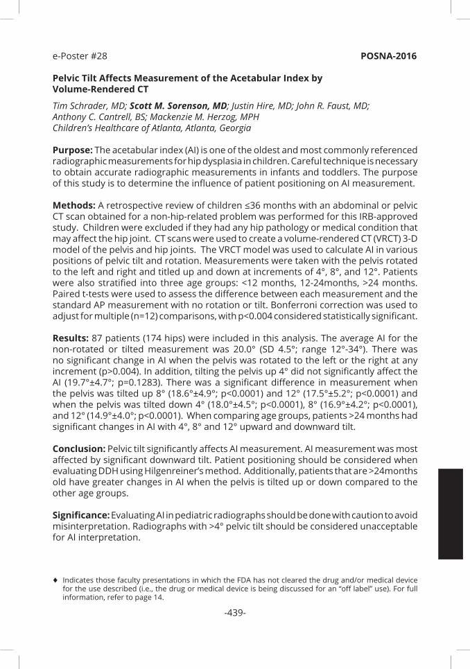
-439-
♦ Indicates those faculty presentations in which the FDA has not cleared the drug and/or medical device for the use described (i.e., the drug or medical device is being discussed for an “off label” use). For full information, refer to page 14.
e-Poster #28 POSNA-2016 Pelvic Tilt Affects Measurement of the Acetabular Index by Volume-Rendered CT
Tim Schrader, MD; Scott M. Sorenson, MD; Justin Hire, MD; John R. Faust, MD; Anthony C. Cantrell, BS; Mackenzie M. Herzog, MPH Children’s Healthcare of Atlanta, Atlanta, Georgia
Purpose: The acetabular index (AI) is one of the oldest and most commonly referenced radiographic measurements for hip dysplasia in children. Careful technique is necessary to obtain accurate radiographic measurements in infants and toddlers. The purpose of this study is to determine the influence of patient positioning on AI measurement.
Methods: A retrospective review of children ≤36 months with an abdominal or pelvic CT scan obtained for a non-hip-related problem was performed for this IRB-approved study. Children were excluded if they had any hip pathology or medical condition that may affect the hip joint. CT scans were used to create a volume-rendered CT (VRCT) 3-D model of the pelvis and hip joints. The VRCT model was used to calculate AI in various positions of pelvic tilt and rotation. Measurements were taken with the pelvis rotated to the left and right and titled up and down at increments of 4°, 8°, and 12°. Patients were also stratified into three age groups: <12 months, 12-24months, >24 months. Paired t-tests were used to assess the difference between each measurement and the standard AP measurement with no rotation or tilt. Bonferroni correction was used to adjust for multiple (n=12) comparisons, with p<0.004 considered statistically significant.
Results: 87 patients (174 hips) were included in this analysis. The average AI for the non-rotated or tilted measurement was 20.0° (SD 4.5°; range 12°-34°). There was no significant change in AI when the pelvis was rotated to the left or the right at any increment (p>0.004). In addition, tilting the pelvis up 4° did not significantly affect the AI (19.7°±4.7°; p=0.1283). There was a significant difference in measurement when the pelvis was tilted up 8° (18.6°±4.9°; p<0.0001) and 12° (17.5°±5.2°; p<0.0001) and when the pelvis was tilted down 4° (18.0°±4.5°; p<0.0001), 8° (16.9°±4.2°; p<0.0001), and 12° (14.9°±4.0°; p<0.0001). When comparing age groups, patients >24 months had significant changes in AI with 4°, 8° and 12° upward and downward tilt.
Conclusion: Pelvic tilt significantly affects AI measurement. AI measurement was most affected by significant downward tilt. Patient positioning should be considered when evaluating DDH using Hilgenreiner’s method. Additionally, patients that are >24months old have greater changes in AI when the pelvis is tilted up or down compared to the other age groups.
Significance: Evaluating AI in pediatric radiographs should be done with caution to avoid misinterpretation. Radiographs with >4° pelvic tilt should be considered unacceptable for AI interpretation.
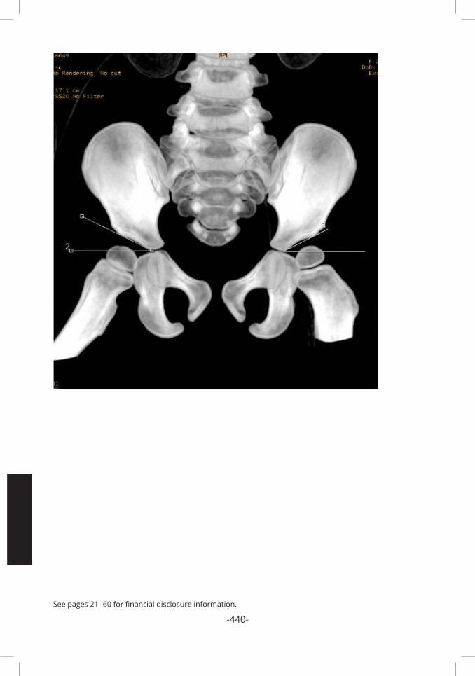
-440-
See pages 21- 60 for financial disclosure information.

-441-
♦ Indicates those faculty presentations in which the FDA has not cleared the drug and/or medical device for the use described (i.e., the drug or medical device is being discussed for an “off label” use). For full information, refer to page 14.
e-Poster #29 POSNA-2016 Variations in Surgical Rates of Pediatric Distal Radius Fractures Across Institutions: Analysis of the PHIS Database
Susan T. Mahan, MD, MPH; Apurva Shah, MD, MBA; Leslie A. Kalish, ScD; Lanna F. Feldman, MS; Donald S. Bae, MD Boston Children’s Hospital, Boston, Massachusetts
Purpose: Identification and reduction of variations in healthcare delivery may streamline care delivery, improve patient outcomes, and minimize cost. The purpose of this study was to assess the variation of surgical care for pediatric distal radius fractures (DRFs) using the Pediatric Health Information System (PHIS) database.
Methods: The PHIS database was queried from 2009-2013 for DRFs in patients between 4-18 years of age. Patients who underwent surgical treatment with internal fixation were identified using one of four surgical CPT codes (25606, 25607, 25608, or 25609). Twenty-five children’s hospitals with consistent CPT reporting systems were included. Surgical rates were modeled using mixed effects logistic regression with a random intercept for hospital, and were adjusted for gender and age (categories chosen a priori: 4-10, 11-14, 15-18 years of age). Model-based predicted surgical rates for each hospital were standardized for the aggregate gender and age distributions, resulting in predictions for the same “average” patient at every hospital.
Results: 59,143 patients were identified (range 906 - 6612 per hospital), of which 1161 (1.96%; range 0.35% - 17.2%) had surgery. Higher surgical rates were associated with male gender (Males: 2.26%, Females: 1.45%; adjusted p<.001) and older age (4-10 years: 1.54%, 11-14: 2.34%, 15-18: 4.32%; adjusted p<.001). There was no change in surgical rates over time. Model-based predicted surgical rates varied widely across hospitals (0.38% to 16.3%, p<.001). The rates were significantly lower than the aggregate rate of 1.96% at 11 (44%) of 25 hospitals. The rate was significantly higher at 6 (24%) hospitals, including more than five-fold higher in 2 hospitals.
Conclusion: Based on this PHIS analysis, approximately 2% of pediatric DRFs undergo surgical treatment. Variations in surgical rates were identified, however, with greatest variation seen in a small minority of institutions.
Significance: An important step in providing value-based healthcare is measurement and assessment of variation. This investigation provides information regarding the overall rate of surgery in pediatric DRF in U.S. pediatric hospitals as well as the distribution of variation. Future analysis will strive to establish target surgical rates and characterize the effects of variations on patient outcomes and cost.
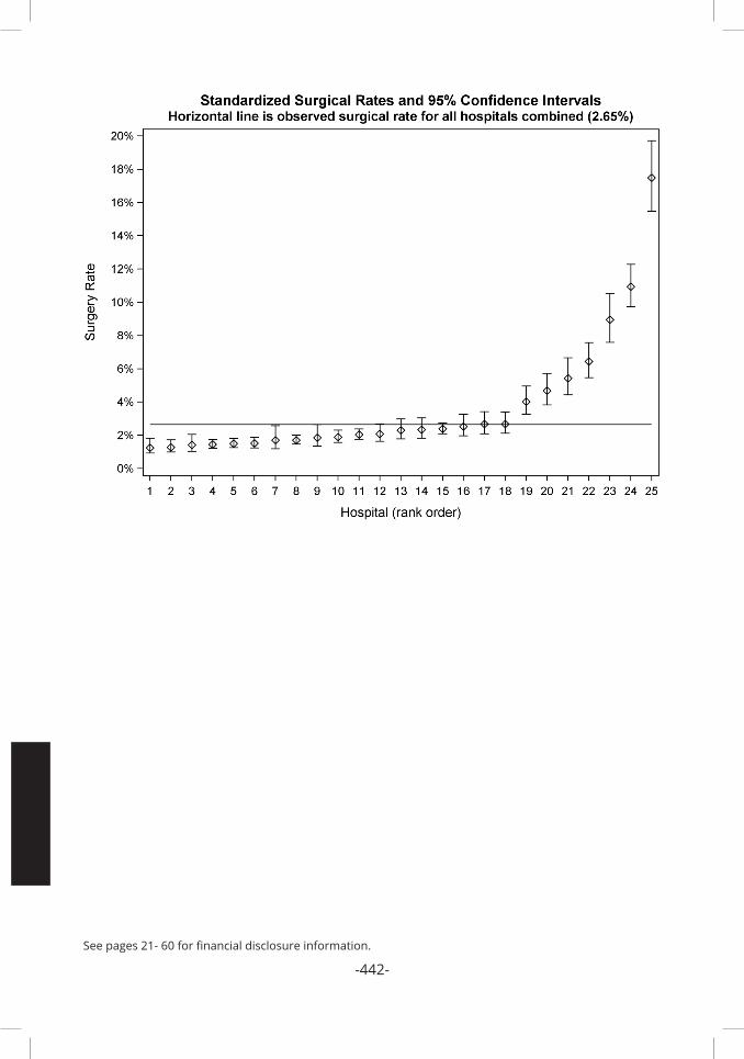
-442-
See pages 21- 60 for financial disclosure information.
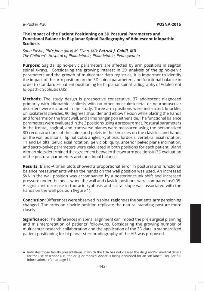
-443-
♦ Indicates those faculty presentations in which the FDA has not cleared the drug and/or medical device for the use described (i.e., the drug or medical device is being discussed for an “off label” use). For full information, refer to page 14.
e-Poster #30 POSNA-2016 The Impact of the Patient Positioning on 3D Postural Parameters and Functional Balance in Bi-planar Spinal Radiography of Adolescent Idiopathic Scoliosis
Saba Pasha, PhD; John (Jack) M. Flynn, MD; Patrick J. Cahill, MD The Children’s Hospital of Philadelphia, Philadelphia, Pennsylvania
Purpose: Sagittal spino-pelvic parameters are affected by arm positions in sagittal spinal X-rays. Considering the growing interest in 3D analysis of the spino-pelvic parameters and the growth of multicenter data registries, it is important to identify the impact of the arm position on the 3D spinal parameters and functional balance in order to standardize patient positioning for bi-planar spinal radiography of Adolescent Idiopathic Scoliosis (AIS).
Methods: The study design is prospective consecutive. 37 adolescent diagnosed primarily with idiopathic scoliosis with no other musculoskeletal or neuromuscular disorders were included in the study. Three arm positions were instructed: knuckles on ipsilateral clavicles, 90 degrees shoulder and elbow flexion while placing the hands and forearms on the front wall, and arms hanging on either side. The functional balance parameters were evaluated in the 3 positions using a pressure mat. Postural parameters in the frontal, sagittal, and transverse planes were measured using the personalized 3D reconstructions of the spine and pelvis in the knuckles on the clavicles and hands on the wall positions. Spinal Cobb angles, kyphosis, lordosis, vertebral axial rotation, T1 and L4 tilts, pelvic axial rotation, pelvic obliquity, anterior pelvic plane inclination, and sacro-pelvic parameters were calculated in both positions for each patient. Bland Altman plots determined the agreement between the two arm positions in 3D evaluation of the postural parameters and functional balance.
Results: Bland-Altman plots showed a proportional error in postural and functional balance measurements when the hands on the wall position was used. An increased SVA in the wall position was accompanied by a posterior trunk shift and increased pressure under the heels when the wall and clavicle positions were compared p<0.05. A significant decrease in thoracic kyphosis and sacral slope was associated with the hands on the wall position (Figure 1).
Conclusion: Differences were observed in spinal regions as the patients’ arm pensioning changed. The arms on clavicle position replicate the natural standing posture more closely.
Significance: The differences in spinal alignment can impact the pre-surgical planning and misinterpretation of patients’ follow-ups. Considering the growing number of multicenter research collaboration and the application of the 3D data, a standardized patient positioning for bi-planar stereoradiography of the AIS was proposed.
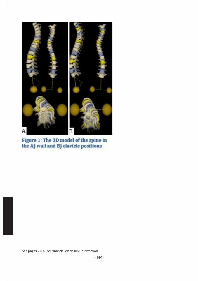
-444-
See pages 21- 60 for financial disclosure information.
-445-
♦ Indicates those faculty presentations in which the FDA has not cleared the drug and/or medical device for the use described (i.e., the drug or medical device is being discussed for an “off label” use). For full information, refer to page 14.
e-Poster #31 POSNA-2016 The Results of Pharmacotherapy and Lower Extremity Corrective Osteotomies in Children with Osteogenesis Imperfecta
Andrii Zyma, MD; Guk Yurii, MD; Tamara Kinchaya-Polischuk, MD; Andrii Cheverda, MD; Stepan Martcyniak; David G. Stewart Jr., MD; Yuri Demyan, MD Institute of Orthopedics and Traumatology, National Academy of Medical Sciences, Kiev, Ukraine
Purpose: To evaluate the correction of bone deformity, patient function and bone condition in patients with osteogenesis imperfecta (OI) with treatment consisting of pharmacotherapy with and without corrective osteotomies.
Methods: A four-year prospective study was conducted in 21 patients with OI, including 13 males and 8 females with a mean age of 9.4 ± 0.6 years, including 10 patients with Sillence type I osteogenesis imperfecta and 11 patients with type III. Pharmacotherapy consisted of calcium supplementation, 1,25-vitamin D supplementation and pamidronate infusions over 6-12 months, with the number of infusions determined by the level of metabolic derangement as evaluated by β-СТх and Z- score. 11 patients (24 operations) underwent pharmacotherapy followed by corrective osteotomies of lower extremity long bones using were a locking rod with a T-shaped telescopic part; 10 patients received only pharmacotherapy.
Results: All operated patients achieved deformity correct and bone healing. 8 surgical patients were nonambulatory before treatment; gait was restored in all patients within 1.5 years. One nonsurgical patient was nonambulatory before treatment and gait was restored within one year. 18 patients experienced no recurrence of fractures and 3 patients experienced re-fracture over 1.5-2 year follow-up. Operative complications included implant migration in one patient, repeated deformity in two patients and fracture of rod and bone in one patient. In surgical patients, average Z-score improved from -4.68 ± 0.47 before treatment to -3.45 ± 0.35 after treatment and β-СТх improved from 0.92 ± 0.13 before treatment to 0.66 ± 0.10 after. In non-surgical patients, Z-score improved from -3.8 ± 0.54 before pharmacotherapy to -2.79 ± 0.52 after and β-СТх improved from 1.54±0.31 before treatment to 0.87±0.12 after. In patients with OI type I, Z-score improved from -3.4 ± 0.48 before treatment to -2.20 ± 0.33 after treatment and β-СТх decreased from 1.04 ± 0.27 to 0.66±0.14. In patients with OI type III, Z-score improved from -5.18 ± 0.35 before treatment to -4.09 ± 0.26 after treatment and β-СТх decreased from 1.38 ± 0.22 to 0.85 ± 0.08.
Conclusion: Marked improvement or recovery of walking function, improved bone density and decreased bone turnover were achieved in patients with osteogenesis imperfecta through combined pharmacotherapy and surgery or through pharmacotherapy alone.
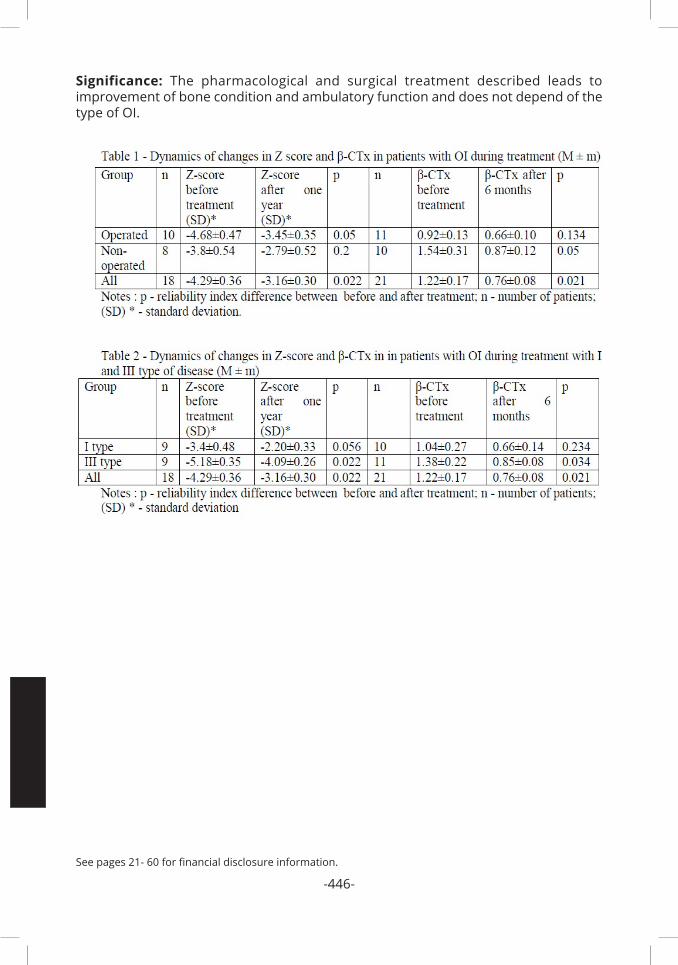
-446-
See pages 21- 60 for financial disclosure information.
Significance: The pharmacological and surgical treatment described leads to improvement of bone condition and ambulatory function and does not depend of the type of OI.
-447-
♦ Indicates those faculty presentations in which the FDA has not cleared the drug and/or medical device for the use described (i.e., the drug or medical device is being discussed for an “off label” use). For full information, refer to page 14.
e-Poster #32 POSNA-2016 Lateral Femoral Cutaneous Nerve Palsy Occurred in 25% of Patients Following Posterior Spinal Fusion for Adolescent Idiopathic Scoliosis
Austin Sanders, BA; Lindsay M. Andras, MD; Paul D. Choi, MD; Vernon T. Tolo, MD; David L. Skaggs, MD, MMM Children’s Hospital Los Angeles, Los Angeles, California
Purpose: No prior studies have prospectively evaluated the prevalence of lateral femoral cutaneous nerve palsy (LFCNP) in the treatment of adolescent idiopathic scoliosis (AIS). The purpose of this study was to evaluate the incidence, risk factors, and time to resolution of LFCNP following posterior spinal fusion (PSF) for AIS.
Methods: A prospective study was conducted on patients undergoing PSF for AIS between June 2014 and May 2015. Patients were examined pre-operatively, post-operatively on a daily basis during their hospital stay, and at follow-up clinic visits until resolution of the LFCNP. All neurologic examinations were performed by attending pediatric orthopaedic surgeons. Patients who had preoperative neurologic deficits, neuropathy, or underwent a staged procedure were excluded.
Results: 55 patients with an average age of 14 years (10-21) were enrolled. Twenty-five percent (14/55) of patients had a post-operative LFCNP. There were no other postoperative neurologic deficits. Of the 14 patients with a LFCNP, 57% of these were bilateral. 14% (2/14) of these patients had absent sensation to light touch on exam, while 85% (12/14) had decreased sensation. No patients reported experiencing pain associated with the LFCNP or tenderness when the area was palpated. There were no cases where the LFCNP limited postoperative mobilization or prolonged hospital stay. The LFCNP resolved in an average of 3.6 days (1-18). 6/14 (43%) resolved after 1 day. No correlation was observed between occurrence of LFCNP and gender, age, height, BMI, length of fusion, primary cobb angle, or blood loss. Occurrence of LFCNP was positively correlated with increased weight (p=0.032) and increased operative times (0.016). Duration of the LFCNP was also correlated with increased operative time (p=0.010).
Conclusion: LFCNP occurred in 25% of AIS patients undergoing posterior spinal fusion. Risk of LFCNP increased with longer operative times and increased patient weight. On average, LFCNP resolved in less than 4 days and did not cause any pain or limitations.
Significance: LFCNP is common following PSF in AIS, occurring in 1 out of every 4 patients.
-448-
See pages 21- 60 for financial disclosure information.
e-Poster #33 POSNA-2016 A Model of Pre-Pseudarthrosis (Congenital Tibia Dysplasia) Associated with Neurofibromatosis Type 1
David G. Little, MBBS, FRACS, PhD; Nikita Deo, BSc; Tegan L. Cheng, BE BMedSci; Aaron Schindeler, BSc PhD The Children’s Hospital at Westmead, Sydney, Australia
Purpose: While congenital tibial dysplasia (CTD) is an uncommon manifestation of Neurofibromatosis (NF1) (2-4 fracture is often inevitable once a child begins to load bear, with resultant pseudarthrosis. There are no current animal models of CTD.
Methods: Nf1flox/flox:Ai9 genetically modified mice were used; this line possesses a conditional loxP-flanked Nf1 allele and a Cre-dependent tdTomato fluorescent reporter. Neonatal mice were injected with Cre-expressing adenovirus into the tibia at 1 week of age. Control animals received a saline injection. Firstly, animals were euthanased at 3 and 10 weeks for histology and biomechanical testing respectively. In a second study, animals underwent closed fracture at the 10 week time point and bone healing examined with radiographically and histologically. In a third study, the potential for dysplastic bowing was tested under conditions of cyclic load. Animals underwent cyclic loading for 20min for 5/7 days for 2 weeks. (Animal ethics protocol #K319)
Results: Analysis of tdTomato expression showed viral recombination in the bone and surrounding soft tissues of the Ad-Cre injection site. Consistent with this, the cortical bone at the injection site showed an abnormal, highly porous appearance. Biomechanical testing showed reduced bone strength in the Ad-Cre treated group. In fracture studies, Ad-Cre injected bones showed poor healing compared to saline controls. Histology showed abundant fibrotic tdTomato+ cells at the fracture site, which was in line with prior studies showing that the fibrous hamartoma is a key pathology in NF1. Animals that underwent cyclic loading in induced and non-induced tibiae did not show bowing over a two-week period, however a number of mice in the Ad-Cre induced knockout group developed spontaneous fractures in the foot or tibia during normal mild cyclic loading conditions and were subsequently euthanized.
Conclusion: These data validate that localized NF1 inactivation after Ad-Cre injection can generate abnormal bone. This bone is weaker and has impaired subsequent healing. Subsequent studies will examine alternative loading conditions to model early-stage dysplastic tibiae as well as utilizing this model for emerging candidate drug interventions to improve intact bone strength, and healing pre- and post-fracture.
Significance: These data support the concept that local double inactivation of NF1 underlies the focal bone defects that can lead to fracture in the distal tibia. These and similar models will be used to test therapeutic interventions for improving bone density and biomechanical properties of the affected bones prior to fracture.
-449-
♦ Indicates those faculty presentations in which the FDA has not cleared the drug and/or medical device for the use described (i.e., the drug or medical device is being discussed for an “off label” use). For full information, refer to page 14.
e-Poster #34 POSNA-2016 The PASS (Pediatric/Adolescent Shoulder Survey): A Youth Questionnaire with Discriminant Validity and Responsiveness to Change
Tracey P. Bastrom, MA; Joanna H. Roocroft, MA; Andrew T. Pennock, MD; Eric W. Edmonds, MDRady Children’s Hospital, San Diego, California Purpose: Many health-related quality of life tools for shoulder pathology have been developed for adults; such as, the Disabilities of the Arm, Shoulder, and Hand (DASH) or quickDASH. However, some content is not applicable or readily grasped by a younger population. Previous study demonstrated good reliability and convergent validity of a shoulder assessment tool designed for the pediatric and adolescent population. The purpose of this study was to examine discriminant validity and responsiveness to change of the new shoulder survey.
Methods: A retrospective review of patients evaluated in our sports clinics for shoulder complaints was performed. Inclusion criteria was completion of the shoulder survey during at least one visit. Total score of the pediatric/adolescent shoulder survey (PASS) was calculated, as well as Symptoms/Limitations and Compensatory mechanism/Emotional domain scores. A maximum score of 100% indicates full function. Total score was calculated for the quickDASH and quickDASH sports module. A maximum score of 100 points indicates total disability. Discriminant validity was evaluated based on; acute injury, duration of symptoms, and diminished range of motion (ROM) or strength on clinical exam (defined as reduction in ROM of at least 10 degrees or decrease in strength of 1 point). A subset of patients with scores available both pre and within 6 months post-operatively were evaluated to assess responsiveness to treatment.
Results: 132 children, 70% male, mean age 16 ± 2 years (range 12-19) were analyzed and 73% completed the survey at their first visit for the shoulder complaint. The QuickDASH demonstrated significant gender differences in scores (p<0.05, table). Both tools were able to discriminate between patients with acute injuries and diminished clinical exams (p<0.05). A significant, medium effect correlation was observed between duration of symptoms and scores on the PASS. The subset of 25 patients with pre and post-operative data showed significant change in total score and one domain of the PASS. All tools/domains showed significant correlation with length of time since surgery.
Conclusion: This 13 question pediatric/adolescent friendly shoulder survey shows excellent psychometric properties; good discriminant ability, and early evidence of responsiveness to change, especially when compared to an adult formatted questionnaire that is validated for younger patients such as the quckDASH.
Significance: The PASS is a validated alternative for assessing shoulder impairment in the pediatric and adolescent population, with questions specifically targeted to this age group. Moreover, the PASS is not affected by reported gender (or other demographic variables) concerning validity in reporting patient outcome.
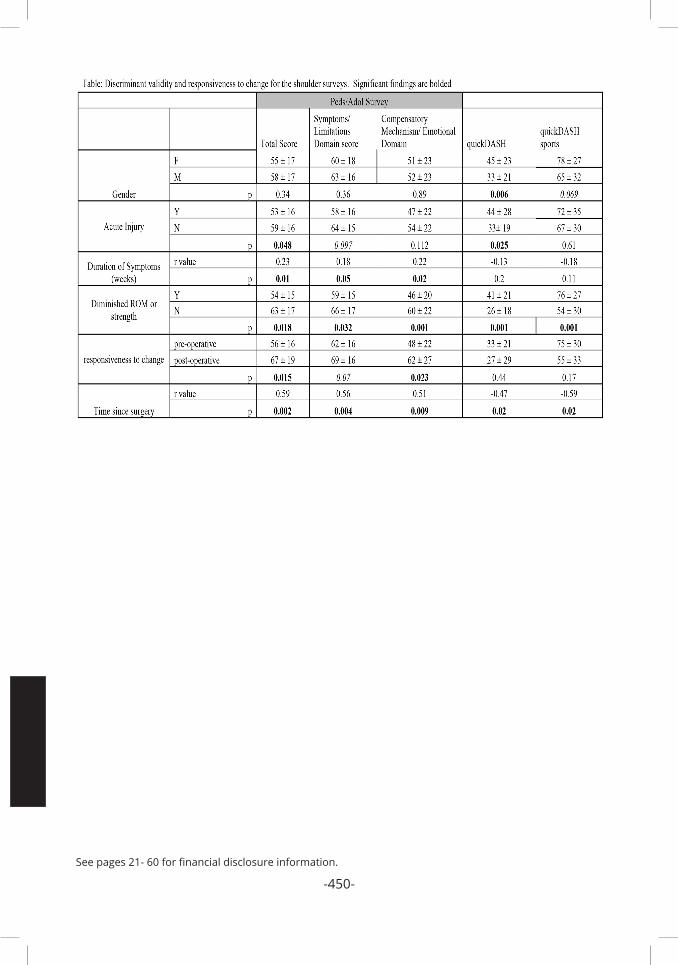
-450-
See pages 21- 60 for financial disclosure information.

-451-
♦ Indicates those faculty presentations in which the FDA has not cleared the drug and/or medical device for the use described (i.e., the drug or medical device is being discussed for an “off label” use). For full information, refer to page 14.
e-Poster #35 POSNA-2016 Best Practice Guidelines for the Surgical Treatment of Early Onset Scoliosis
Michael P. Glotzbecker, MD; Tricia A. St Hilaire, MPH; Growing Spine Study Group; George H. Thompson, MD; Michael G. Vitale, MD, MPH Children’s Spine Study Group, San Diego, California
Purpose: Postoperative surgical site infection (SSI) is unfortunately a commonly encountered complication in the surgical treatment of children with Early Onset Scoliosis (EOS). There is documented variation in the treatment of this population. Previous work building consensus for the approach to high risk patients (neuromuscular) has been promising. The goal of the current study is to apply similar principles to develop consensus based guidelines for the treatment of patients with EOS.
Methods: A focus group from the Growing Spine Study Group (GSSG) and the Childrens Spine Study Group (CSSG) developed a list of statements to be distributed to a larger group of experts. Using the Delphi process, participants were presented with a systematic review of the literature as well as a review of current practices. The first round was conducted using an electronic survey. Results of this survey were then discussed face-to-face and the statements were further refined. A final round was conducted using the Audience Response System, allowing participants to vote for each statement (strongly agree or agree) Agreement >80% or disagreement <20% was considered consensus.
Results: Consensus was obtained on the following statements (60% of statements reached consensus): All patients should receive perioperative intravenous cefazolin prior to an insertion or lengthening procedure; Vancomycin powder should be used in the bone graft/and or surgical site for insertion procedures; Patients should receive a preoperative Patient Education Sheet; Patient should have a pulmonary workup evaluation if there is a history of respiratory problems; Prep should be wide enough to place a chest tube within the surgical field for insertion procedures; Adherence to perioperative antimicrobial regiments should be monitored; Soft tissue handling and incision planning in important in preventing postoperative infections for insertion and lengthening procedures; Patients should have intraoperative wound irrigation; If removing hair prior to an insertion procedure, clipping is preferred to shaving; When compared to other skin preparations, chlorhexidine is preferred; Operating room access should be limited during scoliosis surgery when practical; All previous scars/incisions should be prepped in the surgical field; Patients should have a chlorhexidine skin wash at home the night before an insertion/lengthening procedure; Patients with myelodysplasia should have urine cultures obtained and treated if positive before an insertion procedure.
Conclusion: Using the Delphi process a several “best practices” were developed for the treatment of EOS.
Significance: In reducing variability, standardizing practice may lead to improved outcomes. Areas of equipoise offer opportunities for future study.
-452-
See pages 21- 60 for financial disclosure information.
e-Poster #36 POSNA-2016 8-plates & I-plates for Epiphysiodesis
Kevin S. Horowitz, MD; Jaclyn F. Hill, MD; Ali A. Qadeer, BSBaylor College of Medicine, Houston, Texas
Purpose: Lower extremity limb-length discrepancy (LLD) is a commonly encountered problem in pediatric orthopaedics. LLD that is predicted to be between 2 and 5 cm at skeletal maturity has traditionally been treated with epiphysiodesis. Recently, the effectiveness of dual 8-plates as a method of epiphysiodesis has been called into question. The purpose of this study was to determine the efficacy of dual 8-plates and dual I-plates (also referred to as H-plates or quad-plates) in preventing physeal growth. A secondary goal was to assess the effect of dual 8-plates and dual I-plates on coronal plane alignment.
Methods: We retrospectively reviewed the records and radiographs of patients who underwent epiphysiodesis between July 2007 and February 2014 using either dual-8 or dual-I plates. A minimum of 12-month radiographic follow-up was required for inclusion. Percent effectiveness of each technique was calculated by comparing the final bone segment length to predicted lengths for complete physeal arrest and for full physeal growth. Angular deformity was determined by the change in lateral distal femoral angle and medial proximal tibial angle.
Results: During the study period, 46 patients underwent epiphysiodesis using 8-plates or I-plates. The mean percent effectiveness for all 8-plates was 60% and that for I-plates was 85% (p=0.23). Amongst the 8-plate cohort, the mean percent effectiveness in tibias was 78% compared to 44% in femurs (p<0.05). The mean angular change for the 8-plate cohort was 3.2° compared to 1.9° for the I-plate cohort (p=0.32). For the distal femur, mean angular change the 8-plate cohort was 3.8° compared to 1.6° for the I-plate cohort (p=0.066). 5 patients in the 8-plate cohort required revision surgery secondary to angular deformity compared to none in the I-plate cohort.
Conclusion: Dual 8-plates have limited effectiveness in preventing physeal growth and have the potential to create coronal plane deformity. If dual 8-plates or I-plates are used for epiphysiodesis, we recommend that dual I-plates be used in the distal femur and that consideration be given for the use of dual I-plates in the proximal tibia.
Significance: This study may affect the technique and choice of implants when performing an epiphysiodesis. Further studies are needed to better characterize the efficacy of I-plates for this surgical application.

-453-
♦ Indicates those faculty presentations in which the FDA has not cleared the drug and/or medical device for the use described (i.e., the drug or medical device is being discussed for an “off label” use). For full information, refer to page 14.
e-Poster #37 POSNA-2016 Hip Arthroscopy Failure in the Setting of Acetabular Dysplasia: A Concerning Trend?
Tonya An, BS; Jacob Haynes; Jeffrey J. Nepple, MD; John C. Clohisy, MD; Perry L. Schoenecker, MD; Michael B. Millis, MD; Young Jo Kim, MD; Ira Zaltz, MD; Rafael J. Sierra, MD; Wudbhav N. Sankar, MD; Ernest L. Sink, MD; David A. Podeszwa, MD; Daniel J. Sucato, MD, MS; Robert T. Trousdale, MD; Paul E. Beaule, MD; ANCHOR GroupWashington University School of Medicine, Saint Louis, Missouri Purpose: Despite the success of hip arthroscopy, evidence suggests that arthroscopy alone is inadequate for treatment of conditions such as acetabular dysplasia (AD) due to its failure to correct structural deformity. Our objective was to define the incidence of failed hip arthroscopy in patients with symptomatic AD requiring periacetabular osteotomy (PAO). We secondarily analyzed the patient and structural characteristics of the failed arthroscopy cases.
Methods: Utilizing a prospective, multicenter joint preservation database, we identified a cohort of primary AD patients from 2009-2014 who underwent PAO after a single prior ipsilateral hip arthroscopy. A comparison cohort of PAO patients without prior arthroscopy was isolated. Patients with hip disease attributable to cerebral palsy, Perthes, etc. were excluded. Demographic and radiographic data were summarized for each group. We compared the proportion of PAO after failed hip arthroscopy between the beginning and end of the study period by 2-tailed z-test with P<0.05 regarded as statistically significant.
Results: 99 patients (99 hips) had received arthroscopic surgery prior to PAO, while 1053 patients (1103 hips) underwent PAO without prior hip arthroscopy. The proportion of PAO procedures after previous ipsilateral hip surgery stayed constant (13-19%); however, the incidence of PAO after previous hip arthroscopy increased from 2.5% in 2009 to 9.8% in 2014 (P=0.004). Over the 6 year period, the rate of all PAO procedures increased 12% per year, whereas rates of PAO after hip arthroscopy grew an average 54% per year. Female sex, increased average LCEA and ACEA, and decreased acetabular inclination were associated with failed hip arthroscopy (P<0.01).
Conclusion: Our data illustrates an increase in the rate of PAO after previous hip arthroscopy over the past six years. While the rate of PAO following previous ipsilateral surgery remains constant, hip arthroscopies now constitute almost 60% of those previous surgery attempts. Additionally, we identified mild dysplastic features and female sex as characteristics associated with failure of hip arthroscopy. These results are consistent with prior literature suggesting that inadequately corrected structural deformity is the most common indication for revision surgery.
Significance: Our findings raise concern that isolated hip arthroscopy is being increasingly utilized in patients with acetabular dysplasia. These trends highlight the
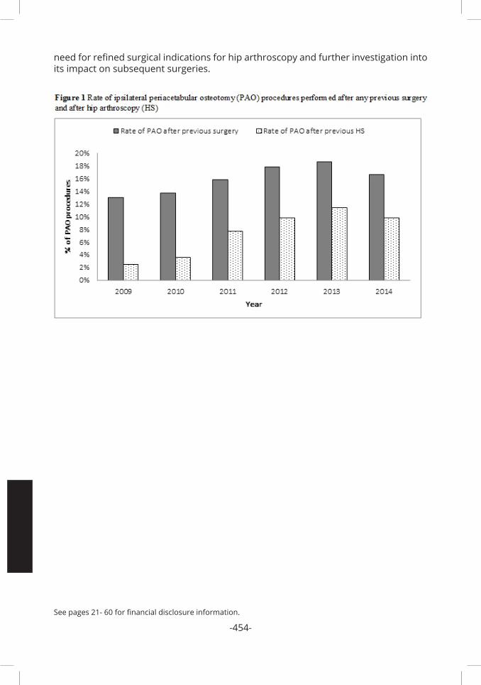
-454-
See pages 21- 60 for financial disclosure information.
need for refined surgical indications for hip arthroscopy and further investigation into its impact on subsequent surgeries.
-455-
♦ Indicates those faculty presentations in which the FDA has not cleared the drug and/or medical device for the use described (i.e., the drug or medical device is being discussed for an “off label” use). For full information, refer to page 14.
e-Poster #38 POSNA-2016 Reducing Anxiety, Improving Communication and Increasing Satisfaction in Families During Orthopedic Procedures with Mobile Technology
Jose A. Herrera-Soto, MD; Dennis R. Knapp, MD; Mark A. Birnbaum, MD; Jonathan H. Phillips, MD; Kevin de la Roza, MD Arnold Palmer Hospital for Children, Orlando, Florida Purpose: Most if not all families feel anxiety and helplessness while waiting for a loved one during their surgical procedure. However, families are not allowed to be part of the operative experience for obvious reasons, until now. A new communications platform that allows updates to be sent directly to your mobile device from the operating room could substantially change that experience. The purpose of our study was to evaluate the family’s experience and perception At our institution with the use of the EASE app.
Methods: The EASE platform is two mobile applications and a system that allows for encrypted transmission of communications. the app is HIPAA compliant and creates a comprehensive messaging system that incorporates the required security of protected health information and the simplicity of modern mobile communication. Prior to surgery, a patient or legal guardian downloads the EASE application (“EASE”) from the app store to their mobile device. They will then consent to the EASE process either via written consent or electronic signature. After registering, they will choose the types of updates desired (texts, texts and images, or texts, images and videos). The EASE app also allows for collection of customizable surveys and testimonials. Testimonials and questionnaires were documented for our study.
Results: 119 surveys for elective procedures were collected in 9 months. The overall experience was 9.3/10.When asked: 1. If you or a loved one required future medical procedures would you choose the EASE program again? 99% said YES. 2. Would the availability of EASE influence your choice of hospital should you, or a loved one, require surgery? 77% answered YES 3. 98 % of families said the texts, photos, and videos were Appropriate. 4. 90% said the app made them at EASE
Conclusion: EASE consistently obtains high satisfaction scores and creates a closer bond between the clinician and their patient families. EASE data has shown that personalized texts, photos, and videos using the application decreases anxiety, increases understanding and makes families feel as though they never left their loved one’s side.
Significance: EASE presents significant value to a hospital and its staff by providing healthcare facilities with an easy to implement, secure and sustainable communications platform to connect to all of their patients’ families. As hospitals strive to achieve the
-456-
See pages 21- 60 for financial disclosure information.
triple aim of increasing patient satisfaction, improving population health and reducing the cost per capita of healthcare; EASE gives hospitals a powerful tool to improve patient and family satisfaction.
-457-
♦ Indicates those faculty presentations in which the FDA has not cleared the drug and/or medical device for the use described (i.e., the drug or medical device is being discussed for an “off label” use). For full information, refer to page 14.
e-Poster #39 POSNA-2016 Implementing a Multidisciplinary Clinical Pathway Can Reduce the Surgical Site Infection Rate After Posterior Spinal Fusion In High Risk Patients
Michael P. Glotzbecker, MD; Michael J. Troy, BS; Patricia E. Miller, MS; Alexandra M. Grzywna, BA; Michael T. Hresko, MD; Lawrence I. Karlin, MD; John B. Emans, MD; Brian D. Snyder, MD, PhD; Daniel J. Hedequist, MD Boston Children’s Hospital, Boston, Massachusetts
Purpose: To compare postoperative surgical site infection (SSI) rates after spinal fusion in high risk patients and adherence to the protocol before and after the institution. Methods: An institutional clinical pathway was created in 2012 based on nationally published Best Practice Guidelines as well hospital practices with a goal of reducing the rate of SSI in high risk patients. It was based on multidisciplinary input and focused on care from patient optimization preoperatively through the postoperative care period. Patients were retrospectively reviewed from 2008-2012, and a comparable number of patients were retrospectively reviewed from a prospectively collected database of patients at high risk for SSI. Patients with AIS, growth friendly operations, trauma, or current infections were excluded. Patients with neuromuscular or syndromic diagnosis at high risk for infection based on medical co-morbidities were included. Number of SSIs (defined as <90 days after index procedure by CDC) before and after implementation of the guideline were compared, as were compliance with measures within the guideline. Uni- and multivariable logistic regression analysis using penalized maximum likelihood estimation was used to assess the effect of changes in surgical practices on infection rate.
Results: 12/136 (8.8% (95% CI: 4.6-14.9%)) patients treated before implementation of the guideline had a SSI compared to 5/104 (4.8% (95% CI 1.6-10.9%)) patients treated on the clinical pathway. The groups were similar with regard to age, sex, and metal type (p>0.05). The percentage of neuromuscular diagnosis (79% vs 78%) was similar between the two groups. There was no difference in surgical time, length of ICU stay, or total LOS between the two groups. There was an increase in the percentage of patients that received topical vancomycin (84% vs 0%) and betadine irrigation (72% vs 7%) after implementation of the pathway. Appropriate dosing of antibiotics within one hour of incision improved from 54% to 85% after implementation.
Conclusion: Implementation of a clinical pathway aimed to reduce infection in patients at high risk for SSI after spinal fusion led to a 45% reduction in SSI rate. While multiple changes were made, it is impossible to attribute the drop in the SSI rate to any one factor. However adherence to a protocol using multiple strategies to reduce infection can result in a lower SSI rate.
Significance: Given the direct and indirect costs of a SSI, even small reductions of SSI rates in high risk patients may have a profound impact on health care costs and patient related outcomes.
-458-
See pages 21- 60 for financial disclosure information.
e-Poster #40 POSNA-2016 Investigating Caregiver Perspectives on Health Status and Comfort in Children with Cerebral Palsy Undergoing Surgery for Hip Displacement
Stacey Miller, PT; Emily Schaeffer, PhD; Maria Juricic, PT; Kim Hesketh, MSc, PT; Kishore Mulpuri, MDBritish Columbia Children’s Hospital, Vancouver, British Columbia, Canada
Purpose: Hip displacement is the second most common deformity in children with cerebral palsy (CP). A displaced, and particularly a dislocated hip, can have significantly adverse effects on an individual. Surgical intervention to correct progressive hip displacement or dislocation is recommended for children with CP. Success of surgical intervention is often described using radiological outcomes. There is evidence that surgical treatment for displaced or dislocated hips decreases pain and hip stiffness and improves radiological outcomes. However, there is no information in the literature regarding the impact of surgical treatment on the health related quality of life (HRQOL) in these children. The aim of our study was to examine the impact of surgical treatment of hip displacement or dislocation on HRQOL in children with CP.
Methods: This prospective longitudinal cohort study involved children attending a tertiary care hospital orthopaedic department. Children with CP between the ages of 4 and 18 years, with hip displacement/dislocation, defined as a Reimer’s migration percentage (MP) of >40% on a pre-operative x-ray, and undergoing surgical reconstruction were eligible for inclusion. Quality of life was measured pre-operatively and post-operatively using the CPCHILD Questionnaire.
Results: Twelve patients (one child was GMFCS level III, 4 were level IV, and 7 were level V), aged 4.0 to 17.3 years, were assessed pre-operatively and then again at least six months post-operatively. All underwent unilateral (5) or bilateral (7) reconstructive hip surgery. The migration percentage of hips undergoing reconstruction was reduced by an average of 52% (9-100%). The average change in CPCHILD score showed an increase of 6.4 points [95% CI: -1.4-14.2].
Conclusion: In this pilot study, no significant change was noted in HRQOL following reconstructive hip surgery, despite a marked reduction in Reimer’s MP. However, only 4 of 12 parents reported that their child had daily pain pre-operatively. A larger sample size will be required to draw more accurate conclusions from these findings.
Significance: Preliminary findings suggest that HRQOL was neither positively nor negatively affected by reconstructive hip surgery. There is an evident need for a multicentre study examining this issue in a larger patient population in order to determine the long-term impact of different hip interventions on quality of life in children with CP.

-459-
♦ Indicates those faculty presentations in which the FDA has not cleared the drug and/or medical device for the use described (i.e., the drug or medical device is being discussed for an “off label” use). For full information, refer to page 14.
e-Poster #41 POSNA-2016 Both Family History and Swaddling are Correlated with the Occurrence and Hip Types in Developmental Dysplasia of the Hip
Hakan Omeroglu, MD; Nusret Kose, MD; Anil Akceylan, MD Eskisehir Osmangazi University Hospital, Eskisehir, Turkey Purpose: We aimed to revisit the risk factors as well as to assess correlation between the risk factors and ultrasonographic (US) hip typing in developmental dysplasia of the hip (DDH).
Methods: Data of 760 healthy newborns (mean age 33 days) with bilateral US documented normal hips (control group) were compared with one of 192 patients (mean age 105 days) having US diagnosis of unilateral or bilateral DDH (study group). Family history, breech presentation, postnatal swaddling, intrauterine packing (foot deformities, torticollis, multiple pregnancy), oligohydroamnios and first-born girl were recorded as “risk factors”. US hip typing was made according to Graf’s system and dysplastic hips were further classified as stable (IIa-, IIb, IIc) and unstable (D, III, IV). Stable hip group contained 141 patients, having unilateral or bilateral stable hips. Meanwhile, unstable hip group contained 51 patients having at least one unstable hip. Chi-square test and binary logistic regression analysis were used to assess the data.
Results: Study group had higher number of subjects having at least one risk factor than control group (65% vs 47%, p<0,001). Rate of having at least two risk factors was higher in study group than in control group (33% vs 21%, p<0,001). When risk factors were assessed independently, rate of family history (16% vs 7%, p<0,001), breech presentation (13% vs 7%, p=0,015) and swaddling (16% vs 6%, p<0,001) were higher in study group than in control group. Packing, first-born girl and oligohydroamnios were not correlated with the occurrence of DDH. Multivariate analysis revealed that family history (p<0,001), breech presentation (p=0,015) and swaddling (p<0,001) were significant risk factors related with the occurrence of DDH. Independent analysis of each risk factor revealed that, rate of family history (28% vs 11%, p=0,007), swaddling (28% vs 11%, p=0,007) and oligohydroamnios (18% vs 6%, p=0,018) were higher in unstable hip group than in stable hip group. However, multivariate analysis revealed that only family history (p<0,001) and swaddling (p<0,001) were significant risk factors correlated with the occurrence of hip instability.
Conclusion: Family history, swaddling and breech presentation are the three significant risk factors related with the occurrence of DDH. If a baby has more than one risk factor, occurrence of DDH considerably increases. Besides, family history and swaddling significantly increase the occurrence of US hip instability that may influence the treatment success.
Significance: Babies having history of either DDH in relatives or postnatal swaddling need to be handled more carefully during hip screening and management in DDH.
-460-
See pages 21- 60 for financial disclosure information.
e-Poster #42 POSNA-2016 Prevalence of Vitamin D Deficiency in Pediatric Patients with Scoliosis Preparing for Spinal Surgery
Peter F. Sturm, MD; Jennifer M. Anadio, MA; Theresa Mayes Cincinnati Children’s Hospital Medical Center, Cincinnati, Ohio
Purpose: Establishing prevalence of vitamin D deficiency in the elective spinal fusion surgery population has the potential to impact clinical outcomes. The objectives of this study were to document the vitamin D status of patients with scoliosis preparing for surgical intervention in order to establish frequency of deficiency and to determine characteristics that influence vitamin D levels.
Methods: Medical records were queried over a 25 month period for patients with a diagnosis of scoliosis (of any etiology) who underwent surgery for posterior spinal fusion or growing construct and had a serum vitamin 25 hydroxyvitamin D (D25) recorded in the preoperative period. Demographic data, including gender, age, body mass index (BMI), race, scoliosis type, spine surgery procedure and season of the year, were extracted. Chi-square analysis and multivariate modeling were used to evaluate deficiency status among various demographic categories and to determine the demographic factors impacting D25 state. Results: A total of 217 patients with a mean age of 13.6 + 3.6 years had vitamin D levels drawn at a mean of 38.7 + 20.6 days prior to surgery. The majority of the sample presented with a diagnosis of idiopathic scoliosis (n = 126) and most patients were scheduled for spinal fusion surgery (n=192). Nearly 75% of the study population (n=162) demonstrated a D25 value below normal. African Americans presented with greater risk of deficiency (P< 0.001) compared to whites, as did patients preparing for spinal fusion versus growing construct surgery (P<0.018). Severe hypovitaminosis D was more common in winter than any other season (P<0.005). Patients with neuromuscular scoliosis demonstrated significantly higher D25 levels than idiopathic patients (P<0.0002). Gender, BMI and age did not impact D25.
Conclusion: A high prevalence of D25 deficiency exists in scoliosis patients preparing for corrective spinal surgery. African American children, those presenting for spinal fusion surgery, and patients admitted in winter season are most at risk for D25 deficiency.
Significance: Routine screening of D25 levels in the preoperative period is supported, especially among these high risk subsets.
-461-
♦ Indicates those faculty presentations in which the FDA has not cleared the drug and/or medical device for the use described (i.e., the drug or medical device is being discussed for an “off label” use). For full information, refer to page 14.
e-Poster #43 POSNA-2016 Obesity and Both-Bone Forearm Fractures
Ugochi Okoroafor, MD; Jasmin L. McGinty, MD SSM Health Cardinal Glennon Children’s Hospital, St. Louis, Missouri
Purpose: Obesity is a major health problem among children in the United States. In addition to being associated with numerous comorbidities, pediatric obesity is associated with a higher prevalence and increased risk of extremity fractures. In pediatric fractures treated nonoperatively, maintaining reduction can be difficult in obese children due to the larger soft tissue envelope. The purpose of this study was to investigate the relationship between obesity and failure of nonoperative management of pediatric both-bone forearm fractures.
Methods: We conducted a retrospective review of skeletally immature patients > 2 years old who received initial orthopaedic treatment for closed radius and ulna shaft fractures at a Level I pediatric trauma center between 2011 and 2014. 145 patients who were initially treated nonoperatively with splinting or casting were included in the study. Radiographic parameters from known literature were used to assess the quality of the reduction. The patients were divided into two groups: 1) normal weight children (BMI > 5th and < 85th percentile), and 2) overweight and obese children (BMI > 85th percentile). 59% (86/145) of patients were normal weight. 41% (59/145) patients were overweight and obese. The mean patient age was 8 years in both groups. The primary outcome measure was loss of reduction, defined as the need for repeat closed reduction or surgical intervention after initial closed treatment. Secondary outcome measures included time to healing and surgical complications. For statistical analyses, Chi-square and Fisher exact tests were used for categorical variables, and t test was used for continuous variables. Statistical significance was defined as p<0.05.
Results: 16 % (14/86) of normal weight children experienced loss of reduction compared to 31% (18/59) of overweight and obese children (p=0.04). 29% (4/14) of normal weight children who lost reduction subsequently required surgery compared to 56% (10/18) of overweight and obese children (p=0.12). One obese patient experienced a surgical complication. Time to healing averaged 6 weeks in normal weight children and 7 weeks in overweight and obese children (p=0.03).
Conclusion: Overweight and obese children have a significantly higher rate of loss of reduction in both-bone forearm fractures managed nonoperatively compared to normal weight children. These patients may benefit from closer clinical follow up and a lower threshold for surgical intervention.
Significance: Obesity is associated with a higher rate of failure of nonoperative management of pediatric both-bone forearm fractures.
-462-
See pages 21- 60 for financial disclosure information.
e-Poster #44 POSNA-2016 A New Simplified Thumb Ossification Composite Index (TOCI) - Can Predict the Peak Height Velocity, Skeletal Maturity and Curve Progression in Adolescent Idiopathic Scoliosis?
Alec Lik Hang Hung; Wai-Wang Chau; Benjamin Hon Kei Yip; Fiona Wai Ping Yu; Vivian Wing Yin Hung; Tsz Ping Lam, MD; Bobby Kin Wah Ng, MD; Jack Cheng Department of Orthopaedics and Traumatology, Prince of Wales Hospital, Hong Kong, SAR
Purpose: Accurate skeletal maturity assessment is important for predicting curve progression and clinical management of IS. Commonly used assessment methods are still inadequate or too complex for rapid clinical use. We introduce a simplified skeletal maturity scoring system based on thumb epiphysis ossification composite index (TOCI) staging in a hand radiograph that can be used in a busy outpatient setting. The objective of this study was to evaluate whether the new TOCI staging based on radiograph of the thumb could correlate well with skeletal maturity parameters and predict curve progression in IS at early stages.
Methods: A prospective series of AIS girls (pre-menarche, Risser 0) were recruited and followed at 6 monthly intervals longitudinally till skeletal maturity. Anthropometric data, peak height velocity (PHV), Cobb angle (CA), computed angle velocity (AV) were recorded. A new TOCI skeletal maturity staging (Stages 1-8) were registered and compared with Sander’s Digital Skeletal Age (DSA) from hand radiographs of each subject. Inter-observer reliability was tested. Logistic regression analysis was used to evaluate the risk of curve progression at each TOCI stage vs CA.
Results: Of the 127 AIS girls (mean age of 11.3) with initial mean CA of 24.1°, 66% (N=84) with curve progression of (>5°) had mean AV at onset of curve acceleration phase (CAP) of 13.5°/year. PHV was found to occur at TOCI stage 5 (mean age 11.6) and highly correlated with DSA score (r=0.91, p<0.01) and CAP (r=0.90, p<0.01). Logistic regression analysis showed patient with >30° CA and ≤TOCI stage 5 had 90% risk of progression to surgical magnitude (>50°) at maturity. TOCI also showed excellent inter-rater reliability between 2 orthopedist (ICC=0.97 (0.96, 0.98)) and 3 non-medical rater (ICC=0.93 (0.89, 0.96)).
Conclusion: In conclusion, the new TOCI scoring system based on simple hand radiograph correlated well with standard skeletal maturity parameters and can predict curve progression in AIS at early stages.
Significance: With further validation on larger series and ethnic groups, the simplicity and high reliability of TOCI has good potential for application in busy clinical settings in helping clinical decisions on bracing treatment, prognosis and counseling in AIS patients.
-463-
♦ Indicates those faculty presentations in which the FDA has not cleared the drug and/or medical device for the use described (i.e., the drug or medical device is being discussed for an “off label” use). For full information, refer to page 14.
e-Poster #45 POSNA-2016 Age at Surgery, Amount of Arthrosis, and Severity of Dysplasia May Help Predict Early Versus Late Failure of Bernese Periacetabular Osteotomy
Travis H. Matheney, MD; Joel E. Wells, MD, MPH; Evgeny Bulat, MS; Patricia E. Miller, MS; Young Jo Kim, MD; Michael B. Millis, MD Boston Children’s Hospital, Boston, Massachusetts
Purpose: Outcomes following Bernese periacetabular osteotomy (PAO) for developmental dysplasia of the hip (DDH) have been previously reported. Some PAOs fail requiring conversion to total hip arthroplasty (THA). Time to failure ranges from a few years to more than 20. With this wide range of time we sought to determine: baseline characteristics of patients/hips that fail early (< 10 years) versus late (> 10 years) and factors predicting early or late failure of PAO.
Methods: Institutional review board approval was obtained. A retrospective analysis was performed on 126 hips (105 patients) undergoing PAO. Pre-operative demographic and radiographic data included age, gender, minimum joint space width (JSW), anterior and lateral center edge angles (ACEA and LCEA), and Tönnis acetabular sourcil angle (TA). Statistical analyses included Student’s t-test, Mann-Whitney U-test, chi-square test, and Fisher’s exact test. Receiver operating curve (ROC) analysis was used to identify optimal cut-off values for age and radiographic variables.
Results: Average age at PAO for all hips was 26.5 (9.5 to 45.4) years with 88% of patients being female. Ninety-eight (78%) PAO were still preserved at an average of 17.4 (+ 2.6) years. Sixteen hips (13%) underwent early THA at an average of 5.8 (0.3 to 9.2) years and 12 hips (9%) underwent late THA at an average of 14.5 (11 to 18.4) years. ROC analysis determined that age > 32 years and JSW < 2.5mm were most predictive of early failure. Age > 25, LCEA < -10 degrees, and TA > 27 degrees were more likely to fail late. A multinomial logistic regression model was applied and found that independent predictors of early failure were age (p=0.02) and JSW (p=0.01) while predictors of late failure were age (p=0.04) and pre-operative LCEA (p=0.001).
Conclusion: PAO remains a great solution for symptomatic DDH with the majority preserved at nearly 18 years. Pre-operative arthrosis (JSW loss) and age > 32 years appear to be most predictive of early failure (average 6 years). Significant pre-operative dysplasia and age > 25 appear to be the most predictive of late failure (average 15 years). A patient with risk factors for PAO failure may still have a successful outcome for many years and should be counseled as to the possible longevity of their PAO.
Significance: Hip-preserving surgery continues to be utilized to treat symptomatic DDH. This information will further aid the surgeon in advising the patient regarding various potential outcomes of their PAO.
-464-
See pages 21- 60 for financial disclosure information.
e-Poster #46 POSNA-2016 Abnormal Sclerostin Expression in Serum and Bone Tissue Osteocyte Lacuna-canaliculi System in AIS – a New Novel Finding
Huanxiong Chen, MD; Wayne YW Lee; Jiajun Zhang, BSc, MSC; Bobby Kin Wah Ng, MD; Tsz Ping Lam, MD; Jian Q. Feng, PhD; Jack Cheng; Kwong Man Lee Prince of Wales Hospital, Shatin, Hong Kong, SAR
Purpose: Etiopathogenesis of Adolescent idiopathic scoliosis (AIS) is not well defined. Reported low bone mass, deranged bone micro-architecture and abnormal bone biomarkers have led to the speculation of dysfunctional bone metabolism as one of the underlying etiopathogenetic factors. Osteocytes are critical regulators of bone metabolism attributing to specialized lacuno-canalicular network (LCN) where osteocytes and dendritic processes are residing. LCN facilitates cell-to-cell communication via paracrine signaling like sclerostin. We recently reported pathological change in LCN ultrastructure in AIS bone biopsy. In this study, we further hypothesized that such change might affect the sclerostin level at tissue and serum levels in AIS.
Methods: Part I of the study was an case-control study on bone biopsies taken intra-operatively from 5 severe AIS patients undergoing posterior spinal fusion Vs 5 age-matched controls having other orthopaedic operations requiring iliac crest autografts. Basic anthropometric data, pubertal maturity and curve severity of subjects were measured (Table 1). The specimens were examined by acid-etched scanning electron microscopy (SEM), quantitative fluorescein isothiocyanate (FITC)-Imaris technique and immunostaining for sclerostin. Part II of this study include 48 AIS girls with Cobb angle (49.9° ± 21.6°) with serum sclerostin measured with multiplex.
Results: Abnormal clusters of irregular roundish shape osteocytes with shorter and disorganized canaliculi were found in AIS in significant contrast to the well-organized osteocyte LCN with clearly spotted spindle lacunae and longer canaliculi protruded perpendicularly from the lacunae in normal controls (Figure. 1a-c). Immunostaining showed significantly lower percentage of sclerostin-positive osteocytes with expression of sclerostin confined to the cell bodies in AIS in contrast to the evenly expressed sclerostin in controls (Figure. 1d-e). Correlation study showed a negative correlation of serum sclerostin level and curve severity (R = -0.51) (Figure. 2). Linear regression analysis adjusting for age and Tanner stage revealed the same relationship. The addition of sclerostin into conventional model increased the R-square from 0.279 to 0.356.
Conclusion: To the best of our knowledge, this is the first study showing the abnormal sclerostin expression and distribution with associated ultrastructural changes in osteocyte LCN system and significant negative correlation of serum sclerostin level and curve severity after adjusted for age and Tanner stage in AIS.
Significance: Such abnormalities could affect systemic bone homeostasis, leading to abnormal bone mass, microstructure and mechanical bone strength that could
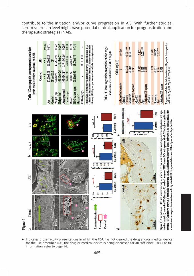
-465-
♦ Indicates those faculty presentations in which the FDA has not cleared the drug and/or medical device for the use described (i.e., the drug or medical device is being discussed for an “off label” use). For full information, refer to page 14.
contribute to the initiation and/or curve progression in AIS. With further studies, serum sclerostin level might have potential clinical application for prognostication and therapeutic strategies in AIS.
-466-
See pages 21- 60 for financial disclosure information.
e-Poster #47 POSNA-2016 Maternal Vitamin D Deficient Diets Increase Kyphosis in Offspring
Matthew A. Halanski, MD; Blake Hildahl, MD; Rajeev Chaudhary, MS; Rachel L. Lenhart, PhD; Thomas D. Crenshaw, PhD; Laura A. Amundson, BS American Family Children’s Hospital, University of Wisconsin, Madison, Wisconsin
Purpose: The increased risk of neural tube defects associated with maternal folate (B9) deficiency has long been established. Deficiencies in other vitamins have been implicated in the development of other spinal deformities. The purpose of this study was to explore the role of maternal vitamin D intake and newborn diet on the development of kyphosis in a porcine model.
Methods: 170 crossbred piglets were produced from mothers fed one of three maternal vitamin D diets (0, 325 or 1,750 IU D3/kg diet), from breeding through the lactation period. Piglets were then fed one of four nursery diets supplying different levels of: vitamin D (0 or 280 IU D3/kg diet) and dietary Ca and P (low Ca/P and high Ca/P). At 13 weeks of age, upright non-weightbearing lateral spinal X-rays were performed, sagittal Cobb angles measured in triplicate, and the averages taken to determine the degree of kyphosis for each piglet. The average kyphosis for each dietary cohort was compared using one-way ANOVA and Tukey’s Analysis. Factorial logistic regression was performed to assess the role of maternal or nursery diet on kyphosis. Sixteen additional piglets were assessed at 9, 13*, and 17 weeks of age by X-ray and at 17 weeks, their spines removed, and CT and MRI performed to assess relative vertebral bone density (Hounsfield units) and disk morphology (Phirrmann grade).
Results: Perinatal maternal diets, deficient in vitamin D, increased the development of kyphosis in the spine of offspring. While these findings were most significant in offspring exposed to vitamin D deficient nursery diets, postnatal supplementation did not completely eliminate the risk (See Table). Logistical Regression demonstrated that the maternal diet (p<0.001) was the more important predictor of kyphosis than nursery diet (p=0.4). Kyphosis in this model appeared to peak at 13 weeks of age but was found to be significant (p<0.001) at all time points when compared with controls. Average apical vertebral Hounsfield units were similar between kyphotic 377+/-31 and control spines 381+/- 46 (p=0.9), as was disk morphology.
Conclusion: In this porcine model, kyphosis in offspring was inversely related to maternal vitamin D intake.
Significance: This study suggests a role for perinatal vitamin D levels in the development of kyphotic spine deformities. In addition, this is the first reported reproducible large animal spinal deformity model created by diet alone. This model may prove useful in teaching complex surgical techniques (e.g. VCR) and serve as a platform to test future pediatric spinal implants.
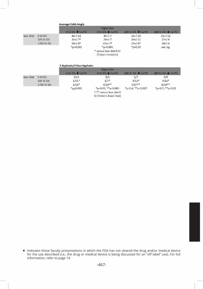
-467-
♦ Indicates those faculty presentations in which the FDA has not cleared the drug and/or medical device for the use described (i.e., the drug or medical device is being discussed for an “off label” use). For full information, refer to page 14.
-468-
See pages 21- 60 for financial disclosure information.
e-Poster #48 POSNA-2016 ♦Combined Pharmacologic and Biomechanical treatment of Congenital Pseudarthrosis of the Tibia: 100% Union; No Refractures?
Dror Paley, MD; Davida Packer, MD; Craig A. Robbins, MD; John Robb, PA-C; Raymond W. Liu, MD Paley Institute, West Palm Beach, Florida
Purpose: Can a new method of treatment of CPT lead to union in all cases and prevent refractures.
Methods: Since 2007, nineteen cases of CPT were treated by a single surgeon. 13/19 also had neurofibromatosis, and 6/19 were idiopathic. The average age was 54 months (range 11 to 145). All patients were treated by the following protocol: preoperative infusion of Zolidronic Acid, resection of fibrous hamartoma, shaping, mating and straightening of the bone ends, intramedullary rodding of tibia and fibula, subiliacus muscle periosteal graft to the CPT site, extreme iliac bone harvest of cancellous only bone graft by hollowing the iliac bone between the cortical tables all the way down to the acetabulum, triradiate, SI notch and SI joint regions, bone graft of CPT to create a crossunion between tibia and fibula, insertion of BMP2 between soft tissues and bone graft application of circular external fixator (Ilizarov apparatus with 4 olive wires) for rotation and length control second infusion of Zolidronic Acid at time of external fixator removal.
Results: Radiographic union including cross union to the fibula were achieved in 3-4 months in 19/19 cases (100%). Average followup was 45 months (range 12 -79). There were no refractures.
Conclusion: This treatment is successful at obtaining and maintaining union. The rationale for this treatment is based on: The zolidronic acid infusion prior to bone graft harvest makes the graft resistant to resorption. The hamartoma resection removes the osteoclastic constricting membrane around the bone The acute deformity correction reduces bending forces on the CPT The rodding of the two bones gives stability but also braces the bone from within to prevent refracture. An antegrade telescopic growing rod locked into the proximal and distal epiphysis is preferred. The periosteal grafting replaces the pathologic periosteum with healthy periosteum. The cross union between the tibia and fibula create a very wide bone especially in young children whose bone diameters are very narrow making refracture less likely. Cross union in young children requires a large volume of bone graft achieved by hollowing of the ilium. Adding BMP speeds healing which CPT bone is low. Rotational and length stability is provided by the circular external fixator allowing full weight bear postoperatively.
Significance: This new treatment has the highest ever reported union and lowest ever reported refracture rate.
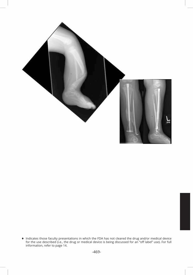
-469-
♦ Indicates those faculty presentations in which the FDA has not cleared the drug and/or medical device for the use described (i.e., the drug or medical device is being discussed for an “off label” use). For full information, refer to page 14.

-470-
See pages 21- 60 for financial disclosure information.
e-Poster #49 POSNA-2016 Genu Varum in Achondroplasia and Hypochondroplasia
Hiroshi Kitoh, MD; Kenichi Mishima; Masaki Matsushita, MD; Hiroshi Sugiura; Sachi Hasegawa; Naoki Ishiguro, MD Nagoya University Graduate School of Medicine, Nagoya, Aichi, Japan
Purpose: Genu varum is commonly observed in patients with achondroplasia (ACH) and hypochondroplasia (HCH). Fibular overgrowth and laxity of the lateral collateral ligament have been suggested as probable causes of this deformity, but the etiology is still controversial. We radiographically analyzed the lower limb alignment in ACH/HCH patients and determined factors associated with the development of genu varum in these disorders.
Methods: Full-length standing anteroposterior radiographs were retrospectively examined in 40 ACH and 6 HCH patients. There were 23 male and 23 female with an average age of 12.2 years (2.6 to 31.6 years). We measured mLPFA, mLDFA, mMPTA, mLDTA, joint line congruence angle (JLCA), femoral to tibial length ratio (Fe/T), fibular to tibial length ratio (Fi/T), the distance from the proximal fibula to the proximal tibia knee orientation line (Prox), and the distance from the distal fibula to the ankle joint orientation line (Dist) (Fig. 1). Mechanical axis deviation (MAD) was measured as the distance between the center of the knee and a line drawn from the center of the femoral head to the center of the ankle joint (positive values indicate medial deviation), and genu varum was defined as the leg with MAD of 17 mm or more. Radiological parameters were compared between the legs with genu varum (V group) and those without genu varum (non-V group).
Results: The mean MAD was 13.8 mm (-24 to 57 mm) and 36 legs were included in the V group (39%). Although mLPFA and mLDTA did not show differences between the two groups, significant larger mLDFA (93.4±5.3 degrees) and smaller mMPTA (81.9±4.6 degrees) were demonstrated in the V group (p<0.0001). The JLCA (8.0±7.0) was also larger in the V group (p=0.0096). Although Fi/T did not correlate with genu varum (p=0.0879), larger Fe/T (1.24±0.07) significantly affected the occurrence of varus deformity (p=0.0146). Neither Prox nor Dist correlated with genu varum. Lineal regression analysis demonstrated strong relationships between MAD and mLPFA (R=061) and mMPTA (R=0.66).
Conclusion: Fibular overgrowth was not a cause of genu varum in ACH/HCH. Varus deformities of the distal femur and proximal tibia affected the development of genu varum, and knee joint laxity (larger JLCA) deteriorated the deformity in some patients. Relative mesomelia may be associated with the occurrence of genu varum.
Significance: Hemiepiphysiodesis of the distal femur and/or proximal tibia should be considered to correct genu varum deformity in severe cases.
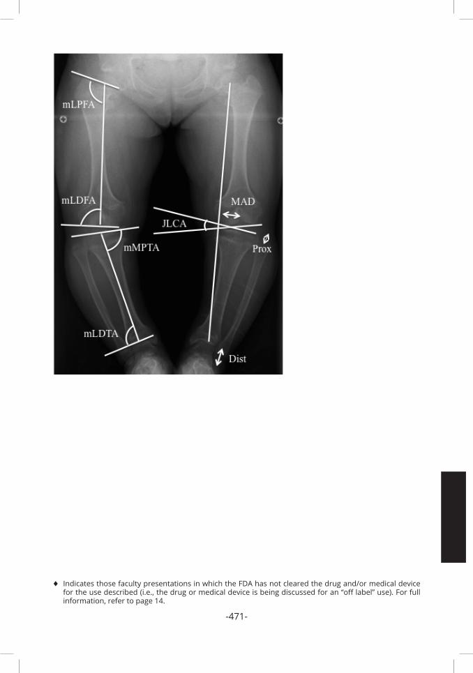
-471-
♦ Indicates those faculty presentations in which the FDA has not cleared the drug and/or medical device for the use described (i.e., the drug or medical device is being discussed for an “off label” use). For full information, refer to page 14.
-472-
See pages 21- 60 for financial disclosure information.
e-Poster #50 POSNA-2016 Skill Acquisition and Retention Following Simulated Pavlik Harness Learning
Simon P. Kelley, MBChB, MRCS, FRCS; Joel Moktar, MD; Alexandra Maxwell, BS; Catharine Bradley, BScPT, MSc; John H. Wedge, MD; M. Lucas Murnaghan, MD, MEd, FRCSCHospital for Sick Children, Toronto, Ontario, Canada
Purpose: Simulated learning is increasingly prevalent in many surgical training programs as medical education moves towards competency based curricula. In orthopaedic surgery, developmental dysplasia of the hip is a commonly treated diagnosis where the standard of care in patients less than six months of age is an orthotic device such as the Pavlik Harness. However, despite widespread use of the Pavlik Harness and the potential complications that may arise from inappropriate application, no formal educational methods exist. We aimed to develop a simulated learning module for Pavlik Harness application, and to assess improvement and retention of skill after exposure to the module.
Methods: A video and model based simulated learning module for Pavlik Harness application was developed. Two novice groups (residents and allied health professionals) were exposed to the module and at pre-intervention, post-intervention and retention testing were evaluated on their ability to apply a Pavlik Harness to the model. Evaluations were completed using a previously validated Objective Structured Assessment of Technical Skill (OSATS) and a Global Rating Scale (GRS) specific to Pavlik Harness application. A control group who did not undergo the module was also evaluated at two time points to determine if exposure to the Pavlik Harness alone would affect ability. All groups were compared to a group of clinical experts who were used as a competency benchmark. Statistical analysis of skill acquisition and retention was conducted using t-tests and ANOVA.
Results: Exposure to the learning module improved resident and allied health professionals’ competency in applying a Pavlik Harness (p<0.05) to the level of expert clinicians and this level of competency was retained one month after exposure to the module. Control subjects who were not exposed to the module did not improve nor did they achieve competency.
Conclusion: The simulated learning module has been shown to be an effective tool for teaching the application of a Pavlik Harness and learners demonstrated retainable skills post intervention.
Significance: This learning module should form the cornerstone of formal teaching for Pavlik Harness application in developmental dysplasia of the hip and we have demonstrated it as an effective learning tool for Orthopaedic residents.
-473-
♦ Indicates those faculty presentations in which the FDA has not cleared the drug and/or medical device for the use described (i.e., the drug or medical device is being discussed for an “off label” use). For full information, refer to page 14.
e-Poster #51 POSNA-2016 Back Pain in Cerebral Palsy Patients is Markedly Reduced After Spinal Fusion for Scoliosis
Burt Yaszay, MD; Tracey P. Bastrom, MA; Paul D. Sponseller, MD; Suken A. Shah, MD; Firoz Miyanji, MD; Jahangir Asghar, MD; Patrick J. Cahill, MD; Amer Samdani, MD; Peter O. Newton, MD Rady Children’s Hospital, San Diego, California
Purpose: The relationship between pain and spinal deformity in the cerebral palsy (CP) patient is poorly defined. Understanding this relationship is essential in order to evaluate the cost/benefit ratio of operative correction, which is associated with significant complications. The purpose of this study was to evaluate pre- and post-op back pain in CP scoliosis patients.
Methods: A prospective multi-center registry of CP patients was queried to identify patients with preop and 2 year postop pain assessments including visual analog scale (VAS) back pain scores. Radiographic and clinical demographics were recorded. Wilcoxon signed ranks test was used to compare change in VAS score.
Results: 164 patients with preop pain assessments were identified, of which 61 had 2yr postop pain assessments. The mean age was 14yrs (8-20) and mean pre-op Cobb was 85° (40-143°). When recorded, 73% of caregivers reported pain relief as one reason for surgery. Preop, patients with moderate to severe VAS scores had significantly decreased sitting tolerance compared to those with no pain (p<0.01). The distribution of VAS scores prior to surgery was: 34% no pain (VAS 0), 14% mild (VAS 1-3), 29% moderate (VAS 4-6), and 24% severe pain (VAS 7-10). At 2yrs postop, the median pain score improved significantly from 3 to 0 (p<0.001). All but 4 patients improved or remained in the no-mild pain group (VAS<3) at 2yrs after surgery. Two of these patients experienced complications requiring revision (1 for PJK; 1 for broken rod with extension of fusion to pelvis). A correlation approaching a moderate effect size was observed, suggesting that improvements in sitting tolerance may be associated with decreased VAS following surgery (r=-0.26, p=0.08).
Conclusion: As perceived by their caregivers, preoperatively the majority of CP patients experienced pain secondary to their scoliosis. Some of this may be inferred by their decreased tolerance for sitting. Following scoliosis correction, there was a significant improvement in pain with 93% of caregivers having reported no or only mild back pain as compared to 48% pre-operatively.
Significance: A better understanding of the pain experienced by CP patients with scoliosis is essential in order to evaluate the cost/benefit ratio of undergoing operative correction. As reported by their caregivers, CP patients with scoliosis experienced pain which appeared to influence their sitting tolerance. Postoperatively, their pain improved along with their sitting ability.
-474-
See pages 21- 60 for financial disclosure information.
e-Poster #52 POSNA-2016 Does Using an EMR Template Improve Documentation for Pediatric Supracondylar Fractures?
Mark J. Adamczyk, MD; Ryan J. Urchek, MD; Melanie Morscher, BS Akron Children’s Hospital, Akron, Ohio
Purpose: Multiple studies report that using a documentation template results in more complete and accurate electronic medical record (EMR) data. However, the use of an orthopaedic specific EMR template to document an acute injury has not been reported to our knowledge. Since pediatric supracondylar humerus fractures are associated with a relatively high incidence of nerve injury and compartment syndrome, it is imperative that documentation be complete. The purpose of this investigation is to compare the completeness of orthopaedic EMR documentation with and without the use of a template for pediatric supracondylar humerus fractures.
Methods: An IRB-approved retrospective review identified 119 supracondylar humerus fractures (types II-IV) presenting to a single pediatric institution over one year. The orthopaedic history and physical was documented using a template in 42 cases (templated group) and was not used in 77 cases (non-templated group). The completeness of the EMR documentation for the two groups was compared and included clinical exam findings (sensation, motor, compartment check, capillary refill), number of orthopaedic providers, and demographics. Statistics were used to analyze the data.
Results: The EMR documentation in the templated group compared to the non-templated group was more complete in regards to the sensory exam of the hand (100% vs. 91%), the motor status of the ulnar nerve (100% vs. 88%), AIN (100% vs. 92%), and PIN (100% vs. 94%) and compartment checks (95% vs. 42%). The templated and non-templated group documentation of radial pulse (95% vs. 99%) and capillary refill (74% vs. 78%) was similar. The two groups were also similar in terms of age and fracture type. On average, 5 (range: 2-10) different orthopaedic health care providers participated in the care of the patient from injury to the final office visit.
Conclusion: This is the first study to demonstrate that using a documentation template to record clinical exam findings for pediatric supracondylar humerus fractures resulted in more complete EMR data at initial presentation. Not only was documentation of motor and sensory findings more complete, but compartment checks were better documented (95% vs. 42%), an exam finding that can have serious sequela if untreated. Since there were on average 5 different orthopaedic providers participating in patient care, complete EMR documentation becomes even more important.
Significance: Use of an orthopaedic EMR template for pediatric supracondylar humerus fractures resulted in improved documentation of the sensory and motor exams and compartment checks.
-475-
♦ Indicates those faculty presentations in which the FDA has not cleared the drug and/or medical device for the use described (i.e., the drug or medical device is being discussed for an “off label” use). For full information, refer to page 14.
e-Poster #53 POSNA-2016 Complications and Radiographic Outcomes of Posterior Spinal Fusion (PSF) in Patients with Spinal Muscular Atrophy (SMA) Following Growth Friendly Treatment
Rodrigo Goes de Mendonca, MD; John A. Heflin, MD; Paul D. Sponseller, MD; George H. Thompson, MD; Michael G. Vitale, MD, MPH; John T. Smith, MD; Tara Flynn, BA; Ryan P. Murphy, BS; Jolecia M. Flournory, BA; Jeffrey R. Sawyer, MD;Children’s Spine Study Group; Growing Spine Study Group Le Bonheur Children’s Hospital, Memphis, Tennessee
Purpose: A common treatment strategy for early onset scoliosis (EOS) in SMA patients has been rib-based (RB) and/or spine-based (SB) distraction followed by PSF at the end of growth. The purpose of this study is to characterize outcomes of PSF in patients with SMA who have completed SB and/or RB distraction.
Methods: An IRB-approved review of the Children’s Spine Study Group (CSSG) and Growing Spine Study Group (GSSG) was performed. Children with SMA treated with RB or SB distraction who had undergone PSF at the end of distraction were included. Radiographic parameters including coronal/sagittal curve angle, T1-T12 height were measured. Operative outcomes including time, estimated blood loss (EBL), complications and levels fused were evaluated. Complications were stratified by severity/type using the Smith Classification.
Results: Twenty patients (10 male, 10 female) were identifed with a mean pre-treatment scoliosis/kyphosis of 75°/58° respectively. Initially there were 8 patients (40%) with RB anchors, 11 patients (55%) with SB anchors, and 1 patient (5%) with hybrid rib/spine (RS) anchors. During treatment, 2 RB patients were converted RS fixation before PSF. The mean T1-T12 growth rate was 1.3cm year during growing treatment.
At PSF the mean age was 13.0 years (mean surgical distraction treatment time 4.9 years). The mean pre PSF scoliosis/kyphosis were 60°/50° respectively which corrected to 50°/47° postoperatively. The mean T1-T12 height increased from 21.8 to 23.5cm. The mean surgical time was 6.5 hours and estimated blood loss was 750cc. There were no interoperative complications. At most recent follow up (mean 1.2 yrs, range 0.5-2.2yrs post-fusion) scoliosis (51°), kyphosis (48°) and T1-12 height (22.8cm) were maintained. There were 16 complications in 11 patients (55%), of which 11 (69%) were device-related (Type I:4,Type II:7) and 4 (31%) were disease-related (Type I:2, Type II:2, Type III:1).
Conclusion: The initial outcomes of PSF in SMA patients following distraction-based treatment are good with a reasonable complication rate in this high-risk patient population. While these early results are maintained, longer term studies with more patients are necessary to determine if the benefits of PSF outweigh the risks.
Significance: Knowledge of the outcomes and complications associated with PSF in SMA patients will be useful in counseling families preoperatively as well as in preventing postoperative complications.
-476-
See pages 21- 60 for financial disclosure information.
e-Poster #54 POSNA-2016 The Cost Effectiveness of Universal Screening Programs for Scoliosis
Matthew E. Oetgen, MD; Kathleen G. Noble; Shannon M. Kelly, MD; Allison Matthews, MSCR Children’s National Health System, Washington, District of Columbia
Purpose: Controversy exists regarding the utility and efficacy of universal school screening for scoliosis. Historically, the lack of definitive data on the true effectiveness of bracing has limited the ability to appropriately construct cost effectiveness models of universal screening for scoliosis; however, recent literature on bracing has provided a more accurate assessment of brace effectiveness. We designed a cost-effectiveness model of universal and selective scoliosis school screening, with a hypothesis that universal scoliosis screening is more cost effective than no screening.
Methods: A detailed literature review was performed to gather estimates of the prevalence and natural history of scoliosis, effectiveness of bracing and observation, referral rates for patients with scoliosis, and the effectiveness of traditional screening. Additionally, estimates of the costs associated with scoliosis were gathered including screening, diagnosis, observation, bracing, and fusion. Based on these variables, a cost effectiveness model was constructed for all children in the United States between ages 10-14 years in regards to scoliosis screening.
Results: The total population for the model was 4.1 million based on data from the 2010 US census. Based on the literature an estimate of the prevalence of scoliosis >10º in this population is 2%, with 10% having progressive curves needing bracing (>20º), and 0.2% requiring surgery (scoliosis>50º). The treatment success of bracing was estimated at 72%. The true positive rate of scoliosis screening was estimated to be 84%. The per person average costs were as follows: spinal fusion $129,000, bracing: $8,204, observation: $1,040, referral: $237, and screening: $13.45. Using this data it was determined that a national screening program would prevent 349 children from progressing to spinal fusion per year. This comes at a cost of an extra $52.6 million (total cost of scoliosis treatment per year without screening program: $682 million vs. total cost with screening program: $734 million).
Conclusion: Based on prevalence, effectiveness, and cost data derived from the current literature on scoliosis screening, diagnosis, and treatment we estimate a universal scoliosis screening program would be more costly to society than no screening program. Despite this extra cost, it appears a screening program would be effective at preventing approximately 349 spinal fusion surgeries each year in the United States.
Significance: Universal screening appears to be potentially effective at preventing surgery for scoliosis, but at an increased cost compared to no screening. This information will be useful to policy makers when weighing the costs and benefits of implementing universal screening programs.
-477-
♦ Indicates those faculty presentations in which the FDA has not cleared the drug and/or medical device for the use described (i.e., the drug or medical device is being discussed for an “off label” use). For full information, refer to page 14.
e-Poster #55 POSNA-2016 Management of Terminal Bony Overgrowth of the Humerus with Osteocartilaginous Grafts
Graham T. Fedorak, MD, FRCSC; Anna V. Cuomo, MD; Hugh G. Watts, MD; Anthony A. Scaduto, MD Shiners Hospitals for Children, Los Angeles, Los Angeles, California
Purpose: Bony overgrowth is a common complication in children after humeral transcortical amputation. Previous studies group children with humeral overgrowth with those who have developed overgrowth in other locations. Capping tibial overgrowth with the proximal fibula has been shown to be the most effective treatment. This is not clear for the humerus. We present eighteen humeri treated surgically for humeral osseous overgrowth with simple resection or autologous osteocartilaginous graft.
Methods: Records were reviewed of amputation patients from a single pediatric hospital from 1987-2011. Patients with two years follow-up who underwent surgical treatment for established humeral overgrowth were included. One requiring revision surgery before two years was included. Patients initially managed with simple resection were compared to those managed with autologous osteocartilaginous grafts. All patients who underwent autologous osteocartilaginous grafting, either as index or subsequent procedure were examined.
Results: Eighteen humeri in sixteen patients met inclusion criteria, including ten acquired amputations and eight congenital. Mean age at initial surgery was 8 (2.6-13.3) years and mean follow-up was 6.6 (1.4-10.3) years. Thirteen humeri underwent simple resection, with recurrent overgrowth in nine, and revision surgery in eight at a mean 2.6 years. Five humeri were primarily managed with autologous osteocartilaginous grafts. Two developed non-overgrowth related complications at 1 and 42 months. Including revision procedures after simple resection, 10 humeri were managed with autologous osteocartilaginous grafts. Thirty percent (3/10) required revision surgery, however, there were no cases of recurrent overgrowth.
Conclusion: At a mean of 5.4 years, seven of ten humeri treated with autologous osteocartilaginous grafts, either primarily or as a secondary procedure, had not required revision surgery. In contrast, only five of thirteen humeri treated with simple resection did not require further surgery with eight undergoing revision at a mean 2.6 years. Despite this difference in outcomes, the heterogeneity of surgical techniques in humeral osteocartilaginous capping demonstrates that an ideal technique has yet to be established. Furthermore, outcomes are not as consistent as when the technique is applied to overgrowth of the tibia.
Significance: This study provides guidance for a problem, which deceptively appears to have a simple solution. The ease of performing simple resection is belied by the fact that the surgeon who chooses this strategy will likely be back in the operating room within a few years. However, issues with fixation and hardware prominence can complicate osteocartilaginous grafting of the humerus.
-478-
See pages 21- 60 for financial disclosure information.
e-Poster #56 POSNA-2016 Final Fusion in Patients Treated with Rib Based Distraction: A Review of Peri-operative Results
John A. Heflin, MD; Jessica Morgan, BS; Victoria Heagy, BS; John T. Smith, MD University of Utah, Salt Lake City, Utah
Purpose: Rib based distraction for early onset scoliosis (EOS) and thoracic insufficiency is an accepted form of managing progressive spinal and chest wall deformities. Once growth has ceased, these patients generally undergo definitive fusion surgery to prevent further progression of deformity. Our objective was to review peri-operative outcomes in patients undergoing definitive fusion following rib-based distraction treatment for EOS.
Methods: 27 patients treated with rib-based distraction for EOS who subsequently underwent final fusion were evaluated with an IRB approved patient registry. All patients were treated by a single surgeon. Information from the registry, patient operative reports, and x-rays were reviewed. Surgical time, blood-loss, complications, overall correction, and need for osteotomies were documented. Radiographic measurements included pre and post-operative sagittal and coronal Cobb angles. All numerical measurements were evaluated for statistical mean, range, and standard deviation.
Results: The average age at implantation of the rib-based construct was 7.45 (1.78-11.78) years. The mean age at definitive fusion was 13.51 (9.21-18.51) years, following an average of 10.5 (0-18) lengthening procedures and 13.35 (3-21) total surgeries. The mean coronal Cobb angle measured 67.42 (40-107) degrees prior to fusion and 50.17 (32-82) degrees following fusion. Pre-operative mean kyphosis was 61.08 (8-113) degrees and post-operatively averaged 50.00(20-85) degrees. Operative time averaged 256 (115-520) minutes with a mean 534cc (180-1280) blood loss. Osteotomies were required in 11 patients (41%). Complications occurred in 12 patients (44%), 9 requiring a repeat procedure.
Conclusion: Definitive fusion in patients treated with rib-based distraction is both technically challenging and tends to be associated with longer operative times, higher blood loss, and a higher rate of complications.
Significance: Although the amount of correction is limited by stiffness, scaring, and autofusion, definitive fusion surgery does improve pre-operative coronal Cobb angles and kyphosis. Longer term studies will be needed to establish whether correction is maintained over time.
-479-
♦ Indicates those faculty presentations in which the FDA has not cleared the drug and/or medical device for the use described (i.e., the drug or medical device is being discussed for an “off label” use). For full information, refer to page 14.
e-Poster #57 POSNA-2016 Intra-wound Vancomycin Reduces the Infection in Growth-Friendly Spine Surgery
Justin Haller, MD; David Andrew Hulet, BS; Qian Ding, PhD; Angela P. Presson, PhD; John A. Heflin, MD; John T. Smith, MD University of Utah, Salt Lake City, Utah
Purpose: Patients with Early Onset Scoliosis (EOS) who require growth-friendly spine surgery are known to be at high risk for infections and/or wound dehiscence. Several studies in pediatric and adult patients undergoing spine surgery have shown that vancomycin powder applied to the wound before closure significantly reduces the risk of post-operative infection. There are currently no studies that evaluate the efficacy of intra-wound vancomycin in reducing infection for patients undergoing growth-friendly spine surgery.
Methods: Data from 116 patients in a prospectively collected database with EOS of multiple diagnoses were reviewed. Patients were divided into two groups. From May 2002 through June 2013, there were 1036 VEPTR procedures in which no patient received intra-wound vancomycin. Beginning in June 2013, there were 161 consecutive EOS procedures having VEPTR surgery and all had intra-wound vancomycin powder. Patient groups were compared based on age, gender, diagnosis, type of surgery, ambulatory status, and bowel/bladder incontinence. Additionally, complications related to intra-wound vancomycin powder were reviewed.
Results: The pre- and post-vancomycin groups were similar with regard to age, gender, diagnosis, ambulatory status, and bowel/bladder incontinence. The groups were significantly different with regard to type of surgery (Table 1). Prior to the use of intra-wound vancomycin, the infection rate in VEPTR surgery was 5.7%. The patients receiving intra-wound vancomycin had a significantly reduced infection rate of 1.2%. (p < 0.031). Using multivariate logistic generalized estimating equation model to control for surgery type, infection rate was lower in the patients who received intra-wound vancomycin as compared to the patients who did not receive intra-wound vancomycin (1.2% vs. 4.4%, p = 0.066). There were no complications related to the administration of intra-wound vancomycin.
Conclusion: Intra-wound vancomycin is safe and reduces infection rates for patients undergoing growth-friendly spine surgery.
Significance: Intra-wound vancomycin can be utilized for significant infection rate reduction in repetitive surgery.
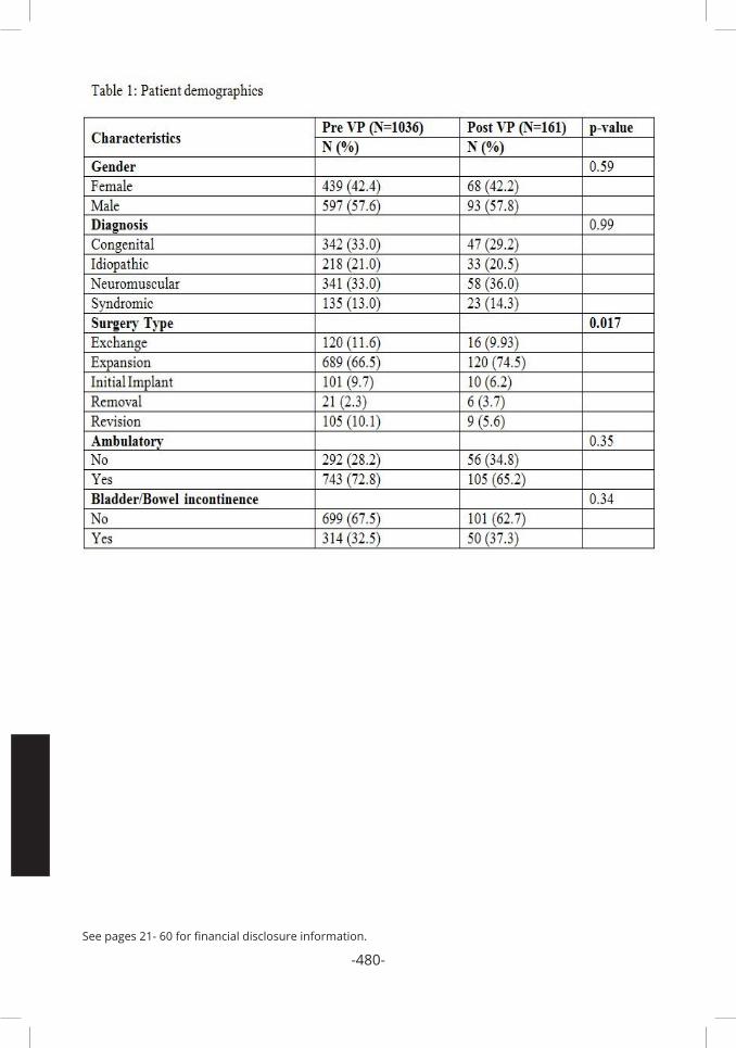
-480-
See pages 21- 60 for financial disclosure information.

-481-
♦ Indicates those faculty presentations in which the FDA has not cleared the drug and/or medical device for the use described (i.e., the drug or medical device is being discussed for an “off label” use). For full information, refer to page 14.
e-Poster #58 POSNA-2016 Both Decreased and Increased Relative Acetabular Volume are Associated with Increased Hip Arthritis in an Osteological Review of 1090 Hips
Douglas S. Weinberg, MD; Drew F.K. Williamson, BA; Michael B. Millis, MD; Raymond W. Liu, MD Case Western Reserve University, Cleveland, Ohio
Purpose: Recently, there has been considerable interest in quantifying the associations between bony abnormalities about the hip joint and osteoarthritis. The relationships between acetabular undercoverage, acetabular overcoverage, and femoroacetabular impingement (FAI) with hip joint osteoarthritis (HOA) remain controversial.
Methods: 545 cadaveric skeletons (1090 hip joints) from the Hamann-Todd osteological collection were obtained. Femoral head volume (FHV), acetabular volume (AV), the ratio between femoral head volume and acetabular volume (FHV/AV), acetabular version, alpha angle, and anterior femoral neck offset (AFNO) were measured. A validated grading system was used to quantify HOA as minimal, moderate, or severe. Multiple linear regression was used to determine the independent correlates of FHV, AV, and FHV/AV. Multinomial logistic regression was used to determine which factors increase the risk of HOA.
Results: Females had smaller FHVs (standardized beta -0.382, p<0.001), and AVs (standardized beta -0.351, p< 0.001), although the ratio of FHV/AV was unchanged. Every 1-degree increase in alpha angle increased the probability of having moderate HOA compared to minimal HOA by 7.1%. Every 1-millimeter decrease in AFNO increased the probability of having severe or moderate HOA, compared to minimal HOA, by 11% and 9%, respectively. The relative risk ratio of having severe HOA compared to minimal HOA were 7.2 and 3.3-times greater for acetabular undercoverage and overcoverage, respectively, relative to normal acetabular coverage.
Conclusion: Acetabular undercoverage and overcoverage were independent predictors of increased HOA. Alpha angle and AFNO had modest effects, supporting that bony abnormalities both in the dysplastic and FAI spectrum increase the risk for more severe HOA.
Significance: Both acetabular undercoverage and overcoverage are associated with hip arthritis, supporting the growing viewpoint that overcorrection of either condition is unfavorable.
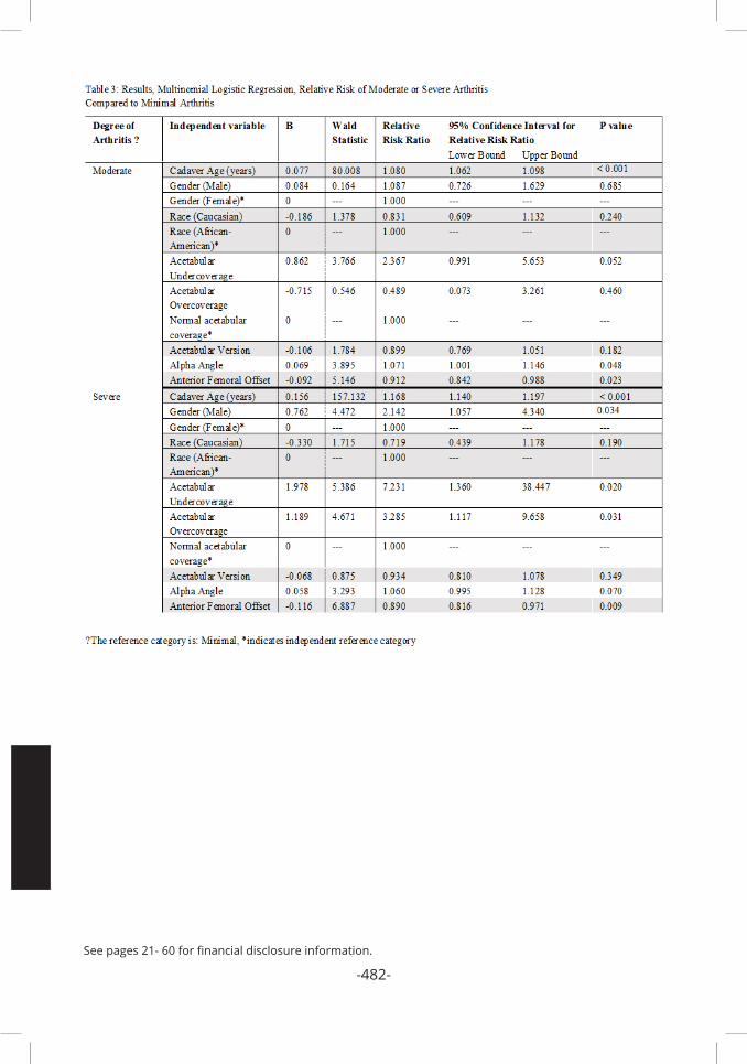
-482-
See pages 21- 60 for financial disclosure information.
-483-
♦ Indicates those faculty presentations in which the FDA has not cleared the drug and/or medical device for the use described (i.e., the drug or medical device is being discussed for an “off label” use). For full information, refer to page 14.
e-Poster #59 POSNA-2016 Does Clavicle Grow beyond Skeletal Maturity?
Pooya Hosseinzadeh, MD; Cristina Brea, BS; Gary Kiebzak, PhD Baptist Children’s Hospital, Miami, Florida
Purpose: A recent radiographic study has reported than 80% of clavicle growth is completed by age 9 in females and by age 12 in males and remodeling of malunited clavicle fractures should not be expected in adolescents. The data has been used by some to advocate operative treatment of displaced clavicle fractures in adolescents. Medial epiphysis of the clavicle contributes to the longitudinal length and does not ossify until 25 years of age. We studied the longitudinal growth of the clavicle until 25 years of age.
Methods: 700 standing PA chest radiographs were used to measure the length of the clavicle for patients between 12-25 years of age (50 radiographs per age, 25 females and 25 males). Radiographs with evidence of previous fractures or with significant rotation were excluded. Measurements were done by one author using the same technique. Measurements were done twice and the average of the two measurements were used. Male and female measurements were compared and reported separately. Average length of clavicle at each age was calculated. Statistical analysis was used to investigate if significant growth occurs after 18 years of age.
Results: Clavicle length was 126.55±75 mm, 157.78 ±10.96 mm, and 166.82±10.35 mm in males at ages 12, 18, and 25, respectively. Clavicle length was 130.34± 9.97 mm, 142.52±11.00 mm, and 145.92±9.024 mm in females at ages 12, 18, and 25, respectively. Regression analysis showed linear growth of the clavicle up to18 years of age in both genders. Significant growth of the clavicle after age 18 was only found in males. 12% of the growth of the clavicle in males occur after 16 years of age.
Conclusion: Linear growth of the clavicle is seen in both males and females up to 18 years of age. The growth after age 18 is not significant in females. In males, significant growth occurs after 18 years of age and clavicle continues to grow until age 25. 12% of the clavicle growth in males occurs after age 16. The continued growth after age 18 in males could indicate the potential for remodeling of displaced clavicle fractures in this age population.
Significance: This study shows continued growth of the clavicle in males after 18 years of age which could indicate the potential for remodeling in malunited fractures in adolescents. Future prospective studies are needed to document the remodeling potential in displaced clavicle fractures in adolescents.
-484-
See pages 21- 60 for financial disclosure information.
e-Poster #60 POSNA-2016 Deficient Quality of Care for Clubfoot: Implications for Patient-Centered Care
Asitha Jayawardena, MD; Jennifer L. Trevillian, BS; Thomas Cook, PT, PhD; Jose A. Morcuende, MD, PhD University of Iowa, Iowa City, Iowa
Purpose: As access to the Internet is transforming the physician-patient relationship, a better understanding of patient perceptions of treatment is essential to provide the highest quality of care. Clubfoot, due to its historically internet-savvy patient population and its easy-to-understand treatment method embodies the altering physician-patient dynamic.
Methods: An anonymous survey was administered through 10 active online clubfoot support groups to 446 parents from 33 different countries in both 2009 and 2012.
Results: Most parents (77%) were initially unfamiliar with clubfoot and (64%) initially learned about the Ponseti method from personal research. Only half were given information about clubfoot by a medical professional following their child’s diagnosis and only 36% felt the information they received was adequate in content. Thirty-nine percent of parents reported receiving what they judged to be substandard care. In fact, 40% of parents switched physicians due to dissatisfaction with the initial treatment provided. Parents who reported substandard care were more likely to have: not immediately been given information about clubfoot (p=0.04); learned of the Ponseti method later in their treatment course (p<0.001); and believed their first physician did not use the Ponseti method (p<0.001).
Conclusion: Parents report that most aspects of clubfoot care are inadequate and this substandard care is associated with not using the Ponseti method properly. Physicians should make a greater effort to provide high quality care using the Ponseti method or risk having these children self-referred to another facility that provides more effective and more patient-centered care.
Significance: Orthopaedic specialists may feel that the Ponseti method is simply a matter of applying a series of plaster casts, a treatment modality that they use with many conditions. They may fail to take the time to learn about, or fail to grasp the nuances of, the specific manipulations and casting techniques that are the core of the Ponseti method. Future studies are needed to better understand the discrepancy between self-reported physician data claiming the use of the Ponseti method and self-reported parent data indicating a lack of high quality treatment for clubfoot using the Ponseti method.
-485-
♦ Indicates those faculty presentations in which the FDA has not cleared the drug and/or medical device for the use described (i.e., the drug or medical device is being discussed for an “off label” use). For full information, refer to page 14.
e-Poster #61 POSNA-2016 Implementing a National Clubfoot Treatment Program in Nigeria
Jose A. Morcuende, MD, PhD; Thomas Cook, PT, PhD; Olayinka Adegbehingbe, MD; Tomeka Petersen, MSW; Omolade A. Lasebikan, MBChB, MD, MPH; Adeoye J. Adetiloye, MD, FWACS; Habila Umaru, MD; Oladipo Adewole, MD; Orlando Ugwoegbulem III, MBBS, MD; Zumnan D. Songden, MD; Oluwadare Esan, MBBS, MD; Edith A. Parker, PhD University of Iowa, Iowa City, Iowa
Purpose: This two-year project was intended to evaluate and refine a program framework to: (1) expand the number of well-trained orthopedic surgeons who can provide high quality treatment of clubfoot deformity using the Ponseti method and (2) to establish a national program of sustainable clinic units to provide treatment for the estimated 8,000 children born with clubfoot each year in Nigeria.
Methods: The program model consisted of support of the clubfoot care pathway (identification, referral, correction, and follow-up) through strengthening 11 supporting elements, divided into 3 categories: clinical, local, and regional/national. A key feature in applying the framework was the role of in-country champion-physicians to provide leadership, energy, and direction. Project activities included clinic site visits; use of an International Clubfoot Registry for patient record-keeping; web conferencing to facilitate education and communications; development of Best Practice Guidelines; advocacy and education with frontline health workers, parents groups, and the media; and engagement with professional societies, training institutions, local health administrators, and the Ministry of Health. A country strategic plan contained time lines and targets for each of the proposed activities. Data collected included site visit reports, field notes, emails, checklists, questionnaires, oral reports, and qualitative interviews.
Results: During the project period, twenty-eight new clubfoot clinical units were established in Nigeria. One hundred forty additional health workers were informed about clubfoot and its treatment. Nineteen local health administrators, four professional organizations, and 250 additional stakeholders were engaged to support the national program. The national Ministry of Health formally adopted Best Practices Guidelines for the treatment of clubfoot. Twenty-seven web-conferences and webinars were conducted and data on 558 clubfoot patients were entered into the International Clubfoot Registry.
Conclusion: The proposed framework proved to be an effective means to systematically implement a national clubfoot treatment program in Nigeria. During the two-year project, a number of significant challenges were encountered and a significant number of lessons were learned.
Significance: This project has helped to codify an important and generalizable approach that should inform others, especially those in low-resource situations, who are working to establish and/or strengthen the clubfoot care pathway in their countries.
-486-
See pages 21- 60 for financial disclosure information.
e-Poster #62 POSNA-2016 Comparison Between Participation and Desire to Participate in Activities Among Children with Skeletal Dysplasias
Cathleen L. Raggio, MD; Meredith Manson; Erin M. Carter, MS; Kate P. Citron, BS Hospital for Special Surgery, New York, New York
Purpose: Skeletal dysplasias are a group of genetic disorders that involve abnormalities in bone and cartilage growth. Affected patients’ participation in various activities can be limited because of physical differences. This study aims to examine the activities children with skeletal dysplasias typically participate in using the Children’s Assessment of Participation and Enjoyment (CAPE), and their activity preferences using the Preferences for Activity of Children (PAC). Both questionnaires address the same Recreational, physical, social, and skill-based activity domains. These surveys were validated in Ontario, Canada for children (ages 6-21) with or without disabilities. We hope to better understand the discrepancies between activity participation and preference. By identifying activities that patients want to be doing we can encourage them to participate in these activities in the future to improve their quality of life. We hypothesized that: 1) There are certain activities that patients would like to be doing, but do not participate, 2) More male patients participate in physical activities than female patients, 3) More average-statured patients participate in certain activities than short-statured patients.
Methods: Fifteen pediatric patients (ages 7-21) with varying types of skeletal dysplasias completed the CAPE and PAC surveys at the time of their doctor visit. The binary yes/no responses on the CAPE were converted to the PAC preference 1-3 scale in order to facilitate comparison. A non-parametric Mann-Whitney U test and two chi-square analyses were conducted to analyze the data.
Results: Overall, significantly more patients would like to be doing activities than the amount of patients who actually participated in these activities (p<0.05; see Figure 1). There was no significant difference between participation in physical activities between boys and girls. More average-statured patients participated in the following activities than short-statured patients: doing team sports, bicycling, playing games, talking on the phone (p<0.05).
Conclusion: Skeletal dysplasia patients participate in a wide variety of activities. However, they desire to participate in more activities than they currently do across all domains (skill-based, self-improvement, social and physical activities). Depending on the severity of their condition, and with the proper support, many of the activities could be accomplished or modified to enable desired participation.
Significance: It is important to understand patient interests in order to recommend therapies/activities to enhance quality of life and disprove myths about limitations and/or restrictions for children with skeletal dysplasias.
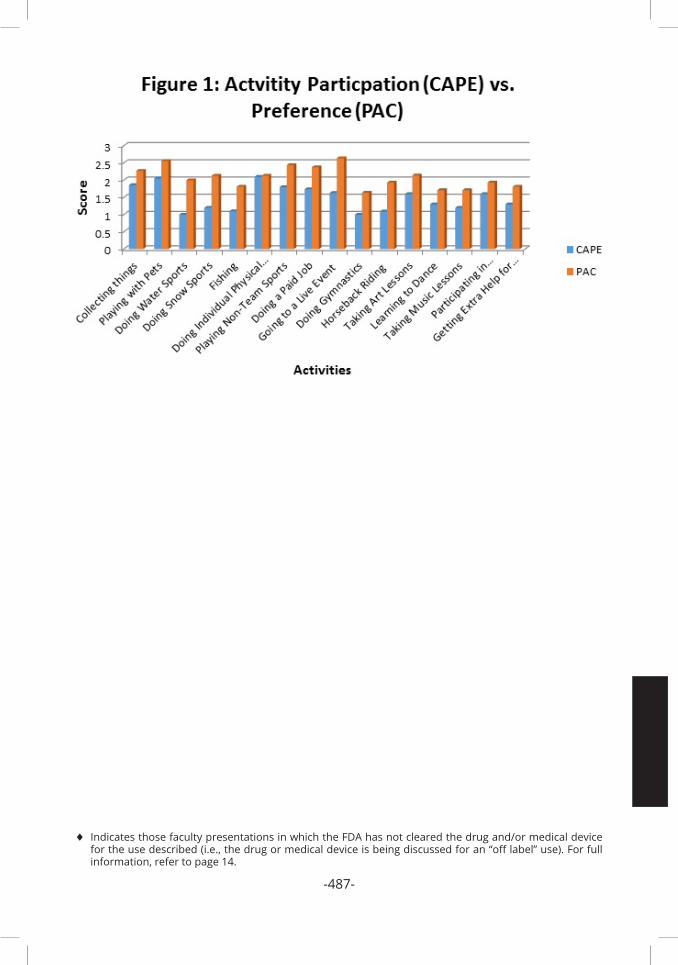
-487-
♦ Indicates those faculty presentations in which the FDA has not cleared the drug and/or medical device for the use described (i.e., the drug or medical device is being discussed for an “off label” use). For full information, refer to page 14.
-488-
See pages 21- 60 for financial disclosure information.
e-Poster #63 POSNA-2016 Knee Hyperextension in Children with Cerebral Palsy: Outcomes over a Minimum of Five Year Follow Up
Oussama Abousamra, MD; Daveda Taylor, DPT; Chris Church, MPT; Maria Del Pilar Duque Orozco, MD; Nancy Lennon, MSPT; John D. Henley, PhD; Julieanne P. Sees, DO; Freeman Miller, MD; Justin R. Connor, MD Nemours / Alfred I. duPont Hospital for Children, Wilmington, Delaware
Purpose: This study aimed to evaluate the course of knee hyperextension (back-kneeing) in children with cerebral palsy (CP) over a minimum of 5 year follow up.
Methods: 3D gait analysis records from 1990 to 2015 were reviewed for all children with CP who had: (1) knee hyperextension (defined as midstance minimum knee flexion below one standard deviation that of age matched typically developing children (smaller then -2o) and (2) follow-up gait analysis that was at least 5 years later. Gait velocity, average knee flexion at initial contact, minimum knee flexion in midstance, maximum ankle dorsiflexion in midstance, knee flexion moments in midstance and pelvic forward tilt in stance were analyzed from the motion data. From the accompanying physical exams knee and ankle passive range of motion (PROM), the Gross Motor Function Classification System (GMFCS), and the Gross Motor Function Measure (GMFM) were collected. Surgeries performed on the extremities during follow up were also recorded.
Results: Knee hyperextension was identified in 308 knees, of which 97 had follow-up data greater than 5 years (mean 8 years). The mean age at the initial visit was 8±3 years and the mean age at the follow-up visit was 16±4 years. Between the tests plantar flexor lengthening (PFL) was performed on 40% of the limbs, hamstring lengthening on 27% and rectus transfer on the 24%. Overall, knee flexion and ankle dorsiflexion in midstance did increase (p<0.0001). However the functional data did not change (22% were GMFCS I, 12% GMFCS II, 39% GMFCS III and 27% GMFCS IV; gait velocity was 65±34cm/s and GMFM was 25±9), nor was there a change in muscle tone (90% had a spastic gastrocnemius and 70% had a spastic quadriceps femoris). At follow-up, 29% of the knees were in hyperextension, 37% were normal and 34% were in excessive flexion (> 1SD above normal) in midstance. Similar changes were noted between children who had PFL and those who did not (Table).
Conclusion: In this study, hyperextended knees showed increased flexion over eight years. 2nd rocker dorsiflexion also increased. Children maintained their functional level at follow-up.
Significance: In children with CP, knee hyperextension generally decreases over time. All children had specific orthopaedic management to address their gait deviations. Plantar flexor lengthening is indicated in some cases and the role of multilevel surgery in the treatment of knee hyperextension needs to be further determined.
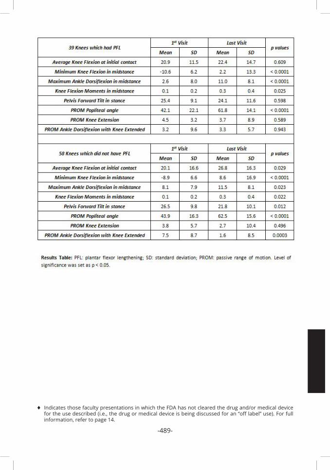
-489-
♦ Indicates those faculty presentations in which the FDA has not cleared the drug and/or medical device for the use described (i.e., the drug or medical device is being discussed for an “off label” use). For full information, refer to page 14.
-490-
See pages 21- 60 for financial disclosure information.
e-Poster #64 POSNA-2016 Cost Analysis of Magnetically-Controlled Growing Rods compared with Traditional Growing Rods for Early Onset Scoliosis in the United States: An Integrated Healthcare Delivery System Perspective
David W. Polly Jr, MD; Stacey J. Ackerman, MSE, PhD; Karen B. Schneider, PhD; Jeff Pawelek, BS; Behrooz A. Akbarnia, MD University of Minnesota, Dept. of Orthopaedic Surgery, Saint Paul, Minnesota
Purpose: Early onset scoliosis (EOS) is a challenging clinical problem. Untreated, EOS may result in pulmonary compromise, as well as decrease in thoracic volume and the space available for the lungs. Treatment with traditional growing rods (TGR) has high complication rates, and requires repeated invasive surgical lengthening under general anaesthesia that can significantly affect quality of life. Magnetically-controlled growing rods (MCGR) are lengthened non-invasively using an externally applied magnet remote control in a physician office, thus avoiding the need for repeated surgical lengthenings. A budget impact model (BIM) was developed to compare MCGRs with TGRs for the treatment of EOS from the United States (US) integrated healthcare delivery system perspective.
Methods: For both TGR and MCGR, the model estimated the cost for initial implantation, lengthenings, revisions due to device failure, surgical site infections (SSIs), device exchanges (at 3.8 years), and final fusion, over a 6-year episode of care (aged 6 to 12 years). Model parameters were estimated on the basis of the published literature, analyses from a multi-center EOS database comprised of US institutions, and a series of interviews with pediatric spine surgeons, private payers, and hospital purchasers. Costs were discounted at 3.0% per annum and represent 2015 United States Dollars (USD).
Results: Of 1,000 simulated patients over 6 years, MCGR was associated with an estimated 270 fewer deep SSIs and 197 fewer revisions due to device failure compared with TGR. MCGR was projected to cost an additional $61 per patient over the 6-year episode of care compared with TGR. Sensitivity analyses indicated that the results were sensitive to changes in the percentage of MCGR dual rod use, months between TGR lengthenings, percentage of hospital inpatient (versus outpatient) TGR lengthenings, and MCGR device cost.
Conclusion: Cost offsets for MCGR accrue over time and reflect the elimination of repeated invasive TGR surgical lengthenings and its associated complications. Despite the higher up-front cost for insertion of the MCGR device and later for exchange of the MCGR device, the cumulative cost of MCGR is offset over the 6-year episode of care from the perspective of the US integrated healthcare delivery system. The health-related QoL gained by MCGR patients and their caregivers is likely to be substantial and warrants further investigation.
Significance: The cost impact of MCGR is offset by eliminating repeated TGR surgical lengthenings.
-491-
♦ Indicates those faculty presentations in which the FDA has not cleared the drug and/or medical device for the use described (i.e., the drug or medical device is being discussed for an “off label” use). For full information, refer to page 14.
e-Poster #65 POSNA-2016 Does Correction of Pelvic Obliquity Result in Improved Outcomes and Sitting Tolerance for Patients with Cerebral Palsy Scoliosis?
Brian E. Kaufman, MD; Josh M. Pahys, MD Nemours / Alfred I. duPont Hospital for Children, Wilmington, Delaware
Purpose: Severe pelvic obliquity (PO) associated with spinal deformity in cerebral palsy (CP) patients interferes with sitting balance and quality of life (QOL). Surgeons and caregivers rate sitting balance as their highest surgical indication or priority. To date, no guidelines exist regarding optimal alignment for these patients to facilitate care, alleviate pain, and improve QOL. This study evaluated the relationship between PO and QOL in CP patients with scoliosis undergoing posterior spinal fusion (PSF).
Methods: A prospectively collected multi-center database was reviewed for all CP patients with scoliosis who underwent PSF and had minimum two-year follow-up. A total of 101 patients were identified. QOL was evaluated using the validated CPCHILD clinical patient related outcome measure. Radiographs, sitting tolerance, and QOL data were evaluated at preoperative, initial postoperative, and two-year follow-up visits.
Results: There was significant improvement in mean preoperative PO to 2 years postoperatively (21.8° to 7.5°, p<0.001) and sitting tolerance. There was no significant correlation between the absolute magnitude of PO at 2 years postoperatively and QOL. A significant correlation did exist, however, between percent correction of PO from preoperative to 2 years postoperatively and overall QOL (p=0.02), especially in the positioning, transfers, and mobility domain. QOL data revealed a significant correlation with decreased PO and increased QOL in nearly all domains, specifically PO<10° vs. 10-19° (p=0.03) and PO<10° vs. PO >20° (p<0.001). There were 15 patients with spastic hip subluxation/dislocation. The hip status did not correlate with postoperative PO, percent correction of PO, or postoperative QOL scores.
Conclusion: This prospectively collected study evaluated 101 CP patients who underwent PSF for severe scoliosis and PO with two-year follow-up of X-rays, clinical data, and patient-related outcomes. Regardless of stage of treatment (pre- or postoperative), PO <10° was associated with the highest QOL scores and sitting tolerance. The percent correction of PO achieved after surgery was associated with an improved overall QOL score (especially the positioning, transfers, and mobility domain) at 2 years postoperatively.
Significance: Severe pelvic obliquity associated with spinal deformity does interfere with sitting and QOL in severely affected (GMFCS 5) children with cerebral palsy. Leveling the pelvis as much as possible during PSF is desirable to improve patient sitting balance and alignment.
-492-
See pages 21- 60 for financial disclosure information.
e-Poster #66 POSNA-2016 Comparison of 3D Muscular Parameters Between Children with Cerebral Palsy and Typically Developing Children
Abir Massaad, MSc; Ayman Assi, PhD; Ziad Bakouny; Wafa Skalli, PhD; Ismat Ghanem, MD Faculty of Medicine, University of Saint-Joseph, Beirut, Lebanon
Purpose: The aim of this study was to compare 3D muscular lengths and volumes between typically developing (TD) children and ambulant spastic children with CP presenting no medical history at the lower limbs (LL).
Methods: Seventeen spastic CP children with a mean age of 12±4 years (diplegia N=14, hemiplegia N=3, GMFCS levels: I N= 9, II N= 8), with no medical history at the LL level, had undergone an MRI exam of the LLs. Seventeen TD children, age-matched to the CP group, were also enrolled in the study. Belly muscle contours were delimited on a reduced number of axial sections and extrapolated on the totality of the belly muscles [4]. 3D subject-specific muscle reconstructions were performed for the hamstrings, hip adductors (brevis, magnus and longus), vastii (medial, lateral and intermedius), rectus femoris, gracilis, sartorius, gastrocnemii (medial and lateral), soleus and anterior tibialis in order to calculate the normalized belly muscle lengths (to LL length) and volumes (to weight) bilaterally. Differences between TD children, CP GMFCS levels I and II were compared using ANOVA test. Significance level was set at 0.05.
Results: Muscle lengths were significantly reduced in children with GMFCS level II compared to TD children (p<0.05) except for the adductor magnus, semi-tendinosus, femoral biceps brevis and longus. Only gastrocnemii lengths significantly differed between children with GMFCS level I and TD children (p<0.05). Muscle volumes were significantly reduced in children with GMFCS level II (p<0.05) except for the femoral biceps brevis, gracilis and adductor magnus.
Conclusion: For the first time belly muscle lengths and volumes of the lower limbs were calculated in spastic children with CP with no medical history, based on subject-specific 3D reconstructions. Lower limb belly muscle volumes and lengths were shown to be more reduced when motor impairments increase in children with CP. In order to better understand the relationship between soft tissue anomalies and gait alterations, future studies should determine whether CP groups with homogeneous gait profiles present similar muscle geometry. [1]Barrett , 2010. [2]Noble, 2013. [3]Oberhoffer, 2010. [4]Jolivet, 2008.
Significance: Spasticity in children with cerebral palsy (CP) affects muscle function and geometry [1]. Orthopedic interventions (botilinum toxin injection, casting, soft tissue surgeries) are usually performed at an early age which might affect the results of previous assessments of muscle lengths and volumes [2,3]. However, little is known about the natural history of these muscle alterations.
-493-
♦ Indicates those faculty presentations in which the FDA has not cleared the drug and/or medical device for the use described (i.e., the drug or medical device is being discussed for an “off label” use). For full information, refer to page 14.
e-Poster #67 POSNA-2016 Alterations in Acetabular and Proximal Femur Geometry in Subjects with Down Syndrome
Elie Joseph Mansour, MD; Jean Jacques Yaacoub, MD; Ayman Assi, PhD; Ziad Bakouny; Wafa Skalli, PhD; Ismat Ghanem, MD Faculty of Medicine, University of Saint-Joseph, Beirut, Lebanon
Purpose: The aim of this study is to describe acetabular and proximal femur geometry of DS subjects, in the 3 planes and in standing position, compared to an asymptomatic control group.
Methods: 41 subjects (13F, 28M, age 17.4±6.2 years) with DS, without history of cardiac surgery, have undergone EOS® biplanar X-ray exam with 3D reconstructions of their pelvis and lower limbs in standing position. These subjects were age and gender-matched to 41 asymptomatic subjects (13F, 28M, age 17.7±5.4 years). The following parameters were calculated: femoral neck shaft angle, femoral head diameter normalized to femoral length, neck length normalized to femoral length, femoral offset normalized to femoral length, external coverage angle, Idelberg and Frank angle, acetabular coverage of the femoral head, acetabular tilt, abduction and anteversion, posterior and anterior coverage of the acetabulum. Parameters were compared between the 2 groups using Student and Mann-Whitney tests (α-level=0.05).
Results: The following parameters were significantly higher in DS group compared to the control group: femoral head diameter (1.08% vs 1.03%,p=0.002), femoral offset (0.98% vs 0.92%,p=0.013), neck length (1.32% vs 1.20%,p=0.001), external coverage acetabular angle (33.3° vs 31.4°,p=0.03). The abduction of the acetabulum (53.5° vs 55.1°,p=0.02) was significantly smaller in the DS group. The angle of Idelberg and Frank (53.2° vs 53.0°,p=0.9), acetabular tilt (19.3° vs 20.7°,p=0.16) and the acetabular anteversion (15.0° vs 15.0°,p=0.8) did not differ between both groups (DS vs Control respectively).
Conclusion: This is the first study that describes 3D anatomy of the acetabulum and proximal femur in subjects with DS in standing position. The acetabular anteversion was shown to be similar between groups, in contrary to previous studies [3]. This could be due to the difference in subject positioning when calculating hip parameters. Furthermore, subjects with DS presented a more abducted acetabulum which could predispose them to femoral-acetabular impingement and osteoarthritis. Treatment of hip instability and impingement in DS subjects should take into consideration these anatomical differences in their functional position for a better patient-oriented management.[1] Bennet 1982. [2] Shaw 2012. [3] Sankar 2012. [4] Babisch 2008
Significance: Subjects with Down syndrome (DS) are known to present an increased prevalence of hip pathologies such as hip instability [1]. While previous studies have found that DS subjects have increased femoral and acetabular anteversion in supine position [2, 3], no studies have investigated the anomalies of the hip in the remaining planes. Moreover, hip parameters are more relevant when assessed in load bearing position [4].
-494-
See pages 21- 60 for financial disclosure information.
e-Poster #68 POSNA-2016 Subjects with Down Syndrome Have Altered Coronal and Sagittal Spino-Pelvic Alignment
Jean Jacques Yaacoub, MD; Elie Joseph Mansour, MD; Ziad Bakouny; Ayman Assi, PhD; Virginie Lafage, PhD; Ismat Ghanem, MD Faculty of Medicine, University of Saint-Joseph, Beirut, Lebanon
Purpose: The aim of this study is to describe the scoliotic deformities and sagittal spino-pelvic alignment in subjects with DS.
Methods: 41 subjects (13F, 28M, age 17.4±6.2 years) with DS, without history of cardiac surgery, had undergone EOS® biplanar X-ray exam and 3D reconstructions of their spine and pelvis. These subjects were age and gender-matched to 41 asymptomatic subjects (13F, 28M, age 17.7±5.4 years). EOS® exams were done in free standing position. The following spinal and pelvic parameters were calculated in 3D: thoracic kyphosis (TK: T1-T12 and T4-T12), lumbar lordosis (LL: L1-L5 and L1-S1), scoliosis Cobb angle, axial rotation of the apical vertebra, pelvic incidence (PI), pelvic tilt (PT) and sacral slope (SS). Spino-pelvic parameters were compared between both groups using Student’s or Mann-Whitney’s tests. Since scoliosis can influence sagittal spinal curvatures, ANCOVA test was used to compare kyphosis and lordosis between groups while eliminating the frontal Cobb angle as a confounding factor. Significance level was set at 0.05 for all tests.
Results: The following parameters were significantly increased in the DS group compared to the control group: PI (53.2° vs 45.0°, p=0.01), SS (47.7° vs 36.6°, p=0.01), L1-L5 (53.7° vs 44.2°, p=0.001) and L1-S1 (68.0° vs 56.8°, p=0.01) lordosis. 20 subjects with DS presented a scoliotic spine with a Cobb angle >10° (vs 8 in control group, p=0.005); the coronal Cobb angle was significantly higher in the DS group (11.5° vs 8.1°, p=0.001) as well as the apical axial vertebral rotation (6.6° vs 3.7°, p=0.001). The T1-T12 (38.5° vs 44.8°, p=0.001), T4-T12 (29.0° vs 37.5°, p=0.001) kyphosis and PT (4.8° vs 8.1°, p=0.009) were significantly decreased in the DS group. After adjusting for Cobb angle, T1-T12, T4-T12, L1-L5 and L1-S1 remained significantly different between groups (p<0.001 for all parameters).
Conclusion: This is the first study that quantifies coronal and sagittal parameters of the spine and pelvis in subjects with DS compared to a control group. While in asymptomatic young subjects a large PI and LL are usually associated with large TK, DS subjects presented this pattern but with a low TK and PT. These findings were found to be independent of the degree of scoliotic frontal deformity. [1] Rosselló 2007. [2] Caird 2006. [3] Milbrandt 2005.
Significance: Orthopedic conditions are common in subjects with Down Syndrome (DS) [1]. While a few studies have investigated spinal deformities in the frontal and sagittal planes [2,3], none have analyzed the pelvic parameters in relation to the spine.
-495-
♦ Indicates those faculty presentations in which the FDA has not cleared the drug and/or medical device for the use described (i.e., the drug or medical device is being discussed for an “off label” use). For full information, refer to page 14.
e-Poster #69 POSNA-2016 Serial Casting for Stiff Clubfoot According to Ponseti: Timing of Achilles Tendon
Ismat Ghanem, MD; Elie Saliba, MD; Ayman Assi, PhD Hotel-Dieu de France Hospital, University of Saint-Joseph, Beirut, Lebanon
Purpose: The aim was to evaluate the influence of early Achilles tenotomy during management of clubfoot using Ponseti method in neonates with Dimeglio type III and IV deformities.
Methods: Ninety neonates with 140 stiff clubfeet, according to Dimeglio, scheduled to undergo Ponseti method were prospectively randomly assigned into 2 groups:70 feet underwent the percutaneous tenotomy during the first casting session(early group EG) and 70 during the 6th casting session(late group LG). Procedure is performed in an outpatient clinic setting without any anesthesia other than local skin application of lidocaine spray. Achilles tendon is palpated and an 18 gauge needle is introduced through the skin along its medial border few millimeters above its distal insertion on the calcaneus. Tendon is cut from medial to lateral in 1-3 needle swipes while foot is held in forced dorsiflexion. Cast is then applied according to Ponseti. Patients were reviewed at an average follow-up of 7.4 years (5.2-10.8) and results were assessed using Ghanem-Seringe score. Technical difficulties, postoperative blood staining on cast and short/long term complications were recorded.
Results: Results were rated excellent, good, fair, and poor in 70%,18%,9% and 3% of patients respectively in LG and 82%,13%,4% and 1% of patients respectively in EG(p=0.05). Flattening of the talar dome of mild to moderate severity was found in 16% in LG and 4% of EG(p<0.001). Technical difficulties mainly those related to palpation of Achilles tendon and tenotomy “feeling” were encountered in 38% in LG and 3% in EG(p<0.0001). Blood staining on cast was found in 13% in LG and 3% in EG(p<0.001).
Conclusion: Percutaneous Achilles tendon performed as early as during first casting session seems to give greater excellent results than when performed during 6th casting session as described by Ponseti, with less short/long term complications. This may be explained by greater ease to palpate Achilles tendon and perform a clear cut and selective tenotomy on a virgin foot, and early decrease in compressive forces over talar dome during corrective maneuvers.
Significance: Ponseti method for congenital idiopathic clubfoot correction has traditionally included percutaneous Achilles tenotomy in more than 90% of stiff cases. The latter is usually performed during the fourth to sixth cast application and has proven to be the key factor for success of nonoperative treatment. However, in some cases it may not be easy to palpate the tendon and perform the procedure percutaneously owing to the swelling produced by repeated casting and a mini-incision may be required.
-496-
See pages 21- 60 for financial disclosure information.
e-Poster #70 POSNA-2016 Validation of the Gait Outcomes Assessment List (GOAL) Questionnaire: A Goal Based Outcome Measure for Ambulatory Children with Cerebral Palsy
Unni G. Narayanan, MBBS, MSc, FRCSC; Rachel Moline, BA; Clarissa Encisa, BSc, MPH The Hospital for Sick Children, Toronto, Ontario, Canada
Purpose: Children with cerebral palsy (CP) undergo a variety of interventions to improve their gait and gait related function. Current outcome measures do not adequately reflect patients’ or parents’ goals for these interventions. The Gait Outcomes Assessment List (GOAL©) questionnaire (48 items across 7 domains) was developed through an iterative process involving ambulatory children with CP and their parents, to incorporate their priorities and goals. Health care professionals from multiple disciplines were surveyed to refine the questionnaire. The purpose of this study was to test the reliability and validity of the GOAL© as a meaningful outcome measure for this population.
Methods: 51 children with CP, GMFCS I (11); II (24); III (16), mean (SD) age 13.2 yrs (3.4yrs), and their parents, completed the child and parent versions of the GOAL concurrently with the Gillette Functional Abilities Questionnaire (FAQ) and the Functional Mobility Scale (FMS). 25 children & parents completed the GOAL 2 weeks after the first to measure test-retest reliability using Intraclass Correlation Coefficient (ICC). Internal consistency was measured using Cronbach’s alpha. Children’s and parents’ responses were compared using Pearson correlation coefficient and paired t-tests. GOAL total and domain scores were compared between GMFCS levels using ANOVA, and correlated with the FAQ and FMS using Spearman’s correlation coefficient.
Results: Test-retest reliability of the GOAL total score was excellent, ICC:0.93 (95% CI:0.78-0.98) & 0.98 (0.96-0.99) for child and parent versions respectively. ICCs for domain scores ranged from 0.69 to 0.93. Internal consistency for each domain ranged from 0.74 to 0.97. The mean (SD; Range) GOAL total score for Children was 60.7 (16.7; 25.1 to 88.9) and for the Parents was 50.9 (17.1; 19 to 86). GOAL total scores decreased significantly with increasing severity of GMFCS level (p < 0.01). Children’s GOAL total scores positively correlated with their parents’ scores (R=0.86; p < 0.1) but consistently rated themselves higher across all domains than their parents (p< 0.01). GOAL total scores correlated positively with the FMS (r=0.76; p < 0.01) and the FAQ level (r=0.85; p < 0.01) with the highest correlations between expected domains of the GOAL and the FAQ & FMS.
Conclusion: The GOAL questionnaire is reliable, internally consistent, and demonstrates construct and convergent validity. The wide range of total and domain scores indicate no floor or ceiling effects.
Significance: The GOAL questionnaire has the potential to serve as an important patient/parent reported outcome measure that accounts for intervention goals for children with ambulatory CP.

-497-
♦ Indicates those faculty presentations in which the FDA has not cleared the drug and/or medical device for the use described (i.e., the drug or medical device is being discussed for an “off label” use). For full information, refer to page 14.
e-Poster #71 POSNA-2016 The Rate and Risk of Curve Progression following Skeletal Maturity– Does the Story End with Curve Magnitude?
Firoz Miyanji, MD; Christopher W. Reilly, MD; Suken A. Shah, MD; David H. Clements; Amer Samdani, MD; Sameer Desai, BS; Baron Lonner, MD; Harry L. Shufflebarger, MD; Randal R. Betz, MD; Peter O. Newton, MD British Columbia Children’s Hospital, Vancouver, British Columbia, Canada
Purpose: Natural history of AIS ≥30° in skeletally mature patients is poorly defined. Studies reporting rates and risk factors for progression are predominantly of large curves in immature patients. Our aim was to determine the rate of curve progression in AIS following skeletal maturity, any associated changes in SRS-22 scores, and identify any potential predictors of curve progression.
Methods: Patients enrolled in a prospective, longitudinal, multi-center non-surgical AIS database were evaluated. All patients had minimum 2-year follow-up, idiopathic scoliosis ≥30°, and were skeletally mature. SRS-22 functional outcome scores and radiographic data were compared at baseline and 2-year follow-up. Patients were divided into 3 groups based on curve size: A=30°-39°, B=40°-49°, C= ≥50°. Curve progression was defined as any change in curve magnitude.
Results: There were 80 patients,majority females (93.8%) with a mean age of 16.5+/-0.16. Mean BMI was 21+/-0.31 with 15.1% overweight. Mean major cobb at baseline was 38.3°+/-0.88°. At 2 year follow-up 46.3% of curves had progressed an average 3.4°+/-0.38°. Of curves that progressed, patients in group A had the largest mean rate of progression followed by group B. SRS-22 scores on average declined significantly over 2 years in this cohort (4.23 to 4.08; p=0.002). Patients who progressed had on average a more significant decline in SRS outcome scores compared to those that did not (p=0.018, p=0.041 respectively), with the most significant change noted in the Self- Image domain (p=0.03).There was no significant difference in the change in SRS scores over 2 years based on curve size. Univariate analysis did not identify any factors predictive of curve progression in this cohort.
Conclusion: Skeletally mature patients with AIS ≥30° may continue to have a risk of progression at a mean rate of 1.7°/yr and significant decline in SRS-22 outcome scores, in particular Pain and Self-Image, over time.
Significance: Albeit a slow rate of progression, moderate AIS curves of ≥30° in skeletally mature patients may overtime continue to progress and warrant intervention. Further longer term, longitudinal follow-up may provide valuable insight into this patient population.
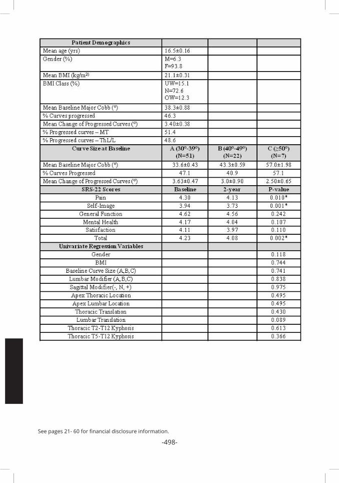
-498-
See pages 21- 60 for financial disclosure information.
-499-
♦ Indicates those faculty presentations in which the FDA has not cleared the drug and/or medical device for the use described (i.e., the drug or medical device is being discussed for an “off label” use). For full information, refer to page 14.
e-Poster #72 POSNA-2016 Normal Human Spine Growth During Childhood and Adolescence to Maturity with Prediction of Final Spine Height Developed from a Longitudinal Cohort of Children Followed Through Growth Completion
James O. Sanders, MD; Lauren E. Karbach, MD; Thomas Osinski, BS; Xiang Lu, MA; Xing Qiu, PhD; Raymond W. Liu, MD; Daniel R. Cooperman, MD University of Rochester and the Golisano Children’s Hospital at Strong, Rochester, New York
Purpose: Spine growth prediction is important for treating young children with spinal deformities. Cross sectional growth studies are inherently limited because the adolescent growth spurt, a time of marked change with variable timing between children is blunted failing to show the marked individual changes. This study uses longitudinal data to determine multipliers of spinal height during childhood and adolescence.
Methods: From a longitudinal cohort of normal children, we identified those completing growth. We normalized heights and spinal lengths as percentages of final at maturity and compared them to PHV timing and age. The subjects’ anthropometrics of standing pubic symphyseal, manubrial notch, and auditory meatus heights were correlated with individual simultaneous radiographs to C1, T1 and S1 to identify the distance from C1 to S1, and T1 to both C1 and S1. Multipliers were calculated as the reciprocal of the percentage remaining growth and these were then compared to both age and PGA.
Results: 54 subjects had completed their growth at the study terminus (35 f, 19m). The distribution of the multipliers was much more widely distributed by chronological age than to PHV timing. Boys and girls had identical multipliers compared to the PHV. For both sexes, growth was negligible at PHV + 3 yrs. At PHV, height was consistently at 90% of final height (multiplier 1/90%= 1.11) while spinal length was 85% of final (multiplier 1/85%= 1.18). During childhood, spinal growth is 1.5cm/year but increase to 2.5 or even up to 3cm/yr during the growth spurt. C1-T1 was 22.7% and T1-S1 77.3% of C1-S1 respectively. Spinal height multipliers are shown in the figure.
Conclusion: 1) Timing relative to the PHV provides reliable multipliers for future spinal growth while chronological age has limited usefulness near adolescence.2) The spine grows 1.5 cm/year during childhood but increases to 2.5cm/yr during the growth spurt.3) When the child is 90% of their final height the spine is only 85% of its final height. Significance: The spine grows more rapidly during the growth spurt than has been identified from cross sectional studies. This study justifies the use of multipliers for spinal growth relative to the PHV but not to age near the adolescent growth spurt. Supplementing this information with detailed cross sectional information should provide high quality descriptions of spinal growth for prediction of future growth to assist with growth modification and prediction of future lung volume.
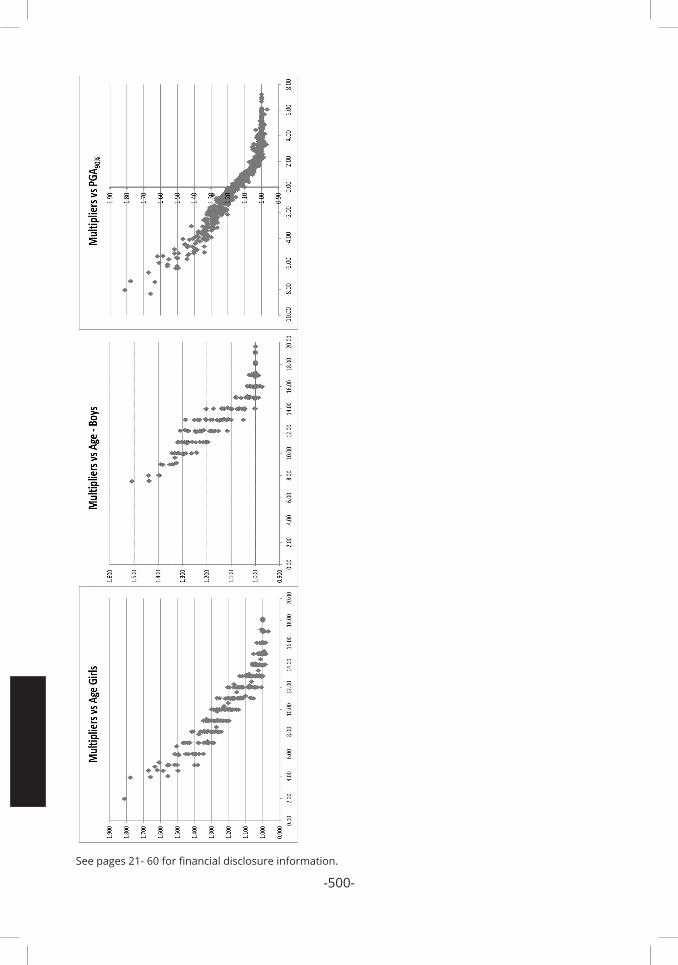
-500-
See pages 21- 60 for financial disclosure information.

-501-
♦ Indicates those faculty presentations in which the FDA has not cleared the drug and/or medical device for the use described (i.e., the drug or medical device is being discussed for an “off label” use). For full information, refer to page 14.
e-Poster #73 POSNA-2016 Femoral Neck Aspiration Aids in the Diagnosis of Osteomyelitis in Children with Septic Arthritis of the Hip
Jedidiah E. Schlung, BS; Tracey P. Bastrom, MA; Peter O. Newton, MD; Scott J. Mubarak, MD; Vidyadhar S.V. Upasani, MD Rady Children’s Hospital, San Diego, California
Purpose: The role of femoral neck aspiration (FA) in the treatment of septic arthritis of the hip (SAH) is controversial. The purpose of this study was to determine if FA conducted concomitantly with irrigation and debridement (I&D) of the septic hip aids in identification of the organism involved, alters the treatment plan, or if the risks of the procedure outweigh its potential benefit. We also analyzed whether concurrent magnetic resonance imaging (MRI) could obviate the need for FA.
Methods: After obtaining IRB approval we performed a retrospective review of all patients with SAH surgically treated at a single institution between January 2003 and June 2014. 52 patients had culture positive septic arthritis and 32 patients had intra-articular white blood cell counts greater than 50,000 with negative cultures. 23 out of these 84 patients (27%) were diagnosed with concomitant femoral osteomyelitis. Demographic and clinical data were recorded for each patient. The sensitivity and specificity of FA and MRI for diagnosing osteomyelitis was determined.
Results: 31 patients (37%) had FA performed at the time of the hip I&D, resulting in positive cultures in 17 patients (55%). All of these patients also had either a positive concurrent blood or joint fluid culture. 54 patients (64%) had a pre-operative MRI (Figure). The pre-op MRI failed to identify femoral osteomyelitis in 10 patients. FA was performed in 6 of these 10 patients resulting in a positive culture and appropriate management in all 6. Delayed diagnosis in the remaining 4 patients resulted in the need for re-operation in all 4 patients, femoral neck fracture and avascular necrosis of the femoral head in one patient and extensive femoral osteomyelitis and necrosis of the bone in a second patient. No complications associated with FA were identified. FA was found to have a sensitivity of 89% and specificity of 100% for osteomyelitis, while MRI had a sensitivity of 40% and specificity of 95%.
Conclusion: In this series, FA did not aid in identification of the infectious organism if blood and joint cultures were obtained, however, it did aid in the diagnosis of concomitant osteomyelitis when treating children with SAH, especially in patients with negative MRI findings for femoral osteomyelitis.
Significance: We recommend femoral neck aspiration be performed at the time of septic hip I&D as the sequelae of missed or inappropriately treated osteomyelitis outweigh the risks of the aspiration procedure.
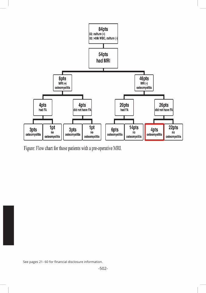
-502-
See pages 21- 60 for financial disclosure information.
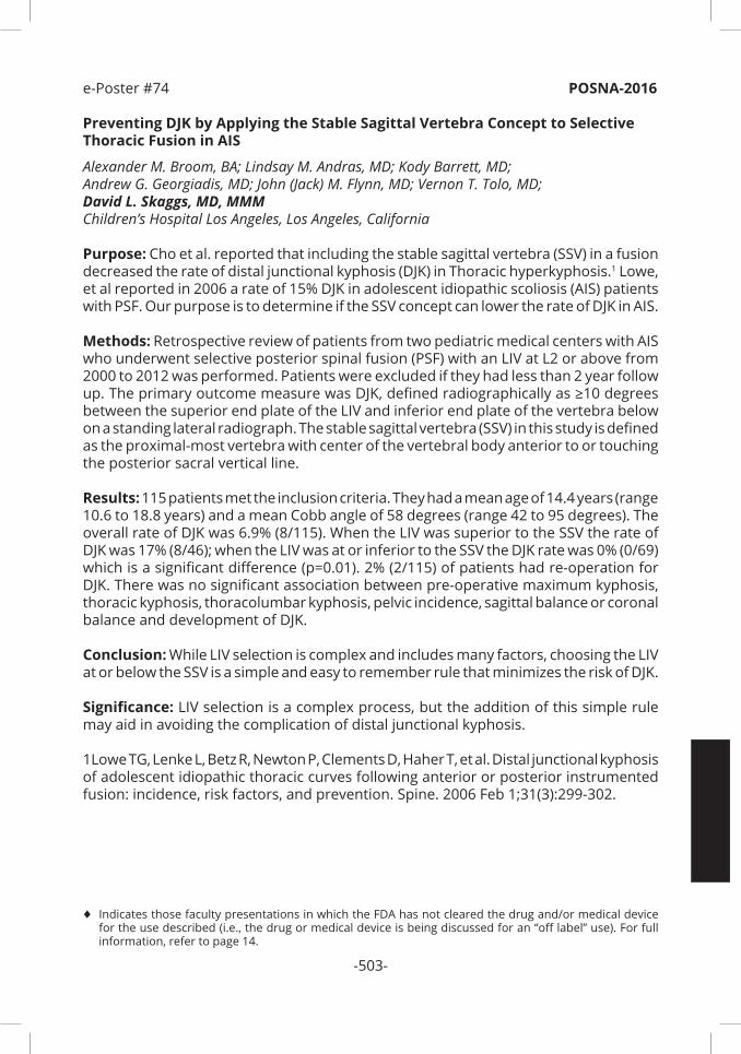
-503-
♦ Indicates those faculty presentations in which the FDA has not cleared the drug and/or medical device for the use described (i.e., the drug or medical device is being discussed for an “off label” use). For full information, refer to page 14.
e-Poster #74 POSNA-2016 Preventing DJK by Applying the Stable Sagittal Vertebra Concept to Selective Thoracic Fusion in AIS
Alexander M. Broom, BA; Lindsay M. Andras, MD; Kody Barrett, MD; Andrew G. Georgiadis, MD; John (Jack) M. Flynn, MD; Vernon T. Tolo, MD; David L. Skaggs, MD, MMM Children’s Hospital Los Angeles, Los Angeles, California
Purpose: Cho et al. reported that including the stable sagittal vertebra (SSV) in a fusion decreased the rate of distal junctional kyphosis (DJK) in Thoracic hyperkyphosis.1 Lowe, et al reported in 2006 a rate of 15% DJK in adolescent idiopathic scoliosis (AIS) patients with PSF. Our purpose is to determine if the SSV concept can lower the rate of DJK in AIS.
Methods: Retrospective review of patients from two pediatric medical centers with AIS who underwent selective posterior spinal fusion (PSF) with an LIV at L2 or above from 2000 to 2012 was performed. Patients were excluded if they had less than 2 year follow up. The primary outcome measure was DJK, defined radiographically as ≥10 degrees between the superior end plate of the LIV and inferior end plate of the vertebra below on a standing lateral radiograph. The stable sagittal vertebra (SSV) in this study is defined as the proximal-most vertebra with center of the vertebral body anterior to or touching the posterior sacral vertical line.
Results: 115 patients met the inclusion criteria. They had a mean age of 14.4 years (range 10.6 to 18.8 years) and a mean Cobb angle of 58 degrees (range 42 to 95 degrees). The overall rate of DJK was 6.9% (8/115). When the LIV was superior to the SSV the rate of DJK was 17% (8/46); when the LIV was at or inferior to the SSV the DJK rate was 0% (0/69) which is a significant difference (p=0.01). 2% (2/115) of patients had re-operation for DJK. There was no significant association between pre-operative maximum kyphosis, thoracic kyphosis, thoracolumbar kyphosis, pelvic incidence, sagittal balance or coronal balance and development of DJK.
Conclusion: While LIV selection is complex and includes many factors, choosing the LIV at or below the SSV is a simple and easy to remember rule that minimizes the risk of DJK.
Significance: LIV selection is a complex process, but the addition of this simple rule may aid in avoiding the complication of distal junctional kyphosis. 1Lowe TG, Lenke L, Betz R, Newton P, Clements D, Haher T, et al. Distal junctional kyphosis of adolescent idiopathic thoracic curves following anterior or posterior instrumented fusion: incidence, risk factors, and prevention. Spine. 2006 Feb 1;31(3):299-302.
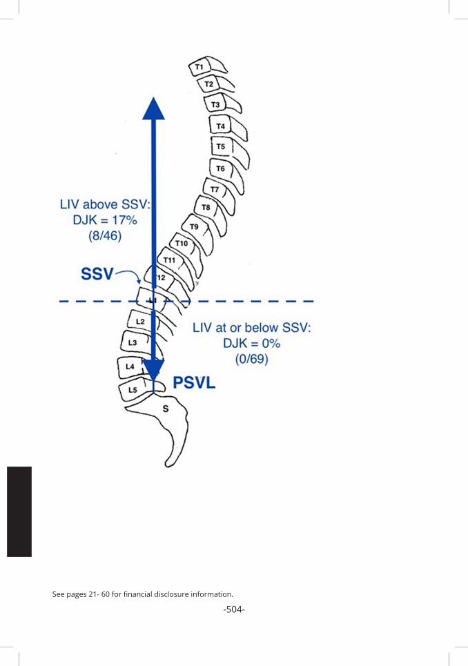
-504-
See pages 21- 60 for financial disclosure information.

-505-
♦ Indicates those faculty presentations in which the FDA has not cleared the drug and/or medical device for the use described (i.e., the drug or medical device is being discussed for an “off label” use). For full information, refer to page 14.
e-Poster #75 POSNA-2016 Restoring Sagittal Balance Improves Clinical Outcomes for Cerebral Palsy Patients with Spinal Deformity
Kushagra Verma, MD, MS; Suken A. Shah, MD; Firoz Miyanji, MD; Peter O. Newton, MD; Burt Yaszay, MD; Maty Petcharaporn, BS; Tracey P. Bastrom, MA; Unni G. Narayanan, MBBS, MSc, FRCSC; Paul D. Sponseller, MD Nemours / Alfred I. duPont Hospital for Children, Wilmington, Delaware
Purpose: A positive sagittal balance (>5 cm) has been associated with adverse outcomes in adult deformity patients but no such guidelines exist for primarily seated patients. This study evaluated the association between restored sagittal balance and clinical outcomes in cerebral palsy (CP).
Methods: From a multicenter database, we retrospectively reviewed prospectively collected data from patients with CP who underwent PSF to the pelvis. Demographic, clinical outcome, sitting tolerance, and seated radiographic data were studied. Using Pearson’s correlation, we evaluated the relationship between multiple radiographic measurements and clinical outcomes using the CPCHILD Questionnaire – 6 domains, each scored 0 to 100 (best).
Results: 93 patients (age 13.7±2.7 yr) who underwent PSF (2008 – 2011) with 2-year follow-up were included. Mean radiographic parameters: major Cobb (29±16°), pelvic obliquity (8.1±6.8°), T2-T12 kyphosis (36±16°), T12-S1 lordosis (-53±17°), and sagittal vertical axis (SVA, -0.3±6.6cm). Clinical outcomes: daily living (43±18), transfers/mobility (43±18), comfort/emotions (80±18), social (55±29), health (61±19), overall function (70± 22), total score (56±15), and sitting tolerance (294±166min). There was a linear correlation between SVA and transfers (r = 0.27, p = 0.026), social (0.24/0.38), overall function (0.25/0.30), and total score (0.30/0.015). No correlation was found with other measurements. Given that SVA may vary with patient position, pelvic incidence (PI), sacral slope (SS), and T1 Pelvic Angle (T1PA – see figure) were measured on a subset of patients. Higher PI improved sitting (0.40/0.035), while higher T1PA improved transferring (0.52/0.006), overall function (0.51/0.006), and total score (0.39/0.043) with a trend towards better health (0.33/0.091).
Conclusion: Restoration of sagittal balance to a neutral or slightly positive SVA and T1PA are associated with better transferring, function, social interaction, and total care-giver satisfaction in CP. Beyond a certain threshold, however, a positive SVA or T1PA is likely detrimental. T1PA can be evaluated intra-operatively in the prone position to improve the clinical outcomes of these children.
Significance: For nonambulatory children with cerebral palsy, restoration of sagittal balance to neutral or slightly positive sagittal vertical axis and T1 pelvic angle correlated with improved clinical outcomes.
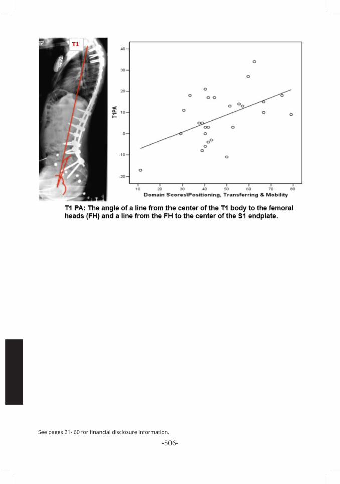
-506-
See pages 21- 60 for financial disclosure information.
-507-
♦ Indicates those faculty presentations in which the FDA has not cleared the drug and/or medical device for the use described (i.e., the drug or medical device is being discussed for an “off label” use). For full information, refer to page 14.
e-Poster #76 POSNA-2016 Radiographic Outcomes of Shilla Growth Guidance System and Traditional Growing Rods through Definitive Treatment
Scott J. Luhmann, MD; June C. Smith, MPH; Anna McClung, RN, BSN; Frances L. McCullough, RNP, MNSc; Richard E. McCarthy, MD; George H. Thompson, MD; Growing Spine Study Group St. Louis Children’s Hospital, Saint Louis, Missouri
Purpose: The purpose of this study was to compare the radiographic outcomes of patients who had undergone the Shilla Growth Guidance System and traditional Growing Rod (GR) treatment for management of early-onset scoliosis (EOS) through definitive treatment.
Methods: A multicenter EOS database was queried to identify patients who met the following criteria: surgical treatment with Shilla or GR and had undergone definitive treatment of the spinal deformity. The patients were matched by age, pre-operative curve magnitude, and diagnosis. The study population consisted of 36 patients (18 in each group) whose mean age at initial surgery was: Shilla 7.9 y and GR 7.7y (NS). Length of follow-up after initial surgery was 6.1 y for Shilla and 7.4 y for GR (NS). Definitive treatment was posterior spinal fusion (15 Shilla, 17 GR), implant removal (3 Shilla), or completion of lengthenings (1 GR).
Results: The overall mean number of surgeries was 3.1 (range: 1-7) for Shilla and 9.3 (range: 4-24) for GR (including 5.8 lengthenings). Total complications were 20 for Shilla and 26 for GR; the most common complication in both groups were implant-related (Shilla 13; GR 11), followed by infection (Shilla 5, GR 7). The pre-operative curve was 58 degrees for Shilla and 63 degrees for GR (NS). After index surgery the major curve decreased to 21 degrees (-37 degrees) for Shilla and 37 (-26 degrees) for GR (p<0.05). At last follow-up the major curve was 30 degrees (-28 degrees;48%) for Shilla and 34 degrees (-29 degrees; 46%) for GR (NS). The initial T1-S1 length for Shilla was 316 mm and GR 292 mm; at last follow-up Shilla was 406 (90 mm increase) and GR was 378 (86 mm increase) (NS). The initial T1-T12 length for Shilla was 195 mm and GR 185 mm; at last follow-up Shilla was 247 mm (52 mm increase) and GR was 233 mm (48 mm increase).
Conclusion: In this analysis of EOS patients who had completed scoliosis treatment, the final radiographic outcomes (and changes) and complications (implant-related and infection) between the Shilla and GR groups, were not statistically different. The main differences between the groups was the 3-fold difference in overall surgeries between the two groups.
Significance: Shilla was similar in T1-T12 height, T1-S1 height, curve correction with 1/3 the overall surgeries when compared to GR.
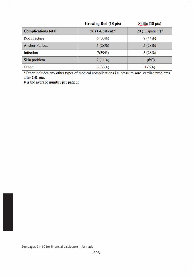
-508-
See pages 21- 60 for financial disclosure information.

-509-
♦ Indicates those faculty presentations in which the FDA has not cleared the drug and/or medical device for the use described (i.e., the drug or medical device is being discussed for an “off label” use). For full information, refer to page 14.
e-Poster #77 POSNA-2016 Radiographic Assessment of Angular Correction of Genu Varum and Genu Valgus Using Guided Growth: The Correlation Between Screw Divergence and Change in Anatomic Alignment
Weilong Jeffrey Shi, BS; Kyle R. Sweeney, MD; Michael B. Gottschalk, MD; Robert W. Bruce, MD; Nicholas D. Fletcher, MD Emory University, Atlanta, Georgia
Purpose: Routine assessment of angular change following guided growth surgery typically requires long standing radiographs of the lower extremities which require special techniques and subject the patient to greater radiation doses than does a single AP radiograph of the knee. Our study sought to determine whether we could predict the change in angular deformity of the legs using divergence of screws in a guided growth construct as a surrogate for angular change in the lower extremities.
Methods: We retrospectively reviewed all patients with genu varum/valgum treated with hemiepiphysiodesis at a single institution within the last fifteen years. Initial anatomic alignment of the femur and tibia as well as the initial divergence of hemiepiphysiodesis screws (SD) were measured on standing long cassette radiographs of the lower extremities from the hips to ankles with the patellas facing anteriorly. Anatomic alignment measurements included anatomic lateral distal femoral angle (ALDFA) and anatomic femoral-tibial angle (FTA). We then looked at these measurements again on follow-up visits in order to determine a correlation between and both ΔFTA and ΔALDFA.
Results: 80 radiographs allowing for comparisons between ΔFTA and ΔSD and 107 radiographs for comparison between ΔSD and ΔALDFA were reviewed. Each degree change in SD results in 1.80° of change in FTA and 2.11° of change in ALDFA. We were able to predict the change in FTA by the equation (0.41xΔSD)+1.39 . The coefficient of determination (R2) value of ΔFTA vs ΔSD was 0.50.The ΔALDFA is correlated to ΔSD by the equation (0.27x ΔSD)+1.84 with a R2 of 0.31. The correlation as estimated by the Restricted Maximum Likelihood Method (REML) shows that ΔFTA and ΔSD had a correlation of 0.68 (95% CI 0.54-0.78). ΔALDFA and ΔSD had a correlation of 0.56 (95% CI 0.42-0.68). Finally, ΔALDFA and ΔFTA had a correlation of 0.71 (95%CI 0.58 -0.80).
Conclusion: We were able to predict the change in both FTA and ALDFA using the SD seen on an AP radiograph of the lower extremities. Each degree change in SD results in 1.80° of change in FTA and 2.11° of change in ALDFA.
Significance: Each 1° change in SD results in approximately 2° change in both ALDFA and FTA. Given the increased radiation dose of full length standing radiographic films, this relationship could allow the use of AP knee radiographs to replace full length films at some follow-up visits.
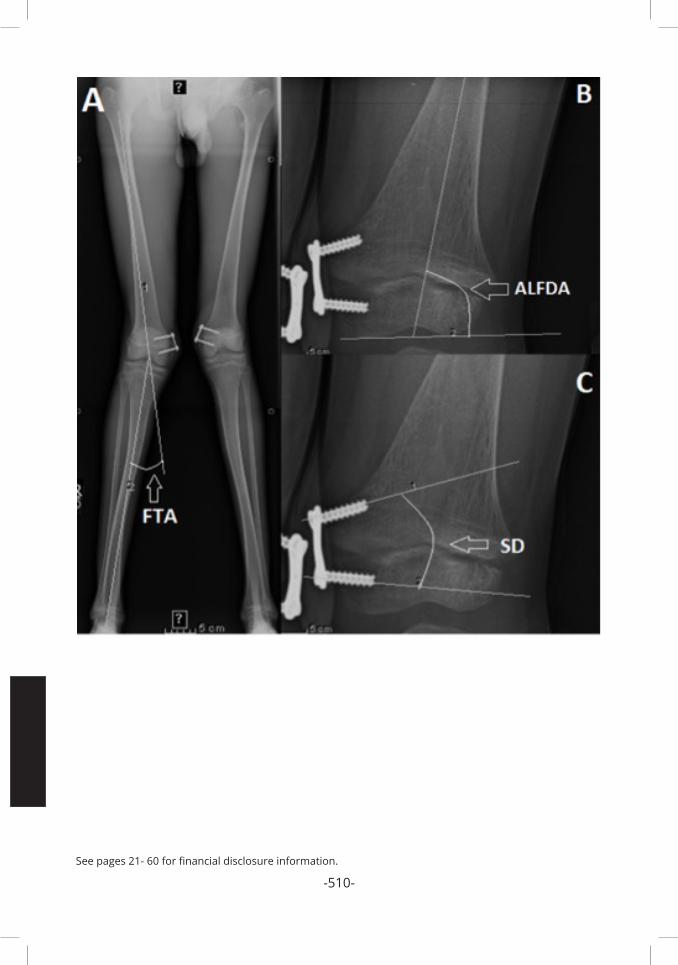
-510-
See pages 21- 60 for financial disclosure information.
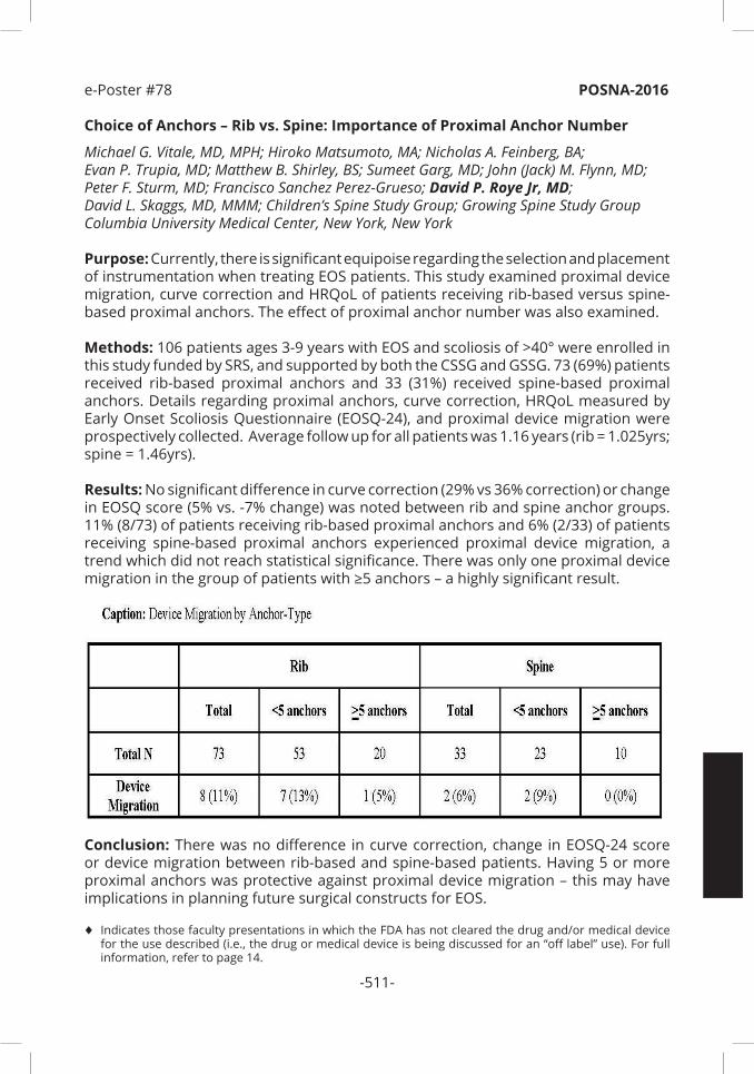
-511-
♦ Indicates those faculty presentations in which the FDA has not cleared the drug and/or medical device for the use described (i.e., the drug or medical device is being discussed for an “off label” use). For full information, refer to page 14.
e-Poster #78 POSNA-2016 Choice of Anchors – Rib vs. Spine: Importance of Proximal Anchor Number
Michael G. Vitale, MD, MPH; Hiroko Matsumoto, MA; Nicholas A. Feinberg, BA; Evan P. Trupia, MD; Matthew B. Shirley, BS; Sumeet Garg, MD; John (Jack) M. Flynn, MD; Peter F. Sturm, MD; Francisco Sanchez Perez-Grueso; David P. Roye Jr, MD; David L. Skaggs, MD, MMM; Children’s Spine Study Group; Growing Spine Study Group Columbia University Medical Center, New York, New York
Purpose: Currently, there is significant equipoise regarding the selection and placement of instrumentation when treating EOS patients. This study examined proximal device migration, curve correction and HRQoL of patients receiving rib-based versus spine-based proximal anchors. The effect of proximal anchor number was also examined.
Methods: 106 patients ages 3-9 years with EOS and scoliosis of >40° were enrolled in this study funded by SRS, and supported by both the CSSG and GSSG. 73 (69%) patients received rib-based proximal anchors and 33 (31%) received spine-based proximal anchors. Details regarding proximal anchors, curve correction, HRQoL measured by Early Onset Scoliosis Questionnaire (EOSQ-24), and proximal device migration were prospectively collected. Average follow up for all patients was 1.16 years (rib = 1.025yrs; spine = 1.46yrs).
Results: No significant difference in curve correction (29% vs 36% correction) or change in EOSQ score (5% vs. -7% change) was noted between rib and spine anchor groups. 11% (8/73) of patients receiving rib-based proximal anchors and 6% (2/33) of patients receiving spine-based proximal anchors experienced proximal device migration, a trend which did not reach statistical significance. There was only one proximal device migration in the group of patients with ≥5 anchors – a highly significant result.
Conclusion: There was no difference in curve correction, change in EOSQ-24 score or device migration between rib-based and spine-based patients. Having 5 or more proximal anchors was protective against proximal device migration – this may have implications in planning future surgical constructs for EOS.
-512-
See pages 21- 60 for financial disclosure information.
Significance: There was no significant difference in curve correction, health related quality of life (HRQoL) and device migration between patients who received rib-and spine-based proximal anchors. However, lower proximal anchor number was associated with an increased risk of proximal device migration in both patient groups.
-513-
♦ Indicates those faculty presentations in which the FDA has not cleared the drug and/or medical device for the use described (i.e., the drug or medical device is being discussed for an “off label” use). For full information, refer to page 14.
e-Poster #79 POSNA-2016 Improving The Handoff Process Between Operating Room and Pediatric Intensive Care Unit
Stephen J. Gleich, MD; Michael E. Nemergut, MD, PhD; Anthony A. Stans, MD; Dawit T. Haile, MD; Angela L. Heinrich, RN; Christopher L. Bosley, RRT-NPS; James W. Ward, MBA Mayo Clinic, Rochester, Minnesota
Purpose: This Six Sigma project was initiated to improve patient care during the transfer of patients from the OR to the ICU. Medical errors are responsible for patient harm and billions of dollars in increased health care spending. Miscommunication is a major contributor to these errors, with handoffs being identified as a particularly vulnerable period. At our institution, surgical patients with scheduled admissions to the ICU are first recovered in the PACU. With this process, multiple, unstructured and individual handoffs occur in parallel between providers, which may lead to communication errors, differential information sharing, content variability, care delays and inefficiency.
Methods: A multidisciplinary Q.I. project was initiated with input from the ICU, anesthesia and surgical services. A series of PDSA cycles were conducted, which began by defining the current process via direct observation and value stream mapping of orthopedic and neurosurgical patients. A new process was then introduced, including direct transfer of the patient to the ICU and a single, structured, bedside report between all care providers. A standardized handoff tool was implemented. We used process times, wait times and information content as process measures and handoff errors as outcome measures. A 10-point satisfaction score was also measured.
Results: Following implementation of the new transfer process, the average wait time decreased by 58 minutes, process time decreased by 9 minutes, and lead time decreased by 66.5 minutes. The handoff error rate decreased by 1.3 errors/patient and first time quality rate increased by 67%. Staff satisfaction improved from 48% to 73%. By elimination of the PACU stay, the costs involved in admission to the PACU were deferred.
Conclusion: A single, multidisciplinary bedside handoff process between the OR and ICU leads to cost and time savings. By elimination of redundant, non-value added processes, less opportunity for medical errors occurred, with substantial improvements in first-time quality. Such a process can be successfully attained while affecting staff satisfaction positively.
Significance: A structured, standardized handoff process between the operating room and ICU reduces medical errors, reduces wait times, improves staff satisfaction and improves quality.
-514-
See pages 21- 60 for financial disclosure information.
e-Poster #80 POSNA-2016 2D versus 3D Measurements of AIS Spinal Parameters in Clinical Care
Saba Pasha, PhD; Patrick J. Cahill, MD; John (Jack) M. Flynn, MD The Children’s Hospital of Philadelphia, Philadelphia, Pennsylvania
Purpose: Bi-planar radiography images of the spine are considered the gold standard for clinical evaluation of adolescent idiopathic scoliosis (AIS). Although AIS is known to impact the 3D alignment and orientation of the spine and pelvis the discrepancy between the 2D and 3D measurements in the presence of the 3D deformity of the spine, 3D vertebral rotation, and patient rotation with respect to the X-ray scanner has not been evaluated. This study investigated the correlation between the off-plane rotation of the spinal curve and error between the 2D and 3D measurements of the scoliotic spinal parameters.
Methods: A total number of 36 AIS patients were prospectively recruited. Posterior-anterior and lateral spinal X-rays were registered in EOS. The 3D models of the spine, pelvis, and femoral heads were reconstructed using SterEOS software. Four independent observers measured thoracic and lumbar Cobb angles, kyphosis, and lordosis on the 2D posterior-anterior and lateral X-ray images. These parameters were also measured in radio plane (in the natural patient standing posture with respect to the X-ray scanners) and patient plane (femoral head rotated and placed in frontal plane) using the 3D models. Thoracic and lumbar apical vertebral rotation and the plane of maximum curvature were calculated using a MATLAB code. A test of ANOVA compared the 2D, radio plane, and patient plane measurements and the agreements between the measurements were studied.
Results: The intraclass coefficient correlation was excellent for the 2D measurements (ICC=0.93, p<0.05). The differences between the 2D and 3D kyphosis was significant and increased as a function of kyphosis angle (p<0.05) (figure 1). The differences between the 2D and patient plane 3D measurements of the lumbar lordosis was significantly related to the magnitude of the lumbar apical axial rotation and pelvic rotation (r=0.64, r=-0.54, p<0.05).
Conclusion: A clinically and statistically significant difference was observed between the 2D and 3D sagittal spinal measurements. The differences between the 2D and 3D measurements were related to the spine and pelvic axial plane rotation and the curve severity in the sagittal plane.
Significance: The impact of the off-plane deformity of the scoliotic spine on the differences between the 2D and 3D clinical measurements of the spine was highlighted. The differences between 2D and 3D measurements should be acknowledged and not used interchangeably.
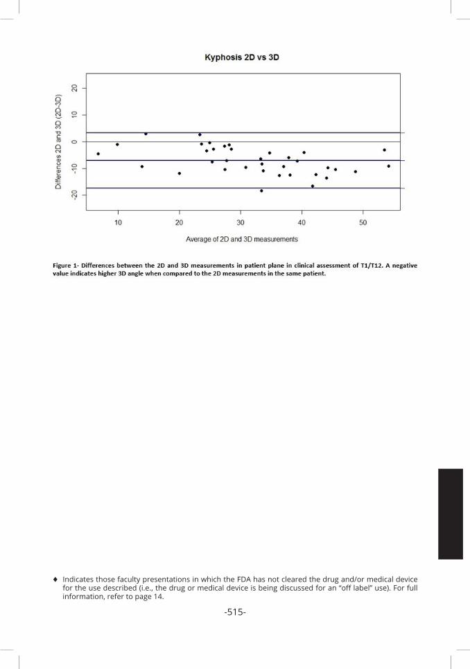
-515-
♦ Indicates those faculty presentations in which the FDA has not cleared the drug and/or medical device for the use described (i.e., the drug or medical device is being discussed for an “off label” use). For full information, refer to page 14.
-516-
See pages 21- 60 for financial disclosure information.
e-Poster #81 POSNA-2016 Spica Casting for Pediatric Femur Fractures: Treatment Practice and Value Outcomes
Caroline V. Hornberger, PA-C; Mary K. Riordan, BA; William A. Phillips, MD Texas Children’s Hospital, Houston, Texas
Purpose: Pediatric femur fractures typically have good outcomes. Lacking guidelines, orthopedic surgeons take individualized approaches to most orthopaedic problems. United States healthcare is moving towards cost containment and increasing high value healthcare delivery. There is a need for further examination of resource utilization that leads to similar outcomes.
Methods: We performed an IRB-approved retrospective review of patients aged one to five years who presented to a Level I pediatric trauma center with a closed low velocity diaphyseal femur fractures treated with spica casting in the operating room between 2013 and 2014. To assess variations in management, the treatment course of a maximum of five patients for each of 11 fellowship trained pediatric orthopedic surgeons was analyzed and resources utilized in each child’s care were documented. Results: 48 patients were identified. The mean age was 2.8 years. 46 of patients (96%) had acceptable outcomes (alignment & healing). 17% had no intra-operative imaging performed. 50% of children had three or fewer clinic visits. 62% had three or fewer x-rays in clinic. 78% had their cast removed on the first or second post-op clinic visit. The average number of clinic visits by surgeon ranged from 1.8 to 4.7. The average number of clinic x-rays ordered by surgeon ranged from 1.5 to 4.7. Analysis by surgeons’ years of experience showed that intra-operative fluoroscopy was not used as often (p < 0.001) by more senior surgeons and they recommended a later first post-op visit (p= 0.021).
Conclusion: Differences in resource utilization were identified in the management of femoral fractures treated by spica casting. Management differed in the number of outpatient visits and amount of imaging received.
Significance: Varied amounts of resources were directed toward the management of pediatric femoral fractures treated by spica casting without similar variation in patient outcomes. The findings of this study suggest a standardized approach with careful resource utilization in the management of femoral fractures treated with spica casting may improve health care value while producing good outcomes.

-517-
♦ Indicates those faculty presentations in which the FDA has not cleared the drug and/or medical device for the use described (i.e., the drug or medical device is being discussed for an “off label” use). For full information, refer to page 14.
e-Poster #82 POSNA-2016 Selective Thoracic Fusion of a Left Decompensated Main Thoracic Curve: Proceeding with Caution
Burt Yaszay, MD; Tracey P. Bastrom, MA; Carrie Bartley, MA; Suken A. Shah, MD; Baron Lonner, MD; Jahangir Asghar, MD; Firoz Miyanji, MD; Patrick J. Cahill, MD; Peter O. Newton, MD Rady Children’s Hospital, San Diego, California
Purpose: In an attempt to minimize decompensation after fusion in patients with thoracic scoliosis who are coronally decompensated to the left preoperatively, some surgeons recommend fusing both curves while others recommend coupling a selective thoracic fusion (STF) with under-correction of the thoracic curve. The purpose of this study was to evaluate the risks and strategies for selectively fusing a patient decompensated to the left preoperatively.
Methods: A prospective, multicenter AIS database was queried for subjects with minimum 2yr f/u who underwent STF (defined as having an LIV of L1 or proximal) of right Lenke 1-4C curves that preoperatively were coronally decompensated to the left. Rates of postop coronal decompensation (C7-CSVL) >2cm following different levels of LIV were compared. Correction of the thoracic curve and spontaneous lumbar curve correction (SLCC) were compared between patients who remained decompensated postop and those who were balanced.
Results: Thirty-nine patients were identified with avg thoracic and lumbar Cobb of 54° and 41°. Mean decompensation was 2.4 ± 0.9cm preop and 1.9 ± 1.1cm postop. One was fused short of stable, 19 were fused to stable, and 19 were fused past stable. No patients were fused short of the Cobb end vertebra. Postop incidence of continued decompensation was 41%. The rate of postop decompensation between the stable (37%) and the past stable groups (47%) was not significantly different (p=0.12). When comparing LIV to neutral vertebra, there was no difference whether instrumented to neutral or beyond (p=0.24). Patients who were balanced postop had a higher percent thoracic curve correction (61 vs 50%, p=0.017) and thus higher SLCC (47 vs 32%, p=0.004).
Conclusion: When performing a STF for a patient decompensated to the left, there was a high rate (41%) of continued postop decompensation. Selecting an LIV relative to stable and neutral vertebrae did not significantly affect this risk. However, rather than undercorrecting the main thoracic curve, this data suggests that greater correction of the thoracic curve reduces the risk of postop decompensation.
Significance: STF for right main thoracic curves decompensated to the left resulted in a high rate of postop decompensation (41%). Patients balanced postop had greater thoracic curve correction and higher SLCC. These findings may better prepare surgeons and patients for the possibility of continued decompensation after surgery.
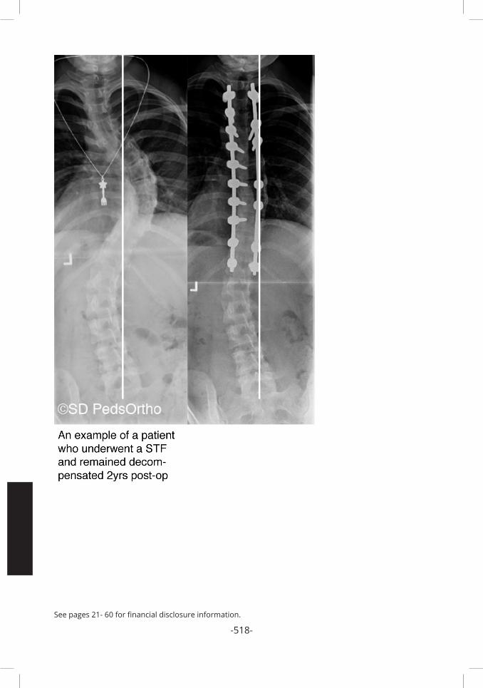
-518-
See pages 21- 60 for financial disclosure information.
-519-
♦ Indicates those faculty presentations in which the FDA has not cleared the drug and/or medical device for the use described (i.e., the drug or medical device is being discussed for an “off label” use). For full information, refer to page 14.
e-Poster #83 POSNA-2016 Multicenter Evaluation of Spinal Deformity in Skeletal Dysplasias with Growing Rod Systems
Klane K. White, MD, MS; Viviana Bompadre, PhD; Gregory Redding, MD; Walter F. Krengel III, MD; Suken A. Shah, MD; William G. Mackenzie, MD Seattle Children’s Hospital, Seattle, Washington LOE-Therapeutic-Level III
Purpose: Severe, early-onset spinal deformity is common in patients with skeletal dysplasias. These deformities often present at young ages and are often associated with significant pulmonary dysfunction. Given the limited growth potential in late childhood relative to other children with spinal deformities, the appropriateness of treatment with growth friendly constructs is not clear. The objective of this study is to define the population of patients with skeletal dysplasias that have been treated with growing rod systems, the indications used for treatment and the outcomes of these interventions.
Methods: A retrospective, multicenter comparative cohort study was performed. Using CSSG and GSSG databases, 23 patients identified to have a skeletal dysplasia (SKD) were evaluated for diagnosis, age at treatment, gender and type of growing rod construct (spine vs. rib constructs). Radiographic parameters including maximum coronal and saggital Cobb angle with levels, T1-S1 height, T1-T12 height were measured pre-implantation, immediate post implantation and at most recent follow-up. These patients were matched by age and construct type with a control cohort (CON) of similarly treated patients with early onset scoliosis (EOS) without skeletal dysplasia. To compare the groups, we used the Wilcoxon matched-pairs signed-ranks test. We compared changes in T1-S1, T1-T12, Cobb angle and kyphosis from time to implant, post op and last follow-up using repeated measures ANOVA.
Results: The SKD and CON groups were significantly different in T1-T12 length (12.8 vs. 15.2 cm, p=0.01) and T1-S1 (21.2 vs 24.5 cm, p=0.05) at implantation. The groups did not significantly differ in major Cobb angle while kyphosis tended to be more severe in the SKD group (p=0.80 and 0.07, respectively). No significant difference in kyphosis between time of implant and last follow-up was observed in either group. Cobb angle significantly decreased (<0.01), while ΔT1-T12 (SKD – 3.2 cm, CON - 5 cm) and ΔT1-S1 (SKD – 6.3 cm, CON – 6.7 cm) significantly increased in both groups after implantation (p<0.01).
Conclusion: While patients with SKD start with shorter spine lengths, gains in spine length appear to be comparable to other forms of EOS.
Significance: The use of growth accommodating techniques for early onset spinal deformities in skeletal dysplasias has not been previously studied. We report the first comprehensive review of this topic. Growth friendly techniques are appropriate in this patient population.
-520-
See pages 21- 60 for financial disclosure information.
e-Poster #84 POSNA-2016 Enhancing the Economy of Surgery: Reducing Cost and Variation of Surgeon Preference Cards for Supracondylar Humerus Fractures
Thomas B. Bemenderfer, MD; Kelly L. Vanderhave, MD; Brian P. Scannell, MD; Virginia F. Casey, MD; Michael D. Paloski, DO; Christian Clark, MD; Brian K. Brighton, MD, MPH Carolinas Healthcare System/Levine Children’s Hospital, Charlotte, North Carolina
Purpose: Over the past several decades, fiscal responsibility in the healthcare industry has become a priority. There is increasing pressure from the healthcare macro-environment to address the economy of medicine, with a focus on careful utilization of finite resources. Physicians play a large role in resource utilization. Closed reduction and percutaneous pinning (CRPP) of a supracondylar humerus fracture is among the most common pediatric orthopaedic procedures at our hospital. The purpose of this study was to determine whether a single universal preference card could be agreed upon among surgeons and its impact on costs.
Methods: The surgeon preference cards for CRPP of the elbow for six pediatric orthopaedic surgeons were reviewed. We identified the costs of all items on the preference cards and determined what items were unique to specific physicians and what items were more universal. A survey was circulated to each physician to determine which items on the preference card were required versus discretionary. A cost-specific optimization of preference cards was performed based on surgeon feedback.
Results: On initial survey of the six pediatric orthopaedic surgeons in our health system performing this procedure, the average cost based on their preference cards ranged from $85.71 to $139.34 with an average cost of $99.86. Actual costs ranged from $140 to $321 with an average of $193.17. Based on the results of the survey, the single cost-optimized preference card allowed for removal of 4 sets of instruments, 2 pieces of equipment, and 45 supply items. The cost for the procedure based on this final preference card was approximately $28 which was a cost savings of 72%.
Conclusion: A single preference card that is uniform to all surgeons performing a common surgical procedure is possible and can reduce procedural costs to the patient. Significance: A cost effective approach to the utilization of instruments, equipment, medications/solutions, and supplies amongst surgeons and operating room personnel is imperative. Similar processes may be applied at a systems level that would result in healthcare savings.
-521-
♦ Indicates those faculty presentations in which the FDA has not cleared the drug and/or medical device for the use described (i.e., the drug or medical device is being discussed for an “off label” use). For full information, refer to page 14.
e-Poster #85 POSNA-2016 Age-Based Patellofemoral Morphology in the Immature Knee
Kevin E. Klingele, MD; Matthew C. Beran, MD Nationwide Children’s Hospital, Columbus, Ohio
Purpose: Patellar instability (PI) is a common cause of anterior knee pain and disability in the pediatric population. The use of patellofemoral measurements on MRI provides a quantitative means for PI assessment and has now become an important diagnostic tool, but these techniques largely rely upon adult standards. Our goal is to describe morphologic trends in the skeletally immature knee and to predict the age at which adult norms can reliably be used in the evaluation of the pediatric knee.
Methods: We retrospectively reviewed 144 normal knee MRIs in 133 skeletally immature patients that presented between 2002 and 2014. Patients were equally distributed by age and gender with ages ranging from 1-16. MRI exclusion criteria included: moderate to severe effusions, cartilaginous defects, patellofemoral abnormalities, ligamentous injury, neoplasms, infection, congenital disease, or arthritic changes. All 1 and 2 year olds were included due to lack of MRIs and only females younger than 15 were used to account for anticipated physeal closure. All measurements used cartilaginous landmarks and results were stratified based on age and gender. Each measurement was charted in a linear regression model or analyzed with Student’s t test.
Results: Each measurement can reliably be performed at all ages with good inter- and intraobserver reliability. All MR measurements were graphically represented in a linear regression model and are shown to approach adult norms with increasing age. The age at which there is no statistical difference between our pediatric patients and the adult norms is shown as the “regression cutoff” (Table 1). Further t-test analysis suggests a 2nd cutoff that serves as the age at which younger should not be compared to adult norms (Table 1).
Conclusion: The measurements commonly used to evaluate for patellar instability in the adult population are subject to considerable variation throughout skeletal maturation. Based on our analysis, children < 10 years of age should not be compared to adult standards. Conversely, children ≥ 10 appear to have reached near patellofemoral maturation and show consistent and progressive development of patellofemoral morphology with increasing age.
Significance: The ability to predict morphologic abnormalities in the first decade of life may lend to earlier surgical intervention or realignment procedures. Surgical outcomes may be augmented by remaining patellofemoral growth and remodeling, especially in those patients under 10 years of age.
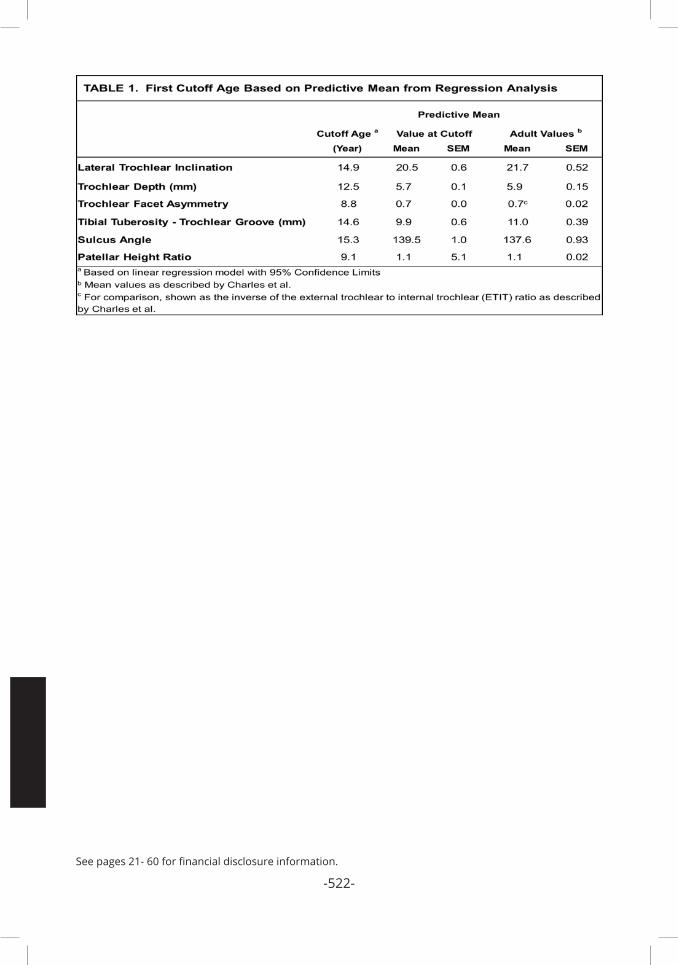
-522-
See pages 21- 60 for financial disclosure information.
-523-
♦ Indicates those faculty presentations in which the FDA has not cleared the drug and/or medical device for the use described (i.e., the drug or medical device is being discussed for an “off label” use). For full information, refer to page 14.
e-Poster #86 POSNA-2016 Implementation of a Dedicated Pediatric Orthopaedic Trauma Room
Gianna D. Katsaros, MS; Wendy J. Ramalingam, MD; Jaime R. Denning, MD; Patrick W. Whitlock, MD Cincinnati Children’s Hospital Medical Center, Cincinnati, Ohio
Purpose: A dedicated orthopaedic operating room (DOOR) is a fully staffed OR that remains available for non-emergent, orthopaedic trauma. Existing adult literature on DOORs have demonstrated improvements in OR efficiency, scheduling and patient outcomes, which reduces overall cost by decreasing length of hospital stay (LOS), procedure time, and perioperative complications. The purpose of this study is to examine the value of establishing a DOOR for urgent pediatric orthopaedic trauma at a Level I trauma center.
Methods: A retrospective review of pediatric patients with isolated, operative types II and III supracondylar humerus fractures was performed for two 1-year time periods before and after the implementation of DOOR at our institution on May 1, 2014. Patients with compartment syndrome, neurovascular compromise, or open fracture were excluded. Each group was stratified by OR start time into two groups: regular hours (7:01 am to 6:00 pm) and after hours (6:01 pm to 7:00 am). Records were then examined to determine procedure length, OR time, complication rate, LOS, and total cost. We also determined total time from admission to OR for all elbow fractures treated operatively prior to and after DOOR implementation.
Results: Before implementation of DOOR from May 1, 2012 – April 30, 2013, 110 pediatric supracondylar humerus fractures underwent operative intervention, 97 during regular and 13 after hours. After implementation of DOOR from May 1, 2014 to April 30, 2015, 99 pediatric supracondylar humerus fractures underwent operative intervention, 86 during regular and 13 after hours. There was no significant difference in regards to procedure length, OR time, complication rate, LOS or total cost between the regular and after hours procedures performed before and after implementation of DOOR. A trend towards decreased procedure time of after hours surgery was observed (43.2±8.3 min pre-DOOR vs. 31.7±14.3 min post-DOOR, p=0.09). There was a 50% decrease in cost variability of after hours surgery after DOOR implementation ($15,197.95±$4,532.54 pre-DOOR vs. $14,095.62±$2,136.30 post-DOOR, p=0.44). The mean time from admission to OR for all operative elbow fractures was reduced (14.7±17.3 hours pre-DOOR vs. 11.1±6.02 hours post-DOOR, p<0.01).
Conclusion: There was no increase in complication rate, LOS or total cost in operative, non-emergent pediatric supracondylar humerus fractures treated after the implementation of DOOR. A decrease in cost variability was observed. A significant decrease in time from admission to OR for all operatively treated elbow fractures was observed after the implementation of DOOR.
Significance: DOORs are efficient, safe and cost effective in the pediatric orthopaedic trauma setting.
-524-
See pages 21- 60 for financial disclosure information.
e-Poster #87 POSNA-2016 Symptomatic Leg Length Discrepancy following Open Reduction and Salter’s Innominate Osteotomy in DDH Children
Chii-Jeng Lin, MD, PhD; Ming-Tung Huang, MD; Nai-Wen Guo, PhD National Cheng Kung University, Tainan, Taiwan
Purpose: Leg length discrepancy (LLD) had been reported to be a potential morbidity of surgical treatment for developmental dysplasia of hip (DDH). Our research goal is to evaluate the prevalence of LLD in DDH patients treated with open reduction and Salter osteotomy.
Methods: From 1998 to 2012, 279 DDH children were treated with open reduction and Salter osteotomy while 233 children were included for study. The exclusion criteria were concomitant femoral osteotomies or bilateral cases. Among them, 31 cases (13%) reported symptomatic LLD due to self-consciousness of inequality. They were followed for at least 3 years, averaged 51.2 months (28 to 112 months). The differences in iliac height (IH), femoral and tibial lengths (FL and TL) and apparent length (AL=IH+FL+TL) were analyzed from standing scanogram. Besides, neck shaft angle (NSA) and vertical offset (VOS, defined as the vertical contribution of leg length) were also evaluated for the role of femoral neck. All measurements were repeated yearly to determine the annual increment.
Results: All the hips were well contained during follow-up while seven of them developed various extent of coxa magna. The length was significantly longer in DDH side in IH (8.2 mm, p<0.001), FL (8.9 mm, p<0.001) and AL (17.9 mm, p<0.001) but not in TL. Both NSA (148 vs. 143 degrees, p<0.01) and VOS (22.8 vs. 19.6 mm, p<0.001) were also significantly higher in the DDH side. The femoral neck contributed to 36 % (3.2 mm, p<0.001) of FL difference. The annual increment of leg length difference increased gradually until age 6 then the changes seemed to stabilize.
Conclusion: There were 13% children complained symptomatic LLD after open reduction and Salter osteotomy for DDH. The discrepancy of length came not only from the pelvic osteotomy but also from the femur while the neck contributed to 36% of femoral length difference. Such change might stabilize after age 6.
Significance: Some of the DDH children might develop symptomatic LLD after surgery. The difference in apparent length was a combination of immediate change in pelvic length from osteotomy and gradually changes in proximal femur. Careful evaluation of such subtle LLD might be necessary until age 6.
-525-
♦ Indicates those faculty presentations in which the FDA has not cleared the drug and/or medical device for the use described (i.e., the drug or medical device is being discussed for an “off label” use). For full information, refer to page 14.
e-Poster #88 POSNA-2016 Mitochondrial Enzyme Activity is Reduced in Skeletal Muscle in Children with Cerebral Palsy
Sudarshan Dayanidhi, PhD; Elisa Buckner, PhD; Henry (Hank) G. Chambers, MD; Simon Schenk, PhD; Richard L. Lieber, PhD Rady Children’s Hospital, San Diego, California
Purpose: Compared to typically developing (TD) children, the energetic cost of movement is increased and endurance capacity is reduced in children with cerebral palsy (CP). These impairments have been primarily attributed to inefficient muscle activation patterns and cardiorespiratory factors. However these changes could also be due to reduced oxidative capacity (i.e., mitochondrial function) in skeletal muscle. The purpose of this study was to evaluate the maximal activity of various mitochondrial enzymes in skeletal muscle of CP and TD children to potentially understand the mechanisms behind their reduced endurance capacity.
Methods: Under a protocol approved by the Institutional Review Board of the University of California, San Diego, 13 subjects (CP, n=7, 12.6±5.6 years old, GMFCS I [n=2], GMFCS II [n=4], GMFCS IV [n=1]; TD, n=6, 16.4± 1.3 years old) were recruited. Parental and age-appropriate consent was obtained from all the subjects. Children with CP were undergoing hamstring lengthening while TD children were undergoing anterior cruciate ligament reconstruction. Gracilis muscle biopsies were flash frozen in liquid nitrogen-cooled isopentane and stored at -80°C. ~50 mg of muscle was used to assess the maximal activity of mitochondrial enzymes: Complex I (CI; NADH:ubiquinone oxidoreductase), Complex III (CIII; ubiquinol cytochrome c oxidoreductase), Complex I+III (CI+III; NADH cytochrome c oxidoreductase) and citrate synthase (CS) using standardized protocols.
Results: Maximal activities of COXIII (37 ± 8 vs. 73. ± 7 nmol/min/mg protein) and COXI+III (8 ± 2 vs. 32. ± 9 nmol/min/mg protein) were reduced by 50 and 75% (p<0.05), respectively, in CP vs. TD (Figure1A-B). Preliminary data suggests that there were no differences between groups in the maximal activities of CS or CI (n=3/group).
Conclusion: These results demonstrate that the maximal activity of components of the electron transport chain are impaired in children with CP. Specifically, this impairment appears to be localized to complex III of the electron transport chain, and further studies will focus on potential mechanisms underlying this difference. Significance: These results may be relevant to the increased fatigability and reduced endurance capacity seen during movement in children with CP and could potentially open up new avenues of therapeutic intervention.
-526-
See pages 21- 60 for financial disclosure information.
e-Poster #89 POSNA-2016 3 Dimensional Loss of Thoracic Kyphosis is Associated with Greater Degrees of Thoracic Idiopathic Scoliosis
Peter O. Newton, MD; Thomas B. Sullivan, MD; Tracey P. Bastrom, MA; Carrie Bartley, MA; Lori Dolan, PhD; Stuart L. Weinstein, MD; Harms Study Group Rady Children’s Hospital, San Diego, California
Purpose: To compare the magnitude of three-dimensional (3D) thoracic kyphosis in three distinct cohorts of adolescents with and without adolescent idiopathic scoliosis (AIS) to characterize the evolution of sagittal alignment with increasing severity of thoracic scoliosis.
Methods: Subjects included a retrospective compilation of normal screening referrals to our institution (n=150), prospectively enrolled surgical candidates from a multicenter database (n=1509), and prospectively enrolled bracing candidates (n=84) from the Bracing in Adolescent Idiopathic Scoliosis Trial (BrAIST). Analysis included a comparison of 2D and 3D radiographic measurements among the three patient cohorts. The majority of patients were initially evaluated via biplanar radiographs for which 3D reconstruction was not feasible. Consequently, 3D T5-T12 kyphosis was calculated using a validated method based upon coronal Cobb and sagittal kyphosis from traditional 2D radiographs for the 2 scoliosis groups in which axial rotation affects the measurement. For the non-scoliotic group, the 2D and 3D kyphosis measurements were assumed to be equal (no axial rotation).
Results: There was a significant difference in average 2D coronal Cobb (p<0.001) between the 3 groups. 2D kyphosis was significantly smaller in the surgical group relative to the non-scoliotic group (p<0.001), but no difference was appreciated between the non-scoliotic and bracing groups or between the bracing and surgical groups (both p>0.05). The average 3D measurements of kyphosis were significantly different between all groups (p<0.001). Based on the normokyphotic range of the Lenke classification (10–40 degrees) using 2D measurements, we found 90% of the non-scoliotics, 87% of bracing candidates, and 72% of surgical candidates were classified as normokyphotic. When using 3D measurements, the distribution of normokyphotic patients was 90% for both the non-scoliotic and bracing candidate groups, but only 35% of surgical candidates.
Conclusion: 2D radiographs progressively underestimate true 3D thoracic kyphosis as the coronal deformity increases. There is a significant difference in the sagittal plane alignment between non-scoliotic, moderately scoliotic, and severely scoliotic patients. The increase in sagittal plane deformity associated with increasing scoliotic severity is more pronounced in 3D. Almost all moderately scoliotic patients were normokyphotic using 2D and 3D measurements. The percentage of surgical patients classified as hypokyphotic was 65% when 3D measurements were applied in place of 2D (18%).
Significance: Accuracy of sagittal plane measurements from 2D radiographs can be improved with use of a 2D-3D conversion formula. Increasing severity of scoliosis is associated with an increase in sagittal plane deformity (loss of kyphosis).
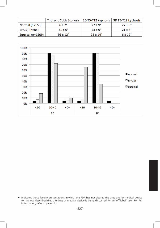
-527-
♦ Indicates those faculty presentations in which the FDA has not cleared the drug and/or medical device for the use described (i.e., the drug or medical device is being discussed for an “off label” use). For full information, refer to page 14.
-528-
See pages 21- 60 for financial disclosure information.
e-Poster #90 POSNA-2016 The Safety of Percutaneous Achilles Tenotomy in Idiopathic Clubfoot Deformity Correction in an Outpatient Setting
Mengyang M. Li, MD; Guy Grimard, MD; Morris A. Duhaime, MD; Panagiotis (Peter) Glavas, MD Sainte-Justine University Hospital Center, Montreal, Quebec, Canada
Purpose: The Ponseti method of serial manipulation and casting is often used for clubfoot deformity. Over the course of treatment, approximately 80% to 90% of patients will require an Achilles tenotomy. This technique is not without risks as neurovascular complications have been reported due to the proximity of these structures to the Achilles tendon (i.e tibial nerve and posterior tibial artery medially, and sural nerve and peroneal artery laterally). This study evaluates the safety of a percutaneous Achilles tenotomy in a tertiary care pediatric hospital.
Methods: A retrospective review of all patient files in the clubfoot clinic from 2003 to 2014 was performed. We identified the patients that had a primary tenotomy for idiopathic clubfoot and noted the presence of any complications. The procedure was performed with a percutaneous incision in an outpatient setting under local anaesthesia. A long leg cast was then applied and the patient was allowed to return home after a short period of observation.
Results: A total of 330 feet in 209 patients were included in this study. There were 52 right, 36 left and 121 bilateral tenotomies. We identified 11 total complications. Complications were categorized according to the Dindo-Clavien classification as minor (not requiring additional hospital stay, surgical intervention, or associated with comorbidity) or major (requiring additional hospitalization or surgical intervention). The most common complication was bleeding: 7 cases of non-pulsatile bleeding that was controlled by manual compression, 1 case of pulsatile bleeding controlled by manual compression and 1 case of non-pulsatile bleeding that required a cast change the next day. Other complications reported were superficial skin cuts and a technical problem with the lighting equipment. This represents a rate of 3.0% for minor complications and 0.3% for major complications. The rate of major complications in this study is comparable to that reported in the medical literature. There are no other reported rates of minor complications in the literature.
Conclusion: Percutaneous Achilles tenotomy as part of the Ponseti method for clubfoot correction is a technique that can be safely performed in an outpatient setting under local anesthesia.
Significance: This is the largest study to date that confirms the safety of percutaneous Achilles tenotomy in an outpatient setting.
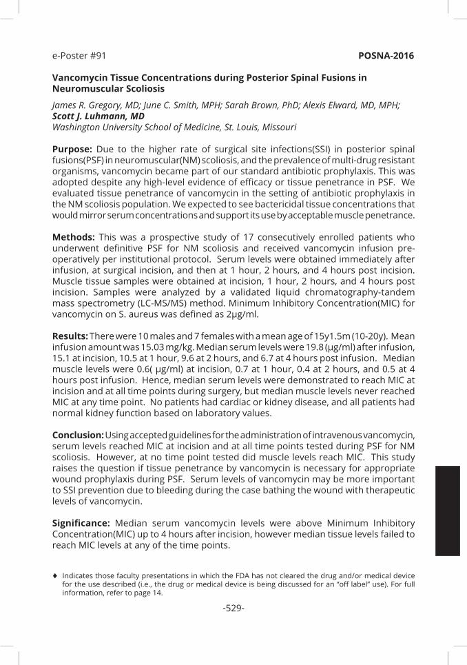
-529-
♦ Indicates those faculty presentations in which the FDA has not cleared the drug and/or medical device for the use described (i.e., the drug or medical device is being discussed for an “off label” use). For full information, refer to page 14.
e-Poster #91 POSNA-2016 Vancomycin Tissue Concentrations during Posterior Spinal Fusions in Neuromuscular Scoliosis
James R. Gregory, MD; June C. Smith, MPH; Sarah Brown, PhD; Alexis Elward, MD, MPH; Scott J. Luhmann, MD Washington University School of Medicine, St. Louis, Missouri
Purpose: Due to the higher rate of surgical site infections(SSI) in posterior spinal fusions(PSF) in neuromuscular(NM) scoliosis, and the prevalence of multi-drug resistant organisms, vancomycin became part of our standard antibiotic prophylaxis. This was adopted despite any high-level evidence of efficacy or tissue penetrance in PSF. We evaluated tissue penetrance of vancomycin in the setting of antibiotic prophylaxis in the NM scoliosis population. We expected to see bactericidal tissue concentrations that would mirror serum concentrations and support its use by acceptable muscle penetrance.
Methods: This was a prospective study of 17 consecutively enrolled patients who underwent definitive PSF for NM scoliosis and received vancomycin infusion pre-operatively per institutional protocol. Serum levels were obtained immediately after infusion, at surgical incision, and then at 1 hour, 2 hours, and 4 hours post incision. Muscle tissue samples were obtained at incision, 1 hour, 2 hours, and 4 hours post incision. Samples were analyzed by a validated liquid chromatography-tandem mass spectrometry (LC-MS/MS) method. Minimum Inhibitory Concentration(MIC) for vancomycin on S. aureus was defined as 2µg/ml.
Results: There were 10 males and 7 females with a mean age of 15y1.5m (10-20y). Mean infusion amount was 15.03 mg/kg. Median serum levels were 19.8 (µg/ml) after infusion, 15.1 at incision, 10.5 at 1 hour, 9.6 at 2 hours, and 6.7 at 4 hours post infusion. Median muscle levels were 0.6( µg/ml) at incision, 0.7 at 1 hour, 0.4 at 2 hours, and 0.5 at 4 hours post infusion. Hence, median serum levels were demonstrated to reach MIC at incision and at all time points during surgery, but median muscle levels never reached MIC at any time point. No patients had cardiac or kidney disease, and all patients had normal kidney function based on laboratory values.
Conclusion: Using accepted guidelines for the administration of intravenous vancomycin, serum levels reached MIC at incision and at all time points tested during PSF for NM scoliosis. However, at no time point tested did muscle levels reach MIC. This study raises the question if tissue penetrance by vancomycin is necessary for appropriate wound prophylaxis during PSF. Serum levels of vancomycin may be more important to SSI prevention due to bleeding during the case bathing the wound with therapeutic levels of vancomycin.
Significance: Median serum vancomycin levels were above Minimum Inhibitory Concentration(MIC) up to 4 hours after incision, however median tissue levels failed to reach MIC levels at any of the time points.
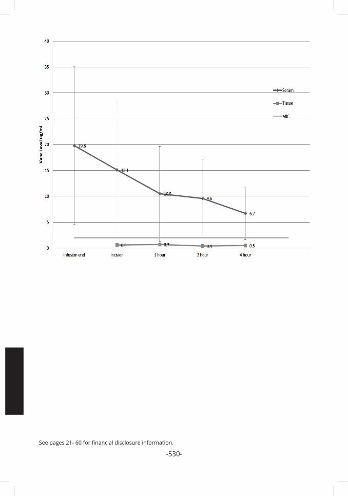
-530-
See pages 21- 60 for financial disclosure information.
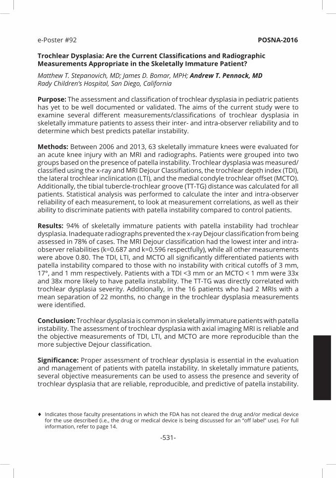
-531-
♦ Indicates those faculty presentations in which the FDA has not cleared the drug and/or medical device for the use described (i.e., the drug or medical device is being discussed for an “off label” use). For full information, refer to page 14.
e-Poster #92 POSNA-2016 Trochlear Dysplasia: Are the Current Classifications and Radiographic Measurements Appropriate in the Skeletally Immature Patient?
Matthew T. Stepanovich, MD; James D. Bomar, MPH; Andrew T. Pennock, MD Rady Children’s Hospital, San Diego, California
Purpose: The assessment and classification of trochlear dysplasia in pediatric patients has yet to be well documented or validated. The aims of the current study were to examine several different measurements/classifications of trochlear dysplasia in skeletally immature patients to assess their inter- and intra-observer reliability and to determine which best predicts patellar instability.
Methods: Between 2006 and 2013, 63 skeletally immature knees were evaluated for an acute knee injury with an MRI and radiographs. Patients were grouped into two groups based on the presence of patella instability. Trochlear dysplasia was measured/classified using the x-ray and MRI Dejour Classifiations, the trochlear depth index (TDI), the lateral trochlear inclinication (LTI), and the medial condyle trochlear offset (MCTO). Additionally, the tibial tubercle-trochlear groove (TT-TG) distance was calculated for all patients. Statistical analysis was performed to calculate the inter and intra-observer reliability of each measurement, to look at measurement correlations, as well as their ability to discriminate patients with patella instability compared to control patients.
Results: 94% of skeletally immature patients with patella instability had trochlear dysplasia. Inadequate radiographs prevented the x-ray Dejour classification from being assessed in 78% of cases. The MRI Dejour classification had the lowest inter and intra-observer reliabilities (k=0.687 and k=0.596 respectfully), while all other measurements were above 0.80. The TDI, LTI, and MCTO all significantly differentiated patients with patella instability compared to those with no instability with critical cutoffs of 3 mm, 17°, and 1 mm respectively. Patients with a TDI <3 mm or an MCTO < 1 mm were 33x and 38x more likely to have patella instability. The TT-TG was directly correlated with trochlear dysplasia severity. Additionally, in the 16 patients who had 2 MRIs with a mean separation of 22 months, no change in the trochlear dysplasia measurements were identified.
Conclusion: Trochlear dysplasia is common in skeletally immature patients with patella instability. The assessment of trochlear dysplasia with axial imaging MRI is reliable and the objective measurements of TDI, LTI, and MCTO are more reproducible than the more subjective Dejour classification.
Significance: Proper assessment of trochlear dysplasia is essential in the evaluation and management of patients with patella instability. In skeletally immature patients, several objective measurements can be used to assess the presence and severity of trochlear dysplasia that are reliable, reproducible, and predictive of patella instability.
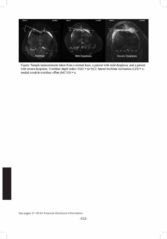
-532-
See pages 21- 60 for financial disclosure information.
-533-
♦ Indicates those faculty presentations in which the FDA has not cleared the drug and/or medical device for the use described (i.e., the drug or medical device is being discussed for an “off label” use). For full information, refer to page 14.
e-Poster #93 POSNA-2016 Fibroblast Growth Factor Receptor Three Regulates Fracture Remodeling Through an Osteoclast Independent Effect
Simon P. Kelley, MBChB, MRCS, FRCS; Chunying Yu; Baht Gurpreet, PhD; Benjamin A. Alman, MD, FRCSC The Hospital for Sick Children, Toronto, Ontario, Canada
Purpose: Fibroblast growth factor receptor 3 (fgfr3) mutations cause skeletal dysplasias such as achondroplasia and CATSHL syndrome. We have previously shown that in skeletal repair fgfr3 acts as a bone switch, which modulates the balance of intramembranous and endochondral bone formation in healing murine tibial fractures, and leads to an accelerated but structurally deficient callus formation. Due to the observation that fracture callus at post fracture day 21 was shown to be significantly smaller and more fragile in fgfr3+/- mice than in their wild type controls we hypothesized that this abnormality may also be due to enhanced fracture remodeling from an effect of fgfr3 on osteoclasts. Furthermore that the fgfr3 directed osteoclastic effect on fracture healing is an independent function of the haematopoietic lineage.
Methods: We used an established murine semi-stabilized tibia fracture model in genetically modified fgfr3 knockout mice and their wild type littermate controls. Fractures were harvested and analyzed at post-fracture day (PFD) 14 and 21 using radiography and histomorphometry, to assess fracture callus structure and composition, with TRAP staining for osteoclast number and activity. In-vitro osteoclast differentiation and function was assessed using bone marrow cultures treated with M-CSF and RANKL, and a resorption pit assay. To isolate the effects of fgfr3+/- haematopoietic cells (incl. osteoclasts) from those of fgfr3+/- mesenchymal cells (incl. osteoblasts and chondrocytes), irradiated WT mice received a bone marrow transplant (BMT) of haematopoietic cells from either fgfr3+/- mice or WT control mice. Fractures were performed 8 weeks after BMT, harvested at PFD 21 and analyzed by radiography.
Results: Fgfr3+/- mutant fractures show smaller fracture calli at PFD21 than WT controls. Histomorphometric analysis of PFD21 fractures reveals reduced cartilage area and reduced trabecular bone area per callus area. TRAP staining of fracture calluses demonstrates more osteoclasts per bone surface area and increased erosion surface per bone surface area in fgfr3+/- calli. In vitro osteoclast differentiation assays with fgfr3+/- cultures demonstrated increased osteoclasts with higher activity. WT mice that received an fgfr3+/- BMT demonstrated smaller fracture callus at PFD21 than WT mice receiving control WT BMT.
Conclusion: Fgfr3 regulates osteoclast differentiation and function. In vivo fgfr3 deficiency is manifested by an enhancement of fracture remodeling which is an independent effect of fgfr3 on the haematopoietic niche, despite fgfr3 having a known effect on mesenchymal cells.
Significance: Control of the haematopoietic bone marrow niche through fgfr3 signaling represents a novel approach to modulating both fracture repair and bone regeneration.
-534-
See pages 21- 60 for financial disclosure information.
e-Poster #94 POSNA-2016 Relationships between Three Classification Systems in Brachial Plexus Birth Palsy
Dustin A. Greenhill, MD; Sandra Tomlinson-Hansen, BA; Scott H. Kozin, MD; Dan A. Zlotolow, MD Shriners Hospital for Children - Philadelphia, Pennsylvania
Purpose: The Mallet scale, Active Movement Scale (AMS), and Toronto Test are validated for use in children with brachial plexus birth palsy (BPBP). However, the inability to compare these evaluation systems has led to difficulty gauging treatment efficacy and interpreting available literature in which multiple scoring systems are reported. Given the critical importance of physical examination, we compared three scoring systems in order to clarify statistical relationships between current validated evaluation methods.
Methods: The medical records of children with BPBP treated at a single institution over a 14-year period were retrospectively reviewed. Modified Mallet, AMS, and Toronto scores were recorded throughout the entire period. Data was included if at least two complete scoring systems were documented during the same exam session. Spearman correlation coefficients were calculated for all composite and subscore combinations. A concordance table was constructed for select variables found to be highly correlated.
Results: Total single-session score combinations were as follows: 157 Mallet and AMS, 325 AMS and Toronto, and 143 Mallet and Toronto. Composite AMS and Toronto scores were found to have a strong correlation (r=0.928, p<0.001). A concordance table comparing these variables revealed that a Toronto score of 3.5 is concordant to an AMS score of 45. Modified Mallet scores had only a moderate correlation with composite AMS (r=0.512, p<0.001) and Toronto (r=0.458, p<0.001) scores. Specifically regarding the modified Mallet score, maneuvers requiring external rotation had stronger correlations with the composite modified Mallet score than maneuvers highlighting internal rotation.
Conclusion: Modified Mallet scores do not correlate well with AMS or Toronto scores and should be utilized separately when managing children with BPBP. Similarly, AMS and Toronto scores are inadequate to guide clinical decisions for which the literature cites Mallet scores as outcome measures, and vice versa. Lastly, Mallet scores should incorporate an isolated internal rotation component in order to adequately assess midline function.
Significance: Modified Mallet scores do not correlate well with AMS or Toronto scores and should be separately utilized when managing children with BPBP. Furthermore, Mallet scores should incorporate an isolated internal rotation component in order to adequately assess midline function. With regards to interpreting either AMS or Toronto scores, our concordance table may serve as an additional source of information when physicians and parents are trying to make important decisions about a topic in which there is limited scientific data. In order to accurately track neurologic progression during the treatment of children with BPBP, surgeons and referring physicians should understand the importance of appropriate documentation using adequate outcome measures.
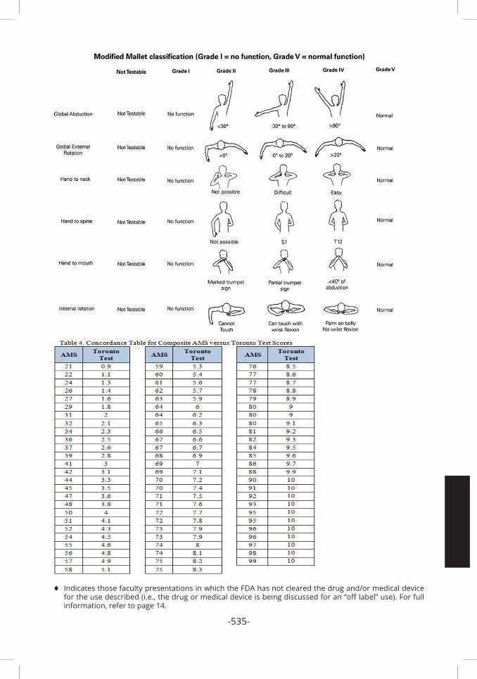
-535-
♦ Indicates those faculty presentations in which the FDA has not cleared the drug and/or medical device for the use described (i.e., the drug or medical device is being discussed for an “off label” use). For full information, refer to page 14.
-536-
See pages 21- 60 for financial disclosure information.
e-Poster #95 POSNA-2016 Validity of Beighton’s Score in a Paediatric Population
Nimesh Patel, MRCS; Peter J. Riddlestone, MBBS; Roshan Gunalan; James E. Metcalfe, FRCS, MBChB; Ben Holroyd, FRCS (Tr & Orth) Derriford Hospital, Plymouth, United Kingdom
Purpose: The Beighton Score (BS) was originally described as an assessment tool modified from the technique described by Carter and Wilkinson in 1964 in patients suffering from Ehlers-Danlos syndrome, to screen for hypermobility. However considerable variation in range of movement is possible within joints of normal individuals, though joint laxity is thought to decrease with age. BS is a valid screening tool in an adult population, but its use in paediatrics to assess generalised joint mobility is not fully validated. The correlation of hypermobility and joint pain in children is also not fully understood. We therefore aim to investigate the validity of BS in the paediatric population.
Methods: Over a three-month period, all children presenting to our regional paediatric orthopaedic unit had their BS measured by a trained orthopaedic surgeon. Non-ambulatory children were excluded, as were those with known hypermobility syndrome, or neuromuscular disorders. We also screened them for joint pain and measured the rotational profile of their lower limbs.
Results: Two hundred patients were assessed, aged between 3-15 years (mean 10.1). There were 92 males and 108 females. Mean (SD) BS was 2.06 (2.2), and the range was 0-8. Comparing males VS females, mean BS (SD) was 1.71 (2.25) VS 2.36 (2.14); p=0.0378, age was 9.75 VS 10.13, BS range was 0-7 VS 0-8. Sixty-four children (32%) complained of pain in at least one joint, though the mean (SD) BS in these patients was 1.71 (1.86). There were 8 patients with abnormal rotational profile at the hip, though not related to an increased BS.
Conclusion: We found that the average BS was just over 2 in all of our children, though significantly lower in males. In adults, a score of 4 or higher has been attributed to generalised hypermobility, though a true diagnostic cut off has not been defined in children. Since there is already an innate tendency to have an increased BS in normal children, it makes interpretation of the score in a paediatric population difficult and less meaningful. Of note, there was no correlation between arthralgia and a high BS, or abnormal lower limb rotational profile.
Significance: We therefore do not recommend routine measurement of Beighton’s score to diagnose and assess hypermobility in a paediatric population, nor do we feel that it correlates with joint pain.
-537-
♦ Indicates those faculty presentations in which the FDA has not cleared the drug and/or medical device for the use described (i.e., the drug or medical device is being discussed for an “off label” use). For full information, refer to page 14.
e-Poster #96 POSNA-2016 Relative Impact of Pre-Operative Hip Location vs Post-Operative Abduction Angle on Epiphyseal Perfusion After Closed Reduction and Spica Casting for DDH
Michael Rivlin, MD; Scott B. Rosenfeld, MD; Herman Kan, MD; Wei Zhang, PhD; Erica K. Schallert, MD; Siddharth P. Jadhav, MD Texas Children’s Hospital, Houston, Texas
Purpose: SPICA MRI with gadolinium is an established technique for postoperative assessment of perfusion of the capital femoral epiphysis after closed reduction of developmental dysplasia of the hip (DDH). When perfusion abnormalities are present, patients are at risk for developing epiphyseal osteonecrosis. The purpose of this study is to investigate the relative impact of two variables on postoperative perfusion on SPICA MRI in patients with DDH: pre-operative hip location and the degree of postoperative hip abduction.
Methods: We performed a retrospective study of 18 patients (36 individual hips) with DDH (average age 11.7 months, range 4-36 months, 4:14 male: female) who underwent gadolinium enhanced SPICA MRI after closed hip reduction between July 2011 and November 2014. Pre-operative hip location was characterized as being 1) dislocated (completely out of the socket) or 2) reduced/subluxed (well reduced dysplastic hips and subluxed hips). Hip abduction angle and percentage of femoral head volume perfused were calculated from post reduction contrast-enhanced SPICA MRI. Hip location on pre-operative radiographs and hip abduction angles in the spica cast were compared with perfusion via the Wilcoxon rank test, Fisher’s exact test, and Pearson’s correlation. Multivariate analysis was performed to determine the individual impact of each factor.
Results: 19 hips were dislocated and 17 were reduced or subluxed pre-operatively. The mean post-operative volume of femoral head perfusion was 61% ± 44% in the dislocated group and 88% ± 22% in the reduced/subluxed group (p=0.0405). 7/19 (37%) dislocated hips and 2/17 (12%) reduced/subluxed hips had abnormal post-operative enhancement. Of post-op hips that were abducted ≤60 degrees on MRI, 4/16 (25%) demonstrated abnormal (≤50%) femoral head perfusion compared with 5/20 (25%) hips that were abducted greater than 60 degrees (P=1.0). When correcting for differences in abduction, there was increased enhancement in hips that were reduced/subluxed compared to dislocated pre-operatively (p=0.03). There was poor correlation between abduction angle and perfusion (correlation=0.038, p=0.82).
Conclusion: Hips that were dislocated pre-operatively demonstrated lower perfusion than those that were reduced/subluxed. The degree of post-op hip abduction did not correlate with perfusion. Pre-operative hip location may be a more important factor in predicting post-reduction perfusion than post-operative abduction angle.
Significance: Higher hip spica abduction angles may be acceptable if necessary to achieve stable reduction. Patients whose hips are not dislocated may not require contrast enhanced MRI examination postoperatively.
-538-
See pages 21- 60 for financial disclosure information.
e-Poster #97 POSNA-2016 Pavlik Harness Initiation in Barlow Positive Hips: Can We Wait?
Katherine Cook, BS, BA; Michael Ingram, BA; Jill E. Larson, MD; Jamie Burgess, PhD; Joseph (Jay) Janicki, MD Lurie Children’s Hospital, Chicago, Illinois
Purpose: Developmental dysplasia of the hip (DDH) can be effectively treated with a Pavlik Harness (PH) within the first 6 months of life. However, 80% of unstable hips will stabilize naturally by 2 months of age. While many advocate the initiation of PH treatment as soon as identified, delaying treatment during the first few weeks of life may allow the hips to naturally stabilize and avoid intervention entirely. When a newborn presents with a Barlow positive hip, we offer families the option of delaying treatment until after 4 weeks of age to determine whether the hips stabilize naturally. The purpose of this study is to evaluate our protocol in which PH treatment is delayed in patients with a Barlow positive hip in the first 2 weeks of life.
Methods: A retrospective review of medical records between 2010 and 2014 was completed. Patients were included if they presented with unstable hip/s within the first two weeks of life, were Barlow positive and deferred treatment. Baseline data included sex, bilaterality, form and timing of intervention if necessary and a minimum of 1 year follow up.
Results: A total of 22 children and 27 hips were included. There were 18 females and 4 males. There was left hip instability found in 15 children, right hip instability in 2 children and 5 had bilateral instability. Of these 22 children, 14 stabilized without treatment. Eight children were treated with a PH at 4-6 weeks of age due to persistent instability and 2 initiated treatment at 12 weeks due to persistent dysplasia. All 22 children, whether treated with PH or not, required no further treatments and all children were followed to at least 1 year of age with radiographs.
Conclusion: Delaying treatment in children with Barlow positive hips resulted in 64% natural stabilization and 55% not requiring treatment. Meanwhile, delaying treatment in this cohort resulted in no PH failures. A protocol of deferred treatment in children with Barlow positive hips appears to be safe and effective in this small cohort.
Significance: Parents can be counseled that waiting to initiate PH treatment is appropriate. By avoiding swaddling, the hips may stabilize without treatment. The treatment of DDH is often a stressful adjustment for parents with a newborn. The appropriate use of a PH may affect the nursing and even parent/infant bonding. Our protocol decreases newborn period interventions and its social impact.

-539-
♦ Indicates those faculty presentations in which the FDA has not cleared the drug and/or medical device for the use described (i.e., the drug or medical device is being discussed for an “off label” use). For full information, refer to page 14.
e-Poster #98 POSNA-2016 Unique Trabecular Bone Micro-structure and Mechanical Property in Iliac Crest Bone Biopsy of Adolescent Idiopathic Scoliosis
Wayne YW Lee; Zhiwei Wang, MD, PhD; Jiajun Zhang, BSc, MSC; Huanxiong Chen, MD; Tsz Ping Lam, MD; Bobby Kin Wah Ng, MD; X. E. Guo, PhD; Jack Cheng Prince of Wales Hospital, Shatin, Hong Kong SAR
Purpose: Systemic low bone mass and abnormal bone morphology, volumetric bone mineral density and microarchitecture have been reported in adolescent idiopathic scoliosis (AIS) using dual-energy X-ray absorptiometry (DXA) and high resolution peripheral QCT measurement. This study aimed to investigate and compare the detail trabecular bone microstructure and mechanical property at tissue level in AIS Vs controls with advanced imaging analysis tools. Individual trabecula segmentation (ITS) segments trabecular bone further into rod and plate configuration, and finite element analysis (FEA) estimates the biomechanical properties of bone.
Methods: 28 AIS girls (14.2 ± 1.5) and 9 age-matched controls (15.3 ± 3.7) were recruited. Trabecular bone strips were obtained from posterior iliac crest region during standard bone autograft harvesting for posterior spinal fusion in severe scoliosis patients or for other orthopaedic procedures that required autograft in the control group. The strips (less than 1cm3) were divided scanned with microCT and 3D reconstructed images were analyzed with standard protocol and advanced ITS and FEA. Only 3D reconstructed images larger than 4mm3 were subjected to ITS and FEA measurement.
Results: The recruited AIS and control groups had similar demographic and anthropometric parameters (Table 1). There were no significant difference in conventional microCT measurements between the two groups (P>0.05 Table 2). In ITS analysis, AIS had significantly lower rod thickness (rTb.Th) (0.062±0.005 mm) and less rod number (rTb.N) (3.8±0.8 mm-1) than controls (P<0.05). The rod bone volume fraction (rBV/TV) was also found to be decrease in AIS (0.055±0.024) by 16.7% compared with controls (0.066±0.026), though not reaching statistical significance due to the small sample size in this pilot study (P=0.091) (Table 3). AIS was also shown to have significantly lower bone mechanical property (670±209 MPa) than controls (883±154 MPa) (P=0.039) on FEA (Table 4).
Conclusion: With bone site-matched controls, this is the first ex vivo study at tissue level showing different micro-structure and lower apparent modulus in AIS, providing supporting evidence to our previous findings using HR-pQCT scan that significantly lower trabecular thickness and number, and lower apparent modulus in distal radius of AIS. The present findings also suggest that the loss of trabeculae was mainly resulted from the decline of rod trabeculae.
Significance: This study on non-weight bearing iliac crest bone provided scientific evidence supporting the association of dysfunctional bone metabolism and AIS at tissue
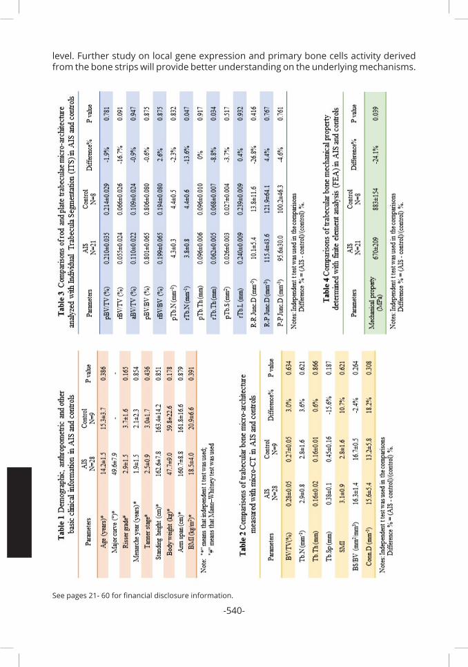
-540-
See pages 21- 60 for financial disclosure information.
level. Further study on local gene expression and primary bone cells activity derived from the bone strips will provide better understanding on the underlying mechanisms.
-541-
♦ Indicates those faculty presentations in which the FDA has not cleared the drug and/or medical device for the use described (i.e., the drug or medical device is being discussed for an “off label” use). For full information, refer to page 14.
e-Poster #99 POSNA-2016 A Quantitative Method for Radiologic Assessment of Skeletal Maturity using the Distal Femur
Derrick M. Knapik, MD; James O. Sanders, MD; Allison Gilmore, MD; Daniel R. Cooperman, MD; Raymond W. Liu, MD University Hospitals Case Medical Center, Cleveland, Ohio
Purpose: Current methods for estimating skeletal maturity have shown wide variability and generally involve significant complexity, limiting practical use and leading some to favor chronological age. Recently, it has been shown that 90% of final adult height is a more accurate predictor of skeletal growth patterns as compared to peak height velocity. Using 90% of ultimate height as a gold standard and applying this to the same historical collection used to establish the Greulich and Pyle atlas, we sought to develop a quick, quantitative, and reproducible method of estimating skeletal maturity.
Methods: Forty-two healthy children (26 girls, 16 boys) reached 90% growth at an average age of 11.3±0.9 years for girls and 13.2±0.5 years for boys. Annual radiographs of the knee three years prior and two years following the 90% mark were analyzed. A horizontal line was drawn along the distal femoral physis, and the distance from the tip of the central peak to this line was divided by the physeal width to obtain a standardized value (Figure 1). Measurements were tested for intraclass correlation coefficient in 24 radiographs by the two authors. Linear regression analysis was used to compare chronological age, Greulich and Pyle bone age, and chronological age combined with central peak standardized height.
Results: The intraclass correlation coefficient was 0.944 for central peak height, giving excellent inter-relator reliability. Linear regression analysis showed an improvement in overall correlation between predicted age of 90% growth and true age of 90% growth when using a combination of chronological age and position of the central peak of the distal femur on radiographs when compared to either chronological age or Greulich and Pyle bone age in both girls and boys (Table 1).
Conclusion: 90% of ultimate adult height has been shown to be a better predictor of true skeletal maturity when compared to peak height velocity, and its use at a new gold standard may allow for the development of improved skeletal maturity grading systems. Using regression analysis, we have found that using a combination of chronologic age and standardized distal femur central peak height provides better prediction of 90% height as compared to using either chronological age or Greulich and Pyle bone age measurements. Significance: To our knowledge, central peak height represents the first purely quantitative parameter for estimating skeletal maturity, and offers a practical methodology which can potentially improve treatment when skeletal maturity is an important factor.
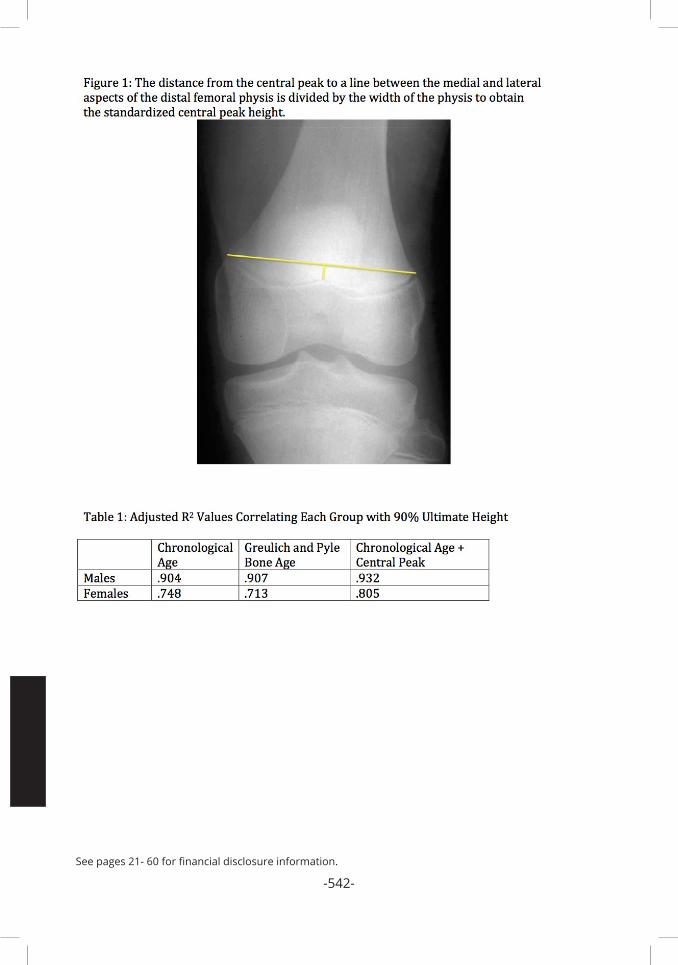
-542-
See pages 21- 60 for financial disclosure information.
-543-
♦ Indicates those faculty presentations in which the FDA has not cleared the drug and/or medical device for the use described (i.e., the drug or medical device is being discussed for an “off label” use). For full information, refer to page 14.
e-Poster #100 POSNA-2016 The Mid-term Outcome of a Combined Surgical Technique for Congenital Psuedarthrosis of the Tibia (A Study of 59 Cases with Mean 4.6 Year Follow-up)
Haibo Mei, MD; Guanghui Zhu Hunan Children’s Hospital, Changsha, China
Purpose: The purpose of this study was to report on the initial union rate of congenital pseudarthrosis of the tibia treated using combined intramedullary rodding, bone graft, and Ilizarov’s fixator at 1 institution and evaluate the frequency of refracture and residual deformities of the leg and ankle, with mean 4.6 years follow -up.
Methods: Between February 2007 and March 2010, a total of 59consecutive patients with congenital pseudarthrosis of the tibia treated with intramedullary rod of the tibia, wrapping autogenic iliac bone graft, and Ilizarov’s fixator were retrospectively reviewed. Patients were divided into 2 groups based on status of fibula. There were 19 patients who had intact fibula in Group I and 40 patients with concomitant pseudarthrosis of the fibula in Group II. The 2 groups were compared with each other regarding the union obtained, tibial alignment, limb length discrepancy (LLD), valgus deformity of the ankle, and the frequencies of refracture during period of follow-up.
Results: Fifty-two (88%) of the 59 patients had bone union at site of pseudarthrosis. The average time to union was 4.2 months (range, 3 to10 months) and mean duration of Ilizarov treatment was 4.3 months (range, 3 to 10.4 months). In group I, 16 (84%) of 19 patients achieved union, while in group II, 36 (90%) of 40 patients had union (P=0.67, Chi-square test). 31 patients had proximal tibial valgus with a mean of 12 degrees (range, 5 to 23 degrees), 23 patients had ankle valgus deformities with a mean of 12.3degrees (range, 6 to 25 degrees). Of Patients with concomitant fibular pseudarthrosis in Group II, 18 of 40 patients with fibular pseudarthrosis were treated with osteosynthesis, 8 patients obtained union of the fibula, and 10 patients had persisted pseudoarthrosis of the fibula at the last followup. 34 of 52 patients had an average 2.2 cm LLD (range, 1.5- 4 cm). Refractures occurred in fourteen (26.9%) of 52patients who had union of pseudarthrosis at a mean follow-up of 4.6 years following union.
Conclusion: The midterm outcome of this combined techinque demonstrated 88% union rate obtained at primary surgery and its residual deformities after union were comparable with other studies reported in literature. Nevertheless, it is necessary for us to carefully follow up these younger patients until skeletal maturity and to evaluate the benefit of the combined approach.
Significance: This combined technique of CPT demonstrated 88% union rate obtained at primary surgery and its residual deformities after union were comparable with other studies reported in literature.
-544-
See pages 21- 60 for financial disclosure information.
e-Poster #101 POSNA-2016 Rectus Femoris Transfer Surgery in Cerebral Palsy Patients with Stiff Knee Gait
Ted C. Sousa, MD; Alexander Nazareth, MS; Susan A. Rethlefsen, DPT; Nicole M. Mueske, MS; Tishya A.L. Wren, PhD; Robert M. Kay, MD Children’s Hospital Los Angeles, Los Angeles, California
Purpose: Previous research found minimally ambulatory patients do not benefit from distal rectus femoris transfer surgery (DRFT) due to increased stance knee flexion post-operatively. This study compared postoperative knee kinematics in patients with cerebral palsy (CP) and stiff knee gait in two groups: those who underwent DRFT and those who did not.
Methods: A retrospective study of 53 children with CP and stiff knee gait who underwent multilevel surgery and preoperative and postoperative gait analysis was undertaken. Twenty-seven subjects underwent DRFT (DRFT, age 8.0 ± 2.1 years) and 26 subjects did not (CTRL, age 8.8 ± 2.7 years). Data were compared using t-tests, X2 tests and multiple regression analysis.
Results: Age at surgery, time of follow-up and concomitant surgeries were similar between groups. Baseline range of motion, strength and kinematic variables did not differ between groups. Distribution of Gross Motor Function Classification System (GMFCS) levels differed between groups (p= 0.02), with the CTRL group having a larger percentage (61% vs 31%) of GMFCS III and IV subjects.
Significant post-operative improvements in maximum knee extension in stance were seen for both DRFT (D8.2° ± 12.9, p = 0.003) and CTRL (D12.7° ± 21.1°, p = 0.001) groups. Decreased post-operative peak knee flexion in swing was found for both DRFT (D5.2° ± 11.7, p = 0.03) and CTRL (D12.4° ± 15.2, p < 0.0001) groups. Timing of post-operative peak knee flexion in swing (percentage of gait cycle) occurred earlier in both DRFT (D5.1 ± 4.6%, p =0.004) and CTRL (D2.1 ± 5.2%, p = 0.02) groups. No significant change in total knee ROM was found for either group.
DRFT group had significantly greater improvement in timing of post-operative peak knee flexion in swing than the CTRL group (difference 3% [0.4%, 5.6%], p =0.02). CTRL group had a significantly greater decrease in post-operative peak flexion in swing compared to DRFT group (difference 7.2° [14.5,0.1], p= 0.05). No difference between groups was found for knee total ROM. After adjusting for GMFCS level, a significant difference between groups was found for post-operative change in maximum knee flexion in swing only.
Conclusion: Minimally ambulatory patients who undergo hamstring lengthening without DRFT improve their knee extension in stance, maintain knee range of motion from stance to swing, and experience similar improvement in timing of peak swing knee flexion to those who undergo DRFT.
Significance: DRFT is not indicated in minimally ambulatory patients with CP.
-545-
♦ Indicates those faculty presentations in which the FDA has not cleared the drug and/or medical device for the use described (i.e., the drug or medical device is being discussed for an “off label” use). For full information, refer to page 14.
e-Poster #102 POSNA-2016 Stable, Moderate to Severe Slipped Capital Femoral Epiphysis: Choosing a Staged Imhauser Correction versus a Single Event Modified Dunn Procedure
Jakub A. Sikora-Klak, MD; James D. Bomar, MPH; Christina Paik, PA-C; Dennis R. Wenger, MD; Vidyadhar S.V. Upasani, MD Rady Children’s Hospital, San Diego, California
Purpose: Controversy exists regarding the treatment of children with stable, moderate to severe slipped capital femoral epiphysis (SCFE). Although in situ screw fixation remains the gold standard, proximal femoral osteotomies are often used to address the cam-like deformity created by the slip. The purpose of this study was to compare radiographic outcomes and complication rates between a staged flexion, valgus proximal femoral osteotomy (Imhauser) after in situ fixation versus subcapital realignment (modified Dunn) performed as the index procedure.
Methods: Twenty-one children with stable, chronic or acute on chronic SCFE were included in this retrospective review. Nine hips were treated with initial in situ screw fixation followed by an Imhauser osteotomy. Twelve hips were treated with the modified Dunn procedure. All patients had minimum one-year follow-up. Peri-operative data including surgical time and fluoroscopy time were recorded. Radiographic data included pre- and post-operative Southwick slip angle (SSA), neck shaft angle (NSA), articular surface to trochanter distance (ATD), and medial proximal femoral angle (MPFA). ANOVA and the Mann-Whitney test were used to compare data between the two groups.
Results: Measurements were performed by two independent observers and found to be similar (ICC scores range: 0.81-0.98). Pre- and post-operative radiographic parameters were similar between the two groups (Table). On average, the surgical time for the Imhauser procedure was 56 minutes less and fluoroscopy time was 40% less than the modified Dunn procedure. Three of the 12 hips treated with the modified Dunn went on to develop AVN (25%) compared to zero of nine hips treated with the Imhauser. Four patients in the modified Dunn group required revision surgery (33%) compared to one Imhauser patient (8%).
Conclusion: Although the staged Imhauser correction requires two anesthetic periods, the modified Dunn procedure was technically more challenging (requiring longer surgical time and fluoroscopy time), resulted in greater complications (avascular necrosis and revision surgery), and similar radiographic deformity correction.

-546-
See pages 21- 60 for financial disclosure information.
Significance: The high risk, high reward hype of the modified Dunn procedure may not be justified and the ideal indications for subcapital realignment remain elusive. Prospective, comparative studies evaluating subjective and functional outcomes for these procedures are needed to guide clinical practice.

-547-
♦ Indicates those faculty presentations in which the FDA has not cleared the drug and/or medical device for the use described (i.e., the drug or medical device is being discussed for an “off label” use). For full information, refer to page 14.
e-Poster #103 POSNA-2016 Is There an Association Between Hip Motion and Severity of Acetabular Dysplasia? Analysis of the ANCHOR PAO Cohort
Peter D. Fabricant, MD, MPH; Wudbhav N. Sankar, MD; Mark Seeley, MD; John C. Clohisy, MD; Young Jo Kim, MD; Michael B. Millis, MD; David A. Podeszwa, MD; Perry L. Schoenecker, MD; Ernest L. Sink, MD; Daniel J. Sucato, MD, MS; Ira Zaltz, MD Children’s Hospital of Philadelphia, Philadelphia, Pennsylvania and Washington University, St. Louis, Missouri
Purpose: It is generally believed that acetabular dysplasia is associated with increased hip range of motion (ROM). The purpose of the current study was to investigate the associations between severity of dysplasia and hip ROM in a large multicenter cohort of patients while accounting for concomitant biometric variables and femoral-sided deformity.
Methods: A prospective registry of patients undergoing periacetabular osteotomy for symptomatic acetabular dysplasia by one of thirteen surgeons at ten institutions was used to compile demographic, biometric, and hip ROM data on 1,051 patients (mean age 26±10 years). Dysplasia severity was determined using previously defined criteria based on lateral center edge angle of Wiberg (Severe,<5º; Moderate,5°-15º; Mild,>15°). ANOVA with Tukey-Adjusted pairwise comparisons, Pearson correlation analyses, and linear regression modeling was used to investigate for associations between dysplasia severity, alpha-angle, and hip ROM.
Results: When controlling for age, sex, BMI, and alpha-angle in linear regression analysis, only internal (b=1.94; P=0.005) and external (b=-2.63; P<0.001) rotation of the hip in extension were significantly different between groups with increasing dysplasia severity. Alpha-angle was significantly greater for those with severe acetabular dysplasia compared to subjects with mild disease (60°±16° vs. 57°±15°; P=0.038). Alpha-angle was also significantly correlated with rotational ROM parameters (internal rotation in flexion, external rotation in flexion, internal rotation in extension, external rotation in extension), but not with linear motion (flexion, abduction, adduction) with mild to moderate negative correlation between magnitude of alpha-angle and ROM (Pearson r, range: -0.077 to -0.216; P<0.05 for all).
Conclusion: In this multicenter study of over one thousand patients with symptomatic acetabular dysplasia, internal rotation in extension was directly associated with severity of dysplasia while external rotation in extension was inversely associated. Furthermore, alpha-angle was greater with increasing severity of dysplasia and predictive of rotational ROM parameters. Taken together, these data suggest that femoral-sided deformity, including alpha-angle and possibly femoral version, may be responsible for differences in ROM based on severity of dysplasia.
Significance: Contrary to previous understanding, increased acetabular dysplasia severity doesn’t predict increased hip ROM. Surgeons should not rely on hip ROM
-548-
See pages 21- 60 for financial disclosure information.
in the diagnostic algorithm between acetabular dysplasia and FAI, as ROM is largely dependent on femoral-sided morphology and there is substantial covariance between alpha-angle and dysplasia severity.
-549-
The Alfred R. Shands Jr. Circle is OREF’s highest-level recognition society. Established in 1994, the Shands Circle was created to recognize those individuals who contribute a minimum of $20,000 in cash or securities or a minimum of $50,000 using life insurance, will or trust, or to the OREF endowment. POSNA thanks its members who have joined the Shands Circle.
THE ALFRED R. SHANDS JR. CIRCLE
Drs. Behrooz Akbarnia and Nasrin OwsiaMargaret R. Albanese, MDDr. and Mrs. Stephen A. AlbaneseMichael C. Albert, MDBenjamin A. Alman, MDDr. David and Mrs. Linda AronssonDr. Marc and Mrs. Ellie AsherDr. and Mrs. George S. BassettDr. and Mrs. James H. BeatyRandal R. Betz, MDLaurel C. Blakemore, MDDr. and Mrs. Oheneba Boachie-AdjeiDr. Jennette L. Boakes and Mr. David RivesGary T. Brock, MD (In honor of Denis S. Drummond, MD)Dr. and Mrs. Robert W. Bruce Jr.Dr. and Mrs. Robert B. CadyRobert M. Campbell Jr., MDS. Terrance Canale, MDDr. Hank and Mrs. Jill ChambersDr. and Mrs. Henry R. CowellAlvin H. Crawford, MDJohn P. Dormans, MD, FACSDenis S. Drummond, MDDrs. John B. and S. Jean EmansDr. Howard and Mrs. Phyllis EppsDr. Robert Bucholz and Dr. Marybeth EzakiFrances Farley, MD and Darryl SnabesDavid S. Feldman, MDJohn (Jack) M. Flynn, MDDr. Steven and Mrs. Lisa FrickKeith R. Gabriel, MDTheodore J. Ganley, MDDrs. Mark and Paulette GebhardtDr. and Mrs. Walter B. GreeneDr. and Mrs. Lawrence L. Haber
Dr. Richard and Mrs. Sherril HaynesWilliam L. Hennrikus Jr., MDMr. John C. HerklotzDr. Dale and Mrs. Stevie HoekstraDrs. Alexandra Page and John Houkom*Dr. and Mrs. John D. HsuDr. and Mrs. Kamal N. IbrahimLawrence I. Karlin, MDJames R. Kasser, MDRobert M. Kay, MDJohn T. Killian, MDYoung-Jo Kim, MD, PhDAndrew G. King, MDDr. and Mrs. Howard A. KingJan E. Klamar, MDDr. L. Andrew and Mrs. Leigh E. KomanKen N. Kuo, MD (In memory of Angela S. M. Kuo)Hubert Labelle, MDArabella I. Leet, MD*Dr. Lawrence G. and Mrs. Elizabeth A. LenkeR. Merv Letts, MDBaron S. Lonner, MDDr. John E. and Mrs. Ann K. LonsteinDr. and Mrs. Herbert J. LouisDr. G. Dean and Mrs. Marilyn MacEwenSteven M. Mardjetko, MDSharon K. Mayberry, MDRichard E. McCarthy, MDJames J. McCarthy, MDDr. Newton C. and Mrs. Mary McColloughDr. Gregory and Mrs. Madelyn MencioDr. Todd A. Milbrandt and Ms. Mary Beth MagyarDr. Scott and Mrs. Sandy MubarakDr. Peter and Mrs. Cathy NewtonKenneth J. Noonan, MD
-550-
THE ALFRED R. SHANDS JR. CIRCLE, (cont.)
James W. Ogilvie, MDNorman Y. Otsuka, MDHamlet A. Peterson, MDDrs. William and Linda PhillipsKristin A. Pierz, MDDr. and Mrs. Charles T. PriceEllen M. Raney, MDDr. and Mrs. Karl E. RathjenJuan A. Realyvasquez, MDKent A. Reinker, MDDr. and Mrs. B. Stephens RichardsDr. James and Mrs. Elizabeth RoachDr. and Mrs. Sanjeev SabharwalJames O. Sanders, MDJohn F. Sarwark, MDDr. and Mrs. Perry L. SchoeneckerDr. Richard M. and Mrs. Colleen G. SchwendDr. and Mrs. Suken A. ShahJay Shapiro, MDKevin G. Shea, MDDr. and Mrs. Harry L. ShufflebargerDavid L. Skaggs, MD, MMMDr. and Mrs. Chadwick F. SmithBrian D. Snyder, MD, PhDDr. Kit M. Song and Kwi Y. LeeDr. Paul and Mrs. Amy SponsellerHoward H. Steel, MDDavid B. Stevens, MDDr. Peter M. and Mrs. Gayle M. Stevens*Dr. Peter F. Sturm and Mrs. Judith Serling-SturmDr. and Mrs. Daniel J. SucatoDr. George H. and Mrs. Janice L. ThompsonVernon T. Tolo, MDLaura Lowe Tosi, MDDr. and Mrs. Michael G. VitaleEric J. Wall, MDWilliam C. Warner Jr., MDPeter M. Waters, MDDr. and Mrs. Stuart L. Weinstein
David S. Weisman, MDLawrence Wells, MDDr. R. Baxter and Mrs. Susan Willis
*=Deceased

-551-
POSNA DIRECT FUND(January 1, 2015 - December 31, 2015)
Robert B Cady, MDMark A Erickson, MDFrances A Farley, MDRobert M Kay, MD
Ken N Kuo, MDRonald Lewis, MDGregory A Mencio, MDJay Shapiro, MD
GOLD ($5,000 +)Shriners Hospitals for ChildrenSamantha Anne Spencer, MDPaul D Sponseller, MDPeter M Waters, MD
SILVER ($1,000 - $4,999)Stephen A Albanese, MDDavid D Aronsson, MDMark A Barry, MDAllan C Beebe, MDLaurel C Blakemore, MDJennette L Boakes, MDMatthew J Bueche, MDRobert B Carrigan, MDVirginia F Casey, MDRandolph Copeland, MDJon R Davids, MDHoward R Epps, MDJohn (Jack) M Flynn, MDMichael J Forness, DOSteven L Frick, MDTheodore J Ganley, MDSumeet Garg, MDGaia Georgopoulos, MDGeoffrey F Haft, MDBrian Michael Haus, MDWilliam L Hennrikus Jr, MDDavid B Horn, MD
Michelle A James, MDLori A Karol, MDDerek M Kelly, MDMininder S Kocher, MD, MPHVedant A Kulkarni, MDJoel A Lerman, MDRobert R Madigan, MDJeffrey E Martus, MDJames McCarthy MDCandice O McDaniel, MDJohn J McKeon, MDJeffrey L Mikutis, DOJeffrey B Neustadt, MDTom F Novacheck, MDPeter D Pizzutillo, MDDebra Popejoy-Templeton, MDEllen M Raney, MDKarl E Rathjen, MDB Stephens Richards III, MDJames O Sanders, MDWudbhav N Sankar, MDJohn F Sarwark, MD
Jeffrey R Sawyer, MDAnthony A Scaduto, MDPerry L Schoenecker, MDRichard M Schwend, MDKevin G Shea, MDErnest L Sink, MDJohn T Smith, MDBrian D Snyder, MD, PhDHillard Spencer, MDDavid A Spiegel, MDChris Stutz, MDGeorge H Thompson, MDVernon T Tolo, MDMichael G. Vitale, MDNicholas Waanders, MDCarl R Weinert Jr, MDDennis R Wenger, MDRoger F Widmann, MDR Baxter Willis, MDIra Zaltz, MD
BRONZE ($100 - $999)Behrooz A Akbarnia, MDLindsay Andras, MDMarc A Asher, MDMohan V Belthur, MD, FRCS (ORTHO), FRCSCJohn S Blanco, MDBrian K Brighton, MD,MPHPatrick John Cahill, MD
Kristen L Carroll, MDRobert Hyun Cho, MDJohn A Churchill, MDHenry Richard Cowell, MDClifford L Craig, MDAristides I Cruz Jr, MDMatthew Dobbs, MDEmily Dodwell, MD
Eric W Edmonds, MDRon El-Hawary, MD, MSc, FRCSCScott Ferry, MDEric Fornari, MDJenny M Frances, MDThomas J Grogan, MDLawrence L Haber, MD
Established in 2011, POSNA Direct Funds help POSNA accomplish a variety of educational, research, and patient care efforts, as well as to maximize long-term viability of POSNA. POSNA thanks its members who have contributed to POSNA Direct in 2015.
-552-
POSNA DIRECT FUND, (cont.)
Christina K Hardesty, MDGerald F HarrisWilliam A Herndon, MDBenton E Heyworth, MDChristine Ho, MDKevin Horowitz, MDJason Howard, MDChristopher Robert Hydorn, MDByron Hideo Izuka, MDDale E Jarka, MDDanielle A Katz, MDThomas F Kling Jr, MDSteven E Koop, MDJennifer C Laine, MDRobert K Lark, MDA Noelle Larson, MDMark Tsu Chong Lee, MDWallace B Lehman, MDRaymond W Liu, MD
BRONZE, (cont.)
Elizabeth L Magnabosco, MDNewton C McCollough III, MDMichael J Mendelow, MDJames F Mooney, MDMartin J Morrison III, MDJon Edward Oda, MDStephanie A Otis, MDNorman Y Otsuka, MDNicholas Rajacich, MDThomas S Renshaw, MDLawrence A Rinsky, MDJoseph Rosenblatt, DOScott B Rosenfeld, MDColeen S Sabatini, MD, MPHBrian P Scannell, MDDavid M Scher, MDSusan A Scherl, MDJonathan G Schoenecker, MDApurva Shah, MD, MBA
David L Skaggs, MDBrian G Smith, MDPeter A Smith, MDDavid D Spence, MDAnthony A Stans, MDTeri StechAlan K Stotts, MDDaniel J Sucato, MD, MSJeffrey D Thomson, MDBryan Tompkins, MDVidyadhar S V Upasani, MDMatthew R Wagner, MDWilliam C Warner Jr., MDDrew E Warnick, MDJennifer M Weiss, MDLawrence Wells, MDMarcella Woiczik, MDSuzanne M Yandow, MDLewis E Zionts, MD
DONOR (under $100)James T Bennett, MDJaime Rice Denning, MDShevaun M Doyle, MDCorinna C Franklin, MDFranklin Keith Gettys, MDCorey Gill, MD
Bryce T. Gillespie, MDAlan R Gurd, MDHeather Harrison, MDMakoto Kamegaya, MDMara S Karamitopoulos, MDJohn F Lovejoy, III MD
Matthew D Milewski, MDRobert Francis Murphy, MDCarl W Nissen, MDSean Tabaie, MS, MDKirsten Tulchin-Francis, PhDMaegen Wallace, MD, ATC
-553-
Joshua M. Abzug, MDMichael C. Ain, MDStephen A. Albanese, MDBenjamin A. Alman, MDHeather L. Bergeson, MDJohn G. Birch, MDJohn S. Blanco, MDJ. Andrew Bowe, MDTimothy D. Brown, MDMatthew J. Bueche, MDPeter C. Cook, MDAristides I. Cruz, MDEliana D. Delgado, MDMatthew B. Dobbs, MDShevaun M. Doyle, MDDavid M. Drvaric, MDRobert C. Durkin, MDCharles F. Eberle, MDCraig P. Eberson, MDMichael G. Ehrlich, MDJohn B. Emans, MDMark A. Erickson, MD
Paul W. Esposito, MDRobert D. Fitch, MDSumeet Garg, MDGaia Georgopoulos, MDAllison Gilmore, MDJ. Eric Gordon, MDWalter B. Greene, MDBrian E. Grottkau, MDSumit Gupta, MDGeoffrey F. Haft, MDMatthew A. Halanski, MDChristina K. Hardesty, MDWilliam L. Hennrikus, MDBenton E. Heyworth, MDDaniel G. Hoernschemeyer, MDMichael T. Hresko, MDCharles E. Johnston, MDScott Kaiser, MDJames R. Kasser, MDDanielle A. Katz, MDYoung-Jo Kim, MDMininder S. Kocher, MD
OREF/POSNA ENDOWMENT DONORS(January 1, 2015 - December 31, 2015)
Michael C. Albert, MDJames J. McCarthy, MDGregory A. Mencio, MDTodd A. Milbrandt, MDNorman Y. Otsuka, MD
Kristan A. Pierz, MDRichard M. Schwend, MDSuken A. Shah, MDKit M. Song, MD
POSNA thanks its members who have contributed to the OREF /POSNA Endowment in 2015.
POSNA thanks its members who have contributed to the OREF/POSNA Designated Giving in 2015.
OREF/POSNA DESIGNATED GIVING DONORS(January 1, 2015 - December 31, 2015)
-554-
Dennis E. Kramer, MDJeffrey B. Kreher, MDKen N. Kuo, MDRobert K. Lark, MDRushyuan J. Lee, MDMengnai Li, MDJohn P. Lubicky, MDMaxwell S. MacCollum, MDSusan T. Mahan, MDTravis H. Matheney, MDCollin J. May, MDGregory J. Melkonian, MDGregory A. Mencio, MDFreeman Miller, MDMichael B. Millis, MDR. Justin Mistovich, MDPeter O. Newton, MDKenneth J. Noonan, MDNatasha O’Malley, MDDonna M. Pacicca, MDKathryn E. Palomino, MDNirav K. Pandya, MDWilliam A. Phillips, MDCathleen L. Raggio, MDEllen M. Raney, MDKent A. Reinker, MDSamuel R. Rosenfeld, MDJames O. Sanders, MDDavid M. Scher, MDJonathan R. Schiller, MDGary M. Schniegenberg, MDJonathan G. Schoenecker, MDJay Shapiro, MDBrian A. Shaw, MDHarry L. Shufflebarger, MDErnest L. Sink, MDPatricia M. Solga, MDJochen P. Son-Hing, MDVishwas R. Talwalkar, MDJeffrey D. Thomson, MDJohn E. Tis, MDMartin A. Torch, MDLaura L. Tosi, MDWilliam P. Tully, MDRanjit Varghese, MD
Carley B. Vuillermin, MDJanet L. Walker, MDPeter M. Waters, MDDavid S. Weisman, MDJennifer M. Weiss, MDDennis R. Wenger, MDRoger F. Widmann, MDJulie Wilson, MDYi-Meng Yen, MDLewis E. Zionts, MD
OREF/POSNA DESIGNATED GIVING DONORS, (cont.)

-555-

-556-