Ferri F., Ebisch S.J.H., Costantini, M., Salone A., Arciero, G., Mazzola, V., Ferro F.M., Romani...
14
E-Mail [email protected] Psychopathology DOI: 10.1159/000366133 Binding Action and Emotion in First-Episode Schizophrenia Francesca Ferri a–d Marcello Costantini a, c, d Anatolia Salone c, d Sjoerd Ebisch c, d Domenico De Berardis e, f Viridiana Mazzola h Giampiero Arciero g, i Filippo Maria Ferro e Massimo Di Giannantonio c–e Gian Luca Romani c, d Vittorio Gallese b a Mind, Brain Imaging and Neuroethics, University of Ottawa Institute of Mental Health Research, Ottawa, Ont., Canada; b Department of Neuroscience, University of Parma, Parma, c Department of Neuroscience, Imaging and Clinical Sciences and d Institute for Advanced Biomedical Technologies, University G. d’Annunzio, and e Institute of Psychiatry, G. d’Annunzio University Chieti-Pescara, Chieti, f Psychiatric Service of Diagnosis and Treatment, Department of Mental Health, Hospital G. Mazzini, National Health Service, Teramo, and g Institute of Post-Rationalist Psychology, Rome, Italy; h Swiss Center for Affective Sciences, University of Geneva, and i Department of Mental Health and Psychiatry, University Hospital of Geneva, Geneva, Switzerland agent produced poorer neural activity in the right anterior insula in SCZ patients as compared to HCs. This difference was mostly due to the negative β-values shown by SCZ pa- tients, which positively correlated with their empathy scores. No differences were found contingent upon the observation of an action performed by a happy agent. Conclusion: Our results show that emotional cues allow SCZ patients to par- tially recover mnES. However, their understanding of the emotional components of the actions of others will likely re- main deficient. © 2014 S. Karger AG, Basel Introduction Schizophrenia (SCZ) is associated with multifaceted social deficits characterizing different stages of the illness [1]. Behavioural and neuro-imaging investigations showed that SCZ affects different components of social cognition, such as empathy [2–4], mentalizing [5], self/ other distinction [6–8], integration of social cue [9, 10] and emotion processing [11]. Key Words Basic symptoms · Embodiment · Emotions · Intersubjectivity · Neurosciences · Schizophrenia · Functional magnetic resonance imaging Abstract Background/Aims: Several components of social cognition are compromised in schizophrenia (SCZ) from the early stage of the illness. In this study we first investigated whether mir- ror neuron-driven embodied simulation (mnES) is altered in first-episode SCZ. Second, we tested whether emotional cues impact on the mnES in SCZ patients. Methods: Twenty- two SCZ patients and 22 healthy controls (HCs) observed goal-related actions in either a neutral or emotional context during functional magnetic resonance imaging scanning. Results: Observation of neutral action elicited a lower activ- ity in the frontoparietal network in SCZ patients, as com- pared to HCs. Particularly, activation in the left inferior pari- etal lobule in response to the same condition negatively correlated with patients’ self-experience disturbances. Moreover, observation of an action performed by an angry Received: February 26, 2014 Accepted after revision: July 24, 2014 Published online: September 30, 2014 Francesca Ferri Mind, Brain Imaging and Neuroethics University of Ottawa Institute of Mental Health Research Ottawa, ON K2E 6N6 (Canada) E-Mail francesca.ferri @ theroyal.ca © 2014 S. Karger AG, Basel 0254–4962/14/0000–0000$39.50/0 www.karger.com/psp Downloaded by: V. Gallese - 144614 Biblioteca Centrale di Medicina 160.78.88.88 - 10/9/2014 5:42:53 PM
Transcript of Ferri F., Ebisch S.J.H., Costantini, M., Salone A., Arciero, G., Mazzola, V., Ferro F.M., Romani...

E-Mail [email protected]
Psychopathology DOI: 10.1159/000366133
Binding Action and Emotion inFirst-Episode Schizophrenia
Francesca Ferri a–d Marcello Costantini a, c, d Anatolia Salone c, d Sjoerd Ebisch c, d
Domenico De Berardis e, f Viridiana Mazzola h Giampiero Arciero g, i Filippo Maria Ferro e
Massimo Di Giannantonio c–e Gian Luca Romani c, d Vittorio Gallese b
a Mind, Brain Imaging and Neuroethics, University of Ottawa Institute of Mental Health Research, Ottawa, Ont. , Canada; b Department of Neuroscience, University of Parma, Parma , c Department of Neuroscience, Imaging and Clinical Sciences and d Institute for Advanced Biomedical Technologies, University G. d’Annunzio, and e Institute of Psychiatry, G. d’Annunzio University Chieti-Pescara, Chieti , f Psychiatric Service of Diagnosis and Treatment, Department of Mental Health, Hospital G. Mazzini, National Health Service, Teramo , and g Institute of Post-Rationalist Psychology, Rome , Italy; h Swiss Center for Affective Sciences, University of Geneva, and i Department of Mental Health and Psychiatry, University Hospital of Geneva, Geneva , Switzerland
agent produced poorer neural activity in the right anterior insula in SCZ patients as compared to HCs. This difference was mostly due to the negative β-values shown by SCZ pa-tients, which positively correlated with their empathy scores. No differences were found contingent upon the observation of an action performed by a happy agent. Conclusion: Our results show that emotional cues allow SCZ patients to par-tially recover mnES. However, their understanding of the emotional components of the actions of others will likely re-main deficient. © 2014 S. Karger AG, Basel
Introduction
Schizophrenia (SCZ) is associated with multifaceted social deficits characterizing different stages of the illness [1] . Behavioural and neuro-imaging investigations showed that SCZ affects different components of social cognition, such as empathy [2–4] , mentalizing [5] , self/other distinction [6–8] , integration of social cue [9, 10] and emotion processing [11] .
Key Words
Basic symptoms · Embodiment · Emotions · Intersubjectivity ·Neurosciences · Schizophrenia · Functional magnetic resonance imaging
Abstract
Background/Aims: Several components of social cognition are compromised in schizophrenia (SCZ) from the early stage of the illness. In this study we first investigated whether mir-ror neuron-driven embodied simulation (mnES) is altered in first-episode SCZ. Second, we tested whether emotional cues impact on the mnES in SCZ patients. Methods: Twenty-two SCZ patients and 22 healthy controls (HCs) observed goal-related actions in either a neutral or emotional context during functional magnetic resonance imaging scanning. Results: Observation of neutral action elicited a lower activ-ity in the frontoparietal network in SCZ patients, as com-pared to HCs. Particularly, activation in the left inferior pari-etal lobule in response to the same condition negativelycorrelated with patients’ self-experience disturbances. Moreover, observation of an action performed by an angry
Received: February 26, 2014 Accepted after revision: July 24, 2014 Published online: September 30, 2014
Francesca Ferri Mind, Brain Imaging and Neuroethics University of Ottawa Institute of Mental Health Research Ottawa, ON K2E 6N6 (Canada) E-Mail francesca.ferri @ theroyal.ca
© 2014 S. Karger AG, Basel0254–4962/14/0000–0000$39.50/0
www.karger.com/psp
Dow
nloa
ded
by: V
. Gal
lese
- 1
4461
4B
iblio
teca
Cen
tral
e di
Med
icin
a16
0.78
.88.
88 -
10/
9/20
14 5
:42:
53 P
M
Ferri et al.
PsychopathologyDOI: 10.1159/000366133
2
Mirror neuron-driven embodied simulation (mnES) has been proposed to have a crucial role in human social cognition [12] . Mirror neurons are motor neurons that are activated not only while performing an action, but also while observing someone else executing the same ac-tion. This specific class of neurons was first discovered in macaques’ premotor area F5 [13, 14] and, later on, also in the posterior parietal cortex [15] , in the primary motor cortex [16] and in the anterior cingulate cortex [17] . The potential relevance of mirror neurons for social cognition becomes clear if their interpretation as the expression of direct form of action understanding is taken into account [18] . Specifically, mnES theory proposes that people pre-reflectively [19] reuse their own mental states or process-es represented in bodily format to functionally attribute them to others [15, 20] .
Empirical investigations exploring mirror neurons in SCZ yielded contrasting results. Evidence of poorer mnES comes from transcranial magnetic stimulation studies [21, 22] and from magneto-encephalography studies [23, 24] . For example, deficient motor facilitation during ac-tion observation relative to the resting state was detected in antipsychotic-naive SCZ patients as compared with medicated SCZ patients and healthy comparison subjects [22] . Accordingly, a magneto-encephalography study demonstrated that untreated SCZ patients exhibit fewer waveforms and equivalent current dipoles in the right pa-rietal lobe than healthy subjects [24] . In contrast, electro-encephalography studies reported intact [25] or increased [26] μ-wave suppression (representing greater mnES) in SCZ patients when compared with healthy participants. The contrasting results across these studies may be due to several reasons, such as differing sample sizes and meth-ods used, different impact of positive and negative symp-toms within the studied patients’ cohort, as well as effect of medications.
However, mnES of action would be only one of differ-ent components of social cognition affected by SCZ, as said above. Recent research has focused on another side of social abnormalities in SCZ patients, that is emotional disturbances. Patients experience emotions, particularly negative ones, more than healthy individuals and more than would be expected from their decreased affective ex-pressivity. Deregulated affective states often characterize SCZ patients from the prodromal stages of the disorder and are conceived as an important target for interven-tions [27] . Moreover, one of the most explored emotion-al deficits in these patients concerns facial emotion rec-ognition. There is large consensus in the literature that patients are generally less accurate than healthy controls
(HCs) in recognizing emotions [27–30] . Interestingly, however, studies examining emotion/cognition interac-tions in SCZ – i.e. how much emotional distractors affect task performance [31–33] – have suggested increased ef-fects of emotion interference in SCZ. In other words, while performing a task, patients seem to rely on salient emotional cues more than HCs. Such aberrant respon-siveness to affective contextual information likely con-tributes to patients’ impairments in social cognition and behaviour.
During everyday life, emotions play an important role in shaping social interchange. Emotional colouring fre-quently characterizes motor behaviours. Thus, action and emotion understanding are both crucially relevant for grasping another’s intention in social contexts. Finally, emotions are often motivators of (inter-)actions. In a re-cent functional magnetic resonance imaging (fMRI) study [34] we investigated whether an emotion (happi-ness, anger or neutral) dynamically expressed by an ob-served agent modulates brain activity underlying the per-ception of his/her grasping action. Results showed that the observation of grasping actions embedded in an emo-tional context elicits a higher neural response at the level of motor frontal cortices, temporal and occipital cortices, bilaterally. Particularly, the dynamic facial expression of anger modulates the re-enactment of a motor representa-tion of the observed action as suggested by the stronger activity in the bilateral precentral gyrus (PCG) and infe-rior frontal gyrus (IFG). In the present study, we used the same experimental paradigm to investigate the impact of emotional information on action processing in first-epi-sode SCZ patients. Our aim was not to test the patients’ ability to recognize emotions (participants were trained to recognize the presented emotions before the scanning session); rather, we wanted to investigate how emotional information is integrated with that pertaining observed action in first-episode SCZ patients. Moreover, we were interested in exploring how action perception is modu-lated by emotional cues in the same patients, as compared to HCs.
Four possible scenarios could be predicted. (1) Both mnES and responses to emotional cues are not altered in first-episode SCZ patients. No difference should be ob-served in this case between patients’ and HCs’ neural re-sponses during the observation of actions embedded in emotional contexts. (2) Only responses to emotional cues are altered in first-episode SCZ patients. In this case, neu-ral responses to actions embedded in a neutral context should not differ between the two groups, whereas neural responses to actions embedded in emotional contexts
Dow
nloa
ded
by: V
. Gal
lese
- 1
4461
4B
iblio
teca
Cen
tral
e di
Med
icin
a16
0.78
.88.
88 -
10/
9/20
14 5
:42:
53 P
M
Action Observation in Schizophrenia PsychopathologyDOI: 10.1159/000366133
3
should do. (3) Only mnES is altered in first-episode SCZ patients. In this case, neural responses to actions embed-ded in a neutral context should differ between the two groups, whereas neural responses to actions embedded in emotional contexts may or may not differ. In particular, it is possible to hypothesize that patients, as they strongly rely on salient emotional cues, can recover their response to action observation when emotional information is available. (4) Both mnES and responses to emotional cues are altered in first-episode SCZ patients. In this case, we should observe entirely different neural response patterns between patients and control participants during the ob-servation of actions embedded both in neutral and emo-tional contexts. Previous studies have shown a poorer ability of SCZ patients in recognizing both actions [21–24] and emotions [27–30] as compared to HCs. Based on these data we favour the hypothesis that both mnES and responses to emotional cues are altered in first-episode SCZ patients.
Materials and Methods
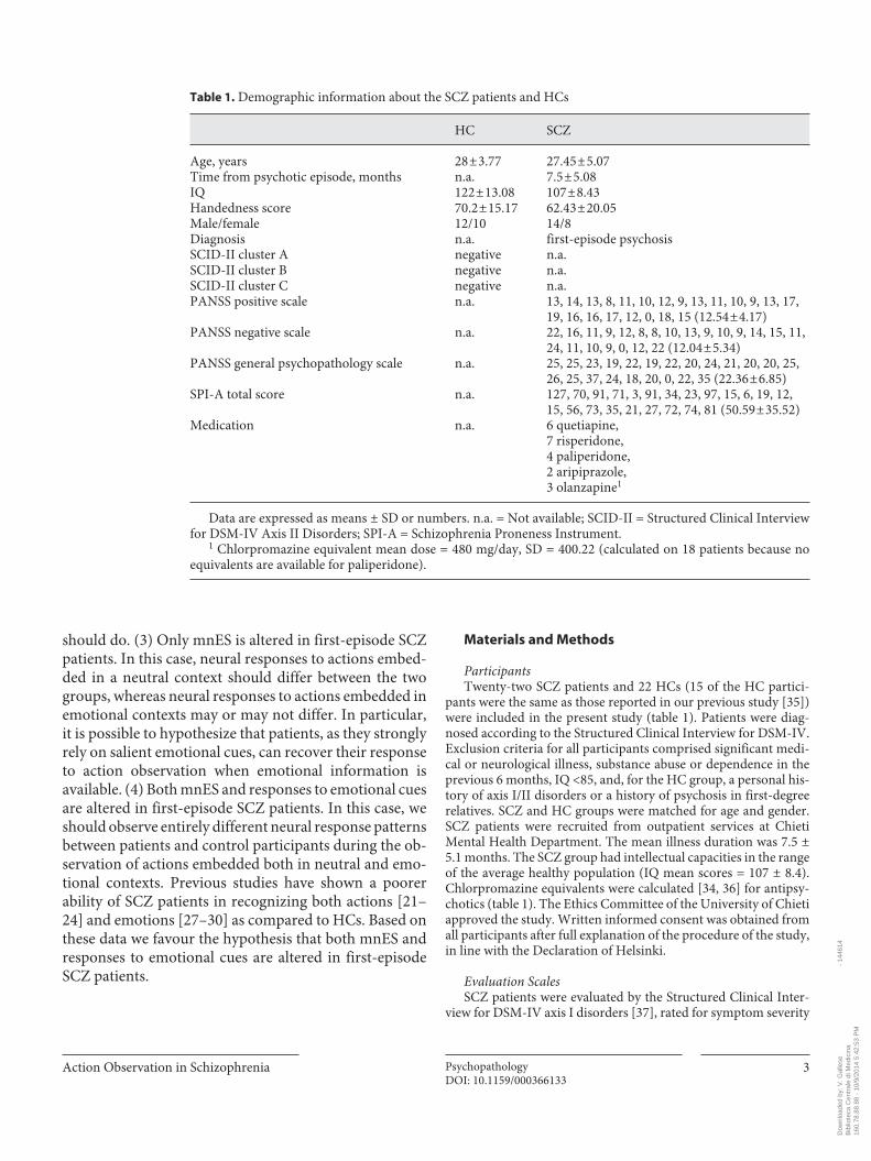
Participants Twenty-two SCZ patients and 22 HCs (15 of the HC partici-
pants were the same as those reported in our previous study [35] ) were included in the present study ( table 1 ). Patients were diag-nosed according to the Structured Clinical Interview for DSM-IV. Exclusion criteria for all participants comprised significant medi-cal or neurological illness, substance abuse or dependence in the previous 6 months, IQ <85, and, for the HC group, a personal his-tory of axis I/II disorders or a history of psychosis in first-degree relatives. SCZ and HC groups were matched for age and gender. SCZ patients were recruited from outpatient services at Chieti Mental Health Department. The mean illness duration was 7.5 ± 5.1 months. The SCZ group had intellectual capacities in the range of the average healthy population (IQ mean scores = 107 ± 8.4). Chlorpromazine equivalents were calculated [34, 36] for antipsy-chotics ( table 1 ). The Ethics Committee of the University of Chieti approved the study. Written informed consent was obtained from all participants after full explanation of the procedure of the study, in line with the Declaration of Helsinki.
Evaluation Scales SCZ patients were evaluated by the Structured Clinical Inter-
view for DSM-IV axis I disorders [37] , rated for symptom severity
Table 1. Demographic information about the SCZ patients and HCs
HC SCZ
Age, years 28 ± 3.77 27.45 ± 5.07Time from psychotic episode, months n.a. 7.5 ± 5.08IQ 122 ± 13.08 107 ± 8.43Handedness score 70.2 ± 15.17 62.43 ± 20.05Male/femaleDiagnosis
12/10n.a.
14/8first-episode psychosis
SCID-II cluster A negative n.a.SCID-II cluster B negative n.a.SCID-II cluster C negative n.a.PANSS positive scale n.a. 13, 14, 13, 8, 11, 10, 12, 9, 13, 11, 10, 9, 13, 17,
19, 16, 16, 17, 12, 0, 18, 15 (12.54 ± 4.17)PANSS negative scale n.a. 22, 16, 11, 9, 12, 8, 8, 10, 13, 9, 10, 9, 14, 15, 11,
24, 11, 10, 9, 0, 12, 22 (12.04 ± 5.34)PANSS general psychopathology scale n.a. 25, 25, 23, 19, 22, 19, 22, 20, 24, 21, 20, 20, 25,
26, 25, 37, 24, 18, 20, 0, 22, 35 (22.36 ± 6.85)SPI-A total score n.a. 127, 70, 91, 71, 3, 91, 34, 23, 97, 15, 6, 19, 12,
15, 56, 73, 35, 21, 27, 72, 74, 81 (50.59 ± 35.52)Medication n.a. 6 quetiapine,
7 risperidone,4 paliperidone,2 aripiprazole,3 olanzapine1
Data are expressed as means ± SD or numbers. n.a. = Not available; SCID-II = Structured Clinical Interview for DSM-IV Axis II Disorders; SPI-A = Schizophrenia Proneness Instrument.
1 Chlorpromazine equivalent mean dose = 480 mg/day, SD = 400.22 (calculated on 18 patients because no equivalents are available for paliperidone).
Dow
nloa
ded
by: V
. Gal
lese
- 1
4461
4B
iblio
teca
Cen
tral
e di
Med
icin
a16
0.78
.88.
88 -
10/
9/20
14 5
:42:
53 P
M

Ferri et al.
PsychopathologyDOI: 10.1159/000366133
4
with the Positive and Negative Symptom Scale (PANSS) [38] and evaluated for the presence of basic symptoms (BSs) [39] by means of the Schizophrenia Proneness Instrument [39] by two trained psychiatrists. HC participants were evaluated by means of the Structured Clinical Interview for DSM-IV axis II personality dis-orders [40] . SCZ patients were also required to complete the em-pathic quotient (EQ) questionnaire [41] . Evidence of the reliabil-ity and validity of the EQ has been reported in persons without psychosis [42] . A systematic investigation of the reliability and va-lidity of the EQ in persons with psychosis is still lacking. However, for previous use of the EQ with participants diagnosed with SCZ see, for example: Bora et al. [43] , Koelkebeck et al. [44] , Konstan-takopoulos et al. [45] and Lysaker et al. [46] .
fMRI Data Acquisition All images were collected with a 1.5-tesla Philips Achieva scan-
ner operating at the Institute of Advanced Biomedical Technolo-gies (ITAB G. d’Annunzio, Chieti, Italy). Functional images were acquired with a gradient echo echo-planar imaging sequence. Each subject underwent 4 scans, each including 216 consecutive vol-umes comprising 26 consecutive ascending 4-mm-thick slices ori-ented parallel to the anterior-posterior commissure and covering the whole brain (TR = 2.4 s, TE = 50 ms, 64 × 64 image matrix,4 × 4 mm in-plane resolution; FOV = 256 mm, no gap). A high-resolution structural image was acquired at the end of the session via a 3-dimensional magnetization-prepared rapid acquisition gradient echo pulse sequence (170 sagittal slices, voxel size: 1.25 × 1.25 × 1.20 mm, TR = 8.6 ms, TE = 4.0 ms, 192 × 192 image matrix, FOV = 240 mm).
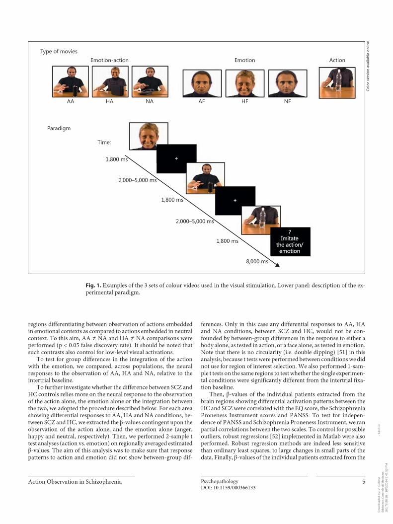
Stimuli and Conditions The experimental stimuli consisted of 3 sets of colour videos:
(1) ‘emotion action’, showing an actor (torso, face and arms of ei-ther a male or a female) grasping 1 of 4 different objects (bottle, pencil case, receiver or CD case placed on a table) with the right hand and facially expressing anger, happiness or no emotion; (2) ‘emotion’, showing only the face of the actor (either a male or a female) expressing anger, happiness or no emotion, and (3) ‘ac-tion’, showing only the hand action (the field of view was such that the face did not appear). Hence, the experiment comprised the fol-lowing 7 conditions: (1a) angry action (AA, the actor grasped an object expressing anger); (1b) happy action (HA, the actor grasped an object expressing happiness); (1c) neutral action (NA, the actor grasped an object with a neutral facial expression); (2a) angry face (AF, expression of anger); (2b) happy face (HF, expression of hap-piness); (2c) neutral face (NF, a face expressing no emotion); (3) action (one of the objects being grasped). All the emotion and ac-tion conditions were dynamic. The actors in the video clips were seen from a frontal point of view. Actors and different types of objects were presented in equal proportions. Two professional ac-tors, a female and a male, were enrolled as models for the videos [47] . The sex of the actor was counterbalanced across conditions. The kinematics of all the presented hand actions was identical in order to avoid that the action emotional context could be inferred by hand kinematics. To obtain such an identity, we applied the blue screen technique, that is a technique for compositing two images or frames together in which a colour (or a small colour range) from one image is removed (made transparent), revealing another im-age behind it. It was applied to our stimuli in order to superimpose on the same trunk different dynamic facial expressions.
Design and Procedure The rapid event-related fMRI paradigm consisted of 4 scans. In
each scan 12 videos were presented for each of the 7 experimental conditions (AA, HA, NA, action, AF, HF, NF). Each video lasted 1,800 ms and was preceded by a randomized, non-predictable in-tertrial interval ranging from 2,000 to 5,000 ms during which a black fixation cross was presented at the centre of a white screen ( fig. 1 ). Participants were instructed to carefully watch the whole scene. To make sure participants paid attention to the experimen-tal stimuli, 8 control trials were randomly inserted in the video sequence of each scan. These unpredictable trials were followed by a question mark lasting 2,000 ms followed by a written request (6,000 ms) to imitate either the action (4 trials) or the emotion (4 trials; fig. 1 ). In total, our experiment consisted of 336 passive ob-servation trials (48 for each experimental condition) and 32 imita-tion trials (16 for actions and 16 for emotions), presented in a pseu-dorandomized order.
Participants lay supine in the scanner with the arms out-stretched beside the abdomen. Visual stimuli were projected onto a backprojection screen situated behind the subject’s head and were visible in a mirror (10 × 15 cm). Sound-attenuating head-phones were used to muffle scanner noise. Participants were in-structed to carefully watch the whole scene.
fMRI Data Preprocessing and Analysis fMRI data were preprocessed and analysed using SPM8 (Well-
come Department of Cognitive Neurology, Institute of Neurology, London, UK). For each participant, functional images were first spatially corrected for head movements using a least-squares ap-proach and 6-parameter rigid body spatial transformations [48] . The realigned functional images were then corrected for difference in timing between slices, using the middle slice acquired in time as a reference. The high-resolution anatomical image and the func-tional images were coregistered and then stereotactically normal-ized to the Montreal Neurological Institute brain template used in SPM8. Functional images were resampled with a voxel size of 3 × 3 × 3 mm and spatially smoothed with a 3-dimensional gaussian filter of 8 mm full width at half maximum to accommodate ana-tomical variations between subjects [48] . Images were subsequent-ly analysed using a random-effects approach. At the first stage, the time series of functional MR images obtained from each partici-pant were analysed separately. The effects of the experimental par-adigm were estimated on a voxel-by-voxel basis, according to the general linear model extended to allow the analysis of fMRI data as a time series [49] . The onset of each trial constituted a neural event that was modelled through a canonical haemodynamic re-sponse function, chosen to represent the relationship between neuronal activation and blood flow changes [50] . Imitation and question mark periods were modelled as separate conditions and then excluded from further analyses.
These single-subject models were used to compute 7 contrast images per subject, each representing the estimated amplitude of the haemodynamic response in 1 of the 7 experimental conditions (AA, HA, NA, action, AF, HF, NF), relative to the intertrial base-line. These contrast images were used (i) to test the effect of emo-tional context on action observation in both populations separate-ly and (ii) to directly compare SCZ and HC. The effect of emo-tional context on action observation in both populations separately was analysed by means of whole-brain voxel-wise con-trasts. In particular, in each population we investigated cortical
Dow
nloa
ded
by: V
. Gal
lese
- 1
4461
4B
iblio
teca
Cen
tral
e di
Med
icin
a16
0.78
.88.
88 -
10/
9/20
14 5
:42:
53 P
M
Action Observation in Schizophrenia PsychopathologyDOI: 10.1159/000366133
5
regions differentiating between observation of actions embedded in emotional contexts as compared to actions embedded in neutral context. To this aim, AA ≠ NA and HA ≠ NA comparisons were performed (p < 0.05 false discovery rate). It should be noted that such contrasts also control for low-level visual activations.
To test for group differences in the integration of the action with the emotion, we compared, across populations, the neural responses to the observation of AA, HA and NA, relative to the intertrial baseline.
To further investigate whether the difference between SCZ and HC controls relies more on the neural response to the observation of the action alone, the emotion alone or the integration between the two, we adopted the procedure described below. For each area showing differential responses to AA, HA and NA conditions, be-tween SCZ and HC, we extracted the β-values contingent upon the observation of the action alone, and the emotion alone (anger, happy and neutral, respectively). Then, we performed 2-sample t test analyses (action vs. emotion) on regionally averaged estimated β-values. The aim of this analysis was to make sure that response patterns to action and emotion did not show between-group dif-
ferences. Only in this case any differential responses to AA, HA and NA conditions, between SCZ and HC, would not be con-founded by between-group differences in the response to either a body alone, as tested in action, or a face alone, as tested in emotion. Note that there is no circularity (i.e. double dipping) [51] in this analysis, because t tests were performed between conditions we did not use for region of interest selection. We also performed 1-sam-ple t tests on the same regions to test whether the single experimen-tal conditions were significantly different from the intertrial fixa-tion baseline.
Then, β-values of the individual patients extracted from the brain regions showing differential activation patterns between the HC and SCZ were correlated with the EQ score, the Schizophrenia Proneness Instrument scores and PANSS. To test for indepen-dence of PANSS and Schizophrenia Proneness Instrument, we ran partial correlations between the two scales. To control for possible outliers, robust regressions [52] implemented in Matlab were also performed. Robust regression methods are indeed less sensitive than ordinary least squares, to large changes in small parts of the data. Finally, β-values of the individual patients extracted from the
Type of movies
Paradigm
Time:
1,800 ms
2,000–5,000 ms
2,000–5,000 ms
1,800 ms
1,800 ms
8,000 ms
?Imitate
the action/emotion
+
+
Emotion-action Emotion Action
AA HA NA AF HF NF
Fig. 1. Examples of the 3 sets of colour videos used in the visual stimulation. Lower panel: description of the ex-perimental paradigm.
Colo
r ver
sion
ava
ilabl
e on
line
Dow
nloa
ded
by: V
. Gal
lese
- 1
4461
4B
iblio
teca
Cen
tral
e di
Med
icin
a16
0.78
.88.
88 -
10/
9/20
14 5
:42:
53 P
M
Ferri et al.
PsychopathologyDOI: 10.1159/000366133
6
brain regions showing differential activation patterns between the HCs and SCZ patients were also correlated with chlorpromazine equivalence values to test for unspecific drug-driven effects.
Results
Neural Mapping of Observing AAs and HAs Compared to NAs in HCs and SCZ Patients Due to data corruption 1 HC was discarded from the
analysis. When contrasting the effect of observing either AA or HA with NA in HCs (AA ≠ NA or HA ≠ NA, re-spectively), the following activation differences were found. Regarding AA relative to NA, higher activation was found in the left IFG (pars triangularis and pars opercu-laris), left pre-supplementary motor area (SMA), right in-ferior PCG, middle temporal gyrus/superior temporal sul-cus (MTG/STS) bilaterally and left middle optic chiasm (OC). Regarding HA, relative to NA, higher activation was
found in the MTG/STS, left fusiform gyrus, right lingual gyrus, middle and inferior OC bilaterally ( table 2 ; fig. 2 ).
The same contrasts while comparing AA relative to NA in SCZ patients revealed the following: higher activation in the left IFG encompassing the insula, the SMA, MTG/STS, the right middle frontal gyrus encompassing the pars opercularis and triangularis of the IFG, the left superior frontal gyrus and the left inferior parietal lobe. No voxels turned out to be significant in the contrast HA ≠ NA.
Group Differences in the Neural Activity while Observing NAs While comparing the neural activity contingent upon
the observation of an actor grasping the object with an NF, HCs showed higher activation, as compared to SCZ patients in the following areas: bilateral middle frontal gyrus, the right IFG (parts orbitalis, opercularis and tri-angularis), the left PCG and the left inferior parietal lobe ( table 3 ; fig. 3 ).
Table 2. Montreal Neurological Institute coordinates of peaks of relative activation in the cortical regions where the blood oxygen level-dependent signal was significantly different during observation of action-emotion versus NA in HCs and SCZ patients
Anatomical region Side Main local maxima
HC SCZ
x y z z-score x y z z-score
AA versus NAOC L/R –24 –99 –3 9.97MTG/STS R 57 –45 3 4.60 66 –45 9 3.45
L –51 –45 9 5.05 –60 –45 18 4.31PCG R 51 6 39 3.84SMA/pre-SMA L –3 6 66 4.35 –6 –3 72 3.78IFG – insula L –36 12 24 3.78 –51 9 –6 4.60
–39 –36 30 4.25MFG/IFG (opercular-
triangular)R 51 24 33 4.16
33 54 0 4.0333 24 –9 3.40
SFG L –15 12 63 3.45IPL L –48 –42 45 3.66
HA versus NAOC R 33 –93 –6 3.52LG R 21 –66 –6 4.27FG L –36 –63 –12 4.50MTG/STS R 54 –45 9 5.28
L –54 –42 9 5.27
SMA = Supplementary motor area; MFG = middle frontal gyrus; SFG = superior frontal gyrus; IPL = inferior parietal lobe; LG = lingual gyrus; FG = fusiform gyrus.
Dow
nloa
ded
by: V
. Gal
lese
- 1
4461
4B
iblio
teca
Cen
tral
e di
Med
icin
a16
0.78
.88.
88 -
10/
9/20
14 5
:42:
53 P
M
Action Observation in Schizophrenia PsychopathologyDOI: 10.1159/000366133
7
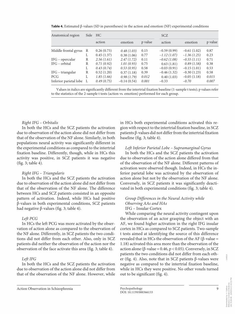
Increased activations in HCs were generally due to negative β-values in SCZ patients, which significantly dif-fered from baseline ( table 4 ), except for the PCG.
Two-sample t tests aimed at identifying the source of the difference between HC and SCZ patients are de-scribed below for each area. Detailed statistics are report-ed in table 4 .
Right Middle Frontal Gyrus In both HCs and SCZ patients the activation due to
observation of the action alone did not differ from that of the observation of the NF alone. Visual inspection of the data though suggested higher activation in HCs as com-pared to SCZ patients. To statistically examine this obser-vation, we tested whether estimated β-values in the two conditions of interest were significantly activated with re-spect to the intertrial fixation baseline in both popula-tions. The estimated β-value in the NF alone condition was higher than zero in HCs but not in SCZ patients ( fig. 3 ; table 4 ). Differently, estimated β-values in the ac-tion alone condition did not differ from the intertrial fix-ation baseline in both populations.
AA > NA AH > NA Overlapa
b
Fig. 2. Regions activated either in AA ver-sus NA contrast (red; colours in the online version only) or in HA versus NA (green) contrast in HCs ( a ) and SCZ patients ( b ).p < 0.05, false discovery rate corrected.
Colo
r ver
sion
ava
ilabl
e on
line
Table 3. Montreal Neurological Institute coordinates of peaks of differential activation between HCs and SCZ patients
Anatomical region Side Main local maxima
x y z z-score
AAIFG – insula R 39 24 –9 4.85
NAMiddle frontal gyrus R 33 45 18 4.36
L –30 45 33 4.57–45 51 0 4.48
IFG – orbital R 36 24 –9 4.56L –33 30 –6 3.91
IFG – triangular R 48 36 15 3.64IFG – opercular R 48 15 30 4.28PCG L –45 6 42 4.21Inferior parietal lobe L –45 –33 36 4.38
Dow
nloa
ded
by: V
. Gal
lese
- 1
4461
4B
iblio
teca
Cen
tral
e di
Med
icin
a16
0.78
.88.
88 -
10/
9/20
14 5
:42:
53 P
M
Ferri et al.
PsychopathologyDOI: 10.1159/000366133
8
Left Middle Frontal Gyrus In both HCs and SCZ patients the activation due to
observation of the action alone did not differ from that of the observation of the NF alone. The difference between the two groups was due to the fact that only in SCZ pa-tients was the activity in this area significantly lower in both the action alone condition and the NF alone condi-tion with respect to the intertrial fixation baseline ( fig. 3 ; table 4 ).
Right IFG – Opercular In both the HCs and the SCZ patients the activation
due to observation of the action alone did not differ from that of the observation of the NF alone. However, while in HCs both experimental conditions activated this re-gion with respect to the intertrial fixation baseline, in SCZ patients only the observation of the NF alone pro-duced a reliable activation as compared to baseline ( fig. 3 ; table 4 ).
LH-MFG3
RH-MFG
RH-IFG triangular
LH-IFG insula
LH-IPL
HC
SCZ
LH precentral
RH-IFG orbitalis
RH-IFG opercular
Action Emotion Action Emotion
Action Emotion
**
2
1
0
–1
–2
3
2
1
0
–1
–2
3
2
1
0
–1
–2
3210
–1–2
*
* *
*
*
*
**
* *
*
*
Fig. 3. Regions of relatively higher activation (and estimated β-values) in HCs as compared to SCZ patients during observation of the NA. Group activation data are rendered on the cortical surface of a ‘canonical’ brain [81] . MFG = Middle frontal gyrus; LH = left hemisphere; RH = right hemisphere; IPL = inferior parietal lobe. Asterisks indicate β-values significantly higher than the intertrial fixation baseline. Bars indicate significant differences between conditions.
Colo
r ver
sion
ava
ilabl
e on
line
Dow
nloa
ded
by: V
. Gal
lese
- 1
4461
4B
iblio
teca
Cen
tral
e di
Med
icin
a16
0.78
.88.
88 -
10/
9/20
14 5
:42:
53 P
M
Action Observation in Schizophrenia PsychopathologyDOI: 10.1159/000366133
9
Right IFG – Orbitalis In both the HCs and the SCZ patients the activation
due to observation of the action alone did not differ from that of the observation of the NF alone. Similarly, in both populations neural activity was significantly different in the experimental conditions as compared to the intertrial fixation baseline. Differently, though, while in HCs this activity was positive, in SCZ patients it was negative ( fig. 3 ; table 4 ).
Right IFG – Triangularis In both the HCs and the SCZ patients the activation
due to observation of the action alone did not differ from that of the observation of the NF alone. The difference between HCs and SCZ patients consisted in an opposite pattern of activation. Indeed, while HCs had positive β-values in both experimental conditions, SCZ patients had negative β-values ( fig. 3 ; table 4 ).
Left PCG In HCs the left PCG was more activated by the obser-
vation of action alone as compared to the observation of the NF alone. Differently, in SCZ patients the two condi-tions did not differ from each other. Also, only in SCZ patients did neither the observation of the action nor the observation of the face activate this area ( fig. 3 ; table 4 ).
Left IFG In both the HCs and the SCZ patients the activation
due to observation of the action alone did not differ from that of the observation of the NF alone. However, while
in HCs both experimental conditions activated this re-gion with respect to the intertrial fixation baseline, in SCZ patients β-values did not differ from the intertrial fixation baseline ( fig. 3 ; table 4 ).
Left Inferior Parietal Lobe – Supramarginal Gyrus In both the HCs and the SCZ patients the activation
due to observation of the action alone differed from that of the observation of the NF alone. Different patterns of activation were observed though. Indeed, in HCs the in-ferior parietal lobe was activated by the observation of action alone but not by the observation of the NF alone. Conversely, in SCZ patients it was significantly deacti-vated in both experimental conditions ( fig. 3 ; table 4 ).
Group Differences in the Neural Activity while Observing AAs and HAs IFG – Insular Cortex While comparing the neural activity contingent upon
the observation of an actor grasping the object with an AF, we found higher activation in the right IFG insular cortex in HCs as compared to SCZ patients. Two-sample t tests aimed at identifying the source of this difference revealed that in HCs the observation of the AF (β-value = 1.18) activated this area more than the observation of the action alone (β-value = 0.46, p < 0.05). Conversely, in SCZ patients the two conditions did not differ from each oth-er ( fig. 4 ). Also, note that in SCZ patients β-values were negative as compared to the intertrial fixation baseline, while in HCs they were positive. No other voxels turned out to be significant ( fig. 4 ).
Table 4. Estimated β-values (SD in parentheses) in the action and emotion (NF) experimental conditions
Anatomical region Side HC SCZ
action emotion p value actio n emotion p value
Middle frontal gyrus R 0.26 (0.75) 0.48 (1.03) 0.15 –0.59 (0.99) –0.61 (1.02) 0.87L 0.45 (1.37) 0.38 (1.06) 0.77 –1.12 (1.07) –1.36 (1.25) 0.23
IFG – opercular R 2.56 (1.61) 2.47 (1.72) 0.11 –0.62 (1.08) –0.55 (1.11) 0.71IFG – orbital R 0.71 (0.92) 1.01 (0.93) 0.75 0.63 (1.81) 0.89 (1.58) 0.38
L 0.43 (0.74) 0.53 (0.95) 0.58 –0.03 (0.91) –0.15 (1.01) 0.53IFG – triangular R 0.52 (1.20) 0.37 (1.18) 0.39 –0.46 (1.32) –0.30 (1.23) 0.58PCG L 1.85 (1.66) 0.98 (1.79) 0.012 0.40 (1.03) –0.05 (1.18) 0.015Inferior parietal lobe L 0.49 (0.75) –0.14 (0.54) 0.001 –0.33 –0.70 0.007
Values in italics are significantly different from the intertrial fixation baseline (1-sample t tests); p values refer to the statistics of the 2-sample t tests (action vs. emotion) performed for each group.
Dow
nloa
ded
by: V
. Gal
lese
- 1
4461
4B
iblio
teca
Cen
tral
e di
Med
icin
a16
0.78
.88.
88 -
10/
9/20
14 5
:42:
53 P
M
Ferri et al.
PsychopathologyDOI: 10.1159/000366133
10
No differences were found contingent upon the obser-vation of HAs.
Correlations between Differential Brain Activity with EQ Scores, Schizophrenia Proneness Instrument Scores, PANSS and Chlorpromazine Equivalences Discovering the possible relation between measures of
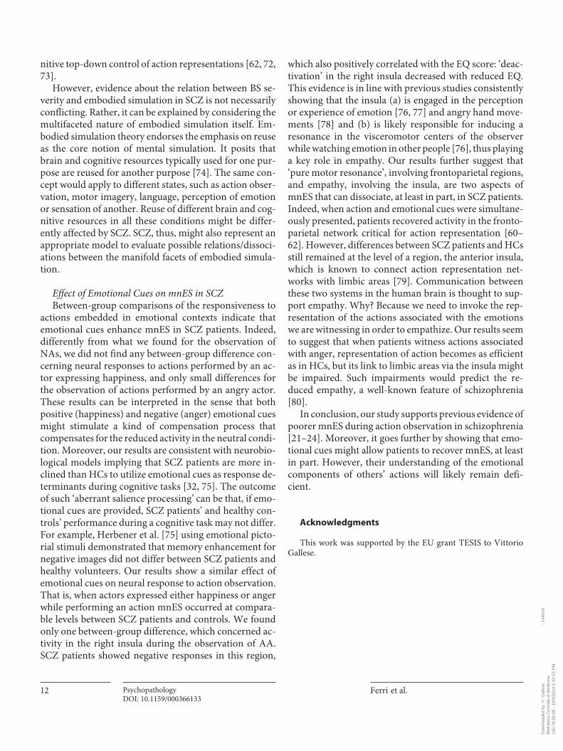
empathy and symptomatology as measured by the Schizo-
phrenia Proneness Instrument scores and PANSS, and neural activity contingent upon the observation of ac-tions performed in either a neutral or an emotional con-text is crucial for understanding the nature of the phe-nomena called into play in our experimental conditions. We found that the activation of the left inferior parietal lobe (i.e. supramarginal gyrus), while observing the NA, was negatively correlated with the total score to the Schizophrenia Proneness Instrument (r = –0.60, p < 0.01; fig. 5 ). This correlation remained significant also control-ling for the PANSS score, thus suggesting that the two scales operationalize different aspects of the psychopa-thology. PANSS scores did not correlate with the blood oxygen level-dependent signal in any activated cortical region.
We also found that the activation of the right anterior insula while observing the AA was positively correlated with the EQ (r = 0.46, p < 0.05; fig. 4 ). Both correlations survived the robust regression analysis.
Finally, no significant covariance effect was found for chlorpromazine equivalences in the SCZ patients, sug-gesting that there was no linear relationship between medication dose and differential activation between the HCs and SCZ patients.
HC SCZ
Action Emotion
3
2
1
0
0–6
–1
–2
*
* *
102030405060
*
–5 –4 –3 –2 –1 0
r = 0.46
1 2
Estimated -value
Estim
ated
-v
alue
EQ
a
b c
Fig. 4. a Group differences in the neuralactivity while observing AA. b Estimated β-values in the action alone and emotion alone conditions in HCs and SCZ patients. c Correlation between estimated β-value in the right anterior insula and EQ scores in SCZ patients. The line represents the linear best fit; r refers to the coefficient of corre-lation. The correlation survived also therobust fit correlation. Asterisks indicate β-values significantly higher (p < 0.05) than the intertrial fixation baseline. Bars indicate significant differences between conditions.
Colo
r ver
sion
ava
ilabl
e on
line
2.52.01.5
00–0.5 140
–1.0–1.5–2.0–2.5
1.00.5
120100
BSs
80604020
r = – 0.60
Estim
ated
-v
alue
Fig. 5. Correlation between estimated β-value in the left inferior parietal lobe and the Schizophrenia Proneness Instrument scores. The correlation survived also the robust fit correlation.
Dow
nloa
ded
by: V
. Gal
lese
- 1
4461
4B
iblio
teca
Cen
tral
e di
Med
icin
a16
0.78
.88.
88 -
10/
9/20
14 5
:42:
53 P
M

Action Observation in Schizophrenia PsychopathologyDOI: 10.1159/000366133
11
Discussion
The present study aimed at investigating whether and how emotional cues modulate brain activity underlying action processing in first-episode SCZ patients, as com-pared to HCs. Facial expression of emotions, in particu-lar, may cue intentions behind actions. That might facili-tate embodied simulation and trigger an appropriate re-action [35] . In the present study we presented grasping actions either associated (HA, AA) or not (NA) with emotional cues (i.e. dynamic facial expression of emo-tions). These experimental conditions allowed us to test whether mnES per se or its modulation by contextual in-formation is impaired in SCZ patients.
mnES in SCZ Previous studies investigating potential alterations of
mnES in SCZ showed contrasting results [53–58] . Possi-ble explanations of such incongruities may relate to dif-fering sample sizes, medications, different impact of pos-itive and negative symptoms within the studied patients’ cohort, and methods used across studies. Other critical sources of conflicting findings might concern different tasks and stimuli used in each investigation, as well as dif-ferent contexts in which actions were presented. Indeed, mnES seems to be modulated by contextual information, for example, pragmatic [59] and emotional [35] .
We found differences in mnES between SCZ patients and healthy participants already for the observation of NA, that is when emotional cues were not provided by the context. Such differences mainly involved the frontopari-etal network critical for action representation [60–62] , such as the IFG, PCG and inferior parietal lobe.
Imaging data in humans suggest a role of the IFG and PCG in coding the motor goal of the action [63] and, from another perspective, in the understanding of the agent’s motor intention, driven by the context in which the ac-tion is embedded [59] . Moreover, data from action imita-tion and observation studies [59, 64] demonstrate that the inferior parietal lobe, in addition to the IFG, is involved in coding the abstract aspects of actions (such as their mo-tor intention). According to the previous literature and based on our data, we would then conclude that all these aspects of action processing are affected in SCZ patients.
Interestingly, activation in the inferior parietal lobe in these patients correlated negatively with BS severity, as assessed by the Schizophrenia Proneness Instrument: ac-tivation in the inferior parietal lobe decreased with aug-mented symptom severity. This relationship was found between NA observation conditions and BS severity, but
not with observation of actions embedded in emotional contexts. The detected relationship between cortical pro-cesses and BSs highlights the clinical relevance of the re-sults, especially concerning preventive approaches and early diagnosis. BSs can occur and have been reported in every stage of the illness, i.e. in the prodrome to the first psychotic episode, in prodromes to relapse, in residual states and even during psychotic episodes per se [65] . Moreover, BSs represent a link between a phenomeno-logical approach to psychopathology and a categorical approach based on positive and negative symptoms. Spe-cifically, BSs could gradually increase in number and se-verity, even many years before psychosis onset, and fi-nally develop into psychotic symptoms, such as negative and positive symptoms [39, 65] . Interestingly, we did not detect any relationship between activation patterns and positive and negative symptoms, and the correlation be-tween altered activation patterns in SCZ and BSs was in-dependent of positive and negative symptoms. This may underscore the relevance and specificity of BSs, tapping into distinct neural mechanisms. A possible alternative explanation is that the included SCZ patients had a very recent illness onset and relatively low PANSS scores.
Finally, as BSs represent subjective experiential distur-bances in the domains of cognition, perception, bodily experience, action and affective states [66, 67] , our results support a close and specific link between self-experience anomalies and mnES, which supports intersubjective re-lationships in social everyday life [20, 68, 69] .
All these findings are consistent with those previously obtained by Ebisch et al. [6] , which showed activation in the ventral premotor cortex for the observation of inter-personal touch decreasing with increased BS severity. A subsequent study [70] demonstrated a link between BS severity and functional connectivity of the ventral premo-tor cortex with the posterior cingulate cortex, a brain re-gion central in mediating self-experience, further sug-gesting a deranged relationship between an afflicted self and its social environment underlying certain social cog-nition deficits in SCZ.
Interestingly, however, embodied simulation and BS severity have not always been found to negatively corre-late in first-episode SCZ patients. Ferri et al. [71] indeed found that patients with severer symptomatology show a prolonged higher efficiency than HCs, in processing ac-tion-related verbs. According to the authors, this result would indicate that neural activity supporting embodied simulation during action-verb understanding is not itself reduced in SCZ. Possible alterations may be related to hi-erarchically higher-order processes, involved in the cog-
Dow
nloa
ded
by: V
. Gal
lese
- 1
4461
4B
iblio
teca
Cen
tral
e di
Med
icin
a16
0.78
.88.
88 -
10/
9/20
14 5
:42:
53 P
M
Ferri et al.
PsychopathologyDOI: 10.1159/000366133
12
nitive top-down control of action representations [62, 72, 73] .
However, evidence about the relation between BS se-verity and embodied simulation in SCZ is not necessarily conflicting. Rather, it can be explained by considering the multifaceted nature of embodied simulation itself. Em-bodied simulation theory endorses the emphasis on reuse as the core notion of mental simulation. It posits that brain and cognitive resources typically used for one pur-pose are reused for another purpose [74] . The same con-cept would apply to different states, such as action obser-vation, motor imagery, language, perception of emotion or sensation of another. Reuse of different brain and cog-nitive resources in all these conditions might be differ-ently affected by SCZ. SCZ, thus, might also represent an appropriate model to evaluate possible relations/dissoci-ations between the manifold facets of embodied simula-tion.
Effect of Emotional Cues on mnES in SCZ Between-group comparisons of the responsiveness to
actions embedded in emotional contexts indicate that emotional cues enhance mnES in SCZ patients. Indeed, differently from what we found for the observation of NAs, we did not find any between-group difference con-cerning neural responses to actions performed by an ac-tor expressing happiness, and only small differences for the observation of actions performed by an angry actor. These results can be interpreted in the sense that both positive (happiness) and negative (anger) emotional cues might stimulate a kind of compensation process that compensates for the reduced activity in the neutral condi-tion. Moreover, our results are consistent with neurobio-logical models implying that SCZ patients are more in-clined than HCs to utilize emotional cues as response de-terminants during cognitive tasks [32, 75] . The outcome of such ‘aberrant salience processing’ can be that, if emo-tional cues are provided, SCZ patients’ and healthy con-trols’ performance during a cognitive task may not differ. For example, Herbener et al. [75] using emotional picto-rial stimuli demonstrated that memory enhancement for negative images did not differ between SCZ patients and healthy volunteers. Our results show a similar effect of emotional cues on neural response to action observation. That is, when actors expressed either happiness or anger while performing an action mnES occurred at compara-ble levels between SCZ patients and controls. We found only one between-group difference, which concerned ac-tivity in the right insula during the observation of AA. SCZ patients showed negative responses in this region,
which also positively correlated with the EQ score: ‘deac-tivation’ in the right insula decreased with reduced EQ. This evidence is in line with previous studies consistently showing that the insula (a) is engaged in the perception or experience of emotion [76, 77] and angry hand move-ments [78] and (b) is likely responsible for inducing a resonance in the visceromotor centers of the observer while watching emotion in other people [76] , thus playing a key role in empathy. Our results further suggest that ‘pure motor resonance’, involving frontoparietal regions, and empathy, involving the insula, are two aspects of mnES that can dissociate, at least in part, in SCZ patients. Indeed, when action and emotional cues were simultane-ously presented, patients recovered activity in the fronto-parietal network critical for action representation [60–62] . However, differences between SCZ patients and HCs still remained at the level of a region, the anterior insula, which is known to connect action representation net-works with limbic areas [79] . Communication between these two systems in the human brain is thought to sup-port empathy. Why? Because we need to invoke the rep-resentation of the actions associated with the emotions we are witnessing in order to empathize. Our results seem to suggest that when patients witness actions associated with anger, representation of action becomes as efficient as in HCs, but its link to limbic areas via the insula might be impaired. Such impairments would predict the re-duced empathy, a well-known feature of schizophrenia [80] .
In conclusion, our study supports previous evidence of poorer mnES during action observation in schizophrenia [21–24] . Moreover, it goes further by showing that emo-tional cues might allow patients to recover mnES, at least in part. However, their understanding of the emotional components of others’ actions will likely remain defi-cient.
Acknowledgments
This work was supported by the EU grant TESIS to Vittorio Gallese.
Dow
nloa
ded
by: V
. Gal
lese
- 1
4461
4B
iblio
teca
Cen
tral
e di
Med
icin
a16
0.78
.88.
88 -
10/
9/20
14 5
:42:
53 P
M

Action Observation in Schizophrenia PsychopathologyDOI: 10.1159/000366133
13
References
1 Green MF, Bearden CE, Cannon TD, Fiske AP, Hellemann GS, Horan WP, Kee K, Kern RS, Lee J, Sergi MJ, Subotnik KL, Sugar CA, Ventura J, Yee CM, Nuechterlein KH: Social cognition in schizophrenia. 1. Performance across phase of illness. Schizophr Bull 2012; 38: 854–864.
2 Fujino J, Takahashi H, Miyata J, Sugihara G, Kubota M, Sasamoto A, Fujiwara H, Aso T, Fukuyama H, Murai T: Impaired empathic abilities and reduced white matter integrity in schizophrenia. Prog Neuropsychopharmacol Biol Psychiatry 2014; 48: 117–123.
3 Sestito M, Umilta MA, De Paola G, Fortunati R, Raballo A, Leuci E, Maffei S, Tonna M, Amore M, Maggini C, Gallese V: Facial reac-tions in response to dynamic emotional stim-uli in different modalities in patients suffering from schizophrenia: a behavioral and EMG study. Front Hum Neurosci 2013; 7: 368.
4 Ripoll LH, Zaki J, Perez-Rodriguez MM, Sny-der R, Strike KS, Boussi A, Bartz JA, Ochsner KN, Siever LJ, New AS: Empathic accuracy and cognition in schizotypal personality dis-order. Psychiatry Res 2013; 210: 232–241.
5 Bora E, Pantelis C: Theory of mind impair-ments in first-episode psychosis, individuals at ultra-high risk for psychosis and in first-degree relatives of schizophrenia: systematic review and meta-analysis. Schizophr Res 2013; 144: 31–36.
6 Ebisch SJ, Salone A, Ferri F, De Berardis D, Romani GL, Ferro FM, Gallese V: Out of touch with reality? Social perception in first-episode schizophrenia. Soc Cogn Affect Neu-rosci 2013; 8: 394–403.
7 Ferri F, Frassinetti F, Mastrangelo F, Salone A, Ferro FM, Gallese V: Bodily self and schizophrenia: the loss of implicit self-body knowledge. Conscious Cogn 2012; 21: 1365–1374.
8 Liepelt R, Schneider JC, Aichert DS, Wost-mann N, Dehning S, Moller HJ, Riedel M, Dolk T, Ettinger U: Action blind: disturbed self-other integration in schizophrenia. Neu-ropsychologia 2012; 50: 3775–3780.
9 Dalmaso M, Galfano G, Tarqui L, Forti B, Castelli L: Is social attention impaired in schizophrenia? Gaze, but not pointing ges-tures, is associated with spatial attention defi-cits. Neuropsychology 2013; 27: 608–613.
10 Van’t Wout M, van Rijn S, Jellema T, Kahn RS, Aleman A: Deficits in implicit attention to social signals in schizophrenia and high risk groups: behavioural evidence from a new illu-sion. PLoS One 2009; 4:e5581.
11 Kring AM, Elis O: Emotion deficits in people with schizophrenia. Annu Rev Clin Psychol 2013; 9: 409–433.
12 Gallese V: Mirror neurons, embodied simula-tion and a second-person approach to mind-reading. Cortex 2013; 49: 2954–2956.
13 Di Pellegrino G, Fadiga L, Fogassi L, Gallese V, Rizzolatti G: Understanding motor events: a neurophysiological study. Exp Brain Res 1992; 91: 176–180.
14 Gallese V, Fadiga L, Fogassi L, Rizzolatti G: Action recognition in the premotor cortex. Brain 1996; 119: 593–609.
15 Gallese V, Gernsbacher MA, Heyes C, Hickok G, Iacoboni M: Mirror neuron forum. Per-spect Psychol Sci 2011; 6: 369–407.
16 Vigneswaran G, Philipp R, Lemon RN, Kras-kov A: M1 corticospinal mirror neurons and their role in movement suppression during action observation. Curr Biol 2013; 23: 236–243.
17 De Araujo MF, Hori E, Maior RS, Tomaz C, Ono T, Nishijo H: Neuronal activity of the an-terior cingulate cortex during an observation-based decision making task in monkeys. Be-hav Brain Res 2012; 230: 48–61.
18 Rizzolatti G, Fogassi L, Gallese V: Neurophys-iological mechanisms underlying the under-standing and imitation of action. Nat Rev Neurosci 2001; 2: 661–670.
19 Pineda JA: Mirror Neuron Systems. New York, Humana Press, 2009.
20 Gallese V: The roots of empathy: the shared manifold hypothesis and the neural basis of intersubjectivity. Psychopathology 2003; 36: 171–180.
21 Enticott PG, Hoy KE, Herring SE, Johnston PJ, Daskalakis ZJ, Fitzgerald PB: Reduced mo-tor facilitation during action observation in schizophrenia: a mirror neuron deficit? Schizophr Res 2008; 102: 116–121.
22 Mehta UM, Thirthalli J, Basavaraju R, Gangadhar BN, Pascual-Leone A: Reduced mirror neuron activity in schizophrenia and its association with theory of mind deficits: evidence from a transcranial magnetic stimu-lation study. Schizophr Bull 2014; 40: 1083–1094.
23 Schurmann M, Jarvelainen J, Avikainen S, Cannon TD, Lonnqvist J, Huttunen M, Hari R: Manifest disease and motor cortex reactiv-ity in twins discordant for schizophrenia. Br J Psychiatry 2007; 191: 178–179.
24 Kato Y, Muramatsu T, Kato M, Shibukawa Y, Shintani M, Mimura M: Magnetoencephalog-raphy study of right parietal lobe dysfunction of the evoked mirror neuron system in anti-psychotic-free schizophrenia. PLoS One 2011; 6:e28087.
25 Horan WP, Pineda JA, Wynn JK, Iacoboni M, Green MF: Some markers of mirroring ap-pear intact in schizophrenia: evidence from mu suppression. Cogn Affect Behav Neurosci 2014; 14: 1049–1060.
26 McCormick LM, Brumm MC, Beadle JN,Paradiso S, Yamada T, Andreasen N: Mirror neuron function, psychosis, and empathy in schizophrenia. Psychiatry Res 2012; 201: 233–239.
27 Ventura J, Wood RC, Jimenez AM, Helle-mann GS: Neurocognition and symptoms identify links between facial recognition and emotion processing in schizophrenia: meta-analytic findings. Schizophr Res 2013;151:78-84.
28 Ventura J, Wood RC, Jimenez AM, Helle-mann GS: Neurocognition and symptoms identify links between facial recognition and emotion processing in schizophrenia: meta-analytic findings. Schizophr Res 2013; 151: 78–84.
29 Saracco-Alvarez R, Fresan A, Escamilla-Orozco R: Facial emotion recognition in schizophrenia: a comparison with siblings and control subjects. Schizophr Res 2013; 151: 291–292.
30 Simpson C, Pinkham AE, Kelsven S, Sasson NJ: Emotion recognition abilities across stim-ulus modalities in schizophrenia and the role of visual attention. Schizophr Res 2013; 151: 102–106.
31 Hahn B, Robinson BM, Kaiser ST, Harvey AN, Beck VM, Leonard CJ, Kappenman ES, Luck SJ, Gold JM: Failure of schizophrenia patients to overcome salient distractors dur-ing working memory encoding. Biol Psychia-try 2010; 68: 603–609.
32 Patrick RE, Christensen BK: Reduced direct-ed forgetting for negative words suggests schizophrenia-related disinhibition of emo-tional cues. Psychol Med 2013; 43: 2289–2299.
33 Henik A, Salo R: Schizophrenia and the Stroop effect. Behav Cogn Neurosci Rev 2004; 3: 42–59.
34 Woods SW: Chlorpromazine equivalent dos-es for the newer atypical antipsychotics. J Clin Psychiatry 2003; 64: 663–667.
35 Ferri F, Ebisch SJ, Costantini M, Salone A, Ar-ciero G, Mazzola V, Ferro FM, Romani GL, Gallese V: Binding action and emotion in so-cial understanding. PLoS One 2013; 8:e54091.
36 Lehman AF, Lieberman JA, Dixon LB, Mc-Glashan TH, Miller AL, Perkins DO, Kreyen-buhl J; American Psychiatric Association Steering Committee on Practice Group: Prac-tice guideline for the treatment of patients with schizophrenia, second edition. Am J Psy-chiatry 2004; 161: 1–56.
37 First MB, Spitzer RL, Gibbon M, Williams JBW: Structured Clinical Interview for DSM-IV Axis I Disorders, Clinician Version (SCID-CV). Washington, American Psychiatric Press, 1996.
38 Kay SR, Fiszbein A, Opler LA: The positive and negative syndrome scale (PANSS) for schizophrenia. Schizophr Bull 1987; 13: 261–276.
39 Klosterkötter J, Hellmich M, Steinmeyer EM, Schultze-Lutter F: Diagnosing schizophrenia in the initial prodromal phase. Arch Gen Psy-chiatry 2001; 58: 158–164.
Dow
nloa
ded
by: V
. Gal
lese
- 1
4461
4B
iblio
teca
Cen
tral
e di
Med
icin
a16
0.78
.88.
88 -
10/
9/20
14 5
:42:
53 P
M
Ferri et al.
PsychopathologyDOI: 10.1159/000366133
14
40 First MB, Gibbon M, Spitzer RL: Structured Clinical Interview for DSM-IV Axis II Per-sonality Disorders (SCID-II, Version 2.0). New York, Biometric Research Department, New York State Psychiatric Institute, 1996.
41 Baron-Cohen S, Wheelwright S: The empathy quotient: an investigation of adults with As-perger syndrome or high functioning autism, and normal sex differences. J Autism Dev Dis-ord 2004; 34: 163–175.
42 Lawrence EJ, Shaw P, Baker D, Baron-Cohen S, David AS: Measuring empathy: reliability and validity of the empathy quotient. Psychol Med 2004; 34: 911–919.
43 Bora E, Gokcen S, Veznedaroglu B: Empathic abilities in people with schizophrenia. Psychi-atry Res 2008; 160: 23–29.
44 Koelkebeck K, Pedersen A, Suslow T, Kuep-pers KA, Arolt V, Ohrmann P: Theory of mind in first-episode schizophrenia patients: correlations with cognition and personality traits. Schizophr Res 2010; 119: 115–123.
45 Konstantakopoulos G, Oulis P, Ploumpidis D, Patrikelis P, Nikitopoulou S, Papadimitri-ou GN, David AS: Self-rated and perfor-mance-based empathy in schizophrenia: the impact of cognitive deficits. Soc Neurosci 2014; 27: 1–11.
46 Lysaker PH, Hasson-Ohayon I, Kravetz S, Kent JS, Roe D: Self perception of empathy in schizophrenia: emotion recognition, insight, and symptoms predict degree of self and in-terviewer agreement. Psychiatry Res 2013; 206: 146–150.
47 Mazzola V, Vuilleumier P, Latorre V, Petito A, Gallese V, Popolizio T, Arciero G, Bondol-fi G: Effects of emotional contexts on cerebel-lo-thalamo-cortical activity during action ob-servation. PLoS One 2013; 8:e75912.
48 Friston KJ, Ashburner J, Frith CD, Poline JB, Heather JD, Frackowiak RSJ: Spatial registra-tion and normalization of images. Hum Brain Mapp 1995; 2: 165–189.
49 Worsley KJ, Friston KJ: Analysis of fMRI time-series revisited – again. Neuroimage 1995; 2: 173–181.
50 Friston KJ, Fletcher P, Josephs O, Holmes A, Rugg MD, Turner R: Event-related fMRI: characterizing differential responses. Neuro-image 1998; 7: 30–40.
51 Kriegeskorte N, Simmons WK, Bellgowan PSF, Baker CI: Circular analysis in systems neuroscience: the dangers of double dipping. Nat Neurosci 2009; 12: 535–540.
52 Huber PJ: Robust estimation of a location pa-rameter. Ann Stat 1964; 35: 73–101.
53 Piefke M, Kramer K, Korte M, Schulte-Rüther M, Korte JM, Wohlschläger AM, Weber J, Shah NJ, Huber W, Fink GR: Neurofunction-al modulation of brain regions by distinct forms of motor cognition and movement fea-tures. Hum Brain Mapp 2009; 30: 432–451.
54 Sato W, Kochiyama T, Yoshikawa S, Naito E, Matsumura M: Enhanced neural activity in response to dynamic facial expressions of emotion: an fMRI study. Cogn Brain Res 2004; 20: 81–91.
55 Thioux M, Gazzola V, Keysers C: Action un-derstanding: how, what and why. Curr Biol 2008; 18:R431–R434.
56 Van de Riet WA, Grezes J, de Gelder B: Spe-cific and common brain regions involved in the perception of faces and bodies and the representation of their emotional expres-sions. Soc Neurosci 2009; 4: 101–120.
57 Gallese V: Mirror neurons, embodied simula-tion, and the neural basis of social identifica-tion. Psychoanal Dialog 2009; 19: 519–536.
58 Rizzolatti G, Sinigaglia C: Further reflections on how we interpret the actions of others. Na-ture 2008; 455: 589.
59 Iacoboni M, Molnar-Szakacs I, Gallese V, Buccino G, Mazziotta JC, Rizzolatti G: Grasp-ing the intentions of others with one’s own mirror neuron system. PLoS Biol 2005; 3:e79.
60 Buccino G, Binkofski F, Fink GR, Fadiga L, Fogassi L, Gallese V, Seitz RJ, Zilles K, Rizzo-latti G, Freund HJ: Action observation acti-vates premotor and parietal areas in a somato-topic manner: an fMRI study. Eur J Neurosci 2001; 13: 400–404.
61 Buccino G, Lui F, Canessa N, Patteri I, Lagra-vinese G, Benuzzi F, Porro CA, Rizzolatti G: Neural circuits involved in the recognition of actions performed by nonconspecifics: an fMRI study. J Cogn Neurosci 2004; 16: 114–126.
62 Buccino G, Vogt S, Ritzl A, Fink GR, Zilles K, Freund HJ, Rizzolatti G: Neural circuits un-derlying imitation learning of hand actions: an event-related fMRI study. Neuron 2004; 42: 323–334.
63 Johnson-Frey SH, Maloof FR, Newman-Nor-lund R, Farrer C, Inati S, Grafton ST: Actions or hand-object interactions? Human inferior frontal cortex and action observation. Neuron 2003; 39: 1053–1058.
64 Hamilton AF, Grafton ST: Goal representa-tion in human anterior intraparietal sulcus. J Neurosci 2006; 26: 1133–1137.
65 Schultze-Lutter F: Subjective symptoms of schizophrenia in research and the clinic: the basic symptom concept. Schizophr Bull 2009; 35: 5–8.
66 Huber G: The concept of substrate-close basic symptoms and its significance for the theory and therapy of schizophrenic diseases (in German). Nervenarzt 1983; 54: 23–32.
67 Klosterkotter J: The meaning of basic symp-toms for the genesis of the schizophrenic nu-clear syndrome. Jpn J Psychiatry Neurol 1992; 46: 609–630.
68 Gallese V: The manifold nature of interper-sonal relations: the quest for a common mechanism. Philos Trans R Soc Lond B Biol Sci 2003; 358: 517–528.
69 Gallese V: Motor abstraction: a neuroscien-tific account of how action goals and inten-tions are mapped and understood. Psychol Res 2009; 73: 486–498.
70 Ebisch SJ, Mantini D, Northoff G, Salone A, De Berardis D, Ferri F, Ferro FM, Di Gian-nantonio M, Romani GL, Gallese V: Altered brain long-range functional interactions un-derlying the link between aberrant self-expe-rience and self-other relationship in first-epi-sode schizophrenia. Schizophr Bull 2014; 40: 1072–1080.
71 Ferri F, Salone A, Ebisch SJ, De Berardis D, Romani GL, Ferro FM, Gallese V: Action verb understanding in first-episode schizophrenia: is there evidence for a simulation deficit? Neuropsychologia 2012; 50: 988–996.
72 Badre D: Cognitive control, hierarchy, and the rostro-caudal organization of the frontal lobes. Trends Cogn Sci 2008; 12: 193–200.
73 Vogt S, Buccino G, Wohlschlager AM, Ca-nessa N, Shah NJ, Zilles K, Eickhoff SB, Freund HJ, Rizzolatti G, Fink GR: Prefrontal involvement in imitation learning of hand ac-tions: effects of practice and expertise. Neuro-image 2007; 37: 1371–1383.
74 Gallese V, Sinigaglia C: How the body in ac-tion shapes the self. J Consc Stud 2011; 18: 117–143.
75 Herbener ES, Rosen C, Khine T, Sweeney JA: Failure of positive but not negative emotional valence to enhance memory in schizophrenia. J Abnorm Psychol 2007; 116: 43–55.
76 Wicker B, Keysers C, Plailly J, Royet JP, Gal-lese V, Rizzolatti G: Both of us disgusted in my insula: the common neural basis of seeing and feeling disgust. Neuron 2003; 40: 655–664.
77 Phillips ML, Williams LM, Heining M, Herba CM, Russell T, Andrew C, Bullmore ET, Brammer MJ, Williams SC, Morgan M, Young AW, Gray JA: Differential neural re-sponses to overt and covert presentations of facial expressions of fear and disgust. Neuro-image 2004; 21: 1484–1496.
78 Grosbras M-H, Paus T: Brain networks in-volved in viewing angry hands or faces. Cereb Cortex 2006; 16: 1087–1096.
79 Carr L, Iacoboni M, Dubeau M-C, Mazziotta JC, Lenzi GL: Neural mechanisms of empathy in humans: a relay from neural systems for imitation to limbic areas. Proc Natl Acad Sci 2003; 100: 5497–5502.
80 Derntl B, Finkelmeyer A, Toygar TK, Huls-mann A, Schneider F, Falkenberg DI, Habel U: Generalized deficit in all core components of empathy in schizophrenia. Schizophr Res 2009; 108: 197–206.
81 Mazziotta JC, Toga AW, Evans A, Fox P, Lan-caster J: A probabilistic atlas of the human brain: theory and rationale for its develop-ment. The International Consortium for Brain Mapping (ICBM). Neuroimage 1995; 2: 89–101.
Dow
nloa
ded
by: V
. Gal
lese
- 1
4461
4B
iblio
teca
Cen
tral
e di
Med
icin
a16
0.78
.88.
88 -
10/
9/20
14 5
:42:
53 P
M