Fenofibrate enhances barrier function of endothelial continuum within the metastatic niche of...
14
1. Introduction 2. Methods 3. Results 4. Discussion 5. Conclusions Original Research Fenofibrate enhances barrier function of endothelial continuum within the metastatic niche of prostate cancer cells Katarzyna Piwowarczyk, Ewa Wybieralska, Jaroslaw Baran, Julia Borowczyk, Paulina Rybak, Milena Kosin ´ska, Anna Julia Wlodarczyk, Marta Michalik, Maciej Siedlar, Zbigniew Madeja, Jerzy Dobrucki, Krzysztof Reiss & Jaroslaw Czyz ˙ † † Jagiellonian University, Department of Cell Biology, Faculty of Biochemistry, Biophysics and Biotechnology, Krakow, Poland Objective: Extravasation of circulating cancer cells is an important step of the metastatic cascade and a potential target for anti-cancer strategies based on vasoprotective drugs. Reports on anti-cancer effects of fenofibrate (FF) prompted us to analyze its influence on the endothelial barrier function during prostate cancer cell diapedesis. Research design and methods: In vitro co-cultures of endothelial cells with cancer cells imitate the ‘metastatic niche’ in vivo. We qualitatively and quan- titatively estimated the effect of 25 μM FF on the events which accompany prostate carcinoma cell diapedesis, with the special emphasis on endothelial cell mobilization. Results: Fenofibrate attenuated cancer cell diapedesis via augmenting endothelial cell adhesion to the substratum rather than through the effect on intercellular communication networks within the metastatic niche. The inhibition of endothelial cell motility was accompanied by the activation of PPARa-dependent and PPARa-independent reactive oxygen species signaling, Akt and focal adhesion kinase (FAK) phosphorylation, in the absence of cyto- toxic effects in endothelial cells. Conclusions: Fenofibrate reduces endothelial cell susceptibility to the paracrine signals received from prostate carcinoma cells, thus inhibiting endo- thelial cell mobilization and reducing paracellular permeability of endothe- lium in the metastatic niche. Our data provide a mechanistic rationale for extending the clinical use of FF and for the combination of this well tolerated vasoactive drug with the existing multidrug regimens used in prostate cancer therapy. Keywords: endothelium, fenofibrate, invasion, PPARa, prostate cancer, reactive oxygen species Expert Opin. Ther. Targets (2015) 19(2):163-176 1. Introduction Agonists of PPARs are known to control the expression of genes involved in glucose and lipid metabolism. They have been used in the treatment of diabetes, hyperlipid- emia and cardiovascular diseases [1,2]. Fenofibrate (propan-2-yl 2-{4-[(4-chloro- phenyl) carbonyl] phenoxy}-2-methylpropanoate) is an FDA-approved, PPARa agonist characterized by excellent safety and tolerability profile after chronic and pro- longed treatments [3,4]. It is widely used in clinical practice to lower serum levels of tri- glycerides and cholesterol, improve low-density lipoproteins; high-density lipoproteins ratio and prevent atherosclerosis [5-8]. In parallel, the vascular protecting 10.1517/14728222.2014.981153 © 2015 Informa UK, Ltd. ISSN 1472-8222, e-ISSN 1744-7631 163 All rights reserved: reproduction in whole or in part not permitted Expert Opin. Ther. Targets Downloaded from informahealthcare.com by Uniwersytet Jagiellonski on 04/16/15 For personal use only.
-
Upload
independent -
Category
Documents
-
view
2 -
download
0
Transcript of Fenofibrate enhances barrier function of endothelial continuum within the metastatic niche of...

1. Introduction
2. Methods
3. Results
4. Discussion
5. Conclusions
Original Research
Fenofibrate enhances barrierfunction of endothelial continuumwithin the metastatic niche ofprostate cancer cellsKatarzyna Piwowarczyk, Ewa Wybieralska, Jarosław Baran, Julia Borowczyk,Paulina Rybak, Milena Kosinska, Anna Julia Włodarczyk, Marta Michalik,Maciej Siedlar, Zbigniew Madeja, Jerzy Dobrucki, Krzysztof Reiss &Jarosław Czyz†
†Jagiellonian University, Department of Cell Biology, Faculty of Biochemistry, Biophysics and
Biotechnology, Krakow, Poland
Objective: Extravasation of circulating cancer cells is an important step of the
metastatic cascade and a potential target for anti-cancer strategies based on
vasoprotective drugs. Reports on anti-cancer effects of fenofibrate (FF)
prompted us to analyze its influence on the endothelial barrier function
during prostate cancer cell diapedesis.
Research design and methods: In vitro co-cultures of endothelial cells with
cancer cells imitate the ‘metastatic niche’ in vivo. We qualitatively and quan-
titatively estimated the effect of 25 µM FF on the events which accompany
prostate carcinoma cell diapedesis, with the special emphasis on endothelial
cell mobilization.
Results: Fenofibrate attenuated cancer cell diapedesis via augmenting
endothelial cell adhesion to the substratum rather than through the effect
on intercellular communication networks within the metastatic niche. The
inhibition of endothelial cell motility was accompanied by the activation of
PPARa-dependent and PPARa-independent reactive oxygen species signaling,
Akt and focal adhesion kinase (FAK) phosphorylation, in the absence of cyto-
toxic effects in endothelial cells.
Conclusions: Fenofibrate reduces endothelial cell susceptibility to the
paracrine signals received from prostate carcinoma cells, thus inhibiting endo-
thelial cell mobilization and reducing paracellular permeability of endothe-
lium in the metastatic niche. Our data provide a mechanistic rationale for
extending the clinical use of FF and for the combination of this well tolerated
vasoactive drug with the existing multidrug regimens used in prostate
cancer therapy.
Keywords: endothelium, fenofibrate, invasion, PPARa, prostate cancer, reactive oxygen species
Expert Opin. Ther. Targets (2015) 19(2):163-176
1. Introduction
Agonists of PPARs are known to control the expression of genes involved in glucoseand lipid metabolism. They have been used in the treatment of diabetes, hyperlipid-emia and cardiovascular diseases [1,2]. Fenofibrate (propan-2-yl 2-{4-[(4-chloro-phenyl) carbonyl] phenoxy}-2-methylpropanoate) is an FDA-approved, PPARaagonist characterized by excellent safety and tolerability profile after chronic and pro-longed treatments [3,4]. It is widely used in clinical practice to lower serum levels of tri-glycerides and cholesterol, improve low-density lipoproteins; high-densitylipoproteins ratio and prevent atherosclerosis [5-8]. In parallel, the vascular protecting
10.1517/14728222.2014.981153 © 2015 Informa UK, Ltd. ISSN 1472-8222, e-ISSN 1744-7631 163All rights reserved: reproduction in whole or in part not permitted
Exp
ert O
pin.
The
r. T
arge
ts D
ownl
oade
d fr
om in
form
ahea
lthca
re.c
om b
y U
niw
ersy
tet J
agie
llons
ki o
n 04
/16/
15Fo
r pe
rson
al u
se o
nly.

activity of fenofibrate (FF), independent of its lipid-loweringactivity, has been reported, including its activity in improvingphysiological neovascularization [9-11]. Conversely, severalreports indicate that FF may suppress endothelial cell prolifera-tion and migration, leading to the attenuation of tumorvasculature [12-15]. Vascularization of primary tumors and theinteractions of cancer cells with endothelial continua are crucialfor cancer development [16,17]; therefore, these facts promptedquestions about possible application of FF in cancer therapy [18].According to the simplified model of cancer promotion
and progression, the formation of extensively proliferatingcell subpopulations characterizes early steps of carcinogenesis,while cancer progression is triggered by the clonal appearanceof cells, capable of invading distant organs [19]. Potentialinterference of FF with cancer promotion was illustrated bythe attenuation of growth and survival of glioblastoma [20],hepatoma [21,22], medulloblastoma [23] and melanoma cells [24].At the level of cancer progression, FF was demonstrated toreduce invasive potential of cancer cells [24-27]. Both theseactivities have been ascribed to PPARa activation [3,24,26,28-30]
but also to the PPARa-independent effect of FF on theaccumulation of reactive oxygen species (ROS) [21,31]. Impor-tantly, ROS have been implicated in the PPARa-dependent[26] and PPARa-independent [27] inhibition of cancer cellmotility. Converging effects of FF on endothelial and cancercell properties, its excellent safety and tolerability profile alongwith anti-inflammatory activity [32], warrant the intensiveinvestigations into the possible interference of FF with theprogression of cancers characterized by long latency periods,such as prostate cancer.Penetration of the endothelial layer lining up vessel walls by
circulating cancer cells (cancer cell diapedesis) is a multistepprocess, which determines the effectiveness of the metastaticcascade and malignant dissemination [33]. It is initiated bythe stabilization of cancer cell adhesion to the endotheliallayer inside microcapillaries [34-36] and followed by localremodeling of endothelial continuum in the ‘metastatic niche’of cancer cells. Prompted by previous reports on theinterference of FF with tumor vasculature [15], we undertookan in-depth examination of its effects on endothelial reactivityto the signals generated by cancer cells. Monitoring of theevents accompanying the penetration of endothelial contin-uum by DU-145 cells gave us the opportunity to recapitulatethe complexity of systems that regulate endothelial suscepti-bility to challenge by a single cancer cell. This approachenabled us to comprehensively address FF effects on theefficiency of prostate carcinoma cell diapedesis.
2. Methods
2.1 Cell cultureHuman umbilical vein endothelial cells (HUVECs, LifeTechnologies Corp.) were routinely cultured in endothelialbasal medium (EBM) supplemented with 10% fetal bovineserum (FBS) and supplement cocktail (recombinant human
epidermal growth factor [rhEGF], bovine brain extract,hydrocortisone, gentamicin, amphotericin-B; all from Lonza).For end point experiments, the cells were used within2--6 passages in serum-free EBM medium with supplements.Human prostate carcinoma DU-145 and PC-3 cells were rou-tinely cultivated in DMEM-F12 HAM (Sigma) medium sup-plemented with 10% FBS and antibiotics [37]. Co-cultureexperiments were performed on HUVEC monolayersbetween 70 and 98% confluence (indicated in the text).DU-145 or PC-3 cells were seeded into HUVEC cultures atthe density of 1300 cells/cm2 in serum-free conditions. Feno-fibrate, GW9662 and N-acetyl-L-cysteine (NAC; all fromSigma) were administered at the time points andconcentration(s) indicated in the text.
2.2 Immunocytochemistry and fluorescence
microscopyFor the immunofluorescence analysis, cells on coverslips werefixed with 3.7% formaldehyde (20 min), permeabilized(0.1% Triton X-100 in PBS, 5 min), blocked with 3% BSAand incubated with a primary antibody (rabbit polyclonalanti-VE-cadherin IgG, mouse monoclonal anti-vinculin IgG,mouse monoclonal anti-ZO-1 IgG; all from Sigma; [38]). Sub-sequently, the cells were stained with a secondary antibody(Alexa Fluor 488-conjugated goat anti-rabbit IgG or AlexaFluor 488-conjugated goat anti-mouse IgG; Life Technolo-gies), and counterstained with TRITC-conjugated phalloidinand/or Hoechst 33258 (both from Sigma). Where indicated,cancer cells were stained with CellTracker Orange CMRA(10 µM, Invitrogen). For gap junctional intercellular coupling(GJIC) analyses, calcein-loaded (Invitrogen) donor (DU-145)cells were plated on monolayers of acceptor (HUVEC) cellsgrown on coverslips in Petri dishes at the ratio of 1:50 [39] toevaluate intercellular calcein transfer and coupling indexdefined as the percentage of donor cells coupled with at leastone acceptor cell. To determine the cytotoxic effect of FF,HUVECs incubated with various concentrations of FF for 6,24 and 48 h were harvested, and the number of viable cellswas determined by the fluorescence diacetate/ethidium bro-mide test. Image acquisition was performed with a LeicaDMI6000B microscope (DMI7000 version; Leica Microsys-tems, Wetzlar, Germany) equipped with the Total InternalReflection Fluorescence, Nomarski Differential InterferenceContrast (DIC) and Interference Modulation Contrast(IMC) modules. LAS-AF deconvolution software was usedfor image processing as described previously [39]. Three dimen-sional (3D) images were registered using a Leica TCSSP5 confocal microscope (Leica Microsystems, Mannheim,Germany) using 63� HCX PL APO CS oil immersion (NA1.4) objective lens. The following instrumental parameterswere used: excitation 405 nm (pulsed), 488 nm (Ar) and543 nm (HeNe); emission detection bands: 430 nm -- 480nm for Hoechst (DNA counterstaining), and 500 nm -- 575nm for AlexaFluor488 or 560 nm -- 630 nm (for TRITC);
K. Piwowarczyk et al.
164 Expert Opin. Ther. Targets (2015) 19(2)
Exp
ert O
pin.
The
r. T
arge
ts D
ownl
oade
d fr
om in
form
ahea
lthca
re.c
om b
y U
niw
ersy
tet J
agie
llons
ki o
n 04
/16/
15Fo
r pe
rson
al u
se o
nly.

voxel size: 70� 70� 210 nm, registration in sequential mode,scanning 700 Hz, 4� line-averaged. 3D stacks of images weredeconvolved and visualized as 3D projections using SVI 3DHuygens Deconvolution & Analysis Software (ScientificVolume Imaging B.V., Hilversum, Netherlands).
2.3 Transmigration and transendothelial permeability
assaysHUVECs were seeded on coverslips at 2 � 104 cells/well andgrown to confluence for 72 h. Thereafter, 1300 DU-145 orPC-3 cells/cm2 were seeded on a HUVEC monolayer on cov-erslips and incubated for 6 and 24 h, before F-actin/DNAstaining and microscopic estimation of the percentage ofcancer cells capable of disrupting the endothelial continuum(endothelial penetration index-EPI). For permeability studies,HUVECs were plated at 1 � 105 cells into polycarbonateTranswell inserts (3 µm pore size, 6.5 mm diameter; Corning)and nonadherent cells were removed after 6 h. Inserts wereused for experiments 3 days after plating and transendothelialpermeability was measured as previously described [40].Briefly, fluorescein isothiocyanate (FITC)-dextran (MW42000; 1 mg/ml) was added to the upper compartment ofTranswell after 6 h of FF and/or DU-145 cell addition inserum-free media. Samples were taken after 15 min, 30 minand 1 h from the lower compartment and equal volume ofmedia was re-added to the lower chamber. The amount ofFITC-dextran was determined with a microplate readerInfinite M200PRO (Tecan Group Ltd.), using excitationwavelength of 492 nm and emission detection at 521 nm.All inserts were fixed and then stained with eozine and phenolred after the experiment to verify the confluence and generalappearance of the HUVEC monolayer.
2.4 Cell motilityThe movement of HUVECs in mono- and co-cultures wastime-lapse recorded using Leica DMI6000B time-lapse sys-tem equipped with a temperature chamber (37 ± 0.2�C/5%CO2), IMC contrast optics and a cooled, digital DFC360FXCCD camera. HUVECs were seeded at density of 500 cells/cm2 and cultured for 4 days to form islets (70% confluence).Then cultures were treated with FF and/or GW9662/NACand recorded for 7 h with 5 min intervals. In the case ofco-culture, 1300 DU-145 or PC-3 cells/cm2 were added toendothelial cell culture, recorded for 7 h and only HUVECsthat had direct contact with cancer cells were analyzed. Thetracks of individual cells were determined from a series ofchanges in the cell centroid positions. The data were pooledand analyzed to estimate basic cell motility parameters,including: i) total length of the cell trajectory (µm); ii) thetotal length of cell displacement, that is, the distance fromthe starting point directly to the cell’s final position (µm);iii) the average speed of cell movement, that is, total lengthof cell trajectory/time of recording (um/h); and iv) the averagerate of cell displacement, that is, the distance from the starting
point directly to the cell’s final position/time of recording(µm/h). Cell trajectories from no less than three independentexperiments (number of cells > 50) were obtained for analysisby the non-parametric Mann--Whitney test [41].
2.5 FACS analyses of ROSHUVECs in mono- and co-cultures with DU-145 cells weretreated with the EGM medium containing 25 µM FF and/or 10 µM GW9662/5 mM NAC, in the presence of DHR123 (dihydorhodamine 123; 2 µM, Life Technologies) for4 h. Subsequently, the cells were harvested, washed and sus-pended in PBS. Measurements of DHR 123 fluorescenceintensity were carried out with a FACSCalibur flow cytometer(Becton Dickinson; excitation -- 488 nm).
2.6 Immunoblotting and angioactive protein arraysCells for immunoblotting were dissolved in a lysis buffer,cellular proteins (20 µg/lane) were applied to 10% SDS-polyacrylamide gels, followed by transfer to a nitrocellulosemembrane. Membranes were exposed to primary antibody(rabbit monoclonal anti-pSer473Akt IgG, rabbit monoclonalanti-Akt IgG, rabbit polyclonal anti-pThr202/pTyr204Erk1/2 IgG, rabbit anti-Erk1/2 IgG, rabbit monoclonal anti-pTyrFAK IgG, rabbit monoclonal anti-Focal Adhesion Kinase[FAK] IgG (all from Cell Signalling); mouse monoclonalanti-vinculin IgG, mouse anti-a-tubulin IgG and mouse anti-actin IgG (all from Sigma)) followed by counterstaining withrelevant HRP-conjugated secondary antibody (Invitrogen),their detection and semiquantification with SuperSignal WestPico Substrate (Pierce, Rockford, IL) and the MicroChemiiimaging system (SNR Bio-Imaging Systems, Jerusalem,Israel) [42]. Angiogenesis-related protein expression inHUVECswas estimated by semiquantitative technique based on antibodyarray kit (Proteome Profiler�Human Angiogenesis Array Kit,R&D Systems) according to the manufacturer’s protocol. Sam-ples were mixed with a cocktail of biotinylated detection anti-bodies and then incubated with nitrocellulose membranes toenable their binding to cognate immobilized capture antibod-ies, detection with streptavidin-HRP and semiquantificationwith the MicroChemii imaging system and Quantity One soft-ware. The signal was produced at each spot in proportion to theamount of the analyte bound. The results were expressed as foldchanges above or below the relevant control.
2.7 Quantitative reverse transcription polymerase
chain reactionTotal mRNA from the cells was prepared using the RNeasyMini Kit Plus (Qiagen, Inc.) and reverse-transcribed withhigh capacity reverse transcription kit (Applied Biosystems).Detection of vinculin and glyceraldehyde 3-phosphate dehy-drogenase (GAPDH) levels was performed by real-timereverse transcription polymerase chain reaction (RT-PCR)assay using 7500Fast System (Applied Biosystems). For detec-tion of specific cDNAs, TaqMan gene expression assay was
FF enhances barrier function of endothelial continuum within the metastatic niche of prostate cancer cells
Expert Opin. Ther. Targets (2015) 19(2) 165
Exp
ert O
pin.
The
r. T
arge
ts D
ownl
oade
d fr
om in
form
ahea
lthca
re.c
om b
y U
niw
ersy
tet J
agie
llons
ki o
n 04
/16/
15Fo
r pe
rson
al u
se o
nly.

used including 6-carboxyfluorescein-labeled probes:Hs00419715 m1 (vinculin) and Hs99999905_m1 (GAPDH)(Applied Biosystems). GAPDH was used as a reference gene.Results are presented as a DDCt value.
2.8 Statistical analysisThe statistical significance was determined by the Student’st-test with p < 0.01 (transmigration, fluorimetric andviability tests) or p < 0.05 (qRT-PCR) considered to indicatesignificant differences; by the non-parametric Mann--Whitneytest (p < 0.01; time-lapse analyses) and Wilcoxon signed-ranktest (p < 0.05; permeability tests). Each parameter wascalculated as the mean and standard error of the mean.
3. Results
3.1 Fenofibrate enhances endothelial barrier function
to DU-145 cellsWe used co-cultures of HUVECs with human prostatecarcinoma DU-145 cells to estimate the effect of FF on theefficiency of DU-145 cell diapedesis. Confluent monolayersof adherent HUVECs imitate the endothelial layer at theinterface between the blood and the interstitial tissues. VE-cadherin-mediated intercellular adhesion, together withZO-1-dependent tight junctions, stabilized endothelial integ-rity in vitro (Figure 1A). Disturbance of adherents and tightjunctions was observed in the proximity of DU-145 cellsshortly after their seeding onto confluent endothelial cells incontrol conditions. Concomitantly, DU-145 cells efficientlypenetrated endothelial layer as visualized and quantified byconfocal microscopy (Figure 1B). The value of EPI estimatedin control conditions increased from 66 to 79% between6th and 24th hour of co-incubation. 25 µM FF considerablydelayed transendothelial penetration of DU-145 cells(EPI = 24 and 55% after 6 and 24 h of co-incubation, respec-tively). This effect was accompanied by less prominent distur-bances of endothelial continuum in the presence of FF(Figure 1A).
3.2 Fenofibrate targets DU-145-induced HUVEC
mobilizationActin cytoskeleton determines endothelial barrier functionthrough the effect on endothelial cell adhesion [43]. Therefore,we further estimated the effect of DU-145 cells on actin cyto-skeleton organisation in HUVEC. Short and scattered stressfibers attached to relatively small focal adhesions (FAs), wereaccompanied by cortical belts of F-actin in control HUVECsin serum-free conditions (Figure 2A). The seeding ofDU-145 cells evoked reorganization of F-actin in proximalHUVECs, which was illustrated by the formation of promi-nent stress fibers and FAs. Concomitant time-lapse analysesof proximal HUVECs revealed an induction of their motilityin subconfluent co-cultures with DU-145 cells (Figure 2B).These data demonstrate that cancer cells affect endothelialbarrier function through the mobilization of endothelial cells.
HUVECA.
B.
DNAVE-cadherinV
E-c
adh
erin
ZO-1DNA
1 h
24 hHUVEC+DU145
HUVEC+DU145+FF
EPI (%)
80
60
40
20
0 6 24
*
*
t (h)
6 h
ZO
-1
HUVEC+DU145 HUVEC+DU145+FF
Figure 1. Fenofibrate inhibits penetration of endothelial
layers by DU-145 cells. A. DU-145 cells (marked with *) were
seeded onto the monolayer of HUVECs (98% confluence,
serum-free; left) at thedensity of 1300 cells/cm2 in theabsence
(middle) or presence of 25 µM FF (right). After 6 h, specimens
were fixed with 3.7% FA, permeabilized, stained for VE-
cadherin (upper panel) or ZO-1 (lower panel) and counter-
stained with Hoechst33258. B. Representative XY and XZ
reconstructions of transmigrating DU-145 cells registered 1,
6 and 24 h. after seeding in the absence of FF. DU-145 cells
were stained with CellTracker, seeded and fixed as in A,
stained against F-actin, counterstained with Hoechst33258
and visualized with confocal laser scanning microscopy.
Transendothelial penetration indices were estimated for
DU-145 cells seeded onto the HUVEC monolayer in the
absence and presence of FF at the indicated time points.
Statistical significance versus the relevant control at p < 0.01
(t-Student test, n = 3). Scale bar - 40 µm.Note that attenuating
effect of FF on a disruption of intercellular contacts within the
endothelium in the proximity of DU-145 correlates with
delayed penetration of DU-145 through endothelial layers
in the presence of 25 µM FF. All results are representative of
three independent experiments.FA: Focal adhesion; HUVECs: Human umbilical vein endothelial cells.
K. Piwowarczyk et al.
166 Expert Opin. Ther. Targets (2015) 19(2)
Exp
ert O
pin.
The
r. T
arge
ts D
ownl
oade
d fr
om in
form
ahea
lthca
re.c
om b
y U
niw
ersy
tet J
agie
llons
ki o
n 04
/16/
15Fo
r pe
rson
al u
se o
nly.

Noteworthy, the enhancement of endothelial barrier functionby FF (Figure 1) was correlated with its inhibitory effect onDU-145-induced HUVEC motility (Figure 2B). In contrast,a synergy of FF and DU-145 effects on HUVEC cytoskeletonwas illustrated by increased numbers of mature FAs and moreprominent stress fibers in proximal HUVECs cultivated in thepresence of FF (Figure 2A). Corresponding effects of FF onHUVEC mobilization by human prostate cancer PC-3 wereobserved (Figure S1 in supplementary data). Thus, augmenta-tion of HUVEC adhesion to underlying extracellular matrix
may reduce their susceptibility to the signals from cancer cellsand improve endothelial barrier function.
Because FF was found to exert its biological effects throughthe activation of PPARa/ROS-dependent signaling [3], wefurther estimated how PPAR antagonist GW9662 influencedthe effect of FF on HUVEC mobilization by DU-145 cells.GW9662 attenuated FF-induced cytoskeleton reorganization(Figure 2A) but failed to restore FF-inhibited HUVEC motil-ity in the co-cultures with DU-145 cells (Figure 2B). Similarcorrelation was observed in HUVEC/PC-3 co-cultures
HUVEC HUVEC
VinculinActinDNA
40
30
20
10
0HUVEC +DU145 +DU145
+FF+DU145+FF+GW9662
300
200
100
300200100
TLCT (mm)
TLCT (mm)TLCT (mm)
ASCM 25
20
15
10
5
0
80
60
40
20
0
AS
CM
(mm
/h)
AR
CD
(mm/h
)
ARCD
TLCT (mm)
TL
CD
(mm
)T
LC
D (
mm)
0 200 400 600 800 200 400 600 8000
0 200 400 600 800 200 400 600 8000
HUVEC +DU145 +DU145+FF +DU145+FF+GW 9662
nFA
/cel
l
HUVEC+DU145 HUVEC+DU145
HUVEC+DU145+FF HUVEC+DU145+FF+GW9662HUVEC+DU145+FF HUVEC+DU145+FF+GW9662
*
**
*
** *
* *†
††
†
A. B.
Figure 2. Fenofibrate evokes cytoskeletal rearrangements and reduces DU-145-induced HUVEC motility. A. DU-145 cells
(marked with *) were seeded onto the monolayer of HUVECs at the density of 1300 cells/cm2 in the absence (upper right), in
the presence of 25 µM FF (lower left), or in the presence of 25 µM FF and 10 µM GW9662 (lower right). After 24 h, specimens
were fixed with 3.7% FA, permeabilized, and stained for F-actin and vinculin. A number of FAs per single cell was calculated
and plotted from TIRF photomicrographs. Statistical significance versus the relevant control at p < 0.01 (Student’s t-test, n = 3).
B. HUVEC motility was visualized by time-lapse videomicroscopy in the conditions ascertaining their basal motility (70%
confluence). Cells were analyzed in the control (serum-free) conditions (upper left), in the presence of DU-145 cells (upper right), in
the presence of DU-145 cells and 25 µM FF (lower left), or in the presence of DU-145 cells, 25 µM FF and 10 µM GW9662 (lower
right). Cell trajectories are depicted as circular diagrams (axis scale in µm) drawn with the initial point of each trajectory placed at
the origin of the plot (registered for 7 h; n > 50). Inserts depict cell morphology visualized by IMC. Dot-plots and column chart
show movement parameters of proximal cells at the single cell and population level, respectively. Statistical significance was
estimated with the non-parametric Mann-Whitney test. Error bars represent SEM. Scale bar: 25 µm. Note that increased sizes and
numbers of vinculin(+) FAs in FF-treated HUVECs correlate with their attenuated motility. GW9662 counteracts the effects of FF on
HUVEC cytoskeleton but not on their motility. All results are representative of three independent experiments.* vs HUVEC control; † vs HUVEC+DU-145; p £ 0.01
FAs: Focal adhesions; HUVECs: Human umbilical vein endothelial cells; IMC: Interference Modulation Contrast; siRNA: Small interfering RNA; SEM: Standard error
of the mean; TIRF: Total internal reflection fluorescence.
FF enhances barrier function of endothelial continuum within the metastatic niche of prostate cancer cells
Expert Opin. Ther. Targets (2015) 19(2) 167
Exp
ert O
pin.
The
r. T
arge
ts D
ownl
oade
d fr
om in
form
ahea
lthca
re.c
om b
y U
niw
ersy
tet J
agie
llons
ki o
n 04
/16/
15Fo
r pe
rson
al u
se o
nly.

(Figure S1 in supplementary data). These effects were accom-panied by ROS generation in FF-treated HUVEC/DU-145co-cultures and downregulation of ROS levels by GW9662(Figure 3). Because 10 µM GW9662 inhibits the activity ofPPARa [44], our data indicate that FF attenuates DU-145-in-duced HUVEC mobilization at least partly through PPARa/ROS-dependent effect on the adhesive status of HUVECs.However, PPARa-independent pathways may also beinvolved in HUVEC reactions to FF.
3.3 Fenofibrate improves endothelial barrier function
independently of intercellular communication in the
‘metastatic niche’FF may directly target endothelial cells or enhance theirbarrier function through indirect effect on the efficiency ofintercellular communication loops in the metastatic niche.Paracrine loops and GJIC between cancer and endothelialcells has long been suggested to facilitate cancer cell diapede-sis [33,45]. Actually, FF significantly reduced GJIC intensity
between DU-145 cells and HUVECs. However, this effectwas not counteracted by GW9662 (Figure 4A). Furthermore,transient silencing of Cx43 expression in DU-145 cellsattenuated their mobilizing effect on HUVEC motility butchemical block of GJIC by 18-a-glicyrrhetinic acid exertedonly minute effects on HUVEC motility and transendothelialpenetration of DU-145 cells (Figure 4B). Thus, interference ofFF with GJIC is hardly responsible for its effect on HUVECmobilization in co-cultures, nor could the augmentation ofendothelial barrier function by FF be ascribed to its interfer-ence with paracrine loops in the metastatic niche. The shiftsin the expression profiles of 22 out of 55 analyzed angioactivefactors were seen after DU-145 cell seeding on HUVECmonolayers. Only CXCL16 expression was considerablyupregulated in HUVEC/DU-145 co-cultures, whereasdownregulation of anti-angiogenic endostatin and of pro-angiogenic IGFBP-2, angiopoietin-2, uPA and TGF-b1 wasseen. FF attenuated the expression of pro-angiogenicIGFBP-1 and IL-8, and of anti-angiogenic TIMP-4,TIMP-1, but had no effect on the CXCL16 expression levelsin co-cultures (Figure 4C, Figure S2 in supplementary data).
Mobilization of HUVECs and the shifts in the expressionof angioactive factors were accompanied by increased extracel-lular signal-regulated kinase(ERK)1/2-dependent (ERK)1/2-dependent signaling activity in HUVECs co-culturedwith DU-145 cells (Figure 4D, see also Figure 2).A pronounced and prolonged ERK1/2 and Akt phosphoryla-tion was seen when FBS depletion was followed byDU145 cell seeding. FF slightly increased ERK1/2 phosphor-ylation in co-cultures but did not considerably affectERK1/2 phosphorylation in HUVEC monolayers. In con-trast, we observed increased levels of phosphorylated Aktboth in FF-treated HUVEC monolayers and HUVEC/DU-145 cultures. Interestingly, the dynamics of Aktphosphorylation in HUVECs upon DU-145 seeding wasmore similar to control. These data indirectly indicate theinvolvement of Akt -dependent pathway in the augmentationof endothelial barrier function by FF.
3.4 Fenofibrate enhances endothelial barrier function
through the direct effect on endothelial cell
adhesionFurther analyses of HUVEC reactions to FF administered inthe absence of DU-145 cells unequivocally confirmed itsdirect effect on endothelial cells. Akt phosphorylation(Figure 4) was accompanied by oscillations of FAK phosphor-ylation levels and correlated with up-regulation of vinculinlevels in HUVECs at the protein but not mRNA level(Figure 5A). In conjunction with cytoskeletal rearrangements,in particular with the increased numbers of FAs in FF-treatedHUVECs (Figure 5B), this observation is indicative of therecruitment and sequestration of vinculin to FAs. In similarto co-cultures, FF induced cytoskeletal rearrangementsand accumulation of ROS in HUVECs (Figure 5C) in a
HUVEC
0.3% 2%
HUVEC+DU145+FF HUVEC+DU145+FF+GW9662
250
0
SS
C-H
eig
ht
250
0
SS
C-H
eig
ht
250
0
SS
C-H
eig
ht
250
0
SS
C-H
eig
ht
11.4%
100 101 102 103 104
DHR 123-Height
100 101 102 103 104
DHR 123-Height
100 101 102 103 104
DHR 123-Height
100 101 102 103 104
DHR 123-Height
0.2%
HUVEC+DU145
Figure 3. Fenofibrate induces ROS production in HUVECs
co-cultured with DU-145 cells in a PPARa -- dependent
manner. DU-145 cells were seeded onto the monolayer (98%
of confluence; serum-free) of HUVECs at the density of
1300 cells/cm2 in the absence (upper right), in the presence
of 25 µM FF (lower left), or in the presence of 25 µM FF and
10 µM GW9662 (lower right). Co-cultures were incubated in
the presence of DHR123 (2 µM) for 4 h and analyzed with
FACSCalibur flow cytometer. DHR123-specific signal was
collected in green channel (505 nm -- 560 nm). Dot-plots
comprise 20,000 events gated according to SSD properties
Inserts show data quantification according to the gates
indicated in the plots. All results are representative of three
independent experiments.HUVECs: Human umbilical vein endothelial cells; ROS: Reactive oxygen species.
K. Piwowarczyk et al.
168 Expert Opin. Ther. Targets (2015) 19(2)
Exp
ert O
pin.
The
r. T
arge
ts D
ownl
oade
d fr
om in
form
ahea
lthca
re.c
om b
y U
niw
ersy
tet J
agie
llons
ki o
n 04
/16/
15Fo
r pe
rson
al u
se o
nly.
DU145
α-Tubulin
Calcein
Cx43
ASCM
80
60
40
20
0
D.
AS
CM
[mm
/h] A
RC
D [mm
/h]
HUVEC
HUVEC
0.79
0.81
1.20
2.65 2.13 2.57 2.61 2.86 3.07 1.791.91
1.10
1.69
Time after FF administration
2.72 2.12 2.48 2.60 1.51 1.73 1.22
1.03 1.89 1.36 1.72 1.411.41 1.43
1.95 1.58 2.15 2.97 1.26 1.95 2.61
1.28 1.40 1.58 2.11 1.34 1.46 1.87
0.66 0.81 1.03 1.99 0.70 1.03 2.53
THr202/Tyr204 ERK1/2
Ser473Akt
HUVEC+FF
HUVEC
HUVEC+FF
HUVEC+DU145
HUVEC+DU145+FF
HUVEC+DU145+FF
HUVEC+DU145
+ DU145 + DU145_siRNA + DU145+ AGA
ARCD
Ci80
60
40
20
0HUVEC +FF +FF
+GW9662
C.
HUVEC+DU145
HUVEC+DU145+FF
FB
S
0.5
h
1 h
2 h
3 h
4 h
6 h
12 h
24 h
uPA
Ang
iopo
ietin
-2IG
FB
P-2
TG
F-β
1E
ndos
tatin
End
othe
lin-1
FG
F-2
EG
-VE
GF
PIG
FT
IMP
-4IG
FB
P-1
CD
26P
DG
F-A
AP
erse
phin
Thr
ombo
spon
din-
1
TIM
P-1
End
oglin
IL-8
Pen
trax
in 3
Ser
pin
E1
CX
CL1
6
HB
-EG
F
uPA
Ang
iopo
ietin
-2IG
FB
P-2
TG
F-β
1E
ndos
tatin
End
othe
lin-1
FG
F-2
EG
-VE
GF
PIG
F
TIM
P-4
IGF
BP
-1
CD
26
PD
GF
-AA
Per
seph
in
Thr
ombo
spon
din-
1
TIM
P-1
End
oglin
IL-8
Pen
trax
in 3
Ser
pin
E1
CX
CL1
6
HB
-EG
F
2
1.5
1
0,5
-0.5
-1
0
2
1.5
10,5
-0.5
-1
0
B.HUVEC+DU145+FFHUVEC+DU145
HUVEC+DU145+FF+GW9662 ** **
* **
*
*
† †
A.
siRNA
80
60
40
20
0
2520151050
EP
I [%
]
Control AGA siRNA
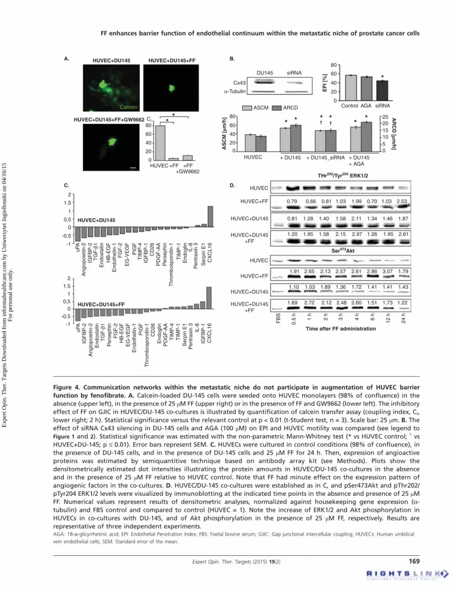
Figure 4. Communication networks within the metastatic niche do not participate in augmentation of HUVEC barrier
function by fenofibrate. A. Calcein-loaded DU-145 cells were seeded onto HUVEC monolayers (98% of confluence) in the
absence (upper left), in the presence of 25 µM FF (upper right) or in the presence of FF and GW9662 (lower left). The inhibitory
effect of FF on GJIC in HUVEC/DU-145 co-cultures is illustrated by quantification of calcein transfer assay (coupling index, Ci,
lower right; 2 h). Statistical significance versus the relevant control at p < 0.01 (t-Student test, n = 3). Scale bar: 25 µm. B. The
effect of siRNA Cx43 silencing in DU-145 cells and AGA (100 µM) on EPI and HUVEC motility was compared (see legend to
Figure 1 and 2). Statistical significance was estimated with the non-parametric Mann-Whitney test (* vs HUVEC control; † vs
HUVEC+DU-145; p £ 0.01). Error bars represent SEM. C. HUVECs were cultured in control conditions (98% of confluence), in
the presence of DU-145 cells, and in the presence of DU-145 cells and 25 µM FF for 24 h. Then, expression of angioactive
proteins was estimated by semiquantitive technique based on antibody array kit (see Methods). Plots show the
densitometrically estimated dot intensities illustrating the protein amounts in HUVEC/DU-145 co-cultures in the absence
and in the presence of 25 µM FF relative to HUVEC control. Note that FF had minute effect on the expression pattern of
angiogenic factors in the co-cultures. D. HUVEC/DU-145 co-cultures were established as in C, and pSer473Akt and pThr202/
pTyr204 ERK1/2 levels were visualized by immunoblotting at the indicated time points in the absence and presence of 25 µMFF. Numerical values represent results of densitometric analyses, normalized against housekeeping gene expression (a-tubulin) and FBS control and compared to control (HUVEC = 1). Note the increase of ERK1/2 and Akt phosphorylation in
HUVECs in co-cultures with DU-145, and of Akt phosphorylation in the presence of 25 µM FF, respectively. Results are
representative of three independent experiments.AGA: 18-a-glicyrrhetinic acid; EPI: Endothelial Penetration Index; FBS: Foetal bovine serum; GJIC: Gap junctional intercellular coupling; HUVECs: Human umbilical
vein endothelial cells; SEM: Standard error of the mean.
FF enhances barrier function of endothelial continuum within the metastatic niche of prostate cancer cells
Expert Opin. Ther. Targets (2015) 19(2) 169
Exp
ert O
pin.
The
r. T
arge
ts D
ownl
oade
d fr
om in
form
ahea
lthca
re.c
om b
y U
niw
ersy
tet J
agie
llons
ki o
n 04
/16/
15Fo
r pe
rson
al u
se o
nly.

PPARa-dependent manner because both reactions wereattenuated by GW9662 (Figure 5C cf. B). Notably,GW9662 did not counteract inhibitory effect of FF onHUVEC motility (Figure 5D). These findings confirm thatFF directly enhances endothelial barrier function partly viadirect PPARa-dependent effects on endothelial cell adhesion.On the other hand, the lack of GW9662 effects on
FF-treated HUVEC motility suggested the involvement ofPPAR-independent pathways in HUVEC reactions to FF.Because we observed intracellular ROS accumulation inHUVECs after FF treatment, PPARa-independent/ROS-dependent pathways may be involved in the observedphenomena. To verify this notion, we analyzed the effect of
NAC on HUVEC reactions to FF. NAC attenuatedFF-induced cytoskeletal rearrangements (Figure 6A) andsignificantly restored FF-inhibited HUVEC motility(Figure 6B). NAC strengthens cellular antioxidant defensesystems [46], therefore these findings would confirm aninvolvement of PPARa-independent/ROS-dependent signal-ing in the determination of HUVEC responses to FF. Onthe other hand, eradication of HUVEC responses to FF byNAC was accompanied by the increase of ROS levels inFF-treated cells (Figure 6C). Notably, FF did not influencethe structure of tight junctions in HUVEC monolayers andits effect on their permeability to solutes was similar in thepresence and absence of DU-145 cells (Figure 7A). No
HUVEC
D.HUVEC+FF
HUVEC+FF+GW9662
HUVEC+FF+GW9662
*
* *
* * *
HUVEC
HUVEC+FF
A. B.
HUVEC HUVEC+FFHUVEC+FF
C.
Time after FF administraton
Time after FF administraton
Vinculin mRNA levels[% of control]
Vinculin
Actin
300
200
100
TLCT [mm] TLCT [mm] TLCT [mm]
TL
CD
[mm
]
0 200 400 600 800 0 200 400 600 800 0 200 400 600 800
120
80
40
03 h 6 h 12 h
FF
24 h
3 h
0 h
24 h6 h
12 h
Tyr397 FAK
Tyr397 FAK
FAK
Actin
FAK
0.99
1.15
0.5
h
1 h
2 h
3 h
4 h
6 h
12 h
24 h
FB
S
1.39 1.31 1.55 0.96 1.38 1.57 2.57Vinculin
ActinDNA
34.5%
DHR 123-Height
DHR 123-Height
HUVEC+FF+GW966240
30
20
10
0
nF
A/c
ell
HUVEC +FF +FF+GW9662
ASCM
HUVEC +FF +FF+GW9662
25
20
15
10
5
0
80
60
40
20
0
ARCD
1.91 1.88 2.15 1.81 6.38 14.19 5.49
Actin 100 101 102 103 104
4.1%
250
0
SS
C-H
eig
ht
250
0
SS
C-H
eig
ht
100 101 102 103 104
AS
CM
[mm
/h] A
RC
D [mm
/h]
Figure 5. Fenofibrate directly targets endothelial cell cytoskeleton and motility. A. HUVECs were cultivated in the absence or
in the presence of 25 µM FF. Tyr397FAK levels and vinculin expression at protein and mRNA level were analyzed at the
indicated time points by immunoblotting and qRT-PCR (quantified as described in Figure 4D). B. Cytoskeletal architecture of
HUVECs cultured in control conditions (upper left), in the presence of 25 µM FF (upper right), or in the presence of 25 µMFF + 10 µMGW9662 (lower left) was visualized and the numbers of FAs per single cell quantified as in Figure 2 (lower right). C.
HUVECs (98% of confluence) were cultivated in the in the presence of 25 µM FF (upper plot), or in the presence of 25 µM FF
and 10 µM GW9662 (lower plot). Cells were incubated in the presence of DHR123 (2 µM) for 4 h and analyzed with
FACSCalibur flow cytometer as in Figure 3. D. HUVEC motility in the control conditions (70% of confluence; left), in the
presence of 25 µM FF (middle left) 25 µM FF + 10 µM GW9662 (middle right) was visualized by time-lapse videomicroscopy and
analyzed as in Figure 2B (right). Statistical significance was estimated with the non-parametric Mann-Whitney test (* vs
HUVEC control; p £ 0.01). Error bars represent SEM. Results are representative of three independent experiments.FAs: Focal adhesions; HUVECs: Human umbilical vein endothelial cells; SEM: Standard error of the mean.
K. Piwowarczyk et al.
170 Expert Opin. Ther. Targets (2015) 19(2)
Exp
ert O
pin.
The
r. T
arge
ts D
ownl
oade
d fr
om in
form
ahea
lthca
re.c
om b
y U
niw
ersy
tet J
agie
llons
ki o
n 04
/16/
15Fo
r pe
rson
al u
se o
nly.

pronounced changes in the expression pattern of angioactivefactors were seen either (Figure 7B). When administered atconcentrations of up to 100 µM, FF prompted the matura-tion of focal contacts and reduced their motility in a dose-dependent fashion, but exerted no effects on their viability(Figure 7C; Table 1). NAC-induced normalization of HUVECphenotype in the presence of high ROS levels indicates theinvolvement of a discrete ROS pool in HUVEC-reactions toFF. Thus, ROS generation is a physiological response ofendothelial cells to FF, unrelated to FF cytotoxicity.
4. Discussion
Penetration of endothelial layer by circulating cancer cells is aprerequisite for their homing in interstitial compartmentsunderlying the endothelium. This process is crucial forthe metastatic cascade [47] and its efficiency depends on a num-ber of intrinsic cancer cell traits: their motile activity and nano-mechanical elasticity, competence for GJIC, expression of celladhesion receptors and secretion ofmetalloproteinases [34,48-50].The interference of FF with tumor progression has predomi-nantly been considered in terms of its effect on these
properties [23,26]. The susceptibility of the endothelial layer toa challenge by a single cancer cell is an equally important, yetunderestimated determinant of diapedesis [48,51]. FF effect onthe properties of endothelial continuum in the metastaticniche has not yet been assessed. This study fills this gap becauseit demonstrates for the first time that FF enhances endothelialbarrier function through the direct effect on endothelial cellsusceptibility to the signals generated by prostate cancer cells.The influence of FF on endothelial cell behavior in the meta-static niche illustrates a new mechanism of its anti-canceractivity. It confirms previous suggestions about the interfer-ence of FF with the metastatic cascade which were based onin vivo approaches [18,25]. It is noteworthy that the enhance-ment of endothelial barrier function to prostate cancer cellswas observed in the presence of 25 µM FF. This concentrationof FF in the culture medium remains within the limits definedby the content of fenofibric acid, an active FF metabolite, inthe blood of patients who take FF on regular bases (up to200 mg/day). Pharmacodynamic studies have revealed thatmaximal concentrations of fenofibric acid in the sera ofFF-treated patients considerably exceed 25 µM [52-55],confirming the biological significance of our findings.
11.2%
ASCMARCD
+FF+NAC+FF
B.
A.HUVEC HUVEC+FF HUVEC+FF+NAC
% o
f H
UV
EC
120
100
80
60
40
0
2068.3%
250
0
SS
C-H
eig
ht
250
0
SS
C-H
eig
ht
DHR 123-Height DHR 123-Height100 101 102 103 104
HUVEC
nFA
/cel
l
0
10
20
30
40
+FF +FF+NAC
100 101 102 103 104
VinculinActinDNA
HUVEC+FF
*
* *
*
C.
HUVEC+FF+NAC
Figure 6. PPARa-independent signaling is involved in the augmentation of endothelial barrier function by fenofibrate.
A. Cytoskeletal architecture of HUVECs cultured in control conditions (left), in the presence of 25 µM FF (middle left), 25 µMFF + 5 mM NAC (middle right) is visualized and the numbers of FAs per single cell quantified as in Figure 2 (right). Statistical
significance versus the relevant control at p < 0.01 (t-Student test, n = 3). B. HUVEC motility in the control conditions (70% of
confluence), in the presence of 25 µM FF and 25 µM FF + 5 mM NAC was visualized by time-lapse videomicroscopy and
analyzed as in Figure 2B. Statistical significance versus the relevant control at p < 0.01 (t-Student test, n = 3). C. HUVEC
monolayers (98% of confluence) treated with 25 µM FF (left), or with 25 µM FF and 5 mM NAC (right) were incubated in the
presence of DHR123 and analyzed with FACSCalibur flow-cytometer as in Figure 3. Note that NAC abrogates HUVEC reactions
to FF but does not down-regulate intracellular ROS levels.FAs: Focal adhesions; HUVECs: Human umbilical vein endothelial cells; NAC: N-acetyl-L-cysteine; ROS: Reactive oxygen species.
FF enhances barrier function of endothelial continuum within the metastatic niche of prostate cancer cells
Expert Opin. Ther. Targets (2015) 19(2) 171
Exp
ert O
pin.
The
r. T
arge
ts D
ownl
oade
d fr
om in
form
ahea
lthca
re.c
om b
y U
niw
ersy
tet J
agie
llons
ki o
n 04
/16/
15Fo
r pe
rson
al u
se o
nly.

ZO-1DNA
VinculinActinDNA
HUVECA. C.
HUVEC+FF50 mM HUVEC+FF100 mMHUVEC+FF
HUVEC+FF+GW9662
HUVEC+FF
B.
FF
TLCD706050403020100
100
80
60
40
20
0
100
80
60
40
20
0
6 h
*
*
*
*
24 h 48 h
HUVEC +FF50 μM
HUVEC +FF25 μM +FF50 μM
+FF100 μM
+FF100 μM
TL
CD
[mm
]C
ell v
iab
ility
[%
]
AS
CM
[mm/h
]
ASCM140
120
100
0
21.5
1
-1
0.50
-0.5
% o
f co
ntr
ol
15 30 60t [min]
DU145DU145+FF
HB
-EG
FC
D26
Ang
iopo
ietin
-2C
XC
L16
Ser
pin
E1
PIG
FT
GF
-β1
uPA
End
osta
tin
End
othe
lin-1
FG
F-2
EG
-VE
GF
TIM
P-4
IGF
BP
-1IG
FB
P-2
PD
GF
-AA
Per
seph
in
Thr
ombo
spon
din-
1
TIM
P-1
End
oglin
IL-8
Pen
trax
in 3
Figure 7. FF does not exert sub-lethal effects in HUVECs. A. Functional status of ZO-1-mediated intercellular contacts in
control conditions (upper left), in the presence of 25 µM FF (upper right), or in the presence of 25 µM FF + 10 µMGW9662 (lower left); and the effect of DU-145 and 25 µM FF on solute permeability of HUVEC monolayers was analyzed as
described (see Figure 1 and Methods, respectively). B. HUVECs were cultured in control conditions (98% of confluence) and in
the presence of 25 µM FF for 24 h. Then, angioactive proteins were analyzed as in Figure 4B. C. HUVECs were cultivated in the
presence of FF (25-100 µM) and their cytoskeleton architecture (see Figure 6A for control; upper panel) motility (middle), and
viability (lower plot) was quantified. Statistical significance: * versus the relevant control at p < 0.01, using the Wilcoxon
signed-rank test (A), Mann--Whitney (B) and Student t-test (C). Note the dose-dependent effects of FF on HUVEC cytoskeleton
and motility but not on HUVEC viability.HUVECs: Human umbilical vein endothelial cells.
Table 1. Effect of fenofibrate on the motile activity of HUVECs.
Parameters Control FF 25 mM FF 50 mM FF 100 mM
TLCT (µm) 391.8 ± 11.5 308.5 ± 14.7* 271.4 ± 11.3* 229.8 ± 9.1*ASCM (µm/h) 55.9 ± 1.6 44.1 ± 2.1* 38.8 ± 1.6* 32.8 ± 1.3*TLCD (µm) 91.1 ± 6.5 82.9 ± 7.5 77.6 ± 5.6* 63.9 ± 5.3*ARCD (µm/h) 13.0 ± 0.9 11.8 ± 1.1 11.1 ± 0.8* 9.1 ± 0.7*
Values are the means ± SEM.
*Statistically significant (Mann--Whitney test) at p £ 0.01 (HUVEC in control conditions vs HUVEC treated with fenofibrate at indicated concentrations).
ARCD: Average rate of cell displacement; ASCM: Average speed of cell movement; HUVECs: Human umbilical vein endothelial cells; SEM: Standard error of the
mean; TLCD: Total length of cell displacement; TLCT: Total length of cell trajectory.
K. Piwowarczyk et al.
172 Expert Opin. Ther. Targets (2015) 19(2)
Exp
ert O
pin.
The
r. T
arge
ts D
ownl
oade
d fr
om in
form
ahea
lthca
re.c
om b
y U
niw
ersy
tet J
agie
llons
ki o
n 04
/16/
15Fo
r pe
rson
al u
se o
nly.

Monitoring of endothelial cell behavior in the proximity ofcancer cells gives the opportunity to reconstruct the events inthe ‘metastatic niche’ of circulating cancer cells during cancercell diapedesis. It provides a tool to evaluate the role of endo-thelial cell mobilization in the progress of the transendothelialpenetration of cancer cells. DU-145 cell line, which was usedin this study to imitate circulating prostate carcinoma cells,had been propagated from prostate cancer metastases to brain.Thus, DU-145 cells represent the progeny of cells capable ofpenetrating tissue barriers. The interference of FF with theirintrinsic properties might thus have participated in theobserved attenuation of transendothelial penetration inHUVEC/DU-145 co-cultures. Actually, we have previouslyshown that FF inhibits the motility and GJIC in DU-145 pop-ulations [27]. Here, we observed the retraction of endotheliumin the proximity of DU-145 cells, which was accompanied byinduction of HUVEC motility and counteracted by FF. Inter-ference of FF with endothelial cell mobilization by PC-3 cells(derived from prostate cancer metastasis to bone) additionallyillustrates the biological significance of our data. Correspond-ing HUVEC responses to FF seen in the absence and presenceof cancer cells show that FF effectors are situated down-streamof intercellular communication loops within the metastaticniche. FF inhibited GJIC between cancer and endothelialcells [56], but this communication route plays a secondary rolein HUVEC mobilization by DU-145 cells. Concomitantly,the expression of angioactive factors, which might mobilizeHUVECs in the ERK1/2-dependent manner, was not affectedby FF. These data indicate that FF enhances endothelial barrierfunction in the ‘metastatic niche’ of prostate cancer cellsthrough a direct effect on endothelial cell properties.
PPARa/ROS-dependent pathway has previously beenimplicated in the inhibition of cancer cell invasive poten-tial [26]. Our observations indicate that PPARa-dependent,ROS-dependent signaling is also involved in the regulationof endothelial barrier function in the ‘metastatic niche’ byFF. This was illustrated by a partial abrogation of endothelialcell responses to FF seen in the presence of PPARa antago-nist. It was accompanied by attenuation of ROS accumulationin FF-treated HUVECs. Akt and FAK phosphorylationobserved in FF-treated HUVECs suggest that both effectorsparticipate in the endothelial cell reactions to FF. Notably,it has previously been shown that endothelial cell reactions(incl. inhibition of cell motility and angiogenesis) to FF andother PPARa agonists are mediated by Akt [57]. WhetherAkt provides a mechanistic link between PPARa pathwayand cytoskeletal rearrangements in endothelial cells requiresa more elaborate study based on PPARa knock-out approach.Such an approach should also consider the interrelationsbetween FF-induced ROS signaling in HUVEC and activa-tion of Akt-dependent pathway.
More detailed analyses are also required to identify mecha-nisms underlying the abrogation of FF effect on endothelialcells by NAC. Concomitantly with the attenuation ofFF-induced cytoskeletal rearrangements, NAC restored the
motility of FF-treated HUVECs. It demonstrates the involve-ment of PPARa-independent, ROS-dependent pathway inHUVEC reactions to FF. Such a pathway has been shownto mediate FF-induced inhibition of cell motility [27]. As aglutathione (GSH) precursor, NAC assists intracellular anti-oxidant defense systems [46]. However, it failed to reduceROS levels in FF-treated endothelial cells. In the absence ofFF effects on endothelial cell viability, these observations sug-gest the involvement of a discrete, relatively small ROS parti-tion, generated in a PPARa-independent fashion. Preventiveeffect of NAC on GSH depletion in FF-treated endothelialcells, which reduces their susceptibility to ROS signaling,may provide an alternative explanation for this conundrum.Precise identification of the specific, NAC-sensitive andNAC-insensitive ROS and their subcellular origin(s) is neededto justify further speculations on the mechanisms of ROSinvolvement in endothelial cell reactions to FF in vitro andin vivo.
Considering the present state of knowledge, we can againonly speculate about the mechanisms of the interplay betweenPPARa-dependent/independent pathways and adhesion ofFF-treated HUVECs [12,57]. Stress fibers thickening, increasedvinculin expression and FA recruitment [58] in FF-treatedendothelial cells are characteristic for strongly adherentisometrically contracting cells [59]. We can suggest that FAK-dependent adhesive status of endothelial cells cooperates inthe regulation of endothelial barrier function. Increased solutepermeability of FF-treated endothelia suggests that the shiftsin cell adhesion strength may evoke changes in the equilib-rium between cell-to-cell and cell-substratum interactions,crucial for endothelial barrier function [33]. However, localloosening of intra-endothelial fascia adherens can only be suf-ficient for transendothelial infiltration of ‘ameboid’ cells, suchas leukocytes or monocytes. These cells are elastic enough tosqueeze through relatively small endothelial discontinu-ities [60]. In contrast, cancer cells that are characterized by‘mesenchymal’ strategy of movement display relatively lowsusceptibility to mechanical distortions [61]. They requiremore prominent endothelial remodeling to penetrate trans-junctional windows. Whereas DU-145 cell motility plays asecondary role in the remodeling of endothelial continuum,the inhibition of endothelial cell motility may additionallystrengthen endothelial barrier function through stabilizingeffect on cell-substratum adhesion. Negative feedback loopsbetween cell migration and adhesion strength may participatein this process. Thus, converging FF effects on cancer andendothelial cells at the ‘metastatic niche’ affect the susceptibil-ity of the endothelial continuum to the signals from cancercells and regulate endothelial barrier function.
5. Conclusions
The in vitro model, which imitates the interface between cir-culating cancer cells and endothelium [34,48,62], enabled us todemonstrate the attenuating effect of FF on endothelial cell
FF enhances barrier function of endothelial continuum within the metastatic niche of prostate cancer cells
Expert Opin. Ther. Targets (2015) 19(2) 173
Exp
ert O
pin.
The
r. T
arge
ts D
ownl
oade
d fr
om in
form
ahea
lthca
re.c
om b
y U
niw
ersy
tet J
agie
llons
ki o
n 04
/16/
15Fo
r pe
rson
al u
se o
nly.

responsiveness to the signals generated by cancer cells in themetastatic niche. The strengthening effect of FF on endo-thelial cell adhesion and impairment of motile activity byFF reduces the efficiency of prostate carcinoma cell diapede-sis and potentially interferes with the prostate cancer meta-static cascade. In the context of other in vivo studies thatdirectly and/or indirectly suggest the interference of FFwith tumor progression [15,18,25], this report extends theknowledge on biological effects of existing and emergingvasoactive drugs on the metastatic cascade. It fills the gapin the understanding of the links between the vasoactive,anti-atherosclerotic and anti-tumorigenic activity of FF andsuggests the application of this lipid-lowering drug as ameans to reducing the metastatic spread of prostate cancercells. It also justifies further analyses of the interference ofFF with the metastatic cascade of prostate cancer and othertumors with the more sophisticated genetic in vivoapproaches. Comprehensive epidemiologic studies are alsopostulated to estimate the links between FF intake andrisk of prostate cancer metastases.
Declaration of interest
This work was financially supported by the Polish NationalScience Centre (grant 2011/01/B/NZ3/00004) and fundsgranted to the Faculty of Biochemistry, Biophysics and Bio-technology of Jagiellonian University to further “The Devel-opment of Young Scientists and Doctoral Students” 2011/2012 (DS/46/2011). The Faculty of Biochemistry, Biophysicsand Biotechnology of the Jagiellonian University is a benefi-ciary of the structural funds from the European Union (GrantsNo: UDA-POIG.01.03.01-14-036/09-00 - “Application ofpolyisoprenoid derivatives as drug carriers and metabolismregulators”; POIG.02.01.00-12-064/08 -- “Molecular bio-technology for health”; POIG 01.02-00-109/99 “Innovativemethods of stem cell applications in medicine”). The authorsdeclare that they have no competing interests. The authorshave no other relevant affiliations or financial involvementwith any organization or entity with a financial interest in orfinancial conflict with the subject matter or materials discussedin the manuscript apart from those disclosed.
BibliographyPapers of special note have been highlighted as
either of interest (�) or of considerable interest(��) to readers.
1. Lamers C, Schubert-Zsilavecz M,
Merk D. Therapeutic modulators of
peroxisome proliferator-activated
receptors (PPAR): a patent review (2008-
present). Expert Opin Ther Pat
2012;22:803-41
2. Ahmed W, Ziouzenkova O, Brown J,
et al. PPARs and their metabolic
modulation: new mechanisms for
transcriptional regulation? J Intern Med
2007;262:184-98
3. Grabacka M, Reiss K. Anticancer
properties of PPARalpha-effects on
cellular metabolism and inflammation.
PPAR Res 2008;2008:930705
4. Grabacka M, Pierzchalska M, Reiss K.
Peroxisome proliferator activated receptor
alpha ligands as anticancer drugs
targeting mitochondrial metabolism.
Curr Pharm Biotechnol 2013;14:342-56
5. Robinson JG. LDL reduction: how low
should we go and is it safe?
Curr Cardiol Rep 2008;10:481-7
6. Adeghate E, Adem A, Hasan MY, et al.
Medicinal chemistry and actions of dual
and Pan PPAR modulators. Open Med
Chem J 2011;5:93-8
7. Balakumar P, Rohilla A, Mahadevan N.
Pleiotropic actions of fenofibrate on the
heart. Pharmacol Res 2011;63:8-12
. This paper critically discusses
pleiotropic actions of fenofibrate (FF)
on the heart beyond its lipid-lowering
effects, in particular vascular
endothelial dysfunction-associated
cardiovascular abnormalities.
8. McKeage K, Keating GM. Fenofibrate:
a review of its use in dyslipidaemia.
Drugs 2011;71:1917-46
9. Katayama A, Yamamoto Y, Tanaka K,
et al. Fenofibrate enhances
neovascularization in a murine ischemic
hindlimb model. J Cardiovasc Pharmacol
2009;54:399-404
10. Hiukka A, Maranghi M, Matikainen N,
et al. PPARalpha: an emerging
therapeutic target in diabetic
microvascular damage.
Nat Rev Endocrinol 2010;6:454-63
11. Noonan JE, Jenkins AJ, Ma JX, et al. An
update on the molecular actions of
fenofibrate and its clinical effects on
diabetic retinopathy and other
microvascular end points in patients with
diabetes. Diabetes 2013;62:3968-75
12. Goetze S, Eilers F, Bungenstock A, et al.
PPAR activators inhibit endothelial cell
migration by targeting Akt.
Biochem Biophys Res Commun
2002;293:1431-7
13. Varet J, Vincent L, Mirshahi P, et al.
Fenofibrate inhibits angiogenesis in vitro
and in vivo. Cell Mol Life Sci
2003;60:810-19
.. One of the first studies to directly
show inhibitory effect of FF on
endothelial cell proliferation,
migration and capillary tube formation
in vitro, and angiogenesis in vivo.
14. Meissner M, Stein M, Urbich C, et al.
PPARalpha activators inhibit vascular
endothelial growth factor receptor-2
expression by repressing Sp1-dependent
DNA binding and transactivation.
Circ Res 2004;94:324-32
15. Panigrahy D, Kaipainen A, Huang S,
et al. PPARalpha agonist fenofibrate
suppresses tumor growth through direct
and indirect angiogenesis inhibition.
Proc Natl Acad Sci USA
2008;105:985-90
.. This study shows that PPARalpha
activation by ligands cause tumor
suppression by the inhibition of
angiogenesis. It has formulated
mechanistic rationale for evaluating
the clinical benefits of fenofibrate in
the treatment of progressed cancers.
16. Cao Z, Shang B, Zhang G, et al. Tumor
cell-mediated neovascularization and
lymphangiogenesis contrive tumor
progression and cancer metastasis.
Biochim Biophys Acta 2013;1836:273-86
. This review comprehensibly discusses
the significance of neovasculogenesis
K. Piwowarczyk et al.
174 Expert Opin. Ther. Targets (2015) 19(2)
Exp
ert O
pin.
The
r. T
arge
ts D
ownl
oade
d fr
om in
form
ahea
lthca
re.c
om b
y U
niw
ersy
tet J
agie
llons
ki o
n 04
/16/
15Fo
r pe
rson
al u
se o
nly.

for cancer progression and the role of
paracrine loops between cancer and
endothelial cells in this process.
17. Hida K, Ohga N, Akiyama K, et al.
Heterogeneity of tumor endothelial cells.
Cancer Sci 2013;104:1391-5
. The significance of heterogeneity in
tumor endothelial cells for cancer
development and therapy is discussed
in this mini-review.
18. Robison NJ, Campigotto F, Chi SN,
et al. A phase II trial of a multi-agent
oral antiangiogenic (metronomic)
regimen in children with recurrent or
progressive cancer. Pediatr Blood Cancer
2014;61:636-42
.. A study that shows application of
fenofibrate in a potentially efficient
‘5-drug’ oral regimen in children with
recurrent or progressive cancer.
19. Langley RR, Fidler IJ. The seed and soil
hypothesis revisited--the role of tumor-
stroma interactions in metastasis to
different organs. Int J Cancer
2011;128:2527-35
20. Giordano A, Macaluso M. Fenofibrate
triggers apoptosis of glioblastoma cells in
vitro: new insights for therapy.
Cell Cycle 2012;11:3154
21. Jiao HL, Zhao BL. Cytotoxic effect of
peroxisome proliferator fenofibrate on
human HepG2 hepatoma cell line and
relevant mechanisms.
Toxicol Appl Pharmacol 2002;185:172-9
22. Yamasaki D, Kawabe N, Nakamura H,
et al. Fenofibrate suppresses growth of
the human hepatocellular carcinoma cell
via PPARalpha-independent mechanisms.
Eur J Cell Biol 2011;90:657-64
23. Urbanska K, Pannizzo P, Grabacka M,
et al. Activation of PPARalpha inhibits
IGF-I-mediated growth and survival
responses in medulloblastoma cell lines.
Int J Cancer 2008;123:1015-24
24. Grabacka M, Plonka PM, Urbanska K,
et al. Peroxisome proliferator-activated
receptor alpha activation decreases
metastatic potential of melanoma cells
in vitro via down-regulation of Akt.
Clin Cancer Res 2006;12:3028-36
25. Grabacka M, Placha W, Plonka PM,
et al. Inhibition of melanoma metastases
by fenofibrate. Arch Dermatol Res
2004;296:54-8
.. To our knowledge, the first study to
demonstrate anti-metastatic activity of
fenofibrate in vivo.
26. Drukala J, Urbanska K, Wilk A, et al.
ROS accumulation and IGF-IR
inhibition contribute to fenofibrate/
PPARalpha -mediated inhibition of
Glioma cell notility in vitro. Mol Cancer
2010;9:159
27. Wybieralska E, Szpak K, Gorecki A,
et al. Fenofibrate attenuates contact-
stimulated cell motility and gap
junctional coupling in DU-145 human
prostate cancer cell populations.
Oncol Rep 2011;26:447-53
28. Thuillier P, Anchiraico GJ, Nickel KP,
et al. Activators of peroxisome
proliferator-activated receptor-alpha
partially inhibit mouse skin tumor
promotion. Mol Carcinog
2000;29:134-42
29. Saidi SA, Holland CM,
Charnock-Jones DS, et al. In vitro and
in vivo effects of the PPAR-alpha
agonists fenofibrate and retinoic acid in
endometrial cancer. Mol Cancer
2006;5:13
30. Panigrahy D, Kaipainen A, Kieran MW,
et al. PPARs: a double-edged sword in
cancer therapy? PPAR Res
2008;2008:350351
31. Scatena R, Bottoni P, Giardina B.
Mitochondria, PPARs, and cancer: is
receptor-independent action of PPAR
agonists a key? PPAR Res
2008;2008:256251
.. This review summarizes extrareceptor
activities of synthetic PPAR ligands
and their consequences for the
application of PPAR activators
in clinics.
32. Tomizawa A, Hattori Y, Inoue T, et al.
Fenofibrate suppresses microvascular
inflammation and apoptosis through
adenosine monophosphate-activated
protein kinase activation. Metabolism
2011;60:513-22
. The study demonstrating that
fenofibrate might exert a protective
effect on the microvasculature through
AMP-activated protein kinase/Akt
activation beyond its lipid-
lowering actions.
33. Reymond N, d’Agua BB, Ridley AJ.
Crossing the endothelial barrier during
metastasis. Nat Rev Cancer
2013;13:858-70
. Comprehensive summary of the
mechanisms involved in cancer
cell diapedesis.
34. Wang X, Ferreira AM, Shao Q, et al.
Beta3 integrins facilitate matrix
interactions during transendothelial
migration of PC3 prostate tumor cells.
Prostate 2005;63:65-80
35. Woodward J. Crossing the endothelium:
E-selectin regulates tumor cell migration
under flow conditions. Cell Adh Migr
2008;2:151-2
36. Strell C, Niggemann B, Voss MJ, et al.
Norepinephrine promotes the
beta1-integrin-mediated adhesion of
MDA-MB-231 cells to vascular
endothelium by the induction of a
GROalpha release. Mol Cancer Res
2012;10:197-207
37. Szpak K, Wybieralska E,
Niedzialkowska E, et al.
DU-145 prostate carcinoma cells that
selectively transmigrate narrow obstacles
express elevated levels of CX43. Cell Mol
Biol Lett 2011;16:625-37
38. Guan K, Czyz J, Furst DO, et al.
Expression and cellular distribution of
alpha(v)integrins in beta(1)integrin-
deficient embryonic stem cell-derived
cardiac cells. J Mol Cell Cardiol
2001;33:521-32
39. Ryszawy D, Sarna M, Rak M, et al.
Functional links between Snail-1 and
Cx43 account for the recruitment of
Cx43-positive cells into the invasive front
of prostate cancer. Carcinogenesis
2014;35:1920-30
40. Wojciak-Stothard B, Potempa S,
Eichholtz T, et al. Rho and Rac but not
Cdc42 regulate endothelial cell
permeability. J Cell Sci
2001;114:1343-55
41. Miekus K, Czernik M, Sroka J, et al.
Contact stimulation of prostate cancer
cell migration: the role of gap junctional
coupling and migration stimulated by
heterotypic cell-to-cell contacts in
determination of the metastatic
phenotype of Dunning rat prostate
cancer cells. Biol Cell 2005;97:893-903
42. Daniel-Wojcik A, Misztal K, Bechyne I,
et al. Cell motility affects the intensity of
gap junctional coupling in prostate
carcinoma and melanoma cell
populations. Int J Oncol 2008;33:309-15
43. Quadri SK. Cross talk between focal
adhesion kinase and cadherins: role in
regulating endothelial barrier function.
Microvasc Res 2012;83:3-11
FF enhances barrier function of endothelial continuum within the metastatic niche of prostate cancer cells
Expert Opin. Ther. Targets (2015) 19(2) 175
Exp
ert O
pin.
The
r. T
arge
ts D
ownl
oade
d fr
om in
form
ahea
lthca
re.c
om b
y U
niw
ersy
tet J
agie
llons
ki o
n 04
/16/
15Fo
r pe
rson
al u
se o
nly.

44. Leesnitzer LM, Parks DJ, Bledsoe RK,
et al. Functional consequences of cysteine
modification in the ligand binding sites
of peroxisome proliferator activated
receptors by GW9662. Biochemistry
2002;41:6640-50
45. Czyz J, Szpak K, Madeja Z. The role of
connexins in prostate cancer promotion
and progression. Nat Rev Urol
2012;9:274-82
. In this review, we summarize the data
that provide the rational for
implementation of the analyses of
connexin function in the studies on
metastatic cascade of prostate cancer.
46. Rushworth GF, Megson IL. Existing and
potential therapeutic uses for
N-acetylcysteine: the need for conversion
to intracellular glutathione for
antioxidant benefits. Pharmacol Ther
2014;141:150-9
47. van Zijl F, Krupitza G, Mikulits W.
Initial steps of metastasis: cell invasion
and endothelial transmigration.
Mutat Res 2011;728:23-34
48. Pollmann MA, Shao Q, Laird DW, et al.
Connexin 43 mediated gap junctional
communication enhances breast tumor
cell diapedesis in culture.
Breast Cancer Res 2005;7:R522-34
49. Friedl P, Alexander S. Cancer invasion
and the microenvironment: plasticity and
reciprocity. Cell 2011;147:992-1009
. Summary of the mechanisms that
contribute to the generation of diverse
cancer invasion routes and programs,
potentially crucial for cancer cell
diapedesis and metastatic
dissemination.
50. Bravo-Cordero JJ, Hodgson L,
Condeelis J. Directed cell invasion and
migration during metastasis. Curr Opin
Cell Biol 2012;24:277-83
51. Garcia-Roman J, Zentella-Dehesa A.
Vascular permeability changes involved
in tumor metastasis. Cancer Lett
2013;335:259-69
52. Doser K, Guserle R, Nitsche V, et al.
Comparative steady state study with
2 fenofibrate 250 mg slow release
capsules. An example of bioequivalence
assessment with a highly variable drug.
Int J Clin Pharmacol Ther
1996;34:345-8
53. Kajosaari LI, Backman JT, Neuvonen M,
et al. Lack of effect of bezafibrate and
fenofibrate on the pharmacokinetics and
pharmacodynamics of repaglinide. Br J
Clin Pharmacol 2004;58:390-6
54. Uetake D, Ohno I, Ichida K, et al.
Effect of fenofibrate on uric acid
metabolism and urate transporter 1.
Intern Med 2010;49:89-94
55. Hu L, Wu H, Niu F, et al. Design of
fenofibrate microemulsion for improved
bioavailability. Int J Pharm
2011;420:251-5
56. Mroue RM, El Sabban ME,
Talhouk RS. Connexins and the gap in
context. Integr Biol (Camb)
2011;3:255-66
57. Okayasu T, Tomizawa A, Suzuki K,
et al. PPARalpha activators upregulate
eNOS activity and inhibit cytokine-
induced NF-kappaB activation through
AMP-activated protein kinase activation.
Life Sci 2008;82:884-91
. A study showing the beneficial effects
of PPARa activators on endothelial
cells attributed to the induction of
AMP-activated protein kinase/
Akt activation.
58. Alexander JS, Zhu Y, Elrod JW, et al.
Reciprocal regulation of endothelial
substrate adhesion and barrier function.
Microcirculation 2001;8:389-401
59. Deguchi S, Sato M. Biomechanical
properties of actin stress fibers of non-
motile cells. Biorheology 2009;46:93-105
60. Yuan SY, Shen Q, Rigor RR, et al.
Neutrophil transmigration, focal
adhesion kinase and endothelial barrier
function. Microvasc Res 2012;83:82-8
61. Friedl P, Wolf K. Plasticity of cell
migration: a multiscale tuning model.
J Exp Med 2010;207:11-19
62. Nevo I, Sagi-Assif O, Meshel T, et al.
The involvement of the fractalkine
receptor in the transmigration of
neuroblastoma cells through bone-
marrow endothelial cells. Cancer Lett
2009;273:127-39
AffiliationKatarzyna Piwowarczyk1, Ewa Wybieralska1,
Jarosław Baran2, Julia Borowczyk3,
Paulina Rybak4, Milena Kosinska1,
Anna Julia Włodarczyk5, Marta Michalik1,
Maciej Siedlar2, Zbigniew Madeja1,
Jerzy Dobrucki4, Krzysztof Reiss6 &
Jarosław Czyz†1
†Author for correspondence1Jagiellonian University, Department of Cell
Biology, Faculty of Biochemistry, Biophysics and
Biotechnology, Krakow, Poland
E-mail: [email protected] Institute of Pediatrics,
Jagiellonian University Medical College,
Department of Clinical Immunology, Krakow,
Poland3Jagiellonian University, Department of Cell
Biology, Faculty of Biochemistry, Biophysics and
Biotechnology, Laboratory of Tissue Engineering,
Krakow, Poland4Jagiellonian University, Division of Cell
Biophysics, Faculty of Biochemistry, Biophysics
and Biotechnology, Krakow, Poland5Cardiff University, School of Biosciences,
Cardiff, UK6School of Medicine, Stanley S. Scott Cancer
Center, LSU Health Sciences Center,
Neurological Cancer Research, New Orleans,
USA
Supplementary materials are available online.
Figures S1 and S2.
K. Piwowarczyk et al.
176 Expert Opin. Ther. Targets (2015) 19(2)
Exp
ert O
pin.
The
r. T
arge
ts D
ownl
oade
d fr
om in
form
ahea
lthca
re.c
om b
y U
niw
ersy
tet J
agie
llons
ki o
n 04
/16/
15Fo
r pe
rson
al u
se o
nly.