Feeding mechanism of Epibulus insidiator (Labridae; Teleostei): Evolution of a novel functional...
22
JOURNAL OF MORPHOLOGY 202:129-150 (1989) Feeding Mechanism of Epibulus insidiator (Labridae;Teleostei): Evolution of a Novel FunctionalSystem MARK W. WESTNEAT AND PETER C. WAINWRIGHT Department of Zoology, Duke University, Durham, North Carolina 27706 (M. W. W.); Department of Developmental and Cell Biology University of California, Zrvine, California9271 7 (P.C. W.) ABSTRACT The feeding mechanism of Epibulus insidiator is unique among fishes, exhibiting the highest degree of jaw protrusion ever described (65% of head length). The functional morphology of the jaw mechanism in Epibulus is analyzed as a case study in the evolution of novel functional systems. The feeding mechanism appears to be driven by unspecialized muscle activity patterns and input forces,that combine with drasticallychanged bone and ligament morphology to produce extreme jaw protrusion. The primary derived osteological features are the form of the quadrate, interopercle, and elongate premaxilla and lower jaw. Epibulus has a unique vomero-interopercular ligament and enlarged interoperculo-mandibular and pre- maxilla-maxilla ligaments. The structures of the opercle, maxilla, and much of the neurocranium retain a primitive labrid condition. Many cranial muscles in Epibulus also retain a primitive structural condition, including the levator operculi, expaxialis, sternohyoideus,and adductor mandibulae. The generalized perciform suction feed- ing pattern of simultaneous peak cranial elevation, gape, and jaw protrusion followed by hyoid depression is retained in Epibulus. Electromyography and high-speed cinematography indicate that patterns of muscle activity during feeding and the kinematic movements of opercular rotation and cranial elevation produce a primitive pattern of force and motion input. Extreme jaw protrusion is produced from this primitive input pattern by several derived kinematic patterns of modified bones and ligaments. The interopercle,quadrate, and maxilla rotate through angles of about 100 degrees, pushing the lower jaw into a protruded position. Analysis of primitive and derived characters at multide levels of structural and functional organization allows conclusions about the level of design at functional novelties. Epibulus insidiator (Pallas), the sling-jaw wrasse, exhibits an extraordinary ability to pro- trude its jaws during feeding. Epibulus is a monotypic genus in the family Labridae and is widely distributed on coral reefs throughout the tropical Pacific, Indian Ocean, and Red Sea. The extreme protrusion of the mouth of this fish was noted by several early ichthyologists. Renard (1719) refers to Epibulus as Bedreiger (the de- ceiver) and Passer (the thief) and states that this fish “has a long snout hidden in the mouth that it throws out with great agility to capture any prey that comes too close.” Bleeker (1862) refers to Epibulus as “ikan kakatua sumpit” (the par- rot wrasse with the pea shooter), and Delsman (’25) cites a Malayan name “tagi utang” (the dunning creditor). These early references reflect interest in the feeding mechanism of Epibulus, which is unique among described fishes. Recent which change has occurred to produce widespread interest in the biomechanicsof feed- ing in fishes (Alexander, ’67; Barel, ’83; Lauder, ’82, ’83; Liem, ’78, ’80; Motta, ’84; Osse, ’69) has resulted in the emergence of the teleost skull as one of the clearest examples of evolutionary change in a vertebrate functional system. The protrusion mechanism of Epibulus represents a major evolutionary change in this functional complex. The evolution of major changes in complex functional systems is an issue of long-standing interest in evolutionary biology (Darwin, 1859; Goldschmidt, ’40; Lauder, %la; Simpson, ’53; Wake and Larson, ’87). How do drastic modifica- tions in complex systems evolve? The unusual feeding mechanism of Epibulus provides an ex- cellent opportunity to address this question.Our primary goal in this study is to describe the jaw mechanism of Epibulus as a mosaic of ancestral o 1989 ALAN R. LISS, INC.
Transcript of Feeding mechanism of Epibulus insidiator (Labridae; Teleostei): Evolution of a novel functional...

JOURNAL OF MORPHOLOGY 202:129-150 (1989)
Feeding Mechanism of Epibulus insidiator (Labridae; Teleostei): Evolution of a Novel Functional System
MARK W. WESTNEAT AND PETER C. WAINWRIGHT Department of Zoology, Duke University, Durham, North Carolina 27706 (M. W. W.); Department of Developmental and Cell Biology University of California, Zrvine, California 9271 7 (P.C. W.)
ABSTRACT The feeding mechanism of Epibulus insidiator is unique among fishes, exhibiting the highest degree of jaw protrusion ever described (65% of head length). The functional morphology of the jaw mechanism in Epibulus is analyzed as a case study in the evolution of novel functional systems. The feeding mechanism appears to be driven by unspecialized muscle activity patterns and input forces, that combine with drastically changed bone and ligament morphology to produce extreme jaw protrusion. The primary derived osteological features are the form of the quadrate, interopercle, and elongate premaxilla and lower jaw. Epibulus has a unique vomero-interopercular ligament and enlarged interoperculo-mandibular and pre- maxilla-maxilla ligaments. The structures of the opercle, maxilla, and much of the neurocranium retain a primitive labrid condition. Many cranial muscles in Epibulus also retain a primitive structural condition, including the levator operculi, expaxialis, sternohyoideus, and adductor mandibulae. The generalized perciform suction feed- ing pattern of simultaneous peak cranial elevation, gape, and jaw protrusion followed by hyoid depression is retained in Epibulus. Electromyography and high-speed cinematography indicate that patterns of muscle activity during feeding and the kinematic movements of opercular rotation and cranial elevation produce a primitive pattern of force and motion input. Extreme jaw protrusion is produced from this primitive input pattern by several derived kinematic patterns of modified bones and ligaments. The interopercle, quadrate, and maxilla rotate through angles of about 100 degrees, pushing the lower jaw into a protruded position. Analysis of primitive and derived characters at multide levels of structural and functional organization allows conclusions about the level of design at functional novelties.
Epibulus insidiator (Pallas), the sling-jaw wrasse, exhibits an extraordinary ability to pro- trude its jaws during feeding. Epibulus is a monotypic genus in the family Labridae and is widely distributed on coral reefs throughout the tropical Pacific, Indian Ocean, and Red Sea. The extreme protrusion of the mouth of this fish was noted by several early ichthyologists. Renard (1719) refers to Epibulus as Bedreiger (the de- ceiver) and Passer (the thief) and states that this fish “has a long snout hidden in the mouth that it throws out with great agility to capture any prey that comes too close.” Bleeker (1862) refers to Epibulus as “ikan kakatua sumpit” (the par- rot wrasse with the pea shooter), and Delsman (’25) cites a Malayan name “tagi utang” (the dunning creditor). These early references reflect interest in the feeding mechanism of Epibulus, which is unique among described fishes. Recent
which change has occurred to produce
widespread interest in the biomechanics of feed- ing in fishes (Alexander, ’67; Barel, ’83; Lauder, ’82, ’83; Liem, ’78, ’80; Motta, ’84; Osse, ’69) has resulted in the emergence of the teleost skull as one of the clearest examples of evolutionary change in a vertebrate functional system. The protrusion mechanism of Epibulus represents a major evolutionary change in this functional complex.
The evolution of major changes in complex functional systems is an issue of long-standing interest in evolutionary biology (Darwin, 1859; Goldschmidt, ’40; Lauder, %la; Simpson, ’53; Wake and Larson, ’87). How do drastic modifica- tions in complex systems evolve? The unusual feeding mechanism of Epibulus provides an ex- cellent opportunity to address this question. Our primary goal in this study is to describe the jaw mechanism of Epibulus as a mosaic of ancestral
o 1989 ALAN R. LISS, INC.
130 M.W. WESTNEAT AND P.C. WAINWRIGHT
and derived features. We ask the question, "At what level of structural and functional organiza- tion can the unique jaw mechanism of Epibulus insidiator be explained?" Our general approach to this question follows that of Lauder and Shaffer ('88) and involves description of charac- ter state change (or lack thereof) at multiple levels of musculoskeletal design. We examine cranial bones, ligaments, and muscles a t three hierarchical levels of organization: 1) morpholog- ical structure, 2) patterns of activity during feed- ing, and 3) functional role in extreme jaw protru- sion. Activity patterns of structures are obtained through high-speed cinematography and electro- myography, and the functional role of a struc- ture is defined as the product of a structure's morphology, activity pattern, and interaction with other structures.
Unique biological features may be sources of valuable functional information, yet organisms are combinations of both ancestral and derived features. For research in functional morphology to be understood in a phylogenetic context, func- tional questions should be addressed in terms of the characters that comprise functional systems. Thus, research on the functional morphology of a complex system should identify activity pat- terns and functions of features retaining a prim- itive condition as well as analysis of more re- cently evolved characters. This approach, which has been pursued by only a few recent studies (e.g., Bemis and Lauder, '86; Lombard and Wake, '86; Schaefer and Lauder, '86), requires the exist- ence of a phylogenetic framework for Epibulus and allied genera because decisions about charac- ter state polarity are made for each structure of the feeding mechanism at each of the three lev- els described above. These decisions are made by using recent (Gomon, '79; Gomon and Russel, unpublished) and current (Westneat, unpub- lished) information regarding the phylogenetic relationships of Epibulus, tribe Cheilinini, and several outgroups within the Labridae.
MATERIALS AND METHODS Epibulus specimens
Sixlive specimens of Epibulus insidiator (ll& 159 mm SL) were collected at Orpheus Island in the central region of the Great Barrier Reef, Australia (146O 33'E, 18" 46's). Fish were cap- tured with barrier nets in 3 to 5 m of water over the fringing reef crest or reef slope. The fish were maintained in marine aquaria for 2 to 3 weeks prior to being shipped to the University of Cali- fornia, Irvine. Specimens for anatomical study were obtained by hand spear. Dissection of five preserved (10 5% formalin) and seven fresh/ frozen (5G220 mm SL) Epibulus specimens aided in morphological analysis. Five additional
specimens ( 3 M O mm SL) were cleared and dou- ble-stained (Dingerkus and Uhler, '77), and five dry skeletons (110-230 mm SL) were prepared by using dermestid beetles.
Cinematography and film analysis High-speed cinematography and computer-
aided acquisition of quantitative kinematic data were used to identify the activity patterns of bones and ligaments during Epibulus feedings. Live Epibulus were maintained in 20M00-liter laboratory aquaria on a diet of thawed shrimp (Penaeus) and live guppies (Poecilia). The natu- ral diet of Epibulus on coral reefs is composed of small fishes and a variety of crustaceans (Ran- dall, '83). Live Epibulus were trained to feed upon shrimp pieces held by forceps while cam- era lights were on. Feedings were filmed with a Redlake LOCAM model 51-0008 intermittent high-speed motion picture camera by using two tungsten-halogen 650-W flood lights and 16-mm Kodak 7277 (400 ASA) black and white 4X rever- sal film. Camera settings were 1/2,000-sec shut- ter speed, aperture 4.0, and 200 frameshec frame rate. All feedings were filmed against a white background with a 1 cm2 grid of lines for refer- ence distance.
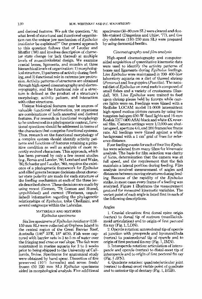
Four feeding events for each of four live Epibu- lus were selected from many films for kinematic analysis. The basis for film selection was clarity of focus, determination that the camera was at full speed, and the requirement that the fish maintain a lateral position during feeding. Film analysis involved measurement of angles and distances between moving structures during feed- ing. Because of the rapidity of the Epibulus strike, in most cases every frame of a feeding was analyzed. Figure 1 illustrates the measurement protocol for measured kinematic variables. The vertex point of each angle is listed first in each of the following descriptions.
Angles 1. Cranial elevation: first dorsal spine origin
(vertex) to dorsal tip of rostrum (maxilla/eth- moid articulation) and to origin of first pectoral fin ray (Fig. 1, LDN).
2. Opercle rotation: anterodorsal tip of opercle at junction with preopercle and hyomandibula (vertex) to posterodorsal tip of opercle and to origin of first pectoral fin ray (Fig. 1, IMN).
3. Interopercle rotation: articulation of intero- percle and opercle (vertex) to distal-most tip of interopercle and to origin of first pectoral fin ray (Fig. 1 JFN).
4. Quadrate rotation: quadrate/articular joint (vertex) to dorsal-most visible point of quadrate and to anterior tip of dentary (Fig. 1, EGB).

FEEDING MECHANICS OF EPl3ULUS INSlDiATOR 131
Fig. 1. Diagram of a feeding Epibulus insidiator, with points shown (A-N) for recording kinematic variables with a computerized digitizing system. See Materials and Methods (cinematography and film analysis) for l it of variables and their measurement points. U, lower jaw; other abbreviations of skull bones as in Figure 2.
5. Maxilla rotation: maxilla/articular junction (vertex) to dorsal-most visible point of maxilla and to anterior tip of dentary (Fig. 1, CDB). Distances
1. Gape: anterior premaxilla tip to anterior dentary tip (Fig. 1, AB).
2. Protrusion distance: lowest pigment line behind eye to anterior tip of premaxilla (Fig. 1, HA).
3. Hyoid depression: first dorsal spine origin to ventral-most point below interopercle (Fig. 1, LK).
4. Body-background distance: origin of first pectoral ray to point on background grid ante- rior to fish and prey (allows body velocity calcu- lation).
5. Jaw-prey distance: premaxilla tip to prey item (allows protrusion velocity calculation).
The origin of the first pectoral ray is used in many of the above variables and it should be noted that change in position of this point could
produce changes in the measured angles. The movement of the first pectoral ray in relation to the fishes body was measured and variation in its position was found to be negligible.
Data acquisition involved projecting high- speed films with an LW stop frame 16-mm film projector onto a Digipad 5 digitizing tablet (GTCO, Columbia, MD). A PC’s Limited 286 computer and Sigma-Scan software (Jan del Sci- entific, Sausalito, CA) were used to record kine- matic variables to data files. Data were analyzed and graphically presented by using an Apple Macintosh IIcx computer and CricketGraph soft- ware.
Electromyography To document the motor patterns in four cra-
nial muscles we obtained electromyographic re- cordings of muscle activity during prey capture in two Epibulus. Stainless-steel bipolar elec- trodes were constructed from two 75-cm-long pieces of poly-coated wire (0.051-mm diameter)
132 M.W. WESTNEAT AND P.C. WAINWRIGHT
threaded through the barrel of a 26-gauge hypo- dermic needle. The first 15 cm of the paired wires were glued together with a cyanoacrylate adhesive and 0.5-mm tips were exposed by scrap- ing off the insulation with a razor blade under a dissecting microscope. These tips were bent back against the barrel of the needle to form hooks that helped to hold the electrodes in place through the course of the experiment. Anesthe- tized fish (tricaine methane sulfonate) were placed into a shallow dissecting tray that was filled half with fresh water and half with the anesthetizing solution and the electrodes were implanted directly into the four muscles. The electrodes were then bundled together and glued into a cable which was secured to a suture looped through the fish's mid-dorsum just anterior to the dorsal fin. Recordings were always made from the left side members of the same four muscles: the levator operculi (LOP), the epaxial muscles (EP), the sternohyoideus (SH), and the adductor mandibulae (AM). All of the muscles were superficial, allowing visual confirmation of electrode placement.
Within an hour of their recovery from anesthe- sia the fish were offered 1-cm3 pieces of frozen shrimp (Penaeus), which were captured in mid- water. Electromyographic signals were recorded during prey capture on a Bell and Howell 4020AM-FM tape recorder. Signals were ampli- fied by Grass P511J preamplifiers with a band- pass of 100 Hz at the low end and 3,000 Hz at the high end. The 60-Hz notch filter was always used. Electromyograms were recorded at a tape speed of 19 cm/sec and were later digitized with a Keithley 12-bit analogue-to-digital converter at a sample rate of 2,050 Hz.
The sampling rate used when digitizing elec- tromyograms influences the degree to which the signal is faithfully reproduced. We used 2,050 Hz, because 1) a Fast Fourier Transform of rep- resentative signals showed that these electromyo- grams contained insignificant power (less than 5 7% in the range above 1,025 Hz so the Nyquist sample criterion was met and aliasing could be eliminated as a source of signal bias, and 2) the variables that were measured from the digitized records were onsets and cutoffs of activity (see below), rather than spike amplitudes or inte- grated areas. Onsets and cutoffs of activity would not be affected by aliasing. A hard copy for visual inspection was made from the computer file of each feeding on a Gould 260 chart re- corder.
The digital computer file from each feeding was then played into a Tektronix 4107 color graphics terminal. To quantify the time course
of muscle activity a computer program assisted in measuring nine variables from the electromyo- grams of each feeding. The duration of activity of each muscle (LOPDUR, EPDUR, SHDUR, AMDUR) and the onset time of each muscle relative to the levator operculi muscle (LOP-EP, LOP-SH, LOP-AM) were measured in millisec- onds. We used the levator operculi as the refer- ence muscle to facilitate comparisons with previ- ously described motor patterns (see below).
To explore the possibility that the feeding mechanism of Epibulus is driven by a novel motor pattern we compared our EMG data to similar data obtained from two perciform taxa, Micropterus salmoides and Lepomis macrochi- rus (Centrarchidae) (Wainwright and Lauder, '86). The centrarchid data are from feedings on 1-2-cm pieces of earthworm (Lumbricus) but were otherwise collected following the same pro- tocol used for Epibulus. We contrasted the seven common EMG variables among the two Epibu- Zus (14 feedings), five Micropterus (50 feedings) and four Lepomis (40 feedings) in a nested anal- ysis of variance experimental design, with indi- viduals nested within species; the Systat statisti- cal package (Wilkinson, '86) was used. An overall multivariate analysis of variance (MANOVA) with all seven variables was used to test the null hypothesis that the average motor patterns (de- fined by the seven EMG variables) of the three species were the same, and subsequent univari- ate ANOVAs were performed with each variable separately. Because several F ratios were exam- ined in this analysis a conservative probability level of P < 0.01 was used to establish statistical significance.
Muscle stimulation experiments To aid our understanding of the mechanism
underlying jaw protrusion in Epibulus, we ob- served the functions of five cranial muscles dur- ing electrical stimulation experiments on four live fish. All muscles examined were sufficiently superficial in location such that it was possible to observe their actions directly during stimula- tions. Each fish was first anesthetized (using tricaine methane sulfonate) and two widely spaced monopolar electrodes were implanted into each muscle. The monopolar electrodes were constructed by scraping the insulation from the last 1 mm of 75-cm-long pieces of 0.051-mm stainless-steel wire. The exposed tips were threaded through a hypodermic needle (26- gauge) and the last 3 mm were bent back against the needle's barrel to form a hook which held the electrode in place when the needle was inserted into the muscle belly. Left- and right-side mus- cles were always stimulated simultaneously. The

FEEDING MECHANICS OF EPIBULUS INSIDIATOR 133
electrodes were attached to muscle stimulators (Grass S44 and S48) so that equal electrical stimulation could be delivered to a bilateral pair or simultaneously to several muscles. Muscles were given twitch (2-ms duration) and presum- ably tetanic stimuli (100 Hz, 2-ms pulse dura- tion, 500-ms train duration, 2-15 V). Muscles were stimulated repeatedly with varying inten- sity and their actions on the elements of the jaws were recorded by a second observer as a percent- age of the maximum action observed during live feedings.
RESULTS
Our results begin with description of the cra- nial anatomy of Epibulus insidiator, followed by kinematic data from high-speed films and the results of electromyographic and muscle stimula- tion experiments. Descriptive morphology fo- cuses on the feeding mechanism and the classifi- cation of structures as primitive (present ancestrally) or derived (unique to Epibulus) and the subsequent discussion draws conclusions re- garding character polarity of functional charac- ters as well. A phylogenetic framework is re- quired for such statements of character polarity; therefore we state briefly our concept of Epibu- lus phylogenetic relationships and the outgroup structure for this analysis.
Epibulus insidiator is a member of the “chei- line” group within tribe Cheilinini and is thought to comprise a monophyletic group with the gen- era Cheilinus and Wetmorella (Gomon, per- sonal communication). Presently five probable synapomorphic characters unite this group rela- tive to labrid outgroups (Gomon, ’79; Westneat, unpublished data). These characters are: 1) ver- tebral count = 23 (9-14), 2) epipleural rib count reduced to ten, 3) mesethmoid extends caudad of preorbital process, 4) pectoral ray count = 12, and 5) frontal shelfand recess are lost. Evolution- ary polarity decisions for Epibulus characters are primarily based on comparisons with charac- ter states of Cheilinus and Wetmorella. Addi- tional outgroups used for comparison include other lineages within tribe Cheilinini, the primi- tive labrid tribe Hypsigenyini (Gomon, ’79), and the perciform family Centrarchidae.
Osteology Jaws
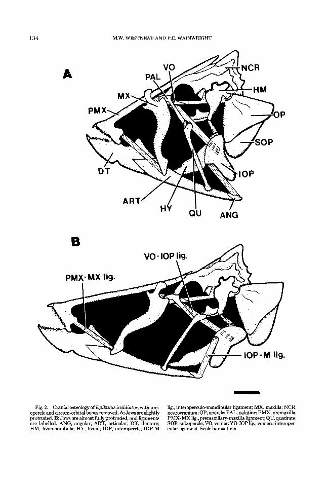
Figure 2 illustrates the major cranial bones and several ligaments that function during jaw protrusion in Epibulus insidiator. Epibulus ex- hibits several unique features of cranial osteol- ogy, while other features retain a primitive la- brid condition. Figure 3 presents the cranial
osteology of Cheilinus diagrammus for compar- ative purposes.
The elongate upper and lower jaws are promi- nent derived features of Epibulus (Fig. 2). The premaxilla (Fig. 2A, PMX) has an extremely elongate ascending process which, when fully retracted, passes posterodorsally through a chan- nel formed by the mesethmoid and frontal bones and extends past the posterior edge of the su- praoccipital crest. The alveolar arm of the pre- maxilla forms the toothed portion of the upper jaw and is a flattened process connected to the dentary via fibrous connective tissue, and to the maxilla via the premaxillary-maxilla ligament. This fibrous connection allows movement of the premaxilla relative to the dentary during in- crease in gape. The maxilla (Fig. 2A, MX) is laterally flattened in Epibulus, and the maxil- lary head is similar to that of other labrids, except that the dor_sal processes of the maxillae meet dorsally around the ascending processes of the premaxillae, forming a guide through which the premaxilla slides.
The lower jaw is also very elongate (Fig. 2), and is formed from a robust, toothed dentary, a long slender articular which makes up about two-thirds of mandibular length, and the angu- lar, which is a bony cap for the posterior tip of the articular. (Bone names are as in Rognes, ’73.) The dentary (Fig. 2A, DT) exhibits a well- defined coronoid process above its suture with the articular. The joint between articular and quadrate is located in Epibulus forward of the posterior tip of the articular by about 20% of mandibular length (Fig. 2). In Cheilinus (Fig. 3) and most perciform fishes, this joint is located at the posterior tip of the articular. In Epibulus, the posterior tip of the mandible is the site of insertion of the interoperculo-mandibular liga- ment onto the angular, as is the case in other labrids. Suspensorium
The suspensorium of Epibulus is modified in several respects. One key feature is the derived form of the quadrate. The primitive labrid quad- rate (and that of most other teleosts) is a flat- tened bone shaped approximately like an equilat- eral triangle, with a thickened knob at the antero ventral end where it articulates with the lower jaw (Fig. 3). In contrast, the quadrate of Epibu- lus is very elongate and approximates the shape of a long, thin cylinder (Fig. 2A, QU). The bony knob on the ventral tip and the joint between the quadrate and articular are retained in Epibulus from the primitive condition. However, a second joint involving the quadrate is found in Epibu- lus, formed by a bony knob on the dorsal tip of
134
A
M.W. WESTNEAT AND P.C. WAINWRIGHT
B
PMX-MX lig.
Fig. 2. Cranial osteology of Epibulus insidiator, with pre- opercle and circum-orbital bones removed. A Jaws are slightly protruded. B Jaws are almost fully protruded, and ligaments are labelled. ANG, angular; ART, articular, DT, dentary; HM, hyomandibula; HY, hyoid; IOP, interopercle; IOP-M
lig., interoperculo-mandibular ligament; MX, maxilla; NCR, neurocranium; OP, opercle; PAL, palatine; PMX, premaxilla; PMX-MX lig., premdary-maxilla ligament; QU, quadrate; SOP, subopercle; VO, vomer; VO-IOP lig., vomero-interoper- cular ligament. Scale bar = 1 cm.

FEEDING MECHANICS OF EPIBULUS INSIDIATOR 135
IOP-M lig. I POP I QU IOP -
Fig. 3. Cranial osteolcgy of Cheilinus diugrammus, a representative of labrid tribe Cheilinini, and closely related to Epibulus. POP, preopercle; other abbreviations as in Figure 2. Scale bar = 1 cm.
the quadrate where it articulates with the metapterygoid. Such a joint allowing movement of the quadrate is absent in Cheilinus and other Perciformes. This derived feature of the Epibu- lus suspensorium frees the quadrate from its ancestral role of stationary support for the lower jaw and enables it to rotate anteriorly. Other aspects of the Epibulus suspensorium that de- serve mention are the apparent loss of the en- topterygoid and reduction in size of both the meta- and ectopterygoid elements.
Hyoid The hyoid apparatus of Epibulus (Fig. 2) re-
tains a structure similar to that of Cheilinus and other labrids. The hypohyal, ceratohyal, and epi- hyal form the body of the hyoid apparatus, lo- cated in the floor of the mouth. The interhyal extends dorsally from its joint with the posterior tip of the epihyal. The hyoid is involved in joints with the hyomandibula and the interopercle that function during feeding in Epibulus. The dorsal tip of the interhyal articulates with the ventral
tip of the hyomandibula. The hyoid bar may rotate around this joint during hyoid depression. The interopercle articulates with the postero dorsal tip of the epihyal and ventral tip of the interhyal via fibrous connective tissue. During jaw protrusion this joint is the axis of rotation of the interopercle during its extreme rotation of nearly 100 degrees.
Opercle series The preopercle, opercle (Fig. 2), and sub-
opercle in Epibulus exhibit forms similar to those of other labrids (i.e., Cheilinus; Fig. 3). The in- teropercle, however, exhibits several features ap- parently unique to Epibulus (Fig. 2A, IOP). The shape of the interopercle is atypical, with a thin process (visible in the adducted position; Fig. 2A) extending postero ventrally from the robust site of attachment of the interoperculo-mandib- ular ligament. In Epibulus the connection be- tween interopercle and opercle is limited to a small region on the surface of each bone. The interopercle is thus free to rotate anteriorly.

136 M.W. WESTNEAT AND P.C. WAINWRIGHT
Neurocranium The neurocranium of Cheilinus (Fig. 3) and
that of primitive labrids such as the Hypsigeny- ini (Gomon, '79) is robust with thickened bony ridges around the large orbit, a ventrally di- rected vomer, a deep parasphenoid, and a thick supraoccipital crest. The Epibulus neurocra- nium (Fig. 2) is characterized by more delicate structures and a narrow profile. The vomer is directed anteriorly and only slightly ventrally and the parasphenoid is thin and has no ventral process. The supraoccipital crest is thin and re- duced relative to the Cheilinus condition. A deep trough is present in the dorso medial surface of the Epibulus neurocranium, formed by the me- sethmoid and frontal bones. The grooves in this trough guide the ascending processes of the pre- maxillae during protrusion. The flattened fron- t& and mesethmoid extend posteriorly to cover the anterior portion of the supraoccipital crest and in large specimens this often causes the supraoccipital to lean laterally, producing an asymmetry in the neurocranium.
Connective tissues Several connective tissues in the trophic appa-
ratus of Epibulus exhibit derived conditions, including a ligament that apparently has arisen de novo in Epibulus and ligaments present ances- trally (in Cheilinus and other labrids) that are modified in Epibulus. An unusual ligament in Epibulus originates on the lateral processes and ventral surface of the vomer and inserts on the medial surface of the interopercle (Fig. 2B). This ligament is apparently unique to Epibulus, as we have found no evidence of its presence in other labrid taxa and have found no reference to such a ligament in perciform fishes. We refer to this ligament as the vomero-interopercular ligament (Fig. 2B, VO-IOP lig.). It is broad and flattened along its considerable length and the fibers pass beneath the palatine to contact the quadrate. A few fibers insert on the dorsal head of the quad- rate, but most pass medial or dorsal to the quad- rate to insert on the interopercle. Additional fibers originate on the quadrate and insert onto the interopercle. When tightened by cranial ele- vation, the vomero-interopercular ligament transfers an antero dorsal force from the neuro- cranium to the interopercle.
The interoperculo-mandibular ligament is present in Cheilinus (Fig. 3) as a fibrous connec- tion between the anterior margin of the intero- percle and the posteroventral tip of the lower jaw. In Epibulus this ligament (Fig. 2B, IOP-M lig.) retains the same origin and insertion points but is thickened and elongate, forming a broad
band of fibers whose alignment is generally dorso ventral as opposed to the primitive antero poste- rior orientation. The interoperculo-mandibular ligament of Epibulus transfers an antero dorsal force to the lower jaw as the interopercle rotates.
The premaxilla-maxillary ligament is the fi- brous connection between the posterior tip of the alveolar process of the premaxilla and the medial surface of the maxillary shaft. The primi- tive condition (Cheilinus) of this ligament is short, allowing minimal movement of the alveo- lar process of the premaxilla independent of the maxilla. The premaxilla-maxillary ligament in Epibulus (Fig. 2B, PMX-MX lig.) takes the derived form of a long, flattened band of fibers that retain the ancestral sites of origin and inser- tion. The ligament is probably in tension during retraction of the jaws.
It should be noted that connective tissues in Epibulus that link bony elements at points of movement or rotation are restricted to fibrous connections at small points on each element, allowing the bones to rotate relative to one an- other. An example of this is the tight fibrous connection between the poster0 dorsal tip of the epihyal and the medial surface of the interoper- cle, around which the interopercle rotates.
MYO@Y Figure 4 presents a lateral view of the cheek
and head muscles of Epibulus. For the purpose of functional description these muscles can be divided into jaw-retracting muscles, jaw-protrud- ing muscles, and those performing other func- tions. The adductor mandibulae complex of mus- cles functions in retraction of the jaws and has three divisions, each of which shows little change from the perciform conditions of Winterbottom ('74). The A1 component (Fig. 4) originates on the anterior edge of the preopercle and inserts via a long, continuous tendon onto the head of the maxilla dorsally, and onto the lateral face of the lower jaw (articular) ventrally. When the jaws are f d y protruded, A1 contraction serves to rotate the maxilla posteriorly and pull the lower jaw posteriorly and dorsally, closing the jaws. The A2 division also originates on the an- tero dorsal edge of the preopercle and passes under the A1 medially. The A2 fuses with the A3 division below the origin of the A3 on the hyo- mandibula and pterotic. The combined A2 and A3 fibers (Fig. 4) are aligned primarily dorso ventrally as they continue below A1 as a thin band of muscle and insert tendinously on the medial face of the articular. Contraction of the A2lA3 muscle fibers after protrusion has oc- curred functions in retracting the lower jaw (see Results: Muscle Stimulation).

FEEDING MECHANICS OF EPIBULUS INSIDIATOR
DO A 2
137
.o P
A 2 + A 3 \
A1 Fig. 4. Cranial muscles of Epibulus insidiator. AAP, adductor arcus palatini; Al, A2, A3, divisions of
the adductor mandibulae; A2 + A3, combined A2 and A3 divisions; Alt, tendon from A1 muscle to maxilla and lower jaw; DO, dilatator operculi, EP, epaxialis, HP, hypaxialis; LAP, levator arcus palatini; LOP, levator operculi. Scale bar = 1 cm.
Jaw protrusion is effected by the epaxial (EP) and levator operculi (LOP) muscles (Fig. 4). Both of these muscles exhibit the structural condition found in Cheilinus and many perciforms. The epaxial muscles are extensive on the dorsal sur- face of the neurocranium, inserting onto the supraoccipital, parietal, frontal, and lateral eth- moid elements. Epaxial contraction elevates the neurocranium. The levator operculi originates on the pterotic as a flat sheet of muscle fibers and inserts on the dorsal and dorsomedial sur- faces of the opercle. Levator operculi contraction functions in rotation of the opercle postero dor- sally. Also pictured in Figure 4 are the dilatator operculi (DO: origin, sphenotic and pterotic; in- sertion, opercle), levator arcus palatini (LAP: origin, sphenotic; insertion, hyomandibula), and adductor arcus palatini (AAP: origin, parasphe- noid; insertion, hyomandibula, and metaptery- goid).
Figure 5 illustrates the geniohyoideus (GH; = protractor hyoideus, Winterbottom, '74) and the sternohyoideus (SH) muscles of the ven- tral surface of the head of Epibulus. The genio- hyoideus is extremely elongate in Epibulus, ex- tending from its anterior site of attachment on the dorsal surface of the dentary symphysis to a posterior attachment on the paired ceratohyals. This muscle occupies the ventral midline be- tween the two halves of the lower jaw as a narrow column of fibers wrapped by a cylinder of connec- tive tissue. This connective tissue appears to exhibit a crossed helical fiber arrangement. The geniohyoideus muscle becomes bifid a t the ante- rior end of the urohyal and continues postero laterally to its paired attachments on the medial surface of each ceratohyal. Preliminary measure- ments of geniohyoideus length from its anterior attachment to the point where it becomes bifid indicate that the extended length of the muscle

138 M.W. WFSTNEAT AND P.C. WAINWRIGHT
Fig. 5. Ventral head muscles of Epibulus insidiator, with dentary (DT) and ceratohyal (CH) boneslabelled. HP, hypax- ialii; GH, geniohyoideus; SH, sternohyoideus. Scale bar = 1 cm.
is approximately twice that of its contracted length.
The stemohyoideus (Fig. 5, SI1) originates dorsally on the cleithrum and ventrally on the anterior face of the myoseptum separating the stemohyoideus from the hypaxialis (HP). The sternohyoideus inserts onto the urohyal anteri- orly.
General feeding behavior Prey capture in Epibulus insidiator may be
divided into three components 1) approach to
the prey, 2) the strike, and 3) prey processing/ swallowing. Table 1 presents data on strike char- acteristics for Epibulus, and Figure 6 presents frames from a film of Epibulus during the strike phase. Prey items appeared to be detected visu- ally and were approached to within 3040 mm (about 25% of SL or 70% of head length) before strike initiation (Table 1). Body velocity slowed to near zero during the strike phase and maxi- mum protrusion velocity of the jaws during the strike was always over 2 m/sec (Table 1). Prey were engulfed by the jaws within 2540 ms of strike initiation. Epibulus employs suction dur- ing feeding, as can be seen in Figure 6 (frames 6-8) where the dark prey item can be seen mov- ing into the buccal cavity a t a speed of over 2 m/sec (Table 1). Retraction of the jaws was com- pleted within 100 msec of strike initiation. Jaw retraction was accompanied by visible swallow- ing movements and by a forward sweep of the pectoral fins to back away from the site of prey capture.
Film kinematics Description and analysis of the kinematics of
cranial bones during feeding will allow interpre- tation of the mechanism of jaw protrusion in Epibulus. Figure 6 presents frames from a high- speed film of an Epibulus feeding. Figure 7 illus- trates the mechanism of jaw protrusion during feeding in the form of schematic mechanical diagrams. The diagram numbers in Figure 7 correspond to the frame numbers in Figure 6. The most evident feeding mechanics in Figures 6 and 7 are extreme protrusion of both the upper and lower jaws, rotation of the neurocranium, opercle, interopercle, quadrate, and maxilla, and increase in gape. These variables can be divided into input and output kinematics. Input kinemat- ics are those directly caused by muscle contrac-
TABLE 1. Characteristics ofthe strike of Epibulus insidiatnr'
Characteristics Mean SD ~~
Body velocity during strike (dsec) 0.05 0 . a Prey distanceatstrike (mm) 36.90 6.90 Time to prey capture (mec) 34.10 6.50 Time to jaw retraction (msec) 111.30 9.40 Maximum protrusion velocity (dm) 2.31 0.45 Maximum protrusion acceleration (m/
se%) 111.20 43.80 Suction velocitv of orev (m/sec)* 2% 0.66
'Dataarechieflycalculatedfromanalyaisof4 feedingsfrom4 indiridu- als; tabulated as means and standard deviations (SD) of mike charm- teristica 2Data are from 3 feediw from 1 individual.

FEEDING MECHANICS OF EPIBULUS INSIDIATOR
Fig. 6. Frames 1, 3, 6, 6, 7, and 8: From a high-speed film (200 framedsec) of the strike of Epibulw insidiator. Successive frames are 0.005 sec apart. Note rotation of quadrate, maxilla, and interoperculo-mandibular ligament. Suction is apparent in frames 6, 7, and 8. See Figure 7 for corresponding mechanical diagrams.
139

A
B
Fig. 7. Mechanical diagrams illustrating the feeding mechanism and kinematics of E p i b u h insidiutor during feeding. A Retracted position, as in Figure 6, frame 1. B Partidy protruded position, as in Figure 6, frame 3. Note cranial elevation, opercle rotation, and interopercle rotation. C Almost fully protruded position, as in Figure 6, frame 6. Note rotation of maxilla and quadrate. Abbreviations as in Figure 2.

FEEDING MECHANICS OF EPIBULUS INSIDIATOR 141
tion (cranial elevation and opercular rotation), whereas output kinematics are caused by a trans- fer of force, through connective tissue or bone articulations, from one skeletal element to an- other.
Figure 8 presents quantitative kinematic data in graphic form for the variables of cranial eleva- tion, opercular rotation, interopercular rotation, quadrate rotation, maxillary rotation, protru- sion distance, gape distance, and hyoid depres- sion. These kinematic profiles reveal the relative importance of cranial and opercular input to jaw protrusion in Epibulus. Cranial elevation and opercular rotation reach a maximum of around 10 degrees of arc. Cranial elevation (Fig. 8A) reaches its maximum arc within 35 msec of the start of the strike. The output movements of interopercular rotation (Fig. SC), quadrate rota- tion (8D), maxil- lary rotation (8E), protrusion (8F), and gape (8G) are all virtually synchronous with cranial elevation. Peak opercular rotation (Fig. BB), however, occurs about 15 msec before the synchronous movements of the other vari- ables.
The highly synchronous pattern of movement among many of the variables depicted in Figure 8 is typical of every Epibulus feeding analyzed and was followed in most cases by peak hyoid depression approximately 15 msec later. The rotations of the interopercle, quadrate, and max- illa peak at approximately 100 degrees. Protru- sion distance data (Fig. 8F) illustrate the ex- treme protrusion ability of Epibulus, with a maximum of over 3 cm, as well as the velocity of the strike (the slope of Fig. 8F is steep).
The mechanism of jaw retraction involves a reverse of the kinematic pattern of bone move- ments. (Note the bell-shaped appearance of the kinematic profiles in Fig. 8.) Muscle stimulation experiments (Table 3) indicate that jaw retrac- tion is driven primarily by the adductor mandib- ulae muscles exerting posteriorly and dorsally directed forces upon the maxilla and the lower jaw. The A1 division (Fig. 4) pulls posteriorly upon the maxillary head, providing a posterior rotation in that element that serves as the input movement for jaw retraction. The A1 and the combined A2/A3 divisions of the adductor man- dibulae muscle both insert on the lower jaw (Fig. 4) and transmit poster0 dorsal forces to the man- dible that contribute to jaw retraction.
Electromyogruphy The motor pattern exhibited by Epibulus at
the strike is summarized in Figure 9A, where the durations and the relative timing of activity are averaged for the 14 feedings from the two exper- imental fish. The strike is characterized by
broadly overlapping activity in all muscles, begin- ning with activity in the levator operculi and the epaxial muscles. This is followed by the onset of activity in the sternohyoideus and then the ad- ductor mandibulae. Average durations of activ- ity range from 33 msec for the epaxial muscle to 45 msec in the sternohyoideus.
The sequence of muscle activity in Epibulus appears to be the same as in Lepomis and Mi- cropterus. The overall MANOVA comparison of the motor pattern in Epibulus, Lepomis, and Micropterus showed no significant difference among the three species (MANOVA comparison of species: Wilkes's X = 0.026, F = 1.48, d.f. = 14,4; P = 0.381). This resultis also reflected in the univariate ANOVAs presented in Table 2. Univariate ANOVA tests indicate that none of the seven EMG variables differed significantly among the three species (Table 2), although the test for significance of the relative onset time between the levator operculi and the adductor mandibulae had a very high F-ratio which ap- proached significance at the 0.01 level (Table 2, LOP-AM). In contrast, six of seven variables showed significant among-individual variance.
Muscle stimulation Anatomical analysis indicates that premaxil-
lary protrusion may be effected by both the levator operculi (via the linkage from the opercle to the rotating interopercle) and the epaxial mus- cles (via the vomero-interopercle ligament). Stim- ulation of these muscles individually resulted in approximately 30 % of maximum jaw protrusion (Table 3). However, when the two muscles were stimulated simultaneously up to 70% of maxi- mal protrusion was observed. This was the great- est amount of protrusion attained during the muscle stimulation experiments.
Stimulation of the sternohyoideus caused de- pression of the hyoid but did not move the jaws. When the geniohyoideus was stimulated from an outstretched position (the jaws fully or par- tially protruded) the muscle shortened and tight- ened the intermandibular fascia but did not re- tract the jaws. Stimulation of the adductor mandibulae (Al) caused firm and complete re- traction of the protruded jaws. This muscle ef- fects retraction from any stage of jaw protrusion, even though the angle of insertion that this mus- cle makes against the dentary changes through about 90' during a full retraction stroke.
DISCUSSION
Epibulus insidiator exhibits the highest de- gree of jaw protrusion known among fishes. The feeding mechanisms of this fish is described be-

ELEVATION 10
I n 8 W
g 6 s O 4
2
0 0 50 100 150 200
12
10
I n 8
s W W U 6
0 4
2
0 0 50 100 150 200
0 50 100 150 200
120
100
In 8o
s W W U 60
40
20
0 0 50 100 150 200
TIME (MS)
Fig. 8. Graphs of kinematic movement of cranial bones during feeding in Epibulus insidiator, resulting from measure- mentstaken from four films each of four individuals (N = 16) feeding on shrimp (Peneus). Error bars are standard devia- tions and graph lines are not the result of fitting techniques.
0 50 100 1 50 200
4
3
a 0 2
1
0 0 50 100 150 200
1.2
1 .o
0.8
5 0.6 0.4
0.2
0.0
DISTANCE
0 50 100 150 200
0.5
0.4
0.3 = 0
0.2
0.1
0.0 0 50 100 150 200
TIME (MS)
Vertical lines indicate time of maximum displacement. A: Cranial elevation. B Opercular rotation. C Interopercular rotation. D Quadrate rotation. E: Maxillary rotation. F Protrusion distance. G: Gape distance. H Hyoid depression.

FEEDING MECHANICS OF EPIBULUS INSIDIATOR 143
A
LOP
AM
EP
SH
Epibulus insidiator 1 I
I
I- t
I
I
!- I
I
B
LOP
AM
EP
SH
~~ ~~
-20 0 20 40 60 80 100 TIME [msec)
C
Lepomis macrochirus I I
I I
I I t I
I
:--.)-- I
I I . . . . . . I . . I
-20 o 20 40 60 eo l o o TIME (mscc)
Micropterus salmoides I I
I
EP
I
- SH
TABLE2. Results of F-tests from nested ANOVAs comparing 7 electromyographic variables among Epibulus,
Lepomis, and Micropterus'
Factor ~
Species Individuals Variable (2,8)' (8,7692)
LOPDUR 1.40 3.48' EPDUR SHDUR AMDUR LOP-EP
1.29 3.16 3.14 0.48
6.54* 3.73' 2.77' 2.81*
LOP-SH 0.14 4.12' LOP-AM 8.06 1.82
'Table entries are the F-ratica from the significance tests. See Materials and Methods (eledromyography) for definition of abbreviations. *( ) = degrees of freedom for F-test. f = P < 0.01.
low, followed by a discussion of the levels of organization at which evolutionary changes have occurred to produce this unusual mechanism of jaw protrusion. Three levels of organization are examined for this purpose: 1) the structures in- volved in feeding, 2) the activity pattern of bones and muscles, and 3) the functional role of each element in the feeding mechanism. Epibulus clearly exhibits major changes in the structure of many bones and ligaments of the head, and films and electromyograms reveal the activity pat- terns of bones and muscles during feeding. These structural and functional data are interpreted in terms of the level of organization at which each element of the jaws exhibits a primitive or de- rived condition. Thus, each element of the jaws of Epibulus exhibits a character state at each of the three levels described above, and a decision of evolutionary polarity is made at each level.
Mechanism of jaw protrusion Analysis of the kinematics of Epibulus feed-
ing (Fig. 8), consideration of morphology (Figs. 2,4), electromyography (Fig. 9), and muscle stim- ulations (Table 3) are combined to propose a mechanism by which this fish captures prey. Figure 7 presents mechanical diagrams of the Epibulus jaw mechanism in retracted (7A), par-
Fig. 9. Bar diagrams showing the average motor pattern exhibited by three fish species, Epibulus insidiator (A), Lep- omis macrochirus (B), and Micropterus salmoides (C), dur- ing prey capture. Mean duration of activity in each muscle is indicated by the length of each bar and average relative onset time is shown with reference to the levator operculi. Left- hand error bars are standard deviations of relative onset times and error bars on the right are standard deviations of muscle burst duration. See Materials and Methods (electro- myography) for muscle abbreviations. Sample sizes: (A) 2 fish, 14 feedings; (B) 4 fish, 40 feedings; (C) 5 fish, 50 feedings. See Table 2 for statistical comparison of motor patterns.
144 M.W. WESTNEAT AND P.C. WAINWRIGHT
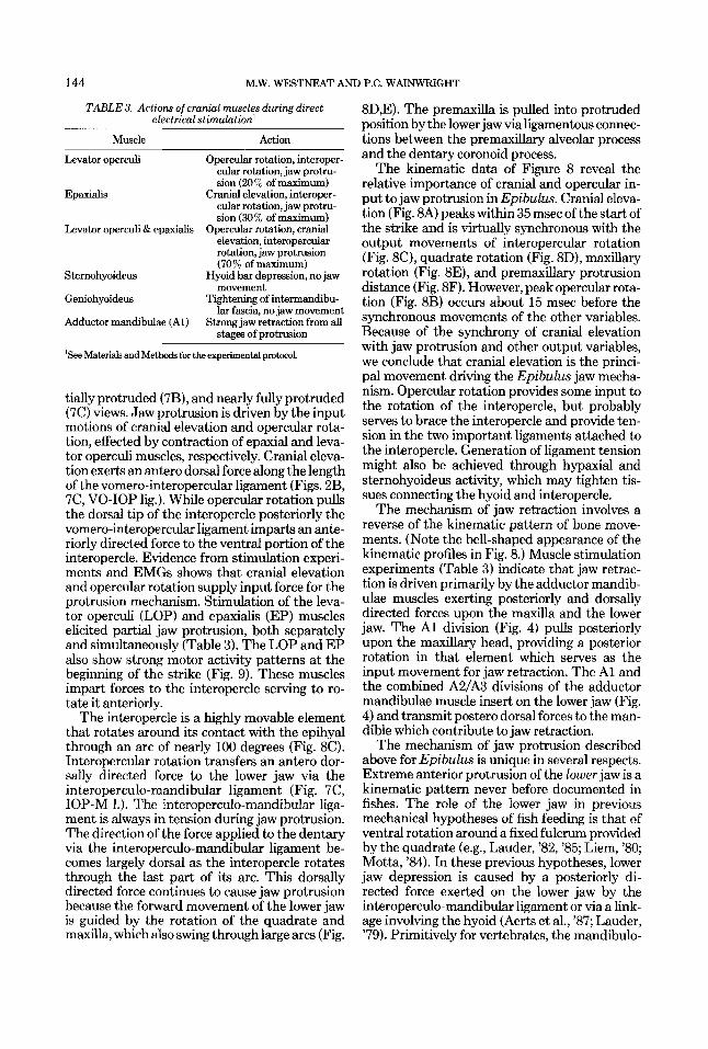
TABLE 3. Actions of cranial muscles during direct electrical stimulation'
Muscle Action
Levator operculi Opercular rotation, interoper- cular rotation, jaw protru- sion (20% of maximum)
Epaxialis Cranial elevation, interoper- cular rotation, jaw protm- sion (30% of maximum)
Opercular rotation, cranial elevation, interopercular rotation, jaw protrusion (70% of maximum)
Sternohyoideus Hyoid bar depression, no jaw movement
Geniohyoideus Tightening of intermandibu- lar fascia, no jaw movement
Adductor mandibulae (Al) Strong jaw retraction from all s h e s of orotrusion
Levator operculi & epaxialis
'See Materials and Methods for the experimental protocol.
tially protruded (7B), and nearly f d y protruded (7C) views. Jaw protrusion is driven by the input motions of cranial elevation and opercular rota- tion, effected by contraction of epaxial and leva- tor operculi muscles, respectively. Cranial eleva- tion exerts an antero dorsal force along the length of the vomero-interopercular ligament (Figs. 2B, 7C, VO-IOP lig-1. While opercular rotation pulls the dorsal tip of the interopercle posteriorly the vomero-interopercular ligament imparts an ante- riorly directed force to the ventral portion of the interopercle. Evidence from stimulation experi- ments and EMGs shows that cranial elevation and opercular rotation supply input force for the protrusion mechanism. Stimulation of the leva- tor operculi (LOP) and epaxialis (EP) muscles elicited partial jaw protrusion, both separately and simultaneously (Table 3). The LOP and EP also show strong motor activity patterns at the beginning of the strike (Fig. 9). These muscles impart forces to the interopercle serving to ro- tate it anteriorly.
The interopercle is a highly movable element that rotates around its contact with the epihyal through an arc of nearly 100 degrees (Fig. 8C). Interopercular rotation transfers an antero dor- sally directed force to the lower jaw via the interoperculo-mandibular ligament (Fig. 7C, IOP-M 1.). The interoperculo-mandibular liga- ment is always in tension during jaw protrusion. The direction of the force applied to the dentary via the interoperculo-mandibular ligament be- comes largely dorsal as the interopercle rotates through the last part of its arc. This dorsally directed force continues to cause jaw protrusion because the forward movement of the lower jaw is guided by the rotation of the quadrate and maxilla, which also swing through large arcs (Fig.
8D,E). The premaxilla is pulled into protruded position by the lower jaw via ligamentous connec- tions between the premaxillary alveolar process and the dentary coronoid process.
The kinematic data of Figure 8 reveal the relative importance of cranial and opercular in- put to jaw protrusion in Epibulus. Cranial eleva- tion (Fig. 8A) peaks within 35 msec of the start of the strike and is virtually synchronous with the output movements of interopercular rotation (Fig. 8C), quadrate rotation (Fig. 8D), maxillary rotation (Fig. 8E), and premaxillary protrusion distance (Fig. 8F). However, peak opercular rota- tion (Fig. 8B) occurs about 15 msec before the synchronous movements of the other variables. Because of the synchrony of cranial elevation with jaw protrusion and other output variables, we conclude that cranial elevation is the princi- pal movement driving the Epibulus jaw mecha- nism. Opercular rotation provides some input to the rotation of the interopercle, but probably serves to brace the interopercle and provide ten- sion in the two important ligaments attached to the interopercle. Generation of ligament tension might also be achieved through hypaxial and sternohyoideus activity, which may tighten tis- sues connecting the hyoid and interopercle.
The mechanism of jaw retraction involves a reverse of the kinematic pattern of bone move- ments. (Note the bell-shaped appearance of the kinematic profiles in Fig. 8.) Muscle stimulation experiments (Table 3) indicate that jaw retrac- tion is driven primarily by the adductor mandib- ulae muscles exerting posteriorly and dorsally directed forces upon the maxilla and the lower jaw. The A1 division (Fig. 4) pulls posteriorly upon the maxillary head, providing a posterior rotation in that element which serves as the input movement for jaw retraction. The A1 and the combined A2/A3 divisions of the adductor mandibulae muscle insert on the lower jaw (Fig. 4) and transmit poster0 dorsal forces to the man- dible which contribute to jaw retraction.
The mechanism of jaw protrusion described above for Epibulus is unique in several respects. Extreme anterior protrusion of the lower jaw is a kinematic pattern never before documented in fishes. The role of the lower jaw in previous mechanical hypotheses of fish feeding is that of ventral rotation around a fixed fulcrum provided by the quadrate (e.g., Lauder, '82, '85; Liem, '80; Motta, '84). In these previous hypotheses, lower jaw depression is caused by a posteriorly di- rected force exerted on the lower jaw by the interoperculo-mandibular ligament or via a link- age involving the hyoid (Aerts et al., '87; Lauder, '79). Primitively for vertebrates, the mandibulo-

FEEDING MECHANICS OF EPIBULUS INSIDIATOR 145
hyoid linkage functions in lower jaw depression, which is brought about when the sternohyoideus muscle retracts the hyoid. This posteriorly di- rected force is passed to the lower jaw through ligamentous connections between the epihyal and lower jaw.
Lower jaw movement in Epibulus is caused by anteriorly and dorsally directed forces exerted through the vomero-interopercular ligament to the interopercle and then to the lower jaw via interopercular rotation. These force vectors pull the lower jaw into protruded position, guided by the extreme rotation of the interopercle and the movable maxilla and quadrate (Fig. 7). The prim- itive hyoid-interopercle-lower jaw linkage is re- tained in Epibulus, but because of the unique nature of the rotating interopercle, retraction of the ceratohyals does not cause any motion of the lower jaws (Table 3, sternohyoideus). Thus the structural linkage for a primitive mechanism of jaw depression is retained in Epibulus but no longer functions in jaw movement. A reasonable hypothesis is that this linkage functions in oro- branchial cavity expansion in Epibulus. Two other linkages that primitively produce jaw de- pression in teleosts, cranial elevation and opercu-
lar rotation (Lauder, '85), apparently play cen- tral roles in the Epibulus feeding mechanism as sources of input motion (Fig. 7).
Ancestral and derived features of Epibulus Description of the mechanism of jaw protru-
sion in Epibulus reveals a mosaic of primitive and derived features that contribute to jaw func- tion during feeding. Table 4 presents a list of structural features and a description of their activity patterns and their presumed functions with indication as to the phylogenetic polarity of each character as primitive or derived relative to Cheilinus and other labrids. The discussion of these features will focus on identifying the partic- ular combination of primitive and derived condi- tions that has produced the unusual mechanism of jaw protrusion in Epibulus.
Anatomy The general pattern of structural modification
in the Epibulus feeding mechanism is extensive change in bone and ligament structure with com- paratively little modification of the cranial mus- cles. Table 4 illustrates the distribution of de- rived osteological features in Epibulus including
TABLE 4. Primitive and derived characters of Epibulw morphologic structure, k inemt ic or electromyographic pattern, and function in jaw protrusion'
Structure
Bones Neurocranium
Ethmoid/frontal Other elements
Premaxilla
M&
Mandible
Kinematic or P/D EMG pattern P/D Function P/D
- Elevation P Force to V-Iop lig. D D - - Guide premaxilla P
D Extreme protrusion D Protrusion P Prey capture P
P Extreme rotation D Brace premax., etc. P Protrude LJ D
D Extreme protrusion D Protrusion D
- - - - P
Prey capture P Quadrate D Extreme rotation D Protrude W D Opercle P Rotation P Input force P Interopercle D Extreme rotation D Anterior force to LJ D Palatine P Hyoid app. P - - Depress mouth floor P
V-Iop lig. D - - Anterior force to IOP D Iop-M lig. D Rotation D Anterior force to LJ D Pmx-Mx lig. D - - Connect Pm to Mx P Operc-Iop lig. D - - Rotation point D Hyoid-Iop lig. D - - Rotation point D
Levator operculi P Early contraction P Opercle rotation P Epaxialis P 1&20 ms after LOP P Cranial elevation P sternohyoideus P 15-20 ms after LOP P Hyoid depression P Geniohyoideus D - - Hyoid protraction P Addudor P 2&25 ma after LOP P Jaw retraction P
- - - -
Ligaments
Muscles
mandibulae
'P = primitive, D = derived, relative to closely related and other labrid species or perciform fishes. Ligaments: V-Iop lig. = vomero-interopercular; Iop-M lig. = interoperculo-mandibular; PIE-Mx liig. = premaxilla-*, Operc-Iop lig. = operculo-interopercular; Hyoid-Iop lii. = hyoid-interopercular.

146 M.W. WESTNEAT AND P.C. WAINWRIGHT
the premaxilla, lower jaw, quadrate, interoper- cle, and aspects of the neurocranium. It should be noted that derived conditions range in magni- tude from extreme modification in structure (such as the quadrate) to modification in size (premaxilla) or general shape and orientation (interopercle). Most other cranial bones are sim- ilar in sue and shape to the condition found in Cheilinus and many other perciform fishes.
Derived conditions found in connective tis- sues (Table 4) range from a completely unique ligament that plays a major role in the function of protrusion (i.e., the vomero-interopercular lig- ament) to reduced elements (i.e., between oper- cle and interopercle) or enlarged ligaments (i.e., premaxilla-maxillary). Many ligaments of the head are not listed in Table 4 and these appear to retain a primitive condition.
The muscles examined in Epibulus (Table 4) retain the origin, insertion, and general shape found in most labrids and in the perciform mus- cles described by Winterbottom ('74). An excep- tion to this is the geniohyoideus muscle (Fig. 5 ) , which is extremely elongate in Epibulus. Other muscles that retain a primitive condition (such as the adductor and levator arcus palatini mus- cles) are diagrammed in Figure 4 but are not included in Table 4 because their function was not examined. Kinematic pattern
Several key elements of the feeding kinemat- ics in Epibulus are conserved from the primitive condition widely present in perciform fishes. Pre- vious work (Alexander, '66; Lauder, '79, '81b; Lauder and Liem, '81; Liem, '80; Osse, '69) re- veals a biomechanical pattern typical of teleost feeding that consists of nearly simultaneous cra- nial elevation, gape, and jaw protrusion followed by hyoid depression. Peak opercular rotation has been shown to precede the simultaneous maxima of the above variables in several taxa (Liem, '70; Aerts et al., '87). The kinematic pat- tern described above is the same pattern found in Epibulus. Figure 8 illustrates the synchrony of peak cranial elevation, protrusion distance, and gape, preceded by opercular rotation and followed by hyoid depression. The magnitude of these variables (degrees of rotation or measured distance), except for protrusion distance, is also similar to literature values for other fish species of comparable size.
The notable differences in kinematic pattern between Epibulus and other teleosts are more numerous than the similarities (see Table 4). Interopercular and quadrate rotation (Fig 8C,D) for Epibulus cannot be compared to literature values because these movements do not occur in
other fishes. In fact, mechanical models of fish jaw movements published by Bare1 et al. ('77) and Aerts et al. ('87) suggest that only a small posterior translation of the interopercle can oc- cur and allow for no independent movement of the quadrate relative to other jaw elements. Max- illary rotation in Epibulus (Fig. 8E) peaks at over 100 degrees. Lauder ('81b) demonstrated a 40-60-degree maxillary rotation for Lebiasina boruca, and maxillary rotation in three species of Cheilinus exhibits a peak value of approxi- mately 20 degrees (Westneat, unpublished data). The high level of maxillary rotation is thus a derived activity pattern in Epibulus. These re- sults demonstrate again that the timing and pattern of input movements (cranial elevation and opercular rotation) are similar to those de- scribed for other teleosts, but the elements that transfer this movement to the highly protrusive jaws exhibit derived kinematic patterns (Table 4).
Muscular motor pattern No significant differences were found among
Epibulus, Lepomis, and Micropterus in the av- erage activity pattern of the four muscles, but the relative onset time of the adductor mandibu- lae (Fig. 9, AM) did approach significance (Ta- ble 2). Although we have not compared the Epibulus motor pattern quantitatively to that of other cheiline labrids, it conforms qualitatively to published accounts of Cheilinus unifasciatus, other labrids (Sanderson, '88), and other labroid taxa feeding on immobile prey (Liem, '78, '80). Thus, with the possible exception of the nearly significant relative onset of the adductor mandib- ulae, the evolution of the Epibulus jaw mecha- nism does not appear to have involved changes in the timing of activity of the primary feeding muscles (Table 4).
It is important to note that our motor pattern analysis is restricted to the seven duration and relative onset variables that were measured. Other levels of EMG signal analysis (e.g., signal amplitude, intra-burst heterogeneity, spike fre- quency, spike number times amplitude) were not performed so that our findings of a con- served motor pattern in Epibulus only relate to the timing variables that were measured. A more detailed analysis of muscle activity would be needed to fully determine if all aspects of the Epibulus motor pattern remain unchanged from the primitive labrid condition. We emphasize, however, that our statistical analysis was unable to falsify the null hypothesis that the average motor patterns were the same in the three spe- cies.

FEEDING MECHANICS OF EPIBULUS INSIDIATOR 147
One factor that made finding significant differ- ences among species less likely was the high levels of variation that were found among individ- uals within species (Table 2). High within-group variance decreases the likelihood that a statisti- cal test will find significant differences among group means. High intraspecific variation in EMG variables has been found widely in aquatic feeding vertebrates (Shaffer and Lauder, '85; Bemis and Lauder, '86; Wainwright and Lauder, '86; Sanderson, '88; Wainwright, '89), indicating that there can be considerable plasticity be- tween feedings in some aspects of the motor pattern. One practical consequence of high in- traspecific variation is that it is important to base comparisons among species on adequate estimates of the within-species variance compo- nent. Thus, experimental designs should permit the partitioning of variance among species and among individuals within species, as exemplified by the nested analysis of variance design used in this study.
The lack of evidence of evolutionary change in the motor pattern of Epibulus agrees with sev- eral recent studies (Lauder and ShaEer, '88; Shaffer and Lauder, '85; Wainwright and Lauder, '86; Sanderson, '88; Wainwright, '89) that com- pared muscle activity patterns among closely related aquatic species during feeding. In each case fewer than 10% of the EMG variables that were compared were found to be significantly different among taxa. Epibulus presents a partic- ularly striking example of drastic evolutionary changes in the morphology of the feeding mech- anism, in contrast to an apparent lack of change in the muscular motor pattern of feeding.
Functional roles and character states Consideration of the functional role (the prod-
uct of a structure's activity pattern and its inter- action with other structures in a mechanism) of each element within the jaw mechanism of Epibulus insidiator carries the analysis of prim- itive and derived character states to an addi- tional level. Table 4 demonstrates the impor- tance of considering each element of the jaw mechanism at the three levels of structure, activ- ity, and functional role, because the distribution of primitive and derived conditions among these levels is rarely the same for any two elements. For example, the premaxilla exhibits a structure (Fig. 2A) and activity pattern (Fig. 8F) that is derived relative to Cheilinus and other labrids, yet its functional role (that of protrusion and prey capture) is primitive within teleosts. The maxilla, however, shows no derived structural condition (Fig. 2A) and yet its activity pattern (Fig. 8E, extreme rotation) is derived compared
to Cheilinus and other teleosts. Of the two func- tional roles described for the maxilla, bracing and guiding the premaxilla is a primitive one but guiding protrusion of the lower jaw is uniquely derived (Table 4). There are elements of the mechanism that exhibit derived conditions at all levels (the quadrate and interopercle) and some that are ancestral at each level (the opercle and most muscles).
Whole organisms are complex combinations of ancestral and derived features, a fact that forms the basis of currently accepted phyloge- netic reconstruction techniques. We have used a phylogenetic framework of Epibulus, its related genera, and labrid outgroups to show that a functional complex within an organism is also likely to be comprised of both primitive and derived features. Furthermore, we demonstrate that an individual element within a functional unit may be both primitive and derived when that element is studied at successive levels of structural and functional analysis.
There is some argument over whether func- tional characters play a role in phylogenetic and- ysis (see Bock, '81), but functional characters have been used in constructing phylogenies and have played a prominent role in evolutionary interpretations of structural changes within lin- eages (Liem and Greenwood, '81; Schaefer and Lauder, '86). We suggest that experiments in functional morphology be designed, if possible, to include consideration of structural and func- tional features of organisms at the multiple lev- els discussed above and that they be performed within a phylogenetic framework that allows in- terpretation of character state evolution at each of these levels.
Attack strategy using extreme protrusion The feeding biomechanics of many fish taxa
have been examined, several of which protrude the upper jaw to a high degree. Table 5 compares the strike characteristics of Epibulus to those of several other protrusive fishes for which data are available (Alexander, '67; Liem, '70; Nyberg, '71; Lauder and Liem, '81). Epibulus protrudes its jaws to 65% of head length, which is twice the extent of protrusion measured in other fishes. Stylephorus chordatus (not shown in Table 5 ) exhibits a derived jaw mechanism that is very different from Epibulus, in which the premaxilla is protruded extremely far ventrally and the neurocranium shows extreme elevation (Pietsch, '78).
A frequently cited hypothesis for the function of jaw protrusion is that of increase in velocity of attack (Lauder and Liem, '81; Motta, 'EM), and the capture of elusive prey occurs in each of the
148 M.W. WESTNEAT AND P.C. WAINWRIGHT
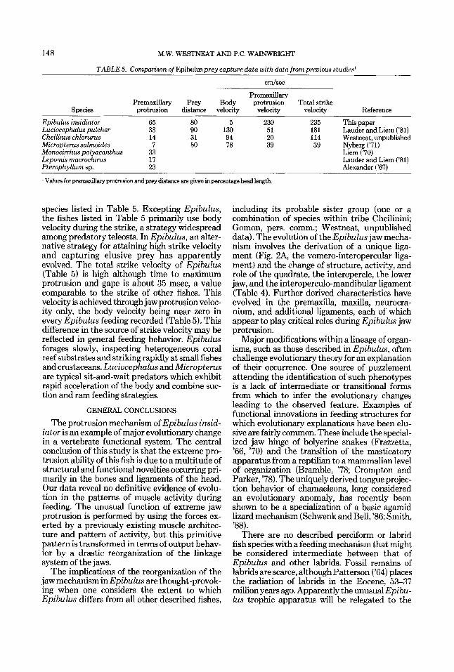
TABLE 5. Comparison of Epibulus prey capture data with data from previous studies'
cmlsec
Premaxillary Premnxillan, Prey Body protrusion Totalstrike
Suecies orotrusion distance velmtv velocitv velocitv Reference ~____ ~ ~ _ _ _ _ _ _____
Epibulus insidiator 65 80 5 230 235 Thispaper Luciocephulus pulcher 33 90 130 51 181 Lauder and Liem ('81) Cheilinus chlorurus 14 31 94 20 114 Westneat, unpublished Micropterus salmides 7 50 78 39 39 Nyberg ('71) Monocirrhus polyacanthus 33 Lepomis macrochirus 17 Lauder and Liem ('81) Pterophvllum SD. 23 Alexander ('67)
Liem ('70)
I Values for premardllary protrusion and prey distance are given in percentage head length.
species listed in Table 5. Excepting Epibulus, the fishes listed in Table 5 primarily use body velocity during the strike, a strategy widespread among predatory teleosts. In Epibulus, an alter- native strategy for attaining high strike velocity and capturing elusive prey has apparently evolved. The total strike velocity of Epibulus (Table 5) is high although time to maximum protrusion and gape is about 35 msec, a value comparable to the strike of other fishes. This velocity is achieved through jaw protrusion veloc- ity only, the body velocity being near zero in every Epibulus feeding recorded (Table 5 ) . This difference in the source of strike velocity may be reflected in general feeding behavior. Epibulus forages slowly, inspecting heterogeneous coral reef substrates and striking rapidly at small fishes and crustaceans. Luciocephalus and Micropterus are typical sit-and-wait predators which exhibit rapid acceleration of the body and combine suc- tion and ram feeding strategies.
GENERAL CONCLUSIONS
The protrusion mechanism of Epibulus insid- iator is an example of major evolutionary change in a vertebrate functional system. The central conclusion of this study is that the extreme pro- trusion ability of this fish is due to a multitude of structural and functional novelties occurring pri- marily in the bones and ligaments of the head. Our data reveal no definitive evidence of evolu- tion in the patterns of muscle activity during feeding. The unusual function of extreme jaw protrusion is performed by using the forces ex- erted by a previously existing muscle architec- ture and pattern of activity, but this primitive pattern is transformed in terms of output behav- ior by a drastic reorganization of the linkage system of the jaws.
The implications of the reorganization of the jaw mechanism in Epibulus are thought-provok- ing when one considers the extent to which Epibulus differs from all other described fishes,
including its probable sister group (one or a combination of species within tribe Cheilinini; Gomon, pers. comm.; Westneat, unpublished data). The evolution of the Epibulus jaw mecha- nism involves the derivation of a unique liga- ment (Fig. 2A, the vomero-interopercdar liga- ment) and the change of structure, activity, and role of the quadrate, the interopercle, the lower jaw, and the interoperculo-mandibular ligament (Table 4). Further derived characteristics have evolved in the premaxilla, maxilla, neurocra- nium, and additional ligaments, each of which appear to play critical roles during Epibulus jaw protrusion.
Major modifications within a lineage of organ- isms, such as those described in Epibulus, often challenge evolutionary theory for an explanation of their occurrence. One source of puzzlement attending the identification of such phenotypes is a lack of intermediate or transitional forms from which to infer the evolutionary changes leading to the observed feature. Examples of functional innovations in feeding structures for which evolutionary explanations have been elu- sive are fairly common. These include the special- ized jaw hinge of bolyerine snakes (Frazzetta, '66, '70) and the transition of the masticatory apparatus from a reptilian to a mammalian level of organization (Bramble, '78; Crompton and Parker, '78). The uniquely derived tongue projec- tion behavior of chamaeleons, long considered an evolutionary anomaly, has recently been shown to be a specialization of a basic agamid lizard mechanism (Schwenk and Bell, '86; Smith, '88).
There are no described perciform or labrid fish species with a feeding mechanism that might be considered intermediate between that of Epibulus and other labrids. Fossil remains of labrids are scarce, although Patterson ('64) places the radiation of labrids in the Eocene, 53-37 million years ago. Apparently the unusual Epibu- lus trophic apparatus will be relegated to the

FEEDING MECHANICS OF EPIBULUS INSIDIATOR 149
status of “puzzling evolutionary phenomenon” for the present. Continued focus on the func- tional morphology and phylogenetic interrela- tionships of labroid fishes and other perciform clades is critical to our understanding of evolu- tion in these highly diverse vertebrate groups.
ACKNOWLEDGMENTS
The authors would like to express their friend- ship with the following people for indispensable help in this project: J. Resing, C. Matthews, H. Choat, G. Russ, K. Clements, and others at James Cook University, Australia, and the Lizard Is- land Research Station; R. Withrow and the Waikiki Aquarium; S. Wainwright, J. Lundberg, K. Smith, and the Zoology Dept. of Duke Univer- sity; G. Lauder, S. Reilly, C . Sanford, and B. Jayne of the University of California, Irvine. S. Reilly, G. Lauder, J. Lundberg, S. Wainwright, and two anonymous reviewers offered critical comments on the manuscript. Discussions with M. Gomon helped clarlfy the phylogenetic posi- tion of Epibulus. This project was funded by a Duke University travel grant, a research assis- tantship under S. Wainwright, and a federal student loan to M. Westneat. Equipment and facilities were provided by K. Smith, B. Nicklas, and by NSF grants BSR 85-20305 and DCB 87-10210 to G. Lauder.
LITERATURE CITED Aerts, P., J.W.M. Osse, and W. Verraes (1987) Model of jaw
depression during feeding in Astotilupiu eleguns (Teleostei: Cichlidae): Mechanisms for energy storage and triggering. J. MorphoL 194:85-109.
Alexander, R.McN. (1966) The functions and mechanisms of the protrusible upper jaws of two species of cyprinid fish. J. Zool. (Lond.) 149:28&296.
Alexander, R.McN. (1967) The functions and mechanisms of the protrusible upper jaws of some acanthopterygian fish. J. Zool. (Lond.) 151:4344.
Barel, C.D.N. (1983) Towards a constructional morphology of cichlid fishes (Teleostei, Percifomes). Neth J. Zool. 33r357- 424.
Barel, C.D.N., J.W. Van Der Muelen, and H. Berkhoudt (1977) Kinematischer Transmissionskoeffizient und Vier- stangensystem als Functionsparameter und Formmodel fiir Mandibulare Depressionsapparate beiTeleostiem. Anat. Anz. 142:21-31.
Bemis, W.E., and G.V. Lauder (1986) Morphology and func- tion of the feeding apparatus of the lungfish, Lepidosiren puradora (Dipnoi). J. Morphol. 187%-108.
Bleeker, P. (1862) Conspectus generum Labroideorum analyti- cus. Verslag. Akad. Amst. 1494-109.
Bock, W.J. (1981) Introductionto the sympasium: Functional- adaptive analysis in systematics. Am. Zool. 21:%20.
Bramhle, D.M. (1978) Origin of the mammalian feeding com- plex: Models and mechanisms. Paleobiology4:271-301.
Crompton, A.W., and P. Parker (1978) Evolution of the mammalian masticatory apparatus. Am. Sci. 66:192-201.
Darwin, C. (1859) On the Origin of Species. Cambridge, Massachusetts Harvard University Press (1964).
Delsman, H.C. (1925) Fishes with protrusile mouths. Treuhia 6.998-106.
Dingerkus, G., and L.D. Uhler (1977) Enzyme clearing of alcian blue stained whole vertebrates for demonstration of cartilage. Stain Technol. 52:229-232.
Frazzetta, T.H. (1966) Studies on the morphology and func- tion of the skull in the Boidae (Serpentes). 11. Morphology and function of the jaw apparatus in Python sebae and Python molorus. J. Morphol. 118:217-296.
Frazzetta, T.H. (1970) From hopeful monsters to bolyerine snakes? Am. Nat. 104:55-72.
Goldschmidt, R. (1940) The Material Basis of Evolution. New Haven, Connecticut: Yale Univ. Press.
Gomon,M.F. (1979)ARevisionoftheGenusBodianus(Fam- ily Labridae) With a Phylogenetic Hypothesis of Related Genera in Tribe Hypsigenyini. Ph.D. Dissertation, Univer- sity of Miami, Florida.
Lauder. G.V. (1979) F d i mechanics in urimitive teleosts and the halecomorph fisyh Amiu Culuu.’J. Zool. (Lond.) 187t.54.1-57R. ~~ . ~ _ . -
Lauder, G.V. (1981a) Form and function: Structural analysis in evolutionary morphology. Paleobiology 7:430-442.
Lauder, G.V. (1981b) Intraspecific functional repertoires in the feeding mechanism of the characoid fishes Lebiasina, Hoplias and Chulceus. Copeia 1981:15&168.
Lauder, G.V. (1982) Patterns of evolution in the feeding mechanism of acanthopterygian fishes. Am. Zool. 22:275- 285.
Lauder, G.V. (1983) Functional and morphological bases of trophic specialization in sunfishes (Teleostei, Cen- trarchidae). J . Morphol. 178:l-21.
Lauder, G.V. (1985) Aquatic feeding in lower vertebrates. In M. Hildebrand, D.M. Bramble, K.F. Liem, and D.B. Wake (eds): Functional Vertebrate Morphology. Cambridge: Belknap Press, pp. 210-229.
Lauder, G.V., and K.F. Liem (1981) Prey capture by Lucio- cephalus pulcher: Implications for models of jaw protru- sion in teleost fishes. Environ. Biol. Fish. 6:257-268.
Lauder, G.V., and H.B. S M e r (1988) Ontogeny of functional design in tiger salamanders (Ambystoma tigrinum): Are motor patterns conserved during major morphological trans- formations? J. Morphol. 19E249-268.
Liem, K.F. (1970) Comparative functional anatomy of the Nandidae (Pisces: Teleostei). Fieldiana Zool. 56:l-166.
Liem, K.F. (1978) Modulatory multiplicity in the functional repertoire of the feeding mechanism in cichlids. I. Pisci- vores. J. Morphol. 158:323-360.
Liem,K.F. (1980) Adaptivesignificance of intra- and interspe- cific differences in the feeding repertoires of cichlid fishes. Am. Zool. 20:295314.
Liem, K.F., and P.H. Greenwood (1981) A functional ap- proach to the phylogeny of the pharyngognath teleosts. Am. Zool. 21:83-101.
Lombard, R.E., and D.B. Wake (1986) Tongue evolution in the lungless salamanders, family Plethodontidae. IV. Phy- logeny of the plethodontid salamanders and the evolution of feeding dynamics. Syst. Zool. 35t532-551.
Motta, P.J. (1984) Mechanics and functions of jaw protrusion in teleost fishes: A review. Copeia I984:1-18.
Nyberg, D.W. (1971) Prey capture in largemouth bass. Am. Midl. Nat. 86:12%144.
Osse, J.W.M. (1969) Functional morphology of the head of the perch (Perca fluuiatilis): An electromyographic study. Neth. J. Zool. 10r289-392.
Patterson, C. (1964) A review of Mesozoic actinopterygian fishes with special reference to those of the English chalk. Philce. Trans. R. Soc. Lond. [Biol.] 247:213432.
Pietsch, T.W. (1978) The feeding mechanism of Stylephorus chordatus (Telwtei: Lampridiformes): Functional and eco- logical implications. Copeia 1978:255-262.
Randall, J.R. (1983) Red Sea Reef Fishes. London: IMMEL Publishing.
Renard, L. (1719) Poissons, Ecrivisses et Crabes, de Diverses Couleurs et Figures Extraordinaire, que l’on Trouve Au-

150 M.W. WESTNEAT AND P.C. WAINWRIGHT
tour des Isles Moloques, et sur les Cotes des Terres Am- trales.. . Histoire des Plus Rares Curiositez de la Mer des Indes. 2 vols. in 1. Amsterdam: Louis Renard.
Rognes, K. (1973) Head skeleton and jaw mechanism in Labrinae (Teleostei: Labridae) from Norwegian waters. Univ. Bergen Arb. Mat. Naturv. (Ser. 1971)4:1-149.
Sanderson, S.L. (1988) Variation in neuromuscular activity during prey capture by trophic specialists and generalists (Pisces: Labridae). Brain Behav. Evol. 32t257-268.
Schaefer, S.A., and G.V. Lauder (1986) Historical transforma- tion of functional design: Evolutionary morphology of feed- ing mechanisms in loricarioid catfishes. Syst. Zool. 3548% 509.
Schwenk, K., and D.A. Bell (1986) Chameleon-like tongue protrusion in an agamid lizard. Am. Zool. 26.65A.
ShafTer, H.B., and G.V. Lauder (1985) Aquatic prey capture in amhystomatid salamanders: Patterns of variation in muscle activity. J. Morphol. 183.273-284.
Simpson, G.G. (1953) The Major Features of Evolution. New York Columbia University Press.
Smith, K.K. (1988) Form and function of the tongue in agamid lizards with comments on its phylogenetic signifi- cance. J. Morphol. 196t157-171.
Wainwright, P.C. (1989) Prey processing in haemulid fishes: Patterns of variation in pharyngeal jaw muscle activity. J. Exp. Biol. 141:35%376.
Wainwright, P.C., and G.V. Lauder (1986) Feeding biology of sunfishes: Patterns of variation in the feeding mechanism. J. Linn. Soc. (Zool.) 88:217-228.
Wake, D.B., and A. Larson (1987) Multidimensional analysis of an evolving lineage. Science 238:4243.
Wilkinson, L. (1986) SYSTAT The System for Statistics. Evanston, Illinois: Systat Inc.
Winterbottom, R. (1974) A descriptive synonymy of the stri- ated muscles of the Teleostei. Proc. Acad. Nat. Sci. Phila. 125:22&317.
All in-text references underlined in blue are linked to publications on ResearchGate, letting you access and read them immediately.