Feasibility, Tolerance, and Quality of Life for Hypofractionation ...
12
Review began 03/02/2022 Review ended 03/19/2022 Published 03/25/2022 © Copyright 2022 Malik et al. This is an open access article distributed under the terms of the Creative Commons Attribution License CC-BY 4.0., which permits unrestricted use, distribution, and reproduction in any medium, provided the original author and source are credited. Feasibility, Tolerance, and Quality of Life for Hypofractionation Versus Conventional Fractionation for Post-mastectomy Radiotherapy in Indian Patients Deepika Malik , Ashok Singh , Manoj M. Birajdar , Virendra J. Vyas 1. Radiation Oncology, Maharishi Markandeshwar Institute of Medical Sciences and Research (MMIMSR), Ambala, IND 2. Radiation Oncology, Mahatma Gandhi Institute of Medical Sciences, Wardha, IND 3. Radiation Oncology, All India Institute of Medical Sciences, Raipur, IND 4. Clinical Oncology, Indian Institute of Head and Neck Oncology, Indore, IND Corresponding author: Deepika Malik, [email protected] Abstract Introduction: The international standard for post-operative radiotherapy for breast cancer delivers hypofractionated radiotherapy. However, many centers in India still follow the longer conventional schedule probably because of paucity of large prospective trials in Indian patients on the same and apprehension regarding tolerance of high dose per fraction in the said population. We aimed to test the feasibility of hypofractionation in our setting and compared the toxicities and the quality of life in patients receiving conventional and hypofractionated radiotherapy. Materials and methods: Eighty histopathologically proven women of non-metastatic carcinoma breast who underwent modified radical mastectomy were assigned to receive 50 Gray/25 fractions/five weeks or 40 Gray/15 fractions/three weeks. Patients were assessed for the following toxicities - radiation dermatitis, radiation pneumonitis, dysphagia, skin fibrosis, lymphedema, shoulder stiffness, and brachial plexopathy, during radiation and at treatment completion and then at first, third, and sixth-month follow-up. Health- related quality of life was assessed using the European Organisation for Research and Treatment of Cancer core quality of life questionnaire (EORTC QLQ-C30) and breast cancer-specific quality of life questionnaire (QLQ-BR23) at treatment completion and then at first, third, and sixth-month follow-up. Results and conclusion: We had a mean follow-up of 12.78 months. All the assessed toxicities and quality of life scores were comparable between the two arms at all time points of evaluation (p>0.05); 40 Gray in 15 fractions over three weeks is feasible and as safe as the five-week schedule with comparable quality of life. Hypofractionation can be practiced as a routine for post-mastectomy breast cancer patients as this shorter radiotherapy schedule is convenient and cheaper for the patients with no compromise on normal tissue toxicity or quality of life. Categories: Radiation Oncology, Oncology Keywords: post-mastectomy radiotherapy, breast cancer, quality of life, acute toxicity, hypofractionation Introduction The international standard radiotherapy regimen after breast-conserving surgery or mastectomy for breast cancer delivers hypofractionated radiotherapy after it was proven to be as efficacious to the conventional fractionation of 50 Gray (Gy) in 25 fractions over five weeks with similar adverse reactions. Post-operative irradiation reduces locoregional recurrence risk by about 70% and reduces breast cancer mortality and also improves overall survival [1,2]. Hypofractionation, a strategy allowing shortening of overall treatment time, involves higher fraction doses (above 2 Gy), such that a biologically equivalent dose can be delivered in fewer fractions. Radiobiology justifies hypofractionation in breast cancer owing to a low α/β value at 4 Gy which is comparable to that of healthy tissues (α/β is 3.5 Gy for soft tissues) [3]. Thus, a higher dose per fraction is assumed to be more efficient in destroying tumor cells with concerns however of a high incidence and severity of late radiation toxicity. The landmark UK Standardisation of Breast Radiotherapy (START) trials for hypofractionation (START-A, 50 Gy in 25 fractions over five weeks compared with 41.6 Gy or 39 Gy in 13 fractions over five weeks and START-B, 50 Gy in 25 fractions over five weeks compared with 40 Gy in 15 fractions over three weeks) in their five and 10-year follow-up results showed that hypofractionated radiotherapy schedule offered rates of local-regional tumor relapse and late adverse effects at least as favorable as the standard schedule of 50 Gy in 25 fractions [4-6]. Hypofractionation is being practiced routinely for breast cancer in the UK and many parts of the world. However, in India, some centers still practice the five-week schedule routinely despite the 1 2 3 4 Open Access Original Article DOI: 10.7759/cureus.23497 How to cite this article Malik D, Singh A, Birajdar M M, et al. (March 25, 2022) Feasibility, Tolerance, and Quality of Life for Hypofractionation Versus Conventional Fractionation for Post-mastectomy Radiotherapy in Indian Patients. Cureus 14(3): e23497. DOI 10.7759/cureus.23497
-
Upload
khangminh22 -
Category
Documents
-
view
2 -
download
0
Transcript of Feasibility, Tolerance, and Quality of Life for Hypofractionation ...

Review began 03/02/2022 Review ended 03/19/2022 Published 03/25/2022
© Copyright 2022Malik et al. This is an open access articledistributed under the terms of the CreativeCommons Attribution License CC-BY 4.0.,which permits unrestricted use, distribution,and reproduction in any medium, providedthe original author and source are credited.
Feasibility, Tolerance, and Quality of Life forHypofractionation Versus ConventionalFractionation for Post-mastectomy Radiotherapyin Indian PatientsDeepika Malik , Ashok Singh , Manoj M. Birajdar , Virendra J. Vyas
1. Radiation Oncology, Maharishi Markandeshwar Institute of Medical Sciences and Research (MMIMSR), Ambala, IND2. Radiation Oncology, Mahatma Gandhi Institute of Medical Sciences, Wardha, IND 3. Radiation Oncology, All IndiaInstitute of Medical Sciences, Raipur, IND 4. Clinical Oncology, Indian Institute of Head and Neck Oncology, Indore,IND
Corresponding author: Deepika Malik, [email protected]
AbstractIntroduction: The international standard for post-operative radiotherapy for breast cancer delivershypofractionated radiotherapy. However, many centers in India still follow the longer conventional scheduleprobably because of paucity of large prospective trials in Indian patients on the same and apprehensionregarding tolerance of high dose per fraction in the said population. We aimed to test the feasibility ofhypofractionation in our setting and compared the toxicities and the quality of life in patients receivingconventional and hypofractionated radiotherapy.
Materials and methods: Eighty histopathologically proven women of non-metastatic carcinoma breast whounderwent modified radical mastectomy were assigned to receive 50 Gray/25 fractions/five weeks or 40Gray/15 fractions/three weeks. Patients were assessed for the following toxicities - radiation dermatitis,radiation pneumonitis, dysphagia, skin fibrosis, lymphedema, shoulder stiffness, and brachial plexopathy,during radiation and at treatment completion and then at first, third, and sixth-month follow-up. Health-related quality of life was assessed using the European Organisation for Research and Treatment of Cancercore quality of life questionnaire (EORTC QLQ-C30) and breast cancer-specific quality of life questionnaire(QLQ-BR23) at treatment completion and then at first, third, and sixth-month follow-up.
Results and conclusion: We had a mean follow-up of 12.78 months. All the assessed toxicities and quality oflife scores were comparable between the two arms at all time points of evaluation (p>0.05); 40 Gray in 15fractions over three weeks is feasible and as safe as the five-week schedule with comparable quality of life.Hypofractionation can be practiced as a routine for post-mastectomy breast cancer patients as this shorterradiotherapy schedule is convenient and cheaper for the patients with no compromise on normal tissuetoxicity or quality of life.
Categories: Radiation Oncology, OncologyKeywords: post-mastectomy radiotherapy, breast cancer, quality of life, acute toxicity, hypofractionation
IntroductionThe international standard radiotherapy regimen after breast-conserving surgery or mastectomy for breastcancer delivers hypofractionated radiotherapy after it was proven to be as efficacious to the conventionalfractionation of 50 Gray (Gy) in 25 fractions over five weeks with similar adverse reactions. Post-operativeirradiation reduces locoregional recurrence risk by about 70% and reduces breast cancer mortality and alsoimproves overall survival [1,2].
Hypofractionation, a strategy allowing shortening of overall treatment time, involves higher fraction doses(above 2 Gy), such that a biologically equivalent dose can be delivered in fewer fractions. Radiobiologyjustifies hypofractionation in breast cancer owing to a low α/β value at 4 Gy which is comparable to that ofhealthy tissues (α/β is 3.5 Gy for soft tissues) [3]. Thus, a higher dose per fraction is assumed to be moreefficient in destroying tumor cells with concerns however of a high incidence and severity of late radiationtoxicity.
The landmark UK Standardisation of Breast Radiotherapy (START) trials for hypofractionation (START-A, 50Gy in 25 fractions over five weeks compared with 41.6 Gy or 39 Gy in 13 fractions over five weeksand START-B, 50 Gy in 25 fractions over five weeks compared with 40 Gy in 15 fractions over three weeks) intheir five and 10-year follow-up results showed that hypofractionated radiotherapy schedule offered rates oflocal-regional tumor relapse and late adverse effects at least as favorable as the standard schedule of 50 Gyin 25 fractions [4-6]. Hypofractionation is being practiced routinely for breast cancer in the UK and manyparts of the world. However, in India, some centers still practice the five-week schedule routinely despite the
1 2 3 4
Open Access OriginalArticle DOI: 10.7759/cureus.23497
How to cite this articleMalik D, Singh A, Birajdar M M, et al. (March 25, 2022) Feasibility, Tolerance, and Quality of Life for Hypofractionation Versus ConventionalFractionation for Post-mastectomy Radiotherapy in Indian Patients. Cureus 14(3): e23497. DOI 10.7759/cureus.23497
robust evidence the UK START trials provided in favor of hypofractionation. This could probably be becauseof apprehension regarding the inferiority of hypofractionation with respect to toxicities as sadly there are nolarge prospective randomized trials conducted in India on this aspect. Also, a major part of the patientpopulation studied in the UK trials as well as other studies on hypofractionation in breast cancer had postbreast conservation surgery patients. Post-mastectomy patients formed a small part of these studies.Therefore, to some extent, the above-mentioned apprehension is justified until hypofractionation is studiedin the target population where it is intended to be used.
India is currently witnessing what could be called an epidemic of cancer, breast cancer being a majorattributor in females. Breast cancer has now become the most common cancer, with 2,261,419 new casesdiagnosed worldwide and 178,361 new cases diagnosed in India in 2020. Breast cancer accounts for 13.5% ofall cancer cases in India, compared to 11.77% worldwide [7]. This burden is likely to grow not only in termsof the absolute number of cases but also in terms of incidence owing to the urbanization and changinglifestyle patterns of the Indian population [8]. The proportion of breast cancers registered annually at ourcenter reinforces this estimate (11% to 24% over a span of 20 years from the year 1998 to 2018).
This rising breast cancer incidence has a direct bearing on the burden on radiotherapy machines [8]. As perthe World Health Organisation, a developing country should have at least one teletherapy machine for apopulation of one million. Going by these figures, India has to increase the number of existing teletherapymachines by fourfold [9]. In 2014, policy analysis on cancer research in India said that research on morecost-effective treatment pathways that will provide good outcomes is needed in India. The authorsmentioned that results of clinical research on brachytherapy, hypofractionated radiotherapy schedules, andregular repeating (metronomic) chemotherapy regimens to reduce treatment times can drive down the costsof care and enable more patients to be effectively treated [10].
Conventional fractionation of five weeks requires a long in or near hospital stay or commuting to a hospitalfor this long period. By adopting hypofractionation, the ever-rising burden on radiotherapy machines andthe consequential long waiting lists can be controlled. This would be of great logistic benefit to resource-limited radiotherapy centers which are the backbone of cancer care in India. A shorter course of radiotherapywould also relatively relieve poor patients financially by reducing the number of days of treatment, travel tothe radiotherapy center, or staying in the hospital premises. Also, cost of the radiotherapy treatment ofthree weeks would cost 0.6 times lesser than a five-week schedule. The purpose of this study was thereforeto determine the feasibility and tolerability of hypofractionation and its effect on health-related quality oflife in our patients over the tries and tested conventional radiotherapy schedule.
Materials And MethodsPatientsAfter obtaining approval from the institutional ethics committee (ref. no.: MGIMS/IEC/RADTH/98/2014)and written informed consent from patients, 80 histopathologically proven patients of non-metastaticbreast cancer, treated surgically with modified radical mastectomy (MRM), and having indication for post-mastectomy radiotherapy were enrolled from the department of radiation oncology at a rural medical collegein Maharashtra between January 2015 and December 2017. Patients above 65 years of age, KarnofskyPerformance Status (KPS) of 70 or less, with a history of prior radiotherapy to chest or neck were excludedfrom the study. All patients underwent a thorough history and physical examination and relevant blood-workup, chest radiograph, and abdominal ultrasonography. All patients were evaluated for hormone receptorstatus - estrogen receptor (ER), progesterone receptor (PR), and human epidermal growth factor receptor 2(HER2/neu) status. Computed tomography chest and abdomen and bone-scan were done for stage III orbeyond or on clinical or biochemical suspicion of metastasis. The 80 patients enrolled were randomized byblock method into two arms. Forty patients in the study arm received 40.05 Gray in 15 fractions with afraction size of 2.67 Gray, once daily for five days a week over three weeks (hypofractionated arm). Fortypatients in the control arm received 50 Gray in 25 fractions with a fraction size of two Gray, once daily forfive days a week over five weeks (conventional arm).
Radiation techniquePatients lay supine on an adjustable carbon fiber breast board with ipsilateral arm abducted and externallyrotated and fixed on strategically placed adjustable handgrip and neck hyperextended to opposite side. Allpatients were simulated on Nucletron Simulix Evolution (Stockholm, Sweden: Elekta). Chest-wall wastreated with medial and lateral tangential portals with superior border placed at the level of head of clavicle,inferior border 2 cm below the contralateral inframammary fold, medial border at patient midline, andlateral border at mid-axillary line attempting to cover the MRM scar wherever possible, with no bolus.Supraclavicular fossa (when indicated) was treated with a single anterior portal with superior border atthyroid-cricoid notch, inferior border at the superior border of chest wall portals with a 5 mm gap on skinsurface between the two portals, medial border 1 cm lateral to midline, and lateral border at the junction ofmedial two-thirds and lateral one-third of the clavicle. A conventional two-dimensional plan was createdand patients were treated on cobalt-60 (Theratron Phoenix; Ontario, Canada: Best Theratronics Ltd.)machine. Dose to chest-wall was prescribed at midpoint of chest-wall contour and at D-max forsupraclavicular portal.
2022 Malik et al. Cureus 14(3): e23497. DOI 10.7759/cureus.23497 2 of 12
Assessment of outcomesToxicities compared between the two arms were skin toxicity, lung toxicity, dysphagia, shoulder restriction(graded as per Radiation Therapy Oncology Group {RTOG} morbidity schema), brachial plexopathy (clinicalfinding said to be present if patient had pain, numbness, paresthesia, and motor weakness), andlymphedema (clinical finding using tape measurement said to be present if a difference of more than 2 cmbetween arm circumference measured at baseline and at evaluation between affected and contralateral arm;arm circumference measured at 10 cm above and 10 cm below olecranon process) [11]. Patients wereassessed for the toxicities at radiotherapy completion, and then at one month, three months, and six monthsafter completion of radiotherapy, ensuring that as far as possible toxicity was assessed and documented on astandard scoring chart by a single observer (the principal investigator) and photographic evidence for skinreactions were obtained for comparison at follow-up. Skincare and physiotherapy were emphasized from dayone of treatment and then at subsequent visits.
Quality of life was assessed using the European Organisation for Research and Treatment of Cancer corequality of life questionnaire (EORTC QLQ-C30) and breast cancer-specific quality of life questionnaire(QLQ-BR23) which were filled by all patients at baseline, at treatment completion, and then at one month,three months, and six months follow-up [12]. Questionnaires were provided in patients' own language(Marathi/Hindi/English) and for illiterate ones, questions from the questionnaires were read out by the samedepartmental social worker for all such patients (to avoid bias). It was ensured that all patients answered allquestions in the questionnaire. Scoring was done in accordance with the EORTC scoring manual.
Conclusions on comparison of efficacy of radiotherapy schedules could not be drawn from this study owingto a short follow-up as only a limited time was available for conducting this study. Therefore responseassessment in form of local recurrence or distant metastases has not been taken as an objective for thisstudy. Patient compliance was assessed by analyzing the unplanned gaps in radiotherapy regimen. Gaps inradiotherapy were labeled as unplanned when they were caused by factors other than public holidays,statutory holidays, or machine breakdown.
Statistical analysisStatistical analysis was done using IBM SPSS Statistics version 12.0 (Chicago, IL: SPSS Inc.). The categoricalvariables were presented as mean (standard deviation), and Pearson’s chi-square test or Fisher’s exact testwas used as the test of significance. The continuous variables were presented as frequency (percentage) andunpaired t-test was used as the test of significance. The equality of variance for comparison of means wastested using Levene’s test. To calculate probability of radiation-induced dermatitis and pneumonitis weused life-table analysis. P-value<0.05 was considered statistically significant.
ResultsThe mean age of our study population was 45.88 years (range: 28-65 years). Most patients (i.e., 41.25%, 33out of 80 patients) belonged to stage IIIA. A total of 52.5% (i.e., 42 out of 80) were left-sided breast cancers.Also, 65% of patients (i.e., 52 out of 80 patients) were hormone receptor-positive, out of which 86.53% (i.e.,45 out of 52) were estrogen receptor (ER) positive and the rest were progesterone receptor (PR) positive. Atotal of 35% of patients (i.e., 28 out of 80 patients) were HER2/neu positive. And 18.75% of patients (i.e., 15out of 80 patients) were triple-negative breast cancers. Patient population characteristics were well balancedbetween the two arms (Table 1).
2022 Malik et al. Cureus 14(3): e23497. DOI 10.7759/cureus.23497 3 of 12
Parameter Hypofractionation arm Conventional arm p-Value
Mean age, years 44.88 46.88 0.32
KPS, mean (SD) 85.75 (5.9) 86.0 (5.9) 0.85
Stage of presentation, frequency (%)
II A 3 (7.5) 3 (7.5)
1.00
II B 13 (32.5) 13 (32.5)
III A 16 (40) 17 (42.5)
III B 2 (5) 1 (2.5)
III C 6 (15) 6 (15)
Laterality, frequency (%)Left 23 (57.5%) 19 (47.5%)
0.37Right 17 (42.5%) 21 (52.5%)
ER positive, frequency (%) 20 (50%) 25 (62.5%) 0.37
PR positive, frequency (%) 15 (37.5%) 17 (42.5%) 0.82
HER2/neu positive, frequency (%) 15 (37.5%) 13 (32.5%) 0.82
Menstrual status, frequency (%)Pre-menopausal 18 (45%) 11 (27.5%)
0.16Post-menopausal 22 (55%) 29 (72.5%)
Duration of follow-up (months) mean (SD) 12.1 (4.3) 12.9 (4.1) 0.38
TABLE 1: Population characteristics of the two arms at baseline.KPS: Karnofsky Performance Status; HER2/neu: human epidermal growth factor receptor 2; ER: estrogen receptor; PR: progesterone receptor
Forty patients in each arm completed the treatment. The mean follow-up duration for this study was 12.48months. One patient in hypofractionated arm was lost to follow-up after one month of follow-up, and onepatient in conventional arm died due to acute myocardial infarction two months after completingradiotherapy.
Toxicity comparisonAt the end of radiotherapy, 62.5% and 20% of patients in hypofractionation arm had grade I and II skintoxicity, respectively compared to 57.5% and 32.5% of patients in conventional arm (p=0.35) while none hadgrade III or beyond in both arms. At one month after completion, 82.5% of patients had grade I skin toxicityin hypofractionation arm, compared to 90% with grade I and 5% with grade II in conventional arm (p=0.09),and none had grade II or beyond in hypofractionation arm and grade III or beyond in conventional arm. Atthree months 43.6% and 48.7% of patients had grade I skin toxicity in hypofractionation and conventionalarm respectively (p=0.82), with none having grade II or beyond. At six months 20.5% and 28.2% of patientshad grade I skin toxicity (p=0.60), none having grade II or beyond (Table 2).
2022 Malik et al. Cureus 14(3): e23497. DOI 10.7759/cureus.23497 4 of 12

Time RT dermatitis gradeHF CV
p-ValueFrequency (%)
Treatment completion
Grade 0 7 (17.5) 4 (10)
0.35Grade I 25 (62.5) 23 (57.5)
Grade II 8 (20) 13 (32.5)
One month follow-up
Grade 0 7 (17.5) 2 (5)
0.1Grade I 33 (82.5) 36 (90)
Grade II 0 2 (5)
Three months follow-up
Grade 0 22 (56.4) 20 (51.3)
0.82Grade I 17 (43.6) 19 (48.7)
Grade II 0 0
Six months follow-up
Grade 0 31 (79.5) 28 (71.8)
0.60Grade I 8 (20.5) 11 (28.2)
Grade II 0 0
TABLE 2: Comparison of radiation dermatitis.The table shows comparison of radiation dermatitis at different time points of evaluation between conventional radiotherapy arm and hypofractionatedradiotherapy arm. The grading of radiation dermatitis is as per the RTOG acute toxicity grading.
RT dermatitis: radiation dermatitis; HF: hypofractionated arm; CV: conventional fractionation arm; RTOG: Radiation Therapy Oncology Group
Radiation pneumonitis was absent at radiotherapy completion in both arms. At one month after treatmentconclusion, 5% and 10% of patients had grade I radiation pneumonitis in hypofractionation andconventional arm, respectively (p=0.7), with no incidence of grade II or beyond. At three months, 2.6% vs12.8% had grade I pneumonitis (p=0.2) in hypofractionation and conventional arm, respectively. At sixmonths 2.6% of patients had grade I radiation pneumonitis in hypofractionation arm, none had it inconventional arm (p=1.0) (Table 3).
2022 Malik et al. Cureus 14(3): e23497. DOI 10.7759/cureus.23497 5 of 12

Time RT pneumonitis gradeHF CV
p-ValueFrequency (%)
Treatment completionGrade 0 0 0
-Grade I 0 0
One month follow-upGrade 0 38 (95) 36 (90)
0.7Grade I 2 (5) 4 (10)
Three months follow-upGrade 0 38 (97.4) 34 (87.2)
0.2Grade I 1 (2.6) 5 (12.8)
Six months follow-upGrade 0 38 (97.4) 39 (100)
1.0Grade I 1 (2.6) 0
TABLE 3: Comparison of radiation pneumonitis.The table shows comparison of radiation pneumonitis at different time points of evaluation between conventional radiotherapy arm and hypofractionatedradiotherapy arm. The grading of radiation pneumonitis is as per the RTOG acute toxicity grading.
RT pneumonitis: radiation pneumonitis; HF: hypofractionated arm; CV: conventional fractionation arm; RTOG: Radiation Therapy Oncology Group
Dysphagia during treatment was found in 20% of patients in both arms, skin fibrosis at follow-up was foundin 30% and 37.5% of patients (p=0.64), lymphedema at follow-up in 7.5% vs 10% of patients (p=1.00), inhypofractionation and conventional arm respectively, shoulder stiffness at follow-up in 2.5% of patients inboth arms and no patients had brachial plexopathy at follow-up in both arms (Table 4).
ToxicityHypofractionated arm Conventional arm
p-ValueFrequency (%)
Dysphagia during treatment 8 (20%) 8 (20%) 1.0
Skin fibrosis at follow-up 12 (30%) 15 (37.5%) 0.64
Lymphedema at follow-up 3 (7.5%) 4 (10%) 1.0
Shoulder stiffness at follow-up 1 (2.5%) 1 (2.5%) 1.0
Brachial plexopathy at follow-up None None -
TABLE 4: Comparison of other toxicities.
Patient compliance was assessed by analyzing the unplanned gaps. Further, 5% vs 7.5% patients (p=1.0) hadunplanned gaps in their radiotherapy schedule in hypofractionation and conventional arm, respectively. Theaverage number of days of interruption were 5.5 vs 8.67 days, respectively (p=0.1).
Quality of life comparisonAll parameters of QLQ-C30 and QLQ-BR23 were comparable at baseline for the two arms (Table 5). Theglobal health status (QL2), functional scales (physical functioning {PF2}, role functioning {RF2}, emotionalfunctioning {EF}, cognitive functioning {CF}, and social functioning {SF}), symptom scales (fatigue {FA},nausea and vomiting {NV}, pain {PA}, dyspnea {DY}, insomnia {SL}, appetite loss {Ap}, constipation {CO},diarrhea {DI}, and financial difficulties {FI}), breast cancer-specific functional scales (body image {BRBI},sexual functioning {BRSEF}, sexual enjoyment {BRSEE}, and future perspective {BRFU}), and breast cancer-specific symptom scales (systemic therapy side effects {BRST}, breast symptoms {BRBS}, arm symptoms{BRAS}, and upset by hair loss {BRHL}) were comparable at baseline and at subsequent evaluations attreatment completion, and at one, three, and six months of follow-up. The trend of the specific scores hasbeen depicted in Figure 1 and is discussed later.
2022 Malik et al. Cureus 14(3): e23497. DOI 10.7759/cureus.23497 6 of 12
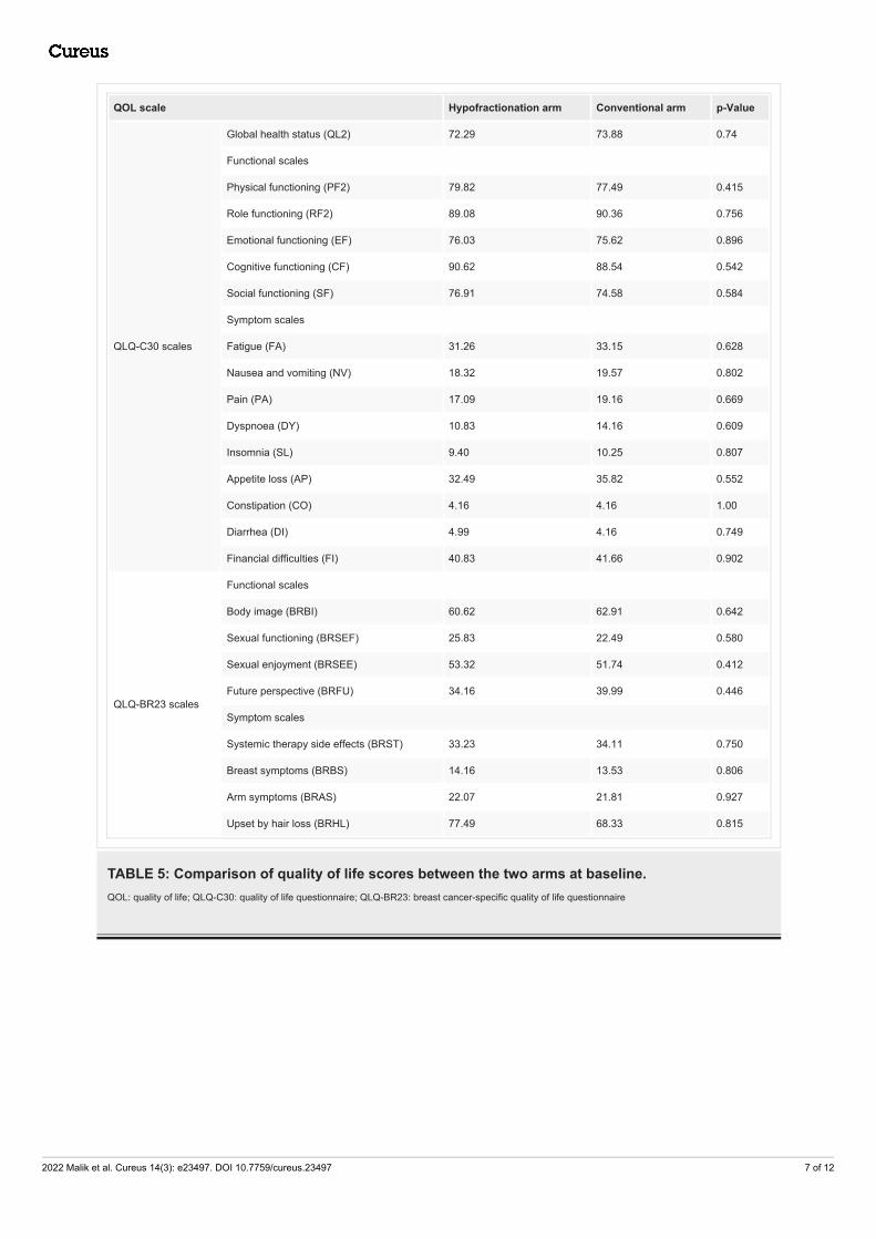
QOL scale Hypofractionation arm Conventional arm p-Value
QLQ-C30 scales
Global health status (QL2) 72.29 73.88 0.74
Functional scales
Physical functioning (PF2) 79.82 77.49 0.415
Role functioning (RF2) 89.08 90.36 0.756
Emotional functioning (EF) 76.03 75.62 0.896
Cognitive functioning (CF) 90.62 88.54 0.542
Social functioning (SF) 76.91 74.58 0.584
Symptom scales
Fatigue (FA) 31.26 33.15 0.628
Nausea and vomiting (NV) 18.32 19.57 0.802
Pain (PA) 17.09 19.16 0.669
Dyspnoea (DY) 10.83 14.16 0.609
Insomnia (SL) 9.40 10.25 0.807
Appetite loss (AP) 32.49 35.82 0.552
Constipation (CO) 4.16 4.16 1.00
Diarrhea (DI) 4.99 4.16 0.749
Financial difficulties (FI) 40.83 41.66 0.902
QLQ-BR23 scales
Functional scales
Body image (BRBI) 60.62 62.91 0.642
Sexual functioning (BRSEF) 25.83 22.49 0.580
Sexual enjoyment (BRSEE) 53.32 51.74 0.412
Future perspective (BRFU) 34.16 39.99 0.446
Symptom scales
Systemic therapy side effects (BRST) 33.23 34.11 0.750
Breast symptoms (BRBS) 14.16 13.53 0.806
Arm symptoms (BRAS) 22.07 21.81 0.927
Upset by hair loss (BRHL) 77.49 68.33 0.815
TABLE 5: Comparison of quality of life scores between the two arms at baseline.QOL: quality of life; QLQ-C30: quality of life questionnaire; QLQ-BR23: breast cancer-specific quality of life questionnaire
2022 Malik et al. Cureus 14(3): e23497. DOI 10.7759/cureus.23497 7 of 12

FIGURE 1: Time trends of quality of life scores at different time points.X-axis: time since baseline to sixth-month follow-up; Y-axis: QOL scores
QOL: quality of life; B: baseline; TC: treatment completion; 1M: first-month follow-up; 3M: third-month follow-up;6M: sixth-month follow-up; QL2: global health status; PF2: physical functioning; RF2: role functioning; EF:emotional functioning; CF: cognitive functioning; SF: social functioning; FA: fatigue; NV: nausea and vomiting; PA:pain; DY: dyspnea; SL: insomnia; AP: appetite loss; CO: constipation; DI: diarrhea; FI: financial difficulties; BRBI:body image; BRSEF: sexual functioning, BRSEE: sexual enjoyment; BRFU: future perspective; BRST: systemictherapy side effects; BRBS: breast symptoms; BRAS: arm symptoms; BRHL: upset by hair loss
Comparison of responseAt the time of analysis, one patient in conventional arm had local recurrence (chest wall) with distantmetastasis as well. Two and five patients, respectively, had distant metastasis in hypofractionation andconventional arm, liver being the most common site of metastasis (four patients), followed by lung (twopatients) and bones (one patient).
DiscussionThis prospective randomized study aimed to determine the feasibility and tolerability of hypofractionationover the conventional radiotherapy schedule at a resource-limited rural-based radiotherapy center. Forty
2022 Malik et al. Cureus 14(3): e23497. DOI 10.7759/cureus.23497 8 of 12
patients each in both arms completed treatment. The mean age of the study population was 45.88 years.Corresponding to Kumbhaj et al., an Indian study on hypofractionation, 60.10% of the study population wasstage III; however, this could be because we enrolled postmastectomy patients, a high proportion of whomwould anyways be locally advanced [13]. In the present study, 52.5% patients had left-sided while 47.5% hadright-sided breast cancer, which is in concordance to the available data that says the left-sided breastcancers are 5-10% more than right-sided ones [14]. Sixty-five percent of the study population was hormonereceptor-positive, ER positivity being 58.75%, PR positivity 41.25%; 35% human epidermal growth factorreceptor-positive. The results correspond to another Indian study by Nandi et al., where 41-43% population(post-breast conservation surgery {BCS} + post-mastectomy) was PR positive and 30-37% were Her2 neupositive, although the ER positivity in their study was higher than ours (91% in BCS cases and 84% inmastectomy cases) [15]. The mean duration of follow-up was 12.48 months.
Incidence of radiation dermatitis appears to favor hypofractionation arm numerically but the difference wasfound to be statistically insignificant at all time points (Table 2). The rates and grade of skin toxicity werelower in our study compared to other Asian studies. There were higher rates of grade III and beyond acuteradiation dermatitis in studies by Kumbhaj et al. (20% vs 5%) for 40 Gy vs 50 Gy, respectively [13], Shahid etal. (37% vs 28% vs 14%) for 27 Gy/week vs 35 Gy/two weeks vs 40 Gy/three weeks [16], and Abhilash et al.(6.6% vs 3.3%) for 39 Gy/13 fractions vs 50 Gy/25 fractions [17]. While in our study, there were no patientswith grade III or beyond acute radiation dermatitis, which could be attributed to the fact that bolus was notused for our patients as per institutional protocol and appropriate skincare counseling was timely done andregularly re-enforced. However, there was no significant difference in the incidence of acute radiationdermatitis in any of these studies. Nandi et al. in their study had no incidence of grade III or beyond skintoxicity in concordance to our study [15]. Eldeeb et al. compared three fractionation schedules in post-mastectomy patients, 50 Gy/25 fractions, 45 Gy/17 fractions, and 40 Gy/15 fractions, and found nostatistical difference in skin pigmentation between the three arms while grades II-III erythema wassignificantly higher in the two hypofractionation arms compared to the control arm [18].
The biggest twin trials, the START A and B trials, showed a lower rate of change in skin appearance inhypofractionation patients than the conventional ones. In START A, the event rate when 50 Gy wascompared with 41.6 Gy was 31.1% vs 25% and when compared with 39 Gy, it was 31.1% vs 21.6% [4]. InSTART B trial, the event rate for change in skin appearance was 27.8% vs 22.9% when 50 Gy was comparedwith 40 Gy [5].
Radiation pneumonitis was overall low (and only limited to grade I) in our study for both arms and wascomparable between the two arms at all time points of evaluation (Table 3). Our results are similar to theSTART trials that provide the most robust evidence in favor of hypofractionation, which showed thatradiation pneumonitis with hypofractionation is comparable with the conventional schedule [4,5]. Shahid etal. also noted similar results with statistically insignificant difference in rates of acute radiationpneumonitis between 27 Gy in one week, 35 Gy in two weeks, and 40 Gy in three weeks (4% vs 5% vs 5%).
Dysphagia during treatment was seen in equal proportion (20%) of both groups, with a low overall incidencestudy compared to other studies where it was nearly 75% patients for Kumbhaj et al. [13], 15-18% for Shahidet al. [16]. For both studies, dysphagia incidence was comparable between the two arms. Skin fibrosis untilanalysis (30% vs 37.5%) was comparable between the hypofractionated and conventional arms. Lymphedema(7.5% vs 10%) was comparable between hypofractionated and conventional arms, with similar results byKumbhaj et al. (7% vs 2%); however, Shahid et al. found higher rates of lymphedema (35-41%) [13,16].Shoulder stiffness was in equal proportion (2.5%) of both the groups, as for Kumbhaj et al. (2% had shoulderrestriction in each arm), although Abhilash et al. had higher rates (20% vs 30%) in hypofractionation andconventional arms but with statistically insignificant differences [13,17]. None had brachial plexopathy inboth the arms until analysis as in START B trial, where no brachial plexopathy was seen in 82 and 79 patients(axilla and SCF treated) in hypofractionation and conventional arm, respectively [5].
Five percent vs 7.5% of patients had unplanned gaps in hypofractionated and conventional arm,respectively, with insignificant difference, the average number of days of interruption being higher inhypofractionated arm (5.5 vs 8.66 days) although statistically insignificant. This higher number of days ofunplanned interruption for conventional arm could be attributed to the higher cost of not only the treatmentper se but also of traveling and lodging, but to establish this fact, larger sample size needs to be studied. Inthis study, 97.5% of patients (78 out of 80) completed the EORTC QOL questionnaires at all points ofevaluation. In the START B trial, the compliance with completion of quality of life questionnaires over fiveyears was more than 90%.
Global health status (QL2), as well as all parameters of QLQ-C30 and QLQ-BR23, was comparable at baselinefor the two arms (Table 5). QL2 of patients in this study were comparable at all time points of evaluation andshowed a decreasing trend attributable to the trend of functional and symptom scales, as seen in Figure 1,and is discussed below.
When trend over time, since baseline was evaluated, we observed that the physical functioning (PF2) scorewas almost the same at baseline and at the completion of treatment, after which during subsequent follow-
2022 Malik et al. Cureus 14(3): e23497. DOI 10.7759/cureus.23497 9 of 12
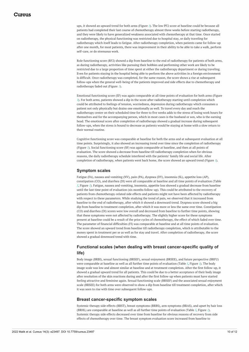
ups, it showed an upward trend for both arms (Figure 1). The low PF2 score at baseline could be because allpatients had completed their last course of chemotherapy almost three weeks before starting radiotherapy,and they were likely to have generalized weakness associated with chemotherapy at that time. Once startedon radiotherapy, the physical functioning was restricted due to hospital stay, or daily traveling forradiotherapy which itself leads to fatigue. After radiotherapy completion, when patients came for follow-upafter one month, for most patients, there was improvement in their ability to be able to take a walk, performself-care, or do strenuous work.
Role functioning score (RF2) showed a dip from baseline to the end of radiotherapy for patients of both arms,as during radiotherapy, activities like pursuing their hobbies and performing other work are likely to berestricted due to a large proportion of time spent at either the radiotherapy department or during traveling.Even for patients staying in the hospital being able to perform the above activities in a foreign environmentis difficult. Once radiotherapy was completed, for the same reason, the score shows a rise at subsequentfollow-ups when the general well-being of the patients improved and side effects due to chemotherapy andradiotherapy faded out (Figure 1).
Emotional functioning score (EF) was again comparable at all time points of evaluation for both arms (Figure1). For both arms, patients showed a dip in the score after radiotherapy starting until completion whichcould be attributed to feelings of tension, worriedness, depression during radiotherapy which consumes apatient not only physically but drains one emotionally as well. To travel every day and reach theradiotherapy center on their scheduled time for three to five weeks adds to the stress of losing work hours forthemselves and for the accompanying person, which in most cases is the husband or son, who is the earninghead. The emotional score after completion of radiotherapy showed a gradual increase during subsequentfollow-ups, when the stress is bound to decrease as patients would be staying at home with a slow return totheir normal routine.
Cognitive functioning score was comparable at baseline for both the arms and at subsequent evaluation at alltime points. Surprisingly, it also showed an increasing trend over time since the completion of radiotherapy(Figure 1). Social functioning score (SF) was again comparable at baseline, and then at all points ofevaluation. The score showed a decrease from baseline till radiotherapy completion when for obviousreasons, the daily radiotherapy schedule interfered with the patients’ family life and social life. Aftercompletion of radiotherapy, when patients went back home, the score showed an upward trend (Figure 1).
Symptom scalesFatigue (FA), nausea and vomiting (NV), pain (PA), dyspnea (DY), insomnia (SL), appetite loss (AP),constipation (CO), and diarrhea (DI) were all comparable at baseline and all time points of evaluation (Table5, Figure 1). Fatigue, nausea and vomiting, insomnia, appetite loss showed a gradual decrease from baselineuntil the last time point of evaluation (six months follow-up). This could be attributed to the recovery ofpatients from chemotherapy-related side effects and patients might not have been affected by radiotherapywith respect to these parameters. While studying the trend of pain, we observed that it increased frombaseline to the end of radiotherapy, after which it showed a downward trend. Dyspnea score showed a bigdip from baseline to treatment completion, after which it was more or less the same over time. Constipation(CO) and diarrhea (DI) scores were low overall and decreased from baseline to further time points, showingthat these symptoms were not affected by radiotherapy. The slightly higher score for these symptomspresent at baseline could be a result of the prior cycles of chemotherapy, the effect of which faded over time.The parameter of financial difficulties (FI) was comparable at baseline and at all time points of evaluation.The score showed an upward trend from baseline till radiotherapy completion, which is attributable to themoney spent in treatment per se as well as for stay and travel. After completion of radiotherapy, the scoreshowed a gradual downward trend with time.
Functional scales (when dealing with breast cancer-specific quality oflife)Body image (BRBI), sexual functioning (BRSEF), sexual enjoyment (BRSEE), and future perspective (BRFU)were comparable at baseline as well as all further time points of evaluation (Table 5, Figure 1). The bodyimage scale was low and almost similar at baseline and at treatment completion. After the first follow-up, itshowed a gradual upward trend for all patients. This could be due to a better acceptance of their body imageafter resolution of the skin reactions during and after the first follow-up when patients must have startedfeeling attractive and feminine again. Sexual functioning scale (BRSEF) and the associated sexual enjoymentscale (BRSEE) for both arms were observed to show a dip from baseline till treatment completion, after whichit was seen to rise with time over subsequent follow-ups.
Breast cancer-specific symptom scalesSystemic therapy side effects (BRST), breast symptoms (BRBS), arm symptoms (BRAS), and upset by hair loss(BRHL) are comparable at baseline as well as all further time points of evaluation (Table 5, Figure 1).Systemic therapy side effects decreased over time from baseline for obvious reasons of recovery from sideeffects of chemotherapy over time. The breast symptom evaluation score increased from baseline to
2022 Malik et al. Cureus 14(3): e23497. DOI 10.7759/cureus.23497 10 of 12

treatment completion, which is attributable to radiotherapy side effects of pain, swelling, increasedsensitivity, and dryness/itchiness over the radiotherapy site. Once radiotherapy was completed, the scoreshould a downward trend with time over subsequent follow-ups as the above-mentioned side effectsrecovered. The arm symptom score was similar at baseline and at treatment completion, after which itshowed a downward trend over time at subsequent follow-ups, which is attributable to gradual recovery fromradiotherapy-induced pain and swelling over axilla. The score for upset from hair loss also decreased frombaseline with time which can be explained by gradual regrowth of hair lost due to chemotherapy.
The quality of life subgroup in START B trial included 12.3% post-mastectomy women for whom the relevantparameter in QOL assessment changed in skin appearance which favored the hypofractionation arm with ahazard ratio of 0.86 on the forest plot [5]. START A trialists observed a lower rate of change in skinappearance after 39 Gy than 50 Gy (p=0.004) [4]. We observed comparable scores for breast symptoms ofpain, itching, and swelling in the irradiated between two arms at all time points of evaluation.
Efficacy of hypofractionation was not undertaken as an objective for this study, owing to a too short follow-up to be able to draw conclusions on it. The higher number of distant metastasis in the conventional arm (5vs 2) will have to be considered as an incidental finding because follow-up duration is very short to make anyinference on the same. Small sample size and a short follow-up duration limit our study results. Longerfollow-up on a large sample (multicentric trial) is required to analyze the response and the late cardiaceffects.
ConclusionsHypofractionation with 40 Gray/25 fractions/five weeks in post-mastectomy setting is feasible and non-inferior to conventional five-week fractionation schedule in terms of toxicity profile and quality of life forour patients. Efficacy of the same has already been established by the START trials in the United Kingdom. Athree-week schedule costs 0.6 times less compared to the five-week schedule. A treatment modality thatcosts less and offers similar toxicity profile, quality of life, and efficacy should be readily accepted. A shorterschedule would also lessen the burden on the radiotherapy machines which would in turn shorten thewaitlists. Hence, resource constraint institutes in India can adopt hypofractionation as their post-mastectomy radiotherapy protocol, which not only improves patient compliance but also saves resources forboth patients and the institute.
Additional InformationDisclosuresHuman subjects: Consent was obtained or waived by all participants in this study. Institutional EthicsCommittee Mahatma Gandhi Institute of Medical Sciences, Sevagram, Wardha issued approval#MGIMS/IEC/RADTH/98/2014. Animal subjects: All authors have confirmed that this study did not involveanimal subjects or tissue. Conflicts of interest: In compliance with the ICMJE uniform disclosure form, allauthors declare the following: Payment/services info: All authors have declared that no financial supportwas received from any organization for the submitted work. Financial relationships: All authors havedeclared that they have no financial relationships at present or within the previous three years with anyorganizations that might have an interest in the submitted work. Other relationships: All authors havedeclared that there are no other relationships or activities that could appear to have influenced thesubmitted work.
References1. Clarke M, Collins R, Darby S, et al.: Effects of radiotherapy and of differences in the extent of surgery for
early breast cancer on local recurrence and 15-year survival: an overview of the randomised trials. Lancet.2005, 366:2087-106. 10.1016/S0140-6736(05)67887-7
2. Overgaard M, Hansen PS, Overgaard J, et al.: Postoperative radiotherapy in high-risk premenopausal womenwith breast cancer who receive adjuvant chemotherapy. Danish Breast Cancer Cooperative Group 82b Trial.N Engl J Med. 1997, 337:949-55. 10.1056/NEJM199710023371401
3. Owen JR, Ashton A, Bliss JM, et al.: Effect of radiotherapy fraction size on tumour control in patients withearly-stage breast cancer after local tumour excision: long-term results of a randomised trial. Lancet Oncol.2006, 7:467-71. 10.1016/S1470-2045(06)70699-4
4. Bentzen SM, Agrawal RK, Aird EG, et al.: The UK Standardisation of Breast Radiotherapy (START) Trial A ofradiotherapy hypofractionation for treatment of early breast cancer: a randomised trial. Lancet Oncol. 2008,9:331-41. 10.1016/S1470-2045(08)70077-9
5. Bentzen SM, Agrawal RK, Aird EG, et al.: The UK Standardisation of Breast Radiotherapy (START) Trial B ofradiotherapy hypofractionation for treatment of early breast cancer: a randomised trial. Lancet. 2008,371:1098-107. 10.1016/S0140-6736(08)60348-7
6. Haviland JS, Owen JR, Dewar JA, et al.: The UK Standardisation of Breast Radiotherapy (START) trials ofradiotherapy hypofractionation for treatment of early breast cancer: 10-year follow-up results of tworandomised controlled trials. Lancet Oncol. 2013, 14:1086-94. 10.1016/s1470-2045(13)70386-3
7. Cancer Today. (2021). Accessed: April 05, 2021: https://gco.iarc.fr/today.8. Murthy NS, Chaudhry K, Nadayil D, Agarwal UK, Saxena S: Changing trends in incidence of breast cancer:
Indian scenario. Indian J Cancer. 2009, 46:73-4. 10.4103/0019-509x.48603
2022 Malik et al. Cureus 14(3): e23497. DOI 10.7759/cureus.23497 11 of 12

9. Jayarajan K, Kar DC, Sahu R, Sing M: Bhabhatron: an indigenous telecobalt machine for cancer treatment .BARC Newsletter. Bhabha Atomic Research Centre, Mumbai, India; 2008. 297:27-34.
10. Sullivan R, Badwe RA, Rath GK, et al.: Cancer research in India: national priorities, global results . LancetOncol. 2014, 15:213-22. 10.1016/s1470-2045(14)70109-3
11. Cox JD, Stetz J, Pajak TF: Toxicity criteria of the Radiation Therapy Oncology Group (RTOG) and theEuropean Organization for Research and Treatment of Cancer (EORTC). Int J Radiat Oncol Biol Phys. 1995,31:1341-6. 10.1016/0360-3016(95)00060-C
12. Aaronson NK, Ahmedzai S, Bergman B, et al.: The European Organization for Research and Treatment ofCancer QLQ-C30: a quality-of-life instrument for use in international clinical trials in oncology. J NatlCancer Inst. 1993, 85:365-76. 10.1093/jnci/85.5.365
13. Kumbhaj P, Sharma R, Singh D, et al.: Study of two different dose fractionation schedules of postmastectomy chest wall irradiation in carcinoma breast patients. Int J Health Med Sci. 2013, 2:1001-5.10.5455/ijmsph.2013.040820131
14. Wilting J, Hagedorn M: Left-right asymmetry in embryonic development and breast cancer: commonmolecular determinants?. Curr Med Chem. 2011, 18:5519-27. 10.2174/092986711798347252
15. Nandi M, Mahata A, Mallick I, Achari R, Chatterjee S: Hypofractionated radiotherapy for breast cancers--preliminary results from a tertiary care center in eastern India. Asian Pac J Cancer Prev. 2014, 15:2505-10.10.7314/apjcp.2014.15.6.2505
16. Shahid A, Athar MA, Asghar S, et al.: Post mastectomy adjuvant radiotherapy in breast cancer: acomparision of three hypofractionated protocols. J Pak Med Assoc. 2009, 59:282-7.
17. Abhilash GH, Dhull AK, Atri RE, Dhankhar R, Kaushal V: Comparison of hypofractionated radiation therapyversus conventional radiation therapy in post mastectomy breast cancer. J Evid Based Med Healthc. 2016,3:1177-81. 10.18410/jebmh/2016/270
18. Eldeeb H, Awad I, Elhanafy O: Hypofractionation in post-mastectomy breast cancer patients: seven-yearfollow-up. Med Oncol. 2012, 29:2570-6. 10.1007/s12032-012-0192-1
2022 Malik et al. Cureus 14(3): e23497. DOI 10.7759/cureus.23497 12 of 12