Fatty Acids in Marine Organisms: In the Pursuit of Bioactive Agents
10
Valuable compounds in macroalgae extracts Paula B. Andrade a , Mariana Barbosa a , Rui Pedro Matos a , Graciliana Lopes a , Juliana Vinholes a , Teresa Mouga b , Patrícia Valentão a,⇑ a REQUIMTE/Laboratório de Farmacognosia, Departamento de Química, Faculdade de Farmácia, Universidade do Porto, Rua de Jorge Viterbo Ferreira No 228, 4050-313 Porto, Portugal b GIRM – Marine Resources Research Group, School of Tourism and Maritime Technology, Polytechnic Institute of Leiria, Santuário N.ª Sra. dos Remédios, Apartado 126, 2524-909 Peniche, Portugal article info Article history: Received 20 August 2012 Received in revised form 20 October 2012 Accepted 12 November 2012 Available online 28 November 2012 Keywords: Macroalgae Phloroglucinol Polyols Fatty acids Sterols Bioactivity abstract Bioactive compounds present in ethanolic extracts from 18 macroalgae of the Portuguese coast were ana- lysed by gas chromatography–mass spectrometry (GC–MS), leading to the characterization of 14 com- pounds: proline, phloroglucinol, mannitol, 8 fatty acids and 3 sterols. A dose-dependent response against enzymes with biological significance (a-glucosidase, acetylcholinesterase and butyrylcholinester- ase) and free radicals (DPPH, nitric oxide, superoxide and hydroxyl) was found, Phaeophyta being the most promising group. A PCA analysis was performed and allowed the establishment of a correlation between the algae chemical composition and the biological activity. Cystoseira tamariscifolia (Hudson) Papenfuss, Cystoseira nodicaulis (Withering) M. Roberts, Cystoseira usneoides (Linnaeus) M. Roberts and Fucus spiralis Linnaeus are among the most active species, which is in accordance with their higher con- tents in phloroglucinol, mannitol, oleic, arachidonic and eicosapentaenoic acids, and fucosterol. The results point to the potential interest of the use of Phaeophyta species as food additives, due to their potent antiradical activities, and especially highlights the importance of F. spiralis in the food chain of Mediterranean countries. Moreover, the incorporation of the extracts of these species in food products, nutraceutical and pharmaceutical preparations for human health should also be instigated, since they can suppress hyperglycemia and inhibit cholinesterases. Ó 2012 Elsevier Ltd. All rights reserved. 1. Introduction Biodiversity within red (Rhodophyta), green (Chlorophyta) and brown (Phaeophyta) macroalgae offers the possibility of finding a wide variety of natural compounds with interesting properties (Shahnaz & Shameel, 2009). Therefore, an increasing attention to macroalgae metabolites of industries from different branches (tex- tile, fuel, plastics, paint, varnish, cosmetics, pharmaceutical and food) was noticed in the last years (Cardozo et al., 2007). In fact, more than 15,000 primary and secondary metabolites from differ- ent metabolic pathways have been reported for macroalgae and different applications were assigned to them (Grosso, Vinholes, Valentão, & Andrade, 2011). From human health point of view, both types of metabolites are of extreme importance since some of them can display remarkable positive effects on organism. The metabolites found in macroalgae are described as having anti- inflammatory, antimutagenic, antitumor, antidiabetic, and antihy- perthensive properties. In addition, they are hepatoprotectors, and can also inhibit the lipoxygenase, aldose reductase and cholines- terases (El Gamal, 2010; Lopes et al., 2012; Pangestuti & Kim, 2011; Yoon, Chung, Kim, & Choi, 2008; Zubia, Fabre, Kerjean, & Deslandes, 2009a; Zubia et al., 2009b). Considering that macroal- gae are part of the diet in different countries and constitute a source of beneficial nutrients, such as vitamins, trace minerals, lip- ids, amino acids and dietary fibres, its use as a low-calorie food should be investigated, as it might be important in body weight control and cardiovascular health (Bocanegra, Bastida, Benedí, Ród- enas, & Sánchez-Muniz, 2009). Up to date, several approaches, such as infrared (IR) spectros- copy, nuclear magnetic resonance (NMR), liquid chromatogra- phy–mass spectrometry (LC–MS) and gas chromatography–mass spectrometry (GC–MS) have been used in order to obtain as much information as possible about metabolites from different matrices (Awad, 2002; Gómez-Romero, Segura-Carretero, & Fernández- Gutiérrez, 2010; Humston, Dombek, Tu, Young, & Synovec, 2011; Vergouw et al., 2008). Among them GC–MS is the most used one, since reliable results can be obtained with a cheaper, although powerful, method (Sumner, Mendes, & Dixon, 2003). GC–MS anal- ysis offers information about the identity of volatile and non-vola- tile (capable of being volatilized by a derivatizing agent) compounds present in a sample. Presently, silylation has been pre- ferred as derivatizing method for GC–MS analysis; however, the re- sults are dependent on the agent used and the preparation of 0308-8146/$ - see front matter Ó 2012 Elsevier Ltd. All rights reserved. http://dx.doi.org/10.1016/j.foodchem.2012.11.081 ⇑ Corresponding author. Tel.: +351 220428653; fax: +351 226093390. E-mail address: [email protected] (P. Valentão). Food Chemistry 138 (2013) 1819–1828 Contents lists available at SciVerse ScienceDirect Food Chemistry journal homepage: www.elsevier.com/locate/foodchem
Transcript of Fatty Acids in Marine Organisms: In the Pursuit of Bioactive Agents

Food Chemistry 138 (2013) 1819–1828
Contents lists available at SciVerse ScienceDirect
Food Chemistry
journal homepage: www.elsevier .com/locate / foodchem
Valuable compounds in macroalgae extracts
Paula B. Andrade a, Mariana Barbosa a, Rui Pedro Matos a, Graciliana Lopes a, Juliana Vinholes a,Teresa Mouga b, Patrícia Valentão a,⇑a REQUIMTE/Laboratório de Farmacognosia, Departamento de Química, Faculdade de Farmácia, Universidade do Porto, Rua de Jorge Viterbo Ferreira No 228, 4050-313 Porto, Portugalb GIRM – Marine Resources Research Group, School of Tourism and Maritime Technology, Polytechnic Institute of Leiria, Santuário N.ª Sra. dos Remédios, Apartado 126,2524-909 Peniche, Portugal
a r t i c l e i n f o a b s t r a c t
Article history:Received 20 August 2012Received in revised form 20 October 2012Accepted 12 November 2012Available online 28 November 2012
Keywords:MacroalgaePhloroglucinolPolyolsFatty acidsSterolsBioactivity
0308-8146/$ - see front matter � 2012 Elsevier Ltd. Ahttp://dx.doi.org/10.1016/j.foodchem.2012.11.081
⇑ Corresponding author. Tel.: +351 220428653; faxE-mail address: [email protected] (P. Valentão).
Bioactive compounds present in ethanolic extracts from 18 macroalgae of the Portuguese coast were ana-lysed by gas chromatography–mass spectrometry (GC–MS), leading to the characterization of 14 com-pounds: proline, phloroglucinol, mannitol, 8 fatty acids and 3 sterols. A dose-dependent responseagainst enzymes with biological significance (a-glucosidase, acetylcholinesterase and butyrylcholinester-ase) and free radicals (DPPH, nitric oxide, superoxide and hydroxyl) was found, Phaeophyta being themost promising group. A PCA analysis was performed and allowed the establishment of a correlationbetween the algae chemical composition and the biological activity. Cystoseira tamariscifolia (Hudson)Papenfuss, Cystoseira nodicaulis (Withering) M. Roberts, Cystoseira usneoides (Linnaeus) M. Roberts andFucus spiralis Linnaeus are among the most active species, which is in accordance with their higher con-tents in phloroglucinol, mannitol, oleic, arachidonic and eicosapentaenoic acids, and fucosterol. Theresults point to the potential interest of the use of Phaeophyta species as food additives, due to theirpotent antiradical activities, and especially highlights the importance of F. spiralis in the food chain ofMediterranean countries. Moreover, the incorporation of the extracts of these species in food products,nutraceutical and pharmaceutical preparations for human health should also be instigated, since theycan suppress hyperglycemia and inhibit cholinesterases.
� 2012 Elsevier Ltd. All rights reserved.
1. Introduction
Biodiversity within red (Rhodophyta), green (Chlorophyta) andbrown (Phaeophyta) macroalgae offers the possibility of finding awide variety of natural compounds with interesting properties(Shahnaz & Shameel, 2009). Therefore, an increasing attention tomacroalgae metabolites of industries from different branches (tex-tile, fuel, plastics, paint, varnish, cosmetics, pharmaceutical andfood) was noticed in the last years (Cardozo et al., 2007). In fact,more than 15,000 primary and secondary metabolites from differ-ent metabolic pathways have been reported for macroalgae anddifferent applications were assigned to them (Grosso, Vinholes,Valentão, & Andrade, 2011). From human health point of view,both types of metabolites are of extreme importance since someof them can display remarkable positive effects on organism. Themetabolites found in macroalgae are described as having anti-inflammatory, antimutagenic, antitumor, antidiabetic, and antihy-perthensive properties. In addition, they are hepatoprotectors, andcan also inhibit the lipoxygenase, aldose reductase and cholines-terases (El Gamal, 2010; Lopes et al., 2012; Pangestuti & Kim,
ll rights reserved.
: +351 226093390.
2011; Yoon, Chung, Kim, & Choi, 2008; Zubia, Fabre, Kerjean, &Deslandes, 2009a; Zubia et al., 2009b). Considering that macroal-gae are part of the diet in different countries and constitute asource of beneficial nutrients, such as vitamins, trace minerals, lip-ids, amino acids and dietary fibres, its use as a low-calorie foodshould be investigated, as it might be important in body weightcontrol and cardiovascular health (Bocanegra, Bastida, Benedí, Ród-enas, & Sánchez-Muniz, 2009).
Up to date, several approaches, such as infrared (IR) spectros-copy, nuclear magnetic resonance (NMR), liquid chromatogra-phy–mass spectrometry (LC–MS) and gas chromatography–massspectrometry (GC–MS) have been used in order to obtain as muchinformation as possible about metabolites from different matrices(Awad, 2002; Gómez-Romero, Segura-Carretero, & Fernández-Gutiérrez, 2010; Humston, Dombek, Tu, Young, & Synovec, 2011;Vergouw et al., 2008). Among them GC–MS is the most used one,since reliable results can be obtained with a cheaper, althoughpowerful, method (Sumner, Mendes, & Dixon, 2003). GC–MS anal-ysis offers information about the identity of volatile and non-vola-tile (capable of being volatilized by a derivatizing agent)compounds present in a sample. Presently, silylation has been pre-ferred as derivatizing method for GC–MS analysis; however, the re-sults are dependent on the agent used and the preparation of

1820 P.B. Andrade et al. / Food Chemistry 138 (2013) 1819–1828
derivatives. Different groups of compounds, belonging to differentmetabolic pathways, can be obtained as trimethylsylil derivatives,such as amino acids, organic acids, carbohydrates, fatty acids andsterols (Humston et al., 2011; Kim, Shin, Hossain, Yun, Lee, &Kim, 2011; Liu et al., 2011; Na Jom, Frank, & Engel, 2011). Recently,our research group has developed a GC–MS-based method for thedetermination of several classes of compounds (including aminoacids, fatty acids and sterols) on sea-star samples (Marthasteriasglacialis Linnaeus) (Pereira et al., 2012). Good results were ob-served by the analysis of raw extracts obtained using a mild extrac-tion temperature, thus avoiding the formation of artefacts.
Portugal algal flora is characterised by around 250 species ofRhodophyta, 100 of Phaeophyta and 60 of Chlorophyta (Sousa-Pinto, 1998). In order to valorise the macroalgae from thePortuguese coast and extend their application for food and phar-maceuticals industries, ethanol extracts of eighteen macroalgaespecies, including three edible ones (Ulva lactuca Linnaeus, Osmun-dea pinnatifida (Hudson) Stackhouse and Fucus spiralis Linnaeus),were analysed by means of GC–MS. As different biological proper-ties are attributed to macroalgae, we also investigated their biolog-ical potential towards different enzymes (a-glucosidase, acetyl andbutyrylcholinesterase), and free radicals (DPPH�, nitric oxide (�NO),superoxide (O2
��) and hydroxyl (�OH)). Although there are somestudies addressing some of the parameters focused in this work(for some of the species), it is the first time that these 18 macroal-gae species are screened with a methodology that allows thesimultaneous identification and quantification of metabolites fromdifferent classes (fatty acids, sugars, sterols, amino acids and phen-olics). Additionally, and to our knowledge, with the exception ofCodium tomentosum Stackhouse and U. lactuca, for which the a-glu-cosidase activity has previously been reported, the entire enzy-matic assays were performed for the studied species for the firsttime. Finally, in order to relate the extracts chemical compositionwith their biological activity, principal component analysis (PCA)was performed.
Table 1Macroalgae samples.
Phylum Species
ChlorophytaCodium tomentosum StackhouseCodium adhaerens C. AgardhUlva lactuca Linnaeus
RhodophytaAsparagopsis armata HarveySphaerococcus coronopifolius StackhousePlocamium cartilagineum (Linnaeus) P. S. DixonOsmundea pinnatifida (Hudson) StackhouseSchizymenia dubyi (Chauv. ex Duby) J. Agardh
PhaeophytaCystoseira tamariscifolia (Hudson) PapenfussCystoseira usneoides (Linnaeus) M. RobertsCystoseira nodicaulis (Withering) M. RobertsCladostephus spongiosus (Huds.) C. AgardhHalopteris filicina (Gratel.) Kütz.Saccorhiza polyschides (Lightfoot) BattersPadina pavonica (L.) ThivySargassum vulgare C. AgardhFucus spiralis LinnaeusStypocaulon scoparium (Linnaeus) Kützing
2. Material and methods
2.1. Standards and reagents
Standards of proline (P99%), norvaline (P99%), mannitol(P98%), phloroglucinol (P99%), methyl linolelaidate (P99%), tet-radecanoic acid (C14:0) (P98%), palmitic acid (C16:0) (P99%),stearic acid (C18:0) (P99%), arachidonic acid (C20:4) (P99%), a-linolenic acid (C18:3) (P99%), oleic acid (C18:1) (P99%),5,8,11,14,17-eicosapentaenoic acid (C20:5) (P99%), arachidic acid(C20:0) (P97%), cholestanol (P95%), b-sitosterol (P99%), desmos-terol (P84%) and fucosterol (P95%), and N-methyl-N-(trimethyl-silyl) trifluoroacetamide (MSTFA), 1,1-diphenyl-2-picrylhydrazylradical (DPPH�), ethylenediaminetetraacetic acid disodium salt,iron sulphate heptahydrate, salicylic acid, b-nicotinamide adeninedinucleotide (NADH), phenazine methosulfate (PMS), nitrotetrazo-lium blue chloride (NBT), 5,50-dithio-bis(2-nitrobenzoic acid)(DTNB), sodium nitroprusside dehydrate (SNP), sulphanilamide,acetylcholinesterase (AChE) from electric eel (type VI-s, lyophilizedpowder), acetylthiocholine iodide (ATCI), butyrylcholinesterase(BuChE) from equine serum (lyophilized powder), S-butyrylthioch-oline chloride (BTCC), a-glucosidase (type I from baker’s yeast) and4-nitrophenyl a-D-glucopyranoside (PNP-G) were from Sigma (St.Louis, MO, USA). The n-alkane series (C8–C40) was purchased fromSupelco (Bellefonte, PA, USA). N-(1-Naphthyl)ethylenediaminedihydrochloride, methanol, potassium dihydrogen phosphate,hydrogen peroxide, sulphuric and acetic acids were obtained fromMerck (Darmstadt, Germany). Sodium hydroxide was from Panreac(Barcelona, Spain).
2.2. Preparation of standard solutions
Stock solutions of proline, phloroglucinol, mannitol, all fattyacids and sterols, as well as of the internal standards (IS) norvaline,methyl linolelaidate and desmosterol were prepared individuallyin ethanol and kept at �20 �C until analysis. Calibration solutionsfor phloroglucinol and mannitol were prepared by diluting eachstock solution in appropriate amounts with ethanol in the concen-tration range of 1.50–50.00 lg/ml for both compounds.
2.3. Macroalgae samples
Macroalgae samples consist of three Chlorophyta, five Rhodo-phyta and 10 Phaeophyta species (Table 1). Samples were ran-domly collected in different places of the west coast of Portugaland identified by Teresa Mouga, PhD (GIRM). Each macroalgaesample corresponds to a mixture of three to four individuals inthe same stage of development. With the exceptions of Cystoseirausneoides (Linnaeus) M. Roberts and Cystoseira nodicaulis (Wither-ing) M. Roberts, collected in 2009, all samples were collected in2008. U. lactuca and Saccorhiza polyschides (Lightfoot) Batters werecollected in June; C. usneoides and C. nodicaulis, in November andthe remaining species, in September. To prevent sample altera-tions, after collection they were immediately placed on ice andtransported to the laboratory in insulated, sealed ice boxes, to pro-tect them from heat, air, and light exposure. Macroalgae were thencarefully and quickly washed with NaCl aqueous solution (3.5%) toremove epiphytes and encrusting material, at room temperature,without exposure to direct light, and kept at �20 �C, prior to theirlyophilisation in a Labconco 4.5 Freezone apparatus (Kansas City,MO, USA). The dried material was powdered (<910 lm) and keptin the dark, in a desiccator, until it was subjected to extraction.No alteration (colour, smell, humidity) of the samples was noticedduring storage. Each sample corresponds to a mixture of threeindividuals.
2.4. GC–MS system and data acquisition
2.4.1. GC–MS apparatusAnalysis was performed with a Varian CP-3800 gas chromato-
graph coupled to a Varian Saturn 4000 mass selective ion trapdetector (USA) and a Saturn GC–MS workstation software version

P.B. Andrade et al. / Food Chemistry 138 (2013) 1819–1828 1821
6.8. A VF-5 ms (30 m � 0.25 mm � 0.25 lm) column (VARIAN) wasused. A CombiPAL automatic autosampler (Varian, Palo Alto, CA)was used for all experiments. The injector port was heated to250 �C. Injections were performed in split mode, with a ratio of1/40. The carrier gas was helium C-60 (Gasin, Portugal), at a con-stant flow of 1 ml/min. The ion trap detector was set as follows:transfer line, manifold and trap temperatures were 280, 50, and180 �C, respectively. The mass ranged from 50 to 600 m/z, with ascan rate of 6 scan/s. The emission current was 50 lA and the elec-tron multiplier was set in relative mode to auto tune procedure.The maximum ionisation time was 25.000 ls, with an ionisationstorage level of 35 m/z. The injection volume was 2 ll and the anal-ysis was performed in Full Scan mode.
2.4.2. GC–MS conditions for trimethylsilyl (TMS) derivatives analysisThe oven temperature was set at 100 �C for 1 min, then increas-
ing 20 �C/min to 250 �C and held for 2 min, 10 �C/min to 300 �C andheld for 10 min. All mass spectra were acquired in electron impact(EI) mode. Ionisation was maintained off during the first 3 min toavoid solvent overloading. Identification of compounds wasachieved by comparisons of their retention time and mass spectrawith those of pure standards TMS derivatives prepared and in-jected under the same conditions and from NIST 05 MS LibraryDatabase. In addition, the retention index (RI) was experimentallycalculated and the values were compared with those reported inNIST webBook and in the literature (Isidorov & Vinogorova, 2003;Meyer et al., 2009; Radulovic & Dordevic, 2011; Shepherd et al.,2007) for GC columns with 5%-phenyl-95%-dimethylpolysiloxane.For the RI determination, an n-alkanes series (C8–C40) was used.
2.5. Metabolites extraction and derivatization
The metabolites extraction was performed according to themethod previously reported by our group (Pereira et al., 2012).Briefly, 100.00 ± 1.00 mg of dried sample were transferred to aglass vial and the internal standards were added: 80 ll of norvaline(0.30 mg/ml), 20 ll of methyl linolelaidate (10.00 mg/ml), and80 ll of desmosterol (2.00 mg/ml). The volume was then com-pleted to 2.00 ml with ethanol. The extraction procedure was asfollows: incubation for 20 min at 40 �C under magnetic stirring(200 rpm). Samples were then filtered through a 0.45 lm mem-brane (Millipore). An aliquot of 50 ll of extract was transferredto a glass vial, the solvent was evaporated under nitrogen streamand 50 ll of the derivatizing reagent (MSTFA) was added to thedried residue. The vial was capped, vortexed and heated for20 min in a dry block heater maintained at 40 �C. All analyses wereperformed in triplicate.
2.6. Metabolites quantification
For quantification purposes, each sample was injected in tripli-cate and the amount of metabolites present in samples wasachieved from the calibration curves of the respective TMS deriva-tives. All compounds were quantified in Full Scan mode with theexception of a-linolenic (m/z 191, 335, and 350) and oleic (m/z264, 339 and 354) derivatives that were quantified by the area ob-tained from the reprocessed chromatogram, using the characteris-tic m/z fragments.
Table 2Validation parameters for phloroglucinol and mannitol.
Compound Concentration range (lg/ml) Slope Interception
Phloroglucinol 1.50–50.00 0.1689 +1.7161Mannitol 1.50–50.00 0.0363 +0.6125
The quantification of proline, fatty acids and sterols in macroal-gae ethanolic extracts was achieved by using the calibration curvespreviously described by the authors (Pereira et al., 2012).
For phloroglucinol and mannitol at least six concentration lev-els of compounds’ TMS derivatives were analysed. Each calibrationsolution contained norvaline as internal standard, at final concen-tration of 12.00 lg/ml. The derivatization procedure was carriedout as described above using the calibration solution instead ofthe extract. Linearity was evaluated in the concentration range of1.50–50.00 lg/ml for both compounds, and expressed by the corre-lation coefficient (Table 2). Limit of detection (LOD) and limit ofquantification (LOQ) were determined from calibration curve data(ICH, 1996), according to the following formula:
LOD ¼ ð3:3� SDÞ=b
LOQ ¼ ð10� SDÞ=b
where SD is the residual standard deviation of the linear regression,and b is the slope of the regression line.
The ratios of the peak areas of compounds vs. those of IS wereused to obtain the compounds concentration from the calibrationgraphs. Compounds amounts are media of three assays.
2.7. Enzymatic assays
2.7.1. a-Glucosidase inhibitory activityThe effect on a-glucosidase was assessed in 96-well plates,
using a procedure previously reported (Vinholes et al., 2011).Briefly, each well contained 4-nitrophenyl a-D-glucopyranosidein phosphate buffer (pH 7.0) and extract. The reaction was initiatedby the addition of the enzyme solution and the plates were incu-bated at 37 �C for 10 min. The absorbance at 400 nm was measuredin a Multiskan Ascent plate reader (Thermo Electron Corporation).The values of EC50 were calculated from three independent assays,performed in triplicate. Acarbose was used as positive control.
2.7.2. Acetylcholinesterase (AChE) and butyrylcholinesterase (BuChE)inhibitory activity
The inhibition of AChE and BuChE activities was determinedspectrophotometrically in a Multiskan Ascent plate reader (Ther-mo Electron Corporation), based on Ellman’s method, as previouslyreported (Vinholes et al., 2011). The absorbance was measured at405 nm and the rates of the reactions were calculated by AscentSoftware version 2.6 (Thermo Labsystems Oy). The values of EC50
were calculated by performing three independent assays, intriplicate.
2.8. Antioxidant activity
2.8.1. DPPH� assayThe free radical scavenging activity was determined spectro-
photometrically in a Multiskan Ascent plate reader (Thermo Elec-tron Corporation), by monitoring the decrease of absorbance ofDPPH� at 515 nm, according to Vinholes et al. (2011). The valuesof EC50 were calculated from three independent assays, performedin triplicate.
Correlation coefficient LOD (lg/ml) LOQ (lg/ml) RSD
0.9834 0.57 1.89 3.200.9919 0.95 3.16 1.14
1822 P.B. Andrade et al. / Food Chemistry 138 (2013) 1819–1828
2.8.2. Hydroxyl radical (�OH) assayHydroxyl radical scavenging activity was measured according
to Royer, Diouf, and Stevanovic (2011) with slight modification.To 62.5 ll of extracts (0.40–50.00 mg/ml) in phosphate buffer(150 mM, pH 7.4) were added 275 ll of 8 mM iron sulphate hepta-hydrate solution (EDTA-Na 20 lM) and 125 ll of 7 mM H2O2 solu-tion. The reaction was initiated by the addition of 187.5 ll ofsalicylic acid (3 mM). After vortexing, the reaction mixture was al-lowed to stand for 30 min at 37 �C. The absorbance at 515 nm wasthen determined. EC50 values were calculated from three indepen-dent assays, performed in triplicate.
2.8.3. Superoxide anion (O2��) assay
O2�� was generated by the NADH/PMS system, as reported in
Vinholes et al. (2011). The absorbance of the reaction mixturewas determined at 560 nm in a Multiskan Ascent plate reader(Thermo Electron Corporation) at room temperature, for 2 min.The calculation of EC50 values were determined from three inde-pendent assays, performed in triplicate.
2.8.4. Nitric oxide (�NO) assayThe antiradical activity was determined in a Multiskan Ascent
plate reader (Thermo Electron Corporation), applying a previouslydescribed procedure (Vinholes et al., 2011). The absorbance wasmeasured at 562 nm and the values of EC50 were calculated fromthree independent assays, performed in triplicate.
2.9. Principal component analysis (PCA)
PCA was used to transform a number of potentially correlatedvariables into a number of relatively independent variables thatcould be ranked based upon their contribution for explaining thevariation of the whole data set (Xu & Hagler, 2002). As so, the rel-atively important components of high-dimensional pattern couldbe successfully identified. The original high-dimensional data canbe mapped onto a lower dimensional space, and therefore the com-plexity of a high-dimensional pattern classification problem isgreatly reduced (Lo, Jiang, Chao, & Chang, 2007). For the presentstudy, pattern recognition based on PCA was performed using SPSS.Data matrix consisted on the metabolites present on the ethanolicextracts of 18 samples of macroalgae and their activity at the high-est concentration tested.
3. Results and discussion
3.1. Macroalgae qualitative profile
Representative chromatographic profiles of Chlorophyta, Rho-dophyta and Phaeophyta are presented in Fig. 1. Fourteen com-pounds were positively identified in the ethanolic extracts of 18species of macroalgae (Tables 3 and 4). In a general way, the differ-ent species have the same type of compounds, proline (1), manni-tol (4), palmitic acid (C16:0) (5), and stearic acid (C18:0) (8) beingthe compounds present in almost all samples (Tables 3 and 4,Fig. 1).
Proline (1) was the unique amino acid found among the threePhyla. As far as we know, it is the first time that this compoundis described in C. tomentosum Stackhouse, Codium adhaerens C.Agardh, Sphaerococcus coronopifolius Stackhouse, Plocamium carti-lagineum (Linnaeus) P.S. Dixon, O. pinnatifida, Schizymenia dubyi(Chauv. Ex Duby) J. Agardh, C. tamariscifolia (Hudson) Papenfuss,C. usneoides, C. nodicaulis, Cladostephus spongiosus (Huds.) C.Agardh, Halopteris filicina (Gratel.) Kütz, Padina pavonica (L.) Thivyand F. spiralis.
Mannitol (4) is a sugar alcohol commonly found in macroalgae,being responsible for osmoregulation (Iwamoto & Shiraiwa, 2005).Nevertheless, to our knowledge, this is the first report of mannitolin the analysed species.
Concerning fatty acids, apart from the common compounds re-ferred above (palmitic (5) and stearic (8) acids), we were able toidentify a-linolenic (C18:3) (6), oleic (C18:1) (7) and eicosapenta-enoic (20:5) (10) acids just in Chlorophyta and Phaeophyta. Tetra-decanoic (3) and arachidonic (9) acids were found only in F. spiralis,while arachidic acid (11) was present just in C. tomentosum and C.adhaerens (Tables 3 and 4). As far as we are aware, this is the firstreport of the fatty acids composition of C. adhaerens, Asparagopsisarmata Harvey, S. coronopifolius, P. cartilagineum, O. pinnatifida, S.dubyi, C. tamariscifolia, C. usneoides, C. nodicaulis, C. spongiosus, H.filicina, Sacchoriza polyshides (Lightfoot) Batters, P. pavonica, Sargas-sum vulgare C. Agardh, F. spiralis and Stypocaulon scoparium (Lin-naeus) Kützing.
Specific sterols were found just in one of the Phyla, as is the caseof b-sitosterol (13) in Chlorophyta and fucosterol (14) in Pha-eophyta (Tables 3 and 4, Fig. 1). Both compounds were previouslyidentified in all samples under study (Lopes et al., 2011). However,the occurrence of cholestanol (12) in C. tomentosum, in all Rhodo-phyta and in three Phaeophyta (C. spongiosus, H. filicina and S. vulg-are) is reported herein for the first time.
Phloroglucinol (2) is the basic unit of phlorotannins characteris-tic of Phaeophyta, which are only exudated after algae death(Shibata, Hama, Miyasaki, Ito, & Nakamura, 2006). As so, it is notsurprising its presence only in brown algae. As far as we know, thisis the first report of phloroglucinol in the 18 macroalgae species.
3.2. Macroalgae quantitative profile
3.2.1. Calibration curves for phloroglucinol and mannitolAmong the fourteen compounds found in macroalgae, phloro-
glucinol and mannitol were not detected in our previous work withsea-star (Pereira et al., 2012). Therefore, for their quantification,calibration curves were constructed as described above. As ob-served in Table 2, good analytical parameters were obtained forboth compounds under the concentration range tested. Mannitolshowed better results than phloroglucinol concerning linearityand relative standard deviation for the lowest concentration tested(Table 2). On the other hand, LOD and LOQ for phloroglucinol werelower than those of mannitol. Nevertheless, the compounds’ quan-tification in samples can be attained at low concentrations for bothcompounds.
Quantification of the other metabolites in macroalgae sampleswas achieved by the calibration curves reported before by ourgroup (Pereira et al., 2012).
3.2.2. Metabolites quantificationThe total amounts of metabolites found in the three classes of
macroalgae varied from 75.54 to 2524.56 mg/100 g of dried algae(Tables 3 and 4). Fatty acids was the most represented class ofmetabolites, both in diversity (8 compounds) and contents,accounting for 56–71% of the composition of Chlorophyta, 25–53% of Rhodophyta and 3–100% of Phaeophyta (Tables 3 and 4).As already mentioned, palmitic (5) and stearic (8) acids were pres-ent in all samples, at concentrations ranging from 4.82 to 143.80and 14.04 to 26.53 mg/100 g of dry algae, respectively. In agree-ment with our results, palmitic acid has been described as the ma-jor fatty acid in U. lactuca and C. tomentosum, while stearic is foundin these species at small amounts (Muralidhar, Syamala, Prakash,Kalidas, & Naik, 2010; Tabarsa, Rezaei, Ramezanpour, & Waaland,2012a; Yaich, Garna, Besbes, Paquot, Blecker, & Attia, 2011). Never-theless, in P. pavonica the major compounds were a-linolenic (6)

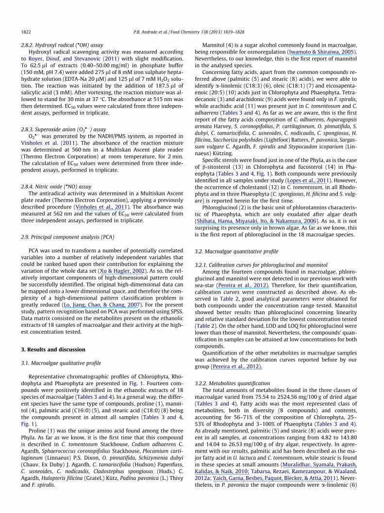
Fig. 1. Chromatographic profile of ethanolic extracts of (A) Codium adhaerens (Chlorophyta), (B) Plocamium cartilagineum (Rhodophyta), and (C) Cystoseira tamariscifolia(Phaeophyta). Identity of compounds as in Tables 3 and 4.
P.B. Andrade et al. / Food Chemistry 138 (2013) 1819–1828 1823
and oleic (7) acids instead of palmitic acid, as reported by Tabarsa,Rezaei, Ramezanpour, Robert Waaland, and Rabiei (2012b).
Concerning sterols, their contribution for the composition ofChlorophyta reaches 25%, which is similar to that of Rhodophyta(26%), while in Phaeophyta they represented up to 15% (Tables 3and 4). Under the conditions used in the present work only freesterols are detected, rendering difficult the comparison betweenour results and those reported in literature. The majority of theworks concerning macroalgae sterols composition involves othersolvents for extraction, purification steps are also included and freeand bounded sterols are quantified after saponification. Lopes et al.(2011) have used that procedure for the determination of the ster-ols profile of the same species studied herein. They found thatfucosterol was the predominant sterol in Chlorophyta and Pha-eophyta, while cholesterol was the main compound inRhodophyta.
The contribution of proline (1) for Chlorophyta, Rhodophyta andPhaeophyta reached 54% (Tables 3 and 4). This compound was de-scribed by Qasim (1991) as the major one in P. pavonica and U. lact-uca. However, it was not so abundant in S. polyshides (6%)(Sánchez-Machado, López-Cervantes, López-Hernández, Paseiro-Losada, & Simal-Lozano, 2003), and Sargassum vulgare C. Agardh(1%) (Ramos, Monteiro, Moreira, & Carvalho, 2000).
Mannitol (4) represents up to 9% of the compounds determinedin Chlorophyta, 47% in Rhodophyta and 59% in Phaeophyta. C.usneoides and U. lactuca were the richest and poorest species,respectively (Tables 3 and 4).
Phloroglucinol (2) was found in the three Cystoseira speciesunder study: C. tamariscifolia exhibited the highest amounts(23% of the total identified compounds), while in C. usneoidesand C. nodicaulis its contribution was 6% and 8%, respectively(Table 4).
3.3. Biological activities
3.3.1. Enzymatic assays3.3.1.1. a-Glucosidase inhibitory activity. a-Glucosidase is an impor-tant enzyme on dietary carbohydrate digestion and post-transla-tional processing of glycoproteins. Small intestinal a-glucosidaseis responsible for the hydrolysis of carbohydrates into glucose,which is absorbed through the gut wall to become the blood glu-cose and to cause post-prandial hyperglycaemia. It has been recog-nised that inhibitors of this enzyme can prevent some diseases,such as diabetes, obesity, hyperlipoproteinemia and hyperlipida-emia, as well as having antitumour and anti-HIV activity (Xiancui,Rongli, Xiao, Lijun, & Lixin, 2005). From the 18 macroalgae etha-nolic extracts tested, only five showed promising results (Table 5,Fig. 2A). The best dose–response activity was observed for C.tamariscifolia, with 25% of enzyme inhibition at 0.05 mg dry al-gae/ml. As can be observed in Fig. 2A and Table 5, the remainingspecies were less effective enzyme inhibitors. The inhibition of thisenzyme by macroalgae has been addressed to purified extracts,such as the polysaccharide and polyphenolic-enriched fractionsof Ascophyllum nodosum (Linnaeus) Le Jolis and Alaria esculenta(Linnaeus) Greville (Phaeophyta), U. lactuca (Chlorophyta) andPalmaria palmata (Linnaeus) Weber and Mohr (Rhodophyta) (Apos-tolidis, Karayannakidis, Kwon, Lee, & Seeram, 2011; Zhang et al.,2007; Nwosu et al., 2011) and to bromophenols from Grateloupiaelliptica Holmes (Rhodophyta) (Kim, Nam, Kurihara, & Kim,2008). To the best of our knowledge, this is the first report onthe a-glucosidase inhibitory activity of C. tamariscifolia, C. nodicau-lis, F. spiralis, H. filicina and S. polyshides.
3.3.1.2. Acetylcholinesterase and butyrylcholinesterase inhibitoryactivity. Alzheimer’s disease (AD) is an irreversible, progressive

Table 3Metabolic composition of Chlorophyta and Rhodophyta samples (mg/100 g dry algae)a.
Compounds RIcalcb RIlit
c Chlorophyta Rhodophyta
C. tomentosum C. adhaerens U. lactuca A. armata S. coronopifolius P. cartilagineum O. pinnatifida S. dubyi
1 Proline 1286 1299 a 46.62 (2.14) 22.88 (2.02) 24.08 (1.08) – – 146.78 (8.12) 19.64 (3.00) 15.32 (1.40)2 Phloroglucinol 1631 1647 b – – – – – – – –3 C14:0 1837 1843 b – – – – – – – –4 Mannitol 1896 1912 b 34.8 0 (0.20) 34.32 (0.04) <LOQ 34.70 (0.32) 35.62 (0.86) 40.90 (5.00) 41.18 (0.50) 35.02 (0.12)5 C16:0 2043 2040 b 70.40 (7.16) 143.80 (10.36) 38.50 (8.04) 33.38 (6.58) 4.82 (0.04) 41.26 (2.96) 40.52 (1.64) 65.34 (10.84)6 C18:3 2225 2218 c 56.30 (1.44) 25.78 (2.52) – – – – – –7 C18:1 2238 2248 b 51.08 (1.79) 19.04 (1.02) – – – – – –8 C18:0 2253 2234 c 19.76 (1.28) 21.62 (1.88) 21.30 (1.98) 20.88 (2.18) 21.80 (1.86) 26.52 (3.24) 18.56 (1.00) 20.20 (2.92)9 C20:4 2389 2373 b – – – – – – – –10 C20:5 2398 2380 b – 18.08 (0.24) – – – – – –11 C20:0 2456 – 22.48 (2.08) 28.36 (1.22) – – – – – –12 Cholestanol 3207 – 10.34 (0.46) – – 17.70 (0.60) 13.88 (0.50) 15.68 (0.80) 41.22 (1.28) 24.60 (2.24)13 b-Sitosterol 3355 3342 d 78.44 (9.78) 104.52 (3.70) <LOQ – – – – –14 Fucosterol 3361 3330 e – – – – – – – –
a Values are expressed as mean (SD) of three determinations.b RIcalc: Retention index obtained in this experiments.c RILit: Retention index described in literature; a – Meyer et al. (2009); b – NIST chemistry webbook (http://webbook.nist.gov/chemistry/name–ser.html); c – Isidorov and Vinogorova (2003); d – Radulovic and Dordevic (2011); e
– Shepherd et al. (2007).
Table 4Metabolic composition of Phaeophyta samples (mg/100 g dry algae)a.
Compounds RIcalcb RIlit
c Phaeophyta
C. tamariscifolia C. usneoides C. nodicaulis C. spongiosus H. filicina S. polyschides P. pavonica S. vulgare F. spiralis S. scoparium
1 Proline 1286 1299 a 33.82 (2.22) 32.56 (7.32) 62.66 (8.20) 56.88 (0.56) 64.94 (4.84) 11.10 (1.38) 13.48 (2.78) 11.74 (1.10) 39.68 (9.68) –2 Phloroglucinol 1631 1647 b 55.66 (1.08) 21.20 (0.24) 20.94 (0.28) – – – 21.02 (0.06) – <LOQ –3 C14:0 1837 1843 b – – – – – – – – 6.98 (0.27) –4 Mannitol 1896 1912 b 66.96 (6.76) 198.44 (39.32) 130.36 (8.56) 40.88 (2.06) 35.84 (0.24) 113.60 (5.04) 38.32 (1.58) 39.46 (1.52) 127.38 (3.48) –5 C16:0 2043 2040 b 29.60 (5.96) 20.50 (3.62) 9.12 (3.20) 76.42 (0.72) 72.44 (7.60) 127.72 (18.52) 12.16 (3.38) 34.04 (9.50) 60.60 (2.06) 48.66 (12.16)6 C18:3 2225 2218 c – – – – – – 186.02 (2.20) 241.36 (5.76) – –7 C18:1 2238 2248 b – – – 419.66 (10.56) – – 146.92 (4.88) 284.86 (7.12) 2169.06 (166.76) –8 C18:0 2253 2234 c 25.54 (1.20) 17.58 (0.92) – – 26.32 (3.20) 20.34 (0.26) 14.04 (2.42) 20.40 (2.60) 20.84 (2.14) 26.88 (4.13)9 C20:4 2389 2373 b – – – – – – – – 46.56 (2.90) –10 C20:5 2398 2380 b – – – – – – – – 21.70 (1.14) –11 C20:0 2456 – – – – – – – – – – –12 Cholestanol 3207 – – – – 15.40 (0.68) 17.28 (1.40) – – 12.74 (1.16) – –13 b–Sitosterol 3355 3342 d – – – – – – – – – –14 Fucosterol 3361 3330 e 34.62 (3.44) 43.36 (6.40) 39.42 (1.88) 1.00 (0.26) 2.80 (0.24) 20.74 (1.64) 16.48 (0.56) 25.76 (4.36) 31.76 (0.94) –
a Values are expressed as mean (SD) of three determinations.b RIcalc: Retention index obtained in this experiments.c RIlit: Retention index described in literature; a – Meyer et al. (2009); b – NIST chemistry webbook (http://webbook.nist.gov/chemistry/name-ser.html); c – Isidorov and Vinogorova (2003); d – Radulovic and Dordevic (2011); e
– Shepherd et al. (2007).
1824P.B.A
ndradeet
al./FoodChem
istry138
(2013)1819–
1828

Table 5Inhibition of a-glucosidase and cholinesterases and antiradical capacity of the testedextracts.
Species Enzyme Radical
a-Gluc AChE BuChE DPPH� �OH O2�� �NO
ChlorophytaC. tomentosum – – – – – – 1.25b
C. adhaerens – – – – – – 0.26b
U. lactuca – – – – – – –
RhodophytaA. armata – – – – – – 14.33b
S. coronopifolius – – – – – – –P. cartilagineum – – – – – – 4.62b
O. pinnatifida – – – – – 0.10b –S. dubyi – – – – – – 4.57b
PhaeophytaC. tamariscifolia 0.05a – 0.67b 0.58c 0.21b 2.94b 0.24b
C. usneoides – 0.53b 0.23b 1.96c 0.89b – 0.79b
C. nodicaulis 3.57a – – 4.94c 1.34b 2.56b 0.48b
C. spongiosus – – – – 0.54b – 3.19b
H. filicina 0.41a – – – – – 1.99b
S. polyschides 3.20a – – – 0.44b – 1.78b
P. pavonica – – – – – 0.41b 2.99b
S. vulgare – – – – 1.74b 1.36b 7.76b
F. spiralis 0.16a 4.67b – 2.49c 0.57b 2.87b 0.74b
S. scoparium – – – – – – 1.73b
a EC20 values (mg dry algae/ml) correspond to mean of three assays.b EC25 values (mg dry algae/ml) correspond to mean of three assays.c EC50 values (mg dry algae/ml) correspond to mean of three assays.
0.01 0.1 1 10
0
20
40
60
80
100C. tamariscifolia
F. spiralis
C. nodicaulisH. filicinaS. polyshides
α-G
luco
sida
se in
hibi
tion
(%)
0.001 0.01 0.1 1 10
0
20
40
60
80
100 C. usneoidesF. spiralis
AC
hE in
hibi
tion
(%)
0.01 0.1 1 10
0
20
40
60
80
100
C. usneoidesC. tamariscifolia
Log concentration (mg dry algae/mL)
BuC
hE in
hibi
tion
(%)
A
B
C
Fig. 2. Enzymatic inhibition of ethanolic extracts of macroalgae samples towards a-glucosidase (A), acetylcholinesterase (B) and butyrylcholinesterase (C). Resultsshow mean ± SEM of three experiments performed in triplicate.
P.B. Andrade et al. / Food Chemistry 138 (2013) 1819–1828 1825
neurodegenerative disorder, which occurs gradually, resulting inmemory loss, behaviour disturbances, personality changes and adecline in cognitive abilities; it is also the most common cause ofdementia among the elderly. Nowadays, different therapeuticstrategies can be used in AD patients, as the use of cholinomimeticagents, for the activation of cholinergic functions, and inhibitors ofcholinesterases (acetyl- and butyrylcholinesterase) in order to in-crease the levels of acetylcholine in the synaptic fend. Inhibitorsof cholinesterases are currently the most promising approach,since these enzymes are associated with the aetiology of AD(Lléo, 2007). Therefore, extracts able to inhibit both enzymesmay result in a higher efficacy on AD treatment, and in the caseof inhibition of butyrylcholinesterase it can also prevent the forma-tion of new b-amyloid plaques (Çokugras�, 2003). Concerning AChEinhibition, a dose–response behaviour was observed for C. usneo-ides and F. spiralis, though the first was more effective (Table 5,Fig. 2B). The same trend was observed for BuChE inhibitory activ-ity, better results being obtained with C. usneoides than with C.tamariscifolia (Table 5, Fig. 2C). Pangestuti and Kim (2011) have re-cently reviewed the cholinesterases inhibitory potential of marinealgae, indicating that different species of Chlorophyta, Rhodophytaand Phaeophyta were able to remarkably inhibit AChE. Moreover,sterols and phlorotannins from Ecklonia stolonifera Okamura havealso been reported as cholinesterases inhibitors (Yoon et al.,2008). As far as we know, this is the first report on cholinesterasesinhibitory activities of C. usneoides, C. tamariscifolia and F. spiralis.
3.3.2. Antioxidant activitiesThe eighteen macroalgae ethanol extracts were evaluated as
radical scavengers against DPPH�, �OH, O2�� and �NO. A dose-depen-
dent response was observed for all assays (Table 5, Fig. 3 and 4).Among the tested extracts, five were able to scavenge DPPH� in
a dose-dependent manner (Fig. 3A). As observed in Table 5 andFigs. 3 and 4A, the capacity of inhibiting 50% of DPPH� followsthe order C. tamariscifolia > C. usneoides > F. spiralis > C. nodicaulis.For O. pinnatifida we were only able to calculate its EC25 (0.56 mgdry algae/ml) since for concentrations higher than 2.77 mg dry al-gae/ml the scavenging capacity decreased. The DPPH� scavenging
capacity of different extracts of C. tamariscifolia and F. spiralis hasalready been reported (Paiva, Patarra, Neto, Lima, & Baptista,2011; Zubia et al., 2009a; Zubia et al., 2009b), but, as far as weknow, that of C. usneoides, C. nodicaulis and C. spongiosus is de-scribed for the first time herein.
Hydroxyl radical is the most reactive oxygen species, which cancause enormous biological damage (Kohen & Nyska, 2002). Sevenout of eighteen macroalgae species, all of them from Phaeophyta,showed a concentration dependent activity against hydroxyl radi-cal (Table 5, Fig. 3B). As it happened for DPPH�, C. tamariscifolia wasthe most potent species. To the best of our knowledge, this is thefirst report on hydroxyl scavenging capacity of C. tamariscifolia, C.usneoides, C. nodicaulis, C. spongiosus, H. filicina, C. tomentosum, C.adhaerens, A. armata, S. coronopifolius, P. cartilagineum and O.pinnatifida.
Extracts that show scavenging activity over O2�� and �NO are
very important due to their role in preventing the formation ofother deleterious oxidative species (Pacher, Beckman, & Liaudet,2007). As displayed in Fig. 3C, six macroalgae samples were ableto scavenge O2
�� radicals in a dose-dependent way. The compari-son of the EC25 values (Table 5) enables us to order the scavengingpotential of the different species as follows: O. pinnatifida > P. pavo-nica > S. vulgare > C. nodicaulis > F. spiralis � C. tamariscifolia. Theactivity of O. pinnatifida against this reactive oxygen species is de-scribed herein for the first time.
Considering the extracts inhibitory potential against �NO, scav-enging activity was observed for fifteen of the samples (Fig. 4),

Fig. 3. Antiradical activity of macroalgae ethanolic extracts against (A) DPPH�, (B)hydroxyl, and (C) superoxide anion. Results show mean ± SEM of three experimentsperformed in triplicate. Fig. 4. Nitric oxide inhibition for (A) Chlorophyta, (B) Rhodophyta and (C)
Phaeophyta. Results show mean ± SEM of three experiments performed intriplicate.
1826 P.B. Andrade et al. / Food Chemistry 138 (2013) 1819–1828
with EC25 values ranging from 0.24 to 14.33 mg dry algae/ml (Ta-ble 5). Among them, C. tamariscifolia was the most effective species(Table 5). The nitric oxide scavenging ability of phlorotannins puri-fied extracts from C. nodicaulis, C. tamariscifolia, C. usneoides, F. spi-ralis and S. vulgare was recently described by Lopes et al. (2012).Moreover, C. tomentosum and P. cartilagineum aqueous extractswere also reported as potent nitric oxide scavengers (Valentão,Trindade, Gomes, de Pinho, Mouga, & Andrade, 2010). As far aswe are aware, this is the first report on the protective effects ofC. adhaerens, A. armata and S. dubyi against �NO.
3.4. Discrimination of macroalgae by PCA analysis
An increased interest in obtaining biologically active com-pounds from natural sources has been noticed in the last years,since they show low toxicity, are completely biodegradable, avail-able from renewable sources and in almost all cases obtained atlow costs (Fabricant & Farnsworth, 2001). Therefore, classifying ex-tracts according to their chemical composition and biological activ-ities can be valuable to understand if there is a relation betweenthem. In this sense, PCA analysis was applied considering macroal-gae metabolic composition and the bioactivity of the highest con-centration tested in each assay. As can be observed in Fig. 5A,54.60% of the variability could be explained by the first two dimen-sions: PC1 accounted for 35.40% of the variance, while PC2 wasresponsible for 19.20%. Four groups were distinguished (Fig. 5A).One group (G1) involves C. adhaerens and C. tomentosum, another
only contains F. spiralis (G2), the third is formed by the three Cys-toseira species (G3) and the last (G4) is the most heterogeneousone, including the other samples (Fig. 5A). According to Fig. 5B,G1 is chemically distinct from the other samples by the contentsof b-sitosterol, palmitic (C16:0) and arachidic (C20:0) acids, whileG2 is only by the fatty acids (tetradecanoic (C14:0), oleic (C18:1),arachidonic (C20:4) and eicosapentaenoic (C20:5)). Cystoseira spe-cies (G3) are grouped according to their contents of mannitol,phloroglucinol and fucosterol. The remaining twelve macroalgae(G4) are found in a same group due to their proline, cholestanoland stearic (C18:0) and a-linolenic (C18:3) acids amounts(Fig. 5B). The compounds behind the enzymatic and antiradicalactivities are displayed into the plans formed with PC1 positiveaxis (Fig. 5B). We can observe that samples with higher contentsof mannitol, phloroglucinol and fucosterol are closely related withcholinesterases (AChE and BuChE) and DPPH� inhibition. On theother hand, these compounds are also responsible for a-glucosi-dase inhibition, hydroxyl, superoxide, and nitric oxide scavenging,though in a less extent. Other compounds contribute to explain theresults obtained for both enzymatic and antiradicals assays, as isthe case of tetradecanoic (C14:0), oleic (C18:1), arachidonic(C20:4) and eicosapentaenoic (C20:5) acids (Fig. 5B).
In fact, the extracts activities can be partially explained by theircomposition. For instance, the higher contents of mannitol in thethree Cystoseira species and in F. spiralis (Table 4) can be relatedwith the activity observed towards cholinesterases, since this

Fig. 5. Projection of macroalgae (A) (variables: Codium tomentosum (Cot), Codiumadhaerens (CA), Ulva lactuca (UL), Asparagopsis armata (AA), Sphaerococcus coro-nopfolius (SC), Plocamium cartilagineum (PC), Osmundea pinnatifida (OP), Schizymeniadubyi (SD), Cystoseira tamariscifolia (CT), Cystoseira usneoides (CU), Cystoseiranodicaulis (CN), Cladostephus spongiosus (CS), Halopteris filicina (HF), Saccorhizapolyschides (SP), Padina pavonica (PP), Sargassum vulgare (SV), Fucus spiralis (FS),Stypocaulon scoparium (SS) and loadings (B) by chemical composition and bioac-tivities (variables: proline, mannitol, phloroglucinol, tetradecanoic, palmitic(C16:0), stearic (C18:0), oleic (C18:1), a-linolenic (C18:3), arachidic (C20:0),arachidonic (C20:4), eicosapentaenoic (C20:5) acids, cholestanol, b-sitosterol,fucosterol and total amount of compounds (Total), a-glucosidase (aGLUC), acetyl-cholinesterase (AChE), butyrylcholinesterase (BuChE), hydroxyl radical, DPPH�,superoxide radical (SO) and nitric oxide radical (NO)) into the plane composed bythe principal components PC1 and PC2 containing 54.6% of the total variance.
P.B. Andrade et al. / Food Chemistry 138 (2013) 1819–1828 1827
compound was described as inhibiting the serum cholinesterasesactivities in mice and rabbits, in a dose-dependent manner (Cui-Xia et al., 2004). Additionally, this compound can also be responsi-ble for the antioxidant capacity, since it is reported as a well knownfree radical scavenger, used as reference in the evaluation of the
activity against hydroxyl radical in different assays (Iwamoto &Shiraiwa 2005).
Phloroglucinol, another compound present only in Phaeophyta(Table 4), was described by Moon et al. (2011) as inhibiting 50%of a-glucosidase at 141.18 lM (=0.017 mg/ml). Therefore, the sam-ple with higher content of phloroglucinol, C. tamariscifolia (0.23 mgdry algae/ml), was able to reduce more efficiently this enzyme. Be-sides, phloroglucinol was also reported as radical scavenger againstDPPH�, hydrogen peroxide and hydroxyl radical (Kang et al., 2006).Again, the samples with higher phloroglucinol content (Table 4)were those with higher activity towards the radicals tested(Table 5).
The amount of fucosterol in Phaeophyta samples also helpsexplaining our results. This compound has been established asthe responsible for the antidiabetic activity observed in streptozo-tocin-induced diabetic rats (Lee, Shin, Kim, & Lee, 2004). Concern-ing its cholinesterases inhibitory activity, Yoon et al. (2008) havefound that fucosterol is a selective inhibitor of BuChE(EC50 = 421.72 lM or 0.17 lg/ml). Thus, we can speculate thatthe higher BuChE activity observed for C. usneoides is in agreementwith its higher fucosterol content.
As referred above, fatty acids were also implicated in the enzy-matic and anti-radical activities (Fig. 5). For instance, oleic acid at18.22 mg/ml can inhibit 50% of a-glucosidase activity (Miyazawa,Yagi, & Taguchi, 2005). Moreover, oleic, arachidonic and eicosapen-taenoic acids may also be involved in AChE inhibition. As reportedby Ren, Houghton, and Hider (2006), fatty acids like oleic, arachi-donic and eicosapentaenoic acids can effectively inhibit AChE ina dose-dependent manner. Besides the ones characterised, wecan also consider that other compounds present in the extract,but not determined in this work, may also be responsible for theobserved activities.
As far as we know, it is the first time that a relationship betweenchemical composition and biological activities of extracts fromthese 18 macroalgae species is established.
4. Conclusions
The chemical composition of the analysed macroalgae speciesand their biological potential revealed to be clearly correlated.According to its higher content in biologically active compounds,like phloroglucinol, mannitol, fatty acids and fucosterol,Phaeophyta was the most promising group of macroalgae, thus dis-playing better results against enzymes and free radicals. Notwith-standing all of the algae species studied in this work are edible,according to the abilities displayed as radical scavengers and cho-linesterases’ inhibitors, they are potential candidates for food prod-ucts, nutraceutical and pharmaceutical preparations, as additives.Moreover, as F. spiralis is an edible species with great pharmacolog-ical properties, its consumption should be encouraged.
Acknowledgments
The authors are grateful to Fundação para a Ciência e a Tecno-logia (FCT) for Grant No. PEst-C/EQB/LA0006/2011. G. Lopes(SFRH/BD/61565/2009) is indebted to FCT, FSE and POPH for thegrant.
References
Apostolidis, E., Karayannakidis, P., Kwon, Y.-I., Lee, C., & Seeram, N. (2011). Seasonalvariation of phenolic antioxidant-mediated a-glucosidase inhibition ofAscophyllum nodosum. Plant Foods for Human Nutrition, 66, 313–319.
Awad, N. E. (2002). Volatile constituents, carbohydrate compositions and topicalanti-inflammatory of the two green algae Caulerpa racemosa (O. Dargent) andCodium tomentosum (Stackhouse). Bulletin of the Faculty of Pharmacy (CairoUniversity), 20, 233–243.

1828 P.B. Andrade et al. / Food Chemistry 138 (2013) 1819–1828
Bocanegra, A., Bastida, S., Benedí, J., Ródenas, S., & Sánchez-Muniz, F. J. (2009).Characteristics and nutritional and cardiovascular-health properties ofseaweeds. Journal of Medicinal Food, 12, 236–258.
Cardozo, K. H. M., Guaratini, T., Barros, M. P., Falcão, V. R., Tonon, A. P., Lopes, N. P.,et al. (2007). Metabolites from algae with economical impact. ComparativeBiochemistry and Physiology Part C: Toxicology and Pharmacology, 146, 60–78.
Çokugras�, A. N. (2003). Butyrylcholinesterase: Structure and physiologicalimportance. Turkish Journal of Biochemistry, 28, 54–61.
Cui-xia, L., Ji-liang, W., Nan, J., Bei, J., Hong-tao, M., Tao, J., et al. (2004). Effect ofmannitol on the activity of serum cholinesterases in the mouse and rabbit.Herald of Medicine, 23, 716–717.
El Gamal, A. A. (2010). Biological importance of marine algae. Saudi PharmaceuticalJournal, 18, 1–25.
Fabricant, D. S., & Farnsworth, N. R. (2001). The value of plants used in traditionalmedicine for drug discovery. Environmental Health Perspectives, 109, 69–75.
Gómez-Romero, M., Segura-Carretero, A., & Fernández-Gutiérrez, A. (2010).Metabolite profiling and quantification of phenolic compounds in methanolextracts of tomato fruit. Phytochemistry, 71, 1848–1864.
Grosso, C., Vinholes, J., Valentão, P., & Andrade, P. B. (2011). Halogenatedcompounds from seaweed, a biological overview. In F. Columbus (Ed.),Seaweed: Ecology, nutrient composition and medicinal uses (pp. 1–25). NewYork, USA: Nova Science Publishers.
Humston, E., Dombek, K., Tu, B., Young, E., & Synovec, R. (2011). Toward a globalanalysis of metabolites in regulatory mutants of yeast. Analytical andBioanalytical Chemistry, 401, 2387–2402.
International Conference on Harmonization (ICH) of technical requirements for theregistration of pharmaceuticals for human use, validation of analyticalprocedures. (1996). Text and methodology. ICH-Q2B, Geneva.
Isidorov, V. A., & Vinogorova, V. T. (2003). GC–MS analysis of compounds extractedfrom buds of Populus balsamifera and Populus nigra. Zeitschrift für NaturforschungC, 58, 355–360.
Iwamoto, K., & Shiraiwa, Y. (2005). Salt-regulated mannitol metabolism in algae.Marine Biotechnology, 7, 407–415.
Kang, K. A., Lee, K. H., Chae, S., Zhang, R., Jung, M. S., Ham, Y. M., et al. (2006).Cytoprotective effect of phloroglucinol on oxidative stress induced cell damagevia catalase activation. Journal of Cellular Biochemistry, 97, 609–620.
Kim, K. Y., Nam, K. A., Kurihara, H., & Kim, S. M. (2008). Potent a-glucosidaseinhibitors purified from the red alga Grateloupia elliptica. Phytochemistry, 69,2820–2825.
Kim, S., Shin, M., Hossain, M., Yun, E., Lee, H., & Kim, K. (2011). Metabolite profilingof sucrose effect on the metabolism of Melissa officinalis by gaschromatography–mass spectrometry. Analytical and Bioanalytical Chemistry,399, 3519–3528.
Kohen, R., & Nyska, A. (2002). Oxidation of biological systems: Oxidative stressphenomena, antioxidants, redox reactions, and methods for theirquantification. Toxicologic Pathology, 30, 620–650.
Lee, Y. S., Shin, K. H., Kim, B. K., & Lee, S. (2004). Anti-diabetic activities of fucosterolfrom Pelvetia siliquosa. Archives of Pharmacal Research, 27, 1120–1122.
Liu, Y., Chen, T., Qiu, Y., Cheng, Y., Cao, Y., Zhao, A., et al. (2011). An ultrasonication-assisted extraction and derivatization protocol for GC/TOFMS-based metaboliteprofiling. Analytical and Bioanalytical Chemistry, 400, 1405–1417.
Lléo, A. (2007). Current therapeutic options for Alzheimer’s disease. CurrentGenomics, 8, 550–558.
Lo, T. C. T., Jiang, Y. H., Chao, A. L. J., & Chang, C. A. (2007). Use of statistical methodsto find the polysaccharide structural characteristics and the relationshipsbetween monosaccharide composition ratio and macrophage stimulatoryactivity of regionally different strains of Lentinula edodes. Analytica ChimicaActa, 584, 50–56.
Lopes, G., Sousa, C., Bernardo, J., Andrade, P. B., Valentão, P., Ferreres, F., et al. (2011).Sterol profiles in 18 macroalgae of the Portuguese coast. Journal of Phycology, 47,1210–1218.
Lopes, G., Sousa, C., Silva, L. R., Pinto, E., Andrade, P. B., Bernardo, J., et al. (2012). Canphlorotannins purified extracts constitute a novel pharmacological alternativefor microbial infections with associated inflammatory conditions? PLoS ONE, 7,e31145.
Meyer, E. H., Tomaz, T., Carroll, A. J., Estavillo, G., Delannoy, E., Tanz, S. K., et al.(2009). Remodeled respiration in ndufs4 with low phosphorylation efficiencysuppresses Arabidopsis germination and growth and alters control ofmetabolism at night. Plant Physiology, 151, 603–619.
Miyazawa, M., Yagi, N., & Taguchi, K. (2005). Inhibitory compounds of alpha-glucosidase activity from Arctium lappa L. Journal of Oleo Science, 54, 589–594.
Moon, H. E., Islam, M. N., Ahn, B. R., Chowdhury, S. S., Sohn, H. S., Jung, H. A., et al.(2011). Protein tyrosine phosphatase 1B and alpha-glucosidase inhibitoryphlorotannins from edible brown algae, Ecklonia stolonifera and Eiseniabicyclis. Bioscience, Biotechnology, and Biochemistry, 75, 1472–1480.
Muralidhar, A. P., Syamala, K., Prakash, C., Kalidas, C., & Naik, P. R. (2010).Comparative studies on fatty acid composition of three marine macroalgaecollected from Madapam region: South East Coast of India. World AppliedSciences Journal, 11, 958–965.
Na Jom, K., Frank, T., & Engel, K.-H. (2011). A metabolite profiling approach to followthe sprouting process of mung beans (Vigna radiata). Metabolomics, 7, 102–117.
Nwosu, F., Morris, J., Lund, V. A., Stewart, D., Ross, H. A., & McDougall, G. J. (2011).Anti-proliferative and potential anti-diabetic effects of phenolic-rich extractsfrom edible marine algae. Food Chemistry, 126, 1006–1012.
Pacher, P., Beckman, J. S., & Liaudet, L. (2007). Nitric oxide and peroxynitrite inhealth and disease. Physiological Reviews, 87, 315–424.
Paiva, L. S., Patarra, R. F., Neto, A. I., Lima, E. M. C., & Baptista, J. A. B. (2011).Antioxidant activity of macroalgae from the Azores Arquipelago. Life and MarineSciences, 29, 01–06.
Pangestuti, R., & Kim, S.-K. (2011). Neuroprotective effects of marine algae. MarineDrugs, 9, 803–818.
Pereira, D. M., Vinholes, J., Guedes de Pinho, P., Valentão, P., Mouga, T., Teixeira, N.,et al. (2012). A gas chromatography-mass spectrometry multi-target methodfor the simultaneous analysis of three classes of metabolites in marineorganisms. Talanta, 100, 391–400.
Qasim, R. (1991). Amino acid composition of some common seaweeds. PakistanJournal of Pharmaceutical Sciences, 4, 49–54.
Radulovic, N. S., & DorDevic, N. (2011). Steroids from poison hemlock (Coniummaculatum L.): A GC–MS analysis. Journal of the Chemical Serbian Society, 76,1471–1483.
Ramos, M. V., Monteiro, A. C. O., Moreira, R. A., & Carvalho, A. F. F. U. (2000). Aminoacid composition of some Brazilian seaweed species. Journal of FoodBiochemistry, 24, 33–39.
Ren, Y., Houghton, P., & Hider, R. C. (2006). Relevant activities of extracts andconstituents of animals used in traditional Chinese medicine for central nervoussystem effects associated with Alzheimer’s disease. Journal of Pharmacy andPharmacology, 58, 989–996.
Royer, M., Diouf, P. N., & Stevanovic, T. (2011). Polyphenol contents and radicalscavenging capacities of red maple (Acer rubrum L.) extracts. Food and ChemicalToxicology, 49, 2180–2188.
Sánchez-Machado, D. I., López-Cervantes, J., López-Hernández, J., Paseiro-Losada, P.,& Simal-Lozano, J. (2003). High-performance liquid chromatographic analysis ofamino acids in edible seaweeds after derivatization with phenyl isothiocyanate.Chromatographia, 58, 159–163.
Shahnaz, L., & Shameel, M. (2009). Chemical composition and bioactivity of somebenthic algae from Karachi coast of Pakistan. International Journal of Algae, 11,377–393.
Shepherd, T., Dobson, G., Verrall, S., Conner, S., Griffiths, D., McNicol, J., et al. (2007).Potato metabolomics by GC–MS: What are the limiting factors? Metabolomics, 3,475–488.
Shibata, T., Hama, Y., Miyasaki, T., Ito, M., & Nakamura, T. (2006). Extracellularsecretion of phenolic substances from living brown algae. Journal of AppliedPhycology, 18, 787–794.
Sousa-Pinto, I. (1998). The seaweed resources of Portugal. In A. T. Critchley & M.Ohno (Eds.), Seaweed resources of the world (pp. 176–184). Yokosuka: JapanInternational Cooperation Agency.
Sumner, L. W., Mendes, P., & Dixon, R. A. (2003). Plant metabolomics: Large-scale phytochemistry in the functional genomics era. Phytochemistry, 62,817–836.
Tabarsa, M., Rezaei, M., Ramezanpour, Z., Robert Waaland, J., & Rabiei, R. (2012b).Fatty acids, amino acids, mineral contents, and proximate composition of somebrown seaweeds. Journal of Phycology, 48, 285–292.
Tabarsa, M., Rezaei, M., Ramezanpour, Z., & Waaland, J. R. (2012a). Chemicalcompositions of the marine algae Gracilaria salicornia (Rhodophyta) and Ulvalactuca (Chlorophyta) as a potential food source. Journal of the Science of Foodand Agriculture, 92, 2500–2506.
Valentão, P., Trindade, P., Gomes, D., de Pinho, P. G., Mouga, T., & Andrade, P. B.(2010). Codium tomentosum and Plocamium cartilagineum: Chemistry andantioxidant potential. Food Chemistry, 119, 1359–1368.
Vergouw, C. G., Botros, L. L., Roos, P., Lens, J. W., Schats, R., Hompes, P. G. A., et al.(2008). Metabolomic profiling by near-infrared spectroscopy as a tool to assessembryo viability: A novel, non-invasive method for embryo selection. HumanReproduction, 23, 1499–1504.
Vinholes, J., Grosso, C., Andrade, P. B., Gil-Izquierdo, A., Valentão, P., de Pinho, P. G.,et al. (2011). In vitro studies to assess the antidiabetic, anti-cholinesteraseand antioxidant potential of Spergularia rubra. Food Chemistry, 129,454–462.
Xiancui, L., Rongli, N., Xiao, F., Lijun, H., & Lixin, Z. (2005). Macroalage as a source ofalpha-glucosidase inhibitors. Chinese Journal of Oceanology and Limnology, 23,354–356.
Xu, J., & Hagler, A. (2002). Chemoinformatics and drug discovery. Molecules, 7,566–600.
Yaich, H., Garna, H., Besbes, S., Paquot, M., Blecker, C., & Attia, H. (2011). Chemicalcomposition and functional properties of Ulva lactuca seaweed collected inTunisia. Food Chemistry, 128, 895–901.
Yoon, N., Chung, H., Kim, H., & Choi, J. (2008). Acetyl- and butyrylcholinesteraseinhibitory activities of sterols and phlorotannins from Ecklonia stolonifera.Fisheries Science, 74, 200–207.
Zhang, J., Tiller, C., Shen, J., Wang, C., Girouard, G. S., Dennis, D., et al. (2007).Antidiabetic properties of polysaccharide- and polyphenolic-enriched fractionsfrom the brown seaweed Ascophyllum nodosum. Canadian Journal of Physiologyand Pharmacology, 85, 1116–1123.
Zubia, M., Fabre, M. S., Kerjean, V., & Deslandes, E. (2009b). Antioxidant andcytotoxic activities of some red algae (Rhodophyta) from Brittany coasts(France). Botanica Marina, 52, 268–277.
Zubia, M., Fabre, M. S., Kerjean, V., Lann, K. L., Stiger-Pouvreau, V., Fauchon, M., et al.(2009a). Antioxidant and antitumoural activities of some Phaeophyta fromBrittany coasts. Food Chemistry, 116, 693–701.