Fatty acid-binding protein 4 impairs the insulin-dependent nitric oxide pathway in vascular...
8
ORIGINAL INVESTIGATION Open Access Fatty acid-binding protein 4 impairs the insulin-dependent nitric oxide pathway in vascular endothelial cells Gemma Aragonès 1 , Paula Saavedra 1 , Mercedes Heras 1 , Anna Cabré 1 , Josefa Girona 1 and Lluís Masana 1,2* Abstract Background: Recent studies have shown that fatty acid-binding protein 4 (FABP4) plasma levels are associated with impaired endothelial function in type 2 diabetes (T2D). In this work, we analysed the effect of FABP4 on the insulin-mediated nitric oxide (NO) production by endothelial cells in vitro. Methods: In human umbilical vascular endothelial cells (HUVECs), we measured the effects of FABP4 on the insulin-mediated endothelial nitric oxide synthase (eNOS) expression and activation and on NO production. We also explored the impact of exogenous FABP4 on the insulin-signalling pathway (insulin receptor substrate 1 (IRS1) and Akt). Results: We found that eNOS expression and activation and NO production are significantly inhibited by exogenous FABP4 in HUVECs. FABP4 induced an alteration of the insulin-mediated eNOS pathway by inhibiting IRS1 and Akt activation. These results suggest that FABP4 induces endothelial dysfunction by inhibiting the activation of the insulin-signalling pathway resulting in decreased eNOS activation and NO production. Conclusion: These findings provide a mechanistic linkage between FABP4 and impaired endothelial function in diabetes, which leads to an increased cardiovascular risk. Keywords: Diabetes, Endothelium, Fatty acid-binding protein 4 (FABP4), Endothelial dysfunction, Insulin, Insulin-signalling pathway, Endothelial nitric oxide synthase (eNOS), Nitric oxide (NO) Introduction The adipose fatty acid-binding protein (FABP), also known as FABP4 and aP2, is one of the best charac- terised intracellular lipid transport proteins [1]. It belongs to the superfamily of low-molecular-weight intracellular lipid-binding proteins and plays a central regulatory role in energy metabolism and inflammation [2,3]. FABP4 is highly expressed in mature adipocytes and accounts for approximately 6 % of their soluble pro- tein. FABP4 is also found circulating in the plasma. In the last several years, much effort has been focused on uncovering the role of FABP4. However, neither the secretory pathways nor the functions of circulating FABP4 are known. We and other authors have shown that FABP4 levels are increased in obesity, metabolic syndrome (MS), type 2 diabetes (T2D), and familial combined hyperlipidaemia or lipodystrophy syndromes, and these levels are also closely correlated with adverse lipid profiles and insulin resistance [4-10]. In these stud- ies, serum FABP4 predicted the development of MS and atherosclerosis [11,12]. A recent study showed that although serum levels of both adipocyte and epidermal FABP had associations with MS, only FABP4 was significantly associated with increased cardiovascular risk in Chinese adults [13]. Moreover, increased plasma levels of FABP4 in non- elderly men were independently associated with the presence of coronary artery disease [14]. A recent study showed that FABP4 had a direct impact on decreasing * Correspondence: [email protected] 1 Research Unit on Lipids and Atherosclerosis, Universitat Rovira i Virgili, IISPV, Spanish Biomedical Research Centre in Diabetes and Associated Metabolic Disorders (CIBERDEM), Reus, Spain 2 Vascular Medicine and Metabolism Unit, Research Unit on Lipids and Atherosclerosis, Sant Joan University Hospital, Universitat Rovira i Virgili, C. Sant Llorenç 21, Reus 43201, Spain Full list of author information is available at the end of the article CARDIO VASCULAR DIABETOLOGY © 2012 Aragonès et al.; licensee BioMed Central Ltd. This is an Open Access article distributed under the terms of the Creative Commons Attribution License (http://creativecommons.org/licenses/by/2.0), which permits unrestricted use, distribution, and reproduction in any medium, provided the original work is properly cited. Aragonès et al. Cardiovascular Diabetology 2012, 11:72 http://www.cardiab.com/content/11/1/72
Transcript of Fatty acid-binding protein 4 impairs the insulin-dependent nitric oxide pathway in vascular...

CARDIOVASCULAR DIABETOLOGY
Aragonès et al. Cardiovascular Diabetology 2012, 11:72http://www.cardiab.com/content/11/1/72
ORIGINAL INVESTIGATION Open Access
Fatty acid-binding protein 4 impairs theinsulin-dependent nitric oxide pathway invascular endothelial cellsGemma Aragonès1, Paula Saavedra1, Mercedes Heras1, Anna Cabré1, Josefa Girona1 and Lluís Masana1,2*
Abstract
Background: Recent studies have shown that fatty acid-binding protein 4 (FABP4) plasma levels are associatedwith impaired endothelial function in type 2 diabetes (T2D). In this work, we analysed the effect of FABP4 on theinsulin-mediated nitric oxide (NO) production by endothelial cells in vitro.
Methods: In human umbilical vascular endothelial cells (HUVECs), we measured the effects of FABP4 on theinsulin-mediated endothelial nitric oxide synthase (eNOS) expression and activation and on NO production. We alsoexplored the impact of exogenous FABP4 on the insulin-signalling pathway (insulin receptor substrate 1(IRS1) and Akt).
Results: We found that eNOS expression and activation and NO production are significantly inhibited byexogenous FABP4 in HUVECs. FABP4 induced an alteration of the insulin-mediated eNOS pathway by inhibitingIRS1 and Akt activation. These results suggest that FABP4 induces endothelial dysfunction by inhibiting theactivation of the insulin-signalling pathway resulting in decreased eNOS activation and NO production.
Conclusion: These findings provide a mechanistic linkage between FABP4 and impaired endothelial function indiabetes, which leads to an increased cardiovascular risk.
Keywords: Diabetes, Endothelium, Fatty acid-binding protein 4 (FABP4), Endothelial dysfunction, Insulin,Insulin-signalling pathway, Endothelial nitric oxide synthase (eNOS), Nitric oxide (NO)
IntroductionThe adipose fatty acid-binding protein (FABP), alsoknown as FABP4 and aP2, is one of the best charac-terised intracellular lipid transport proteins [1]. Itbelongs to the superfamily of low-molecular-weightintracellular lipid-binding proteins and plays a centralregulatory role in energy metabolism and inflammation[2,3]. FABP4 is highly expressed in mature adipocytesand accounts for approximately 6 % of their soluble pro-tein. FABP4 is also found circulating in the plasma. Inthe last several years, much effort has been focused on
* Correspondence: [email protected] Unit on Lipids and Atherosclerosis, Universitat Rovira i Virgili, IISPV,Spanish Biomedical Research Centre in Diabetes and Associated MetabolicDisorders (CIBERDEM), Reus, Spain2Vascular Medicine and Metabolism Unit, Research Unit on Lipids andAtherosclerosis, Sant Joan University Hospital, Universitat Rovira i Virgili, C.Sant Llorenç 21, Reus 43201, SpainFull list of author information is available at the end of the article
© 2012 Aragonès et al.; licensee BioMed CentCommons Attribution License (http://creativecreproduction in any medium, provided the or
uncovering the role of FABP4. However, neither thesecretory pathways nor the functions of circulatingFABP4 are known. We and other authors have shownthat FABP4 levels are increased in obesity, metabolicsyndrome (MS), type 2 diabetes (T2D), and familialcombined hyperlipidaemia or lipodystrophy syndromes,and these levels are also closely correlated with adverselipid profiles and insulin resistance [4-10]. In these stud-ies, serum FABP4 predicted the development of MS andatherosclerosis [11,12].A recent study showed that although serum levels of
both adipocyte and epidermal FABP had associationswith MS, only FABP4 was significantly associated withincreased cardiovascular risk in Chinese adults [13].Moreover, increased plasma levels of FABP4 in non-elderly men were independently associated with thepresence of coronary artery disease [14]. A recent studyshowed that FABP4 had a direct impact on decreasing
ral Ltd. This is an Open Access article distributed under the terms of the Creativeommons.org/licenses/by/2.0), which permits unrestricted use, distribution, andiginal work is properly cited.
Aragonès et al. Cardiovascular Diabetology 2012, 11:72 Page 2 of 8http://www.cardiab.com/content/11/1/72
the contractility of myocardial muscle cells, which sug-gested that the release of FABP4 in to the bloodstreamcould have a direct effect on some peripheral cells andtissues [15]. In addition, we recently demonstrated thathigh levels of plasma FABP4, as other inflammation med-iators, were associated with endothelial dysfunctionassessed by peripheral artery tonometry [16,17].Endothelial dysfunction is the first event in the patho-
genesis of atherosclerosis and refers to an imbalance in therelease of vasodilating molecules, such as nitric oxide(NO) and vasoconstricting factors. NO-dependent vaso-dilatation is thought to reflect endothelial function, and itsimpairment is predictive of future cardiovascular risk[18,19]. The insulin-signalling pathway in the vascularendothelium leads to the activation of endothelial nitricoxide synthase (eNOS) and an increased production ofNO. This pathway involves the insulin receptor-mediatedphosphorylation of insulin receptor substrate 1 (IRS1),which activates PI3-kinases that then phosphorylates andactivates Akt at Ser473. Akt directly phosphorylates eNOSat Ser1177, resulting in increased eNOS activation and NOproduction. Under pathological conditions, proinflamma-tory factors cause an impairment in this particular insulin-signalling pathway in the endothelium, which promotesendothelial dysfunction [20]. This impairment might berelated to the defective insulin signalling in the endothelialcells. Moreover, insulin may also activate the pro-atherogenic mitogen-activated protein kinase (MAPK)pathway in endothelial cells that leads to increases in theexpression of adhesion molecules and leukocyte adhesionto the vascular endothelium. In addition, experimental ani-mals with a vascular endothelial cell-specific insulin recep-tor deficiency show a reduction in the expression ofadhesion molecules and eNOS mRNA [21].Although the role of circulating FABP4 on the vascular
endothelium is unknown, a recent study has revealed thatthe elevated expression of intracellular FABP4 in endothe-lial cells contributes to their dysfunction through a reduc-tion of eNOS [22]. These data along with our ownobservations showing the influence of circulating FABP4on endothelial function warrants testing the hypothesisthat high levels of circulating FABP4 in altered metabolicconditions could modify the correct function of endothelialcells and cause endothelial dysfunction by impairments ofthe insulin-signalling pathway and NO production. If thishypothesis were correct, it would contribute to themechanisms by which circulating FABP4 contributes tovascular endothelial dysfunction in diabetes.
Materials and methodsCell culture and reagentsHuman umbilical vein endothelial cells (HUVECs) wereobtained from Cascade Biologics (Invitrogen Life Tech-nologies, UK). After thawing, cells were seeded in 75-cm2
flasks and cultured in medium 200 according to the suppli-er’s recommendations. Medium 200 was supplementedwith 2 % low serum growth supplement (LSGS) and 1 %Gentamicin/Amphotericin solution (Invitrogen Life Tech-nologies, UK). The cells were placed in a humidified incu-bator at 37 °C and 5 % CO2 until there were enough cellsavailable for experiments. The HUVEC cells were used atpassage 3 in the current study.Insulin was purchased from Sigma-Aldrich, Inc.
(USA). Human recombinant FABP4 and anti-FABP4antibody was from BioVendor (Czech Republic). Theanti-eNOS, anti-phospho-eNOS (Ser1177), anti-Akt, anti-phospho-Akt (Ser473), antibodies were from Cell Signal-ing Technology, Inc. (Beverly MA, USA). The anti-IRS1and anti-phospho-IRS1 (Tyr989) antibodies were fromSanta Cruz (California, USA), and the anti-IgG-HRPantibody was from Dako (Denmark).
Design of the studies to determine the FABP4 effectsEffect of FABP4 on eNOS activationConfluent cells were starved in 1 % low serum medium(without growth factors). To determine whether FABP4has any effect on the basal eNOS activation, we studiedeNOS phosphorylation at Ser1177. The HUVECs wereincubated with FABP4 (25–100 ng/ml) for 30 min in theendothelial starvation medium. The treated cells wererinsed with ice-cold PBS and lysed in lysis buffer, whichwas composed of 50 mM Tris–HCl, 150 mM NaCl,0.1 % SDS, 1 % Nonidet, 0.5 % deoxycholate and prote-ase inhibitors, and the cells were stored at −80 °C untilthey were processed. The total protein concentrationwas measured using a Bradford assay (BioRad, USA).Immunoblot analysis of eNOS phosphorylation atSer1177 and total eNOS was performed.We also examined the effect of FABP4 on the insulin-
stimulated eNOS phosphorylation at Ser1177. TheHUVECs were preincubated with FABP4 (25–100 ng/ml) for 30 min and were then stimulated with 600 nMinsulin for 30 min in the endothelial starvation medium.We used the same protocol described above to obtainthe samples. The total protein concentration was mea-sured using a Bradford assay. Immunoblot analysis ofeNOS phosphorylation at Ser1177 and total eNOS wasalso performed.
Effect of FABP4 on the insulin-signalling pathwayBecause insulin activates eNOS through IRS1 and Akt,we investigated whether FABP4 would have any effecton the insulin-stimulated IRS1 and Akt activation. Westudied the phosphorylation of IRS1 at Tyr989 and ofAkt at Ser473. The HUVECs were preincubated for30 min with FABP4 (25–100 ng/ml) before a 30-min in-sulin treatment (600 nM) in the starvation medium.Treated cells were collected and lysed in lysis buffer, and
Aragonès et al. Cardiovascular Diabetology 2012, 11:72 Page 3 of 8http://www.cardiab.com/content/11/1/72
then the lysates were stored at −80 °C until they wereprocessed. The total protein concentration was mea-sured using a Bradford assay. Immunoblot analysis ofIRS1 phosphorylation at Tyr989, total IRS1, Akt phos-phorylation at Ser473 and total Akt was performed.
Immunoblot analysis of eNOS phosphorylation at Ser1177,total eNOS, IRS1 phosphorylation at Tyr989, total IRS1, Aktphosphorylation at Ser473, and total Akt, and FABP4Electrophoresis and immunoblot analysis were per-formed using the NuPAGE Protein Analysis System(Invitrogen Life Technologies, UK). The membrane wasblocked with a 2 % ECL Advance Blocking Reagent(Amersham Biosciences, Fairfield, CT) and was incu-bated with anti-eNOS, anti-eNOS phosphorylation atSer1177, anti-Akt, anti-Akt phosphorylation at Ser473,anti-IRS1 phosphorylation at Tyr989, anti-IRS1, or anti-FABP4 antibodies. Antigen-antibody complexes weredetected by incubating the membrane with an IgG-HRPantibody. The bands were visualised using ECL reagents(Amersham Pharmacia, Fairfield, CT) with the VersaDocimage system and were quantified with the QuantityOne analysis software version 4.6.2 (Bio Rad, USA). Therelative levels of the phosphorylated proteins were quan-tified after being normalised to the total proteins andwere expressed as arbitrary units (AU).
Effect of FABP4 on eNOS mRNA expressionUpon insulin stimulation, the eNOS mRNA wasincreased in the vascular endothelial cells. Therefore, weexamined the mRNA expression of insulin-stimulatedeNOS from the FABP4-treated cell lysates. Confluentvascular cells were preincubated with FABP4 (50–100 ng/ml) for 30 min and then stimulated with 600 nMinsulin for 24 hours in a supplemented medium. TotalRNA was isolated from the cells using the ABI PRISM6100 Nucleic Acid PrepStation (Applied Biosystems,CA, USA). The absorbance at 260 nm was used to meas-ure the RNA concentration, and an absorbance ratio of260/280 nm was used to analyse the RNA quality.
Real-time quantitative PCR of eNOSTotal RNA (0.5 μg) was reverse transcribed to cDNA usingRandom Hexamers and SuperScript II (Invitrogen LifeTechnologies, UK) by following the manufacturer’s proto-col. TaqMan primers and probes for eNOS and GAPDHwere obtained from validated and pre-designed Assays-on-Demand products (Applied Biosystems, CA, USA) andwere used in real-time PCR amplifications. The mRNA ex-pression for each gene and sample was calculated usingthe recommended 2-ΔΔCt method. The control group (un-treated cells) was defined as the calibrator in this experi-ment. GAPDH was used as a housekeeping gene tonormalise the results of the gene of interest.
Nitric oxide assayCells were starved for 24 h with 1 % low serum medium(without growth factors) and then preincubated for 30-min with FABP4 (25–100 ng/ml) before a 30-min insulintreatment (600 nM) in the starvation medium. Thesupernatants were collected, and the detection of theNO2 and NO3 anions was performed with the Nitrate/Nitrite Colorimetric Assay kit (Cayman Chemical, AnnArbor, MI) using the Griess reaction and following man-ufacturer’s instructions. The absorbance of the solutionwas read on a spectrophotometer at 540 nm. To quan-tify the NO production, a standard nitrate curve wasgenerated in the same medium in which the experimentswere performed. The results were expressed as foldincreases with respect to the insulin treatment.
Statistical analysesThe results are represented as the means ± SD of at least3 separate experiments. Differences between the meanswere determined using a one-way analysis of variance(ANOVA), which was followed by a Dunnett’s post-hoctest for multiple comparisons. Differences were consid-ered significant at P< 0.05. The GraphPad Prism 5.0Software, Inc. was used for statistical analyses.
ResultsFABP4 inhibits eNOS activation and NO productionin HUVECsTo determine whether FABP4 have any effect on eNOS ac-tivity, we studied the eNOS activation measuring its phos-phorylation at Ser1177. We found that exposing HUVECs inthe endothelium growth medium (with serum and growthfactors that maintain the basal activation of eNOS) toFABP4 (25-100 ng/ml) inhibited eNOS phosphorylation atSer1177 in a concentration-dependent manner at as early as30-min of exposure. The maximal inhibition was achievedat 100 ng/ml of FABP4 (65 %; P< 0.05) (Figure 1A). Oninsulin stimulation, eNOS is phosphorylated by the acti-vation of PI3k/Akt pathway in vascular endothelial cells.First, we performed a dose–response curve with 100,300 and 600 nM insulin, and we observed that 600 nMinsulin was required to increase eNOS Ser1177 phosphor-ylation by 20 % (P< 0.05) (Figure 1B). Therefore, weperformed all of the experiments with 600 nM insulin.We also performed an insulin time course (10, 30,60 min), and we observed that the maximal phosphoryl-ation of eNOS at Ser1177 occurred 30 min after insulintreatment (26 % increase; P< 0.05) (Figure 1C). We nextanalysed whether FABP4 also impairs insulin–inducedeNOS activation and NO production. As shown inFigure 1D, the addition of insulin increases thephosphorylation of eNOS at Ser1177, but a 30-minpretreatment with FABP4 (25–100 ng/ml) inhibits thisinsulin-dependent increase by up to 45 % with the 100-

Figure 1 (See legend on next page.)
Aragonès et al. Cardiovascular Diabetology 2012, 11:72 Page 4 of 8http://www.cardiab.com/content/11/1/72

(See figure on previous page.)Figure 1 Effect of FABP4 on eNOS activation and NO production in HUVECs. A, effect of FABP4 on eNOS phosphorylation at Ser1177.HUVECs were incubated with FABP4 at the indicated concentrations for 30 minutes. B, eNOS Ser1177 phosphorylation in response to variousdoses of insulin (100, 300, 600 nM). HUVECs were incubated with insulin at the indicated concentrations for 30 minutes. C, Time course of eNOSSer1177 phosphorylation in response to insulin. HUVECs were incubated with 600 nM insulin for 10, 30 and 60 min. D, effect on insulin-stimulatedeNOS phosphorylation at Ser1177. HUVECs were incubated with FABP4 at the indicated concentrations for 30 minutes and then with insulin (600nM) for 30 minutes. E, effect of FABP4 on NO production. HUVECs were incubated with FABP4 at indicated concentrations for 30 minutes andthen stimulated with insulin (600 nM for 30 min). Representative blots are shown. The data are given as the mean± standard deviation fromthree independent experiments. *P< 0.05 vs. insulin(−) / FABP4(−); #P< 0.05 vs. insulin(+) / FABP4(−).
Figure 2 Effect of FABP4 on eNOS mRNA expression. HUVECswere incubated with FABP4 at the indicated concentrations for30 minutes and then with insulin (600 nM) for 24 hours. TNFα(10 ng/ml) was used as a negative control. The data are expressedusing the 2-ΔΔCt method. *P< 0.05 vs. insulin(−) / FABP4(−); #P< 0.05vs. insulin(+) / FABP4(−).
Aragonès et al. Cardiovascular Diabetology 2012, 11:72 Page 5 of 8http://www.cardiab.com/content/11/1/72
ng/ml concentration (P< 0.05) in the absence of anychanges in the total protein levels. Therefore, we exam-ined the effect of FABP4 on the ability of eNOS to pro-duce NO under insulin-stimulated conditions. Thechange in eNOS phosphorylation at Ser1177 was accom-panied by a significant decrease in the NO productionof up to 68 % by treatment with 100 ng/ml of FABP4 (P< 0.05) (Figure 1E). Thus, FABP4 can inhibit both thebasal and the insulin-stimulated eNOS phosphorylationat Ser1177 and can cause the inactivation of eNOS anddecrease NO production in endothelial cells.
FABP4 inhibits insulin-stimulated eNOS mRNA expressionin HUVECsUpon insulin stimulation, the eNOS mRNA wasincreased in the vascular endothelial cells. As wasexpected, insulin augmented the mRNA expression ofeNOS after 24 hours of treatment (P <0.05). The upre-gulation of insulin-stimulated eNOS expression wasinhibited by 63 % and 59 % due to the treatment with50 ng/ml and 100 ng/ml of FABP4, respectively(P< 0.05) (Figure 2). Additionally, we found a 93 % de-crease in the eNOS expression after an exposure toTNFα (10 ng/ml) as positive control (P< 0.05).
FABP4 effect on eNOS is produced through theinsulin-signalling pathway in HUVECsSince insulin activates eNOS through IRS1 and Aktpathway, we then investigated whether FABP4 have anyeffects on insulin-stimulated IRS1 and Akt activation,which was monitored by phosphorylation at Tyr989 andSer473 sites, respectively. The addition of insulin toHUVECs increases about 25 % the phosphorylation ofIRS1 at Tyr989 (Figure 3A), but a 30-min pretreatmentwith FABP4 (25–100 ng/ml) inhibits this insulin-dependent increase by up to 44 % with the 100-ng/mlconcentration (P< 0.05). No variations were observed inthe total IRS1 protein level across the different FABP4concentration treatments. There was also a similar effectof the FABP4 treatment on the phosphorylation of Aktat Ser473. The insulin stimulation significantly increasedthe phosphorylation of Akt at Ser473 (Figure 3B). TheFABP4 pretreatment impaired this insulin-mediatedphosphorylation of Akt at Ser473 by up to 75 % with the
100 ng/ml concentration (P< 0.05) in the absence ofany change in protein levels. Collectively, FABP4 inhib-ited insulin-stimulated phosphorylation of IRS1 and Akt,indicating possible impairment of upstream insulin-signalling pathway.In Figure 4, we analysed whether FABP4 was able to
be internalized within the endothelial cells. We observedthat incubation of HUVECs with 25–100 ng/ml FABP4for 30 min increased the amount of exogenous FABP4 inthe treated cells compared with the untreated cells(P< 0.05).
DiscussionThe present study demonstrates that exogenous FABP4induces endothelial cell dysfunction in vitro, as assessedby the impact on one of their main properties, the vaso-dilatory mechanisms. We have also shown that this ef-fect is mediated by the interaction of FABP4 with theinsulin-signalling pathway in vascular cells. FABP4 alterseNOS activation, as was demonstrated by the reduction

Figure 3 Effect of FABP4 on IRS1 and Akt activation in HUVECs. A, effect on insulin-stimulated IRS1 phosphorylation at Tyr989. B, effect oninsulin-stimulated Akt phosphorylation at Ser473. HUVECs were incubated with FABP4 at the indicated concentrations for 30 minutes and thenwith insulin (600 nM) for 30 minutes. Representative blots are shown. The data are given as the mean± standard deviation from threeindependent experiments. *P< 0.05 vs. insulin(−) / FABP4(−); #P< 0.05 vs. insulin(+) / FABP4(−).
Aragonès et al. Cardiovascular Diabetology 2012, 11:72 Page 6 of 8http://www.cardiab.com/content/11/1/72
of eNOS phosphorylation at Ser1177 and NO production.In addition, the reduction of IRS1 phosphorylation atTyr989 and Akt phosphorylation at Ser473 suggests thatthe effect observed for eNOS activation is due to theinterference of FABP4 in the insulin-signalling pathwayin endothelial cells. Our results support a direct effect ofextracellular FABP4 on vascular cells and, therefore, aputative effect of circulating FABP4 on peripheral tis-sues. This observation is important because FABP4 isrecognised as a biomarker of cardiometabolic risk but itcould be considered as mediator of peripheral tissuedamage. Several studies have linked FABP4 levels toobesity, T2D and MS [4,8,11]. Additionally, FABP4 hasbeen associated with the burden of coronary atheroma-tosis, but a causal role has not yet been established. Theonly report that showed a direct effect of FABP4 on cellsdemonstrated that FABP4 reduces the contractile cap-acity of cardiomyocytes [15]. We have previouslyreported that plasma FABP4 levels are associated withendothelial dysfunction in diabetic patients [16]. Ourpresent data supports a causal role of FABP4 in the dys-function of the vascular wall. These findings are in ac-cordance with a previous study suggesting that anelevated expression of intracellular FABP4 in vascular
endothelial cells contributes to endothelial dysfunctionboth in vivo and in vitro [22]. In contrast, a recent studyshowed that FABP4/5 inhibitors ameliorate dyslipidae-mia but not insulin resistance in diet-induced obesemice [23]. Our results show that exogenous FABP4 altersthe insulin-signalling pathway at its early activation steps.Additionally, it has already been shown that FABP4 inter-feres with the insulin receptor [24]. Moreover, FABP4could be phosphorylated on Tyr19 in response to insulinstimulation [25]. Our results support these previous obser-vations and extend them to the exogenous FABP4, whichhas a greater clinical implication. Along with a decrease ineNOS expression, in our hands, FABP4 increased the ex-pression of vascular cell adhesion protein 1 (VCAM1), E-selectin and leukocyte adhesion to endothelial cells (datanot shown), suggesting a more global effect of FABP4 onendothelial function. In our experiments, HUVECs werestimulated with 600nM insulin because that was the dosewith which we observed an increase in the phospho-eNOSproduced and because other authors also obtained max-imal nitric oxide production with similar dose [26].The mechanisms that FABP4 utilises to interfere with
the cell are currently being investigated. Although previ-ous studies showed that Heart-FABP (62 % homology
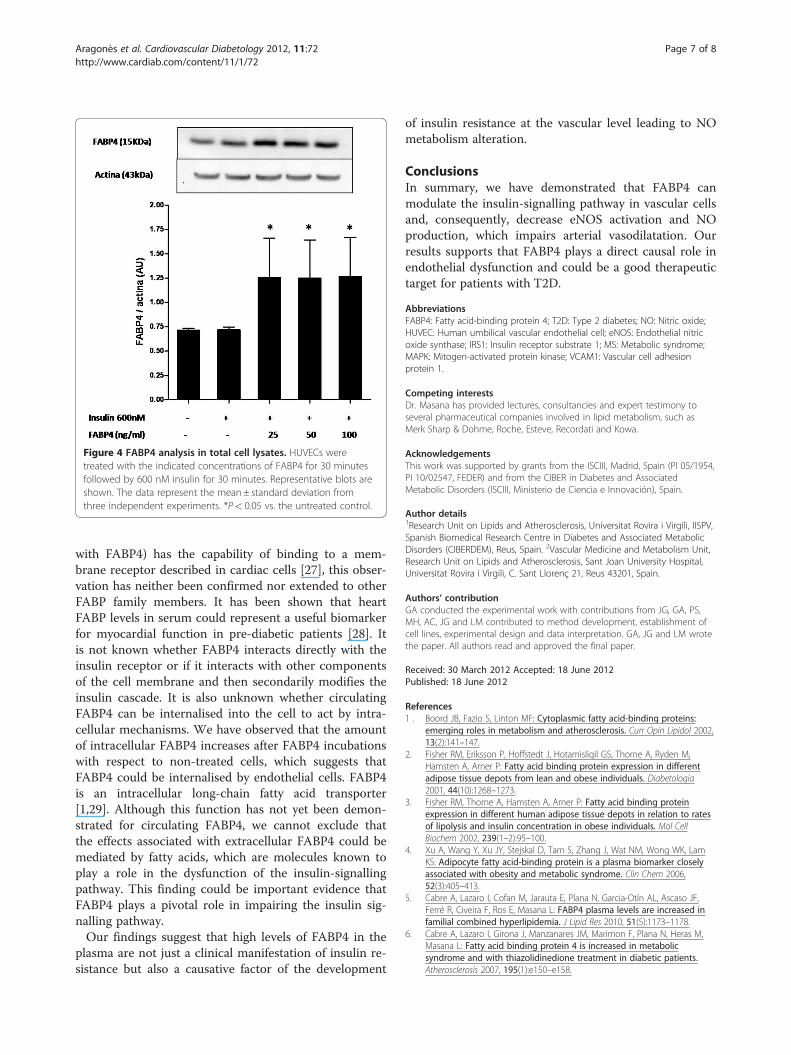
Figure 4 FABP4 analysis in total cell lysates. HUVECs weretreated with the indicated concentrations of FABP4 for 30 minutesfollowed by 600 nM insulin for 30 minutes. Representative blots areshown. The data represent the mean± standard deviation fromthree independent experiments. *P< 0.05 vs. the untreated control.
Aragonès et al. Cardiovascular Diabetology 2012, 11:72 Page 7 of 8http://www.cardiab.com/content/11/1/72
with FABP4) has the capability of binding to a mem-brane receptor described in cardiac cells [27], this obser-vation has neither been confirmed nor extended to otherFABP family members. It has been shown that heartFABP levels in serum could represent a useful biomarkerfor myocardial function in pre-diabetic patients [28]. Itis not known whether FABP4 interacts directly with theinsulin receptor or if it interacts with other componentsof the cell membrane and then secondarily modifies theinsulin cascade. It is also unknown whether circulatingFABP4 can be internalised into the cell to act by intra-cellular mechanisms. We have observed that the amountof intracellular FABP4 increases after FABP4 incubationswith respect to non-treated cells, which suggests thatFABP4 could be internalised by endothelial cells. FABP4is an intracellular long-chain fatty acid transporter[1,29]. Although this function has not yet been demon-strated for circulating FABP4, we cannot exclude thatthe effects associated with extracellular FABP4 could bemediated by fatty acids, which are molecules known toplay a role in the dysfunction of the insulin-signallingpathway. This finding could be important evidence thatFABP4 plays a pivotal role in impairing the insulin sig-nalling pathway.Our findings suggest that high levels of FABP4 in the
plasma are not just a clinical manifestation of insulin re-sistance but also a causative factor of the development
of insulin resistance at the vascular level leading to NOmetabolism alteration.
ConclusionsIn summary, we have demonstrated that FABP4 canmodulate the insulin-signalling pathway in vascular cellsand, consequently, decrease eNOS activation and NOproduction, which impairs arterial vasodilatation. Ourresults supports that FABP4 plays a direct causal role inendothelial dysfunction and could be a good therapeutictarget for patients with T2D.
AbbreviationsFABP4: Fatty acid-binding protein 4; T2D: Type 2 diabetes; NO: Nitric oxide;HUVEC: Human umbilical vascular endothelial cell; eNOS: Endothelial nitricoxide synthase; IRS1: Insulin receptor substrate 1; MS: Metabolic syndrome;MAPK: Mitogen-activated protein kinase; VCAM1: Vascular cell adhesionprotein 1.
Competing interestsDr. Masana has provided lectures, consultancies and expert testimony toseveral pharmaceutical companies involved in lipid metabolism, such asMerk Sharp & Dohme, Roche, Esteve, Recordati and Kowa.
AcknowledgementsThis work was supported by grants from the ISCIII, Madrid, Spain (PI 05/1954,PI 10/02547, FEDER) and from the CIBER in Diabetes and AssociatedMetabolic Disorders (ISCIII, Ministerio de Ciencia e Innovación), Spain.
Author details1Research Unit on Lipids and Atherosclerosis, Universitat Rovira i Virgili, IISPV,Spanish Biomedical Research Centre in Diabetes and Associated MetabolicDisorders (CIBERDEM), Reus, Spain. 2Vascular Medicine and Metabolism Unit,Research Unit on Lipids and Atherosclerosis, Sant Joan University Hospital,Universitat Rovira i Virgili, C. Sant Llorenç 21, Reus 43201, Spain.
Authors’ contributionGA conducted the experimental work with contributions from JG, GA, PS,MH, AC, JG and LM contributed to method development, establishment ofcell lines, experimental design and data interpretation. GA, JG and LM wrotethe paper. All authors read and approved the final paper.
Received: 30 March 2012 Accepted: 18 June 2012Published: 18 June 2012
References1 . Boord JB, Fazio S, Linton MF: Cytoplasmic fatty acid-binding proteins:
emerging roles in metabolism and atherosclerosis. Curr Opin Lipidol 2002,13(2):141–147.
2. Fisher RM, Eriksson P, Hoffstedt J, Hotamisligil GS, Thorne A, Ryden M,Hamsten A, Arner P: Fatty acid binding protein expression in differentadipose tissue depots from lean and obese individuals. Diabetologia2001, 44(10):1268–1273.
3. Fisher RM, Thorne A, Hamsten A, Arner P: Fatty acid binding proteinexpression in different human adipose tissue depots in relation to ratesof lipolysis and insulin concentration in obese individuals. Mol CellBiochem 2002, 239(1–2):95–100.
4. Xu A, Wang Y, Xu JY, Stejskal D, Tam S, Zhang J, Wat NM, Wong WK, LamKS: Adipocyte fatty acid-binding protein is a plasma biomarker closelyassociated with obesity and metabolic syndrome. Clin Chem 2006,52(3):405–413.
5. Cabre A, Lazaro I, Cofan M, Jarauta E, Plana N, Garcia-Otín AL, Ascaso JF,Ferré R, Civeira F, Ros E, Masana L: FABP4 plasma levels are increased infamilial combined hyperlipidemia. J Lipid Res 2010, 51(5):1173–1178.
6. Cabre A, Lazaro I, Girona J, Manzanares JM, Marimon F, Plana N, Heras M,Masana L: Fatty acid binding protein 4 is increased in metabolicsyndrome and with thiazolidinedione treatment in diabetic patients.Atherosclerosis 2007, 195(1):e150–e158.

Aragonès et al. Cardiovascular Diabetology 2012, 11:72 Page 8 of 8http://www.cardiab.com/content/11/1/72
7. Cabre A, Lazaro I, Girona J, Manzanares JM, Marimon F, Plana N, Heras M,Masana L: Plasma fatty acid-binding protein 4 increases with renaldysfunction in type 2 diabetic patients without microalbuminuria. ClinChem 2008, 54(1):181–187.
8. Cabre A, Lazaro I, Girona J, Manzanares JM, Marimon F, Plana N, Heras M,Masana L: Plasma fatty acid binding protein 4 is associated withatherogenic dyslipidemia in diabetes. J Lipid Res 2008, 49(8):1746–1751.
9. Coll B, Cabre A, Alonso-Villaverde C, Lazaro I, Aragones G, Parra S, Girona J,Masana L: The fatty acid binding protein-4 (FABP4) is a strong biomarkerof metabolic syndrome and lipodystrophy in HIV-infected patients.Atherosclerosis 2008, 199(1):147–153.
10. Stejskal D, Karpisek M: Adipocyte fatty acid binding protein in a Caucasianpopulation: a new marker of metabolic syndrome? Eur J Clin Invest 2006,36(9):621–625.
11. Xu A, Tso AW, Cheung BM, Wang Y, Wat NM, Fong CH, Yeung DC, JanusED, Sham PC, Lam KS: Circulating adipocyte-fatty acid binding proteinlevels predict the development of the metabolic syndrome: a 5-yearprospective study. Circulation 2007, 115(12):1537–1543.
12. Yeung DC, Xu A, Cheung CW, Wat NM, Yau MH, Fong CHK, Chau MT, LamKS: Serum adipocyte fatty acid-binding protein levels wereindependently associated with carotid atherosclerosis. Arterioscler ThrombVasc Biol 2007, 27(8):1796–1802.
13. Hong J, Gu W, Zhang Y, Yan Q, Dai M, Shi J, Zhai Y, Wang W, Li X, Ning G:Different association of circulating levels of adipocyte and epidermalfatty acid-binding proteins with metabolic syndrome and coronaryatherosclerosis in Chinese adults. Atherosclerosis 2011, 217(1):194–200.
14. Doi M, Miyoshi T, Hirohata S, Nakamura K, Usui S, Takeda K, Iwamoto M,Kusachi S, Kusano K, Ito H: Association of increased plasma adipocytefatty acid-binding protein with coronary artery disease in non-elderlymen. Cardiovasc Diabetol 2011, 10:44.
15. Lamounier-Zepter V, Look C, Alvarez J, Christ T, Ravens U, Schunck W,Ehrhart-Bornstein M, Bornstein SR, Morano I: Adipocyte fatty acid-bindingprotein suppresses cardiomyocyte contraction: a new link betweenobesity and heart disease. Circ Res 2009, 105(4):326–334.
16. Aragones G, Ferre R, Lazaro I, Cabre A, Plana N, Merino J, Heras M, Girona J,Masana L: Fatty acid-binding protein 4 is associated with endothelialdysfunction in patients with type 2 diabetes. Atherosclerosis 2010, 213(1):329–331.
17. Aragonès G, Ferré R, Girona J, Plana N, Merino J, Heras M, Masana L: Smallartery dilation and endothelial markers in cardiovascular risk patients.Eur J Clin Invest 2012, 42(1):34–41.
18. Hansson GK: Inflammation, atherosclerosis, and coronary artery disease.N Engl J Med 2005, 352(16):1685–1695.
19. Jansson PA: Endothelial dysfunction in insulin resistance and type 2diabetes. J Intern Med 2007, 262(2):173–183.
20. Tabit CE, Chung WB, Hamburg NM, Vita JA: Endothelial dysfunction indiabetes mellitus: molecular mechanisms and clinical implications. RevEndocr Metab Disord 2010, 11(1):61–74.
21. Vicent D, Ilany J, Kondo T, Naruse K, Fisher SJ, Kisanuki YY, Bursell S,Yanagisawa M, King GL, Kahn CR: The role of endothelial insulin signalingin the regulation of vascular tone and insulin resistance. J Clin Invest2003, 111(9):1373–1380.
22. Lee MY, Li H, Xiao Y, Zhou Z, Xu A, Vanhoutte PM: Chronic administrationof BMS309403 improves endothelial function in apolipoprotein E-deficient mice and in cultured human endothelial cells. Br J Pharmacol2011, 162(7):1564–1576.
23. Lan H, Cheng CC, Kowalski TJ, Pang L, Shan L, Chuang CC, Jackson J, Rojas-Triana A, Bober L, Liu L, Voigt J, Orth P, Yang X, Shipps GW Jr, Hedrick JA:Small-molecule inhibitors of FABP4/5 ameliorate dyslipidemia but notinsulin resistance in mice with diet-induced obesity. J Lipid Res 2011, 52(4):646–656.
24. Hresko RC, Hoffman RD, Flores-Riveros JR, Lane MD: Insulin receptortyrosine kinase-catalyzed phosphorylation of 422(aP2) protein. Substrateactivation by long-chain fatty acid. J Biol Chem 1990,265(34):21075–21085.
25. Nielsen SU, Spener F: Fatty acid-binding protein from rat heart isphosphorylated on Tyr19 in response to insulin stimulation. J Lipid Res1993, 34(8):1355–1366.
26. Zeng G, Quon MJ: Insulin-stimulated production of nitric oxide isinhibited by wortmannin. Direct measurement in vascular endothelialcells. J Clin Invest 1996, 98(4):894–898.
27. Burton PB, Hogben CE, Joannou CL, Clark AG, Hsuan JJ, Totty NF, SorensenC, Evans RW, Tynan MJ: Heart fatty acid binding protein is a novelregulator of cardiac myocyte hypertrophy. Biochem Biophys Res Commun1994, 205(3):1822–1828.
28. Karbek B, Ozbek M, Bozkurt NC, Ginis Z, Güngünes A, Ünsal IÖ, Cakal E,Delibası T: Heart-type fatty acid binding protein (H-FABP): relationshipwith arterial intima-media thickness and role as diagnostic marker foratherosclerosis in patients with ımpaired glucose metabolism. CardiovascDiabetol 2011, 10:37.
29. Fu Y, Luo L, Luo N, Garvey WT: Lipid metabolism mediated by adipocytelipid binding protein (ALBP/aP2) gene expression in human THP-1macrophages. Atherosclerosis 2006, 188(1):102–111.
doi:10.1186/1475-2840-11-72Cite this article as: Aragonès et al.: Fatty acid-binding protein 4 impairsthe insulin-dependent nitric oxide pathway in vascular endothelial cells.Cardiovascular Diabetology 2012 11:72.
Submit your next manuscript to BioMed Centraland take full advantage of:
• Convenient online submission
• Thorough peer review
• No space constraints or color figure charges
• Immediate publication on acceptance
• Inclusion in PubMed, CAS, Scopus and Google Scholar
• Research which is freely available for redistribution
Submit your manuscript at www.biomedcentral.com/submit