Fast and metastable fragmentation of deprotonated d-fructose – A combined experimental and...
8
International Journal of Mass Spectrometry 305 (2011) 50–57 Contents lists available at ScienceDirect International Journal of Mass Spectrometry jou rn al h om epa ge: www.elsevier.com/locate/ijms Fast and metastable fragmentation of deprotonated d-fructose –A combined experimental and computational study Helga D. Flosadottir, Ilko Bald ∗,1 , Oddur Ingólfsson ∗ University of Iceland, Department of Chemistry, Science Institute, Dunhagi 3, 107 Reykjavík, Iceland a r t i c l e i n f o Article history: Received 4 May 2011 Received in revised form 26 May 2011 Accepted 26 May 2011 Available online 12 June 2011 Keywords: Metastable decay Monosaccharides Matrix-assisted laser desorption and ionization Classical dynamics simulations Anions a b s t r a c t Metastable and prompt fragmentation of the deprotonated monosaccharide d-fructose was studied. By using the isotope labelled molecules 1- 13 C-d-fructose, 2- 13 C-d-fructose and 6- 13 C-d-fructose the origin of the observed fragments could unambiguously be identified. It was found that prompt fragmentation (<200 ns) is characterized by pronounced site selectivity, i.e. the neutral fragments contain C6 and the negative charge remains on the fragment containing the anomeric centre (C2). The selectivity is preserved in metastable dissociation (≈8 s) only for the generation of the fragment ion C 4 H 4 O 3 − (m/z 100). For all other metastable decay channels fragments with charge retention on the C6 containing fragment contribute appreciably. Density functional theory (DFT) and classical dynamics simulations are applied to predict the formation of fragment ions. Most of the experimentally detected fragment ions were also observed in the simulations thereby revealing detailed information about the fragmentation pathways. We found that the deprotonation site strongly influences the pathway of the dissociation reactions. © 2011 Elsevier B.V. All rights reserved. 1. Introduction Mono- and oligosaccharides are among the most common biomolecules, serving both as energy carrier and as structural component for all living organisms. Correspondingly, consider- able effort has been dedicated to develop mass spectrometric and spectroscopic tools to characterize their structure and com- position [1,2]. Furthermore, the subtle variances in saccharide structure and the resulting complexity of their mass spectra has motivated detailed studies of their fragmentation as poten- tial means for structural characterisation and identification [3]. In this context, the fragmentation of different monosaccha- rides has been investigated by collision activation in tandem mass spectrometry, both in positive and negative ion mode [4,5]. The current interest in monosaccharide fragmentation is further motivated by the potential role of secondary low energy elec- trons in radiation damage to DNA [6,7] where the sugar moiety represents a pivotal building block. This has triggered studies on dissociative electron attachment (DEA) to both isolated ribose [8] and fructose [9] and the ribose monophoshate [10]. Recently, thin ∗ Corresponding authors. E-mail addresses: [email protected] (I. Bald), [email protected] (O. Ingólfs- son). 1 Present address: Interdisciplinary Nanoscience Center (iNANO), Department of Physics and Astronomy, University of Aarhus, Denmark. films of polysaccharide have been shown to change their proper- ties markedly when exposed to low energy electrons [11]. This effect was attributed to DEA. Fragmentation studies on the iso- lated deprotonated sugars as well as ribose and deoxyribose in the respective nucleosides have also been conducted [12,13]. In these experiments the fragmentation of the anionic monosaccha- rides was found to be characterized by pronounced site selectivity, i.e. in the course of fragmentation the negative charge tends to remain on the anomeric carbon atom [8,12]. It was also shown that this selectivity is not determined by the mode of ionization (free electron attachment or deprotonation, respectively), but rather by the time scale of the dissociation reaction [12]. Direct DEA along a repulsive state proceeds generally very fast (in the order of vibra- tional periods, i.e. tens of fs) and the reaction pathway is governed by the involved electronic states of the anionic system [14]. In the case of low-energy electron attachment (<1 eV) to the pentose d-ribose this leads to dissociation by loss of C5 containing neu- tral fragments [8,15]. Interestingly, low-energy heavy-ion (≈10 eV) induced fragmentation of d-ribose also leads to a selective excision of C5 [16]. The characteristic fragmentation of deprotonated monosac- charides is a loss of different numbers of H 2 O molecules and cross ring cleavage via abstraction of carbon containing frag- ments consisting of CH 2 O units [4,12]. Due to the fairly simple composition of the monosaccharides, given by the CH 2 O repeat- unit, conventional mass spectrometry on fragmentation of the native monosaccharides does not reveal much information on the underlying mechanism. In DEA this has been partly addressed by 1387-3806/$ – see front matter © 2011 Elsevier B.V. All rights reserved. doi:10.1016/j.ijms.2011.05.016
-
Upload
uni-potsdam -
Category
Documents
-
view
0 -
download
0
Transcript of Fast and metastable fragmentation of deprotonated d-fructose – A combined experimental and...

Fe
HU
a
ARRAA
KMMMiCA
1
bcaapshtIrm[
mtrda
s
P
1d
International Journal of Mass Spectrometry 305 (2011) 50– 57
Contents lists available at ScienceDirect
International Journal of Mass Spectrometry
jou rn al h om epa ge: www.elsev ier .com/ locate / i jms
ast and metastable fragmentation of deprotonated d-fructose – A combinedxperimental and computational study
elga D. Flosadottir, Ilko Bald ∗,1, Oddur Ingólfsson ∗
niversity of Iceland, Department of Chemistry, Science Institute, Dunhagi 3, 107 Reykjavík, Iceland
r t i c l e i n f o
rticle history:eceived 4 May 2011eceived in revised form 26 May 2011ccepted 26 May 2011vailable online 12 June 2011
a b s t r a c t
Metastable and prompt fragmentation of the deprotonated monosaccharide d-fructose was studied. Byusing the isotope labelled molecules 1-13C-d-fructose, 2-13C-d-fructose and 6-13C-d-fructose the originof the observed fragments could unambiguously be identified. It was found that prompt fragmentation(<200 ns) is characterized by pronounced site selectivity, i.e. the neutral fragments contain C6 and thenegative charge remains on the fragment containing the anomeric centre (C2). The selectivity is preservedin metastable dissociation (≈8 �s) only for the generation of the fragment ion C H O − (m/z 100). For
eywords:etastable decayonosaccharidesatrix-assisted laser desorption and
onizationlassical dynamics simulations
4 4 3
all other metastable decay channels fragments with charge retention on the C6 containing fragmentcontribute appreciably. Density functional theory (DFT) and classical dynamics simulations are appliedto predict the formation of fragment ions. Most of the experimentally detected fragment ions were alsoobserved in the simulations thereby revealing detailed information about the fragmentation pathways.We found that the deprotonation site strongly influences the pathway of the dissociation reactions.
nions
. Introduction
Mono- and oligosaccharides are among the most commoniomolecules, serving both as energy carrier and as structuralomponent for all living organisms. Correspondingly, consider-ble effort has been dedicated to develop mass spectrometricnd spectroscopic tools to characterize their structure and com-osition [1,2]. Furthermore, the subtle variances in saccharidetructure and the resulting complexity of their mass spectraas motivated detailed studies of their fragmentation as poten-ial means for structural characterisation and identification [3].n this context, the fragmentation of different monosaccha-ides has been investigated by collision activation in tandemass spectrometry, both in positive and negative ion mode
4,5].The current interest in monosaccharide fragmentation is further
otivated by the potential role of secondary low energy elec-rons in radiation damage to DNA [6,7] where the sugar moiety
epresents a pivotal building block. This has triggered studies onissociative electron attachment (DEA) to both isolated ribose [8]nd fructose [9] and the ribose monophoshate [10]. Recently, thin∗ Corresponding authors.E-mail addresses: [email protected] (I. Bald), [email protected] (O. Ingólfs-
on).1 Present address: Interdisciplinary Nanoscience Center (iNANO), Department of
hysics and Astronomy, University of Aarhus, Denmark.
387-3806/$ – see front matter © 2011 Elsevier B.V. All rights reserved.oi:10.1016/j.ijms.2011.05.016
© 2011 Elsevier B.V. All rights reserved.
films of polysaccharide have been shown to change their proper-ties markedly when exposed to low energy electrons [11]. Thiseffect was attributed to DEA. Fragmentation studies on the iso-lated deprotonated sugars as well as ribose and deoxyribose inthe respective nucleosides have also been conducted [12,13]. Inthese experiments the fragmentation of the anionic monosaccha-rides was found to be characterized by pronounced site selectivity,i.e. in the course of fragmentation the negative charge tends toremain on the anomeric carbon atom [8,12]. It was also shown thatthis selectivity is not determined by the mode of ionization (freeelectron attachment or deprotonation, respectively), but rather bythe time scale of the dissociation reaction [12]. Direct DEA along arepulsive state proceeds generally very fast (in the order of vibra-tional periods, i.e. tens of fs) and the reaction pathway is governedby the involved electronic states of the anionic system [14]. Inthe case of low-energy electron attachment (<1 eV) to the pentosed-ribose this leads to dissociation by loss of C5 containing neu-tral fragments [8,15]. Interestingly, low-energy heavy-ion (≈10 eV)induced fragmentation of d-ribose also leads to a selective excisionof C5 [16].
The characteristic fragmentation of deprotonated monosac-charides is a loss of different numbers of H2O molecules andcross ring cleavage via abstraction of carbon containing frag-ments consisting of CH2O units [4,12]. Due to the fairly simple
composition of the monosaccharides, given by the CH2O repeat-unit, conventional mass spectrometry on fragmentation of thenative monosaccharides does not reveal much information on theunderlying mechanism. In DEA this has been partly addressed by
rnal o
uhthmlMtwtaclbbbwct
atsiltacu
2
2
dw(dDo(tdohtstddtmpwwtttrlsata
H.D. Flosadottir et al. / International Jou
sing isotopic labelled d-ribose and d-fructose as target molecules,owever, the details of site selective fragmentation in depro-onated monosaccharides are much less explored. Recently weave used classical dynamics calculations to predict further frag-entation of the parent anion [M–H]− from the amino acid
-valine formed trough DEA and through deprotonation in theALDI process [17]. For this fairly simple molecule the simula-
ions reproduced the site selectivity of the dissociation processesell and showed clearly how different deprotonation sides lead
o different fragmentation. More recently we have extended thepplication of our simulations to the nucleosides and deoxynu-leosides constituting the DNA and RNA [13]. For these fairlyarge and complicated molecules the simulations reproduced theond selectivity seen in the experiments very well. Furthermore,y selectively blocking the different deprotonation sites at thease and the sugar units, we could determine unambiguouslyhat deprotonation sites lead to the individual fragmentation
hannels. These results were also well reproduced in our simula-ions.
Here we report on a study aimed at understanding detailsbout the mechanism behind specific fragmentation reactions ofhe monosaccharide d-fructose and the conditions under whichelective bond breaking occurs. For this purpose we have stud-ed fast and metastable decay of the deprotonated isotopicabelled 1-13C-, 2-13C- and 6-13C-d-fructose. To further verifyhe mechanism behind the individual fragmentation processesnd to understand the role of individual deprotonation sites weompliment our experimental results with classical dynamics sim-lations.
. Methods
.1. MALDI-experiments
Fast (prompt) and metastable decay of isotope labelled-fructose (1-13C-d-fructose, 2-13C-d-fructose, 6-13C-d-fructose)as measured in in-source decay (ISD) and post-source decay
PSD) mode, respectively, on a commercial matrix assisted laseresorption and ionization (MALDI) instrument (Reflex IV, Brukeraltonics). The instrument is equipped with a reflectron time-f-flight mass spectrometer (ToF-MS) and an N2 desorption laser337 nm, repetition rate 10 Hz). The ions are extracted with a delayime of 200 ns and accelerated into the field-free flight tube with aouble focusing Wiley McLaren acceleration optics. The operationf the instrument to measure fast (ISD) and metastable (PSD) decayas been described in detail elsewhere [18]. Briefly, the ISD spec-ra were all recorded in reflectron mode and the laser power waset to be about 10% above the detection threshold for the depro-onated molecule. The PSD spectra were recorded by gating theeprotonated molecules into the field free linear flight tube. Theesorption laser power was kept about 20% above the detectionhreshold for the corresponding ion. The laser spot was moved
anually over the sample during acquisition to average out sam-le inhomogeneity. The acceleration voltage into the linear regionas 25 kV resulting in about 8 �s flight time, which is the timeindow within which we observe metastable decay. The width of
he mass gate was ±5 Da in all experiments. After the linear flighthe ions are decelerated and reaccelerated with a grid-less reflec-ron and detected on a double micro-channel plate detector. Theeflectron voltage is stepped down in 7 segments to assure for col-ection of all fragments. Individual segments are the sum of 500
hots, which were recorded by using the fragmentation analysesnd structural ToF method FAST, within the instrumental con-rol software FlexControl®. The alignment of individual segmentsnd the mass calibration of the spectra was carried out with thef Mass Spectrometry 305 (2011) 50– 57 51
FlexAnalyses® software also provided by the instrument manufac-turer.d-Fructose was purchased from Sigma–Aldrich (St. Louis, MO,
USA) with a stated purity ≥99% and the isotope labelled ana-logues 1-13C-d-fructose, 2-13C-d-fructose and 6-13C-d-fructosewere obtained from Cambridge Isotope Laboratories, Inc. (Andover,MA, USA) with a stated purity of 99%. All compounds were usedwithout further purification.
Samples were prepared by pre-spotting 0.5 �L of a 2.8 mMaqueous solution of bisbenzimide hydrochloride matrix;C25H24N6O·3HCl, from Sigma–Aldrich (St. Louis, MO, USA) ona stainless steel sample carrier. After drying the matrix in air,0.5 �L of a 0.13 M solution of d-fructose in methanol was spottedon the matrix and allowed to dry.
2.2. Classical dynamics simulations
To identify possible fragmentation channels we have conductedclassical dynamics simulations of various dissociation processesfor both � and � d-fructopyranose. The calculations have beendescribed in detail elsewhere [13]. We thus only give a briefdescription here. We used density functional theory (DFT) with aplane wave basis set (energy cut-off at 395.994 eV) and the PW91functional as implemented in the VASP code [19,20]. Geometricenergy minimization using damped molecular dynamics and con-jugate gradient algorithm to relax the system to its ground state wascarried out for neutral �- and �-d-fructose, and the five differentdeprotonated molecules of �- and �-d-fructose, respectively. Theneutral d-fructose system was then heated to room temperatureby scaling the velocities of the atoms to give an internal energycorresponding to 298 K. The vibrational modes were allowed toequilibrate in a dynamics simulation for 1000 fs. The simulationwas then continued for another 1000 fs from which 10 configura-tions, separated by a time interval of 100 fs, were taken and used asstarting points for further simulations of the fragmentation process.For each investigated parent ion (deprotonation site) the respectiveproton was removed from all 10 geometries. An internal energy of8 eV was then added to account for the internal energy acquiredin the MALDI process. Finally constant energy (microcanonical)simulations of 500 fs were carried out for the highly vibrationallyexcited, deprotonated molecules. The resulting statistical fragmen-tation pathways were documented and the charge of each fragmentwas determined by the Bader’s method [21–23].
During the desorption and ionization process in MALDI smallmolecules acquire approximately 4–5 eV internal energy [24], andthe experimental time window of metastable fragmentation inour measurements was about 8 �s. Due to limited computationaltime and cost 500 fs was the chosen timeframe for the simulations.This is only a fraction of the experimental time window for themetastable decay. To induce fragmentation within the computa-tional time window we have increased the internal energy insertedto the system to 8 eV, which increases the speed of fragmenta-tion and other reactions such as rearrangement. It is important tobe aware that increasing the internal energy may also open newfragmentation channels, which are not available for a system with4–5 eV internal energy. On the other hand we cannot avouch forthe simulations to cover the whole potential energy surface, andsince the fragmentation channels were accelerated by high inter-nal energy, the more probable or faster fragmentation channel isgenerated during the dynamic simulation while the less efficient
may not be observed at all. In total we performed 50 simulations(10 for each deprotonation site), and for each deprotonation sitewe observed 4–7 different fragmentation pathways that are sum-marized in Table S1 of the Supplementary material.
52 H.D. Flosadottir et al. / International Journal of Mass Spectrometry 305 (2011) 50– 57
O
C2C6
H
HO
OH
H
OH
1CHH
H
OH
OH
β-D-fructoseC6H12O6(m/z 180) 1C
OH
HHO H
H C2C6 OHO
HO
OH
pyranose furanose
Ftu
3
3
pWamwpcpadoipats�
thdchcfa
3
tacamc
a
occa
cqFie
to this fragment or if there is possibly some contribution from non-terminal carbon loss.
The fragment that appears at m/z 119 from native d-fructose shows complementary behaviour when we compare
ig. 1. Molecular structure of �-d-fructopyranose and �-d-fructofuranose. Posi-ions 1, 2 and 6 are highlighted corresponding to the positions of isotope labellingsed in the present study.
. Results and Discussion
.1. Structure and acidity of d-fructose
d-Fructose can adopt four isomeric structures, the �- or �-yranose structure, and the �- or �-furanose structure (Fig. 1).hen bound within oligo- and disaccharides d-fructose usually
dopts the furanose form, whereas in aqueous solution of theonomer equilibrium between pyranose and furanose evolvesith the pyranose being dominant. d-Fructose crystallises as �-yranose [25], and quantum mechanical and molecular mechanicsalculations of the structure of d-fructose indicate that in the solidhase indeed the �-pyranose tautomer is the most stable amongll possible isomers [26]. The gas phase structure, however, is moreifficult to predict [26]. A recent experimental and theoretical studyn the gas phase structure of monosaccharides indicated that dur-ng laser desorption the crystal structure of monosaccharides isreserved [27]. Hence, for the present work it is reasonable tossume that d-fructose is present as the six-membered ring, i.e.he pyranose form, and as the �-anomer. Nevertheless, it is pos-ible that the present sample also contains a certain amount of-pyranose tautomer.
During the MALDI process d-fructose is deprotonated, andhe proton can basically be removed from any of the fiveydroxyl groups. So far the individual acidities for the differenteprotonation positions in d-fructose have not been reported. Aomputational study on d-glucose [28], however, showed that theydroxyl group at the anomeric centre is the most acidic. Ourlassical dynamics simulations (see Section 3.3) indicate that in d-ructose the anomeric hydroxyl group (OH2) and OH5 are the mostcidic.
.2. Post-source and in-source decay mass spectra
Fig. 2 compares the PSD spectra of the native d-fructose withhe isotope marked analogues 1-13C-d-fructose, 2-13C-d-fructosend 6-13C-d-fructose. Fig. 3 compares the ISD spectra for the sameompounds. The sum formula for the observed fragment ions and
tentative assignment of the corresponding neutrals are sum-arized in Table 1 along with the matching fragments from the
lassical dynamics simulations.The characteristic fragments from PSD of the native d-fructose
re observed at m/z 161, 149, 143, 131, 119, 107, 100, 89 and 71.The peaks appearing at m/z 161 and 143 correspond to the loss
f one and two water molecules, respectively. Thus, there is noarbon loss associated with the formation of these fragments andorrespondingly the peaks appear shifted up by one mass unit forll the 13C labelled sugars.
The fragment at m/z = 149 in the spectrum of native d-fructoseorresponds to the loss of one formaldehyde unit (CH2O), conse-
uently, it is the largest fragment formed through carbon loss. Fromig. 2 it can be seen that the mass of this fragment is partly shiftedn the case of 1-13C-d-fructose and 6-13C-d-fructose, but appearsxclusively at m/z = 150 for 2-13C-d-fructose. Hence, the fragment isFig. 2. Post-source decay spectra of d-fructose, 1-13C-d-fructose, 2-13C-d-fructoseand 6-13C-d-fructose.
formed by a terminal loss of a CH2O unit from either the C1 or the C6end. In both 1-13C-d-fructose and 6-13C-d-fructose the ratio of them/z 149 and m/z 150 signal are found to be comparable, indicatingthat the C1 and the C6 loss are comparably likely.
The fragment from native d-fructose that appears at m/z 131 isassigned to the loss of H2O and a CH2O unit. The intensity ratios ofthe corresponding signals at m/z 131 and 132 in the PSD spectra of1-13C-d-fructose and 2-13C-d-fructose are very similar and in bothcases the signal at m/z 132 dominates. In the PSD spectra of 6-13C-d-fructose fragment peaks appear at m/z 132, 131 and 130. Alsohere, the signal at m/z 132 dominates. If we only consider terminalcarbon loss, the m/z 132 peak from 1-13C-d-fructose and 2-13C-d-fructose must be due to C6 loss. From 6-13C-d-fructose, on theother hand, the m/z 132 signal must be due to C1 loss. It is thusclear that both C1 and C6 loss contribute to the signal observedat m/z 131 for the native d-fructose. It is, however, not conclusivefrom the PSD spectra to what extent the C1 and C6 loss contribute
Fig. 3. In-source decay spectra of d-fructose, 1-13C-d-fructose, 2-13C-d-fructose and6-13C-d-fructose.
H.D. Flosadottir et al. / International Journal of Mass Spectrometry 305 (2011) 50– 57 53
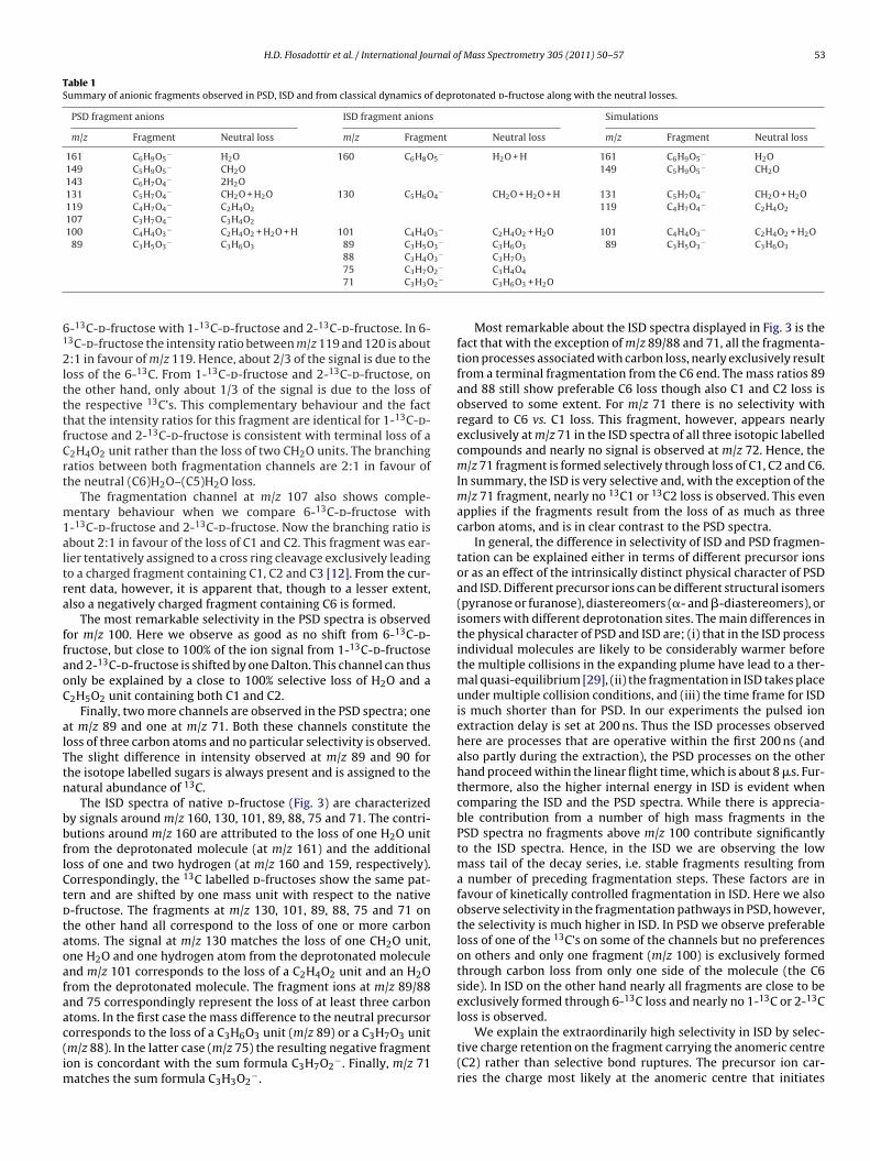
Table 1Summary of anionic fragments observed in PSD, ISD and from classical dynamics of deprotonated d-fructose along with the neutral losses.
PSD fragment anions ISD fragment anions Simulations
m/z Fragment Neutral loss m/z Fragment Neutral loss m/z Fragment Neutral loss
161 C6H9O5− H2O 160 C6H8O5
− H2O + H 161 C6H9O5− H2O
149 C5H9O5− CH2O 149 C5H9O5
− CH2O143 C6H7O4
− 2H2O131 C5H7O4
− CH2O + H2O 130 C5H6O4− CH2O + H2O + H 131 C5H7O4
− CH2O + H2O119 C4H7O4
− C2H4O2 119 C4H7O4− C2H4O2
107 C3H7O4− C3H4O2
100 C4H4O3− C2H4O2 + H2O + H 101 C4H4O3
− C2H4O2 + H2O 101 C4H4O3− C2H4O2 + H2O
89 C3H5O3− C3H6O3 89 C3H5O3
− C3H6O3 89 C3H5O3− C3H6O3
88 C3H4O3− C3H7O3
2−
2−
61
2ltttfCrt
m1altra
ffaoC
alTtn
bbflCtdtaoafaac(im
75 C3H7O71 C3H3O
-13C-d-fructose with 1-13C-d-fructose and 2-13C-d-fructose. In 6-3C-d-fructose the intensity ratio between m/z 119 and 120 is about:1 in favour of m/z 119. Hence, about 2/3 of the signal is due to the
oss of the 6-13C. From 1-13C-d-fructose and 2-13C-d-fructose, onhe other hand, only about 1/3 of the signal is due to the loss ofhe respective 13C’s. This complementary behaviour and the facthat the intensity ratios for this fragment are identical for 1-13C-d-ructose and 2-13C-d-fructose is consistent with terminal loss of a2H4O2 unit rather than the loss of two CH2O units. The branchingatios between both fragmentation channels are 2:1 in favour ofhe neutral (C6)H2O–(C5)H2O loss.
The fragmentation channel at m/z 107 also shows comple-entary behaviour when we compare 6-13C-d-fructose with
-13C-d-fructose and 2-13C-d-fructose. Now the branching ratio isbout 2:1 in favour of the loss of C1 and C2. This fragment was ear-ier tentatively assigned to a cross ring cleavage exclusively leadingo a charged fragment containing C1, C2 and C3 [12]. From the cur-ent data, however, it is apparent that, though to a lesser extent,lso a negatively charged fragment containing C6 is formed.
The most remarkable selectivity in the PSD spectra is observedor m/z 100. Here we observe as good as no shift from 6-13C-d-ructose, but close to 100% of the ion signal from 1-13C-d-fructosend 2-13C-d-fructose is shifted by one Dalton. This channel can thusnly be explained by a close to 100% selective loss of H2O and a2H5O2 unit containing both C1 and C2.
Finally, two more channels are observed in the PSD spectra; onet m/z 89 and one at m/z 71. Both these channels constitute theoss of three carbon atoms and no particular selectivity is observed.he slight difference in intensity observed at m/z 89 and 90 forhe isotope labelled sugars is always present and is assigned to theatural abundance of 13C.
The ISD spectra of native d-fructose (Fig. 3) are characterizedy signals around m/z 160, 130, 101, 89, 88, 75 and 71. The contri-utions around m/z 160 are attributed to the loss of one H2O unitrom the deprotonated molecule (at m/z 161) and the additionaloss of one and two hydrogen (at m/z 160 and 159, respectively).orrespondingly, the 13C labelled d-fructoses show the same pat-ern and are shifted by one mass unit with respect to the native-fructose. The fragments at m/z 130, 101, 89, 88, 75 and 71 onhe other hand all correspond to the loss of one or more carbontoms. The signal at m/z 130 matches the loss of one CH2O unit,ne H2O and one hydrogen atom from the deprotonated moleculend m/z 101 corresponds to the loss of a C2H4O2 unit and an H2Orom the deprotonated molecule. The fragment ions at m/z 89/88nd 75 correspondingly represent the loss of at least three carbontoms. In the first case the mass difference to the neutral precursor
orresponds to the loss of a C3H6O3 unit (m/z 89) or a C3H7O3 unitm/z 88). In the latter case (m/z 75) the resulting negative fragmenton is concordant with the sum formula C3H7O2−. Finally, m/z 71atches the sum formula C3H3O2
−.
C3H4O4
C3H6O3 + H2O
Most remarkable about the ISD spectra displayed in Fig. 3 is thefact that with the exception of m/z 89/88 and 71, all the fragmenta-tion processes associated with carbon loss, nearly exclusively resultfrom a terminal fragmentation from the C6 end. The mass ratios 89and 88 still show preferable C6 loss though also C1 and C2 loss isobserved to some extent. For m/z 71 there is no selectivity withregard to C6 vs. C1 loss. This fragment, however, appears nearlyexclusively at m/z 71 in the ISD spectra of all three isotopic labelledcompounds and nearly no signal is observed at m/z 72. Hence, them/z 71 fragment is formed selectively through loss of C1, C2 and C6.In summary, the ISD is very selective and, with the exception of them/z 71 fragment, nearly no 13C1 or 13C2 loss is observed. This evenapplies if the fragments result from the loss of as much as threecarbon atoms, and is in clear contrast to the PSD spectra.
In general, the difference in selectivity of ISD and PSD fragmen-tation can be explained either in terms of different precursor ionsor as an effect of the intrinsically distinct physical character of PSDand ISD. Different precursor ions can be different structural isomers(pyranose or furanose), diastereomers (�- and �-diastereomers), orisomers with different deprotonation sites. The main differences inthe physical character of PSD and ISD are; (i) that in the ISD processindividual molecules are likely to be considerably warmer beforethe multiple collisions in the expanding plume have lead to a ther-mal quasi-equilibrium [29], (ii) the fragmentation in ISD takes placeunder multiple collision conditions, and (iii) the time frame for ISDis much shorter than for PSD. In our experiments the pulsed ionextraction delay is set at 200 ns. Thus the ISD processes observedhere are processes that are operative within the first 200 ns (andalso partly during the extraction), the PSD processes on the otherhand proceed within the linear flight time, which is about 8 �s. Fur-thermore, also the higher internal energy in ISD is evident whencomparing the ISD and the PSD spectra. While there is apprecia-ble contribution from a number of high mass fragments in thePSD spectra no fragments above m/z 100 contribute significantlyto the ISD spectra. Hence, in the ISD we are observing the lowmass tail of the decay series, i.e. stable fragments resulting froma number of preceding fragmentation steps. These factors are infavour of kinetically controlled fragmentation in ISD. Here we alsoobserve selectivity in the fragmentation pathways in PSD, however,the selectivity is much higher in ISD. In PSD we observe preferableloss of one of the 13C’s on some of the channels but no preferenceson others and only one fragment (m/z 100) is exclusively formedthrough carbon loss from only one side of the molecule (the C6side). In ISD on the other hand nearly all fragments are close to beexclusively formed through 6-13C loss and nearly no 1-13C or 2-13Closs is observed.
We explain the extraordinarily high selectivity in ISD by selec-tive charge retention on the fragment carrying the anomeric centre(C2) rather than selective bond ruptures. The precursor ion car-ries the charge most likely at the anomeric centre that initiates

54 H.D. Flosadottir et al. / International Journal o
O OHO-
OH
OHHO
H
O-OH
O
OH
OHO
H
H
H-
C
OHO
OH
OHHO
m/z 149
-HC
OHO
OHm/z 89
-C
OHO
OH
OH
H
m/z 119OH
OHO
OH
O-
OHH
m/z 161
anomeric ring opening
-H2O -CH2O -2CH2O
-2CH2O
m/z 179
Fig. 4. Summary of dissociation products observed in classical dynamics simula-tions for �-d-fructose deprotonated at the anomeric hydroxyl group (OH2). Theild
ttttrvm
trs
3
aordots
i1ptd
ohait(Sm
nitial step in the fragmentation is a ring cleavage at the C2–O bond followed byoss of water and different neutral carbon-containing fragments from the C6 site of-fructose.
he dissociation. Fast fragmentation (of hot molecular ions) in ISDherefore leads to selective charge retention on the anomeric cen-re. The metastable ions probed in PSD have lower internal energieshan the precursor ions in ISD, and the internal energy is most likelyedistributed over all available degrees of freedom (intramolecularibrational redistribution, IVR). Thus, after several �s other frag-entation channels become accessible resulting in less selectivity.In the next section we will discuss classical dynamics simula-
ions that help to elucidate important details of the fragmentationeactions, especially the role of deprotonation sites for specific dis-ociation reactions.
.3. Simulations of the fragmentation processes
In previous studies of metastable fragmentation of the aminocid l-valine [17] and the DNA/RNA nucleosides and deoxynucle-sides [13] we have shown that classical dynamics simulationsepresent a powerful tool to predict dissociation pathways ofeprotonated biomolecules. In the current study we have carriedut classical dynamics simulations on the fragmentation of vibra-ionally activated �- and �-d-fructose in their ground electronictate when these are deprotonated at the various hydroxyl groups.
From deprotonated �- and �-d-fructose the main fragment ionsn the classical dynamics simulations were found at m/z 161, 149,19, 101, 89 and 59 agreeing very well with the fragmentationroducts observed in the experiments. The predicted fragmenta-ion reactions leading to these fragment ions (except m/z 59) aftereprotonation at the OH2 are summarized in Fig. 4.
In general, the precursor ions generated by initial deprotonationf a hydroxyl group in d-fructose are stabilized by intramolecularydrogen bonds between the negatively charged oxygen and andjacent hydroxyl group. The subsequent fragmentation reactionsn our simulations were found to be composed of three charac-
eristic reaction steps. These are (i) intramolecular proton transfer,ii) anomeric ring opening, and (iii) antiperiplanar dissociation (seeupplementary material for more information). Basically all frag-entation reactions observed in our classical dynamics simulationsf Mass Spectrometry 305 (2011) 50– 57
can be described as a sequence of these reaction steps and the typ-ical neutral elimination products are water, formaldehyde (CH2O)and longer aldehydes.
The intramolecular proton transfer steps occur with low energybarriers and are thus frequently observed. The proton transferproceeds also faster than dissociation via C–C bond breaking andconsequently further dissociation preferably takes place when pro-ton transfer has lead to charge location at the most acidic hydroxylgroup. The number of observed intramolecular proton transferreactions in a given number of simulations for a specific deprotona-tion site indicates that the acidity of the individual OH groups canbe ordered in the following way: OH2 ≈ OH5 > OH1 > OH3 ≈ OH4.Almost no proton transfer is observed after deprotonation of OH2or OH5. Instead various cross-ring cleavage reactions are inducedfrom these anions. Anomeric ring opening is characteristic fordeprotonation of OH2. This creates a ketone at C2, which in somecases leads to further fragmentation such as elimination of water(resulting in m/z 161) or loss of one or more CH2O units as isdepicted in Fig. 4. Deprotonation at OH1, on the other hand, resultseither in C1–C2 bond cleavage or proton transfer from OH2 andsubsequent anomeric bond cleavage (see below). Deprotonation atOH3 or OH4 mainly results in proton transfer from the adjacenthydroxyl groups indicating that these hydroxyl groups have thelowest acidity. Antiperiplanar arrangement of the bonds to whichthe negatively charged oxygen and the leaving group are connectedis a typical prerequisite for the dissociation channels observed. Forinstance, 70% of the dissociation reactions observed after deproto-nation of OH5 proceed from an antiperiplanar conformation.
m/z 161. The fragment ion at m/z 161 represents the loss of onewater molecule from the respective d-fructose and does not involvecarbon loss. It is thus not possible to gain insight into this frag-mentation channel through the mass spectra of the isotope marked13C-d-fructoses. Nevertheless, as this is the most pronounced signalin the PSD spectra it is worthwhile to have a closer look at this chan-nel in our simulations. In the simulations this fragment is formedfrom the d-fructose when it is deprotonated at OH3, OH4 or OH5.Water loss is also observed after deprotonation at OH1 and OH2,but in both these cases this is either observed subsequent to a CH2Oloss or preceding the same (see below). Deprotonation at OH3, onthe other hand, leads to proton transfer from OH2 in conjunctionwith an anomeric ring opening. Subsequently OH3 along with thehydrogen of OH4 is eliminated as H2O and further fragmentation isnot observed. Water loss after deprotonation at OH4 on the otherhand, proceeds through an antiperiplanar dissociation rupturingthe C3–C4 and the C2–OH bond. In this case OH2 with the hydro-gen from OH1 is eliminated. This channel was only observed fromthe �-d-fructose, as such antiperiplanar dissociation is not possiblefrom the �-anomer. Finally, from d-fructose deprotonated at OH5,we also find water elimination containing OH2 with the hydrogenfrom OH1. However, here the elimination is found to proceed underC1–O1–C2 epoxide formation without any direct involvement ofthe charge location site.
m/z 149. The predictive power of our simulation method can bedemonstrated on the fragment with m/z 149, corresponding to theloss of a single CH2O molecule from the deprotonated d-fructose.The mass spectra of isotope labelled d-fructose indicate that thecarbon atom of CH2O originates to a comparable extent from posi-tion 1 and 6 (see Section 3.2 and Fig. 2). Our classical dynamicssimulations showed that the different origin is likely to be corre-lated with the initial position of deprotonation. Deprotonation atthe anomeric centre (OH2) leads to a ring-opening, as it was pre-viously observed for glucose [28], and subsequently to a formation
of a C O double bond, and abstraction of neutral CH2O from theC6 site (Fig. 5a). Remarkably, deprotonation at position 1 results insix out of ten simulations in the formation of CH2O from C1 as itis displayed in Fig. 5b. Hence, the classical dynamics simulations
H.D. Flosadottir et al. / International Journal of Mass Spectrometry 305 (2011) 50– 57 55
O O-
OH
OH
OHHO
C-O O
OH
OH
OHHO
O OHO-
OH
OHHO
H
O-OH
O
OH
OHHO
H H-
C
O OHO
OH
OHHO
m/z 149
C5H9O5-
m/z 149
C5H9O5-
(a)
(b)
Fig. 5. Reaction scheme from the classical dynamics simulations showing how thelo
po6i
avwbleotOqowCa
Fo1
O OHO-
OH
OHHO
H
-O OHO
OH
OHO
HH -C
HO OHO
OH
OHO
H
C4H7O4-
m/z 119
oss of neutral CH2O from the C1 site and the C6 site (leading to m/z 149) dependsn the initial deprotonation site.
redicted a signal in the mass spectrum at m/z 149, the intensityf which is shifted by 50% to m/z 150 in both 1-13C-d-fructose and-13C-d-fructose, and completely shifted in 2-13C-d-fructose. This
s fully in agreement with the PSD mass spectra shown in Fig. 2.m/z 131. The fragment ion at m/z 131 is assigned to the loss of
neutral water and a formaldehyde unit. Experimentally we haveerified both C6 and C1 loss in this case, but from our mass spectrae cannot make conclusive statements about the relative contri-
utions of these two channels. In our simulations both C1 and C6oss was observed but deprotonation at OH1 exclusively results inlimination of C1. Here the �-anomer leads to C1 loss in six outf ten simulations (6/10), when deprotonated at OH1. In an addi-ional step water is eliminated from OH5 and the hydrogen fromH2 (Fig. 6a). Also deprotonation at OH5 results in C1 loss subse-uent to antiperiplanar water elimination (Fig. 6b). C6 loss is onlybserved from the �-anomer. An example is shown in Fig. 6c, inhich deprotonation at OH2 induces anomeric bond cleavage and
H2O elimination from C6 followed by water elimination from OH4nd the hydrogen of OH3.O O-
OH
OH
OHHO
C-O
OOH
OHHO
CO
O-
OH
OH
C-O
OH
OHO
O O-
OH
OHO
O OOH
OHOH
-OH
H
O O-
OH
OHHO
OH
O- O
O
HOHO
OH
H
O
O-HO
OH
α ano mer - deprotonation at OH1
- CH2O - H2O
- CH2O
- CH2O
- H2O
- H2O
β a nomer - deprotonatio n at OH1
β anome r - deproton atio n at OH5
(a)
(b)
(c)
ig. 6. Reaction scheme from the classical dynamics simulations showing the lossf neutral CH2O and H2O from the C6 site as well as from the C1 site (leading to m/z31).
Fig. 7. Reaction scheme resulting from the classical dynamics simulations showingthe loss of neutral C2H4O2 from the C6 site (leading to m/z 119).
m/z 119. Deprotonation of the anomeric centre also results inloss of larger carbon containing neutral fragments. Fig. 7 showsthe detailed fragmentation pathways for the fragment anion m/z119. Here, deprotonation at OH2 leads to anomeric bond cleav-age followed by a proton transfer from the OH5 group to the ringoxygen. This in turn induces the elimination of the neutral frag-ment HOC2H3O through a C4–C5 bond rupture. This agrees withthe experiments where 2/3 of the anion PSD signal was shifted tohigher masses for 1-13C-d-fructose and 2-13C-d-fructose, and only1/3 was shifted for 6-13C-d-fructose.
m/z 101. The most intense ion observed in ISD appears at m/z101. According to our classical dynamics simulations only depro-tonation at OH5 results in a fragment ion at m/z 101. Here theinitial deprotonation of OH5 results in antiperiplanar dissociationinvolving ring-opening between C5 and C4 and cleavage of theC3–OH bond under formation of OH− (Fig. 8). In a second step,proton transfer from OH4 to the departing OH− group leads toH2O formation and further fragmentation into C4H5O3
− (m/z 101)through anomeric bond cleavage. This corresponds to an exclusiveC6 loss, agreeing well with the experimentally observed selectivity.In previous measurements on metastable decay of deprotonatedd-fructose and d-ribose [12] it was shown that some fragmentsappear shifted by one mass unit when comparing ISD and PSD massspectra. Thus, it is likely that the (metastable) fragment anion at m/z101 looses another hydrogen atom after intramolecular redistribu-tion of the internal energy during the flight time to the reflectronto form the ion at m/z 100. Here it should also be noted that theclassical dynamics simulations only cover a time range of 500 fs,i.e. further fragmentation into thermodynamically more favourableproducts might have been observed if the simulation had run longenough.
m/z 89. The signal at m/z 89 (C3H5O3−) is the most promi-
nent carbon loss channel observed in PSD of the deprotonatedd-fructose, and it is also observed with appreciable intensity inthe ISD spectra. In the PSD spectra of 1-13C-, 2-13C-, and 6-13C-d-fructose the intensity ratios between m/z 89 and 90 are all closeto 1:1 and no particular selectivity was observed. The simulationsshowed formation of this ion from the �- and �-d-fructose depro-tonated at OH2 and from the �-d-fructose deprotonated at OH1. Inthe case of deprotonation at OH2 the initial step is an anomeric ring
opening followed by a CH2O loss from C6. A bond rupture betweenC3 and C4 then leads to a fragment ion at m/z 89 containing C1, C2and C3 (Fig. 9a). Deprotonation at OH1 also leads to anomeric ringopening, after proton transfer from OH2. However, then a seriesO OH
OH
OH-O
O OH
OH-OH
O
m/z 101
O
OH
C4H5O3-
O
-O
OH
OHOH
Fig. 8. Reaction scheme showing the formation of C4H5O3− (m/z 101) by deproto-
nation of OH5 as observed in the classical dynamics simulations.

56 H.D. Flosadottir et al. / International Journal o
O OHO-
OH
OHHO
-HC
OOH
O
OHOH
HO
C3H5O3- m/z 89
O OH
OH
OH
HO
O-
O-
O
HO
O-O
OH
OH
HO
OH
C3H5O3- m/z 89
OH
OH
OH
OH O
OH
OH
-O
OH
(a)
(b)
Fm
oCceu
Misstatst�i
4
bauoflfgmtcsip
[
[
[
ig. 9. Reaction scheme from the classical dynamics simulations showing the for-ation of C3H5O3
− (m/z 89) and C4H7O4− (m/z 119) by deprotonation of OH2.
f proton transfer reactions leads to C3–C4 bond rupture, and C1,2 and C3 leave as one unit (Fig. 9b), leaving a m/z 89 fragmentontaining C4, C5 and C6. This is in very good agreement with ourxperiment and supports the notation that C1 and C2 leave as onenit.
m/z 59. A fragment ion at m/z 59 was previously observed inALDI-ISD [12] and dissociative electron attachment [8] exper-
ments on d-ribose, but not from d-fructose. In the currentimulations this fragment is observed as the final product after aeries of fragmentation steps. Thus, it may be a result of fragmenta-ion with high activation energy barrier, which is not energeticallyccessible in the experiments, but is seen in the simulations due tohe high internal energy (8 eV). To verify this we also carried outimulations where the atomic velocities were scaled to correspondo a total of 5 eV internal energy. These lower energy simulations for-d-fructopyranose do not show a complex fragmentation result-
ng in such a small fragment.
. Conclusions
We studied ISD and PSD of negative ions of d-fructose in MALDIy means of the isotope labelled d-fructose analogues 1-13C-, 2-13C-nd 6-13C-d-fructose. By using the isotope labelled d-fructoses wenambiguously identify the composition of most of the fragmentsbserved. To further identify the individual fragments and theragmentation mechanisms we conducted classical dynamics simu-ations of the vibrationally excited (hot) �- and �-d-fructopyranoseorm of d-fructose when deprotonated at the individual hydroxylroups. We find that ISD proceeds nearly exclusively through ter-inal carbon loss from the C6 side and the charge remains on
he C1 and C2 containing fragment. In PSD other fragmentation
hannels also contribute and terminal fragmentation from the C1ide competes with carbon loss from the C6 side. The differencen fragmentation pathways is ascribed to the intrinsically differenthysical character of ISD and PSD. The desorbed ions that are sub-[[
[
f Mass Spectrometry 305 (2011) 50– 57
jected to ISD are considerably hotter and fragmentation proceedsfaster than in PSD. The pronounced selectivity in ISD thus is kineti-cally controlled, whereas the branching ratios in PSD are governedby the thermochemistry.
With our classical dynamics simulations we are able to predictmost of the experimentally observed fragmentation products andto elucidate the basic individual dissociation reaction steps behindthose. We find that the fragmentation is generally composed oftwo or all three of the following reaction steps; intramolecularproton transfer, anomeric ring opening and antiperiplanar dissocia-tion. In the simulations we observe that deprotonation at differenthydroxyl groups may result in fragment ions with the same m/zratio but formed via different reaction pathways. This is in accor-dance with the experimental mass spectra obtained by PSD. Therewe observe loss of neutral fragments originating from both endsof the molecule, i.e. C1 and C6, for all m/z ratios apart from m/z101. The simulations suggest that terminal carbon loss from the C1site contributes when deprotonation occurs at other positions thanOH2 (the anomeric centre), namely OH1 and OH5. Deprotonation atthe anomeric centre, OH2, on the other hand, predominantly leadsto terminal C6 loss. Finally, the simulations show that the frag-ment ion at m/z 101 (which is the most intense in ISD) originatesexclusively from deprotonation at OH5 and there is no alternativereaction pathway to create a fragment ion at m/z 101. In the PSDspectra the only close to 100% site selective reaction channel isobserved at m/z 100 and is accordingly assigned to be also due toinitial deprotonation at OH5.
Acknowledgements
We acknowledge financial support from the Icelandic Centrefor Research (RANNIS), the University of Iceland Research Fund, IBacknowledges support for a visit to Reykjavik by the COST actionP9 (Radiation Damage in Biomolecular Systems, RADAM) and bythe European Science Foundation (ESF) program: Electron inducedprocesses at the molecular level (EIPAM). HDF acknowledges a PhDgrant from the Eimskip University Fund.
Appendix A. Supplementary data
Supplementary data associated with this article can be found, inthe online version, at doi:10.1016/j.ijms.2011.05.016.
References
[1] D.J. Harvey, Mass Spectrometry Reviews 18 (1999) 349.[2] J.P. Simons, R.A. Jockusch, P. Carcabal, I. Hung, R.T. Kroemer, N.A. Macleod, L.C.
Snoek, International Reviews in Physical Chemistry 24 (2005) 489.[3] D.J. Harvey, Mass Spectrometry Reviews 30 (2011) 1.[4] R.E. March, C.J. Stadey, Rapid Communications in Mass Spectrometry 19 (2005)
805.[5] K.P. Madhusudanan, Journal of Mass Spectrometry 41 (2006) 1096.[6] L. Sanche, European Physical Journal D: Atomic Molecular and Optical Physics
35 (2005) 367.[7] P. Swiderek, Angewandte Chemie (International Edition) 45 (2006) 4056.[8] I. Bald, J. Kopyra, E. Illenberger, Angewandte Chemie (International Edition) 45
(2006) 4851.[9] P. Sulzer, S. Ptasinska, F. Zappa, B. Mielewska, A.R. Milosavljevic, P. Scheier, T.D.
Mark, I. Bald, S. Gohlke, M.A. Huels, E. Illenberger, Journal of Chemical Physics125 (2006) 044304.
10] I. Bald, I. Dabkowska, E. Illenberger, Angewandte Chemie (International Edition)47 (2008) 8518.
11] A. Ryzhkova, U. Jarzak, A. Schafer, M. Baumer, P. Swiderek, Carbohydrate Poly-mers 83 (2011) 608.
12] I. Bald, H.D. Flosadottir, J. Kopyra, E. Illenberger, O. Ingolfsson, InternationalJournal of Mass Spectrometry 280 (2009) 190.
13] H. D. Flosadottir, H. Jonson, O. Ingolfsson, in press.14] I. Bald, J. Langer, P. Tegeder, O. Ingolfsson, International Journal of Mass Spec-
trometry 277 (2008) 4.15] I. Bald, J. Kopyra, I. Dabkowska, E. Antonsson, E. Illenberger, Journal of Chemical
Physics 126 (2007) 074308.

rnal o
[
[
[
[[[
[
[
[
[
[
H.D. Flosadottir et al. / International Jou
16] Z.W. Deng, I. Bald, E. Illenberger, M.A. Huels, Angewandte Chemie (InternationalEdition) 47 (2008) 9509.
17] H.D. Flosadottir, S. Denifl, F. Zappa, N. Wendt, A. Mauracher, A. Bacher, H. Jon-sson, T.D. Maerk, P. Scheier, O. Ingolfsson, Angewandte Chemie (InternationalEdition) 46 (2007) 8057.
18] M. Stano, H.D. Flosadottir, O. Ingolfsson, Rapid communications in mass spec-trometry 20 (2006) 3498.
19] G. Kresse, J. Furthmuller, Physical Review B 54 (1996) 11169.
20] G. Kresse, J. Hafner, Physical Review B 49 (1994) 14251.21] G. Henkelman, A. Arnaldsson, H. Jonsson, Computational Materials Science 36(2006) 354.22] E. Sanville, S.D. Kenny, R. Smith, G. Henkelman, Journal of Computational Chem-
istry 28 (2007) 899.
[
[[
f Mass Spectrometry 305 (2011) 50– 57 57
23] W. Tang, E. Sanville, G. Henkelman, Journal of Physics-Condensed Matter (2009)21.
24] J.F. Greisch, V. Gabelica, F. Remacle, E. De Pauw, Rapid Communications in MassSpectrometry 17 (2003) 1847.
25] J.A. Kanters, G. Roelofsen, B.P. Alblas, I. Meinders, Acta Crystallographica SectionB-Structural Science 33 (1977) 665.
26] B. Ma, H.F. Schaefer III, N.L. Allinger, Journal of the American Chemical Society120 (1998) 3411.
27] L.P. Guler, Y.-Q. Yu, H.I. Kenttaemaa, Journal of Physical Chemistry A 106 (2002)6754.
28] J.-Y. Salpin, J. Tortajada, Journal of Mass Spectrometry 39 (2004) 930.29] R. Knochenmuss, Analyst (Cambridge United Kingdom) 131 (2006)
966.