The free-energy barriers against expansion of the metastable ...
18
Supplementary information: The free-energy barriers against expansion of the metastable fusion stalk and its implication for the protein fusion machinery Herre Jelger Risselada, Gregory Bubnis, Helmut Grubm¨ uller Theoretical Molecular Biophysics Group, Max-Planck-Institute for Biophysical Chem- istry, 37077 G¨ ottingen, Germany, Methods: System setup. All vesicles were obtained by spontaneous aggregation of ran- domly placed lipids. This method is described in detail elsewhere [1]. An additional long equilibration run of 20 μs was performed for the mixed lipid systems (two ar- tificial pores were present) [2]. In all cases, equilibrium of the vesicle was defined by the flux convergence of the lipid components. The pure POPC vesicle/dimples consisted of 2217 lipids. The mixed POPC:POPE system, 1330 POPC and 887 POPE (40%) lipids. The mixed POPC:cholesterol system, 1980 POPC and 834 cholesterol (30%) molecules. The box dimensions in our (umbrella) simulations were 21 × 22 × 20 nm 3 . About 50000 solvent beads were present. All vesicles were equilibrated with 2 artificial pores of 1.6 nm radius. Equilibrium is defined by the absence of net flux/flip-flops for each individual component, which can take upto 20 μs in the mixed vesicles [2]. Artificial pores and stalk formation. The hydrophilic pore of radius R=1.6 nm was stabilized using an repulsive potential U (r i ) with U (r i )=1/2k(r i - R) 2 , if r i <R and U (r i ) = 0 if r i ≥ R, where r i denotes the (minimal) distance of the center of mass of the lipid from the pore center and k a force constant (k force =50 kJ nm -2 mol -1 ) [1, 2]. This potential only acts on carbon lipid tails and glycerol groups. The central axis through the pores was along the x-axis with coordinates y=0.5 × box length, z=0. This particular choice (z=0) does not interfere with the scaling of coordinates due to the pressure coupling. Likewise, we induced the initial stalk in the bilayer fusion setup by applying an external field. Here, we applied the same harmonic potential to induce a R=1.0 nm ’void’ in the solvent layer between the bilayers. The hydrophobic nature of the void attracts the lipid tails in the adjacent leaflets and results in the formation of a stalk. The external field was subsequently removed to allow equilibration of the stalk structure. The membranes are defined tension-less because there is no degree of freedom that allows lateral membrane tension to built-up. The lipids can freely distribute
-
Upload
khangminh22 -
Category
Documents
-
view
1 -
download
0
Transcript of The free-energy barriers against expansion of the metastable ...

Supplementary information: The free-energybarriers against expansion of the metastablefusion stalk and its implication for the proteinfusion machineryHerre Jelger Risselada, Gregory Bubnis, Helmut Grubmuller
Theoretical Molecular Biophysics Group, Max-Planck-Institute for Biophysical Chem-
istry, 37077 Gottingen, Germany,
Methods:
System setup. All vesicles were obtained by spontaneous aggregation of ran-domly placed lipids. This method is described in detail elsewhere [1]. An additionallong equilibration run of 20 µs was performed for the mixed lipid systems (two ar-tificial pores were present) [2]. In all cases, equilibrium of the vesicle was definedby the flux convergence of the lipid components. The pure POPC vesicle/dimplesconsisted of 2217 lipids. The mixed POPC:POPE system, 1330 POPC and 887POPE (40%) lipids. The mixed POPC:cholesterol system, 1980 POPC and 834cholesterol (30%) molecules. The box dimensions in our (umbrella) simulationswere 21× 22× 20 nm3. About 50000 solvent beads were present. All vesicles wereequilibrated with 2 artificial pores of 1.6 nm radius. Equilibrium is defined by theabsence of net flux/flip-flops for each individual component, which can take upto20 µs in the mixed vesicles [2].
Artificial pores and stalk formation. The hydrophilic pore of radius R=1.6 nmwas stabilized using an repulsive potential U(ri) with U(ri) = 1/2k(ri − R)2, ifri < R and U(ri) = 0 if ri ≥ R, where ri denotes the (minimal) distance of thecenter of mass of the lipid from the pore center and k a force constant (kforce=50kJ nm−2 mol−1) [1, 2]. This potential only acts on carbon lipid tails and glycerolgroups. The central axis through the pores was along the x-axis with coordinatesy=0.5 × box length, z=0. This particular choice (z=0) does not interfere withthe scaling of coordinates due to the pressure coupling. Likewise, we induced theinitial stalk in the bilayer fusion setup by applying an external field. Here, weapplied the same harmonic potential to induce a R=1.0 nm ’void’ in the solventlayer between the bilayers. The hydrophobic nature of the void attracts the lipidtails in the adjacent leaflets and results in the formation of a stalk. The externalfield was subsequently removed to allow equilibration of the stalk structure.
The membranes are defined tension-less because there is no degree of freedomthat allows lateral membrane tension to built-up. The lipids can freely distribute

themselves over the monolayers via the artificial pores, i.e. the chemical potentialof the monolayers are equal and there is no possibility to build up any osmoticpressure difference. Further, the simulations box is semi-isotropically coupled toindependent pressure baths of 1.0 bar such that there can not be a resultant pullingor squeezing force on the stalk/vesicle coming from the boundary conditions (thesimulation box).
Hydrophilic probes. A hydrophilic probe consisted of 8 P4 particles [3], whichare located on the edges of a cube with a dimension of 0.3 × 0.3 nm. The beadswere connected via 12 direct lattice bonds (0.3 nm), 12 in plane diagonal bonds(0.424 nm), and 4 diagonal bonds (0.52 nm), each with a harmonic force constantof 1250 kJ nm−2 mol−1.
Umbrella sampling. In umbrella sampling one attempts to overcome samplingproblems along an quasi-static reaction coordinate by introducing an external biaspotential such that the unfavorable free energy states are sampled adequately. Toconduct umbrella sampling, one must generate a series of configurations alonga reaction pathway (umbrella windows). These umbrella windows are restraintby the bias potential to sample a limited, but overlapping region of the reactionpathway. Afterward, the potential of mean force is constructed from the differentumbrella windows by correcting for the introduction of the biasing potential.
Umbrella windows were obtained by performing an initial pulling simulationwhere the probes were pulled together at a constant rate of −5×10⁻⁵ nm/ps untilbarrier crossing. This simulation was divided in 40 to 50 different fragments witha progressing probe to probe distance, and which formed the setup for the actualproduction runs (i.e., umbrella simulations). The harmonic force constant of the’bond’ between the probes was 1000 kJ nm−2 mol−1. We note that the probescan freely translate throughout the system, and that the observed location of theprobes in an outcome of the free-energy minimization of the membranes. Further,in the stalk widening simulations, we only restrained the x-dimension of the vectorconnecting the two probes, i.e. the dimension parallel to the central axis throughthe stalk. Therefore, the probes can move independently within the xy-plane whichlies perpendicular to the symmetry axis of the stalk (the actual distance betweenthe probes in 3 dimensions is irrelevant). In this way, we do not oppose radialsymmetry when forcing the probes together, because the probes can penetratethe stalk at an angle. The cited probe-probe distance is the distance along thez-dimension. In the leaky stalk simulations, we only restrained the actual distancebetween the probes (three dimensional), and the cited probe-probe distance is thelength of connecting vector. We note, however, that these bonded probes canfreely move and translate within the system like a diatomic molecule. We allowed

for 400 ns of equilibration time in each umbrella window (see Fig. S10). We notethat stalk expansion can be alternatively enforced by bringing two lipids in thetrans-leaflet within close contact (Fig. S14), and that the obtained results are verysimilar. In addition, we note that ’pinching’ the membrane via the probes hardlyaffects the nearby lipid composition in the mixed POPC:POPE membranes (Fig.S15).
Error bars were calculated using the bootstrapping method [4], where the erroris estimated form differences between different sampling intervals. We used theweighted histogram method to reconstruct the potential of mean force from thedifferent umbrella windows [4]. In our simulations we bring the system close to itsbarrier such that barrier crossing (nucleation) can occur within the 1.6 microsecondtime-scale of the umbrella simulations. It is important that the simulations are ofequal length and sufficiently short such that nucleation along the reaction coordi-nate becomes unlikely if the remaining free energy barrier is larger than several kbT.
Bending restraint. We restrained bending of the trans-leaflet by applying po-sition restraints on the amino beads of the lipids with a force constant of 1000kJ nm−2 mol−1. The restraint only acted in the Z-dimension, i.e. the dimensionparallel to the two probes, and allowed free movement/diffusion in the xy-plane.We re-introduced the two artificial pores, such that the lipids in the unrestrainedtrans-leaflets can freely flip-flop and diffuse. Further, the z-dimension, and xy-dimension of the simulation box could freely an independently adapt such that theunrestrained leaflet remains tension less.
Simulation details. The simulations described in this paper were performedwith the GROMACS simulation package [6], version 4.0.5. We used the Martinimodel version 2.1 [3, 7] to simulate the lipids and cholesterol. In all simulations thesystem was coupled to a constant temperature bath [8] of 310 K with a relaxationtime τT of 1.0 ps. The time step used in the simulation was 20 fs [9]. Shiftedpotentials were used to describe van der Waals and electrostatic pair-wise interac-tions. In both cases, the neighbor list cutoff was 1.2 nm and these potentials weregradually shifted to zero when the pair-wise distance exceeded 0.9 nm (van derWaals) or 0 nm (Coulomb interaction). The neighbor list was updated every 10simulation steps. The pressure was semi-isotropically coupled [8] to 1 bar with arelaxation time τP of 0.5 ps. In analogy to the other studies employing the Martinimodel, time scales quoted in this work were scaled by a factor of 4 to correct forthe 4-times faster diffusion rates of water and lipids in the coarse-grained model[3] with respect to reality.

References
[1] Risselada, H, Mark, A, Marrink, S (2008) Application of mean field boundarypotentials in simulations of lipid vesicles. J. Phys. Chem. B 112:7438–47.
[2] Risselada, H, Marrink, S (2009) Curvature effects on lipid packing and dynam-ics in liposomes revealed by coarse grained molecular dynamics simulations.Phys. Chem. Chem. Phys. 11:2056–67.
[3] Marrink, SJ, Risselada, HJ, Yefimov, S, Tieleman, DP, de Vries, AH (2007)The MARTINI force field: Coarse grained model for biomolecular simulations.J. Phys. Chem. B 111:7812–7824.
[4] Hub, J, de Groot, B, van der Spoel, D (2010) g wham - A free weightedhistogram analysis implementation including robust error and autocorrelationestimates. J. Chem. Theory Comput. 6:3713–3720.
[5] Risselada, H et al. (2012) Line-tension controlled mechanism for influenzafusion. PLoS One 7:e38302.
[6] Hess, B, Kutzner, C, Van Der Spoel, D, Lindahl, E (2008) GROMACS 4:Algorithms for highly efficient, load-balanced, and scalable molecular simula-tion. J. Chem. Theory Comput. 4:435.
[7] Monticelli, L et al. (2008) The martini coarse grained force field: Extensionto proteins. J. Chem. Theory Comput. 4:819–834.
[8] Berendsen, HJC, Postma, JPM, van Gunsteren, WF, Di Nola, A, Haak, JR(1984) Molecular-dynamics with coupling to an external bath. J. Chem. Phys.
81:3684–3690.
[9] Marrink, SJ, Periole, X, Tieleman, DP, de Vries, AH (2010) Reply to thecomment on on using a too large integration time step in molecular dynamicssimulations of coarse-grained molecular models. Phys. Chem. Chem. Phys.
12:2257–2258.
[10] Lee, KK (2010) Architecture of a nascent viral fusion pore. EMBO 29:1299–1311.

0 500 1000 1500time (ns)
0.3
0.32
0.34
0.36
0.38
Nin
ner/N
tota
l
Figure 1: Lipid flip-flops for the different umbrella windows (i.e., different probe toprobe distances) in the presence of artificial pores(Stalk widening, pure POPC). Shownis the relative population of the trans-leaflet Ninner as function of time (Ntotal =2217lipids). In these simulations we started with a vesicle obtained by spontaneous formation(Ninner/Ntotal=0.31). The sudden transition toward higher values (> 0.33) reflect bar-rier crossing – subsequent widening of the stalk becomes spontaneous. In the differentumbrella windows were barrier crossing is not observed Ninner/Ntotal remains about 0.33(equilibrium). The latter indicates that, upto the barrier, the process of stalk wideningseems not characterized by a pronounced change in relative leaflet population.

0 5 10 15 20
X,Y(nm)
0
5
10
15
20Z
(nm
)
0 5 10 15 20
X,Y(nm)
0
5
10
15
20
Z(n
m)
Figure 2: Stalk widening in the pure POPC membrane (equilibrated vesicle, withoutartificial pores). Shown are the radially averaged densities of the trans-leaflets for therelaxed stalk (upper panel) and the trans membrane contact (lower panel)

Figure 3: Free-energy barrier against subsequent rupture of the small 2nm-sized hemi-fusion diaphragm. Shown is the free-energy for different membrane compositions. Forpure POPC, 40% POPE, or even pure POPE the barrier is within the thermal noise andspontaneous rupture events are observed in our 1.6 µs simulations. The presence of 30%cholesterol strongly increases the barrier against rupture, about 11 kBT. The strengthen-ing effect of cholesterol is, however, reduced when 50% of the POPC lipids are replacedby membrane disordering poly-unsaturated lipids diC18:2PC. The black line shows thebarrier against pore (rim-pore) formation in an 8nm-sized hemifusion diaphragm formedbetween a pure POPE vesicle and 35x35 nm lipid bilayer (see Current Opinion in Struc-tural Biology, 22(2), 187-196, 2012), about 13 kBT, and illustrates the dependence ofthe barrier on the size and topology of the hemifused membrane structure.

0
0.05
0.1
0.15
0.2
0.25
0.3
6.5 7 7.5 8 8.5 9
acili
ndric
ity
probe to probe distance (nm)
15
20
25
30
35
perim
eter
(nm
)
20 25 30 35 40 45 50 55 60 65
area
(nm
*nm
)POPC
40%PE30%Chol
Figure 4: Average shape and size of the stalk at different stages along the reaction coor-dinate (stalk widening pathway). In these simulations flip-flop between the monolayersis present. The lines end at the point were the subsequent stalk widening becomes spon-taneous (expansion barrier). Here, the stalk region is defined as the region within 1nmabove and below the stalk center (+/- 100 lipids). (left panel) The cross-sectional area,the total length of its circumference, and the acilindricity of the stalk. The acilindricityis a measure of how much the shape of the stalk deviates from radial symmetry, it is 0 fora perfect circle and 1 for a infinite thin rod/line. In all three membrane systems the stalkis rather asymmetric in the relaxed state. The presence of 40% POPE or 30% Cholesterolincreases the overall size of the stalk (area and perimeter), as well as the asymmetry.Formation of the hemifusion diaphragm is characterized by a drop in acilindricity, i.e.the stalk is becoming radially symmetric, and sudden increase in area/perimeter. (rightpanel) Algorithm used to quantify the area, perimeter, and acilindricity. For the cross-sectional image of the stalk we only included the lipid beads (carbon-tails and glycerolgroups) that are within 0.5 nm distance from the central plane through the stalk. Thiscross-sectional ’image’ was mapped (using a overlap radius of 0.3 nm for each bead) ontoa grid where each grid size had a dimension, Agrid=0.28x0.28 nm. The stalk was subse-quently identified by a cluster algorithm. Here, two grid elements belong to one cluster ifone of their four sides are directly contacting. Within such a definition, the stalk perime-ter is a cluster element that has 3 or less direct contacts. Now, the area of the stalk isgiven by Nstalk ∗Agrid and the perimeter length is approximated by Nperimeter ∗
√
Agrid.
The acilindricity is defined as(λx−λy)2
(λx+λy)2, where λx, λy are the principal axis of the gyration
tensor composed of all stalk perimeter elements.

6.577.588.59probe to probe distance (nm)
-1500
-1000
-500
0
∆H (
kT)
POPC40% Cholesterol30% POPE
Figure 5: Relative change in enthalpy during stalk widening. The enthalpy remainsapparently constant until the barrier against stalk expansion is surpassed. The latter ischaracterized by a strong drop in enthalpy, i.e. the subsequent expansion of the stalk isenthalpically favorable.

56789probe to probe distance (nm)
-100
-90
-80
-70
solv
ent i
nter
actio
ns p
er li
pid
(kJ/
mol
)
stalk circumferencecis-leaflettrans-leaflet
planar membrane
56789probe to probe distance (nm)
0.2
0.3
0.4
0.5
part
ition
ing
CHOLPE
Figure 6: Hydration and composition of the stalk’s circumference at the different stagesof stalk widening. Here, the stalk region is defined as the region within 1 nm aboveand below the stalk center (+/-100 lipids). (figure above) Lipid-solvent interactions inthe stalk and membrane leaflets elsewhere (pure POPC). The (negatively curved) cis-leaflets of the dimple/vesicle show a pronounced decrease in lipid-solvent interactionenergy. Note that the ⊃-shaped circumference of the stalk becomes less negativelycurved, and thus more hydrated, when formed between two dimples as opposed to twoplanar membranes. Infact, the lipids in such a shallow stalk are similarly hydrated aslipids in the corresponding, fully hydrated planar membrane. Because lipid head-grouphydration does not from the ’bottleneck’ in such a stalk widening, the addition of, forexample, POPE lipids is not expected to enhance stalk widening. Moreover, wideningof such a dimple stalk increases head-group hydration. (figure below) Partitioning ofPOPE (40%) and Cholesterol (30%) in the stalk region (cis-leaflets) for different umbrellawindows. The solid lines depicts the overall concentration in the (outer) cis-leaflets ofthe equilibrated, relaxed hemifused vesicle/dimple. The error bar gives the standarddeviation of the average. We note that the (outer) cis-leaflets are depleted in cholesteroland PE – these components preferably partition in the (inner) trans-leaflet of the dimple.A pronounced increase in partitioning is observed for POPE, but not for cholesterol. Itshould be noted that the composition of cholesterol elsewhere in the cis-leaflets can notbe exactly defined as about 5% of the total cholesterol content resides in the middleof the membrane. The sudden transition in stalk partitioning (POPE) is due to theformation/expansion of the HD.

56789probe to probe distance (nm)
-60
-40
-20
0
20
40
enth
alpy
(kT
)
5.566.577.58
probe to probe distance (nm)-4000
-2000
0
2000
4000
ener
gy c
hang
e (K
J/m
ol)
LJ: OUTER-OUTERLJ: INNER-INNERLJ:INNER-OUTERLJ:OUTER-WLJ:INNER-WLJ:W-WCoulomb:OUTER-OUTERCoulomb:INNER-INNERCoulomb:INNER-OUTERBond energy
Figure 7: Enthalpy and enthalpy decomposition of stalk widening for the unequili-brated, asymmetric vesicle/dimple (artificial pores are not present). The barrier againststalk widening is about 50 kBT. The here obtained vesicle has 1447 lipids in the (outer)cis-leaflet and 770 lipids in the inner trans-leaflet versus the equilibrated vesicle whichhas 1414 and 803 lipids respectively. Thus, 33 lipids more in the outer leaflet (2.3%)and 33 lipids less in the inner leaflet (4.1%).(left panel) In contrast to the equilibratedvesicles/dimples (Fig. S2), widening of the stalk prior to barrier crossing is associatedwith an enthalpic cost.(right panel) Corresponding energy decomposition. Stalk widen-ing is mainly opposed by a decrease of lipid-lipid interactions in the trans-leaflets, i.e.additional stretching of the trans-leaflets.

Figure 8: Leaky stalk expansion in the mixed membrane systems in close vicinity ofthe barrier. (A) 40% POPE (orange color). Probe to probe distance is 3.3 nm. Theblack bars indicate the membrane thinning of the encircled membrane regions. The stalkencircles the membrane perturbation prior to leakage pore formation. We emphasize thatinverted hexagonal phase forming components, such as POPE and cholesterol lipids, areexpected to enhance stalk elongation, i.e., formation of a four-bilayer junction (Katsovet al., Biophys. J., 2006). The circular elongation of the stalk enhances pore nucleationbecause the (partly) encircled membrane regions are subjected to an additional stress.This stress is reflected by the reduction of membrane thickness in the early hemifusiondiaphragm(s). (B) 30% Cholesterol (brown color). Probe to probe distance is 1.8 nm.The increased elastic moduli of the membrane opposes the ability of the elongated stalkto (strongly) bend and to encircle the membrane perturbation. This reduces the abilityof the stalk to impose stress on the encircled membrane fraction. We note however that,cholesterol neither alters the elastic moduli, nor enhances lipid tail ordering, when bothtails are unsaturated, such as for DOPC, and when the membranes are in the liquiddisordered phase (Pan et al.,Phys. Rev. E, 2009). Thus, the presence of these lipidslikely reduces the ability of cholesterol to prevent leaky fusion.
Figure 9: Widening of the stalk in the presence of 30% cholesterol. (A) The barrieragainst stalk widening is 25 kbT for the equilibrated vesicle with artificial pores (blackpoints). Removal of the pores after equilibration yields a similar barrier (red points).However, for a vesicle formed by spontaneous formation without additional equilibra-tion (green point) the barrier is substantially reduced to about 8 kBT. The equilibratedvesicle has 1271 POPC lipids in the trans-leaflet and 709 in the cis-leaflet. In contrast,the ’non-equilibrated’ vesicle has 1258 POPC lipids in the trans-leaflet and 722 in thecis-leaflet. Although, cholesterol can freely redistribute itself over the leaflets it doesnot compensate the effect of excess PC material in the trans-leaflet. We emphasize thatmembrane curvature and leaflet compositions are coupled. Because POPC is unable toflip-flop within the time scale of the simulation the leaflets can not adapt the preferredPOPC:Cholesterol composition at the given leaflet curvature. (B) Flip-flop of choles-terol for each umbrella window in the ’non-equilibrated’ vesicle. Cholesterol is able tosufficiently flip-flop within the time scale of the simulations. The strong increase incholesterol content indicates that the HD has formed.

456789probe to probe distance (nm)
0
5
10
15
20
25
30
∆G (
kT)
0-400 ns400-800 ns800-1200 ns1200-1600 ns
Figure 10: Free-energy derived for different time intervals. In our simulations we bringthe system close to its barrier such that barrier crossing (nucleation) can occur withinthe 1.6 microsecond time-scale of the umbrella simulations. After nucleation the force onthe probes vanishes. The plateau in the plot indicates the presence of such a nucleationin the simulation. In order to estimate the underlying free energy barrier, the simulationsshould be sufficiently short such that nucleation along the reaction coordinate becomesunlikely if the remaining free energy barrier is larger than several kbT. For example,if the umbrella simulations would be infinitely long, barrier crossing eventually occurseverywhere along the reaction coordinate and no apparent free-energy barrier wouldbe measured. On the other-hand, however, the simulations should be sufficiently longin order to allow the membrane to adapt to the decreasing probe to probe distance(equilibrated pulling forces). We consider the first 400 ns (black line) equilibration, andaverage the forces over 400-1600 ns.

Figure 11: Alternative pathways. (A) Example of an double ’HD’, i.e. an invertedmicelle intermediate (IMI) formed in the reaction fusion between a bilayer and vesicle(For further details see Risselada et al., Chem. Bio. Chem., 2011 and Risselada et al.,Curr. Opin. Struct Biol., 2012). When the stalk encircles the perimeter of the contactinterface between the membranes (vertex ring), it encapsulates exterior solvent and formsa double ’HD’. The membrane(s) of the IMIs are under stress (black bars). This pathwaycan only occur when the expansion of the stalk via elongation is spontaneous and doesnot face a free-energy barrier. (B) IMI-pathways are leaky when the IMI would rupturebefore completion. The black arrow depicts the remaining opening in the IMI.
Figure 12: Fast leaky fusion. Example of a sub-microsecond leaky progression of thestalk in the SNARE-mediated fusion reaction between a vesicle and bilayer.

Figure 13: Leaky elongation of the stalk in the presence of five influenza fusion peptides(see Risselada et al., PLoS ONE, 2012 for further detail). To mimic the presence ofthe M1 protein matrix the bending of the cyan colored leaflet is retrained. The N-terminus of the peptide is colored green. The amphiphilic peptides favorably partitionin the formed leakage pore which hinders ’closing’ of the elongated stalk. This leakyintermediate displays a striking resemblance with the leaky intermediates formed in thefusion reaction between the influenza virus and liposomes observed in recent electroncryo-tomography studies (see Fig. 5B in Lee KK, Embo J., 2010). In these experiments,intermediates are studied by quenching the pH. When the pH is only partly increasedthe adhered M1 matrix remains stable and consequently stabilizes a formed HD. Weemphasize that the metastability of such an leaky intermediate strongly relates to thesurvival of the HD (and thus the pH), because the leakage pore closes when the HDruptures (see Fig. S4 in Risselada et al., Plos One, 2012). Therefore, the experimentallyobserved metastable intermediate likely occurs due to ’quenching’ of the fusion reactionrather than ’misguidance’.

Figure 14: Enforcing stalk widening by pulling two lipids rather than two probes. Theobtained barrier against stalk widening is 18 kbT (probes) versus 20 kbT (lipid), andthus rather independent on the local interaction between the probe and the lipids.
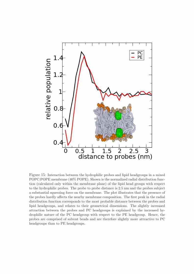
Figure 15: Interaction between the hydrophilic probes and lipid headgroups in a mixedPOPC:POPE membrane (40% POPE). Shown is the normalized radial distribution func-tion (calculated only within the membrane plane) of the lipid head groups with respectto the hydrophilic probes. The probe to probe distance is 2.5 nm and the probes subjecta substantial squeezing force on the membrane. The plot illustrates that the presence ofthe probes hardly affects the nearby membrane composition. The first peak in the radialdistribution function corresponds to the most probable distance between the probes andlipid headgroups, and relates to their geometrical dimensions. The slightly increasedattraction between the probes and PC headgroups is explained by the increased hy-drophilic nature of the PC headgroup with respect to the PE headgroup. Hence, theprobes are comprised of solvent beads and are therefore slightly more attractive to PCheadgroups than to PE headgroups.