Fabrication and evaluation of porous Ti–HA bio-nanomaterial by leaching process
8
ORIGINAL ARTICLE Fabrication and evaluation of porous Ti–HA bio-nanomaterial by leaching process A.M. Omran a , Kee Do Woo b , Duck Soo Kang b , G.T. Abdel-Gaber c , H. Fouad d,e , Hany S. Abdo g,h , Khalil Abdelrazek Khalil f,g, * a Mining and Metallurgical Depart., Faculty of Engineering, Al-Azhar University, Qena 83513, Egypt b Division of Advanced Materials Engineering & RCIT, Chonbuk National University, 561-756, South Korea c Faculty of Engineering, South Valley University, Qena, Egypt d Riyadh Community College, King Saud University, P.O. Box 28095, Riyadh 11437, Saudi Arabia e Biomedical Engineering Department, Faculty of Engineering, Helwan University, P.O. Box 11792, Helwan, Egypt f Mechanical Engineering Department, King Saud University, P.O. Box 800, Riyadh 11421, Saudi Arabia g Faculty of Energy Engineering, Aswan University, Aswan, Egypt h Center of Excellence for Research in Engineering Materials (CEREM), Advanced Manufacturing Institute, King Saud University, P.O. Box 800, Al-Riyadh 11421, Saudi Arabia Received 20 September 2014; accepted 28 November 2014 Available online 8 December 2014 KEYWORDS Ti–HA composite; Biomaterial; Leaching solution; PCAS; Porosity; Powder metallurgy Abstract A porous surface of Ti–HA composite was successfully fabricated by pulsed current acti- vated sintering (PCAS), followed by leaching using diluted H 3 PO 4 . The Ti and HA powders were mixed at different contents of the HA, Ti-5, 10, 30 and 40 wt% HA powders. The mixed powders were pressed in a coated graphite die using pulsed current activated sintering (PCAS) under pressure of 60 MPa at temperature of 1000 °C for 5 min. The sintered Ti–HA specimens were immersed in the eight kinds of leaching solutions at room temperature for 24 h. The leached specimen’s surfaces were characterized using XRD, SEM, EDX and Rockwell hardness. The XRD patterns after sin- tering show that many phases were detected at the sintered specimen surfaces such as; Ti 2 O, CaO, CaTiO 3 , Ti x P y in addition to the remaining Ti and HA. Furthermore, the high concentration H 3 PO 4 leaching solution is more efficient than the low concentration. Also the produced porous surfaces of Ti–HA materials containing more than 30% HA have a low relative density and hard- * Corresponding author at: Mechanical Engineering Department, King Saud University, P.O. Box 800, Riyadh 11421, Saudi Arabia. Tel.: +966 1467 8972. E-mail address: [email protected] (K.A. Khalil). Peer review under responsibility of King Saud University. Production and hosting by Elsevier Arabian Journal of Chemistry (2015) 8, 372–379 King Saud University Arabian Journal of Chemistry www.ksu.edu.sa www.sciencedirect.com http://dx.doi.org/10.1016/j.arabjc.2014.11.052 1878-5352 ª 2014 The Authors. Production and hosting by Elsevier B.V. on behalf of King Saud University. This is an open access article under the CC BY-NC-ND license (http://creativecommons.org/licenses/by-nc-nd/3.0/).
Transcript of Fabrication and evaluation of porous Ti–HA bio-nanomaterial by leaching process
Arabian Journal of Chemistry (2015) 8, 372–379
King Saud University
Arabian Journal of Chemistry
www.ksu.edu.sawww.sciencedirect.com
ORIGINAL ARTICLE
Fabrication and evaluation of porous Ti–HA
bio-nanomaterial by leaching process
* Corresponding author at: Mechanical Engineering Department,
King Saud University, P.O. Box 800, Riyadh 11421, Saudi Arabia.
Tel.: +966 1467 8972.
E-mail address: [email protected] (K.A. Khalil).
Peer review under responsibility of King Saud University.
Production and hosting by Elsevier
http://dx.doi.org/10.1016/j.arabjc.2014.11.0521878-5352 ª 2014 The Authors. Production and hosting by Elsevier B.V. on behalf of King Saud University.This is an open access article under the CC BY-NC-ND license (http://creativecommons.org/licenses/by-nc-nd/3.0/).
A.M. Omran a, Kee Do Woo b, Duck Soo Kang b, G.T. Abdel-Gaber c,
H. Fouad d,e, Hany S. Abdo g,h, Khalil Abdelrazek Khalil f,g,*
a Mining and Metallurgical Depart., Faculty of Engineering, Al-Azhar University, Qena 83513, Egyptb Division of Advanced Materials Engineering & RCIT, Chonbuk National University, 561-756, South Koreac Faculty of Engineering, South Valley University, Qena, Egyptd Riyadh Community College, King Saud University, P.O. Box 28095, Riyadh 11437, Saudi Arabiae Biomedical Engineering Department, Faculty of Engineering, Helwan University, P.O. Box 11792, Helwan, Egyptf Mechanical Engineering Department, King Saud University, P.O. Box 800, Riyadh 11421, Saudi Arabiag Faculty of Energy Engineering, Aswan University, Aswan, Egypth Center of Excellence for Research in Engineering Materials (CEREM), Advanced Manufacturing Institute,
King Saud University, P.O. Box 800, Al-Riyadh 11421, Saudi Arabia
Received 20 September 2014; accepted 28 November 2014Available online 8 December 2014
KEYWORDS
Ti–HA composite;
Biomaterial;
Leaching solution;
PCAS;
Porosity;
Powder metallurgy
Abstract A porous surface of Ti–HA composite was successfully fabricated by pulsed current acti-
vated sintering (PCAS), followed by leaching using diluted H3PO4. The Ti and HA powders were
mixed at different contents of the HA, Ti-5, 10, 30 and 40 wt% HA powders. The mixed powders
were pressed in a coated graphite die using pulsed current activated sintering (PCAS) under pressure
of 60 MPa at temperature of 1000 �C for 5 min. The sintered Ti–HA specimens were immersed in
the eight kinds of leaching solutions at room temperature for 24 h. The leached specimen’s surfaces
were characterized using XRD, SEM, EDX and Rockwell hardness. The XRD patterns after sin-
tering show that many phases were detected at the sintered specimen surfaces such as; Ti2O, CaO,
CaTiO3, TixPy in addition to the remaining Ti and HA. Furthermore, the high concentration
H3PO4 leaching solution is more efficient than the low concentration. Also the produced porous
surfaces of Ti–HA materials containing more than 30% HA have a low relative density and hard-
Fabrication and evaluation of porous Ti–HA 373
ness than the commercial Ti–6Al–4V ELI alloy. In a word, the presence of porous surface coated by
HA will promote the nucleation of the biological apatite created with the human tissue and increase
the bonding between them. So, the produced porous materials are considered so easy for the muscle
cells to permeate after transplanted with high coherence.
ª 2014 The Authors. Production and hosting by Elsevier B.V. on behalf of King Saud University. This is
an open access article under theCCBY-NC-ND license (http://creativecommons.org/licenses/by-nc-nd/3.0/).
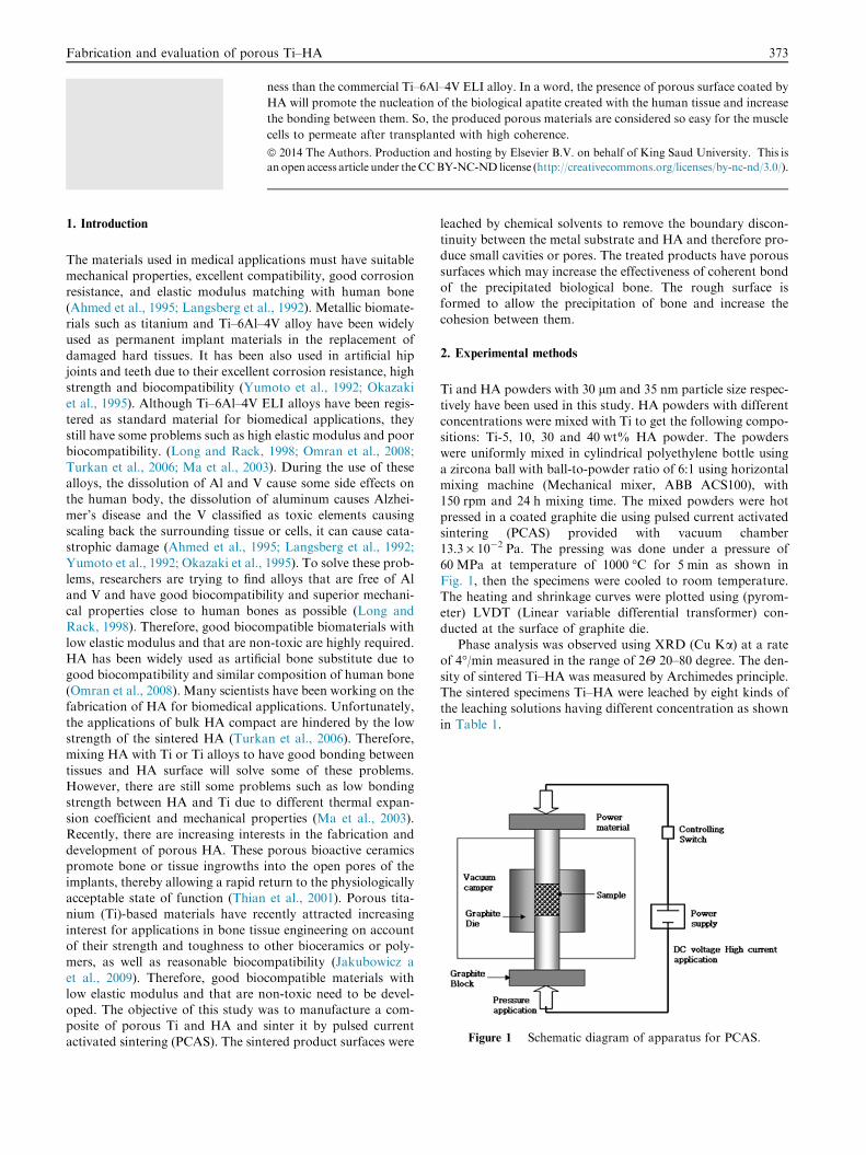
Figure 1 Schematic diagram of apparatus for PCAS.
1. Introduction
The materials used in medical applications must have suitablemechanical properties, excellent compatibility, good corrosionresistance, and elastic modulus matching with human bone
(Ahmed et al., 1995; Langsberg et al., 1992). Metallic biomate-rials such as titanium and Ti–6Al–4V alloy have been widelyused as permanent implant materials in the replacement of
damaged hard tissues. It has been also used in artificial hipjoints and teeth due to their excellent corrosion resistance, highstrength and biocompatibility (Yumoto et al., 1992; Okazaki
et al., 1995). Although Ti–6Al–4V ELI alloys have been regis-tered as standard material for biomedical applications, theystill have some problems such as high elastic modulus and poorbiocompatibility. (Long and Rack, 1998; Omran et al., 2008;
Turkan et al., 2006; Ma et al., 2003). During the use of thesealloys, the dissolution of Al and V cause some side effects onthe human body, the dissolution of aluminum causes Alzhei-
mer’s disease and the V classified as toxic elements causingscaling back the surrounding tissue or cells, it can cause cata-strophic damage (Ahmed et al., 1995; Langsberg et al., 1992;
Yumoto et al., 1992; Okazaki et al., 1995). To solve these prob-lems, researchers are trying to find alloys that are free of Aland V and have good biocompatibility and superior mechani-
cal properties close to human bones as possible (Long andRack, 1998). Therefore, good biocompatible biomaterials withlow elastic modulus and that are non-toxic are highly required.HA has been widely used as artificial bone substitute due to
good biocompatibility and similar composition of human bone(Omran et al., 2008). Many scientists have been working on thefabrication of HA for biomedical applications. Unfortunately,
the applications of bulk HA compact are hindered by the lowstrength of the sintered HA (Turkan et al., 2006). Therefore,mixing HA with Ti or Ti alloys to have good bonding between
tissues and HA surface will solve some of these problems.However, there are still some problems such as low bondingstrength between HA and Ti due to different thermal expan-
sion coefficient and mechanical properties (Ma et al., 2003).Recently, there are increasing interests in the fabrication anddevelopment of porous HA. These porous bioactive ceramicspromote bone or tissue ingrowths into the open pores of the
implants, thereby allowing a rapid return to the physiologicallyacceptable state of function (Thian et al., 2001). Porous tita-nium (Ti)-based materials have recently attracted increasing
interest for applications in bone tissue engineering on accountof their strength and toughness to other bioceramics or poly-mers, as well as reasonable biocompatibility (Jakubowicz a
et al., 2009). Therefore, good biocompatible materials withlow elastic modulus and that are non-toxic need to be devel-oped. The objective of this study was to manufacture a com-posite of porous Ti and HA and sinter it by pulsed current
activated sintering (PCAS). The sintered product surfaces were
leached by chemical solvents to remove the boundary discon-
tinuity between the metal substrate and HA and therefore pro-duce small cavities or pores. The treated products have poroussurfaces which may increase the effectiveness of coherent bond
of the precipitated biological bone. The rough surface isformed to allow the precipitation of bone and increase thecohesion between them.
2. Experimental methods
Ti and HA powders with 30 lm and 35 nm particle size respec-
tively have been used in this study. HA powders with differentconcentrations were mixed with Ti to get the following compo-sitions: Ti-5, 10, 30 and 40 wt% HA powder. The powderswere uniformly mixed in cylindrical polyethylene bottle using
a zircona ball with ball-to-powder ratio of 6:1 using horizontalmixing machine (Mechanical mixer, ABB ACS100), with150 rpm and 24 h mixing time. The mixed powders were hot
pressed in a coated graphite die using pulsed current activatedsintering (PCAS) provided with vacuum chamber13.3 · 10�2 Pa. The pressing was done under a pressure of
60 MPa at temperature of 1000 �C for 5 min as shown inFig. 1, then the specimens were cooled to room temperature.The heating and shrinkage curves were plotted using (pyrom-
eter) LVDT (Linear variable differential transformer) con-ducted at the surface of graphite die.
Phase analysis was observed using XRD (Cu Ka) at a rateof 4�/min measured in the range of 2H 20–80 degree. The den-
sity of sintered Ti–HA was measured by Archimedes principle.The sintered specimens Ti–HA were leached by eight kinds ofthe leaching solutions having different concentration as shown
in Table 1.
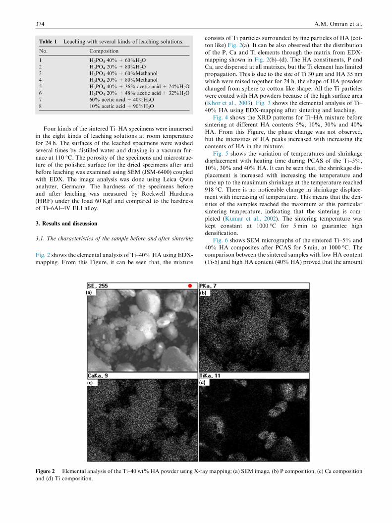
Table 1 Leaching with several kinds of leaching solutions.
No. Composition
1 H3PO4 40%+ 60%H2O
2 H3PO4 20%+ 80%H2O
3 H3PO4 40%+ 60%Methanol
4 H3PO4 20%+ 80%Methanol
5 H3PO4 40%+ 36% acetic acid + 24%H2O
6 H3PO4 20%+ 48% acetic acid + 32%H2O
7 60% acetic acid + 40%H2O
8 10% acetic acid + 90%H2O
374 A.M. Omran et al.
Four kinds of the sintered Ti–HA specimens were immersedin the eight kinds of leaching solutions at room temperaturefor 24 h. The surfaces of the leached specimens were washedseveral times by distilled water and draying in a vacuum fur-
nace at 110 �C. The porosity of the specimens and microstruc-ture of the polished surface for the dried specimens after andbefore leaching was examined using SEM (JSM-6400) coupled
with EDX. The image analysis was done using Leica Qwinanalyzer, Germany. The hardness of the specimens beforeand after leaching was measured by Rockwell Hardness
(HRF) under the load 60 Kgf and compared to the hardnessof Ti–6Al–4V ELI alloy.
3. Results and discussion
3.1. The characteristics of the sample before and after sintering
Fig. 2 shows the elemental analysis of Ti–40% HA using EDX-mapping. From this Figure, it can be seen that, the mixture
Figure 2 Elemental analysis of the Ti–40 wt% HA powder using X-ra
and (d) Ti composition.
consists of Ti particles surrounded by fine particles of HA (cot-ton like) Fig. 2(a). It can be also observed that the distributionof the P, Ca and Ti elements through the matrix from EDX-
mapping shown in Fig. 2(b)–(d). The HA constituents, P andCa, are dispersed at all matrixes, but the Ti element has limitedpropagation. This is due to the size of Ti 30 lm and HA 35 nm
which were mixed together for 24 h, the shape of HA powderschanged from sphere to cotton like shape. All the Ti particleswere coated with HA powders because of the high surface area
(Khor et al., 2003). Fig. 3 shows the elemental analysis of Ti–40% HA using EDX-mapping after sintering and leaching.
Fig. 4 shows the XRD patterns for Ti–HA mixture beforesintering at different HA contents 5%, 10%, 30% and 40%
HA. From this Figure, the phase change was not observed,but the intensities of HA peaks increased with increasing thecontents of HA in the mixture.
Fig. 5 shows the variation of temperatures and shrinkagedisplacement with heating time during PCAS of the Ti–5%,10%, 30% and 40% HA. It can be seen that, the shrinkage dis-
placement is increased with increasing the temperature andtime up to the maximum shrinkage at the temperature reached918 �C. There is no noticeable change in shrinkage displace-
ment with increasing of temperature. This means that the den-sities of the samples reached the maximum at this particularsintering temperature, indicating that the sintering is com-pleted (Kumar et al., 2002). The sintering temperature was
kept constant at 1000 �C for 5 min to guarantee highdensification.
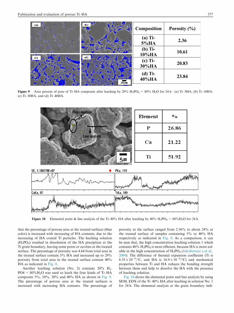
Fig. 6 shows SEM micrographs of the sintered Ti–5% and
40% HA composites after PCAS for 5 min, at 1000 �C. Thecomparison between the sintered samples with low HA content(Ti-5) and high HA content (40% HA) proved that the amount
y mapping; (a) SEM image, (b) P composition, (c) Ca composition
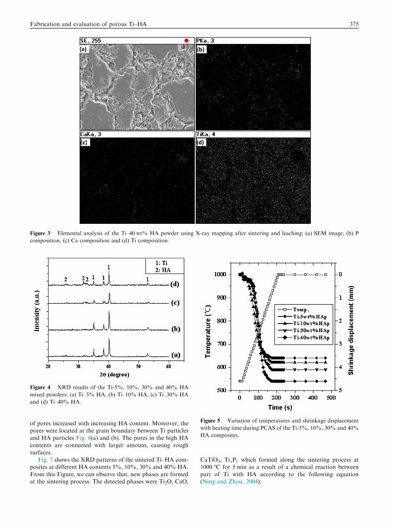
Figure 3 Elemental analysis of the Ti–40 wt% HA powder using X-ray mapping after sintering and leaching; (a) SEM image, (b) P
composition, (c) Ca composition and (d) Ti composition.
Figure 4 XRD results of the Ti-5%, 10%, 30% and 40% HA
mixed powders: (a) Ti–5% HA, (b) Ti–10% HA, (c) Ti–30% HA
and (d) Ti–40% HA.
Figure 5 Variation of temperatures and shrinkage displacement
with heating time during PCAS of the Ti-5%, 10%, 30% and 40%
HA composites.
Fabrication and evaluation of porous Ti–HA 375
of pores increased with increasing HA content. Moreover, thepores were located at the grain boundary between Ti particles
and HA particles Fig. 6(a) and (b). The pores in the high HAcontents are connected with larger amount, causing roughsurfaces.
Fig. 7 shows the XRD patterns of the sintered Ti–HA com-posites at different HA contents 5%, 10%, 30% and 40% HA.From this Figure, we can observe that, new phases are formedat the sintering process. The detected phases were Ti2O, CaO,
CaTiO3, TixPy which formed along the sintering process at1000 �C for 5 min as a result of a chemical reaction betweenpart of Ti with HA according to the following equation(Ning and Zhou, 2004):
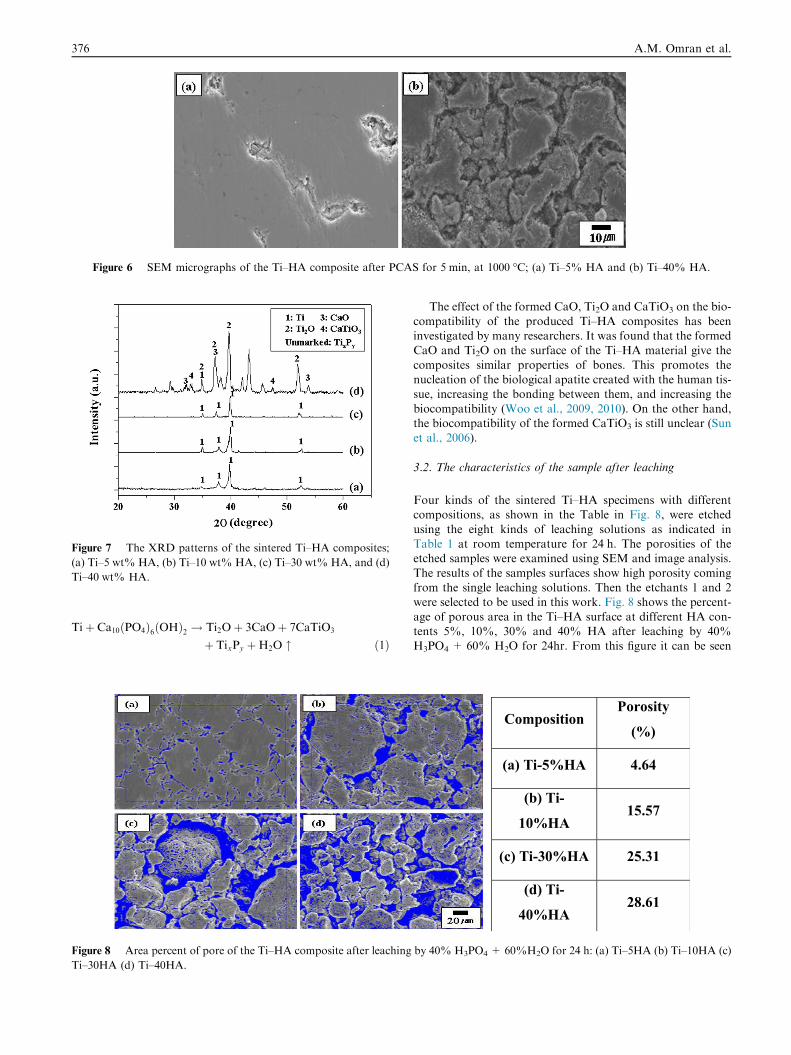
Figure 6 SEM micrographs of the Ti–HA composite after PCAS for 5 min, at 1000 �C; (a) Ti–5% HA and (b) Ti–40% HA.
Figure 7 The XRD patterns of the sintered Ti–HA composites;
(a) Ti–5 wt% HA, (b) Ti–10 wt% HA, (c) Ti–30 wt% HA, and (d)
Ti–40 wt% HA.
376 A.M. Omran et al.
Tiþ Ca10ðPO4Þ6ðOHÞ2 ! Ti2Oþ 3CaOþ 7CaTiO3
þ TixPy þH2O " ð1Þ
Figure 8 Area percent of pore of the Ti–HA composite after leaching
Ti–30HA (d) Ti–40HA.
The effect of the formed CaO, Ti2O and CaTiO3 on the bio-
compatibility of the produced Ti–HA composites has beeninvestigated by many researchers. It was found that the formedCaO and Ti2O on the surface of the Ti–HA material give the
composites similar properties of bones. This promotes thenucleation of the biological apatite created with the human tis-sue, increasing the bonding between them, and increasing the
biocompatibility (Woo et al., 2009, 2010). On the other hand,the biocompatibility of the formed CaTiO3 is still unclear (Sunet al., 2006).
3.2. The characteristics of the sample after leaching
Four kinds of the sintered Ti–HA specimens with differentcompositions, as shown in the Table in Fig. 8, were etched
using the eight kinds of leaching solutions as indicated inTable 1 at room temperature for 24 h. The porosities of theetched samples were examined using SEM and image analysis.
The results of the samples surfaces show high porosity comingfrom the single leaching solutions. Then the etchants 1 and 2were selected to be used in this work. Fig. 8 shows the percent-
age of porous area in the Ti–HA surface at different HA con-tents 5%, 10%, 30% and 40% HA after leaching by 40%H3PO4 + 60% H2O for 24hr. From this figure it can be seen
CompositionPorosity
(%)
(a) Ti-5%HA 4.64
(b) Ti-
10%HA15.57
(c) Ti-30%HA 25.31
(d) Ti-
40%HA28.61
by 40% H3PO4 + 60%H2O for 24 h: (a) Ti–5HA (b) Ti–10HA (c)
Composition Porosity (%)
(a) Ti-5%HA 2.36
(b) Ti-10%HA 10.61
(c) Ti-30%HA 20.83
(d) Ti-40%HA 23.84
Figure 9 Area percent of pore of Ti–HA composite after leaching by 20% H3PO4 + 80% H2O for 24 h : (a) Ti–5HA, (b) Ti–10HA,
(c) Ti–30HA, and (d) Ti–40HA.
Figure 10 Elemental point & line analysis of the Ti–40% HA after leaching by 40% H3PO4 + 60%H2O for 24 h.
Fabrication and evaluation of porous Ti–HA 377
that the percentage of porous area at the treated surfaces (bluecolor) is increased with increasing of HA contents, due to theincreasing of HA coated Ti particles. The leaching solution
(H3PO4) resulted in dissolution of the HA precipitate at theTi grain boundary, leaving some pores or cavities at the treatedsurface. The percentage of porosity was 4.64 from total area inthe treated surface contain 5% HA and increased up to 29%
porosity from total area in the treated surface contain 40%HA as indicated in Fig. 8.
Another leaching solution (No. 2) contains 20% H3-
PO4 + 80%H2O was used to leach the four kinds of Ti–HAcomposite 5%, 10%, 30% and 40% HA as shown in Fig. 9.The percentage of porous area at the treated surfaces is
increased with increasing HA contents. The percentage of
porosity at the surface ranged from 2.36% to about 24% atthe treated surface of samples containing 5% to 40% HArespectively as indicated in Fig. 9. As a comparison, it can
be seen that, the high concentration leaching solution 1 whichcontains 40% H3PO4 is more efficient, because HA is more sol-uble in the high concentration of H3PO4 (Jakubowicz a et al.,2009). The difference of thermal expansion coefficient (Ti is
8.35 · 10�6/�C, and HA is 16.9 · 10�6/�C) and mechanicalproperties between Ti and HA reduces the bonding strengthbetween them and help to dissolve the HA with the presence
of leaching solution.Fig. 10 shows the elemental point and line analysis by using
SEM, EDX of the Ti–40% HA after leaching in solution No. 1
for 24 h. The elemental analysis at the grain boundary indi-
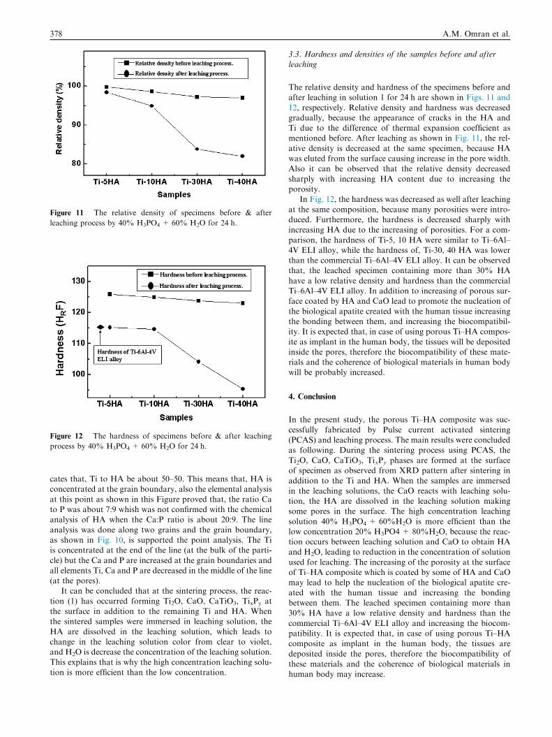
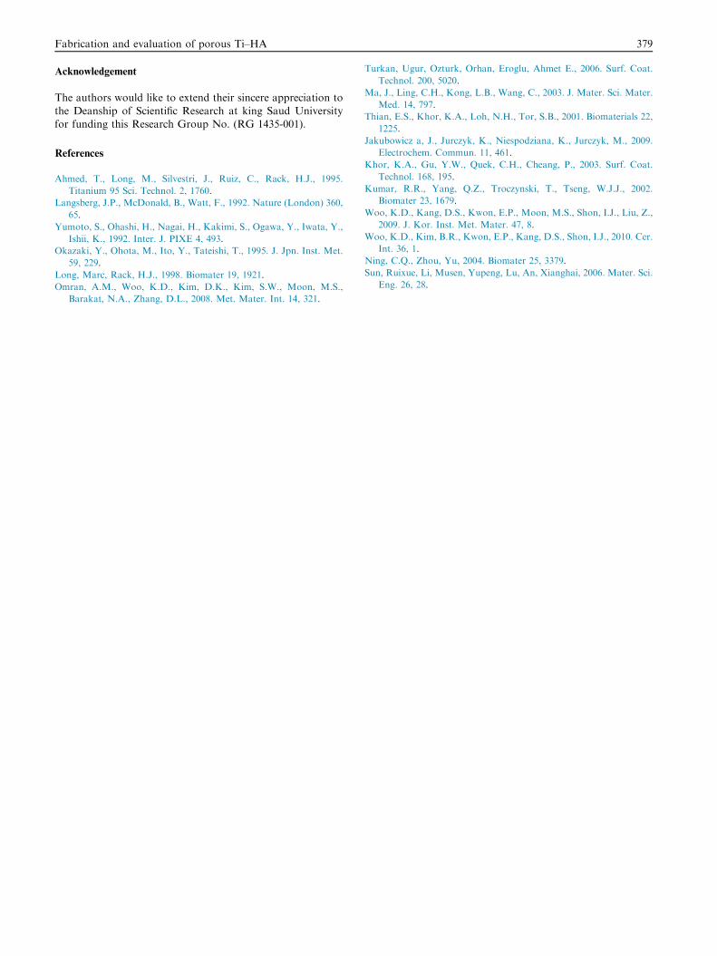
Figure 12 The hardness of specimens before & after leaching
process by 40% H3PO4 + 60% H2O for 24 h.
Figure 11 The relative density of specimens before & after
leaching process by 40% H3PO4 + 60% H2O for 24 h.
378 A.M. Omran et al.
cates that, Ti to HA be about 50–50. This means that, HA isconcentrated at the grain boundary, also the elemental analysis
at this point as shown in this Figure proved that, the ratio Cato P was about 7:9 whish was not confirmed with the chemicalanalysis of HA when the Ca:P ratio is about 20:9. The line
analysis was done along two grains and the grain boundary,as shown in Fig. 10, is supported the point analysis. The Tiis concentrated at the end of the line (at the bulk of the parti-
cle) but the Ca and P are increased at the grain boundaries andall elements Ti, Ca and P are decreased in the middle of the line(at the pores).
It can be concluded that at the sintering process, the reac-
tion (1) has occurred forming Ti2O, CaO, CaTiO3, TixPy atthe surface in addition to the remaining Ti and HA. Whenthe sintered samples were immersed in leaching solution, the
HA are dissolved in the leaching solution, which leads tochange in the leaching solution color from clear to violet,and H2O is decrease the concentration of the leaching solution.
This explains that is why the high concentration leaching solu-tion is more efficient than the low concentration.
3.3. Hardness and densities of the samples before and afterleaching
The relative density and hardness of the specimens before andafter leaching in solution 1 for 24 h are shown in Figs. 11 and
12, respectively. Relative density and hardness was decreasedgradually, because the appearance of cracks in the HA andTi due to the difference of thermal expansion coefficient asmentioned before. After leaching as shown in Fig. 11, the rel-
ative density is decreased at the same specimen, because HAwas eluted from the surface causing increase in the pore width.Also it can be observed that the relative density decreased
sharply with increasing HA content due to increasing theporosity.
In Fig. 12, the hardness was decreased as well after leaching
at the same composition, because many porosities were intro-duced. Furthermore, the hardness is decreased sharply withincreasing HA due to the increasing of porosities. For a com-
parison, the hardness of Ti-5, 10 HA were similar to Ti–6Al–4V ELI alloy, while the hardness of, Ti-30, 40 HA was lowerthan the commercial Ti–6Al–4V ELI alloy. It can be observedthat, the leached specimen containing more than 30% HA
have a low relative density and hardness than the commercialTi–6Al–4V ELI alloy. In addition to increasing of porous sur-face coated by HA and CaO lead to promote the nucleation of
the biological apatite created with the human tissue increasingthe bonding between them, and increasing the biocompatibil-ity. It is expected that, in case of using porous Ti–HA compos-
ite as implant in the human body, the tissues will be depositedinside the pores, therefore the biocompatibility of these mate-rials and the coherence of biological materials in human bodywill be probably increased.
4. Conclusion
In the present study, the porous Ti–HA composite was suc-
cessfully fabricated by Pulse current activated sintering(PCAS) and leaching process. The main results were concludedas following. During the sintering process using PCAS, the
Ti2O, CaO, CaTiO3, TixPy phases are formed at the surfaceof specimen as observed from XRD pattern after sintering inaddition to the Ti and HA. When the samples are immersed
in the leaching solutions, the CaO reacts with leaching solu-tion, the HA are dissolved in the leaching solution makingsome pores in the surface. The high concentration leaching
solution 40% H3PO4 + 60%H2O is more efficient than thelow concentration 20% H3PO4 + 80%H2O, because the reac-tion occurs between leaching solution and CaO to obtain HAand H2O, leading to reduction in the concentration of solution
used for leaching. The increasing of the porosity at the surfaceof Ti–HA composite which is coated by some of HA and CaOmay lead to help the nucleation of the biological apatite cre-
ated with the human tissue and increasing the bondingbetween them. The leached specimen containing more than30% HA have a low relative density and hardness than the
commercial Ti–6Al–4V ELI alloy and increasing the biocom-patibility. It is expected that, in case of using porous Ti–HAcomposite as implant in the human body, the tissues are
deposited inside the pores, therefore the biocompatibility ofthese materials and the coherence of biological materials inhuman body may increase.
Fabrication and evaluation of porous Ti–HA 379
Acknowledgement
The authors would like to extend their sincere appreciation to
the Deanship of Scientific Research at king Saud Universityfor funding this Research Group No. (RG 1435-001).
References
Ahmed, T., Long, M., Silvestri, J., Ruiz, C., Rack, H.J., 1995.
Titanium 95 Sci. Technol. 2, 1760.
Langsberg, J.P., McDonald, B., Watt, F., 1992. Nature (London) 360,
65.
Yumoto, S., Ohashi, H., Nagai, H., Kakimi, S., Ogawa, Y., Iwata, Y.,
Ishii, K., 1992. Inter. J. PIXE 4, 493.
Okazaki, Y., Ohota, M., Ito, Y., Tateishi, T., 1995. J. Jpn. Inst. Met.
59, 229.
Long, Marc, Rack, H.J., 1998. Biomater 19, 1921.
Omran, A.M., Woo, K.D., Kim, D.K., Kim, S.W., Moon, M.S.,
Barakat, N.A., Zhang, D.L., 2008. Met. Mater. Int. 14, 321.
Turkan, Ugur, Ozturk, Orhan, Eroglu, Ahmet E., 2006. Surf. Coat.
Technol. 200, 5020.
Ma, J., Ling, C.H., Kong, L.B., Wang, C., 2003. J. Mater. Sci. Mater.
Med. 14, 797.
Thian, E.S., Khor, K.A., Loh, N.H., Tor, S.B., 2001. Biomaterials 22,
1225.
Jakubowicz a, J., Jurczyk, K., Niespodziana, K., Jurczyk, M., 2009.
Electrochem. Commun. 11, 461.
Khor, K.A., Gu, Y.W., Quek, C.H., Cheang, P., 2003. Surf. Coat.
Technol. 168, 195.
Kumar, R.R., Yang, Q.Z., Troczynski, T., Tseng, W.J.J., 2002.
Biomater 23, 1679.
Woo, K.D., Kang, D.S., Kwon, E.P., Moon, M.S., Shon, I.J., Liu, Z.,
2009. J. Kor. Inst. Met. Mater. 47, 8.
Woo, K.D., Kim, B.R., Kwon, E.P., Kang, D.S., Shon, I.J., 2010. Cer.
Int. 36, 1.
Ning, C.Q., Zhou, Yu, 2004. Biomater 25, 3379.
Sun, Ruixue, Li, Musen, Yupeng, Lu, An, Xianghai, 2006. Mater. Sci.
Eng. 26, 28.