Certificate of Attendance - American Psychiatric Association
Upload
khangminh22Category
view
0download
0
EYEBLINK CONDITIONING IN PSYCHIATRIC CONDITIONS - STATE OF THE FIELD AND FUTURE DIRECTIONSEDITED BY : Tracy L. Greer and Lucien T. ThompsonPUBLISHED IN : Frontiers in Psychiatry

1Frontiers in Psychiatry August 2017 | Eyeblink Conditioning in Psychiatric Conditions
Frontiers Copyright Statement
© Copyright 2007-2017 Frontiers Media SA. All rights reserved.
All content included on this site, such as text, graphics, logos, button
icons, images, video/audio clips, downloads, data compilations and
software, is the property of or is licensed to Frontiers Media SA
(“Frontiers”) or its licensees and/or subcontractors. The copyright in the
text of individual articles is the property of their respective authors, subject to
a license granted to Frontiers.
The compilation of articles constituting this e-book, wherever published,
as well as the compilation of all other content on this site, is the exclusive
property of Frontiers. For the conditions for downloading and
copying of e-books from Frontiers’ website, please see the Terms for
Website Use. If purchasing Frontiers e-books from other websites
or sources, the conditions of the website concerned apply.
Images and graphics not forming part of user-contributed materials may
not be downloaded or copied without permission.
Individual articles may be downloaded and reproduced in accordance
with the principles of the CC-BY licence subject to any copyright or
other notices. They may not be re-sold as an e-book.
As author or other contributor you grant a CC-BY licence to others to
reproduce your articles, including any graphics and third-party materials
supplied by you, in accordance with the Conditions for Website Use and
subject to any copyright notices which you include in connection with your
articles and materials.
All copyright, and all rights therein, are protected by national and
international copyright laws.
The above represents a summary only. For the full conditions see the
Conditions for Authors and the Conditions for Website Use.
ISSN 1664-8714 ISBN 978-2-88945-275-0
DOI 10.3389/978-2-88945-275-0
About FrontiersFrontiers is more than just an open-access publisher of scholarly articles: it is a pioneering approach to the world of academia, radically improving the way scholarly research is managed. The grand vision of Frontiers is a world where all people have an equal opportunity to seek, share and generate knowledge. Frontiers provides immediate and permanent online open access to all its publications, but this alone is not enough to realize our grand goals.
Frontiers Journal SeriesThe Frontiers Journal Series is a multi-tier and interdisciplinary set of open-access, online journals, promising a paradigm shift from the current review, selection and dissemination processes in academic publishing. All Frontiers journals are driven by researchers for researchers; therefore, they constitute a service to the scholarly community. At the same time, the Frontiers Journal Series operates on a revolutionary invention, the tiered publishing system, initially addressing specific communities of scholars, and gradually climbing up to broader public understanding, thus serving the interests of the lay society, too.
Dedication to qualityEach Frontiers article is a landmark of the highest quality, thanks to genuinely collaborative interactions between authors and review editors, who include some of the world’s best academicians. Research must be certified by peers before entering a stream of knowledge that may eventually reach the public - and shape society; therefore, Frontiers only applies the most rigorous and unbiased reviews. Frontiers revolutionizes research publishing by freely delivering the most outstanding research, evaluated with no bias from both the academic and social point of view.By applying the most advanced information technologies, Frontiers is catapulting scholarly publishing into a new generation.
What are Frontiers Research Topics?Frontiers Research Topics are very popular trademarks of the Frontiers Journals Series: they are collections of at least ten articles, all centered on a particular subject. With their unique mix of varied contributions from Original Research to Review Articles, Frontiers Research Topics unify the most influential researchers, the latest key findings and historical advances in a hot research area! Find out more on how to host your own Frontiers Research Topic or contribute to one as an author by contacting the Frontiers Editorial Office: [email protected]

EYEBLINK CONDITIONING IN PSYCHIATRIC CONDITIONS - STATE OF THE FIELD AND FUTURE DIRECTIONS
Topic Editors:Tracy L. Greer, University of Texas Southwestern Medical Center, United StatesLucien T. Thompson, University of Texas at Dallas, United States
Eyeblink classical conditioning (EBC) is a model paradigm for associative (also termed Pavlovian) learning, one of the simplest and best understood forms of learning and memory. Because EBC paradigms are readily adapted across species, the neural substrates of EBC have been well characterized, and include but are not limited to the cerebellum and anterior interpositus nucleus, the hippocampus, and prefrontal cortices. The ability to collect EBC data across many different species (i.e. including but not limited to humans) also has the distinct advantage of facilitating translational research, and therefore may be of particular benefit to elucidate mechanistic changes associated with a wide variety of psychiatric disorders.
In fact, EBC paradigms have been employed to assess individuals with a wide range of neurological deficits (including Korsakoff ’s amnesia, Alzheimer’s disease as well as normal aging, dyslexia, inflammatory tremor, dystonia, and multiple sclerosis) and psychiatric disorders (including major depressive disorder, anxiety disorders, schizophrenia, autism, and alcohol use/addiction disorders). Individuals with these disorders exhibit differential impairments across different EBC task types (e.g., delay vs. trace EBC), with some showing impairment in one but not the other task and some showing impairments in both; across learning stage (e.g., acquisition, discrimination, or extinction), and across response variables (e.g., magnitude and timing of the conditioned eyeblink motor response, modality of the conditioned stimulus). Evaluating specific individual differences in the context of variable brain pathology should aid characterization and refinement of our understanding of complex neuropsychiatric disorders.
The field of psychiatry has seen a transition from more traditional use of symptom clusters to define psychiatric disorders with subsequent examination of associated behaviors and traits, to the use of physiological and behavioral indicators to characterize individuals with respect to various psychological domains [in line with the National Institute of Mental Health Research Domain Criteria (RDoC) initiative]. This approach employs a neuroscience-based framework to assess the pathophysiology of chronic mental illnesses. Behavioral and cognitive processes are critical domains of interest in evaluating potential maladaptive patterns that may be indicative of specific psychopathologies. Furthermore, the rapid development of technological advances that allow for more detailed examination (e.g., EEG, MEG, MRI, fMRI, infrared
2Frontiers in Psychiatry August 2017 | Eyeblink Conditioning in Psychiatric Conditions

imaging) and manipulation (e.g. transcranial magnetic and direct current stimulation) of brain functions should enhance our ability to better characterize EBC performance and its utility in characterizing aspects of particular neuropathologies.
Substantial research evidence exists for the value of EBC paradigms to inform our understanding of the pathophysiologies underlying a wide variety of neurological and psychiatric disorders. Despite these findings, this readily implemented classic cognitive-behavioral paradigm is relatively underutilized in clinical settings. This e-book highlights recent convergence of clinical and research efforts in this area and aims to promote a resurgent interest in eyeblink classical conditioning, and to emphasize the potential for future translational and diagnostic applications of EBC in combination with other techniques to strengthen our understanding of alterations in brain function manifested in behaviors characteristic of specific psychopathologies.
Citation: Greer, T. L., Thompson, L. T., eds. (2017). Eyeblink Conditioning in Psychiatric Conditions - State of the Field and Future Directions. Lausanne: Frontiers Media. doi: 10.3389/978-2-88945-275-0
3Frontiers in Psychiatry August 2017 | Eyeblink Conditioning in Psychiatric Conditions

Chapter 1: Eyeblink Conditioning in Psychiatric Conditions: Overview and Mechanisms across Diagnoses
06 Editorial: Eyeblink Classical Conditioning in Psychiatric Conditions: Novel Uses for a Classic Paradigm
Tracy L. Greer and Lucien T. Thompson08 Eyeblink Conditioning and Novel Object Recognition in the Rabbit: Behavioral
Paradigms for Assaying Psychiatric Diseases Craig Weiss and John F. Disterhoft17 Hippocampal Non-Theta-Contingent Eyeblink Classical Conditioning: A Model
System for Neurobiological Dysfunction Joseph J. Cicchese and Stephen D. Berry31 Timing Tasks Synchronize Cerebellar and Frontal Ramping Activity and Theta
Oscillations: Implications for Cerebellar Stimulation in Diseases of Impaired Cognition
Krystal L. Parker
Chapter 2: Schizophrenia
37 Eyeblink Conditioning in Schizophrenia: A Critical Review Jerillyn S. Kent, Amanda R. Bolbecker, Brian F. O’Donnell and William P. Hetrick52 New Insights into the Nature of Cerebellar-Dependent Eyeblink Conditioning
Deficits in Schizophrenia: A Hierarchical Linear Modeling Approach Amanda R. Bolbecker, Isaac T. Petersen, Jerillyn S. Kent, Josselyn M. Howell,
Brian F. O’Donnell and William P. Hetrick
Chapter 3: Anxiety Disorders
59 Eyeblink Classical Conditioning and Post-traumatic Stress Disorder – A Model Systems Approach
Bernard G. Schreurs and Lauren B. Burhans72 Investigating the Role of Hippocampal BDNF in Anxiety Vulnerability using
Classical Eyeblink Conditioning Kellie L. Janke, Tara P. Cominski, Eldo V. Kuzhikandathil, Richard J. Servatius and
Kevin C. H. Pang
Chapter 4: Autism
81 Autism and Classical Eyeblink Conditioning: Performance Changes of the Conditioned Response Related to Autism Spectrum Disorder Diagnosis
John P. Welsh and Jeffrey T. Oristaglio
Table of Contents
4Frontiers in Psychiatry August 2017 | Eyeblink Conditioning in Psychiatric Conditions

Chapter 5: Alcohol Use Disorders
90 Eyeblink Classical Conditioning in Alcoholism and Fetal Alcohol Spectrum Disorders
Dominic T. Cheng, Sandra W. Jacobson, Joseph L. Jacobson, Christopher D. Molteno, Mark E. Stanton and John E. Desmond
5Frontiers in Psychiatry August 2017 | Eyeblink Conditioning in Psychiatric Conditions

March 2017 | Volume 8 | Article 486
EDITORIALpublished: 27 March 2017
doi: 10.3389/fpsyt.2017.00048
Frontiers in Psychiatry | www.frontiersin.org
Edited by: Raina Robeva,
Sweet Briar College, USA
Reviewed by: Raina Robeva,
Sweet Briar College, USA Nadia Chaudhri,
Concordia University, Canada
*Correspondence:Tracy L. Greer
[email protected]; Lucien T. Thompson
Specialty section: This article was submitted to
Systems Biology, a section of the journal Frontiers in Psychiatry
Received: 19 January 2017Accepted: 09 March 2017Published: 27 March 2017
Citation: Greer TL and Thompson LT (2017)
Editorial: Eyeblink Classical Conditioning in Psychiatric Conditions: Novel Uses for
a Classic Paradigm. Front. Psychiatry 8:48.
doi: 10.3389/fpsyt.2017.00048
Editorial: Eyeblink Classical Conditioning in Psychiatric Conditions: Novel Uses for a Classic ParadigmTracy L. Greer1* and Lucien T. Thompson2*
1 Department of Psychiatry, UT Southwestern Medical Center, Dallas, TX, USA, 2 Aging & Memory Research, Behavioral and Brain Sciences, University of Texas at Dallas, Richardson, TX, USA
Keywords: schizophrenia, alcohol abuse, hippocampus, frontal cortex, cerebellum, post-traumatic stress disorder, autism, depression
Editorial on the Research Topic
Eyeblink Classical Conditioning in Psychiatric Conditions: Novel Uses for a Classic Paradigm
As one of the most basic forms of associative learning, eyeblink conditioning (EBC) is a model paradigm with unique utility in the assessment of complex behavioral disorders, including psychi-atric disorders. Two major EBC paradigms utilized with human subjects are delay EBC [in which a conditioned stimulus (CS; e.g., an auditory tone) co-terminates with an unconditioned stimulus (US; e.g., a corneal airpuff)] and trace EBC [in which CS presentation is followed after a silent inter-stimulus interval (termed the “trace” interval by Pavlov) by the US, with no CS-US overlap in time]. The neural substrates of EBC in these paradigms are well delineated and include the cerebellum and anterior interpositus nucleus, the hippocampus, and prefrontal cortex. Variability in acquisition, discrimination, timing, sensitization, and/or extinction of classically conditioned eyeblink responses provides insight into the behavioral and neurobiological characteristics of a variety of psychiatric and neurological disorders.
In this Research Topic, Kent et al. review the existing literature on EBC studies in schizophrenia, perhaps the most studied psychiatric diagnostic group with respect to EBC to date. In their review of 15 studies, they report that decreased percent CRs (impaired learning) is the most robust and rep-licated finding in this population, with equivocal findings associated with CR timing. Importantly, differences between schizophrenic and healthy controls in percent CRs appear to be reflective of cer-ebellar abnormalities—supported by neuroimaging data and data from first-degree relatives—rather than confounding issues such as medication use. However, the authors highlight the importance of methodological consistency across EBC paradigms for accurate interpretation and synthesis of data across studies, an issue critical in the evaluation of any behavioral measure. Bolbecker et al. report data on delay EBC in schizophrenia using an analytic approach—hierarchical liner modeling—that has the advantages of less restrictive assumptions and inclusion of unequal variances in comparison to more traditionally employed analytic approaches, such as repeated measures analysis of vari-ance. The authors did not find differences between schizophrenic patients and age-matched healthy controls in learning rate, but did observe group differences in time to maximal learning level and plateau of learning response, with schizophrenic patients exhibiting saturation in learning earlier and at a lower level in comparison to healthy controls.
Cheng et al. provide summaries of both animal and human work describing the impact of alcohol use disorders in adults and fetal alcohol syndrome in children. EBC studies in these populations have illuminated structural and functional differences in both the mature and developing cerebellum, as well as learning differences in these diagnostic groups when compared to healthy controls.
7
Greer and Thompson Eyeblink Conditioning in Psychiatric Conditioning
Frontiers in Psychiatry | www.frontiersin.org March 2017 | Volume 8 | Article 48
Weiss and Disterhoft emphasize the utility of rabbit EBC for assessing cerebro-cerebellar functional connectivity. The role of hippocampus, fronto-temporal and cerebellar cortices, and of cerebellar deep nuclei has been well characterized in a wide variety of normal states as well as dysfunctional psychiatric con-ditions. Their review concentrates on models of schizophrenia and Alzheimer’s dementia, but also points out applications for normal aging, Parkinson’s disease, progressive supranuclear palsy, alcoholism, and post-traumatic stress disorder (PTSD). Indeed, the rabbit EBC model was developed in parallel with assessment of human EBC in a series of studies from the laboratories of Gormezano, his students, and colleagues, which have allowed mechanistic, physiological, and pharmacological assessments to be carried out in depth.
Schreurs and Burhans detail development of a preclinical EBC extinction model with potential utility for treatment of inap-propriate conditioned behaviors and hyperarousal in individuals suffering from PTSD, whether combat veterans or approximately 5–25% of the general populace who experience severe anxiety, sleeplessness, hypervigilance, and/or flashbacks after trauma constitute a group at extreme risk for suicide. Their exposure paradigm uses conditioning-specific reflex modification and attenuated intensity US presentations to enhance the likely clini-cal utility for human applications.
Janke et al. discuss dysregulation of brain-derived neurotrophic factor (BDNF) in anxiety disorders, including impaired behavioral inhibition temperament (BI). They describe facilitated delay EBC in both human and animal subjects exhibiting BI. In their animal model after EBC, increases in mRNA for hippocampal BDNF were observed in both the dentate gyrus and CA3 regions, along with upregulation of TrkB receptors and downstream activity-related cytoskeletal protein (Arc) in the hippocampus, but these increases were blunted in the strain showing faster acquisition and hyper-sensitivity to both CS and US. Hippocampal BDNF administration reversed the behavioral sensitization, suggesting treatment strategies that enhance that hippocampal BDNF may be effective for a variety of anxiety disorders.
Welsh and Oristaglio provide a secondary analysis of children with autism spectrum disorder (ASD) who underwent both delay and trace EBC. They grouped children based on their diagnosis into either an autistic disorder group or an Asperger’s syndrome or Pervasive Developmental Disorder (Asp/PDD) group. Neither ASD group showed differences in CR acquisition compared to an age- and IQ-matched group of typically devel-oping (TD) children. However, the groups differed with respect to CR timing alterations, with the Asp/PDD group showing delayed CR onset and peak latencies during trace conditioning that were not observed in the ASD or TD groups. These data
may illustrate differences in the underlying biology of these two diagnostic groups that are expressed as behavioral differences. Although the authors indicate that these differences must be further tested in a larger trial, these data are representative of the types of approaches that are supported by the NIMH’s Research Domain Criterion (RDoc) initiative that aims to use a variety of approaches, including behavioral phenotyping, to help our field better distinguish brain dysfunction among patients with psychiatric illnesses.
Similarly, Parker describes the potential for timing tasks to aid in our understanding of cognitive impairment across diagnostic groups, again in line with the RDoC approach. She asserts that tasks that involve temporal processing, such as EBC, can reflect connectivity between the cerebellum and frontal cortex, and in particular, hypothesizes that the medial frontal cortex plays a critical role in cognitive processing that occurs in timing tasks. This reciprocal relationship between cerebellar and frontal corti-cal regions is an important area of further investigation.
Cicchese and Berry also focus on timing and the critical role of theta (the 3–7 Hz EEG bandwidth prominent in medial temporal lobe) in many forms of learning and memory. They review non-theta contingent EBC, demonstrating its importance as a model system for characterizing neurobiological dysfunction in severe cognitive disorders, including schizophrenia, major depression, and Alzheimer’s disease. From their animal studies and an exten-sive review of the human literature, they argue that theta rhythms serve to coordinate timing and synchrony of activity in widely distributed brain systems critical for acquiring, consolidating, and retrieving memories, both for complex cognitive sequences and for relatively low-level tasks such as classical EBC.
The studies in this Research Topic highlight the utility of EBC in assessing the integrity of cerebellar and medial temporal lobe function in both normal and pathological states and support wider uses of this behavioral paradigm across a multitude of psychiatric disorders. The editors’ earlier work on impaired trace EBC in depressed individuals (1) illustrates qualitative diagnostic potential for this simple associative learning paradigm in future clinical practice. Increased familiarity of clinical practitioners with the straightforward methodology required, and adoption of standards for EBC testing and adjunct assessments of clinical characteristics of populations studied, should still further increase the use of this classical conditioning paradigm in diagnostic and treatment settings.
AUTHOR CONTRIBUTIONS
All authors listed have made substantial, direct, and intellectual contribution to the work and approved it for publication.
REFERENCE1. Greer TL, Trivedi MH, Thompson LT. Impaired delay and trace eyeblink
conditioning performance in major depressive disorder. J Affect Disord (2005) 86:235–45. doi:10.1016/j.jad.2005.02.006
Conflict of Interest Statement: The authors declare that the research was con-ducted in the absence of any commercial or financial relationships that could be construed as a potential conflict of interest.
Copyright © 2017 Greer and Thompson. This is an open-access article distributed under the terms of the Creative Commons Attribution License (CC BY). The use, distribution or reproduction in other forums is permitted, provided the original author(s) or licensor are credited and that the original publication in this journal is cited, in accordance with accepted academic practice. No use, distribution or reproduction is permitted which does not comply with these terms.

October 2015 | Volume 6 | Article 1428
REVIEWpublished: 07 October 2015
doi: 10.3389/fpsyt.2015.00142
Frontiers in Psychiatry | www.frontiersin.org
Edited by: Lucien T. Thompson,
University of Texas at Dallas, USA
Reviewed by: John T. Green,
University of Vermont, USA Yutaka Kirino,
Tokushima Bunri University, Japan
*Correspondence: Craig Weiss,
Department of Physiology, Northwestern University Feinberg
School of Medicine, 303 E. Chicago Avenue, Chicago, IL 60611, USA
Specialty section: This article was submitted to
Systems Biology, a section of the journal Frontiers in Psychiatry
Received: 07 July 2015Accepted: 22 September 2015
Published: 07 October 2015
Citation: Weiss C and Disterhoft JF (2015)
Eyeblink conditioning and novel object recognition in the rabbit:
Behavioral paradigms for assaying psychiatric diseases.
Front. Psychiatry 6:142. doi: 10.3389/fpsyt.2015.00142
Eyeblink conditioning and novel object recognition in the rabbit: Behavioral paradigms for assaying psychiatric diseasesCraig Weiss* and John F. Disterhoft
Department of Physiology, Northwestern University Feinberg School of Medicine, Chicago, IL, USA
Analysis of data collected from behavioral paradigms has provided important information for understanding the etiology and progression of diseases that involve neural regions mediating abnormal behavior. The trace eyeblink conditioning (EBC) paradigm is par-ticularly suited to examine cerebro-cerebellar interactions since the paradigm requires the cerebellum, forebrain, and awareness of the stimulus contingencies. Impairments in acquiring EBC have been noted in several neuropsychiatric conditions, including schizo-phrenia, Alzheimer’s disease (AD), progressive supranuclear palsy, and post-traumatic stress disorder. Although several species have been used to examine EBC, the rabbit is unique in its tolerance for restraint, which facilitates imaging, its relatively large skull that facilitates chronic neuronal recordings, a genetic sequence for amyloid that is identical to humans which makes it a valuable model to study AD, and in contrast to rodents, it has a striatum that is differentiated into a caudate and a putamen that facilitates analysis of diseases involving the striatum. This review focuses on EBC during schizophrenia and AD since impairments in cerebro-cerebellar connections have been hypothesized to lead to a cognitive dysmetria. We also relate EBC to conditioned avoidance responses that are more often examined for effects of antipsychotic medications, and we propose that an analysis of novel object recognition (NOR) may add to our understanding of how the underlying neural circuitry has changed during disease states. We propose that the EBC and NOR paradigms will help to determine which therapeutics are effective for treating the cognitive aspects of schizophrenia and AD, and that neuroimaging may reveal bio-markers of the diseases and help to evaluate potential therapeutics. The rabbit, thus, provides an important translational system for studying neural mechanisms mediating maladaptive behaviors that underlie some psychiatric diseases, especially cognitive impairments associated with schizophrenia and AD, and object recognition provides a simple test of memory that can corroborate the results of EBC.
Keywords: Alzheimer’s disease, cerebellum, cognitive dysmetria, hippocampus, prefrontal cortex, schizophrenia
Neuropsychiatric diseases are a significant worldwide health issue. Analysis of data collected from behavioral paradigms has provided important information for understanding the etiology, and pro-gression of diseases that involve neural regions mediating abnormal behavior. Behavioral paradigms also provide systems for testing potential treatments and therapeutics. Eyeblink conditioning (EBC)

October 2015 | Volume 6 | Article 1429
Weiss and Disterhoft Conditioning, NOR and psychiatric disease
Frontiers in Psychiatry | www.frontiersin.org
is one such behavioral paradigm. This paradigm pairs a neutral conditioning stimulus (CS), e.g., a brief tone, flash of light, or vibration of whiskers with a mildly aversive stimulus to the eye or surrounding area in order to evoke a conditioned blink response. Subjects become conditioned after several pairings of the stimuli such that a blink is evoked in response to the CS and prior to the onset of the aversive unconditioned stimulus (US). Importantly, control experiments indicate that the learning is associative in nature, i.e., blinks do not tend to occur to the CS when it is presented in a random unpaired schedule with the US.
Learning occurs most quickly when onset of the US is delayed from the onset of the CS by approximately 250 ms, and when the CS and US overlap and coterminate in time [longer interstimulus intervals (ISIs) are optimal for human subjects]. The 250 ms ISI is the shortest interval tested in the rabbit by Schneiderman and Gormezano (1). Several studies have found that generation of a conditioned response (CR), a blink that occurs prior to the onset of the US and which protects the eye from the noxious stimulus, requires the thalamus, cerebellum, and afferent inputs from the brainstem to the cerebellum (2–5). However, learning the task is more difficult when a stimulus-free interval separates the two stimuli during a trial, i.e., more trials are required before CRs are exhibited (6). The simple addition of this stimulus-free “trace” interval between the two stimuli increases the memory demand of the task, recruits forebrain areas that would otherwise not be required for the task, and importantly requires awareness that the CS predicts the occurrence of the aversive stimulus [as reported by human subjects (7, 8)]. The requirement for awareness makes trace EBC a useful paradigm to investigate the cognitive nature of cerebellar function as proposed by Leiner et al. (9, 10), and abnormalities in the cerebro-cerebellar circuitry that mediates awareness likely involves the circuitry that makes EBC sensitive to neuropsychiatric disease.
The distinction between the neural requirements for the delay and trace versions of the EBC paradigm allows behavioral testing to dissociate forebrain-dependent cognitive effects from a more basic sensorimotor integration mediated by the brainstem/cere-bellar/thalamic systems. Although EBC has been used most often to study neural mechanisms mediating learning and memory in healthy adults, the dissociation between forebrain and cerebellar/brainstem effects is useful in helping to characterize the effects of a disease state, and the effects of a potential treatment.
Several reports indicate that EBC can be used to detect impair-ments in neuropsychiatric diseases, such as schizophrenia (11–14), Alzheimer’s disease [AD (15–17)], progressive supranuclear palsy [PSP (18)], and post-traumatic stress disorder [PTSD (19)] EBC is significantly impaired by AD, relative to age-matched control subjects (15, 17). There is the one report of EBC in patients with PSP, which indicates a severe impairment in acquiring EBC with trace intervals of 0, 300, or 600 ms (18); those authors concluded that the deficit was likely due to neuropathological changes in the cerebellar nuclei since other pathologies overlap with those of Parkinson’s disease (PD), which does not impair acquisition of EBC (20). The effects of PTSD on EBC are discussed by Schreurs and Burhans elsewhere in this volume (19). The EBC paradigm also reveals age-related learning impairments in humans (21–23), rabbits (24), and rats (25–28). Overall, the EBC paradigm is quite
translational in nature. The phases of behavioral acquisition are similar between human and non-human subjects (although scaled differently) and many of the same stimuli and stimulus delivery systems can be used with both types of subjects (29). Much of our understanding of the neural networks mediating this conditioning comes from in vivo recordings from single neurons and multiunit activity in different brain regions during the task (30–35), and from permanent and temporary lesions of regions suspected to be involved in the task (3, 4, 33, 36–39).
Although this review focuses on the benefits of using the rabbit as the experimental subject, considerable advances have been made by using the mouse as a subject for EBC and deserve mention, especially for manipulations of the cerebellum and dif-ferent transmitter systems. An understanding of the neurotrans-mitters and receptors involved in conditioning and cognition has been facilitated by using knockout and transgenic mice, e.g., elimination of monoamine oxidase isoenzymes A and B increases levels of monoamines, including serotonin (40) and resulted in abnormally enhanced acquisition rates of delay EBC, elevated levels of hippocampal long-term potentiation, decreased ratio levels of NMDA receptor subunits NR2A and NR2B in prefrontal cortex (PFC) [increased ratio levels in hippocampus (41)] and the adenosine receptor has been shown to be important in both acquisition of EBC and the development of LTP (42). These stud-ies are of interest given the involvement of NMDA receptors and serotonin in schizophrenia (43–45).
In terms of the cerebellum, elimination of cannabinoid recep-tor 1 (CB1), which is highly expressed in cerebellum, or muta-tions of the glutamate receptor mGluR1 (46) or subunit delta2 which affects cerebellar cortex was found to significantly impair delay conditioning, but not trace conditioning [(47, 48), see Ref. (49) for a discussion of this result], and elimination of calcium/calmodulin-dependent protein kinase type IV (CaMKIV), which is expressed in cerebellar granule and nuclear cells, impaired long-term retention of delay conditioned blinks (50). These stud-ies are of interest given the role of cerebellar–cortical interactions with schizophrenia (51).
In terms of AD, the insertion of genes related to AD have been shown to accelerate impairments in mice acquiring EBC (52, 53) and reduce the volume of their hippocampus, as measured with MRI (54). However, the genetic sequence for amyloid in the mouse is different than the sequence found in human amyloid. This adds the complication of foreign DNA in the host. By con-trast, the rabbit sequence for amyloid is identical to the sequence in humans (55) and should minimize that complication. Lastly, learning specific changes in the cortical representation of the CS for whisker-signaled conditioning have been described (56) and provide a substrate for experimental manipulation.
A circuit diagram of relevant brain regions involved in trace and delay EBC is shown in Figure 1. Note that five modules have been identified: cerebellum, PFC, limbic-medial temporal, sensory cortex, and basal ganglia. The thalamic nuclei connecting the different modules are also shown (the anterior thalamus (AT) includes anterior dorsal, anterior ventral, and anterior medial). The circuit shows the flow of information representing the condi-tioning stimuli through the cerebellum, the forebrain, and back to the cerebellum by way of the pontine nuclei. The disruption of any

October 2015 | Volume 6 | Article 14210
Weiss and Disterhoft Conditioning, NOR and psychiatric disease
Frontiers in Psychiatry | www.frontiersin.org
of the pathways or nuclei will lead to maladaptive responses to the stimuli regulating learned behaviors and to disrupted executive functions due to changes in the PFC.
The cerebellum is a necessary component for acquisition and expression of conditioned blink responses (2, 57). It is one synapse removed from the motor neurons that control the CR and importantly, it provides feedback to the frontal cortex via the thalamus (58–60). Removal of this input may contribute to a cognitive dysmetria and symptoms of schizophrenia (51). Destruction of the cerebellar nuclei (the sole output of the cer-ebellum) eliminates acquisition and expression of CRs, but leaves intact the unconditioned, reflexive eyeblink to noxious stimuli. The PFC is required for acquisition of EBC when the task is cognitively demanding as in trace conditioning or when the CS is relatively mild and requires attention for detection, even during delay conditioning (61). Acquisition of trace EBC requires the caudal anterior cingulate portion of the PFC [cACC (36)], and long-term retention involves the prelimbic (PL) portion (35). Lesions of the hippocampus result in non-adaptive short-latency CRs or with enough damage the animal is unable to acquire CRs (6, 62). Lesions of SI prior to whisker-signaled trace EBC prevent
FIGURE 1 | Trace eyeblink conditioning requires forebrain input to a cerebellar circuit that mediates conditioning. Four forebrain circuits that interact via thalamic nuclei (shown in red text). The limbic, medial temporal circuit is outlined in dark green and is sensitive the effects of aging. The limbic forebrain circuit is outlined in magenta and is affected during schizophrenia. The basal ganglia circuit is outlined in tan and is affected during supranuclear palsy. The sensory circuit is outlined in light green (the somatosensory system is shown in this example). The cerebellar circuit is shown to the far right. The conditioning stimulus (CS) is conveyed to the cerebellum via mossy fibers originating in the pontine nuclei; the unconditioned stimulus (US) is conveyed via climbing fibers from the inferior olive. AT, anterior thalamus; cACC, caudal anterior cortex; Cd, caudate; DG, dentate gyrus; EC, entorhinal cortex; GPi, globus pallidus internal; MD, medial dorsal thalamus; MNs, motor neurons (facial and accessory abducens for blink conditioning); PFC, prefrontal cortex; PL, prelimbic cortex; PR, perirhinal cortex; rACC, rostral anterior cingulate cortex; rDAO, rostral dorsal accessory olive; RE, nucleus reuniens; RNm, magnocellular red nucleus; RS, retrosplenial cortex; SI, primary sensory cortex; SII, secondary sensory cortex; V, trigeminal nucleus; VA, ventral anterior thalamus; VPm, ventral posterior medial cortex; SNpc, Substantia Nigra pars compacta; SNpr, Substantia Nigra pars reticulata.
acquisition of CRs, but similar lesions made after consolidation has been allowed to occur for 30 days does not abolish CRs. We suggest that CS information is relayed into the hippocampal formation via the secondary sensory cortical system after con-solidation has occurred. The role of the striatum was examined because of cognitive deficits associated with PD (63–66). Lesions of the caudate nucleus prevent acquisition of CRs (33) and similar lesions made after acquisition prevent any further improvement in expression of the CR (67).
Although most recording and lesion techniques are invasive and not appropriate to study in humans, functional magnetic resonance imaging can be done in both human and non-human animal subjects during and after learning (68–70). Blink condi-tioning thus provides an important translational tool for studying the neural mechanisms mediating maladaptive behaviors that underlie some psychiatric diseases. Here, we review some of the work that has been done with schizophrenia as a prototypical psychiatric disease and suggest ways in which the paradigm may be used to test potential therapeutics.
Other neuropsychiatric diseases have also been examined with EBC, e.g., AD, PSP, PD, and PTSD. Briefly, AD significantly

October 2015 | Volume 6 | Article 14211
Weiss and Disterhoft Conditioning, NOR and psychiatric disease
Frontiers in Psychiatry | www.frontiersin.org
impairs acquisition relative to age-matched control subjects (15), acquisition is normal in patients with PD but impaired in patients with PSP (17, 18), and PTSD has effects (especially on the unconditioned response) as discussed elsewhere in this issue by Schreurs and Burhans (19).
Schizophrenia
Schizophrenia, a neuropsychiatric syndrome that includes symptoms of hallucinations, delusions, and extremely disordered thinking affects approximately 1% of the population. Behavioral abnormalities related to schizophrenia usually appear in the late teens and causes a life-long disability. Much evidence suggests that schizophrenia is a neuro-developmental disorder affecting connections between the cerebellum and PFC, which leads to a cognitive dysmetria (51, 71). More recently, an analysis of cerebellar gray matter using a modern unbiased morphometry approach, rather than whole-brain voxel based morphometry, found that gray matter volumes in Crus I/II were significantly reduced among patients, and the reduction correlated with tests measuring thought disorders and executive functioning (72).
Schizophrenia should affect both trace and delay conditioning since the cerebellum is required for both the delay and trace ver-sions of the paradigm (73), even though the PFC is not required for the less demanding delay paradigm when salient stimuli are used (36). The connections between the cerebellum and PFC have been studied in non-human primates by Peter Strick and his group (58, 60). They found that neuronal loops connect the dor-solateral PFC and the cerebellum, and that the dentate cerebellar output nucleus of the loop is active during cognitive processing, as measured with functional magnetic resonance imaging [fMRI; (74)]. Cerebellar activation, as measured during fMRI based experiments has yielded mixed results, but a meta-analysis of more than 200 studies (75) found that approximately 40% of reports included individuals with schizophrenia and cerebellar hypoactivation was found in approximately two-thirds of those patients, mostly during tasks testing cognition and executive functions.
We have also used fMRI to measure the blood oxygen level-dependent (BOLD) response from the cerebellum in rabbits con-ditioned to evoke eyeblinks. We demonstrated learning-related decreases in the cerebellar cortex and learning-related increases in the deep cerebellar nuclei (68). We have also shown with mul-tiple single-neuron tetrode recordings that neurons in the caudal anterior cingulate region (cACC) of the PFC exhibit conditioning specific increases in activity early in the trial sequence that appear to reflect a signal for attention to sensory stimuli. Conversely, neu-rons in the prelimbic area exhibit robust neuronal activation in response to the CS during tests for retention of remotely acquired EBC, i.e., the rabbits were trained to criterion and then left in their home cages for 30 days (35). Although the exact homolog of the primate dorsolateral PFC is difficult to establish in lower spe-cies, the activity pattern we reported for neurons in the prelimbic cortex appears to be a signal that reflects retrieval of the memory for how to respond appropriately to the conditioned stimulus, especially since the activity pattern was not evident during the relatively few trials when CRs were not expressed.
Interactions between the cerebellum and forebrain use rela-tively long axonal tracts and information processing within the PFC (and elsewhere), and is dependent on the proper functioning of the neurons and interneurons within the region. Abnormalities in GABAergic neurons have been proposed to contribute to the symptoms of schizophrenia. Changes in the inhibitory neurons of the PFC, especially of the dorsolateral PFC, have been reviewed by Lewis et al. (76). They proposed that GABAergic neurons in schizophrenic patients have defects in signaling pathways such that expression of the messenger RNA for GAD67, an enzyme involved in the synthesis of GABA, is reduced and postsynaptic GABAA receptors are upregulated. These deficits in the PFC could account for the disturbances in working memory (43), possibly due to a hypoglutamatergic state since antagonists of NMDA receptors, e.g., ketamine or phencyclidine (PCP), induce halluci-nations similar to those observed in people with schizophrenia, and administration of PCP prevents acquisition of trace, but not delay EBC in rabbits (77).
Myelination defects in the cerebellar–prefrontal tracts are also thought to be involved in schizophrenia and have been hypothesized to lead to a functional disconnection between the two regions and a cognitive dysmetria (71). This disconnection could account for the hypoactivation found in the PFC of schizo-phrenic patients during imaging studies (78). A study of intrinsic connectivity between the cerebellum and the rest of the brain in schizophrenic patients, their siblings, and controls supports the hypothesis of a functional disconnection (79). This study found that patients had significantly impaired connectivity between the cerebellum and forebrain regions, including the hippocampus, thalamus, and middle cingulate gyrus (79). Each of these brain regions, and the cerebellum, are critically involved in mediating trace EBC (3, 4, 6, 31, 36, 38, 79–81).
Schizophrenia and Blink Conditioning Studies
The literature discussed so far suggest that patients with schizo-phrenia should have impaired acquisition of both delay and trace EBC because of defects in the cerebellum and thalamus/PFC, respectively. However, initial studies of EBC in patients with schizophrenia yielded mixed results. A review by Lubow (82) concluded that the inconsistencies in results were likely due to differences in the medication history of the patients. Lubow’s conclusion was that the comparison between controls and patients that have or have not been medicated needs to be done in the same study to determine if symptoms are due to the disease per se, or due to interactions with medications. Those types of studies have been done (with delay conditioning) since the review by Lubow (11, 13, 82, 83); all of these more recent studies found that the groups with schizophrenia had impaired performance as compared to matched control subjects. The report by Coesmans et al. (83) is noteworthy in that the patients were recently diagnosed with schizophrenia (which limited the effects of medication), and no consistent effect of medication was found on conditioning (clozapine vs. haloperidol), i.e., all patient groups were impaired relative to control subjects. A report by Bolbecker (84) is also noteworthy in that a cerebellar dependence was

October 2015 | Volume 6 | Article 14212
Weiss and Disterhoft Conditioning, NOR and psychiatric disease
Frontiers in Psychiatry | www.frontiersin.org
demonstrated by the subcutaneous administration of secretin (an agonist of group B G-protein coupled receptors), which acts as a retrograde messenger and neuromodulator on cerebellar basket and Purkinje cells. The compound significantly improved delay EBC in medically stable schizophrenic patients, as compared to patients that received a placebo control (controls showed no sig-nificant improvement in performance across trial blocks). These data suggest that it is also necessary for the cerebellar cortex to function properly in order for conditioning to occur properly.
Although early studies examining conditioning in schizo-phrenic patients are difficult to interpret due to differences in medication history, two of the studies are of particular interest in that they measured the level of arousal during the conditioning session. Mednick (85) found that the percentage of CRs corre-lated with the subjects’ skin potentials, which indicated that the subjects were more aroused. Spain (86) found a similar result, although that experiment may have been confounded by an instruction to press a response key at the termination of the CS (1000 ms CS, 500 ms ISI, 160 ms US). These results are of interest due to interactions with executive functions of the PFC and the sensitivity of the PFC to the modality of the US used during trace conditioning studies. Oswald et al. (87) found that lesions of the PFC [anterior cingulate region (24)] impaired acquisition much more when the US was a puff of air to the cornea as compared to a shock to the periorbital region. The shock US appears to be able to compensate for deficits that might otherwise occur when a less salient stimulus is used.
The effects of arousal on responses to stimuli may be mediated by interactions of the PFC and hippocampal system via thalamic nuclei, including the anterior thalamic nuclei. This system has been examined with spatial memory tasks (88), but little is known about the system during EBC. We suggest that the greater arousal state of schizophrenic patients may be due to impaired circuitry in the prefrontal–thalamic–hippocampal system, which is then less able to respond properly to stimuli that are behaviorally important.
Effects of Neurotransmitters and Drugs on Eyeblink Conditioning
The EBC paradigm is an excellent model system to study behav-ioral pharmacology. Several drugs and transmitter systems have been examined using EBC. Acetylcholine (ACh) was one of the first neurotransmitters examined for effects on EBC. Given the involvement of the hippocampus in EBC (89), and the wide-spread role of ACh, Solomon et al. (90) examined the effects of scopolamine, a cholinergic, muscarinic antagonist on EBC in the rabbit. They found that systemically administered scopolamine severely impaired acquisition of delay EBC, but not when tested in rabbits that had their hippocampus ablated prior to the experi-ment. This demonstrates that a malfunctioning hippocampus (due to low ACh) is more of a detriment to learning than having no hippocampus at all, and suggests that abnormal neuronal trans-mission through the hippocampal system is likely to contribute to the cognitive impairments associated with schizophrenia.
Haloperidol was the next major drug examined for effects on EBC. This antipsychotic medication blocks dopamine (D2),
alpha 1, and 5-HT2 (serotonin) receptors, among others, and has been shown to impair the acquisition rate for EBC (91). The impairment appeared to be due to an elevation in the threshold for an auditory CS to elicit CRs and suggests that the drug may be affecting attentional mechanisms and neuronal processing of the auditory cue since the effect was present when a 75 or 85-dB tone was used, but not when a 95-dB tone was used as a CS (92, 93).
The effects of serotonergic receptors on cognition, psy-choses, and EBC deserve further review. An analysis of their effects on EBC has been investigated by John Harvey (94). He and his colleagues manipulated serotonergic receptors with agonists and antagonists during EBC and found that lysergic acid diethylamide (LSD) facilitated acquisition of CRs due to enhanced activation of the 2A/2C receptors unless the receptors were blocked by an antagonist, e.g., by Ritanserin (95). Since a 5HT1A agonist (8-OH-DPAT) had no effect, the effects of LSD are likely to be acting through the 2A/2C receptors rather than the 1A receptor. Harvey et al. (96) also increased the density of 5HT2A receptors in the frontal cortex by injecting MDL11,939 (a potent 5-HT2A antagonist) daily for 8 days prior to starting conditioning trials. The results indicated that the treated rabbits acquired CRs significantly faster than did rabbits given the vehi-cle control, and rabbits given the drug and explicitly unpaired stimuli exhibited <5% of trials with either spontaneous blinks or pseudo-CRs, suggesting that the drug was not acting on non-associative process.
Lastly, the N-methyl-d-aspartate (NMDA) receptor is the major excitatory receptor in the brain and is altered during learning and memory to facilitate ionic flow through its channel. Antagonists of the NMDA receptor (e.g., PCP, MK-801) are known to induce psychosis and have been found to impair EBC significantly in a dose-dependent manner in rabbits (77). Conversely, GLYX-13 (a novel NMDAR glycine-site functional partial agonist) facilitates acquisition of EBC in young and aging rats (27, 97).
Other Behavioral Paradigms for Evaluating Schizophrenia
We have focused our discussion on EBC as a behavioral paradigm to evaluate the effects of the schizophrenic condition. This behavior could be considered as a conditioned avoidance response (CAR), the type of response that has classically been observed to evaluate the effectiveness of antipsychotic medica-tions, i.e., suppression of the CAR (43). However, CAR paradigms typically evaluate responses that occur over the course of several seconds, as in moving away from a region to avoid a foot-shock. By contrast, movements related to EBC occur over the course of a fraction of 1 s. Regardless, both types of paradigms involve a CAR and should produce similar results. An examination of EBC under conditions that model the schizophrenic condition might allow a test of this hypothesis.
As alluded to earlier, EBC works so well with rabbits because it requires minimal behavioral output from the rabbit, and rabbits do not express much spontaneous behavior that might otherwise interfere with the behavior of interest. In terms of being able to use the rabbit to examine the neurobiology of schizophrenia in

October 2015 | Volume 6 | Article 14213
Weiss and Disterhoft Conditioning, NOR and psychiatric disease
Frontiers in Psychiatry | www.frontiersin.org
more detail, additional behaviors would be beneficial, both to add support to the results from EBC and to compare the rabbit to other established behavioral tests that are done in rodents. The novel object recognition (NOR) test is a popular test for declara-tive memory in rodents, especially for tests of schizophrenic-like impairments (44, 45, 98, 99). The test is done in two phases, an initial exploration phase where two identical objects are explored by the test animal, and a test phase that examines exploratory behavior after one of the objects has been replaced with a novel object after some period of time, e.g., 5–30 min. Rodents tend to favor the exploration of a novel object over the exploration of a familiar object, and the ratio of the time spent exploring one object relative to the other provides a cognitive index that can be evaluated.
The NOR paradigm has been used in rabbits by Hoffmann (100, 101) and was found to share similar properties with the rodent paradigm, i.e., the rabbits exhibited a preference for a novel object after a five minute delay (but not after a 20-min delay). Hoffman and colleagues also showed that acute administration of NMDA antagonists (ketamine and MK-801) significantly impaired NOR in the rabbits when the drug was administered 20 min before the sample phase of the test. The NOR paradigm in rabbits provides the opportunity to test the effects of the Meltzer paradigm for inducing schizophrenia by the chronic administration and subsequent washout of sub-anesthetic doses of NMDA receptor antagonists. Those results can then be compared directly with results from EBC studies to determine if the effects are generalized to multiple tests of memory and cognition, and if repeated doses of NMDA antago-nists have prolonged effects. As noted above, the relative ease with which BOLD imaging studies can be done in rabbits offers the parallel opportunity to visualize the brain regions mediating the potential schizophrenia-like effect.
Conclusion
Trace EBC is uniquely suited to examine cerebro-cerebellar interactions since the paradigm has been shown to require both the cerebellum and the forebrain. The additional requirement for awareness of the stimulus contingencies when a stimulus-free trace interval separates the two stimuli during a trial gives the paradigm good face validity. Although the paradigm has been used most often to study neural mechanisms mediating learning and memory in healthy adults, the paradigm can be used to detect impairments in neuropsychiatric diseases, especially schizophre-nia. The paradigm is also quite translational in nature and animal models of schizophrenia can be examined with EBC in several species to allow an analysis from genes to molecules to behav-ior. The paradigm is frequently used in rabbits, rats, mice, and humans, but the rabbit model is particularly appealing given its tolerance for restraint and the ease of using it without the need for anesthetics or sedatives during functional imaging experiments. An animal model of schizophrenia is particularly suited to answer two important questions: (1) what therapeutics are best for treat-ing both the cognitive and psychotic aspects of schizophrenia and (2) can neuroimaging reveal biomarkers of the disease and a determination of appropriate therapeutics? Forebrain-dependent trace EBC in the rabbit is positioned to answer these questions, and the relatively new demonstration of NOR in the rabbit (100) provides an additional test for cognitive impairments and amelio-ration of psychotic symptoms by antipsychotic drugs.
Acknowledgments
The authors thank Eugenie Suter for thoughtful discussions of the manuscript. This work was supported by NIH grants RO1NS059879 (CW) and RO1MH47340 (JD).
References
1. Schneiderman N, Gormezano I. Conditioning of the nictitating membrane of the rabbit as a function of CS-US interval. J Comp Physiol Psychol (1964) 57:188–95. doi:10.1037/h0043419
2. Thompson RF. The neurobiology of learning and memory. Science (1986) 233(4767):941–7. doi:10.1126/science.3738519
3. Halverson HE, Freeman JH. Ventral lateral geniculate input to the medial pons is necessary for visual eyeblink conditioning in rats. Learn Mem (2010) 17(2):80–5. doi:10.1101/lm.1572710
4. Halverson HE, Freeman JH. Medial auditory thalamic input to the lateral pontine nuclei is necessary for auditory eyeblink conditioning. Neurobiol Learn Mem (2010) 93(1):92–8. doi:10.1016/j.nlm.2009.08.008
5. Poulos AM, Thompson RF. Localization and characterization of an essen-tial associative memory trace in the mammalian brain. Brain Res (2014) 1621:252–9. doi:10.1016/j.brainres.2014.10.068
6. Moyer JR Jr, Deyo RA, Disterhoft JF. Hippocampectomy disrupts trace eye-blink conditioning in rabbits. Behav Neurosci (1990) 104(2):243–52. doi:10.1037/0735-7044.104.2.243
7. Clark RE, Squire LR. Classical conditioning and brain systems: the role of awareness. Science (1998) 280(5360):77–81. doi:10.1126/science.280.5360.77
8. Manns JR, Clark RE, Squire LR. Parallel acquisition of awareness and trace eyeblink classical conditioning. Learn Mem (2000) 7(5):267–72. doi:10.1101/lm.33400
9. Leiner HC, Leiner AL, Dow RS. The human cerebro-cerebellar system: its computing, cognitive, and language skills. Behav Brain Res (1991) 44(2):113–28. doi:10.1016/S0166-4328(05)80016-6
10. Leiner HC, Leiner AL, Dow RS. Does the cerebellum contribute to mental skills? Behav Neurosci (1986) 100(4):443–54. doi:10.1037/0735-7044.100.4.443
11. Bolbecker AR, Mehta CS, Edwards CR, Steinmetz JE, O’Donnell BF, Hetrick WP. Eye-blink conditioning deficits indicate temporal processing abnormal-ities in schizophrenia. Schizophr Res (2009) 111(1–3):182–91. doi:10.1016/j.schres.2009.03.016
12. Bolbecker AR, Steinmetz AB, Mehta CS, Forsyth JK, Klaunig MJ, Lazar EK, et al. Exploration of cerebellar-dependent associative learning in schizo-phrenia: effects of varying and shifting interstimulus interval on eyeblink conditioning. Behav Neurosci (2011) 125(5):687–98. doi:10.1037/a0025150
13. Forsyth JK, Bolbecker AR, Mehta CS, Klaunig MJ, Steinmetz JE, O’Donnell BF, et al. Cerebellar-dependent eyeblink conditioning deficits in schizophre-nia spectrum disorders. Schizophr Bull (2012) 38(4):751–9. doi:10.1093/schbul/sbq148
14. Bolbecker AR, Kent JS, Petersen IT, Klaunig MJ, Forsyth JK, Howell JM, et al. Impaired cerebellar-dependent eyeblink conditioning in first-degree relatives of individuals with schizophrenia. Schizophr Bull (2014) 40(5):1001–10. doi:10.1093/schbul/sbt112
15. Woodruff-Pak DS, Finkbiner RG, Sasse DK. Eyeblink conditioning discrim-inates Alzheimer’s patients from non-demented aged. Neuroreport (1990) 1(1):45–8. doi:10.1097/00001756-199009000-00013
16. Solomon PR, Levine E, Bein T, Pendlebury WW. Disruption of classical conditioning in patients with Alzheimer’s disease. Neurobiol Aging (1991) 12(4):283–7. doi:10.1016/0197-4580(91)90004-4
17. Woodruff-Pak DS. Eyeblink classical conditioning differentiates normal aging from Alzheimer’s disease. Integr Physiol Behav Sci (2001) 36(2):87–108. doi:10.1007/BF02734044

October 2015 | Volume 6 | Article 14214
Weiss and Disterhoft Conditioning, NOR and psychiatric disease
Frontiers in Psychiatry | www.frontiersin.org
18. Sommer M, Grafman J, Litvan I, Hallett M. Impairment of eyeblink clas-sical conditioning in progressive supranuclear palsy. Mov Disord (2001) 16(2):240–51. doi:10.1002/mds.1050
19. Schreurs BG, Burhans LB. Eyeblink classical conditioning and post-trau-matic stress disorder – a model systems approach. Front Psychiatry (2015) 6:50. doi:10.3389/fpsyt.2015.00050
20. Sommer M, Grafman J, Clark K, Hallett M. Learning in Parkinson’s disease: eyeblink conditioning, declarative learning, and procedural learning. J Neurol Neurosurg Psychiatry (1999) 67(1):27–34. doi:10.1136/jnnp.67.1.27
21. Woodruff-Pak DS. Aging and classical conditioning: parallel studies in rabbits and humans. Neurobiol Aging (1988) 9(5–6):511–22. doi:10.1016/S0197-4580(88)80108-8
22. Knuttinen MG, Power JM, Preston AR, Disterhoft JF. Awareness in classical differential eyeblink conditioning in young and aging humans. Behav Neurosci (2001) 115(4):747–57. doi:10.1037/0735-7044.115.4.747
23. Cheng DT, Faulkner ML, Disterhoft JF, Desmond JE. The effects of aging in delay and trace human eyeblink conditioning. Psychol Aging (2010) 25(3):684–90. doi:10.1037/a0017978
24. Thompson LT, Moyer JR Jr, Disterhoft JF. Trace eyeblink condition-ing in rabbits demonstrates heterogeneity of learning ability both between and within age groups. Neurobiol Aging (1996) 17(4):619–29. doi:10.1016/0197-4580(96)00026-7
25. Weiss C, Thompson RF. Delayed acquisition of eyeblink conditioning in aged F1 hybrid (Fischer-344 x Brown Norway) rats. Neurobiol Aging (1992) 13(2):319–23. doi:10.1016/0197-4580(92)90045-Y
26. Knuttinen MG, Gamelli AE, Weiss C, Power JM, Disterhoft JF. Age-related effects on eyeblink conditioning in the F344 x BN F1 hybrid rat. Neurobiol Aging (2001) 22(1):1–8. doi:10.1016/S0197-4580(00)00194-9
27. Burgdorf J, Zhang XL, Weiss C, Matthews E, Disterhoft JF, Stanton PK, et al. The N-methyl-D-aspartate receptor modulator GLYX-13 enhances learning and memory, in young adult and learning impaired aging rats. Neurobiol Aging (2011) 32(4):698–706. doi:10.1016/j.neurobiolaging.2009.04.012
28. Curlik DM, Weiss C, Nicholson DA, Disterhoft JF. Age-related impairments on one hippocampal-dependent task predict impairments on a subsequent hippocampal-dependent task. Behav Neurosci (2014) 128(6):676–88. doi:10.1037/bne0000018
29. Thompson LT, Moyer JR Jr, Akase E, Disterhoft JF. A system for quanti-tative analysis of associative learning. Part 1. Hardware interfaces with cross-species applications. J Neurosci Methods (1994) 54(1):109–17. doi:10.1016/0165-0270(94)90165-1
30. McEchron MD, Weible AP, Disterhoft JF. Aging and learning-specific changes in single-neuron activity in CA1 hippocampus during rabbit trace eyeblink conditioning. J Neurophysiol (2001) 86(4):1839–57.
31. Weible AP, Weiss C, Disterhoft JF. Activity profiles of single neurons in caudal anterior cingulate cortex during trace eyeblink conditioning in the rabbit. J Neurophysiol (2003) 90(2):599–612. doi:10.1152/jn.01097.2002
32. Weible AP, O’Reilly JA, Weiss C, Disterhoft JF. Comparisons of dorsal and ventral hippocampus cornu ammonis region 1 pyramidal neuron activity during trace eye-blink conditioning in the rabbit. Neuroscience (2006) 141(3):1123–37. doi:10.1016/j.neuroscience.2006.04.065
33. Flores LC, Disterhoft JF. Caudate nucleus is critically involved in trace eyeblink conditioning. J Neurosci (2009) 29(46):14511–20. doi:10.1523/JNEUROSCI.3119-09.2009
34. Ward RL, Flores LC, Disterhoft JF. Infragranular barrel cortex activity is enhanced with learning. J Neurophysiol (2012) 108(5):1278–87. doi:10.1152/jn.00305.2012
35. Hattori S, Yoon T, Disterhoft JF, Weiss C. Functional reorganization of a pre-frontal cortical network mediating consolidation of trace eyeblink condition-ing. J Neurosci (2014) 34(4):1432–45. doi:10.1523/JNEUROSCI.4428-13.2014
36. Weible AP, McEchron MD, Disterhoft JF. Cortical involvement in acquisi-tion and extinction of trace eyeblink conditioning. Behav Neurosci (2000) 114(6):1058–67. doi:10.1037/0735-7044.114.6.1058
37. Galvez R, Weible AP, Disterhoft JF. Cortical barrel lesions impair whisker-CS trace eyeblink conditioning. Learn Mem (2007) 14(1):94–100. doi:10.1101/lm.418407
38. Kalmbach BE, Ohyama T, Kreider JC, Riusech F, Mauk MD. Interactions between prefrontal cortex and cerebellum revealed by trace eyelid condition-ing. Learn Mem (2009) 16(1):86–95. doi:10.1101/lm.1178309
39. Chen H, Yang L, Xu Y, Wu GY, Yao J, Zhang J, et al. Prefrontal control of cerebellum-dependent associative motor learning. Cerebellum (2014) 13(1):64–78. doi:10.1007/s12311-013-0517-4
40. Chen K, Holschneider DP, Wu W, Rebrin I, Shih JC. A spontaneous point mutation produces monoamine oxidase A/B knock-out mice with greatly elevated monoamines and anxiety-like behavior. J Biol Chem (2004) 279(38):39645–52. doi:10.1074/jbc.M405550200
41. Singh C, Bortolato M, Bali N, Godar SC, Scott AL, Chen K, et al. Cognitive abnormalities and hippocampal alterations in monoamine oxidase A and B knockout mice. Proc Natl Acad Sci U S A (2013) 110(31):12816–21. doi:10.1073/pnas.1308037110
42. Fontinha BM, Delgado-García JM, Madroñal N, Ribeiro JA, Sebastião AM, Gruart A. Adenosine A(2A) receptor modulation of hippocampal CA3-CA1 synapse plasticity during associative learning in behaving mice. Neuropsychopharmacology (2009) 34(7):1865–74. doi:10.1038/npp.2009.8
43. Wadenberg ML. Conditioned avoidance response in the develop-ment of new antipsychotics. Curr Pharm Des (2010) 16(3):358–70. doi:10.2174/138161210790170085
44. Meltzer HY, Horiguchi M, Massey BW. The role of serotonin in the NMDA receptor antagonist models of psychosis and cognitive impair-ment. Psychopharmacology (Berl) (2011) 213(2–3):289–305. doi:10.1007/s00213-010-2137-8
45. Horiguchi M, Meltzer HY. The role of 5-HT1A receptors in phen-cyclidine (PCP)-induced novel object recognition (NOR) deficit in rats. Psychopharmacology (Berl) (2012) 221(2):205–15. doi:10.1007/s00213-011-2561-4
46. Aiba A, Kano M, Chen C, Stanton ME, Fox GD, Herrup K, et al. Deficient cerebellar long-term depression and impaired motor learning in mGluR1 mutant mice. Cell (1994) 79(2):377–88. doi:10.1016/0092-8674(94)90205-4
47. Takatsuki K, Kawahara S, Mishina M, Kirino Y. Characterization of hip-pocampal theta rhythm in wild-type mice and glutamate receptor subunit delta2 mutant mice during eyeblink conditioning with a short trace interval. Brain Res (2005) 1063(2):159–67. doi:10.1016/j.brainres.2005.09.040
48. Kishimoto Y, Kano M. Endogenous cannabinoid signaling through the CB1 receptor is essential for cerebellum-dependent discrete motor learning. J Neurosci (2006) 26(34):8829–37. doi:10.1523/JNEUROSCI.1236-06.2006
49. Woodruff-Pak DS, Disterhoft JF. Where is the trace in trace conditioning? Trends Neurosci (2008) 31(2):105–12. doi:10.1016/j.tins.2007.11.006
50. Lee KH, Chatila TA, Ram RA, Thompson RF. Impaired memory of eyeblink conditioning in CaMKIV KO mice. Behav Neurosci (2009) 123(2):438–42. doi:10.1037/a0014724
51. Andreasen NC, Paradiso S, O’Leary DS. “Cognitive dysmetria” as an integra-tive theory of schizophrenia: a dysfunction in cortical-subcortical-cerebellar circuitry? Schizophr Bull (1998) 24(2):203–18. doi:10.1093/oxfordjournals.schbul.a033321
52. Kishimoto Y, Oku I, Nishigawa A, Nishimoto A, Kirino Y. Impaired long-trace eyeblink conditioning in a Tg2576 mouse model of Alzheimer’s disease. Neurosci Lett (2012) 506(1):155–9. doi:10.1016/j.neulet.2011.10.071
53. Kishimoto Y, Kirino Y. Presenilin 2 mutation accelerates the onset of impairment in trace eyeblink conditioning in a mouse model of Alzheimer’s disease overexpressing human mutant amyloid precursor protein. Neurosci Lett (2013) 538:15–9. doi:10.1016/j.neulet.2013.01.025
54. Weiss C, Venkatasubramanian PN, Aguado AS, Power JM, Tom BC, Li L, et al. Impaired eyeblink conditioning and decreased hippocampal volume in PDAPP V717F mice. Neurobiol Dis (2002) 11(3):425–33. doi:10.1006/nbdi.2002.0555
55. Davidson JS, West RL, Kotikalapudi P, Maroun LE. Sequence and methylation in the beta/A4 region of the rabbit amyloid precursor protein gene. Biochem Biophys Res Commun (1992) 188(2):905–11. doi:10.1016/0006-291X(92)91141-C
56. Chau LS, Prakapenka AV, Zendeli L, Davis AS, Galvez R. Training-dependent associative learning induced neocortical structural plasticity: a trace eyeblink conditioning analysis. PLoS One (2014) 9(4):e95317. doi:10.1371/journal.pone.0095317
57. Christian KM, Thompson RF. Neural substrates of eyeblink conditioning: acquisition and retention. Learn Mem (2003) 10(6):427–55. doi:10.1101/lm.59603

October 2015 | Volume 6 | Article 14215
Weiss and Disterhoft Conditioning, NOR and psychiatric disease
Frontiers in Psychiatry | www.frontiersin.org
58. Middleton FA, Strick PL. Cerebellar projections to the prefrontal cortex of the primate. J Neurosci (2001) 21(2):700–12.
59. Dum RP, Strick PL. An unfolded map of the cerebellar dentate nucleus and its projections to the cerebral cortex. J Neurophysiol (2003) 89(1):634–9. doi:10.1152/jn.00626.2002
60. Kelly RM, Strick PL. Cerebellar loops with motor cortex and prefrontal cortex of a nonhuman primate. J Neurosci (2003) 23(23):8432–44.
61. Wu GY, Yao J, Zhang LQ, Li X, Fan ZL, Yang Y, et al. Reevaluating the role of the medial prefrontal cortex in delay eyeblink conditioning. Neurobiol Learn Mem (2012) 97(3):277–88. doi:10.1016/j.nlm.2012.02.001
62. Solomon PR, Vander Schaaf ER, Thompson RF, Weisz DJ. Hippocampus and trace conditioning of the rabbit’s classically conditioned nicti-tating membrane response. Behav Neurosci (1986) 100(5):729–44. doi:10.1037/0735-7044.100.5.729
63. Ding W, Ding LJ, Li FF, Han Y, Mu L. Neurodegeneration and cognition in Parkinson’s disease: a review. Eur Rev Med Pharmacol Sci (2015) 19(12):2275–81.
64. Kelly VE, Johnson CO, McGough EL, Shumway-Cook A, Horak FB, Chung KA, et al. Association of cognitive domains with postural instability/gait disturbance in Parkinson’s disease. Parkinsonism Relat Disord (2015) 21(7):692–7. doi:10.1016/j.parkreldis.2015.04.002
65. Lin CH, Wu RM. Biomarkers of cognitive decline in Parkinson’s dis-ease. Parkinsonism Relat Disord (2015) 21(5):431–43. doi:10.1016/j.parkreldis.2015.02.010
66. Pellicano C, Assogna F, Cravello L, Langella R, Caltagirone C, Spalletta G, et al. Neuropsychiatric and cognitive symptoms and body side of onset of parkinsonism in unmedicated Parkinson’s disease patients. Parkinsonism Relat Disord (2015) 21(9):1096–100. doi:10.1016/j.parkreldis.2015.07.002
67. Flores LC, Disterhoft JF. Caudate nucleus in retrieval of trace eyeblink con-ditioning after consolidation. J Neurosci (2013) 33(7):2828–36. doi:10.1523/JNEUROSCI.2326-12.2013
68. Miller MJ, Chen NK, Li L, Tom B, Weiss C, Disterhoft JF, et al. fMRI of the conscious rabbit during unilateral classical eyeblink conditioning reveals bilateral cerebellar activation. J Neurosci (2003) 23(37):11753–8.
69. Cheng DT, Disterhoft JF, Power JM, Ellis DA, Desmond JE. Neural substrates underlying human delay and trace eyeblink conditioning. Proc Natl Acad Sci U S A (2008) 105(23):8108–13. doi:10.1073/pnas.0800374105
70. Miller MJ, Weiss C, Song X, Iordanescu G, Disterhoft JF, Wyrwicz AM. Functional magnetic resonance imaging of delay and trace eyeblink conditioning in the primary visual cortex of the rabbit. J Neurosci (2008) 28(19):4974–81. doi:10.1523/JNEUROSCI.5622-07.2008
71. Andreasen NC, O’Leary DS, Cizadlo T, Arndt S, Rezai K, Ponto LL, et al. Schizophrenia and cognitive dysmetria: a positron-emission tomography study of dysfunctional prefrontal-thalamic-cerebellar circuitry. Proc Natl Acad Sci U S A (1996) 93(18):9985–90. doi:10.1073/pnas.93.18.9985
72. Kühn S, Romanowski A, Schubert F, Gallinat J. Reduction of cerebellar grey matter in Crus I and II in schizophrenia. Brain Struct Funct (2012) 217(2):523–9. doi:10.1007/s00429-011-0365-2
73. Woodruff-Pak DS, Lavond DG, Thompson RF. Trace conditioning: abolished by cerebellar nuclear lesions but not lateral cerebellar cortex aspirations. Brain Res (1985) 348(2):249–60. doi:10.1016/0006-8993(85)90443-3
74. Kim SG, Uğurbil K, Strick PL. Activation of a cerebellar output nucleus during cognitive processing. Science (1994) 265(5174):949–51. doi:10.1126/science.8052851
75. Lungu O, Barakat M, Laventure S, Debas K, Proulx S, Luck D, et al. The incidence and nature of cerebellar findings in schizophrenia: a quantitative review of fMRI literature. Schizophr Bull (2013) 39(4):797–806. doi:10.1093/schbul/sbr193
76. Lewis DA, Hashimoto T, Volk DW. Cortical inhibitory neurons and schizo-phrenia. Nat Rev Neurosci (2005) 6(4):312–24. doi:10.1038/nrn1648
77. Thompson LT, Disterhoft JF. N-methyl-D-aspartate receptors in associative eyeblink conditioning: both MK-801 and phencyclidine produce task- and dose-dependent impairments. J Pharmacol Exp Ther (1997) 281(2):928–40.
78. Parker KL, Andreasen NC, Liu D, Freeman JH, O’Leary DS. Eyeblink conditioning in unmedicated schizophrenia patients: a positron emission tomography study. Psychiatry Res (2013) 214(3):402–9. doi:10.1016/j.pscychresns.2013.07.006
79. Collin G, Hulshoff Pol HE, Haijma SV, Cahn W, Kahn RS, van den Heuvel MP. Impaired cerebellar functional connectivity in schizophrenia patients and their healthy siblings. Front Psychiatry (2011) 2:73. doi:10.3389/fpsyt.2011.00073
80. Kronforst-Collins MA, Disterhoft JF. Lesions of the caudal area of rabbit medial prefrontal cortex impair trace eyeblink conditioning. Neurobiol Learn Mem (1998) 69(2):147–62. doi:10.1006/nlme.1997.3818
81. Halverson HE, Poremba A, Freeman JH. Medial auditory thalamus inactiva-tion prevents acquisition and retention of eyeblink conditioning. Learn Mem (2008) 15(7):532–8. doi:10.1101/lm.1002508
82. Lubow RE. Classical eyeblink conditioning and schizophrenia: a short review. Behav Brain Res (2009) 202(1):1–4. doi:10.1016/j.bbr.2009.03.006
83. Coesmans M, Röder CH, Smit AE, Koekkoek SK, De Zeeuw CI, Frens MA, et al. Cerebellar motor learning deficits in medicated and medication-free men with recent-onset schizophrenia. J Psychiatry Neurosci (2014) 39(1):E3–11. doi:10.1503/jpn.120205
84. Bolbecker AR, Hetrick WP, Johannesen JK, O’Donnell BF, Steinmetz JE, Shekhar AS. Secretin effects on cerebellar-dependent motor learning in schizophrenia. Am J Psychiatry (2009) 166(4):460–6. doi:10.1176/appi.ajp.2008.08040597
85. Mednick SA. A learning theory approach to research in schizophrenia. Psychol Bull (1958) 55(5):316–27. doi:10.1037/h0040425
86. Spain B. Eyelid conditioning and arousal in schizophrenic and normal subjects. J Abnorm Psychol (1966) 71(4):260–6. doi:10.1037/h0023596
87. Oswald BB, Maddox SA, Tisdale N, Powell DA. Encoding and retrieval are differentially processed by the anterior cingulate and prelimbic cortices: a study based on trace eyeblink conditioning in the rabbit. Neurobiol Learn Mem (2010) 93(1):37–45. doi:10.1016/j.nlm.2009.08.001
88. Aggleton JP, O’Mara SM, Vann SD, Wright NF, Tsanov M, Erichsen JT. Hippocampal-anterior thalamic pathways for memory: uncovering a net-work of direct and indirect actions. Eur J Neurosci (2010) 31(12):2292–307. doi:10.1111/j.1460-9568.2010.07251.x
89. Berger TW, Alger B, Thompson RF. Neuronal substrate of classical condi-tioning in the hippocampus. Science (1976) 192(4238):483–5. doi:10.1126/science.1257783
90. Solomon PR, Solomon SD, Schaaf EV, Perry HE. Altered activity in the hippocampus is more detrimental to classical conditioning than removing the structure. Science (1983) 220(4594):329–31. doi:10.1126/science.6836277
91. Harvey JA, Gormezano I. Effects of haloperidol and pimozide on classical conditioning of the rabbit nictitating membrane response. J Pharmacol Exp Ther (1981) 218(3):712–9.
92. Sears LL, Steinmetz JE. Haloperidol impairs classically conditioned nictitat-ing membrane responses and conditioning-related cerebellar interpositus nucleus activity in rabbits. Pharmacol Biochem Behav (1990) 36(4):821–30. doi:10.1016/0091-3057(90)90084-U
93. Sears LL, Steinmetz JE. Effects of haloperidol on sensory processing in the hippocampus during classical eyeblink conditioning. Psychopharmacology (Berl) (1997) 130(3):254–60. doi:10.1007/s002130050237
94. Harvey JA. Serotonergic regulation of associative learning. Behav Brain Res (1996) 73(1–2):47–50. doi:10.1016/0166-4328(96)00068-X
95. Welsh SE, Kachelries WJ, Romano AG, Simansky KJ, Harvey JA. Effects of LSD, ritanserin, 8-OH-DPAT, and lisuride on classical conditioning in the rabbit. Pharmacol Biochem Behav (1998) 59(2):469–75. doi:10.1016/S0091-3057(97)00436-X
96. Harvey JA. Role of the serotonin 5-HT(2A) receptor in learning. Learn Mem (2003) 10(5):355–62. doi:10.1101/lm.60803
97. Moskal JR, Kuo AG, Weiss C, Wood PL, O’Connor Hanson A, Kelso S, et al. GLYX-13: a monoclonal antibody-derived peptide that acts as an N-methyl-D-aspartate receptor modulator. Neuropharmacology (2005) 49(7):1077–87. doi:10.1016/j.neuropharm.2005.06.006
98. Horiguchi M, Meltzer HY. Blonanserin reverses the phencyclidine (PCP)-induced impairment in novel object recognition (NOR) in rats: role of indirect 5-HT(1A) partial agonism. Behav Brain Res (2013) 247:158–64. doi:10.1016/j.bbr.2013.03.027
99. Rajagopal L, Massey BW, Huang M, Oyamada Y, Meltzer HY. The novel object recognition test in rodents in relation to cognitive impairment in

October 2015 | Volume 6 | Article 14216
Weiss and Disterhoft Conditioning, NOR and psychiatric disease
Frontiers in Psychiatry | www.frontiersin.org
schizophrenia. Curr Pharm Des (2014) 20(31):5104–14. doi:10.2174/1381612819666131216114240
100. Hoffman KL, Basurto E. One-trial object recognition memory in the domestic rabbit (Oryctolagus cuniculus) is disrupted by NMDA receptor antagonists. Behav Brain Res (2013) 250:62–73. doi:10.1016/j.bbr.2013.04.049
101. Hoffman KL, Hernández Decasa DM, Beyer Ruiz ME, González-Mariscal G. Scent marking by the male domestic rabbit (Oryctolagus cuniculus) is stimulated by an object’s novelty and its specific visual or tactile characteristics. Behav Brain Res (2010) 207(2):360–7. doi:10.1016/j.bbr.2009.10.021
Conflict of Interest Statement: The authors declare that the research was con-ducted in the absence of any commercial or financial relationships that could be construed as a potential conflict of interest.
Copyright © 2015 Weiss and Disterhoft. This is an open-access article distributed under the terms of the Creative Commons Attribution License (CC BY). The use, distribution or reproduction in other forums is permitted, provided the original author(s) or licensor are credited and that the original publication in this journal is cited, in accordance with accepted academic practice. No use, distribution or reproduction is permitted which does not comply with these terms.

February 2016 | Volume 7 | Article 117
REVIEWpublished: 12 February 2016
doi: 10.3389/fpsyt.2016.00001
Frontiers in Psychiatry | www.frontiersin.org
Edited by: Lucien T. Thompson,
University of Texas at Dallas, USA
Reviewed by: Bo Hu,
Third Military Medical University, China
Francisco E. Olucha-Bordonau, University of Valencia, Spain
*Correspondence:Stephen D. Berry
Specialty section: This article was submitted to
Systems Biology, a section of the journal Frontiers in Psychiatry
Received: 02 July 2015Accepted: 01 January 2016
Published: 12 February 2016
Citation: Cicchese JJ and Berry SD (2016)
Hippocampal Non-Theta-Contingent Eyeblink Classical Conditioning: A Model System for Neurobiological
Dysfunction. Front. Psychiatry 7:1.
doi: 10.3389/fpsyt.2016.00001
Hippocampal Non-Theta-Contingent Eyeblink Classical Conditioning: A Model System for Neurobiological DysfunctionJoseph J. Cicchese and Stephen D. Berry*
Department of Psychology, Center for Neuroscience, Miami University, Oxford, OH, USA
Typical information processing is thought to depend on the integrity of neurobiological oscillations that may underlie coordination and timing of cells and assemblies within and between structures. The 3–7 Hz bandwidth of hippocampal theta rhythm is asso-ciated with cognitive processes essential to learning and depends on the integrity of cholinergic, GABAergic, and glutamatergic forebrain systems. Since several significant psychiatric disorders appear to result from dysfunction of medial temporal lobe (MTL) neurochemical systems, preclinical studies on animal models may be an important step in defining and treating such syndromes. Many studies have shown that the amount of hippocampal theta in the rabbit strongly predicts the acquisition rate of classical eyeblink conditioning and that impairment of this system substantially slows the rate of learning and attainment of asymptotic performance. Our lab has developed a brain–computer interface that makes eyeblink training trials contingent upon the explicit presence or absence of hippocampal theta. The behavioral benefit of theta-contingent training has been demonstrated in both delay and trace forms of the paradigm with a two- to fourfold increase in learning speed over non-theta states. The non-theta behavioral impairment is accompanied by disruption of the amplitude and synchrony of hippocampal local field potentials, multiple-unit excitation, and single-unit response patterns dependent on theta state. Our findings indicate a significant electrophysiological and behavioral impact of the pretrial state of the hippocampus that suggests an important role for this MTL sys-tem in associative learning and a significant deleterious impact in the absence of theta. Here, we focus on the impairments in the non-theta state, integrate them into current models of psychiatric disorders, and suggest how improvement in our understanding of neurobiological oscillations is critical for theories and treatment of psychiatric pathology.
Keywords: hippocampus, neurobiological oscillations, theta rhythm, brain–computer interface, cognitive dysfunction, psychiatric disorders
INTRODUCTION
Recent findings suggest that an estimated 18.1–36.1% of the global population will suffer from a mental disorder, as classified by the Diagnostic and Statistical Manual of Mental Disorders, during their lifetime (1). Onset of these conditions can begin as early as childhood or not appear until late adulthood. One of the primary areas affected by mental illness is cognitive functioning, including

February 2016 | Volume 7 | Article 118
Cicchese and Berry Non-Theta-Contingent Conditioning Model of Neurobiological Dysfunction
Frontiers in Psychiatry | www.frontiersin.org
attention and memory. Cognitive disruption is seen in a wide range of psychiatric disorders, including, but not limited to, major depressive disorder (MDD) (2), schizophrenia (3), and Alzheimer’s disease (AD) (4). Due to its efficacy in both humans and animal models, eyeblink conditioning (EBC) has proven valuable as a behavioral marker of cognitive impairment in men-tal illness. Through studies of human patients and animal models, researchers have identified disruptions in electrophysiological activity in each of these disorders (5–8).
This review summarizes a series of findings on the rela-tionship between theta oscillations in the hippocampus and EBC in the rabbit. We propose that EBC, which is remarkably similar behaviorally and neurobiologically in humans, can be a productive model system that can serve as a marker for psychi-atric disorders and allow invasive local field potential (LFP) and single-unit analyses to investigate their neural substrates. We have developed a brain–computer interface that allows us to give training trials in the explicit presence (T+) or absence (T−) of theta in the CA1 region of dorsal hippocampus. A major feature of this interface is that, unlike drug, lesion, or genetic manipula-tions, our method allows the phasic increases and decreases of theta that characterize intact hippocampal function and may be a critical aspect of theta’s influence on cognitive processes. We will show that EBC training in the explicit absence of theta reproduces several important behavioral and electrophysiologi-cal dysfunctions similar to what is observed in major psychiatric disorders. We argue that the electrophysiological markers at the cellular level during disordered behavioral performance will aid in our understanding of these pathologies and set the course for manipulations or treatments that can restore function or prevent the progression of disease. A major theme will be that neurobio-logical oscillations, especially theta, serve as important coordina-tors and facilitators of distributed cognitive brain systems and that the disintegration of these areas is responsible for cognitive impairment and, in extreme cases, psychiatric disorders. We conclude with recommendations for the directions such research may take.
EYEBLINK CLASSICAL CONDITIONING
Basic Behavioral ParadigmRabbit classical EBC is a widely used model of associative learn-ing. It has been used in research involving humans (9) and non-human animals to investigate the neural substrates of associative learning (10). The EBC paradigm typically involves the presenta-tion of a relatively neutral conditioned stimulus (CS), such as a tone, paired with, but preceding, the presentation of behaviorally relevant unconditioned stimulus (US), such as a corneal airpuff. After sufficient pairings, the subject learns to perform an adaptive eyeblink conditioned response (CR) to the CS, prior to the arrival of the airpuff US. EBC is most commonly presented in one of two general paradigms, delay or trace conditioning.
In delay EBC, the CS and US overlap and coterminate. The essential neural circuitry for delay EBC is well established and is contained within the cerebellum [for review: (11)]. The primary site of plasticity has been localized in the interpositus nucleus (IPN). Lesions of the IPN completely prevent acquisition of CRs
and eliminate responding in previously trained animals without preventing eyeblinks to the UR (12). In addition to the IPN, the cerebellar cortex has also been shown to be necessary for delay EBC (13), playing a role in the precise timing and amplitude of the CR. Information about the US projects from the inferior olive (IO) to Purkinje cells in the cerebellar cortex and granule cells of the IPN via climbing fibers. CS-related information projects from the lateral pontine nuclei (LPN) to the cerebellar cortex and IPN through the mossy fiber pathway. This cerebellar pathway is essential for delay EBC acquisition and performance, but there are also structures that seem to play a modulatory role. The hippocampus, a structure strongly implicated in learning and memory, is unnecessary for learning the delay paradigm (14), though electrophysiological studies have shown conditioning-dependent changes in cellular response profiles over training (15, 16). Additionally, lesions of the amygdala have been shown to disrupt reflex facilitation in rabbits (17). Lee and Kim (18) provide evidence that the amygdala and hippocampus modulate the emotional and muscular components of EBC, respectively, interacting to allow for the overall learned behavior.
The trace form of EBC alters the paradigm by introducing a stimulus-free period between CS offset and US onset. This form of EBC still requires the cerebellar pathway discussed above (19), but lesion and inactivation studies have shown that it is influenced by the amygdala (20, 21), and requires the medial prefrontal cor-tex (22) and hippocampus (23). Pharmacological inactivation of the hippocampus with scopolamine, a muscarinic acetylcholine (ACh) receptor antagonist, prevented learning; however, a day of training with saline infusions resulted in a gradual acquisition of the paradigm as if training had just begun (24). Disruption of hippocampal functioning via lesions or pharmacological inacti-vation of major inputs has also been shown to cause behavioral deficits (25–27). Additionally, electrophysiological studies have identified conditioning-related changes in hippocampal cellular responding during the trace paradigm. Multiple-unit recordings have demonstrated gradual increases in response magnitude dur-ing the late half of the trace period as training progresses (28). McEchron and Disterhoft (29, 30) have identified several unique response profiles for hippocampal pyramidal cells at the single-unit level. The response profiles most associated with CR learning show increases in pyramidal cell firing to both the CS and US early in training; however, as the animal approaches behavioral asymptote, the response to the US, but not to the CS, begins to decrease. Additionally, recent work has shown that conditioning-related increases in single-unit firing continue through retrieval of the consolidated memory (31).
Eyeblink conditioning does not serve solely as an animal model, having been used in human subjects for over a century (32). As in rabbits, patients with cerebellar damage are impaired in learning the delay and trace forms of EBC (33–35). Those suffering hippocampal damage fail to acquire trace, but are able to learn delay EBC (9, 36–38). Additionally, neuroimaging work has implicated a role for the prefrontal cortex in trace EBC (39, 40). Due to the well-defined circuitry necessary for successful EBC performance, this paradigm is able to provide critical input into the neural regions affected in several psychi-atric disorders.

February 2016 | Volume 7 | Article 119
Cicchese and Berry Non-Theta-Contingent Conditioning Model of Neurobiological Dysfunction
Frontiers in Psychiatry | www.frontiersin.org
Disruption of EBC in Psychiatric DisordersEarly research in patients with MDD implicated cerebellar dysfunction primarily through neuroimaging studies (41–43). The behavioral effects identified with EBC serve to corroborate regional dysfunction observed in neuroimaging studies. Training patients on both delay and trace EBC, Greer et al. (44) provided behavioral evidence indicating abnormalities in cerebellar processing. They found a significant decrease in the number of CRs in MDD patients compared to controls across both forms. While these results do not allow for differentiation of cerebellar and hippocampal dysfunction, comparison of the delay and trace paradigms has been used in other disorders to differentiate func-tional regions. Grillon et al. (45) compared performance on both the delay and trace EBC paradigms in patients suffering from panic disorder. There was no difference in performance between patients and control subjects on the delay task; however, patients performed significantly worse on the trace paradigm, show-ing a delayed acquisition rate. This pattern of results indicates hippocampal dysfunction and potential deficits in declarative memory in panic disorder patients. As panic disorder requires unexpected panic attacks, the authors posit that these deficits may underlie a patient’s inability to identify predictive cues. Results have been less clear in studies of schizophrenia. Early work indi-cated a possible enhancement of delay EBC, with patients dem-onstrating faster acquisition rates than controls (46, 47). More recently, several studies have found impaired delay EBC perfor-mance through decreased acquisition rates (48–53), decreased CR amplitude (54), and less adaptively timed CRs (50) compared to controls, as well as linking those deficits to decreased cerebellar volume (49) and blood flow (52). Additionally, Marenco et al. (55) demonstrated an increase in short latency (non-adaptively timed) CRs during trace EBC in schizophrenic patients.
Eyeblink conditioning has been especially prominent in the study of AD, being used both in animal models and in human patients. Studies have shown deficits in acquisition rate for both the delay (33, 56–58) and trace paradigms (59–61), with a larger effect in the delay paradigm (59). Papka and Woodruff-Pak (62) identified the number of trials necessary to accurately assess delay EBC in AD patients, providing a more efficient test of cognitive performance that may serve as a diagnostic tool in differentiat-ing normal aging from dementia (63). While delay EBC can be acquired normally after hippocampal removal, pharmacological disruption of the septo-hippocampal cholinergic system leads to deficits in performance (26, 64). As cholinergic disruption is a key component of AD pathology (65–67), parallel findings between rabbits with cholinergic dysfunction and AD patients provide validation of the animal model. Furthermore, galantamine, a cholinesterase inhibitor, facilitates EBC performance in aged, but not young, animals, suggesting that it counteracts the decrease in cholinergic activity associated with aging (68).
CHOLINERGIC DYSFUNCTION IN PSYCHIATRIC DISORDERS
Cholinergic systems have long been associated with cognitive functions, such as attention and memory, that are often affected in
psychiatric disorders (69). The basal forebrain cholinergic system is deserving of particular attention due to the target structures of its separate cholinergic neuron populations. The first originates in the horizontal limb of the diagonal band of Broca (DBB) and nucleus basalis and projects to areas of the cortex, such as the mPFC (70), an area involved in sustained attention (71). A sepa-rate population of cholinergic projections originates in the medial septum and vertical DBB targeting the dorsal hippocampus, an essential region for encoding of declarative memory. Numerous lines of research have converged to show deficits in cholinergic functions underlying the cognitive deficits of several psychiatric disorders. In AD patients, postmortem studies have indicated a loss of cholinergic neurons in the nucleus basalis (72), a finding supported recently using MRI (73). Additionally, the primary treatments for AD involve acetylcholinesterase inhibitors as a means of increasing cholinergic activity (74–76). Other disorders linked to cholinergic dysfunction include schizophrenia and MDD. In humans, muscarinic antagonists have been shown to increase the severity and duration of both positive and cogni-tive symptoms in schizophrenic patients (77, 78). Furthermore, anti-muscarinics can lead to a temporary psychosis resembling schizophrenia in healthy subjects (79). Postmortem studies have shown a decrease in muscarinic ACh receptors in schizophrenia patients (80, 81). Additionally, acetylcholinesterase inhibitors have been useful in treating hallucinations (82). These findings have been corroborated in animal models where muscarinic antagonists have led to cognitive impairments and psychosis indicating behaviors in rodent models (78). Though less research has been conducted in MDD patients, recent studies have shown antidepressant effects of scopolamine, a muscarinic receptor antagonist (83), and decreased levels of muscarinic receptors in MDD. As hippocampal theta power is positively correlated with ACh activity (84, 85), it may be possible to use our model system, in which the non-theta group likely shows diminished choliner-gic activity immediately preceding conditioning trials, to explore electrophysiological and behavioral bases of these disorders.
ELECTROPHYSIOLOGIOLOGICAL DISRUPTION IN PSYCHIATRIC DISORDERS
Neurobiological oscillations have been associated with memory processes, feature binding, and consciousness through their ability to synchronize across and within brain regions, though a defini-tive function has not been established (86–89). Synchronization of cellular activity within a region can be clearly seen in the strong relationship of single-units and neurobiological oscillations with many cells having preferred phases of the oscillation to increase their firing rates (90–93). Oscillatory potentials can be divided into a several frequency bands based on functional behaviors with which they are associated, as well as cellular and pharmacological mechanisms underlying their generation (88). It is important to note that these different oscillations do not operate in isolation, with multiple theories proposing an interaction between two frequency bands being essential for cognitive processes (89, 94, 95). As normal functioning requires the complex interplay

February 2016 | Volume 7 | Article 120
Cicchese and Berry Non-Theta-Contingent Conditioning Model of Neurobiological Dysfunction
Frontiers in Psychiatry | www.frontiersin.org
of oscillatory activity across brain regions, lack of synchrony or perturbations of these endogenous signals can lead to detrimental effects associated with several psychiatric disorders.
In recent years, research into causes and potential treat-ments for schizophrenia has increasingly emphasized a basic understanding the neural circuits and processes leading to the myriad of symptoms. Due to the large-scale network believed to be involved in the disorder, abnormalities in oscillatory dynam-ics seem poised to play a major role in explaining the cognitive deficits (5). At a relatively broad level, schizophrenia has been associated with alterations in the relative power of several oscil-latory frequencies associated with cognitive processes, including theta (4–7 Hz), alpha (8–12 Hz), beta (15–30 Hz), and gamma (40–100 Hz) (5–8, 96, 97). Some research has also indicated the importance of understanding different frequency oscillations in the context of their cross-frequency modulation, particularly in regard to gamma and theta (97). Researchers have also attempted to examine disruptions in neural dynamics and relate them to specific disruptions of behavioral tasks (6). A common finding in electrophysiological research is phase locking of oscillatory activity following stimulus presentation, a phenomenon typically allowing for coordination of neuronal firing across a distributed system. However, schizophrenic patients have shown delays in phase locking following auditory (98) and visual stimulation (99), with the degree of phase locking correlated with the extent of visual hallucinations and thought disorders (100). Additionally, while increases in frontal midline theta are typically seen fol-lowing initiation of working memory tasks (101), schizophrenic patients show no increase, and at times a decrease, of evoked theta at various degrees of working memory load (102). These distur-bances have been linked to a lack of theta coherence between left frontal and temporal EEG recordings in schizophrenics com-pared to controls (103). At the cellular level, a loss of synchrony may affect the optimal balance between excitation and inhibition, particularly in regard to activity of GABAergic interneurons (96).
Similarly, MDD has been characterized by alterations in oscil-latory activity across theta, alpha, and beta bandwidths, but has also shown decreases in delta (0.5–3 Hz) activity (104, 105). These patterns result in changes of the relative ratio of each frequency, creating a highly heterogeneous state (104). MDD patients show a convoluted pattern of effects in terms of oscillatory synchroniza-tion. While MDD is characterized by increased synchronization of alpha and beta, as well as frontal theta (105, 106), several studies have also demonstrated a decrease in frontal theta power relative to controls (107–109). Furthermore, increases in theta power following deep brain stimulation have been shown to predict long-term clinical efficacy of treatment (110). Extending beyond frontal theta, animal models of MDD have revealed the effects of theta in the medial temporal lobe (MTL). Zheng and Zhang (111) found a decrease in theta phase coupling between the ventral hip-pocampus and medial prefrontal cortex that was associated with decrease in synaptic plasticity of the pathway. Furthermore, Sauer et al. (112) have shown reduced synchrony of theta and gamma oscillations in the prelimbic cortex attributed in part to a decrease of output from prelimbic GABAergic interneurons.
Finally, it is important to consider neurobiological oscilla-tions in AD, a disorder most commonly noted for the presence
of amyloid beta (Aβ) plaques. Recent work has shown the potential of oscillatory activity as a means of early AD diagnosis. Compared to controls, AD patients have shown lower theta phase locking to stimuli (8), as well as decreased functional connectiv-ity as measured by phase synchronization (113–115). Utilizing Granger causality and stochastic event synchrony, Dauwels et al. (116) demonstrated that loss of EEG synchrony can accurately predict occurrence of AD based on pre-dementia data. Using EEG synchrony as a screening tool can potentially be improved upon by applying principal component analysis before estimat-ing synchrony (117). Animal models of AD are also being used to characterize the cellular basis of maladaptive alterations in oscillatory and cellular activity. Increasing disruption of hip-pocampal theta oscillations has been shown in Aβ overproducing transgenic mice as a function of age (118). Guitérrez-Lerma et al. (119) found that the two different types of hippocampal theta are affected differentially by a variety of Aβ peptides. Hippocampal pyramidal cells are disrupted in normal aging, showing a decrease in excitability over time (120, 121), as well as in AD models in which desynchronization of action potential generation leads to a shift in the excitatory/inhibitory equilibrium (122). Hippocampal Aβ also impacts functioning in target structures. For example, investigating a decrease in hippocampal theta power, Villette et al. (123) showed a reduction of firing activity in GABAergic neurons in the medial septum. Importantly, this reduction in fir-ing was not caused by a loss of neurons, but rather an alteration in their normal firing pattern. Our model system permits analysis of specific electrophysiological responses to the conditioning stimuli in terms of LFP synchrony and cellular reactivity with precise control of hippocampal theta state.
THETA-TRIGGERED MODEL
Hippocampal Theta OscillationsThough psychiatric disorders are accompanied by disruptions in several frequency bands, work in our lab has focused on the hippocampal theta rhythm (3–12 Hz). Across a range of species and tasks, hippocampal theta has been implicated in spatial (90, 91, 124–126), declarative (127–129), and working (101, 130, 131) memory processes. Within the theta band, Kramis et al. (132) identified two types of theta that are pharmacologically and behaviorally different, cholinergic (3–7 Hz) and non-cholinergic (8–12 Hz) theta. Cholinergic theta is present during alert immo-bility and is eliminated by the muscarinic ACh receptor antago-nist, atropine. Non-cholinergic theta appears during voluntary movements and is unaffected by atropine. Both types of theta have been shown in the rabbit depending on the task (132, 133), with cholinergic theta being the dominant frequency during EBC.
In 1978, Berry and Thompson (134) identified a cognitive benefit of hippocampal theta that would serve as the foundation of the future development of our brain–computer interface (BCI). They found a strong positive correlation between pre-training hippocampal theta and learning rate, a finding that was recently replicated in rabbits (135) and extended into human spatial learn-ing (136, 137). Several studies have shown that lesions to the MS reduce hippocampal theta power and significantly slow learning of an EBC task (25, 26, 64, 138). Additionally, eliciting theta

FIGURE 1 | Surface plots of power spectral density (PSD) of the pre-CS period triggering a trial for 1 day of training for an animal in the T+ (A) and T− condition (B). Trials in the T+ condition were consistently triggered under conditions of high theta and low delta and alpha. The T− condition was triggered by periods of low theta and high delta or alpha. Note that the T− condition is more heterogeneous than T+, with trials being triggered under both high delta and high alpha conditions. Figures created from data published in Cicchese et al. (146).
February 2016 | Volume 7 | Article 121
Cicchese and Berry Non-Theta-Contingent Conditioning Model of Neurobiological Dysfunction
Frontiers in Psychiatry | www.frontiersin.org
through MS stimulation or water deprivation has led to increases in learning rate (139, 140). It is important to note, however, that all of these studies utilized non-physiological alterations to the LFP, disrupting the natural ebb and flow that some believe to underlie the role of theta in cognitive processes (88, 141, 142). Also, it has been shown that artificial stimulation of the MS distorts the normal physiological response patterns of theta-related cells in the hippocampus (143). Thus, allowing the normal fluctuations of theta and non-theta states, as our interface does, may be a key to understanding the natural role of oscillations in behavioral learning and cellular response profiles.
Signal Processing Foundation of the BCITo address that important issue, Seager et al. (144) developed a BCI capable of making training trials contingent on fluctuations in the naturally occurring oscillations. For a comprehensive overview of the BCI design and methodology, see Hoffmann et al. (145). Briefly, the BCI uses real-time spectral analysis to restrict EBCC trials to the explicit presence (T+) or absence (T−) of hippocampal theta (Figure 1). To accomplish this, either chronic monopolar electrodes or independently moveable tetrodes are implanted in area CA1 of the hippocampus. During training, a custom LabView program calculates a ratio of power at bandwidths specified by the experimenter. For our work that involves calculating the ratio of theta (3.5–8.5 Hz) to non-theta (0.5–3.5 Hz and 8.5–22 Hz) in real time. The ratio is calculated for 640-ms running time intervals, offset by 160 ms to allow for partially overlapping samples. In the T+ condition, a trial is triggered if the ratio of theta to non-theta exceeds 1.0 for three consecutive intervals. A trial is triggered in the T− condition if the ratio falls below 0.3 for three consecu-tive intervals. This methodology allows for the different training groups to receive trials under opposite theta conditions while still allowing for the natural fluctuation between trials.
Behavioral Effects of Theta-Contingent TrainingThe initial BCI study examined the effects of theta-contingent training during a delay EBC paradigm (144). Subjects were divided
into four groups: (1) trials triggered in the explicit presence of theta (T+); (2) trials in the explicit absence of theta (T−); (3) T+ yoked controls, inter-trial intervals matched to the T+ subjects regardless of theta state; and (4) T− yoked controls. Animals trained under T− conditions learned significantly slower than those in the T+ condition (Figure 2A), requiring more trials to reach asymptotic performance (eight CRs out of nine consecutive trials; 8/9 CRs) and showing a lower percentage of CRs across training. Additionally, T− subjects required significantly more trials to the 8/9 criterion than their yoked controls (Figure 2B), highlighting the detrimental effects of T− training. This is impor-tant to note when considering non-theta-contingent training as a natural model of a dysfunctional hippocampus, as these results coincide with the previous findings that pharmacologically disrupting hippocampal functioning is more detrimental to delay EBC than having no hippocampus (64). These findings have been extended to trace EBC in several studies. Utilizing the same four groups (T+, T−, T+ yoked, and T− yoked), Griffin et al. (28) showed that T− animals required significantly more trials to reach early (fifth CR) and late (8/9 CRs) learning criteria, demonstrated a lower percentage of CRs on the first 4 days of training, and required more trials to reach fifth CR than their yoked control counterparts. These results have been replicated by our lab with T− animals reaching the fifth CR criterion later than T+ animals (146, 147) and T− animals showing a lower percentage of CRs across the first 4 days of training (148). Taken together, the deficits seen in both delay and trace EBC mirror the patterns seen in patients and animal models of several psychiatric disorders. This is particularly relevant for disorders in which the cholinergic system is affected, such as AD, as the T− condition reflects a period where the cholinergic system is not engaged.
Furthermore, our BCI findings point to a potential treat-ment for cognitive deficits seen in aging and AD. Asaka et al. (149) examined the effects of theta-contingent training on aged animals, those that typically show learning deficits (150, 151). Four groups of animals were trained, young T+, young yoked controls, aged T+, and aged yoked controls. As expected, aged yoked controls performed significantly worse than young yoked

FIGURE 3 | Average number of trials to reach the late learning criteria (8/9 CRs and 80% CRs) for young yoked controls, aged T+ triggered, and aged yoked control animals. Aged yoked controls required more trials to reach both criteria than young yoked controls, indicating disrupted performance in aged animals. Aged animals trained under T+ conditions performed better than their yoked control counterparts and showed no difference from the young yoked controls. Thus, theta-contingent training alleviated the cognitive deficits seen in aged controls. *p < 0.05. Figure adapted from data published in Asaka et al. (149).
FIGURE 2 | (A) Average number of trials required to reach behavioral criteria in delay (8/9 CRs) and trace (fifth CR and 8/9 CRs) forms of EBC. Animals trained under T− conditions required significantly more trials than T+ animals to reach asymptotic performance (8/9 CRs) in delay conditioning, as well as more trials to reach early (fifth CR) and asymptotic (8/9 CRs) behavioral markers. (B) Average difference in the number of trials to reach behavioral criteria from yoked controls. T− animals needed more trials than controls to reach asymptotic performance of delay conditioning and more trials to reach the early learning criteria of trace conditioning. The differences from yoked controls provide evidence of detrimental performance in the T− condition, showing that T−/T+ differences are not simply an effect of improved performance in the T+ condition. *p < 0.05. Delay figures adjusted from Seager et al. (144), trace figures adjusted from Griffin et al. (28).
February 2016 | Volume 7 | Article 122
Cicchese and Berry Non-Theta-Contingent Conditioning Model of Neurobiological Dysfunction
Frontiers in Psychiatry | www.frontiersin.org
controls, taking longer to reach several late learning behavioral criteria (including 8/9 CRs and 80% CRs in a session). However, aged T+ animals learned significantly faster than aged yoked controls, and showed no difference in learning rate from young yoked controls (Figure 3). Importantly, the benefit of T+ training persisted past behavioral indicators of asymptotic performance in aged animals, suggesting that sustained accurate performance, a cerebellar-dependent function, is also affected by oscillatory state. While aging is accompanied by a decrease in cholinergic activity, the presence of 3–7 theta in the hippocampus demonstrates that periods of relatively normal cholinergic activity persist that can be engaged as a non-pharmacological intervention for cognitive deficits.
These behavioral results are consistent with recent studies in human subjects. Using magnetoencephalographic (MEG) recordings, Guderian et al. (152) found a positive correlation between pretrial theta amplitude in the MTL and recall rate in an episodic learning task. Following this demonstration, Fell et al. (153) recorded bilaterally along the longitudinal axis of the MTL with intracranial EEG. Enhancement of hippocampal theta predicted successful encoding of a word recognition task. Similarly, Lega et al. (154) recording from the hippocampus of neurosurgical patients showed higher theta power during encod-ing. Interestingly, the researchers identified a slow and fast center in the theta rhythm, and only the slow theta (~3 Hz) showed this pattern.
Electrophysiological Effects within the HippocampusIn addition to deleterious behavioral effects, training in the explicit absence of theta has been shown to have negative effects on hip-pocampal electrophysiology at the LFP, multiple-unit, and single-unit levels. Previous work in rats has demonstrated a phase reset of the local theta rhythm following stimulus presentation (155, 156). Using the trace EBC paradigm, our lab has replicated this phase reset and shown coherent rhythmicity at theta frequencies
in T+ animals following both CS and US presentation (147, 148); however, animals trained under T− conditions display a delayed onset of phase reset, as well as decreased rhythmicity in theta frequency compared to T+ animals. These results in the T− con-dition are important to consider as McCartney et al. (156) have shown that the phase reset produced by relevant stimuli provides ideal conditions for LTP to occur, suggesting a decrease in neural plasticity when trained in the absence of theta. Additionally, this delayed phase reset is comparable to that seen in schizophrenic patients in response to both auditory (98) and visual (99) stimuli.
Coinciding with the effects on LFPs, T− training impairs both the magnitude and rhythmicity of hippocampal multiple-units. During trace EBC, multiple-units in T− animals inhibited below baseline firing during presentation of the tone and through the 500-ms trace interval, while those in T+ animals showed

FIGURE 4 | Surface plots showing the standard scores (10-ms bins) of all rate decreasing (A) and rate increasing (B) cells averaged across the entire training session (truncated to 4 for illustration purposes). Note that rate decreases and increases during tone presentation and are sustained past airpuff presentation. (C) A greater percentage of cells in the T− condition were rate decreasing during the tone than in the T+ condition. (D) Cells in the T+ condition were more likely to increase their firing during the tone and trace periods than those in T−. *p < 0.025. Figure adapted from data published in Cicchese et al. (146).
February 2016 | Volume 7 | Article 123
Cicchese and Berry Non-Theta-Contingent Conditioning Model of Neurobiological Dysfunction
Frontiers in Psychiatry | www.frontiersin.org
excitation (28). Note that this indicates an active suppression or inhibition of unit firing under T− conditions rather than simply the absence of an excitatory response. While this effect was seen on the second and third days of training, Darling et al. (147) linked this decrease in activity of T− units to behavioral criterion, showing significant inhibition at the early (fifth CR) and late (8/9 CRs) learning markers. Furthermore, similar to what has been seen in LFPs, T− multiple-units lack rhythmicity in firing during the trace interval, whereas T+ units fired coherently at 6.25 Hz (147).
Early work in rabbit EBC showed that conditioning-dependent changes in multiple-unit activity were the result of changes in pyramidal cell activity (16, 157). To replicate this, our theta-triggered work was continued with single-unit recordings of hippocampal pyramidal cells. To determine whether changes in multiple-unit activity were caused by large firing rate changes in a few critical cells or by a change in the overall number of cells responding in a particular way (firing rate increasing or decreasing), Cicchese et al. (146) analyzed pyramidal cell responses by their qualitative (rate increasing or decreasing) and quantitative (response magnitude) properties. Early in learning, putative pyramidal cells were more likely to decrease their fir-ing rate during the tone period in T− than in T+ animals and more likely to increase their firing rate during both the tone and trace periods in T+ compared to T− (Figure 4). Importantly, there were no theta-contingent differences in the magnitude of either firing rate increases or decreases. These findings suggest that the role of theta in cellular firing is related to the recruit-ment of additional units firing a particular pattern, rather than
a drastic change in rate of relatively few cells. This implies that an optimal hippocampal ensemble response for EBC consists of more widespread excitation of pyramidal cells rather than a sparse code of heightened responses by a few cells. Thus, theta may serve to optimize the ratio of cells showing excitation or inhibition, leading to a dysfunctional balance in the absence of theta. This conclusion would agree with findings from models of schizophrenia (96) and AD (122), implicating a shift in the excitatory/inhibitory equilibrium as a potential cellular mecha-nism. Additionally, Rutishauser et al. (158) found a positive correlation between performance of a memory task and coor-dination of hippocampal spike timing to the local theta rhythm. This is consistent with our results showing a learning deficit in T− subjects accompanied with less coherence of pyramidal cell response direction.
Electrophysiological Effects Across Brain RegionsDue to the distributed memory system involved in trace EBC, it is important to consider how non-theta-contingent training may negatively affect processing in other necessary regions. LFP recordings taken from hippocampal CA1, and cerebellar IPN and HVI, have revealed striking theta-contingent differences in both rhythmicity and synchronization between areas that may underlie dysfunctional processing during training (148). Coinciding with improved behavioral performance, T+ animals showed theta rhythmicity time-locked to conditioning stimuli in the cerebel-lum and precise theta antiphase (180o) synchronization between CA1 and IPN/HVI LFPs. By contrast, T− performance deficits

February 2016 | Volume 7 | Article 124
Cicchese and Berry Non-Theta-Contingent Conditioning Model of Neurobiological Dysfunction
Frontiers in Psychiatry | www.frontiersin.org
were accompanied by an absence of theta oscillations in IPN and HVI, as well as a lack of synchronization with CA1. These results are consistent with human studies showing an increase in theta synchrony across distributed regions following induction of MTL theta oscillations (159), as well as with fear conditioning studies in rats showing a synchronized theta activity between the lateral amygdala and hippocampus following training (160). The lack of synchronization across areas is of particular interest in light of psychiatric research. Animal models of MDD have implicated the absence of ventral hippocampus–mPFC theta phase coupling with decreased synaptic plasticity (111), while a loss of cortical EEG synchrony is a fundamental feature in AD (113–116). These oscillatory disruptions likely cause a decline in functional con-nectivity, failing to coordinate activity across regions necessary for cognitive processes.
The hippocampus does not directly project to the cerebel-lum, but may have an indirect influence through its effects on the mPFC. The mPFC is necessary for trace EBC (161, 162) and projects to the lateral pontine nucleus, which conveys important CS-related mossy fiber input to the cerebellum (163). Previous work has identified a mPFC cellular response profile character-ized by inhibition followed by a period of persistent excitation in response to tone presentation (164). This pattern is thought to increase the salience of the tone by increasing the signal-to-noise ratio. Darling et al. (147) capitalized on our theta-triggered paradigm by recording simultaneously from area CA1 and the mPFC (caudal anterior cingulate region) under T+ and T− con-ditions. Interestingly, though the inhibitory/excitatory pattern was replicated in T+ animals, it was absent in those trained under T− conditions. This finding implies that mPFC processing is highly related to hippocampal theta state and that our T− animals may fail to apply proper motivational salience to the condition-ing stimuli. Importantly, the increased theta synchrony between hippocampus and amygdala during Pavlovian conditioning (160) raises the possibility that motivational and emotional input from the basolateral amygdala normally converges on the mPFC in synchrony with hippocampal input to modulate salience; thus, in the absence of hippocampal theta, a lack of converging input disrupts processing of the stimuli. A similar effect is seen in schizophrenia where patients show maladaptive motivational salience when rating reinforcements (165) and when learning to discriminate between a predictive CS+ and neutral CS− (166, 167). Additionally, compared to controls, schizophrenia patients show increased neural activity to the CS− in regions associated with learning (166, 167). Thus, our T− condition appears to replicate some important findings from the human literature and relate them to neuronal response patterns in important structures.
CONCLUSION
Summary and LimitationsAs the study of cognitive processes has moved away from discrete functional regions to distributed neural networks (168), it is essen-tial to understand the oscillatory activity capable of synchronizing these anatomically disparate regions (88, 141, 142). Similarly, a focus on electrophysiological disruption in psychiatric disorders is proving invaluable as loss of synchronization across regions is a
common feature underlying their pathology (8, 112–114). Using our BCI, we have shown that training in the explicit absence of hippocampal theta produces deficits in EBC expected of a number of psychiatric conditions. Furthermore, these behavioral deficits are accompanied by electrophysiological disruptions at the LFP (147, 148), multiple- (28, 147), and single-unit (146) levels that are characteristic of conditions as disparate as schizophrenia, MDD, and AD. Of particular interest are the patterns seen across the regions necessary for EBC, with a lack of synchrony between hippocampus and cerebellum (148) and the absence of relevant response patterns in mPFC units (147). Though our non-theta-triggering has proven effective at modeling the electrophysiologi-cal correlates of a disrupted system, it is important to note that it still has room to grow. The BCI allows for trials to be delivered in the presence of a specific brain state, but does not give control of that activity. Thus, fluctuations in pretrial activity that may typi-cally be abnormal in disorders cannot be controlled for. However, the ability of our non-theta-triggering to model interruption of distributed neural networks without lesions or pharmacological intervention provides a tool for studying psychiatric disorders in a more natural way, allowing for decreased levels of the given frequency, as is typical in illness, rather than complete abolition.
An important challenge to our findings has recently been published in the form of a failure to replicate the benefits of theta-contingent EBC (169). The authors found that animals trained under T− conditions were more likely to acquire the paradigm than yoked controls or those trained in the presence of theta; however, it should be noted that T− animals required more sessions to reach behavioral criterion than their yoked controls, consistent with our findings. These findings seem to contradict numerous studies in animals (28, 135, 144, 146–148) and humans (152–154), showing beneficial learning effects of increased hippocampal/MTL pretrial theta. Due to a funda-mental methodological difference, it is possible that the study by Nokia and Wikgren (169) does not directly apply to our work. Specifically, in their study, all subjects were presented with a full session of unpaired conditioning before training began. This introduces latent inhibition as a major confound to later learning effects. While T+ and T− animals each received the unpaired session, work has not been completed to investigate how effects of latent inhibition may interact with theta-contingent learning conducted after unpaired presentations. For example, unpaired presentations of CS and US have been shown to cause a baseline EEG shift from pre- to post-exposure (170), and latent inhibition produces significantly reduced hippocampal unit responsiveness to a tone CS (171). An effect of the unpaired session is suggested by the unusually low percentage of animals that successfully acquired the CS–US association. Additionally, T+ animals that reached criterion took an average of ~5 fewer sessions than their yoked control counterparts; however, that difference was not significant, likely due to insufficient power (T+: n = 4, yoked control: n = 2; 0.05 < p < 0.10). While these results highlight the complex relationship between oscillatory potentials and dif-ferent learning paradigms, potential differences in hippocampal functioning caused by latent inhibition, as well as low statistical power, prevent a direct comparison to our theta and non-theta-contingent findings.

February 2016 | Volume 7 | Article 125
Cicchese and Berry Non-Theta-Contingent Conditioning Model of Neurobiological Dysfunction
Frontiers in Psychiatry | www.frontiersin.org
Future DirectionsKnowing the established effect of theta on cognitive processes, it will be critical to further study its role. In particular, further exploration of mPFC theta activity could serve to bridge the gap between animal and human recording studies. Much of the theta work in human subjects has centered on frontal midline theta, but it is still unclear what the neural correlates underlying these oscillations are (101). By understanding the relationship between oscillations in subcortical structures and those recorded by scalp EEG, it would be possible to utilize neurofeedback training as a possible treatment for psychiatric conditions, similar to what has been done in patients with ADHD (172, 173).
Though our BCI does not allow for direct manipulations of theta, new research methods, such as optogenetics, may make this possible. Using optogenetic stimulation of the medial septum could provide precise temporal control of theta rhythm induction. During this stimulation, simultaneous recordings from relevant areas (hippocampus, mPFC, and cerebellum) could provide further insight into the electrophysiological relationship of the distributed network. Specifically, this methodology would allow for precise control over theta phase during stimulation presenta-tion. Considering the prominent model of separate encoding and retrieval phases of theta (128), our T+ group could be further studied by looking at trials triggered consistently on either the peak or through of theta. It is possible that triggering during the retrieval phase of the theta rhythm could be equally detrimental to training in the absence of theta, an idea recently supported using theta-contingent training in conjunction with threshold values to target specific phases (174). Furthermore, optogenetic manipula-tion of theta state could be used in conjunction with conditional genetic knockout animal models to identify potential benefits of inducing synchronous neural activity in animals that are typically lacking. Initial studies into this possibility could utilize classical conditioning to allow for discrete learning points. By doing so, optogentic stimulation of the medial septum at theta frequency could be initiated prior to CS delivery, ensuring synchronous and homogeneous neural activity when learning is expected to occur. Dependent on the results, additional work should be completed to examine the amount of time asynchronous activity must be disrupted for alleviation of behavioral deficits. While research has shown physiological difference in cellular responding to naturally occurring and artificially stimulated theta (143), it is likely that optogentically induced theta would still provide benefits in animals with genetically disrupted theta oscillations. Several studies using the Morris water maze support this notion. Deficits in performance caused by disruption of hippocampal theta via pharmacological inactivation of the medial septum (175–177) or fimbria-fornix lesions (178) were overcome by artificial
stimulation at theta frequency. Conversely, recent contextual fear conditioning work found a decrease in performance as a result of artificial theta stimulation (179). The authors propose, however, that the continuous stimulation provided at a fixed frequency may have interrupted the normal oscillatory processes of the rat; specifically, the constant theta likely interfered with the natural theta entrainment experienced during walking and sniffing as the rat explores its environment. Furthermore, they suggest that stimulation coinciding with an external cue, such as a tone CS, may show enhancement in performance similar to the aforementioned studies.
Although our work has focused on the theta to non-theta [3.5–8 Hz/(0.5–3.5 Hz + 8.5–22 Hz)] ratio, the LabView program can be set with any frequency range in the numerator and denomi-nator. With this flexibility, future studies could utilize the BCI for training contingent on different frequency bands and explora-tion of different definitions of non-theta. Our non-theta state is heterogeneous, with major contributions of delta (0.5–2 Hz) and alpha (8–12 Hz) compared to the homogeneous theta band. This heterogeneity may underlie the detrimental effects seen in our non-theta conditioning. It will be important for future studies to alter the frequencies defined as non-theta, including using indi-vidual frequency bands in the denominator, to determine whether the decrease in theta or the heterogeneity of oscillatory bands is responsible for adverse learning. In work by others, triggering tri-als based on sharp-wave ripple oscillations (150–250 Hz) has been shown to increase EBC learning rate and increase the phase lock-ing of theta oscillations to conditioning stimuli (180), suggesting that the heterogeneity of our non-theta state plays an important role. Therefore, it will be important to continue research into the effects of ripple-contingent training and their relation to theta. As discussed previously, several frequency bands are disrupted in psychiatric disorders. In light of the differences in behavioral and neurochemical characteristics of these various oscillations, it is critical to understand the contributions of each to cognitive pro-cesses and psychiatric pathology. Multidisciplinary approaches as discussed above will be an important contributor to this effort.
AUTHOR CONTRIBUTIONS
All authors listed, have made substantial, direct and intellectual contribution to the work, and approved it for publication.
ACKNOWLEDGMENTS
This material is based on work supported by the National Science Foundation under Grant Nos. IOB-0517575 and IOS-1121969 (SB).
REFERENCES1. Kessler RC, Aguilar-Gaxiola S, Alonso J, Chatteri S, Lee S, Ormel J, et al.
The global burden of mental disorders: an update from the WHO World Mental Health (WMH) surveys. Epidemiol Psychiatr Soc (2009) 18:23–33. doi:10.1017/S1121189X00001421
2. Hammar Å, Årdal G. Cognitive functioning in major depression – a sum-mary. Front Hum Neurosci (2009) 3:26. doi:10.3389/neuro.09.026.2009
3. Gold JM. Cognitive deficits as treatment targets in schizophrenia. Schizophr Res (2004) 72:21–8. doi:10.1016/j.schres.2004.09.008
4. Ferri CP, Prince M, Brayne C, Brodaty H, Fratiglioni L, Ganguli M, et al. Global prevalence of dementia: a Delphi consensus study. Lancet (2005) 366:2112–7. doi:10.1016/S0140-6736(05)67889-0
5. Uhlhaas PJ, Singer W. Neural synchrony in brain disorders: relevance for cognitive dysfunctions and pathophysiology. Neuron (2006) 52:155–68. doi:10.1016/j.neuron.2006.09.020

February 2016 | Volume 7 | Article 126
Cicchese and Berry Non-Theta-Contingent Conditioning Model of Neurobiological Dysfunction
Frontiers in Psychiatry | www.frontiersin.org
6. Uhlhaas PJ, Haenschel C, Nikolić D, Singer W. The role of oscillations and synchrony in cortical networks and their putative relevance for the patho-physiology of schizophrenia. Schizophr Bull (2008) 34:927–43. doi:10.1093/schbul/sbn062
7. Başar E. Brain oscillations in neuropsychiatric disease. Dialogues Clin Neurosci (2013) 15:291–300.
8. Başar E, Schmiedt-Fehr C, Mathes B, Femir B, Emek-Savaş DD, Tülay E, et al. What does the broken brain say to the neuroscientist? Oscillations and connectivity in schizophrenia, Alzheimer’s disease, and bipolar disorder. Int J Psychophysiol (2015). doi:10.1016/j.ijpsycho.2015.02.004
9. Clark RE, Squire LR. Classical conditioning and brain systems: the role of awareness. Science (1998) 280:77–81. doi:10.1126/science.280.5360.77
10. Gormezano I, Schneiderman N, Deaux E, Fuentes I. Nictitating membrane: classical conditioning and extinction in the albino rabbit. Science (1962) 138:33–4. doi:10.1126/science.138.3536.33
11. Christian KM, Thompson RF. Neural substrates of eyeblink conditioning: acquisition and retention. Learn Mem (2003) 10:427–55. doi:10.1101/lm.59603
12. McCormick DA, Clark GA, Lavond DG, Thompson RF. Initial localization of the memory trace for a basic form of learning. Proc Natl Acad Sci U S A (1982) 79:2731–5. doi:10.1073/pnas.79.8.2731
13. Freeman JH, Steinmetz AB. Neural circuitry and plasticity mechanisms underlying delay eyeblink conditioning. Learn Mem (2011) 18:666–77. doi:10.1101/lm.2023011
14. Schmaltz LW, Theios J. Acquisition and extinction of a classically conditioned response in hippocampectomized rabbits (Oryctolagus cuniculus). J Compar Physiol Psychol (1972) 79:328–33. doi:10.1037/h0032531
15. Berger TW, Alger B, Thompson RF. Neuronal substrate of classical con-ditioning in the hippocampus. Science (1976) 192:483–5. doi:10.1126/science.1257783
16. Berger TW, Thompson RF. Identification of pyramidal cells as the critical elements in hippocampal neuronal plasticity during learning. Proc Natl Acad Sci U S A (1978) 75:1572–6. doi:10.1073/pnas.75.3.1572
17. Weisz DJ, Harden DG, Xiang Z. Effects of amygdala lesions on reflex facil-itations and conditioned response acquisition during nictitating membrane response conditioning in rabbit. Behav Neurosci (1992) 106(2):262–73. doi:10.1037/0735-7044.106.2.262
18. Lee T, Kim JJ. Differential effects of cerebellar, amygdalar, and hippocam-pal lesions on classical eyeblink conditioning in rats. J Neurosci (2004) 24:3242–50. doi:10.1523/JNEUROSCI.5382-03.2004
19. Woodruff-Pak DS, Lavond DG, Thompson RF. Trace conditioning: abolished by cerebellar nuclear lesions but not lateral cerebellar cortex aspirations. Brain Res (1985) 348:249–60. doi:10.1016/0006-8993(85)90443-3
20. Ng KH, Freeman JF. Amygdala inactivation impairs eyeblink conditioning in developing rats. Dev Psychobiol (2014) 56:999–1007. doi:10.1002/dev.21180
21. Siegel JJ, Taylor W, Gray R, Kalmbach B, Zemelman BV, Desai NS, et al. Trace eyeblink conditioning in mice is dependent upon the dorsal medial prefrontal cortex, cerebellum, and amygdala: behavioral characterization and functional circuitry. eNeuro (2015) 2(4):1–29. doi:10.1523/ENEURO.0051-14.2015
22. Oswald B, Knuckley B, Mahan K, Sanders C, Powell DA. Prefrontal control of trace versus delay eyeblink conditioning: role of the unconditioned stim-ulus in rabbits (Oryctolagus cuniculus). Behav Neurosci (2006) 120:1033–42. doi:10.1016/j.physbeh.2008.08.013
23. Solomon PR, Vander Schaaf ER, Thompson RF, Weisz DJ. Hippocampus and trace conditioning of the rabbit’s classically conditioned nic-titating membrane response. Behav Neurosci (1986) 100:729–44. doi:10.1037/0735-7044.100.5.729
24. Kaneko T, Thompson RF. Disruption of trace conditioning of the nic-titating membrane response in rabbits by central cholinergic blockade. Psychopharmacology (1997) 131:161–6. doi:10.1007/s002130050279
25. Berry SD, Thompson RF. Medial septal lesions retard classical conditioning of the nictitating membrane response in rabbits. Science (1979) 205:209–11. doi:10.1126/science.451595
26. Salvatierra AT, Berry SD. Scopolamine disruption of septo-hippocampal activity and classical conditioning. Behav Neurosci (1989) 103:715–21. doi:10.1037/0735-7044.103.4.715
27. Allen MT, Padilla Y, Gluck MA. Ibotenic acid lesions of the medial septum retard delay eyeblink conditioning in rabbits (Oryctolagus cuniculus). Behav Neurosci (2002) 116:733–8. doi:10.1037/0735-7044.116.4.733
28. Griffin AL, Asaka Y, Darling RD, Berry SD. Theta-contingent trial pre-sentation accelerates learning rate and enhances hippocampal plasticity during trace eyeblink conditioning. Behav Neurosci (2004) 118:403–11. doi:10.1037/0735-7044.118.2.403
29. McEchron MD, Disterhoft JF. Sequence of single neuron changes in CA1 hippocampus of rabbits during acquisition of trace eyeblink conditioned responses. J Neurophysiol (1997) 78:1030–44.
30. McEchron MD, Disterhoft JF. Hippocampal encoding of non-spatial trace conditioning. Hippocampus (1999) 9:385–96. doi:10.1002/(SICI)1098-1063(1999)9:4<385::AID-HIPO5>3.3.CO;2-B
31. Hattori S, Chen L, Weiss C, Disterhoft JF. Robust hippocampal responsivity during retrieval of consolidated associative memory. Hippocampus (2015) 25(5):655–69. doi:10.1002/hipo.22401
32. Gormezano I. Bibliography of human eyeblink conditioning (1899-1985). In: Woodruff-Pak DS, Steinmetz JE, editors. Eyeblink Classical Conditioning Volume 1: Applications in Humans. Boston, MA: Kluwer Academic Publishers (2000). p. 275–307.
33. Solomon PR, Stowe GT, Pendlebury WW. Disrupted eyelid conditioning in a patient with damage to cerebellar afferents. Behav Neurosci (1989) 103:898–902. doi:10.1037/0735-7044.103.4.898
34. Woodruff-Pak DS, Papka M, Ivry RB. Cerebellar involvement in eyeblink classical conditioning in humans. Neuropsycholology (1996) 10:443–58. doi:10.1037//0894-4105.10.4.443
35. Woodruff-Pak DS. Aging and classical conditioning: parallel studies in rabbits and humans. Neurobiol Aging (1988) 9:511–22. doi:10.1016/S0197-4580(88)80108-8
36. Daum I, Channon S, Canavan AG. Classical conditioning in patients with severe memory problems. J Neurol Neurosurg Psychiatry (1989) 52:47–51. doi:10.1136/jnnp.52.1.47
37. Gabrieli J, McGlinchey-Berroth R, Carrillo M, Gluck M, Cermak L, Disterhoft JF. Intact delay-eyeblink classical conditioning in amnesia. Behav Neurosci (1995) 109:819–27. doi:10.1037/0735-7044.109.5.819
38. McGlinchey-Berroth R, Carrillo MC, Gabrieli JD, Brawn CM, Disterhoft JF. Impaired trace eyeblink conditioning in bilateral, medial-temporal lobe amnesia. Behav Neurosci (1997) 111:873–82. doi:10.1037//0735-7044.111.5.873
39. Blaxton TA, Zeffiro TA, Gabrieli JDE, Bookheimer SY, Carrillo MC, Theodore WH, et al. Functional mapping of human learning: a positron emission tomography activation study of eyeblink conditioning. J Neurosci (1996) 16:4032–40.
40. Schreurs B, McIntosh A, Bahro M, Herscovitch P, Sunderland T, Molchan S. Lateralization and behavioral correlation of changes in regional cerebral blood flow with classical conditioning of the human eyeblink response. J Neurophysiol (1997) 77:2153–63.
41. George MS, Ketter TA, Post RM. SPECT and PET imaging in mood disor-ders. J Clin Psychiatry (1993) 54:6–13.
42. Soares JC, Mann JJ. The anatomy of mood disorders – review of structural neuroimaging studies. Biol Psychiatry (1997) 41:86–106. doi:10.1016/S0006-3223(96)00006-6
43. Escalona PR, Early B, McDonald WM, Doraiswamy PM, Shah SA, Husain MM, et al. Reduction of cerebellar volume in major depression: a controlled MRI study. Depression (1993) 1:156–8. doi:10.1002/depr.3050010307
44. Greer TL, Trivedi MH, Thompson LT. Impaired delay and trace eyeblink conditioning performance in major depressive disorder. J Affect Disord (2005) 86:235–45. doi:10.1016/j.jad.2005.02.006
45. Grillon C, Lissek S, McDowell D, Levenson J, Pine DS. Reduction of trace but not delay eyeblink conditioning in panic disorder. Am J Psychiatry (2007) 164:283–9. doi:10.1176/appi.ajp.164.2.283
46. Spain B. Eyelid conditioning and arousal in schizophrenic and normal subjects. J Abnorm Psychol (1966) 71:260–6. doi:10.1037/h0023596
47. Sears LL, Andreasen NC, O’Leary DS. Cerebellar functional abnormalities in schizophrenia are suggested by classical eyeblink conditioning. Biol Psychiatry (2000) 48:204–9. doi:10.1016/S0006-3223(00)00247-X
48. Brown SM, Kieffaber PD, Carroll CA, Vohs JL, Tracy JA, Shekhar A, et al. Eyeblink conditioning deficits indicate timing and cerebellar abnormal-ities in schizophrenia. Brain Cognit (2005) 58:94–108. doi:10.1016/j.bandc.2004.09.011
49. Edwards CR, Newman S, Bismark A, Skosnik PD, O’Donnell BF, Shekhar A, et al. Cerebellum volume and eyeblink conditioning in

February 2016 | Volume 7 | Article 127
Cicchese and Berry Non-Theta-Contingent Conditioning Model of Neurobiological Dysfunction
Frontiers in Psychiatry | www.frontiersin.org
schizophrenia. Psychiatry Res (2008) 162:185–94. doi:10.1016/j.psychresns.2007.06.001
50. Bolbecker AR, Mehta CS, Edwards CR, Steinmetz JE, O’Donnell BF, Hetrick WP. Eye-blink conditioning deficits indicate temporal processing abnor-malities in schizophrenia. Schizophr Res (2009) 111:182–91. doi:10.1016/j.schres.2009.03.016
51. Bolbecker AR, Steinmetz AB, Mehta CS, Forsyth JK, Klaunig MJ, Lazar EK, et al. Exploration of cerebellar-dependent associative learning in schizophrenia: effects of varying and shifting interstimulus interval on eyeblink conditioning. Behav Neurosci (2011) 125:687–98. doi:10.1037/a0025150
52. Parker KL, Andreasen NC, Liu D, Freeman JH, O’Leary DS. Eyeblink conditioning in unmedicated schizophrenia patients: a positron emis-sion tomography study. Neuroimaging (2013) 214:402–9. doi:10.1016/j.pscychresns.2013.07.006
53. Coesmans M, RÖder CH, Smit AE, Koekkoek SKE, De Zeeuw CI, Frens MA, et al. Cerebellar motor learning deficits in medicated and medication-free men with recent-onset schizophrenia. J Psychiatry Neurosci (2014) 39:3–11. doi:10.1503/jpn.120205
54. Forsyth JK, Bolbecker AR, Mehta CS, Klaunig MJ, Steinmetz JE, O’Donnell BF, et al. Cerebellar-dependent eyeblink conditioning deficits in schizophre-nia spectrum disorders. Schizophr Bull (2012) 38:751–9. doi:10.1093/schbul/sbq148
55. Marenco S, Weinberger DR, Schreurs BG. Single-cue delay and trace classical conditioning in schizophrenia. Biol Psychiatry (2003) 53:390–402. doi:10.1016/S0006-3223(02)01506-8
56. Solomon PR, Levine E, Bein T, Pendlebury WW. Disruption of classical conditioning in patients with Alzheimer’s disease. Neurobiol Aging (1991) 12:283–7. doi:10.1016/0197-4580(91)90004-4
57. Woodruff-Pak DS, Thompson RF. Classical conditioning of the eyeblink response in the delay paradigm in adults aged 18-83 years. Psychol Aging (1988) 3:219–29. doi:10.1037/0882-7974.3.3.219
58. Ferrante LS, Woodruff-Pak DS. Longitudinal investigation of eyeblink classical conditioning in elderly human subjects. J Gerontol B Psychol Sci Soc Sci (1995) 50:42–50. doi:10.1093/geronb/50B.1.P42
59. Woodruff-Pak DS, Papka M. Alzheimer’s disease and eyeblink conditioning: 750 ms trace vs. 400 ms delay paradigm. Neurobiol Aging (1996) 17:397–404. doi:10.1016/0197-4580(96)00022-X
60. Kishimoto Y, Oku I, Nishigawa A, Nishimoto A, Kirino Y. Impaired long-trace eyeblink conditioning in a Tg2576 mouse model of Alzheimer’s disease. Neurosci Lett (2012) 506:155–9. doi:10.1016/j.neulet.2011.10.071
61. Kishimoto Y, Kirino Y. Presenilin 2 mutation accelerates the onset of impairment in trace eyeblink conditioning in a mouse model of Alzheimer’s disease overexpressing human mutant amyloid precursor protein. Neurosci Lett (2013) 538:15–9. doi:10.1016/j.neulet.2013.01.025
62. Papka M, Woodruff-Pak DS. Number of trials needed to assess human eyeblink classical conditioning. Psychol Aging (1996) 11:373–6. doi:10.1037/0882-7974.11.2.373
63. Woodruff-Pak DS. Eyeblink classical conditioning differentiates normal aging from Alzheimer’s disease. Integr Physiol Behav Sci (2001) 36:87–108. doi:10.1007/BF02734044
64. Solomon PR, Solomon SD, Vander Schaaf E, Perry HE. Altered activity in the hippocampus is more detrimental to classical conditioning than removing the structure. Science (1983) 220:329–31. doi:10.1126/science.6836277
65. Bierer L, Haroutunian V, Gabriel S, Knott P, Carlin L, Purohit DP, et al. Neurochemical correlates of dementia severity in Alzherimer’s disease: rel-ative importance of the cholinergic deficits. J Neurochem (1995) 64:749–60. doi:10.1046/j.1471-4159.1995.64020749.x
66. Collie A, Maruff P. The neuropsychology of preclinical Alzheimer’s disease and mild cognitive impairment. Neurosci Biobehav Rev (2000) 24:365–74. doi:10.1016/S0149-7634(00)00012-9
67. Popp J, Arlt S. Pharmacological treatment of dementia and mild cognitive impairment due to Alzheimer’s disease. Curr Opin Psychiatry (2011) 24:556–61. doi:10.1097/YCO.0b013e32834b7b96
68. Weible AP, Oh MM, Lee G, Disterhoft JF. Galantamine facilitates acquisition of hippocampus-dependent trace eyeblink conditioning in aged rabbits. Learn Mem (2004) 11:108–15. doi:10.1101/lm.69804
69. Everitt BJ, Robbins TW. Central cholinergic systems and cognition. Ann Rev Psychol (1997) 48:649–84. doi:10.1146/annurev.psych.48.1.649
70. Mesulam M, Mufson EJ, Levey AI, Wainer BH. Cholinergic innervation of cortex by the basal forebrain: cytochemistry and cortical connections of the septal area, diagonal band nuclei, nucleus basalis (substantia innominata and hypothalamus) in the rhesus monkey. J Comp Neurol (1983) 214:170–97. doi:10.1002/cne.902140206
71. Kahn JB, Ward RD, Kahn LW, Rudy NM, Kandel ER, Balsam PD, et al. Medial prefrontal lesions in mice impair sustained attention but spare maintenance of information in working memory. Learn Mem (2012) 19:513–7. doi:10.1101/lm.026302.112
72. Whitehouse PJ, Price DL, Clark AW, Coyle JT, DeLong MR. Alzheimer dis-ease: evidence for selective loss of cholinergic neurons in the nucleus basalis. Ann Neurol (1981) 10:122–6. doi:10.1002/ana.410100203
73. Muth K, SchÖnmeyer R, Matura S, Haenschel C, SchrÖder J, Pantel J. Mild cognitive impairment in the elderly is associated with volume loss of the cholinergic basal forebrain region. Biol Psychiatry (2010) 67:588–91. doi:10.1016/j. biopsych.2009.02.026
74. Jelic V, Kivipelto M, Winblad B. Clinical trials in mild cognitive impairment: lessons for the future. J Neurol Neurosurg Psychiatry (2006) 77:429–38. doi:10.1136/jnnp.2005.07926
75. Raschetti R, Albanese E, Vanacore N, Maggini M. Cholinesterase inhibitors in mild cognitive impairment: a systematic review of randomized trials. PLoS Med (2007) 4:e338. doi:10.1371/journal.pmed.0040338
76. Karakaya R, Fuber F, SchrÖder J, Pantel J. Pharmacological treatment of mild cognitive impairment as a prodromal syndrome of Alzheimer’s disease. Curr Neuropharmacol (2013) 11:102–8. doi:10.2174/1570159113804999487
77. Tandon R. Antipsychotics in the treatment of schizophrenia: an overview. J Clin Psychiatry (2011) 1:4–8. doi:10.4088/JCP.10075su1.01
78. Carruthers SP, Gurvich CT, Rossell SL. The muscarinic system, cognition and schizophrenia. Neurosci Biobehav Rev (2015) 55:393–402. doi:10.1016/j.neubiorev.2015.05.011
79. Yeomans JS. Role of tegmental cholinergic neurons in dopaminergic activa-tion, antimuscarinic psychosis and schizophrenia. Neuropsychopharmacology (1995) 12:3–16. doi:10.1016/0893-133X(94)00054-4
80. Mancama D, Arranz MJ, Landau S, Kerwin R. Reduced expression of the muscarinic 1 receptor cortical subtype in schizophrenia. Am J Med Genet B Neuropsychiatr Genet (2003) 119B:2–6. doi:10.1002/ajmg.b.20020
81. Newell KA, Zavitsanou K, Jew SK, Huang XF. Alterations of muscarinic and GABA receptor binding in the posterior cingulate cortex in schizophrenia. Prog Neuropsychopharmacol Biol Psychiatry (2007) 31:225–33. doi:10.1016/j.pnpbp.2006.07.004
82. Patel SS, Attard A, Jacobsen P, Shergill S. Acetylcholinesterase inhibitors (AChEI’s) for the treatment of visual hallucinations in schizophrenia: a review of the literature. BMC Psychiatry (2010) 10:69. doi:10.1186/1471-244X-10-69
83. Furey ML, Drevets WC. Antidepressant efficacy of the antimuscarinic drug scopolamine: a randomized, placebo-controlled clinical trial. Arch Gen Psychiatry (2006) 63:1121–9. doi:10.1001/archpsyc.63.10.1121
84. Marrosu F, Portas C, Mascia MS, Casu MA, Fá M, Giagheddu M, et al. Microdialysis measurement of cortical and hippocampal acetylcholine release during sleep-wake cycle in freely moving cats. Brain Res (1995) 671:329–32. doi:10.1016/0006-8993(94)01399-3
85. Monmaur P, Collet A, Puma C, Frankel-Kohn L, Sharif A. Relations between acetylcholine release and electrophysiological characteristics of theta rhythm: a microdialysis study in the urethane-anesthetized rat hippocampus. Brain Res Bull (1997) 42:141–6. doi:10.1016/S0361-9230(96)00200-6
86. Singer W. Neuronal synchrony: a versatile code for the definition of relations? Neuron (1999) 24:49–65. doi:10.1016/s0896-6273(00)80821-1
87. Buzsáki G. Theta oscillations in the hippocampus. Neuron (2002) 33:325–40. doi:10.1016/s0896-6273(02)00586-x
88. Buzsáki G. Rhythms of the Brain. New York, NY: Oxford University Press, Inc (2006).
89. Lisman J. The theta/gamma discrete phase code occurring during the hippocampal phase precession may be a more general brain coding scheme. Hippocampus (2005) 15:913–22. doi:10.1002/hipo.20121
90. O’Keefe J, Recce ML. Phase relationship between hippocampal place units and the EEG theta rhythm. Hippocampus (1993) 3:317–30. doi:10.1002/hipo.450030307
91. Skaggs WE, McNaughton BL, Wilson MA, Barnes CA. Theta phase precession in hippocampal neuronal populations and the compression

February 2016 | Volume 7 | Article 128
Cicchese and Berry Non-Theta-Contingent Conditioning Model of Neurobiological Dysfunction
Frontiers in Psychiatry | www.frontiersin.org
of temporal sequences. Hippocampus (1996) 6:149–72. doi:10.1002/(SICI)1098-1063(1996)6:2<149::AID-HIPO6>3.0.CO;2-K
92. Klausberger T, Magill PJ, Márton LF, Roberts JD, Cobden PM, Buzsáki G, et al. Brain-state- and cell-type-specific firing of hippocampal interneurons in vivo. Nature (2003) 441:844–8. doi:10.1038/nature01374
93. Klausberger T, Somogyi P. Neuronal diversity and temporal dynamics: the unity of hippocampal circuit operations. Science (2008) 321:53–7. doi:10.1126/science.1149381
94. Canolty RT, Edwards E, Dalal SS, Soltani M, Nagarajan SS, Kirsch HE, et al. High gamma power is phase-locked to theta oscillations in human neocortex. Science (2006) 313:1626–8. doi:10.1126/science.1128115
95. Belluscio MA, Mizuseki K, Schmidt R, Kempter R, Buzsáki G. Cross-frequency phase-phase coupling between θ and γ oscillations in the hippocampus. J Neurosci (2012) 32:423–35. doi:10.1523/JNEUROSCI.4122-11.2012
96. Uhlhaas PJ, Singer W. Oscillations and neuronal dynamics in schizophrenia: the search for basic symptoms and translational opportunities. Biol Psychiatry (2015) 77(12):1001–9. doi:10.1016/j.biopsych.2014.11.019
97. Kirihara K, Rissling AJ, Swerdlow NR, Braff DL, Light GA. Hierarchical organization of gamma and theta oscillatory dynamics in schizophrenia. Biol Psychiatry (2012) 71:873–80. doi:10.1016/j.biopsych.2012.01.016
98. Kwon JS, O’Donnell BF, Wallenstein GV, Greene RW, Hirayasu Y, Nestor PG, et al. Gamma frequency-range abnormalities to auditory stimulation in schizophrenia. Arch Gen Psychiatry (1999) 56:1001–5. doi:10.1001/archpsych.56.11.1001
99. Spencer KM, Nestor PG, Niznikiewicz MA, Salisbury DF, Shenton ME, McCarley RW. Abnormal neural synchrony in schizophrenia. J Neurosci (2003) 23:7407–11.
100. Spencer KM, Nestor PG, Perlmutter R, Niznikiewicz MA, Klump MC, Frumin M, et al. Neural synchrony indexes disordered perception and cognition in schizophrenia. Proc Natl Acad Sci U S A (2004) 101:17288–93. doi:10.1073/pnas.0406074101
101. Hsieh L, Ranganath C. Frontal midline theta oscillations during working memory maintenance and episodic encoding and retrieval. Neuroimage (2014) 85:721–9. doi:10.1016/j.neuroimage.201.08.003
102. Schmiedt C, Brand A, Hildebrandt H, Basar-Eroglu C. Event-related theta oscillations during working memory tasks in patients with schizophrenia and healthy controls. Brain Res Cogn Brain Res (2005) 25:936–47. doi:10.1016/j.cogbrainres.2005.09.015
103. Ford JM, Mathalon DH, Whitfield S, Faustman WO, Roth WT. Reduced communication between frontal and temporal lobes during talking in schizophrenia. Biol Psychiatry (2002) 51:485–92. doi:10.1016/S0006-3223(01)0135-X
104. Fingelkurts AA, Fingelkurts AA, Rytsälä H, Suominen K, Isometsä E, KähkÖnen S. Composition of brain oscillations in ongoing EEG during major depression disorder. Neurosci Res (2006) 56:133–44. doi:10.1016/j.neures.2006.06.006
105. Fingelkurts AA, Fingelkurts AA. Altered structure of dynamic electroen-cephalogram oscillatory pattern in major depression. Biol Psychiatry (2014) 77(12):1050–60. doi:10.1016/j.biopsych.2014.12.011
106. Fingelkurts AA, Fingelkurts AA, Rytsälä H, Suominen K, Isometsä E, KähkÖnen S. Impaired functional connectivity at EEG alpha and theta frequency bands in major depression. Hum Brain Mapp (2007) 28:247–61. doi:10.1002/hbm.20275
107. Mulert C, Juckel G, Brunnmeier M, Karch S, Leicht G, Mergl R, et al. Rostral anterior cingulate cortex activity in the theta band predicts responses to antidepressive medication. Clin EEG Neurosci (2007) 38:78–81. doi:10.1177/155005940703800209
108. Saletu B, Anderer P, Saletu-Zyhlarz GM. EEG topography and tomography (LORETA) in diagnosis and pharmacotherapy of depression. Clin EEG Neurosci (2010) 41:203–10. doi:10.1177/155005941004100407
109. Smart O, Tiruvadi V, Mayberg H. Multimodal approaches to define network oscillations in depression. Biol Psychiatry (2015) 77(12):1061–70. doi:10.1016/j.biopsych.2015.01.002
110. Broadway JM, Holtzheimer PE, Hilimire MR, Parks NA, Devylder JE, Mayberg HS, et al. Frontal theta cordance predicts 6-month antidepressant response to subcallosal cingulate deep brain stimulation for treatment-resis-tant depression: a pilot study. Neuropsychopharmacology (2012) 37:1764–72. doi:10.1038/npp.2012.23
111. Zheng C, Zhang T. Synaptic plasticity-related neural oscillations on hip-pocampus-prefrontal cortex pathway in depression. Neuroscience (2015) 292:170–80. doi:10.1016/j.neuroscience.2015.01.071
112. Sauer J, Strüber M, Bartos M. Impaired fast-spiking interneuron function in a genetic mouse model of depression. eLife (2015) 4:979. doi:10.7554/eLife.04979
113. Koenig T, Prichep L, Dierks T, Hubl D, Wahlund LO, John ER, et al. Decreased EEG synchronization in Alzheimer’s disease and mild cognitive impairment. Neurobiol Aging (2005) 26:165–71. doi:10.1016/j.neurobiolaging.2004.03.008
114. Park YM, Che HJ, Im CH, Jung HT, Bae SM, Lee SH. Decreased EEG synchronization and its correlation with symptom severity in Alzheimer’s disease. Neurosci Res (2008) 62:112–7. doi:10.1016/j.neures.2008.06.009
115. van Straaten EC, Scheltens P, Gouw AA, Stam CJ. Eyes-closed task-free electroencephalography in clinical trials for Alzheimer’s disase: an emerging method based upon brain dynamics. Alzheimers Res Ther (2014) 6:86. doi:10.1186/s13195-014-0086-x
116. Dauwels J, Vialatte F, Latchoumane C, Jeong J, Cichocki A. EEG synchrony analysis for early diagnosis of Alzheimer’s disease: a study with several synchrony measures and EEG data sets. IEEE Eng Med Biol Soc (2009) 2009:2224–7. doi:10.1109/IEMBS.2009.5334862
117. Al-Jumeily D, Iram S, Vialette F, Fergus P, Hussain A. A novel method of early diagnosis of Alzheimer’s disease based on EEG signals. Sci World J (2015) 2015:931387. doi:10.1155/2015/931387
118. Scott L, Feng J, Kiss T, Needle E, Atchison K, Kawabe TT, et al. Age-dependent disruption in hippocampal theta oscillation in amyloid-β overproducing transgenic mice. Neurobiol Aging (2012) 33:.e13–1481. doi:10.1016/j.neurobiolaging.2011.12.010
119. Guitérrez-Lerma AI, Ordaz B, Peña-Ortega F. Amyloid beta peptides differentially affect hippocampal theta rhythms in vitro. Int J Pept (2013) 2013:328140. doi:10.1155/2013/328140
120. Moyer JR, Power JM, Thompson LT, Disterhoft JR. Increased excitability of aged rabbit CA1 neurons after trace eyeblink conditioning. J Neurosci (2000) 20:5476–82.
121. Wu WW, Oh M, Disterhoft JF. Age-related biophysical alterations of hippo-campal pyramidal neurons: implications for learning and memory. Ageing Res Rev (2002) 1:181–207. doi:10.1016/S1568-1637(01)00009-5
122. Kurudenkandy FR, Zilberter M, Biverstål H, Presto J, Honcharenko D, StrÖmberg R, et al. Amyloid-β-induced action potential desynchronization and degradation of hippocampal gamma oscillations is prevented by inter-ference with peptide conformation change and aggregation. J Neurosci (2014) 34:11416–25. doi:10.1523/JNEUROSCI.1195-14.2014
123. Villette V, Poindessous-Jazat F, Simon A, Léna C, Roullot E, Bellessort B, et al. Decreased rhythmic GABAergic septal activity and memory-associated θ oscillations after hippocampal amyloid-β pathology in the rat. Neurobiol Dis (2010) 30:10991–1003. doi:10.1523/JNEUOSCI.6284-09.2010
124. Kahana MJ, Sekuler R, Caplan JB, Kirschen M, Madsen JR. Human theta oscillations exhibit task dependence during virtual maze navigation. Nature (1999) 399:781–4. doi:10.1038/21645
125. Vertes RP. Hippocampal theta rhythm: a tag for short-term memory. Hippocampus (2005) 15:923–35. doi:10.1002/hipo.20118
126. Burgess N, O’Keefe J. Models of place and grid cell firing and theta rhythmic-ity. Curr Opin Neurobiol (2011) 21:734–44. doi:10.1016/j.conb.2011.07.002
127. Klimesch W, Doppelmayr M, Russegger H, Pachinger T. Theta band power in the human scalp EEG and the encoding of new information. Neuroreport (1996) 7:1235–40. doi:10.1097/00001756-199605170-00002
128. Hasselmo ME. What is the function of hippocampal theta rhythm? – Linking behavioral data to phasic properties of field potential and unit recording data. Hippocampus (2005) 15:936–49. doi:10.1002/hipo.20116
129. Osipova D, Takashima A, Oostenveld R, Fernández G, Maris E, Jensen O. Theta and gamma oscillations predict encoding and retrieval of declarative memory. J Neurosci (2006) 26:7523–31. doi:10.1523/JNEUROSCI.1948-06.2006
130. Hwang G, Jacobs J, Geller A, Danker J, Sekuler R, Kahana MJ. EEG correlates of verbal and nonverbal working memory. Behav Brain Funct (2005) 1:20. doi:10.1186/1744-9081-1-20
131. Jones MW, Wilson MA. Phase precession of medial prefrontal cortical activity relative to the hippocampal theta rhythm. Hippocampus (2005) 15:867–73. doi:10.1002/hipo.20119

February 2016 | Volume 7 | Article 129
Cicchese and Berry Non-Theta-Contingent Conditioning Model of Neurobiological Dysfunction
Frontiers in Psychiatry | www.frontiersin.org
132. Kramis R, Vanderwolf CH, Bland BH. Two types of hippocampal rhythmical slow activity in both the rabbit and the rat: relations to behavior and effects of atropine, diethyl ether, urethane, and pentobarbital. Exp Neurol (1975) 49:58–85. doi:10.1016/0014-4886(75)90195-8
133. Sinclair BR, Seto MG, Bland BH. Theta-cells in CA1 and dentate layers of hippocampal formation: relations to slow-wave activity and motor behavior in the freely moving rabbit. J Neurophysiol (1982) 48:1214–25.
134. Berry SD, Thompson RF. Prediction of learning rate from the hippocam-pal electroencephalogram. Science (1978) 200:1298–300. doi:10.1126/science.663612
135. Nokia M, Penttonen M, Korhonen T, Wikgren J. Hippocampal theta (3-8Hz) activity during classical eyeblink conditioning in rabbits. Neurobiol Learn Mem (2008) 90:62–70. doi:10.1016/j.nlm.2008.01.005
136. Caplan JB, Madsen JR, Raghavachari S, Kahana MJ. Distinct patterns of brain oscillations underlie two basic parameters of human maze learning. J Neurophysiol (2001) 86:368–80.
137. Caplan JB, Madsen JR, Schulze-Bonhage A, Aschenbrenner-Scheibe R, Newman EL, Kahana MJ. Human theta oscillations related to sensorimotor integration and spatial learning. J Neurosci (2003) 23:4726–36.
138. Solomon PR, Gottfried KE. The septohippocampal cholinergic system and classical conditioning of the rabbit’s nictitating membrane response. J Comp Physiol Psychol (1981) 95:322–30. doi:10.1037/h0077779
139. Deupree D, Coppock W, Willer H. Pretraining septal driving of hippocampal rhythmic slow activity facilitates acquisition of visual discrimination. J Comp Physiol Psychol (1982) 96:557–62. doi:10.1037/h0077908
140. Berry SD, Swain RA. Water deprivation optimizes hippocampal activity and facilitates nictitating membrane conditioning. Behav Neurosci (1989) 103:71–6. doi:10.1037/0735-7044.103.1.71
141. Berry SD, Hoffmann LC. Hippocampal theta-dependent eyeblink classical conditioning: coordination of a distributed learning system. Neurobiol Learn Mem (2011) 95:185–9. doi:10.1016/j.nlm.2010.11.014
142. Hoffmann LC, Cicchese JJ, Berry SD. Harnessing the power of theta: natural manipulations of cognitive performance during hippocampal theta-contin-gent eyeblink conditioning. Front Syst Neurosci (2015) 9:50. doi:10.3389/fnsys.2015.00050
143. Scarlett D, Dypvik AT, Bland BH. Comparison of spontaneous and septally driven hippocampal theta field and theta-related cellular activity. Hippocampus (2004) 14:99–106. doi:10.1002/hipo.10151
144. Seager MA, Johnson LD, Chabot ES, Asaka Y, Berry SD. Oscillatory brain states and learning: impact of hippocampal theta-contingent training. Proc Natl Acad Sci U S A (2002) 99:1616–20. doi:10.1073/pnas.032662099
145. Hoffmann LC, Cicchese JJ, Berry SD. Hippocampal theta-based brain-com-puter interface. In: Hassanien AE, Azar AT, editors. Brain-Computer Interfaces. Switzerland: Springer International Publishing (2015). p. 155–84.
146. Cicchese JJ, Darling RD, Berry SD. Pretrial hippocampal theat-state differen-tiates single-unit response profiles during rabbit trace eyeblink conditioning. Learn Mem (2015) 22:318–22. doi:10.1101/lm.038216.115
147. Darling RD, Takatsuki K, Griffin AL, Berry SD. Eyeblink conditioning con-tingent on hippocampal theta enhances hippocampal and medial prefrontal responses. J Neurophysiol (2011) 105:2213–24. doi:10.1152/jn.00801.2010
148. Hoffmann LC, Berry SD. Cerebellar theta oscillations are synchronized by hippocampal theta-contingent trace conditioning. Proc Natl Acad Sci U S A (2009) 106:21371–6. doi:10.1073/pnas.0908403106
149. Asaka Y, Mauldin KN, Griffin AL, Seager MA, Shurell E, Berry SD. Nonpharmacological amelioration of age-related learning deficits: the impact of hippocampal theta-triggered training. Proc Natl Acad Sci U S A (2005) 102:13284–8. doi:10.1073/pnas.0506515102
150. Solomon PR, Graves CA. Classical conditioning of the nictitating membrane response in aged rabbits. Ann N Y Acad Sci (1985) 444:486–8. doi:10.1111/j.1749-6632.1985.tb37619.x
151. Thompson LT, Deyo RA, Disterhoft JF. Nimodipine enhances sponta-neous activity of hippocampal pyramidal neurons in aging rabbits at a dose that facilitates associative learning. Brain Res (1990) 535:119–30. doi:10.1016/0006-8993(90)91830-A
152. Guderian S, Schott BH, Richardson-Klavehn A, Düzel E. Medial temporal theta state before an event predicts episodic encoding success in humans. Proc Natl Acad Sci U S A (2009) 106:5365–70. doi:10.1073/pnas.0900289106
153. Fell J, Ludowig E, Staresina BP, Wagner T, Kranz T, Elger CE, et al. Medial temporal theta/alpha power enhancement precedes successful memory
encoding: evidence based on intracranial EEG. J Neurosci (2011) 31:5392–7. doi:10.1523/JNEUROSCI.3668-10.2011
154. Lega BC, Jacobs J, Kahana M. Human hippocampal theta oscillations and the formation of episodic memories. Hippocampus (2012) 22:748–61. doi:10.1002/hipo.20937
155. Givens B. Stimulus-evoked resetting of the dentate theta rhythm: relation to working memory. Neuroreport (1996) 8:159–63. doi:10.1097/00001756-199612200-00032
156. McCartney H, Johnson AD, Weil ZM, Givens B. Theta reset produces opti-mal conditions for long-term potentiation. Hippocampus (2004) 14:684–7. doi:10.1002/hipo.20019
157. Berger TW, Rinaldi PC, Weisz DJ, Thompson RF. Single-unit analysis of different hippocampal cell types during classical conditioning of rabbit nictitating membrane response. J Neurophys (1983) 50:1197–219.
158. Rutishauser U, Ross IB, Mamelak AN, Schuman EM. Human memory strength is predicted by theta-frequency phase-locking of single neurons. Nature (2010) 464:903–7. doi:10.1038/nature08860
159. Guderian S, Düzel E. Induced theta oscillations mediate large-scale synchrony with mediotemporal areas during recollection in humans. Hippocampus (2005) 15:901–12. doi:10.1002/hipo.20125
160. Seidenbecher T, Laxmi TR, Stork O, Pape HC. Amygdalar and hippocampal theta rhythm synchronization during fear memory retrieval. Science (2003) 301:846–50. doi:10.1126/science.1085818
161. Weible AP, McEchron MD, Disterhoft JF. Cortical involvement in acquisi-tion and extinction of trace eyeblink conditioning. Behav Neurosci (2000) 114:1058–67. doi:10.1037/0735-7044.114.6.1058
162. Kalmbach BE, Ohyama T, Kredier JC, Riusech F, Mauk MD. Interactions between prefrontal cortex and cerebellum revealed by trace eyelid condition-ing. Learn Mem (2009) 16:86–95. doi:10.1101/lm.1178309
163. Dailey ME, Buchanan J, Bergles DE, Smith SJ. Mossy fiber growth and syn-aptogenesis in rat hippocampal slices in vitro. J Neurosci (1994) 14:1060–78.
164. Weible AP, Weiss C, Disterhoft JF. Activity of single neurons in caudal anterior cingulate cortex during trace eyeblink conditioning in the rabbit. J Neurophysiol (2003) 90:599–612. doi:10.1152/jn.01097.2002
165. Roiser JP, Stephan KE, den Ouden HE, Barnes TR, Friston KJ, Joyce EM. Do patients with schizophrenia exhibit aberrant salience? Psychol Med (2009) 39:199–209. doi:10.1017/S0033291708003863
166. Jensen J, Willeit M, Zipursky RB, Savina I, Smith AJ, Menon M, et al. The formation of abnormal associations in schizophrenia: neural and behav-ioral evidence. Neuropsychopharmacology (2008) 33:473–9. doi:10.1038/sj.npp.1301437
167. Diaconescu AO, Jensen J, Wang H, Willeit M, Menon M, Kapur S, et al. Aberrant effective connectivity in schizophrenia patients during appetitive conditioning. Front Hum Neurosci (2011) 4:239. doi:10.3389/fnhum.2010.00239
168. Bressler SL, Menon V. Large-scale brain networks in cognition: emerging methods and principles. Trends Cogn Sci (2010) 14:277–90. doi:10.1016/j.tics.2010.04.004
169. Nokia MS, Wikgren J. Effects of hippocampal state-contingent trial presentation on hippocampus-dependent nonspatial classical condi-tioning and extinction. J Neurosci (2014) 34:6003–10. doi:10.1523/JNEUROSCI.4859-13.2014
170. Berry SD, Seager MA, Asaka Y, Borgnis RL. Motivational issues in aversive and appetitive conditioning paradigms. In: Woodruff-Pak DS, Steinmetz JE, editors. Eyeblink Classical Conditioning Vol. 2: Animal Models. Norwell, MA: Kluwer Academic Publishers (2000). p. 287–312.
171. Best MR, Best PJ. The effects of state of consciousness and latent inhibition on hippocampal unit activity in the rat during conditioning. Exp Neurol (1976) 51:564–73. doi:10.1016/0014-4886(76)90180-1
172. Bink M, van Nieuwenhuizen C, Popma A, Bongers IL, van Boxtel GJ. Neurocognitive effects of neurofeedback in adolescents with ADHD: a randomized controlled trial. J Clin Psychiatry (2014) 75:535–42. doi:10.4088/JCP.13m08590
173. Holtmann M, Pniewski B, Wachtlin D, Wörz S, Strehl U. Neurofeedback in children with attention-deficit/hyperactivity disorder (ADHD): a controlled multicenter study of a non-pharmacological treatment approach. BMC Pediatr (2014) 14:202. doi:10.1186/1471-2431-14-202
174. Nokia MS, Waselius T, Mikkonen JE, Wikgren J, Penttonen M. Phase matters: responding to and learning about peripheral stimuli depends on

February 2016 | Volume 7 | Article 130
Cicchese and Berry Non-Theta-Contingent Conditioning Model of Neurobiological Dysfunction
Frontiers in Psychiatry | www.frontiersin.org
hippocampal theta phase at stimulus onset. Learn Mem (2015) 22:307–17. doi:10.1101/lm.038166.115
175. Allen CN, Crawford IL. GABAergic agents in the medial septal nucleus affect hippocampal theta rhythm and acetylcholine utilization. Brain Res (1984) 322:261–7. doi:10.1016/0006-8993(84)90116-1
176. Mizumori SJ, Perez GM, Alvarado MC, Barnes CA, McNaughton BL. Reversible inactivation of the medial septum differentially affects two forms of learning in rats. Brain Res (1990) 528:12–20. doi:10.1016/0006-8993(90)90188-H
177. McNaughton N, Ruan M, Woodnorth MA. Restoring theta-like rhythmicity in rats restores initial learning in the Morris water maze. Hippocampus (2006) 16:1102–10. doi:10.1002/hipo.20235
178. Turnbull J, Jiang F, Racine R. Hippocampal stimulation of fornical-lesioned rats improves working memory. Can J Neurol Sci (1994) 21:100–3.
179. Lipponen A, Woldemichale BT, Gurevicius K, Tanila H. Artificial theta stimulation impairs encoding of contextual fear memory. PLoS One (2012) 7(11):e48506. doi:10.1371/journal.pone.0048506
180. Nokia MS, Penttonen M, Wikgren J. Hippocampal ripple- contingent training accelerates trace eyeblink conditioning and retards extinction in rabbits. J Neurosci (2010) 30:11486–92. doi:10.1523/JNEUROSCI.2165-10.2010
Conflict of Interest Statement: The authors declare that the research was con-ducted in the absence of any commercial or financial relationships that could be construed as a potential conflict of interest.
Copyright © 2016 Cicchese and Berry. This is an open-access article distributed under the terms of the Creative Commons Attribution License (CC BY). The use, distribu-tion or reproduction in other forums is permitted, provided the original author(s) or licensor are credited and that the original publication in this journal is cited, in accordance with accepted academic practice. No use, distribution or reproduction is permitted which does not comply with these terms.

January 2016 | Volume 6 | Article 19031
HYPOTHESIS AND THEORYpublished: 18 January 2016
doi: 10.3389/fpsyt.2015.00190
Frontiers in Psychiatry | www.frontiersin.org
Edited by: Tracy L. Greer,
University of Texas Southwestern Medical Center, USA
Reviewed by: Claudia Tesche,
University of New Mexico, USA Zeran Li,
Washington University, USA
*Correspondence:Krystal L. Parker
Specialty section: This article was submitted to
Systems Biology, a section of the journal Frontiers in Psychiatry
Received: 20 September 2015Accepted: 30 December 2015
Published: 18 January 2016
Citation: Parker KL (2016) Timing Tasks
Synchronize Cerebellar and Frontal Ramping Activity and Theta Oscillations: Implications for
Cerebellar Stimulation in Diseases of Impaired Cognition.
Front. Psychiatry 6:190. doi: 10.3389/fpsyt.2015.00190
Timing Tasks Synchronize Cerebellar and Frontal Ramping Activity and Theta Oscillations: Implications for Cerebellar Stimulation in Diseases of Impaired CognitionKrystal L. Parker*
Department of Neurology, Carver College of Medicine, University of Iowa, Iowa City, IA, USA
Timing is a fundamental and highly conserved mammalian capability, yet the underlying neural mechanisms are widely debated. Ramping activity of single neurons that gradually increase or decrease activity to encode the passage of time has been speculated to pre-dict a behaviorally relevant temporal event. Cue-evoked low-frequency activity has also been implicated in temporal processing. Ramping activity and low-frequency oscillations occur throughout the brain and could indicate a network-based approach to timing. Temporal processing requires cognitive mechanisms of working memory, attention, and reasoning, which are dysfunctional in neuropsychiatric disease. Therefore, timing tasks could be used to probe cognition in animals with disease phenotypes. The medial frontal cortex and cerebellum are involved in cognition. Cerebellar stimulation has been shown to influence medial frontal activity and improve cognition in schizophrenia. However, the mechanism underlying the efficacy of cerebellar stimulation is unknown. Here, we discuss how timing tasks can be used to probe cerebellar interactions with the frontal cortex and the therapeutic potential of cerebellar stimulation. The goal of this theory and hypothesis manuscript is threefold. First, we will summarize evidence indicating that in addition to motor learning, timing tasks involve cognitive processes that are present within both the cerebellum and medial frontal cortex. Second, we propose methodologies to investigate the connections between these areas in patients with Parkinson’s disease, autism, and schizophrenia. Lastly, we hypothesize that cerebellar transcranial stimulation may rescue medial frontal ramping activity, theta oscillations, and timing abnormalities, thereby restoring executive function in diseases of impaired cognition. This hypothesis could inspire the use of timing tasks as biomarkers for neuronal and cognitive abnormalities in neuropsychiatric disease and promote the therapeutic potential of the cerebellum in diseases of impaired cognition.
Keywords: eyeblink conditioning, interval timing, ramping activity, theta oscillations, cerebellum, prefrontal cortex

January 2016 | Volume 6 | Article 19032
Parker Frontal and Cerebellar Synchronization During Timing
Frontiers in Psychiatry | www.frontiersin.org
INTRODUCTION
Timing is highly conserved for all mammals, and although it is paramount to survival, the precise neural mechanisms underlying the perception of time are unknown. Depending on the duration of time and type of behavioral task, the frontal cortex, striatum, hippocampus, and the cerebellum have been implicated in tim-ing (1, 2). Neuropsychiatric illnesses such as Parkinson’s disease (PD), autism, and schizophrenia involve cognitive impairment (3, 4). Mammals depend on time for working memory, attention, reasoning, communication, decision-making, and movement. As a valid proxy for cognition, timing tasks present a window into aberrant neural circuitry in animal models and in human neuropsychiatric disease (4–6).
The seminal theories of cognitive dysfunction in neuropsy-chiatric disease indicate a disruption in the fluid and coordinated sequences of thought and action that are the hallmarks of normal cognition (7, 8). Based on consistent abnormalities in structural and functional imaging of schizophrenia, cognitive dysmetrias are thought to occur as a result of abnormalities in a network between the cerebellum and frontal cortex (7, 8). The network connecting the frontal cortex and cerebellum involves an efferent disynaptic projection via corticospinal tracts to the ipsilateral rostral pontine nuclei (9). The afferent cerebellar projection is through the ven-trolateral and mediodorsal thalamic nuclei (10–13). Cerebellar stimulation dynamically influences the medial frontal cortex in animals (14–16) and is safe and effective in alleviating cognitive impairments and elevating mood in patients with schizophrenia (17). Therefore, the pathway between the cerebellum and medial frontal cortex could be isolated to investigate cognitive circuitry and the therapeutic potential for cerebellar stimulation in diseases involving compromised cognition.
TIMING TASKS REQUIRE COGNITIVE PROCESSING IN THE CEREBELLUM AND FRONTAL CORTEX
Eyeblink conditioning and interval timing are two tasks requiring temporal processing that can be used in animals and humans to investigate the cerebellar influence on the frontal cortex. Eyeblink conditioning is the canonical paradigm to investigate cerebellar function as timing is impaired following cerebellar inactivation and lesion (18–24). Additionally, eyeblink conditioning is a powerful technique to illuminate cerebellar dysfunction in neuropsychiatric disorders (25–28). Eyeblink conditioning involves the pairing of a neutral conditioned stimulus (CS), such as a light or tone, with an aversive unconditioned stimulus (US), typically an airpuff to the eye or periorbital shock, to elicit an unconditioned response (UR). Following repeated pairings of the CS and US, the subject adaptively predicts the pending US and elicits a preventative conditioned eye-blink response (CR) that precedes the onset of the US. Two types of eyeblink conditioning exist in which there is either no interval between the CS and US and the two stimuli co-terminate (delay conditioning) or an interval of time between the two so that the offset of the CS is several milliseconds or seconds before the onset of the US (trace conditioning). Although studies claim trace and
delay conditioning recruit different brain regions, they both involve activity in the cerebellum and medial frontal cortex (29–31).
This is an important consideration because the cerebellum and medial frontal cortex are both essential for accurate timing and both are aberrant in neuropsychiatric disease (25–27, 30–34). Although eyeblink conditioning involves motor performance, timing the interval also requires working memory, attention to time, and therefore involves cognitive processing. Animals with a disrupted cerebellum (35) and humans with cerebellar damage exhibit spared motor performance while eyeblink conditioning is impaired (36), indicating a separate role of the cerebellum in cognitive and motor function. Additionally, PET imaging studies indicate that both the frontal cortex and cerebellum are involved in eyeblink conditioning (37, 38) and they are hypoactive concur-rent with impairments in eyeblink conditioning in patients with schizophrenia (26, 27).
Interval timing closely resembles eyeblink conditioning in that two stimuli are separated by an interval of time, and subjects esti-mate the passage of the specified interval. Subjects hold temporal information regarding the passage of time in their mind while they estimate when the respective amount of time has elapsed by making a motor response. Interval timing critically depends on the medial frontal cortex, which is impaired in patients with neuropsychologic illness (25–27, 30–34). There are currently no studies in animals reporting a cerebellar involvement in interval timing, likely due to the traditional view of cerebellar contributions to only subsecond temporal processing (2). However, humans with cerebellar damage have profound deficits discriminating longer intervals (8–32 s) in a temporal bisection task (39). Therefore, the cerebellum merits further investigation during interval timing tasks that require timing in the range of seconds. By combining interval timing literature with the work on eyeblink condition-ing, we could gain insight into the function of cingulocerebellar circuitry and its dysfunction in cognitive disease.
TIMING TASKS CAN BE USED TO PROBE THE NEURAL MECHANISMS UNDERLYING COGNITIVE PROCESSING
Although different timescales are often used, there are two types of neuronal activity that are consistently described dur-ing timing tasks: ramping (consistent increases or decreases in neuronal firing) (40–48) and low-frequency oscillations (42, 43, 49, 50). Single medial frontal cortical neurons that are consist-ently active or increase or decrease activity to bridge the interval between the CS and US are consistently reported during operant and classical conditioning paradigms, including eyeblink con-ditioning (9), interval timing (42), and fear conditioning (51). These neurons are often referred to as climbing, bridging, or ramping neurons, but we will refer to them as ramping neurons in this manuscript.
Ramping activity involves the accumulation of temporal information between the stimuli encoding the start of the trial, US or reward availability, and response time. Of these ramping neurons, 15–20% of them encode the passage of time by ramping or accumulating the increase or decrease in action potentials

January 2016 | Volume 6 | Article 19033
Parker Frontal and Cerebellar Synchronization During Timing
Frontiers in Psychiatry | www.frontiersin.org
over a behaviorally relevant timing window (9, 52). Although essential to bridge the CS and US, this activity may indicate when to respond prior to the end of the CS in delay conditioning. A subset of cerebellar neurons shows a similar pattern of bridging or ramping activity to that of frontal neurons during eyeblink conditioning (9, 53, 54). Therefore, it is speculated that consistent activity in the medial frontal cortex provides the cerebellum with timing information for bridging the temporal gap between the CS and US regardless of the presence of an interstimulus interval (9).
Ramping activity that reverberates throughout the circuit could represent timing as a circuit-wide phenomenon rather than structure and task specific. Investigating concurrent medial frontal and cerebellar activity during timing tasks in healthy and aberrant states could elucidate how the brain encodes cogni-tive processes. Neuronal activity that lapses the interval could represent working memory processes. Therefore, combining the literature from both the eyeblink conditioning and interval tim-ing fields could provide a circuit-based interpretation of how the brain encodes time and incidentally, cognition.
In addition to the role of ramping activity during timing, cue-evoked theta activity is also essential for temporal processing (42). During interval timing, rodents and humans have similar bursts of low-frequency activity immediately following trial start (42, 43) as measured by multi-neuron local field potential (LFP) signals. This burst of cue-evoked activity could represent the start of an internal clock in timing tasks that initiates ramping activity in single neurons to encode the passage of time (50). Low-frequency oscillations also synchronize activity within brain networks as revealed by coherence in theta frequencies between brain areas, presenting a mechanism for how neuronal networks organize behavior across time (55).
Concomitant with ramping patterns, medial frontal theta activity is dependent on dopamine as revealed by diminution of low-frequency oscillations following focal D1 dopamine blockade in the frontal cortex during interval timing (42, 43). PD characteristically involves dopamine dysfunction, and consistent with these results, medial frontal theta activity is attenuated in PD patients (43). We previously described common mid-frontal oscillations triggered by the cue (tone) during interval timing tasks in both humans and rodents (43). Additionally, the prelim-bic cortex and cerebellar nuclei are coupled at low frequencies (56, 57). Synchronization between ramping neurons in both the cerebellum and frontal cortex during cognitive processing indicates that rather than one area encoding time, low-frequency activity throughout a circuit may be essential, implicating a highly conserved neural architecture for temporal organization of behavior in mammals.
CEREBELLAR STIMULATION DURING TIMING TASKS CAN BE USED TO RESCUE NEURAL MECHANISMS UNDERLYING COGNITION
If cerebellar and frontal areas both encode cognitive processes, cerebellar stimulation could be used to recover aberrant neuronal activity and rescue cognitive abnormalities in disease. Cerebellar
vermal transcranial magnetic stimulation (TMS) produced downstream changes in neuronal activity in the frontal cortex as revealed by electroencephalogram (EEG) (58). A classic study by Cooper et al. electrically stimulated the cerebellum in patients with epilepsy and reported improved cognition based on increased alertness, improvement in thinking, and fluency of speech in addi-tion to many enriched emotional characteristics (59). Recently, cerebellar theta-burst (TMS) was reported to be safe and effective in alleviating some cognitive impairments and elevating mood in treatment-resistant schizophrenia patients (17). There are cur-rently several clinical trials further investigating the therapeutic potential of the cerebellum in schizophrenia, yet the underlying neuronal mechanisms remain unknown – Clinicaltrials.gov (60). These studies indicate that there is great potential for cerebellar stimulation to be used to treat cognitive symptoms of neuropsy-chiatric disease pending the explicit mapping and understanding of the influence of the cerebellum on frontal circuits.
Cerebellar dentate electrical stimulation has been shown to influence the dopamine efflux in the frontal cortex (14–16, 61). Conversely, electrical stimulation of the prelimbic frontal cortex elicited neuronal firing in cerebellar lobule VII (61) establishing a physiologic mechanism for communication between the two areas. However, to our knowledge, cerebellar stimulation has never been explored in behaving animals. We recently described a novel method to use cerebellar optogenetic stimulation to rescue cognitive deficits induced by pharmacological frontal inactiva-tion in behaving animals. In addition to providing critical infor-mation regarding aberrant neural circuitry in disease, cerebellar stimulation can be used to recover dysfunctional neurons and rescue timing impairments in eyeblink conditioning and interval timing tasks.
CLINICAL IMPLICATIONS
We have hypothesized that cognitive processing during timing tasks relies on low-frequency, cue-evoked activity in the medial frontal cortex to signal the start of single neuron ramping. Ramping activity could represent an internal clock encoding the passage of time and indicating when to make a motor response (41, 42). By combing frontal EEG with cerebellar TMS, we can investigate how cerebellar stimulation influences neuronal activ-ity in the frontal cortex. We hypothesize that low-frequency cer-ebellar stimulation will reinstate both low-frequency oscillations and ramping properties of medial frontal neurons in patients with neuropsychiatric illness.
Electroencephalogram activity indicates the sum of a large population of neurons over a relatively poor spatially repre-sented area. This technique will allow us to investigate neuronal oscillations in humans, but only rodent models can be used to investigate how stimulation influences ramping activity. We recently explored temporal processing in PD (43). PD involves the death of dopaminergic neurons in the substantia nigra, pars compacta and in the ventral tegmental area that projects to the frontal cortex (62). We hypothesized that dysfunctional frontal dopamine would lead to diminished frontal theta and result in impaired interval timing performance. We recorded EEG from patients with PD and healthy controls while they performed

January 2016 | Volume 6 | Article 19034
Parker Frontal and Cerebellar Synchronization During Timing
Frontiers in Psychiatry | www.frontiersin.org
interval timing tasks, and to explore ramping activity, we used an animal model of frontal dopamine depletion with 6-OHDA in the medial frontal cortex. Interestingly, patients with PD and animal models of PD have diminished oscillations during interval timing tasks and ramping activity is diminished concurrent with dysfunctional temporal processing (43). These data indicate spe-cific dopamine-dependent activity in the medial frontal cortex is necessary for interval timing and therefore, cognitive processing.
We hypothesize that cognitive abnormalities are similar between many neuropsychiatric diseases including PD, schizo-phrenia, and autism. In schizophrenia, the prefrontal cortex shows abnormal D1 dopamine (63–65), and patients inaccu-rately estimate time (66, 67). Cerebellar TMS has been shown to decrease negative symptoms including cognitive processing in patients with schizophrenia (17). However, if cerebellar stimula-tion is to become a useful treatment strategy targeted at currently untreatable cognitive impairments in schizophrenia, the precise neuronal effects of cerebellar stimulation need to be illuminated. Reinhart et al. recently reported that patients with schizophrenia have impaired frontal theta activity and cerebellar stimulation appears to rescue this activity (55, 68). The therapeutic potential of cerebellar stimulation during timing tasks has never been studied. Thus, combined TMS and EEG neural recordings in patients with PD, schizophrenia, autism can be used to investigate the neural mechanisms underlying cognitive processing during timing tasks. Cerebellar stimulation is currently in clinical trials to be used to treat the recurrent cognitive symptoms of schizo-phrenia. Therefore, we expect that insights from this research will guide future therapies for devastating neuropsychiatric diseases. Performance on timing tasks and frontal dysfunction may be a useful clinical biomarker of frontal dysfunction in neuropsychi-atric illness.
CONCLUSION
Eyeblink conditioning and interval timing are powerful tech-niques that can be used in both human and animals to probe cognitive processing in cerebellar and frontal cortical circuitry. Timing tasks can provide us with a behavioral outcome to evaluate the efficacy of cerebellar stimulation on the frontal cortex neuronal activity and cognitive processing neuropsychi-atric diseases including schizophrenia, bipolar disorder, ADHD, autism, OCD, and PD. As EEG is widely available, inexpensive, and easily executed, the detection of diminished frontal theta has the potential to be used as a biomarker of neuropsychiatric cognitive and neuronal dysfunction (28). TMS is rapidly becom-ing an important research tool in neuropsychiatric illness (60), so identifying a specific type of activity that encodes timing and cognition could guide individualized stimulation according to abnormalities in real time. Specifically, a closed-loop design where cerebellar stimulation is based on real time, aberrant frontal activity as defined by a temporal prediction error, could inspire a new paradigm to adaptively stimulate cerebellar neurons using TMS with temporal specificity to reinstate accurate timing and cognitive processes (69).
AUTHOR CONTRIBUTIONS
KP takes full authorship of this manuscript.
FUNDING
This work was funded by K01MH106824, NARSAD Young Investigator Award as part of the Lieber Investigators, and Nellie Ball Trust Research Awards.
REFERENCES1. Ivry RB, Spencer RM. The neural representation of time. Curr Opin Neurobiol
(2004) 14:225–32. doi:10.1016/j.conb.2004.03.013 2. Buhusi CV, Meck WH. What makes us tick? Functional and neural mechanisms
of interval timing. Nat Rev Neurosci (2005) 6:755–65. doi:10.1038/nrn1764 3. Eack SM, Bahorik AL, McKnight SAF, Hogarty SS, Greenwald DP, Newhill
CE, et al. Commonalities in social and non-social cognitive impairments in adults with autism spectrum disorder and schizophrenia. Schizophr Res (2013) 148:24–8. doi:10.1016/j.schres.2013.05.013
4. Parker KL, Lamichhane D, Caetano MS, Narayanan NS. Executive dysfunc-tion in Parkinson’s disease and timing deficits. Front Integr Neurosci (2013) 7:75. doi:10.3389/fnint.2013.00075
5. Ivry RB, Keele SW, Diener HC. Dissociation of the lateral and medial cere-bellum in movement timing and movement execution. Exp Brain Res (1988) 73:167–80. doi:10.1007/BF00279670
6. Ward RD, Kellendonk C, Kandel ER, Balsam PD. Timing as a window on cognition in schizophrenia. Neuropharmacology (2011) 62:1175–81. doi:10.1016/j.neuropharm.2011.04.014
7. Andreasen NC, Paradiso S, O’Leary DS. “Cognitive dysmetria” as an integra-tive theory of schizophrenia. Schizophr Bull (1998) 24:203–18. doi:10.1093/oxfordjournals.schbul.a033321
8. Schmahmann JD. Dysmetria of thought: clinical consequences of cerebellar dysfunction on cognition and affect. Trends Cogn Sci (Regul Ed) (1998) 2:362–71. doi:10.1016/S1364-6613(98)01218-2
9. Siegel JJ, Kalmbach B, Chitwood RA, Mauk MD. Persistent activity in a cortical-to-subcortical circuit: bridging the temporal gap in trace eyelid conditioning. J Neurophysiol (2012) 107:50–64. doi:10.1152/jn.00689.2011
10. Parker KL, Narayanan NS, Andreasen NC. The therapeutic potential of the cerebellum in schizophrenia. Front Syst Neurosci (2014) 8:163. doi:10.3389/fnsys.2014.00163
11. Strick PL, Dum RP, Fiez JA. Cerebellum and nonmotor func-tion. Annu Rev Neurosci (2009) 32:413–34. doi:10.1146/annurev.neuro.31.060407.125606
12. Bostan AC, Dum RP, Strick PL. Cerebellar networks with the cerebral cortex and basal ganglia. Trends Cogn Sci (2013) 17:241–54. doi:10.1016/j.tics.2013.03.003
13. Shinoda Y, Futami T, Kano M. Synaptic organization of the cerebello-thal-amo-cerebral pathway in the cat. II. Input-output organization of single thalamocortical neurons in the ventrolateral thalamus. Neurosci Res (1985) 2:157–80. doi:10.1016/0168-0102(85)90010-0
14. Mittleman G, Goldowitz D, Heck DH, Blaha CD. Cerebellar modulation of frontal cortex dopamine efflux in mice: relevance to autism and schizophrenia. Synapse (2008) 62:544–50. doi:10.1002/syn.20525
15. Rogers TD, Dickson PE, Heck DH, Goldowitz D, Mittleman G, Blaha CD. Connecting the dots of the cerebro-cerebellar role in cognitive function: neuronal pathways for cerebellar modulation of dopamine release in the prefrontal cortex. Synapse (2011) 65:1204–12. doi:10.1002/syn.20960
16. Rogers TD, Dickson PE, McKimm E, Heck DH, Goldowitz D, Blaha CD, et al. Reorganization of circuits underlying cerebellar modulation of prefrontal cortical dopamine in mouse models of autism spectrum disorder. Cerebellum (2013) 12:547–56. doi:10.1007/s12311-013-0462-2
17. Demirtas-Tatlidede A, Freitas C, Cromer JR, Safar L, Ongur D, Stone WS, et al. Safety and proof of principle study of cerebellar vermal theta burst stimulation in refractory schizophrenia. Schizophr Res (2010) 124:91–100. doi:10.1016/j.schres.2010.08.015

January 2016 | Volume 6 | Article 19035
Parker Frontal and Cerebellar Synchronization During Timing
Frontiers in Psychiatry | www.frontiersin.org
18. Bracha V. Role of the cerebellum in eyeblink conditioning. Prog Brain Res (2004) 143:331–9. doi:10.1016/S0079-6123(03)43032-X
19. Christian KM, Thompson RF. Neural substrates of eyeblink conditioning: acquisition and retention. Learn Mem (2003) 10:427–55. doi:10.1101/lm.59603
20. McCormick DA, Clark GA, Lavond DG, Thompson RF. Initial localization of the memory trace for a basic form of learning. Proc Natl Acad Sci U S A (1982) 79:2731–5. doi:10.1073/pnas.79.8.2731
21. McCormick DA, Thompson RF. Neuronal responses of the rabbit cerebellum during acquisition and performance of a classically conditioned nictitating membrane-eyelid response. J Neurosci (1984) 4:2811–22.
22. Yeo CH, Hardiman MJ, Glickstein M. Classical conditioning of the nictitating membrane response of the rabbit. I. Lesions of the cerebellar nuclei. Exp Brain Res (1985) 60:87–98. doi:10.1007/BF00237022
23. Parker K. The role of cerebellar nuclear GABAergic neurotransmission in eyeblink motor control. Graduate Theses and Dissertations. (2009). Available from: http://lib.dr.iastate.edu/etd/10509
24. Garcia KS, Mauk MD. Pharmacological analysis of cerebellar contributions to the timing and expression of conditioned eyelid responses. Neuropharmacology (1998) 37:471–80. doi:10.1016/S0028-3908(98)00055-0
25. Brown SM, Kieffaber PD, Carroll CA, Vohs JL, Tracy JA, Shekhar A, et al. Eyeblink conditioning deficits indicate timing and cerebellar abnormalities in schizophrenia. Brain Cogn (2005) 58:94–108. doi:10.1016/j.bandc.2004.09.011
26. Forsyth JK, Bolbecker AR, Mehta CS, Klaunig MJ, Steinmetz JE, O’Donnell BF, et al. Cerebellar-dependent eyeblink conditioning deficits in schizophrenia spectrum disorders. Schizophr Bull (2012) 38:751–9. doi:10.1093/schbul/sbq148
27. Parker KL, Andreasen NC, Liu D, Freeman JH, O’Leary DS. Eyeblink conditioning in unmedicated schizophrenia patients: a positron emission tomography study. Psychiatry Res (2013) 214:402–29. doi:10.1016/j.pscychresns.2013.07.006
28. Reeb-Sutherland BC, Fox NA. Eyeblink conditioning: a non-invasive biomarker for neurodevelopmental disorders. J Autism Dev Disord (2013) 45:376–94. doi:10.1007/s10803-013-1905-9
29. Wu G, Yao J, Zhang L, Li X, Fan Z, Yang Y, et al. Reevaluating the role of the medial prefrontal cortex in delay eyeblink conditioning. Neurobiol Learn Mem (2012) 97:277–88. doi:10.1016/j.nlm.2012.02.001
30. Kronforst-Collins MA, Disterhoft JF. Lesions of the caudal area of rabbit medial prefrontal cortex impair trace eyeblink conditioning. Neurobiol Learn Mem (1998) 69:147–62. doi:10.1006/nlme.1997.3818
31. Weible AP, McEchron MD, Disterhoft JF. Cortical involvement in acquisi-tion and extinction of trace eyeblink conditioning. Behav Neurosci (2000) 114:1058–67. doi:10.1037/0735-7044.114.6.1058
32. Woodruff-Pak DS, Lavond DG, Thompson RF. Trace conditioning: abolished by cerebellar nuclear lesions but not lateral cerebellar cortex aspirations. Brain Res (1985) 348:249–60. doi:10.1016/0006-8993(85)90443-3
33. Takehara K, Kawahara S, Kirino Y. Time-dependent reorganization of the brain components underlying memory retention in trace eyeblink condition-ing. J Neurosci (2003) 23:9897–905.
34. Bolbecker AR, Mehta CS, Edwards CR, Steinmetz JE, O’Donnell BF, Hetrick WP. Eye-blink conditioning deficits indicate temporal processing abnor-malities in schizophrenia. Schizophr Res (2009) 111:182–91. doi:10.1016/j.schres.2009.03.016
35. Woodruff-Pak DS, Disterhoft JF. Where is the trace in trace conditioning? Trends Neurosci (2008) 31:105–12. doi:10.1016/j.tins.2007.11.006
36. Gerwig M, Kolb FP, Timmann D. The involvement of the human cerebellum in eyeblink conditioning. Cerebellum (2007) 6:38–57. doi:10.1080/14734220701225904
37. Blaxton TA, Zeffiro TA, Gabrieli JD, Bookheimer SY, Carrillo MC, Theodore WH, et al. Functional mapping of human learning: a positron emission tomography activation study of eyeblink conditioning. J Neurosci (1996) 16:4032–40.
38. Parker KL, Andreasen NC, Liu D, Freeman JH, Ponto LLB, O’Leary DS. Eyeblink conditioning in healthy adults: a positron emission tomography study. Cerebellum (2012) 11:946–56. doi:10.1007/s12311-012-0377-3
39. Nichelli P, Alway D, Grafman J. Perceptual timing in cerebellar degeneration. Neuropsychologia (1996) 34:863–71. doi:10.1016/0028-3932(96)00001-2
40. Durstewitz D. Self-organizing neural integrator predicts interval times through climbing activity. J Neurosci (2003) 23:5342–53.
41. Reutimann J, Yakovlev V, Fusi S, Senn W. Climbing neuronal activity as an event-based cortical representation of time. J Neurosci (2004) 24:3295–303. doi:10.1523/JNEUROSCI.4098-03.2004
42. Parker KL, Chen K-H, Kingyon JR, Cavanagh JF, Narayanan NS. D1-dependent 4 Hz oscillations and ramping activity in rodent medial frontal cortex during interval timing. J Neurosci (2014) 34:16774–83. doi:10.1523/JNEUROSCI.2772-14.2014
43. Parker KL, Chen K-H, Kingyon JR, Cavanagh JF, Naryanan NS. Medial frontal ~4 Hz activity in humans and rodents is attenuated in PD patients and in rodents with cortical dopamine depletion. J Neurophysiol (2015) 114:1310–20. doi:10.1152/jn.00412.2015
44. Narayanan NS, Laubach M. Delay activity in rodent frontal cortex during a simple reaction time task. J Neurophysiol (2009) 101:2859–71. doi:10.1152/jn.90615.2008
45. Kim J, Ghim J-W, Lee JH, Jung MW. Neural correlates of interval timing in rodent prefrontal cortex. J Neurosci (2013) 33:13834–47. doi:10.1523/JNEUROSCI.1443-13.2013
46. Wong KF, Wang XJ. A recurrent network mechanism of time integration in perceptual decisions. J Neurosci (2006) 26:1314–28. doi:10.1523/JNEUROSCI.3733-05.2006
47. Xu M, Zhang S, Dan Y, Poo M. Representation of interval timing by temporally scalable firing patterns in rat prefrontal cortex. Proc Natl Acad Sci U S A (2014) 111:480–5. doi:10.1073/pnas.1321314111
48. Donnelly NA, Paulsen O, Robbins TW, Dalley JW. Ramping single unit activity in the medial prefrontal cortex and ventral striatum reflects the onset of wait-ing but not imminent impulsive actions. Eur J Neurosci (2015) 41:1524–37. doi:10.1111/ejn.12895
49. Narayanan NS, Cavanagh JF, Frank MJ, Laubach M. Common medial frontal mechanisms of adaptive control in humans and rodents. Nat Neurosci (2013) 16:1888–97. doi:10.1038/nn.3549
50. Kononowicz TW. Dopamine-dependent oscillations in frontal cortex index “start-gun” signal in interval timing. Front Hum Neurosci (2015) 9:331. doi:10.3389/fnhum.2015.00331
51. Gilmartin MR, Miyawaki H, Helmstetter FJ, Diba K. Prefrontal activity links nonoverlapping events in memory. J Neurosci (2013) 33:10910–4. doi:10.1523/JNEUROSCI.0144-13.2013
52. Chen H, Yang L, Xu Y, Wu G, Yao J, Zhang J, et al. Prefrontal control of cer-ebellum-dependent associative motor learning. Cerebellum (2014) 13:64–78. doi:10.1007/s12311-013-0517-4
53. Campolattaro MM, Kashef A, Lee I, Freeman JH. Neuronal correlates of cross-modal transfer in the cerebellum and pontine nuclei. J Neurosci (2011) 31:4051–62. doi:10.1523/JNEUROSCI.4142-10.2011
54. Aksenov D, Serdyukova N, Irwin K, Bracha V. GABA neurotransmission in the cerebellar interposed nuclei: involvement in classically conditioned eyeblinks and neuronal activity. J Neurophysiol (2004) 91:719–27. doi:10.1152/jn.00859.2003
55. Reinhart RMG, Zhu J, Park S, Woodman GF. Synchronizing theta oscilla-tions with direct-current stimulation strengthens adaptive control in the human brain. Proc Natl Acad Sci U S A (2015) 112:9448–53. doi:10.1073/pnas.1504196112
56. Dugué GP, Brunel N, Hakim V, Schwartz E, Chat M, Lévesque M, et al. Electrical coupling mediates tunable low-frequency oscillations and resonance in the cerebellar Golgi cell network. Neuron (2009) 61:126–39. doi:10.1016/j.neuron.2008.11.028
57. Watson TC, Becker N, Apps R, Jones MW. Back to front: cerebellar connec-tions and interactions with the prefrontal cortex. Front Syst Neurosci (2014) 8:4. doi:10.3389/fnsys.2014.00004
58. Schutter DJLG, van Honk J, d’Alfonso AAL, Peper JS, Panksepp J. High frequency repetitive transcranial magnetic over the medial cerebellum induces a shift in the prefrontal electroencephalography gamma spectrum: a pilot study in humans. Neurosci Lett (2003) 336:73–6. doi:10.1016/S0304-3940(02)01077-7
59. Cooper IS, Amin I, Riklan M, Waltz JM, Poon TP. Chronic cerebellar stimulation in epilepsy. Clinical and anatomical studies. Arch Neurol (1976) 33:559–70. doi:10.1001/archneur.1976.00500080037006

January 2016 | Volume 6 | Article 19036
Parker Frontal and Cerebellar Synchronization During Timing
Frontiers in Psychiatry | www.frontiersin.org
60. Grimaldi G, Argyropoulos GP, Boehringer A, Celnik P, Edwards MJ, Ferrucci R, et al. Non-invasive cerebellar stimulation – a consensus paper. Cerebellum (2014) 13:121–38. doi:10.1007/s12311-013-0514-7
61. Watson TC, Jones MW, Apps R. Electrophysiological mapping of novel pre-frontal – cerebellar pathways. Front Integr Neurosci (2009) 3:18. doi:10.3389/neuro.07.018.2009
62. Alberico SL, Cassell MD, Narayanan NS. The vulnerable ventral tegmental area in Parkinson’s disease. Basal Ganglia (2015) 5:51–5. doi:10.1016/j.baga.2015.06.001
63. Weinberger DR, Berman KF, Zec RF. Physiologic dysfunction of dor-solateral prefrontal cortex in schizophrenia: I. Regional cerebral blood flow evidence. Arch Gen Psychiatry (1986) 43:114–24. doi:10.1001/archpsyc.1986.01800020020004
64. Okubo Y, Suhara T, Suzuki K, Kobayashi K, Inoue O, Terasaki O, et al. Decreased prefrontal dopamine D1 receptors in schizophrenia revealed by PET. Nature (1997) 385:634–6. doi:10.1038/385634a0
65. Goldman-Rakic PS, Castner SA, Svensson TH, Siever LJ, Williams GV. Targeting the dopamine D1 receptor in schizophrenia: insights for cognitive dysfunction. Psychopharmacology (Berl) (2004) 174:3–16. doi:10.1007/s00213-004-1793-y
66. Elvevåg B, McCormack T, Gilbert A, Brown GDA, Weinberger DR, Goldberg TE. Duration judgements in patients with schizophrenia. Psychol Med (2003) 33:1249–61. doi:10.1017/S0033291703008122
67. Bonnot O, de Montalembert M, Kermarrec S, Botbol M, Walter M, Coulon N. Are impairments of time perception in schizophrenia a neglected phenomenon? J Physiol Paris (2011) 105:164–9. doi:10.1016/j.jphysparis.2011.07.006
68. Schutter DJLG, van Honk J. An electrophysiological link between the cerebellum, cognition and emotion: frontal theta EEG activity to sin-gle-pulse cerebellar TMS. Neuroimage (2006) 33:1227–31. doi:10.1016/j.neuroimage.2006.06.055
69. Grosenick L, Marshel JH, Deisseroth K. Closed-loop and activi-ty-guided optogenetic control. Neuron (2015) 86:106–39. doi:10.1016/j.neuron.2015.03.034
Conflict of Interest Statement: The author declares that the research was con-ducted in the absence of any commercial or financial relationships that could be construed as a potential conflict of interest.
Copyright © 2016 Parker. This is an open-access article distributed under the terms of the Creative Commons Attribution License (CC BY). The use, distribution or reproduction in other forums is permitted, provided the original author(s) or licensor are credited and that the original publication in this journal is cited, in accordance with accepted academic practice. No use, distribution or reproduction is permitted which does not comply with these terms.


=

≥
≥
± ≥
± ≥
±
±
±
=
≥=


=
=
=

≥
=
+ −
+−
≥
≥
= = =


+ −+ − + −
+ − +
< + −

= =
==


January 2016 | Volume 7 | Article 452
ORIGINAL RESEARCHpublished: 25 January 2016
doi: 10.3389/fpsyt.2016.00004
Frontiers in Psychiatry | www.frontiersin.org
Edited by: Tracy L. Greer,
University of Texas Southwestern Medical Center, USA
Reviewed by: Jeffrey Varner,
Purdue University, USA Litao Sun,
The Scripps Research Institute, USA Eleftheria Pervolaraki,
University of Leeds, UK Michael Killian,
University of Texas at Arlington, USA
*Correspondence:William P. Hetrick
Specialty section: This article was submitted to
Systems Biology, a section of the journal Frontiers in Psychiatry
Received: 05 June 2015Accepted: 11 January 2016Published: 25 January 2016
Citation: Bolbecker AR, Petersen IT, Kent JS,
Howell JM, O’Donnell BF and Hetrick WP (2016) New Insights into the Nature of Cerebellar-Dependent
Eyeblink Conditioning Deficits in Schizophrenia: A Hierarchical Linear
Modeling Approach. Front. Psychiatry 7:4.
doi: 10.3389/fpsyt.2016.00004
New Insights into the Nature of Cerebellar-Dependent Eyeblink Conditioning Deficits in Schizophrenia: A Hierarchical Linear Modeling ApproachAmanda R. Bolbecker , Isaac T. Petersen , Jerillyn S. Kent , Josselyn M. Howell , Brian F. O’Donnell and William P. Hetrick*
Department of Psychological and Brain Sciences, Indiana University, Bloomington, IN, USA
Evidence of cerebellar dysfunction in schizophrenia has mounted over the past several decades, emerging from neuroimaging, neuropathological, and behavioral studies. Consistent with these findings, cerebellar-dependent delay eyeblink conditioning (dEBC) deficits have been identified in schizophrenia. While repeated-measures analysis of variance is traditionally used to analyze dEBC data, hierarchical linear modeling (HLM) more reliably describes change over time by accounting for the dependence in repeat-ed-measures data. This analysis approach is well suited to dEBC data analysis because it has less restrictive assumptions and allows unequal variances. The current study examined dEBC measured with electromyography in a single-cue tone paradigm in an age-matched sample of schizophrenia participants and healthy controls (N = 56 per group) using HLM. Subjects participated in 90 trials (10 blocks) of dEBC, during which a 400 ms tone co-terminated with a 50 ms air puff delivered to the left eye. Each block also contained 1 tone-alone trial. The resulting block averages of dEBC data were fitted to a three-parameter logistic model in HLM, revealing significant differences between schizophrenia and control groups on asymptote and inflection point, but not slope. These findings suggest that while the learning rate is not significantly different compared to controls, associative learning begins to level off later and a lower ultimate level of associative learning is achieved in schizophrenia. Given the large sample size in the present study, HLM may provide a more nuanced and definitive analysis of differences between schizophrenia and controls on dEBC.
Keywords: schizophrenia, eyeblink conditioning, cerebellum, associative learning, reflex conditioning, conditioned response, cognition, psychosis
INTRODUCTION
Schizophrenia is a complex disorder with diverse symptoms and heterogeneous expression. Besides its cardinal psychotic symptoms, cognitive and motor abnormalities are prominent symptoms of the disorder. The cognitive dysmetria theory of schizophrenia (1) provides a unitary framework that can account for the disparate symptoms of schizophrenia. It posits that disruptions in the

January 2016 | Volume 7 | Article 453
Bolbecker et al. Eyeblink Conditioning in Schizophrenia
Frontiers in Psychiatry | www.frontiersin.org
cortico–cerebello–thalamo–cortical circuit (CCTCC) lead to poor coordination of information, resulting in different symptom constellations. Given that the cerebellum plays a role in temporal processing (2), it may occupy a unique role in this circuit by modu-lating the temporal coordination of information. Consistent with this proposition, evidence collected over the last several decades points to not only an important cerebellar role in coordinated movement and motor learning, but also non-motor psychological processes, most notably cognition (3–8). The neuroanatomical substrate for these functional effects has been revealed by studies confirming that the CB is reciprocally connected to prefrontal, parietal, and motor/premotor cortex (9–12). It is not surpris-ing then that lesions to the cerebellum can produce symptoms commonly seen in schizophrenia, including visuospatial deficits, attention deficits, executive dysfunction, flattened affect, disin-hibited, and socially inappropriate behavior (6).
Neuropathological and neuroimaging studies have docu-mented morphological and functional cerebellar abnormalities in schizophrenia. For example, subjects with schizophrenia have reduced bilateral cerebellar volume (13), abnormal cerebellar con-nectivity to cerebral regions involved in both motor and cognitive functions (14), cerebellar morphological abnormalities (15), and reductions in Purkinje cell size and density (16–18). Even groups at clinical and familial risk for psychosis show reduced cerebellar gray matter (19) compared to non-risk groups. However, negative findings exist both in the neuroimaging (20) and neuropathology (21) literature.
Importantly, first-episode (22–24) and antipsychotic medica-tion naïve schizophrenia patients (25) have reduced cerebellar volume, suggesting that cerebellar abnormalities are character-istic of the disorder rather than medication use. Perhaps most convincingly, cerebellar volume is associated with cognitive defi-cits (26) as well as symptoms of depression, negative symptoms, and psychotic features in schizophrenia (25, 27, 28), suggesting that illness severity or progression may coincide with cerebellar degradation.
Delay eyeblink conditioning (dEBC) is an associative learn-ing task that is highly dependent upon cerebellar functioning (29–31), in. The neuro-circuitry of this task has been extensively studied, and evidence overwhelmingly supports the conclusion that the cerebellum is critical both for learning the association between the unconditioned and conditioned stimuli and for the expression of the conditioned eyeblink response (32, 33). Numerous additional brain regions (i.e., hippocampus, medial septum, frontal cortex) can change the way in which the eyeblink response is expressed (34), neuroplasticity in the cerebellum initially elicits the classically conditioned eyeblink response (35).
Over the past decade, accumulating evidence indicates that cerebellar-mediated dEBC associative learning is abnormal in schizophrenia (36–38), schizotypal personality disorder (39), and first-degree relatives of schizophrenia patients (38). These associative learning deficits in schizophrenia may be remediated by pharmacological intervention (37).
One outstanding issue in the dEBC literature is statistical in nature. Specifically, a repeated-measures analysis of variance (ANOVA) is commonly used to analyze dEBC data, despite the availability of superior and more sophisticated statistical
techniques, such as hierarchical linear modeling (HLM), which may reveal more reliable and nuanced findings. In our previous studies of dEBC using ANOVA, we have found conflicting results with respect to whether the learning rate (e.g., the block by group interaction in ANOVA) differs between groups. Several studies have found that the schizophrenia group had a reduced acquisi-tion rate (36, 39), while others found no difference between groups (38, 40). Notably, the study with the largest sample size (N = 62) found a reduced average percentage of conditioned responses from subjects with schizophrenia, but no between-group differ-ences in acquisition rate compared to healthy controls (40).
Hierarchical linear modeling is particularly well suited to dEBC data analysis and is superior to repeated-measures ANOVA for measuring time-dependent change because it takes into consideration the statistical dependencies in repeated-measures designs. HLM can be considered a special case of regression that can accommodate variance on more than one level (i.e., nested data), in this case, at both the individual level and at the group level. In HLM, the best-fitting line for each individual is identi-fied, but each line fit is also influenced by the trajectories of other group members. This aspect of HLM has the effect of increasing the accuracy of each individual’s fit while minimizing the error of measurement at the individual and group level. Moreover, HLM has less restrictive assumptions, can tolerate missing data points, and can accommodate hierarchical or nested data structures (41). Perhaps the greatest strength of HLM is that heterogeneity of variance is treated as potentially meaningful information that can help to identify significant interactions between variables (42), whereas in ANOVA it is treated as a nuisance factor. Finally, HLM can be used to examine growth curves that model tradi-tional learning curves so that important parameters, such as the slope, asymptote, and inflection point of the fitted curves can be quantified. [For a more comprehensive explanation of the use of HLM in repeated-measures designs, please see Ref. (43)].
Hierarchical linear modeling was implemented in a recent study (44, 45) in which dEBC data from healthy controls, individuals with schizophrenia, and first-degree relatives of individuals with schizophrenia (N = 18 per group) were fitted to a linear model. Differences in acquisition rate (i.e., slope), indicating a slower rate of associative learning was found between both the schizophrenia and family members groups compared to controls. In the present study, data from a larger schizophrenia sample was age-matched to controls (N = 59 per group) and HLM was applied to a three-parameter logistic growth model to more closely approximate a learning curve. We predicted that the slope of the learning curve would be lower for the schizophrenia group, indicating a slower learning rate. We also expected that the asymptote – the maximum level of performance – would be lower in schizophrenia, and that the inflection point, which is the point on the learning curve when learning begins to slow down and level off, would occur later.
MATERIALS AND METHODS
ParticipantsParticipants were 56 individuals (17 females) who were diagnosed with schizophrenia and 56 age-matched control participants (29

TABLE 1 | Demographic, clinical, and medication information.
Schizophrenia Controls
Age (years) M = 36.4 (SD = 10) M = 35.8 (SD = 10)Sex (M:F) 39:17 27:29PANSS total score M = 59 (SD = 13) – Positive M = 16 (SD = 6) – Negative M = 15 (SD = 5) – General M = 28 (SD = 6) –aPast alcohol dependence 13 0Past illicit drug dependence 16 0bPsychotropic medication No antipsychotic medication 6 56 Atypical antipsychotic 44 0 Typical antipsychotic 12 0
aNine schizophrenia patients met criteria for both past alcohol and other drug dependence.bEight schizophrenia patients were taking both typical and atypical antipsychotic drugs at the time of testing. Medication information was not available for two participants with schizophrenia.
January 2016 | Volume 7 | Article 454
Bolbecker et al. Eyeblink Conditioning in Schizophrenia
Frontiers in Psychiatry | www.frontiersin.org
females). Control participants had no history of psychotic and mood disorders and no history of schizophrenia spectrum disor-ders within first-degree relatives. Data from 36 individuals with schizophrenia (12 females) and 32 controls (15 females) included in this study had been included in an earlier study of dEBC that used more traditional analysis methods (40). Participants with schizophrenia were recruited through outpatient and inpatient units at local hospitals. The control group was recruited by post-ing community and newspaper advertisements. Participants’ demographic, clinical, and medication information can be seen in Table 1. Welch’s t-test showed that, as expected due to age-matching, the mean age of schizophrenia participants did not differ from controls [t(1,112) = −0.29, P = 0.77]. Sex was signifi-cantly different across groups [χ2(1) = 4.46, P = 0.035], with more males in the schizophrenia group (see Table 1). Importantly, sex was used as a covariate in the HLM analyses and it did not significantly improve model fit (p > 0.05).
The Diagnostic and Statistical Manual of Mental Disorders-IV Axis I Disorders (SCID-I) (46) sections for mood disorders, psychotic disorders, and substance abuse disorders was used to diagnose participants in the schizophrenia group. Medical records were consulted to refine diagnoses when necessary. The non-patient version of SCID-I (47) sections for mood, psychotic, and substance abuse, as well as the SCID II, was used to identify controls without a history of psychiatric or personality disorders. The positive and negative syndrome scale (PANSS) (48) was used to rate clinical symptoms in the schizophrenia group. A total of 53 of the 56 participants in the schizophrenia group had PANSS scores available within 2 weeks of the time of dEBC testing.
Participants were excluded from the experiment if they had clinically significant hearing loss, cardiovascular disease, an intelligence quotient (IQ) score of less than 70, had received electroconvulsive therapy, or if they had a history of neurologi-cal disorders, head injury resulting in loss of consciousness, or alcohol or substance dependence within the 3 months prior to their participation in the experiment. Additional exclusion criteria for potential control group participants were history of psychotic or mood disorders, or having a first-degree relative
with a schizophrenia spectrum diagnosis. All aspects of this study were approved by the Indiana University Human Subjects Institutional Review Board (IUB-IRB; Protocol #1009001702), and all participants provided written informed consent prior to participation in the study.
Delay Eyeblink Conditioning ProcedureThe experiment consisted of 10 blocks of dEBC, with 10 trials per block. Of these 10 trials, 9 were paired with a conditioned stimulus tone lasting 400 ms (1000 Hz, 80 dB) that co-terminated with a 50 ms unconditioned stimulus air puff (10 psi at the source). A single tone-alone trial was also randomly presented during each block. The experiment began with eight uncondi-tioned stimuli (15 s average inter-trial interval with a range of 10–20 s) that were presented alone to assess the integrity of eyeblink responses. Participants rated neutral pictures from the International Affective Picture System (49) throughout the experiment to maintain alertness. Pictures were presented for 2 s between trials and participants indicated the pleasantness of each picture on a response pad. Participants were monitored using a closed circuit camera to ensure their eyes remained open during the experiment. In cases in which a participant’s eyes appeared to close, the experiment was briefly suspended so alertness could be re-established by turning on the lights and offering the participant a drink of water.
ProcedureElectromyographic activity was recorded from the orbicularis palpebrarum of the left eye by placing two bipolar electrodes 1 cm below the left eyelid, approximately 1 cm apart, and centered beneath the pupil. A ground electrode was placed on the forehead. The 50 ms unconditioned stimulus air puff was delivered to the left eye via copper tubing affixed to lens-less glasses and connected to plastic tubing (approximately 120″) connected to a regulator. Ear inserts (E-A-RLINK – Aearo Company Auditory Systems) were used to deliver the conditioned stimulus tone. Electromyographic recordings were continuously recorded (2.5 kHz A/D rate; high-pass filter = 1 Hz; low-pass filter = 500 Hz; gain = 1000) and stored offline for further processing.
Data ProcessingThe continuous dEBC data files were segmented into 1086 ms epochs starting 500 ms before the conditioned stimulus onset. Data were high-pass filtered using a 28 Hz (6 dB per octave) filter, rectified, then smoothed using a 41 point Gaussian weighted moving average. The 90 paired dEBC trials from each experiment were analyzed using DataMunch, a MatLab program specifically designed for eyeblink conditioning data analysis (36, 38–40, 44, 45, 50–52). Blinks that occurred between 25 and 100 ms were characterized as alpha responses, which occur in response to the conditioned response tone onset and are reflexive, orienting responses that are not learning-related phenomena. For each participant, eyeblinks were counted as conditioned responses if they exceeded 5 SDs of baseline activity (baseline = 125 ms prior to conditioned stimulus onset) for each trial.
Trials in which electromyographic activity increased during the time window beginning 25 ms prior to the conditioned

TABLE 2 | Parameter estimates for the HLM growth curve model for percentage of conditioned responses.
Value (SE) DF t-value p-value
R2 = 0.73Asymptote 68.92 (3.46) 1003 −19.91 0.000 SZ-HC −20.31 (4.97) 1003 −4.09 0.000*Inflection Point 0.64 (0.17) 1003 3.59 0.000 SZ-HC 0.74 (0.27) 1003 2.77 0.006*Slope 1.1 (0.19) 1003 5.88 0.000 SZ-HC 0.51 (0.33) 1003 1.59 0.112
SZ, schizophrenia, HC, healthy controls.*Indicates differences between groups with a significance at P < 0.017.
January 2016 | Volume 7 | Article 455
Bolbecker et al. Eyeblink Conditioning in Schizophrenia
Frontiers in Psychiatry | www.frontiersin.org
stimulus onset through 75 ms post-onset were excluded from analysis. These trials were excluded because blinks during this interval are not considered learning-related, and can interfere with the emission of a true conditioned response eyeblink.
Conditioned responses were recorded when an eyeblink occurred between 100 and 350 ms after the tone’s onset, the time interval corresponding to the 250 ms prior to the unconditioned stimulus onset. The onset latency was calculated as the time when the electromyographic activity exceeded 0.5 SDs from baseline activity.
Statistical AnalysisBlock-by-block percentages of conditioned responses from dEBC experiments were fitted to growth curve models using HLM. Conditioned response averages for each of the 10 blocks for each individual were calculated and the best-fitting line was generated, resulting in one line for each participant – a total of 154 lines. Eleven from this initial group (six participants with schizophrenia and five controls) were dropped from the analysis because they failed to exhibit conditioned responding such that the difference between the last and the first estimation of a linear curve fit was <0%. Therefore, 143 participants remained for age-matching (60 in the schizophrenia group; 83 in the control group). The final sample included 59 participants with schizophrenia who were age-matched to a healthy control whose age was within 2 years of their own.
The lme function of the nlme package (53) in R 3.0 (R Development Core Team, 2009) was used to model associative learning for growth curve modeling in HLM. Models used maxi-mum likelihood estimation, except when testing whether effects should be fixed or random, in which case restricted maximum likelihood was used as suggested by Singer and Willett (54). Linear and non-linear forms of change were examined with nested model comparisons using the likelihood ratio test. Model fit was examined with pseudo-R2 (54), which was calculated by the squared correlation between the model’s fitted and observed values, representing the proportion of variance in the outcome explained by the model.
A three-parameter logistic growth curve with a randomly varying asymptote and fixed values for the slope and inflection point was used, which fit the data well (pseudo-R2 = 0.73). The model allowed different asymptote estimates across participants but not different estimates of slope or inflection point (but were allowed to differ by group). A random effect of asymptote was a better model fit than a model with a random effect of inflection point, and models with a random effect of slope did not converge. For each individual, logistic growth curves were fit to associative learning curves across the 10 blocks of the experiment. These logistic curves estimated whether the groups were different for each of the three parameters: slope, inflection point, and asymptote. The inflection point is the point on the curve where it changes curvature, and the asymptote is where learning begins to level off. The slope measures the change in associative learn-ing over time and was used to assess differences in learning rate between groups.
We attempted to analyze data from conditioned response onset latency, but the data fit a logistic growth curve model
poorly (pseudo-R2 = 0.24). Therefore, although all indications were that no differences on primary dependent variables could be observed, given the lack of fit and consequent unreliability of statistical measures, we have not included this analysis in the Section “Results.”
Using three separate statistical tests of between-group dif-ferences (schizophrenia vs. controls for asymptote, slope, and inflection point), a Bonferroni-corrected alpha level of P < 0.017 (P < 0.05/3 comparisons) was deemed significant, although results with P < 0.05 are reported.
RESULTS
Baseline Unconditioned Response AmplitudeDifferences in conditioned response measurements could arise from impairment in general eyeblink performance. Therefore, to ensure that any observed differences between groups on the percentage of conditioned responses was not due to such a general performance issue, eight unconditioned stimulus air puffs were presented alone at the beginning of the experiment. Baseline unconditioned response amplitude was available for a total of 41 participants with schizophrenia and 42 controls. Neither the average peak unconditioned response amplitudes [F(1,81) = 3.17, P = 0.08] nor latencies [F(1, 81) = 0.003, P = 0.96] were significantly different between groups. While the differences in amplitude did not reach significance, it is important to note that average group differences indicated that the schizophrenia group had larger unconditioned response amplitudes (M = 97.89 μV, SD = 23.27) compared to controls (M = 89.64 μV, SD = 18.79). This finding is consistent with earlier findings that unconditioned response amplitude was larger on paired dEBC trials in schizo-phrenia (40). Overall, these findings suggest that differences in conditioned responses are unlikely to be due to deficits in blink performance in the schizophrenia group.
Percentage of Conditioned ResponsesParameter estimates of the logistic model examining learning curves of the percentage of conditioned responses are in Table 2. Figure 1 shows the line fits for each participant, the group average fitted line, and the conditioned response average for each of the 10 blocks. Findings suggest that the difference in learning between the beginning and end of the experiment is similar between groups,

FIGURE 1 | Conditioned response data for the control group (left) and the schizophrenia group (right). The logistic curve fit for each individual (black lines), the average percentage of CRs for the raw data for each block in red, and the group average logistic curve fit in blue.
January 2016 | Volume 7 | Article 456
Bolbecker et al. Eyeblink Conditioning in Schizophrenia
Frontiers in Psychiatry | www.frontiersin.org
but that learning saturates later in the schizophrenia group, and the level at which saturation occurs is lower in the schizophrenia group. When the groups were considered together, performance improved across the 10 blocks of the experiment, t(1003) = 5.88, P < 0.001, SE = 0.19, and the rate of learning did not differ between groups, t(1003) = 1.59, P = 0.11, SE = 0.33. However, the asymptote was significantly lower in the schizophrenia group, t(1003) = −4.09, P < 0.001, SE = 4.97. Moreover, the inflection point occurred later in schizophrenia group [t(1003) = 2.77, P = 0.006, SE = 0.27]. These results indicate that the rate of learning over the course of the experiment (the slope), measured as the difference between blocks 1 and 10 on the fitted logistic curves, was not significantly different between groups. However, the reduced asymptote in schizophrenia makes the slope more similar between groups even though the inflection point occurred later. Overall, the schizophrenia group attained a lower ultimate level of learning and took longer to achieve this maximum.
Correlations with Clinical SymptomsWe examined associations of participants’ estimates on each of the three logistic model parameters for the percentage of conditioned responses with PANSS positive, negative, general, and total scores using bivariate correlations with age partialed out. There were no significant correlations between any behavioral parameters and clinical variables.
DISCUSSION
The goal of the present study was to extend and clarify results of earlier studies examining dEBC in schizophrenia using more sophisticated statistical models. HLM of data fitted to a logistic growth curve model provided insight into how three components of the learning curve change over time in schizophrenia. Overall, associative learning in the schizophrenia group leveled off at a
lower level compared to controls, and took longer to reach the maximal learning level. Surprisingly, the rate of learning (i.e., slope) within subjects with schizophrenia was not significantly different from controls.
Analysis of dEBC data using HLM, a superior analytic approach compared to ANOVA, suggests robust differences between subjects in the control and schizophrenia groups. Cerebellar abnormalities in schizophrenia are most likely responsible for these behavioral dEBC differences. The regions of cerebellar cortex that show reduced regional cerebral blood flow (rCBF) during dEBC in unmedicated schizophrenia (55) also overlap with those identi-fied as fundamental to normal expression of conditioned eyeblink responses in animal studies (56–59). The interpositus nucleus is necessary for the acquisition and retention of the conditioned eye-blink response with cerebellar cortical sites, in particular long-term depression at the parallel fiber–Purkinje cell synapse, modulating important aspects of the gain and timing of the response (see, Ref. (60) for extensive review). Human studies of populations with cerebellar lesions or degeneration largely support these findings, and also suggest that purely cortical lesions produce significant reductions in the expression of conditioned responses, but do not abolish them (61). Importantly, cerebellar cortical structure is associated with conditioned response timing (62) and acquisition (63). Taken together, these findings suggest that abnormalities in the interpositus nuclei and the cortex of the cerebellum contribute to the dEBC deficits observed in schizophrenia.
Our laboratory has undertaken a program of research that aims to tackle outstanding questions about cerebellar abnormalities in schizophrenia. We have previously reported deficits in schizophre-nia on timing tasks that rely heavily on cerebellar-based timing mechanisms, including paced finger-tapping (64) and a temporal bisection task (45, 65, 66). Using neuroimaging techniques, we can more definitively understand the extent to which the dEBC deficits in schizophrenia are uniquely attributable to alterations in cerebellar function compared to other cortical and subcortical circuits in which the cerebellum participates. We are currently using functional magnetic resonance imaging in conjunction with dEBC and paced finger-tapping to determine how cerebellar functional and structural abnormalities contribute to perfor-mance deficits in schizophrenia. Moreover, our recent studies have identified dEBC abnormalities in an intermediate phenotype of schizophrenia, namely schizotypal personality disorder (39), and in first-degree relatives of individuals with schizophrenia (44), suggesting that dEBC impairments may be risk markers for schizophrenia. Ongoing studies of first-degree relatives will determine whether familial risk is associated with morphological and functional alterations in the cerebellum and related circuits.
Our current studies and others addressing similar questions may provide evidence that the cerebellum is a potential thera-peutic target for remediating symptoms of schizophrenia. Indeed, preliminary evidence supports this idea. For example, secretin is a neuropeptide with receptors in the cerebellum, which permit-ted us to make predictions based on a mechanistic model of its actions within the cerebellar cortex (67, 68). When we adminis-tered secretin to a small group of participants with schizophrenia, it significantly improved dEBC performance and validated the utility of the cerebellum as a potential pharmacological target

January 2016 | Volume 7 | Article 457
Bolbecker et al. Eyeblink Conditioning in Schizophrenia
Frontiers in Psychiatry | www.frontiersin.org
(37) [c.f., Ref. (69, 70)]. Similarly, a small sample of individuals with treatment-resistant schizophrenia underwent theta-burst transcranial magnetic stimulation of the cerebellum and experi-enced both improved mood symptoms and enhanced cognitive performance (71). Taken together, efforts to identify cerebellar-dependent biomarkers will facilitate the development of new potential therapeutic targets within the cerebellum that could provide previously unexplored avenues of treatment that are sorely needed for this perplexing disorder.
ACKNOWLEDGMENTS
We would like to thank the participants and clinical research team at Larue D. Carter Memorial Hospital and the Indiana University Neuroscience Clinical Research Center for their support.
FUNDING
National Institute of Mental Health (R01 MH074983B, PI: WH).
REFERENCES1. Andreasen NC. A unitary model of schizophrenia: Bleuler’s “fragmented
phrene” as schizencephaly. Arch Gen Psychiatry (1999) 56(9):781–7. doi:10.1001/archpsyc.56.9.781
2. Ivry RB, Spencer RM. The neural representation of time. Curr Opin Neurobiol (2004) 14(2):225–32. doi:10.1016/j.conb.2004.03.013
3. Ivry RB, Keele SW. Timing functions of the cerebellum. J Cogn Neurosci (1989) 1(2):136–52. doi:10.1162/jocn.1989.1.2.136
4. Katz DB, Steinmetz JE. Psychological functions of the cerebellum. Behav Cogn Neurosci Rev (2002) 1:229–41. doi:10.1177/1534582302001003004
5. Leiner HC, Leiner AL, Dow RS. The human cerebro-cerebellar system: its com-puting, cognitive, and language skills. Behav Brain Res (1991) 44(2):113–28. doi:10.1016/S0166-4328(05)80016-6
6. Schmahmann JD. Disorders of the cerebellum: ataxia, dysmetria of thought, and the cerebellar cognitive affective syndrome. J Neuropsychiatry Clin Neurosci (2004) 16(3):367–78. doi:10.1176/appi.neuropsych.16.3.367
7. Schmahmann JD, Sherman JC. Cerebellar cognitive affective syndrome. Int Rev Neurobiol (1997) 41:433–40. doi:10.1016/S0074-7742(08)60363-3
8. Schmahmann JD, Sherman JC. The cerebellar cognitive affective syndrome. Brain (1998) 121(Pt 4):561–79. doi:10.1093/brain/121.4.561
9. Clower DM, West RA, Lynch JC, Strick PL. The inferior parietal lobule is the target of output from the superior colliculus, hippocampus, and cerebellum. J Neurosci (2001) 21(16):6283–91.
10. Middleton FA, Strick PL. Anatomical evidence for cerebellar and basal ganglia involvement in higher cognitive function. Science (1994) 266(5184):458–61. doi:10.1126/science.7939688
11. Middleton FA, Strick PL. Cerebellar output: motor and cognitive channels. Trends Cogn Sci (1998) 2(9):348–54. doi:10.1016/S1364-6613(98)01220-0
12. Middleton FA, Strick PL. Cerebellar projections to the prefrontal cortex of the primate. J Neurosci (2001) 21(2):700–12.
13. Laidi C, d’Albis MA, Wessa M, Linke J, Phillips ML, Delavest M, et al. Cerebellar volume in schizophrenia and bipolar I disorder with and without psychotic features. Acta Psychiatr Scand (2015) 131(3):223–33. doi:10.1111/acps.12363
14. Shinn AK, Baker JT, Lewandowski KE, Ongur D, Cohen BM. Aberrant cere-bellar connectivity in motor and association networks in schizophrenia. Front Hum Neurosci (2015) 9:134. doi:10.3389/fnhum.2015.00134
15. Schmitt A, Schulenberg W, Bernstein HG, Steiner J, Schneider-Axmann T, Yeganeh-Doost P, et al. Reduction of gyrification index in the cerebellar vermis in schizophrenia: a post-mortem study. World J Biol Psychiatry (2011) 12(Suppl 1):99–103. doi:10.3109/15622975.2011.598379
16. Maloku E, Covelo IR, Hanbauer I, Guidotti A, Kadriu B, Hu Q, et al. Lower number of cerebellar Purkinje neurons in psychosis is associated with reduced reelin expression. Proc Natl Acad Sci U S A (2010) 107(9):4407–11. doi:10.1073/pnas.0914483107
17. Reyes MG, Gordon A. Cerebellar vermis in schizophrenia. Lancet (1981) 2(8248):700–1. doi:10.1016/S0140-6736(81)91039-4
18. Tran KD, Smutzer GS, Doty RL, Arnold SE. Reduced Purkinje cell size in the cerebellar vermis of elderly patients with schizophrenia. Am J Psychiatry (1998) 155(9):1288–90. doi:10.1176/ajp.155.9.1288
19. Roman-Urrestarazu A, Murray GK, Barnes A, Miettunen J, Jaaskelainen E, Maki P, et al. Brain structure in different psychosis risk groups in the Northern Finland 1986 birth cohort. Schizophr Res (2014) 153(1–3):143–9. doi:10.1016/j.schres.2013.12.019
20. Cahn W, Hulshoff Pol HE, Bongers M, Schnack HG, Mandl RC, Van Haren NE, et al. Brain morphology in antipsychotic-naive schizophrenia: a study of multiple brain structures. Br J Psychiatry Suppl (2002) 43:s66–72. doi:10.1192/bjp.181.43.s66
21. Supprian T, Ulmar G, Bauer M, Schuler M, Puschel K, Retz-Junginger P, et al. Cerebellar vermis area in schizophrenic patients – a post-mortem study. Schizophr Res (2000) 42(1):19–28. doi:10.1016/S0920-9964(99)00103-6
22. Bottmer C, Bachmann S, Pantel J, Essig M, Amann M, Schad LR, et al. Reduced cerebellar volume and neurological soft signs in first-episode schizophrenia. Psychiatry Res (2005) 140(3):239–50. doi:10.1016/j.pscychresns.2005.02.011
23. Kasparek T, Marecek R, Schwarz D, Prikryl R, Vanicek J, Mikl M, et al. Source-based morphometry of gray matter volume in men with first-episode schizo-phrenia. Hum Brain Mapp (2010) 31(2):300–10. doi:10.1002/hbm.20865
24. Rasser PE, Schall U, Peck G, Cohen M, Johnston P, Khoo K, et al. Cerebellar grey matter deficits in first-episode schizophrenia mapped using cortical pattern match-ing. Neuroimage (2010) 53(4):1175–80. doi:10.1016/j.neuroimage.2010.07.018
25. Ichimiya T, Okubo Y, Suhara T, Sudo Y. Reduced volume of the cerebellar vermis in neuroleptic-naive schizophrenia. Biol Psychiatry (2001) 49(1):20–7. doi:10.1016/S0006-3223(00)01081-7
26. Nopoulos PC, Ceilley JW, Gailis EA, Andreasen NC. An MRI study of cere-bellar vermis morphology in patients with schizophrenia: evidence in support of the cognitive dysmetria concept. Biol Psychiatry (1999) 46(5):703–11. doi:10.1016/S0006-3223(99)00093-1
27. Wassink TH, Andreasen NC, Nopoulos P, Flaum M. Cerebellar morphology as a predictor of symptom and psychosocial outcome in schizophrenia. Biol Psychiatry (1999) 45(1):41–8. doi:10.1016/S0006-3223(98)00175-9
28. Potkin SG, Alva G, Fleming K, Anand R, Keator D, Carreon D, et al. A PET study of the pathophysiology of negative symptoms in schizophrenia. Positron emission tomography. Am J Psychiatry (2002) 159(2):227–37. doi:10.1176/appi.ajp.159.2.227
29. Daum I, Schugens MM, Ackermann H, Lutzenberger W, Dichgans J, Birbaumer N. Classical conditioning after cerebellar lesions in humans. Behav Neurosci (1993) 107(5):748–56.
30. Topka H, Valls-Sole J, Massaquoi SG, Hallett M. Deficit in classical condition-ing in patients with cerebellar degeneration. Brain (1993) 116(Pt 4):961–9. doi:10.1093/brain/116.4.961
31. Woodruff-Pak DS, Papka M, Ivry RB. Cerebellar involvement in eyeblink classical conditioning in humans. Neuropsychology (1996) 10(4):443–58. doi:10.1037/0894-4105.10.4.443
32. Kim JJ, Thompson RF. Cerebellar circuits and synaptic mechanisms involved in classical eyeblink conditioning. Trends Neurosci (1997) 20(4):177–81. doi:10.1016/S0166-2236(96)10081-3
33. Steinmetz JE. Brain substrates of classical eyeblink conditioning: a highly localized but also distributed system. Behav Brain Res (2000) 110(1–2):13–24. doi:10.1016/S0166-4328(99)00181-3
34. Christian KM, Thompson RF. Neural substrates of eyeblink conditioning: acquisition and retention. Learn Mem (2003) 10:427–55. doi:10.1101/lm.59603
35. Fanselow MS, Poulos AM. The neuroscience of mammalian associative learning. Annu Rev Psychol (2005) 56:207–34. doi:10.1146/annurev.psych.56.091103.070213
36. Brown SM, Kieffaber PD, Carroll CA, Vohs JL, Tracy JA, Shekhar A, et al. Eyeblink conditioning deficits indicate timing and cerebellar abnormal-ities in schizophrenia. Brain Cogn (2005) 58(1):94–108. doi:10.1016/j.bandc.2004.09.011

January 2016 | Volume 7 | Article 458
Bolbecker et al. Eyeblink Conditioning in Schizophrenia
Frontiers in Psychiatry | www.frontiersin.org
37. Bolbecker AR, Hetrick WP, Johannesen JK, O’Donnell BF, Steinmetz JE, Shekhar AS. Secretin effects on cerebellar-dependent motor learning in schizophrenia. Am J Psychiatry (2009) 166(4):460–6. doi:10.1176/appi.ajp.2008.08040597
38. Bolbecker AR, Steinmetz AB, Mehta CS, Forsyth JK, Klaunig MJ, Lazar EK, et al. Exploration of cerebellar-dependent associative learning in schizo-phrenia: effects of varying and shifting interstimulus interval on eyeblink conditioning. Behav Neurosci (2011) 125(5):687–98. doi:10.1037/a0025150
39. Forsyth JK, Bolbecker AR, Mehta CS, Klaunig MJ, Steinmetz JE, O’Donnell BF, et al. Cerebellar-dependent eyeblink conditioning deficits in schizophrenia spectrum disorders. Schizophr Bull (2010) 38:751–9. doi:10.1093/schbul/sbq148
40. Bolbecker AR, Mehta CS, Edwards CR, Steinmetz JE, O’Donnell BF, Hetrick WP. Eye-blink conditioning deficits indicate temporal processing abnormal-ities in schizophrenia. Schizophr Res (2009) 111(1–3):182–91. doi:10.1016/j.schres.2009.03.016
41. Gueorguieva R, Krystal JH. Move over ANOVA: progress in analyzing repeated-measures data and its reflection in papers published in the archives of general psychiatry. Arch Gen Psychiatry (2004) 61(3):310–7. doi:10.1001/archpsyc.61.3.310
42. Bryk AS, Raudenbush SW. Heterogeneity of variance in experimental studies: a challenge to conventional interpretations. Psychol Bull (1988) 104(3):396–404. doi:10.1037/0033-2909.104.3.396
43. Raudenbush SW, Bryk AS. Hierarchical Linear Models: Applications and Data Analysis Methods. 2nd ed. (Vol. 1). Thousand Oaks, CA: Sage Publications, Inc (2002).
44. Bolbecker AR, Kent JS I, Petersen T, Klaunig MJ, Forsyth JK, Howell JM, et al. Impaired cerebellar-dependent eyeblink conditioning in first-degree relatives of individuals with schizophrenia. Schizophr Bull (2014) 40(5):1001–10. doi:10.1093/schbul/sbt112
45. Bolbecker AR, Westfall DR, Howell JM, Lackner RJ, Carroll CA, O’Donnell BF, et al. Increased timing variability in schizophrenia and bipolar disorder. PLoS One (2014) 9(5):e97964. doi:10.1371/journal.pone.0097964
46. First MB, Spitzer RL, Gibbon M, Williams JBW. Structured Clinical Interview for DSM-IV-TR Axis I Disorders, Research Version, Patient Edition (SCID-I/P). New York: New York State Psychiatric Institute, Biometrics Research (2002).
47. First MB, Spitzer RL, Gibbon M, Williams JBW. Structured Clinical Interview for DSM-IV Axis I disorders – Nonpatient Version 2.0. New York, NY: Psychiatric Institute (1995).
48. Kay SR, Fiszbein A, Opler LA. The positive and negative syndrome scale (PANSS) for schizophrenia. Schizophr Bull (1987) 13(2):261–76. doi:10.1093/schbul/13.2.261
49. Lang PJ, Greenwald MK. The International Affective Picture System Standardization Procedure and Initial Group Results for Affective Judgements: Technical Reports 1A and 1B. Gainseville: Center for Research in Psychophysiology, University of Florida (1988).
50. Bolbecker AR, Mehta C, Johannesen JK, Edwards CR, O’Donnell BF, Shekhar A, et al. Eyeblink conditioning anomalies in bipolar disorder suggest cerebellar dysfunction. Bipolar Disord (2009) 11(1):19–32. doi:10.1111/j.1399-5618.2008.00642.x
51. Steinmetz AB, Edwards CR, Steinmetz JE, Hetrick WP. Comparison of audi-tory and visual conditioning stimuli in delay eyeblink conditioning in healthy young adults. Learn Behav (2009) 37(4):349–56. doi:10.3758/lb.37.4.349
52. Steinmetz AB, Skosnik PD, Edwards CR, Bolbecker AR, Steinmetz JE, Hetrick WP. Evaluation of bidirectional interstimulus interval (ISI) shift in auditory delay eye-blink conditioning in healthy humans. Learn Behav (2011) 39(4):358–70. doi:10.3758/s13420-011-0031-9
53. Pinheiro J, Bates D, DebRoy S, Sarkar D, R Core Team. NLME: Linear and Nonlinear Mixed Effects Models; R Package Version 3. (2009). Available from http://www.r-project.org/
54. Singer JD, Willett JB. Applied Longitudinal Data Analysis: Modeling Change and Event Occurrence. New York: Oxford (2003).
55. Parker KL, Andreasen NC, Liu D, Freeman JH, O’Leary DS. Eyeblink conditioning in unmedicated schizophrenia patients: a positron emission tomography study. Psychiatry Res (2013) 214(3):402–9. doi:10.1016/j.pscychresns.2013.07.006
56. Gould TJ, Steinmetz JE. Changes in rabbit cerebellar cortical and interpos-itus nucleus activity during acquisition, extinction, and backward classical eyelid conditioning. Neurobiol Learn Mem (1996) 65(1):17–34. doi:10.1006/nlme.1996.0003
57. Mostofi A, Holtzman T, Grout AS, Yeo CH, Edgley SA. Electrophysiological localization of eyeblink-related microzones in rabbit cerebellar cortex. J Neurosci (2010) 30(26):8920–34. doi:10.1523/jneurosci.6117-09.2010
58. Rosenfield ME, Moore JW. Connections to cerebellar cortex (Larsell’s HVI) in the rabbit: a WGA-HRP study with implications for classi-cal eyeblink conditioning. Behav Neurosci (1995) 109(6):1106–18. doi:10.1037/0735-7044.109.6.1106
59. Villarreal RP, Steinmetz JE. Neuroscience and learning: lessons from study-ing the involvement of a region of cerebellar cortex in eyeblink classical conditioning. J Exp Anal Behav (2005) 84(3):631–52. doi:10.1901/jeab.2005.96-04
60. Freeman JH, Steinmetz AB. Neural circuitry and plasticity mechanisms underlying delay eyeblink conditioning. Learn Mem (2011) 18(10):666–77. doi:10.1101/lm.2023011
61. Gerwig M, Guberina H, Esser AC, Siebler M, Schoch B, Frings M, et al. Evaluation of multiple-session delay eyeblink conditioning comparing patients with focal cerebellar lesions and cerebellar degeneration. Behav Brain Res (2010) 212(2):143–51. doi:10.1016/j.bbr.2010.04.007
62. Edwards CR, Newman S, Bismark A, Skosnik PD, O’Donnell BF, Shekhar A, et al. Cerebellum volume and eyeblink conditioning in schizophrenia. Psychiatry Res (2008) 162(3):185–94. doi:10.1016/j.pscychresns.2007.06.001
63. Dimitrova A, Gerwig M, Brol B, Gizewski ER, Forsting M, Beck A, et al. Correlation of cerebellar volume with eyeblink conditioning in healthy sub-jects and in patients with cerebellar cortical degeneration. Brain Res (2008) 1198:73–84. doi:10.1016/j.brainres.2008.01.034
64. Carroll CA, O’Donnell BF, Shekhar A, Hetrick WP. Timing dysfunctions in schizophrenia as measured by a repetitive finger tapping task. Brain Cogn (2009) 71(3):345–53. doi:10.1016/j.bandc.2009.06.009
65. Carroll CA, Boggs J, O’Donnell BF, Shekhar A, Hetrick WP. Temporal processing dysfunction in schizophrenia. Brain Cogn (2008) 67(2):150–61. doi:10.1016/j.bandc.2007.12.005
66. Carroll CA, O’Donnell BF, Shekhar A, Hetrick WP. Timing dysfunctions in schizophrenia span from millisecond to several-second durations. Brain Cogn (2009) 70(2):181–90. doi:10.1016/j.bandc.2009.02.001
67. Lee SM, Chen L, Chow BK, Yung WH. Endogenous release and multiple actions of secretin in the rat cerebellum. Neuroscience (2005) 134(2):377–86. doi:10.1016/j.neuroscience.2005.04.009
68. Yung WH, Leung PS, Ng SS, Zhang J, Chan SC, Chow BK. Secretin facilitates GABA transmission in the cerebellum. J Neurosci (2001) 21(18):7063–8.
69. Fuchs JR, Robinson GM, Dean AM, Schoenberg HE, Williams MR, Morielli AD, et al. Cerebellar secretin modulates eyeblink classical conditioning. Learn Mem (2014) 21(12):668–75. doi:10.1101/lm.035766.114
70. Williams MR, Fuchs JR, Green JT, Morielli AD. Cellular mechanisms and behavioral consequences of Kv1.2 regulation in the rat cerebellum. J Neurosci (2012) 32(27):9228–37. doi:10.1523/jneurosci.6504-11.2012
71. Demirtas-Tatlidede A, Freitas C, Cromer JR, Safar L, Ongur D, Stone WS, et al. Safety and proof of principle study of cerebellar vermal theta burst stim-ulation in refractory schizophrenia. Schizophr Res (2010) 124(1–3):91–100. doi:10.1016/j.schres.2010.08.015
Conflict of Interest Statement: The authors declare that the research was con-ducted in the absence of any commercial or financial relationships that could be construed as a potential conflict of interest.
Copyright © 2016 Bolbecker, Petersen, Kent, Howell, O’Donnell and Hetrick. This is an open-access article distributed under the terms of the Creative Commons Attribution License (CC BY). The use, distribution or reproduction in other forums is permitted, provided the original author(s) or licensor are credited and that the original publication in this journal is cited, in accordance with accepted academic practice. No use, distribution or reproduction is permitted which does not comply with these terms.

PSYCHIATRYREVIEW ARTICLEpublished: 08 April 2015
doi: 10.3389/fpsyt.2015.00050
Eyeblink classical conditioning and post-traumatic stress
disorder – a model systems approach
Bernard G. Schreurs1,2* and Lauren B. Burhans1,2
1 Blanchette Rockefeller Neurosciences Institute, West Virginia University, Morgantown, WV, USA2 Department of Physiology and Pharmacology, West Virginia University, Morgantown, WV, USA
Edited by:Lucien T. Thompson, University ofTexas at Dallas, USA
Reviewed by:Stephen Daniel Berry, MiamiUniversity, USAJohn T. Green, University of Vermont,USA
*Correspondence:Bernard G. Schreurs, BlanchetteRockefeller Neurosciences Institute, 8Medical Center Drive, Morgantown,WV 26506, USAe-mail: [email protected]
Not everyone exposed to trauma suffers flashbacks, bad dreams, numbing, fear, anxiety,
sleeplessness, hyper-vigilance, hyperarousal, or an inability to cope, but those who do may
suffer from post-traumatic stress disorder (PTSD). PTSD is a major physical and mental
health problem for military personnel and civilians exposed to trauma. There is still debate
about the incidence and prevalence of PTSD especially among the military, but for those
who are diagnosed, behavioral therapy and drug treatment strategies have proven to be
less than effective. A number of these treatment strategies are based on rodent fear con-
ditioning research and are capable of treating only some of the symptoms because the
extinction of fear does not deal with the various forms of hyper-vigilance and hyperarousal
experienced by people with PTSD. To help address this problem, we have developed a
preclinical eyeblink classical conditioning model of PTSD in which conditioning and hyper-
arousal can both be extinguished. We review this model and discuss findings showing that
unpaired stimulus presentations can be effective in reducing levels of conditioning and
hyperarousal even when unconditioned stimulus intensity is reduced to the point where it
is barely capable of eliciting a response. These procedures have direct implications for the
treatment of PTSD and could be implemented in a virtual reality environment.
Keywords: conditioning-specific reflex modification, explicitly unpaired, extinction, reflex modification, rabbit
nictitating membrane response, virtual reality
INTRODUCTIONPeople exposed to trauma who suffer flashbacks, bad dreams,numbing, fear, anxiety, sleeplessness, hyper-vigilance, hyper-arousal, or an inability to cope comprise the 15–25% who sufferfrom post-traumatic stress disorder (PTSD) (1–3). There is a cru-cial need to know how responding to stressful events changes as afunction of trauma for patients who suffer from PTSD and partic-ularly combat-related PTSD – a condition that can be resistant tobehavioral and drug therapy (2, 4, 5). PTSD is the most commonpsychiatric condition for which veterans seek services (6, 7). PTSDamong veterans may be 3 times higher than in the general pop-ulation, although it may be 30 times higher in combat veterans(8). Even these numbers may be underestimates due to under-reporting of mental disorders in active duty personnel because ofperceived weakness, loss of confidence, stigma, and threat to careerposed by a need for mental health services (6, 9–11). Adding fur-ther concern are recent findings that PTSD can lead to an increasedrisk of dementia (12, 13) and PTSD symptoms can last more than15 years (14). Despite some progress in diagnosing and treatingPTSD in civilians, treating veterans is less successful (5, 15, 16), andPTSD among veterans results in increased death (17, 18) includ-ing suicide (18, 19). It is clear every effort, including better animalmodeling, needs to be made to improve our understanding andtreatment of PTSD.
Researchers have developed a range of animal models of PTSD(3, 20–29). Although animal models cannot capture all the aspectsof a human disorder, they are invaluable for developing and testing
potential treatments, especially when a model expresses more thanone phenotype of PTSD (30–33). However, many of the currentanimal models of PTSD have limitations. First, they focus on thefear associated with trauma (fear conditioning) without assess-ing or treating the hyperarousal caused by trauma or they focuson stress-induced hyperarousal without assessing or treating fearconditioning. Second, the majority of animal models rely on groupdata, and it is clear that not everyone exposed to trauma developsPTSD (2, 13, 30, 34, 35). In fact, depending on the population andon the type of trauma, only 5–25% of exposed people developPTSD (1–3).
We have developed an animal model of PTSD in which con-ditioning and hyperarousal can both be extinguished (36). Themodel is based on observations that the eyeblink response becomesexaggerated as a function of classical conditioning (37–43). Theexaggerated response occurs when the eliciting stimulus suchas an air puff or periorbital electrical stimulation is tested byitself, and this form of hyperarousal is termed conditioning-specific reflex modification (CRM). CRM is detected by com-paring responses to a range of unconditioned stimulus (US)intensities by themselves before and after classical condition-ing. This phenomenon has been observed by others in rabbiteyeblink conditioning (44, 45) and in rat eyeblink condition-ing (46). We now have strong evidence we can “treat” CRM aswell as extinguish conditioned responses (CRs) to stimuli asso-ciated with the US. Importantly, high levels of CRM only occurin 15–25% of rabbits exposed to eyeblink classical conditioning
www.frontiersin.org April 2015 | Volume 6 | Article 50 | 59

Schreurs and Burhans PTSD – a model systems approach
(EBCC) – levels that are consistent with the incidence of PTSD(2, 3, 35).
EYEBLINK CLASSICAL CONDITIONINGEBCC IN HUMANSThe history of human EBCC dates back to German studies begin-ning in 1899 and described by Woodruff-Pak and Steinmetz (47)who referenced an exhaustive bibliography of over 500 humanEBCC studies from 1899 to 1985 compiled by Gormezano (48).EBCC in the United States was pioneered by Cason in 1922 usingelectric shock as the US (49). EBCC was then expanded upon byHilgard in a subsequent series of studies in the 1930s with rats,dogs, monkeys, and humans which were all conducted with whathas become the standard US for EBCC particularly in humans – apuff of air to the eye (50). The first documented studies of EBCCto investigate psychiatric disorders were published in the 1950sby Spence and Taylor when EBCC was assessed in subjects withanxiety (51) and those with neurosis and psychosis (52, 53).
The first report of EBCC in patients with PTSD was a studyby Ayers and colleagues using delay conditioning in veterans (54).A number of other studies followed mostly in veterans (55–58)and one in civilians (59). The consensus of these studies is thatthere may be changes in EBCC as a result of PTSD but the effectsare quite variable and may involve personality traits (57). Thesestudies are reviewed in more detail in the accompanying articlesfrom the Servatius laboratory.
EBCC IN ANIMALSAs noted above, the history of EBCC in animals began with studiesusing dogs in 1935, monkeys in 1936 (50), and rats in 1938 (60).Perhaps because of the strong focus on human eyelid condition-ing in the intervening years (48), little if any attention was paidto EBCC in animals until the 1960s. A return to EBCC in animalsmay also have reflected the neurobiological limitations inherentin and the growing theoretical and methodological controversiessurrounding human EBCC (47, 61, 62). To address these method-ological issues as well as provide the behavioral basis for studyinglearning’s neural substrates, Gormezano and colleagues developedclassical conditioning of a series of related skeletal responses inthe rabbit centered on the eyelid and nictitating membrane (63–66). These preparations were followed by the development of jawmovement conditioning, classical conditioning of an appetitiveresponse (67), and heart rate conditioning, classical conditioningof an autonomic response (68, 69). In order to overcome the verylimited ability to use invasive techniques in humans and pursue thegrowing interest in the neural substrates of learning, Thompsonand colleagues began to use neural recording and lesion tech-niques to delineate the pathways and substrates of EBCC in therabbit (70–72).
REFLEX MODIFICATIONAlthough the focus of nearly all classical conditioning experimentshas been on the development of a CR (e.g., eyeblink) to the condi-tioned stimulus (CS, e.g., tone), some attention has also been paidto the unconditioned response (UR, e.g., eyeblink) to the US. Forexample, there is ample evidence that URs may be modified as aresult of non-associative processes. Illustrated in the top panel of
Figure 1 is an example of a non-associative change in the eyeblinkwhere repeated elicitation of the eyeblink indexed by measuringthe nictitating membrane response (NMR) can lead to a reductionin the amplitude of the response known as habituation (73–81). Inthis example, a rabbit’s response to a strong periorbital electricalstimulus (2 mA, 100 ms) decreases across four 20-trial blocks ofelectrical stimulation presented at different intensities (0.1, 0.25,0.5, 1.0, and 2.0 mA) and durations (10, 25, 50, 100 ms). URs may
FIGURE 1 | Example of habituation and reflex modification. The top
panel of the figure shows representative nictitating membrane responses
(eyeblink) to a 2.0-mA, 100 ms periorbital electrical stimulus for an individual
rabbit during the first (black, Pretest 1), second (red, Pretest 2), third
(Pretest 3), and fourth (Pretest 4) block of pretesting to periorbital electrical
stimuli of different intensities (0.1, 0.25, 0.5, 1.0, and 2.0 mA) and durations
(10, 25, 50, 100 ms). The onset of the responses are staggered from left to
right to help illustrate the decrease in response amplitude (solid arrows)
known as habituation as a function of repeated stimulus presentations
across the four blocks. The middle panel shows the response on Pretest 1
(black) compared to the response to the same 2.0-mA, 100 ms periorbital
electrical stimulus on the first paired trial (blue, Paired Trial 1) of the tone
conditioned stimulus and the periorbital electrical unconditioned stimulus.
The open arrows indicate the increase in the amplitude of the response
known as reflex facilitation on the paired trial. The bottom panel depicts the
response on the first paired trial of the tone conditioned response and the
periorbital electrical unconditioned stimulus on the first day (blue, Paired
Trial 1 Day 1) compared to the first paired trial on the sixth and last day
(green, Paired Trial 1 Day 6). The diamond arrowheads indicate the decrease
in the amplitude of the response on the later paired trial when a conditioned
response is present (earlier response onset). This decrease in amplitude is
known as conditioned diminution.
Frontiers in Psychiatry | Systems Biology April 2015 | Volume 6 | Article 50 | 60

Schreurs and Burhans PTSD – a model systems approach
also be enhanced or undergo sensitization; that is, a response toa weak stimulus will become larger if it is elicited after a seriesof stronger stimulations (82). Although non-associative, sensiti-zation can also occur during pairings of the CS and US and canbe estimated on the basis of unpaired presentations of these twostimuli (83). A CS may facilitate the rabbit NMR the first time thetone and air puff (or periorbital electrical stimulation) are pre-sented together (that is, before any association could have formedbetween the two stimuli). Depicted in the middle panel of Figure 1
is an example of an eyeblink that increased in size in the presenceof a tone CS – a phenomenon known as reflex modification, inthis case reflex facilitation (84–96).
Unconditioned responses may also be modified as a result ofassociative processes and there is substantial evidence that a URcan be modified as a function of CS–US pairings. For exam-ple, the presence of a CS may decrease the size of the UR afterrepeated pairings have resulted in the formation of an association.This is a phenomenon known as conditioned diminution (85, 89).The bottom panel of Figure 1 shows an example of conditioneddiminution where there is a decrease in the amplitude of the eye-blink UR from the first paired trial where there is no CR to a laterpaired trial where there is a CR (indicated by the earlier onsetlatency compared to the first trial on which only a UR is present).
In all of these aforementioned studies, the focus has been onchanges in the UR that are attributable to the CS. Consequently,dependent variable measures, such as amplitude of the response,have been assessed in the presence of the CS as in the case ofthe bottom panel of Figure 1. Our original studies were influ-enced by the hypothesis that classical conditioning alters not onlyCS processing but also alters US processing. This hypothesis isconsistent with a local interaction model of learning and mem-ory in which CS and US inputs interact at a number of localdendritic sites distributed across a neuronal array (97, 98). It isfrom this background that we first observed the changes in theUR that has come to be termed CRM (37). By way of contrast toearlier studies where the UR was assessed in the presence of theCS, the experiments reviewed here focus on the effects of condi-tioning on responding to the US in the absence of the CS and,hence, examined conditioning-specific effects that are intrinsic toUS processing and UR production.
CONDITIONING-SPECIFIC REFLEX MODIFICATIONTHE BASIC PHENOMENONFigure 2 shows an example of CRM in which representative NMRsto a 0.5-mA periorbital electrical stimulus are shown in a rabbitbefore (Pretest), 1 day after (Post Test 1), and 1 month (Post Test 2)after 6 days of EBCC (Paired). The responses show clear increasesin amplitude, area, and peak latency compared to the responses ina control rabbit after 6 days of explicitly unpaired presentations ofthe tone CS and periorbital electrical stimulation US (Unpaired).Thus, CRM occurs following EBCC and persists for a month butdoes not occur following explicitly unpaired stimulus presenta-tions – the optimal control condition for assessing non-associativecontributors to responding (83). CRM is detected by comparingresponses to a range of US intensities presented by themselvesbefore and after classical conditioning and has been observed byothers following EBCC in rabbits (44, 45) and rats (46). CRM is
FIGURE 2 | Example of conditioning-specific reflex modification (CRM).
Representative nictitating membrane responses (eyeblink) to 0.5-mA
periorbital electrical stimulation (black arrowhead) averaged over four
durations (10, 25, 50, 100 ms) in an individual rabbit before (dashed line,
Pretest), 1 day after (red line, Post Test 1), and 1 month after (blue line, Post
Test 2) 6 days of conditioned stimulus–unconditioned stimulus pairings
(eyeblink classical conditioning, Paired). The responses show clear
increases in amplitude, area, and peak latency (double arrow, CRM)
compared to the responses of a control rabbit to 0.5-mA periorbital
electrical stimulation (black arrowhead) averaged over four durations (10, 25,
50, 100 ms) before (dashed lined, Pretest), 1 day after (red line Post Test 1),
and 1 month after (blue line, Post Test 2) 6 days of explicitly unpaired
presentations of the conditioned stimulus and unconditioned stimulus
(Unpaired). The gray arrowhead indicates where a 2.0-mA shock would have
occurred during conditioned stimulus–unconditioned stimulus pairings.
Although there is a slight increase in the amplitude of the response in the
rabbit in the Unpaired group 1 month after explicitly unpaired presentations
of the conditioned stimulus and unconditioned stimulus, it is not as large as
the response seen in the rabbit from the Paired group nor is there a shift to
the right in the peak latency.
not idiosyncratic to EBCC because we have also found CRM ofheart rate as a result of heart rate classical conditioning (42, 99,100). Thus, the effect appears to exist in at least two species andin both the autonomic and the skeletal response systems. Giventhe subject of the present focus topic, this review will be limitedto changes in the rabbit unconditioned NMR that occur as theresult of EBCC because CRM of HR is obtained at conditioningparameters (i.e., long interstimulus intervals) that do not nor-mally support EBCC. The NMR serves as a convenient index ofthe eyeblink as it is a component of the defensive response systemconsisting of closure of the upper eyelid, retraction of the eye-ball, and sweep of the nictitating membrane which are very highlycorrelated (63, 65, 101).
BEHAVIORAL LAWSRabbit EBCC has yielded a large number of behavioral “laws” thathave been enumerated and detailed elsewhere (63,66,69,102,103).Chief among these “laws” is the relationship between the strengthand rate of EBCC and a number of parameters including CS andUS intensity and duration, interstimulus interval, and number ofstimulus pairings (66). In a series of experiments reviewed pre-viously (40, 42), we have found that CRM is also a function of a
www.frontiersin.org April 2015 | Volume 6 | Article 50 | 61
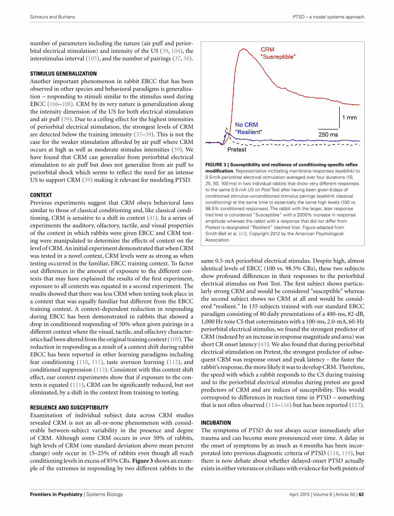
Schreurs and Burhans PTSD – a model systems approach
number of parameters including the nature (air puff and perior-bital electrical stimulation) and intensity of the US (39, 104), theinterstimulus interval (105), and the number of pairings (37, 38).
STIMULUS GENERALIZATIONAnother important phenomenon in rabbit EBCC that has beenobserved in other species and behavioral paradigms is generaliza-tion – responding to stimuli similar to the stimulus used duringEBCC (106–108). CRM by its very nature is generalization alongthe intensity dimension of the US for both electrical stimulationand air puff (39). Due to a ceiling effect for the highest intensitiesof periorbital electrical stimulation, the strongest levels of CRMare detected below the training intensity (37–39). This is not thecase for the weaker stimulation afforded by air puff where CRMoccurs at high as well as moderate stimulus intensities (39). Wehave found that CRM can generalize from periorbital electricalstimulation to air puff but does not generalize from air puff toperiorbital shock which seems to reflect the need for an intenseUS to support CRM (39) making it relevant for modeling PTSD.
CONTEXTPrevious experiments suggest that CRM obeys behavioral lawssimilar to those of classical conditioning and, like classical condi-tioning, CRM is sensitive to a shift in context (41). In a series ofexperiments the auditory, olfactory, tactile, and visual propertiesof the context in which rabbits were given EBCC and CRM test-ing were manipulated to determine the effects of context on thelevel of CRM. An initial experiment demonstrated that when CRMwas tested in a novel context, CRM levels were as strong as whentesting occurred in the familiar, EBCC training context. To factorout differences in the amount of exposure to the different con-texts that may have explained the results of the first experiment,exposure to all contexts was equated in a second experiment. Theresults showed that there was less CRM when testing took place ina context that was equally familiar but different from the EBCCtraining context. A context-dependent reduction in respondingduring EBCC has been demonstrated in rabbits that showed adrop in conditioned responding of 50% when given pairings in adifferent context where the visual, tactile, and olfactory character-istics had been altered from the original training context (109). Thereduction in responding as a result of a context shift during rabbitEBCC has been reported in other learning paradigms includingfear conditioning (110, 111), taste aversion learning (112), andconditioned suppression (113). Consistent with this context shifteffect, our context experiments show that if exposure to the con-texts is equated (111), CRM can be significantly reduced, but noteliminated, by a shift in the context from training to testing.
RESILIENCE AND SUSCEPTIBILITYExamination of individual subject data across CRM studiesrevealed CRM is not an all-or-none phenomenon with consid-erable between-subject variability in the presence and degreeof CRM. Although some CRM occurs in over 50% of rabbits,high levels of CRM (one standard deviation above mean percentchange) only occur in 15–25% of rabbits even though all reachconditioning levels in excess of 85% CRs. Figure 3 shows an exam-ple of the extremes in responding by two different rabbits to the
FIGURE 3 | Susceptibility and resilience of conditioning-specific reflex
modification. Representative nictitating membrane responses (eyeblink) to
0.5-mA periorbital electrical stimulation averaged over four durations (10,
25, 50, 100 ms) in two individual rabbits that show very different responses
to the same 0.5-mA US on Post Test after having been given 6 days of
conditioned stimulus–unconditioned stimulus pairings (eyeblink classical
conditioning) at the same time to essentially the same high levels (100 vs.
98.5% conditioned responses). The rabbit with the larger, later response
(red line) is considered “Susceptible” with a 2000% increase in response
amplitude whereas the rabbit with a response that did not differ from
Pretest is designated “Resilient” (dashed line). Figure adapted from
Smith-Bell et al. (43), Copyright 2012 by the American Psychological
Association.
same 0.5-mA periorbital electrical stimulus. Despite high, almostidentical levels of EBCC (100 vs. 98.5% CRs), these two subjectsshow profound differences in their responses to the periorbitalelectrical stimulus on Post Test. The first subject shows particu-larly strong CRM and would be considered “susceptible” whereasthe second subject shows no CRM at all and would be consid-ered “resilient.” In 135 subjects trained with our standard EBCCparadigm consisting of 80 daily presentations of a 400-ms, 82-dB,1,000 Hz tone CS that coterminates with a 100-ms, 2.0-mA, 60-Hzperiorbital electrical stimulus, we found the strongest predictor ofCRM (indexed by an increase in response magnitude and area) wasshort CR onset latency (43). We also found that during periorbitalelectrical stimulation on Pretest, the strongest predictor of subse-quent CRM was response onset and peak latency – the faster therabbit’s response, the more likely it was to develop CRM. Therefore,the speed with which a rabbit responds to the CS during trainingand to the periorbital electrical stimulus during pretest are goodpredictors of CRM and are indices of susceptibility. This wouldcorrespond to differences in reaction time in PTSD – somethingthat is not often observed (114–116) but has been reported (117).
INCUBATIONThe symptoms of PTSD do not always occur immediately aftertrauma and can become more pronounced over time. A delay inthe onset of symptoms by as much as 6 months has been incor-porated into previous diagnostic criteria of PTSD (118, 119), butthere is now debate about whether delayed-onset PTSD actuallyexists in either veterans or civilians with evidence for both points of
Frontiers in Psychiatry | Systems Biology April 2015 | Volume 6 | Article 50 | 62

Schreurs and Burhans PTSD – a model systems approach
view (118–124). In our animal model of PTSD symptoms, rabbitsdo not show a delay in onset of CRM, but there is a windowduring which incubation exacerbates CRM. The results are con-sistent with clinical data in which exacerbation or reactivation ofprior symptoms accounts for 38.3% of military cases of PTSD and15.3% of civilian cases (120, 125). In one set of experiments, wehave observed the exacerbation of symptoms as a function of aperiod of incubation (126). CRM typically requires at least 3 daysof EBCC when levels of conditioning reach or exceed 85% CRs(37, 39). We carried out an experiment (Figure 4) in which rabbitswere given EBCC for just 1 day resulting in mean conditioninglevels of only 45% CRs, and saw little evidence of CRM whentested the next day. However, if left in their home cages for 6 days,there was a significant amount of CRM which persisted for a weekafter testing (126). The incubation effect was not strong following10 days in the home cage and did not persist. These data suggestthere may be no delay in CRM onset but there is a window forincubation to exacerbate CRM.
RESPONSE GENERALIZATIONOne of the most interesting aspects of our initial CRM experi-ments was the observation that, in individual subjects, responsesto weak periorbital electrical stimulus intensities appeared to havea significantly different topography after EBCC than they do beforeEBCC and that the topography was reminiscent of the CR (37, 40).This observation was even more clearly articulated by Gruart andYeo (44) when they first reported changes in the rabbit eyelid UR
FIGURE 4 | Incubation of conditioning-specific reflex modification.
Representative nictitating membrane responses (eyeblink) to a 0.25-mA
periorbital electrical stimulus averaged over four durations (10, 25, 50,
100 ms) in individual rabbits before (dashed line) and after 1, 6, or 10 days of
incubation in the home cage (red line) following a single session of
conditioned stimulus–unconditioned stimulus pairings (eyeblink classical
conditioning) which supported a level of only 45% conditioned responses.
The blue lines depict nictitating membrane responses to 0.25-mA periorbital
electrical stimulation 7 days (of incubation) after Post Test 1. Although there
is some suggestion of conditioning-specific reflex modification after 1 and
10 days of incubation, there was very clear and strong conditioning-specific
reflex modification that occurred after 6 days of incubation and persisted a
week later. Figure adapted from Schreurs et al. (126), used with permission
from Elsevier.
following EBCC. The marked alteration in response topography issomewhat lost in the averaging that takes place when presentinggroup data especially when, as noted above, not all rabbits showCRM. Figure 5 shows the strong similarity between a CR thatoccurs during EBCC and a UR to periorbital stimulation by itselfassessed after EBCC compared to an UR assessed before EBCC.These early observations lead to the hypothesis that CRM is a CRthat generalized from the CS–US pairings to the US itself (40, 44).A series of experiments were conducted to test this hypothesis byaltering the topography of the CR by presenting two shocks duringCS pairings or by presenting CS–US pairings with two differentinterstimulus intervals (38). The results provided evidence bothfor and against the hypothesis so a final experiment was designedto eliminate CRs by presenting the CS by itself during extinction(38). If the exaggerated responses to the US after EBCC (CRM)were generalized CRs, it was reasoned that eliminating the CRsshould eliminate CRM. The results of this experiment were moreconclusive. Despite reducing CRs to essentially baseline levels ofless than 10% by presenting the CS by itself, Figure 6 shows CRMremained virtually intact. A number of control groups actuallyproved to be even more instructive. First, presentations of the USby itself completely eliminated CRM as shown in Figure 6 but leftCRs relatively intact. Thus, the extinction of CRs left strong levelsof CRM and the extinction of CRM left strong levels of condi-tioned responding. Second,combining presentations of the CS andthe US in an explicitly unpaired manner resulted in elimination of
FIGURE 5 |Topographical similarity between a conditioned response
and conditioning-specific reflex modification. Representative nictitating
membrane responses (eyeblinks) in the same rabbit to a tone paired with
shock during the third day of conditioned stimulus–unconditioned stimulus
pairings (eyeblink classical conditioning, blue line, Paired Trial) and 0.5-mA
periorbital electrical stimulation presented by itself before (dashed line,
Pretest), and after (red line, Post Test) 6 days of conditioned
stimulus–unconditioned stimulus pairings. The response after eyeblink
classical conditioning shows a strong similarity in response amplitude, peak
latency, and overall topography compared to the response before eyeblink
classical conditioning. The responses are shifted in time so that their onsets
coincide even though the response on the paired trial is to the conditioned
stimulus that overlaps with the periorbital electrical stimulus and the
responses on the Pretest and Post Test trial are to 0.5-mA periorbital
electrical stimulation by itself.
www.frontiersin.org April 2015 | Volume 6 | Article 50 | 63

Schreurs and Burhans PTSD – a model systems approach
FIGURE 6 | Extinction of conditioning-specific reflex modification.
Averaged nictitating membrane responses (eyeblink) to a periorbital
electrical stimulus of 1.0 mA averaged over four durations (10, 25, 50,
100 ms) for four groups of rabbits before 6 days of conditioned
stimulus–unconditioned stimulus pairings (eyeblink classical conditioning)
(dashed lines, Pretest) and 6 days after (red line, Post Test) either exposure
to the training chamber with no further stimulus presentations (Sit),
presentations of the conditioned stimulus alone (CS-alone), explicitly
unpaired presentations of the conditioned stimulus and unconditioned
stimulus (Unpaired), or presentations of the unconditioned stimulus alone
(US-alone). The level of conditioning-specific reflex modification (CRM) was
highest in the Sit group, followed by the CS-alone group, and the Unpaired
group with virtually no CRM in the US-alone group. Although there was
residual CRM in the Unpaired group, rabbits in this group showed no
responding to the conditioned stimulus unlike rabbits in the Paired and
US-alone groups suggesting that only unpaired presentations of the
conditioned stimulus and unconditioned stimulus were able to significantly
reduce CRM as well as eliminate conditioned responses to the conditioned
stimulus. Figure adapted from Schreurs et al. (38), in the public domain.
CRs and a reduction in the level of CRM (Figure 6). It was theseexperiments that led to a further exploration of treatments thateliminate both CRs and CRM as a possible treatment strategy forPTSD.
EXTINCTION OF CRMThere is a significant body of evidence from both clinical andbasic research that repeated presentation of feared stimuli doesnot prevent fear from returning – a phenomenon referred to as“relapse” (127, 128). Nevertheless, fear extinction is a cornerstoneof many approaches to the treatment of PTSD (3, 28, 129–137).However, the renewal of fear or relapse may be “thwarted” byunpaired presentations of both the feared stimulus and the eventproducing the fear (38, 138–140). Experiments drawn from a largenumber of different conditioning paradigms including humanand rabbit EBCC (36, 39, 141–144), as well as conditioned bar-press suppression in rats (138, 139), and human discriminativefear conditioning (140) show unpaired presentations of the CSand US produce extinction of a CR. In the human discrimina-tive fear study, Vervleit and coworkers found that compared tonormal extinction, only unpaired extinction prevented renewal of
fear responses in people trained to discriminate one of two picturespaired with shock (140).
In rabbit experiments designed to extinguish EBCC, compara-ble extinction of responding to the CS occurs following CS-aloneor unpaired CS and US presentations (38). However, as notedabove and shown in Figure 6, unpaired presentations were ableto extinguish CRM better than CS-alone presentations (38). Theability of unpaired presentations to diminish both CRs and exag-gerated URs (i.e., CRM) suggests it may be relevant for treatingboth the conditioned fear and hyperarousal symptoms of PTSD(41, 42, 104). However, no matter how effective unpaired extinc-tion might be in extinguishing fear and hyperarousal in animalmodels, it would be ethically unacceptable for treating PTSDbecause the US intensity used in unpaired extinction has alwaysbeen the same as that used to induce classical conditioning (36,39, 138–144). The repeated presentation of a traumatic eventresponsible for PTSD in order to treat it is untenable.
UNPAIRED EXTINCTION THAT IS CLINICALLY RELEVANTTo address concerns about using a traumatic stimulus duringunpaired extinction and make an unpaired extinction proceduremore clinically relevant, rabbit EBCC experiments were conductedin which unpaired extinction sessions employed periorbital elec-trical stimulation of reduced intensity that was presented fordifferent numbers of days (36). Specifically, rabbits received UStesting (Pretest), EBCC, another session of US testing to deter-mine the size of CRM (Post Test 1), and then 1, 3, or 6 days ofunpaired CS and US presentations with a weak (0.25 mA), mod-erate (1.0 mA), or strong (2.0 mA) US followed by a final sessionof US testing to determine the effect of unpaired presentationson CRM (Post Test 2). The results revealed extinction of bothCRs and CRM was a function of the US intensity used duringunpaired stimulus presentations and the number of days of thoseunpaired stimulus presentations (36). The levels of CRs declinedfrom 95% to less than 20% within 3 days of unpaired stimuluspresentations. Figure 7 shows CRs during acquisition and 1, 3,or 6 days of unpaired extinction in which the US intensity waseight times weaker (0.25 mA) than the intensity used during pair-ings (2.0 mA). Figure 8 depicts sample responses from differentrabbits before and after EBCC (Pretest and Post Test 1, respec-tively) and again after unpaired stimulus presentations (Post Test2) with a 0.25-mA US that were delivered for either one, three,or six daily sessions (days). The sample responses in the middleand right illustrate that after as few as three sessions of unpairedpresentations with a weak US, any CRM seen after EBCC (redlines) was largely eliminated (blue lines). In contrast, the sampleresponses on the left show clearly that CRM was actually enhancedafter a single session of unpaired presentations with a weak US.Taken together, these data suggest that both CRs and CRM seemedto be diminished, if not eliminated, most effectively with at least3 days of mild US presentations but one session of stimulus pre-sentations actually appears to exacerbate responding. Of note, andof particular clinical relevance, was the finding that extinction ofCRs and CRM occurred even though the weak US produced rela-tively low levels of responding (rabbits blinked to the weak US onless than 25% of occasions). Analysis of rabbit heart rate duringthese sessions indicated that this weak US did not produce any
Frontiers in Psychiatry | Systems Biology April 2015 | Volume 6 | Article 50 | 64

Schreurs and Burhans PTSD – a model systems approach
FIGURE 7 | Extinction of conditioned responding with unpaired
presentations of a conditioned stimulus and weak unconditioned
stimulus. Mean (±SEM) conditioned responses to the conditioned
stimulus during 6 days of pairings of a conditioned stimulus and a 2.0-mA
unconditioned stimulus (Acquisition) and during one (square), three (circle),
or six (triangle) subsequent days of unpaired conditioned
stimulus–unconditioned stimulus presentations (Unpaired Extinction) with a
0.25-mA unconditioned stimulus. Conditioned responding increased to
asymptotic levels of 95% during Acquisition and fell to less than 20%
following 3 days of unpaired presentations (Unpaired Extinction). Figure
adapted from Schreurs et al. (36), used with permission from Elsevier.
change in heart rate, suggesting it was not unduly stressful (36).One important implication of these data is that treatment mustnot be brief because brief treatment using unpaired stimulus pre-sentations may not just be ineffectual; it may actually heighten thesymptoms of PTSD.
VIRTUAL REALITYIf weakened versions of the initiating trauma are to be used aspart of PTSD therapy, there would be very few such events thatcould or even should be repeated or recreated. The advent of cred-ible virtual reality (VR) environments that have been developed totreat PTSD provide a feasible way around this stricture (145–151).Given the unpaired extinction data reviewed above, one couldimagine a treatment situation in which a PTSD patient could beasked to describe a specific trigger or set of triggers for unwantedmemories (150) and present the trigger(s) in an unpaired man-ner with a weakened version of an aversive event. A weakened butstill stressful version of an explosion might be strongly shakinga driver’s seat in a virtual Humvee which is part of a VR sce-nario in which the sights and sounds of combat are also presented(149–151). The VR environment could be programed to presentthese events in a separate, unpaired manner and the predictionwould be that, with a number of repetitions over more than onesession, PTSD symptoms would abate. For example, the sights,sounds of a previously traumatic context could be presented, andthen the goggles and headphones would go blank and silent fora period of no stimulation which would then be followed by thedriver’s seat being strongly shaken. The sequence of these seriesof events would be randomized so that they would never occur
together to reflect the explicitly unpaired procedure (83). Impor-tantly, given that CRM has been shown to generalize from stressfulperiorbital electrical stimulation to what would be considered lessstressful air puffs, the weakened versions of stressful events usedin an unpaired extinction procedure may not need to involve thetraumatic event. Psychophysiological indices including heart rate,skin conductance, respiration, and cortisol levels could be used toassess stress levels and titrate the intensity of the stimulation.
METHODOLOGICAL ADDENDUMSTIMULUS DELIVERY AND RESPONSE MEASUREMENTThe experiments described in this review require precise controland calibration of stimulus parameters particularly intensity andtiming of the US. This is relatively straightforward for perior-bital electrical stimulation through the use of programmable shockdelivery equipment and the use of digital computer control. Onthe other hand, the delivery of air puff requires more elaborateequipment and techniques including a digitally controlled, pro-grammable pressure regulator and an accurate digital manometerto ensure that the intensity of the air puff reflects the air strikingthe cornea and not the pressure at the source. Response detectionis also of importance especially if response characteristics such aslatency, amplitude, and area are to be determined in addition tosimply registering if a response occurs or not. As a result, transduc-tion and recording of the eyeblink response becomes important.Researchers may wish to consider the advantages and disadvan-tages of remotely sensing versus directly measuring the closureof the eyelid using mechanical coupling. For example, infraredreflectance measures may not be capable of completely quantifyingthe peak latency of a response whereas mechanical couple may pro-duce drag that subtly alters the latency and amplitude of a response(152). EMG recording of the orbicularis oculi muscle may haveadvantages but the electrical noise induced by periorbital electricalstimulation as well as time constants of integration affecting onsetlatency and difficulty in determining units of response amplitudepresent limitations in quantifying the UR.
DATA ANALYSISEven if the UR is transduced accurately, questions remain aboutthe analysis of data, particularly when responses are at the limitsof detectability as the result of very weak stimulation. By conven-tion and due, in part, to the limits of analog instrumentation, anNMR or eyeblink response has been defined as movement of atleast 0.5 mm (61, 66, 153, 154). How then is a change in responseamplitude and latency from pretest to post test determined if thereis no response on pretest but a significant response on post testas often occurs after EBCC? The main issue has always been whatto do about the lack of a response on pretest or post test. Wehave addressed this in several ways including analyzing individualsubject data only for US parameters at which responses occurred(37–39, 104), averaging topographies across subjects and analyz-ing for changes in skew and kurtosis (41, 155), and calculatingpercent change where a response on a test was considered to be a100% change if there was no response on the other test (43). Mostrecently, two additional measures, magnitude of the response andmagnitude of the response area, have been calculated to overcomethe limitations of empty data cells on pretest or post test resulting
www.frontiersin.org April 2015 | Volume 6 | Article 50 | 65
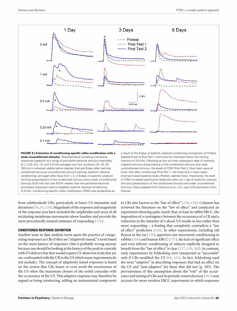
Schreurs and Burhans PTSD – a model systems approach
FIGURE 8 | Extinction of conditioning-specific reflex modification with a
weak unconditioned stimulus. Representative nictitating membrane
responses (eyeblink) to a range of periorbital electrical stimulus intensities
(0.1, 0.25, 0.5, 1.0, and 2.0 mA) averaged over four durations (10, 25, 50,
100 ms) in individual rabbits before (dashed line) and 6 days after (red line)
conditioned stimulus–unconditioned stimulus pairings (eyeblink classical
conditioning), and again after (blue line) 1, 3, or 6 days of explicitly unpaired
stimulus presentations of the conditioned stimulus and a weak unconditioned
stimulus (0.25 mA) that was 87.5% weaker than the periorbital electrical
stimulation previously used to establish eyeblink classical conditioning
(2.0 mA). Conditioning-specific reflex modification (CRM) was established as
a result of the 6 days of eyeblink classical conditioning [comparison of Pretest
(dashed lines) to Post Test 1 (red lines) for intensities below the training
intensity of 2.0 mA]. Following as few as three subsequent days of explicitly
unpaired stimulus presentations of the conditioned stimulus and weak
unconditioned stimulus, the levels of CRM (Post Test 2, blue lines) were all
lower than after conditioning (Post Test 1, red lines) and in many cases
returned toward baseline levels (Pretest, dashed lines). Importantly, the level
of CRM increased significantly (asterisks) after just 1 day of explicitly unpaired
stimulus presentations of the conditioned stimulus and weak unconditioned
stimulus. Figure adapted from Schreurs et al. (36), used with permission from
Elsevier.
from subthreshold URs, particularly at lower US intensities anddurations (36, 43, 126). Magnitude of the response and magnitudeof the response area have included the amplitudes and areas of allnictitating membrane movements above baseline and provide themost procedurally neutral estimates of responding (154).
CONDITIONED RESPONSE DEFINITIONAnother issue in data analysis turns upon the practice of catego-rizing responses as CRs if they are “adaptively timed,” a term basedon the onset latency of responses (this is probably wrong anywaybecause one should be looking at the latency of the peak to coincidewith US delivery but that would require CS-alone test trials that areun-confounded with the UR to the US which many experiments donot include). The concept of adaptively timed responses is basedon the notion that CRs lessen or even avoid the aversiveness ofthe US when the maximum closure of the eyelid coincides withthe occurrence of the US. This adaptive response may therefore beargued as being reinforcing, adding an instrumental component
to CRs also known as the “law of effect” (156–158). Coleman hasreviewed the literature on the “law of effect” and conducted anexperiment showing quite clearly that, at least in rabbit EBCC, theimposition of a contingency between the occurrence of a CR and areduction in the intensity of a shock US results in less rather thanmore responding – a finding that completely contradicts a “lawof effect” prediction (156). In other experiments, including tailflexion in the rat (159), appetitive jaw movement conditioning inrabbits (160) and human EBCC (157), the lack of significant effectand even inferior conditioning of subjects explicitly designed tobenefit from the “law of effect” is clear (157, 159, 160). In contrast,early experiments by Schlosberg were interpreted as “successful”only if CRs modified the US (60, 161). In fact, Schlosberg usedthe term “adaptive” in describing responses that had an effect onthe US and “non-adaptive” for those that did not (p. 383). Thepervasiveness of this assumption about the “role” of the occur-rence and timing of CRs and its periodic reintroduction (162) mayaccount for more modern EBCC experiments in which responses
Frontiers in Psychiatry | Systems Biology April 2015 | Volume 6 | Article 50 | 66

Schreurs and Burhans PTSD – a model systems approach
are only considered to be CRs if they occur within an interval thatis characterized as “adaptive.”
The use of onset latencies to detect adaptively timed CRs andhence, “true CRs” can be traced to another period in the historyof EBCC where latencies were used to identify and eliminate thedata of “voluntary responders” (62, 163, 164). Voluntary respon-ders were subjects who were “rejected” from experiments based onthe occurrence of short-latency eyeblinks that occurred between200 and 300 ms after CS onset and were judged to have the sameappearance as subjects who were instructed to blink or by subjectswho reported they were blinking “voluntarily” to avoid the air puff(165). This practice has been explicitly adopted by a number oflaboratories especially during trace conditioning where there wasa long interval between the offset of the CS and the onset of the USbecause it “corrected for both voluntary and random blinks thatcould occur as a result of the longer trace intervals” (166, 167).
In our view, an empirical approach to determining onset latencyneeds to be neutral with respect to characterizing responses. Weendorse the complete characterization of all responses using arange of dependent variables including onset and peak latency andpresenting all response onsets on a latency histogram without anypreconceptions of how responses should look or be distributed.Publication of such histograms together with any interpretationof what are considered responses whether they be “adaptive” ornot would allow readers to interpret the data for themselves.
SUMMARY AND CONCLUSIONThere is a crucial need to know how responding to stressfulevents changes as a function of trauma for those who suffer fromPTSD. A number of treatment strategies for PTSD are capableof treating only some of the symptoms because the extinctionof fear does not deal with the various forms of hyper-vigilanceand hyperarousal experienced by people with PTSD, especiallyin combat veterans. Based on our work on conditioning of therabbit’s NMR, we have developed a preclinical EBCC model ofPTSD that addresses both CRs to trauma-associated cues as wellas hyperarousal (CRM). Animal models of EBCC are particularlyuseful here because EBCC is one of the few behavioral para-digms in which there is a one-to-one correspondence betweenanimals and humans. We have demonstrated that CRM followsmany of the same behavioral rules as EBCC, can generalize acrossstimulus modalities, shows sensitivity to context manipulations,and can be exacerbated after an incubation period. Importantly,CRM does not develop in all animals just as PTSD does notdevelop in all those exposed to trauma, with some individualsdemonstrating susceptibility while others show resilience. We haveshown that CRs and CRM can be simultaneously extinguishedby unpaired stimulus presentations, even when US intensity isreduced to the point where it is barely capable of eliciting aresponse. This is important because presenting strong uncondi-tioned stimuli as a therapeutic approach would be untenable.These unpaired procedures with attenuated stimuli have directimplications for the treatment of PTSD and could be implementedin a VR environment.
AUTHOR CONTRIBUTIONSBS and LB conceived and wrote the manuscript.
ACKNOWLEDGMENTSThe authors thank Carrie Smith-Bell for help with data collec-tion and analysis. The work described here was supported bythe NIH Intramural program, the Blanchette Rockefeller Neu-rosciences Institute, and NIH grants MH64715 and MH081159.The contents of this manuscript are solely the responsibility of theauthors and do not necessarily represent the official views of theNIMH.
REFERENCES1. Breslau N. The epidemiology of posttraumatic stress disorder: what is the
extent of the problem? J Clin Psychiatry (2001) 62(Suppl 17):16–22.2. Difede J, Olden M, Cukor J. Evidence-based treatment of post-traumatic
stress disorder. Annu Rev Med (2014) 65:319–32. doi:10.1146/annurev-med-051812-145438
3. Morrison FG, Ressler KJ. From the neurobiology of extinction toimproved clinical treatments. Depress Anxiety (2014) 31:279–90. doi:10.1002/da.22214
4. Creamer M, Elliott P, Forbes D, Biddle D, Hawthorne G. Treatment for combat-related posttraumatic stress disorder: two-year follow-up. J Trauma Stress(2006) 19:675–85. doi:10.1002/jts.20155
5. Watts BV, Schnurr PP, Mayo L, Young-Xu Y, Weeks WB, Friedman MJ. Meta-analysis of the efficiency of treatments for posttraumatic stress disorder. J ClinPsychiatry (2013) 74:e541–50. doi:10.4088/JCP.12r08225
6. Ramaswamy S, Madaan V, Qadri F, Heaney CJ, North TC, Padala PR, et al. Aprimary care perspective of posttraumatic stress disorder for the Departmentof Veterans Affairs. Prim Care Companion J Clin Psychiatry (2005) 7:180–7.doi:10.4088/PCC.v07n0407
7. Rosen CS, Greenbaum MA, Fitt JE, Laffaye C, Norris VA, Kimerling R.Stigma, help-seeking attitudes, and use of psychotherapy in veterans with diag-noses of posttraumatic stress disorder. J Nerv Ment Dis (2011) 199:879–85.doi:10.1097/NMD.0b013e3182349ea5
8. Ramchand R, Schell TL, Karney BR, Osilla KC, Burns RM, Caldarone LB. Dis-parate prevalence estimates of PTSD among service members who served inIraq and Afghanistan: possible explanations. J Trauma Stress (2010) 23:59–68.doi:10.1002/jts.20486
9. Friedman MJ. Veterans’ mental health in the wake of war. N Engl J Med (2005)352:1287–90. doi:10.1056/NEJMp058028
10. Iversen AC, Van SL, Hughes JH, Greenberg N, Hotopf M, Rona RJ, et al. Thestigma of mental health problems and other barriers to care in the UK ArmedForces. BMC Health Serv Res (2011) 1:31. doi:10.1186/1472-6963-11-31
11. Murphy D,Busuttil W. PTSD,stigma and barriers to help-seeking within the UKArmed Forces. J R Army Med Corps (2014). doi:10.1136/jrmac-2014-000344
12. Yaffe K, Vittinghoff E, Lindquist K, Barnes D, Covinsky KE, Neylan T, et al.Posttraumatic stress disorder and risk of dementia among US veterans. ArchGen Psychiatry (2010) 67:608–13. doi:10.1001/archgenpsychiatry.2010.61
13. Veitch DP, Friedl KE, Weiner MW. Military risk factors for cognitive decline,dementia, and Alzheimer’s disease. Curr Alzheimer Res (2013) 10:907–30.doi:10.2174/15672050113109990142
14. Chapman C, Mills K, Slade T, McFarlane AC, Bryant RA, Creamer M, et al.Remission from post-traumatic stress disorder in the general population. Psy-chol Med (2012) 42:1695–703. doi:10.1017/S0033291711002856
15. Dierperink M, Erbes C, Leskela J, Kaloupek D, Farrer MK, Fisher L, et al. Com-parison of treatment for post-traumatic stress disorder among three Depart-ment of Veterans Affairs medical centers. Mil Med (2005) 170:305–8.
16. English BA, Jewell M, Jewell G, Ambrose S, Davis LL. Treatment of chronicposttraumatic stress disorder in combat veterans with citalopram. J Clin Psy-chopharmacol (2006) 26:84–8. doi:10.1097/01.jcp.0000195043.39853.bc
17. Boscarino JA. Posttraumatic stress disorder and mortality among U.S. armyveterans 30 years after military service. Ann Epidemiol (2005) 16:248–56.doi:10.1016/j.annepidem.2005.03.009
18. Pompili M, Sher L, Serafini G, Forte A, Innamorati M, Dominici G, et al. Post-traumatic stress disorder and suicide risk among veterans: a literature review.J Nerv Ment Dis (2013) 201:802–12. doi:10.1097/NMD.0b013e3182a21458
19. Sher L, Braquehais MD, Casas M. Posttraumatic stress disorder, depression,and suicide in veteran. Cleve Clin J Med (2012) 79:92–7. doi:10.3949/ccjm.79a.11069
www.frontiersin.org April 2015 | Volume 6 | Article 50 | 67
Schreurs and Burhans PTSD – a model systems approach
20. Yehuda R, Antelman SM. Criteria for rationally evaluating animal models ofposttraumatic stress disorder. Biol Psychiatry (1993) 33:479–86. doi:10.1016/0006-3223(93)90001-T
21. Cohen H, Zohar J. An animal model of posttraumatic stress disorder. Theuse of cut-off behavioral criteria. Ann N Y Acad Sci (2004) 1032:167–78.doi:10.1196/annals.1314.014
22. Cohen H, Matar MA, Richter-Levin G, Zohar J. The contribution of an animalmodel toward uncovering biological risk factors for PTSD. Ann N Y Acad Sci(2006) 1071:335–50. doi:10.1196/annals.1364.026
23. Siegmund A, Wotjak CT. Toward an animal model of posttraumatic stressdisorder. Ann N Y Acad Sci (2006) 1071:324–34. doi:10.1196/annals.1364.025
24. Stam R. PTSD and stress sensitization: a tale of brain and body. Part 2: ani-mal models. Neurosci Biobehav Rev (2007) 31:558–84. doi:10.1016/j.neubiorev.2007.01.001
25. Ursano RJ, Li H, Zhang L, Hough CJ, Fullerton CS, Benedek DM, et al. Mod-els of PTSD and traumatic stress: the importance of research “from bedsideto bench to bedside”. Prog Brain Res (2008) 167:203–14. doi:10.1016/S0079-6123(07)67014-9
26. Yamamoto S, Morinobu S, Takei S, Fuchikami M, Matsuki A, Yamawaki S,et al. Single prolonged stress: toward an animal model of posttraumatic stressdisorder. Depress Anxiety (2009) 26:1110–7. doi:10.1002/da.20629
27. Daskalakis NP, Yehuda R, Diamond DM. Animal models in translational stud-ies of PTSD. Psychoneuroendocrinology (2013) 38:1895–911. doi:10.1016/j.psyneuen.2013.06.006
28. Goswami S, Rodriguez-Sierra OE, Cascardi M, Pare D. Animal models ofpost-traumatic stress disorder: face validity. Front Neurosci (2013) 7:89.doi:10.3389/fnins.2013.00089
29. Matar MA, Zohar J, Cohen H. Translationally relevant modeling of PTSD inrodents. Cell Tissue Res (2013) 354:127–39. doi:10.1007/s00441-013-1687-6
30. Yehuda R, LeDoux J. Response variation following trauma: a translationalneuroscience approach to understanding PTSD. Neuron (2007) 56:19–32.doi:10.1016/j.neuron.2007.09.006
31. Johnson LR, McGuire J, Lazarus R, Palmer AA. Pavlovian fear memory cir-cuits and phenotype models of PTSD. Neuropharmacology (2012) 62:638–46.doi:10.1016/j.neuropharm.2011.07.004
32. Radley JJ, Kabbaj M, Jacobson L, Heydendael W, Yehuda R, Herman JP.Stress risk factors and stress-related pathology: neuroplasticity, epigeneticsand endophenotypes. Stress (2012) 14:481–97. doi:10.3109/10253890.2011.604751
33. Lissek S, van Meurs B. Learning models of PTSD: theoretical accounts andpsychobiological evidence. Int J Psychophysiol (2014). doi:10.1016/j.ijpsycho.2014.1011.1006
34. Cohen H, Zohar J, Matar MA, Zeev K, Loewenthal U, Richter-Levin G. Set-ting apart the affected: the use of behavioral criteria in animal models ofpost traumatic stress disorder. Neuropsychopharmacology (2004) 29:1962–70.doi:10.1038/sj.npp.1300523
35. Reznikoc R, Diwan M, Nobrega JN, Hamani C. Towards a better preclini-cal model of PTSD: characterizing animals with weak extinction, maladap-tive stress responses and low plasma corticosterone. J Psychiatr Res (2015)61:158–65. doi:10.1016/j.jpsychires.2014.1012.1017
36. Schreurs BG, Smith-Bell CA, Burhans LB. Unpaired extinction: implicationsfor treating post-traumatic stress disorder. J Psychiatr Res (2011) 45:638–49.doi:10.1016/j.jpsychires.2010.10.010
37. Schreurs BG, Oh MM, Hirashima C, Alkon DL. Conditioning-specific modi-fication of the rabbit’s unconditioned nictitating membrane response. BehavNeurosci (1995) 109:24–33. doi:10.1037/0735-7044.109.1.24
38. Schreurs BG, Shi T, Pineda SI, Buck DL. Conditioning the unconditionedresponse: modification of the rabbit’s (Oryctolagus cuniculus) unconditionednictitating membrane response. J Exp Psychol Anim Behav Process (2000)26:144–56. doi:10.1037/0097-7403.26.2.144
39. Buck DL, Seager MA, Schreurs BG. Conditioning-specific reflex modificationof the rabbit (Oryctolagus cuniculus) nictitating membrane response: gener-ality and nature of the phenomenon. Behav Neurosci (2001) 115:1039–47.doi:10.1037/0735-7044.115.5.1039
40. Schreurs BG. Classical conditioning and modification of the rabbit’s (Orycto-lagus cuniculus) unconditioned nictitating membrane response. Behav CognNeurosci Rev (2003) 2:83–96. doi:10.1177/1534582303002002001
41. Schreurs BG, Gonzalez-Joekes J, Smith-Bell CA. Conditioning-specific reflexmodification of the rabbit (Oryctolagus cuniculus) nictitating membrane
response is sensitive to context. Learn Behav (2006) 34:315–24. doi:10.3758/BF03192886
42. Burhans LB, Smith-Bell CA, Schreurs BG. Conditioning-specific reflex modifi-cation of the rabbit’s nictitating membrane response and heart rate: behavioralrules, neural substrates, and potential applications to post-traumatic stress dis-order. Behav Neurosci (2008) 122:1191–206. doi:10.1037/a0013599
43. Smith-Bell CA, Burhans LB, Schreurs BG. Predictors of susceptibility andresilience in an animal model of post traumatic stress disorder. Behav Neurosci(2012) 126:749–61. doi:10.1037/a0030713
44. Gruart A, Yeo CH. Cerebellar cortex and eyeblink conditioning: bilateral reg-ulation of conditioned responses. Exp Brain Res (1995) 104:431–48. doi:10.1007/BF00231978
45. Wikgren J, Ruusuvirta T, Korhonen T. Reflex facilitation during eyeblink con-ditioning and subsequent interpositus nucleus inactivation in the rabbit (Oryc-tolagus cuniculus). Behav Neurosci (2002) 116:1052–8. doi:10.1037/0735-7044.116.6.1052
46. Servatius RJ, Brennan FX, Beck KD, Beldowicz D, Coyle-Dinorcia K. Stressfacilitates acquisition of the classically conditioned eyeblink response atboth long and short interstimulus intervals. Learn Motiv (2001) 32:178–92.doi:10.1006/lmot.2000.1071
47. Woodruff-Pak DS, Steinmetz JE. Past, present, and future of human eyeblinkconditioning. In: Woodruff-Pak DS, Steinmetz JE, editors. Eyeblink ClassicalConditioning Volume 1: Applications in Humans. Boston, MA: Kluwer AcademicPublishers (2000). p. 1–17.
48. Gormezano I. Bibliography of human eyeblink conditioning (1899-1985). In:Woodruff-Pak DS, Steinmetz JE, editors. Eyeblink Classical Conditioning Vol-ume 1: Applications in Humans. Boston, MA: Kluwer Academic Publishers(2000). p. 275–307.
49. Cason H. The conditioned eyelid reaction. J Exp Psychol (1922) 5:153–96.doi:10.1037/h0074822
50. Hilgard ER, Marquis DG. Conditioning and Learning. New York, NY: Wiley(1940).
51. Taylor JA. The relationship of anxiety to the conditioned eyelid response. J ExpPsychol (1951) 441:81–92. doi:10.1037/h0059488
52. Spence KW, Taylor JA. The relation of conditioned response strength to anxietyin normal, neurotic, and psychotic subjects. J Exp Psychol (1953) 45:265–72.doi:10.1037/h0056392
53. Taylor JA, Spence KW. Conditioning level in the behavior disorders. J AbnormPsychol (1954) 49:497–502. doi:10.1037/h0055951
54. Ayers ED, White J, Powell DA. Pavlovian eyeblink conditioning in combat withand without post-traumatic stress disorder. Integr Physiol Behav Sci (2003)38:230–47. doi:10.1007/BF02688856
55. Burriss L, Ayers E, Powell DA. Combat veterans show normal discriminationduring differential trace eyeblink conditioning, but increased responsivity tothe conditioned and unconditioned stimulus. J Psychiatr Res (2007) 41:785–94.doi:10.1016/j.jpsychires.2006.04.004
56. Ginsberg JP, Ayers E, Burriss L, Powell DA. Discriminative delay Pavlovian eye-blink conditioning in veterans with and without posttraumatic stress disorder.J Anxiety Disord (2008) 22:809–23. doi:10.1016/j.janxdis.2007.08.009
57. Myers CE, Vanmeenen KM, McAuley JD, Beck KD, Pang KC, ServatiusRJ. Behaviorally inhibited temperament is associated with severity of post-traumatic stress disorder symptoms and faster eyeblink conditioning in veter-ans. Stress (2012) 15:31–44. doi:10.3109/10253890.2011.578184
58. McGlinchey RE,Fortier CB,Venne JR,Maksimovskiy AL,Milberg WP. Effects ofOEF/OIF-related physical and emotional co-morbidities on associative learn-ing: concurrent delay and trace eyeblink classical conditioning. Int J EnvironRes Public Health (2014) 11:3046–73. doi:10.3390/ijerph110303046
59. Vythulingam M, Lawley M, Collin C, Bonne O, Agarwal R, Hadd K, et al.Hydrocortisone impairs hippocampal-dependent trace eyeblink conditioningin post-traumatic stress disorder. Neuropsychopharmacology (2006) 31:182–8.doi:10.1038/sj.npp.1300843
60. Hughes B, Schlosberg H. Conditioning in the white rat. IV. The conditionedlid reflex. J Exp Psychol (1938) 23:641–50. doi:10.1037/h0059822
61. Gormezano I. Classical conditioning. In: Sidowski JB, editor. Experimen-tal Methods and Instrumentation in Psychology. New York, NY: McGraw-Hill(1966). p. 385–420.
62. Coleman SR, Webster S. The problem of volition and the conditionedreflex. Part II: voluntary-responding subjects, 1951–1980. Behaviorism (1988)16:17–49.
Frontiers in Psychiatry | Systems Biology April 2015 | Volume 6 | Article 50 | 68

Schreurs and Burhans PTSD – a model systems approach
63. Gormezano I, Schneiderman N, Deaux EG, Fuentes I. Nictitating membrane:classical conditioning and extinction in the albino rabbit. Science (1962)138:33–4. doi:10.1126/science.138.3536.33
64. Schneiderman N, Fuentes I, Gormezano I. Acquisition and extinction of theclassically conditioned eyelid response in the albino rabbit. Science (1962)136:650–2. doi:10.1126/science.136.3516.650
65. Deaux EB, Gormezano I. Eyeball retraction: classical conditioning and extinc-tion in the albino rabbit. Science (1963) 141:630–1. doi:10.1126/science.141.3581.630
66. Gormezano I, Kehoe EJ, Marshall BS. Twenty years of classical conditioningresearch with the rabbit. 10th ed. In: Sprague JM, editor. Progress in Psychobi-ology and Physiological Psychology. New York, NY: Academic Press (1983). p.197–275.
67. Smith MC, Dilollo V, Gormezano I. Conditioned jaw movement in the rabbit.J Comp Physiol Psychol (1966) 62:479–83. doi:10.1037/h0023947
68. Schneiderman N, Smith MC, Smith AC, Gormezano I. Heart rate classicalconditioning in rabbits. Psychon Sci (1966) 6:241–2. doi:10.3758/BF03328047
69. Gormezano I. Investigations of defense and reward conditioning in the rabbit.In: Black AH, editor. Classical Conditioning II: Current Research and Theory.New York, NY: Appleton-Century-Crofts (1972). p. 151–81.
70. Berger TW,Alger B, Thompson RF. Neuronal substrate of classical conditioningin the hippocampus. Science (1976) 192:483–5. doi:10.1126/science.1257783
71. Cegavske CF, Thompson RF, Patterson MM, Gormezano I. Mechanisms ofefferent neuronal control of the reflex nictitating membrane response in therabbit. J Comp Physiol Psychol (1976) 90:411–23. doi:10.1037/h0077214
72. Young RA, Cegavske CF, Thompson RF. Tone-induced changes in excitabil-ity of abducens motoneurons and of the reflex path of nictitating membraneresponse in rabbit (Oryctolagus cuniculus). J Comp Physiol Psychol (1976)90:424–34. doi:10.1037/h0077219
73. Stein L. Habituation and stimulus novelty: a model based on classical condi-tioning. Psychol Rev (1966) 73:352–6. doi:10.1037/h0023449
74. Thompson RF, Spencer WA. Habituation: a model phenomenon for the studyof neuronal substrates of behavior. Psychol Rev (1966) 73:16–43. doi:10.1037/h0022681
75. Pinsker HM, Kupfermann V, Castellucci VF, Kandel ER. Habituation and disha-bituation of the gill-withdrawal reflex in Aplysia. Science (1970) 167:1740–2.doi:10.1126/science.167.3926.1740
76. Wine JJ, Krasne FB, Chen L. Habituation and inhibition of the crayfish lateralgiant fiber escape response. J Exp Biol (1975) 62:771–82.
77. Lukowiak K. CNS control of the PNS-mediated gill withdrawal reflex and itshabituation. Can J Physiol Pharmacol (1977) 55:1252–62. doi:10.1139/y77-171
78. Boulis NM, Sahley CL. A behavioral analysis of habituation and sensitizationof shortening in the semi-intact leech. J Neurosci (1988) 8:4621–7.
79. Tomsic D, Massoni V, Maldonado H. Habituation to a danger stimulus in twosemiterrestrial crabs: ontogenic, ecological and opioid modulation correlates.J Comp Physiol A (1993) 173:621–33. doi:10.1007/BF00197770
80. Blumenthal TD. Short lead interval startle modification. In: Dawson ME, SchellAM, Bohmelt AH, editors. Startle Modification: Implications for Neuroscience,Cognitive Science, and Clinical Science. Cambridge: Cambridge University Press(1999). p. 51–71.
81. Humpherys D, Eggan K, Akutsu H, Hochedlinger K, Rideout WM III, Bin-iszkiewicz D, et al. Epigenetic instability in ES cells and cloned mice. Science(2001) 293:95–7. doi:10.1126/science.1061402
82. Mattingly BA, Koch C, Osborne FH, Gotsick JE. Stimulus and response fac-tors affecting the development of behavioral sensitization to apomorphine.Psychopharmacology (1997) 130:109–16. doi:10.1007/s002130050217
83. Gormezano I, Kehoe EJ. Classical conditioning: some methodological-conceptual issues. 2nd ed. In: Estes WK, editor. Handbook of Learning andCognitive Processes. Hillsdale, NJ: Erlbaum (1975). p. 143–79.
84. Ison JR, Leonard DW. Effects of auditory stimuli on the amplitude of the nic-titating membrane reflex of the rabbit (Oryctolagus cuniculus). J Comp PhysiolPsychol (1971) 75:157–64. doi:10.1037/h0030671
85. Donegan NH. Priming-produced facilitation or diminution of responding toa Pavlovian unconditioned stimulus. J Exp Psychol Anim Behav Process (1981)7:295–312.
86. Weisz DJ, McInerny J. An associative process maintains reflex facilitation ofthe unconditioned nictitating membrane response during the early stages oftraining. Behav Neurosci (1990) 104:21–7. doi:10.1037/0735-7044.104.1.21
87. Weisz DJ, Walts C. Reflex facilitation of the rabbit nictitating membraneresponse by an auditory stimulus as a function of interstimulus interval. BehavNeurosci (1990) 104:11–20. doi:10.1037/0735-7044.104.1.11
88. Whalen PJ, Kapp BS. Contributions of the amygdaloid central nucleus to themodulation of the nictitating membrane reflex in the rabbit. Behav Neurosci(1991) 105:141–53. doi:10.1037/0735-7044.105.1.141
89. Canli T, Detmer WM, Donegan NH. Potentiation or diminution of discretemotor unconditioned responses (rabbit eyelid) to an aversive Pavlovian uncon-ditioned stimulus by two associative processes: conditioned fear and a con-ditioned diminution of unconditioned stimulus processing. Behav Neurosci(1992) 106:498–508. doi:10.1037/0735-7044.106.3.498
90. Weisz DJ, Harden DG, Xiang Z. Effects of amygdala lesions on reflex facilitationand conditioned response acquisition during nictitating membrane responseconditioning in rabbit. Behav Neurosci (1992) 106:262–73. doi:10.1037/0735-7044.106.2.262
91. Yang BY,Weisz DJ. An auditory conditioned stimulus modulates unconditionedstimulus-elicited neuronal activity in the cerebellar anterior interpositus anddentate nuclei during nictitating membrane response conditioning in rabbits.Behav Neurosci (1992) 106:889–99. doi:10.1037/0735-7044.106.6.889
92. Flaten MA. A comparison of electromyographic and photoelectric techniquesin the study of classical eyeblink conditioning and startle reflex modification.Psychophysiology (1993) 7:230–7.
93. Lam Y-W, Wong A, Canli T, Brown TH. Conditioned enhancement of the earlycomponent of the rat eyeblink reflex. Neurobiol Learn Mem (1996) 66:212–20.doi:10.1006/nlme.1996.0061
94. Nowak AJ, Goodell-Marshall B, Kehoe EJ, Gormezano I. Elicitation, modifica-tion, and conditioning of the rabbit nictitating membrane response by elec-trical stimulation in the spinal trigeminal nucleus, inferior olive, interpositusnucleus, and red nucleus. Behav Neurosci (1997) 111:1041–55. doi:10.1037/0735-7044.111.5.1041
95. Marcos JL, Redondo J. Effects of conditioned stimulus presentation on diminu-tion of the unconditioned response in aversive classical conditioning. Biol Psy-chiatry (1999) 50:89–102. doi:10.1016/S0301-0511(99)00007-1
96. Nowak AJ, Kehoe EJ, Macrae M, Gormezano I. Conditioning and reflex modifi-cation of the rabbit nictitating membrane response using electrical stimulationin auditory nuclei. Behav Brain Res (1999) 105:189–98. doi:10.1016/S0166-4328(99)00073-X
97. Alkon DL. Memory storage and neural systems. Sci Am (1989) 261:42–50.doi:10.1038/scientificamerican0789-42
98. Alkon DL, Blackwell KT, Barbour GS, Werness SA, Vogl TP. Biological plausi-bility of synaptic associative memory models. Neural Netw (1994) 7:1005–17.doi:10.1016/S0893-6080(05)80156-X
99. Schreurs BG, Crum JM, Wang D, Smith-Bell CA. Conditioning-specific reflexmodification of rabbit (Oryctolagus cuniculus) heart rate. Behav Neurosci(2005) 119:1484–95. doi:10.1037/0735-7044.119.6.1484
100. Schreurs BG, Smith-Bell CA, Burhans LB. Classical conditioning andconditioning-specific reflex modification of rabbit heart rate as a func-tion of unconditioned stimulus location. Behav Neurosci (2011) 125:604–12.doi:10.1037/a0024325
101. Berthier NE. Muscle activity during unconditioned and conditioned eye blinksin the rabbit. Behav Brain Res (1992) 48:21–8. doi:10.1016/S0166-4328(05)80135-4
102. Smith MC, Coleman SR, Gormezano I. Classical conditioning of the rabbit’snictitating membrane response at backward, simultaneous, and forward CS-USintervals. J Comp Physiol Psychol (1969) 69:226–31. doi:10.1037/h0028212
103. Kehoe EJ, Macrae M. Fundamental behavioral methods and findings in classicalconditioning. 1st ed. In: Moore JW, editor. A Neuroscientist’s Guide to ClassicalConditioning. New York, NY: Springer (2002). p. 171–231.
104. Seager MA, Smith-Bell CA, Schreurs BG. Conditioning-specific reflexmodification of the rabbit (Oryctolagus cuniculus) nictitating membraneresponse: US intensity effects. Learn Behav (2003) 31:292–8. doi:10.3758/BF03195990
105. Schreurs BG, Smith-Bell CA, Darwish DS, Stankovic G, Sparks DL. Highdietary cholesterol facilitates classical conditioning of the rabbit’s nicti-tating membrane response. Nutr Neurosci (2007) 10:31–43. doi:10.1080/10284150701565540
106. Hupka RB, Liu SS, Moore JW. Auditory differential conditioning of the rabbitnictitating membrane response: V. Stimulus generalization as a function of the
www.frontiersin.org April 2015 | Volume 6 | Article 50 | 69

Schreurs and Burhans PTSD – a model systems approach
position of the CS+ and CS- on the frequency dimension. Psychon Sci (1969)15:129–31. doi:10.3758/BF03336238
107. Moore JW. Stimulus control: studies in auditory generalization in rabbits. In:Black AH, Prokasy WF, editors. Classical Conditioning II: Current Research andTheory. New York, NY: Appleton-Century-Crofts (1972). p. 206–30.
108. Scavio MJ Jr, Gormezano I. CS intensity effects upon rabbit nictitating mem-brane conditioning, extinction, and generalization. Pavlov J Biol Sci (1974)9:25–34.
109. Penick S, Solomon PR. Hippocampus, context, and conditioning. Behav Neu-rosci (1991) 105:611–7. doi:10.1037/0735-7044.105.5.611
110. Zhou Y, Riccio DC. Manipulation of components of context: the context shifteffect and forgetting of stimulus attributes. Learn Motiv (1996) 27:400–7.doi:10.1006/lmot.1996.0023
111. Millin PM, Riccio DC. Is the context shift effect a case of retrieval failure?The effects of retrieval enhancing treatments on forgetting under altered stim-ulus conditions in rats. J Exp Psychol Anim Behav Process (2004) 30:325–34.doi:10.1037/0097-7403.30.4.325
112. Boakes RA, Westbrook RF, Elliott M, Swinbourne AL. Context dependencyof conditioned aversions to water and sweet tastes. J Exp Psychol Anim BehavProcess (1997) 23:56–67.
113. Gunther LM, Miller RR, Matute H. CSs and USs: what’s the difference? J ExpPsychol Anim Behav Process (1997) 23:15–30.
114. Pole N. The psychophysiology of posttraumatic stress disorder: a meta-analysis.Psychol Bull (2007) 33:725–46. doi:10.1037/0033-2909.133.5.725
115. Hennig-Fast K,Werner NS, Lermer R, Latscha K, Meister F, Reiser M, et al. Afterfacing traumatic stress: brain activation, cognition and stress coping in police-men. J Psychiatr Res (2009) 43:1146–55. doi:10.1016/j.jpsychires.2009.03.001
116. Schumacher S, Schnyder U, Furrer M, Mueller-Pfeiffer C, Wilhelm FH,Moergeli H, et al. Startle reactivity in the long-term after severe acciden-tal injury: preliminary data. Psychiatry Res (2013) 210:570–4. doi:10.1016/j.psychres.2013.06.034
117. Vrana SR, Calhoun PS, McClernon FJ, Dennis MF, Lee ST, Beckham JC. Effectsof smoking on the acoustic startle response and prepulse inhibition in smokerswith and without posttraumatic stress disorder. Psychopharmacology (2013)230:477–85. doi:10.1007/s00213-013-3181-y
118. Smid GE, Mooren TT, van der Mast RC, Gersons BP, Kleber RJ. Delayedposttraumatic stress disorder: systematic review, meta-analysis, and meta-regression analysis of prospective studies. J Clin Psychiatry (2009) 70:1572–82.doi:10.4088/JCP.08r04484
119. Utzon-Frank N, Breinegaard N, Bertelsen M, Borritz M, Eller NH, NordentoftM, et al. Occurrence of delayed-onset post-traumatic stress disorder: a sys-tematic review and meta-analysis of prospective studies. Scand J Work EnvironHealth (2014) 40:215–29. doi:10.5271/sjweh.3420
120. Andrews B, Brewin CR, Philpott R, Stewart L. Delayed-onset posttraumaticstress disorder: a systematic review of the evidence. Am J Psychiatry (2007)164:1319–26. doi:10.1176/appi.ajp.2007.06091491
121. Frueh BC, Grubaugh AL, Yeager DE, Magruder KM. Delayed-onset post-traumatic stress disorder among war veterans in primary care clinics. Br JPsychiatry (2009) 194:515–20. doi:10.1192/bjp.bp.108.054700
122. Andrews B, Brewin CR, Stewart L, Philpott R, Hejdenberg J. Comparison ofimmediate-onset and delayed-onset posttraumatic stress disorder in militaryveterans. J Abnorm Psychol (2009) 118:767–77. doi:10.1037/a0017203
123. Horesh D, Solomon Z, Zerach G, Ein-Dor T. Delayed-onset PTSD among warveterans: the role of life events throughout the life cycle. Soc Psychiatry PsychiatrEpidemiol (2010) 46:863–70. doi:10.1007/s00127-010-0255-6
124. Fikretoglu D,Liu A. Prevalence,correlates, and clincial features of delayed-onsetposttraumatic stress disorder in a nationally representative military sample. SocPsychiatry Psychiatr Epidemiol (2012) 47:1359–66. doi:10.1007/s00127-011-0444-y
125. Hauger RL, Olivares-Reyes JA, Dautzenberg FM, Lohr JB, Braun S, Oakley RH.Molecular and cell signaling targets for PTSD pathophysiology and pharma-cotherapy. Neuropharmacology (2012) 62:705–14. doi:10.1016/j.neuropharm.2011.11.007
126. Schreurs BG, Smith-Bell CA, Burhans LB. Incubation of conditioning-specificreflex modification: implications for post traumatic stress disorder. J PsychiatrRes (2011) 45:1535–41. doi:10.1016/j.jpsychires.2011.07.003
127. Bouton ME, Westbrook RF, Corcoran KA, Maren S. Contextual and temporalmodulation of extinction: behavioral and biological mechanisms. Biol Psychi-atry (2006) 60:352–60. doi:10.1016/j.biopsych.2005.12.015
128. Milad MR, Rauch SL, Pitman RK, Quirk GJ. Fear extinction in rats: implica-tions for human brain imaging and anxiety disorders. Biol Psychiatry (2006)73:61–71. doi:10.1016/j.biopsycho.2006.01.008
129. Pitman RK, Orr SP, Shalev AY. Once bitten, twice shy: beyond the conditioningmodel of PTSD. Biol Psychiatry (1993) 33:145–6. doi:10.1016/0006-3223(93)90132-W
130. Brewin CR, Holmes EA. Psychological theories of posttraumatic stress disorder.Clin Psychol Rev (2003) 23:339–76. doi:10.1016/S0272-7358(03)00033-3
131. Rauch SL, Shin LM, Phelps EA. Neurocircuitry models of posttraumatic stressdisorder and extinction: human neuroimaging research – past, present, andfuture. Biol Psychiatry (2006) 60:376–82. doi:10.1016/j.biopsych.2006.06.004
132. McNally RJ. Mechanisms of exposure therapy: how neuroscience can improvepsychological treatments for anxiety disorders. Clin Psychol Rev (2007)27:750–9. doi:10.1016/j.cpr.2007.01.003
133. Maren S. Seeking a spotless mind: extinction, deconsolidation, and erasure offear memory. Neuron (2011) 70:830–45. doi:10.1016/j.neuron.2011.04.023
134. Milad MR, Quirk GJ. Fear extinction as a model for translational neuroscience:ten years of progress. Annu Rev Psychol (2012) 63:129–51. doi:10.1146/annurev.psych.121208.131631
135. Parsons RG, Ressler KJ. Implications of memory modulation for post-traumatic stress and fear disorders. Nat Neurosci (2013) 16:146–53. doi:10.1038/nn.3296
136. Briscione MA, Jovanovic T, Norrholm SD. Conditioned fear associated pheno-types as robust, translational indices of trauma-, stressor-, and anxiety-relatedbehaviors. Front Psychiatry (2014) 5:88. doi:10.3389/fpsyt.2014.00088
137. VanElzakker MB, Dahlgren MK, Davis FC, Dubois S, Shin LM. From Pavlov toPTSD: the extinction of conditioned fear in rodents, humans, and anxiety dis-orders. Neurobiol Learn Mem (2014) 113:3–18. doi:10.1016/j.nlm.2013.11.014
138. Rauhut AS, Thomas BL, Ayres JJ. Treatments that weaken Pavlovian condi-tioned fear and thwart its renewal in rats: implications for treating humanphobias. J Exp Psychol Anim Behav Process (2001) 27:99–114. doi:10.1037/0097-7403.27.2.99
139. Thomas BL, Longo CL,Ayres JJ. Thwarting the renewal (relapse) of conditionedfear with the explicitly unpaired procedure: possible interpretations and impli-cations for treating human fears and phobias. Learn Motiv (2005) 36:374–407.doi:10.1016/j.lmot.2004.11.005
140. Vervleit B, Vansteenwegen D, Hermans D. Unpaired shocks during extinctionweaken the contextual renewal of a conditioned discrimination. Learn Motiv(2010) 41:22–31. doi:10.1016/j.lmot.2009.08.001
141. Spence KW. Extinction of the human eyelid CR as a function of presenceor absence of the UCS during extinction. J Exp Psychol (1966) 71:642–8.doi:10.1037/h0023108
142. Leonard DW. Partial reinforcement effects in classical conditioning in rabbitsand human beings. J Comp Physiol Psychol (1975) 88:596–608. doi:10.1037/h0076419
143. Frey PW, Butler CS. Extinction after aversive conditioning: an associativeor nonassociative process? Learn Motiv (1977) 8:1–17. doi:10.1016/0023-9690(77)90063-7
144. Kehoe EJ, Weidemann G, Dartnall S. Apparatus exposure produces profounddeclines in conditioned nictitating-membrane responses to discrete condi-tioned stimuli by the rabbit. J Exp Psychol Anim Behav Process (2004) 30:259–70.doi:10.1037/0097-7403.30.4.259
145. Rothbaum BO, Hodges LF, Ready D, Graap K, Alarcon RD. Virtual reality expo-sure therapy for Vietnam veterans with posttraumatic stress disorder. J ClinPsychiatry (2001) 62:617–22. doi:10.4088/JCP.v62n0808
146. Beck JG, Palyo SA, Winer EH, Schwagler BE, Ang EJ. Virtual reality exposuretherapy for PTSD symptoms after a road accident: an uncontrolled case series.Behav Ther (2007) 38:39–48. doi:10.1016/j.beth.2006.04.001
147. Difede J, Cukor J, Jayasubghe N, Patt I, Jedel S, Spielman L, et al. Vir-tual reality exposure therapy for the treatment of posttraumatic stress dis-order following September 11, 2001. J Clin Psychiatry (2007) 68:1639–47.doi:10.4088/JCP.v68n1102
148. Gerardi M, Cukor J, Difede J, Rizzo A, Rothbaum BO. Virtual reality exposuretherapy for post-traumatic stress disorder and other anxiety disorders. CurrPsychiatry Rep (2010) 12:298–305. doi:10.1007/s11920-010-0128-4
149. Reger GM, Holloway KM, Candy C, Rothbaum BO, Difede J, Rizzo AA,et al. Effectiveness of virtual reality exposure therapy for active duty soldiersin a military mental health clinic. J Trauma Stress (2011) 24:93–6. doi:10.1002/jts.20574
Frontiers in Psychiatry | Systems Biology April 2015 | Volume 6 | Article 50 | 70

Schreurs and Burhans PTSD – a model systems approach
150. McLay RN, Graap K, Spira J, Perlman K, Johnston S, Rothbaum BO, et al. Devel-opment and testing of virtual reality exposure therapy for post-traumatic stressdisorder in active duty service members who served in Iraq and Afghanistan.Mil Med (2012) 177:635–42. doi:10.7205/MILMED-D-11-00221
151. Motraghi TE, Seim RW, Meyer EC, Morisette SB. Virtual reality exposuretherapy for the treatment of posttraumatic stress disorder: a methodolog-ical review using CONSORT guidelines. J Clin Psychol (2014) 70:197–208.doi:10.1002/jclp.22051
152. Gormezano I, Gibbs CM. Transduction of the rabbit’s nictitating membraneresponse. Behav Res Methods Instrum Comput (1988) 20:18–21. doi:10.3758/BF03202596
153. Marshall-Goodell B, Schreurs BG, Gormezano I. Ruler vs. the Apple II/FIRSTsystem analysis of analog signals in classical conditioning. Behav Res MethodsInstrum (1982) 14:519–25. doi:10.3758/BF03203415
154. Garcia KS, Mauk MD, Weidemann G, Kehoe EJ. Covariation of alternativemeasures of responding in rabbit (Oryctolagus cuniculus) eyeblink condition-ing during acquisition training and tone generalization. Behav Neurosci (2003)117:292–303. doi:10.1037/0735-7044.117.2.292
155. Burhans LB, Schreurs BG. Inactivation of the central nucleus of the amygdalaabolishes conditioning-specific reflex modification of the rabbit nictitatingmembrane response and delays classical conditioning. Behav Neurosci (2008)122:75–88. doi:10.1037/0735-7044.122.1.75
156. Coleman SR. Consequences of response-contingent change in unconditionedstimulus intensity upon the rabbit (Oryctolagus cuniculus) nictitating mem-brane response. J Comp Physiol Psychol (1975) 88:591–5. doi:10.1037/h0076413
157. Clark CG, Prokasy WF. Manipulation of response-contingent unconditioned-stimulus intensity in human eyelid conditioning: a two-phase model analysis.Mem Cognit (1976) 4:277–82. doi:10.3758/BF03213176
158. Coleman SR, Gormezano I. Classical conditioning and the “law of effect”: his-torical and empirical assessment. Behaviorism (1979) 7:1–33.
159. Miller RR, Greco C, Vigorito M. Classically conditioned tail flexion in rats: CR-contingent modification of US intensity as a test of the preparatory responsehypothesis. Anim Learn Behav (1981) 9:80–8. doi:10.3758/BF03212029
160. Gormezano I, Coleman SR. The law of effect and CR contingency modificationof the UCS. Cond Reflex (1973) 8:41–56.
161. Schlosberg H. The relationship between success and the laws of conditioning.Psychol Rev (1937) 44:379–94. doi:10.1037/h0062249
162. Perkins CC Jr. An analysis of the concept of reinforcement. Psychol Rev (1968)75:155–72. doi:10.1037/h0025509
163. Gormezano I. Yoked comparisons of classical and instrumental conditioning ofthe eyelid response; and an addendum on “voluntary responders”. In: ProkasyWF, editor. Classical Conditioning. New York, NY: Appleton-Century-Crofts(1965). p. 48–70.
164. Coleman SR. The problem of volition and the conditioned reflex. Part I: con-ceptual background, 1900-1940. Behaviorism (1985) 13:99–124.
165. Spence KW, Ross LE. A methodological study of the form and latency of eye-lid response in conditioning. J Exp Psychol (1959) 58:376–81. doi:10.1523/JNEUROSCI.2455-10.2010
166. Finkbiner RG,Woodruff-Pak DS. Classical eyeblink conditioning in adulthood:effects of age and interstimulus interval on acquisition in the trace paradigm.Psychol Aging (1991) 6:109–17. doi:10.1037/0882-7974.6.1.109
167. Solomon PR, Blanchard S, Levine E, Velazquez E, Groccia-Ellison ME. Atten-uation of age-related conditioning deficits in humans by extension of theinterstimulus interval. Psychol Aging (1991) 6:36–42. doi:10.1037/0882-7974.6.1.36
Conflict of Interest Statement: The authors declare that the research was conductedin the absence of any commercial or financial relationships that could be construedas a potential conflict of interest.
Received: 09 February 2015; paper pending published: 03 March 2015; accepted: 25March 2015; published online: 08 April 2015.Citation: Schreurs BG and Burhans LB (2015) Eyeblink classical conditioning andpost-traumatic stress disorder – a model systems approach. Front. Psychiatry 6:50. doi:10.3389/fpsyt.2015.00050This article was submitted to Systems Biology, a section of the journal Frontiers inPsychiatry.Copyright © 2015 Schreurs and Burhans. This is an open-access article distributedunder the terms of the Creative Commons Attribution License (CC BY). The use, dis-tribution or reproduction in other forums is permitted, provided the original author(s)or licensor are credited and that the original publication in this journal is cited, inaccordance with accepted academic practice. No use, distribution or reproduction ispermitted which does not comply with these terms.
www.frontiersin.org April 2015 | Volume 6 | Article 50 | 71


× ×
−
× ×
−
×

−
ˆ
ˆ
=
< <
= =
= <
= <
×= <
= <= < =
<= <
= =
= < = <= <
= <
= < ×= =

=
= = ==
= <= <
= <×
= <× ×
×
= < × ×= <


August 2016 | Volume 7 | Article 13781
ORIGINAL RESEARCHpublished: 11 August 2016
doi: 10.3389/fpsyt.2016.00137
Frontiers in Psychiatry | www.frontiersin.org
Edited by: Tracy L. Greer,
University of Texas Southwestern Medical Center, USA
Reviewed by: Christian Hansel,
University of Chicago, USA Heather K. Titley,
University of Chicago, USA
*Correspondence:John P. Welsh
†Present address: Jeffrey T. Oristaglio,
ECRI Institute, Plymouth Meeting, Pennsylvania, USA
Specialty section: This article was submitted
to Systems Biology, a section of the journal Frontiers in Psychiatry
Received: 27 October 2015Accepted: 22 July 2016
Published: 11 August 2016
Citation: Welsh JP and Oristaglio JT (2016)
Autism and Classical Eyeblink Conditioning: Performance
Changes of the Conditioned Response Related to Autism
Spectrum Disorder Diagnosis. Front. Psychiatry 7:137.
doi: 10.3389/fpsyt.2016.00137
Autism and Classical Eyeblink Conditioning: Performance Changes of the Conditioned Response Related to Autism Spectrum Disorder DiagnosisJohn P. Welsh1,2* and Jeffrey T. Oristaglio3†
1 Center for Integrative Brain Research, Seattle Children’s Research Institute, Seattle, WA, USA, 2 Department of Pediatrics, University of Washington Autism Center, University of Washington, Seattle, WA, USA, 3 Department of Pharmacology and Physiology, Drexel University College of Medicine, Philadelphia, PA, USA
Changes in the timing performance of conditioned responses (CRs) acquired during trace and delay eyeblink conditioning (EBC) are presented for diagnostic subgroups of children having autism spectrum disorder (ASD) aged 6–15 years. Children diagnosed with autistic disorder (AD) were analyzed separately from children diagnosed with either Asperger’s syndrome or Pervasive developmental disorder (Asp/PDD) not otherwise specified and compared to an age- and IQ-matched group of children who were typically developing (TD). Within-subject and between-groups contrasts in CR performance on sequential exposure to trace and delay EBC were analyzed to determine whether any differences would expose underlying functional heterogeneities of the cerebral and cer-ebellar systems, in ASD subgroups. The EBC parameters measured were percentage CRs, CR onset latency, and CR peak latency. Neither AD nor Asp/PDD groups were impaired in CR acquisition during trace or delay EBC. Both AD and Asp/PDD altered CR timing, but not always in the same way. Although the AD group showed normal CR timing during trace EBC, the Asp/PDD group showed a significant 27 and 28 ms increase in CR onset and peak latency, respectively, during trace EBC. In contrast, the direction of the timing change was opposite during delay EBC, during which the Asp/PDD group showed a significant 29 ms decrease in CR onset latency and the AD group showed a larger 77 ms decrease in CR onset latency. Only the AD group showed a decrease in CR peak latency during delay EBC, demonstrating another difference between AD and Asp/PDD. The difference in CR onset latency during delay EBC for both AD and Asp/PDD was due to an abnormal prevalence of early onset CRs that were intermixed with CRs having normal timing, as observed both in CR onset histograms and mean CR waveforms. In conclusion, significant heterogeneity in EBC performance was apparent between diagnostic groups, and this may indicate that EBC performance can report the heterogeneity in the neurobiological predispositions for ASD. The findings will inform further explorations with larger cohorts, different sensory modalities, and different EBC paradigms and provide a reference set for future EBC studies of children having ASD and non-human models.
Keywords: autism, eyeblink conditioning, timing, cerebellum, diagnostic specificity

Welsh and Oristaglio Eyeblink Conditioning and ASD Diagnosis
Frontiers in Psychiatry | www.frontiersin.org August 2016 | Volume 7 | Article 137
INTRODUCTION
The purpose of this paper is to describe the changes in con-ditioned response (CR) performance during trace and delay eyeblink conditioning (EBC) in a cohort of high-functioning children having autism spectrum disorder (ASD). The de novo acquisition and mean parameters of CR timing of this cohort of 14 children with ASD and 16 typically developing (TD) children were reported by Oristaglio et al. (1). Here, we provide more detailed information regarding the distribution of CR perfor-mance changes in the cohort described by Oristaglio et al. (1), re-grouped by ASD-spectrum diagnosis in order to determine whether heterogeneity in EBC performance may relate to diag-nostic category within a high-functioning group of children. Our experimental design sequentially employed trace and delay EBC paradigms in every subject to provide within-subject and between-group contrasts of EBC performance on two paradigms that differ in their degree of cerebral involvement (2).
Two previous studies (1, 3) of subjects having idiopathic ASD demonstrated changes in CR timing during delay EBC without a reduction in the rate of CR acquisition, agreeing that CR timing is shifted earlier with ASD. In a complementary pair of studies (4, 5), delay EBC in children and adults with Fragile X, a severe form of intellectual disability in which some individuals have comorbid symptoms of ASD, showed reduced CR acquisi-tion and earlier CR peak latencies with greater impairments in adults as compared to children. Thus, four studies have demon-strated heterogeneity in the changes in EBC performance among individuals with varying ASD symptomatology. Although not explicitly emphasized, this heterogeneity is mirrored in mouse models of idiopathic and syndromic ASD in which there are differential changes in the rate of CR acquisition and the direc-tionality of CR timing that depend on the specific gene mutation induced (4, 6, 7).
For our study, the rationale for using a sequence of trace and delay EBC was to provide the first examination of trace EBC in children with ASD, followed by an opportunity to determine whether we could replicate the previous finding of mistimed CRs during delay EBC. Although the division of the original cohort in Oristaglio et al. (1) into subgroups necessarily reduces group size and statistical power, our goal is to provide these data so that they may serve as a reference for future studies of EBC using larger cohorts of children having ASD with varying degrees of functional impairment and for those that use classical conditioning of the eyeblink or other responses in non-human animal models of ASD.
MATERIALS AND METHODS
SubjectsThe subjects were 30 children (age 6–15 years) recruited at the Drexel Autism Center at Friends Hospital in Philadelphia. The study was approved by the IRB of the Drexel University College of Medicine and a legal custodian of the subjects signed a consent form prior to participation. Fourteen subjects were diagnosed with ASD (13 males, 1 female), and 16 were typically developing (TD; 7 males, 9 females). ASD subjects included those diagnosed
with autistic disorder (AD; n = 7), Asperger’s disorder (Asp, n = 2), and pervasive developmental disorder-not otherwise specified (PDD-NOS, n = 5) based on the content-area scores on the revised Autism Diagnostic Interview [ADI-R; Ref. (8)] and the Childhood Autism Rating Scale (9). By convention, children with Asp or PDD-NOS met criteria on two of the three domains assessed on the ADI-R and showed developmental delays prior to 3 years of age based on retrospective report. Exclusion factors for the ASD group were the presence of psychiatric diagnosis including Rett’s disorder or childhood disintegrative disorder. TD subjects had no psychiatric diagnoses other than one diagnosed with oppositional defiant disorder and obsessive-compulsive disorder. The ASD and TD subjects were approximately matched for age [mean ± 1 SEM: TD, 9.6 ± 2.5; AD, 7.7 ± 1.3; Asp/PDD, 9.4 ± 0.6 years; F(2,27) = 1.4, p = 0.3] and IQ [TD, 111 ± 3; AD, 107 ± 3; Asp/PDD, 104 ± 5 WASI score; F(2,27) = 0.8, p = 0.5].
Eyeblink ConditioningEyeblink conditioning was carried out as specified in Oristaglio et al. (1). Briefly, the subjects watched a silent movie while they wore headphones that delivered tones binaurally. Eye blinks were detected by an infrared emitter-sensor approximately 1 cm from the right eye. Eye blinks were defined as a change in sensor output greater than 15 SD above the mean baseline. The conditioned stimulus (CS) was a 1-kHz, 61-dB tone. The unconditioned stimulus (US) was a 100 ms puff of air (5 psi source) delivered to the eye through a tube (1 mm i.d.) attached to the sensor. EBC sessions contained 90 trials divided into nine blocks of 10 trials. The first nine trials in each block consisted of paired CS–US tri-als, and the 10th was a CS-alone trial. The intertrial interval was 20 s (range 15–25 s). Three EBC sessions occurred on separate visits. The first two sessions consisted of trace EBC, which was performed using a 200-ms CS, a 500-ms trace interval, and then the US (700 ms CS–US interval). The third session consisted of delay EBC also at the 700 ms CS–US interval and was performed by extending the CS duration so that it coterminated with the US. CRs were defined as eye blinks that occurred at least 80 ms after CS onset and prior to US onset. This experimental design used a constant CS–US interval in order to place no explicit motor demand on CR timing while changing only the EBC paradigm from trace to delay on session 3. Although the two paradigms would necessarily interact, there is precedent for using sequences of delay and trace EBC within the same human subjects in clinical studies (10, 11). Moreover, functional brain imaging has demon-strated differential brain activation in humans experiencing both paradigms concurrently (2).
The mean time between the two trace EBC sessions was 13 ± 2 days, and the mean time between the second trace EBC session and the delay EBC session was 27 ± 9 days. There was not a difference between the groups in the number of days between sessions for between the trace sessions [F(2,27) = 1.7, p = 0.2] or between the trace and delay sessions [F(2,27) = 1.0, p = 0.4].
Statistical AnalysisConditioned response acquisition was examined using mixed effects analysis of variance. Differences in CR performance were examined with three methods using the post-CS response
82

FIGURE 1 | CR acquisition for the AD, Asp/PDD, and TD groups. (A) During two sessions of trace EBC. (B) During a subsequent session of delay EBC. Mean data are presented in 10-trial blocks. Error bars show ± 1 SEM. Curves showing TD are taken from Oristaglio et al. (1).
Welsh and Oristaglio Eyeblink Conditioning and ASD Diagnosis
Frontiers in Psychiatry | www.frontiersin.org August 2016 | Volume 7 | Article 137
distributions as primary data. First, differences in the means of the individual responses were evaluated by paired t-test. Second, differences in CR distributions were evaluated using the non-par-ametric, two-sample Kolmogorov–Smirnov (K–S) test, a highly liberal test that evaluates whether there is a difference in the shape of the cumulative probability function at any unspecified location in the CS–US interval. Third, differences between medians were evaluated using the non-parametric Mood’s median test. Mood’s median test evaluated whether there is a difference in the ratio of responses below and above an aggregate median, which is tested using a 2 × 2 contingency table. Mood’s median test was chosen due to its robustness against differences in the shapes of the latency distributions between groups. The three tests were employed under the rationale that the most significant effects of ASD diagnosis would produce a significant change not only in the cumulative probability function but also in the more conservative mean and median tests that inform about the directionality of a change in central tendency. We labeled CR performance changes as strong or moderate depending on the number of statistical tests that detected a difference between the distributions. CR waveform analysis was carried out by averaging CRs triggered on CS onset. Last, mean CR onset and peak latency for each individual subject was z-transformed using the TD mean and SD and presented for each EBC session. The threshold used for statistical significance was 5%. Data are presented as the mean ± 1 SEM.
RESULTS
As reported for this cohort (1), the rate of CR acquisition and asymptotic percentage CRs did not differ between the 14 ASD
and 16 TD subjects during any of the three EBC sessions. Figure 1 shows the percentage CRs over sessions in which the ASD cohort was divided into diagnostic subgroups. All groups showed associative learning by displaying a significant increase in percentage CRs from the first to the second session [F(1,17) = 3.1, p < 0.005]. There was not a significant difference in learning rate between the groups [F(1,27) = 0.01, p = 0.99] nor a significant difference in the shape of the learning curves across the trace EBC sessions [F(1,17) = 0.8, p = 0.67; Figure 1A]. Switching to delay EBC did not significantly change the percentage CRs from the previous session (Figure 1B), and there was no significant difference between groups as the overall mean CR frequency during delay EBC was 48 ± 4, 41 ± 7, and 46 ± 8% for TD, AD, and Asp/PDD, respectively [F(2,27) = 0.1, p = 0.93 for the first three blocks; F(8,216) = 0.5, p = 0.63 for all nine blocks]. Thus, there was no indication that AD or Asp/PDD impaired the ability to acquire CRs during trace or delay EBC or show asymptotic percentage CRs characteristic of the TD group under these EBC parameters.
Figure 2 shows post-CS time histograms and cumulative probability plots of CR onset and peak latency over two sessions of trace EBC. The mean CR onset latencies were: TD, 441 ± 5; AD, 441 ± 7; Asp/PDD, 468 ± 7 ms. T-tests did not indicate a differ-ence between TD and AD [t(1,748) = 0.09, n.s.] but did indicate a significant difference between TD and Asp/PDD [t(1,735) = 3.1, p < 0.01]. K–S tests indicated that the shape of both the AD (D = 0.04, p < 0.01) and Asp/PDD (D = 0.08, p < 0.001) distri-butions differed from TD. However, Mood’s median test did not detect a significant difference of the median CR onset latency of either diagnostic group when compared to TD (both p ≥ 0.1).
83

FIGURE 2 | CR timing during trace EBC. (A–C) Post-CS time histograms of CR onset for the TD (A), Asp/PDD (B), and AD (C). (D–F) CR peak latency histograms from the same groups. (G,H) Cumulative probability plots of CR onset and CR peak. Bin width = 50 ms. Time 0 = CS onset.
Welsh and Oristaglio Eyeblink Conditioning and ASD Diagnosis
Frontiers in Psychiatry | www.frontiersin.org August 2016 | Volume 7 | Article 137
Thus, there was a moderate indication of a small delay in CR onset latency during trace EBC for Asp/PDD (27 ms), but not AD.
The mean CR peak latencies during trace EBC were TD, 508 ± 4; AD, 518 ± 6; and Asp/PDD, 536 ± 6 ms. Again, t-tests did not indicate a difference between TD and AD [t(1,643) = 1.31, n.s.], but did indicate a significant difference between TD and Asp/PDD [t(1,592) = 3.6, p < 0.01]. K–S tests indicated that the shape of both the AD (D = 0.04, p < 0.05) and Asp/PDD (D = 0.09, p < 0.01) distributions differed from TD. However, Mood’s median test did not detect a significant difference between either diagnostic group and TD, although Mood’s test between TD and Asp/PDD was nearly significant (X2 = 3.79, p = 0.052). Thus, as for CR onset latency, there was a moderate indication of a small increase in CR onset latency during trace EBC for Asp/PDD (28 ms), but not for AD.
Figure 3 shows the identical analysis for delay EBC. The mean CR onset latencies were TD, 472 ± 7; AD, 395 ± 13; and Asp/PDD, 443 ± 11 ms. T-tests indicated a highly significant difference between TD and AD [t(617) = 5.4, p < 0.001] and a significant difference between TD and Asp/PDD [t(633) = 2.2, p < 0.05]. The CR onset latency histogram of the TD group was shaped
such that the prevalence of CR onsets increased as the CS–US interval progressed, reaching maximum 650 ms after CS onset (Figure 3A, arrow). In contrast, AD subjects showed a mode CR onset at 300 ms, and the majority of their CRs were initiated between 200 and 400 ms after CS onset (Figure 3C, asterisks). The distribution of the Asp/PDD group had two peaks, the first identical to AD between 200 and 400 ms (Figure 3B, asterisks) and the second close to the TD peak at 600 ms (Figure 3B, arrow). K–S analyses indicated that the changes in distribution shapes were highly significant (AD vs. TD: D = 0.21, p < 0.001; Asp/PDD vs. TD, D = 0.11, p < 0.001). Mood’s median test detected a highly significant difference between the CR onsets of TD and AD (X2 = 20.8, p < 0.001) but not between TD and Asp/PDD (X2 = 3.3, n.s.). Thus, there was a strong indication of a large decrease in CR onset latency for AD (77 ms) and a moderate indication for a smaller decrease for Asp/PDD (29 ms).
The mean CR peak latencies during delay EBC were: TD, 544 ± 13; AD, 482 ± 11; Asp/PDD, 536 ± 10 ms. T-tests indicated a highly significant difference between TD and AD [t(282) = 3.6, p < 0.001], but not between TD and Asp/PDD [t(298) = 0.4, n.s.]. K–S analysis indicated a highly significant difference between TD
84

FIGURE 3 | CR timing during delay EBC. (A–C) Post-CS time histograms of CR onset. (D–F) CR peak latency histograms. (G,H) Cumulative probability plots of CR onset and CR peak. Bin width = 50 ms. Time 0 = CS onset. Arrows show mode onsets. Asterisks indicate early CRs.
TABLE 1 | Magnitude and direction of CR performance changes for the AD and Asp/PDD groups.
CR onset latency CR peak latency
K–S t-test Median K–S t-test Median
TraceAD **Yes No No *Yes No NoAsp/PDD ***Yes **Yes ↑ No **Yes **Yes ↑ No
DelayAD ***Yes ***Yes ↓↓ ***Yes ↓↓ ***Yes ***Yes ↓↓ **Yes ↓↓Asp/PDD ***Yes *Yes ↓ No No No No
Arrows indicate directionality of change (up arrow = increased latency; down arrow = decreased latency), relative to the TD group.Color indicates the statistical strength of performance change (green = consistently positive; yellow = moderate; red = consistently negative).*p < 0.05, **p < 0.01, ***p < 0.001, compared to TD.
Welsh and Oristaglio Eyeblink Conditioning and ASD Diagnosis
Frontiers in Psychiatry | www.frontiersin.org August 2016 | Volume 7 | Article 137
and AD (D = 0.21, p < 0.001), but not between TD and Asp/PDD (D = 0.06, n.s.). Mood’s median test also detected a highly significant difference between the CR peak latencies of TD and AD (X2 = 10.2, p < 0.01), but not between TD and Asp/PDD (X2 = 1.4, n.s.). Thus, there was a strong indication of a decrease in CR peak latency for AD (62 ms), but no indication of a change for Asp/PDD.
Table 1 presents the outcomes of the above comparisons. In sum, there was moderate indication that Asp/PDD, but not AD, was associated with a small increase in CR onset and peak latency during trace EBC. There was a strong indication that subjects with AD had significantly reduced CR onset and peak latencies during delay EBC. Reduced CR onset latencies during delay EBC were also observed in the Asp/PDD group, but to a smaller degree, and, unlike the AD group, the CR peak latency was not significantly different for Asp/PDD during delay EBC.
Figure 4A plots the average topography of the CRs on the first 30 trials of delay EBC. The most significant deviation from the monophasic waveform of the TD group (Figure 4A, green) was the presence of two peaks in the average CR of the AD group (Figure 4A, red), with the first peak at 350 ms (Figure 4A, arrow) and the second at 600 ms (Figure 4A, arrowhead on
red trace). The average CR of the AD group also differed from Asp/PDD waveform, which also showed only a single peak at approximately 600 ms (Figure 4A, arrowheads). The two peaks in the average CR of the AD group was consistent with either a biphasic CR or the averaging of two types of CRs having early and late timing. The analysis was repeated by selecting the
85

FIGURE 4 | Waveform analysis during delay EBC. (A) Average CRs during the first 30 trials of delay EBC for TD (green), Asp/PDD (blue) and AD (red) groups. (B) Average of the most prevalent type of CR for each group. Arrows indicate abnormally early CR peaks by the AD group. Arrowheads indicate CR peak close to US onset. Asterisks indicate unconditioned response peaks to the airpuff US. Curves show the mean of all subjects in each group.
Welsh and Oristaglio Eyeblink Conditioning and ASD Diagnosis
Frontiers in Psychiatry | www.frontiersin.org August 2016 | Volume 7 | Article 137
CRs having modal onsets within each of the groups. For the AD group, this corresponded to CRs with onsets between 200 and 450 ms (Figure 4B, red) detected in the onset histograms (Figure 3), while, for the other two groups, this corresponded to CRs with onsets between 500 and 650 ms. The early CRs of AD subjects showed only one, abnormally early peak that occurred at 350 ms (Figure 4B, arrow), thereby accounting for the first of the two peaks in the average CR and indicating that they were not biphasic CRs but rather monophasic CRs that were inappropriately timed. Notably, those early CRs did not maintain peak amplitude throughout the CS–US interval, unlike the average CR of TD and Asp/PDD that peaked within 50 ms of the US (Figures 4A,B, arrowheads).
There was significant heterogeneity among the subjects with regard to CR performance. Figure 5 shows plots of normalized values of CR onset latency vs. peak latency relative to the TD mean for every subject. It can be seen that the three groups overlapped on session 1, which was trace EBC (Figure 5A), and largely overlapped on session 2, which was also trace EBC (Figure 5B). Of note was that three of seven AD subjects during the second trace EBC session moved into the lower half of the TD distribution, while the Asp/PDD distribution did not shift. During session 3, which was delay EBC (Figures 5C), five of seven AD subjects separated further and fell below both the TD (Figure 5C, green lines) and Asp/PDD (Figure 5C, blue lines) means for both CR onset and peak latency, with two AD subjects
86

FIGURE 5 | Individual CR timing data during the first 30 trials of each EBC session. Data are normalized to the TD group mean and expressed as standard scores. Colored lines indicate means of TD (green), AD (red), and Asp/PDD (blue) groups.
Welsh and Oristaglio Eyeblink Conditioning and ASD Diagnosis
Frontiers in Psychiatry | www.frontiersin.org August 2016 | Volume 7 | Article 137
far outside the TD range. During delay EBC, the mean deviations of CR onset and peak latency were 1.5 and 1.1 SDs from the TD mean, respectively (Figure 5C, red lines).
DISCUSSION
There are two reports in the literature that have described altera-tions in classical EBC in high-functioning subjects with ASD (1, 3). Both reports used a heterogeneous subject pool, either due to a wider age range than is standard in current ASD research (3) and/or due to the pooling of current diagnostic categories within the ASD spectrum. Here, we reexamined the data presented in Oristaglio et al. (1) that pooled children diagnosed with AD, Asp, and PDD-NOS into one group. By separating subjects with AD and analyzing two subgroups, this is the first description of differential effects of ASD diagnoses on CR performance in high-functioning children. A limitation of the present study is the small number of subjects within the groups. Thus, our preliminary find-ings warrant replication with larger populations of children along the ASD spectrum and at different ages. Advantages of EBC are the stereotypic nature of CR performance and the fact that EBC can be applied across a wide range of cognitive functioning in a standardized manner. Because EBC is a robust test of associative learning and motor timing that interrogates the functioning of the cerebral and hindbrain–cerebellar systems in trace and delay paradigms, respectively, it may have further utility for evaluating brain dysfunction in pediatric populations with ASD.
By disaggregating diagnostic groups, we determined that sub-jects with AD were largely responsible for the finding that ASD is related to shorter CR onset and peak latencies during delay EBC (1, 3). The sensitivity of CR performance during the early stages of delay EBC in the AD group was indicated by three statistical tests that evaluated changes in central tendency and distribution shape. The analysis provided the new finding that subjects with Asp/PDD, but not those with AD, showed a difference in motor timing during trace EBC in which both CR onset and peak laten-cies were delayed. The magnitude of that effect was much smaller than the reduced CR latency shown by AD subjects during delay
EBC, but may have important consequences for understanding brain regions that may be differentially impacted in diagnostic subcategories of ASD. Because decreases in CR onset and peak latency during delay EBC have been associated with damage to the cerebellar cortex (12, 13), the EBC phenotype with AD is consistent with a potential cerebellar involvement in the CR performance change. Because Asp/PDD subjects showed slightly shorter CR onset latencies during delay EBC and no change in CR peak latency, this may be consistent with more subtle cerebellar involvement than in AD. Of particular relevance is the finding that there is significant heterogeneity among individuals with ASD in the loss of Purkinje cells in cerebellar cortex (14). Although the loss of Purkinje cells or other cell types in the cerebellum may not pertain to the majority of cases of ASD, changes in excitability and/or plasticity in cerebellar and pre-cerebellar neurons also may underlie CR performance changes in ASD (15), as confirmed in mouse models of tuberous sclerosis (16), Fragile X (4), and 15q11–13 duplication (6), all of which are conditions associated with ASD in humans. On the other hand, the slightly longer CR onsets that Asp/PDD subjects demonstrated during trace EBC potentially implicates an additional disruption in a telencephalic process that plays a larger role for specifying CR timing during trace EBC.
We observed that there is significant heterogeneity in CR performance within an ASD diagnostic group and that there is overlap between groups. For instance, two of seven subjects in the AD group showed normal CR timing during delay EBC, and one of seven subjects in the Asp/PDD group showed a change in CR performance as extreme as the most-affected AD subjects. The neurobiological causes of these effects remain to be elucidated, but future studies of brain morphology and neurochemistry may be helpful (17, 18).
The observation of heterogeneity in EBC performance at different points along the ASD spectrum is consistent with the contrast between high-functioning children with idiopathic ASD and individuals with Fragile X syndrome, a form of severe intellectual disability in which approximately 50% have comorbid symptoms of ASD. As two reports confirm (1, 3),
87

Welsh and Oristaglio Eyeblink Conditioning and ASD Diagnosis
Frontiers in Psychiatry | www.frontiersin.org August 2016 | Volume 7 | Article 137
high-functioning individuals with ASD are not impaired in their ability to acquire CRs, but many have CR onset and peak laten-cies during delay EBC that are earlier than normal. In the case of Fragile X, affected individuals similarly show earlier CR peak latencies during delay EBC, but also a prominent reduction in CR acquisition in subjects older than 45 years (4, 5). Our preliminary indication that children with Asp/PDD show an increase in CR latency during trace EBC and a small decrease in CR latency during delay EBC, while children with AD show normal CR performance during trace EBC but a large decrease in both CR onset and peak latency during delay EBC helps further indicate that there is heterogeneity in CR performance changes across the ASD spectrum.
Understanding the genetic predispositions for the magnitude and direction of EBC performance changes across the ASD spectrum will be a promising direction for future clinical stud-ies. A recent report (7) of CR performance during delay EBC to a light CS in mouse models of idiopathic and syndromic ASD indicated that monogenetic mutations can have differential effects on CR performance. For instance, a globally expressed truncation mutation in Shank3 decreased CR peak latency by approximately 30 ms, and a truncation mutation in MeCP2 increased CR peak latency equivalently. However, global Cntnap2 knockout, 15q(11–13) duplication, and Purkinje cell-specific knockout of tuberous sclerosis protein had no effect on CR timing despite being models of human ASD. It is noteworthy that the overall magnitude of the CR performance changes in our AD group during delay EBC was much larger than observed in any mouse model produced by monogenetic deletion or mutation, which may reflect the polygenetic nature of idiopathic autism in humans that affects brain development, connectivity, and synaptic physiology to various degrees across individuals (19). Moreover, understanding the contributions that alterations in sensory processing may play in affecting EBC performance across the ASD spectrum will be important, as there is significant heterogeneity among individuals in the directionality of changes in sensitivity that can differ across sensory modalities. It could be important to determine whether the delays in tone-evoked potentials and high-frequency oscillations in the superior tem-poral gyrus (20) observed in children with ASD having language impairment relate to delays in CR performance during trace EBC with an auditory CS, generally believed to require greater cerebral involvement than delay EBC, as well as whether any of the effects on CR performance reported here generalize to visual or tactile CSs.
As previously discussed (1), the abnormally short-latency CRs during delay EBC in the present cohort of high-functioning children with ASD replicate the major effect reported by Sears et al. (3) in a smaller group of individuals having ASD that spanned a larger-age range. Interestingly, in both Sears et al. (3) and Oristaglio et al. (1), the changes in CR timing with idiopathic ASD were not accompanied by an impaired ability to acquire CRs. Two differences between the results of our study and Sears et al. (3) were that the latter study observed enhanced CR acquisition in subjects with ASD relative to subjects with TD and overall greater asymptotic percent CRs than in our study. Those differences may be more apparent than real, however, for six reasons. First, Sears
et al. (3) employed delay EBC exclusively, while our subjects were initially trained on trace EBC, a more difficult task that typically results in more modest learning performance in both human and animal studies. Second, Sears et al. (3) employed a 350 ms CS–US interval and an intensity of a tone CS that is about double in perceived loudness, both of which are well known to increase the rate of CR acquisition and the asymptotic percent CRs as compared to the 700 ms CS–US interval and 61-dB tone employed in our study (21). We specifically chose the 700 ms CS–US interval to produce a more difficult learning paradigm that would enhance the ability to detect differences between diagnostic groups and a softer tone CS to prevent distress in the children. Third, Sears et al. (3) measured CRs with corneo-retinal potentials in contrast to our use of an infrared detector. This significant difference in method may have contributed to Sears et al.’s (3) increased detection of small amplitude CRs supported by both associative and non-associative factors. Fourth, the faster CR acquisition in Sears et al. (3) may indicate a heightened ability to process short CS–US intervals in ASD that is not apparent when a 700 ms CS–US interval is employed. That possibility is the most interesting with regard to ASD neurobiology and can be tested explicitly in the future using different CS–US intervals in a within-subject design. Fifth, we observed considerable variability in the rate of CR acquisition for children in our ASD subgroups, with some performing as well or better than TD subjects while others performing below the performance of the TD group. This may potentially be accounted for by heterogeneity in sensory processing that does not segregate according to the clinical crite-ria which define the diagnostic subgroups. Sixth, the significant heterogeneity in the causes and symptoms of ASD and the fact that the ASD populations of Sears et al. (3) and Oristaglio et al. (1) were ascertained by different clinicians separated by 20 years, over which time inclusion/exclusion criteria and treatment have gradually shifted, could have contributed to the differences in CR acquisition, while greatly increasing the significance of the replicated effect on CR timing. Additional clarification will require studies of much larger ASD populations than has been performed to date.
In sum, our study provides an initial examination of the utility of trace and delay EBC for distinguishing ASD subgroups and suggests differential effects and heterogeneity of CR performance between and within subgroups. This provides a reference dataset for future studies of larger populations of children and non-human animals that may examine the genetic and neurobiologi-cal bases of the directionality and magnitude of CR performance differences with ASD and whether these differences generalize to other forms of sensory-motor timing.
AUTHOR CONTRIBUTIONS
JW and JO analyzed the data and wrote the paper.
ACKNOWLEDGMENTS
We gratefully acknowledge the late Dr. Richard Malone, direc-tor of the former Drexel University Autism Center at Friends Hospital for invaluable contributions to this work and in the
88

Welsh and Oristaglio Eyeblink Conditioning and ASD Diagnosis
Frontiers in Psychiatry | www.frontiersin.org August 2016 | Volume 7 | Article 137
care of his patients, and thank Drs. M. Ghaffari, S. Hyman West, and B. R. Verma for evaluating the subjects, and M. Lech and A. Fuscellaro for recruitment, implementing the experiments, and assisting with data tabulation.
FUNDING
This work was funded by the United States Public Health Services Grants R21 MH084219 to JO and RO1 MH100887 to JW.
REFERENCES1. Oristaglio J, Hyman West S, Ghaffari M, Lech MS, Verma BR, Harvey JA,
et al. Children with autism spectrum disorders show abnormal conditioned response timing on delay, but not trace, eyeblink conditioning. Neuroscience (2013) 248:708–18. doi:10.1016/j.neuroscience.2013.06.007
2. Cheng DT, Disterhoft JF, Power JM, Ellis DA, Desmond JE. Neural substrates underlying human delay and trace eyeblink conditioning. Proc Natl Acad Sci U S A (2008) 105:8108–13. doi:10.1073/pnas.0800374105
3. Sears LL, Finn PR, Steinmetz JE. Abnormal classical eye-blink condi-tioning in autism. J Autism Dev Disord (1994) 24:737–51. doi:10.1007/BF02172283
4. Koekkoek SKE, Yamaguchi K, Milojkovic BA, Dortland BR, Ruigrok TJH, Maex R, et al. Deletion of FMR1 in Purkinje cells enhances parallel fiber LTD, enlarges spines, and attenuates cerebellar eyelid conditioning in Fragile X syndrome. Neuron (2005) 47:339–52. doi:10.1016/j.neuron.2005.07.005
5. Tobia MJ, Woodruff-Pak DS. Delay eyeblink classical conditioning is impaired in Fragile X syndrome. Behav Neurosci (2009) 123:665–76. doi:10.1037/a0015662
6. Piochon C, Kloth AD, Grasselli G, Titley HK, Nakayama H, Hashimoto K, et al. Cerebellar plasticity and motor learning deficits in a copy-number variation mouse model of autism. Nat Commun (2014) 5:5586. doi:10.1038/ncomms6586
7. Kloth AD, Badura A, Li A, Cherskov A, Connolly SG, Giovannucci A, et al. Cerebellar associative sensory learning defects in five mouse autism models. Elife (2015) 4:e06085. doi:10.7554/eLife.06085
8. Lord C, Rutter M, LeCouteur A. Autism diagnostic interview-revised: a revised version of a diagnostic interview for caregivers of individuals with possible pervasive developmental disorders. J Autism Dev Disord (1994) 24:659–85. doi:10.1007/BF02172145
9. Schopler E, Reichler R, DeVellis R. Toward objective classification of child-hood autism: childhood autism rating scale (CARS). J Autism Dev Disord (1980) 10:91–103. doi:10.1007/BF02408436
10. Jacobson SW, Stanton ME, Dodge NC, Pienaar M, Fuller DS, Molteno CD, et al. Impaired delay and trace eyeblink conditioning in school-age chil-dren with fetal alcohol syndrome. Alcohol Clin Exp Res (2011) 35:250–64. doi:10.1111/j.1530-0277.2010.01341.x
11. Hardiman MJ, Hsu HJ, Bishop DV. Children with specific language impair-ment are not impaired in the acquisition and retention of Pavlovian delay and trace conditioning of the eyeblink response. Brain Lang (2013) 127:428–39. doi:10.1016/j.bandl.2013.08.001
12. Gerwig M, Haijar K, Dimitrova A, Maschke M, Kolb FP, Frings M, et al. Timing of conditioned eyeblink responses is impaired in cerebellar patients. J Neurosci (2005) 25:3919–31. doi:10.1523/JNEUROSCI.0266-05.2005
13. Kalmbach BE, Davis T, Ohyama T, Riuesch F, Nores WL, Mauk MD. Cerebellar cortex contributions to the expression of timing of conditioned eyelid responses. J Neurophysiol (2010) 103:2039–49. doi:10.1152/jn.00033.2010
14. Whitney ER, Kemper TL, Bauman ML, Rosene DL, Blatt GJ. Cerebellar Purkinje cells are reduced in a subpopulation of autistic brains: a stereological experiment using calbindin-D28k. Cerebellum (2008) 7:406–16. doi:10.1007/s12311-008-0043-y
15. Welsh JP, Estes A, Dager, SR. Establishing links between cerebellar imaging findings and symptom expression in autistic disorder. In: Fatemi SH, Aldinger KA, Ashwood P, Bauman ML, Blaha CD, Blatt GJ, editor. Consensus paper: pathological role of the cerebellum in autism. Cerebellum (2012) 11:777–807. doi:10.1007/s12311-012-0355-9
16. Tsai PT, Hull C, Chu Y, Greene-Colozzi E, Sadowski AR, Leech JM, et al. Autistic-like behaviour and cerebellar dysfunction in Purkinje cell Tsc1 mutant mice. Nature (2012) 488:647–51. doi:10.1038/nature11310
17. Dager SR, Corrigan NM, Richards TL, Posse S. Research applications of magnetic resonance spectroscopy to investigate psychiatric disorders. Top Magn Reson Imaging (2008) 19:81–96. doi:10.1097/RMR.0b013e318181e0be
18. Webb SJ, Sparks BF, Friedman SD, Shaw DW, Giedd J, Dawson G, et al. Cerebellar vermal volumes and behavioral correlates in children with autism spectrum disorder. Psychiatry Res (2009) 172:61–7. doi:10.1016/j.pscychresns.2008.06.001
19. Geschwind DH, State MW. Gene hunting in autism spectrum disorder: on the path to precision medicine. Lancet Neurol (2015) 14:1109–20. doi:10.1016/S1474-4422(15)00044-7
20. Edgar JC, Khan SY, Blaskey L, Chow VY, Rey M, Gaetz W, et al. Neuromagnetic oscillations predict evoked-response latency delays and core language deficits in autism spectrum disorders. J Autism Dev Disord (2015) 45:395–405. doi:10.1007/s10803-013-1904-x
21. Gormezano I, Kehoe EJ, Marshall B. Twenty years of classical conditioning research in the rabbit. In: Sprague JM, Epstein AN, editors. Progress in Psychobiology and Physiological Psychology. (Vol. 10), New York: Academic Press (1983). p. 197–275.
Conflict of Interest Statement: The authors declare that the research was con-ducted in the absence of any commercial or financial relationships that could be construed as a potential conflict of interest.
Copyright © 2016 Welsh and Oristaglio. This is an open-access article distributed under the terms of the Creative Commons Attribution License (CC BY). The use, distribution or reproduction in other forums is permitted, provided the original author(s) or licensor are credited and that the original publication in this journal is cited, in accordance with accepted academic practice. No use, distribution or reproduction is permitted which does not comply with these terms.
89

November 2015 | Volume 6 | Article 15590
MINI REVIEWpublished: 02 November 2015
doi: 10.3389/fpsyt.2015.00155
Frontiers in Psychiatry | www.frontiersin.org
Edited by: Tracy L. Greer,
University of Texas Southwestern Medical Center, USA
Reviewed by: Oksana Sorokina,
The University of Edinburgh, UK Litao Sun,
The Scripps Research Institute, USA Derick H. Lindquist,
The Ohio State University, USA
*Correspondence:Dominic T. Cheng
Specialty section: This article was submitted to
Systems Biology, a section of the journal Frontiers in Psychiatry
Received: 09 July 2015Accepted: 16 October 2015
Published: 02 November 2015
Citation: Cheng DT, Jacobson SW,
Jacobson JL, Molteno CD, Stanton ME and Desmond JE (2015)
Eyeblink classical conditioning in alcoholism and fetal alcohol spectrum
disorders. Front. Psychiatry 6:155.
doi: 10.3389/fpsyt.2015.00155
Eyeblink classical conditioning in alcoholism and fetal alcohol spectrum disordersDominic T. Cheng1* , Sandra W. Jacobson2,3,4 , Joseph L. Jacobson2,3,4 , Christopher D. Molteno3 , Mark E. Stanton5 and John E. Desmond1
1 Department of Neurology, Johns Hopkins University School of Medicine, Baltimore, MD, USA, 2 Department of Psychiatry and Behavioral Neurosciences, Wayne State University School of Medicine, Detroit, MI, USA, 3 Department of Psychiatry and Mental Health, University of Cape Town, Cape Town, South Africa, 4 Department of Human Biology, University of Cape Town, Cape Town, South Africa, 5 Department of Psychology, University of Delaware, Newark, DE, USA
Alcoholism is a debilitating disorder that can take a significant toll on health and profes-sional and personal relationships. Excessive alcohol consumption can have a serious impact on both drinkers and developing fetuses, leading to long-term learning impair-ments. Decades of research in laboratory animals and humans have demonstrated the value of eyeblink classical conditioning (EBC) as a well-characterized model system to study the neural mechanisms underlying associative learning. Behavioral EBC studies in adults with alcohol use disorders and in children with fetal alcohol spectrum disorders report a clear learning deficit in these two patient populations, suggesting alcohol-related damage to the cerebellum and associated structures. Insight into the neural mecha-nisms underlying these learning impairments has largely stemmed from laboratory animal studies. In this mini-review, we present and discuss exemplary animal findings and data from patient and neuroimaging studies. An improved understanding of the neural mechanisms underlying learning deficits in EBC related to alcoholism and prenatal alcohol exposure has the potential to advance the diagnoses, treatment, and prevention of these and other pediatric and adult disorders.
Keywords: alcoholism, ethanol, cerebellum, fetal alcohol spectrum disorders, eyeblink classical conditioning, associative learning
INTRODUCTION
Alcohol is one of the most widely abused substances in the world (1) and can have a major impact on health and professional and personal relationships. One reason for this negative societal impact is that excessive alcohol consumption often leads to long-term learning and memory impairments. In this mini-review, we will outline exemplary animal and human findings that guide our current understanding of how chronic alcohol exposure alters neural structure and function underlying a fundamental form of learning, eyeblink classical conditioning (EBC). Specifically, this mini-review will focus on alcohol use disorders (AUD) in adults and fetal alcohol spectrum disorders (FASD) in children.
One area of the brain that is targeted in AUD and FASD is the cerebellum (2, 3). Although exces-sive alcohol consumption affects many other brain regions (4–6), this mini-review will focus on the cerebellum due to its critical involvement in EBC (7) and the particular vulnerability of the

November 2015 | Volume 6 | Article 15591
Cheng et al. Alcohol and eyeblink conditioning
Frontiers in Psychiatry | www.frontiersin.org
cerebellum to alcohol exposure (8, 9). This line of research has produced overwhelming evidence that the cerebellum and asso-ciated structures are critically important for EBC. Specifically, contributions from the cerebellar cortex, particularly in lateral lobule VI (10, 11), and cerebellar deep nuclei (12, 13) have been documented in both animals and humans. Figure 1 depicts this well-documented circuitry.
Eyeblink classical conditioning involves the pairing of a neu-tral conditioned stimulus (CS; e.g., a tone) and an unconditioned stimulus (US; e.g., a corneal airpuff). The US is often a biologically salient stimulus sufficient to elicit an unconditioned response (UR; e.g., a blink). Following multiple CS–US pairings, an organism learns to produce a conditioned response (CR) in anticipation of the US presentation, suggesting that an association between the CS and US has been learned. EBC is a simple, yet elegant model of learning, which can already be assessed in humans by 5 months of age (14) and represents a foundation on which more complex learning is built (15, 16). Understanding the etiology of funda-mental learning impairments that accompany alcohol-related disorders may have potential to foster new approaches to early diagnoses, intervention, and effective treatments and presents a model for studying effects of other pediatric and adult disorders as well as effects of other drugs or environmental contaminants.
LABORATORY ANIMAL WORK
Structural Alterations (Mature Cerebellum)There is extensive laboratory animal evidence showing that chronic intake of alcohol is associated with neuroanatomical changes in the cerebellum (17). A common observation is shrinkage
of the cerebellum. In the adult rat, these volumetric reductions may be due to death and atrophy of cells in the Purkinje, granular, and molecular layers of the cerebellar cortex (18–21). In addition to degenerative changes in cell bodies, morphological changes to dendrites and axons have also been reported (22–24). Combined treatments of thiamine deficiency and alcohol exposure have led to axon terminal degeneration in the deep cerebellar nuclei, the sole output region for the cerebellum (25). Fewer synapses between parallel fibers and Purkinje cells (26) and a significant decrease in the number of dendritic microtubules have been found in alcohol-fed adult rats (27). At the molecular and cellular level, γ-aminobutyric acidA (GABAA) is altered by chronic alcohol consumption (28), whereas there is an overexpression of gluta-mate and a prolonged opening of mitochondrial permeability in the cerebellum following alcohol withdrawal (29).
Structural Alterations (Developing Cerebellum)Cerebellar structural abnormalities also appear in the develop-ing cerebellum as a result of excessive early alcohol exposure. This damaging effect appears to be sensitive to time of alcohol exposure as rats receiving alcohol on postnatal day 4 suffered up to 50% Purkinje cell loss, whereas later exposure (postnatal days 8/9) resulted in less severe (15%) cell loss (30, 31). Alcohol-related damage in granule cells has also been investigated and cell vulner-ability again appears to be greatest early in development (post-natal days 4/5) (32, 33). The structural integrity of the cerebellar deep nuclei, a region believed to be crucially important for EBC memory formation and storage (7), has been shown to be suscep-tible to chronic alcohol consumption. Binge-like and moderate
FIGURE 1 | Essential neural circuitry of eyeblink conditioning. Blue lines indicate the conditioned stimulus pathway. Green lines indicate the unconditioned stimulus pathway. Red lines indicate the conditioned response pathway. Excitatory and inhibitory synapses are represented by + and −, respectively.
November 2015 | Volume 6 | Article 15592
Cheng et al. Alcohol and eyeblink conditioning
Frontiers in Psychiatry | www.frontiersin.org
neonatal exposure to alcohol was sufficient to produce behavioral deficits in EBC associated with significant deep nuclear cell loss in adult rats (34, 35). During development, even a single exposure to alcohol introduced subcutaneously was sufficient to promote cellular apopotosis in the deep cerebellar nuclei (36).
Functional Differences (Mature Cerebellum)Abnormal cerebellar functioning is another consequence of chronic alcohol exposure. Very little attention has been given to the chronic effects of alcohol on the cerebellum in adult labora-tory animals. To the best of our knowledge, only one study to date has examined these effects. In mature mice, chronic alcohol consumption resulted in a decrease in simple and complex spike firing and an increase in complex spike duration and pause in Purkinje cells but no differences were detected in Golgi cell firing patterns (37).
Functional Differences (Developing Cerebellum)Most of our current knowledge on the functional consequences of chronic alcohol exposure stems from work on the develop-ing cerebellum. Following alcohol exposure during pregnancy, in vitro experiments using a long-term depression (LTD) induc-tion protocol showed parallel fiber long-term potentiation (LTP) in cerebellar slices in alcohol-exposed juvenile mice but LTD in control mice (38). Furthermore, in vivo experiments showed that simple spike firing rates in Purkinje cells increased and showed faster oscillations of local field potentials in exposed mice relative to controls (38). These exposed mice also exhibited impaired EBC, further supporting the hypothesis that cerebellar LTD in Purkinje cells is crucial for the timing of eyeblink CRs (39). Interestingly, other in vitro electrophysiology experiments showed that alcohol exposure led to relatively greater inhibitory inputs to the Purkinje cells in the vermis (40). In the cerebellar deep nuclei, activity in the interpositus nucleus of the cerebellum was diminished and did not develop as rapidly in neonatal alcohol-exposed rats rela-tive to controls during EBC (41, 42).
Learning DeficitsSince the cerebellum is vulnerable to chronic alcohol exposure and this structure plays a critical role in EBC, prolonged alcohol use is likely to result in learning deficits. Surprisingly, to date, there are no laboratory animal eyeblink conditioning studies investigating the role of chronic alcohol consumption in adulthood.
By contrast, there have been several animal studies on effects of pre- and neonatal exposure. Neonatal rats exposed to alcohol during the equivalent of the human third trimester showed learning deficits in standard delay EBC (43) as well as more complex EBC protocols, including trace conditioning, discrimination, and reversal learning (44, 45). The effects of alcohol on EBC also appear to be dose dependent, with higher dosages producing greater impairments (45, 46). Binge-like and even moderate exposure to alcohol during development produces EBC deficits that persist into adulthood, suggest-ing long-lasting permanent cerebellar damage (35, 47). This
evidence is consistent with studies that report a significant correlation between learning and the number of deep cerebel-lar nuclear cells in alcohol-exposed rats (34). Finally, interven-tions to ameliorate neonatal alcohol-related learning deficits have been met with mixed results. MK-801 administration, choline supplementation, and a combination of exercise and environmental enrichment mitigate behavioral EBC deficits, suggesting neuroprotective or other ameliorative effects (48–50), whereas vitamin E did not reduce alcohol-related EBC deficits (51).
HUMAN WORK
Structural Alterations (Mature Cerebellum)Consistent with laboratory animal findings, human data also indicate that chronic alcohol consumption has harmful effects on the structural integrity of the adult cerebellum (4, 52). Structural MRI has revealed gray matter reductions in the cerebellar hemispheres and vermis in AUDs (53). Furthermore, cerebellar gray matter volume loss was correlated with poor neuropsychological performance and early age of first drinking (54). Diffusion tensor imaging (DTI) showed that recovered AUDs had diminished white matter fibers relative to healthy controls, suggesting that impaired connectivity may partially mediate some of these behavioral deficits (55). Human histologi-cal studies report significant Purkinje cell loss in the cerebellar hemispheres and vermis as a result of years of alcohol abuse (9, 56, 57).
Structural Alterations (Developing Cerebellum)As indicated above, animal models predict that alcohol exposure damages the developing cerebellum. These findings are also con-sistent with human studies: autopsy reports of children prenatally exposed to large quantities of alcohol describe malformations in the cerebellum characterized by reduced size and disorganiza-tion (58). In addition, cerebellar dysgenesis was reported in 10 of 16 FAS autopsies (59). Modern neuroimaging data agree with these observations, as exposed children had proportionately greater reductions in cerebellar cranial vault and volume (60, 61), including a 15% reduction in cerebellar volume in children with FAS (8). Specifically, significantly smaller cerebellar hemispheres and vermis were found in exposed relative to healthy children (62, 63). Differences in white matter integrity [lower fractional anisotropy (FA) and greater perpendicular diffusivity] between alcohol-exposed and non-exposed children have been identified in the middle cerebellar peduncles, fibers shown to be impor-tant in animal models of EBC (64, 65). Children with FAS also showed lower FA bilaterally in the superior peduncles. Finally, using in vivo (1) H magnetic resonance spectroscopy (MRS) to examine neurochemical differences in the cerebellar deep nuclei, Du Plessis et al. (66) found that prenatal alcohol exposure was associated with lower levels N-Acetylaspartate (NAA) and glyc-erophosphocholine + phosphocholine (Cho) and higher levels of glutamate plus glutamine (Glx).

November 2015 | Volume 6 | Article 15593
Cheng et al. Alcohol and eyeblink conditioning
Frontiers in Psychiatry | www.frontiersin.org
Functional Differences (Mature and Developing Cerebellum)Consistent with these structural findings, evidence from func-tional magnetic resonance imaging (fMRI) studies suggests fMRI brain activations are also affected by alcoholism. In a finger tapping task, AUD subjects tended to exhibit more extensive and bilateral cerebellar activation than healthy controls (67). Greater right superior cerebellar activity during a Sternberg working memory task was assessed in AUD subjects (68). In an auditory language task, AUD subjects showed greater fMRI activations in the cerebellar vermis, despite comparable behavioral perfor-mance to healthy controls (69). Children diagnosed with fetal alcohol syndrome (FAS) or partial FAS (PFAS) showed greater cerebellar activation in a working memory n-back task relative to healthy children (70). Rhythmic tapping elicited greater activa-tion in children with FASD in crus I and vermis IV–V (71). This pattern of greater activation by adults and children may represent compensatory mechanisms during each task.
Learning DeficitsSimilar to laboratory animals, humans also show alcohol-related deficits in EBC. Impaired standard delay eyeblink condition-ing (CS and US co-terminate) was seen in amnesic Korsakoff
patients and recovered, uncomplicated AUDs (72). These find-ings were extended to more complex conditioning protocols. During temporal discrimination, in which two distinct CSs with two different interstimulus intervals (ISI) were presented, AUDs’ peak CR latency at the long ISI was significantly shorter relative to healthy controls, demonstrating a deficit in adaptive CR tim-ing (73). Trace conditioning is a procedure that incorporates a stimulus free period between offset of the CS and onset of the US. Naive AUDs showed learning deficits in trace conditioning, whereas AUDs previously trained in delay conditioning showed comparable trace conditioning to naive control subjects (74). AUDs who were successful at learning a delay discrimination protocol (i.e., learn that one CS predicts the US, whereas another CS predicts its absence) were impaired when the contingencies were reversed, suggesting an inability to learn new adaptive associations (75).
Similar to adults, children with FASD demonstrate remark-ably consistent conditioning deficits. In a cross-sectional study comparing children with FASD, attention deficit hyperactive disorder (ADHD), dyslexia, and healthy controls, the children with FASD and dyslexia showed conditioning impairments relative to the healthy children and different patterns than those seen in children with ADHD (76). In the first prospective
TABLE 1 | Effects of alcohol on cerebellar structure, function, and eyeblink conditioning reported in the literature.
Animals Humans
Reference Comments Reference Comments
Structural alterations
(32) Purkinje and granule cell loss (D) (9) Purkinje cell volume loss (M)
(36) Purkinje and deep cerebellar nuclear cell loss (D) (8) Cerebellar volume loss (D)
(30) Purkinje cell loss (lobules I–V, IX, and X) (D) (63) Hypoplasia of cerebellar vermis (D)
(34, 35) Deep cerebellar nuclear cell loss (D) (54) Cerebellar gray matter loss correlated with neuropsych. tests (M)
(33) Purkinje and granule cell loss (postnatal days 4–5) (D) (55) Diminished white matter fiber (M)
(21) Purkinje and granule cell loss (M) (59) Cerebellar dysgenesis in 10 of 16 FAS autopsies (D)
(27) Dendritic microtubules loss (M) (58) Cerebellar reduction and disorganization (D)
(24) Longer terminal dendritic segments in Purkinje cells (M) (66) Differences in cerebellar neurochemisiry (D)
(25) Deep cerebellar nuclear axon terminal degeneration (M) (65) Cerebellar peduncles damage (D)
(18) Granule cell loss (M) (60, 61) Reductions in cerebellar cranial vault and volume (D)(19, 22) Longer and reduced Purkirje dendritic spines (M) (57) Cell loss in cerebellar vermis (M)
(23) Increased climbing fibers (M) (62) Cerebellar vermis volume reduction (D)(20) Purkinje and granule cell loss (M) (64) Cerebellar peduncles damage (D)(26) Fewer synapses between parallel fibers and Purkinje cells (M) (53) Cerebellar vermis gray matter deficits (M)(31) Purkinje cell loss (postnatal days 4–5) (D) (56) Reduced Purkinje cell density in the vermis (M)
Functional differences
(41) No single-unit activity changes in cerebellar deep nuclei (D) (69) Greater fMRI activity in cerebellar vermis (M)(40) Greater inhibitory inputs to Purkinje cells (D) (68) Greater fMRI responses in lobule VI (M)(42) Slower increases in deep nuclear activity (D) (70) Greater cerebellar fMRI activation (D)(37) Purkinje cell firing differences (M) (71) Greater crus I and vermis IV–V activation (D)(38) Purkinje cell firing differences (D) (67) More extensive cerebellar fMRI activation (M)
Learning deficits
(44) Impaired EBC discrimination learning (D) (76) Impaired delay EBC (D)(34, 35, 47) Impaired delay EBC (D) (75) Impaired EBC discrimination and reversal learning (M)
(45) Impaired trace EBC (D) (77, 78) Impaired delay and trace EBC (D)(43) Impaired delay EBC (D) (74) Impaired trace EBC (M)
(72, 73) Impaired delay and temporal EBC discrimination (M)
A summary of animal and human work investigating how excessive alcohol consumption affects the cerebellum and eyeblink conditioning. M and D indicate effects on the mature and developing cerebellum, respectively.

November 2015 | Volume 6 | Article 15594
Cheng et al. Alcohol and eyeblink conditioning
Frontiers in Psychiatry | www.frontiersin.org
longitudinal study on EBC in children with FASD, Jacobson et al. (77) extended these findings by presenting additional trials (up to 150 trials) to 5-year-old children diagnosed with FAS, PFAS, heavily exposed non-syndromal (HE) children, and controls. Despite the additional training opportunity, none of the children with FAS met criterion for conditioning, whereas 75% of the controls did (77). In another cohort of school-aged children, 66.7% of the children with FAS failed to meet criterion on the delay task, and only 16.7% of the FAS and 21.4% of HE group met criterion for trace conditioning in comparison to 66.7% of healthy controls (78). Odds ratio data from a logistic regression analysis showed that the children with FAS were 7.7 times more likely to fail to meet criterion on the delay task compared with controls and 10.0 times more likely on the trace conditioning task. Similarly, the HE group was 5.1 times more likely to fail to meet criterion on delay and 7.3 times more likely on trace. In both the 5-year and school-age studies, IQ did not differentiate the children who reached criterion on delay and trace EBC from those who failed, indicating that it could not be a mediator of the effect of fetal alcohol exposure on perfor-mance on either EBC task; nor was ADHD responsible for the observed alcohol-related pattern of EBC impairment seen in the two cohorts. Collectively, these findings strongly support the view that prenatal alcohol exposure has deleterious effects on children’s ability to demonstrate successful EBC and thus has the potential to serve as a biobehavioral marker of prenatal alcohol impairment as well as a useful tool to assess the efficacy of an intervention (79).
DISCUSSION
The damaging effects of alcoholism on the cerebellum and EBC have been well-documented in animal and human investigations. This mini-review summarizes some exemplary laboratory animal and human studies (see Table 1). Chronic, excessive alcohol consumption leads to neuroanatomical alterations in the adult and/or fetal cerebellum, including neuronal loss and white matter degradation. Alcohol exposure also triggers abnormal cerebellar
activity as shown through electrophysiology and neuroimaging methodologies. The combination of these effects likely underlies the conditioning deficits seen by these two populations.
One limitation in this field of study is that alcohol affects multiple regions of the brain outside the cerebellum. Affected and connected areas may exert influences on cerebellar structures, making results difficult to interpret. Future work should consider the cerebellum as part of a larger network. This fundamental associative learning task is clinically relevant because it represents a foundation on which more complex learning is built. Studies of environmental exposures, such as alcohol, on EBC have the potential to provide new information about the EBC neural circuitry and behavioral performance and to elucidate vulnerable neural structures that are uniquely recruited during basic learn-ing processes. A comparison of EBC and neuroimaging findings between adults with AUD and children with FASD to determine common neuroanatomical targets of alcohol abuse is an impor-tant goal. Moreover, EBC has the potential to identify impairment related to different exposures and in different pediatric and adult disorders, such as ADHD, schizophrenia, FASD, and AUD. This work could lead to assessment of degree of behavioral and cer-ebellar impairment in AUD and aid in early identification of fetal alcohol-affected children as well as assessment of efficacy of new interventions and treatments. Future interventions could involve the use of neuromodulatory tools, such as transcranial magnetic stimulation and transcranial direct current stimulation, as a way to alter brain activation in an effort to improve learning in AUD and FASD individuals. Finally, this learning model could also be used to identify at-risk individuals, thereby leading to effective prevention strategies.
FUNDING
This work was supported by grants from the NIH/National Institute on Alcohol Abuse and Alcoholism (NIAAA; K01AA020873 to DC, R01AA018694 to JD, two supplements to RO1AA09524 to SJ, R01AA016781 to SJ, U01 AA014790 to SJ), and the Joseph Young, Sr., Fund, State of MI (to SJ and JJ).
REFERENCES1. World Health Organization. Global Status Report on Alcohol and Health
[Online]. Geneva: WHO Press (2014).2. Sullivan EV, Harding AJ, Pentney R, Dlugos C, Martin PR, Parks MH, et al.
Disruption of frontocerebellar circuitry and function in alcoholism. Alcohol Clin Exp Res (2003) 27:301–9. doi:10.1097/01.ALC.0000052584.05305.98
3. Norman AL, Crocker N, Mattson SN, Riley EP. Neuroimaging and fetal alco-hol spectrum disorders. Dev Disabil Res Rev (2009) 15:209–17. doi:10.1002/ddrr.72
4. Sullivan EV, Pfefferbaum A. Neurocircuitry in alcoholism: a substrate of disruption and repair. Psychopharmacology (Berl) (2005) 180:583–94. doi:10.1007/s00213-005-2267-6
5. Lebel C, Roussotte F, Sowell ER. Imaging the impact of prenatal alcohol exposure on the structure of the developing human brain. Neuropsychol Rev (2011) 21:102–18. doi:10.1007/s11065-011-9163-0
6. Meintjes EM, Narr KL, Der Kouwe AJ, Molteno CD, Pirnia T, Gutman B, et al. A tensor-based morphometry analysis of regional differences in brain volume in relation to prenatal alcohol exposure. Neuroimage Clin (2014) 5:152–60. doi:10.1016/j.nicl.2014.04.001
7. Christian KM, Thompson RF. Neural substrates of eyeblink conditioning: acquisition and retention. Learn Mem (2003) 10:427–55. doi:10.1101/lm.59603
8. Archibald SL, Fennema-Notestine C, Gamst A, Riley EP, Mattson SN, Jernigan TL. Brain dysmorphology in individuals with severe prenatal alcohol exposure. Dev Med Child Neurol (2001) 43:148–54. doi:10.1017/S0012162201000299
9. Andersen BB. Reduction of Purkinje cell volume in cerebellum of alcoholics. Brain Res (2004) 1007:10–8. doi:10.1016/j.brainres.2004.01.058
10. Gerwig M, Dimitrova A, Kolb FP, Maschke M, Brol B, Kunnel A, et al. Comparison of eyeblink conditioning in patients with superior and posterior inferior cerebellar lesions. Brain (2003) 126:71–94. doi:10.1093/brain/awg011
11. Nolan BC, Freeman JH. Purkinje cell loss by OX7-saporin impairs acquisition and extinction of eyeblink conditioning. Learn Mem (2006) 13:359–65. doi:10.1101/lm.168506
12. Lavond DG, Lincoln JS, Mccormick DA, Thompson RF. Effect of bilateral lesions of the dentate and interpositus cerebellar nuclei on conditioning of heart-rate and nictitating membrane/eyelid responses in the rabbit. Brain Res (1984) 305:323–30. doi:10.1016/0006-8993(84)90438-4
13. Logan CG, Grafton ST. Functional anatomy of human eyeblink conditioning determined with regional cerebral glucose metabolism and positron-emission

November 2015 | Volume 6 | Article 15595
Cheng et al. Alcohol and eyeblink conditioning
Frontiers in Psychiatry | www.frontiersin.org
tomography. Proc Natl Acad Sci U S A (1995) 92:7500–4. doi:10.1073/pnas.92.16.7500
14. Herbert JS, Eckerman CO, Stanton ME. The ontogeny of human learning in delay, long-delay, and trace eyeblink conditioning. Behav Neurosci (2003) 117:1196–210. doi:10.1037/0735-7044.117.6.1196
15. Schmajuk NA, DiCarlo JJ. A neural network approach to hippocampal function in classical conditioning. Behav Neurosci (1991) 105:82–110. doi:10.1037/0735-7044.105.1.82
16. Stanton ME. Multiple memory systems, development and conditioning. Behav Brain Res (2000) 110:25–37. doi:10.1016/S0166-4328(99)00182-5
17. Jaatinen P, Rintala J. Mechanisms of ethanol-induced degeneration in the developing, mature, and aging cerebellum. Cerebellum (2008) 7:332–47. doi:10.1007/s12311-008-0034-z
18. Tavares MA, Paula-Barbosa MM. Alcohol-induced granule cell loss in the cerebellar cortex of the adult rat. Exp Neurol (1982) 78:574–82. doi:10.1016/0014-4886(82)90075-9
19. Tavares MA, Paula-Barbosa MM, Gray EG. A morphometric Golgi analysis of the Purkinje cell dendritic tree after long-term alcohol consumption in the adult rat. J Neurocytol (1983) 12:939–48. doi:10.1007/BF01153343
20. Tavares MA, Paula-Barbosa MM, Cadete-Leite A. Chronic alcohol con-sumption reduces the cortical layer volumes and the number of neurons of the rat cerebellar cortex. Alcohol Clin Exp Res (1987) 11:315–9. doi:10.1111/j.1530-0277.1987.tb01315.x
21. Oliveira SA, Chuffa LG, Fioruci-Fontanelli BA, Lizarte Neto FS, Novais PC, Tirapelli LF, et al. Apoptosis of Purkinje and granular cells of the cerebellum following chronic ethanol intake. Cerebellum (2014) 13:728–38. doi:10.1007/s12311-014-0591-2
22. Tavares MA, Paula-Barbosa MM, Gray EG. Dendritic spine plas-ticity and chronic alcoholism in rats. Neurosci Lett (1983) 42:235–8. doi:10.1016/0304-3940(83)90267-7
23. Tavares MA, Paula-Barbosa MM, Cadete-Leite A. Morphological evidence of climbing fiber plasticity after long-term alcohol intake. Neurobehav Toxicol Teratol (1986) 8:481–5.
24. Pentney RJ, Dlugos CA. Cerebellar Purkinje neurons with altered terminal dendritic segments are present in all lobules of the cerebellar vermis of ageing, ethanol-treated F344 rats. Alcohol Alcohol (2000) 35:35–43. doi:10.1093/alcalc/35.1.35
25. Phillips SC. Neuro-toxic interaction in alcohol-treated, thiamine-deficient mice. Acta Neuropathol (1987) 73:171–6. doi:10.1007/BF00693784
26. Tavares MA, Paula-Barbosa MM, Verwer RW. Synapses of the cerebellar cortex molecular layer after chronic alcohol consumption. Alcohol (1987) 4:109–16. doi:10.1016/0741-8329(87)90007-3
27. Paula-Barbosa MM, Tavares MA. Long term alcohol consumption induces microtubular changes in the adult rat cerebellar cortex. Brain Res (1985) 339:195–9. doi:10.1016/0006-8993(85)90645-6
28. Kumar S, Fleming RL, Morrow AL. Ethanol regulation of gamma-aminobu-tyric acid A receptors: genomic and nongenomic mechanisms. Pharmacol Ther (2004) 101:211–26. doi:10.1016/j.pharmthera.2003.12.001
29. Jung ME. Alcohol withdrawal and cerebellar mitochondria. Cerebellum (2015) 14:421–37. doi:10.1007/s12311-014-0598-8
30. Goodlett CR, Marcussen BL, West JR. A single day of alcohol exposure during the brain growth spurt induces brain weight restriction and cerebellar Purkinje cell loss. Alcohol (1990) 7:107–14. doi:10.1016/0741-8329(90)90070-S
31. Thomas JD, Goodlett CR, West JR. Alcohol-induced Purkinje cell loss depends on developmental timing of alcohol exposure and correlates with motor performance. Brain Res Dev Brain Res (1998) 105:159–66. doi:10.1016/S0165-3806(97)00164-8
32. Bonthius DJ, West JR. Alcohol-induced neuronal loss in developing rats: increased brain damage with binge exposure. Alcohol Clin Exp Res (1990) 14:107–18. doi:10.1111/j.1530-0277.1990.tb00455.x
33. Hamre KM, West JR. The effects of the timing of ethanol exposure during the brain growth spurt on the number of cerebellar Purkinje and granule cell nuclear profiles. Alcohol Clin Exp Res (1993) 17:610–22. doi:10.1111/j.1530-0277.1993.tb00808.x
34. Green JT, Tran T, Steinmetz JE, Goodlett CR. Neonatal ethanol produces cerebellar deep nuclear cell loss and correlated disruption of eyeblink conditioning in adult rats. Brain Res (2002) 956:302–11. doi:10.1016/S0006-8993(02)03561-8
35. Green JT, Arenos JD, Dillon CJ. The effects of moderate neonatal ethanol exposure on eyeblink conditioning and deep cerebellar nuclei neuron num-bers in the rat. Alcohol (2006) 39:135–50. doi:10.1016/j.alcohol.2006.09.002
36. Dikranian K, Qin YQ, Labruyere J, Nemmers B, Olney JW. Ethanol-induced neuroapoptosis in the developing rodent cerebellum and related brain stem structures. Brain Res Dev Brain Res (2005) 155:1–13. doi:10.1016/j.devbrainres.2004.11.005
37. Servais L, Bearzatto B, Delvaux V, Noel E, Leach R, Brasseur M, et al. Effect of chronic ethanol ingestion on Purkinje and Golgi cell firing in vivo and on motor coordination in mice. Brain Res (2005) 1055:171–9. doi:10.1016/j.brainres.2005.07.026
38. Servais L, Hourez R, Bearzatto B, Gall D, Schiffmann SN, Cheron G. Purkinje cell dysfunction and alteration of long-term synaptic plasticity in fetal alco-hol syndrome. Proc Natl Acad Sci U S A (2007) 104:9858–63. doi:10.1073/pnas.0607037104
39. Koekkoek SK, Hulscher HC, Dortland BR, Hensbroek RA, Elgersma Y, Ruigrok TJ, et al. Cerebellar LTD and learning-dependent timing of conditioned eyelid responses. Science (2003) 301:1736–9. doi:10.1126/science.1088383
40. Light KE, Hayar AM, Pierce DR. Electrophysiological and immunohisto-chemical evidence for an increase in GABAergic inputs and HCN channels in Purkinje cells that survive developmental ethanol exposure. Cerebellum (2015) 14(4):398–412. doi:10.1007/s12311-015-0651-2
41. Green JT, Johnson TB, Goodlett CR, Steinmetz JE. Eyeblink classical condi-tioning and interpositus nucleus activity are disrupted in adult rats exposed to ethanol as neonates. Learn Mem (2002) 9:304–20. doi:10.1101/lm.47602
42. Lindquist DH, Sokoloff G, Milner E, Steinmetz JE. Neonatal ethanol exposure results in dose-dependent impairments in the acquisition and timing of the conditioned eyeblink response and altered cerebellar interpositus nucleus and hippocampal CA1 unit activity in adult rats. Alcohol (2013) 47:447–57. doi:10.1016/j.alcohol.2013.05.007
43. Stanton ME, Goodlett CR. Neonatal ethanol exposure impairs eyeblink conditioning in weanling rats. Alcohol Clin Exp Res (1998) 22:270–5. doi:10.1111/j.1530-0277.1998.tb03649.x
44. Brown KL, Calizo LH, Goodlett CR, Stanton ME. Neonatal alcohol exposure impairs acquisition of eyeblink conditioned responses during discrimination learning and reversal in weanling rats. Dev Psychobiol (2007) 49:243–57. doi:10.1002/dev.20178
45. Murawski NJ, Jablonski SA, Brown KL, Stanton ME. Effects of neonatal alcohol dose and exposure window on long delay and trace eyeblink con-ditioning in juvenile rats. Behav Brain Res (2013) 236:307–18. doi:10.1016/ j.bbr.2012.08.025
46. Brown KL, Calizo LH, Stanton ME. Dose-dependent deficits in dual inter-stimulus interval classical eyeblink conditioning tasks following neonatal binge alcohol exposure in rats. Alcohol Clin Exp Res (2008) 32:277–93. doi:10.1111/j.1530-0277.2007.00579.x
47. Green JT, Rogers RF, Goodlett CR, Steinmetz JE. Impairment in eyeblink classical conditioning in adult rats exposed to ethanol as neonates. Alcohol Clin Exp Res (2000) 24:438–47. doi:10.1111/j.1530-0277.2000.tb02010.x
48. Young BW, Sengelaub DR, Steinmetz JE. MK-801 administration during neonatal ethanol withdrawal attenuates interpositus cell loss and juvenile eyeblink conditioning deficits. Alcohol (2010) 44:359–69. doi:10.1016/ j.alcohol.2009.12.002
49. Thomas JD, Tran TD. Choline supplementation mitigates trace, but not delay, eyeblink conditioning deficits in rats exposed to alcohol during development. Hippocampus (2012) 22:619–30. doi:10.1002/hipo.20925
50. Hamilton GF, Jablonski SA, Schiffino FL, St Cyr SA, Stanton ME, Klintsova AY. Exercise and environment as an intervention for neonatal alcohol effects on hippocampal adult neurogenesis and learning. Neuroscience (2014) 265:274–90. doi:10.1016/j.neuroscience.2014.01.061
51. Tran TD, Jackson HD, Horn KH, Goodlett CR. Vitamin E does not protect against neonatal ethanol-induced cerebellar damage or deficits in eyeblink classical conditioning in rats. Alcohol Clin Exp Res (2005) 29:117–29. doi:10.1097/01.ALC.0000150004.53870.E1
52. Oscar-Berman M, Marinkovic K. Alcohol: effects on neurobehavioral functions and the brain. Neuropsychol Rev (2007) 17:239–57. doi:10.1007/s11065-007-9038-6
53. Sullivan EV, Deshmukh A, Desmond JE, Mathalon DH, Rosenbloom MJ, Lim KO, et al. Contribution of alcohol abuse to cerebellar volume deficits in men

November 2015 | Volume 6 | Article 15596
Cheng et al. Alcohol and eyeblink conditioning
Frontiers in Psychiatry | www.frontiersin.org
with schizophrenia. Arch Gen Psychiatry (2000) 57:894–902. doi:10.1001/archpsyc.57.9.894
54. Chanraud S, Martelli C, Delain F, Kostogianni N, Douaud G, Aubin HJ, et al. Brain morphometry and cognitive performance in detoxified alcohol-depen-dents with preserved psychosocial functioning. Neuropsychopharmacology (2007) 32:429–38. doi:10.1038/sj.npp.1301219
55. Chanraud S, Reynaud M, Wessa M, Penttila J, Kostogianni N, Cachia A, et al. Diffusion tensor tractography in mesencephalic bundles: relation to mental flexibility in detoxified alcohol-dependent subjects. Neuropsychopharmacology (2009) 34:1223–32. doi:10.1038/npp.2008.101
56. Torvik A, Torp S. The prevalence of alcoholic cerebellar atrophy. A mor-phometric and histological study of an autopsy material. J Neurol Sci (1986) 75:43–51.
57. Phillips SC, Harper CG, Kril J. A quantitative histological study of the cerebel-lar vermis in alcoholic patients. Brain (1987) 110(Pt 2):301–14. doi:10.1093/brain/110.2.301
58. Clarren SK, Alvord EC Jr, Sumi SM, Streissguth AP, Smith DW. Brain mal-formations related to prenatal exposure to ethanol. J Pediatr (1978) 92:64–7. doi:10.1016/S0022-3476(78)80072-9
59. Clarren SK. Neuropathology and fetal alcohol syndrome. In: West JR, editor. Alcohol and Brain Development. New York, NY: Oxford University Press (1986). p. 158–66.
60. Mattson SN, Riley EP, Jernigan TL, Ehlers CL, Delis DC, Jones KL, et al. Fetal alcohol syndrome: a case report of neuropsychological, MRI and EEG assessment of two children. Alcohol Clin Exp Res (1992) 16:1001–3. doi:10.1111/j.1530-0277.1992.tb01909.x
61. Mattson SN, Riley EP, Jernigan TL, Garcia A, Kaneko WM, Ehlers CL, et al. A decrease in the size of the basal ganglia following prenatal alcohol exposure: a preliminary report. Neurotoxicol Teratol (1994) 16:283–9. doi:10.1016/0892-0362(94)90050-7
62. Sowell ER, Jernigan TL, Mattson SN, Riley EP, Sobel DF, Jones KL. Abnormal development of the cerebellar vermis in children prenatally exposed to alco-hol: size reduction in lobules I-V. Alcohol Clin Exp Res (1996) 20:31–4. doi:10.1111/j.1530-0277.1996.tb01039.x
63. Autti-Ramo I, Autti T, Korkman M, Kettunen S, Salonen O, Valanne L. MRI findings in children with school problems who had been exposed prenatally to alcohol. Dev Med Child Neurol (2002) 44:98–106. doi:10.1111/j.1469-8749.2002.tb00294.x
64. Spottiswoode BS, Meintjes EM, Anderson AW, Molteno CD, Stanton ME, Dodge NC, et al. Diffusion tensor imaging of the cerebellum and eyeblink conditioning in fetal alcohol spectrum disorder. Alcohol Clin Exp Res (2011) 35:2174–83. doi:10.1111/j.1530-0277.2011.01566.x
65. Fan J, Meintjes EM, Molteno CD, Spottiswoode BS, Dodge NC, Alhamud AA, et al. White matter integrity of the cerebellar peduncles as a mediator of effects of prenatal alcohol exposure on eyeblink conditioning. Hum Brain Mapp (2015) 36(7):2470–82. doi:10.1002/hbm.22785
66. Du Plessis L, Jacobson JL, Jacobson SW, Hess AT, Van Der Kouwe A, Avison MJ, et al. An in vivo (1)H magnetic resonance spectroscopy study of the deep cerebellar nuclei in children with fetal alcohol spectrum disorders. Alcohol Clin Exp Res (2014) 38:1330–8. doi:10.1111/acer.12380
67. Parks MH, Morgan VL, Pickens DR, Price RR, Dietrich MS, Nickel MK, et al. Brain fMRI activation associated with self-paced finger tapping in chronic alcohol-dependent patients. Alcohol Clin Exp Res (2003) 27:704–11. doi:10.1111/j.1530-0277.2003.tb04408.x
68. Desmond JE, Chen SH, Derosa E, Pryor MR, Pfefferbaum A, Sullivan EV. Increased frontocerebellar activation in alcoholics during verbal working
memory: an fMRI study. Neuroimage (2003) 19:1510–20. doi:10.1016/S1053-8119(03)00102-2
69. Chanraud-Guillermo S, Andoh J, Martelli C, Artiges E, Pallier C, Aubin HJ, et al. Imaging of language-related brain regions in detoxified alcoholics. Alcohol Clin Exp Res (2009) 33:977–84. doi:10.1111/j.1530-0277.2009.00918.x
70. Diwadkar VA, Meintjes EM, Goradia D, Dodge NC, Warton C, Molteno CD, et al. Differences in cortico-striatal-cerebellar activation during working memory in syndromal and nonsyndromal children with prenatal alcohol exposure. Hum Brain Mapp (2013) 34:1931–45. doi:10.1002/hbm.22042
71. Du Plessis L, Jacobson SW, Molteno CD, Robertson FC, Peterson BS, Jacobson JL, et al. Neural correlates of cerebellar-mediated timing during finger tapping in children with fetal alcohol spectrum disorders. Neuroimage Clin (2015) 7:562–70. doi:10.1016/j.nicl.2014.12.016
72. McGlinchey-Berroth R, Cermak LS, Carrillo MC, Armfield S, Gabrieli JD, Disterhoft JF. Impaired delay eyeblink conditioning in amnesic Korsakoff ’s patients and recovered alcoholics. Alcohol Clin Exp Res (1995) 19:1127–32. doi:10.1111/j.1530-0277.1995.tb01590.x
73. McGlinchey-Berroth R, Fortier CB, Cermak LS, Disterhoft JF. Temporal discrimination learning in abstinent chronic alcoholics. Alcohol Clin Exp Res (2002) 26:804–11. doi:10.1111/j.1530-0277.2002.tb02608.x
74. McGlinchey RE, Fortier CB, Capozzi SM, Disterhoft JF. Trace eyeblink conditioning in abstinent alcoholic individuals: effects of complex task demands and prior conditioning. Neuropsychology (2005) 19:159–70. doi:10.1037/0894-4105.19.2.159
75. Fortier CB, Steffen EM, Lafleche G, Venne JR, Disterhoft JF, Mcglinchey RE. Delay discrimination and reversal eyeblink classical conditioning in abstinent chronic alcoholics. Neuropsychology (2008) 22:196–208. doi:10.1037/0894-4105.22.2.196
76. Coffin JM, Baroody S, Schneider K, O’Neill J. Impaired cerebellar learning in children with prenatal alcohol exposure: a comparative study of eyeblink conditioning in children with ADHD and dyslexia. Cortex (2005) 41:389–98. doi:10.1016/S0010-9452(08)70275-2
77. Jacobson SW, Stanton ME, Molteno CD, Burden MJ, Fuller DS, Hoyme HE, et al. Impaired eyeblink conditioning in children with fetal alcohol syndrome. Alcohol Clin Exp Res (2008) 32:365–72. doi:10.1111/j.1530-0277.2007.00585.x
78. Jacobson SW, Stanton ME, Dodge NC, Pienaar M, Fuller DS, Molteno CD, et al. Impaired delay and trace eyeblink conditioning in school-age chil-dren with fetal alcohol syndrome. Alcohol Clin Exp Res (2011) 35:250–64. doi:10.1111/j.1530-0277.2010.01341.x
79. Jacobson SW, Jacobson JL, Stanton ME, Meintjes EM, Molteno CD. Biobehavioral markers of adverse effect in fetal alcohol spectrum disorders. Neuropsychol Rev (2011) 21:148–66. doi:10.1007/s11065-011-9169-7
Conflict of Interest Statement: The authors declare that the research was con-ducted in the absence of any commercial or financial relationships that could be construed as a potential conflict of interest.
Copyright © 2015 Cheng, Jacobson, Jacobson, Molteno, Stanton and Desmond. This is an open-access article distributed under the terms of the Creative Commons Attribution License (CC BY). The use, distribution or reproduction in other forums is permitted, provided the original author(s) or licensor are credited and that the original publication in this journal is cited, in accordance with accepted academic practice. No use, distribution or reproduction is permitted which does not comply with these terms.

Copyright © 2022 FDOKUMEN