Expression of α-Smooth Muscle Actin Determines the Fate of Mesenchymal Stromal Cells
15
Stem Cell Reports Ar ticle Expression of a-Smooth Muscle Actin Determines the Fate of Mesenchymal Stromal Cells Nilesh P. Talele, 1 Julie Fradette, 2 John E. Davies, 3 Andras Kapus, 4 and Boris Hinz 1, * 1 Laboratory of Tissue Repair and Regeneration, Matrix Dynamics Group, Faculty of Dentistry, University of Toronto, Toronto, ON M5S 3E2, Canada 2 Division of Regenerative Medicine, Department of Surgery, Centre de recherche en organoge ´ne `se expe ´rimentale de l’Universite ´ Laval / LOEX, CHU de Que ´bec Research Centre, Faculty of Medicine, Universite ´ Laval, Que ´bec, QC G1J 1Z4, Canada 3 Institute for Biomaterials and Biomedical Engineering, University of Toronto, Toronto, ON M5S 3G9, Canada 4 Keenan Research Centre, Li Ka Shing Knowledge Institute, St. Michael’s Hospital, and Department of Surgery, University of Toronto, Toronto, ON M5B 1W8, Canada *Correspondence: [email protected] http://dx.doi.org/10.1016/j.stemcr.2015.05.004 This is an open access article under the CC BY-NC-ND license (http://creativecommons.org/licenses/by-nc-nd/4.0/). SUMMARY Pro-fibrotic microenvironments of scars and tumors characterized by increased stiffness stimulate mesenchymal stromal cells (MSCs) to express a-smooth muscle actin (a-SMA). We investigated whether incorporation of a-SMA into contractile stress fibers regulates human MSC fate. Sorted a-SMA-positive MSCs exhibited high contractile activity, low clonogenicity, and differentiation potential limited to osteogenesis. Knockdown of a-SMA was sufficient to restore clonogenicity and adipogenesis in MSCs. Conversely, a-SMA overexpression induced YAP translocation to the nucleus and reduced the high clonogenicity and adipogenic potential of a-SMA-negative MSCs. Inhibition of YAP rescued the decreased adipogenic differentiation potential induced by a-SMA, establishing a mechanistic link between matrix stiffness, a-SMA, YAP, and MSC differentiation. Consistent with in vitro findings, nuclear localization of YAP was positively corre- lated in a-SMA expressing stromal cells of adiposarcoma and osteosarcoma. We propose that a-SMA mediated contraction plays a critical role in mechanically regulating MSC fate by controlling YAP/TAZ activation. INTRODUCTION Human mesenchymal stromal cells (MSCs) are being used in cell therapy to support organ regeneration after injury, e.g., by injection into the heart after myocardial infarction (Behfar et al., 2014). However, the outcomes of MSC therapy have been variable and the reasons for success or failure are a matter of ongoing debate (Behfar et al., 2014; Bianco et al., 2013). First, the potential of MSC therapy to support organ regeneration depends on the intrinsic character of the transplanted cell population, which is often ill-defined (Bianco et al., 2013; Mishra et al., 2009; Prockop et al., 2014). Second, engraftment success, survival, phenotype, and activity of MSCs strongly depend on the microenvironment present at the site of delivery (Forbes and Rosenthal, 2014). This microenvironment often shares features of a healing wound, including inflammatory cells, neo-vasculature, and pro-fibrotic cytokines such as TGF-b1(Forbes and Rosenthal, 2014). Tissue repair and tumor microenviron- ment can convert MSCs into contractile myofibroblasts (MFs) that de novo form a-smooth muscle actin (a-SMA)-containing stress fibers (Hinz, 2010a; Hinz et al., 2012). The most prominent examples are ‘‘cancer- associated fibroblasts’’ (CAFs) which originate at least in part from bone marrow-derived MSCs (Karnoub et al., 2007; Mishra et al., 2009; O ¨ hlund et al., 2014; Quante et al., 2011). Acute and transient MF activation is part of the body’s normal wound healing program, but persistent MFs contribute to fibrosis by excessively producing and con- tracting collagenous extracellular matrix (ECM) into stiff scar tissue (Hinz et al., 2012). In turn, the stiffness of mature scar promotes mechanical activation of MFs (Hinz, 2010b). In cell therapy, MSCs engrafted into early stages of organ fibrosis were shown to improve healing, but delivery into stiff mature scar further enhanced fibro- genesis in fibrotic lung, kidney, liver, and heart (Breitbach et al., 2007; di Bonzo et al., 2008; Nagaya et al., 2005; Nini- chuk et al., 2006; Yan et al., 2007). Substrate mechanics in conjunction with intracellular tension have also been shown to determine the preference of naive MSCs toward specific lineages (Engler et al., 2006; Winer et al., 2009; Yang et al., 2011, 2014), but the functional consequences of MF activation (fibrogenesis) on MSC clonogenicity and lineage differentiation potential have not been systemati- cally investigated. We hypothesize that acquisition of MF contractile fea- tures, most notably expression of a-SMA, will determine the fate of bone-marrow-derived human MSCs (hMSCs). Our results show that a-SMA-positive hMSCs exhibit low self-renewal and lineage differentiation potential, in contrast to a-SMA-negative hMSCs, which are clonal and multi-potent. Soft culture substrates that suppress the pro-fibrotic MF phenotype also enhance the lineage differ- entiation potential of a-SMA-positive hMSCs. We identify 1016 Stem Cell Reports j Vol. 4 j 1016–1030 j June 9, 2015 j ª2015 The Authors
-
Upload
independent -
Category
Documents
-
view
2 -
download
0
Transcript of Expression of α-Smooth Muscle Actin Determines the Fate of Mesenchymal Stromal Cells

Stem Cell Reports
ArticleExpression of a-Smooth Muscle Actin Determines the Fate of MesenchymalStromal Cells
Nilesh P. Talele,1 Julie Fradette,2 John E. Davies,3 Andras Kapus,4 and Boris Hinz1,*1Laboratory of Tissue Repair and Regeneration, Matrix Dynamics Group, Faculty of Dentistry, University of Toronto, Toronto, ON M5S 3E2, Canada2Division of Regenerative Medicine, Department of Surgery, Centre de recherche en organogenese experimentale de l’Universite Laval / LOEX, CHU de
Quebec Research Centre, Faculty of Medicine, Universite Laval, Quebec, QC G1J 1Z4, Canada3Institute for Biomaterials and Biomedical Engineering, University of Toronto, Toronto, ON M5S 3G9, Canada4Keenan Research Centre, Li Ka Shing Knowledge Institute, St. Michael’s Hospital, and Department of Surgery, University of Toronto, Toronto,
ON M5B 1W8, Canada
*Correspondence: [email protected]
http://dx.doi.org/10.1016/j.stemcr.2015.05.004
This is an open access article under the CC BY-NC-ND license (http://creativecommons.org/licenses/by-nc-nd/4.0/).
SUMMARY
Pro-fibrotic microenvironments of scars and tumors characterized by increased stiffness stimulate mesenchymal stromal cells (MSCs) to
express a-smooth muscle actin (a-SMA). We investigated whether incorporation of a-SMA into contractile stress fibers regulates human
MSC fate. Sorted a-SMA-positive MSCs exhibited high contractile activity, low clonogenicity, and differentiation potential limited to
osteogenesis. Knockdown of a-SMAwas sufficient to restore clonogenicity and adipogenesis inMSCs. Conversely, a-SMA overexpression
induced YAP translocation to the nucleus and reduced the high clonogenicity and adipogenic potential of a-SMA-negative MSCs.
Inhibition of YAP rescued the decreased adipogenic differentiation potential induced by a-SMA, establishing a mechanistic link between
matrix stiffness, a-SMA, YAP, andMSC differentiation. Consistent with in vitro findings, nuclear localization of YAP was positively corre-
lated in a-SMA expressing stromal cells of adiposarcoma and osteosarcoma.We propose that a-SMAmediated contraction plays a critical
role in mechanically regulating MSC fate by controlling YAP/TAZ activation.
INTRODUCTION
Human mesenchymal stromal cells (MSCs) are being
used in cell therapy to support organ regeneration after
injury, e.g., by injection into the heart after myocardial
infarction (Behfar et al., 2014). However, the outcomes
of MSC therapy have been variable and the reasons for
success or failure are a matter of ongoing debate (Behfar
et al., 2014; Bianco et al., 2013). First, the potential of
MSC therapy to support organ regeneration depends on
the intrinsic character of the transplanted cell population,
which is often ill-defined (Bianco et al., 2013; Mishra
et al., 2009; Prockop et al., 2014). Second, engraftment
success, survival, phenotype, and activity of MSCs
strongly depend on the microenvironment present at
the site of delivery (Forbes and Rosenthal, 2014). This
microenvironment often shares features of a healing
wound, including inflammatory cells, neo-vasculature,
and pro-fibrotic cytokines such as TGF-b1 (Forbes and
Rosenthal, 2014). Tissue repair and tumor microenviron-
ment can convert MSCs into contractile myofibroblasts
(MFs) that de novo form a-smooth muscle actin
(a-SMA)-containing stress fibers (Hinz, 2010a; Hinz
et al., 2012). The most prominent examples are ‘‘cancer-
associated fibroblasts’’ (CAFs) which originate at least in
part from bone marrow-derived MSCs (Karnoub et al.,
2007; Mishra et al., 2009; Ohlund et al., 2014; Quante
et al., 2011).
1016 Stem Cell Reports j Vol. 4 j 1016–1030 j June 9, 2015 j ª2015 The Aut
Acute and transient MF activation is part of the body’s
normal wound healing program, but persistent MFs
contribute to fibrosis by excessively producing and con-
tracting collagenous extracellular matrix (ECM) into stiff
scar tissue (Hinz et al., 2012). In turn, the stiffness of
mature scar promotes mechanical activation of MFs
(Hinz, 2010b). In cell therapy, MSCs engrafted into early
stages of organ fibrosis were shown to improve healing,
but delivery into stiff mature scar further enhanced fibro-
genesis in fibrotic lung, kidney, liver, and heart (Breitbach
et al., 2007; di Bonzo et al., 2008; Nagaya et al., 2005; Nini-
chuk et al., 2006; Yan et al., 2007). Substrate mechanics in
conjunction with intracellular tension have also been
shown to determine the preference of naive MSCs toward
specific lineages (Engler et al., 2006; Winer et al., 2009;
Yang et al., 2011, 2014), but the functional consequences
of MF activation (fibrogenesis) on MSC clonogenicity and
lineage differentiation potential have not been systemati-
cally investigated.
We hypothesize that acquisition of MF contractile fea-
tures, most notably expression of a-SMA, will determine
the fate of bone-marrow-derived human MSCs (hMSCs).
Our results show that a-SMA-positive hMSCs exhibit low
self-renewal and lineage differentiation potential, in
contrast to a-SMA-negative hMSCs, which are clonal and
multi-potent. Soft culture substrates that suppress the
pro-fibrotic MF phenotype also enhance the lineage differ-
entiation potential of a-SMA-positive hMSCs. We identify
hors

a-SMA incorporation into hMSC stress fibers and down-
stream translocation of YAP/TAZ transcription factors
into the nucleus as a key event in regulating genes associ-
ated with self-renewal and differentiation. Inhibition of
a-SMA may thus be a potential strategy to improve the
therapeutic potential of MSCs and reduce the risk of MSC
fibrogenesis.
RESULTS
MF Activation Results in Reduced Clonogenicity and
Differentiation Potential of hMSCs
Independently of MSC origin, MF activation occurs spon-
taneously in standard cell culture on rigid tissue culture
plastic in serum-containing media. Cultured hMSCs
derived from adipose tissue, umbilical cord perivascula-
ture, and bone marrow all developed MF characteristics,
including a-SMA-positive stress fibers, extradomain-A
fibronectin (ED-A FN) in the ECM, and high contractile
activity, which were enhanced by TGF-b1 (Figure S1). To
test whether MF activation affected stem cell features,
we focused on bone-marrow derived hMSCs (Figure 1A).
In standard culture, 17% ± 4.2% of hMSCs expressed
a-SMA in F-actin-positive stress fibers, which increased
to 32% ± 5.1% after TGF-b1 treatment, associating with
3.5-fold higher contraction (Figures 1A, 1B, and S1).
TGF-b1 treatment reduced the number of colony forming
units-fibroblasts (CFU-F) by 3-fold (Figure 1B) and the abil-
ity of hMSCs to differentiate into adipogenic (10-fold) and
osteogenic (7-fold) cell lineages in conventional lineage-
induction assays supplemented with TGF-b1 (Figure 1B).
TGF-b1 treatment resulted in �1.5-fold increase in median
fluorescence intensity of common MSC markers such as
CD44, CD73, and CD90, but did not change levels of
CD105 and CD166 (Figure S2). This supports the onset of
a fibrogenic program since CD44, CD73, and CD90 have
all been identified on fibroblasts and are upregulated in
conditions of fibrosis (Fernandez et al., 2013; Koumas
et al., 2003; Li et al., 2011; Maring et al., 2012; McQualter
et al., 2013).
To test whether low ECM stiffness, a powerful inhibitor
of MF activation (Goffin et al., 2006), improves hMSC
clonogenicity and lineage differentiation potential, we
cultured hMSCs on silicone substrates with different
Young’s elastic modulus simulating different scar matu-
ration stages (Figure 1C). Expression of a-SMA and ED-A
FN decreased after 5 days culture on ‘‘fibrosis-rigid’’
(65 kPa) over ‘‘wound-stiff’’ (26 kPa) to ‘‘normal tissue-
soft’’ (3 kPa) substrates (Figures 1D and 1E). With percent-
ages of a-SMA stress fiber-positive hMSCs decreasing
from 21.9% ± 3.4% (65 kPa) to 17.3% ± 2.4% (26 kPa)
and 5.7% ± 3.1% (3 kPa) (Figure 1F), the number of
Stem C
CFU-F increased (Figure 1G). Treatment with TGF-b1
enhanced a-SMA expression on 26 kPa and 65 kPa
substrates compared to non-treated controls, but also
reduced CFU-F capacity independently of a-SMA content
(3 kPa). This finding is consistent with previous studies
reporting inhibitory effects of TGF-b1 on mesenchymal
cell proliferation (Kim et al., 2014) and MSC self-renewal
capacity by inducing osteogenesis and chondrogenesis
(Watabe and Miyazono, 2009). Consistently, our hMSCs
exhibited �8-fold increase in the early chondrogenic
marker SOX9 and �50-fold increase of the late marker
ACAN (aggrecan) upon TGF-b1stimulation. Whereas
ACTA2 was also increased by TGF-b1 treatment in con-
ventional culture conditions (�8-fold), expression levels
remained low in complete chondrogenesis medium
(containing TGF-b) and pellet culture that induced
SOX9 (�7-fold) and substantially ACAN (�180-fold) (Fig-
ure S1). Hence, using TGF-b1 as a stimulus is not suitable
to test whether MF activation is directing hMSC stem cell
potential.
a-SMA-Positive hMSCs Exhibit Low Clonogenicity
and Bias toward Osteogenic Potential
To answer whether loss of hMSC stem cell features on stiff
substrates is a consequence of MF activation and a-SMA
expression, we sorted a-SMA-positive and a-SMA-negative
hMSC from heterogeneous hMSC using flow cytometry
(Figure 2A). Because no cell surface marker reliably
identifies MFs, we sorted based on the observation that
a-SMA-expressing hMSCs exhibited about six times
larger spreading area than a-SMA-negative cells (Fig-
ure 2A). After live-sorting the lower and higher quartile
of hMSCs by size, the large-size-sorted fraction contained
no a-SMA-negative cells, whereas only 2.3% of the
small-sorted population were a-SMA-positive (Figure 2B).
For the remainder of this study, size-sorted hMSCs
were thus termed SMA(�) (small) and SMA(+) (large).
The pro-fibrotic character of SMA(+) hMSCs extended
beyond expression of a-SMA as shown by higher mRNA
levels of pro-fibrotic markers ACTA2, COL1A1, COL3A1,
CTGF, and TGFB2 as compared to SMA(�) hMSCs
(Figure 2C).
SMA(�) hMSCs contained 8-fold more CFU-Fs compared
with SMA(+) hMSCs (Figure 2D) and in single cell cloning
assays, formed �8-fold more clones than unsorted hMSCs;
SMA(+) hMSCs did not produce any clones (Figure 2D).
Consistently, mRNA levels of SOX2, OCT4, and DNMT1
genes that are indicative and instrumental for stem cell
self-renewal and clonogenicity in MSCs (Arnold et al.,
2011; Tsai et al., 2012; Yannarelli et al., 2013; Yu et al.,
2012b), were �5-fold lower in SMA(+) compared with
SMA(�) hMSCs (Figure 2E). When cultured under
respective lineage induction conditions, SMA(�) hMSCs
ell Reports j Vol. 4 j 1016–1030 j June 9, 2015 j ª2015 The Authors 1017

Figure 1. hMSC-to-MF ActivationReduces Clonogenicity and LineageDifferentiation Potential(A and B) hMSCs were cultured with andwithout TGF-b1 (2 ng/ml) for 4 days toassess MF activation by staining for a-SMA(red) and stress fibers (F-actin, green), for10 days to assess CFU-F formation, and14 days to assess adipogenesis (oil-red-O-positive lipids) and osteogenesis (Aliz-arin Red S-positive mineralized nodules)in respective induction media (+TGF-b1).(B) MF activation was quantified by as-sessing the percentage of a-SMA expressinghMSCs. The contraction was assessed bygrowing hMSC on deformable wrinklingsubstrates (white lines in immunofluores-cence image overlay) and quantifying thetotal wrinkle area normalized by cellnumbers. Clonogenicity was assessed bycounting the number of colonies formedafter 10 days.(C–G) HMSCs were cultured on FN-coatedsilicone substrates, mimicking fibrosis-rigid(65 kPa), wound-stiff (26 kPa), and normal-tissue-soft (3 kPa) environments. (D) After5 days, cells were analyzed by westernblotting for a-SMA and vimentin (loadingcontrol) and (E) immunofluorescence fora-SMA (red) and ED-A FN (green). The scalebar represents 50 mm. (F) MF activation wasquantified as percentage of a-SMA stressfiber positive cells. (G) Clonogenicity wasquantified in CFU-F assays on differentlystiff substrates.All of the graphs show averages ± SD fromat least five independent experiments(*p % 0.05, **p % 0.005, and ***p %0.0005 using ANOVA followed by a post hocTukey’s multiple comparison test). See alsoFigure S1.
differentiated into both lipid-producing and PPARG
transcript expressing adipocytes and osteoblasts that
expressed RUNX2 and formed mineralized nodules (Fig-
ure 2F). In contrast, SMA(+) hMSCs exhibited substantially
reduced adipogenic potential, but enhanced osteogenesis
(�4- fold more mineralized nodules) (Figure 2F). Collec-
tively, these results indicate that the a-SMA-positive
fraction of hMSC populations is fibrogenic, non-clonal,
and biased toward osteogenesis.
1018 Stem Cell Reports j Vol. 4 j 1016–1030 j June 9, 2015 j ª2015 The Aut
Mechanical Deactivation Restores the Differentiation
Potential of SMA(+) hMSCs
We next addressed whether expression of a-SMA in hMSCs
hallmarks reversible MF activation or terminal fibrogenic
differentiation by culturing pure SMA(+) hMSCs for
5 days on soft culture substrates. a-SMA protein levels
and sizes of cell-ECM focal adhesions decreased with
decreasing substrate stiffness (Figure S3). Cell culture on
3 kPa soft substrates resulted in 2-fold reduced expression
hors
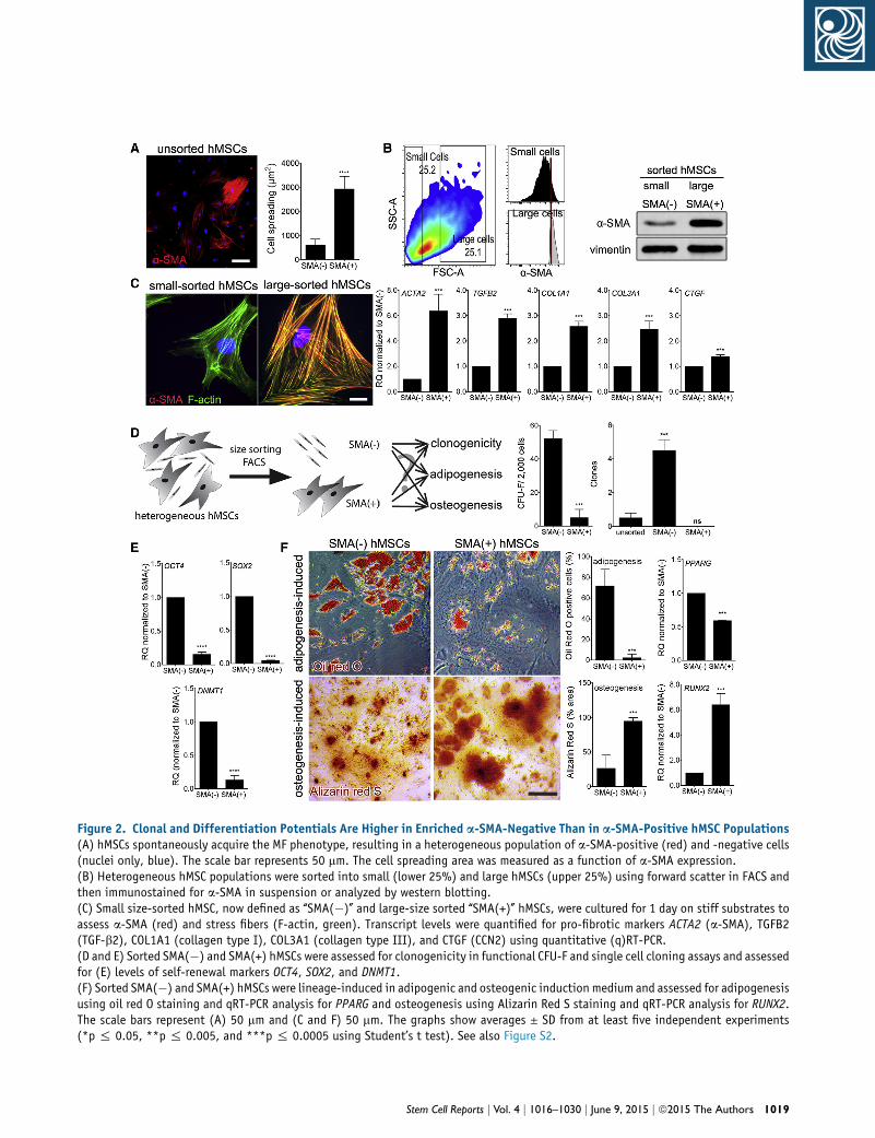
Figure 2. Clonal and Differentiation Potentials Are Higher in Enriched a-SMA-Negative Than in a-SMA-Positive hMSC Populations(A) hMSCs spontaneously acquire the MF phenotype, resulting in a heterogeneous population of a-SMA-positive (red) and -negative cells(nuclei only, blue). The scale bar represents 50 mm. The cell spreading area was measured as a function of a-SMA expression.(B) Heterogeneous hMSC populations were sorted into small (lower 25%) and large hMSCs (upper 25%) using forward scatter in FACS andthen immunostained for a-SMA in suspension or analyzed by western blotting.(C) Small size-sorted hMSC, now defined as ‘‘SMA(�)’’ and large-size sorted ‘‘SMA(+)’’ hMSCs, were cultured for 1 day on stiff substrates toassess a-SMA (red) and stress fibers (F-actin, green). Transcript levels were quantified for pro-fibrotic markers ACTA2 (a-SMA), TGFB2(TGF-b2), COL1A1 (collagen type I), COL3A1 (collagen type III), and CTGF (CCN2) using quantitative (q)RT-PCR.(D and E) Sorted SMA(�) and SMA(+) hMSCs were assessed for clonogenicity in functional CFU-F and single cell cloning assays and assessedfor (E) levels of self-renewal markers OCT4, SOX2, and DNMT1.(F) Sorted SMA(�) and SMA(+) hMSCs were lineage-induced in adipogenic and osteogenic induction medium and assessed for adipogenesisusing oil red O staining and qRT-PCR analysis for PPARG and osteogenesis using Alizarin Red S staining and qRT-PCR analysis for RUNX2.The scale bars represent (A) 50 mm and (C and F) 50 mm. The graphs show averages ± SD from at least five independent experiments(*p % 0.05, **p % 0.005, and ***p % 0.0005 using Student’s t test). See also Figure S2.
Stem Cell Reports j Vol. 4 j 1016–1030 j June 9, 2015 j ª2015 The Authors 1019

Figure 3. Soft Substrate Culture Restores Lineage Differentiation of SMA(+) hMSCs(A–D) Sorted SMA(+) hMSCs were cultured on stiff and soft substrates and assessed for MF activation after 5 days, clonogenicity after10 days, and differentiation potential after 14 days. MF activation was assessed by (B) western blotting, (C) immunofluorescence fora-SMA (red) and stress fibers (F-actin, green) (the scale bar represents 20 mm), and (D) qRT-PCR for fibrotic markers.(E) Self-renewal potential was quantified from CFU-F assays and qRT-PCR analysis of OCT4, SOX2, and DNMT1 transcripts.(F) Sorted SMA(+) were grown for 5 days on stiff and soft culture substrates and then transferred to conventional culture dishes forinduction into adipogenic lineage (oil red O, PPARG) and osteogenesis (Alizarin Red S, RUNX2). The scale bar represents 50 mm.The graphs show averages ± SD from at least five independent experiments (*p% 0.05, **p% 0.005, and ***p% 0.0005 using Student’st test). See also Figure S3.
of a-SMA (Figures 3A and 3B), disappearance of a-SMA from
stress fibers (Figure 3C), and generally reduced levels of
fibrotic marker transcripts compared with hMSC(+) grown
on stiff substrates (Figure 3D). The culture time required to
deactivateMFsmechanically was 5 days (Figure S4). Revers-
ibility of the fibrotic phenotype was not dependent on the
degree of MF pre-activation. SMA(+) hMSCs lost the MF
phenotype on soft substrate even when being sorted from
TGF-b1-pre-treated heterogeneous populations and were
1020 Stem Cell Reports j Vol. 4 j 1016–1030 j June 9, 2015 j ª2015 The Aut
not re-activated on soft substrate by adding TGF-b1
(Figure S4).
Transfer of SMA(+) hMSCs that had lost MF characteris-
tics on soft substrates to conventional lineage induction
media resulted in 1.3-fold higher levels of PPARG
mRNA and �10-fold increase in lipid-production than
SMA(+) hMSCs harvested from control stiff culture
(Figure 3E). Conversely, the osteogenic capacity of soft-
grown SMA(+) hMSCs was reduced by �2-fold compared
hors

to SMA(+) hMSCs from stiff cultures (Figure 3E). However,
clonogenicity and mRNA levels of OCT4, SOX2, and
DNMT1 of ‘‘MF-deactivated’’ SMA(+) hMSCs were similarly
low compared with control SMA(+) hMSCs grown on stiff
substrates (Figure 3F). Hence, SMA(+) hMSC can be deacti-
vated to lose fibrotic MF features by reducing substrate
stiffness, but do not regain clonogenicity, possibly due
to a dose-dependent effect of substrate stiffness on MF
(de)activation. To achieve greater changes in MF activation
state and to test whether the MF marker a-SMA plays
a functional role in guiding MSC fate, we next overex-
pressed and knocked down a-SMA in MF-sorted hMSC
populations.
Expression of a-SMA Directly Controls hMSC Fate
Expression of a-SMA and incorporation into stress fibers
upregulate contraction of fibroblastic cells (Hinz et al.,
2001, 2002). It is conceivable that a-SMA directly regu-
lates hMSC stem cell character because actin-myosin
generated intracellular tension guides hMSC fate decision
(Engler et al., 2006; MacQueen et al., 2013; McBeath et al.,
2004; Swift et al., 2013). To elucidate the role of a-SMA
protein in directing hMSC fate, we first transfected
SMA(�) hMSCs with a-SMA-GFP (Figure 4A) that localized
to stress fibers and GFP control that remained cytosolic
(Figure 4B). Expression of SMA-GFP (67 kDa) resulted
in upregulation of endogenous a-SMA (42 kDa) (Fig-
ure 4B), consistent with a positive feedback loop of
enhanced cell contraction and mechanically induced
a-SMA expression (Hinz et al., 2002). Overexpression of
a-SMA-GFP reduced the high clonogenicity of SMA(�)
hMSCs and transcript levels of SOX2, OCT4, and
DNMT1 by maximally 4-fold compared to GFP control
(Figure 4C). a-SMA-GFP-overexpressing SMA(�) hMSCs
exhibited �5-fold decreased adipogenesis (Figure 4D),
but �2-fold increased osteogenic potential compared to
controls (Figure 4D).
Next, we knocked down a-SMA in SMA(+) hMSCs using
short hairpin (sh)RNA directed against a-SMA, which
resulted in�3-fold higher numbers of CFU-Fs and dramatic
upregulation of SOX2, OCT4, and DNMT1 transcripts (up
to 40-fold, OCT4) than in controls (scrambled shRNA
with GFP reporter) (Figure 4G). In cell lineage induction
assays, SMA(+) hMSCs showed �25-fold increased forma-
tion of lipid-producing adipocytes,�5.6-fold higher PPARG
mRNA levels (Figure 4H), and �2.2-fold decrease of miner-
alized nodule area, as well as �100-fold decrease in RUNX2
mRNA levels after a-SMA knockdown compared with
controls (Figure 4H). Our results showing that knockdown
of a-SMA alone confers clonogenicity and adipogenic/
osteogenic differentiation suggest that the MF is an acti-
vated phenotype and not a terminal differentiation state
of SMA(+) hMSCs.
Stem C
Expression of a-SMA in hMSCs Controls YAP/TAZ
Nuclear Localization
We next addressed how a-SMA regulates hMSC fate and
clonogenicity. Cell contractility and actin filament bundle
assembly both stimulate activation and translocation of
YAP/TAZ to the nucleus (Dupont et al., 2011; Gaspar and
Tapon, 2014). YAP/TAZ transcriptionally regulate genes
associated with stem cell fate decision and self-renewal
(Varelas, 2014). To test whether a-SMA expression affects
YAP/TAZ localization in hMSCs, we first co-analyzed
a-SMA stress fiber intensity with the ratio of nuclear/cyto-
solic YAP on an individual cell basis and demonstrated a
clear correlation (Figure S5). The low baseline percentage
of SMA(�) hMSCs with predominantly nuclear YAP
(25%, nuclear/cytosolic YAP > 1) was increased to 90% after
transfection with a-SMA-GFP, corresponding to the per-
centage of SMA(+) hMSCs with constitutive nuclear YAP
(Figure 5A). Knockdown of a-SMA with shRNA reduced
the percentage of SMA(+) hMSCs with predominantly
nuclear YAP to 7% (Figure 5B).
To decipher themode of action of a-SMA, contraction, or
action polymerization, we used the a-SMA-specific fusion
peptide SMA-FP in combination with cytoskeletal drugs
(Figure 5C). SMA-FP selectively targets a-SMA in stress
fibers, inhibits a-SMA-mediated contraction, and selec-
tively depolymerizes a-SMA from stress fibers (Clement
et al., 2005; Hinz et al., 2002). SMA-FP removed a-SMA
from persisting stress fibers, in contrast to control (DMSO
and skeletal actin fusion peptide, SKA-FP) SMA(+) hMSCs
(Figure S5). SMA-FP treatment resulted in low levels of
nuclear YAP and reduced hMSC contraction on wrinkling
elastomer substrates (Figure 5C). SMA(+) hMSCs treated
with the myosin II inhibitor blebbistatin showed similar
low levels of YAP in the nucleus and low contraction, but
disassembled all actins from stress fibers, as evident from
phalloidin staining (Figures 5C and S5).
Next, we stabilized stress fibers by pre-treating SMA(+)
hMSCs with jasplakinolide, followed by addition of SMA-
FP (Figure 5D). With jasplakinolide being present, the
SMA-FP was unable to depolymerize a-SMA from stress
fibers (Figure 5D) and hMSCs contraction was unaltered
(Figures 5D and 5E), indicating that the SMA-FP reduces
MF contraction primarily by depolymerizing a-SMA from
stress fibers. Whereas YAP nuclear localization remained
high under jasplakinolide/SMA-FP treatment (Figures 5D
and 5F), addition of blebbistatin to jasplakinolide-treated
SMA(+) hMSCs resulted in the loss of a-SMA from stress
fibers, reduced cell contractility, and reduced YAP nuclear
localization (Figures 5D–5F). Control SMA(+) hMSCs
treated with jasplakinolide alone were similar to untreated
cells (Figures 5D–5F). In combination, these experiments
suggest that enhanced contractile activity mediated by
a-SMA upregulates YAP/TAZ nuclear localization.
ell Reports j Vol. 4 j 1016–1030 j June 9, 2015 j ª2015 The Authors 1021

Figure 4. Overexpression and Knockdown of a-SMA Affecs hMSC Stem Cell Character(A) SMA(�) hMSCs were transfected with control GFP or a-SMA-GFP constructs and evaluated for clonogenicity and lineage differentiation.(B) Transfected cells were cultured for 5 days and stained for a-SMA (red) and GFP (green) (the scale bar represents 20 mm) and processedfor western blotting.(C) The effect of a-SMA overexpression on stem cell character was assessed by CFU-F assays and qRT-PCR analysis of SOX2, OCT4, and DNMT1.(D) Differentiation potential was tested by inducing adipogenic and osteogenic lineages using respective induction media and assessed foradipogenesis and osteogenesis. The scale bar represents 50 mm.(E–H) The same analysis was performed with SMA(+) hMSCs that were transfected with either scrambled (scr) shRNA or a-SMA-shRNAconstructs.The graphs show averages ± SD from at least three independent experiments (*p % 0.05, **p % 0.005, and ***p % 0.0005 usingStudent’s t test). See also Figure S4.
To show that YAP/TAZ activation is indeed respon-
sible for reduced lineage differentiation and self-renewal
capacity downstream of a-SMA-expression in hMSCs, we
co-transfected SMA(�) hMSCs with small interfering (si)
RNA directed against YAP1 together with a-SMA-GFP.
Transfection of YAP1 siRNA substantially reduced YAP
expression levels (Figure 6A) and resulted in reduced
RUNX2mRNA levels, increased PPARGmRNA, and reduced
1022 Stem Cell Reports j Vol. 4 j 1016–1030 j June 9, 2015 j ª2015 The Aut
SOX2 and OCT4 levels in control cells (Figure 6A, GFP).
Knockdown of YAP1 in a-SMA-overexpressing hMSCs
restored expression of PPARG mRNA and strongly reduced
RUNX2 mRNA expression (Figure 6A). These results
confirmed that the effect of high a-SMA expression on
hMSC lineage differentiation potential is mediated by
YAP. However, knockdown of YAP1 in a-SMA-overexpress-
ing hMSCs did not restore high expression of SOX2 and
hors

Figure 5. Presence of a-SMA in hMSC Stress Fibers Results in YAP/TAZ Nuclear Accumulation(A) Sorted SMA(�) hMSCs were transfected with control GFP and a-SMA-GFP constructs.(B) Sorted SMA(+) hMSCs were transfected with control scrambled (scr) and a-SMA-shRNA constructs. Localization of YAP/TAZ wasvisualized using immunofluorescence staining for YAP (red) and GFP (green).(C–F) The ratio of nuclear over cytosolic YAP/TAZ was calculated by image analysis and the percentage of cells with predominantly nuclearYAP/TAZ localization was determined. Sorted SMA(+) cells were either pre-treated with (C) DMSO (control) or (D) jasplakinolide for 30 min,followed by the addition of the a-SMA N-terminal peptide AcEEED (SMA-FP), blebbistatin, or DMSO control for another 40 min. Cells werethen stained for YAP and a-SMA or YAP and stress fibers as indicated and assessed for contraction using wrinkling silicone substrates.Quantified from these images were (E) cell contraction as percentage of cells associated with wrinkles, and (F) percentage of cells withpredominant nuclear YAP/TAZ localization.All scale bars represent 20 mm. The graphs show averages ± SD from at least three independent experiments (*p% 0.05 and **p% 0.005using ANOVA followed by a post hoc Tukey’s multiple comparison test). See also Figure S5.
Stem Cell Reports j Vol. 4 j 1016–1030 j June 9, 2015 j ª2015 The Authors 1023

Figure 6. Suppression of MSC Adipogenesis by a-SMA Is Mediated via YAP(A) Sorted SMA(�) hMSCs were co-transfected with siRNA directed against YAP1 together with control GFP or a-SMA-GFP constructs andassessed after 5 days. YAP downregulation was confirmed using western blotting.(B) Sorted SMA(�) hMSCs were transfected with control GFP or a-SMA-GFP constructs and treated for 5 days with verteporfin (VP, 2 mM) orDMSO control. Efficacy of vereporfin was assessed by performing qRT-PCR for the YAP1 downstream target CCN2 (CTGF). All cells wereanalyzed by qRT-PCR for mRNA expression of RUNX2, PPARG, SOX2, and OCT4.The graphs show averages ± SD from three different donors per condition (*p % 0.05, **p % 0.005, and ***p % 0.0005 using ANOVAfollowed by a post hoc Tukey’s multiple comparison test).
OCT4, as expected if a-SMA would suppress hMSC self-
renewal by acting through YAP1 (Figure 6A). To test
whether this result was due to the overall loss of YAP, we
used verteporfin to selectively abrogate the nuclear activity
of YAP by inhibiting binding to TEAD elements (Liu-Chit-
tenden et al., 2012) (Figure 6B). Drug action was controlled
by low levels of the YAP1 downstream target CCN2 (CTGF)
(Figure 6B). Treating SMA(�) and SMA(�)-overexpressing
a-SMA with verteporfin generally confirmed our results
obtained with YAP1 knockdown (Figure 6B). Hence,
a-SMA expression levels regulate the hMSC multilineage
differentiation program via YAP, but downregulate hMSC
self-renewal possibly through a different mechanism.
Finally, to investigate whether a-SMA expression is
relevant for YAP/TAZ activation andhMSC lineage determi-
nation in vivo, we correlated a-SMA expression and YAP
1024 Stem Cell Reports j Vol. 4 j 1016–1030 j June 9, 2015 j ª2015 The Aut
nuclear localization in soft mesenchymal tumors (adipo-
sarcoma) and stiff mesenchymal tumors (osteosarcoma)
that involve MSC tumorigenesis and fibrogenesis (Moh-
seny et al., 2009; Rodriguez et al., 2012; Xiao et al., 2013).
The levels of a-SMA expression were negligible in healthy
fat control tissue and low in normal bone, but significantly
higher in the activated stroma of the respective tumor (Fig-
ures 7A–7C). Increased a-SMA expression in adiposarcoma
correlated with 3.5-fold higher percentages of cells with
predominantly nuclear YAP staining (22%) compared to
normal fat tissue (6%, Figures 7A, 7C, and 7D). The levels
of nuclear localized YAP staining did not significantly in-
crease in osteosarcoma compared to already high levels
observed in healthy bone (Figures 7B–7D), supporting
that high levels of YAP/TAZ in mesenchymal cells direct
osteogenic differentiation.
hors

Figure 7. Expression of a-SMA Correlateswith High Levels of Nuclear YAP/TAZ inHuman Adiposarcoma and OsteosarcomaTissues(A) Human normal fat and adiposarcomatissue samples were obtained from the samedonor, sectioned, and stained for a-SMA(red), YAP (green), and cell nuclei (blue).(B) Donor-matched normal bone and oste-osarcoma tissue were similarly processed.(C and D) Percentage of (C) a-SMA-positivecells and (D) cells with predominantly nu-clear YAP were quantified from at least threetissue sections per donor. The graphs showaverages ± SD from three different donorsper condition (*p % 0.05, **p % 0.005,and ***p % 0.0005 using ANOVA followedby a post hoc Tukey’s multiple comparisontest).
DISCUSSION
MSCs are prone to MF activation by stiff ECM and TGF-b1,
but the consequences on their stem cell potential and
reversibility have not been systematically assessed. We
establish a direct link between a-SMA expression/function,
YAP/TAZ activity, and hMSC fate. Different actin isoforms
promote specific types of actin organization levels and a
shift in the ratios of actin isoforms can reprogram cell
differentiation (Lechuga et al., 2014; Tondeleir et al.,
2012). a-SMA incorporation into existing stress fibers has
been shown to increase actin organization and intracellular
tension (Goffin et al., 2006; Hinz et al., 2001). Actin organi-
zation as cortical filaments or incorporation into stress fiber
bundles has been shown to differentially control YAP/TAZ
activation, nuclear localization, and regulation of the
Hippo pathway (Gaspar and Tapon, 2014; Halder et al.,
2012; Yu et al., 2012a). In epithelia, cell-morphology-
dependent actin organization provides positional informa-
tion to individual cells in a multicellular layer (Aragona
et al., 2013; Wada et al., 2011). YAP/TAZ are also central
Stem C
in regulating cell fate decision by interacting with RUNX2
and PPARG (Hong et al., 2005; Hong and Yaffe, 2006; Vare-
las et al., 2008). Consistently, neo-expression of a-SMA
alone was sufficient in our experiments to reduce hMSC
differentiation potential, which was rescued by inhibition
and downregulation of YAP1.
Whether the a-SMA-induced nuclear shift of YAP is
responsible for reduced clonogenicity is unclear because
YAP1 knockdown did not protect against the reduction of
SOX2 and OCT4 transcript levels upon a-SMA-overexpres-
sion. Consistently, YAP binds to and is expected to activate
gene transcription of SOX2 and OCT4 in embryonic stem
(ES) cells (Lian et al., 2010). However, YAP is inactivated
in ES cells during differentiation (Lian et al., 2010), whereas
it drives specific lineage specification in MSCs (Dupont
et al., 2011). Overexpression of OCT4 has been shown to
result in increased MSC proliferation, whereas OCT4
knockdown had the opposite effect (Tsai et al., 2012).
Similarly, knockdown of SOX2 reduces proliferation in
MSC-derived osteoprogenitors that is restored by YAP1
overexpression, which acts downstream of SOX2 in this
ell Reports j Vol. 4 j 1016–1030 j June 9, 2015 j ª2015 The Authors 1025

model (Seo et al., 2013). Hence, the interplay between
a-SMA, YAP/TAZ, and self-renewal genes is complex and
not entirely understood at present.
YAP/TAZ transcription factors provide a direct link
between cell mechanosensing and gene regulation (Halder
et al., 2012). Inhibition of cell contraction has been shown
to decrease nuclear localization of YAP/TAZ and transcrip-
tion of downstream genes, whereas high intracellular
tension drives YAP/TAZ into the nucleus (Calvo et al.,
2013; Dupont et al., 2011). Expression of a-SMA is not
essential for the localization of YAP/TAZ in the nucleus
and �10% of the hMSC retained predominantly nuclear
YAP even in the presence of the SMA-FP. However, incorpo-
ration of a-SMA into existing stress fibers substantially in-
creases contractile force and cell stress (Hinz et al., 2001)
and thus accentuates fibrogenic transcription programs.
YAP/TAZ have been shown to be involved in regulating
a-SMA expression and fibrogenesis using an experimental
model of epithelial-to-mesenchymal transition (Speight
et al., 2013) and during lung fibroblast-to-MF activation
by stiff ECM (Liu et al., 2015). These reports and our works
suggest a positive feedback loop between a-SMA and YAP/
TAZ signaling that amplifies fibrosis.
Importantly, expression of a-SMA is not simply a marker
for MF activation, but also the driver of cell function and
fate. Increased expression of a-SMA directly reduces the
clonal potential of hMSCs and guides their differentiation
toward osteoblasts. Hence, a-SMAnot only identifies osteo-
progenitors in hMSC populations as shown by others
before (Grcevic et al., 2012; Kalajzic et al., 2008), but may
be part of the mechanism driving differentiation. Analysis
of bone marrow-derived hMSCs treated with TGF-b1 and/
or exposed to fibrosis-stiff culture substrates has previously
revealed a fibrogenic MF activation program (Park et al.,
2011; Wang et al., 2004). Our results additionally demon-
strate that neo-expression of a-SMA associates with
reduced clonogenicity and lineage differentiation potential
of hMSCs and that this program is reversible. SMA(+)
hMSC can be deactivated to lose fibrotic MF features by
reducing substrate stiffness. Originally considered to be a
terminal differentiation state of various precursor cells,
deactivation of the MF has been shown in recent experi-
mental models of kidney and liver fibrosis (Hecker et al.,
2011; Kisseleva and Brenner, 2013). In vitro, the depletion
of MF features in fibroblasts and MSCs is achieved by cul-
ture on soft silicone substrates (Achterberg et al., 2014;
Balestrini et al., 2012; Goffin et al., 2006; Park et al.,
2011) or by treatment with anti-fibrotic growth factors
(Desai et al., 2014). Our results add to these findings that
SMA(+) hMSC populations, deactivated to loseMF features,
regain adipogenic lineage differentiation potential rather
than turning into a-SMA-negative fibroblasts. Hence,
a-SMA-positive cells are likely derived from previously
1026 Stem Cell Reports j Vol. 4 j 1016–1030 j June 9, 2015 j ª2015 The Aut
a-SMA-negative hMSCs and expression of a-SMA reversibly
reduced their clonogenicity and lineage potential.
Our findings have important implications for hMSC ther-
apy in fibroproliferative diseases, including tumor forma-
tion and development of fibrosis. Our in vivo data show
that the percentage of a-SMA-expressing MFs in sarcomas
correlates with the degree of YAP/TAZ activation. A wide
variety of different tumors have been shown to accumulate
stromal cells that are positive for a-SMA and perform MF
functions, including stiffeningof the stromaandpromoting
tumor progression (Hinz et al., 2012; Ohlund et al., 2014).
Consistently, YAP expressed in cancer-associated fibroblasts
was recently shown to play an important role in controlling
of cytoskeleton-regulating genes, as well as tumor cell inva-
sion and ECM stiffening (Calvo et al., 2013). We propose a
feedforward loop of MSC-to-MF activation in the tumor
microenvironment, leading to higher contractile cells and
stiffer ECM, which both lead to increased YAP/TAZ activity
and conversion of regenerative MSCs into fibrotic MFs.
Interrupting this feedforward loop will have important
consequences for hMSC potential in clinical applications.
First, specific inhibitors of a-SMA such as the SMA-FP or
shRNA strategies may be co-delivered with hMSCs to exert
fibrosis-inhibitory effects on the resident fibrotic cell
population in the lesion. Second, suppressing MSC-to-MF
activation during cell culture expansion will enhance the
fraction of valuable stem cells and reduce the risk of fibrosis
upon implantation. We have shown that explantation
and continued culture on soft culture substrates renders
populations of lung fibroblasts resistant to subsequent
mechanical activation on stiff substrates over several
consecutive passages (Balestrini et al., 2012). This concept
of cells acquiring a ‘‘mechanical memory’’ has recently
been confirmed for MSC lineage programs on a shorter
timescale with YAP/TAZ being involved (Yang et al.,
2014). It is yet unclear whyhMSCs and othermesenchymal
cells that have been cultured on conventional stiff culture
dishes are not similarly primed and can at least acutely
(up to 8 days in our experiments) lose fibrogenic character
upon short-term exposure (5 days) to soft substrates. In our
own studies, we found it to be essential to use substrates
with a pathophysiological stiffness range (1–100 kPa) and
to never expose cells to tissue culture plastic formechanical
priming to occur (Balestrini et al., 2012).
EXPERIMENTAL PROCEDURES
For detailed experimental procedures, see the Supplemental Exper-
imental Procedures.
Tissue, Cell Culture, and DrugsMSCswere obtained from the bonemarrow of healthy donors (Da-
vies et al., 2001), fromumbilical perivasculature (Ennis et al., 2008)
hors

and from adipose tissue (Vermette et al., 2007). Tissue sections of
mesenchymal tumors and respective healthy control tissues were
purchased fromBiomax.Cell drugs used in these study are: blebbis-
tatin (50 mM) (Tocris Bioscience, Cedarlane), jasplakinolide
(50 nM) (Life Technologies), verteporfin (2 mM) (Sigma-Aldrich),
SMA-FP and control SKA-FP (5 mM). Immunofluorescence, micro-
scopy, and western blot were performed as described earlier with
antibodies listed in the Supplemental Experimental Procedures
(Klingberg et al., 2014). Adipogenesis, osteogenesis, and chondro-
genesis were induced and assessed as described earlier (Majd et al.,
2011). Elastic silicone culture substrates were purchased from
Excellness Biotech SA and activated for cell adhesion by plasma
oxygenation (PE-100, Plasma Etch), followed by coating with
2 mg/cm2 FN (Millipore). Cell contractility was assessed using
FN-coated wrinkling silicone substrates (Balestrini et al., 2012).
Flow Cytometry and Cell SortingFor flow cytometry, fixed cells were immunostained using
antibodies CD44-APC-H7, CD73-PE-CY7, CD90-FITC, CD105-
PerCP-Cy5.5, CD146-V450, and respective isotype controls (BD
Bioscience) and analyzed using a flow cytometer (LSR II, BD). To
enrich for a-SMA-positive and a-SMA-negative hMSC populations,
cells were trypsinized and sorted for cell size (upper and lower 25%
in forward scatter) using fluorescence activated cell sorting (FACS)
(FACSAria III, BD).
Plasmid Constructsa-SMA was overexpressed using a-SMA-GFP (Clement et al., 2005)
and knockeddown using 29-mer shRNA targeting a-SMA tran-
scripts in pGFP-V-RS vectors (OriGene Technologies). Human
YAP1 27-mer siRNA were ordered from (OriGene Technologies).
Cells were transfected using an electroporation device (Neon,
Life Technologies).
SUPPLEMENTAL INFORMATION
Supplemental Information includes Supplemental Experimental
Procedures and five figures and can be found with this article
online at http://dx.doi.org/10.1016/j.stemcr.2015.05.004.
AUTHOR CONTRIBUTIONS
N.P.T. designed, performed, analyzed, and wrote the experimental
data in manuscript format. B.H. supervised, designed, analyzed,
and wrote the manuscript. J.F., J.E.D., and A.K. contributed to
data analysis and writing of the manuscript.
ACKNOWLEDGMENTS
We thank Drs. Christine Chaponnier and Giulio Gabbiani for
kindly providing anti-a-SMA antibody and SMA/SKA fusion
peptides. We are also thankful to Stellar Boo for technical support.
We are grateful to Dr. Chris McCulloch (University of Toronto,
Toronto) for carefully reading our manuscript and his invaluable
input. This research was supported by the Canadian Institutes of
Health Research (CIHR) (grants #210820 and #286920 to B.H.
and #111233 to J.F.), the Collaborative Health Research Pro-
gramme (CIHR/NSERC) (#1004005 and #413783), and the Canada
Foundation for Innovation and Ontario Research Fund (CFI/ORF)
Stem C
(#26653) all to B.H. N.P.T. was a Marie Curie Fellow at the Tissue
Transmigration Training Network (T3-Net) funded by the Euro-
pean Commission under Framework Program (FP7/2007-2013)
with grant agreement #237946 and the University of Toronto
Fellowship.
Received: November 19, 2014
Revised: May 4, 2015
Accepted: May 4, 2015
Published: May 28, 2015
REFERENCES
Achterberg, V.F., Buscemi, L., Diekmann, H., Smith-Clerc, J.,
Schwengler, H., Meister, J.J., Wenck, H., Gallinat, S., and Hinz, B.
(2014). The nano-scale mechanical properties of the extracellular
matrix regulate dermal fibroblast function. J. Invest. Dermatol.
134, 1862–1872.
Aragona, M., Panciera, T., Manfrin, A., Giulitti, S., Michielin, F.,
Elvassore, N., Dupont, S., and Piccolo, S. (2013). A mechanical
checkpoint controlsmulticellular growth throughYAP/TAZ regula-
tion by actin-processing factors. Cell 154, 1047–1059.
Arnold, K., Sarkar, A., Yram, M.A., Polo, J.M., Bronson, R., Sen-
gupta, S., Seandel, M., Geijsen, N., and Hochedlinger, K. (2011).
Sox2(+) adult stem and progenitor cells are important for tissue
regeneration and survival of mice. Cell Stem Cell 9, 317–329.
Balestrini, J.L., Chaudhry, S., Sarrazy, V., Koehler, A., and Hinz, B.
(2012). The mechanical memory of lung myofibroblasts. Integr.
Biol. (Camb) 4, 410–421.
Behfar, A., Crespo-Diaz, R., Terzic, A., and Gersh, B.J. (2014). Cell
therapy for cardiac repair—lessons from clinical trials. Nat. Rev.
Cardiol. 11, 232–246.
Bianco, P., Cao, X., Frenette, P.S., Mao, J.J., Robey, P.G., Simmons,
P.J., andWang, C.Y. (2013). The meaning, the sense and the signif-
icance: translating the science of mesenchymal stem cells into
medicine. Nat. Med. 19, 35–42.
Breitbach, M., Bostani, T., Roell, W., Xia, Y., Dewald, O., Nygren,
J.M., Fries, J.W., Tiemann, K., Bohlen, H., Hescheler, J., et al.
(2007). Potential risks of bone marrow cell transplantation into
infarcted hearts. Blood 110, 1362–1369.
Calvo, F., Ege, N., Grande-Garcia, A., Hooper, S., Jenkins, R.P.,
Chaudhry, S.I., Harrington, K., Williamson, P., Moeendarbary, E.,
Charras, G., and Sahai, E. (2013). Mechanotransduction and
YAP-dependent matrix remodelling is required for the generation
and maintenance of cancer-associated fibroblasts. Nat. Cell Biol.
15, 637–646.
Clement, S., Hinz, B., Dugina, V., Gabbiani, G., andChaponnier, C.
(2005). The N-terminal Ac-EEED sequence plays a role in alpha-
smooth-muscle actin incorporation into stress fibers. J. Cell Sci.
118, 1395–1404.
Davies, J.E., Baksh, D., and Karp, J.M. (2001). Mesenchymal cell
culture: bone. In Methods in Tissue Engineering, A. Atala and R.
Lanza, eds. (San Diego: Academic Press), pp. 333–344.
Desai, V.D., Hsia, H.C., and Schwarzbauer, J.E. (2014). Reversible
modulation of myofibroblast differentiation in adipose-derived
mesenchymal stem cells. PLoS ONE 9, e86865.
ell Reports j Vol. 4 j 1016–1030 j June 9, 2015 j ª2015 The Authors 1027

di Bonzo, L.V., Ferrero, I., Cravanzola, C., Mareschi, K., Rustichell,
D., Novo, E., Sanavio, F., Cannito, S., Zamara, E., Bertero, M., et al.
(2008). Human mesenchymal stem cells as a two-edged sword in
hepatic regenerativemedicine: engraftment and hepatocyte differ-
entiation versus profibrogenic potential. Gut 57, 223–231.
Dupont, S., Morsut, L., Aragona, M., Enzo, E., Giulitti, S., Corde-
nonsi, M., Zanconato, F., Le Digabel, J., Forcato, M., Bicciato, S.,
et al. (2011). Role of YAP/TAZ in mechanotransduction. Nature
474, 179–183.
Engler, A.J., Sen, S., Sweeney, H.L., andDischer, D.E. (2006).Matrix
elasticity directs stem cell lineage specification. Cell 126, 677–689.
Ennis, J., Sarugaser, R., Gomez, A., Baksh, D., and Davies, J.E.
(2008). Isolation, characterization, and differentiation of human
umbilical cord perivascular cells (HUCPVCs). Methods Cell Biol.
86, 121–136.
Fernandez, P., Perez-Aso,M., Smith, G.,Wilder, T., Trzaska, S., Chir-
iboga, L., Franks, A., Jr., Robson, S.C., Cronstein, B.N., and Chan,
E.S. (2013). Extracellular generation of adenosine by the ectonu-
cleotidases CD39 and CD73 promotes dermal fibrosis. Am. J.
Pathol. 183, 1740–1746.
Forbes, S.J., and Rosenthal, N. (2014). Preparing the ground for
tissue regeneration: from mechanism to therapy. Nat. Med. 20,
857–869.
Gaspar, P., and Tapon, N. (2014). Sensing the local environment:
actin architecture and Hippo signalling. Curr. Opin. Cell Biol. 31,
74–83.
Goffin, J.M., Pittet, P., Csucs, G., Lussi, J.W., Meister, J.J., and Hinz,
B. (2006). Focal adhesion size controls tension-dependent recruit-
ment of alpha-smoothmuscle actin to stress fibers. J. Cell Biol. 172,
259–268.
Grcevic, D., Pejda, S., Matthews, B.G., Repic, D., Wang, L., Li, H.,
Kronenberg, M.S., Jiang, X., Maye, P., Adams, D.J., et al. (2012).
In vivo fate mapping identifies mesenchymal progenitor cells.
Stem Cells 30, 187–196.
Halder, G., Dupont, S., and Piccolo, S. (2012). Transduction of
mechanical and cytoskeletal cues by YAP and TAZ. Nat. Rev. Mol.
Cell Biol. 13, 591–600.
Hecker, L., Jagirdar, R., Jin, T., and Thannickal, V.J. (2011). Revers-
ible differentiation of myofibroblasts by MyoD. Exp. Cell Res. 317,
1914–1921.
Hinz, B. (2010a). Themyofibroblast in connective tissue repair and
regeneration. In Regenerative Medicine and Biomaterials for the
Repair of Connective Tissues, C.A.J. Ralphs, ed. (Cambridge:
Woodhead Publishing), pp. 39–82.
Hinz, B. (2010b). The myofibroblast: paradigm for a mechanically
active cell. J. Biomech. 43, 146–155.
Hinz, B., Celetta, G., Tomasek, J.J., Gabbiani, G., and Chaponnier,
C. (2001). Alpha-smooth muscle actin expression upregulates
fibroblast contractile activity. Mol. Biol. Cell 12, 2730–2741.
Hinz, B., Gabbiani, G., andChaponnier, C. (2002). TheNH2-termi-
nal peptide of alpha-smoothmuscle actin inhibits force generation
by themyofibroblast in vitro and in vivo. J. Cell Biol. 157, 657–663.
Hinz, B., Phan, S.H., Thannickal, V.J., Prunotto, M., Desmouliere,
A., Varga, J., De Wever, O., Mareel, M., and Gabbiani, G. (2012).
1028 Stem Cell Reports j Vol. 4 j 1016–1030 j June 9, 2015 j ª2015 The Aut
Recent developments in myofibroblast biology: paradigms for
connective tissue remodeling. Am. J. Pathol. 180, 1340–1355.
Hong, J.H., and Yaffe, M.B. (2006). TAZ: a beta-catenin-like mole-
cule that regulates mesenchymal stem cell differentiation. Cell
Cycle 5, 176–179.
Hong, J.H., Hwang, E.S., McManus, M.T., Amsterdam, A., Tian, Y.,
Kalmukova, R., Mueller, E., Benjamin, T., Spiegelman, B.M., Sharp,
P.A., et al. (2005). TAZ, a transcriptional modulator of mesen-
chymal stem cell differentiation. Science 309, 1074–1078.
Kalajzic, Z., Li, H., Wang, L.P., Jiang, X., Lamothe, K., Adams, D.J.,
Aguila, H.L., Rowe, D.W., and Kalajzic, I. (2008). Use of an alpha-
smooth muscle actin GFP reporter to identify an osteoprogenitor
population. Bone 43, 501–510.
Karnoub, A.E., Dash, A.B., Vo, A.P., Sullivan, A., Brooks, M.W., Bell,
G.W., Richardson, A.L., Polyak, K., Tubo, R., and Weinberg, R.A.
(2007). Mesenchymal stem cells within tumour stroma promote
breast cancer metastasis. Nature 449, 557–563.
Kim, W., Barron, D.A., San Martin, R., Chan, K.S., Tran, L.L., Yang,
F., Ressler, S.J., and Rowley, D.R. (2014). RUNX1 is essential for
mesenchymal stem cell proliferation andmyofibroblast differenti-
ation. Proc. Natl. Acad. Sci. USA 111, 16389–16394.
Kisseleva, T., and Brenner, D.A. (2013). Inactivation of myofibro-
blasts during regression of liver fibrosis. Cell Cycle 12, 381–382.
Klingberg, F., Chow, M.L., Koehler, A., Boo, S., Buscemi, L., Quinn,
T.M., Costell, M., Alman, B.A., Genot, E., and Hinz, B. (2014).
Prestress in the extracellular matrix sensitizes latent TGF-b1 for
activation. J. Cell Biol. 207, 283–297.
Koumas, L., Smith, T.J., Feldon, S., Blumberg, N., and Phipps, R.P.
(2003). Thy-1 expression in human fibroblast subsets defines my-
ofibroblastic or lipofibroblastic phenotypes. Am. J. Pathol. 163,
1291–1300.
Lechuga, S., Baranwal, S., Li, C., Naydenov, N.G., Kuemmerle, J.F.,
Dugina, V., Chaponnier, C., and Ivanov, A.I. (2014). Loss of g-cyto-
plasmic actin triggersmyofibroblast transition of human epithelial
cells. Mol. Biol. Cell 25, 3133–3146.
Li, Y., Jiang, D., Liang, J., Meltzer, E.B., Gray, A., Miura, R., Wogen-
sen, L., Yamaguchi, Y., and Noble, P.W. (2011). Severe lung fibrosis
requires an invasive fibroblast phenotype regulated by hyaluronan
and CD44. J. Exp. Med. 208, 1459–1471.
Lian, I., Kim, J., Okazawa, H., Zhao, J., Zhao, B., Yu, J., Chinnaiyan,
A., Israel, M.A., Goldstein, L.S., Abujarour, R., et al. (2010). The role
of YAP transcription coactivator in regulating stem cell self-
renewal and differentiation. Genes Dev. 24, 1106–1118.
Liu, F., Lagares, D., Choi, K.M., Stopfer, L.,Marinkovic, A., Vrbanac,
V., Probst, C.K., Hiemer, S.E., Sisson, T.H., Horowitz, J.C., et al.
(2015). Mechanosignaling through YAP and TAZ drives fibroblast
activation and fibrosis. Am. J. Physiol. Lung Cell. Mol. Physiol.
308, L344–L357.
Liu-Chittenden, Y., Huang, B., Shim, J.S., Chen, Q., Lee, S.J., An-
ders, R.A., Liu, J.O., and Pan, D. (2012). Genetic and pharmacolog-
ical disruption of the TEAD-YAP complex suppresses the oncogenic
activity of YAP. Genes Dev. 26, 1300–1305.
MacQueen, L., Sun, Y., and Simmons, C.A. (2013). Mesenchymal
stem cell mechanobiology and emerging experimental platforms.
J. R. Soc. Interface 10, 20130179.
hors

Majd, H., Quinn, T.M., Wipff, P.J., and Hinz, B. (2011). Dynamic
expansion culture for mesenchymal stem cells. Methods Mol.
Biol. 698, 175–188.
Maring, J.A., Trojanowska, M., and ten Dijke, P. (2012). Role of
endoglin in fibrosis and scleroderma. Int. Rev. Cell Mol. Biol.
297, 295–308.
McBeath, R., Pirone, D.M., Nelson, C.M., Bhadriraju, K., andChen,
C.S. (2004). Cell shape, cytoskeletal tension, and RhoA regulate
stem cell lineage commitment. Dev. Cell 6, 483–495.
McQualter, J.L., McCarty, R.C., Van der Velden, J., O’Donoghue,
R.J., Asselin-Labat, M.L., Bozinovski, S., and Bertoncello, I.
(2013). TGF-b signaling in stromal cells acts upstream of FGF-10
to regulate epithelial stem cell growth in the adult lung. Stem
Cell Res. (Amst.) 11, 1222–1233.
Mishra, P.J., Mishra, P.J., Glod, J.W., and Banerjee, D. (2009).
Mesenchymal stem cells: flip side of the coin. Cancer Res. 69,
1255–1258.
Mohseny, A.B., Szuhai, K., Romeo, S., Buddingh, E.P., Briaire-de
Bruijn, I., de Jong, D., van Pel, M., Cleton-Jansen, A.M., and
Hogendoorn, P.C. (2009). Osteosarcoma originates from mesen-
chymal stem cells in consequence of aneuploidization and
genomic loss of Cdkn2. J. Pathol. 219, 294–305.
Nagaya, N., Kangawa, K., Itoh, T., Iwase, T., Murakami, S., Miya-
hara, Y., Fujii, T., Uematsu, M., Ohgushi, H., Yamagishi, M., et al.
(2005). Transplantation of mesenchymal stem cells improves
cardiac function in a rat model of dilated cardiomyopathy.
Circulation 112, 1128–1135.
Ninichuk, V., Gross, O., Segerer, S., Hoffmann, R., Radomska, E.,
Buchstaller, A., Huss, R., Akis, N., Schlondorff, D., and Anders,
H.J. (2006). Multipotent mesenchymal stem cells reduce intersti-
tial fibrosis but do not delay progression of chronic kidney disease
in collagen4A3-deficient mice. Kidney Int. 70, 121–129.
Ohlund, D., Elyada, E., and Tuveson, D. (2014). Fibroblast hetero-
geneity in the cancer wound. J. Exp. Med. 211, 1503–1523.
Park, J.S., Chu, J.S., Tsou, A.D., Diop, R., Tang, Z., Wang, A., and Li,
S. (2011). The effect of matrix stiffness on the differentiation of
mesenchymal stem cells in response to TGF-b. Biomaterials 32,
3921–3930.
Prockop, D.J., Prockop, S.E., and Bertoncello, I. (2014). Are clinical
trials with mesenchymal stem/ progenitor cells (MSCs) too far
ahead of the science? Lessons from experimental hematology.
Stem Cells 32, 3055–3061.
Quante, M., Tu, S.P., Tomita, H., Gonda, T., Wang, S.S., Takashi, S.,
Baik, G.H., Shibata, W., Diprete, B., Betz, K.S., et al. (2011). Bone
marrow-derived myofibroblasts contribute to the mesenchymal
stem cell niche and promote tumor growth. Cancer Cell 19,
257–272.
Rodriguez, R., Rubio, R., andMenendez, P. (2012). Modeling sarco-
magenesis using multipotent mesenchymal stem cells. Cell Res.
22, 62–77.
Seo, E., Basu-Roy, U., Gunaratne, P.H., Coarfa, C., Lim, D.S.,
Basilico, C., and Mansukhani, A. (2013). SOX2 regulates YAP1 to
maintain stemness and determine cell fate in the osteo-adipo line-
age. Cell Rep. 3, 2075–2087.
Stem C
Speight, P., Nakano, H., Kelley, T.J., Hinz, B., and Kapus, A. (2013).
Differential topical susceptibility to TGFb in intact and injured
regions of the epithelium: key role in myofibroblast transition.
Mol. Biol. Cell 24, 3326–3336.
Swift, J., Ivanovska, I.L., Buxboim, A., Harada, T., Dingal, P.C.,
Pinter, J., Pajerowski, J.D., Spinler, K.R., Shin, J.W., Tewari,
M., et al. (2013). Nuclear lamin-A scales with tissue stiffness
and enhances matrix-directed differentiation. Science 341,
1240104.
Tondeleir, D., Lambrechts, A., Muller, M., Jonckheere, V., Doll, T.,
Vandamme,D., Bakkali, K.,Waterschoot, D., Lemaistre,M., Debeir,
O., et al. (2012). Cells lacking b-actin are genetically reprogrammed
and maintain conditional migratory capacity. Mol. Cell. Prote-
omics 11, 255–271.
Tsai, C.C., Su, P.F., Huang, Y.F., Yew, T.L., and Hung, S.C. (2012).
Oct4 and Nanog directly regulate Dnmt1 to maintain self-renewal
and undifferentiated state in mesenchymal stem cells. Mol. Cell
47, 169–182.
Varelas, X. (2014). The Hippo pathway effectors TAZ and YAP in
development, homeostasis and disease. Development 141, 1614–
1626.
Varelas, X., Sakuma, R., Samavarchi-Tehrani, P., Peerani, R., Rao,
B.M., Dembowy, J., Yaffe, M.B., Zandstra, P.W., and Wrana, J.L.
(2008). TAZ controls Smad nucleocytoplasmic shuttling and regu-
lates human embryonic stem-cell self-renewal. Nat. Cell Biol. 10,
837–848.
Vermette, M., Trottier, V., Menard, V., Saint-Pierre, L., Roy, A., and
Fradette, J. (2007). Production of a new tissue-engineered adipose
substitute from human adipose-derived stromal cells. Biomaterials
28, 2850–2860.
Wada, K., Itoga, K., Okano, T., Yonemura, S., and Sasaki, H. (2011).
Hippo pathway regulation by cell morphology and stress fibers.
Development 138, 3907–3914.
Wang, D., Park, J.S., Chu, J.S., Krakowski, A., Luo, K., Chen, D.J.,
and Li, S. (2004). Proteomic profiling of bone marrow mesen-
chymal stem cells upon transforming growth factor beta1 stimula-
tion. J. Biol. Chem. 279, 43725–43734.
Watabe, T., and Miyazono, K. (2009). Roles of TGF-beta family
signaling in stem cell renewal and differentiation. Cell Res. 19,
103–115.
Winer, J.P., Janmey, P.A., McCormick, M.E., and Funaki, M. (2009).
Bone marrow-derived human mesenchymal stem cells become
quiescent on soft substrates but remain responsive to chemical
or mechanical stimuli. Tissue Eng. Part A 15, 147–154.
Xiao, W., Mohseny, A.B., Hogendoorn, P., and Cleton-Jansen,
A.-M. (2013). Mesenchymal stem cell transformation and sar-
coma genesis. Clin. Sarcoma Res. 3, 10. http://dx.doi.org/10.
1186/2045-3329-3-10.
Yan, X., Liu, Y., Han, Q., Jia, M., Liao, L., Qi, M., and Zhao, R.C.
(2007). Injured microenvironment directly guides the differentia-
tion of engrafted Flk-1(+) mesenchymal stem cell in lung. Exp.
Hematol. 35, 1466–1475.
Yang,M.T., Fu, J.,Wang,Y.K.,Desai, R.A., andChen,C.S. (2011). As-
saying stem cell mechanobiology on microfabricated elastomeric
ell Reports j Vol. 4 j 1016–1030 j June 9, 2015 j ª2015 The Authors 1029

substrates with geometrically modulated rigidity. Nat. Protoc. 6,
187–213.
Yang, C., Tibbitt, M.W., Basta, L., and Anseth, K.S. (2014).Mechan-
ical memory and dosing influence stem cell fate. Nat. Mater. 13,
645–652.
Yannarelli, G., Pacienza, N., Cuniberti, L., Medin, J., Davies, J., and
Keating, A. (2013). Brief report: The potential role of epigenetics on
multipotent cell differentiation capacity of mesenchymal stromal
cells. Stem Cells 31, 215–220.
1030 Stem Cell Reports j Vol. 4 j 1016–1030 j June 9, 2015 j ª2015 The Aut
Yu, F.X., Zhao, B., Panupinthu, N., Jewell, J.L., Lian, I., Wang, L.H.,
Zhao, J., Yuan, H., Tumaneng, K., Li, H., et al. (2012a). Regulation
of the Hippo-YAP pathway by G-protein-coupled receptor
signaling. Cell 150, 780–791.
Yu, K.R., Yang, S.R., Jung, J.W., Kim, H., Ko, K., Han, D.W.,
Park, S.B., Choi, S.W., Kang, S.K., Scholer, H., and Kang, K.S.
(2012b). CD49f enhances multipotency and maintains stemness
through the direct regulation of OCT4 and SOX2. Stem Cells
30, 876–887.
hors