Expression of integrin-linked kinase and its binding partners in chondrosarcoma: Association with...
12
Expression of integrin-linked kinase and its binding partners in chondrosarcoma: Association with prognostic significance Dionysios J. Papachristou a , Vassiliki Gkretsi a , Uma N.M. Rao a , Georgios I. Papachristou b , Odysseas A. Papaefthymiou c , Efthimia K. Basdra d , Chuanyue Wu a , and Athanasios G. Papavassiliou e,* a Department of Pathology, University of Pittsburgh Medical Center, Pittsburgh, PA, USA b Department of Medicine, University of Pittsburgh Medical Center, Pittsburgh, PA, USA c Department of Orthopedic Surgery, ‘Agios Savvas’ Anticancer Institute, Athens, Greece d Department of Histology and Embryology, Craniofacial Development Group, University of Athens Medical School, Athens, Greece e Department of Biological Chemistry, University of Athens Medical School, 75, M. Asias Street, Goudi, 11527 Athens, Greece Abstract Integrin-linked kinase (ILK) and its binding partners α-parvin, β-parvin, Mig-2 and Migfilin are important components of the cell–matrix adhesions implicated in cell motility, growth, survival and ultimately carcinogenesis. Herein, we investigated immunohistochemically the expression of these molecules in cartilaginous neoplasms and explored their involvement in chondrosarcoma pathobiology and behaviour. Our analyses revealed that ILK, α-parvin, β-parvin and Mig-2 are expressed in the majority of chondrosarcomas but in a small proportion of enchondromas, implying that these proteins might have a role in the development and progression of chondrogenic neoplasms. Moreover, our findings highlight the possibilities that ILK might serve as biological marker that could accurately predict a highgrade tumour and that Mig-2 may function as a promising prognostic indicator of high-risk patients. Keywords Chondrosarcoma; ILK; Migfilin; Mig-2; Parvins; Prognosis; Survival 1. Introduction Cell–extracellular matrix (ECM) adhesions play a crucial role in the regulation of most vital cellular processes. Integrinlinked kinase (ILK), an important component of cell–ECM adhesions, is a widely expressed Ser/Thr kinase that interacts with the cytoplasmic domains of β1 and β3 integrins, thus linking the ECM with the intracellular compartment. Interestingly, ILK serves a dual role at the cell–ECM adhesion sites; first, it acts as an adaptor protein forming a stable ternary complex that connects the ECM with actin cytoskeleton and second, it acts as *Corresponding author: Tel.: +30 210 7462508/9; fax: +30 210 7791207., E-mail addresses: [email protected], [email protected] (A.G. Papavassiliou). Conflict of interest statement None declared. NIH Public Access Author Manuscript Eur J Cancer. Author manuscript; available in PMC 2009 November 1. Published in final edited form as: Eur J Cancer. 2008 November ; 44(16): 2518–2525. doi:10.1016/j.ejca.2008.07.021. NIH-PA Author Manuscript NIH-PA Author Manuscript NIH-PA Author Manuscript
Transcript of Expression of integrin-linked kinase and its binding partners in chondrosarcoma: Association with...

Expression of integrin-linked kinase and its binding partners inchondrosarcoma: Association with prognostic significance
Dionysios J. Papachristoua, Vassiliki Gkretsia, Uma N.M. Raoa, Georgios I. Papachristoub,Odysseas A. Papaefthymiouc, Efthimia K. Basdrad, Chuanyue Wua, and Athanasios G.Papavassilioue,*
a Department of Pathology, University of Pittsburgh Medical Center, Pittsburgh, PA, USA
b Department of Medicine, University of Pittsburgh Medical Center, Pittsburgh, PA, USA
c Department of Orthopedic Surgery, ‘Agios Savvas’ Anticancer Institute, Athens, Greece
d Department of Histology and Embryology, Craniofacial Development Group, University of Athens MedicalSchool, Athens, Greece
e Department of Biological Chemistry, University of Athens Medical School, 75, M. Asias Street, Goudi,11527 Athens, Greece
AbstractIntegrin-linked kinase (ILK) and its binding partners α-parvin, β-parvin, Mig-2 and Migfilin areimportant components of the cell–matrix adhesions implicated in cell motility, growth, survival andultimately carcinogenesis. Herein, we investigated immunohistochemically the expression of thesemolecules in cartilaginous neoplasms and explored their involvement in chondrosarcomapathobiology and behaviour. Our analyses revealed that ILK, α-parvin, β-parvin and Mig-2 areexpressed in the majority of chondrosarcomas but in a small proportion of enchondromas, implyingthat these proteins might have a role in the development and progression of chondrogenic neoplasms.Moreover, our findings highlight the possibilities that ILK might serve as biological marker thatcould accurately predict a highgrade tumour and that Mig-2 may function as a promising prognosticindicator of high-risk patients.
KeywordsChondrosarcoma; ILK; Migfilin; Mig-2; Parvins; Prognosis; Survival
1. IntroductionCell–extracellular matrix (ECM) adhesions play a crucial role in the regulation of most vitalcellular processes. Integrinlinked kinase (ILK), an important component of cell–ECMadhesions, is a widely expressed Ser/Thr kinase that interacts with the cytoplasmic domainsof β1 and β3 integrins, thus linking the ECM with the intracellular compartment. Interestingly,ILK serves a dual role at the cell–ECM adhesion sites; first, it acts as an adaptor protein forminga stable ternary complex that connects the ECM with actin cytoskeleton and second, it acts as
*Corresponding author: Tel.: +30 210 7462508/9; fax: +30 210 7791207., E-mail addresses: [email protected],[email protected] (A.G. Papavassiliou).Conflict of interest statementNone declared.
NIH Public AccessAuthor ManuscriptEur J Cancer. Author manuscript; available in PMC 2009 November 1.
Published in final edited form as:Eur J Cancer. 2008 November ; 44(16): 2518–2525. doi:10.1016/j.ejca.2008.07.021.
NIH
-PA Author Manuscript
NIH
-PA Author Manuscript
NIH
-PA Author Manuscript

a Ser/Thr kinase transmitting signals to the inner part of the cell.1,2 In vitro experiments havedemonstrated that ILK activity is regulated in a phosphoinositide 3- kinase (PI3-K)-dependantfashion either by integrin clustering or by growth factors and chemokines.2,3 This results inthe phosphorylation and hence activation of multiple signal transduction pathways such asAKT/protein kinase B (PKB), glycogen synthase kinase-3β (GSK-3β), PK that regulateimportant cellular functions including growth and survival, cell-cycle progression, cellmotility, vascular development, tumour invasion, migration and angiogenesis.4–6
As an adaptor protein, ILK has the ability to bind to the particularly interesting new Cys–His-rich protein (PINCH) and parvins, all of which are critical components of the integrin-mediatedsignalling, forming a stable ternary protein complex at cell–ECM adhesion sites known as PIPcomplex. 2,6,7 The mammalian parvin family is composed of α-, β- and γ-parvin, which areencoded by three different genes. Human α- and β-parvins are closely related in terms ofstructure displaying 74% identity and 85% homology.8 Notably, these proteins are extensivelyexpressed in human tissues, whereas the distribution of γ-parvin is more restricted.9
Further adding to the complexity of the matter, ILK is also known to interact with other cell–ECM adhesion proteins such as mitogen inducible gene-2 (Mig-2). Mig-2 is critical for anumber of fundamental cellular processes but its role in carcinogenesis has yet to be defined.Mig-2 binds to Migfilin, another cell–ECM adhesion protein, which in turn associates withFilamin, linking the ECM to the actin cytoskeleton machinery.10,11
During the last few years a considerable volume of studies have been conducted to shed lightupon the role of ILK in the pathobiology of human neoplasia. Indeed, immunohistochemicaland biochemical data have shown that increased ILK expression is implicated in thepathogenesis and progression of several human malignancies, including melanoma, Ewing’ssarcoma, and colon, gastric, lung, prostate, ovarian and breast cancers.12–15 Interestingly, invitro experiments in cell lines have demonstrated important roles for the ILK interactors in celladhesion, spreading, motility, differentiation and survival.16 Nonetheless, the expression ofthese molecules in human tumours and their implication in tumorigenesis has not been studiedextensively.
In this study we assessed the expression of ILK and its binding partners, α-parvin, β-parvinand Mig-2, as well as the expression of Migfilin in a well-characterised panel of human primary,central chondrosarcomas (CHS) of all grades, enchondromas (ECH) and normal cartilage.Moreover, we explored their role in the pathogenesis and progression of cartilaginousneoplasms and assessed their potential as possible biomarkers for the prediction of cartilagetumour grade. Finally, we examined these proteins as possible prognostic factors for thesurvival of CHS patients.
2. Patients and methods2.1. Patients
We used formalin-fixed, paraffin-embedded tissue obtained from 60 primary, central(conventional) CHS and 25 ECH patients that were diagnosed at the University Hospitals ofPatras and Ioannina, Greece as well at the ‘Agios Savvas’ and ‘Agia Olga’ Oncology Institutes,Athens, Greece, between 1993 and 2005. The study protocol was approved by the EthicsCommittee of the University of Athens Medical School. Follow-up data were available for 40of 60 patients (66.7%). The mean follow-up period was 67.9 months (STD = 40.9, range = 2–180 months). During this period of time 11 of 24 (45.8%) patients with high-grade (HG) and3 of 16 (18.75%) patients with low-grade (LG) CHS died of their disease.
Papachristou et al. Page 2
Eur J Cancer. Author manuscript; available in PMC 2009 November 1.
NIH
-PA Author Manuscript
NIH
-PA Author Manuscript
NIH
-PA Author Manuscript

With respect to CHS patients, 34 were male and 26 were female (mean age 54 year, range 21–85 year). In 32 cases (54%) the tumour was developed in the limbs of the patients (femur: 25cases; humerus: 7 cases). In the remaining 28 patients (34%), the neoplasm was located at theaxial skeleton (pelvis: 22 cases; shoulder: 7 cases). There were 20 (33.3%) grade 1, 29 (48.3%)grade 2 and 11 (18.3%) grade 3 tumours. Twenty-three of the CHS of our series (39%) weresmaller than 8 cm, whereas the remaining 37 tumours (61%) were bigger or equal to 8 cm.
The determination of the CHS grade was based upon established, widely accepted microscopiccriteria.17,18 Specifically, the tumour architecture, cellularity and necrosis as well as thepresence of specific cellular and nuclear features namely hyperchromasia, mitoses, nuclearenlargement and binucleation were assessed. Grade 1 CHS do not have metastatic potentialand the 5-year survival for the patients with this tumour is 89%; on the contrary, grades 2 and3 CHS metastasise in 10–33% and 70% of the cases, respectively, and the combined group ofpatients has a 5-year survival of 53%.18 In agreement with the aforementioned data, weconsidered grade 1 CHS as LG, whereas grades 2 and 3 as HG tumours.
Regarding the ECH patients, 11 were male and 14 female (mean age 52 years, range 31–67years). All ECH were located at the extremities (humerus: 12; femur: 9; tibia: 4). Twentynormal hyaline cartilage specimens were obtained from autopsy material.
The best preserved sections from each case were selected for immunohistochemistry. Sectionsfrom areas with extensive myxoid changes and necrosis were excluded from the analysis.
2.2. Antibodies and immunohistochemistryWe generated IgG mouse monoclonal anti-ILK, anti-α-parvin, anti-β-parvin, anti-Mig-2 andanti-Migfilin antibodies at our Laboratory as previously reported.10
Following deparaffinisation, the classic biotin–streptavidin –peroxidase assay was performedon 4-μm thick, formalin-fixed, paraffin-embedded sections, as previously described.19 Thefollowing purified monoclonal antibodies were used: anti-ILK (concentration: 20 μg/ml,dilution: 1/100), anti-α-parvin (concentration: 7 μg/ml, dilution: 1/50), anti-β-parvin(concentration: 12.4 μg/ml, dilution: 1/100), anti-Mig-2 (concentration: 12.46 μg/ml, dilution:1/50) and anti-Migfilin (concentration: 1.4 μg/ml, dilution: 1/70). Normal skeletal muscle wasused as positive control for all antibodies tested. Vessel endothelial cells served as internalpositive control for Migfilin staining. In negative control slides, the primary antibody wassubstituted with 1% TBS.
A minimum of 500 cells were examined in each specimen. Stain intensity and proportion ofimmunopositive cells were assessed by light microscopy and evaluated independently by twoinvestigators (D.J.P. and A.G.P.). For all proteins, immunohistochemical staining was gradedon a scale of 0–3+, according to the following assumption: 0: no immunoreactivity; 1+: mildimmunoreactivity, 1–33% positive tumour cells; 2+: moderate immunoreactivity, 33–66%positive tumour cells; 3+: strong immunoreactivity, 67–100% positive tumour cells.22 In avery small number of cases inter-observer variation was noticed. These cases were re-examinedand re-evaluated by both D.J.P. and A.G.P. and a consensus score was placed.
2.3. Statistical analysesMann-Whitney test was employed to compare non-parametric variable scores between HG andLG CHS, as well as between LG CHS and ECH. The strength of association amongst nominalvariables was assessed by Kendall’s τ-test. Tumour grade was modelled using a binary logisticregression analysis; graded levels of protein expression were entered in the model as categoricalpredictors. Expression levels of all proteins were recoded from the four-level scale (0–3+) intoa two-level scale (‘low/high’ expression), with a cutoff value for immunopositivity at 33%.
Papachristou et al. Page 3
Eur J Cancer. Author manuscript; available in PMC 2009 November 1.
NIH
-PA Author Manuscript
NIH
-PA Author Manuscript
NIH
-PA Author Manuscript
Analyses for overall survival were evaluated using the Kaplan–Meier method, and differentsubgroups were compared using the log-rank test. Overall survival was calculated fromthe dateof diagnostic biopsy until death because of the disease. Multivariate survival analysis wasperformed with the Cox regression model. All differences were considered statisticallysignificant if P < 0.05. P-values were two-tailed. All statistical analyses were performed usingthe SPSS 13.0 for Windows (version 13.0, SPSS Inc., Chicago, IL, USA).
3. Results3.1. Expression of ILK, α-parvin and β-parvin
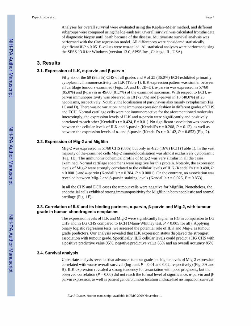
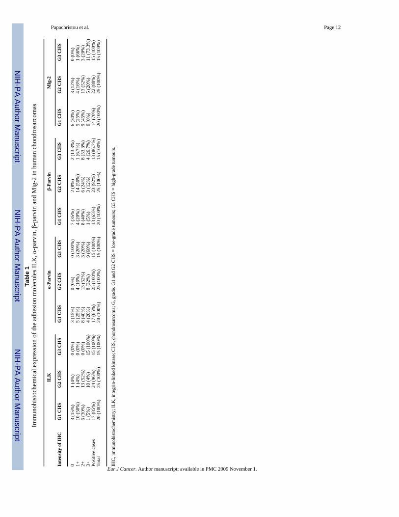
Fifty six of the 60 (93.3%) CHS of all grades and 9 of 25 (36.0%) ECH exhibited primarilycytoplasmic immunoreactivity for ILK (Table 1). ILK expression pattern was similar betweenall cartilage tumours examined (Figs. 1A and B, 2B–D). α-parvin was expressed in 57/60(95.0%) and β-parvin in 49/60 (81.7%) of the examined sarcomas. With respect to ECH, α-parvin immunopositivity was observed in 18 (72.0%) and β-parvin in 10 (40.0%) of 25neoplasms, respectively. Notably, the localisation of parvinswas also mainly cytoplasmic (Fig.1C and D). There was no variation in the immunoexpression fashion in different grades of CHSand ECH. Normal cartilage cells were not immunoreactive for the aforementioned molecules.Interestingly, the expression levels of ILK and α-parvin were significantly and positivelycorrelated to each other (Kendall’s τ = 0.424, P = 0.01). No significant association was observedbetween the cellular levels of ILK and β-parvin (Kendall’s τ = 0.208, P = 0.12), as well asbetween the expression levels of α- and β-parvin (Kendall’s τ = 0.142, P = 0.853) (Fig. 2).
3.2. Expression of Mig-2 and MigfilinMig-2 was expressed in 51/60 CHS (85%) but only in 4/25 (16%) ECH (Table 1). In the vastmajority of the examined cells Mig-2 immunolocalisation was almost exclusively cytoplasmic(Fig. 1E). The immunohistochemical profile of Mig-2 was very similar in all the casesexamined. Normal cartilage specimens were negative for this protein. Notably, the expressionlevels of Mig-2 were strongly correlated to the cellular levels of ILK (Kendall’s τ = 0.490, P< 0.0001) and α-parvin (Kendall’s τ = 0.384, P < 0.0001). On the contrary, no association wasrevealed between Mig-2 and β-parvin staining levels (Kendall’s τ = 0.025, P = 0.853).
In all the CHS and ECH cases the tumour cells were negative for Migfilin. Nonetheless, theendothelial cells exhibited strong immunopositivity for Migfilin in both neoplastic and normalcartilage (Fig. 1F).
3.3. Correlation of ILK and its binding partners, α-parvin, β-parvin and Mig-2, with tumourgrade in human chondrogenic neoplasms
The expression levels of ILK and Mig-2 were significantly higher in HG in comparison to LGCHS and in LG CHS compared to ECH (Mann-Whitney test, P < 0.005 for all). Applyingbinary logistic regression tests, we assessed the potential role of ILK and Mig-2 as tumourgrade predictors. Our analysis revealed that ILK expression status displayed the strongestassociation with tumour grade. Specifically, ILK cellular levels could predict a HG CHS witha positive predictive value 95%, negative predictive value 65% and an overall accuracy 85%.
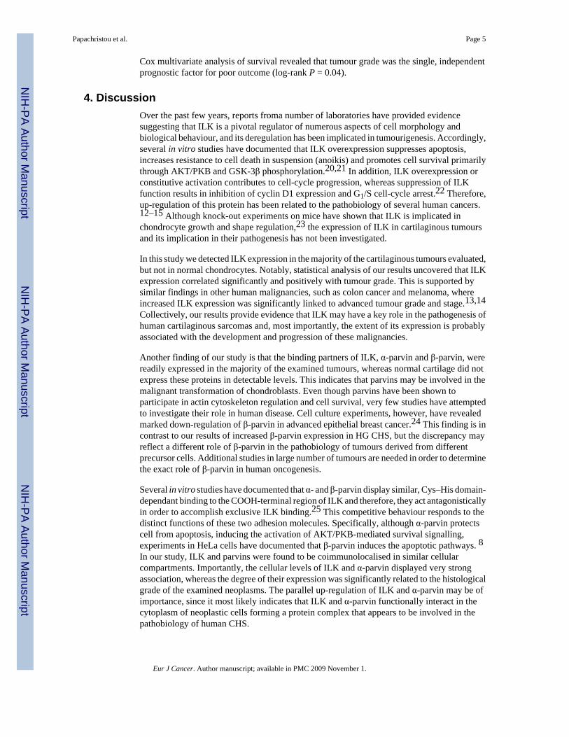
3.4. Survival analysisUnivariate analysis revealed that advanced tumour grade and higher levels of Mig-2 expressioncorrelated with worse overall survival (log-rank P = 0.01 and 0.02, respectively) (Fig. 3A andB). ILK expression revealed a strong tendency for association with poor prognosis, but theobserved correlation (P = 0.06) did not reach the formal level of significance. α-parvin and β-parvin expression, as well as patient gender, tumour location and size had no impact on survival.
Papachristou et al. Page 4
Eur J Cancer. Author manuscript; available in PMC 2009 November 1.
NIH
-PA Author Manuscript
NIH
-PA Author Manuscript
NIH
-PA Author Manuscript
Cox multivariate analysis of survival revealed that tumour grade was the single, independentprognostic factor for poor outcome (log-rank P = 0.04).
4. DiscussionOver the past few years, reports froma number of laboratories have provided evidencesuggesting that ILK is a pivotal regulator of numerous aspects of cell morphology andbiological behaviour, and its deregulation has been implicated in tumourigenesis. Accordingly,several in vitro studies have documented that ILK overexpression suppresses apoptosis,increases resistance to cell death in suspension (anoikis) and promotes cell survival primarilythrough AKT/PKB and GSK-3β phosphorylation.20,21 In addition, ILK overexpression orconstitutive activation contributes to cell-cycle progression, whereas suppression of ILKfunction results in inhibition of cyclin D1 expression and G1/S cell-cycle arrest.22 Therefore,up-regulation of this protein has been related to the pathobiology of several human cancers.12–15 Although knock-out experiments on mice have shown that ILK is implicated inchondrocyte growth and shape regulation,23 the expression of ILK in cartilaginous tumoursand its implication in their pathogenesis has not been investigated.
In this study we detected ILK expression in the majority of the cartilaginous tumours evaluated,but not in normal chondrocytes. Notably, statistical analysis of our results uncovered that ILKexpression correlated significantly and positively with tumour grade. This is supported bysimilar findings in other human malignancies, such as colon cancer and melanoma, whereincreased ILK expression was significantly linked to advanced tumour grade and stage.13,14Collectively, our results provide evidence that ILK may have a key role in the pathogenesis ofhuman cartilaginous sarcomas and, most importantly, the extent of its expression is probablyassociated with the development and progression of these malignancies.
Another finding of our study is that the binding partners of ILK, α-parvin and β-parvin, werereadily expressed in the majority of the examined tumours, whereas normal cartilage did notexpress these proteins in detectable levels. This indicates that parvins may be involved in themalignant transformation of chondroblasts. Even though parvins have been shown toparticipate in actin cytoskeleton regulation and cell survival, very few studies have attemptedto investigate their role in human disease. Cell culture experiments, however, have revealedmarked down-regulation of β-parvin in advanced epithelial breast cancer.24 This finding is incontrast to our results of increased β-parvin expression in HG CHS, but the discrepancy mayreflect a different role of β-parvin in the pathobiology of tumours derived from differentprecursor cells. Additional studies in large number of tumours are needed in order to determinethe exact role of β-parvin in human oncogenesis.
Several in vitro studies have documented that α- and β-parvin display similar, Cys–His domain-dependant binding to the COOH-terminal region of ILK and therefore, they act antagonisticallyin order to accomplish exclusive ILK binding.25 This competitive behaviour responds to thedistinct functions of these two adhesion molecules. Specifically, although α-parvin protectscell from apoptosis, inducing the activation of AKT/PKB-mediated survival signalling,experiments in HeLa cells have documented that β-parvin induces the apoptotic pathways. 8In our study, ILK and parvins were found to be coimmunolocalised in similar cellularcompartments. Importantly, the cellular levels of ILK and α-parvin displayed very strongassociation, whereas the degree of their expression was significantly related to the histologicalgrade of the examined neoplasms. The parallel up-regulation of ILK and α-parvin may be ofimportance, since it most likely indicates that ILK and α-parvin functionally interact in thecytoplasm of neoplastic cells forming a protein complex that appears to be involved in thepathobiology of human CHS.
Papachristou et al. Page 5
Eur J Cancer. Author manuscript; available in PMC 2009 November 1.
NIH
-PA Author Manuscript
NIH
-PA Author Manuscript
NIH
-PA Author Manuscript
With regard to Mig-2, another ILK-interacting cell–ECM adhesion protein, our study revealedthat it was highly expressed in the majority of the CHS but only in a small fraction of the ECHand not at all in normal cartilage. Moreover, our results demonstrated that the cellular levelsof Mig-2 were significantly increased in HG, compared with LG CHS and ECH. Remarkably,Mig-2 immunolocalisation paralleled the localisation of ILK and α-parvin and its expressionlevels were robustly and positively associated with the cellular levels of these two molecules.Taken together, our findings imply that Mig-2 might be involved in CHS pathobiology,possibly through interaction with ILK and α-parvin.
Migfilin is a Mig-2-binding protein encountered at cell– ECM adhesions in fibroblasts and atboth cell–ECM and cell– cell adhesions in epithelial and endothelial cells10,26 In vitroexperiments have shown that Migfilin is crucial for cell adhesion and spreading, and it regulatescell shape and migration and strengthens the cell–cell adhesions between neighbouring cells.11 Interestingly, recent data show that Migfilin is expressed in human leiomyomas andleiomyosarcomas and that its expression levels are associated with higher tumour grades.27 Inthis study however, Migfilin could not be detected in any of the cartilaginous neoplasmsexamined. This may indicate that Migfilin has different functions in these two differentcategories of mesenchymal tumours. Remarkably, however, in both studies endothelial cellsdisplayed strong Migfilin immunopositivity. It would be of great interest to further test thesignificance of this protein as a potential novel endothelial marker.
We also assessed the cellular levels of ILK, α-parvin, β-parvin and Mig-2 as biological markersthat could aid in the discrimination between HG and LG CHS. Binary logistic regressionanalysis revealed that elevated immunoexpression of ILK could accurately predict HGtumours. This is potentially a notable finding. Recent therapeutic protocols suggest that aconsiderable volume of extremity LG CHS can be treated with local curettage and cryosurgery,whilst HG CHS patients should undergo aggressive surgical procedures.28,29 Consequently,precise distinction between these two tumour categories might contribute to an individualisedassessment of prognosis and therapeutic intervention. In our study however, limitations ofstatistical analyses related to the size of our sample did not allow extrapolation of thisconclusion beyond this series.
In line with previous data, we found that higher tumour grade was the single independentparameter related to poor patient outcome.18,30 Univariate analysis revealed that Mig-2expression could function as a prognostic factor for the overall survival of the CHS patients ofour sample. However, because of the limited number of our series and the retrospective natureof our study, the importance of Mig-2 as biomarker for tumour prognosis should be drawn withcaution. Additional, multicentric, preferentially prospective studies are required to determinewhether this molecule could be helpful in patient evaluation in an individual basis.
In conclusion, our study provides novel evidence that ILK, α-parvin and Mig-2 are expressedin human cartilaginous tumours and, most importantly, that these proteins may have a possible—yet unidentified— role in the development and progression of these neoplasms. Furthermore,it underscores the potential function of ILK as molecular marker for the prediction of HGtumours and highlights the possibility that Mig- 2 may act as promising prognostic factor forpoor survival. If the participation of the aforementioned adhesion molecules in thepathobiology of CHS proves to be essential, treatment with small-molecule drugs that couldselectively disrupt the interaction between ILK and α-parvin at focal adhesions might havegreat benefit in the treatment of CHS patients.
AcknowledgementsThis study was supported in part by NIH Grants GM65188 and DK54639 to C.W.
Papachristou et al. Page 6
Eur J Cancer. Author manuscript; available in PMC 2009 November 1.
NIH
-PA Author Manuscript
NIH
-PA Author Manuscript
NIH
-PA Author Manuscript
References1. Brakebusch C, Fässler R. The integrin-actin connection, an eternal love affair. EMBO J 2003;22:2324–
33. [PubMed: 12743027]2. Legate KR, Montañez E, Kudlacek O, Fässler R. ILK, PINCH and parvin: the tIPP of integrin signaling.
Nat Rev Mol Cell Biol 2006;7:20–31. [PubMed: 16493410]3. Khwaja A, Rodriquez-Viciana P, Wennstrom S, Warne PH, Downward J. Matrix adhesion and Ras
transformation both activate a phosphoinositide 3-OH kinase and protein kinase B/Akt cellular survivalpathway. EMBO J 1997;16:2783–93. [PubMed: 9184223]
4. Tan C, Cruet-Hennequart S, Troussard A, et al. Regulation of tumor angiogenesis by integrin-linkedkinase (ILK). Cancer Cell 2004;5:79–90. [PubMed: 14749128]
5. Kaneko Y, Kitazato K, Basaki Y. Integrin-linked kinase regulates vascular morphogenesis induced byvascular endothelial growth factor. J Cell Sci 2004;26:407–15. [PubMed: 14679308]
6. Wu C, Dedhar S. Integrin-linked kinase (ILK) and its interactors: a new paradigm for the coupling ofextracellular matrix to actin cytoskeleton and signaling complexes. J Cell Biol 2001;155:505–10.[PubMed: 11696562]
7. Zhang Y, Chen K, Tu Y, et al. Assembly of the PINCH-ILK-CHILKBP complex precedes and isessential for localization of each component to cell–matrix adhesion sites. J Cell Sci 2002;115:4777–86. [PubMed: 12432066]
8. Sepulveda JL, Wu C. The parvins. Cell Mol Life Sci 2006;63:25–35. [PubMed: 16314921]9. Korenbaum E, Olski TM, Noegel AA. Genomic organization and expression profile of the parvin
family of focal adhesion proteins in mice and humans. Gene 2001;279:69–79. [PubMed: 11722847]10. Tu Y, Wu S, Shi X, Chen K, Wu C. Migfilin and Mig-2 link focal adhesions to filamin and the actin
cytoskeleton and function in cell shape modulation. Cell 2003;113:37–47. [PubMed: 12679033]11. Gkretsi V, Zhang Y, Tu Y, et al. Physical and functional association of migfilin with cell–cell
adhesions. J Cell Sci 2005;118:697–710. [PubMed: 15671069]12. Persad S, Dedhar S. The role of integrin-linked kinase (ILK) in cancer progression. Cancer Metastasis
Rev 2003;22:375–84. [PubMed: 12884912]13. Bravou V, Klironomos G, Papadaki E, Taraviras S, Varakis J. ILK over-expression in human colon
cancer progression correlates with activation of beta-catenin, down-regulation of E-cadherin andactivation of the Akt-FKHR pathway. J Pathol 2006;208:91–9. [PubMed: 16278819]
14. Wong RPC, Ng P, Dedhar S, Gang L. The role of integrin-linked kinase in melanoma cell migration,invasion, and tumor growth. Mol Cancer Ther 2007;6:1692–700. [PubMed: 17575101]
15. Okamura M, Yamaji S, Nagashima Y, et al. Prognostic value of integrin beta1-ILK-pAkt signalingpathway in non-small cell lung cancer. Hum Pathol 2007;38:1081–91. [PubMed: 17442374]
16. Gkretsi V, Bowen WC, Yang Y, Wu C, Michalopoulos GK. Integrin-linked kinase is involved inmatrix-induced hepatocyte differentiation. Biochem Biophys Res Commun 2007;16:638–43.[PubMed: 17194454]
17. Evans HL, Ayala AG, Romsdahl MM. Prognostic factors in chondrosarcoma of bone. Aclinicopathologic study with emphasis on histologic grading. Cancer 1977;40:818–913. [PubMed:890662]
18. Bertoni, F.; Bacchini, P.; Hogendoorn, PCW. Chondrosarcoma. In: Fletcher, CDM.; Unni, KK.;Mertens, F., editors. Pathology and genetics of soft tissue and bone. Lyon: IARC Press; 2002. p.247-51.
19. Papachristou DJ, Papachristou GI, Papaefthimiou OA, et al. The MAPK–AP-1/–Runx2 signallingaxes are implicated in chondrosarcoma pathobiology either independently or via up-regulation ofVEGF. Histopathology 2005;47:565–74. [PubMed: 16324193]
20. Persad S, Attwell S, Gray V, et al. Regulation of protein kinase B/Akt-serine 473 phosphorylationby integrin-linked kinase: critical roles for kinase activity and amino acids arginine 211 and serine343. J Biol Chem 2001;276:27462–9. [PubMed: 11313365]
21. Persad S, Attwell S, Gray V, et al. Inhibition of integrin-linked kinase (ILK) suppresses activation ofprotein kinase B/Akt and induces cell cycle arrest and apoptosis of PTEN-mutant prostate cancercells. Proc Natl Acad Sci USA 2000;97:3207–12. [PubMed: 10716737]
Papachristou et al. Page 7
Eur J Cancer. Author manuscript; available in PMC 2009 November 1.
NIH
-PA Author Manuscript
NIH
-PA Author Manuscript
NIH
-PA Author Manuscript
22. D’Amico M, Hulit J, Amanatullah DF, et al. The integrin-linked kinase regulates the cyclin D1 genethrough glycogen synthase kinase 3beta and cAMP-responsive element-binding protein-dependentpathways. J Biol Chem 2000;275:32649–57. [PubMed: 10915780]
23. Grashoff G, Aszóoldi A, Sakai T, Hunziker EB, Fässler R. Integrin-linked kinase regulateschondrocyte shape and proliferation. EMBO Rep 2003;4:432–8. [PubMed: 12671688]
24. Mongroo PS, Johnstone CN, Naruszewicz I, et al. Betα-parvin inhibits integrin-linked kinase signalingand is downregulated in breast cancer. Oncogene 2004;23:8959–70. [PubMed: 15467740]
25. Nikolopoulos SN, Turner CE. Molecular dissection of actopaxin-integrin-linked kinase-Paxillininteraction and their role in subcellular localization. J Biol Chem 2002;277:1568–75. [PubMed:11694518]
26. Shi X, Ma Y-Q, Tu Y, et al. The MIG-2/Integrin interaction strengthens cell–matrix adhesion andmodulates cell motility. J Biol Chem 2007;282:20455–66. [PubMed: 17513299]
27. Papachristou DJ, Gkretsi V, Tu Y, et al. Increased cytoplasmic level of migfilin is associated withhigher grades of human leiomyosarcoma. Histopathology 2007;51:499–508. [PubMed: 17711449]
28. Ahlmann ER, Menendez LR, Fedenko AN, Learch T. Influence of cryosurgery on treatment outcomeof low-grade chondrosarcoma. Clin Orthop Relat Res 2006;451:201–7. [PubMed: 16788412]
29. Leerapun T, Hugate RR, Inwards CY, Scully SP, Sim FH. Surgical management of conventionalgrade I chondrosarcoma of long bones. Clin Orthop Relat Res 2007;463:166–72. [PubMed:17632422]
30. Bjornsson J, McLeod RA, Unni KK, Ilstrup DM, Pritchard DJ. Primary chondrosarcoma of longbones and limb girdles. Cancer 1998;83:2105–19. [PubMed: 9827715]
Papachristou et al. Page 8
Eur J Cancer. Author manuscript; available in PMC 2009 November 1.
NIH
-PA Author Manuscript
NIH
-PA Author Manuscript
NIH
-PA Author Manuscript
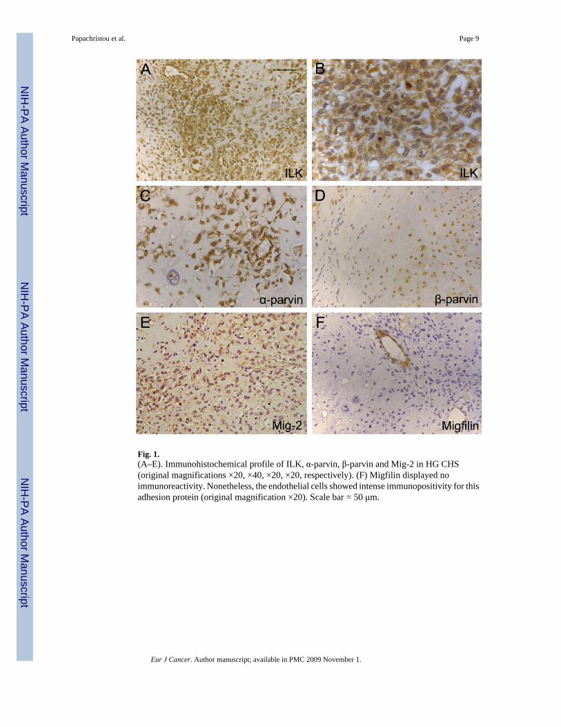
Fig. 1.(A–E). Immunohistochemical profile of ILK, α-parvin, β-parvin and Mig-2 in HG CHS(original magnifications ×20, ×40, ×20, ×20, respectively). (F) Migfilin displayed noimmunoreactivity. Nonetheless, the endothelial cells showed intense immunopositivity for thisadhesion protein (original magnification ×20). Scale bar = 50 μm.
Papachristou et al. Page 9
Eur J Cancer. Author manuscript; available in PMC 2009 November 1.
NIH
-PA Author Manuscript
NIH
-PA Author Manuscript
NIH
-PA Author Manuscript
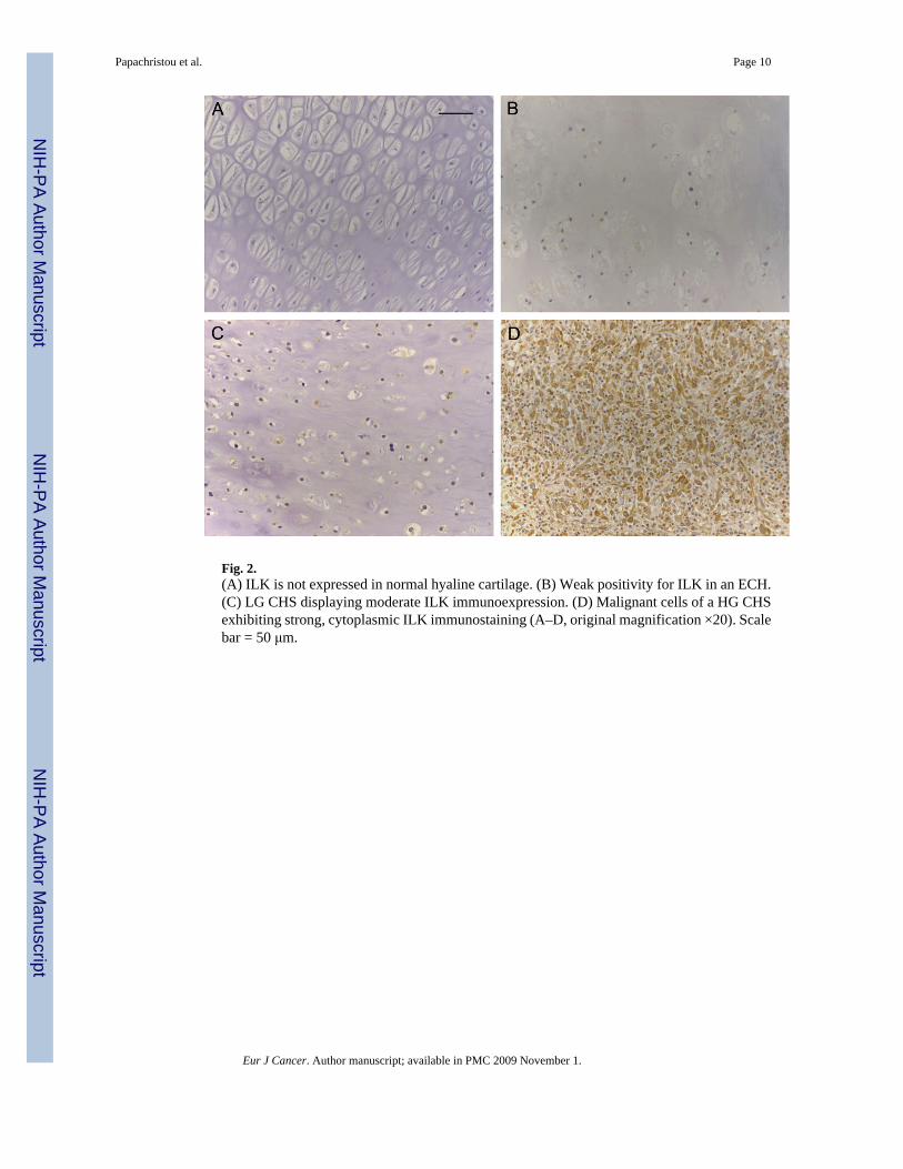
Fig. 2.(A) ILK is not expressed in normal hyaline cartilage. (B) Weak positivity for ILK in an ECH.(C) LG CHS displaying moderate ILK immunoexpression. (D) Malignant cells of a HG CHSexhibiting strong, cytoplasmic ILK immunostaining (A–D, original magnification ×20). Scalebar = 50 μm.
Papachristou et al. Page 10
Eur J Cancer. Author manuscript; available in PMC 2009 November 1.
NIH
-PA Author Manuscript
NIH
-PA Author Manuscript
NIH
-PA Author Manuscript

Fig. 3.(A) Kaplan–Meier curve for the overall survival of the patients with high- and low-grade CHS.The two curves are significantly different (P = 0.01). (B) Kaplan–Meier curve for the overallsurvival of the CHS patients, according to the expression of Mig-2. The two curves aresignificantly different (P = 0.02).
Papachristou et al. Page 11
Eur J Cancer. Author manuscript; available in PMC 2009 November 1.
NIH
-PA Author Manuscript
NIH
-PA Author Manuscript
NIH
-PA Author Manuscript
NIH
-PA Author Manuscript
NIH
-PA Author Manuscript
NIH
-PA Author Manuscript
Papachristou et al. Page 12Ta
ble
1Im
mun
ohis
toch
emic
al e
xpre
ssio
n of
the
adhe
sion
mol
ecul
es IL
K, α
-par
vin,
β-p
arvi
n an
d M
ig-2
in h
uman
cho
ndro
sarc
omas
ILK
α-Pa
rvin
β-Pa
rvin
Mig
-2
Inte
nsity
of I
HC
G1
CH
SG
2 C
HS
G3
CH
SG
1 C
HS
G2
CH
SG
3 C
HS
G1
CH
SG
2 C
HS
G3
CH
SG
1 C
HS
G2
CH
SG
3 C
HS
03
(15%
)1
(4%
)0
(0%
)3
(15%
)0
(0%
)0
(100
%)
7 (3
5%)
2 (8
%)
2 (1
3.3%
)6
(30%
)3
(12%
)0
(0%
)1+
10 (5
0%)
1 (4
%)
0 (0
%)
5 (2
5%)
4 (1
6%)
3 (2
0%)
4 (2
0%)
14 (5
6%)
1 (6
.7%
)5
(25%
)4
(16%
)1
(66%
)2+
6 (3
0%)
13 (5
2%)
0 (0
%)
8 (4
0%)
13 (5
2%)
3 (2
0%)
8 (4
0%)
6 (2
4%)
8 (5
3.3%
)9
(45%
)13
(52%
)3
(20%
)3+
1 (5
%)
10 (4
%)
15 (1
00%
)4
(20%
)8
(32%
)9
(60%
)1
(5%
)3
(12%
)4
(26.
7%)
0 (0
%)
5 (2
0%)
11 (7
3.3%
)Po
sitiv
e ca
ses
17 (8
5%)
24 (9
6%)
15 (1
00%
)17
(85%
)25
(100
%)
15 (1
00%
)13
(65%
)23
(92%
)13
(86.
7%)
14 (7
0%)
22 (8
8%)
15 (1
00%
)To
tal
20 (1
00%
)25
(100
%)
15 (1
00%
)20
(100
%)
25 (1
00%
)15
(100
%)
20 (1
00%
)25
(100
%)
15 (1
00%
)20
(100
%)
25 (1
00%
)15
(100
%)
IHC
, im
mun
ohis
toch
emis
try; I
LK, i
nteg
rin-li
nked
kin
ase;
CH
S, c
hond
rosa
rcom
a; G
, gra
de. G
1 an
d G
2 C
HS
= lo
w-g
rade
tum
ours
; G3
CH
S =
high
-gra
de tu
mou
rs.
Eur J Cancer. Author manuscript; available in PMC 2009 November 1.