
Expression of COUP-TF-interacting protein 2 (CTIP2) in mouse skin during development and in...
12
Expression of COUP-TF-interacting protein 2 (CTIP2) in mouse skin during development and in adulthood Olga Golonzhka, Mark Leid, Gitali Indra * , and Arup Indra * Department of Pharmaceutical Sciences, College of Pharmacy (ML, GI, AI), Department of Biochemistry and Biophysics (OG), and Environmental Health Sciences Center (ML, AI), Oregon State University, Corvallis, Oregon 97331, USA Abstract COUP-TF-interacting protein 2 (CTIP2), also known as Bcl11b, is a transcriptional regulatory protein that is highly expressed in and plays a critical role(s) during development of T lymphocytes and the central nervous system. We demonstrate herein that CTIP2 is also highly expressed in mouse skin during embryogenesis and in adulthood as revealed by immunohistochemical analyses. CTIP2 expression in the ectoderm was first detected at embryonic day 10.5 (E10.5), and became increasingly restricted to proliferating cells of the basal cell layer of the developing epidermis in later stages of fetal development and in adult skin. In addition, CTIP2 expression was also detected in some cells of the suprabasal layer of the developing epidermis, as well as in developing and mature hair follicles. Relatively fewer cells of the developing dermal component of skin were found to express CTIP2, and the adult dermis was devoid of CTIP2 expression. Some, but not all, of the cells present within hair follicle bulge were found to co-express CTIP2, keratin K15, and CD34, indicating that a subset of K15 + CD34 + skin stem cells may express CTIP2. Considered together, these findings suggest that CTIP2 may play a role(s) in skin development and/or homeostasis. Keywords COUP-TF-interacting protein 2; Bcl11b; nuclear receptor; Transcription; expression; zinc finger; skin; epidermis; dermis; ectoderm; mesoderm; keratinocyte; stratification; development; stem cells; proliferation; differentiation; morphogenesis; basal cell; suprabasal cell; hair bulb; hair follicle; hair bulge; immunohistochemistry; in situ hybridization; marker; mouse; embryo; K10; K14; K15; Ki67; CD34 1. Results and discussion CTIP2 (Chicken ovalbumin upstream promoter transcription factor (COUP-TF)–interacting protein 2), also known as Bcl11b, is a C 2 H 2 zinc finger protein (Avram et al., 2000) that has been shown to repress transcription though interaction with COUP-TF nuclear receptor proteins as well as through direct, sequence-specific DNA binding (Avram et al., 2002). CTIP2 is required for normal T cell development and CTIP2-null mice exhibit arrested thymocyte development (Wakabayashi et al., 2003b). Additionally, deregulation of CTIP2 may be implicated in immune system malignant transformation (Wakabayashi et al., 2003a; *Corresponding authors: Department of Pharmaceutical Sciences, OSU, Corvallis, Oregon 97331, USA, (Tel: 1 +(541) 737 5775; Fax: 1 +(541) 737 3999, e-mail: [email protected]; [email protected]). Publisher's Disclaimer: This is a PDF file of an unedited manuscript that has been accepted for publication. As a service to our customers we are providing this early version of the manuscript. The manuscript will undergo copyediting, typesetting, and review of the resulting proof before it is published in its final citable form. Please note that during the production process errors may be discovered which could affect the content, and all legal disclaimers that apply to the journal pertain. NIH Public Access Author Manuscript Gene Expr Patterns. Author manuscript; available in PMC 2008 August 1. Published in final edited form as: Gene Expr Patterns. 2007 August ; 7(7): 754–760. NIH-PA Author Manuscript NIH-PA Author Manuscript NIH-PA Author Manuscript
-
Upload
oregonstate -
Category
Documents
-
view
3 -
download
0
Transcript of Expression of COUP-TF-interacting protein 2 (CTIP2) in mouse skin during development and in...
Expression of COUP-TF-interacting protein 2 (CTIP2) in mouseskin during development and in adulthood
Olga Golonzhka, Mark Leid, Gitali Indra*, and Arup Indra*Department of Pharmaceutical Sciences, College of Pharmacy (ML, GI, AI), Department ofBiochemistry and Biophysics (OG), and Environmental Health Sciences Center (ML, AI), OregonState University, Corvallis, Oregon 97331, USA
AbstractCOUP-TF-interacting protein 2 (CTIP2), also known as Bcl11b, is a transcriptional regulatoryprotein that is highly expressed in and plays a critical role(s) during development of T lymphocytesand the central nervous system. We demonstrate herein that CTIP2 is also highly expressed in mouseskin during embryogenesis and in adulthood as revealed by immunohistochemical analyses. CTIP2expression in the ectoderm was first detected at embryonic day 10.5 (E10.5), and became increasinglyrestricted to proliferating cells of the basal cell layer of the developing epidermis in later stages offetal development and in adult skin. In addition, CTIP2 expression was also detected in some cellsof the suprabasal layer of the developing epidermis, as well as in developing and mature hair follicles.Relatively fewer cells of the developing dermal component of skin were found to express CTIP2,and the adult dermis was devoid of CTIP2 expression. Some, but not all, of the cells present withinhair follicle bulge were found to co-express CTIP2, keratin K15, and CD34, indicating that a subsetof K15+ CD34+ skin stem cells may express CTIP2. Considered together, these findings suggest thatCTIP2 may play a role(s) in skin development and/or homeostasis.
KeywordsCOUP-TF-interacting protein 2; Bcl11b; nuclear receptor; Transcription; expression; zinc finger;skin; epidermis; dermis; ectoderm; mesoderm; keratinocyte; stratification; development; stem cells;proliferation; differentiation; morphogenesis; basal cell; suprabasal cell; hair bulb; hair follicle; hairbulge; immunohistochemistry; in situ hybridization; marker; mouse; embryo; K10; K14; K15; Ki67;CD34
1. Results and discussionCTIP2 (Chicken ovalbumin upstream promoter transcription factor (COUP-TF)–interactingprotein 2), also known as Bcl11b, is a C2H2 zinc finger protein (Avram et al., 2000) that hasbeen shown to repress transcription though interaction with COUP-TF nuclear receptorproteins as well as through direct, sequence-specific DNA binding (Avram et al., 2002). CTIP2is required for normal T cell development and CTIP2-null mice exhibit arrested thymocytedevelopment (Wakabayashi et al., 2003b). Additionally, deregulation of CTIP2 may beimplicated in immune system malignant transformation (Wakabayashi et al., 2003a;
*Corresponding authors: Department of Pharmaceutical Sciences, OSU, Corvallis, Oregon 97331, USA, (Tel: 1 +(541) 737 5775; Fax:1 +(541) 737 3999, e-mail: [email protected]; [email protected]).Publisher's Disclaimer: This is a PDF file of an unedited manuscript that has been accepted for publication. As a service to our customerswe are providing this early version of the manuscript. The manuscript will undergo copyediting, typesetting, and review of the resultingproof before it is published in its final citable form. Please note that during the production process errors may be discovered which couldaffect the content, and all legal disclaimers that apply to the journal pertain.
NIH Public AccessAuthor ManuscriptGene Expr Patterns. Author manuscript; available in PMC 2008 August 1.
Published in final edited form as:Gene Expr Patterns. 2007 August ; 7(7): 754–760.
NIH
-PA Author Manuscript
NIH
-PA Author Manuscript
NIH
-PA Author Manuscript

Bezrookove et al., 2004; Kamimura et al., 2007). It was shown that CTIP2 is also expressedin layer V of cerebral cortex and plays a critical role in the establishment of connections ofcorticospinal motor neurons to the spinal cord (Arlotta et al., 2005).
Mouse epidermis develops from a single-layered embryonic ectoderm (Mack et al., 2005).Subsequent stratification events lead to the formation of the periderm (around E9-E12), whichoverlies the ectoderm (Byrne et al., 2003; Mack et al., 2005). Cells of this two-layeredepidermis then undergo a series of proliferation and terminal differentiation events whichresults in the formation of new cell layers of the future epidermis. Formation of the matureepidermis is completed by E18, at which the epidermis consists of four layers: the basal,spinous, granular and cornified layer (Mack et al., 2005).
Epidermis undergoes constant renewal, which is required to maintain normal tissuehomeostasis. This is possible due to the presence of two populations of proliferating cells:transit amplifying cells with limited proliferative potential and keratinocyte stem cells, whichare slow-cycling cells with high proliferative capacity (Lavker et al., 1993; Slack, 2000).
Previous RNA in situ hybridization using a CTIP2 RNA probe performed in our laboratorydemonstrated that CTIP2 was highly expressed in developing and mature central nervoussystem and spinal cord as well as in the thymus (Leid et al., 2004). The epidermis was notspecifically identified as a site of CTIP2 expression in the previous in situ hybridization studies,although CTIP2 mRNA is found in the skin (see Fig. 1G and I from Leid et al., 2004).Preliminary attempts to define CTIP2 expression pattern during mouse embryogenesis usinga CTIP2-specific monoclonal antibody revealed high-level expression of CTIP2 in developingskin. To our knowledge this is the first evidence for expression of CTIP2 in skin, duringdevelopment or in the adulthood, and it therefore provided a rationale to perform additionalanalyses.
1.1 Expression of CTIP2 during epidermal morphogenesisTo characterize the expression profile of CTIP2 during mouse skin ontogenesis, we performedimmunohistochemistry at different stages of development using an anti-CTIP2 monoclonalantibody, which has been previously described (Senawong et al., 2003; Arlotta et al., 2005;Topark-Ngarm et al., 2006).
CTIP2 expression was detected in the ectoderm as early as embryonic day 10.5 (E10.5), whereCTIP2-positive cells were found in the outermost layer of the cross-section of a developingfetus (Fig.1A, a). Some of these cells were already expressing the basal cell marker keratin 14(K14) (Fig.1A, b), which signifies a commitment of these cells to give rise to stratified epithelia,(Byrne et al., 1994).
Expression of CTIP2 persisted at E12.5 (Fig.1A, c). This stage of skin development is markedby formation and stratification of the embryonic basal layer and all cells of that layer werefound to be positive for CTIP2 expression (Fig.1A). CTIP2 expression precisely co-localizedwith basal cell marker K14 (Vassar et al., 1989) at E12.5 (Fig.1A, d).
Strong expression of CTIP2 was detected in the rapidly dividing basal cell layer at E14.5, andexpression of CTIP2 appeared to be co-localized with that of K14 (Fig.1B, a and b). At thisstage, the early differentiating layers started to form, and were identified using differentiationmarker keratin 10 (K10) (Byrne et al., 1994). CTIP2 expression also extended to some cells inthe differentiating suprabasal cell layer at E14.5, as seen by co-localization of CTIP2 and K10staining (Fig.1B, c). However, the level of CTIP2 expression in the suprabasal cell appearedto be lower than that of basal cells (Fig.1B, compare b and c). In addition, some cells of thefuture dermis were also found to express CTIP2 at this developmental stage (Fig.1B, a).
Golonzhka et al. Page 2
Gene Expr Patterns. Author manuscript; available in PMC 2008 August 1.
NIH
-PA Author Manuscript
NIH
-PA Author Manuscript
NIH
-PA Author Manuscript

CTIP2 expression was further investigated at E16.5 and E18.5 (Fig. 1B, d-i). High levels ofCTIP2 expression were consistently observed in the basal layer of the epidermis at these twostages (Fig.1B d, e, g, h). Some CTIP2 positive cells were observed in the suprabasal layersthat expressed high levels of K10 at E16.5 (Fig.1B, f), whereas the suprabasal expression ofCTIP2 at E18.5 was primarily restricted to a small number of K10-positive cells that wereadjacent to the basal cells (Fig.1B, I; see supplemental Fig. 1).
Later stages of skin morphogenesis were marked by the formation of hair follicles, and anti-CTIP2 antibody robustly stained hair bulbs and follicles at E16.5 and E18.5, respectively (Fig.1B, d and g). These stages were also characterized by expansion of the dermal compartmentof the skin, and some of these dermal cells were also found to express CTIP2 (Fig.1B, d andg, and data not shown).
1.2 CTIP2 expression in proliferating cellsTo evaluate the proliferation status of CTIP2-expressing cells we performed co-labeling studiesusing anti-CTIP2 and an antibody against the proliferation marker Ki67 (Schluter et al.,1993) at different stages during epidermal morphogenesis (Fig.2). At early stages ofdevelopment (E10.5), almost all cells of the fetus were positive for Ki67 expression (Fig.2, band data not shown). This was expected as early stages of fetal development are marked byrapid and ubiquitous proliferation. All cells of the ectodermal compartment, as well as thoseof the underlying mesenchyme were found to express Ki67 (Fig.2, b). Two days later (at E12.5)virtually all cells of the ectoderm were positive for Ki67 expression (Fig.2, e). Most, but notall, cells of mesenchymal origin were negative for Ki67 staining at this developmental stage(Fig.2, f).
The basal cell layer of epidermis is highly proliferative at E14.5 (Mack et al., 2005), which isreflected in the presence of multiple Ki67-positive cells (Fig.2, h-i). A smaller fraction of cellsof the future dermis were positive for Ki67 staining at this stage (Fig.2, h-i), and all layers ofthe developing skin became less proliferative with developmental progression. For example,only a fraction of basal cells, as well as those cells of the dermal compartment, were positivefor Ki67 staining at E16.5 (Fig.2, k-l). By E18.5 only a few cells were still proliferating in thebasal cell layer, and the dermis was mostly non-proliferative. Interestingly, epithelia of thedeveloping hair follicles, which were invaginating into the dermis, still harbored a considerablenumber with Ki67-positive cells (Fig.2, n).
At early, highly proliferative stages of development (E10.5-E14.5) almost all of the CTIP2-positive epidermal cells were found to be dividing as indicated by co-localization of CTIP2and Ki67 staining (Fig.2, a-i). The total number of Ki67-positive cells was reduced in theepidermis at E16.5 but, in general, cells that were positive for Ki67 staining were also foundto be positive for CTIP2 expression (Fig.2 j-l). By E18.5, the number of proliferating cells infetal skin was comparatively reduced, but again a correspondence was observed between theKi67 staining and that of CTIP2, indicating that majority of proliferating cells express CTIP2.Similarly, some CTIP2-positive cells were detected in the developing hair follicle epithelium,and these cells were also positive for Ki67 expression (Fig.2, m-o).
1.3 Expression of CTIP2 in adult skinImmunohistochemistry was performed on 8-10-week old adult mouse skin using anti-CTIP2,-K14 and -K10 antibodies to evaluate the possibility that CTIP2 may be expressed in adult skin(Fig. 3). In the adult skin, CTIP2 expression was found in the majority, if not all cells of thebasal layer (Fig. 3, a and b), hair follicles and some cells of the suprabasal K10-positive layers(Fig. 3, c), which is similar to the results that we obtained during embryogenesis (Fig. 1). Incontrast to the developing dermis, CTIP2 expression was not detected in the dermal
Golonzhka et al. Page 3
Gene Expr Patterns. Author manuscript; available in PMC 2008 August 1.
NIH
-PA Author Manuscript
NIH
-PA Author Manuscript
NIH
-PA Author Manuscript
compartment of the adult skin (compare Fig. 3,a and Fig. 1B, g). Immunoblot analysis of CTIP2in protein extracts from dorsal skin biopsies confirmed expression of CTIP2 in fetal (E18.5)and adult skin (Fig. 4, top panel).
Expression of CTIP2 in whole skin, protein extracts of the E18.5 embryo was found to besignificantly higher that that of the adult skin. This could be partially explained by ourobservation that CTIP2 expression is maintained in the epidermal basal and suprabasal cellsthroughout development, and into the adulthood, whereas the adult dermal compartment, whichis dramatically expanded relative to fetal skin, does not express significant amounts of CTIP2.
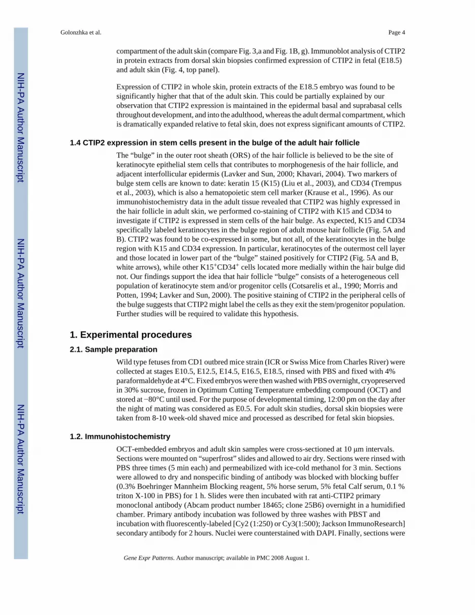
1.4 CTIP2 expression in stem cells present in the bulge of the adult hair follicleThe “bulge” in the outer root sheath (ORS) of the hair follicle is believed to be the site ofkeratinocyte epithelial stem cells that contributes to morphogenesis of the hair follicle, andadjacent interfollicular epidermis (Lavker and Sun, 2000; Khavari, 2004). Two markers ofbulge stem cells are known to date: keratin 15 (K15) (Liu et al., 2003), and CD34 (Trempuset al., 2003), which is also a hematopoietic stem cell marker (Krause et al., 1996). As ourimmunohistochemistry data in the adult tissue revealed that CTIP2 was highly expressed inthe hair follicle in adult skin, we performed co-staining of CTIP2 with K15 and CD34 toinvestigate if CTIP2 is expressed in stem cells of the hair bulge. As expected, K15 and CD34specifically labeled keratinocytes in the bulge region of adult mouse hair follicle (Fig. 5A andB). CTIP2 was found to be co-expressed in some, but not all, of the keratinocytes in the bulgeregion with K15 and CD34 expression. In particular, keratinocytes of the outermost cell layerand those located in lower part of the “bulge” stained positively for CTIP2 (Fig. 5A and B,white arrows), while other K15+CD34+ cells located more medially within the hair bulge didnot. Our findings support the idea that hair follicle “bulge” consists of a heterogeneous cellpopulation of keratinocyte stem and/or progenitor cells (Cotsarelis et al., 1990; Morris andPotten, 1994; Lavker and Sun, 2000). The positive staining of CTIP2 in the peripheral cells ofthe bulge suggests that CTIP2 might label the cells as they exit the stem/progenitor population.Further studies will be required to validate this hypothesis.
1. Experimental procedures2.1. Sample preparation
Wild type fetuses from CD1 outbred mice strain (ICR or Swiss Mice from Charles River) werecollected at stages E10.5, E12.5, E14.5, E16.5, E18.5, rinsed with PBS and fixed with 4%paraformaldehyde at 4°C. Fixed embryos were then washed with PBS overnight, cryopreservedin 30% sucrose, frozen in Optimum Cutting Temperature embedding compound (OCT) andstored at −80°C until used. For the purpose of developmental timing, 12:00 pm on the day afterthe night of mating was considered as E0.5. For adult skin studies, dorsal skin biopsies weretaken from 8-10 week-old shaved mice and processed as described for fetal skin biopsies.
1.2. ImmunohistochemistryOCT-embedded embryos and adult skin samples were cross-sectioned at 10 μm intervals.Sections were mounted on “superfrost” slides and allowed to air dry. Sections were rinsed withPBS three times (5 min each) and permeabilized with ice-cold methanol for 3 min. Sectionswere allowed to dry and nonspecific binding of antibody was blocked with blocking buffer(0.3% Boehringer Mannheim Blocking reagent, 5% horse serum, 5% fetal Calf serum, 0.1 %triton X-100 in PBS) for 1 h. Slides were then incubated with rat anti-CTIP2 primarymonoclonal antibody (Abcam product number 18465; clone 25B6) overnight in a humidifiedchamber. Primary antibody incubation was followed by three washes with PBST andincubation with fluorescently-labeled [Cy2 (1:250) or Cy3(1:500); Jackson ImmunoResearch]secondary antibody for 2 hours. Nuclei were counterstained with DAPI. Finally, sections were
Golonzhka et al. Page 4
Gene Expr Patterns. Author manuscript; available in PMC 2008 August 1.
NIH
-PA Author Manuscript
NIH
-PA Author Manuscript
NIH
-PA Author Manuscript

rinsed with PBST, dehydrated through sequential washes in 50, 70, 95 and 100% ethanol andthen cleared in xylene. Slides were mounted with DPX mounting media and allowed to dryovernight. Images were captured at 40X magnification using Leica DMRA fluorescentmicroscope and Hamamatsu C4742-95 digital camera and processed using OpenLab softwareand Adobe Photoshop 7.0.
2.3 Whole skin protein extract preparation and Immunoblot analysisDorsal skin biopsies from E18.5 fetuses or adult mice were homogenized in RIPA buffer (50mM Tris pH7.5, 1% NP-40, 0.5% Sodium Deoxycholate, 0.1 %SDS, 150 mM NaCl, 5mMEDTA, proteinase inhibitors) using tissue grinder and cleared by centrifugation. Supernatantswere collected and equal amounts of protein (determined using the Bio-Rad Protein Assay Kit)were analyzed by immunoblotting using antibodies against CTIP2 and β-actin as a loadingcontrol.
2.4 AntibodiesThe following antibodies and their dilutions were used for immunohistochemical studies: anti-CTIP2 (Abcam, 1:300), -K14 (Covance, 1:1000), -K10 (Covance, 1:1000), -Ki67 (Abcam,1:500), -K15 (Covance, 1:500), -CD34 (Santa Cruz, 1:50). Antibodies used for Western blotanalysis: anti-CTIP2 (Abcam, 1:1000), -β-actin (Abcam, 1:4000).
Supplimental SectionRefer to Web version on PubMed Central for supplementary material.
Acknowledgements
These studies were supported by a grant from the National Institutes of Health (GM60852) to ML and by a NIEHSCenter grant (ES00210) to the Oregon State University Environmental Health Sciences Center.
ReferencesArlotta P, Molyneaux BJ, Chen J, Inoue J, Kominami R, Macklis JD. Neuronal subtype-specific genes
that control corticospinal motor neuron development in vivo. Neuron 2005;45:207–221. [PubMed:15664173]
Avram D, Fields A, Pretty On Top K, Nevrivy DJ, Ishmael JE, Leid M. Isolation of a novel family of C(2)H(2) zinc finger proteins implicated in transcriptional repression mediated by chicken ovalbuminupstream promoter transcription factor (COUP-TF) orphan nuclear receptors. J Biol Chem2000;275:10315–10322. [PubMed: 10744719]
Avram D, Fields A, Senawong T, Topark-Ngarm A, Leid M. COUP-TF (chicken ovalbumin upstreampromoter transcription factor)-interacting protein 1 (CTIP1) is a sequence-specific DNA bindingprotein. Biochem J 2002;368:555–563. [PubMed: 12196208]
Bezrookove V, van Zelderen-Bhola SL, Brink A, Szuhai K, Raap AK, Barge R, Beverstock GC,Rosenberg C. A novel t(6;14)(q25-q27;q32) in acute myelocytic leukemia involves the BCL11B gene.Cancer Genet Cytogenet 2004;149:72–76. [PubMed: 15104287]
Byrne C, Tainsky M, Fuchs E. Programming gene expression in developing epidermis. Development1994;120:2369–2383. [PubMed: 7525178]
Byrne C, Hardman M, Nield K. Covering the limb--formation of the integument. J Anat 2003;202:113–123. [PubMed: 12587926]
Cotsarelis G, Sun TT, Lavker RM. Label-retaining cells reside in the bulge area of pilosebaceous unit:implications for follicular stem cells, hair cycle, and skin carcinogenesis. Cell 1990;61:1329–1337.[PubMed: 2364430]
Kamimura K, Ohi H, Kubota T, Okazuka K, Yoshikai Y, Wakabayashi Y, Aoyagi Y, Mishima Y,Kominami R. Haploinsufficiency of Bcl11b for suppression of lymphomagenesis and thymocytedevelopment. Biochem Biophys Res Commun 2007;355:538–542. [PubMed: 17306224]
Golonzhka et al. Page 5
Gene Expr Patterns. Author manuscript; available in PMC 2008 August 1.
NIH
-PA Author Manuscript
NIH
-PA Author Manuscript
NIH
-PA Author Manuscript

Khavari PA. Profiling epithelial stem cells. Nat Biotechnol 2004;22:393–394. [PubMed: 15060550]Krause DS, Fackler MJ, Civin CI, May WS. CD34: structure, biology, and clinical utility. Blood
1996;87:1–13. [PubMed: 8547630]Lavker RM, Miller S, Wilson C, Cotsarelis G, Wei ZG, Yang JS, Sun TT. Hair follicle stem cells: their
location, role in hair cycle, and involvement in skin tumor formation. J Invest Dermatol1993;101:16S–26S. [PubMed: 8326150]
Lavker RM, Sun TT. Epidermal stem cells: properties, markers, and location. Proc Natl Acad Sci U S A2000;97:13473–13475. [PubMed: 11087834]
Leid M, Ishmael JE, Avram D, Shepherd D, Fraulob V, Dolle P. CTIP1 and CTIP2 are differentiallyexpressed during mouse embryogenesis. Gene Expr Patterns 2004;4:733–739. [PubMed: 15465497]
Liu Y, Lyle S, Yang Z, Cotsarelis G. Keratin 15 promoter targets putative epithelial stem cells in the hairfollicle bulge. J Invest Dermatol 2003;121:963–968. [PubMed: 14708593]
Mack JA, Anand S, Maytin EV. Proliferation and cornification during development of the mammalianepidermis. Birth Defects Res C Embryo Today 2005;75:314–329. [PubMed: 16425252]
Morris RJ, Potten CS. Slowly cycling (label-retaining) epidermal cells behave like clonogenic stem cellsin vitro. Cell Prolif 1994;27:279–289. [PubMed: 10465012]
Senawong T, Peterson VJ, Avram D, Shepherd DM, Frye RA, Minucci S, Leid M. Involvement of thehistone deacetylase SIRT1 in chicken ovalbumin upstream promoter transcription factor (COUP-TF)-interacting protein 2-mediated transcriptional repression. J Biol Chem 2003;78(44):43041–50.[PubMed: 12930829]
Schluter C, Duchrow M, Wohlenberg C, Becker MH, Key G, Flad HD, Gerdes J. The cell proliferation-associated antigen of antibody Ki-67: a very large, ubiquitous nuclear protein with numerous repeatedelements, representing a new kind of cell cycle-maintaining proteins. J Cell Biol 1993;123:513–522.[PubMed: 8227122]
Slack JM. Stem cells in epithelial tissues. Science 2000;287:1431–1433. [PubMed: 10688782]Topark-Ngarm A, Golonzhka O, Peterson VJ, Barrett B Jr, Martinez B, Crofoot K, Filtz TM, Leid M.
CTIP2 associates with the NuRD complex on the promoter of p57KIP2, a newly identified CTIP2target gene. J Biol Chem 2006;281(43):32272–83. [PubMed: 16950772]
Trempus CS, Morris RJ, Bortner CD, Cotsarelis G, Faircloth RS, Reece JM, Tennant RW. Enrichmentfor living murine keratinocytes from the hair follicle bulge with the cell surface marker CD34. JInvest Dermatol 2003;120:501–511. [PubMed: 12648211]
Vassar R, Rosenberg M, Ross S, Tyner A, Fuchs E. Tissue-specific and differentiation-specific expressionof a human K14 keratin gene in transgenic mice. Proc Natl Acad Sci U S A 1989;86:1563–1567.[PubMed: 2466292]
Wakabayashi Y, Inoue J, Takahashi Y, Matsuki A, Kosugi-Okano H, Shinbo T, Mishima Y, Niwa O,Kominami R. Homozygous deletions and point mutations of the Rit1/Bcl11b gene in gamma-rayinduced mouse thymic lymphomas. Biochem Biophys Res Commun 2003a;301:598–603. [PubMed:12565905]
Wakabayashi Y, Watanabe H, Inoue J, Takeda N, Sakata J, Mishima Y, Hitomi J, Yamamoto T, UtsuyamaM, Niwa O, Aizawa S, Kominami R. Bcl11b is required for differentiation and survival of alphabetaT lymphocytes. Nat Immunol 2003b;4:533–539. [PubMed: 12717433]
Golonzhka et al. Page 6
Gene Expr Patterns. Author manuscript; available in PMC 2008 August 1.
NIH
-PA Author Manuscript
NIH
-PA Author Manuscript
NIH
-PA Author Manuscript
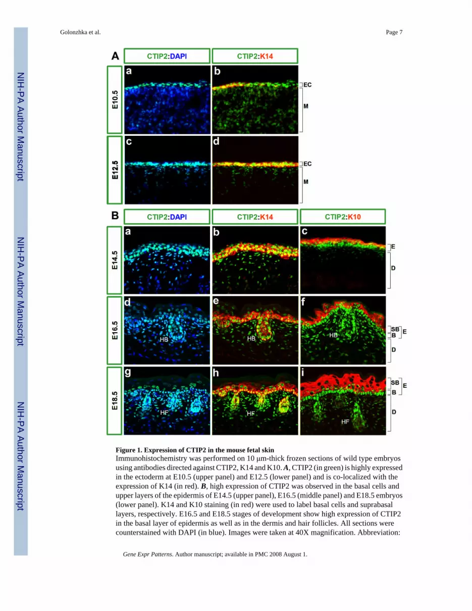
Figure 1. Expression of CTIP2 in the mouse fetal skinImmunohistochemistry was performed on 10 μm-thick frozen sections of wild type embryosusing antibodies directed against CTIP2, K14 and K10. A, CTIP2 (in green) is highly expressedin the ectoderm at E10.5 (upper panel) and E12.5 (lower panel) and is co-localized with theexpression of K14 (in red). B, high expression of CTIP2 was observed in the basal cells andupper layers of the epidermis of E14.5 (upper panel), E16.5 (middle panel) and E18.5 embryos(lower panel). K14 and K10 staining (in red) were used to label basal cells and suprabasallayers, respectively. E16.5 and E18.5 stages of development show high expression of CTIP2in the basal layer of epidermis as well as in the dermis and hair follicles. All sections werecounterstained with DAPI (in blue). Images were taken at 40X magnification. Abbreviation:
Golonzhka et al. Page 7
Gene Expr Patterns. Author manuscript; available in PMC 2008 August 1.
NIH
-PA Author Manuscript
NIH
-PA Author Manuscript
NIH
-PA Author Manuscript

EC- ectoderm, M-mesoderm, E-epidermis, D-dermis, B-basal cell layer, SB- suprabasal celllayers, HB-hair bulb, HF-hair follicle.
Golonzhka et al. Page 8
Gene Expr Patterns. Author manuscript; available in PMC 2008 August 1.
NIH
-PA Author Manuscript
NIH
-PA Author Manuscript
NIH
-PA Author Manuscript
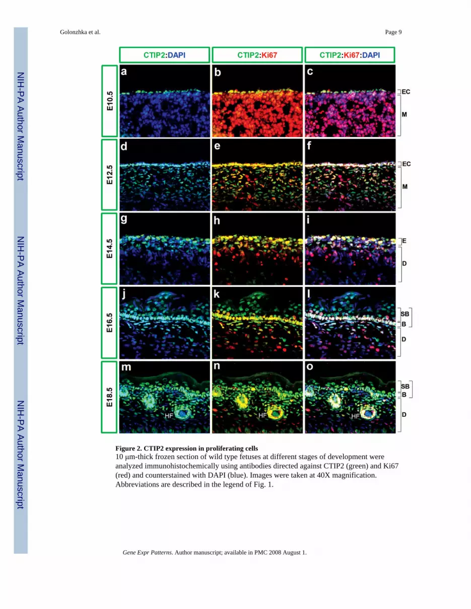
Figure 2. CTIP2 expression in proliferating cells10 μm-thick frozen section of wild type fetuses at different stages of development wereanalyzed immunohistochemically using antibodies directed against CTIP2 (green) and Ki67(red) and counterstained with DAPI (blue). Images were taken at 40X magnification.Abbreviations are described in the legend of Fig. 1.
Golonzhka et al. Page 9
Gene Expr Patterns. Author manuscript; available in PMC 2008 August 1.
NIH
-PA Author Manuscript
NIH
-PA Author Manuscript
NIH
-PA Author Manuscript
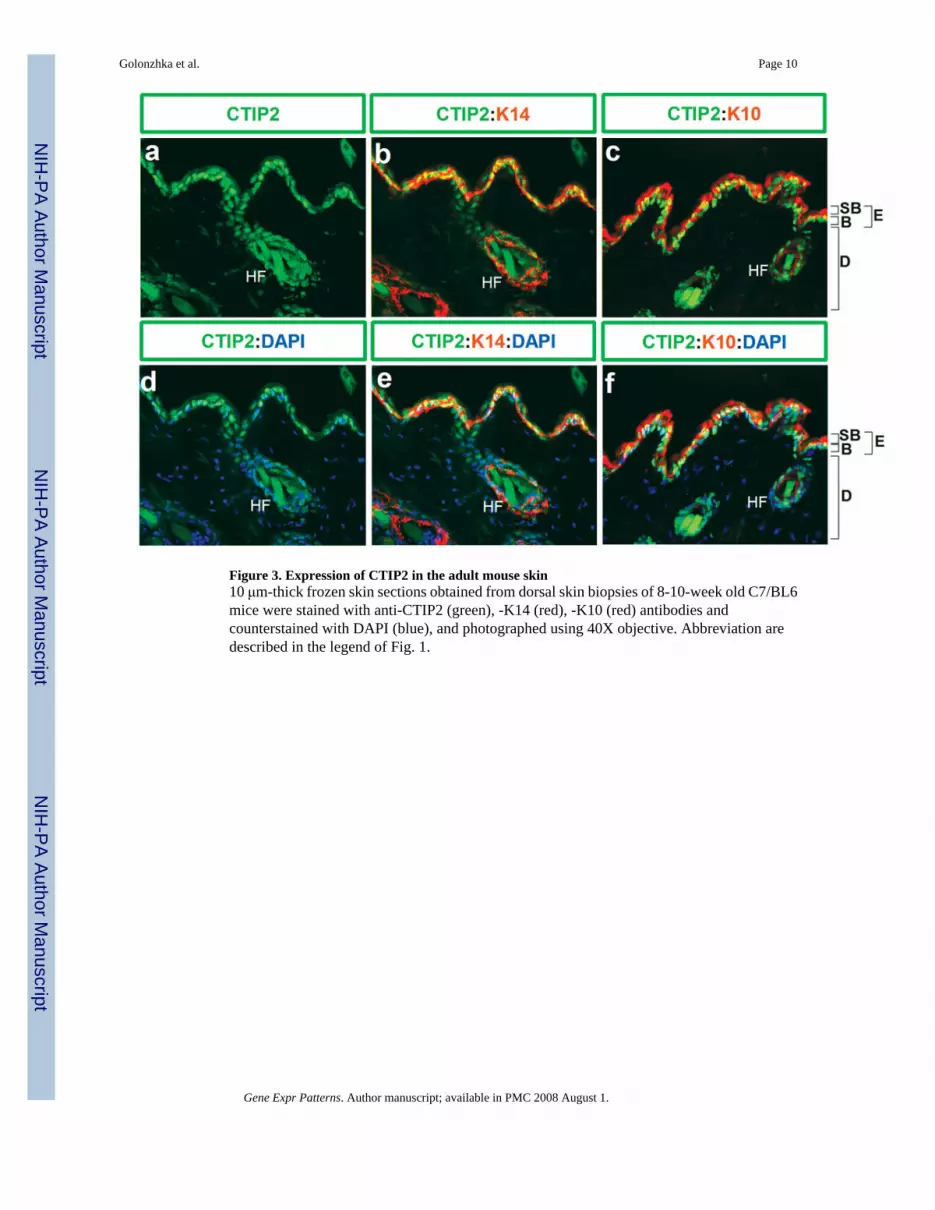
Figure 3. Expression of CTIP2 in the adult mouse skin10 μm-thick frozen skin sections obtained from dorsal skin biopsies of 8-10-week old C7/BL6mice were stained with anti-CTIP2 (green), -K14 (red), -K10 (red) antibodies andcounterstained with DAPI (blue), and photographed using 40X objective. Abbreviation aredescribed in the legend of Fig. 1.
Golonzhka et al. Page 10
Gene Expr Patterns. Author manuscript; available in PMC 2008 August 1.
NIH
-PA Author Manuscript
NIH
-PA Author Manuscript
NIH
-PA Author Manuscript

Figure 4. Immunoblot analysis of CTIP2 expression in fetal and adult skinWhole skin protein extracts were prepared from adult (8-week old) and fetal (E18.5) dorsalskin biopsies and analyzed using CTIP2-specific antibody (upper panel). β-actin was used asan internal loading control (lower panel).
Golonzhka et al. Page 11
Gene Expr Patterns. Author manuscript; available in PMC 2008 August 1.
NIH
-PA Author Manuscript
NIH
-PA Author Manuscript
NIH
-PA Author Manuscript
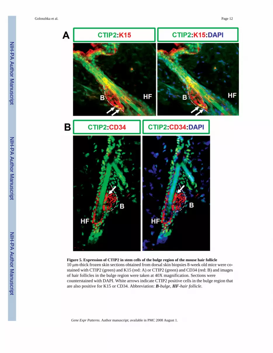
Figure 5. Expression of CTIP2 in stem cells of the bulge region of the mouse hair follicle10 μm-thick frozen skin sections obtained from dorsal skin biopsies 8-week old mice were co-stained with CTIP2 (green) and K15 (red: A) or CTIP2 (green) and CD34 (red: B) and imagesof hair follicles in the bulge region were taken at 40X magnification. Sections werecounterstained with DAPI. White arrows indicate CTIP2 positive cells in the bulge region thatare also positive for K15 or CD34. Abbreviation: B-bulge, HF-hair follicle.
Golonzhka et al. Page 12
Gene Expr Patterns. Author manuscript; available in PMC 2008 August 1.
NIH
-PA Author Manuscript
NIH
-PA Author Manuscript
NIH
-PA Author Manuscript