Expression and characterization of antimicrobial peptides Retrocyclin-101 and Protegrin-1 in...
25
Expression and characterization of antimicrobial peptides Retrocyclin-101 and Protegrin-1 in chloroplasts to control viral and bacterial infections Seung-Bum Lee † , Baichuan Li † , Shuangxia Jin, and Henry Daniell * Department of Molecular Biology and Microbiology, College of Medicine, University of Central Florida, Orlando, FL, USA Summary Retrocyclin-101 (RC101) and Protegrin-1 (PG1) are two important antimicrobial peptides that can be used as therapeutic agents against bacterial and/or viral infections, especially those caused by the HIV-1 or sexually transmitted bacteria. Because of their antimicrobial activity and complex secondary structures, they have not yet been produced in microbial systems and their chemical synthesis is prohibitively expensive. Therefore, we created chloroplast transformation vectors with the RC101 or PG1 coding sequence, fused with GFP to confer stability, furin or Factor Xa cleavage site to liberate the mature peptide from their fusion proteins and a His-tag to aid in their purification. Stable integration of RC101 into the tobacco chloroplast genome and homoplasmy were confirmed by Southern blots. RC101 and PG1 accumulated up to 32%–38% and 17%~26% of the total soluble protein. Both RC101 and PG1 were cleaved from GFP by corresponding proteases in vitro, and Factor Xa–like protease activity was observed within chloroplasts. Confocal microscopy studies showed location of GFP fluorescence within chloroplasts. Organic extraction resulted in 10.6-fold higher yield of RC101 than purification by affinity chromatography using His-tag. In planta bioassays with Erwinia carotovora confirmed the antibacterial activity of RC101 and PG1 expressed in chloroplasts. RC101 transplastomic plants were resistant to tobacco mosaic virus infections, confirming antiviral activity. Because RC101 and PG1 have not yet been produced in other cell culture or microbial systems, chloroplasts can be used as bioreactors for producing these proteins. Adequate yield of purified antimicrobial peptides from transplastomic plants should facilitate further preclinical studies. Keywords antimicrobial peptide; chloroplast genetic engineering; molecular farming; plant-made biopharmaceuticals Introduction Antimicrobial peptides are evolutionarily conserved components of the innate immune response and are found in different organisms, including bacteria, vertebrates, invertebrates and plants (Boman, 1995; Nicolas and Mor, 1995; Broekaert et al., 1997; Hancock and Chapple, 1999). Antimicrobial peptides are also called peptide antibiotics. When compared with conventional antibiotics, development of resistance is less likely with antimicrobial © 2010 The Authors © 2010 Society for Experimental Biology and Blackwell Publishing Ltd * Correspondence (fax 407 823 0956; [email protected]). † Both authors contributed equally to this article. NIH Public Access Author Manuscript Plant Biotechnol J. Author manuscript; available in PMC 2012 October 11. Published in final edited form as: Plant Biotechnol J. 2011 January ; 9(1): 100–115. doi:10.1111/j.1467-7652.2010.00538.x. NIH-PA Author Manuscript NIH-PA Author Manuscript NIH-PA Author Manuscript
Transcript of Expression and characterization of antimicrobial peptides Retrocyclin-101 and Protegrin-1 in...

Expression and characterization of antimicrobial peptidesRetrocyclin-101 and Protegrin-1 in chloroplasts to control viraland bacterial infections
Seung-Bum Lee†, Baichuan Li†, Shuangxia Jin, and Henry Daniell*Department of Molecular Biology and Microbiology, College of Medicine, University of CentralFlorida, Orlando, FL, USA
SummaryRetrocyclin-101 (RC101) and Protegrin-1 (PG1) are two important antimicrobial peptides that canbe used as therapeutic agents against bacterial and/or viral infections, especially those caused bythe HIV-1 or sexually transmitted bacteria. Because of their antimicrobial activity and complexsecondary structures, they have not yet been produced in microbial systems and their chemicalsynthesis is prohibitively expensive. Therefore, we created chloroplast transformation vectors withthe RC101 or PG1 coding sequence, fused with GFP to confer stability, furin or Factor Xacleavage site to liberate the mature peptide from their fusion proteins and a His-tag to aid in theirpurification. Stable integration of RC101 into the tobacco chloroplast genome and homoplasmywere confirmed by Southern blots. RC101 and PG1 accumulated up to 32%–38% and 17%~26%of the total soluble protein. Both RC101 and PG1 were cleaved from GFP by correspondingproteases in vitro, and Factor Xa–like protease activity was observed within chloroplasts.Confocal microscopy studies showed location of GFP fluorescence within chloroplasts. Organicextraction resulted in 10.6-fold higher yield of RC101 than purification by affinitychromatography using His-tag. In planta bioassays with Erwinia carotovora confirmed theantibacterial activity of RC101 and PG1 expressed in chloroplasts. RC101 transplastomic plantswere resistant to tobacco mosaic virus infections, confirming antiviral activity. Because RC101and PG1 have not yet been produced in other cell culture or microbial systems, chloroplasts can beused as bioreactors for producing these proteins. Adequate yield of purified antimicrobial peptidesfrom transplastomic plants should facilitate further preclinical studies.
Keywordsantimicrobial peptide; chloroplast genetic engineering; molecular farming; plant-madebiopharmaceuticals
IntroductionAntimicrobial peptides are evolutionarily conserved components of the innate immuneresponse and are found in different organisms, including bacteria, vertebrates, invertebratesand plants (Boman, 1995; Nicolas and Mor, 1995; Broekaert et al., 1997; Hancock andChapple, 1999). Antimicrobial peptides are also called peptide antibiotics. When comparedwith conventional antibiotics, development of resistance is less likely with antimicrobial
© 2010 The Authors © 2010 Society for Experimental Biology and Blackwell Publishing Ltd*Correspondence (fax 407 823 0956; [email protected]).†Both authors contributed equally to this article.
NIH Public AccessAuthor ManuscriptPlant Biotechnol J. Author manuscript; available in PMC 2012 October 11.
Published in final edited form as:Plant Biotechnol J. 2011 January ; 9(1): 100–115. doi:10.1111/j.1467-7652.2010.00538.x.
NIH
-PA Author Manuscript
NIH
-PA Author Manuscript
NIH
-PA Author Manuscript

peptides. Many bacteria species remain sensitive to antimicrobial peptides after a long timeof evolution (Yeaman and Yount, 2003; Nizet, 2006). Adaptive immune systems canremember the pathogen and elicit a much faster and stronger immune response against thatpathogen at subsequent encounters (Boman, 1995). Without such specificity and memory,antimicrobial peptides evolved a different mechanism against pathogen infections. Mostantimicrobial peptides are efficient against a broad spectrum of pathogens rather thanspecific against one pathogen, which makes them especially suitable for use against localand systematic infections (Bals, 2000; Schaller-Bals et al., 2002). Other than theantimicrobial activities, some antimicrobial peptides are shown to have immunomodulatoryactivities. Some studies show that antimicrobial peptides such as defensins are likely to playa role in recruiting effector T cells to inflammatory sites, thereby contributing to the effectorphase of adaptive immunity (Yang et al., 2001). These intriguing characteristics ofantimicrobial peptides facilitate development of novel antibiotics. However, the high cost ofproduction of antimicrobial peptides and lack of suitable expression systems could bepotential barriers for their development and clinical studies.
The chloroplast, as a bioreactor, is able to express foreign proteins at high levels because oftheir high copy numbers. When a transgene is integrated into the inverted repeat region ofthe chloroplast genome, up to 20 000 copies of the transgene per cell could be expressed.Several therapeutic proteins have been expressed in chloroplasts, including human bloodproteins somatotropin (Staub et al., 2000), insulin-like growth factor (Daniell et al., 2009),proinsulin (Ruhlman et al., 2007), IFN-α2b (Arlen et al., 2007), serum albumin (Fernandez-San et al., 2003), IFN-γ (Leelavathi and Reddy, 2003), cardiotrophin-1 (Farran et al., 2008),alpha1-anti-trypsin (Nadai et al., 2009) and glutamic acid decarboxylase (Wang et al., 2008).In addition, several vaccine antigens have been expressed in chloroplasts against severalbacterial pathogens including cholera toxin B subunit (Daniell et al., 2001), tetanus toxin(Tregoning et al., 2003), anthrax protective antigen (Watson et al., 2004; Koya et al., 2005),plague F1-V fusion antigen (Arlen et al., 2008), and outer surface lipoprotein A (OspA) forLyme disease (Glenz et al., 2006), and their functionality has been evaluated in cell culturesystems or animal models after pathogen or toxin challenges. Antigens produced againstprotozoan pathogens were immunogenic against amoeba (Chebolu and Daniell, 2007) oreffective against the malarial parasite (Davoodi-Semiromi et al., 2009). Although severalviral antigens have been expressed in chloroplasts, neutralizing antibodies were shown onlyagainst human papillomavirus (Fernandez-San et al., 2008) and canine parvovirus 2L21peptide (Molina et al., 2004). Other proteins expressed in chloroplasts include bovinemammary-associated serum amyloid (Manuell et al., 2007), aprotinin (Tissot et al., 2008)and monoclonal large single-chain (lsc) antibody against glycoprotein D of the herpessimplex virus (Mayfield et al., 2003). The expression levels of these proteins are mostly2%~20% of total soluble protein (TSP) but could be even higher than RuBisCo (Oey et al.,2009; Ruhlman et al., 2010). Other advantages of chloroplast transformation include multi-gene engineering, transgene containment, lack of position effect, gene silencing andmaternal inheritance (Daniell et al., 2005; 2009).
Retrocyclin is a cyclic octadecapeptide, which is artificially synthesized based on a humanpseudogene that is homologous to rhesus monkey circular minidefensins. Retrocyclincontains six cysteines and has large β-sheet structure that is stabilized by threeintramolecular disulphide bonds. Structure–function studies indicate that the cyclicbackbone, intramolecular tri-disulphide ladder, and arginine residues of retrocyclincontributed substantially to its protective effects (Trabi et al., 2001; Jenssen et al., 2006).Retrocyclin peptides are small antimicrobial agents with potent activity against bacteria andviruses, especially against HIV retrovirus or sexually transmitted bacteria. Previous studieshave shown that Retrocyclin-101 (RC101) and other retrocyclins can protect human CD4+
cells from infection by T- and M-tropic strains of HIV-1 in vitro (Cole et al., 2002) and
Lee et al. Page 2
Plant Biotechnol J. Author manuscript; available in PMC 2012 October 11.
NIH
-PA Author Manuscript
NIH
-PA Author Manuscript
NIH
-PA Author Manuscript

prevent HIV-1 infection in an organ-like construct of human cervicovaginal tissue (Cole etal., 2007). The ability of RC101 to prevent HIV-1 infection and retain full activity in thepresence of vaginal fluid makes it a good candidate for topical microbicide to prevent sexualtransmission of HIV-1.
Protegrin-1 (PG1) belongs to the protegrin family, which is discovered in porcine leucocytes(Kokryakov et al., 1993). PG1 is a cysteine-rich, 18-residue β-sheet peptide. It has a highcontent of arginine, an amidated C-terminus, and four conserved cysteines at positions 6, 8,13 and 15, which would form two disulphide bonds. The antimicrobial activity of PG1 isstrongly related to the stability of β-hairpin conformation, and the β-hairpin conformation ofPG1 is stabilized by the two disulphide bonds. Removal of both disulphide bonds wouldresult in substantial reduction of PG1's activity (Harwig et al., 1996; Chen et al., 2000).Therefore, the disulphide bridges are very important to the activity of PG1. It was shownthat PG1 had potent antimicrobial activity against a broad spectrum of microorganisms,including bacteria, fungi and yeasts (Kokryakov et al., 1993; Steinberg et al., 1997).Chlamydia trachomatis and Neisseria gonorrhoeae are two kinds of pathogenic bacteria thatcan cause sexually transmitted diseases (STDs) in humans. Two previous studies thatcompare the efficiency of PG1 with human neutrophil defensins demonstrated that PG1 ismore potent than human neutrophil defensins in inactivating C. trachomatis and N.gonorrhoeae (Qu et al., 1996; Yasin et al., 1996). Therefore, a combination of RC101 andPG1 should be able to inactivate most bacterial and viral pathogens, and they will beespecially effective against bacteria and viruses that cause STDs.
In a previous study, our laboratory expressed the antimicrobial peptide MSI-99, an analogueof magainin 2, via the chloroplast genome to obtain high levels of protection againstbacterial and fungal pathogens (DeGray et al., 2001). Recently, a proteinaceous antibiotic,PlyGBS lysin, was also expressed in the chloroplast, and it was shown that the proteinsynthesis capacity of the chloroplast was exhausted by the massive production of the foreignprotein (Oey et al., 2009). However, antimicrobial peptides containing multipleintramolecular disulphide bonds have not yet been expressed in chloroplasts. In this study,we investigated expression of functional disulphide-bonded antimicrobial peptides inchloroplasts. The rationale is that chloroplasts have already been shown in previous studiesto be fully functional in expressing biologically active, disulphide-bonded therapeuticproteins, such as human somatotropin (Staub et al., 2000), cholera toxin B (Daniell et al.,2001), human interferon-α2b (Arlen et al., 2007) and alkaline phosphatases (Bally et al.,2008). Because of the high cost associated with chemical synthesis and inability of cellculture or microbial systems to produce these proteins, expression of RC101 or PG1antimicrobial peptides in chloroplasts would be an ideal solution for their large-scaleeconomic production.
ResultsConstruction of chloroplast transformation vectors
Two chloroplast transformation vectors were designed for expressing RC101 and PG1 inchloroplasts. They were constructed using the basic pLD vector, which was developed in ourlaboratory for chloroplast transformation (Daniell et al., 1998; Verma et al., 2008). BothPG1 and RC101 were fused with GFP gene because of their small sizes (18 amino acids).Besides, GFP was used as a reporter and to aid in quantification of the fusion proteins. A 6-histidine tag was also engineered upstream of RC101/PG1 to facilitate purification of thesefusion proteins. A furin protease cleavage site was engineered between PG1 and GFP, whilea Factor Xa protease cleavage site was engineered between RC101 and the 6-histidine tag tofacilitate release of PG1/RC101 from these fusion proteins. The promoter and 5′-untranslated region (UTR) of the tobacco psbA gene was placed upstream of the His6-GFP-
Lee et al. Page 3
Plant Biotechnol J. Author manuscript; available in PMC 2012 October 11.
NIH
-PA Author Manuscript
NIH
-PA Author Manuscript
NIH
-PA Author Manuscript

Furin-PG1/GFP-His6-Xa-RC101 transgene cassette to enhance expression of these fusionproteins. The aadA gene, which conferred resistance to spectinomycin, was driven by theconstitutive Prrn promoter. The flanking sequences of trnI and trnA facilitatedrecombination with the native chloroplast genome (Figure 1b,c). The transgene fragmentsequences and the disulphide bonds of RC101 and PG1 are shown in Figure 1d,e.
Confirmation of transgene cassette integration and homoplasmySeveral primary shoots appeared from the RC101- and PG1-bombarded tobacco leaves, andthey were developed through three rounds of selection. To confirm integration of transgenecassettes into the chloroplast genome, the putative transformed shoots were screened byPCR. Two pairs of primers were used for screening. The 3P and 3M primers were used tocheck site-specific integration of the selectable marker gene (aadA) into the chloroplastgenome. The 5P and 2M primers were used to check integration of the transgene expressioncassette (Figure 1b,c). DNA template from the RC101-GFP and PG1-GFP transplastomicshoots yielded PCR products with both primers (Figure 2a,b). The 3P-3M PCR products forboth the RC101 and PG1 transformants were 1.65 kbp, and 5P-2M PCR products were 2.6kbp. Because the sizes of the RC101 and PG1 transgene expression cassette (including GFP)were similar, PCR product sizes were also similar. These PCR products could be generatedonly from transformed chloroplasts and not nuclear transformants or spontaneous mutants.
Because there are thousands of copies of chloroplast genomes in each plant cell, some ofthem may not be transformed. Therefore, Southern blot was performed to investigatewhether RC101 and PG1 transplastomic plants achieved homoplasmy. The probe used wasmade by digesting the flanking sequences trnI and trnA with BamHI and BglII (Figure 1a).Flanking sequence probe identified a single 4.0-kbp fragment in the untransformed tobacco,as expected. In the RC101 and PG1 transplastomic lines, only one 6.4-kbp fragment wasobserved (Figure 2c). Absence of the 4.0-kbp fragment confirmed that all the chloroplastgenomes were transformed (to the detection limit of Southern blots), and therefore, they areconsidered to be homoplasmic.
Evaluation of RC101 or PG1 expression in transgenic chloroplastsTo evaluate expression of foreign genes in chloroplasts of RC101-GFP and PG1-GFPtransplastomic lines, immunoblots using GFP antibodies were performed. Based on the TSPconcentration, same amount of protein extracts from RC101 and PG1 transplastomic lines(before and after pro-tease digestion) were resolved on 12% SDS-PAGE gels. The size ofRC101 is 1.9 kDa, while the size of PG1 is 2.1 kDa. Therefore, the sizes of RC101-GFP andPG1-GFP are both ~29 kDa. After cleavage of RC101 and PG1 from GFP, we shouldobserve only the 27 kDa GFP polypeptide. The immunoblot result is shown in Figure 3a.Clearly, the fusion proteins were cleaved after protease digestion.
An alternative approach to confirm the expression of RC101-GFP and PG1-GFP proteins isto observe the green fluorescence emitted by GFP. After crude protein extracts wereresolved on the native polyacrylamide gel, the green fluorescence emitted by GFP fusionproteins was observed under the UV light. The green peptides shown correspond to the GFPfusion proteins. The strong green fluorescence observed indicated that GFP fusion proteinswere expressed at high levels (Figure 3b). The expression of RC101 and PG1 transplastomicplants was quantified using the GFP fluorescence by densitometric analysis. The integrateddensity values (IDVs) of GFP fluorescence were measured by spot densitometry. The linearGFP standard curve was established using 150–600 ng of GFP standard protein (Figure 3c).Based on this GFP standard curve, the expression levels of RC101 and PG1 transplastomicplants were estimated to be approximately 35% and 25% of TSP (Figure 3d). To confirm theexpression levels of the transplastomic plants, ELISA was also performed to determine the
Lee et al. Page 4
Plant Biotechnol J. Author manuscript; available in PMC 2012 October 11.
NIH
-PA Author Manuscript
NIH
-PA Author Manuscript
NIH
-PA Author Manuscript

quantities of RC101-GFP and PG1-GFP fusion proteins in transplastomic tobacco plants.Because the antimicrobial peptides RC101 and PG1 were fused with GFP proteins, ELISAwas performed using the GFP antibodies to quantify the RC101-GFP and PG1-GFP fusionproteins. RC101-GFP accumulated to 32%~38% of TSP, and PG1-GFP accumulated to17%~26% of TSP. This variation of expression levels could be attributable to leaf samplesharvested from plants under different periods of illumination.
Dot blot analysis was also performed to evaluate the expression of RC101 in transgenicchloroplasts. Factor Xa-cleaved samples and Factor Xa-uncleaved samples from RC101transplastomic plants were tested by dot blots. It is shown that both uncut and cut samples ofRC101-GFP appeared positive (Figure 4a). As shown in previous experiments (Figure 3a),RC101-GFP fusion proteins were already partially cleaved by Factor Xa within chloroplasts.Because PG1 was not immunogenic, dot blot analysis could not be performed with PG1transplastomic plants. Instead, PG1 protein expression was examined by silver staining. Bycomparison of cut and uncut samples from PG1 transplastomic plants, it is clear that there isa 2 kDa polypeptide present in the furin-digested sample but absent in the uncut sample anduntransformed tobacco protein extract (Figure 4b). The size of PG1 is 2.16 kDa, andtherefore, this polypeptide should correspond to the PG1 protein.
RC101 and PG1 were expressed and contained within chloroplastsTo investigate whether the chloroplasts remained intact when RC101 or PG1 antimicrobialpeptides were highly expressed in chloroplasts, fresh leaves were examined under theconfocal microscope. Strong green fluorescence was emitted from the RC101 and PG1transplastomic lines (Figure 5a,b). We observed that chloroplasts emitting greenfluorescence formed circles around each cell. There was no GFP fluorescence outsidechloroplasts. This observation confirmed that chloroplasts remained intact because GFP-fused antimicrobial proteins were not released into the cytoplasm in any detectable quantity.
Purification of RC101-GFP and PG1-GFP fusion proteinsWe then tried to purify the RC101-GFP and PG1-GFP fusion proteins. The engineered His-tag and GFP protein facilitated purification of RC101-GFP and PG1-GFP fusion proteins.We tried to purify the fusion proteins by affinity chromatography using His-tag or organicextraction through GFP. Results of purification using both methods are shown in Figure 6.Approximately 8 μg of purified PG1-GFP and 5 μg of purified RC101-GFP were obtainedfrom one gram of fresh tobacco leaf using the affinity chromatography method. In contrast,purification of RC101-GFP using the organic extraction method resulted in a yield of 53 μgof purified RC101-GFP per gram of fresh tobacco leaf. The organic extraction methodresulted in much higher yield than the affinity chromatography method. It is evident thatmonomers, dimers and multimers of the RC101-GFP were recovered by organic extractionmethod, resulting in 10.6-fold higher yield, whereas only the monomer was recovered usingthe affinity chromatography. The highly enriched fraction was the RC101-GFP monomer,~29 kDa in size. The upper bands should be dimers and multimers formed by RC101-GFPproteins. This same pattern was observed in the native gel electrophoresis of RC101-GFPtransplastomic plant protein extracts (Figure 3b). PG1-GFP protein was purified only byaffinity chromatography, and we could observe a single band, which should be the monomerform of PG1-GFP.
RC101 and PG1 retained their antimicrobial activity when expressed in chloroplastsRetrocyclin-101, as a member of the θx-defensin family, possesses antibacterial activity aswell as antiviral activity (Tang et al., 1999). To investigate the functionality of RC101 andPG1 expressed in the tobacco chloroplasts, we performed both antibacterial and antivirusassays using plant pathogens because use of HIV and other human bacterial pathogens
Lee et al. Page 5
Plant Biotechnol J. Author manuscript; available in PMC 2012 October 11.
NIH
-PA Author Manuscript
NIH
-PA Author Manuscript
NIH
-PA Author Manuscript

requires higher levels of containment than our current facilities. The antibacterial activity ofRC101 and PG-1 was studied by investigating enhanced resistance to Erwinia soft rot usingeither the syringe or sand paper method. One day after inoculation with Erwinia, the firstsigns of damage were observed on leaves of untransformed plants in the regions ofinoculation. On the third day, virtually all inoculated untransformed leaf surfaces underwentnecrosis, whereas in leaves of RC101 or PG1 transplastomic plants, no or minimallydamaged zones were observed depending on the number of bacteria inoculated. Inoculationof potted plants with Erwinia carotovora using a syringe method resulted in areas of necrosissurrounding the point of inoculation in untransformed control for all cell densities (Figure7b,f), whereas transplastomic RC101 and PG-1 mature leaves showed no areas of necrosis(Figure 7a,e). Even inoculation of 108 cells resulted in no or minimal necrosis in maturetransplastomic leaves. In contrast, untransformed plants inoculated with 102 cells displayedobvious necrosis. Similar results were obtained with E. carotovora inoculated by the sandpaper method. Transplastomic mature leaves inoculated with E. carotovora showed nonecrosis (Figure 7c) or a mild discoloration at the site of inoculation of 108 cells (Figure 7g),and untransformed plants inoculated with 102 cells or higher density displayed obviousnecrosis (Figure 7d,h).
The bacterial count in inoculated plants was also estimated. Bacterial suspensions (1.0 × 10⁵
cfu/mL) of E. carotovora were inoculated into transplastomic and untransformed leaves by asyringe. Following inoculation, the density of E. carotovora in untransformed, RC101 andPG-1 transplastomic leaves was less than 1 × 10⁵ cfu/cm2 at 0 day postinoculation. Threedays after inoculation, the population of E. carotovora in untransformed tobacco leavesreached 2.0 × 10⁸ cfu/cm2 (Figure 8a,b). In comparison, the density of E. carotovora wasless than 1 × 10⁴ cfu/cm2 in both RC101 (Figure 8a) and PG1 (Figure 8b) transplastomicleaves 3 days after inoculation, a 10 000-fold reduction in bacterial burden. In addition, noapparent symptoms of necrosis were observed in any of the RC101 or PG1 plants. Theseresults demonstrated that the RC101 and PG1 transplastomic plants are resistant to E.carotovora. Therefore, RC101 and PG1 maintained their antibacterial activity whenexpressed in chloroplasts.
To determine the antiviral activity of PG1 and RC101 when expressed in tobaccochloroplasts, transplastomic and untransformed control plants were tested for tobaccomosaic virus (TMV) infection for 20 days. In susceptible untransformed control and PG1plants, TMV multiplied and spread throughout the plants, causing typical mosaic, necrosisand wrinkle symptoms within 20 days after inoculation (Figure 9a,b). However, the RC101transplastomic plants did not show obvious symptoms of TMV infection, and the plantsgrew well (Figure 9c). These results confirmed the antiviral activity of RC101 by conferringresistance to TMV when expressed in chloroplasts.
DiscussionRC101 and PG1 are antimicrobial peptides that have potent antimicrobial activities against abroad spectrum of microorganisms. Both RC101 and PG1 are disulphide-bonded proteins.RC101 contains three and PG1 contains two intramolecular disulphide bonds that areimportant for their antimicrobial activities (Harwig et al., 1996; Chen et al., 2000; Trabi etal., 2001; Jenssen et al., 2006). Because RC101 and PG1 are microbicidal and containmultiple disulphide bonds, they have not yet been produced in microbial or cell culturesystems. The goal of our study is to produce low-cost and functional RC101 and PG1antimicrobial peptides in transgenic tobacco chloroplasts.
Our laboratory has previously expressed antimicrobial peptide MSI-99 in transgenic tobaccochloroplasts without harmful effects to transplastomic plants. MSI-99 is an analogue of a
Lee et al. Page 6
Plant Biotechnol J. Author manuscript; available in PMC 2012 October 11.
NIH
-PA Author Manuscript
NIH
-PA Author Manuscript
NIH
-PA Author Manuscript

naturally occurring peptide (magainin 2) found in the skin of the African frog (Jacob andZasloff, 1994). In another study, a proteinaceous antibiotic called PlyGBS lysine wasexpressed in tobacco chloroplasts to high levels (>70% TSP, Oey et al., 2009). The PlyGBStransplastomic plants showed delayed growth and a slightly pale-green phenotype whencompared to the untransformed plants. The authors suggested that it was attributable to theexhaustion of protein synthesis capacity of transgenic chloroplasts by the massive over-expression of PlyGBS although expression of >70% TSP of CTB-proinsulin yielded healthytransplastomic plants (Ruhlman et al., 2010). Previously expressed antimicrobial peptidesdid not contain disulphide bonds, whereas the RC101 and PG1 antimicrobial peptides haveβ-sheet structures and contain multiple intramolecular disulphide bonds. Therefore, effortsto express RC101 and PG1 in transgenic chloroplasts should further expand the applicationsof the chloroplast transformation system.
To facilitate expression of small antimicrobial peptides RC101 and PG1 in tobaccochloroplasts, each peptide was translationally fused with the GFP. This also facilitateddetection and quantification of RC101-GFP and PG1-GFP in chloroplasts. The expression ofGFP fusion proteins was visualized by examination under UV light or in immunoblots usingthe anti-GFP antibody. ELISA was also performed using anti-GFP antibody to quantify theexpression of fusion proteins. Factor Xa protease cleavage site was inserted between RC101and GFP and the furin cleavage site was inserted between PG1 and GFP, so that they couldbe cleaved from their fusion proteins by appropriate proteases. It is interesting to note thatRC101-GFP protein was already partially cleaved within chloroplasts, suggesting thepresence of Factor Xa–like protease activity within chloroplasts.
The smaller green fluorescent peptides observed in RC101 and PG1 lanes in Figure 3bshould be the monomer form of RC101-GFP or PG1-GFP. The monomers ran faster than theGFP standard, probably because GFP, when fused with RC101 or PG1, has higherelectrophoretic mobility in native gels. Different sizes correspond to the multimers formedby the GFP fusion proteins. GFP protein did not form multimers. Therefore, the formation ofmultimers by RC101-GFP or PG1-GFP fusion proteins is probably because of foldedantimicrobial peptides RC101 or PG1, which are disulphide-bonded proteins. Similarfolding pattern has also been observed before when proteins containing multiple disulphidebonds were expressed in chloroplasts, including CTB-proinsulin (Ruhlman et al., 2007) andinterferon-α2b (Arlen et al., 2007).
The toxicity of antimicrobial peptides is specific against microbial membranes and thereforecan be safely applied to mammals, including human beings. The composition of themembranes is likely to be the determining factor for their selectivity. Biomembranes ofprokaryotic or eukaryotic cells differ significantly. Mammalian cytoplasmic membranes aremainly composed of phosphatidylcholine, phosphatidylethanolamine, sphingomyelin andcholesterol, which are all generally neutrally charged. In contrast, in many bacterialpathogens, the membranes are composed predominantly of phosphatidylglycerol (PG),cardiolipin and phosphatidylserine, which are highly electronegative (Yeaman and Yount,2003). Most antimicrobial peptides, including RC101 and PG1, are positively charged underphysiological pH because they are rich in Arginine. Therefore, the net negative charge of thebiomembranes makes them the preferred target sites of antimicrobial peptides. Thechloroplast envelope and thylakoid membranes predominantly possess three glycolipids:monogalactosyl diacylglycerol (MGDG), digalactosyl diacylglycerol (DGDG) andsulfoquinovosyl diacylglycerol (SQDG), and a sole phospholipid: PG. SQDG and PG,distinct from the noncharged MGDG and DGDG, are negatively charged. However, MGDGmakes up 50% of chloroplast membrane lipid and DGDG makes up 30%, suggesting that themajor components of chloroplast membranes are neutral. In this study, we examined freshleaves of RC101 and PG1 transplastomic plants under confocal microscope. Confocal
Lee et al. Page 7
Plant Biotechnol J. Author manuscript; available in PMC 2012 October 11.
NIH
-PA Author Manuscript
NIH
-PA Author Manuscript
NIH
-PA Author Manuscript

images showed that GFP fusion proteins were contained within chloroplasts and were notreleased into the cytoplasm. Cationic antimicrobial peptides including RC101 and PG1 killbacteria by disrupting their membranes. Although the chloroplast membrane structurecannot be resolved from the confocal images shown in Figure 5, no GFP fluorescence wasdetected outside the chloroplasts, suggesting that chloroplasts are not disrupted.
RC101-GFP and PG1-GFP accumulated up to 32%~38% and 17%~26% of TSP, and theywere purified by affinity chromatography or organic extraction method. The results showedthat organic extraction resulted in nearly 10-fold higher yield than the affinitychromatography method (53 vs 5 μg/g fresh leaf). PG1 was only purified by affinitychromatography, and the yield was 8 μg/g of fresh leaf. We did not observe dimers ormultimers in RC101-GFP or PG1-GFP samples purified by affinity chromatography, whichindicated that they were lost during the purification process. The His-tag was not accessiblein the dimer or multimer forms of RC101-GFP and PG1-GFP. Therefore, most of the fusionproteins were not bound to the affinity column and lost during purification.
Previous study reported that the minimum inhibitory concentrations of PG-1 against gram-positive or gram-negative bacteria ranged from 0.12 to 2 μg/mL (Steinberg et al., 1997).Retrocyclin (10–20 μg/mL) can inhibit proviral DNA formation and protect human CD4+
lymphocytes from in vitro infection by both T-tropic and M-tropic strains of HIV-1 (Cole etal., 2002). RC101, as low as 2 μg, can prevent HIV-1 infection in an organ-like construct ofhuman cervicovaginal tissue (Cole et al., 2007). In another study, it was reported thatRetrocyclin-1, an analogue of RC101, can kill vegetative Bacillus anthracis cells with anminimum effective concentration <1 μg/mL (Wang et al., 2006). As can be seen from thesepublished data, antimicrobial peptides are highly potent and their effective dosage is onlyfew microgram per millilitre. Although our purification yield is relatively low, tobacco canbe scaled up to yield up to 40 metric tons of biomass/acre/year. One acre of RC101transplastomic tobacco plants could potentially yield up to 2 kg of purified RC101 byorganic extraction. Therefore, adequate quantities of RC101 or PG1 could be purified fromtransplastomic plants for preclinical or clinical studies.
RC101 and PG1 are shown to be functional when expressed in chloroplasts. Both RC101and PG1 protected the transgenic tobacco plants from bacterial infection caused by E.carotovora. In the antiviral assays, RC101 transgenic plants were resistant to TMV infection,but PG1 transgenic plants showed the symptoms of mosaic, necrosis and wrinkle asuntransformed plants. Although PG1 has a broad-spectrum antimicrobial activity againstbacteria, virus and fungus, it is most effective against bacterial infections, especiallyantibiotic-resistant bacteria (Kokryakov et al., 1993; Qu et al., 1996; Yasin et al., 1996;Steinberg et al., 1997). In our study, PG1 is not effective in protecting plants from TMVinfection. RC101 is an analogue of retrocyclin, and it is especially effective in protectingagainst viral infections. Several previous studies have shown that RC101 can be used toprevent HIV-1 infection (Cole et al., 2002, 2007). Our study shows that RC101 is activeagainst the retrovirus TMV when expressed in chloroplasts. The antimicrobial activities ofRC101 and PG1 can protect plants from phytopathogen infections, which make them goodcandidates to engineer disease-resistant plants. Because the use of HIV and other humanbacterial or viral pathogens requires higher levels of containment than our current facilities,these studies were not performed. Future studies will include testing RC101 and PG1 insuitable animal models against bacterial or viral pathogens.
Lee et al. Page 8
Plant Biotechnol J. Author manuscript; available in PMC 2012 October 11.
NIH
-PA Author Manuscript
NIH
-PA Author Manuscript
NIH
-PA Author Manuscript
Experimental proceduresConstruction of chloroplast transformation vectors
The 6xHis-Factor Xa-RC101 sequence was synthesized by Klenow fragment, and it wasflanked by EcoRV and NotI restriction sites. The oligomers used were the following:C2Fwd (5′-GATATCCATCATCATCATCATCATATCGAAGGCCGCGGTATTTGTAGATGTATTTGTGGTAAAGGTATTT-3′) and C2Rev (3′-CGGCGCCATAAACATCTACATAAACACCATTTCCATAAACATCTACATAAACACCATCTATTCGCCGGCG-5′ or 5′-GCGGCCGCTTATCTACCACAAATACATCTACAAATACCTTTACCACAAATACATCTACAAATACCGCGGC-3′). Soluble modified GFP (sm-GFP) protein was cloned intothe pGEM-T vector. The 6xHis-Factor Xa-RC101 sequence was cleaved by EcoRV andNotI and subcloned into the pGEM-GFP vector. Then GFP-6xHis-Factor Xa-RC101 wasdigested by NdeI (partial) and NotI and subcloned into the pLD vector (Daniell et al., 1998,2001).
The EcoRV-Furin-PG1-NotI sequence was synthesized by Klenow fragment. The oligomersused were the following: EcoRV-Start Codon-Furin-PG1 (5′-GTC-GATATC-ATG-GGCCAAAAACGAAGGGGAGGTCGCCTGTGCTATTGTAGGCGTAGGTTCTGCGTCTGT) and NotI-stop codon-reverse PG1 (5′-GCA-GCGGCCGC-TCA-TCCTCGTCCGACACAGACGCAGAACCTACGCCTACAATAGCACAGGCGACCTCCCCT-3′). A 6xHis tag was introduced by PCR to the 5′ end of smGFP sequence and thePCR products were then cloned into the pGEM-T vector. The synthesized Furin-PG1 genesequence was digested by EcoRV and NotI and then inserted into the 3′ end of 6xHis-smGFP sequence in the pGEM-T vector. The 6xHis-smGFP-Furin-PG1 sequence wasdigested by the NdeI (partial) and NotI enzymes and subcloned into the pLD vector.
Bombardment and selection of transplastomic plantsSterile tobacco leaves were bombarded using the Bio-rad PDS 1000/He biolistic device asdescribed previously (Verma et al., 2008). Bombarded leaves were then subjected to threerounds of selection. First two rounds of selection were performed on the regenerationmedium of plants, and the third round of selection was on Murashige and Skoog mediumwithout hormone medium. All these were supplemented with 500 mg/L of spectinomycin.After selection, RC101 and PG1 transplastomic shoots were transferred to pots in thegreenhouse.
PCR analysis to confirm transplastomic plantsTotal plant DNA was isolated from transplastomic tobacco leaves using the DNeasy PlantMini Kit from Qiagen (Valencia, CA, USA). PCR was set up with two pairs of primers,3P-3M and 5P-2M (Verma et al., 2008), to confirm the successful transformation of tobaccochloroplasts. The 3P primer (AAAACCCGTCCTCAGTTCGGATTGC) anneals with thenative chloroplast genome, and 3M primer (CCGCGTTGTTTCATCAAGCCTTACG)anneals with the aadA gene. Therefore, this pair of primers was used to check site-specificintegration of selectable marker genes into the chloroplast genome. The 5P primer(CTGTAGAAGTCACCATTGTTGTGC) anneals with the aadA gene, and 2M primer(TGACTGCCCACCTGAGAGCGGACA) anneals with the trnA gene, which were used tocheck integration of the transgene expression cassette.
Southern blot to confirm homoplasmyTotal plant DNA was digested with ApaI enzyme and then separated on a 0.8% agarose gel.After electrophoresis, the gel was soaked in 0.25N HCl depurination solution for 15 min andthen rinsed twice in water, 5 min each. After that, the gel was soaked in transfer buffer (0.4
Lee et al. Page 9
Plant Biotechnol J. Author manuscript; available in PMC 2012 October 11.
NIH
-PA Author Manuscript
NIH
-PA Author Manuscript
NIH
-PA Author Manuscript

N NaOH, 1 M NaCl) for 20 min, and then the dry transfer was set up. After transfer, themembrane was rinsed with 2 × SSC twice for 5 min each. After the membrane was dry, itwas cross-linked using GS GeneLinker UV Chamber at C3 setting. The 0.81-kbp flankingsequence probe was prepared by digesting pUC-CT vector with BamHI and BglII (Figure1a). After the probe was labelled with 32P, hybridization of the membrane was performedusing Stratagene QUICK-HYB hybridization solution and protocol (Stratagene, La Jolla,CA, USA).
Factor Xa and furin cleavage assaysRC101 tobacco transplastomic leaves (100 mg) were ground in liquid nitrogen andhomogenized in 200 μL of plant extraction buffer (0.1 N NaOH, 1 M Tris–HCl, pH 4.5)using a mechanical mixer. The homogenized plant extract was then centrifuged for 5 min at16 000 g at 4 °C. The extract (10 μg) was then incubated with 1 μ of Factor Xa protease in20 mM Tris–HCl (pH 8.0 @ 25 °C) with 100 mM NaCl and 2 mM CaCl2 overnight at 23 °C.The cleaved products were loaded with uncleaved RC101 protein extracts on the same gel toinvestigate cleavage of RC101-GFP fusion protein. Western blot analysis was performed asdescribed.
Total protein from the PG1-GFP transplastomic tobacco leaves was extracted the same wayas RC101-GFP described earlier. The extract (10 μg) from PG1-GFP transplastomic tobaccoleaves was incubated with 1 unit of furin in a total reaction volume of 25 μL containing 100mM Hepes (pH 7.5, 25 °C), 0.5% Triton X-100, 1 mM CaCl2 and 1 mM 2-mercaptoethanol at25 °C.
Native polyacrylamide gel electrophoresis and densitometric analysisTotal protein from the RC101-GFP and PG1-GFP transplastomic plants was extracted asdescribed earlier. The TSP concentration was determined by the Bradford assay, and thendifferent amount of TSP was loaded with native gel loading buffer (60 mM pH 6.8 Tris–HCl,25% glycerol and 0.01% Bromophenol blue) into the 12% native polyacrylamide gel. Afterelectrophoresis, the gel was scanned and analysed for the presence of GFP fusion proteinsusing AlphaImager® and AlphaEase® FC software (Alpha Innotech, San Leandro, CA,USA). The IDVs of the GFP standards and samples were recorded and analysed further.
Western blot analysisFrozen leaf materials (100 mg) were ground in liquid nitrogen and then resuspended in 200μL of plant extraction buffer. The supernatant was collected after centrifuging the samplefor 5 min at 14 000 rpm. The plant extract was mixed with 2× sample loading buffer andthen boiled for 5 min before loading. The transformed and untransformed plant extracts andrecombinant GFP standard (Vector Labs, Burlingame, CA, USA) were loaded onto the 12%SDS-PAGE gel. The proteins in the gel were then transferred to the nitrocellulose membraneat 100 V for 1 h. After transfer, the membrane was first blocked in PTM (1X PBS, 0.1%Tween-20, 3% milk) for 2 h at room temperature and then incubated with chick anti-GFPprimary antibody (Chemicon, Billerica, MA, USA) at 1 : 3000 dilution in PTM for 2 h atroom temperature. After the membrane was washed 3 times with PBS-T (1X PBS, 0.1%Tween-20), 5 min each time, rabbit anti-chick secondary antibody conjugated with HRP wasadded at 1 : 3000 dilution in PTM and then incubated for 1 h at room temperature.
Dot blot assayThe Immobilon-P (PVDF) membrane was prewet in methanol for 1–2 min, rinsed twicewith TBS (500 mM NaCl, 20 mM Tris–HCl pH 7.5) and soaked in TBS until use. Proteinextracts from RC101 transplastomic line and standards (0.25–8 ng of RC101 peptides) were
Lee et al. Page 10
Plant Biotechnol J. Author manuscript; available in PMC 2012 October 11.
NIH
-PA Author Manuscript
NIH
-PA Author Manuscript
NIH
-PA Author Manuscript
resuspended in 0.1% acetic acid and then dotted onto an PVDF membrane. Once the last dotwas soaked in, the membrane was placed in fixation buffer (0.05% glutaraldehyde in 1XTBS) and rocked on the orbital shaker at room temperature for 20 min. The membrane wasblocked for 30 min at 37 °C using Superblock (Pierce, Rockford, IL, USA) and thenincubated overnight with anti-RC101 polyclonal antisera (Invitrogen custom antibodyservice, Carlsbad, CA, USA) diluted 1 : 2000 in antibody buffer (Superblock diluted 1 : 3 inTBS containing 0.05% Tween-20 and 0.01% thimerosal). After washing twice and blockedagain for 15 min, the membrane was incubated with peroxidaseconjugated anti-rabbitimmunoglobulin G for 1 h. After washing, the membrane was developed with Immun-StarHRP (Bio-Rad, Hercules, CA, USA). Images were captured and analysed using the Bio-RadChemiDoc system.
PG-1 furin cleavage assay and silver stainingAfter furin digestion, PG1 was cleaved off from GFP. Because of nonavailability of PG1antibody, we used silver staining to investigate the presence of the 2.1 kDa PG1 protein afterfurin cleavage. The cleaved products of PG1-GFP fusion protein were separated in a 16.8%tris–tricine gel to get the maximum resolution in the ≤10 kDa range. Untransformed plantextracts, Marker 12 unstained standard (Invitrogen), PG1-GFP plant protein extracts beforeand after furin digestion were mixed with sample loading buffer and loaded on the 16.8%gel. After electrophoresis, the gel was stained by silver staining.
Confocal microscopyUntransformed, RC101-GFP and PG1-GFP transplastomic tobacco leaves were harvestedfresh before microscopic analysis. They were cut into 5 × 5 mm small pieces and fixed onslides. Confocal microscope (Olympus FluoView, Center Valley, PA, USA) with adjustablebandwidths of the detected fluorescence wavelength was used. The filter used was 505–525nm. GFP fluorescence from the samples was detected and saved as digital format files.
ELISA quantification of RC101-GFP and PG1-GFP fusion proteinsAll untransformed, transplastomic plant protein extracts (all the extracts used here were thesame as used in Bradford assay) and recombinant GFP standard (Vector Laboratories,Burlingame, CA, USA, MB-0752) were diluted using the ELISA coating buffer (15 mM
Na2CO3, 35 mM NaHCO3, pH 9.6). The recombinant GFP standard was serially diluted from100 to 3.125 ng/mL. Different dilutions of test samples were prepared ranging from 1 : 1000to 1 : 9000. The wells of a 96-well microtiter EIA plate were coated with 100 μL of dilutedtest samples and standards. The plate was covered with an adhesive plastic and incubated for2 h at room temperature. After incubation, the coating solution was removed and the platewas washed twice by filling the wells with 200 μL PBS and once by water. The coated wellswere blocked by adding 200 μL of blocking buffer (3% dry milk in PBS). Then, the platewas covered and incubated for 2 h at room temperature. After removing the blocking buffer,the plate was washed again as described before. Mouse anti-GFP IgG monoclonal antibody(Chemicon, MAB3836) at 1 : 2000 dilution was added and incubated for 2 h at roomtemperature. After washing twice with PBS and once with water, HRP-conjugated goat anti-mouse IgG antibody (American Qualex, San Clemente, CA, USA) at 1 : 2000 dilution wasadded and incubated for 2 h at room temperature. After washing, the plate was developedwith TMB (3, 3′, 5, 5′—Tetramethylbenzidine). The absorbance of each well was read witha microplate reader (Bio-rad, model 680).
Lee et al. Page 11
Plant Biotechnol J. Author manuscript; available in PMC 2012 October 11.
NIH
-PA Author Manuscript
NIH
-PA Author Manuscript
NIH
-PA Author Manuscript

Purification of RC101-GFP and PG1-GFP fusion proteins by affinity chromatography ororganic extraction
Fresh leaves (10 g) were ground in liquid nitrogen. Lysis buffer (10 mM Imidazole (pH 8.0),50 mM Na/K Phosphate buffer, 20 mM Tris–HCl, 300 mM NaCl) (75–80 mL) and one tablet ofpro-tease inhibitor cocktail (Roche-Complete, EDTA-free) were added to the ground leafpowder. The sonicated sample was centrifuged at 75 000 g for 1 h at 10 °C. The supernatantwas filtered using a Mira cloth to remove debris and loaded onto the column.
The sample lines of the AKTA-3D FPLC were primed and purged before loading thesamples at the rate of 3 mL/min. The fraction size was 2.5 mL. The samples were subjectedto affinity chromatography, and the elution was performed at 100% gradient, which was 250mM imidazole. The peak at the right wavelength (498 nm) was noted, and all the fractionscomprising that peak were taken. The purified proteins were then separated on the nativePAGE gel. The gel was stained by coomassie staining and viewed directly.
The organic extraction protocol described by Skosyrev et al. (2003) was used. Saturatedammonium sulphate (pH 7.8) was added to a final saturation of 70% to the plant proteinextract. The entire suspension was extracted twice with a one-fourth and a 1/16th volume ofethanol by vigorous shaking for 1 min. After centrifugation, both ethanol phases werecollected carefully to avoid disturbance of the interphase. A one-fourth volume of n-butanolwas added to the combined ethanol extract. After vigorous shaking and centrifugation, thelower phase containing fusion protein was carefully collected. Lower phase was adjusted to20% saturation of ammonium sulphate and loaded directly to a column with Butyl-Toyopearl equilibrated with 20% ammonium sulphate in 10 mM Tris–HCl, pH 7.8. Afterwashing with the equilibration buffer, protein was eluted with salt-free 10 mM Tris–HCl, pH7.8.
In planta assay for resistance to Erwinia soft rotTo investigate bacterial resistance of RC101 and PG-1 transplastomic line, untransformedcontrol and transplastomic leaves were inoculated with bacterial suspension culture. E.carotovora strain was obtained from Dr. Jerry Bartz's laboratory (University of Florida,Gainesville) and grown for 24 h at 25 °C in 5 mL of nutrient broth (NB) medium (Difco,Lawrence, KS, USA). Different dilutions of bacteria were prepared. Five- to 7-mm areas ofgreenhouse-grown untransformed, RC101 and PG-1 transplastomic tobacco leaves werescraped with fine-grain sandpaper, and 20 μL of 108, 106, 104 and 102 of Erwinia cells wasinoculated to each prepared area. In a parallel study, 20 μL of 108, 106, 104 and 102 ofErwinia cells was injected into leaves of untransformed, RC101 and PG-1 transplastomictobacco using a syringe with a precision glide needle. Photographs were taken 5 days afterinoculation.
Erwinia carotovora inoculation and analysisThe leaves of untransformed and transplastomic tobacco plants were inoculated with 20 μLof bacterial suspension (1.0 × 10⁵ cfu/mL) through a syringe. Each leaf disc (0.8 cmdiameter) was punched off from the inoculated area of an individual plant after 0, 1 or 3days of incubation. The bacterial population inside the leaf was calculated as follows. Leaftissue was ground in 100 μL sterilized water in a microcentrifuge tube. The suspension wasserially diluted with sterilized water and was then plated on nutrient broth agar plates(Difco). Colonies were counted after 1 day of incubation at 25 °C.
Lee et al. Page 12
Plant Biotechnol J. Author manuscript; available in PMC 2012 October 11.
NIH
-PA Author Manuscript
NIH
-PA Author Manuscript
NIH
-PA Author Manuscript

Tobacco Mosaic Virus (TMV) inoculation and analysisFull-length infectious TMV RNA transcripts were generated by in vitro transcription ofKpnI-linearized Klenow-filled pTMV004 vector (Obtained from Prof. William Dawson,University of Florida Citrus Research and Education Center, Lake Alfred) using T7 RNApolymerase (Promega, Madison, WI, USA), as described before (Dinesh-Kumar and Baker,2000). In vitro-generated TMV transcripts were rub-inoculated onto tobacco plants, andinfected leaves were harvested 14 days after inoculation and re-inoculated onto tobaccoplants for virus multiplication. The inoculum for plant infection was prepared by grindinginfected TMV-sensitive tobacco leaf tissues in 10 mM sodium phosphate buffer, pH 7.0. Theleaf sap with virus was then injected into the main veins of 4 to 5-week-old PG1, RC101transplastomic and untransformed tobacco plant leaves using a syringe. Plants wereevaluated for the development of symptoms to TMV infection for 20 days after inoculation.
AcknowledgmentsInvestigations reported here were supported by NIH R01 GM 63879 and USDA 3611-21000-021-02S grants toHenry Daniell. Authors are thankful to Dr. Alexander Cole and his laboratory colleagues for providing data onFigure 4a and helpful discussions, Dr. Jerry Bartz (University of Florida, Gainesville) for providing Erwiniacarotovora culture and Dr. William Dawson (Citrus Research and Education Center, UF, Lake Alfred) for providingthe cDNA clone of TMV.
ReferencesArlen PA, Falconer R, Cherukumilli S, Cole A, Cole AM, Oishi KK, Daniell H. Field production and
functional evaluation of chloroplast-derived interferon-alpha2b. Plant Biotechnol. J. 2007; 5:511–525. [PubMed: 17490449]
Arlen PA, Singleton M, Adamovicz JJ, Ding Y, voodi-Semiromi A, Daniell H. Effective plaguevaccination via oral delivery of plant cells expressing F1-V antigens in chloroplasts. Infect. Immun.2008; 76:3640–3650. [PubMed: 18505806]
Bally J, Paget E, Droux M, Job C, Job D, Dubald M. Both the stroma and thylakoid lumen of tobaccochloroplasts are competent for the formation of disulphide bonds in recombinant proteins. PlantBiotechnol. J. 2008; 6:46–61. [PubMed: 17944820]
Bals R. Epithelial antimicrobial peptides in host defense against infection. Respir. Res. 2000; 1:141–150. [PubMed: 11667978]
Boman HG. Peptide antibiotics and their role in innate immunity. Annu. Rev. Immunol. 1995; 13:61–92. [PubMed: 7612236]
Broekaert WF, Cammue BPA, De Bolle MFC, Thevissen K, De Samblanx GW, Osborn RW.Antimicrobial peptides from plants. Crit. Rev. Plant Sci. 1997; 16:297–323.
Chebolu S, Daniell H. Stable expression of Gal/GalNAc lectin of Entamoeba histolytica in transgenicchloroplasts and immunogenicity in mice towards vaccine development for amoebiasis. PlantBiotechnol. J. 2007; 5:230–239. [PubMed: 17309678]
Chen J, Falla TJ, Liu H, Hurst MA, Fujii CA, Mosca DA, Embree JR, Loury DJ, Radel PA, ChengCC, Gu L, Fiddes JC. Development of protegrins for the treatment and prevention of oral mucositis:structure-activity relationships of synthetic protegrin analogues. Biopolymers. 2000; 55:88–98.[PubMed: 10931444]
Cole AM, Hong T, Boo LM, Nguyen T, Zhao C, Bristol G, Zack JA, Waring AJ, Yang OO, Lehrer RI.Retrocyclin: a primate peptide that protects cells from infection by T- and M-tropic strains ofHIV-1. Proc. Nat. Acad. Sci. 2002; 99:1813–1818. [PubMed: 11854483]
Cole AL, Herasimtschuk A, Gupta P, Waring AJ, Lehrer RI, Cole AM. The retrocyclin analogueRC-101 prevents human immunodeficiency virus type 1 infection of a model humancervicovaginal tissue construct. Immunology. 2007; 121:140–145. [PubMed: 17250585]
Daniell H, Datta R, Varma S, Gray S, Lee SB. Containment of herbicide resistance through geneticengineering of the chloroplast genome. Nat. Biotechnol. 1998; 16:345–348. [PubMed: 9555724]
Lee et al. Page 13
Plant Biotechnol J. Author manuscript; available in PMC 2012 October 11.
NIH
-PA Author Manuscript
NIH
-PA Author Manuscript
NIH
-PA Author Manuscript

Daniell H, Lee SB, Panchal T, Wiebe PO. Expression of the native cholera toxin B subunit gene andassembly as functional oligomers in transgenic tobacco chloroplasts. J. Mol. Biol. 2001;311:1001–1009. [PubMed: 11531335]
Daniell H, Chebolu S, Kumar S, Singleton M, Falconer R. Chloroplast-derived vaccine antigens andother therapeutic proteins. Vaccine. 2005; 23:1779–1783. [PubMed: 15734040]
Daniell H, Ruiz G, Denes B, Sandberg L, Langridge W. Optimization of codon composition andregulatory elements for expression of human insulin like growth factor-1 in transgenic chloroplastsand evaluation of structural identity and function. BMC. Biotechnol. 2009; 9:33. [PubMed:19344517]
Daniell H, Singh ND, Mason H, Streatfield SJ. Plant-made vaccine antigens and biopharmaceuticals.Trends Plant Sci. 2009; 14:669–679. [PubMed: 19836291]
Davoodi-Semiromi A, Samson N, Daniell H. The green vaccine: a global strategy to combat infectiousand autoimmune diseases. Hum. Vaccin. 2009; 5:488–493. [PubMed: 19430198]
DeGray G, Rajasekaran K, Smith F, Sanford J, Daniell H. Expression of an antimicrobial peptide viathe chloroplast genome to control phytopathogenic bacteria and fungi. Plant Physiol. 2001;127:852–862. [PubMed: 11706168]
Dinesh-Kumar SP, Baker BJ. Alternatively spliced N resistance gene transcripts: their possible role intobacco mosaic virus resistance. Proc. Natl. Acad. Sci. U S A. 2000; 97:1908–1913. [PubMed:10660679]
Farran I, Rio-Manterola F, Iniguez M, Garate S, Prieto J, Mingo-Castel AM. High-density seedlingexpression system for the production of bioactive human cardiotrophin-1, a potential therapeuticcytokine, in transgenic tobacco chloroplasts. Plant Biotechnol. J. 2008; 6:516–527. [PubMed:18384506]
Fernandez-San MA, Mingo-Castel A, Miller M, Daniell H. A chloroplast transgenic approach tohyper-express and purify Human Serum Albumin, a protein highly susceptible to proteolyticdegradation. Plant Biotechnol. J. 2003; 1:71–79. [PubMed: 17147744]
Fernandez-San MA, Ortigosa SM, Hervas-Stubbs S, Corral-Martinez P, Segui-Simarro JM, Gaetan J,Coursaget P, Veramendi J. Human papillomavirus L1 protein expressed in tobacco chloroplastsself-assembles into virus-like particles that are highly immunogenic. Plant Biotechnol. J. 2008;6:427–441. [PubMed: 18422886]
Glenz K, Bouchon B, Stehle T, Wallich R, Simon MM, Warzecha H. Production of a recombinantbacterial lipoprotein in higher plant chloroplasts. Nat. Biotechnol. 2006; 24:76–77. [PubMed:16327810]
Hancock REW, Chapple DS. Peptide antibiotics. Antimicrob. Agents Chemother. 1999; 43:1317–1323. [PubMed: 10348745]
Harwig SS, Waring A, Yang HJ, Cho Y, Tan L, Lehrer RI. Intramolecular disulfide bonds enhance theantimicrobial and lytic activities of protegrins at physiological sodium chloride concentrations.Eur. J. Biochem. 1996; 240:352–357. [PubMed: 8841398]
Jacob L, Zasloff M. Potential therapeutic applications of magainins and other antimicrobial agents ofanimal origin. Ciba Found. Symp. 1994; 186:197–216. [PubMed: 7768152]
Jenssen H, Hamill P, Hancock REW. Peptide antimicrobial agents. Clin. Microbiol. Rev. 2006;19:491–511. [PubMed: 16847082]
Kokryakov VN, Harwig SSL, Panyutich EA, Shevchenko AA, Aleshina GM, Shamova OV, KornevaHA, Lehrer RI. Protegrins: leukocyte antimicrobial peptides that combine features of corticostaticdefensins and tachyplesins. FEBS Lett. 1993; 327:231–236. [PubMed: 8335113]
Koya V, Moayeri M, Leppla SH, Daniell H. Plant-based vaccine: mice immunized with chloroplast-derived anthrax protective antigen survive anthrax lethal toxin challenge. Infect. Immun. 2005;73:8266–8274. [PubMed: 16299323]
Leelavathi S, Reddy VS. Chloroplast expression of His-tagged GUS-fusions: a general strategy tooverproduce and purify foreign proteins using transplastomic plants as bioreactors. Mol. Breeding.2003; 11:49–58.
Manuell AL, Beligni MV, Elder JH, Siefker DT, Tran M, Weber A, McDonald TL, Mayfield SP.Robust expression of a bioactive mammalian protein in Chlamydomonas chloroplast. PlantBiotechnol. J. 2007; 5:402–412. [PubMed: 17359495]
Lee et al. Page 14
Plant Biotechnol J. Author manuscript; available in PMC 2012 October 11.
NIH
-PA Author Manuscript
NIH
-PA Author Manuscript
NIH
-PA Author Manuscript

Mayfield SP, Franklin SE, Lerner RA. Expression and assembly of a fully active antibody in algae.Proc. Natl. Acad. Sci. U S A. 2003; 100:438–442. [PubMed: 12522151]
Molina A, Hervas-Stubbs S, Daniell H, Mingo-Castel AM, Veramendi J. High-yield expression of aviral peptide animal vaccine in transgenic tobacco chloroplasts. Plant Biotechnol. J. 2004; 2:141–153. [PubMed: 17147606]
Nadai M, Bally J, Vitel M, Job C, Tissot G, Botterman J, Dubald M. High-level expression of activehuman alpha1-antitrypsin in transgenic tobacco chloroplasts. Transgenic Res. 2009; 18:173–183.[PubMed: 18686007]
Nicolas P, Mor A. Peptides as weapons against microorganisms in the chemical defense system ofvertebrates. Annu. Rev. Microbiol. 1995; 49:277–304. [PubMed: 8561461]
Nizet V. Antimicrobial peptide resistance mechanisms of human bacterial pathogens. Curr. Issues Mol.Biol. 2006; 8:11–26. [PubMed: 16450883]
Oey M, Lohse M, Kreikemeyer B, Bock R. Exhaustion of the chloroplast protein synthesis capacity bymassive expression of a highly stable protein antibiotic. Plant J. 2009; 57:436–445. [PubMed:18939966]
Qu XD, Harwig SS, Oren AM, Shafer WM, Lehrer RI. Susceptibility of Neisseria gonorrhoeae toprotegrins. Infect. Immun. 1996; 64:1240–1245. [PubMed: 8606085]
Ruhlman T, Ahangari R, Devine A, Samsam M, Daniell H. Expression of cholera toxin B-proinsulinfusion protein in lettuce and tobacco chloroplasts – oral administration protects againstdevelopment of insulitis in non-obese diabetic mice. Plant Biotechnol. J. 2007; 5:495–510.[PubMed: 17490448]
Ruhlman T, Verma D, Samson N, Daniell H. The role of heterologous chloroplast sequence elementsin transgene integration and expression. Plant Physiol. 2010; 152:2088–2104. [PubMed:20130101]
Schaller-Bals S, Schulze A, Bals R. Increased levels of antimicrobial peptides in tracheal aspirates ofnewborn infants during infection. Am. J. Respir. Crit. Care Med. 2002; 165:992–995. [PubMed:11934727]
Skosyrev VS, Rudenko NV, Yakhnin AV, Zagranichny VE, Popova LI, Zakharov MV,Gorokhovatsky AY, Vinokurov LM. EGFP as a fusion partner for the expression and organicextraction of small polypeptides. Protein Expr. Purif. 2003; 27:55–62. [PubMed: 12509985]
Staub JM, Garcia B, Graves J, Hajdukiewicz PT, Hunter P, Nehra N, Paradkar V, Schlittler M, CarrollJA, Spatola L, Ward D, Ye G, Russell DA. High-yield production of a human therapeutic proteinin tobacco chloroplasts. Nat. Biotechnol. 2000; 18:333–338. [PubMed: 10700152]
Steinberg DA, Hurst MA, Fujii CA, Kung AH, Ho JF, Cheng FC, Loury DJ, Fiddes JC. Protegrin-1: abroad-spectrum, rapidly microbicidal peptide with in vivo activity. Antimicrob. AgentsChemother. 1997; 41:1738–1742. [PubMed: 9257752]
Tang YQ, Yuan J, Sapay G, Sapay K, Tran D, Miller CJ, Ouellette AJ, Selsted ME. A cyclicantimicrobial peptide produced in primate leukocytes by the ligation of two truncated -defensins.Science. 1999; 286:498–502. [PubMed: 10521339]
Tissot G, Canard H, Nadai M, Martone A, Botterman J, Dubald M. Translocation of aprotinin, atherapeutic protease inhibitor, into the thylakoid lumen of genetically engineered tobaccochloroplasts. Plant Biotechnol. J. 2008; 6:309–320. [PubMed: 18266824]
Trabi M, Schirra HJ, Craik DJ. Three-dimensional structure of RTD-1, a cyclic antimicrobial defensinfrom rhesus macaque leukocytes. Biochemistry. 2001; 40:4211–4221. [PubMed: 11284676]
Tregoning JS, Nixon P, Kuroda H, Svab Z, Clare S, Bowe F, Fairweather N, Ytterberg J, van Wijk KJ,Dougan G, Maliga P. Expression of tetanus toxin fragment C in tobacco chloroplasts. NucleicAcids Res. 2003; 31:1174–1179. [PubMed: 12582236]
Verma D, Samson NP, Koya V, Daniell H. A protocol for expression of foreign genes in chloroplasts.Nat. Protoc. 2008; 3:739–758. [PubMed: 18388956]
Wang W, Mulakala C, Ward SC, Jung G, Luong H, Pham D, Waring AJ, Kaznessis Y, Lu WY,Bradley KA, Lehrer RI. Retrocyclins kill bacilli and germinating spores of Bacillus anthracis andinactivate anthrax lethal toxin. J. Biol. Chem. 2006; 281:32755–32764. [PubMed: 16790431]
Lee et al. Page 15
Plant Biotechnol J. Author manuscript; available in PMC 2012 October 11.
NIH
-PA Author Manuscript
NIH
-PA Author Manuscript
NIH
-PA Author Manuscript

Wang X, Brandsma M, Tremblay R, Maxwell D, Jevnikar AM, Huner N, Ma S. A novel expressionplatform for the production of diabetes-associated autoantigen human glutamic acid decarboxylase(hGAD65). BMC Biotechnol. 2008; 8:87–90. [PubMed: 19014643]
Watson J, Koya V, Leppla SH, Daniell H. Expression of Bacillus anthracis protective antigen intransgenic chloroplasts of tobacco, a non-food/feed crop. Vaccine. 2004; 22:4374–4384. [PubMed:15474731]
Yang D, Chertov O, Oppenheim JJ. The role of mammalian antimicrobial peptides and proteins inawakening of innate host defenses and adaptive immunity. Cell. Mol. Life Sci. 2001; 58:978–989.[PubMed: 11497243]
Yasin B, Harwig SS, Lehrer RI, Wagar EA. Susceptibility of Chlamydia trachomatis to protegrins anddefensins. Infect. Immun. 1996; 64:709–713. [PubMed: 8641770]
Yeaman MR, Yount NY. Mechanisms of antimicrobial peptide action and resistance. Pharmacol. Rev.2003; 55:27–55. [PubMed: 12615953]
Lee et al. Page 16
Plant Biotechnol J. Author manuscript; available in PMC 2012 October 11.
NIH
-PA Author Manuscript
NIH
-PA Author Manuscript
NIH
-PA Author Manuscript

Figure 1.Schematic representation of chloroplast vectors. (a) The native chloroplast genome showingboth homologous recombination sites (trnI and trnA) and the restriction enzyme sites usedfor Southern blot analysis. (b) The pLD-His6-GFP-Furin-PG1 vector map with the primerannealing sites. (c) The pLD-GFP-His6-Factor Xa-retrocyclin-101 (RC101) vector map;primer annealing sites are the same as shown on the PG1 vector map. (d) The nucleotidesequence of GFP-6xHis-Factor Xa-RC101 and the schematic representation of disulphidebonds in RC101. (e) The nucleotide sequence of 6xHis-GFP-Furin-PG1 and the schematicrepresentation of disulphide bonds in PG1.
Lee et al. Page 17
Plant Biotechnol J. Author manuscript; available in PMC 2012 October 11.
NIH
-PA Author Manuscript
NIH
-PA Author Manuscript
NIH
-PA Author Manuscript

Figure 2.PCR and Southern blot analysis to investigate transgene integration and homoplasmy. (a)PCR analysis of the untransformed and transplastomic lines using the primer pair 3P/3M.Lanes 1–3: retrocyclin-101 (RC101) transplastomic lines; 4–6: PG1 transplastomic lines. (b)PCR analysis of the untransformed and transplastomic lines using the primer pair 5P/2M.Lanes 1–3: RC101 transplastomic lines; 4–6: PG1 transplastomic lines. (c) Southern blothybridized with the flanking sequence trnI-trnA probe to investigate the homoplasmy ofRC101 and PG1 transplastomic lines. Lanes 1–2, DNA samples from RC101 transplastomicplants; lanes 3–4, PG1 transplastomic plants. M, 1-kbp DNA plus ladder; WT,untransformed tobacco.
Lee et al. Page 18
Plant Biotechnol J. Author manuscript; available in PMC 2012 October 11.
NIH
-PA Author Manuscript
NIH
-PA Author Manuscript
NIH
-PA Author Manuscript

Figure 3.Protease cleavage of the fusion proteins by immunoblot and quantification of expression bydensitometric analysis. (a) Immunoblot analysis of retrocyclin-101 (RC101)-GFP and PG1-GFP expression and cleavage. 1: untransformed protein extract, 10 μg; 2: precision plusprotein marker, 5 μg; 3: RC101-GFP transplastomic line protein extract, 3 μg; 4: RC101-GFP protein extract digested by Factor Xa protease, 3 μg; 5: PG1-GFP protein extract, 6 μg;6: PG1-GFP protein extract digested by furin protease, 6 μg; 7: GFP standard, 100 ng. (b)Native polyacrylamide gel electrophoresis of RC101-GFP and PG1-GFP protein extracts.Lanes 1–3, GFP standard (150, 300, 600 ng); lane 4, untransformed plant extract, 10 μg;lanes 5–6, RC101 transplastomic extracts (6, 8 μg); lanes 7–8, PG1 transplastomic extracts(6, 8 μg). (c) GFP standard curve based on the integrated density values of 150, 300 and 600ng of GFP standard. (d) Estimation of RC101-GFP and PG1-GFP expression levels intransplastomic plants.
Lee et al. Page 19
Plant Biotechnol J. Author manuscript; available in PMC 2012 October 11.
NIH
-PA Author Manuscript
NIH
-PA Author Manuscript
NIH
-PA Author Manuscript

Figure 4.Dot blot analysis and silver staining to investigate expression of retrocyclin-101 (RC101)and PG1. (a) Dot blot analysis of RC101 before and after cleavage. Indicated amount ofRC101 was used as standards. Uncut, RC101-GFP without Factor Xa cleavage; Cut, RC101-GFP after Factor Xa cleavage. (b) Silver-stained gel of plant extracts before or after furincleavage of PG1-GFP protein. 1: Marker 12 (invitrogen); 2: Untransformed plant proteinextract, 40 μg; 3: PG1-GFP protein extract without furin digestion, 40 μg; 4: PG1-GFPprotein extract digested by furin protease, 40 μg.
Lee et al. Page 20
Plant Biotechnol J. Author manuscript; available in PMC 2012 October 11.
NIH
-PA Author Manuscript
NIH
-PA Author Manuscript
NIH
-PA Author Manuscript

Figure 5.Confocal microscopy of retrocyclin-101 (RC101)-GFP and PG1-GFP transplastomic plants.The left panels show chloroplasts from RC101-GFP (a) or PG1-GFP (b) transplastomic lines(bars = 20 μm). The right panels show four times higher magnification of the boxed regions(bars = 5 μm).
Lee et al. Page 21
Plant Biotechnol J. Author manuscript; available in PMC 2012 October 11.
NIH
-PA Author Manuscript
NIH
-PA Author Manuscript
NIH
-PA Author Manuscript

Figure 6.Purified retrocyclin-101 (RC101)-GFP and PG1-GFP fusion proteins were separated onnative PAGE and observed by Coomassie staining or fluorescence under UV light. PG1-GFP was purified by affinity chromatography, and RC101-GFP was purified by both affinitychromatography and organic extraction method. Samples were loaded in duplicate. M,Precision Plus protein marker, 5 μg; St, GFP standard, 500 ng. The same gel was observedunder UV light (bottom) or stained by Coomassie staining (top). The yield of RC101-GFPwas 5 μg/g leaf by affinity chromatography and 53 μg/g leaf by organic extraction; PG1-GFP yield was 8 μg/g leaf by affinity chromatography purification.
Lee et al. Page 22
Plant Biotechnol J. Author manuscript; available in PMC 2012 October 11.
NIH
-PA Author Manuscript
NIH
-PA Author Manuscript
NIH
-PA Author Manuscript

Figure 7.In planta antimicrobial bioassays to investigate functionality of retrocyclin-101 (RC101) andPG1 expressed in chloroplasts. Twenty microliter of the 108, 106, 104 and 102 cells from anovernight culture of Erwinia carotovora was injected into leaves of (a) RC101, (e) PG-1transplastomic and (b, f) untransformed (UT) plants using a syringe with a precision glideneedle. Five- to 7-mm areas of (c) RC101, (g) PG-1 and (d, h) untransformed leaves werescraped with fine-grain sandpaper. Twenty microliter of the 108, 106, 104 and 102 cells ofErwinia was inoculated to each prepared area. Photographs were taken 5 days afterinoculation.
Lee et al. Page 23
Plant Biotechnol J. Author manuscript; available in PMC 2012 October 11.
NIH
-PA Author Manuscript
NIH
-PA Author Manuscript
NIH
-PA Author Manuscript
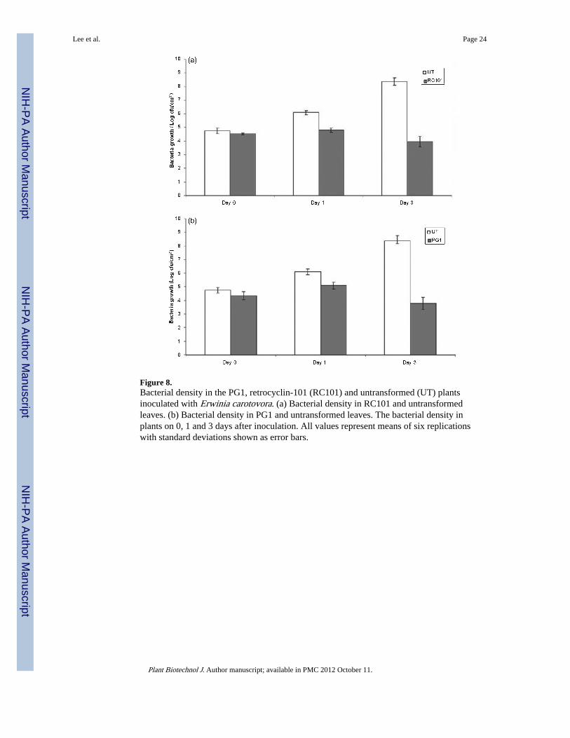
Figure 8.Bacterial density in the PG1, retrocyclin-101 (RC101) and untransformed (UT) plantsinoculated with Erwinia carotovora. (a) Bacterial density in RC101 and untransformedleaves. (b) Bacterial density in PG1 and untransformed leaves. The bacterial density inplants on 0, 1 and 3 days after inoculation. All values represent means of six replicationswith standard deviations shown as error bars.
Lee et al. Page 24
Plant Biotechnol J. Author manuscript; available in PMC 2012 October 11.
NIH
-PA Author Manuscript
NIH
-PA Author Manuscript
NIH
-PA Author Manuscript

Figure 9.Response of untransformed and retrocyclin-101 (RC101)/PG1 transplastomic plants totobacco mosaic virus (TMV). (a) TMV-inoculated leaf from untransformed plant; (b) TMV-inoculated leaf from transplastomic PG1 plant. (c) TMV-inoculated leaf from transplastomicRC101 plant. Pictures were taken on 20 days after inoculation.
Lee et al. Page 25
Plant Biotechnol J. Author manuscript; available in PMC 2012 October 11.
NIH
-PA Author Manuscript
NIH
-PA Author Manuscript
NIH
-PA Author Manuscript


![[5437]-101 M.Sc ENVIRONMENTAL SCIENCE EVSC 101](https://static.fdokumen.com/doc/165x107/631d8020ec7900c0c80d1eb7/5437-101-msc-environmental-science-evsc-101.jpg)