Expression and characterization of 4-α-glucanotransferase genes from Manihot esculenta Crantz and...
7
This article appeared in a journal published by Elsevier. The attached copy is furnished to the author for internal non-commercial research and education use, including for instruction at the authors institution and sharing with colleagues. Other uses, including reproduction and distribution, or selling or licensing copies, or posting to personal, institutional or third party websites are prohibited. In most cases authors are permitted to post their version of the article (e.g. in Word or Tex form) to their personal website or institutional repository. Authors requiring further information regarding Elsevier’s archiving and manuscript policies are encouraged to visit: http://www.elsevier.com/authorsrights
-
Upload
nottingham -
Category
Documents
-
view
3 -
download
0
Transcript of Expression and characterization of 4-α-glucanotransferase genes from Manihot esculenta Crantz and...
This article appeared in a journal published by Elsevier. The attachedcopy is furnished to the author for internal non-commercial researchand education use, including for instruction at the authors institution
and sharing with colleagues.
Other uses, including reproduction and distribution, or selling orlicensing copies, or posting to personal, institutional or third party
websites are prohibited.
In most cases authors are permitted to post their version of thearticle (e.g. in Word or Tex form) to their personal website orinstitutional repository. Authors requiring further information
regarding Elsevier’s archiving and manuscript policies areencouraged to visit:
http://www.elsevier.com/authorsrights
Author's personal copy
Process Biochemistry 49 (2014) 84–89
Contents lists available at ScienceDirect
Process Biochemistry
jo u r n al homep age: www.elsev ier .com/ locate /procbio
Expression and characterization of 4-�-glucanotransferase genesfrom Manihot esculenta Crantz and Arabidopsis thaliana and theiruse for the production of cycloamyloses
Krit Tantanarata, Ellis C. O’Neill b, Martin Rejzekb,Robert A. Fieldb,∗∗, Tipaporn Limpasenia,∗
a Department of Biochemistry, Faculty of Science, Chulalongkorn University, Bangkok 10330, Thailandb Department of Biological Chemistry, John Innes Centre, Norwich Research Park, Norwich NR4 7UH, UK
a r t i c l e i n f o
Article history:Received 10 September 2013Received in revised form 12 October 2013Accepted 16 October 2013Available online 25 October 2013
Keywords:D-enzymeGH77 familyManihot esculenta CrantzArabidopsis thalianaCycloamylose
a b s t r a c t
4-�-Glucanotransferase or disproportionating enzyme (D-enzyme, DPE) catalyzes the �-1.4 glycosyltransfer between oligosaccharides. Type I D-enzyme (DPE1) can transfer maltosyl unit from one 1.4-�-d-glucan to an acceptor mono- or oligo-saccharide, which reflects the physiological role of DPE1 inplant starch metabolism. In this study, the genes encoding DPE1 from Arabidopsis thaliana (AtDPE1) andManihot esculenta Crantz cultivar KU50 (MeDPE1) were cloned and expressed in Escherichia coli and puri-fied to homogeneity. MeDPE1 encoded 585 amino acid residues, including a 56 residue signal peptide,while AtDPE1 encoded 576 amino acid residues with a 45 residue signal peptide. The molecular mass ofboth mature enzymes, estimated from deduced amino acid sequence, were the same at 59.4 kDa, with apI of 5.13. The predicted structures of both enzymes showed the conserved 250’s loop and three catalyticamino acid residues, characteristics of disproportionating enzymes in the GH77 glycoside hydrolase fam-ily. Biochemical characterization showed that both purified recombinant enzymes were homodimers insolution, with similar optimum pH and temperature for disproportionating activity at pH 6–8 and 37 ◦C.Using potato amylose as a substrate, AtDPE1 can produce cycloamyloses in the range 16–50 glucoseresidues, while products from the action of MeDPE1 on the same substrate were in a wider range of 16to DP > 60. These recombinant enzymes are useful tools for elucidation of their functional roles in starchmetabolism and for applications in the starch industry.
© 2013 Elsevier Ltd. All rights reserved.
1. Introduction
4-�-Glucanotransferase (EC 2.4.1.25), an enzyme in glycosidehydrolase family 77 (GH77) [1], transfers a sugar moiety from thenon-reducing end of an �-1,4-glucan (donor) to the non-reducingend of another �-1,4-glucan (acceptor) chain. This enzyme isknown as disproportionating enzyme (DPE), or D-enzyme, in plants[2] and reported as amylomaltase in bacteria [3]. D-enzyme andamylomaltase are both classified in the GH77 family, but differ intheir substrate and reaction specificity [3–6]. D-enzyme was firstdiscovered in potato tubers as two isoforms, DPE1 and DPE2, whichdiffer in their structure and function [4,5,7]. DPE1 catalyzes anintermolecular transglycosylation reaction by transferring a mal-tosyl unit from the non-reducing end of a donor �-1,4-glucan, in
∗ Corresponding author. Tel.: +66 2218 5423; fax: +66 2218 5418.∗∗ Corresponding author. Tel.: +44 1603 450720; fax: +44 1603 450018.
E-mail addresses: [email protected] (R.A. Field), [email protected],[email protected] (T. Limpaseni).
particular maltotriose, to another acceptor �-1,4-glucan chain. Incontrast, while DPE2 catalyzes a similar transglycosylation reac-tion, it transfers single glucosyl residues and can use maltoseas a glucosyl donor. DPE2 has been characterized in Arabidop-sis [8–10], potato [7] and rice [11]. Arabidopsis DPE2 has beenshown to be a versatile biocatalyst, accepting a range of natural andnon-natural (e.g. fluorinated) monosaccharide acceptors [12,13].The dpe1 mutant of Arabidopsis accumulated maltooligosaccha-rides, especially its preferred substrate maltotriose, but does notaccumulate maltose [14]. It was proposed that DPE1 plays animportant role in the conversion of transitory starch into sucrosein leaves at night [15,16]. In vitro DPE1 from adzuki bean cotyle-dons can catalyze intermolecular transglycosylation to produceacarviosyl-maltooligosaccharides from acarbose [17] and purifiedDPE1 from Arabidopsis leaf has been used to produce maltodextrinfrom maltotriose [6]. Apart from the intermolecular transglycosyla-tion, DPE1 from potato catalyzed intramolecular transglycosylation(cyclization) of amylose to form cyclic �-1,4-glucans (cycloamy-loses), with degree of polymerization (DP) ranging from 17to several hundred [18]. A number of enzymes, such as
1359-5113/$ – see front matter © 2013 Elsevier Ltd. All rights reserved.http://dx.doi.org/10.1016/j.procbio.2013.10.009
Author's personal copy
K. Tantanarat et al. / Process Biochemistry 49 (2014) 84–89 85
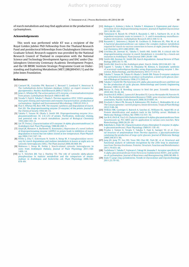
Table 1Comparison of biochemical properties of MeDPE1 and AtDPE1 with other enzymes in GH77 family from plant and bacteria.
MeDPE1 AtDPE1 StDPE1 AtDPE2 TaMalQ SynMalQ
Amino acid residues 585 576 576 955 500 505Signal peptide 56 45 52 – – –Amino acid sequence identity with MeDPE1 100% 73% 71% 26% 42% 44%Amino acid sequence identity with AtDPE1 73% 100% 69% 34% 42% 43%Molecular weight (SDS-PAGE) 66 kDa 66 kDa 65 kDa 100 kDa 57 kDa 57 kDaMolecular weight (Gel filtration) 117 kDa 123 kDa NR 202 kDa NR NROptimum temperature 37 ◦C 37 ◦C 45 ◦C 42 ◦C 75 ◦C 45 ◦COptimum pH 6.0–8.0 6.0–8.0 6.7 7.0 5.5–6.0 7.0Km (mM) (maltotriose) 69.2 27.5 NR NR NR NRKm (mM) (maltose) NR NR NR 2.9 NR NRkcat (s−1) 894 580 NR NR NR NRkcat/Km (mM−1 s−1) 12.9 21.6 NR NR NR NRMinimum size of cycloamylose 16 16 17 NR 22 22References This Study This Study [2,5,18] [13] [3] [24]
4-Alpha-glucanotransferase are from M. esculenta Crantz (MeDPE1), A. thaliana (AtDPE1, AtDPE2), S. tuberosum (StDPE1), T. aquaticus ATCC 33923 (TaMalQ), Synechocystis sp.PCC 6803 (SynMalQ).NR, not reported.
amylomaltase and cyclodextrin glucosyltransferase, have beenreported to be capable of producing cycloamyloses [19].
Cassava (Manihot esculenta Crantz) is grown in tropical region(Africa, Asia and the Americas). The high starch content (20–40%) ofcassava tubers makes them an important feedstock for use in culi-nary, animal feed and starch-based products [20]. The Arabidopsisgenome has been well studied, with support from The Arabidop-sis Information Resource (TAIR) (http://www.arabidopsis.org/) [21]and the Cassava genome is now available on the phytozomegenome browser (http://www.phytozome.net/cassava) [22]. D-enzyme genes have been identified in both the cassava andArabidopsis genomes, but the corresponding recombinant enzymeshave not been characterized to date. In this study, we reportthe cloning and expression of the DPE1 gene from both plants,along with the characterization of the recombinant enzymes andassessment of their ability to produce useful products, such ascycloamyloses.
2. Materials and methods
2.1. Stock cDNA
Cassava cDNA was obtained from M. esculenta Crantz KU50 (courtesy of Dr.Yindee Chanvivattana, National Center for Genetic Engineering and Biotechnology,Pathumthani, Thailand). Arabidopsis cDNA stock number U12707, specific to theDPE1 gene (Genbank accession number AY081744), was purchased from ABRC (OhioState University).
2.2. Cloning of DPE1 gene from cassava
The coding region for 4-�-glucanotransferase from cassava (M. esculenta Crantz:MeDPE1) was identified in Phytozome v6.0 (Locus name: cassava4.1 008552m.g). APCR fragment was obtained using the forward primer 5′-TTC GAA GCA GTT TCTTTA TCC TCT ACC-3′ (introduced BstBI site underlined) and reverse primer 5′-GTC GAC CAC CCG CCC ATA CAT TG-3′ (introduced SalI site underlined). The forwardprimer was designed to remove the signal peptide. The PCR program was 2 minat 95 ◦C, 32 cycles of: 8 s at 98 ◦C, 20 s at 70 ◦C, 105 s at 72 ◦C, followed by finalelongation for 10 min at 72 ◦C. The PCR reaction was carried out with 1 unit of Phu-sion High-Fidelity DNA polymerase (Thermo scientific) according to manufacturerinstructions, with a final concentration of each primer at 0.2 pmol/�l in 50 �l totalvolume.
The PCR amplified MeDPE1 gene was cloned in pTrcHis2c vector (Invitrogen)with two restriction sites (BstBI and SalI) and a C-terminal hexa-histidine tag (aminoacid sequence: VDHHHHHH), the plasmid obtained was denoted pTMeDPE1. ThepTMeDPE1 was transformed into Escherichia coli DH5� by the CaCl2 method andcorrect insertion of the DPE1 gene was confirmed by DNA sequencing.
2.3. Cloning of the DPE1 gene from Arabidopsis
The sequence of the Arabidopsis thaliana DPE1 gene (AtDPE1) was identified atlocus AT5G64860 in The Arabidopsis Information Resource (TAIR) database website.PCR amplification was designed to remove the signal peptide using forward primer5′-CACC ATG GAG GTC GTT TCG AGT AAT TCC-3′ with CACC for directional cloning
(underlined) and reverse primer 5′-TCA AAG CCG TCC GTA CAA TGA CAA AAG ATC-3′ ,using the same PCR program as for MeDPEI.
The PCR amplified AtDPE1 was cloned in pET151-TOPO vector (Invitrogen) usingthe ChampionTM pET151 Directional TOPO (Invitrogen), which inserts an N-terminalhexa-histidine tag and a V5 epitope beyond a TEV cleavage site, and the plasmidobtained was denoted pETAtDPE1. This plasmid was transformed into the expressionhost BL21 StarTM (DE3) One Shot® (Invitrogen) and screening was performed asdescribed for the cloning of MeDPE1.
2.4. Expression and purification of DPE1
Transformed E. coli cells containing recombinant plasmids pTMeDPE1 andpETAtDPE1 from either cassava or Arabidopsis were separately inoculated into 5 mlLB medium containing ampicillin (100 �g/ml) and cultivated overnight at 37 ◦C.Each culture (1%, v/v) was inoculated into 200 ml Auto Induction Media (AIM) con-taining ampicillin (100 �g/ml). The cultures were grown at 37 ◦C 250 rpm whileOD600 reached 0.5–0.6. The temperature was decreased to 16 ◦C and the cultureswere further incubated at 250 rpm overnight (18 h). Bacterial cells were collectedby centrifugation at 5000 × g for 15 min. Cell pellets were resuspended in cell lysissolution (50 mM Tris–HCl pH 8.0, 0.5 M NaCl, 30 mM imidazole, 0.3 mg/ml DTT,50 �g/ml lysozyme, 1 tablet per 50 ml complete protease inhibitor cocktail (Roche)and 12.5 �g/ml DNase) and lysed with a cell disruptor (constant system) at 25 kpsi.Crude lysate were cleared by centrifugation at 30,000 × g for 30 min at 4 ◦C.
Crude MeDPE1 and AtDPE1 were applied to 5 ml Ni HiTrap IMAC FF columns(GE Healthcare) washed with 50 mM Tris–HCl pH 8.0, 0.5 M NaCl and 30 mM imid-azole, and eluted with a linear gradient of 30–500 mM imidazole in 50 mM Tris–HClpH 8.0 containing 0.5 M NaCl. The enzymes were further purified by gel filtration(HiLoad 26/60 Superdex 75 prep grade, GE Healthcare), eluted with 100 mM NaCl in50 mM HEPES pH 7.5. Single peaks of protein were pooled, concentrated and buffer-exchanged into 20 mM HEPES pH 7.0 with Centrifugal Filter Units of MWCO 30,000(Millipore, US).
2.5. Disproportionation activity assay
DPE1 assay was modified from that used by Peat et al. for the potato enzyme[5]. The reaction mixture, containing 54 �M maltotriose in 100 mM MOPS-NaOHpH 7.0, was incubated with the purified DPE1 at 37 ◦C for 15 min and the reactionwas stopped by heating to 95 ◦C in boiling water for 5 min. The amount of glucosereleased was determined by using the hexokinase-G6P dehydrogenase method [13].The DPE1 reaction mixture (20 �l) was incubated with hexokinase assay cocktail:1 mM MgCl2, 0.6 mM adenosine 5′-triphosphate disodium salt hydrate (ATP, Sigma),0.44 mM �-nicotinamide adenine dinucleotide hydrate (NAD) and 3.3 unit/ml hex-okinase (Roche) in 27.5 mM HEPES pH 7.9 in 178 �l. The starting absorbance (Abs1)at 340 nm was recorded. Two units of glucose-6-phosphate dehydrogenase (G6P-DH, Roche) in 2 �l were added and, after incubation for 10 min at 21 ◦C, the finalabsorbance (Abs2) at 340 nm was recorded. The change in OD at 340 nm was usedto calculate the glucose released, in comparison to a standard curve and one unitof DPE1 activity was defined as the amount of enzyme which released 1 �mol ofglucose per minute under the assay conditions. Disproportionation activity wasalso monitored by iodine staining of non-denaturing polyacrylamide electrophore-sis gels containing glycogen. DPE1 preparations were separated by electrophoresison 0.75 mm thick polyacrylamide gel containing 10% (w/v) acrylamide and 0.3%(w/v) rabbit liver glycogen (Sigma), at 16 mA per slab at 4 ◦C in a Mini-Gel elec-trophoresis unit (Bio-Rad). After electrophoresis the gel was incubated in substratesolution buffer (50 mM Tris–HCl pH 8.0, 8 mM EDTA and 0.3% maltodextrin) at room

Author's personal copy
86 K. Tantanarat et al. / Process Biochemistry 49 (2014) 84–89
Fig. 1. Sequence alignment of amylomaltase and plant D-enzymes (Arabidopsis, cassava and potato). The three catalytic amino acids are marked in bold and with an arrow.The 250s loops are boxed in black. The accession numbers for these sequence are cassava4.1 008552m.g (Manihot esculenta Crantz, MeDPE1), AAK59831.1 (Arabidopsisthaliana, AtDPE1), Q06801 (Solanum tuberosum, StDPE1), BAA33728.1 (Thermus aquaticus ATCC 33923, TaMalQ), BAA16800 (Synechocystis sp. PCC 6803, SynMalQ), andNP 181616 (Arabidopsis thaliana, AtDPE2).
temperature overnight. The gel was rinsed several times with distilled water andstained with iodine solution (0.2% I2 in 2% KI).
2.6. Characterization of DPE1
The effect of pH and temperature on the disproportionating activity of purifiedMeDPE1 and AtDPE1 were studied. The enzymes were incubated in 0.1 M bufferat different pH values [sodium acetate buffer (pH 3.0, 4.0, 5.0), sodium phosphatebuffer (pH 5.0, 6.0, 7.0), Tris–HCl (pH 7.0, 8.0, 9.0), MOPS (pH 7.0, 8.0), glycine–NaOH(pH 9.0, 10.0)]. For optimum temperature measurements, 0.1 M MOPS buffer pH7.0 was used. The enzymes were incubated in the temperature range 5–60 ◦C for15 min in the disproportionation activity buffer. Molecular weights of both recom-binant enzymes were determined by gel filtration and SDS-polyacrylamide gelelectrophoresis (SDS-PAGE) (Fig. 3C).
For kinetic studies, the initial velocities of the enzymatic reaction were deter-mined by varying the concentration of maltotriose (2–60 mM). Values of theMichaelis constants (Km and kcat) were calculated.
2.7. Cycloamylose production
Amylose type III from potato (Sigma) was solubilized in water at 2% (w/v) byboiling for 15 min and centrifuged at 5000 × g for 15 min to remove the insolublefraction. The soluble material was dialyzed (MWCO 12,000–14,000, Cellu-Sep T4,Membrane Filtration Products Inc., USA) to eliminate residual glucose and shortchain oligosaccharides. The amylose solution was incubated with recombinantMeDPE1 or AtDPE1 (1.2 U/ml) in 5 mM phosphate buffer pH 7.0 (10 ml reactions) at37 ◦C for 18 h and the reaction was stopped by heating to 95 ◦C for 15 min. To removeresidual linear glucan amyloglucosidase from Rhizopus sp. (Megazyme) was added(1.5 U/ml), incubated at 37 ◦C and the reaction was stopped as above. The precipitatewas removed by centrifugation at 12,000 × g for 30 min. The cycloamylose productwas precipitated by addition of absolute ethanol (to final concentration 81%), andcollected after 2 days by centrifugation at 12,000 × g for 30 min. The precipitate waswashed twice with absolute ethanol and dissolved at 2 mg/ml in distilled water.Analysis of cycloamylose was performed on high performance anion exchangechromatography with pulsed amperometric detector (HPAEC-PAD, Dionex-500,USA) equipped with a CarboPac PA-100 column (4 mm × 250 mm, Dionex). Elution

Author's personal copy
K. Tantanarat et al. / Process Biochemistry 49 (2014) 84–89 87
Fig. 2. Domain and structure analysis of MeDPE1 and AtDPE1. (A) Domains of DPE1and MalQ. (B) Three-dimensional homology model of MeDPE1 (B1) and AtDPE1 (B2)generated using I-TASSER with DPE1 from potato (PDB code: 1x1N) as a template, using. (C) The proposed catalytic sites of MeDPE1 (C1) and AtDPE1 (C2).
was performed using a linear gradient of sodium nitrate in 150 mM NaOH (4–8%for 0–2 min; 8–18% for 2–10 min; 18–28% for 10–20 min; 28–35% for 20–40 min;35–45% for 40–45 min; 45–63% for 55–60 min) at a flow rate 1 ml/min. The size ofcycloamylose products was compared with standard cycloamyloses, with an aver-age molecular weight of 7000 Da (Ezaki Glico, Japan). The molecular mass spectrumof cycloamyloses was obtained by matrix-assisted laser desorption ionization-timeof flight mass spectrometry (MALDI-TOF-MS).
3. Results and discussion
3.1. DPE1 gene sequence
Nucleotide sequence analysis of A. thaliana (AtDPE1 gene, acces-sion number: NP 201291.1) predicted by Chlorop V1.1 showedthe presence of an open reading frame of 1731 bp encoding 576amino acid residues with a 45 amino acid signal peptide at theN-terminus (Fig. 2A). For M. esculenta Crantz (MeDPE1 gene, Phy-tozome v6.0: Locus name: cassava4.1 008552m.g), the nucleotidesequence showed the presence of an open reading frame of1758 bp encoding 585 amino acid residues with 56 amino acidresidues as the signal sequence. Molecular weight prediction using
Compute pI/Mw Tool from the ExPAsy server [23] indicated thatboth MeDPE1 and AtDPE1 had the same predicted molecularweight and isoelectric point (59 kDa, pI 5.13). The deduced aminoacid sequences of AtDPE1 and MeDPE1 were aligned with potatoDPE1 (StDPE1) and bacterial amylomaltase, which are also mem-bers of glycoside hydrolase family 77. The BLAST program inGenBank showed plant DPE1 were closely related with approxi-mately 70% identity (Table 1). No signal peptide was present in thesequences of the bacterial amylomaltases [24] (Fig. 2A). The signalpeptide, referred to as the transit peptide in plants, plays a role inbringing the protein into the specialised organelles such as plas-tids. The peptide is likely to be largely disordered, interfering withsolubility, and is not expected to have any catalytic function. Themembers of glycoside hydrolase family 77 all contain 250’s loopas a conserved region, which is not present in glycosyl hydrolasewith a similar structure. Three catalytic amino acid residues wereidentified; one glutamic and two aspartic acids (Figs. 1 and 2C).
StDPE1 from potato (PDB code: 1X1N) was usedas template for simulation of three dimensional struc-tures of MeDPE1 and AtDPE1 using I-TASSER server
Fig. 3. Gel filtration chromatography of (A) MeDPE1 and (B) AtDPE1 showing elution times of standard proteins: (1) aprotinin (65 kDa), (2) cytochrome C (12.4 kDa), (3)RNase A (13.7 kDa), (4) myoglobin (17.6 kDa), (5) carbonic anhydrase (29.0 kDa), (6) BSA (66 kDa) and (7) alcohol dehydrogenase (150 kDa). (C) SDS-PAGE of MeDPE1 (lane1) and AtDPE1 (lane 2). (D) Coomassie and iodine stains of non-denaturing-glycogen PAGE of MeDPE1 (lanes 1, 3) and AtDPE1 (lanes 2, 4).

Author's personal copy
88 K. Tantanarat et al. / Process Biochemistry 49 (2014) 84–89
Fig. 4. (A) HPEAC-PAD analysis of cycloamyloses produced by the action of AtDPE1 and MeDPE1 on amylose from potato. A standard cycloamyloses preparation was usedto compare product size. (B) MALDI-TOF MS analysis of the cycloamylose product from AtDPE1 action. Numbers show the degree of polymerization.
(http://zhanglab.ccmb.med.umich.edu/I-TASSER/). The modelsshowed a (�,�)8 barrel core, 250’s loop and three catalytic aminoacids in both enzymes (Fig. 2B and C), supporting the result fromamino acid sequence alignment. Further investigation of thecrystal structures and ligand binding sites of both MeDPE1 andAtDPE1 are underway.
3.2. Expression and purification of DPE1
In an initial attempt at heterologous expression, the MeDPE1gene was amplified from cassava without the signal peptide, whichis likely to interfere with solubility and is not expected to haveany catalytic function. This was cloned in to pET17b, which carriesan N-terminal T7 tag and expressed in E. coli BL21 (DE3) at 37 ◦C.However, the protein was expressed in insoluble form and no activ-ity was detected. Expression at lower temperature (16 ◦C) did notimprove the expression of MeDPE1. The MeDPE1 gene was subse-quently cloned into the pTrcHis2C vector (Invitrogen) under thetrc promoter, as reported for potato DPE1 [5]. This vector, whichinserts 6 histidines at the C-terminal, was employed successfullyto express MeDPE1 in E. coli DH5� at 16 ◦C. AtDPE1 was success-fully expressed under the T7 promoter using the pET151/D-TOPOTM
(Invitrogen) expression vector in E. coli BL21 (DE3) at 16 ◦C.Transformed E. coli strains carrying plasmids for the expres-
sion of MeDPE1 and AtDPE1 were grown and induced with lactoseat 16 ◦C. Disproportionating enzymes activity was detected in thesoluble cytosolic fraction of E. coli cells and the enzymes werepurified using Ni affinity chromatography (HiTrap IMAC FF), fol-lowed by size exclusion chromatography (HiLoad 26/60 Sephadex75 prep grade). Chromatograms from gel filtration show singlepeaks (Fig. 3A and B) with the calculated molecular weight of therecombinant MeDPE1 and AtDPE1 of 123 kDa and 117 kDa, respec-tively. The apparent molecular weights on SDS-PAGE were similarat 66 kDa (Fig. 3C), which corresponds well to the molecular weight,calculated from the deduced amino acid sequences of 66 kDa for themonomer. The results suggested both enzymes were homodimersin solution, as previously reported for the potato and pea enzyme[25] while amylomaltases from Thermus aquaticus ATCC 33923 [26]and Thermus brockianus [27] were reported to be monomers.
3.3. Characteristics of recombinant MeDPE1 and AtDPE1
Native polyacrylamide gel electrophoresis of MeDPE1 andAtDPE1 on gels containing glycogen gave a positive iodine stainon addition of the transglycosylation substrate maltotriose, con-firming the transglycosylation activity of both proteins [14]. PlantDPE1 differs from other GH77 family enzymes, such as amylo-maltase and DPE2, in its ability to transfer two glucose units in
the transglycosylation reaction rather than one [12]. MeDPE1 andAtDPE1 showed similar optimum pH and temperature for dispro-portionating activity to other plant D-enzymes (Table 1) whichcorrelated to wild type Arabidopsis DPE1 from leaf at pH 6.5 [6].In contrast, most characterized bacterial amylomaltases are moretolerant to high temperature and low pH. The kinetic parame-ters for disproportionating activity of both MeDPE1 and AtDPE1were determined with maltotriose as the substrate. The Km andkcat/Km were 69.2 ± 10 mM and 12.9 ± 0.2 mM−1 s−1 for MeDPE1and 27.5 ± 6 mM and 21.6 ± 3 mM−1 s−1 for AtDPE1, respectively.This indicates that AtDPE1 is approximately twice as active towardmaltotriose as MeDPE1. Crucially transitory starch degradationoccurs daily in leaves and AtDPE1 is proposed to be involved inthe process [14]. On the other hand, starch synthesis is far moreprominent than degradation in cassava tubers. Thus, the observeddifferences in kinetic properties between the Arabidopsis and cas-sava enzymes may reflect the different physiological contexts inwhich they operate.
3.4. Identification of reaction products
The ability of MeDPE1 and AtDPE1 to form cycloamyloseswhen fed long chain amylose was also investigated. Previously,cycloamylose production were reported mostly in bacteria suchas T. aquaticus ATCC 33923 [3] and Pyrococcus kodakaraensis KOD1[28]. StDPE1, the potato DPE1, is the only plant enzyme reportedto produce cycloamyloses [18]. Whether these reactions occurin vivo remains to be established. HPAEC-PAD analysis of theproducts from reactions of MeDPE1 and AtDPE1 with potato amy-lose revealed the presence of cycloamyloses from DP16 upwards(Fig. 4A). AtDPE1 produced cycloamyloses in the range DP 16–50with maximum intensity at DP 18, while cycloamyloses producedby MeDPE1 showed maximum intensity at DP 19 but range upto DP > 60. Products analysis by MALDI-TOF-MS showed molecu-lar masses corresponding to cyclic molecules containing differentnumbers of glucose units, with regular mass differences betweenpeaks corresponding to the mass of one glucosyl unit (162 Da) in apolysaccharide chain (Fig. 4B), and correlated well to the theoreticalmolecular weight of cycloamyloses previously reported [29].
4. Conclusion
Two plant disproportionating enzyme (DPE1) from M. esculentaCrantz and A. thaliana were successfully cloned, over-expressedin E. coli and characterized. These enzymes showed similar bio-chemical characteristics, but with notable difference in their kineticparameters and their ability to form longer chain cycloamyloses(DP > 60). These recombinant enzymes are useful tools for the study
Author's personal copy
K. Tantanarat et al. / Process Biochemistry 49 (2014) 84–89 89
of starch metabolism and may find application in the production ofcycloamyloses.
Acknowledgements
This work was performed while KT was a recipient of theRoyal Golden Jubilee PhD Fellowship from the Thailand ResearchFund and postdoctoral fellowships from Chulalongkorn UniversityGraduate School. Research supports was provided by the NationalResearch Council of Thailand in cooperation with the NationalScience and Technology Development Agency and IIAC under Chu-lalongkorn University Centenary Academic Development Project,and the UK BBSRC Institute Strategic Programme Grant on Under-standing and Exploiting Metabolism (MET) [BB/J004561/1] and theJohn Innes Foundation.
References
[1] Cantarel BL, Coutinho PM, Rancurel C, Bernard T, Lombard V, Henrissat B.The Carbohydrate-Active EnZymes database (CAZy): an expert resource forglycogenomics. Nucleic Acid Research 2009;37:D233–8.
[2] Jones G, Whelan WJ. The action pattern of D-enzyme, a transmaltodextrinylasefrom potato. Carbohydrate Research 1969;9:483–90.
[3] Terada Y, Fujii K, Takaha T, Okada S. Thermus aquaticus ATCC 33923 amyloma-ltase gene cloning and expression and enzyme characterization: production ofcycloamylose. Applied and Environmental Microbiology 1999;65:910–5.
[4] Peat S, Whelan WJ, Rees WR. The enzymic synthesis and degradation of starch.Part XX. The disproportionating enzyme (D-enzyme) of the potato. Journal ofthe Chemical Society 1956:44–53.
[5] Takaha T, Yanase M, Okada S, Smith SM. Disproportionating enzyme (4-�-glucanotransferase; EC 2.4.1.25) of potato. Purification, molecular cloning,and potential role in starch metabolism. Journal of Biological Chemistry1993;268:1391–6.
[6] Lin TP, Preiss J. Characterization of D-enzyme (4-alpha-glucanotransferase) inArabidopsis leaf. Plant Physiology 1988;86:260–5.
[7] Lloyd JR, Blennow A, Burhenne K, Kossmann J. Repression of a novel isoformof disproportionating enzyme (stDPE2) in potato leads to inhibition of starchdegradation in leaves but not tubers stored at low temperature. Plant Physiol-ogy 2004;134:1347–54.
[8] Fettke J, Chia T, Eckermann N, Smith A, Steup M. A transglucosidase neces-sary for starch degradation and maltose metabolism in leaves at night acts oncytosolic heteroglycans (SHG). The Plant Journal 2006;46:668–84.
[9] Malinova I, Steup M, Fettke J. Starch-related cytosolic heteroglycans inroots from Arabidopsis thaliana. Journal of Plant Physiology 2011;168:1406–14.
[10] Lu Y, Steichen JM, Yao J, Sharkey TD. The role of cytosolic alpha-glucanphosphorylase in maltose metabolism and the comparison of amylo-maltase in Arabidopsis and Escherichia coli. Plant Physiology 2006;142:878–89.
[11] Akdogan G, Kubota J, Kubo A, Takaha T, Kitamura S. Expression and charac-terization of rice disproportionating enzymes. Journal of Applied Glycoscience2011;58:99–105.
[12] Tantanarat K, Rejzek M, O’Neill E, Ruzanski C, Hill L, Fairhurst SA, et al. Anexpedient enzymatic route to isomeric 2-, 3- and 6-monodeoxy-monofluoro-maltose derivatives. Carbohydrate Research 2012;358:12–8.
[13] Ruzanski C, Smirnova J, Rejzek M, Cockburn D, Pedersen HL, Pike M, et al.A bacterial glucanotransferase can replace the complex maltose metabolismrequired for starch-to-sucrose conversion in leaves at night. Journal of Biolog-ical Chemistry 2013;288:28581–98.
[14] Critchley JH, Zeeman SC, Takaha T, Smith AM, Smith SM. A critical role fordisproportionating enzyme in starch breakdown is revealed by a knock-outmutation in Arabidopsis. Plant Journal 2001;26:89–100.
[15] Smith AM, Zeeman SC, Smith SM. Starch degradation. Annual Review of PlantBiology 2005;56:73–96.
[16] Smith AM. Starch in the Arabidopsis plant. Starch–Stärke 2012;64:421–34.[17] Tagami T, Tanaka Y, Mori H, Okuyama M, Kimura A. Enzymatic synthesis
of acarviosyl-maltooligosaccharides using disproportionating enzyme 1. Bio-science, Biotechnology, and Biochemistry 2013;77:312–9.
[18] Takaha T, Yanase M, Takata H, Okada S, Smith SM. Potato D-enzyme catalyzesthe cyclization of amylose to produce cycloamylose, a novel cyclic glucan. Jour-nal of Biological Chemistry 1996;271:2902–8.
[19] Takaha T, Smith SM. The functions of 4-alpha-glucanotransferases and their usefor the production of cyclic glucans. Biotechnology and Genetic EngineeringReviews 1999;16:257–80.
[20] Nassar N, Ortiz R. Breeding cassava to feed the poor. Scientific American2010;302:78–82, 84.
[21] Swarbreck D, Wilks C, Lamesch P, Berardini TZ, Garcia-Hernandez M, Foerster H,et al. The Arabidopsis Information Resource (TAIR): gene structure and functionannotation. Nucleic Acids Research 2008;36:D1009–14.
[22] Prochnik S, Marri PR, Desany B, Rabinowicz PD, Kodira C, Mohiuddin M, et al.The Cassava genome: current progress, future directions. Tropical Plant Biology2012;5:88–94.
[23] Wilkins MR, Gasteiger E, Bairoch A, Sanchez JC, Williams KL, Appel RD, et al.Protein identification and analysis tools in the ExPASy server. Methods inMolecular Biology (Clifton, NJ) 1999;112:531–52.
[24] Lee B-H, Oh D-K, Yoo S-H. Characterization of 4-alpha-glucanotransferase fromSynechocystis sp. PCC 6803 and its application to various corn starches. NewBiotechnology 2009;26:29–36.
[25] Kakefuda G, Duke SH. Characterization of pea chloroplast D-enzyme (4-alpha-d-glucanotransferase). Plant Physiology 1989;91:136–43.
[26] Przylas I, Tomoo K, Terada Y, Takaha T, Fujii K, Saenger W, et al. Crys-tal structure of amylomaltase from Thermus aquaticus, a glycosyltransferasecatalysing the production of large cyclic glucans. Journal of Molecular Biology2000;296:873–86.
[27] Jung JH, Jung TY, Seo DH, Yoon SM, Choi HC, Park BC, et al. Structural andfunctional analysis of substrate recognition by the 250s loop in amylomal-tase from Thermus brockianus. Proteins: Structure, Function and Bioinformatics2011;79:633–44.
[28] Tachibana Y, Takaha T, Fujiwara S, Takagi M, Imanaka T. Acceptor specificity of4-alpha-glucanotransferase from Pyrococcus kodakaraensis KOD1, and synthe-sis of cycloamylose. Journal of Bioscience and Bioengineering 2000;90:406–9.
[29] Endo T. Large-ring cyclodextrins. Trends in Glycoscience and Glycotechnology2011;23:79–92.