Exploring the use of soft X-ray microscopy for imaging subcellular structures of the inner ear1
10
© 2004 The Royal Microscopical Society Journal of Microscopy, Vol. 215, Pt 2 August 2004, pp. 203 – 212 Received 27 January 2004; accepted 25 March 2004 Blackwell Publishing, Ltd. Exploring the use of soft X-ray microscopy for imaging subcellular structures of the inner ear 1 GÖRAN A. JOHANSSON*, SHYAM M. KHANNA†, AJIT NAIR‡, PAULA MANNSTRÖM§, GREG DENBEAUX‡ & MATS ULFENDAHL§ *Biomedical and X-ray Physics, Department of Physics, Royal Institute of Technology, SE-106 91 Stockholm, Sweden † Department of Otolaryngology/Head and Neck Surgery, Columbia University, New York, NY 10032, U.S.A. ‡ Center for X-ray Optics, Lawrence Berkeley Laboratory, University of California, Berkley, CA 94720, U.S.A. §Center for Hearing and Communication Research, and Department of Clinical Neuroscience, Karolinska Institutet, Karolinska University Hospital, SE-171 76 Stockholm, Sweden Key words. Hair cell, inner ear, Reissner’s membrane, soft X-ray microscopy, tectorial membrane. Received 27 January 2004; accepted 25 March 2004 Summary The soft X-ray microscope at the Lawrence Berkeley National Laboratory was developed for visualization of biological tissue. Soft X-ray microscopy provides high-resolution visualization of hydrated, non-embedded and non-sectioned cells and is thus potentially an alternative to transmission electron microscopy. Here we show for the first time soft X-ray micrographs of struc- tures isolated from the guinea-pig inner ear. Sensory outer hair cells and supporting pillar cells are readily visualized. In the hair cells, individual stereocilia can easily be identified within the apical hair bundle. The underlying cuticular plate is, however, too densely composed or too thick to be clearly visualized, and thus appears very dark. The cytoplasmic structures protrud- ing from the cuticular plates as well as the fibrillar material surrounding and projecting from the cell nuclei can be seen. In the pillar cells the images reveal individual microtubule bundles. Soft X-ray images of the acellular tectorial membrane and thin two-layered Reissner’s membrane display a level of resolution comparable to low-power electron microscopy. Introduction To investigate the true structural and functional properties of cells, it is essential to study them under as intact and physiological conditions as possible. Each experimental method, however, reflects a combination of advantages and disadvant- ages. For example, light microscopy allows studying living cells but with a limited spatial resolution. In addition, for high spatial resolution imaging of thin objects, the contrast for biological samples is quite poor and contrast enhancement techniques, e.g. fluorophore staining and phase contrast microscopy, are frequently used. Transmission electron microscopy, by contrast, offers much greater spatial resolu- tion but is primarily used for fixed, dehydrated, embedded and, finally, sectioned tissue. In addition, the processing efforts required prior to visualization are laborious and time consuming. Clearly, a microscope technique combining the advantages of light and electron microscopes would be very powerful. The soft X-ray microscope offers a highly interesting method for imaging intact hydrated cells, with high natural absorption contrast and with a spatial resolution beyond what is achievable with visible light microscopy (Schmahl et al., 1993; Kirz et al., 1995; Meyer-Ilse et al., 1995; see also review by Yamamoto & Shinohara, 2002). The reason is that in the soft X-ray region the interaction between matter and electromagnetic radiation is particularly strong and highly wavelength dependent (Attwood, 1999). By a slight change in wavelength, the photon absorption can change by several orders of magnitude and, for a selected wavelength, one material can be almost transparent and another material highly absorbent. Its resolution falls between that of light microscopy and high-resolution electron microscopy, and is presently limited by the available diffractive X-ray optics to 20 nm. Exposure times in the soft X-ray micro- scope are usually a few seconds, which creates the potential for making several hundred images per day. Furthermore, Correspondence to: Dr Mats Ulfendahl. Tel.: +46 851776307; fax: +46 8301876; e-mail: [email protected] 1 This paper is dedicated to the memory of Dr Werner Meyer-Ilse. Werner was the key moving force in the design and fabrication of the X-ray microscope XM-1 at ALS. He inspired us to work on the application of X-ray microscopy to the inner ear and helped us with his encouragement and support.
-
Upload
independent -
Category
Documents
-
view
1 -
download
0
Transcript of Exploring the use of soft X-ray microscopy for imaging subcellular structures of the inner ear1
© 2004 The Royal Microscopical Society
Journal of Microscopy, Vol. 215, Pt 2 August 2004, pp. 203–212
Received 27 January 2004; accepted 25 March 2004
Blackwell Publishing, Ltd.
Exploring the use of soft X-ray microscopy for imaging subcellular structures of the inner ear
1
G Ö R A N A . J O H A N S S O N *, S H YA M M . K H A N NA †, A J I T NA I R ‡, PAU L A M A N N S T RÖ M §, G R E G D E N B E AU X ‡ & M AT S U L F E N DA H L §
*
Biomedical and X-ray Physics, Department of Physics, Royal Institute of Technology, SE-106 91 Stockholm, Sweden
†
Department of Otolaryngology/Head and Neck Surgery, Columbia University, New York, NY 10032, U.S.A.
‡
Center for X-ray Optics, Lawrence Berkeley Laboratory, University of California, Berkley, CA 94720, U.S.A.
§
Center for Hearing and Communication Research, and Department of Clinical Neuroscience, Karolinska Institutet, Karolinska University Hospital, SE-171 76 Stockholm, Sweden
Key words.
Hair cell, inner ear, Reissner’s membrane, soft X-ray microscopy, tectorial membrane.
Received 27 January 2004;
accepted 25 March 2004
Summary
The soft X-ray microscope at the Lawrence Berkeley NationalLaboratory was developed for visualization of biological tissue.Soft X-ray microscopy provides high-resolution visualizationof hydrated, non-embedded and non-sectioned cells and is thuspotentially an alternative to transmission electron microscopy.Here we show for the first time soft X-ray micrographs of struc-tures isolated from the guinea-pig inner ear. Sensory outerhair cells and supporting pillar cells are readily visualized. In thehair cells, individual stereocilia can easily be identified withinthe apical hair bundle. The underlying cuticular plate is, however,too densely composed or too thick to be clearly visualized, andthus appears very dark. The cytoplasmic structures protrud-ing from the cuticular plates as well as the fibrillar materialsurrounding and projecting from the cell nuclei can be seen.In the pillar cells the images reveal individual microtubulebundles. Soft X-ray images of the acellular tectorial membraneand thin two-layered Reissner’s membrane display a level ofresolution comparable to low-power electron microscopy.
Introduction
To investigate the true structural and functional propertiesof cells, it is essential to study them under as intact and
physiological conditions as possible. Each experimental method,however, reflects a combination of advantages and disadvant-ages. For example, light microscopy allows studying livingcells but with a limited spatial resolution. In addition, for highspatial resolution imaging of thin objects, the contrast forbiological samples is quite poor and contrast enhancementtechniques, e.g. fluorophore staining and phase contrastmicroscopy, are frequently used. Transmission electronmicroscopy, by contrast, offers much greater spatial resolu-tion but is primarily used for fixed, dehydrated, embeddedand, finally, sectioned tissue. In addition, the processingefforts required prior to visualization are laborious and timeconsuming. Clearly, a microscope technique combining theadvantages of light and electron microscopes would be verypowerful.
The soft X-ray microscope offers a highly interesting methodfor imaging intact hydrated cells, with high natural absorptioncontrast and with a spatial resolution beyond what is achievablewith visible light microscopy (Schmahl
et al
., 1993; Kirz
et al
.,1995; Meyer-Ilse
et al
., 1995; see also review by Yamamoto &Shinohara, 2002). The reason is that in the soft X-ray regionthe interaction between matter and electromagnetic radiationis particularly strong and highly wavelength dependent(Attwood, 1999). By a slight change in wavelength, the photonabsorption can change by several orders of magnitude and, fora selected wavelength, one material can be almost transparentand another material highly absorbent. Its resolution fallsbetween that of light microscopy and high-resolution electronmicroscopy, and is presently limited by the available diffractiveX-ray optics to 20 nm. Exposure times in the soft X-ray micro-scope are usually a few seconds, which creates the potentialfor making several hundred images per day. Furthermore,
Correspondence to: Dr Mats Ulfendahl. Tel.: +46 851776307; fax: +46
8301876; e-mail: [email protected]
1
This paper is dedicated to the memory of Dr Werner Meyer-Ilse. Werner was
the key moving force in the design and fabrication of the X-ray microscope
XM-1 at ALS. He inspired us to work on the application of X-ray microscopy to
the inner ear and helped us with his encouragement and support.

204
G . A . J O H A N S S O N
E T A L .
© 2004 The Royal Microscopical Society,
Journal of Microscopy
,
215
, 203–212
sample handling is as easy as in visible light microscopyand it can utilize non-dehydrated and non-embedded tissueimmersed in buffer solutions without any contrast enhance-ment techniques.
The soft X-ray microscope at the Lawrence Berkeley NationalLaboratory has been developed for the visualization of biologi-cal specimens (Denbeaux
et al
., 2001; Meyer-Ilse
et al
., 2001).In the present report, we explore, for the first time, the use ofsoft X-ray microscopy to examine elements from the guinea-pig inner ear. The main aim of the study was to compare thevisualization of different cellular elements using transmissionelectron microscopy and soft X-ray microscopy, especiallyregarding the relative efficiency of the two techniques in termsof the information they provide with respect to the prepara-tion time needed.
Materials and methods
Cell and tissue preparations
Cells were isolated from the hearing organs of albino guinea-pigs (female, 250–300 g,
n
= 8; Simonsen Laboratories, Inc.,Gilroy, CA, U.S.A.). The animals were deeply anaesthetizedusing carbon dioxide and rapidly decapitated. The temporalbones were excised and stored on ice until they were trans-ferred to tissue culture medium (minimum essential mediumwith Hanks’ salts; Life Technologies/GibcoBRL), where themiddle ear cavity was opened to expose the cochlea. The bonyshell of the cochlea as well as the stria vascularis were removed,and the coils of the hearing organ were gently scraped off thebasilar membrane using a fine-tipped scalpel. Following briefenzymatic digestion (collagenase type I, 0.5 mg mL
−
1
, 3 min;Sigma Chemical Co.) and repeated rinsing, the cells aredispersed using gentle mechanical treatment. The isolation
procedure has been developed to produce individual outerhair cells but also other cell types can be found, e.g. supportingcells such as pillar cells (see Fig. 1). The isolated cells wereimmediately fixed using 3% glutaraldehyde in phosphatebuffer (0.1
m
, pH 7.3) and stored in Eppendorf tubes at roomtemperature. To enhance the number of cells for visualization,the solutions were left to settle and only the ‘pellets’ were used.In some experiments the tubes were centrifuged to furtherincrease the density of the cells. In a separate series of experi-ments (using pigmented guinea-pigs) tissue samples were pre-pared from the Reissner’s membrane and tectorial membrane.The cochlea was exposed and the tissue fixed
in situ
using 2.5%glutaraldehyde in tissue culture medium (minimum essentialmedium with Hanks’ salts; Life Technologies/GibcoBRL).Using fine scalpels and forceps, parts of the membranes weremicrodissected and stored in fixative. A small drop (10–20
µ
L)of the cell or tissue suspension (containing also the fixative)was then transferred to the so-called wet cell (#20), which isused in this experiment is to keep the biological cells in anaqueous environment during exposure in the soft X-raymicroscope. The chamber consists of two circular stainlesssteel plates, each with a 100-nm-thick silicon nitride mem-brane glued in its centre. A drop of cell suspension was placedon one of the membranes and the other was placed on top.By slowly pressing the steel plates together the gap betweenthe membranes is decreased until it is comparable with thediameter (
∼
10
µ
m) of the cells. The wet cell was then placed ina regular light microscope (Zeiss Axioplan) to localize thecells of interest. The coordinates were stored and used to findexactly the same cells using the soft X-ray microscope. Theexperiments were performed at two sessions, the first aimingat observing thin membranous structures from the inner earand the second focused on cells isolated from the hearingorgan (organ of Corti; Fig. 1).
Fig. 1. (A) Schematic illustration of one of the turns of cochlea in the guinea-pig inner ear. The cochlea is divided into three fluid-filled compartments.Scala media, containing the hearing organ (organ of Corti), is separated from scala vestibuli and scala tympani by the thin Reissner’s membrane and thebasilar membrane, respectively. Asterisk, the tunnel of Corti inside the hearing organ. (B) The hearing organ consists of two types of sensory cells, innerand outer hair cells, and several types supporting cells, e.g. pillar cells, Deiters’ cells and Hensen’s cells. The hair bundles (stereocilia) of the sensory haircells protrude from the surface of the hearing organ towards the overlying tectorial membrane. The basal ends of the outer hair cells, supported by theDeiters’ cells, receive predominantly efferent innervation via nerve fibres projecting from the centre of the cochlea. Asterisk, tunnel of Corti.
S O F T X - R AY M I C RO S C O P Y O F T H E I N N E R E A R
205
© 2004 The Royal Microscopical Society,
Journal of Microscopy
,
215
, 203–212
The protocol (#18101) for the care and use of animals wasapproved by the Animal Welfare and Research Committee atthe Lawrence Berkeley National Laboratory.
Soft X-ray microscopy
The soft X-ray transmission microscope (XM-1) (Meyer-Ilse
et al
., 1995, 1999) located at the Lawrence Berkeley NationalLaboratory was used to collect the images presented here. Theadvanced light source (ALS) provides the short-wavelengthsynchrotron radiation needed for the experiments. In themicroscope a condenser zone plate lens focuses the intensesoft X-ray beam coming from one of the bending magnetsinside the synchrotron onto the sample (Anderson
et al
., 2000).A pinhole (17
µ
m) in front of the sample in combination withthe inherently strongly chromatic condenser zone plate lensacts as a monochromator and gives a spectral resolution of
λ
/
∆λ
> 500 for the photons reaching the sample. By varying thedistance between the condenser zone plate and the pinhole,different wavelengths between 1.4 and 4.1 nm can be selected.The wavelength used in this study was 2.4 nm, which is justabove the oxygen K-shell absorption edge (
λ
= 2.3 nm). How-ever, the photon absorption by carbon atoms is high at thiswavelength and this ensures that maximum contrast is achievedfor biological samples. The soft X-rays can penetrate up to 10-
µ
m-thick samples, which is usually not possible with othertypes of microscopy methods with similar or better resolution,cf. conventional electron microscopy.
The actual imaging of the sample is performed with a microzone plate with a focal length of 1100
µ
m (Anderson
et al
.,2000) and the magnification used for this experiment was1950
×
. A peltier-cooled, back-illuminated CCD camera with1024
×
1024 pixels was used to record the images. In softX-ray microscopy using diffractive optics the achievable resolu-tion is, generally, limited by the outermost zone width of thezone plate. In this experiment the outermost zone width was40 nm and the same value is expected for the spatial resolu-tion, although the effective pixel size in the sample plane is12 nm. Nonetheless, this is an order of magnitude better thanwith visible light microscopy. The exposure time is typically afew seconds to achieve low-noise images. The field of view forthe magnification used is 10
µ
m in diameter, which is usuallytoo small to allow an image of a complete cell. This limitationcan, however, be overcome by acquiring multiple exposures ofdifferent parts of the same cell and then using a computer pro-gram that automatically combines the images into a singleimage with a field of view large enough to cover the entire cell.Alignment of the individual images in the composite was pos-sible because the XM-1 control program records the spatialcoordinates of every image taken. The computer uses thesecoordinates to assemble the composites on a grid. The preci-sion alignment of this composite at near the resolution of themicroscope was achieved using an auto-correlation routine toposition adjacent images (Loo
et al
., 2000).
Electron microscopy
For comparison, the hearing organ and Reissner’s membranewere prepared for electron microscopy. The animals weredeeply anaesthetized with pentobarbiturate (i.p.) and rapidlydecapitated. The cochlea was removed and fixed in 3% glutar-aldehyde in 0.1
m
phosphate buffer at pH 7.4. Following post-fixation in 1% osmium tetroxide in 0.1 M phosphate buffer for1 h, the tissue was dehydrated in ascending concentrations ofethanol, and embedded in plastic resin (Agar 100; Agar Scien-tific Ltd, Stansted, U.K.). Ultrathin sections were cut on an LKBultrotome. The sections were stained with uranyl acetate andlead citrate and examined in an electron microscope. The timefrom death of the the animal to obtaining an electron micro-scope image was about 9 days. This included the time for theembedding material to harden etc. The actual technician timeused was approximately four full working days.
Results
The sensory hair cells from the hearing organ are ideal forusing with the soft X-ray microscope as their diameter is lessthan 10
µ
m (Fig. 2) and they thus precisely fit the experimen-tal chamber. The stereocilia bundles and even individual stereo-cilia were clearly seen at the apical poles of the outer hair cells(Fig. 3). By contrast, very little of the internal structure of thecarbon-dense cuticular plates was revealed. Extending down-wards from the cuticular plate a protrusion of dense materialcould be seen. This infracuticular network is seen only in thelong outer hair cells from the apical regions of the cochlea. Ithas been shown to consist of filamentous actin (Zenner, 1986;Thorne
et al
., 1987) and to be closely related to microtubules(Steyger
et al
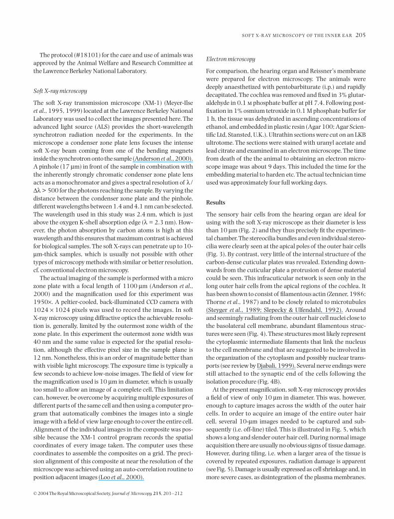
., 1989; Slepecky & Ulfendahl, 1992). Aroundand seemingly radiating from the outer hair cell nuclei close tothe basolateral cell membrane, abundant filamentous struc-tures were seen (Fig. 4). These structures most likely representthe cytoplasmic intermediate filaments that link the nucleusto the cell membrane and that are suggested to be involved inthe organization of the cytoplasm and possibly nuclear trans-ports (see review by Djabali, 1999). Several nerve endings werestill attached to the synaptic end of the cells following theisolation procedure (Fig. 4B).
At the present magnification, soft X-ray microscopy providesa field of view of only 10
µ
m in diameter. This was, however,enough to capture images across the width of the outer haircells. In order to acquire an image of the entire outer haircell, several 10-
µ
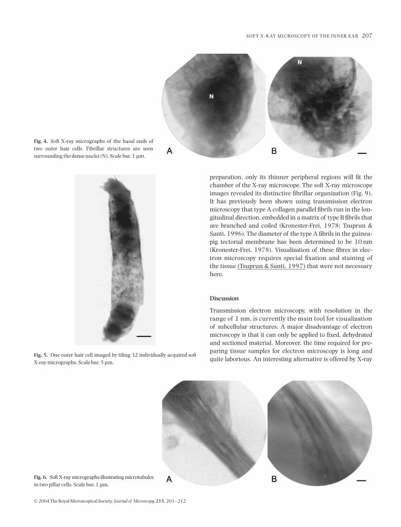
m images needed to be captured and sub-sequently (i.e. off-line) tiled. This is illustrated in Fig. 5, whichshows a long and slender outer hair cell. During normal imageacquisition there are usually no obvious signs of tissue damage.However, during tiling, i.e. when a larger area of the tissue iscovered by repeated exposures, radiation damage is apparent(see Fig. 5). Damage is usually expressed as cell shrinkage and, inmore severe cases, as disintegration of the plasma membranes.

206
G . A . J O H A N S S O N
E T A L .
© 2004 The Royal Microscopical Society,
Journal of Microscopy
,
215
, 203–212
To limit potential cell damage in the sample during the focusingprocedure, this was performed with a higher binning of theCCD camera and using very short exposure times.
The preparations contained not only outer hair cells butalso a small number of supporting cells, mainly pillar cells(cf. Fig. 1B). These cells form the tunnel of Corti and are
thought to provide mechanical stability to the hearing organ.The pillar cells thus have a distinct cytoskeleton containingboth large bundles of cross-linked microtubules and plentifulactin filaments (e.g. Kikuchi
et al
., 1991; Slepecky & Ulfendahl,1992). Compared with the cell membranes, filamentousstructures proved to be more resistant and showed no detectablesigns of damage. In the pillar cells, filaments and microtubuleswere clearly visualized (Fig. 6).
As with the outer hair cells, the thin Reissner’s membrane iseasily fitted into the chamber. This membrane, consisting oftwo cell layers but only around 5
µ
m thick, separates twofluid-filled compartments in the inner ear, scala vestibuli andscala media (see Slepecky, 1996, for an overview and refer-ences). The soft X-ray microscope images clearly revealed thestructure of the Reissner’s membrane (Fig. 7). The membraneconsists of two layers. The two cells with dark nuclei (topleft and bottom middle) are mesothelial cells that face scalavestibuli whereas the remaining cells with lighter nuclei areepithelial cells facing scala media and the hearing organ (cf.Fig. 1). Cell boundaries and intercellular junctions can bereadily identified. The desmosomes (thickening at the middleof an intercellular bridge) seen in the image are between 0.1and 0.2
µ
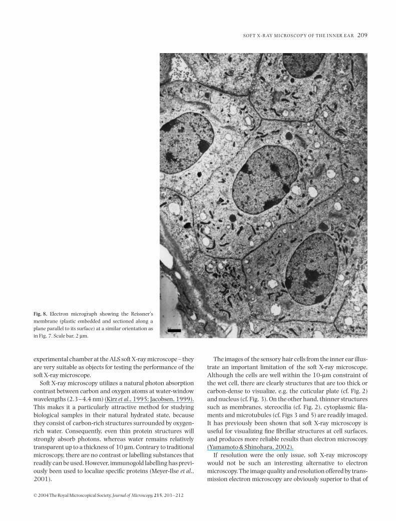
m wide. Inside the cells, nuclei and some mitochon-dria are clearly defined. There is a fibrous matrix between thecell wall and the nucleus. The matrix may be visible due tothe greater depth of focus of the X-ray microscope. For com-parison, a transmission electron micrograph of the Reissner’smembrane is shown in Fig. 8. This image is similar to thatobtained by the X-ray microscope. Mitochondria are betterdefined and can be seen in greater number. Cell boundariesare sharper and show more detail. However, it is evident thateven though the membrane is visualized intact (i.e. not sec-tioned) and no staining was used, the X-ray microscope imageprovides structural information comparable with what is seenwith electron microscopy, at least at a low magnification.
Compared with the other structures shown here, the tector-ial membrane is a more difficult structure to visualize. Themembrane, an acellular structure covering the surface of theorgan of Corti (see Slepecky, 1996), is quite thick in its naturalenvironment. Although it shrinks significantly during tissue
Fig. 2. Electron micrograph showing an outer hair cell from the apicalregions of the hearing organ. N, nucleus. Scale bar, 10 µm.
Fig. 3. Soft X-ray micrographs showing the apicalpoles of two outer hair cells. Individual stereociliacan be seen projecting from the dense cuticularplates. Scale bar, 1 µm.
S O F T X - R AY M I C RO S C O P Y O F T H E I N N E R E A R
207
© 2004 The Royal Microscopical Society,
Journal of Microscopy
,
215
, 203–212
preparation, only its thinner peripheral regions will fit thechamber of the X-ray microscope. The soft X-ray microscopeimages revealed its distinctive fibrillar organization (Fig. 9).It has previously been shown using transmission electronmicroscopy that type A collagen parallel fibrils run in the lon-gitudinal direction, embedded in a matrix of type B fibrils thatare branched and coiled (Kronester-Frei, 1978; Tsuprun &Santi, 1996). The diameter of the type A fibrils in the guinea-pig tectorial membrane has been determined to be 10 nm(Kronester-Frei, 1978). Visualization of these fibres in elec-tron microscopy requires special fixation and staining ofthe tissue (Tsuprun & Santi, 1997) that were not necessaryhere.
Discussion
Transmission electron microscopy, with resolution in therange of 1 nm, is currently the main tool for visualizationof subcellular structures. A major disadvantage of electronmicroscopy is that it can only be applied to fixed, dehydratedand sectioned material. Moreover, the time required for pre-paring tissue samples for electron microscopy is long andquite laborious. An interesting alternative is offered by X-ray
Fig. 4. Soft X-ray micrographs of the basal ends oftwo outer hair cells. Fibrillar structures are seensurrounding the dense nuclei (N). Scale bar, 1 µm.
Fig. 6. Soft X-ray micrographs illustrating microtubulesin two pillar cells. Scale bar, 1 µm.
Fig. 5. One outer hair cell imaged by tiling 32 individually acquired softX-ray micrographs. Scale bar, 5 µm.

208
G . A . J O H A N S S O N
E T A L .
© 2004 The Royal Microscopical Society,
Journal of Microscopy
,
215
, 203–212
microscopy. This is a relatively new and potentially powerfultool for observing microstructures in biological tissues (Meyer-Ilse
et al
., 1995; Jacobsen, 1999; Yamamoto & Shinohara, 2002).At present, the resolution of the soft X-ray microscope fallsbetween that of a light microscope and an electron microscope.In this report we explore for the first time the use of soft X-raymicroscopy for imaging sensory and supporting cells from the
hearing organ as well as the adjacent Reissner’s membraneand tectorial membrane. The hair cells contain complexstructures, e.g. filaments and microtubules that are vital forthe function of the cell, and these carbon-dense structuresshould be readily visualized using soft X-ray microscopy.As the diameter of the sensory hair cells is only about 10
µ
m –the maximal height of a structure that will fit the present
Fig. 7. Soft X-ray micrograph (tiled image) of the Reissner’s membrane from the guinea-pig cochlea. About 15 cells can be seen in the photograph. Thearrowheads indicate desmosomes. Scale bar, 1 µm.
S O F T X - R AY M I C RO S C O P Y O F T H E I N N E R E A R
209
© 2004 The Royal Microscopical Society,
Journal of Microscopy
,
215
, 203–212
experimental chamber at the ALS soft X-ray microscope – theyare very suitable as objects for testing the performance of thesoft X-ray microscope.
Soft X-ray microscopy utilizes a natural photon absorptioncontrast between carbon and oxygen atoms at water-windowwavelengths (2.3–4.4 nm) (Kirz
et al
., 1995; Jacobsen, 1999).This makes it a particularly attractive method for studyingbiological samples in their natural hydrated state, becausethey consist of carbon-rich structures surrounded by oxygen-rich water. Consequently, even thin protein structures willstrongly absorb photons, whereas water remains relativelytransparent up to a thickness of 10
µ
m. Contrary to traditionalmicroscopy, there are no contrast or labelling substances thatreadily can be used. However, immunogold labelling has previ-ously been used to localize specific proteins (Meyer-Ilse
et al
.,2001).
The images of the sensory hair cells from the inner ear illus-trate an important limitation of the soft X-ray microscope.Although the cells are well within the 10-
µ
m constraint ofthe wet cell, there are clearly structures that are too thick orcarbon-dense to visualize, e.g. the cuticular plate (cf. Fig. 2)and nucleus (cf. Fig. 3). On the other hand, thinner structuressuch as membranes, stereocilia (cf. Fig. 2), cytoplasmic fila-ments and microtubules (cf. Figs 3 and 5) are readily imaged.It has previously been shown that soft X-ray microscopy isuseful for visualizing fine fibrillar structures at cell surfaces,and produces more reliable results than electron microscopy(Yamamoto & Shinohara, 2002).
If resolution were the only issue, soft X-ray microscopywould not be such an interesting alternative to electronmicroscopy. The image quality and resolution offered by trans-mission electron microscopy are obviously superior to that of
Fig. 8. Electron micrograph showing the Reissner’smembrane (plastic embedded and sectioned along aplane parallel to its surface) at a similar orientation asin Fig. 7. Scale bar, 2 µm.

210
G . A . J O H A N S S O N
E T A L .
© 2004 The Royal Microscopical Society,
Journal of Microscopy
,
215
, 203–212
the X-ray microscope. However, the fundamental advantageof the X-ray microscope is that it allows visualization of cellsimmersed in a buffer, i.e. non-dehydrated and non-embeddedtissue. This is an important step to minimize artefacts, e.g.tissue shrinkage, prior to visualization. By imaging thesestructures with the cell in its natural hydrated state and withhigher resolution than is possible using visible light micro-scopy, it should be possible to gain more knowledge about the
structures involved in cellular function. Moreover, eliminat-ing many of the time-consuming steps, such as dehydration,embedding and sectioning, associated with electron micro-scopy would drastically increase the experimental throughput.The time-period from dissecting the tissue until viewing theprint easily exceeds 1 week (9 days in the present study). Bycontrast, the time required for placing the tissue in the testchamber of the X-ray microscope and obtaining an image was
Fig. 9. Soft X-ray micrograph (tiled image) illustrating the tectorial membrane from the guinea-pig cochlea clearly showing the transverse collagen fibrebundles and fibres. Scale bar, 1 µm.
S O F T X - R AY M I C RO S C O P Y O F T H E I N N E R E A R
211
© 2004 The Royal Microscopical Society,
Journal of Microscopy
,
215
, 203–212
approximately 15 min. The turnaround time was reduced by afactor of nearly 300. It is obvious that such a large reductionin the time needed to examine a tissue specimen has import-ant consequences for this application.
Image quality and resolution are limited by the fact that thehigh energy of the X-rays damages protein structures andcauses the tissue to shrink (Schneider, 1998). However, it shouldalso be kept in mind that conventional electron microscopyis associated with significant tissue changes due to radiationdamage and the vacuum requirement. This shrinkage can bereduced by converting the tissue to a more radiation-resistantform, i.e. by fixing the tissue.
In the present study, it was necessary to use chemicalfixation (glutaraldehyde) to stabilize the tissue prior to X-rayexposure. An attractive alternative would be to use cryofixa-tion because it would inhibit morphological changes andstructural deterioration resulting from radiation damage. Theprocedure would also prevent movement of cells that are notfixed to the silicon nitride membrane surface. The soft X-raygroup in Göttingen, Germany, started work on cryo transmis-sion X-ray microscopy and they have shown that vitrifiedsamples are stable up to a dosage of 10
10
Gy (Schneider,1998), i.e. the structural radiation damage is less than can bedetected at the resolution used. Normally, a single exposuregives a dose of approximately 10
7
Gy to the sample. Cryofixa-tion has previously been used with outstanding results at theALS soft X-ray microscope (Meyer-Ilse
et al
., 2001), but thefeature was unfortunately not available during this study.
Soft X-ray microscopy is very much at its beginning and fur-ther technical improvements are required before it can be usedmore generally to complement electron microscopy in experi-mental research. Unfortunately, one of the limitations to awider use of soft X-ray microscopy is the necessity to use astrong beam of X-rays, which means that experiments must beperformed at synchrotron radiation facilities such as the ALS.To facilitate experimental investigations in environmentsmore suited for biomedical experiments, it would be useful todevelop instruments using table-top soft X-ray sources thatcan still provide high-resolution imaging ( Johansson
et al
.,2002). For example, it was previously shown that outer haircell shortening was correlated with a more undulated cellmembrane (Ulfendahl & Slepecky, 1988). This observation,based on a very laborious electron microscope analysis of indi-vidual fixed, dehydrated, embedded and sectioned cells, couldhave been much more straightforward using high-resolutionsoft X-ray microscopy. For future studies on the structural andsubcellular changes related to outer hair cell motility, soft X-ray microscopy could be of great value.
Acknowledgements
We wish to thank Angelic Pearson, Erik Anderson, Bill Bates,Weilun Chao, Kathleen Fuller, Randy Deguzman and GerdSchneider for valuable help and support during the experi-
ments. G.A.J. also wishes to thank Hans Hertz and David Att-wood for providing him with the opportunity to visit theCenter for X-ray Optics. This work was supported by grantsfrom the Swedish Research Council, the Foundation TystaSkolan, the Emil Capita Foundation, and the Swedish Founda-tion for International Cooperation in Research and High Edu-cation (STINT).
References
Anderson, E.H., Olynick, D.L., Harteneck, B., Veklerov, E., Denbeaux, G.,Chao, W., Lucero, A., Johnson, L. & Attwood, D. Jr (2000) Nanofabrica-tion and diffractive optics for high-resolution X-ray applications.
J. Vac.Sci. Technol
.
18
, 2970–2975.Attwood, D. (1999)
Soft X-rays and Extreme Ultraviolet Radiation: Principlesand Applications
. Cambridge University Press, Cambridge.Denbeaux, G., Anderson, E., Chao, W., Eimüller, T., Johnson, L., Köhler, M.,
Larabell, C., Legros, M., Fischer, P., Pearson, A., Schütz, G., Yager, D. &Attwood, D. (2001) Soft X-ray microscopy to 25 nm with applicationsto biology and magnetic materials.
Nucl. Instr. Meth. A
,
467
–
468
,841–844.
Djabali, K. (1999) Cytoskeletal proteins connecting intermediatefilaments to cytoplasmic and nuclear periphery.
Histol. Histopathol
,
14
,510–509.
Jacobsen, C. (1999) Soft X-ray microscopy.
Trends Cell Biol
.
9
, 44–47.Johansson, G.A., Holmberg, A., Hertz, H.M. & Berglund, M. (2002)
Design and performance of a laser-plasma based compact soft X-raymicroscope.
Rev. Sci. Instrum
.
73
, 1193–1197.Kikuchi, T., Takasaka, T., Tonosaki, A., Katori, Y. & Shinkawa, H. (1991)
Microtubules of guinea pig epithelial cells.
Acta Otolaryngol. (Stock-holm)
,
111
, 286–290.Kirz, J., Jacobsen, C. & Howells, M. (1995) Soft X-ray microscopes and
their biological applications.
Q. Rev. Biophys
.
28
, 33–130.Kronester-Frei, A. (1978) Ultrastructure of the different zones of the
tectorial membrane.
Cell Tiss. Res
.
193
, 11–23.Loo, B.W., Jr, Meyer-Ilse, W. & Rothman, S.S. (2000) Automatic image
acquisition, calibration and montage assembly for biological X-raymicroscopy.
J. Microsc
.
197
, 185–201.Meyer-Ilse, W., Denbeaux, G., Johnson, L.E., Bates, W., Lucero, A. &
Anderson, E.H. (1999) The high resolution X-ray microscope, XM-1.
X-ray microscopy VI. XRM99
(ed. by W. Meyer-Ilse, T. Warwick andD. Attwood), pp. 129–134. American Institute of Physics, Melville, NY.
Meyer-Ilse, W., Hamamoto, D., Nair, A., Lelièvre, S.A., Denbeaux, G.,Johnson, L., Pearson, A.L., Yager, D., Legros, M.A. & Larabell, C.A.(2001) High resolution protein localization using soft X-ray micro-scopy.
J. Microsc
.
201
, 395–403.Meyer-Ilse, W., Medecki, H., Jochum, L., Anderson, E., Attwood, D.,
Magowan, C., Balhorn, R., Moronne, M., Rudolph, D. & Schmahl, G.(1995) New high-resolution zone-plate microscope at beamline 6.1 ofthe ALS.
Synchrotron Radiation News
,
8
, 29–33.Schmahl, G., Rudolph, D., Niemann, B., Guttmann, P., Thieme, J.,
Schneider, G., David, C., Diehl, M. & Wilhein, T. (1993) X-ray microscopystudies.
Optik
,
93
, 95–102.Schneider, G. (1998) Cryo X-ray microscopy with high spatial resolution
in amplitude and phase contrast.
Ultramicroscopy
,
75
, 85–104.Slepecky, N.B. (1996) Structure of the mammalian cochlea.
The Cochlea
(ed. by P. Dallos, A. N. Popper and R. R. Fay), pp. 44–129. Springer-Verlag, New York.
212
G . A . J O H A N S S O N
E T A L .
© 2004 The Royal Microscopical Society,
Journal of Microscopy
,
215
, 203–212
Slepecky, N. & Ulfendahl, M. (1992) Actin-binding and microtubule-associated proteins in the organ of Corti.
Hearing Res
.
57
, 201–215.Steyger, P.S., Furness, D.N., Hackney, C.M. & Richardson, G.P. (1989)
Tubulin and microtubules in cochlear hair cells: comparative immuno-cytochemistry and ultrastructure.
Hearing Res
.
42
, 1–16.Thorne, P.R., Carlisle, L., Zajic, G., Schacht, J. & Altschuler, R.A. (1987)
Differences in the distribution of F-actin in outer hair cells along theorgan of Corti.
Hearing Res
.
30
, 253–265.Tsuprun, V. & Santi, P. (1996) Crystalline arrays of proteoglycan and
collagen in the tectorial membrane.
Matrix Biology
,
15
, 31–38.
Tsuprun, V. & Santi, P. (1997) Ultrastructural organization of proteogly-cans and fibrillar matrix of the tectorial membrane.
Hearing Res
.
110
,107–118.
Ulfendahl, M. & Slepecky, N. (1988) Ultrastructural correlates ofinner ear sensory cell shortening.
J. Submicrosc. Cytol. Pathol
.
20
,47–51.
Yamamoto, Y. & Shinohara, K. (2002) Application of X-ray microscopy inanalysis of living hydrated cells. Anat. Rec. 269, 217–223.
Zenner, H. (1986) Motile responses in outer hair cells. Hearing Res. 22,83–90.


![Clad Inner Surface Temperature l°C]](https://static.fdokumen.com/doc/165x107/633831c324ea072f160c74b1/clad-inner-surface-temperature-lc.jpg)