Exogenous and endogenous DNA modifications as monitored by 32P-postlabeling: Relationships to cancer...
17
Experimental Gerontology, Vol.27, pp. 533-549, 1992 0531-5565/92$5.00 + .00 Printed in the USA. All rightsreserved. Copyright © 1992 Pergamon PressLtd. EXOGENOUS AND ENDOGENOUS DNA MODIFICATIONS AS MONITORED BY 32p-POSTLABELING: RELATIONSHIPS TO CANCER AND AGING KURT RANDERATH, DONGHUI D, RAGHU NATH and ERIKA RANDERATH Division of Toxicology,Department of Pharmacology,Baylor Collegeof Medicine, Houston, Texas 77030 Abstract - - 32P-postlabeling analysis, a highly sensitive method for the detection and measurement of covalent carcinogen-DNA adducts and other DNA modifications, does not require radioactive test substances and, therefore, can be applied to DNA of mammals, including humans exposed to low doses of environmental or occupational genotoxicants. The basic procedure entails the enzymatic incorporation of 32p-label into hydrolysis products of DNA, followed by chromatographic mapping and autoradio- graphy of the 32P-labeled digestion products and quantitative scintillation spectrometry. Microgram amounts of DNA are analyzed: Thus the assay is suited for limited amounts of cells or tissues. Various versions of the assay afford different sensitivities of adduct detection. A single aromatic or bulky/hydrophobic adduct in 108-101°nucleotides can be detected and measured (corresponding to 0.3-30 amol adduct/#g DNA or 0.1-10 nmoi adduct/mol DNA-P). In animal models, the assay has been successfully applied to a variety of mutagenic (genotoxic) as well as nonmutagenic carcinogens. In humans, DNA specimens from cigarette smokers, iron foundry workers, and coke oven workers whose total aromatic adduct levels ranged from 1 adduct in 106-108 DNA nucleotides have been examined by ~2P-postlabeling. The assay also detects DNA modifications-- Indigenous (I)-compounds--that increase with age in untreated animals. I-compound profiles and levels are highly species-, strain-, sex-, and tissue-specific, and also depend on diet composition. Caloric restriction, a highly efficient method for improving resist- ance to carcinogenesis and extending life span, increased rather than decreased I-com- pound levels in various tissues of male rats. Nonmutagenic hepatocarcinogens reduced levels of I-compounds in the target organ. Because of the specificity of this effect, reduc- tion of I-compound levels appears to represent a novel biomarker for the action of non- mutagenic carcinogens. DNA from various hepatomas was found largely devoid of I- compounds. The results support a possible antineoplastic and antiaging role of these DNA modifications. Key Words: DNA adducts, I-compounds, aging, cancer, 32p-postlabeling, carcinogens, DNA damage Correspondence to: K. Randerath. 533
-
Upload
independent -
Category
Documents
-
view
3 -
download
0
Transcript of Exogenous and endogenous DNA modifications as monitored by 32P-postlabeling: Relationships to cancer...

Experimental Gerontology, Vol. 27, pp. 533-549, 1992 0531-5565/92 $5.00 + .00 Printed in the USA. All rights reserved. Copyright © 1992 Pergamon Press Ltd.
EXOGENOUS AND ENDOGENOUS DNA MODIFICATIONS AS MONITORED BY 32p-POSTLABELING: RELATIONSHIPS TO
CANCER AND AGING
KURT RANDERATH, DONGHUI D, RAGHU NATH and ERIKA RANDERATH
Division of Toxicology, Department of Pharmacology, Baylor College of Medicine, Houston, Texas 77030
Abstract - - 32P-postlabeling analysis, a highly sensitive method for the detection and measurement of covalent carcinogen-DNA adducts and other DNA modifications, does not require radioactive test substances and, therefore, can be applied to DNA of mammals, including humans exposed to low doses of environmental or occupational genotoxicants. The basic procedure entails the enzymatic incorporation of 32p-label into hydrolysis products of DNA, followed by chromatographic mapping and autoradio- graphy of the 32P-labeled digestion products and quantitative scintillation spectrometry. Microgram amounts of DNA are analyzed: Thus the assay is suited for limited amounts of cells or tissues. Various versions of the assay afford different sensitivities of adduct detection. A single aromatic or bulky/hydrophobic adduct in 108-101° nucleotides can be detected and measured (corresponding to 0.3-30 amol adduct/#g DNA or 0.1-10 nmoi adduct/mol DNA-P). In animal models, the assay has been successfully applied to a variety of mutagenic (genotoxic) as well as nonmutagenic carcinogens. In humans, DNA specimens from cigarette smokers, iron foundry workers, and coke oven workers whose total aromatic adduct levels ranged from 1 adduct in 106-108 DNA nucleotides have been examined by ~2P-postlabeling. The assay also detects DNA modifications-- Indigenous (I)-compounds--that increase with age in untreated animals. I-compound profiles and levels are highly species-, strain-, sex-, and tissue-specific, and also depend on diet composition. Caloric restriction, a highly efficient method for improving resist- ance to carcinogenesis and extending life span, increased rather than decreased I-com- pound levels in various tissues of male rats. Nonmutagenic hepatocarcinogens reduced levels of I-compounds in the target organ. Because of the specificity of this effect, reduc- tion of I-compound levels appears to represent a novel biomarker for the action of non- mutagenic carcinogens. DNA from various hepatomas was found largely devoid of I- compounds. The results support a possible antineoplastic and antiaging role of these DNA modifications.
Key Words: DNA adducts, I-compounds, aging, cancer, 32p-postlabeling, carcinogens, DNA damage
Correspondence to: K. Randerath.
533
534 K. R A N D E R A T H et aL
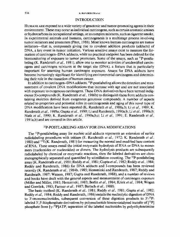
INTRODUCTION
HUMANS ARE exposed to a wide variety ofgenotoxic and tumor-promoting agents in their environment. These may occur as individual carcinogens, such as certain aromatic amines or hydrocarbons in occupational settings, or as complex mixtures, such as cigarette smoke. In experimental animals and humans, carcinogenesis is a multistage process involving tumor initiators and promoters (Pitot, 1986). Most known human carcinogens are tumor initiators--that is, compounds giving rise to covalent addition products (adducts) of DNA, a key event in tumor initiation. Various sensitive assays exist to measure the for- mation ofcarcinogen-DNA adducts, while no practical endpoint has been defined for the biomonitoring of exposure to tumor promoters. Some of the assays, such as 32p-postla- beling (K. Randerath et al., 1981 ), allow one to monitor activities of unidentified carcin- ogens and carcinogen mixtures at the target site (DNA), a feature that is particularly important for assessing human carcinogen exposure. Assays for DNA adducts have become increasingly significant for identifying environmental carcinogens and determin- ing their role in the causation of human cancer.
In addition to carcinogen-DNA adducts, 32p-postlabeling allows the detection and mea- surement of covalent DNA modifications that increase with age and are not associated with exposure to exogenous carcinogens. These DNA derivatives have been termed indig- enous (I)-compounds (K. Randerath et al., 1986b) to distinguish them from adducts con- taining moieties derived from exogenous genotoxic compounds. A number of aspects related to properties and potential roles in carcinogenesis and aging of this novel type of DNA modification have been reported (K. Randerath et al., 1988a,b; Li et al., 1989; K. Randerath et al., 1989a; Gupta et al., 1990; Li and Randerath, 1990a,b; Li et al., 1990a,b; Nath et al., 1990; K. Randerath et al., 1990a,b,c; Li et al., 1991; E. Randerath et al., 199 la,b) and are covered in this article.
32p-POSTLABELING ASSAY FOR DNA MODIFICATIONS
The 32p-postlabeling assay for nucleic acid adducts represents an extension of earlier radiolabeling procedures with tritium (E. Randerath et al., 1972; K. Randerath et al., 1980) and J25I (K. Randerath, 198 l) for measuring the normal and modified base content of RNA. These assays entail the initial enzymatic hydrolysis of RNA or DNA to mono- mers (nucleosides or nucleotides) or dimers. The hydrolysis products are subsequently radiolabeled by chemical or enzymatic reactions, then the labeled derivatives are chro- matographically separated and quantified by scintillation counting. The 32P-postlabeling assay (K. Randerath et al., 1981; Reddy et aL, 1981; Gupta et al., 1982; Reddy et al., 1984; Reddy and Randerath, 1986) for DNA adducts and I-compounds has been reviewed recently (K. Randerath et al., 1984b, 1985; Hemminki and Randerath, 1987; Reddy and Randerath, 1987; Watson, 1987; Gupta and Randerath, 1988), and a number of reviews and books have dealt with the general aspects and measurement of carcinogen exposure (Miller and Miller, 1981; Hemminki, 1983; Berlin et al., 1984; Kriek et al., 1984; Wogan and Gorelick, 1985; Farmer et al., 1987; Bartsch et al., 1988).
The basic method (K. Randerath et aL, 1981; Reddy et aL, 1981; Gupta et al., 1982; Reddy et al., 1984; Reddy and Randerath, 1986) entails the nucleolytic digestion of DNA to 3'-mononucleotides, subsequent conversion of these digestion products to 5'-32P - labeled 3',5'-bisphosphate derivatives by polynucleotide kinase-catalyzed transfer of [32p] phosphate from [7-32P]ATP, separation of the labeled nucleotides by polyethyleneimine

EXOGENOUS AND ENDOGENOUS DNA ALTERATIONS 535
(PEI)-cellulose thin-layer chromatography (TLC), autoradiography to detect adducts or I- compounds and normal nucleotides, and scintillation spectrometry for quantitation.
Five versions of the assay have been described (Gupta and Randerath, 1988; K. Ran- derath et al., 1989b), two of which involve 32p-labeling of adducts in the presence of nor- mal nucleotides, while in the other three procedures adducts are enriched by removal of normal nucleotides prior to 32p-labeling either by enzymatic or physical techniques. The enzymatic enrichment procedures, encompassing the nuclease P 1-enhanced bisphosphate (Reddy and Randerath, 1986) and monophosphate (K. Randerath et al., 1989b) assays, which achieve a sensitivity of adduct detection of 1 adduct in 10 9-10 ~° DNA nucleotides and require 5-10 ug DNA per individual assay, have been employed in most of the work reviewed here. In the bisphosphate procedure, a micrococcal nuclease/spleen phospho- diesterase digest of the DNA consisting of normal and modified deoxyribonucleoside 3'-monophosphates (Np + Xp) is treated with nuclease P I to convert the normal nucle- otides, but not bulky adducts, to deoxyribonucleosides, which are not substrates for the subsequent labeling reaction. Adducts are then specifically labeled to form 5'-32p-labeled adducted deoxyribonucleoside 3',5'-bisphosphates (*pXp, the asterisk denoting the 32p_ label). In the monophosphate procedure, DNA hydrolysis with nuclease P 1 and acid phos- phatase yields modified dinucleotides (XpN) and normal deoxyribonucleosides. The dinucleotides are labeled to give *pXpN, which may be assayed as such or, preferably, converted to deoxyribonucleoside 5'-monophosphates (*pX) by venom phosphodiester- ase hydrolysis, followed by TLC mapping.
Optimal adduct or I-compound resolution depends on chromatographic conditions. TLC on PEI-cellulose has been used most extensively for this purpose. Nonpolar adducts containing polycyclic aromatic moieties are best separated with concentrated electrolyte solutions containing 7-8.5 M urea (Gupta et al., 1982; Reddy et al., 1984; Gupta and Ran- derath, 1988), while more polar adducts are resolved with solvents containing lower con- centrations of electrolytes and urea (K. Randerath et al., 1988a). Several mapping systems may be required for DNA samples containing derivatives of widely divergent polarities (K. Randerath et al., 1988a). High performance liquid chromatography has been used to separate small alkylated (Wilson et al., 1988; Watson and Crane, 1989) as well as poly- cyclic aromatic (Dunn and San, 1988) adducts in combination with 32p-postlabeling.
Adduct levels are expressed in terms of relative adduct labeling (RAL) values, which represent the ratio of count rates of adducted nucleotides over count rates of total (adducted plus normal) nucleotides (Gupta et al., 1982; Reddy and Randerath, 1986). Count rates of adducted nucleotides are determined by Cerenkov assay of radioactive spots cut or scraped from the chromatogram. The RAL × 10 n value is equal to the number ofadducts in 10" DNA nucleotides, provided that adducts are labeled and recovered quan- titatively; otherwise, a correction factor is applied (Reddy and Randerath, 1986).
EXOGENEOUS DNA MODIFICATIONS
Mutagenic /genotoxic carcinogens
Two factors may affect the potency of a carcinogen, the extent of initial DNA binding and adduct persistence, although the latter may not always be associated with tumorige- nicity (Wogan and Gorelick, 1985). Carcinogens whose DNA binding has been examined by 32p-postlabeling have been listed in a review (Gupta and Randerath, 1988). Strong asso- ciations have been observed by 32p-postlabeling between the extent of initial binding to

536 K. RANDERATH et aL
target organ DNA and hepatocarcinogenicity of dibenzo[c,g]carbazole (Schurdak and Randerath, 1985) and safrole (Phillipps et al., 1984; K. Randerath et al., 1984a). A num- ber of highly persistent adducts was detected in labeled digests of DNA derived from mouse skin after topical treatment with polycyclic aromatic hydrocarbons, such as benzo[a]pyrene, 7,12-dimethylbenz[a]anthracene, and 3-methylcholanthrene (E. Ran- derath et al., 1983, 1985). While part of the adducts disappeared rapidly from skin or epi- dermal DNA after treatment, adducts remaining after 2 weeks underwent little change. Some polycyclic aromatic hydrocarbon-DNA adducts persisted in mouse epidermis and dermis for at least 1 year after brief dermal carcinogen exposure (E. Randerath et al., 1985). Such results imply that persistent adducts occupy genomic sites in quiescent cells and are inaccessible to DNA repair processes.
Complex carcinogenic mix tures
Covalent DNA damage induced by complex mixtures of environmental (Everson et al., 1986; E. Randerath et al., 1986; K. Randerath et al., 1986a; Everson et al., 1988; E. Ran- derath et al., 1989) or occupational (Savela et al., 1989; Hemminki et al., 1990a,b) geno- toxicants can be assayed by 32p-postlabeling. Experimental animal models for such expo- sures may be used for the identification of unknown adducts in humans (E. Randerath et al., 1988).
Cigarette smoke-induced DNA adducts were detected in placentas of smokers (Everson et al., 1986, 1988); in lung, bronchus, and larynx of smokers with cancer of these organs (E. Randerath et al., 1986; K. Randerath et al., 1986a; E. Randerath et al., 1989); and in autopsy samples of additional organs (heart, kidney, ascending aorta, bladder, and esoph- agus) from heavy smokers (E. Randerath et al., 1989). DNA adduct maps from smokers assayed by the nuclease P 1/bisphosphate or monophosphate procedure showed two exten- sive diagonal radioactive zones of cigarette smoke-related 32p-labeled material, consistent with the presence of numerous, only partially resolved aromatic DNA adducts of varying polarities. Smoking-associated adducts in surgical specimens from lung, bronchus, and larynx were found to occur in a dose- and exposure time-dependent manner, and the extent of DNA damage in lung and larynx paralleled cancer risk and mortality (E. Ran- derath et al., 1989), which are known to increase with the number of cigarettes smoked and duration of smoking (U.S. Department of Health and Human Services, 1982; Inter- national Agency for Research on Cancer [IARC], 1985). The highest levels of aromatic adducts observed were about 1.2 adducts in 107 DNA nucleotides. In agreement with the slow decline of cancer risk and mortality after cessation of smoking (U.S. Department of Health and Human Services, 1982; IARC, 1985), long-time persistence of smoking- induced adducts was also observed. Thus, the 32p-postlabeled DNA adducts provided a valid dosimeter for human carcinogen exposure. Mice treated dermally with a cigarette smoke condensate exhibited similar adduct patterns and tissue distribution as did smok- ers, with lung and heart displaying the highest levels (E. Randerath et al., 1988, 1989).
Effects on DNA of human exposure to polycyclic aromatic hydrocarbons were studied in two occupational settings, a Finnish foundry (Savela et al., 1989; Hemminki et al., 1990b) and a Polish cokery (Hemminki et al., 1990a). White blood cell (WBC) DNA of workers and appropriate controls was assayed by the nuclease Pl/bisphosphate version of 3Zp-postlabeling assay. When foundry workers were categorized into high, medium, or low exposure groups, based on ambient benzo[a]pyrene (BP) levels, highly significant corre- lations were observed between exposure and WBC DNA adduct levels (Savela et al., 1989;

EXOGENOUS AND ENDOGENOUS DNA ALTERATIONS 537
Hemminki et al., 1990b). Complex patterns of unidentified adducts, different from those induced by cigarette smoking, were observed. Maximal DNA damage was about 1 adduct in 106 DNA nucleotides. The results paralleled the elevated cancer risk of foundry workers. WBC DNA adduct levels of cokery workers were compared with those of local and rural controls (Hemminki et al., 1990a). Local controls exhibitied adduct patterns and levels ( 1 adduct in about l07 DNA nucleotides) similar to the workers, while rural controls had much lower levels. These studies show that 32p-postlabeling constitutes an effective means for the assessment of DNA damage caused by occupational genotoxic exposures.
ENDOGENOUS DNA MODIFICATIONS
D N A modif icat ions induced by estrogens
Natural or synthetic estrogens induce a high incidence of renal carcinoma in male Syr- ian hamsters after 6-8 months of treatment (Kirkman, 1959, pp. 1-57). In this model, continuous estrogen exposure was accompanied by the gradual appearance of several DNA adducts that were detectable by 32p-postlabeling assay and increased with exposure time (Liehr et al., 1986). Rechromatography of these adducts in various systems showed them to be identical irrespective of the type of estrogen (i.e., stilbenes or steroids) admin- istered (Liehr et al., 1986). Thus, the adduct moieties were not structrually derived form the estrogens but appeared to originate from an unknown endogenous source or sources. It was apparent that estrogen treatment induced a DNA-reactive electrophilic metabolite or metabolites that gave rise to this novel type of DNA modification, and such metabo- lite(s) appeared to be activated by cytochrome P-450 rather than prostaglandin endoper- oxide synthase (Liehr et aL, 1987). Since these adducts were present only in the target organ, they appeared to be causally associated with estrogen-induced renal carcinogenesis. Whether DNA adducts of endogenous origin occur also in other systems of hormonal car- cinogenesis has not yet been determined.
I - compounds - -Age -dependen t D N A modif icat ions
Covalent adduct-like modifications, termed I-compounds, were detected by 32p-postla- beling in DNA from various tissues, particularly liver and kidney, of untreated animals (K. Randerath et al., 1986b). While these DNA modifications were absent or only barely detectable in tissues of newborn rodents, they accumulated with age (K. Randerath et al., 1986b, 1988b; Li et al., 1989; K. Randerath et al., 1989a; Gupta et al., 1990; Li and Ran- derath, 1990a; K. Randerath et al., 1990a; E. Randerath et al., 1991 a,b) reaching substan- tial levels (e.g., up to 1-2 modifications in 107 nucleotides in rat liver DNA at 7-10 months), and tended to plateau in older animals (E. Randerath et al., 1991 a). I-compound profiles and levels were found to be strongly and reproducibly influenced by a number of genetic factors, including species, strain, sex, and tissue (K. Randerath et al., 1986b, 1988b, 1989a; Li and Randerath, 1990b; K. Randerath et al., 1990a), as well as by envi- ronmental factors, such as diet composition (Li and Randerath, 1990a; K. Randerath et al., 1990a). Since exogenous carcinogens have been ruled out as a source of these DNA modifications (K. Randerath et al., 1986b; Li and Randerath, 1990a; K. Randerath et al., 1990a; Li et al., 1990b; E. Randerath et al., 1991 a), taken together, these findings support the original hypothesis (K. Randerath et al., 1986b) that I-compounds are formed by reac- tions of metabolically produced, as yet unidentified endogenous electrophiles with DNA. However, it is possible that more specific processes, such as the transfer of I-moieties to

538 K. RANDERATH et aL
DNA by DNA-binding proteins, are involved in the formation of some of these DNA modifications. While I-compounds represent structurally diverse DNA derivatives, as indicated by the complexity of their profiles and wide range of polarities, none of these compounds has been structurally characterized so far because of the large amounts of tis- sue required to provide sufficient DNA for this purpose.
In accord with their structural diversity, I-compounds could conceivably play different biological roles. Their adduct-like character (K. Randerath et al., 1986b, 1988a) and age- dependent increases suggest that they might contribute to spontaneous tumor formation and natural aging. On the other hand, several lines of experimental evidence reviewed later in this article indicate that adverse health effects are associated with decreased or low levels of I-compounds, as noted for (1) liver of experimental animals exposed to nonmutagenic hepatocarcinogens, (2) organ sites of spontaneous tumorigenesis and non-neoplastic renal diseases in susceptible mouse strains prior to development of pathology (Li and Rander- ath, 1990b), and (3) hepatomas of various origins. Increased I-compound levels have been found in calorically restricted male rats, paralleling the beneficial effects of caloric restric- tion (E. Randerath et al., 1991 a). A functional role of I-compounds is consistent, also, with results showing that rat liver mitochondrial DNA exhibits similar levels and profiles as does nuclear DNA (Gupta et al., 1990), suggesting that I-compound levels may be regu- lated by cellular control mechanisms for the benefit of the cell.
Effects o f nutrition on I-compounds
Diet has been found to greatly influence I-compound patterns and levels (Li and Ran- derath, 1990a). Nutritional effects on I-compounds have been investigated in three studies in which (1) purified and natural ingredient diets were compared (Li and Randerath, 1990a), (2) effects of various nutrients added to basic diet were tested (Li and Randerath, 1989; K. Randerath et al., 1990a), and (3) effects of the natural antioxidant vitamin E were investigated (Li et al., 1991). In these experiments, weanling female Sprague-Dawley rats were fed various diets ad libitum for 3 and 6 months. In Study 1 (Li and Randerath, 1990a), four groups of rats received either AIN-76A purified diet or one of three natural ingredient (chow) diets (Teklad LM485, Purina 5001, Wayne MRH22/5 Rodent Blox). In Study 2 (Li and Randerath, 1989; K. Randerath et al., 1990a; Li and Randerath, unpub- lished experiments), six groups of rats were given basic or modified AIN-76A purified diets containing high levels of one of the following nutrients: carbohydrate, protein, fat, vita- mins, and minerals. In Study 3 (Li et al., 1991), four groups of rats were fed Draper's diets containing 0, 100, 1000, or 10000 mg/kg alpha-tocopherol (vitamin E). Liver and kidney DNA were isolated and assayed by the nuclease P 1/bisphosphate version of the 3ZP-post- labeling assay.
Study 1: Diet type. The first set of experiments examined effects on I-compounds of nat- ural ingredient versus purified diet. When weanling female Sprague-Dawley rats received AIN-76A purified diet or natural ingredient diet for 6 months, age-dependent, tissue-spe- cific, and diet related I-compound profiles were detected. As shown in Fig. 1, rats fed nat- ural ingredient diet (Teklad) exhibited more complex profiles and much enhanced inten- sitites of liver I-compounds than rats fed purified diet (AIN) (Li and Randerath, 1990a). Similar results were obtained for kidney, but the overall spot profiles were different for liver and kidney (Li and Randerath, 1990a). The total I-compound levels in liver and kidney DNA from rats fed chow diet were 3-6 times higher than those in rats fed purified diet.

EXOGENOUS AND ENDOGENOUS DNA ALTERATIONS 539
U
C L!
I._
WEANLING A I N T E K L A D FIG. 1. Liver l-compound profiles of female Sprague-Dawley rats at the time of weaning and after 6 months of feeding of either AIN purified diet or Teklad chow diet, as determined by 32p_ postlabeling assay. For experimental conditions, see text. Film exposure was for 14 h at - 80"C with Kodak XAR-5 film and DuPont Lightning Plus intensifying screen. U = upper, C = cen- tral, L = lower cut maps; B = background spot that was present in all samples at similar inten- sities; prefix L = liver specific spot or cluster of spots. Note marked diet-related differences in spot patterns. (From Cancer Res. 50, 3991-3996, 1990, with permission).
These observations clearly indicated an association between dietary composi t ion and D N A modifications. Accumula t ing evidence suggests that these D N A derivatives were not derived f rom envi ronmenta l genotoxic carcinogens (Li and Randerath, 1990a; Li et al., in press-b). For example, when female Sprague-Dawley rats, Swiss (ICR) mice, and Syrian hamsters were raised on the same diet (Teklad) and under the same environmental con- ditions, species and tissue specific I - c o m p u n d profiles were observed in liver and kidney DNA, as contrasted with identical carcinogen D N A adduct patters detected after exposure to the carcinogens safrole, 7,12-dimethylbenz[a]anthracence, and dibenz[aj]acr idine (Li et aL, 1990b). In addition, rats displayed the highest I - compound but the lowest adduct levels in both liver and kidney a m o n g the three species. These findings demonstra ted fun- damenta l differences between I -compounds and ca rc inogen-DNA adducts. Thus, differ- ences in I - c o m p o u n d profiles between rats fed the two types of diets could well be due to
540 K. RANDERATH et al.
the complex nutrient sources and presence of large amounts of non-nutrient natural prod- ucts in chow diets (Campbell and Hayes, 1974). Nutrients and non-nutrient natural prod- ucts or their metabolites may react with DNA directly or affect DNA via alteration of metabolism by enzyme modulation (Newberne, 1975; Carr, 1982; Guengerich, 1984), resulting in the formation ofDNA-reactive electrophiles (Li and Randerath, 1990a).
In the second set of experiments, effects on I-compounds of chow diets from different suppliers were compared. Rats raised under the same environmental conditions but fed chow diets from different suppliers exhibited qualitative and quantitative differences in I- compounds (Li and Randerath, 1990a). Compared with female rats fed Teklad LM485 or Purina 5001 chow diets, three liver I-spots were absent and total I-compound levels were significantly lower in both liver and kidney DNA of rats fed Wayne MRH22/5 Rodent Blox. These differences were shown to be due to the absence of certain dietary compo- nen t s - tha t is, ground oats and alfalfa meal in the latter diet (Li and Randerath, unpub- lished data): Addition of ground oats to Wayne diet or AIN diet elicited formation of the three missing spots, and supplementation of Wayne diet with alfalfa meal significantly increased total I-compound levels. Thus, it was apparent that at least certain I-compounds are not derived from environmental genotoxic carcinogens, but from dietary components or their metabolites. In other words, diet may not only give rise to DNA adducts through environmental carcinogenic contaminants (e.g. aflatoxins, nitrosamines, polycyclic aro- matic hydrocarbons, food pyrolysis products), but many also elicit structural alteration of DNA through its content of nutrient or non-nutrient natural products. Although the bio- logical significance of I-compounds has not yet been precisely defined, these DNA deriv- atives could represent either normal DNA modifications playing functional roles, or pre- mutagenic DNA lesions contributing to carcinogenesis and aging.
Study 2." Nutrient levels. Varying macronutrient levels in purified diet were found to induce significant quantitative but not qualitative changes of I-compounds in rat liver and kidney DNA (Figs. 2 and 3) (Li and Randerath, 1989). Compared with animals fed basic AIN-76A diet (65% carbohydrate, 20% protein, and 5% fat, w/w), high carbohydrate diet (78%) produced a significant increase in the polar as well as total I-compound levels in liver at 3 months and in kidney at 3 and 6 months. High protein diet (50%) increased nonpolar hepatic I-compound levels at 6 months. High fat diet (20%) elicited significantly lower polar I-compound levels in liver at 6 months, and only a few significant differences were found between polyunsaturated (safflower oil) and saturated (lard) fat diet groups. Sup- plementation of AIN-76A diet with excess vitamins and minerals had little effect on pat- terns or levels of I-compounds in rat liver and kidney DNA. These results, also, support the hypothesis that I-compounds arise through normal metabolism of nutrients leading to production of DNA-reactive electrophiles. Induction of increased I-compound levels by high carbohydrate and reduction by high fat diet suggest that formation of these DNA derivatives may not be related to lipid peroxides, but rather to carbohydrate metabolism. The precise mechanisms of interactions between nutrients and DNA are not understood, but these observations provide further evidence for a close link between nutrition and I- compounds.
Study 3." Vitamin E. To further investigate the relationship between I-compounds and individual nutrients, female Sprague-Dawley rats were fed Draper's diets containing dif- ferent levels of the natural antioxidant, alpha-tocopherol (vitamin E). Both qualitative and quantitative changes in I-compounds were found in liver and kidney DNA (Li et al.,

EXOGENOUS AND ENDOGENOUS DNA ALTERATIONS 541
40
(3 mo) Liver
O) O
>( .J
r r
30
20
10
HP HC UFA SFA
60
50
0 AIN
110
100 (6 mo)
90
80
, o --1 , o
40
3O
, o
0 AIN HP
Lower
HC UFA SFA
Central [ ] Upper
FIG. 2. Liver I -compound levels (expressed as relative adduct labeling [RAL] × 109 values) of female Sprague-Dawley rats fed modified AIN diets for 3 and 6 months. AIN = AIN-76A basic diet, HP = high protein, HC = high carbohydrate, UFA = unsaturated fatty acid, SFA = saturated fatty acid.
1991). High-level vitamin E diets ( 1000 or 10 000 mg/kg of diet) induced formation of one tissue-specific extra spot and enhanced intensities of most I-spots in both liver and kidney DNA. On the other hand, rats fed vitamin E-deficient diet for 6 months exhibited iden- tical profiles and similar levels of I-compounds as those fed control diet (100 mg/kg). These results also suggested that I-compounds are not derived from lipid peroxides since vitamin E-deficient diet did not elicit any significant changes in I-compounds. The induction of extra spots (adducts) by feeding high levels of vitamin E implied that excess amounts of certain nutrients can give rise to DNA adducts, which could potentially affect carcinogen- esis and aging.
In conclusion, using the ultrasensitive 32p-postlabeling method, we have demonstrated a close association between diet and DNA modifications. These effects appear unrelated to the presence of carcinogens and are most likely mediated by the natural nutrient and non-nutrient composition of the diet.

542 K. RANDERATH et al.
O
X ,-I
n-
50
40
30
20
10
0
50
40
30
20
10
(3 mo)
AIN HP HC UFA
Kidney
SFA
(6 mo)
r - - _ _
AIN HP
[ ] Lower
HC UFA
Central ~ Upper
SFA
FIG. 3. Kidney l -compound levels o f female Sprague-Dawley rats fed modified AIN diets for 3 and 6 months. AIN = AIN-76A basic diet, HP = high protein, HC = high carbohydrate, UFA = unsaturated fatty acid, SFA = saturated fatty acid.
Effects o f caloric restriction on I-compounds
Caloric restriction (CR) is known to extend the life span of experimental animals (Ross, 1972; Masoro, 1988), to improve resistance to carcinogenesis (Pollard and Luckert, 1985), and to retard significantly the rate of occurrence of most age-associated degenerative pro- cesses (Masoro, 1983, 1988). Two major reasons prompted us to investigate the effects of CR on I-compounds (E. Randerath et al., 1991a):
1. The adduct-like character (K. Randerath et al., 1986b, 1988a) and age-dependent increases of I-compounds suggested that these DNA modifications might contribute to spontaneous carcinogenesis, and thus, the question arose whether CR would reduce the number and/or levels of I-compounds.
2. Because I-compound profiles strongly depend on diet and CR induces major metabolic alterations (Feuers et al., 1989; Leakey et al., 1989a,b), it was of interest to determine whether and how CR would affect I-compound profiles and/or levels.
The experimental protocol for these studies (E. Randerath et al., 1991 a) briefly was as follows: Male Brown-Norway rats were raised at the National Center for Toxicological

EXOGENOUS AND ENDOGENOUS DNA ALTERATIONS 543
Research in a pathogen-free environment (Duffy et al., 1989). Control rats were fed NIH- 31 diet ad libitum (AL), while CR animals received 60% of AL consumption starting at 3.5 months of age. Animals were sacrificed at l, 4, 8, 12, 16, and 24 months of age, and DNA from liver, kidney, and WBCs was analyzed by the nuclease P 1/bisphosphate (liver and kidney) or monophosphate (WBC) version of the 32P-postlabeling assay.
Liver and kidney DNA from AL and CR groups were found (E. Randerath et al., 1991 a) to exhibit numerous I-compounds, particularly on lower cut maps. All spots detected in CR rats were also found in AL animals and were present at all time-points from 4 months on, while weanlings lacked some of the spots. Liver and kidney patterns were distinct from each other. Autoradiographic intensities of liver and kidney I-compounds increased with age for both AL and CR groups. Quantitation showed that liver and kidney I-compound levels increased more rapidly with age in CR than AL rats between 12 and 24 months (E. Randerath et al., 1991 a). In liver, total I-compounds began leveling offat around 8 and 12 months for AL and CR animals, respectively, while kidney levels plateaued at about 16 months. RAL values of I-compounds on lower and center cut maps and total RAL values at l and 24 months are shown in Fig. 4. In liver, total RAL values reached 8.7 and 13.5 X l0 -8 for AL and CR animals, respectively, at 24 months, corresponding to 8.7 and 13.5 I- compounds in l08 DNA nucleotides, thereby exceeding the l-month values by 3.7- and 5.7-fold. In kidney, the corresponding RAL values were 27.4 × l0 -8 and 34.5 × l0 -8, exceeding the l-month values by 16.6- and 20.9-fold. Thus, at 24 months, I-compound levels in CR liver and kidney were about 60% and 30% higher, respectively, than in AL animals. These differences were significant (p _< 0.05) by Student's t test. While WBC exhibited few I-compounds and their levels were much lower than in liver and kidney, age- related increases were observed in both AL and CR animals, with total levels being higher in CR than AL rats at most ages studied (E. Randerath et al., 1991 a).
LIVER KIDNEY
co o
n,,,
+°T 301 l i T ).11
× 2 0 9
10
, 0 - - AL CR AL CR
1MO 24 MO 1 MO 24 MO
FIo. 4. Changes of liver and kidney l-compound levels (expressed as relative adduct labeling [RAL] × l0 s values, with age in ad libitum fed (AL) and calorically restricted (CR) male Brown-Norway rats. L and C = total l-compound fractions of increasing polarities from lower and central cut maps, respectively (K. Randerath et a/., 1988a); T = L + C; MO = month(s); Error bars = standard deviations of the mean.

5 4 4 K. RANDERATH~aL
The observation that I-compound levels in CR animals are higher than in AL animals is inconsistent with the idea that these DNA modifications represent premutagenic lesions involved in carcinogenesis and deterioration of the genome during aging. Taken together, the described results rather suggest that I-compounds are functionally important and, as such, are subject to cellular control mechanisms. This interpretation is supported by (1) the plateauing of I-compound levels after an initial rise with age; (2) pronounced spe- cies-, sex-, and tissue-specificities of I-compound profiles; and (3) partial reduction of I- compound levels during hepatocarcinogenesis and the lack of I-compounds in hepatomas (see following sections). Associations between intermediary metabolism and potential effects of I-compounds on gene expression (K. Randerath et al., 1990a) may exist because (1) I-compound levels are highest in metabolically active organs such as liver and kidney, (2) nutrition in AL animals strongly influences I-compound profiles and levels, and (3) CR, which produces profound metabolic effects, increases I-compound levels.
Effects o f hepatocarcinogens on I -compounds
Liver DNA 1-compound levels in rats were found to be substantially reduced by non- mutagenic hepatocarcinogens such as 2,3,7,8-tetrachlorodibenzo-p-dixoin (TCDD) (K, Randerath et aL, 1988b, 1990b), choline-devoid (CD) diet (Li et al., 1989, 1990a), per- oxisome proliferators (E. Randerath et al., 1991 b), and carbon tetrachloride (Nath et al., 1990). A carcinogenic dose of TCDD fed for 6 months to female rats reduced the levels of individual I-compound fractions by 37-77% in female liver, the target organ, but not in the nontarget organs kidney or male liver, when compared with age-matched controls. Thus, the reduction of I-compound levels by TCDD correlated with carcinogenicity in an organ- and sex-specific manner. Similarly, feeding CD diet reduced I-compound levels strongly in male rat liver, the target organ of carcinogenesis, but not in female liver (nontarget). Neither TCDD nor CD diet induced hepatic DNA adducts detectable by 3zp_ postlabeling. I-compound loss, therefore, appears to represent a novel biomarker for the action of nonmutagenic carcinogens on DNA.
Various findings indicated that reduction of I-compound levels by these carcinogens could not be explained solely by stimulated cell proliferation but rather involved changes in I-compound synthesis:
1. Levels of individual I-compound fractions were reduced differentially (K. Randerath et al., 1988b; Li et al., 1990a; Nath et al., 1990; E. Randerath et al., 1991b).
2. When male rats received a CD diet for 3, 6, 9, and 12 months, followed by a choline- supplemented (CS) diet to 16 months, total hepatic I-compound levels were similar to those in age-matched controls that had received CD diet only, and were significantly lower than those in rats fed the CS diet only (Li et al., 1990a). Thus, I-compound levels never recovered under these conditions.
3. Similarly, up to 22 weeks after treatment of ICR mice with carbon tetrachloride, per- sistently low hepatic I-compound levels were observed (Nath et al., 1990).
In the latter two experiments, DNA synthesis had returned to normal levels after carcin- ogen withdrawal. The interpretation that the capacity for I-compound synthesis was at least partially lost in these systems (K. Randerath et al., 1990a) was supported by low levels or lack of I-compounds in various hepatomas (see next section).
EXOGENOUS AND ENDOGENOUS DNA ALTERATIONS 545
I -compound in hepatomas
In addition to the partial reduction of I-compound levels in rat liver DNA by nonmu- tagenic carcinogens, hepatoma DNA has been found even more highly deficient in I-com- pounds (Li et al., 1990a; K. Randerath et al., 1990c; E. Randerath et al., 1991b). Drastic reductions of I-compound levels and loss of some individual fractions were observed in tumors induced by the peroxisome proliferators ciprofibrate and Wy- 14643 (E. Randerath et al., 1991b) and by CD diet (Li et al., 1990a). I-compound levels in CD-diet-induced tumors appeared to be inversely related to tumor incidence. Most I-compounds seen in rat liver were also not detected in eight transplantable Morris hepatomas of different growth rates, and those present exhibited levels 7-16 times lower than liver values (K. Randerath et al., 1990c). Notably, the extent of I-compound deficiency did not correlate with tumor growth rate, supporting the above interpretation that lack of I-compounds was due not only to increased cell proliferation but also to reduced I-compound biosynthesis. Lack of I-compounds in tumors as well as consistent reduction of I-compound levels in carcinogenesis models support an anticarcinogenic role of these DNA modifications (K. Randerath et al., 1990a,c).
CONCLUSIONS AND PROSPECTS
Due to its versatility and sensitivity, 32p-postlabeling has become a major method for the analysis of DNA alterations induced by exogenous and endogenous DNA-reactive (electrophilic) chemicals. It complements other methods (immunoassay, spectrometry, use of radiolabeled carcinogens), but sometimes represents the only method for adduct analysis currently available. This is the case, for example, for adducts in humans exposed to complex genotoxic mixtures and adducts formed by reactions of DNA with putative endogenous electrophiles (hormone-induced "indirect" adducts, I-compounds, and per- haps other covalent DNA alterations that have yet to be recognized). Work has just begun relating the many types of DNA alterations detectable by 32p-postlabeling to biological endpoints such as cancer, degenerative diseases, and aging. As a method, 32p-postlabeling has not attained its full potential. The current literature provides evidence that the scope of the assay is being extended to new classes of carcinogens and new types of exposures or problems; further, concomitantly with such new applications, it is natural that the assay is undergoing substantial metamorphosis. For example, new applications may require modified assay conditions, especially in terms of chromatographic separations of the 32p_ labeled DNA derivatives. In many instances, however, 32p-postlabeling in its present form is the method of choice for studying covalent DNA alterations and their relationships to cancer and aging.
Acknowledgments - - Work from our laboratory reviewed in this article was supported by grants R37 CA 32 i 57 awarded by the National Cancer Institute, R01 AG 07750 awarded by the National Institute on Aging, and P42 ES 04917 awarded by the National Institute of Environmental Health Sciences, Department of Health and Human Services.
REFERENCES
BARTSCH, H., HEMMINKI, K., AND O'NEILL, I.K. (Editors). Methods for Detecting DNA Damaging Agents in Humans: Applications in Cancer Epidemiology and Prevention. IARC Scientific Publication No. 89, Inter- national Agency for Research on Cancer, Lyon, France, 1988.

546 K. RANDERATH et al.
BERLIN, A., DRAPER, M., HEMMINKI, K., and VAINIO, H. (Editors). Monitoring Human Exposure to Car- cinogenic and Mutagenic Agents. IARC Scientific Publication No. 59. International Agency for Research on Cancer, Lyon, France, 1984.
CAMPBELL, T.C. and HAYES, J.R. Role of nutrition in the drug-metabolizing enzyme system. Pharmacol. Rev. 26, 171-197, 1974.
CARR, C.J. Food and drug interactions. Annu. Rev. Pharmacol. Toxicol. 22, 19-29, 1982. DUFFY, P.H., FEUERS, R.J., LEAKEY, J.A., NAKAMURA, K.D., TURTURRO, A., and HART, R.W.
Effects of chronic caloric restriction on physiological variables related to energy metabolism in the male Fischer 344 rat. Mech. AgeingDev. 48, 117-133, 1989.
DUNN, B.P. and SAN, R.H.C. HPLC enrichment ofhydrophobic DNA-carcinogen adducts for enhanced sen- sitivity of 32p-postlabeling analysis. Carcinogenesis 9, 1055-1060, 1988.
EVERSON, R.B., RANDERATH, E., SANTELLA, R.M., AVITTS, T.A., WEINSTEIN, I.B., and RANDER- ATH, K. Quantitative associations between DNA damage in human placenta and maternal smoking and birth weight. J. Natl. Cancerlnst. 80, 567-576, 1988.
EVERSON, R.B., RANDERATH, E., SANTELLA, R.M., CEFALO, R.C., AVITTS, T.A., and RANDERATH, K. Detection of smoking related covalent DNA adducts in human placenta. Science 231, 54-57, 1986.
FARMER, P.B., NEUMANN, H.-G., and HENSCHLER, D. Estimation of exposure of man to substances react- ing covalently with macro-molecules. Arch. Toxicol. 60, 251-260, 1987.
FEUERS, R.J., DUFFY, P.H., LEAKEY, J.A., TURTURRO, A., MITTELSTAEDT, R.A., and HART, R.W. Effect of chronic caloric restriction on hepatic enzymes of intermediary metabolism in the male Fischer 344 rat. Mech. AgeingDev. 48, 179-189, 1989.
GUENGERICH, F.P. Effects of nutritive factors on metabolic processes involving bioactivation and detoxica- tion of chemicals. Annu. Rev. Nutr. 4, 207-231, 1984.
GUPTA, K.P., VAN GOLEN, K.L., RANDERATH, E., and RANDERATH, K. Age-dependent covalent DNA alterations (I-compounds) in rat liver mitrochrondrial DNA. Mutat. Res. 237, 17-27, 1990.
GUPTA, R.C., and RANDERATH, K. Analysis of DNA adducts by 32p-labeling and thin-layer chromatogra- phy. In: DNA Repair, A Laboratory Manual of Research Procedures. Vol. 3, Friedberg, E.C. and Hanawalt, P.C. (Editors), pp. 399-418, Marcel Dekker, Inc., New York, NY, 1988.
GUPTA, R.C., REDDY, M.V., and RANDERATH, K. 32p-Postlabeling analysis of nonradioactive aromatic carcinogen-DNA adducts. Carcinogenesis 3, 1081-1092, 1982.
HEMMINKI, K. Nucleic acid adducts of chemical carcinogens and mutagens. Arch. Toxicol. 52, 249-285, 1983. HEMMINKI, K., GRZYBOWSKA, E., CHORAZY, M., TWARDOWSKA-SAUCHA, K., SROCZYNSKI,
J.W., PUTMAN, K.L., RANDERATH, K., PHILLIPS, D.H., HEWER, A., SANTELLA, R.M., YOUNG, T.L., AND PERERA, F.P. DNA adducts in humans evironmentally exposed to aromatic compounds in an industrial area of Poland. Carcinogenesis 11, 1229-1231, 1990a.
HEMMINKI, K. and RANDERATH, K. Detection of genetic interaction of chemicals by biochemical methods: Determination of DNA and protein adducts. In: Mechanisms of Cell lnjury: Implications for Human Health, Fowler, B.A. (Editor), pp. 209-227, John Wiley and Sons, New York, NY, 1987.
HEMMINKI, K., RANDERATH, K., REDDY M.V., PUTMAN, K.L., SANTELLA, R.M., PERERA, F.P., YOUNG, T.-L., PHILLIPS, D.H., HEWER, A., and SAVELA, K. Postlabeling and immunoassay analysis of polycyclic aromatic hydrocarbons--adducts of deoxyribonucleic acid in white blood cells of foundry workers. Scand. J. Work Environ. Health 16, 158-162, 1990b.
INTERNATIONAL AGENCY FOR RESEARCH ON CANCER. IARC Monographs on the Evaluation of the Carcinogenic Risk of Chernicals to Humans. Tobacco Smoking. Vo138. IARC, Lyon, France, 1985.
KIRKMAN, H. Estrogen-Induced Tumors of the Kidney in the Syrian Hamster. National Cancer Institute, Washington, DC, 1959.
KRIEK, E., DEN ENGELSE, L., SCHERER, E., and WESTRA, G. Formation of DNA modifications by chem- ical carcinogens. Indentification, localization, and quantification. Biochem. Biophys. Acta 738, 181-201, 1984.
LEAKEY, J.E.A., CUNNY, H.C., BAZAR, J., JR., WEBB, P.J., FEUERS, R.J., DUFFY, P.H., and HART, R.W. Effects of aging and caloric restriction on hepatic drug mtabolizing enzymes in the Fischer 344 rat. I: The cytochrome P-450 dependent mono-oxygenase system. Mech. Ageing Dev. 48, 145-155, 1989a.
LEAKEY, J.E.A., CUNNY, H.C., BAZAR, J., JR., WEBB, P.J., LIPSCOMB, J.C., STIKKER, W., JR., FEUERS, R.J., DUFFY, P.H., and HART, R.W. Effects of aging and caloric restriction on hepatic drug metabolizing enzymes in the Fischer 344 rat. II: Effects on conjugating enzymes. Mech. Ageing Dev. 48, 157- 166, 1989b.

EXOGENOUS AND ENDOGENOUS DNA ALTERATIONS 547
LI, D., CHANDAR, N., LOMBARDI, B., and RANDERATH, K. Reduced accumlation of I-compounds in liver DNA of rats fed a choline-devoid diet. Carcinogenesis 10, 605-607, 1989.
LI, D. and RANDERATH, K. Association between diet and age-related DNA modifications (l-compounds) in rat liver and kidney. Cancer Res, 50, 3991-3996, 1990a.
LI, D. and RANDERATH, K. Nutritional modulations of I-compounds (putative indigenous DNA modifica- tions) in rat liver and kidney. Proc. Am. Assoc. Cancer Res. 30, 196, 1989.
L1, D., WANG, Y.-M., NATH, R.G., MISTRY, S., and RANDERATH, K. Effects of dietary vitamin E on l- compounds (putative indigenous DNA modifications) of rat liver and kidney. J. Nutr. 121, 65-71,1991.
LI, D., XU, D., CHANDAR, N., LOMBARDI, B., and RANDERATH, K. Persistent reduction of I-compound levels in liver DNA from male Fischer rats fed choline-devoid diet and in DNA of resulting neoplasms. Cancer Res. 50, 7577-7580, 1990a.
LI, D., XU, D., and RANDERATH, K. Species and tissue specificities of I-compounds as contrasted with car- cinogen adducts in liver, kidney and skin DNA of Sprague-Dawley rats, ICR mice and Syrian hamsters. Car- cinogenesis 11, 2227-2232, 1990b.
LIEHR, J.G., AVITTS, T.A., RANDERATH, E., and RANDERATH, K. Estrogen-induced endogenous DNA adduction: Possible mechanism of hormonal cancer. Proc. Natl. Acad. Sci. U.S.A. 83, 5301-5305, 1986.
LIEHR, J.G., HALL, E.R., AVITTS, T.A., RANDERATH, E., and RANDERATH, K. Localization of estro- gen-induced DNA adducts and cytochrome P-450 activity at the site of renal carcinogenesis in the hamster kidney. Cancer Res. 47, 2156-2159, 1987.
MASORO, E.J. Nutrition as a modulator of the aging process. Physiologist 27, 98-101, 1983. MASORO, E.J. Minireview: Food restriction in rodents: An evaluation of its role in the study of aging. J. Ger-
ontol. 43, B59-B64, 1988. MILLER, E.C. and MILLER, J.A. Searches for ultimate chemical carcinogens and their reactions with cellular
macromolecules. Cancer 47, 2327-2345, 1981. NATH, R.G., LI, D., and RANDERATH, K. Acute and long-term effects of carbon tetrachloride on DNA mod-
ifications (I-compounds) in male mouse liver. Chem. Biol. Interact. 76, 343-357, 1990. NEWBERNE, P.M. Influence on pharmacological experiments of chemicals and other factors in diets of labo-
ratory animals. Fed. Proc. 34, 209-218, 1975. PHILLIPS, D.H., REDDY, M.V., and RANDERATH, K. 32p-Postlabelling analysis ofDNA adducts formed in
the livers of animals treated with safrole, estragole and other naturally-occuring alkenylbenzenes. II. Newborn male B6C3F~ mice. Carcinogenesis 5, 1623-1628, 1984.
PITOT, H.C. Fundamentals ofOncology. 3rd ed. Marcel Dekker, New York, NY, 1986. POLLARD, M. and LUCKERT, P.H. Tumorigenic effects of direct- and indirect-acting chemical carcinogens
in rats on a restricted diet. J. Natl. Cancerlnst. 74, 1347-1349, 1985. RANDERATH E., AGRAWAL, H.P., REDDY, M.V., and RANDERATH, K. Highly persistent polycyclic
aromatic hydrocarbon-DNA adducts in mouse skin: Detection by 32P-postlabeling analysis. Cancer Lett. 20, 109-114, 1983.
RANDERATH E., AGRAWAL, H.P., WEAVER, J.A., BORDELON, C.B., and RANDERATH, K. 32p-Postlabeling analysis of DNA adducts persisting for up to 42 weeks in the skin, epidermis and dermis of mice treated topically with 7,12-dimethylbenz(a)-anthracene. Carcinogenesis 6, 1117-1126, 1985.
RANDERATH E., AVITTS, T.A., REDDY, M.V., MILLER, R.H., EVERSON, R.B., and RANDERATH, K. Comparative 32p-analysis of cigarette smoke induced DNA damage in human tissues and mouse skin. Cancer Res. 46, 5869-5877, 1986.
RANDERATH E., HART, R.W., TURTURRO, A., DANNA, T.F., REDDY, R., and RANDERATH, K. Effects of aging and caloric restriction on 1-compounds in liver, kidney, and white blood cell DNA of male Brown-Norway rats. Mech. Ageing Dev. 58, 279-296, 1991 a.
RANDERATH E., MILLER, R.H., MITTAL, D., AVITTS, T.A., DUNSFORD, H.A., and RANDERATH, K. Covalent DNA damage in tissues of cigarette smokers as determined by 32p-postlabeling assay. J. Natl. Can- cer Inst. 81, 341-347, 1989.
RANDERATH E., MITTAL, D., and RANDERATH, K. Tissue distribution of covalent DNA damage in mice treated dermally with cigarette "tar": Preference for lung and heart DNA. Carcinogenesis 9, 75-80, 1988.
RANDERATH, E., RANDERATH, K., REDDY, R., DANNA, T.F., RAP, M.S., and REDDY, J.K. Induction of rat liver DNA alterations by chronic administration of peroxisome proliferators as detected by 32p-postla- beling. Mutat. Res. 247, 65-76, 1991b.
RANDERATH, E., YU, C.-T, and RANDERATH, K. Base analysis of ribopolynucleotides by chemical tritium

548 K. RANDERATH et aL
labeling: A methodological study with model nucleosides and purified tRNA species. Anal Biochem. 48, 172- 198, 1972.
RANDERATH, K. 3-([3-~251]lodo-4-hydroxyphenyl)propionyl carbohydrazide, a new radioiodination reagent for ultrasensitive detection and determination of periodate-oxidized nucleoside derivatives and other carbonyl compounds. Anal. Biochem. ! 15, 391-397, 1981.
RANDERATH, K., GUPTA, R.C., and RANDERATH, E. 3H and 32p derivative methods for base composition and sequence analysis of RNA. In: Methods in Enzymology. Vol. 65, Part L Nucleic Acids, Grossman, L. and Moldave, K. (Editors), pp. 638-680, Academic Press, New York, NY, 1980.
RANDERATH, K., HAGLUND, R.E., PHILLIPS, D.H., and REDDY, M.V. 62P-Postlabelling analysis of DNA adducts formed in the livers of animals treated with safrole, estragole and other naturally-occurring alkenyl- benzenes. I. Adult female CD- 1 mice. Carcinogenesis 5, 1613-1622, 1984a.
RANDERATH, K., LI, D., and RANDERATH, E. Age-related DNA modifications (I-compounds): Modulation by physiological and pathological processes. Mutat. Res. 238, 245-253, 1990a.
RANDERATH, K., LIEHR, J.G., GLADEK, A., and RANDERATH, E. Age-dependent covalent DNA alter- ations (I-compounds) in rodent tissues: Species, tissue and sex specificities. Mutat. Res. 219, 121 - 133, 1989a.
RANDERATH, K., LU, L.-J.W., and LI, D. A comparison between different types of covalent DNA modifica- tions (I-compounds, persistent carcinogen adducts and 5-methylcytosine) in regenerating rat liver. Carcino- genesis 9, 1843-1848, 1988a.
RANDERATH, K., PUTMAN, K.L., RANDERATH, E., MASON, G., KELLEY, M., and SAFE, S. Organ- specific effects of long term feeding of 2,3,7,8-tetrachlorodibenzo-p-dioxin and 1,2,3,7,8-penta-chlorodi- benzo-p-dioxin on I-compounds in hepatic and renal DNA of female Sprague-Dawley rats. Carcinogenesis 9, 2285-2289, 1988b.
RANDERATH, K., PUTMAN, K.L., RANDERATH, E., ZACHAREWSKI, T., HARRIS, M., and SAFE, S. Effects of 2,3,7,8-tetrachlorodibenzo-p-dioxin on I-compounds in hepatic DNA of Sprague-Dawley rats: Sex- specific effects and structure-activity relationships. ToxicoL AppL Pharmacol. 103, 271-280, 1990b.
RANDERATH, K., RANDERATH, E., AGRAWAL, H.P., GUPTA, R.C., SCHURDAK, M.E., and REDDY, M.V. Postlabeling methods for carcinogen-DNA adduct analysis. Environ. Health Perspect. 62, 57-65, 1985.
RANDERATH, K., RANDERATH, E., AGRAWAL, H.P., and REDDY, M.V. Biochemical (postlabeling) methods for analysis of carcinogen-DNA adducts. In: Monitoring Human Exposure to Carcinogenic and Mutagenic Agents. IARC Scientific Publications No. 59, Berlin, A., Draper, M., Hemminki, K., and Vainio, H. (Editors), pp. 217-231, International Agency for Research on Cancer, Lyon, France, 1984b.
RANDERATH, K., RANDERATH, E., and DANNA, T.F. Lack of l-compounds in DNA from a spectrum of Morris hepatomas. Carcinogenesis 11, 1041-1044, 1990c.
RANDERATH, K., RANDERATH, E., DANNA, T.F., VAN GOLEN, K.L., and PUTMAN, K.L. A new sen- sitive 32p-postlabeling assay based on the specific enzymatic conversion of bulky DNA lesions to radiolabeled dinucleotides and nucleoside 5'-monophosphates. Carcinogenesis 10, 1231 - 1239, 1989b.
RANDERATH, K., REDDY, M.V., AVITTS, T.A., MILLER, R.H., EVERSON, R.B., and RANDERATH, E. 3:p-Postlabeling test for smoking-related DNA adducts in animal and human tissues. In: Mechanisms in Tobacco Carcinogenesis. Banbury Report 23, Hoffmann, D. and Harris, C.C. (Editors), pp. 85-98, Cold Spring Harbor Laboratory, Cold Spring Harbor, NY, 1986a.
RANDERATH, K., REDDY, M.V., and DISHER, R.M. Age- and tissue-related DNA modifications in untreated rats: Detection by 32p-postlabeling assay and possible significance for spontaneous tumor induction and aging. Carcinogenesis 7, 1615-1617, 1986b.
RANDERATH, K., REDDY, M.V., and GUPTA, R.C. 32P-labeling test for DNA damage. Proc. Nail Acad. Sci U. S. A. 78, 6126-6129, 1981.
REDDY, M.V., GUPTA, R.C., RANDERATH, E., and RANDERATH, K. 32P-Postlabeling test for covalent DNA binding of chemicals in vivo: Application to a variety of aromatic carcinogens and methylating agents. Carcinogenesis 5, 231-243, 1984.
REDDY, M.V., GUPTA, R.C., and RANDERATH, K. 32p-Base analysis of DNA. Anal Biochem. 117, 271- 279, 1981.
REDDY, M.V. and RANDERATH, K. Nuclease Prmediated enhancement of sensitivity of 32P-postlabeling test for structurally diverse DNA adducts. Carcinogenesis 7, 1543-1551, 1986.
REDDY, M.V. and RANDERATH, K. 32p-Postlabeling assay for carcinogen-DNA adducts: Nuclease P~-medi- ated enhancement of its sensitivity and applications. Environ. Health Perspect. 76, 41-47, 1987.
ROSS, M.H. Length of life and caloric intake. Am. J. Clin. Nutr. 25, 834-838, 1972.

EXOGENOUS AND ENDOGENOUS DNA ALTERATIONS 549
SAVELA, K., HEMMINKI, K., HEWER, A., PHILLIPS, D.H., PUTMAN, K.L., and RANDERATH, K. Inter- laboratory comparison of the 32p-postlabeling assay for aromatic DNA adducts in white blood cells of iron foundry workers. Mutat. Res. 224, 485-592, 1989.
SCHURDAK, M.E. and RANDERATH, K. Tissue-specific DNA adduct formation in mice treated with the environmental carcinogen, 7H-dibenzo(c,g)carbazole. Carcinogenesis 6, 1271-1274, 1985.
U.S. DEPARTMENT OF HEALTH AND HUMAN SERVICES. The Health Consequences of Smoking: Can- cer. A Report of the Surgeon General. Ot~ce on Smoking and Health, Rockville, MD, 1982.
WATSON, W.P. Post-radiolabeling for detecting DNA damage. Mutagenesis 2, 319-331, 1987. WATSON, W.P. and CRANE, A.E. HPLC-32p-postlabeling analysis of I,N6-ethenodeoxyadenosine and 3,N 4-
ethenodeoxycytidine. Mutagenesis 4, 75-77, 1989. WILSON, V.L., BASU, A.K., ESSIGMANN, J.M., SMITH, R.A., and HARRIS, C.C, O6-Alkyldeoxyguanosine
detection by 32p-postlabeling and nucleotide chromatographic analysis. Cancer Res. 48, 2156-2161, 1988. WOGAN, G.N. and GORELICK, N.J. Chemical and biochemical dosimetry of exposure to genotoxic chemicals.
Environ. Health Perspect. 62, 5-18, 1985.