Does subcutaneous adipose tissue behave as an (anti-)thixotropic material?
J Physiol 587.7 (2009) pp 1607–1617 1607
Exercise and adrenaline increase PGC-1α mRNA expressionin rat adipose tissue
Lindsey N. Sutherland, Marc R. Bomhof, Lauren C. Capozzi, Susan A.U. Basaraba and David C. Wright
Alberta Diabetes Institute, University of Alberta, Edmonton, Alberta, Canada
The purpose of the present investigation was to explore the effects of exercise and adrenalineon the mRNA expression of PGC-1α, a master regulator of mitochondrial biogenesis, in ratabdominal adipose tissue. We hypothesized that (1) exercise training would increase PGC-1α
mRNA expression in association with increases in mitochondrial marker enzymes, (2) adrenalinewould increase PGC-1α mRNA expression and (3) the effect of exercise on PGC-1α mRNAexpression in white adipose tissue would be attenuated by a β-blocker. Two hours of daily swimtraining for 4 weeks led to increases in mitochondrial marker proteins and PGC-1α mRNAexpression in epididymal and retroperitoneal fat depots. Additionally, a single 2 h bout ofexercise led to increases in PGC-1α mRNA expression immediately following exercise cessation.Adrenaline treatment of adipose tissue organ cultures led to dose-dependent increases in PGC-1α
mRNA expression. A supra-physiological concentration of adrenaline increased PGC-1α mRNAexpression in epididymal but not retroperitoneal adipose tissue. β-Blockade attenuated theeffects of an acute bout of exercise on PGC-1α mRNA expression in epididymal but notretroperitoneal fat pads. In summary, this is the first investigation to demonstrate that exercisetraining, an acute bout of exercise and adrenaline all increase PGC-1α mRNA expression in ratwhite adipose tissue. Furthermore it would appear that increases in circulating catecholaminelevels may be one potential mechanism mediating exercise induced increases in PGC-1α mRNAexpression in rat abdominal adipose tissue.
(Received 24 October 2008; accepted after revision 6 February 2009; first published online 16 February 2009)Corresponding author D. C. Wright: Alberta Diabetes Institute, 4126C HRIF East, University of Alberta, Edmonton,Alberta, Canada, T6G 2P5. Email: [email protected]
In recent years a growing number of studies havefocused on the regulation of adipose tissue mitochondrialbiogenesis, in large part due to the purported role ofadipose tissue mitochondria in the regulation of wholebody fuel metabolism (Wilson-Fritch et al. 2004; Chooet al. 2006; Koh et al. 2007). For instance in rodent modelsof insulin resistance and type 2 diabetes, adipose tissuemitochondrial content is reduced (Wilson-Fritch et al.2004; Choo et al. 2006; Valerio et al. 2006). Interestingly,peroxisome proliferator-activated receptor (PPAR)-γagonists (e.g. thiazolidinediones) (Wilson-Fritch et al.2004; Choo et al. 2006; Rong et al. 2007) and ciliaryneurotrophic factor (CNTF) (Crowe et al. 2008) havebeen shown to induce mitochondrial biogenesis in adiposetissue from insulin resistant animals. These changes areassociated with increases in the mRNA expression ofPPARγ co-activator 1α (PGC-1α) (Wilson-Fritch et al.2004; Crowe et al. 2008) and the related co-activatorPGC-1β (Rong et al. 2007). PGC-1α and -β are masterregulators of mitochondrial biogenesis that co-activateand induce the expression of transcription factors suchas nuclear respiratory factors 1 and 2 and mitochondrial
transcription factor A (Tfam), molecules involved inthe coordinated regulation of nuclear and mitochondrialencoded genes, respectively (Scarpulla, 2008). Whenover-expressed in white adipocytes, PGC-1α leads toincreases in the expression of mitochondrial respiratorychain proteins and enzymes involved in fatty acidoxidation (Tiraby et al. 2003) resulting in a phenotypesimilar to that of brown adipose tissue. Of interest froma clinical perspective, work from Smith’s laboratory hasshown a correlation between adipose tissue PGC-1α
mRNA expression and whole body insulin sensitivity(Hammarstedt et al. 2003). Collectively these findingshighlight the importance of PGC-1α in white adiposetissue.
Given the increasing interest surrounding adipose tissuemitochondrial biogenesis it is surprising that only oneisolated report has examined the effects of exercise on theregulation of this process in adipose tissue (Stallknechtet al. 1991). Stallknecht and colleagues (1991) found that6 h of daily swimming for 12 weeks led to increases incytochrome c oxidase and malate dehydrogenaseactivities in rat epididymal adipose tissue. Given the
C© 2009 The Authors. Journal compilation C© 2009 The Physiological Society DOI: 10.1113/jphysiol.2008.165464
1608 L. N. Sutherland and others J Physiol 587.7
close association between increases in PGC-1α mRNAexpression and the induction of mitochondrial biogenesisit seems likely that exercise training could increase themRNA expression of this key transcriptional co-activatorin adipose tissue. While PGC-1α and -β regulate similargenes (Scarpulla, 2008), it would appear that they responddifferently to external stimuli. For instance, in skeletalmuscle, PGC-1β expression is not increased by exercise.It is yet to be determined if exercise induced increases inmitochondrial enzymes are paralleled by similar changesin PGC-1β mRNA expression in white adipose tissue.
In contrast to skeletal muscle (Winder et al. 2006;Wright, 2007) little is known regarding the specificmechanisms which may trigger exercise induced increasesin PGC-1α mRNA expression and mitochondrialbiogenesis in white adipose tissue. Given the recentfindings that β-adrenergic agonists can increase PGC-1α
mRNA expression in hepatocytes (Ding et al. 2006)and brown fat pre-adipocytes (Puigserver et al. 1998) itseems likely that increases in circulating catecholaminelevels, as seen during exercise, could initiate exerciseinduced increases in adipose tissue PGC-1α mRNAexpression. Within this context the purpose of thepresent investigation was to explore the regulation ofadipose tissue PGC-1α mRNA expression by exercise. Wehypothesized that both exercise training and an acutebout of exercise would lead to increases in PGC-1α
mRNA expression. We further surmised that adrenaline,much like exercise, would lead to increases in PGC-1α
mRNA expression. Lastly, we postulated that the acuteeffects of exercise on PGC-1α mRNA expression wouldbe attenuated in the presence of β-blockade. To achievethese objectives we studied PGC-1α mRNA expressionand markers of mitochondrial biogenesis in rat abdominaladipose tissue depots.
Methods
Materials
Reagents, molecular weight marker, and nitrocellulosemembranaes for SDS-PAGE were purchased fromBio-Rad (Mississauga, Ontario). ECL Plus was a productof Amersham Pharmacia Biotech (Arlington Heights,IL,USA). Antibodies against COXIV, and CORE I werepurchased from Molecular Probes (Eugene, OR, USA). Anantibody against β-actin was a product of Sigma (St Louis,MO, USA). Horseradish peroxidase-conjugated donkeyanti-rabbit and goat anti-mouse IgG were purchased fromJackson ImmunoResearch Laboratories (West Grove, PA,USA). SuperScript II Reverse Transcriptase, oligo(dT) anddNTP were purchased from Invitrogen (Carlsbad, CA,USA). Citrate Synthase activity kits were obtained fromSigma. All other chemicals were purchased from Sigma.
Treatment of rats
All protocols followed Canadian Council on Animal Care(CCAC) guidelines and were approved by the AnimalUse and Welfare Committee at the University of Alberta.Male Wistar rats (Charles River, Wilmington, MA, USA)weighing ∼200 g were housed two per cage, with a12 h–12 h light–dark cycle, and were provided with waterand standard rat chow ad libitum. The 12 h light cyclewas from 06.00 h to 18.00 h and all experimental protocolswere performed between 07.00 h and 10.00 h. As wehypothesized that adrenaline would increase PGC-1α
expression and since adrenaline levels would increasefollowing an overnight fast, all animals were studied inthe fed condition. After 1 week acclimatization, rats wererandomly divided into two groups. Half of the animalswere subjected to exercise training, starting at 15 minper day of swimming for 2 days and then 2 h per day,7 days per week for 4 weeks. As described previously byStallknecht et al. (1991), the remaining rats swam for2 min per day and served as a sham control. Approximatelytwenty hours following the last bout of exercise, rats wereanaesthesized with sodium pentabaritol (5 mg (100 g bodyweight)−1). Epididymal and retroperitoneal adipose tissuewas dissected free of the testes and kidneys, respectively,immediately weighed and then clamp frozen in tongscooled to the temperature of liquid nitrogen and storedat −80◦C until further analysis.
Adipose tissue organ culture
Adipose tissue organ culture (ATOC) is a wellcharacterized technique that has been used to determinechanges in adipose tissue metabolism and gene expression(Fried et al. 1993; Trujillo et al. 2006; Lee et al. 2007).The major strength of this method is the maintenanceof gene expression over prolonged periods (Fried &Moustaid-Moussa, 2001). Epididymal and retroperitonealfat pads were removed from male Wistar rats (∼200 g),weighed, and immediately placed in 50 ml conical tubescontaining sterile PBS with 1% antibiotic/antimycotic.Under sterile conditions, 500 mg of tissue was placed intoculture dishes containing 15 ml of M199 supplementedwith 1% antibiotic/antimycotic, 50 μU insulin and 2.5 nM
dexamethasone added to the media. The tissue was thenminced into ∼5–10 mg pieces and kept in an incubatorat 37◦C to equilibrate for 24 h. Approximately 3–4 g ofadipose tissue was collected from each rat, which providedadequate adipose tissue to use a 500 mg sample fromeach individual rat in all treatment groups. For thedose–response experiment, adrenaline (1, 5, 10 or 50 μM)or vehicle (sterile dH2O) was added to the medium and theplates were returned to the incubator for 6 h. After 6 h, theculture medium containing the adipose tissue minces waspoured into ice-cold phosphate-buffered saline (PBS) and
C© 2009 The Authors. Journal compilation C© 2009 The Physiological Society

J Physiol 587.7 Regulation of adipose tissue PGC-1α expression 1609
Table 1. Sequences for the primers and probes used for real-time PCR procedures
Gene ofInterest Forward Reverse Probe
PGC-1α 5′-GTGCAGCCAAGACTCTGTATGG-3′ 5′-GTCCAGGTCATTCACATCAAGTTC-3′ 5′-AGTGACATAGAGTGTGCTGCC-3′
PGC-1β 5′-CCGATCCCGGCAAACC-3′ 5′-CAGAAGTTCCCTTAGGATGGAGAA-3′ 5′-CCAAAGCCTTCTGGACTG-3′
Table 2. The effect of exercise training on body weight,epididymal and retroperitoneal fat pad weight, and food intakein male Wistar rats
Sedentary Trained
Initial body weight (g) 248.5 ± 2.2 251.6 ± 2.7Final body weight (g) 410.9 ± 4.8 389.4 ± 14.0Weight gain (g) 162.4 ± 3.8 137.8 ± 13.8∗
Epididymal fat pad (g) 4.4 ± 0.2 3.4 ± 0.3∗
Retroperitoneal fat pad (g) 4.3 ± 0.4 2.0 ± 0.3∗
Food intake (g (100 g body 5.9 ± 0.2 6.4 ± 0.3weight)−1)
Data are presented as means ± S.E.M. for 8 per group. ∗P < 0.05compared to sedentary values.
then cell strainers were used to collect the adipose tissueminces from the media/PBS. The adipose tissue minceswere then snap frozen in liquid nitrogen and stored at−80◦C until further analysis. For the time course study,adipose tissue was allowed to equilibrate for 24 h and then1 μM of adrenaline was added to the medium and plateswere returned to the incubator for 2, 4, 6 or 12 h. In anadditional experiment the effects of a 2 h treatment with100 nM adrenaline was examined. After each time point,the adipose tissue minces were collected as described aboveand stored at −80◦C until further analysis.
Acute exercise and β-blockade
Rats were acclimated to swim exercise as described above.Seventy and 10 min prior to the start of exercise, rats wereinjected (I.P.) with a weight adjusted bolus of propranololhydrochloride (0.2 mg (100 g body weight)−1) or anequivalent volume of sterile saline. This protocol haspreviously been used to inhibit β-adrenergic signalling inrats during swim exercise (Nolte et al. 1994). Immediatelyfollowing 2 h of swimming, rats were anaesthetized andadipose tissue harvested. There were six rats in the controlgroup and nine rats each in the swim and swim +propranolol groups.
Western blotting
Clamp-frozen epididymal and retroperitoneal fat washomogenized in a 2 : 1 volume-to-weight ratio of ice coldcell lysis buffer supplemented with Protease InhibitorCocktail and phenylmethylsulfonyl fluoride using a motordriven glass on glass mortar and pestle. Homogenized
samples were sonicated for 5 s and centrifuged for 15 minat 2500 g at 4◦C. The protein concentration of thesupernatant was determined using the BCA method(Smith et al. 1985). The CV for this assay is <5% in ourlaboratory. The protein content of CORE 1 and COXIVwere determined by Western blot analysis as describedpreviously (Wright et al. 2007; Sutherland et al. 2008).Briefly, equal amounts of protein were separated oneither 10% (CORE 1) or 15% (COXIV) gels thatwere prepared in lab. Proteins were wet transferred tonitrocellulose membranes for 90 min at 200 mA per tank.Membranes were blocked in Tris buffered saline–0.01%tween (TBST) supplemented with 5% non-fat dry milkat room temperature for 1 h with gentle agitation.Membranes were incubated in TBST–5% non-fat drymilk supplemented with appropriate primary antibodiesovernight at 4◦C with gentle agitation. The followingmorning blots were briefly washed in TBST and thenincubated in TBST–1% non-fat dry milk supplementedwith HRP conjugated goat anti-mouse secondary antibodyfor 1 h at room temperature. Bands were visualized usingECL plus and captured using a Typhoon Imaging system(General Electric, Piscataway, NJ, USA). Imagequantsoftware was used to quantify relative band intensities(General Electric). To control for equal loading andtransfer of proteins, β-actin was used as an internalcontrol. In preliminary experiments, we found that4 weeks of swimming had no effect on the protein contentof β-actin in epididymal and retroperitoneal adipose tissue(3.22 ± 0.09 control, 3.27 ± 0.12 trained in epididymaladipose tissue and 4.72 ± 0.33 control, 4.77 ± 0.39 trainedin retroperitoneal adipose tissue, arbitrary densitometricunits, n = 5–6).
Citrate synthase activity
Frozen adipose tissue samples were homogenized andprotein extracted as described above, in the description ofWestern blotting. Citrate synthase activity was determinedby measuring the formation of 5-thio-2-nitrobenzoic acidspectrophotometrically (412 nm) in a microplate reader(Molecular Devices, Sunnyvale, CA, USA) as described inthe manufacturer’s instructions.
Real time RT-PCR
RNA was isolated from epididymal and retroperitonealadipose tissue and adipose tissue minces using an RNeasy
C© 2009 The Authors. Journal compilation C© 2009 The Physiological Society
1610 L. N. Sutherland and others J Physiol 587.7
lipid kit (Qiagen, Mississauga, ON, Canada) accordingto the manufacturer’s instructions. Complementary DNA(cDNA) was synthesized from 1 μg of RNA usingSuperScript II Reverse Transcriptase, oligo(dT) and dNTP.Real time PCR was performed using a 7900 HT fastreal-time PCR system (Applied Biosystems, Streetsville,ON, Canada). Taqman gene expression assays (AppliedBiosytems) were used to determine the mRNA expressionof β-actin and Tfam. Primers and probes for PGC-1α andPGC-1β were designed using Primers Express 3.0 software(Applied Biosystems, Streetsville, ON, Canada) (Table 1).Samples were run in duplicate on a 96 well plate. Resultswere normalized to the mRNA expression of β-actin as wehave found in preliminary experiments that this gene didnot change with any of our experimental manipulations.
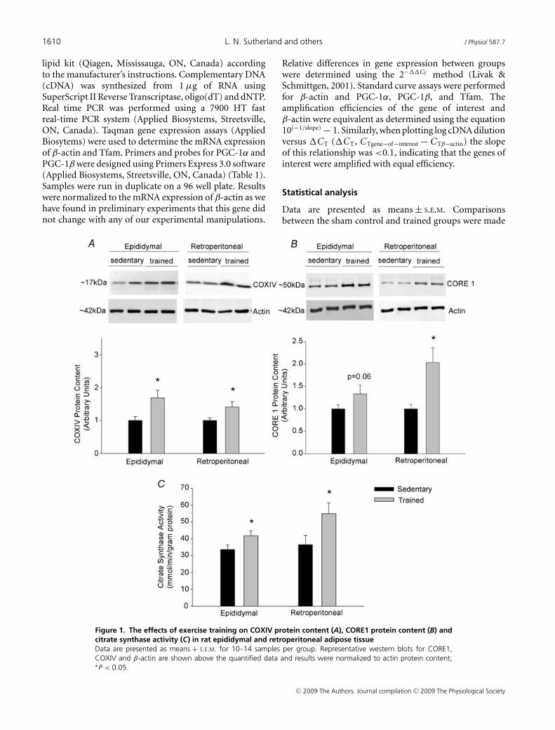
Figure 1. The effects of exercise training on COXIV protein content (A), CORE1 protein content (B) andcitrate synthase activity (C) in rat epididymal and retroperitoneal adipose tissueData are presented as means + S.E.M. for 10–14 samples per group. Representative western blots for CORE1,COXIV and β-actin are shown above the quantified data and results were normalized to actin protein content;∗P < 0.05.
Relative differences in gene expression between groupswere determined using the 2−��CT method (Livak &Schmittgen, 2001). Standard curve assays were performedfor β-actin and PGC-1α, PGC-1β, and Tfam. Theamplification efficiencies of the gene of interest andβ-actin were equivalent as determined using the equation10(−1/slope) − 1. Similarly, when plotting log cDNA dilutionversus �C T (�C T, C Tgene−of−interest − C Tβ−actin) the slopeof this relationship was <0.1, indicating that the genes ofinterest were amplified with equal efficiency.
Statistical analysis
Data are presented as means ± S.E.M. Comparisonsbetween the sham control and trained groups were made
C© 2009 The Authors. Journal compilation C© 2009 The Physiological Society

J Physiol 587.7 Regulation of adipose tissue PGC-1α expression 1611
using Student’s unpaired t test. Comparisons between thevehicle and treated groups during ATOC experiments weremade using a one-way ANOVA followed by a post hoccomparison using Fisher’s LSD test. Similarly, differencesbetween control, swim and swim plus propranolol groupswere made using a one-way ANOVA and Fisher’s LSD test.Statistical significance was set at P < 0.05.
Results
Effect of exercise on body weight, fat pad massand food intake
Despite a slight increase in food consumption, body weightgain in the swim trained group was significantly less thanin the control group (Table 2). Fat pads from the swimtrained rats weighed less than those in the control group.
Exercise-induced increases in mitochondrial proteincontent and enzyme activity
Four weeks of swim training led to increases in theprotein content of COXIV and CORE 1, proteins ofcomplex IV and complex III of the respiratory chain,
0.0
0.5
1.0
1.5
2.0
2.5
3.0
3.5
PG
C-1
α m
RN
A E
xp
ressio
nF
old
Incre
ase C
om
pare
d to S
edenta
ry
Epididymal Retroperitoneal
*
*
0.0
0.5
1.0
1.5
2.0
2.5
Epididymal Retroperitoneal
0.0
0.2
0.4
0.6
0.8
1.0
1.2
1.4
1.6
1.8
Epididymal Retroperitoneal
**
PG
C-1
β m
RN
A E
xpre
ssio
nF
old
Incre
ase C
om
pare
d t
o S
edenta
ry
Tfa
m m
RN
A E
xpre
ssio
nF
old
Incre
ase C
om
pare
d t
o S
edenta
ry
A B
C
Figure 2. The effects of exercise training on the mRNA expression of PGC-1α (A), PGC-1β (B) and TfammRNA expression (C) in epididymal and retroperitoneal adipose tissueData are presented as means + S.E.M. for 10–14 samples per group, normalized to actin mRNA expression, andexpressed as fold differences compared to sedentary controls; ∗P < 0.05.
respectively. Additionally citrate synthase activity wasincreased in epididymal and retroperitoneal adiposetissue from trained rats (Fig. 1). The measurement ofthese enzymes has previously been used as markers ofmitochondrial content in skeletal muscle and adiposetissue (Garcia-Roves et al. 2006; Rong et al. 2007;Sutherland et al. 2008).
Exercise training increases the mRNA expressionof PGC-1α and Tfam
PGC-1α and Tfam mRNA expression were increased inepididymal and retroperitoneal adipose tissue followingtraining (Fig. 2). On the other hand, the mRNA expressionof PGC-1β was not increased in either fat pad followingswim training.
Acute exercise increases the mRNA expressionof PGC-1α
Immediately following an acute, 2 h bout of exercise,PGC-1α mRNA expression was increased in both fat pads.Four hours following exercise cessation, PGC-1α mRNA
C© 2009 The Authors. Journal compilation C© 2009 The Physiological Society
1612 L. N. Sutherland and others J Physiol 587.7
expression was not different from control values (Fig. 3).Tfam mRNA expression was not significantly increasedin either fat pad immediately, or 4 h following exercisecessation.
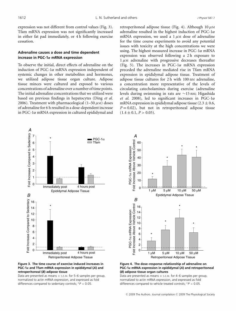
Adrenaline causes a dose and time dependentincrease in PGC-1α mRNA expression
To observe the initial, direct effects of adrenaline on theinduction of PGC-1α mRNA expression independent ofsystemic changes in other metabolites and hormones,we utilized adipose tissue organ culture. Adiposetissue minces were cultured and exposed to variousconcentrations of adrenaline over a number of time points.The initial adrenaline concentrations that we utilized werebased on previous findings in hepatocytes (Ding et al.2006). Treatment with pharmacological (1–50 μM) dosesof adrenaline for 6 h resulted in a dose-dependent increasein PGC-1α mRNA expression in cultured epididymal and
0
1
2
3
4
5
6
7PGC-1αTfam
Fold
Incre
ase C
om
pare
d t
o S
edenta
ry
Immediately post 4 hours post
Epididymal Adipose Tissue
*
0
2
4
6
8
10
12
14
16
Immediately post 4 hours post
Retroperitoneal Adipose Tissue
Fold
Incre
ase C
om
pare
d t
o S
edenta
ry
*
A
B
Figure 3. The time course of exercise induced increases inPGC-1α and Tfam mRNA expression in epididymal (A) andretroperitoneal (B) adipose tissueData are presented as means + S.E.M. for 5–6 samples per group,normalized to actin mRNA expression, and expressed as folddifferences compared to sedentary controls; ∗P < 0.05.
retroperitoneal adipose tissue (Fig. 4). Although 10 μM
adrenaline resulted in the highest induction of PGC-1α
mRNA expression, we used a 1 μM dose of adrenalinefor the time course experiments to avoid any potentialissues with toxicity at the high concentrations we wereusing. The highest measured increase in PGC-1α mRNAexpression was observed following a 2 h exposure to1 μM adrenaline with progressive decreases thereafter(Fig. 5). The increases in PGC-1α mRNA expressionpreceded the adrenaline mediated rise in Tfam mRNAexpression in epididymal adipose tissue. Treatment ofadipose tissue cultures for 2 h with 100 nM adrenaline,a concentration more representative of the levels ofcirculating catecholamines during exercise (adrenalinelevels during swimming in rats are ∼15 nM; Higashidaet al. 2008), led to significant increases in PGC-1α
mRNA expression in epididymal adipose tissue (2.3 ± 0.6,P = 0.02), but not in retroperitoneal adipose tissue(1.4 ± 0.1, P > 0.05).
PG
C-1
α m
RN
A E
xpre
ssio
nF
old
Incre
ase A
bove V
ehic
le C
ontr
ol
0
10
20
30
40
50
60
1 μM 5 μM 10 μM 50 μM
1 μM 5 μM 10 μM 50 μM
*
*
* *
PG
C-1
α m
RN
A E
xpre
ssio
nF
old
Incre
ase A
bove V
ehic
le C
ontr
ol
0
2
4
6
8
10
12
14
16
*
*
*
*
A
BEpididymal Adipose Tissue
Retroperitoneal Adipose Tissue
Figure 4. The dose–response relationship of adrenaline onPGC-1α mRNA expression in epididymal (A) and retroperitoneal(B) adipose tissue organ culturesData are presented as means + S.E.M. for 4–6 samples per group,normalized to actin mRNA expression, and expressed as folddifferences compared to vehicle treated controls; ∗P < 0.05.
C© 2009 The Authors. Journal compilation C© 2009 The Physiological Society

J Physiol 587.7 Regulation of adipose tissue PGC-1α expression 1613
β-Blockade attenuates exercise induced increasesin adipose tissue PGC-1α mRNA expression
Rats were treated with the β-blocker propranolol (200 μg(100 g body weight)−1) 70 and 10 min prior to 2 h of swimexercise. In preliminary experiments we found that thisdosing protocol almost completely blocked the in vivoeffects of adrenaline (20 μg (100 g body weight)−1) onPGC-1α mRNA expression in adipose tissue (adrenaline3.69 ± 1.26-fold increase above control, adrenaline +propranolol 1.44 ± 0.36-fold increase above control). Thisadrenaline treatment results in increases in adrenaline(∼190 nM) (Fell et al. 1981) to levels much higher thanseen during exercise (∼15 nM) (Higashida et al. 2008).The same propranolol treatment has previously been usedin rat swim models and does not affect the ability ofrats to complete the swim exercise (Nolte et al. 1994). Asseen in Fig. 6, propranolol attenuated the exercise inducedrise in PGC-1α mRNA expression in epididymal but notretroperitoneal adipose tissue.
Epididymal Adipose Tissue
PG
C-1
α m
RN
A e
xpre
ssio
nfo
ld-incre
ase a
bove v
ehic
le c
ontr
ol
0
2
4
6
8
10
12
14
16
18
20
2 hr 4 hr 6 hr 12 hr 2 hr 4 hr 6 hr 12 hr
*
**
*
A
0
2
4
6
8 *
*
*
PG
C-1
α m
RN
A e
xpre
ssio
nfo
ld-incre
ase a
bove v
ehic
le c
ontr
ol
B
Retroperitoneal Adipose Tissue
0.0
0.2
0.4
0.6
0.8
1.0
1.2
1.4
1.6
1.8
2.0
* *
Tfa
m m
RN
A e
xpre
ssio
nfo
ld-incre
ase a
bove v
ehic
le c
ontr
ol
Epididymal Adipose Tissue
2 hr 4 hr 6 hr 12 hr 2 hr 4 hr 6 hr 12 hr
C
0.0
0.2
0.4
0.6
0.8
1.0
1.2
Retroperitoneal Adipose Tissue
Tfa
m m
RN
A e
xpre
ssio
nfo
ld-incre
ase a
bove v
ehic
le c
ontr
ol
D
Figure 5. The time course of adrenaline (1 μM) induced increases in PGC-1α and Tfam mRNA expressionin rat epididymal (A and C) and retroperitoneal (B and D) organ culturesData are presented as means + S.E.M. for 4–6 samples per group, normalized to actin mRNA expression, andexpressed as fold differences compared to vehicle treated controls; ∗P < 0.05.
Discussion
Adipose tissue mitochondria are increasingly beingrecognized as key players in the regulation of wholebody metabolism. Surprisingly, and in sharp contrast toskeletal muscle, few studies have explored the effects ofexercise on mitochondrial biogenesis in adipose tissue.Consistent with one previous report (Stallknecht et al.1991), we found that 2 h of daily swim exercise for 28consecutive days led to increases in markers of adiposetissue mitochondrial biogenesis, such as CORE1 andCOXIV protein content and citrate synthase activity.
Mitochondrial biogenesis is a complex processinvolving the coordinated regulation of both nuclear andmitochondrial encoded genes. A central cog in this processwould appear to be PGC-1α. The over-expression ofPGC-1α in skeletal muscle (Lin et al. 2002) or whiteadipocytes (Tiraby et al. 2003) induces mitochondrialbiogenesis, whereas the deletion of this gene leads toreductions in mitochondria (Leone et al. 2005). Exercise
C© 2009 The Authors. Journal compilation C© 2009 The Physiological Society

1614 L. N. Sutherland and others J Physiol 587.7
has been shown to have both an acute (Baar et al. 2002;Miura et al. 2007; Mathai et al. 2008) and a chronictraining related effect (Goto et al. 2000) on PGC-1α
mRNA expression in skeletal muscle. Similar to theseaforementioned findings we made the novel observationthat 4 weeks of daily swim training led to increases in themRNA expression of PGC-1α, in white adipose tissue. Onthe other hand training did not significantly increase theexpression of PGC-1β in either fat depot. These findingsare consistent with a previous report which demonstratedthat cold exposure, fasting and exercise increased PGC-1α
but not PGC-1β mRNA levels in brown adipose tissue,liver and skeletal muscle, respectively (Meirhaeghe et al.2003). PGC-1α mRNA expression in white adipose tissuewas increased immediately after exercise and returnedto control levels 4 h following exercise cessation. Sincetissue was harvested well after this time point, our resultssuggest that the long-term increases in PGC-1α mRNAexpression were the result of a training effect and notrelated to the residual effects of the last bout of exercise.The rise in PGC-1α was associated with increases in the
Fold
Incre
ase in P
GC
-1α
mR
NA
Expre
ssio
n
0
1
2
3
4
5
Exercise Exercise + Propranolol
*
* #
Epididymal Adipose Tissue
Fold
Incre
ase in P
GC
-1α
mR
NA
Expre
ssio
n
0
1
2
3
4
5
6
7
Exercise Exercise + Propranolol
Retroperitoneal Adipose Tissue
*
*
A
B
Figure 6. The effects of propranolol on the exercise inducedincreases in PGC-1α mRNA expression in rat epididymal (A) andretroperitoneal (B) adipose tissueData are presented as means + S.E.M. for 6–9 samples per group,normalized to actin mRNA expression, and are expressed as folddifferences compared to non-exercised rats (∗P < 0.05) or betweenthe exercised groups with saline or propranolol treatment (#P < 0.05).
mRNA expression of Tfam, a transcription factor involvedin the regulation of mitochondrial encoded genes whoseexpression is controlled, at least in part, by PGC-1α (Wuet al. 1999; Gleyzer et al. 2005). While our findings areconsistent with the notion that PGC-1α could be involvedin mediating exercise induced mitochondrial biogenesisin adipose tissue, future studies are needed to determineif the changes in PGC-1α mRNA expression are paralleledby increases in the protein content of this transcriptionalco-activator.
Biochemical changes within the contracting muscleitself, such as perturbations in high energy phosphates(Bergeron et al. 2001; Baar et al. 2002; Zong et al.2002), and increases in cytosolic calcium concentration(Ojuka et al. 2002, 2003), are believed to initiate, to alarge extent, exercise induced mitochondrial biogenesisin skeletal muscle. On the other hand, the triggeringmechanisms mediating this process in adipose tissuehave not been established. Hormonal factors such asadrenaline are intimately involved in the acute regulationof adipose tissue metabolism during exercise (McMurray& Hackney, 2005). Moreover, micromolar concentrationsof adrenaline have been shown to induce PGC-1α mRNAexpression in hepatocytes (Ding et al. 2006). Given thesefindings it seems likely that adrenaline could inducePGC-1α mRNA expression in white adipose tissue andperhaps serve as an extracellular signal in the exercisemediated induction of PGC-1α.
As an initial approach to test this hypothesis weused adipose tissue organ culture and determined thedose–response relationship and time course of adrenalineinduced increases in PGC-1α mRNA expression. Acrossa range of concentrations, we found that adrenalinemarkedly increased PGC-1α mRNA expression andthat these changes preceded increases in Tfam mRNAexpression. Interestingly, epididymal adipose tissueappeared much more responsive to the effects ofpharmacological doses of adrenaline as witnessed by largerincreases in PGC-1α mRNA expression in epididymaladipose tissue and the absence of adrenaline inducedincreases in Tfam mRNA expression in retroperitonealadipose tissue. Consistent with these findings, PGC-1α
mRNA expression in retroperitoneal adipose tissue organcultures did not significantly increase when treatedwith supra-physiological (100 nM) concentrations ofadrenaline. Our findings in epididymal adipose tissueare in line with recent results from Miura and colleagues(2007) who reported that β-adrenergic stimulation leadsto increases in PGC-1α mRNA expression in murineskeletal muscle.
Having shown that both exercise and adrenalinetreatment induce PGC-1α mRNA expression in whiteadipose tissue, we wanted to gain insight into a potentialrole of adrenaline in mediating the acute effects of exerciseon the induction of PGC-1α mRNA expression in white
C© 2009 The Authors. Journal compilation C© 2009 The Physiological Society

J Physiol 587.7 Regulation of adipose tissue PGC-1α expression 1615
adipose tissue. Rats were treated with the non-specificβ-blocker propranolol prior to, and tissue harvestedimmediately following, 2 h of swimming. In epididymaladipose tissue, β-blockade led to a ∼40% reduction in theexercise induced increase in PGC-1α mRNA expression.It should be noted that the same propranolol treatmentblocked the adrenaline induced rise in PGC-1α mRNAexpression suggesting that this treatment was sufficientto block the effects of adrenaline on adipose tissue geneexpression. The partial attenuation of the exercise inducedincrease in PGC-1α mRNA expression in combinationwith the results of our adrenaline experiments is consistentwith the hypothesis that elevations in catecholamines maymediate a portion of the exercise induced increase inPGC-1α mRNA expression in epididymal adipose tissue.
In contrast to epididymal adipose tissue, β-blockade didnot significantly attenuate the exercise induced increasein PGC-1α gene expression in the retroperitoneal fatpad. Given the greater effect of adrenaline on theinduction of PGC-1α expression in epididymal comparedto retroperitoneal adipose tissue, these results are notentirely surprising. Taken in combination with previousresults showing enhanced lipolysis in epididymal versusretroperitoneal adipocytes (Tavernier et al. 1995), ourfindings would suggest the existence of depot specificdifferences in responsiveness to β-adrenergic stimulation.While the mechanisms underlying these apparent depotspecific differences are not clear, our results suggest thatmultiple extracellular signals are likely to be involved in theexercise induced up-regulation of PGC-1α expression inwhite adipose tissue. For example exercise has been shownto increase circulating levels of thyroid hormone (Wirthet al. 1981; Limanova et al. 1983; Fortunato et al. 2008), ahormone that has been shown, at least in skeletal muscle,to increase PGC-1α protein content (Branvold et al.2008). Along a similar line the expression and secretionof interleukin 6 (IL-6) from skeletal muscle has beenshown to increase dramatically during exercise (Pedersen& Febbraio, 2008). Interestingly, IL-6 has been shown toactivate 5′AMP activated protein kinase (Kelly et al. 2004),a reputed mediator of PGC-1α mRNA expression (Jageret al. 2007). The potential roles of IL-6 and/or thyroidhormone in the regulation of PGC-1α in white adiposetissue need to be explored in further detail.
The purpose of the present study was to examine theeffects of exercise and adrenaline on PGC-1α mRNAexpression in white adipose tissue. Although swimmingmay not be representative of all exercise models giventhe relatively large increases in catecholamines (Higashidaet al. 2008), we have made the novel observations thatboth acute swim exercise and long term training leadto increases in PGC-1α mRNA expression in whiteadipose tissue. Interestingly, it does not appear thatβ-adrenergic agonists are the sole regulators of exerciseinduced increases in PGC-1α mRNA expression. A further
elucidation of the specific extracellular signals whichregulate exercise induced increases in PGC-1α mRNAexpression is an area ripe for investigation and will lendmuch insight into the regulation of adipose tissue geneexpression by exercise.
References
Baar K, Wende AR, Jones TE, Marison M, Nolte LA, Chen M,Kelly DP & Holloszy JO (2002). Adaptations of skeletalmuscle to exercise: rapid increase in the transcriptionalcoactivator PGC-1. FASEB J 16, 1879–1886.
Bergeron R, Ren JM, Cadman KS, Moore IK, Perret P, PypaertM, Young LH, Semenkovich CF & Shulman GI (2001).Chronic activation of AMP kinase results in NRF-1activation and mitochondrial biogenesis. Am J PhysiolEndocrinol Metab 281, E1340–E1346.
Branvold DJ, Allred DR, Beckstead DJ, Kim HJ, Fillmore N,Condon BM, Brown JD, Sudweeks SN, Thomson DM &Winder WW (2008). Thyroid hormone effects on LKB1,MO25, phospho-AMPK, phospho-CREB, and PGC-1α inrat muscle. J Appl Physiol 105, 1218–1227.
Choo HJ, Kim JH, Kwon OB, Lee CS, Mun JY, Han SS, YoonYS, Yoon G, Choi KM & Ko YG (2006). Mitochondria areimpaired in the adipocytes of type 2 diabetic mice.Diabetologia 49, 784–791.
Crowe S, Turpin SM, Ke F, Kemp BE & Watt MJ (2008).Metabolic remodeling in adipocytes promotes ciliaryneurotrophic factor-mediated fat loss in obesity.Endocrinology 149, 2546–2556.
Ding X, Lichti K, Kim I, Gonzalez FJ & Staudinger JL (2006).Regulation of constitutive androstane receptor and its targetgenes by fasting, cAMP, hepatocyte nuclear factor α, and thecoactivator peroxisome proliferator-activated receptor γ
coactivator-1α. J Biol Chem 281, 26540–26551.Fell RD, Terblanche SE, Winder WW & Holloszy JO (1981).
Adaptive responses of rats to prolonged treatment withepinephrine. Am J Physiol Cell Physiol 241, C55–C58.
Fortunato RS, Ignacio DL, Padron AS, Pecanha R, Marassi MP,Rosenthal D, Werneck-de-Castro JP & Carvalho DP (2008).The effect of acute exercise session on thyroid hormoneeconomy in rats. J Endocrinol 198, 347–353.
Fried SK & Moustaid-Moussa N (2001). Culture of adiposetissue and isolated adipocytes. Methods Mol Biol 155,197–212.
Fried SK, Russell CD, Grauso NL & Brolin RE (1993).Lipoprotein lipase regulation by insulin and glucocorticoidin subcutaneous and omental adipose tissues of obesewomen and men. J Clin Invest 92, 2191–2198.
Garcia-Roves PM, Huss J & Holloszy JO (2006). Role ofcalcineurin in exercise-induced mitochondrial biogenesis.Am J Physiol Endocrinol Metab 290, E1172–E1179.
Gleyzer N, Vercauteren K & Scarpulla RC (2005). Control ofmitochondrial transcription specificity factors (TFB1M andTFB2M) by nuclear respiratory factors (NRF-1 and NRF-2)and PGC-1 family coactivators. Mol Cell Biol 25, 1354–1366.
Goto M, Terada S, Kato M, Katoh M, Yokozeki T, Tabata I &Shimokawa T (2000). cDNA Cloning and mRNA analysis ofPGC-1 in epitrochlearis muscle in swimming-exercised rats.Biochem Biophys Res Commun 274, 350–354.
C© 2009 The Authors. Journal compilation C© 2009 The Physiological Society
1616 L. N. Sutherland and others J Physiol 587.7
Hammarstedt A, Jansson PA, Wesslau C, Yang X & Smith U(2003). Reduced expression of PGC-1 and insulin-signalingmolecules in adipose tissue is associated with insulinresistance. Biochem Biophys Res Commun 301,578–582.
Higashida K, Higuchi M & Terada S (2008). Potential role oflipin-1 in exercise-induced mitochondrial biogenesis.Biochem Biophys Res Commun 374, 587–591.
Jager S, Handschin C, St-Pierre J & Spiegelman BM (2007).AMP-activated protein kinase (AMPK) action in skeletalmuscle via direct phosphorylation of PGC-1α. Proc NatlAcad Sci U S A 104, 12017–12022.
Kelly M, Keller C, Avilucea PR, Keller P, Luo Z, Xiang X, GiraltM, Hidalgo J, Saha AK, Pedersen BK & Ruderman NB(2004). AMPK activity is diminished in tissues of IL-6knockout mice: the effect of exercise. Biochem Biophys ResCommun 320, 449–454.
Koh EH, Park JY, Park HS, Jeon MJ, Ryu JW, Kim M, Kim SY,Kim MS, Kim SW, Park IS, Youn JH & Lee KU (2007).Essential role of mitochondrial function in adiponectinsynthesis in adipocytes. Diabetes 56, 2973–2981.
Lee MJ, Wang Y, Ricci MR, Sullivan S, Russell CD & Fried SK(2007). Acute and chronic regulation of leptin synthesis,storage, and secretion by insulin and dexamethasone inhuman adipose tissue. Am J Physiol Endocrinol Metab 292,E858–E864.
Leone TC, Lehman JJ, Finck BN, Schaeffer PJ, Wende AR,Boudina S, Courtois M, Wozniak DF, Sambandam N,Bernal-Mizrachi C, Chen Z, Holloszy JO, Medeiros DM,Schmidt RE, Saffitz JE, Abel ED, Semenkovich CF & KellyDP (2005). PGC-1α deficiency causes multi-system energymetabolic derangements: muscle dysfunction, abnormalweight control and hepatic steatosis. PLoS Biol 3, e101.
Limanova Z, Sonka J, Kratochvil O, Sonka K, Kanka J &Sprynarova S (1983). Effects of exercise on serum cortisoland thyroid hormones. Exp Clin Endocrinol 81,308–314.
Lin J, Wu H, Tarr PT, Zhang CY, Wu Z, Boss O, Michael LF,Puigserver P, Isotani E, Olson EN, Lowell BB, Bassel-Duby R& Spiegelman BM (2002). Transcriptional co-activatorPGC-1 alpha drives the formation of slow-twitch musclefibres. Nature 418, 797–801.
Livak KJ & Schmittgen TD (2001). Analysis of relative geneexpression data using real-time quantitative PCR and the2–��CT method. Methods 25, 402–408.
Mathai AS, Bonen A, Benton CR, Robinson DL & Graham TE(2008). Rapid exercise-induced changes in PGC-1α mRNAand protein in human skeletal muscle. J Appl Physiol 105,1098–1105.
McMurray RG & Hackney AC (2005). Interactions ofmetabolic hormones, adipose tissue and exercise. Sports Med35, 393–412.
Meirhaeghe A, Crowley V, Lenaghan C, Lelliott C, Green K,Stewart A, Hart K, Schinner S, Sethi JK, Yeo G, Brand MD,Cortright RN, O’Rahilly S, Montague C & Vidal-Puig AJ(2003). Characterization of the human, mouse and ratPGC1β (peroxisome-proliferator-activated receptor-γco-activator 1β) gene in vitro and in vivo. Biochem J 373,155–165.
Miura S, Kawanaka K, Kai Y, Tamura M, Goto M, Shiuchi T,Minokoshi Y & Ezaki O (2007). An increase in murineskeletal muscle peroxisome proliferator-activated receptor-γcoactivator-1alpha (PGC-1α) mRNA in response to exerciseis mediated by β-adrenergic receptor activation.Endocrinology 148, 3441–3448.
Nolte LA, Gulve EA & Holloszy JO (1994). Epinephrine-induced in vivo muscle glycogen depletion enhances insulinsensitivity of glucose transport. J Appl Physiol 76, 2054–2058.
Ojuka EO, Jones TE, Han DH, Chen M & Holloszy JO (2003).Raising Ca2+ in L6 myotubes mimics effects of exercise onmitochondrial biogenesis in muscle. FASEB J 17,675–681.
Ojuka EO, Jones TE, Han DH, Chen M, Wamhoff BR, SturekM & Holloszy JO (2002). Intermittent increases in cytosolicCa2+ stimulate mitochondrial biogenesis in muscle cells. AmJ Physiol Endocrinol Metab 283, E1040–E1045.
Pedersen BK & Febbraio MA (2008). Muscle as an endocrineorgan: focus on muscle-derived interleukin-6. Physiol Rev88, 1379–1406.
Puigserver P, Wu Z, Park CW, Graves R, Wright M &Spiegelman BM (1998). A cold-inducible coactivator ofnuclear receptors linked to adaptive thermogenesis. Cell 92,829–839.
Rong JX, Qiu Y, Hansen MK, Zhu L, Zhang V, Xie M,Okamoto Y, Mattie MD, Higashiyama H, Asano S, Strum JC& Ryan TE (2007). Adipose mitochondrial biogenesis issuppressed in db/db and high-fat diet-fed mice andimproved by rosiglitazone. Diabetes 56, 1751–1760.
Scarpulla RC (2008). Transcriptional paradigms in mammalianmitochondrial biogenesis and function. Physiol Rev 88,611–638.
Smith PK, Krohn RI, Hermanson GT, Mallia AK, Gartner FH,Provenzano MD, Fujimoto EK, Goeke NM, Olson BJ &Klenk DC (1985). Measurement of protein usingbicinchoninic acid. Anal Biochem 150, 76–85.
Stallknecht B, Vinten J, Ploug T & Galbo H (1991). Increasedactivities of mitochondrial enzymes in white adipose tissuein trained rats. Am J Physiol Endocrinol Metab 261,E410–E414.
Sutherland LN, Capozzi LC, Turchinsky NJ, Bell RC & WrightDC (2008). Time course of high-fat diet-induced reductionsin adipose tissue mitochondrial proteins: potentialmechanisms and the relationship to glucose intolerance. AmJ Physiol Endocrinol Metab 295, E1076–E1083.
Tavernier G, Galitzky J, Valet P, Remaury A, Bouloumie A,Lafontan M & Langin D (1995). Molecular mechanismsunderlying regional variations of catecholamine-inducedlipolysis in rat adipocytes. Am J Physiol Endocrinol Metab268, E1135–E1142.
Tiraby C, Tavernier G, Lefort C, Larrouy D, Bouillaud F,Ricquier D & Langin D (2003). Acquirement of brown fatcell features by human white adipocytes. J Biol Chem 278,33370–33376.
Trujillo ME, Lee MJ, Sullivan S, Feng J, Schneider SH,Greenberg AS & Fried SK (2006). Tumor necrosis factor α
and glucocorticoid synergistically increase leptin productionin human adipose tissue: role for p38 mitogen-activatedprotein kinase. J Clin Endocrinol Metab 91, 1484–1490.
C© 2009 The Authors. Journal compilation C© 2009 The Physiological Society
J Physiol 587.7 Regulation of adipose tissue PGC-1α expression 1617
Valerio A, Cardile A, Cozzi V, Bracale R, Tedesco L, Pisconti A,Palomba L, Cantoni O, Clementi E, Moncada S, CarrubaMO & Nisoli E (2006). TNF-α downregulates eNOSexpression and mitochondrial biogenesis in fat and muscleof obese rodents. J Clin Invest 116, 2791–2798.
Wilson-Fritch L, Nicoloro S, Chouinard M, Lazar MA, ChuiPC, Leszyk J, Straubhaar J, Czech MP & Corvera S (2004).Mitochondrial remodeling in adipose tissue associated withobesity and treatment with rosiglitazone. J Clin Invest 114,1281–1289.
Winder WW, Taylor EB & Thomson DM (2006). Role ofAMP-activated protein kinase in the molecular adaptation toendurance exercise. Med Sci Sports Exerc 38, 1945–1949.
Wirth A, Holm G, Lindstedt G, Lundberg PA & Bjorntorp P(1981). Thyroid hormones and lipolysis in physically trainedrats. Metabolism 30, 237–241.
Wright DC (2007). Mechanisms of calcium-inducedmitochondrial biogenesis and GLUT4 synthesis. Appl PhysiolNutr Metab 32, 840–845.
Wright DC, Geiger PC, Han DH, Jones TE & Holloszy JO(2007). Calcium induces increases in peroxisomeproliferator-activated receptor γ coactivator-1α andmitochondrial biogenesis by a pathway leading to p38mitogen-activated protein kinase activation. J Biol Chem282, 18793–18799.
Wu Z, Puigserver P, Andersson U, Zhang C, Adelmant G,Mootha V, Troy A, Cinti S, Lowell B, Scarpulla RC &Spiegelman BM (1999). Mechanisms controllingmitochondrial biogenesis and respiration through thethermogenic coactivator PGC-1. Cell 98, 115–124.
Zong H, Ren JM, Young LH, Pypaert M, Mu J, Birnbaum MJ &Shulman GI (2002). AMP kinase is required formitochondrial biogenesis in skeletal muscle in response tochronic energy deprivation. Proc Natl Acad Sci U S A 99,15983–15987.
Acknowledgements
D.C.W. is a CIHR New Investigator, CDA Scholar and AlbertaHeritage Foundation for Medical Research Scholar. L.S. wassupported by an Alberta Heritage Foundation for MedicalResearch Studentship and a Natural Sciences and EngineeringResearch Council of Canada Graduate Student Scholarship. M.B.and S.B. were supported by NSERC summer studentships. Thisresearch was supported by an Operating Grant to D.C.W. fromthe Canadian Institutes of Health Research and a pilot projectgrant from the Alberta Diabetes Institute.
C© 2009 The Authors. Journal compilation C© 2009 The Physiological Society
Copyright © 2022 FDOKUMEN