Excited-state properties of the 16kDa red carotenoid protein from Arthrospira maxima
6
Excited-state properties of the 16 kDa red carotenoid protein from Arthrospira maxima Pavel Chábera a,1 , Milan Durchan a,b , Patrick M. Shih c , Cheryl A. Kerfeld c,d , Tomáš Polívka a,b, ⁎ a Institute of Physical Biology, University of South Bohemia, Nové Hrady, Czech Republic b Biological Centre, Czech Academy of Sciences, České Budějovice, Czech Republic c Department of Plant and Microbial Biology, University of California, Berkeley, USA d US Department of Energy, Joint Genome Institute, Walnut Creek, CA, USA abstract article info Article history: Received 10 May 2010 Received in revised form 21 August 2010 Accepted 24 August 2010 Available online 8 September 2010 Keywords: Cyanobacteria Photoprotection Carotenoid Excited-state Carotenoid-binding protein We have studied spectroscopic properties of the 16 kDa red carotenoid protein (RCP), which is closely related to the orange carotenoid protein (OCP) from cyanobacteria. Both proteins bind the same chromophore, the carotenoid 3′-hydroxyechinenone (hECN), and the major difference between the two proteins is lack of the C-terminal domain in the RCP; this results in exposure of part of the carotenoid. The excited-state lifetime of hECN in the RCP is 5.5 ps, which is markedly longer than in OCP (3.3 ps) but close to 6 ps obtained for hECN in organic solvent. This confirms that the binding of hECN to the C-terminal domain in the OCP changes conformation of hECN, thereby altering its excited-state properties. Hydrogen bonds between the C-terminal domain and the carotenoid are also absent in the RCP. This allows the conformation of hECN in the RCP to be similar to that in solution, which results in comparable excited-state properties of hECN in solution. The red-shift of the RCP absorption spectrum is most likely due to aggregation of RCP induced by hydrophobic nature of hECN that, when exposed to buffer, stimulates formation of assemblies minimizing contact of hECN with water. We suggest that the loss of the C-terminal domain renders the protein amphipathic, containing both hydrophobic (the exposed part of hECN) and hydrophilic (N-terminal domain) regions, and may help the RCP to interact with lipid membranes; exposed hECN can penetrate into the hydrophobic environment of the lipid membrane, possibly to provide additional photoprotection. © 2010 Elsevier B.V. All rights reserved. 1. Introduction Photoprotective mechanisms involving carotenoids are the key processes protecting photosynthetic organisms from excess light [1,2]. These processes range from direct quenching of (bacterio) chlorophyll triplet states and quenching singlet oxygen to complex reactions involving specialized proteins and cofactors. In plants for example, photoprotection involves chlorophyll-binding antenna proteins and is triggered by a pH change in thylakoid membrane that, with a help of a specialized protein called PsbS, activates enzymes that perform mutual interconversion of two carotenoids, violaxanthin and zeaxanthin [3,4]. This complicated process, known as non-photochemical quenching (NPQ), eventually results in thermal dissipation of absorbed energy that is manifested by a decrease of chlorophyll fluorescence. Until recently the NPQ has been associated only with plants, but a number of studies in the past decade showed that NPQ exists also in cyanobacteria to decrease the amount of energy transferred from phycobilisomes to PSII [5–8]. In contrast to plants, however, a 35 kDa water-soluble carotenoid-binding protein known as the orange carotenoid protein (OCP) plays an essential role [7–9]. This protein, discovered nearly twenty years ago [10] and whose structure is known [11,12], binds a single carotenoid, 3′-hydroxyechinenone (hECN). The OCP is composed of two domains, an all-helical N- terminal domain (Pfam09150; http://pfam.sanger.ac.uk/) which has not been observed in any other protein structures determined to-date, and a C-terminal domain which is a member of the nuclear transport factor 2 (NTF2) superfamily (Pfam02136; http://pfam.sanger.ac.uk/) of α/β proteins. The carotenoid has a conjugated chain consisting of 11 conjugated CC bonds spanning two protein domains. The conjugated chain is terminated by a carbonyl group that is nestled in the binding pocket of the C-terminal domain (Fig. 1A), forming hydrogen bonds between the keto oxygen and absolutely conserved tyrosine and tryptophan residues [11–13]. Binding to the protein forces the terminal ring of hECN to the s-trans conformation, while s- cis orientation occurs in solution (Fig. 1). This change is associated with changes of hECN excited-state properties [14]. Biochimica et Biophysica Acta 1807 (2011) 30–35 ⁎ Corresponding author. Institute of Physical Biology, University of South Bohemia, Zámek 136, CZ-373 33 Nové Hrady, Czech Republic. Tel.: + 420 389 033 824; fax: + 420 386 361 219. E-mail address: [email protected] (T. Polívka). 1 Current address: Department of Chemical Physics, Lund University, Sweden. 0005-2728/$ – see front matter © 2010 Elsevier B.V. All rights reserved. doi:10.1016/j.bbabio.2010.08.013 Contents lists available at ScienceDirect Biochimica et Biophysica Acta journal homepage: www.elsevier.com/locate/bbabio
-
Upload
independent -
Category
Documents
-
view
7 -
download
0
Transcript of Excited-state properties of the 16kDa red carotenoid protein from Arthrospira maxima
Biochimica et Biophysica Acta 1807 (2011) 30–35
Contents lists available at ScienceDirect
Biochimica et Biophysica Acta
j ourna l homepage: www.e lsev ie r.com/ locate /bbab io
Excited-state properties of the 16 kDa red carotenoid protein fromArthrospira maxima
Pavel Chábera a,1, Milan Durchan a,b, Patrick M. Shih c, Cheryl A. Kerfeld c,d, Tomáš Polívka a,b,⁎a Institute of Physical Biology, University of South Bohemia, Nové Hrady, Czech Republicb Biological Centre, Czech Academy of Sciences, České Budějovice, Czech Republicc Department of Plant and Microbial Biology, University of California, Berkeley, USAd US Department of Energy, Joint Genome Institute, Walnut Creek, CA, USA
⁎ Corresponding author. Institute of Physical BiologyZámek 136, CZ-373 33 Nové Hrady, Czech Republic. Tel.:386 361 219.
E-mail address: [email protected] (T. Polívka).1 Current address: Department of Chemical Physics, L
0005-2728/$ – see front matter © 2010 Elsevier B.V. Adoi:10.1016/j.bbabio.2010.08.013
a b s t r a c t
a r t i c l e i n f oArticle history:Received 10 May 2010Received in revised form 21 August 2010Accepted 24 August 2010Available online 8 September 2010
Keywords:CyanobacteriaPhotoprotectionCarotenoidExcited-stateCarotenoid-binding protein
We have studied spectroscopic properties of the 16 kDa red carotenoid protein (RCP), which is closelyrelated to the orange carotenoid protein (OCP) from cyanobacteria. Both proteins bind the samechromophore, the carotenoid 3′-hydroxyechinenone (hECN), and the major difference between the twoproteins is lack of the C-terminal domain in the RCP; this results in exposure of part of the carotenoid. Theexcited-state lifetime of hECN in the RCP is 5.5 ps, which is markedly longer than in OCP (3.3 ps) but close to6 ps obtained for hECN in organic solvent. This confirms that the binding of hECN to the C-terminal domainin the OCP changes conformation of hECN, thereby altering its excited-state properties. Hydrogen bondsbetween the C-terminal domain and the carotenoid are also absent in the RCP. This allows the conformationof hECN in the RCP to be similar to that in solution, which results in comparable excited-state properties ofhECN in solution. The red-shift of the RCP absorption spectrum is most likely due to aggregation of RCPinduced by hydrophobic nature of hECN that, when exposed to buffer, stimulates formation of assembliesminimizing contact of hECN with water. We suggest that the loss of the C-terminal domain renders theprotein amphipathic, containing both hydrophobic (the exposed part of hECN) and hydrophilic (N-terminaldomain) regions, and may help the RCP to interact with lipid membranes; exposed hECN can penetrate intothe hydrophobic environment of the lipid membrane, possibly to provide additional photoprotection.
, University of South Bohemia,+420 389 033 824; fax: +420
und University, Sweden.
ll rights reserved.
© 2010 Elsevier B.V. All rights reserved.
1. Introduction
Photoprotective mechanisms involving carotenoids are the keyprocesses protecting photosynthetic organisms from excess light[1,2]. These processes range from direct quenching of (bacterio)chlorophyll triplet states and quenching singlet oxygen to complexreactions involving specialized proteins and cofactors. In plants forexample, photoprotection involves chlorophyll-binding antennaproteins and is triggered by a pH change in thylakoid membranethat, with a help of a specialized protein called PsbS, activatesenzymes that perform mutual interconversion of two carotenoids,violaxanthin and zeaxanthin [3,4]. This complicated process, knownas non-photochemical quenching (NPQ), eventually results in thermaldissipation of absorbed energy that is manifested by a decrease ofchlorophyll fluorescence.
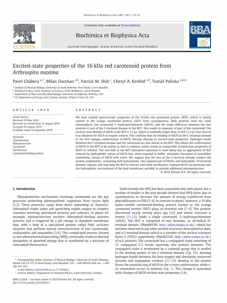
Until recently the NPQ has been associated only with plants, but anumber of studies in the past decade showed that NPQ exists also incyanobacteria to decrease the amount of energy transferred fromphycobilisomes to PSII [5–8]. In contrast to plants, however, a 35 kDawater-soluble carotenoid-binding protein known as the orangecarotenoid protein (OCP) plays an essential role [7–9]. This protein,discovered nearly twenty years ago [10] and whose structure isknown [11,12], binds a single carotenoid, 3′-hydroxyechinenone(hECN). The OCP is composed of two domains, an all-helical N-terminal domain (Pfam09150; http://pfam.sanger.ac.uk/) which hasnot been observed in any other protein structures determined to-date,and a C-terminal domain which is a member of the nuclear transportfactor 2 (NTF2) superfamily (Pfam02136; http://pfam.sanger.ac.uk/)of α/β proteins. The carotenoid has a conjugated chain consisting of11 conjugated C C bonds spanning two protein domains. Theconjugated chain is terminated by a carbonyl group that is nestledin the binding pocket of the C-terminal domain (Fig. 1A), forminghydrogen bonds between the keto oxygen and absolutely conservedtyrosine and tryptophan residues [11–13]. Binding to the proteinforces the terminal ring of hECN to the s-trans conformation, while s-cis orientation occurs in solution (Fig. 1). This change is associatedwith changes of hECN excited-state properties [14].

Fig. 1. (A) Structure of A. maxima OCP showing the two helical bundles of the N-terminal domain (blue), and the C-terminal domain (red). The hECN (orange) and theabsolutely conserved tyrosine and tryptophan residues that hydrogen bonds to thecarotenoid are shown in sticks. Figure was prepared with PyMOL (http://www.pymol.org/). Molecular structure of hECN when bound to OCP (B), and in solution (C).
31P. Chábera et al. / Biochimica et Biophysica Acta 1807 (2011) 30–35
Although increasing transcript levels for the OCP during exposureto intense light suggested its possible involvement in photoprotec-tion, the mechanism of its action was not known until recently.
It was shown that action spectrum of NPQ in Synechocystis [15]matches the absorption spectrum of OCP [14,16,17]. Moreover, theabsence of the OCP in cyanobacteria prevents fluorescence quenchinginduced by blue light [7]. Thus, the OCP was identified asphotoreceptor triggering the NPQ in cyanobacteria. Later, the samegroup demonstrated that OCP itself is photoactive. Upon illuminationwith blue-green light, the OCP converts to a red form, which isproposed to be the active form of the protein responsible for inductionof NPQ [9]. Using mutants accumulating different carotenoids in OCP,Punginelli et al. [18] proved that the conjugated carbonyl group ofhECN is the key structural feature leading to the photoactivity of OCP.
A red form of the OCP can be also produced by acidification of theprotein resulting in a red-shifted absorption spectrum. The origin ofthe red-shift is unknown, as well as the origin of the red-shift of theactive form of OCP. While these red forms of OCP have all molecularweights of 32 kDa, there is another form of this protein, a 16 kDa redcarotenoid protein (RCP) that co-purifies with the OCP in certaincyanobacteria [13]. RCP also has a red-shifted absorption spectrumand lacks the C-terminal domain, presumably resulting in exposure ofhECN to solvent (Fig. 1A). Interestingly, the accumulating genomicsequence data indicates that many genomes also contain, in additionto a full length OCP gene, one or more open reading frames encodinghomologs to the N-terminal domain while containing a single copy ofthe C-terminal domains (with the exception of the draft genome ofMicrocoleus chthonplastes which does not contain a gene encoding C-terminal domain) [19]. The primary structures of these fragments arehighly conserved. When they were first noted [11], it was suggestedthat they could combine in modular fashion to produce OCP variants.More recently evidence for their expression has been reported [20].For example, in Nostoc punctiforme, proteomic analysis showed that
several of the N-terminal paralogs are known to be expressed [21].Here we apply transient absorption spectroscopy to study excited-state properties of the 16 kDa RCP protein from Arthrospira maximaand compare the results with hECN in solution and in the OCP.
2. Materials and methods
A. maxima OCP and hECN were prepared as described previously[14]. In many organisms, including A. maxima the RCP co-purifies withthe OCP and is subsequently separated from the purified OCP by gelfiltration chromatography [22]. The purified OCP and RCP sampleswere stored in dark at 4 °C and prior to experiments diluted in a buffer(5 mM Tris pH 8, 0.5 M EDTA, and 80 mM NaCl) to yield opticaldensity of 0.1/mm at absorption maximum. hECN was dissolved inmethanol or acetone (Sigma Aldrich, spectroscopic grade) to giveoptical density 0.3/mm at the excitation wavelength. Absorptionspectra were measured on UV-300 spectrophotometer (SpectronicUnicam), and CD spectra were recorded on J-715 spectropolarimeter(Jasco).
The femtosecond spectrometer used for collecting transientabsorption spectra is based on an amplified 1 kHz Ti:sapphire lasersystem (Integra-i, Quantronix) producing 795 nm pulses of ~130 fsduration with energy of 2.1 mJ/pulse. The amplified pulses weredivided into two paths. The first generates excitation pulses in opticalparametric amplifier whose output was set to desired excitationwavelength and focused to sample to a spot of 400 μm in diameter.The energy of excitation was attenuated by neutral density filtersto 200 nJ/pulse resulting in excitation density of ~4×10¹⁴ photonspulse−1 cm−2 at all excitation wavelengths. The second part of theamplifier output was used to produce white-light continuum probepulses in a 2-mm sapphire plate. In order to minimize dispersion, thewhite-light continuum was collimated by parabolic mirror anddivided into the probe beam, which overlapped with the pumpbeam at the sample, and a reference beam that passes the sampleoutside the excited area. Probe and reference beams were focused tothe sample by a pair of 30 cm spherical mirrors. After passing thesample, both beams were dispersed in spectrograph onto a doublephotodiode array with 1024 elements allowing measurements oftransient spectra in a spectral window of ~240 nm. The instrumentresponse function was ~130 fs as determined by fitting the instanta-neous rise of bleaching signal in a laser dye. Mutual polarization ofpump and probe beamswas set to themagic angle (54.7°) by placing apolarization rotator in the pump beam. All measurements werecarried out in a rotating cuvette consisting of two 1 mm quartzwindows separated by a 1 mm Teflon spacer.
Transient spectra collected by the diode-array detection systemwere fitted globally using DAFit software (Pascher Instruments) to asum of exponentials, including numerical deconvolution of theresponse function, and a fourth degree polynomial describing thechirp. To visualize the excited-state dynamics, we assume that theexcited system evolves according to a sequential, irreversible schemeA→B, B→C, C→D… The arrows represent increasingly slowermonoexponential processes and the time constants of these processescorrespond to lifetimes of the species A, B, C, D… The spectral profilesof the species are called evolution-associated difference spectra(EADS), and provide information about the time evolution of thewhole system [23].
3. Results
The absorption spectrum of the 16 kDa RCP is compared to theabsorption spectra of hECN in methanol and in the OCP is shown inFig. 2. The 0–0 peak of the S2–S0 transition of hECN, located inmethanol at 476 nm, shifts to 496 nmwhen hECN is bound to the OCP.In addition, resolution of vibrational bands is enhanced in the OCP.This is explained by prolongation of the effective conjugation length

Fig. 2. Absorption spectra of OCP (dashed) and RCP (solid) compared with absorptionspectra of hECN in methanol (dotted). All spectra are normalized.
32 P. Chábera et al. / Biochimica et Biophysica Acta 1807 (2011) 30–35
induced by binding to theOCP [14]. In the RCP, however, the resolutionof vibrational bands is nearly lost and, although the absorptionmaximum is at about the same wavelength as in the OCP, theabsorption spectrum has a red shoulder that extends up to 600 nm.Furthermore, the RCP absorption spectrum exhibits an additionalshoulder at 410 nm that is not present in either hECN in solution or inthe OCP. Clearly, this shoulder cannot be a higher vibrational band,because the energy gap between the blue shoulder at 410 nm and theabsorption maximum is too large to match the known vibrationalspacing commonly observed in carotenoid absorption spectra [24].
The transient absorption spectrum of the RCP measured at 1 psafter excitation at 500 nm shows a single excited-state absorptionband peaking at 615 nm (Fig. 3). This is due to the S1–Sn transitionthat appears after the fast S2–S1 internal conversion, which in hECNoccurs within 100–200 fs depending on solvent [14]. With theexception of the large red-shift, the shape of the RCP transientabsorption spectrum is comparable to that of hECN in methanol,which also exhibits a single, broad S1–Sn band that peaks at 580 nm.On the other hand, the transient absorption spectrum of the OCP isqualitatively different. It has two excited-state absorption bands
Fig. 3. Transient absorption spectra of hECN in methanol (dotted), OCP (dashed) andRCP (solid) recorded at 1 ps after excitation at 485 nm (hECN in methanol), 495 nm(OCP) and 500 nm (RCP). For RCP, the part of the spectrum close to the excitationwavelength is removed due to scattering. All spectra are normalized at their maxima.
located at 565 and 650 nm.While the 565 nm band is due to the S1–Sntransition, the other band is indicator of an intramolecular chargetransfer (ICT) state; the 650 nm band is due to the ICT–SN transition[25,26]. The stabilization of the ICT state in OCP is most likely causedby the s-trans conformation of hECN in OCP caused by binding ofhECN to the C-terminal domain that leads to a specific interaction ofthe conjugated carbonyl group with amino acid residues nearby [14].The absence of the ICT band in RCP again points to a release of theprotein lock caused by lacking the C-terminal domain, resulting in thes-cis conformation of the terminal ring with conjugated carbonylgroup of hECN in RCP.
Fitting the spectro-temporal dataset obtained for the RCP globallyallowed for extraction of time constants associated with the excited-state processes of hECN bound in the RCP. The results are shown inFig. 4. The first EADS corresponding to the S2 spectrum of hECN decaysin 130 fs to produce the second EADS that is reminiscent of the S1–Sntransition. However, the second EADS decays in 0.8 ps and the thirdEADS (dotted line in Fig. 4) generated via the 0.8 ps process hasessentially the same shape. It should be noted that although the 0.8 pstime constant is in the range expected for vibrational relaxation in theS1 state [27,28], the shape of the second EADS prevents assignment tothe hot S1 state. Even though it has larger magnitude in the red part ofthe spectrum, a feature usually associated with the hot S1 state[28,29], there is also a concomitant loss of bleaching indicating thatthe 0.8 ps process repopulates the ground state. The third EADSgenerated by the 0.8 ps process decays within 5.5 ps and it is clearlydue to the S1 lifetime. Yet another EADS is necessary to obtain goodfits in the 550–650 nm spectral region. The final spectrum has alifetime of 20 ps and its possible origin will be discussed later.
Since the global fitting of hECN in methanol and in the OCP wasdiscussed in detail earlier [9,14], we compare only kinetics at the keywavelengths to demonstrate differences among the three samples.Fig. 5 compares kinetic traces taken at the maxima of the respectiveS1–Sn bands whose decays monitor the S1 lifetime. hECN in methanolhas S1 lifetime of 6.2 ps that is shortened to 3.3 ps in the OCP. Theshorter S1 lifetime in the OCP reflects the change of hECN structure(see Fig. 1) induced by the OCP's binding pocket, which makes theeffective conjugation length longer, and consequently the S1 lifetimeshorter. In the RCP, hECN exhibits excited-state dynamics similar tothat in solution. The S1 lifetime extracted from global fitting (Fig. 4) is5.5 ps. However, contrary to hECN in methanol the kinetic tracemeasured for hECN in the RCP at the S1–Sn maximum does not decayto zero within the first 25 ps. Instead, additional decay component of20 ps (see global fitting described previously) is necessary to fit thekinetic at long times as shown in the inset of Fig. 5, which shows thekinetic trace extending to longer delays.
Fig. 4. EADS obtained from global fitting of data measured for RCP after excitation at500 nm. See text for details.

Fig. 5. Kinetic traces measured at maxima of the S1–Sn transitions for hECN in methanol(585 nm, open squares) and RCP (615 nm, full squares), and the ICT–SN transition ofhECN in OCP (650 nm, open circles). Solid lines are fits obtained from global fittinganalysis. Inset shows the decay of the S1–Sn signal in RCP at longer delays.
33P. Chábera et al. / Biochimica et Biophysica Acta 1807 (2011) 30–35
4. Discussion
The main structural difference between the OCP and the RCP isthe absence of the C-terminal domain in RCP (Fig. 1). Thus, while inthe OCP the hECN is completely buried in protein [11,12], the lack ofthe C-terminal domain in the RCP apparently exposes part of thecarotenoid. Moreover, the C-terminal domain of the OCP provides thespecific binding cleft and hydrogen bonding between the conjugatedcarbonyl group of the hECN and the protein. Thus, while in theOCP the terminal ring with the conjugated carbonyl group is locked ins-trans orientation (Fig. 1), the lack of the C-terminal domain in RCPshould release the protein lock, resulting in the s-cis conformation ofthe terminal ring of hECN. In addition, since the s-trans conformationof hECN in the OCP enables hydrogen bonding of the carbonyl oxygento the tyrosine and tryptophan nearby [11,12], and since thishydrogen bond is supposed to be the key factor in stabilizing theICT state of hECN in OCP [14], the spectroscopic features characteristicof the s-trans conformation and ICT state are expected to disappear inRCP.
Indeed, the excited-state dynamics of hECN observed heresupports this scenario. The transient absorption spectrum of the RCPlacks the splitting of the S1–Sn transition into two bands that is atypical marker of the involvement of the ICT state in excited-statedynamics [25,26]. With the exception of the red-shift, the transientabsorption spectrum of hECN in the RCP is similar to that in solution,supporting the notion that the orientation of the C=O group inrespect to the main conjugation is the same for hECN in both RCP andin solution. This hypothesis is further strengthened by the 5.5 ps S1lifetime of hECN in RCP. Even though the S1 lifetime in solution issomewhat longer, yielding values that slightly depend on solvent(6.2 ps in methanol, 6.4 ps in n-hexane, and 6.8 ps in CS2 [14]), it isclear that the effective conjugation of hECN in the RCP is significantlyshorter than in the OCP where the S1 lifetime of hECN is shortened to3.3 ps. The slight difference between the S1 lifetime of hECN insolution and in the RCP may be caused by a π–π stacking interaction.The structure of the OCP revealed such an interaction betweentryptophan in the N-terminal domain and the terminal ring of hECNthat does not contain the carbonyl group. Since the π–π interactionmay shorten the S1 lifetime of carotenoids with terminal rings [30], itcould be the origin of the slightly shorter S1 lifetime of hECN in RCP.
It is important to note that changes in the S2 lifetime furthersupport the idea of similar spectroscopic properties of hECN in solutionand RCP. While in solution the S2 lifetime varies between 135 fs (CS2)and 230 fs (hexane), the S2 lifetime in the OCP is markedly shorter,
yielding a sub-100 fs value [14]. The 130-fs S2 lifetime measured herefor hECN in the RCP matches those measured in CS2. Moreover, thesolvent-dependence of the S2 lifetime of hECN follows the energy gaplaw. The larger polarizability of CS2 pushes the S2 state down,while theenergy of the S1 state remains unaffected due to its negligible dipolemoment. Consequently, the S2–S1 energy gap is narrow in CS2, makingthe S2 lifetime shorter than in hexane or methanol. Because the 0–0band of the S2 state, and consequently also the S2–S1 energy gap, ofhECN in the RCP are close to that in CS2 [14], the 130 fs S2 lifetime ofhECN in the RCP matches nicely the S2 lifetime of hECN in CS2, againstrengthening the observation that the spectroscopic properties ofhECN in the RCP are very similar to those in solution.
Thus, both the shape of transient absorption spectra and the S1 andS2 lifetimes of hECN in the RCP are similar to those in solution, likelybecause the s-cis conformation of the exposed terminal group. Yet,because the s-cis conformation makes the conjugation lengtheffectively shorter than for the s-trans conformation in OCP [14], theabsorption spectrum of the RCP should be similar to that of hECN insolution and thus blue-shifted as compared with the OCP. Obviously,this is not the case. It is true that absorption spectrum of hECNmay besignificantly red-shifted even in solution, but this is achieved only inthe highly-polarizable CS2 [14]. For the RCP in buffer, however, thehECN is likely exposed to water, which has a polarizability comparableto hexane or methanol. Therefore dispersive interactions cannot bethe reason for the absorption spectrum of the RCP extended to nearly600 nm (Fig. 2).
What is then the origin of the red-shifted absorption band in theRCP? It is worth noting that absorption spectrum of the 16 kDaprotein shown in Fig. 2 is (except the 410 nm shoulder in the 16 kDaRCP) remarkably similar to the absorption spectrum of the active formof the OCP [9,18]. The origin of the red-shift of the active form of theOCP remains unknown, but since the carbonyl oxygen is crucial forphotoconversion of the OCP into the active form [18], and since the16 kDa RCP exposes the part of hECN containing carbonyl oxygen, it istempting to suggest that the interactions, resulting in the red-shiftedabsorption spectrum, may be of the same origin. However, since thecarbonyl oxygen in the RCP is not buried in protein but exposed tobuffer, it is likely the hydophobicity of hECN that is responsible for thered-shift of the RCP absorption spectrum.
Apart from a few exceptions [31], carotenoids are highlyhydrophobic molecules that usually aggregate when exposed towater [32,33]. It is therefore likely that the RCP, with apparentlynearly half of the hECN molecule exposed to buffer, will self-organizeinto aggregates in which the hydrophilic protein part is exposed towater while the carotenoid is protected from contact with buffer. Thechanges in absorption spectra of aggregated carotenoids depend onorganization of molecules within the aggregate [33–35], andindication of the aggregation-induced changes can be found also inabsorption spectrum of the RCP. The shoulder at 410 nm isreminiscent of the H-band of carotenoid aggregates resulting fromtight packing of the aggregatedmolecules [33–35]. This band could befor example due to an RCP dimer, in which the hECN molecules are inclose contact. On the other hand, red-shifted absorption band isusually indicator of weakly-coupled carotenoid aggregates. Thus, thered-shift of the RCP absorption spectrum may be due to formation ofmicelles consisting of RCP proteins.
To further test the hypothesis that the RCP forms aggregates, wehave measured circular dichroism (CD) spectra of the OCP, RCP andhECN in solution (Fig. 6). As for other carotenoids, hECN has virtuallyno optical activity in acetone in the 450–550 nm spectral region.Binding to the OCP induces chirality of hECN, a phenomenon knownalso for other carotenoids. For example, optical activity was observedfor the otherwise nonchiral salinixanthin when bound to proteinxanthorhodopsin [36], strong carotenoid CD bands were alsoproduced upon binding of carotenoids to light-harvesting complexesof purple bacteria [37]. The CD bands in such cases originate from

Fig. 6. CD spectra of hECN in acetone (dotted), OCP (dashed), and RCP (solid).
34 P. Chábera et al. / Biochimica et Biophysica Acta 1807 (2011) 30–35
interaction of the carotenoid with the protein in an asymmetricenvironment or from an enforced asymmetric conformation of thecarotenoid chain. Thus, in the OCP the non-conservative CD bandsresults from the asymmetric binding site, in which hECN is bowed,exhibiting an average deviation of 16° from all-trans conformation(Fig. 1). In the RCP, the missing C-terminal domain releases theprotein lock, resulting in decrease of intensity of the protein binding-induced CD bands, but the binding cleft in the N-terminal domain stillgenerates CD spectrum whose overall shape is comparable to that ofthe OCP except for two features characteristic of the RCP. First, there isa clear Cotton effect at ~400 nm consisting of negative signal at412 nm, and approximately equal positive lobe at 390 nm. Thisconservative signal, superimposed on the non-conservative CDbackground generated by interaction with protein, suggests apresence of excitonic interaction between hECN molecules typicalfor carotenoid aggregates [34]. Similarly, the weak negative CD signalabove 550 nm observed exclusively in the RCP could be due to red-shifted weakly interacting carotenoid aggregates [34]. Thus, CDspectra further supports our hypothesis that the red-shift of the RCPabsorption spectrum is likely caused by aggregation of RCP, which isinduced by exposition of the highly hydrophobic hECN to buffer.
The formation of aggregates also explains the presence of theadditional kinetic components, 0.8 and 20 ps, in RCP. A componentwith a lifetime longer than the S1 lifetime was observed in both blue-and red-shifted aggregates of the carotenoid zeaxanthin [33].Although a decay component with a lifetime longer than the S1state with a spectrum that is blue-shifted in respect to the S1–Sntransition could be also interpreted as due to the so-called S* state[38,39], this assignment is unlikely in the RCP. The S* lifetimemeasured in other carotenoids is always markedly shorter, 5–7 ps[40], than the 20 ps lifetime observed here. Also, the spectrum of the20 ps component in the RCP is significantly broader that thecharacteristic spectrum of the S* state indicating different origin ofthis component. Thus, we assign the 20 ps decay component as due toexcited-state lifetime of RCP aggregates. On the other hand, thelifetime of the shorter, 0.8 ps component is reminiscent of relaxationof the hot S1 state [27,28], but its spectral profile, which is essentiallyidentical to that of relaxed S1 state (Fig. 4), suggests that it mustbe of different origin. In addition, it is clear from Fig. 5 that there isalso a decrease of ground state bleaching during the 0.8 ps process,indicating that a fraction of the excited RCP returns to ground statewith a time constant of 0.8 ps. Thus, the process characterized bythe 0.8 ps component is another channel of depopulation of theS1 state. Such process was identified in carotenoid aggregates andassigned to annihilation [33]. Consequently, we suggest that the0.8 ps component corresponds to annihilation occurring within RCPaggregates.
Further support for RCP aggregation comes from experiments withadding detergent to the RCP buffer. Absorption spectra of RCPmeasured with different concentrations of dodecyl maltoside areshown in Fig. S1 (supporting information). It is clear that uponincreasing concentration of dodecyl maltoside, RCP absorptionspectrum exhibits a blue shift and the 410 nm band, assigned toaggregates disappears. However, the aggregation appears to beconcentration-dependent. In gel filtration RCP elutes as a monomer,even inmobile phase lacking detergent (data not shown); the dilutionof the protein sample during gel filtration may account for the lack ofaggregation.
Thus, while spectroscopic data and addition of detergent to RCPbuffer clearly support the notion of formation of RCP aggregates, gelfiltration experiments suggest that no aggregates are present. Thisseemingly contradictory result may be explained by weakly-boundaggregates that dissociate during the gel filtration. Such situation israther common in carotenoids, because carotenoid aggregates areunstable and very sensitive to changes in solvent parameters (e.g. pH,carotenoid concentration, water content in a hydrated solvent)[33,34]. The notion of disintegrating the aggregates during the gelfiltration experiment is also supported by data shown in Fig. S2(supporting information). We have measured absorption spectrum ofRCP diluted in the buffer used for the gel filtration experiment. Theabsorption spectrum resulting from dilution of RCP to concentrationcomparable to that used in the gel filtration experiment exhibitscomparable, thoughweaker effects as those observed after addition ofdodecyl maltoside.
We conclude that the loss of C-terminal domain in the 16 kDa RCPfrom A. maxima makes the spectroscopic properties of hECN, whichare markedly altered by binding to OCP [14], similar to those inorganic solvents. The terminal group of hECN that contains theconjugated carbonyl group is in s-cis orientation in respect to themain conjugated chain. This orientation partially isolates theconjugated carbonyl, resulting in loss of ICT features in transientabsorption spectra, which are readily observed in the OCP. The red-shift of the RCP absorption spectrum is most likely due to aggregationof the RCP proteins induced by hydrophobic nature of hECN that,when exposed to buffer, stimulates formation of assemblies mini-mizing contact of hECN with buffer.
It is thus likely that aggregation of the 16 kDa RCP is only a result oflocal environment and it is not directly related to RCP function thatremains an open question. It is notable that the numerous cyano-bacteria that contain open reading frames encoding N-terminalfragments of the OCP tend to inhabit environments that fluctuatebetween extremes [19]. It was demonstrated that exposure of hECNresults in more efficient singlet oxygen scavenging, thus it is possiblethat while the OCP triggers the NPQ, the primary role of the 16 kDaRCP is scavenging singlet oxygen. The loss of the C-terminal domainsplits the protein into two parts; one hydrophobic (the exposed partof hECN), the other hydrophilic (N-terminal domain). Such behaviormay help the 16 kDa RCP to interact with lipid membranes as theexposed hECN can penetrate into the hydrophobic environment of thelipid membrane, possibly to provide additional photoprotection.
Supplementarymaterials related to this article can be found onlineat doi:10.1016/j.bbabio.2010.08.013.
Acknowledgements
Research in Czech Republic was supported by grants from theCzech Ministry of Education (MSM6007665808 and AV0Z50510513),and the Czech Science Foundation (202/09/1330). CAK's workconducted by the U.S. Department of Energy Joint Genome Instituteis supported by the Office of Science of the U.S. Department of Energyunder Contract No. DE-AC02-05CH11231. PMS and CAK are alsosupported by a grant from the National Science Foundation (NSF MCB0851094).

35P. Chábera et al. / Biochimica et Biophysica Acta 1807 (2011) 30–35
References
[1] P. Horton, M.P. Johnson, M.L. Perez-Bueno, A.Z. Kiss, A.V. Ruban, Photosyntheticacclimation: does the dynamic structure and macro-organisation of photosystemII in higher plant grana membranes regulate light harvesting states? FEBS J. 275(2008) 1069–1079.
[2] K.K. Niyogi, Photoprotection revisited: genetic and molecular approaches, Annu.Rev. Plant Physiol. Plant Mol. Biol. 50 (1999) 333–359.
[3] X.P. Li, O. Bjorkman, C. Shih, A.R. Grossman, M. Rosenquist, S. Jansson, K.K. Niyogi,A pigment-binding protein essential for regulation of photosynthetic lightharvesting, Nature 403 (2000) 391–395.
[4] B. Demmig-Adams, W.W. Adams, The role of xanthophyll cycle carotenoids in theprotection of photosynthesis, Trends Plant Sci. 1 (1996) 21–26.
[5] S. Bailey, A. Grossman, Photoprotection in cyanobacteria: regulation of lightharvesting, Photochem. Photobiol. 84 (2008) 1410–1420.
[6] N.V. Karapetyan, Protective dissipation of excess absorbed energy by photosyn-thetic apparatus of cyanobacteria: role of antenna terminal emitters, Photosynth.Res. 97 (2008) 195–204.
[7] A. Wilson, G. Ajlani, J.M. Verbavatz, I. Vass, C.A. Kerfeld, D. Kirilovsky, A solublecarotenoid protein involved in phycobilisome-related energy dissipation incyanobacteria, Plant Cell 18 (2006) 992–1007.
[8] D. Kirilovsky, Photoprotection in cyanobacteria: the orange carotenoid protein(OCP)-related non-photochemical-quenching mechanism, Photosynth. Res. 93(2007) 7–16.
[9] A. Wilson, C. Punginelli, A. Gall, C. Bonetti, M. Alexandre, J.M. Routaboul, C.A.Kerfeld, R. van Grondelle, B. Robert, J.T.M. Kennis, D. Kirilovsky, A photoactivecarotenoid protein acting as light intensity sensor, Proc. Natl. Acad. Sci. U. S. A. 105(2008) 12075–12080.
[10] Y.P. Wu, D.W. Krogman, The orange carotenoid protein of Synechocystis PCC 6803,Biochim. Biophys. Acta 1322 (1997) 1–7.
[11] C.A. Kerfeld, M.R. Sawaya, V. Brahmandam, D. Cascio, K.K. Ho, C.C. Trevithick-Sutton, D.W. Krogmann, T.O. Yeates, Structure 11 (2003) 55–66.
[12] A. Wilson, J.N. Kinney, P. Zwart, C. Punginelli, S. D'Haene, F. Perreau, M.G. Klein, D.Kirilovsky, C.A. Kerfeld, Structural determinants underlying photoprotection inthe photoactive orange carotenoid protein of cyanobacteria, J. Biol. Chem. 285(2010) 18364–18375.
[13] C.A. Kerfeld, Structure and function of the water-soluble carotenoid-bindingproteins of cyanobacteria, Photosynth. Res. 81 (2004) 215–225.
[14] T. Polívka, C.A. Kerfeld, T. Pascher, V. Sundström, Spectroscopic properties of thecarotenoid 3′-hydroxyechinenone in the orange carotenoid protein from thecyanobacterium Arthrospira maxima, Biochemistry 44 (2005) 3994–4003.
[15] M.G. Rakhimberdieva, I.N. Stadnichuk, T.V. Elanskaya, N.V. Karapetyan, Caroten-oid-induced quenching of the phycobilisome fluorescence in photosystem II-deficient mutant of Synechocystis sp. FEBS Lett. 574 (2004) 85–88.
[16] C.A. Kerfeld, Water-soluble carotenoid proteins of cyanobacteria, Arch. Biochem.Biophys. 430 (2004) 2–9.
[17] N.V. Karapetyan, Non-photochemical quenching of fluorescence in cyanobacteria,Biochemistry-Moscow 72 (2007) 1127–1135.
[18] C. Punginelli, A. Wilson, J.M. Routaboul, D. Kirilovsky, Influence of zeaxanthin andechinenone binding on the activity of the Orange Carotenoid Protein, Biochim.Biophys. Acta 1787 (2009) 280–288.
[19] C.A. Kerfeld, D. Kirilovsky, Photoprotection in cyanobacteria: the orangecarotenoid protein and energy dissipation, in: G. Peschek (Ed.), The BioenergeticProcesses of Cyanobacteria — From Evolutionary Singularity to EcologicalDiversity, Springer, in press.
[20] J. Stockel, E.A. Welsh, M. Liberton, R. Kunnvakkam, R. Aurora, H.B. Pakrasi,Global transcriptomic analysis of Cyanothece 51142 reveals robust diurnal
oscillation of central metabolic processes, Proc. Natl. Acad. Sci. U. S. A. 105(2008) 6156–6161.
[21] D.C. Anderson, E.L. Campbell, J.C. Meeks, A soluble 3D LC/MS/MS proteome of thefilamentous cyanobacterium Nostoc punctiforme, J. Proteome Res. 5 (2006)3096–3104.
[22] Y.P. Wu, PhD. Thesis, Purdue University, 1996.[23] I.H.M. van Stokkum, D.S. Larsen, R. van Grondelle, Global and target analysis of
time-resolved spectra, Biochim. Biophys. Acta 1657 (2004) 82–104.[24] T. Polívka, V. Sundström, Ultrafast dynamics of carotenoid excited states — from
solution to natural and artificial systems, Chem. Rev. 104 (2004) 2021–2071.[25] H.A. Frank, J.A. Bautista, J. Josue, Z. Pendon, R.G. Hiller, F.P. Sharples, D. Gosztola, M.R.
Wasielewski, Effect of the solvent environment on the spectroscopic properties anddynamics of the lowest excited states of carotenoids, J. Phys. Chem. B 104 (2000)4569–4577.
[26] D. Zigmantas, R.G. Hiller, F.P. Sharples, H.A. Frank, V. Sundström, T. Polívka, Effectof a conjugated carbonyl group on the photophysical properties of carotenoids,Phys. Chem. Chem. Phys. 6 (2004) 3009–3016.
[27] H.H. Billsten, D. Zigmantas, V. Sundström, T. Polívka, Dynamics of vibrationalrelaxation in the S1 state of carotenoids having 11 conjugated C C bonds, Chem.Phys. Lett. 355 (2002) 465–470.
[28] F.L. de Weerd, I.H.M. van Stokkum, R. van Grondelle, Subpicosecond dynamics inthe excited state absorption of all-trans-beta-carotene, Chem. Phys. Lett. 354(2002) 38–43.
[29] D. Niedzwiedzki, J.F. Koscielecki, H. Cong, J.O. Sullivan, G.N. Gibson, R.R. Birge, H.A.Frank, Ultrafast dynamics and excited state spectra of open-chain carotenoids atroom and low temperatures, J. Phys. Chem. B 111 (2007) 5984–5998.
[30] M. Fuciman, P. Chábera, A. Župčanová, P. Hříbek, J.B. Arellano, F. Vácha, J. Pšenčík,T. Polívka, Excited state properties of aryl carotenoids, Phys. Chem. Chem. Phys. 12(2010) 3112–3120.
[31] P. Chábera, M. Fuciman, K.R. Naqvi, T. Polívka, Ultrafast dynamics of hydrophiliccarbonyl carotenoids— relation between structure and excited state properties inpolar solvents, Chem. Phys. 373 (2010) 56–64.
[32] A.V. Ruban, P. Horton, A.J. Young, Aggregation of higher-plant xanthophylls —differences in absorption spectra and in the dependence on solvent polarity, J.Photochem. Photobiol. B 21 (1993) 229–234.
[33] H.H. Billsten, V. Sundström, T. Polívka, Self-assembled aggregates of thecarotenoid zeaxanthin: time-resolved study of excited states, J. Phys. Chem. A109 (2005) 1521–1529.
[34] M. Simonyi, Z. Bikádi, F. Zsila, J. Deli, Supramolecular exciton chirality ofcarotenoid aggregates, Chirality 15 (2003) 680–698.
[35] F.C. Spano, Analysis of the UV/Vis and CD spectral line shapes of carotenoidassemblies: spectral signatures of chiral H-aggregates, J. Am. Chem. Soc. 131(2009) 4267–4278.
[36] S.P. Balashov, E.S. Imasheva, J.K. Lanyi, Induced chirality of the light-harvestingcarotenoid salinixanthin and its interaction with the retinal of xanthorhodopsin,Biochemistry 45 (2006) 10998–11004.
[37] S. Georgakopoulou, R. van Grondelle, G. van der Zwan, Circular dichroism ofcarotenoids in bacterial light-harvesting complexes: experiments and modeling,Biophys. J. 87 (2004) 3010–3022.
[38] C.C. Gradinaru, J.T.M. Kennis, E. Papagiannakis, I.H.M. van Stokkum, R.J. Cogdell, G.R.Fleming, R.A. Niederman, R. van Grondelle, An unusual pathway of excitation energydeactivation in carotenoids: singlet-to-triplet conversion on an ultrafast timescale ina photosynthetic antenna, Proc. Natl. Acad. Sci. U. S. A. 98 (2001) 2364–2369.
[39] P. Chábera, M. Fuciman, P. Hříbek, T. Polívka, Effect of carotenoid structure onexcited state dynamics of carbonyl carotenoids, Phys. Chem. Chem. Phys. 11(2009) 8795–8803.
[40] T. Polívka, V. Sundström, Dark excited states of carotenoids: consensus andcontroversy, Chem. Phys. Lett. 477 (2009) 1–11.