Excessive ovarian production of nerve growth factor elicits granulosa cell apoptosis by setting in...
24
Excessive Ovarian Production of Nerve Growth Factor Elicits Granulosa Cell Apoptosis by Setting in Motion a Tumor Necrosis Factor alpha/Stathmin$Mediated Death Signaling Pathway Cecilia Garcia-Rudaz, Mauricio Dorfman, Srinivasa Nagalla 1 , Konstantin Svechnikov 2 , Olle Söder 2 , Sergio R. Ojeda, and Gregory A. Dissen Division of Neuroscience, Oregon National Primate Research Center-Oregon Health & Science University, Beaverton, OR 97006 1 School of Medicine, Oregon Health & Science University, Portland OR 97239 2 Pediatric Endocrinology Unit, Q2:08 Karolinska Institute & University Hospital, SE17176 Stockholm, Sweden Abstract Excessive nerve growth factor (NGF) production by the ovary, achieved via a transgenic approach, results in arrested antral follicle growth, reduced ovulatory capacity, and a predisposition to cyst formation in response to mildly elevated LH levels. Two salient features in these mutant mice (termed 17NF) are an elevated production of 17-alpha hydroxyprogesterone (17-OHP4), testosterone (T4) and estradiol (E 2 ) in response to gonadotropins, and an increased frequency of granulosa cell (GC) apoptosis. Here we show that the increase in steroidal response is associated with enhanced expression of Cyp17a1, Hsd17b, and Cyp19a1, which encode the enzymes catalyzing the synthesis of 17-OHP4, T4 and E 2 , respectively. Using a proteomic approach, we identified stathmin (STMN1), as a protein that is overproduced in 17NF ovaries. In its phosphorylated state, STMN1 mediates a cell death signal initiated by tumor necrosis factor alpha (TNF). STMN1 is expressed in GCs and excessive NGF increases its abundance as well as that of its forms phosphorylated at serine (Ser) 16, 25 and 38. TNF synthesis is also increased in 17NF ovaries, and this change is abolished by blocking neurotrophic tyrosine kinase (NTRK) receptors. Inhibiting TNF actions in vivo by administering a soluble TNF receptor prevented the increase in total and phosphorylated STMN1 production, as well as GC apoptosis in NGF- overproducing ovaries. These results indicate that an excess of NGF in the ovary promotes steroidogenesis by enhancing the expression of enzyme genes involved in 17-OHP4, T4 and E 2 synthesis, and causes GC apoptosis by activating a TNF/STMN1-mediated cell death pathway. Keywords Neurotrophins; ovarian granulosa cells; apoptosis; ovulation Copyright © 2011 by the Society for Reproduction and Fertility Correspondence should be addressed to: Gregory A. Dissen [email protected]. Sergio R. Ojeda; [email protected]. Present address for C.G.-R.: Department of Physiology and Pediatric Endocrinology Unit, Monash Medical Centre, Monash University, Vic 3168, Australia. Declaration of Interest The authors declare that there is no conflict of interest that could be perceived as prejudicing the impartiality of the research reported. NIH Public Access Author Manuscript Reproduction. Author manuscript; available in PMC 2012 March 13. Published in final edited form as: Reproduction. 2011 August ; 142(2): 319–331. doi:10.1530/REP-11-0134. NIH-PA Author Manuscript NIH-PA Author Manuscript NIH-PA Author Manuscript
Transcript of Excessive ovarian production of nerve growth factor elicits granulosa cell apoptosis by setting in...

Excessive Ovarian Production of Nerve Growth Factor ElicitsGranulosa Cell Apoptosis by Setting in Motion a Tumor NecrosisFactor alpha/Stathmin$Mediated Death Signaling Pathway
Cecilia Garcia-Rudaz , Mauricio Dorfman , Srinivasa Nagalla 1, Konstantin Svechnikov 2, OlleSöder 2, Sergio R. Ojeda , and Gregory A. DissenDivision of Neuroscience, Oregon National Primate Research Center-Oregon Health & ScienceUniversity, Beaverton, OR 97006
1School of Medicine, Oregon Health & Science University, Portland OR 97239
2Pediatric Endocrinology Unit, Q2:08 Karolinska Institute & University Hospital, SE17176Stockholm, Sweden
AbstractExcessive nerve growth factor (NGF) production by the ovary, achieved via a transgenicapproach, results in arrested antral follicle growth, reduced ovulatory capacity, and apredisposition to cyst formation in response to mildly elevated LH levels. Two salient features inthese mutant mice (termed 17NF) are an elevated production of 17-alpha hydroxyprogesterone(17-OHP4), testosterone (T4) and estradiol (E2) in response to gonadotropins, and an increasedfrequency of granulosa cell (GC) apoptosis. Here we show that the increase in steroidal response isassociated with enhanced expression of Cyp17a1, Hsd17b, and Cyp19a1, which encode theenzymes catalyzing the synthesis of 17-OHP4, T4 and E2, respectively. Using a proteomicapproach, we identified stathmin (STMN1), as a protein that is overproduced in 17NF ovaries. Inits phosphorylated state, STMN1 mediates a cell death signal initiated by tumor necrosis factoralpha (TNF). STMN1 is expressed in GCs and excessive NGF increases its abundance as well asthat of its forms phosphorylated at serine (Ser) 16, 25 and 38. TNF synthesis is also increased in17NF ovaries, and this change is abolished by blocking neurotrophic tyrosine kinase (NTRK)receptors. Inhibiting TNF actions in vivo by administering a soluble TNF receptor prevented theincrease in total and phosphorylated STMN1 production, as well as GC apoptosis in NGF-overproducing ovaries. These results indicate that an excess of NGF in the ovary promotessteroidogenesis by enhancing the expression of enzyme genes involved in 17-OHP4, T4 and E2synthesis, and causes GC apoptosis by activating a TNF/STMN1-mediated cell death pathway.
Keywords
Neurotrophins; ovarian granulosa cells; apoptosis; ovulation
Copyright © 2011 by the Society for Reproduction and Fertility
Correspondence should be addressed to: Gregory A. Dissen [email protected]. Sergio R. Ojeda; [email protected] address for C.G.-R.: Department of Physiology and Pediatric Endocrinology Unit, Monash Medical Centre, MonashUniversity, Vic 3168, Australia.
Declaration of Interest The authors declare that there is no conflict of interest that could be perceived as prejudicing the impartialityof the research reported.
NIH Public AccessAuthor ManuscriptReproduction. Author manuscript; available in PMC 2012 March 13.
Published in final edited form as:Reproduction. 2011 August ; 142(2): 319–331. doi:10.1530/REP-11-0134.
NIH
-PA
Author M
anuscriptN
IH-P
A A
uthor Manuscript
NIH
-PA
Author M
anuscript

Introduction
Regardless of the physiological role that NGF may play in the regulation of normal tissuefunctions, its excess has been shown to initiate pathological changes in both endocrine andnon-endocrine tissues (Davis et al. 1997; Hoyle et al. 1998; Edwards et al. 2005). The ovaryis no exception as the development of follicular cysts in rats treated with estradiol valerate(EV) is associated with overproduction of NGF in the gland (Lara et al. 2000; Stener-Victorin et al. 2000). This excess and that of the low affinity NGF receptor (NGFR; alsocommonly known as the p75 neurotrophin receptor) are responsible, to a significant extent,for some of the ovarian abnormalities observed in these rats (Lara et al. 2000). Consistentwith these findings, a selective increase in intraovarian NGF content via grafting of cellsgenetically engineered to produce NGF initiated several of the structural and functionalalterations associated with the development of follicular cysts in the rat ovary, includingappearance of precystic structures, an increase in the number of apoptotic follicles, andhyperandrogenemia (Dissen et al. 2000a). Thus, ovarian NGF may not only contribute toregulating normal follicle growth, but if produced at persistently elevated levels, it may alsoinitiate ovarian pathology.
To more precisely define the mechanisms underlying this pathology we generated transgenicmice carrying the NGF gene under the control of the 17-alpha hydroxylase/C17–20 lyase(17α-OH) promoter (Dissen et al. 2009). Because this promoter is specifically expressed inandrogen-producing cells (Gore-Langton & Armstrong 1994), these animals (termed 17NF)show selective overexpression of NGF in thecal/interstitial cells of the ovary (Dissen et al.2009), the normal site of NGF production. Reproductive function is compromised; the age atvaginal opening was delayed by one week, and the age of the first fertile estrous cycle(determined by measuring the interval from exposure to a male and production of a litter ofpups) was delayed by almost two months. This reduced reproductive capacity carries overinto a lengthening of the interval between subsequent litters. Both the number of litters perdam and the number of pups per litter were reduced by 50%. Resembling the effect of localNGF overproduction by genetically engineered cells (Dissen et al. 2000a), the ovaries ofNGF-overexpressing mice show accumulation of antral follicles, which are arrested at amedium-intermediate stage (Dissen et al. 2009). This developmental arrest is accompaniedby a selective increase in 17α-hydroxyprogesterone (17-OHP4), testosterone (T4) andestradiol (E2) production in response to pregnant mare serum gonadotropin (PMSG), and anenhanced incidence of granulosa cell (GC) apoptosis.
We undertook the present study to gain insights into the intraovarian mechanisms that maycontribute to this dual ovarian phenotype in 17NF mice. We first determined if the enhanced17-OHP4, T4 and E2 response to gonadotropins seen in 17NF mice is related to an increasedexpression of the genes encoding steroidogenic enzymes involved in the synthesis of thesesteroids. We then used a proteomic approach to identify proteins that may contribute toincrease granulosa cell apoptosis in 17NF ovaries, and obtained results implicating stathmin(STMN1), a critical intermediate of the signaling pathway used by TNF to promote celldeath (Vancompernolle et al. 2000), as a major component of NGF-dependent GCapoptosis. A preliminary report of these findings has been published (Garcia-Rudaz et al.2008).
Results
Excessive ovarian production of NGF results in selective changes in the expression ofgenes encoding steroidogenic enzymes
We previously observed that the ovaries from 17NF mice produced a slight, but significantincrease in basal serum P4 levels and release more 17-OHP4, T4, and E2 than WT mice in
Garcia-Rudaz et al. Page 2
Reproduction. Author manuscript; available in PMC 2012 March 13.
NIH
-PA
Author M
anuscriptN
IH-P
A A
uthor Manuscript
NIH
-PA
Author M
anuscript

response to PMSG [(Dissen et al. 2009), Fig. 1A]. These increases are accompanied by adecrease in the release of P4 following PMSG [(Dissen et al. 2009), Fig. 1A]. It was,therefore, of interest to determine whether the expression of genes encoding enzymesinvolved in the synthesis of these steroids is altered by the overproduction of NGF. Nodifferences in the content of Cyp11a1 mRNA were observed between WT and 17NFovaries, although in both cases the mRNA levels increased in response to PMSG (Fig.1B).Cyp11a1 mRNA encodes the enzyme cytochrome P450, family 11, subfamily a, polypeptide1 (also known as cytochrome P450 cholesterol side-chain cleavage enzyme), whichcatalyzes the conversion of cholesterol to pregnenolone. The abundance of Star mRNA wasincreased in untreated 17NF mice (Fig.1B), suggesting that an augmented expression ofSTAR contributes to the elevated serum P4 observed in mutant mice not exposed to PMSG(Dissen et al. 2009). This was the only change observed in the genes encoding thesteroidogenic enzymes under basal conditions (Fig.1B). Following PMSG treatment therewas less Star mRNA in ovaries from 17NF mice than WT mice (Fig.1B), coinciding withthe decline in P4 observed in these mice (Dissen et al. 2009). No differences in theabundance of Hsd3b1 mRNA were found between WT and 17NF mice (Fig. 2B). ThismRNA encodes hydroxy-delta-5-steroid dehydrogenase, 3 beta- and steroid delta-isomerase1 (also known as 3-hydroxysteroid dehydrogenase), the enzyme that catalyzes theconversion of pregnenolone to P4. The content of the mRNA encoding cytochrome P450,family 17, subfamily A, polypeptide 1 (more commonly known as 17-alpha-hydroxylase/C17–20 lyase; Cyp17a1), the enzyme that catalyzes the formation of 17-OHP4 from P4 (Fig.1A) was increased in 17NF ovaries in response to PMSG (Fig. 1B). The levels of the mRNAencoding 17-beta hydroxysteroid (17-beta) dehydrogenase 1 also known as 17-hydroxysteroid dehydrogenase type 1 (Hsd17b1), which catalyzes the conversion ofandrostenedione to T4 and estrone to E2 (Fig. 1A) were also elevated in 17NF mice treatedwith PMSG (Fig. 1B). The increase in Hsd17b1 mRNA content was specific to isoform 1 asthe expression of isoform 4 (Hsd17b4) was not altered in the transgenic mice, even afterPMSG treatment (data not shown). Finally, the mRNA abundance of Cyp19a1, whichencodes cytochrome P450, family 19, subfamily A, polypeptide 1, the P450 aromataseenzyme that catalyzes the formation of E2 and estrone from T4 and androstenedione(respectively), rose more in 17NF ovaries in response to PMSG (Fig. 1B).
A proteomics approach revealed preferential expression of a protein involved in growtharrest in the ovaries from 17NF mice
To identify differentially expressed proteins in 17NF mice, we subjected ovarian lysatesfrom WT and 17NF mice to 2-dimensional gel electrophoresis-mass spectrometric analysis.Several spots were differentially expressed in the 2-D gel (Fig. 2). Spot quantification andstatistical analysis (Phoretix 2D Evolution software, Perkinelmer Inc., Boston MA) of thegel identified four spots (2, 4, 5 and 6) as having the highest degree of statistical confidence(100%, see Material and Methods). Spots 4, 5 and 6 correspond to translationally modifiedforms of apolipoprotein AI (ApoAI), the major apoprotein of HDL. While the more basicspot (No. 6) - predominantly expressed in 17NF ovaries - represents proApoAI, the moreacidic spots (4 and 5) represent biologically active, mature ApoAI isoforms, resulting fromcovalent phosphorylation of the pro-isoform (Beg et al. 1989). Spot 2, on the other hand,corresponds to the phosphorylated form of stathmin/phosphoprotein p19 (STMN1), adevelopmentally regulated phosphoprotein (Doye et al. 1989) that becomes rapidlyphosphorylated in response to signals leading to cell growth arrest (Braverman et al. 1986;Zhu et al. 1989).
To determine if STMN1 abundance is increased in 17NF ovaries, we assessed the content ofthe protein by both immunohistochemistry and western blot analysis using 30-day-old WTand 17NF mice. The immunohistochemical analysis revealed that STMN1 is mostly
Garcia-Rudaz et al. Page 3
Reproduction. Author manuscript; available in PMC 2012 March 13.
NIH
-PA
Author M
anuscriptN
IH-P
A A
uthor Manuscript
NIH
-PA
Author M
anuscript

expressed in GCs, and that the level of expression is greater in follicles from 17NF micethan WT controls (Fig. 3, A and B). This difference is evident in both preantral (Fig. 3, Cand D) and antral follicles (Fig. 3, E and F). Sections incubated without primary antibodyexhibited no detectable immunostaining (data not shown). Consistent with theseimmunohistochemical observations and those of the 2-D gel analysis, STMN1 abundance,quantified by western blots, was significantly (p<0.05) increased in the ovaries from 17NFmice as compared with WT controls (Fig. 3G).
STMN1 phosphorylation is increased in 17NF ovaries
STMN1 is a cytoplasmic phosphoprotein highly expressed in rapidly proliferating tissues(Braverman et al. 1986; Rubin & Atweh 2004). It regulates microtubule assembly bypromoting microtubule depolymerization (Rubin & Atweh 2004), an event required for theformation of the mitotic spindle, a structure critical for cell division. The actions of STMN1are terminated by phosphorylation; for instance, activation of the ASK1/p38 MAP kinasecomplex, results in STMN1 phosphorylation so that the microtubule destabilizing activity ofSTMN1 is turned off (Mizumura et al. 2006). Cell death then ensues via a mitochondrial-dependent pathway not yet well characterized. STMN1 phosphorylation at serine (Ser) 16,25, 38 and 63 (henceforth referred to as 16P, 25P, 38P and 63P) accounts for all the majorfunctional STMN1 phosphor-forms in vivo (Beretta et al. 1993). To determine the pattern ofSTMN1 phosphorylation in the ovaries of 17NF mice we used antibodies that specificallyrecognize 16P, 25P and 38P (Gavet et al. 1998). The antibodies also recognize a reducedelectrophoretic mobility form of phosphorylated STMN1, known as spot “17”, whichmigrates as a 23 kDa species (Gavet et al. 1998). The ovaries of 17NF mice showed amarked increase in the 19 kDa STMN1 species phosphorylated at 16P, 25P and 38Pcompared with WT mice (Fig.4A–D). In addition to the 19 kDa species, the lower mobility23 kDa 25P and 38P forms were also highly expressed in the ovaries of 17NF micecompared with those of WT mice (Fig. 4A, C, and D), respectively. Interestingly, neither17NF nor WT ovaries showed a 23 kDa 16P form (Fig. 4A and B), previously reported inHeLa cells (Gavet et al. 1998). The increases in total and phosphorylated STMN1abundance were discerned despite the fact (revealed by GAPDH blotting) that the lanescontaining 17NF ovary samples were underloaded in comparison to the lanes containing WTovary samples.
Production of TNF, an activator of the ASK1/p38MAPkinase/ STMN1 pathway is elevated in17NF ovaries
One of the mechanisms by which TNF promotes cell death is by inducing STMN1phosphorylation (Vancompernolle et al. 2000). NGF has been shown to be a potent stimulusfor TNF release in other cell systems (Bullock & Johnson, Jr. 1996; Barouch et al. 2001).These findings and the earlier observations that TNF is an apoptotic signal for GCs (Kaipiaet al. 1996) and also suppresses gonadotropin-induced steroidogenesis in these cells (Adashiet al. 1989), raise the possibility that the increase in apoptosis and reduced follicle growthseen in 17NF ovaries may involve TNF. Our results show that Tnf mRNA levels wereincreased (p<0.05) in 17NF ovaries as compared to WT controls (Fig. 5A). The ovaries from17NF mice also contain more (p=0.02) TNF protein than WT ovaries (Fig. 5B), indicatingthat TNF synthesis is increased in the presence of excessive amounts of NGF. In vitrotreatment of the ovaries with the neurotrophic tyrosine kinase, receptor (NTRK) tyrosinekinase inhibitor K252a significantly (p=0.02) decreased TNF protein levels in 17NF ovaries,suggesting that the stimulatory effect of NGF on TNF production is mediated by highaffinity NTRK1 tyrosine kinase NGF receptors.
Garcia-Rudaz et al. Page 4
Reproduction. Author manuscript; available in PMC 2012 March 13.
NIH
-PA
Author M
anuscriptN
IH-P
A A
uthor Manuscript
NIH
-PA
Author M
anuscript

Blockade of TNF actions prevents the increase in STMN1 and phosphorylated STMN1abundance, in addition to GC apoptosis in 17NF ovaries
To directly examine the notion that the increase in STMN1 and STMN1 phosphorylationlevels, as well as the enhanced level of apoptosis seen in 17NF ovaries, are caused by TNF,we treated 27-day-old 17NF mice for four days with Etanercept (Enbrel), at a dose shown byothers to inhibit TNF actions (Peppel et al. 1991; Peppel et al. 1993; Kolls et al. 1994). Weselected 16P for analysis, because the apoptotic effects of TNF have been shown to requireSTMN1 phosphorylation at 16P (Vancompernolle et al. 2000). We also studied 38P,because TNF uses, but does not require, this phosphorylated form (in conjunction with 25P)to inhibit the microtubule destabilizing activity of STMN1, and induce cell death(Vancompernolle et al. 2000). The 17NF ovaries had increased levels of total STMN1 (Fig.6A), as well as 16P and 38P (Fig.6, B and C, respectively). These increases were all bluntedby Enbrel treatment to values near the levels detected in WT controls (Fig.6, A–C). Thisindicates that inhibition of TNF signaling prevents the overall increase in stathmin levelsseen in NGF overexpressing ovaries. Enbrel treatment resulted in a specific decrease in 16P,but not 38P, abundance in relation to total STMN1 levels (Fig. 6, D and E), a findingconsistent with the notion that phosphorylation of 16P is a primary link in the signalingpathway used by TNF to induce cell death (Vancompernolle et al. 2000). A representativewestern blot illustrating these changes is shown in figure 6F.
A previous study showed that small to medium size (101–300 μm) follicles have increasedGC apoptosis in 17NF ovaries (Dissen et al. 2009). The ovaries from 17NF mice treatedwith Enbrel have a lower incidence of apoptotic antral follicles than the ovaries fromuntreated 17NF animals (Fig. 7A). Importantly, this reduction occurred specifically in small-to-medium size follicles (101–300 μm; Fig.7B). Examples of this difference are shown inFigure 7C and D, which show that the ovary of a 17NF mouse treated with Enbrel (Fig. 7D)has a reduced number of apoptotic medium-size follicles (arrows) as compared to the ovaryof a 17NF mouse treated with vehicle (Fig. 7C). These results indicate that GC death in17NF mice is to a significant extent mediated by an increased production of TNF.
5α-androstane-3 β, 17β-diol (3 β-diol) does not contribute to promote GC apoptosis in 17NFovaries
Evidence has emerged showing that 3-diol can also result in GC apoptosis via binding toestrogen receptor beta (ER) (Weihua et al. 2002; Omoto et al. 2005). To determine if thissignaling system also contributes to promoting GC apoptosis in 17NF ovaries, we performedthree experiments. In the first experiment, we measured the content of Hsd3b1 mRNA.Although 3 -hydroxysteroid dehydrogenase, encoded by this mRNA, converts pregnenoloneinto P4 (Fig. 1A), it also catalyzes the conversion of dihydrotestosterone (DHT) into 3-diol(Fig.8A). As shown in Fig. 1, the abundance of Hsd3b1 mRNA content was similar in 17NFovaries and WT controls, either in the presence or absence of PMSG stimulation (Fig. 1B).In a second experiment, we measured the content of Cyp7b1 mRNA, which encodescytochrome P450, family 7, subfamily B, polypeptide 1 also known as cytochrome P4507b1, an enzyme that catalyzes the metabolism of 3-diol into inactive products (Fig.8A).Cyp7b1 mRNA levels were substantially greater in 17NF ovaries than WT controls underboth basal conditions and after PMSG stimulation (Fig. 8B). These results indicate that theintraovarian metabolism of 3-diol is accelerated, instead of reduced, in 17NF ovaries.Consistent with this interpretation, serum 3-diol levels were significantly lower in 17NFthan WT mice (Fig 8C).
In a third experiment, we used ER-null mice to deteμmine if apoptosis still occurs in GCsof 17NF mice in the absence of ER. GCs are the predominant intraovarian site of ERexpression in rodents (Byers et al. 1997; Sharma et al. 1999; Fitzpatrick et al. 1999; Sar &
Garcia-Rudaz et al. Page 5
Reproduction. Author manuscript; available in PMC 2012 March 13.
NIH
-PA
Author M
anuscriptN
IH-P
A A
uthor Manuscript
NIH
-PA
Author M
anuscript

Welsch 1999). The results showed that ovaries from 17NF/ ER−/− animals had the samefraction of apoptotic follicles than 17NF ovaries (30.3±5% and 38.7±4% respectively).These results indicate that neither an increased production of 3 -diol nor increased ER -mediated signaling contribute to promote GC apoptosis in 17NF ovaries.
Discussion
This report provides insights into the cellular mechanisms underlying some of thedeleterious effects that an excess of NGF has on ovarian function. We previously reportedthat 17NF mice release more 17-OHP4, T4 and E2 than WT mice in response to PMSG, andthat the incidence of GC apoptosis was increased in the mutant ovaries (Dissen et al. 2009).The present results indicate that the increased response of these steroids to gonadotropins islikely related to an enhanced expression of the genes encoding 3 -hydroxysteroiddehydrogenase (HSD3B1), 17 -hydroxysteroid dehydrogenase type 1 (HSD17B1), andP450 aromatase (CYP19A1), respectively, and that the elevated incidence of GC apoptosisinvolves a TNF-STMN1 mediated pathway, not previously known to operate in the ovary.
In all likelihood, the elevated steroidogenic enzyme gene expression observed in 17NFovaries is related to the increased number of medium sized follicles observed in NGFoverexpressing ovaries. Of interest in this context is the striking similarity that existsbetween the increased steroid output of the NGF overproducing ovary in response togonadotropins and the abnormal steroidal output seen in patients in which follicle growth –like in 17NF ovaries – fails to progress efficiently to the periovulatory stage. For instance,patients with polycystic ovarian morphology (PCOM) exhibit an enhanced 17-OHP4response to GnRH (Mortensen et al. 2006), adult subjects with PCOM respond to hCG witha greater increase in T4 (Adams et al. 2004), and adolescents with PCOS, release more E2when challenged with gonadotropins (Adams et al. 2004; Mortensen et al. 2006).
Our study does not address the issue of the signaling mechanism mediating this effect ofNGF on steroidogenic enzyme gene expression. Neurotrophins acting via high-affinityNTRK receptors can activate at least four intracellular signaling pathways, including thoserequiring RAS/extracellular signal regulated kinase (ERK) protein kinase,phosphatidylinositol-3-OH kinase (PI3K)/AKT kinase, phospholipase C- 1 (PLC- 1) andNF-κB (Patapoutain & Reichardt 2001). Despite this diversity of signaling options, differentcell types may not respond to NTRK stimulation with activation of the same pathway[reviewed in (Patapoutain & Reichardt 2001)], indicating that signaling molecules areconnected to NTRK receptors in a cell-specific manner. In many cellular systems, includingthe ovary (Julio-Pieper et al. 2009), NGF preferentially uses the same ERK pathwaymediating EGF action (Chao 1992; Szeberényi & Erhardt 1994); because binding of EGF toits receptor and trans-activation of the EGF receptor by LH results in increasedsteroidogenesis (Makarevich et al. 2002; Evaul & Hammes 2008), it would appear plausiblethat the effect of NGF on the expression of steroidogenic enzyme genes is similarlymediated, at least in thecal-interstitial cells, the site of NGF overexpression. However, theincreased Cyp19a1 (P450 aromatase) gene expression cannot be due to a direct effect ofNGF on GCs, because in rodents these cells lack both NTRK1 (Dissen et al. 1996) andNGFR (Dissen et al. 1991). It is likely, therefore, that this change is due to a secondaryeffect of NGF, which acting on thecal-interstitial cells, stimulates the release of diffusiblefactors that, upon recognition by GCs, set in motion a signaling pathway linked to P450aromatase gene expression. One of these factors may be prostaglandin E2, which is releasedby thecal cells in response to NGF (Dissen et al. 2000b) and has been shown to induceexpression of several steroidogenic genes including Cyp19a1 (Brueggemeier et al. 2003;Attar et al. 2009).
Garcia-Rudaz et al. Page 6
Reproduction. Author manuscript; available in PMC 2012 March 13.
NIH
-PA
Author M
anuscriptN
IH-P
A A
uthor Manuscript
NIH
-PA
Author M
anuscript

A similar theca-GC interaction may be less relevant in the human ovary, because humanGCs express NTRK1 receptors (Abir et al. 2005; Salas et al. 2006). Considering that in boththe developing central nervous systems and some pediatric tumors of neural origin, NTRK1receptors mediate a cell death signal (Harel et al. 2009; Nikoletopoulou et al. 2010), it isformally possible that an excess of NGF in human GCs may induce cell death directly,without the intermediacy of TNF of thecal-interstitial origin. However, if NGF-induced GCapoptosis requires NGFR in addition to NTRK1 (an involvement not ruled out by ourresults), then the rodent and human ovary would behave similarly because in both cases GCslack these receptors (Dissen et al. 1991; Anesetti et al. 2001).
A proteomics approach allowed us to unveil a potentially important pathway mediating thedeleterious effects of NGF on GC survival and follicle growth. We identifiedphosphorylated STMN1 as a protein preferentially expressed in 17NF ovaries in comparisonto WT controls. STMN1 is a cytoplasmic phosphoprotein highly expressed in proliferatingcells (Rubin & Atweh 2004). In its unphosphorylated state, STMN1 promotesdepolymerization of microtubules and prevents the polymerization of tubulin heterodimers.As a consequence of these actions, cell proliferation decreases and the cells accumulate inthe G2/M phases of the cell cycle (Gavet et al. 1998; Rubin & Atweh 2004). The actions ofSTMN1 are terminated by phosphorylation (Gavet et al. 1998), which occurs when the cellsenter mitosis (Gavet et al. 1998). However, studies involving inhibition and overexpressionof STMN1 expression have shown that STMN1 is not only important for the initiation andprogression of mitosis, but also for the exit from mitosis [reviewed in (Rubin & Atweh2004)]. As such, STMN1 is considered to be an essential component of the cell cycle.
This function notwithstanding, recent studies have shown that STMN1 plays a role in celldeath. A pathway that causes STMN1 phosphorylation is the apoptosis signal-regulatingkinase 1 (ASK1)/p38-mediated cascade (Mizumura et al. 2006), which mediates bothcytokine and cellular stress-mediated apoptotic cell death (Matsuzawa & Ichijo 2001). TNFand interleukin-1 stand out among the cytokines that use the ASK1/p38 pathway to induceapoptosis; osmotic shock, UV radiation, heat shock and oxidative stress are cellular stressesthat also use the ASK1/p38 pathway to elicit cell death (Matsuzawa & Ichijo 2001). TNFcan also induce STMN1 phosphorylation and cell death by activating other kinases, such asprotein kinase A (Zhang et al. 1988; Gradin et al. 1998), the MEK/ERK pathway (Lovric etal. 1998), and the Ca2+/calmodulin dependent kinase pathway (Lawler 1998).
Our results show that phosphorylated STMN1 is more abundant in 17NF ovaries than in WTcontrols, and that – consistent with its reported abundance in proliferating cells – STMN1 ispredominantly expressed in GCs of antral follicles. To the best of our knowledge thepresence of STMN1 in the ovary has never been reported. However surprising this gap incurrent knowledge might be, our results also show that an even more distinct change in17NF ovaries is an abundance of phosphorylated forms of STMN1. All forms ofphosphorylated STMN1 we measured (16P, 25P and 38P) are overexpressed in 17NFovaries, suggesting that this posttranslational modification is strongly favored by an excessof NGF. Although NGF is able to induce STMN1 phosphorylation by itself (Doye et al.1990), such an effect may not take place in rodent GCs, because as mentioned earlier rodentGCs do not contain NGF receptors. However, as human GCs contain NTRK1 receptors it ispossible that NGF may directly induce stathmin phosphorylation in human GCs.
An ovarian factor known to induce GC apoptosis (Kaipia et al. 1996), and more recentlyshown to promote cell death by hyperphosphorylating STMN1 (Vancompernolle et al.2000), is TNF. The downstream cellular mechanisms underlying this effect are not wellunderstood. Resembling the pattern of phosphorylation seen in 17NF ovaries, TNF has beenshown to induce phosphorylation of all four major phosphorylation sites of the protein,
Garcia-Rudaz et al. Page 7
Reproduction. Author manuscript; available in PMC 2012 March 13.
NIH
-PA
Author M
anuscriptN
IH-P
A A
uthor Manuscript
NIH
-PA
Author M
anuscript

including 16P, 25P, 38P and 63P (Vancompernolle et al. 2000). However, onlyphosphorylation at 16P and 63P is required for TNF to promote cell death via microtubulestabilization (Vancompernolle et al. 2000). Phosphorylation at the other two sites appears tooccur only after 16P and 63P are phosphorylated, and if prevented, the lack ofphosphorylation blocks neither TNF-induced microtubule stabilization nor TNF-induced celldeath (Vancompernolle et al. 2000). Our results show that TNF production is increased in17NF ovaries, and that this change is likely due to activation of NTRK1 receptors. They alsodemonstrate that blocking TNF actions in 17NF mice in vivo not only diminishes theincreased levels of STMN1 and its 16P and 38P forms, but also reduces the number offollicles with apoptotic GCs observed in these animals. The relevance that these findingsmight have to the understanding of the cell-cell mechanisms underlying NGF-induced GCatresia is considerable, because NGF has been shown to be a potent stimulus for TNFrelease in other cell systems (Bullock & Johnson, Jr. 1996; Barouch et al. 2001), and TNF isa well-known apoptotic signal for GCs (Kaipia et al. 1996) that also suppressesgonadotropin-induced steroidogenesis in these cells (Adashi et al. 1989). A NGF-TNFrelationship has never been examined in the ovary, but it is likely to be functional becauseinterstitial-thecal cells, the site of NGF production, are also a site of TNF synthesis (Chen etal. 1993).
Although NGF/pro-NGF can promote cell death by activating NGFR (Dechant & Barde2002; Barker 2004; Lu et al. 2005) and use this receptor to stimulate TNF release (Lebrun-Julien et al. 2010), it is unlikely that this mechanism operates in GCs, because neither rodentnor human GCs express NGFR (Dissen et al. 1991; Dissen et al. 1996; Anesetti et al. 2001).The possibility exists, however, that NGFR expressed in thecal-interstitial cells of bothspecies contribute (in conjunction with NTRK1 receptors) to mediating the effect of NGF onTNF production, and hence, the TNF-dependent increase in GC apoptosis. Further studiesare required to resolve this issue.
Finally, our results rule out the contribution of 3 -diol to NGF-dependent GC death. Thisandrogen metabolite may act as a signal for the arrest of GC growth (Omoto et al. 2005) viaactivation of ER receptors (Kuiper et al. 1997; Weihua et al. 2002), which are abundant inGCs of antral follicles (Krege et al. 1998). Our results make clear that 17NF ovaries do notproduce more 3 -diol than WT ovaries, and that ER receptors – which mediate 3 -diolgrowth inhibitory effects (Weihua et al. 2002; Omoto et al. 2005) - are neither responsiblefor the arrest of follicle growth nor the enhanced rate of GC apoptosis seen in 17NF ovaries.
Altogether, these observations suggest a novel mechanism by which an excess of NGFcauses GC apoptosis. According to this concept, NGF stimulates TNF production, and thiscytokine then act on GCs to induce apoptosis using a STMN1-mediated pathway.
Materials and Methods
Animals, treatments and tissue collection
Transgenic 17NF mice were generated at the OHSU Transgenic/Gene Targeting Core asdescribed (Dissen et al. 2009). ER -null mice (Krege et al. 1998) were kindly provided byDr. Kenneth Korach (National Institute of Health, Research Triangle Park, NC). They wereused to assess the contribution of ER to the increase in granulosa cell apoptosis observed in17NF mice; double mutant mice were generated by first breeding homozygote 17NF mice toER +/− animals, and then the progeny of these animals were intrabred to generate 17NF/ER −/− mice. Another group of 17NF animals was treated with Etanercept (trade nameEnbrel®; Immunex Corp., Thousand Oaks, CA) at a dose reported to inhibit TNF actions(Peppel et al. 1991; Peppel et al. 1993; Kolls et al. 1994). The animals were giving daily i.p.injections of Enbrel [8 μg/g body weight (BW) in a volume of 5 μl/g BW] for four days
Garcia-Rudaz et al. Page 8
Reproduction. Author manuscript; available in PMC 2012 March 13.
NIH
-PA
Author M
anuscriptN
IH-P
A A
uthor Manuscript
NIH
-PA
Author M
anuscript

starting on day 27, and were euthanized 5 h after the last injection. Control mice wereinjected with distilled water. Etanercept is a fusion protein consisting of the extracellulardomain of the TNF receptor 2 fused to the Fc component of human immunoglobulin G1(IgG1). Animal usage was duly approved by the Institutional Animal Care and UseCommittee of the Oregon National Primate Research Center.
RNA extraction and Semi-quantitative PCR
Ovaries were collected from WT and 17NF prepubertal mice (29 to 31 days-old). To inducefollicular development half of the mice were given an i.p. injection of pregnant mare's serumgonadotropin (PMSG, 5 IU) 48 h before removing the ovaries. Total RNA from both ovariesof individual mice was extracted using the RNeasy Mini Kit (Qiagen, Valencia, CA). RNAsamples were treated with DNase (Promega, Fitchburg, WI) before 1 μg was reversetranscribed with the Omniscript reverse transcriptase kit (Qiagen). Semi-quantitative PCRwas carried out as previously described (Romero et al. 2002) using the primers listed inTable 1.
Fluorescence 2D-gel electrophoresis-mass spectrometry
To identify downstream proteins selectively expressed in the ovaries of 17NF animals weused the comparative proteomics technique of fluorescence two-dimensional differential gelelectrophoresis followed by time-of-flight ion mass spectrometry (Righetti et al. 2004).Lysates (100 μg) from wild-type (WT) and 17NF 30-day-old mouse ovaries were labeledusing Cy5 and Cy3 fluorescent cyanine (Cy) dyes (GE Healthcare Biosciences, Piscataway,NJ) at a concentration of 400 pmol of dye/50 μg of protein. Labeled proteins were dissolvedin isoelectric focusing (IEF) buffer containing 0.5% ampholytes and rehydrated passivelyonto a 24-cm Immobilized pH gradient (IPG) strip (pH 4–7) for 12 h at room temperature.After rehydration, the IPG strip was subjected to isoelectric focusing for ~10 hrs to attain atotal of 65 KV-hrs. Focused proteins were reduced in the presence of 1% DTT for 15 minand then alkylated with 2.5% iodoacetamide. IPG strips were loaded onto an 8–16%gradient polyacrylamide gel (24 × 20-cm), and electrophoresed at 80–90 V for 18 hrs.Following electrophoresis, the gel was scanned in a Typhoon 9400 scanner (GE HealthcareBiosciences) using appropriate lasers and filters at a photomultiplier (PMT) voltage of 550.Gel images in both channels were overlaid and the differences were visualized usingImageQuant software, version 5.2 (GE Healthcare Biosciences).
Individual spots were excised from the gel and subjected to in-gel digestion with trypsin for24 hrs at 37 °C. Following tryptic digestion, the peptide solution was filtered through a 0.22-mm Durapore filter (Millipore), vacuum-dried and reconstituted in 5% formic acid andanalyzed on a hybrid quadrapole time-of-flight mass spectrometer (Q-Tof-2) connected to aCapLC (Waters Corporation, Milford, MA). An MS/MSMS survey method was used toacquire MS and MS/MS spectra. Masses from 400 to 1500 Da were scanned for MS survey,and masses from 50 to 1900 Da were scanned for MS/MS. Data analysis was performedusing ProteinLynx Global Server v2.1 (Waters Corporation) and by de novo sequencingusing a PEAKS algorithm, combined with the OpenSea alignment algorithm (v 1.3.1)(Searle et al. 2005). Peptides consisting of five or more amino acids were used and matchedto either a non-redundant mouse IPI (International Protein Index) or the Swiss-Prot databaseto identify the corresponding proteins. Proteins with two or more peptides by bothProteinLynx (significance score >10.6) and OpenSea (significance score > 100) scoringalgorithms were chosen (Searle et al. 2005).
Western blots
In one series of experiments, ovaries were collected from WT and 17NF mice (29 to 31day-old). Brain tissue, collected at the same time, served as a positive control. In a second series,
Garcia-Rudaz et al. Page 9
Reproduction. Author manuscript; available in PMC 2012 March 13.
NIH
-PA
Author M
anuscriptN
IH-P
A A
uthor Manuscript
NIH
-PA
Author M
anuscript

we collected ovaries from 17NF mice treated with Enbrel (see above) and 17NF animalstreated with the diluent (distilled water). The ovaries (4/tube) were homogenized in 500 μlof freshly prepared RIPA lysis buffer (10 mM Tris, pH 7.4, 0.1 % SDS, 0.5% Deoxicholicacid, 1% Triton ×−100, NaCl 150 mM, 80 uM aprotinin, 2 uM Leupeptin, 1.5 uM Pepstatinand 1 mM PMSF). After clearing the homogenates by centrifugation, protein concentrationswere estimated using the Bradford method (Bio-Rad, Hercules, CA). Laemmli sample buffer(6×) was then added to each sample to a final concentration of 1×. The samples were boiledfor 5 min before loading them (25 μg protein/sample) onto a 4–20% precast SDS-PAGE gel[Invitrogen, Carlsbad, CA; (Prevot et al. 2003)]. After electrophoresis at 130V for 2 h, theproteins were transferred for 1.5 h at 4 °C onto polyvinylidene difluoride membranes(Millipore, Billerica, MA). The membranes were blocked in 5% non-fat milk for 1 h, andthen incubated overnight at 4 °C with a rabbit polyclonal antibody against non-phosphorylated-Stathmin (1:20,000; Calbiochem, San Diego, CA) followed by an anti-rabbitHRP antibody (1 h at room temperature, 1:50,000; Invitrogen). The signal was developed byenhanced chemiluminescence using the Western lightning chemiluminescence substrate(PerkinElmer Life Sciences, Boston, MA). To correct for procedural losses, the membranewas washed several times in Tris Buffered Saline Tween 20 (TBST; 10 mM Tris, 150 mMNaCl, pH 7.5 plus 0.2 % Tween 20) before exposure (overnight at 4 °C) to a mousemonoclonal antibody against GAPDH (AbCam Inc, Cambridge, MA, USA; 1:20,000dilution), followed by an anti mouse HRP antibody (1 h at room temperature, 1:50,000;Invitrogen-previously Zymed). To detect the phosphorylated forms of stathmin, 80 μg ofprotein were loaded onto 18 % precast SDS-PAGE gels, subjected to electrophoresis for 2 hand then transferred to membranes as above. Before blocking with 5% non-fat milk,membranes were fixed with 0.25% glutaraldehyde for 20 min at room temperature (Gavet etal. 1998). Three different rabbit polyclonal antibodies which recognize Stathminphosphorylated on 16P, 25P or 38P, respectively (Gavet et al. 1998) were used. Theantibody to Stathmin 16P was used at a 1:200,000 dilution whereas the antibodies toStathmin 25P and 38P were used at 1:2,000 dilution. The membranes were incubated withthese antibodies overnight at 4 °C, followed in all cases by an anti-rabbit HRP antibody (1 hat room temperature, 1:25,000; Invitrogen). To avoid interference by the different P-stathmin antibodies, membranes were stripped before applying a new antibody. Briefly,membranes were incubated at 65 °C under constant shaking with a stripping solutioncontaining Tris-HCl 62.5 mM pH 6.7, 2% SDS and 0.7% beta-mercaptoethanol, and thenwashed several times in TBST. Stathmin-P antibodies were kindly provided by Dr. AndreSobel (Institut National de la Sante et de la Recherche Medicale Unite 153, Paris, France).For quantitation purposes, the membranes were extensively washed in TBST beforeexposing them to the antibodies that recognize non phosphorylated stathmin, as outlinedabove.
Immunohistochemistry
The ovaries from 28-day-old WT and 17NF mice were fixed by immersion in Zamboni'sfixative, cryostat sectioned at 14 μm intervals, and processed for STMN1immunohistochemistry (Dissen et al. 1995) utilizing the same rabbit polyclonal antibody(1:20,000 dilution, overnight at 4 °C) against nonphosphorylated STMN1 used for westernblots. The immunoreaction was developed the next day using a biotinylated donkey anti-rabbit gamma globulin antibody (1:250, 1h at room temperature; Jackson ImmunoResearchLaboratories, West Grove, PA), followed by diaminobenzidine, as reported (Dissen et al.1991). Thereafter, the sections were counter-stained with 0.25% methyl green.
Apoptosis
Apoptotic ovarian cells were detected using the In Situ Cell Death Detection Kit coupledwith fluorescent detection (TUNEL) (Roche Diagnostics), following the manufacturer's
Garcia-Rudaz et al. Page 10
Reproduction. Author manuscript; available in PMC 2012 March 13.
NIH
-PA
Author M
anuscriptN
IH-P
A A
uthor Manuscript
NIH
-PA
Author M
anuscript

instructions. The ovaries analyzed were from 30-day-old 17NF mice treated with Enbrel ordiluent and from 29 to 31-day-old 17NF/ER −/− and 17NF/ER +/+ mice. The ovaries wereimmersion-fixed overnight at 4 °C in 4% paraformaldehyde-PBS, and then cryoprotected in20% sucrose-PBS 24 h at 4 °C before embedding them in OCT compound (Miles Inc,Elkhart, IN), and dry ice-freezing. The whole ovary was then serially sectioned at 14 μmintervals. One series from each ovary, consisting of one every fourth section, waspermeabilized for 30 min at 4 °C with a 0.5% citrate, 1% Triton × 100 permeabilizationsolution and then subjected to TUNEL reaction. The DNA strand breaks characteristic ofapoptotic cells were identified by labeling the breaks with fluorescein-labeled dUTP, so thatthe nuclei emit a green fluorescence. For quantitation analysis, apoptotic GCs from antralfollicles in which the oocyte was visible, were counted and the antral follicle diameter wasmeasured with an eyepiece using a 10× objective. Follicles were considered apoptotic if theyhad more than 6 visible green cells at 10× magnification. The proportion of antral folliclesshowing apoptosis was then calculated.
Measurement of TNF by ELISA
Prepubertal female 26-day-old 17NF and WT mice were given an i.p. injection of pregnantmare's serum (PMSG, 5 I.U) 48 h before removing the ovaries for short-term incubation(Advis et al. 1979). The incubation was carried out in Krebs-Ringer-Bicarbonate solution(KRB, pH 7.4), containing 0.1mg/ml of bovine serum albumin at 37 °C, continuouslyflushed with 95% of O2 and 5% CO2, saturated with water and with constant shaking (60cycles per minute). Briefly, the ovaries were halved and preincubated individually in smallplastic flasks containing 250 μl/ovary of KRB supplemented with glucose (1 mg/ml) for 30min. After this preincubation period, the medium was replaced by fresh KRB supplementedwith 2.5 IU of hCG per ovary. One ovary from each 17NF and WT mice was treated with100 nM of the NTRK receptor inhibitor's K252a (Tocris Bioscience, Ellisville, MO). Thecontralateral ovary from the same animal, received no treatment. After 3 h of incubation, theovaries were collected for protein extraction. Individual ovaries were homogenized in 120 μlof homogenization buffer containing 25 mM Tris-HCl pH 7.4, 1% Triton ×−100, 150 mMNaCl, 1 mM PMSF and 80 uM Aprotinin. The lysates were centrifuged at 10,000 g 10 minand supernatants (100μl) were collected for TNF measurement. TNF was measured using acommercial ELISA kit (Mouse TNF, eBioscience, MA, USA cat# 88-7324-22) followingthe manufacturer recommendations. The sensitivity for this assay was 8 pg/ml.
Measurement of 5 α-androstane-3 β, 17β-diol (3 β-diol) levels
The levels of 5a androstane-3 , 17 -diol (3 -diol) in serum from WT and 17NF mice weredetermined by RIA (Wahlgren et al. 2008) using a specific anti-3 diol-polyclonal antibody(BioSite, Stockholm, Sweden). The radioactive trace, 5α-[1α, 2α-3H (N)] androstane-3 ,17 -diol (specificity activity, 45 Ci/mmol), was obtained from NEN Life Science Products(Boston, MA). For these particular assays, the inter-assay and intra assay variations were 12and 8%, respectively.
Statistical Analysis
The results were analyzed using SigmaStat 3.1 software (Systat Software Inc., San Jose,CA). The data were first subjected to a normality test and an equal variance test. Data thatpassed these two tests were then analyzed with the student's t test. Data that failed either thenormality or equal variance test were analyzed by the non-parametric Mann-Whitney RankSum Test method.
Garcia-Rudaz et al. Page 11
Reproduction. Author manuscript; available in PMC 2012 March 13.
NIH
-PA
Author M
anuscriptN
IH-P
A A
uthor Manuscript
NIH
-PA
Author M
anuscript

AcknowledgmentsWe thank Maria E Costa for her expert technical assistance in performing the immunohistochemical studies. Wealso thank Dr. Anda Cornea, Director of the ONPRC imaging core, for her help with the analysis of the TUNELresults.
Funding This work was supported by NIH grants HD24870 (SRO), the Eunice Kennedy Shriver NICHD/NIHthrough cooperative agreement HD18185 as part of the Specialized Cooperative Centers Program in Reproductionand Infertility Research (SRO), and RR-000163 for the operation of the Oregon National Primate Research Center(GAD, SRO). CG-R was a visiting scientist supported by a fellowship from NICHD TW/HD00668 FogartyInternational Training & Research in Population & Health grant.
References
Abir R, Fisch B, Jin G, Barnnet M, Ben-Haroush A, Felz C, Kessler-Icekson G, Feldberg D, Nitke S,Ao A. Presence of NGF and its receptors in ovaries from human fetuses and adults.Mol.Hum.Reprod. 2005; 11:229–236. [PubMed: 15829579]
Adams JM, Taylor AE, Crowley WF Jr. Hall JE. Polycystic ovarian morphology with regularovulatory cycles: insights into the pathophysiology of polycystic ovarian syndrome.J.Clin.Endocrinol.Metab. 2004; 89:4343–4350. [PubMed: 15356031]
Adashi EY, Resnick CE, Croft CS, Payne DW. Tumor necrosis factor α inhibits gonadotropinhormonal action in nontransformed ovarian granulosa cells. Journal of Biological Chemistry. 1989;264:11591–11597. [PubMed: 2545676]
Advis JP, Andrews WW, Ojeda SR. Changes in ovarian steroidal and prostaglandin E responsivenessto gonadotropins during the onset of puberty in the female rat. Endocrinology. 1979; 104:653–658.[PubMed: 436723]
Anesetti G, Lombide P, D'Albora H, Ojeda SR. Intrinsic neurons in the human ovary. Cell and TissueResearch. 2001; 306:231–237. [PubMed: 11702234]
Attar E, Tokunaga H, Imir G, Yilmaz MB, Redwine D, Putman M, Gurates B, Attar R, Yaegashi N,Hales DB, et al. Prostaglandin E2 via steroidogenic factor-1 coordinately regulates transcription ofsteroidogenic genes necessary for estrogen synthesis in endometriosis. J.Clin.Endocrinol.Metab.2009; 94:623–631. [PubMed: 19001523]
Barker PA. p75NTR is positively promiscuous: novel partners and new insights. Neuron. 2004;42:529–533. [PubMed: 15157416]
Barouch R, Kazimirsky G, Appel E, Brodie C. Nerve growth factor regulates TNF-alpha production inmouse macrophages via MAP kinase activation. J.Leukoc.Biol. 2001; 69:1019–1026. [PubMed:11404390]
Beg ZH, Stonik JA, Hoeg JM, Demosky SJ Jr. Fairwell T, Brewer HB Jr. Human apolipoprotein A-I.Post-translational modification by covalent phosphorylation. Journal of Biological Chemistry. 1989;264:6913–6921. [PubMed: 2496123]
Beretta L, Dobransky T, Sobel A. Multiple phosphorylation of stathmin. Identification of four sitesphosphorylated in intact cells and in vitro by cyclic AMP-dependent protein kinase and p34cdc2.Journal of Biological Chemistry. 1993; 268:20076–20084. [PubMed: 8376365]
Braverman R, Bhattacharya B, Feuerstein N, Cooper HL. Identification and characterization of thenonphosphorylated precursor of pp17, a phosphoprotein associated with phorbol ester induction ofgrowth arrest and monocytic differentiation in HL-60 promyelocytic leukemia cells. Journal ofBiological Chemistry. 1986; 261:14342–14348. [PubMed: 3464595]
Brueggemeier RW, Richards JA, Petrel TA. Aromatase and cyclooxygenases: enzymes in breastcancer. J.Steroid Biochem.Mol.Biol. 2003; 86:501–507. [PubMed: 14623550]
Bullock ED, Johnson EM Jr. Nerve growth factor induces the expression of certain cytokine genes andbcl-2 in mast cells. Potential role in survival promotion. Journal of Biological Chemistry. 1996;271:27500–27508. [PubMed: 8910334]
Byers M, Kuiper GGJM, Gustafsson J-A, Park-Sarge O-K. Estrogen receptor-beta mRNA expressionin rat ovary: down-regulation by gonadotropins. Molecular Endocrinology. 1997; 11:172–182.[PubMed: 9013764]
Garcia-Rudaz et al. Page 12
Reproduction. Author manuscript; available in PMC 2012 March 13.
NIH
-PA
Author M
anuscriptN
IH-P
A A
uthor Manuscript
NIH
-PA
Author M
anuscript

Chao MV. Growth factor signaling: Where is the specificity? Cell. 1992; 68:995–997. [PubMed:1547509]
Chen H-L, Marcinkiewicz JL, Sancho-Tello M, Hunt JS, Terranova PF. Tumor necrosis factor-α geneexpression in mouse oocytes and follicular cells. Biology of Reproduction. 1993; 48:707–714.[PubMed: 8485234]
Davis BM, Fundin BT, Albers KM, Goodness TP, Cronk KM, Rice FL. Overexpression of nervegrowth factor in skin causes preferential increases among innervation to specific sensory targets.Journal of Comparative Neurology. 1997; 387:489–506. [PubMed: 9373009]
Dechant G, Barde YA. The neurotrophin receptor p75(NTR): novel functions and implications fordiseases of the nervous system. Nat.Neurosci. 2002; 5:1131–1136. [PubMed: 12404007]
Dissen GA, Garcia-Rudaz C, Paredes A, Mayer C, Mayerhofer A, Ojeda SR. Excessive OvarianProduction of Nerve Growth Factor Facilitates Development of Cystic Ovarian Morphology inMice and Is a Feature of Polycystic Ovarian Syndrome in Humans. Endocrinology. 2009;150:2906–2914. [PubMed: 19264868]
Dissen GA, Hill DF, Costa ME, Dees WL, Lara HE, Ojeda SR. A role for trkA nerve growth factorreceptors in mammalian ovulation. Endocrinology. 1996; 137:198–209. [PubMed: 8536613]
Dissen GA, Hill DF, Costa ME, Ma YJ, Ojeda SR. Nerve growth factor receptors in the peripubertalrat ovary. Molecular Endocrinology. 1991; 5:1642–1650. [PubMed: 1664045]
Dissen GA, Lara HE, Leyton V, Paredes A, Hill DF, Costa ME, Martínez-Serrano A, Ojeda SR.Intraovarian excess of nerve growth factor increases androgen secretion and disrupts estrouscyclicity in the rat. Endocrinology. 2000a; 141:1073–1082. [PubMed: 10698183]
Dissen GA, Newman Hirshfield A, Malamed S, Ojeda SR. Expression of neurotrophins and theirreceptors in the mammalian ovary is developmentally regulated: Changes at the time offolliculogenesis. Endocrinology. 1995; 136:4681–4692. [PubMed: 7664689]
Dissen GA, Parrott JA, Skinner MK, Hill DF, Costa ME, Ojeda SR. Direct effects of nerve growthfactor on thecal cells from antral ovarian follicles. Endocrinology. 2000b; 141:4736–4750.[PubMed: 11108289]
Doye V, Boutterin MC, Sobel A. Phosphorylation of stathmin and other proteins related to nervegrowth factor induced regulation of PC12 cells. Journal of Biological Chemistry. 1990;265:11650–11655. [PubMed: 2365691]
Doye V, Soubrier F, Bauw G, Boutterin MC, Beretta L, Koppel J, Vandekerckhove J, Sobel A. Asingle cDNA encodes two isoforms of stathmin, a developmentally regulated neuron-enrichedphosphoprotein. Journal of Biological Chemistry. 1989; 264:12134–12137. [PubMed: 2745432]
Edwards RH, Rutter WJ, Hanahan D. Directed expression of NGF to pancreatic cells in transgenicmice leads to selective hyperinnervation of the islets. Cell. 2005; 58:161–170. [PubMed: 2665941]
Evaul K, Hammes SR. Cross talk between G protein-coupled and epidermal growth factor receptorsregulates gonadotropin-mediated steroidogenesis in Leydig cells. Journal of Biological Chemistry.2008; 283:27525–27533. [PubMed: 18701461]
Fitzpatrick SL, Funkhouser JM, Sindoni JM, Stevis PE, Deecher DC, Bapat AR, Merchenthaler I, FrailDE. Expression of estrogen receptor-beta protein in rodent ovary. Endocrinology. 1999;140:2581–2591. [PubMed: 10342845]
Garcia-Rudaz, C.; Mayerhofer, A.; Ojeda, SR.; Dissen, GA. An excessive ovarian production of nervegrowth factor (NGF) facilitates the development of polycystic ovarian morphology in mice and isa discernible feature of polycystic ovarian syndrome (PCOS) in humans. Program of the 90thAnnual Meeting of the Endocrine Society; San Francisco, CA. 2008. 2008
Gavet O, Ozon S, Manceau V, Lawler S, Curmi P, Sobel A. The stathmin phosphoprotein family:intracellular localization and effects on the microtubule network. Journal of Cell Science. 1998;111(Pt 22):3333–3346. [PubMed: 9788875]
Gore Langton, RE.; Armstrong, DT. Follicular steroidogenesis and its control. In: Knobil, E.; Neill,JD., editors. The Physiology of Reproduction. 2nd Ed.. Raven Press, Ltd; New York: 1994. p.571-627.
Gradin HM, Larsson N, Marklund U, Gullberg M. Regulation of microtubule dynamics byextracellular signals: cAMP dependent protein kinase switches off the activity of oncoprotein 18 inintact cells. J Cell Biol. 1998; 140:131–141. [PubMed: 9425161]
Garcia-Rudaz et al. Page 13
Reproduction. Author manuscript; available in PMC 2012 March 13.
NIH
-PA
Author M
anuscriptN
IH-P
A A
uthor Manuscript
NIH
-PA
Author M
anuscript

Harel L, Costa B, Tcherpakov M, Zapatka M, Oberthuer A, Hansford LM, Vojvodic M, Levy Z, ChenZY, Lee FS, et al. CCM2 mediates death signaling by the TrkA receptor tyrosine kinase. Neuron.2009; 63:585–591. [PubMed: 19755102]
Hoyle GW, Graham RM, Finkelstein JB, Nguyen K-PT, Gozal D, Friedman M. Hyperinnervation ofthe airways in transgenic mice overexpressing nerve growth factor. Am.J.Respir.Cell Mol.Biol.1998; 18:149–157. [PubMed: 9476901]
Julio-Pieper M, Lozada P, Tapia V, Vega M, Miranda C, Vantman D, Ojeda SR, Romero C. Nervegrowth factor induces vascular endothelial growth factor expression in granulosa cells via a trkAreceptor/mitogen activated protein kinase extracellularly regulated kinase 2-dependent pathway.J.Clin.Endocrinol.Metab. 2009; 94:3065–3071. [PubMed: 19454577]
Kaipia A, Chun S-Y, Eisenhauer K, Hsueh AJW. Tumor necrosis factor-α and its second messenger,ceramide, stimulate apoptosis in cultured ovarian follicles. Endocrinology. 1996; 137:4864–4870.[PubMed: 8895358]
Kolls J, Peppel K, Silva M, Beutler B. Prolonged and effective blockade of tumor necrosis factoractivity through adenovirus mediated gene transfer. Proc.Natl.Acad.Sci.U.S.A. 1994; 91:215–219.[PubMed: 8278368]
Krege JH, Hodgin JB, Couse JF, Enmark E, Warner M, Mahler JF, Sar M, Korach KS, Gustafsson JA,Smithies O. Generation and reproductive phenotypes of mice lacking estrogen receptor beta.Proc.Natl.Acad.Sci.U.S.A. 1998; 95:15677–15682. [PubMed: 9861029]
Kuiper GGJM, Carlsson B, Grandien K, Enmark E, Haggblad J, Nilsson S, Gustafsson J-A.Comparison of the ligand binding specificity and transcript tissue distribution of estrogen receptorsalpha and beta. Endocrinology. 1997; 138:863–870. [PubMed: 9048584]
Lara HE, Dissen GA, Leyton V, Paredes A, Fuenzalida H, Fiedler JL, Ojeda SR. An increasedintraovarian synthesis of nerve growth factor and its low affinity receptor is a principal componentof steroid-induced polycystic ovary in the rat. Endocrinology. 2000; 141:1059–1072. [PubMed:10698182]
Lawler S. Microtubule dynamics: if you need a shrink try stathmin/Op18. Curr.Biol. 1998; 8:R212–R214. [PubMed: 9512407]
Lebrun-Julien F, Bertrand MJ, De BO, Stellwagen D, Morales CR, Di PA, Barker PA. ProNGFinduces TNFalpha-dependent death of retinal ganglion cells through a p75NTR non-cell-autonomous signaling pathway. Proc.Natl.Acad.Sci.U.S.A. 2010; 107:3817–3822. [PubMed:20133718]
Lovric J, Dammeier S, Kieser A, Mischak H, Kolch W. Activated raf induces thehyperphosphorylation of stathmin and the reorganization of the microtubule network. J Biol.Chem.1998; 273:22848–22855. [PubMed: 9712920]
Lu B, Pang PT, Woo NH. The yin and yang of neurotrophin action. Nat.Rev.Neurosci. 2005; 6:603–614. [PubMed: 16062169]
Makarevich AV, Sirotkin AV, Chrenek P, Bulla J. Effect of epidermal growth factor (EGF) on steroidand cyclic nucleotide secretion, proliferation and ERK-related MAP-kinase in cultured rabbitgranulosa cells. Exp.Clin.Endocrinol.Diabetes. 2002; 110:124–129. [PubMed: 12012272]
Matsuzawa A, Ichijo H. Molecular mechanisms of the decision between life and death: regulation ofapoptosis by apoptosis signal regulating kinase 1. J.Biochem.(Tokyo). 2001; 130:1–8. [PubMed:11432772]
Mizumura K, Takeda J, Hashimoto S, Horie T, Ichijo H. Identification of Op18/stathmin as a potentialtarget of ASK1-p38 MAP kinase cascade. Journal of Cellular Physiology. 2006; 206:363–370.[PubMed: 16110469]
Mortensen M, Rosenfield RL, Littlejohn E. Functional significance of polycystic-size ovaries inhealthy adolescents. J.Clin.Endocrinol.Metab. 2006; 91:3786–3790. [PubMed: 16895960]
Nikoletopoulou V, Lickert H, Frade JM, Rencurel C, Giallonardo P, Zhang L, Bibel M, Barde YA.Neurotrophin receptors TrkA and TrkC cause neuronal death whereas TrkB does not. Nature.2010; 467:59–63. [PubMed: 20811452]
Omoto Y, Lathe R, Warner M, Gustafsson JA. Early onset of puberty and early ovarian failure inCYP7B1 knockout mice. Proc Natl.Acad.Sci.U.S.A. 2005; 102:2814–2819. [PubMed: 15710898]
Garcia-Rudaz et al. Page 14
Reproduction. Author manuscript; available in PMC 2012 March 13.
NIH
-PA
Author M
anuscriptN
IH-P
A A
uthor Manuscript
NIH
-PA
Author M
anuscript

Patapoutain A, Reichardt LF. Trk receptors: Mediators of neurotrophin action. Curr.Opin.Neurobiol.2001; 11:272–280. [PubMed: 11399424]
Peppel K, Crawford D, Beutler B. A tumor necrosis factor (TNF) receptor-IgG heavy chain chimericprotein as a bivalent antagonist of TNF activity. J Exp.Med. 1991; 174:1483–1489. [PubMed:1660525]
Peppel K, Poltorak A, Melhado I, Jirik F, Beutler B. Expression of a TNF inhibitor in transgenic mice.J Immunol. 1993; 151:5699–5703. [PubMed: 7693816]
Prevot V, Cornea A, Mungenast A, Smiley G, Ojeda SR. Activation of erbB-1 signaling in tanycytesof the median eminence stimulates transforming growth factor beta1 release via prostaglandin E2production and induces cell plasticity. J.Neuroscience. 2003; 23:10622–10632.
Righetti PG, Castagna A, Antonucci F, Piubelli C, Cecconi D, Campostrini N, Antonioli P, Astner H,Hamdan M. Critical survey of quantitative proteomics in two-dimensional electrophoreticapproaches. J.Chromatogr.A. 2004; 1051:3–17. [PubMed: 15532550]
Romero C, Paredes A, Dissen GA, Ojeda SR. Nerve growth factor induces the expression of functionalFSH receptors in newly formed follicles of the rat ovary. Endocrinology. 2002; 143:1485–1494.[PubMed: 11897707]
Rubin CI, Atweh GF. The role of stathmin in the regulation of the cell cycle. Journal of CellularBiochemistry. 2004; 93:242–250. [PubMed: 15368352]
Salas C, Julio-Pieper M, Valladares M, Pommer R, Vega M, Mastronardi C, Kerr B, Ojeda SR, LaraHE, Romero C. Nerve growth factor dependent activation of trkA receptors in the human ovaryresults in synthesis of FSH receptors and estrogen secretion. Journal of Clinical Endocrinology andMetabolism. 2006; 91:2396–2403. [PubMed: 16537688]
Sar M, Welsch F. Differential expression of estrogen receptor-beta and estrogen receptor-alpha in therat ovary. Endocrinology. 1999; 140:963–971. [PubMed: 9927330]
Searle BC, Dasari S, Wilmarth PA, Turner M, Reddy AP, David LL, Nagalla SR. Identification ofprotein modifications using MS/MS de novo sequencing and the OpenSea alignment algorithm.J.Proteome.Res. 2005; 4:546–554. [PubMed: 15822933]
Sharma SC, Clemens JW, Pisarska MD, Richards JS. Expression and function of estrogen receptorsubtypes in granulosa cells: regulation by estradiol and forskolin. Endocrinology. 1999; 140:4320–4334. [PubMed: 10465306]
Stener-Victorin E, Lundeberg T, Waldenstrom U, Manni L, Aloe L, Gunnarsson S, Janson PO. Effectsof electro-acupuncture on nerve growth factor and ovarian morphology in rats with experimentallyinduced polycystic ovaries. Biology of Reproduction. 2000; 63:1497–1503. [PubMed: 11058557]
Szeberényi J, Erhardt P. Cellular components of nerve growth factor signaling. Biochimica etBiophysica Acta. 1994; 1222:187–202. [PubMed: 8031855]
Vancompernolle K, Boonefaes T, Mann M, Fiers W, Grooten J. Tumor necrosis factor-inducedmicrotubule stabilization mediated by hyperphosphorylated oncoprotein 18 promotes cell death.Journal of Biological Chemistry. 2000; 275:33876–33882. [PubMed: 10913145]
Wahlgren A, Svechnikov K, Strand ML, Jahnukainen K, Parvinen M, Gustafsson JA, Soder O.Estrogen receptor beta selective ligand 5alpha-Androstane-3beta, 17beta-diol stimulatesspermatogonial deoxyribonucleic acid synthesis in rat seminiferous epithelium in vitro.Endocrinology. 2008; 149:2917–2922. [PubMed: 18292193]
Weihua Z, Lathe R, Warner M, Gustafsson JA. An endocrine pathway in the prostate, ER , AR, 5α-androstane-3 ,17 -diol, and CYP7B1, regulates prostate growth. Proc.Natl.Acad.Sci.U.S.A. 2002;99:13589–13594. [PubMed: 12370428]
Zhang YH, Lin JX, Yip YK, Vilcek J. Enhancement of cAMP levels and of protein kinase activity bytumor necrosis factor and interleukin 1 in human fibroblasts: role in the induction of interleukin 6.Proc.Natl.Acad.Sci.U.S.A. 1988; 85:6802–6805. [PubMed: 2842790]
Zhu XX, Kozarsky K, Strahler JR, Eckerskorn C, Lottspeich F, Melhem R, Lowe J, Fox DA, HanashSM, Atweh GF. Molecular cloning of a novel human leukemia-associated gene. Evidence ofconservation in animal species. Journal of Biological Chemistry. 1989; 264:14556–14560.[PubMed: 2760073]
Garcia-Rudaz et al. Page 15
Reproduction. Author manuscript; available in PMC 2012 March 13.
NIH
-PA
Author M
anuscriptN
IH-P
A A
uthor Manuscript
NIH
-PA
Author M
anuscript
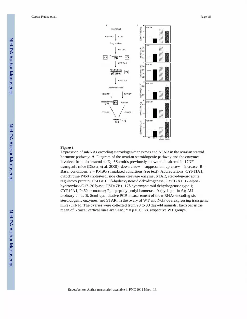
Figure 1.Expression of mRNAs encoding steroidogenic enzymes and STAR in the ovarian steroidhormone pathway. A. Diagram of the ovarian steroidogenic pathway and the enzymesinvolved from cholesterol to E2. *Steroids previously shown to be altered in 17NFtransgenic mice (Dissen et al. 2009); down arrow = suppression, up arrow = increase; B =Basal conditions, S = PMSG stimulated conditions (see text). Abbreviations: CYP11A1,cytochrome P450 cholesterol side chain cleavage enzyme; STAR, steroidogenic acuteregulatory protein; HSD3B1, 3 -hydroxysteroid dehydrogenase, CYP17A1, 17-alpha-hydroxylase/C17–20 lyase; HSD17B1, 17 hydroxysteroid dehydrogenase type 1;CYP19A1, P450 aromatase; Ppia peptidylprolyl isomerase A (cyclophilin A); AU =arbitrary units. B. Semi-quantitative PCR measurement of the mRNAs encoding sixsteroidogenic enzymes, and STAR, in the ovary of WT and NGF overexpressing transgenicmice (17NF). The ovaries were collected from 28 to 30 day-old animals. Each bar is themean of 5 mice; vertical lines are SEM; * = p<0.05 vs. respective WT groups.
Garcia-Rudaz et al. Page 16
Reproduction. Author manuscript; available in PMC 2012 March 13.
NIH
-PA
Author M
anuscriptN
IH-P
A A
uthor Manuscript
NIH
-PA
Author M
anuscript

Figure 2.Protein expression profiles in 17NF and WT (B6D2) ovaries revealed by fluorescence two-dimensional differential gel electrophoresis (2-DIGE). Lysates (100 μg of protein) werelabeled with A. Cy5 (WT, red color) and B. Cy3 (17NF, green color), respectively. C.Images from A and B were merged; numbers (1 to 10) point to differentially expressedproteins. Among these spots, four of them, Spots 2, 4, 5 and 6 were identified with 100% ofstatistical confidence, the sequence of more than two diagnostic peptides per protein (seeMaterial and Methods). Spots 4, 5 and 6 correspond to translationally modified forms ofapolipoprotein AI (Apo AI), the major apoprotein of HDL. Spot 2 corresponds to thephosphorylated form of STMN1. For additional details see text.
Garcia-Rudaz et al. Page 17
Reproduction. Author manuscript; available in PMC 2012 March 13.
NIH
-PA
Author M
anuscriptN
IH-P
A A
uthor Manuscript
NIH
-PA
Author M
anuscript

Figure 3.STMN1 is predominantly expressed in GCs of antral follicles, and is more abundant in theovaries from 17NF mice than in those of WT animals. A. Immunoreactive STMN1 in WTovaries. B. Increased abundance of STMN1 in GCs of 17NF ovaries. Bar = 200 μm. Noticethe increased number of small antral follicles, and the absence of large, preovulatoryfollicles in the 17NF ovary. C–F, Immunoreactive STMN1 is more abundant in preantral (Cand D) and medium sized antral follicles (E and F) of 17NF animals than WT controls.Sections incubated without primary antibody exhibited no discernible immunostaining; notshown). Ten sections from two animals of each genotype were examined; representativesections are presented. Bar = 50 μm. G. Ovarian STMN1 levels quantified by western blotanalysis are greater (*p=0.02) in 17NF ovaries as compared with WT ovaries. AU =arbitrary units. Vertical lines represent SEM and numbers at top of bars are number ofindependent observations per group.
Garcia-Rudaz et al. Page 18
Reproduction. Author manuscript; available in PMC 2012 March 13.
NIH
-PA
Author M
anuscriptN
IH-P
A A
uthor Manuscript
NIH
-PA
Author M
anuscript

Figure 4.The abundance of three major phosphorylated forms of STMN1 (phosphorylated on 16P,25P and 38P, respectively) is increased in the ovaries from 17NF mice as compared to WTovaries. A. The phosphoforms were identified by western blot analysis using ovaries fromprepubertal, 30-day-old mice, and antibodies specific for each form (Gavet et al. 1998).Quantitative results for each phosphoisoform (16P, 25P and 38P) are shown in panels B, C,and D, respectively. The three antibodies recognize both the 19 kDa phosphorylated STMN1form and a reduced electrophoretic mobility species known as stathmin “17”, whichmigrates as a ~23 kDa species (Gavet et al. 1998). Notice that neither WT nor 17NF ovariesexpress STMN1 “17' phosphorylated on 16P. AU = arbitrary units. Each bar is the mean of 4mice; vertical lines represent SEM. * = p<0.05, ** = p<0.01, and *** = p<0.001 vs. WTgroups.
Garcia-Rudaz et al. Page 19
Reproduction. Author manuscript; available in PMC 2012 March 13.
NIH
-PA
Author M
anuscriptN
IH-P
A A
uthor Manuscript
NIH
-PA
Author M
anuscript

Figure 5.Tnf mRNA and TNF protein abundance are increased in 17NF ovaries as compared to WTcontrols, and this increase in TNF production is abolished by blocking high affinity NTRKreceptors. A, Tnf mRNA content in 30-day-old WT and 17NF ovaries measured by semi-quantitative PCR. Ppia = Peptidylprolyl isomerase A (cyclophilin A). B, TNF proteinmeasured by ELISA in protein extracts from 28–30 day-old ovaries incubated for 3 h inKRB buffer at 37 C. Some ovaries were treated during this time with 100 ng/ml of K252a, ablocker of NTRK tyrosine kinase activity. MM = molecular marker; * = p<0.05, and ** =p<0.02 vs. WT group; AU = arbitrary units; vertical lines represent SEM and numbers of topof bars are number of independent observations per group.
Garcia-Rudaz et al. Page 20
Reproduction. Author manuscript; available in PMC 2012 March 13.
NIH
-PA
Author M
anuscriptN
IH-P
A A
uthor Manuscript
NIH
-PA
Author M
anuscript

Figure 6.Blocking TNF actions via in vivo administration of a soluble TNF receptor 2 form coupledto the Fc portion of human IgG1 (Enbrel) prevents the increase in abundance of totalSTMN1 and phosphorylated STMN1 forms seen in ovaries overexpressing NGF. A, TotalSTMN1 expressed as a ratio of the GAPDH signal; B, 16P content normalized usingGAPDH as the reference unit; C, 38P normalized similarly; D, 16P expressed as a fractionof non-phosphorylated stathmin; E, 38P similarly expressed; F, Representative westernblots. WT = wild type ovaries; 17NF+V = ovaries from NGF overexpressing mice treatedwith vehicle; 17NF+E = ovaries from NGF overexpressing mice treated with Enbrel. AU =arbitrary units; columns are mean ± SEM. Each group is the mean of 4 to 8 animals. * =p<0.05 vs. 17NF group not treated with Enbrel; ** = p <0.01 and *** = p<0.001 vs. WTgroup.
Garcia-Rudaz et al. Page 21
Reproduction. Author manuscript; available in PMC 2012 March 13.
NIH
-PA
Author M
anuscriptN
IH-P
A A
uthor Manuscript
NIH
-PA
Author M
anuscript

Figure 7.Blocking TNF actions via in vivo administration of Enbrel (once daily for four days)prevents the increase in GC apoptosis seen in NGF overexpressing ovaries, as assessed byquantitative evaluation of TUNEL reactions. A, The ovaries of 17NF mice treated withEnbrel (E) show a lower incidence of total apoptotic antral follicles as compared withvehicle (V)-treated 17NF mice. B, This difference is due to a lower number of apoptoticsmall-to-medium size (101–300 μm) follicles in the ovaries of Enbrel-treated 17NF micethan in vehicle-treated 17NF animals; C, Representative image of an ovary from a 17NFmouse treated with vehicle; D, Image of an ovary from a 17NF mouse treated with Enbrel.Arrows point to apoptotic medium size follicles; and asterisks identify healthy medium sizefollicles. Bar = 200 μm; columns are mean ± SEM. Each group is the mean of 4 animals. * =p<0.05 vs. 17NF group treated with vehicle.
Garcia-Rudaz et al. Page 22
Reproduction. Author manuscript; available in PMC 2012 March 13.
NIH
-PA
Author M
anuscriptN
IH-P
A A
uthor Manuscript
NIH
-PA
Author M
anuscript

Figure 8.The content of Cyp7b1 mRNA, which encodes CYP7b1 (the enzyme that catalyzes themetabolic degradation of 3 -diol) is increased in the ovaries of 17NF mice as assessed bysemi-quantitative PCR, and plasma levels of 3 -diol are reduced in these animals ascompared to WT animals as assessed by radioimmunoassay. A, Diagram showing the 3 -diol biosynthetic/metabolic pathway; 3 -diol is in bold. B, Content of Cyp7b1 mRNA inovaries of 17NF and WT mice; the ovaries were collected from 28 to 30 day-old animals.AU = arbitrary units; each bar is the mean of five mice. C, Serum concentration of 5α-androstane-3 , 17 diol (3 -diol); each bar is the mean of 12 mice. Bars are mean ± SEM; *= p<0.05 vs. respective WT groups.
Garcia-Rudaz et al. Page 23
Reproduction. Author manuscript; available in PMC 2012 March 13.
NIH
-PA
Author M
anuscriptN
IH-P
A A
uthor Manuscript
NIH
-PA
Author M
anuscript

NIH
-PA
Author M
anuscriptN
IH-P
A A
uthor Manuscript
NIH
-PA
Author M
anuscript
Garcia-Rudaz et al. Page 24
Table 1
PCR primers used for semi-quantitative PCR
mRNA Primer Sequence Range Product
Cyp11a1 Forward GCGCCTGGAGCCATCAAGAACT 491–512 442
NM_019779 Reverse CCCCCAGGAGGCTATAAAGGACAC 909–932
Star Forward CCGGGTGGATGGGTCAAGTT 190–209 428
NM_011485 Reverse GCGCACGCTCACGAAGTCTC 598–617
Hsd3b1 Forward TGCAGGGCCCAACTCGTA 514–531 313
NM_008293 Reverse TGCCCAGGCCACATTTTC 807–826
Cyp17a1 Forward GAAGGCCAGGACCCAAGTGTG 992–1012 418
NM_007809 Reverse CTAAGAAGCGCTCAGGCATAAACC 1384–1409
Hsd17b1 Forward AGGCCGCCAGGACTCAAG 174–191 273
NM_010475 Reverse GCACACGCCCAGAGTGGCGCCTCT 421–446
Cyp19a1 Forward* ACGGGCCCTGGTCTTAT 498–764 404
NM_007810 Reverse CTCTCAGCGAAAATCAAATCA 879–901
Cyp7b1 Forward TTACTGCTCTCGGCCCTGTTCCTC 156–179 502
NM_007825 Reverse TCGCAAATGTGATCTCAAATACCA 632–657
Tnf Forward CAGGGGCCACCACGCTCTTC 281–300 419
NM_013693 Reverse CTTGGGGCAGGGGCTCTTGAC 677–699
Ppia Forward GGCAAATGCTGGACCAAACACAA 341–363 223
NM_008907 Reverse GGTAAAATGCCCGCAAGTCAAAAG 538–563
Ppia mRNA (peptidylprolyl isomerase A) encodes cyclophilin A, and is constitutively expressed in all tissues).
*Primers designed for a separate rat project; some mismatches are present compared to mouse sequence, but they generate a single PCR product
from the mouse ovary.
Reproduction. Author manuscript; available in PMC 2012 March 13.