Excess of proximal microsatellite-stable colorectal cancer in African Americans from a multiethnic...
32
Published OnlineFirst July 10, 2014. Clin Cancer Res Rosa M Xicola, Molly Gagnon, Julia R Clark, et al. African Americans from a multi-ethnic study Excess of proximal microsatellite-stable colorectal cancer in Updated version 10.1158/1078-0432.CCR-14-0353 doi: Access the most recent version of this article at: Material Supplementary http://clincancerres.aacrjournals.org/content/suppl/2014/07/25/1078-0432.CCR-14-0353.DC1.html Access the most recent supplemental material at: Manuscript Author edited. Author manuscripts have been peer reviewed and accepted for publication but have not yet been E-mail alerts related to this article or journal. Sign up to receive free email-alerts Subscriptions Reprints and . [email protected] Department at To order reprints of this article or to subscribe to the journal, contact the AACR Publications Permissions . [email protected] Department at To request permission to re-use all or part of this article, contact the AACR Publications Research. on August 21, 2014. © 2014 American Association for Cancer clincancerres.aacrjournals.org Downloaded from Author manuscripts have been peer reviewed and accepted for publication but have not yet been edited. Author Manuscript Published OnlineFirst on July 10, 2014; DOI: 10.1158/1078-0432.CCR-14-0353 Research. on August 21, 2014. © 2014 American Association for Cancer clincancerres.aacrjournals.org Downloaded from Author manuscripts have been peer reviewed and accepted for publication but have not yet been edited. Author Manuscript Published OnlineFirst on July 10, 2014; DOI: 10.1158/1078-0432.CCR-14-0353
Transcript of Excess of proximal microsatellite-stable colorectal cancer in African Americans from a multiethnic...
Published OnlineFirst July 10, 2014.Clin Cancer Res Rosa M Xicola, Molly Gagnon, Julia R Clark, et al. African Americans from a multi-ethnic studyExcess of proximal microsatellite-stable colorectal cancer in
Updated version
10.1158/1078-0432.CCR-14-0353doi:
Access the most recent version of this article at:
Material
Supplementary
http://clincancerres.aacrjournals.org/content/suppl/2014/07/25/1078-0432.CCR-14-0353.DC1.html
Access the most recent supplemental material at:
Manuscript
Authoredited. Author manuscripts have been peer reviewed and accepted for publication but have not yet been
E-mail alerts related to this article or journal.Sign up to receive free email-alerts
Subscriptions
Reprints and
To order reprints of this article or to subscribe to the journal, contact the AACR Publications
Permissions
To request permission to re-use all or part of this article, contact the AACR Publications
Research. on August 21, 2014. © 2014 American Association for Cancerclincancerres.aacrjournals.org Downloaded from
Author manuscripts have been peer reviewed and accepted for publication but have not yet been edited. Author Manuscript Published OnlineFirst on July 10, 2014; DOI: 10.1158/1078-0432.CCR-14-0353
Excess MSS Colon Cancers in African Americans
1
Title: Excess of proximal microsatellite-stable colorectal cancer in African Americans from a multi-ethnic study Running title: Excess MSS colon cancers in African Americans
Key words: Colorectal cancer, African Americans, Microsatellite instability
Authors: Rosa M Xicola 1, Molly Gagnon1, Julia R Clark1, Timothy Carroll1, Weihua
Gao1, Christian Fernandez1, Dragana Mijic1, James B Rawson1, Ashley
Janoski1, Cenk K. Pusatcioglu1, Priyanka Rajaram1, Adam B Gluskin1,
Maureen Regan1, Vivek Chaudhry2, Herand Abcarian2, Jennifer Blumetti2,
Jose Cintron2, Joshua Melson3, Hui Xie1, Grace Guzman1, Rajyasree
Emmadi1, Victoria Alagiozian-Angelova2, Sonia S. Kupfer5, Carol
Braunschweig1, Nathan A Ellis1, Xavier Llor1, 4.
Authors’ Affiliations:
1University of Illinois at Chicago. Chicago, Illinois
2John H Stroger Jr. Hospital. Chicago, Illinois
3Rush University Medical Center. Chicago, Illinois
4Jesse Brown VA Medical Center. Chicago. Illinois
5University of Chicago. Chicago, Illinois
Grant support:
This work was supported by grants from the American Cancer Society Illinois Division
(223187, X.L.) and the National Cancer Institute (U01 CA153060, N.A.E.). Further support
was provided by an Area of Excellence Award from the University of Illinois at Chicago
Office of the Vice Chancellor for Research, the Department of Medicine, and University of
Illinois Cancer Center. Statistical support was provided by the University of Illinois Center
for Clinical and Translational Science (UL1TR000050). The content is solely the
Research. on August 21, 2014. © 2014 American Association for Cancerclincancerres.aacrjournals.org Downloaded from
Author manuscripts have been peer reviewed and accepted for publication but have not yet been edited. Author Manuscript Published OnlineFirst on July 10, 2014; DOI: 10.1158/1078-0432.CCR-14-0353
Excess MSS Colon Cancers in African Americans
2
responsibility of the authors and does not necessarily represent the official views of the
National Institutes of Health.
Abbreviations: African American: AA; Chicago Colorectal Cancer Consortium:
CCCC; Colorectal cancer: CRC; Elevated microsatellite instability at
selected tetranucleotide repeats: EMAST; Non-Hispanic White: NHW;
Immunohistochemistry: IHC; Microsatellite Instability: MSI;
Microsatellite Stable: MSS; Non-Steroidal Anti Inflammatory drugs:
NSAIDs; Surveillance Epidemiology and End Results: SEER
Correspondence:
Xavier Llor, MD, PhD
Yale University
Section of Digestive Diseases
333 Cedar Street/LMP 1080
P.O. Box 208019
New Haven, CT 06520-8019
Phone# 203-737-8062
Fax# 203-370-0049
Nathan A. Ellis, PhD
University of Arizona
Department of Cellular and Molecular Medicine
Research. on August 21, 2014. © 2014 American Association for Cancerclincancerres.aacrjournals.org Downloaded from
Author manuscripts have been peer reviewed and accepted for publication but have not yet been edited. Author Manuscript Published OnlineFirst on July 10, 2014; DOI: 10.1158/1078-0432.CCR-14-0353
Excess MSS Colon Cancers in African Americans
3
1515 N Campbell Ave
PO Box 245024
Tucson, AZ 85724-5024
Phone 520-626-4134
Disclosures of Potential Conflicts of Interest: None of the authors have any potential
conflicts of interest to disclose that are relevant to the manuscript
Author contributions:
RM Xicola, NA Ellis, and X Llor designed and supervised the overall project, analyzed and
interpreted the data, and drafted the manuscript. They take full responsibility for the
integrity of the data and the accuracy of the data analysis. R Emmadi critically revised the
manuscript for important intellectual content. V Chaudhry, H Abcarian, J Blumetti, J
Cintron, J Melson, SS Kupfer, C Braunchweig supervised different aspects of the
recruitment and data acquisition. R Emmadi, G Guzman, R Bushman, V Alagiozian-
Angelova revised and selected all pathology specimens. W Gao performed the variable
imputation for the statistical analysis. Hui Xie supervised the statistical analysis. M
Gagnon, and A Janoski processed and analyzed data. JR Clark, D Mijic, C Pusatcioglu, P
Rajaram, enrolled patients and acquired data. AB Gluskin, T Carroll, C Fernandez, JB
Rawson and Maureen Regan processed, prepared and analyzed the samples.
The study sponsors had no role in the design of the study; no role in the collection,
analysis, or interpretation of the data; no role in the writing of the manuscript; and no role
in the decision to submit the manuscript for publication. The authors gratefully
Research. on August 21, 2014. © 2014 American Association for Cancerclincancerres.aacrjournals.org Downloaded from
Author manuscripts have been peer reviewed and accepted for publication but have not yet been edited. Author Manuscript Published OnlineFirst on July 10, 2014; DOI: 10.1158/1078-0432.CCR-14-0353
Excess MSS Colon Cancers in African Americans
4
acknowledge the recruiters of the Chicago Colorectal Cancer Consortium for their
dedication and integrity, including Maggie Moran, Katy Ceryes, Katya Seligman, Amy
Disharoon, Katie Morrissey, and Archana Krishnan. DNA sequencing and fragment
analysis were performed in the Center for Genomics Research in the University of Illinois
Cancer Center under the supervision of Dr. Stefan Green.
Word count: 3939
Total number of figures and tables: 4 tables; 2 supplementary tables; and 1
Supplementary figure
Research. on August 21, 2014. © 2014 American Association for Cancerclincancerres.aacrjournals.org Downloaded from
Author manuscripts have been peer reviewed and accepted for publication but have not yet been edited. Author Manuscript Published OnlineFirst on July 10, 2014; DOI: 10.1158/1078-0432.CCR-14-0353
Excess MSS Colon Cancers in African Americans
5
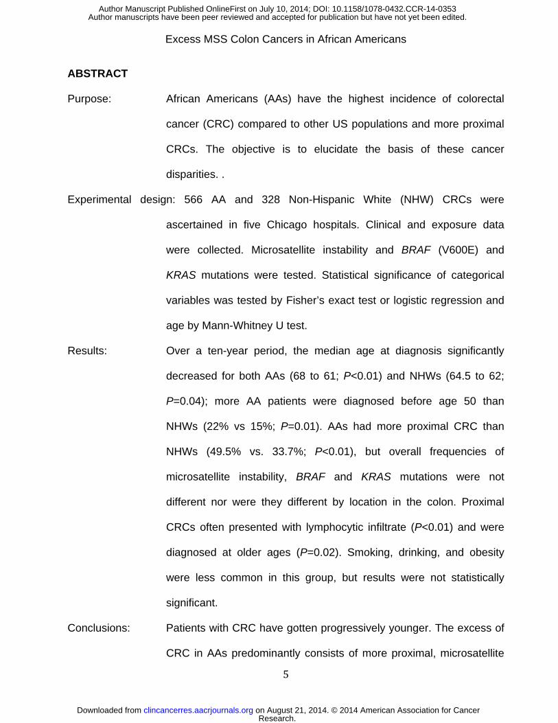
ABSTRACT
Purpose: African Americans (AAs) have the highest incidence of colorectal
cancer (CRC) compared to other US populations and more proximal
CRCs. The objective is to elucidate the basis of these cancer
disparities. .
Experimental design: 566 AA and 328 Non-Hispanic White (NHW) CRCs were
ascertained in five Chicago hospitals. Clinical and exposure data
were collected. Microsatellite instability and BRAF (V600E) and
KRAS mutations were tested. Statistical significance of categorical
variables was tested by Fisher’s exact test or logistic regression and
age by Mann-Whitney U test.
Results: Over a ten-year period, the median age at diagnosis significantly
decreased for both AAs (68 to 61; P<0.01) and NHWs (64.5 to 62;
P=0.04); more AA patients were diagnosed before age 50 than
NHWs (22% vs 15%; P=0.01). AAs had more proximal CRC than
NHWs (49.5% vs. 33.7%; P<0.01), but overall frequencies of
microsatellite instability, BRAF and KRAS mutations were not
different nor were they different by location in the colon. Proximal
CRCs often presented with lymphocytic infiltrate (P<0.01) and were
diagnosed at older ages (P=0.02). Smoking, drinking, and obesity
were less common in this group, but results were not statistically
significant.
Conclusions: Patients with CRC have gotten progressively younger. The excess of
CRC in AAs predominantly consists of more proximal, microsatellite
Research. on August 21, 2014. © 2014 American Association for Cancerclincancerres.aacrjournals.org Downloaded from
Author manuscripts have been peer reviewed and accepted for publication but have not yet been edited. Author Manuscript Published OnlineFirst on July 10, 2014; DOI: 10.1158/1078-0432.CCR-14-0353
Excess MSS Colon Cancers in African Americans
6
stable tumors, commonly presenting lymphocytic infiltrate and less
often associated with toxic exposures or a higher BMI. Younger AAs
had more distal CRCs than older ones. These data suggest two
different mechanisms driving younger age and proximal location of
CRCs in AAs.
Key words: Colorectal cancer, African Americans, Microsatellite instability
Research. on August 21, 2014. © 2014 American Association for Cancerclincancerres.aacrjournals.org Downloaded from
Author manuscripts have been peer reviewed and accepted for publication but have not yet been edited. Author Manuscript Published OnlineFirst on July 10, 2014; DOI: 10.1158/1078-0432.CCR-14-0353
Excess MSS Colon Cancers in African Americans
7
Statement of translational relevance
African Americans (AAs) disproportionately die from colorectal cancer (CRC), and
health disparities compared with whites are not well understood. We show that a very
significant number of AAs are diagnosed before age 50 compared with whites, and their
tumors are usually more advanced at diagnosis. Furthermore, in AAs there is a significant
excess of proximal CRCs, consisting of microsatellite stable tumors commonly exhibiting
lymphocytic infiltrate and less often associated with smoking, drinking, or higher BMI.
Because proximal CRCs are associated with higher missed tumor rates on colonoscopy
and increased risk of interval cancers, we believe clinical practice should be changed to
screen AAs at earlier ages and to lower the threshold for performing diagnostic tests when
suspicious symptoms present in young AAs. Specific screening and diagnostic
approaches will help eliminate CRC health disparities in AAs.
Research. on August 21, 2014. © 2014 American Association for Cancerclincancerres.aacrjournals.org Downloaded from
Author manuscripts have been peer reviewed and accepted for publication but have not yet been edited. Author Manuscript Published OnlineFirst on July 10, 2014; DOI: 10.1158/1078-0432.CCR-14-0353
Excess MSS Colon Cancers in African Americans
8
Introduction
Colorectal cancer (CRC) represented 9% of all diagnosed cancers in the US in
2012(1) but incidence rates had started a slow and steady decline almost thirty years ago,
even before the generalization of CRC screening(2). In contrast to the general decline,
Surveillance Epidemiology and End Results (SEER) data have shown that since 1992
CRC incidence rates are increasing among adults younger than 50(3). Some studies have
suggested that young-onset CRC seems to disproportionally affect non-white,
underinsured patients who live in southern and western parts of the US(4). (5)Because
most adults younger than age 50 are not screened for CRC, the shift towards younger
ages at diagnosis very likely is not be explained by earlier detection. The lower mean age
of presentation in African Americans (AAs) has prompted some medical organizations to
recommend CRC screening for average risk AAs to be started at a younger age than the
current recommendation of age 50 in Non-Hispanic Whites (NHWs)(6).
Another important difference between AA and NHW CRC patients is the higher
incidence of proximal adenomas and cancers (defined here as tumors proximal to the
splenic flexure) documented in AAs over the last 30 years(7, 8). The site of tumor
development in CRC has important implications not only related to screening but also due
to the distinct biological features and prognosis(9). For example, death from proximal
CRCs seems to be less preventable by colonoscopy performance(10) and this outcome
could be related to higher miss rates of proximal lesions(11). Missed tumors as well as
lower screening rates could also contribute to the reported more advanced stages at
presentation of CRC in AAs compared to NHWs(7).
Significant biological differences exist between proximal and distal CRCs, including
a higher percentage of tumors with microsatellite instability (MSI), increased
Research. on August 21, 2014. © 2014 American Association for Cancerclincancerres.aacrjournals.org Downloaded from
Author manuscripts have been peer reviewed and accepted for publication but have not yet been edited. Author Manuscript Published OnlineFirst on July 10, 2014; DOI: 10.1158/1078-0432.CCR-14-0353
Excess MSS Colon Cancers in African Americans
9
hypermethylation, and increased gene mutation rates in proximal tumors(12). These
differences could have prognostic and therapeutic implications. In this regard, some
authors have suggested the presence of higher MSI rates and CpG island
hypermethylation in AAs(13). Environmental factors such as diet could also play a role in
CRC differences; for example, higher red blood cell folate levels have been associated
with increased methylation of genes that control colonic growth and cell differentiation in
tumors from AAs(14).
A higher frequency of MSI in AA CRCs might explain the increased proportion of
proximal CRCs in this population(15, 16); however, MSI frequency in AA CRC has not
been well determined, as studies have been limited by relatively small samples sizes(16-
19). To address this limitation, we investigated the presence of MSI as a potential
explanation for the excess proximal CRC in AAs in a large collection of unselected cases.
We addressed the MSI hypothesis and related questions in cases from the Chicago
Colorectal Cancer Consortium (CCCC)—a multi-institutional study of CRC in an ethnically
diverse urban area. The present report from the CCCC is the most comprehensive one to
date that compares differences in key clinical and molecular features in AA and NHW
CRCs living in a single geographic area.
Materials and Methods
Ascertainment, Recruitment, and Study design
The CCCC includes five large Chicago medical centers: University of Illinois
Hospital and Health Sciences System (UIHHS), Jesse Brown Veterans Administration
(JBVA), John H. Stroger Hospital of Cook County (JHSHCC), University of Chicago
Medicine, and Rush University Medical Center. The CCCC ascertained incident CRC
Research. on August 21, 2014. © 2014 American Association for Cancerclincancerres.aacrjournals.org Downloaded from
Author manuscripts have been peer reviewed and accepted for publication but have not yet been edited. Author Manuscript Published OnlineFirst on July 10, 2014; DOI: 10.1158/1078-0432.CCR-14-0353
Excess MSS Colon Cancers in African Americans
10
patients from search of Pathology records (the majority of cases coming from 1997-2010)
and from prospective enrolment in surgery and endoscopy units (2011-2012). Patients
with CRC recurrence; inflammatory bowel disease; non-adenocarcinoma tumors were
excluded. Patients with unspecified tumor locations or multiple primaries were excluded
from the analysis.
From this ascertainment we were able to include 894 CRC patients (566 AAs and
328 NHWs). Other ethnic groups were not included. Biological samples were available
from 635 individuals: 409 AAs and 228 NHWs. For every type of analysis, the number of
patients that were available and informative for analysis is expressed as the denominator
in every cell of the tables. The median age at diagnosis was 63, and 56.7% were male
and 43.3% female.
For all cases we collected data from pathology, radiology, endoscopy, clinical, and
operative reports. Cancer staging was determined according to criteria set by the
American Joint Committee on Cancer staging system (20). Mucinous phenotype was
considered positive when more than 50% of the tumor displayed mucin production. (20).
For the subset of cases that were prospectively enrolled, we administered an
extensive personal questionnaire. The questionnaire collected information on
demographics and socio-economic data, medical and family history of cancer (traced
backward and laterally at least up to second-degree relatives), smoking and alcohol
consumption, use of medications and supplements over the previous 5 years, and
physical exercise. Significant exercise constituted at least 150 minutes a week of
moderate intensity exercise or 75 minutes a week of vigorous intensity exercise as
determined by the 2008 Physical Activity Guidelines for Americans(21). Obesity was
assessed by body mass index (BMI)(21). The study was conducted according to the
Research. on August 21, 2014. © 2014 American Association for Cancerclincancerres.aacrjournals.org Downloaded from
Author manuscripts have been peer reviewed and accepted for publication but have not yet been edited. Author Manuscript Published OnlineFirst on July 10, 2014; DOI: 10.1158/1078-0432.CCR-14-0353
Excess MSS Colon Cancers in African Americans
11
corresponding approved IRB protocol at each institution.
Bio-specimen Collection
For cases ascertained through searches of pathology records, paraffin blocks and
slides were pulled from the Pathology Department archives. Areas of tumor and non-
tumor tissue were identified and cores were collected. Paraffin was removed using an
octane/methanol method(22). DNA was then prepared using the Gentra Puregene DNA
Isolation kit (Qiagen, Valencia, CA) according to the manufacturer’s instructions, except
the proteinase K extraction step was extended to three days, adding fresh enzyme on
each day, and the sample was heated at 95ºC for 15 minutes prior to protein precipitation.
For cases ascertained in surgery or endoscopy, fresh biopsies were taken from tumors
and uninvolved colonic mucosa 10 cm away from the tumor. Biopsies were preserved in
RNAlater® (Life Technologies Corporation, Grand Island, NY) buffer and frozen. DNAs
were extracted from ground tissue using the Maxwell® 16 Tissue DNA Purification Kit
(Promega, Fitchburg, WI).
Molecular Analysis
MSI was assessed in paired DNA samples from tumor and uninvolved tissue. The
panel of mononucleotide markers included NR21, NR22, NR24, NR27, BAT25, and
BAT26(23). The use of a mononuclotide panel has been shown to be superior for MSI
detection than the NCI panel(24). Multiplex PCR amplified all markers, and PCR products
were analyzed by capillary electrophoresis, as previously described(24). The most
common mutations of BRAF (V600E)(25) and KRAS (codons 12 and 13)(26) were
analyzed through direct DNA sequencing. Amplification and sequencing of the candidate
regions was performed as previously described(27).
Research. on August 21, 2014. © 2014 American Association for Cancerclincancerres.aacrjournals.org Downloaded from
Author manuscripts have been peer reviewed and accepted for publication but have not yet been edited. Author Manuscript Published OnlineFirst on July 10, 2014; DOI: 10.1158/1078-0432.CCR-14-0353
Excess MSS Colon Cancers in African Americans
12
Illinois State Cancer Registry Data
Chicago is part of Cook County, the second most populous county in the US with
5,231,351 residents. The Illinois State Cancer Registry (ISCR) collects statewide cancer
data through mandated reporting by medical centers, pathology labs, and through data
exchange with other states. Cook County CRC incidence and staging data was obtained
from the publicly available dataset of the ISCR(28). AA and NHW CRC patients registered
between 1991 and 2010 (3,553 AAs and 10,247 NHWs CRC cases) were analyzed by
age of diagnosis and cancer stage.
Statistical Analysis
Differences in categorical variables were assessed by the Fisher’s exact test or Chi
square test. Differences in age were analyzed by the Mann-Whitney U test. We performed
a primary analysis on all cases for which data was available in more than 80% of
individuals. Primary analysis included age, sex, histologic grade, and microsatellite
instability. We performed a secondary analysis on the subset of cases from which data
was available through the administration of the personal questionnaire and their medical
records.
To identify factors associated with proximal tumor location in AAs, we performed
logistic regression that included the following co-variates: first-degree relative with CRC;
previous colonoscopy; previous colon polyps; exercise; smoking (packs/year); alcohol
(g/day); use of aspirin, NSAIDs, COX2 inhibitors, and statins; mucinous phenotype; BMI;
tumor stage; lymphocytic infiltrate, histologic grade and age. Before performing the logistic
regression, in order to include all AA patients in the analysis, we used the Multivariate
Research. on August 21, 2014. © 2014 American Association for Cancerclincancerres.aacrjournals.org Downloaded from
Author manuscripts have been peer reviewed and accepted for publication but have not yet been edited. Author Manuscript Published OnlineFirst on July 10, 2014; DOI: 10.1158/1078-0432.CCR-14-0353
Excess MSS Colon Cancers in African Americans
13
Imputations by Chained Equations (MICE) procedure to impute missing data based on the
set of patients with available data. Normally distributed variables were imputed using
predictive mean matching, binary variables by logistic regression, and categorical
variables with >2 levels by polytomous logistic regression. Final estimates of odds ratios
(ORs), 95% confidence intervals (CIs), and P values were calculated by averaging
statistics across the 50 complete datasets that we imputed and computing total variance
by Rubin’s rules(29). The MICE package in R was used to perform the logistic regression
on imputed data(30). All reported P values correspond to two-sided tests. Differences
were considered statistically significant if the P value was less than 0.05. All statistical
analyses were carried out using R 3.0.0(31).
Results
Age and Stage at Diagnosis of CRC
To determine whether the change in the age distribution of CRC cases in the
Chicago population is similar to the change observed in the general US population, we
compared the median age of diagnosis in the group of patients ascertained prospectively
(2011-2012) with a similarly-sized group of patients diagnosed with CRC ten years ago
(2000-2002). Patients in both ethnic groups diagnosed with CRC in 2011-2012 had a
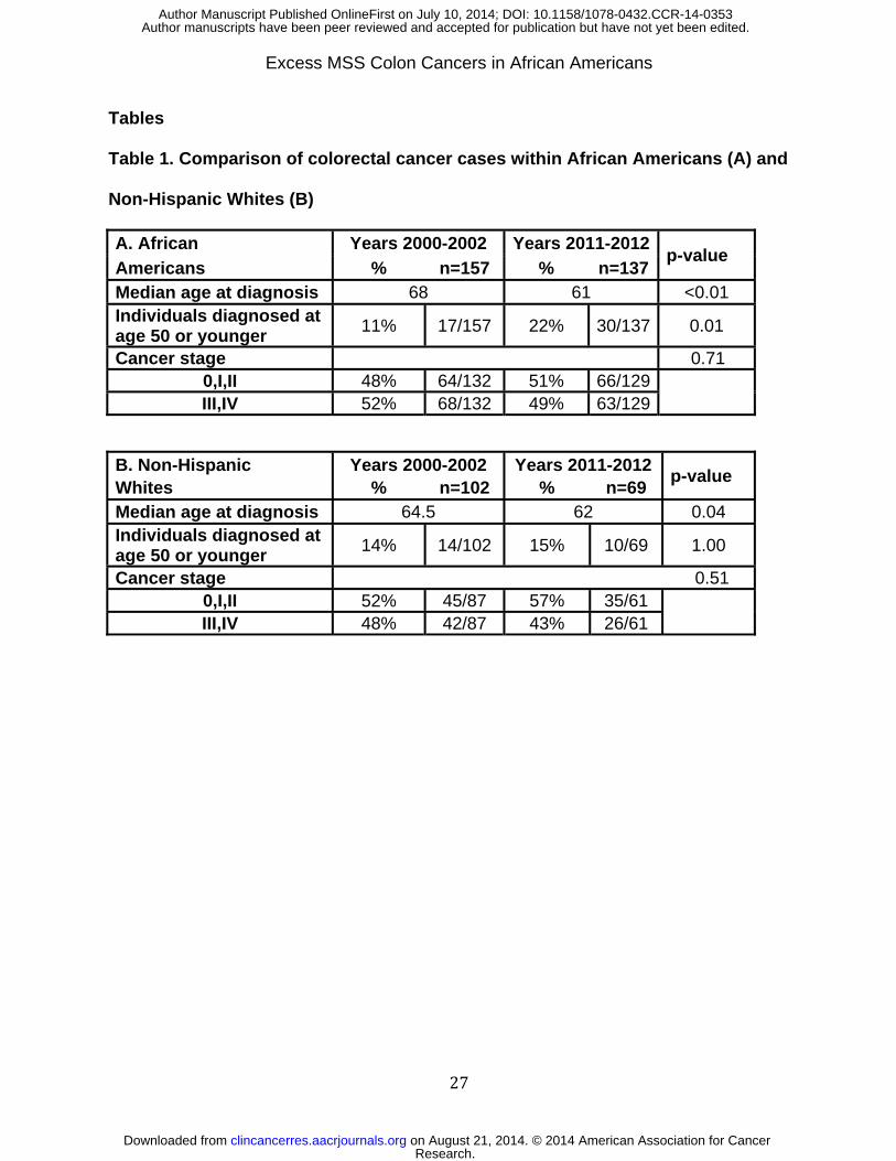
significantly lower median age at diagnosis than those diagnosed 10 years ago: 61 vs. 68
for AAs (P<0.01) and 62 vs. 64.5 for NHWs (P=0.04) (Tables 1A and B). Over this period,
the percentage of patients diagnosed with CRC before age 50 went from 11% to 22% in
AAs but the percentage was not different in NHWs (Tables 1A and B). To determine
whether the shift to younger ages at diagnosis could be related to earlier detection, we
compared cancer stage distribution between the 2000-2002 and 2011-2012 groups. In the
Research. on August 21, 2014. © 2014 American Association for Cancerclincancerres.aacrjournals.org Downloaded from
Author manuscripts have been peer reviewed and accepted for publication but have not yet been edited. Author Manuscript Published OnlineFirst on July 10, 2014; DOI: 10.1158/1078-0432.CCR-14-0353
Excess MSS Colon Cancers in African Americans
14
2011-2012 groups, there was a 3% and 5% increase in cases with early stage CRC (0-II)
in AAs and NHWs, respectively, although these comparisons did not reach statistical
significance (Tables 1A and B). The shift towards earlier ages of diagnosis was also
observed and found to be significant in the CRC data for Cook County extracted from the
ISCR (Supplementary Figure and Supplementary Table 1), indicating that the shift is not
restricted to the hospitals sampled in the CCCC. The median ages of diagnosis of the
2011-12 CCCC cases were lower than the median age data from the most recent SEER
(2005-2009) nationwide survey, which showed median ages of 65 for AAs and 70 for
NHWs(32).
Features that Distinguish AA CRC by Age
After observing the high percentage of young AA patients with CRC, we explored
factors that could associate with younger age at diagnosis. AA patients 50 years and
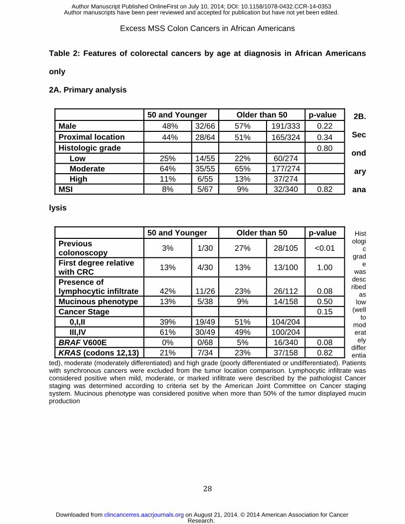
younger were evenly distributed by sex as opposed to older patients that showed a male
predominance similar to what is reported for CRC as a whole (57%) (Table 2A). Although
the presence of a higher proportion of younger patients in the AA population could
suggest a bigger genetic component, the secondary analysis showed that younger
patients did not have more first-degree relatives with CRC (P=1; Table 2B). CRCs from
younger patients tended to have more lymphocytic infiltrate than older patients (42% vs.
23%; P=0.08) and more advanced cancer stages (61% vs. 49.0; P=0.15). No differences
were seen in histological grade or mucinous phenotype. The frequencies of MSI and
KRAS mutations were similar in both age groups but no BRAF mutations were found in
the younger AAs (Table 2B). Only 3% of younger patients had had a colonoscopy prior to
diagnosis vs. 27% of the older patients (P<0.01; Table 2B).
Research. on August 21, 2014. © 2014 American Association for Cancerclincancerres.aacrjournals.org Downloaded from
Author manuscripts have been peer reviewed and accepted for publication but have not yet been edited. Author Manuscript Published OnlineFirst on July 10, 2014; DOI: 10.1158/1078-0432.CCR-14-0353
Excess MSS Colon Cancers in African Americans
15
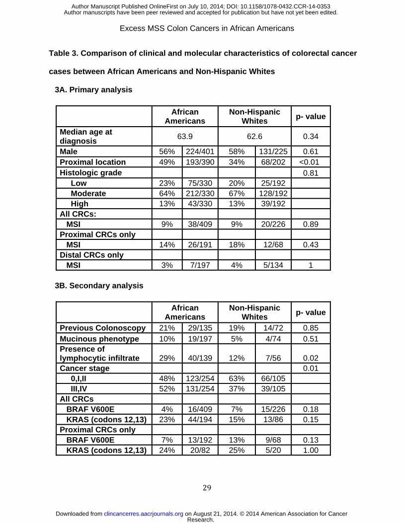
Clinical and Molecular Features that Distinguish AA and NHW CRC
Some studies have reported significant differences in some tumor features
between AAs and NHWs, such as lower histologic grade(7, 33), more advanced
stages(7), and more proximal tumors in AAs(7). We did not see any differences in
histologic grade, but AAs had a significantly higher percentage of proximal tumors than
NHWs (49.5% vs. 33.7%; P=0.01) (Table 3A). Furthermore, from the secondary analysis
there was a significantly higher frequency of advanced stage tumors in AAs (52% in AAs
vs 37% in NHWs; P=0.01) and tumor lymphocytic infiltrate was more common (29% in AA
vs 12% in NHWs; P=0.02) (Table 3B). Data from the Cook County registry 2006-2010
also showed less localized and more metastatic CRCs in AAs compared to NHWs
(Supplementary Figure 1B and Supplementary Table 1B).
Some authors have suggested that AA patients have much higher frequencies of
MSI tumors than NHWs(13), which could explain the higher frequency of proximal CRCs
in AAs. We did not identify a significant difference in the percentage of CRC cases with
MSI between AAs and NHWs (Table 3A). Proximal CRCs more often exhibited MSI than
distal CRCs in both AAs and NHWs, but MSI frequencies in proximal CRCs were no
different between the two ethnic groups (P=0.43) (Table 3A). The frequencies of KRAS
mutations in proximal CRCs were similar in both ethnic groups, whilst BRAF mutations
were less frequent in proximal CRCs in AAs, but not significantly so. The frequencies of
MSI in older and younger age groups were also similar (P=0.82; Table 2A).
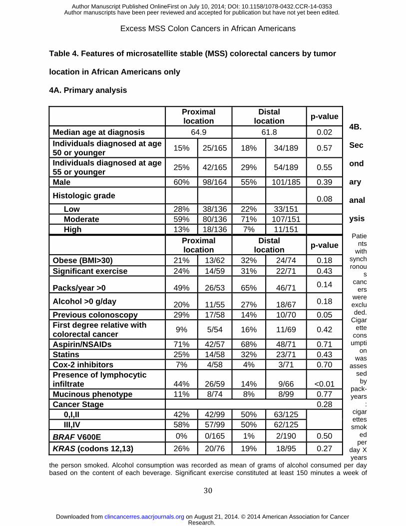
Features that Distinguish Proximal and Distal Microsatellite Stable CRCs in AAs
In order to understand what biological factors might be driving MSS proximal
CRCs, we tested factors that could correlate with these CRCs in AAs. Whereas male
Research. on August 21, 2014. © 2014 American Association for Cancerclincancerres.aacrjournals.org Downloaded from
Author manuscripts have been peer reviewed and accepted for publication but have not yet been edited. Author Manuscript Published OnlineFirst on July 10, 2014; DOI: 10.1158/1078-0432.CCR-14-0353

Excess MSS Colon Cancers in African Americans
16
gender was not significantly associated with proximal MSS CRCs (P=0.39; Table 4A),
patients with proximal MSS CRCs were older (P=0.02; 4A). Younger patients had more
distal than proximal CRCs, though this difference was not significant (Table 4A). In a
multivariate analysis of variables from the secondary analysis, tumor lymphocytic infiltrate
was independently associated with proximal location in MSS CRCs in AAs (OR=8.3, 95%
CI 1.11-62.30; P=0.04). More proximal MSS CRCs than distal MSS CRCs were
diagnosed at later stages (P=0.28), despite the patients having undergone more previous
colonoscopies (P=0.05). On the other hand, a higher percentage of cases with distal MSS
tumors were obese (BMI30 in 32% patients with distal CRC vs. 21% proximally), alcohol
users (27% vs. 20.0%), and smokers (65% vs. 49%); however none of these differences
were statistically significant. Finally, the frequencies of KRAS and BRAF mutations were
indistinguishable in proximal and distal MSS CRCs (Table 4B).
Discussion
Using patients ascertained through the CCCC, we collected samples and clinical
data on an ethnically mixed population recruited in the same geographical area, allowing
for a robust comparison over time and between AAs and NHWs—ethnic groups with a
large disparity in both CRC incidence and CRC mortality. To our knowledge this study
includes the largest group of AA CRC patients reported to date with not only granular
clinical data but also relevant tumor molecular features.
The CCCC data collected presents a dynamic picture of CRC that has evolved
over a relatively short period of time towards a younger age at diagnosis in both AAs and
NHWs, similar to what has been observed in the SEER registry(3). This shift in age of
diagnosis is also observed in the ISCR data for Cook County where 20% (773 out of
Research. on August 21, 2014. © 2014 American Association for Cancerclincancerres.aacrjournals.org Downloaded from
Author manuscripts have been peer reviewed and accepted for publication but have not yet been edited. Author Manuscript Published OnlineFirst on July 10, 2014; DOI: 10.1158/1078-0432.CCR-14-0353
Excess MSS Colon Cancers in African Americans
17
3,878) of AAs and 14% (1,046 out of 7715) of NWHs were diagnosed before age 55 in the
period 2006-2010 (Supplementary Figure and Supplementary Table 1A). A predominance
of AAs among younger patients with CRC has also been reported within the National
Cancer Database, a large hospital-based cancer registry(4).
Remarkably, while older patients are being diagnosed at earlier stages, the
younger patients present with more advanced cancer. It is plausible that the widespread
use of endoscopic procedures for either diagnostic purposes or secondary to
implementation of screening strategies has contributed to the overall increase in detection
of CRCs at earlier stages. In fact, this trend has been recently shown in the National
Bowel Cancer Screening Program in South Australia(34). Less clear is why CRC is
affecting higher numbers of younger individuals (particularly AAs) and why these CRCs
are more often diagnosed when the cancer is more advanced. Younger ages at diagnosis
could suggest a higher proportion of familial or syndromic cases; however, the frequency
of younger AA cases with relatives with CRC was not increased, and the shift in age of
diagnosis over such a short time period is unlikely to be explained by genetic causes.
Moreover, Lynch syndrome, the most common of all hereditary CRC syndromes, is
associated with MSI and the frequency of MSI was not increased in younger AA CRC
cases.
Given the shift towards earlier age of diagnosis, we believe it might be wise to
evaluate the effectiveness of CRC screening strategies at younger ages, particularly in
AAs. In fact, some authors have already suggested an earlier screening age in AAs(6).
Furthermore, physicians may need to lower the threshold that prompts them to order
diagnostic procedures in younger individuals with suspicious symptoms that due to their
young age would not raise a high level of suspicion for colorectal malignancy.
Research. on August 21, 2014. © 2014 American Association for Cancerclincancerres.aacrjournals.org Downloaded from
Author manuscripts have been peer reviewed and accepted for publication but have not yet been edited. Author Manuscript Published OnlineFirst on July 10, 2014; DOI: 10.1158/1078-0432.CCR-14-0353
Excess MSS Colon Cancers in African Americans
18
One of the main goals of this study was to determine whether an increased
frequency of MSI in AA CRC could explain the increased proportion of proximal CRCs in
this population. Our data provided a strong counterpoise to this hypothetical explanation.
With 409 AA and 226 NHW CRCs assayed, the frequency of MSI CRC in AAs was found
to be no different from the frequency in the NHWs. In fact, other studies (17, 19) that also
compared AAs and NHWs failed to find differences in MSI frequencies between AAs and
NHWs (Supplementary Table 2). Moreover, when our data is combined with all the
available studies, the MSI frequencies between AAs and NHWs are nearly identical to
each other (Supplementary Table 2). The differences in frequencies across the various
studies could reflect differences in biological factors that underlie MSI (e.g., age, gender,
and environmental triggers); alternatively, and not exclusively, as the frequency of MSI is
low, the differences could reflect the vagaries of sampling. The MSI frequency in the
present report is almost identical to that found in the Epicolon study, based on 1,200
consecutive patients from Spain(24). The robustness of our MSI testing methodology was
asserted in that study as we showed an extremely high level of concordance between
presence of MSI and loss of expression of mismatch repair proteins(24). Finally, it is worth
noting that the study that showed the highest incidence of MSI in tumors from AA patients
was based on limited number of cases(15).
Similarly, we did not see any significant differences in mutational rates of the
commonly mutated MAP kinase genes, KRAS and BRAF, between AAs and NHWs.
Neither did we observe a predominance of KRAS codon 13 mutations in proximal MSS
tumors in AAs, as previously reported(17).
We did observe biological differences in AA CRC compared to NHW CRC,
characterized primarily by more proximal tumors, which has been observed in many
Research. on August 21, 2014. © 2014 American Association for Cancerclincancerres.aacrjournals.org Downloaded from
Author manuscripts have been peer reviewed and accepted for publication but have not yet been edited. Author Manuscript Published OnlineFirst on July 10, 2014; DOI: 10.1158/1078-0432.CCR-14-0353
Excess MSS Colon Cancers in African Americans
19
studies of AA CRC. As noted above, these proximal tumors in AAs do have the same
percentage of MSI as NHWs. Because the tumors on the right side still have a relatively
low percentage of MSI, the great majority of right-sided tumors are MSS in both ethnic
groups. Thus, in terms of percentages, AAs have many more right-sided MSS tumors
than NHWs. Additionally; this group of proximal MSS tumors in AAs is characterized by
presence of lymphocytic infiltrate. Altogether, we find the overall excess of CRCs in AAs is
mostly contributed by a higher prevalence of the proximal MSS phenotype. CD8+-type
lymphocytic infiltrations have been associated with both MSI and MSS CRCs(35).
Elevated microsatellite instability at selected tetranucleotide repeats (EMAST) in MSS
CRCs has been linked to inflammatory processes in the tumor and heterogeneous
expression of the DNA mismatch repair protein MSH3(36). It is possible that the excess
proximal MSS, inflammatory CRCs seen in AAs is related in some way to the EMAST
phenomenon. Further studies are warranted to test whether specific lymphocyte and
novel genomic-instability phenotypes are associated with proximal MSS CRCs in AAs.
Ferracin et al. reported the association of a subgroup of proximal MSS cancers
with BRAF mutations, CpG island methylator phenotype (CIMP), mucinous phenotype,
chromosomal instability, and a unique gene expression profile(37)—a pattern of clinical
correlations that is similar to those observed in MSI tumors. These observations suggest
that a subset of proximal tumors originate through the methylator phenotype but only
those with MLH1 promoter methylation develop MSI. Bond et al.(38) suggested that MSS
cancers with BRAF mutations are fundamentally different from MSI/BRAF mutated
cancers but that both types of tumors preferentially develop in the proximal colon.
MSS/BRAF mutated CRCs were found to have levels of chromosomal instability (CIN)
that increase with more advanced stages of presentation, suggesting that CIN may
Research. on August 21, 2014. © 2014 American Association for Cancerclincancerres.aacrjournals.org Downloaded from
Author manuscripts have been peer reviewed and accepted for publication but have not yet been edited. Author Manuscript Published OnlineFirst on July 10, 2014; DOI: 10.1158/1078-0432.CCR-14-0353
Excess MSS Colon Cancers in African Americans
20
contribute to progression of this phenotype. In our series, none of the MSS proximal
tumors had BRAF mutations and no distinct association was found with mucinous
phenotype. Consequently, these phenotypes did not make a significant contribution to the
MSS proximal cancers in the AA patients in our series, although other markers, such as
CIMP and CIN, should be assessed. It is unclear why the phenotype described in these
manuscripts, while relatively infrequent, is basically not seen in our series, as we did not
detect any BRAF V600E mutations in proximal MSS tumors in AAs. It is possible that AAs
may have different BRAF mutations and our analysis restricted to V600E could limit this
assessment. In any case, further molecular characterization in the described group will be
essential to better understand this difference between AA and NHW CRCs.
The much higher percentage of proximal CRCs in AAs constitutes an added
challenge for AAs, because these tumors are reportedly more likely to be missed by
colonoscopies(10, 11) and interval cancers (CRCs discovered at or before the next
recommended screening/surveillance colonoscopy) have been repeatedly found to
appear twice as often in the proximal colon(39).
Could the additional proximal tumors in AAs be explained by factors such as toxic
exposures, body habitus, obesity, or physical exercise? On the contrary, distal MSS
tumors were more often diagnosed in obese patients and consumers of alcohol or
tobacco, though our study probably was underpowered to prove this hypothesis. Younger
AAs present with more distal tumors than proximal ones, which suggests that the increase
of CRC in young AAs could be linked to environmental factors.
Our study had some limitations. Patients have been recruited within a limited
geographical urban area with mostly modest income households; therefore, data may not
be fully generalizable to other communities. The limited number of patients with data on
Research. on August 21, 2014. © 2014 American Association for Cancerclincancerres.aacrjournals.org Downloaded from
Author manuscripts have been peer reviewed and accepted for publication but have not yet been edited. Author Manuscript Published OnlineFirst on July 10, 2014; DOI: 10.1158/1078-0432.CCR-14-0353
Excess MSS Colon Cancers in African Americans
21
such factors as toxic exposures, body habitus or exercise also reduces the possibility of
drawing more firm conclusions on the differential effect of these factors.
In summary, our data strongly supports the conclusion that the excess of proximal
CRC in AAs consists of MSS tumors, commonly presenting lymphocytic infiltrate and less
often associated with toxic exposures or a higher BMI. In addition, AAs are more often
diagnosed with CRC at younger ages than NWHs. The clinical evidence suggests that the
different mechanisms drive the younger ages of diagnosis and the proximal MSS CRCs.
Given the trend towards earlier cancer presentation, CRC screening approaches require
further evaluation, especially in AAs.
Research. on August 21, 2014. © 2014 American Association for Cancerclincancerres.aacrjournals.org Downloaded from
Author manuscripts have been peer reviewed and accepted for publication but have not yet been edited. Author Manuscript Published OnlineFirst on July 10, 2014; DOI: 10.1158/1078-0432.CCR-14-0353
Excess MSS Colon Cancers in African Americans
22
References
1. Society AC. Cancer Facts & Figures 2012. Atlanta: American Cancer Society Inc.;
2012 2012.
2. Agrawal S, Bhupinderjit A, Bhutani MS, Boardman L, Nguyen C, Romero Y, et al.
Colorectal cancer in African Americans. Am J Gastroenterol. 2005;100:515-23; discussion
4.
3. Siegel RL, Jemal A, Ward EM. Increase in incidence of colorectal cancer among
young men and women in the United States. Cancer Epidemiol Biomarkers Prev.
2009;18:1695-8.
4. You YN, Xing Y, Feig BW, Chang GJ, Cormier JN. Young-onset colorectal cancer:
is it time to pay attention? Archives of internal medicine. 2012;172:287-9.
5. You YN. Risk factors for young-onset advanced colorectal cancer-reply. Archives
of internal medicine. 2012;172:971-2.
6. Rex DK, Johnson DA, Anderson JC, Schoenfeld PS, Burke CA, Inadomi JM.
American College of Gastroenterology guidelines for colorectal cancer screening 2009
[corrected]. Am J Gastroenterol. 2009;104:739-50.
7. Irby K, Anderson WF, Henson DE, Devesa SS. Emerging and widening colorectal
carcinoma disparities between Blacks and Whites in the United States (1975-2002).
Cancer Epidemiol Biomarkers Prev. 2006;15:792-7.
8. Thornton JG, Morris AM, Thornton JD, Flowers CR, McCashland TM. Racial
variation in colorectal polyp and tumor location. J Natl Med Assoc. 2007;99:723-8.
Research. on August 21, 2014. © 2014 American Association for Cancerclincancerres.aacrjournals.org Downloaded from
Author manuscripts have been peer reviewed and accepted for publication but have not yet been edited. Author Manuscript Published OnlineFirst on July 10, 2014; DOI: 10.1158/1078-0432.CCR-14-0353
Excess MSS Colon Cancers in African Americans
23
9. Sinicrope FA, Rego RL, Foster N, Sargent DJ, Windschitl HE, Burgart LJ, et al.
Microsatellite instability accounts for tumor site-related differences in clinicopathologic
variables and prognosis in human colon cancers. Am J Gastroenterol. 2006;101:2818-25.
10. Baxter NN, Goldwasser MA, Paszat LF, Saskin R, Urbach DR, Rabeneck L.
Association of colonoscopy and death from colorectal cancer. Annals of internal medicine.
2009;150:1-8.
11. Rex DK, Cutler CS, Lemmel GT, Rahmani EY, Clark DW, Helper DJ, et al.
Colonoscopic miss rates of adenomas determined by back-to-back colonoscopies.
Gastroenterology. 1997;112:24-8.
12. Comprehensive molecular characterization of human colon and rectal cancer.
Nature. 2012;487:330-7.
13. Ashktorab H, Smoot DT, Farzanmehr H, Fidelia-Lambert M, Momen B, Hylind L, et
al. Clinicopathological features and microsatellite instability (MSI) in colorectal cancers
from African Americans. Int J Cancer. 2005;116:914-9.
14. Wallace K, Grau MV, Levine AJ, Shen L, Hamdan R, Chen X, et al. Association
between folate levels and CpG Island hypermethylation in normal colorectal mucosa.
Cancer Prev Res (Phila). 2010;3:1552-64.
15. Ashktorab H, Smoot DT, Carethers JM, Rahmanian M, Kittles R, Vosganian G, et
al. High Incidence of Microsatellite Instability in Colorectal Cancer from African
Americans. Clin Cancer Res. 2003;9:1112-7.
16. Kumar K, Brim H, Giardiello F, Smoot DT, Nouraie M, Lee EL, et al. Distinct BRAF
(V600E) and KRAS mutations in high microsatellite instability sporadic colorectal cancer
in African Americans. Clin Cancer Res. 2009;15:1155-61.
Research. on August 21, 2014. © 2014 American Association for Cancerclincancerres.aacrjournals.org Downloaded from
Author manuscripts have been peer reviewed and accepted for publication but have not yet been edited. Author Manuscript Published OnlineFirst on July 10, 2014; DOI: 10.1158/1078-0432.CCR-14-0353
Excess MSS Colon Cancers in African Americans
24
17. Sylvester BE, Huo D, Khramtsov A, Zhang J, Smalling RV, Olugbile S, et al.
Molecular analysis of colorectal tumors within a diverse patient cohort at a single
institution. Clin Cancer Res. 2012;18:350-9.
18. Eaton AM, Sandler R, Carethers JM, Millikan RC, Galanko J, Keku TO. 5,10-
methylenetetrahydrofolate reductase 677 and 1298 polymorphisms, folate intake, and
microsatellite instability in colon cancer. Cancer Epidemiol Biomarkers Prev.
2005;14:2023-9.
19. Hatch SB, Lightfoot HM, Jr., Garwacki CP, Moore DT, Calvo BF, Woosley JT, et al.
Microsatellite instability testing in colorectal carcinoma: choice of markers affects
sensitivity of detection of mismatch repair-deficient tumors. Clin Cancer Res.
2005;11:2180-7.
20. AJCC U. AJCC cancer staging manual
7th ed. New York: Springer; 2010.
21. Services USDoHaH. 2008 Physical Activity Guidelines for Americans. 2008 [cited
2013 1-30-2013]; Available from: http://www.health.gov/paguidelines/pdf/paguide.pdf
22. Fredricks DN, Relman DA. Paraffin removal from tissue sections for digestion and
PCR analysis. Biotechniques. 1999;26:198-200.
23. Buhard O, Cattaneo F, Wong YF, Yim SF, Friedman E, Flejou JF, et al.
Multipopulation analysis of polymorphisms in five mononucleotide repeats used to
determine the microsatellite instability status of human tumors. J Clin Oncol. 2006;24:241-
51. Epub 2005 Dec 5.
24. Xicola RM, Llor X, Pons E, Castells A, Alenda C, Pinol V, et al. Performance of
different microsatellite marker panels for detection of mismatch repair-deficient colorectal
tumors. J Natl Cancer Inst. 2007;99:244-52.
Research. on August 21, 2014. © 2014 American Association for Cancerclincancerres.aacrjournals.org Downloaded from
Author manuscripts have been peer reviewed and accepted for publication but have not yet been edited. Author Manuscript Published OnlineFirst on July 10, 2014; DOI: 10.1158/1078-0432.CCR-14-0353
Excess MSS Colon Cancers in African Americans
25
25. Kumar R, Angelini S, Czene K, Sauroja I, Hahka-Kemppinen M, Pyrhonen S, et al.
BRAF mutations in metastatic melanoma: a possible association with clinical outcome.
Clin Cancer Res. 2003;9:3362-8.
26. Maruta H, Holden J, Sizeland A, D'Abaco G. The residues of Ras and Rap proteins
that determine their GAP specificities. J Biol Chem. 1991;266:11661-8.
27. Goel A, Xicola RM, Nguyen TP, Doyle BJ, Sohn VR, Bandipalliam P, et al.
Aberrant DNA methylation in hereditary nonpolyposis colorectal cancer without mismatch
repair deficiency. Gastroenterology. 2010;138:1854-62.
28. IDPH. Illinois State Cancer Registry. November 2012 ed: Illinois Department of
Public Health; 2012.
29. Hopke PK, Liu C, Rubin DB. Multiple imputation for multivariate data with missing
and below-threshold measurements: time-series concentrations of pollutants in the Arctic.
Biometrics. 2001;57:22-33.
30. van Buuren S, Groothuis-Oudshoorn K. mice: Multivariate Imputation by Chained
Equations in R. Journal of Statistical Software. 2011;45.
31. R Core Team. R: A language and environment for statistical computing. R
Foundation for Statistical Computing. Vienna, Austria: R Foundation for Statistical
Computing; 2013.
32. Howlader N, Noone AM, Krapcho M, Neyman N, Aminou R, Waldron W, et al.
SEER Cancer Statistics Review, 1975-2009 (Vintage 2009 Populations). 4-2012 ed:
National Cancer Institute; 2012.
33. Chen VW, Fenoglio-Preiser CM, Wu XC, Coates RJ, Reynolds P, Wickerham DL,
et al. Aggressiveness of colon carcinoma in blacks and whites. National Cancer Institute
Research. on August 21, 2014. © 2014 American Association for Cancerclincancerres.aacrjournals.org Downloaded from
Author manuscripts have been peer reviewed and accepted for publication but have not yet been edited. Author Manuscript Published OnlineFirst on July 10, 2014; DOI: 10.1158/1078-0432.CCR-14-0353

Excess MSS Colon Cancers in African Americans
26
Black/White Cancer Survival Study Group. Cancer Epidemiol Biomarkers Prev.
1997;6:1087-93.
34. Cole SR, Tucker GR, Osborne JM, Byrne SE, Bampton PA, Fraser RJ, et al. Shift
to earlier stage at diagnosis as a consequence of the National Bowel Cancer Screening
Program. Med J Aust. 2013;198:327-30.
35. Lee SY, Miyai K, Han HS, Hwang DY, Seong MK, Chung H, et al. Microsatellite
instability, EMAST, and morphology associations with T cell infiltration in colorectal
neoplasia. Dig Dis Sci. 2012;57:72-8.
36. Devaraj B, Lee A, Cabrera BL, Miyai K, Luo L, Ramamoorthy S, et al. Relationship
of EMAST and microsatellite instability among patients with rectal cancer. J Gastrointest
Surg. 2010;14:1521-8.
37. Ferracin M, Gafa R, Miotto E, Veronese A, Pultrone C, Sabbioni S, et al. The
methylator phenotype in microsatellite stable colorectal cancers is characterized by a
distinct gene expression profile. J Pathol. 2008;214:594-602.
38. Bond CE, Umapathy A, Buttenshaw RL, Wockner L, Leggett BA, Whitehall VL.
Chromosomal instability in BRAF mutant, microsatellite stable colorectal cancers. PloS
one. 2012;7:e47483.
39. Patel SG, Ahnen DJ. Prevention of interval colorectal cancers: what every clinician
needs to know. Clin Gastroenterol Hepatol. 2014;12:7-15.
Research. on August 21, 2014. © 2014 American Association for Cancerclincancerres.aacrjournals.org Downloaded from
Author manuscripts have been peer reviewed and accepted for publication but have not yet been edited. Author Manuscript Published OnlineFirst on July 10, 2014; DOI: 10.1158/1078-0432.CCR-14-0353
Excess MSS Colon Cancers in African Americans
27
Tables
Table 1. Comparison of colorectal cancer cases within African Americans (A) and
Non-Hispanic Whites (B)
A. African Years 2000-2002 Years 2011-2012 p-value
Americans % n=157 % n=137 Median age at diagnosis 68 61 <0.01 Individuals diagnosed at age 50 or younger
11% 17/157 22% 30/137 0.01
Cancer stage 0.71 0,I,II 48% 64/132 51% 66/129 III,IV 52% 68/132 49% 63/129
B. Non-Hispanic Years 2000-2002 Years 2011-2012 p-value
Whites % n=102 % n=69 Median age at diagnosis 64.5 62 0.04 Individuals diagnosed at age 50 or younger
14% 14/102 15% 10/69 1.00
Cancer stage 0.51 0,I,II 52% 45/87 57% 35/61 III,IV 48% 42/87 43% 26/61
Research. on August 21, 2014. © 2014 American Association for Cancerclincancerres.aacrjournals.org Downloaded from
Author manuscripts have been peer reviewed and accepted for publication but have not yet been edited. Author Manuscript Published OnlineFirst on July 10, 2014; DOI: 10.1158/1078-0432.CCR-14-0353
Excess MSS Colon Cancers in African Americans
28
Table 2: Features of colorectal cancers by age at diagnosis in African Americans
only
2A. Primary analysis
2B.
Sec
ond
ary
ana
lysis
Hist
ologic
grade
was described
as low
(well to
moderately
differentia
ted), moderate (moderately differentiated) and high grade (poorly differentiated or undifferentiated). Patients with synchronous cancers were excluded from the tumor location comparison. Lymphocytic infiltrate was considered positive when mild, moderate, or marked infiltrate were described by the pathologist Cancer staging was determined according to criteria set by the American Joint Committee on Cancer staging system. Mucinous phenotype was considered positive when more than 50% of the tumor displayed mucin production
50 and Younger Older than 50 p-value
Male 48% 32/66 57% 191/333 0.22 Proximal location 44% 28/64 51% 165/324 0.34 Histologic grade 0.80 Low 25% 14/55 22% 60/274 Moderate 64% 35/55 65% 177/274 High 11% 6/55 13% 37/274 MSI 8% 5/67 9% 32/340 0.82
50 and Younger Older than 50 p-value Previous colonoscopy
3% 1/30 27% 28/105 <0.01
First degree relative with CRC
13% 4/30 13% 13/100 1.00
Presence of lymphocytic infiltrate 42% 11/26 23% 26/112 0.08 Mucinous phenotype 13% 5/38 9% 14/158 0.50 Cancer Stage 0.15 0,I,II 39% 19/49 51% 104/204 III,IV 61% 30/49 49% 100/204 BRAF V600E 0% 0/68 5% 16/340 0.08 KRAS (codons 12,13) 21% 7/34 23% 37/158 0.82
Research. on August 21, 2014. © 2014 American Association for Cancerclincancerres.aacrjournals.org Downloaded from
Author manuscripts have been peer reviewed and accepted for publication but have not yet been edited. Author Manuscript Published OnlineFirst on July 10, 2014; DOI: 10.1158/1078-0432.CCR-14-0353
Excess MSS Colon Cancers in African Americans
29
Table 3. Comparison of clinical and molecular characteristics of colorectal cancer
cases between African Americans and Non-Hispanic Whites
3A. Primary analysis
3B. Secondary analysis
African
Americans Non-Hispanic
Whites p- value
Median age at diagnosis
63.9 62.6 0.34
Male 56% 224/401 58% 131/225 0.61 Proximal location 49% 193/390 34% 68/202 <0.01 Histologic grade 0.81 Low 23% 75/330 20% 25/192 Moderate 64% 212/330 67% 128/192 High 13% 43/330 13% 39/192 All CRCs: MSI 9% 38/409 9% 20/226 0.89 Proximal CRCs only MSI 14% 26/191 18% 12/68 0.43 Distal CRCs only MSI 3% 7/197 4% 5/134 1
African
Americans Non-Hispanic
Whites p- value
Previous Colonoscopy 21% 29/135 19% 14/72 0.85 Mucinous phenotype 10% 19/197 5% 4/74 0.51 Presence of lymphocytic infiltrate 29% 40/139 12% 7/56 0.02 Cancer stage 0.01 0,I,II 48% 123/254 63% 66/105 III,IV 52% 131/254 37% 39/105 All CRCs BRAF V600E 4% 16/409 7% 15/226 0.18 KRAS (codons 12,13) 23% 44/194 15% 13/86 0.15 Proximal CRCs only BRAF V600E 7% 13/192 13% 9/68 0.13 KRAS (codons 12,13) 24% 20/82 25% 5/20 1.00
Research. on August 21, 2014. © 2014 American Association for Cancerclincancerres.aacrjournals.org Downloaded from
Author manuscripts have been peer reviewed and accepted for publication but have not yet been edited. Author Manuscript Published OnlineFirst on July 10, 2014; DOI: 10.1158/1078-0432.CCR-14-0353
Excess MSS Colon Cancers in African Americans
30
Table 4. Features of microsatellite stable (MSS) colorectal cancers by tumor
location in African Americans only
4A. Primary analysis
4B.
Sec
ond
ary
anal
ysis
Patients
with synchronou
s canc
ers were excluded.
Cigarette
consumpti
on was
assessed
by pack-years
: cigarettes smok
ed per
day X years
the person smoked. Alcohol consumption was recorded as mean of grams of alcohol consumed per day based on the content of each beverage. Significant exercise constituted at least 150 minutes a week of
Proximal location
Distal location
p-value
Median age at diagnosis 64.9 61.8 0.02
Individuals diagnosed at age 50 or younger
15% 25/165 18% 34/189 0.57
Individuals diagnosed at age 55 or younger
25% 42/165 29% 54/189 0.55
Male 60% 98/164 55% 101/185 0.39
Histologic grade 0.08 Low 28% 38/136 22% 33/151 Moderate 59% 80/136 71% 107/151 High 13% 18/136 7% 11/151
Proximal location
Distal location
p-value
Obese (BMI>30) 21% 13/62 32% 24/74 0.18 Significant exercise 24% 14/59 31% 22/71 0.43
Packs/year >0 49% 26/53 65% 46/71 0.14
Alcohol >0 g/day 20% 11/55 27% 18/67 0.18
Previous colonoscopy 29% 17/58 14% 10/70 0.05 First degree relative with colorectal cancer
9% 5/54 16% 11/69 0.42
Aspirin/NSAIDs 71% 42/57 68% 48/71 0.71 Statins 25% 14/58 32% 23/71 0.43 Cox-2 inhibitors 7% 4/58 4% 3/71 0.70 Presence of lymphocytic infiltrate 44% 26/59 14% 9/66 <0.01 Mucinous phenotype 11% 8/74 8% 8/99 0.77 Cancer Stage 0.28 0,I,II 42% 42/99 50% 63/125 III,IV 58% 57/99 50% 62/125
BRAF V600E 0% 0/165 1% 2/190 0.50
KRAS (codons 12,13) 26% 20/76 19% 18/95 0.27
Research. on August 21, 2014. © 2014 American Association for Cancerclincancerres.aacrjournals.org Downloaded from
Author manuscripts have been peer reviewed and accepted for publication but have not yet been edited. Author Manuscript Published OnlineFirst on July 10, 2014; DOI: 10.1158/1078-0432.CCR-14-0353
Excess MSS Colon Cancers in African Americans
31
moderate intensity exercise or 75 minutes a week of vigorous intensity exercise as determined by the 2008 Physical Activity Guidelines for Americans.
Research. on August 21, 2014. © 2014 American Association for Cancerclincancerres.aacrjournals.org Downloaded from
Author manuscripts have been peer reviewed and accepted for publication but have not yet been edited. Author Manuscript Published OnlineFirst on July 10, 2014; DOI: 10.1158/1078-0432.CCR-14-0353