evolutionary morphology of the extremely specialized feeding ...
298
EVOLUTIONARY MORPHOLOGY OF THE EXTREMELY SPECIALIZED FEEDING APPARATUS IN SEAHORSES AND PIPEFISHES (SYNGNATHIDAE) Part l - Text Thesis submitted to obtain the degree of Doctor in Sciences (Biology) Academiejaar 2010-2011 Proefschrift voorgedragen tot het bekomen van de graad van Doctor in de Wetenschappen (Biologie) Rector: Prof. Dr. Paul van Cauwenberge Decaan: Prof. Dr. Herwig Dejonghe Promotor: Prof. Dr. Dominique Adriaens
-
Upload
khangminh22 -
Category
Documents
-
view
1 -
download
0
Transcript of evolutionary morphology of the extremely specialized feeding ...

EVOLUTIONARY MORPHOLOGY OF THE EXTREMELY SPECIALIZED FEEDING
APPARATUS IN SEAHORSES AND PIPEFISHES (SYNGNATHIDAE)
Part l - Text
Thesis subm itted to ob ta in the degree of D octor in Sciences (Biology)
Academiejaar 2010-2011
Proefschrift voorgedragen to t he t bekom en van de graad van D octor in de W etenschappen (Biologie)
Rector: Prof. Dr. Paul van Cauwenberge Decaan: Prof. Dr. H erw ig Dejonghe
Promotor: Prof. Dr. D om inique Adriaens


EVOLUTIONARY MORPHOLOGY OF THE
EXTREMELY SPECIALIZED FEEDING
APPARATUS IN SEAHORSES AND
PIPEFISHES (SYNGNATHIDAE)
Part l - Text
Heleen Leysen
Thesis subm itted to ob tain the degree of D octor in Sciences (Biology)
Proefschrift voorgedragen to t het bekom en van de graad van D octor in de W etenschappen (Biologie)
Academiejaar 2010-2011
Rector: Prof. Dr. Paul van Cauwenberge Decaan: Prof. Dr. H erw ig Dejonghe
Promotor: Prof. Dr. D om inique Adriaens


Re a d in g * a n d e x a m in a t io n c o m m it t e e
Prof. Dr. Luc Lens, voorzitter (Universiteit Gent, BE)
Prof. Dr. Dominique Adriaens, promotor (Universiteit Gent, BE)
Prof. Dr. Peter Aerts (Universiteit Antwerpen & Universiteit Gent, BE)*
Prof. Dr. Patricia Hernandez (George Washington University, USA)*
Dr. Anthony Herrei (Centre National de la Recherche Scientifique, FR)*
Dr. Bruno Frédérich (Université de Liège, BE)
Dr. Tom Geerinckx (Universiteit Gent, BE)


Dankwoord
Het schrijven van dit doctoraat was me niet gelukt zonder de hulp, raad en steun van een aantal mensen. Een woord van dank is hier dan ook gepast.
Allereerst wil ik Prof. Dr. Dominique Adriaens bedanken voor alles wat hij de afgelopen jaren voor mij heeft gedaan. Er zijn veel zaken die het goede verloop van een doctoraat bepalen en de invloed van de promotor is volgens mij een niet te onderschatten factor. Ik ben dan ook heel blij met Dominique ais promotor, hij stond letterlijk dag en nacht klaar met goede raad, suggesties voor nieuwe technieken, nuttige artikels, correcties van manuscripten en hulp bij statistische verwerking. Ik wil hem bedanken voor zijn fenomenale kennis en expertise waaruit ik gretig heb kunnen putten en voor de vele interessante congressen en workshops waar hij me naartoe heeft laten gaan, maar vooral om me de kans te geven aan dit doctoraat te beginnen en om me te helpen het tot een goed einde te brengen. Echt heel erg bedankt!
Dit onderzoek maakt deel uit van ‘het zeepaardjesproject’, wat niet alleen financiële steun van het Fonds voor Wetenschappelijk Onderzoek (FWO) betekende, maar ook een zeer interessante en aangename samenwerking met het Labo voor Functionele Morfologie van de Universiteit van Antwerpen. Vooral Gert heeft me heel erg geholpen, zijn boeiende experimenten en onze discussies over hoe het hyoid nu toch precies werkt, zetten me telkens weer aan het denken. Ook Sam verdient een woord van dank voor zijn feedback en alle modelleringswonderen die hij heeft verricht. Prof. Dr. Peter Aerts wil ik bedanken voor zijn constructieve kritiek en enthousiasme tijdens de projectvergaderingen en op congres. Dr. Anthony Herrei ben ik dank verschuldigd voor het snel beantwoorden van mijn mailtjes en voor zijn interesse in het zeepaardjesonderzoek. Uiteraard was dit doctoraat niet mogelijk zonder zeepaardjes en zeenaaiden, en dus bedank ik de derde partner van het project, de Koninklijke Maatschappij voor Dierkunde van Antwerpen en in het bijzonder Philippe Jouk, Zjef Pereboom en Peter Galbusera voor het bezorgen van de nodige specimens. Maar ook dank aan de Mariene Biologie voor de staalname van Syngnathus rostellatus en S. acus, Jos Snoeks van het Koninklijk Museum voor Midden Afrika voor het uitlenen van de Microphis brachyurus aculeatus specimens, Ronald Vonk van het Zoologisch Museum van Amsterdam voor toegang tot de collectie en David en Marcel voor hun hulp bij het kweken van Hippocampus reidi.
Het Instituut voor de Aanmoediging van Innovatie door Wetenschap en Technologie in Vlaanderen (IWT) ben ik erkentelijk voor het toekennen van een doctoraatsbeurs.
I also greatly appreciated the cooperation w ith Prof. Dr. Elizabeth Dumont. Her experience in the field of finite element greatly inspired me and I thoroughly enjoyed the F EM workshop at Amherst. I wish to thank her for all her assistance, advice and motivation. I am indebted to Prof. Patricia Hernandez for her expertise in

immunohistochemistry and her unfailing patience when introducing me into the basics of this technique. Although I wasn’t able to incorporate immuno’s in this dissertation, I am very grateful for everything she taught me.
Voor alle problemen met ophelderingen, kleuringen of verdunningen, voor histologische protocols, voor snijwerk, om te weten of mijn afval bij organische of anorganische zuren hoorde, voor het indienen van onkostennota’s, voor hulp bij procite of endnote, om een bepaald product terug te vinden, voor de verzorging van het zeepaardj esaquarium en voor nog veel meer praktisch werk kon ik steeds bij Barbara en Joachim terecht. Hun kennis heeft me heel erg geholpen en onnoemelijk veel uren labowerk uitgespaard.Ik kon ook steeds rekenen op het bewonderenswaardige geduld van Hilde, Miranda, Jeroen en M artin voor alle administratieve hulp, practicum-gerelateerde zaken, IT- problemen of onvindbare boeken en artikels. Bedankt!Voor het maken van de CT-scans ben ik dank verschuldigd aan Prof. Dr. Luc Van Hoorebeke, Manuel, Matthieu, Loes, Prof. Dr. Nora De Clerck en Andrei.Gerard wil ik bedanken voor zijn hulp bij de vormgeving. Niets dan lof voor absara!
Het werk van mijn Bachelorstudenten Anouck, Lien en Karen is dit onderzoek zeker ten goede gekomen en daarvoor wil ik hen graag bedanken. Ook de masterthesis van Marjolein, Karen en Annelies zijn van onschatbare waarde geweest, waarvoor dank.
Veel dank gaat naar mijn (ex-)collega’s die me steeds met plezier naar de Ledeganck deden komen. Frank en Stijn, een vrolijker duo zal het lab volgens mij niet gauw meer kennen. Zij slaagden er zelfs in om een middagpauze in de resto gezellig te maken, wat toch een hele prestatie is. Natalie bedank ik voor haar bemoedigende woorden, toen ik nog een eiland met haar deelde en later via mail. Tom wil ik graag extra bedanken voor zijn niet af latende behulpzaamheid bij het vinden van het juiste engels woord, bij het regelen van allerlei IWT- of andere administratieve dingen en bij het nalezen van teksten, maar ook bij het opeten van een tros pitloze druifjes of een fruittaart. Paul, bedank ik voor zijn hulp bij de morfometrische analyses, voor zijn nuchtere levenskijk en voor onze weddenschap die me tot lezen aanzet. Met Celine heb ik een lange, gemeenschappelijke weg afgelegd, samen thesis schrijven, samen IWT voorbereiden, samen naar congressen en workshops en samen practica begeleiden. Ik ben haar dankbaar voor haar zin voor relativering en om altijd klaar te staan met een helpende hand. Voor het oplossen van allerhande computerproblemen of statistische vraagstukken kon ik rekenen op Tim. Hij en Niels hebben samen, door hun optimisme en honger naar kennis mijn motivatie echt een stevige duw in de rug gegeven, meer dan ze beseffen. En Niels, ik kijk uit naar alle toekomstige ‘weetjes van de dag’! Los van al de werkgerelateerde hulp wil ik Barbara en Joachim, die me geholpen hebben om me van bij het begin thuis te voelen in het lab, graag bedanken voor hun opgeruimdheid en voor de fijne babbeltjes tussen het werken door. Catherine wil ik bedanken omdat ze me heeft aangezet langer op het lab te blijven, voor de ontspanning daarbuiten en voor de prachtige muziek (La Meglio Gioventù! Elis Regina! Roby Lakatos! Frida! The Tolga Quartet!...) die het werk zeker heeft vergemakkelijkt. M et Annelies heb ik met veel

plezier een eiland, liefde voor Italië en ontroerende neefjes- en nichtjesverhalen gedeeld. Ik wil haar bedanken voor haar hulp bij het maken van reconstructies, uiteindelijk lukte het ons samen om de meest raadselachtige nevenverschijnselen van Amira op te lossen. Emilie ben ik veel dank verschuldigd voor de gezellige gesprekjes, de geweldig motiverende aansporingen in de laatste weken en voor onze gedeelde (zij het passieve) interesse voor fotografie. Eindelijk zal ze rustig kunnen werken zonder dat ik steeds over haar schouder meekijk. Soheil wist steeds interessante artikels, boeken en software op te sporen en heeft een niet te onderschatte bijdrage geleverd aan mijn kennis van Iran. Het beste lamsvlees van mijn leven at ik bij hem thuis, waarvoor dank. Aiso Fatemeh has made me enthusiastic about the Iranian culture by bringing delicious cakes and cookies to the lab. I would like to thank her for joining me through the last stressful weeks before the deadline. I thank Spyros for sharing his great taste in music w ith me and for never returning to the ARC after a meeting w ith Dominique, without dropping in on our office. I also own a lot of thanks to Mimi, I’ve always admired her cheerfulness, sense of perspective and perseverance and I loved attending the Montreal meeting together. I want to thank Aslr for being such a good friend and for the great time we had in Torino. Uiteraard wil ik ook graag alle andere mensen van op ’t derde bedanken, voor de practicabegeleiding, gezellige middagpauzes, uitstapjes naar de Serli, filmavondjes en internationale culinaire ontdekkingen. Nu ik het opschrijf besef ik pas hoe hard ik dit allemaal zal missen!!
Bovendien is hier een hartelijke ‘dankjewel’ voor al mijn mede-bioloogjes op zijn plaats. Stijn, Patricia, Sasha en Greet wil ik bedanken voor de leuke intermezzo’s en de heerlijke reizen. Ik hoop dat daar nog vaak een vervolg op komt! Ook Marjolein verdient veel dank voor het aanbrengen van het zeepaardj esonderwerp in het lab en voor alles wat we samen gedaan hebben. Vera, Loe & Sam, Stien & Wim, Mimi en de Italianen bedank ik voor de nodige ontspanning, voor het luisteren naar mijn geklaag, voor de vele etentjes, om af en toe te vragen hoe het nog met de zeepaardjes is en vooral om er op bepaalde momenten niet naar te vragen. Hanne en Barbara, de treinritjes Antwerpen-Gent zal ik steeds associëren met gezelligheid (en ‘keneel’). Een betere woon-werkverplaatsing kan ik me niet inbeelden, super hard bedankt! Goedele, Aslr, Catherine, Stijn en Freija (en hun zetels en bedden) bedank ik om me van het Gentse leven na de laatste trein te laten genieten. Mijn huisgenoten, Nelles, Jasper, Annelies en Anton, verdienen ook een bedankje omdat ze zo’n aangenaam gezelschap waren om bij thuis te komen na een werkdag.
Jef, Ona, Bas, Toon, Boris, Renée, Oskar, Dunia, Leyla, Ziya en Sanaa, bedank ik niet omdat ze iets aan dit doctoraat hebben bijgedragen, maar gewoon omdat ze er zijn. Mijn broers en (toekomstige) schoonzussen wil ik ongelooflijk hard bedanken om me te verwennen met heerlijke maaltijden, leuke uitstapjes en reisjes en om me terug kleine zus te doen voelen. En tenslotte heel erg veel dank aan mijn ouders om me zelf mijn weg te laten kiezen en om me volledig te steunen bij elke beslissing die ik nam.
Heleen


Er gaat meer boven je pet dan eronderToon Hermans


T a b l e o f c o n t e n t s
1 In trod u ction _______________________________________1.1. A daptive evolution a n d specialization____________
1.2. Sy n g n at h id a e: A case of extreme specialization__
1.2.1. W h a t are syngnathids?_________________________
1.2.2. Trophic specialization and constraints_________
1.3. A ims a n d thesis outline____________________________
1.3.1. C o n te x t and general aim_______________________
1.3.2. Thesis ou tl ine and specific aims________________
2 M aterial & m eth ods_______________________________2.1. M aterial____________________________________________
2.2. M ethods____________________________________________
2.2.1. Keeping live specim ens_________________________
2.2.2. Sacrificing and fixation_________________________
2.2.3. Biometry_______________________________________
2.2.4. ¡n f o f o clearing and staining____________________
2.2.5. D issecting______________________________________
2.2.6. Serial sec tion ing________________________________
2.2.7. C om p u ted tom ograp hy scanning______________
2.2.8. Graphical 3D -reconstructing___________________
2.2.9. G eom etr ic m orphom etric analysis_____________
2.2.10. Finite e lem en t analysis________________________
2.3. T E RM I N O LOGY_______________________________________
3 M orp holog ica l descr ip tion________________________3.1. M orphology of the c h o n d r o - a n d osteocranium .
Abstract______________________________________________
3.1.1. Introduction____________________________________
3.1.2. Brief material & m eth o d s_______________________
3.1.3. Results__________________________________________
3.1.4. D iscussion______________________________________
3.2. Implications of s n o u t elongation________________
Abstract______________________________________________
3.2.1. Introduction___________________________________
_ l_i_ 4_ 4_ 9111112
17.1720
2021
21
22
232323.242527.29.33.33.33.34.3536.44.53.53.54

3.2.2. Brief material & m eth o d s______________________________________________ 57
3.2.3. Results_________________________________________________________________ 58
3.2.4. D iscussion_____________________________________________________________ 72
4 M o r p h o l o g i c a l v a r i a t i o n ______________________________________________________ 83
Abstract_____________________________________________________________________ 83
4.1. Introduction_____________________________________________________________ 84
4.2. Brief material & m eth ods________________________________________________88
4.3. Results___________________________________________________________________89
4.4. D iscussion_______________________________________________________________ 93
5 F u n c t i o n a l i n t e r p r e t a t i o n ___________________________________________________ 1 0 1
5.1. Kinematics of the feeding apparatus____________________________________ 101
Abstract____________________________________________________________________ 101
5.1.1. Introduction__________________________________________________________ 102
5.1.2. Brief material & m eth o d s___________________________________________ 105
5.1.3. Results________________________________________________________________108
5.1.4. D iscussion____________________________________________________________ 115
5.2. M echanical stress distribution in the feeding apparatus______________ 121
Abstract____________________________________________________________________ 121
5.2.1. Introduction__________________________________________________________ 122
5.2.2. Brief material & m eth o d s___________________________________________ 125
5.2.3. Results________________________________________________________________127
5.2.4. D iscussion____________________________________________________________ 129
6 G e n e r a l d i s c u s s i o n ____________________________________________________________1 35
6.1. O ntogenetic m orphology_______________________________________________ 135
6.2. Fu nctional morphology_________________________________________________142
6.3. Evolutionary morphology______________________________________________ 152
6.4. G eneral co n c lu sio n_____________________________________________________ 156
7 S u m m a r y & s a m e n v a t t i n g ____________________________________________________1 5 9
7.1. S ummary__________________________________________________________________ 159
7.2. Samenvatting_____________________________________________________________165
8 R e f e r e n c e s _____________________________________________________________________ 1 7 1
Publication list 1 9 3


Introduction
This d issertation deals w ith the m orphology of the specialized feeding apparatus
of pipefishes and seahorses w ith in an evolutionary context. Their rem arkable head m orphology form s the base of an in teresting paradox betw een exceptionally
short prey capture tim es on the one hand and severe hydrodynam ic constraints
on the other. T hat paradox is elaborated on in the p resen t chapter, b u t first an
in troduction to the concepts of ‘evolution’ and ‘specialization’ is provided, before
they are applied to pipefishes and seahorses.
1.1. A daptive e v o l u t io n a n d spe c ia l iz a t io n
Long before the term ‘evolutionary biology’ existed, philosophers and scientists
tried to explain the vast phenotypic variation p resen t in nature. M any great
m inds agonized over the process th a t accounts for the enorm ous com plexity and
diversity of life. As early as in the sixth cen tury BC, ancien t Greeks like
A naxim ander and Em pedocles developed the first theories involving
transfo rm ation of organism s over tim e, w hat we now call evolution. Evolution is defined as the change in characteristics of living organism s betw een generations
(Ridley, 1993). I t explains life’s diversity, w hich can be regarded as the response to

2 C h a p t e r i - I n t roduc t i on
environm ental diversity. A t the same tim e, evolution accounts for the un ity of
life since all individuals, ex tinct and extant, are related th rough the process of
descent w ith m odification from com m on ancestors.
The theory of descent w ith m odification, as originally form ulated by Charles
D arw in (1859) and A lfred Russel W allace (1889), is essentially based on three
concepts. Firstly, b irth rates of several organisms are m uch higher th an th e ir
environm ent can support (Kutschera & Niklas, 2004). To illustra te this, D arw in
(1859) calculated tha t, s tarting w ith one pair of elephants and using a m inim um rate of na tu ra l increase, in less th an five centuries there w ould be as m any as
fifteen m illion elephants alive. H e concluded th a t there m ust be a na tu ra l down-
regulating m echanism for th is overproduction and argued th a t it could be found
in com petition. M em bers of all species1 are tho u g h t to com pete w ith each o ther
for access to resources of w hich the availability is lim ited. D arw in (1859) referred
to th is phenom enon as the struggle for existence. Secondly, living organism s
display individual variation in phenotypic tra its , w hich have a genotypic basis as was discovered in 1866 by George M endel, to be rediscovered only in 1900 by
H ugo de Vries. D arw in and W allace b o th no ted th a t certain character differences
have an effect on an individual's ability to survive and reproduce. An organism in
possession of such a tra it has be tte r chances in the struggle for life. A nd thirdly,
the perceived variation appeared to be heritable and could thus be passed from
generation to generation leading to sim ilarly characterized offspring.
These th ree argum ents com bined allowed D arw in and W allace to form ulate the
hypothesis of na tu ra l selection as the driving force for evolution. In short, it implies the increased probability of survival and reproduction of the m em bers in
a population best fitted to deal w ith the environm ental factors, th is is, survival of
the fittest. The fitness of an individual in th is con tex t is defined as the relative
con trib u tio n of its genotype to the next generation, relative to the con tribu tions
of o ther genotypes (Lawrence, 2005). A character th a t increases the fitness of an
individual w ith respect to o ther individuals in the population is an adaptive tra it
(Bock, 2003). H ence, individuals th a t are be tte r adapted to th e ir environm ent
have a be tte r chance to reproduce and in tu rn , some of th e ir offspring w ill likely possess the adaptive characters.
1 a s p e c ie s is d e f in e d as a g r o u p o f in te r b r e e d in g n a tu r a l p o p u la t io n s t h a t is r e p r o d u c t iv e ly i s o la t e d f r o m o th e r s u c h g r o u p s (M a y r , 1 9 9 6)

Chapter i - Introduction 3
O ccasionally an adap tation can be so extrem e th a t it substantially changes (part
of) the m orphology of an anim al, providing it w ith unique functional features.
An example of th is is the peculiar bill w ith overlapping upper and low er bill tips
in crossbills (finches of the genus Loxia), w hich appears to be an adaptive tra it to
extract seeds from conifer cones w ith im proved efficiency (Benkman, 1987; 1993;
Edelaar et al., 2005). A nother illu stra tion of an extraordinary phenotype is found
in the great an tea ter (Myrmecophaga tridactyla L.), w hich belongs to the suborder
Verm ilingua. The an terio r p art of its cranium is elongated and the anim al has a long, sticky tongue w ith small spikes, w hich can be p ro truded to a great extend
and serves to catch ants and term ites (Naples, 1999). Scale-eating cichlids (part of
the tribe Perissodini) also exhibit a rem arkable morphology. Their m outh is no t
longer d irected rostrally, b u t is b en t laterally (either le ft or right) due to
asym m etry of the jaws, w hich makes it a very efficien t too l for rem oving scales of
the flank of o ther fish (Liem & Stew art, 1976; S tew art & A lbertson, 2010).
Such tra its are considered adaptive specializations, provided th a t they improve the perform ance of behaviours related to th a t m orphology and hence grant
individuals a fitness advantage in a given ecological setting (Ferry-Graham et al.,
2002). In a trophic context, adaptive specialists are m ore efficient a t obtain ing
th e ir preferred prey item s th an generalists. Therefore, crossbills, anteaters and
scale-eating cichlids can be regarded as adaptive specialists. Usually, specialists
occupy only a narrow ecological niche; they are lim ited to a restric ted range of
the available resources due to ecological, m echanical or evolutionary constrain ts
(Dobzhansky, 1973; Ferry-G raham et al., 2002). Trophic specialists, exploiting a lim ited dietary breadth , w ould be predicted to lack flexibility or versatility in
th e ir feeding strategies com pared to generalists (Ralston & W ainw righ t, 1997).
The feeding versatility of an individual is in te rp re ted as its ability to m odulate
the kinem atics of feeding structu res in response to d ifferen t food types
(Sanderson, 1991). In consequence of the specialist’s stereotypy, species th a t
occupy a small niche could be m ore prone to ex tinction since they are less
resilien t to changes in environm ental conditions. H ence, species appear to face a
trade-off betw een selection for a broader ecological niche in order to cope w ith unfavourable abiotic factors and lim ited food availability on the one hand, and
pressure tow ards a narrow niche w ith increased in take proficiency of a particu lar
food type on the o ther hand.

4 C h a p t e r i - I n t roduc t i on
1.2. SYNGNATHIDAE: A CASE OF EXTREME SPECIALIZATION
1.2.1. W h a t are syngnathids?
M em bers of the fam ily Syngnathidae (i.e. seahorses, pipefishes, pipehorses and
seadragons) probably are the w orld’s m ost unfish-like fish and in the past they
have caused m any taxonom ists a serious headache. Even Linnaeus (1766)
categorized seahorses and pipefishes in a division of the Am phibia (together w ith
e.g. lampreys, sharks, rays, sturgeons and razorfish).
In fact, seahorses, pipefishes and th e ir allies belong to the Teleostei, a group
counting over 26,000 ex tan t species (Fig. 1.1). This group includes 96% of all living fishes and is the m ost diversified group of vertebrates. Teleosteans occur in
every freshw ater, brackish and m arine hab ita t (rivers, lakes and oceans) and some
can even survive on land. They exhibit a large variety of locom otion behaviours
(swimming, gliding, w alking or staying immobile) and the te leost d iet is
extrem ely diverse (algae, Zooplankton, worm s, snails, insect larvae, clams,
shrim ps, am phibians, o ther fish, fry, scales, blood, faeces,...). In short, they have
rad iated in to countless niches (H elfm an et al., 2009). Teleostean synapom orphies2 include: the presence of a ventrocaudal process a t the quadrate bone, the
maxillary bones are m obile and enable upper jaw pro trusion , coronoid bones are
absent, there is a fusion betw een articular, angular and re troarticu lar bones in the
low er jaw, the parie tal bones are caudally d istinctly broader th an rostrally, the
urohyal bone is form ed as an ossification w ith in the sternohyoideus tendon and
there is a single, m edian vom eral bone th a t forms the roof of the m outh (Nelson,
2006; W iley & Johnson, 2010). The Teleostei can be divided in to four cohorts,
nam ely the Osteoglossom orpha, Elopom orpha, O starioclupeom orpha (=Otocephala) and Euteleostei (Fig. 1.1A; N elson, 2006).
The Euteleostei, or “tru e ” teleosts, are by far the largest group w ith in the
Teleostei. Shared traits, i.e. presence of an adipose fin, nup tia l breeding tubercles
and a rostral m em branous com ponent to the first uroneural, are found in the
2 a s y n a p o m o r p h y is a h o m o lo g o u s c h a r a c te r c o m m o n t o t w o o r m o r e ta x a a n d is t h o u g h t t o h a v e i t s o r ig in in a s in g le s tr u c tu r e in th e ir c o m m o n a n c e s to r (L a w r e n c e , 2 0 0 5 )

Chapter i - Introduction 5
basal euteleosteans and apparently have been lost in the m ore derived ones
(Lauder & Liem, 1983). The E uteleostei consist of 28 orders and 346 families and
comprise, am ong others, the A canthom orpha (Fig. 1.1B). This group, also called
the spiny-rayed fishes, is characterized by the presence of tru e fin spines in the
dorsal, anal and pelvic fins instead of hardened segm ented rays (H elfm an et al.,
2009). Encom passing approxim ately 16,000 species in over 300 fam ilies (including
cods, tunas, stonefishes, flatfishes, puffers and m uch more), they com prise over
60% of the ex tan t teleosts and about one-th ird of all living vertebrates. A canthom orph m onophyly is supported by b o th m orphological and m olecular
analyses (Nelson, 2006). The phylogeny of the Percom orpha, a clade w ith in the
A canthom orpha, however, is problem atic and cu rren tly under discussion (e.g.
Lauder & Liem, 1983; Jamieson, 1991; P aren ti & Song, 1996; Springer & O rrell,
2004; Sm ith & Craig, 2007; Sm ith, 2010). In th is d issertation, the percom orphs are
th o u g h t to encom pass n ine orders as show n in Fig. 1.1C (Nelson, 2006). This
group contains over 13,000 m ainly m arine species th a t are characterized by the pelvic fin being attached to the cleith rum of the pectoral girdle and a rostral
pelvic process th a t is ventrally displaced (Nelson, 2006; H elfm an et al., 2009).
Traditionally, pipefishes, seahorses and th e ir allies are encom passed by the order
G asterosteiform es, b u t the system atic relationships am ong gasterosteiform fishes
have been the subject of m uch discussion (see W ilson & O rr (subm itted) for a
review). According to the classification of Keivany & N elson (2006) based on 110
inform ative osteological characters, the order is divided in to th ree suborders:
firstly the H ypoptychoidei, com prising only the family of the sand eels
(H ypoptychidae), secondly the G asterosteoidei, including the sticklebacks
(Gasterosteidae) and the tubesnouts (Aulorhynchidae), and th ird ly the
Syngnathoidei to w hich fam ilies such as the seam oths (Pegasidae), arm oured
sticklebacks (Indostom idae), trum petfishes (Aulostomidae) and pipefishes and
seahorses (Syngnathidae) belong (Fig. 1.2; Keivany & N elson, 2006). Recently,
however, the m onophyly of G asterosteiform es has been questioned based on
m olecular phylogenetic research (Chen et al., 2003; Sm ith & W heeler, 2004;
D etta i & Lecointre, 2005; Kawahara et al., 2008). A lthough m ost of these m olecular studies only include a few gasterosteiform representatives, m aking it
d ifficult to evaluate the relationships of the order as a whole, Kawahara et al.
(2008) analyzed m em bers of all eleven families. They found support to place the

6 C h a p t e r i - I n t roduc t i on
Indostom idae, Syngnathoidei (minus Indostom idae) and G asterosteoidei together
w ith the H ypoptychoidei in th ree separate clades w ith in the Percom orpha and
suggest a basal divergence (Fig. 1.3). A ccording to th is p o in t of view, the
D actylopteroidei (flying gurnards) are classified as the sister group of the
Syngnathoidei. The classification w ith G asterosteoidei as the closest sister group
of the Syngnathoidei, as suggested by Keivany & N elson (2006), is followed here,
since m orphological analyses provide no alternative and the results of the
m olecular analyses are n o t consisten t w ith each other.N evertheless, w hether additional research confirm s the tree proposed by
Kawahara et al. (2008) or not, Gasterosteus aculeatus L. (three-spined stickleback)
rem ains valid as a generalized percom orph out-group. G asterosteiform fishes are
characterized by an arm our of plates th a t covers the body, the m outh is usually
small, the pelvic girdle is never directly attached to the cleithra, they have only a
lim ited num ber of branchiostegal rays and th e ir gills are e ither reduced or
m odified (Lauder & Liem, 1983; Jamieson, 1991; N elson, 2006). Besides tha t, m any species have an uncom m on locom otion, as for instance the shrim pfishes and
razorfishes (family Centriscidae) th a t swim head-dow n w ith the dorsal edge of
th e ir razor-like body leading (Fig. 1.2; H elfm an et al., 2009) and the cleaner
p ipefish (Doryrhamphus janssi (H erald & Randall)) w hich is know n to swim
upside-down. W ith in th is diverse order, differences in reproductive behaviour
are num erous (e.g. W ilson et al., 2001; K awahara et al., 2008). Some species, like
sticklebacks, produce a glue-like substance th a t is used for building a nest ou t of
p lan t m aterial w hile male seahorses possess a pouch (this w ill be elaborated on later) (Nelson, 2006; K awahara et al., 2008). G asterosteiform body size is also
reasonably variable, ranging from less th an 3 cm standard leng th (SL) in
Indostomus species (Indostom idae) to over a m eter SL in the genus Fistularia
(Fistulariidae) (Nelson, 2006; K awahara et al., 2008). In to tal, the order of
G asterosteiform es covers eleven families, 71 genera and 278 species, the m ajority
of w hich can be found in m arine w aters (Nelson, 2006).
The family of Syngnathidae is subdivided in to tw o subfamilies: Syngnathinae
(pipefishes, seadragons and pipehorses) and H ippocam pinae (seahorses). Seadragons are relatively large fishes w ith a high, laterally fla ttened body covered
by leafy appendages (Fig. 1.4D). Each of the tw o seadragon genera includes only
one species: Phyllopteryx taeniolatus (Lacepède) (weedy seadragon) and Phycodurus

Chapter i - Introduction 7
eques (G ünther) (leafy seadragon). Pipehorses look like an in term ediate form
betw een pipefishes and seahorses, having a straight, horizon ta l body w ith a
slightly tilted head and a prehensile tail (Fig. 1.4F; K uiter, 2003; 2004). The genera
Solegnathus and Syngnathoides and also the genera Acentronura, Amphelikturus and
Idiotropiscis belong to the pipehorses, the la tte r th ree are som etim es referred to as
pygmy pipehorses. All o ther m em bers of the Syngnathinae subfam ily are
pipefishes. The Syngnathidae is by far the m ost speciose fam ily of the
G asterosteiform es, w ith an estim ated species num ber varying from 232 (Nelson, 2006) to over 320 (Kuiter, 2003). N um erous m isidentifications, synonyms and
even spelling m istakes have resulted in a taxonom ic chaos. Especially am ong
seahorses, species identification is problem atic, since m orphological differences
betw een species are subtle and they are able to change colour and develop skin
filam ents on th e ir body in order to camouflage them selves (Lourie et al., 1999a;
2004; M urugan et al., 2008; Sanders et al., 2008). The subfam ily H ippocam pinae
has recently been the subject of revision by Lourie and co-workers of the Project Seahorse (1999b, 2004). I t consists of only one genus (Hippocampus) and Project
Seahorse currently recognizes 38 species (http://seahorse.fisheries.ubc.ca,
consulted on 16/02/2011) versus 54 valid species names according to Fishbase
(Froese & Pauly, 2011). W ith in the syngnathid fam ily a large m orphological
diversity exists: size, body shape, colour pa tte rn , ornam ents, body posture, fin
arrangem ent and snou t phenotype are all highly variable (Fig. 1.4). Nevertheless,
there are some shared features, w hich w ill be discussed below.
Probably the m ost d istinc t syngnathid characteristic is the incubating area on the ventral side of the male body. This m orphological s truc tu re can vary from an
unpro tec ted patch of skin on to w hich the brood is loosely attached, over one
simple or tw o overlapping m em branes on the tru n k or tail, to a fully enclosed
pocket-like pouch w ith a small apertu re (Herald, 1959; K ornienko, 2001; W ilson
et al., 2001; C arcupino et al., 2002; Kvarnem o & Simmons, 2004). The evolutionary
tren d tow ards increasing paren tal care corresponds fairly w ell to the prevailing
m olecular-based phylogeny of the family (Fig. 1.5; W ilson et al., 2001; 2003;
H elfm an et al., 2009; W ilson & O rr, subm itted). The incubating area is associated w ith the syngnathid unique reproductive strategy: the female deposits the eggs in
the pouch of the male w here they are incubated. The male provides all post
fertiliza tion paren tal care (e.g. pro tection , n u tritio n , osm oregulation and

8 C h a p t e r i - I n t roduc t i on
ventilation) and fully developed, independen t young are released from the pouch
after a few weeks (Kornienko, 2001; W ilson et al., 2001; V incent & Giles, 2003;
Foster & V incent, 2004; Lourie et al., 2004).
Instead of scales, the syngnathid body is covered w ith a complex of precisely
arranged bony plates to p ro tec t them against predation. This arm our, how ever
rigid around the tru n k , does n o t im pede a rticu la tion of the separate segm ents in
the caudal region. Especially in the tail of seahorses and pipehorses, th a t lacks a
caudal fin, flexibility is high. The dorsoventral bending ability of the ta il (it can curl even over 360e) is a ra th e r unusual m odification th a t makes it very efficient
for grasping and holding on to objects (Hale, 1996; B runer & Bartolino, 2008).
Seahorses n o t only use th e ir prehensile ta il to anchor them selves to holdfasts, b u t
it also has a function in courtsh ip (entw ining of tails) and male com petition (tail
w restling) (Lourie et al., 1999b).
Syngnathids exhibit great camouflage capacities. They can have a speckled colour
pa tte rn , w eed-like appendages, skin filam ents or w arts th a t m ake them look like algae, seagrass or sticks (Kuiter, 2003). C om bined w ith a slow, m anoeuvring
m otion, enabled by undulation of th e ir dorsal and pectoral fins, they fade alm ost
com pletely w ith the surroundings (Consi et al., 2001). This favours the am bush
foraging strategy of syngnathids, they sit and w ait u n til a prey, usually a small
crustacean, comes close, w hich is th en captured by a quick flick of the head
(Foster & V incent, 2004; K endrick & H yndes, 2005). The d ie t of syngnathids,
being gape-lim ited suction feeders, com prises small crustaceans (mostly
am phipods and copepods), o ther small invertebrates and fish fry (Payne et al., 1998; Lourie et al., 1999b; Teixeira & M usick, 2001; W oods, 2003; Foster &
V incent, 2004).
A lthough they individually have a very small hom e range, syngnathids are
ubiquitous (Foster & V incent, 2004). M ost species occur in shallow tem perate and
tropical w aters, especially in the Indo-W est Pacific at the coast of Australia,
South-East Asia and Japan (Lourie et al., 1999b; K uiter, 2003). Seahorses are
alm ost exclusively m arine (Hippocampus capensis Boulenger lives in estuaries),
w hile pipefishes can be found in m arine, brackish and even freshw ater (Foster & V incent, 2004). Their p referred hab ita t can consist of seagrasses, algae, corals,
mangroves, etc., w hich all offer p ro tec tion from predators and a h igh prey density
(Payne et al., 1998; Lourie et al., 1999b, K endrick & Hyndes, 2003).

Chapter i - Introduction 9
D espite the extensive in te rest in the reproductive strategies (e.g. Kvarnemo et al.,
2000; C arcupino et al., 2002; V incent & Giles, 2003; Poortenaar et al., 2004), life
h istory (e.g. K anou & Kohno, 2001; Foster & V incent, 2004), popu la tion dynamics
(e.g. Teske et al., 2003; Lourie et al., 2005), aquaculture (e.g. W oods, 2003;
Koldewey & M artin-Sm ith , 2010) and conservation (e.g. G offredo et al., 2004;
Foster & V incent, 2004; Scales, 2010) of syngnathids, little is know n about m ost
aspects of th e ir phenotype. M any questions regarding th e ir feeding behaviour, head m orphology and possible adaptive tra its to the specialized suction feeding
rem ain unanswered. The few detailed anatom ical studies th a t have been carried
o u t date from the first half of the previous cen tury or even before (e.g.
M cM urrich, 1883; Jungersen, 1910). Some of those studies do n o t deal w ith
syngnathids exclusively, b u t superficially describe the cranium of a w ide array of
taxa (e.g. Gregory, 1933; de Beer, 1937) w hile o thers focus on the developm ent of
the skull (e.g. M cM urrich , 1883; Kadam, 1958; 1961). Publications on myology or functional in te rp re ta tio n of the feeding apparatus are scarce. However, Branch
(1966) supplies a reasonable description of the cranial osteology and myology of
Syngnathus acus L. and he even makes suggestions about the feeding m ethod
based on his observations. Basic kinem atical data on suction feeding events of
Hippocampus erectus Perry and Syngnathus floridae (Jordan & Gilbert) is provided
by Bergert and W ainw righ t (1997) and of H. erectus and H. zosterae Jordan &
G ilbert by C olson et al. (1998), b o th in com bination w ith a cursory m orphological
descrip tion of the head of the recorded species. Finally, M uller and Osse (1984) investigated the hydrodynam ics of suction feeding in several fishes, including the
snake pipefish (Entelurus aequoreus (L.)). All these studies form the foundation for
the p resen t doctoral research on evolutionary m orphology of the syngnathid
feeding apparatus.
1.2.2. Trophic special ization and constraints
The trade-off described above (C hapter 1.1), betw een selection for specialization on the one hand and troph ic niche enlargem ent on the other, naturally also holds
for m em bers of the fam ily Syngnathidae.

10 C h a p t e r i - I n t roduc t i on
All syngnathids have an elongated, tubu lar snou t w ith tiny term inal jaws th a t
enables them to perform suction feeding. The leng th of the snout is highly
variable, ranging from very short as in the m ushroom -coral pipefish
(Siokunichthys nigrolineatus Dawson) (Fig. 1.4E) to extrem ely elongate in for
example the weedy seadragon (Phyllopteryx taeniolatus) (Fig. 1.4D). Having a
snou t w ith a very small diam eter, seahorses and pipefishes are able to feed only
on prey particles of lim ited size. Their m orphology thus confines them to a diet
of only small prey item s from a broad range of available prey (T ipton & Bell, 1988;
Teixeira & M usick, 1995; W oods, 2003; K endrick & H yndes, 2005). H ence
syngnathids can be regarded as specialists (Ferry-Graham et al., 2002). Their
extrem e phenotype (i.e. the slender snout) w ill only be selected for if it is
associated w ith a fitness advantage (Ridley, 2003). Previous studies have show n
th a t syngnathids exhibit h igh velocity and great precision w hen perform ing
suction feeding (M uller & Osse, 1984; Bergert & W ainw righ t, 1997; de Lussanet &
M uller, 2007). A general actinopterygian suction feeding event involves a rapid expansion of the buccal cavity, w hich results in a pressure difference betw een the
inside of the cavity and the surrounding water. So w hen the jaws open, a w ater
flow is generated th a t transports the prey in to the m outh (e.g. Lauder, 1985). In
seahorses and pipefishes th is buccal expansion is preceded by a ro ta tio n of the
head th a t positions the m outh close to the prey, a feeding m echanism know n as
pivot feeding (de Lussanet & M uller, 2007; V an W assenbergh et al., 2008). Prey
in take tim e can be as little as 6 ms, ranking syngnathids am ong the fastest feeding
teleosts ever recorded (C hapter 5.1; M uller & Osse, 1984; Bergert & W ainw right, 3.997; de Lussanet & M uller, 2007). The elongated snou t w ith small diam eter
increases the w ater flux rate in the m outh and thus enhances suction velocity.
H owever, as m entioned before, the m inu te m ou th apertu re of syngnathids will
directly constrain the size of prey th a t can be taken in. M oreover, the design of
the head w ill likely influence the hydrodynam ics of suction feeding. E longation
of the snou t w ill increase the m om ent of inertia of the head during neurocranial
elevation and drag forces w ill becom e m ore im portan t w ith dim inishing snout
diam eter. H ence, long-snouted species w ill need to generate m ore energy to b o th reach and suck in the prey.
The high level of cranial specialization is likely associated w ith a reduced
functional versatility and could thus resu lt in low er resilience w hen facing

Chapter i - Introduction i l
environm ental changes (Ralston & W ainw right, 1997; Adriaens & H errei, 2009).
In com bination w ith anthropogenic th rea ts (habitat loss and overexploitation)
th is m ight explain the vulnerable ecological status of Syngnathidae. O f the 71
syngnathid species registered in the IU C N Red List of T hreatened Species, 10 are
critically endangered, endangered or vulnerable, 20 are near-th reatened or of least
concern and for all 41 others there is insuffic ien t data to assess the risk of
ex tinction (IUCN, 2010). Their sparse d istribution , low m obility, small hom e
ranges, lengthy paren tal care, low fecundity and m onogam y of some species severely reduce th e ir chances to recover from ecological changes, m aking them
susceptible to ex tinction (Foster & V incent, 2004).
In order to understand the po ten tia l evolutionary origin and ecological
im plications of th is unique phenotype, the extrem ely specialized cranial
m orphology of Syngnathidae is the focus of th is study.
1.3. AIMS AND THESIS OUTLINE
1.3.1. C o n te x t and general aims
This doctoral study form s p a rt of an in tegral Research Foundation Flanders
(FW O) research project en titled “Functional consequences and ecological
im plications of extrem e m orphological specialization: design and function of the
feeding apparatus in seahorses and pipefishes (Syngnathidae)” (project code
G.0539.07). The project involves a partnersh ip betw een the Research G roup Evolutionary M orphology of V ertebrates at G hent University (UGent), the
Laboratory of Functional M orphology a t the U niversity of A ntw erp (UA) and the
Royal Zoological Society of A ntw erp (KMDA).
The m ain aims of the project are to investigate the m orphological and functional
constrain ts of the syngnathid trophic system related to the extrem e
specialization, and to explore w hether these constrain ts m ight partially explain
the reduced ecological resilience of syngnathids. The core of th is research project
consists of the following th ree facets:

12 C h a p t e r i - I n t roduc t i on
• A m orphological analysis of the cranial diversity in syngnathids,
w hich I carried o u t and is the subject of th is d issertation (UGent).
• A functional analysis of feeding in syngnathids, perform ed by
G ert Roos and Sam Van W assenbergh (UA).
• An ontogenetic analysis of m orphological and functional
changes in the head of a syngnathid, an ongoing study started by
me and Annelies Genbrugge, in collaboration w ith G ert Roos
and Sam Van W assenbergh (U G ent and UA).
As the th ird party of the project, the KMDA con tribu ted by supplying animals,
b o th th rough CITES-regulated trade and established breeding programs.
The functional com ponent of the research, being carried ou t a t the U niversity of
A ntw erp, focuses m ainly on the dynamics and kinem atics of the specialized
suction feeding, the functional flexibility and versatility of the feeding apparatus
and the effect of snou t leng th variation on the perform ance (Roos, 2010).This doctoral thesis is the collection of the results of the m orphological analyses
as p a rt of the FW O project. The im plications of snou t elongation as depicted in
the previous paragraph form the central them e of th is research. The overall goal
is to resolve the evolutionary p a tte rn leading to the extrem e m orphological
specialization of seahorses and pipefishes.
1.3.2. Thesis out line and specif ic aims
In to ta l, th is d issertation is divided in to seven chapters.
The first (current) chapter starts w ith a short in troduction to the concepts of
‘evolution’ and ‘specialization’. A next paragraph provides background
in form ation on the phylogenetic position, general biology and specialized
feeding apparatus of the family Syngnathidae as an in troduction to these
w onderfu l animals. The chapter concludes w ith the overall objectives of the
research and the ou tline of the thesis.

Chapter i - Introduction 13
In the second chapter the exam ined specim ens, th e ir origin and application are
listed. Also the m ethods utilized are explained here, in order to avoid repe tition
in the following chapters. The additional chapters w ill only con tain a concise
m aterial & m ethods section w ith reference to the second chapter. The last
paragraph of th is chap ter includes some term inology used in the rem ainder of the
dissertation.
The first results are given in the th ird chapter. A detailed m orphological descrip tion of the feeding apparatus of a few syngnathid representatives is
provided. The struc tu ra l and functional in teractions betw een the individual
com ponents in the head are exam ined in order to understand how seahorses and
pipefishes are able to perform such exceptionally fast and pow erful suction
feeding. Two subdivisions are made. The first one focuses on the peculiar snou t
m orphology of Syngnathus rostellatus N ilsson and Hippocampus capensis in a mere
ontogenetical context and includes com parison w ith the generalized percom orph Gasterosteus aculeatus (C hapter 3.1). In particu lar, the following questions were
addressed.
• Is snou t elongation already presen t early in ontogeny?
• W hich osteological com ponents form p art of the snou t and how
do they interact?
• W h a t m orphological changes con tribu te to an evolutionary sh ift tow ards snout elongation?
The second study evaluates the im plications of snou t elongation on the
m usculoskeletal s truc tu re by com paring the head m orphology of several
syngnathid species w ith varying snout length. I t is hypothesized th a t changes in the snou t leng th w ill a lter the functional lever systems as to assure the same
kinem atical energy o u tp u t (C hapter 3.2). The following questions w ere addressed.
• Does snout elongation affect the cranial m usculoskeletal system?
• Is the syngnathid feeding apparatus a m orphologically conserved
en tity (for example due to physical constraints)?
• W h a t are the m ain m orphological differences w ith in the family?
The fo u rth chap ter focuses on the m orphological variation found w ith in the
syngnathid family. A geom etric m orphom etric analysis is carried ou t to analyze

14 C h a p t e r i - I n t roduc t i on
the in tra- and interspecific shape variation in the head of several syngnathid
species. By quantifying the am ount of variation in the cranium , the hypothesis
th a t specialization leads to a reduced m orphological plasticity is tested. In
addition, juvenile Hippocampus reidi G insburg are included in the analysis as to
fu rth e r elaborate on the developm ental shape changes of the head. The following
questions w ere addressed.
• W h a t are the m ost p rom inen t differences in the head shape of pipefishes versus seahorses, and are these differences related to
relative snou t length?
• Does relative snout leng th have an effect on the am ount of intraspecific variation (i.e. are long-snouted species m ore
constrained th an short-snouted ones)?
• Is the developm ental period after release from the pouch in seahorses characterized by im portan t head shape changes?
In the fif th chapter, the results on the functional in te rp re ta tio n of the trophic
m orphology in syngnathids are presented. The chap ter consists of tw o parts.In the first part, a previously described p lanar four-bar model for suction feeding
in syngnathids (Fig. 1.6; M uller, 1987) is evaluated. This in tegra tion of form and
func tion is done in close collaboration w ith the U niversity of A ntw erp. G ert
Roos and Sam Van W assenbergh perform ed the k inem atical analyses, w hereas I
provided a detailed m orphological analysis of the individual com ponents
(linkages, jo in ts and muscles) involved in the model (C hapter 5.1). The following
questions w ere addressed.
• H ow does the syngnathid feeding apparatus w ork and how does it differ from a typical te leostean feeding sequence?
• H ow are syngnathids able to achieve these incredibly short prey capture times?
• Does the proposed theoretical four-bar m echanism (Fig. 1.6; M uller, 1987) reflect the feeding kinem atics?
The second p a rt concerns the m echanical stress exerted on the skull as a resu lt of
the fast and pow erful suction feeding. The trophic apparatus needs to cope w ith
h igh forces and pressures acting on it during feeding. W h eth e r and how the
leng th and the struc tu re of the skeletal feeding apparatus influence the stress

Chapter i - Introduction 15
d istribu tion in the skull is the objective of th is study (C hapter 5.2). The following
questions w ere addressed.
• W here in the head does the stress caused by m echanical loading during suction feeding accum ulate?
• Is there a relationship betw een snou t leng th and stress
d is tribu tion p a tte rn in the syngnathid cranium ?
All findings are discussed in chap ter six. The obtained results are synthesized and
in tegrated w ith data from the lite ra tu re in an a ttem p t to unravel the evolutionary
p a tte rn leading to the extrem ely specialized cranial morphology. In the first part,
the role of the extensive paren tal care in the syngnathid evolution is considered.
N ext, the m odifications of b o th the feeding apparatus and the feeding mode are
discussed. Thirdly, evolutionary transitions w ith in the family, such as the origin
of the grasping tail and tilted head w ith respect to the body, are dealt w ith in a phylogenetic fram ew ork. The last p a rt of the general discussion includes a
concise conclusion.
A nd finally a synopsis, b o th in English and D utch, covering all previously
in troduced chapters, is provided in the seventh chapter.

i 6 C h a p t e r l - I n t roduc t i on

17
2 Material & methods
2.1. M aterial
The m ain p art of th is research was based on 76 specim ens covering 14 syngnathid species (Table 2.1, 2.2, Fig. 2.1). Except for the specim ens of Syngnathus acus, m ost
specim ens w ere also used in a geom etric m orphom etric analysis, w hich included
368 specim ens of 38 d ifferen t species (Table 2.3). Among those was an
ontogenetic series of Hippocampus reidi, w ith juveniles ranging in age betw een 1
and 65 days, used for an ontogenetic m orphom etric study in com bination w ith
the adults of H. reidi.
Selection of the species was based on system atic position w ith in the existing phylogenetic fram ew ork (Fig. 1.5; W ilson & O rr, subm itted), snou t m orphology
(prim arily regarding the level of snout elongation) and the availability of
specimens.
As visible in table 2.1, m any specim ens w ere obtained th ro u g h CITES-regulated
com m ercial trade, b u t the specim ens of Hippocampus capensis and Hippocampus kuda
B leeker w ere obtained from the A ntw erp Zoo and the specim ens of S. rostellatus
and S. acus w ere caught on the Belgian con tinen tal shelf (N orth Sea) by the
M arine Biology research group of the G hent University. O f the specimens analyzed m orphom etrically, m ost belong to the collection of the Zoological
M useum of Am sterdam , b u t the specim ens of Microphis brachyurus aculeatus

i8 C h a p te r 2 - M ateria l & m e th o d s
(Bleeker) are p art of the Royal M useum of C entral Africa collection and some
com m ercially obtained specim ens w ere included as w ell (Table 2.3). Several
juveniles w ere included in the research: H. capensis is bred a t the A ntw erp Zoo,
the juvenile H. reidi are the offspring of seahorses held in a breeding facility at
the research group (as is described under 2.2.1) and some of the caught S.
rostellatus males w ere p regnan t (Table 2.2).
T a b le 2 .1 - A d u lt s p e c im e n s u s e d fo r t h e m o r p h o lo g ic a l s tu d ie s w i t h r e fe r e n c e t o t h e s p e c i f ic c h a p te r (s ) ( b e t w e e n b r a c k e ts ) in w h ic h t h e y are u t i l iz e d . A b b r e v ia t io n s : A Z , A n tw e r p Z o o ; C , c o m m e r c ia l tra d e ; C T , c o m p u t e d to m o g r a p h y ; F E A , f in i t e e le m e n t a n a ly s is ; H L , h e a d le n g th ; N S , N o r t h Sea; SL , s ta n d a r d le n g th .
species SL (mm) HL (mm) orig in m ethodCorythoichthys intestinalis 120.2 16.1 C C T -scanning (3.2)Corythoichthys intestinalis 102.7 16.9 C clearing an d sta in in g (3.2)Corythoichthys intestinalis 117.6 16.4 C clearing an d sta in in g (3.2)Corythoichthys intestinalis 120.8 16.4 C dissecting (3.2)Corythoichthys intestinalis 136.2 18.2 C dissecting (3.2)Doryrhamphus dactyliophorus 103.5 25.4 C C T -scanning, FEA, d issec ting (3.2, 5.2)Doryrhamphus dactyliophorus 91.2 23.6 C serial section ing , 3D -reco n stru c tin g (3.2)Doryrhamphus dactyliophorus 105.1 25.0 C clearing an d sta in in g (3.2)Doryrhamphus janssi 102.7 22.6 C serial sec tion ing (3.2)Doryrhamphus janssi 105.6 24.5 C dissecting (3.2)Doryrhamphus janssi 81.5 16.4 C clearing an d sta in in g (3.2)Doryrhamphus janssi 90.8 19-5 C clearing an d sta in in g (3.2)Doryrhamphus melanopleura 41.6 11.6 C clearing an d sta in in g (3.2)Doryrhamphus melanopleura 56.5 13.2 C C T -scanning, FEA, d issec ting (3.2, 5.2)Dunckerocampus pessuliferus 94-5 26.0 C serial sec tion ing (3.2)Dunckerocampus pessuliferus 112.3 30.1 C dissecting (3.2)Syngnathus acus 330.2 39-4 NS serial sec tion ingSyngnathus acus 332.4 43-4 NS serial sec tion ingSyngnathus acus 325.1 42.7 NS clearing an d sta in ingSyngnathus acus 350.1 45.8 NS dissectingSyngnathus acus 371.6 46.1 NS dissectingSyngnathus acus 311.3 38.7 NS C T -scanningSyngnathus rostellatus 126.2 14.8 NS clearing an d sta in in g (3.1)Syngnathus rostellatus 111.1 14.5 NS clearing an d sta in in g (3.1)Syngnathus rostellatus 111.5 13.0 NS clearing an d sta in ingSyngnathus rostellatus 97-9 12.2 NS clearing an d sta in in g (3.1)Syngnathus rostellatus 95-7 13.6 NS sta in ingSyngnathus rostellatus 100.0 12.6 NS sta in ingSyngnathus rostellatus 109.1 14.0 NS sta in ingSyngnathus rostellatus 110.8 15-3 NS C T -scanning, FEA (5.2)Syngnathus rostellatus 93.8 13-5 NS sta in ingSyngnathus rostellatus 105.0 13.0 NS clearing an d sta in in g (3.1)Syngnathus rostellatus 97-5 13.7 NS sta in ingSyngnathus rostellatus 114.5 14.1 NS dissectingSyngnathus rostellatus 126.4 15.1 NS dissectingSyngnathus rostellatus 129.1 15-5 NS dissectingHippocampus abdominalis 222.7 30.9 C C T -scanning, FEA, d issec ting (3.2, 5.2)Hippocampus barbouri 141.4 25-5 C C T -scanningHippocampus breviceps 50.7 9.8 c C T -scanningHippocampus capensis 96.1 15.2 AZ clearing an d sta in in g (3.1)Hippocampus capensis 97.6 17.7 AZ clearing an d sta in in g (3.1)Hippocampus capensis 99.0 16.0 AZ clearing an d sta in in g (3.1)Hippocampus capensis 99.2 16.8 AZ C T -scanningHippocampus capensis 133.0 19.4 AZ clearing an d sta in ingHippocampus kuda 117.4 24.8 AZ clearing an d sta in ing(c o n tin u e d o n n e x t page)

C h a p te r 2 - M ater ia l & m e th o d s 19
species SL (mm) HL (mm) orig in m ethodHippocampus kuda 93-3 19-5 AZ clearing an d sta in ingHippocampus kuda 94.6 19.2 AZ clearing an d sta in ingHippocampus kuda 125.2 23.8 AZ serial sec tion ingHippocampus re id 117.2 23.8 C clearing an d sta in in g (3.1, 3.2, 5.1)Hippocampus re id 113.5 21.4 C clearing an d sta in in g (3.1, 3.2, 5.1)Hippocampus re id 103.5 24.0 C serial section ing , 3D -reco n stru c tin g (3.2, 5.1)Hippocampus re id 119.0 25.2 C C T -scanning, FEA (3.1, 5.1, 5.2)Hippocampus re id 109.7 24.4 C clearing an d sta in in g (3.1, 5.1)Hippocampus re id 113.7 23.8 C dissecting (3.2)Hippocampus re id 138.1 28.3 C dissecting (3.2)Hippocampus re id 152.8 32.2 C dissecting (3.2)Hippocampus re id 123.9 25-5 C C T -scanning, FEA (5.2)Hippocampus zosterae 28.3 4-5 C C T -scanning, serial sec tion ingHippocampus zosterae 29.0 5-3 C C T -scanning, FEA, serial sec tion ing (3.2, 5.2)Hippocampus zosterae 32.8 5-3 C clearing an d sta in in g (3.2)
T a b le 2 .2 - L ist o f ju v e n i le s y n g n a th id s p e c ie s u s e d fo r t h e m o r p h o lo g ic a l s tu d ie s . A b b r e v ia t io n s : C T , c o m p u t e d to m o g r a p h y ; d p , d a y s p o s t -r e le a s e f r o m p o u c h ; H L , h e a d le n g th ; pr, p re r e le a s e f r o m p o u c h ; SL , s ta n d a r d le n g th .
species SL (mm) HL (mm) age m ethodHippocampus capensis 29.4 6.7 14 dp clearing an d sta in ingHippocampus capensis 13.3 2.8 1 dp clearing an d sta in in g (3.1)Hippocampus capensis 13.6 2.9 2 dp clearing an d sta in in g (3.1)Hippocampus capensis 12.8 2.8 3 dp serial section ing , 3D -reco n stru c tin g (3.1)Hippocampus capensis 14.0 3.2 4 dp clearing an d sta in in g (3.1)Hippocampus reidi 7.0 2.0 1 dp serial section ing , 3D -reco n stru c tin g (3.1)Hippocampus reidi 7.2 2.1 1 dp C T -scanningHippocampus reidi 7-5 2.3 1 dp clearing an d sta in in gHippocampus reidi 6.0 1.8 1 dp clearing an d sta in ingSyngnathus rostellatus 11.0 1.8 p r1 clearing an d sta in in g (3.1)Syngnathus rostellatus 14.5 2.3 p r1 clearing an d sta in in g (3.1)Syngnathus rostellatus 11.3 2.2 p r1 clearing an d sta in in g (3.1)Syngnathus rostellatus 11.4 1-9 p r1 clearing an d sta in in g (3.1)Syngnathus rostellatus 13.l 2 2.1 p r1 serial sec tion ing (3.1)Syngnathus rostellatus 19.3 3-5 p r1 serial sec tion ingSyngnathus rostellatus 25-5 4.8 p r1 serial sec tion ing
1 T he age o f th e specim ens o f S. rostellatus cou ld n o t be d e te rm in ed th e sam pled m ales.
since th ey w ere co llec ted from th e b ro o d a rea o f
2 S tandard len g th was es tim ated by in te rp o la tio n based o n th e h ead len g th over s ta n d ard le n g th ra tio o f o th e rspecim ens.
T a b le 2 .3 - L is t o f a l l 38 s y n g n a th id s p e c ie s p lu s t h e ju v e n i le s o f H ip p o c a m p u s re id i u s e d fo r t h e m o r p h o m e tr ic a n a ly s is . A b b r e v ia t io n s : A Z , A n tw e r p Z o o ; B , b r e d a t t h e r e s e a r c h g r o u p ; C , c o m m e r c ia l tra d e ; D , d r a w in g s (se e c h a p te r 4); H L , h e a d le n g th ; N S , N o r t h Sea; R M C A , R o y a l M u s e u m o f C e n tr a l A fr ica ; S n L , s n o u t le n g th ; s t d e v , s ta n d a r d d e v ia t io n ; Z M A , Z o o lo g ic a l M u s e u m o f A m ste r d a m .
species num ber o f specim ens orig in m ean (SnL /H L) ± st devAnarchopterus criniger 1 D 0.253Anarchopterus tectus 1 D 0.250B ryx dunckeri 1 D 0.265B ryx randalli 1 D 0.337Campichthys tricarinatus 1 D 0.315Corythoichthys intestinalis 7 C+ZM A 0.46910.019Cosmocampus balli 1 D 0.359Cosmocampus brachycephalus 1 D 0.271(c o n tin u e d o n n e x t page)

2 0 C h a p te r 2 - M ateria l & m e th o d s
species n u m b e r o f sp e c im en s o r ig in m e a n (S nL /H L ) ± s t devCosmocampus hildebrandi 1 D 0.328Doryichthys martensii 2 ZM A 0.347+0.013Doryichthys retzii 6 ZM A 0.372+0.018Doryrhamphus dactyliophorus 1 C 0.662Doryrhamphus janssi 6 C 0.572+0.012Doryrhamphus melanopleura 3 C 0.438+0.023Dunckerocampus pessuliferus 2 C 0.649+0.083Gasterotokeus biaculeatus 1 ZM A 0.539Haliichthys taeniophorus 1 ZM A 0.569Microphis brachyurus aculeatus 95 RM CA 0.578+0.012Microphis caudocarinatus 1 D 0.420M inyichthys inusitatus 1 D 0.238Siokunichthys bentuviai 1 D 0.180Syngnathoides biaculeatus 18 ZM A 0.553+0.026Syngnathus parvicarinatus 1 D 0.278Syngnathus rostellatus 15 NS 0.458+0.011Syngnathus typhle 15 ZM A 0.483+0.038Hippocampus abdominalis 5 C + Z M A 0.390+0.024Hippocampus barbouri 3 C 0.502+0.017Hippocampus breviceps 2 C 0.424+0.014Hippocampus capensis 4 AZ 0.338+0.022Hippocampus guttulatus 29 ZM A 0.360+0.029Hippocampus hippocampus 1 ZM A 0.328Hippocampus histrix 5 ZM A 0.476+0.015Hippocampus kuda 40 C + Z M A 0.428+0.036Hippocampus ramulosus 18 ZM A 0.354+0.032Hippocampus reidi 12 C 0.426+0.026Hippocampus reidi juveniles 54 B 0.442+0.028Hippocampus spinosissimus 3 ZM A 0.375+0.028Hippocampus trimaculatus 5 ZM A 0.496+0.006Hippocampus zosterae 3 C 0.315+0.135
2.2. M e t h o d s
2.2.1. Keeping live spec im ens
Specimens obtained from com m ercial trade (Bassleer Biofish, Belgium and De
Jong M arinelife, the N etherlands) w ere kep t in a 1.15 x 0.55 x 0.65 m aquarium
(4111). The tan k had a 10 cm sandy bo ttom and live rock, a p ro te in skim m er, tw o
heating elem ents, tw o light bulbs w ith a photoperiod of 12 hours, a filter and a
pum p. Tem perature was kep t constan t at 240 C and salinity a t 32 ppt. The animals w ere fed defrosted Artemia or Mysis daily as syngnathids feed m ainly on small
crustaceans (Payne et al., 1998; Foster & V incent, 2004; K endrick & H yndes, 2005;
Felicio et al., 2006; K arina et al., 2006; Kitsos et al., 2008).
The juvenile Hippocampus reidi specim ens w ere obtained from successful

C h a p te r 2 - M ater ia l & m e th o d s 21
breeding in the lab. Juveniles th a t em erged from the m ale’s pouch w ere
im m ediately transferred to a sm aller tan k 0.35 x 0.20 x 0.25 m (17.5 1) a t a
tem peratu re of 26o C. W ate r was partly refreshed th ree tim es a day and the
juveniles w ere fed rotifers first and la ter Anem ia, th ree tim es daily. Various males
gave b ir th to in to ta l n ine litte rs of young, w hich w ere kep t in separate aquaria.
M orta lity caused none of the juveniles to live over 94 days.
2.2.2. Sacrificing and fixation
Sacrificing was achieved by an overdose of MS 222 (tricaine m ethanesulfonate,
Sigma Aldrich, St. Louis, M O) in accordance w ith the Belgian law on the
p ro tec tion of laboratory anim als (KB d.d. N ovem ber 14, 1993). F ixation was done
in a 10% buffered and neu tralized form alin solution.
2.2.3. Biometry
A fter sacrificing, small individuals w ere photographed using a digital cam era
(ColorView 8, Soft Im aging System G m bH M ünster, Germany) m ounted on an
O lym pus SZX-9 stereoscopic m icroscope (Olympus G m bH H am burg, Germany)
and driven by the softw are program analySIS 5.0 (Soft Im aging System G m bH
M ünster, Germany). Pictures of larger specim ens w ere captured by means of a
m acroscopic digital cam era (Sony DSC-F717 cam era w ith Cari Zeiss Vario-
Sonnar 9-7-48.5mm f/2-2.4 lens, C anon EOS 400D cam era w ith C anon EF-S 18- 55mm fZ3.5-5.60r lens or N ikon D40X cam era w ith i8-55m m f/3.5-5.6 G il AF-S
DX N IKKOR lens). A scale bar was included in each p ic tu re and the specimens
w ere orien ted in a plane parallel to the camera, w ith maximal overlap of paired
anatom ical structures as to m inim ize orien ta tional errors. For all specimens,
freew are ImageJ 1.43U (Rasband W.S., US N ational In s titu tes of H ealth ,
M aryland) was used to m easure standard length (SL), head leng th (HL) and snout
leng th (SnL). S tandard leng th of pipefishes was m easured as described by FishBase (Froese & Pauly, 2011), i.e. the distance betw een the tip of the snout and
the base of the caudal fin (Fig. 2.2A). In seahorses the s itua tion is d ifferen t since

2 2 C h a p te r 2 - M ateria l & m e th o d s
they lack a caudal fin. H ere standard leng th was defined according to the
m easuring protocol described by Lourie et al. (i999a;b) i.e. as the sum of head
length , tru n k leng th (TrL) and ta il leng th (TaL) (Fig. 2.2B). For all specimens,
head leng th was m easured as the distance from the tip of the upper jaw to the
base of the cleithral curvature just below the gili slit, and snou t leng th as the
distance betw een the tip of the upper jaw and the caudal m argin of the nostril.
2.2.4. !n f o f o clearing and staining
C learing and stain ing of bone and cartilage w ith alizarin red S and alcian blue
was done according to a slightly m odified version of the protocol of Taylor & Van
Dyke (1985) (Table 2.4). A djustm ents include om ission of the degreasing, final
cleaning and guanine removal steps, lim ita tion of cartilage stain ing to less th an
12 hours as the acetic acid could decalcify bone, w hich makes alizarin red bone
stain ing ineffective, and shorten ing of bleaching tim e to a few hours maxim um. An Olym pus SZX-7 stereoscopic m icroscope equipped w ith an O lym pus SZX-DA
draw ing a ttachm ent was used to study and draw the bony and cartilaginous
elem ents of the cranium . KOH 5% was used to com pletely disarticulate the
Suspensorium of some adu lt specimens, so all bones could be individually
exam ined in detail.
T a b le 2 .4 - C le a r in g a n d s ta in in g p r o t o c o l m o d if ie d fr o m T a y lo r & V a n D y k e (1 9 8 5 ).
s te p so lu t io n d u ra t io nd eh y d ra tio n 50% alcohol 12h
75% alcoho l 12h96-100% alcohol 12h96-100% alcohol 12h
cartilage s ta in ing9-30 m g a lc ian b lue 8GX (Sigma) in lo o m l o f 40% g lacial ace tic acid an d 60% abso lu te a lcohol
12h
n e u tra liz a tio n sa tu ra ted b o rax (N a2B40 7.io H 20 ) 48hbleach ing 10% H 20 2 in 0.5% K O H 0.5-6I1clearing enzym e buffer: 0.45 g p u rified try p sin in 400 m l o f 30% sa tu ra te d bo rax 12h-...bone sta in ing 0.5% K O H in 0.1% a liza rin red S (Sigma) 24hfu rth e r clearing 0.5-3% K O H 12h-...p rese rv a tio n 25% glycerine + 75% 0.5% K O H 12h
50% glycerine + 50% 0.5% K O H 12h75% glycerine + 25% 0.5% K O H 12h100% glycerine storage

C h a p te r 2 - M ater ia l & m e th o d s 23
2.2.5. Dissecting
For the study of soft tissue structures, such as ligam ents, tendons and muscles,
dissections w ere perform ed. V isualization of muscle fibre o rien tation , origin and
insertion sites was enhanced by the use of an iodine so lu tion (Bock & Shear,
1972).
2.2.6. Serial sec t ion ing
Serial histological cross sections of 14 specim ens w ere made in order to study the
detailed anatom y. H ead or en tire body was em bedded in Technovit 7100 (Heraeus
Kulzer W ehrheim , Germany) according to the Technovit 7100 User Instructions
(Table 2.5). N ext, sem i-thin sections (2 or 5 pm) w ere cu t using a Leica Polycut
SM 2500 sliding m icrotom e (Leica M icrosystem s G m bH W etzlar, Germany)
equipped w ith a w olfram carbide coated knife. Finally, slices w ere stained w ith
to lu id ine blue, m ounted on slides w ith e ither DPX or a xylene based m ounting m edium and covered.
T a b le 2 .5 - T e c h n o v i t 7 1 0 0 U s e r I n s t r u c t io n s e m b e d d in g p r o t o c o l
s te p so lu t io n d u ra t io nvacuum fix a tio n 10% b u ffe red fo rm a lin days-w eeksw ash ing tap w a te r 8hd écalc ifica tion D ecalc (H isto lab) 36hw ash ing tap w a te rd eh y d ra tio n 30% alcohol 12h
50% alcohol 12h70% alcoho l 12h96% a lcohol (tw o tim es a lcoho l renew al) 12h
em bedding T echnov it 7100 so lu tio n A m in. 24hT echnov it 7100 so lu tio n A renew al m in. 48 hadd T echnov it 7100 h ard er II 12hplace in deep freezer 12h
p o ly m eriza tio n place a t room tem p era tu re (check progress) approx . 12hplace in ov en (approx. 40°C) l h
2.2.7. C om pu ted tom ograp h y scanning
CT-scans of one or m ore specim ens of alm ost all species (w ith the exception of
Doryrhamphus janssi, Dunckerocampus pessuliferus Fow ler and Hippocampus kuda)

24 C h a p te r 2 - M ateria l & m e th o d s
w ere made either at the M icro CT Research G roup of the U niversity of A ntw erp
(MCT; h ttp ://w ebhoi.ua.ac.be/m ct) or a t the C entre for X-ray Tom ography of
G hen t University (UGCT; M asschaele et al., 2007; http ://w w w .ugct.ugent.be). The
M CT group uses a SkyScan-1072 com pact desktop system for non-destructive
h igh reso lu tion X-ray m icroscopy and m icrotom ography (SkyScan Kontich,
Belgium). A t the UGCT, all specim ens w ere scanned using the transm ission tube
head, a t 100 kV tube voltage (except for one of the H. reidi specimens, w hich was
scanned w ith the directional tube head a t 80 kV tube voltage). For each individual betw een 500 and 1900 projections of betw een 500x500 and 1096x1096
pixels w ere recorded (1000 projections of 748x748 pixels for the H. reidi), w ith an
exposure tim e betw een 1 s and 1.6 s per projection and covering 360 degrees. The
raw data w ere processed and reconstructed using the in-house developed CT-
softw are O ctopus (Vlassenbroeck et al., 2007).
2.2.8. Graphical 3D-reconstruct ing
C om puter-generated 3D -reconstructions w ere made to visualize skeletal
elem ents, using CT-data, and overall topography (i.e. bones, cartilage, muscles,
ligam ents, tendons, etc), using histological sections. Images of the histological
sections w ere captured using a digital cam era (Colorview 8) m ounted on a
Reichert-Jung Polyvar ligh t m icroscope (Reichert Depew, USA), contro lled by the
softw are program analySIS 5.0. The digital images of histological data were
im ported in the softw are package Am ira (3.1, 4.1.0, 5.1.0 or 5.2.2; Visage Imaging, Berlin, Germany). A lignm ent of the histological sections and tracing of the
elem ents was done m anually by superim position to get maximal overlap of all
structures. Each elem ent was rendered and separately sm oothed. In some cases
Rhinoceros 3.0 softw are (M cNeel Europe SL Barcelona, Spain) was used for
m aking images of all structures combined. For the CT-data, b o th softw are
program s Am ira 5.2.2 and myVGL 2.0.4 (Volume G raphics G m bH , Heidelberg,
Germany) w ere used to generate volum e rendered as w ell as surface rendered
reconstructions of the cranial skeleton.

C h a p te r 2 - M ater ia l & m e th o d s 25
2.2.9. G eom etr ic morphom etr ic analysis
G eom etric m orphom etries are frequently used to qualify and quantify anatom ical
shape changes w ith an ontogenetic, phylogenetic or pathological basis. Especially
landm ark-based analyses provide an accurate descrip tion of these shape changes
and in addition have the advantage of allowing a clear visualization,
in te rp re ta tio n and com m unication of the results (Zelditch et al., 2004).
H ere, the landm ark-based geom etric m orphom etric analysis involved the use of the T hin Plates Spline freew are (tps; Rohlf, F.J., State U niversity of Stony Brook,
N ew York). F irst tp sU til version 1.46 (Rohlf, 2010a) was used for construction of
the tps-files. Landmarks (LM) w ere digitized on the pictures of the head w ith
tpsD ig2 version 2.16 (Rohlf, 2010b). The follow ing 15 hom ologous landm arks
w ere digitized (Fig. 2.3): (1) dorsorostral tip of prem axillary bone; (2) distal po in t
of dentary bone; (3) ventrocaudal p o in t of low er jaw below suspensorial
articu lation; (4) ven tral p o in t w here snout is dorsoventrally at its narrow est; (5)
dorsal p o in t w here snout is dorsoventrally a t its narrow est; (6) base of m esethm oidal curvature; (7) caudal border of nostril; (8) operculo-hyom andibular
articu lation; (9) ven tro rostra l tip of sphenotic bone; (10) m ost dorsal po in t of
skull at the level of the eye w ith respect to the line connecting L M i w ith LM12;
(11) m ost dorsal po in t of skull a t the level of the braincase w ith respect to the
line connecting L M i w ith LM12; (12) base of cleithral curvature w here the gili
slit is situated; (13) dorsal p o in t of pectoral fin base; (14) ven tra l p o in t of pectoral
fin base; and (15) ventrocaudal tip of preopercular bone. For all specimens, head
leng th was calculated as the distance betw een L M i and LM12, snou t length as the distance betw een L M i and LM7 and snout height as the distance betw een
LM4 and LM5. The in terlandm ark distances w ere calculated using PAST version
2.03 (H am m er et al., 2001).
Superim position is used to remove all non-shape variation in the configurations
of landm arks (i.e. variation in position, o rien ta tion and scale). Various
superim position m ethods exist, here the landm arks w ere subm itted to a
generalized Procrustes analysis (GPA), w hich superim poses landm ark
configurations using least-squares estim ates for transla tion and ro ta tion param eters (Adams et al., 2004). The centro id of each configuration is translated
to the origin, the sets of landm arks are ro ta ted to m inim ize the squared

26 C h a p te r 2 - M ateria l & m e th o d s
differences betw een corresponding landm arks and configurations are scaled to a
com m on, u n it size. TpsSmall version 1.20 (Rohlf, 2003) was used to confirm the
fitness of the dataset for fu rth er statistical analysis. N ext, for each specim en
partia l w arps w ere com puted in tpsRelw version 1.49 (Rohlf, 2010c) and
subm itted to a principal com ponent analysis (PCA) resu lting in relative w arps
(RWs) th a t sum m arize the variation am ong the specimens. D eform ation grids
w ere generated to visualize the shape variation and relative w arp scores and
centro id size w ere saved for fu rth er analyses.O ne tps-file com prising all pipefishes and one for the seahorses w ere loaded in
C oordG en6f (Sheets, 2000a) and Tw oG roup6h (Sheets, 2000b), b o th p a rt of the
In tegrated M orphom etries Package freew are (IMP; Sheets, H.D., Canisius
College, Buffalo, N ew York). The (d issim ilarity of pipefishes versus seahorses was
determ ined by means of a G oodall’s F test, w hich examines differences in mean
shape betw een tw o groups relative to the shape variation found w ith in the
groups. I t com pares the Procrustes distance betw een the means of tw o samples to the am ount of variation found in the samples. The probability th a t th is F-score
could have arisen by chance was tested by a B ootstrap resam pling procedure
(4,900 Bootstraps).
Finally, tpsRegr version 1.37 (Rohlf, 2009) was used to perform regression
analyses betw een the obtained relative w arp scores on the one hand and centroid
size, log head leng th or snou t leng th over head leng th (i.e. relative snou t length)
on the o ther hand.
A nother tps-file was made, com prising only Hippocampus reidi specimens, b o th adu lt and juvenile. Identical landm arks w ere placed, a PC analysis was carried ou t
and deform ation grids w ere used to visualize shape changes. In order to detect
im portan t transform ations during developm ent, we w anted to p lo t head shape
(i.e. relative w arp scores) over age. Since we did n o t know the exact age of all
specim ens, head leng th was used as a proxy (correlation coefficient betw een age
and head leng th is 0.93, based on te n H. reidi juveniles w ith age ranging betw een 1
and 65 days; Fig. 2.4).

C h a p te r 2 - M ater ia l & m e th o d s 2 7
2 .2.10. Finite e lem en t analysis
Finite elem ent analysis is a techn ique in w hich m echanical loading of a physical
system is sim ulated in a model to investigate the resulting stress d istribution. The
fin ite elem ent m ethod allows estim ation of stress d is tribu tion in objects of
complex geom etry for w hich no exact analytical solutions exist. I t implies
dividing the com plex and irregularly shaped object in to a large num ber of
discrete, manageable elem ents connected to each o ther by nodes. The mesh, i.e. all the elem ents and nodes com bined, needs to be assigned m aterial properties th a t
define the elastic behaviour of the model, such as Young’s m odulus and Poissons
ratio. Next, boundary constrain ts are applied to the m esh to anchor the model
and the loading conditions are defined. Finally, the model is solved under the
assigned m aterial properties, boundary conditions and applied forces to obtain
nodal displacem ents and th en the resulting stresses. A lthough initially developed
for engineering, the fin ite elem ent m ethod has started to find its way in to
biological analyses as well (e.g. D um ont et al., 2005; R ichm ond et al., 2005).The raw CT-data of H. reidi and H. zosterae w ere processed to generate a
continuous, w ater-tigh t’ surface model using the softw are program M im ics 12
M aterialise NV (Leuven, Belgium); for all o ther specim ens Am ira 5.1.0 or 5.2.2
was used. The operculum , branchial arches and post-cranial structures w ere
removed. All of the models w ere sim plified and initially sm oothed in Am ira 5.1.0.
Ideally the model of a fish head, having m ore degrees of freedom com pared to a
m am m alian skull, w ould consist of several separate b u t articu la ting bony
elem ents, including cartilaginous structures, su ture morphology, connective tissue, ligam entous connections, etc. N o t only w ill the com plexity of such a
system vastly increase the com putational resources required to construct the
fin ite elem ent model and run the analysis, b u t unfortunately , m any of the
necessary data is lacking for syngnathids in particu lar and even for fishes in
general. Since generating a model th a t accurately reflects the reality is
impossible, an exploratory analysis was perform ed by m aking some
sim plifications, i.e. the cranium was m odelled as a single unit, w ith o u t taking
sutures in the braincase and articu lations betw een the functional com ponents in the skull in to account. The tw o suspensorio-neurocranial joints, the suspensorio-
hyoid articu lation , the low er jaw, the m axillary bones and the in teropercular

28 C h a p te r 2 - M ateria l & m e th o d s
bone w ere fused and trea ted as solid, im m obile elem ents. Obviously, the results
of these analyses m ust be in te rp re ted cautiously, yet I feei th a t they still give a
valuable estim ation of the stress during suction feeding in syngnathid skulls.
To reduce the effects of model size, Rhinoceros 4.0 softw are (M cNeel Europe SL
Barcelona, Spain) was used to scale the reconstructions to the same braincase
length , defined as the distance betw een the rostral border of the parietal bone
and the occipital jo in t of the basioccipital bone
Geomagic Studio 10 G m bH (S tuttgart, Germany) was used to fine-tune the models, p repare them for solid m eshing (i.e. fill artificial holes, repair in tersecting
triangles and adjust the aspect ra tio of the triangles), and to reduce triangle
num ber. The surface models w ere exported to Strandy Pty Ltd (Sydney, Australia)
and transform ed in to solid models.
Assigning m aterial properties to the models was n o t straightforw ard since there
is only data of tw o fish species available. Erickson et al. (2002) analyzed the pelvic
m etapterygia of Polypterus sp. and found a m ean Young’s m odulus of 17.6±7.8 GPa and in a recen t publication, H o rto n & Summers (2009) determ ined a Young’s
m odulus for acellular bone in Myoxocephalus polyacanthocephalus (Pallas) of
6.4810.31 GPa. D ata concerning m aterial properties of skull bones in fishes is
lacking. Besides tha t, there is considerable variation in features like bone density,
cortical thickness, d irection of loading, m icrostructure, etc. betw een taxa, bones
and even sites w ith in bones. So even if the m aterial p roperties of fish skull bones
w ould have been examined, there is no guaranty th a t they w ould approxim ate the
values of syngnathid skull bone properties. For example, Peterson & Dechow (2003) found th a t the elastic properties of some cranial bones in hum ans are m ore
sim ilar to those m easured in long bones th an to those of the m andible. H ence, an
arb itrary Young’s m odulus of 20 GPa was chosen, w hich is the m ean of a range of
m aterial properties of various bones in birds and eu therian mammals: 6.7-34.1
GPa (Currey, 1999). A ccording to the same line of reasoning, the models w ere
assigned w ith a Poisson’s ra tio of 0.3, based on various bones in actinopterygians,
birds and eu therian mammals.
C onstrain ts w ere applied to a few nodes a t the occipital join ts of the skulls and suction pressure was sim ulated by loading the suspensoria and rostral p art of the
neurocranium w ith a pressure of 20 kPa o rien ted inwards, tow ards the central,
longitudinal axis of the cylindrical snou t (Fig. 2.5). Because our goal was to

C h a p te r 2 - M ater ia l & m e th o d s 2 9
com pare the relative m agnitudes and patterns of stress am ong the models (not
absolute stress magnitudes), we applied the same pressure to the snouts of all
models based on m easurem ents from w ith in the buccal cavity of trum petfish (20
kPa, H uskey & Q uintero , 2006). Von M ises stress was used to describe the effect
of the m echanical loading on the models. The Von M ises criterion is a form ula
for com bining the principal stresses (acting in the x, y, and z directions) at a given
p o in t in to an equivalent stress, or Von M ises Stress, w hich is th en com pared to
the yield stress of the m aterial. If the Von M ises stress exceeds the yield stress, th en the m aterial is considered to be at the failure condition. H ence, Von Mises
stress is a good predictor of failure under ductile fracture (Nalla et al., 2003). W e
predicted stress d istributions in the skull models by means of linear fin ite
elem ent analysis.
2.3. T e r m i n o l o g y
According to Balon (1975), the larval period of fishes begins w ith the sw itch to
exogenous feeding and lasts un til the m edian fin fold is d ifferentiated. The
juvenile period is th en defined as starting w ith com plete d ifferen tia tion of fins
and ending w ith the onset of gam ete m aturation (Balon, 1975). Given the fact
th a t the d ifferen tia tion of the fins precedes the onset of exogenous feeding in
syngnathids (Azzarello, 1990 and personal observation), a larval period is missing
and new ly released young are considered juveniles. M oreover, new borns resemble m in iatu re adults in form (see chapter 3.1 and 6.1), as is confirm ed for
Hippocampus kuda (Choo & Liew, 2006). In the la tte r species, g row th allom etries
after release from the brood pouch reflect typical te leostean juvenile grow th and
n o t larval grow th (Choo & Liew, 2006). Therefore, the te rm juvenile is used
instead of larva here. A lthough m any authors refer to a larval stage and even a
m etam orphosis3 inside the pouch, the la tte r cannot be excluded w ith the data
3 d e f in e d b y L a w r e n c e (2 0 0 5 ) as t h e t r a n s fo r m a t io n o f o n e s tr u c tu r e in t o a n o th e r , m o s t c o m m o n ly r e fe r r in g t o t h e r a d ic a l c h a n g e in fo r m a n d s tr u c tu r e u n d e r g o n e b y s o m e a n im a ls b e t w e e n e m b r y o a n d ju v e n i le s ta g e

30 C h a p te r 2 - M ateria l & m e th o d s
presen ted in th is thesis (e.g. Choo & Liew, 2006; Forsgren & Lowe, 2006).
T hroughout th is dissertation, prey distance refers to the distance betw een the
prey and the snout tip a t the onset of prey capture, hence before head ro ta tion
(distance 6-8 on Fig. 5.2). Prey capture tim e is defined as the tim e necessary to
suck in the prey.
The term inology of all muscles follows th a t of W in te rb o tto m (1974). For the m ajor p a rt of the osteological com ponents, the term inology proposed by
Lekander (1949), H arring ton (1955) and A rratia & Schultze (1990) is followed. N evertheless, in some occasions ano ther name, w hich seem ed m ore correct or
apt, is applied. The use of those term s is accounted for below.
The dentary and anguloarticular bones in Syngnathus rostellatus and H. capensis
could be a fusion of several bones. In m ost teleosts, the dentary bone comprises the perichondral m entom eckelian, the derm al splenial and the derm al dentary
bones, and should thus be nam ed ‘dento-splenio-m entom eckelium ’ according to
the nom enclature of Lekander (1949). The anguloarticular bone is the fusion of
the perichondral articu lar bone, the derm al splenial bones and the derm al angular
bone; the ‘angulo-splenio-articular’. However, w hether th is is also the case for
syngnathids is n o t certain, because the absence of the preoperculo-m andibular
canal may indicate the absence of the splenial bones. In the cu rren t deficiency of
conclusive ontogenetic evidence to elucidate this, the term s ‘dentary bone’ and
‘anguloarticular bone’ are used here.
K indred (1924) suggested there is a p terygoid bone in Syngnathus fuscus Storer,
w hich w ould be a fusion of the ectopterygoid and the endopterygoid bones.
According to Kadam (1961) the ectopterygoid and the endopterygoid bones ossify
separately in Nerophis (species n o t stated), Syngnathus serratus (Tem m inck &
Schlegel) and Hippocampus (species n o t stated). Bergert & W ainw righ t (1997)
found b o th an ectopterygoid and an endopterygoid bone in S. floridae, and solely an ectopterygoid bone in H. erectus. In the species studied here we found no
indications of an endopterygoid bone. As Kadam (1961) correctly po in ted out, the
bone th a t K indred (1924) describes as the pterygoid bone consists of tw o separate

C h a p te r 2 - M ater ia l & m e th o d s 31
elem ents and one of them is indeed the derm al ectopterygoid bone. However, he
did n o t notice th a t the bone he called the endopterygoid bone is perichondral,
and therefore hom ologous to a m etapterygoid bone. Bergert & W ainw righ t
(3.997) follow ed K indred (1924) in identifying the m etapterygoid bone of S.
floridae as the endopterygoid bone. In addition, they did n o t m ention the
presence of a sim ilar bone in H. erectus. Sw innerton (1902) sta ted th a t in
Gasterosteus aculeatus the pterygoid bone takes up the position of b o th
endopterygoid and ectopterygoid bones, however, only one centre of ossification is found. According to de Beer (1937) G. aculeatus possesses b o th an ectopterygoid
and an endopterygoid bone, fused to form w hat he calls a pterygoid bone. W e
could n o t exclude a fusion betw een the ecto- and endopterygoid bone in the
syngnathid species studied here. However, based on its topography, ventro lateral
to the au topalatine and the m etapterygoid bone, the syngnathid pterygoid bone is
considered hom ologous to the ectopterygoid bone.
As Branch (1966) m entioned, the hom ology of the circum orbital bones has been
unclear. K indred (1924) and de Beer (1937) defined the m etapterygoid bone of S.
fuscus as “the in tram em branous ossification dorsal to the quadrate, rostral to the
sym plectic and excluded from contac t w ith the m etapterygoid process of the
palatoquadrate by the pterygoid”. However, Kadam (1961), B ranch (1966) and
Patterson (1977) po in ted ou t th a t th is is n o t the m etapterygoid bone, b u t the
lacrim al bone. Jungersen (1910) identified the circum orbital bones as the
posterior and an terio r p reorb ita l bones in Syngnathus typhle L. (which he called Syphonostoma typhle) because of th e ir position lateral of the adductor m andibulae
muscle. Gregory (1933) sta ted th a t Phyllopteryx possesses “a row of an to rb ita l
plates on the side of the oral tu b e”, w hich he labelled as tw o m etapterygoid
bones. As previously m entioned, K indred (1924) and de Beer (1937) m ain tained
th a t the lacrim al bone in S. fuscus is the m etapterygoid bone, a lthough they
correctly po in ted o u t th a t the second in fraorb ital bone is a circum orbital bone.
Kadam (1961) described the tw o bones of the suborbital chain in Nerophis as an
an terio r p reorb ita l bone and a posterior suborbital bone and he rem arked th a t in Syngnathus and Hippocampus there are tw o p reorb ita l bones. The use of the term s
p reorb ita l and suborbital bones should be avoided as they only indicate the
position of these bones relative to the o rb it b u t do n o t say anything about th e ir

32 C h a p te r 2 - M ateria l & m e th o d s
hom ology (Daget, 1964). Therefore we use the term s an to rb ita l bone and
in fraorb ital bones, as e.g. in Lekander (1949), N elson (1969) and Schultze (2008).
The te rm prevom eral bone is used by m any authors (e.g. Gregory, 1933; de Beer,
1937; H arring ton , 1955) instead of vom eral bone. H ere, the la tte r is used, because
of the hom ology w ith the vom eral bone in sarcopterygians (Schultze, 2008). The
use of the term s parietal and postparietal bones as substitu tes for the fron tal and
parie tal bones is also based on the term inology of Schultze (2008), w ho provides evidence for absence of the m am m alian fron tal bones in actinopterygians.

33
3 Morphological description
3.1. M o r p h o l o g y o f t h e c h o n d r o - a n d o s t e o c r a n i u m
M o d if ie d from : L e y se n H , J o u k P , B r u n a in M , C h r is t ia e n s J,
A d r ia e n s D .C r a n ia l a r c h ite c tu r e o f t u b e - s n o u te d
G a s te r o s te i fo r m e s (S y n g n a th u s ro s te l la tu s a n d H ip p o c a m p u s capensis)
J o u r n a l o f M o r p h o lo g y 2 0 1 0 , 2 7 1 :2 5 5 -2 7 0 .
A b strac t
The long snou t of pipefishes and seahorses (Syngnathidae, Gasterosteiform es) is
form ed as an elongation of the ethm oid region. This is in con trast to m any o ther
teleosts w ith elongate snouts (e.g. butterflyfishes) in w hich the snou t is form ed as
an extension of the jaws. Syngnathid fishes perform very fast suction feeding,
accom plished by pow erful neurocranial elevation and hyoid depression. Clearly, suction th rough a long and narrow tube and its hydrodynam ic im plications can
be expected to require certain adaptations in the cranium , especially in
m usculoskeletal elem ents of the feeding apparatus. N o t m uch is know n about
w hich skeletal elem ents actually support the snou t and w hat the effect of

34 C h a p te r 3 - M orph o log ica l d esc r ip t io n
elongation is on related structures. Here, we give a detailed m orphological
descrip tion of the cartilaginous and bony feeding apparatus in b o th juvenile and
adu lt Syngnathus rostellatus and Hippocampus capensis. O ur results are com pared
to previous m orphological studies of a generalized teleost, Gasterosteus aculeatus.
W e found th a t the e thm oid region is elongated early during developm ent, w ith
the ethm oid plate, the hyosym plectic and the basihyal cartilage being extended in
the chondrocranium . In the juveniles of b o th species alm ost all bones are form ed,
a lthough only as a very th in layer. The elongation of the vomeral, m esethm oid, quadrate, m etapterygoid, sym plectic and preopercular bones is already present.
Probably because of the long and specialized paren tal care, w hich enables the
release of advanced developm ental stages from the brooding pouch, m orphology
of the feeding apparatus of juveniles is already very sim ilar to th a t of adults. W e
describe m orphological features related to snou t elongation th a t may be
considered adaptations for suction feeding; e.g. the peculiar shape of the
in terhyal bone and its saddle-shaped articu la tion w ith the posterior ceratohyal bone m ight aid in explosive hyoid depression by reducing the risk of hyoid
dislocation.
3.1.1. Introduction
Unlike o ther long-snouted teleosts (e.g., butterflyfishes, C haetodontidae), the
tu b u la r snout of syngnathids is n o t form ed by the extension of the jaws, b u t by
an elongation of the region betw een the au topalatine bone and the lateral e thm oid bone, nam ely the ethm oid region. This snou t w ith tiny term inal jaws
enables pipefishes and seahorses to perform fast and pow erful suction feeding.
They approach th e ir prey from below and a rapid neurocranial elevation
positions the m ou th close to the prey. N ext an explosive expansion of the snout
followed by low er jaw depression, causes w ater to flow in to the m outh apertu re
(C hapter 5.1; M uller & Osse, 1984; M uller, 1987; de Lussanet & M uller, 2007).
Suction feeding in pipefishes and seahorses is the fastest ever recorded in
teleosts. M uller and Osse (1984) found th a t Entelurus aequoreus cap tured its prey in 5 ms, w hile Bergert and W ainw righ t (1997) recorded a tim e of 5.8 ms for
Hippocampus erectus and 7.9 ms for Syngnathus floridae. De Lussanet and M uller

C h a p te r 3 - M orph o log ica l d esc r ip t io n 35
(2007) recorded capture tim es of 6-8 ms for S. acus and Roos et al. (chapter 5.1)
recorded 5.77 ms for H. reidi. I t was recently discovered th a t new borns are even
faster (Van W assenbergh et al., 2009). However, having a long and narrow snout
is n o t w ith o u t hydrodynam ic costs. For example, by increasing the leng th of the
snout, the m om ent of inertia increases. Secondly, it im plies th a t a large difference
in pressure betw een the buccal cavity and the surrounding w ater m ust be created
(Poiseuille’s law). A nd finally, as the upper and low er jaws closing the m outh
apertu re are m inute, the prey size is constrained. H ence, the hydrodynam ic im plications of suction feeding th rough a long, narrow tube can be expected to
rely on special adaptations in the feeding apparatus, particu larly of
m usculoskeletal com ponents form ing and acting upon the jaws and ethm oid
region.
To understand to w hat degree s tructu ra l specializations of the tu bu lar snou t can
be related to th is highly perfo rm an t suction feeding, a detailed exam ination of
the m orphology is needed. Thus far, studies dealing w ith syngnathid m orphology are scarce or lack great detail (M cM urrich, 1883; de Beer, 1937; Kadam, 1958;
1961; Branch, 1966). To fill the gap in cu rren t knowledge, th is study focuses on
the detailed anatom y of the cranial skeletal system of Syngnathus rostellatus
(N ilsson’s pipefish) and Hippocampus capensis (Knysna seahorse). Special
a tten tio n is paid to the snout m orphology to understand w hich skeletal elem ents
are in fact elongated and w hat effect th is elongation may have on the cranial
architecture. The study of juveniles is required for a b e tte r com prehension of
interspecific differences, as w ell as the detailed anatom ical n a tu re of snou t
elongation. The highly derived syngnathid m orphology is com pared to th a t of a
generalized teleost, nam ely Gasterosteus aculeatus (three-spined stickleback), based
on the study of A nker (1974).
3.1.2. Brief material & m eth ods
The four adults and five juveniles of Syngnathus rostellatus (126.2, 111.1, 97.9,
105.0, 11.0, 14.5, 11.3, 11.4 and 13.1 m m SL respectively), th ree adults and four juveniles of Hippocampus capensis (96.1, 97.6, 99.0, 13.3, 13.6, 12.8 and 14.0 m m SL
respectively) and tw o adults and one juvenile of H. reidi (117.2, 113.5 an(l 7-0 m m

36 C h a p te r 3 - M orph o log ica l d esc r ip t io n
SL respectively) studied are presented in Table 2.1 and 2.2. A dult as w ell as
juvenile specim ens of all species (w ith exception of a juvenile H. reidi) w ere
cleared and stained according to the protocol of Taylor and Van Dyke (1985). In
the juveniles, bone stain ing was n o t very clear, so serial histological cross sections
w ere used, w hich also enabled m ore precise detection of the skeletal elem ents.
G raphical 3D -reconstructions of the chondrocranium of b o th S. rostellatus and H.
capensis w ere generated. The specim en of S. rostellatus (13.1 m m SL) used for serial
sectioning shows the hyoid in a resting position, w hereas th a t of H. capensis (12.8 m m SL) had its hyoid depressed. For m ore inform ation on specim ens and
protocols I refer to chapter 2.
3.I.3. Results
A. Juvenile craniumSyngnathus rostellatus
The cartilaginous neurocranium consists of tw o parts th a t are separated by the
eyes: the rostral e thm oid and the caudal otic capsule (Fig. 3.1). The ethm oid plate
is long and narrow b u t becomes w ider rostrally w here it lies ventral to the rostral
cartilage (Fig. 3.iA,B). M ore caudally the ethm oid plate bears a vertical ridge, i.e.
the in ternasal septum , connected to the orbitonasal lam inae, w hich enclose the
orbitonasal foram ina (Fig. 3.iA,B). A lthough the ethm oid plate and the septum
are firm ly fixed, histological differences am ong the cartilaginous elem ents
suggests th a t the in ternasal sep tum is n o t form ed as an ou tg row th of the ethm oid plate. There is a clear difference in the size, shape and organization of th e ir
chondrocytes (Fig. 3.2D). The ethm oid plate is continuous w ith the trabecula
com m unis, th a t lies medial to the orbits (Fig. 3.iB,C). V entrally the otic capsule is
provided w ith an a rticu la tion facet for the hyom andibular p a rt of the
hyosym plectic cartilage. M eckel’s cartilage bears a ventral re troarticu lar process
and articulates caudally w ith the pterygoquadrate p art of the palatoquadrate
cartilage, w hich is roughly L-shaped (Fig. 3.1A). The palatine part, w hich is
com pletely separated from the p terygoquadrate part, lies laterally to the ethm oid plate (Fig. 3.1A). The largest cartilage elem ent of the splanchnocranium is the
hyosym plectic cartilage, w hich consists of a long, horizon ta l sym plectic part, and

C h a p te r 3 - M orph o log ica l d esc r ip t io n 37
a shorter oblique hyom andibular p a rt (Fig. 3.i A,C). A t the ventrocaudal m argin of
the hyosym plectic cartilage lies the in terhyal cartilage, articu la ting ventrally w ith
the ceratohyal cartilage (Fig. 3.1C). M edial to the tw o ceratohyal cartilages lies
one long basihyal and tw o shorter hypohyal cartilages (Fig. 3.1C).
Juveniles of S. rostellatus show the onset of ossification in m ost places, however,
only a very th in layer of bone was observed (Fig. 3.2). V entral to the ethm oid plate
the derm al parasphenoid bone has already formed. This very long bone runs from
the e thm oid region up to the posterior p a rt of the otic region (Fig. 3.2D,F). Form ation of the m esethm oid bone begins dorsal to the e thm oid plate and
around the in ternasal septum (Fig. 3.2C). A th in bony sheet a t the ventral end of
the orbitonasal lam inae is the precursor of the lateral ethm oid bone. A round the
m ain p a rt of M eckel’s cartilage, the dentary bone is form ed (w hether th is bone
includes the m entom eckelian and splenial bones is uncertain due to the absence
of canals, see chapter 2.3 for a discussion on the term inology; Fig. 3.2B). This
bone bears a large ventral ridge and caudally encloses the anguloarticular bone (this could be fused w ith the splenial bones, b u t again no canals w ere observed,
see chapter 2.3 for a discussion on the term inology), w hich is still poorly
developed and only presen t on the lateral side of M eckel’s cartilage (Fig. 3.2B).
The re troarticu lar bone is visible as a small ossification of the ventrocaudal part
of the M eckel’s cartilage (Fig. 3.2B). In th e upper jaw, b o th maxillary and
prem axillary bones have appeared and are already fairly w ell developed. The
form er articulates w ith the rostral cartilage dorsally. The autopalatine bone is
p resen t b u t does n o t bear a clear maxillary or vom eral articu la tion facet yet (Fig. 3.2A). V entral to the palatoquadrate cartilage the ectopterygoid bone is form ed
(Fig. 3.2A). This derm al bone shows a small horizon tal p art and a longer vertical
one th a t m eets the dorsal process of the quadrate bone. A t the dorsal edge of the
palatoquadrate cartilage, the small m etapterygoid bone arises (Fig. 3.2C). The
quadrate bone bears a dorsal process, as w ell as a ventrom edial and ventro lateral
wing. M ore caudally these w ings enclose the cartilaginous hyosym plectic and the
sym plectic bone (Fig. 3.2C,E). The sym plectic bone consists of b o th the
ossification around the rostral p a rt of the hyosym plectic cartilage and a dorsal crest on top of the perichondral p a rt (Fig. 3.2E). The hyom andibular bone is
form ed caudally around the hyosym plectic cartilage and bears dorsal
articu lations w ith the neurocranium and opercular bone th a t rem ain

38 C h a p te r 3 - M orph o log ica l d esc r ip t io n
cartilaginous (Fig. 3.2F). The preopercular bone consists of b o th a short and long
process. The long one covers the quadrate and sym plectic bones rostrally and is
also provided w ith a large lateral process (Fig. 3.2E). Its shorter oblique bar covers
the hyom andibular bone caudally (Fig. 3.2F). All o ther elem ents of the hyoid arch,
i.e., basihyal, hypohyal, ceratohyal and in terhyal cartilages, show the presence of a
very th in sheet of bone (Fig. 3.2E,F). The hypohyal bones bear a ven tro lateral and
a ventrom edial process, w hich su rround the ceratohyal bones ventrally (Fig. 3.2E).
A nterio r and posterior ceratohyal bones are hard to distinguish from each o ther a t th is stage (Fig. 3.2E). W ith in the tendon of the sternohyoideus muscle, the
urohyal bone has also arisen. The opercular bone is a th in b u t fairly large bony
sheet, bearing a lateral process and articu lating w ith the hyom andibular bone
medially. N one of the o ther opercular bones (interopercular, subopercular and
suprapreopercular bones) and ne ither the branchiostegal rays are p resen t yet.
Hippocampus capensis For the chondrocranium of H. capensis (Fig. 3.3), we repo rt only those features
w hich differ from S. rostellatus.
The ethm oid plate of the cartilaginous neurocranium in H. capensis, is shorter and
rostrally narrow er th an th a t of S. rostellatus (Fig. 3-3A,B). C audal to the olfactory
organs, the ethm oid plate w idens and m eets the orbitonasal lam inae (Fig. 3-3A,B,
3.4D). I t is also continuous w ith the trabecula com m unis, b u t in the seahorse the
la tte r is m uch shorter and m ore robust (Fig. 3.3C). The otic capsule has a d istinct
position com pared to th a t in S. rostellatus, nam ely dorsocaudal to the orbits. H ence, it does n o t lie on the same level as the ethm oid plate, b u t a t an angle to
the la tte r (otic capsule tilted about 340 up; Fig. 3.3A). A t the ventral surface of the
otic capsule, the articu la tion facet of the hyom andibular p a rt of the
hyosym plectic cartilage is m uch m ore p rom inen t and it is laterally flanked by a
spheno-ptero tic ridge (Fig. 3.3A). M eckel’s cartilage is m ore tapered rostrally
com pared to th a t of S. rostellatus (Fig. 3.3A). The sym plectic p a rt of the
hyosym plectic cartilage is som ew hat shorter in H. capensis. The hyom andibular
part, however, is longer and m ore vertically o rien ta ted com pared to th a t of the p ipefish (Fig. 3.3A). In the seahorse, the shorter basihyal cartilage lies in fro n t of
the ceratohyal cartilages, w hich may be due to the hyoid being re tracted (Fig.
3.3A,C).

C h a p te r 3 - M orph o log ica l d esc r ip t io n 39
A lm ost all bones are p resen t in the juvenile H. capensis studied, except for the
circum orbital bones (Fig. 3.4). The vom eral bone lies ventral to the ethm oid plate
and becomes covered by the parasphenoid bone m ore caudally (Fig. 34A,B,D).
The la tte r bears tw o ra ther large lateral wings th a t reach the ventral surface of
the otic capsule. The dentary bone rostrally bears a small lateral process and has a
well developed coronoid process. The anguloarticular bone and re troarticu lar
bone are p rom inen t and there is a ligam entous connection betw een the
re troarticu lar bone and the slender in teropercular bone th a t continues to ru n up to the posterior ceratohyal bone (Fig. 3.4C). The dorsal crest of the sym plectic
bone is larger in H. capensis com pared to S. rostellatus (Fig. 3.4E). There is a large
spine on the lateral surface of the preopercular bone and the ascending bar is
o rien ted vertically instead of obliquely as in the pipefish (Fig. 3.4F). The bony
sheets around the hypohyal and ceratohyal cartilages are w ell developed (Fig.
3.4F). In addition, the an terio r and posterior ceratohyal bones are d istinc t from
each other. In the seahorse, the urohyal bone is m uch shorter. The opercular bone has a convex shape and bears a p rom inen t lateral process. Also the subopercular
bone and branchiostegal rays are fairly well developed in juvenile H. capensis.
B. A d u lt c ra n iu m
Syngnathus rostellatus
The m ost distinctive character of the skull of Syngnathus rostellatus is the highly
extended tube snout (Fig. 3.5). I t is form ed by the elongation of the vomeral,
m esethm oid and the circum orbital bones of the neurocranium and of the quadrate, m etapterygoid, symplectic, preopercular and in teropercu lar bones of
the splanchnocranium (Fig. 3.5A).
Both the m axillary and prem axillary bones are relatively small and toothless (Fig.
3-5A,B,D,E). The maxillary bone bears tw o cartilaginous processes dorsally: a
rostral prem axillary one and a caudal one for the articu la tion w ith the vomeral
bone. Below the la tte r process there is also a cartilaginous a rticu la tion surface for
the au topalatine bone. The round rostral cartilage is situated m ediocaudal to the
maxillary bone and dorsal to the vom eral bone. V entrally, the maxillary bone is triangularly shaped, covering the coronoid process of the dentary bone to w hich
it is ligam entously connected. The slender prem axillary bone is rostrocaudally

40 C h a p te r 3 - M orph o log ica l d esc r ip t io n
fla ttened and tapers ventrally. I t is provided w ith a dorsocaudal cartilaginous
a rticu la tion head for the maxillary bone.
The vom eral bone is a long and narrow bone th a t broadens rostrally, form ing an
a rticu la tion w ith the au topalatine bone laterally and the maxillary bone rostrally
(Fig. 3-5A,B,D,E). The h ind p a rt of the vom eral bone reaches the lateral ethm oid
bones and is covered dorsally by the m esethm oid bone. M ore caudally, it is
wedged in a fissure of the parasphenoid bone. The m esethm oid bone covers m ore
th an half the leng th of the snou t and stretches ou t caudally, up to the parietal bones (Fig. 3-5A,B). The lateral e thm oid bone is a slim bone th a t separates the
nasal opening from the orbits (Fig. 3-5A,B).
The parasphenoid bone is positioned rostrally betw een the dorsal m esethm oid
bone and the ventral vom eral bone (Fig. 3.5A). I t bears tw o lateral w ings behind
the orbits and fits in to a wedge of the basioccipital bone caudally. In m ost
specim ens studied of S. rostellatus only tw o circum orbital bones are present,
w hich seem to be hom ologous to an an torb ito lacrim al and a second in fraorb ital bone (C hapter 2.3). O nly one specim en has just one bone on its righ t side. In the
individuals w ith tw o circum orbital bones, the large antorb ito lacrim al bone
caudally reaches the fro n t end of the nasal opening, and covers a large p art of the
quadrate bone (Fig. 3-5A,B). Ventrally, the antorbito lacrim al bone shows one or
several small indentations. The second infraorb ital bone is m uch sm aller and
borders the ventral side of the nasal opening, as w ell as the an terio r side of the
orbits (Fig. 3-5A,B,C).
The large dentary bone of the low er jaw has a w ell developed coronoid process (Fig. 3-5A,C,D). Inside a cavity of the dentary bone, the sm aller anguloarticular
bone fits, w hich bears a distinctive cartilaginous a rticu la tion w ith the quadrate
bone caudally (Fig. 3-5A,C,D). The re troarticu lar bone is very small, w ith a strong
m andibulo-in teropercular ligam ent connecting it to the in teropercular bone (Fig.
3.5A,C,D).In the adult stage, the au topalatine bone carries a p rom inen t cartilaginous
maxillary process, a sm aller a rticu la tion condyle for the vom eral bone and a
slender cartilaginous process caudally (Fig. 3-5A,B,D,E). There is no separate derm opalatine bone and as in m ost ex tan t teleosts, it is probably fused to the
au topalatine bone (Arratia & Schultze, 1991). The ectopterygoid bone is roughly
triangularly shaped, w ith a vertical p a rt runn ing along the ascending process of

C h a p te r 3 - M orph o log ica l d esc r ip t io n 4 1
the quadrate bone and a horizon tal p a rt th a t is covered dorsally by the vom eral
bone (Fig. 3-5A,B,D,E). This ho rizon ta l p a rt shows a gap in to w hich the
cartilaginous process of the au topalatine bone fits, w ith a firm connection
linking b o th bones. Lateral to the vom eral bone and behind the ectopterygoid
bone lies the m etapterygoid bone w hich tapers caudally and is covered by the
upper rostra l m argin of the lacrim al bone (Fig. 3-5A,B). The quadrate bone, a long
perichondral bone th a t stretches ou t caudally, is m ostly covered by the
m etapterygoid bone rostrally and the tw o circum orbital bones caudally (Fig. 3-5A,B,C).
The hyom andibular bone articulates dorsally by a double condyle w ith the
sphenotic bone and w ith the cartilaginous surface betw een the sphenotic, the
prootic and the p te ro tic bones and it also bears a dorsocaudal opercular process.
The sym plectic bone is alm ost com pletely covered by the preopercular and
circum orbital bones and forms the ventral border of the orbits (Fig. 3.5A). It
b ifurcates rostrally in to tw o processes: a low er horizon ta l p art th a t joins the quadrate bone, and a m ore dorsal oblique crest lying beh ind the upper m argin of
the second in fraorb ital bone.
The long horizon ta l process of the preopercular bone overlaps w ith the quadrate
bone rostrally w here it tapers (Fig. 3-5A,C). M edially the preopercular bone has
tw o ridges: one supporting the sym plectic bone and one for insertion of the
levator arcus palatin i muscle, w hich continues to run along th is ridge and m ore
caudally in a groove of the hyom andibular bone. V entrally the preopercular bone
has a cartilaginous d ifferen tia tion w here the cartilaginous head of the in terhyal bone articulates. There is no articu la tion betw een the in terhyal bone and the
hyom andibular bone. The in teropercu lar bone is covered by the preopercular
bone and the quadrate bone, w ith an in teroperculo-hyoid ligam ent connecting it
to the posterior ceratohyal bone caudally (Fig. 3.5C). The in terhyal bone, w hich is
s to u t and small, is ventrally provided w ith a very firm , saddle-shaped jo in t for the
posterior ceratohyal bone (Fig. 3-5A,C). The posterio r ceratohyal bone has a small
la teral process, close to the in terhyal articu lation (Fig. 3.5C). O n to th is process,
the in teroperculo-hyoid ligam ent attaches rostrally and a t its caudal base, the tw o branchiostegal rays are connected. There is a firm in terd ig ita tion betw een the
posterior and an terio r ceratohyal bone. Distally, there is a small triangularly
shaped gap betw een the left and righ t an terio r ceratohyal bones, just below the

42 C h a p te r 3 - M orph o log ica l d esc r ip t io n
very firm cartilaginous symphysis. The an terio r ceratohyal bones are connected
to the urohyal bone by a paired ceratohyal-urohyal ligam ent (Fig. 3.5C). The
hypohyal bone is a small elem ent th a t is firm ly connected to the medial face of
the an terio r ceratohyal bone. M edial to the an terio r ceratohyal bones and
covered by the o ther elem ents of the hyoid lies the slender basihyal bone, w hich
rem ains cartilaginous rostrally. The urohyal bone is a fairly long and slender bone
th a t broadens som ew hat rostrally w here the ceratohyal-urohyal ligam ents a ttach
(Fig. 3.5C).The opercular bone is large and has a convex lateral surface (Fig. 3.5A,B,C). There
is just a tiny gili slit close to the cleithrum . The suprapreopercular bone is a small
bone lying dorsorostrally to the opercular bone (Fig. 3-5A,B). The subopercular
bone is sickle-shaped, covered by the ventral edge of the opercular bone. The tw o
branchiostegal rays, w hich are long and slender, join the caudal m argin of the
opercular bone and reach up to the gili slit (Fig. 3-5A,C). There are no canals for
the lateral line system p resen t in any of the bones studied.
Hippocampus capensis
The prem axillary and maxillary bones look very sim ilar to those in S. rostellatus
(Fig. 3-6A,B,D,E). In H. capensis, however, they are m ore heavily b u ilt and the
maxillary bone shows a m ore p rom inen t convex curve w hen viewed rostrally. The
rostral cartilage has a m ore elliptical shape instead of being round (Fig. 3.6E).
The dorsal p a rt of the tube snout consists of the vom eral bone and the
m esethm oid bone (Fig. 3.6B). The la tte r has a slightly b ifu rcated rostral end and covers approxim ately half the snou t length. The lateral e thm oid bone is very
d istinc t and has quite a large lateral process (Fig. 3.6A,B).
The parasphenoid bone stretches ventrally along the neurocranium and bends
som ew hat upw ards in the otic region (Fig. 3.6A). The num ber of circum orbital
bones in H. capensis is variable. In spite of th is variability, some of them can be
considered as hom ologous (antorbital, lacrim al and derm osphenotic bones) as
indicated by Schultze (2008). The derm osphenotic bone is consistently p resen t in
all specim ens observed. V ariation was found a t the level of all o ther circum orbital bones, including left righ t variation (e.g. one specim en, 97.6 m m SL, has an
additional fo u rth circum orbital bone on its righ t side of w hich the hom ology is
less obvious). A nother specim en (99.0 m m SL) also seem ed to have a fused

C h a p te r 3 - M orph o log ica l d esc r ip t io n 43
an torb ito lacrim al bone, w hereas separate bones w ere observed in others. The
m ost com m on p a tte rn observed is w here the an to rb ita l bone is the smallest,
covering the quadrate bone and the m etapterygoid bone (Fig. 3.6A,B). The
lacrim al bone also covers the quadrate bone and is provided w ith a dorsorostral
gap in to w hich the m etapterygoid bone fits (Fig. 3.6A,B). Finally, the second
in fraorb ital bone covers the quadrate, the preopercular and a large p a rt of the
sym plectic bones (Fig. 3.6A,B,C). O f the circum orbital bones, the m ost an terior
one covers the next a t its caudal end, so the an to rb ita l bone covers the lacrim al bone, w hich in tu rn covers the second infraorb ital bone.
The dentary bone is a short b u t solid bone (Fig. 3.6A,C,D). V entrocaudally, the
anguloarticular bone bears tw o ventral processes in betw een w hich the small
re troarticu lar bone fits (Fig. 3.6A,C,D).
The autopalatine bone is a ra ther slender bone w hereas the ectopterygoid bone is
som ew hat firm er com pared to the one in S. rostellatus (Fig. 3.6A,B,D,E). The
m etapterygoid bone fits in to a gap of the lacrim al bone caudally (Fig. 3.6A,B).The tw o neurocranial condyles of the hyom andibular bone are larger and m ore
d istan t from each o ther in the seahorse. In addition, the hyom andibular bone is
provided w ith a lateral process th a t is firm ly connected to the preopercular bone.
The oblique fork of the sym plectic bone p resen t in the p ipefish is larger in the
seahorse and form s a dorsal p late upon the perichondral part. O nly the caudal
part, th a t borders the ventrorostra l m argin of the orbits, is visible in a lateral view
(Fig. 3.6A). The preopercular bone has a short ascending process th a t form s the
posterior m argin of the orbits (Fig. 3.6A,C). The in teropercu lar bone is m uch shorter com pared to th a t in S. rostellatus (Fig. 3.6A,C). The interhyal, the an terior
and posterior ceratohyal, the hypohyal and basihyal bones resem ble those of S.
rostellatus (Fig. 3.6A,C). The urohyal bone, w hich is m ore robust, has a rostral
b ifu rca tion w ith b o th processes connected to the an terio r ceratohyal bone by
ceratohyal-urohyal ligam ents (Fig. 3.6C).
The opercular bone is h igher and has a less rounded dorsocaudal edge (Fig.
3-6A,B,C). The suprapreopercular bone is absent. The tw o very th in and slender
branchiostegal rays reach up to the caudal edge of the opercular bone (Fig. 3-6A,B,C). As in S. rostellatus the canals for the lateral line are absent in all bones
studied.

44 C h a p te r 3 - M orph o log ica l d esc r ip t io n
3.1.4. Discussion
A. A sp ec ts o f s n o u t e lo n g a tio n
As show n in Table 2.2, even though size ranges are similar, there is a difference in
developm ental stage betw een the juveniles of Syngnathus rostellatus (11.0-14.5 mm
SL) and Hippocampus capensis (12.8-14.0 m m SL). Due to the d ifferent
developm ental stages of our specim ens (S. rostellatus specim ens had n o t left the
brood pouch), we canno t link the m orphological differences betw een the tw o species to differences in th e ir developm ental rate. However, th is poses no
problem for the m ain goal of th is study, i.e. to show the re la tion betw een snout
elongation and cranial m orphology in an early developm ental stage. Therefore,
we w ill focus on the differences betw een b o th species, irrespective of the ir
d ifferen t developm ental stages.
Both S. rostellatus and H. capensis have an elongated snou t com pared to
Gasterosteus aculeatus. This elongation is restric ted to the e thm oid region
(vomeral, m esethm oid, circum orbital, quadrate, m etapterygoid, preopercular, in teropercu lar and sym plectic bones). I t appears to occur early in developm ent, as
observed in several Syngnathidae (e.g. Hippocampus antiquorum Leach (Ryder,
1881), Syngnathus peckianus S torer (M cM urrich, 1883), S. fuscus (Kindred, 1921),
Hippocampus (Kadam, 1958) and Nerophis (Kadam, 1961)). In H. antiquorum and S.
peckianus, the ethm oid region is even elongated before the yolk sac is fully
absorbed (Ryder, 1881; M cM urrich , 1883).
A short com parison betw een some of these elem ents in syngnathids and the
stickleback, as a generalized te leost representative w ith o u t an elongated snout, is given here in order to understand the im plications of snou t elongation on cranial
m orphology in syngnathids (Fig. 3.7).
The vom eral bone stretches up to the lateral ethm oid bone in S. rostellatus and H.
capensis, b u t in Nerophis it does n o t reach the nasal region (Kadam, 1961).
According to Kadam (1961), th is is a difference betw een the G astrophori
(syngnathids w ith the brood pouch rostra l to anal fin: e.g. Nerophis) and the
U rophori (brood pouch caudal to anal fin: e.g. Syngnathus and Hippocampus).
Rostrally the vom eral bone provides an a rticu la tion w ith the maxillary bone, b u t there is no m esethm oid-prem axillary articu la tion p resen t as there is in prim itive
teleosts (Gregory, 1933).

C h a p te r 3 - M orph o log ica l d esc r ip t io n 45
In S. rostellatus and H. capensis the quadrate bone consists of a perichondral
ascending process and a m em branous horizon ta l process. W h eth e r or n o t th is
horizon ta l process is hom ologous to th e one considered a teleostean
synapom orphy by A rratia and Schultze (1991), could n o t be confirm ed here. The
process is m uch sm aller on the quadrate bone in G. aculeatus, w hich is
triangularly shaped w ith its apex dorsally (Anker, 1974). The ven tro rostra l corner
of the quadrate bone provides the a rticu la tion w ith the low er jaw and
ventrocaudally it bears a cartilaginous extension th a t lies lateral to the sym plectic
bone (Anker, 1974).The preopercular bone in S. rostellatus and H. capensis is L-shaped. In the form er
the horizon tal process is substantially longer th an the vertical one, w hile in H.
capensis the difference is less and in G. aculeatus the vertical process is the largest
(Anker, 1974). Caudally, th is vertical process meets the opercular bone in
syngnathid species (Jungersen, 1910; Kindred, 1924; Kadam, 1961; Branch, 1966),
b u t in G. aculeatus they only join each o ther dorsally (ventrally they are separated by an ascending process of the subopercular bone; Sw innerton, 1902; Anker,
1974)-In G. aculeatus the in teropercu lar bone covers the subopercular bone caudally
(Anker, 1974), b u t b o th lie w ell separated from each o ther in S. rostellatus and H.
capensis.
The occurrence of an an to rb ita l bone and lacrim al bone, followed by six
in fraorb ital bones bordering the o rb it (the first, th ird and sixth being the
lacrim al, jugal and derm osphenotic bones, respectively), is a prim itive feature of m ost teleosts (Reno, 1966; N elson, 1969; Schultze, 2008). In the suborder
Syngnathoidei o ther circum orbital bones besides the lacrim al bone are usually
absent (Nelson, 2006), however, in syngnathids there are usually tw o to three
in fraorb ital bones, w hich develop late (Kadam, 1961). In S. rostellatus and H.
capensis the circum orbital bones are positioned in fro n t of the o rb it instead of
around it. There is, however, a difference betw een those tw o species, as m ost
specim ens of the seahorse studied have an an to rb ita l bone, a lacrim al bone (= first
in fraorb ital bone) and a second in fraorb ital bone, w hereas there are only tw o circum orbital bones p resen t in alm ost all S. rostellatus specim ens studied. Here,
the posterior one corresponds to the second in fraorb ital bone. The an terio r one is
the largest one and appears to be a fusion betw een the an to rb ita l bone and the

46 C h a p te r 3 - M orph o log ica l d esc r ip t io n
lacrim al bone. This hypothesis is supported by the absence of a separate
an to rb ita l bone, the bone being as large as and tak ing the place of b o th the
an to rb ita l bone and the lacrim al bone in H. capensis. In addition, there is a ventral
inden ta tion th a t could po in t ou t the incom plete fusion betw een the an to rb ita l
bone and the lacrim al bone. The form ation of the an torb ito lacrim al bone could
be a s truc tu ra l advantage to s treng then the elongated snou t laterally. D uring the
fast elevation of the snout, large, ventrally o rien ted forces are expected to be
exerted on to the dorsal p a rt of the snout. In the case of an unfused an torb ita l bone and lacrim al bone, a possible bending zone betw een the tw o bones exists.
The form ation of an an torb ito lacrim al bone could reduce the risk of bending and
still allows lateral expansion of the snout. In G. aculeatus there are th ree separate
circum orbital bones p resen t (Sw innerton, 1902; de Beer, 1937; Anker, 1974).
B. F as t s u c tio n fe e d in g a d a p ta tio n s
Syngnathid fishes are know n to capture prey by an unusual feeding strategy know n as pivot feeding (de Lussanet & M uller, 2007). They perform a rapid
elevation of the head, w hich brings the m outh quickly close to the prey (M uller,
1987). Then, expansion of th e ir long snout generates a fast w ater flow th a t carries
the prey in to the m outh. This increase in buccal volum e is m ainly achieved by a
lateral expansion, instead of ventral expansion typical for m ost suction feeding
fish (Roos et al., 2009). The hyoid is know n to play an im portan t role in
Suspensorium abduction as w ell as in depression of the low er jaw (Roos et al.,
2009).Seahorses and pipefishes are am bush predators; they sit and w ait u n til a prey
comes close to the m outh (Foster & V incent, 2004). They are know n to consum e
m ainly small crustaceans such as am phipods and copepods (Foster & V incent,
2004; K endrick & Hyndes, 2005) and a recent study by C astro et al. (2008) showed
th a t nem atodes are also one of the m ain food item s consum ed in the wild.
According to K endrick and H yndes (2005), th e trophic specialization of these
fishes can be explained by th e ir extrem e snout m orphology (length and gape),
th e ir feeding behaviour and in the case of seahorses, th e ir low mobility. Syngnathids have a very small m outh aperture, severely lim iting food particle
size. The maxillary and prem axillary bones of S. rostellatus and H. capensis are
ra ther small. Teeth, b o th oral and pharyngeal, are absent and prey is swallowed

C h a p te r 3 - M orph o log ica l d esc r ip t io n 47
w hole (Lourie et al., 1999b). Gasterosteus aculeatus, however, has a large, teeth-
bearing prem axillary bone th a t is pro trusib le (de Beer, 1937; Alexander, 1967a;
A nker, 1974; M otta , 1984; N elson, 2006). U nder the assum ption th a t a long
ascending process of the prem axillary bone is associated w ith a great am ount of
p ro tru sion (Gosline, 1981; M otta , 1984; W estn ea t & W ainw righ t, 1989; W estneat,
2004), the lack of an ascending process in S. rostellatus and H. capensis indicates
there is no upper jaw p ro trusion (Branch, 1966; Bergert & W ainw righ t, 1997).
They do have a small rostral cartilage, dorsorostral to the ethm oid plate and medial to the maxillary bones. This rostral cartilage is n o t necessarily an
adap tation to the pow erful suction feeding b u t could ra ther be an ancestral
feature also found in Percidae, C ichlidae, A therinoidei, G asterosteidae and
others, w here it assists in upper jaw p ro tru sion (Alexander, 1967a; 1967b; M otta ,
1984). A lternatively, the rostral cartilage in syngnathids could be involved in the
fast ro ta tion of the maxillary and prem axillary bones during m outh opening.
D epression of the low er jaw induces a rostral swing of the maxillary bone, because of the firm prim ordial ligam ent runn ing from the coronoid process to
the maxillary bone. As a consequence of the connection betw een m axillary and
prem axillary bones, b o th ro ta te rostrally. The m outh aperture is th en laterally
enclosed, resulting in a m ore circular gape, hence, a m ore rostrally directed w ater
flow in to the m outh m ight be generated as hypothesized by Lauder (1979; 1985)
and experim entally show n by Sanford et al. (2009). K indred (1921) and Kadam
(1961) also found a rostral cartilage in S. fuscus and Nerophis, w hich is connected
to the palatine cartilage w ith dense connective tissue. Kadam (1958) fu rth er m entions a rostral cartilage articu la ting w ith the prem axillary and maxillary
bones in Hippocampus.
The low er jaw of S. rostellatus and H. capensis is sim ilar to the one in G. aculeatus,
b u t m uch shorter relative to th e ir head length. The anguloarticular bone in the
syngnathid species is m ore tigh tly fixed to the dentary bone, im proving the
rigidity of the low er jaw. This m ight facilitate abduction of the le ft and right
low er jaws, observed during m anipu lation of specim ens (Roos et al., 2009). In the
stickleback there is no fusion betw een the angular bone and articu lar bone. The angular bone also fits in to a cavity of the dentary bone, b u t w ith a po ten tia l
pivoting zone in betw een them (Anker, 1974). There is a saddle-like jo in t betw een
the articu lar bone and the quadrate bone, as in S. rostellatus and H. capensis.

48 C h a p te r 3 - M orph o log ica l d esc r ip t io n
The m etapterygoid bone is a perichondral ossification of the m etapterygoid
process of the palatoquadrate cartilage (Arratia & Schultze, 1991). In G. aculeatus,
as in o ther general teleosts, the quadrate and the hyom andibular bones are
connected by means of the m etapterygoid bone, form ing the Suspensorium
(Gregory, 1933; Anker, 1974). This is n o t the case in S. rostellatus and H. capensis,
w here there is no connection betw een the short m etapterygoid and the
hyom andibular bones. N either is there a connection betw een the very
rud im entary m etapterygoid process of the pterygoquadrate p a rt of the palatoquadrate cartilage and the hyosym plectic cartilage in the pipefish and
seahorse juveniles.
The sym plectic p a rt of the hyosym plectic cartilage in S. rostellatus juveniles is
very long com pared to the hyom andibular part, w ith the angle betw een these tw o
parts being obtuse. In H. capensis, b o th parts are alm ost equally long and they are
perpendicular to each other. This arrangem ent looks very m uch like the one in G.
aculeatus (Sw innerton, 1902; Kindred, 1924). Kadam (1961) describes the sym plectic bone in Nerophis as a chondrom em branous bone w ith a perichondral
part, nam ely the ossification of the an terio r region of the hyosym plectic cartilage,
and an in tram em branous part, w hich rises up from the perichondral part. The
vertical p late bears a dorsorostral process and decreases gradually in height m ore
caudally. This is also found in S. rostellatus andiT. capensis.
A t the 6.3-9.0 m m SL stage of G. aculeatus, w here there is no ossification of the
cranial cartilage yet, the hyom andibular p art of the hyosym plectic cartilage
already has the tw o-headed articu la tion w ith the neurocranium as seen in adults (Sw innerton, 1902; Kindred, 1924; de Beer, 1937; A nker, 1974). The dorsorostral
condyle articulates in a socket form ed by the sphenotic bone, the dorsocaudal
condyle fits in a socket of the p tero tic bone (Anker, 1974). In the juvenile
syngnathids (S. rostellatus, H. capensis and H. reidi), there is only a single
cartilaginous articulation. The hyom andibular bone in adu lt S. rostellatus and H.
capensis is sim ilar to the one in G. aculeatus-, it also bears a double articu lar facet
w ith the neurocranium , as in H. reidi. D issection and m anipulation of th is double
hyom andibular a rticu la tion in S. rostellatus and H. capensis proved th a t it is very firm. Strikingly, in S. fuscus (Kindred, 1924; de Beer, 1937), Nerophis (Kadam, 1961)
and S. acus (Branch, 1966) only a single condyle is present, w hich is th o u g h t to

C h a p te r 3 - M orph o log ica l d esc r ip t io n 49
increase the freedom of m ovem ent of the hyom andibular bone (Kindred, 1924;
Branch, 19 66).
The connection betw een the Suspensorium and the hyoid arch is provided by the
in terhyal bone. The general te leost a rticu la tion is a ball-and-socket join t, w ith a
rod-shaped in terhyal bone bearing a rounded head th a t fits in to a facet of the
Suspensorium, allowing the in terhyal bone to ro ta te in every d irection w ith
respect to the Suspensorium (Anker, 1989; Aerts, 1991). The configuration in G.
aculeatus is sim ilar (Anker, 1974). This is n o t tru e for S. rostellatus and H. capensis, w here the in terhyal bone articulates w ith the preopercular bone dorsally and
bears tw o articu la tion heads ventrally, in betw een w hich the posterior ceratohyal
bone articulates. In th a t way, m ovem ent is m ore restric ted to one in a
rostrocaudal d irection, resulting in a hyoid depression during the expansive phase
of the suction feeding. The tw o heads of the hyom andibular bone in com bination
w ith the robust in terhyal bone can be assum ed to indirectly reduce the degrees of
freedom betw een the hyoid and the neurocranium , hence con traction of the sternohyoideus muscle is expected to be translated in a m ore pow erful hyoid
depression. Fast hyoid ro ta tio n is thus possible w ith a reduced risk of
d isarticu lation of the ceratohyal bone. In S. peckianus (M cM urrich, 1883), S. fuscus
(de Beer, 1937), Nerophis (Kadam, 1961), S. acus (Branch, 1966), S. floridae and H.
erectus (Bergert & W ainw right, 1997) the in terhyal bone is similar, b u t it is
claim ed to articu la te w ith the hyom andibular bone instead of the preopercular
bone.
M uller and Osse (1984) showed th a t high negative pressures w ill be reached in the gili cavity of the p ipefish Entelurus aequoreus during prey capture. According
to Osse and M uller (1980) the small gili slit and the strongly ossified gili cover are
considered adaptations to the pivot type of feeding, characterized by a very fast
neurocranial elevation (M uller & Osse, 1984; M uller, 1987; de Lussanet & M uller,
2007). The pressure in the opercular cavities is considered to be higher w ith
increasing snou t length, and a com parison betw een d ifferen t syngnathid species
showed th a t increasing snout leng th also results in a structurally m ore robust
opercular bone (e.g. m ore ridges, greater curvature and thicker; Osse & M uller, 1980; M uller & Osse, 1984). In both , S. rostellatus and H. capensis, the gili slits are
nearly closed by a firm sheet of connective tissue covered w ith skin w ith only a
small apertu re a t the dorsocaudal tip. Their opercular bone is firm and th ick and

50 C h a p te r 3 - M orph o log ica l d esc r ip t io n
has a convex surface w hich w ill help in w ithstand ing medially directed forces.
C om parison betw een the tw o species reveals th a t the opercular bone in the
pipefish, w hich has a m ore elongated snout, is smaller, th icker and has a greater
curvature, as expected.
The branchiostegal rays support the branchiostegal m em brane, w hich closes the
gili cavity ventrally. Among teleosts there can be m ore th an tw en ty
branchiostegal rays, b u t acanthopterygians alm ost never have m ore th an eight
(Gosline, 1967; A rratia & Schultze, 1990). In syngnathids the num ber of branchiostegal rays varies betw een one and th ree (M cAllister, 1968). H ere, in S.
rostellatus and H. capensis, there are only tw o branchiostegal rays p resen t on each
side. As Gosline (1967) po in ted out, the num ber of branchiostegal rays is related
to the leng th of the hyoid bar. In syngnathids, the hyoid is relatively small, w hich
m ight be associated w ith its lost function as a m outh b o ttom depressor (Roos et
al., 2009). A longer hyoid w ill have an increased m om ent of inertia resulting in
hyoid depression a t a low er velocity. In addition, the angle betw een the line of action of the sternohyoideus muscle and the hyoid w ill becom e less favourable as
the hyoid leng th increases. Thus, the leng th of the hyoid bar is expected to be
constrained and consequently, there w ill be less available space for a ttachm en t of
the branchiostegal rays.
In b o th adu lt and juvenile H. capensis the braincase is tilted dorsally w ith respect
to the ethm oid region so it is s ituated dorsocaudally to the orb ita instead of
caudally as in S. rostellatus, S. fuscus and Nerophis (all pipefishes; Kindred, 1921;
Kadam, 1961). In the H. reidi juvenile (7.01 m m SL) th is dorsal tiltin g of the otic capsule was also visible, a lthough the til t was less th an the one in H. capensis
juvenile (only about 200 up com pared to 340 in the latter). In adu lt H. capensis,
m ore or less the same til t is observed (38o up). Kadam (1958) described the
presence of a spheno-pterotic ridge a t the base of the taeniae m arginales (which
he calls posto rb ita l processes) in Hippocampus th a t appears to be missing in
Nerophis (Kadam, 1961) and S. fuscus (Kindred, 1921). W e also observed th is ridge
in H. capensis and H. reidi, b u t n o t in S. rostellatus. A part from tha t, the
hyosym plectic a rticu la tion socket m ediocaudally to th is ridge is m ore d istinc t in the Hippocampus species studied.

C h a p te r 3 - M orph o log ica l d esc r ip t io n 5 1
It is obvious th a t already a t an early developm ental age the juvenile feeding
apparatus resembles th a t of adult S. rostellatus and H. capensis. This m ight be the
resu lt of the specialized paren tal care th a t enables the postponing of release from
the brooding pouch u n til an advanced developm ental state is reached. In the
seahorse brooding pouch, oxygen is supplied th ro u g h surrounding capillaries and
the male p ro lactin horm one is secreted, inducing breakdow n of the chorion to
produce a p lacental fluid (Lourie et al., 1999b; C arcupino et al., 2002; Foster &
V incent, 2004). Lack of oxygen and endogenous energy is probably no longer a lim iting factor and em ergence from the pouch may be delayed, as in Galeichthys
feliceps V alenciennes, an ariid m outh-brooder (Tilney & H echt, 1993).

52 C h a p te r 3 - M orp ho lo g ica l d esc r ip t io n

C h a p te r 3 - M orph o log ica l d esc r ip t io n 53
3 .2 . Im p l i c a t i o n s o f s n o u t e l o n g a t i o n
M o d if ie d from :L e y se n H , C h r is t ia e n s J ,D e K e g e l B , B o o n e M N ,
V a n H o o r e b e k e L, A d r ia e n s D . M u s c u lo s k e le t a l s tr u c tu r e o f t h e f e e d in g sy s te m
a n d im p l ic a t io n s o f s n o u t e lo n g a t io n inH ip p o c a m p u s re id i a n d D o r y r h a m p h u s d a c ty l io p h o ru s
J o u r n a l o f F is h B io lo g y , in p r e ss
Abstract
Syngnathids capture prey by means of high-velocity p ivot feeding w ith
Suspensorium abduction and neurocranial elevation. The presence of adaptive
evolutionary m odifications in the cranial m usculoskeletal system, related to th is
type of feeding, is assumed. I t is hypothesized th a t the syngnathid troph ic
apparatus, being highly specialized, w ill show a conserved m orphology am ong
species; however, some struc tu ra l variation related to variation in snou t leng th is expected. A thorough m orphological descrip tion of the feeding apparatus in a
long-snouted seahorse and pipefish (Hippocampus reidi and Doryrhamphus
dactyliophorus) revealed some specialized features th a t m ight be associated w ith

54 C h a p te r 3 - M orph o log ica l d esc r ip t io n
the fast and pow erful suction feeding, like the tw o ligam entous connections
betw een the low er jaw and the hyoid, the saddle jo in t of the la tte r w ith the
Suspensorium and the vertebro-pectoral fusion th a t articulates on th ree points
w ith the cranium . Despite the conserved m orphology of the feeding apparatus,
some m orphological differences w ere observed betw een the tw o species. For
instance in H. reidi the o rien ta tion of the occipital jo in t is ventrocaudal, the
sternohyoideus and epaxial muscles are m ore bulky and b o th have a short tendon.
In D. dactyliophorus, on the o ther hand, the p ro trac to r hyoidei muscle is enclosed by the m andibulo-hyoid ligam ent, the sternohyoideus and epaxial tendons are
long and a sesamoid bone is p resen t in the latter. These features w ere com pared
to the condition in several o ther syngnathid species w ith a large range of snout
lengths, in order to evaluate the im plications of snou t elongation on the
m usculoskeletal s truc tu re of the cranium . The arched p a th of the adductor
m andibulae muscle and the greater rigidity of the low er jaw m ight be related to
elongation of the snou t since it yields an increased m echanical advantage of the low er jaw system and a reduced to rque betw een the elem ents of the low er jaw
during p ro trac to r hyoidei muscle contraction , respectively. N evertheless, m ost
observed features did n o t seem to be related to snou t length.
3.2.1 Introduction
Pipefishes and seahorses are rem arkable creatures. Their extended body is
covered w ith an arm our of bony plates instead of scales, they have independently moveable eyes and males have a special reproductive s truc tu re like a pouch or a
brooding area, w hether or n o t covered by overlapping m em branes, to p ro tec t the
eggs and young (e.g. Herald, 1959; V incent & Sadler, 1995; Lourie et al., 1999b;
W ilson et al., 2001; K uiter, 2003; N elson, 2006). In addition, seahorses are
characterized by a grasping ta il instead of a caudal fin, a vertical body axis and a
tilted head (e.g. Lourie et al., 1999b; 2004). The evolution to th is uprigh t body
posture m ight be related to the Late Oligocene expansion of seagrass habitats
since the vertical leaves serve as excellent camouflage for seahorses, favouring b o th am bushing prey and avoiding p redation (Teske & Beheregaray, 2009), as well
as a vertical body position has been suggested to increase the distance from w hich

C h a p te r 3 - M orph o log ica l d esc r ip t io n 55
a prey can be captured during suction feeding in seahorses (Van W assenbergh et
al., 2011). Besides all this, syngnathids have a tu b u la r snout, form ed by the
elongation of the ethm oid region (chapter 3.1). W ith in the fam ily a large
diversity in the snou t m orphology exists, ranging from very short-snouted
species, like Siokunichthys breviceps Sm ith, to species w ith an extrem ely elongated
snout, like Phyllopteryx taeniolatus. All syngnathid species studied thus far are
know n to perform fast and pow erful suction feeding w ith th a t snou t (e.g. chapter
5.1; Ryer & O rth , 1987; Bergert & W ainw right, 1997; de Lussanet & M uller, 2007; Van W assenbergh et al., 2008; S torero & González, 2009). In teleosts, suction
feeding is the m ost com m on feeding pattern . It involves a fast enlargem ent of the
oro-branchial cavity resulting in a pressure difference betw een the inside of th a t
cavity and the surrounding w ater. W h en the jaws open, a w ater flow is created
w hich w ill tran sp o rt the prey in to the m outh. The oro-branchial expansion can
be generated in ventral, dorsal, lateral and rostral directions by hyoid depression,
neurocranial elevation, suspensorial abduction and b o th maxillary p ro tru sion and low er jaw depression, respectively (Lauder, 1983). In pipefishes and seahorses,
however, the jaws are n o t p ro trusib le (Branch, 1966; Bergert & W ainw righ t, 1997)
and there is alm ost no ventral expansion of the buccal cavity by means of hyoid
ro ta tio n (Roos et al., 2009). Instead, the m ain buccal volum e increase in
syngnathids is achieved by rapid suspensorial abduction, induced by the
depression of the hyoid and low er jaw, w here neurocranial elevation positions
the m ou th close to the prey (Roos et al., 2009). This type of feeding, w here only
the head and n o t the en tire body is accelerated tow ards the prey, is called pivot feeding (de Lussanet & M uller, 2007) and is also found in some deepsea fishes and
flatfishes (Tchernavin, 1953; M uller & Osse, 1984; Gibb, 1995; 1997).D espite a large variation in the snout m orphology (Kuiter, 2003), the feeding
m echanism of syngnathids seems quite conserved. H ence, the question is w hat
the best morphology, i.e. long or short snout, w ould be for optim al perform ance
during th is specialized type of suction feeding. K endrick and H yndes (2005)
pred icted th a t syngnathids w ith longer snouts w ould be able to a ttack prey from
a greater distance and w ith greater speed th an those w ith shorter snouts, and thus w ould be m ore successful at catching relatively fast moving prey. This prediction
was confirm ed by a d ietary analysis of b o th short and long-snouted species; the
la tte r had consum ed a greater am ount of mobile prey. Also de Lussanet and

56 C h a p te r 3 - M orph o log ica l d esc r ip t io n
M uller (2007) argued th a t the optim al snou t leng th is a species-specific trade-off
betw een a long snout on the one hand, w hich decreases the angle the head needs
to ro ta te and hence the tim e to approach the prey, and a short snout on the o ther
hand, w hich decreases the m om ent of inertia of the head resulting in faster prey
intake. Roos et al. (subm itted a) perform ed kinem atical analyses of prey capture
events in b o th a long- and a short-snouted pipefish, respectively Doryrhamphus
dactyliophorus (Bleeker) and Doryrhamphus melanopleura (Bleeker). They no ted
th a t the to ta l distance travelled by the snou t tip during pivot feeding is larger in the long-snouted pipefish. This suggests th a t species w ith a long snou t can attack
prey from fu rth er away, so probably m ore elusive prey can be caught. O n the
o ther hand, short-snouted species showed low er prey capture times. Besides tha t,
they also found th a t prey catching efficiency was substantially less in long
snouted pipefishes com pared to the pipefishes w ith a short snout. Since a correct
positioning of the m ou th w ith respect to the prey is crucial, the observed
difference in prey catching efficiency m ight be related either to the prey type used in the experim ent, w hich could have been too small for the long-snouted D.
dactyliophorus to allow accurate aiming, or to the larger distance betw een the
snou t tip and the eyes in the long-snouted p ipefish (Roos et al., subm itted a). A
m ore recen t study (Roos et al., 2011) shows th a t a large snout d iam eter improves
volum e increase and expansion tim e, b u t negatively effects maxim al flow
velocity. A long snou t reduces b o th maximal flow velocity and suction volume,
however, as m entioned before, the tim e to reach the prey is decreased.
C onsidering the highly specialized feeding strategy of syngnathids, i.e. the high- velocity pivot feeding w ith Suspensorium abduction and neurocranial elevation,
it can be assum ed th a t th is has been associated w ith adaptive evolutionary
m odifications in the cranial m usculoskeletal system. In addition, more
specialized species are generally th o u g h t to show a reduced m orphological
versatility since even the sm allest deviation of one elem ent of the complex
in tegrated system can have an effect on the perform ance (Adriaens & H errei,
2009). H ence, the hypothesis tested in th is study is th a t the syngnathid trophic
apparatus, being highly specialized, w ill show a conserved m orphology to some degree b u t w here existing struc tu ra l variation can be related to variation in snout
length. Also, since changes in the snout geom etry w ill have an im pact on
functional lever systems (especially at the level of tendons, ligam ents and the ir

C h a p te r 3 - M orph o log ica l d esc r ip t io n 57
attachm ents), differences are expected to be found th a t reflect com pensations to
sustain kinem atic versus force efficiency in these lever systems. For example,
elongation of the snout and w ith it th a t of the jaw closing adductor m andibulae
muscle complex, is expected to be associated w ith a change in its m usculoskeletal
architecture. So, an arrangem ent of the jaw closure apparatus in the longer
snouted species to assure the same kinem atical efficiency is expected. The
m echanical advantage of the low er jaw lever system is for instance increased by
increasing the height of the coronoid process of the dentary bone (where the adductor m andibulae tendons attach), or by a m ore dorsocaudally o rien ta ted
m uscle’s line of action w ith respect to the jaw articulation. Also, w hen the
p ro trac to r hyoidei muscle contracts w hile the m outh is kep t closed, it m ight
induce some to rque betw een the elem ents of the low er jaw. Long-snouted species
likely generate h igher stra in levels in the muscle and therefore the in terd ig ita tion
betw een the anguloarticular and dentary bones is expected to be m ore firm.
Finally, w hen snout leng th increases, the m om ent of inertia during head ro ta tion w ill increase as well, so a larger pow er o u tp u t is needed to accom plish fast
neurocranial elevation. This m ight be realised by a larger epaxial muscle mass or
an im proved pow er am plifying system, such as a longer epaxial tendon.
To validate these hypotheses, a tho rough m orphological descrip tion of the
feeding apparatus in a long-snouted seahorse, Hippocampus reidi, and an
extrem ely long-snouted pipefish (D. dactyliophorus) is presented, to establish
m orphological tra its th a t could be associated w ith the specialized feeding type.
These tra its are com pared am ong several o ther syngnathid species w ith a large range of snou t lengths to determ ine the im plications of snou t elongation on the
m usculoskeletal configuration of the feeding apparatus.
3.2.2. Brief material & m eth ods
All anim als w ere obtained from com m ercial trade in accordance w ith the CITES
requirem ents (Table 2.1).
Dissections w ere perform ed on th ree specim ens of H. reidi (113.7 mm, 138.1 m m and 152.8 m m SL), one specim en of D. dactyliophorus (103.5 m m SL), one
Hippocampus abdominalis Lesson specim en (222.7 m m SL), one D. melanopleura

58 C h a p te r 3 - M orph o log ica l d esc r ip t io n
specim en (56.5 m m SL), tw o specim ens of Corythoichthys intestinalis Ramsay
(120.8 m m and 136.2 m m SL), one Doryrhamphus janssi specim en (105.6 m m SL)
and one Dunckerocampus pessuliferus specim en (112.3 m m SL).
Three specim ens of H. reidi (117.2 mm, 113.5 m m and 109.7 m m Ls), one of D.
dactyliophorus (105.1 m m SL), tw o of C. intestinalis (102.7 m m and 117.6 m m SL), one of Hippocampus zosterae (32.8 m m SL) one of D. melanopleura (41.6 m m SL)
and tw o of D. janssi (81.5 m m and 90.8 m m SL) w ere cleared and stained for bone
and cartilage according to the protocol of Taylor and Van Dyke (1985).Serial histological cross sections of the head of a H. reidi (103.5 m m SL), a D.
dactyliophorus (91.2 m m SL), a H. zosterae (29.0 m m SL), a D. janssi (102.7 m m SL)
and a D. pessuliferus (94.5 m m SL) specim en w ere made.
CT-scans of ano ther specim en of H. reidi (119.0 m m SL), the dissected D.
dactyliophorus specim en (103.5 m m SL), the dissected H. abdominalis specim en
(222.7 m m SL), a specim en of C. intestinalis (120.2 m m SL), the dissected D.
melanopleura (56.5 m m SL) and the sectioned H. zosterae specim en (29.0 m m SL) w ere made and com puter-generated 3D -reconstructions w ere generated to
visualize m usculoskeletal topography of the cranium . The 3D -reconstruction of
H. reidi shows a slight d isto rtion at the level of the snout, w hich is probably an
artefact due to the alignm ent (Fig. 3.8). However, th is does n o t im pair the
qualitative in terp re ta tions in th is study.
3.2.3. Results
A thorough descrip tion of the head m orphology of H. reidi and D. dactyliophorus
is given as a representative pipefish and seahorse feeding apparatus, w hich is
fu rth e r used for a com parison w ith th a t of o ther syngnathids species. Special
a tten tio n is paid to the structures th a t are expected to have undergone
m odifications related to differences in snout length, as hypothesized in the in troduction . Next, com parison of those structures is made betw een several
syngnathid species w ith varying snout length.

C h a p te r 3 - M orph o log ica l d esc r ip t io n 59
A. M o rp h o lo g ic a l d e s c r ip tio n o f H ippocam pus re id i Both low er jaw halves of H. reidi consist of M eckel’s cartilage, and the dentary,
the anguloarticular and the re troarticu lar bones. The dentary bone (Fig. 3.8A, 3.9)
is the largest elem ent. I t bears no te e th and roughly has a triangular shape w hen
viewed laterally. The dorsocaudal p rom inen t coronoid process is connected to the
ventro la teral surface of the maxillary bone by means of the prim ordial ligam ent.
Two of the adductor m andibulae muscle bundles, nam ely A í and A2, a ttach on to
the coronoid process (Fig. 3.8A, 3.9, all adductor m andibulae tendons were reconstructed together (t-A) and th e ir d ifferen t a ttachm ent sites w ere observed
on dissections). A í attaches by means of a slender b ifurcated tendon, of w hich
the dorsal p art attaches on to the dorsom edial surface of the maxillary bone and
the ventral p a rt on the coronoid process of the dentary bone. The A í muscle
bundle (Fig. 3.9A) is s itua ted in betw een the medial A3 and the lateral A2 and
runs dorsal to the la tte r to the caudal edge of the sym plectic bone w here it
attaches. The bundle A2 (Fig. 3.8B, 3.9A) is the m ost lateral one, w ith the longest tendon and the largest muscle mass. Fibres are o rien ta ted rostrocaudally and
a ttach on the preopercular and hyom andibular bones. The A3 ten d o n attaches
ventrally on the medial face of the dentary. The short and th in A3 muscle sheet is
the m ost medial bundle of the muscle complex and attaches on the rostral m argin
of the sym plectic bone. The muscle bundle is in all th ree cases m uch longer th an
the tendon in fro n t of the muscle and they all ru n alm ost in a stra ight line. The
dentary bone is provided w ith a tube-like cavity in w hich the tapering end of the
anguloarticular bone fits. A t the medial side of the dentary bone, the s tou t m andibulo-hyoid ligam ent (Fig. 3.8C, 3.9) attaches ventrocaudally, w hich is
connected to the an terio r ceratohyal bone. M ore rostrally and close to the
dentary symphysis, there is a small a ttachm ent site of the th in p ro trac to r hyoidei
tendon (Fig. 3.8C). The p ro trac to r hyoidei muscle (Fig. 3.8A, 3.8C, 3.9A, 3.10A)
runs ventrally to the m andibulo-hyoid ligam ent and attaches to the posterior
ceratohyal bone (see chapter 5.1 for a m ore detailed description of th is muscle).
The rostral tendon of the p ro trac to r hyoidei is covered dorsally by the
in term andibularis muscle (Fig. 3.8C) connecting le ft and righ t dentary bones. The anguloarticular bone (Fig. 3.8A, 3.9) bears a rostral process enclosed by the
dentary cavity and the angular p art of the coronoid process stretches along the
caudal m argin of the dentary bone. The dentary and anguloarticular bones are

6o C h a p te r 3 - M orph o log ica l d esc r ip t io n
firm ly fixed by connective tissue and form a single functional unit. Caudally, the
anguloarticular bone form s a saddle-shaped cartilaginous a rticu la tion for the
quadrate bone surrounded by strong ligaments. The re troarticu lar bone (Fig.
3.8A, 3.8C, 3.9) is a very small bone th a t is connected to the in teropercu lar bone
by means of a firm m andibulo-interopercular ligam ent.
Both the prem axillary and maxillary bones are reduced and toothless bones th a t
articu late w ith each o ther dorsally. The prem axillary bone (Fig. 3.8A, 3.8B, 3.9) is
a slender bone th a t has a som ew hat w ider head th a t tapers ventrally. Both prem axillary heads are ligam entously connected and a short ligam entous sheet
connects the prem axillary bone to the m axillary bones rostrally. The m axillary
bone (Fig. 3.8A, 3.8B, 3.9) is m ore robust w ith a broadening ventral p art w here the
prim ordial ligam ent attaches. Dorsally at the maxillary heads a ligam ent attaches
th a t runs to the au topalatine bone. In betw een the left and righ t m axillary bones,
a rostral cartilage is situated, w hich is enclosed by a large ligam ent th a t runs to
the vom eral bone. The m axillary bones articu late w ith the cartilage dorsally to the vom eral bone.
The Suspensorium form s the lateral wall of the tubu lar snou t and m ost elem ents
are therefore elongated. The small au topalatine bone (Fig. 3.8A, 3.8B, 3.9B) is a
slender bar w ith a th ickened rostral end, situated in a horizon tal plane lateral of
the vom eral bone. Its ventral side fits in to a dorsal groove of the ectopterygoid
bone and b o th are firm ly connected by means of connective tissue. The rostral
p a rt of the au topalatine bone extends from th is groove and articulates
cartilaginously w ith the ethm oid cartilage dorsal to the vom eral bone. This forms
the rostral one of the tw o articu lations th a t moveably connect the Suspensorium
to the neurocranium . A part from the horizon ta l p art th a t surrounds the ventral
p a rt of the au topalatine bone, the ectopterygoid bone (Fig. 3.8A, 3.8B, 3.9B) bears
a tapering descending process. This process runs along the ascending
perichondral p art of the quadrate bone to w hich it is connected th rough a sheet
of connective tissue. The medial surface of the ectopterygoid bone is connected
to the lateral side of the vom eral bone by means of a ligam ent. The m etapterygoid bone (Fig. 3.8A, 3.8B, 3.9B) is a plate-shaped bone w ith a tapering end, w ith its
ventral edge covered by the circum orbital bones. Rostrally it surrounds the
dorsolateral surface of the quadrate bone and caudally its ven tro lateral end is

C h a p te r 3 - M orph o log ica l d esc r ip t io n 61
covered by the lacrim al bone to w hich it is strongly a ttached by means of
connective tissue. The quadrate bone (Fig. 3.8, 3.9B) consists of tw o parts, i.e. an
ascending perichondral one w ith the quadrato-m andibular a rticu la tion a t its base
and a large horizon tal plate w ith a tapering end. The m andibular articu la tion is
saddle-shaped w ith the lateral head larger and lying m ore rostrally com pared to
the medial one. Both processes are connected to the anguloarticular bone by
means of s to u t ligam ents. Laterally to the ascending process there is a
longitudinal groove, w hich is closed on the lateral side by the circum orbital bones. In th is groove lie the tendons of the adductor m andibulae muscle complex
(Fig. 3.10B). The horizon ta l b ranch of the quadrate bone reaches the nasal cavity
and has a convex shape w hen viewed rostrally. In the space thus medially created,
lie the hyosym plectic cartilage and the sym plectic bone, a ttached to the surface
of the quadrate bone by means of connective tissue. A lm ost halfw ay the leng th of
the quadrate bone its horizon tal process starts tapering, w ith the preopercular
bone lying ventrally. There is a slender ventral gap betw een the tw o bones and the ventral edge of the quadrate bone and the dorsal m argin of the preopercular
bone are firm ly connected th rough a ligam ent. The sym plectic bone (Fig. 3.9A) is
rod-shaped and bears a plate-like, dorsal crest w ith a rostral process. The rod
encloses the hyosym plectic cartilage b u t the rostral end rem ains free, as w ell as
the caudal p art w here it is still fused w ith the hyom andibular p a rt of the
hyosym plectic cartilage. Rostrally the perichondral p a rt is surrounded by the
quadrate bone and m ore caudally by the preopercular bone. The plate-like dorsal
crest of the sym plectic bone closes the medial side of the cavity created by the circum orbital bones in w hich the adductor m andibulae muscle complex runs.
The A í and A3 bundles of the complex (Fig. 3.9A) a ttach on the caudal and rostral
p a rt of th is dorsal crest of the sym plectic bone, respectively. The dorsorostral end
of the sym plectic bone is connected to the m esethm oid bone th ro u g h a large
ligam ent. Caudally the sym plectic bone has a dorsolateral ridge supporting the
adductor arcus palatin i muscle (Fig. 3.8B, 3.9A). This muscle has its origin along
the ventro lateral side of the parasphenoid bone and inserts on the sym plectic
bone rostrally and on the hyom andibular bone caudally. A t the caudal tip of the perichondral rod of the sym plectic bone, a firm ligam ent attaches medially th a t
runs to one of the processes of the in terhyal bone. The ventral side of the
preopercular bone is connected to the dorsal edge of the in teropercu lar bone by

62 C h a p te r 3 - M orph o log ica l d esc r ip t io n
means of a long ligam ent, leaving enough space for depression of the
in teropercu lar bone. Bundle A2 of the adductor m andibulae muscle complex
attaches on the medial face of the preopercular and hyom andibular bones. The
hyom andibular bone (Fig. 3.9A) bears a dorsorostrally and a dorsocaudally
o rien ted articu la tion head th a t fit respectively in to a socket on the ventral
surface of the sphenotic bone and in the cartilaginous surface betw een the
sphenotic, the prootic and the p tero tic bones. I t also has a medial and a lateral
w ing form ing a hollow space for the levator arcus palatin i muscle and the d ilatators operculi muscles (Fig. 3.9A). The d ila ta to r operculi muscle consists of
tw o bundles, a dorsal one and a ventral one. The ventral bundle of the d ila ta to r
operculi muscle inserts medially on the dorsorostral edge of the opercular bone
and has its origin on the lateral surface of the medial w ing of the hyom andibular
bone. I t is covered by the d ila ta to r operculi dorsalis muscle th a t originates at the
su tu re of the lateral w ings of the parasphenoid bone w ith the sphenotic bone and
inserts on the dorsal process of the opercular bone by means of a tendon. For a m ore tho rough descrip tion of the preopercular and hyom andibular bones in H.
reidi, see chapter 5.1.
The hyoid arch consists of the interhyal, the an terio r and posterior ceratohyal,
the hypohyal and the basihyal bones. A lthough strictly n o t p art of it, the urohyal
bone is also closely associated w ith the hyoid. The in terhyal bone (Fig. 3.8A, 3.8C,
3.9A) provides the articu la tion betw een the Suspensorium and the o ther elem ents
of the hyoid, w hich form a rigid unit. The posterior ceratohyal bone (Fig. 3.8A, 3.8C, 3.9A) has an irregular shape w ith a sm ooth dorsal surface th a t articulates in
betw een the tw o heads of the in terhyal bone. The very small a ttachm ent site of
the tendon of the hyohyoideus abductor muscle th a t runs to the first of the tw o
branchiostegal rays is situated at the caudal surface of the posterior ceratohyal
bone. The bone tapers ventrally w here there is a firm synchondrosis w ith the
an terio r ceratohyal bone. The an terio r ceratohyal bone (Fig. 3.8A, 3.8C, 3.9A) has
a som ew hat triangular shape w ith a small b u t firm symphysis betw een the left
and righ t bones close to th e ir distal apices. Just dorsal of the symphysis, the very m uch reduced hypohyal bones lay together w ith the small basihyal bone. They
are all strongly a ttached to the an terio r ceratohyal bone and are alm ost
incorporated by it (not visible on figures). The urohyal bone (Fig. 3.8A, 3.8C, 3.9A)

C h a p te r 3 - M orph o log ica l d esc r ip t io n 63
is a small, b lu n t bone, m easuring less th an the to ta l hyoid length. The short
tendon of the sternohyoideus muscle (Fig. 3.8A, 3.8C, 3.9A) encloses the urohyal
bone alm ost en tirely a t its caudal tip. I t is a very large muscle, w hich incorporates
its tendon entirely (Fig. 3.10C). M ore details about the hyoid of H. reidi can be
found in chap ter 5.1.
The e thm oid bones form the dorsal surface of the snou t and w ith the exception
of the lateral ethm oid, they are all elongated. The narrow vom eral bone (Fig. 3.8A, 3.8B, 3.9) broadens rostrally w here it articulates w ith the maxillary bones
ventrally. Dorsally it is connected to a rostral p a rt of the ethm oid cartilage th a t
articulates w ith the autopalatine. Caudally it reaches the lateral e thm oid bone
and is covered by the m esethm oid bone and the parasphenoid bone dorsally. The
m esethm oid bone (Fig. 3.8A, 3.8B, 3.9A) in terd ig itates w ith the lateral ethm oid
bones caudally. The parasphenoid bone (Fig. 3.8A) connects the e thm oid region
w ith the occipital region of the skull. Along the m ajor p a rt of its leng th the adductor arcus palatin i muscle originates (Fig. 3.9A). A t the caudal m argin of the
muscle attachm ent, the parasphenoid bone bears tw o lateral wings, just behind
the eyes. From there and caudally, the bone is b en t upw ards and fits in to a cavity
of the basioccipital bone.
The lateral w all of the snou t is covered by a series of circum orbital bones (Fig.
3.8A, 3.8B). They are very variable in num ber and shape. In the reconstructed
specim en there are th ree bones on the right side, w hich could be considered
hom ologous to the an torb ital, the lacrim al and the second in fraorb ital bones. O n the left side, however, there are four circum orbital bones, of w hich the hom ology
is som ew hat problem atic. The o rb it is bordered by the lateral ethm oid bones
rostrally, the parietal bone dorsally, the sphenotic bone caudally and the
parasphenoid bone medially. The parie tal bone (Fig. 3.8A, 3.8B, 3.9A) is the largest
bone of the neurocranium , in terd ig itating w ith the m esethm oid bone
m ediorostrally and the lateral e thm oid bones laterorostrally. C audal to the eyes,
it has a su tu re w ith the prootic bone, the sphenotic bone, the p tero tic bone, the
epioccipital bone and the supraoccipital bone. Together w ith the la tter, the parie tal bone covers alm ost the en tire neurocranial roof.
The otic region com prises the sphenotic, the prootic, the p te ro tic and the
posttem poral bones. The sphenotic bone (Fig. 3.8A, 3.8B) has a small lateral spine

6 4 C h a p te r 3 - M orph o log ica l d esc r ip t io n
and a w ell developed posto rb ita l process. From the ventral tip of th is process, the
levator arcus palatin i muscle originates (Fig. 3.9A). The sphenotic bone forms
ventrally, a t the su ture w ith the prootic bone, one of the tw o articu la tion sockets
for the hyom andibular bone. The prootic bone is s itua ted ventrally and
in terd ig itates w ith all surrounding bones. A t the ventral surface of the prootic
bone, the adductor operculi muscle has its origin and attaches on the dorsom edial
crest of the opercular bone. The levator operculi muscle (Fig. 3.9A) originates
from the ventral surface of the prootic, the p te ro tic and basioccipital bones, and attaches along the dorsal edge of the opercular bone a t the base of the
dorsom edial crest. The p tero tic bone (Fig. 3.8A, B) is provided w ith a
cartilaginous socket in w hich the caudal articu la tion head of the hyom andibular
bone articulates. A part from th is ventral com ponent, the p tero tic bone also has a
large lateral com ponent th a t fits in betw een the sphenotic, parietal, epioccipital
and posttem poral bones. The posttem poral bone (Fig. 3.8A, 3.9A), w hich also
bears a lateral spine, covers the neurocranium laterocaudally. There is no postparietal bone.
All the elem ents of the occipital region are connected to each o ther by serrated
sutures. The supraoccipital bone (Fig. 3.8A, 3.8B, 3.9A) is wedged in betw een the
le ft and righ t parietal bones rostrally and its caudal tapering end touches the
corona (i.e. the first postcranial bony plate). I t has an ascending profile so the
neurocranium is higher th an w ide w hen viewed caudally. A t the centre of the
supraoccipital caudal surface, the supracarinalis muscle (Fig. 3.9A, 3.10D) attaches
w hich runs to the neural arch of the first vertebra and to the second postcranial bony plate. I t consists of a left and a righ t muscle bundle w hose fibres are m erged
and hence are hard to separate. Lateral of the supracarinalis muscle a ttachm ent,
the w ell-developed epaxial muscle attaches by means of tw o large, b u t short
tendons (Fig. 3.10D). The epioccipital bone (Fig. 3.8A, 3.8B) lies medially to the
posttem poral bone and laterally to the supraoccipital bone. I t is su rrounded by
the parietal and p tero tic bones rostrally and the exoccipital caudally. The
exoccipital bone (Fig. 3.8A, 3.8B) flanks the occipital jo in t and the foram en
m agnum laterally. I t covers the largest p art of the caudal neurocranial surface and is ligam entously connected to the dorsorostral tip of the cleithrum . The
basioccipital bone (Fig. 3.9A) bears the occipital jo in t and stretches o u t in
betw een the left and righ t exoccipital and p te ro tic bones ventrally w here it

C h a p te r 3 - M orph o log ica l d esc r ip t io n 6 5
in terd ig itates w ith the parasphenoid bone. The occipital jo in t has a ventrocaudal
o rien ta tion , con tribu ting to the angle betw een the head and the body in the
seahorse. Synchondroses rem ain present, for instance in betw een the prootic and
sphenotic bone and at the border of the basioccipital bone.
The in teropercu lar bone (Fig. 3.8A, 3.8C, 3.9A) is a plate-like bone th a t lies
ventrally in the snout, medially to the preopercular bone. It is connected to the
la tte r, the low er jaw and the hyoid by means of ligam ents described earlier. The subopercular bone (Fig. 3.8A, C) is a small and very th in bone ventrom edial to the
opercular bone to w hich it is connected by m eans of connective tissue. The
opercular bone (Fig. 3.8A, B, C) is a large, convexly shaped bone th a t tapers at its
dorsocaudal edge. Its ventrocaudal m argin is rounded w ith the tw o slender
branchiostegal rays runn ing along the edge. In betw een the insertions of the
d ila ta to r operculi and adductor operculi muscles, there is a small cartilaginous
hyom andibular a rticu la tion laterally. The a ttachm ent of the levator operculi muscle (Fig. 3.9A) can be found along the dorsal m argin of the opercular bone.
This stra igh t dorsal edge follows the lateral ridge of the neurocranial floor and
caudally the tapering end covers the medial cleith rum w here there is a small gili
slit.
B. M orp h olog ica l d escrip tion o f Doryrham phus dactyliophorusThe descrip tion of the cranium of D. dactyliophorus w ill be lim ited to those
features in w hich it differs from H. reidi.
The dentary bone (Fig. 3.11 A, 3.11C, 3.12) has a som ew hat m ore complex shape. It
is longer th an w ide and is provided w ith a p rom inen t lateral crest th a t gives the
rostral end a concave profile w hen viewed frontally. The coronoid process is less
developed. The adductor m andibulae muscle com plex (Fig. 3.11A, 3.12) consists of
only tw o muscle bundles; a ventral one, tho u g h t to be hom ologous to the A24,
and a dorsal one, probably hom ologous to A 35, th a t a ttach b o th w ith a very long
tendon on to the coronoid process of the dentary bone. The A í bundle was no t
4 H o m o lo g y is b a s e d o n s im ila r a t t a c h m e n t s i t e s as th e A 2 b u n d le in H ip p o c a m p u s re id i a n d o n t h e la c k o f a d iv is io n in s e r t in g o n t h e m a x illa r y b o n e s , c h a r a c te r is t ic fo r A í .5 H o m o lo g y is b a se d o n t h e id e n t ic a l o r ig in as t h e A 3 b u n d le in H ip p o c a m p u s re id i a n d o n t h e v e n t r a l p o s i t i o n o f th e m u s c le w i t h r e s p e c t t o A 2 . H o w e v e r , t h e in s e r t io n o n t h e c o r o n o id p r o c e s s o f t h e d e n ta r y b o n e , u n c o m m o n fo r A 3 , m ig h t p o in t t o a s e c o n d a r y d iv is io n o f A 2 in s te a d .

66 C h a p te r 3 - M orph o log ica l d esc r ip t io n
found. The fusion w ith the anguloarticular bone (Fig. 3.11 A, 3.11C, 3.12B) seems
to be m ore firm com pared to the seahorse, as w ell as it has a larger process
form ing the posterior p a rt of the coronoid process. The tendon of the p ro trac to r
hyoidei muscle (Fig. 3.11C, 3.12A) attaches fu rth e r away from the symphysis
betw een the left and righ t dentary bone. The muscle is in te rru p ted by dense
connective tissue th ree times. Strikingly, the fibres of the p ro trac to r hyoidei
muscle are enclosed by the heavily b u ilt m andibulo-hyoid ligam ent (Fig. 3.10E,
3.11C), leaving only the rostral and caudal b ifurcating ends free.The prem axillary and maxillary bones are identical to the ones in H. reidi, except
for the dorsom edial process of the maxillary bone being sm aller in D.
dactyliophorus (Fig. 3.11A, 3.11B, 3.12).
The suspensorio-neurocranial a rticu la tion is a hinge jo in t consisting of tw o small
projections of the au topalatine bone (Fig. 3.11A, 3.11B, 3.12B) w ith a
cartilaginous p a rt of the vom eral bone in betw een. The horizon tal p a rt of the ectopterygoid bone (Fig. 3.11A, 3.11B, 3.12B) in D. dactyliophorus is provided w ith
a large la terorostra l process th a t is absent in H. reidi. Caudally there is a slender
groove in to w hich the palatoquadrate cartilage and the perichondral p a rt of the
quadrate bone are situated and the ectopterygoid bone m eets the m etapterygoid
bone dorsocaudally. As all o ther bones of the Suspensorium, the m etapterygoid
bone (Fig. 3.11A, 3.11B) in th is p ipefish is m uch m ore elongated com pared to H.
reidi. Besides tha t, the m ost p rom inen t difference is th a t the m etapterygoid bone
in the form er has a dorsal p a rt and a lateral p a rt a t an alm ost perpendicular angle to each o ther so th a t the cross section of the snou t has a rectangular appearance,
com pared to a m ore circular shape in the seahorse (Fig. 3.10F). U nlike the
quadrate bone in H. reidi, the one in D. dactyliophorus has no clear d ifferen tia tion
betw een the ascending perichondral p a rt and the horizon ta l plate since b o th
parts are alm ost sim ilar in height (Fig. 3.11). The tendons of the adductor
m andibulae muscle complex lie rostrally in a fu rrow of the quadrate bone th a t is
laterally closed by the lacrim al bone and the m etapterygoid bone (Fig. 3.10F).
M ore caudally the tendons ru n dorsally in the snout. Som ew hat m ore th an halfw ay its length, the quadrate bone starts to taper and the caudal th ird covers
the hyosym plectic cartilage and the sym plectic bone laterally w hereas it is
covered itself by the preopercular bone. The horizontal, medial groove of the

C h a p te r 3 - M orph o log ica l d esc r ip t io n 67
preopercular bone (Fig. 3.11), in w hich the caudal end of the quadrate bone and
the sym plectic bone lies, bulges outw ards on its lateral side. The A2 muscle of the
adductor m andibulae complex (Fig. 3.12A) attaches on the medial surface of the
preopercular bone and caudally on the dorsal crest of the sym plectic bone.
Caudally the dorsal m argin of the preopercular bone curves outw ards, form ing
the rounded border of the o rb it and in terd ig ita ting w ith the ventrocaudal p a rt of
the second in fraorb ital bone. There is no preopercular spine. The perichondral
p a rt of the sym plectic bone (Fig. 3.11A, 3.12A) in D. dactyliophorus is m uch longer w ith respect to the plate-like part, com pared to the situa tion in the seahorse.
Furtherm ore, the vertical plate is provided w ith a p rom inen t dorsolateral crest
th a t in terd ig itates w ith the dorsom edial p a rt of the second in fraorb ital bone and
th a t separates the ventral adductor m andibulae muscle complex from the dorsal
adductor arcus palatin i muscle. Bundle A3 of the adductor m andibulae muscle
(Fig. 3.12A) attaches rostrally on the dorsal crest as in H. reidi. The hyom andibular
bone (Fig. 3.12A) is b lu n te r th an the one in H. reidi. The medial and lateral wings of the seahorse, in betw een w hich the levator arcus palatin i muscle and d ila ta to r
operculi muscles run, are here obliquely orien tated , resulting in a ra ther
ventro lateral and a dorsom edial wing. The connection w ith the preopercular
bone is very tig h t as the hyom andibular bone is provided w ith several lateral
pro trusions th a t in terd ig itate w ith the preopercular bone. A part from tha t, the
hyom andibular bone and its ligam ent and muscle a ttachm ents are sim ilar to
those in H. reidi.
The shape and ligam ent configuration of the in terhyal bone (Fig. 3.11 A, 3.11C) is
sim ilar in the tw o species. The posterior ceratohyal bone (Fig. 3.11 A, 3.11C,
3.12A) in D. dactyliophorus shows a lo t of inden ta tions and protrusions. The
medial ones form the in terd ig itating parts w ith the an terio r ceratohyal bone,
ensuring a firm connection. Laterally there is a knob-like process th a t is also
p resen t in the seahorse b u t it is larger in the pipefish. The in teroperculo-hyoid
ligam ent and the tendon of the p ro trac to r hyoidei muscle attach, respectively,
rostrally and just ventrally to th is process. At the caudal surface, ventral to its cartilaginous head, the tendon of the hyohyoideus abductor muscle (Fig. 3.11C,
3.12A) originates. This muscle runs along w ith the branchiostegal rays before it
attaches, so it does n o t a ttach on the rostral tip of the branchiostegals bu t

68 C h a p te r 3 - M orph o log ica l d esc r ip t io n
som ew hat m ore caudally. Rostrally the fused branchiostegal rays are
ligam entously a ttached to the posterior ceratohyal bone. The an terio r ceratohyal
bone (Fig. 3.11 A, 3.11C) has the same, som ew hat triangular, shape as in H. reidi
b u t is spinier. The apices of the left and righ t bones do n o t diverge very m uch and
the symphysis is located at the level of the apices, so m ore distal com pared to the
seahorse. The hypohyal bones are quite large and situated a t the m ost dorsal half
of the an terio r ceratohyal bone, alm ost at the level of the m andibulo-hyoid
ligam ent a ttachm ent (not visible on figures). In H. reidi the basihyal bone is really tiny, in D. dactyliophorus it is a long and slender ossification around the even
longer cartilaginous basihyal rostrally. The urohyal bone (Fig. 3.11 A, 3.11C,
3.12A) is long; m ore th an th ree tim es the hyoid length. The sternohyoideus
muscle (Fig. 3.11A, 3.11C, 3.12A) is n o t as large as th a t in H. reidi, and the well
developed ten d o n is n o t incorporated by the muscle (Fig. 3.10G).
As m entioned before, the e thm oid region of D. dactyliophorus is m uch more elongated com pared to th a t in H. reidi. The vom eral bone consists of a central rod
w ith tw o lateral wings (Fig. 3.11A, 3.11B). O nce covered by the m esethm oid bone
caudally, the w ings disappear w hereas the central p a rt of the vom eral bone runs
up to the end of the lateral ethm oid bone. Rostrally the m esethm oid bone (Fig.
3.11A, 3.11B) tapers w hereas in H. reidi its rostral end bifurcates. Both vom eral
and m esethm oid bones show a low dorsal crest along th e ir en tire length, in the
seahorse th is crest is only presen t in the m esethm oid bone a t the level of the
nasal cavities. Rostrally, at the level of the parietal bones, the parasphenoid bone
(Fig. 3.11A) lies in betw een the dorsal m esethm oid bone and the ventral vom eral
bone. The caudal end of the parasphenoid bone is n o t ben t dorsally as in the
seahorse, b u t it does also fit in a cavity of the basioccipital bone.
In D. dactyliophorus there are only tw o circum orbital bones: a very small lacrim al
bone and a large second infraorb ital bone (Fig. 3.11A, 3.11B). The lacrim al bone
covers the tendons of the adductor m andibulae muscle complex and the
ascending process of the quadrate. Dorsally it is partially enclosed by the
m etapterygoid bone th a t separates the tw o in fraorb ital bones from each other. The second infraorb ital bone (Fig. 3.10F) has an alm ost horizon tal p art and a
vertical one dorsally sim ilar to the m etapterygoid bone. I t covers the adductor
m andibulae tendons and the m etapterygoid bone rostrally and the adductor

C h a p te r 3 - M orph o log ica l d esc r ip t io n 69
m andibulae muscle caudally. The horizon tal p a rt in terd ig itates b o th w ith the
sym plectic bone and w ith the preopercular bone caudally w here it form s p art of
the border of the orbit. In all D. dactyliophorus specim ens studied, the num ber and
shape of the circum orbital bones is the same. The p reorb ita l p a rt of the parietal
bone (Fig. 3.11A, 3.11B, 3.12A) is larger in the pipefish th an in the seahorse and
the form er does n o t have a spine. The spine of the sphenotic bone (Fig. 3.11 A) is
also absent b u t apart from tha t, there are alm ost no differences. The adductor
operculi muscle does n o t originate from the prootic bone b u t from the p tero tic bone (Fig. 3.12A). The posttem poral bone (Fig. 3.11A, 3.11B) is spineless b u t
shows a p rom inen t ridge. In D. dactyliophorus the postparietal bone is also
missing.
The supraoccipital bone (Fig. 3.10H, 3.11A, 3.11B, 3.12A) has a biconcave profile
in cross section w here it is provided w ith clear a ttachm ent sites for the
postcranial muscles. The supracarinalis muscle consists of a separate left and
righ t bundle th a t a ttach directly in the concave inden ta tions of the supraoccipital bone. These bundles ru n to the neural arch of the first vertebra (not
reconstructed) w here they a ttach by means of a very small tendon. Together w ith
the epioccipital bone (Fig. 3.11A, 3.11B), the supraoccipital bone encloses a cavity
for the very solid tendon of the slender epaxial muscle. Interestingly, w ith in the
tendon , a very long bony rod is embedded. The rostral p a rt of the tend inous tissue
is in fact a short, b u t firm ligam ent th a t connects the supraoccipital bone to th is
sesamoid bone. M ore caudally the ventro lateral surface of the sesamoid bone is
covered by the epaxial muscle and its tendon. The dorsal com ponents of b o th the
epioccipital bone and the exoccipital bone (Fig. 3.11 A, 3.11B) are larger in the
pipefish, le ft and righ t bones even in terd ig itate w ith each o ther medially, ventral
to the supraoccipital bone. The occipital jo in t, form ed by the basioccipital bone
(Fig. 3.12A), is o rien ted caudally so th a t the cranium and vertebral colum n lie on
the same axis.
C. O th e r sy n g n a th id spec iesFor purposes of com parison w ith o ther pipefishes and seahorses only those features th a t m ight be related to snout elongation are treated.
Hippocampus zosterae (Fig. 3.13A) is a short-snouted seahorse. The articu lation
betw een autopalatine and vom eral bone involves less cartilage th an the one in H.

7 o C h a p te r 3 - M orph o log ica l d esc r ip t io n
reidi. The dentary and anguloarticular bones in terd ig itate ra ther loosely and the
coronoid process is low w ith respect to the length of the low er jaw long axis. The
adductor m andibulae tendons are m uch shorter th an the muscle bundles and the
p ro trac to r hyoidei muscle, w hich is n o t enclosed by the m andibulo-hyoid
ligam ent, attaches m ediorostrally on the dentary. The hyoid is a very solid
struc tu re w ith a firm ceratohyal symphysis. As is the case in H. reidi, the urohyal
leng th is less th an the hyoid leng th and the sternohyoideus muscle is of
substantia l size. There is no sesamoid bone in the short epaxial tendon and the epaxial muscle is w ell developed.
In H. abdominalis (Fig. 3.13B), a seahorse w ith in term ediate snou t length, the
anguloarticular and dentary bones are n o t very firm ly in terd ig itated and the
dentary bone is provided w ith a h igh coronoid process. The adductor m andibulae
muscle complex runs approxim ately in a horizon tal line caudally and only about
a fo u rth of its leng th consists of just tendon. The an terio r p a rt of the vom eral bone broadens som ew hat and at its rostral end it articulates w ith the
au topalatine bone by means of a p o in t articulation. Like the o ther seahorses, the
urohyal bone of H. abdominalis is shorter th an the hyoid and no epaxial sesamoid
bone is present. However, the p ro trac to r hyoidei muscle is enclosed by the firm
m andibulo-hyoid ligam ent for alm ost its en tire length, sim ilar to the situa tion in
D. dactyliophorus. The p ro trac to r hyoidei tendon attaches on the ventrom edial
surface of the dentary bone, rostral to the m andibulo-hyoid ligam ent a ttachm ent
site. Som ew hat m ore caudally, the ligam ent incorporates the muscle and only the caudal th ird of the p ro trac to r hyoidei muscle is le ft free. Here, the left and right
bundles diverge and ru n ventral to the ligam ent to respectively the posterior and
an terio r ceratohyal bone, w here they attach. The sternohyoideus and epaxial
muscles are b o th well developed and have short tendons.
Corythoichthys intestinalis (Fig. 3.13C) has a short b u t very slender snout. Its skull
bones are covered w ith m any small indentations, especially a t the level of the
braincase, the opercular bone and caudally on the preopercular bone. The vom eral bone is very slim and provides a simple a rticu la tion w ith the
au topalatine bone rostrally. Also the dentary bone is slender and its rostral p a rt is
b en t upw ards to alm ost the same height as the w ell developed coronoid process.

C h a p te r 3 - M orph o log ica l d esc r ip t io n 7 1
The anguloarticular bone is fairly w ell a ttached to the dentary bone. The
p ro trac to r hyoidei muscle and m andibulo-hyoid ligam ent ru n separately to the
posterior and an terio r ceratohyal bones, respectively. The leng th of the adductor
m andibulae tendons is small in com parison to the muscle length. The symphysis
betw een left and righ t an terio r ceratohyal bones is large, taking up alm ost half of
the to ta l hyoid length. The urohyal bone is long, being m ore th an tw o tim es the
hyoid length , and has a small b ifu rcation caudally. The sternohyoideus and
epaxial muscles are sm aller th an the ones in H. reidi, b u t th e ir tendons are n o t as long as in D. dactyliophorus. Nevertheless, C. intestinalis has a long and slender
ossified rod w ith in the tendon of the epaxial muscle. However, the ligam entous
p a rt th a t connects the supraoccipital bone w ith th is rod is shorter th an in D.
dactyliophorus. Like all pipefishes, C. intestinalis has a caudally orien ted occipital
join t, w ith no angle betw een the head and the body.
In D. melanopleura (Fig. 3.13D), also a short-snouted pipefish, the snou t and maxillary bones are covered w ith small protuberances. The occipital join t, w hich
has a caudal orien ta tion , is elliptically shaped and quite broad. The low er jaw in
D. melanopleura has a fairly solid connection betw een the separate elem ents and a
relatively h igh coronoid process. The p ro trac to r hyoidei muscle is n o t enclosed
by the m andibulo-hyoid ligam ent in D. melanopleura. The tendons of the
sternohyoideus and the epaxial muscle are all relatively long. The tw o muscles
also have a sm aller d iam eter th an the muscles of the seahorses. Doryrhamphus
melanopleura, like D. dactyliophorus and C. intestinalis, has a long and slender sesamoid bone w ith in the ten d o n of the epaxial muscle. The urohyal bone is long,
exceeding the hyoid th ree tim es in length, and the caudal b ifu rcation is large,
alm ost reaching the cleithrum .
The cranium of the long-snouted D. janssi and D. pessuliferus is quite sim ilar to
the one in D. dactyliophorus. The low er jaw of D. janssi looks like the one in D.
dactyliophorus, w ith a low coronoid process and robust connection betw een
dentary and anguloarticular bone. However, the m andibulo-hyoid ligam ent does n o t enclose the p ro trac to r hyoidei muscle. In D. pessuliferus the low er jaw
in te rd ig ita tion is also firm and in addition, the p ro trac to r hyoidei muscle is
enclosed by the m andibulo-hyoid ligam ent. In b o th species, the p art of the

7 2 C h a p te r 3 - M orph o log ica l d esc r ip t io n
adductor m andibulae complex th a t consists of solely tendon is approxim ately
half the to ta l leng th and the muscle runs dorsally in the snout. The hinge-joint
betw een the au topalatine and the vom eral bone found in D. dactyliophorus is a
simple po in t-jo in t in these tw o species here. All th ree species have a very long
urohyal bone, b u t in b o th D. pessuliferus and D. janssi, it is a forked, very slender
rod. In the la tte r the urohyal bone keeps runn ing in betw een the hypaxial muscle
bundles even w ell beyond the pectoral fin, reaching a leng th of over six tim es the
hyoid length , w hich is alm ost as long as the en tire snout. In D. pessuliferus the urohyal leng th is only som ew hat m ore th an tw o tim es the hyoid length. Their
sternohyoideus muscle, w hich is n o t very large, has a w ell developed tendon,
sim ilar to the s itua tion found in D. dactyliophorus. Both D. pessuliferus and D.
janssi possess a long and slim sesamoid bone in the long epaxial tendon.
3.2.4. Discussion
Pipefishes and seahorses are pivot feeders; they capture prey by means of a rapid
dorsoro tation of the head in com bination w ith an equally fast expansion of the
buccal cavity. Syngnathid pivot feeding starts w ith hyoid ro tation , w hich is
followed by neurocranial elevation (both m ovem ents are coupled in a four-bar
m echanism ; chapter 5.1; M uller, 1987; Van W assenbergh et al., 2008). N ext, the
m outh is opened, the snout is expanded and the prey is sucked in after only 6 ms
approxim ately (Bergert & W ainw righ t, 1997; de Lussanet & M uller, 2007; Roos et
al., 2009). The specialized feeding strategy is hypothesized to be associated w ith m orphological m odifications and innovations of the feeding apparatus, especially
w ith respect to the snout. Therefore, the m orphology of the m ain cranial
elem ents w ill be discussed in a functional context first. In the next p art of the
discussion, the s truc tu ra l variation in the cranium of all studied syngnathids is
considered in an a ttem p t to assess the im plication of variation in snou t leng th on
the head morphology.
A. S p ec ia lized fe e d in g s tra te g y The studied syngnathid species all have a fused dentary and anguloarticular bone,
as w ell as a saddle-shaped quadrato-m andibular jo in t and a firm b u t mobile

C h a p te r 3 - M orph o log ica l d esc r ip t io n 73
dentary symphysis. The hyoid configuration resembles the one of the low er jaw
in having a solid synchondrosis betw een the an terio r and posterior ceratohyal
bones, a saddle-shaped interhyo-ceratohyal articu la tion and a strong and flexible
ceratohyal symphysis. Besides the special low er jaw and hyoid m orphology, the
maxillary bones are m uch reduced in size and the pectoral girdle is immovably
connected to the vertebrae. A lthough these tra its are n o t necessarily syngnathid
synapom orphic characters, they m ight be related to the pivot feeding. These
features and th e ir assum ed role during suction feeding are elaborated on, based on the m orphology of H. reidi and D. dactyliophorus.
All th ree elem ents of each low er jaw half (dentary, anguloarticular and
re troarticu lar bones) function as a single u n it th a t articulates by means of a
saddle jo in t w ith the Suspensorium. This type of a rticu la tion perm its only
ro ta tio n in a sagittal plane, resulting in m outh opening and closing.
Theoretically, low er jaw depression can be accom plished in th ree d ifferen t ways:
(1) the m andibulo-hyoid ligam ent translates hyoid ro ta tio n in to m outh opening,(2) the m andibulo-in teropercular and in teropercular-hyoid ligam ents form a
second coupling betw een hyoid ro ta tio n and low er jaw depression, and (3)
con traction of the p ro trac to r hyoidei muscle w ill also open the m outh (note th a t
an opercular four-bar m echanism for depressing the low er jaw (e.g. A nker, 1974;
W estneat, 1990; H u n t von H erbing et al., 1996b; Van W assenbergh et al., 2005) is
n o t functional in syngnathids since there is no connection betw een the opercular
and in teropercu lar bones). In w hat follows, these th ree low er jaw depressing
m echanism s are discussed in m ore detail.
(1) The very w ell developed m andibulo-hyoid ligam ent attaches medially on the
dentary bone, ventrorostra l to the quadrato-m andibular a rticu la tion (so below
the axis of ro tation) and runs to the an terio r ceratohyal bone. These a ttachm ent
sites differ from the general actinopterygian configuration of such a m andibulo-
hyoid ligam ent, w here it connects the re troarticu lar process of the low er jaw
w ith the posterior ceratohyal bone (e.g. Oncorhynchus mykiss (W albaum) (Verraes,
3.977), Polypterus seneyalus Cuvier, Lepisosteus oculatus W inchell and Amia calva L.
(Lauder, 1980a; Lauder & N orton , 1980), Ateleopus japonicus Bleeker (Sasaki et al., 2006)). Verraes (1977) suggested th a t in Gasterosteus aculeatus (which belongs to
the same order as the Syngnathidae; N elson, 2006) the hyalo-m andibular ligam ent
described by A nker (1974) is n o t hom ologous to the m andibulo-hyoid ligam ent of

74 C h a p te r 3 - M orph o log ica l d esc r ip t io n
o ther fishes. The m andibulo-hyoid ligam ent is th en absent or it could have
becom e split up in a separate m andibulo-interopercular and in teroperculo-hyoid
ligam ent (Stiassny, 1996). This m ight also be true for syngnathids, since the
a ttachm ent sites of the m andibulo-hyoid ligam ent resemble the ones of the
hyalo-m andibular ligam ent in G. aculeatus. An argum ent supporting this
hypothesis is the above m entioned m andibulo-interopercular and interoperculo-
hyoid ligam ents being also presen t in syngnathids.
(2) The m andibulo-opercular ligam ent connects the re troarticu lar bone to the in teropercu lar bone, w hich is in its tu rn connected to the ceratohyal posterior by
means of the in teroperculo-hyoid ligam ent. O bviously these th ree ligam ents
(m andibulo-hyoid, m andibulo-in teropercular and interopercular-hyoid) w ill
couple hyoid to low er jaw depression.
(3) There is also the possibility of jaw depression th rough con traction of the
p ro trac to r hyoidei muscle. W h en the m outh of H. reidi is closed, the line of
action of the p ro trac to r hyoidei muscle lies dorsal to the quadrato-m andibular join t, so con traction w ill n o t resu lt in low er jaw depression, b u t instead the
muscle w ill probably be stra ined and elastic energy w ill be stored (similar to the
catapu lt m echanism in the epaxial muscle of syngnathids (Van W assenbergh et
al., 2008)). B oth H. reidi and D. dactyliophorus specim ens used for the 3D-
reconstructions have th e ir hyoid and low er jaw depressed, b u t the m andibulo-
hyoid ligam ent does n o t seem to be fully extended, as it is curved ventrally along
the hyoid. This could suggest th a t the ligam ent contribu tes only to the in itial
phase of m ou th opening, resulting in a low ering of the dentary (and thus also of
the p ro trac to r hyoidei ten d o n attachm ent site) u n til the muscle line of action
shifts to below the low er jaw articulation. F u rther low er jaw depression could
th en be the consequence of a sudden release of the stra in energy stored in the
p ro trac to r hyoidei m uscle-tendon system. The m andible w ould th en ro tate
fu rth e r ventrally due to its ow n inertia, causing the slack status of the
m andibulo-hyoid ligam ent. The tendinous in te rrup tions of the p ro trac to r
hyoidei muscle w ould be especially beneficial since tendons are be tte r suited for
storing elastic strain energy th an muscle tissue is (Alexander & Bennet-C lark,
1977)-The m outh is closed again th ro u g h the con traction of the adductor m andibulae
complex, in com bination w ith the ro ta tion of the hyoid to its resting position

C h a p t e r 3 - Morpholog ica l descr i pt i on 75
and the relaxation of the m andibular ligam ents and p ro trac to r hyoidei muscle.
Lower jaw adduction is also accom plished by con traction of the
in term andibularis muscle th a t attaches close to the dentary symphysis and
perhaps indirectly by con traction of the adductor arcus palatin i muscle, w hich
w ill adduct the suspensoria (the la tte r muscle m ight also be responsible for the
in itia l locking of the cranial four-bar system prio r to neurocranial elevation
(Roos et al., subm itted b)).
Syngnathids do n o t have a typical te leostean hyoid configuration w ith a ball and socket jo in t betw een the in terhyal and the hyom andibular bones. Instead, the
in terhyal bone has a rounded dorsal head th a t articulates w ith the preopercular
bone and tw o large ventral processes th a t form a saddle-shaped articu la tion w ith
the posterior ceratohyal bone. As in the low er jaw, th is type of jo in t w ill lim it the
translational and ro ta tional degrees of freedom of the a rticu la tion and may
facilita te muscle action to be transferred strictly u n to hyoid depression. This may
reduce the risk of dislocation during fast and pow erful hyoid depression, pow ered by the con traction of the sternohyoideus muscle. A t first, all elem ents of
the hyoid w ill ro ta te as a single rigid u n it w ith respect to the preopercular bone.
However, a t a certain angle, th is ro ta tion seems to become restrained and from
th a t po in t on, the in terhyal bone rem ains im m obile and the posterior ceratohyal
bone starts articu la ting w ith respect to it. The angle over w hich the hyoid tu rn s
during suction feeding is m ore th an 900. H ence, once beyond the po in t of 900,
there is no direct con trib u tio n of hyoid depression to buccal volum e increase
th rough a low ering of the buccal floor (Roos et al., 2009). Indirectly, however, hyoid depression does con tribu te to oral expansion via suspensorial abduction,
w hich is the m ain cause of buccal volum e increase (Roos et al., 2009). W h en the
hyoid is pro tracted , the tw o an terio r ceratohyal bones lay at an angle w ith respect
to each o ther in a way th a t b o th hyoid bars and the symphyseal hinge axis do no t
fall in the same plane. C on traction of the sternohyoideus muscle w ill therefore
n o t only resu lt in hyoid depression, b u t also in ro ta tion of the hyoid bars a t the ir
symphysis (Aerts, 1991). This ro ta tio n w ill move le ft and righ t hyoid outw ards
along th e ir long axis, hence, the suspensoria w ill be abducted. This m ight be aided by con traction of the levator arcus palatin i muscle.
In all species studied, the m axillary bones are very m uch reduced and
prem axillary bones are toothless. Their con tribu tion to the buccal volume

76 C h a p t e r 3 - Mor pholog ica l descr i pt ion
increase during suction w ill n o t be substantial, nor are they used for grasping the
prey since prey is exclusively caught by m eans of suction. Given the absence of an
ascending process of the prem axillary bone, no real p ro tru sion of the upper jaw
seems possible. However, the configuration does allow ro ta tio n of b o th m axillary
bones w ith respect to the vom eral bone. W h en the low er jaw depresses, the
maxillary bones swing rostrally due to the prim ordial ligam ent th a t connects the
ventral surface of the maxillary bone to the coronoid process of the dentary bone.
This rostral swing could help closing the m outh opening laterally, giving it a m ore circular gape, w hich creates a m ore rostrocaudally orien ted w ater flow
(Alexander, 1967c; Lauder, 1979; 1985; A erts & Verraes, 1987). The medially
situated rostral cartilage, also found in Gasterosteus aculeatus (Anker, 1974) and
for example in cyprinodontiform fishes (H ernandez et al., 2009), could be a
rem iniscent feature th a t translates the ro ta tio n of the maxillary bones in to
m ovem ent of the prem axillary bones, sim ilar to the k inethm oid in cyprinid fishes
(H arrington, 1955; Alexander, 1966; 1967a; M otta , 1984; H ernandez et al., 2007). There is a small overlap betw een the cleithrum of the pectoral girdle on the one
hand and b o th exoccipital and posttem poral bones on the o ther hand (a
supracleith rum is absent). Since the pectoral girdle is firm ly attached to the
vertebral colum n, th is overlap form s tw o extra articu lations (left and right)
betw een the cranium and the vertebro-pectoral complex, besides the occipital
joint. These th ree articu la tion poin ts all lay on the same axis, so fast neurocranial
elevation w ith a reduced risk of dislocation is possible. In m ost actinopterygians,
the pectoral girdle consists of a relatively small scapulacoracoid (or a scapula and coracoid) th a t supports the fin and is attached to the cleithrum , w hich is
connected to a series of o ther derm al bones including the posttem poral one
(Gosline, 1977; Starck, 1979; M cG onnell, 2001). Unlike the situa tion in syngnathids, in m ost actinopterygians there is no connection betw een the girdle
and the vertebral colum n and m ost elem ents of the shoulder girdle articulate
w ith each other, resulting in a very mobile struc tu re (Gosline, 1977). In G.
aculeatus, there is a supracle ith rum th a t is movably connected to the
posttem poral bone rostrally and to the cleith rum caudally (Anker, 1974). In addition, a small costa and connective tissue provides the connexion betw een the
cleith rum and the transverse process of the first vertebra (Anker, 1974). This
situation , i.e. the reduced m obility com pared to the typical actinopterygian

C h a p t e r 3 - Morpholog ica l descr i pt i on 77
configuration, may be regarded as the plesiom orphic condition to the syngnathid
configuration. O n the supraoccipital bone, the supracarinalis muscle and the
tendon of the epaxial muscle attach. The epaxial muscle and its tendon are
involved in a pow er-am plifier system, w hich makes the extrem ely high-velocity
ro ta tio n of the head in syngnathids possible (Van W assenbergh et al., 2008). The
supracarinalis muscle is connected to the first vertebra and m ight also be
associated w ith neurocranial elevation during suction feeding.
B. V ariation related to sn ou t len g thThe m orphology of the low er jaw and hyoid, as previously described, is very
sim ilar in all studied species, regardless of th e ir snou t phenotype. This
conservative m orphology is precisely w hat was expected in these high
perform ance suction feeders, as even a tiny change in the complex in tegrated
system could have a negative effect on the feeding perform ance (Adriaens &
H errei, 2009). H ow ever some variation related to differences in snou t leng th was expected, since an elongation of the snou t is tho u g h t to influence the efficiency
of the m echanics of the lever systems in the head.
Increase in snou t leng th implies there has to be an elongation of e ither the
adductor m andibulae tendon or muscle complex. Since pow erful jaw closure is
n o t required in suction feeding, elongation of the tendon instead of the muscle is
energetically m ore efficient. An additional advantage of tendon over muscle
elongation is th a t tendinous tissue occupies less space. This is reflected in the jaw
muscle m orphology of the species w ith the longest snouts: in D. dactyliophorus, D. janssi and D. pessuliferus the tendons of the adductor m andibulae are all very long,
w ith approxim ately half of the distance betw een origin and insertion being
covered by tendon alone. Besides tha t, e longation of the adductor m andibulae
complex is expected to be associated w ith changes in the coronoid height, on to
w hich the muscle attaches, or in the muscle line of action to m ain tain kinem atic
efficiency. The angle betw een the axis connecting the jaw jo in t w ith the
a ttachm ent site of the adductor m andibulae muscle on the coronoid process (i.e.
the in-lever arm) and the line of action of the muscle w ill decrease w ith increasing snou t length. This w ill reduce the m echanical advantage of the lever
system. The m echanical advantage increases w ith elongation of the in-lever arm,
hence, a h igher coronoid process is hypothesized to be p resen t in long-snouted

78 C h a p t e r 3 - Mor pholog ica l descr i pt ion
species. The coronoid process is low w ith respect to the leng th of the low er jaw
in b o th short- and long-snouted species (D. dactyliophorus, D. janssi, C. intestinalis
and H. zosterae), w hereas o thers (H. reidi, H. abdominalis, D. melanopleura and D.
pessuliferus) have a relatively h igh coronoid process. Therefore, the hypothesis
does n o t hold true. However, the p a th of the adductor m andibulae muscle is m ore
arched in the long-snouted pipefishes (D. dactyliophorus and D. pessuliferus), and
the muscle is a t the snou t tip ventrally supported by a ridge of the quadrate th a t
probably prevents it from low ering during contraction. H ence, the muscles line of action a t the level of the a ttachm ent site on the dentary is o rien ta ted more
dorsally. This w ill likely resu lt in an increased angle w ith the in-lever arm and
could hence increase the m echanical advantage of the low er jaw lever system.
If the p ro trac to r hyoidei muscle is stra ined prio r to m ou th opening, as suggested
in the first p art of the discussion and based on kinem atic data (Roos et al., 2009),
to rque m ight be induced betw een the separate elem ents of the low er jaw. This
could be m ore of an issue in long-snouted species, since m ore elastic stra in energy can be stored in the long p ro trac to r hyoidei muscle and its tendon. Therefore, an
increase in the am ount of in terd ig ita tion and ligam entous connection betw een
the anguloarticular and dentary bones w ith increasing snout leng th was
hypothesized. In seahorses, the dentary-anguloarticular in terd ig ita tion ranged
from loosely (H. zosterae) over in term ediate (H. abdominalis) to ra ther firm (H.
reidi) w ith increasing snou t length, supporting the hypothesis. Also, all long
snouted pipefishes seem to have a tigh ter connection betw een the elem ents of
the low er jaw th an some shorter snouted species do, like C. intestinalis and Synynathus rostellatus (chapter 3.1). However, ano ther short-snouted pipefish, D.
melanopleura, has quite a firm ly in terd ig itated low er jaw as well.
The sternohyoideus muscle is confluen t w ith the hypaxial muscle, w hich plays an
im portan t role in the coupling of hyoid ro ta tio n and neurocranial elevation
(chapter 5.1; M uller, 1987; Van W assenbergh et al., 2008). Before prey capture, the
epaxial and hypaxial muscles con tract and tension is b u ilt up in b o th tendons
(Van W assenbergh et al., 2008). This energy is suddenly released w hen the four-
bar m echanism is unlocked, resulting in pow erfu l m ovem ents of the hyoid and head. Snout elongation w ill increase the m om ent of inertia of the head, hence, a
larger pow er o u tp u t is needed to overcome th is during head ro tation . I t is
therefore hypothesized th a t the epaxial and sternohyoideus muscles of a long

C h a p t e r 3 - Morpholog ica l descr i pt i on 79
snouted syngnathid w ill e ither have a larger cross section or a longer tendon to,
respectively, generate extra force and store extra energy for head ro tation . In all
seahorses, regardless of th e ir snou t length, b o th epaxial and sternohyoideus
muscle have a short tendon and a large diam eter. O n the o ther hand, all
pipefishes, w ith exception of C. intestinalis, have slender b u t very long
sternohyoideus and epaxial tendons and muscles. Thus, no tren d of e ither
increase or decrease in tendon leng th or muscle d iam eter w ith increasing snout
leng th was observed. Yet, there appears to be a phylogenetic p a tte rn (pipefishes versus seahorses) in head ro ta tion technique. The long tendon and small diam eter
of the sternohyoideus muscle in the pipefishes seems beneficial for generating
the required power. In the seahorses, the large muscle cross section may indicate
th a t the m ain pow er for head ro ta tion is generated by muscle con traction (as
opposed to release of stored stra in energy). Potentially , the b en t neck in the
seahorses prevents the optim al function ing of the elastic recoil m echanism in the
sternohyoideus and hypaxial muscles as found in pipefishes. However, C. intestinalis seems to have an in term ediate configuration, w ith ra ther short tendon
lengths and larger epaxial and sternohyoideus muscles. The sim ilarity w ith
seahorse muscle m orphology m ight be re lated to th e ir troph ic behaviour, sit-and-
w ait feeding strategy ra ther th an active pursu it of the prey, as in o ther pipefishes.
K inem atical data of the p a th travelled by the m outh during feeding also shows a
convergence betw een seahorses and Corythoichthys intestinalis (Van W assenbergh
et al., 2011). Besides tha t, a recent phylogeny (Teske & Beheregaray, 2009) suggests
a closer relationship betw een Corythoichthys and Hippocampus species th an for instance betw een the la tte r and the genus Synynathus.
Finally, the a rticu la tion betw een the au topalatine bone and the ethm oid cartilage
a t the level of the vom eral bone is m erely a po in t a rticu la tion in H. reidi.
H owever, in D. dactyliophorus the au topalatine bone is provided w ith tw o small
la teral processes in betw een w hich the vom eral bone articulates, thus form ing a
hinge joint. This configuration perm its m otion w ith only one ro ta tional degree
of freedom , w hich m ight reduce the risk of dislocation during suspensorial
abduction. The extrem ely long snou t in D. dactyliophorus may th en explain the necessity for such a stronger join t, as torque forces exerted on the snout may be
m ore substan tial during snou t abduction. N one of the o ther syngnathids (H.

8o C h a p t e r 3 - Mor pholog ica l descr i pt ion
zosterae, H. abdominalis, D. melanopleura, C. intestinalis, D. janssi and D. pessuliferus)
have the same hinge-like a rticu la tion betw een autopalatine and vom eral bones.
A part from the hypothesized differences, some o ther features are w o rth
m entioning. O ne of those is the m andibulo-hyoid ligam ent th a t encloses the
p ro trac to r hyoidei muscle entirely, thus form ing a sheath for the muscle. This
strik ing configuration is found in b o th long- and short-snouted pipefishes (D.
dactyliophorus, D. pessuliferus and C. intestinalis) and in H. abdominalis, b u t it is
absent in the o thers (D. janssi, D. melanopleura, H. reidi and H. zosterae). H ence, it is n o t related to snou t length, ne ither is it characteristic for pipefishes. The
functional advantage of th is configuration, however, rem ains unclear.
An in teresting finding related to the epaxial muscle is the presence of a long
sesamoid bone em bedded in to its very firm tendon in all pipefishes studied.
Usually, sesamoid bones are form ed in tendons th a t pass over a jo in t w here they
p ro tec t the tendon from damage and w here they could im prove the m echanical
efficiency of the related muscle (Sarin et al., 1999; Hall, 2005). M ineralisation of an energy-storing tendon w ould be a bad th in g since it w ould significantly
reduce the elasticity (Currey, 2010). This loss of po ten tia l strain energy storage
capacity w ill negatively affect the epaxial catapult perform ance. Potentially, the
elastic energy necessary for the pow er enhancem ent is stored in the small b u t
firm epaxial ligam ent connecting the supraoccipital bone w ith the ten d o n bone.
The fact th a t th is ligam ent is shorter in the tw o pipefishes w ith a short snout
supports th is hypothesis since less stra in energy is required for pivot feeding in
short-snouted species as the m om ent of inertia is lower. The epaxial sesamoid
bone is found in none of the seahorses. Again, th is m ight be explained by the
seahorse’s tilted head, due to w hich the epaxial ten d o n is curved. The presence of
a long and rigid elem ent w ith in th is tendon w ould probably im pede force
transfer, since the curved line of action of the epaxial muscle w ould no t
correspond to the stra igh t long axis of the sesamoid bone. Still, the epaxial
sesamoid bone is n o t a pipefish specific character, since it was n o t found in S.
rostellatus (chapter 3.1). M ore com parative m orphological and kinem atic analyses
w ill be required before any conclusions can be made on the functional significance of the presence or absence of th is sesamoid bone.
Also, a p rom inen t phylogenetic difference in urohyal leng th was observed. The
seahorses studied here all have a short urohyal bone, w ith a leng th of less th an

C h a p t e r 3 - Morpholog ica l descr i pt i on 81
the hyoid leng th and lacking a b ifurcation. In the pipefishes the urohyal bone is
very m uch elongated w ith respect to the hyoid leng th (to even over six tim es as
long in D. janssi). O f all pipefishes, the urohyal bone was shortest in S. rostellatus,
w ith a leng th of about tw ice th a t of the hyoid (chapter 3.1). The urohyal leng th
does n o t seem to be correlated w ith snout leng th as b o th short-snouted and long
snouted pipefishes have a very long urohyal bone. W ith the exception of D.
dactyliophorus and S. rostellatus, the pipefish urohyal bone has a b ifurcating caudal
end, w hich can be small, as in C. intestinalis, or w ell developed, as in D. melanopleura and D. pessuliferus, or it can consist of tw o exceptionally long and
slender diverging rods, as in D. janssi. Analogous to the epaxial sesam oid bone, the
absence of an elongated urohyal in seahorses may be related to the tilted position
of the head w ith respect to the body (making the distance along a stra ight line
betw een the hyoid and the pectoral girdle too short).
In conclusion, some variation in low er jaw rigidity and adductor m andibulae muscle line of action was found, w hich could be snou t length specific. However,
m ost strik ing features, like the m andibulo-hyoid ligam ent enclosing the
p ro trac to r hyoidei muscle, the presence of an epaxial sesamoid bone and the
elongation of the urohyal bone in some pipefishes or the w ell developed
sternohyoideus muscle in seahorses, could n o t be explained by variation in snout
length. These tra its may ra ther be associated w ith d ifferen t pow er generating
strategies, nam ely storage and release of elastic energy versus pure muscle force.
H owever, sim ilarity of certain features m ight also be associated w ith phylogenetic relatedness of the species; possibly n o t all shared tra its have a purely
functional base (see also discussion of chap ter 4).

82 C h a p te r 3 - M o rph o log ica l d esc r ip t io n

83
4■ Morphological variation
M o d if ie d from : L e y se n H , R o o s G , A d r ia e n s D .
M o r p h o lo g ic a l v a r ia t io n in p ip e f i s h a n d se a h o r se h e a d sh a p e in r e la t io n t o s n o u t l e n g t h a n d d e v e lo p m e n ta l
g r o w t hJ o u r n a l o f M o r p h o lo g y , s u b m it te d
Abstract
The feeding apparatus of Syngnathidae, w ith its elongate tu b u la r snou t and tiny,
toothless jaws, is highly specialized for perform ing fast and pow erful pivot
feeding. In addition, the prolonged syngnathid paren tal care probably enables the
juveniles to be provided w ith a feeding apparatus th a t resembles the one in
adults, b o th in m orphology and function. In th is study a landm ark-based
geom etric m orphom etric analysis was carried ou t on the head of syngnathid representatives in order to (l) exam ine to w hat degree pipefish shape variation is
d ifferen t from th a t of seahorses; (2) determ ine w hether the high level of
specialization reduces the am ount of intraspecific m orphological variation found
w ith in the family; and (3) elucidate w hether or n o t im portan t shape changes
occur in the seahorse head during post-release ontogeny. W e found th a t (1) there
is a significant shape difference betw een p ipefish and seahorse head shape: the
m ain differences concern snout leng th and height, position and o rien ta tion of

84 C h a p t e r 4 - Morpholog ica l var ia t ion
the pectoral fin base and height of the head and opercular bone. W e hypothesize
th a t th is m ight be related to d ifferen t prey capture kinem atics (long snou t w ith
little head ro ta tion versus short snout w ith large head rotation) and to d ifferen t
body postu re (in line w ith the head versus vertical w ith a tilted head) in pipefishes
and seahorses; (2) b o th pipefishes and seahorses showed an inverse relation
betw een relative snout leng th and intraspecific variation and although pipefishes
show a large diversity in relative snout elongation, they are m ore constrained in
term s of head shape; and (3) the head of juvenile Hippocampus reidi specim ens still undergoes gradual shape changes after being expelled from the brood pouch.
O ntogenetic changes include low ering of the snou t and head b u t also differences
in o rien ta tion of the preopercular bone and low ering of the snou t tip.
4.1 Introduction
The family Syngnathidae consists of pipefishes, seadragons, pipehorses and seahorses, all having a tu bu lar snou t w ith tiny, term inal, toothless jaws. The
snou t can be extrem ely elongate, up to a snou t leng th of alm ost tw o th irds of the
to ta l head leng th as in the weedy seadragon (Phyllopteryx taeniolatus). In m ost
anim als jaw morphology, m outh shape and d en titio n type can provide a quite
accurate idea about the preferred prey type and feeding strategy. The shape of a
fish skull, consisting of over 30 moveable bony elem ents and m ore th an 50
muscles (Lauder, 1983), reflects the individual’s d iet and prey capture m echanism
to a great ex ten t (Gerking, 1994; Delariva & A gostinho, 2001; Ferry-G raham et al., 200ia; Palma & Andrade, 2002). D ietary influences on head m orphology (or vice
versa) are particularly evident in trophic specialists, w ho exploit a lim ited dietary
b read th w ith respect to the available prey types in th e ir hab ita t (Sanderson, 1991).
These species have highly specific feeding dem ands and th e ir head usually is
characterized by specific adaptations w hich m ake the feeding apparatus well-
su ited for th e ir diet (e.g. jaw asym m etry and specialized den tal m orphology in
scale-eating cichlids (Liem & Stew art, 1976; Takahashi et al., 2007; S tew art &
A lbertson, 2010)).The syngnathid feeding apparatus and feeding strategy are highly specialized (see
chap ter 3.2 and 5.1). Seahorses and pipefishes approach th e ir prey ventrally, or

C h a p t e r 4 - Morpholog ica l var ia t ion 85
they sit and w ait un til a prey passes above the snout. Than the head rapidly
ro tates dorsally to position the m outh close to the prey and alm ost a t the same
tim e, a fast buccal expansion creates a w ater flow th a t transports the prey in to
the m outh (Van W assenbergh et al., 2008). I t is show n th a t a syngnathid suction
feeding event is perform ed w ith great precision and at high speed (M uller & Osse,
1984; Bergert & W ainw right, 1997; de Lussanet & M uller, 2007). The ra te of the
incom ing w ater is increased by the small d iam eter of the snout, hence suction
velocity is enhanced. Prey in take tim e is as little as 6 ms, m aking syngnathids am ong the fastest feeding teleosts ever recorded (C hapter 5.1; M uller & Osse,
1984; Bergert & W ainw right, 1997; de Lussanet & M uller, 2007). Due to the tiny
m outh apertu re and narrow snout, the syngnathid d iet consists solely of small
prey item s (mostly crustaceans) (T ipton & Bell, 1988; Teixeira & M usick, 1995;
W oods, 2003; Foster & V incent, 2004; K endrick & H yndes, 2005). W e already
no ted the sim ilarities betw een pipefishes and seahorses (the long and slender
snou t w ith small toothless jaws at the tip), b u t there are also some noticeable differences. Pipefishes (subfamily Syngnathinae) are characterized by a long,
cylindrical body and a narrow head w ith o u t spines, w hereas seahorses (subfamily
H ippocam pinae) have an uprigh t body axis w ith a prehensile tail and a high,
tilted head usually w ith spines and a corona (i.e. the first nuchal plate w ith a
crow n-like process on top). In th is study we are in terested in the variation in
syngnathid head shape and we address th ree d ifferen t questions. First, we
investigated w hether pipefishes and seahorses occupy a d is tinc t p a rt of the
syngnathid m orphospace, or w hether th e ir head shape is spread over a con tinuum
of shapes w ith transitional form s in betw een the typical p ipefish and seahorse
m orphotypes.
A nother in teresting aspect is the hypothesis th a t the troph ic apparatus in
specialists is w ell-adapted to the prevailing circum stances. H ence, only a small
deviation of one elem ent of the specialized and complexly in tegrated feeding
system may resu lt in reduced functional perform ance of the w hole system
(Adriaens & H errei, 2009). C onsequently it is expected th a t a specialized organism
w ill show a less versatile feeding system m orphology and hence a reduced am ount of intraspecific variation. So our second question is w hether th is can be
generalized for syngnathids: does the h igh level of specialization influence the
am ount of m orphological variation found w ith in the family? Long-snouted

86 C h a p t e r 4 - Morpholog ica l var ia t ion
species are expected to be able to catch a prey from a greater distance and have
shorter prey capture tim es (M uller & Osse, 1984; K endrick & H yndes, 2005; de
Lussanet & M uller, 2007). Analysis of the syngnathid stom ach co n ten t has
dem onstrated th a t long-snouted species consum e m ore prey item s th a t are
relatively m ore mobile, com pared to short-snouted species (Kendrick & Hyndes,
2005). H ence, long-snouted syngnathids can be regarded as trophically more
specialized and they are therefore expected to show a reduction in m orphological
variation w ith respect to shorter snouted syngnathids.Thirdly, n o t only th e ir feeding apparatus and strategy makes syngnathids an
in teresting study object, they are particu larly w ell know n for th e ir rem arkable
paren tal care. Normally, newly hatched larval fishes are n o t fully developed yet,
how ever th e ir feeding apparatus m ust be operational by the tim e the yolk sac is
resorbed. O ften th is critical period is accom panied by a dietary sh ift during
developm ent so th a t in early stages feeding can occur in a d ifferen t way and on
d ifferen t prey item s th an after the tran sitio n to juvenile and adu lt feeding. This can involve in tense m orphological transform ations during ontogeny (e.g. the
ventral sh ift of the m outh in the suckerm outh catfish Ancistrus cf. triradiatus
described by G eerinckx et al. (2005) or the radical transfo rm ation from tapetad to
w halefish as recently discovered by Johnson et al. (2009), w hich consists of a
m ajor increase in jaw leng th am ong o ther things). In syngnathids, however, th is
m etam orphosis does n o t take place (at least n o t after release from the pouch):
new ly born seahorses are know n to be provided w ith an adu lt feeding apparatus
and prey capture kinem atics (Van W assenbergh et al., 2009). This sim ilarity is facilita ted by the prolonged paren tal care in seahorses and pipefishes; embryos
ha tch in a special brood area on the male body w here they are nourished and
p ro tec ted (Lourie et al., 1999b; C arcupino et al., 2002; Foster & V incent, 2004).
Release of the young in to the free w orld is delayed un til an advanced
developm ental stage has been reached; likely the incredible investm ent of
seahorse parents in th e ir offspring has extended the developm ental phase in the
pouch (C hapter 3.1 and Foster & V incent, 2004; D hanya et al., 2005; Van
W assenbergh et al., 2009). All elem ents of the external skeleton in juvenile pipefishes and seahorses are presen t and they are capable of independent feeding
a t the m om ent of release from the pouch (K ornienko, 2001; Foster & V incent,
2004). Therefore it is plausible th a t all great m orphological transform ations will

C h a p t e r 4 - Morpholog ica l var ia t ion 87
take place during the period in the brood pouch. The only changes th a t are likely
to occur a fter being expelled from the pouch w ill be the resu lt of differential
relative grow th patterns (allometries) since the relative snou t leng th of newly
released seahorses is m uch sm aller com pared to the one of adults. T raditional
m orphom etric research has dem onstrated th a t there are some gradual
ontogenetic changes in relative head and snout dim ensions. Choo and Liew (2006)
no ted a decrease in head length, head height, snou t leng th and snou t height in
re la tion to standard leng th in Hippocampus kuda, w ith snou t leng th and height increasing relatively to head leng th and height. O ther allom etries w ere observed
by Roos et al. (2010) in H. reidi, w here snou t leng th increased and snou t height
decreased relative to head length. However, p o ten tia l subtle differences in head
shape (e.g. position of the corona, o rien ta tion of the opercular bone, steepness of
the m esethm oidal curvature) ra ther th an changes in head dim ension could n o t be
discovered w ith the applied linear m easurem ents. D etection of these shape
changes w ith in a coordinate system requires a detailed geom etric m orphom etric analysis. The final question of th is research is w hether or n o t juvenile seahorses
still show a period w ith substantial m orphological transform ations after release
from the brood pouch.
The previously m entioned questions w ere addressed by quantifying the
m orphological variation in the cranium of a broad range of syngnathid
representatives. F irst we exam ined to w hat degree p ipefish shape variation is
d ifferen t from th a t of seahorses. W e perform ed a landm ark-based geom etric
m orphom etric analysis on the external head of a large num ber of syngnathid
representatives and expected it to yield a clear separation of the tw o subfamilies,
due to the p rom inen t differences in head height and tiltin g am ong o ther things.
Secondly, we exam ined w hether syngnathid shape variation is constrained in
re la tion to relative snout length, e ither a t an in ter- or intraspecific level. For th is
part, our predictions are based on the previously form ulated assum ption th a t a
h igh level of specialization is associated w ith a reduction of m orphological
variety. If th is hypothesis holds true , a long-snouted pipefish or seahorse species,
being m ore specialized th an a short-snouted one, w ill show a reduced level of intraspecific m orphological variation. For the same reason, at an interspecific
level, we expected pipefishes, w hich can have extrem ely elongated snouts and
thus are trophically m ore specialized, to be m orphologically m ore constrained

88 C h a p t e r 4 - Morpholog ica l var ia t ion
th an seahorses. To test th is hypothesis at an intraspecific level, a regression
analysis betw een the relative average snou t leng th of a species and the variance of
the shape variables of th a t species as a proxy of intraspecific variation was carried
out. For the hypothesis a t an interspecific level, we com pared b o th subfam ilies in
term s of th e ir variance. Finally, we investigated the hypothesis th a t syngnathids
do n o t experience extensive ontogenetic shape changes from the m om ent they
are released from the pouch because im portan t m orphological transitions have
likely occurred during the period of paren tal care.
4.2. Brief material & m eth ods
In to ta l 368 specim ens of 38 d ifferen t species w ere analyzed m orphom etrically
(Table 2.3). This includes 54 juvenile specim ens of Hippocampus reidi, ranging in
age betw een 1 and 65 days, w hich w ere used for the ontogenetic study in
com bination w ith the adults of H. reidi. Animals w ere sacrificed and fixed as described in chapter 2.2.2 and the righ t side of the head of all anim als was
photographed (see chapter 2.2.3).
The geom etric m orphom etric analysis involved the use of the T hin Plates Spline
freew are (tps; Rohlf, F.J., State University of Stony Brook, N ew York) as
explained in chap ter 2.2.9 and figure 2.3.The range of snou t lengths in our sample did n o t adequately reflect the diversity
in relative snou t leng th know n to exist in the pipefish subfamily; it lacked sh o rt
snouted pipefishes. For example, species like Siokunichthys bentuviai C lark and Anarchopterus (Dawson) have a relative snou t leng th of respectively about 0.180
and 0.250 (m easurem ents made on drawings by D awson in K uiter, 2003), w hich is
even shorter th an the relative snou t length of the seahorse species w ith the
shortest snout in our analysis, Hippocampus zosterae. Therefore, drawings of the
lateral side of the head of 11 short-snouted species and one long-snouted species
(Microphis caudocarinatus (Weber)) made by D awson (1978; 1983; K uiter, 2003)
w ere added to the original dataset (Table 2.3). These drawings did n o t include a
scale bar, hence for analyses relying on size factors (e.g. regression betw een centro id size and relative w arp scores) the drawings w ere left out. The use of the
dataset w ith o u t the drawings is clearly m entioned th ro u g h o u t the results.

C h a p t e r 4 - Morpholog ica l var ia t ion 89
W e carried ou t the same analyses w ith o u t LM2, because its position (on the distal
p o in t of the dentary bone) is expected to be dependent on the gape (Fig. 2.3). In
specim ens w ith a depressed low er jaw LM2 w ill be located m ore ventrorostrally
com pared to its position in fishes w ith a closed m outh. O m itting LM2 allowed us
to assess the consequences of th is po ten tia l error.
A nother tps-file was made for the ontogenetical analysis (see chapter 2.2.9).
For those species represented in the analysis by m ore th an ten specim ens, the
species variance was calculated using the following formula:
y d ij2
A i ( n i - !)w ith d;j the Euclidean distance of all R W scores th a t together explain over 95% of
the to ta l variation of the jth specim en in species i to the R W scores of the species
consensus, and n¡ the num ber of specim ens in species i. The species variance is a
m easure of how m uch each specim en w ith in a species deviates from the species
consensus; the larger the variance, the m ore divers th a t species is. H ence, it is a reflection of the m orphological intraspecific variation of a species.
The same form ula was used to determ ine a m easure for interspecific variation for
pipefishes and for seahorses respectively, only the overall subfam ily consensus
was used instead of the species consensus and all species variances w ere sum m ed
for each subfamily. The previously m entioned form ula is calculated analogous to
the partia l disparity described by Foote (1993), b u t it has the num ber of
specim ens in a subgroup (nr i) in the denom inator ra ther th an the to ta l num ber of specim ens of all subgroups com bined (Nr i). In th is way, the proxy for
intraspecific variation is less susceptible to differences in subgroup size.
4.3. Results
A. O verall shape variationFirstly, no differences betw een the results w ith or w ith o u t LM2 w ere observed, hence the dataset including LM2 was used because it allowed us to in te rp re t
low er jaw leng th (distance betw een LM3 and LM2).

90 C h a p t e r 4 - Morpholog ica l var ia t ion
In addition, we could prove th a t sim plification of a p ic tu re by a draw ing did no t
affect the placing of landm arks since the results after adding the drawings were
as expected: the short-snouted species all cluster together near the Doryichthys
species, w hich are also short-snouted, and the long-snouted pipefish Microphis
caudocarinatus could be found closer to the o ther long-snouted species (Fig. 4.1).
As expected, the Goodall’s F te st showed th a t there is a significant difference
betw een pipefishes and seahorses (F-score = 713.03; df = 26.00, 7800.00; p-level <
0.0001; B ootstrap p-level = 0.0002).The first and second relative w arp scores are p lo tted to visualize the shape
differences (Fig. 4.1). The first axis of the relative w arp p lo t (R W i), w hich
explains 75.08% of the to ta l variation, separates pipefishes from seahorses.
Positive scores of the R W i (i.e. p ipefish m orphotype) m ainly reflect a vast
decrease in head height w ith respect to the consensus (ventral sh ift of LM10 and
LM11) and a dorsal displacem ent of the shortened pectoral fin base (dorsal shift
of LM13 and LM14). In addition, they reflect a dorsoventral shorten ing of the opercular bone (shortening of the distance betw een LM15 and LM8), an increase
in snou t length (caudal displacem ent of LM6 and LM7) and a dorsoventral
low ering of the snou t and low er jaw (dorsal sh ift of LM2, LM3 and LM4).
Specimens a t the o ther extrem e (i.e. seahorse m orphotype) have a large and very
high head (dorsorostral sh ift of LM10 and dorsocaudal sh ift of LM11), a
dorsoventrally enlarged opercular bone (dorsal sh ift of LM8 in com bination w ith
the ventral displacem ent of LM15), a relatively short b u t h igh snou t (convergence
of LM1-5 w ith LM6-7 together w ith a ventral displacem ent of LM3, LM4 and LM15) and an elongated and ventrally displaced pectoral fin base (ventrorostral
sh ift of LM13 and even m ore so for LM14) com pared to the consensus
configuration.
The shape changes represented by the second relative w arp axis, w hich explains
10.02% of the to ta l shape variation, also reflects variation in snou t height
(dorsoventral sh ift of L M i and LM5), snou t leng th (divergence or convergence of
LM5 and LM6) and head leng th (rostrocaudal m ovem ent of LM8 and LM15
together w ith rostrocaudal m ovem ent of b o th LM13 and LM14), b u t all to a lesser degree com pared to R W i. Besides tha t, RW 2 represents variation in a
rostrocaudal pectoral fin base displacem ent (shifts in LM13 and LM14 position

C h a p t e r 4 - Morpholog ica l var ia t ion 9 1
along the long axis of the head) and in the o rien ta tion of the opercular bone
(rostrocaudal m ovem ent of LM8 w ith respect to LM15).
B. S h ape v a r ia t io n a n d s n o u t d im e n s io n s
The regression analysis (of the dataset w ith o u t the drawings) showed th a t less
th an 3.5% of the to ta l shape variation was explained by e ither centroid size or log
head length, indicating th a t the obtained R W scores are independent of size. O n
the o ther hand, 65.32% of the overall variation is explained by differences in relative snout length. O f all relative w arps, the first is the only one th a t is
correlated w ith relative snout leng th (a correlation coefficient of 0.89). H ence,
these results confirm th a t one of the m ain differences betw een the pipefish and
seahorse m orphotype is th e ir relative snou t length, as was already evident from
the com parison of deform ation grids along RW 1. Also, the first relative w arp axis
is significantly negatively correlated w ith relative snou t height (a correlation
coefficient of -0.92).Four pipefish and four seahorse species are represented in the analysis by m ore
th an ten specim ens and thus th e ir species variance was calculated as a proxy for
intraspecific shape variation as described earlier (Table 4.1). Both pipefish and
seahorse subfam ilies seemed to show an inverse re la tion betw een relative snout
leng th and intraspecific variation, so the m orphological variation of long-snouted
species is likely to be m ore constrained th an th a t of short-snouted species (Fig.
4.2). However, w ith only four data po in ts in each group, there was n o t sufficient
statistical support for th is trend. A nother th ing th a t can clearly be no ted is th a t alm ost each seahorse species shows a h igher am ount of intraspecific variation
th an the pipefishes. This was also found on an interspecific level, overall
variation was low er for pipefishes (4.86 io"3) th a n for seahorses (7.78 io '3).
T a b le 4 .1 - L is t o f t h e f o u r p ip e f i s h e s a n d f o u r s e a h o r se s fo r w h ic h t h e in tr a s p e c if ic v a r ia t io n w a s c a lc u la te d
speciesn u m b e r o f sp e c im en s
m e a n (SnL /H L ) ± s t dev
in tra sp e c if icv a r ia t io n
Syngnathus rostellatus 15 0.45810.011 1.46E-03Syngnathus typhle 15 0.48310.038 3.55E-03Syngnathoides biaculeatus 18 0.55310.026 1.59E-03Microphis brachyurus aculeatus 95 0.57810.012 0.96E-03Hippocampus reidi 12 0.42610.026 3.39E-03Hippocampus ramulosus 18 0.35410.032 8.33E-03Hippocampus guttulatus 29 0.36010.029 8.03E-03Hippocampus kuda 40 0.42810.036 6.94E-03

92 C h a p t e r 4 - Morpholog ica l var ia t ion
It is obvious from the p lo t of R W i versus RW 2 scores (Fig. 4.1) th a t the seahorse
specim ens are scattered all around the graph w hereas the space occupied by the
pipefishes is m ore stre tched along an axis as if n o t all possible positions in the
syngnathid m orphospace could be occupied. This difference was quantified by
perform ing a linear regression betw een R W i and RW 2 (accounting for the m ost
p rom inen t shape changes), resulting in a correlation coefficient of R2=o.6i (p-
levelco.oooi) for pipefishes com pared to R2=o.o8 (p-levelco.oooi) for seahorses.
This proves again th a t seahorses show a higher level of interspecific variation and th a t pipefishes all follow a sim ilar m orphological pa ttern . The pipefish
m orphospace is best described as an ellipse w ith the long axis prim arily
explaining differences in relative snou t length, b u t also in snou t height,
rostrocaudal position of the pectoral girdle, head height and opercular height.
This tren d probably depends greatly on the selection and num ber of specim ens
and species in the analysis.
C. O n to g e n e tic sca lin g
The adult Hippocampus reidi specim ens are w ell separated from the o ther age
classes by a com bination of the first and second relative w arp axis, explaining,
40.25% and 16.75%, respectively, of the to ta l shape variation (Fig. 4.3). Apparently,
even after 65 days, changes in head shape still occur. O ntogenetic shape
transitions from a one day old stage to adult involve a reduction of the obtuse
angle betw een the rostral edge of the opercular bone and the snout to an alm ost
righ t angle (dorsorostral sh ift of LM8 and to a lesser degree of LM15), dorsoventral low ering of the snou t tip (ventral displacem ent of L M i w ith respect
to LM5) and tiltin g of the pectoral fin base (dorsocaudal sh ift of LM13 and even
m ore of LM14), besides a dorsoventral low ering of the head w ith respect to its
leng th (ventral displacem ent of LM10 and LM11) and a dorsoventral com pression
of the snou t (dorsal sh ift of LM3, LM4 and LM15) (Fig. 4.3). To evaluate the
presence of d ifferen t stages or inflexion points in the ontogeny, we p lo tted head
leng th (as a m easure of age) over relative w arp scores. A lthough there are changes
in head shape during post-release developm ent, no clear tren d in head shape over tim e could be established (Fig. 4.4).

C h a p t e r 4 - Morpholog ica l var ia t ion 93
4.4. Discussion
An im portan t rem ark is th a t some of the variation found m ight be caused by
phylogenetic non-independency. Related species m ight share the same tra its due
to shared ancestry. To con tro l for phylogenetic relatedness in com parative
analyses, a fully resolved phylogeny is required. U nfortunate ly a tree including all
p ipefish and seahorse genera is currently n o t available. H ence, the results of th is
geom etric m orphom etric analysis need to be in terp re ted carefully.
A. P ip e fish versus s eah o rse sh ap e v a r ia t io n
B oth subfam ilies are m orphologically clearly separated from each other,
suggesting a bim odal d is tribu tion in head shape on subfam ily level instead of a
m orphological con tinuum (Fig. 4.1). The m ain differences betw een the pipefish
and seahorse head shape are snout leng th and height, position and o rien ta tion of
the pectoral fin base and height of the head and opercular bone. The sho rt
snouted pipefishes cluster at the left hand side of the p ipefish m orphospace, as expected since the first relative w arp axis, separating pipefishes (+RWT) from
seahorses (-RW i), is highly correlated w ith snout leng th (Fig. 4.1). However, they
do n o t occur m ore to the le ft in the p lo t th an Hippocampus zosterae, yet the
relative snout leng th of some of them is smaller. This confirm s th a t R W 1 reflects
m ore th an just relative snou t length, as elaborated on in the results.
The first relative w arp axis is correlated positively w ith snou t leng th and
negatively w ith snou t height. W hen relative snout leng th is p lo tted over relative
snou t height for all syngnathids studied, linear regression yields a negative slope (slope=-o.3794, R2=o.8i, p-levelco.oooi). H ence an increase in relative snout
leng th results in a decrease of relative snout height and th is re la tion is m ore
pronounced in pipefishes (slope^o.3134, R2=o.67, p-levelco.oooi) com pared to
seahorses (slope=-o.i982, R2=o.36, p-levelco.oooi). In pipefishes, absolute snout
height does n o t vary m uch over a w ide range of snou t lengths, so long-snouted
pipefishes ten d to have a relatively narrow snout. De Lussanet and M uller (2007)
have determ ined th a t the optim al snou t length of syngnathids is inversely related
w ith snou t cross-section. If it can be assum ed th a t na tu ra l selection w ould favour an optim al snou t length , our results confirm the hypothesis of de Lussanet and
M uller (2007). They sta ted th a t a long snou t is advantageous since it m inim izes

94 C h a p t e r 4 - Morpholog ica l var ia t ion
the angle over w hich the head m ust be tu rn ed and hence reduces the tim e to
reach the prey (de Lussanet and M uller, 2007). However, a longer snou t im plicates
a larger strik ing distance, w hich may reduce strik ing success (Roos et al.,
subm itted a). M oreover, an increased snou t leng th implies a higher to ta l m om ent
of inertia , w hich negatively influences the angular acceleration of head rotation.
R eduction of the cross-sectional area of the snout decreases the to ta l m om ent of
inertia and com pensates for the longer snout. This is confirm ed by our analysis.
M uller & Osse (1984) reasoned th a t the velocity and acceleration of the w ater relative to the m outh opening increases w ith increasing snou t length, hence
faster prey can be caught. Roos et al. (2010) established th is experim entally since
they found th a t the snou t leng th in b o th juvenile and adu lt H. reidi approaches
the optim al leng th for a given w idth , in order to m inim ize the tim e to reach prey.
A pparently, long-snouted species only require lim ited head ro ta tio n in
com parison w ith a short-snouted species th a t requires a large excursion of the
head to overcome the same distance. In addition, the increase in cranial ro ta tion perform ance by snou t elongation is opposed to the associated reduction in
suction perform ance (Roos et al., 2011). Im provem ent of the efficiency of the tw o
d ifferen t aspects of pivot feeding, i.e. head ro ta tio n versus suction perform ance
m ight be reflected in p ipefish versus seahorse morphology. Seahorses have an
uprigh t posture and in rest th e ir head is a t an acute angle w ith th e ir body. D uring
head ro ta tion of a feeding strike, the supraoccipital bone slides under the corona
(thus producing the characteristic clicking sound (Colson et al., 1998)). Pipefishes,
on the o ther hand, have th e ir head in line w ith th e ir body a t the beginning of a prey capture event. M an ipu lation of cleared and stained specim ens (D.
dactyliophorus, S. rostellatus and C. intestinalis) showed th a t from a certain angle,
fu rth e r head ro ta tion is m echanically impeded. E ither the cranio-vertebral jo in t
m orphology lim its neurocranial elevation or the supraoccipital bone or the
epaxial bones (very long and slender ossifications w ith in the ten d o n of the
epaxial muscle in some pipefishes, see chapter 3.2) are pushed against the first
nuchal plate (no sliding was observed in pipefishes). M uller & Osse (1984) suggest
a m echanical lim ita tion of head ro ta tion by the ligam ent connecting the supraoccipital bone w ith the cleith rum (which they refer to as the p late form ed
by the fused pectoral radiais and dorsal vertebral processes). W e presum e th a t the
seahorse configuration m ight allow for a greater ro ta tion of the head w ith

C h a p t e r 4 - Morpholog ica l var ia t ion 95
respect to the body com pared to the p ipefish situation. If th is hypothesis holds,
seahorses do n o t need to have an extrem ely elongated snout to overcome the
prey-m outh distance as a vast am ount of cranial ro ta tio n w ould suffice to reach
the prey. A recen t study by Van W assenbergh et al. (2011) proved th a t the ben t
tru n k o rien ta tion of seahorses improves feeding perform ance by increasing the
strike distance. A typical p ipefish body o rien tation , on the o ther hand, appears to
be beneficial by increasing strike velocity. A nother reason to assume im proved
head ro ta tio n perform ance w ith an increase in tiltin g of the head is the large insertion site for the w ell-developed epaxial muscle, w hich plays a crucial role in
neurocranial elevation, on the h igh supraoccipital bone in seahorses.
Experim ental support for the hypothesis is found in kinem atical data:
neurocranial elevation in adult Hippocampus reidi reaches 310 (C hapter 5.1) and
even as m uch as 400 in juvenile H. reidi (Van W assenbergh et al., 2009), versus a
ro ta tio n of only about 200 in Syngnathus leptorhynchus G irard (Van W assenbergh
et al., 2008) and ro ta tio n does n o t exceed io° in Doryrhamphus dactyliophorus or D. melanopleura (Roos et al., subm itted a). However, Bergert and W ainw right (1997)
found a neurocranial elevation of 290 in S. floridae, equal to w hat they recorded
for Hippocampus erectus, w hich shows th a t our hypothesis cannot be generalized
for all syngnathids. A com parative study of the supraoccipital-nuchal p late
m orphology and m echanics th a t w ill help resolve th is is called for.
O th er m orphological differences betw een pipefish and seahorse heads, i.e. ventral
position of the pectoral fin base and opercular bone higher th an long in the latter,
m ight also be explained as a consequence of the head tiltin g in seahorses. The pectoral girdle is a ttached to the vertebral colum n so the ventral displacem ent of
the pectoral fin base observed in our deform ation grids, actually represents a shift
of the en tire body from a caudal position to a ventrocaudal one w ith respect to
the head. At the same tim e, by tiltin g the head w ith the pectoral girdle
immovably fixed to the vertebral colum n, the distance betw een the caudal m argin
of the preopercular bone and the rostral tip of the cleith rum is reduced leaving
only little space for the gills in rostrocaudal direction. This m ight have caused the
branchial cham ber in seahorses to expand m ore in a dorsoventral d irection hence the height of the opercular bone exceeds its leng th (the opposite is the case in
pipefishes).

96 C h a p t e r 4 - Morpholog ica l var ia t ion
W e dem onstrated the existence of several differences in the head shape of
pipefishes and seahorses, yet they evolved from the same ancestor. The oldest
fossils of the fam ily date 50 M yr back and m orphologically resem ble m odern
pipefishes (Teske et al., 2004; Teske & Beheregaray, 2009). I t w ould be in teresting
to see w here in the syngnathid m orphospace the m ost recent com m on ancestor,
from w hich pipefishes and seahorses diverged from each other, w ill plot.
According to a recen t publication by Teske and Beheregaray (2009) pipehorses of
the genus Idiotropiscis can be regarded as the ex tan t evolutionary link betw een seahorses and pipefishes. These seahorse-like pipefishes swim horizontally b u t
th e ir head is tilted ventrally over an angle of about 250 w ith respect to the body
(Kuiter, 2004). Based on pictures, the head of Idiotropiscis species seems to stand
m idway betw een the one of pipefishes and seahorses in term s of snou t and head
dim ension. Hence, it is expected to show o ther transitional features as well, for
instance in the position of the pectoral fin and the shape of the opercular bone. A
fu tu re geom etric m orphom etric analysis th a t includes one of the three Idiotropiscis species m ight yield elucidating results w ith respect to syngnathid
evolution.
B. S h ape v a r ia t io n a n d s n o u t le n g th
W e hypothesized th a t there w ould be a greater am ount of m orphological
variation w ith in a short-snouted species versus w ith in a long-snouted one
(intraspecifically) and also in seahorses com pared to pipefishes (interspecifically).
As explained before, the expectation th a t long-snouted species w ould be m orphologically m ore constrained follows from the experim entally based
assum ption th a t they are m ore specialized. Support for syngnathids w ith a long
snou t being more specialized was found in gu t con ten ts analyses. The diet of
long-snouted syngnathids consists of a higher num ber of mobile prey com pared
to w hat is ingested by short-snouted species (Pranzoi et al., 1993; K endrick &
Hyndes, 2005). This suggests th a t a long snout enables a species to capture prey
from a greater distance and in a shorter tim e, as argued by M uller and Osse (1984)
and de Lussanet & M uller (2007), and was la te r dem onstrated by Roos et al. (subm itted a). H ence long-snouted syngnathids can be confidently regarded as
trophic specialists.

C h a p t e r 4 - Morpholog ica l var ia t ion 97
O ur prelim inary results on intraspecific variation are w ith in our expectations:
for b o th pipefishes and seahorses there is an indication of a negative relationship
betw een relative snou t length and degree of intraspecific variation. In o ther
w ords, the m ore specialized a species (longer snout), the m ore m orphologically
constrained it is (reduced variation). H ow ever we m ust be careful w hen draw ing
conclusions from these results as we only looked a t four d ifferen t species for
each subfam ily and also because b o th the correlation coefficient and the slope in
pipefishes are small (R2=o.26, slope=-o.oi02). Adding ano ther species m ight change the observed relationship betw een pipefishes and seahorses. For fu tu re
research, it w ill be in teresting to include o ther species, b o th short-snouted ones
(for instance H. zosterae and Siokunichthys bentuviai) and long-snouted species (D.
dactyliophorus and H. barbouri Jordan & Richardson) to see if the p resen t inverse
re la tion still stands.
O n an interspecific level, seahorses show a higher degree of variation com pared
to pipefishes. However, we m ust no te th a t the average relative snout leng th of the four pipefish species is higher th an th a t of the four seahorse species. Since we
saw th a t long-snouted species are m orphologically m ore constrained th an sho rt
snouted ones, the difference in interspecific variation (i.e. pipefishes having less
variation th an seahorses) is possibly the resu lt of our selection of long-snouted
pipefishes versus shorter snou ted seahorses. Nevertheless, if we look a t the
d is tribu tion of all specim ens, b o th long- and short-snouted ones, in the p lo t of
R W i versus RW 2 scores (Fig. 4.1), we see th a t the m orphospace occupied by
seahorses is sm aller com pared to the pipefish m orphospace. Hence, the diversity in relative snout shape is larger for pipefishes th an for seahorses. Yet, the fact th a t
the pipefish specim ens have an elliptical d is tribu tion in the p lo t suggests th a t
they are m ore constrained in term s of cranial m orphology since all specimens
follow a sim ilar allom etric pattern . A lthough some fu rth er investigation is
needed, for now we can conclude th a t the level of interspecific variation in
seahorses probably is higher th an in pipefishes.
Studies dealing w ith the effect of specialization on m orphological diversity are
scarce. Holliday & Steppan (2004) investigated how hypercarnivory (specialization to a m eat-only diet) affects m orphological variation in m am m alian
carnivores. They found th a t hypercarnivores show significantly low er
m orphological diversity th an th e ir prim itively non-hypercarnivorous sister

98 C h a p t e r 4 - Morpholog ica l var ia t ion
groups, w hich probably is related to functional constrain t. These findings
support our observation w ith in syngnathids.
A m ethodological consideration m ight be the fact th a t in these analyses
specim ens belonging to the ‘species’ H. guttulatus Cuvier, H. kuda and H. ramulosus
Leach w ere used. These ‘species’ are know n to be species complexes and they
encom pass various m orphotypes. Lourie et al. (1999b) do n o t even recognize H.
ramulosus as a separate species b u t ra ther as a synonym of H. guttulatus. C urrently, there is no alternative to using these species complexes as they have n o t been
revised yet. The use of the invalid species name H. ramulosus should be avoided,
however, the seahorse identification key (Lourie et al., 2004) o ften does n o t yield
one unam biguous species nam e b u t ra ther various possible solutions. H ence, here
the assignm ent of specim ens to the species complexes is preferred over the use of
un identifiab le specimens. Furtherm ore, th is com plication of seahorse
iden tification does n o t take the edge of the results of the geom etric m orphom etric analysis. For the calculation of the R W scores, the variation
am ong specim ens (and n o t groups) is calculated hence w hether a specim en
belongs to species A or species B w ill n o t change its position on the R W 1 versus
RW 2 plot. O n an intraspecific level, it m ight be so th a t w hat we consider now as
one species in fact are several, w hich could reduce the am ount of intraspecific
variation for the separated species com pared to w hat we have found here.
However, there is no reason to assume th a t breaking dow n the species complex
in to separate species w ill affect the intraspecific variation d ifferently for long- versus short-snouted species. H ence, in the absence of taxonom ical clarification,
we do n o t consider the use of species complexes an issue.
C. Shape variation and o n to g en etic scaling
No evidence of saltatory developm ent was found th a t could con trad ict the
hypothesis th a t in syngnathids complex m orphological changes and ontogenetic
m odifications takes place during the extended period of paren tal care in the brood pouch and n o t afterw ards. In m any o ther fish, freshly hatched embryos
possess a yolk sac for endogenous nu trition . The yolk sac w ill be depleted a t the
tran sitio n to active feeding; a period characterized by the developm ent of the jaw

C h a p t e r 4 - Morpholog ica l var ia t ion 99
apparatus and hence changes in head shape. In syngnathids, there is no larval
period and m etam orphosis is absent or occurs in the brood pouch, so the feeding
apparatus is already fully developed before em erging (Kornienko, 2001; Choo &
Liew, 2006). This is consisten t w ith the findings for Syngnathoides biaculeatus
(Bloch), Hippocampus kuda and H. reidi, w here the new borns resem ble m iniature
adults w ith a com pletely functional feeding apparatus and a fully absorbed yolk
sac (Dhanya et al., 2005; Choo & Liew, 2006; Roos et al., 2011). A nother reason to
expect little change is the fact th a t prey capture events in juvenile seahorses are observed to be even faster th an in adults, 2.5 ms com pared to 5 ms (Van
W assenbergh et al., 2009 and chap ter 5.1). To ensure the success of the incredibly
rapid pivot feeding, the com bined m ovem ent of all elem ents involved in the
feeding apparatus (lower jaw, hyoid, neurocranium , Suspensorium and operculum )
is required. The slightest deviation of one elem ent from its norm al p a th m ight
cause a reduction in perform ance. So, juvenile seahorses are expected to be highly
constrained, n o t only m orphologically b u t also kinem atically.Still the juveniles differ from the adults; am ong the m ain allom etric shape
changes are low ering of the snout and head w ith respect to snou t length, b u t also
a d ifferen t o rien ta tion of the preopercular bone and of the snout tip. Roos et al.
(2010) noticed a tran sitio n from short and broad snouts to long and narrow ones
during the developm ent of H. reidi. This supports our findings th a t the head of
juvenile seahorses still undergoes gradual shape changes after release from the
brood pouch. Reynolds num ber of the juvenile’s environm ent is relatively low
and therefore the escape response of the (even smaller) prey item s w ill be lim ited (Larsen et al., 2008; Roos, 2010). H igh-velocity head ro ta tio n for a fast approach of
the prey is thus no necessity; on the o ther hand, pow erful suction is required to
set the viscous fluid in to m otion. A feeding apparatus w ith a short and broad
snou t is beneficial for optim izing suction perform ance and is hence typically
found in juvenile syngnathids.
Probably the allom etric shifts in buccal cavity shape, changes in the o rien ta tion
of the opercular bone and low ering of the snou t tip w ill have an effect on the
hydrodynam ics of suction feeding. The high efficiency of the feeding m echanism in juvenile H. reidi can m ost likely only be assured if along w ith these
m orphological changes the kinem atics of prey capture w ere also altered to a
certain degree. Indeed, ontogenetic changes in the snou t dim ension of H. reidi

100 C h a p t e r 4 - Morpholog ica l var ia t ion
appear to have an influence on suction feeding capacity and on cranial ro ta tion
perform ance (Roos et al., 2011).

101
5 Functional interpretation
5 .1 . K i n e m a t i c s o f t h e f e e d i n g a p p a r a t u s
M o d if ie d from :R o o s G*, L e y se n H *, V a n W a s s e n b e r g h S, H e r r e i A ,
J a c o b s P , D ie r ic k M , A e r ts P , A d r ia e n s D . L in k in g m o r p h o lo g y a n d m o t io n : A t e s t o f a fo u r -b a r
m e c h a n is m in se a h o r se s . P h y s io lo g ic a l a n d B io c h e m ic a l Z o o lo g y 2 0 0 8 , 82:7 -19 .
* a u th o r s w i t h e q u a l c o n t r ib u t io n
Abstract
Syngnathid fishes possess a highly m odified cranium characterized by a long and
tu b u la r snou t w ith m inute jaws a t its end. Previous studies indicated th a t these
species are extrem ely fast suction feeders w ith th e ir feeding strike characterized
by a rapid elevation of the head accom panied by a ro ta tio n of the hyoid. A planar
four-bar model is proposed to explain the coupled m otion of the neurocranium
and the hyoid. Because b o th neurocranial elevation and hyoid ro ta tion are crucial for the feeding m echanism in previously studied Syngnathidae, a detailed
evaluation of th is model is needed. In th is study, we presen t kinem atic data of the
feeding strike in the seahorse Hippocampus reidi. W e com bined these data w ith a

102 C h a p t e r 5 - Func t i ona l i n t e rp r e t a t i on
detailed m orphological analysis of the im portan t linkages and join ts involved in
ro ta tio n of the neurocranium and the hyoid and we com pared the kinem atic
m easurem ents w ith o u tp u t of a theoretical four-bar model. The kinem atic
analysis shows th a t neurocranial ro ta tion never precedes hyoid ro ta tion , thus
indicating th a t hyoid ro ta tio n triggers the explosive feeding strike. O ur data
suggest th a t w hile neurocranium and hyoid initially (first 1.5 ms) behave as
pred icted by the four-bar model, eventually, the hyoid ro ta tio n is underestim ated
by the model. Shortening, or a posterior displacem ent of the sternohyoideus muscle (of w hich the posterior end is confluen t w ith the hypaxial muscles in H.
reidi), probably explains the discrepancy betw een the model and our kinem atic
m easurem ents. As a result, w hile four-bar m odelling indicates a clear coupling
betw een hyoid ro ta tio n and neurocranial elevation, the detailed m orphological
de term ination of the linkages and join ts of th is four-bar model rem ain crucial in
order to fully understand th is m echanism in seahorse feeding.
5.1.1. Introduction
Suction feeding is p resen t in m ost vertebrate groups, including frogs (Dean,
2003), salam anders (Elwood & Cundall, 1994), tu rtles (Van Damme & Aerts, 1997)
and even mammals (B loodw orth & M arshall, 2005). However, suction feeding is
ubiquitous in some groups, such as te leost fish (M uller & Osse, 1984; Lauder,
1985) and elasm obranchs (W ilga et al., 2007). The kinem atics of suction feeding
have been studied and com pared betw een a w ide variety of fish (e.g. C arroll & W ainw righ t, 2003; G ibb & Ferry-G raham , 2005; Van W assenbergh et al., 2005).
Among the fish species studied, a strik ing variation in head m orphology can be
found, ranging from the asym m etrical m orphology found in fla tfish (Gibb, 1997)
to the m ore general laterally com pressed percom orph m orphology in largem outh
bass (Svanbäck et al., 2002) to the extrem ely elongated upper and low er jaw found
in long-jawed bu tte rfly fish (Ferry- G raham et al., 2001a). However, despite these
great differences in cranial morphology, all these species have converged
behaviourally to perform suction feeding.According to Lauder (1985), prey capture th rough suction is characterized by four
phases: preparation , expansion, com pression and recovery. D uring the

C h a p t e r 5 - Func t i ona l i n t e rp r e t a t i on 103
preparation phase, the buccal cavity is com pressed and consequently the buccal
volum e is decreased. In the subsequent expansion phase, the m outh opens
quickly th rough hyoid ro ta tio n or th rough the coupling betw een the low er jaw,
the in teropercu lar bone and the operculum . A fter m outh opening, the
Suspensorium abducts laterally and the cranium ro tates dorsally. Because of this
rapid expansion of the head, the volum e in the buccal cavity increases rapidly,
subsequently sucking in the prey w ith the surrounding water. To ensure a
continuous posterior flow of w ater, the expansion m ovem ent m ust proceed from the fro n t to the back of the head. This is o ften term ed the rostrocaudal expansion
sequence and is observed in all suction-feeding teleosts studied to date (e.g.
Lauder, 1985; C arroll & W ainw righ t, 2003; G ibb & Ferry-Graham , 2005; Van
W assenbergh et al., 2005). D uring the com pression phase, the m outh of the fish
closes, the Suspensorium adducts and the hyoid elevates, together decreasing the
volum e of the buccal cavity. Finally, the opercular and branchiostegal valves
open, allowing the sucked w ater to flow o u t of the buccopharyngeal cavity th rough the gili arches. In the last phase, the recovery phase, all the skeletal
elem ents re tu rn to th e ir original position.
O ne of the m ost extrem e cranial m orphologies in te leost fish is undoubtedly
found in syngnathid fishes. These fish are characterized by an elongated tu bu lar
snou t w ith m inu te jaws at its end. Despite th is peculiar feeding m orphology, it is
unclear w hether they have converged on the general te leost suction-feeding
p a tte rn as m entioned above. However, it is know n th a t seahorses and pipefish
use suction to capture th e ir prey (M uller, 1987; Bergert & W ainw righ t, 1997; de Lussanet & M uller, 2007; Van W assenbergh et al., 2008). M oreover, th e ir feeding
strike is characterized by a very fast ro ta tion of the hyoid and a rapid elevation of
the head. Prey capture tim es w ith in 6 ms are recorded, m aking them am ong the
fastest suction-feeding vertebrates (Bergert & W ainw righ t, 1997; de Lussanet &
M uller, 2007; Van W assenbergh et al., 2008). To execute such extrem ely rapid
m ovem ents, M uller (1987) suggested a need for pow er am plification. M ore
recently, Van W assenbergh et al. (2008) verified th a t the rapid elevation of the
head is probably accom plished by elastic recoil of the tendons of the epaxial muscles.
M uller (1987) also postu la ted a m echanical linkage betw een the elevation of the
neurocranium and the ro ta tio n of the hyoid and described a four-bar linkage

104 C h a p t e r 5 - Func t i ona l i n t e rp r e t a t i on
model by w hich the rapid dorsal ro ta tion of the neurocranium could pow er the
expansion of the oropharyngeal cavity and cause explosive suction. The proposed
four-bar model consists of the ceratohyal-interhyal complex, the sternohyoideus-
urohyal complex, the neurocranium -suspensorium com plex and the pectoral
girdle (Fig. 1.6). If th is four-bar linkage is kep t in a locked position and
accom panied by active muscle contraction , elastic energy could poten tia lly be
stored and la ter released. In pipefish, it was originally hypothesized and later
dem onstrated (M uller, 1987; Van W assenbergh et al., 2008, respectively) th a t the hyoid is kep t in a locked position w hile the epaxial and hypaxial muscles
contract. Through shorten ing of the muscles, the tendons of the epaxial muscles
leng then and could store elastic energy. A small deviation of the hyoid from its
stable position could th en trigger the release of previously stored elastic energy,
resu lting in very fast m ovem ents of the head and the hyoid.
In spite of these hypotheses, our understanding of these extrem ely fast
m ovem ents is lim ited because of the relative low tem poral reso lu tion of the recordings of the suction event in previous studies (200-400 fram es per second
[fps], Bergert & W ainw righ t, 1997; 1,000 fps, de Lussanet & M uller, 2007; b u t
2,000 fps, Van W assenbergh et al., 2008). H ow ever, to te st the proposed four-bar
system, a quantita tive analysis of the m ovem ents of the cranial elem ents during
the strike is essential. Additionally, a detailed study of the spatial topography and
m orphology of the join ts and linkages of the proposed four-bar system is crucial
for our understanding of the function ing of the system.
In th is study, we exam ined the feeding kinem atics and cranial m orphology in the seahorse species Hippocampus reidi. Q uantification of the m ovem ents of the
cranial elem ents during prey capture com bined w ith a detailed 3D-analysis of the
m orphology of the linkages in the proposed four-bar system based on serial
histological sections and CT-scanning allows us to te st the validity of the model
proposed by M uller (1987). M oreover, these data can provide insight on how the
extrem e suction perform ance of these anim als is achieved.

C h a p t e r 5 - Func t i ona l i n t e rp r e t a t i on 105
5.1.2. Brief material & m eth ods
D ifferen t Hippocampus reidi specim ens w ere used for the m orphological analyses
and the collection of kinem atic data.
A. M o rp h o lo g y
Three specim ens (117.2, 113.5 and 109.7 m m SL; Table 2.1) w ere cleared and stained according to the protocol of Taylor and Van Dyke (1985). O n one of them , dissections w ere perform ed on the hyoid for a clearer view of the articu lation
facets. Serial histological cross sections (5 pm) of the head of a fo u rth specim en
(103.5 m m SL) w ere made. The fif th H. reidi (119.0 m m SL) was scanned a t the
m odular micro-CT setup of G hen t University using the d irectional tube head, at
80 kV tube voltage. N ext, com puter-generated 3D -reconstructions w ere made
based on CT-data and histological sections to visualize overall morphology. For
m ore details on m ethods, procedures or protocols, see chapter 2.2.
O nly those muscles, tendons, ligam ents and cartilaginous elem ents related to the hyoid w ere reconstructed (see chapter 3.2 for the com plete reconstructions). The
3D -reconstruction, using the histological data, shows a slight d isto rtion a t the
level of the snou t (Fig. 5.1C), w hich is probably an artefact due to the alignm ent.
O nly serial sections of the cranial skeleton w ere used, so the c leithrum was no t
fully reconstructed (see Fig. 3.8 for a com plete reconstruction).
B. K in em atics
Two days before a p lanned film ing session, we stopped feeding the animals. D uring the recording sessions, anim als w ere placed in a sm aller tan k (0.3 m x 0.2
m X 0.05 m) w ith a scale bar attached to the tank. A t the tim e of the experim ents,
five anim als w ere fed small crustaceans a ttached to the outflow of a p ipette.
M ovem ents of several cranial elem ents w ere quantified during prey capture by
means of lateral view high-speed video recordings. The video recordings w ere
made using a Redlake Im aging M otionP ro digital cam era (Redlake, Tucson). Four
arrays of eight each red u ltra b righ t LEDs w ere used for illum ination. Sequences
w ere film ed a t 2,000 fps w ith a sh u tte r tim e of 0.2 ms. O nly videos in w hich the head was orien ted perpendicular to the lens of the cam era w ere re ta ined for
fu rth e r analysis, thus elim inating correction for parallax. A to ta l of 14 recordings

io 6 C h a p t e r 5 - Func t i ona l i n t e rp r e t a t i on
from five individuals w ere used, nam ely th ree videos of four individuals and tw o
of one individual.
Eight landm arks w ere digitized (Fig. 5.2) fram e by fram e using Didge softw are (A.
Cullum , C reighton University, O m aha, NE): (1) the tip of the nose spine, just
an terio r of the eye; (2) the tip of the snou t posterior to the maxillary bones; (3)
the symphysis of the ceratohyal bones; (4) the distal tip of the ven tro lateral spine
on the pectoral girdle; (5) the tip of the process on the second nuchal plate; (6) the
proxim al tip of the prem axillary bone; (7) the distal tip of the dentary bone; and (8) a d istinc t p o in t on the prey th a t could be tracked dow n during the w hole
sequence (e.g. the eye).
Based on the X and Y coordinates of these landm arks, five variables were
calculated: (a) m outh opening (distance 6-7); (b) hyoid angle (angle betw een 1-2
and 3-p, p being the coordinate of the centre of the in terhyal bone, of w hich the
position was first determ ined based on a CT-scan and subsequently recalculated
for each tim e step based on the position of the neurocranium landm arks 1-2); (c) neurocranial elevation (angle betw een 1-2 and 4-5); (d) cleith rum to hyoid
distance (distance 3-4); and (e) prey distance (distance 6-8).
In order to reduce d igitation noise, the kinem atic profiles w ere filtered using a
fourth -order low-pass zero phase sh ift B u tterw orth filter w ith a cu toff frequency
of 500 H z and velocities and accelerations w ere calculated th rough num erical
d ifferen tia tion of the sm oothed displacem ent profiles. The maxim al
displacem ents, velocities and accelerations of each variable w ere m easured as well
as the tim e needed to reach maxim al displacem ents. Time o was defined as the
image before the first visible movement.
C. F o u r-B ar M o d e l
The coupling betw een the ro ta tion of the neurocranium and the hyoid was
evaluated by calculating the o u tp u t of the p lanar four-bar system described by
M uller (1987). The following landm arks w ere defined as the joints in the four-bar
system (Fig. 5.2): (p) the centre of the in terhyal bone; (q) the p o in t of articu lation
of the neurocranium and the vertebral co lum n-pectoral girdle complex; (r) the m ediorostral tip on the cleith rum near the origin of the sternohyoideus muscle
(s); and the symphysis of the ceratohyal bones. CT-scan image reconstructions
w ere used to determ ine the positions of these four-bar joints relative to the

C h a p t e r 5 - Func t i ona l i n t e rp r e t a t i on 107
digitized landm arks on the high-speed video images (Fig. 5.2). The coordinates of
p and q w ere calculated w ith respect to a fram e of reference defined by tw o
po in ts moving w ith the neurocran ium (landm arks 1-2; Fig. 5.2). Similarly, the
coordinate of r is calculated w ith respect to landm arks 4-5 (Fig. 5.2). Finally,
because the coordinate of s at tim e t = o corresponds to a digitized coordinate
(landm ark 3; Fig. 5.2), the in itia l configuration of the four-bar system at t = o is
set.
From th is m om ent on, the distances of pq, ps, rs and qr are kep t constant. The neurocranium -suspensorium bar qp was chosen as the fixed bar (i.e. the frame).
The tim e-dependent change in the angle betw een pq and qr equalled the change
in the m easured neurocranial elevation angle and was used as in p u t m otion. The
form ulas p resented in Aerts and Verraes (1984) w ere used to calculate the
resu ltan t ro ta tio n of the hyoid w ith respect to the fram e under the four-bar
conditions described above. Because the in vivo m easured hyoid angle (angle 1-2
to 3-p, Fig. 5.2) is also available, the four-bar model prediction can be com pared w ith th is angle for each tim e step sam pled in the kinem atic analysis.
In order to visualize the spatial configuration of the model com ponents before
and after neurocranial elevation, the following procedure was executed. Based on
the 3D -reconstruction of the CT-data, 3D -coordinates (bilateral w here possible)
w ere retrieved using Amira 4.1.0 softw are from a selection of 23 articu la tion
po in ts and skeletal reference poin ts (Fig. 5.3). The coordinates w ere th en
im ported in to Rhinoceros 3.0 softw are to construct a sim plified model of those
structu res involved in the hyoid ro ta tio n and neurocranial elevation: (1) m andibulo-quadrate articulation; (2) m andibular symphysis; (3) prem axillary
symphysis; (4) rostral tip of the vom eral bone; (5) distal tip of the nose spine; (6)
a rticu la tion betw een the supraoccipital bone and the corona (i.e. the first nuchal
plate); (7) distal tip of the dorsocaudal process on the corona; (8) distal tip of the
process on the second nuchal plate; (9) a rticu la tion betw een the neurocranium
and the first vertebra; (10) a rticu la tion betw een the neurocranium and the
pectoral girdle; (11) distal tip of the ven tro lateral spine on the pectoral girdle; (12)
tip of the angle form ed betw een the dorsal horizon tal arm of the pectoral girdle and the vertical arm; (13) rostra l po in t of the pectoral complex (form ed by the
cleith rum and several plates of the bony armour); (14) caudal tip of the urohyal
bone; (15) symphysis of the ceratohyal bones, (16) a rticu la tion betw een posterior

io 8 C h a p t e r 5 - Func t i ona l i n t e rp r e t a t i on
ceratohyal bone and in terhyal bone; and (17) a rticu la tion betw een in terhyal and
preopercular bones.
D. S ta tis tic s
In order to statistically determ ine w hether a difference in tim ing exists betw een
cranial ro ta tion , hyoid ro ta tio n and m outh opening, a contingency table (Fisher’s
exact test) was used. D ata from feeding sequences in w hich m ovem ents occur at
the same tim e fram e w ere om itted from the contingency table. In case a tem poral d istinc tion occurs, the contingency table tests w hether or n o t th is difference in
tim ing is significant. N ote, however, th a t th is statistical te st does n o t rule ou t
th a t the actual m ovem ents m ight occur at exactly the same instant. This
separation of the data set was used only for th is specific statistical analysis.
To com pare our m easurem ents w ith the predicted values of the model, a paired t-
te st was used at each tim e fram e to determ ine a t w hich in stan t the predicted and
the m easured values statistically differ. In addition, a least squares regression analysis was used to investigate w hether a strong correlation exists in our
kinem atic data betw een the angles of hyoid ro ta tio n and cranial ro ta tion during
the feeding sequence.
5.I.3. Results
A. M o rp h o lo g y
The following descrip tion based on the histological data was confirm ed on in toto cleared and stained specimens.
The hyom andibular bone bears th ree cartilaginous a rticu la tion heads: tw o
dorsally for the sphenotic bone and the cartilaginous surface betw een the
sphenotic, the prootic and the p tero tic bones respectively and a laterocaudal
rounded condyle for the opercular bone (Fig. 5.i A,C). Both the opercular and
neurocranial processes are surrounded by ligam ents. A lateral and a medial w ing
on the hyom andibular bone form a groove in w hich the levator arcus palatine
muscle lies dorsally and the preopercular bone in terd ig itates w ith the ventral side of the lateral wing. The hyom andibulo-sphenotic ligam ent is connected to the
distal tip of the lateral w ing and a ligam ent connected to the parasphenoid bone

C h a p t e r 5 - Func t i ona l i n t e rp r e t a t i on 109
attaches on the medial wing. Along its medial length, the hyom andibular bone is
provided w ith a tend inous a ttachm ent of the adductor arcus palatine muscle.
V entrorostrally , there is a synchondrosis betw een the hyom andibular bone and
the sym plectic bone (Fig. 5.1A). A firm ligam ent runs betw een the hyom andibular
and the in terhyal bones. The hyom andibular bone in Hippocampus reidi is no t
connected to the m etapterygoid or in terhyal bones, as it usually is in teleosts
(Gregory, 1933; H arring ton , 1955; Rojo, 1991). According to Jungersen (1910) the con tac t betw een the m etapterygoid and the hyom andibular bones is lost in the families C entriscidae, Aulostom idae, Solenostom idae and Syngnathidae, w hereas
A nker (1974) showed th a t it is still p resen t in Gasterosteus aculeatus.
The preopercular bone is a very long L-shaped derm al bone (Fig. 5.1B). Its
horizon ta l b ranch stretches ou t rostrally and is attached to the quadrate and the
in teropercu lar bones th ro u g h ligam ents. Caudally, the vertical branch is tigh tly
connected to the lateral face of the hyom andibular bone th rough an
in te rd ig ita tion and connective tissue. M edially, it bears a ridge th a t supports the sym plectic bone along its leng th and the levator arcus palatin i muscle fits in to a
longitudinal groove (Fig. 5.1A). Laterally, it bears a spine. The levator arcus
palatin i muscle inserts tendinously m ediocaudally on to the preopercular bone.
The a rticu la tion w ith the in terhyal bone is situated ventrally w here the
horizon ta l and vertical branches meet. Ligaments a ttached to the ventral surface
of the preopercular bone enclose the jo in t for the in terhyal bone. The
preopercular bone supports the snou t laterally and form s a large p art of the
Suspensorium.The perichondral in terhyal bone is small, solid and bears a lateral and a medial
head ventrally (Fig. 5.4). Dorsocaudally, the rounded cartilaginous surface form s a
condyle th a t fits in to the socket jo in t of the preopercular bone. In betw een the
lateral and medial process of the in terhyal bone, a saddle jo in t is form ed w here
the posterior ceratohyal bone articulates. The lateral process bears ligam ents th a t
ru n to the posterior ceratohyal and preopercular bones, w hile massive ligam ents
from the sym plectic and the hyom andibular bones a ttach on to the medial head
dorsally and ventrally, respectively. Ligaments from b o th processes thus enclose the jo in t cavity betw een the in terhyal and the posterior ceratohyal bones. The
plane of the a rticu la tion facet betw een the preopercular bone and the in terhyal

110 C h a p t e r 5 - Func t i ona l i n t e rp r e t a t i on
bone is orien ted obliquely. The o rien ta tion of the long axis of the in terhyal bone
runs from ventrom edially to slightly dorsolaterally (Fig. 5.1A).
The perichondral posterior ceratohyal bone is very irregularly shaped. A part from
the caudal saddle-shaped facet, w hich articulates in betw een the tw o processes of
the in terhyal bone, tw o heads are presen t on the proxim al p a rt of the posterior
ceratohyal bone: a lateral one and a ventral cartilaginous one (Fig. 5.4). The
interoperculo-hyoid ligam ent, connecting the in teropercu lar bone w ith the
posterior ceratohyal bone, attaches rostral to th is la teral process. The slender tendon of the p ro trac to r hyoidei muscle runs ventral to the interoperculo-hyoid
ligam ent and attaches on the lateral side of the posterior ceratohyal bone (Fig.
5.1B, 5.4A). Its la teral border supports the tw o slender branchiostegal rays. At its
distal end, the posterior ceratohyal bone bears a th in , tapering process th a t
in terd ig itates w ith the an terio r ceratohyal bone. A part from th is in terd ig itation ,
there is a firm synchondrosis and a ligam entous connection betw een the an terio r
and posterior ceratohyal bones.The an terio r ceratohyal bone th a t incorporates the small hypohyal bone is larger
th an the posterior one and is m ore or less triangularly shaped (Fig. 5.4). The apex
of the triangle is situated rostrally, w here there is a small b u t very firm medial
symphysis com posed of connective tissue betw een the distal end of the left and
righ t an terio r ceratohyal bones. M ore proxim ally, a ligam ent connects the la tte r
w ith the very small cartilaginous basihyal bone. V entrally, a t the apex, a ligam ent
runn ing to the urohyal bone attaches (Fig. 5.1, 5.4A). The m andibulo-hyoid
ligam ent runs from the dentary bone as a single u n it along the snout. W h en it reaches the hyoid, it separates in to th ree parts. All parts a ttach together medially
in a cavity of the an terio r ceratohyal bone. Caudally, the an terio r ceratohyal bone
bears several slender processes in betw een w hich the long rod of the posterior
ceratohyal bone fits.
The urohyal bone is a small, m ore or less cylindrical bone. Rostrally, it bears a
th in dorsom edial ridge, giving the urohyal bone a triangular shape at its fro n t in
cross section (Fig. 5.4). A short and stou t ligam ent attaches on b o th sides of the
ridge and runs to the an terio r ceratohyal bone. The tendon of the sternohyoideus muscle encloses the urohyal bone caudally (Fig. 5.1).
The large cleith rum forms the m ajority of the pectoral girdle. In lateral view, it is
roughly comma-shaped, w ith a bigger dorsal p a rt and a rostrally b en t ventral part

C h a p t e r 5 - Func t i ona l i n t e rp r e t a t i on i l l
(Fig. 5.1B). The dorsal com ponent is fla ttened laterally except for a medial b ranch
th a t in terd ig itates firm ly w ith the processus transversus of the first vertebra (Fig.
5.1A). Laterally, th is p art is m ostly covered by the dorsocaudal edge of the
opercular bone. There is also a dorsocaudal in te rd ig ita tion w ith the second
nuchal plate, w hich lies posterior to the corona. Rostrally, a solid ligam ent runs
from the rostral tip of the c leithrum to the caudal end of the exoccipital bone.
This tip fits betw een the caudal end of the posttem poral bone laterally and the
exoccipital bone medially, form ing the articu la tion betw een the pectoral girdle and the neurocranium . A ligam entous connection w ith the posttem poral bone,
w hich form s p a rt of the neurocranium in H. reidi, is lacking. The ventral p art of
the cleith rum is bifurcated , w ith a medial b ranch contacting the scapulacoracoid
and a lateral b ranch runn ing ventrally, w here it in terd ig itates w ith the bony
arm our form ing some sort of pectoral com plex (Fig. 5.1C). As such, the medial
and lateral branches enclose a heart-shaped cavity in to w hich the sternohyoideus
muscle and pectoral fin m usculature lie. These tw o branches fuse m ore caudally and form a bony sheet dorsal to the muscles.
The p ro trac to r hyoidei muscle is generally considered to be em bryologically
form ed by the fusion of the interm andibularis posterior and the an terior
interhyoideus muscle (W in terbo ttom 1974), b u t see G eerinckx and Adriaens
(2007) for a discussion of possible m isin terpreta tions on the true nature of th is
muscle. In H. reidi, however, b o th parts can still be clearly d istinguished from
each o ther because they are w ell separated by a tendinous connection (Fig. 5.1C).
Rostrally, tw o short tendons are connected to the medial face of the left and right dentary bones, ventral to the an terio r in term andibularis muscle (connecting the
tw o dentary bone). The posterior in term andibularis p art starts as tw o individual
bundles of fibres fusing alm ost halfw ay along th e ir leng th w hen reaching the
tend inous connection. Fibres of b o th the posterior in term andibularis and the
an terio r interhyoideus muscle ru n in a longitudinal direction. A lthough n o t
visible on the reconstruction , the serial sections show th a t along its en tire length,
the an terio r interhyoideus consists of tw o bundles separated in the middle by a
th in layer of connective tissue. M ore caudally, the bundles diverge, giving the muscle its typical X shape. A ttachm ent by a long and slender ten d o n occurs on
the lateral face of the posterior ceratohyal, just ventral to a ttachm ent of the
interoperculo-hyoid ligam ent (Fig. 5.1B, 5.4A).

112 C h a p t e r 5 - Func t i ona l i n t e rp r e t a t i on
The sternohyoideus muscle inserts rostrally on the urohyal bone by a firm tendon
th a t caudally splits in to tw o and continues to ru n in betw een the fibres (Fig. 5.1).
The muscle is enclosed by ventral bony plates of the body and partly by the
lateral and medial b ranch of the cleith rum nearly along its en tire length. M ore
caudally, the bony arm our extends laterally, w ith the pectoral fin m usculature
being situated dorsally. The site of origin of the sternohyoideus muscle is the
cleithrum , the scapulacoracoid and the ventral bony plates on to w hich the
muscle attaches th ro u g h a layer of connective tissue. The tw o large and firm bundles of the sternohyoideus muscle course ventrorostra lly and un ite rostrally.
Caudally they join the fibres of the hypaxial muscle.
B. K in em atics
D uring the preparatory phase, the anim al approaches its prey relatively slowly.
Next, the expansion phase is characterized by a fast ro ta tio n of the hyoid and a
ro ta tio n of the head and snout. The m outh starts to open a few m illiseconds later (Table 5.1; Fig. 5.5). C ranial elevation causes the m outh opening to be positioned
in close proxim ity to and directed tow ards the prey, w hich is finally sucked in to
the m outh. A fter prey capture, a relatively long period is needed to restore the
original configuration of the skeletal elem ents (Fig. 5.5).
The cranium starts to elevate 0.14 ± 0.08 ms (mean ± SE) after the onset of hyoid
ro ta tio n (Table 5.1; Fig. 5.6A). In eight of the 14 analyzed sequences, feeding
began w ith hyoid ro ta tion . In six sequences, hyoid ro ta tio n and neurocranial
elevation started at the same video image. Therefore, the onset of neurocranial elevation did n o t precede the onset of hyoid ro ta tio n in any of the sequences.
C onsequently, in the eight sequences w here a clear tem poral d istinc tion betw een
b o th m ovem ents is present, the onset of hyoid ro ta tio n is significantly earlier
th an the onset of neurocranial elevation (Fisher’s exact test, P = 0.04). This resu lt
was supported by prelim inary data based on videos recorded a t 8,000 fps.
The onset of m outh opening takes place 0.11 ± 0.01 ms a fter the onset of
neurocranial elevation (and thus 0.25 ± 0.07 ms after the onset of hyoid rotation).
The onset of cranial elevation preceded the onset of m outh opening in 10 of the 14 sequences. In the four rem aining sequences, the onsets of neurocranial
elevation and m outh opening occurred a t the same video image. C onsequently,
the onset of neurocranial elevation is significantly earlier on average th an the

C h a p t e r 5 - Func t i ona l i n t e rp r e t a t i on 113
onset of m outh opening (Fisher s exact test, P = 0.02) in 10 videos w here a clear
tem poral d istinction was apparent. M o u th opening reaches a peak value of 0.27 ±
0.16 cm 3.50 ± 0.28 ms after the beginning of hyoid ro tation .
The prey is sucked in 2.00 ± 0.16 ms after the m ou th reaches its m axim um gape
(5.50 ± 0.44 ms after the onset of hyoid rotation). N ext, the hyoid reaches its peak
excursion of 68.45o ± 4-32° 17-29 ± !-58 ms after the onset of hyoid ro tation . The neurocranium is the last e lem ent reaching its peak elevation of 31.100 ± 2.130
18.46 ± 1.59 ms after the onset of hyoid ro tation . D uring the en tire feeding strike, there exists a strong correlation betw een the cranial elevation and the hyoid
ro ta tio n (correlation test, N = 51, R2 = 0.99, P < 0.01).
The dura tion of the expansion phase (which begins w ith the onset of hyoid
ro ta tio n and ends w ith the beginning of m outh closing) is 5.77 ± 0.66 ms. The
com pression phase (which is defined here as starting at the onset of m outh
closing and ending w hen the m outh is fully closed) lasts m uch longer, w ith a
dura tion of 415.74 ± 48.20 ms. The final recovery phase (which begins a t the m om ent the jaws are fully closed and ends a t the tim e all skeletal elem ents have
acquired th e ir original position) has a du ra tion of 168.23 ± 36.27 ms. The to ta l
du ra tion of the feeding sequence (from one fram e before the onset of hyoid
ro ta tio n un til the end of the recovery phase) is 598.73 ± 25.12 ms, on average.
T a b le 5 .1 - S u m m a r y k in e m a t ic s o f t h e p r e y c a p tu r e e v e n t in H ip p o c a m p u s re id i (N = 14). SE , s ta n d a r d error.
Variable M ean SBP eak hyoid ro ta t io n Q 1 68.45 4.32P eak velocity hyo id ro ta t io n (Vs) 29.85 X i o 3 2.42 X i o 3P eak acce le ra tio n hyo id ro ta t io n (Vs2) 23.22 X io 6 1.84 X io 6O nset h yo id ro ta t io n (ms) 0.50 0.00T im e to peak hyoid ro ta t io n (ms) 17.29 1.58T im e to peak velocity hyo id ro ta t io n (ms) 1.29 0.07T im e to peak acce le ra tio n hyo id ro ta t io n (ms) 0.50 0.00P eak cran ia l e lev a tio n (°) 1 31.10 2.13P eak velocity cran ia l e lev a tio n (Vs) 13.88 X i o 3 976.77P eak acce le ra tio n cran ia l e lev a tio n (Vs2) 10.36 X io 6 709.71 X i o 3O nset c ran ia l e lev a tio n (ms) 0.64 0.08T im e to peak cran ia l e lev a tio n (ms) 18.46 1-59T im e to peak velocity c ran ia l e lev a tio n (ms) 1.36 0.06T im e to peak acce le ra tio n c ran ia l e lev a tio n (ms) 0.50 0.00P eak m o u th o p en in g (mm) 1 2.70 0.16P eak velocity m o u th op en in g (m/s) 1.58 0.12P eak acce le ra tio n m o u th o p en in g (m /s2) 1.22 X i o 3 116.53O nset o f m o u th o p en in g (ms) 0.75 O.O7Tim e to peak m o u th o p en in g (ms) 3.50 O.28T im e to peak velocity m o u th o p en in g (ms) 1.21 O.O9Tim e to peak acce le ra tio n m o u th op en in g (ms) 0.46 O.O4(c o n tin u e d o n n e x t page)

114 C h a p t e r 5 - Func t i ona l i n t e rp r e t a t i on
Variable M ean SEP eak sho rten in g p ec to ra l girdle - cera to h y a l tip (mm) 1 1.05 0.18P eak velocity sh o rten in g p ec to ra l gird le - cera to h y a l tip (m/s) 55.12 8.45P eak acce le ra tio n sh o rten in g p ec to ra l g ird le - cera to h y a l tip (m /s2) 70.24 X i o 3 7.80 X i o 3O nset o f sh o rten in g p ec to ra l girdle - cera to h y a l (ms) 0.82 0.19Tim e to peak sh o rten in g p ec to ra l girdle - cera to h y a l tip (ms) 12.80 0.25Tim e to peak velocity sh o rten in g p ec to ra l gird le - cera to h y a l tip (ms) 0.54 0.18Tim e to peak acce le ra tio n sh o rten in g pec to ra l girdle - ce ra to h y a l tip (ms) 0.20 0.05D u ra tio n o f p rey cap tu re (ms) 5.50 0.44P rey d istance a t tim e o (mm) 6.71 0.53P eak velocity p rey (m/s) 0.27 0.06P eak acce le ra tio n p rey (m /s2) 190.37 57.62Tim e to peak velocity p rey (ms) 5.10 0.86Tim e to peak acce le ra tio n prey (m /s2) 4.00 O.9I1 C alcu la ted as th e d ifference b e tw e e n th e value a t tim e o ms an d th e m axim um value.
C. F o u r-B ar M o d e l The hyoid angle calculated th ro u g h the four-bar model reaches a m axim um peak
of 88.99o ± 5-29° (mean ± SE) after 18.46 ± 1.49 ms. The m easured hyoid angle is on average 10.99o larger, w hile the tim ing of peak excursion differs only by 1.17 ms
(Fig. 5.6B). D uring the first 1.50 ms, the m easured hyoid angle statistically shows
no difference from the predicted hyoid angle (t-test, t = 1.514, df = 13, P = 0.154).
A fter th is in itia l phase, the predicted angle started to underestim ate the
m easured hyoid angle significantly (t-test, t = 3.034, df = 13, P < 0.01 a fter tim e =
2.00 ms) and finally ends at 91.08% of the m easured angle (Fig. 5.6B). This p a tte rn is consistently observed for each individual feeding sequence analyzed.
The m orphological analysis confirm s th a t the neurocranium and the
Suspensorium can be represented as a single u n it (as assum ed in the four-bar
model) articu la ting w ith the following structures: the pectoral girdle, the
vertebral colum n, the ceratohyal bones (through the in terhyal bone) and the
low er jaws (Fig. 5.7). The upper jaws articulate w ith the neurocranium , b u t w ith
respect to the four-bar linkage system, they can be considered as m echanically
coupled to the neurocranial unit. The degrees of freedom betw een the neurocranium , pectoral girdle and vertebral colum n are reduced because the
vertebral colum n is immovably connected to the pectoral girdle. As such, the
pectoral girdle can be considered to be p a rt of the postcranial skeletal unit. This
also implies th a t the postcranial skeletal u n it articulates w ith the neurocranium
w ith th ree joints: tw o for the pectoral girdle and one for the first vertebra. All
th ree join ts show a topography w here they are well aligned (so the vertebral
a rticu la tion is lin ing up w ith the axis betw een the left and righ t pectoral articulation).

C h a p t e r 5 - Func t i ona l i n t e rp r e t a t i on 1 1 5
As neurocranial ro ta tion m ust occur around the axis going th ro u g h the three
neurocranial joints, its elevation is coupled to a posterior displacem ent of the
supraoccipital crest w ith respect to the nuchal plates. As the second nuchal plate
is strongly connected to b o th the pectoral girdle and the neural spine of the first
vertebra, a posterior ro ta tio n of the supraoccipital crest m ust be com pensated for
by a sliding or tiltin g action of the corona, w ith respect to the second nuchal
plate and/or the neurocranium . A t th is po in t, it cannot be discerned w hether th is
m ovem ent involves one or b o th actions, b u t the model does suggest th a t a forw ard m ovem ent of the corona w ith respect to the supraoccipital crest occurs
(indicated by the shortened distance of the bar connecting the supraoccipital
crest and the distal tip of the process on the corona; Fig. 5.7B).
Using the image of a feeding H. reidi a t maxim al neurocranial elevation, w here
the depressed hyoid bars could clearly be observed, the model could only be fitted
properly if the in terhyal bones w ere ro ta ted forw ard (thus con trary to w hat
w ould be expected for hyoid rotation). O bservations of dissected specim ens also indicated the in terhyal bone to be directed rostrally w hen the hyoid is depressed.
W h eth e r or n o t the in terhyal bone is also directed rostrally a t the onset of
neurocranial elevation could n o t be determ ined based on the model (Fig. 5.7).
5.1.4. Discussion
A. C o m p a riso n w ith O th e r T e leo sts
A com parison of the kinem atics of the feeding strike of Hippocampus reidi w ith the rostrocaudal expansion sequence of typical suction feeding teleosts reveals
differences in the tim ing of the m ovem ents of the skeletal com ponents. W hen
there was a clear tem poral d istinction , the hyoid (and n o t the m outh , as observed
for typical te leost suction feeders; see, e.g. Lauder, 1985; C arroll & W ainw right,
2003; Gibb & Ferry-G raham , 2005; Van W assenbergh et al., 2005) in H. reidi is the
first com ponent of the cranial system th a t is set in m otion. However, in six of a
to ta l of 14 sequences, hyoid ro ta tio n and cranial elevation occur a t the same tim e
frame; therefore, we cannot statistically justify w hether or n o t a difference in tim ing exists. In those cases w here our tem poral reso lu tion was adequate and the
tw o behaviours showed tem poral separation, hyoid m ovem ents always preceded

i i 6 C h a p t e r 5 - Func t i ona l i n t e rp r e t a t i on
cranial ro tation . Considering the very fast m ovem ents, th is apparen t overlap in
tim ing could be caused by the low tem poral reso lu tion of the recordings.
Therefore, fu rth er analyses using higher tem poral reso lu tion are needed to
confirm th is finding and test its generality. The m outh is d irected to the prey via
cranial elevation, w hich occurs just a fter the onset of hyoid ro tation . C ranial
elevation thus appears to be used for positioning the m outh opening tow ards the
prey ra ther th an con tribu ting to m outh opening (also found in clariid catfish;
Van W assenbergh et al., 2006a), w hich also differs from the typical te leost p a tte rn (e.g. Lauder, 1985). Prey capture occurred w ith in 5-6 ms, w hich makes H. reidi
one of the fastest suction feeders to date, com parable w ith the reported prey
capture tim es of an tennariid anglerfish (Grobecker & Pietsch, 1979).
B. K in em atics a n d M o rp h o lo g y R e la te d to th e F o u r-B ar S ystem
The four-bar linkage proposed by M uller (1987) consists of the neurocranium -
suspensorium complex, the hyoid, the sternohyoideus-urohyal complex and the vertebral co lum n-pectoral girdle complex. In H. reidi, the articu la tion betw een
the Suspensorium and the hyoid is provided by the in terhyal bone. In a
generalized teleost, th is small bone is usually rod-shaped and bears a rounded
head th a t fits in to a facet of the Suspensorium, form ing a ball-and-socket jo in t
(Anker, 1989; Aerts, 1991). This configuration perm its the in terhyal bone to
ro ta te in every direction w ith respect to the Suspensorium (unless restric ted by
the Suspensorium itself). In H. reidi, however, the in terhyal bone is reduced to
only the ball of the ball-and-socket join t, w hich bears tw o d istinct ventral processes (Fig. 5.4). The posterior ceratohyal bone, form ing a rigid u n it w ith the
an terio r ceratohyal bone, articulates in betw een these heads. The tw o heads of
the in terhyal bone reduce the degrees of freedom betw een the hyoid and the
Suspensorium. Thus, m ovem ents are largely confined to a sagittal plane going
th rough the long axis of the in terhyal bone. This m ovem ent is very im portan t for
hyoid ro ta tio n during the expansion phase of suction feeding (Aerts, 1991). The
m orphological specialization of the in terhyal bone does suggest th a t th is bone
and its m ovem ents play an im portan t role in the suction-feeding m echanism and cannot be neglected.
The hyoid bar and the sternohyoideus-urohyal complex are connected th ro u g h a
ligam ent th a t runs from the ventral apex of the an terio r ceratohyal bone to the

C h a p t e r 5 - Func t i ona l i n t e rp r e t a t i on 117
dorsom edial ridge of the urohyal bone (Fig. 5.1, 5.4A). The a ttachm ent of th is
ligam ent is the position w here the force is applied during sternohyoideus
contraction. As the distance betw een the a ttachm ent and the ro ta tion centre (the
a rticu la tion w ith the in terhyal bone) is maximal, the m om ent arm and, as a
consequence, the m om ent of force w ill be large as the hyoid becomes depressed
(and thus the angle betw een the hyoid and the sternohyoideus muscle becomes
m ore favourable). Caudally, the tendon of the sternohyoideus muscle encloses the
urohyal bone. Surprisingly, several figures in M uller (1987) show a w orking line of the sternohyoideus muscle form ing an angle w ith the urohyal bone. This is in
con trad iction to his previous defin ition of the sternohyoideus-urohyal complex,
w hich com prises the sternohyoideus muscle and the urohyal bone as a single bar.
Also, anatom ically, th is is impossible, because the urohyal bone in teleosts
typically ossifies w ith in the tendon of the sternohyoideus muscle and will
therefore always be positioned in the w orking line of th is muscle (and thus in the
line th ro u g h the tendon; de Beer, 1937; Patterson, 1977).O ur com parison betw een the results of the four-bar system (M uller, 1987) and
the observed kinem atics show good agreem ent during the first 1.5 ms of the
feeding strike (Fig. 5.6B). A fter the in itia l 1.5 ms, the trajectories start to diverge
and a clear difference betw een the tw o m ovem ent profiles becomes apparent.
Given the above m orphological considerations, th is is m ost likely caused by
shorten ing of the sternohyoideus-urohyal “bar” (rs in Fig. 5.2). Indeed, calculating
the distance betw een the hyoid and the tip of the cleith rum shows an average
shorten ing of 1.54 mm. A second possible explanation of the discrepancy betw een the four-bar model and the m easured kinem atics (Fig. 5.6B) is abduction of the
hyom andibular bone; because th is implies a lateral (outward) displacem ent of the
in terhyal join t, the projected leng th of the ceratohyal bones on the dorsoventral
plane (i.e., the four-bar plane) w ill decrease. However, calculating the distance
betw een the hyoid tip and the in terhyal bone in the dorsoventral plane never
showed a significant decrease during the phase of rapid neurocranial elevation.
Therefore, it appears th a t defining the sternohyoideus-urohyal complex as a bar
of constan t leng th and n o t Suspensorium abduction is responsible for the underestim ation of the actual ro ta tio n of the hyoid. I t should also be no ted th a t
given the functional con tinuum of the sternohyoideus and the hypaxial muscles

i i 8 C h a p t e r 5 - Func t i ona l i n t e rp r e t a t i on
in H. reidi (chapter 3.2), hypaxial muscle shorten ing may con tribu te to the
observed decrease in leng th betw een the hyoid tip and the c leithrum tip.
Due to the im m obile connection betw een the pectoral girdle and the vertebrae,
the pectoral girdle and vertebral colum n should be considered a single
m echanical u n it (at least w ith respect to the four-bar linkage system). This
sim plification has also been made by M uller (1987); he elim inates the movable
connection betw een the pectoral girdle and neurocranium and continues to w ork
w ith the vertebra-neurocranium articulation. This configuration seems to indicate a streng thened b u t m obile connection betw een neurocranium and
vertebral system by th is pectoral-vertebral ankylosis6, as the neurocranio-
vertebral a rticu la tion lies on to the axis connecting the left and righ t pectoral
articu lations w ith the neurocranium . From a m echanical p o in t of view, any
deviation from th is axis w ould be prone to cause a dislocation during extensive
neurocranial elevation.
According to Bergert and W ainw righ t (1997), th e com ponents of the M uller four- bar linkage model are the hyom andibular bone, the ceratohyal bone, the
sternohyoideus muscle together w ith the urohyal bone and the pectoral girdle.
They give a description of the Hippocampus erectus skull, w ith the hyom andibular
bone covering the preopercular bone and articu lating w ith the in terhyal bone
(Bergert & W ainw righ t, 1997). However, th is configuration cannot be found in
H. reidi, Hippocampus capensis, or Hippocampus kuda.
C. F in a l C o n c lu s io nIn conclusion, our m orphological and kinem atic data clearly show an im portan t
coupling betw een the hyoid ro ta tio n and neurocranial elevation in H. reidi, as
suggested by M uller’s four-bar m echanism (1987). However, our results do reveal
a significant difference betw een the model and the observed kinem atics near the
end of the neurocranial elevation phase, w hich poin ts ou t th a t the model does
n o t entirely describe the actual m ovem ents during a feeding strike in H. reidi.
Because the sternohyoideus muscle runs th rough the pectoral girdle and is
confluen t w ith the hypaxial m usculature, m odelling the sternohyoideus-urohyal complex as a bar of constan t leng th is n o t appropriate. O ur data show th a t the
6 a n a n k y lo s is is t h e u n io n o f t w o o r m o r e b o n e s t h u s fo r m in g a s in g le u n i t

C h a p t e r 5 - Func t i ona l i n t e rp r e t a t i on 119
extrem e perform ance of the feeding system in seahorses is made possible by a
series of m orphological specializations of the cranial system.

120

C h a p t e r 5 - Func t i ona l i n t e rp r e t a t i on 121
5.2. M e c h a n i c a l str es s d i s t r ib u t i o n in t h e f e e d i n g a p p a r a t u s
M o d if ie d from :L e y se n H , D u m o n t E R , B r a b a n t L, V a n H o o r e b e k e L, A d r ia e n s D .
D e a l in g w i t h s tr e s s in t h e f e e d in g a p p a r a tu s o f se a h o r se s a n dp ip e f is h e s
Z o o lo g ic a l J o u r n a l o f t h e L in n e a n S o c ie ty , s u b m it te d
Abstract
Pipefishes and seahorses (Syngnathidae) are extrem ely fast suction feeders, w ith
prey capture tim es of less th an 6 ms. These fast strikes are likely to resu lt in large
and rapid pressure drops in the buccal cavity. The high pressure gradients th a t
occur during suction feeding imply heavy m echanical loading on the cranium ,
thus the feeding apparatus is th o u g h t to experience high am ounts of stress. By
means of a fin ite elem ent analysis (PEA), we investigated w here stress accum ulates under strong suction pressure and w hether there is a difference in
the d is tribu tion of craniofacial stress betw een long- and short-snouted species.
W e predicted th a t h igh stress concentrations w ould occur a t the articulations
and in the cartilaginous regions of the cranium . W e also predicted tha t, given the
same pressure, the skulls of long-snouted species exhibit low er stress levels th an

122 C h a p t e r 5 - Func t i ona l i n t e rp r e t a t i on
the skulls of short-snouted species, since the form er are th o u g h t to be
structurally b e tte r adapted to deal w ith high suction pressures. The results
partially support our first hypothesis: except for Doryrhamphus dactyliophorus
(whose uncharacteristic stress p a tte rn we could n o t adequately explain), all
models show peak stress concentrations at the articu lations (e.g. suspensorio-
neurocranial articulation) and cartilaginous regions (e.g. m esethm oid-parietal
transition). O n the o ther hand, the relationship betw een snout leng th and
patterns of stress d istribu tion was m ore d ifficult to assess. W e did n o t find a simple re la tionship betw een snou t leng th and the m agnitudes of stress predicted
by the PEA. In an a ttem p t to explain this, we evaluated our m ethodology by
assessing the effect of hyoid position and model construction on the stress
d istribution.
5.2.1. Introduction
Among teleosts, the m ost w idespread m ethod of prey capture is suction feeding
(Lauder, 1980b; 1983; M uller & Osse, 1984; Lauder, 1985; C arroll et al., 2004).
Suction feeding involves a pow erful expansion of the oral cavity th a t generates a
negative pressure w ith in the head relative to the am bient pressure. This pressure
g radien t draws w ater w ith the prey in to the m outh to fill the added space and
equalize the pressure (e.g. Alexander, 1969; Lauder, 1985; Sanford & W ainw right,
2002). Buccal expansion can be accom plished by maxillary pro trusion , depression of the low er jaw, la teral abduction of the suspensoria, neurocranial elevation and
depression of the hyoid (Alexander, 1969; Lauder, 1983).
Pipefishes and seahorses (Syngnathidae) are extrem ely fast suction feeders, w ith
prey capture tim es of less th an 6 ms (C hapter 5.1; M uller & Osse, 1984; Bergert &
W ainw righ t, 1997; de Lussanet & M uller, 2007). Explosive cranial kinem atics,
such as extrem ely rapid accelerations of skeletal elem ents and high suction
forces, are generated during the pow erful suction feeding of these fishes. A syngnathid feeding strike is characterized by a very fast depression of the hyoid
and an alm ost sim ultaneous neurocranial elevation. M o u th opening starts only
after the hyoid has ro ta ted 8o°. Both low er jaw halves and hyoid bars push the
suspensoria outw ards w hile depressing, thus producing the m ain buccal volume

C h a p t e r 5 - Func t i ona l i n t e rp r e t a t i on 123
increase in syngnathids (C hapter 5.1; Roos et al., 2009). Svanbäck et al. (2002)
dem onstrated th a t an increase in velocity of buccal expansion results in an
increasing m agnitude of suction pressure in largem outh bass. These large
pressure gradients may resu lt in heavy m echanical loading of the cranium during
suction feeding. The fast strikes in syngnathids probably also produce large and
fast pressure drops. Suction pressure has n o t yet been m easured in syngnathids,
b u t M uller and Osse (1984) dem onstrated th a t pipefishes like Entelurus aequoreus
(snake pipefish) perform suction feeding w ith alm ost perm anently closed opercular valves (the gili slits are very small), therefore high negative pressure is
b u ilt up inside th e ir buccal cavity. Osse and M uller (1980) argued th a t the dom ed
opercular bone, the lobed gills, the small gili slits and reduced num ber of
branchiostegal rays are related to the high negative pressure experienced during
suction feeding. Because of th is pressure, a great am ount of m echanical force is
th o u g h t to be exerted on to the skull bones, especially on to those com prising the
snout; these are actively abducted and thus create the suction pressure. This force can be assum ed to resu lt in high levels of stress in the feeding apparatus.
O ne of the goals of th is study is to determ ine w here stress accum ulates under the
assum ed high pressure generated during suction feeding in seahorses and
pipefishes. Given the fact th a t cartilaginous tissue has a h igh fluid con ten t, it is
capable of resisting high compressive pressure and is m ore com pliant th an bone
(e.g. H erring, 1993; Lieberm an et al., 2001; Hall, 2005). Applying force to a n o n
fixed elem ent w ill cause it to move instead of being stressed. The articu lating bones in the skull in syngnathids w ill probably first translate force in to m otion
w hen exposed to suction pressure and hence w ill experience less m echanical
stress. In addition, presence of articu lar cartilage in join ts is believed to prevent
load concentrations (M ori et al., 2010). W e m odelled the cranium as a single,
im m obile elem ent and assigned it the m aterial properties of bone, including the
cartilaginous zones. Precisely because our models do n o t contain cartilage to
dissipate stress and do n o t allow m ovem ent of articu la ting elem ents, we predict
stress to be concentrated a t those regions of the models w here stress w ould be reduced in the real skulls (i.e. articulations, symphyses and cartilaginous zones). A
fin ite elem ent analysis (PEA) on 3D-models based on the cranium of several
syngnathid species was perform ed to elucidate this.

124 C h a p t e r 5 - Func t i ona l i n t e rp r e t a t i on
W e also investigated a hypothesis based on Poiseuille’s law, w hich states th a t the
volum etric flow rate of w ater th rough a tube is p roportional to the radius of the
tube to the fo u rth pow er and to the pressure difference betw een the tw o ends of
the tube, b u t inversely p roportional to the leng th of the tube and the dynamic
fluid viscosity. In o ther words, to ob ta in the same w ater flow th rough a short,
broad tube as th rough a long, narrow one, a larger pressure difference is needed
in the latter. T ranslating th is to syngnathid snouts, we expect species w ith a long
and /or narrow snou t having to generate a larger pressure difference by means of buccal expansion to achieve the same w ater flow as short-snouted species. Roos et
al. (subm itted a) observed th a t the distance to the prey w hen the head reaches its
maximal ro ta tio n (i.e. the distance th a t needs to be overcome by suction) is m uch
longer in a long-snouted species (D. dactyliophorus) com pared to a short-snouted
one (D. melanopleura). Therefore, long- and narrow -snouted species w ill have to
w ith stand m ore stress because of the higher forces and faster accelerations of
cranial elem ents needed to realize the h igh suction pressure. W e w anted to assess the im plications of snou t elongation on the d istribu tion of stress in the skull by
com paring the stress pa tterns betw een long-snouted and short-snouted
syngnathids. W e hypothesize tha t, w hen exposed to the same pressure, the
location of peak stress w ill be sim ilar am ong species, b u t th a t the long-snouted
species (for example Doryrhamphus dactyliophorus and Hippocampus reidi) w ill
exhibit low er levels of stress th an the short-snouted ones (for example D.
melanopleura and H. zosterae). This is based on the assum ption th a t the skulls of
long-snouted species exhibit m orphological adaptations th a t enable them to w ith stand relatively higher suction pressures. So, w hen the same pressure is
applied to the cranium of a short- and a long-snouted species, the skull of the
la tte r is expected to be less stressed since its struc tu re w ould be stronger.
By com paring d ifferen t species, no t only the effect of snou t leng th b u t also
differences in snou t m orphology have to be taken in to account. Factors such as
variation in bone thickness, presence or absence of inden tations or spines,
overlap betw een bony elem ents and pure bone geom etry could influence the
patterns of stress d istribution . W e tried to exclude these factors by constructing tw o artificial seahorse models. W e started w ith a long-snouted model (H. reidi)
and shortened the snou t un til it had the same leng th as the snou t in a sho rt
snouted seahorse (H. zosterae) in the first model, and the snout leng th of a species

C h a p t e r 5 - Func t i ona l i n t e rp r e t a t i on 125
w ith an in term ediate snout leng th (H . abdominalis) in the second. In th is way we
constructed th ree models (the na tu ra l H. reidi, the artificial H. zosterae and the
artificial H. abdominalis models) w ith the same head m orphology th a t differ only
in snou t length. As a consequence, any m orphological adaptations in the head of
the long-snouted H. reidi model for w ithstand ing high suction pressure are
p resen t in b o th artificial models as well. H ence, w hen applying the same am ount
of pressure to the models, we expect to see identical stress patterns, except for
slightly higher concen trations in the longer-snouted model due to the larger surface area un to w hich the pressure is applied.
5.2.2. Brief material & m eth ods
A. S pec im ens
W e used th ree pipefish species (genus Doryrhamphus and Syngnathus) and three
seahorse species (genus Hippocampus) in th is study. A dult specim ens of D.
dactyliophorus (103.5 m m SL), D. melanopleura (56.5 m m SL), H. abdominalis (222.7
m m SL), H. reidi (119.0 mm, 123.9 m m SL) and H. zosterae (29.0 m m SL) w ere
obtained from com m ercial trade (Table 2.1, 5.2). The specim en of S. rostellatus
(110.8 m m SL) was caught on the Belgian con tinen tal shelf (N orth Sea).
T a b le 5 .2 - O v e r v ie w o f t h e c o n s t r u c t e d m o d e ls , th e ir s u r fa c e a rea a n d s o m e b io m e t r ic s o f th e s p e c im e n s s c a n n e d . T h e H . re id i d e p r e s se d a n d p r o tr a c te d m o d e ls are g e n e r a te d w i t h C T -d a ta o f t w o d i f f e r e n t s p e c im e n s . H L , h e a d le n g th ; SL , s ta n d a r d le n g th ; S n L , s n o u t le n g th .
m o d e lsu rfa c e a rea
(m m 2)SL
(mm)H L
(mm)SnL
(mm)H L /S n L
Doryrhamphus dactyliophorus 298.3 103.5 25.4 16.6 1-5Doryrhamphus melanopleura 263.5 56.5 13.2 5.8 2.3Syngnathus rostellatus 224.8 110.8 15-3 7-4 2.1Hippocampus abdominalis 358.2 222.7 30.9 11.8 2.6Hippocampus reidi depressed 340.0 119.0 25.2 11.0 2.3Hippocampus reidi p ro tra c te d 384.7 123.9 25-5 10.4 2-5Hippocampus zosterae 300.0 29.0 5-3 1.8 2.9Hippocampus abdominalis a r tific ia l1 315.2 - - 8.8 -Hippocampus zosterae a r tif ic ia l1 276.3 - - 5-9 -
1 m odel g en era ted w ith C T -data o f H. reidi depressed

126 C h a p t e r 5 - Func t i ona l i n t e rp r e t a t i on
B. C o n s tru c t in g m odels
In to ta l, n ine models w ere made for th is analysis: a model of each of the six
species, tw o artificial seahorse models and a H. reidi model w ith the hyoid
p ro trac ted (Table 5.2). For the H. reidi models, CT-data of tw o H. reidi specimens
w ere used: one w ith the hyoid p ro trac ted (used for the H. reidi p ro trac ted model
and the tw o artificial models) and one w ith a depressed hyoid (for the H. reidi
depressed model). CT-scans and models w ere made as explained in chapter 2.2.7
and 2.2.10.D um ont et al. (2009) dem onstrated th a t if the goal of a com parative FEA study is
to identify differences in stress m agnitudes th a t stem solely from differences in
model shape, th en the ratios of applied force to model surface area m ust be held
constant. In th is analysis we chose n o t to com pletely elim inate the effect of size,
since one of the aims is to determ ine the relationship betw een relative snout
leng th and stress. Instead we scaled the models to equal braincase leng th (see
chap ter 2.2.10).In addition to m odelling d ifferen t species, we created tw o artificial models w ith
d ifferen t levels of snou t elongation in Rhinoceros 3.0 M cN eel Europe (Barcelona,
Spain). S tarting w ith the original H. reidi model, the p a rt of the snou t betw een
the ectopterygoid bone and the lateral ethm oid bone was com pressed u n til it had
the same leng th as the short snou t in H. zosterae (Fig. 5.8). This model w ill be
called the ‘H. zosterae artificial m odel’. N ext, we constructed ano ther model w ith
the same h ind p art of the skull and the same jaws as the original H. reidi model,
b u t th is tim e we decreased snou t leng th to approxim ate the in term ediate length of the snou t in H. abdominalis. This model w ill be referred to as the ‘H.
abdominalis artificial m odel’.
W e also reasoned th a t the outcom e of the FEA m ight be influenced by the
o rien ta tion of the hyoid since n o t all models had the hyoid in the same position.
H ence, scans of a second H. reidi specimen, w ith its hyoid pro tracted , w ere made
and a model was constructed w hich we could th en com pare w ith the original H.
reidi model w ith its hyoid depressed. To distinguish betw een b o th H. reidi models
one is called the ‘H. reidi p ro trac ted m odel’, the o ther the ‘H. reidi depressed m odel’.

C h a p t e r 5 - Func t i ona l i n t e rp r e t a t i on 127
C. F in i te e le m e n t analy sis
The fin ite elem ent analysis was carried o u t as described in chap ter 2.2.10 and
figure 2.4. Since the stress m agnitude differs vastly betw een the various models,
d ifferen t colour scales are used in figures 5.9 and 5.10 in order to avoid a lim ited
range of colouring in the models w ith highest and low est stress levels. This way,
em phasis is laid on the (dis-)similarity in the stress d istribu tion and com parison
betw een the models is facilitated.
5.2.3. Results
A. S tress d is t r ib u t io n in p ip e fish e s a n d seah o rses
Figure 5.9 illustrates the von M ises stress during sim ulated suction feeding. Peak
stress values occur a t the occipital jo in t w here boundary constrain ts (both against
displacem ent and rotation) w ere applied in order to anchor the model, th is
artefact can be ignored. In each of the seahorse models stress is concentra ted at the articu la tion betw een the au topalatine bone and the neurocranium , in the
hyoid and in the lateral w all of the snout. Some elevated stress is also found at
the m esethm oid-parietal border. Lowest stress is p resen t in the braincase, a t the
supraoccipital bone and at the spines on the lateral ethm oid, preopercular,
parietal, sphenotic and posttem poral bones. In the H. reidi model h igh levels of
stress are also presen t a t the hyom andibulo-sphenotic join t, w hereas the
maxillary bones show very little stress (Fig. 5.9A). Stress is concentra ted in the H.
abdominalis model a t the ventral border of the quadrate bone (where there is an indentation) and a t the tran sitio n of the very slender vom eral bone to the
m esethm oid bone (Fig. 5.9B). There is also stress in the lateral p art of the snout
and a t the tw o suspensorio-neurocranial articulations (i.e. the hyom andibulo-
sphenotic and autopalatine-vom eral joint). However, in the H. zosterae model,
little stress accum ulates at the level of the hyom andibulo-sphenotic articu lation ,
especially com pared to the am ount of stress in the lateral w all of the snou t (Fig.
5.9C). O n the o ther hand, the con tact zone betw een the hyoid and the
Suspensorium does experience a lo t of stress. W ith in the seahorses, the H. abdominalis model, w ith an in term ediate snout length, exhibits h ighest levels of
stress, even up to five tim es the stress m agnitude in the o ther tw o species. Stress

128 C h a p t e r 5 - Func t i ona l i n t e rp r e t a t i on
values are low er in the model of the long-snouted H. reidi and low est in the sh o rt
snouted H. zosterae model (mark the d ifferen t colour scales in figure 5.9).
The pipefish m odels show m ore diverse patterns in stress values. The p a tte rn of
stress d is tribu tion in the S. rostellatus and D. melanopleura models is quite sim ilar
b u t differs from the s itua tion in the D. dactyliophorus model. In the form er two,
highest stress concentrations are found a t the low er jaw symphysis and rostral to
the nostrils, b u t also a t the caudal suspensorio-neurocranial a rticu la tion and at
the ventral border of the o rb it (Fig. 5-9E,F). The dorsal and lateral surface of the braincase shows the low est stress levels. The model of S. rostellatus also exhibit
h igh stress a t the a ttachm ent of the hyoid to the suspensoria, and alm ost no stress
in the hyoid symphysis or in the parasphenoid bone. The stress in the D.
melanopleura model is concen tra ted a t the hyoid symphysis and a t the
autopalatine-vom eral articulation. Conversely, in the D. dactyliophorus model, the
stress accum ulates in the roof, floor and lateral walls of the braincase, the
hyom andibulo-sphenotic articu la tion and around the orb it (Fig. 5.9D). The hyoid and the rostral p art of the snout w ith the jaws exhibit very low stress. Com paring
the th ree p ipefish species, stress levels are low est in the S. rostellatus model w hich
has an in term ediate snou t length. Stress m agnitudes are on average alm ost double
in the model of short-snouted D. melanopleura and by far the highest in the
extrem ely long-snouted D. dactyliophorus model (mark the d ifferen t colour scales
in figure 5.9).
B. S tress d is t r ib u t io n in th e a r t i f ic ia l m odels The stress d istribu tion patterns of the H. reidi depressed and the artificial models
are very similar, w ith peak stress concentrations at the hyoid symphysis, the
hyom andibulo-sphenotic jo in t and the rostra l a rticu la tion betw een the
Suspensorium and neurocranium (Fig. 5.10). Also the low er jaw and o ther parts of
the hyoid besides the symphysis are prone to some stress, yet the models exhibit
alm ost no stress a t the maxillary bones and the braincase. In addition, the stress
in the snou t is transm itted in an alm ost identical m anner; all th ree models show
high am ounts of stress laterocaudally in the snout and at the dorsorostral edge at the autopalatine-vom eral articulation. The la terorostra l surface of the snou t and
ven trorostra l edge of the o rb it experience low est stress. A m inor difference
betw een the models involves the stress concen tration a t the m esethm oid-parietal

C h a p t e r 5 - Func t i ona l i n t e rp r e t a t i on 129
border in the H. reidi and H. zosterae artificial models th a t seems to be missing in
the H. abdominalis artificial model. This h igh sim ilarity was expected since the
th ree models all share the same morphology.
Again according to our expectations, the overall stress level in the models
increases w ith increasing snout length. The artificial model of H. zosterae
experiences very small am ounts of stress, whereas the H. reidi model is subject to
m uch higher levels and the H. abdominalis model shows in term ediate stress values
(mark the d ifferen t colour scales in figure 5.10).
5.2.4. Discussion
A. S tress d is t r ib u t io n in p ip e fish e s a n d seah o rses
W e w an ted to analyze w here in the skull the stress w ould be concentrated , w hen
loaded as expected during suction feeding. O ur p rediction was th a t the largest
stress concentrations w ould occur a t the level of the articulations, symphyses and cartilaginous zones in the cranium . This hypothesis is based on the assum ption
th a t these areas disperse or absorb the stress in the actual cranium . Since the
skull was m odelled as a single, immovable elem ent w ith m aterial properties of
bone, the assum ed stress absorp tion by the articu lations and cartilaginous regions
as such cannot be detected in our FEA results. H ence, the stress peaks th a t m ight
be found a t the articulations, symphyses and cartilaginous regions of the model
w ill probably be m uch low er in reality. This hypothesis is m ore or less supported
by the obtained results: apart from the models of D. dactyliophorus and H. zosterae, all models show elevated levels of stress a t b o th suspensorio-neurocranial
articulations. Again w ith the exception of D. dactyliophorus, peak stress
concentrations w ere p resen t a t the low er jaw symphysis and hyoid symphysis, as
expected. However, in H. zosterae and S. rostellatus no elevated stress
concen tra tion was found in the hyoid symphysis, w hich m ight be explained by
the p ro trac tion of the hyoid, as we w ill explain further. M ost models also have a
stress accum ulation at the m esethm oid-parietal tran sitio n just before the eye,
w hich is a region com prising a lo t of cartilage.The cranium of D. dactyliophorus experiences m uch higher stress levels th an the
o ther skulls and also shows an inverted stress p a tte rn w ith peak concentrations

130 C h a p t e r 5 - Func t i ona l i n t e rp r e t a t i on
in the braincase and low est stress at the rostral p a rt of the snout. W e could no t
adequately explain these findings.
W e can also com pare the differences in stress d istribu tion betw een a typical
p ipefish cranium and a typical seahorse. The m ost p rom inen t difference betw een
them is the deeper braincase of seahorses, b u t as we only applied loads to the
Suspensorium, n o t m uch stress was exerted on the braincase itself. H ow ever there
are o ther differences betw een the tw o groups. In pipefishes the braincase is in
line w ith the long axis of the snout (and of the en tire body) w hereas seahorses have a tilted braincase w ith respect to the snout (C hapter 3.1). This ti l t can be
quite large. An angle of around 40° betw een the long axis of the snout and the
dorsal profile of the braincase is p resen t in the seahorse species studied, whereas
in these pipefishes th is angle ranges betw een 150 and 250 (see also Fig. 5.9). Stress
tends to increase at ab rup t geom etric transitions in the model, like a t the level of
the acute angle betw een the m esethm oid and parie tal bones. W e expected th is
effect to be m ore pronounced in seahorses th an in pipefishes. Yet we did n o t find substantially higher stress levels in the m esethm oid-parietal transition of
seahorses. Perhaps th is can be accounted for by specific features of the geom etry
of the skull. W e found th a t the m esethm oid bone bears a dorsom edial ridge in
seahorses, thus allowing a b e tte r transm ission of the stress w ith in the bone. This
could explain the low er th an expected stress levels a t the m esethm oid-parietal
transition . A nother difference betw een pipefishes and seahorses is the presence
of spines and a higher supraoccipital bone in th e latter. I t has been suggested th a t
the spines m ake seahorses less attractive as prey (Lourie et al., 1999b). W e no ted th a t the spines and distal p a rt of the supraoccipital bone experience alm ost no
stress in any of the seahorses; hence, stress reduction m ight be an additional
advantage of the spines.
B. E ffe c t o f s n o u t le n g th o n s tre ss d is t r ib u t io n
W e w ere also in terested in the effect of snout elongation on the cranial stress
d is tribu tion in seahorses and pipefishes. W e expected th a t for a given m echanical
loading, the longer the snout, the low er the stress w ould be, as the skull of longsnouted species is th o u g h t to be structu rally b e tte r adapted to cope w ith higher
m echanical loads. However, the model w ith the longest snou t was n o t the least
stressed, ne ither am ong the seahorses nor am ong the pipefishes. M oreover, we

C h a p t e r 5 - Func t i ona l i n t e rp r e t a t i on 131
did n o t find a tren d in stress concen tra tion (increasing or decreasing) from long
snouted species th rough in term ediate forms to short-snouted species. This
suggests th a t there is no simple re la tion betw een snout leng th and am ount of
stress expected to be caused by suction. A probable explanation is th a t the
observed stress level is the resu lt of the com bination of snou t leng th w ith the
geom etry of the bones form ing the snout. The previously m entioned dorsom edial
crest on the m esethm oid bone in seahorses could be an example. In the seahorses
we also found th a t the lateral bones of the snou t of H. abdominalis are m uch th in n e r on a cross section relative to the snout diam eter, w hen com pared to H.
reidi and H. zosterae. This m ight account for the accum ulation of stress in the
lateral wall of the snou t in the form er and could explain w hy the level of overall
stress exceeds th a t of the long-snouted H. reidi.
In order to separate the effect of snou t elongation from the influence of snou t
m orphology (i.e. bone geometry), we com pared the results of the tw o artificial
models w ith those of the na tu ra l model of H. reidi. The in itia l hypothesis was th a t these th ree models w ould be sim ilar in stress p a tte rn since they vary only in
snou t length; any m orphological adap ta tion th a t enables them to w ith stand high
pressure is presen t in all th ree of them . However, we expected to see a gradual
increase in stress levels w ith increasing snou t length , because the surface area to
w hich the load is applied increases as well. All models seem to transm it the loads
in a sim ilar way: the stress d is tribu tion p a tte rn is alm ost identical and w hen
shorten ing the snou t of H. reidi to the snou t leng th of H. abdominalis
(interm ediate snou t length) the stress levels w ere reduced and even m ore so for a snou t leng th of H. zosterae (short snou t length) (Fig. 5.10). So, in the absence of
m orphological differences, we found changes in stress levels related to relative
snou t length.
Based on the first analysis, we could n o t confirm our hypothesis th a t a long
snouted species encounters less stress because it has a stronger structure. Three
reasons come to m ind w hy th is m ight be. First, long-snouted species m ight no t
have to generate higher suction pressure during feeding th an short-snouted
species and hence no s tructu ra l reinforcem ent is needed. This can be rejected w ith fair certain ty based on Poiseuille’s law and M uller and Osse’s (1984) finding
th a t pressure in the opercular cavity increases w ith increasing snou t length. A
second possibility is th a t long-snouted species do experience high levels of

132 C h a p t e r 5 - Func t i ona l i n t e rp r e t a t i on
pressure b u t they do n o t exhibit m orphological adaptations to w ith stand the
increased stress. If th is w ere true, the long-snouted syngnathids in our analysis
should be m ore stressed th an the short-snouted ones. W e did n o t observe this
p a tte rn in com parisons am ong species, b u t it was p resen t in the com parison of
the artificial models. The fact th a t the artificial models varied only in snout
leng th clearly dem onstrates th a t the m orphology of the cranium does have an
influence on how stress is transm itted . Third, long-snouted species m ight have a
cranium th a t is adapted to dim inish stress during suction feeding in a way th a t we could n o t detect w ith our m ethodology (e.g. perhaps there is m ore cartilage in
highly stressed regions). A lthough a recent m orphological study found no
p rom inen t features th a t could be related to variation in relative snou t length
(C hapter 3.2), it m ight be an in teresting avenue to pursue th is more thoroughly
on a histological level in the future.
C. B io lo g ica l re le v an c e of th e re su ltsV alidation of the results of a fin ite elem ent analysis requires a com parison
betw een the results of the FEA and in vivo or in vitro data on bone strain in the
m odelled structu res (Ross, 2005). U nfortunately , there are no experim ental data
on bone stra in in the cranium of Syngnathidae, nor is it curren tly feasible given
the size of these skulls. In absence of validation, we consider our com parison of
stress pa tterns betw een the d ifferen t models to be reliable, b u t absolute stress
values are not. Still, we identified a few possible m ethodological lim itations of
our setup. M ost notably, the position of the hyoid and sim plification of the models and the forces acting upon them during suction m ight influence the FE
outcom e.
A possible source of inaccuracy could be the position of the hyoid in the
constructed models: in the H. zosterae, S. rostellatus and D. melanopleura models
the hyoid is p ro trac ted w hereas the o ther models show a depressed hyoid. In
order to test w hether the position of the hyoid m ight influence the stress
d is tribu tion pattern , we com pared the H. reidi depressed model w ith the H. reidi
p ro trac ted one (Fig. 5.11). The p a tte rn of stress d istribu tion is quite sim ilar in b o th models, only the m esethm oid bone, the lateral surface of the snou t and
hyoid-suspensorium articu la tion are subject to m ore stress in the model w ith the
hyoid protracted. This finding falls w ith in our expectation since the pro tracted

C h a p t e r 5 - Func t i ona l i n t e rp r e t a t i on 133
hyoid does n o t have only one con tact po in t at the articu la tion w ith the
Suspensorium, b u t is m odelled continuously w ith the rest of the skull along
alm ost the en tire hyoid bars. Thus, a m ore firm connection betw een left and right
Suspensorium is form ed in the model w ith its hyoid pro tracted , w hich reduces
the flexibility of the suspensoria and could explain the observed increase of stress
in the snou t and m esethm oid bone w hen applying an inw ard pressure on the
suspensoria in the model. O n the o ther hand, the higher levels of stress at the
hyoid symphysis observed in the model w ith the depressed hyoid m ake sense, since at th is p o in t the pressure of le ft and righ t side converges. The outcom e of
the hyoid test corresponds w ith our previous observations th a t H. zosterae (hyoid
protracted) experiences m ore stress in the snou t w hereas H. reidi and H.
abdominalis (hyoid depressed) have high stress accum ulations at the hyoid
symphysis (Fig. 5.9A-C). H ence, the differences betw een the th ree seahorse
models m ight be partially related to changes in the position of th e ir hyoid.
By constructing the skull as a single unit, we ignored the presence of sutures and articulations. This is especially im portan t since sutures are know n to be less stiff
and experience higher su tu ral strains th an bone (H erring & Teng, 2000; Rayfield,
2005; M cH enry et al., 2006). In th is context, previous research has show n th a t the
m orphology of su tures is related to the type of deform ation to w hich they are
m ost resistant. As such, in terd ig itated sutures appear to be correlated w ith
com pression w hereas abu tting sutures usually are subject to tensile stress
(H erring & Teng, 2000; M arkey et al., 2006; M arkey & M arshall, 2007).
A rticu lating elem ents transm it m echanical loading d ifferently th an fused elem ents, since cartilaginous articulations can function as stress absorbers.
U nfortunately , analyzing the cranium as a solid u n it instead of as an assembly of
individual parts is necessary to reduce the com plexity of constructing the model
and to bring the processing of the analysis to a m anageable level. By neglecting
the effects of m aterial anisotropy and heterogeneity , muscle force, neurocranial
elevation and su ture morphology, we assume th a t these factors affect all the
species of our study in the same way (M cH enry et al., 2006). As techniques of
m odelling muscle forces, bony articu lations and kinem atics are being developed for fin ite elem ent analyses, we look forw ard to adding those techniques to the
analyses described here. In the p resen t study, overall head shape and relative size

134 C h a p t e r 5 - Func t i ona l i n t e rp r e t a t i on
are the only substantial differences am ong the models and therefore explain all
the differences we observe am ong them .

135
ßGeneral discussion
A thorough insight in to an anim al’s m orphology is vital for understanding
complex processes associated w ith its developm ent, perform ance (e.g.
locom otion, respiration, feeding, etc.) and evolutionary history. In th is
d issertation, a tho rough m orphological descrip tion of the m usculoskeletal head
in Syngnathidae is provided first. Secondly, the shape variation in the syngnathid
head and the functional im plications of the feeding apparatus are studied. In th is
chapter, these results are discussed in an ontogenetic, functional (w ith respect to feeding) and evolutionary context. The role of the complex paren tal care, the
m odifications of the feeding apparatus and the evolutionary patterns tow ards
and w ith in the fam ily are dealt w ith.
6 .1 . O n t o g e n e t ic m o r p h o l o g y
Syngnathids are know n for th e ir extrem e paren tal care. The eggs, deposited by
the female, are a ttached to the male’s body w here they are fertilized (e.g. H erald,
3.959; V incent et al., 1995; Drozdov et al., 1997; Pham Tchi M i et al., 1998;
K ornienko, 2001; W ilson et al., 2001; V incent & Giles, 2003; Foster & V incent,

136 C h a p te r 6 - G enera l d iscussion
2004; Lourie et al., 2004; Ripley & Foran, 2009). In the incubating area of the male,
the eggs can be loosely attached to the ventral surface of the body (e.g. Entelurus,
Nerophis), separated from each o ther by a spongy mass (e.g. Solegnathus,
Doryrhamphus), covered by a m em brane e ither in com bination w ith protective
plates or no t (e.g. Syngnathus, Microphis) or enclosed by a sealed brood pouch (e.g.
Hippocampus) (Fig. 1.5; H erald, 1959; K ornienko, 2001; W ilson et al., 2001;
C arcupino et al., 2002; Kvarnem o & Simmons, 2004). In the seahorse’s pouch,
small cups are form ed on the incubating area in w hich the eggs, and la ter the embryos, are enveloped by epithelial tissue (Foster & V incent, 2004). The male
provides the embryos w ith all post-fertilization paren tal care un til the yolk is
com pletely absorbed. W ith increasing evolutionary com plexity of brood area, the
am ount of paren tal investm ent in term s of n u tritio n and oxygenation is tho u g h t
to increase (Carcupino et al., 2002). The brood pouch of seahorses can be tho u g h t
of as a pseudo-placenta for the embryos, having an osm oregulatory, aerative and
nu tritive function (Kornienko, 2001; Foster & V incent, 2004; Stölting & W ilson, 2007). Also in Syngnathus pipefishes (e.g. S. fuscus, S. floridae, S. scovelli (Everm ann
& Kendall) and S. abaster Risso) paren tal n u tr ien t provision, oxygenation and
osm otic p ro tec tion has been dem onstrated (Drozdov et al., 1997; K ornienko, 2001;
C arcupino et al., 2002; Ripley & Foran, 2009). The supply of oxygen and
endogenous energy enables a delay of em ergence from the brooding structure.
From the m om ent of p a rtu ritio n (i.e. release from the brooding area), the
syngnathid young are com pletely independent and receive no assistance or
p ro tec tion from th e ir parents (V incent et al., 1995; K ornienko, 2001; Foster & V incent, 2004; Van W assenbergh et al., 2009). The size and relative degree of
developm ent of new born syngnathids m ight be related to the degree of
d ifferen tia tion of the incubation area exhibited by the male. Species provided
w ith a pouch-like s truc tu re (e.g. Syngnathus acus, S. typhle, S. acusimilis G ünther
and S. abaster) appear to have larger young of a m ore advanced developm ental
stage com pared to species th a t simply a ttach the embryos to th e ir body surface
(Nerophis lumbriciformes (Jenyns), N. ophidion L. and Entelurus aequoreus)
(M onteiro et al., 2003; Silva et al., 2006). The release of w ell developed juveniles was also seen in o ther fish w ith unusual paren tal care. For example, in the w hite
barbel (Galeichthys feliceps), an ariid m outh-brooder, embryos are ventilated and
nourished in the adult buccal cavity. H ence, release from the m outh is postponed,

C h a p te r 6 - G enera l d iscussion 137
resu lting in a to ta l incubation period of alm ost double the pre-hatching period
(Tilney & H echt, 1993). A sim ilar strategy is used in m ost haplochrom ine cichlids
(G oodwin et al., 1998; Salzburger et al., 2005).
O ne of the consequences of the long and in tense paren tal care and associated
release of offspring a t advanced developm ental stages from the incubating area, is
the great sim ilarity of the head m orphology of new ly released young to th a t of
adults (e.g. Azzarello, 1990; D hanya et al., 2005; Choo & Liew, 2006; Silva et al.,
2006). The osteology of early juveniles was elaborated on in chapter 3.1. From this
study, it appears th a t in the new borns of S. rostellatus, H. capensis and H. reidi
alm ost all bones are form ed, although only as very th in layers. The bones th a t
w ere n o t yet com pletely form ed are the an torb ita l, tw o or th ree in fraorb ital
bones (for discussion on circum orbital bone homology, see chapter 2.3) and some
opercular bones in S. rostellatus. In H. reidi, the aforem entioned bones only start
to develop a fter 29 days (Reyserhove, 2008). This corresponds to the observed late onset of developm ent of the circum orbital bones in o ther syngnathids (Kadam,
1961), w hich is also the case in o ther teleosts such as some Perciform es (Sparus
aurata L.; Faustino & Power, 2001) and some Siluriform es (Clarias gariepinus
(Burchell), Ancistrus cf. triradiatus E igenm ann and Corydoras aeneus (Gili);
A driaens & Verraes, 1998; G eerinckx et al., 2007; H uysen truy t et al., 2011). The
absence of the inter-, sub- and suprapreopercular bones in S. rostellatus can be
related to the fact th a t the studied specim en had n o t yet reached the free-living
stage. However, in H. reidi, the in teropercu lar bone, as w ell as the vom eral and re troarticu lar bones, are also n o t fully developed u n til 25 days post-release
(Reyserhove, 2008). In H. kuda some m inor developm ental changes w ere observed
after being expelled from the pouch as w ell (Choo & Liew, 2006). O nly after four
days did the cheek spines start to appear and coronary spines w ere no t
com pletely developed u n til the n in th day post-partu rition . A nother im portan t
difference betw een early juvenile and adult m orphology concerns the single
cartilaginous hyom andibulo-neurocranial a rticu la tion in juvenile syngnathids,
w hereas in adults the hyom andibular bone bears a double articu lar facet w ith the neurocranium (C hapter 3.1).
Overall, however, these osteological differences betw een early juveniles and
adults are small. Van W assenbergh et al. (2009) did show th a t n o t only the

138 C h a p te r 6 - G enera l d iscussion
skeletal elem ents b u t also all the muscles, ligam ents and tendons involved in
pivot feeding are p resen t in new born H. reidi juveniles, w hich seemed to be true
for H. capensis as w ell (Thielemans, 2007). Perhaps even m ore im portantly , we saw
th a t the elongation of the snou t occurs early in developm ent of S. rostellatus, H.
capensis and H. reidi (C hapter 3.1), w hich is also tru e for several o ther syngnathids
(e.g. H. antiquorum (Ryder, 1881), S. peckianus (M cM urrich, 1883), S. fuscus
(Kindred, 1921), Hippocampus (species n o t stated, Kadam, 1958), Nerophis (species
n o t stated, Kadam, 1961), S. scovelli a n d # , zosterae (Azzarello, 1990)). Also some of the specialized m orphological features in the feeding apparatus th a t m ight be
considered adaptations to the rem arkable pivot feeding in syngnathids are
already presen t in juveniles. An example can be found in the cartilaginous
interhyal, w hich is very sim ilar in form to the bony elem ent in adults. Both have a
double a rticu la tion head for the ceratohyal bone and a rounded surface for the
a rticu la tion w ith the preopercular bone.
In some fishes, such as eels (e.g. Tesch, 2003; van G inneken & M aes, 2005) and flatfishes (e.g. G effen et al., 2007), the free-living larva is shaped unlike the adult
and undergoes a p ro found m etam orphosis to produce the juvenile. This
substantia l change in m orphology is o ften associated w ith a tran sitio n from
freshw ater to m arine environm ent or w ith the settlem ent of p lank ton ic larvae
w hen they develop in to ben th ic juveniles in m arine fishes (Rose & Reiss, 1993). In
m any cases m etam orphosis is accom panied by developm ental alterations in
feeding behaviour and h ab ita t (H elfm an et al., 2009).
Syngnathids do experience a dietary shift. Prey size increases ontogenetically, likely since larger prey yield an advantage in term s of energy supply and
subsequently grow th. This ontogenetic sh ift in diet is re la ted to a developm ental
increase in m ou th gape, as is the case in o ther fish (e.g. W ern er & Gilliam, 1984;
Cook, 1996; H u n t van H erbing, 1996a). In addition, a t least some syngnathids also
undergo a tran sitio n from a pelagic to a ben th ic environm ent (M onteiro et al.,
2003; Foster & V incent, 2004). D espite these behavioural changes during
developm ent, no real m etam orphosis takes place. O nly an increase in grow th rate
a t the tim e of these shifts was observed in H. kuda (Choo & Liew, 2006). D hanya et al. (2005) also found th a t new born Syngnathoides biaculeatus only differ from
adults in relative snout leng th and colour. Choo & Liew (2006) perform ed several
body m easurem ents on H. kuda juveniles and noticed th a t head length , snout

C h a p te r 6 - G enera l d iscussion 139
leng th and snou t height (which they call snou t depth) increase over
developm ental tim e. However, w hen com paring the increase in head leng th w ith
the one in snout length, the increase in snou t leng th is relatively larger com pared
to overall head grow th. A sim ilar com parison, b u t th is tim e betw een head length
and snout height, showed th a t snou t height increases m ore slowly th an head
length. In o ther words, new ly born H. kuda young have a shorter and relatively
less h igh snou t th an older juveniles. A study on H. reidi yielded the same results,
juveniles w ith short and broad snouts develop in to long and narrow snouted adults (Roos et al., 2010). The geom etric m orphom etric analysis of chapter 4
revealed th a t the ontogeny of the same species also involves allom etric changes of
the snou t height and head height w ith respect to snout length, as well as changes
in the o rien ta tion of the preopercular bone and low ering of the snou t tip. All
these results show th a t syngnathid post-partu rition developm ent is characterized
by gradual shape changes, and no m ajor transform ations occur.
As stated, new ly released syngnathids closely resem ble m iniature adults. The m edian fin fold, w hich is a larval characteristic according to Balon (1975), has
d ifferen tia ted in to the dorsal, anal and caudal fins long before p a rtu ritio n and
thus before the tran sitio n to exogenous feeding (chapter 2.3; Azzarello, 1990).
H ence before being expelled from the pouch, p ipefish and seahorse young have
already transform ed in to juveniles (w hether or n o t by means of a m etam orphosis)
and m ost m orphological d ifferen tia tion takes place inside the pouch (Choo &
Liew, 2006). As in o ther fish, the absence of a tru e larval stage can be related to
the extrem e reproductive strategy in syngnathids (Balon, 1979; 1984; Kolm & Ahnesjö; 2005). E lim ination of the larval stage is generally accepted as an
im portan t ecological and evolutionary phenom enon (Balon, 1984) and may
therefore have played a crucial role in evolution tow ards a specialized feeding
apparatus (H uysentruyt, 2008).
Prolonging the em bryonic period in syngnathids increases the available tim e for
anatom ical structures in the troph ic apparatus to develop, as active feeding
becomes necessary only later in ontogeny. Pipefishes and seahorses give b ir th to young w ith a feeding system appropriately configured for self-sufficiency (e.g.
L inton & Soloff, 1964; Azzarello, 1990; Pham Tchi M i et al., 1998; K ornienko,
2001; Foster & V incent, 2004; Teske et al., 2005; Van W assenbergh et al., 2009).

140 C h a p te r 6 - G enera l d iscussion
N ot only is the trophic system com pletely developed a t b irth , as is apparen t from
the previous paragraphs, b u t it is also fully functional, resem bling the adult
feeding apparatus b o th in form and function. H ence, the postponed onset of
exogenous feeding in syngnathids enables the direct developm ent of the adult
phenotype w ith o u t a transitional larval stage (Balon, 1979; 1986). This d irect
developm ent saves the costs of additional functional rem odelling of the trophic
apparatus and in addition it increases the survival chances of the young because
the vulnerable free-living larval stage, characterized by high m ortality, is avoided (Balon, 1986).
Juvenile p ivot feeding strikes are kinem atically sim ilar to those in adults and
sta rt w ith a very fast ro ta tion of the hyoid followed by neurocranial elevation
and finally m outh opening (Van W assenbergh et al., 2009). Prey capture in newly
released H. reidi juveniles occurs tw ice as fast as an adu lt feeding sequence (2.5 ms
com pared to 5.5 ms) and the velocity of head ro ta tio n is over tw o tim es as high
(exceeding 30,ooo°s1 as opposed to less th an i4 ,o o o ° s ( C h a p te r 5.1; Van W assenbergh et al., 2009). The period of steady im provem ent of prey capture
perform ance during ontogeny, as typically found in o ther fishes (e.g. Drost, 1987;
Coughlin, 1994; Reiriz et al., 1998; W arburton , 2003), appears to be absent in H.
reidi new borns (Van W assenbergh et al., 2009). However, prey capture success is
low in new born seahorses relative to adults, w hich m ight be re la ted to the
location of the centre of ro ta tio n of the head. In adults the centre of ro ta tion is
positioned close to the eyes, hence the eyes w ill rem ain practically imm obile
during neurocranial elevation, facilita ting visual predation. Juveniles on the o ther hand, have th e ir centre of ro ta tio n fu rth er away from the eye, w hich m ight
negatively affect visual acuity (Roos et al., 2010). Therefore, the gradual changes
in head and snout dim ension during developm ent of the juveniles probably helps
increasing prey capture success.
In m any cases, significant ontogenetic alteration in shape and p roportion alm ost
certainly yields functional change as w ell (Em erson & Bramble, 1993). A clear
example of the effect of allom etrical grow th on func tion is found in the m outh-
opening m echanism in the generalized cichlid Haplochromis elegans Trewavas (O tten, 1982; Liem, 1991; Em erson & Bramble, 1993). In early stages, m outh
opening is caused by con traction of the p ro trac to r hyoidei muscle (geniohyoideus
in O tten , 1982). However, during developm ent the articu la tion betw een low er

C h a p te r 6 - G enera l d iscussion 141
jaw and Suspensorium becomes displaced so th a t the muscle line of action runs
dorsal to the articulation. H ence, con traction of the p ro trac to r hyoidei muscle no
longer results in m outh opening. C oncurren tly w ith the change in position of the
suspensorial articu lation , an alternative low er jaw depression m echanism is
developed, i.e. the opercular m outh opening system (O tten, 1982).
So far, no such kinem atical tran sitio n or sh ift in the sequence of m ovem ents of
skeletal elem ents was observed in post-release developm ent of syngnathids.
N either is there a period of ram feeding preceding the definitive adult suction feeding, as found in the young (both larvae and juveniles) of m any o ther fish (e.g.
Liem, 1991; Coughlin, 1994; Cook, 1996). The finding th a t the func tion of the
cranial structures rem ains sim ilar th ro u g h o u t syngnathid ontogeny is likely
related to the lack of substantial shape changes (Choo & Liew, 2006).
However, in all fishes the allom etric increase in head and snout dim ensions will
also change the size and shape of the buccal cavity. In a suction feeding event, it
is the sudden increase in buccal volum e th a t generates suction pressure, w hich eventually w ill cause the prey to be transported in to the m outh. H ence the
dim ension and shape of the buccal cavity undoubtedly plays an im portan t role in
suction feeding hydrodynamics. For example, flow p a tte rn s during expansion are
reasoned to be substantially affected by the size or shape of the buccal cavity
(D rost et al., 1988; Van W assenbergh et al., 2006b; Roos et al., 2011).
The size and shape of the m outh opening is also th o u g h t to play a crucial role in
prey capture. Suction feeders w ith a small gape are able to produce a high-
velocity w ater flow and prey can thus be sucked in from a greater distance (Alexander, 1967c; Cook, 1996). A dult peak gape size is sm aller th an the one in
juveniles w ith respect to th e ir snout length, so it can be reasoned th a t adults have
a gape m orphology th a t is hydrodynam ically advantageous for generating suction
(Cook, 1996). The relatively larger gape in juveniles m ight be related to reducing
viscosity, w hich plays a m ore im portan t role in the larval fish environm ent th an
in the adu lt one (chapter 4.4; W ebb & W eihs, 1986; H u n t von H erbing & Keating,
2003). Roos et al. (2011) found th a t there is a difference in feeding perform ance
betw een short and broad snouted juveniles on the one hand and long and narrow snouted adults on the other. W hile juveniles are able to draw a high am ount of
w ater tow ards and inside the m outh and have a short expansion tim e, adults
perform best in term s of cranial ro ta tion , thus m inim izing the tim e to reach prey

142 C h a p te r 6 - G enera l d iscussion
(Roos et al., 2011). This difference reflects a trade-off th a t w ill be elaborated upon
in chap ter 6.2.
In short, the evolution of the highly specialized feeding apparatus likely has
benefited from the extended period of paren tal care, since tim e available for
developm ent of the rem arkable m orphology is thereby increased.
6.2. Fu n c t i o n a l m o r p h o l o g y
T hroughout th is d issertation, it has been established th a t the syngnathid head
looks quite unlike the head of a generalized teleost. Among the m ost substantial
differences are: (1) the elongation of those suspensorial and neurocranial bones
form ing the tu bu lar snou t (i.e. the quadrate, preopercular, symplectic,
m etapterygoid, vom eral and m esethm oid bones), (2) the reduced, toothless prem axillary and maxillary bones, (3) the bones of each low er jaw half being fixed
to each other, (4) the short hyoid, (5) the peculiar shape of the in terhyal bone,
w ith its tw o articu la tion heads ventrally form ing a saddle joint, (6) the loss of the
connection betw een low er jaw and opercular bone, (7) the large, strongly ossified
opercular bone, and (8) the reinforced cranio-vertebral a rticu la tion involving the
pectoral girdle being fixed to the vertebral colum n. I t is no t surprising th a t these
m orphological features affect the kinem atics of suction feeding in syngnathids.
For example, due to the small m outh opening and the long buccal cavity, only a small expansion of the snout is required to generate a high-velocity w ater flow
(Osse & M uller, 1980; M uller & Osse, 1984; Bergert & W ainw righ t, 1997; Roos et
al., 2009).
Syngnathids approach th e ir prey by means of a very fast dorsoro tation of the
head, a feeding technique know n as pivot feeding (de Lussanet & M uller, 2007;
Roos et al., 2009; subm itted a; subm itted b). In order to investigate w hether the
m odifications of the feeding apparatus can be related to the specialized pivot
feeding, a tho rough descrip tion of a typical syngnathid feeding sequence and the role of the muscles involved in th is complex feeding system w ill be given, based

C h a p te r 6 - G enera l d iscussion 143
on observations of live anim als, results from chapters 3.1, 3.2, 5.1 and the
pub lication by Roos et al. (2009). All tim es subsequently m entioned are expressed
w ith respect to hyoid ro ta tion , since tim e = o ms is defined as one video recording
fram e prio r to the onset of its m ovem ent (see Fig. 5.5).
As in m ost suction feeding teleosts, a syngnathid prey capture event consists of
four phases: the preparation , expansion, com pression and recovery phases
(Lauder, 1985).In the p reparatory phase, the prey is slowly pursued e ither by swim m ing up to it
or, for syngnathids w ith a prehensile tail, by stretch ing the body tow ards the prey
w hile grasping on to a holdfast. Prior to a feeding strike, the head is positioned
suitably for prey capture and tilted ventrally, w ith o u t losing visual focus on the
prey. The head depression is probably caused by con traction of the
sternohyoideus muscle th a t inserts on the urohyal bone (and indirectly, to the
ceratohyal bones) and is partly confluen t w ith the hypaxial muscles. Activity of the p ro trac to r hyoidei muscle (connecting the dentary bone w ith the ceratohyal
posterior bone) is required to m ake sure the en tire cranium and n o t only the
hyoid rotates. To coun ter the action of the p ro trac to r hyoidei muscle on the
low er jaw and hence prevent the m outh from opening, the adductor m andibulae
muscle, w hich originates from the dentary and maxillary bones and inserts onto
the preopercular, hyom andibular and sym plectic bones, probably contracts as
well.
The expansive phase starts w ith hyoid ro ta tio n and n o t m outh opening as usually is the case in teleosts (Lauder, 1985). N eurocranial elevation follows shortly after
(0.4 ms) as b o th m ovem ents (neurocranium and hyoid ro tation) are coupled (Fig.
5.5). M uller (1987) described a four-bar linkage, consisting of the hyoid, the
neurocranium -suspensorium complex, the pectoral girdle and the urohyal bone
together w ith the sternohyoideus muscle (Fig. 1.6). A lthough M uller’s model did
n o t en tirely fit the k inem atical data of Roos et al. (chapter 5.1) due to shorten ing
of the sternohyoideus-urohyal bar, a biom echanical coupling betw een the
m ovem ents of the hyoid and neurocran ium does exist. Prior to the feeding strike, the hyoid is p ro trac ted lying in betw een the left and righ t suspensoria and in line
w ith the urohyal-sternohyoideus bar. The epaxial and hypaxial muscles con tract
w hile the hyoid is kep t in place, thus straining th e ir long tendons. The elastic

144 C h a p te r 6 - G enera l d iscussion
energy stored in these tendons w ill th en be rapidly released once the hyoid
deviates from its locking position, resulting in a sudden neurocranial elevation.
By means of such elastic recoil, the lim itations of muscle perform ance can be
circum vented (e.g. A lexander & B ennet-C lark, 1977; Aerts, 1998; Zack et al., 2009).
A ccelerations are achieved th a t exceed the o u tp u t of norm al muscle con traction
and pow er is am plified, provided th a t the energy is released in a shorter tim e
span. The presence of th is pow er am plification m echanism was experim entally
proven for Syngnathus leptorhynchus (Van W assenbergh et al., 2008). M uller (1987) proposed the p ro trac to r hyoidei to be involved in triggering the system. Given
the a ttachm en t of the p ro trac to r hyoidei muscle on the hyoid ven tra l to the
a rticu la tion w ith the Suspensorium, th is w ould im ply continuous con traction of
the muscle during the storing of elastic energy to keep the hyoid immobile.
Relaxation of the muscle w ould cause hyoid depression and hence dorsal ro ta tion
of the head. Recently, Roos et al. (subm itted b) suggested suspensorial abduction
to trigger the m echanism , based on the observation th a t hyoid ro ta tio n is only possible if the lateral snou t walls are abducted (de Lussanet & M uller, 2007). I t is
therefore reasoned th a t con traction of the adductor arcus palatin i muscle, w hich
originates from the parasphenoid bone and inserts on the sym plectic and
hyom andibular bones, keeps the suspensoria adducted and the four-bar
m echanism locked. W h en the muscle relaxes, a small w idening of the snout
enables hyoid ro ta tion , w hich consequently releases the stored energy.
C om parison of pipefishes w ith seahorses showed th a t the sternohyoideus,
hypaxial and epaxial muscles are m uch m ore massive in seahorses w hereas the epaxial and hypaxial tendons are m uch longer in pipefishes (C hapter 3.2). I
hypothesized th a t in pipefishes the elastic energy storage m echanism is favoured
over muscle con traction for generating the pow er needed to elevate the
neurocranium , w hereas in seahorses the w ell developed muscle bundles indicate
the opposite (C hapter 3.2). A kinem atical analysis of pivot feeding in new born
seahorses (Hippocampus reidi) carried ou t by V an W assenbergh et al. (2009) shows
th a t the angular velocity of b o th head and hyoid ro ta tio n is incredibly fast. Their
inverse dynamic analysis suggests th a t the elastic recoil system m ust already be p resen t to pow er these prey capture m ovem ents (Van W assenbergh et al., 2009).
Pipefishes and seahorses are b o th pivot feeders, w ith quite sim ilar feeding
behaviour and the same pow er am plifier system, however, the muscles th o u g h t to

C h a p te r 6 - G enera l d iscussion 145
deliver the highest fraction of to ta l suction pow er (i.e. epaxial and hypaxial
muscles) are very d ifferen t in morphology. In seahorses, the short tendons and
bulky epaxial and sternohyoideus muscles seem n o t ideal for storage and release
of elastic energy. Electrom yographical (EMG) research to analyze muscle activity
p rio r to visible m ovem ent m ight help determ ine how these muscles generate the
required power. The long tendons and relatively small epaxial and hypaxial
muscle d iam eter of pipefishes clearly reflect the elastic recoil mechanism . Yet,
elongate sesamoid bones w ere found in the epaxial tendon of some pipefishes (Doryrhamphus dactyliophorus, D. melanopleura, D. janssi, Dunckerocampus
pessuliferus and Corythoichthys intestinalis) (C hapter 3.2). Their presence seems
quite puzzling since a bone inside a tendon is n o t likely to increase the elastic
properties of the tendon. However, if the pow er am plification is n o t impeded,
th en it m ight prevent the ten d o n from rup tu ring during con traction of the
muscles.
The m ain function of neurocranial ro ta tion is to position the m outh close to the prey, in addition to expanding the buccal cavity ventrocaudally. As the
neurocranium articulates w ith the vertebro-pectoral complex, ro ta tion w ill move
the neurocranium away from the fixed pectoral girdle.
Hyoid depression w ill n o t substantially con tribu te to a ventral expansion of the
buccal cavity, unlike in o ther fish, for tw o reasons. First, the hyoid is only
covered by a th in layer of skin th a t does n o t expand m uch w hen the hyoid
retracts. The second reason is th a t the angle over w hich the hyoid tu rn s during
suction exceeds 900, hence from 900 on, the buccal floor is elevated instead of
depressed. This was confirm ed em pirically by taking X-ray images of an expanded
Hippocampus reidi head w ith a barium solu tion in the m ou th cavity (Roos et al.,
2009). N o flu id was found in the ventral space created by hyoid depression.
Therefore the hyoid is th o u g h t to have lost its role in expanding the buccal cavity
ventrally. However, hyoid ro ta tio n plays an im portan t role in low er jaw
depression and suspensorial abduction. W h en the sternohyoideus muscle
contracts, the hyoid n o t only rotates caudally, b u t also pushes the preopercular
bones outw ards, thus w idening the snout. The exact m echanism of th is abduction rem ains unclear, b u t it m ight be the resu lt of a tw ist of the hyoid bars along th e ir
long axis. The shape and o rien ta tion of the hyoid symphysis allows an increase of
the angle betw een the long axes of the hyoid bars during hyoid ro ta tion , hence

146 C h a p te r 6 - G enera l d iscussion
the suspensoria w ill be pushed outw ards. This k ind of tw ist w ill only be possible
if the symphysis functions as a hinge jo in t and if the hyoid tu rn s as a w hole
(including the in terhyal bone) about its preopercular articu lation , since the
saddle-shaped jo in t betw een in terhyal and ceratohyal posterior bone restricts
m ovem ents o ther th an those in a rostrocaudal plane (see chapter 3.1).
A t 0.7 ms, m outh opening starts (Fig. 5.5). The m outh can be opened in three
ways. First, as m entioned briefly before, con traction of the p ro trac to r hyoidei
muscle w ill resu lt in low er jaw depression. A second and a th ird m outh opening m echanism can be found in the ligam entous coupling betw een the low er jaw and
the hyoid: the m andibulo-hyoid ligam ent on the one hand and the m andibulo-
in teropercu lar and in teroperculo-hyoid ligam ents on the o ther hand. Due to
these linkages, ro ta tion of the hyoid w ill resu lt in opening of the m outh. A
ligam entous connection betw een the low er jaw and the maxillary bones
(prim ordial ligam ent) translates low er jaw depression in to a rostral swing of the
maxillary bones. This way the m outh is laterally closed resulting in a m ore circular gape w hich is know n to reduce drag (Alexander, 1967c; Lauder, 1979;
1985; Bergert & W ainw righ t, 1997).W h en the m outh is closed in H. reidi, the line of action of the p ro trac to r hyoidei
muscle lies dorsal to the low er jaw articu lation (C hapter 3.2). C on traction will
therefore elevate the low er jaw instead of depressing it, indicating th a t the
muscle does no t play a crucial role in the in itial phase of m outh opening. Branch
(1966) goes one step fu rth er and states th a t the p ro trac to r hyoidei muscle is
superfluous in the m outh opening mechanism , b u t is needed for p ro trac tion of the hyoid. H e based his s tatem ent on the observation th a t cu tting the p ro trac to r
hyoidei muscle in a sacrificed Syngnathus acus specim en had no effect on
depression of the low er jaw, as con traction of the sternohyoideus muscle is
transm itted by the ligam ents. I found th a t the p ro trac to r hyoidei muscle is
enclosed by the m andibulo-hyoid ligam ent in D. dactyliophorus, D. pessuliferus, C.
intestinalis and H. abdominalis (C hapter 3.2). C on traction of the muscle and the
ligam ent is likely n o t coupled as they are separated from each o ther by a small
circular cavity.Kinem atical data showed th a t low er jaw depression is only in itia ted after the
hyoid has ro ta ted over 8o° (Roos et al., 2009). This signifies e ither th a t the
ligam ents can only generate sufficient tension to open the m ou th after

C h a p te r 6 - G enera l d iscussion 147
substantia l hyoid ro ta tion , or th a t m ou th opening is counteracted during the first
8o° of hyoid ro ta tion . If so, th en th is counteracting is probably achieved by means
of adductor m andibulae muscle contraction. The delay of m outh opening can be
considered an advantage, as it avoids in itia tio n of suction feeding w hen the prey
is n o t yet w ith in reach.
A nother m outh opening m echanism often found in teleosts is the opercular
linkage, w hich translates dorsocaudal m ovem ent of the opercular bone, caused by
con traction of the levator operculi muscle (attaching ventrally on the braincase), th rough re trac tion of the in teropercu lar bone and the m andibulo-in teropercular
ligam ent to depression of the low er jaw (e.g. Anker, 1974; Lauder, 1985; W estneat,
1990; H u n t von H erbing et al., 1996a; W estneat, 2004; Van W assenbergh, 2005).
This system is n o t functional in syngnathids since a m echanical link betw een the
opercular and in teropercu lar bones is missing. The high sub-am bient pressures in
the opercular cavity during suction feeding m ight be associated w ith reduction of
the size of the gili slit and to strengthening of the operculum (Osse & M uller, 1980; M uller & Osse, 1984). This re inforcem ent likely also implies a lim ita tion of
opercular mobility; hence the loss of opercular elevation and coupled low er jaw
depression seems beneficial. The absence of a levator operculi muscle in some
pipefishes, e.g. D. dactyliophorus (C hapter 3.2) and Syngnathus acus (Branch, 1966)
is very likely related to the decoupling of the low er jaw and the opercular bone.
D uring the first m illiseconds of the expansive phase, the volum e of the buccal
cavity is actually reduced by means of a lateral com pression of the head. This is
probably a consequence of con traction of the adductor arcus pala tin i and the adductor operculi muscles (connecting the opercular bone to the prootic bone),
resu lting in a medial m ovem ent of the Suspensorium and operculum , respectively.
A t 1.5 ms, the small gili slits open and next, around 2 ms, the snout starts to
expand at the level of the low er jaws, w hich is b rough t about by the abducing
force com ponent of low er jaw depression th a t pushes the quadrate bones
outw ards. This is rapidly (2.5 ms) follow ed by a d ila ta tion of the m ore posterior
p a rt of the snout, caused by hyoid depression (over 90e) and po ten tia lly aided by
con traction of the levator arcus palatin i muscle th a t has its origin on the sphenotic bone and its insertion on the hyom andibular and preopercular bones.
The resulting suspensorial abduction causes the m ain expansion of the buccal
cavity.

148 C h a p te r 6 - G enera l d iscussion
A t 2.6 ms after the start of the expansive phase, peak gape is achieved (Fig. 5.5),
followed by the neurocranium reaching its maxim al ro ta tion (3.6 ms). Very soon
after tha t, the prey starts to move in the direction of the m ou th opening (4.0 ms).
A t 4.8 ms the opercula are w idened by means of the d ila ta to r operculi (insertion
on the opercular bone, origin on the hyom andibular, parasphenoid and sphenotic
bones) and hyohyoideus abductor muscles (originating from the ceratohyal
posterior bone and inserting on the first branchiostegal ray). The gili slits are
sealed by the branchiostegal m em brane and the tw o slender branchiostegal rays just p rio r to the prey being sucked in (5.7 ms).
In syngnathids, the com pression phase is characterized by the start of m outh
closure, w hich is accom plished by con traction of the adductor m andibulae
muscle complex. The d ifferen t bundles of th is muscle elevate the low er jaw and
also re trac t the maxillary bones to th e ir m ore caudal position. The maxim al
buccal expansion is generated, moving the prey fu rth er th ro u g h the snout (14.3
ms). A t 19.6 ms, peak hyoid ro ta tio n is reached, after w hich the hyoid is p ro trac ted again by con traction of the p ro trac to r hyoidei muscle. Also the
Suspensorium starts to adduce by con traction of the adductor arcus palatin i
muscle, aided by con traction of the in term andibularis muscle (connecting left
and righ t dentary bones) th a t pulls b o th low er jaw halves together. The m outh is
n o t fully closed u n til about 420 ms. Due to con traction of the adductor operculi
muscle, w hich causes an inw ard m ovem ent of the opercular bone and pulls the
branchiostegal rays together, w ater is passed over the gills and expelled th rough
the tiny branchial openings.D uring the recovery phase all elem ents re tu rn to th e ir original position. The
neurocranium is com pletely depressed and the hyoid is p ro trac ted again. This
phase lasts un til approxim ately 600 ms a fter in itia tion of prey capture, som etim es
longer.
C om parison betw een the k inem atics of the syngnathid p ivot feeding and those of
a general te leost suction feeding event reveals the following: (1) m ovem ent of
cranial elem ents in syngnathids does n o t proceed in a rostrocaudal sequence starting w ith m ou th opening, follow ed by hyoid depression and finally head
ro tation . However, lateral expansion does follow the rostrocaudal wave typical
for m any aquatic suction feeders (see Bishop et al., 2008 and references w ithin).

C h a p te r 6 - G enera l d iscussion 149
The low er jaws abduct first, next the hyoid bars push the suspensoria outw ards
and lastly the opercula w iden. (2) Buccal volum e increase is m ainly caused by
lateral snou t abduction, there is a certain am ount of caudal expansion due to
neurocranial elevation and no rostral or ventral expansion (absence of m axillary
p ro tru sion and low ering of the buccal floor). (3) N eurocranial elevation is
substantia l and is used to position the m ou th close to the prey. (4) An opercular
m outh opening m echanism is absent.
So the syngnathid trophic apparatus does no t only diverge from a general te leost
in its morphology, b u t also in function. The pivot feeding technique yields
exceptionally short prey capture tim es, w ith large forces, velocities and
accelerations involved. H ence, the cranial m orphology is likely determ ined by
m echanical demands associated w ith th is type of feeding. The fin ite elem ent
analysis th a t sim ulated suction pressure in the cranium of several seahorses and
pipefishes confirm ed this. H ighest stress m agnitudes w ere found in those regions expected to be able to w ith stand high am ounts of compressive stress (i.e. cartilage
and articulations). Also, head m orphology is found to affect the stress
d is tribu tion generated during feeding. However, no simple re la tion was found
w ith relative snou t length. Nevertheless, snout leng th and diam eter w ill probably
influence prey capture perform ance. The family of Syngnathidae consists of
species w ith m any d ifferen t degrees of snou t dim ension, yet all syngnathids
recorded so far exhibit the same pivot feeding strategy. This raises the question
w hat the m ost favourable snou t leng th is for carrying ou t pivot feeding, or m ore specifically, w hether snout elongation is related to an increase in feeding
perform ance.
In theory, an elongate snou t is beneficial because it decreases the head ro ta tion
angle to bridge a certain distance betw een the snou t tip and the prey (Kendrick &
Hyndes, 2005; de Lussanet & M uller, 2007). Prey can therefore be a ttacked from
fu rth e r away and, as the predator does n o t need to approach its prey closely to
in itia te a feeding strike, it w ill be less likely to be detected and has a greater
chance to capture the prey (Flynn & Ritz, 1999; K endrick & Hyndes, 2005). Also, long-snouted species should be able to achieve a higher angular velocity of the
head, provided th a t the mass to be ro ta ted is m inim ized (M uller & Osse, 1984).
C onsequently, species w ith a long snou t are able to capture m ore rapidly

150 C h a p te r 6 - G enera l d iscussion
swim m ing prey (Pranzoi et al., 1993; K endrick & Hyndes, 2005; O liveira et al.,
2007). Besides tha t, a small gape allows the fish to be m ore selective in the uptake
of prey (Branch, 1966). O n the o ther hand, feeding th rough a long and slender
tube is th o u g h t to entail several physical constraints. Firstly, w ith increasing
snou t length, the m om ent of inertia during head ro ta tio n increases as w ell (de
Lussanet & M uller, 2007). To com pensate for this, the cross-sectional area of the
snou t m ust be m inim ized, how ever a small gape is unfavourable for the capture
of large prey and thus a tiny m outh opening lim its syngnathids to consum e only small prey items. Secondly, a long snou t also increases the distance betw een the
eye and the prey and given th a t syngnathids are visual predators, th is m ight
reduce the perform ance in term s of accurate aim ing and positioning of the snout
tip relative to the prey (Roos et al., subm itted a). Thirdly, the longer the snout, the
larger the sub-am bient pressure in the buccal cavity m ust be to create the same
flow velocity as in shorter snouted species (Poiseuille’s law). Finally, the to ta l
buccal volum e increase is expected to be sm aller and expansion tim e longer in long-snouted species, w hich w ill negatively affect suction perform ance (Roos et
al., 2011).
Some experim ental analyses on the re la tion betw een snou t leng th and prey type
or prey capture perform ance have been carried out. A com parison betw een the
feeding kinem atics of the long-snouted p ipefish D. dactyliophorus and the sh o rt
snouted D. melanopleura showed th a t b o th species capture prey by means of the
same stereotypical pivot feeding kinem atics, b u t differences in certain aspects of
the feeding strike could be observed (Roos et al., subm itted a). The long-snouted species tu rns its head over a larger angle to bring its m outh close to the prey,
hence a larger distance is covered. In addition, D. dactyliophorus is able to generate
a higher velocity of the m outh during head ro ta tion , thus m inim izing the tim e to
reach the prey. A dietary study showed th a t long-snouted syngnathids consum e
m ore elusive prey from a w ide range of available prey types th an short-snouted
species, confirm ing the short tim es to reach prey (Kendrick & Hyndes, 2005). The
short-snouted D. melanopleura, however, captured its prey in less tim e and had a
h igher success rate, likely due to the sm aller distance betw een eyes and m outh (Roos et al., subm itted a). Successful prey capture appears to depend greatly on
adequate positioning of the snou t tip relative to the prey.

C h a p te r 6 - G enera l d iscussion 1 5 1
A resem blance w ith butterflyfishes (Chaetodontidae) can be observed as th is
fam ily also encom passes b o th short- and long-jawed species. Their rem arkable
feeding apparatus consists of elongated prem axillary bones and low er jaw th a t
are n o t fused to form a tube, w hich differs from syngnathids w here the bones of
the ethm oid region form an elongated snout. A lthough bearing teeth , the long-
jawed butterflyfishes (Chelmon rostratus (L.), Forcipiger flavissimus Jordan &
M cG regor and Forcipiger longirostris (Broussonet)) capture th e ir prey by means of
a com bination of suction and ram feeding (Ferry-G raham et al., 2001a). The prey is approached by swim m ing or by p ro trud ing the long and slender jaws tow ards
it. Ferry-G raham et al. (2001a) dem onstrated th a t the chaetodontid w ith the
longest jaws (F. longirostris) in itia ted its feeding strike from a greater distance
com pared to o ther (shorter jawed) butterflyfishes, indicating th a t a lo t of ram was
perform ed to catch the prey. Besides tha t, the diet of th is high-perform ance
species is know n to be exclusively com prised of elusive prey. A nother study, in
w hich kinem atical data of five chaetodontid species was analyzed, revealed th a t the tw o short-jaw ed species never missed a strike w hereas several failed attem pts
a t prey capture w ere observed in the long-jawed butterflyfishes (Ferry-Graham et
al., 200lb). These findings are sim ilar to the observations in long-snouted
syngnathids, th a t ro ta te th e ir head over a greater angle (i.e. in itia te a prey capture
event from fu rth er away), consum e m ore highly m obile prey and have low er
feeding success rate com pared to short-snouted species. Both examples support
the s tatem ent by Gibb & Ferry-G raham (2005) th a t as individuals of a species
becom e m ore specialized in catching elusive prey, th is w ill be reflected in m odification of th e ir suction feeding ability, e ither by changing th e ir prey
capture kinem atics or th e ir feeding apparatus morphology.
All th is suggests th a t no optim al snou t leng th exists for perform ing pivot feeding.
Rather, there seems to be a trade-off betw een favouring the perform ance of head
ro ta tio n and m axim izing suction perform ance (Roos et al., 2011). These d ifferen t
strategies are reflected in e ither a long snou t w ith a small d iam eter and a high
angular velocity to decrease the tim e to reach prey on the one hand, or shorten ing of the snout, thus increasing the suction volum e and the velocity of the w ater
flow on the o ther hand. The developm ental sh ift from short- and broad-snouted
juveniles to longer and m ore slender snouted adults could be related to th is (see

152 C h a p te r 6 - G enera l d iscussion
chap ter 6.1 and Roos et al., 2010). The trade-off m ight explain the large
m orphological diversity in relative snou t dim ension presen t in Syngnathidae.
6.3. E v o l u t i o n a r y m o r p h o l o g y
A significant am ount of research has been carried ou t to resolve the phylogeny of
the Percom orpha in general and the G asterosteiform es in particu lar (e.g. Johnson
& Patterson , 1993; C hen et al., 2003; M iya et al., 2003; Sm ith & W heeler, 2004;
D etta i & Lecointre, 2005; M iya et al., 2005; N elson, 2006; Keivany & Nelson, 2006;
K awahara et al., 2008; W ilson & O rr, subm itted). In spite of this, affin ities are still
n o t very w ell understood. As m entioned in the in troduction (C hapter 1), the m onophyly of the order G asterosteiform es, as established based on
m orphological studies (Fig. 1.2; Johnson & Patterson, 1993; N elson, 2006; Keivany
& N elson, 2006), is recently d isputed by m olecular data (Fig. 1.3; C hen et al., 2003;
Sm ith & W heeler, 2004; D etta i & Lecointre, 2005; K awahara et al., 2008).
According to the la tter, the order should be divided in to th ree groups, w hich are
th o u g h t to have diverged early in Percom orph evolution. The first group consists
of the Indostom idae, whose phylogenetic position has always been controversial (M cAllister, 1968; Johnson & Patterson, 1993; Britz & Johnson, 2002; N elson,
2006; K awahara et al., 2008) being placed w ith in the G asterosteoidei (Britz &
Johnson, 2002; Takata & Sasaki, 2005; N elson, 2006), in the suborder
Syngnathoidei (Keivany & N elson, 2006) or nested w ith in ano ther order,
Synbranchiform es (Kawahara et al., 2008). The second clade com prises the form er
suborder G asterosteoidei and the th ird group encom passes the suborder
Syngnathoidei (Kawahara et al., 2008). According to th is vision, the Syngnathoidei
cluster together w ith the D actylopteridae (flying gurnards), how ever m orphological data does n o t support th is sister taxon relationship (Johnson &
Patterson, 1993).A lthough the G asterosteiform es rem ain a problem atic clade and fu rth er analyses,
b o th m orphological and m olecular (perhaps using nuclear instead of
m itochondrial gene data), are needed to resolve the in terrela tions w ith in the

C h a p te r 6 - G enera l d iscussion 153
clade, the previously described inconsistency does n o t affect the results of th is
dissertation. Gasterosteus aculeatus (three-spined stickleback), used as representing
the ancestral condition for a com parison w ith Syngnathus rostellatus and
Hippocampus capensis in chap ter 3.1, m ight no t be a sister group species of the
Syngnathidae, b u t it rem ains a generalized percom orph in b o th po in ts of view,
and is therefore still suitable as an out-group. The results of th is com parison are
already briefly discussed in chapter 6.2.
The first evidence of Syngnathidae in the fossil record dates back to the M iddle
Eocene (ca. 50 Mya) (Patterson, 1993; Teske et al., 2004). These ancestors resemble
ex tan t pipefishes, b o th having the head in line w ith the body, a tu bu lar snou t
w ith a small m outh and bony plates covering the body (Teske & Beheregaray,
2009). The tran sitio n from pipefish to seahorse m orphology (i.e. the uprigh t body
axis w ith the tilted head in com bination w ith the prehensile tail) is no t
represented in the fossil record th a t is hence inadequate to elucidate all evolutionary patterns w ith in the family. The only know n seahorse fossils were
found in M arecchia in Italy (ca. 3 Mya) and recently in Tunjice in Slovenia (ca. 13
Mya) (Teske et al., 2004; Zalohar et al., 2009). Due to these findings and the fact
th a t seahorses have a cosm opolitan d istribu tion , m eaning th a t divergence of the
genus Hippocampus from the o ther syngnathids should have occurred before the
com plete closure of the Tethyan seaway, the origin of seahorses is th o u g h t to be
a t least 20 M ya (Casey et al., 2004; Zalohar et al., 2009). A close affin ity betw een
seahorses and pipefish of the genus Syngnathus has been established based on m olecular data (W ilson et al., 2001; W ilson & Rouse, 2010). The obtained
phylogeny corresponds surprisingly w ell w ith the relationships proposed by
H erald (1959), w hich he in ferred from studying brood pouch m orphology w ith in
the Syngnathidae (Fig. 1.5). W ilson et al. (2001; 2003) state th a t the extrem ely
rapid diversification of the brooding structures m ight be the driving force behind
the evolution of the family. The a ttachm ent of eggs and embryos to the male
body is th o u g h t to be derived from nest build ing and guarding behaviour of male
ancestors (Baylis, 1981). From gluing organic m aterial together to build a nest, as observed in the three-sp ined stickleback, it may be only a small step to gluing
embryos on the body surface.

154 C h a p te r 6 - G enera l d iscussion
Recently, ano ther m olecular phylogeny (Teske & Beheregaray, 2009) contradicts
the close relationship betw een Syngnathus pipefishes and seahorses, indicating
Idiotropiscis pygmy pipehorses, i.e. seahorse-like pipefishes th a t swim horizontally
(Kuiter, 2004), to be the sister taxon of seahorses. These pipehorses can be
considered as the living evolutionary link betw een pipefishes and seahorses.
Separation betw een the pipehorse and the seahorse lineages occurred during the
Late O ligocene (ca. 25-28 Mya; hence m uch earlier th an the first know n seahorse
fossil) and is suggested to be b rough t about by h ab ita t association (Teske & Beheregaray, 2009). Tectonic events in the W este rn Pacific O cean during th is
geological period resu lted in the increase of shallow -w ater areas dom inated by
seagrass beds. Fragm ents of seagrasses w ere found together w ith the seahorse
fossils in the Tunjice Hills, giving an indication of the ancestral hab ita t (Zalohar
et al., 2009). The seahorses’ vertical body posture greatly benefitted by this
vegetation since seagrasses provide b e tte r camouflage com pared to algal reefs
(Teske & Beheregaray, 2009). Also presently , seahorses occur abundantly in th is h ab ita t because seagrasses form adequate holdfasts and are usually associated
w ith high concentra tions of copepods and o ther im portan t syngnathid prey item s
(T ipton & Bell, 1988; Foster & V incent, 2004; V izzini & M azzola, 2004; C urtis &
V incent, 2005; M urugan et al., 2008; Scales, 2010). Sim ilar to the uprigh t seahorse
body, the selective pressure tow ards a m ore cryptic m orphology m ight also have
led to the evolution of fleshy appendages in seadragons (W ilson & Rouse, 2010).
A part from the highly specialized brood pouch as a key innovation of the family
or the ecologically based selection for evolution from pipefish to seahorse, the biom echanics of prey capture m ight also have been a driving force. In chap ter 4,
the geom etric m orphom etric analysis yielded some differences in general head
m orphology betw een pipefishes and seahorses. The m ost im portan t
m odifications th a t characterize the transition from a p ipefish to a seahorse
include a m ore ventral position of the pectoral fin, a decrease in snou t leng th and
an increase in snout, head and opercular heights. I reasoned th a t all these
differences could be related to the tilted head of seahorses, e ither directly (e.g.
displacem ent of the pectoral fin along w ith the body) or indirectly (as a consequence of head rotation). The tiltin g of the head w ith respect to the body is
suggested to allow a greater degree of head ro ta tio n in seahorses (C hapter 4).
H ence, a larger distance to prey could be spanned w ith o u t having to increase the

C h a p te r 6 - G enera l d iscussion 155
snou t length. If the prey is in fro n t and above the m outh , ro ta tion of the head
over a large angle w ill b ring the tip of the snout closer to the prey along the
dorsoventral axis, however, it increases the distance to the prey along the
rostrocaudal axis. So, in order to suck in the prey, a larger w ater volum e has to be
p u t in to m otion, w hich requires m ore energy. To overcome th is problem , the
snou t tip can be positioned som ew hat ven tro rostra l to the prey before the strike,
as m odelled in Fig. 5.7B (note the position of the m outh w ith respect to the
outflow of the pipette). However, th is m ight increase the risk of detection by the prey and subsequently trigger an escape response. A lternatively, the en tire head
can be moved an teriorly during ro ta tion , w hich is subtly visible in Fig. 5.7A (also
no te the m ore posterior position of the m outh a t the onset of neurocranial
elevation in the video fram e com pared to the model). This an terio r m ovem ent of
the m outh was also observed by Van W assenbergh et al. (2011). H igh speed
recordings of prey in take in com bination w ith m odelling of the head ro ta tion
showed th a t a m ore pronounced angle betw een head and body, as in seahorses, is associated w ith an an terio r transla tion of the head, in addition to its ro tation.
This way, the m outh moves tow ards the prey, w hich can thus be captured from
fu rth e r away. O n the o ther hand, m athem atical m odelling suggests th a t a head in
line w ith the body, as in pipefishes, favours higher velocities of the m outh during
head ro ta tio n (Van W assenbergh et al., 2011). The tilted head of seahorses is
considered to act in the same way as the elongation of the snou t observed in
m any syngnathids. They b o th increase the prey distance, enlarge the space in
w hich prey can be captured and therefore a larger num ber of food item s m ight be caught (Van W assenbergh et al., 2011). Roos et al. (2011) found th a t the tim e to
reach prey is decreased by increasing snout leng th during ontogeny. A pparently,
tim e to reach prey can also be m inim ized by head ro ta tion in com bination w ith
an terio r m ovem ent of the m ou th as in seahorses. Perhaps th is explains the
absence of extrem ely long-snouted seahorses. The seahorse w ith the longest
snou t is Hippocampus histrix Kaup (Lourie et al., 2004), w hich has a snou t length
of about half its head length. Interestingly, the snou t of the tw o fossil seahorse
species found in Slovenia (H. sarmaticus and H. slovenicus), also measures half the leng th of the head (Zalohar et al., 2009). Pipefishes such as Doryrhamphus
dactyliophorus and Dunckerocampus pessuliferus, however, have a m uch m ore
elongate snout, m easuring over 65% of the head length.

1 5 6 C h a p te r 6 - G enera l d iscussion
D uring a seahorse feeding strike, the an terio r displacem ent of the m outh is no t
always accom plished by swim m ing tow ards the prey, b u t by stretch ing the body
tow ards it, w hile keeping the tail w rapped around a holdfast. Hence, the
evolution to an increasingly b en t head m ight be associated w ith the loss of the
caudal fin and the ta il becom ing prehensile. Choo & Liew (2006) observed some
vestigial caudal fin rays in new ly born Hippocampus kuda th a t disappear after a
few days, confirm ing the secondary loss of the caudal fin during evolution. Given
the pipehorse morphology, being a pipefish-like body w ith a seahorse-like tail, it is likely th a t the origin of the grasping tail and the associated sit-and-w ait feeding
behaviour preceded th a t of the tilted head in com bination w ith an terio r
transla tion of the m ou th (Van W assenbergh et al., 2011). K inem atical analysis of
prey capture in Idiotropiscis pipehorses m ight yield in teresting results for
understanding the evolutionary p a tte rn th a t has led to the seahorse m orphology
and feeding strategy.
6 .4 . G e n e r a l c o n c l u s i o n
This d issertation focuses on the rem arkably specialized feeding apparatus of the
Syngnathidae. As in m any evolutionary m orphological studies, the research
started w ith questions such as ‘W h at does it consist of?’, ‘H ow does it work?’, and
‘W hy does it look the way it looks?’. These questions seemed especially
in teresting for syngnathids since th e ir head looks so unusual w ith the tu bu lar
snou t and small jaws. Prey capture is incredibly fast and yet the advantages of the
m orphology are n o t im m ediately apparen t given the hydrodynam ic lim itations.
H ence, a research project was in itia ted w ith central aim to elucidate the
evolutionary p a tte rn th a t has led to the extrem e m orphological specialization of
Syngnathidae.
In the first place, the results of th is d issertation yield a num ber of struc tu ra l
features th a t probably are related to the pivot feeding. An example of th is is the
in terhyal bone th a t deviates greatly from the typical te leostean rod-shape in bearing tw o articu la tion heads for the hyoid. The saddle jo in t thus form ed,

C h a p te r 6 - G enera l d iscussion 157
constrains hyoid m ovem ent o ther th an ro ta tion in the sagittal plane. A part from
th is and o ther m odifications, the syngnathid head is also characterized by
coupling events. The m ost im portan t of these is found in the four-bar system th a t
links hyoid ro ta tio n to neurocranial elevation and at the same tim e w orks as a
pow er am plifier. Also a decoupling is observed as the opercular m outh opening
m echanism is lost. These m orphological tra its are highly conserved am ong the
family: they could be dem onstrated in all studied species.
The fam ily is characterized by a large m orphological variation w ith regard to snou t dim ensions. The in itia l assum ption was th a t species w ith a longer snou t are
m ore specialized for catching elusive prey, w hich was dem onstrated by a dietary
analysis (Kendrick & Hyndes, 2005). By m eans of a prelim inary geom etric
m orphom etric analysis it was established th a t long-snouted syngnathids have a
reduced intraspecific variation in term s of th e ir head shape, as expected for a
specialized morphology. However, a k inem atical study proved th a t p ipefish w ith
a long snout do n o t necessarily perform faster pivot feeding th an short-snouted species (Roos et al., subm itted a). M oreover, w hen looking a t the syngnathid
phylogeny (Fig. 1.5), there is no evolution tow ards a m ore elongate snout since
the longest-snouted species are found in the Phyllopteryx and Doryrhamphus
genera and n o t am ong seahorses (Hippocampus), w hich are considered as
phylogenetically the m ost derived syngnathids. This suggests the presence of a
trade-off ra ther th an a straightforw ard evolutionary trend. A long snou t is
favoured to increase the angular velocity of head ro ta tio n and to m inim ize the
tim e to reach prey, w hereas a short snou t is advantageous for increasing suction volum e and velocity (Roos et al., 2011).
Finally, the exceptional reproductive strategy and prolonged paren tal care very
likely played an im portan t role in the evolution of the feeding apparatus, since
they allow the release of fully independen t juveniles th a t resem ble adults b o th in
form and function.
Perhaps m ost im portantly , the results of th is d issertation highlight th a t
Syngnathidae, w ith th e ir highly specialized feeding apparatus, extraordinary
developm ent and unequalled perform ance, establish conclusive proof for the incredible capabilities of na tu ra l selection and evolution.

158

159
Summary & samenvatting
7.1. S u m m a r y
This PhD thesis deals w ith the evolution of the specialized head m orphology in
Syngnathidae, a fam ily am ong the bony fishes th a t comprises pipefishes,
seahorses, seadragons and pipehorses. A detailed m orphological descrip tion of the
feeding apparatus of several syngnathid representatives form s the core of th is
research. Besides tha t, o ther aspects like m orphological variation w ith in the
family, perform ance of the trophic system and m echanical loading during prey in take are thoroughly studied. The following sum m ary provides a clear overview
of the m ain results and conclusions of each chapter and presents answers to the
research questions as form ulated in chapter 1.3.2.
In the first chap ter some general concepts like ‘evolution’, ‘na tu ra l selection’ and
‘specialization’ are illustrated. An in troduction to the biology of Syngnathidae
follows, in w hich th e ir taxonom ical position , reproduction, complex paren tal care and external m orphology are discussed briefly. N ext, the scientific problem
of the study is described. The feeding apparatus is extrem ely specialized in this
family. All representatives have a tu bu lar snou t w ith small, term inal jaws. Yet, the
leng th and diam eter of the snou t is variable am ong species. Prey is cap tured by

i 6 o C h a p te r 7 - S u m m a ry & S a m e n v a t t in g
means of head ro ta tion , w hich positions the m ou th close to the prey, followed by
an increase of the buccal cavity volume. This expansion generates a sub-am bient
pressure and hence a w ater flow tow ards the m outh is created. This type of
feeding is know n as pivot feeding. Prey in take is very fast (prey capture tim es are
less th an 6 ms), however, suction feeding by means of a long and slender snout
also implies hydrodynam ic constrain ts (such as m ore drag due to suction th rough
a narrow tube and a h igh m om ent of inertia caused by ro ta tion of a head w ith a
long snout). This paradox renders the troph ic apparatus in Syngnathidae a very in teresting research topic. The objective of th is doctoral research is to unravel the
evolutionary p a tte rn beh ind the extrem e m orphological and functional
specialization.
C hapter tw o provides an overview of the studied m aterial and the applied
m ethods. First, a list of the chosen species is given w ith reference to the origin of
the specim ens and w hat they w ere used for. In a second part, the techniques th a t are used th ro u g h o u t the research are explained in depth. D etailed protocols for
the clearing and stain ing of the specim ens and for the histological sectioning are
presented, as w ell as the applied m ethods for m easuring, dissecting, CT-scanning
and graphical 3D -reconstructioning. The geom etric m orphom etric and fin ite
elem ent analyses are elaborated on a little more. The chap ter closes w ith a
clarification of various controversial terms.
The th ird chap ter deals w ith the m orphology of the feeding apparatus in Syngnathidae and is divided in tw o parts.
In the first part, the s truc tu re of the cranium of a p ipefish (Syngnathus rostellatus)
and th a t of a seahorse (Hippocampus reidi) is placed in to an evolutionary context
by com parison w ith the head m orphology of a stickleback (Gasterosteus aculeatus).
In addition, m orphology of the juvenile head around the tim e of release from the
pouch is described. E longation of the snout in the cartilaginous skull is situated
a t the level of the ethm oid region, w ith the e thm oid plate, the hyosym plectic
cartilage and the basihyal cartilage being extended. W ith exception of the circum orbital and some opercular bones, all cranial bones are already form ed in
the juveniles, albeit solely as a th in layer of bone. This close resem blance w ith the
adu lt head is probably associated w ith the extended brood period and paren tal

C h a p te r 7 - S u m m a ry & s am e n v a t t in g 161
care. Due to this, em ergence of the young from the pouch can be delayed u n til an
advanced developm ental stage is reached. A part from tha t, the results show th a t
the adu lt snou t is made up by the elongated vomeral, m esethm oid, quadrate,
m etapterygoid, sym plectic and preopercular bones. Some m orphological features
th a t can be regarded as evolutionary adaptations to the specialized feeding type
are the reduced maxillary and toothless prem axillary bones, the firm ly connected
bones of the low er jaw, the robust operculum and the saddle-shaped jo in t
betw een the in terhyal and ceratohyal posterior bone.The second p art of th is chapter focuses on the effect of snou t elongation w ith in
the family, b o th on an osteological and a m uscular level. An exhaustive
m orphological study of the head of b o th a long-snouted seahorse (H. reidi) and a
p ipefish w ith an extrem ely elongate snout (Doryrhamphus dactyliophorus),
com pared to several syngnathids w ith b o th short and in term ediate snou t lengths
(H. zosterae, H. abdominalis, Corythoichthys intestinalis and D. melanopleura),
confirm ed the previous results. Also, a few specialized muscle, tendon and ligam ent configurations are discovered, such as the sternohyoideus muscle, w hich
is very w ell developed in seahorses, the p ro trac to r hyoidei muscle th a t is enclosed
by the m andibulo-hyoid ligam ent in some syngnathids and the long, rod-shaped
sesamoid bone in the epaxial tendons of several pipefishes. These observations
seem to reveal d ifferen t pow er generating strategies during pivot feeding,
favouring storage and release of elastic energy in the epaxial and hypaxial
tendons over p lain muscle force in pipefishes and vice versa in seahorses.
A lthough the syngnathid m usculoskeletal s truc tu re consists of some elem ents shared by all representatives, there are also notable m orphological differences
am ong species. However, only a few of these differences could be related to
variation in relative snout length.
The m orphological variation w ith in the fam ily is quan tified in chap ter four by
means of a geom etric m orphom etric analysis carried o u t on a large num ber of
syngnathid representatives. T hrough th is m ethod, it is established th a t the m ost
im portan t differences betw een the pipefish and seahorse head m orphology are related to changes in snout and head dim ension, position of the pectoral girdle
and height of the opercular bone. This variation is likely associated w ith the
head-body axis (body in line or at an angle w ith the head) and w ith the feeding

1Ö2 C h a p te r 7 - S u m m a ry & S a m e n v a t t in g
kinem atics (long snou t w ith little head ro ta tio n or short snou t w ith large
rotation). Due to the high degree of specialization in the feeding apparatus of
Syngnathidae, a small deviation of the m ovem ents of or in teractions betw een the
elem ents in the head, m ight resu lt in reduced functional perform ance. This leads
to the hypothesis th a t each species of the fam ily is characterized by lim ited
intraspecific m orphological variation. M orphological variation w ith in species
w ith a long snout, w hich are regarded as being m ore specialized com pared to
short-snouted species, w ill be dim inished. The prelim inary results of th is study confirm our expectations. Finally, it was investigated w hether juvenile seahorses
(H. reidi) still undergo im p o rtan t ontogenetic transform ations after leaving the
pouch. G radual shifts in shape, like the dorsoventral narrow ing of the snout and
head and the reo rien ta tion of the preopercular bone, characterize juvenile
developm ent. However, the period of m ajor developm ental changes seems to have
taken place in the pouch.
In the fif th chapter, some functional aspects of the specialized feeding apparatus
are addressed.
The first p a rt of th is research was carried ou t in collaboration w ith G ert Roos
and Sam van W assenbergh (University of A ntw erp) and it evaluates a previously
described planar four-bar m echanism (M uller, 1987) th a t couples hyoid ro ta tion
to neurocranial elevation. Both the m orphological descrip tion of all linkages and
join ts involved in the system, as the kinem atical recordings of a feeding strike in
a seahorse (H. reidi), dem onstrate the incredible specializations of the trophical system. N ot only does the syngnathid pivot feeding differs from suction feeding
in a general te leost in term s of velocity (prey capture takes less th an 6 ms), b u t
also in sequence of m ovem ents; the expansion phase starts w ith hyoid ro ta tion
and n o t w ith m outh opening as is usually the case (i.e. rostrocaudal wave). Besides
tha t, the results provide clear proof for the coupling betw een hyoid and
neurocranium . However, a discrepancy is discovered betw een the predicted
m ovem ents (based on four-bar modelling) and the observed m ovem ents (based on
video recordings). This is m ost likely due to m odelling the bar th a t consists of the urohyal bone together w ith the sternohyoideus muscle, as constan t in length,
w hereas it shortens due to muscle contraction.

C h a p te r 7 - S u m m a ry & s am e n v a t t in g 163
The second p art of th is chap ter explores w here in the skull m ost m echanical
stress is exerted as a consequence of the large pressure differences during feeding.
This was done by means of a fin ite elem ent analysis on the m odelled skulls of
th ree seahorse and th ree pipefish species w ith various relative snou t leng th (H.
reidi, H. abdominalis, H. zosterae, D. dactyliophorus, S. rostellatus and D.
melanopleura). In accordance w ith our expectations, in all models (except for D.
dactyliophorus) peak stress accum ulations w here observed a t the level of
articu lations and cartilaginous regions in the m odelled crania. These concentrations of stress w ill be m uch low er in the real skulls since b o th
cartilaginous tissue and articu lating elem ents are th o u g h t to resist compressive
stress well. A second aim was to analyze w hether or n o t a difference in stress
d is tribu tion p a tte rn exists betw een short- and long-snouted species. The
prediction was th a t species w ith a long snout need to generate larger pressure
differences, and hence should be able to w ith stand higher am ounts of stress
th rough certain m orphological adaptations. W h en applying the same pressure to b o th short- and long-snouted models, the models w ith a long snou t are tho u g h t
to experience less stress. This hypothesis could n o t be confirm ed by the results,
however, the geom etry of the bones in the snou t (bone thickness, am ount of
overlap betw een d ifferen t bones, etc) has a clear influence on the stress
d istribution.
In chapter six, the obtained results are discussed in an ontogenetic, functional
and evolutionary context, followed by a general conclusion.First, the rem arkable developm ent of Syngnathidae is dealt w ith. The parental
care is long and extensive; the male attaches the eggs and embryos on to its body
or keeps them in a sealed pouch, even after hatching. The degree of parental
investm ent increases w ith increasing com plexity of the brood area. Even the m ost
basal syngnathids p ro tec t the young against p redation and some m ore derived
species osm oregulate, nourish and aerate the young as well. Due to the
p ro longation of the brood period, onset of exogenous feeding is delayed and m ore
tim e for developm ent of the young is available. H ence, new ly born syngnathids are alm ost fully developed (the larval phase and re lated m etam orphosis are absent
or occur in the pouch) and juveniles are able to capture prey by means of pivot

164 C h a p te r 7 - S u m m a ry & S a m e n v a t t in g
feeding. In sum, it can be stated th a t the far-reaching paren tal care probably has
aided in the evolution of the specialized syngnathid head morphology.
The second p a rt of the discussion highlights the function ing of the syngnathid
feeding system. The m ost im portan t s truc tu ra l and functional m odifications in
com parison w ith the troph ic apparatus of a generalized te leost are repeated.
Next, a detailed descrip tion of the m ovem ents and activity of all separate
elem ents in the head during feeding in take is given. Finally, the advantages and
disadvantages related to snou t elongation are listed. From th is it was deduced th a t the m orphological variation associated w ith snou t dim ension p resen t in the
family, is probably the resu lt of a trade-off. To optim ize head ro ta tion
perform ance, a long snou t is favoured, w hereas a short snout is beneficial for
im proving suction capacity. This m ight account for the large m orphological
diversity seen w ith in Syngnathidae.
P art th ree considers the results in a phylogenetic and evolutionary fram ework.
First, argum ents for com parison of the syngnathid head w ith the head of the three-sp ined stickleback (Gasterosteus aculeatus), even though they m ight n o t be
sister taxa, are provided. Then, the evolutionary trends th a t m ight have led to the
seahorse m orphology (i.e. the prehensile tail, tilted head and vertical body
posture) are described. The genus Idiotropiscis (pygmy pipehorses) is regarded as
the ex tan t tran sitio n from pipefishes to seahorses. D ivergence betw een seahorses
and pipehorses is th o u g h t to have benefitted from the increase of habitats
dom inated by seagrasses, w hich provide be tte r p ro tec tion to organism s w ith an
uprigh t body axis. In addition, biom echanics of pivot feeding m ight have played a role in the evolution tow ards seahorses. The tilted head-body axis likely p rom otes
head ro ta tio n and the grasping ta il allows stre tch ing of the body in the d irection
of the prey. The an terio r directed m ovem ent of the m outh reduces the tim e
needed to reach the prey. H ence, the selective advantage of snou t elongation,
w hich is th o u g h t to sim ilarly m inim ize tim e to reach prey, is reduced in
seahorses. The absence of extrem ely long-snouted seahorses could thus be
explained.
Finally, these findings are concisely recapitu lated in the general conclusion.

C h a p te r 7 - S u m m a ry & s am e n v a t t in g 165
7.2. Sa m e n v a t t i n g
D it doctoraatsproefschrift behandelt de evolutie van de gespecialiseerde
kopm orfologie in Syngnathidae, een familie b innen de beenvissen w aartoe
zeenaaiden, zeepaardjes, zeedraken en ‘pipehorses’ behoren. Een gedetailleerde
m orfologische beschrijving van h e t voedselopnam eapparaat bij enkele syngnathe
vertegenw oordigers vorm t de kern van d it onderzoek. D aarnaast w orden andere
aspecten zoals m orfologische variatie b innen de familie, w erking van h e t trofisch
systeem en m echanische belasting tijdens de voedselopnam e uitvoerig bestudeerd. De volgende sam envatting geeft een duidelijk overzicht van de
belangrijkste resu ltaten en conclusies per hoofdstuk en b iedt een antw oord op de
onderzoeksvragen zoals geform uleerd in hoofdstuk 1.3.2.
In h e t eerste hoofdstuk w orden enkele algem ene begrippen zoals ‘evolutie’,
‘natuurlijke selectie’ en ‘specialisatie’ toegelicht. D aarop volgt een inleiding to t de
biologie van Syngnathidae, waarbij de taxonom ische positie, voortp lanting, complexe broedzorg en uitw endige m orfologie aan bod kom en. Vervolgens w ord t
de w etenschappelijke probleem stelling geschetst. H e t voedselopnam e-appararaat
is in deze fam ilie extreem gespecialiseerd. Alle vertegenw oordigers hebben een
buisvorm ige snu it m et kleine, eindstandige kaken. De lengte en diam eter van de
snu it is echter variabel over verschillende soorten. Voedselopnam e (‘pivot
feeding’) gebeurt door middel van kopro ta tie w aardoor de sn u ittip naar de prooi
gebracht w ordt, gevolgd door een volum etoenam e in de buccale holte. Deze
expansie creëert een onderdruk w aardoor een w aterstroom in de rich ting van de m ond ontstaat. H et vangen van een prooi gebeurt heel snel (m inder dan 6 ms),
m aar voedselopnam e m et behulp van een lange, smalle snu it houd t ook een aantal
hydrodynam ische beperkingen in (zoals een hoge w eerstand bij h e t zuigen van
w ater door een dunne buis en een groot inertiem om ent tijdens ro tatie van een
kop m et lange snuit). Deze paradox m aakt h e t voedselopnam e-apparaat bij
Sygnathidae to t een zeer in teressant onderzoeksobject. De doelstelling van dit
doctoraatsonderzoek is dan ook h e t ontrafelen van h e t evolutionaire pa troon dat
aan de basis ligt van deze extrem e m orfologische en functionele specialisatie.

i66 C h a p te r 7 - S u m m a ry & S a m e n v a t t in g
H oofdstuk tw ee geeft een overzicht van h e t bestudeerde m ateriaal en de
toegepaste m ethodes. Eerst w ord t een opsom m ing gegeven van de gekozen
soorten m et verm elding van de herkom st van de specim ens en w aarvoor ze
gebru ik t w erden. In een tw eede deel w orden de techn ieken die doorheen het
onderzoek toegepast w erden, uitgebreid uitgelegd. G edetailleerde protocols voor
h e t ophelderen en kleuren van specim ens en voor h e t m aken van histologische
coupes w orden gegeven, alsook de gehanteerde m ethode bij h e t m eten, uitvoeren
van dissecties, nem en van CT-scans en m aken van grafische 3D-reconstructies. Iets m eer aandacht w ord t besteed aan de geom etrisch m orfom etrische en de
eindige e lem enten (finite elem ent) analyses. Tenslotte volgt in de laatste
paragraaf een verduidelijking van een aantal controversiële begrippen.
H et derde hoofdstuk handelt over de m orfologie van h e t voedselopnam e-
apparaat bij Syngnathidae en is onderverdeeld in tw ee delen.
In h e t eerste deel w ord t de bouw van h e t cranium van een zeenaald (Syngnathus rostellatus) en van een zeepaard ('Hippocampus capensis) in een evolutieve context
geplaatst door vergelijking m et de kopm orfologie van een stekelbaars
(Gasterosteus aculeatus). O ok de m orfologie van de juveniele kop rond h e t tijdstip
van vrijkom en u it de buidel, kom t aan bod. De snuitverlenging van de
kraakbenige schedel situeert zich te r hoogte van de ethm oid regio en de
ethm oidplaat, h e t hyosym plecticum en h e t basihyale zijn verlengd. In de
juvenielen zijn alle craniale beenderen, m et u itzondering van de circum orbitalia
en enkele operculaire beenderen, al gevormd, zij h e t enkel ais een zeer dun laagje. Deze grote gelijkenis m et de adulte kop is w aarschijnlijk te w ijten aan de lange
broedperiode en uitgebreide ouderlijke zorg. H ierdoor kan h e t vrijlaten van de
jongen u it de buidel uitgesteld w orden to t ze een vergevorderd
ontw ikkelingsstadium bere ik t hebben. Verder to n en de resu lta ten aan dat de
adulte snu it gevormd w ord t door een verlenging van h e t vomer, m esethm oid,
quadratum , m etapterygoid, sym plecticum en preoperculare. Enkele
m orfologische aspecten die ais evolutionaire adaptaties aan de gespecialiseerde
voedselopnam e kunnen beschouw d w orden, zijn de gereduceerde en tandloze maxillaire beenderen, de stevige verbinding tussen de verschillende beenderen
van de onderkaak, h e t robuuste kieuw deksel en h e t zadelvorm ig gew richt tussen
interhyale en ceratohyale posterior.

C h a p te r 7 - S u m m a ry & s am e n v a t t in g 167
In h e t tw eede deel van d it hoofdstuk ligt de nadruk op h e t effect van
snuitverlenging b innen de familie, zowel op osteologisch ais myologisch vlak.
Een gedetailleerde m orfologische studie van de kop van zowel een lang-snuitige
zeepaard (H. reidi) ais een zeenaald m et extreem lange snu it (Doryrhamphys
dactyliophorus), in vergelijking m et een aantal syngnathen m et korte en
in term ediaire snu it (H. zosterae, H. abdominalis, Corythoichthys intestinalis en D.
melanopleura), bevestigt de resu ltaten u it h e t voorgaande deel. Bovendien w orden
enkele gespecialiseerde spier-, pees- en ligam entconfiguraties aan h e t lich t gebracht. Zo is de sternohyoideus spier heel goed ontw ikkeld bij zeepaardjes,
w ord t de p ro trac to r hyoidei spier bij enkele syngnathen omgeven door h e t
m andibulo-hyoid ligam ent en w ord t in de epaxiale pezen van een aantal
zeenaaiden een lang, staafvorm ig sesamoid been gevonden. Deze w aarnem ingen
suggereren de aanw ezigheid van verschillende verm ogen versterkende strategieën
voor kop ro ta tie tijdens de voedselopnam e. H e t vrijlaten van elastische energie,
opgeslagen in de epaxiale en hypaxiale pezen, lijk t begunstigd in zeenaaiden terw ijl sp ierkracht verkozen w ord t in zeepaardjes. H oew el h e t syngnathe
m usculoskeletale systeem u it een aantal e lem enten bestaat die gedeeld w orden
door alle vertegenw oordigers, zijn er dus toch een aantal opm erkelijke
m orfologische verschillen tussen soorten. Slechts w einig van deze verschillen
kun n en echter gerelateerd w orden aan variatie in relatieve snuitlengte.
A ansluitend bij h e t voorgaande, w ord t in hoofdstuk vier de m orfologische
variatie b innen de familie gekw antificeerd door m iddel van een geom etrisch m orfom etrische analyse bij een groot aantal syngnathe vertegenw oordigers. Via
deze m ethode kan vastgesteld w orden dat de belangrijkste verschillen in
kopm orfologie tussen zeenaaiden en zeepaardjes gerelaterd zijn aan
veranderingen in snuit- en kopdim ensie, de positie van de pectorale vin en de
hoogte van h e t kieuwdeksel. Deze variatie heeft w aarschijnlijk te m aken m et de
o riën ta tie van de lichaamsas ten opzichte van de kop (lichaam in h e t verlengde of
onder een hoek m et de kop) en aan de k inem atica van de voedselopnam e (lange
snu it en beperk te kopro ta tie of ko rte snu it en veel rotatie). G ezien de hoge graad van specialisatie in h e t voedselopnam e-apparaat bij Syngnathidae zou een kleine
afw ijking in de in teractie tussen de verschillende elem enten in de kop al to t een
verm inderde prestatie kun n en leiden. D it doet verm oeden dat elke soort b innen

i68 C h a p te r 7 - S u m m a ry & S a m e n v a t t in g
de fam ilie gekenm erkt w ord t door een beperkte intraspecifieke m orfologische
variatie. Vooral de m orfologische p lasticiteit van soorten m et een lange snuit, die
ais m eer gespecialiseerd beschouw d w orden dan kort-snuitige soorten, zal
gereduceerd zijn. De prelim inaire resu ltaten van deze studie bevestigen onze
verw achtingen. Tenslotte w ord t onderzocht of er nog belangrijke ontogenetische
transform aties op treden bij juveniele zeepaardjes (H. reidi) na h e t vrijkom en u it
de buidel. G raduele vorm verschuivingen, zoals h e t dorsoventraal vernauw en van
de snu it en kop en de hero riën ta tie van h e t preoperculair been, kenm erken de juveniele ontw ikkeling. M aar, de periode van ingrijpende veranderingen lijk t
reeds doorlopen te zijn in de buidel.
In h e t vijfde hoofdstuk kom en enkele functionele aspecten van h e t
gespecialiseerde voedselopnam e-apparaat aan bod.
H et eerste deelonderzoek w erd uitgevoerd in sam enw erking m et G ert Roos en
Sam van W assenbergh (U niversiteit A ntw erpen) en evalueert een eerder beschreven planair vierstangensysteem (M uller, 1987) dat ro ta tie van h e t hyoid
koppelt aan neurocraniale elevatie. Zowel de m orfologische beschrijving van alle
stangen en gew richten betrokken bij h e t systeem, als de k inem atische opnam es
van de voedselopnam e bij een zeepaardje (H. reidi) to n en de ongelooflijke
specialisatie van h e t tro fisch apparaat aan. N iet alleen verschilt de syngnathe
‘pivot feeding’ van zuigvoeding bij een gegeneralizeerde teleost in snelheid (duur
van een prooiopnam e is m inder dan 6 ms), m aar ook de volgorde van bew eging is
anders. De expansiefase begint m et hyoidrotatie en n ie t m et m ondopening zoals gebruikelijk (rostrocaudale golf). Bovendien leveren de resu ltaten een duidelijk
bewijs voor de koppeling tussen hyoid en neurocranium . Er w ord t echter een
discrepantie tussen de voorspelde bew egingen (op basis van het
vierstangenm odel) en de w aargenom en bew egingen (op basis van de video-
opnames) aangetoond. D it is hoogst w aarschijnlijk te w ijten aan h e t m odelleren
van de stang bestaande u it h e t urohyale en de sternohyoideus spier ais
onveranderlijk in lengte, terw ijl deze verkort bij spiercontractie.
In h e t tw eede deel w ord t onderzocht w aar in de schedel de m eeste m echanische stress op treed t ais gevolg van de grote drukveranderingen tijdens de
voedselopnam e. H iervoor w ord t een eindige elem enten analyse uitgevoerd op de
gem odelleerde schedels van drie zeepaard- en drie zeenaaldsoorten m et

C h a p te r 7 - S u m m a ry & s am e n v a t t in g 169
variërende relatieve snuitlengte (H . reidi, H. abdominalis, H. zosterae, D.
dactyliophorus, S. rostellatus en D. melanopleura). In overeenstem m ing m et de
verw achtingen (uitgezonderd in D. dactyliophorus) w orden stresspieken
w aargenom en te r hoogte van articulaties en kraakbenige zones in de
gem odelleerde schedels. Deze concentraties aan stress zullen in de w erkelijke
schedels veel lager liggen aangezien zowel kraakbeen ais articulerende elem enten
verondersteld w orden stress goed te w eerstaan. Een tw eede doei is nagaan of er
een verschil is in stressd istribu tiepatroon tussen kort- en langsnuitige soorten. De hypothese is dat soorten m et een lange snu it grotere drukverschillen m oeten
creeëren, hogere stress m oeten kun n en w eerstaan en dus bepaalde m orfologische
adaptaties zullen hebben die d it toelaten . W anneer kort- en langsnuitige
m odellen blootgesteld w orden aan eenzelfde druk, zullen de m odellen m et een
lange snu it de m inste stress ervaren. Deze hypothese w ord t n ie t bevestigd door de
resu ltaten , m aar de geom etrie van de beenderen in de snu it (dikte van h e t been,
m ate van overlap tussen de beenderen, enz.) heeft een duidelijke invloed op de stress d istributie.
In hoofdstuk zes w orden de behaalde resu ltaten bediscussieerd in een
ontogenetische, functionele en evolutionaire context, gevolgd door een algem ene
conclusie.
Ten eerste kom t de bijzondere ontw ikkeling bij Syngnathidae aan bod. De
broedzorg d u u rt lang en is complex; h e t m annetje hech t de eitjes en embryos aan
h e t lichaam vast of bew aart ze in een afgesloten buidel, zelfs nadat ze u it h e t ei gekom en zijn. De m ate van ouderlijke investering neem t toe m et toenem ende
com plexiteit van de broedstructuur. Zelfs de m eest basale syngnathen bieden de
jongen bescherm ing tegen predatie, m eer geëvolueerde soorten zorgen ook voor
osm oregulatie, voeding en zuurstoftoevoer. D oor de verlenging van de
broedperiode w ord t de start van exogene voedselopnam e uitgesteld en is er meer
tijd voor de ontw ikkeling van de jongen. Pasgeboren syngnathen zijn dan ook
bijna volledig ontw ikkeld (de larvale fase en bijhorende m etam orfose zijn afwezig
of v inden plaats in de buidel) en juvenielen kun n en zelfstandig prooien vangen door m iddel van de speciale ‘pivot feeding’. Sam envattend kan gesteld w orden dat
de ingrijpende broedzorg w aarschijnlijk de evolutie van de gespecialiseerde
syngnathe kopm orfologie in de hand heeft gewerkt.

C h a p te r 7 - S u m m a ry & S a m e n v a t t in g
In h e t tw eede deel van de discussie w ord t de nadruk gelegd op de w erking van de
syngnathe kop. De belangrijkste structu rele en functionele m odificaties in
vergelijking m et h e t trofische apparaat van een gegeneraliseerde te leost w orden
herhaald. D aarna volgt een gedetailleerde beschrijving van de bew eging van en
in teractie tussen de afzonderlijke elem enten in de kop tijdens de voedselopname.
Tenslo tte w orden de voor- en nadelen gerelateerd aan snuitverlenging opgesomd.
H ieru it b lijk t dat de m orfologische variatie in term en van snuitdim ensie in de
fam ilie w aarschijnlijk h e t gevolg is van een trade-off. Voor de optim alisatie van kopro ta tie is een lange snu it voordelig, terw ijl voor h e t genereren van een grote
zu igkracht een korte snu it gunstiger is. De grote m orfologische diversiteit b innen
de Syngnathidae zou zo verklaard ku n n en w orden.
H et derde deel behandelt de resu ltaten in een fylogenetisch en evolutionair
kader. Er w ord t eerst k o rt beargum enteerd w aarom vergelijking van de syngnathe
kop m et die van de driedoornige stekelbaars (Gasterosteus aculeatus) nog steeds
n u ttig is, ook ais ze n ie t eikaars zustertaxa blijken te zijn. Vervolgens w orden de evolutionaire trends besproken die to t de zeepaardm orfologie (d.w.z. de
grijpstaart, de gekantelde kop en de verticale lichaam shouding) zouden hebben
kun n en leiden. H e t genus Idiotropiscis (‘pygmy pipehorses3) w ord t beschouw d ais
de nog levende transitievorm tussen zeenaaiden en zeepaardjes. De divergentie
van pipehorses en zeepaarden zou bevorderd zijn door de opm ars van
zeegrashabitats die m eer bescherm ing bieden aan organism en m et een rechte
lichaamsas. O ok de biom echanica van de voedselopnam e zou een rol gespeeld
kun n en hebben in de evolutie van zeepaardjes. De kanteling van de kop ten
opzichte van de lichaamsas zou kopro ta tie kun n en bevoordelen en de grijpstaart
laat een strekking van h e t lichaam in de rich ting van de prooi toe. Deze
voorw aartse beweging van de m ond zou de tijd die nodig is om de prooi te
bereiken reduceren, w at precies h e t effect van snuitverlenging is. Ais een grote
kopro ta tie in com binatie m et een voorw aartse bew eging in de rich ting van de
prooi hetzelfde selectief voordeel b ied t ais een lange snuit, zou dat een verklaring
kun n en zijn voor h e t on tb reken van zeepaardjes m et u itzonderlijk lange snuit.
Tenslo tte w orden deze bevindingen k o rt en bondig sam engevat in de algem ene conclusie.

C h a p te r 8 - R eferences 171
8ADAMS DC, ROHLF FJ, Slice DE. 2004. G eom etric m orphom etries: Ten years of
progress following the ‘revolution’. Ita lian Journal of Zoology 71:5-16.
ADRIAENS D, H errel A. 2009. Functional consequences of extrem e m orphologies in the craniate troph ic system. Physiological and Biochemical Zoology 82:1-6.
ADRIAENS D, Verraes W . 1998. O ntogeny of the osteocranium in the African catfish, Clarias gariepinus (Burchell, 1822) (Siluriformes: Clariidae):O ssification sequence as a response to functional demands. Journal of M orphology 235:183-237.
Aerts P. 1991. Hyoid m orphology and m ovem ents relative to abducing forces during feeding in A stato tilapia elegans (Teleostei: Cichlidae). Journal of M orphology 208:323-345.
AERTS P. 1998. V ertical jum ping in Galago senegalensis: The quest for an obligate m echanical pow er am plifier. Philosophical Transactions of the Royal Society of London, Biological Sciences 353:1607-1620.
AERTS P, Verraes W . 1984. Theoretical analysis of a p lanar four-bar linkage in the teleostean skull: The use of m athem atics in biom echanics. Annales de la Société Royal Zoologique de Belgique 114:273-290.
AERTS P, Verraes W. 1987. Do inertial effects explain the maximal rotation of the maxilla in the rainbow tro u t (Salmo gairdneri) during feeding? Annales de la Société royale zoologique de Belgique 117:221-235.
ALEXANDER R M cN . 1966. The functions and m echanism s of the protrusib le upper jaws of tw o species of cyprinid fish. Journal of Zoology (London) 149:288-296.
ALEXANDER R M cN . 1967a. The functions and m echanism s of the p ro trusib le

172 C h a p te r 8 - R eferences
upper jaws of some acanthopterygian fish. Journal of Zoology (London) 151:43-64.
ALEXANDER R M cN . 1967b. M echanism s of the jaws of some atherin ifo rm fish. Journal of Zoology (London) 151:233-255.
ALEXANDER R M cN . 1967c. Functional design in fishes. H u tch inson University Library, London, p. 160.
ALEXANDER R M cN . 1969. M echanics of the feeding action of a cyprinid fish. Journal of Zoology (London) 159:1-15.
ALEXANDER R M cN , Bennet-Clark HC. 1977. Storage of elastic stra in energy in muscle and o ther tissues. N ature 265:114-117.
ANKER G C h. 1974. M orphology and k inetics of the head of the stickleback, Gasterosteus aculeatus. T ransactions of the Zoological Society of London 32:311-416.
ANKER G C h. 1989. The m orphology of jo in ts and ligam ents of a generalized Halpochromis species: H. elegans Trawavas 1933 (Teleostei, Cichlidae). III. The hyoid and the branchiostegal apparatus, the branchial apparatus and the shoulder girdle apparatus. N etherlands Journal of Zoology 39:1-40.
Arratia G, SCHULTZE HP. 1990. The urohyal: D evelopm ent and hom ology w ith in osteichthyans. Journal of M orphology 203:247-282.
ARRATIA G, SCHULTZE HP. 1991. Palatoquadrate and its ossifications: D evelopm ent and hom ology w ith in osteichthyans. Journal of M orphology 208:1-81.
Azzarello MY. 1990. A comparative study of the developmental osteology of Syngnathus scovelli and Hippocampus zosterae (Pisces: Syngnathidae) and its phylogenetic implications. Evolutionary M onographs 12:1-90.
BALON EK. 1975. Term inology of intervals in fish developm ent. Journal of Fisheries Research Board of Canada 32:1663-1670.
B ALON EK. 1979. The theory of saltation and its application in the ontogeny of fishes: steps and thresholds. Environm ental Biology of Fishes 4:97-101.
B ALON EK. 1984. Reflections on some decisive events in the early life of fishes. Transactions of the Am erican Fisheries Society 113:178-185.
B ALON EK. 1986. Types of feeding in the ontogeny of fishes and the lif e-history model. Environm ental Biology of Fishes 16:11-24.
Baylis JR. 1981. The evolution of parental care in fishes, w ith reference to Darwin’s rule of male sexual selection. Environmental Biology of Fishes 6:223-251.
Benkman CW. 1987. Crossbill foraging behavior, bill structure, and patterns of food profitability. The W ilson Bulletin 99:351-368.

C h a p te r 8 - R eferences 173
B enkm an C W . 1993. A daptation to single resources and the evolution of crossbill (Loxia) diversity. Ecological M onographs 63:305-325.
BERGERI BA, W ainw right PC. 1997. M orphology and kinematics of prey capture in the syngnathid fishes Hippocampus erectus and Syngnathus floridae. M arine Biology 127:563-570.
Bishop KL, W ainw right PC, H olzman R. 2008. A nterior-to-posterior wave of buccal expansion in suction feeding fishes is critical for optim izing flu id flow velocity profile. Journal of the Royal Society In terface 5:1309-1316.
Bloodw orth B, MARSHALL CD. 2005. Feeding kinematics of Kogia and Tursiops (Odontoceti: Cetacea): characterization of suction and ram feeding. The Journal of Experimental Biology 208:3721-3730.
BOCK W J. 2003. Ecological aspects of the evolutionary processes. Zoological Science 20:279-289.
BOCK W J, Shear RC. 1972. A stain ing m ethod for gross dissection of vertebrate muscles. A natom ischer A nzeiger 130:222-227.
B R A N C H GM. 1966. C on tribu tions to the functional m orphology of fishes. Part III. The feeding m echanism of Syngnathus acus Linnaeus. Zoologica A fricana 2:69-89.
BRITZ R, JOHNSON D. 2002. “Paradox lost”: Skeletal ontogeny of Indostomus paradoxus and its significance for the phylogenetic relationships of the Indostom idae (Teleostei, Gasterosteiform es). Am erican M useum N ovitates 3383:2-42.
BRUNER E, Bartolino V. 2008. M orphological variation in the seahorse vertebral system. In te rna tiona l Journal of M orphology 26:247-262.
Carcupino M, Baldacci A, M azzini M, Pranzoi P. 2002. Functional significance of the male brood pouch in the reproductive strategies of pipefishes and seahorses: A morphological and ultrastructural comparative study on three anatomically different pouches. Journal of Fish Biology 61:1465-1480.
CARROLL AM, WAINWRIGHT PC. 2003. Functional m orphology of prey capture in the sturgeon, Scaphirhynchus albus. Journal of M orphology 256:270-284.
Carroll AM, W ainw right PC, H uskey SH, Collar DC, Turingan RG. 2004. M orphology predicts suction feeding performance in centrarchid fishes. The Journal of Experimental Biology 207:3873-3881.
Casey SP, H all HJ, Stanley HF, Vincent ACJ. 2004. The origin and evolution of seahorses (genus Hippocampus): A phylogenetic study using cytochrome b gene of m itochondrial DNA. M olecular Phylogenetics and Evolution 30:261- 272.

174 C h a p te r 8 - R eferences
Castro ALD, D in iz AD, M artins IZ, Vendel AL, de O liveira TPR, Rosa IM D.2008. Assessing diet com position of seahorses in the w ild using a non destructive m ethod: Hippocampus reidi (Teleostei: Syngnathidae) as a study- case. N eotropical Ichthyology 6:637-644.
CHEN W , Bonillo C, Lecointre G. 2003. R epeatability of clades as a criterion of reliability: A case study for m olecular phylogeny of A canthom orpha (Teleostei) w ith larger num ber of taxa. M olecular Phylogenetics and Evolution 26:262-288.
C h o o CK, L iew HC. 2006. M orphological developm ent and allom etric grow th p atterns in juvenile seahorse. Journal of Fish Biology 69:426-445.
C o ls o n DJ, P a t e r SN, B ra in e r d EL, Lew is SM. 1998. Sound production during feeding in Hippocampus seahorses (Syngnathidae). E nvironm ental Biology of Fishes 51:221-229.
CoNSi TR, Seifert PA, Triantafyllou MS, Edelman ER. 2001. The dorsal fin engine of the seahorse (Hippocampus sp.). Journal of M orphology 248:80-97.
COORA. 1996. O ntogeny of feeding m orphology and kinem atics in juvenile fishes: A case study of the co ttid fish Clinocottus analis. The Journal of Experim ental Biology 199:1961-1971.
Coughlin DJ. 1994. Suction prey capture by clow nfish larvae (Amphiprion perideraion). Copeia 1:242-246.
CURRBY JD. 1999. The design of m ineralised hard tissues for th e ir m echanical functions. The Journal of Experim ental Biology 202:3285-3294.
C U R R B Y JD. 2010. M echanical properties and adaptations of some less fam iliar bony tissues. Journal of the M echanical Behavior of Biomedical M aterials 3:357-372.
CURTIS JMR, Vincent ACJ. 2005. D istribu tion of sym patric seahorse species along a g radient of hab ita t com plexity in a seagrass-dom inated com m unity. M arine Ecology Progress Series 291:81-91.
Daget J. 1964. Le crâne des téléostéens. M ém oires du M uséum d’H istoire N aturelle Serie A. Zoologie 31:163-341.
DARWIN C. 1859. O n the origin of species by means of na tu ra l selection, or preservation of favored races in the struggle for life. John M urray: London (reprin t ed ition 1998: W ordsw orth Editions Limited, H erfordshire. p. 392.
DAWSON CE. 1978. Syngnathus parvicarinatus a new A ustralian pipefish, w ith notes on S. sauvagei (W hitley) and Leptonotus caretta (Klunzinger). Copeia 2:288-293.
DAWSON CE. 1983. W estern A tlantic occurrence of the genus M inyichthys (Pisces: Syngnathidae), w ith a description of M inusitatus n. sp. Copeia 3:774-776.

C h a p te r 8 - R eferences 175
DB Beer GR. 1937. The developm ent of the vertebrate skull. U niversity Press, Oxford, p. 552.
DB L u s s a n e t M H E, MULLER M. 2007. The sm aller your m outh , the longer your snout: Predicting the snout leng th of Syngnathus acus, Centriscus scutatus and o ther p ipe tte feeders. Journal of the Royal Society In terface 4:561-573.
DEAN M N. 2003. Suction feeding in the pipid frog, Hymenochirus boettgeri: K inem atic and behavioral considerations. Copeia 4:879-886.
D elariva RL, Ag o stin h o AA. 2001. Relationship betw een m orphology and diets of six neotropical loricariids. Journal of Fish Biology 58:832-847.
D ettai A, Lecointre G. 2005. F u rth er support for the clades obtained by m ultiple m olecular phylogenies in the acanthom orph bush. C om ptus Rendus Biologies 328:674-689.
D h a n y a S, R a ja g o p a l S, A jm al K h a n S, B a la s u b ra m a n ia n T. 2005. Em bryonic developm ent in alligator pipefish, Syngnathoides biaculeatus (Bloch, 1785). C u rren t Science 88:178-181.
DOBZHANSKY T. 1973. N oth ing in biology makes sense except in the light of evolution. A m erican Biology Teacher 35:125-129.
DROST MR. 1987. R elation betw een aim ing and catch success in larval fishes. C anadian Journal of Fisheries and Aquatic Sciences 44:304-315.
DROST MR, M uller M , O ssb JW M . 1988. A quantita tive hydrodynam ic model of suction feeing in larval fishes: The role of frictional forces. Proceedings of the Royal Society of London Series B Biological Sciences 234:263-281.
D ro z d o v AL, K o rn ie n k o ES, K ra s n o lu ts k y . 1997. R eproduction and developm ent of Syngnathus acusimilis. Russian Journal of M arine Biology 23:265-268.
DUMONT ER, P iccirillo J, G rosse IR. 2005. F inite-elem ent analysis of b iting behavior and bone stress in the facial skeletons of bats. The A natom ical Record P art A 283A:3i9-330.
DUMONT ER, G rosse IR, Slater GJ. 2009. R equirem ents for com paring the perform ance of fin ite elem ent models of biological structures. Journal of Theoretical Biology 256:96-103.
E d e la a r P, POSTMA E, K nops P, P h il l ip s R. 2005. N o support for a genetic basis of m andible crossing direction in crossbills (Loxia spp.). The Auk 122:1-7.
E lw o o d JRL, CUNDALL D. 1994. M orphology and behavior of the feeding apparatus in Cryptobranchus alleganiensis (Caudata). Journal of M orphology 220:47-70.

1 7 6 C h a p te r 8 - R eferences
EMERSON SB, Bramble DM. 1993. Scaling, allom etry, and skull design. In: H anken J, H aii BK, editors. The skull volum e 3: Functional and evolutionary mechanisms. The University of Chicago Press, Chicago, pp. 384-421.
Erickson GM, Catanese J, Keaveny TM. 2002. Evolution of the biomechanical material properties of the femur. Anatomical Record 268:115-124.
FAUSTINO M , Power DM. 2001. O steological developm ent of the viscerocranial skeleton in sea bream: A lternative ossification strategies in te leost fish. Journal of Fish Biology 58:537-572.
FelIcio AKC, Rosa IL, Souto A, Freitas RHA. 2006. Feeding behavior of the longsnout seahorse Hippocampus reidi Ginsburg, 1933. Journal of Ethology 24:219-225.
Ferry-Graham LA, W ainw right PC, Bellwood DR. 2001a. Prey capture in long-jawed butterflyfishes (Chaetodontidae): The functional basis of novel feeding habits. Journal of Experimental M arine Biology and Ecology 256:167-184.
Ferry-Graham LA, W ainw right PC, H ulsey CD, Bellwood DR. 2001b. Evolution and m echanics of long jaws in butterflyfishes (Family Chaetodontidae). Journal of M orphology 248:120-143.
Ferry-Graham LA, Bolnick DI, W ainw right PC. 2002. Using functional morphology to examine the ecology and evolution of specialization. Integrative and Comparative Biology 42:265-277.
FLYNN AJ, Ritz DA. 1999. Effect of h ab ita t com plexity and predatory style on the capture of fish feeding on aggregated prey. Journal of the M arine Biology A ssociation of the U nited Kingdom 79:487-494.
F O O T E M. 1993. C on tribu tions of individual taxa to overall m orphological disparity. Paleobiology 19:403-419.
POSTER SJ, Vincent ACJ. 2004. Life h istory and ecology of seahorses: Im plications for conservation and m anagem ent. Journal of Fish Biology 64:1- 61.
Pranzoi P, M accagnani R, Rossi R, Ceccherelli VU. 1993. Life cycles and feeding habits of Syngnathus taenionotus and S. abaster (Pisces, Syngnathidae) in a brackish bay of the Po river delta (Adriatic Sea). M arine Ecology Progress Series 97:71-81.
F R O E SE R, PAULY D. 2011. Fishbase. W orld W ide W eb electronic publication. V ersion 05/2010. ww w .fishbase.org. D ow nloaded on 23 A ugust 2010.
Geerinckx T, ADRIAENS D. 2007. O ntogeny of the in term andibu lar and hyoid m usculature in the suckerm outh arm oured catfish Ancistrus cf. triradiatus (Loricariidae, Siluriformes). A nim al Biology 57:339-357•

C h a p te r 8 - R eferences 177
G eerin ck x T, B ru n a in M , A d riaen s D. 2005. D evelopm ent of the chondrocranium in the suckerm outh arm ored catfish A ncistrus cf. tr irad ia tus (Loricariidae, Siluriformes). Journal of M orphology 266:331-355.
G eerin ck x T, B ru n a in M , A d riaen s D. 2007. D evelopm ent of the osteocranium in the suckerm outh arm ored catfish A ncistrus cf. tr irad ia tus (Loricariidae, Siluriform es. Journal of M orphology 268:254-274.
Geffen AJ, VAN der Veer H W , Nash RDM. 2007. The cost of m etam orphosis in flatfishes. Journal of Sea Research 58:35-45.
Gerking SD. 1994. Feeding ecology of fish. Academic Press, California, p. 416.
GlBB AC. 1995. K inem atics of prey capture in a flatfish, Pleuronichthys verticalis. Journal of Experim ental Biology 198,1173-1183.
GlBB AC. 1997. Do fla tfish feed like o ther fishes? A com parative study of percom orph prey-capture kinem atics. The Journal of Experim ental Biology 200:2841-2859.
GlBB AC, FERRY-GRAHAM LA. 2005. C ranial m ovem ents during suction feeding in te leost fishes: Are they m odified to enhance suction production? Zoology 108:141-153.
Goffredo S, PlCCINBTTI C, Zaccanti P. 2004. V olunteers in m arine conservation m onitoring: A study of the d is tribu tion of seahorses carried o u t in collaboration w ith recreational scuba divers. C onservation Biology 18:1492- 1503.
Go o d w in NB, Balshinb-Earn S, Reynolds JD. 1998. Evolutionary transitions in parental care in cichlid fish. Proceedings of the Royal Society of London Series B Biological Sciences 265:2265-2272.
Gosline WA. 1967. R eduction in branchiostegal ray num ber. Copeia 1:237-239.
GOSLINE WA. 1977. The struc tu re and function of the derm al pectoral girdle in bony fishes w ith particu lar reference to ostariophysines. Journal of Zoology (London) 183, 329-338.
GOSLINE WA. 1981. The evolution of the prem axillary p ro tru sion system in some teleostean fish groups. Journal of Zoology (London) 193:11-23.
GREGORY W K. 1933. Fish skull: A study of the evolution of n a tu ra l mechanisms. Transactions of the Am erican Philosophical Society 32:75-481.
GROBECKER DB, PlBTSCH TW . 1979. H igh-speed cinem atographic evidence for u ltrafast feeding in an tennariid anglerfishes. Science 205:1161-1162.
HALB ME. 1996. Functional m orphology of ven tra l ta il bending and prehensile abilities of the seahorse, H ippocam pus kuda. Journal of M orphology 227:51-65.
HALL BK. 2005. Bones and cartilage: D evelopm ental and evolutionary skeletal biology. Elsevier Academic Press, London, p. 760.

i 78 C h a p te r 8 - R eferences
HAMMER 0 , H a rp e r DAT, Ryan PD. 2001. PAST: Paleontological Statistics softw are package for education and data analysis. Palaeontologia Electrónica 4. p.9.
HARRINGTON R W Jr. 1955. The osteocranium of the Am erican cyprinid fish, N o tro p is bifrenatus, w ith an anno tated synonym y of te leost skull bones. Copeia 4:267-291.
H elfman GS, Collette BB, Facby DE, Bow en BW. 2009. The diversity of fishes 2nd edition. Biology, evolution, and ecology. W iley-Blackwell, C hichester, p. 720.
HERALD ES. 1959. From pipefish to seahorse: A study of phylogenetic relationships. Proceedings of the California Academy of Sciences 29:465-473.
HERNANDEZ LP, B ird NC, Staab KL. 2007. Using zebrafish to investigate cypriniform evolutionary novelties: functional developm ent andevolutionary diversification of the kinethm oid. Journal of Experim ental Zoology (Mol Dev Evol) 3086:625-641.
H ernandez LP, G ibb AG, Ferry-Graham L. 2009. Trophic apparatus in cyprinodontiform fishes: Functional specializations for picking and scraping behaviors. Journal of Morphology 270:645-661.
HERRING SW. 1993. Epigenetic and functional influences on skull grow th. In: H anken J, H all BK, editors. The skull volum e 1: Developm ent. The University of Chicago Press, Chicago, pp. 153-206.
HERRING SW, Teng S. 2000. Strain in the braincase and its sutures during function. American Journal of Physical Anthropology 112:575-593.
HOLLIDAY JA, Stbppan SJ. 2004. Evolution of hypercarnivory: The effect of specialization on morphological and taxonomic diversity. Paleobiology 30:108-128.
HORTON JM, Summers AP. 2009. The material properties of acellular bone in a teleost fish. The Journal of Experimental Biology 212:1413-1420.
HUNT von H brbing I, Keating K. 2003. Tem perature-induced changes in viscosity and its effects on swim m ing speed in larval haddock. In: Browm an HI, Skiftesvik AB, editors. The Big Fish Bang. Proceedings of the 26the A nnual Larval Fish Conference. In s titu te of M arine Research, Bergen, pp. 23- 34-
H u n t v o n H brb ing I, M iyake T, H a l l BK, B o u ti l ie r RG. 1996a. Ontogeny of feeding and respiration in larval Atlantic cod Gadus morhua (Teleostei, Gadiformes): I. Morphology. Journal of Morphology 227:15-35.
H u n t v o n H brb ing I, M iyake T, H a l l BK, B o u ti l ie r RG. 1996b. Ontogeny of feeding and respiration in larval A tlantic cod Gadus morhua (Teleostei, Gadiformes): II. Function. Journal of Morphology 227:37-50.

C h a p te r 8 - R eferences 179
HUSKEY S, Q uintero R. 2006. W hy the long face? Prey capture in trum petfishes. In tegrative and Com parative Biology 46:E65-E65.
HUYSENTRUYT F. 2008. Early developm ent of C orydoras aeneus (Siluriformes, Callichthyidae): A case study for understanding the evolutionary basis of loricarioid ontogenetic pattern ing . U npublished D octoral Thesis. G hent University.
HUYSENTRUYT F, GEERINCKX T, BRUNAIN M , ADRIAENS D. 2011. D evelopm ent of the osteocranium in Corydoras aeneus (Gili, 1858) (Callichthyidae, Siluriformes). Journal of M orphology.
IUCN. 2010. IU C N Red List of T hreatened Species. W orld W ide W eb electronic publication. V ersion 2010.4. w w w .iucnredlist.org. D ow nloaded on15 Decem ber 2010.
JAMIESON BGM. 1991. Fish evolution and systematic: Evidence from sperm atozoa. W ith a survey of lophophorate , echinoderm and pro tochordate sperm and an account of gam ete cryopreservation. Cam bridge U niversity Press, Cambridge, p. 319.
JOHNSON GD, Patterson C. 1993. Percomorph phylogeny: A survey of acanthomorphs and a new proposal. Bulletin of M arine Science 52:554-626.
Johnson GD, Paxton JR, Sutton TT, Satoh TP, Sado T, N ishida M, M iya M.2009. Deep-sea m ystery solved: astonishing larval transform ations and extrem e sexual dim orphism un ite th ree fish families. Biology Letters 5:235- 239.
JUNGERSEN HFE. 1910. Ichthyotom ical contributions. II. The s truc tu re of the Aulostom idae, Syngnathidae and Solenostom idae. D et Kongelige Danske V idenskabernes Selskabs Skrifter 8:267-364.
K adam KM. 1958. The developm ent of the chondrocranium in the sea-horse, H ippocam pus (Lophobranchii). Zoological Journal of the L innean Society 43:557-573-
KADAM KM. 1961. The developm ent of the skull in N eroph is (Lophobranchii). Acta Zoologica 42:257-298.
K anou K, K o h n o H. 2001. Early life h istory of a seahorse, H ippocam pus mohnikei, in Tokyo Bay, Japan. Ichtyological Research 48:361-368.
K arin a A, F e lic io C, R osa IL, S o u to A, F re ita s RHA. 2006. Feeding behavior of the longsnout seahorse H ippocam pus reidi Ginsburg, 1933. Journal of Ethology 24:219-225.
Kawahara R, M iya M , M abuchi K, Lavoué S, Inoue JG, Satoh TP, Kawaguchi A, NlSHIDA M. 2008. In terrela tionsh ips of the 11 gasterosteiform families (sticklebacks, pipefishes, and th e ir relatives): A new perspective based on w hole m itogenom e sequences from 75 higher teleosts. M olecular

i8o C h a p te r 8 - R eferences
P h y l o g e n e t i c s a n d E v o l u t i o n 4 6 : 2 2 4 - 2 3 6 .
Kbivany Y, NELSON JS. 2006. In terrela tionsh ips of G asterosteiform es (A ctinopterygii, Percom orpha). Journal of Ichthyology 46:884-896.
KENDRICK AJ, H yndbs GA. 2003. P atterns in the abundance and size-distribution of syngnathid fishes am ong habitats in a seagrass-dom inated m arine environm ent. Estuarine, C oastal and Shelf Science 57:631-640.
KENDRICK AJ, H yndbs GA. 2005. V ariations in the dietary com positions of m orphologically diverse syngnathid fishes. Environm ental Biology of Fishes 72:415-427.
KINDRED JE. 1921. The chondrocranium of S yngnathus fuscus. Journal of M orphology 35:425-456.
KINDRED JE. 1924. An in term ediate stage in the developm ent of the skull of S yngnathus fuscus. Am erican Journal of A natom y 33:421-447.
K itso s MS, T zom os T, A n a g n o s to p o u lo u L, K o u k o u ra s A. 2008. D iet com position of the seahorses, H ippocam pus g u ttu la tu s Cuvier, 1829 and H ippocam pus h ippocam pus (L., 1758) (Teleostei, Syngnathidae) in the Aegean Sea. Journal of Fish Biology 72:1259-1267.
Koldew ey HJ, M a rtin -Sm it h KM. 2010. A global review of seahorse aquaculture. A quaculture 302:131-152.
KOLM N, Ahnesjö I. 2005. Do egg size and paren tal care coevolve in fishes? Journal of Fish Biology 66:1499-1515.
KORNIENKO ES. 2001. R eproduction and developm ent in some genera of pipefish and seahorses of the family Syngnathidae. Russian Journal of M arine Biology 27:515-526.
Kuiter RH. 2003. Seahorses, pipefishes and th e ir relatives. A com prehensive guide to Syngnathiform es revised edition. TMC Publishing, Chorleywood. p. 237.
KUITER RH. 2004. A new pygmy pipehorse (Pisces: Syngnathidae: Idiotropiscis) from eastern Australia. Records of the A ustralian M useum 56:163-165.
KUTSCHERA U, NIKLAS KJ. 2004. The m odern theory of biological evolution: an expanded synthesis. N aturw issenschaften 91:255-276.
Kvarnem o C, SIMMONS LW. 2004. Testes investm ent and spaw ning mode in pipefishes and seahorses (Syngnathidae). Biological Journal of the Linnean Society 83:369-376.
K v a rn em o C, M o o rb GI, Jo n es AG, N e ls o n W S, A vise JC. 2000. M onogam ous pair bonds and m ate sw itching in the w estern A ustralian seahorse H ippocam pus subelongatus. Journal of Evolutionary Biology 13:882-888.
LARSEN PS, M adsen CV, RiisgâRD. 2008. Effect of tem peratu re and viscosity on

C h a p te r 8 - R eferences 181
swim m ing velocity of the copepod Acartia tonsa, brine shrim p Anem ia salina and ro tifer Brachionus plicatilis. A quatic Biology 4:47-54.
LAUDER GV, Liem KR 1983. The evolution and in terrelationships of the actinopterygian fishes. Bulletin of The M useum of Com parative Zoology 150:95-197.
LAUDER GV. 1979. Feeding mechanics in primitive teleosts and in the halecomorph fish Amia calva. Journal of Zoology (London) 187:543-578.
LAUDER GV. 1980a. E volution of the feeding m echanism in prim itive actinopterygian fishes: A functional anatom ical analysis of Polypterus, Lepisosteus and Amia. Journal of M orphology 163:283-317.
LAUDER GV. 1980b. The suction feeding m echanism in sunfishes (Lepomis): An experim ental analysis. The Journal of Experim ental Biology 88:49-72.
LAUDER GV. 1983. Food capture. In: W ebb PW, W eihs D, editors. Fish Biomechanics. Praeger Publishers, New York. pp. 280-311.
LAUDER GV. 1985. A quatic feeding in low er vertebrates. In: H ildebrand M, Bramble DM , Liem KP, W ake DB, editors. Functional vertebrate m orphology. The Belknap Press, Cambridge, pp. 210-229.
LAUDER GV, N orton SP. 1980. Asym m etrical muscle activity during feeding in the gar, Lepisosteus oculatus. Journal of Experim ental Biology 84:17-32.
LAWRENCE E. 2005. H enderson’s dictionary of biology 13th edition. Pearson Education Limited, Essex, p. 748.
LEKANDER B. 1949. The sensory line system and the canal bones in the head of some O stariophysi. Acta Zoologica 30:1-131.
Lieberman DE, Devlin MJ, Pearson OM. 2001. Articular area responses to mechanical loading: Effects of exercise, age and skeletal location. American Journal of Physical Anthropology 116:266-277.
LlEM KF. 1991. A functional approach to the developm ent of the head of teleosts: Im plications on constructional m orphology and constraints. In: Schmidt- K ittler N, Vogel K, editors. C onstructional M orphology and Evolution. Springer-Verlag, Berlin, pp. 231-249.
LlEM KF, Stewart DJ. 1976. Evolution of the scale-eating cichlid fishes of Lake Tanganyika: A generic revision w ith a descrip tion of a new species. B ulletin M useum of Com parative Zoology 147:319-350.
LINNAEUS C. 1766. Systema naturae per régna tria naturae, secundum classes, ordines, genera, species, cum characteribus, d ifferentiis, synonymis, locis. Tornus 1 (Regnum animale). Editio duodécim a, reform ata. Laurentii Salvii, Holmiae. p. 532.
LINTON JR, Soloff BL. 1964. The physiology of the brood pouch of the male sea

182 C h a p te r 8 - R eferences
horse H ippocam pus erectus. B ulletin of M arine Science of the G ulf and C aribbean 14:45-61.
Lourie SA, Pritchard JC, Casey SP, Truong SK, Hall HJ, Vincent ACJ. 1999a. The taxonom y of V ietnam ’s exploited seahorses (family Syngnathidae). Biological Journal of the L innean Society 66:231-256.
LOURIE SA, Vincent ACJ, H all HJ. 1999b. Seahorses. An iden tification guide to the w orld’s species and th e ir conservation. Project Seahorse, London, p. 214.
Lourie SA, Foster SJ, C ooper EW T, V in c e n t ACJ. 2004. A guide to the iden tification of seahorses. Project Seahorse and TRAFFIC N o rth America. U niversity of B ritish Colum bia and W orld W ildlife Fund, W ash ing ton DC. p. 114.
LOURIE SA, Green DM, Vincent ACJ. 2005. Dispersal, hab ita t differences, and com parative phylogeography of Southeast Asian seahorses (Syngnathidae : Hippocampus). M olecular Ecology 14:1073-1094.
M a rk e y MJ, M A R S H A L L CR. 2007. Linking form and func tion of the fibrous joints in the skull: A new quan tifica tion scheme for cranial sutures using the ex tant fish Polypterus endlicherii. Journal of M orphology 268:89-102.
M arkey MJ, M ain RP, M arshall CR. 2006. In vivo cranial su tu re function and su ture m orphology in the ex tan t fish Polypterus: im plications for inferring skull func tion in living and fossil fish. The Journal of Experim ental Biology 209:2085-2102.
M asschaele BC, C nudde V, D ierick M , Jacobs P, Van H oorebeke L, Vlassenbroeck J. 2007. UGCT: N ew X-ray radiography and tom ography facility. N uclear Instrum ents & M ethods in Physics Research Section A: A ccelerators, Spectrom eters, D etectors and Associated Equipm ent 580:266- 269.
MAYR E. 1996. W h a t is a species and w hat is not? Philosophy of Science 63:262- 277.
M C A L L I S T E R DE. 1968. The evolution of branchiostegals and associated opercular, guiar, and hyoid bones and the classification of teleostom e fishes, living and fossil. B ulletin of the N ational M useum of Canada (Biological Series) 221:1- 239.
M c G O N N E L L I M . 2001. The evolution of the pectoral girdle. Journal of A natom y 199:189-194.
M c H enry CR, C lausen PD, Daniel W JT, M eers MB, Pendharkar A. 2006. Biomechanics of the rostrum in crocodilians: A com parative analysis using fin ite-elem ent modeling. The A natom ical Record P art A 288A:827-849.
M c M U R R I C H JPMA. 1883. O n the osteology and developm ent of Synynathus peckianus (Storer). The Q uarterly Journal of M icroscopical Science 23:623-

C h a p te r 8 - R eferences 183
650.
M iya M , Takeshima H, Endo H, Ishiguro NB, Inoue JG, M ukai T, Satoh TP, Yamaguchi M , Kawaguchi A, M abuchi K, Shirai SM, N ishida M. 2003. M ajor pa tterns of higher teleostean phylogenies: A new perspective based on 100 com plete m itochondrial DNA sequences. M olecular Phylogenetics and Evolution 26:121-138.
M iya M , Satoh TP, N ish id a M. 2005. The phylogenetic position of toadfishes (order Batrachoidiform es) in the higher ray-finned fish as in ferred from p artitioned Bayesian analysis of 102 w hole m itochondrial genom e sequences. Biological Journal of the L innean Society 85:289-306.
M O N T E I R O NM , A lm a d a VC, V ie ira M N. 2003. Early life h istory of the pipefish Nerophis lumbriciformis (Pisces: Syngnathidae). Journal of the M arine Biological A ssociation of the U nited Kingdom 83:1179-1182.
MOTTA PJ. 1984. M echanics and func tion of jaw p ro tru sion in te leost fishes: A review. Copeia 1:1-18.
MULLER M. 1987. O ptim ization principles applied to the m echanism s of neurocranium levation and m outh bo ttom depression in bony fishes (Halecostomi). Journal of T heoretical Biology 126:343-368.
MULLER M , O sse JW M . 1984. Hydrodynam ics of suction feeding in fish. Transactions of the Zoological Society of London 37:51-135.
M urugan A, Dhanya S, Rajagopal S, Balasubramanian T. 2008. Seahorses and pipefishes of the Tamil Nadu coast. C urrent Science 95:253-260.
Nalla RK, KINNEY JH, Ritchie RO. 2003. M echanistic failure criteria for the failure of hum an cortical bone. N ature M aterials 2:164-168.
NAPLES VL. 1999. M orphology, evolution and func tion of feeding in the g iant an tea te r (Myrmecophaga tridactyla). Journal of Zoology London 249:19-41.
NELSON GJ. 1969. In fraorb ita l bones and th e ir bearing on the phylogeny and geography of osteoglossom orph fishes. Am erican M useum N ovitates 2394:1- 37-
NELSON JS. 2006. Fishes of the w orld 4th edition. John W iley & Sons, N ew Jersey, p. 601.
OLIVEIRA F, Erzini K, Gonçalves JMS. 2007. Feeding habits of the deep-snouted pipefish Syngnathus typhle in a tem perate coastal lagoon. Estuarine, Coastal and Shelf Science 72:337-347.
OSSE JW M , MULLER M. 1980. A m odel of suction feeding in te leost fishes w ith some im plications for ventilation. In: Ali MA, editor. Environm ental physiology of fishes. P lenum Publishing C orporation , N ew York. pp. 335-352.
O tten E. 1982. The developm ent of a m outh-opening m echanism in a generalized

184 C h a p te r 8 - R eferences
H aplochrom is species: H. elegans Trewavas 1933 (Pisces, Chichlidae). N etherlands Journal of Zoology 32:31-48.
PALMA J, A n d ra d e JP 2002. M orphological study of D iplodus sargus, D ip lodus pu n tazzo , and Lithognathus m orm grus (Sparidae) in the Eastern A tlantic and M editerranean Sea. Fisheries Research 57:1-8.
P A R E N T I LR, S o n g J. 1996. Phylogenetic significance of the pectoral-pelvic fin association in acanthom orph fishes: A reassessm ent using com parative neuroanatom y. In: Stiassny MLJ, Paren ti LR, Johnson GD, editors. In terrela tionsh ips of fishes. Academic Press, California, pp. 427-444.
P A T T E R S O N C. 1977. Cartilage bones, derm al bones and m em brane bones, or the exoskeleton versus the endoskeleton. In: Andrews SM, M iles RS, W alker AD, editors. Problem s in V ertebrate Evolution. Academic Press, London, pp. 77- 121.
P A T T E R S O N C. 1993. An overview of the early fossil record of A canthom orphs. B ulletin of M arine Science 52:29-59.
Payne M F, R ip p in g a le RJ, L o n g m o re RB. 1998. G row th and survival of juvenile pipefish (Stigm atopora argus) fed live copepods w ith high and low HUFA content. A quaculture 167:237-245.
P ham T c h i M i, K o rn ie n k o ES, D ro z d o v AL. 1998. Em bryonic and larval developm ent of the seahorse H ippocam pus kuda. Russian Journal of M arine Biology 24:325-329.
PETERSON J, D e c h o w PC. 2003. M ateria l properties of the hum an cranial vault and zygoma. The A natom ical Record Part A
P O O R T E N A A R C W , W O O D S CM C, JA M E S J P , G lA M B A R T O L O M E I FM , L O K M A N PM. 2004. Reproductive biology of fem ale big-bellied seahorses. Journal of Fish Biology 64:717-725.
RALSTON KR, W a in w r ig h t PC. 1997. F unctional consequences of troph ic specialization in pufferfish. F unctional Ecology 11:43-52.
Rayfield EJ. 2005. Using fin ite-elem ent analysis to investigate suture morphology: A case study using large carnivorous dinosaurs. The A natom ical Record P art A 283A:349-365.
Reiriz L, N icieza AG, BRAÑA F. 1998. Prey selection by experienced and naive juvenile A tlantic salmon. Journal of Fish Biology 53:100-114.
RENO H W . 1966. The in fraorb ital canal, its lateral-line ossicles and neurom asts, in the m innow s N otrop is volucellus and N. buchanani. Copeia 3:403-413.
R ey se rh o v e L. 2008. Liggen m orfologische transform aties in h e t cranium van larvale zeepaardjes (H ip p o ca m p u s reidi) aan de basis van de m ortaliteitsp iek in h u n ontw ikkeling? U npublished Bachelor Thesis. G hen t University.

C h a p te r 8 - R eferences 185
Richm ond BG, W right BW, Grosse I, Dechow PC, Ross CP, Spencer MA, STRAIT DS. 2005. F in ite elem ent analysis in functional morphology. The A natom ical Record P art A 283A:259-274.
RIDLEY M. 1993. Evolution. Oxford: Blackwell Scientific Publications, p. 670.
RIDLEY M. 2003. Evolution. O xford U niversity Press, Oxford, p. 472.
R IP L E Y JL, F O R A N CM. 2009. D irect evidence for em bryonic uptake of paternally- derived n u trien ts in tw o pipefishes (Syngnathidae: Syngnathus spp.). Journal of Com parative Physiology B-Biochemical Systemic and Environm ental Physiology 179:325-333.
ROHLF FJ. 2003. TpsSmall: T hin Plate Spline Small V ariation (version 1.20). Stony Brook University, New York.
ROHLF FJ. 2009. TpsRegr: T hin Plate Spline Regression (version 1.39). Stony Brook University, New York.
ROHLF FJ. 2010a. TpsUtil: T hin Plate Spline U tility (version 1.46). Stony Brook University, New York.
ROHLF FJ. 2010b. TpsDig2: T hin Plate Spline D igitise (version 2.16). Stony Brook University, New York.
ROHLF FJ. 2010C . TpsRelw: T hin Plate Spline Relative W arps Analysis (version 1 .4 9 ). Stony Brook University, New York.
ROJO AL. 1991. D ictionary of evolutionary fish osteology. CRC Press, Boca Raton, p. 273.
ROOS G. 2010. Specializations of extrem e head morphology: A detailed functional m orphological study of feeding in seahorses and pipefishes (Syngnathidae). U npublished D octoral Thesis. University of Antw erp.
Roos G, VAN W assbnbbrgh S, H brrbl A, Aerts P. 2009. Kinematics of suction feeding in the seahorse Hippocampus reidi. The Journal of Experimental Biology 212:3490-3498.
Roos G, Van W assbnbbrgh S, H brrbl A, Adriaens D, Abrts P. 2010. Snout allometry in seahorses: Insights on optim ization of pivot feedingperformance during ontogeny. The Journal of Experimental Biology 213:2184-2193.
Roos G, Van W assbnbbrgh S, Aerts P, H errel A, Adriaens D. 2011. Effects of snout dimensions on the hydrodynamics of suction feeding in juvenile and adult seahorses. Journal of Theoretical Biology 269:307-317.
Roos G, Van W assbnbbrgh S, Abrts P, H errel A, Adriaens D. subm itted a. W hy the long face? A comparative study of feeding kinematics of two pipefish w ith different snout lengths. Journal of Fish Biology.

i86 C h a p te r 8 - R eferences
Roos G, Lbysbn H, Van W assbnbbrgh S, Abrts P, H brrbl A, Adriaens D. subm itted b. Locking the pivot-feeding mechanism in Syngnathidae: The role of snout adduction. Animal Biology.
ROSE CS, Reiss JO. 1993. M etam orphosis and the vertebrate skull: O ntogenetic pa tterns and developm ent mechanisms. In: H anken J, H aii BK, editors. The skull volum e 1: Developm ent. The U niversity of Chicago Press, Chicago, pp. 289-346.
ROSS CP. 2005. F in ite elem ent analysis in vertebrate biom echanics. The A natom ical Record P art A 283A:253-258.
RYDER JA. 1881. A con trib u tio n to the developm ent and m orphology of the lophobranchiates (H ippocam pus antiquorum, the sea-horse). Bulletin of the U nited States Fish Com m ission 1882:191-199.
R yer CH , ORTH RJ. 1987. Feeding ecology of the n o rth e rn pipefish, Syngnathus fuscus, in a seagrass com m unity of the low er Chesapeake Bay. Estuaries 10:330-336.
Salzburger W , Mack T, Verheyen E, M bybr A. 2005. O ut of Tanganyika: Genesis, explosive spéciation, key-innovations and phylogeography of the haplochromine cichlid fishes. BMC Evolutionary Biology 5:17.
Sanders JG, Cribbs JE, Fibnbbrg HG, H ulburd GC, Katz LS, Palumbi SR. 2008. The tip of the tail: M olecular identification of seahorses for sale in apothecary shops and curio stores in California. Conservation Genetics 9:65- 71-
SANDERSON, S. L. 1991. Functional stereotypy and feeding perform ance correlated in a troph ic specialist. Functional Ecology 5:795-803.
SANFORD CPJ, W ainw right PC. 2002. Use of sonom icrom etry dem onstrates the link betw een prey capture kinem atics and suction pressure in largem outh bass. The Journal of Experim ental Biology 205:3445-3457.
SANFORD CPJ, DAY S, K in o w N. 2009. The role of m outh shape on the hydrodynam ics of suction feeding in fishes. In tegrative and Com parative Biology 49 (suppl.i):ei49.
Sarin VK, Erickson GM, G iori NJ, Bergman AG, Carter DR. 1999. Coincident development of sesamoid bones and clues to their evolution. Anatomical Record 257:174-180.
SASAKI K, T an ak a Y, T a k a ta Y. 2006. C ranial m orphology of A teleopus japonicus (Ateleopodidae, A teleopodiform es), w ith a discussion on m etam orphic m outh m igration and lam pridiform affinities. Ichthyological Research 53:254-263.
SCALES H. 2010. Advances in the ecology, biogeography and conservation of seahorses (genus H ippocam pus). Progress in Physical Geography 34:443-458.

C h a p te r 8 - R eferences 187
SCHULTZE HP. 2008. N om enclature and hom ologization of cranial bones in actinopterygians. In: A rratia G, Schultze HP, W ilson W V H , editors. M esozoic fishes 4 - H om ology and phylogeny. Verlag Dr. Friederich Pfeil., M ünchen, pp. 23-48.
S H E E T S HD. 2000a. CoordGen6f: In tegrated M orphom etries Package C oordinate G enerating U tility. Canisius College, Buffalo, N ew York, h ttp ://w w w 3.canisius.edu/~sheets/m orphsoft.h tm l.
S H E E T S HD. 2000b. TwoGroup6h: In teg ra ted M orphom etries Package. Canisius College, Buffalo, N ew York,http ://w w w 3.canisius.edu/~sheets/m orphsoft.h tm l.
S ilv a K, M o n te i r o NM , A lm a d a VC, V ie ira M N. 2006. Early life h istory of S yngnathus abaster. Journal of Fish Biology 68:80-86.
SMITH WmL. 2010. Prom oting reso lu tion of the percom orph bush: A reply to M ooi and Gili. Copeia 3:520-524.
SMITH W m L, W heeler W C. 2004. Polyphyly of the m ail-cheeked fishes (Teleostei: Scorpaeniformes): Evidence from the m itochondrial and nuclear sequence data. M olecular Phylogenetics and Evolution 32:627-646.
SMITH W mL, Craig MT. 2007. C asting the percom orph n e t widely: The im portance of broad taxonom ic sam pling in the search for the placem ent of serranid and percid fishes. Copeia 1:35-55.
SPRINGER VG, O r r e l l TM. 2004. Appendix: Phylogenetic analysis of 147 families of acanthom orph fishes based on dorsal gill-arch muscles and skeleton. B ulletin of the Biological Society of W ash ing ton 11:237-260.
Starck D. 1979. D er S chultergürtel der Fische. In: Starck D, editors. Vergleichende A natom ie der W irbeltiere. Springer-Verlag, Berlin, pp. 467- 472.
STEWART TA, Albertson RC. 2010. Evolution of a unique p redatory feeding apparatus: functional anatom y, developm ent and a genetic locus for jaw la terality in Lake Tanganyika scale-eating cichlids. BMC Biology 8:11.
S tia s sn y MLJ. 1996. Basal ctenosquam ate relationships and the in terre la tionships of the m yctophiform (Scopelom orph) fishes. In: Stiassny MLJ, Paren ti LR, Johnson GD, editors. In terrela tionships o f fishes. Academic Press, San Diego, pp. 405-426.
StöLTING KN, WILSON AB. 2007. M ale pregnancy in seahorses and pipefish: Beyond the m am m alian model. BioEssays 29:884-896.
S t o r e r o LP, GONZÁLEZ RA. 2009. Prey selectivity and trophic behavior of the Patagonian seahorse, H ippocam pus patagonicus, in captivity. Journal of the W orld A quaculture Society 40:394-401.

i88 C h a p te r 8 - R eferences
SVANBÄCK R, WAIN WRIGHT PC, Ferry-Graham LA. 2002. Linking cranial kinem atics, buccal pressure, and suction feeding perform ance in largem outh bass. Physiological and Biochemical Zoology 75:532-543.
SWINNERTON H H . 1902. A con trib u tio n to the m orphology of the te leostean head skeleton, based upon a study of developing skull of the three-sp ined stickleback (Gasterosteus aculeatus). Q uarterly Journal of M icroscopical Sciences 45:503-597.
Takahashi R, M oriw aki T, H ori M. 2007. Foraging behaviour and functional m orphology of tw o scale-eating cichlids from Lake Tanganyika. Journal of Fish Biology 70:1458-1469.
Takata J, SASAKI K. 2005. Branchial structu res in the G asterosteiform es, w ith special reference to myology and phylogenetic im plications. Ichthyological Research 52:33-49.
TAYLOR W R, VAN Dyke GC. 1985. Revised procedures for stain ing and clearing small fishes and o ther vertebrates for bone and cartilage study. C ybium 9:107- 119.
T C H E R N A V I N W . 1953. The feeding m echanism s of a deep sea fish. B ritish M useum (N atural H istory), London, p. 101.
Teixeira RL, M usick JA. 1995. Trophic ecology of 2 congeneric pipefishes (Syngnathidae) of the low er York River, Virginia. Environm ental Biology of Fishes 43:295-309.
Tesch FW . 2003. The eel 3rd edition. Blackwell Science, Oxford, p.408.
Teske PR, Beheregaray LB. 2009. Evolution of seahorses’ uprigh t position was linked to O ligocene expansion of seagrass habitats. Biology Letters 5:521-523.
Teske PR, C h e r ry M I, M a t th e e CA. 2003. Population genetics of the endangered Knysna seahorse, H ippocam pus capensis. M olecular Ecology 12:1703-1715.
TESKE PR, C h e r ry M I, M a t th e e CA. 2004. The evolutionary h istory of seahorses (Syngnathidae: H ippocam pus): M olecular data suggest a W est Pacific origin and tw o invasions of the A tlantic Ocean. M olecular Phylogenetics and Evolution 30:273-286.
Teske PR, H am ilton H, Palsboll PJ, C h o o CK, Gabr H, Lourib SA, Santos M, Srbbpada A, CHERRY M I, M atthee CA. 2005. M olecular evidence for longdistance colonization in an Indo-Pacific seahorse lineage. M arine Ecology Progress Series 286:249-260.
T h ie le m a n s A. 2007. Studie naar h e t m usculo-skeletale apparaat bij een larvaal Kaaps zeepaardje ('H ippocampus capensis) aan de hand van een grafische 3D- reconstructie. U npublished Bachelor Thesis. G hent University.

C h a p te r 8 - R eferences 189
T iln e y RL, HECHT T. 1993. Early ontogeny of G aleichthys fe liceps from the sou th east coast of South Africa. Journal of Fish Biology 43:183-212.
TIPTON K, Bell SS. 1988. Foraging patterns of tw o syngnathid fishes: Im portance of harpacticoid copepods. M arine Ecology Progress Series 47:31-43.
VAN DAMME J, A e r ts P. 1997. K inem atics and functional m orphology of aquatic feeding in A ustralian snake-necked tu rtles (Pleurodira; Chelodina). Journal of M orphology 233:113-125.
VAN GlNNEKEN VJT, M aes GE. 2005. The European eel (Anguilla anguilla , Linnaeus), its lifecycle, evolution and reproduction: a lite ra tu re review. Reviews of Fish Biology and Fisheries 15:367-398.
Van W assenbergh S, H errel A, Adriaens D, Aerts P. 2005. A test of m outh- opening and hyoid-depression m echanism s during prey capture in a catfish using high-speed cineradiography. The Journal of Experim ental Biology 208: 4627-4639.
Van W assenbergh S, H errel A, Adriaens D, Aerts P. 2006a. M odulation and variability of prey capture kinem atics in clariid catfishes. Journal of Experim ental Zoology 305A:559-569.
Van W assenbergh S, Aerts P, H errel A. 2006b. H ydrodynam ic m odelling of aquatic suction perform ance and in tra-oral pressures: lim itations for com parative studies. Journal of the Royal Society In terface 3:507-514.
Van W assenbergh S, Strother JA, Flammang B, Ferry-Graham LA, Aerts P. 2008. Extrem ely fast prey capture in pipefish is pow ered by elastic recoil. Journal of the Royal Society In terface 5:285-296.
Van W assenbergh S, Roos G, G enbrugge A, Leysen H, Aerts P, Adriaens D, HERREL A. 2009. Suction is kids play: Extrem ely fast suction in new born seahorses. Biology Letters 5:200-203.
Van W assenbergh S, Roos G, Ferry L. 2011. An adaptive explanation for the horse-like shape of seahorses. N ature C om m unications 2.
V e r ra e s W . 1977. Postem bryonic ontogeny and functional anatom y of the ligam entum m andibulo-hyoideum and the ligam entum interoperculo- m andibulare, w ith notes on the opercular bones and some o ther cranial elem ents in Salmo ga irdneri Richardson, 1836 (Teleostei: Salmonidae). Journal of M orphology 151:111-119.
VINCENT ACJ, S a d le r LM. 1995. Faith fu l pair bonds in w ild seahorses, H ippocam pus whitei. Anim al Behaviour 50:1557-1569.
VINCENT ACJ, G ile s BG. 2003. C orrelates of reproductive success in a w ild population of H ippocam pus whitei. Journal of Fish Biology 63:344-355.
VINCENT ACJ, Berglund A, Ahnesjö I. 1995. Reproductive ecology of five p ipefish species in one eelgrass meadow. Environm ental Biology of Fishes

190 C h a p te r 8 - R eferences
4 4 : 3 4 7 - 3 6 1 .
VlZZINI S, M azzola M. 2004. The troph ic s truc tu re of the pipefish com m unity (Pisces: Syngnathidae) from a w estern M editerranean seagrass m eadow based on stable isotope analysis. Estuaries 27:325-333.
V la s s e n b ro e c k J, D ie r ic k M, M a s sc h a e le B, C n u d d e V, V an H o o re b e k e L, JACOBS P. 2007. Softw are tools for quantification of X-ray m icrotom ography a t the UGCT. N uclear Instrum ents & M ethods in Physics Research Section A: A ccelerators, Spectrom eters, D etectors and Associated Equipm ent 580:442- 445-
WALLACE AR. 1889. Darwinism: An exposition of the theory of na tu ra l selection. W ith some of its applications. M acM illian, London, p. 494.
W arburton K. 2003. Learning of foraging skills by fish. Fish and Fisheries 4:203- 215.
WEBB PW , W eihs D. 1986. Functional locom otor m orphology of early life history stages in fishes. Transactions of the Am erican Fisheries Society 115:115-127.
WERNER EE, GILLIAM JF. 1984. The ontogenetic niche and species in teractions in size-structured populations. A nnual Review of Ecology, Evolution and Systematics 15:393-425.
W estneat M W . 1990. Feeding m echanics of te leost fishes (Labridae; Perciformes): A test of four-bar linkage models. Journal of M orphology 205:269-295.
WESTNEAT M W . 2004. Evolution of levers and linkages in the feeding m echanism s of fishes. In tegrative and Com parative Biology 44:378-389.
WESTNEAT M W , W a in w r ig h t PC. 1989. Feeding m echanism of Epibulus insidator (Labridae; Teleostei): evolution of a novel functional system. Journal of M orphology 202:129-150.
WlLEY EO, Jo h n so n DG. 2010. A te leost classification based on m onophyletic groups. In: N elson JS, Schultze HP, W ilson M VH, editors. O rigin and phylogenetic in terre la tionships of teleosts. H onoring G loria A rratia. Verlag Dr. Friedrich Pfeil, M ünchen, pp. 123-182.
WlLGA CD, MOTTA PJ, Sanford CP. 2007. Evolution and ecology of feeding in elasm obranchs. In tegrative and Com parative Biology 47: 55-69.
WILSON NG, Rouse GW . 2010. C onvergent camouflage and the non-m onophyly of ‘seadragons’ (Syngnathidae: Teleostei): Suggestions for a revised taxonom y of syngnathids. Zoologica Scripta 39:551-558.
WILSON AB, ORR JW . subm itted. The evolutionary origins of syngnathid fishes. Journal of Fish Biology.
W ilson AB, V in c e n t A, Ah n esjO I, M eyer A 2001. M ale pregnancy in seahorses

C h a p te r 8 - R eferences 191
and pipefishes (family Syngnathidae): Rapid diversification of paternal brood pouch m orphology in ferred from a m olecular phylogeny. The Journal of H eredity 92:159-166.
W ilson AB, AhnbsjO I, V in c e n t ACJ, M eyer A. 2003. The dynamics of male brooding, m ating patterns, and sex roles in pipefishes and seahorses (family Syngnathidae). Evolution 57:1374-1386.
WlNTBRBOTTOM R. 1974. A descriptive synonym y of the stria ted muscles of the Teleostei. Proceedings of the Academy of N atu ra l Sciences of Philadelphia 125:225-317.
WOODS CM C. 2003. G row th and survival of the juvenile seahorse Hippocampus abdominalis reared on live, frozen and artificial foods. A quaculture 220:287- 298.
ZACK TI, Clavbrib T, Pater SN. 2009. Elastic energy storage in the m antis shrim p’s fast predatory strike. The Journal of Experim ental Biology 212:4002- 4009.
Z a lo h a r J, H iti j T, K r iz n a r M. 2009. Two new species of seahorses (Syngnathidae, Hippocampus) from the M iddle M iocene (Sarmatian) C oprolitic H orizon in Tunjice Hills, Slovenia: The oldest fossil record of seahorses. Annales de Paléontologie 92:71-96.
Zelditch ML, Swidbrski DL, Sheets H D, Fink W L. 2004. G eom etric m orphom etries for biologists: A prim er. Elsevier Academic Press, London p. 443-

192 C h a p te r 8 - R eferences

C h a p te r 8 - R eferences 193
Publication list
Roos G, Leysen H, Van W assenbergh S, H errel A, Jacobs P, D ierick M , Aerts P, ADRIAENS D. 2009. Linking m orphology and m otion: A test of a four-bar m echanism in seahorses. Physiological and Biochemical Zoology 82(i ):7-19.
Van W assenbergh S, Roos G, G enbrugge A, Leysen H, Aerts P, Adriaens D, HERREL A. 2009. Suction is kids play: Fast suction in new born seahorses. Biology Letters 5:200-203.
Leysen H, Jo u k P, B ru n a in M, C h r is t ia e n s J, A d ria e n s A. 2010. C ranial arch itectu re of tube-snouted G asterosteiform es (Syngnathus rostellatus and H ippocam pus capensis). Journal of M orphology 27i(3):255-270.
Leysen H, C h r is t ia e n s J, De K eg e l B, B o o n e M N , V an H o o re b e k e L, ADRIAENS D. in press. M usculoskeletal s truc tu re of the feeding system and im plications of snou t elongation in H ippocam pus reidi and Dunckerocampus dactyliophorus. Journal of Fish Biology,
Roos G, Leysen H, Van W assenbergh S, Aerts P, H errel A, Adriaens D. subm itted. Locking the pivot-feeding m echanism in Syngnathidae: The role of snou t adduction. A nim al Biology.
LEYSEN H, Roos G, Adriaens D. subm itted. M orphological variation in pipefish and seahorse head shape in re la tion to snou t leng th and developm ental grow th. Journal of M orphology.
Leysen H, D u m o n t ER, Brabant L, Van H oorebeke L, Adriaens D. subm itted. Dealing w ith stress in the feeding apparatus of seahorses and pipefishes. Zoological Journal of the L innean Society.

© A l l r ig h ts r e se r v e d . T h is t h e s i s c o n t a in s c o n f id e n t ia l in f o r m a t io n a n d c o n f id e n t ia l r e se a r c h r e s u lt s t h a t are p r o p e r ty t o t h e U G e n t (R e se a r c h g r o u p E v o lu t io n a r y M o r p h o lo g y o f V e r te b r a te s , D e p a r t m e n t o f B io lo g y ) . T h e c o n t e n t s o f t h i s m a s te r t h e s is m a y u n d e r n o c ir c u m s ta n c e s b e m a d e p u b lic , n o r c o m p le t e or p a r tia l, w i t h o u t t h e e x p l ic i t a n d p r e c e d in g p e r m is s io n o f t h e U G e n t r e p r e s e n ta t iv e , i .e . t h e su p e r v iso r . T h e t h e s i s m a y u n d e r n o c ir c u m s ta n c e s b e c o p ie d o r d u p lic a te d in a n y fo r m , u n le s s p e r m is s io n g r a n te d in w r i t t e n fo r m . A n y v io la t io n o f t h e c o n f id e n t ia l n a tu r e o f t h i s t h e s i s m a y im p o s e irr ep a ra b le d a m a g e t o t h e U G e n t . In c a se o f a d is p u te t h a t m a y a r ise w i t h i n t h e c o n t e x t o f t h i s d e c la r a t io n , t h e J u d ic ia l C o u r t o f G e n t o n ly is c o m p e t e n t t o b e n o t i f i e d .


EVOLUTIONARY MORPHOLOGY OF THE EXTREMELY SPECIALIZED FEEDING
APPARATUS IN SEAHORSES AND PIPEFISHES (SYNGNATHIDAE)
Academiejaar 2010-2011
Heleen Leysen
Thesis subm itted to ob ta in the degree of D octor in Sciences (Biology)
Part 2 - Figures
Proefschrift voorgedragen to t he t bekom en van de graad van D octor in de W etenschappen (Biologie)
Rector: Prof. Dr. Paul van Cauwenberge Decaan: Prof. Dr. H erw ig Dejonghe
Promotor: Prof. Dr. D om inique Adriaens


EVOLUTIONARY MORPHOLOGY OF THE
EXTREMELY SPECIALIZED FEEDING
APPARATUS IN SEAHORSES AND
PIPEFISHES (SYNGNATHIDAE)
Part 2 - Figures
Heleen Leysen
Thesis subm itted to ob tain the degree of D octor in Sciences (Biology)
Proefschrift voorgedragen to t het bekom en van de graad van D octor in de W etenschappen (Biologie)
Academiejaar 2010-2011
Rector: Prof. Dr. Paul van Cauwenberge Decaan: Prof. Dr. H erw ig Dejonghe
Promotor: Prof. Dr. D om inique Adriaens


T a b l e o f c o n t e n t s
1 In trod u ction _______________________________________________________________l
2 M aterial & m eth ods_______________________________________________________ 12
2.1 M aterial_________________________________________________________________________12
2.2 M ethods_________________________________________________________________________14
3 M orp h olog ica l d escr ip tion ________________________________________________ 22
3.1 M orphology of c h o n d r o - a n d osteocranium________________________________ 22
3.2 Implications of s n o u t elongation_____________________________________________ 36
4 M orp h olog ica l varia tion ___________________________________________________________4 8
5 F u n ction a l in terp reta tio n _________________________________________________ 56
5.1 Kinematics of the feeding apparatus___________________________________________56
5.2 M echanical stress distribution in the feeding apparatus_____________________70


C h a p t e r i - I n t roduc t i on i
Osteoglossomorpha
Elopocephala
Clupeocephala|
Elopomorpha
Ostarioclupeomorpha
Euteleostei *
Neoteleostei
Eurypterygii
Clenosquamata
B€
Protacanthopterygii
Stenopterygii
Ateleopodomorpha
Cyclosquamata
Scopelomorpha
Acanthomorpha °
Euacanlhomorpha
lolacanthogter^gMrLampriomorpha
Polymixiomorpha
Paracanthopterygii
Acanthopteryqii Mugilomorpha
Atherinomorpha^erco m o rg h ^^
StephanoberyciformesBeryciformesZeiformesGasterosteiformesSynbranchiformesScorpaeniformesPerciformesPleuronectiformesTetraodontiformes
F ig . l . l - P h y lo g e n e t ic r e la t io n s h ip s o f A T e le o s te i; B E u t e le o s te i ; C A c a n th o m o r p h a ( N e ls o n , 2 0 0 6).

C h a p te r i - In tro d u c tio n

C h a p t e r i - I n t roduc t i on
HypoptychoideiHypoptychidae
Gasterosteoidei I
Syngnathoidei
Indostomoida
Pegasoida
Aulostomoidea
Syngnathoida
CCentriscoideaC
r
Aulorhynchidae
Gasterosteidae
Indostomidae
Pegasidae
Fistulariidae
Aulostomidae
Macroramphosidae
Centriscidae
Solenostomidae
«s1— -cH—Q
<3
-< 3 ^3 = O
Syngnathoidea | Syngnathidae Syngnathinae
Hippocampinae
F ig . 1 .2 - P h y lo g e n e t ic r e la t io n s h ip s o f G a s te r o s te i fo r m e s (K e iv a n y & N e l s o n , 2 0 0 6 ). N o t e t h e d iv is io n in t o th r e e su b o rd ers: H y p o p ty c h o id e i , G a s t e r o s te o id e i a n d S y n g n a th o id e i . (A ll p ic tu r e s f r o m N e l s o n (2 0 0 6 ) e x c e p t fo r H y p o p ty c h id a e f r o m F is h B a s e (F r o e se & P a u ly , 2 0 1 0 )).

C h a p te r i - In tro d u c tio n

C h a p t e r i - I n t roduc t i on
O phidiiform es
S ynbranch ifo rm es
Indostomidae
M ugiliform es
B len n iidae /G ob iesoc idae
A therin iform es
C yprinodontiform es
B eloniform es
Gobioiidei
Syngnathoidei
D actylopteroidei
P leu ronec tifo rm es
C aran g id a e
S co m b rid ae
P e rc id a e
S co rp aen o id e i
C ottoidei
Zoarco idei
Hypoptychoidei
Gasterosteoidei
P erco idei
Lophiiform es
T etraodontiform es
F ig . 1 .3 - M o le c u la r p h y lo g e n y o f P e r c o m o r p h a p r o p o s e d b y K a w a h a r a e t al. (2 0 0 8 ). T h e c la d e s b e lo n g in g t o t h e o r d e r G a s te r o s te i fo r m e s sensu K e iv a n y & N e l s o n (2 0 0 6 ) are in b o ld .

C h a p te r i - In tro d u c tio n

C h a p t e r i - I n t roduc t i on
F ig . 1 .4 - S o m e s p e c ie s o f t h e f a m ily S y n g n a th id a e (A -C s u b fa m ily H ip p o c a m p in a e , D -F s u b fa m ily S y n g n a th in a e ) s h o w in g t h e m o r p h o lo g ic a l d iv e r s ity . A H ip p o c a m p u s abdominalis-, B H . b a r g ib a n t i ; C H . h is t r ix ; D P h y l l o p t e r y x taeniolatus-, E S io k u n ich th ys nigrolineatus-, F Id io tro p isc is lu m n itzer i .

8 C h a p t e r i - I n t roduc t i on
Fig. 1.5 - Legend
u n p r o t e c t e d e g g s
■LflÜLM wa in d iv id u a l e g g c o m p a r tm e n ts
r u d im e n ta r y p o u c h w i t h p la te s a n d /o r s k in - fo ld s
e n c lo s e d p o u c h (s e m i- in v e r te d )
e n c lo s e d p o u c h ( in v e r te d )
e n c lo s e d p o u c h (e v er ted )
f u l ly e n c lo s e d p o u c h

C h a p t e r i - I n t roduc t i on
Urophori
Gastrophori
Hippocampus (54)
Syngnathus (32)
Enneacampus (2)
Urocampus (2)
Hippichthys (6)
Festucalex (7)
Pugnaso (1)
Vanacampus (4)
Kaupus (1)
Hypselognathus (2)
Haliichthys (1 )
Solegnathus (5)
Phyllopteryx (1 )
Syngnathoides (1 )
Stigmatopora (4)
Corythoichthys (11 )
Microphis (21 )
Doryrhamphus (14)
Maroubra (2)
Entelurus (1)
Nerophis (3)
-----------
F ig . 1 .5 - P h y lo g e n e t ic r e la t io n s h ip s o f s o m e s y n g n a th id g e n e r a w i t h s c h e m a t ic c r o s s s e c t io n s t h r o u g h t h e m a le b r o o d p o u c h s h o w in g t h e in c r e a se in p o u c h e n c lo s u r e . F ig u r e s b e t w e e n b r a c k e ts in d ic a te t h e n u m b e r o f s p e c ie s c o n s id e r e d v a h d a c c o r d in g t o F is h b a se (F r o e se & P a u ly , 2 0 1 1 ). G e n e r a b e lo w t h e d a s h e d l in e are a b d o m in a l b r o o d e r s (G a s tr o p h o r i) , g e n e r a a b o v e t h e l in e a re t a i l b r o o d e r s (U r o p h o r i) . T h e g e n u s H ip p o c a m p u s b e lo n g s t o t h e s u b fa m ily H ip p o c a m p in a e , a ll o t h e r g e n e r a are p a rt o f t h e s u b fa m ily S y n g n a th in a e . M o d i f ie d f r o m W i ls o n & O rr ( su b m itte d ) , b a s e d o n c o m p le t e c y to c h r o m e b a n d p a r t ia l 12S r D N A a n d 16S r D N A s e q u e n c e data .

10 C h a p t e r i - I n t roduc t i on
Fig. 1.6 - Legend
h h y o idn n e u r o c r a n iu m -s u s p e n s o r iu m c o m p le x
P g p e c to r a l g ir d leu u r o h y a l b o n e a n d s t e r n o h y o id e u s m u s c leV v e r te b r a l c o lu m np c o n n e c t io n b e t w e e n t h e n e u r o c r a n iu m a n d v e r te b r a l c o lu m n
Q c o n n e c t io n b e t w e e n t h e p e c to r a l g ir d le a n d t h e n e u r o c r a n iu mm -e p a x e p a x ia l m u s c lem -h y p a x h y p a x ia l m u s c lem -s t s t e r n o h y o id e u s m u s c le

C h a p t e r i - I n t roduc t i on
A
m -e p a x
P = Q
m -h y p ax
m -s tB
m -e p a x
re a c tio nfo rce
m -h y p ax
C
F ig . 1 .6 - T h e p la n a r fo u r -b a r c h a in as d e s c r ib e d b y M u l le r (1 9 8 7 ). T h e s y s te m is s tr e s s e d b y th e e p a x ia l a n d h y p a x ia l m u s c le s w h e r e a s t h e s t e r n o h y o id e u s m u s c le is t h o u g h t t o a c t as a tr ig g e r t h u s in i t ia t in g fa s t h e a d a n d h y o id m o v e m e n ts . A H e a d o f E n te lu ru s aequoreus in r e s t ( le f t ) a n d e x p a n d e d (r ig h t) s i tu a t io n ; B D ia g r a m o f t h e b a rs, jo in t s a n d m u s c le s o f t h e fo u r -b a r sy s te m ; C S c h e m a t ic d r a w in g o f t h e r e s u lta n t fo r c e s . S c a le bar = 5 m m .

12 C h a p te r 2 - M ateria l & m e th o d s

C h a p t e r 2 - Mater ia l & m e t h o d s
F ig . 2 .1 - R ig h t la te r a l s id e o f t h e h e a d s o f t h e 14 s y n g n a th id s p e c ie s s tu d ie d . P ic tu r e s o f f ix e d s p e c im e n s s c a le d b a s e d o n b r a in c a se l e n g t h a n d s o r te d a c c o r d in g t o s n o u t l e n g t h (A -G p ip e f is h e s , H - N se a h o r se s ) . A D o r y r h a m p h u s d a c ty l io p h o ru s ; B D u n c k ero ca m p u s pessu liferus; C S y n g n a th u s acus; D D o r y r h a m p h u s ja n ss i; E S y n g n a th u s ros te l la tus; F C o r y th o ic h th y s in tes t ina l is ; G D o r y r h a m p h u s m ela n o p leu ra ; H H ip p o c a m p u s barbouri; I H ip p o c a m p u s kuda; J H ip p o c a m p u s reidi; K H ip p o c a m p u s breviceps; L H ip p o c a m p u s a b d o m in a li s ; M H ip p o c a m p u s capensis; N H ip p o c a m p u s zos terae . S c a le b a r = 5 m m .

14 C h a p t e r 2 - Mater ia l & m e t h o d s
Fig. 2.2 - Legend
H L h e a d le n g th ; f r o m t ip o f u p p e r ja w t o b a se o f c le i th r a l c u r v a tu r eSL s ta n d a r d le n g th ; b e t w e e n t ip o f s n o u t a n d b a se o f c a u d a l f inS n L s n o u t le n g th ; b e t w e e n t ip o f u p p e r ja w a n d c a u d a l m a r g in o f n o s t r i lT aL t a i l le n g th ; f r o m m id - p o in t o f la s t t r u n k r in g t o t a i l t ipT rL t r u n k le n g th ; f r o m b a se o f c le i th r a l c u r v a tu r e t o m id - p o in t o f la s t t r u n k r in g

C h a p t e r 2 - Mater ia l & m e t h o d s
F ig . 2 .2 - M e a s u r e m e n ts as p e r fo r m e d o n A a p ip e f is h (M ic ro p h is b ra c h y u ru s acu lea tus) , a n d B a se a h o r s e (H ip p o c a m p u s reidi). S e e t e x t fo r a d e f in i t io n o f t h e le n g th s . S c a le b a r = i o m m .

i6 C h a p te r 2 - M ateria l & m e th o d s

C h a p t e r 2 - Mater ia l & m e t h o d s
F ig . 2 .3 - R ig h t la te r a l s id e o f t h e h e a d o f a M ic r o p h i s b ra c h y u ru s a cu lea tu s s p e c im e n s h o w in g t h e 15 la n d m a r k s u s e d fo r t h e g e o m e t r ic m o r p h o m e tr ic a l a n a ly s is . T h e t h in b la c k l in e , c o n n e c t in g la n d m a r k 1 w i t h la n d m a r k 12, m e a s u r e s h e a d l e n g t h a n d w a s u s e d t o d e f in e la n d m a r k s 10 a n d 11. S e e t e x t fo r a n a n a to m ic a l d e s c r ip t io n o f t h e la n d m a r k s . S c a le b a r = 2 m m .

i8 C h a p te r 2 - M ateria l & m e th o d s

C h a p t e r 2 - Mater ia l & m e t h o d s
5,0
4,5
4,0
y = 0,0399x + 2,2357 R2 = 0,86363,5
_ lX
3,0
2,5
2,0
0 10 20 30 40 50 60 70
age
F ig . 2 .4 - R e la t io n b e t w e e n a g e a n d h e a d le n g t h (H L ) o f t e n H ip p o c a m p u s re id i ju v e n i le s w i t h l in e a r r e g r e s s io n t r e n d l in e a n d e q u a t io n . C o r r e la t io n c o e f f i c i e n t is 0 .93 .

20 C h a p te r 2 - M ateria l & m e th o d s

C h a p t e r 2 - Mater ia l & m e t h o d s
F ig . 2 .5 - H ip p o c a m p u s re id i m o d e l fo r t h e f in i t e e le m e n t a n a ly s is s h o w in g th e a p p l ie d p r e ssu r e (b lu e ) , t h e c o n s tr a in t s a t t h e o c c ip i t a l p a r t o f t h e s k u l l (red ) a n d t h e c y lin d r ic a l c o o r d in a te s y s te m w i t h th e o r ig in at t h e t ip o f t h e s n o u t (g r ee n ). A r ig h t la te r a l v ie w ; B r o s tr a l v ie w .

22 C h a p t e r 3 - Morpholog ica l descr i pt i on
Fig. 3.1 - Legend
b h b a s ih y a l c a r t ila g ec h c e r a to h y a l c a r t ila g ec -M e c k M e c k e l ’s c a r t ila g ec -r o s t r o s tr a l c a r t ila g ee t -p e t h m o id p la tefn -h y p fe n e s tr a h y p o p h y s e af - o n o r b ito n a s a l fo r a m e nh h h y p o h y a l c a r t ila g eh s h y o s y m p le c t ic c a r t ila g eih in t e r h y a l c a r t ila g elm -o n o r b ito n a s a l la m in ao t -c a p o t i c c a p su le
P-P p a la t in e p a r t o f p a la to q u a d r a te c a r t ila g ep -q p te r y g o q u a d r a te p a r t o f p a la to q u a d r a te c a r t ila g ep l-o c p i la o c c ip i t a l iss - in in te r n a s a l s e p tu mtn - m ta e n ia e m a r g in a listn - t -m -p ta e n ia e t e c t i m e d ia lis p o s te r io rtr -c tr a b e c u la c o m m u n ist t -p t e c t u m p o s te r iu st t - s y n t e c t u m s y n o t ic u m

C h a p te r 3 - M orph o log ica l d esc r ip t io n
tt-p tt-syn tn-m tn-t-m-p f-on s-in et-p p-p c-rost
p-q c-M eck
tt-syn tn-t-m-p Im-on hs p-q c-rost
B tt-p ot-cap tn-m tr-c s-in et-p p-p c-Meck
ot-cap ih hh tr-c hs p-p
C fn-hyp ch bh p-q c-Meck
F ig . 3 .1 - 3 D - r e c o n s t r u c t io n o f t h e ju v e n i le c h o n d r o c r a n iu m o f S y n g n a th u s ro s te l la tu s (1 3 .1 m m SL) b a se d o n h i s t o lo g ic a l s e c t io n s . A la te r a l v i e w o f t h e r ig h t s id e ; B d o r sa l v ie w ; C v e n tr a l v ie w . S c a le b a r = 0 .2 m m .

24 C h a p te r 3 - M o rph o log ica l d e sc r ip t ion
F ig . 3 .2 - Legend
b h b a s ih y a l c a r t ila g ec -M e c k M e c k e l ’s c a r t ila g ec h c e r a to h y a l c a r t ila g ee t -p e t h m o id p la teh h h y p o h y a l c a r t ila g eh s h y o s y m p le c t ic c a r t ila g eih in t e r h y a l c a r t ila g elm -o n o r b ito n a s a l la m in ao -a n g a n g u lo a r t ic u la r b o n eo -a p a l a u to p a la t in e b o n eo -b h b a s ih y a l b o n eo -c h c e r a to h y a l b o n eo -d e n d e n ta r y b o n eo -e c p e c t o p te r y g o id b o n eo -h h h y p o h y a l b o n eo -h m h y o m a n d ib u la r b o n eo - ih in t e r h y a l b o n eo - m e t h m e s e t h m o id b o n eo -m p m e ta p te r y g o id b o n eo -m x m a x illa r y b o n eo -p a r p a r ie ta l b o n eo -p a r a p a r a s p h e n o id b o n eo -p o p p r e o p e r c u la r b o n eo -q q u a d r a te b o n eo -r a rt r e tr o a r t ic u la r b o n eo -sy m s y m p le c t ic b o n eo t -c a p o t i c c a p su lep -p p a la t in e p a r t o f p a la to q u a d r a te c a r t ila g ep -q p te r y g o q u a d r a te p a r t o f p a la to q u a d r a te c a r t ila g es - in in te r n a s a l s e p tu m .

C h a p te r 3 - M orph o log ica l d esc r ip t io n
o -m eth
» 3 ?
F ig . 3 .2 - H is t o lo g ic a l c r o s s s e c t io n s o f t h e ju v e n i le c r a n iu m o f S y n g n a th u s ro s te l la tu s (1 3 .1 m m SL ). A d o r so r o s tr a l p a r t o f t h e s n o u t a t t h e le v e l o f t h e a u to p a la t in e b o n e ; B lo w e r jaw ; C d o r sa l p a r t o f e th m o id r e g io n ; D in te r n a s a l r e g io n ; E h y o id ; F r ig h t h y o id -s u s p e n s o r iu m a r t ic u la t io n . S c a le b a rs = 50 pm .

26 C h a p te r 3 - M o rph o log ica l d e sc r ip t ion
Pig- 3-3 - Legend
b h b a s ih y a l c a r t ila g ec -M e c k M e c k e l ’s c a r t ila g ec -r o s t r o s tr a l c a r t ila g ec h c e r a to h y a l c a r t ila g ecp -a a n te r io r c o p u lae t -p e t h m o id p la teh h h y p o h y a l c a r t ila g eh s h y o s y m p le c t ic c a r t ila g eih in t e r h y a l c a r t ila g elm -o n o r b ito n a s a l la m in ao t -c a p o t i c c a p su lep -p p a la t in e p a r t o f p a la to q u a d r a te c a r t ila g ep -q p te r y g o q u a d r a te p a r t o f p a la to q u a d r a te c a r t ila g ep l-o c p i la o c c ip i t a l isr -s -p t s p h e n o - p t e r o t ic r id g es - in in te r n a s a l s e p tu mtn - m ta e n ia m a r g in a listn - t -m -p ta e n ia t e c t i m e d ia lis p o s te r io rtr -c tr a b e c u la c o m m u n ist t - s y n t e c t u m s y n o t ic u m .

C h a p te r 3 - M orph o log ica l d esc r ip t io n
pl-oc o t-cap tt-syn tn-t-m-p tn-m
A r-s-pt cp-a ch hh ih bh hs Im-on p-q c-Meck
tt-syn tn-m tr-c hs p-q et-p c-Meck
B ot-cap tn-t-m-p Im-on s-in p-p c-rost
ot-cap cp-a ih tr-c et-p c-Meck
C ch hh bh hs p-q p-p
P ig - 3-3 " 3 D - r e c o n s t r u c t io n o f t h e ju v e n i le c h o n d r o c r a n iu m o f H ip p o c a m p u s capensis (1 2 .8 m m SL ), w i t h h y o id d e p r e s se d , b a s e d o n h i s t o lo g ic a l s e c t io n s . A la te r a l v i e w o f t h e r ig h t s id e ; B d o r sa l v ie w ; C v e n tr a l v ie w . S c a le bar = 0 .2 m m .

28 C h a p te r 3 - M o rph o log ica l d e sc r ip t ion
Fig. 3.4 - Legend
b h b a s ih y a l c a r t ila g ec -M e c k M e c k e l ’s c a r t ila g ec h c e r a to h y a l c a r t ila g ee t -p e t h m o id p la teh h h y p o h y a l c a r t ila g eh s h y o s y m p le c t ic c a r t ila g eih in t e r h y a l c a r t ila g elm -o n o r b ito n a s a l la m in ao -a n g a n g u lo a r t ic u la r b o n eo -a p a l a u to p a la t in e b o n eo -c h -p p o s te r io r c e r a to h y a l b o n eo -e c p e c t o p te r y g o id b o n eo -h h h y p o h y a l b o n eo -h m h y o m a n d ib u la r b o n eo - ih in t e r h y a l b o n eo - le t h la te r a l e th m o id b o n eo - m e t h m e s e t h m o id b o n eo -m p m e ta p te r y g o id b o n eo -p a r p a r ie ta l b o n eo -p a r a p a r a s p h e n o id b o n eo -p o p p r e o p e r c u la r b o n eo -q q u a d r a te b o n eo -r a rt r e tr o a r t ic u la r b o n eo -sy m s y m p le c t ic b o n eo -v m v o m e r a l b o n ep -p p a la t in e p a r t o f p a la to q u a d r a te c a r t ila g ep -q p te r y g o q u a d r a te p a r t o f p a la to q u a d r a te c a r t ila g es - in in te r n a s a l s e p tu m

C h a p te r 3 - M orph o log ica l d esc r ip t io n
o-meth
Fig. 3.4 - H is t o lo g ic a l c r o s s s e c t io n s o f t h e ju v e n i le c r a n iu m o f H ip p o c a m p u s capen sis (12.8 m m SL ). A d o r so r o s tr a l p a rt o f t h e s n o u t a t t h e l e v e l o f t h e a u to p a la t in e b o n e ; B d o r sa l p a rt o f e t h m o id r e g io n ; C l e f t lo w e r jaw ; D in te r n a s a l r e g io n ; E p a rt o f r ig h t S u sp e n so r iu m ; F h y o id - s u s p e n s o r iu m a r t ic u la t io n . S c a le b a rs = 5 0 pm .

30 C h a p te r 3 - M o rph o log ica l d e sc r ip t ion
Pig- 3-5 - Legend
c -r o s t r o s tr a l c a r t ila g eo -a n g a n g u lo a r t ic u la r b o n eo -a n t - la c a n to r b ito la c r im a l b o n eo -a p a l a u to p a la t in e b o n eo -c h -a a n te r io r c e r a to h y a l b o n eo -c h -p p o s te r io r c e r a to h y a l b o n eo -c l c le i th r u mo -d e n d e n ta r y b o n eo -e c p e c t o p te r y g o id b o n eo -h m h y o m a n d ib u la r b o n eo - ih in t e r h y a l b o n eo - io - I I s e c o n d in fr a o r b ita l b o n eo - io p in te r o p e r c u la r b o n eo - le t h la te r a l e th m o id b o n eo - m e t h m e s e t h m o id b o n eo -m p m e ta p te r y g o id b o n eo -m x m a x illa r y b o n eo -o p o p e r c u la r b o n eo -p a r p a r ie ta l b o n eo -p a r a p a r a s p h e n o id b o n eo -p o p p r e o p e r c u la r b o n eo - p o s tp p o s tp a r ie ta l b o n eo - p o s t t p o s t t e m p o r a l b o n eo -p r m x p r e m a x il la r y b o n eo -p t p t e r o t ic b o n eo -q q u a d r a te b o n eo -r a rt r e tr o a r t ic u la r b o n eo -s o c s u p r a o c c ip ita l b o n eo -s p h s p h e n o t ic b o n eo -s p o p su p r a p r e o p e r c u la r b o n eo -sy m s y m p le c t ic b o n eo -u h u r o h y a l b o n eo -v m v o m e r a l b o n er-br b r a n c h io s te g a ! ray

C h a p te r 3 - M orph o log ica l d esc r ip t io n
o-cl o-postt o-postp o-soc o-spop o-sph o-par o-leth o-melh o-ant-lac o-vm o-mpo-ecpo-apal
r-br o-para o-sym o-io-ll o-pop o-rarto-q
o-cl o-postt o-postp o-spop o-par o-leth o-meth o-mp o-q
o-den
o-prmxo-apal
o-op o-soc o-pt o-sph o-io-ll o-ant-lac o-vm o-ecp
r-br o-lh o-ch-a o-lo-ll o-q o-ang
o-prmx
~~nr>---—V
o-mx
o-uh o-ch-p o-pop o-rarto-op o-iop
o-ecp o-apal o-mx o-prmx o-vm o-ang c-rost
o-rart o-deno-ang E o-apal o-mx o-prmx o-deno-ecp
P ig - 3-5 " A d u lt o s t e o c r a n iu m o f S y n g n a th u s ro s te l la tu s (1 1 1 .1 m m SL ). A la te r a l v i e w o f t h e r ig h t s id e ; B d o r sa l v ie w ; C v e n tr a l v ie w ; D la te r a l v i e w o f s n o u t t ip ; E d o r sa l v i e w o f s n o u t t ip . S c a le b a rs = 1 m m .

32 C h a p te r 3 - M o rph o log ica l d e sc r ip t ion
F ig . 3 .6 - Legend
c -r o s t r o s tr a l c a r t ila g ecr c o r o n ao -a n g a n g u lo a r t ic u la r b o n eo -a n t a n to r b ita l b o n eo -a p a l a u to p a la t in e b o n eo -c h -a a n te r io r c e r a to h y a l b o n eo -c h -p p o s te r io r c e r a to h y a l b o n eo -c l c le i th r u mo -d e n d e n ta r y b o n eo -e c p e c t o p te r y g o id b o n eo - e p o c e p io c c ip i t a l b o n eo -h m h y o m a n d ib u la r b o n eo - ih in t e r h y a l b o n eo - io - I I s e c o n d in fr a o r b ita l b o n eo - io p in te r o p e r c u la r b o n eo - le t h la te r a l e th m o id b o n eo - la c la c r im a l b o n eo - m e t h m e s e t h m o id b o n eo -m p m e ta p te r y g o id b o n eo -m x m a x illa r y b o n eo -o p o p e r c u la r b o n eo -p a r p a r ie ta l b o n eo -p a r a p a r a s p h e n o id b o n eo -p o p p r e o p e r c u la r b o n eo - p o s t t p o s t t e m p o r a l b o n eo -p r m x p r e m a x il la r y b o n eo -p t p t e r o t ic b o n eo -q q u a d r a te b o n eo -r a rt r e tr o a r t ic u la r b o n eo -s o c s u p r a o c c ip ita l b o n eo -s o p s u b o p e r c u la r b o n eo -s p h s p h e n o t ic b o n eo -sy m s y m p le c t ic b o n eo -u h u r o h y a l b o n eo -v m v o m e r a l b o n er-br b r a n c h io s te g a ! ray

C h a p te r 3 - M orph o log ica l d esc r ip t io n
o-cl cr o-postt o -soc o-epoc o-pt o -sph o-par
o -para o-leth o-meth o-vm o-m p o-ecp
o-sop o-op o-hm o-ih o-pop o-sym o-io-ll o-iop o-lac o-anl o-q o-rartr-br
r-br o-op o-postt o-epoc o-sph o-leth o-lac o-vm o-mp o-den
o -d o-soc o-pt o-io-ll o-meth o-ant o-q o-ecp
o-ih o-uh o-pop o-io-llo-op o-ang
_
r-br o-ch-p o-ch-a o-rarto-iop
o-apalo-mxo-prm x
o-den
o-ang
c-rost
o-prmx
o-apal
o-vm o-ecp o-apal
•rarto- o-ang
o-ecp o-mx o-prmx
F ig . 3 .6 - A d u lt o s t e o c r a n iu m o f H ip p o c a m p u s capensis (9 6 .1 m m SL ). A la te r a l v i e w o f th e r ig h t s id e ; B d o r sa l v ie w ; C v e n tr a l v ie w ; D la te r a l v i e w o f s n o u t t ip ; E d o r sa l v i e w o f s n o u t t ip . S c a le b a rs = 1 m m .

34 C h a p te r 3 - M o rp h o lo g ica l d e sc r ip tio n

C h a p te r 3 - M orph o log ica l d esc r ip t io n
B
D
P ig - 3-7 - C r a n ia , s c a le d t o sa m e h e a d l e n g t h ( fr o m f r o n t o f e th m o id p la te ( ju v e n ile s ) o r v o m e r a l b o n e (a d u lts ) t o b a c k o f o c c ip i t a l r e g io n ) , o f ju v e n i le s (b e ig e ) a n d a d u lts (g rey ). A ju v e n ile G a s te ro s te u s a cu lea tu s (1 6 .0 m m SL) (a f te r S w in n e r t o n , 1902); B a d u lt G. a cu lea tu s (S L n o t m e n t io n e d ) (a f te r A n k e r , 1974); C ju v e n i le c h o n d r o c r a n iu m S y n g n a th u s ro s te l la tu s (1 3 .1 m m SL); D a d u lt S. ro s te l la tu s (1 1 1 .1 m m SL); E ju v e n i le c h o n d r o c r a n iu m H ip p o c a m p u s capensis (1 2 .8 m m SL); F a d u lt H . capensis (9 6 .1 m m SL). B la c k a r r o w s in d ic a t e r o s tr a l b o r d e r o f o r b ita , w h i t e a r ro w s in d ic a t e f r o n t o f t h e a u to p a la t in e b o n e . A r ea in b e t w e e n t h e a r r o w s c o r r e s p o n d s t o t h e e t h m o id r e g io n . B a rs s h o w l e n g t h o f e t h m o id r e g io n r e la t iv e t o h e a d le n g t h o f ju v e n i le s (b e ig e ) a n d a d u lts (g rey ). N o t e t h e e lo n g a t io n in b o t h s y n g n a th id sp e c ie s .

36 C h a p te r 3 - M o rph o log ica l d e sc r ip t ion
Fig. 3.8 - Legend
cr c o r o n ac -r o s t r o s tr a l c a r t ila g e1 -ch -u h c e r a to h y a l-u r o h y a l l ig a m e n t1 -io p -h in te r o p e r c u lo - h y o id l ig a m e n t1 -m n d -h m a n d ib u lo - h y o id l ig a m e n t1 -m n d -io p m a n d ib u lo - in te r o p e r c u la r l ig a m e n tm -A 2 s e c o n d b u n d le o f t h e a d d u c to r m a n d ib u la e m u s c le c o m p le xm -a a p a d d u c to r a rcu s p a la t in i m u s c lem -e p a x e p a x ia l m u s c lem -h -a b d a b d u c to r h y o h y o id e u s m u s c lem -im in te r m a n d ib u la r is m u s c lem -la p le v a to r a r cu s p a la t in i m u s c lem -p r -h p r o tr a c to r h y o id e i m u s c lem -s t s t e r n o h y o id e u s m u s c leo -a n g a n g u lo a r t ic u la r b o n eo -a n t a n to r b ita l b o n eo -a p a l a u to p a la t in e b o n eo -c h -a a n te r io r c e r a to h y a l b o n eo -c h -p p o s te r io r c e r a to h y a l b o n eo -c l c le i th r u mo -d e n d e n ta r y b o n eo -e c p e c t o p te r y g o id b o n eo - e p o c e p io c c ip i t a l b o n eo -e x o c e x o c c ip i t a l b o n eo -h m h y o m a n d ib u la r b o n eo - ih in t e r h y a l b o n eo - io - I I s e c o n d in fr a o r b ita l b o n eo - io p in te r o p e r c u la r b o n eo - la c la c r im a l b o n eo - le t h la te r a l e th m o id b o n eo - m e t h m e s e t h m o id b o n eo -m p m e ta p te r y g o id b o n eo -m x m a x illa r y b o n eo -o p o p e r c u la r b o n eo -p a r p a r ie ta l b o n eo -p a r a p a r a s p h e n o id b o n eo -p o p p r e o p e r c u la r b o n eo - p o s t t p o s t t e m p o r a l b o n eo -p r m x p r e m a x il la r y b o n eo -p t p t e r o t ic b o n eo -q q u a d r a te b o n eo -r a rt r e tr o a r t ic u la r b o n eo -s o c s u p r a o c c ip ita l b o n eo -s o p s u b o p e r c u la r b o n eo -s p h s p h e n o t ic b o n eo -u h u r o h y a l b o n eo -v m v o m e r a l b o n er-br b r a n c h io s te g a l rayt -e p a x e p a x ia l t e n d o nt-p r -h p r o tr a c to r h y o id e i t e n d o nt - s t s t e r n o h y o id e u s t e n d o n

C h a p te r 3 - M orph o log ica l d esc r ip t io n
o-prmx
o-mx o-ecp o-ant o-q o-lac o-io-ll o-leth m-A2 o-pt o-epoc o-postt o-op cr o-cl
B c-rost o-apal o-vm o-mp o-meth m-aap o-pop o-par m-lap o-sph o-soc o-exoc t-epax m-epax
o-den o-rart l-mnd-iop l-mnd-h o-pop t-pr-h o-ch-a l-ch-uh o-uh t-st m-st o-cl
F ig . 3 .8 - 3 D - r e c o n s t r u c t io n o f t h e c r a n iu m o f H ip p o c a m p u s re id i (1 0 3 .5 m m SL) b a s e d o n h is t o lo g ic a l s e c t io n s . A la te r a l v i e w o f t h e l e f t s id e ; B d o r sa l v ie w ; C v e n tr a l v ie w . B e ig e in d ic a t e s b o n e , k h a k i in d ic a t e s c a r t ila g e , o r a n g e in d ic a t e s t e n d o n a n d l ig a m e n t , r e d in d ic a t e s m u s c le . S c a le b a r = 2 m m .

38 C h a p te r 3 - M o rph o log ica l d e sc r ip t ion
Fig. 3.9 - Legend
c -r o s t r o s tr a l c a r t ila g el- c h -u h c e r a to h y a l-u r o h y a l l ig a m e n t1- io p -h in te r o p e r c u lo - h y o id l ig a m e n tl-m n d -h m a n d ib u lo - h y o id l ig a m e n tl-m n d - io p m a n d ib u lo - in te r o p e r c u la r l ig a m e n tm - A i f ir s t b u n d le o f th e a d d u c to r m a n d ib u la e m u s c le c o m p le xm -A 2 s e c o n d b u n d le o f t h e a d d u c to r m a n d ib u la e m u s c le c o m p le xm -A 3 th ir d b u n d le o f th e a d d u c to r m a n d ib u la e m u s c le c o m p le xm -a a p a d d u c to r a rcu s p a la t in i m u s c lem -a d d -o p a d d u c to r o p e r c u li m u s c lem -d o d d ila ta to r o p e r c u li d o r sa h s m u s c lem -e p a x e p a x ia l m u s c lem -h -a b d a b d u c to r h y o h y o id e u s m u s c lem -la p le v a to r a r cu s p a la t in i m u s c lem - l-o p le v a to r o p e r c u h m u s c lem -p r -h p r o tr a c to r h y o id e i m u s c lem -s-c s u p r a c a r in a lis m u s c lem -s t s t e r n o h y o id e u s m u s c leo -a n g a n g u lo a r t ic u la r b o n eo -a n t a n to r b ita l b o n eo -a p a l a u to p a la t in e b o n eo -b o c b a s io c c ip it a l b o n eo -c h -a a n te r io r c e r a to h y a l b o n eo -c h -p p o s te r io r c e r a to h y a l b o n eo -d e n d e n ta r y b o n eo -e c p e c t o p te r y g o id b o n eo -h m h y o m a n d ib u la r b o n eo - io p in te r o p e r c u la r b o n eo -m p m e ta p te r y g o id b o n eo -m x m a x illa r y b o n eo -p r m x p r e m a x il la r y b o n eo -p t p t e r o t ic b o n eo -q q u a d r a te b o n eo -r a rt r e tr o a r t ic u la r b o n eo -s o c s u p r a o c c ip ita l b o n eo -sy m s y m p le c t ic b o n eo -u h u r o h y a l b o n eo -v m v o m e r a l b o n ep r-c c o r o n o id p r o c e s sr-br b r a n c h io s te g a l rayt -A a d d u c to r m a n d ib u la e m u s c le c o m p le x t e n d o nt-e p a x e p a x ia l t e n d o nt-p r -h p r o tr a c to r h y o id e i t e n d o nt - s t s t e r n o h y o id e u s t e n d o n

C h a p te r 3 - M orph o log ica l d esc r ip t io n
o-prmx
P ig - 3-9 " 3 D - r e c o n s t r u c t io n o f t h e c r a n iu m o f H ip p o c a m p u s re id i (1 0 3 .5 m m SL) b a s e d o n h is t o lo g ic a l s e c t io n s . A la te r a l v i e w o f t h e l e f t s id e w i t h s e v e r a l b o n e s r e m o v e d t o s h o w m u s c le s u n d e r n e a th ; B la te r a l v i e w o f t h e l e f t s id e o f t h e s n o u t t ip . B e ig e in d ic a t e s b o n e , k h a k i in d ic a te s c a r t i la g e , o r a n g e in d ic a t e s t e n d o n a n d h g a m e n t , r e d in d ic a t e s m u s c le . S c a le bar A = 2 m m ; sc a le b a r B = 1 m m .

40 C h a p te r 3 - M o rph o log ica l d e sc r ip t ion
F ig . 3 .1 0 - Legend
l -m n d -h m a n d ib u lo - h y o id l ig a m e n tm -e p a x e p a x ia l m u s c lem -p r -h p r o tr a c to r h y o id e i m u s c lem -s-c s u p r a c a r in a lis m u s c lem -s t s t e r n o h y o id e u s m u s c leo -a n t a n to r b ita l b o n eo -e p a x s e s a m o id b o n e in e p a x ia l t e n d o no - io - I I s e c o n d in fr a o r b ita l b o n eo - io p in te r o p e r c u la r b o n eo -m p m e ta p te r y g o id b o n eo -p o p p r e o p e r c u la r b o n eo -p a r a p a r a s p h e n o id b o n eo -q q u a d r a te b o n eo -s o c s u p r a o c c ip ita l b o n eo -v m v o m e r a l b o n et -A a d d u c to r m a n d ib u la e m u s c le c o m p le x t e n d o nt-e p a x e p a x ia l t e n d o nt - s t s t e r n o h y o id e u s t e n d o n

C h a p te r 3 - M orph o log ica l d esc r ip t io n
t-epaxm-s-c
t-epax
m-epax
F ig . 3 .1 0 - H is t o lo g ic a l c r o s s s e c t io n s o f t h e c r a n iu m o f H ip p o c a m p u s re id i (1 0 3 .5 m m SL) (A -D ) a n d D o r y r h a m p h u s d a c ty l io p h o ru s (9 1 .2 m m SL) (E -H ) s ta in e d w i t h t o lu id in e b lu e . A ,E t h e n a s a l r e g io n s h o w in g t h e m a n d ib u lo - h y o id h g a m e n t a n d t h e p r o tr a c to r h y o id e i m u s c le o r g a n isa t io n ; B ,F t h e s n o u t r o s tr a lly w i t h t h e a d d u c to r m a n d ib u la e te n d o n ; C ,G t h e s t e r n o h y o id e u s m u s c le a n d i t s t e n d o n ; D ,H t h e o c c ip i t a l r e g io n w i t h t h e s u p r a o c c ip ita l b o n e a n d i t s a s s o c ia te d t e n d o n s a n d m u s c le s , n o t e t h e s e s a m o id b o n e in D . d a c ty l io p h o ru s . S c a le b a rs A -D = 5 0 0 pm ; E -H = 2 0 0 pm .

42 C h a p te r 3 - M o rph o log ica l d e sc r ip t ion
Fig. 3.11 - Legend
c-rost ro s tra l cartilage1-epax epaxial ligam ent1-iop-h in teropercu lo -hyo id ligam entl-m nd-h m and ibu lo-hyoid ligam entl-m nd-iop m and ibu lo -in te ropercu la r ligam ent1-prim p rim ord ia l ligam entm-A2 second bund le of the adducto r m andibu lae m uscle com plexm-aap ad ducto r arcus p a la tin i m usclem -dod d ila ta to r opercu li dorsalis m usclem -epax epaxial m usclem -h-abd ab ducto r hyohyoideus m usclem -im in te rm and ibu la ris m usclem -lap levator arcus pa la tin i m usclem -pr-h p ro tra c to r hyoidei m usclem-s-c supracarinalis m usclem -st sternohyo ideus m uscleo-ang angu loarticu la r boneo-apal au to p a la tin e boneo-bh basihyal boneo-ch-a an te r io r cera tohyal boneo-ch-p p o ste rio r cera tohyal boneo-cl c le ith rumo-den den ta ry boneo-ecp ectop terygo id boneo-epax sesam oid bone in epaxial te n d o no-epoc ep iocc ip ita l boneo-ih in te rh y a l boneo-io-II second in frao rb ita l boneo-iop in te ro p ercu la r boneo-lac lacrim al boneo-le th la te ra l e thm o id boneo-m eth m esethm oid boneo-m p m etap terygo id boneo-m x m axillary boneo-op opercu lar boneo-par p a rie ta l boneo-para paraspheno id boneo-pop p reopercu la r boneo -postt p o sttem p o ra l boneo-prm x prem axillary boneo-p t p te ro tic boneo-q quadrate boneo -rart re tro a rticu la r boneo-soc supraoccip ita l boneo-sop subopercu lar boneo-sph spheno tic boneo-spop suprap reopercu la r boneo-sym sym plectic boneo-uh urohyal boneo-vm vom eral boner-br branch iostegal rayt-epax epaxial te n d o nt-h-abd hyohyoideus ab duc to r ten d o nt-p r-h p ro tra c to r hyoidei te n d o nt-st sternohyo ideus te n d o n

C h a p te r 3 - M orph o log ica l d esc r ip t io n
F ig . 3 .1 1 - 3 D - r e c o n s t r u c t io n o f t h e c r a n iu m o f D o r y r h a m p h u s d a c ty l io p h o ru s (9 1 .2 m m SL) b a se d o n h i s t o lo g ic a l s e c t io n s . A la te r a l v i e w o f t h e l e f t sid e ; B d o r sa l v ie w ; C v e n tr a l v ie w . B e ig e in d ic a t e s b o n e , k h a k i in d ic a t e s c a r t ila g e , o r a n g e in d ic a t e s t e n d o n a n d h g a m e n t , r ed in d ic a te s m u s c le . S c a le b ar = 2 m m .

44 C h a p te r 3 - M o rph o log ica l d e sc r ip t ion
Fig. 3.12 - Legend
l - c h -u h c e r a to h y a l-u r o h y a l l ig a m e n t1-e p a x e p a x ia l h g a m e n t1- io p -h in te r o p e r c u lo - h y o id l ig a m e n tl-m n d -h m a n d ib u lo - h y o id l ig a m e n tl-m n d - io p m a n d ib u lo - in te r o p e r c u la r l ig a m e n t1-p r im p r im o r d ia l l ig a m e n tm -A 2 s e c o n d b u n d le o f t h e a d d u c to r m a n d ib u la e m u s c le c o m p le xm -A 3 th ir d b u n d le o f th e a d d u c to r m a n d ib u la e m u s c le c o m p le xm -a a p a d d u c to r a rcu s p a la t in i m u s c lem -a d d -o p a d d u c to r o p e r c u li m u s c lem -d o d d ila ta to r o p e r c u li d o r sa h s m u s c lem -d o v d ila ta to r o p e r c u li v e n tr a lis m u s c lem -e p a x e p a x ia l m u s c lem -h -a b d a b d u c to r h y o h y o id e u s m u s c lem -la p le v a to r a r cu s p a la t in i m u s c lem -p r -h p r o tr a c to r h y o id e i m u s c lem -s-c s u p r a c a r in a lis m u s c lem -s t s t e r n o h y o id e u s m u s c leo -a n g a n g u lo a r t ic u la r b o n eo -a p a l a u to p a la t in e b o n eo -b o c b a s io c c ip it a l b o n eo -c h -p p o s te r io r c e r a to h y a l b o n eo -d e n d e n ta r y b o n eo -e c p e c t o p te r y g o id b o n eo -e p a x s e s a m o id b o n e in e p a x ia l t e n d o no -h m h y o m a n d ib u la r b o n eo - io p in te r o p e r c u la r b o n eo - la c la c r im a l b o n eo -m p m e ta p te r y g o id b o n eo -m x m a x illa r y b o n eo -p r m x p r e m a x il la r y b o n eo -p t p t e r o t ic b o n eo -q q u a d r a te b o n eo -r a rt r e tr o a r t ic u la r b o n eo -s o c s u p r a o c c ip ita l b o n eo -sy m s y m p le c t ic b o n eo -u h u r o h y a l b o n eo -v m v o m e r a l b o n ep r-c c o r o n o id p r o c e s sr-br b r a n c h io s te g a l rayt-A 2 s e c o n d t e n d o n o f t h e a d d u c to r m a n d ib u la e m u s c le c o m p le xt-A 3 th ir d t e n d o n o f t h e a d d u c to r m a n d ib u la e m u s c le c o m p le xt -e p a x e p a x ia l t e n d o nt-p r -h p r o tr a c to r h y o id e i t e n d o nt - s t s t e r n o h y o id e u s t e n d o n

C h a p te r 3 - M orph o log ica l d esc r ip t io n
X X CN P O C D C t :E E < = ¿ £ . £ 5C I ' Q . Q . r o T3 ‘rCL a . ± 1 a— O O o
m
F ig . 3 .1 2 - 3 D - r e c o n s t r u c t io n o f t h e c r a n iu m o f D o r y r h a m p h u s d a c ty l io p h o ru s (9 1 .2 m m SL) b a se d o n h i s t o lo g ic a l s e c t io n s . A la te r a l v i e w o f t h e l e f t s id e w i t h se v e r a l b o n e s r e m o v e d t o s h o w m u s c le s u n d e r n e a th ; B la te r a l v i e w o f t h e l e f t s id e o f th e s n o u t t ip . B e ig e in d ic a t e s b o n e , k h a k i in d ic a t e s c a r t ila g e , o r a n g e in d ic a t e s t e n d o n a n d h g a m e n t , r ed in d ic a t e s m u s c le . S c a le b ar A = 2 m m ; s c a le b a r B = 1 m m .

46 C h a p te r 3 - M o rph o log ica l d e sc r ip t ion
F ig . 3 .1 3 - Legend
cr c o r o n ao -a n g a n g u lo a r t ic u la r b o n eo -c h -a a n te r io r c e r a to h y a l b o n eo -d e n d e n ta r y b o n eo -e c p e c t o p te r y g o id b o n eo -e p a x s e s a m o id b o n e in e p a x ia l t e n d o no - ih in t e r h y a l b o n eo - io p in te r o p e r c u la r b o n eo - m e t h m e s e t h m o id b o n eo -m x m a x illa r y b o n eo -o p o p e r c u la r b o n eo -p a r p a r ie ta l b o n eo -p a r a p a r a s p h e n o id b o n eo -p o p p r e o p e r c u la r b o n eo -q q u a d r a te b o n eo -s o c s u p r a o c c ip ita l b o n eo -s p h s p h e n o t ic b o n eo -sy m s y m p le c t ic b o n eo -u h u r o h y a l b o n eo -v m v o m e r a l b o n e

C h a p te r 3 - M orph o log ica l d esc r ip t io n
o-ecp o-meth o-mx 1 o-vm
o-den o-ang o-q o-sym o-ih o-pop o-op
o-den
ih o-ch-a o-uh o-op
o-mx o-ecp o-vm o-meth
o-den o-ang o-q o-iop o-sym o-pop o-ch-a o-ih o-uh o-op
o-den o-ang o-q o-sym o-pop o-ch-a o-ih o-uh o-op
F ig . 3 .1 3 - 3 D - r e c o n s t r u c t io n s o f t h e o s te o c r a n iu m b a s e d o n C T -d a ta in la te r a l v i e w o f A H ip p o c a m p u s zo s te ra e (2 9 .0 m m SL); B H ip p o c a m p u s a b d o m in a l i s (2 2 2 .7 m m SL); C C o r y th o ic h th y s in te s t in a l i s (1 2 0 .2 m m SL); D D o r y r h a m p h u s m ela n o p le u ra (5 6 .5 m m SL ). S c a le b a rs = 1 m m .

48 C h a p te r 4 - M o rp h o lo g ica l v a ria tio n

C h a p te r 4 - M orpho lo g ica l var ia tion
ccoin
S 52s
E 5
cLt>( j
9 . 5
5, a)]
F ig . 4 .1 - S c a tte r p lo t o f r e la t iv e w a r p l a n d r e la t iv e w a r p 2, t o g e th e r e x p la in in g 90.38% o f th e t o t a l sh a p e v a r ia t io n . T h e p o s i t i o n o f t h e m e a n s u b fa m ily c o n f ig u r a t io n is d e p ic te d b y a V (r ig h t fo r p ip e f is h e s , l e f t fo r se a h o r se ) a n d t h e c o r r e s p o n d in g d e f o r m a t io n g r id s w i t h r e s p e c t t o th e c o n s e n s u s are i l lu s tr a te d .

50 Chapter 4 - Morphological variation

Intr
aspe
cific
vari
atio
n
C h a p te r 4 - M orph o log ica l var ia tion
0,010
0,008
0,006
0,004
0,002
0,000
0 seahorseH. ramulosus
OO
H. guttulatusH. kuda
O
• pipefish
S. typhleH. reidi Q •
S. rostellatus•
S. biaculeatus •
M. brachyurus • aculeatus
0,30 0,35 0,40 0,45 0,50
Mean (SnL/HL)
0,55 0,60
F ig . 4 .2 - S c a tte r p lo t o f th e m e a n r e la t iv e s n o u t l e n g t h v e r s u s t h e in t r a s p e c if ic sh a p e v a r ia t io n o f f o u r p ip e f i s h s p e c ie s (b la c k d o ts ) a n d fo u r se a h o r se s p e c ie s ( w h it e d o ts ) . H L , h e a d le n g th ; S n L , s n o u t le n g th .

52 Chapter 4 - Morphological variation

C h a p t e r 4 - Mor pholog ica l var ia t ion
RW2 (16.75%)
P ig - 4-3 - S c a t te r p lo t o f r e la t iv e w a r p 1 a n d r e la t iv e w a r p 2 o f t h e o n t o g e n e t ic s e r ie s o f H ip p o c a m p u s reid i, t o g e th e r e x p la in in g 57% o f t h e t o t a l sh a p e v a r ia t io n . B la c k d o t s r e p r e se n t a d u lts , w h it e d o t s are ju v e n ile s . T h e d e f o r m a t io n g r id s o f t w o s p e c im e n s , a o n e d a y o ld (b e lo w ) a n d a n a d u lt (a b o v e ) , are v is u a liz e d .

54 C h a p te r 4 - M o rp ho lo g ica l var ia tion

RW
C h a p t e r 4 - Mor pholog ica l var ia t ion
0,08
0,06
0,04
0,02
0,00
- 0,02
-0,04
-0,06
-0,08
- 0,10
- 0,12
-0,140,20
o.
o 9ST
0 0 - 0
O
0 A r* 0 0
* < •qgo 8
0 00 0_ 0 •
I 0 ! • • 0 1 00
' • o °
° 4 *
O Q •
0 )
• 0
Q
0 * ° o0
0
• % O• • 0
•0
•
0 •
OO
o• ° €> <X>
• é> 0o
o
o
• •
Or
c**«o% • • o Üp
% o o
o
0,30 0,40 0,50Log (HL)
0,60
o
0RWI
• RW2
• RW3
ORW4
0,70
F ig . 4 .4 - G r o w t h c u r v e o f H ip p o c a m p u s re id i ju v e n ile s , w i t h lo g h e a d le n g t h (L o g (H L )) as a p r o x y fo r a g e a n d t h e r e la t iv e w a r p (R W ) sc o r e s r e p r e s e n t in g h e a d sh a p e c h a n g e s . N o c le a r tr e n d c o u ld b e o b se r v e d .

56 C h a p t e r 5 - Func t i onal i n t e rp r e t a t i on
F ig . 5 .1 - L e g e n d
h s h y o s y m p le c t ic c a r t ila g e
l- c h -u h c e r a to h y a l-u r o h y a l l ig a m e n t
1 -io p -h in te r o p e r c u lo - h y o id l ig a m e n t
l-m n d -h m a n d ib u lo - h y o id l ig a m e n t
l-m n d - io p m a n d ib u lo - in te r o p e r c u la r l ig a m e n t
m -im -p p o s te r io r in te r m a n d ib u la r is p a r t o f p r o tr a c to r h y o id e i m u s c le
m -in t -a a n te r io r in t e r h y o id e u s p a r t o f p r o tr a c to r h y o id e i m u s c le
m -s t s t e r n o h y o id e u s m u s c le
o -c h -a a n te r io r c e r a to h y a l b o n e
o -c h -p p o s te r io r c e r a to h y a l b o n e
o -c l c le i th r u m
o -c o r b c ir c u m o r b ita l b o n e s
o -d e n d e n ta r y b o n e
o -e c p e c t o p te r y g o id b o n e
o -h m h y o m a n d ib u la r b o n e
o - ih in t e r h y a l b o n e
o - io p in te r o p e r c u la r b o n e
o -m p m e ta p te r y g o id b o n e
o -m x m a x illa r y b o n e
o -o p o p e r c u la r b o n e
o -p o p p r e o p e r c u la r b o n e
o -p r m x p r e m a x il la r y b o n e
o -q q u a d r a te b o n e
o -sy m s y m p le c t ic b o n e
o -u h u r o h y a l b o n e
o -v m v o m e r a l b o n e
t - in t -a t e n d o n o f a n te r io r in t e r h y o id e u s p a r t o f p r o tr a c to r h y o id e i m u s c le
t - s t s t e r n o h y o id e u s t e n d o n
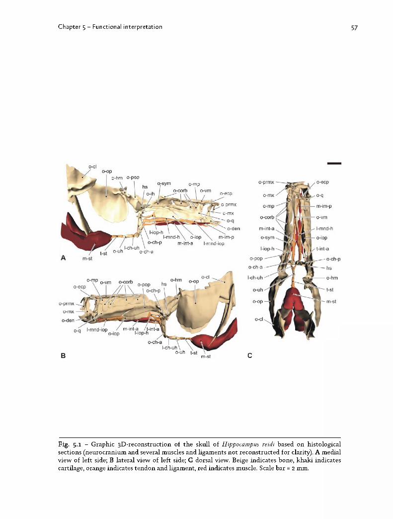
C h a p t e r 5 - Funct i onal i n t e rp r e t a t i on
o-mpo-ecp
o-prmx
i-mx
o-den
m-int-a l-mnd-iop
o-hm o-pop
o-ch-a
o-prmx
c
F ig . 5 .1 - G r a p h ic 3 D - r e c o n s t r u c t io n o f t h e s k u l l o f H ip p o c a m p u s re id i b a s e d o n h is t o lo g ic a l s e c t io n s (n e u r o c r a n iu m a n d se v e r a l m u s c le s a n d h g a m e n t s n o t r e c o n s tr u c te d fo r c la r ity ) . A m e d ia l v ie w o f l e f t s id e ; B la te r a l v i e w o f l e f t s id e ; C d o r sa l v ie w . B e ig e in d ic a t e s b o n e , k h a k i in d ic a t e s c a r t i la g e , o r a n g e in d ic a t e s t e n d o n a n d l ig a m e n t , r ed in d ic a t e s m u s c le . S c a le bar = 2 m m .

Oh
O-1
58 C h a p t e r 5 - Func t i onal i n t e rp r e t a t i on
F ig . 5 .2 - L e g e n d
1 t ip o f t h e n o s e s p in e , ju s t a n te r io r o f t h e e y e2 t ip o f t h e s n o u t p o s te r io r t o t h e m a x illa r y b o n e s3 sy m p h y s is o f t h e c e r a to h y a l b o n e s4 d is ta l t ip o f t h e v e n tr o la te r a l s p in e o n t h e p e c to r a l g ird le5 t ip o f t h e p r o c e s s o n t h e s e c o n d n u c h a l p la te6 p r o x im a l t ip o f t h e p r e m a x illa r y b o n e7 d is ta l t ip o f t h e d e n ta r y b o n e8 d i s t in c t p o in t o n t h e p r e y
c e n tr e o f t h e in te r h y a l b o n er o t a t io n p o in t o f n e u r o c r a n iu m w i t h v e r te b r a l c o lu m n - p e c t o r a l g ir d le c o m p le x
r m e d io r o s tr a l la n d m a r k o n t h e c le i th r u ms sy m p h y s is o f t h e c e r a to h y a l b o n e s

C h a p t e r 5 - Func t i ona l i n t e rp r e t a t i on
F ig . 5 .2 - G r a p h ic 3 D - r e c o n s t r u c t io n o f t h e s k u l l o f H ip p o c a m p u s re id i b a s e d o n C T -d a ta , r e p r e s e n t in g t h e d ig i t iz e d la n d m a r k s (y e llo w ) a n d t h e c a lc u la te d fo u r -b a r s y s te m (red ).

6o C h a p t e r 5 - Func t i onal i n t e rp r e t a t i on
Pig- 5-3 _ Legend
1 m a n d ib u lo -q u a d r a te a r t ic u la t io n2 m a n d ib u la r sy m p h y s is3 p r e m a x il la r y s y m p h y s is4 r o s tr a l t ip o f t h e v o m e r a l b o n e5 d is ta l t ip o f t h e n o s e s p in e6 a r t ic u la t io n b e t w e e n t h e s u p r a o c c ip ita l b o n e a n d t h e c o r o n a7 d is ta l t ip o f t h e d o r so c a u d a l p r o c e s s o n t h e c o r o n a8 d is ta l t ip o f t h e p r o c e s s o n t h e s e c o n d n u c h a l p la te9 a r t ic u la t io n b e t w e e n t h e n e u r o c r a n iu m a n d t h e f ir s t v e r te b r a ( n o t i l lu s tr a te d )10 a r t ic u la t io n b e t w e e n t h e n e u r o c r a n iu m a n d t h e p e c to r a l g ir d le11 d is ta l t ip o f t h e v e n tr o la te r a l s p in e o n t h e p e c to r a l g ird le12 t ip o f t h e a n g le fo r m e d b y t h e h o r iz o n t a l a n d v e r t ic a l arm o f t h e p e c to r a l g ir d le13 r o s tr a l p o in t o f t h e p e c to r a l c o m p le x14 c a u d a l t ip o f t h e u r o h y a l b o n e15 sy m p h y s is o f t h e c e r a to h y a l b o n e s16 a r t ic u la t io n b e t w e e n p o s te r io r c e r a to h y a l a n d in t e r h y a l b o n e s17 a r t ic u la t io n b e t w e e n in te r h y a l a n d p r e o p e r c u la r b o n e scr c o r o n an p -I I s e c o n d n u c h a l p la teo -c h -a a n te r io r c e r a to h y a l b o n eo - c l c le i th r u mo -d e n d e n ta r y b o n eo -h m h y o m a n d ib u la r b o n eo - io p in te r o p e r c u la r b o n eo - l e t h la te r a l e th m o id b o n eo - m e t h m e s e t h m o id b o n eo -m x m a x illa r y b o n eo -p a r p a r ie ta l b o n eo -p a r a p a r a s p h e n o id b o n eo - p o p p r e o p e r c u la r b o n eo - p o s t t p o s t t e m p o r a l b o n eo -p r m x p r e m a x il la r y b o n eo -q q u a d r a te b o n eo -s o c s u p r a o c c ip ita l b o n eo - s p h s p h e n o t ic b o n eo - u h u r o h y a l b o n eo -v m v o m e r a l b o n ev i f ir s t v e r te b r a

C h a p t e r 5 - Func t i ona l i n t e rp r e t a t i on
o-pop
2nd nuchal plate corona
neurocranlalunitpectoral
unit \
upper jaw
urohyal
interhyal
lower jaw
P ig - 5-3 " S k u ll m o r p h o lo g y o f a d u lt H ip p o c a m p u s re id i a n d 3 D -m o d e l . A 3 D - r e c o n s t r u c t io n o f th e s k u l l b a s e d o n C T -d a ta ; B 3 D - r e c o n s t r u c t io n in d ic a t in g t h e p o s i t io n o f t h e la n d m a r k s u s e d c o n s t r u c t in g t h e m o d e l ( p o in t 9 n o t i llu s tr a te d ); C m o d e l d r a w n b a s e d o n t h e la n d m a r k s a n d s u p e r im p o s e d o n a 3 D -r e c o n s tr u c t io n ; D m o d e l u s e d fo r v is u a l iz in g m e c h a n ic a l u n i t s a n d th e c h a n g e s in r e la t iv e p o s i t i o n d u r in g f e e d in g s tr ik e . S c a le b a r = 5 m m .

62 C h a p t e r 5 - Func t i onal i n t e rp r e t a t i on
F ig . 5 .4 - L e g e n d
a f - ih a r t ic u la t io n fa c e t o f in te r h y a l b o n e w i t h p r e o p e r c u la r b o n ec h c e r a to h y a l c a r t ila g e1 -ch -u h c e r a to h y a l-u r o h y a l l ig a m e n tlh -c h -p la te r a l h e a d o f p o s te r io r c e r a to h y a l b o n elh - ih la te r a l h e a d o f in t e r h y a l b o n e1 -io p -h in te r o p e r c u lo - h y o id l ig a m e n t1 -m n d -h m a n d ib u lo - h y o id l ig a m e n tm h - ih m e d ia l h e a d o f in te r h y a l b o n em -in t -a a n te r io r in t e r h y o id e u s p a r t o f p r o tr a c to r h y o id e i m u s c leo -c h -a a n te r io r c e r a to h y a l b o n eo -c h -p p o s te r io r c e r a to h y a l b o n eo - ih in t e r h y a l b o n eo - io p in te r o p e r c u la r b o n eo -p o p p r e o p e r c u la r b o n eo -u h u r o h y a l b o n et - in t -a t e n d o n o f a n te r io r in t e r h y o id e u s p a r t o f p r o tr a c to r h y o id e i m u s c lev h -c h -p v e n t r a l h e a d o f p o s te r io r c e r a to h y a l b o n e

C h a p t e r 5 - Func t i ona l i n t e rp r e t a t i on
0-pop
o-iop
1-iop-h
m-int-a
t-int-a
l-mnd-h
A
o-ih
o-ch-p
ch
0-ch-a
1-ch-uh
o-uh
B
F ig . 5 .4 - D e t a i l o f t h e 3 D - r e c o n s t r u c t io n o f t h e r e tr a c te d h y o id c o m p le x in H ip p o c a m p u s re id i b a s e d o n h i s t o lo g ic a l s e c t io n s ( so m e h g a m e n t s n o t r e c o n s tr u c te d fo r c la r ity ) . A la te r a l v i e w o f l e f t s id e ; B c a u d a l v i e w (o th e r s tr u c tu r e s n o t r e c o n s tr u c te d ) . B e ig e in d ic a t e s b o n e , k h a k i in d ic a te s c a r t i la g e , o r a n g e in d ic a t e s t e n d o n a n d l ig a m e n t , r ed in d ic a t e s m u s c le . S c a le b a rs = 1 m m .

64 C h a p te r 5 - F unc tiona l in t e rp re ta t io n

C h a p t e r 5 - Func t i ona l i n t e rp r e t a t i on
ms
660.5 ms 695.5 ms
0.5 ms0 ms
545.5 ms
P ig - 5-5 " E x a m p le o f a ( fa ile d ) f e e d in g s e q u e n c e o f H ip p o c a m p u s re id i f e e d in g o n a m y s id sh r im p in u p p e r l e f t c o r n e r . I m p o r ta n t e v e n ts are in d ic a te d b y a n a r ro w . A im a g e o f o n e fr a m e b e fo r e th e f ir s t v is ib le m o v e m e n t; B o n s e t o f h y o id r o ta t io n ; C o n s e t o f c r a n ia l r o t a t io n a n d m o u t h o p e n in g ; D m o u t h r e a c h e s m a x im a l o p e n in g ; E h y o id r e a c h e s m a x im a l r o ta t io n ; F n e u r o c r a n iu m r e a c h e s m a x im a l r o ta t io n ; G m o u t h f u l ly c lo se d ; H n e u r o c r a n iu m r e s to r e d t o i t s o r ig in a l p o s it io n ; I h y o id f u l ly p r o tr a c te d .

66 C h a p te r 5 - F unc tiona l in t e rp re ta t io n

C h a p t e r 5 - Func t i ona l i n t e rp r e t a t i on
oan
70
60
50
40
30
20
10
00 1 2 3 4
- 2
- 1
0
Time (ms)
100 -
80o0)O)CA3■goX
0 2 4 6 8 10 12 14
B Time (ms)
F ig . 5 .6 - A T im e a v e r a g e d k in e m a t ic p r o f i le s (N = 14) o f t h e m e a n h y o id r o t a t io n (b la c k d o ts ) , t h e m e a n n e u r o c r a n ia l r o t a t io n (g ra y d o ts ) a n d th e m e a n m o u t h o p e n in g ( w h it e d o ts ) o f a l l 14 s e q u e n c e s . T im e = o m s w a s c o n s e q u e n t ly d e f in e d fo r e a c h s e q u e n c e a s t h e fr a m e b e fo r e t h e f ir s t v is ib le m o v e m e n t . P r e y c a p tu r e o c c u r s o n a v e ra g e 5.5 m s a fte r t h e o n s e t o f h y o id r o ta t io n . B C o m p a r is o n o f t h e a v e ra g e m e a su r e d h y o id a n g le (b la c k d o ts ) a n d t h e a v e ra g e h y o id a n g le p r e d ic te d b y t h e fo u r -b a r m o d e l ( w h it e d o ts ; N = 14). In t h e f ir s t 1 .5 m s t h e g r a p h s d o n o t

68 C h a p te r 5 - F unc tiona l in t e rp re ta t io n

C h a p t e r 5 - Func t i ona l i n t e rp r e t a t i on
P ig - 5-7 " M o d e l s h o w in g e x te n s iv e n e u r o c r a n ia l e le v a t io n o n H ip p o c a m p u s reidi. A v id e o fr a m e s u s e d fo r p o s i t io n in g t h e c o m p o n e n t s o f t h e m o d e l a t t h e o n s e t o f n e u r o c r a n ia l e le v a t io n ( le f t ) a n d a t m a x im a l e le v a t io n (r ig h t) . B m o d e l a t t h e o n s e t o f e le v a t io n a n d t h e p o s i t i o n o f t h e s k u l l at m a x im a l e le v a t io n , s u p e r im p o s e d a n d a l ig n e d b a se d o n t h e p o s i t i o n o f t h e p e c to r a l g ir d le .

70 C h a p t e r 5 - Func t i onal i n t e rp r e t a t i on
F ig . 5 .8 - L e g e n d
o -a p a l a u to p a la t in e b o n eo -b o c b a s io c c ip it a l b o n eo -d e n d e n ta r y b o n eo -h m h y o m a n d ib u la r b o n eo - io p in te r o p e r c u la r b o n eo - le t h la te r a l e th m o id b o n eo - m e t h m e s e t h m o id b o n eo -m x m a x illa r y b o n eo -p a r a p a r a s p h e n o id b o n eo -p a r p a r ie ta l b o n eo -p o p p r e o p e r c u la r b o n eo - p o s t t p o s t t e m p o r a l b o n eo -p r m x p r e m a x il la r y b o n eo -s o c s u p r a o c c ip ita l b o n eo -s p h s p h e n o t ic b o n eo -v m v o m e r a l b o n e

o-apal o-mx o-prm*
C h a p t e r 5 - Func t i ona l i n t e rp r e t a t i on
O -S O C
F ig . 5 .8 - C o n s t r u c t io n o f t h e H ip p o c a m p u s zo s te ra e a r t if ic ia l m o d e l b a s e d o n C T -d a ta s h o w in g th e o r ig in a l H . zo s te ra e (b lu e ) , t h e H . re id i d e p r e s se d m o d e l b e in g m o d if ie d (d ark g rey ) a n d t h e c o m p r e s s e d p a r t o f t h e s n o u t ( l ig h t g rey ). N o t e th a t th e m o d e ls are s c a le d b a se d o n b r a in c a se l e n g th . A u n a lte r e d m o d e ls o f H . re id i a n d H . zos te ra e , w i t h in d ic a t io n o f s o m e b o n e s ; B c o m p r e s s io n o f th e sn o u t ; C a t ta c h m e n t o f t h e s n o u t t ip a n d ja w s. T h e c o m p r e s s e d s n o u t w a s t h e n s m o o t h e d a n d s im p h f ie d u n t i l t h e t r ia n g le s h a d th e sa m e s iz e as t h e o n e s in t h e r e s t o f t h e c r a n iu m .

72 C h a p t e r 5 - Func t i onal i n t e rp r e t a t i on
P ig - 5-9 - L e g e n d
1 a u to p a la t in e -v o m e r a l a r t ic u la t io n2 h y o id
3 la te r a l w a l l o f t h e s n o u t
4 m e s e th m o id -p a r ie ta l b o r d e r
5 h y o m a n d ib u lo - s p h e n o t ic jo in t6 v o m e r o - m e s e th m o id t r a n s i t io n
7 lo w e r ja w sy m p h y s is8 r o s tr a l b o r d e r o f t h e n o s t r i l
9 v e n t r a l b o r d e r o f t h e o r b it10 b r a in c a se r o o f

C h a p t e r 5 - Func t i ona l i n t e rp r e t a t i on
P ig - 5-9 - V o n M is e s s tr e s s d is t r ib u t io n p a t te r n in s e a h o r se s (A -C ) a n d p ip e f is h e s (D -F ) w i t h lo n g (A , D ) , in te r m e d ia te (B , E) o r s h o r t (C , F) s n o u t le n g th . A H ip p o c a m p u s re id i d e p r e sse d ; B H . a b d o m in a li s ; C H . zos terae; D D o r y r h a m p h u s d a c ty l io p h o ru s ; E S y n g n a th u s ros te l la tus; F D. m ela n o p leu ra . D i f f e r e n t c o lo r sc a le s are u s e d fo r e a c h m o d e l t o fa c ih t a t e c o m p a r is o n o f t h e s tr e s s c o n c e n t r a t io n p a tte r n . S tr e ss v a lu e s e x p r e s s e d in M P a .

74 C h a p t e r 5 - Func t i onal i n t e rp r e t a t i on
F ig . 5 .1 0 - L e g e n d
1 h y o id sy m p h y s is2 h y o m a n d ib u lo - s p h e n o t ic jo in t
3 a u to p a la t in e -v o m e r a l a r t ic u la t io n
4 lo w e r ja w
5 h y o id6 m a x illa r y b o n e s
7 b r a in c a se8 la te r o c a u d a l ly in t h e s n o u t
9 la te r o r o s tr a l ly in th e s n o u t10 v e n tr o r o s tr a l b o r d e r o f t h e o r b it

C h a p t e r 5 - Func t i ona l i n t e rp r e t a t i on
4
F ig . 5 .1 0 - V o n M is e s s tr e s s d is t r ib u t io n p a t te r n in t h e a r t if ic ia l ly c o n s t r u c t e d m o d e ls b a s e d o n H ip p o c a m p u s re id i m o r p h o lo g y . A H . re id i d e p r e s se d m o d e l (as in F ig . 5 .9A ); B H . a b d o m in a l i s a r t if ic ia l m o d e l; C H . zo s te ra e a r t if ic ia l m o d e l. S tr e ss v a lu e s e x p r e s s e d in M P a , c o lo r s c a le s are d if f e r e n t fo r a l l m o d e ls .

76 C h a p t e r 5 - Func t i ona l i n t e rp r e t a t i on
F ig . 5 .1 1 - L e g e n d
1 m e s e th m o id -p a r ie ta l b o r d e r2 la te r o r o s tr a l ly in th e s n o u t3 h y o id -s u s p e n s o r iu m a r t ic u la t io n

C h a p t e r 5 - Func t i ona l i n t e rp r e t a t i on
F ig . 5 .1 1 - V o n M is e s s tr e s s d is t r ib u t io n p a t te r n in t w o m o d e ls o f H ip p o c a m p u s re id i s h o w in g t h e e f f e c t o f t h e p o s i t i o n o f t h e h y o id . A H . re id i p r o tr a c te d m o d e l; B H . re id i d e p r e s se d m o d e l. S tr e ss v a lu e s e x p r e s s e d in M P a .

© A l l r ig h ts r e se r v e d . T h is t h e s i s c o n t a in s c o n f id e n t ia l in f o r m a t io n a n d c o n f id e n t ia l r e se a r c h r e s u lt s t h a t are p r o p e r ty t o t h e U G e n t (R e se a r c h g r o u p E v o lu t io n a r y M o r p h o lo g y o f V e r te b r a te s , D e p a r t m e n t o f B io lo g y ) . T h e c o n t e n t s o f t h i s m a s te r t h e s is m a y u n d e r n o c ir c u m s ta n c e s b e m a d e p u b lic , n o r c o m p le t e or p a r tia l, w i t h o u t t h e e x p l ic i t a n d p r e c e d in g p e r m is s io n o f t h e U G e n t r e p r e s e n ta t iv e , i .e . t h e su p e r v iso r . T h e t h e s i s m a y u n d e r n o c ir c u m s ta n c e s b e c o p ie d o r d u p lic a te d in a n y fo r m , u n le s s p e r m is s io n g r a n te d in w r i t t e n fo r m . A n y v io la t io n o f t h e c o n f id e n t ia l n a tu r e o f t h i s t h e s i s m a y im p o s e irr ep a ra b le d a m a g e t o t h e U G e n t . In c a se o f a d is p u te t h a t m a y a r ise w i t h i n t h e c o n t e x t o f t h i s d e c la r a t io n , t h e J u d ic ia l C o u r t o f G e n t o n ly is c o m p e t e n t t o b e n o t i f i e d .