Evolution based on domain combinations: the case of glutaredoxins
14
BioMed Central Page 1 of 14 (page number not for citation purposes) BMC Evolutionary Biology Open Access Research article Evolution based on domain combinations: the case of glutaredoxins Rui Alves* 1 , Ester Vilaprinyo 2 , Albert Sorribas 1 and Enrique Herrero* 1 Address: 1 Departament de Ciències Mèdiques Bàsiques, Universitat de Lleida, IRBLleida, Montserrat Roig 2, 25008, Lleida, Spain and 2 Institut d'Investigació Biomédica de Bellvitge (IDIBELL), Hospitalet de Llobregat, Spain Email: Rui Alves* - [email protected]; Ester Vilaprinyo - [email protected]; Albert Sorribas - [email protected]; Enrique Herrero* - [email protected] * Corresponding authors Abstract Background: Protein domains represent the basic units in the evolution of proteins. Domain duplication and shuffling by recombination and fusion, followed by divergence are the most common mechanisms in this process. Such domain fusion and recombination events are predicted to occur only once for a given multidomain architecture. However, other scenarios may be relevant in the evolution of specific proteins, such as convergent evolution of multidomain architectures. With this in mind, we study glutaredoxin (GRX) domains, because these domains of approximately one hundred amino acids are widespread in archaea, bacteria and eukaryotes and participate in fusion proteins. GRXs are responsible for the reduction of protein disulfides or glutathione-protein mixed disulfides and are involved in cellular redox regulation, although their specific roles and targets are often unclear. Results: In this work we analyze the distribution and evolution of GRX proteins in archaea, bacteria and eukaryotes. We study over one thousand GRX proteins, each containing at least one GRX domain, from hundreds of different organisms and trace the origin and evolution of the GRX domain within the tree of life. Conclusion: Our results suggest that single domain GRX proteins of the CGFS and CPYC classes have, each, evolved through duplication and divergence from one initial gene that was present in the last common ancestor of all organisms. Remarkably, we identify a case of convergent evolution in domain architecture that involves the GRX domain. Two independent recombination events of a TRX domain to a GRX domain are likely to have occurred, which is an exception to the dominant mechanism of domain architecture evolution. Background Domain duplication and shuffling by recombination and fusion, followed by divergence are the more frequent mechanisms for the evolution of proteins [1]. It has been estimated that such recombination and fusion events are likely to occur only once for a given multidomain archi- tecture and that, after such an event, the fusion protein is duplicated and/or diverges over time [1]. In addition, sta- tistical analysis of known multidomain proteins has shown that a) there is a strong bias for individual domains involved in recombination and fusion events to be short [2], and b) some specific sets of recombined domains (supra domains) participate in further recombination and fusion events [3]. This provides a model for the dominant Published: 25 March 2009 BMC Evolutionary Biology 2009, 9:66 doi:10.1186/1471-2148-9-66 Received: 30 September 2008 Accepted: 25 March 2009 This article is available from: http://www.biomedcentral.com/1471-2148/9/66 © 2009 Alves et al; licensee BioMed Central Ltd. This is an Open Access article distributed under the terms of the Creative Commons Attribution License (http://creativecommons.org/licenses/by/2.0 ), which permits unrestricted use, distribution, and reproduction in any medium, provided the original work is properly cited.
-
Upload
independent -
Category
Documents
-
view
1 -
download
0
Transcript of Evolution based on domain combinations: the case of glutaredoxins
BioMed CentralBMC Evolutionary Biology
ss
Open AcceResearch articleEvolution based on domain combinations: the case of glutaredoxinsRui Alves*1, Ester Vilaprinyo2, Albert Sorribas1 and Enrique Herrero*1Address: 1Departament de Ciències Mèdiques Bàsiques, Universitat de Lleida, IRBLleida, Montserrat Roig 2, 25008, Lleida, Spain and 2Institut d'Investigació Biomédica de Bellvitge (IDIBELL), Hospitalet de Llobregat, Spain
Email: Rui Alves* - [email protected]; Ester Vilaprinyo - [email protected]; Albert Sorribas - [email protected]; Enrique Herrero* - [email protected]
* Corresponding authors
AbstractBackground: Protein domains represent the basic units in the evolution of proteins. Domainduplication and shuffling by recombination and fusion, followed by divergence are the mostcommon mechanisms in this process. Such domain fusion and recombination events are predictedto occur only once for a given multidomain architecture. However, other scenarios may berelevant in the evolution of specific proteins, such as convergent evolution of multidomainarchitectures. With this in mind, we study glutaredoxin (GRX) domains, because these domains ofapproximately one hundred amino acids are widespread in archaea, bacteria and eukaryotes andparticipate in fusion proteins. GRXs are responsible for the reduction of protein disulfides orglutathione-protein mixed disulfides and are involved in cellular redox regulation, although theirspecific roles and targets are often unclear.
Results: In this work we analyze the distribution and evolution of GRX proteins in archaea,bacteria and eukaryotes. We study over one thousand GRX proteins, each containing at least oneGRX domain, from hundreds of different organisms and trace the origin and evolution of the GRXdomain within the tree of life.
Conclusion: Our results suggest that single domain GRX proteins of the CGFS and CPYC classeshave, each, evolved through duplication and divergence from one initial gene that was present inthe last common ancestor of all organisms. Remarkably, we identify a case of convergent evolutionin domain architecture that involves the GRX domain. Two independent recombination events ofa TRX domain to a GRX domain are likely to have occurred, which is an exception to the dominantmechanism of domain architecture evolution.
BackgroundDomain duplication and shuffling by recombination andfusion, followed by divergence are the more frequentmechanisms for the evolution of proteins [1]. It has beenestimated that such recombination and fusion events arelikely to occur only once for a given multidomain archi-tecture and that, after such an event, the fusion protein is
duplicated and/or diverges over time [1]. In addition, sta-tistical analysis of known multidomain proteins hasshown that a) there is a strong bias for individual domainsinvolved in recombination and fusion events to be short[2], and b) some specific sets of recombined domains(supra domains) participate in further recombination andfusion events [3]. This provides a model for the dominant
Published: 25 March 2009
BMC Evolutionary Biology 2009, 9:66 doi:10.1186/1471-2148-9-66
Received: 30 September 2008Accepted: 25 March 2009
This article is available from: http://www.biomedcentral.com/1471-2148/9/66
© 2009 Alves et al; licensee BioMed Central Ltd. This is an Open Access article distributed under the terms of the Creative Commons Attribution License (http://creativecommons.org/licenses/by/2.0), which permits unrestricted use, distribution, and reproduction in any medium, provided the original work is properly cited.
Page 1 of 14(page number not for citation purposes)
BMC Evolutionary Biology 2009, 9:66 http://www.biomedcentral.com/1471-2148/9/66
mode of domain architecture evolution in proteins that isvery much consensual. Recent work has further estimatedthat between 88% and 95% of all multidomain architec-tures have evolved through such mechanisms [4-6]. Theremaining architectures are likely to have evolved throughconvergent evolution [6]. A recent theory proposes that,during major evolutionary transitions, evolution is bipha-sic, further complicating the model of protein evolution[7]. According to this view, in an initial post-transitionphase, large scale horizontal gene transfer (HGT) wouldoccur. This would be followed by a second phase wherethe more common mechanisms for protein evolutionbecome dominant. Given this background, it is of interestto analyze the evolution of a specific type of proteindomain in order to assess the importance of the evolu-tionary mechanisms described above in the evolution ofthat domain.
A protein domain of small size that is known to partici-pate in the architecture of multidomain proteins and iswidespread over the many branches of the evolutionarytree would be an appropriate choice to study. The glutare-doxin (GRX) domain meets all these conditions. It hasapproximately one hundred amino acids, it is a part ofseveral multidomain architectures and it is present inarchaea, bacteria and eukaryotes. GRXs are thiol oxidore-ductases responsible for the reduction of proteindisulfides or glutathione-protein mixed disulfides, whichemploy reduced glutathione (GSH) as hydrogen donor[8]. Together with thioredoxins (TRXs) and other pro-teins, GRXs are grouped in the thioredoxin fold super-family, because they share a common structural foldconsisting of a four or five-stranded β-sheet flanked byseveral α-helices on either side of the β-sheet [9]. Func-tionally, TRXs are also disulfide oxireductases. In contrastwith GRXs, oxidized TRXs are reduced by thioredoxinsreductases at the expense of NADPH [8]. Alternatively, inplant chloroplasts, TRXs are reduced directly by the ferre-doxin/ferredoxin reductase system that is coupled to pho-tosynthesis [10]. Three GRX families have been definedbased on the sequence of the putative active sites. Classi-cal dithiol GRXs (CPYC class) with a C [P/S] [Y/F]C activesite sequence are widespread in archaea, bacteria andeukaryotes. On the other hand, monothiol GRXs (CGFSclass) contain a CGFS-like active site sequence, and theyhave currently been reported to exist in bacteria andeukaryotes [11]. Finally, land plants contain (in additionto GRXs species of the above classes) a third class of GRXs(CCMC class), with the sequence CC [M/L] [C/S] in theputative active sites [12,13]. This division in three groupsis further complicated by the recent characterization inSaccharomyces cerevisiae of three GRXs (Grx6, Grx7 andGrx8) with CSYS, CPYS and CPDC active site motifs [14-16].
Multidomain proteins that contain GRX domains havealso been reported in a variety of different organisms[12,13,17-19]. The most studied GRX fusion proteins ineukaryotes are TRX-GRX fusions, containing CGFS classGRXs modules. In these proteins, one to three GRX mod-ules are linked to an N-terminal TRX-like module whichdoes not conserve the WC [G/P]PC active site of func-tional TRXs. S. cerevisiae Grx3 and Grx4 [11] and humanGLRX3 (PICOT) [20] are examples of these multidomainGRXs.
Most GRXs are likely to participate in a diversity of proc-esses that require redox-type regulation, although theirspecific roles and targets in those processes are oftenunclear. Examples of processes in which CPYC class GRXsare involved in include activation of ribonucleotidereductase, or 3'-phosphoadenylylsulfate reductase, reduc-tion of ascorbate, regulation of the DNA binding activityof nuclear factors, or protection against heavy metals(reviewed in [21]). Examples of processes in which CGFSclass GRXs are involved include iron sulfur cluster biogen-esis (Grx5), and regulation of cellular iron homeostasis(Grx3 and Grx4) in S. cerevisiae [11,18,22], cytochrome-cbiogenesis in bacteria [23,24], PROKAR-(lipid mem-brane) proteins in archaea [25], and regulation of signaltransduction pathways in response to external signals (thehuman PICOT) [20].
In this work we analyze the distribution and evolution ofGRX domains in archaea, bacteria and eukaryotes. Westudy over one thousand proteins, containing at least oneGRX domain, from hundreds of different organisms andtrace the origin and evolution of the GRX domain withinthe tree of life. Our phylogenetic analysis suggests that sin-gle domain GRX proteins of the CGFS class have evolvedthrough duplication and divergence from one initial genethat was present in the last common ancestor (LCA) of allorganisms. The same appears to hold true for singledomain GRX proteins of the CPYC class. We predict setsof residues that are likely to be important for protein func-tion in the CGFS and CPYC classes of GRXs. Remarkably,we identify a case of convergent evolution in domainarchitecture, where two independent recombinationevents of a TRX domain to a GRX domain are likely tohave occurred. We also identify domain combinationsthat, in the context of GRX domain evolution, appear tofunction as the supra-domains proposed by Vogel et al.[3].
ResultsWe have made a systematic identification of GRXdomains from the UNIPROT database (information sum-marized in Table 1). Over 75% of all GRX domains arefound in single domain proteins (Table 1). The other 25%are found as a part of a variety of multidomain proteins.
Page 2 of 14(page number not for citation purposes)
BMC Evolutionary Biology 2009, 9:66 http://www.biomedcentral.com/1471-2148/9/66
For example, GRXs have combined with pyridine nucle-otide-disulphide oxidoreductases class-II domains inthioredoxin-glutathione-reductase proteins, or with pep-tide methionine sulfoxide reductase domains, among oth-ers. An interesting case is that of the triple fusion betweenthe GRX domain and frataxin and rhodanese domains. Inthis case, the frataxin-rhodanese cassette appears to act asa supra domain of recombination [3] in the context ofGRX domain evolution, because proteins where the GRXdomain recombined exclusively either with the frataxindomain or with the rhodanese domain are not found. Inaddition to these and other well characterized proteindomains, GRXs are also associated with other types of lesswell characterized protein domains (for example DUF296domains). Many of these domains are recurrently foundin different proteins but have yet to be assigned a specificfunction. Table 2 details the major domain types found tocombine with the different GRX classes. [Additional Files1, 2 and 3 contain the sequences and alignments for theGRX domains that have been identified in multidomainproteins from the UNIPROT database.] Because of thelarge number of sequences being analyzed we divided thesequences into smaller sets for a more detailed and accu-rate analysis. We also analyzed the full set, with results
that are similar to the ones described below (data notshown).
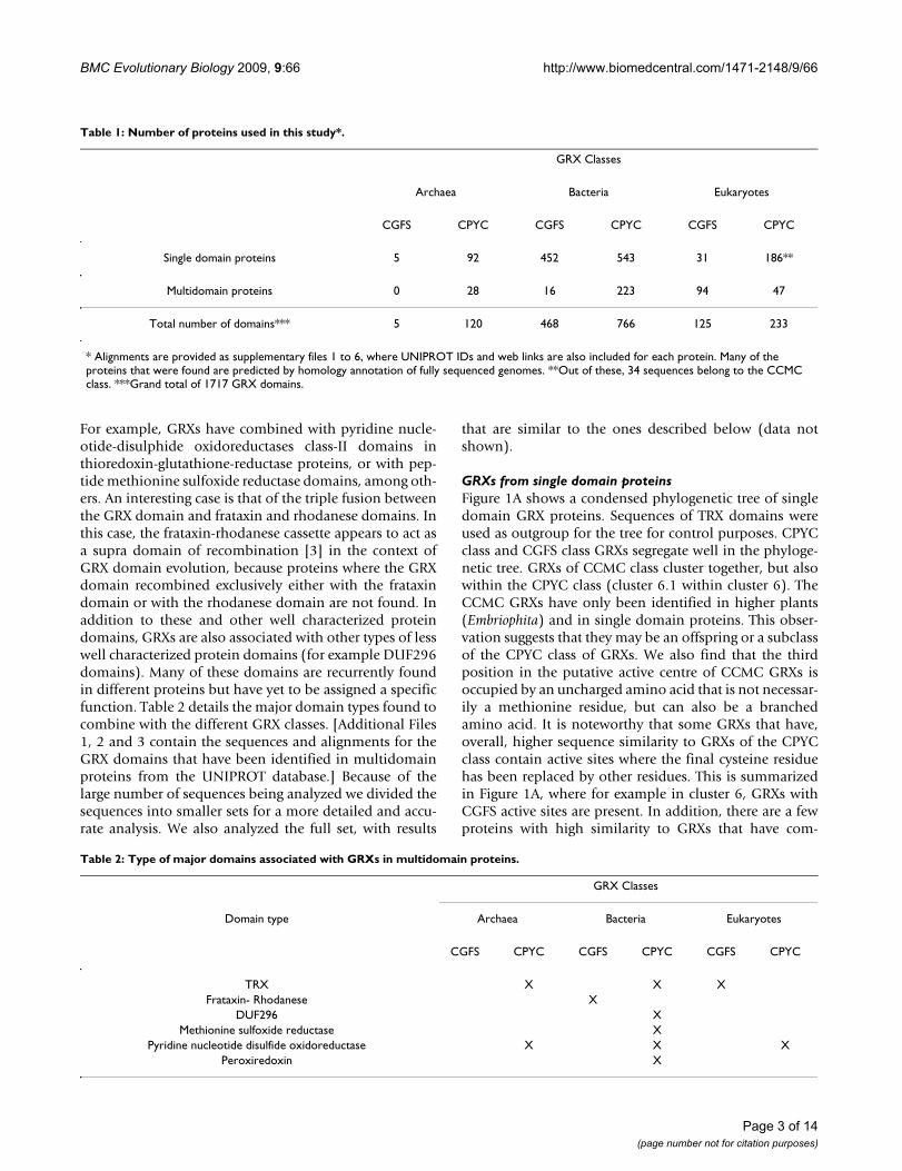
GRXs from single domain proteinsFigure 1A shows a condensed phylogenetic tree of singledomain GRX proteins. Sequences of TRX domains wereused as outgroup for the tree for control purposes. CPYCclass and CGFS class GRXs segregate well in the phyloge-netic tree. GRXs of CCMC class cluster together, but alsowithin the CPYC class (cluster 6.1 within cluster 6). TheCCMC GRXs have only been identified in higher plants(Embriophita) and in single domain proteins. This obser-vation suggests that they may be an offspring or a subclassof the CPYC class of GRXs. We also find that the thirdposition in the putative active centre of CCMC GRXs isoccupied by an uncharged amino acid that is not necessar-ily a methionine residue, but can also be a branchedamino acid. It is noteworthy that some GRXs that have,overall, higher sequence similarity to GRXs of the CPYCclass contain active sites where the final cysteine residuehas been replaced by other residues. This is summarizedin Figure 1A, where for example in cluster 6, GRXs withCGFS active sites are present. In addition, there are a fewproteins with high similarity to GRXs that have com-
Table 1: Number of proteins used in this study*.
GRX Classes
Archaea Bacteria Eukaryotes
CGFS CPYC CGFS CPYC CGFS CPYC
Single domain proteins 5 92 452 543 31 186**
Multidomain proteins 0 28 16 223 94 47
Total number of domains*** 5 120 468 766 125 233
* Alignments are provided as supplementary files 1 to 6, where UNIPROT IDs and web links are also included for each protein. Many of the proteins that were found are predicted by homology annotation of fully sequenced genomes. **Out of these, 34 sequences belong to the CCMC class. ***Grand total of 1717 GRX domains.
Table 2: Type of major domains associated with GRXs in multidomain proteins.
GRX Classes
Domain type Archaea Bacteria Eukaryotes
CGFS CPYC CGFS CPYC CGFS CPYC
TRX X X XFrataxin- Rhodanese X
DUF296 XMethionine sulfoxide reductase X
Pyridine nucleotide disulfide oxidoreductase X X XPeroxiredoxin X
Page 3 of 14(page number not for citation purposes)
BMC Evolutionary Biology 2009, 9:66 http://www.biomedcentral.com/1471-2148/9/66
Figure 1 (see legend on next page)
A B
DC
Page 4 of 14(page number not for citation purposes)
BMC Evolutionary Biology 2009, 9:66 http://www.biomedcentral.com/1471-2148/9/66
pletely lost their active site (cluster 3 in Figure 1A, e-val-ues<10-5). The function of these proteins is unknown, andthis may represent a situation where the GRX domain isbeing co-opted for a new function that has yet to be char-acterized.
Within each class of GRXs, different taxa cluster roughly aspreviously reported (see for example [26-28]). For exam-ple, in cluster 1 of Figure 1A bacterial GRXs of the CGFSclass group together, and apart from eukaryotic GRXs ofthe same class. This is consistent with GRXs being presentat the LCA of the three kingdoms and is inconsistent withmassive HGT of GRX genes between different kingdoms.In fact, CGFS class and CPYC class GRXs appear to havebeen both present in the LCA of all branches in the tree,because both classes of GRXs cluster apart and differenti-ation between archaea, bacteria, and eukaryotes is onlyobserved within the clusters shown in Figure 1A.
A group of proteins that have been annotated as GRX-likeproteins in fully sequenced genomes, mostly in archaeaand eukaryotes but also in bacteria, appear to be some-where in between TRXs and CPYC class GRXs in terms ofsequence similarity (clusters 8 and 9 in Figure 1A). Theyhave more variability in their sequence than the otherclusters from Figure 1A, but they are nevertheless similarto other well characterized GRXs, with e-value ≤ 10-10.They all contain a GRX-like putative active site sequence.The bacterial sequences in this cluster come from groupsthat are not typically considered as having GRXs (see cap-tion for Figure 1A).
A detailed analysis of the data also reveals that CPYC classGRXs have been found in all sequenced archaea genomes.However, within Archaea, we were only able to find CGFSclass GRXs in Halobacteriales. Five sequences of the CGFSclass have been found in this group (see see Supplemen-tary Table 1 in Additional File 4, Cluster 2 in Figure 1Aand Cluster 4 in Figure 1B). A more detailed analysis of
the DNA sequence for the genes that code for these GRXsreveals the following.
a) The highest homology between the H. salinariumCGFS class GRX and other non-archaea GRX is to aMyxococcus xanthus GRX [see Supplementary Table 1 inAdditional File 4].
b) The codon usage of genes is a characteristic that canoften be used to identify HGT. This is so because theevolution of an organism leads to an optimization ofthe codon usage in genes for the specific physiology ofthat organism [29]. We calculated the average codonusage in the Myxococcus xanthus and in the H. sali-narium genomes [29]. We also calculated the codonusage for the CGFS class grx genes from H. salinariumand M. xanthus. We found that the codon usage in theCGFS class GRXs of Halobaterium is similar to the aver-age codon usage in the Myxococcus genome. It is alsosimilar to the codon usage in the Myxococcus CGFSclass grx genes.
c) The borders of the CGFS class grx gene in H. sali-narium are homologous to the flanking regions of thetransposon gene XAC3504 from the gamma proteo-bacteria Xanthomonas axonopodis.
d) The flanking regions of the CGFS class grx genes inarchaea could be degenerated palindromes. If this isso, it may indicate the remains of a degenerated trans-poson. Transposons are responsible for gene mobilitywithin and between genes through HGT.
Taken together, these observations suggest that CGFS classGRXs in archaea may have been the result of one HGTfrom some proteobacteria ancestor to the halobacteriales.
Unlike in archaea, single domain CGFS class and CPYCclass GRXs are both widespread in bacteria, as shown in
Condensed phylogenetic tree for single domain GRXs from UNIPROT databaseFigure 1 (see previous page)Condensed phylogenetic tree for single domain GRXs from UNIPROT database. Panel A: Global tree. The out-group on the lower left of the tree is composed of TRX single domain proteins (in yellow). All major divisions between clus-ters have bootstrap values of 100%, indicated by the "100" label. The CGFS class of GRXs is depicted on the upper branchs [in green]. Sequences are more homogeneous on this class. The mid-lower branches depict CPYC class GRXs [in mauve]. There is a wider variability in the sequences of this class. The clusters identified with the tag "Bacteria*" include almost all non-proteo-bacteria GRX domains. These include domains from Actinobacteria, Deinnococcus, Planktomycetes, Green sulfur bacteria, Green non sulfur bacteria, Thermotoga, Aquifaceae, and Flavobacteria. The cluster identified with the tag "Bacteria**" include mostly proteobacteria GRX domains. GRX domains from Cyanobacteria and Spirochaetes are also present in this cluster. Panel B: Condensed phylogenetic tree of single domain GRXs in archaea. Panel C: Phylogenetic tree of GRX proteins for bacteria. CGFS class GRXs [Cluster 1] have less variability in their sequence than CPYC class GRXs [other clusters]. Some GRX-like proteins have lost their active site [XXXX in Cluster 2]. Panel D: Phylogenetic tree of GRX proteins for eukaryotes. CGFS GRXs [in green] have less variability in their sequence than CPYC GRXS [in mauve]. Nevertheless, the variability in the sequence of the active site for CGFS GRXs is far greater than that found in bacteria [compare to panel C].
Page 5 of 14(page number not for citation purposes)
BMC Evolutionary Biology 2009, 9:66 http://www.biomedcentral.com/1471-2148/9/66
Figure 1C. Both classes of GRXs appear to have existed assuch in the LCA of bacteria, because in general CGFS classGRXs cluster together, as do CPYC class GRXs. Interest-ingly, some GRXs that are, sequence-wise, clearly includedin the CPYC class have evolved CGFC putative active cent-ers (cluster 6 in Figure 1C; also see Additional Files 1, 2, 3,5, 6, and 7), although no CGFS class GRXs with a secondcysteine in the active site where found.
Figure 1D shows a phylogenetic tree based on thesequence alignment of eukaryotic single domain GRXs.CGFS class and CPYC class GRXs are also commonlyfound in eukaryotes. A fraction of CPYC class GRXs havelost the second cysteine residue of their putative active site(GRXs in clusters 3, 5, 6 and 8).
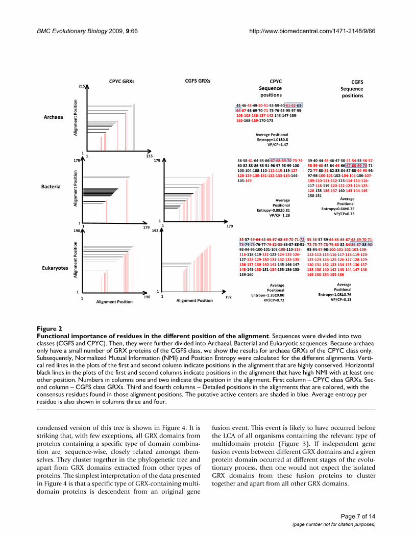
Sequence analysis of individual GRX domains from an evolutionary and functional perspectiveWe compared the variability of the CPYC class alignmentsto that of the CGFS alignments. This variability can bequantified by calculating both, the average positionalentropy of the alignment and the normalized mutualinformation (NMI) between each pair of positions in thealignment (see Methods for details).
The higher the average positional entropy is, the higherthe variability per position in the alignment is (see e.g.[30]). Based on this we find that the CPYC class has highersequence variability than the CGFS class. Although thedifference in variability between the two classes may bedue to the larger number of sequences that have beenidentified for the CPYC class, we believe that this is notthe case, because the standard deviation of the positionalentropy in the two classes is almost the same (approxi-mately 0.8 for the CPYC class and approximately 0.75 forthe CGFS class, Figure 2).
The results from NMI profiles support those from theaveraged position entropy, although the interpretation ofthese profiles is more nuanced. On one hand, if two posi-tions in the alignment have a NMI that is very high, onecan interpret this result as indicating that any changes inthe residue at one of the positions needs to be counterbal-anced by a compensatory change in the residue of theother position [30-35]. Thus, the residues in those posi-tions are functionally constrained and functionallyimportant for the protein. On the other hand, positionswith residues that are highly conserved will have a NMIwith any other position in the alignment that is not signif-icantly different from zero. Therefore, highly conservedpositions are also likely to be functionally constrained. Allthe information regarding conservation and co-variationof residues is summarized in Figure 2. Many of the resi-dues predicted in Figure 2 as being functionally importantare known to be involved in the overall function of the
GRX domains. For example, residues in the putative activesite of both classes of GRXs are marked with a blue trans-parent rectangle. In the same figure, one can see that CGFSclass GRXs have a larger number of positions in the align-ment that either have high NMI interaction with otherposition (black lines and black residues) or are highlyconserved (red lines and red residues). If our results aregeneral and our interpretation is correct, GRXs of theCGFS class may require more positions to be constrained,if they are to remain functional.
An alternative explanation for the differences in the varia-bility between CPYC and CGFS GRXs is the following. Thelarger variability of CPYC GRXs could also be observed ifCPYC class GRXs had an earlier origin than CGFS GRXs,because this could have allowed for CPYC GRXs to haveevolved for a longer time. Our results suggest that thisexplanation is unlikely. In Figure 1, the CPYC and CGFSclasses of GRXs cluster perfectly apart, and the branchingstructure of the tree indicates that both classes werealready present in the LCA of archaea, bacteria andeukaryotes. This suggests that both proteins may havehad, roughly, a similar amount of time to evolve. TheDNA sequences of the different domains can be furtheranalyzed in order to assess if the two classes of GRXs havebeen evolving for significantly different times. It is knownthat the rate of synonymous mutations in different genesis similar, even when the rate of non-synonymous muta-tions is quite different [33]. Therefore, by comparing theaverage percentage of synonymous substitutions percodon between the CGFS class and the CPYC class GRXsin the conserved positions of each multiple alignment,one can obtain additional information regarding whetherthese proteins have been evolving for approximately thesame time. This percentage is similar in both classes ofproteins. In addition, we calculated the average ratio ofnon-synonymous (Ka) over synonymous (Ks) mutationsper codon to be <Ka/Ks> = 2.9 for the GRX domains ofeach of the two classes. This ratio can also be used to esti-mate the rate of evolution of proteins [36]. Takentogether, all these data are consistent with the notion thatthe difference in the number of highly conserved posi-tions in both classes of GRXs is not due to a difference inthe time they had to evolve. The protein and DNAsequence alignments are provided [see Additional Files 1,2, 3, 5, 6, and 7].
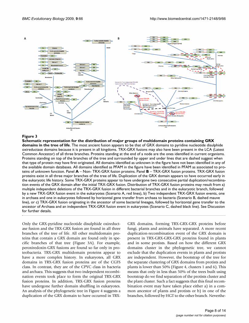
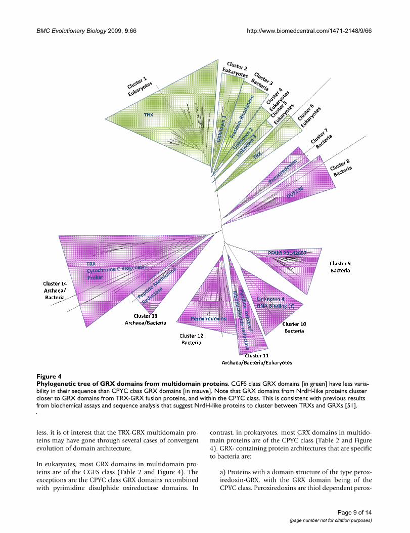
GRXs domains in multidomain proteinsAs stated above, and summarized in Table 2 and Figure 3,multidomain proteins that contain GRX domains arewidespread. To more accurately analyze these GRXdomains we isolated their sequence from the multido-main proteins, as described in the Methods section. TheGRX domains were then aligned using MEGA4. The mul-tiple alignment was used to build a phylogenetic tree. A
Page 6 of 14(page number not for citation purposes)
BMC Evolutionary Biology 2009, 9:66 http://www.biomedcentral.com/1471-2148/9/66
condensed version of this tree is shown in Figure 4. It isstriking that, with few exceptions, all GRX domains fromproteins containing a specific type of domain combina-tion are, sequence-wise, closely related amongst them-selves. They cluster together in the phylogenetic tree andapart from GRX domains extracted from other types ofproteins. The simplest interpretation of the data presentedin Figure 4 is that a specific type of GRX-containing multi-domain proteins is descendent from an original gene
fusion event. This event is likely to have occurred beforethe LCA of all organisms containing the relevant type ofmultidomain protein (Figure 3). If independent genefusion events between different GRX domains and a givenprotein domain occurred at different stages of the evolu-tionary process, then one would not expect the isolatedGRX domains from these fusion proteins to clustertogether and apart from all other GRX domains.
Functional importance of residues in the different position of the alignmentFigure 2Functional importance of residues in the different position of the alignment. Sequences were divided into two classes (CGFS and CPYC). Then, they were further divided into Archaeal, Bacterial and Eukaryotic sequences. Because archaea only have a small number of GRX proteins of the CGFS class, we show the results for archaea GRXs of the CPYC class only. Subsequently, Normalized Mutual Information (NMI) and Position Entropy were calculated for the different alignments. Verti-cal red lines in the plots of the first and second column indicate positions in the alignment that are highly conserved. Horizontal black lines in the plots of the first and second columns indicate positions in the alignment that have high NMI with at least one other position. Numbers in columns one and two indicate the position in the alignment. First column – CPYC class GRXs. Sec-ond column – CGFS class GRXs. Third and fourth columns – Detailed positions in the alignments that are colored, with the consensus residues found in those alignment positions. The putative active centers are shaded in blue. Average entropy per residue is also shown in columns three and four.
Archaea
Bacteria
CPYC�GRXs CGFS�GRXs
Eukaryotes
1
190
1
179
1 190
1
1 192
1 179
1
215
11 215
CPYC�Sequence�positions
CGFS�Sequence�positions
45�46�48�49�50�51�53�59�60�61�62�63�64�67�68�69�70�71�75�76�93�95�97�99�103�108�136�137�142�143�147�159�165�168�169�170�173
39�40�44�45�46�47�50�52�54�55�56�57�58�59�60�62�64�65�66�67�68�69�70�71�72�77�80�81�82�83�84�87�88�94�95�96�97�98�100�101�102�104�105�106�107�109�110�111�112�113�114�115�116�117�118�119�120�122�123�124�125�126�135�136�137�140�143�144�145�150�151
55�56�57�59�64�65�66�67�68�69�70�71�72�75�77�78�79�80�82�84�86�87�88�90�93�94�97�98�100�101�102�103�109�112�113�115�116�117�118�119�120�122�123�124�125�126�127�128�129�130�131�132�133�134�135�136�137�138�138�140�142�143�144�147�148�149�150�154�155�156
55�57�59�64�65�66�67�68�69�70�71�72�73�74�75�76�77�79�82�85�86�87�88�91�93�94�95�100�101�103�108�110�113�116�118�119�121�122�124�125�126�127�128�129�130�131�132�133�135�136�137�139�140�141�145�146�147�148�149�150�151�154�155�156�158�159�160
Average�Positional�Entropy=1.01±0.8
VP/CP=1.47
AveragePositional�
Entropy=0.89±0.81VP/CP=1.28
AveragePositional�
Entropy=1.26±0.80VP/CP=0.72
AveragePositional�
Entropy=0.64±0.75VP/CP=0.73
AveragePositional�
Entropy=1.08±0.76VP/CP=0.13
1
179
1
192
Alignment�PositionAlignment�Position
Alignm
ent�P
osition
179
Alignm
ent�P
osition
Alignm
ent�P
osition
56�58�61�64�65�66�67�68�69�70�73�74�80�82�83�86�88�91�96�97�98�99�100�103�104�108�110�112�115�119�127� �128�129�130�131�132�133�134�144�145�149
Page 7 of 14(page number not for citation purposes)
BMC Evolutionary Biology 2009, 9:66 http://www.biomedcentral.com/1471-2148/9/66
Only the GRX-pyridine nucleotide disulphide oxireduct-ase fusion and the TRX-GRX fusion are found in all threebranches of the tree of life. All other multidomain pro-teins that contain a GRX domain are found only in spe-cific branches of that tree (Figure 3A). For example,peroxiredoxin-GRX fusions are found so far only in pro-teobacteria. TRX-GRX multidomain proteins appear tohave a more complex history. In eukaryotes, all GRXdomains in TRX-GRX fusion proteins are of the CGFSclass. In contrast, they are of the CPYC class in bacteriaand archaea. This suggests that two independent recombi-nation events took place to form the original TRX-GRXfusion proteins. In addition, TRX-GRX fusion proteinshave undergone further domain shuffling in eukaryotes.An analysis of the phylogenetic tree in Figure 4 suggests aduplication of the GRX domain to have occurred in TRX-
GRX domains, forming TRX-GRX-GRX proteins beforefungi, plants and animals have separated. A more recentduplication-recombination event of the GRX domain ispresent in TRX-GRX-GRX-GRX proteins found in plantsand in some protists. Based on how the different GRXdomains cluster in the phylogenetic tree, we cannotexclude that the duplication events in plants and protistsare independent. However, the bootstrap of the tree forthe separate clustering of GRX domains from protists andplants is lower than 50% (Figure 4, clusters 1 and 6). Thismeans that only in less than 50% of the trees built usingbootstrap do we find separation of the protists cluster andthe plant cluster. Such a fact suggests that this final recom-bination event may have taken place either a) in a com-mon ancestor of plants and protists or b) in one of thebranches, followed by HGT to the other branch. Neverthe-
Schematic representation for the distribution of major groups of multidomain proteins containing GRX domains in the tree of lifeFigure 3Schematic representation for the distribution of major groups of multidomain proteins containing GRX domains in the tree of life. The most ancient fusion appears to be that of GRX domains to pyridine nucleotide disulphide oxireductase domains because it is present in all kingdoms. TRX-GRX fusions may also have been present in the LCA (Latest Common Ancestor) of all three branches. Proteins standing at the end of a node are the ones identified in current organisms. Proteins standing on top of the branches of the tree and surrounded by upper and under lines that are dashed suggest when that type of protein may have first originated. All domains identified as unknown in the figure have not been identified in any of the available domain databases. All domains identified as PFAM in the figure have been identified in PFAM as associated to pro-teins of unknown function. Panel A – Non- TRX-GRX fusion proteins. Panel B – TRX-GRX fusion proteins. TRX-GRX fusion proteins exist in all three major branches of the tree of life. Duplication of the GRX domain appears to have occurred early in the eukaryotic life history. Some TRX-GRX proteins appear to have undergone two consecutive partial duplication/recombina-tion events of the GRX domain after the initial TRX-GRX fusion. Distribution of TRX-GRX fusion proteins may result from a) multiple independent deletions of the TRX-GRX fusion in different bacterial branches and in the eukaryotic branch, followed by a new TRX-GRX fusion event in the eukaryotes (Scenario A, red lines), b) Two independent TRX-GRX fusion events, one in archaea and one in eukaryotes followed by horizontal gene transfer from archaea to bacteria (Scenario B, dashed mauve line), or c) TRX-GRX fusion originating in the ancestor of some bacterial lineages, followed by horizontal gene transfer to the ancestor of Archaea and an independent TRX-GRX fusion event in eukaryotes (Scenario C, dashed black line). See Discussion for further details.
Page 8 of 14(page number not for citation purposes)
BMC Evolutionary Biology 2009, 9:66 http://www.biomedcentral.com/1471-2148/9/66
less, it is of interest that the TRX-GRX multidomain pro-teins may have gone through several cases of convergentevolution of domain architecture.
In eukaryotes, most GRX domains in multidomain pro-teins are of the CGFS class (Table 2 and Figure 4). Theexceptions are the CPYC class GRX domains recombinedwith pyrimidine disulphide oxireductase domains. In
contrast, in prokaryotes, most GRX domains in multido-main proteins are of the CPYC class (Table 2 and Figure4). GRX- containing protein architectures that are specificto bacteria are:
a) Proteins with a domain structure of the type perox-iredoxin-GRX, with the GRX domain being of theCPYC class. Peroxiredoxins are thiol dependent perox-
Phylogenetic tree of GRX domains from multidomain proteinsFigure 4Phylogenetic tree of GRX domains from multidomain proteins. CGFS class GRX domains [in green] have less varia-bility in their sequence than CPYC class GRX domains [in mauve]. Note that GRX domains from NrdH-like proteins cluster closer to GRX domains from TRX-GRX fusion proteins, and within the CPYC class. This is consistent with previous results from biochemical assays and sequence analysis that suggest NrdH-like proteins to cluster between TRXs and GRXs [51].
� � �
� � � � � � � � �� � � � � � � �� � � � � � � �
� � � � � � �
� � � � � � � � � � � � � �
� � � � � � � �� ! � � � � " � � # $ % &
� � � � � � � '� � � � � � � �
� � � � ( � � � " � ( � � �
� � � � � � � �
� � � � � � � )� � � � � � � � � � � � � � �
� � � � � � � � � � � � � � � � � � � � � � * � � � � + � � � �
� � � � � � � �� � � � � � � �
Page 9 of 14(page number not for citation purposes)
BMC Evolutionary Biology 2009, 9:66 http://www.biomedcentral.com/1471-2148/9/66
idases involved in cell protection against oxidativestress.
b) Proteins with a domain structure DUF296-GRX.DUF296 is a domain of unknown function that con-tains what appears to be a zinc finger like motif [37].This suggests that these proteins may be involved inDNA binding, probably acting in regulation of geneexpression. The GRX domain is of the CPYC class.
c) Proteins with a domain structure frataxin-rhoda-nese-GRX. Single domain frataxin proteins areinvolved in iron storage and metabolism and in iron-sulfur cluster biogenesis [22,38]. The acidic aspartateand glutamate residues that are responsible for ironbinding in the frataxin protein are, for the most part,conserved in the fusion proteins, suggesting that thesedomains may remain at least partially functional. Onthe other hand, proteins containing rhodanesedomains participate in sulfur metabolism [39]. Again,the cysteine and glycine residues of the rhodaneseactive center are for the most part conserved in thefusion proteins, suggesting that the rhodanesedomains may remain at least partially functional.Considering that S. cerevisiae Grx5 is implicated inmitochondrial maturation of iron-sulfur clusters [18],this suggests a connection between the complexfrataxin-rhodanese-GRX proteins and iron-sulfur(cluster) metabolism.
DiscussionGRX domains are a part of the thioredoxin-fold super-family. This superfamily includes domains of severalclasses, such as TRXs, PRXs (Peroxiredoxins), GSTs (glu-tathione S-transferase), GRXs, among others. Each ofthese domain classes is involved in different types ofredox reactions that follow dissimilar mechanisms. In thiswork we analyzed GRX protein domains from a widenumber of organisms in the tree of life. We confirm thatGRXs are widespread over the three major kingdoms oflife, both as individual proteins and as domains of largerproteins. We detect no signal of extensive HGT of GRXsamong the different taxa, except for GRXs of the CGFSclass between bacteria and Halobacteriales (archaea).Because GRXs are present in archaea, bacteria and eukary-otes, our results suggest that both the CPYC class and theCGFS class of GRXs were already present in the LCA. Thisinference follows from accepting the proposal that bacte-ria diverged first, followed by the divergence betweenarchaea and eukaryotes [40]. If one accepts this view, itfollows that archaea and many bacterial branches havelost GRXs of the CGFS class somewhere in the early stagesof their evolution.
Our analysis of the available data leads us to speculateabout the origin of GRXs. One should recall that GRXsand TRXs belong to the same protein super family becausethey have a common structural fold. In addition, whenone uses BLAST to search for GRX sequence homologuesin the UNIPROT database, we find TRX domains as theclosest relatives of GRX domains (data not shown). Tak-ing these two facts together, one could make the case thatboth types of domains may have originated from the samecommon ancestor gene. If this is so, then the clusters inFigure 1A may be indicative of how TRXs and GRXs havediverged. Initially, a TRX ancestor may have been dupli-cated well before the LCA. After divergence, this dupli-cated TRX ended up becoming the ancestor of the GRXdomain proteins. At this stage, further duplication events,either of the GRX domain or of the TRX domain may haveled to the formation of the different GRX classes. Thebranching structure of the tree shown in Figure 1A,together with the active site information that is alsoshown in that figure, leads us to further speculate thatsequences from clusters 8 and 9 may be fossil sequencesthat are remnants of intermediate steps in the divergenceof the original GRX domain towards current day GRXs ofthe CGFS and CPYC classes. If one accepts this picture,then it follows that the original ancestral of all GRXs is ofthe dithiolic type (that is with a CXXC active center) andthat CGFS class GRXs would be more recent than CPYCclass GRXs, even though both may have been present atthe LCA of archaea, bacteria and eukaryotes. Consistentlywith this view, CPYC class GRXs sometimes lose the sec-ond cysteine in their active site, while no GRX with twocysteines in its active site was found in clusters with CGFSclass GRXs.
If the picture described in the previous paragraph is cor-rect, CGFS-class GRXs would be the more specializedGRXs, which could entail having stronger functional con-straints against mutations in their sequence. Our analysisfinds that GRXs of the CGFS class indeed have a higherpercentage of conserved residues than those of the CPYCclass and that the average residue variability, over thestretches of the proteins where residue conservation islow, is similar in the CGFS and CPYC classes. This furthersupports the notion that the lower sequence diversity inthe CGFS class may be due to this class having functionalconstraints on a larger set of residues than the CPYC class.A deeper understanding of the catalytic mechanisms ofthe two GRX classes and of the protein dynamics duringcatalysis would be necessary if one is to confirm this spec-ulation and explain it in a mechanistically rational way.Nevertheless, enzymatic studies already indicate that themechanism of action of both types of GRXs is different[11,17].
Page 10 of 14(page number not for citation purposes)
BMC Evolutionary Biology 2009, 9:66 http://www.biomedcentral.com/1471-2148/9/66
GRX modules as parts of multidomain proteins are wide-spread. Figure 3 summarizes the broad phylogenetic dis-tribution of these proteins. The only GRX- containingmultidomain architecture that is common to the threekingdoms [with the same class of GRX module] is thatfound in GRX-pyridine nucleotide disulfide oxireductaseproteins. This suggests that this type of fusion protein maybe ancestral to the divergence between the three kingdomsof life.
The question arises on the biological advantages of suchdomain fusions. In the case of S. cerevisiae Grx3, the TRXdomain seems to be required for the nuclear targeting ofthe molecule [41], without having enzyme activity asredoxin. However, in other cases both domains mightretain the original enzyme activities and be involved indifferent stages of the same biological process. Domaincombinations involving redoxin modules could be anevolutionary strategy to incorporate into a single peptidethe enzymatic redox activity and the target of such redoxcontrol. The case of the fusions between GRX domains ofthe CGFS class and frataxin-rhodanese domains could bean example of this situation. The participation of CGFSclass GRXs in the synthesis of iron-sulfur clusters is wellknown to occur in eukaryotes [18], although such partici-pation has not been demonstrated in bacteria up to date.
An interesting observation is that while TRX-GRX proteinsin eukaryotes have GRX modules of the CGFS class, TRX-GRX proteins in bacteria and archaea have GRX modulesof the CPYC class. This is consistent with the fusionbetween TRX and GRX modules having occurred inde-pendently in evolution at least twice. Furthermore, theTRX-GRX fusion proteins in eukaryotes have undergonefurther evolution, giving rise to proteins where the GRXmodule has been duplicated once or twice. The observa-tions are consistent with the following scenarios (Figure3B):
a) Scenario A: TRX-GRX fusion, with GRX belongingto the CPYC class, present at the LCA of the three king-doms. TRX-GRX fusion lost in the eukaryotic branch,with a subsequent, independent, new TRX-GRX fusionevent occurring in eukaryotes, this time with a GRX ofthe CGFS class.
b) Scenario B: TRX-GRX fusion, with GRX belongingto the CPYC class, occurring in ancestral archaea, fol-lowed by HGT of this protein to the common ancestralof several bacterial lineages. Independent TRX-GRXfusion event occurring in eukaryotes, this time with aGRX of the CGFS class.
c) Scenario C: TRX-GRX fusion, with GRX belongingto the CPYC class, occurring in ancestral bacteria, fol-
lowed by horizontal gene transfer of this protein to thecommon ancestral of archaea. Independent TRX-GRXfusion event occurring in eukaryotes, this time with aGRX of the CGFS class.
We do not have enough data to distinguish between thethree hypotheses. However, Occam's razor suggests thatscenario b) may be the most likley, because that is the sce-nario where a smaller number of events would have hadto occur. This would point to the TRX-GRX hybrid pro-teins as an interesting example of convergent evolution indomain architecture.
ConclusionIn this study we trace the origin of glutaredoxins to theLCA of archaea, bacteria and eukaryotes. We proposeprobable patterns of evolution for the different GRXclasses and trace the origin and evolution of recombina-tion events between the GRX domain and other proteindomains. We find at an interesting case of convergent evo-lution in the domain architecture of TRX-GRX proteins.
MethodsRetrieval and curation of GRX sequencesWe used the UNIPROT database [42] to retrieve allsequences analyzed in this study. We downloaded thesequences of all proteins containing a GRX domain inFASTA format [43]. These proteins have been identifiedusing PSI-BLAST [44]. As query sequences for the BLASTsearch we have used all GRX domain sequences from S.cerevisiae, Escherichia coli and Halobacterium salinarium. AllcDNA sequences corresponding to the UNIPROT entrieswere retrieved from Genebank [45]. Once all GRXdomains were identified, we analyzed them in isolation.
Because a large number of very similar sequences wereidentified, we used the "Decrease Redundancy" programat SWISSPROT [46] to reduce the number of sequences toanalyze. We set the algorithm to eliminate all sequencesthat had more than 90% identity to the sequence that wasretained in the set. We then cross-checked the eliminatedsequences and re-introduced any sequence from an organ-ism for which no close relative with a similar sequencehad been retained. This was done using a local PERLscript. A summary statistics of the analyzed sequences isshown in Table 1.
Sequence alignments and building of phylogenetic treesMEGA4 [47] was used to build sequence alignments andthe phylogenetic trees shown in Figure 1. All trees pre-sented here were built using a minimum evolution modeland bootstrapped one thousand times. Dendroscope [48]was used for tree analysis and representation. TRX domainsequences were used for control purposes as an outgroupfor the tree building alignments. As a control we have also
Page 11 of 14(page number not for citation purposes)
BMC Evolutionary Biology 2009, 9:66 http://www.biomedcentral.com/1471-2148/9/66
build trees from the same set of sequence using neighborjoining methods, and maximum likelihood methods. Theresulting trees were similar to those shown in the Figures1 and 4.
Domain identification in multidomain proteinsMultidomain proteins were identified using the DomainFishing server [49] and PROSITE [50]. The GRX domainsas identified by PROSITE were then manually excisedfrom the longer proteins. Whenever a domain was exclu-sively identified by the Domain Fishing Server, thatdomain was then manually excised from the longer pro-teins.
Analysis of residue conservation in alignments
Mutual information between different position pairs in analignment is indicative of how much the residue variationin one position constrains the residue variation in theother position. It was calculated using the formula
, where f(AA [i, m]) represents the relative frequency ofamino acid of type m in position i of the alignment andf(AA [i, m], AA [j, k]) represents the join relative fre-quency of that amino acid in position m and of aminoacid of type k in position j of the alignment. The higher thecovariance between any two positions, the higher theMutual Information between those two positions will be.For representation purposes in Figure 2, we define nor-malized mutual information as
, where Max{MI(align-
ment)} is the maximum MI between any two positions inthe alignment. The higher NMI (i, j) is, the higher the cov-ariance between positions i and j will be.
Positional entropy measures how high or low is the varia-bility at a given alignment position. Positional entropy forposition k in an alignment was calculated using the for-
mula , where Pjk is the frequency of
amino acid j in alignment position k.
The ratio Ka/Ks, where Ka stands for non-synonymoussubstitutions and Ks stands for synonymous in the DNAcan be used to compare how fast different proteins areevolving [36]. We use this ratio to compare the rate of evo-lution between CPYC class GRXs and CGFS class GRXs. Allcalculations were done using Mathematica.
AbbreviationsGRX: Glutaredoxin; HGT: horizontal gene transfer; LCA:Last common ancestor; MI: Mutual information; NMI:Normalized mutual information; TRX: Thioredoxin.
Authors' InformationRA and EH conceived and designed the study. RA and EVexecuted the study. RA, EV, AS and EH carried out dataanalysis, wrote the manuscript, and approved the finalversion.
Additional material
MI i j
f AA i m AA j k
f AA i m AA j k
f AA i m
,
, , ,
, , ,
,
( ) =[ ] [ ] ×( )
[ ] [ ]( )[ ]( )log
ff AA j kk AA types Position jm AA types
,,
[ ]( )⎛
⎝⎜
⎞
⎠⎟
⎛
⎝
⎜⎜⎜⎜
⎞
⎠
⎟⎟⎟⎟
== ∑,, Position i∑
NMI i jMI i j
Max MI alignment,
,( ) = ( )( ){ }
S P Pk jk jkj
= − ( )=∑ ln
1
20
Additional File 1GRX sequences from Archaea. UNIPROT links to the GRX sequences from archaea, together with protein and DNA alignments and links to other sequences that have over 90% sequence identity.Click here for file[http://www.biomedcentral.com/content/supplementary/1471-2148-9-66-S1.htm]
Additional File 2GRX sequences from bacteria. UNIPROT links to the GRX sequences from bacteria, together with protein and DNA alignments and links to other sequences that have over 90% sequence identity.Click here for file[http://www.biomedcentral.com/content/supplementary/1471-2148-9-66-S2.htm]
Additional File 3GRX sequences from eukaryotes. UNIPROT links to the GRX sequences from eukaryotes, together with protein and DNA alignments and links to other sequences that have over 90% sequence identity.Click here for file[http://www.biomedcentral.com/content/supplementary/1471-2148-9-66-S3.htm]
Additional File 4Analysis of CGFS-GRXs sequences from archaea. Contains the supple-mentary table I, with compiled information about CGFS class GRXs in archaea.Click here for file[http://www.biomedcentral.com/content/supplementary/1471-2148-9-66-S4.htm]
Additional File 5Sequences of GRX domains from multidomain proteins. UNIPROT links to the GRX sequences from multidomain proteins, together with pro-tein and DNA alignments and links to other sequences that have over 90% sequence identity.Click here for file[http://www.biomedcentral.com/content/supplementary/1471-2148-9-66-S5.fas]
Additional File 6Aligned sequences of CPYC class GRX domains from multidomain proteins. Alignment used to build the phylogenetic trees.Click here for file[http://www.biomedcentral.com/content/supplementary/1471-2148-9-66-S6.fas]
Page 12 of 14(page number not for citation purposes)
BMC Evolutionary Biology 2009, 9:66 http://www.biomedcentral.com/1471-2148/9/66
AcknowledgementsWe thank the anonymous reviewers whose suggestions significantly improved this paper. RA is funded by a Ramon y Cajal Research Award from the Spanish Ministry of Science and Education. This work was partially funded by grant BFU2007-62772/BMC from the Spanish Ministry of Science and Education to RA, grant BFU2005-00234/BMC to AS and grants BFU2007-62703/BMC and CSD2007-0020 to EH.
References1. Vogel C, Teichmann SA, Pereira-Leal J: The relationship between
domain duplication and recombination. J Mol Biol 2005,346(1):355-365.
2. Tordai H, Nagy A, Farkas K, Banyai L, Patthy L: Modules, multido-main proteins and organismic complexity. FEBS J 2005,272(19):5064-5078.
3. Vogel C, Berzuini C, Bashton M, Gough J, Teichmann SA: Supra-domains: evolutionary units larger than single proteindomains. J Mol Biol 2004, 336(3):809-823.
4. Forslund K, Henricson A, Hollich V, Sonnhammer EL: Domain tree-based analysis of protein architecture evolution. Mol Biol Evol2008, 25(2):254-264.
5. Gough J: Convergent evolution of domain architectures (israre). Bioinformatics 2005, 21(8):1464-1471.
6. Moore AD, Bjorklund AK, Ekman D, Bornberg-Bauer E, Elofsson A:Arrangements in the modular evolution of proteins. TrendsBiochem Sci 2008, 33(9):444-451.
7. Koonin EV: The Biological Big Bang model for the major tran-sitions in evolution. Biol Direct 2007, 2:21.
8. Holmgren A: Thioredoxin and glutaredoxin systems. J BiolChem 1989, 264(24):13963-13966.
9. Martin JL: Thioredoxin – a fold for all reasons. Structure 1995,3(3):245-250.
10. Gelhaye E, Rouhier N, Navrot N, Jacquot JP: The plant thiore-doxin system. Cell Mol Life Sci 2005, 62(1):24-35.
11. Herrero E, de la Torre-Ruiz MA: Monothiol glutaredoxins: acommon domain for multiple functions. Cell Mol Life Sci 2007,64(12):1518-1530.
12. Lemaire SD: The glutaredoxin family in oxygenic photosyn-thetic organisms. Photosynth Res 2004, 79(3):305-318.
13. Rouhier N, Couturier J, Jacquot JP: Genome-wide analysis ofplant glutaredoxin systems. J Exp Bot 2006, 57(8):1685-1696.
14. Mesecke N, Mittler S, Eckers E, Herrmann JM, Deponte M: Twonovel monothiol glutaredoxins from Saccharomyces cerevi-siae provide further insight into iron-sulfur cluster binding,oligomerization, and enzymatic activity of glutaredoxins.Biochemistry 2008, 47(5):1452-1463.
15. Izquierdo A, Casas C, Muhlenhoff U, Lillig CH, Herrero E: Saccha-romyces cerevisiae Grx6 and Grx7 are monothiol glutare-doxins associated with the early secretory pathway. EukaryotCell 2008, 7(8):1415-1426.
16. Mesecke N, Spang A, Deponte M, Herrmann JM: A Novel Group ofGlutaredoxins in the cis-Golgi Critical for Oxidative StressResistance. Mol Biol Cell 2008, 19(6):2673-2680.
17. Lillig CH, Berndt C, Holmgren A: Glutaredoxin systems. BiochimBiophys Acta 2008, 1780(11):1304-1317.
18. Vilella F, Alves R, Rodriguez-Manzaneque MT, Belli G, Swaminathan S,Sunnerhagen P, Herrero E: Evolution and cellular function ofmonothiol glutaredoxins: involvement in iron-sulphur clus-ter assembly. Comp Funct Genomics 2004, 5(4):328-341.
19. Hong SK, Cha MK, Kim IH: A glutaredoxin-fused thiol peroxi-dase acts as an important player in hydrogen peroxide
detoxification in late-phased growth of Anabaena sp.PCC7120. Arch Biochem Biophys 2008, 475(1):42-49.
20. Isakov N, Witte S, Altman A: PICOT-HD: a highly conservedprotein domain that is often associated with thioredoxin andglutaredoxin modules. Trends Biochem Sci 2000, 25(11):537-539.
21. Fernandes AP, Holmgren A: Glutaredoxins: Glutathione-dependent redox enzymes with functions far beyond a sim-ple thioredoxin backup system. Antioxidants & Redox Signaling2004, 6(1):63-74.
22. Lill R, Muhlenhoff U: Maturation of iron-sulfur proteins ineukaryotes: mechanisms, connected processes, and dis-eases. Annu Rev Biochem 2008, 77:669-700.
23. Beckman DL, Kranz RG: Cytochromes c biogenesis in a photo-synthetic bacterium requires a periplasmic thioredoxin-likeprotein. Proc Natl Acad Sci USA 1993, 90(6):2179-2183.
24. Monika EM, Goldman BS, Beckman DL, Kranz RG: A thioreductionpathway tethered to the membrane for periplasmic cyto-chromes c biogenesis; in vitro and in vivo studies. J Mol Biol1997, 271(5):679-692.
25. Fukui T, Atomi H, Kanai T, Matsumi R, Fujiwara S, Imanaka T: Com-plete genome sequence of the hyperthermophilic archaeonThermococcus kodakaraensis KOD1 and comparison withPyrococcus genomes. Genome Res 2005, 15(3):352-363.
26. Ding G, Yu Z, Zhao J, Wang Z, Li Y, Xing X, Wang C, Liu L: Tree oflife based on genome context networks. PLoS ONE 2008,3(10):e3357.
27. Fukami-Kobayashi K, Minezaki Y, Tateno Y, Nishikawa K: A tree oflife based on protein domain organizations. Mol Biol Evol 2007,24(5):1181-1189.
28. Kurt Lienau E, Desalle R: Evidence, Content and Corroborationand the Tree of Life. Acta Biotheor 2008 in press. [DOI 10.1007/s10441-008-9066-5].
29. Sharp PM, Matassi G: Codon usage and genome evolution. CurrOpin Genet Dev 1994, 4(6):851-860.
30. Valdar WS: Scoring residue conservation. Proteins 2002,48(2):227-241.
31. Crooks GE, Brenner SE: Protein secondary structure: entropy,correlations and prediction. Bioinformatics 2004,20(10):1603-1611.
32. Gloor GB, Martin LC, Wahl LM, Dunn SD: Mutual information inprotein multiple sequence alignments reveals two classes ofcoevolving positions. Biochemistry 2005, 44(19):7156-7165.
33. Kimura M: Estimation of evolutionary distances betweenhomologous nucleotide sequences. Proc Natl Acad Sci USA 1981,78(1):454-458.
34. Socolich M, Lockless SW, Russ WP, Lee H, Gardner KH, RanganathanR: Evolutionary information for specifying a protein fold.Nature 2005, 437(7058):512-518.
35. Suel GM, Lockless SW, Wall MA, Ranganathan R: Evolutionarilyconserved networks of residues mediate allosteric commu-nication in proteins. Nat Struct Biol 2003, 10(1):59-69.
36. Hurst LD: The Ka/Ks ratio: diagnosing the form of sequenceevolution. Trends Genet 2002, 18(9):486.
37. Richardt S, Lang D, Reski R, Frank W, Rensing SA: PlanTAPDB, aphylogeny-based resource of plant transcription-associatedproteins. Plant Physiol 2007, 143(4):1452-1466.
38. Bencze KZ, Kondapalli KC, Cook JD, McMahon S, Millan-Pacheco C,Pastor N, Stemmler TL: The structure and function of frataxin.Crit Rev Biochem Mol Biol 2006, 41(5):269-291.
39. Cipollone R, Ascenzi P, Visca P: Common themes and variationsin the rhodanese superfamily. IUBMB Life 2007, 59(2):51-59.
40. Gribaldo S, Brochier-Armanet C: The origin and evolution ofArchaea: a state of the art. Philos Trans R Soc Lond B Biol Sci 2006,361(1470):1007-1022.
41. Molina MM, Belli G, de la Torre MA, Rodriguez-Manzaneque MT,Herrero E: Nuclear monothiol glutaredoxins of Saccharomy-ces cerevisiae can function as mitochondrial glutaredoxins. JBiol Chem 2004, 279(50):51923-51930.
42. UNIPROT: The universal protein resource (UniProt). NucleicAcids Res 2008:D190-195.
43. Pearson WR: Rapid and sensitive sequence comparison withFASTP and FASTA. Methods Enzymol 1990, 183:63-98.
44. Altschul SF, Madden TL, Schaffer AA, Zhang J, Zhang Z, Miller W, Lip-man DJ: Gapped BLAST and PSI-BLAST: a new generation ofprotein database search programs. Nucleic Acids Res 1997,25(17):3389-3402.
Additional File 7Aligned sequences of CGFS-class GRX domains from multidomain proteins. Alignment used to build the phylogenetic trees.Click here for file[http://www.biomedcentral.com/content/supplementary/1471-2148-9-66-S7.doc]
Page 13 of 14(page number not for citation purposes)
http://www.ncbi.nlm.nih.gov/entrez/query.fcgi?cmd=Retrieve&db=PubMed&dopt=Abstract&list_uids=2668278
http://www.ncbi.nlm.nih.gov/entrez/query.fcgi?cmd=Retrieve&db=PubMed&dopt=Abstract&list_uids=7788290
http://www.ncbi.nlm.nih.gov/entrez/query.fcgi?cmd=Retrieve&db=PubMed&dopt=Abstract&list_uids=8384715
http://www.ncbi.nlm.nih.gov/entrez/query.fcgi?cmd=Retrieve&db=PubMed&dopt=Abstract&list_uids=8384715
http://www.ncbi.nlm.nih.gov/entrez/query.fcgi?cmd=Retrieve&db=PubMed&dopt=Abstract&list_uids=8384715
http://www.ncbi.nlm.nih.gov/entrez/query.fcgi?cmd=Retrieve&db=PubMed&dopt=Abstract&list_uids=9299319
http://www.ncbi.nlm.nih.gov/entrez/query.fcgi?cmd=Retrieve&db=PubMed&dopt=Abstract&list_uids=9299319
http://www.ncbi.nlm.nih.gov/entrez/query.fcgi?cmd=Retrieve&db=PubMed&dopt=Abstract&list_uids=9299319
http://www.ncbi.nlm.nih.gov/entrez/query.fcgi?cmd=Retrieve&db=PubMed&dopt=Abstract&list_uids=7888755
http://www.ncbi.nlm.nih.gov/entrez/query.fcgi?cmd=Retrieve&db=PubMed&dopt=Abstract&list_uids=6165991
http://www.ncbi.nlm.nih.gov/entrez/query.fcgi?cmd=Retrieve&db=PubMed&dopt=Abstract&list_uids=6165991
http://www.ncbi.nlm.nih.gov/entrez/query.fcgi?cmd=Retrieve&db=PubMed&dopt=Abstract&list_uids=2156132
http://www.ncbi.nlm.nih.gov/entrez/query.fcgi?cmd=Retrieve&db=PubMed&dopt=Abstract&list_uids=2156132
BMC Evolutionary Biology 2009, 9:66 http://www.biomedcentral.com/1471-2148/9/66
Publish with BioMed Central and every scientist can read your work free of charge
"BioMed Central will be the most significant development for disseminating the results of biomedical research in our lifetime."
Sir Paul Nurse, Cancer Research UK
Your research papers will be:
available free of charge to the entire biomedical community
peer reviewed and published immediately upon acceptance
cited in PubMed and archived on PubMed Central
yours — you keep the copyright
Submit your manuscript here:http://www.biomedcentral.com/info/publishing_adv.asp
BioMedcentral
45. Benson DA, Karsch-Mizrachi I, Lipman DJ, Ostell J, Wheeler DL:GenBank. Nucleic Acids Res 2008:D25-30.
46. "Decrease Redundancy" program at SWISSPROT [http://expasy.org/tools/redundancy/]
47. Tamura K, Dudley J, Nei M, Kumar S: MEGA4: Molecular Evolu-tionary Genetics Analysis (MEGA) software version 4.0. MolBiol Evol 2007, 24(8):1596-1599.
48. Huson DH, Richter DC, Rausch C, Dezulian T, Franz M, Rupp R:Dendroscope: An interactive viewer for large phylogenetictrees. BMC Bioinformatics 2007, 8:460.
49. Contreras-Moreira B, Bates PA: Domain fishing: a first step inprotein comparative modelling. Bioinformatics 2002,18(8):1141-1142.
50. Hulo N, Bairoch A, Bulliard V, Cerutti L, De Castro E, Langendijk-Genevaux PS, Pagni M, Sigrist CJ: The PROSITE database. NucleicAcids Res 2006:D227-230.
51. Jordan A, Aslund F, Pontis E, Reichard P, Holmgren A: Characteri-zation of Escherichia coli NrdH. A glutaredoxin-like proteinwith a thioredoxin-like activity profile. J Biol Chem 1997,272(29):18044-18050.
Page 14 of 14(page number not for citation purposes)
http://www.ncbi.nlm.nih.gov/entrez/query.fcgi?cmd=Retrieve&db=PubMed&dopt=Abstract&list_uids=9218434
http://www.ncbi.nlm.nih.gov/entrez/query.fcgi?cmd=Retrieve&db=PubMed&dopt=Abstract&list_uids=9218434